EP2531857B1 - Infection prognostic assay - Google Patents
Infection prognostic assay Download PDFInfo
- Publication number
- EP2531857B1 EP2531857B1 EP11702698.9A EP11702698A EP2531857B1 EP 2531857 B1 EP2531857 B1 EP 2531857B1 EP 11702698 A EP11702698 A EP 11702698A EP 2531857 B1 EP2531857 B1 EP 2531857B1
- Authority
- EP
- European Patent Office
- Prior art keywords
- flc
- infection
- free
- total
- serum
- Prior art date
- Legal status (The legal status is an assumption and is not a legal conclusion. Google has not performed a legal analysis and makes no representation as to the accuracy of the status listed.)
- Active
Links
- 208000015181 infectious disease Diseases 0.000 title claims description 47
- 238000003556 assay Methods 0.000 title description 20
- 210000002966 serum Anatomy 0.000 claims description 34
- 230000034994 death Effects 0.000 claims description 30
- 231100000517 death Toxicity 0.000 claims description 30
- 238000000034 method Methods 0.000 claims description 21
- 210000003719 b-lymphocyte Anatomy 0.000 claims description 14
- 208000037265 diseases, disorders, signs and symptoms Diseases 0.000 claims description 12
- 201000010099 disease Diseases 0.000 claims description 11
- 230000004083 survival effect Effects 0.000 claims description 8
- 208000035143 Bacterial infection Diseases 0.000 claims description 7
- 208000022362 bacterial infectious disease Diseases 0.000 claims description 6
- 238000004393 prognosis Methods 0.000 claims description 6
- 208000024891 symptom Diseases 0.000 claims description 6
- 238000003018 immunoassay Methods 0.000 claims description 5
- 239000000203 mixture Substances 0.000 claims description 5
- 206010024971 Lower respiratory tract infections Diseases 0.000 claims description 4
- 206010035664 Pneumonia Diseases 0.000 claims description 4
- 206010034674 peritonitis Diseases 0.000 claims description 4
- 208000019206 urinary tract infection Diseases 0.000 claims description 4
- 208000037384 Clostridium Infections Diseases 0.000 claims description 3
- 206010009657 Clostridium difficile colitis Diseases 0.000 claims description 3
- 206010054236 Clostridium difficile infection Diseases 0.000 claims description 3
- 208000037374 Clostridium difficile sepsis Diseases 0.000 claims description 3
- 210000004369 blood Anatomy 0.000 claims description 2
- 239000008280 blood Substances 0.000 claims description 2
- 230000003247 decreasing effect Effects 0.000 claims description 2
- 238000004879 turbidimetry Methods 0.000 claims description 2
- 238000004848 nephelometry Methods 0.000 claims 1
- 239000011324 bead Substances 0.000 description 15
- 206010035226 Plasma cell myeloma Diseases 0.000 description 13
- 108060003951 Immunoglobulin Proteins 0.000 description 12
- DDRJAANPRJIHGJ-UHFFFAOYSA-N creatinine Chemical compound CN1CC(=O)NC1=N DDRJAANPRJIHGJ-UHFFFAOYSA-N 0.000 description 12
- 102000018358 immunoglobulin Human genes 0.000 description 12
- 230000003907 kidney function Effects 0.000 description 10
- 201000000050 myeloid neoplasm Diseases 0.000 description 10
- 239000000523 sample Substances 0.000 description 10
- 230000002159 abnormal effect Effects 0.000 description 9
- 238000004519 manufacturing process Methods 0.000 description 8
- 238000004458 analytical method Methods 0.000 description 7
- 238000003149 assay kit Methods 0.000 description 7
- 210000001175 cerebrospinal fluid Anatomy 0.000 description 7
- 238000001514 detection method Methods 0.000 description 7
- 238000003745 diagnosis Methods 0.000 description 7
- 239000003550 marker Substances 0.000 description 7
- 208000002774 Paraproteinemias Diseases 0.000 description 6
- 229940109239 creatinine Drugs 0.000 description 6
- 238000000684 flow cytometry Methods 0.000 description 6
- 239000000427 antigen Substances 0.000 description 5
- 108091007433 antigens Proteins 0.000 description 5
- 102000036639 antigens Human genes 0.000 description 5
- 229940072221 immunoglobulins Drugs 0.000 description 5
- 201000006417 multiple sclerosis Diseases 0.000 description 5
- 238000012360 testing method Methods 0.000 description 5
- 208000023761 AL amyloidosis Diseases 0.000 description 4
- 102000004506 Blood Proteins Human genes 0.000 description 4
- 108010017384 Blood Proteins Proteins 0.000 description 4
- 108090000790 Enzymes Proteins 0.000 description 4
- 102000004190 Enzymes Human genes 0.000 description 4
- 208000005531 Immunoglobulin Light-chain Amyloidosis Diseases 0.000 description 4
- 206010062237 Renal impairment Diseases 0.000 description 4
- 208000020832 chronic kidney disease Diseases 0.000 description 4
- 230000000694 effects Effects 0.000 description 4
- 230000006870 function Effects 0.000 description 4
- 229920000126 latex Polymers 0.000 description 4
- 239000004816 latex Substances 0.000 description 4
- 210000000440 neutrophil Anatomy 0.000 description 4
- 239000000758 substrate Substances 0.000 description 4
- NHZLNPMOSADWGC-UHFFFAOYSA-N 4-amino-N-(2-quinoxalinyl)benzenesulfonamide Chemical compound C1=CC(N)=CC=C1S(=O)(=O)NC1=CN=C(C=CC=C2)C2=N1 NHZLNPMOSADWGC-UHFFFAOYSA-N 0.000 description 3
- 206010028980 Neoplasm Diseases 0.000 description 3
- 201000011510 cancer Diseases 0.000 description 3
- 238000001962 electrophoresis Methods 0.000 description 3
- 239000012634 fragment Substances 0.000 description 3
- 230000024924 glomerular filtration Effects 0.000 description 3
- 230000002458 infectious effect Effects 0.000 description 3
- 238000005259 measurement Methods 0.000 description 3
- YBJHBAHKTGYVGT-ZKWXMUAHSA-N (+)-Biotin Chemical compound N1C(=O)N[C@@H]2[C@H](CCCCC(=O)O)SC[C@@H]21 YBJHBAHKTGYVGT-ZKWXMUAHSA-N 0.000 description 2
- -1 BODIPY Chemical compound 0.000 description 2
- 206010006002 Bone pain Diseases 0.000 description 2
- 238000012286 ELISA Assay Methods 0.000 description 2
- 238000010824 Kaplan-Meier survival analysis Methods 0.000 description 2
- 208000034578 Multiple myelomas Diseases 0.000 description 2
- 208000012902 Nervous system disease Diseases 0.000 description 2
- 208000025966 Neurological disease Diseases 0.000 description 2
- 241001494479 Pecora Species 0.000 description 2
- 239000002202 Polyethylene glycol Substances 0.000 description 2
- 208000035415 Reinfection Diseases 0.000 description 2
- 239000012491 analyte Substances 0.000 description 2
- 125000003178 carboxy group Chemical group [H]OC(*)=O 0.000 description 2
- 210000004027 cell Anatomy 0.000 description 2
- 238000007405 data analysis Methods 0.000 description 2
- 238000000502 dialysis Methods 0.000 description 2
- LOKCTEFSRHRXRJ-UHFFFAOYSA-I dipotassium trisodium dihydrogen phosphate hydrogen phosphate dichloride Chemical compound P(=O)(O)(O)[O-].[K+].P(=O)(O)([O-])[O-].[Na+].[Na+].[Cl-].[K+].[Cl-].[Na+] LOKCTEFSRHRXRJ-UHFFFAOYSA-I 0.000 description 2
- 230000002526 effect on cardiovascular system Effects 0.000 description 2
- 206010016256 fatigue Diseases 0.000 description 2
- 238000000338 in vitro Methods 0.000 description 2
- 238000001727 in vivo Methods 0.000 description 2
- 208000027866 inflammatory disease Diseases 0.000 description 2
- 210000003734 kidney Anatomy 0.000 description 2
- 208000017169 kidney disease Diseases 0.000 description 2
- 230000001404 mediated effect Effects 0.000 description 2
- 239000011859 microparticle Substances 0.000 description 2
- 239000002245 particle Substances 0.000 description 2
- 239000002953 phosphate buffered saline Substances 0.000 description 2
- 229920001223 polyethylene glycol Polymers 0.000 description 2
- 230000000241 respiratory effect Effects 0.000 description 2
- 230000009885 systemic effect Effects 0.000 description 2
- 239000003053 toxin Substances 0.000 description 2
- 231100000765 toxin Toxicity 0.000 description 2
- 108700012359 toxins Proteins 0.000 description 2
- 230000002485 urinary effect Effects 0.000 description 2
- VGIRNWJSIRVFRT-UHFFFAOYSA-N 2',7'-difluorofluorescein Chemical compound OC(=O)C1=CC=CC=C1C1=C2C=C(F)C(=O)C=C2OC2=CC(O)=C(F)C=C21 VGIRNWJSIRVFRT-UHFFFAOYSA-N 0.000 description 1
- 208000024893 Acute lymphoblastic leukemia Diseases 0.000 description 1
- 208000014697 Acute lymphocytic leukaemia Diseases 0.000 description 1
- 108010088751 Albumins Proteins 0.000 description 1
- 102000009027 Albumins Human genes 0.000 description 1
- 239000012099 Alexa Fluor family Substances 0.000 description 1
- 102000002260 Alkaline Phosphatase Human genes 0.000 description 1
- 108020004774 Alkaline Phosphatase Proteins 0.000 description 1
- 108700004676 Bence Jones Proteins 0.000 description 1
- 208000024172 Cardiovascular disease Diseases 0.000 description 1
- 241000193163 Clostridioides difficile Species 0.000 description 1
- 208000035473 Communicable disease Diseases 0.000 description 1
- 238000002965 ELISA Methods 0.000 description 1
- 206010017711 Gangrene Diseases 0.000 description 1
- 208000017604 Hodgkin disease Diseases 0.000 description 1
- 208000021519 Hodgkin lymphoma Diseases 0.000 description 1
- 208000010747 Hodgkins lymphoma Diseases 0.000 description 1
- 241000282412 Homo Species 0.000 description 1
- 108010001336 Horseradish Peroxidase Proteins 0.000 description 1
- 206010020983 Hypogammaglobulinaemia Diseases 0.000 description 1
- 102000013463 Immunoglobulin Light Chains Human genes 0.000 description 1
- 108010065825 Immunoglobulin Light Chains Proteins 0.000 description 1
- 208000022435 Light chain deposition disease Diseases 0.000 description 1
- 241000124008 Mammalia Species 0.000 description 1
- 208000025205 Mantle-Cell Lymphoma Diseases 0.000 description 1
- 206010060880 Monoclonal gammopathy Diseases 0.000 description 1
- 208000007452 Plasmacytoma Diseases 0.000 description 1
- 239000004793 Polystyrene Substances 0.000 description 1
- 208000001647 Renal Insufficiency Diseases 0.000 description 1
- 206010040047 Sepsis Diseases 0.000 description 1
- 210000001744 T-lymphocyte Anatomy 0.000 description 1
- 206010047115 Vasculitis Diseases 0.000 description 1
- 208000033559 Waldenström macroglobulinemia Diseases 0.000 description 1
- 230000005856 abnormality Effects 0.000 description 1
- 230000001580 bacterial effect Effects 0.000 description 1
- 229960002685 biotin Drugs 0.000 description 1
- 235000020958 biotin Nutrition 0.000 description 1
- 239000011616 biotin Substances 0.000 description 1
- 210000001185 bone marrow Anatomy 0.000 description 1
- 238000004364 calculation method Methods 0.000 description 1
- 238000011088 calibration curve Methods 0.000 description 1
- 239000003153 chemical reaction reagent Substances 0.000 description 1
- 230000001684 chronic effect Effects 0.000 description 1
- 208000032852 chronic lymphocytic leukemia Diseases 0.000 description 1
- 239000011248 coating agent Substances 0.000 description 1
- 238000000576 coating method Methods 0.000 description 1
- 206010009887 colitis Diseases 0.000 description 1
- 230000000295 complement effect Effects 0.000 description 1
- 238000010276 construction Methods 0.000 description 1
- 230000002596 correlated effect Effects 0.000 description 1
- 238000010790 dilution Methods 0.000 description 1
- 239000012895 dilution Substances 0.000 description 1
- 208000035475 disorder Diseases 0.000 description 1
- 239000000975 dye Substances 0.000 description 1
- 230000001210 effect on neutrophils Effects 0.000 description 1
- 208000028208 end stage renal disease Diseases 0.000 description 1
- 201000000523 end stage renal failure Diseases 0.000 description 1
- 238000011156 evaluation Methods 0.000 description 1
- 230000001747 exhibiting effect Effects 0.000 description 1
- 238000001914 filtration Methods 0.000 description 1
- GNBHRKFJIUUOQI-UHFFFAOYSA-N fluorescein Chemical compound O1C(=O)C2=CC=CC=C2C21C1=CC=C(O)C=C1OC1=CC(O)=CC=C21 GNBHRKFJIUUOQI-UHFFFAOYSA-N 0.000 description 1
- 201000003444 follicular lymphoma Diseases 0.000 description 1
- PCHJSUWPFVWCPO-UHFFFAOYSA-N gold Chemical compound [Au] PCHJSUWPFVWCPO-UHFFFAOYSA-N 0.000 description 1
- 239000010931 gold Substances 0.000 description 1
- 229910052737 gold Inorganic materials 0.000 description 1
- 238000001631 haemodialysis Methods 0.000 description 1
- 230000036541 health Effects 0.000 description 1
- 230000005802 health problem Effects 0.000 description 1
- 208000006750 hematuria Diseases 0.000 description 1
- 238000002649 immunization Methods 0.000 description 1
- 230000016784 immunoglobulin production Effects 0.000 description 1
- 230000004968 inflammatory condition Effects 0.000 description 1
- 230000002757 inflammatory effect Effects 0.000 description 1
- 230000004054 inflammatory process Effects 0.000 description 1
- 230000028709 inflammatory response Effects 0.000 description 1
- 238000002955 isolation Methods 0.000 description 1
- 201000006370 kidney failure Diseases 0.000 description 1
- 208000032839 leukemia Diseases 0.000 description 1
- 210000000265 leukocyte Anatomy 0.000 description 1
- 208000019423 liver disease Diseases 0.000 description 1
- 230000014759 maintenance of location Effects 0.000 description 1
- 230000036210 malignancy Effects 0.000 description 1
- 230000003211 malignant effect Effects 0.000 description 1
- 210000000885 nephron Anatomy 0.000 description 1
- 230000001254 nonsecretory effect Effects 0.000 description 1
- 238000005457 optimization Methods 0.000 description 1
- VYNDHICBIRRPFP-UHFFFAOYSA-N pacific blue Chemical compound FC1=C(O)C(F)=C2OC(=O)C(C(=O)O)=CC2=C1 VYNDHICBIRRPFP-UHFFFAOYSA-N 0.000 description 1
- 239000013610 patient sample Substances 0.000 description 1
- 210000004180 plasmocyte Anatomy 0.000 description 1
- 229920002223 polystyrene Polymers 0.000 description 1
- 230000008569 process Effects 0.000 description 1
- 230000035755 proliferation Effects 0.000 description 1
- 201000001474 proteinuria Diseases 0.000 description 1
- 238000000611 regression analysis Methods 0.000 description 1
- 238000011160 research Methods 0.000 description 1
- 238000012552 review Methods 0.000 description 1
- PYWVYCXTNDRMGF-UHFFFAOYSA-N rhodamine B Chemical compound [Cl-].C=12C=CC(=[N+](CC)CC)C=C2OC2=CC(N(CC)CC)=CC=C2C=1C1=CC=CC=C1C(O)=O PYWVYCXTNDRMGF-UHFFFAOYSA-N 0.000 description 1
- 230000035945 sensitivity Effects 0.000 description 1
- UNFWWIHTNXNPBV-WXKVUWSESA-N spectinomycin Chemical compound O([C@@H]1[C@@H](NC)[C@@H](O)[C@H]([C@@H]([C@H]1O1)O)NC)[C@]2(O)[C@H]1O[C@H](C)CC2=O UNFWWIHTNXNPBV-WXKVUWSESA-N 0.000 description 1
- 238000010561 standard procedure Methods 0.000 description 1
- 238000007619 statistical method Methods 0.000 description 1
- 230000001629 suppression Effects 0.000 description 1
- 238000007817 turbidimetric assay Methods 0.000 description 1
- 230000003827 upregulation Effects 0.000 description 1
- 210000002700 urine Anatomy 0.000 description 1
- 230000003612 virological effect Effects 0.000 description 1
Images


Classifications
-
- G—PHYSICS
- G01—MEASURING; TESTING
- G01N—INVESTIGATING OR ANALYSING MATERIALS BY DETERMINING THEIR CHEMICAL OR PHYSICAL PROPERTIES
- G01N33/00—Investigating or analysing materials by specific methods not covered by groups G01N1/00 - G01N31/00
- G01N33/48—Biological material, e.g. blood, urine; Haemocytometers
- G01N33/50—Chemical analysis of biological material, e.g. blood, urine; Testing involving biospecific ligand binding methods; Immunological testing
- G01N33/68—Chemical analysis of biological material, e.g. blood, urine; Testing involving biospecific ligand binding methods; Immunological testing involving proteins, peptides or amino acids
- G01N33/6854—Immunoglobulins
- G01N33/6857—Antibody fragments
-
- G—PHYSICS
- G01—MEASURING; TESTING
- G01N—INVESTIGATING OR ANALYSING MATERIALS BY DETERMINING THEIR CHEMICAL OR PHYSICAL PROPERTIES
- G01N33/00—Investigating or analysing materials by specific methods not covered by groups G01N1/00 - G01N31/00
- G01N33/48—Biological material, e.g. blood, urine; Haemocytometers
- G01N33/50—Chemical analysis of biological material, e.g. blood, urine; Testing involving biospecific ligand binding methods; Immunological testing
- G01N33/53—Immunoassay; Biospecific binding assay; Materials therefor
- G01N33/574—Immunoassay; Biospecific binding assay; Materials therefor for cancer
-
- G—PHYSICS
- G01—MEASURING; TESTING
- G01N—INVESTIGATING OR ANALYSING MATERIALS BY DETERMINING THEIR CHEMICAL OR PHYSICAL PROPERTIES
- G01N2800/00—Detection or diagnosis of diseases
- G01N2800/50—Determining the risk of developing a disease
Definitions
- the invention relates to a method of prognosis of a human patient with an infection, and/or identifying a human patient at greater risk from an infection.
- the infection is a bacterial infection selected from, a lower respiratory tract infection (such as pneumonia), urinary tract infection, Clostridium difficile infection, septicaemia or peritonitis.
- Antibodies comprise heavy chains and light chains. They usually have a two-fold symmetry and are composed of two identical heavy chains and two identical light chains, each containing variable and constant region domains. The variable domains of each light-chain/heavy-chain pair combine to form an antigen-binding site, so that both chains contribute to the antigen-binding specificity of the antibody molecule.
- Light chains are of two types, ⁇ and ⁇ and any given antibody molecule is produced with either light chain but never both. There are approximately twice as many ⁇ as ⁇ molecules produced in humans, but this is different in some mammals. Usually the light chains are attached to heavy chains. However, some unattached "free light chains" are detectable in the serum or urine of individuals.
- Free light chains may be specifically identified by raising antibodies against the surface of the free light chain that is normally hidden by the binding of the light chain to the heavy chain. In free light chains (FLC) this surface is exposed, allowing it to be detected immunologically.
- kits for the detection of ⁇ or ⁇ free light chains include, for example, "FreeliteTM", manufactured by The Binding Site Limited, Birmingham, United Kingdom. The Applicants have previously identified that determining the amount of free ⁇ /free ⁇ ratios, aids the diagnosis of monoclonal gammopathies in patients.
- an increase in one of the ⁇ or ⁇ light chains and a consequently abnormal ratio is looked for.
- multiple myelomas result from the monoclonal multiplication of a malignant plasma cell, resulting in an increase in a single type of cell producing a single type of immunoglobulin.
- This increase in concentration may be determined, and usually the ratio of the free ⁇ to free ⁇ is determined and compared with the normal range. This aids in the diagnosis of monoclonal disease.
- the free light chain assays may also be used for the following of treatment of the disease in patients. Prognosis of, for example, patients after treatment for AL amyloidosis may be carried out.
- US4792529 discloses the increase of kappa FLC in cerebrospinal fluid (CSF) in patients with multiple sclerosis (MS). Furthermore, US4792529 reports that 38% of the samples from patients with various CNS inflammatory and infectious diseases exhibit abnormal levels of lambda FLC, but not kappa FLC. A selective increase in the CSF indicates localised production of the light chains. Localised immunoglobulin production in the CNS is not commonplace, and so normal levels of FLC and intact immunoglobulins are low in normal CSF. Localised production of light chains in the CSF may therefore result in a discernable increase in FLC levels. In contrast, immunoglobulin levels in normal serum are found in g/L quantities. Here, the antibody levels are systemic and not due to localised production.
- Fagnart et al. also reports a correlation between kappa FLC and MS in CSF.
- a number of viral and bacterial diseases of the CNS are studied, but there are large discrepancies between different infections and whether CNS or sera was tested.
- Mean free kappa in sera did not vary between the control and test groups, although the paper does highlight an increase in lambda FLC serum in the control groups.
- this observation was shown for both sera from patients with infectious and non-infectious neurological disorders, and the increase was higher in those patients with non-infectious neurological disorders.
- this paper concludes that identifying a correlation between local production of FLC in the CNS can be seen in MS only, similar to US4792529 .
- the Applicants have now identified that assaying for FLC and especially total FLC can be used in a method of prognosis for a subject with an infection, identifying a subject at greater risk from an infection and/or identifying a subject at risk of developing an infection. They have found that FLC concentration is statistically significantly linked to risk of death in subjects from infection and to be indicative of the presence of such an infection.
- the concentration of FLC in serum from individuals that are apparently healthy is influenced to some extent by the ability of the individual's kidneys to filter and excrete FLC. In individuals where FLC clearance is restricted, there is an increase in the levels of FLC found in serum. As a consequence, it is now believed that FLC is a good marker of renal function. Because monomeric FLC kappa molecules (25kDa) are of different size to dimeric lambda molecules (50kDa), together they are better markers of glomerular filtration than, for example, creatinine 113 kDa). However, in contrast to creatinine, production of FLCs may result as a consequence of many diseases, so serum FLCs will typically not be used as a renal function marker, in isolation.
- FLC production is an early indicator of B-cell up-regulation. In this respect it can complement the use of CRP which is a T-cell mediated marker of inflammatory responses.
- High FLC concentrations may well be an indication of chronic renal or inflammatory disorders or B-cell dyscrasias.
- an abnormal FLC assay result may be a marker of a variety of disorders that currently require several tests in combination. The converse of this, when the FLC assay results are normal, indicates good renal function, no inflammatory conditions and no evidence of B-cell dyscrasia.
- the Applicant studied serum samples from patients having various degrees of renal impairment. The causes of death of patients was investigated in comparison with FLC concentration. FLC levels, and especially total FLC levels, were observed to be clearly associated with risk of death from infection.
- the invention provides a method of prognosis of a human patient with an infection, and/or identifying a human patient at greater risk of death from an infection, the method comprising detecting an amount of free light chains (FLC) in a sample of blood or serum from the human patient, wherein a higher amount of FLC is associated with decreased survival due to the infection and/or increased risk from an infection wherein the infection is a bacterial infection selected from a lower respiratory tract infection (such as pneumonia), urinary tract infection, Clostridium difficile infection, septicaemia or peritonitis.
- FLC free light chains
- the subject may not have been previously diagnosed with an infection.
- the FLC may be kappa or lambda FLC. However, preferably the total FLC concentration is measured, as detecting kappa FLC or lambda FLC alone may miss, for example, abnormally high levels of one or other FLC produced for example monoclonally in the patient.
- Total free light chain means the total amount of free kappa plus free lambda light chains in a sample.
- the subject does not necessarily have symptoms of a B-cell associated disease.
- the symptoms may include recurrent infections, bone pain and fatigue.
- a B-cell associated disease is preferably not a myeloma, (such as intact immunoglobulin myeloma, light chain myeloma, non-secretory myeloma), an MGUS, AL amyloidosis, Waldenström's macroglobulinaemia, Hodgkin's lymphoma, follicular centre cell lymphoma, chronic lymphocytic leukaemia, mantle cell lymphoma, pre-B cell leukaemia or acute lymphoblastic leukaemia.
- the individual typically does not have reduced bone marrow function.
- the individual typically does not have an abnormal ⁇ : ⁇ FLC ratio, typically found in many such diseases.
- total free light chains means the amount of ⁇ and ⁇ free light chains in the sample from the subject.
- the FLC is determined by immunoassay, such as ELISA assays, Serum Protein Electrophoresis (SPE), or utilising fluorescently labelled beads, such as Luminex TM beads.
- immunoassay such as ELISA assays, Serum Protein Electrophoresis (SPE), or utilising fluorescently labelled beads, such as Luminex TM beads.
- Total free light chain means the total amount of free kappa plus free lambda light chains in a sample.
- the subject does not necessarily have symptoms of a B-cell associated disease.
- the symptoms may include recurrent infections, bone pain and fatigue.
- a B-cell associated disease is preferably not a myeloma, (such as intact immunoglobulin myeloma, light chain myeloma, non-secretory myeloma), an MGUS, AL amyloidosis, Waldenstrom's Sandwich assays, for example use antibodies to detect specific antigens.
- One or more of the antibodies used in the assay may be labelled with an enzyme capable of converting a substrate into a detectable analyte.
- enzymes include horseradish peroxidase, alkaline phosphatase and other enzymes known in the art.
- detectable tags or labels may be used instead of, or together with, the enzymes.
- detectable tags or labels include radioisotopes, a wide range of coloured and fluorescent labels known in the art, including fluorescein, Alexa fluor, Oregon Green, BODIPY, rhodamine red, Cascade Blue, Marina Blue, Pacific Blue, Cascade Yellow, gold; and conjugates such as biotin (available from, for example, Invitrogen Ltd, United Kingdom).
- Dye sols, chemiluminescent labels, metallic sols or coloured latex may also be used.
- One or more of these labels may be used in the ELISA assays according to the various inventions described herein or alternatively in the other assays, labelled antibodies or kits described herein.
- sandwich-type assays is itself well known in the art.
- a "capture antibody” specific for the FLC is immobilised on a substrate.
- the “capture antibody” may be immobilised onto the substrate by methods which are well known in the art.
- FLC in the sample are bound by the "capture antibody” which binds the FLC to the substrate via the "capture antibody”.
- Unbound immunoglobulins may be washed away.
- the presence of bound immunoglobulins may be determined by using a labeled "detecting antibody” specific to a different part of the FLC of interest than the binding antibody.
- Flow cytometry may be used to detect the binding of the FLC of interest. This technique is well known in the art for, e.g. cell sorting. However, it can also be used to detect labeled particles, such as beads, and to measure their size. Numerous text books describe flow cytometry, such as Practical Flow Cytometry, 3rd Ed. (1994), H. Shapiro, Alan R. Liss, New York , and Flow Cytometry, First Principles (2nd Ed.) 2001, A.L. Given, Wiley Liss .
- One of the binding antibodies such as the antibody specific for FLC, is bound to a bead, such as a polystyrene or latex bead.
- the beads are mixed with the sample and the second detecting antibody.
- the detecting antibody is preferably labeled with a detectable label, which binds the FLC to be detected in the sample. This results in a labeled bead when the FLC to be assayed is present.
- Labeled beads may then be detected via flow cytometry.
- Different labels such as different fluorescent labels may be used for, for example, the anti- free ⁇ and anti- free ⁇ antibodies.
- Other antibodies specific for other analytes, such as bacterial-specific antigens, described herein may also be used in this or other assays described herein to allow the detection of those analytes. This allows the amount of each type of FLC bound to be determined simultaneously or the presence of other analytes to be determined.
- different sized beads may be used for different antibodies, for example for different marker specific antibodies.
- Flow cytometry can distinguish between different sized beads and hence can rapidly determine the amount of each FLC or other analyte in a sample.
- An alternative method uses the antibodies bound to, for example, fluorescently labeled beads such as commercially available LuminexTM beads. Different beads are used with different antibodies. Different beads are labeled with different fluorophore mixtures, thus allowing different analytes to be determined by the fluorescent wavelength. Luminex beads are available from Luminex Corporation, Austin, Texas, United States of America.
- the assay used is a nephelometric or turbidimetric method.
- Nephelometric and turbidimetric assays for the detection of ⁇ - or ⁇ - FLC are generally known in the art, but not for total FLC assays. They have the best level of sensitivity for the assay.
- ⁇ and ⁇ FLC concentrations may be separately determined or a single assay for total FLC arrived at.
- Such an assay contains anti-K and anti- ⁇ FLC antibodies typically at a 60:40 ratio, but other ratios, such as 50:50 may be used.
- Antibodies may also be raised against a mixture of free ⁇ and free ⁇ light chains.
- the amount of FLC such as total FLC may be compared to a standard, predetermined value to determine whether the total amount is higher or lower than a normal range of FLC.
- a level of >1.7 mg/L of FLC per unit GFR was associated with an increased risk of death from infection. Patients with a level above the 90 th percentile (6.12mg/L of FLC per unit GFR) had a very significantly increased risk.
- ICD10 levels have shown increased mortality at 50mg/ml and 60mg/ml FLC as measured by SPE.
- assay kits have been produced for measurement of kappa and lambda FLC separately, to allow the calculation of a ratio. They have been conventionally used in individuals already exhibiting disease symptoms.
- the assay is capable of determining FLC, for example total FLC, in the sample for example from approximately 1 mg/L to 100 mg/L, or 1 mg/L - 80 mg/L. This is expected to detect the serum FLC concentrations in the vast majority of individuals without the requirement for re-assaying samples at a different dilution.
- the method comprises detecting the amount of total free light chain in the sample utilising an immunoassay, for example, by utilising a mixture of anti-free ⁇ light chain and anti-free ⁇ light chain antibodies or fragments thereof.
- Such antibodies may be in a ratio of 50:50 anti-K: anti- ⁇ antibodies.
- Antibodies, or fragments, bound to FLC may be detected directly by using labelled antibodies or fragments, or indirectly using labelled antibodies against the anti-free ⁇ or anti-free ⁇ antibodies.
- the antibodies may be polyclonal or monoclonal. Polyclonal may be used because they allow for some variability between light chains of the same type to be detected as they are raised against different parts of the same chain. The production of polyclonal antibodies is described, for example in WO97/17372 .
- the amount of serum FLC, such as total FLC, identified, and found to be significant to show an increased likelihood of overall survival is below 50 mg/L.
- a level of ⁇ 47.4mg/L showed P ⁇ 0.001.
- the patients were recruited from the renal clinics at the University Hospital Birmingham.
- the patients had a range of renal problems including proteinuria, haematuria, chronic kidney disease (all stages), end stage renal failure (haemodialysis and peritoneal dialysis) and renal transplant recipients.
- the principal infections identified were lower respiratory tract infections (pneumonia), urinary tract infections, Clostridium difficile, septicaemia and peritonitis.
- the method according to the invention may utilise the following assay kit.
- the assay kit quantifies the total free ⁇ plus free ⁇ light chains present within patient samples, for example, in serum. This may be achieved by coating 100 nm carboxyl modified latex particles with a 50:50 blend of anti-free ⁇ and anti-free ⁇ light chain sheep antibody.
- the measuring range for the total free light chains is for 1-80 mg/L. However, other measuring ranges could equally be considered.
- Anti-free ⁇ and anti-free ⁇ anti sera are produced using techniques generally known in the art, in this particular case in sheep.
- the general immunisation process is described in WO 97/17372 .
- Anti- ⁇ and anti- ⁇ antisera were diluted to equal concentrations using phosphate buffered saline (PBS). Those antibodies were combined to produce antisera comprising 50% anti ⁇ antibody and 50% anti ⁇ antibody.
- PBS phosphate buffered saline
- Antibodies were coated onto carboxyl modified latex at a coat load of 10 mg/lot. This was achieved using standard procedures. See, for example, " Microparticle Reagent Optimization: A laboratory reference manual from the authority on microparticles” Eds: Caryl Griffin, Jim Sutor, Bruce Shull. Copyright Seradyn Inc, 1994 (P/N 0347835(1294 ).
- the combined antibodies were compared to results obtained using commercially available ⁇ and ⁇ FreeliteTM kits (obtained from the Binding Site Group Limited, Birmingham, United Kingdom). Such FreeliteTM kits identify the amount of ⁇ and the amount of ⁇ free light chains in separate assays.
- the total FLC kits were used to generate curves, which were validated using controlled concentrations. Calibration curves were able to be obtained between 1 and 80 mg/l for total free light chain.
- results were obtained for ⁇ free light chain (KFLC), ⁇ free light chain (LFLC) and total FLC, using the ⁇ FreeliteTM, ⁇ FreeliteTM and total free light chain assays. These results are shown for 15 different normal serum samples. The results are shown in the table below and in Figure 4 as measured by turbidimetry.
- the figure ( Figure 5 ) represents the % of deaths due to infections/respiratory causes in the different categories. Statistical significance was determined by Chi-squared analysis.
Landscapes
- Health & Medical Sciences (AREA)
- Life Sciences & Earth Sciences (AREA)
- Immunology (AREA)
- Engineering & Computer Science (AREA)
- Molecular Biology (AREA)
- Chemical & Material Sciences (AREA)
- Biomedical Technology (AREA)
- Urology & Nephrology (AREA)
- Hematology (AREA)
- Biotechnology (AREA)
- General Health & Medical Sciences (AREA)
- Cell Biology (AREA)
- Pathology (AREA)
- Food Science & Technology (AREA)
- Medicinal Chemistry (AREA)
- Physics & Mathematics (AREA)
- Analytical Chemistry (AREA)
- Biochemistry (AREA)
- Microbiology (AREA)
- General Physics & Mathematics (AREA)
- Proteomics, Peptides & Aminoacids (AREA)
- Hospice & Palliative Care (AREA)
- Oncology (AREA)
- Investigating Or Analysing Biological Materials (AREA)
- Peptides Or Proteins (AREA)
- Measuring Or Testing Involving Enzymes Or Micro-Organisms (AREA)
Description
- The invention relates to a method of prognosis of a human patient with an infection, and/or identifying a human patient at greater risk from an infection. The infection is a bacterial infection selected from, a lower respiratory tract infection (such as pneumonia), urinary tract infection, Clostridium difficile infection, septicaemia or peritonitis.
- The Applicants have for many years studied free light chains as a way of assaying for a wide-range of monoclonal gammopathies in patients. The use of such free light chains in diagnosis is reviewed in detail in the book "Serum Free Light Chain Analysis, Fifth Edition (2008) A.R. Bradwell et al, ISBN 0704427028".
- Antibodies comprise heavy chains and light chains. They usually have a two-fold symmetry and are composed of two identical heavy chains and two identical light chains, each containing variable and constant region domains. The variable domains of each light-chain/heavy-chain pair combine to form an antigen-binding site, so that both chains contribute to the antigen-binding specificity of the antibody molecule. Light chains are of two types, κ and λ and any given antibody molecule is produced with either light chain but never both. There are approximately twice as many κ as λ molecules produced in humans, but this is different in some mammals. Usually the light chains are attached to heavy chains. However, some unattached "free light chains" are detectable in the serum or urine of individuals. Free light chains may be specifically identified by raising antibodies against the surface of the free light chain that is normally hidden by the binding of the light chain to the heavy chain. In free light chains (FLC) this surface is exposed, allowing it to be detected immunologically. Commercially available kits for the detection of κ or λ free light chains include, for example, "Freelite™", manufactured by The Binding Site Limited, Birmingham, United Kingdom. The Applicants have previously identified that determining the amount of free κ/free λ ratios, aids the diagnosis of monoclonal gammopathies in patients. It has been used, for example, as an aid in the diagnosis of intact immunoglobulin multiple myeloma (MM), light chain MM, non-secretory MM, AL amyloidosis, light chain deposition disease, smouldering MM, plasmacytoma and MGUS (monoclonal gammopathies of undetermined significance). Detection of FLC has also been used, for example, as an aid to the diagnosis of other B-cell dyscrasia and indeed as an alternative to urinary Bence Jones protein analysis for the diagnosis of monoclonal gammopathies in general.
- Conventionally, an increase in one of the λ or κ light chains and a consequently abnormal ratio is looked for. For example, multiple myelomas result from the monoclonal multiplication of a malignant plasma cell, resulting in an increase in a single type of cell producing a single type of immunoglobulin. This results in an increase in the amount of free light chain, either λ or κ, observed within an individual. This increase in concentration may be determined, and usually the ratio of the free κ to free λ is determined and compared with the normal range. This aids in the diagnosis of monoclonal disease. Moreover, the free light chain assays may also be used for the following of treatment of the disease in patients. Prognosis of, for example, patients after treatment for AL amyloidosis may be carried out.
- Katzman et al (Clin. Chem. (2002); 48(9): 1437-1944) discuss serum reference intervals and diagnostic ranges for free κ and free λ immunoglobulins in the diagnosis of monoclonal gammopathies. Individuals from 21-90 years of age were studied by immunoassay and compared to results obtained by immunofixation to optimise the immunoassay for the detection of monoclonal free light chains (FLC) in individuals with B-cell dyscrasia.
The amount of κ and λ FLC and the κ/λ ratios were recorded allowing a reference interval to be determined for the detection of B-cell dyscrasias. - Bacterial infections are a major cause of death in both the general population and those with kidney disease. There are many reasons why individuals with kidney disease are at increased risk of infection, one of which is the retention of uraemic toxins. Uraemic toxins are molecules that in health are removed by the kidney but in kidney failure their serum concentrations increase. An Austrian research team has published studies reporting that FLCs have a negative effect on neutrophil function (For review see Cohen & Horl "Free immunoglobulin light chains as a risk factor in renal and extrarenal complications" (2009) Seminars in Dialysis; 22: 369-372). Neutrophils form only one part of the immune defence system, all the reported studies have been in vitro and no causative effects have been demonstrated in vivo. However, the Applicant realised that with the possibility of FLCs influencing neutrophil function and the known association of raised FLCs with inflammatory processes, they hypothesised that by measuring serum FLCs, patients at risk of infection-related mortality might be identified.
- Marshall et al. (Am J Clin Pathol 2009; 132:308-309) comments on the high serum free light chain (kappa/lambda) ratios, but not amounts, of FLC seen in patients with renal impairment, infection, gangrene, chronic liver disease, malignancy, colitis and vasculitis. Marshall et al. did not specifically comment on any link between the amounts or even ratios of FLC. The fact that no correlation was reported suggests that none was found, suggesting that there is no reason to measure FLC concentrations in patients with infections.
-
US4792529 discloses the increase of kappa FLC in cerebrospinal fluid (CSF) in patients with multiple sclerosis (MS). Furthermore,US4792529 reports that 38% of the samples from patients with various CNS inflammatory and infectious diseases exhibit abnormal levels of lambda FLC, but not kappa FLC. A selective increase in the CSF indicates localised production of the light chains. Localised immunoglobulin production in the CNS is not commonplace, and so normal levels of FLC and intact immunoglobulins are low in normal CSF. Localised production of light chains in the CSF may therefore result in a discernable increase in FLC levels. In contrast, immunoglobulin levels in normal serum are found in g/L quantities. Here, the antibody levels are systemic and not due to localised production. In the case of an infection, antibodies specific for a particular antigen may be produced, but these antibodies would only represent less than 1% of the immunoglobulin pool. This increase in FLC is not expected to be discernable from the background production of FLC.US4792529 further states that although CSF kappa light chain levels were significantly higher in MS patients than controls, no significant differences in serum levels were seen between groups. Therefore,US4792529 teaches that assaying for an increase in systemic FLC is not likely to indicate the presence of a bacterial infection. - Fagnart et al. (Journal of Neuroimmunology, 1988, 19:119-132) also reports a correlation between kappa FLC and MS in CSF. A number of viral and bacterial diseases of the CNS are studied, but there are large discrepancies between different infections and whether CNS or sera was tested. Mean free kappa in sera did not vary between the control and test groups, although the paper does highlight an increase in lambda FLC serum in the control groups. However, this observation was shown for both sera from patients with infectious and non-infectious neurological disorders, and the increase was higher in those patients with non-infectious neurological disorders. Finally, this paper concludes that identifying a correlation between local production of FLC in the CNS can be seen in MS only, similar to
US4792529 . - The Applicants have now identified that assaying for FLC and especially total FLC can be used in a method of prognosis for a subject with an infection, identifying a subject at greater risk from an infection and/or identifying a subject at risk of developing an infection. They have found that FLC concentration is statistically significantly linked to risk of death in subjects from infection and to be indicative of the presence of such an infection.
- The concentration of FLC in serum from individuals that are apparently healthy is influenced to some extent by the ability of the individual's kidneys to filter and excrete FLC. In individuals where FLC clearance is restricted, there is an increase in the levels of FLC found in serum. As a consequence, it is now believed that FLC is a good marker of renal function. Because monomeric FLC kappa molecules (25kDa) are of different size to dimeric lambda molecules (50kDa), together they are better markers of glomerular filtration than, for example, creatinine 113 kDa). However, in contrast to creatinine, production of FLCs may result as a consequence of many diseases, so serum FLCs will typically not be used as a renal function marker, in isolation.
- However, markers of B-cell proliferation/activity are important and because B-cells are responsible for making FLCs, this is clinically useful. FLC production is an early indicator of B-cell up-regulation. In this respect it can complement the use of CRP which is a T-cell mediated marker of inflammatory responses.
- High FLC concentrations may well be an indication of chronic renal or inflammatory disorders or B-cell dyscrasias. Hence, an abnormal FLC assay result may be a marker of a variety of disorders that currently require several tests in combination. The converse of this, when the FLC assay results are normal, indicates good renal function, no inflammatory conditions and no evidence of B-cell dyscrasia.
- The Applicant studied serum samples from patients having various degrees of renal impairment. The causes of death of patients was investigated in comparison with FLC concentration. FLC levels, and especially total FLC levels, were observed to be clearly associated with risk of death from infection.
- The invention provides a method of prognosis of a human patient with an infection, and/or identifying a human patient at greater risk of death from an infection, the method comprising detecting an amount of free light chains (FLC) in a sample of blood or serum from the human patient, wherein a higher amount of FLC is associated with decreased survival due to the infection and/or increased risk from an infection wherein the infection is a bacterial infection selected from a lower respiratory tract infection (such as pneumonia), urinary tract infection, Clostridium difficile infection, septicaemia or peritonitis.
- The subject may not have been previously diagnosed with an infection. The FLC may be kappa or lambda FLC. However, preferably the total FLC concentration is measured, as detecting kappa FLC or lambda FLC alone may miss, for example, abnormally high levels of one or other FLC produced for example monoclonally in the patient.
- Total free light chain means the total amount of free kappa plus free lambda light chains in a sample.
- Preferably the subject does not necessarily have symptoms of a B-cell associated disease. The symptoms may include recurrent infections, bone pain and fatigue. Such a B-cell associated disease is preferably not a myeloma, (such as intact immunoglobulin myeloma, light chain myeloma, non-secretory myeloma), an MGUS, AL amyloidosis, Waldenström's macroglobulinaemia, Hodgkin's lymphoma, follicular centre cell lymphoma, chronic lymphocytic leukaemia, mantle cell lymphoma, pre-B cell leukaemia or acute lymphoblastic leukaemia. Moreover, the individual typically does not have reduced bone marrow function. The individual typically does not have an abnormal κ : λ FLC ratio, typically found in many such diseases.
- The term "total free light chains" means the amount of κ and λ free light chains in the sample from the subject.
- Typically the FLC, such as total FLC, is determined by immunoassay, such as ELISA assays, Serum Protein Electrophoresis (SPE), or utilising fluorescently labelled beads, such as Luminex ™ beads.
- Total free light chain means the total amount of free kappa plus free lambda light chains in a sample.
- Preferably the subject does not necessarily have symptoms of a B-cell associated disease. The symptoms may include recurrent infections, bone pain and fatigue. Such a B-cell associated disease is preferably not a myeloma, (such as intact immunoglobulin myeloma, light chain myeloma, non-secretory myeloma), an MGUS, AL amyloidosis, Waldenstrom's Sandwich assays, for example use antibodies to detect specific antigens. One or more of the antibodies used in the assay may be labelled with an enzyme capable of converting a substrate into a detectable analyte. Such enzymes include horseradish peroxidase, alkaline phosphatase and other enzymes known in the art. Alternatively, other detectable tags or labels may be used instead of, or together with, the enzymes. These include radioisotopes, a wide range of coloured and fluorescent labels known in the art, including fluorescein, Alexa fluor, Oregon Green, BODIPY, rhodamine red, Cascade Blue, Marina Blue, Pacific Blue, Cascade Yellow, gold; and conjugates such as biotin (available from, for example, Invitrogen Ltd, United Kingdom). Dye sols, chemiluminescent labels, metallic sols or coloured latex may also be used. One or more of these labels may be used in the ELISA assays according to the various inventions described herein or alternatively in the other assays, labelled antibodies or kits described herein.
- The construction of sandwich-type assays is itself well known in the art. For example, a "capture antibody" specific for the FLC is immobilised on a substrate. The "capture antibody" may be immobilised onto the substrate by methods which are well known in the art. FLC in the sample are bound by the "capture antibody" which binds the FLC to the substrate via the "capture antibody".
- Unbound immunoglobulins may be washed away.
- In ELISA or sandwich assays the presence of bound immunoglobulins may be determined by using a labeled "detecting antibody" specific to a different part of the FLC of interest than the binding antibody.
- Flow cytometry may be used to detect the binding of the FLC of interest. This technique is well known in the art for, e.g. cell sorting. However, it can also be used to detect labeled particles, such as beads, and to measure their size. Numerous text books describe flow cytometry, such as Practical Flow Cytometry, 3rd Ed. (1994), H. Shapiro, Alan R. Liss, New York, and Flow Cytometry, First Principles (2nd Ed.) 2001, A.L. Given, Wiley Liss.
- One of the binding antibodies, such as the antibody specific for FLC, is bound to a bead, such as a polystyrene or latex bead. The beads are mixed with the sample and the second detecting antibody. The detecting antibody is preferably labeled with a detectable label, which binds the FLC to be detected in the sample. This results in a labeled bead when the FLC to be assayed is present.
- Other antibodies specific for other analytes described herein may also be used to allow the detection of those analytes.
- Labeled beads may then be detected via flow cytometry. Different labels, such as different fluorescent labels may be used for, for example, the anti- free λ and anti- free κ antibodies. Other antibodies specific for other analytes, such as bacterial-specific antigens, described herein may also be used in this or other assays described herein to allow the detection of those analytes. This allows the amount of each type of FLC bound to be determined simultaneously or the presence of other analytes to be determined.
- Alternatively, or additionally, different sized beads may be used for different antibodies, for example for different marker specific antibodies. Flow cytometry can distinguish between different sized beads and hence can rapidly determine the amount of each FLC or other analyte in a sample.
- An alternative method uses the antibodies bound to, for example, fluorescently labeled beads such as commercially available Luminex™ beads. Different beads are used with different antibodies. Different beads are labeled with different fluorophore mixtures, thus allowing different analytes to be determined by the fluorescent wavelength. Luminex beads are available from Luminex Corporation, Austin, Texas, United States of America.
- Preferably the assay used is a nephelometric or turbidimetric method. Nephelometric and turbidimetric assays for the detection of λ - or κ - FLC are generally known in the art, but not for total FLC assays. They have the best level of sensitivity for the assay. λ and κ FLC concentrations may be separately determined or a single assay for total FLC arrived at. Such an assay contains anti-K and anti-λ FLC antibodies typically at a 60:40 ratio, but other ratios, such as 50:50 may be used.
- Antibodies may also be raised against a mixture of free λ and free κ light chains.
- The amount of FLC such as total FLC may be compared to a standard, predetermined value to determine whether the total amount is higher or lower than a normal range of FLC.
- As discussed in detail below, the Applicants have identified that higher concentrations of serum FLC are associated with a significant increase in the likelihood of reduced survival in patients with infections. More so than, for example, for people with lower serum FLC levels.
- A level of >1.7 mg/L of FLC per unit GFR was associated with an increased risk of death from infection. Patients with a level above the 90th percentile (6.12mg/L of FLC per unit GFR) had a very significantly increased risk.
- ICD10 levels have shown increased mortality at 50mg/ml and 60mg/ml FLC as measured by SPE.
- Historically, assay kits have been produced for measurement of kappa and lambda FLC separately, to allow the calculation of a ratio. They have been conventionally used in individuals already exhibiting disease symptoms.
- Preferably the assay is capable of determining FLC, for example total FLC, in the sample for example from approximately 1 mg/L to 100 mg/L, or 1 mg/L - 80 mg/L. This is expected to detect the serum FLC concentrations in the vast majority of individuals without the requirement for re-assaying samples at a different dilution.
- Preferably the method comprises detecting the amount of total free light chain in the sample utilising an immunoassay, for example, by utilising a mixture of anti-free κ light chain and anti-free λ light chain antibodies or fragments thereof. Such antibodies may be in a ratio of 50:50 anti-K: anti-λ antibodies. Antibodies, or fragments, bound to FLC may be detected directly by using labelled antibodies or fragments, or indirectly using labelled antibodies against the anti-free λ or anti-free κ antibodies.
- The antibodies may be polyclonal or monoclonal. Polyclonal may be used because they allow for some variability between light chains of the same type to be detected as they are raised against different parts of the same chain. The production of polyclonal antibodies is described, for example in
WO97/17372 - Preferably, the amount of serum FLC, such as total FLC, identified, and found to be significant to show an increased likelihood of overall survival, is below 50 mg/L. A level of <47.4mg/L showed P<0.001.
- Individuals with a corrected FLC level above the median (1.7mg/L FLC per unit GFR-glomerular filtration rate) had a markedly shorter survival. Patients with a level above the 90th percentile (6.12mg/L FLC per unit GFR) had a very significantly increased risk.
- The invention will now be described by way of example only, with reference to the following figures:
-
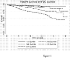
Figure 1 shows the probability of survival for a study population divided into quintiles on the basis of their total FLC concentrations. The quintile levels were <33.3, 33.4-47.3, 47.4-76.8, 67.9-106.3 and >106.5 mg/L. -
Figure 2 shows total FLC was corrected for GFR and then separated into quintiles. High total corrected FLC quintiles (4 and 5= lower line) predicted death from severe infections (P<0.001): -
Figure 3 ROC curve for the two top quintiles as a risk marker for death by infection (corrected level of total FLCs >1.7 mg/1unitGFR considered abnormal). -
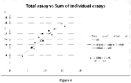
Figure 4 is a comparison between the total FLC concentrations obtained using separate, commercially available, anti-free κ and anti-free λ assay kits, compared to a total FLC assay kit using combined anti-λ and anti-K free light chain antibodies. -
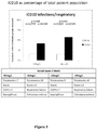
Figure 5 shows a comparison of patient deaths over a 4.5 year study for two different FLC concentrations as prognostic cut off points, the table showing causes of death. - 1300 patients with various degrees of renal impairment had serum samples collected ("Baseline") and were then followed up for a period of up to 63 months.
- In more detail the patients were recruited from the renal clinics at the University Hospital Birmingham. The patients had a range of renal problems including proteinuria, haematuria, chronic kidney disease (all stages), end stage renal failure (haemodialysis and peritoneal dialysis) and renal transplant recipients.
- The tests and assessments made included:
- Serum creatinine and an estimated glomerular filtration rate (eGFR).
- A corrected level ofFLCs per unit GFR was calculated as follows: total serum FLC concentration (mg/L) was divided by estimated glomerular filtration rate as calculated by the Cockcroft-Gault equation (REF) in mls/min/1.73m2. Thus giving a serum total FLC level for the patient, independent of renal function, in mg/L per unit GFR. Ref:
- Cockcroft DW, Gault MH: Prediction of creatinine clearance
- from serum creatinine. Nephron 16: 31-41, 1976.
- Urinary albumin/creatinine ratio.
- Serum FLC concentrations, both kappa and lambda (Freelite, The Binding Site, Birmingham, UK).
- Total, serum FLC concentrations were calculated by adding the values for kappa FLC and lambda FLC.
- Patients were followed up for time to death and cause of death.
- Kaplan Meier analysis of patient survival demonstrated that patients with higher FLC levels had a reduced survival (P<0.001), see
Figure 1 . - When causes of death were investigated the chances of death secondary to infections was significantly higher in patients with higher total FLC concentrations.
Total FLC Quintiles (n=number of deaths in each group) 1 2 3 4 5 Total Cause of death Cardiovascular 1 2 2 12 27 44 Infection 3 1 7 8 19 38 Renal 0 0 1 4 12 17 Cancer 2 2 2 3 7 16 Other 2 0 0 3 1 6 Unknown 0 0 0 0 1 1 Total 8 5 12 30 67 122 - The risk of death from infections was also associated with renal function (CKD stages 1-5), although this did not as clearly delineate an at risk group:
CKD stages (n= number of deaths in each group) 1.00 2.00 3.00 4.00 5.00 Total Cause.simp Cardiovascular 1 0 15 18 10 44 Infection 0 2 14 11 11 38 Renal 0 0 1 8 8 17 Cancer 0 2 8 5 1 16 Other 0 1 2 1 2 6 Unknown 0 0 0 0 1 1 Total 1 5 40 43 33 122 - Total FLC and renal function (eGFR) are correlated closely (R-0.645, P<0.001). Therefore to exclude any influence of renal function on the utility of total FLCs to predict infections, total FLC was corrected for GFR and then separated into quintiles. High total corrected FLC quintiles (4 and 5= lower line,
Figure 2 ) predicted death from severe infections (P<0.001): - When these two top quintiles were used as a risk marker for death by infection (corrected level of total FLCs >1.7 mg/1unit GFR considered abnormal) and analysed using a ROC curve the test was significantly predictive (P<0.0001):
- The principal infections identified were lower respiratory tract infections (pneumonia), urinary tract infections, Clostridium difficile, septicaemia and peritonitis.
- The results were gathered in patients with some form of renal impairment. But given that the relationship between elevations of total FLC and infections was independent of renal function it is likely total FLC may be similarly predictive of infections in the general population. It might be speculated that the test(s) will be more sensitive in the general population where there is no background elevation of FLC concentrations due to reduced renal function. As mentioned earlier, studies reporting a suppression of neutrophil function mediated by FLC have previously been published. Neutrophils form only one part of the immune defence system, however, and it has not, to our knowledge, been proposed that measurement of serum FLC concentrations could predict subjects at greater risk of death due to bacterial infection. Cohen and co-workers first reported on the in vitro effect of FLC on leukocytes in 1995 but have not published any studies measuring in vivo FLC concentrations in the period since then. The size of the predictive effect we observed was unexpected, especially after serum FLC concentrations had been corrected for renal function.
- The method according to the invention may utilise the following assay kit. The assay kit quantifies the total free κ plus free λ light chains present within patient samples, for example, in serum. This may be achieved by coating 100 nm carboxyl modified latex particles with a 50:50 blend of anti-free κ and anti-free λ light chain sheep antibody. In the assay exemplified below, the measuring range for the total free light chains is for 1-80 mg/L. However, other measuring ranges could equally be considered.
- Anti-free κ and anti-free λ anti sera are produced using techniques generally known in the art, in this particular case in sheep. The general immunisation process is described in
WO 97/17372 - Anti-κ and anti-λ antisera were diluted to equal concentrations using phosphate buffered saline (PBS). Those antibodies were combined to produce antisera comprising 50% anti κ antibody and 50% anti λ antibody.
- Antibodies were coated onto carboxyl modified latex at a coat load of 10 mg/lot. This was achieved using standard procedures. See, for example, "Microparticle Reagent Optimization: A laboratory reference manual from the authority on microparticles" Eds: Caryl Griffin, Jim Sutor, Bruce Shull. Copyright Seradyn Inc, 1994 (P/N 0347835(1294).
- This reference also provides details of optimising the assay kits using polyethylene glycol (PEG).
- The combined antibodies were compared to results obtained using commercially available κ and λ Freelite™ kits (obtained from the Binding Site Group Limited, Birmingham, United Kingdom). Such Freelite™ kits identify the amount of κ and the amount of λ free light chains in separate assays. The total FLC kits were used to generate curves, which were validated using controlled concentrations. Calibration curves were able to be obtained between 1 and 80 mg/l for total free light chain. In the results table below, results were obtained for κ free light chain (KFLC), λ free light chain (LFLC) and total FLC, using the κ FreeliteTM, λ FreeliteTM and total free light chain assays. These results are shown for 15 different normal serum samples. The results are shown in the table below and in
Figure 4 as measured by turbidimetry. - Preliminary results indicate that the principle of using a total free light chain assay based on anti-κ and anti-λ free light chain antibody is viable.
-

- Between November 8th 2005 and January 10th 2006 the hospital laboratory received 723 serum samples with a request for serum protein electrophoresis (SPE). Samples from paediatric patients, patients on immunoglobulin replacement, second and subsequent samples from the same patient, were excluded from the analysis. Also excluded were all patients with evidence of a monoclonal gammopathy as indicated by an abnormal κ/λ free light chain (FLC) ratio (<0.25 or >1.65; Katzmann) or abnormal SPE result (if confirmed by immunofixation). This left 528 patients in the final data analysis.
- All sera were analysed for serum protein abnormalities by serum protein electrophoresis (SPE; Sebia, UK). Serum immunofixation (IFE; Sebia) was performed on all samples with the presence of an abnormal SPE band or those with a high index of suspicion (unexplained hypogammaglobulinaemia, broad beta region, or low immunoglobulins with supporting clinical details). As part of an evaluation of serum free light chain (sFLC) analysis in a diagnostic setting, sFLC measurements (Freelite, The Binding Site, Birmingham, UK) were made for all sera using a Siemens Dade-Behring Prospec Nephelometer, in accordance with the manufacturer's instructions.
- In July 2010, 4 years and 6 months after the end of the original study, patient records were reviewed. For all patients, the date of the last follow-up or date of death was recorded and death certificates were obtained. The primary causes of death listed on the death certificates were categorised according to WHO International Statistical Classification of Diseases and Related Health Problems 10th Revision (ICD-10).
- The association of high combined (κ + λ) FLC concentrations with increased probability of death was investigated using Kaplan Meier survival analysis, Cox regression analysis and Chi-squared analysis. Categorical cut-offs of both > or <50mg/L and > or <65mg/L were investigated.
- Over the 4.5 years of follow-up there were 99 deaths (=18.8% mortality). Higher concentrations of polyclonal serum free light chains (kappa plus lambda) were found to be associated with increased mortality. This was the case if the prognostic cut-off utilised was 50mg/L or 65mg/L and was seen with all statistical analyses. The predominant causes of death were "Circulatory" (cardio-vascular disease) and "Infections/respiratory".
- The figure (
Figure 5 ) represents the % of deaths due to infections/respiratory causes in the different categories. Statistical significance was determined by Chi-squared analysis.
Claims (6)
- A method of prognosis of a human patient with an infection, and/or identifying a human patient at greater risk of death from an infection, the method comprising detecting an amount of free light chains (FLC) in a sample of blood or serum from the human patient, wherein a higher amount of FLC is associated with decreased survival due to the infection and/or increased risk from an infection, wherein the infection is a bacterial infection selected from a lower respiratory tract infection (such as pneumonia), urinary tract infection, Clostridium difficile infection, septicaemia or peritonitis.
- A method according to claim 1, wherein the total FLC is determined by immunoassay using anti-free light chain antibodies.
- A method according to claim 2, wherein the antibodies are a mixture of anti-free κ light chain and anti-free λ light chain antibodies.
- A method according to claim 1, wherein the amount of free light chains is the amount of total free light chains in the sample.
- A method according to claims 1 to 4, wherein the method comprises detecting the amount of FLC by nephelometry or turbidimetry.
- A method according to claims 1 to 5, wherein the human patient does not have symptoms of a B-cell associated disease.
Applications Claiming Priority (2)
| Application Number | Priority Date | Filing Date | Title |
|---|---|---|---|
| GBGB1001950.3A GB201001950D0 (en) | 2010-02-05 | 2010-02-05 | Infection prognostic assay |
| PCT/GB2011/050197 WO2011095820A1 (en) | 2010-02-05 | 2011-02-04 | Infection prognostic assay |
Publications (2)
| Publication Number | Publication Date |
|---|---|
| EP2531857A1 EP2531857A1 (en) | 2012-12-12 |
| EP2531857B1 true EP2531857B1 (en) | 2017-01-18 |
Family
ID=42082582
Family Applications (1)
| Application Number | Title | Priority Date | Filing Date |
|---|---|---|---|
| EP11702698.9A Active EP2531857B1 (en) | 2010-02-05 | 2011-02-04 | Infection prognostic assay |
Country Status (6)
| Country | Link |
|---|---|
| US (2) | US20130217030A1 (en) |
| EP (1) | EP2531857B1 (en) |
| JP (2) | JP5818818B2 (en) |
| CN (2) | CN102859362B (en) |
| GB (1) | GB201001950D0 (en) |
| WO (1) | WO2011095820A1 (en) |
Families Citing this family (2)
| Publication number | Priority date | Publication date | Assignee | Title |
|---|---|---|---|---|
| GB2519564A (en) * | 2013-10-24 | 2015-04-29 | Shared Band Ltd | Multicast transmission over bonded broadband |
| US20170268070A1 (en) | 2014-07-30 | 2017-09-21 | Mor Research Applications Ltd. | Prognostic methods and systems of treatment for acute lymphoblastic leukemia |
Family Cites Families (12)
| Publication number | Priority date | Publication date | Assignee | Title |
|---|---|---|---|---|
| US4792529A (en) * | 1985-10-18 | 1988-12-20 | University Of Rochester | Immunoassay of free kappa light chains for the detection of multiple sclerosis |
| IT1216698B (en) * | 1988-04-01 | 1990-03-08 | New Scient Co Spa | METHOD FOR DETERMINING THE PRESENCE OF FREE LIGHT CHAINS IN URINE SAMPLES, COMPLEX OF PREPARATIONS FOR THE PERFORMANCE OF THE METHOD, AND ITS REAGENT. |
| JP4053596B2 (en) | 1995-11-03 | 2008-02-27 | ザ・バインディング・サイト・リミテッド | Antibody production and medical use involving antibodies |
| US6322788B1 (en) * | 1998-08-20 | 2001-11-27 | Stanley Arthur Kim | Anti-bacterial antibodies and methods of use |
| US20040018576A1 (en) * | 2002-07-24 | 2004-01-29 | Dematteo Todd M. | Bence Jones protein testing cassette |
| JP4438455B2 (en) * | 2004-03-04 | 2010-03-24 | ヤマサ醤油株式会社 | Method for measuring free human immunoglobulin light chain and kit |
| WO2005116651A2 (en) * | 2004-05-24 | 2005-12-08 | Diasys Corporation | Method and device for testing for bence-jones protein |
| US20090082304A1 (en) * | 2004-11-12 | 2009-03-26 | Northwestern University | Methods of Treating Hematological Malignancies with Nucleoside Analog Drugs |
| GB0501741D0 (en) * | 2005-01-27 | 2005-03-02 | Binding Site The Ltd | Antibody |
| EP2267449B1 (en) * | 2005-04-12 | 2014-01-08 | Akira Matsumori | Biomarker for diagnosing heart failure and the use thereof |
| JP2007292661A (en) * | 2006-04-26 | 2007-11-08 | Akira Matsumori | Detection method and detection reagent for active myocarditis |
| GB0801609D0 (en) * | 2008-01-29 | 2008-03-05 | Binding Site The Ltd | Hevylite diagnostic stain |
-
2010
- 2010-02-05 GB GBGB1001950.3A patent/GB201001950D0/en not_active Ceased
-
2011
- 2011-02-04 EP EP11702698.9A patent/EP2531857B1/en active Active
- 2011-02-04 CN CN201180008422.8A patent/CN102859362B/en active Active
- 2011-02-04 US US13/576,099 patent/US20130217030A1/en not_active Abandoned
- 2011-02-04 WO PCT/GB2011/050197 patent/WO2011095820A1/en active Application Filing
- 2011-02-04 CN CN201180008402.0A patent/CN102859361B/en active Active
- 2011-02-04 JP JP2012551689A patent/JP5818818B2/en active Active
- 2011-02-04 US US13/576,063 patent/US20130149792A1/en not_active Abandoned
- 2011-02-04 JP JP2012551687A patent/JP5818817B2/en active Active
Non-Patent Citations (1)
| Title |
|---|
| BALMER PAUL ET AL: "Anti-pneumococcal antibody titre measurement: what useful information does it yield?", JOURNAL OF CLINICAL PATHOLOGY APR 2007, vol. 60, no. 4, April 2007 (2007-04-01), pages 345 - 350, ISSN: 0021-9746 * |
Also Published As
| Publication number | Publication date |
|---|---|
| JP2013519082A (en) | 2013-05-23 |
| US20130149792A1 (en) | 2013-06-13 |
| JP2013519083A (en) | 2013-05-23 |
| EP2531857A1 (en) | 2012-12-12 |
| CN102859362A (en) | 2013-01-02 |
| CN102859361B (en) | 2015-06-17 |
| CN102859362B (en) | 2015-11-25 |
| CN102859361A (en) | 2013-01-02 |
| GB201001950D0 (en) | 2010-03-24 |
| WO2011095820A1 (en) | 2011-08-11 |
| US20130217030A1 (en) | 2013-08-22 |
| JP5818818B2 (en) | 2015-11-18 |
| JP5818817B2 (en) | 2015-11-18 |
Similar Documents
| Publication | Publication Date | Title |
|---|---|---|
| EP2467724B1 (en) | Survival prognostic assay | |
| US20130078655A1 (en) | Kidney prognostic assay | |
| EP2623517B1 (en) | Urine and serum biomarkers associated with diabetic nephropathy | |
| US20140017707A1 (en) | Methods and kits for predicting the risk of respiratory failure, renal failure or thrombopenia in a septic patient by measuring endocan levels in blood | |
| KR20110022621A (en) | Iga nephropathy detection method and detection kit | |
| WO2014013225A1 (en) | Triage scoring system | |
| US20130071855A1 (en) | Flc as biomarker | |
| EP2764366B1 (en) | Prognostic method for diabetes | |
| EP2531857B1 (en) | Infection prognostic assay | |
| EP2531854B1 (en) | Cancer prognosis assay | |
| EP2791682B1 (en) | Assay | |
| JP2017003529A (en) | Method of testing for glomerular disorder |
Legal Events
| Date | Code | Title | Description |
|---|---|---|---|
| PUAI | Public reference made under article 153(3) epc to a published international application that has entered the european phase |
Free format text: ORIGINAL CODE: 0009012 |
|
| 17P | Request for examination filed |
Effective date: 20120713 |
|
| AK | Designated contracting states |
Kind code of ref document: A1 Designated state(s): AL AT BE BG CH CY CZ DE DK EE ES FI FR GB GR HR HU IE IS IT LI LT LU LV MC MK MT NL NO PL PT RO RS SE SI SK SM TR |
|
| DAX | Request for extension of the european patent (deleted) | ||
| 17Q | First examination report despatched |
Effective date: 20130524 |
|
| REG | Reference to a national code |
Ref country code: DE Ref legal event code: R079 Ref document number: 602011034443 Country of ref document: DE Free format text: PREVIOUS MAIN CLASS: G01N0033680000 Ipc: G01N0033574000 |
|
| RIC1 | Information provided on ipc code assigned before grant |
Ipc: G01N 33/574 20060101AFI20160621BHEP Ipc: G01N 33/68 20060101ALI20160621BHEP |
|
| GRAP | Despatch of communication of intention to grant a patent |
Free format text: ORIGINAL CODE: EPIDOSNIGR1 |
|
| INTG | Intention to grant announced |
Effective date: 20160802 |
|
| GRAJ | Information related to disapproval of communication of intention to grant by the applicant or resumption of examination proceedings by the epo deleted |
Free format text: ORIGINAL CODE: EPIDOSDIGR1 |
|
| STAA | Information on the status of an ep patent application or granted ep patent |
Free format text: STATUS: EXAMINATION IS IN PROGRESS |
|
| GRAP | Despatch of communication of intention to grant a patent |
Free format text: ORIGINAL CODE: EPIDOSNIGR1 |
|
| STAA | Information on the status of an ep patent application or granted ep patent |
Free format text: STATUS: GRANT OF PATENT IS INTENDED |
|
| GRAS | Grant fee paid |
Free format text: ORIGINAL CODE: EPIDOSNIGR3 |
|
| INTC | Intention to grant announced (deleted) | ||
| RIN1 | Information on inventor provided before grant (corrected) |
Inventor name: MEAD, GRAHAM Inventor name: BRADWELL, ARTHUR |
|
| GRAA | (expected) grant |
Free format text: ORIGINAL CODE: 0009210 |
|
| STAA | Information on the status of an ep patent application or granted ep patent |
Free format text: STATUS: THE PATENT HAS BEEN GRANTED |
|
| INTG | Intention to grant announced |
Effective date: 20161202 |
|
| AK | Designated contracting states |
Kind code of ref document: B1 Designated state(s): AL AT BE BG CH CY CZ DE DK EE ES FI FR GB GR HR HU IE IS IT LI LT LU LV MC MK MT NL NO PL PT RO RS SE SI SK SM TR |
|
| REG | Reference to a national code |
Ref country code: GB Ref legal event code: FG4D |
|
| REG | Reference to a national code |
Ref country code: CH Ref legal event code: EP |
|
| REG | Reference to a national code |
Ref country code: AT Ref legal event code: REF Ref document number: 863202 Country of ref document: AT Kind code of ref document: T Effective date: 20170215 |
|
| REG | Reference to a national code |
Ref country code: FR Ref legal event code: PLFP Year of fee payment: 7 |
|
| REG | Reference to a national code |
Ref country code: IE Ref legal event code: FG4D |
|
| REG | Reference to a national code |
Ref country code: DE Ref legal event code: R096 Ref document number: 602011034443 Country of ref document: DE |
|
| REG | Reference to a national code |
Ref country code: NL Ref legal event code: MP Effective date: 20170118 |
|
| REG | Reference to a national code |
Ref country code: LT Ref legal event code: MG4D |
|
| PG25 | Lapsed in a contracting state [announced via postgrant information from national office to epo] |
Ref country code: BE Free format text: LAPSE BECAUSE OF NON-PAYMENT OF DUE FEES Effective date: 20170228 |
|
| REG | Reference to a national code |
Ref country code: AT Ref legal event code: MK05 Ref document number: 863202 Country of ref document: AT Kind code of ref document: T Effective date: 20170118 |
|
| PG25 | Lapsed in a contracting state [announced via postgrant information from national office to epo] |
Ref country code: NL Free format text: LAPSE BECAUSE OF FAILURE TO SUBMIT A TRANSLATION OF THE DESCRIPTION OR TO PAY THE FEE WITHIN THE PRESCRIBED TIME-LIMIT Effective date: 20170118 |
|
| PG25 | Lapsed in a contracting state [announced via postgrant information from national office to epo] |
Ref country code: LT Free format text: LAPSE BECAUSE OF FAILURE TO SUBMIT A TRANSLATION OF THE DESCRIPTION OR TO PAY THE FEE WITHIN THE PRESCRIBED TIME-LIMIT Effective date: 20170118 Ref country code: NO Free format text: LAPSE BECAUSE OF FAILURE TO SUBMIT A TRANSLATION OF THE DESCRIPTION OR TO PAY THE FEE WITHIN THE PRESCRIBED TIME-LIMIT Effective date: 20170418 Ref country code: HR Free format text: LAPSE BECAUSE OF FAILURE TO SUBMIT A TRANSLATION OF THE DESCRIPTION OR TO PAY THE FEE WITHIN THE PRESCRIBED TIME-LIMIT Effective date: 20170118 Ref country code: FI Free format text: LAPSE BECAUSE OF FAILURE TO SUBMIT A TRANSLATION OF THE DESCRIPTION OR TO PAY THE FEE WITHIN THE PRESCRIBED TIME-LIMIT Effective date: 20170118 Ref country code: IS Free format text: LAPSE BECAUSE OF FAILURE TO SUBMIT A TRANSLATION OF THE DESCRIPTION OR TO PAY THE FEE WITHIN THE PRESCRIBED TIME-LIMIT Effective date: 20170518 Ref country code: GR Free format text: LAPSE BECAUSE OF FAILURE TO SUBMIT A TRANSLATION OF THE DESCRIPTION OR TO PAY THE FEE WITHIN THE PRESCRIBED TIME-LIMIT Effective date: 20170419 |
|
| PG25 | Lapsed in a contracting state [announced via postgrant information from national office to epo] |
Ref country code: AT Free format text: LAPSE BECAUSE OF FAILURE TO SUBMIT A TRANSLATION OF THE DESCRIPTION OR TO PAY THE FEE WITHIN THE PRESCRIBED TIME-LIMIT Effective date: 20170118 Ref country code: ES Free format text: LAPSE BECAUSE OF FAILURE TO SUBMIT A TRANSLATION OF THE DESCRIPTION OR TO PAY THE FEE WITHIN THE PRESCRIBED TIME-LIMIT Effective date: 20170118 Ref country code: LV Free format text: LAPSE BECAUSE OF FAILURE TO SUBMIT A TRANSLATION OF THE DESCRIPTION OR TO PAY THE FEE WITHIN THE PRESCRIBED TIME-LIMIT Effective date: 20170118 Ref country code: RS Free format text: LAPSE BECAUSE OF FAILURE TO SUBMIT A TRANSLATION OF THE DESCRIPTION OR TO PAY THE FEE WITHIN THE PRESCRIBED TIME-LIMIT Effective date: 20170118 Ref country code: PT Free format text: LAPSE BECAUSE OF FAILURE TO SUBMIT A TRANSLATION OF THE DESCRIPTION OR TO PAY THE FEE WITHIN THE PRESCRIBED TIME-LIMIT Effective date: 20170518 Ref country code: BG Free format text: LAPSE BECAUSE OF FAILURE TO SUBMIT A TRANSLATION OF THE DESCRIPTION OR TO PAY THE FEE WITHIN THE PRESCRIBED TIME-LIMIT Effective date: 20170418 Ref country code: PL Free format text: LAPSE BECAUSE OF FAILURE TO SUBMIT A TRANSLATION OF THE DESCRIPTION OR TO PAY THE FEE WITHIN THE PRESCRIBED TIME-LIMIT Effective date: 20170118 Ref country code: SE Free format text: LAPSE BECAUSE OF FAILURE TO SUBMIT A TRANSLATION OF THE DESCRIPTION OR TO PAY THE FEE WITHIN THE PRESCRIBED TIME-LIMIT Effective date: 20170118 |
|
| REG | Reference to a national code |
Ref country code: CH Ref legal event code: PL |
|
| REG | Reference to a national code |
Ref country code: DE Ref legal event code: R097 Ref document number: 602011034443 Country of ref document: DE |
|
| PG25 | Lapsed in a contracting state [announced via postgrant information from national office to epo] |
Ref country code: LI Free format text: LAPSE BECAUSE OF NON-PAYMENT OF DUE FEES Effective date: 20170228 Ref country code: IT Free format text: LAPSE BECAUSE OF FAILURE TO SUBMIT A TRANSLATION OF THE DESCRIPTION OR TO PAY THE FEE WITHIN THE PRESCRIBED TIME-LIMIT Effective date: 20170118 Ref country code: EE Free format text: LAPSE BECAUSE OF FAILURE TO SUBMIT A TRANSLATION OF THE DESCRIPTION OR TO PAY THE FEE WITHIN THE PRESCRIBED TIME-LIMIT Effective date: 20170118 Ref country code: RO Free format text: LAPSE BECAUSE OF FAILURE TO SUBMIT A TRANSLATION OF THE DESCRIPTION OR TO PAY THE FEE WITHIN THE PRESCRIBED TIME-LIMIT Effective date: 20170118 Ref country code: CZ Free format text: LAPSE BECAUSE OF FAILURE TO SUBMIT A TRANSLATION OF THE DESCRIPTION OR TO PAY THE FEE WITHIN THE PRESCRIBED TIME-LIMIT Effective date: 20170118 Ref country code: CH Free format text: LAPSE BECAUSE OF NON-PAYMENT OF DUE FEES Effective date: 20170228 Ref country code: SK Free format text: LAPSE BECAUSE OF FAILURE TO SUBMIT A TRANSLATION OF THE DESCRIPTION OR TO PAY THE FEE WITHIN THE PRESCRIBED TIME-LIMIT Effective date: 20170118 |
|
| PLBE | No opposition filed within time limit |
Free format text: ORIGINAL CODE: 0009261 |
|
| STAA | Information on the status of an ep patent application or granted ep patent |
Free format text: STATUS: NO OPPOSITION FILED WITHIN TIME LIMIT |
|
| REG | Reference to a national code |
Ref country code: IE Ref legal event code: MM4A |
|
| PG25 | Lapsed in a contracting state [announced via postgrant information from national office to epo] |
Ref country code: SM Free format text: LAPSE BECAUSE OF FAILURE TO SUBMIT A TRANSLATION OF THE DESCRIPTION OR TO PAY THE FEE WITHIN THE PRESCRIBED TIME-LIMIT Effective date: 20170118 Ref country code: MC Free format text: LAPSE BECAUSE OF FAILURE TO SUBMIT A TRANSLATION OF THE DESCRIPTION OR TO PAY THE FEE WITHIN THE PRESCRIBED TIME-LIMIT Effective date: 20170118 Ref country code: DK Free format text: LAPSE BECAUSE OF FAILURE TO SUBMIT A TRANSLATION OF THE DESCRIPTION OR TO PAY THE FEE WITHIN THE PRESCRIBED TIME-LIMIT Effective date: 20170118 |
|
| 26N | No opposition filed |
Effective date: 20171019 |
|
| PG25 | Lapsed in a contracting state [announced via postgrant information from national office to epo] |
Ref country code: LU Free format text: LAPSE BECAUSE OF NON-PAYMENT OF DUE FEES Effective date: 20170204 |
|
| REG | Reference to a national code |
Ref country code: BE Ref legal event code: MM Effective date: 20170228 |
|
| REG | Reference to a national code |
Ref country code: FR Ref legal event code: PLFP Year of fee payment: 8 |
|
| PG25 | Lapsed in a contracting state [announced via postgrant information from national office to epo] |
Ref country code: SI Free format text: LAPSE BECAUSE OF FAILURE TO SUBMIT A TRANSLATION OF THE DESCRIPTION OR TO PAY THE FEE WITHIN THE PRESCRIBED TIME-LIMIT Effective date: 20170118 Ref country code: IE Free format text: LAPSE BECAUSE OF NON-PAYMENT OF DUE FEES Effective date: 20170204 |
|
| PG25 | Lapsed in a contracting state [announced via postgrant information from national office to epo] |
Ref country code: MT Free format text: LAPSE BECAUSE OF NON-PAYMENT OF DUE FEES Effective date: 20170204 |
|
| PG25 | Lapsed in a contracting state [announced via postgrant information from national office to epo] |
Ref country code: HU Free format text: LAPSE BECAUSE OF FAILURE TO SUBMIT A TRANSLATION OF THE DESCRIPTION OR TO PAY THE FEE WITHIN THE PRESCRIBED TIME-LIMIT; INVALID AB INITIO Effective date: 20110204 |
|
| PG25 | Lapsed in a contracting state [announced via postgrant information from national office to epo] |
Ref country code: CY Free format text: LAPSE BECAUSE OF NON-PAYMENT OF DUE FEES Effective date: 20170118 |
|
| PG25 | Lapsed in a contracting state [announced via postgrant information from national office to epo] |
Ref country code: MK Free format text: LAPSE BECAUSE OF FAILURE TO SUBMIT A TRANSLATION OF THE DESCRIPTION OR TO PAY THE FEE WITHIN THE PRESCRIBED TIME-LIMIT Effective date: 20170118 |
|
| PG25 | Lapsed in a contracting state [announced via postgrant information from national office to epo] |
Ref country code: TR Free format text: LAPSE BECAUSE OF FAILURE TO SUBMIT A TRANSLATION OF THE DESCRIPTION OR TO PAY THE FEE WITHIN THE PRESCRIBED TIME-LIMIT Effective date: 20170118 |
|
| PG25 | Lapsed in a contracting state [announced via postgrant information from national office to epo] |
Ref country code: AL Free format text: LAPSE BECAUSE OF FAILURE TO SUBMIT A TRANSLATION OF THE DESCRIPTION OR TO PAY THE FEE WITHIN THE PRESCRIBED TIME-LIMIT Effective date: 20170118 |
|
| P01 | Opt-out of the competence of the unified patent court (upc) registered |
Effective date: 20230529 |
|
| PGFP | Annual fee paid to national office [announced via postgrant information from national office to epo] |
Ref country code: DE Payment date: 20240206 Year of fee payment: 14 Ref country code: GB Payment date: 20240208 Year of fee payment: 14 |
|
| PGFP | Annual fee paid to national office [announced via postgrant information from national office to epo] |
Ref country code: FR Payment date: 20240228 Year of fee payment: 14 |