EP2525227B1 - A method for detecting pancreatic cancer using the serological marker ULBP2 - Google Patents
A method for detecting pancreatic cancer using the serological marker ULBP2 Download PDFInfo
- Publication number
- EP2525227B1 EP2525227B1 EP12168401.3A EP12168401A EP2525227B1 EP 2525227 B1 EP2525227 B1 EP 2525227B1 EP 12168401 A EP12168401 A EP 12168401A EP 2525227 B1 EP2525227 B1 EP 2525227B1
- Authority
- EP
- European Patent Office
- Prior art keywords
- ulbp2
- pancreatic cancer
- serological marker
- detecting
- mass spectrometry
- Prior art date
- Legal status (The legal status is an assumption and is not a legal conclusion. Google has not performed a legal analysis and makes no representation as to the accuracy of the status listed.)
- Active
Links
- 102100039989 UL16-binding protein 2 Human genes 0.000 title claims description 91
- 206010061902 Pancreatic neoplasm Diseases 0.000 title claims description 88
- 201000002528 pancreatic cancer Diseases 0.000 title claims description 84
- 208000015486 malignant pancreatic neoplasm Diseases 0.000 title claims description 83
- 208000008443 pancreatic carcinoma Diseases 0.000 title claims description 83
- 239000003550 marker Substances 0.000 title claims description 40
- 230000000405 serological effect Effects 0.000 title claims description 35
- 238000000034 method Methods 0.000 title claims description 16
- 101000607320 Homo sapiens UL16-binding protein 2 Proteins 0.000 title 1
- 101710173415 UL16-binding protein 2 Proteins 0.000 claims description 90
- 210000002966 serum Anatomy 0.000 claims description 30
- INZOTETZQBPBCE-NYLDSJSYSA-N 3-sialyl lewis Chemical group O[C@H]1[C@H](O)[C@H](O)[C@H](C)O[C@H]1O[C@H]([C@H](O)CO)[C@@H]([C@@H](NC(C)=O)C=O)O[C@H]1[C@H](O)[C@@H](O[C@]2(O[C@H]([C@H](NC(C)=O)[C@@H](O)C2)[C@H](O)[C@H](O)CO)C(O)=O)[C@@H](O)[C@@H](CO)O1 INZOTETZQBPBCE-NYLDSJSYSA-N 0.000 claims description 24
- 238000003018 immunoassay Methods 0.000 claims description 13
- 239000011324 bead Substances 0.000 claims description 11
- 210000004369 blood Anatomy 0.000 claims description 8
- 239000008280 blood Substances 0.000 claims description 8
- 238000003556 assay Methods 0.000 claims description 5
- 238000011088 calibration curve Methods 0.000 claims description 3
- 238000002965 ELISA Methods 0.000 claims 8
- 238000004949 mass spectrometry Methods 0.000 claims 8
- 125000003275 alpha amino acid group Chemical group 0.000 claims 2
- 206010028980 Neoplasm Diseases 0.000 description 33
- 238000001514 detection method Methods 0.000 description 27
- 201000011510 cancer Diseases 0.000 description 23
- 210000001519 tissue Anatomy 0.000 description 18
- 230000014509 gene expression Effects 0.000 description 15
- 210000004027 cell Anatomy 0.000 description 13
- 101001109501 Homo sapiens NKG2-D type II integral membrane protein Proteins 0.000 description 9
- 102100022680 NKG2-D type II integral membrane protein Human genes 0.000 description 9
- 238000001574 biopsy Methods 0.000 description 9
- 239000003446 ligand Substances 0.000 description 9
- 108090000623 proteins and genes Proteins 0.000 description 9
- 239000000523 sample Substances 0.000 description 9
- 208000002454 Nasopharyngeal Carcinoma Diseases 0.000 description 8
- 102000004169 proteins and genes Human genes 0.000 description 8
- 206010027476 Metastases Diseases 0.000 description 7
- 230000035945 sensitivity Effects 0.000 description 7
- 238000010186 staining Methods 0.000 description 7
- 208000005718 Stomach Neoplasms Diseases 0.000 description 6
- 230000004069 differentiation Effects 0.000 description 6
- 206010017758 gastric cancer Diseases 0.000 description 6
- 201000011549 stomach cancer Diseases 0.000 description 6
- 230000009401 metastasis Effects 0.000 description 5
- YBJHBAHKTGYVGT-ZKWXMUAHSA-N (+)-Biotin Chemical compound N1C(=O)N[C@@H]2[C@H](CCCCC(=O)O)SC[C@@H]21 YBJHBAHKTGYVGT-ZKWXMUAHSA-N 0.000 description 4
- 241000282414 Homo sapiens Species 0.000 description 4
- 238000004458 analytical method Methods 0.000 description 4
- 239000000090 biomarker Substances 0.000 description 4
- 238000012216 screening Methods 0.000 description 4
- 108010090804 Streptavidin Proteins 0.000 description 3
- 210000001744 T-lymphocyte Anatomy 0.000 description 3
- 150000001413 amino acids Chemical group 0.000 description 3
- KRKNYBCHXYNGOX-UHFFFAOYSA-N citric acid Chemical compound OC(=O)CC(O)(C(O)=O)CC(O)=O KRKNYBCHXYNGOX-UHFFFAOYSA-N 0.000 description 3
- 238000003364 immunohistochemistry Methods 0.000 description 3
- 208000024891 symptom Diseases 0.000 description 3
- 210000004881 tumor cell Anatomy 0.000 description 3
- 238000005406 washing Methods 0.000 description 3
- 206010009944 Colon cancer Diseases 0.000 description 2
- 208000001333 Colorectal Neoplasms Diseases 0.000 description 2
- WZUVPPKBWHMQCE-UHFFFAOYSA-N Haematoxylin Chemical compound C12=CC(O)=C(O)C=C2CC2(O)C1C1=CC=C(O)C(O)=C1OC2 WZUVPPKBWHMQCE-UHFFFAOYSA-N 0.000 description 2
- 108020003285 Isocitrate lyase Proteins 0.000 description 2
- 238000012313 Kruskal-Wallis test Methods 0.000 description 2
- 102100030301 MHC class I polypeptide-related sequence A Human genes 0.000 description 2
- 206010061306 Nasopharyngeal cancer Diseases 0.000 description 2
- 108010004729 Phycoerythrin Proteins 0.000 description 2
- 238000001793 Wilcoxon signed-rank test Methods 0.000 description 2
- 238000013459 approach Methods 0.000 description 2
- 230000008901 benefit Effects 0.000 description 2
- 229960002685 biotin Drugs 0.000 description 2
- 235000020958 biotin Nutrition 0.000 description 2
- 239000011616 biotin Substances 0.000 description 2
- 239000000872 buffer Substances 0.000 description 2
- 201000010989 colorectal carcinoma Diseases 0.000 description 2
- 239000003636 conditioned culture medium Substances 0.000 description 2
- 238000003745 diagnosis Methods 0.000 description 2
- 238000003384 imaging method Methods 0.000 description 2
- 239000010445 mica Substances 0.000 description 2
- 229910052618 mica group Inorganic materials 0.000 description 2
- 201000011216 nasopharynx carcinoma Diseases 0.000 description 2
- 210000002797 pancreatic ductal cell Anatomy 0.000 description 2
- 230000001575 pathological effect Effects 0.000 description 2
- 210000004976 peripheral blood cell Anatomy 0.000 description 2
- 239000012488 sample solution Substances 0.000 description 2
- 239000012086 standard solution Substances 0.000 description 2
- 206010006187 Breast cancer Diseases 0.000 description 1
- 208000026310 Breast neoplasm Diseases 0.000 description 1
- 108010062802 CD66 antigens Proteins 0.000 description 1
- 241000283707 Capra Species 0.000 description 1
- 102100024533 Carcinoembryonic antigen-related cell adhesion molecule 1 Human genes 0.000 description 1
- 201000009030 Carcinoma Diseases 0.000 description 1
- 208000005243 Chondrosarcoma Diseases 0.000 description 1
- 206010017993 Gastrointestinal neoplasms Diseases 0.000 description 1
- 208000032612 Glial tumor Diseases 0.000 description 1
- 201000010915 Glioblastoma multiforme Diseases 0.000 description 1
- 206010018338 Glioma Diseases 0.000 description 1
- 102100040896 Growth/differentiation factor 15 Human genes 0.000 description 1
- 101710194460 Growth/differentiation factor 15 Proteins 0.000 description 1
- 108010045100 HSP27 Heat-Shock Proteins Proteins 0.000 description 1
- 102100039165 Heat shock protein beta-1 Human genes 0.000 description 1
- 101000991061 Homo sapiens MHC class I polypeptide-related sequence B Proteins 0.000 description 1
- 101000607306 Homo sapiens UL16-binding protein 1 Proteins 0.000 description 1
- 102100030300 MHC class I polypeptide-related sequence B Human genes 0.000 description 1
- -1 PAM4 Proteins 0.000 description 1
- 206010060862 Prostate cancer Diseases 0.000 description 1
- 208000000236 Prostatic Neoplasms Diseases 0.000 description 1
- 102100040012 UL16-binding protein 1 Human genes 0.000 description 1
- 230000003187 abdominal effect Effects 0.000 description 1
- 230000003213 activating effect Effects 0.000 description 1
- 150000001412 amines Chemical class 0.000 description 1
- 239000000427 antigen Substances 0.000 description 1
- 102000036639 antigens Human genes 0.000 description 1
- 108091007433 antigens Proteins 0.000 description 1
- 230000000903 blocking effect Effects 0.000 description 1
- 210000000481 breast Anatomy 0.000 description 1
- 238000004113 cell culture Methods 0.000 description 1
- 238000003759 clinical diagnosis Methods 0.000 description 1
- 210000001072 colon Anatomy 0.000 description 1
- 238000002591 computed tomography Methods 0.000 description 1
- 230000002596 correlated effect Effects 0.000 description 1
- 210000004748 cultured cell Anatomy 0.000 description 1
- 238000013211 curve analysis Methods 0.000 description 1
- 230000009089 cytolysis Effects 0.000 description 1
- 230000001419 dependent effect Effects 0.000 description 1
- 238000011161 development Methods 0.000 description 1
- 238000010790 dilution Methods 0.000 description 1
- 239000012895 dilution Substances 0.000 description 1
- 230000000694 effects Effects 0.000 description 1
- 238000001839 endoscopy Methods 0.000 description 1
- 210000004475 gamma-delta t lymphocyte Anatomy 0.000 description 1
- 208000005017 glioblastoma Diseases 0.000 description 1
- 206010073071 hepatocellular carcinoma Diseases 0.000 description 1
- 231100000844 hepatocellular carcinoma Toxicity 0.000 description 1
- 238000009169 immunotherapy Methods 0.000 description 1
- 208000032839 leukemia Diseases 0.000 description 1
- 238000001294 liquid chromatography-tandem mass spectrometry Methods 0.000 description 1
- 238000007477 logistic regression Methods 0.000 description 1
- 210000004072 lung Anatomy 0.000 description 1
- 239000002609 medium Substances 0.000 description 1
- 125000000896 monocarboxylic acid group Chemical group 0.000 description 1
- 238000007837 multiplex assay Methods 0.000 description 1
- 239000002773 nucleotide Substances 0.000 description 1
- 210000000056 organ Anatomy 0.000 description 1
- 230000002018 overexpression Effects 0.000 description 1
- 210000000496 pancreas Anatomy 0.000 description 1
- 210000002307 prostate Anatomy 0.000 description 1
- 238000003118 sandwich ELISA Methods 0.000 description 1
- 230000028327 secretion Effects 0.000 description 1
- 239000012679 serum free medium Substances 0.000 description 1
- 238000002415 sodium dodecyl sulfate polyacrylamide gel electrophoresis Methods 0.000 description 1
- 239000000243 solution Substances 0.000 description 1
- 239000000758 substrate Substances 0.000 description 1
- 230000004083 survival effect Effects 0.000 description 1
- 238000004885 tandem mass spectrometry Methods 0.000 description 1
- 230000002103 transcriptional effect Effects 0.000 description 1
- 239000000439 tumor marker Substances 0.000 description 1
- 238000010200 validation analysis Methods 0.000 description 1
Images


Classifications
-
- G—PHYSICS
- G01—MEASURING; TESTING
- G01N—INVESTIGATING OR ANALYSING MATERIALS BY DETERMINING THEIR CHEMICAL OR PHYSICAL PROPERTIES
- G01N33/00—Investigating or analysing materials by specific methods not covered by groups G01N1/00 - G01N31/00
- G01N33/48—Biological material, e.g. blood, urine; Haemocytometers
- G01N33/50—Chemical analysis of biological material, e.g. blood, urine; Testing involving biospecific ligand binding methods; Immunological testing
- G01N33/53—Immunoassay; Biospecific binding assay; Materials therefor
- G01N33/574—Immunoassay; Biospecific binding assay; Materials therefor for cancer
- G01N33/57407—Specifically defined cancers
- G01N33/57438—Specifically defined cancers of liver, pancreas or kidney
Definitions
- Embodiments relate to a serological marker for detecting pancreatic cancer and a method for using the serological marker, especially to a serological marker with high sensitivity and specificity for detecting pancreatic cancer and a method for using the serological marker.
- Pancreatic cancer is respectively ranked fourth and tenth among cancer-related mortality in United State and Taiwan and shows increased mortality these years. Because pancreas is anatomically located in a deeper site and the apparent symptoms of the pancreatic cancer is late onset, less than 8% of pancreatic cancer patients are diagnosed at the localized stage and can be surgically cured. More than 50% of diagnosed pancreatic cancer patients have exhibited distant metastasis, and the 5-year survival rate of which is less than 5%.
- ALCAN which is a synonym for ULBP2
- ULBP2 glycosylphosphatidylinositol-anchored structure ubiquitously expressed in human cancers
- ALCAN may be a potential target for cancer diagonosis because its mRNA could be detected in cell lines from various human cancer types (lung, breast, colon, prostate, neuroblastma, glioma, leukemia, T-cell lyphoma and chondrosarcoma) but not (or barely) in several normal organs.
- the levels of ALCAN expression and secretion vary drastically in different cell lines, and more importantly, this paper is completely silent about the relation between ALCAN and pancreatic cancer.
- WO 2011/050328 A2 (Publication Date:28.04.2011 . ASSESSMENT OF SOLID TUMOR BURDEN) disclosed a method to assess solid tumor burden on the basis of measuring expression levels of one or both of an activating Natural Killer (NK) cell receptor ligand(s) (NKG2D ligands) on peripheral blood cells in a blood specimens obtained from a testee (claim 1) and mentioned ULBP2 as one instance for the NKG2D ligand (claim 2) and pancreatic cancer as one example for dozens of the solid tumor types listed (claim 4).
- NK Natural Killer
- pancreatic cancer diagnosis are mainly based on imaging methods, such as abdominal sona or high-resolution computed tomography scan, and sometimes might combine with an invasive endoscopy or a magnetic resonance cholangiopancreatography to increase detection efficiency.
- imaging methods such as abdominal sona or high-resolution computed tomography scan
- an invasive endoscopy or a magnetic resonance cholangiopancreatography to increase detection efficiency.
- the volume of pancreatic cancer in early stage is too small to be detected by the imaging method, which greatly increases the difficulty in detecting pancreatic cancer.
- a serological marker carbohydrate antigen 19-9 (CA 19-9)
- CA 19-9 has few disadvantages in pancreatic cancer detection, such as the insufficient sensitivity and specificity and the unable detection of pancreatic cancer in early stage. Therefore, developing other serological markers to overcome above-mentioned disadvantages of CA19-9 and increase the detection efficiency will be an important issue to improve the management of pancreatic cancer. This is achieved by the subject matter of claim 1.
- a serological marker for detecting a pancreatic cancer with high sensitivity and specificity comprising at least an UL16 binding protein 2 (ULBP2) is provided.
- the ULBP2 is highly expression in pancreatic tumor tissues, and is significant increased in serum of a pancreatic cancer patient than in a normal healthy person.
- the ULBP2 is also significant increased in pancreatic cancer than in gastric cancer (GC), nasopharyngeal carcinoma cancer (NPC) and colorectal carcinoma cancer (CRC). Therefore, the ULBP2 is capable to be a serological marker for efficiently detecting pancreatic cancer by comparing its levels in the blood samples isolated from a patient and a normal healthy person.
- GC gastric cancer
- NPC nasopharyngeal carcinoma cancer
- CRC colorectal carcinoma cancer
- a bead-based immunoassay at least using an UL16 binding protein 2 (ULBP2) as a serological marker is used to detect pancreatic cancer and has exhibited an excellent sensitivity.
- ULBP2 UL16 binding protein 2
- the limitation of the bead-based immunoassay in detecting the pancreatic cancer is 3.91pg/ml.
- ULBP2 is combined with a carbohydrate antigen 19-9 (CA 19-9) to improve the detection efficiency.
- CA 19-9 carbohydrate antigen 19-9
- the ULBP2 also has ability to detect the pancreatic cancer at early stage and has more precise detection effect than CA 19-9.
- Embodiment 1 screening and selecting a serological marker for detecting the pancreatic cancer
- Proteins secreted from two pancreatic cancer cell lines PANC-1 and MIA PaCa-2 which were collected by incubating the cultured cells in serum-free medium for 24 hr (this medium is thereafter defined as conditioned medium), are systematically identified by one-dimensional SDS-PAGE in conjunction with nano-LC-MS/MS (the GeLC-MS/MS approach). This method identified a total of 1812 non-redundant proteins from the conditioned medium of the two cell lines.
- the transcriptional expression of each identified protein in the pancreatic cancer tissues was further analyzed according to a public doamin transcriptomic information of the pancreatic cancer tissues (National Center for Biotechnology Information (NCBI) Gene Expression Omnibus).
- pancreatic ductal cells respectively isolated from 25 healthy donors and 24 pancreatic cancer patients are subjected to an array-based analysis to identify the genes whose message RNA (mRNA) levels are higher expressed in the pancreatic cancer patients than those in the healthy donors.
- mRNA message RNA
- 30 pancreatic cancer cell secreted proteins exhibited at least two-fold higher mRNA expression levels in the pancreatic ductal cells from cancer patients than from the healthy donors. Eleven out of the 30 proteins had been reported to be over-expressed in the tissue biopsy of pancreatic cancer by previous studies.
- UL16 binding protein 2 (ULBP2) is selected as a serological candidate marker for detecting pancreatic cancer in the present invention.
- the ULBP2 in the present embodiment has an amino acid sequence (SEQ ID NO:1) shown as following:
- Immunohistochemistry assay In the embodiment, a goat anti-ULBP2 antibody is applied. A tissue biopsy is isolated and heated in a 0.01 M citric acid buffer (pH 6.0). A blocking buffer is added and reacted at room temperature for 5 minutes. The tissue biopsy is reacted with the anti-ULBP2 antibody (1:20 dilution) at 4°C for 16 hours. Then, the tissue biopsy is stained with the N-Histofine® (Nichirei, Japan) at room temperature and followed by treatment with substrate DAB chromogen (Novocastra/Leica Microsystems, IL, USA). The tissue biopsy is also counterstained with hematoxylin.
- the expression level of target proteins was evaluated according to the simplified H score system, which is based on the intensity of cell staining [3 (strong), 2 (moderate), 1 (weak), or 0 (no cell staining)] and the percentage of cell staining [3 ( ⁇ 90%), 2 (50-89%), 1 (10-49%), or 0 (0-9%)].
- the two scores were multiplied by each other and then divided by 3 to get the final score. Positive staining was defined as a final score ⁇ 0.67.
- the immunohistochemistry assay of the cancer tissue biopsies from 67 pancreatic cancer patients shows that ULBP2 staining is positive in all biopsies (100%). Additionally, the expression of ULBP2 is more significant in the cancer tissue than in the adjacent non-cancerous tissues (the intensity of brown color indicates the expression level of the ULBP2).
- the mean expression level of the ULBP2 in the cancer tissue and in the adjacent non-cancerous tissue is score of 2.71 ⁇ 0.49 and 1.89 ⁇ 0.74, respectively.
- the expression of ULBP2 of the embodiment is not influenced by the clinically pathological symptoms such as the gender, age, histological grade, overall cancer status or TMN classification.
- the detecting result of the ULBP2 has highly consistence in patients with different clinically pathological symptoms.
- Table 1 Correlation between clinicopathological features and ULBP2 expression in 67 pancreatic cancer patients Characteristics Patient No. IHC score (Mean ⁇ SD) a p -value Gender Male 44 2.64 ⁇ 0.54 0.180 b Female 23 2.83 ⁇ 0.36 Age (years) ⁇ 64 c 33 2.61 ⁇ 0.55 0.089 b ⁇ 64 34 2.80 ⁇ 0.41 Histological grade d Well differentiation 23 2.71 ⁇ 0.59 0.746 c Moderate differentiation 31 2.68 ⁇ 0.44 Poor differentiation 10 2.70 ⁇ 0.48 Overall stage stage I 9 2.52 ⁇ 0.82 0.593 e stage II 56 2.73 ⁇ 0.43 stage IV 2 3.00 ⁇ 0.00 Tumor-node-metastasis (TNM)-T classification TNM-T2 9 2.52 ⁇ 0.82 0.653 b TNM-T3 58 2.74 ⁇ 0.42 TNM-N
- a bead-based immunoassay is used to detect the ULBP2 level in a serum sample.
- An ULBP2 antibody used as a capture antibody, is pre-coupled to COOH beads using the Bio-Plex Amine Couplin Kit (Bio-Rad).
- a biotin-conjugated anti-ULBP2 antibody is used as a detection antibody.
- the bead with the capture antibody is added in a filter-bottom 96-well microplate (Millipore). Then the serum sample solution or standard solution containing ULBP2 protein at various concentrations (3.91 ⁇ 3.2 ⁇ 10 4 pg/mL) is added into the well to react in dark at room temperature for 1 hour.
- the detection antibody After washing the serum sample solution or the standard solution, the detection antibody is added into each well and reacted in dark at room temperature for 1 hour. After washing out the detection antibody, the phycoerythrin-conjugated streptavidin solution is added for 10 minutes to allow the binding between streptavidin and biotin. Unbound streptavidin is removed by a wash step. The ULBP2 level in the serum sample is then calculated by the fluorescent strength of the phycoerythrin based on the fluorescent strength of the standard calibration curve.
- the ULBP2 in the serum sample is very easy to be detected using the bead-based immunoassay.
- the ULBP2 concentration ranged from 4.3 pg/mL to 31.9 ng/mL in the embodiment is precisely detected, which is not stably and accurately performed in a sandwich ELISA assay.
- the ULBP2 level is significantly increased in the cancer serum samples ( 200.2 ⁇ 168.6 pg/ml) than in the healthy control samples (51.4 ⁇ 64.6 pg/ml).
- the sensitivity and specificity values for cancer detection is 83.8% and 73.9%, respectively.
- ULBP2 is a novel serum marker for pancreatic cancer detection.
- the serum ULBP2 levels are not statistically correlated with age, gender, histological grade, tumor overall stage, and TNM classification of pancreatic cancers in this case control study (Table 2).
- Table 2 Correlation of serum ULBP2 and CA19-9 with clinicopathologic characteristics in 154 pancreatic cancer patients. Characteristics No.
- pancreatic cancer marker CA 19-9 and the pancreatic cancer serological marker ULBP2 in 154 pancreatic cancer patients is compared to evaluate their detection efficacy. Both CA19-9 and ULBP2 show elevated serum levels in the pancreatic cancer patients than in the healthy controls and are not influenced by the clinicopathological characteristics. At 40 U/mL of CA 19-9, a cutoff value currently applied for pancreatic cancer screening in clinics, the sensitivity and specificity values is 84.4% and 74.6%, respectively.
- pancreatic cancer patients with CA 19-9 levels ⁇ 40 U/mL could be discriminated form healthy individuals based on ULBP2 levels > 60 pg/mL.
- 24 of 36 healthy individuals with CA 19-9 levels > 40 U/mL could be further distinguished form the patients based on ULBP2 levels ⁇ 60 pg/mL.
- the combined usage of ULBP2 and CA19-9 has a great benefit to pancreatic cancer detection by using CA19-9 alone (shown in Table 3). Table 3.
- Table 3 The efficacy of ULBP2 and CA 19-9 for detecting pancreatic cancers.
- pancreatic cancer patients are enrolled to evaluate the ability of ULBP2 for early detection of pancreatic cancers.
- the ULBP2 level in serum samples of the healthy controls is 51.4 ⁇ 64.6 pg/mL that is less than that in the pancreatic cancer patients at any stage (TNM classification-T1/T2, TNM classification-N0 and overall Stage I-II is 205.7 ⁇ 184.3 pg/mL, 191.6 ⁇ 155.2 and 181.2 ⁇ 158.8 pg/mL, respectively, p ⁇ 0.0001).
- the results are similar to the CA19-9 and indicate the pancreatic cancer serological marker ULBP2 is able to use for early detection of pancreatic cancer.
- the ROC analysis shows that ULBP2 has better performance in early detection of pancreatic cancer than CA19-9. Furthermore, the detection efficiency is improved by combining ULBP2 and CA19-9 Table 4.
- the ability of ULBP2 and CA 19-9 for early detection of pancreatic cancers ULBP2 CA 19-9 ULBP2 conbined with CA 19-9 AUC 95% CI AUC 95% CI AUC 95%CI TNM classificati on-T1/T2 0.854 0.778 ⁇ 0.930 0.796 0.690 ⁇ 0.901 0.883 0.816 ⁇ 0.949 TNM classificati on-N0 0.866 0.811 ⁇ 0.920 0.841 0.764 ⁇ 0.917 0.893 0.841 ⁇ 0.946 Overall Stage I-II 0.846 0.798 ⁇ 0.895 0.839 0.782 ⁇ 0.896 0.897 0.856 ⁇ 0.937
- the specimens obtained from gastric cancer (GC), nasopharyngeal carcinoma cancer (NPC) and colorectal carcinoma cancer (CRC) patinets are applied to evaluate the specificity of the pancreatic cancer serological marker ULBP2.
- the serum ULBP2 levels are significantly elevated in pancreatic cancer compared to that in CRC (200.2 ⁇ 168.6 versus 70.6 ⁇ 73.8 pg/mL, p ⁇ 0.0001) and NPC (200.2 ⁇ 168.6 versus 65.5 ⁇ 74.3 pg/mL, p ⁇ 0.0001).
- the results illustrate that ULBP2 represents a relative specific marker for pancreatic cancer, particularly that its level do not alter or only marginally elevated in the other two gastrointestinal cancers, CRC and GC. Table 5. The detection efficiency of the pancreatic cancer serological marker ULBP2 in different cancers. Sample No.
- the above-mentioned embodiments illustrate the serological marker ULBP2 is significant increased in the serum of a pancreatic cancer patient and is not correlative with the clinicophathological characteristics.
- the detection sensitivity of the serological marker ULBP2 is sharply improved to 3.91pg/mL in the serum sample.
- the serological marker ULBP2 has ability to detect the pancreatic cancer at early stage.
- the ULBP2 is combined with the CA19-9 to promote the efficiency and increase the specificity in pancreatic cancer detection, also in the early stage cancer detection. Therefore, the serological marker ULBP2 actually has capability to detect the pancreatic cancer in the early stage and strength the efficiency of the clinical diagnosis.
Landscapes
- Health & Medical Sciences (AREA)
- Life Sciences & Earth Sciences (AREA)
- Urology & Nephrology (AREA)
- Immunology (AREA)
- Engineering & Computer Science (AREA)
- Hematology (AREA)
- Chemical & Material Sciences (AREA)
- Molecular Biology (AREA)
- Biomedical Technology (AREA)
- Microbiology (AREA)
- Analytical Chemistry (AREA)
- Biotechnology (AREA)
- Oncology (AREA)
- Hospice & Palliative Care (AREA)
- Gastroenterology & Hepatology (AREA)
- Food Science & Technology (AREA)
- Medicinal Chemistry (AREA)
- Physics & Mathematics (AREA)
- Cell Biology (AREA)
- Biochemistry (AREA)
- General Health & Medical Sciences (AREA)
- General Physics & Mathematics (AREA)
- Pathology (AREA)
- Investigating Or Analysing Biological Materials (AREA)
- Measuring Or Testing Involving Enzymes Or Micro-Organisms (AREA)
- Peptides Or Proteins (AREA)
Description
- Embodiments relate to a serological marker for detecting pancreatic cancer and a method for using the serological marker, especially to a serological marker with high sensitivity and specificity for detecting pancreatic cancer and a method for using the serological marker.
- Pancreatic cancer is respectively ranked fourth and tenth among cancer-related mortality in United State and Taiwan and shows increased mortality these years. Because pancreas is anatomically located in a deeper site and the apparent symptoms of the pancreatic cancer is late onset, less than 8% of pancreatic cancer patients are diagnosed at the localized stage and can be surgically cured. More than 50% of diagnosed pancreatic cancer patients have exhibited distant metastasis, and the 5-year survival rate of which is less than 5%.
- Bünger S et al. (J Cancer Res Clin Oncol. 137(3):375-89. Epub 2010 Dec 31. Serum biomarkers for improved diagnostic of pancreatic cancer: a current overview) gave an overview on serum biomarkers with screening potential for pancreatic cancer. The authors retrieved 1180 relevant articles and selected 43 papers from the PUBMED database, in which the diagnostic performance of 41 individual serum markers for detecting pancreatic cancer were compared and evaluated. This analysis disclosed methods for detecting pancreatic cancer in a blood specimens obtained from a testee using individual serum markers such as MIC-1, PAM4, OPN, HSP27, TPS, TSGF, CAM17.1, PF4, and CEACAM1, which showed superior values for diagnostic performance. As most of these serum markers were tested in a small sample cohort, further standardized validation studies using multiplex assays in large and muticenter sample cohort(s) are needed to verify their utilities in clinical settings. As none of the 43 papers mentioned ULBP2 (the target serum marker in the present application) as a serum marker for detecting pancreatic cancer, the present invention may therefore be regarded as the provision of an alternative serological biomarker for detecting pancreatic cancer.
- Onda et al. (Biochem Biophys Res Commun. 2001 Jul 13;285(2):235-43. A novel secreted tumor antigen with a glycosylphosphatidylinositol-anchored structure ubiquitously expressed in human cancers) reported that ALCAN (which is a synonym for ULBP2) may be a potential target for cancer diagonosis because its mRNA could be detected in cell lines from various human cancer types (lung, breast, colon, prostate, neuroblastma, glioma, leukemia, T-cell lyphoma and chondrosarcoma) but not (or barely) in several normal organs. However, the levels of ALCAN expression and secretion vary drastically in different cell lines, and more importantly, this paper is completely silent about the relation between ALCAN and pancreatic cancer.
-
WO 2011/050328 A2 (Publication Date:28.04.2011 . ASSESSMENT OF SOLID TUMOR BURDEN) disclosed a method to assess solid tumor burden on the basis of measuring expression levels of one or both of an activating Natural Killer (NK) cell receptor ligand(s) (NKG2D ligands) on peripheral blood cells in a blood specimens obtained from a testee (claim 1) and mentioned ULBP2 as one instance for the NKG2D ligand (claim 2) and pancreatic cancer as one example for dozens of the solid tumor types listed (claim 4). However, this article only provided experimental data and gave the examples regarding three NKG2D ligands (MICA, MICB and ULBP1) (claim 3) using specimens from patients with 4 cancer types (glioblastoma multiforme, hepatocellular carcinoma, prostate cancer or breast cancer)(claim 5). Therefore, claims 2 and 4 just represent two independent "wish lists" containing a variety of biomarkers and cancer types, and the particular combination of ULBP2 and pancreatic cancer is not disclosed in an enabling way. In addition, this article focuses on measuring the NKG2D ligand levels on peripheral blood cells in a blood specimens, which differ distinctly from that (i.e. the serum specimens) of the present invention. Moreover, this article did not disclose the overexpression of any of the NKG2D ligands in cancer cells from any of the tissue specimens examined. - Wrobel et al. (Scand J Immunol. 2007 Aug-Sep;66(2-3):320-8. Lysis of a broad range of epithelial tumour cells by human gamma delta T cells: involvement of NKG2D ligands and T-cell receptor-versus NKG2D-dependent recognition) showed the surface expression of NKG2D ligands (MICA/B and ULBP-1-3) on various epithelial cancer cell lines including pancreatic tumour cell lines PancTu1 and Pt45-P1. The proteolytically released of ULBP2 from the surface of tumour cells has also been mentioned in the discussion section of this article. Nonetheless, this article focuses on investigating the role of NKG2D ligands in the T-cell-based immunotherapy of cancer using the cell culture model and is absolutely silent about diagnostic aspects.
- Current approaches for pancreatic cancer diagnosis are mainly based on imaging methods, such as abdominal sona or high-resolution computed tomography scan, and sometimes might combine with an invasive endoscopy or a magnetic resonance cholangiopancreatography to increase detection efficiency. However, the volume of pancreatic cancer in early stage is too small to be detected by the imaging method, which greatly increases the difficulty in detecting pancreatic cancer. With the development of molecular diagnosis and tumor biology, a serological marker, carbohydrate antigen 19-9 (CA 19-9), is
- widely applied in detecting pancreatic cancer. However, as current knowledge, CA 19-9 has few disadvantages in pancreatic cancer detection, such as the insufficient sensitivity and specificity and the unable detection of pancreatic cancer in early stage. Therefore, developing other serological markers to overcome above-mentioned disadvantages of CA19-9 and increase the detection efficiency will be an important issue to improve the management of pancreatic cancer. This is achieved by the subject matter of
claim 1. - According to one aspect of an embodiment of the invention, a serological marker for detecting a pancreatic cancer with high sensitivity and specificity comprising at least an UL16 binding protein 2 (ULBP2) is provided. The ULBP2 is highly expression in pancreatic tumor tissues, and is significant increased in serum of a pancreatic cancer patient than in a normal healthy person. The ULBP2 is also significant increased in pancreatic cancer than in gastric cancer (GC), nasopharyngeal carcinoma cancer (NPC) and colorectal carcinoma cancer (CRC). Therefore, the ULBP2 is capable to be a serological marker for efficiently detecting pancreatic cancer by comparing its levels in the blood samples isolated from a patient and a normal healthy person.
- According to another aspect of an embodiment of the invention, a bead-based immunoassay at least using an UL16 binding protein 2 (ULBP2) as a serological marker is used to detect pancreatic cancer and has exhibited an excellent sensitivity. The limitation of the bead-based immunoassay in detecting the pancreatic cancer is 3.91pg/ml.
- In a further embodiment of the invention, ULBP2 is combined with a carbohydrate antigen 19-9 (CA 19-9) to improve the detection efficiency. The ULBP2 also has ability to detect the pancreatic cancer at early stage and has more precise detection effect than CA 19-9.
- The above objects and advantages of the present invention will become more readily apparent to those ordinarily skilled in the art after reviewing the following detailed descriptions and accompanying drawings in which:
-
-
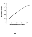
Fig. 1 is a standard calibration curve of a serological marker for detecting pancreatic cancer by using a bead-based immunoassay. -
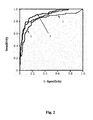
Fig. 2 is a receiver operating characteristic curve of the ULBP2 and CA19-9 in detecting pancreatic cancer. -
Fig. 3A shows the detection efficiency of ULBP2, CA19-9 and ULBP2 combined with CA19-9 in detecting the pancreatic cancer of T1/T2 stage of the TNM classification. -
Fig. 3B shows the detection efficiency of ULBP2, CA19-9 and ULBP2 combined with CA19-9 in detecting the pancreatic cancer of N0 stage of the TNM classification. -
Fig. 3C shows the detection efficiency of ULBP2, CA19-9 and ULBP2 combined with CA19-9 in detecting the pancreatic cancer of I-II stage of the overall stage. -
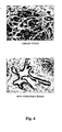
Fig. 4 shows the immunohistochemistry assay of the cancer tissue biopsies from 67 pancreatic cancer patients shows that ULBP2 staining is positive in all biopsies (100%), and the expression of ULBP2 is more significant in the cancer tissue than in the non-cancerous tissues. - Embodiment 1: screening and selecting a serological marker for detecting the pancreatic cancer
- Proteins secreted from two pancreatic cancer cell lines PANC-1 and MIA PaCa-2, which were collected by incubating the cultured cells in serum-free medium for 24 hr (this medium is thereafter defined as conditioned medium), are systematically identified by one-dimensional SDS-PAGE in conjunction with nano-LC-MS/MS (the GeLC-MS/MS approach). This method identified a total of 1812 non-redundant proteins from the conditioned medium of the two cell lines. The transcriptional expression of each identified protein in the pancreatic cancer tissues was further analyzed according to a public doamin transcriptomic information of the pancreatic cancer tissues (National Center for Biotechnology Information (NCBI) Gene Expression Omnibus). In this transcriptome dataset, pancreatic ductal cells respectively isolated from 25 healthy donors and 24 pancreatic cancer patients are subjected to an array-based analysis to identify the genes whose message RNA (mRNA) levels are higher expressed in the pancreatic cancer patients than those in the healthy donors. By integrating this transcriptome dataset and the secreted protein database of the two pancreatic cancer cell lines generated as described above, 30 pancreatic cancer cell secreted proteins exhibited at least two-fold higher mRNA expression levels in the pancreatic ductal cells from cancer patients than from the healthy donors. Eleven out of the 30 proteins had been reported to be over-expressed in the tissue biopsy of pancreatic cancer by previous studies. Among the other 19 proteins without any references related to pancreatic cancer, UL16 binding protein 2 (ULBP2) is selected as a serological candidate marker for detecting pancreatic cancer in the present invention.
- The ULBP2 in the present embodiment has an amino acid sequence (SEQ ID NO:1) shown as following:

- One ordinary skill in the art understands that any amino acid replacement by an amino acid with similar characteristic will cause a little variation in the original SEQ ID NO:1. However, a sequence has similarity more than 95% to SEQ ID NO:1 is considered as a serological marker for detecting pancreatic cancer that can be used in the embodiment.
- Immunohistochemistry assay: In the embodiment, a goat anti-ULBP2 antibody is applied. A tissue biopsy is isolated and heated in a 0.01 M citric acid buffer (pH 6.0). A blocking buffer is added and reacted at room temperature for 5 minutes. The tissue biopsy is reacted with the anti-ULBP2 antibody (1:20 dilution) at 4°C for 16 hours. Then, the tissue biopsy is stained with the N-Histofine® (Nichirei, Japan) at room temperature and followed by treatment with substrate DAB chromogen (Novocastra/Leica Microsystems, IL, USA). The tissue biopsy is also counterstained with hematoxylin. The expression level of target proteins was evaluated according to the simplified H score system, which is based on the intensity of cell staining [3 (strong), 2 (moderate), 1 (weak), or 0 (no cell staining)] and the percentage of cell staining [3 (≥90%), 2 (50-89%), 1 (10-49%), or 0 (0-9%)]. The two scores were multiplied by each other and then divided by 3 to get the final score. Positive staining was defined as a final score ≥ 0.67.
- With reference to the
Fig. 4 , the immunohistochemistry assay of the cancer tissue biopsies from 67 pancreatic cancer patients shows that ULBP2 staining is positive in all biopsies (100%). Additionally, the expression of ULBP2 is more significant in the cancer tissue than in the adjacent non-cancerous tissues (the intensity of brown color indicates the expression level of the ULBP2). The mean expression level of the ULBP2 in the cancer tissue and in the adjacent non-cancerous tissue is score of 2.71±0.49 and 1.89±0.74, respectively. Moreover, with reference to Table 1, the expression of ULBP2 of the embodiment is not influenced by the clinically pathological symptoms such as the gender, age, histological grade, overall cancer status or TMN classification. The detecting result of the ULBP2 has highly consistence in patients with different clinically pathological symptoms.Table 1. Correlation between clinicopathological features and ULBP2 expression in 67 pancreatic cancer patients Characteristics Patient No. IHC score (Mean ± SD)a p-value Gender Male 44 2.64 ± 0.54 0.180b Female 23 2.83 ± 0.36 Age (years) < 64c 33 2.61 ± 0.55 0.089b ≥ 64 34 2.80 ± 0.41 Histological graded Well differentiation 23 2.71 ± 0.59 0.746c Moderate differentiation 31 2.68 ± 0.44 Poor differentiation 10 2.70 ± 0.48 Overall stage stage I 9 2.52 ± 0.82 0.593e stage II 56 2.73 ± 0.43 stage IV 2 3.00 ± 0.00 Tumor-node-metastasis (TNM)-T classification TNM-T2 9 2.52 ± 0.82 0.653b TNM-T3 58 2.74 ± 0.42 TNM-N classification TNM-N0 25 2.69 ± 0.59 0.806b TNM-N1 42 2.72 ± 0.43 TNM-M classification No metastasis 65 2.70 ± 0.50 0.479b Distant metastasis 2 3.00 ± 0.00 Intensity and percentage scores of cell staining were multiplied by each other and then divided by 3 to get the IHC scores.
b By Wilcoxon test.
c Median.
d Histological grade information not available in 3 patients.
e By Kruskal-Wallis test. - A bead-based immunoassay is used to detect the ULBP2 level in a serum sample. An ULBP2 antibody, used as a capture antibody, is pre-coupled to COOH beads using the Bio-Plex Amine Couplin Kit (Bio-Rad). A biotin-conjugated anti-ULBP2 antibody is used as a detection antibody. The bead with the capture antibody is added in a filter-bottom 96-well microplate (Millipore). Then the serum sample solution or standard solution containing ULBP2 protein at various concentrations (3.91∼3.2× 10⁴ pg/mL) is added into the well to react in dark at room temperature for 1 hour. After washing the serum sample solution or the standard solution, the detection antibody is added into each well and reacted in dark at room temperature for 1 hour. After washing out the detection antibody, the phycoerythrin-conjugated streptavidin solution is added for 10 minutes to allow the binding between streptavidin and biotin. Unbound streptavidin is removed by a wash step. The ULBP2 level in the serum sample is then calculated by the fluorescent strength of the phycoerythrin based on the fluorescent strength of the standard calibration curve.
- With reference to
Fig.1 , the ULBP2 in the serum sample is very easy to be detected using the bead-based immunoassay. The ULBP2 concentration ranged from 4.3 pg/mL to 31.9 ng/mL in the embodiment is precisely detected, which is not stably and accurately performed in a sandwich ELISA assay. The ULBP2 level is significantly increased in the cancer serum samples ( 200.2 ± 168.6 pg/ml) than in the healthy control samples (51.4 ± 64.6 pg/ml). When a cutoff value of 60 pg/mL for ULBP2 is chosen, the sensitivity and specificity values for cancer detection is 83.8% and 73.9%, respectively. These findings indicate that ULBP2 is a novel serum marker for pancreatic cancer detection. However, the serum ULBP2 levels are not statistically correlated with age, gender, histological grade, tumor overall stage, and TNM classification of pancreatic cancers in this case control study (Table 2).Table 2. Correlation of serum ULBP2 and CA19-9 with clinicopathologic characteristics in 154 pancreatic cancer patients. Characteristics No. ULBP2 (pg/mL) Mean ± SD p-value CA 19-9 (U/mL) Mean ± SD p-value Gender Male 111 199.5 ± 168.4 64.7 ± 24.6 Female 43 202.0 ± 170.8 60.1 ± 23.7 Age (years) < 70b 75 200.4 ± 182.9 60.2 ± 26.0 ≥ 70 79 199.9 ± 154.9 66.4 ± 22.5 Histological gradec Well differentiation 12 148.8 ± 101.0 0.377d 70.0 ± 13.7 0.553d Moderate differentiation 102 198.7 ± 173.1 59.8 ± 26.0 Poor differentiation 14 134.4 ± 99.9 58.7 ± 25.2 Overall stagec Stage I-II 106 181.2± 158.8 60.7 ± 24.4 Stage III-IV 22 215.3 ± 178.5 60.4 ± 28.9 Tumor-node-metastasis (TNM)-T classificationc TNM-T1 7 251.0 ± 150.0 0.418d 62.1 ± 20.5 0.536d TNM-T2 27 194.0 ± 192.9 54.8 ± 28.4 TNM-T3 77 175.1 ± 150.5 61.8 ± 24.0 TNM-T4 17 204.0 ± 171.1 63.9 ± 26.8 TNM-N classificationc TNM-N0 57 191.6 ± 155.2 62.1 ± 25.5 TNM-N1 71 183.4 ± 168.5 59.4 ± 24.9 TNM-M classificationc No metastasis 125 190.5 ± 162.4 60.9 ± 25.2 Distant metastasis 3 40.8 ± 27.7 48.5 ± 22.1 a By Wilcoxon test.
b Median.
c Information of histological grade, overall stage and TNM stage not available in 26 patients.
dBy Kruskal-Wallis test. - The performance of the currently used pancreatic cancer marker CA 19-9 and the pancreatic cancer serological marker ULBP2 in 154 pancreatic cancer patients is compared to evaluate their detection efficacy. Both CA19-9 and ULBP2 show elevated serum levels in the pancreatic cancer patients than in the healthy controls and are not influenced by the clinicopathological characteristics. At 40 U/mL of CA 19-9, a cutoff value currently applied for pancreatic cancer screening in clinics, the sensitivity and specificity values is 84.4% and 74.6%, respectively. Noteworthily, upon selection of a cutoff value of 60 pg/mL for ULBP2, 21 of 24 pancreatic cancer patients with CA 19-9 levels < 40 U/mL could be discriminated form healthy individuals based on ULBP2 levels > 60 pg/mL. In addition, 24 of 36 healthy individuals with CA 19-9 levels > 40 U/mL could be further distinguished form the patients based on ULBP2 levels < 60 pg/mL. The combined usage of ULBP2 and CA19-9 has a great benefit to pancreatic cancer detection by using CA19-9 alone (shown in Table 3).
Table 3. The efficacy of ULBP2 and CA 19-9 for detecting pancreatic cancers. Cancer patients (n=154) Total Sample ULBP2 (> 60 pg/mL) ULBP2 (< 60 pg/mL) CA19-9 (< 40 U/mL) 24 21 3 CA19-9 (> 40 U/mL) 130 108 22 Healthy controls (n=142) CA19-9 (< 40 U/mL) 118 24 82 CA19-9 (> 40 U/mL) 36 12 24 - With reference to
Fig. 2 , the abilities of ULBP2 and CA 19-9 as detection markers are further tested by receiver operator characteristic (ROC) curve analysis and area under the ROC curve (AUC). The analysis demonstrated that ULBP2 (line 1) [AUC = 0.862, 95% confidence interval (CI), 0.821-0.904] is slightly better than CA 19-9 (line 2) (AUC = 0.856, 95% CI, 0.809-0.902) as a screening marker. Most importantly, the combination of ULBP2 and CA 19-9 (line 3) using the logistic regression model shows a higher diagnostic capacity than either marker alone (AUC = 0.910, 95% CI, 0.877-0.943). These results collectively reveal that ULBP2 is a useful serum marker for pancreatic cancer, especially when used together with CA 19-9. - 142 healthy specimens and 154 pancreatic cancer patients are enrolled to evaluate the ability of ULBP2 for early detection of pancreatic cancers. The ULBP2 level in serum samples of the healthy controls is 51.4 ± 64.6 pg/mL that is less than that in the pancreatic cancer patients at any stage (TNM classification-T1/T2, TNM classification-N0 and overall Stage I-II is 205.7 ± 184.3 pg/mL, 191.6 ± 155.2 and 181.2 ± 158.8 pg/mL, respectively, p<0.0001). The results are similar to the CA19-9 and indicate the pancreatic cancer serological marker ULBP2 is able to use for early detection of pancreatic cancer.
- With reference to
Figs. 3A to 3C , the ROC analysis shows that ULBP2 has better performance in early detection of pancreatic cancer than CA19-9. Furthermore, the detection efficiency is improved by combining ULBP2 and CA19-9Table 4. The ability of ULBP2 and CA 19-9 for early detection of pancreatic cancers ULBP2 CA 19-9 ULBP2 conbined with CA 19-9 AUC 95% CI AUC 95% CI AUC 95%CI TNM classificati on-T1/T2 0.854 0.778∼0.930 0.796 0.690∼0.901 0.883 0.816∼0.949 TNM classificati on-N0 0.866 0.811∼0.920 0.841 0.764∼0.917 0.893 0.841∼0.946 Overall Stage I-II 0.846 0.798∼0.895 0.839 0.782∼0.896 0.897 0.856∼0.937 - The specimens obtained from gastric cancer (GC), nasopharyngeal carcinoma cancer (NPC) and colorectal carcinoma cancer (CRC) patinets are applied to evaluate the specificity of the pancreatic cancer serological marker ULBP2. The ULBP2 levels in serum samples from the NPC and CRC, and plasma samples from the GC are detected. As shown in Table 5, compared with the healthy controls (51.4 ± 64.6 pg/mL for serum ULBP2), the serum ULBP2 levels are slightly higher in patients suffered from NPC (N = 28, 65.5 ± 74.3 pg/mL, p = 0.122) or CRC (N = 29, 70.6 ± 73.8 pg/mL, p = 0.038). However, the serum ULBP2 levels are significantly elevated in pancreatic cancer compared to that in CRC (200.2 ± 168.6 versus 70.6 ± 73.8 pg/mL, p < 0.0001) and NPC (200.2 ± 168.6 versus 65.5 ± 74.3 pg/mL, p < 0.0001). The results illustrate that ULBP2 represents a relative specific marker for pancreatic cancer, particularly that its level do not alter or only marginally elevated in the other two gastrointestinal cancers, CRC and GC.
Table 5. The detection efficiency of the pancreatic cancer serological marker ULBP2 in different cancers. Sample No. ULBP2 in serum (pg/mL) ULBP2 in plasma (pg/mL) Pancreatic cancer 154 200.2±168.6 - GC 30 - 78.1±79.7 NPC 28 65.5±74.4 - CRC 29 70.6±73.8 - Control 1142 51.4±64.6 - Control 225 - 86.1±101.2 - Accordingly, the above-mentioned embodiments illustrate the serological marker ULBP2 is significant increased in the serum of a pancreatic cancer patient and is not correlative with the clinicophathological characteristics. The detection sensitivity of the serological marker ULBP2 is sharply improved to 3.91pg/mL in the serum sample. The serological marker ULBP2 has ability to detect the pancreatic cancer at early stage. The ULBP2 is combined with the CA19-9 to promote the efficiency and increase the specificity in pancreatic cancer detection, also in the early stage cancer detection. Therefore, the serological marker ULBP2 actually has capability to detect the pancreatic cancer in the early stage and strength the efficiency of the clinical diagnosis.
-
- <110> CHANG GUNG UNIVERSITY
- <120> A Serological Marker for Detecting Pancreatic Cancer and A Method For Using The Serological Marker
- <160> 1
- <210> SEQ ID NO: 1
<211> 240
<212> NUCLEOTIDE
<213> Homo sapiens - <400> 1

Claims (9)
- A method for detecting pancreatic cancer, comprising steps of:detecting: at least detecting the level of an UL16 binding protein 2 (ULBP2) having similarity of 95% or more with an amino acid sequence of SEQ ID No. 1 in a blood specimen, obtained from testeecalculating: calculating a concentration of the ULBP2 by using a stand and calibration curve and comparing the concentration of the ULBP2 with a control concentration of the ULBP2 of a blood specimen from a healthy person.
- The method as claimed in claim 1, wherein the ULBP2 has an amino acid sequence of SEQ ID No. 1.
- The method as claimed in claim 1, wherein the step of detecting further comprises at least additional serological marker of detecting a pancreatic cancer.
- The method as claimed in claim 3, wherein the additional serological marker is carbohydrate antigen 19-9 (CA19-9).
- The method as claimed in claim 1, wherein the blood specimen is a whole blood, a serum or a plasma blood specimen.
- The method as claimed in claim 1, wherein the serological marker is applied in a bead-based immunoassay, a sandwich enzyme-linked immunosorbent assay (ELISA), a mass spectrometry-based assay or a mass spectrometry-based immunoassay.
- The method as claimed in claim 2, wherein the serological marker is applied in a bead-based immunoassay, a sandwich enzyme-linked immunosorbent assay (ELISA), a mass spectrometry-based assay or a mass spectrometry-based immunoassay.
- The method as claimed in claim 3, wherein the serological marker is applied in a bead-based immunoassay, a sandwich enzyme-linked immunosorbent assay (ELISA), a mass spectrometry-based assay or a mass spectrometry-based immunoassay.
- The method as claimed in claim 4, wherein the serological marker is applied in a bead-based immunoassay, a sandwich enzyme-linked immunosorbent assay (ELISA), a mass spectrometry-based assay or a mass spectrometry-based immunoassay.
Priority Applications (1)
| Application Number | Priority Date | Filing Date | Title |
|---|---|---|---|
| PL12168401T PL2525227T3 (en) | 2011-05-19 | 2012-05-16 | A method for detecting pancreatic cancer using the serological marker ULBP2 |
Applications Claiming Priority (1)
| Application Number | Priority Date | Filing Date | Title |
|---|---|---|---|
| TW100117502A TWI408370B (en) | 2011-05-19 | 2011-05-19 | A serological maker for detecting pancreatic cancer and a method for using the serological maker |
Publications (2)
| Publication Number | Publication Date |
|---|---|
| EP2525227A1 EP2525227A1 (en) | 2012-11-21 |
| EP2525227B1 true EP2525227B1 (en) | 2014-11-19 |
Family
ID=46087581
Family Applications (1)
| Application Number | Title | Priority Date | Filing Date |
|---|---|---|---|
| EP12168401.3A Active EP2525227B1 (en) | 2011-05-19 | 2012-05-16 | A method for detecting pancreatic cancer using the serological marker ULBP2 |
Country Status (5)
| Country | Link |
|---|---|
| US (1) | US20120295288A1 (en) |
| EP (1) | EP2525227B1 (en) |
| ES (1) | ES2527830T3 (en) |
| PL (1) | PL2525227T3 (en) |
| TW (1) | TWI408370B (en) |
Families Citing this family (8)
| Publication number | Priority date | Publication date | Assignee | Title |
|---|---|---|---|---|
| CN103175970A (en) * | 2013-02-05 | 2013-06-26 | 福建省洪诚生物药业有限公司 | CA19-9 quantitative detection kit and preparation method of kit |
| EP2970490A4 (en) | 2013-03-15 | 2017-04-26 | Novelogics Biotechnology, Inc. | Antibodies to mica and micb proteins |
| KR20150129932A (en) | 2014-05-12 | 2015-11-23 | 연세대학교 산학협력단 | A kit for pancreatic cancer diagnosis comprising complememt factor b-specific binding antibody |
| PT3227311T (en) | 2014-12-05 | 2022-02-23 | Xyphos Biosciences Inc | Insertable variable fragments of antibodies and modified a1-a2 domains of nkg2d ligands |
| US11117969B2 (en) | 2014-12-05 | 2021-09-14 | Xyphos Biosciences Inc. | Insertable variable fragments of antibodies and modified α1-α2 domains of NKG2D ligands |
| CN110088137A (en) | 2016-10-19 | 2019-08-02 | 诺瓦罗技科斯生物科技有限公司 | For the antibody of MICA and MICB albumen |
| US12071515B2 (en) | 2018-06-08 | 2024-08-27 | The Regents Of The University Of Colorado, A Body Corporate | High dynamic range two-stage photopolymers |
| US20220119567A1 (en) * | 2019-02-18 | 2022-04-21 | The Regents Of The University Of Colorado, A Body Corporate | Network polymers and methods of making and using same |
Family Cites Families (1)
| Publication number | Priority date | Publication date | Assignee | Title |
|---|---|---|---|---|
| EP2491396A4 (en) * | 2009-10-22 | 2013-04-17 | Univ California | Assessment of solid tumor burden |
-
2011
- 2011-05-19 TW TW100117502A patent/TWI408370B/en active
-
2012
- 2012-05-16 ES ES12168401.3T patent/ES2527830T3/en active Active
- 2012-05-16 EP EP12168401.3A patent/EP2525227B1/en active Active
- 2012-05-16 PL PL12168401T patent/PL2525227T3/en unknown
- 2012-05-18 US US13/475,658 patent/US20120295288A1/en not_active Abandoned
Also Published As
| Publication number | Publication date |
|---|---|
| US20120295288A1 (en) | 2012-11-22 |
| TWI408370B (en) | 2013-09-11 |
| TW201248151A (en) | 2012-12-01 |
| ES2527830T3 (en) | 2015-01-30 |
| EP2525227A1 (en) | 2012-11-21 |
| PL2525227T3 (en) | 2015-06-30 |
Similar Documents
| Publication | Publication Date | Title |
|---|---|---|
| EP2525227B1 (en) | A method for detecting pancreatic cancer using the serological marker ULBP2 | |
| Simon et al. | Evaluation of the novel serum markers B7-H4, Spondin 2, and DcR3 for diagnosis and early detection of ovarian cancer | |
| EP2115472B1 (en) | Cancer biomarkers | |
| Niclou et al. | Glioma proteomics: status and perspectives | |
| CN101088011B (en) | Use of CYFRA 21-1 as a marker for colorectal cancer | |
| Yi et al. | Autoantibody to tumor antigen, alpha 2-HS glycoprotein: a novel biomarker of breast cancer screening and diagnosis | |
| EP3885768A1 (en) | Biomarker panel for diagnosing cancer | |
| Takayama et al. | Serum tumor antigen REG4 as a diagnostic biomarker in pancreatic ductal adenocarcinoma | |
| Soukup et al. | Panel of urinary diagnostic markers for non-invasive detection of primary and recurrent urothelial urinary bladder carcinoma | |
| JP5384672B2 (en) | Use of s-ErbB-3 as a marker for cancer | |
| CN112345755B (en) | Biomarker for breast cancer and application thereof | |
| US20220214345A1 (en) | Colorectal cancer screening examination and early detection method | |
| CN101014862A (en) | Methods and compositions for the detection of ovarian disease | |
| CN101346628A (en) | Use of a marker combination comprising osteopontin and carcinoembryonic antigen in the assessment of colorectal cancer | |
| US8377648B2 (en) | Autoimmune regulation of prostate cancer by annexin A3 | |
| KR102369544B1 (en) | Use of nucleosome-transcription factor complexes for cancer detection | |
| US20110147218A1 (en) | Soluble cadherin 17 for the diagnosis and risk stratification of cancer and tumor of the gastrointestinal tract | |
| JP6361943B2 (en) | Pancreatic cancer diagnostic kit comprising an antibody that specifically binds to complement factor B protein and an antibody that specifically binds to sugar chain antigen 19-9 protein | |
| EP3215851A2 (en) | Lung cancer sub-typing method | |
| CN117120847A (en) | Method for detecting lung cancer | |
| Benlier et al. | A novel diagnostic tool for the detection of bladder cancer: measurement of urinary high mobility group box-1 | |
| US20120225441A1 (en) | Protein markers for detecting liver cancer and method for identifying the markers thereof | |
| KR102280360B1 (en) | A Composition for Diagnosing Cancer | |
| KR20160053875A (en) | Maker for the diagnosis of gastric cancer | |
| US20150004621A1 (en) | Biological marker for early cancer detection and methods for cancer detection (bf819) |
Legal Events
| Date | Code | Title | Description |
|---|---|---|---|
| PUAI | Public reference made under article 153(3) epc to a published international application that has entered the european phase |
Free format text: ORIGINAL CODE: 0009012 |
|
| AK | Designated contracting states |
Kind code of ref document: A1 Designated state(s): AL AT BE BG CH CY CZ DE DK EE ES FI FR GB GR HR HU IE IS IT LI LT LU LV MC MK MT NL NO PL PT RO RS SE SI SK SM TR |
|
| AX | Request for extension of the european patent |
Extension state: BA ME |
|
| 17P | Request for examination filed |
Effective date: 20130517 |
|
| R17P | Request for examination filed (corrected) |
Effective date: 20130521 |
|
| GRAP | Despatch of communication of intention to grant a patent |
Free format text: ORIGINAL CODE: EPIDOSNIGR1 |
|
| INTG | Intention to grant announced |
Effective date: 20140605 |
|
| INTG | Intention to grant announced |
Effective date: 20140612 |
|
| GRAS | Grant fee paid |
Free format text: ORIGINAL CODE: EPIDOSNIGR3 |
|
| GRAA | (expected) grant |
Free format text: ORIGINAL CODE: 0009210 |
|
| AK | Designated contracting states |
Kind code of ref document: B1 Designated state(s): AL AT BE BG CH CY CZ DE DK EE ES FI FR GB GR HR HU IE IS IT LI LT LU LV MC MK MT NL NO PL PT RO RS SE SI SK SM TR |
|
| REG | Reference to a national code |
Ref country code: GB Ref legal event code: FG4D |
|
| REG | Reference to a national code |
Ref country code: CH Ref legal event code: EP |
|
| REG | Reference to a national code |
Ref country code: AT Ref legal event code: REF Ref document number: 697312 Country of ref document: AT Kind code of ref document: T Effective date: 20141215 |
|
| REG | Reference to a national code |
Ref country code: IE Ref legal event code: FG4D |
|
| REG | Reference to a national code |
Ref country code: DE Ref legal event code: R096 Ref document number: 602012003837 Country of ref document: DE Effective date: 20141231 |
|
| REG | Reference to a national code |
Ref country code: ES Ref legal event code: FG2A Ref document number: 2527830 Country of ref document: ES Kind code of ref document: T3 Effective date: 20150130 |
|
| REG | Reference to a national code |
Ref country code: NL Ref legal event code: VDEP Effective date: 20141119 |
|
| REG | Reference to a national code |
Ref country code: AT Ref legal event code: MK05 Ref document number: 697312 Country of ref document: AT Kind code of ref document: T Effective date: 20141119 |
|
| REG | Reference to a national code |
Ref country code: LT Ref legal event code: MG4D |
|
| PG25 | Lapsed in a contracting state [announced via postgrant information from national office to epo] |
Ref country code: PT Free format text: LAPSE BECAUSE OF FAILURE TO SUBMIT A TRANSLATION OF THE DESCRIPTION OR TO PAY THE FEE WITHIN THE PRESCRIBED TIME-LIMIT Effective date: 20150319 Ref country code: FI Free format text: LAPSE BECAUSE OF FAILURE TO SUBMIT A TRANSLATION OF THE DESCRIPTION OR TO PAY THE FEE WITHIN THE PRESCRIBED TIME-LIMIT Effective date: 20141119 Ref country code: NO Free format text: LAPSE BECAUSE OF FAILURE TO SUBMIT A TRANSLATION OF THE DESCRIPTION OR TO PAY THE FEE WITHIN THE PRESCRIBED TIME-LIMIT Effective date: 20150219 Ref country code: IS Free format text: LAPSE BECAUSE OF FAILURE TO SUBMIT A TRANSLATION OF THE DESCRIPTION OR TO PAY THE FEE WITHIN THE PRESCRIBED TIME-LIMIT Effective date: 20150319 Ref country code: LT Free format text: LAPSE BECAUSE OF FAILURE TO SUBMIT A TRANSLATION OF THE DESCRIPTION OR TO PAY THE FEE WITHIN THE PRESCRIBED TIME-LIMIT Effective date: 20141119 Ref country code: NL Free format text: LAPSE BECAUSE OF FAILURE TO SUBMIT A TRANSLATION OF THE DESCRIPTION OR TO PAY THE FEE WITHIN THE PRESCRIBED TIME-LIMIT Effective date: 20141119 |
|
| PG25 | Lapsed in a contracting state [announced via postgrant information from national office to epo] |
Ref country code: GR Free format text: LAPSE BECAUSE OF FAILURE TO SUBMIT A TRANSLATION OF THE DESCRIPTION OR TO PAY THE FEE WITHIN THE PRESCRIBED TIME-LIMIT Effective date: 20150220 Ref country code: RS Free format text: LAPSE BECAUSE OF FAILURE TO SUBMIT A TRANSLATION OF THE DESCRIPTION OR TO PAY THE FEE WITHIN THE PRESCRIBED TIME-LIMIT Effective date: 20141119 Ref country code: HR Free format text: LAPSE BECAUSE OF FAILURE TO SUBMIT A TRANSLATION OF THE DESCRIPTION OR TO PAY THE FEE WITHIN THE PRESCRIBED TIME-LIMIT Effective date: 20141119 Ref country code: AT Free format text: LAPSE BECAUSE OF FAILURE TO SUBMIT A TRANSLATION OF THE DESCRIPTION OR TO PAY THE FEE WITHIN THE PRESCRIBED TIME-LIMIT Effective date: 20141119 Ref country code: CY Free format text: LAPSE BECAUSE OF FAILURE TO SUBMIT A TRANSLATION OF THE DESCRIPTION OR TO PAY THE FEE WITHIN THE PRESCRIBED TIME-LIMIT Effective date: 20141119 Ref country code: SE Free format text: LAPSE BECAUSE OF FAILURE TO SUBMIT A TRANSLATION OF THE DESCRIPTION OR TO PAY THE FEE WITHIN THE PRESCRIBED TIME-LIMIT Effective date: 20141119 Ref country code: LV Free format text: LAPSE BECAUSE OF FAILURE TO SUBMIT A TRANSLATION OF THE DESCRIPTION OR TO PAY THE FEE WITHIN THE PRESCRIBED TIME-LIMIT Effective date: 20141119 |
|
| REG | Reference to a national code |
Ref country code: PL Ref legal event code: T3 |
|
| PG25 | Lapsed in a contracting state [announced via postgrant information from national office to epo] |
Ref country code: EE Free format text: LAPSE BECAUSE OF FAILURE TO SUBMIT A TRANSLATION OF THE DESCRIPTION OR TO PAY THE FEE WITHIN THE PRESCRIBED TIME-LIMIT Effective date: 20141119 Ref country code: DK Free format text: LAPSE BECAUSE OF FAILURE TO SUBMIT A TRANSLATION OF THE DESCRIPTION OR TO PAY THE FEE WITHIN THE PRESCRIBED TIME-LIMIT Effective date: 20141119 Ref country code: CZ Free format text: LAPSE BECAUSE OF FAILURE TO SUBMIT A TRANSLATION OF THE DESCRIPTION OR TO PAY THE FEE WITHIN THE PRESCRIBED TIME-LIMIT Effective date: 20141119 Ref country code: SK Free format text: LAPSE BECAUSE OF FAILURE TO SUBMIT A TRANSLATION OF THE DESCRIPTION OR TO PAY THE FEE WITHIN THE PRESCRIBED TIME-LIMIT Effective date: 20141119 Ref country code: RO Free format text: LAPSE BECAUSE OF FAILURE TO SUBMIT A TRANSLATION OF THE DESCRIPTION OR TO PAY THE FEE WITHIN THE PRESCRIBED TIME-LIMIT Effective date: 20141119 |
|
| REG | Reference to a national code |
Ref country code: DE Ref legal event code: R097 Ref document number: 602012003837 Country of ref document: DE |
|
| PLBE | No opposition filed within time limit |
Free format text: ORIGINAL CODE: 0009261 |
|
| STAA | Information on the status of an ep patent application or granted ep patent |
Free format text: STATUS: NO OPPOSITION FILED WITHIN TIME LIMIT |
|
| 26N | No opposition filed |
Effective date: 20150820 |
|
| REG | Reference to a national code |
Ref country code: CH Ref legal event code: PL |
|
| PG25 | Lapsed in a contracting state [announced via postgrant information from national office to epo] |
Ref country code: LI Free format text: LAPSE BECAUSE OF NON-PAYMENT OF DUE FEES Effective date: 20150531 Ref country code: MC Free format text: LAPSE BECAUSE OF FAILURE TO SUBMIT A TRANSLATION OF THE DESCRIPTION OR TO PAY THE FEE WITHIN THE PRESCRIBED TIME-LIMIT Effective date: 20141119 Ref country code: CH Free format text: LAPSE BECAUSE OF NON-PAYMENT OF DUE FEES Effective date: 20150531 Ref country code: LU Free format text: LAPSE BECAUSE OF FAILURE TO SUBMIT A TRANSLATION OF THE DESCRIPTION OR TO PAY THE FEE WITHIN THE PRESCRIBED TIME-LIMIT Effective date: 20150516 |
|
| REG | Reference to a national code |
Ref country code: IE Ref legal event code: MM4A |
|
| PG25 | Lapsed in a contracting state [announced via postgrant information from national office to epo] |
Ref country code: SI Free format text: LAPSE BECAUSE OF FAILURE TO SUBMIT A TRANSLATION OF THE DESCRIPTION OR TO PAY THE FEE WITHIN THE PRESCRIBED TIME-LIMIT Effective date: 20141119 |
|
| PG25 | Lapsed in a contracting state [announced via postgrant information from national office to epo] |
Ref country code: IE Free format text: LAPSE BECAUSE OF NON-PAYMENT OF DUE FEES Effective date: 20150516 |
|
| REG | Reference to a national code |
Ref country code: FR Ref legal event code: PLFP Year of fee payment: 5 |
|
| PG25 | Lapsed in a contracting state [announced via postgrant information from national office to epo] |
Ref country code: MT Free format text: LAPSE BECAUSE OF FAILURE TO SUBMIT A TRANSLATION OF THE DESCRIPTION OR TO PAY THE FEE WITHIN THE PRESCRIBED TIME-LIMIT Effective date: 20141119 |
|
| REG | Reference to a national code |
Ref country code: FR Ref legal event code: PLFP Year of fee payment: 6 |
|
| PG25 | Lapsed in a contracting state [announced via postgrant information from national office to epo] |
Ref country code: HU Free format text: LAPSE BECAUSE OF FAILURE TO SUBMIT A TRANSLATION OF THE DESCRIPTION OR TO PAY THE FEE WITHIN THE PRESCRIBED TIME-LIMIT; INVALID AB INITIO Effective date: 20120516 Ref country code: BG Free format text: LAPSE BECAUSE OF FAILURE TO SUBMIT A TRANSLATION OF THE DESCRIPTION OR TO PAY THE FEE WITHIN THE PRESCRIBED TIME-LIMIT Effective date: 20141119 Ref country code: SM Free format text: LAPSE BECAUSE OF FAILURE TO SUBMIT A TRANSLATION OF THE DESCRIPTION OR TO PAY THE FEE WITHIN THE PRESCRIBED TIME-LIMIT Effective date: 20141119 |
|
| PG25 | Lapsed in a contracting state [announced via postgrant information from national office to epo] |
Ref country code: TR Free format text: LAPSE BECAUSE OF FAILURE TO SUBMIT A TRANSLATION OF THE DESCRIPTION OR TO PAY THE FEE WITHIN THE PRESCRIBED TIME-LIMIT Effective date: 20141119 |
|
| PG25 | Lapsed in a contracting state [announced via postgrant information from national office to epo] |
Ref country code: BE Free format text: LAPSE BECAUSE OF FAILURE TO SUBMIT A TRANSLATION OF THE DESCRIPTION OR TO PAY THE FEE WITHIN THE PRESCRIBED TIME-LIMIT Effective date: 20141119 |
|
| REG | Reference to a national code |
Ref country code: FR Ref legal event code: PLFP Year of fee payment: 7 |
|
| PG25 | Lapsed in a contracting state [announced via postgrant information from national office to epo] |
Ref country code: MK Free format text: LAPSE BECAUSE OF FAILURE TO SUBMIT A TRANSLATION OF THE DESCRIPTION OR TO PAY THE FEE WITHIN THE PRESCRIBED TIME-LIMIT Effective date: 20141119 |
|
| PG25 | Lapsed in a contracting state [announced via postgrant information from national office to epo] |
Ref country code: AL Free format text: LAPSE BECAUSE OF FAILURE TO SUBMIT A TRANSLATION OF THE DESCRIPTION OR TO PAY THE FEE WITHIN THE PRESCRIBED TIME-LIMIT Effective date: 20141119 |
|
| REG | Reference to a national code |
Ref country code: DE Ref legal event code: R082 Ref document number: 602012003837 Country of ref document: DE Representative=s name: LANGPATENT ANWALTSKANZLEI IP LAW FIRM, DE Ref country code: DE Ref legal event code: R081 Ref document number: 602012003837 Country of ref document: DE Owner name: S&T BIOMED CO., LTD., TW Free format text: FORMER OWNER: CHANG GUNG UNIVERSITY (A UNIVERSITY OF TAIWAN), TAO-YUAN, TW |
|
| REG | Reference to a national code |
Ref country code: ES Ref legal event code: PC2A Owner name: S&T BIOMED CO., LTD. Effective date: 20200617 |
|
| REG | Reference to a national code |
Ref country code: GB Ref legal event code: 732E Free format text: REGISTERED BETWEEN 20200709 AND 20200715 |
|
| P01 | Opt-out of the competence of the unified patent court (upc) registered |
Effective date: 20230602 |
|
| PGFP | Annual fee paid to national office [announced via postgrant information from national office to epo] |
Ref country code: GB Payment date: 20240521 Year of fee payment: 13 |
|
| PGFP | Annual fee paid to national office [announced via postgrant information from national office to epo] |
Ref country code: DE Payment date: 20240521 Year of fee payment: 13 |
|
| PGFP | Annual fee paid to national office [announced via postgrant information from national office to epo] |
Ref country code: ES Payment date: 20240626 Year of fee payment: 13 |
|
| PGFP | Annual fee paid to national office [announced via postgrant information from national office to epo] |
Ref country code: FR Payment date: 20240528 Year of fee payment: 13 |
|
| PGFP | Annual fee paid to national office [announced via postgrant information from national office to epo] |
Ref country code: PL Payment date: 20240429 Year of fee payment: 13 |
|
| PGFP | Annual fee paid to national office [announced via postgrant information from national office to epo] |
Ref country code: IT Payment date: 20240524 Year of fee payment: 13 |