EP2524031B1 - Menschlicher stammzelllinien und maus: modell von endothelialen progenitorzellen - Google Patents
Menschlicher stammzelllinien und maus: modell von endothelialen progenitorzellen Download PDFInfo
- Publication number
- EP2524031B1 EP2524031B1 EP11704276.2A EP11704276A EP2524031B1 EP 2524031 B1 EP2524031 B1 EP 2524031B1 EP 11704276 A EP11704276 A EP 11704276A EP 2524031 B1 EP2524031 B1 EP 2524031B1
- Authority
- EP
- European Patent Office
- Prior art keywords
- cells
- cell
- endothelial
- isolated
- differentiation
- Prior art date
- Legal status (The legal status is an assumption and is not a legal conclusion. Google has not performed a legal analysis and makes no representation as to the accuracy of the status listed.)
- Not-in-force
Links
- 230000003511 endothelial effect Effects 0.000 title claims description 18
- 210000000130 stem cell Anatomy 0.000 title description 24
- 241000699666 Mus <mouse, genus> Species 0.000 title description 17
- 210000004027 cell Anatomy 0.000 claims description 332
- 210000002889 endothelial cell Anatomy 0.000 claims description 69
- 239000002243 precursor Substances 0.000 claims description 59
- 206010028980 Neoplasm Diseases 0.000 claims description 42
- 230000004069 differentiation Effects 0.000 claims description 40
- 238000000034 method Methods 0.000 claims description 26
- 241001529936 Murinae Species 0.000 claims description 10
- 238000004113 cell culture Methods 0.000 claims description 9
- 238000004519 manufacturing process Methods 0.000 claims description 9
- 238000001514 detection method Methods 0.000 claims description 8
- 230000003902 lesion Effects 0.000 claims description 8
- 206010061216 Infarction Diseases 0.000 claims description 7
- 230000033115 angiogenesis Effects 0.000 claims description 7
- 230000007574 infarction Effects 0.000 claims description 7
- 244000005700 microbiome Species 0.000 claims description 7
- 230000006378 damage Effects 0.000 claims description 6
- 230000002950 deficient Effects 0.000 claims description 6
- 230000000694 effects Effects 0.000 claims description 6
- 238000012258 culturing Methods 0.000 claims description 4
- 230000024245 cell differentiation Effects 0.000 claims description 3
- 208000028867 ischemia Diseases 0.000 claims description 3
- 230000035755 proliferation Effects 0.000 claims description 3
- 238000012216 screening Methods 0.000 claims description 3
- 206010012601 diabetes mellitus Diseases 0.000 claims description 2
- 229940126601 medicinal product Drugs 0.000 claims 3
- 241000699670 Mus sp. Species 0.000 description 30
- 230000014509 gene expression Effects 0.000 description 29
- 206010021143 Hypoxia Diseases 0.000 description 27
- 239000002609 medium Substances 0.000 description 27
- 230000007954 hypoxia Effects 0.000 description 24
- 210000001519 tissue Anatomy 0.000 description 24
- 230000007959 normoxia Effects 0.000 description 23
- 108090000623 proteins and genes Proteins 0.000 description 21
- 230000000692 anti-sense effect Effects 0.000 description 18
- 210000003958 hematopoietic stem cell Anatomy 0.000 description 17
- 238000002347 injection Methods 0.000 description 17
- 239000007924 injection Substances 0.000 description 17
- LOKCTEFSRHRXRJ-UHFFFAOYSA-I dipotassium trisodium dihydrogen phosphate hydrogen phosphate dichloride Chemical compound P(=O)(O)(O)[O-].[K+].P(=O)(O)([O-])[O-].[Na+].[Na+].[Cl-].[K+].[Cl-].[Na+] LOKCTEFSRHRXRJ-UHFFFAOYSA-I 0.000 description 14
- 238000002372 labelling Methods 0.000 description 14
- 239000002953 phosphate buffered saline Substances 0.000 description 14
- 102000004127 Cytokines Human genes 0.000 description 12
- 108090000695 Cytokines Proteins 0.000 description 12
- 230000015572 biosynthetic process Effects 0.000 description 12
- 230000006870 function Effects 0.000 description 12
- 239000012124 Opti-MEM Substances 0.000 description 10
- RWHOZGRAXYWRNX-DVKNGEFBSA-N [(2s,3r,4s,5s,6r)-3,4,5-trihydroxy-6-(phosphonooxymethyl)oxan-2-yl] dihydrogen phosphate Chemical compound O[C@H]1[C@H](O)[C@@H](COP(O)(O)=O)O[C@@H](OP(O)(O)=O)[C@@H]1O RWHOZGRAXYWRNX-DVKNGEFBSA-N 0.000 description 10
- 239000003550 marker Substances 0.000 description 10
- MFXMNSMTYUGIGR-UHFFFAOYSA-N n-[acetyl(methyl)amino]-n-phenylnitrous amide Chemical compound CC(=O)N(C)N(N=O)C1=CC=CC=C1 MFXMNSMTYUGIGR-UHFFFAOYSA-N 0.000 description 10
- 230000001225 therapeutic effect Effects 0.000 description 10
- 238000011534 incubation Methods 0.000 description 9
- 239000000047 product Substances 0.000 description 9
- 210000000952 spleen Anatomy 0.000 description 9
- 238000011282 treatment Methods 0.000 description 9
- 238000002965 ELISA Methods 0.000 description 8
- 238000012512 characterization method Methods 0.000 description 8
- 230000012010 growth Effects 0.000 description 8
- 108010082117 matrigel Proteins 0.000 description 8
- 210000000056 organ Anatomy 0.000 description 8
- 230000001575 pathological effect Effects 0.000 description 8
- 238000003752 polymerase chain reaction Methods 0.000 description 8
- 238000007920 subcutaneous administration Methods 0.000 description 8
- 230000008685 targeting Effects 0.000 description 8
- 102100031573 Hematopoietic progenitor cell antigen CD34 Human genes 0.000 description 7
- 101000777663 Homo sapiens Hematopoietic progenitor cell antigen CD34 Proteins 0.000 description 7
- 101150088608 Kdr gene Proteins 0.000 description 7
- FAPWRFPIFSIZLT-UHFFFAOYSA-M Sodium chloride Chemical compound [Na+].[Cl-] FAPWRFPIFSIZLT-UHFFFAOYSA-M 0.000 description 7
- 210000004369 blood Anatomy 0.000 description 7
- 239000008280 blood Substances 0.000 description 7
- 238000002474 experimental method Methods 0.000 description 7
- 239000003102 growth factor Substances 0.000 description 7
- 210000004072 lung Anatomy 0.000 description 7
- 108020004999 messenger RNA Proteins 0.000 description 7
- 210000005087 mononuclear cell Anatomy 0.000 description 7
- 239000007787 solid Substances 0.000 description 7
- 102100030988 Angiotensin-converting enzyme Human genes 0.000 description 6
- 108010078239 Chemokine CX3CL1 Proteins 0.000 description 6
- 101100540419 Danio rerio kdrl gene Proteins 0.000 description 6
- 101150009958 FLT4 gene Proteins 0.000 description 6
- 102100020997 Fractalkine Human genes 0.000 description 6
- 102000016663 Vascular Endothelial Growth Factor Receptor-3 Human genes 0.000 description 6
- 102100033177 Vascular endothelial growth factor receptor 2 Human genes 0.000 description 6
- 239000003814 drug Substances 0.000 description 6
- 238000000684 flow cytometry Methods 0.000 description 6
- 230000003394 haemopoietic effect Effects 0.000 description 6
- 238000002595 magnetic resonance imaging Methods 0.000 description 6
- 210000002783 mesonephros Anatomy 0.000 description 6
- 229910052760 oxygen Inorganic materials 0.000 description 6
- 238000005070 sampling Methods 0.000 description 6
- 102100021936 C-C motif chemokine 27 Human genes 0.000 description 5
- 101710112538 C-C motif chemokine 27 Proteins 0.000 description 5
- 102100023126 Cell surface glycoprotein MUC18 Human genes 0.000 description 5
- KCXVZYZYPLLWCC-UHFFFAOYSA-N EDTA Chemical compound OC(=O)CN(CC(O)=O)CCN(CC(O)=O)CC(O)=O KCXVZYZYPLLWCC-UHFFFAOYSA-N 0.000 description 5
- 102000004889 Interleukin-6 Human genes 0.000 description 5
- 108090001005 Interleukin-6 Proteins 0.000 description 5
- 241000283973 Oryctolagus cuniculus Species 0.000 description 5
- 108010004729 Phycoerythrin Proteins 0.000 description 5
- 102100024616 Platelet endothelial cell adhesion molecule Human genes 0.000 description 5
- 102100021669 Stromal cell-derived factor 1 Human genes 0.000 description 5
- 101710088580 Stromal cell-derived factor 1 Proteins 0.000 description 5
- 239000011324 bead Substances 0.000 description 5
- 229940071106 ethylenediaminetetraacetate Drugs 0.000 description 5
- 210000004700 fetal blood Anatomy 0.000 description 5
- MHMNJMPURVTYEJ-UHFFFAOYSA-N fluorescein-5-isothiocyanate Chemical compound O1C(=O)C2=CC(N=C=S)=CC=C2C21C1=CC=C(O)C=C1OC1=CC(O)=CC=C21 MHMNJMPURVTYEJ-UHFFFAOYSA-N 0.000 description 5
- 239000000499 gel Substances 0.000 description 5
- 238000003384 imaging method Methods 0.000 description 5
- 238000010253 intravenous injection Methods 0.000 description 5
- 230000008569 process Effects 0.000 description 5
- 230000028327 secretion Effects 0.000 description 5
- 239000006228 supernatant Substances 0.000 description 5
- 238000012360 testing method Methods 0.000 description 5
- UUUHXMGGBIUAPW-UHFFFAOYSA-N 1-[1-[2-[[5-amino-2-[[1-[5-(diaminomethylideneamino)-2-[[1-[3-(1h-indol-3-yl)-2-[(5-oxopyrrolidine-2-carbonyl)amino]propanoyl]pyrrolidine-2-carbonyl]amino]pentanoyl]pyrrolidine-2-carbonyl]amino]-5-oxopentanoyl]amino]-3-methylpentanoyl]pyrrolidine-2-carbon Chemical compound C1CCC(C(=O)N2C(CCC2)C(O)=O)N1C(=O)C(C(C)CC)NC(=O)C(CCC(N)=O)NC(=O)C1CCCN1C(=O)C(CCCN=C(N)N)NC(=O)C1CCCN1C(=O)C(CC=1C2=CC=CC=C2NC=1)NC(=O)C1CCC(=O)N1 UUUHXMGGBIUAPW-UHFFFAOYSA-N 0.000 description 4
- 102100022987 Angiogenin Human genes 0.000 description 4
- -1 CD31 Proteins 0.000 description 4
- 241000283707 Capra Species 0.000 description 4
- 102100039620 Granulocyte-macrophage colony-stimulating factor Human genes 0.000 description 4
- 101000716065 Homo sapiens C-C chemokine receptor type 7 Proteins 0.000 description 4
- 101000738771 Homo sapiens Receptor-type tyrosine-protein phosphatase C Proteins 0.000 description 4
- 102000004890 Interleukin-8 Human genes 0.000 description 4
- 108090001007 Interleukin-8 Proteins 0.000 description 4
- 241001465754 Metazoa Species 0.000 description 4
- 108090000882 Peptidyl-Dipeptidase A Proteins 0.000 description 4
- 102100036031 Podocalyxin Human genes 0.000 description 4
- 102100037422 Receptor-type tyrosine-protein phosphatase C Human genes 0.000 description 4
- 102000008790 VE-cadherin Human genes 0.000 description 4
- 108010053099 Vascular Endothelial Growth Factor Receptor-2 Proteins 0.000 description 4
- 238000004458 analytical method Methods 0.000 description 4
- 230000002491 angiogenic effect Effects 0.000 description 4
- 108010072788 angiogenin Proteins 0.000 description 4
- 210000001185 bone marrow Anatomy 0.000 description 4
- 108010018828 cadherin 5 Proteins 0.000 description 4
- 239000002872 contrast media Substances 0.000 description 4
- 210000001671 embryonic stem cell Anatomy 0.000 description 4
- 239000012091 fetal bovine serum Substances 0.000 description 4
- NOESYZHRGYRDHS-UHFFFAOYSA-N insulin Chemical compound N1C(=O)C(NC(=O)C(CCC(N)=O)NC(=O)C(CCC(O)=O)NC(=O)C(C(C)C)NC(=O)C(NC(=O)CN)C(C)CC)CSSCC(C(NC(CO)C(=O)NC(CC(C)C)C(=O)NC(CC=2C=CC(O)=CC=2)C(=O)NC(CCC(N)=O)C(=O)NC(CC(C)C)C(=O)NC(CCC(O)=O)C(=O)NC(CC(N)=O)C(=O)NC(CC=2C=CC(O)=CC=2)C(=O)NC(CSSCC(NC(=O)C(C(C)C)NC(=O)C(CC(C)C)NC(=O)C(CC=2C=CC(O)=CC=2)NC(=O)C(CC(C)C)NC(=O)C(C)NC(=O)C(CCC(O)=O)NC(=O)C(C(C)C)NC(=O)C(CC(C)C)NC(=O)C(CC=2NC=NC=2)NC(=O)C(CO)NC(=O)CNC2=O)C(=O)NCC(=O)NC(CCC(O)=O)C(=O)NC(CCCNC(N)=N)C(=O)NCC(=O)NC(CC=3C=CC=CC=3)C(=O)NC(CC=3C=CC=CC=3)C(=O)NC(CC=3C=CC(O)=CC=3)C(=O)NC(C(C)O)C(=O)N3C(CCC3)C(=O)NC(CCCCN)C(=O)NC(C)C(O)=O)C(=O)NC(CC(N)=O)C(O)=O)=O)NC(=O)C(C(C)CC)NC(=O)C(CO)NC(=O)C(C(C)O)NC(=O)C1CSSCC2NC(=O)C(CC(C)C)NC(=O)C(NC(=O)C(CCC(N)=O)NC(=O)C(CC(N)=O)NC(=O)C(NC(=O)C(N)CC=1C=CC=CC=1)C(C)C)CC1=CN=CN1 NOESYZHRGYRDHS-UHFFFAOYSA-N 0.000 description 4
- 229940100601 interleukin-6 Drugs 0.000 description 4
- 238000002955 isolation Methods 0.000 description 4
- 210000001165 lymph node Anatomy 0.000 description 4
- 230000005291 magnetic effect Effects 0.000 description 4
- 239000000463 material Substances 0.000 description 4
- 201000001441 melanoma Diseases 0.000 description 4
- 108090000743 multidrug resistance protein 3 Proteins 0.000 description 4
- 102000004233 multidrug resistance protein 3 Human genes 0.000 description 4
- 210000003205 muscle Anatomy 0.000 description 4
- 239000002105 nanoparticle Substances 0.000 description 4
- 230000001338 necrotic effect Effects 0.000 description 4
- 230000007170 pathology Effects 0.000 description 4
- 230000001023 pro-angiogenic effect Effects 0.000 description 4
- 238000010839 reverse transcription Methods 0.000 description 4
- PYWVYCXTNDRMGF-UHFFFAOYSA-N rhodamine B Chemical compound [Cl-].C=12C=CC(=[N+](CC)CC)C=C2OC2=CC(N(CC)CC)=CC=C2C=1C1=CC=CC=C1C(O)=O PYWVYCXTNDRMGF-UHFFFAOYSA-N 0.000 description 4
- 238000002560 therapeutic procedure Methods 0.000 description 4
- 230000002792 vascular Effects 0.000 description 4
- 108091032973 (ribonucleotides)n+m Proteins 0.000 description 3
- BJHCYTJNPVGSBZ-YXSASFKJSA-N 1-[4-[6-amino-5-[(Z)-methoxyiminomethyl]pyrimidin-4-yl]oxy-2-chlorophenyl]-3-ethylurea Chemical compound CCNC(=O)Nc1ccc(Oc2ncnc(N)c2\C=N/OC)cc1Cl BJHCYTJNPVGSBZ-YXSASFKJSA-N 0.000 description 3
- JKMHFZQWWAIEOD-UHFFFAOYSA-N 2-[4-(2-hydroxyethyl)piperazin-1-yl]ethanesulfonic acid Chemical compound OCC[NH+]1CCN(CCS([O-])(=O)=O)CC1 JKMHFZQWWAIEOD-UHFFFAOYSA-N 0.000 description 3
- 102100033350 ATP-dependent translocase ABCB1 Human genes 0.000 description 3
- 108091003079 Bovine Serum Albumin Proteins 0.000 description 3
- 102100022595 Broad substrate specificity ATP-binding cassette transporter ABCG2 Human genes 0.000 description 3
- 102100036301 C-C chemokine receptor type 7 Human genes 0.000 description 3
- 108010025714 CD146 Antigen Proteins 0.000 description 3
- 102000006435 Chemokine CCL21 Human genes 0.000 description 3
- 108010083702 Chemokine CCL21 Proteins 0.000 description 3
- SHZGCJCMOBCMKK-UHFFFAOYSA-N D-mannomethylose Natural products CC1OC(O)C(O)C(O)C1O SHZGCJCMOBCMKK-UHFFFAOYSA-N 0.000 description 3
- 240000001008 Dimocarpus longan Species 0.000 description 3
- 102100031983 Ephrin type-B receptor 4 Human genes 0.000 description 3
- 235000000235 Euphoria longan Nutrition 0.000 description 3
- 108010017213 Granulocyte-Macrophage Colony-Stimulating Factor Proteins 0.000 description 3
- 102100034221 Growth-regulated alpha protein Human genes 0.000 description 3
- 239000007995 HEPES buffer Substances 0.000 description 3
- 101000823298 Homo sapiens Broad substrate specificity ATP-binding cassette transporter ABCG2 Proteins 0.000 description 3
- 101100220044 Homo sapiens CD34 gene Proteins 0.000 description 3
- 101001069921 Homo sapiens Growth-regulated alpha protein Proteins 0.000 description 3
- 101000595198 Homo sapiens Podocalyxin Proteins 0.000 description 3
- 108090001090 Lectins Proteins 0.000 description 3
- 102000004856 Lectins Human genes 0.000 description 3
- 108010047230 Member 1 Subfamily B ATP Binding Cassette Transporter Proteins 0.000 description 3
- 102100021339 Multidrug resistance-associated protein 1 Human genes 0.000 description 3
- 102100033237 Pro-epidermal growth factor Human genes 0.000 description 3
- 102000004142 Trypsin Human genes 0.000 description 3
- 108090000631 Trypsin Proteins 0.000 description 3
- 239000000427 antigen Substances 0.000 description 3
- 108091007433 antigens Proteins 0.000 description 3
- 102000036639 antigens Human genes 0.000 description 3
- 239000007640 basal medium Substances 0.000 description 3
- 201000011510 cancer Diseases 0.000 description 3
- 230000001413 cellular effect Effects 0.000 description 3
- 230000007248 cellular mechanism Effects 0.000 description 3
- 238000006243 chemical reaction Methods 0.000 description 3
- 210000002358 circulating endothelial cell Anatomy 0.000 description 3
- 210000002257 embryonic structure Anatomy 0.000 description 3
- 230000001146 hypoxic effect Effects 0.000 description 3
- 229940096397 interleukin-8 Drugs 0.000 description 3
- XKTZWUACRZHVAN-VADRZIEHSA-N interleukin-8 Chemical compound C([C@H](NC(=O)[C@H](CC(O)=O)NC(=O)[C@H](CC=1C2=CC=CC=C2NC=1)NC(=O)[C@@H](NC(C)=O)CCSC)C(=O)N[C@@H](CC(O)=O)C(=O)N[C@@H](CC(O)=O)C(=O)N[C@@H](CC(C)C)C(=O)N[C@@H](CC(N)=O)C(=O)N[C@@H](CC=1C=CC=CC=1)C(=O)N[C@@H]([C@@H](C)O)C(=O)NCC(=O)N[C@@H](CCSC)C(=O)N1[C@H](CCC1)C(=O)N1[C@H](CCC1)C(=O)N[C@@H](C)C(=O)N[C@H](CC(O)=O)C(=O)N[C@H](CCC(O)=O)C(=O)N[C@H](CC(O)=O)C(=O)N[C@H](CC=1C=CC(O)=CC=1)C(=O)N[C@H](CO)C(=O)N1[C@H](CCC1)C(N)=O)C1=CC=CC=C1 XKTZWUACRZHVAN-VADRZIEHSA-N 0.000 description 3
- WTFXARWRTYJXII-UHFFFAOYSA-N iron(2+);iron(3+);oxygen(2-) Chemical compound [O-2].[O-2].[O-2].[O-2].[Fe+2].[Fe+3].[Fe+3] WTFXARWRTYJXII-UHFFFAOYSA-N 0.000 description 3
- 239000002523 lectin Substances 0.000 description 3
- 239000007788 liquid Substances 0.000 description 3
- 210000004185 liver Anatomy 0.000 description 3
- 230000005012 migration Effects 0.000 description 3
- 238000013508 migration Methods 0.000 description 3
- 230000036457 multidrug resistance Effects 0.000 description 3
- 108010066052 multidrug resistance-associated protein 1 Proteins 0.000 description 3
- 210000003491 skin Anatomy 0.000 description 3
- 239000001509 sodium citrate Substances 0.000 description 3
- NLJMYIDDQXHKNR-UHFFFAOYSA-K sodium citrate Chemical compound O.O.[Na+].[Na+].[Na+].[O-]C(=O)CC(O)(CC([O-])=O)C([O-])=O NLJMYIDDQXHKNR-UHFFFAOYSA-K 0.000 description 3
- 239000000243 solution Substances 0.000 description 3
- 210000001562 sternum Anatomy 0.000 description 3
- 230000026683 transduction Effects 0.000 description 3
- 238000010361 transduction Methods 0.000 description 3
- 239000012588 trypsin Substances 0.000 description 3
- 230000004614 tumor growth Effects 0.000 description 3
- 210000005166 vasculature Anatomy 0.000 description 3
- 108010085238 Actins Proteins 0.000 description 2
- 208000031261 Acute myeloid leukaemia Diseases 0.000 description 2
- 102100022749 Aminopeptidase N Human genes 0.000 description 2
- 102100022014 Angiopoietin-1 receptor Human genes 0.000 description 2
- IJGRMHOSHXDMSA-UHFFFAOYSA-N Atomic nitrogen Chemical compound N#N IJGRMHOSHXDMSA-UHFFFAOYSA-N 0.000 description 2
- 102100031650 C-X-C chemokine receptor type 4 Human genes 0.000 description 2
- 102100029761 Cadherin-5 Human genes 0.000 description 2
- 102000019034 Chemokines Human genes 0.000 description 2
- 108010012236 Chemokines Proteins 0.000 description 2
- 102000029816 Collagenase Human genes 0.000 description 2
- 108060005980 Collagenase Proteins 0.000 description 2
- 102000004405 Collectins Human genes 0.000 description 2
- 108090000909 Collectins Proteins 0.000 description 2
- 102100037241 Endoglin Human genes 0.000 description 2
- 101800003838 Epidermal growth factor Proteins 0.000 description 2
- 241000282324 Felis Species 0.000 description 2
- 108050007372 Fibroblast Growth Factor Proteins 0.000 description 2
- 102000018233 Fibroblast Growth Factor Human genes 0.000 description 2
- 102000007563 Galectins Human genes 0.000 description 2
- 108010046569 Galectins Proteins 0.000 description 2
- WQZGKKKJIJFFOK-GASJEMHNSA-N Glucose Natural products OC[C@H]1OC(O)[C@H](O)[C@@H](O)[C@@H]1O WQZGKKKJIJFFOK-GASJEMHNSA-N 0.000 description 2
- 101000757160 Homo sapiens Aminopeptidase N Proteins 0.000 description 2
- 101000753291 Homo sapiens Angiopoietin-1 receptor Proteins 0.000 description 2
- 101000922348 Homo sapiens C-X-C chemokine receptor type 4 Proteins 0.000 description 2
- 101000623903 Homo sapiens Cell surface glycoprotein MUC18 Proteins 0.000 description 2
- 101000881679 Homo sapiens Endoglin Proteins 0.000 description 2
- 101000617130 Homo sapiens Stromal cell-derived factor 1 Proteins 0.000 description 2
- 101000851007 Homo sapiens Vascular endothelial growth factor receptor 2 Proteins 0.000 description 2
- 108060003951 Immunoglobulin Proteins 0.000 description 2
- 102000004877 Insulin Human genes 0.000 description 2
- 108090001061 Insulin Proteins 0.000 description 2
- 102100034343 Integrase Human genes 0.000 description 2
- PIWKPBJCKXDKJR-UHFFFAOYSA-N Isoflurane Chemical compound FC(F)OC(Cl)C(F)(F)F PIWKPBJCKXDKJR-UHFFFAOYSA-N 0.000 description 2
- SHZGCJCMOBCMKK-JFNONXLTSA-N L-rhamnopyranose Chemical compound C[C@@H]1OC(O)[C@H](O)[C@H](O)[C@H]1O SHZGCJCMOBCMKK-JFNONXLTSA-N 0.000 description 2
- PNNNRSAQSRJVSB-UHFFFAOYSA-N L-rhamnose Natural products CC(O)C(O)C(O)C(O)C=O PNNNRSAQSRJVSB-UHFFFAOYSA-N 0.000 description 2
- 101710128836 Large T antigen Proteins 0.000 description 2
- TWRXJAOTZQYOKJ-UHFFFAOYSA-L Magnesium chloride Chemical compound [Mg+2].[Cl-].[Cl-] TWRXJAOTZQYOKJ-UHFFFAOYSA-L 0.000 description 2
- XUMBMVFBXHLACL-UHFFFAOYSA-N Melanin Chemical compound O=C1C(=O)C(C2=CNC3=C(C(C(=O)C4=C32)=O)C)=C2C4=CNC2=C1C XUMBMVFBXHLACL-UHFFFAOYSA-N 0.000 description 2
- 241000699729 Muridae Species 0.000 description 2
- 206010029113 Neovascularisation Diseases 0.000 description 2
- XDMCWZFLLGVIID-SXPRBRBTSA-N O-(3-O-D-galactosyl-N-acetyl-beta-D-galactosaminyl)-L-serine Chemical compound CC(=O)N[C@H]1[C@H](OC[C@H]([NH3+])C([O-])=O)O[C@H](CO)[C@H](O)[C@@H]1OC1[C@H](O)[C@@H](O)[C@@H](O)[C@@H](CO)O1 XDMCWZFLLGVIID-SXPRBRBTSA-N 0.000 description 2
- 229930040373 Paraformaldehyde Natural products 0.000 description 2
- 108010092799 RNA-directed DNA polymerase Proteins 0.000 description 2
- 239000006146 Roswell Park Memorial Institute medium Substances 0.000 description 2
- PXIPVTKHYLBLMZ-UHFFFAOYSA-N Sodium azide Chemical compound [Na+].[N-]=[N+]=[N-] PXIPVTKHYLBLMZ-UHFFFAOYSA-N 0.000 description 2
- 108010009583 Transforming Growth Factors Proteins 0.000 description 2
- 102000009618 Transforming Growth Factors Human genes 0.000 description 2
- 108010073929 Vascular Endothelial Growth Factor A Proteins 0.000 description 2
- 102100039037 Vascular endothelial growth factor A Human genes 0.000 description 2
- 235000010724 Wisteria floribunda Nutrition 0.000 description 2
- 208000027418 Wounds and injury Diseases 0.000 description 2
- 210000001789 adipocyte Anatomy 0.000 description 2
- 230000008901 benefit Effects 0.000 description 2
- 210000000988 bone and bone Anatomy 0.000 description 2
- 239000000872 buffer Substances 0.000 description 2
- 239000002771 cell marker Substances 0.000 description 2
- 230000008859 change Effects 0.000 description 2
- 229960002424 collagenase Drugs 0.000 description 2
- 239000006071 cream Substances 0.000 description 2
- 210000004748 cultured cell Anatomy 0.000 description 2
- 238000010586 diagram Methods 0.000 description 2
- 239000003085 diluting agent Substances 0.000 description 2
- 239000012153 distilled water Substances 0.000 description 2
- 229940079593 drug Drugs 0.000 description 2
- 239000000975 dye Substances 0.000 description 2
- 229940116977 epidermal growth factor Drugs 0.000 description 2
- 210000003743 erythrocyte Anatomy 0.000 description 2
- 229940126864 fibroblast growth factor Drugs 0.000 description 2
- BRZYSWJRSDMWLG-CAXSIQPQSA-N geneticin Chemical compound O1C[C@@](O)(C)[C@H](NC)[C@@H](O)[C@H]1O[C@@H]1[C@@H](O)[C@H](O[C@@H]2[C@@H]([C@@H](O)[C@H](O)[C@@H](C(C)O)O2)N)[C@@H](N)C[C@H]1N BRZYSWJRSDMWLG-CAXSIQPQSA-N 0.000 description 2
- 239000008103 glucose Substances 0.000 description 2
- 239000001963 growth medium Substances 0.000 description 2
- 210000003566 hemangioblast Anatomy 0.000 description 2
- 210000005260 human cell Anatomy 0.000 description 2
- 102000018358 immunoglobulin Human genes 0.000 description 2
- 238000000338 in vitro Methods 0.000 description 2
- 238000001727 in vivo Methods 0.000 description 2
- 238000011503 in vivo imaging Methods 0.000 description 2
- 230000001939 inductive effect Effects 0.000 description 2
- 229940125396 insulin Drugs 0.000 description 2
- 230000003993 interaction Effects 0.000 description 2
- 229960002725 isoflurane Drugs 0.000 description 2
- 239000003446 ligand Substances 0.000 description 2
- 239000011159 matrix material Substances 0.000 description 2
- 210000001616 monocyte Anatomy 0.000 description 2
- 229940126619 mouse monoclonal antibody Drugs 0.000 description 2
- 239000002773 nucleotide Substances 0.000 description 2
- 125000003729 nucleotide group Chemical group 0.000 description 2
- 230000003287 optical effect Effects 0.000 description 2
- 229920002866 paraformaldehyde Polymers 0.000 description 2
- 239000002245 particle Substances 0.000 description 2
- 239000004033 plastic Substances 0.000 description 2
- 229920003023 plastic Polymers 0.000 description 2
- 230000002062 proliferating effect Effects 0.000 description 2
- 235000018102 proteins Nutrition 0.000 description 2
- 102000004169 proteins and genes Human genes 0.000 description 2
- 230000008707 rearrangement Effects 0.000 description 2
- 102000005962 receptors Human genes 0.000 description 2
- 108020003175 receptors Proteins 0.000 description 2
- 230000007115 recruitment Effects 0.000 description 2
- 230000008929 regeneration Effects 0.000 description 2
- 238000011069 regeneration method Methods 0.000 description 2
- 238000011160 research Methods 0.000 description 2
- 230000001177 retroviral effect Effects 0.000 description 2
- 239000000523 sample Substances 0.000 description 2
- 238000000926 separation method Methods 0.000 description 2
- 239000013589 supplement Substances 0.000 description 2
- 230000017423 tissue regeneration Effects 0.000 description 2
- 238000012384 transportation and delivery Methods 0.000 description 2
- 230000005747 tumor angiogenesis Effects 0.000 description 2
- VBEQCZHXXJYVRD-GACYYNSASA-N uroanthelone Chemical compound C([C@@H](C(=O)N[C@H](C(=O)N[C@@H](CS)C(=O)N[C@@H](CC(N)=O)C(=O)N[C@@H](CS)C(=O)N[C@H](C(=O)N[C@@H]([C@@H](C)CC)C(=O)NCC(=O)N[C@@H](CC=1C=CC(O)=CC=1)C(=O)N[C@@H](CO)C(=O)NCC(=O)N[C@@H](CC(O)=O)C(=O)N[C@@H](CCCNC(N)=N)C(=O)N[C@@H](CS)C(=O)N[C@@H](CCC(N)=O)C(=O)N[C@@H]([C@@H](C)O)C(=O)N[C@@H](CCCNC(N)=N)C(=O)N[C@@H](CC(O)=O)C(=O)N[C@@H](CC(C)C)C(=O)N[C@@H](CCCNC(N)=N)C(=O)N[C@@H](CC=1C2=CC=CC=C2NC=1)C(=O)N[C@@H](CC=1C2=CC=CC=C2NC=1)C(=O)N[C@@H](CCC(O)=O)C(=O)N[C@@H](CC(C)C)C(=O)N[C@@H](CCCNC(N)=N)C(O)=O)C(C)C)[C@@H](C)O)NC(=O)[C@H](CO)NC(=O)[C@H](CC(O)=O)NC(=O)[C@H](CC(C)C)NC(=O)[C@H](CO)NC(=O)[C@H](CCC(O)=O)NC(=O)[C@@H](NC(=O)[C@H](CC=1NC=NC=1)NC(=O)[C@H](CCSC)NC(=O)[C@H](CS)NC(=O)[C@@H](NC(=O)CNC(=O)CNC(=O)[C@H](CC(N)=O)NC(=O)[C@H](CC(C)C)NC(=O)[C@H](CS)NC(=O)[C@H](CC=1C=CC(O)=CC=1)NC(=O)CNC(=O)[C@H](CC(O)=O)NC(=O)[C@H](CC=1C=CC(O)=CC=1)NC(=O)[C@H](CO)NC(=O)[C@H](CO)NC(=O)[C@H]1N(CCC1)C(=O)[C@H](CS)NC(=O)CNC(=O)[C@H]1N(CCC1)C(=O)[C@H](CC=1C=CC(O)=CC=1)NC(=O)[C@H](CO)NC(=O)[C@@H](N)CC(N)=O)C(C)C)[C@@H](C)CC)C1=CC=C(O)C=C1 VBEQCZHXXJYVRD-GACYYNSASA-N 0.000 description 2
- 210000003462 vein Anatomy 0.000 description 2
- 108010047303 von Willebrand Factor Proteins 0.000 description 2
- 102100036537 von Willebrand factor Human genes 0.000 description 2
- 229960001134 von willebrand factor Drugs 0.000 description 2
- XLYOFNOQVPJJNP-UHFFFAOYSA-N water Chemical compound O XLYOFNOQVPJJNP-UHFFFAOYSA-N 0.000 description 2
- 102000040650 (ribonucleotides)n+m Human genes 0.000 description 1
- INGWEZCOABYORO-UHFFFAOYSA-N 2-(furan-2-yl)-7-methyl-1h-1,8-naphthyridin-4-one Chemical compound N=1C2=NC(C)=CC=C2C(O)=CC=1C1=CC=CO1 INGWEZCOABYORO-UHFFFAOYSA-N 0.000 description 1
- RSGFPIWWSCWCFJ-VAXZQHAWSA-N 2-hydroxypropane-1,2,3-tricarboxylic acid;(2r,3s,4r,5r)-2,3,4,5,6-pentahydroxyhexanal;phosphoric acid Chemical compound OP(O)(O)=O.OC[C@@H](O)[C@@H](O)[C@H](O)[C@@H](O)C=O.OC(=O)CC(O)(C(O)=O)CC(O)=O RSGFPIWWSCWCFJ-VAXZQHAWSA-N 0.000 description 1
- BYJQAPYDPPKJGH-UHFFFAOYSA-N 3-(2-carboxyethyl)-1h-indole-2-carboxylic acid Chemical compound C1=CC=C2C(CCC(=O)O)=C(C(O)=O)NC2=C1 BYJQAPYDPPKJGH-UHFFFAOYSA-N 0.000 description 1
- 102100031585 ADP-ribosyl cyclase/cyclic ADP-ribose hydrolase 1 Human genes 0.000 description 1
- 102000007469 Actins Human genes 0.000 description 1
- 101710105077 Agglutinin-1 Proteins 0.000 description 1
- 101000878581 Aplysia californica Feeding circuit activating peptides Proteins 0.000 description 1
- 102100022718 Atypical chemokine receptor 2 Human genes 0.000 description 1
- 102100035875 C-C chemokine receptor type 5 Human genes 0.000 description 1
- 101710149870 C-C chemokine receptor type 5 Proteins 0.000 description 1
- 102100032912 CD44 antigen Human genes 0.000 description 1
- 102100039196 CX3C chemokine receptor 1 Human genes 0.000 description 1
- 101001039256 Caenorhabditis elegans Low-density lipoprotein receptor-related protein Proteins 0.000 description 1
- 101100446726 Caenorhabditis elegans flh-2 gene Proteins 0.000 description 1
- UXVMQQNJUSDDNG-UHFFFAOYSA-L Calcium chloride Chemical compound [Cl-].[Cl-].[Ca+2] UXVMQQNJUSDDNG-UHFFFAOYSA-L 0.000 description 1
- 108010078791 Carrier Proteins Proteins 0.000 description 1
- 108010083675 Chemokine CCL27 Proteins 0.000 description 1
- 102000006387 Chemokine CCL27 Human genes 0.000 description 1
- 102000006579 Chemokine CXCL10 Human genes 0.000 description 1
- 108010008978 Chemokine CXCL10 Proteins 0.000 description 1
- 108010008951 Chemokine CXCL12 Proteins 0.000 description 1
- 102000006573 Chemokine CXCL12 Human genes 0.000 description 1
- KRKNYBCHXYNGOX-UHFFFAOYSA-K Citrate Chemical compound [O-]C(=O)CC(O)(CC([O-])=O)C([O-])=O KRKNYBCHXYNGOX-UHFFFAOYSA-K 0.000 description 1
- 108010071942 Colony-Stimulating Factors Proteins 0.000 description 1
- IGXWBGJHJZYPQS-SSDOTTSWSA-N D-Luciferin Chemical compound OC(=O)[C@H]1CSC(C=2SC3=CC=C(O)C=C3N=2)=N1 IGXWBGJHJZYPQS-SSDOTTSWSA-N 0.000 description 1
- NBSCHQHZLSJFNQ-QTVWNMPRSA-N D-Mannose-6-phosphate Chemical compound OC1O[C@H](COP(O)(O)=O)[C@@H](O)[C@H](O)[C@@H]1O NBSCHQHZLSJFNQ-QTVWNMPRSA-N 0.000 description 1
- WQZGKKKJIJFFOK-QTVWNMPRSA-N D-mannopyranose Chemical compound OC[C@H]1OC(O)[C@@H](O)[C@@H](O)[C@@H]1O WQZGKKKJIJFFOK-QTVWNMPRSA-N 0.000 description 1
- 206010059866 Drug resistance Diseases 0.000 description 1
- 102000004190 Enzymes Human genes 0.000 description 1
- 108090000790 Enzymes Proteins 0.000 description 1
- 102000050554 Eph Family Receptors Human genes 0.000 description 1
- 108091008815 Eph receptors Proteins 0.000 description 1
- 101710114542 Ephrin type-B receptor 4 Proteins 0.000 description 1
- 102000003951 Erythropoietin Human genes 0.000 description 1
- 108090000394 Erythropoietin Proteins 0.000 description 1
- 238000011771 FVB mouse Methods 0.000 description 1
- 108090000386 Fibroblast Growth Factor 1 Proteins 0.000 description 1
- 102100031706 Fibroblast growth factor 1 Human genes 0.000 description 1
- PNNNRSAQSRJVSB-SLPGGIOYSA-N Fucose Natural products C[C@H](O)[C@@H](O)[C@H](O)[C@H](O)C=O PNNNRSAQSRJVSB-SLPGGIOYSA-N 0.000 description 1
- 102000009465 Growth Factor Receptors Human genes 0.000 description 1
- 108010009202 Growth Factor Receptors Proteins 0.000 description 1
- 101000694288 Homo sapiens 40S ribosomal protein SA Proteins 0.000 description 1
- 101000777636 Homo sapiens ADP-ribosyl cyclase/cyclic ADP-ribose hydrolase 1 Proteins 0.000 description 1
- 101000678892 Homo sapiens Atypical chemokine receptor 2 Proteins 0.000 description 1
- 101000777558 Homo sapiens C-C chemokine receptor type 10 Proteins 0.000 description 1
- 101000897494 Homo sapiens C-C motif chemokine 27 Proteins 0.000 description 1
- 101000868273 Homo sapiens CD44 antigen Proteins 0.000 description 1
- 101000746022 Homo sapiens CX3C chemokine receptor 1 Proteins 0.000 description 1
- 101001027128 Homo sapiens Fibronectin Proteins 0.000 description 1
- 101001030069 Homo sapiens Major vault protein Proteins 0.000 description 1
- 101001008874 Homo sapiens Mast/stem cell growth factor receptor Kit Proteins 0.000 description 1
- 101000946889 Homo sapiens Monocyte differentiation antigen CD14 Proteins 0.000 description 1
- 101100519221 Homo sapiens PDGFB gene Proteins 0.000 description 1
- 101000602164 Homo sapiens Platelet-derived growth factor subunit A Proteins 0.000 description 1
- 101001043564 Homo sapiens Prolow-density lipoprotein receptor-related protein 1 Proteins 0.000 description 1
- 101000738769 Homo sapiens Receptor-type tyrosine-protein phosphatase alpha Proteins 0.000 description 1
- 101000655352 Homo sapiens Telomerase reverse transcriptase Proteins 0.000 description 1
- 101000800116 Homo sapiens Thy-1 membrane glycoprotein Proteins 0.000 description 1
- 108090000174 Interleukin-10 Proteins 0.000 description 1
- 108010065805 Interleukin-12 Proteins 0.000 description 1
- 108090000978 Interleukin-4 Proteins 0.000 description 1
- 241000581650 Ivesia Species 0.000 description 1
- SHZGCJCMOBCMKK-DHVFOXMCSA-N L-fucopyranose Chemical compound C[C@@H]1OC(O)[C@@H](O)[C@H](O)[C@@H]1O SHZGCJCMOBCMKK-DHVFOXMCSA-N 0.000 description 1
- 108060001084 Luciferase Proteins 0.000 description 1
- 239000005089 Luciferase Substances 0.000 description 1
- 102100027754 Mast/stem cell growth factor receptor Kit Human genes 0.000 description 1
- 102100035877 Monocyte differentiation antigen CD14 Human genes 0.000 description 1
- 101100330194 Mus musculus Cyren gene Proteins 0.000 description 1
- 101001095260 Mus musculus Prolyl endopeptidase Proteins 0.000 description 1
- 208000033776 Myeloid Acute Leukemia Diseases 0.000 description 1
- 239000004677 Nylon Substances 0.000 description 1
- 208000037273 Pathologic Processes Diseases 0.000 description 1
- 108091000080 Phosphotransferase Proteins 0.000 description 1
- 108010038512 Platelet-Derived Growth Factor Proteins 0.000 description 1
- 102000010780 Platelet-Derived Growth Factor Human genes 0.000 description 1
- 102100037596 Platelet-derived growth factor subunit A Human genes 0.000 description 1
- 102100040990 Platelet-derived growth factor subunit B Human genes 0.000 description 1
- 102100021923 Prolow-density lipoprotein receptor-related protein 1 Human genes 0.000 description 1
- 102000004278 Receptor Protein-Tyrosine Kinases Human genes 0.000 description 1
- 108090000873 Receptor Protein-Tyrosine Kinases Proteins 0.000 description 1
- 208000017442 Retinal disease Diseases 0.000 description 1
- 206010038923 Retinopathy Diseases 0.000 description 1
- BQCADISMDOOEFD-UHFFFAOYSA-N Silver Chemical compound [Ag] BQCADISMDOOEFD-UHFFFAOYSA-N 0.000 description 1
- 108010091105 Subfamily B ATP Binding Cassette Transporter Proteins 0.000 description 1
- 102000018075 Subfamily B ATP Binding Cassette Transporter Human genes 0.000 description 1
- 102100033523 Thy-1 membrane glycoprotein Human genes 0.000 description 1
- 108060008682 Tumor Necrosis Factor Proteins 0.000 description 1
- 102000000852 Tumor Necrosis Factor-alpha Human genes 0.000 description 1
- 241000219871 Ulex Species 0.000 description 1
- 102000005789 Vascular Endothelial Growth Factors Human genes 0.000 description 1
- 108010019530 Vascular Endothelial Growth Factors Proteins 0.000 description 1
- 206010052428 Wound Diseases 0.000 description 1
- 241000021375 Xenogenes Species 0.000 description 1
- 230000005856 abnormality Effects 0.000 description 1
- 230000001133 acceleration Effects 0.000 description 1
- 125000000218 acetic acid group Chemical group C(C)(=O)* 0.000 description 1
- 230000009471 action Effects 0.000 description 1
- 230000004913 activation Effects 0.000 description 1
- 239000013543 active substance Substances 0.000 description 1
- 230000004075 alteration Effects 0.000 description 1
- APKFDSVGJQXUKY-INPOYWNPSA-N amphotericin B Chemical compound O[C@H]1[C@@H](N)[C@H](O)[C@@H](C)O[C@H]1O[C@H]1/C=C/C=C/C=C/C=C/C=C/C=C/C=C/[C@H](C)[C@@H](O)[C@@H](C)[C@H](C)OC(=O)C[C@H](O)C[C@H](O)CC[C@@H](O)[C@H](O)C[C@H](O)C[C@](O)(C[C@H](O)[C@H]2C(O)=O)O[C@H]2C1 APKFDSVGJQXUKY-INPOYWNPSA-N 0.000 description 1
- 210000000648 angioblast Anatomy 0.000 description 1
- 238000010171 animal model Methods 0.000 description 1
- 125000000129 anionic group Chemical group 0.000 description 1
- 150000001450 anions Chemical class 0.000 description 1
- 230000003527 anti-angiogenesis Effects 0.000 description 1
- 230000001772 anti-angiogenic effect Effects 0.000 description 1
- 239000003146 anticoagulant agent Substances 0.000 description 1
- 229940127219 anticoagulant drug Drugs 0.000 description 1
- 210000000709 aorta Anatomy 0.000 description 1
- 230000006907 apoptotic process Effects 0.000 description 1
- 238000003556 assay Methods 0.000 description 1
- QVGXLLKOCUKJST-UHFFFAOYSA-N atomic oxygen Chemical compound [O] QVGXLLKOCUKJST-UHFFFAOYSA-N 0.000 description 1
- 230000029918 bioluminescence Effects 0.000 description 1
- 238000005415 bioluminescence Methods 0.000 description 1
- 210000004204 blood vessel Anatomy 0.000 description 1
- 210000004556 brain Anatomy 0.000 description 1
- 210000000481 breast Anatomy 0.000 description 1
- 239000001110 calcium chloride Substances 0.000 description 1
- 229910001628 calcium chloride Inorganic materials 0.000 description 1
- 235000011148 calcium chloride Nutrition 0.000 description 1
- 125000002091 cationic group Chemical group 0.000 description 1
- 230000010261 cell growth Effects 0.000 description 1
- 230000006041 cell recruitment Effects 0.000 description 1
- 239000006285 cell suspension Substances 0.000 description 1
- 238000002659 cell therapy Methods 0.000 description 1
- 108091092328 cellular RNA Proteins 0.000 description 1
- 210000002236 cellular spheroid Anatomy 0.000 description 1
- 230000000739 chaotic effect Effects 0.000 description 1
- 230000013932 chemokine (C-X-C motif) ligand 1 production Effects 0.000 description 1
- 229930002875 chlorophyll Natural products 0.000 description 1
- 235000019804 chlorophyll Nutrition 0.000 description 1
- ATNHDLDRLWWWCB-AENOIHSZSA-M chlorophyll a Chemical compound C1([C@@H](C(=O)OC)C(=O)C2=C3C)=C2N2C3=CC(C(CC)=C3C)=[N+]4C3=CC3=C(C=C)C(C)=C5N3[Mg-2]42[N+]2=C1[C@@H](CCC(=O)OC\C=C(/C)CCC[C@H](C)CCC[C@H](C)CCCC(C)C)[C@H](C)C2=C5 ATNHDLDRLWWWCB-AENOIHSZSA-M 0.000 description 1
- 230000001427 coherent effect Effects 0.000 description 1
- 230000000295 complement effect Effects 0.000 description 1
- 239000002299 complementary DNA Substances 0.000 description 1
- 230000001143 conditioned effect Effects 0.000 description 1
- 238000004624 confocal microscopy Methods 0.000 description 1
- 230000008094 contradictory effect Effects 0.000 description 1
- XUJNEKJLAYXESH-UHFFFAOYSA-N cysteine Natural products SCC(N)C(O)=O XUJNEKJLAYXESH-UHFFFAOYSA-N 0.000 description 1
- 235000018417 cysteine Nutrition 0.000 description 1
- 230000000093 cytochemical effect Effects 0.000 description 1
- 210000000805 cytoplasm Anatomy 0.000 description 1
- 230000007711 cytoplasmic localization Effects 0.000 description 1
- 230000003247 decreasing effect Effects 0.000 description 1
- 230000007547 defect Effects 0.000 description 1
- 238000000326 densiometry Methods 0.000 description 1
- 238000000432 density-gradient centrifugation Methods 0.000 description 1
- 108010002255 deoxyhemoglobin Proteins 0.000 description 1
- 238000010790 dilution Methods 0.000 description 1
- 239000012895 dilution Substances 0.000 description 1
- 201000010099 disease Diseases 0.000 description 1
- 208000037265 diseases, disorders, signs and symptoms Diseases 0.000 description 1
- 238000010494 dissociation reaction Methods 0.000 description 1
- 230000005593 dissociations Effects 0.000 description 1
- 238000009826 distribution Methods 0.000 description 1
- 230000002500 effect on skin Effects 0.000 description 1
- 230000009762 endothelial cell differentiation Effects 0.000 description 1
- 210000003038 endothelium Anatomy 0.000 description 1
- 238000005516 engineering process Methods 0.000 description 1
- 230000007613 environmental effect Effects 0.000 description 1
- 229940088598 enzyme Drugs 0.000 description 1
- 229940105423 erythropoietin Drugs 0.000 description 1
- 239000012530 fluid Substances 0.000 description 1
- 210000002149 gonad Anatomy 0.000 description 1
- 210000003714 granulocyte Anatomy 0.000 description 1
- 230000035876 healing Effects 0.000 description 1
- 238000009396 hybridization Methods 0.000 description 1
- 229940072221 immunoglobulins Drugs 0.000 description 1
- 230000002401 inhibitory effect Effects 0.000 description 1
- 230000000977 initiatory effect Effects 0.000 description 1
- 208000014674 injury Diseases 0.000 description 1
- 238000003780 insertion Methods 0.000 description 1
- 230000037431 insertion Effects 0.000 description 1
- 210000000936 intestine Anatomy 0.000 description 1
- 230000003834 intracellular effect Effects 0.000 description 1
- 230000001788 irregular Effects 0.000 description 1
- 210000003734 kidney Anatomy 0.000 description 1
- 210000000265 leukocyte Anatomy 0.000 description 1
- 239000002502 liposome Substances 0.000 description 1
- 230000004807 localization Effects 0.000 description 1
- 210000002751 lymph Anatomy 0.000 description 1
- 210000002540 macrophage Anatomy 0.000 description 1
- 229910001629 magnesium chloride Inorganic materials 0.000 description 1
- 238000000838 magnetophoresis Methods 0.000 description 1
- 210000001161 mammalian embryo Anatomy 0.000 description 1
- 238000005259 measurement Methods 0.000 description 1
- 230000007246 mechanism Effects 0.000 description 1
- 210000002901 mesenchymal stem cell Anatomy 0.000 description 1
- 238000000386 microscopy Methods 0.000 description 1
- 210000004925 microvascular endothelial cell Anatomy 0.000 description 1
- 239000000203 mixture Substances 0.000 description 1
- 239000003068 molecular probe Substances 0.000 description 1
- 230000002107 myocardial effect Effects 0.000 description 1
- 208000010125 myocardial infarction Diseases 0.000 description 1
- 238000003333 near-infrared imaging Methods 0.000 description 1
- 239000013642 negative control Substances 0.000 description 1
- 230000001613 neoplastic effect Effects 0.000 description 1
- 229910052757 nitrogen Inorganic materials 0.000 description 1
- 210000004967 non-hematopoietic stem cell Anatomy 0.000 description 1
- 231100001221 nontumorigenic Toxicity 0.000 description 1
- 210000000633 nuclear envelope Anatomy 0.000 description 1
- 239000002405 nuclear magnetic resonance imaging agent Substances 0.000 description 1
- 210000004940 nucleus Anatomy 0.000 description 1
- 229920001778 nylon Polymers 0.000 description 1
- 238000005457 optimization Methods 0.000 description 1
- 230000008520 organization Effects 0.000 description 1
- 210000004409 osteocyte Anatomy 0.000 description 1
- 239000001301 oxygen Substances 0.000 description 1
- 230000036961 partial effect Effects 0.000 description 1
- 230000009054 pathological process Effects 0.000 description 1
- 239000008188 pellet Substances 0.000 description 1
- 210000005259 peripheral blood Anatomy 0.000 description 1
- 239000011886 peripheral blood Substances 0.000 description 1
- 230000002093 peripheral effect Effects 0.000 description 1
- 102000020233 phosphotransferase Human genes 0.000 description 1
- 210000001127 pigmented epithelial cell Anatomy 0.000 description 1
- 210000002826 placenta Anatomy 0.000 description 1
- 239000013612 plasmid Substances 0.000 description 1
- 108090000917 podocalyxin Proteins 0.000 description 1
- 238000006116 polymerization reaction Methods 0.000 description 1
- 239000013641 positive control Substances 0.000 description 1
- 238000012809 post-inoculation Methods 0.000 description 1
- OXCMYAYHXIHQOA-UHFFFAOYSA-N potassium;[2-butyl-5-chloro-3-[[4-[2-(1,2,4-triaza-3-azanidacyclopenta-1,4-dien-5-yl)phenyl]phenyl]methyl]imidazol-4-yl]methanol Chemical compound [K+].CCCCC1=NC(Cl)=C(CO)N1CC1=CC=C(C=2C(=CC=CC=2)C2=N[N-]N=N2)C=C1 OXCMYAYHXIHQOA-UHFFFAOYSA-N 0.000 description 1
- 230000009443 proangiogenesis Effects 0.000 description 1
- 108090000765 processed proteins & peptides Proteins 0.000 description 1
- 230000001737 promoting effect Effects 0.000 description 1
- 230000000644 propagated effect Effects 0.000 description 1
- 239000001397 quillaja saponaria molina bark Substances 0.000 description 1
- 108091008598 receptor tyrosine kinases Proteins 0.000 description 1
- 102000027426 receptor tyrosine kinases Human genes 0.000 description 1
- 230000001172 regenerating effect Effects 0.000 description 1
- 230000008439 repair process Effects 0.000 description 1
- 238000002271 resection Methods 0.000 description 1
- TUFFYSFVSYUHPA-UHFFFAOYSA-M rhodamine 123 Chemical compound [Cl-].COC(=O)C1=CC=CC=C1C1=C(C=CC(N)=C2)C2=[O+]C2=C1C=CC(N)=C2 TUFFYSFVSYUHPA-UHFFFAOYSA-M 0.000 description 1
- 229930182490 saponin Natural products 0.000 description 1
- 150000007949 saponins Chemical class 0.000 description 1
- 210000005212 secondary lymphoid organ Anatomy 0.000 description 1
- 230000018448 secretion by cell Effects 0.000 description 1
- 229910052709 silver Inorganic materials 0.000 description 1
- 239000004332 silver Substances 0.000 description 1
- 210000004927 skin cell Anatomy 0.000 description 1
- 210000002460 smooth muscle Anatomy 0.000 description 1
- 239000011780 sodium chloride Substances 0.000 description 1
- 239000002689 soil Substances 0.000 description 1
- 241000894007 species Species 0.000 description 1
- 229940043517 specific immunoglobulins Drugs 0.000 description 1
- 230000006641 stabilisation Effects 0.000 description 1
- 238000011105 stabilization Methods 0.000 description 1
- 238000010186 staining Methods 0.000 description 1
- 230000004936 stimulating effect Effects 0.000 description 1
- 230000000638 stimulation Effects 0.000 description 1
- 229960005322 streptomycin Drugs 0.000 description 1
- 239000000126 substance Substances 0.000 description 1
- 210000001541 thymus gland Anatomy 0.000 description 1
- 210000002303 tibia Anatomy 0.000 description 1
- 230000001988 toxicity Effects 0.000 description 1
- 231100000419 toxicity Toxicity 0.000 description 1
- 238000013518 transcription Methods 0.000 description 1
- 230000035897 transcription Effects 0.000 description 1
- 230000005945 translocation Effects 0.000 description 1
- 238000002054 transplantation Methods 0.000 description 1
- 230000032258 transport Effects 0.000 description 1
- 210000004881 tumor cell Anatomy 0.000 description 1
- 210000003606 umbilical vein Anatomy 0.000 description 1
- 241001430294 unidentified retrovirus Species 0.000 description 1
- 201000011531 vascular cancer Diseases 0.000 description 1
- 210000003556 vascular endothelial cell Anatomy 0.000 description 1
- 230000006711 vascular endothelial growth factor production Effects 0.000 description 1
- 108010080213 vascular factor Proteins 0.000 description 1
- 230000006444 vascular growth Effects 0.000 description 1
- 231100000216 vascular lesion Toxicity 0.000 description 1
- 206010055031 vascular neoplasm Diseases 0.000 description 1
- 230000008728 vascular permeability Effects 0.000 description 1
- 238000012795 verification Methods 0.000 description 1
- 230000035899 viability Effects 0.000 description 1
- 238000010865 video microscopy Methods 0.000 description 1
- 230000000007 visual effect Effects 0.000 description 1
- 238000012800 visualization Methods 0.000 description 1
- 238000005406 washing Methods 0.000 description 1
- 230000029663 wound healing Effects 0.000 description 1
Images







Classifications
-
- C—CHEMISTRY; METALLURGY
- C12—BIOCHEMISTRY; BEER; SPIRITS; WINE; VINEGAR; MICROBIOLOGY; ENZYMOLOGY; MUTATION OR GENETIC ENGINEERING
- C12N—MICROORGANISMS OR ENZYMES; COMPOSITIONS THEREOF; PROPAGATING, PRESERVING, OR MAINTAINING MICROORGANISMS; MUTATION OR GENETIC ENGINEERING; CULTURE MEDIA
- C12N5/00—Undifferentiated human, animal or plant cells, e.g. cell lines; Tissues; Cultivation or maintenance thereof; Culture media therefor
- C12N5/06—Animal cells or tissues; Human cells or tissues
- C12N5/0602—Vertebrate cells
- C12N5/069—Vascular Endothelial cells
- C12N5/0692—Stem cells; Progenitor cells; Precursor cells
-
- A—HUMAN NECESSITIES
- A61—MEDICAL OR VETERINARY SCIENCE; HYGIENE
- A61P—SPECIFIC THERAPEUTIC ACTIVITY OF CHEMICAL COMPOUNDS OR MEDICINAL PREPARATIONS
- A61P35/00—Antineoplastic agents
-
- A—HUMAN NECESSITIES
- A61—MEDICAL OR VETERINARY SCIENCE; HYGIENE
- A61P—SPECIFIC THERAPEUTIC ACTIVITY OF CHEMICAL COMPOUNDS OR MEDICINAL PREPARATIONS
- A61P9/00—Drugs for disorders of the cardiovascular system
- A61P9/10—Drugs for disorders of the cardiovascular system for treating ischaemic or atherosclerotic diseases, e.g. antianginal drugs, coronary vasodilators, drugs for myocardial infarction, retinopathy, cerebrovascula insufficiency, renal arteriosclerosis
-
- G—PHYSICS
- G01—MEASURING; TESTING
- G01N—INVESTIGATING OR ANALYSING MATERIALS BY DETERMINING THEIR CHEMICAL OR PHYSICAL PROPERTIES
- G01N33/00—Investigating or analysing materials by specific methods not covered by groups G01N1/00 - G01N31/00
- G01N33/48—Biological material, e.g. blood, urine; Haemocytometers
- G01N33/50—Chemical analysis of biological material, e.g. blood, urine; Testing involving biospecific ligand binding methods; Immunological testing
- G01N33/5005—Chemical analysis of biological material, e.g. blood, urine; Testing involving biospecific ligand binding methods; Immunological testing involving human or animal cells
- G01N33/5008—Chemical analysis of biological material, e.g. blood, urine; Testing involving biospecific ligand binding methods; Immunological testing involving human or animal cells for testing or evaluating the effect of chemical or biological compounds, e.g. drugs, cosmetics
- G01N33/5044—Chemical analysis of biological material, e.g. blood, urine; Testing involving biospecific ligand binding methods; Immunological testing involving human or animal cells for testing or evaluating the effect of chemical or biological compounds, e.g. drugs, cosmetics involving specific cell types
- G01N33/5064—Endothelial cells
-
- G—PHYSICS
- G01—MEASURING; TESTING
- G01N—INVESTIGATING OR ANALYSING MATERIALS BY DETERMINING THEIR CHEMICAL OR PHYSICAL PROPERTIES
- G01N33/00—Investigating or analysing materials by specific methods not covered by groups G01N1/00 - G01N31/00
- G01N33/48—Biological material, e.g. blood, urine; Haemocytometers
- G01N33/50—Chemical analysis of biological material, e.g. blood, urine; Testing involving biospecific ligand binding methods; Immunological testing
- G01N33/5005—Chemical analysis of biological material, e.g. blood, urine; Testing involving biospecific ligand binding methods; Immunological testing involving human or animal cells
- G01N33/5008—Chemical analysis of biological material, e.g. blood, urine; Testing involving biospecific ligand binding methods; Immunological testing involving human or animal cells for testing or evaluating the effect of chemical or biological compounds, e.g. drugs, cosmetics
- G01N33/5044—Chemical analysis of biological material, e.g. blood, urine; Testing involving biospecific ligand binding methods; Immunological testing involving human or animal cells for testing or evaluating the effect of chemical or biological compounds, e.g. drugs, cosmetics involving specific cell types
- G01N33/5073—Stem cells
Definitions
- the present invention relates to isolated human precursor cells of endothelial cells other than embryonic stem cells, to isolated murine precursor cells of endothelial cells representing a differentiation pattern in both cases.
- the present invention also relates to methods using them.
- the present invention relates to established cell lines of human endothelial cell precursor isolated cells other than embryonic stem cells and to established cell lines of isolated murine endothelial cell precursor cells.
- the present invention finds applications in the medical and / or veterinary field, in particular in the therapeutic field, and / or in the field of the study of cellular mechanisms, for example, cellular mechanisms of repair, angiogenesis or targeting.
- references in brackets ( [] ) refer to the list of references at the end of the text.
- Endothelial cell precursor cells have been the subject of many studies and scientific publications for several years. Indeed, since the identification of the stem cells, several studies have been carried out to identify the specific markers of the stem cells, as well as the different elements likely to induce differentiation and / or specialization of these cells. Stem cells can, before differentiation, be at the origin of all the cells. They can differentiate and become, for example precursors of specialized cells. One of the most well known types of stem cells are hematopoietic stem cells. These cells have been studied extensively because of their potential involvement in diseases such as acute myeloid leukemia, tumor growth as presented in the documents Arbab AS, Pandit SD, Anderson SA, et al.
- Endothelial progenitor cells can be found in several tissues as well as in the bloodstream as shown in the document Dome B, Timar J, Ladanyi A, et al. Circulating endothelial cells, bone marrow-derived endothelial progenitor cells and proangiogenic hematopoietic cells in cancer: From biology to therapy. Crit Rev Oncol Hematol. 2009; 69: 108-1241 [4]. These cells have the ability to actively find sites with endothelial damage, to adhere to the damaged surface and to differentiate into endothelial cells so as to restore vascular integrity, the following documents present these different stock Yamahara K, Itoh H.
- Putative endothelial cell progenitors or angioblasts were first isolated from human peripheral blood by magnetic bead selection based on cell surface expression of the CD347 antigen. In vitro, these cells were able to differentiate into endothelial cells. In animal models of ischemia, endothelial cell precursor cells are incorporated into sites of active angiogenesis, as presented in the document Shi Q, Rafii S, Wu MH, et al. Evidence for circulating bone marrow-derived endothelial cells. Blood. 1998; 92: 362-367 [8] .
- endothelial cell precursor cells are very difficult because it is a rare population both in the bone marrow and in the bloodstream.
- endothelial cell precursor cells For example, a combination of CD133 and / or CD34 markers and sometimes endothelial cell-specific markers such as VEGFR-2 (CD309), VE-Cadherin (CD144) and melanoma cell adhesion molecule (M-CAM).
- endothelial cell-specific markers such as VEGFR-2 (CD309), VE-Cadherin (CD144) and melanoma cell adhesion molecule (M-CAM).
- melanoma cell adhesion molecule ", CD146) were used to separate stem cells and cell precursors as described in Lin Y, Weisdorf DJ, Solovey A, et al. "Origins of circulating endothelial cells and endothelial outgrowth from blood. J Clin Invest.
- markers CD45, CD13, CD31, CD105, CD144, CD202b, Ulex Agglutinin-1 (UEA-1)
- HSCs hematopoietic stem cells
- CD45 common leukocyte antigen has been suggested as a marker to help distinguish hematopoietic and endothelial lineages, since it is not expressed in embryonic hemangioblasts and is acquired during differentiation only by cells of the hematopoietic lineage. It has also been shown that in adults only CD34 + CD45- cells are able to differentiate to endothelial cell precursor cells whereas CD34 + CD45 + cells differentiate rather into EC type cells expressing the additional CD14 + monocytic marker.
- progenitor cells which have the following markers: CD34 + AC133 + and VEGFR2 + are not precursor cells of endothelial cells but precursor cells of hematopoietic cells as indicated in FIG. Case J, Mead LE, Bessler WK, Prater D, White HA, Saadatzadeh MR, Bhavsar JR, Yoder MC, Haneline LS, Ingram DA "Human CD34 + AC133 + VEGFR-2 + cells are not endothelial progenitor cells but distinct, primitive hematopoietic progenitors. Exp Hematol. 2007 Oct; 35 (10): 1479-80 [11] .
- endothelial cell precursors are known for their contributions to the generation of vessels in adults and the modulation of postnatal vasculature. These cells are also known for their role in tumor growth via the generation of new vessels. Indeed, studies have shown, through the use of particular devices, that precursor cells could be recruited at the level of tumors and / or tissue lesions in order to produce vessels allowing the growth of these tumors and / or the participation in the regeneration of damaged tissues as described in Arbab AS, Pandit SD, Anderson SA et al. [1] Asahara T, Murohara T, Sullivan A, Silver M, van der Zee R, Li T, Witzenbichler B, Schatteman G, Isner JM.
- the present invention makes it possible precisely to solve the aforementioned problems and disadvantages of the state of the art by providing isolated endothelial cell precursor cells.
- the inventors are the first to have succeeded in isolating endothelial cell precursor human cells other than embryonic stem cells, and murine endothelial cell precursor cells.
- the inventors immortalized said cells and obtained established cell lines, that is to say, immortalized cell lines, stable, non-tumorigenic and whose characteristics are identical from one generation to another.
- a definition of an established cell line is given in the book Gene IV, by Benjamin Leuvine, page1133, 6th edition, De Boeck University .
- the subject of the present invention is therefore an isolated endothelial cell precursor cell chosen from the human endothelial cell precursor isolated cells deposited in the National Microorganism Culture Collection (CNCM), under the numbers CNCM No. I-4220, No. I 4221 or the isolated murine cells deposited in the National Collection of Culture of Microorganisms (CNCM), under the numbers CNCM No. I-4222 No. I-4223.
- CNCM National Microorganism Culture Collection
- the inventors have developed lines after performing culture and phenotype stabilization of different cells that meet this definition of the invention. These cells were deposited at the National Collection of Microorganism Cultures (CNCM), Pasteur Institute, 25 rue du Dondel Roux, 75724 Paris cedex 15, France. These are human isolated endothelial cell precursor cells other than embryonic stem cells deposited at the National Collection of Microorganisms Culture (CNCM), Pasteur Institute, 25 rue du Dondel Roux, 75724 Paris cedex 15, France August 18, 2009 under the numbers CNCM No. I-4220 (HucPEC 55.1) and No. I-4221 (HucPEC 55.2).
- the aforementioned cells make it possible to identify molecules capable of inducing differentiation and / or specialization.
- certain molecules are capable of stimulating the cells to cause angiogenesis.
- the step of introducing the molecule to be screened can be carried out previously, simultaneously and / or after the step of introducing at least one isolated cell.
- differentiation is meant a mechanism by which the cell will acquire particular characteristics. For example, differentiation can lead to the production of vascular-type cells, pigmented or non-pigmented epithelial cells, osteocyte, muscle, adipocyte and other cells.
- special is meant a method by which the cell will be specialized, i.e. the cell will be a differentiated cell, e.g. vessel endothelial cells, circulating endothelial cells, endothelial cells of bone marrow, spleen, thymus, secondary lymphoid organs, peripheral organs, for example endothelial cells of the liver, lung, kidney or brain.
- a differentiated cell e.g. vessel endothelial cells, circulating endothelial cells, endothelial cells of bone marrow, spleen, thymus, secondary lymphoid organs, peripheral organs, for example endothelial cells of the liver, lung, kidney or brain.
- molecule to be screened is meant a chemical molecule, a peptide and / or any molecule known to those skilled in the art.
- the molecule to be screened may be a commercially available molecule and / or for which it is desired to know its effect on the cells of the present invention. It may also be molecules known to induce cell differentiation or specialization, for example growth factors, for example members of the family of platelet derived growth factors (PDGF), the platelet derived growth factor (PDGF).
- PDGF platelet derived growth factors
- PDGF platelet derived growth factor
- the term "culturally adapted medium” is intended to mean any medium known to those skilled in the art that enables cell culture. It may be for example a liquid or solid medium. It may be, for example, commercially available media, for example, a medium of the type aMEM, EBM-2, marketed by Clonetics, Lonza, Saint Beauzire, France, the Basals of PromoCell. The skilled person with this general knowledge will easily adapt and / or supplement the media, if necessary, for cell culture.
- an EBM-2 medium marketed by Clonetics, Lonza, Saint Beauzire, France
- said molecules induce a differentiation and / or specialization leading to angiogenesis.
- These molecules can be, for example, pro or anti-angiogenesis molecules.
- the culturing step can be carried out at a temperature of between 20 and 40 ° C, preferably at 37 ° C.
- the cell culture may be carried out in any container known to those skilled in the art, for example, it may be petri dishes, 6-well plates, 24-well plates, 96-well plates, test tubes, incubator, for example "rollers", media cultures such as Cytodex beads, Falcon plastic boxes and other brands.
- the sampling step can be carried out by any method known to those skilled in the art.
- the sampling can be performed via a pipette, a sampling device, for example a sampling tube, valve liquid sampler, suction sampling tube.
- the phenotypic observation can be carried out by any method known to those skilled in the art.
- it can be performed by direct observation, by observation via a microscope, for example an electron microscope, electron microscope, scanning electron microscope, inverted microscope, phase contrast, fluorescence microscope, confocal, video microscope.
- the phenotypic observation can be performed by video microscopy which makes the process of the invention quantitative.
- the phenotypic characterization can be carried out by studying the binding of specific antibodies possibly labeled and / or revealed by a second antibody directed against the relevant immunoglobulin molecule and fluorescent.
- the labeling can be revealed by flow cytofluorimetry.
- differentiation markers can be achieved by any means known to those skilled in the art, for example by means of specific dyes, antibodies specific for said differentiation markers as presented in the document for cytochemical markings, for example commercially available antibodies, for example monoclonal antibodies, polyclonal antibodies, for example mouse monoclonal antibodies marketed by Sigma, the company Becton Dickinson Biosciences, by fluorometric disclosure using labeled antibodies and / or a reaction enzyme.
- the step of identifying the phenotype and / or markers makes it possible to know the effect of the molecule screened on the isolated cultured cell.
- a detection means may be any means known to those skilled in the art allowing for example to visually observe the cells, for example an optical detection means, for example a microscope as described above, for example a optical detection means for detecting differentiation markers, for example an antibody against a differentiation marker and / or any means known to those skilled in the art for detecting differentiation and / or cell specialization. It may be for example the antibodies described above.
- the present invention also relates to a cell culture comprising at least one isolated cell described above.
- the cell culture may be, for example, any cell culture comprising at least one cell of the invention, for example a human or murine precursor endothelial cell according to the invention.
- the inventors have discovered that the cells isolated according to the invention were capable of targeting pathological sites and also of regenerating new endothelial tissues.
- the cells according to the invention are capable of specifically targeting tumors and / or damaged tissues.
- the cells of the invention can provide pathological sites with therapeutic molecules and / or genes.
- the cells of the invention may also for example target damaged and / or necrotic tissue in order to regenerate said tissues.
- the subject of the present invention is an isolated cell according to the invention for use as a medicament and / or a use in the manufacture of a medicament.
- the medicament is for the treatment of genetically deficient cells, tissue lesions, damage to endothelial tissues, necrotic tissue.
- the drug may be for the treatment of infarction, vascular lesions, sites of damage due to diabetes.
- the isolated cells of the invention can also be used as gene transporters and / or at least one therapeutic molecule to a pathological site.
- the expression of the gene may be conditioned by the environmental conditions of the pathological site.
- the addressing of genes and / or at least one therapeutic molecule to a pathological site also makes it possible to improve the effectiveness of the molecule via a direct and targeted action which makes it possible, for example, to reduce the quantity necessary to obtain the desired protein.
- any tissue and / or cellular abnormality for example it may be a tissue lesion, necrotic tissue, genetically deficient cells, tumors and / or any site. proliferation of a pathology.
- it is a tissue lesion, necrotic tissue, genetically deficient cells, tumors, wounds, ischemia.
- therapeutic molecule any molecule known to those skilled in the art that can be used for the treatment of a pathology in the medical and / or veterinary field, it may be for example to any molecule indicated in the 2009 Vidal dictionary.
- the present invention therefore advantageously has applications in the therapeutic field, for example veterinary and / or medical for, for example the treatment of tissue lesions, the administration of therapeutic molecules, but also in the pharmaceutical field in the identification of molecules likely to induce differentiation and / or specialization of precursor cells of endothelial cells ...
- Blood samples were taken from blood from human cords.
- the cord blood was collected from deliveries normal, completed at the "Chair of Obstetrics and Gynecology" of the Wroclaw Medical University.
- the umbilical vein was stitched with a 17-gauge needle connected to a closed salvage bag system containing citrate phosphate dextrose solution as anticoagulant blood bags (marketed by MACO Pharma, Wroc aw, Tru.
- the volume harvested averaged 50-110 ml from a placenta.
- Written informed consent was obtained from all mothers before labor and delivery.
- Sampling protocols for human umbilical cord blood have been approved by the Bioethics Commission at the Medical University of Wroc aw.
- Umbilical cord blood mononuclear cells were isolated by density gradient centrifugation using Lymphoflot (Trade Mark) at a concentration of 1.077 g / ml (marketed by Biotest, Dreieich, Germany). The mononuclear cell layer was collected, the cells were washed twice with 1 mM ethylene diamine tetra acetate (EDTA) in phosphate buffered saline (PBS) buffer and stored frozen in liquid nitrogen. until they are used.
- Lymphoflot Trade Mark
- PBS phosphate buffered saline
- Isolated mononuclear cells (1 X 10 6 ml) were cultured in tissue culture dishes whose plastics are coated with a layer of human fibronectin (Biocoat (trademark) marketed by Becton Dickinson Biosciences, Grenoble, France. France) at 37 ° C in an atmosphere of 95% air and 5% CO 2 .
- the medium used for the cell culture was the EBM-2 (trademark) marketed by Clonetics, Lonza, Saint Beauzire, France, supplemented with 10% Fetal Bovine Serum marketed by Hyclon, Longan, Utah, USA; and growth and differentiation adapted.
- HucPEC 55. 1 and HucPEC 55.2 The two clones obtained were named HucPEC 55. 1 and HucPEC 55.2.
- the phenotype of the cultured cells was analyzed using non-coupled anti-Willebrand factor rabbit polyclonal antibodies marketed by Sigma and the following mouse monoclonal antibodies: anti-CD133 coupled to phycoerythrin (PE) and anti-CD271 coupled with fluorescein iso thiocyanate (FITC) marketed by Miltenyi Biotec, Paris, France, anti-CD105 coupled to PE, anti-CD143, anti-CD146, anti-CXCR4, anti-hVEGFR2 and anti-CD202 non-coupled sold by the company R & D Systems, anti-CD13 coupled to PE, anti-CD34, anti-CD38, anti-CD54, anti-CD90 sold by the company Becton Dickinson Biosciences, anti-CD45 coupled to FITC, anti-CD44 and uncoupled anti-CD15s marketed by the company Becton Dickinson Biosciences, anti-CD31 coupled to FITC marketed by Sigma.
- PE phycoerythrin
- FACS fluorescence-activated cell sorter
- the cells Prior to detection of CD143 and von Willebrand factor, the cells were fixed and permeabilized with 2% paraformaldehyde and 0.2% saponin in a PBS solution for 10 min to allow intracellular labeling.
- the cells were analyzed by flow cytometry using FACSCalibur (trademark) marketed by Becton Dickinson, CA, USA. The data was acquired and recorded for 5000-10000 events using CellQuest software (trade mark) marketed by Becton Dickinson and presented for analysis as histograms using the WINMDI 2.7 software.
- HucPEC 55. 1 and HucPEC 55.2 cells were evaluated using MethoCult GF H4434 (trademark) sold by StemCell Technologies Inc., Vancouver, Canada, containing recombinant cytokines.
- rhSCF 50 ng / ml
- rhGM-CSF 10 ng / ml
- rhIL3 10 ng / ml
- erythropoietin 3 units / ml
- Total cellular RNA was isolated from 2.5 X 10 6 cells using the MiniKit RNeasy Protect (trademark) marketed by Qiagen, Courtaboeuf, France.
- the first strand of cDNA synthesized was made by reverse transcription of 1 ⁇ g of the total RNA using the Omniscript Reverse Transcriptase (trademark) marketed by Qiagen.
- the polymerase chain reaction (PCR) reaction to detect mRNAs of MDR1, MDR3, MRP1, BCRP and LRP as well as ⁇ actin (guardian gene) was made using specific primers and the conditions of embodiments described in Table 1.
- IP-10 interferon inducible protein 10
- IL6 interleukin 6
- IL8 interleukin-8
- VEGF vascular endothelial cell growth factor
- the supernatants of the HucPEC 55. 1 and HucPEC 55. 2 cell lines were collected after culturing for 16 h either in normoxia (20% O 2) or in hypoxia (1% O 2) and stored frozen at -80 ° C. before using them.
- the experiments were done according to the manufacturer's protocol. About 300 beads were analyzed for each cytokine.
- the flow cytometry analyzes of the marking of the beads were carried out using the LSR (trademark) marketed by the company Becton Dickinson and the results were analyzed using the test software FCAP (trademark) marketed by the company. Becton Dickinson.
- Matrigel matrix (trademark) marketed by Becton Dickinson Biosciences was diluted to 1/2 in the basal medium OptiMEM (trademark) marketed by the company Invitrogen, Cergy Pontoise, France, at 4 ° C, distributed in microplates of 96 wells in a volume of 50 .mu.l and allowed to polymerize at 37.degree. C. for 30 min.
- the cells (1.2 ⁇ 10 4 cells) of the HSkMEC lines (microvascular human skin cells) obtained according to the method described in Kieda et al. "New human microvascular endothelial cell lines with specific adhesion molecules phenotypes. Endothelium. 2002; 9 (4): 247-61 [ 14 ].
- the HucPEC 55. 1 and HucPEC 55.2 cells were seeded on microplates coated with Matrigel (trademark) marketed by Becton. Dickinson Biosciences in a volume of 100 ⁇ l and the cultures were made either under normoxia conditions (20% O 2 ) or under conditions of hypoxia (1% O 2 ).
- the photos were taken using an inverted microscope (Axiovert 200M (trademark) Zeiss, Le Pecq, France) equipped with a high resolution digital camera Axiocam (trademark) marketed by Zeiss, Le Pecq, France) connected to a computer running the Axiovision acquisition software marketed by Zeiss. Direct and real-time visualization of pseudo-vessel formation was monitored for 24 hours.
- Two proliferating cell clones were obtained after transduction of human umbilical cord blood mononuclear cells with a supernatant of the TE FLY GA hTERT cell line [13], producing retroviruses containing the human telomerase reverse transcriptase gene.
- the transductions were performed on 4 different samples of umbilical cord blood mononuclear cells cultured under the same conditions in the presence of a mixture of growth promoting factors.
- Two clones were obtained and named HucPEC 55. 1 and HucPEC 55. 2
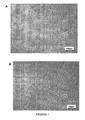
- the morphology of the two cell lines is represented on the Figure 1
- Both cell lines were first analyzed for the expression of precursor / stem cell markers and for endothelial cell markers, including growth factor receptors and selected adhesion molecules as presented in the literature.
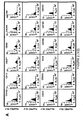
- Figure 2A Both cell lines express CD133 which is known to be a general marker of normal and neoplastic stem cells. They also express CD13 (marker hematopoietic stem cells), CD271 (non-hematopoietic stem cell marker) and CD90 (mesenchymal stem cell marker). Both cell lines were positive for expression of CXCR4 and weakly positive for CD44 and CD15s, which can be expressed on both stem cells and endothelial cells.
- the cells also express markers typical of endothelial cells: CD202b, VEGFR2, CD146, CD105 and weakly express CD143. Markers that are not expressed by these cell lines are collected as presented on the Figure 2B . Both HucPEC 55.1 and HucPEC 55.2 were CD34, CD117, CD38 and CD45 negative suggesting that they are not of hematopoietic origin. They did not express CD31 and vWf, markers of differentiated endothelial cells.
- HucPEC 55.1 and HucPEC 55.2 cell lines grow and differentiate into hematopoietic cells in a semi-solid medium.
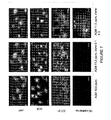
- Both cell lines were grown in a semi-solid medium supplemented with hematopoietic growth and differentiation factors to evaluate their ability to differentiate into hematopoietic cells. Both cell lines are able to grow in a semi-solid medium, but do not differentiate into hematopoietic cells. After 7 to 9 days of culture, the cells form compact multicellular spheroids. They show no sign of differentiation into erythrocytes, granulocytes or monocytes, as shown by Figure 3 .
- a reverse transcriptase of the polymerase chain reaction (RT-PCR) of the mRNAs encoding the multidrug resistance proteins was used to track the expression of their genes in the HucPEC 55. 1 and HucPEC 55 lines. expression of gene products for MDR1, MDR3, MRP1, BCRP and LR is presented on the Figure 4 .
- the mRNAs of all multi-drug resistant proteins are present in both cell lines with similar levels, with the exception of MDR3 which appears to be expressed at a higher level in the HucPEC 55. 2 line compared to the HucPEC line 55.1.
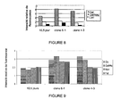
- the cytometric ball test was used to evaluate the secretion of selected cytokines (IP-10, IL6, IL8, VEGF and angiogenin) by HucPEC 55. 1 and HucPEC 55.2 cells growing under normoxic and hypoxic conditions.
- the Figure 5 shows that the alteration of the oxygen partial pressure influences the production of proangiogenic cytokines at variable points by both cell lines.
- the production of proangiogenic cytokines IL8 and angiogenin has increased significantly in hypoxia. Both cell lines did not produce VEGF in normoxia, but the expression of this proangiogenic factor was induced by hypoxia in both cell lines.
- IP10 which is an antiangiogenic cytokine, was produced at much higher levels by HucPEC 55.1 compared to HucPEC 55.2 and was not changed by hypoxia.
- Both cell lines were tested for their angiogenic potential and pseudo-vessel rearrangement on a semi-solid matrix. The kinetics of this rearrangement towards structures of the type tube was compared in hypoxia (1% 02) versus normoxia.
- the Figure 6A indicates that in normoxia, both HucPEC 55. 1 and HucPEC 55.2 are capable of initiating the angiogenic process in a manner comparable to the HSkMEC reference line which is a differentiated skin-derived endothelial microvascular cell line. This result clearly demonstrates that the cells have connected together to form a network. The network evolved for less than 7 hours, for the HucPEC 55. 1 and HucPEC 55. 2 cell lines, and then stopped. Concerning the HSkMECs line, its evolution lasted 17 hours.
- the HucPEC 55. 1 and HucPEC 55.2 cell lines remained where they were and began to proliferate. Hypoxic conditions effectively accelerated the tube formation process for HucPEC 55. 2 cells, as shown on Figure 6B . This process was also accelerated for HucPEC 55. 1 although it was slower than for HucPEC 55. 2. In addition to acceleration, hypoxia also caused a more complete differentiation towards the formation of type structures. vessels that were not performed in normoxia by any of the HucPEC 55. 1 and HucPEC 55. 1 and HucPEC 55 cell lines. 2.
- the established HucPEC 55. 1 and HucPEC 55. 2 cell lines are endothelial cell precursor cells, which have angiogenic properties and have a shorter evolution time than known cell lines allowing for faster proliferation.
- AGM Aorto-Gonado-mesonephrotic region
- dpc 5 days post coitum
- the embryos were obtained from FVB mouse crosses from Transgenesis and Model Animal Archiving Laboratory - UPS44 (TAAM) CNRS Louis.
- the cells were cultured in an incubator at 37 ° C with a humidified atmosphere comprising 5% CO 2 , 21% O 2 .
- the hybridization temperature of the primers described in Table 2 was 57 ° C.
- the products resulting from this RT-PCR were deposited in a migration gel to identify whether the gene is expressed.
- the detection of the von Willebrand factor expressed in the cytoplasm, in the Weibel Palade plates, of the Angiotensin-converting enzyme present in the nuclear membrane, the CD31 differentiation cluster and the VE-cadherin was carried out by microscopy fluorescence and phase contrast equipped with the DIC (Zeiss, Axiovert 200).
- the CBA Cytometric Bead Array
- the CBA Cytometric Bead Array
- TNF ⁇ , IFN ⁇ , GM-CSF, IL-1 ⁇ , IL-4, IL-6, IL-10, IL-12 collected in the supernatant according to the supplier's protocol.
- Five logistic parametric curves were fitted to standard samples and the results were normalized to the number of cells.
- the LSR flow cytometer marketed by BD has been used to determine fluorescence and cell labeling.
- FlowJo v.5.7.2 software was used to analyze the results.
- the cells were washed with a solution of cPBS / 0.5% BSA / 0.1% sodium azide, detached with 1 mg / ml of collagenase type I sold by Gibco, Invitrogen, fixed and permeabilized with Cytofix / Cytoperm marketed by BD, USA according to the supplier's protocol. 5 x 10 5 cells were taken per tube and labeled with a primary antibody in a volume of 30 ⁇ l of cPBS, 0.1% BSA for at least 45min at 4 ° C. The cells were washed two times, suspended in 300 ⁇ l of cPBS and analyzed by FACS.
- Table 3 Table 3 companies Catalog number Dilution Primary antibody Polyclonal rabbit Abcam Ab3470 1/100 Monoclonal mouse Abcam Ab13248 1/100 Polyclonal goat R and D AF3776 1/100 Isotype control Rabbit IgG Sigma I87765 0.016 Mouse IgG Sigma 15381 0,020 Goat IgG Sigma I5256 0,020 Secondary Antibody Ane anti-rabbit PE Immuno Research 711-116-152 1/100 Goat anti mouse PE Santa Cruz SC-3738 1/100 Rabbit anti goat PE Santa Cruz SC-3755 1/100
- the mRNA transcript gels from products obtained with RT-PCR showed that the cells expressed the genes CXCL16, SDF-1, CX3CL1, 6CKine, CTACK (data not provided).
- the gel resulting from the PCR showed that the cells of the MAgEC 11.5 line expressed the Kdr, Flt4 and CCR7 genes (columns 2, 4, 8, 10, 14 and 14). 16 figure 16 ).
- the cells of the MAgEC 10.5 line expressed the Kdr gene (columns 1 and 2, figure 16 ).
- MAgEC cell lines 10.5 and 11.5 do not express the chemokine CCL27 (results not shown) and produce the chemokine CXCL12 ( figure 18 ).
- the MAgEC 10.5 cell line does not produce VEGF, CCL17, CCL27 and CXCL1 ( figures 17 , 19 and 20 ).
- the expression of CCL17 is constant in the cells of the MAgEC 11.5 line regardless of the conditions (hypoxia or normoxia) whereas the production of CXCL1 is decreased when the cells are in hypoxic condition.
- FIGS. 9 to 12 representing fluorescence intensity versus cells clearly demonstrate that the expression of galectin and collectin molecules changes during the developmental stage.
- the level of expression increases from 10.5 dcp cells to a peak for 6-1 clones (11.5 dcp) and a decrease in clone 4-3 cells (11.5 dcp).
- the level of expression of rhamnose-binding lectins does not change between undifferentiated cells of clones 6-1 and differentiated cells of clones 4-3.
- Immunolabeling of the cells of the invention revealed the presence of cell-derived AGM PODXL and EphB4 markers, as shown in FIG. figure 12 . Furthermore, the CD34 differentiation cluster has been increasingly expressed from 10.5 dpc cells to 11.5 dpc cells.
- the human and murine endothelial cell precursor cells are cultured in 24-well plates (Falcon, Grenoble, France).
- the culture medium is the OptiMEM (trademark) marketed by the company Invitrogen supplemented with 2% Fetal Bovine Serum (FCS) sold by the company Hyclon, Longan, Utah, USA.
- the cells are then cultured for 24 hours and then the molecule to be screened is introduced into the culture medium.
- the cells are then cultured for a period of 24 to 48 hours at a temperature of 37 ° C.
- Photos of growing cells can be taken with the Axiovert S microscope (trademark) marketed by Zeiss Jena, Germany equipped with a digital camera Fine Pix 5602 (trademark) marketed by Fuji Film. The observations show that the cells have either undergone toxicity, activation, differentiation, etc.
- Characterization of the cells is also carried out with specific antibodies of differentiation markers, as presented in Example 1 above.
- the precursor cells used in this example are the isolated cells of Examples 1 and 2 above.
- Tumor mice are obtained by injection of 10 5 B16-F10 melanoma cells to the subcutaneous level, above the lug to form tumors.
- the mice used are of the C57 BI6 race.
- the resulting mice constitute tumor models.
- the verification of the presence of the tumors is carried out by visual and material measurement of the size of the tumor.
- the precursor cells of endothelial cells are labeled with the DiD Vybrant dye (registered trademark) sold by the company Molecular Probes USA
- the labeled cells are injected into tumor-bearing mice.
- the acquisition of images is done with a fluorescence measuring device in the near Infra Red, at the Small Animal Imaging Center (CIPA) CNRS, Louis, France.
- CIPA Small Animal Imaging Center
- the images obtained clearly show an address of the cells at the tumor level. Moreover, the results obtained show that the cells have been incorporated into the tumors.
- mice used are identical to the aforementioned mice and the endothelial cell precursor cells are labeled according to the method described above.
- Infarction is caused by coronary injury in mice. After the infarction, the labeled endothelial precursor cells are injected into the mice undergoing infarction. The revelation of the cells is made as indicated above.
- results obtained show that the cells are incorporated in tumors and allows the restoration of damaged tissues and healing.
- the isolated endothelial cell precursor cells according to the invention target tumors as well as damaged tissues. Also, these cells can be used, for example for the treatment of tissue lesions, for the addressing of therapeutic molecules, for example at tumors and for the treatment of genetically deficient cells.
- AMNP Magnetic Anion Nanoparticle Marking
- Cells for magnetic resonance imaging were labeled with AMNPs belonging to SPIO (Super Paramagnetic Iron Oxide) and the group of magnetic resonance contrast agents.
- the cells at 70% confluency were rinsed twice with 8 ml of OptiMEM (registered trade mark) base medium with 5 mM sodium citrate.
- OptiMEM registered trade mark
- 5ml of empty OptiMEM (registered trade mark) medium with 5mM sodium citrate and 2mM ferric fluid (AMNP) was added to the culture container.
- Cells were incubated at 37 ° C, 5% CO 2 for 15-20 minutes. After incubation, the cells were rinsed twice with OptiMEM (Registered Trade Mark) base medium comprising 5 mM sodium citrate.
- the medium with the citrate was removed and 8ml of OptiMEM (Registered Trade Mark) basal medium was added.
- the cells were incubated for 2 hours in a humidified incubator at 37 ° C., 5% CO 2 , and then the medium was changed to complete medium OptiMEM (registered trademark).
- the cells were used for intravenous injections 24 hours after labeling.
- Cells for PKH67 labeling were detached with 0.05% trypsin EDTA (marketed by Gibco), blocked with complete medium OptiMEM (registered trademark). After detachment, the cells were washed twice with OptiMEM (registered trademark) basal medium. After the second washing, the cells were resuspended in 500 ⁇ l of Diluent C marketed by New England BIOLABS, then added to 500 ⁇ l of Diluent C with 4 ⁇ l of PKH67 marketed by Invitrogen, and incubated at 37 ° C. for 3 minutes.
- OptiMEM registered trademark
- mice 10 days before imaging, 5x10 5 B16 (F10) cells were injected subcutaneously into mice.
- Melanoma B16 (F10) cells were detached with 0.05% trypsin EDTA marketed by Gibco, then rinsed twice with 0.9% NaCl (w / v) and counted with a Malassez chamber.
- the cells were resuspended in 0.9% NaCl (w / v) at a concentration of 10x10 6 cells per milliliter and mixed 1: 1 (volume: volume) with Matrigel (trademark) marketed by BD biosciences, just before the injection.
- the mice Prior to injection, the mice were anesthetized with 2-3% isoflurane.
- mice The members of the mice were shaved with an electric razor and shaved with a hair removal cream (hair removal cream Dermotolerance, Vichy). 100 ⁇ l of Matrigel (registered trademark) were injected with 5 ⁇ 10 5 of B16 (F10) cells per member.
- MAgEC 11.5 cells labeled with AMNP and PKH67 or AMNP-Rhodamine were resuspended in 0.9% NaCl (w / v) at a concentration of 10x10 6 cells per ml.
- 100 ⁇ l of suspended cells were injected intravenously either into the retro-orbital sinus or into the tail vein of mice with subcutaneous B16 (F10) tumors.
- Images of the mice were taken before and 24 hours after intravenous injections with a Brüker BioSpin 9.4T spectroscope.
- mice were sacrificed by cervical translocation. Lungs, spleen, mesenteric lymph nodes have been isolated, dilacerated with PBS and incubated with 5 mg / ml type I collagenase marketed by Invitrogen for 30 minutes at 37 ° C. After incubation, the organs were washed with PBS and dissociated on filters and grids and nylon. After dissociation, the erythrocytes were lysed three times with distilled water. Briefly, the cells were centrifuged for 5 minutes at 1500 rpm and resuspended in a drop of PBS. 1 ml of distilled water was added to the cell suspension. After 10 seconds, 4 ml of PBS was added to each tube. The cell pellet was resuspended in PBS.
- a mouse with subcutaneous B16 (F10) -luc-eGFP cells was not treated. Prior to bioluminescence imaging, 150 mg / kg of firefly luciferin was injected intraperitoneally. Images of near-infrared mice were obtained with an IVIS lumina system marketed by Caliper Company daily for a period of one week starting 24 hours with intravenous injection. Bioluminescense imaging was performed according to the Xenogen IVIS 50 bioluminescent protocol: luciferase labeling of a tumor in mice.
- the figure 21 represents the results of cell labeling with AMNP nanoparticles coupled with Rhodamine and shows a cytoplasmic localization of the contrast agent ( Figures 21 , A and C) represented by the white arrows. Fluorescence was low but sufficient ( Figures 21 B and C ). In addition, the location of the nanoparticles was irregular and located particularly around the nucleus.
- the efficiency of labeling MAgEC 11.5 cells with magnetic contrast agents; particularly with AMNP particles coupled with Rhodamine 123 (MAgEC 11.5 - AMNP-Rho) was measured by flow cytometry. The results obtained showed a total labeling of the MAgECs, 90.4%, with the contrast agent.
- Rho- (%) Rho + (%) MAgEC 11.5 99.3 0.33 MAgEC 11.5 - AMNP -Rho 5.72 90.4 MAgEC 11.5 - AMNP -Rho HEPES 4.56 93.7 Rho-: percentage of unmarked cells Rho +: percentage of labeled cells
- HEPES which stabilizes the pH during cell incubation with AMNP (MAgEC 11.5 - AMNP -Rho HEPES), did not change the labeling efficiency.
- the labeling was further confirmed by cellular magnetophoresis (results not provided).
- the organs were extracted and dissociated as previously described.
- the figure 24 represents the results obtained for each organ.
- MAgEC 11.5 cells can target tumors and also different organs.
- the implementation protocol corresponds to the protocol described in point 1.5 above.
- mice in which live MAgEC 11.5 cells labeled with DiD Vybrant (registered trademark) were injected, the cells were detected in the liver (11/11 mice), the spleen (5/11 mice), the lymphoid nodes (3/11 mice), the lungs (2/11 mice), the sternum and the tibia as shown on the figures 25 IA, IB and IIA and IIB.
- Weak fluorescence was detected in the liver, spleen, lungs and sternum of mice where MAgEC 11.5 cells fixed and labeled with DiD Vybrant (trademark) were injected. No fluorescence was detected in the mice in which no injection was performed ( Figures 25 IC and IIC).
- the cells of the invention can be used for the targeting of particular organs and can be used, for example in tissue repair methods, methods of release of active substances and or genes in different organs. .
Landscapes
- Health & Medical Sciences (AREA)
- Life Sciences & Earth Sciences (AREA)
- Engineering & Computer Science (AREA)
- Biomedical Technology (AREA)
- Chemical & Material Sciences (AREA)
- Immunology (AREA)
- Cell Biology (AREA)
- Bioinformatics & Cheminformatics (AREA)
- Urology & Nephrology (AREA)
- Biotechnology (AREA)
- Hematology (AREA)
- Molecular Biology (AREA)
- General Health & Medical Sciences (AREA)
- Microbiology (AREA)
- Biochemistry (AREA)
- Medicinal Chemistry (AREA)
- Organic Chemistry (AREA)
- Developmental Biology & Embryology (AREA)
- Analytical Chemistry (AREA)
- Zoology (AREA)
- Toxicology (AREA)
- Wood Science & Technology (AREA)
- Tropical Medicine & Parasitology (AREA)
- Genetics & Genomics (AREA)
- Pathology (AREA)
- General Physics & Mathematics (AREA)
- Physics & Mathematics (AREA)
- Food Science & Technology (AREA)
- Vascular Medicine (AREA)
- General Engineering & Computer Science (AREA)
- Veterinary Medicine (AREA)
- Chemical Kinetics & Catalysis (AREA)
- Animal Behavior & Ethology (AREA)
- General Chemical & Material Sciences (AREA)
- Nuclear Medicine, Radiotherapy & Molecular Imaging (AREA)
- Public Health (AREA)
- Pharmacology & Pharmacy (AREA)
- Cardiology (AREA)
- Heart & Thoracic Surgery (AREA)
- Micro-Organisms Or Cultivation Processes Thereof (AREA)
Claims (11)
- Isolierte Vorläuferzelle von Endothelzellen, die aus den isolierten humanen Vorläuferzellen von Endothelzellen, die bei der Collection Nationale de Culture de Microorganismes (CNCM) unter den CNCM-Nummern Nr. I-4220, Nr. I-4221 hinterlegt sind, oder den isolierten murinen Zellen, die bei der Collection Nationale de Culture de Microorganismes (CNCM) unter den CNCM-Nummern Nr. I-4222, Nr. I-4223 hinterlegt sind, ausgewählt ist.
- Isolierte Zelle nach Anspruch 1, wobei die besagte Zelle eine etablierte Zelllinie bildet.
- Verfahren zum Screenen von Molekülen, die dazu imstande sind, eine Differenzierung und/oder eine Spezialisierung einer isolierten Zelle nach den Ansprüchen 1 oder 2 zu induzieren, wobei das Verfahren die folgenden Schritte umfasst:- Einbringen des zu screenenden Moleküls in ein Medium, das zur Kultivierung der besagten Zelle geeignet ist,- Einbringen mindestens einer isolierten Zelle nach Anspruch 1 oder 2 in das besagte Medium,- Kultivieren der besagten Zelle in dem besagten Medium für einen Zeitraum, der dazu ausreicht, ihre Differenzierung und/oder ihre Spezialisierung zu ermöglichen,- Entnehmen mindestens einer Zelle, die aus der besagten Kultur hervorgeht,- Beobachten der Proliferation oder Nichtdifferenzierung und/oder Spezialisierung der besagten Zelle durch phänotypische Beobachtung der Zelle und/oder durch Offenbarung von Differenzierungsmarkern.
- Verfahren nach Anspruch 3, wobei die Offenbarung von Differenzierungsmarkern mithilfe spezifischer Antikörper der besagten Marker vorgenommen wird.
- Verfahren nach Anspruch 3 oder 4, wobei die besagten Moleküle eine Differenzierung und/oder Spezialisierung induzieren, die zu einer Angiogenese führt.
- Verfahren nach einem der Ansprüche 3 bis 5, wobei der Schritt zur Identifizierung des Phänotyps und/oder von Markern ermöglicht, die Wirkung des gescreenten Moleküls auf die kultivierte isolierte Zelle zu erfahren.
- Kit zur Durchführung des Verfahrens nach Anspruch 3, wobei das Kit Folgendes umfasst:- mindestens eine isolierte Zelle nach Anspruch 1 oder 2 und- ein Mittel zum Nachweisen der Zelldifferenzierung und/oder -Spezialisierung.
- Zellkultur, die eine isolierte Zelle nach den Ansprüchen 1 oder 2 umfasst.
- Isolierte Zelle nach den Ansprüchen 1 oder 2 zur Verwendung als Arzneimittel.
- Verwendung einer isolierten Zelle nach den Ansprüchen 1 oder 2 zur Herstellung eines Arzneimittels.
- Verwendung einer isolierten Zelle nach Anspruch 10, wobei das Arzneimittel zur Behandlung von genetisch defekten Zellen, Gewebeläsionen, Endothelschäden, Schäden aufgrund einer Ischämie, einem Herzinfarkt, von Tumoren, von Diabetes vorgesehen ist.
Priority Applications (1)
| Application Number | Priority Date | Filing Date | Title |
|---|---|---|---|
| PL11704276T PL2524031T3 (pl) | 2010-01-12 | 2011-01-11 | Linie ludzkich i mysich komórek macierzystych: modele śródbłonkowych komórek progenitorowych |
Applications Claiming Priority (2)
| Application Number | Priority Date | Filing Date | Title |
|---|---|---|---|
| FR1000111A FR3003870B1 (fr) | 2010-01-12 | 2010-01-12 | Lignees de cellules souches humaines et murines: modeles de precurseurs de cellules endotheliales |
| PCT/FR2011/050045 WO2011086319A1 (fr) | 2010-01-12 | 2011-01-11 | Lignees de cellules souches humaines et murines : modeles de precurseurs de cellules endotheliales |
Publications (2)
| Publication Number | Publication Date |
|---|---|
| EP2524031A1 EP2524031A1 (de) | 2012-11-21 |
| EP2524031B1 true EP2524031B1 (de) | 2015-07-08 |
Family
ID=42321206
Family Applications (1)
| Application Number | Title | Priority Date | Filing Date |
|---|---|---|---|
| EP11704276.2A Not-in-force EP2524031B1 (de) | 2010-01-12 | 2011-01-11 | Menschlicher stammzelllinien und maus: modell von endothelialen progenitorzellen |
Country Status (5)
| Country | Link |
|---|---|
| US (1) | US9631178B2 (de) |
| EP (1) | EP2524031B1 (de) |
| FR (1) | FR3003870B1 (de) |
| PL (1) | PL2524031T3 (de) |
| WO (1) | WO2011086319A1 (de) |
Families Citing this family (1)
| Publication number | Priority date | Publication date | Assignee | Title |
|---|---|---|---|---|
| US12016885B2 (en) | 2018-11-06 | 2024-06-25 | Alsatech, Inc. | Method of solubilizing TDP-43 protein aggregates in amyotrophic lateral sclerosis using a microvascular endothelial cell expressing an anti-TDP-43 Fab |
Family Cites Families (3)
| Publication number | Priority date | Publication date | Assignee | Title |
|---|---|---|---|---|
| US20090004661A1 (en) * | 2003-05-26 | 2009-01-01 | Reliance Life Sciences Pvt Ltd. | Method of growing mesenchymal stem cells from bone marrow |
| EP1859028B1 (de) * | 2005-02-10 | 2015-12-02 | Regents Of The University Of Minnesota | Vaskuläre Endothelzellen |
| US20090291061A1 (en) * | 2008-05-21 | 2009-11-26 | Riordan Neil H | Stem cell therapy for blood vessel degeneration |
-
2010
- 2010-01-12 FR FR1000111A patent/FR3003870B1/fr not_active Expired - Fee Related
-
2011
- 2011-01-11 EP EP11704276.2A patent/EP2524031B1/de not_active Not-in-force
- 2011-01-11 PL PL11704276T patent/PL2524031T3/pl unknown
- 2011-01-11 WO PCT/FR2011/050045 patent/WO2011086319A1/fr not_active Ceased
- 2011-01-11 US US13/521,715 patent/US9631178B2/en not_active Expired - Fee Related
Also Published As
| Publication number | Publication date |
|---|---|
| FR3003870A1 (fr) | 2014-10-03 |
| FR3003870B1 (fr) | 2015-10-02 |
| PL2524031T3 (pl) | 2015-12-31 |
| US9631178B2 (en) | 2017-04-25 |
| WO2011086319A1 (fr) | 2011-07-21 |
| US20130040315A1 (en) | 2013-02-14 |
| EP2524031A1 (de) | 2012-11-21 |
Similar Documents
| Publication | Publication Date | Title |
|---|---|---|
| Harraz et al. | CD34− blood-derived human endothelial cell progenitors | |
| Kuwana et al. | Endothelial differentiation potential of human monocyte‐derived multipotential cells | |
| US10041036B2 (en) | Isolation, expansion and use of clonogenic endothelial progenitor cells | |
| Ohga et al. | Heterogeneity of tumor endothelial cells: comparison between tumor endothelial cells isolated from high-and low-metastatic tumors | |
| Patel et al. | Prospective surface marker-based isolation and expansion of fetal endothelial colony-forming cells from human term placenta | |
| Elsheikh et al. | Only a specific subset of human peripheral-blood monocytes has endothelial-like functional capacity | |
| Gauthaman et al. | Extra-embryonic human Wharton’s jelly stem cells do not induce tumorigenesis, unlike human embryonic stem cells | |
| Grenier et al. | Resident endothelial precursors in muscle, adipose, and dermis contribute to postnatal vasculogenesis | |
| JP6382408B2 (ja) | 体性幹細胞 | |
| Xu et al. | Different angiogenic potentials of mesenchymal stem cells derived from umbilical artery, umbilical vein, and Wharton’s jelly | |
| Barclay et al. | Characterization of adult prostatic progenitor/stem cells exhibiting self-renewal and multilineage differentiation | |
| Joo et al. | Human endothelial colony forming cells from adult peripheral blood have enhanced sprouting angiogenic potential through up-regulating VEGFR2 signaling | |
| Collet et al. | Endothelial precursor cell-based therapy to target the pathologic angiogenesis and compensate tumor hypoxia | |
| Paprocka et al. | CD133 positive progenitor endothelial cell lines from human cord blood | |
| Metharom et al. | Myeloid lineage of high proliferative potential human smooth muscle outgrowth cells circulating in blood and vasculogenic smooth muscle-like cells in vivo | |
| AU2012298997B2 (en) | Angiohematopoietic progenitor cells | |
| EP4072570A1 (de) | Verfahren zur erzeugung von hämatopoetischen stammzellen | |
| Vanneaux et al. | In vitro and in vivo analysis of endothelial progenitor cells from cryopreserved umbilical cord blood: are we ready for clinical application? | |
| Mejía-Cruz et al. | Generation of organotypic multicellular spheres by magnetic levitation: model for the study of human hematopoietic stem cells microenvironment | |
| Montali et al. | Human adult mesangiogenic progenitor cells reveal an early angiogenic potential, which is lost after mesengenic differentiation | |
| Chen et al. | CD146+ mural cells from infantile hemangioma display proangiogenic ability and adipogenesis potential in vitro and in xenograft models | |
| US20170258853A1 (en) | Pancreatic stem cells and uses thereof | |
| CN103540566A (zh) | 用于长期造血种群恢复的方法和组合物 | |
| Abangan Jr et al. | MCP1 directs trafficking of hematopoietic stem cell-derived fibroblast precursors in solid tumor | |
| Yao et al. | Identification of high proliferative potential precursors with hemangioblastic activity in the mouse aorta-gonad-mesonephros region |
Legal Events
| Date | Code | Title | Description |
|---|---|---|---|
| PUAI | Public reference made under article 153(3) epc to a published international application that has entered the european phase |
Free format text: ORIGINAL CODE: 0009012 |
|
| 17P | Request for examination filed |
Effective date: 20120628 |
|
| AK | Designated contracting states |
Kind code of ref document: A1 Designated state(s): AL AT BE BG CH CY CZ DE DK EE ES FI FR GB GR HR HU IE IS IT LI LT LU LV MC MK MT NL NO PL PT RO RS SE SI SK SM TR |
|
| DAX | Request for extension of the european patent (deleted) | ||
| 17Q | First examination report despatched |
Effective date: 20140217 |
|
| GRAP | Despatch of communication of intention to grant a patent |
Free format text: ORIGINAL CODE: EPIDOSNIGR1 |
|
| INTG | Intention to grant announced |
Effective date: 20150331 |
|
| GRAS | Grant fee paid |
Free format text: ORIGINAL CODE: EPIDOSNIGR3 |
|
| GRAA | (expected) grant |
Free format text: ORIGINAL CODE: 0009210 |
|
| AK | Designated contracting states |
Kind code of ref document: B1 Designated state(s): AL AT BE BG CH CY CZ DE DK EE ES FI FR GB GR HR HU IE IS IT LI LT LU LV MC MK MT NL NO PL PT RO RS SE SI SK SM TR |
|
| REG | Reference to a national code |
Ref country code: GB Ref legal event code: FG4D Free format text: NOT ENGLISH |
|
| REG | Reference to a national code |
Ref country code: AT Ref legal event code: REF Ref document number: 735444 Country of ref document: AT Kind code of ref document: T Effective date: 20150715 Ref country code: CH Ref legal event code: EP |
|
| REG | Reference to a national code |
Ref country code: IE Ref legal event code: FG4D Free format text: LANGUAGE OF EP DOCUMENT: FRENCH |
|
| REG | Reference to a national code |
Ref country code: DE Ref legal event code: R096 Ref document number: 602011017660 Country of ref document: DE |
|
| REG | Reference to a national code |
Ref country code: AT Ref legal event code: MK05 Ref document number: 735444 Country of ref document: AT Kind code of ref document: T Effective date: 20150708 |
|
| REG | Reference to a national code |
Ref country code: NL Ref legal event code: MP Effective date: 20150708 |
|
| REG | Reference to a national code |
Ref country code: FR Ref legal event code: PLFP Year of fee payment: 6 |
|
| REG | Reference to a national code |
Ref country code: LT Ref legal event code: MG4D |
|
| REG | Reference to a national code |
Ref country code: PL Ref legal event code: T3 |
|
| PG25 | Lapsed in a contracting state [announced via postgrant information from national office to epo] |
Ref country code: NO Free format text: LAPSE BECAUSE OF FAILURE TO SUBMIT A TRANSLATION OF THE DESCRIPTION OR TO PAY THE FEE WITHIN THE PRESCRIBED TIME-LIMIT Effective date: 20151008 Ref country code: LT Free format text: LAPSE BECAUSE OF FAILURE TO SUBMIT A TRANSLATION OF THE DESCRIPTION OR TO PAY THE FEE WITHIN THE PRESCRIBED TIME-LIMIT Effective date: 20150708 Ref country code: FI Free format text: LAPSE BECAUSE OF FAILURE TO SUBMIT A TRANSLATION OF THE DESCRIPTION OR TO PAY THE FEE WITHIN THE PRESCRIBED TIME-LIMIT Effective date: 20150708 Ref country code: GR Free format text: LAPSE BECAUSE OF FAILURE TO SUBMIT A TRANSLATION OF THE DESCRIPTION OR TO PAY THE FEE WITHIN THE PRESCRIBED TIME-LIMIT Effective date: 20151009 Ref country code: LV Free format text: LAPSE BECAUSE OF FAILURE TO SUBMIT A TRANSLATION OF THE DESCRIPTION OR TO PAY THE FEE WITHIN THE PRESCRIBED TIME-LIMIT Effective date: 20150708 |
|
| PG25 | Lapsed in a contracting state [announced via postgrant information from national office to epo] |
Ref country code: PT Free format text: LAPSE BECAUSE OF FAILURE TO SUBMIT A TRANSLATION OF THE DESCRIPTION OR TO PAY THE FEE WITHIN THE PRESCRIBED TIME-LIMIT Effective date: 20151109 Ref country code: RS Free format text: LAPSE BECAUSE OF FAILURE TO SUBMIT A TRANSLATION OF THE DESCRIPTION OR TO PAY THE FEE WITHIN THE PRESCRIBED TIME-LIMIT Effective date: 20150708 Ref country code: HR Free format text: LAPSE BECAUSE OF FAILURE TO SUBMIT A TRANSLATION OF THE DESCRIPTION OR TO PAY THE FEE WITHIN THE PRESCRIBED TIME-LIMIT Effective date: 20150708 Ref country code: IS Free format text: LAPSE BECAUSE OF FAILURE TO SUBMIT A TRANSLATION OF THE DESCRIPTION OR TO PAY THE FEE WITHIN THE PRESCRIBED TIME-LIMIT Effective date: 20151108 Ref country code: ES Free format text: LAPSE BECAUSE OF FAILURE TO SUBMIT A TRANSLATION OF THE DESCRIPTION OR TO PAY THE FEE WITHIN THE PRESCRIBED TIME-LIMIT Effective date: 20150708 Ref country code: AT Free format text: LAPSE BECAUSE OF FAILURE TO SUBMIT A TRANSLATION OF THE DESCRIPTION OR TO PAY THE FEE WITHIN THE PRESCRIBED TIME-LIMIT Effective date: 20150708 Ref country code: SE Free format text: LAPSE BECAUSE OF FAILURE TO SUBMIT A TRANSLATION OF THE DESCRIPTION OR TO PAY THE FEE WITHIN THE PRESCRIBED TIME-LIMIT Effective date: 20150708 |
|
| REG | Reference to a national code |
Ref country code: DE Ref legal event code: R097 Ref document number: 602011017660 Country of ref document: DE |
|
| PG25 | Lapsed in a contracting state [announced via postgrant information from national office to epo] |
Ref country code: SK Free format text: LAPSE BECAUSE OF FAILURE TO SUBMIT A TRANSLATION OF THE DESCRIPTION OR TO PAY THE FEE WITHIN THE PRESCRIBED TIME-LIMIT Effective date: 20150708 Ref country code: EE Free format text: LAPSE BECAUSE OF FAILURE TO SUBMIT A TRANSLATION OF THE DESCRIPTION OR TO PAY THE FEE WITHIN THE PRESCRIBED TIME-LIMIT Effective date: 20150708 Ref country code: IT Free format text: LAPSE BECAUSE OF FAILURE TO SUBMIT A TRANSLATION OF THE DESCRIPTION OR TO PAY THE FEE WITHIN THE PRESCRIBED TIME-LIMIT Effective date: 20150708 Ref country code: DK Free format text: LAPSE BECAUSE OF FAILURE TO SUBMIT A TRANSLATION OF THE DESCRIPTION OR TO PAY THE FEE WITHIN THE PRESCRIBED TIME-LIMIT Effective date: 20150708 Ref country code: CZ Free format text: LAPSE BECAUSE OF FAILURE TO SUBMIT A TRANSLATION OF THE DESCRIPTION OR TO PAY THE FEE WITHIN THE PRESCRIBED TIME-LIMIT Effective date: 20150708 |
|
| PLBE | No opposition filed within time limit |
Free format text: ORIGINAL CODE: 0009261 |
|
| STAA | Information on the status of an ep patent application or granted ep patent |
Free format text: STATUS: NO OPPOSITION FILED WITHIN TIME LIMIT |
|
| PG25 | Lapsed in a contracting state [announced via postgrant information from national office to epo] |
Ref country code: RO Free format text: LAPSE BECAUSE OF FAILURE TO SUBMIT A TRANSLATION OF THE DESCRIPTION OR TO PAY THE FEE WITHIN THE PRESCRIBED TIME-LIMIT Effective date: 20150708 Ref country code: BE Free format text: LAPSE BECAUSE OF NON-PAYMENT OF DUE FEES Effective date: 20160131 |
|
| 26N | No opposition filed |
Effective date: 20160411 |
|
| PG25 | Lapsed in a contracting state [announced via postgrant information from national office to epo] |
Ref country code: SI Free format text: LAPSE BECAUSE OF FAILURE TO SUBMIT A TRANSLATION OF THE DESCRIPTION OR TO PAY THE FEE WITHIN THE PRESCRIBED TIME-LIMIT Effective date: 20150708 Ref country code: LU Free format text: LAPSE BECAUSE OF FAILURE TO SUBMIT A TRANSLATION OF THE DESCRIPTION OR TO PAY THE FEE WITHIN THE PRESCRIBED TIME-LIMIT Effective date: 20160111 |
|
| REG | Reference to a national code |
Ref country code: CH Ref legal event code: PL |
|
| PG25 | Lapsed in a contracting state [announced via postgrant information from national office to epo] |
Ref country code: MC Free format text: LAPSE BECAUSE OF FAILURE TO SUBMIT A TRANSLATION OF THE DESCRIPTION OR TO PAY THE FEE WITHIN THE PRESCRIBED TIME-LIMIT Effective date: 20150708 |
|
| PG25 | Lapsed in a contracting state [announced via postgrant information from national office to epo] |
Ref country code: LI Free format text: LAPSE BECAUSE OF NON-PAYMENT OF DUE FEES Effective date: 20160131 Ref country code: CH Free format text: LAPSE BECAUSE OF NON-PAYMENT OF DUE FEES Effective date: 20160131 |
|
| REG | Reference to a national code |
Ref country code: IE Ref legal event code: MM4A |
|
| REG | Reference to a national code |
Ref country code: FR Ref legal event code: PLFP Year of fee payment: 7 |
|
| PG25 | Lapsed in a contracting state [announced via postgrant information from national office to epo] |
Ref country code: IE Free format text: LAPSE BECAUSE OF NON-PAYMENT OF DUE FEES Effective date: 20160111 |
|
| PG25 | Lapsed in a contracting state [announced via postgrant information from national office to epo] |
Ref country code: NL Free format text: LAPSE BECAUSE OF FAILURE TO SUBMIT A TRANSLATION OF THE DESCRIPTION OR TO PAY THE FEE WITHIN THE PRESCRIBED TIME-LIMIT Effective date: 20150708 |
|
| PG25 | Lapsed in a contracting state [announced via postgrant information from national office to epo] |
Ref country code: MT Free format text: LAPSE BECAUSE OF FAILURE TO SUBMIT A TRANSLATION OF THE DESCRIPTION OR TO PAY THE FEE WITHIN THE PRESCRIBED TIME-LIMIT Effective date: 20150708 |
|
| REG | Reference to a national code |
Ref country code: FR Ref legal event code: PLFP Year of fee payment: 8 |
|
| PG25 | Lapsed in a contracting state [announced via postgrant information from national office to epo] |
Ref country code: CY Free format text: LAPSE BECAUSE OF FAILURE TO SUBMIT A TRANSLATION OF THE DESCRIPTION OR TO PAY THE FEE WITHIN THE PRESCRIBED TIME-LIMIT Effective date: 20150708 Ref country code: SM Free format text: LAPSE BECAUSE OF FAILURE TO SUBMIT A TRANSLATION OF THE DESCRIPTION OR TO PAY THE FEE WITHIN THE PRESCRIBED TIME-LIMIT Effective date: 20150708 Ref country code: HU Free format text: LAPSE BECAUSE OF FAILURE TO SUBMIT A TRANSLATION OF THE DESCRIPTION OR TO PAY THE FEE WITHIN THE PRESCRIBED TIME-LIMIT; INVALID AB INITIO Effective date: 20110111 |
|
| PG25 | Lapsed in a contracting state [announced via postgrant information from national office to epo] |
Ref country code: MK Free format text: LAPSE BECAUSE OF FAILURE TO SUBMIT A TRANSLATION OF THE DESCRIPTION OR TO PAY THE FEE WITHIN THE PRESCRIBED TIME-LIMIT Effective date: 20150708 Ref country code: TR Free format text: LAPSE BECAUSE OF FAILURE TO SUBMIT A TRANSLATION OF THE DESCRIPTION OR TO PAY THE FEE WITHIN THE PRESCRIBED TIME-LIMIT Effective date: 20150708 |
|
| PG25 | Lapsed in a contracting state [announced via postgrant information from national office to epo] |
Ref country code: BG Free format text: LAPSE BECAUSE OF FAILURE TO SUBMIT A TRANSLATION OF THE DESCRIPTION OR TO PAY THE FEE WITHIN THE PRESCRIBED TIME-LIMIT Effective date: 20150708 |
|
| PG25 | Lapsed in a contracting state [announced via postgrant information from national office to epo] |
Ref country code: AL Free format text: LAPSE BECAUSE OF FAILURE TO SUBMIT A TRANSLATION OF THE DESCRIPTION OR TO PAY THE FEE WITHIN THE PRESCRIBED TIME-LIMIT Effective date: 20150708 |
|
| PGFP | Annual fee paid to national office [announced via postgrant information from national office to epo] |
Ref country code: GB Payment date: 20200131 Year of fee payment: 10 Ref country code: PL Payment date: 20191217 Year of fee payment: 10 Ref country code: DE Payment date: 20200113 Year of fee payment: 10 |
|
| PGFP | Annual fee paid to national office [announced via postgrant information from national office to epo] |
Ref country code: FR Payment date: 20200130 Year of fee payment: 10 |
|
| REG | Reference to a national code |
Ref country code: DE Ref legal event code: R119 Ref document number: 602011017660 Country of ref document: DE |
|
| GBPC | Gb: european patent ceased through non-payment of renewal fee |
Effective date: 20210111 |
|
| PG25 | Lapsed in a contracting state [announced via postgrant information from national office to epo] |
Ref country code: FR Free format text: LAPSE BECAUSE OF NON-PAYMENT OF DUE FEES Effective date: 20210131 |
|
| PG25 | Lapsed in a contracting state [announced via postgrant information from national office to epo] |
Ref country code: DE Free format text: LAPSE BECAUSE OF NON-PAYMENT OF DUE FEES Effective date: 20210803 Ref country code: GB Free format text: LAPSE BECAUSE OF NON-PAYMENT OF DUE FEES Effective date: 20210111 |
|
| PG25 | Lapsed in a contracting state [announced via postgrant information from national office to epo] |
Ref country code: PL Free format text: LAPSE BECAUSE OF NON-PAYMENT OF DUE FEES Effective date: 20210111 |