EP2521737B1 - Modified food grade microorganism for treatment of inflammatory bowel disease - Google Patents
Modified food grade microorganism for treatment of inflammatory bowel disease Download PDFInfo
- Publication number
- EP2521737B1 EP2521737B1 EP11700152.9A EP11700152A EP2521737B1 EP 2521737 B1 EP2521737 B1 EP 2521737B1 EP 11700152 A EP11700152 A EP 11700152A EP 2521737 B1 EP2521737 B1 EP 2521737B1
- Authority
- EP
- European Patent Office
- Prior art keywords
- protein
- lactic acid
- domain
- tnfα
- acid bacterium
- Prior art date
- Legal status (The legal status is an assumption and is not a legal conclusion. Google has not performed a legal analysis and makes no representation as to the accuracy of the status listed.)
- Not-in-force
Links
- 244000005700 microbiome Species 0.000 title description 35
- 208000022559 Inflammatory bowel disease Diseases 0.000 title description 18
- 235000013305 food Nutrition 0.000 title description 12
- 108090000623 proteins and genes Proteins 0.000 claims description 144
- JVTAAEKCZFNVCJ-UHFFFAOYSA-N lactic acid Chemical compound CC(O)C(O)=O JVTAAEKCZFNVCJ-UHFFFAOYSA-N 0.000 claims description 111
- 102000004169 proteins and genes Human genes 0.000 claims description 101
- 108060008682 Tumor Necrosis Factor Proteins 0.000 claims description 82
- 102000000852 Tumor Necrosis Factor-alpha Human genes 0.000 claims description 81
- 230000027455 binding Effects 0.000 claims description 69
- 241000894006 Bacteria Species 0.000 claims description 68
- 239000004310 lactic acid Substances 0.000 claims description 50
- 235000014655 lactic acid Nutrition 0.000 claims description 50
- 108090000765 processed proteins & peptides Proteins 0.000 claims description 44
- 108010076504 Protein Sorting Signals Proteins 0.000 claims description 35
- 102000004196 processed proteins & peptides Human genes 0.000 claims description 30
- 230000028327 secretion Effects 0.000 claims description 21
- 229920001184 polypeptide Polymers 0.000 claims description 20
- 239000008194 pharmaceutical composition Substances 0.000 claims description 10
- 239000003814 drug Substances 0.000 claims description 7
- 238000004519 manufacturing process Methods 0.000 claims description 6
- 238000000034 method Methods 0.000 claims description 6
- 125000006850 spacer group Chemical group 0.000 claims description 6
- 230000008827 biological function Effects 0.000 claims description 3
- 210000004027 cell Anatomy 0.000 description 60
- 108091026890 Coding region Proteins 0.000 description 26
- 241000194035 Lactococcus lactis Species 0.000 description 23
- 102000037865 fusion proteins Human genes 0.000 description 19
- 108020001507 fusion proteins Proteins 0.000 description 19
- 230000014509 gene expression Effects 0.000 description 17
- 150000001413 amino acids Chemical class 0.000 description 16
- MSFSPUZXLOGKHJ-UHFFFAOYSA-N Muraminsaeure Natural products OC(=O)C(C)OC1C(N)C(O)OC(CO)C1O MSFSPUZXLOGKHJ-UHFFFAOYSA-N 0.000 description 15
- 108010013639 Peptidoglycan Proteins 0.000 description 15
- 210000001035 gastrointestinal tract Anatomy 0.000 description 14
- 230000001580 bacterial effect Effects 0.000 description 12
- 239000001963 growth medium Substances 0.000 description 12
- 238000000684 flow cytometry Methods 0.000 description 11
- 108020004414 DNA Proteins 0.000 description 10
- FAPWRFPIFSIZLT-UHFFFAOYSA-M Sodium chloride Chemical compound [Na+].[Cl-] FAPWRFPIFSIZLT-UHFFFAOYSA-M 0.000 description 10
- 235000003869 genetically modified organism Nutrition 0.000 description 9
- 239000012103 Alexa Fluor 488 Substances 0.000 description 8
- 235000014897 Streptococcus lactis Nutrition 0.000 description 8
- 239000002609 medium Substances 0.000 description 8
- 239000006228 supernatant Substances 0.000 description 8
- 208000011231 Crohn disease Diseases 0.000 description 7
- 208000037265 diseases, disorders, signs and symptoms Diseases 0.000 description 7
- 238000000386 microscopy Methods 0.000 description 7
- 235000013336 milk Nutrition 0.000 description 7
- 239000008267 milk Substances 0.000 description 7
- 210000004080 milk Anatomy 0.000 description 7
- 206010009900 Colitis ulcerative Diseases 0.000 description 6
- 108091035707 Consensus sequence Proteins 0.000 description 6
- 201000006704 Ulcerative Colitis Diseases 0.000 description 6
- 239000012528 membrane Substances 0.000 description 6
- 239000000203 mixture Substances 0.000 description 6
- 239000013612 plasmid Substances 0.000 description 6
- QKNYBSVHEMOAJP-UHFFFAOYSA-N 2-amino-2-(hydroxymethyl)propane-1,3-diol;hydron;chloride Chemical compound Cl.OCC(N)(CO)CO QKNYBSVHEMOAJP-UHFFFAOYSA-N 0.000 description 5
- 108060003951 Immunoglobulin Proteins 0.000 description 5
- 206010061218 Inflammation Diseases 0.000 description 5
- 241000194036 Lactococcus Species 0.000 description 5
- 241000283973 Oryctolagus cuniculus Species 0.000 description 5
- 125000003275 alpha amino acid group Chemical group 0.000 description 5
- 239000000872 buffer Substances 0.000 description 5
- 230000015556 catabolic process Effects 0.000 description 5
- 210000002421 cell wall Anatomy 0.000 description 5
- 238000006731 degradation reaction Methods 0.000 description 5
- 230000000694 effects Effects 0.000 description 5
- 230000004927 fusion Effects 0.000 description 5
- 102000018358 immunoglobulin Human genes 0.000 description 5
- 230000004054 inflammatory process Effects 0.000 description 5
- 239000000047 product Substances 0.000 description 5
- 239000011780 sodium chloride Substances 0.000 description 5
- 108010062877 Bacteriocins Proteins 0.000 description 4
- 102000004127 Cytokines Human genes 0.000 description 4
- 108090000695 Cytokines Proteins 0.000 description 4
- 238000004113 cell culture Methods 0.000 description 4
- 150000001875 compounds Chemical class 0.000 description 4
- 201000010099 disease Diseases 0.000 description 4
- MHMNJMPURVTYEJ-UHFFFAOYSA-N fluorescein-5-isothiocyanate Chemical compound O1C(=O)C2=CC(N=C=S)=CC=C2C21C1=CC=C(O)C=C1OC1=CC(O)=CC=C21 MHMNJMPURVTYEJ-UHFFFAOYSA-N 0.000 description 4
- 210000004051 gastric juice Anatomy 0.000 description 4
- 238000011534 incubation Methods 0.000 description 4
- 230000001939 inductive effect Effects 0.000 description 4
- 239000000126 substance Substances 0.000 description 4
- 239000000725 suspension Substances 0.000 description 4
- 108010053775 Nisin Proteins 0.000 description 3
- NVNLLIYOARQCIX-MSHCCFNRSA-N Nisin Chemical compound N1C(=O)[C@@H](CC(C)C)NC(=O)C(=C)NC(=O)[C@@H]([C@H](C)CC)NC(=O)[C@@H](NC(=O)C(=C/C)/NC(=O)[C@H](N)[C@H](C)CC)CSC[C@@H]1C(=O)N[C@@H]1C(=O)N2CCC[C@@H]2C(=O)NCC(=O)N[C@@H](C(=O)N[C@H](CCCCN)C(=O)N[C@@H]2C(NCC(=O)N[C@H](C)C(=O)N[C@H](CC(C)C)C(=O)N[C@H](CCSC)C(=O)NCC(=O)N[C@H](CS[C@@H]2C)C(=O)N[C@H](CC(N)=O)C(=O)N[C@H](CCSC)C(=O)N[C@H](CCCCN)C(=O)N[C@@H]2C(N[C@H](C)C(=O)N[C@@H]3C(=O)N[C@@H](C(N[C@H](CC=4NC=NC=4)C(=O)N[C@H](CS[C@@H]3C)C(=O)N[C@H](CO)C(=O)N[C@H]([C@H](C)CC)C(=O)N[C@H](CC=3NC=NC=3)C(=O)N[C@H](C(C)C)C(=O)NC(=C)C(=O)N[C@H](CCCCN)C(O)=O)=O)CS[C@@H]2C)=O)=O)CS[C@@H]1C NVNLLIYOARQCIX-MSHCCFNRSA-N 0.000 description 3
- 101150115929 Usp45 gene Proteins 0.000 description 3
- 238000004873 anchoring Methods 0.000 description 3
- 210000004899 c-terminal region Anatomy 0.000 description 3
- 230000001684 chronic effect Effects 0.000 description 3
- 206010009887 colitis Diseases 0.000 description 3
- 238000010276 construction Methods 0.000 description 3
- 235000021474 generally recognized As safe (food) Nutrition 0.000 description 3
- 235000021473 generally recognized as safe (food ingredients) Nutrition 0.000 description 3
- 239000004309 nisin Substances 0.000 description 3
- 235000010297 nisin Nutrition 0.000 description 3
- 238000002415 sodium dodecyl sulfate polyacrylamide gel electrophoresis Methods 0.000 description 3
- 238000001179 sorption measurement Methods 0.000 description 3
- 238000010186 staining Methods 0.000 description 3
- 238000002560 therapeutic procedure Methods 0.000 description 3
- 238000012384 transportation and delivery Methods 0.000 description 3
- 206010002556 Ankylosing Spondylitis Diseases 0.000 description 2
- XKRFYHLGVUSROY-UHFFFAOYSA-N Argon Chemical compound [Ar] XKRFYHLGVUSROY-UHFFFAOYSA-N 0.000 description 2
- 208000023275 Autoimmune disease Diseases 0.000 description 2
- ULGZDMOVFRHVEP-RWJQBGPGSA-N Erythromycin Chemical compound O([C@@H]1[C@@H](C)C(=O)O[C@@H]([C@@]([C@H](O)[C@@H](C)C(=O)[C@H](C)C[C@@](C)(O)[C@H](O[C@H]2[C@@H]([C@H](C[C@@H](C)O2)N(C)C)O)[C@H]1C)(C)O)CC)[C@H]1C[C@@](C)(OC)[C@@H](O)[C@H](C)O1 ULGZDMOVFRHVEP-RWJQBGPGSA-N 0.000 description 2
- 241000206602 Eukaryota Species 0.000 description 2
- 241000192125 Firmicutes Species 0.000 description 2
- 241001112724 Lactobacillales Species 0.000 description 2
- 241001017508 Lactococcus lactis subsp. cremoris MG1363 Species 0.000 description 2
- 108010062010 N-Acetylmuramoyl-L-alanine Amidase Proteins 0.000 description 2
- 239000002033 PVDF binder Substances 0.000 description 2
- 229920001213 Polysorbate 20 Polymers 0.000 description 2
- 201000004681 Psoriasis Diseases 0.000 description 2
- 241000194018 Streptococcaceae Species 0.000 description 2
- 241000194017 Streptococcus Species 0.000 description 2
- 239000002253 acid Substances 0.000 description 2
- 230000002378 acidificating effect Effects 0.000 description 2
- 101150088616 acma gene Proteins 0.000 description 2
- 238000005273 aeration Methods 0.000 description 2
- 241000385732 bacterium L Species 0.000 description 2
- 230000000903 blocking effect Effects 0.000 description 2
- 239000002775 capsule Substances 0.000 description 2
- 150000001720 carbohydrates Chemical class 0.000 description 2
- 239000013592 cell lysate Substances 0.000 description 2
- 235000013351 cheese Nutrition 0.000 description 2
- 238000010367 cloning Methods 0.000 description 2
- NKLPQNGYXWVELD-UHFFFAOYSA-M coomassie brilliant blue Chemical compound [Na+].C1=CC(OCC)=CC=C1NC1=CC=C(C(=C2C=CC(C=C2)=[N+](CC)CC=2C=C(C=CC=2)S([O-])(=O)=O)C=2C=CC(=CC=2)N(CC)CC=2C=C(C=CC=2)S([O-])(=O)=O)C=C1 NKLPQNGYXWVELD-UHFFFAOYSA-M 0.000 description 2
- 235000015140 cultured milk Nutrition 0.000 description 2
- 238000011161 development Methods 0.000 description 2
- 208000035475 disorder Diseases 0.000 description 2
- 238000010494 dissociation reaction Methods 0.000 description 2
- 230000005593 dissociations Effects 0.000 description 2
- 238000004520 electroporation Methods 0.000 description 2
- 108010060371 endo-N-acetylmuramidase Proteins 0.000 description 2
- 230000007613 environmental effect Effects 0.000 description 2
- 210000001723 extracellular space Anatomy 0.000 description 2
- 238000001943 fluorescence-activated cell sorting Methods 0.000 description 2
- 239000012634 fragment Substances 0.000 description 2
- 230000002496 gastric effect Effects 0.000 description 2
- 230000002068 genetic effect Effects 0.000 description 2
- 235000015243 ice cream Nutrition 0.000 description 2
- 210000003405 ileum Anatomy 0.000 description 2
- 230000002779 inactivation Effects 0.000 description 2
- 230000006698 induction Effects 0.000 description 2
- 208000015181 infectious disease Diseases 0.000 description 2
- 230000003993 interaction Effects 0.000 description 2
- 230000000968 intestinal effect Effects 0.000 description 2
- 210000000936 intestine Anatomy 0.000 description 2
- 238000004969 ion scattering spectroscopy Methods 0.000 description 2
- 239000003446 ligand Substances 0.000 description 2
- 238000012417 linear regression Methods 0.000 description 2
- 230000007935 neutral effect Effects 0.000 description 2
- 230000003287 optical effect Effects 0.000 description 2
- 238000005457 optimization Methods 0.000 description 2
- 239000013600 plasmid vector Substances 0.000 description 2
- 239000000256 polyoxyethylene sorbitan monolaurate Substances 0.000 description 2
- 235000010486 polyoxyethylene sorbitan monolaurate Nutrition 0.000 description 2
- 229920002981 polyvinylidene fluoride Polymers 0.000 description 2
- 239000000843 powder Substances 0.000 description 2
- 230000000770 proinflammatory effect Effects 0.000 description 2
- 238000003259 recombinant expression Methods 0.000 description 2
- 230000003362 replicative effect Effects 0.000 description 2
- 238000013207 serial dilution Methods 0.000 description 2
- 239000000243 solution Substances 0.000 description 2
- 238000002198 surface plasmon resonance spectroscopy Methods 0.000 description 2
- 229960005486 vaccine Drugs 0.000 description 2
- 238000001262 western blot Methods 0.000 description 2
- 235000013618 yogurt Nutrition 0.000 description 2
- UAIUNKRWKOVEES-UHFFFAOYSA-N 3,3',5,5'-tetramethylbenzidine Chemical compound CC1=C(N)C(C)=CC(C=2C=C(C)C(N)=C(C)C=2)=C1 UAIUNKRWKOVEES-UHFFFAOYSA-N 0.000 description 1
- 208000004998 Abdominal Pain Diseases 0.000 description 1
- 239000012099 Alexa Fluor family Substances 0.000 description 1
- 108091093088 Amplicon Proteins 0.000 description 1
- 241000271566 Aves Species 0.000 description 1
- 101000588395 Bacillus subtilis (strain 168) Beta-hexosaminidase Proteins 0.000 description 1
- 101100325906 Bacillus subtilis (strain 168) ganA gene Proteins 0.000 description 1
- 241000186000 Bifidobacterium Species 0.000 description 1
- 241000252983 Caecum Species 0.000 description 1
- OKTJSMMVPCPJKN-UHFFFAOYSA-N Carbon Chemical compound [C] OKTJSMMVPCPJKN-UHFFFAOYSA-N 0.000 description 1
- 241000206594 Carnobacterium Species 0.000 description 1
- 229920002101 Chitin Polymers 0.000 description 1
- 108010022172 Chitinases Proteins 0.000 description 1
- 102000012286 Chitinases Human genes 0.000 description 1
- VEXZGXHMUGYJMC-UHFFFAOYSA-M Chloride anion Chemical compound [Cl-] VEXZGXHMUGYJMC-UHFFFAOYSA-M 0.000 description 1
- 206010008609 Cholangitis sclerosing Diseases 0.000 description 1
- 108020004705 Codon Proteins 0.000 description 1
- PMATZTZNYRCHOR-CGLBZJNRSA-N Cyclosporin A Chemical compound CC[C@@H]1NC(=O)[C@H]([C@H](O)[C@H](C)C\C=C\C)N(C)C(=O)[C@H](C(C)C)N(C)C(=O)[C@H](CC(C)C)N(C)C(=O)[C@H](CC(C)C)N(C)C(=O)[C@@H](C)NC(=O)[C@H](C)NC(=O)[C@H](CC(C)C)N(C)C(=O)[C@H](C(C)C)NC(=O)[C@H](CC(C)C)N(C)C(=O)CN(C)C1=O PMATZTZNYRCHOR-CGLBZJNRSA-N 0.000 description 1
- 108010036949 Cyclosporine Proteins 0.000 description 1
- 206010012735 Diarrhoea Diseases 0.000 description 1
- 241000194033 Enterococcus Species 0.000 description 1
- 102000004190 Enzymes Human genes 0.000 description 1
- 108090000790 Enzymes Proteins 0.000 description 1
- 241000620209 Escherichia coli DH5[alpha] Species 0.000 description 1
- 108090000371 Esterases Proteins 0.000 description 1
- 108050001049 Extracellular proteins Proteins 0.000 description 1
- 208000018522 Gastrointestinal disease Diseases 0.000 description 1
- WQZGKKKJIJFFOK-GASJEMHNSA-N Glucose Natural products OC[C@H]1OC(O)[C@H](O)[C@@H](O)[C@@H]1O WQZGKKKJIJFFOK-GASJEMHNSA-N 0.000 description 1
- 208000009329 Graft vs Host Disease Diseases 0.000 description 1
- 206010019233 Headaches Diseases 0.000 description 1
- 241000238631 Hexapoda Species 0.000 description 1
- 108091092195 Intron Proteins 0.000 description 1
- KDXKERNSBIXSRK-YFKPBYRVSA-N L-lysine Chemical group NCCCC[C@H](N)C(O)=O KDXKERNSBIXSRK-YFKPBYRVSA-N 0.000 description 1
- 241000016545 Labena Species 0.000 description 1
- 241000186660 Lactobacillus Species 0.000 description 1
- 235000000375 Lactococcus lactis subsp cremoris MG1363 Nutrition 0.000 description 1
- 241000194034 Lactococcus lactis subsp. cremoris Species 0.000 description 1
- 241000194041 Lactococcus lactis subsp. lactis Species 0.000 description 1
- 241000192132 Leuconostoc Species 0.000 description 1
- 108090001030 Lipoproteins Proteins 0.000 description 1
- 102000004895 Lipoproteins Human genes 0.000 description 1
- KDXKERNSBIXSRK-UHFFFAOYSA-N Lysine Natural products NCCCCC(N)C(O)=O KDXKERNSBIXSRK-UHFFFAOYSA-N 0.000 description 1
- 208000002720 Malnutrition Diseases 0.000 description 1
- 108010052285 Membrane Proteins Proteins 0.000 description 1
- 241001465754 Metazoa Species 0.000 description 1
- OVRNDRQMDRJTHS-FMDGEEDCSA-N N-acetyl-beta-D-glucosamine Chemical group CC(=O)N[C@H]1[C@H](O)O[C@H](CO)[C@@H](O)[C@@H]1O OVRNDRQMDRJTHS-FMDGEEDCSA-N 0.000 description 1
- 125000001429 N-terminal alpha-amino-acid group Chemical group 0.000 description 1
- 206010028851 Necrosis Diseases 0.000 description 1
- 206010028980 Neoplasm Diseases 0.000 description 1
- 102000007533 Nucleotidases Human genes 0.000 description 1
- 108010071195 Nucleotidases Proteins 0.000 description 1
- 241000202223 Oenococcus Species 0.000 description 1
- 108090000854 Oxidoreductases Proteins 0.000 description 1
- 102000004316 Oxidoreductases Human genes 0.000 description 1
- 108010080032 Pediocins Proteins 0.000 description 1
- 102000057297 Pepsin A Human genes 0.000 description 1
- 108090000284 Pepsin A Proteins 0.000 description 1
- 108091005804 Peptidases Proteins 0.000 description 1
- 102000035195 Peptidases Human genes 0.000 description 1
- 241000700159 Rattus Species 0.000 description 1
- 206010038063 Rectal haemorrhage Diseases 0.000 description 1
- 108091007187 Reductases Proteins 0.000 description 1
- 206010068733 Respiratory fatigue Diseases 0.000 description 1
- 206010057190 Respiratory tract infections Diseases 0.000 description 1
- 240000004808 Saccharomyces cerevisiae Species 0.000 description 1
- 206010040047 Sepsis Diseases 0.000 description 1
- 108010003723 Single-Domain Antibodies Proteins 0.000 description 1
- 108010088160 Staphylococcal Protein A Proteins 0.000 description 1
- 235000014962 Streptococcus cremoris Nutrition 0.000 description 1
- 235000014969 Streptococcus diacetilactis Nutrition 0.000 description 1
- QAOWNCQODCNURD-UHFFFAOYSA-N Sulfuric acid Chemical compound OS(O)(=O)=O QAOWNCQODCNURD-UHFFFAOYSA-N 0.000 description 1
- 239000006180 TBST buffer Substances 0.000 description 1
- 241000500334 Tetragenococcus Species 0.000 description 1
- 101100111413 Thermoanaerobacter pseudethanolicus (strain ATCC 33223 / 39E) lacZ gene Proteins 0.000 description 1
- 206010052779 Transplant rejections Diseases 0.000 description 1
- 108060008683 Tumor Necrosis Factor Receptor Proteins 0.000 description 1
- 206010046306 Upper respiratory tract infection Diseases 0.000 description 1
- 241000207194 Vagococcus Species 0.000 description 1
- 208000027418 Wounds and injury Diseases 0.000 description 1
- 206010000269 abscess Diseases 0.000 description 1
- 238000002835 absorbance Methods 0.000 description 1
- 238000009825 accumulation Methods 0.000 description 1
- 150000007513 acids Chemical class 0.000 description 1
- 229960002964 adalimumab Drugs 0.000 description 1
- 238000003314 affinity selection Methods 0.000 description 1
- 125000000539 amino acid group Chemical group 0.000 description 1
- 229960000723 ampicillin Drugs 0.000 description 1
- AVKUERGKIZMTKX-NJBDSQKTSA-N ampicillin Chemical compound C1([C@@H](N)C(=O)N[C@H]2[C@H]3SC([C@@H](N3C2=O)C(O)=O)(C)C)=CC=CC=C1 AVKUERGKIZMTKX-NJBDSQKTSA-N 0.000 description 1
- 230000003110 anti-inflammatory effect Effects 0.000 description 1
- 230000000890 antigenic effect Effects 0.000 description 1
- 229910052786 argon Inorganic materials 0.000 description 1
- 208000006673 asthma Diseases 0.000 description 1
- LMEKQMALGUDUQG-UHFFFAOYSA-N azathioprine Chemical compound CN1C=NC([N+]([O-])=O)=C1SC1=NC=NC2=C1NC=N2 LMEKQMALGUDUQG-UHFFFAOYSA-N 0.000 description 1
- 229960002170 azathioprine Drugs 0.000 description 1
- 230000029586 bacterial cell surface binding Effects 0.000 description 1
- 230000009286 beneficial effect Effects 0.000 description 1
- 230000004071 biological effect Effects 0.000 description 1
- 230000001851 biosynthetic effect Effects 0.000 description 1
- 230000015572 biosynthetic process Effects 0.000 description 1
- 238000001815 biotherapy Methods 0.000 description 1
- 210000001124 body fluid Anatomy 0.000 description 1
- 239000010839 body fluid Substances 0.000 description 1
- 238000009835 boiling Methods 0.000 description 1
- 229910052799 carbon Inorganic materials 0.000 description 1
- 230000003197 catalytic effect Effects 0.000 description 1
- 230000001364 causal effect Effects 0.000 description 1
- 210000004534 cecum Anatomy 0.000 description 1
- 230000006037 cell lysis Effects 0.000 description 1
- 210000000170 cell membrane Anatomy 0.000 description 1
- 238000005119 centrifugation Methods 0.000 description 1
- 235000013339 cereals Nutrition 0.000 description 1
- 229960003115 certolizumab pegol Drugs 0.000 description 1
- 238000012512 characterization method Methods 0.000 description 1
- 239000003795 chemical substances by application Substances 0.000 description 1
- 229960005091 chloramphenicol Drugs 0.000 description 1
- WIIZWVCIJKGZOK-RKDXNWHRSA-N chloramphenicol Chemical compound ClC(Cl)C(=O)N[C@H](CO)[C@H](O)C1=CC=C([N+]([O-])=O)C=C1 WIIZWVCIJKGZOK-RKDXNWHRSA-N 0.000 description 1
- OEYIOHPDSNJKLS-UHFFFAOYSA-N choline Chemical compound C[N+](C)(C)CCO OEYIOHPDSNJKLS-UHFFFAOYSA-N 0.000 description 1
- 229960001231 choline Drugs 0.000 description 1
- 208000037976 chronic inflammation Diseases 0.000 description 1
- 208000037893 chronic inflammatory disorder Diseases 0.000 description 1
- 229960001265 ciclosporin Drugs 0.000 description 1
- 238000003776 cleavage reaction Methods 0.000 description 1
- 210000001072 colon Anatomy 0.000 description 1
- 238000004624 confocal microscopy Methods 0.000 description 1
- 239000000470 constituent Substances 0.000 description 1
- 238000011443 conventional therapy Methods 0.000 description 1
- 239000003246 corticosteroid Substances 0.000 description 1
- 229960001334 corticosteroids Drugs 0.000 description 1
- 229930182912 cyclosporin Natural products 0.000 description 1
- 230000001086 cytosolic effect Effects 0.000 description 1
- 235000013365 dairy product Nutrition 0.000 description 1
- 230000006378 damage Effects 0.000 description 1
- 238000013461 design Methods 0.000 description 1
- 235000005911 diet Nutrition 0.000 description 1
- 230000037213 diet Effects 0.000 description 1
- 208000037765 diseases and disorders Diseases 0.000 description 1
- 229940079593 drug Drugs 0.000 description 1
- 239000012636 effector Substances 0.000 description 1
- 238000005516 engineering process Methods 0.000 description 1
- 239000002662 enteric coated tablet Substances 0.000 description 1
- 208000010227 enterocolitis Diseases 0.000 description 1
- 230000007515 enzymatic degradation Effects 0.000 description 1
- 230000002255 enzymatic effect Effects 0.000 description 1
- 229940088598 enzyme Drugs 0.000 description 1
- 229960003276 erythromycin Drugs 0.000 description 1
- 230000005284 excitation Effects 0.000 description 1
- 238000002474 experimental method Methods 0.000 description 1
- 239000013604 expression vector Substances 0.000 description 1
- 235000021107 fermented food Nutrition 0.000 description 1
- 238000000799 fluorescence microscopy Methods 0.000 description 1
- 238000004108 freeze drying Methods 0.000 description 1
- 230000006870 function Effects 0.000 description 1
- 235000021472 generally recognized as safe Nutrition 0.000 description 1
- 239000008103 glucose Substances 0.000 description 1
- 208000024908 graft versus host disease Diseases 0.000 description 1
- 235000011868 grain product Nutrition 0.000 description 1
- 231100000869 headache Toxicity 0.000 description 1
- 230000036541 health Effects 0.000 description 1
- 238000010438 heat treatment Methods 0.000 description 1
- 239000001257 hydrogen Substances 0.000 description 1
- 229910052739 hydrogen Inorganic materials 0.000 description 1
- 210000000987 immune system Anatomy 0.000 description 1
- 229940072221 immunoglobulins Drugs 0.000 description 1
- 229940125721 immunosuppressive agent Drugs 0.000 description 1
- 230000000415 inactivating effect Effects 0.000 description 1
- 208000027866 inflammatory disease Diseases 0.000 description 1
- 230000002757 inflammatory effect Effects 0.000 description 1
- 230000028709 inflammatory response Effects 0.000 description 1
- 229960000598 infliximab Drugs 0.000 description 1
- 239000003112 inhibitor Substances 0.000 description 1
- 238000013383 initial experiment Methods 0.000 description 1
- 208000014674 injury Diseases 0.000 description 1
- 208000002551 irritable bowel syndrome Diseases 0.000 description 1
- 238000002372 labelling Methods 0.000 description 1
- 101150086432 lacA gene Proteins 0.000 description 1
- 229940039696 lactobacillus Drugs 0.000 description 1
- 125000003473 lipid group Chemical group 0.000 description 1
- 239000007788 liquid Substances 0.000 description 1
- 238000011068 loading method Methods 0.000 description 1
- 210000002540 macrophage Anatomy 0.000 description 1
- 230000001071 malnutrition Effects 0.000 description 1
- 235000000824 malnutrition Nutrition 0.000 description 1
- 210000004962 mammalian cell Anatomy 0.000 description 1
- 239000011159 matrix material Substances 0.000 description 1
- GLVAUDGFNGKCSF-UHFFFAOYSA-N mercaptopurine Chemical compound S=C1NC=NC2=C1NC=N2 GLVAUDGFNGKCSF-UHFFFAOYSA-N 0.000 description 1
- 229960001428 mercaptopurine Drugs 0.000 description 1
- 238000001823 molecular biology technique Methods 0.000 description 1
- 210000001616 monocyte Anatomy 0.000 description 1
- 230000017074 necrotic cell death Effects 0.000 description 1
- 239000013642 negative control Substances 0.000 description 1
- 239000002773 nucleotide Substances 0.000 description 1
- 125000003729 nucleotide group Chemical group 0.000 description 1
- 235000015097 nutrients Nutrition 0.000 description 1
- 208000015380 nutritional deficiency disease Diseases 0.000 description 1
- 230000020477 pH reduction Effects 0.000 description 1
- 230000008506 pathogenesis Effects 0.000 description 1
- 230000007310 pathophysiology Effects 0.000 description 1
- 239000008188 pellet Substances 0.000 description 1
- 229940111202 pepsin Drugs 0.000 description 1
- 239000000825 pharmaceutical preparation Substances 0.000 description 1
- 239000013641 positive control Substances 0.000 description 1
- 239000008057 potassium phosphate buffer Substances 0.000 description 1
- 235000008476 powdered milk Nutrition 0.000 description 1
- 238000002360 preparation method Methods 0.000 description 1
- 201000000742 primary sclerosing cholangitis Diseases 0.000 description 1
- 239000006041 probiotic Substances 0.000 description 1
- 230000000529 probiotic effect Effects 0.000 description 1
- 235000018291 probiotics Nutrition 0.000 description 1
- 238000012545 processing Methods 0.000 description 1
- 235000019833 protease Nutrition 0.000 description 1
- 238000002731 protein assay Methods 0.000 description 1
- 108020001580 protein domains Proteins 0.000 description 1
- 238000000746 purification Methods 0.000 description 1
- 102000005962 receptors Human genes 0.000 description 1
- 108020003175 receptors Proteins 0.000 description 1
- 210000000664 rectum Anatomy 0.000 description 1
- 238000011160 research Methods 0.000 description 1
- 230000004044 response Effects 0.000 description 1
- 108091008146 restriction endonucleases Proteins 0.000 description 1
- 206010039073 rheumatoid arthritis Diseases 0.000 description 1
- 230000007017 scission Effects 0.000 description 1
- 208000010157 sclerosing cholangitis Diseases 0.000 description 1
- 238000012163 sequencing technique Methods 0.000 description 1
- 239000007787 solid Substances 0.000 description 1
- 238000001694 spray drying Methods 0.000 description 1
- 238000010254 subcutaneous injection Methods 0.000 description 1
- 239000007929 subcutaneous injection Substances 0.000 description 1
- 239000000829 suppository Substances 0.000 description 1
- 208000024891 symptom Diseases 0.000 description 1
- 238000012385 systemic delivery Methods 0.000 description 1
- 239000003826 tablet Substances 0.000 description 1
- 238000012360 testing method Methods 0.000 description 1
- 230000001225 therapeutic effect Effects 0.000 description 1
- 230000014616 translation Effects 0.000 description 1
- YNJBWRMUSHSURL-UHFFFAOYSA-N trichloroacetic acid Chemical compound OC(=O)C(Cl)(Cl)Cl YNJBWRMUSHSURL-UHFFFAOYSA-N 0.000 description 1
- 229940046728 tumor necrosis factor alpha inhibitor Drugs 0.000 description 1
- 239000002452 tumor necrosis factor alpha inhibitor Substances 0.000 description 1
- 230000006433 tumor necrosis factor production Effects 0.000 description 1
- 102000003298 tumor necrosis factor receptor Human genes 0.000 description 1
- 241000701447 unidentified baculovirus Species 0.000 description 1
- 241001515965 unidentified phage Species 0.000 description 1
- 230000035899 viability Effects 0.000 description 1
- 238000005406 washing Methods 0.000 description 1
Images



Classifications
-
- C—CHEMISTRY; METALLURGY
- C07—ORGANIC CHEMISTRY
- C07K—PEPTIDES
- C07K14/00—Peptides having more than 20 amino acids; Gastrins; Somatostatins; Melanotropins; Derivatives thereof
- C07K14/435—Peptides having more than 20 amino acids; Gastrins; Somatostatins; Melanotropins; Derivatives thereof from animals; from humans
- C07K14/52—Cytokines; Lymphokines; Interferons
- C07K14/525—Tumour necrosis factor [TNF]
-
- C—CHEMISTRY; METALLURGY
- C07—ORGANIC CHEMISTRY
- C07K—PEPTIDES
- C07K2319/00—Fusion polypeptide
-
- C—CHEMISTRY; METALLURGY
- C07—ORGANIC CHEMISTRY
- C07K—PEPTIDES
- C07K2319/00—Fusion polypeptide
- C07K2319/01—Fusion polypeptide containing a localisation/targetting motif
- C07K2319/035—Fusion polypeptide containing a localisation/targetting motif containing a signal for targeting to the external surface of a cell, e.g. to the outer membrane of Gram negative bacteria, GPI- anchored eukaryote proteins
Definitions
- the present invention relates to lactic acid bacteria that have attached on their surface a TNF ⁇ binding polypeptide encoded by a gene construct as defined in claim 1. Peptides attached to the surface of the bacterium are more resistant to chemical and enzymatic degradation in the gastrointestinal tract than are secreted proteins. Lactic acid bacteria of the invention are capable of binding TNF ⁇ in the gastrointestinal tract and therefore reduce the content of free TNF ⁇ and alleviate its pro-inflammatory effects in the gut. Lactic acid bacteria of the invention can be used as medicament in the treatment of inflammatory bowel disease.
- IBD Inflammatory bowel disease
- IBD The etiology of IBD is unclear. IBD appears as multifactorial disease, with genetic and environmental factors probably cooperating in their development.
- Crohn's disease can be distinguished from ulcerative colitis in that the inflammation associated with Crohn's disease is transmural and often discontinuous.
- the inflammatory changes of ulcerative colitis typically involve only the superficial mucosal and submucosal layers of the intestinal wall.
- Crohn's disease most commonly involves the ileum and colon but can affect any region of the gut; ulcerative colitis always involves the rectum, and inflammation may extend as far as the caecum in a continuous pattern.
- IBD Intra-intestinal symptoms
- other chronic inflammatory diseases particularly primary sclerosing cholangitis, ankylosing spondylitis and psoriasis.
- Etiology, genetics and pathogenesis of IBD is described by Hugot (1999) and Cho (2008).
- the mucosal immune system is the central effector of intestinal inflammation and injury, with cytokines playing a central role in modulating inflammation. Cytokines may, therefore, be a logical target for IBD therapy using specific cytokine inhibitors.
- cytokine inhibitors include IL-4, IL-12, and others.
- compounds described by Schwartz M (2008) blocking the effect of tumor necrosis factor ⁇ (TNF ⁇ ) or its receptor are of interest.
- TNF ⁇ is a cytokine produced by numerous cell types, including monocytes and macrophages, and was originally identified on the base its ability to induce the necrosis of certain mouse tumors as described by Old (1985).
- TNF ⁇ has been implicated in the pathophysiology of a variety of other human diseases and disorders, including sepsis, infections, autoimmune diseases, transplant rejection and graft-versus-host disease.
- TNF ⁇ promotes the inflammatory response, which, in turn, causes many of the clinical problems associated with autoimmune disorders such as rheumatoid arthritis, ankylosing spondylitis, Crohn's disease, psoriasis and refractory asthma. These disorders are sometimes treated by using a TNF ⁇ inhibitor or by counteracting TNF ⁇ activity.
- monoclonal antibodies against TNF ⁇ are routinely used in parenteral therapy of IBD as described by Sandborn (1999).
- Commercially available are infliximab, adalimumab and certolizumab pegol. Since antibodies are administered mainly by subcutaneous injection, serious unwanted effects emerge such as headache, abscess, upper respiratory tract infection and fatigue.
- Antibodies and antibody related derivatives against TNF ⁇ are not always the optimal choice. The high cost of production of these immunoglobulin preparations prohibits their large-scale application. Antibodies, like other protein molecules, are not stable in the gastrointestinal tract.
- lactic acid bacteria which are normal commensals of the gut and other mucosal surfaces of human and animals and generally regarded as safe, represent ideal candidates.
- WO2007/025977 discloses delivery of secreted antibodies against TNF ⁇ , and of fragments of such antibodies (so-called nanobodies), to the intestine for the treatment of enterocolitis using genetically engineered microorganisms such as lactic acid bacteria.
- the secreted antibodies of WO2007/025977 do not include a surface attachment domain, and are thus not attached to the cell's surface and more prone to chemical and biological degradation in the gut.
- WO97/14806 discloses a delivery system of biologically active compounds in the intestine, wherein non-invasive bacteria such as lactic acid bacteria are used to deliver biological active polypeptides to the gut.
- WO00/23471 discloses recombinant lactic acid bacteria that can be used to deliver IL-10 and soluble TNF receptor via oral route to the ileum to treat IBD. No attachment of the polypeptides to the surface of the microorganism is disclosed. Hence, the polypeptides are subject to rapid degradation.
- WO97/14806 uses the Usp45 secretion domain to ensure efficient secretion of the proteins.
- EP1477802 discloses vaccines against infection with Streptococcus pneunoniae.
- the vaccine comprises the antigenic part of pneumococcal proteins (PpmA, S1rA), which are fused with a cell wall anchoring domain derived from the AcmA protein.
- the pneumococcal proteins of EP 1477802 do not comprise a TNF ⁇ binding domain, hence do not exhibit the therapeutic effects of the present microorganisms.
- TNF ⁇ binding polypeptide encoded by a gene construct as defined in claim 1.
- Surface expression of TNF ⁇ binding polypeptides, or attachment of TNF ⁇ binding polypeptides, on lactic acid bacteria provides protection from chemical and enzymatic factors in the gastrointestinal tract. Such peptides are markedly more acid resistant.
- TNF ⁇ binding polypeptide expressed on the surface of mentioned microorganism may be TNF ⁇ binding Z domain (affibody). The peptide has stable and robust structure due to Z domain and is able to bind TNF ⁇ in the sample, for example body fluid.
- the present invention thus relates to a gene construct coding for a polypeptide comprising a secretion signal peptide, a TNF ⁇ binding domain and surface attachment domain, said TNF ⁇ binding domain and said surface attachment domain being separated by a spacer region, said gene construct preferably being under the control of a suitable promoter; wherein said construct is coding for a protein sequence
- the invention further relates to a protein construct encoded by a gene construct as defined above.
- the invention also relates to a lactic acid bacterium having attached to its surface a protein construct as detailed above.
- said attachment to said surface is a non-covalent attachment.
- the attachment can also be covalent attachment.
- said microorganism is genetically modified to express said protein construct on its surface.
- the invention further relates to a genetically modified lactic acid bacterium said genetically modified bacterium expressing on its surface a protein as defined above.
- microorganisms of the invention are Lactococcus lactis , or Lactococcus spec..
- the lactic acid bacterium of the invention comprises a gene construct as defined above.
- the lactic acid bacterium of the invention comprises a protein construct as defined above.
- the invention also relates to a method for producing a genetically modified lactic acid bacterium as described above, said method comprising introducing of a the above gene construct into a lactic acid bacterium.
- said gene construct is in a plasmid vector capable of replicating in said microorganism, or said gene construct is integrated into the genome of said microorganism.
- the invention further relates to a genetically modified lactic acid bacterium as described above for use as a medicament.
- the invention further relates to the above lactic acid bacterium for the treatment of inflammatory bowel disease, chronic colitis, Crohn's disease or ulcerative colitis.
- the invention further relates to the above lactic acid bacterium for reducing the content of free TNF ⁇ in the gastrointestinal tract of a subject.
- the invention further relates to a pharmaceutical composition
- a pharmaceutical composition comprising a modified lactic acid bacterium as described above.
- the pharmaceutical composition is preferably for oral administration, for example a tablet, or a capsule, preferably an enteric-coated tablet or capsule.
- the pharmaceutical preparation may also be in form of a suppository.
- the lactic acid bacterium in the pharmaceutical composition can be viable or non-viable. In one embodiment the lactic acid bacterium is non-viable.
- said pharmaceutical composition is adapted for oral administration.
- the invention also relates to a food composition comprising the lactic acid bacterium above.
- the food composition is selected from the group consisting of milk, yogurt, curd, cheese, fermented milks, milk based fermented products, ice cream, fermented cereal products, and milk based powders.
- the present invention relates to genetically modified lactic acid bacteria that express, or have attached, a protein construct of the invention on their surface.
- Peptides expressed on the surface of bacteria are more resistant to degradation in the gastrointestinal tract.
- Such bacteria are capable of binding TNF ⁇ and therefore reducing the content of free (non-bound) TNF ⁇ and alleviating its pro-inflammatory effects in the gut.
- Such microorganisms can be used as medicament, e.g., in the treatment of IBD.
- a TNF ⁇ binding domain may be an affibody, as disclosed in WO2006/092338 .
- Affibody molecules are small and robust high affinity protein molecules that can be engineered to bind specifically to a large number of target proteins.
- the scaffold used for construction of affibodies is Z domain, an engineered version of B domain, which is one of the five stable three-alpha-helix bundle domains from the immunoglobulin binding region of staphylococcal protein A.
- Affibody molecules with unique binding properties are usually acquired by randomization of 13 amino acids located in two alpha-helices involved in the binding activity of the parent protein domain.
- Affibodies with affinity towards specific target protein are obtained by library construction and affinity selection.
- a "TNF ⁇ binding domain”, within the meaning of the present disclosure shall thus be understood to be any peptide or domain to which TNF ⁇ binds, or which binds to TNF ⁇ .
- a TNF ⁇ binding domain can be a part of a larger protein, which then binds to TNF ⁇ , or to which TNF ⁇ binds.
- Preferred TNF ⁇ binding domains may have a dissociation constant (K d ) with TNF ⁇ of less than 0.01, 0.1, 1, 10, 100, 1000 nM.
- K d P L C , where [P], [L] and [C] represent the concentrations of the where [P], [L] and [C] represent the concentrations of the protein, ligand and complex, respectively.
- K d within the context of the present invention, is determined by the use of surface plasmon resonance technology, e.g. using the Biacore ® instrument. Kd is determined in an experiment wherein the TNF ⁇ binding domain is immobilized on a sensor chip of the instrument, and samples containing TNF ⁇ prepared by serial dilution are passed over the chip. The skilled person may than interpret the obtained sensograms to establish the Kd value (the apparent Kd value) for the interaction of TNF ⁇ binding domain and TNF ⁇ . Apparent Kd are calculated from the results, using the 1:1 Langmuir binding model of the BIAevaluation 3.2 software provided by the instrument manufacturer, according to the manufacturer's instructions.
- Kd is preferably measured for the free protein construct, i.e., the protein of the invention in a state not bound to the surface of the producing microorganism.
- Kd is measured for the protein bound to the surface of the microorganism.
- a “surface attachment domain” of a protein shall be understood as being any domain of said protein which is capable of attaching the said protein to the surface of a microorganism, e.g., the microorganism producing the protein.
- the surface attachment domain may comprise a lipobox and preferably a corresponding signal peptide (anchoring to membrane via a lipid group), or at least one LysM repeat (sometimes also referred to as a peptidoglycan binding domain, PBD), at least one choline binding domain (CBD) or at least one LPxTG motif (cell wall binding domain).
- a surface attachment domain may preferably comprise at least one of the following consensus sequences:
- LysM repeats (or “Lysin motifs”) are well known in the art. More than 4000 proteins of both prokaryotes and eukaryotes have been found to contain one or more LysM repeats (see http://www.genome.jp/dbget-bin/www bget?pfam:PF01476 ). Notably, this collection contains not only truly secreted proteins, but also (outer-)membrane proteins, lipoproteins or proteins bound to the cell wall in a (non-) covalent manner.
- LysM repeats typically range in length from 40 to 65 amino acid residues and binds to various types of peptidoglycan and chitin, recognizing the N-acetylglucosamine moiety. Most bacterial LysM-containing proteins are peptidoglycan hydrolases with various cleavage specificities. LysM repeats occur frequently in bacterial lysins, in bacteriophage proteins and in certain proteins of eukaryotes (Pfam PF01476 and Prodom PD407905). They are also present in bacterial peptidoglycan hydrolases and in peptidases, chitinases, esterases, reductases or nucleotidases.
- LysM repeats within one surface attachment domain are separated by spacing sequences mostly consisting of Ser, Thr and Asp or Pro residues, which may form a flexible region between the LysM repeats.
- the intervening sequences vary in length, composition and do not share significant homology.
- LysM repeats are present in the N-terminal as well as the C-terminal domains of proteins; they are also present in the central part of proteins, possibly connecting two (catalytic) domains.
- Prokaryotic LysM repeats in contrast to their eukaryotic counterparts, do not possess possible disulphide bridges. Their domains contain extensive secondary structures and hydrogen bond networks, and consequently, disulphide bridges are non-essential for their structure and functions.
- the isoelectric points (pIs) of the LysM proteins range from 4 to 12, with most having a pI of 5 or 10.
- the best-characterized LysM-repeat containing protein is the N-acetylglucosaminidase AcmA of L. lactis. AcmA binds in a non-covalent manner to the cell wall and is responsible for cell lysis of producing cells.
- the LysM repeat may comprise the consensus sequence [YV]-X (0-4) -G-D-[ST]-[VLIA]-X (0-2) -[VLIA].
- microorganisms expressing proteins shall be understood to include microorganisms which express the protein, secrete the protein into the surrounding medium, wherein the protein is subsequently attached to the surface of the microorganism by means of covalent or non-covalent attachment.
- GRAS in respect to a microorganism within the context of the present disclosure, shall be understood to be a microorganism which is generally regarded as safe in accordance with the US Food And Drug Administration guidelines (http://www.fda.gov/Food/FoodIngredientsPackaging/GenerallyRecognizedasSafeGRAS/d efault.htm).
- the expression "GRAS microorganism” and “food grade organism” can be used interchangeably.
- a GRAS microorganism is a microorganism that has a long history of safe usage and is a constituent of fermented foods. Such microorganisms have "generally recognized as safe” status.
- genetically modified microorganism of food grade strain relates to lactic acid bacteria.
- Lactic acid bacteria are a group of phylogenetically related microorganisms.
- Preferred GRAS organisms are: Carnobacterium, Enterococcus, Lactobacillus, Lactococcus, Leuconostoc, Oenococcus, Pedicococcus, Streptococcus, Tetragenococcus, Vagococcus and Weisela.
- genus Bifidobacterium spp. is usually classified as lactic acid bacterium because it occupies the same environmental niches, even though it is phylogenetically more distant.
- genetically modified microorganism of food grade strain is Lactococcus species.
- said genetically modified organism is Lactococcus lactis strain.
- Genetically modified lactic acid bacteria with desirable new traits can be obtained by application of molecular biology techniques and production recombinant microorganism.
- the "surface” of a lactic acid bacterium according to the invention shall be understood to mean the outer surface of the cell wall of the bacterium.
- a "homologue" of a reference protein is a protein which has a similar, but not a fully identical amino acid sequence as the reference protein, and which has the same biological function or activity as the reference protein.
- the homologue has preferably an amino acid sequence which is 80, 90, 95, or 99% identical to the amino acid sequence of the original protein.
- % identity of a protein relative to a reference protein of a defined length shall be understood as follows: A peptide that is 50 percent identical to a reference polypeptide that is 100 amino acids long can be a 50 amino acid polypeptide that is completely identical to a 50 amino acid long portion of the reference polypeptide. It might also be a 100 amino acid long polypeptide, which is 50 percent identical to the reference polypeptide over its entire length. Of course, other polypeptides will meet the same criteria.
- sequence identity as used herein means that the sequences are compared as follows.
- sequences are aligned using Version 9 of the Genetic Computing Group's GAP (global alignment program), using the default (BLOSUM62) matrix (values -4 to +11) with a gap open penalty of -12 (for the first null of a gap) and a gap extension penalty of -4 (per each additional consecutive null in the gap).
- percentage identity is calculated by expressing the number of matches as a percentage of the number of amino acids in the reference polypeptide.
- TNF ⁇ binding domain is expressed as fusion protein containing signal peptide for extracellular secretion, TNF ⁇ binding domain and surface attachment domain.
- Signal peptide leads the fusion protein into the extracellular space, where surface attachment domain anchors it on the membrane by binding to peptidoglycan.
- Signal peptide is processed and detached from fusion protein when reaching extracellular space.
- Quantitative measure of affinity may be determined also for mentioned isolated fusion protein with signal peptide detached by using surface plasmon resonance.
- Said fusion protein may be isolated from the growth medium using methods known to the person skilled in the art and immobilized on a sensor chip e.g. of the Biacore ® instrument. Samples containing TNF ⁇ prepared by serial dilution are passed over the chip. The skilled person may than interpret the obtained sensograms to establish a quantitative measure, apparent Kd value for the interaction of fusion protein and TNF ⁇ . Apparent Kd values may then be calculated from the results, using e.g. the 1:1 Langmuir binding model of the BIAevaluation 3.2 software provided by the instrument manufacturer.
- a genetically modified lactic acid bacterium expressing TNF ⁇ binding domain on its surface may be prepared by introduction of gene construct to the mentioned bacterium.
- the gene construct comprises secretion signal peptide coding sequence, TNF ⁇ binding domain coding sequence and surface attachment domain coding sequence, wherein the TNF ⁇ binding domain coding sequence and the surface attachment domain coding sequence are separated with a spacer region coding sequence.
- Said gene construct is under the control of suitable promoter.
- Gene construct may be a part of plasmid vector capable of replicating in said microorganism or may be introduced into the genome of said microorganism. Introduction of gene construct may be performed by electroporation.
- the secretion signal peptide coding sequence codes for a secretion signal peptide.
- the secretion signal peptide enables the secretion of protein construct from the cell to the growth medium.
- Such secretion signal peptides include, but are not limited to signal peptide of Usp45 protein (SP Usp45 ), SP310 signal peptide and its improved mutants.
- Other secretion signal peptides are known in the art. Secretion may be increased by the use of synthetic pro-peptides.
- pro-peptides are homologues which contain acidic or neutral amino acids at position +2 and +8 or acidic amino acids at position +4 and +5 and have neutral or positive global net charge.
- pro-peptides include but are not limited to LEISSTCDA, LEISSYCDA, LQVDDIPSA and LGISSTCNA.
- secretion signal peptide is the signal peptide of Usp45, where the second amino acid K is changed to A and is straightened by the synthetic pro-peptide LEISSYCDA.
- the usp45 gene encodes the major extracellular protein from Lactococcus lactis.
- the deduced sequence of the 27 residue leader peptide reveals the tripartite characteristics of a signal peptide. This leader peptide enables efficient secretion of various proteins.
- the Usp45 protein is the one accessible in GenBank under the following GenBank record:
- the Usp45 protein is thus understood to be the protein having the amino acid sequence of SEQ ID NO:12. First 27 amino acids represent a preferred signal peptide SP Usp45 .
- the protein product of mentioned gene construct is secreted to the medium and from there attached to the surface of said microorganism by binding to peptidoglycan.
- the attachment to the peptidoglycan on cell surface may be non-covalent, where protein construct comprises different number of peptidoglycan binding domains derived from C terminal part of AcmA protein.
- the attachment to the peptidoglycan on cell surface may be covalent. This may be achieved by addition of LPXTG signal peptide to the protein construct to be covalently attached to the cell surface peptidoglycan.
- the attachment of such assembly to surface peptidoglycan is catalysed by enzyme sortase.
- Surface attachment domain coding sequence preferably codes for different number of peptidoglycan binding domains derived from C terminal part of AcmA.
- surface attachment domain consists of one to six LysM repeats. More preferably it consists of two to four LysM repeats, more preferably of three LysM repeats.
- Spacer region coding sequence between TNF ⁇ binding domain coding sequence and surface attachment domain coding sequence may be derived from AcmA protein.
- the length of spacer region may be 5-50 amino acids, preferably 10-30, more preferably 20 amino acids.
- the AcmA protein is the one accessible in GenBank under the following GenBank record:
- the AcmA protein is thus understood to be the protein having the amino acid sequence of SEQ ID NO:11.
- the promoter that controls the expression of mentioned gene construct may be constitutive or inducible. Constitutive promoters enable continuous protein production, but are usually prone to lower level of expression. Inducible promoters are also referred to as controlled promoters. Inducible promoters usually provide higher expression level. Inducible promoters include but are not limited to bacteriocin controlled promoters, carbohydrate controlled promoters, pH controlled promoters, phage promoter ⁇ 31 and temperature controlled promoters.
- Bacteriocin controlled promoters are controlled by addition of bacteriocins. Such bacteriocins include, but are not limited to nisin and pediocin. pH controlled promoters may be P 170 , induced by accumulation of lactic acid at pH 6.0-6.5, P gad , induced by chloride ions and F 1 F 0 -ATPase promoter, induced by acidification. Temperature controlled promoter may be P 2 , where inactivation of repression is achieved at temperatures above 40°C. Carbohydrate promoters may be lacA and xylA operons that are induced by changing the carbon source in the growth medium.
- Anti-TNF ⁇ binding domain displaying lactic acid bacterium binding to TNF ⁇ refers to a property which may be tested by the use of flow cytometry or confocal microscopy. The testing of binding of surface expressed peptide to TNF ⁇ in accordance with invention is described in examples.
- a genetically modified lactic acid bacterium of the invention can be used as medicament to treat IBD.
- Said disease or disorder is characterized by imbalance in TNF ⁇ production, and can be treated by TNF ⁇ inactivating compounds, such as TNF ⁇ binding polypeptide expressed on the surface of genetically modified bacteria.
- TNF ⁇ inactivating compounds such as TNF ⁇ binding polypeptide expressed on the surface of genetically modified bacteria.
- said disease is an irritable bowel disease, including but not limited to chronic colitis, ulcerative colitis and Crohn's disease.
- compositions for treating IBD are for oral administration.
- pharmaceutical composition comprising anti-TNF ⁇ peptide displaying lactic acid bacteria is for oral administration.
- the pharmaceutical composition may be liquid, comprising lactic acid bacterium expressing anti TNF ⁇ peptide on surface, or it may be solid, comprising dried microorganism that can be reactivated when put in a suitable environment.
- lactic acid bacteria may be dried by any system, including freeze drying and spray drying.
- Anti-TNF ⁇ peptide displaying lactic acid bacterium means that the lactic acid bacterium is viable and can express anti-TNF ⁇ peptide when placed in a suitable environment, not necessarily expressing the anti-TNF ⁇ peptide in the pharmaceutical composition.
- the pharmaceutical composition may further comprise agents known to the person skilled in the art to improve viability of the lactic acid bacteria and stability of surface expressed anti-TNF ⁇ peptide, such as but not limited to skimmed powdered milk.
- Lactic acid bacteria thrive in nutrient rich environments such as milk. They are integral part of mammalian GI flora and are considered beneficial to human health. As such they are added to the diet in the form of probiotic food.
- the present invention also relates to a food containing such recombinant lactic acid bacteria.
- Food composition may be milk, yogurt, curd, cheese, fermented milks, milk based fermented products, ice cream, fermented cereal based products and milk based powders.
- the present disclosure further relates to a system for immobilization of a heterologous protein construct containing a TNF ⁇ binding polypeptide coupled to a surface attachment domain on the cell surface of lactic acid bacteria, in cases for which the use of genetically modified bacteria is less desirable.
- This system is based on non-genetically modified lactic acid bacteria, which are used as carrier (support) to bind an externally added heterologous protein construct of the invention.
- the binding of the protein construct is preferably by a surface attachment domain as described above.
- the cells for production of the protein construct of the invention and the carrier cells may be different, thereby allowing the use of a non-genetically modified (non-GMO) cells as carrier cells.
- said non-GMO carrier lactic acid bacterium is Lactococcus lactis strain.
- Non-GMO carrier lactic acid bacteria may be viable or non-viable (i.e., they cannot proliferate). Such non-viable cells can be generated various methods known in the art, such as UV, chemical inactivation, heat and gamma-irradiation.
- non-viable non-GMO carrier lactic acid bacteria are prepared by boiling in various acids, for example trichloroacetic acid.
- Protein constructs of the invention composed of signal peptide for extracellular secretion, TNF ⁇ binding domain and surface attachment domain, are expressed in a recombinant expression host. Protein constructs are then secreted in the growth medium.
- the protein construct may be purified from growth medium or cell free growth medium may be added to non-GMO carrier lactic acid bacterium.
- Recombinant expression host is a system suitable for the production of a protein construct of the invention, such as bacterial expression systems, yeast expression systems, baculovirus/insect expression systems, and/or mammalian cells.
- the expression system preferably includes an expression vector, including the DNA encoding the protein construct of the invention, and the host cell.
- the expression system preferably allows foreign gene expression at a high level.
- Lactic acid bacteria e.g., L. lactis
- the cell free culture medium is directly suitable for addition to non-GMO carrier lactic acid bacterium and further purification steps are not necessary.
- Example 6 The use of a non-GMO carrier lactic acid bacterium is demonstrated in Example 6.
- FIG. 7a Several gene constructs were prepared to obtain fusion protein with optimal surface binding capabilities and are shown on Figure 7a schematically. They are composed of three functional parts: signal sequence for the secretion to the growth medium, binding domain and peptidoglycan binding domain for surface attachment.
- Secretion signal was derived from Usp45 protein and was strengthened by synthetic propeptide LEISSYCDA in all constructs except pSDBA1.
- Binding domains were either immunoglobulin-binding B domain, or TNF ⁇ -binding Z domain. Immunoglobulin-binding B domain served as model binding domain to characterize expression and functionality of the construct.
- Peptidoglycan binding domains were derived from AcmA protein. They consisted of one peptidoglycan binding LysM repeat, as in the case of pSDBA1 or pSDLBA1, or three peptidoglycan binding LysM repeats in remaining constructs.
- pSDLBA3a, pSDLBA3b and pSDLBA3c contained three peptidoglycan binding LysM repeats with variable length of spacer region between binding domain and first LysM repeat.
- E. coli DH5 ⁇ was grown at 37°C with aeration in LB medium supplemented with 100 ⁇ g/ml ampicillin.
- L. lactis NZ9000 and L. lactis NZ9000 ⁇ HtrA were grown in M-17 medium supplemented with 0.5 % glucose at 30°C without aeration. 10 ⁇ g/ml of chloramphenicol or erythromycin or both was added when appropriate.
- AcmA1, acmA3a, acmA3b and acmA3c genes were amplified from lactococcal genomic DNA using colony PCR with AcmA-EcoRI/AcmA-XbaI, AcmA-EcoRI/AcmA-R3-XbaI, AcmA-Fb-EcoRI/AcmA-R3-XbaI and AcmA-Fc-EcoRI/AcmA-R3-XbaI primer pairs, respectively.
- SpUsp45 was amplified from pGEM::Usp using Usp1-NcoI/UspR-BamHI primer pair.
- B domain gene was amplified from pGEM::B using Bdom-F-BamHI/ Bdom-R-EcoRI primer pair.
- PCR amplicons were digested with restriction enzymes in the following manner: spUsp45 with BamHI, b-dom with BamHI and EcoRI and acmA1 with EcoRI. Digested fragments were ligated and fusion gene termed sdb was PCR amplified using Uspl-NcoI/AcmA-XbaI primer pair.
- Sdb was digested with NcoI and XbaI and cloned to equally prepared pNZ8148, yielding pSDBA1.
- LEISSYCDA synthetic propeptide gene was added to spUsp45 gene by PCR using Uspl-NcoI/LeisR-BamHI primer pair and pGEM::Usp as a template, yielding spUsp45-LEIS.
- SpUsp45-LEIS gene was used to substitute spUsp45 in pSDBA1 via NcoI/BamHI restriction sites, yielding pSDLBA1.
- SDS PAGE was performed using a mini-Protean II apparatus. Prestained standards were used for molecular weight comparison. Protein concentration was determined by Bio Rad protein assay and equal amounts were used to enable comparison between samples. Samples were denatured by heating at 100°C in the presence of DTT before loading. Proteins were stained with Coomassie Brilliant Blue or transferred to polyvinylidene fluoride membrane. The membrane was blocked in 1% Western blocking solution and incubated overnight at 4°C with FITC conjugated anti-protein A antibody. After washing with 50 mM Tris-HCl, 150 mM NaCl, 0.05% Tween 20, pH 7.5, the fluorescence was detected with Typhoon 9410 imager using blue excitation at 488 nm.
- fusion proteins were confirmed by Western blot using FITC conjugated anti-protein A antibody, which bound the B domain of the fusion proteins.
- Molecular weight of fusion proteins was: Sdba1 18.4 kDa, Sdlbal 19.4 kDa, Sdlba3a 34.0 kDa, Sdlba3b 32.9 kDa and Sdlba3c 31.8 kDa.
- the processing of secretion signals was evaluated with growth medium fraction of Sdba1 and Sdlbal proteins. Their N-terminal sequence was determined to be GSADN and LEISS, respectively, which corresponds to the N-terminal sequences of fusion proteins without Usp45 signal.
- Figure 3A shows control and Sdlba3b-expressing cells detected with specific FITC-conjugated antibodies. Mean fluorescence intensity for control is 2.61 and for the sample 115.70.
- Figure 3B shows control and B domain-expressing cells detected with unspecific Alexa Fluor 488-conjugated antibodies. Mean fluorescence intensity for control is 2.26, and for the sample 150.71.
- the staining protocol was similar except the starting volume of cell cultures was 20 ⁇ L and final volume of TBS buffer (50 mM Tris-HCl, 150 mM NaCl, pH 7.5) for re-suspension was 200 ⁇ L.
- Fluorescence microscopy was performed using a Carl Zeiss LSM 510 confocal microscope. Alexa Fluor 488 was excited with an argon laser, and emission was filtered using narrow-band 505-530 nm filter. Images were analyzed using Carl Zeiss LSM image software 3.0.
- TNF ⁇ -binding Z domain was characterized by the capability to bind Alexa Fluor 488-conjugated TNF ⁇ .
- Figure 4 shows flow cytometry results. Control contains pNZ8148 and is shown in black fill. Cells containing pSDZ-TNF are shown with black line. A distinct shift in fluorescence can be observed. Mean fluorescence intensity for control is 4.83 and for the sample 150.71.
- Soluble TNF ⁇ was labelled using Alexa Fluor 488 Microscale Protein Labeling kit according to manufacturer's protocol. Final concentration of labeled protein was 0.5 mg/ml determined with Nanodrop 1000 Spectrophotometer.
- Stability of the surface displayed B domain was tested against simulated gastric juice.
- 500 ⁇ l of pNZ8148 as control or pSDLBA3b-containing cells were mixed with either 500 ⁇ l of TBS buffer, pH 7.0 or 500 ⁇ l of simulated gastric juice, which was prepared using 3 mg/ml pepsin and pH was adjusted to 2.0 or 4.0 with concentrated HCl. The mixture was than incubated at room temperature for 30 min. 50 ⁇ l of the mixture was washed with 500 ⁇ l of TBS buffer, stained with Alexa Fluor 488 rabbit anti-mouse antibody and analyzed with flow cytometer as described earlier.
- Capability of surface displayed B domain to bind Fc region of antibody was tested after the incubation of cells containing pSDLBA3b in simulated gastric conditions and compared to incubation at pH 7.0; positive control or to cells containing pNZ8148 at pH 7.0; negative control.
- Control cell cultures containing pNZ8148 or cell cultures containing pSDLBA3b were washed with PBS buffer twice and resuspended in PBS to the cell density of 1x10 9 cells/mL.
- 100 ⁇ L of cells were centrifuged and resuspended in 100 ⁇ l PBS containing M13/HRP antibody with various concentrations (0.45, 0.9, 1.8, 4.6 and 9.0 ⁇ g/mL) and incubated for 2 h at room temperature with constant shaking in preblocked Eppendorf tubes. After incubation, cells were pelleted and 20 ⁇ L of supernatant was transferred to microtiter plate and incubated with 180 ⁇ L TMB substrate for 15 minutes at room temperature.
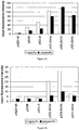
- the amount of antibody adsorbed on 1 ⁇ 10 8 cells was calculated and plotted against the concentration of antibody, shown in Figure 6 .
Landscapes
- Chemical & Material Sciences (AREA)
- Health & Medical Sciences (AREA)
- Life Sciences & Earth Sciences (AREA)
- Organic Chemistry (AREA)
- General Health & Medical Sciences (AREA)
- Gastroenterology & Hepatology (AREA)
- Biochemistry (AREA)
- Biophysics (AREA)
- Zoology (AREA)
- Genetics & Genomics (AREA)
- Medicinal Chemistry (AREA)
- Molecular Biology (AREA)
- Proteomics, Peptides & Aminoacids (AREA)
- Toxicology (AREA)
- Peptides Or Proteins (AREA)
- Micro-Organisms Or Cultivation Processes Thereof (AREA)
Description
- The present invention relates to lactic acid bacteria that have attached on their surface a TNFα binding polypeptide encoded by a gene construct as defined in claim 1. Peptides attached to the surface of the bacterium are more resistant to chemical and enzymatic degradation in the gastrointestinal tract than are secreted proteins. Lactic acid bacteria of the invention are capable of binding TNFα in the gastrointestinal tract and therefore reduce the content of free TNFα and alleviate its pro-inflammatory effects in the gut. Lactic acid bacteria of the invention can be used as medicament in the treatment of inflammatory bowel disease.
- Inflammatory bowel disease (IBD) refers to a group of gastrointestinal disorders characterized by chronic, relapsing inflammatory disorders of the gastrointestinal tract. Crohn's disease and ulcerative colitis are the two main subtypes of IBD.
- The etiology of IBD is unclear. IBD appears as multifactorial disease, with genetic and environmental factors probably cooperating in their development.
- Patients typically suffer from frequent and chronically relapsing flares, resulting in diarrhea, abdominal pain, rectal bleeding and malnutrition. Crohn's disease can be distinguished from ulcerative colitis in that the inflammation associated with Crohn's disease is transmural and often discontinuous. By contrast, the inflammatory changes of ulcerative colitis typically involve only the superficial mucosal and submucosal layers of the intestinal wall. Crohn's disease most commonly involves the ileum and colon but can affect any region of the gut; ulcerative colitis always involves the rectum, and inflammation may extend as far as the caecum in a continuous pattern. Patients with IBD often have various extra-intestinal symptoms such as arthalgias, and are more likely to have other chronic inflammatory diseases, particularly primary sclerosing cholangitis, ankylosing spondylitis and psoriasis. Etiology, genetics and pathogenesis of IBD is described by Hugot (1999) and Cho (2008).
- Since the etiology of both diseases is undetermined the causal therapy does not exist. The most commonly used conventional therapies are anti-inflammatory medicaments such as corticosteroids, salicilates, and immunosuppressives such as cyclosporine, mercaptopurine and azathioprine. Recently, biologic therapies have received a great amount of attention.
- The mucosal immune system is the central effector of intestinal inflammation and injury, with cytokines playing a central role in modulating inflammation. Cytokines may, therefore, be a logical target for IBD therapy using specific cytokine inhibitors. In this context compounds described by Schwartz M (2008) blocking the effect of tumor necrosis factor α (TNFα) or its receptor are of interest.
- TNFα is a cytokine produced by numerous cell types, including monocytes and macrophages, and was originally identified on the base its ability to induce the necrosis of certain mouse tumors as described by Old (1985). TNFα has been implicated in the pathophysiology of a variety of other human diseases and disorders, including sepsis, infections, autoimmune diseases, transplant rejection and graft-versus-host disease. TNFα promotes the inflammatory response, which, in turn, causes many of the clinical problems associated with autoimmune disorders such as rheumatoid arthritis, ankylosing spondylitis, Crohn's disease, psoriasis and refractory asthma. These disorders are sometimes treated by using a TNFα inhibitor or by counteracting TNFα activity.
- In particular, monoclonal antibodies against TNFα are routinely used in parenteral therapy of IBD as described by Sandborn (1999). Commercially available are infliximab, adalimumab and certolizumab pegol. Since antibodies are administered mainly by subcutaneous injection, serious unwanted effects emerge such as headache, abscess, upper respiratory tract infection and fatigue.
- De Silva (1992) reported on abundant presence of TNFα in the stool of IBD patients. Local delivery on the place of inflammation could solve unwanted effects associated with systemic delivery. This was achieved by Worledge (2000), with successful treatment of experimental colitis in rats by orally administering avian IgY anti TNFα antibodies with the capability of deactivating/removing TNFα.
- Antibodies and antibody related derivatives against TNFα are not always the optimal choice. The high cost of production of these immunoglobulin preparations prohibits their large-scale application. Antibodies, like other protein molecules, are not stable in the gastrointestinal tract.
- The use of genetically engineered bacteria that produce and deliver compounds that block the activity of TNFα could provide a solution. In this respect, lactic acid bacteria, which are normal commensals of the gut and other mucosal surfaces of human and animals and generally regarded as safe, represent ideal candidates.
-
WO2007/025977 discloses delivery of secreted antibodies against TNFα, and of fragments of such antibodies (so-called nanobodies), to the intestine for the treatment of enterocolitis using genetically engineered microorganisms such as lactic acid bacteria. The secreted antibodies ofWO2007/025977 do not include a surface attachment domain, and are thus not attached to the cell's surface and more prone to chemical and biological degradation in the gut. -
WO97/14806 WO00/23471 WO97/14806 -
EP1477802 discloses vaccines against infection with Streptococcus pneunoniae. The vaccine comprises the antigenic part of pneumococcal proteins (PpmA, S1rA), which are fused with a cell wall anchoring domain derived from the AcmA protein. The pneumococcal proteins ofEP 1477802 , however, do not comprise a TNFα binding domain, hence do not exhibit the therapeutic effects of the present microorganisms. - Hence it is known to produce peptides by lactic acid bacteria and secrete them into the gastrointestinal tract. Such peptides then generally undergo fast degradation.
- The present invention addresses these problems by producing genetically modified lactic acid bacteria that have attached to their surface, or express on their surface, a TNFα binding polypeptide encoded by a gene construct as defined in claim 1. Surface expression of TNFα binding polypeptides, or attachment of TNFα binding polypeptides, on lactic acid bacteria provides protection from chemical and enzymatic factors in the gastrointestinal tract. Such peptides are markedly more acid resistant. TNFα binding polypeptide expressed on the surface of mentioned microorganism may be TNFα binding Z domain (affibody). The peptide has stable and robust structure due to Z domain and is able to bind TNFα in the sample, for example body fluid.
- The present invention thus relates to a gene construct coding for a polypeptide comprising a secretion signal peptide, a TNFα binding domain and surface attachment domain, said TNFα binding domain and said surface attachment domain being separated by a spacer region, said gene construct preferably being under the control of a suitable promoter; wherein said construct is coding for a protein sequence
- (a) of any one ofSEQ ID NO:6 to SEQ ID NO:10, or
- (b) for a protein sequence which is at least 80, 90, 95, 98, or 99% identical to any one of said sequences under (a), and conferring the same biological function as the said sequence under (a).
- The invention further relates to a protein construct encoded by a gene construct as defined above.
- The invention also relates to a lactic acid bacterium having attached to its surface a protein construct as detailed above. In a preferred embodiment, said attachment to said surface is a non-covalent attachment. The attachment, however, can also be covalent attachment.
- In a preferred embodiment, said microorganism is genetically modified to express said protein construct on its surface. This shall include microorganisms that express the protein, secrete the protein into the medium, wherein said protein is thereafter attached to the surface of said microorganism.
- The invention further relates to a genetically modified lactic acid bacterium said genetically modified bacterium expressing on its surface a protein as defined above.
- In a preferred embodiment of the invention, microorganisms of the invention are Lactococcus lactis, or Lactococcus spec..
- In another preferred embodiment of the invention, the lactic acid bacterium of the invention comprises a gene construct as defined above.
- In another preferred embodiment of the invention, the lactic acid bacterium of the invention comprises a protein construct as defined above.
- The invention also relates to a method for producing a genetically modified lactic acid bacterium as described above, said method comprising introducing of a the above gene construct into a lactic acid bacterium.
- In a preferred embodiment of the invention, said gene construct is in a plasmid vector capable of replicating in said microorganism, or said gene construct is integrated into the genome of said microorganism.
- The invention further relates to a genetically modified lactic acid bacterium as described above for use as a medicament.
- The invention further relates to the above lactic acid bacterium for the treatment of inflammatory bowel disease, chronic colitis, Crohn's disease or ulcerative colitis.
- The invention further relates to the above lactic acid bacterium for reducing the content of free TNFα in the gastrointestinal tract of a subject.
- The invention further relates to a pharmaceutical composition comprising a modified lactic acid bacterium as described above. The pharmaceutical composition is preferably for oral administration, for example a tablet, or a capsule, preferably an enteric-coated tablet or capsule. The pharmaceutical preparation may also be in form of a suppository. The lactic acid bacterium in the pharmaceutical composition can be viable or non-viable. In one embodiment the lactic acid bacterium is non-viable.
- In a preferred embodiment said pharmaceutical composition is adapted for oral administration.
- The invention also relates to a food composition comprising the lactic acid bacterium above.
- In a preferred embodiment the food composition is selected from the group consisting of milk, yogurt, curd, cheese, fermented milks, milk based fermented products, ice cream, fermented cereal products, and milk based powders.
-
-
Figure 1 shows a FSC-SSC density plot of bacterial cells. -
Figures 2a and 2b show the results of flow cytometry of lactococcal cells harboring different plasmids. -
Figure 3 shows a shift in fluorescence determined by flow cytometry. -
Figure 4 shows a shift in fluorescence; control and Sdz-tnf-expressing cells detected with Alexa Fluor 488-conjugated TNFα. -
Figure 5 shows the loss of fluorescence intensity in samples, exposed to simulated gastric juice, as determined by flow cytometry. -
Figure 6 shows unspecific adsorption of antibody to B domain displaying cells. -
Figure 7a and7b shows sequence domains of peptides of the invention, and of known peptides AcmA and Usp45. -
Figure 8 shows successful immobilization of heterologous protein constructs containing TNFα-binding Z domain or Fc-binding B domain on non-GMO carrier lactic acid bacterium L. lactis stained with Alexa 488-labelled TNFα (black bars) or 2 µg of Alexa Fluor 488-labelled conjugated rabbit anti-mouse antibody (grey bars). - The present invention relates to genetically modified lactic acid bacteria that express, or have attached, a protein construct of the invention on their surface. Peptides expressed on the surface of bacteria are more resistant to degradation in the gastrointestinal tract. Such bacteria are capable of binding TNFα and therefore reducing the content of free (non-bound) TNFα and alleviating its pro-inflammatory effects in the gut. Such microorganisms can be used as medicament, e.g., in the treatment of IBD.
- A TNFα binding domain may be an affibody, as disclosed in
WO2006/092338 . Affibody molecules are small and robust high affinity protein molecules that can be engineered to bind specifically to a large number of target proteins. The scaffold used for construction of affibodies is Z domain, an engineered version of B domain, which is one of the five stable three-alpha-helix bundle domains from the immunoglobulin binding region of staphylococcal protein A. Affibody molecules with unique binding properties are usually acquired by randomization of 13 amino acids located in two alpha-helices involved in the binding activity of the parent protein domain. Affibodies with affinity towards specific target protein are obtained by library construction and affinity selection. - A "TNFα binding domain", within the meaning of the present disclosure shall thus be understood to be any peptide or domain to which TNFα binds, or which binds to TNFα. A TNFα binding domain can be a part of a larger protein, which then binds to TNFα, or to which TNFα binds. Preferred TNFα binding domains may have a dissociation constant (Kd) with TNFα of less than 0.01, 0.1, 1, 10, 100, 1000 nM. With regard to the formation of a ligand-protein complex (C) from a protein (P) and a ligand (L) according to


- Kd, within the context of the present invention, is determined by the use of surface plasmon resonance technology, e.g. using the Biacore® instrument. Kd is determined in an experiment wherein the TNFα binding domain is immobilized on a sensor chip of the instrument, and samples containing TNFα prepared by serial dilution are passed over the chip. The skilled person may than interpret the obtained sensograms to establish the Kd value (the apparent Kd value) for the interaction of TNFα binding domain and TNFα. Apparent Kd are calculated from the results, using the 1:1 Langmuir binding model of the BIAevaluation 3.2 software provided by the instrument manufacturer, according to the manufacturer's instructions.
- Kd, according to the disclosure is preferably measured for the free protein construct, i.e., the protein of the invention in a state not bound to the surface of the producing microorganism. Alternatively, Kd is measured for the protein bound to the surface of the microorganism.
- A "surface attachment domain" of a protein, according to the present invention, shall be understood as being any domain of said protein which is capable of attaching the said protein to the surface of a microorganism, e.g., the microorganism producing the protein. The surface attachment domain may comprise a lipobox and preferably a corresponding signal peptide (anchoring to membrane via a lipid group), or at least one LysM repeat (sometimes also referred to as a peptidoglycan binding domain, PBD), at least one choline binding domain (CBD) or at least one LPxTG motif (cell wall binding domain). A surface attachment domain may preferably comprise at least one of the following consensus sequences:
- LysM repeat consensus sequence: [YV]-X(0-4)-G-D-[ST]-[VLIA]-X(0-2)-[VLIA]
- CBD consensus sequence: G-X(0-5)-G-X-[WYI]-[WYT]-[YVL]-[FV]
- Lipobox consensus sequence (+signal peptide): M-X(1-10)-[RK]-{DERK}(7-17)-[LVTIMG]-[ASTIVGMLCPFL]-[AGLISVTFP]-C
- LPxTG consensus sequence: L-P-X-T-G,
- "LysM repeats" (or "Lysin motifs") are well known in the art. More than 4000 proteins of both prokaryotes and eukaryotes have been found to contain one or more LysM repeats (see http://www.genome.jp/dbget-bin/www bget?pfam:PF01476). Notably, this collection contains not only truly secreted proteins, but also (outer-)membrane proteins, lipoproteins or proteins bound to the cell wall in a (non-) covalent manner. LysM repeats typically range in length from 40 to 65 amino acid residues and binds to various types of peptidoglycan and chitin, recognizing the N-acetylglucosamine moiety. Most bacterial LysM-containing proteins are peptidoglycan hydrolases with various cleavage specificities. LysM repeats occur frequently in bacterial lysins, in bacteriophage proteins and in certain proteins of eukaryotes (Pfam PF01476 and Prodom PD407905). They are also present in bacterial peptidoglycan hydrolases and in peptidases, chitinases, esterases, reductases or nucleotidases. Multiple LysM repeats within one surface attachment domain are separated by spacing sequences mostly consisting of Ser, Thr and Asp or Pro residues, which may form a flexible region between the LysM repeats. The intervening sequences vary in length, composition and do not share significant homology. LysM repeats are present in the N-terminal as well as the C-terminal domains of proteins; they are also present in the central part of proteins, possibly connecting two (catalytic) domains. Prokaryotic LysM repeats, in contrast to their eukaryotic counterparts, do not possess possible disulphide bridges. Their domains contain extensive secondary structures and hydrogen bond networks, and consequently, disulphide bridges are non-essential for their structure and functions. The isoelectric points (pIs) of the LysM proteins range from 4 to 12, with most having a pI of 5 or 10. The best-characterized LysM-repeat containing protein is the N-acetylglucosaminidase AcmA of L. lactis. AcmA binds in a non-covalent manner to the cell wall and is responsible for cell lysis of producing cells.
- The LysM repeat may comprise the consensus sequence [YV]-X(0-4)-G-D-[ST]-[VLIA]-X(0-2)-[VLIA].
- The expression "to express on its surface", in relation to microorganisms expressing proteins, shall be understood to include microorganisms which express the protein, secrete the protein into the surrounding medium, wherein the protein is subsequently attached to the surface of the microorganism by means of covalent or non-covalent attachment.
- "GRAS", in respect to a microorganism within the context of the present disclosure, shall be understood to be a microorganism which is generally regarded as safe in accordance with the US Food And Drug Administration guidelines (http://www.fda.gov/Food/FoodIngredientsPackaging/GenerallyRecognizedasSafeGRAS/d efault.htm). The expression "GRAS microorganism" and "food grade organism" can be used interchangeably. A GRAS microorganism is a microorganism that has a long history of safe usage and is a constituent of fermented foods. Such microorganisms have "generally recognized as safe" status. Preferably genetically modified microorganism of food grade strain relates to lactic acid bacteria. Lactic acid bacteria are a group of phylogenetically related microorganisms. Preferred GRAS organisms are: Carnobacterium, Enterococcus, Lactobacillus, Lactococcus, Leuconostoc, Oenococcus, Pedicococcus, Streptococcus, Tetragenococcus, Vagococcus and Weisela. According to their physiological characteristics, genus Bifidobacterium spp. is usually classified as lactic acid bacterium because it occupies the same environmental niches, even though it is phylogenetically more distant. Preferably genetically modified microorganism of food grade strain is Lactococcus species. In one preferred embodiment said genetically modified organism is Lactococcus lactis strain.
- Genetically modified lactic acid bacteria with desirable new traits can be obtained by application of molecular biology techniques and production recombinant microorganism.
- The "surface" of a lactic acid bacterium according to the invention, shall be understood to mean the outer surface of the cell wall of the bacterium.
- A "homologue" of a reference protein, according to the invention, is a protein which has a similar, but not a fully identical amino acid sequence as the reference protein, and which has the same biological function or activity as the reference protein. The homologue has preferably an amino acid sequence which is 80, 90, 95, or 99% identical to the amino acid sequence of the original protein.
- Within the context of the present invention, the "% identity" of a protein relative to a reference protein of a defined length shall be understood as follows: A peptide that is 50 percent identical to a reference polypeptide that is 100 amino acids long can be a 50 amino acid polypeptide that is completely identical to a 50 amino acid long portion of the reference polypeptide. It might also be a 100 amino acid long polypeptide, which is 50 percent identical to the reference polypeptide over its entire length. Of course, other polypeptides will meet the same criteria. The term "sequence identity" as used herein means that the sequences are compared as follows. The sequences are aligned using
Version 9 of the Genetic Computing Group's GAP (global alignment program), using the default (BLOSUM62) matrix (values -4 to +11) with a gap open penalty of -12 (for the first null of a gap) and a gap extension penalty of -4 (per each additional consecutive null in the gap). After alignment, percentage identity is calculated by expressing the number of matches as a percentage of the number of amino acids in the reference polypeptide. - To achieve anchoring in the extracellular site of cell membrane, TNFα binding domain is expressed as fusion protein containing signal peptide for extracellular secretion, TNFα binding domain and surface attachment domain. Signal peptide leads the fusion protein into the extracellular space, where surface attachment domain anchors it on the membrane by binding to peptidoglycan. Signal peptide is processed and detached from fusion protein when reaching extracellular space.
- Quantitative measure of affinity (Kd) may be determined also for mentioned isolated fusion protein with signal peptide detached by using surface plasmon resonance. Said fusion protein may be isolated from the growth medium using methods known to the person skilled in the art and immobilized on a sensor chip e.g. of the Biacore® instrument. Samples containing TNFα prepared by serial dilution are passed over the chip. The skilled person may than interpret the obtained sensograms to establish a quantitative measure, apparent Kd value for the interaction of fusion protein and TNFα. Apparent Kd values may then be calculated from the results, using e.g. the 1:1 Langmuir binding model of the BIAevaluation 3.2 software provided by the instrument manufacturer.
- A genetically modified lactic acid bacterium expressing TNFα binding domain on its surface, may be prepared by introduction of gene construct to the mentioned bacterium. The gene construct comprises secretion signal peptide coding sequence, TNFα binding domain coding sequence and surface attachment domain coding sequence, wherein the TNFα binding domain coding sequence and the surface attachment domain coding sequence are separated with a spacer region coding sequence. Said gene construct is under the control of suitable promoter. Gene construct may be a part of plasmid vector capable of replicating in said microorganism or may be introduced into the genome of said microorganism. Introduction of gene construct may be performed by electroporation.
- The secretion signal peptide coding sequence codes for a secretion signal peptide. The secretion signal peptide enables the secretion of protein construct from the cell to the growth medium. Such secretion signal peptides include, but are not limited to signal peptide of Usp45 protein (SPUsp45), SP310 signal peptide and its improved mutants. Other secretion signal peptides are known in the art. Secretion may be increased by the use of synthetic pro-peptides. Such pro-peptides are homologues which contain acidic or neutral amino acids at position +2 and +8 or acidic amino acids at position +4 and +5 and have neutral or positive global net charge. Such pro-peptides include but are not limited to LEISSTCDA, LEISSYCDA, LQVDDIPSA and LGISSTCNA. Preferably the secretion signal peptide is the signal peptide of Usp45, where the second amino acid K is changed to A and is straightened by the synthetic pro-peptide LEISSYCDA.
- The usp45 gene encodes the major extracellular protein from Lactococcus lactis. The deduced sequence of the 27 residue leader peptide reveals the tripartite characteristics of a signal peptide. This leader peptide enables efficient secretion of various proteins. The Usp45 protein, according to the invention, is the one accessible in GenBank under the following GenBank record:
- LOCUS
- LACUSP45, 1592 bp, DNA, linear, BCT 26-APR-1993
- DEFINITION
- L.lactis secreted protein (usp45) gene, complete cds.
- ACCESSION
- M60178 M35374 X53491
- VERSION
- M60178.1 GI:149524
- KEYWORDS
- secreted protein.
- SOURCE
- Lactococcus lactis subsp. cremoris MG1363
- ORGANISM
- Lactococcus lactis subsp. cremoris MG1363 Bacteria; Firmicutes; Lactobacillales; Streptococcaceae; Lactococcus.
- REFERENCE
- 1 (bases 1 to 1592)
- AUTHORS
- van Asseldonk,M., Rutten,G., Oteman,M., Siezen,R.J., de
- Vos,W.M.
- and Simons,G.
- TITLE
- Cloning of usp45, a gene encoding a secreted protein from Lactococcus lactis subsp. lactis MG1363
- JOURNAL
- Gene 95 (1), 155-160 (1990)
- The Usp45 protein is thus understood to be the protein having the amino acid sequence of SEQ ID NO:12. First 27 amino acids represent a preferred signal peptide SPUsp45.
- The protein product of mentioned gene construct is secreted to the medium and from there attached to the surface of said microorganism by binding to peptidoglycan. The attachment to the peptidoglycan on cell surface may be non-covalent, where protein construct comprises different number of peptidoglycan binding domains derived from C terminal part of AcmA protein. The attachment to the peptidoglycan on cell surface may be covalent. This may be achieved by addition of LPXTG signal peptide to the protein construct to be covalently attached to the cell surface peptidoglycan. The attachment of such assembly to surface peptidoglycan is catalysed by enzyme sortase. Surface attachment domain coding sequence preferably codes for different number of peptidoglycan binding domains derived from C terminal part of AcmA. Preferably surface attachment domain consists of one to six LysM repeats. More preferably it consists of two to four LysM repeats, more preferably of three LysM repeats.
- Spacer region coding sequence between TNFα binding domain coding sequence and surface attachment domain coding sequence may be derived from AcmA protein. The length of spacer region may be 5-50 amino acids, preferably 10-30, more preferably 20 amino acids.
- The AcmA protein, according to the invention, is the one accessible in GenBank under the following GenBank record:
- LOCUS
- AF036720, 2379 bp DNA linear BCT 31-DEC-1997
- DEFINITION
- Lactococcus lactis N-acetylmuramidase (acmA) gene, complete
- cds. ACCESSION
- AF036720
- VERSION
- AF036720.1 GI:2707291
- KEYWORDS
- .
- SOURCE
- Lactococcus lactis
- ORGANISM
- Lactococcus lactis Bacteria; Firmicutes; Lactobacillales; Streptococcaceae; Lactococcus.
- REFERENCE
- 1 (bases 1 to 2379)
- AUTHORS
- Pillidge,C.J.
- TITLE
- Nucleotide sequence of N-acetylmuramidase gene acmA from Lactococcus lactis subsp. cremoris 2250
- JOURNAL
- Unpublished
- REFERENCE
- 2 (bases 1 to 2379)
- AUTHORS
- Pillidge,C.J.
- TITLE
- Direct Submission
- JOURNAL
- Submitted (02-DEC-1997) New Zealand Dairy Research Institute, Private Bag 11029, Palmerston North, New Zealand
- The AcmA protein is thus understood to be the protein having the amino acid sequence of SEQ ID NO:11.
- Examples of described gene constructs of the invention are shown in sequence listing as SEQ ID NO:6 to SEQ ID NO:10 and in
Figures 7a and7b . - The promoter that controls the expression of mentioned gene construct may be constitutive or inducible. Constitutive promoters enable continuous protein production, but are usually prone to lower level of expression. Inducible promoters are also referred to as controlled promoters. Inducible promoters usually provide higher expression level. Inducible promoters include but are not limited to bacteriocin controlled promoters, carbohydrate controlled promoters, pH controlled promoters, phage promoter Φ31 and temperature controlled promoters.
- Bacteriocin controlled promoters are controlled by addition of bacteriocins. Such bacteriocins include, but are not limited to nisin and pediocin. pH controlled promoters may be P170, induced by accumulation of lactic acid at pH 6.0-6.5, Pgad, induced by chloride ions and F1F0-ATPase promoter, induced by acidification. Temperature controlled promoter may be P2, where inactivation of repression is achieved at temperatures above 40°C. Carbohydrate promoters may be lacA and xylA operons that are induced by changing the carbon source in the growth medium.
- Anti-TNFα binding domain displaying lactic acid bacterium binding to TNFα refers to a property which may be tested by the use of flow cytometry or confocal microscopy. The testing of binding of surface expressed peptide to TNFα in accordance with invention is described in examples.
- A genetically modified lactic acid bacterium of the invention can be used as medicament to treat IBD. Said disease or disorder is characterized by imbalance in TNFα production, and can be treated by TNFα inactivating compounds, such as TNFα binding polypeptide expressed on the surface of genetically modified bacteria. More preferably, said disease is an irritable bowel disease, including but not limited to chronic colitis, ulcerative colitis and Crohn's disease.
- Another embodiment of the present invention is the pharmaceutical composition for treating IBD. Preferably pharmaceutical composition, comprising anti-TNFα peptide displaying lactic acid bacteria is for oral administration.
- The pharmaceutical composition may be liquid, comprising lactic acid bacterium expressing anti TNFα peptide on surface, or it may be solid, comprising dried microorganism that can be reactivated when put in a suitable environment. lactic acid bacteria may be dried by any system, including freeze drying and spray drying. "Anti-TNFα peptide displaying lactic acid bacterium" as used here, means that the lactic acid bacterium is viable and can express anti-TNFα peptide when placed in a suitable environment, not necessarily expressing the anti-TNFα peptide in the pharmaceutical composition. The pharmaceutical composition may further comprise agents known to the person skilled in the art to improve viability of the lactic acid bacteria and stability of surface expressed anti-TNFα peptide, such as but not limited to skimmed powdered milk.
- Lactic acid bacteria thrive in nutrient rich environments such as milk. They are integral part of mammalian GI flora and are considered beneficial to human health. As such they are added to the diet in the form of probiotic food. The present invention also relates to a food containing such recombinant lactic acid bacteria. Food composition may be milk, yogurt, curd, cheese, fermented milks, milk based fermented products, ice cream, fermented cereal based products and milk based powders.
- The present disclosure further relates to a system for immobilization of a heterologous protein construct containing a TNFα binding polypeptide coupled to a surface attachment domain on the cell surface of lactic acid bacteria, in cases for which the use of genetically modified bacteria is less desirable. For example, in applications that involve uncontrolled release into the environment, and foods products. This system is based on non-genetically modified lactic acid bacteria, which are used as carrier (support) to bind an externally added heterologous protein construct of the invention. The binding of the protein construct is preferably by a surface attachment domain as described above. Thus the expressed protein construct is not anchored or attached to the producing cells. The cells for production of the protein construct of the invention and the carrier cells may be different, thereby allowing the use of a non-genetically modified (non-GMO) cells as carrier cells.
- In one preferred embodiment said non-GMO carrier lactic acid bacterium is Lactococcus lactis strain.
- Non-GMO carrier lactic acid bacteria may be viable or non-viable (i.e., they cannot proliferate). Such non-viable cells can be generated various methods known in the art, such as UV, chemical inactivation, heat and gamma-irradiation. Preferably non-viable non-GMO carrier lactic acid bacteria are prepared by boiling in various acids, for example trichloroacetic acid.
- Protein constructs of the invention, composed of signal peptide for extracellular secretion, TNFα binding domain and surface attachment domain, are expressed in a recombinant expression host. Protein constructs are then secreted in the growth medium. The protein construct may be purified from growth medium or cell free growth medium may be added to non-GMO carrier lactic acid bacterium.
- Recombinant expression host is a system suitable for the production of a protein construct of the invention, such as bacterial expression systems, yeast expression systems, baculovirus/insect expression systems, and/or mammalian cells.
- The expression system preferably includes an expression vector, including the DNA encoding the protein construct of the invention, and the host cell. The expression system preferably allows foreign gene expression at a high level. Lactic acid bacteria (e.g., L. lactis) are preferably used as an expression host. In this case the cell free culture medium is directly suitable for addition to non-GMO carrier lactic acid bacterium and further purification steps are not necessary.
- The use of a non-GMO carrier lactic acid bacterium is demonstrated in Example 6.
- Several gene constructs were prepared to obtain fusion protein with optimal surface binding capabilities and are shown on
Figure 7a schematically. They are composed of three functional parts: signal sequence for the secretion to the growth medium, binding domain and peptidoglycan binding domain for surface attachment. Secretion signal was derived from Usp45 protein and was strengthened by synthetic propeptide LEISSYCDA in all constructs except pSDBA1. Binding domains were either immunoglobulin-binding B domain, or TNFα-binding Z domain. Immunoglobulin-binding B domain served as model binding domain to characterize expression and functionality of the construct. - Peptidoglycan binding domains were derived from AcmA protein. They consisted of one peptidoglycan binding LysM repeat, as in the case of pSDBA1 or pSDLBA1, or three peptidoglycan binding LysM repeats in remaining constructs. pSDLBA3a, pSDLBA3b and pSDLBA3c contained three peptidoglycan binding LysM repeats with variable length of spacer region between binding domain and first LysM repeat.
- Bacterial strains used in this study are shown in Table 1. E. coli DH5α was grown at 37°C with aeration in LB medium supplemented with 100 µg/ml ampicillin.
- L. lactis NZ9000 and L. lactis NZ9000ΔHtrA were grown in M-17 medium supplemented with 0.5 % glucose at 30°C without aeration. 10 µg/ml of chloramphenicol or erythromycin or both was added when appropriate.
- General cloning procedures were performed using conventional molecular biology methods. Electroporation of L. lactis was performed, using Gene Pulser II apparatus. Primers and plasmids are listed in Table 1.
- AcmA1, acmA3a, acmA3b and acmA3c genes were amplified from lactococcal genomic DNA using colony PCR with AcmA-EcoRI/AcmA-XbaI, AcmA-EcoRI/AcmA-R3-XbaI, AcmA-Fb-EcoRI/AcmA-R3-XbaI and AcmA-Fc-EcoRI/AcmA-R3-XbaI primer pairs, respectively. SpUsp45 was amplified from pGEM::Usp using Usp1-NcoI/UspR-BamHI primer pair. B domain gene was amplified from pGEM::B using Bdom-F-BamHI/ Bdom-R-EcoRI primer pair. PCR amplicons were digested with restriction enzymes in the following manner: spUsp45 with BamHI, b-dom with BamHI and EcoRI and acmA1 with EcoRI. Digested fragments were ligated and fusion gene termed sdb was PCR amplified using Uspl-NcoI/AcmA-XbaI primer pair. Sdb was digested with NcoI and XbaI and cloned to equally prepared pNZ8148, yielding pSDBA1. LEISSYCDA synthetic propeptide gene was added to spUsp45 gene by PCR using Uspl-NcoI/LeisR-BamHI primer pair and pGEM::Usp as a template, yielding spUsp45-LEIS. SpUsp45-LEIS gene was used to substitute spUsp45 in pSDBA1 via NcoI/BamHI restriction sites, yielding pSDLBA1. AcmA3a, acmA3b and acmA3c genes were used to substitute acmA1 gene in pSDLBA1 via EcoRI/XbaI restriction sites, yielding pSDLBA3a, pSDLBA3b and pSDLBA3c, respectively. TNFα binding Z domain gene labeled z-tnf, with BamHI and EcoRI restriction sites was designed on the basis of Z00185 polypeptide decribed in
WO2006/092338 by codon optimization and ordered from ATG:biosynthetics. Z-tnf was BamHI/EcoRI digested and substituted b-dom in equally prepared pSDLBA3a, yielding pSDZ-TNF. All the constructs are shown inFigure 7a . - Overnight cultures of L. lactis NZ9000 or NZ9000ΔHtrA harboring appropriate plasmid: pSDBA1, pSDLBA1, pSDLBA3a, pSDLBA3b, pSDLBA3c or pSDZ-TNF, were diluted in 10 ml of fresh GM-17 medium, grown to optical density A600=0.5-0.8 and induced with 25 ng/ml nisin. 3 hours after induction we split the culture in half. 5 ml were centrifuged at 5000 g for 10 min. Supernatant was decanted; cell pellet was resuspended in 0.1 M potassium phosphate buffer pH 7.00.
- SDS PAGE was performed using a mini-Protean II apparatus. Prestained standards were used for molecular weight comparison. Protein concentration was determined by Bio Rad protein assay and equal amounts were used to enable comparison between samples. Samples were denatured by heating at 100°C in the presence of DTT before loading. Proteins were stained with Coomassie Brilliant Blue or transferred to polyvinylidene fluoride membrane. The membrane was blocked in 1% Western blocking solution and incubated overnight at 4°C with FITC conjugated anti-protein A antibody. After washing with 50 mM Tris-HCl, 150 mM NaCl, 0.05
% Tween 20, pH 7.5, the fluorescence was detected with Typhoon 9410 imager using blue excitation at 488 nm. - Expression of fusion proteins was confirmed by Western blot using FITC conjugated anti-protein A antibody, which bound the B domain of the fusion proteins. Cell lysate, containing cytoplasmic and surface bound fusion protein, and growth medium, containing secreted unbound fusion protein, were tested. All fusion proteins were detected in both, cell lysate and growth medium, and their observed molecular weights corresponded to the calculated. Molecular weight of fusion proteins was: Sdba1 18.4 kDa, Sdlbal 19.4 kDa, Sdlba3a 34.0 kDa, Sdlba3b 32.9 kDa and Sdlba3c 31.8 kDa.
- Two bands of similar size were detected with all fusion proteins, which corresponded to fusion proteins with or without secretion signal with calculated molecular weight difference of 2.9 kDa.
- The processing of secretion signals was evaluated with growth medium fraction of Sdba1 and Sdlbal proteins. Their N-terminal sequence was determined to be GSADN and LEISS, respectively, which corresponds to the N-terminal sequences of fusion proteins without Usp45 signal.
- Growth media of L. lactis NZ9000 expressing Sdba1 or Sdlba1 were concentrated on Amicon Ultra-4, separated on SDS PAGE and blotted to PVDF membrane. Membrane was stained briefly with Coomassie Brilliant Blue, dried and corresponding bands were excised. N-terminal amino acid sequences of Sdba1 and Sdlba1 were determined by automated Edman degradation using an Applied Biosystems 492 Protein sequencer.
- Surface display of B domain was characterized by flow cytometry and fluorescent microscopy. Control and Sdlba3b-expresing cells stained with unspecific antibodies were visualized using fluorescent microscopy. Stronger binding of antibodies and increased fluorescence was observed with Sdlba3b-expresing cells.
- For flow cytometric analysis, cells were gated as shown in
Figure 1 . All constructs for surface display of B domain were analyzed and compared. Anti-protein A antibody was used to specifically detect and quantify surface-bound fusion protein and unspecific rabbit anti-mouse antibodies were used to evaluate the B domain's capability to bind Fc region of immunoglobulins. The results are presented as mean fluorescence intensity values of lactococcal cells, transformed with different plasmids and stained with specific or unspecific antibody for both NZ9000 strain inFigure 2a and NZ9000ΔHtrA strain inFigure 2b . Fluorescence correlates with the amount of surface-bound fusion protein. The expression of SdlbA3b resulted in the highest responses with both specific and unspecific antibody in both bacterial strains. NZ9000 strain showed superior results in comparison to NZ9000ΔHtrA in the initial experiments. For more detailed comparison between pNZ8148; control and pSDLBA3b-containing cells stained with specific and unspecific antibody is shown inFigure 3A and 3B , respectively. A distinct shift in fluorescence between controls (back fill) and Sdlba3b-expressing cells (black line) can be observed in both cases. -
Figure 3A shows control and Sdlba3b-expressing cells detected with specific FITC-conjugated antibodies. Mean fluorescence intensity for control is 2.61 and for the sample 115.70.Figure 3B shows control and B domain-expressing cells detected with unspecific Alexa Fluor 488-conjugated antibodies. Mean fluorescence intensity for control is 2.26, and for the sample 150.71. - For the flow
cytometric analysis 10 µL of cell cultures, approximately 107 cells/ml, were added to 500 µL of 50 mM Tris-HCl, 150 mM NaCl, pH 7.5 and centrifuged 3 min at 5000 g at 4°C. Supernatants were decanted and cells were resuspended in 500 µL of the same buffer. Next, either 1 µg of specific, FITC conjugated anti-protein A antibody or 2 µg of unspecific,Alexa Fluor 488 conjugated rabbit anti-mouse antibody was added to the suspensions and incubated 2 hours at RT with constant shaking at 100 rpm. Cells were than washed three times with 200 µL 50 mM Tris-HCl, 150 mM NaCl, 0.1% Tween 20, pH 7.5 and resuspended in 500 µL of 50 mM Tris-HCl, 150 mM NaCl, pH 7.5. Stained sample and control cells were analyzed with FACS Calibur flow cytometer. At least 100000 bacterial cells were counted for each sample. Cells were gated using FSC vs. SSC to isolate the bacterial cells. - For the purpose of fluorescent microscopy the staining protocol was similar except the starting volume of cell cultures was 20 µL and final volume of TBS buffer (50 mM Tris-HCl, 150 mM NaCl, pH 7.5) for re-suspension was 200 µL. Fluorescence microscopy was performed using a Carl Zeiss LSM 510 confocal microscope.
Alexa Fluor 488 was excited with an argon laser, and emission was filtered using narrow-band 505-530 nm filter. Images were analyzed using Carl Zeiss LSM image software 3.0. - Surface display of TNFα-binding Z domain was characterized by the capability to bind Alexa Fluor 488-conjugated TNFα.
Figure 4 shows flow cytometry results. Control contains pNZ8148 and is shown in black fill. Cells containing pSDZ-TNF are shown with black line. A distinct shift in fluorescence can be observed. Mean fluorescence intensity for control is 4.83 and for the sample 150.71. - Stronger fluorescence of Sdz-tnf-expressing cells was also observed with the use of fluorescent microscopy.
- For flow cytometric analysis and fluorescent microscopy samples were prepared as described in example 2, except 10 µL of Alexa 488-labelled TNFα was added to the suspensions.
- Soluble TNFα was labelled using
Alexa Fluor 488 Microscale Protein Labeling kit according to manufacturer's protocol. Final concentration of labeled protein was 0.5 mg/ml determined with Nanodrop 1000 Spectrophotometer. - Stability of the surface displayed B domain was tested against simulated gastric juice. 500 µl of pNZ8148 as control or pSDLBA3b-containing cells were mixed with either 500 µl of TBS buffer, pH 7.0 or 500 µl of simulated gastric juice, which was prepared using 3 mg/ml pepsin and pH was adjusted to 2.0 or 4.0 with concentrated HCl. The mixture was than incubated at room temperature for 30 min. 50 µl of the mixture was washed with 500 µl of TBS buffer, stained with
Alexa Fluor 488 rabbit anti-mouse antibody and analyzed with flow cytometer as described earlier. - Capability of surface displayed B domain to bind Fc region of antibody was tested after the incubation of cells containing pSDLBA3b in simulated gastric conditions and compared to incubation at pH 7.0; positive control or to cells containing pNZ8148 at pH 7.0; negative control.
- The loss of fluorescence intensity in samples, exposed to simulated gastric juice was less than 14 % even after 30 min at pH 2.0 as shown in
Figure 5 . - Control cell cultures containing pNZ8148 or cell cultures containing pSDLBA3b were washed with PBS buffer twice and resuspended in PBS to the cell density of 1x10⁹ cells/mL. 100 µL of cells were centrifuged and resuspended in 100 µl PBS containing M13/HRP antibody with various concentrations (0.45, 0.9, 1.8, 4.6 and 9.0 µg/mL) and incubated for 2 h at room temperature with constant shaking in preblocked Eppendorf tubes. After incubation, cells were pelleted and 20 µL of supernatant was transferred to microtiter plate and incubated with 180 µL TMB substrate for 15 minutes at room temperature. Colour development was terminated by the addition of 50
µL 2 M H2SO4 and absorbance was read at 450 nm using Rainbow reader. All samples were measured in duplicate. Amount of adsorbed antibody; Γ was calculated as follows: Γ = CAb×V×(1-A450(pSDLBA3b)/A450(pNZ8148)). Langmuir adsorption isotherm was assumed for the adsorption of antibody to the surface of Sdlba3b-expressing L. lactis. Langmuir linear regression method (CAb/Γ = CAb/ Γmax + 1/(K×Γmax)) was used to fit the Langmuir equation to the data and maximal amount of adsorbed antibody; Γmax was calculated. - The amount of antibody adsorbed on 1×10⁸ cells was calculated and plotted against the concentration of antibody, shown in
Figure 6 . - Langmuir linear regression method was applied and yielded the equation CAb/Γ = 6,837×CAb+22,688 (r2=0.9897). From this equation, the maximum amount of adsorbed antibody was calculated to be 0,146 µg per 1×10⁸ cells.
- Overnight cultures of L. lactis NZ9000 harboring plasmid pSDLBA3a or pSDZ-TNF were diluted in 10 ml of fresh GM-17 medium, grown to optical density A600=0.5-0.8 and induced with 25 ng/ml nisin. Three hours after induction cultures were centrifuged at 5000 g for 10 min. Supernatant containing secreted protein construct was separated from the cells and filtered through 0,22 µm Minisart filter (Sartorius). Overnight culture of non-recombinant carrier microorganism L. lactis NZ9000 was grown in fresh GM-17 medium and centrifuged. Supernatant was separated from cells.
- 500 µL of cell-free supernatant from culture expressing either TNFα-binding fusion peptide or Fc-binding fusion peptide were added to approximately 107 of non-recombinant L. lactis cells and incubated for 2 h at room temperature. Next, cells were separated from supernatants by 3 min centrifugation at 5000 g and resuspended in 500 µL of TBS. Next, either 10 µL of Alexa 488-labelled TNFα or 2 µg of
Alexa Fluor 488 conjugated rabbit anti-mouse antibody (Invitrogen; A-11059) was added to the suspensions and incubated 2 hours at room temperature with constant shaking at 100 rpm. Cells were than washed three times with 200 µL 0.1 % TBST and resuspended in 500 µL of TBS. For control, non-recombinant L. lactis without previous incubation with cell-free supernatants from culture expressing either TNFα-binding fusion peptide or Fc-binding fusion peptide were stained as described. Stained sample and control cells were analyzed with FACS Calibur (Becton Dickinson Inc.) flow cytometer. At least 100,000 bacterial cells were counted for each sample. Cells were gated using FSC vs. SSC to isolate the bacterial cells. - The results are shown in
Figure 8 . -
- Hugot JP, Zouali H, Lesage S, Thomas G. Etiology of the inflammatory bowel diseases. Int J Colorectal Dis. 1999 Feb;14(1):2-9.
- Cho JH. The genetics and immunopathogenesis of inflammatory bowel disease. Nat Rev Immunol. 2008 Jun;8(6):458-66.
- Schwartz M, Cohen R. Optimizing conventional therapy for inflammatory bowel disease. Curr Gastroenterol Rep. 2008 Dec;10(6):585-90.
- Old LJ. Tumor necrosis factor (TNF). Science. 1985 .
- Sandborn WJ, Hanauer SB. Antitumor necrosis factor therapy for inflammatory bowel disease: a review of agents, pharmacology, clinical results, and safety. Inflamm Bowel Dis. 1999 May;5(2):119-33.
- de Silva DG, Mendis LN, Sheron N, Alexander GJ, Candy DC, Chart H, Rowe B. TNF alpha in stool as marker of intestinal inflammation. Lancet. 1992 .
- Worledge KL, Godiska R, Barrett TA, Kink JA. Oral administration of avian tumor necrosis factor antibodies effectively treats experimental colitis in rats. Dig Dis Sci. 2000 Dec;45(12):2298-305.
- Le Loir Y, Nouaille S, Commissaire J, Brétigny L, Gruss A, Langella P. Signal peptide and propeptide optimization for heterologous protein secretion in Lactococcus lactis. Appl Environ Microbiol. 2001 Sep;67(9):4119-27.
- Nygren PA. Alternative binding proteins: affibody binding proteins developed from a small three-helix bundle scaffold. FEBS J. 2008 Jun;275(11):2668-76.
-
- <110> University of Ljubljana Institut "Jožef Stefan" Labena d.o.o.
- <120> Modified food grade microorganism for treatment of inflammatory bowel disease
- <130> P003127PC
- <160> 12
- <170> PatentIn version 3.3
- <210> 1
<211> 531
<212> DNA
<213> Artificial - <220>
<223> Gene construct comprising signal peptide coding sequence, TNFalpha affinity domain coding sequence and surface attachment domain coding sequence. - <400> 1

- <210> 2
<211> 558
<212> DNA
<213> Artificial - <220>
<223> Gene construct comprising signal peptide coding sequence, TNFalpha affinity domain coding sequence and surface attachment domain coding sequence. - <400> 2


- <210> 3
<211> 975
<212> DNA
<213> Artificial - <220>
<223> Gene construct comprising signal peptide coding sequence, TNFalpha affinity domain coding sequence and surface attachment domain coding sequence. - <400> 3

- <210> 4
<211> 942
<212> DNA
<213> Artificial - <220>
<223> Gene construct comprising signal peptide coding sequence, TNFalpha affinity domain coding sequence and surface attachment domain coding sequence. - <400> 4


- <210> 5
<211> 906
<212> DNA
<213> Artificial - <220>
<223> Gene construct comprising signal peptide coding sequence, TNFalpha affinity domain coding sequence and surface attachment domain coding sequence. - <400> 5


- <210> 6
<211> 176
<212> PRT
<213> Artificial - <220>
<223> Protein construct comprising signal peptide, TNFalpha affinity domain and surface attachment domain. - <400> 6

- <210> 7
<211> 185
<212> PRT
<213> Artificial - <220>
<223> Protein construct comprising signal peptide, TNFalpha affinity domain and surface attachment domain. - <400> 7

- <210> 8
<211> 324
<212> PRT
<213> Artificial - <220>
<223> Protein construct comprising signal peptide, TNFalpha affinity domain and surface attachment domain. - <400> 8


- <210> 9
<211> 313
<212> PRT
<213> Artificial - <220>
<223> Protein construct comprising signal peptide, TNFalpha affinity domain and surface attachment domain. - <400> 9


- <210> 10
<211> 301
<212> PRT
<213> Artificial - <220>
<223> Protein construct comprising signal peptide, TNFalpha affinity domain and surface attachment domain. - <400> 10


- <210> 11
<211> 437
<212> PRT
<213> Lactococcus lactis - <220>
<223> AcmA protein - <400> 11



- <210> 12
<211> 461
<212> PRT
<213> Lactococcus lactis - <220>
<223> Usp45 protein - <400> 12


Claims (8)
- A gene construct coding for a polypeptide comprising a secretion signal peptide, a TNFα binding domain and a surface attachment domain, said TNFα binding domain and said surface attachment domain being separated by a spacer region, and said gene construct being under the control of a suitable promoter; wherein said construct codes for the protein sequence (a) of any one of SEQ ID NO:6 to SEQ ID NO: 10, or (b) for a sequence which is at least 80 percent identical to any one of said sequences under (a), and conferring the same biological function as said sequence under (a).
- A protein construct encoded by a gene construct of claim 1.
- A lactic acid bacterium having attached to its surface a protein construct according to claim 2.
- The lactic acid bacterium of claim 3, said lactic acid bacterium being genetically modified to express on its surface said protein construct.
- The lactic acid bacterium of claim 4, wherein said lactic acid bacterium comprises a gene construct of claim 1.
- A method for producing a genetically modified lactic acid bacterium of claim 4 or 5, said method comprising introducing of a gene construct of claim 1 into a lactic acid bacterium.
- The lactic acid bacterium of any one of claims 3 to 5 for use as a medicament.
- A pharmaceutical composition comprising the lactic acid bacterium according to any one of claims 3 to 5.
Priority Applications (1)
| Application Number | Priority Date | Filing Date | Title |
|---|---|---|---|
| EP11700152.9A EP2521737B1 (en) | 2010-01-07 | 2011-01-05 | Modified food grade microorganism for treatment of inflammatory bowel disease |
Applications Claiming Priority (3)
| Application Number | Priority Date | Filing Date | Title |
|---|---|---|---|
| EP10000074A EP2343319A1 (en) | 2010-01-07 | 2010-01-07 | Genetically modified food grade microorganism for treatment of inflammatory bowel disease |
| EP11700152.9A EP2521737B1 (en) | 2010-01-07 | 2011-01-05 | Modified food grade microorganism for treatment of inflammatory bowel disease |
| PCT/EP2011/000019 WO2011083080A1 (en) | 2010-01-07 | 2011-01-05 | Modified food grade microorganism for treatment of inflammatory bowel disease |
Publications (2)
| Publication Number | Publication Date |
|---|---|
| EP2521737A1 EP2521737A1 (en) | 2012-11-14 |
| EP2521737B1 true EP2521737B1 (en) | 2017-08-30 |
Family
ID=41786343
Family Applications (2)
| Application Number | Title | Priority Date | Filing Date |
|---|---|---|---|
| EP10000074A Withdrawn EP2343319A1 (en) | 2010-01-07 | 2010-01-07 | Genetically modified food grade microorganism for treatment of inflammatory bowel disease |
| EP11700152.9A Not-in-force EP2521737B1 (en) | 2010-01-07 | 2011-01-05 | Modified food grade microorganism for treatment of inflammatory bowel disease |
Family Applications Before (1)
| Application Number | Title | Priority Date | Filing Date |
|---|---|---|---|
| EP10000074A Withdrawn EP2343319A1 (en) | 2010-01-07 | 2010-01-07 | Genetically modified food grade microorganism for treatment of inflammatory bowel disease |
Country Status (3)
| Country | Link |
|---|---|
| US (1) | US8754198B2 (en) |
| EP (2) | EP2343319A1 (en) |
| WO (1) | WO2011083080A1 (en) |
Families Citing this family (7)
| Publication number | Priority date | Publication date | Assignee | Title |
|---|---|---|---|---|
| US9616114B1 (en) | 2014-09-18 | 2017-04-11 | David Gordon Bermudes | Modified bacteria having improved pharmacokinetics and tumor colonization enhancing antitumor activity |
| US10676723B2 (en) | 2015-05-11 | 2020-06-09 | David Gordon Bermudes | Chimeric protein toxins for expression by therapeutic bacteria |
| US10738338B2 (en) | 2016-10-18 | 2020-08-11 | The Research Foundation for the State University | Method and composition for biocatalytic protein-oligonucleotide conjugation and protein-oligonucleotide conjugate |
| US11129906B1 (en) | 2016-12-07 | 2021-09-28 | David Gordon Bermudes | Chimeric protein toxins for expression by therapeutic bacteria |
| US11180535B1 (en) | 2016-12-07 | 2021-11-23 | David Gordon Bermudes | Saccharide binding, tumor penetration, and cytotoxic antitumor chimeric peptides from therapeutic bacteria |
| EP3946412A4 (en) * | 2019-04-05 | 2023-01-04 | Azitra, Inc. | METHODS AND COMPOSITIONS FOR TREATING ATOPIC DERMATITIS WITH RECOMBINANT MICROORGANISMS |
| EP4647439A1 (en) | 2024-05-09 | 2025-11-12 | Alma Mater Studiorum - Università di Bologna | Non-pathogenic bacteria for delivery of ptd-fusion proteins |
Citations (1)
| Publication number | Priority date | Publication date | Assignee | Title |
|---|---|---|---|---|
| WO2005040200A1 (en) * | 2003-10-24 | 2005-05-06 | Queensland University Of Technology | Protein expression |
Family Cites Families (6)
| Publication number | Priority date | Publication date | Assignee | Title |
|---|---|---|---|---|
| GB9521568D0 (en) | 1995-10-20 | 1995-12-20 | Lynxvale Ltd | Delivery of biologically active polypeptides |
| ATE259829T1 (en) | 1998-10-20 | 2004-03-15 | Vlaams Interuniv Inst Biotech | USE OF A CYTOKINE-PRODUCING LACTOCOCCUS STRAIN FOR THE TREATMENT OF COLITIS |
| EP1477802A1 (en) * | 2003-05-16 | 2004-11-17 | Erasmus Universiteit Rotterdam | Method for selecting and producing vaccine components and vaccines based thereon |
| ATE490268T1 (en) | 2005-03-01 | 2010-12-15 | Affibody Ab | TNF-ALHPA BINDING POLYPEPTIDE, USES THEREOF AND METHODS USING THE SAME |
| US9017662B2 (en) * | 2005-08-30 | 2015-04-28 | Actogenix N.V. | Anti-TNF alpha producing lactic acid bacteria for the treatment of chronic enterocolitis |
| CN102459571B (en) * | 2009-06-04 | 2014-01-08 | 香港中文大学 | Anti-inflammatory bacteria |
-
2010
- 2010-01-07 EP EP10000074A patent/EP2343319A1/en not_active Withdrawn
-
2011
- 2011-01-05 WO PCT/EP2011/000019 patent/WO2011083080A1/en not_active Ceased
- 2011-01-05 US US13/519,408 patent/US8754198B2/en not_active Expired - Fee Related
- 2011-01-05 EP EP11700152.9A patent/EP2521737B1/en not_active Not-in-force
Patent Citations (2)
| Publication number | Priority date | Publication date | Assignee | Title |
|---|---|---|---|---|
| WO2005040200A1 (en) * | 2003-10-24 | 2005-05-06 | Queensland University Of Technology | Protein expression |
| US20070276124A1 (en) * | 2003-10-24 | 2007-11-29 | Turner Mark S | Protein Expression |
Also Published As
| Publication number | Publication date |
|---|---|
| EP2343319A1 (en) | 2011-07-13 |
| WO2011083080A1 (en) | 2011-07-14 |
| EP2521737A1 (en) | 2012-11-14 |
| US8754198B2 (en) | 2014-06-17 |
| US20120282700A1 (en) | 2012-11-08 |
Similar Documents
| Publication | Publication Date | Title |
|---|---|---|
| US8754198B2 (en) | Modified food grade microorganism for treatment of inflammatory bowel disease | |
| AU2024201593B2 (en) | A genetically modified Lactobacillus and uses thereof | |
| JP4089920B2 (en) | Bacterial receptor structure | |
| Sun et al. | Characterization of a S-layer protein from Lactobacillus crispatus K313 and the domains responsible for binding to cell wall and adherence to collagen | |
| Raha et al. | Cell surface display system for Lactococcus lactis: a novel development for oral vaccine | |
| US6506389B2 (en) | Adherence factors of non pathogenic microorganisms and applications thereof for screening microorganisms for specific probiotic properties; novel pharmaceutical compositions and food additives comprising such microorganisms and adherence factors | |
| CA2750576C (en) | Novel pilus polypeptides | |
| EP3280397A2 (en) | Biosynthetic amyloid-based materials displaying functional protein sequences | |
| US20030165505A1 (en) | Nucleic acid and corresponding protein entitled 161P2F10B useful in treatment and detection of cancer | |
| US7754467B2 (en) | Surface expression of biologically active proteins in bacteria | |
| Ribelles et al. | LysA2, the Lactobacillus casei bacteriophage A2 lysin is an endopeptidase active on a wide spectrum of lactic acid bacteria | |
| US9063141B2 (en) | Listeria bacteriophage tailspike protein and uses thereof | |
| WO2010012972A2 (en) | Novel bacterial cells and uses thereof | |
| WO1999025836A1 (en) | Attaching substances to micro-organisms | |
| Orrapin et al. | Recombinant expression of novel protegrin-1 dimer and LL-37-linker–histatin-5 hybrid peptide mediated biotin carboxyl carrier protein fusion partner | |
| Charbonnier et al. | Overexpression, refolding, and purification of the histidine-tagged outer membrane efflux protein OprM of Pseudomonas aeruginosa | |
| Edwards et al. | Identification of major and minor chaperone proteins involved in the export of 987P fimbriae | |
| AU727676B2 (en) | Novel adherence factors of non pathogenic microorganisms and applications thereof for screening microorganisms for specific probiotic properties; novel pharmaceutical compositions and food additives comprising such microorganisms and adherence factors | |
| Muñoz Provencio et al. | Shotgun phage display of Lactobacillus casei BL23 against collagen and fibronectin | |
| Khang et al. | Recombinant S-layer proteins of Lactobacillus brevis mediating antibody adhesion to calf intestine alleviated neonatal diarrhea syndrome | |
| Tarahomjoo et al. | Expression of C-terminal repeat region of peptidoglycan hydrolase of Lactococcus lactis IL1403 in methylotrophic yeast Pichia pastoris | |
| CN103497251A (en) | Single-domain heavy chain antibody L5-79 for Listeria monocytogenes | |
| RU2639246C1 (en) | Application of s-layer protein domain from lactobacillus brevis as component of system for film proteins exposure on lactic acid bacteria cells surface | |
| CN121824691A (en) | A protein that enhances the expression of exogenous proteins and its applications | |
| Tarahomjoo | Exploring Surface Display Technology for Enhancement of Delivering Viable Lactic Acid |
Legal Events
| Date | Code | Title | Description |
|---|---|---|---|
| PUAI | Public reference made under article 153(3) epc to a published international application that has entered the european phase |
Free format text: ORIGINAL CODE: 0009012 |
|
| 17P | Request for examination filed |
Effective date: 20120706 |
|
| AK | Designated contracting states |
Kind code of ref document: A1 Designated state(s): AL AT BE BG CH CY CZ DE DK EE ES FI FR GB GR HR HU IE IS IT LI LT LU LV MC MK MT NL NO PL PT RO RS SE SI SK SM TR |
|
| DAX | Request for extension of the european patent (deleted) | ||
| 17Q | First examination report despatched |
Effective date: 20131001 |
|
| GRAP | Despatch of communication of intention to grant a patent |
Free format text: ORIGINAL CODE: EPIDOSNIGR1 |
|
| INTG | Intention to grant announced |
Effective date: 20170413 |
|
| GRAS | Grant fee paid |
Free format text: ORIGINAL CODE: EPIDOSNIGR3 |
|
| GRAA | (expected) grant |
Free format text: ORIGINAL CODE: 0009210 |
|
| RIN1 | Information on inventor provided before grant (corrected) |
Inventor name: LUNDER, MOJCA Inventor name: BERLEC, ALES Inventor name: CEH, BORIS Inventor name: STRUKELJ, BORUT Inventor name: RAVNIKAR, MATJAZ |
|
| AK | Designated contracting states |
Kind code of ref document: B1 Designated state(s): AL AT BE BG CH CY CZ DE DK EE ES FI FR GB GR HR HU IE IS IT LI LT LU LV MC MK MT NL NO PL PT RO RS SE SI SK SM TR |
|
| RAP1 | Party data changed (applicant data changed or rights of an application transferred) |
Owner name: UNIVERSITY OF LJUBLJANA Owner name: LABENA D.O.O. Owner name: INSTITUTE JOZEF STEFAN |
|
| REG | Reference to a national code |
Ref country code: GB Ref legal event code: FG4D |
|
| REG | Reference to a national code |
Ref country code: CH Ref legal event code: EP |
|
| REG | Reference to a national code |
Ref country code: AT Ref legal event code: REF Ref document number: 923463 Country of ref document: AT Kind code of ref document: T Effective date: 20170915 |
|
| REG | Reference to a national code |
Ref country code: IE Ref legal event code: FG4D |
|
| REG | Reference to a national code |
Ref country code: DE Ref legal event code: R096 Ref document number: 602011041046 Country of ref document: DE |
|
| REG | Reference to a national code |
Ref country code: NL Ref legal event code: MP Effective date: 20170830 |
|
| REG | Reference to a national code |
Ref country code: LT Ref legal event code: MG4D |
|
| REG | Reference to a national code |
Ref country code: AT Ref legal event code: MK05 Ref document number: 923463 Country of ref document: AT Kind code of ref document: T Effective date: 20170830 |
|
| REG | Reference to a national code |
Ref country code: FR Ref legal event code: PLFP Year of fee payment: 8 |
|
| PG25 | Lapsed in a contracting state [announced via postgrant information from national office to epo] |
Ref country code: FI Free format text: LAPSE BECAUSE OF FAILURE TO SUBMIT A TRANSLATION OF THE DESCRIPTION OR TO PAY THE FEE WITHIN THE PRESCRIBED TIME-LIMIT Effective date: 20170830 Ref country code: HR Free format text: LAPSE BECAUSE OF FAILURE TO SUBMIT A TRANSLATION OF THE DESCRIPTION OR TO PAY THE FEE WITHIN THE PRESCRIBED TIME-LIMIT Effective date: 20170830 Ref country code: AT Free format text: LAPSE BECAUSE OF FAILURE TO SUBMIT A TRANSLATION OF THE DESCRIPTION OR TO PAY THE FEE WITHIN THE PRESCRIBED TIME-LIMIT Effective date: 20170830 Ref country code: NO Free format text: LAPSE BECAUSE OF FAILURE TO SUBMIT A TRANSLATION OF THE DESCRIPTION OR TO PAY THE FEE WITHIN THE PRESCRIBED TIME-LIMIT Effective date: 20171130 Ref country code: SE Free format text: LAPSE BECAUSE OF FAILURE TO SUBMIT A TRANSLATION OF THE DESCRIPTION OR TO PAY THE FEE WITHIN THE PRESCRIBED TIME-LIMIT Effective date: 20170830 Ref country code: LT Free format text: LAPSE BECAUSE OF FAILURE TO SUBMIT A TRANSLATION OF THE DESCRIPTION OR TO PAY THE FEE WITHIN THE PRESCRIBED TIME-LIMIT Effective date: 20170830 |
|
| PG25 | Lapsed in a contracting state [announced via postgrant information from national office to epo] |
Ref country code: GR Free format text: LAPSE BECAUSE OF FAILURE TO SUBMIT A TRANSLATION OF THE DESCRIPTION OR TO PAY THE FEE WITHIN THE PRESCRIBED TIME-LIMIT Effective date: 20171201 Ref country code: ES Free format text: LAPSE BECAUSE OF FAILURE TO SUBMIT A TRANSLATION OF THE DESCRIPTION OR TO PAY THE FEE WITHIN THE PRESCRIBED TIME-LIMIT Effective date: 20170830 Ref country code: LV Free format text: LAPSE BECAUSE OF FAILURE TO SUBMIT A TRANSLATION OF THE DESCRIPTION OR TO PAY THE FEE WITHIN THE PRESCRIBED TIME-LIMIT Effective date: 20170830 Ref country code: RS Free format text: LAPSE BECAUSE OF FAILURE TO SUBMIT A TRANSLATION OF THE DESCRIPTION OR TO PAY THE FEE WITHIN THE PRESCRIBED TIME-LIMIT Effective date: 20170830 Ref country code: BG Free format text: LAPSE BECAUSE OF FAILURE TO SUBMIT A TRANSLATION OF THE DESCRIPTION OR TO PAY THE FEE WITHIN THE PRESCRIBED TIME-LIMIT Effective date: 20171130 Ref country code: IS Free format text: LAPSE BECAUSE OF FAILURE TO SUBMIT A TRANSLATION OF THE DESCRIPTION OR TO PAY THE FEE WITHIN THE PRESCRIBED TIME-LIMIT Effective date: 20171230 |
|
| PG25 | Lapsed in a contracting state [announced via postgrant information from national office to epo] |
Ref country code: NL Free format text: LAPSE BECAUSE OF FAILURE TO SUBMIT A TRANSLATION OF THE DESCRIPTION OR TO PAY THE FEE WITHIN THE PRESCRIBED TIME-LIMIT Effective date: 20170830 |
|
| PG25 | Lapsed in a contracting state [announced via postgrant information from national office to epo] |
Ref country code: PL Free format text: LAPSE BECAUSE OF FAILURE TO SUBMIT A TRANSLATION OF THE DESCRIPTION OR TO PAY THE FEE WITHIN THE PRESCRIBED TIME-LIMIT Effective date: 20170830 Ref country code: CZ Free format text: LAPSE BECAUSE OF FAILURE TO SUBMIT A TRANSLATION OF THE DESCRIPTION OR TO PAY THE FEE WITHIN THE PRESCRIBED TIME-LIMIT Effective date: 20170830 Ref country code: DK Free format text: LAPSE BECAUSE OF FAILURE TO SUBMIT A TRANSLATION OF THE DESCRIPTION OR TO PAY THE FEE WITHIN THE PRESCRIBED TIME-LIMIT Effective date: 20170830 Ref country code: RO Free format text: LAPSE BECAUSE OF FAILURE TO SUBMIT A TRANSLATION OF THE DESCRIPTION OR TO PAY THE FEE WITHIN THE PRESCRIBED TIME-LIMIT Effective date: 20170830 |
|
| RAP2 | Party data changed (patent owner data changed or rights of a patent transferred) |
Owner name: LABENA D.O.O. Owner name: INSTITUTE JOZEF STEFAN Owner name: UNIVERZA V LJUBLJANI |
|
| PG25 | Lapsed in a contracting state [announced via postgrant information from national office to epo] |
Ref country code: EE Free format text: LAPSE BECAUSE OF FAILURE TO SUBMIT A TRANSLATION OF THE DESCRIPTION OR TO PAY THE FEE WITHIN THE PRESCRIBED TIME-LIMIT Effective date: 20170830 Ref country code: IT Free format text: LAPSE BECAUSE OF FAILURE TO SUBMIT A TRANSLATION OF THE DESCRIPTION OR TO PAY THE FEE WITHIN THE PRESCRIBED TIME-LIMIT Effective date: 20170830 Ref country code: SM Free format text: LAPSE BECAUSE OF FAILURE TO SUBMIT A TRANSLATION OF THE DESCRIPTION OR TO PAY THE FEE WITHIN THE PRESCRIBED TIME-LIMIT Effective date: 20170830 Ref country code: SK Free format text: LAPSE BECAUSE OF FAILURE TO SUBMIT A TRANSLATION OF THE DESCRIPTION OR TO PAY THE FEE WITHIN THE PRESCRIBED TIME-LIMIT Effective date: 20170830 |
|
| REG | Reference to a national code |
Ref country code: DE Ref legal event code: R097 Ref document number: 602011041046 Country of ref document: DE |
|
| REG | Reference to a national code |
Ref country code: DE Ref legal event code: R082 Ref document number: 602011041046 Country of ref document: DE Representative=s name: ZACCO PATENTANWALTS- UND RECHTSANWALTSGESELLSC, DE Ref country code: DE Ref legal event code: R081 Ref document number: 602011041046 Country of ref document: DE Owner name: LABENA D.O.O., SI Free format text: FORMER OWNERS: INSTITUTE JOZEF STEFAN, LJUBLJANA, SI; LABENA D.O.O., LJUBLJANA, SI; UNIVERSITY OF LJUBLJANA, LJUBLJANA, SI Ref country code: DE Ref legal event code: R081 Ref document number: 602011041046 Country of ref document: DE Owner name: UNIVERZA V LJUBLJANI, SI Free format text: FORMER OWNERS: INSTITUTE JOZEF STEFAN, LJUBLJANA, SI; LABENA D.O.O., LJUBLJANA, SI; UNIVERSITY OF LJUBLJANA, LJUBLJANA, SI |
|
| RAP2 | Party data changed (patent owner data changed or rights of a patent transferred) |
Owner name: UNIVERZA V LJUBLJANI Owner name: LABENA D.O.O. |
|
| PLBE | No opposition filed within time limit |
Free format text: ORIGINAL CODE: 0009261 |
|
| STAA | Information on the status of an ep patent application or granted ep patent |
Free format text: STATUS: NO OPPOSITION FILED WITHIN TIME LIMIT |
|
| 26N | No opposition filed |
Effective date: 20180531 |
|
| PG25 | Lapsed in a contracting state [announced via postgrant information from national office to epo] |
Ref country code: SI Free format text: LAPSE BECAUSE OF FAILURE TO SUBMIT A TRANSLATION OF THE DESCRIPTION OR TO PAY THE FEE WITHIN THE PRESCRIBED TIME-LIMIT Effective date: 20170830 |
|
| REG | Reference to a national code |
Ref country code: CH Ref legal event code: PL |
|
| GBPC | Gb: european patent ceased through non-payment of renewal fee |
Effective date: 20180105 |
|
| PG25 | Lapsed in a contracting state [announced via postgrant information from national office to epo] |
Ref country code: LU Free format text: LAPSE BECAUSE OF NON-PAYMENT OF DUE FEES Effective date: 20180105 |
|
| REG | Reference to a national code |
Ref country code: IE Ref legal event code: MM4A |
|
| REG | Reference to a national code |
Ref country code: BE Ref legal event code: MM Effective date: 20180131 |
|
| PG25 | Lapsed in a contracting state [announced via postgrant information from national office to epo] |
Ref country code: CH Free format text: LAPSE BECAUSE OF NON-PAYMENT OF DUE FEES Effective date: 20180131 Ref country code: LI Free format text: LAPSE BECAUSE OF NON-PAYMENT OF DUE FEES Effective date: 20180131 Ref country code: GB Free format text: LAPSE BECAUSE OF NON-PAYMENT OF DUE FEES Effective date: 20180105 Ref country code: BE Free format text: LAPSE BECAUSE OF NON-PAYMENT OF DUE FEES Effective date: 20180131 |
|
| PG25 | Lapsed in a contracting state [announced via postgrant information from national office to epo] |
Ref country code: IE Free format text: LAPSE BECAUSE OF NON-PAYMENT OF DUE FEES Effective date: 20180105 |
|
| PGFP | Annual fee paid to national office [announced via postgrant information from national office to epo] |
Ref country code: FR Payment date: 20190123 Year of fee payment: 9 Ref country code: DE Payment date: 20190128 Year of fee payment: 9 |
|
| PG25 | Lapsed in a contracting state [announced via postgrant information from national office to epo] |
Ref country code: MC Free format text: LAPSE BECAUSE OF FAILURE TO SUBMIT A TRANSLATION OF THE DESCRIPTION OR TO PAY THE FEE WITHIN THE PRESCRIBED TIME-LIMIT Effective date: 20170830 |
|
| PG25 | Lapsed in a contracting state [announced via postgrant information from national office to epo] |
Ref country code: MT Free format text: LAPSE BECAUSE OF NON-PAYMENT OF DUE FEES Effective date: 20180105 |
|
| PG25 | Lapsed in a contracting state [announced via postgrant information from national office to epo] |
Ref country code: TR Free format text: LAPSE BECAUSE OF FAILURE TO SUBMIT A TRANSLATION OF THE DESCRIPTION OR TO PAY THE FEE WITHIN THE PRESCRIBED TIME-LIMIT Effective date: 20170830 |
|
| PG25 | Lapsed in a contracting state [announced via postgrant information from national office to epo] |
Ref country code: HU Free format text: LAPSE BECAUSE OF FAILURE TO SUBMIT A TRANSLATION OF THE DESCRIPTION OR TO PAY THE FEE WITHIN THE PRESCRIBED TIME-LIMIT; INVALID AB INITIO Effective date: 20110105 Ref country code: PT Free format text: LAPSE BECAUSE OF FAILURE TO SUBMIT A TRANSLATION OF THE DESCRIPTION OR TO PAY THE FEE WITHIN THE PRESCRIBED TIME-LIMIT Effective date: 20170830 |
|
| PG25 | Lapsed in a contracting state [announced via postgrant information from national office to epo] |
Ref country code: CY Free format text: LAPSE BECAUSE OF FAILURE TO SUBMIT A TRANSLATION OF THE DESCRIPTION OR TO PAY THE FEE WITHIN THE PRESCRIBED TIME-LIMIT Effective date: 20170830 Ref country code: MK Free format text: LAPSE BECAUSE OF NON-PAYMENT OF DUE FEES Effective date: 20170830 |
|
| PG25 | Lapsed in a contracting state [announced via postgrant information from national office to epo] |
Ref country code: AL Free format text: LAPSE BECAUSE OF FAILURE TO SUBMIT A TRANSLATION OF THE DESCRIPTION OR TO PAY THE FEE WITHIN THE PRESCRIBED TIME-LIMIT Effective date: 20170830 |
|
| REG | Reference to a national code |
Ref country code: DE Ref legal event code: R119 Ref document number: 602011041046 Country of ref document: DE |
|
| PG25 | Lapsed in a contracting state [announced via postgrant information from national office to epo] |
Ref country code: FR Free format text: LAPSE BECAUSE OF NON-PAYMENT OF DUE FEES Effective date: 20200131 Ref country code: DE Free format text: LAPSE BECAUSE OF NON-PAYMENT OF DUE FEES Effective date: 20200801 |