EP2514360A1 - Improved magnetic resonance imaging - Google Patents
Improved magnetic resonance imaging Download PDFInfo
- Publication number
- EP2514360A1 EP2514360A1 EP11163001A EP11163001A EP2514360A1 EP 2514360 A1 EP2514360 A1 EP 2514360A1 EP 11163001 A EP11163001 A EP 11163001A EP 11163001 A EP11163001 A EP 11163001A EP 2514360 A1 EP2514360 A1 EP 2514360A1
- Authority
- EP
- European Patent Office
- Prior art keywords
- magnetic field
- target
- echo
- time
- fat
- Prior art date
- Legal status (The legal status is an assumption and is not a legal conclusion. Google has not performed a legal analysis and makes no representation as to the accuracy of the status listed.)
- Granted
Links
Images
Classifications
-
- A—HUMAN NECESSITIES
- A61—MEDICAL OR VETERINARY SCIENCE; HYGIENE
- A61B—DIAGNOSIS; SURGERY; IDENTIFICATION
- A61B5/00—Measuring for diagnostic purposes; Identification of persons
- A61B5/05—Detecting, measuring or recording for diagnosis by means of electric currents or magnetic fields; Measuring using microwaves or radio waves
- A61B5/055—Detecting, measuring or recording for diagnosis by means of electric currents or magnetic fields; Measuring using microwaves or radio waves involving electronic [EMR] or nuclear [NMR] magnetic resonance, e.g. magnetic resonance imaging
-
- G—PHYSICS
- G01—MEASURING; TESTING
- G01R—MEASURING ELECTRIC VARIABLES; MEASURING MAGNETIC VARIABLES
- G01R33/00—Arrangements or instruments for measuring magnetic variables
- G01R33/20—Arrangements or instruments for measuring magnetic variables involving magnetic resonance
- G01R33/44—Arrangements or instruments for measuring magnetic variables involving magnetic resonance using nuclear magnetic resonance [NMR]
- G01R33/48—NMR imaging systems
- G01R33/4828—Resolving the MR signals of different chemical species, e.g. water-fat imaging
-
- G—PHYSICS
- G01—MEASURING; TESTING
- G01R—MEASURING ELECTRIC VARIABLES; MEASURING MAGNETIC VARIABLES
- G01R33/00—Arrangements or instruments for measuring magnetic variables
- G01R33/20—Arrangements or instruments for measuring magnetic variables involving magnetic resonance
- G01R33/44—Arrangements or instruments for measuring magnetic variables involving magnetic resonance using nuclear magnetic resonance [NMR]
- G01R33/48—NMR imaging systems
- G01R33/54—Signal processing systems, e.g. using pulse sequences ; Generation or control of pulse sequences; Operator console
- G01R33/56—Image enhancement or correction, e.g. subtraction or averaging techniques, e.g. improvement of signal-to-noise ratio and resolution
- G01R33/5608—Data processing and visualization specially adapted for MR, e.g. for feature analysis and pattern recognition on the basis of measured MR data, segmentation of measured MR data, edge contour detection on the basis of measured MR data, for enhancing measured MR data in terms of signal-to-noise ratio by means of noise filtering or apodization, for enhancing measured MR data in terms of resolution by means for deblurring, windowing, zero filling, or generation of gray-scaled images, colour-coded images or images displaying vectors instead of pixels
Definitions
- the invention relates to a method and a device for the removal of non-cerebral tissue in Magnetic Resonance (MR) imaging according to claim 1.
- the invention relates to a method and a device for removing image parts representing non-brain tissues, such as skull, scalp, veins or meninges, from a MR neuro-image.
- the subject to be imaged is placed in a strong static magnetic field, which forces the hydrogen nuclear magnetic moments associated with the subject hydrogen nuclear spins to adopt an orientation parallel or anti-parallel with respect to said static magnetic field.
- the spin axes are not exactly aligned with the static magnetic field, but they precess around a direction axis of said static magnetic field with a characteristic frequency, giving rise to a net magnetization in the direction of the static magnetic field.
- a pulse of radio frequency energy can be applied to the subject at the aforementioned characteristic frequency, called Larmor frequency, which is for one type of nuclei proportional to the flux density of the magnetic field.
- the radio frequency energy associated with this pulse disturbs the net magnetization from its equilibrium, rotating it away from the static magnetic field with an angle, called flip angle, which depends on the strength and duration of the magnetic component of the electromagnetic radio frequency radiation. Consequently, the net magnetization begins to precess around the static magnetic field main axis, its transverse component inducing an electromotive force in a receiver coil according to Faraday's law of magnetic induction. This electromotive force gives rise to an induced signal that is then at the basis of MR image reconstruction. The magnitude of this induced signal depends, among other factors, on the number of nuclei that produce the magnetization, on their relaxation times, i.e. the time needed by the net magnetization to return to its equilibrium state along the axis of said strong magnetic field. Other factors include the so called spin preparation. In order to optimize a diagnostic value of the signal, different combinations of one or more radio frequency pulses have been proposed, while taking into account some parameters like a repetition time of the pulse, its echo time, the flip angle, its bandwidth, etc.
- the time required for a substance to become magnetized after having been placed in a magnetic field or the time required for said substance to regain longitudinal magnetization following the radiofrequency pulse is usually called the longitudinal relaxation time T1 (also called spin - lattice relaxation).
- This longitudinal relaxation time T1 is in particular determined by thermal interactions between resonating protons and other protons and other magnetic nuclei in the environment submitted to the magnetic field.
- the longitudinal relaxation time T1 depends in particular on the relationship between the natural vibrational frequencies of said substance and said Larmor frequency.
- a transverse relaxation time T2 also called spin - spin relaxation
- the transverse relaxation time T2 characterizes thus the exponential decay of the induced signal resulting from the decline of coherence of the spin precession.
- T1 weighted protocols i.e. protocols giving rise to images where most of the contrast of the tissues is due to differences in the values of the longitudinal relaxation time T1, for imaging soft tissues, and in particular the brain, since said T1 weighted protocols generally offer a good contrast between the tissues forming the brain, like grey of white cerebral matter.
- evolution of high-resolution MR imaging of the last decades enabled advanced post-processing of brain images, enhancing the contrast between the tissues forming the brain.
- Brain contrast is for example crucial in morphometry, since quantitative measures are derived from the image data to determine the volume of certain brain tissues like grey or white cerebral matter, for supporting then diagnostic decisions and facilitating follow-up comparisons.
- MR neuroimaging Unfortunately, one major problem in MR neuroimaging is to obtain a good contrast for discriminating brain tissues from non-brain tissues like the skull. Often, the parts of the image representing non-brain tissues have to be erased before such an advanced image post-processing can be conducted. This process, usually called skull-stripping, is hampered by very similar image intensities of wanted and unwanted tissues, possibly leading to the elimination of too much or too little non-brain tissue, which can bias the final outcome of the following post-processing, and at worst, falsely influence the diagnostic decision.
- the second group comprises methods based on multispectral analysis.
- Van Leemput et al. Van Leemput et al., IEEE Transactions on Medical Imaging 18, 897 (1999 )]. They propose a framework to automatically classify brain tissue using multispectral acquisitions as T1 weighted, T2 weighted and proton density.
- the invention provides thus additional image information, notably from separated fat and water signal image, without considerably prolonging the scan time or impeding the quality of the "normal" image contrast (the water and/or fat combined image).
- the multispectral analysis method comprises in particular a data treatment by a multispectral skull-stripping algorithm (i.e. an algorithm using several image contrasts), yielding better target imaging results compared to normal MR imaging.
- a multispectral skull-stripping algorithm i.e. an algorithm using several image contrasts
- the precession of the nuclei moments around the direction of the static magnetic field is detected by induction coils that are placed outside the target for inducing said current signal, which might be then subjected to Fourier transformation for studying its spectrum.
- the current signal is digitally sampled in the Fourier space (k-space) for generating the MR brain image.
- the radio frequency pulse is followed by a sampling of a certain number of data points in k-space from said current signal at the time of echo TE 1 after the radio frequency pulse of excitation. These points are notably selected using the phase- and frequency-encoding first magnetic field gradients.
- an opposite phase- and frequency-encoding magnetic field gradient i.e. said second magnetic field gradient
- a second echo is induced, and can be acquired and sampled via said current signal at the time of the second echo TE 2 .
- the fat and water information encoded in the induced current signal can be extracted from said current signal by the following way, known as the Dixon technique [ Dixon WT, Radiology 153:1, 189, (1984 )].
- non-brain tissue around vessels is well depicted in the fat-only images, which is in particular used by the method according to the invention for skull-stripping purposes.
- each fat-only image, and/or respectively, each water-only image is preferentially used for extracting data related to fat spatial position in the target, and/or respectively data related to water spatial position in the target, said data being then inputted as said additional input source into a multispectral skull-stripping algorithm of said multispectral analysis method such as for example the method presented in Van Leemput et al. [ Van Leemput et al., IEEE Transactions on Medical Imaging 18, 897 (1999 )].
- said multispectral skull-stripping algorithm is able to provide from said data related to fat or water spatial position a digital image of the target comprising target tissues spatial position.
- the so computed image contrast equals the standard image contrast, i.e. the contrast which would be achieved without employing the Dixon technique, and recovers probable signal losses due to increased sampling bandwidths.
- the present invention features the parallel acquisition of two image volumes; in a post-processing step, a standard T1-weighted contrast and an additional fat contrast depicting mainly undesired tissues is obtained. Both are subsequently used in a general multispectral analysis. Hence, the information available to the multispectral algorithm is augmented by additional information about the location of undesired, i.e. non-brain, parts of the image.
- the method according to the invention presents thus the following advantages compared to conventional skull-stripping procedures:
Landscapes
- Physics & Mathematics (AREA)
- Health & Medical Sciences (AREA)
- Life Sciences & Earth Sciences (AREA)
- Nuclear Medicine, Radiotherapy & Molecular Imaging (AREA)
- High Energy & Nuclear Physics (AREA)
- Engineering & Computer Science (AREA)
- Heart & Thoracic Surgery (AREA)
- General Physics & Mathematics (AREA)
- Biophysics (AREA)
- Pathology (AREA)
- Condensed Matter Physics & Semiconductors (AREA)
- Biomedical Technology (AREA)
- Radiology & Medical Imaging (AREA)
- Medical Informatics (AREA)
- Molecular Biology (AREA)
- Surgery (AREA)
- Animal Behavior & Ethology (AREA)
- General Health & Medical Sciences (AREA)
- Public Health (AREA)
- Veterinary Medicine (AREA)
- Magnetic Resonance Imaging Apparatus (AREA)
Abstract
Description
- The invention relates to a method and a device for the removal of non-cerebral tissue in Magnetic Resonance (MR) imaging according to claim 1. In particular, the invention relates to a method and a device for removing image parts representing non-brain tissues, such as skull, scalp, veins or meninges, from a MR neuro-image.
- In order to obtain images using the technique of MR imaging, the subject to be imaged is placed in a strong static magnetic field, which forces the hydrogen nuclear magnetic moments associated with the subject hydrogen nuclear spins to adopt an orientation parallel or anti-parallel with respect to said static magnetic field. The spin axes are not exactly aligned with the static magnetic field, but they precess around a direction axis of said static magnetic field with a characteristic frequency, giving rise to a net magnetization in the direction of the static magnetic field. In order to stimulate a signal from the hydrogen nuclei, a pulse of radio frequency energy can be applied to the subject at the aforementioned characteristic frequency, called Larmor frequency, which is for one type of nuclei proportional to the flux density of the magnetic field. The radio frequency energy associated with this pulse disturbs the net magnetization from its equilibrium, rotating it away from the static magnetic field with an angle, called flip angle, which depends on the strength and duration of the magnetic component of the electromagnetic radio frequency radiation. Consequently, the net magnetization begins to precess around the static magnetic field main axis, its transverse component inducing an electromotive force in a receiver coil according to Faraday's law of magnetic induction. This electromotive force gives rise to an induced signal that is then at the basis of MR image reconstruction. The magnitude of this induced signal depends, among other factors, on the number of nuclei that produce the magnetization, on their relaxation times, i.e. the time needed by the net magnetization to return to its equilibrium state along the axis of said strong magnetic field. Other factors include the so called spin preparation. In order to optimize a diagnostic value of the signal, different combinations of one or more radio frequency pulses have been proposed, while taking into account some parameters like a repetition time of the pulse, its echo time, the flip angle, its bandwidth, etc.
- The time required for a substance to become magnetized after having been placed in a magnetic field or the time required for said substance to regain longitudinal magnetization following the radiofrequency pulse is usually called the longitudinal relaxation time T1 (also called spin - lattice relaxation). This longitudinal relaxation time T1 is in particular determined by thermal interactions between resonating protons and other protons and other magnetic nuclei in the environment submitted to the magnetic field. The longitudinal relaxation time T1 depends in particular on the relationship between the natural vibrational frequencies of said substance and said Larmor frequency. Similarly, a transverse relaxation time T2 (also called spin - spin relaxation) describes the interaction between neighbouring nuclei with identical precessional frequencies having different magnetic quantum states, and represents a measure of how long a transverse magnetization last following the radiofrequency pulse. The transverse relaxation time T2 characterizes thus the exponential decay of the induced signal resulting from the decline of coherence of the spin precession.
- Each tissue of the human body is thus characterized by an own longitudinal relaxation time T1 and an own transverse relaxation time T2. Numerous studies used T1 weighted protocols, i.e. protocols giving rise to images where most of the contrast of the tissues is due to differences in the values of the longitudinal relaxation time T1, for imaging soft tissues, and in particular the brain, since said T1 weighted protocols generally offer a good contrast between the tissues forming the brain, like grey of white cerebral matter. Moreover, the evolution of high-resolution MR imaging of the last decades enabled advanced post-processing of brain images, enhancing the contrast between the tissues forming the brain. Brain contrast is for example crucial in morphometry, since quantitative measures are derived from the image data to determine the volume of certain brain tissues like grey or white cerebral matter, for supporting then diagnostic decisions and facilitating follow-up comparisons.
- Unfortunately, one major problem in MR neuroimaging is to obtain a good contrast for discriminating brain tissues from non-brain tissues like the skull. Often, the parts of the image representing non-brain tissues have to be erased before such an advanced image post-processing can be conducted. This process, usually called skull-stripping, is hampered by very similar image intensities of wanted and unwanted tissues, possibly leading to the elimination of too much or too little non-brain tissue, which can bias the final outcome of the following post-processing, and at worst, falsely influence the diagnostic decision.
- Different methods involving various skull-stripping algorithms have been used for differentiating brain from non-brain tissues. These methods can be separated into two groups:
- The first group comprises methods based exclusively on T1 weighted protocols.
- These methods are for example:
- the Brain Surface Extractor (BSE) [Shattuck et al., Neurolmage 13, 856 (2001)] which uses a combination of anisotropic diffusion filtering, Marr-Hildreth edge detector and morphological operators to separate brain and non-brain tissue;
- the Watershed algorithm (WAT) [Hahn and Peitgen, MICCAI 2000, 134 (2000)]: an intensity-based approach relying on a 3D algorithm with pre-flooding performed on the intensity inverted image, selecting the basin to represent the brain;
- the Brain Extraction Tool (BET) [Smith, Human Brain Mapping 17, 143 (2002)] which deforms a mask which is constraints on surface smoothness and voxel intensities in the vicinity to the surface position;
- the Hybrid Watershed Algorithm (HWA) [Ségonne et al., Neurolmage 22, 1060 (2004)]: an hybrid approach combining watershed algorithm and deformable model, where the latter adds atlas based shape constraints in order to guarantee anatomically meaningful brain mask.
- Suresh et al. [Suresh et al., Neurolmage 49, 225 (2010)] uses intensity thresholding followed by removal of narrow connections using graph cut segmentation theory to remove non brain tissue;
- Keihaninejad et al. [Keihaninejad et al., 9th European Congress on Epileptology 51, 6 (2010)] extract the intracranial volume using posterior probability maps extracted using SPM5 and apply a specific threshold to keep only brain tissues.
- The second group comprises methods based on multispectral analysis. One of these methods has been developed by Van Leemput et al. [Van Leemput et al., IEEE Transactions on Medical Imaging 18, 897 (1999)]. They propose a framework to automatically classify brain tissue using multispectral acquisitions as T1 weighted, T2 weighted and proton density.
- Unfortunately, the above-mentioned methods are not yet optimal, and there is still a need for improving the outcome of the skull-stripping procedure.
- It is therefore an objective of the present invention to provide a method of removal of non-brain tissue of a MR head image before any advanced post processing technique in order to enable accurate measurement of brain structures and thus secure MR imaging diagnosis. Indeed, the improvement of skull-stripping methods is of vital interest for all advanced brain post-processing techniques.
- This objective is achieved according to the present invention with respect to the method by a method for removing at least one part of a MR brain image, preferentially prior to post-processing of said MR brain image, said part representing non-brain tissue of the MR brain image and being in particular one or more pixels of said image, the method comprising the steps of:
- a) generating a static magnetic field capable of aligning moments of nuclei of a target to be imaged with a direction parallel with said static magnetic field, i.e. said static magnetic field is capable of aligning with it individual magnetic moment of spins of said target;
- b) generating at least one excitation radio frequency pulse capable of exciting atom nuclei of said target being magnetized by said static magnetic field, and capable of deflecting said moments of nuclei from said direction of the static magnetic field;
- c) generating for a first and a second predefined time period at least a first and a second magnetic field gradient, each capable of magnetizing said atom nuclei of the target and each designed for determining positional or spatial information of the excited atom nuclei of the target, the first and the second magnetic field gradient being oppositely directed magnetic field gradients so as to find out chemical spectral information of the target, the first and the second magnetic field gradient being notably generated temporally the one after the other one, and in particular, each of said first and second magnetic field gradient being a phase- and frequency-encoding magnetic field gradient;
- d) for each generated magnetic field gradient, i.e. respectively for said first and said second magnetic field gradient, acquiring a current signal, respectively at a first time of echo TE1 (or time of first echo) after the excitation radio frequency pulse and at a second time of echo TE2 (or time of second echo) after said excitation radio frequency pulse, by means of at least one induction coil, in particular by means of arrays of induction coils, designed for surrounding said target and capable of generating said current signal from the deflection of the nuclei moments according to induction;
- e) reconstructing the MR brain image of an internal structure of the target from said current signal by means of computational processing of said current signal;
characterized in that - f) the first time of echo TE1 and the second time of echo TE2 are adjusted for correlating a second and first time of echo difference TE2-TE1 with a fat and water mutual resonance frequency difference δ, and in that a fat and a water information encoded in the current signal and resulting from said correlation of the second and first time of echo difference TE2-TE1 with the fat and water mutual resonance frequency difference is used as additional input source into a multispectral analysis method capable of automatically classifying brain tissue from multispectral acquisitions, for removing said at least one part representing non-brain tissue, and in order to improve the reconstruction of the MR brain image.
- The invention provides thus additional image information, notably from separated fat and water signal image, without considerably prolonging the scan time or impeding the quality of the "normal" image contrast (the water and/or fat combined image). The multispectral analysis method comprises in particular a data treatment by a multispectral skull-stripping algorithm (i.e. an algorithm using several image contrasts), yielding better target imaging results compared to normal MR imaging. Other aspects, inventive features, and advantages of the present invention will become apparent in the non-limiting detailed description set forth below.
- The precession of the nuclei moments around the direction of the static magnetic field is detected by induction coils that are placed outside the target for inducing said current signal, which might be then subjected to Fourier transformation for studying its spectrum. In particular, the current signal is digitally sampled in the Fourier space (k-space) for generating the MR brain image. Preferentially, the radio frequency pulse is followed by a sampling of a certain number of data points in k-space from said current signal at the time of echo TE1 after the radio frequency pulse of excitation. These points are notably selected using the phase- and frequency-encoding first magnetic field gradients. By applying an opposite phase- and frequency-encoding magnetic field gradient, i.e. said second magnetic field gradient, after the sampling of the data points, a second echo is induced, and can be acquired and sampled via said current signal at the time of the second echo TE2.
- The fat and water information encoded in the induced current signal can be extracted from said current signal by the following way, known as the Dixon technique [Dixon WT, Radiology 153:1, 189, (1984)]. The chemical shift δ of resonance signals of fat and water in body tissues relative to each other, or in other words their mutual resonance frequency difference, is δ = 3.4 ppm, which would correspond to the difference of circa 68 Hz in approximately 0.5 Tesla magnetic field. At 3 Tesla, the change in frequency is Δf = δγB0 = 434 Hz, where γ is the gyromagnetic ratio of protons in the target and B0 the static magnetic field strength. By adjusting the echo times in an imaging sequence of the above described type so that their difference TE2-TE1 equals k/(2Δf) (for k = 1, 3 , 5 , ...), water and fat signals accumulate a phase difference of kπ radians between the first and the second echo. Thus, the phase difference amounts to ϕ = TE1·Δf/(2π) radians at time TE1 and ϕ + kπ radians at time TE2.
- In particular, if A is the complex image generated from data sampled at the time of echo TE1 from said acquired current signal and B the complex image generated from data sampled at the time of echo TE2 from said acquired current signal, then a fat (F) and water (W) information comprised in said acquired current signal is encoded as follows:

and
- Consequently, the extraction of the fat and water information from the complex images A and B gives rise to a water-only and fat-only image respectively calculated as follows:

and
where the resulting water-only image W resembles traditional image contrast obtained in MR imaging, whereas the fat-only image F depicts the fat in the tissue around the skull as well as inferior and anterior of the brain. Consequently, the method according to the invention is able to construct a water-only and fat-only image from said fat and water information encoded in the current signal. Advantageously, non-brain tissue around vessels is well depicted in the fat-only images, which is in particular used by the method according to the invention for skull-stripping purposes. - Indeed, each fat-only image, and/or respectively, each water-only image, is preferentially used for extracting data related to fat spatial position in the target, and/or respectively data related to water spatial position in the target, said data being then inputted as said additional input source into a multispectral skull-stripping algorithm of said multispectral analysis method such as for example the method presented in Van Leemput et al. [Van Leemput et al., IEEE Transactions on Medical Imaging 18, 897 (1999)]. In particular, said multispectral skull-stripping algorithm is able to provide from said data related to fat or water spatial position a digital image of the target comprising target tissues spatial position.
- Advantageously, even in the case of a magnetization-prepared rapid gradient-echo (MP-RAGE) sequence, the acquisition of additional echoes according to the invention does not prolong an overall scan time. Optionally, a root-mean-square-combination C of data sampled at the time of echo TE1, A, and the data sampled at time of echo TE2, B, can be calculated by

- as described in van der Kouwe et al. [Neurolmage 40:2, 559 (2008)]. The so computed image contrast equals the standard image contrast, i.e. the contrast which would be achieved without employing the Dixon technique, and recovers probable signal losses due to increased sampling bandwidths.
-
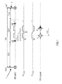
- Figure 1
- schematic illustration a conventional double-echo MP-RAGE image sequence.
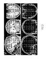
- Figure 2
- exemplary of fat-only (F) and water-only (W) images obtained according to the present invention.
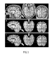
- Figure 3
- examples of skull-stripping results obtained by MR imaging of a target according to the present invention.
-
-
Figure 1 illustrates a preferred sequence of double-echo MP-RAGE for imaging a target according to the invention. The MP-RAGE sequence is chosen as an example since it is widely used to obtain T1-weighted image contrast. Other sequences might be used such as T1-weighted gradient echo (FLASH) or spin-echo-based sequences. The case of MP-RAGE depicted here shows an inversion radio frequency pulse 1 followed after a first delay time TD1 by a gradient echo readout train GRE performed through a period of time determined in function of a number of double-echo acquisitions to be performed. At the end of said period of time, the gradient echo readout GRE is succeeded by a second delay time TD2 which separates the end of the gradient echo readout GRE from the next inversion radio-frequency pulse 2. The first delay time TD1, the gradient echo readout GRE, and the second delay time form a block that is consecutively repeated, starting at each next inversion radio-frequency pulse, until k-space is fully sampled. The time between the two successive inversion radio-frequency pulses is characterized by the repetition time TR. Within the gradient echo readout train GRE, N double-echo acquisitions are performed. Each double-echo acquisition comprises a generation of an excitation radio frequency pulse α which is in particular a low-flip-angle excitation pulse,and a sampling of data during a gradient echo which is achieved by dephasing the spins with at least one readout gradient that follows said excitation radio frequency pulse α. After the excitation radio frequency pulse α, in particular said low-flip-angle excitation pulse, two echoes are formed using in particular either a monopolar readout gradient scheme GRO1 or a bipolar gradient scheme GRO2. Pursuant to the configured readout gradients, two echoes, respectively an in-phase echo E1 and an opposed-phase echo E2, are formed at specific echo times TE1 and TE2, where TE1, respectively TE2, is an amount of time separating each excitation radio frequency pulse α from the middle (half the moment) of a first and respectively of a second magnetic field gradient, i.e. said monopolar or bipolar gradient represented by the readout gradient scheme GRO1 or GRO2. To obtain in-phase and opposed phase fat and water signals, TE1 and TE2, and thus the readout gradients, have to be adjusted as described previously by correlating the second and first time of echo difference ΔTE = TE2-TE1 with a fat and water mutual resonance frequency difference δ. -
Figure 2 illustrates water-only images W and fat-only images F acquired according to Dixon technique, from which additional image information which improves skull-stripping results is extracted according to the invention. Such images are in particular used as additional input source into a multispectral analysis method according to the present invention. Indeed, the multispectral skull-stripping algorithm is able to extract data related to fat spatial position in the target and/or data related to water spatial position in the target arising notably from differences between the water-only W and the fat-only F images, and to use it as complementary information, especially for the skull-tissue boundary regions. -
Figure 3 exemplifies the differences in the skull-stripping results obtained by a multispectral skull-stripping algorithm with or without applying the method according to the invention. Indeed,Figure 3 presents different examples of skull-stripping results obtained by MR imaging of a target, whereinFig. 3A shows a fat and water combined image of said target,Fig. 3B shows state of the art skull-stripping results (i.e. onlyFig. 3A is used for skull-stripping) of said target, andFig. 3C shows images obtained for the same target by applying the claimed invention. Using the formulas I-IV, data of the two echoes E1 and E2 are combined to obtain a common T1-weighted contrast with optimized SNR. Using this alone as an input for the skull-stripping algorithm, suboptimal results are achieved (seeFig. 3B ). Exploiting also the fat image as complementary information in the algorithm ameliorates the outcome significantly (seeFig. 3C ). Arrows indicate areas of better separation, the invention particularly improves results in inferior-posterior regions. - Finally, in contrast to state of the art techniques, the present invention features the parallel acquisition of two image volumes; in a post-processing step, a standard T1-weighted contrast and an additional fat contrast depicting mainly undesired tissues is obtained. Both are subsequently used in a general multispectral analysis. Hence, the information available to the multispectral algorithm is augmented by additional information about the location of undesired, i.e. non-brain, parts of the image. The method according to the invention presents thus the following advantages compared to conventional skull-stripping procedures:
- it significantly improves the outcome of the skull-stripping procedure at negligible cost in terms of scan/processing time;
- compared to a monospectral (i.e. only one image) input, the multispectral skull-stripping algorithm can make use of the complementary information especially in the skull-tissue boundary regions;
- no prolongation of the overall scan time by employing a MP-RAGE sequence;
- intrinsic co-registration of the images yielded by the different echoes;
- simple computation of the water and fat image;
- no significant prolongation of post-processing times.
Claims (6)
- Method for removing at least one part of a MR brain image, said part representing non-brain tissue of the MR brain image, the method comprising the steps of:- generating a static magnetic field capable of aligning moments of nuclei of a target to be imaged with a direction parallel with said static magnetic field magnetic;- generating at least one excitation radio frequency pulse (α) capable of exciting atom nuclei of said target being magnetized by said static magnetic field, and capable of deflecting said moments of nuclei from said direction of the static magnetic field;- generating for a first and a second predefined time period at least a first and a second magnetic field gradient, each capable of magnetizing said atom nuclei of the target and each designed for determining positional or spatial information of the excited atom nuclei of the target, the first and the second magnetic field gradient being oppositely directed magnetic field gradients;- for each generated magnetic field gradient acquiring a current signal, respectively at a first time of echo TE1 after the excitation radio frequency pulse (α) and at a second time of echo TE2 after said excitation radio frequency pulse (α), by means of at least one induction coil designed for surrounding said target and capable of generating said current signal from the deflection of the nuclei moments according to induction;- reconstructing the MR brain image of an internal structure of the target from said current signal by means of computational processing of said current signal;
characterized in that- the first time of echo TE1 and the second time of echo TE2 are adjusted for correlating a second and first time of echo difference ΔTE =TE2-TE1 with a fat and water mutual resonance frequency difference δ, and in that a fat and a water information encoded in the current signal and resulting from said correlation of the second and first time of echo difference ΔTE with the fat and water mutual resonance frequency difference is used as additional input source into a multispectral analysis method for removing said at least one part representing non-brain tissue. - Method according to claim 1, characterized in that the multispectral analysis method comprises a data treatment by a multispectral skull-stripping algorithm.
- Method according to one of the claims 1 or 2, characterized by constructing a water-only and fat-only image from said fat and water information encoded in the current signal.
- Method according to claim 3, characterized in that each fat-only image is used for extracting data related to fat spatial position in the target.
- Method according to one of the claims 3 or 4, characterized in that each water-only image is used for extracting data related to water spatial position in the target.
- Method according to claims 4 to 5, characterized in that said data are inputted as said additional input source into said multispectral skull-stripping algorithm.
Priority Applications (2)
| Application Number | Priority Date | Filing Date | Title |
|---|---|---|---|
| EP11163001.8A EP2514360B1 (en) | 2011-04-19 | 2011-04-19 | Improved magnetic resonance imaging |
| US13/450,703 US8644905B2 (en) | 2011-04-19 | 2012-04-19 | Magnetic resonance imaging |
Applications Claiming Priority (1)
| Application Number | Priority Date | Filing Date | Title |
|---|---|---|---|
| EP11163001.8A EP2514360B1 (en) | 2011-04-19 | 2011-04-19 | Improved magnetic resonance imaging |
Publications (2)
| Publication Number | Publication Date |
|---|---|
| EP2514360A1 true EP2514360A1 (en) | 2012-10-24 |
| EP2514360B1 EP2514360B1 (en) | 2017-02-22 |
Family
ID=44534735
Family Applications (1)
| Application Number | Title | Priority Date | Filing Date |
|---|---|---|---|
| EP11163001.8A Not-in-force EP2514360B1 (en) | 2011-04-19 | 2011-04-19 | Improved magnetic resonance imaging |
Country Status (2)
| Country | Link |
|---|---|
| US (1) | US8644905B2 (en) |
| EP (1) | EP2514360B1 (en) |
Families Citing this family (1)
| Publication number | Priority date | Publication date | Assignee | Title |
|---|---|---|---|---|
| DE102015202062A1 (en) * | 2015-02-05 | 2016-08-11 | Siemens Healthcare Gmbh | Reconstruction of magnetic resonance image data for multiple chemical species in multi-echo imaging |
Citations (2)
| Publication number | Priority date | Publication date | Assignee | Title |
|---|---|---|---|---|
| US20060064004A1 (en) * | 2004-09-22 | 2006-03-23 | Kabushiki Kaisha Toshiba | Magnetic resonance imaging apparatus and magnetic resonance imaging method |
| US20080119721A1 (en) * | 2006-11-22 | 2008-05-22 | Kabushiki Kaisha Toshiba | Magnetic resonance imaging apparatus |
Family Cites Families (1)
| Publication number | Priority date | Publication date | Assignee | Title |
|---|---|---|---|---|
| DE102008057294B4 (en) * | 2008-11-14 | 2010-10-07 | Siemens Aktiengesellschaft | Separation of fat and water images according to the two-point Dixon method, taking into account the T * 2 decay |
-
2011
- 2011-04-19 EP EP11163001.8A patent/EP2514360B1/en not_active Not-in-force
-
2012
- 2012-04-19 US US13/450,703 patent/US8644905B2/en not_active Expired - Fee Related
Patent Citations (2)
| Publication number | Priority date | Publication date | Assignee | Title |
|---|---|---|---|---|
| US20060064004A1 (en) * | 2004-09-22 | 2006-03-23 | Kabushiki Kaisha Toshiba | Magnetic resonance imaging apparatus and magnetic resonance imaging method |
| US20080119721A1 (en) * | 2006-11-22 | 2008-05-22 | Kabushiki Kaisha Toshiba | Magnetic resonance imaging apparatus |
Non-Patent Citations (10)
| Title |
|---|
| DIXON WT, RADIOLOGY, vol. 153, no. 1, 1984, pages 189 |
| HAHN, PEITGEN, MICCAI 2000, vol. 134, 2000 |
| KEIHANINEJAD ET AL., 9TH EUROPEAN CONGRESS ON EPILEP- TOLOGY, vol. 51, 2010, pages 6 |
| KOEN VAN LEEMPUT * ET AL: "Automated Model-Based Tissue Classification of MR Images of the Brain", IEEE TRANSACTIONS ON MEDICAL IMAGING, IEEE SERVICE CENTER, PISCATAWAY, NJ, US, vol. 18, no. 10, 1 October 1999 (1999-10-01), XP011035913, ISSN: 0278-0062 * |
| S6GONNE ET AL., NEUROLMAGE, vol. 22, 2004, pages 1060 |
| SHATTUCK ET AL., NEUROLMAGE, vol. 13, 2001, pages 856 |
| SMITH, HUMAN BRAIN MAPPING, vol. 17, 2002, pages 143 |
| SURESH ET AL., NEUROLMAGE, vol. 49, 2010, pages 225 |
| VAN DER KOUWE ET AL., NEUROLMAGE, vol. 40, no. 2, 2008, pages 559 |
| VAN LEEMPUT ET AL., IEEE TRANSACTIONS ON MEDICAL IMAGING, vol. 18, 1999, pages 897 |
Also Published As
| Publication number | Publication date |
|---|---|
| EP2514360B1 (en) | 2017-02-22 |
| US20120271146A1 (en) | 2012-10-25 |
| US8644905B2 (en) | 2014-02-04 |
Similar Documents
| Publication | Publication Date | Title |
|---|---|---|
| US9977107B2 (en) | Atlas-free brain tissue segmentation method using a single T1-weighted MRI acquisition | |
| Massire et al. | High-resolution multi-parametric quantitative magnetic resonance imaging of the human cervical spinal cord at 7T | |
| CN102596026B (en) | MR imaging apparatus and MR imaging method | |
| US9488711B2 (en) | Magnetic resonance imaging apparatus and magnetic resonance imaging method | |
| EP2648014A1 (en) | MR imaging using APT contrast enhancement and sampling at multiple echo times | |
| EP3295194B1 (en) | Parallel mr imaging with rf coil sensitivity mapping | |
| CN101664309B (en) | Phase sensitive liquid suppression | |
| EP3201643B1 (en) | Magnetic resonance imaging with enhanced bone visualization | |
| CN103597370A (en) | Spatially encoded phase-contrast MRI | |
| US12019134B2 (en) | MR electric properties tomography without contrast agent | |
| CN105829906A (en) | Phase-Sensitive Inversion Recovery Mri With Water/Fat Separation | |
| US8587305B2 (en) | System and method for fat suppression in chemical shift based water-fat separated MR imaging | |
| EP2495579B1 (en) | Method for generating 2D or 3D maps of MRI T1 and T2 relaxation times | |
| CN107076819B (en) | Dixon MR imaging with suppression of flow artifacts | |
| JP7507793B2 (en) | Spiral mr imaging with off-resonance artifact correction - Patents.com | |
| Dai et al. | Distortion-free diffusion imaging using self-navigated Cartesian Echo-planar time resolved acquisition and joint magnitude and phase constrained reconstruction | |
| JPH11313810A5 (en) | MRI machine | |
| EP2514360B1 (en) | Improved magnetic resonance imaging | |
| WO2017040368A1 (en) | Multi-echo spin-, asymmetric spin-, and gradient-echo echo-planar imaging mri pulse sequence | |
| US10114095B2 (en) | Multi-spectral MRI scan with magnetization recovery | |
| EP3726239A1 (en) | Susceptibility-weighting from phase-contrast mr imaging | |
| EP4575541A1 (en) | Magnetic resonance imaging with multiple contrasts | |
| WO2024211423A1 (en) | Systems and methods for patterned multi-slice excitation in magnetic resonance imaging | |
| VERSION | Technical aspects of MR diffusion imaging of the body | |
| Zhu | Assessment of Tissue Micro-structure with Diffusion Weighted Magnetic Resonance Imaging |
Legal Events
| Date | Code | Title | Description |
|---|---|---|---|
| PUAI | Public reference made under article 153(3) epc to a published international application that has entered the european phase |
Free format text: ORIGINAL CODE: 0009012 |
|
| AK | Designated contracting states |
Kind code of ref document: A1 Designated state(s): AL AT BE BG CH CY CZ DE DK EE ES FI FR GB GR HR HU IE IS IT LI LT LU LV MC MK MT NL NO PL PT RO RS SE SI SK SM TR |
|
| AX | Request for extension of the european patent |
Extension state: BA ME |
|
| 17P | Request for examination filed |
Effective date: 20130319 |
|
| RAP1 | Party data changed (applicant data changed or rights of an application transferred) |
Owner name: UNIVERSITE DE LAUSANNE Owner name: SIEMENS AKTIENGESELLSCHAFT |
|
| RAP1 | Party data changed (applicant data changed or rights of an application transferred) |
Owner name: UNIVERSITE DE LAUSANNE Owner name: SIEMENS HEALTHCARE GMBH |
|
| GRAP | Despatch of communication of intention to grant a patent |
Free format text: ORIGINAL CODE: EPIDOSNIGR1 |
|
| INTG | Intention to grant announced |
Effective date: 20160915 |
|
| STAA | Information on the status of an ep patent application or granted ep patent |
Free format text: STATUS: GRANT OF PATENT IS INTENDED |
|
| GRAS | Grant fee paid |
Free format text: ORIGINAL CODE: EPIDOSNIGR3 |
|
| GRAA | (expected) grant |
Free format text: ORIGINAL CODE: 0009210 |
|
| STAA | Information on the status of an ep patent application or granted ep patent |
Free format text: STATUS: THE PATENT HAS BEEN GRANTED |
|
| AK | Designated contracting states |
Kind code of ref document: B1 Designated state(s): AL AT BE BG CH CY CZ DE DK EE ES FI FR GB GR HR HU IE IS IT LI LT LU LV MC MK MT NL NO PL PT RO RS SE SI SK SM TR |
|
| REG | Reference to a national code |
Ref country code: GB Ref legal event code: FG4D |
|
| REG | Reference to a national code |
Ref country code: CH Ref legal event code: EP |
|
| REG | Reference to a national code |
Ref country code: AT Ref legal event code: REF Ref document number: 868642 Country of ref document: AT Kind code of ref document: T Effective date: 20170315 |
|
| REG | Reference to a national code |
Ref country code: IE Ref legal event code: FG4D |
|
| REG | Reference to a national code |
Ref country code: DE Ref legal event code: R096 Ref document number: 602011035156 Country of ref document: DE |
|
| REG | Reference to a national code |
Ref country code: NL Ref legal event code: FP |
|
| REG | Reference to a national code |
Ref country code: LT Ref legal event code: MG4D |
|
| REG | Reference to a national code |
Ref country code: AT Ref legal event code: MK05 Ref document number: 868642 Country of ref document: AT Kind code of ref document: T Effective date: 20170222 |
|
| PG25 | Lapsed in a contracting state [announced via postgrant information from national office to epo] |
Ref country code: NO Free format text: LAPSE BECAUSE OF FAILURE TO SUBMIT A TRANSLATION OF THE DESCRIPTION OR TO PAY THE FEE WITHIN THE PRESCRIBED TIME-LIMIT Effective date: 20170522 Ref country code: LT Free format text: LAPSE BECAUSE OF FAILURE TO SUBMIT A TRANSLATION OF THE DESCRIPTION OR TO PAY THE FEE WITHIN THE PRESCRIBED TIME-LIMIT Effective date: 20170222 Ref country code: HR Free format text: LAPSE BECAUSE OF FAILURE TO SUBMIT A TRANSLATION OF THE DESCRIPTION OR TO PAY THE FEE WITHIN THE PRESCRIBED TIME-LIMIT Effective date: 20170222 Ref country code: GR Free format text: LAPSE BECAUSE OF FAILURE TO SUBMIT A TRANSLATION OF THE DESCRIPTION OR TO PAY THE FEE WITHIN THE PRESCRIBED TIME-LIMIT Effective date: 20170523 Ref country code: FI Free format text: LAPSE BECAUSE OF FAILURE TO SUBMIT A TRANSLATION OF THE DESCRIPTION OR TO PAY THE FEE WITHIN THE PRESCRIBED TIME-LIMIT Effective date: 20170222 |
|
| PG25 | Lapsed in a contracting state [announced via postgrant information from national office to epo] |
Ref country code: RS Free format text: LAPSE BECAUSE OF FAILURE TO SUBMIT A TRANSLATION OF THE DESCRIPTION OR TO PAY THE FEE WITHIN THE PRESCRIBED TIME-LIMIT Effective date: 20170222 Ref country code: LV Free format text: LAPSE BECAUSE OF FAILURE TO SUBMIT A TRANSLATION OF THE DESCRIPTION OR TO PAY THE FEE WITHIN THE PRESCRIBED TIME-LIMIT Effective date: 20170222 Ref country code: SE Free format text: LAPSE BECAUSE OF FAILURE TO SUBMIT A TRANSLATION OF THE DESCRIPTION OR TO PAY THE FEE WITHIN THE PRESCRIBED TIME-LIMIT Effective date: 20170222 Ref country code: BG Free format text: LAPSE BECAUSE OF FAILURE TO SUBMIT A TRANSLATION OF THE DESCRIPTION OR TO PAY THE FEE WITHIN THE PRESCRIBED TIME-LIMIT Effective date: 20170522 Ref country code: AT Free format text: LAPSE BECAUSE OF FAILURE TO SUBMIT A TRANSLATION OF THE DESCRIPTION OR TO PAY THE FEE WITHIN THE PRESCRIBED TIME-LIMIT Effective date: 20170222 Ref country code: ES Free format text: LAPSE BECAUSE OF FAILURE TO SUBMIT A TRANSLATION OF THE DESCRIPTION OR TO PAY THE FEE WITHIN THE PRESCRIBED TIME-LIMIT Effective date: 20170222 Ref country code: PT Free format text: LAPSE BECAUSE OF FAILURE TO SUBMIT A TRANSLATION OF THE DESCRIPTION OR TO PAY THE FEE WITHIN THE PRESCRIBED TIME-LIMIT Effective date: 20170622 |
|
| PG25 | Lapsed in a contracting state [announced via postgrant information from national office to epo] |
Ref country code: IT Free format text: LAPSE BECAUSE OF FAILURE TO SUBMIT A TRANSLATION OF THE DESCRIPTION OR TO PAY THE FEE WITHIN THE PRESCRIBED TIME-LIMIT Effective date: 20170222 Ref country code: RO Free format text: LAPSE BECAUSE OF FAILURE TO SUBMIT A TRANSLATION OF THE DESCRIPTION OR TO PAY THE FEE WITHIN THE PRESCRIBED TIME-LIMIT Effective date: 20170222 Ref country code: SK Free format text: LAPSE BECAUSE OF FAILURE TO SUBMIT A TRANSLATION OF THE DESCRIPTION OR TO PAY THE FEE WITHIN THE PRESCRIBED TIME-LIMIT Effective date: 20170222 Ref country code: CZ Free format text: LAPSE BECAUSE OF FAILURE TO SUBMIT A TRANSLATION OF THE DESCRIPTION OR TO PAY THE FEE WITHIN THE PRESCRIBED TIME-LIMIT Effective date: 20170222 Ref country code: EE Free format text: LAPSE BECAUSE OF FAILURE TO SUBMIT A TRANSLATION OF THE DESCRIPTION OR TO PAY THE FEE WITHIN THE PRESCRIBED TIME-LIMIT Effective date: 20170222 |
|
| REG | Reference to a national code |
Ref country code: DE Ref legal event code: R097 Ref document number: 602011035156 Country of ref document: DE |
|
| PG25 | Lapsed in a contracting state [announced via postgrant information from national office to epo] |
Ref country code: PL Free format text: LAPSE BECAUSE OF FAILURE TO SUBMIT A TRANSLATION OF THE DESCRIPTION OR TO PAY THE FEE WITHIN THE PRESCRIBED TIME-LIMIT Effective date: 20170222 Ref country code: SM Free format text: LAPSE BECAUSE OF FAILURE TO SUBMIT A TRANSLATION OF THE DESCRIPTION OR TO PAY THE FEE WITHIN THE PRESCRIBED TIME-LIMIT Effective date: 20170222 Ref country code: DK Free format text: LAPSE BECAUSE OF FAILURE TO SUBMIT A TRANSLATION OF THE DESCRIPTION OR TO PAY THE FEE WITHIN THE PRESCRIBED TIME-LIMIT Effective date: 20170222 |
|
| REG | Reference to a national code |
Ref country code: CH Ref legal event code: PL |
|
| PLBE | No opposition filed within time limit |
Free format text: ORIGINAL CODE: 0009261 |
|
| STAA | Information on the status of an ep patent application or granted ep patent |
Free format text: STATUS: NO OPPOSITION FILED WITHIN TIME LIMIT |
|
| GBPC | Gb: european patent ceased through non-payment of renewal fee |
Effective date: 20170522 |
|
| REG | Reference to a national code |
Ref country code: IE Ref legal event code: MM4A |
|
| REG | Reference to a national code |
Ref country code: FR Ref legal event code: ST Effective date: 20171229 |
|
| 26N | No opposition filed |
Effective date: 20171123 |
|
| PG25 | Lapsed in a contracting state [announced via postgrant information from national office to epo] |
Ref country code: FR Free format text: LAPSE BECAUSE OF NON-PAYMENT OF DUE FEES Effective date: 20170502 Ref country code: MC Free format text: LAPSE BECAUSE OF FAILURE TO SUBMIT A TRANSLATION OF THE DESCRIPTION OR TO PAY THE FEE WITHIN THE PRESCRIBED TIME-LIMIT Effective date: 20170222 |
|
| PG25 | Lapsed in a contracting state [announced via postgrant information from national office to epo] |
Ref country code: SI Free format text: LAPSE BECAUSE OF FAILURE TO SUBMIT A TRANSLATION OF THE DESCRIPTION OR TO PAY THE FEE WITHIN THE PRESCRIBED TIME-LIMIT Effective date: 20170222 Ref country code: LI Free format text: LAPSE BECAUSE OF NON-PAYMENT OF DUE FEES Effective date: 20170430 Ref country code: CH Free format text: LAPSE BECAUSE OF NON-PAYMENT OF DUE FEES Effective date: 20170430 Ref country code: LU Free format text: LAPSE BECAUSE OF NON-PAYMENT OF DUE FEES Effective date: 20170419 |
|
| REG | Reference to a national code |
Ref country code: BE Ref legal event code: MM Effective date: 20170430 |
|
| PG25 | Lapsed in a contracting state [announced via postgrant information from national office to epo] |
Ref country code: IE Free format text: LAPSE BECAUSE OF NON-PAYMENT OF DUE FEES Effective date: 20170419 Ref country code: GB Free format text: LAPSE BECAUSE OF NON-PAYMENT OF DUE FEES Effective date: 20170522 |
|
| PG25 | Lapsed in a contracting state [announced via postgrant information from national office to epo] |
Ref country code: BE Free format text: LAPSE BECAUSE OF NON-PAYMENT OF DUE FEES Effective date: 20170430 |
|
| PG25 | Lapsed in a contracting state [announced via postgrant information from national office to epo] |
Ref country code: MT Free format text: LAPSE BECAUSE OF NON-PAYMENT OF DUE FEES Effective date: 20170419 |
|
| REG | Reference to a national code |
Ref country code: DE Ref legal event code: R082 Ref document number: 602011035156 Country of ref document: DE Representative=s name: MAIER, DANIEL OLIVER, DIPL.-ING. UNIV., DE |
|
| PG25 | Lapsed in a contracting state [announced via postgrant information from national office to epo] |
Ref country code: HU Free format text: LAPSE BECAUSE OF FAILURE TO SUBMIT A TRANSLATION OF THE DESCRIPTION OR TO PAY THE FEE WITHIN THE PRESCRIBED TIME-LIMIT; INVALID AB INITIO Effective date: 20110419 |
|
| PGFP | Annual fee paid to national office [announced via postgrant information from national office to epo] |
Ref country code: NL Payment date: 20190408 Year of fee payment: 9 |
|
| PGFP | Annual fee paid to national office [announced via postgrant information from national office to epo] |
Ref country code: DE Payment date: 20190617 Year of fee payment: 9 |
|
| PG25 | Lapsed in a contracting state [announced via postgrant information from national office to epo] |
Ref country code: CY Free format text: LAPSE BECAUSE OF NON-PAYMENT OF DUE FEES Effective date: 20170222 |
|
| PG25 | Lapsed in a contracting state [announced via postgrant information from national office to epo] |
Ref country code: MK Free format text: LAPSE BECAUSE OF FAILURE TO SUBMIT A TRANSLATION OF THE DESCRIPTION OR TO PAY THE FEE WITHIN THE PRESCRIBED TIME-LIMIT Effective date: 20170222 |
|
| PG25 | Lapsed in a contracting state [announced via postgrant information from national office to epo] |
Ref country code: TR Free format text: LAPSE BECAUSE OF FAILURE TO SUBMIT A TRANSLATION OF THE DESCRIPTION OR TO PAY THE FEE WITHIN THE PRESCRIBED TIME-LIMIT Effective date: 20170222 |
|
| PG25 | Lapsed in a contracting state [announced via postgrant information from national office to epo] |
Ref country code: AL Free format text: LAPSE BECAUSE OF FAILURE TO SUBMIT A TRANSLATION OF THE DESCRIPTION OR TO PAY THE FEE WITHIN THE PRESCRIBED TIME-LIMIT Effective date: 20170222 Ref country code: IS Free format text: LAPSE BECAUSE OF FAILURE TO SUBMIT A TRANSLATION OF THE DESCRIPTION OR TO PAY THE FEE WITHIN THE PRESCRIBED TIME-LIMIT Effective date: 20170622 |
|
| REG | Reference to a national code |
Ref country code: DE Ref legal event code: R119 Ref document number: 602011035156 Country of ref document: DE |
|
| REG | Reference to a national code |
Ref country code: NL Ref legal event code: MM Effective date: 20200501 |
|
| PG25 | Lapsed in a contracting state [announced via postgrant information from national office to epo] |
Ref country code: DE Free format text: LAPSE BECAUSE OF NON-PAYMENT OF DUE FEES Effective date: 20201103 |
|
| PG25 | Lapsed in a contracting state [announced via postgrant information from national office to epo] |
Ref country code: NL Free format text: LAPSE BECAUSE OF NON-PAYMENT OF DUE FEES Effective date: 20200501 |