EP2512511B1 - Humanized antibodies against human il-22ra - Google Patents
Humanized antibodies against human il-22ra Download PDFInfo
- Publication number
- EP2512511B1 EP2512511B1 EP10779768.0A EP10779768A EP2512511B1 EP 2512511 B1 EP2512511 B1 EP 2512511B1 EP 10779768 A EP10779768 A EP 10779768A EP 2512511 B1 EP2512511 B1 EP 2512511B1
- Authority
- EP
- European Patent Office
- Prior art keywords
- human
- humanized antibody
- seq
- humanized
- amino acid
- Prior art date
- Legal status (The legal status is an assumption and is not a legal conclusion. Google has not performed a legal analysis and makes no representation as to the accuracy of the status listed.)
- Active
Links
Images


Classifications
-
- C—CHEMISTRY; METALLURGY
- C07—ORGANIC CHEMISTRY
- C07K—PEPTIDES
- C07K16/00—Immunoglobulins [IGs], e.g. monoclonal or polyclonal antibodies
- C07K16/18—Immunoglobulins [IGs], e.g. monoclonal or polyclonal antibodies against material from animals or humans
- C07K16/28—Immunoglobulins [IGs], e.g. monoclonal or polyclonal antibodies against material from animals or humans against receptors, cell surface antigens or cell surface determinants
- C07K16/2866—Immunoglobulins [IGs], e.g. monoclonal or polyclonal antibodies against material from animals or humans against receptors, cell surface antigens or cell surface determinants against receptors for cytokines, lymphokines, interferons
-
- A—HUMAN NECESSITIES
- A61—MEDICAL OR VETERINARY SCIENCE; HYGIENE
- A61K—PREPARATIONS FOR MEDICAL, DENTAL OR TOILETRY PURPOSES
- A61K39/00—Medicinal preparations containing antigens or antibodies
- A61K39/395—Antibodies; Immunoglobulins; Immune serum, e.g. antilymphocytic serum
-
- A—HUMAN NECESSITIES
- A61—MEDICAL OR VETERINARY SCIENCE; HYGIENE
- A61P—SPECIFIC THERAPEUTIC ACTIVITY OF CHEMICAL COMPOUNDS OR MEDICINAL PREPARATIONS
- A61P17/00—Drugs for dermatological disorders
-
- A—HUMAN NECESSITIES
- A61—MEDICAL OR VETERINARY SCIENCE; HYGIENE
- A61P—SPECIFIC THERAPEUTIC ACTIVITY OF CHEMICAL COMPOUNDS OR MEDICINAL PREPARATIONS
- A61P17/00—Drugs for dermatological disorders
- A61P17/06—Antipsoriatics
-
- A—HUMAN NECESSITIES
- A61—MEDICAL OR VETERINARY SCIENCE; HYGIENE
- A61P—SPECIFIC THERAPEUTIC ACTIVITY OF CHEMICAL COMPOUNDS OR MEDICINAL PREPARATIONS
- A61P19/00—Drugs for skeletal disorders
- A61P19/02—Drugs for skeletal disorders for joint disorders, e.g. arthritis, arthrosis
-
- A—HUMAN NECESSITIES
- A61—MEDICAL OR VETERINARY SCIENCE; HYGIENE
- A61P—SPECIFIC THERAPEUTIC ACTIVITY OF CHEMICAL COMPOUNDS OR MEDICINAL PREPARATIONS
- A61P37/00—Drugs for immunological or allergic disorders
-
- A—HUMAN NECESSITIES
- A61—MEDICAL OR VETERINARY SCIENCE; HYGIENE
- A61P—SPECIFIC THERAPEUTIC ACTIVITY OF CHEMICAL COMPOUNDS OR MEDICINAL PREPARATIONS
- A61P37/00—Drugs for immunological or allergic disorders
- A61P37/08—Antiallergic agents
-
- C—CHEMISTRY; METALLURGY
- C12—BIOCHEMISTRY; BEER; SPIRITS; WINE; VINEGAR; MICROBIOLOGY; ENZYMOLOGY; MUTATION OR GENETIC ENGINEERING
- C12N—MICROORGANISMS OR ENZYMES; COMPOSITIONS THEREOF; PROPAGATING, PRESERVING, OR MAINTAINING MICROORGANISMS; MUTATION OR GENETIC ENGINEERING; CULTURE MEDIA
- C12N15/00—Mutation or genetic engineering; DNA or RNA concerning genetic engineering, vectors, e.g. plasmids, or their isolation, preparation or purification; Use of hosts therefor
- C12N15/09—Recombinant DNA-technology
- C12N15/11—DNA or RNA fragments; Modified forms thereof; Non-coding nucleic acids having a biological activity
-
- A—HUMAN NECESSITIES
- A61—MEDICAL OR VETERINARY SCIENCE; HYGIENE
- A61K—PREPARATIONS FOR MEDICAL, DENTAL OR TOILETRY PURPOSES
- A61K39/00—Medicinal preparations containing antigens or antibodies
- A61K2039/505—Medicinal preparations containing antigens or antibodies comprising antibodies
-
- C—CHEMISTRY; METALLURGY
- C07—ORGANIC CHEMISTRY
- C07K—PEPTIDES
- C07K2317/00—Immunoglobulins specific features
- C07K2317/20—Immunoglobulins specific features characterized by taxonomic origin
- C07K2317/24—Immunoglobulins specific features characterized by taxonomic origin containing regions, domains or residues from different species, e.g. chimeric, humanized or veneered
-
- C—CHEMISTRY; METALLURGY
- C07—ORGANIC CHEMISTRY
- C07K—PEPTIDES
- C07K2317/00—Immunoglobulins specific features
- C07K2317/50—Immunoglobulins specific features characterized by immunoglobulin fragments
- C07K2317/56—Immunoglobulins specific features characterized by immunoglobulin fragments variable (Fv) region, i.e. VH and/or VL
- C07K2317/565—Complementarity determining region [CDR]
-
- C—CHEMISTRY; METALLURGY
- C07—ORGANIC CHEMISTRY
- C07K—PEPTIDES
- C07K2317/00—Immunoglobulins specific features
- C07K2317/50—Immunoglobulins specific features characterized by immunoglobulin fragments
- C07K2317/56—Immunoglobulins specific features characterized by immunoglobulin fragments variable (Fv) region, i.e. VH and/or VL
- C07K2317/567—Framework region [FR]
-
- C—CHEMISTRY; METALLURGY
- C07—ORGANIC CHEMISTRY
- C07K—PEPTIDES
- C07K2317/00—Immunoglobulins specific features
- C07K2317/70—Immunoglobulins specific features characterized by effect upon binding to a cell or to an antigen
- C07K2317/73—Inducing cell death, e.g. apoptosis, necrosis or inhibition of cell proliferation
-
- C—CHEMISTRY; METALLURGY
- C07—ORGANIC CHEMISTRY
- C07K—PEPTIDES
- C07K2317/00—Immunoglobulins specific features
- C07K2317/70—Immunoglobulins specific features characterized by effect upon binding to a cell or to an antigen
- C07K2317/76—Antagonist effect on antigen, e.g. neutralization or inhibition of binding
-
- C—CHEMISTRY; METALLURGY
- C07—ORGANIC CHEMISTRY
- C07K—PEPTIDES
- C07K2317/00—Immunoglobulins specific features
- C07K2317/90—Immunoglobulins specific features characterized by (pharmaco)kinetic aspects or by stability of the immunoglobulin
- C07K2317/92—Affinity (KD), association rate (Ka), dissociation rate (Kd) or EC50 value
Definitions
- the present invention relates to humanized antibodies against human IL-22RA and to their use in the treatment of psoriasis and other immune-mediated diseases such as psoriatic arthritis and atopic dermatitis.
- IL-22RA (also known as IL22R, IL22R1, IL22RA1, CRF2-9 and Zcytor11) belongs to the type II cytokine receptor family and is a component of the receptor for IL-20, IL-22 and IL-24. Due to their structural similarity IL-20, IL-22 and IL-24, together with IL-19 and IL-26, were combined with IL-10 in the so-called "IL-10 family" (Kunz S et al. 2006). IL-10 is a master regulator of the immune response that mediates down-regulation of pro-inflammatory cytokine expression in macrophages, T cells, and other cells of the immune system (Moore KW et al. 2001).
- IL-20 and IL-24 are produced not only by activated immune cells, but also to a similar extent by keratinocytes. In vivo, these cytokines are expressed preferentially in the inflamed tissues. IL-20 and IL-24 can signal through two receptor complexes, IL-20RA/IL-20RB and IL-22RA/IL-20RB (Langer JA et al. 2004). Several tissues, particularly the skin, tissues from the reproductive and respiratory systems, and various glands appeared to be the main targets of these mediators (Kunz S et al. 2006).
- IL-22 was discovered as a gene up-regulated by CD4 + T cells upon activation and it shares 22% amino acid sequence identity with IL-10; it was, thus, originally named IL-10-related T cell-derived inducible factor (IL-TIF) (Dumoutier L et al. 2000). Unlike IL-10, which regulates immune cell functions, IL-22 controls tissue responses to the immune system. IL-22 signals through a heterodimer receptor formed by IL-22RA and IL-10RB which is highly expressed within various tissues but it is not detectable on immune cells.
- IL-TIF IL-10-related T cell-derived inducible factor
- IL-22 binds via its IL-22RA binding site to the extracellular domain of IL-22RA and, subsequently, IL-10RB binds to a region created by the interaction of IL-22 and IL-22RA to form a cytokine receptor complex with a higher affinity for IL-22 (Li J et al. 2004). Since IL-10RB is broadly expressed by many different cell types, IL-22RA expression is the limiting component that determines IL-22 responsiveness of cells. IL-22RA is expressed strongly in the liver, as well as in the skin, lungs, pancreas and other peripheral tissues (Wolk K et al. 2004; Aggarwal S et al. 2001). Extensive screening of different cell lines has revealed that only cells which express IL-22RA respond to IL-22, suggesting that there is no alternate receptor that can mediate IL-22 signaling.
- IL-22BP IL-22 binding protein
- IL-22R-alpha2 IL-22R-alpha2
- CRF2X CRF2-S1 and CRF2-10
- IL-22BP has been found in diseased tissues from patients with different chronic inflammatory diseases that involve infiltrating activated T cells, such as psoriasis, psoriatic arthritis and atopic dermatitis.
- IL-22 has been most commonly described as a pro-inflammatory cytokine because of its expression in lesions of patients with chronic inflammatory diseases and its induction of pro-inflammatory cytokines such as IL-6, IL-8 and TNF- ⁇ (Wolk K et al. 2004; Andoh A et al. 2005; Ikeuchi H et al. 2005; Nograles KE et al. 2009a.; Nograles KE et al. 2009b).
- Zheng et al. showed that IL-22 is important for mediating IL-23-induced dermal inflammation in a mouse model of psoriasis, indicating a pro-inflammatory role (Zheng Y et al. 2007).
- IL-22 Given the biological effects of IL-22, including keratinocyte hyperplasia, induction of chemokine and pro-inflammatory cytokine production in certain tissue, the use of antagonists that block, inhibit, reduce or neutralize the activity of IL-22, e.g. by interfering with the receptor binding, may prevent infiltration of pathogenic cells at inflammatory sites.
- Mouse anti-human IL-22RA monoclonal antibodies have been previously described in PCT patent application WO 2006/047249 filed on October 21, 2005 . However, mouse antibodies may cause immunogenicity and humanized anti-human IL-22RA antibodies are desirable.
- Humanized antibodies generally have at least three potential advantages over mouse antibodies for use in human therapy: (1) because the effector portion is human, it may interact better with the other parts of the human immune system (e.g., destroy the target cells more efficiently by complement-dependent cytotoxicity (CDC) or antibody-dependent cellular cytotoxicity (ADCC)); (2) the human immune system should not recognize the framework or constant region of the humanized antibody as foreign, and therefore the antibody response against such an injected antibody should be less than against a totally foreign mouse antibody; and (3) injected mouse antibodies have been reported to have a half-life in the human circulation much shorter than the half-life of human antibodies. Injected humanized antibodies will presumably have a half-life more similar to naturally occurring human antibodies, allowing smaller and less frequent doses to be given. Thus, in view of the above, there is a need for humanized anti-human IL-22RA antibodies for treating IL-22 mediated inflammation, such as psoriasis, psoriatic arthritis and atopic dermatitis.
- CDC complement-dependent cyto
- the invention provides a humanized antibody that binds to human IL-22RA.
- the humanized antibody of the invention comprises a) a heavy chain variable domain comprising H-CDR1 , H-CDR2, and H-CDR3 consisting of amino acid sequences of SEQ ID NO: 1, 2 and 3, respectively and b) a light chain variable domain comprising L-CDR1 , L-CDR2, and L-CDR3 consisting of amino acid sequences of SEQ ID NO: 4, 5 and 6, respectively or consisting of amino acid sequences of SEQ ID NO: 4, 5 and 7, respectively.
- the invention provides an antibody described herein, wherein a) said heavy chain variable domain comprises framework regions H-FR1, H-FR2, H-FR3 and H-FR4 consisting of amino acid sequences of SEQ ID NO: 8, 9, 10 and 11, respectively and b) said light chain variable domain comprises framework regions L-FR1, L-FR2, L-FR3 and L-FR4 consisting of amino acid sequences of SEQ ID NO: 12, 13, 14 and 15, respectively.
- the invention provides an antibody described herein, wherein a) said heavy chain variable domain consists of amino acid sequence of SEQ ID NO: 16 and b) said light chain variable domain consists of amino acid sequence of SEQ ID NO: 17.
- said antibody comprises a) a heavy chain constant region consisting of amino acid sequence of SEQ ID NO: 18 and b) a light chain constant domain consisting of amino acid sequence of SEQ ID NO: 19.
- the invention provides a humanized antibody that binds to human IL-22RA which comprises a heavy chain comprising or consisting of amino acid sequence of SEQ ID NO: 20 and a light chain comprising or consisting of amino acid sequence of SEQ ID NO: 21.
- polynucleotide e.g. a DNA, encoding the heavy chain of the humanized antibody according to the present invention.
- said polynucleotide comprises or consists of SEQ ID NO: 22.
- polynucleotide e.g. a DNA, encoding the light chain of the humanized antibody according to the present invention.
- said polynucleotide comprises or consists of SEQ ID NO: 23.
- the invention provides a polynucleotide, e.g. a DNA, encoding both the heavy and the light chains of the humanized antibody according to the present invention.
- the invention provides a vector and more particularly an expression vector comprising a) a polynucleotide encoding the heavy chain of the humanized antibody according to the present invention and b) a polynucleotide encoding the light chain of the humanized antibody according to the present invention.
- the invention provides a vector and more particularly an expression vector comprising a polynucleotide encoding the heavy chain and the light chain of the humanized antibody according to the present invention.
- the invention provides a host cell, preferably a CHO cell, comprising, e.g. as a result of a transfection, a vector and in particular an expression vector according to the invention.
- the invention provides a method of producing a humanized antibody according to the invention, the method comprising culturing a host cell, preferably a CHO cell, according to the invention and isolating the humanized antibody according to the present invention.
- the invention provides a humanized antibody according to the present invention for use as a medicament, in particular for use in the treatment of psoriasis, psoriatic arthritis or atopic dermatitis.
- the invention provides a pharmaceutical composition comprising a humanized antibody according to the present invention and its use as a medicament, in particular for use in the treatment of psoriasis, psoriatic arthritis or atopic dermatitis.
- the invention provides for the use of a humanized antibody according to the present invention or of a pharmaceutical composition comprising said antibody in the manufacture of a medicament for the treatment of psoriasis, psoriatic arthritis or atopic dermatitis.
- antibody includes, inter alia, polyclonal antibodies, affinity-purified polyclonal antibodies, monoclonal antibodies, nanobodies and antigen-binding fragments, such as F(ab') 2 , Fab proteolytic fragments, and single chain variable region fragments (scFvs). Genetically engineered intact antibodies or fragments, such as chimeric antibodies, Fv fragments, single chain antibodies and the like, as well as synthetic antigen-binding peptides and polypeptides, are also included.
- Non-human antibodies may be humanized by grafting non-human CDRs onto human framework and constant regions, or by incorporating the entire non-human variable domains. In some instances, humanized antibodies may retain non-human residues within the human framework regions to enhance proper binding characteristics. Through humanizing antibodies, biological half-life may be increased, and the potential for adverse immune reactions upon administration to humans is reduced.
- immunoglobulin refers to a protein consisting of one or more polypeptides substantially encoded by immunoglobulin genes.
- One form of immunoglobulin constitutes the basic structural unit of an antibody. This form is a tetramer and consists of two identical pairs of immunoglobulin chains, each pair having one light and one heavy chain.
- a light chain has two parts: the variable domain (VL) and the constant domain (CL), which in the context of a light chain can be called constant region as well.
- a heavy chain has two parts as well: the variable domain (VH) and the constant region (CH). In each pair, the light and heavy chain variable domains are together responsible for binding to an antigen, and the constant regions are responsible for the antibody effector functions.
- Full-length immunoglobulin "light chains” (about 25 Kd) are encoded by a variable domain gene at the N-terminus (about 110 amino acids) and a kappa or lambda constant domain (C ⁇ and C ⁇ , respectively) gene at the C-terminus.
- Full-length immunoglobulin "heavy chains” (about 50 Kd), are similarly encoded by a variable domain gene (about 116 amino acids) and one of the other constant region genes (about 330 amino acids) mentioned hereinafter.
- the type of heavy chain defines the antibody's isotype as IgA, IgD, IgE, IgG and IgM, respectively.
- the constant region is identical in all antibodies of the same isotype, but differs in antibodies of different isotypes.
- Heavy chains ⁇ , ⁇ and ⁇ have a constant region composed of three Ig constant domains (C H 1, C H 2, and C H 3), and a hinge region for added flexibility; heavy chains ⁇ and ⁇ have a constant region composed of four Ig constant domains (C H 1, C H 2, C H 3, and C H 4) and a hinge region.
- An immunoglobulin light or heavy chain variable domain consists of a "framework" region interrupted by three hypervariable regions.
- hypervariable region refers to the amino acid residues of an antibody which are responsible for antigen binding.
- the hypervariable region comprises amino acid residues from a "Complementarity Determining Region” or "CDR", i.e. L-CDR1, L-CDR2 and L-CDR3 in the light chain variable domain and H-CDR1, H-CDR2 and H-CDR3 in the heavy chain variable domain (Kabat et al. 1991) and/or those residues from a "hypervariable loop" (Chothia and Lesk, 1987).
- CDR Complementarity Determining Region
- Framework Region or "FR” residues are those variable domain residues other than the hypervariable region residues as herein defined.
- the sequences of the framework regions of different light (i.e. L-FR1, L-FR2, L-FR3 and L-FR4) or heavy (i.e. H-FR1, H-FR2, H-FR3 and H-FR4) chains are relatively conserved within a species.
- a "human framework region” is a framework region that is substantially identical (about 85% or more, usually 90-95% or more) to the framework region of a naturally occurring human immunoglobulin.
- the framework region of an antibody that is the combined framework regions of the constituent light and heavy chains, serves to position and align the CDRs. The CDRs are primarily responsible for binding to an epitope of an antigen.
- humanized immunoglobulin refers to an immunoglobulin comprising a human framework region and one or more CDRs from a non-human (usually a mouse or rat) immunoglobulin.
- the non-human immunoglobulin providing the CDRs is called the "donor” and the human immunoglobulin providing the framework is called the "acceptor”.
- Constant regions need not be present, but if they are, they must be substantially identical to human immunoglobulin constant regions, i.e., at least about 85-90%, preferably about 95% or more identical.
- humanized immunoglobulin all parts of a humanized immunoglobulin, except possibly the CDRs and few residues in the heavy chain constant region if modulation of the effector functions is needed, are substantially identical to corresponding parts of natural human immunoglobulin sequences.
- a "humanized antibody” is an antibody comprising a humanized light chain variable domain and a humanized heavy chain variable domain.
- humanized antibodies may retain non-human residues within the human framework regions to enhance proper binding characteristics and/or some amino acid mutations may be introduced within the CDRs in order to improve the binding affinity and/or to reduce the immunogenicity and/or to increase the degree of humanness.
- recombinant antibodies means antibodies wherein the amino acid sequence has been varied from that of a native antibody. Because of the relevance of recombinant DNA techniques in the generation of antibodies, one need not be confined to the sequences of amino acids found in natural antibodies; antibodies can be redesigned to obtain desired characteristics. The possible variations are many and range from the changing of just one or a few amino acids to the complete redesign of, for example, the variable domain or constant region. Changes in the constant region will, in general, be made in order to improve, reduce or alter characteristics, such as complement fixation (e.g. complement dependent cytotoxicity, CDC), interaction with membranes and other effector functions (e.g. antibody dependent cellular cytotoxicity, ADCC). Changes in the variable domain will be made in order to improve the antigen binding characteristics.
- complement fixation e.g. complement dependent cytotoxicity, CDC
- ADCC antibody dependent cellular cytotoxicity
- immunoglobulins may exist in a variety of other forms including, for example, single-chain or Fv, Fab, and (Fab') 2 , as well as diabodies, linear antibodies, multivalent or multispecific hybrid antibodies.
- single-chain Fv single-chain antibodies
- Fv single-chain antibodies
- scFv single-chain antibody
- single-chain antibodies can also be bi-specific and/or humanized.
- a "Fab fragment” is comprised of one light chain and the variable and C H 1 domains of one heavy chain. The heavy chain of a Fab molecule cannot form a disulfide bond with another heavy chain molecule.
- a "Fab' fragment” contains one light chain and one heavy chain that contains more of the constant region, between the C H 1 and C H 2 domains, such that an interchain disulfide bond can be formed between two heavy chains to form a F(ab') 2 molecule.
- a "F(ab') 2" contains two light chains and two heavy chains containing a portion of the constant region between the C H 1 and C H 2 domains, such that an interchain disulfide bond is formed between two heavy chains.
- the present invention is based upon the discovery of humanized anti-human IL-22RA antibodies.
- Use of these antibodies as antagonists to IL-22RA can inhibit inflammation and, therefore, can be useful in the treatment of chronic inflammatory diseases that involve infiltrating activated T cells, such as psoriasis, psoriatic arthritis and atopic dermatitis.
- the invention provides the use of humanized antibodies that recognize, bind, modulate and/or neutralize the IL-22RA.
- the invention provides the use of humanized light and heavy chain variable domains that recognize, bind, modulate and/or neutralize the IL-22RA.
- Such humanized light and heavy chain variable domains can be fused, respectively, to a kappa or lambda constant domain and to a constant region of an heavy chain chosen among any isotype (IgA, IgD, IgE, IgG and IgM), and expressed in a variety of host cells.
- the constant region chosen is that of an IgG, and more preferably of an IgG1.
- the humanized anti-IL-22RA antibodies described herein were generated using, as starting point of the humanization process, amino acid sequences of mouse anti-human IL-22RA monoclonal antibodies previously described in PCT patent application WO 2006/047249 filed on October 21, 2005 .
- IL-22RA is a type II cytokine receptor described, for the first time, as Zcytor11, in PCT patent application WO 99/07848 filed on July 30, 1998 .
- the amino acid sequence of human IL-22RA is shown in SEQ ID NO: 24.
- the present invention also provides humanized IL-22RA antibodies that bind to polypeptide fragments or peptides comprising an epitope-bearing portion of a IL-22RA polypeptide or an immunogenic epitope or antigenic epitope.
- the binding of the antibodies to these epitopes results in inhibition, blocking, neutralization, and/or reduction in signal transduction of IL-22RA.
- the activity of the antibodies as described herein can be measured by their ability to inhibit, or reduce proliferation using a variety of assays that measure proliferation of and/or binding to cells expressing the IL-22RA receptor.
- assays that measure proliferation of and/or binding to cells expressing the IL-22RA receptor.
- Suitable cell lines to be engineered to be IL-22-dependent include the BaF3 cell line.
- the activity of the humanized anti-IL-22RA antibodies can also be measured in the BaF3 proliferation assay, STAT3 phosphorylation assay in human HepG2 hepatoma cells or in mouse HEPA1-6 hepatoma cells, the Biacore assay, or the normal human keratinocyte assay described hereinafter.
- the humanized antibody of the invention comprises a) a heavy chain variable domain comprising H-CDR1, H-CDR2, and H-CDR3 consisting of amino acid sequences of SEQ ID NO: 1, 2 and 3, respectively and b) a light chain variable domain comprising L-CDR1, L-CDR2, and L-CDR3 consisting of amino acid sequences of SEQ ID NO: 4, 5 and 6, respectively or consisting of amino acid sequences of SEQ ID NO: 4, 5 and 7, respectively.
- the invention provides an antibody described herein, wherein a) said heavy chain variable domain comprises framework regions H-FR1, H-FR2, H-FR3 and H-FR4 consisting of amino acid sequences of SEQ ID NO: 8, 9, 10 and 11, respectively and b) said light chain variable domain comprises framework regions L-FR1, L-FR2, L-FR3 and L-FR4 consisting of amino acid sequences of SEQ ID NO: 12, 13, 14 and 15, respectively.
- the invention provides an antibody described herein, wherein a) said heavy chain variable domain consists of amino acid sequence of SEQ ID NO: 16 and b) said light chain variable domain consists of amino acid sequence of SEQ ID NO: 17.
- the invention provides an antibody described herein, wherein a) said heavy chain variable domain is fused to an heavy chain constant region selected from the group consisting of the constant region of a human IgA, IgG, IgM, IgD, IgE or any subclass, preferably an IgG1 and b) said light chain variable domain is fused to a constant domain of a k or A human immunoglobulin light chain, preferably a k.
- said heavy chain constant region comprises some amino acid mutations that modulate, reduce or inhibit the antibody effector function (e.g. antibody dependent cellular toxicity (ADCC) and complement dependent cytotoxicity (CDC)).
- ADCC antibody dependent cellular toxicity
- CDC complement dependent cytotoxicity
- the invention provides an antibody described herein, wherein said antibody comprises a) a heavy chain constant region consisting of amino acid sequence of SEQ ID NO: 18 and b) a light chain constant domain consisting of amino acid sequence of SEQ ID NO: 19.
- the invention provides a humanized antibody that binds to human IL-22RA which comprises a heavy chain comprising or consisting of amino acid sequence of SEQ ID NO: 20 and a light chain comprising or consisting of amino acid sequence of SEQ ID NO: 21.
- RNA can be prepared using guanidinium isothiocyanate extraction followed by isolation by centrifugation in a CsCl gradient (Chirgwin JM et al. 1979).
- Poly(A)+ RNA is prepared from total RNA using the method of Aviv and Leder (Aviv H et al. 1972).
- Complementary DNA (cDNA) is prepared from poly(A)+ RNA using known methods.
- genomic DNA can be isolated.
- Polynucleotides encoding IL-22RA antibodies are then identified and isolated by, for example, hybridization or PCR.
- the antibodies disclosed herein may be produced by any technique known in the art, such as by recombinant technologies, chemical synthesis, cloning, ligations, or combinations thereof.
- the antibodies of the present invention are produced by recombinant technologies, e.g., by expression of a corresponding nucleic acid in a suitable host cell.
- the polypeptide produced may be glycosylated or not, or may contain other post-translational modifications depending on the host cell type used.
- Many books and reviews provide teachings on how to clone and produce recombinant proteins using vectors and prokaryotic or eukaryotic host cells.
- a further embodiment of the present invention is therefore an isolated nucleic acid molecule encoding any of the antibodies or portion thereof here above or below described, or a complementary strand or degenerate sequence thereof.
- nucleic acid molecule encompasses all different types of nucleic acids, including without limitation deoxyribonucleic acids (e.g., DNA, cDNA, gDNA, synthetic DNA, etc.), ribonucleic acids (e.g., RNA) and peptide nucleic acids (PNA).
- the nucleic acid molecule is a DNA molecule, such as a double-stranded DNA molecule or a cDNA molecule.
- isolated means nucleic acid molecules that have been identified and separated from at least one contaminant nucleic acid molecule with which it is ordinarily associated in the natural source.
- An isolated nucleic acid molecule is other than in the form or setting in which it is found in nature. Isolated nucleic acid molecules therefore are distinguished from the specific nucleic acid molecule as it exists in natural cells.
- a degenerate sequence designates any nucleotide sequence encoding the same amino acid sequence as a reference nucleotide sequence, but comprising a distinct nucleotide sequence as a result of the genetic code degeneracy.
- nucleic acid molecule also called polynucleotide, encodes the heavy chain of the humanized antibody of the invention and another polynucleotide encodes the light chain of the humanized antibody of the invention.
- polynucleotide encoding the heavy chain of the humanized antibody of the invention comprises or consists of SEQ ID NO: 22.
- polynucleotide encoding the light chain of the humanized antibody of the invention comprises or consists of SEQ ID NO: 23.
- a unique polynucleotide encodes for both the heavy and light chain of the humanized antibody of the invention.
- a further embodiment of this invention is a vector comprising DNA encoding any of the above or below described antibodies or portion thereof.
- the vector may be any cloning or expression vector, integrative or autonomously replicating, functional in any prokaryotic or eukaryotic cell.
- the vector may be a plasmid, cosmid, virus, phage, episome, artificial chromosome, and the like.
- the vector may comprise the coding sequences for both the heavy and light chain, or either of the light and heavy chain coding sequences. Should the vector comprise coding sequences for both heavy and light chains, the heavy and light chains may each be operably linked to a promoter.
- the promoter may be the same or different for the heavy and light chain.
- the heavy and light chain may also be operably linked to one single promoter, in this case the coding sequences for the heavy and light chains may preferably be separated by an internal ribosomal entry site (IRES).
- Suitable promoters for eukaryotic gene expression are, for example, promoters derived from viral genes such as the murine or human cytomegalovirus (CMV), the mouse bi-directional CMV promoter or the rous sarcoma virus (RSV) promoter, which are well known to the person skilled in the art.
- the vector may comprise regulatory elements, such as a promoter, terminator, enhancer, selection marker, origin of replication, insulator etc.
- the appropriate nucleic acid sequence may be inserted into the vector by a variety of procedures. In general, DNA is inserted into an appropriate restriction endonuclease site(s) using techniques known in the art. Construction of suitable vectors containing one or more of these components employs standard ligation techniques which are known to the skilled artisan.
- a further embodiment of the present invention is a recombinant host cell, wherein said cell comprises a nucleic acid molecule/polynucleotide or a vector as defined above.
- the host cell may be a prokaryotic or eukaryotic cell.
- prokaryotic cells include bacteria, such as E.coli.
- eukaryotic cells are yeast cells, plant cells, mammalian cells and insect cells including any primary cell culture or established cell line (e.g., 3T3, Vera, HEK293, TN5, etc.).
- Suitable host cells for the expression of glycosylated proteins are derived from multicellular organisms.
- Examples of useful mammalian host cell lines include Chinese hamster ovary (CHO) and COS cells. Particularly preferred mammalian cells of the present invention are CHO cells.
- the antibodies of the present invention may be produced by any technique known in the art, such as by recombinant technologies, chemical synthesis, cloning, ligations, or combinations thereof.
- Another embodiment of this invention is therefore a method of producing an antibody of the present invention, the method comprising culturing a recombinant host cell of the invention under conditions allowing expression of the nucleic acid molecule, and recovering/isolating the polypeptide produced.
- the polypeptide produced may be glycosylated or not, or may contain other post-translational modifications depending on the host cell type used.
- the method of producing an antibody of the present invention may further comprise the step of formulating the antibody into a pharmaceutical composition.
- a further embodiment of the present invention is therefore a pharmaceutical composition
- a pharmaceutical composition comprising the humanized antibody according to the invention.
- said pharmaceutical composition may further comprise additional excipients, such as buffer, stabilizer, surfactant, etc.
- additional excipients such as buffer, stabilizer, surfactant, etc.
- Pharmaceutical compositions according to the invention are useful in the diagnosis, prevention, and/or treatment (local or systemic) of psoriasis and other immune-mediated diseases such as psoriatic arthritis and atopic dermatitis.
- treatment within the context of this invention refers to any beneficial effect on progression of disease, including attenuation, reduction, decrease or diminishing of the pathological development after onset of disease.
- compositions of the invention may be administered with a pharmaceutically acceptable carrier.
- the term "pharmaceutically acceptable” is meant to encompass any carrier, which does not interfere with effectiveness of the biological activity of the active ingredient and that is not toxic to the host to which it is administered.
- the active protein(s) may be formulated in a unit dosage form for injection in vehicles such as saline, dextrose solution, serum albumin and Ringer's solution.
- the invention provides a pharmaceutical composition according to the invention for use as a medicament.
- the invention provides a method of treating a disease in a patient, comprising administering to the patient a pharmaceutical composition according to the invention.
- the disease is selected from psoriasis, psoriatic arthritis and atopic dermatitis.
- the invention provides a humanized antibody according to the invention for use as a medicament.
- the invention provides a method of treating a disease in a patient, comprising administering to the patient a humanized antibody according to the invention.
- the disease is selected from psoriasis, psoriatic arthritis and atopic dermatitis.
- the invention provides for the use of humanized antibody according to the invention for the preparation of a medicament for the treatment of psoriasis, psoriatic arthritis or atopic dermatitis.
- a pharmaceutical composition according to the invention is administered pulmonary.
- a pharmaceutical composition according to the invention is administered intranasally.
- a pharmaceutical composition according to the invention is administered by inhalation.
- a pharmaceutical composition according to the invention is administered orally.
- a pharmaceutical composition according to the invention is administered intravenously or intramuscularly.
- a pharmaceutical composition according to the invention is administered subcutaneously.
- a pharmaceutical composition is administered according to any one of the routes described above daily or every other day.
- a pharmaceutical composition of the invention can be formulated as a solution, suspension, emulsion or lyophilized powder in association with a pharmaceutically acceptable parenteral vehicle (e.g. water, saline, dextrose solution) and additives that maintain isotonicity (e.g. mannitol) or chemical stability (e.g. preservatives and buffers).
- a pharmaceutically acceptable parenteral vehicle e.g. water, saline, dextrose solution
- additives that maintain isotonicity e.g. mannitol
- chemical stability e.g. preservatives and buffers.
- the active ingredients of the pharmaceutical composition according to the invention can be administered to an individual in a variety of ways.
- the routes of administration may include intradermal, transdermal (e.g. in slow release formulations), intramuscular, intraperitoneal, intravenous, subcutaneous, epidural, topical, oral routes and by aerosol administration, intranasal route or inhaled. Any other therapeutically efficacious route of administration can be used, for example absorption through epithelial or endothelial tissues or by gene therapy wherein a DNA molecule encoding the active agent is administered to the patient (e.g. via a vector), which causes the active agent to be expressed and secreted in vivo.
- a pharmaceutical composition according to the invention can be administered together with other components of biologically active agents such as pharmaceutically acceptable surfactants, excipients, carriers, diluents and vehicles.
- the dosage administered to an individual will vary depending upon a variety of factors, including pharmacokinetic properties, the route of administration, patient conditions and characteristics (sex, age, body weight, health, size), extent of symptoms, concurrent treatments, frequency of treatment and the effect desired.
- the antibodies of the present invention can be produced, formulated, administered or used in other alternative forms that can be preferred according to the desired method of use and/or production.
- Useful conjugates or complexes can also be generated for improving the agents in terms of drug delivery efficacy.
- the antibodies described herein can be in the form of active conjugates or complex with molecules such as polyethylene glycol and other natural or synthetic polymers (Harris JM et al. 2003).
- the present invention contemplates chemically modified antibodies, in which the antibody is linked with a polymer.
- the polymer is water soluble so that the conjugate does not precipitate in an aqueous environment, such as a physiological environment.
- a mixture of polymers can be used to produce the conjugates.
- the conjugates used for therapy can comprise pharmaceutically acceptable water-soluble polymer moieties.
- Suitable water-soluble polymers include polyethylene glycol (PEG), monomethoxy-PEG, aryloxy-PEG, bis-succinimidyl carbonate PEG, propylene glycol homopolymers, a polypropylene oxide/ethylene oxide co-polymer, polyoxyethylated polyols (e.g., glycerol), polyvinyl alcohol, dextran, cellulose, or other carbohydrate-based polymers.
- Suitable PEG may have a molecular weight from about 600 to about 60,000, including, for example, 5,000, 12,000, 20,000 and 25,000.
- a conjugate can also comprise a mixture of such water-soluble polymers.
- conjugates comprise any of the antibody disclosed here above and a polyalkyl oxide moiety attached to the N-terminus.
- PEG is one suitable polyalkyl oxide.
- any of the antibody disclosed herein can be modified with PEG, a process known as "PEGylation".
- PEGylation can be carried out by any of the PEGylation reactions known in the art (Francis GE et al. 1998).
- PEGylation can be performed by an acylation reaction or by an alkylation reaction with a reactive polyethylene glycol molecule.
- all these modifications do not affect significantly the ability of the antibody to bind human IL-22RA.
- Modified humanized antibodies providing improved stability and/or therapeutic efficacy are also included.
- modified antibodies include those with conservative substitutions of amino acid residues, and one or more deletions or additions of amino acids which do not significantly deleteriously alter the antigen binding utility. Substitutions can range from changing or modifying one or more amino acid residues to complete redesign of a region as long as the therapeutic utility is maintained.
- Humanized antibodies of the present invention can be modified post-translationally (e.g., acetylation, and phosphorylation) or can be modified synthetically (e.g., the attachment of a labeling group). It is understood that the humanized antibodies designed by the present method may have additional conservative amino acid substitutions which have substantially no effect on antigen binding or other immunoglobulin functions.
- the humanized antibodies can include derivatives that are modified, for example, but not by way of limitation, the derivatives include humanized antibodies, that have been modified, e.g., by glycosylation, acetylation, pegylation, phosphylation, amidation, derivatization by known protecting/blocking groups, proteolytic cleavage, linkage to a cellular ligand or other protein, etc. Additionally, the derivative may contain one or more non-classical and/or non-natural amino acids.
- the in vivo half-lives of the humanized antibodies of the present invention can be increased by modifying (e.g., substituting, deleting or adding) amino acid residues identified as involved in the interaction between the Fc region and the FcRn receptor.
- Example 1 Selection of the starting antibody for the humanization process
- mice anti-human IL-22RA monoclonal antibodies expressed by the five hybridomas described in Example 18 of the PCT patent application WO 2006/047249 filed on October 21, 2005 were compared in order to select the one to be used as starting point of the humanization process.
- the most important criteria for the selection were: high affinity for human I L-22RA, cross-reactivity with murine IL-22RA, no cross-reactivity with IL-22BP and no agonistic activity for human IL-22RA.
- mouse anti-human IL-22RA monoclonal antibody (hereinafter called “mouse 280.46.3.4") are recited in SEQ ID NO: 25 and 26, respectively.
- Example 2 Design of reshaped humanized 280.46.3.4 variable domains
- mice 280.46.3.4 VL were identified by comparison of amino acid sequences.
- Mouse 280.46.3.4 VL was most homologous to human Immunoglobulin germline kappa variable gene 4-1 (IGKV4-1) showing an identity of 82.2 % (83 amino acid residues out of 101; Figure 1 ).
- IGKV4-1 recited in SEQ ID NO: 27, was therefore chosen as human framework acceptor sequence for CDR-grafting.
- the next step in the design for the humanized 280.46.3.4 VL was to join the CDRs from the mouse 280.46.3.4 VL to the frameworks regions (FRs) from human germline IGKV4-1.
- the immunoglobulin kappa joining 1 human germline gene (IGKJ1) was used instead of the mouse J gene.
- IGKJ1 immunoglobulin kappa joining 1 human germline gene
- the next step in the design process for the humanized 280.46.3.4 VH was to join the CDRs from mouse 280.46.3.4 VH to the FRs from human germline IGHV3-66.
- 280.IVH3-66.1 recited in SEQ ID NO: 30, 12 changes were made in the human framework regions ( Figure 4 ).
- the 12 changes in the human FRs were at positions 27, 28, 29, 30, 48, 49, 67, 69, 70, 71, 73 and 78 (see numbering in Tablel).
- the first column gives the residue number according to Kabat (Kabat et al. 1991).
- FR and CDR identify the framework regions (H-FR1, H-FR2, H-FR3, and H-FR4) and the complementarity-determining regions (H-CDR1, H-CDR2, and H-CDR3) of the heavy chain variable domain, with the three CDRs separating the four FRs.
- the second column (Chothia numbering) gives the residue number according to Chothia's CDRs definition (Al-Lazikani et al. 1997).
- the third column (mouse 280.46.3.4 VH) gives the amino acid sequence of the heavy chain variable domain of mouse 280.46.3.4.
- the fourth column (IGHV3-66) gives the amino acid sequence of human Immunoglobulin germline Heavy Variable gene 3-66 (accession number IMGT X92218) used as human acceptor framework for CDR-grafting.
- the fifth column (Humanized 280.VH3-66-46) gives the amino acid sequence of the final humanized version of mouse 280.46.3.4 VH; the residues underlined indicate the amino acids that differ from human germline IGHV3-66.
- positions 27 and 30 in H-FR1 the amino acids present in human germline IGHV3-66 were changed to the amino acids found at those positions in the mouse 280.46.3.4 VH. Although these positions are designated as being within H-FR1 (Kabat numbering; Table 1), positions 26 to 30 are part of the structural loop that forms the H-CDR1 loop of the VH. It is likely therefore that the amino acids at these positions are directly involved in binding to antigen. Indeed, positions 27 to 30 are part of the canonical structure for H-CDR1 as defined by Chothia (Table 1).
- H-FR3 At position 71 in H-FR3, the arginine present in human germline IGHV3-66 was changed to a valine as found at that position in mouse 280.46.3.4 VH. Position 71 is part of the canonical structure for H-CDR2 as defined by Chothia (Table 1). Substitution of an arginine for a valine at this position would very probably disrupt the placing of the H-CDR2 loop.
- FR and CDR identify the framework regions (L-FR1, L-FR2, L-FR3, and L-FR4) and the complementarity-determining regions (L-CDR1, L-CDR2, and L-CDR3) of the light chain variable domain, with the three CDRs separating the four FRs.
- the second column (mouse 280.46.3.4 VL) gives the amino acid sequence of the light chain variable domain of mouse 280.46.3.4.
- the third column (IGKV4-1) gives the amino acid sequence of human Immunoglobulin germline kappa variable gene 4-1 (accession number IMGT Z00023) used as human acceptor framework for CDR-grafting.
- the fourth column (Humanized 280.VK4-1-TSY) gives the amino acid sequence of the final optimized humanized version of mouse 280.46.3.4 VL; the residues underlined indicate the amino acids that differ from human germline IGKV4-1.
- a second version of the humanized light chain, 280.VK4-1-S, recited in SEQ ID NO: 31, was designed and constructed where the cysteine was mutated to a serine which is the most conservative change possible in terms of the size and hydrophilicity.
- the profile of the antibody without the cysteine i.e. a humanized 280.46.3.4 antibody comprising 280.VH3-66-1 paired with 280.VK4-1-S
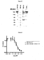
- SDS gel Figure 5
- the antibody containing the cysteine i.e. a humanized 280.46.3.4 antibody comprising 280.VH3-66-1 paired with 280.VK4-1-C.
- the mutation of the free cysteine in the L-CDR1 has improved dramatically not only the biophysical properties but also the potency of the humanized 280.46.3.4 antibody comprising 280.VH3-66.1 paired with 280.VK4-1-S on both human and mouse IL-22RA, as compared to the original mouse 280.46.3.4 antibody.
- Example 4 Removal of a deamidation motif in L-CDR1 and increasing the degree of humanness in humanized 280.46.3.4 VH
- Antibodies can be subject to a variety of chemical modification and/or degradation reactions for example deamidation, isomerization, hydrolysis, disulfide scrambling, beta-elimination, oxidation and adduct formation.
- the main hydrolytic mechanisms of degradation can include the deamidation of asparagines especially when immediately followed by a glycine or a serine.
- the substitution of the cysteine by a serine in the L-CDR1 has created an NS motif which constitutes a potential deamidation site and therefore should be eliminated. In an attempt to destroy this NS motif, a series of mutants was constructed (data not shown). It has been found that the best overall mutation was a change from the serine to a threonine.
- This mutated light chain variable domain (280.VK4-1-T), recited in SEQ ID NO: 32, was paired with the version 4 of the humanized 280.46.3.4 VH (280.VH3-66-4; see below) and assessed for inhibition potency in cell based assays.
- residue 70 (Table 1) was mutated to the human germline residue found at this position, threonine to serine mutation. Also the last two residues of the H-CDR2, glutamic acid and alanine at position 64 and 65 (Table 1), were mutated to the human germline residues found at those positions; lysine and glycine respectively.
- version 4 of the humanized 280.46.3.4 VH (280.VH3-66-4), recited in SEQ ID NO: 33, has three more human germline residues as compared to version 1 (280.VH3-66-1) with two of these residues being located in H-CDR2 ( Figure 9 as compared to Figure 4 ).
- Example 5 Mutation of Kabat residue 32 from Asp to Glu in CDR1 of humanized 280.46.3.4 VH increase affinity and improves stability
- Antibodies can be subject to a variety of chemical modification and/or degradation reactions for example deamidation, isomerization, hydrolysis, disulfide scrambling, beta-elimination, oxidation and adduct formation.
- the main hydrolytic mechanisms of degradation can include the isomerization of aspartic acid (Asp).
- Asp aspartic acid
- a set of mutants where the Asp 32 in the H-CDR1 and Asp 96 in the H-CDR3 were individually mutated in 280.VH3-66-4, has been made. These single mutants were then paired with the humanized light chain variable domain 280.VK4-1-T, described above, and the resulting NiNTA-purified Fab antibody fragments tested for affinity measurement by Biacore.
- the VH containing the mutation Asp to Glu at position 32 in the H-CDR1 was called version 18 or 280.VH3-66-18 and is recited in SEQ ID NO: 34.
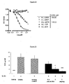
- the results of the Biacore analysis measuring the binding to human IL-22RA presented in Figure 13 show that the D32E mutation increased the on rate by around 2 fold and decreased the off rate by about 5 fold as compared to the parental unmutated D32 (280.VH3-66-4).
- the D96E mutation had a negative impact, reducing the affinity by about 500 fold.
- the mutant D32E when analyzed by differential scanning calorimetry (DSC) appeared to be more stable by 1 degree than the parental D32 antibody ( Figure 14 ). Overall it can been concluded that the D32E mutation greatly improves the properties of the latest heavy chain humanized version 18 (280.VH3-66-18).
- the potency of the humanized antibody expressed as a human IgG1/Kappa was assessed in a HepG2 cell assay.
- the results presented in Figure 15 show that the humanized antibody (280.VH3-66.18/VK4-1-T) is almost 3 times more potent than the mouse parental antibody purified from the 280.46.3.4 hybridoma (ATCC Patent Deposit Designation PTA-6284), with IC 50 values of 132.5 and 370.5 pM, respectively.
- Example 6 Increasing the degree of humanness
- the different humanized antibodies and Fabs mentioned in the Examples have been produced in CHO cells using a single expression vector, which comprises the cDNAs coding for the heavy and light chain under the control of two different promoters.
- Example 7 Potency of humanized 280.VH3-66-46 VH paired with 280.VK4-1-TSY VL in human IL-22RA expressing-cell assays
- the term "280.346.TSY" is hereinafter used to indicate an anti-human IL-22RA humanized antibody comprising 280.VH3-66.46 paired with 280.IGKV4-1-TSY, irrespective of the heavy and light chain constant regions.
- the amino acid sequence of the heavy and the light chain constant regions of a particular 280.346.TSY are recited in SEQ ID NO: 18 and 19 respectively, and the amino acid sequence of the entire heavy and light chain of said particular 280.346.TSY are recited in SEQ ID NO: 20 and 21, respectively.
- Example 8 Potency of humanized 280.346.TSY in mouse IL-22RA expressing-cell assays
- Example 9 Binding selectivity of 280.346.TSY on IL-22RA related proteins by competitive ELISA
- IL-22RA-ECD i.e. IL-22RA-Extra Cellular Domain
- Biotinylated 280-346-TSY antibody was added to the plate in the presence of competitors: human interleukin 22 receptor alpha (hIL-22RA), human IL-22 binding protein (hIL-22BP), mouse IL-22 receptor alpha (mIL-22RA), human IL-10 receptor alpha (hIL-10R) and human IL-20 receptor alpha (hIL-20R).
- hIL-22RA human interleukin 22 receptor alpha
- hIL-22BP human IL-22 binding protein
- mIL-22RA mouse IL-22 receptor alpha
- hIL-10R human IL-10 receptor alpha
- hIL-20R human IL-20 receptor alpha
- IC 50 values for recombinant human and murine IL-22RA were 18.25 pM and 149.3 pM respectively ( Figure 23 ). Monoclonal antibody did not show cross-reactivity with recombinant human IL-22BP, IL-10R, and IL-20R.
- Example 10 Cross reactivity of 280-346-TSY against IL-22RA orthologues as assessed by Kd measurement by KinExA and Biacore.
- the Kd of 280-346-TSY was assessed using both Biacore and KinExA instruments.
- the IL-22RA extracellular domains (ECD) of human and homologous gene sequences found in different species were produced in HEK-293 cells and NiNTA- purified using a 6 His tag.
- the 280-346-TSY antibody has a subnanomolar affinity to human and all three monkey species of IL-22RA tested. It has a nanomolar affinity to mouse, with an affinity around a 100 times lower compared to human and a micromolar affinity to rat IL-22RA (Table 3). Table 3.
- Example 11 Efficacy of 280-346-TSY on IL-22-induced serum amyloid A in mice
- the pharmacodynamic activity of 280-346-TSY was determined on IL-22-induced serum amyloid A in male Balb/c mice. Different doses of 280.346.TSY were administered subcutaneously 22 hours prior to recombinant murine IL-22 intravenous injection. Vehicle control is PBS administered subcutaneously at 10 ml/kg.
- mice were given 100 ⁇ g/kg of IL-22 into the retroorbital plexus under isoflurane anesthesia. Blood sampling was performed 6 hours after IL-22 injection by cardiac puncture under isoflurane anesthesia. A human IgG1 was used as a negative control (isotype control). Serum amyloid A was determined by ELISA (Biosource). 280-346-TSY gave an ED 50 value of 0.5 mg/kg. Mann Whitney test was used to perform statistical analysis: * p ⁇ 0.05 vs. isotype control group; *** p ⁇ 0.001 vs. isotype control group ( Figure 24 ).
- Example 12 Efficacy of 280-346-TSY on IL-23-induced ear inflammation in mice
- 280-346-TSY The pharmacodynamic activity of 280-346-TSY was determined in a mouse model of psoriasis. Efficacy of 280-346-TSY on IL-23-induced ear thickening in female C57BL/6 mice was tested. Mice were injected with 500 ng of recombinant human IL-23 or PBS in a total volume of 20 ⁇ l every other day for 14 days as described by Zheng Y et al. (Nature 2007). Different doses of 280.346.TSY were administered subcutaneously every other day with the first dose given prior to first administration of recombinant IL-23. Vehicle control is PBS administered subcutaneously at 10 ml/kg.
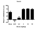
- Dexamethasone (Dexa) was used as positive control. Percentage inhibition were calculated at day 9 which corresponds to the peak of ear swelling. 280-346-TSY gave an ED 50 value of 1.8 mg/kg ( Figure 25 ).
Description
- The present invention relates to humanized antibodies against human IL-22RA and to their use in the treatment of psoriasis and other immune-mediated diseases such as psoriatic arthritis and atopic dermatitis.
- IL-22RA (also known as IL22R, IL22R1, IL22RA1, CRF2-9 and Zcytor11) belongs to the type II cytokine receptor family and is a component of the receptor for IL-20, IL-22 and IL-24. Due to their structural similarity IL-20, IL-22 and IL-24, together with IL-19 and IL-26, were combined with IL-10 in the so-called "IL-10 family" (Kunz S et al. 2006). IL-10 is a master regulator of the immune response that mediates down-regulation of pro-inflammatory cytokine expression in macrophages, T cells, and other cells of the immune system (Moore KW et al. 2001).
- In vitro, IL-20 and IL-24 are produced not only by activated immune cells, but also to a similar extent by keratinocytes. In vivo, these cytokines are expressed preferentially in the inflamed tissues. IL-20 and IL-24 can signal through two receptor complexes, IL-20RA/IL-20RB and IL-22RA/IL-20RB (Langer JA et al. 2004). Several tissues, particularly the skin, tissues from the reproductive and respiratory systems, and various glands appeared to be the main targets of these mediators (Kunz S et al. 2006).
- IL-22 was discovered as a gene up-regulated by CD4+ T cells upon activation and it shares 22% amino acid sequence identity with IL-10; it was, thus, originally named IL-10-related T cell-derived inducible factor (IL-TIF) (Dumoutier L et al. 2000). Unlike IL-10, which regulates immune cell functions, IL-22 controls tissue responses to the immune system. IL-22 signals through a heterodimer receptor formed by IL-22RA and IL-10RB which is highly expressed within various tissues but it is not detectable on immune cells. Initially, IL-22 binds via its IL-22RA binding site to the extracellular domain of IL-22RA and, subsequently, IL-10RB binds to a region created by the interaction of IL-22 and IL-22RA to form a cytokine receptor complex with a higher affinity for IL-22 (Li J et al. 2004). Since IL-10RB is broadly expressed by many different cell types, IL-22RA expression is the limiting component that determines IL-22 responsiveness of cells. IL-22RA is expressed strongly in the liver, as well as in the skin, lungs, pancreas and other peripheral tissues (Wolk K et al. 2004; Aggarwal S et al. 2001). Extensive screening of different cell lines has revealed that only cells which express IL-22RA respond to IL-22, suggesting that there is no alternate receptor that can mediate IL-22 signaling.
- A soluble receptor termed IL-22 binding protein (IL-22BP; also known as IL22BP, IL22RA2, IL-22R-alpha2, CRF2X, CRF2-S1 and CRF2-10) is also able to bind to IL-22 as a natural protein antagonist and probably provides systemic regulation of IL-22 activity (Kotenko SV et al. 2003). IL-22 has been found in diseased tissues from patients with different chronic inflammatory diseases that involve infiltrating activated T cells, such as psoriasis, psoriatic arthritis and atopic dermatitis. IL-22 has been most commonly described as a pro-inflammatory cytokine because of its expression in lesions of patients with chronic inflammatory diseases and its induction of pro-inflammatory cytokines such as IL-6, IL-8 and TNF-α (Wolk K et al. 2004; Andoh A et al. 2005; Ikeuchi H et al. 2005; Nograles KE et al. 2009a.; Nograles KE et al. 2009b). Most recently, Zheng et al. showed that IL-22 is important for mediating IL-23-induced dermal inflammation in a mouse model of psoriasis, indicating a pro-inflammatory role (Zheng Y et al. 2007). Given the biological effects of IL-22, including keratinocyte hyperplasia, induction of chemokine and pro-inflammatory cytokine production in certain tissue, the use of antagonists that block, inhibit, reduce or neutralize the activity of IL-22, e.g. by interfering with the receptor binding, may prevent infiltration of pathogenic cells at inflammatory sites. Mouse anti-human IL-22RA monoclonal antibodies have been previously described in
PCT patent application WO 2006/047249 filed on October 21, 2005 . However, mouse antibodies may cause immunogenicity and humanized anti-human IL-22RA antibodies are desirable. Humanized antibodies generally have at least three potential advantages over mouse antibodies for use in human therapy: (1) because the effector portion is human, it may interact better with the other parts of the human immune system (e.g., destroy the target cells more efficiently by complement-dependent cytotoxicity (CDC) or antibody-dependent cellular cytotoxicity (ADCC)); (2) the human immune system should not recognize the framework or constant region of the humanized antibody as foreign, and therefore the antibody response against such an injected antibody should be less than against a totally foreign mouse antibody; and (3) injected mouse antibodies have been reported to have a half-life in the human circulation much shorter than the half-life of human antibodies. Injected humanized antibodies will presumably have a half-life more similar to naturally occurring human antibodies, allowing smaller and less frequent doses to be given. Thus, in view of the above, there is a need for humanized anti-human IL-22RA antibodies for treating IL-22 mediated inflammation, such as psoriasis, psoriatic arthritis and atopic dermatitis. - In a first aspect, the invention provides a humanized antibody that binds to human IL-22RA. The humanized antibody of the invention comprises a) a heavy chain variable domain comprising H-CDR1 , H-CDR2, and H-CDR3 consisting of amino acid sequences of SEQ ID NO: 1, 2 and 3, respectively and b) a light chain variable domain comprising L-CDR1 , L-CDR2, and L-CDR3 consisting of amino acid sequences of SEQ ID NO: 4, 5 and 6, respectively or consisting of amino acid sequences of SEQ ID NO: 4, 5 and 7, respectively. In another aspect, the invention provides an antibody described herein, wherein a) said heavy chain variable domain comprises framework regions H-FR1, H-FR2, H-FR3 and H-FR4 consisting of amino acid sequences of SEQ ID NO: 8, 9, 10 and 11, respectively and b) said light chain variable domain comprises framework regions L-FR1, L-FR2, L-FR3 and L-FR4 consisting of amino acid sequences of SEQ ID NO: 12, 13, 14 and 15, respectively. In another aspect, the invention provides an antibody described herein, wherein a) said heavy chain variable domain consists of amino acid sequence of SEQ ID NO: 16 and b) said light chain variable domain consists of amino acid sequence of SEQ ID NO: 17. In another aspect, the invention provides an antibody described herein, wherein said antibody comprises a) a heavy chain constant region consisting of amino acid sequence of SEQ ID NO: 18 and b) a light chain constant domain consisting of amino acid sequence of SEQ ID NO: 19.
- In another aspect, the invention provides a humanized antibody that binds to human IL-22RA which comprises a heavy chain comprising or consisting of amino acid sequence of SEQ ID NO: 20 and a light chain comprising or consisting of amino acid sequence of SEQ ID NO: 21.
- Provided is a polynucleotide, e.g. a DNA, encoding the heavy chain of the humanized antibody according to the present invention. Preferably, said polynucleotide comprises or consists of SEQ ID NO: 22.
- Provided is a polynucleotide, e.g. a DNA, encoding the light chain of the humanized antibody according to the present invention. Preferably, said polynucleotide comprises or consists of SEQ ID NO: 23.
- In another aspect, the invention provides a polynucleotide, e.g. a DNA, encoding both the heavy and the light chains of the humanized antibody according to the present invention.
- In another aspect, the invention provides a vector and more particularly an expression vector comprising a) a polynucleotide encoding the heavy chain of the humanized antibody according to the present invention and b) a polynucleotide encoding the light chain of the humanized antibody according to the present invention.
- In another aspect, the invention provides a vector and more particularly an expression vector comprising a polynucleotide encoding the heavy chain and the light chain of the humanized antibody according to the present invention.
- In another aspect, the invention provides a host cell, preferably a CHO cell, comprising, e.g. as a result of a transfection, a vector and in particular an expression vector according to the invention.
- In another aspect, the invention provides a method of producing a humanized antibody according to the invention, the method comprising culturing a host cell, preferably a CHO cell, according to the invention and isolating the humanized antibody according to the present invention.
- In another aspect, the invention provides a humanized antibody according to the present invention for use as a medicament, in particular for use in the treatment of psoriasis, psoriatic arthritis or atopic dermatitis.
- In another aspect, the invention provides a pharmaceutical composition comprising a humanized antibody according to the present invention and its use as a medicament, in particular for use in the treatment of psoriasis, psoriatic arthritis or atopic dermatitis.
- In another aspect, the invention provides for the use of a humanized antibody according to the present invention or of a pharmaceutical composition comprising said antibody in the manufacture of a medicament for the treatment of psoriasis, psoriatic arthritis or atopic dermatitis.
-
-
Figure 1 reports the alignment between the human Immunoglobulin germline kappa variable gene 4-1 (IGKV4-1) and the mouse 280.46.3.4 VL (280.46.3.4). -
Figure 2 reports the alignment between the human Immunoglobulin germline heavy variable gene 3-66 (IGHV3-66) and the mouse 280.46.3.4 VH (280.46.3.4). -
Figure 3 reports the alignment between the human Immunoglobulin germline kappa variable gene 4-1 (IGKV4-1) and the first version of humanized 280.46.3.4 VL (280.VK4-1-C). -
Figure 4 reports the alignment between the first version of the humanized 280.46.3.4 VH (280.VH3-66.1) and the human Immunoglobulin germline heavy variable gene 3-66 (IGHV3-66). -
Figure 5 reports the results of a Coomassie blue staining of protein A-purified humanized 280.46.3.4 antibodies run on an SDS gel under non-denaturing conditions. "Humira" (Adalimumab), a commercialized anti-TNFα monoclonal antibody, is used here as a standard reference. "Marker" is standard protein molecular weight (MW) markers with kD indicated on the left hand side of the figure. "Before" refers to the protein A-purified humanized 280.46.3.4 antibody comprising 280.VH3-66-1 paired with 280.VK4-1-C, containing therefore an unpaired cysteine in the light chain. "After" refers to the protein A-purified humanized 280.46.3.4 antibody comprising 280.VH3-66-1 paired with 280.VK4-1-S. -
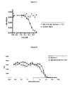
Figure 6 reports the results of the STAT3 phosphorylation assay done in human HepG2 hepatoma cells to compare the potency of the humanized antibody expressed as a human IgG1/kappa, comprising 280.VH3-66-1 paired with 280.VK4-1-S (■ 280.VH3-66-1/VK4-1-S), with the mouse parental antibody 280.46.3.4 (● 280.46.3.4). The results show that the humanized antibody is 1.4 time more potent than the mouse parental antibody, which contains the free cysteine, with IC50 values of 257.5 pM and 370.5 pM, respectively. -
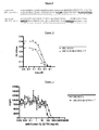
Figure 7 reports the results of the proliferation assay done in human IL-22 receptor transfected-BaF3 stable cells to compare the potency of the humanized antibody expressed as a human IgG1/kappa, comprising 280.VH3-66-1 paired with 280.VK4-1-S (■ 280.VH3-66-1/VK4-1-S), with the mouse parental antibody 280.46.3.4 (● 280.46.3.4). The results show that the humanized antibody is 1.7 time more potent than the mouse parental antibody, which contains the free cysteine, with IC50 values of 340 pM and 587 pM, respectively. -
Figure 8 reports the results of the proliferation assay done in murine IL-22 receptor transfected-BaF3 stable cells to compare the potency of the humanized antibody expressed as a human IgG1/kappa, comprising 280.VH3-66-1 paired with 280.VK4-1-S (■ 280.VH3-66-1NK4-1-S), with the mouse parental antibody 280.46.3.4 (● 280.46.3.4). The results show that the humanized antibody is 2.1 times more potent than the mouse parental antibody, which contains the free cysteine, with IC50 values of 693 pM and 1473 pM, respectively. -
Figure 9 reports the alignment between the human Immunoglobulin germline heavy variable gene 3-66 (IGHV3-66) andversion 4 of the humanized 280.46.3.4 VH (280.VH3-66-4). -
Figure 10 reports the results of the STAT3 phosphorylation assay done in human HepG2 hepatoma cells to compare the potency of the humanized antibody expressed as a human IgG1/kappa, comprising 280.VH3-66-4 paired with 280.VK4-1-T (◆ 280.VH3-66-4/VK4-1-T), with the mouse parental antibody 280.46.3.4 (● 280.46.3.4). The results show that the humanized antibody is 1.8 time more potent than the mouse parental antibody, with IC50 values of 183.2 pM and 333.0 pM, respectively. -
Figure 11 reports the results of the proliferation assay done in human IL-22 receptor transfected-BaF3 stable cells to compare the potency of the humanized antibody expressed as a human IgG1/kappa, comprising 280.VH3-66-4 paired with 280.VK4-1-T (◆ 280.VH3-66-4/VK4-1-T), with the mouse parental antibody 280.46.3.4 (● 280.46.3.4). The results show that the humanized antibody is 1.75 time more potent than the mouse parental antibody, with IC50 values of 334 pM and 587 pM, respectively. -
Figure 12 reports the results of the proliferation assay done in murine IL-22 receptor transfected-BaF3 stable cells to compare the potency of the humanized antibody expressed as a human IgG1/kappa, comprising 280.VH3-66-4 paired with 280.VK4-1-T (◆ 280.VH3-66-4/VK4-1-T), with the mouse parental antibody 280.46.3.4 (● 280.46.3.4). The results show that the humanized antibody is 2.1 times more potent than the mouse parental antibody, with IC50 values of 687 pM and 1473 pM, respectively. -
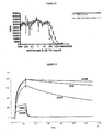
Figure 13 reports the results of the Biacore analysis aiming at measuring the binding affinity to human IL-22RA of a set of mutants, where the Asp 32 in the H-CDR1 and Asp 96 in the H-CDR3 were individually mutated in 280.VH3-66-4. These single mutants were paired with the humanized light chain variable domain 280.VK4-1-T, and then tested for affinity measurement. D32E: mutation Asp to Glu at position 32 in 280.VH3-66-4 to create version 280.VH3-66-18; D32: parental version 280-VH3-66-4; D32N: mutation Asp to Asn at position 32 in 280.VH3-66-4; D96E: mutation Asp to Glu at position 96 in 280.VH3-66-4; D96N: mutation Asp to Asn at position 96 in 280.VH3-66-4.
The results reported in this figure show that the D32E mutation increased the on rate by around 2 fold and decreased the off rate by about 5 fold as compared to the parental unmutated D32 (280.VH3-66-4). On the other hand, the D96E mutation had a negative impact, reducing the affinity by about 500 fold. -
Figure 14 reports the results of a differential scanning calorimetry (DSC) of the D32E mutant and of the parental unmutated D32 performed to examine their thermal stability. The D32E mutant (light grey line) is more stable by 1 degree than the parental D32 (dark grey line). -
Figure 15 reports the results of the STAT3 phosphorylation assay done in human HepG2 hepatoma cells to compare the potency of the humanized antibody expressed as a human IgG1/kappa, comprising 280.VH3-66-18 paired with 280.VK4-1-T (◆ 280.VH3-66-18/VK4-1-T), with the mouse parental antibody 280.46.3.4 (● 280.46.3.4). The results show that the humanized antibody is almost 3 times more potent than the mouse parental antibody, with IC50 values of 132.5 pM and 370.5 pM, respectively. -
Figure 16 reports the alignment between the final humanized VH version, 280.VH3-66-46, and the human Immunoglobulin germline heavy variable gene 3-66 (IGHV3-66). -
Figure 17 reports the alignment between the human Immunoglobulin germline kappa variable gene 4-1 (IGKV4-1) and the final humanized VL version, 280.VK4-1-TSY. -
Figure 18 reports the results of the STAT3 phosphorylation assay done in normal human keratinocytes to compare the potency of the humanized antibody expressed as a human IgG1/kappa, comprising 280.VH3-66-46 paired with 280.VK4-1-TSY (◆ 280.VH3-66-46/VK4-1-TSY), with the mouse parental antibody 280.46.3.4 (● 280.46.3.4). The results show that the humanized antibody (280.346.TSY, see Example 7) is almost 9 times more potent than the mouse parental antibody, with IC50 values of 60.95 pM and 541.9 pM, respectively. -
Figure 19 reports the results of the STAT3 phosphorylation assay done in human HepG2 hepatoma cells to compare the potency of the humanized antibody expressed as a human IgG1/kappa, comprising 280.VH3-66-46 paired with 280.VK4-1-TSY (◆ 280.VH3-66-46/VK4-1-TSY), with the mouse parental antibody 280.46.3.4 (● 280.46.3.4). The results show that the humanized antibody (280.346.TSY, see Example 7) is almost 5 times more potent than the mouse parental antibody, with IC50 values of 55.16 pM and 266.3 pM, respectively. -
Figure 20 reports the results of the proliferation assay done in human IL-22 receptor transfected-BaF3 stable cells to compare the potency of the humanized antibody expressed as a human IgG1/kappa, comprising 280.VH3-66-46 paired with 280.VK4-1-TSY (◆ 280.VH3-66-46/VK4-1-TSY), with the mouse parental antibody 280.46.3.4 (● 280.46.3.4). The results show that the humanized antibody (280.346.TSY, see Example 7) is 1.7 time more potent than the mouse parental antibody, with IC50 values of 317 pM and 545 pM, respectively. -
Figure 21 reports the results of the STAT3 phosphorylation assay done in murine HEPA1-6 hepatoma cells to calculate the potency of the humanized antibody expressed as a human IgG1/kappa, comprising 280.VH3-66-46 paired with 280.VK4-1-TSY (● 280.VH3-66-46/VK4-1-TSY). A human IgG1 is used as a negative control (■ control hIgG1). The results show that the humanized antibody (280.346.TSY, see Example 7) is able to inhibit the activity of murine IL-22 with an IC50 in the nanomolar range (2,1 nM). -
Figure 22 reports the results of the proliferation assay done in murine IL-22 receptor transfected-BaF3 stable cells to compare the potency of the humanized antibody expressed as a human IgG1/kappa, comprising 280.VH3-66-46 paired with 280.VK4-1-TSY (◆ 280.VH3-66-46/VK4-1-TSY), with the mouse parental antibody 280.46.3.4 (● 280.46.3.4). The results show that the humanized antibody (280.346.TSY, see Example 7) is 6.2 times more potent than the mouse parental antibody, with IC50 values of 137 pM and 849 pM, respectively. -
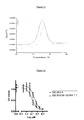
Figure 23 reports the results of a competitive ELISA done to measure the specificity of 280.346.TSY to human IL-22RA. Microtiter plates were coated with human IL-22RA-ECD (i.e. IL-22RA-Extra Cellular Domain). Biotinylated 280-346-TSY antibody was added to the plate in the presence of competitors:human interleukin 22 receptor alpha (hIL-22RA), human IL-22 binding protein (hIL-22BP), murine IL-22 receptor alpha (mIL-22RA), human IL-10 receptor alpha (hIL-10R) and human IL-20 receptor alpha (hIL-20R). Binding to hIL-22RA coated on the plates is revealed by addition of peroxidase-conjugated streptavidin. Measured IC50 values for human (● hIL-22RA) and murine (◆ mIL-22RA) IL-22RA are 18.25 pM and 149.3 pM respectively. 280.346.TSY does not show cross-reactivity with human IL-22BP (■ hIL-22BP), IL-10R alpha (Δ hIL-10R) and IL-20R alpha (V hIL-20R). -
Figure 24 reports the results of the pharmacodynamic activity of 280-346-TSY on IL-22-induced serum amyloid A in mice. Different doses of 280.346.TSY were administered subcutaneously 22 hours prior to recombinant murine IL-22 intravenous injection. Vehicle control is PBS administered subcutaneously. Blood sampling was performed 6 hours after IL-22 administration. A human IgG1 is used as a negative control (isotype control). Serum amyloid A was determined by ELISA. 280-346-TSY showed efficacy in this model and gave an ED50 value of 0.5 mg/kg. Mann Whitney test was used to perform statistical analysis: * p< 0.05 vs. isotype control group; *** p< 0.001 vs. isotype control group. -
Figure 25 reports the results of the pharmacodynamic activity of 280-346-TSY in a mouse model of psoriasis. Efficacy of 280-346-TSY on IL-23-induced ear thickening was determined. Mice were injected with 500 ng of recombinant human IL-23 or PBS every other day for 14 days. Full therapeutic coverage was performed with different doses of 280.346.TSY administered subcutaneously. Vehicle control is PBS administered subcutaneously. Dexamethasone (Dexa) is used as positive control. Percentage inhibition were calculated at day 9 which corresponds to the peak of ear swelling. 280-346-TSY showed efficacy in this model and gave an ED50 value of 1.8 mg/kg. - Prior to setting forth the invention in detail, it may be helpful to the understanding thereof to define the following terms.
- As used herein, the term "antibody", and its plural form "antibodies", includes, inter alia, polyclonal antibodies, affinity-purified polyclonal antibodies, monoclonal antibodies, nanobodies and antigen-binding fragments, such as F(ab')2, Fab proteolytic fragments, and single chain variable region fragments (scFvs). Genetically engineered intact antibodies or fragments, such as chimeric antibodies, Fv fragments, single chain antibodies and the like, as well as synthetic antigen-binding peptides and polypeptides, are also included. Non-human antibodies may be humanized by grafting non-human CDRs onto human framework and constant regions, or by incorporating the entire non-human variable domains. In some instances, humanized antibodies may retain non-human residues within the human framework regions to enhance proper binding characteristics. Through humanizing antibodies, biological half-life may be increased, and the potential for adverse immune reactions upon administration to humans is reduced.
- As used herein, the term "immunoglobulin" (Ig) refers to a protein consisting of one or more polypeptides substantially encoded by immunoglobulin genes. One form of immunoglobulin constitutes the basic structural unit of an antibody. This form is a tetramer and consists of two identical pairs of immunoglobulin chains, each pair having one light and one heavy chain. A light chain has two parts: the variable domain (VL) and the constant domain (CL), which in the context of a light chain can be called constant region as well. A heavy chain has two parts as well: the variable domain (VH) and the constant region (CH). In each pair, the light and heavy chain variable domains are together responsible for binding to an antigen, and the constant regions are responsible for the antibody effector functions.
- Full-length immunoglobulin "light chains" (about 25 Kd) are encoded by a variable domain gene at the N-terminus (about 110 amino acids) and a kappa or lambda constant domain (Cκ and Cλ, respectively) gene at the C-terminus. Full-length immunoglobulin "heavy chains" (about 50 Kd), are similarly encoded by a variable domain gene (about 116 amino acids) and one of the other constant region genes (about 330 amino acids) mentioned hereinafter. There are five types of mammalian heavy chain denoted by the Greek letters: α, δ, ε, γ, and µ. The type of heavy chain defines the antibody's isotype as IgA, IgD, IgE, IgG and IgM, respectively. The constant region is identical in all antibodies of the same isotype, but differs in antibodies of different isotypes. Heavy chains γ, α and δ have a constant region composed of three Ig constant domains (
C H1, CH2, and CH3), and a hinge region for added flexibility; heavy chains µ and ε have a constant region composed of four Ig constant domains (C H1, CH2,C H3, and CH4) and a hinge region. - An immunoglobulin light or heavy chain variable domain consists of a "framework" region interrupted by three hypervariable regions. Thus, the term "hypervariable region" refers to the amino acid residues of an antibody which are responsible for antigen binding. The hypervariable region comprises amino acid residues from a "Complementarity Determining Region" or "CDR", i.e. L-CDR1, L-CDR2 and L-CDR3 in the light chain variable domain and H-CDR1, H-CDR2 and H-CDR3 in the heavy chain variable domain (Kabat et al. 1991) and/or those residues from a "hypervariable loop" (Chothia and Lesk, 1987). "Framework Region" or "FR" residues are those variable domain residues other than the hypervariable region residues as herein defined. The sequences of the framework regions of different light (i.e. L-FR1, L-FR2, L-FR3 and L-FR4) or heavy (i.e. H-FR1, H-FR2, H-FR3 and H-FR4) chains are relatively conserved within a species. Thus, a "human framework region" is a framework region that is substantially identical (about 85% or more, usually 90-95% or more) to the framework region of a naturally occurring human immunoglobulin. The framework region of an antibody, that is the combined framework regions of the constituent light and heavy chains, serves to position and align the CDRs. The CDRs are primarily responsible for binding to an epitope of an antigen.
- Accordingly, the term "humanized" immunoglobulin refers to an immunoglobulin comprising a human framework region and one or more CDRs from a non-human (usually a mouse or rat) immunoglobulin. The non-human immunoglobulin providing the CDRs is called the "donor" and the human immunoglobulin providing the framework is called the "acceptor". Constant regions need not be present, but if they are, they must be substantially identical to human immunoglobulin constant regions, i.e., at least about 85-90%, preferably about 95% or more identical. Hence, all parts of a humanized immunoglobulin, except possibly the CDRs and few residues in the heavy chain constant region if modulation of the effector functions is needed, are substantially identical to corresponding parts of natural human immunoglobulin sequences. A "humanized antibody" is an antibody comprising a humanized light chain variable domain and a humanized heavy chain variable domain. In some instances, humanized antibodies may retain non-human residues within the human framework regions to enhance proper binding characteristics and/or some amino acid mutations may be introduced within the CDRs in order to improve the binding affinity and/or to reduce the immunogenicity and/or to increase the degree of humanness.
- The term "recombinant antibodies" means antibodies wherein the amino acid sequence has been varied from that of a native antibody. Because of the relevance of recombinant DNA techniques in the generation of antibodies, one need not be confined to the sequences of amino acids found in natural antibodies; antibodies can be redesigned to obtain desired characteristics. The possible variations are many and range from the changing of just one or a few amino acids to the complete redesign of, for example, the variable domain or constant region. Changes in the constant region will, in general, be made in order to improve, reduce or alter characteristics, such as complement fixation (e.g. complement dependent cytotoxicity, CDC), interaction with membranes and other effector functions (e.g. antibody dependent cellular cytotoxicity, ADCC). Changes in the variable domain will be made in order to improve the antigen binding characteristics.
- In addition to antibodies, immunoglobulins may exist in a variety of other forms including, for example, single-chain or Fv, Fab, and (Fab')2 , as well as diabodies, linear antibodies, multivalent or multispecific hybrid antibodies. As used herein, the terms "single-chain Fv," "single-chain antibodies," "Fv" or "scFv" refer to antibody fragments that comprises the variable domains from both the heavy and light chains, but lacks the constant regions, but within a single polypeptide chain. Generally, a single-chain antibody further comprises a polypeptide linker between the VH and VL domains which enables it to form the desired structure which would allow for antigen binding. In specific embodiments, single-chain antibodies can also be bi-specific and/or humanized. A "Fab fragment" is comprised of one light chain and the variable and
C H1 domains of one heavy chain. The heavy chain of a Fab molecule cannot form a disulfide bond with another heavy chain molecule. A "Fab' fragment" contains one light chain and one heavy chain that contains more of the constant region, between theC H1 and CH2 domains, such that an interchain disulfide bond can be formed between two heavy chains to form a F(ab')2 molecule. A "F(ab')2" contains two light chains and two heavy chains containing a portion of the constant region between theC H1 and CH2 domains, such that an interchain disulfide bond is formed between two heavy chains. Having defined some important terms, it is now possible to focus the attention on particular embodiments of the instant invention. - The present invention is based upon the discovery of humanized anti-human IL-22RA antibodies. Use of these antibodies as antagonists to IL-22RA can inhibit inflammation and, therefore, can be useful in the treatment of chronic inflammatory diseases that involve infiltrating activated T cells, such as psoriasis, psoriatic arthritis and atopic dermatitis. The invention provides the use of humanized antibodies that recognize, bind, modulate and/or neutralize the IL-22RA. In particular, the invention provides the use of humanized light and heavy chain variable domains that recognize, bind, modulate and/or neutralize the IL-22RA. Such humanized light and heavy chain variable domains can be fused, respectively, to a kappa or lambda constant domain and to a constant region of an heavy chain chosen among any isotype (IgA, IgD, IgE, IgG and IgM), and expressed in a variety of host cells. Preferably, the constant region chosen is that of an IgG, and more preferably of an IgG1. The humanized anti-IL-22RA antibodies described herein were generated using, as starting point of the humanization process, amino acid sequences of mouse anti-human IL-22RA monoclonal antibodies previously described in
PCT patent application WO 2006/047249 filed on October 21, 2005 . IL-22RA is a type II cytokine receptor described, for the first time, as Zcytor11, inPCT patent application WO 99/07848 filed on July 30, 1998 - The present invention also provides humanized IL-22RA antibodies that bind to polypeptide fragments or peptides comprising an epitope-bearing portion of a IL-22RA polypeptide or an immunogenic epitope or antigenic epitope. The binding of the antibodies to these epitopes results in inhibition, blocking, neutralization, and/or reduction in signal transduction of IL-22RA.
- The activity of the antibodies as described herein can be measured by their ability to inhibit, or reduce proliferation using a variety of assays that measure proliferation of and/or binding to cells expressing the IL-22RA receptor. Of particular interest are changes in IL-22-dependent cells. Suitable cell lines to be engineered to be IL-22-dependent include the BaF3 cell line. The activity of the humanized anti-IL-22RA antibodies can also be measured in the BaF3 proliferation assay, STAT3 phosphorylation assay in human HepG2 hepatoma cells or in mouse HEPA1-6 hepatoma cells, the Biacore assay, or the normal human keratinocyte assay described hereinafter.
- In an embodiment, the humanized antibody of the invention comprises a) a heavy chain variable domain comprising H-CDR1, H-CDR2, and H-CDR3 consisting of amino acid sequences of SEQ ID NO: 1, 2 and 3, respectively and b) a light chain variable domain comprising L-CDR1, L-CDR2, and L-CDR3 consisting of amino acid sequences of SEQ ID NO: 4, 5 and 6, respectively or consisting of amino acid sequences of SEQ ID NO: 4, 5 and 7, respectively.
- In another embodiment, the invention provides an antibody described herein, wherein a) said heavy chain variable domain comprises framework regions H-FR1, H-FR2, H-FR3 and H-FR4 consisting of amino acid sequences of SEQ ID NO: 8, 9, 10 and 11, respectively and b) said light chain variable domain comprises framework regions L-FR1, L-FR2, L-FR3 and L-FR4 consisting of amino acid sequences of SEQ ID NO: 12, 13, 14 and 15, respectively.
- In another embodiment, the invention provides an antibody described herein, wherein a) said heavy chain variable domain consists of amino acid sequence of SEQ ID NO: 16 and b) said light chain variable domain consists of amino acid sequence of SEQ ID NO: 17.
- In another embodiment, the invention provides an antibody described herein, wherein a) said heavy chain variable domain is fused to an heavy chain constant region selected from the group consisting of the constant region of a human IgA, IgG, IgM, IgD, IgE or any subclass, preferably an IgG1 and b) said light chain variable domain is fused to a constant domain of a k or A human immunoglobulin light chain, preferably a k.
- In another embodiment, said heavy chain constant region comprises some amino acid mutations that modulate, reduce or inhibit the antibody effector function (e.g. antibody dependent cellular toxicity (ADCC) and complement dependent cytotoxicity (CDC)).
- In another embodiment, the invention provides an antibody described herein, wherein said antibody comprises a) a heavy chain constant region consisting of amino acid sequence of SEQ ID NO: 18 and b) a light chain constant domain consisting of amino acid sequence of SEQ ID NO: 19.
- In another embodiment, the invention provides a humanized antibody that binds to human IL-22RA which comprises a heavy chain comprising or consisting of amino acid sequence of SEQ ID NO: 20 and a light chain comprising or consisting of amino acid sequence of SEQ ID NO: 21.
- Methods for preparing the polynucleotides encoding the antibodies described herein (including DNA and RNA) are well known in the art. Total RNA can be prepared using guanidinium isothiocyanate extraction followed by isolation by centrifugation in a CsCl gradient (Chirgwin JM et al. 1979). Poly(A)+ RNA is prepared from total RNA using the method of Aviv and Leder (Aviv H et al. 1972). Complementary DNA (cDNA) is prepared from poly(A)+ RNA using known methods. In the alternative, genomic DNA can be isolated. Polynucleotides encoding IL-22RA antibodies are then identified and isolated by, for example, hybridization or PCR.
- The antibodies disclosed herein may be produced by any technique known in the art, such as by recombinant technologies, chemical synthesis, cloning, ligations, or combinations thereof. In a embodiment, the antibodies of the present invention are produced by recombinant technologies, e.g., by expression of a corresponding nucleic acid in a suitable host cell. The polypeptide produced may be glycosylated or not, or may contain other post-translational modifications depending on the host cell type used. Many books and reviews provide teachings on how to clone and produce recombinant proteins using vectors and prokaryotic or eukaryotic host cells.
- A further embodiment of the present invention is therefore an isolated nucleic acid molecule encoding any of the antibodies or portion thereof here above or below described, or a complementary strand or degenerate sequence thereof. In this regard, the term "nucleic acid molecule" encompasses all different types of nucleic acids, including without limitation deoxyribonucleic acids (e.g., DNA, cDNA, gDNA, synthetic DNA, etc.), ribonucleic acids (e.g., RNA) and peptide nucleic acids (PNA). In a preferred embodiment, the nucleic acid molecule is a DNA molecule, such as a double-stranded DNA molecule or a cDNA molecule. The term "isolated" means nucleic acid molecules that have been identified and separated from at least one contaminant nucleic acid molecule with which it is ordinarily associated in the natural source. An isolated nucleic acid molecule is other than in the form or setting in which it is found in nature. Isolated nucleic acid molecules therefore are distinguished from the specific nucleic acid molecule as it exists in natural cells. A degenerate sequence designates any nucleotide sequence encoding the same amino acid sequence as a reference nucleotide sequence, but comprising a distinct nucleotide sequence as a result of the genetic code degeneracy.
- In another embodiment a nucleic acid molecule, also called polynucleotide, encodes the heavy chain of the humanized antibody of the invention and another polynucleotide encodes the light chain of the humanized antibody of the invention.
- In a preferred embodiment the polynucleotide encoding the heavy chain of the humanized antibody of the invention comprises or consists of SEQ ID NO: 22.
- In a preferred embodiment the polynucleotide encoding the light chain of the humanized antibody of the invention comprises or consists of SEQ ID NO: 23.
- In a preferred embodiment a unique polynucleotide encodes for both the heavy and light chain of the humanized antibody of the invention.
- A further embodiment of this invention is a vector comprising DNA encoding any of the above or below described antibodies or portion thereof. The vector may be any cloning or expression vector, integrative or autonomously replicating, functional in any prokaryotic or eukaryotic cell. In particular, the vector may be a plasmid, cosmid, virus, phage, episome, artificial chromosome, and the like. The vector may comprise the coding sequences for both the heavy and light chain, or either of the light and heavy chain coding sequences. Should the vector comprise coding sequences for both heavy and light chains, the heavy and light chains may each be operably linked to a promoter. The promoter may be the same or different for the heavy and light chain. The heavy and light chain may also be operably linked to one single promoter, in this case the coding sequences for the heavy and light chains may preferably be separated by an internal ribosomal entry site (IRES). Suitable promoters for eukaryotic gene expression are, for example, promoters derived from viral genes such as the murine or human cytomegalovirus (CMV), the mouse bi-directional CMV promoter or the rous sarcoma virus (RSV) promoter, which are well known to the person skilled in the art. The vector may comprise regulatory elements, such as a promoter, terminator, enhancer, selection marker, origin of replication, insulator etc. The appropriate nucleic acid sequence may be inserted into the vector by a variety of procedures. In general, DNA is inserted into an appropriate restriction endonuclease site(s) using techniques known in the art. Construction of suitable vectors containing one or more of these components employs standard ligation techniques which are known to the skilled artisan.
- A further embodiment of the present invention is a recombinant host cell, wherein said cell comprises a nucleic acid molecule/polynucleotide or a vector as defined above. The host cell may be a prokaryotic or eukaryotic cell. Examples of prokaryotic cells include bacteria, such as E.coli. Examples of eukaryotic cells are yeast cells, plant cells, mammalian cells and insect cells including any primary cell culture or established cell line (e.g., 3T3, Vera, HEK293, TN5, etc.). Suitable host cells for the expression of glycosylated proteins are derived from multicellular organisms. Examples of useful mammalian host cell lines include Chinese hamster ovary (CHO) and COS cells. Particularly preferred mammalian cells of the present invention are CHO cells.
- As disclosed here above, the antibodies of the present invention may be produced by any technique known in the art, such as by recombinant technologies, chemical synthesis, cloning, ligations, or combinations thereof.
- Another embodiment of this invention is therefore a method of producing an antibody of the present invention, the method comprising culturing a recombinant host cell of the invention under conditions allowing expression of the nucleic acid molecule, and recovering/isolating the polypeptide produced. The polypeptide produced may be glycosylated or not, or may contain other post-translational modifications depending on the host cell type used. The method of producing an antibody of the present invention may further comprise the step of formulating the antibody into a pharmaceutical composition.
- A further embodiment of the present invention is therefore a pharmaceutical composition comprising the humanized antibody according to the invention. Preferably, said pharmaceutical composition may further comprise additional excipients, such as buffer, stabilizer, surfactant, etc. Pharmaceutical compositions according to the invention are useful in the diagnosis, prevention, and/or treatment (local or systemic) of psoriasis and other immune-mediated diseases such as psoriatic arthritis and atopic dermatitis.
- The term "treatment" within the context of this invention refers to any beneficial effect on progression of disease, including attenuation, reduction, decrease or diminishing of the pathological development after onset of disease.
- The pharmaceutical compositions of the invention may be administered with a pharmaceutically acceptable carrier.
- The term "pharmaceutically acceptable" is meant to encompass any carrier, which does not interfere with effectiveness of the biological activity of the active ingredient and that is not toxic to the host to which it is administered. For example, for parenteral administration, the active protein(s) may be formulated in a unit dosage form for injection in vehicles such as saline, dextrose solution, serum albumin and Ringer's solution.
- In another aspect, the invention provides a pharmaceutical composition according to the invention for use as a medicament. In another aspect, the invention provides a method of treating a disease in a patient, comprising administering to the patient a pharmaceutical composition according to the invention. Preferably, the disease is selected from psoriasis, psoriatic arthritis and atopic dermatitis.
- In another aspect, the invention provides a humanized antibody according to the invention for use as a medicament. In another aspect, the invention provides a method of treating a disease in a patient, comprising administering to the patient a humanized antibody according to the invention. Preferably, the disease is selected from psoriasis, psoriatic arthritis and atopic dermatitis.
- In another aspect, the invention provides for the use of humanized antibody according to the invention for the preparation of a medicament for the treatment of psoriasis, psoriatic arthritis or atopic dermatitis.
- A pharmaceutical composition according to the invention is administered pulmonary.
- A pharmaceutical composition according to the invention is administered intranasally.
- A pharmaceutical composition according to the invention is administered by inhalation.
- A pharmaceutical composition according to the invention is administered orally.
- A pharmaceutical composition according to the invention is administered intravenously or intramuscularly.
- A pharmaceutical composition according to the invention is administered subcutaneously.
- A pharmaceutical composition is administered according to any one of the routes described above daily or every other day.
- For parenteral (e.g. intravenous, subcutaneous, intramuscular) administration, a pharmaceutical composition of the invention can be formulated as a solution, suspension, emulsion or lyophilized powder in association with a pharmaceutically acceptable parenteral vehicle (e.g. water, saline, dextrose solution) and additives that maintain isotonicity (e.g. mannitol) or chemical stability (e.g. preservatives and buffers). The formulation is sterilized by commonly used techniques.
- The active ingredients of the pharmaceutical composition according to the invention can be administered to an individual in a variety of ways. The routes of administration may include intradermal, transdermal (e.g. in slow release formulations), intramuscular, intraperitoneal, intravenous, subcutaneous, epidural, topical, oral routes and by aerosol administration, intranasal route or inhaled. Any other therapeutically efficacious route of administration can be used, for example absorption through epithelial or endothelial tissues or by gene therapy wherein a DNA molecule encoding the active agent is administered to the patient (e.g. via a vector), which causes the active agent to be expressed and secreted in vivo. In addition, a pharmaceutical composition according to the invention can be administered together with other components of biologically active agents such as pharmaceutically acceptable surfactants, excipients, carriers, diluents and vehicles.
- The dosage administered to an individual will vary depending upon a variety of factors, including pharmacokinetic properties, the route of administration, patient conditions and characteristics (sex, age, body weight, health, size), extent of symptoms, concurrent treatments, frequency of treatment and the effect desired.
- The antibodies of the present invention can be produced, formulated, administered or used in other alternative forms that can be preferred according to the desired method of use and/or production. Useful conjugates or complexes can also be generated for improving the agents in terms of drug delivery efficacy. For this purpose, the antibodies described herein can be in the form of active conjugates or complex with molecules such as polyethylene glycol and other natural or synthetic polymers (Harris JM et al. 2003). In this regard, the present invention contemplates chemically modified antibodies, in which the antibody is linked with a polymer. Typically, the polymer is water soluble so that the conjugate does not precipitate in an aqueous environment, such as a physiological environment. Moreover, a mixture of polymers can be used to produce the conjugates. The conjugates used for therapy can comprise pharmaceutically acceptable water-soluble polymer moieties. Suitable water-soluble polymers include polyethylene glycol (PEG), monomethoxy-PEG, aryloxy-PEG, bis-succinimidyl carbonate PEG, propylene glycol homopolymers, a polypropylene oxide/ethylene oxide co-polymer, polyoxyethylated polyols (e.g., glycerol), polyvinyl alcohol, dextran, cellulose, or other carbohydrate-based polymers. Suitable PEG may have a molecular weight from about 600 to about 60,000, including, for example, 5,000, 12,000, 20,000 and 25,000. A conjugate can also comprise a mixture of such water-soluble polymers. Examples of conjugates comprise any of the antibody disclosed here above and a polyalkyl oxide moiety attached to the N-terminus. PEG is one suitable polyalkyl oxide. As an illustration, any of the antibody disclosed herein can be modified with PEG, a process known as "PEGylation". PEGylation can be carried out by any of the PEGylation reactions known in the art (Francis GE et al. 1998). For example, PEGylation can be performed by an acylation reaction or by an alkylation reaction with a reactive polyethylene glycol molecule. Preferably, all these modifications do not affect significantly the ability of the antibody to bind human IL-22RA.
- Modified humanized antibodies providing improved stability and/or therapeutic efficacy are also included. Examples of modified antibodies include those with conservative substitutions of amino acid residues, and one or more deletions or additions of amino acids which do not significantly deleteriously alter the antigen binding utility. Substitutions can range from changing or modifying one or more amino acid residues to complete redesign of a region as long as the therapeutic utility is maintained. Humanized antibodies of the present invention can be modified post-translationally (e.g., acetylation, and phosphorylation) or can be modified synthetically (e.g., the attachment of a labeling group). It is understood that the humanized antibodies designed by the present method may have additional conservative amino acid substitutions which have substantially no effect on antigen binding or other immunoglobulin functions.
- The humanized antibodies can include derivatives that are modified, for example, but not by way of limitation, the derivatives include humanized antibodies, that have been modified, e.g., by glycosylation, acetylation, pegylation, phosphylation, amidation, derivatization by known protecting/blocking groups, proteolytic cleavage, linkage to a cellular ligand or other protein, etc. Additionally, the derivative may contain one or more non-classical and/or non-natural amino acids. The in vivo half-lives of the humanized antibodies of the present invention can be increased by modifying (e.g., substituting, deleting or adding) amino acid residues identified as involved in the interaction between the Fc region and the FcRn receptor.
- The mouse anti-human IL-22RA monoclonal antibodies expressed by the five hybridomas described in Example 18 of the
PCT patent application WO 2006/047249 filed on October 21, 2005 were compared in order to select the one to be used as starting point of the humanization process. The most important criteria for the selection were: high affinity for human I L-22RA, cross-reactivity with murine IL-22RA, no cross-reactivity with IL-22BP and no agonistic activity for human IL-22RA. Only one antibody met all the criteria above (data not shown), i.e. the antibody expressed by the hydridoma called 280.46.3.4 (ATCC Patent Deposit Designation PTA-6284) which is a mouse IgG1/kappa antibody. - The specific amino acid sequence of the variable domain of the heavy and light chains (VH and VL, respectively) of this mouse anti-human IL-22RA monoclonal antibody (hereinafter called "mouse 280.46.3.4") are recited in SEQ ID NO: 25 and 26, respectively.
- Using the IMGT-GENE database (Giudicelli V. et al. 2005) human germline kappa light chain variable domains whose overall sequences (frameworks and CDRs) showed a high percent identity to those of mouse 280.46.3.4 VL were identified by comparison of amino acid sequences. Mouse 280.46.3.4 VL was most homologous to human Immunoglobulin germline kappa variable gene 4-1 (IGKV4-1) showing an identity of 82.2 % (83 amino acid residues out of 101;
Figure 1 ). IGKV4-1, recited in SEQ ID NO: 27, was therefore chosen as human framework acceptor sequence for CDR-grafting. For the mouse 280.46.3.4 VH, no high percent identity human germline was identified using IMGT-GENE database. Human Immunoglobulin germline heavy variable gene 1-46 (IGHV1-46) human germline was identified as having the highest homology with 60.2% identity (59 amino acid residues out of 98). However, IGHV3-66, recited in SEQ ID NO: 28, was effectively selected as human framework acceptor sequence despite its lowest homology (50.0% identity, 49 amino acid residues out of 98;Figure 2 ) since its sequence is close to that of mouse 280.46.3.4 VH in various important framework positions and is therefore likely to offer good stability. - The next step in the design for the humanized 280.46.3.4 VL was to join the CDRs from the mouse 280.46.3.4 VL to the frameworks regions (FRs) from human germline IGKV4-1. The immunoglobulin kappa joining 1 human germline gene (IGKJ1) was used instead of the mouse J gene. In the first version of reshaped humanized 280.46.3.4 VL (280.VK4-1-C), recited in SEQ ID NO: 29, no changes were made in the human FRs, i.e. none of the mouse residues in the FRs were thought to be structurally important. The alignment between the IGKV4-1 and the first version of humanized 280.46.3.4 VL (280.VK4-1-C) is shown in
Figure 3 . - The next step in the design process for the humanized 280.46.3.4 VH was to join the CDRs from mouse 280.46.3.4 VH to the FRs from human germline IGHV3-66. In the first version of reshaped humanized 280.46.3.4 VH (280.VH3-66.1), recited in SEQ ID NO: 30, 12 changes were made in the human framework regions (
Figure 4 ). The 12 changes in the human FRs were atpositions Table 1. Alignment of amino acid sequences leading to the design of humanized 280.46.3.4 VH The first column (Kabat numbering) gives the residue number according to Kabat (Kabat et al. 1991). FR and CDR identify the framework regions (H-FR1, H-FR2, H-FR3, and H-FR4) and the complementarity-determining regions (H-CDR1, H-CDR2, and H-CDR3) of the heavy chain variable domain, with the three CDRs separating the four FRs. The second column (Chothia numbering) gives the residue number according to Chothia's CDRs definition (Al-Lazikani et al. 1997). The third column (mouse 280.46.3.4 VH) gives the amino acid sequence of the heavy chain variable domain of mouse 280.46.3.4. The fourth column (IGHV3-66) gives the amino acid sequence of human Immunoglobulin germline Heavy Variable gene 3-66 (accession number IMGT X92218) used as human acceptor framework for CDR-grafting. The fifth column (Humanized 280.VH3-66-46) gives the amino acid sequence of the final humanized version of mouse 280.46.3.4 VH; the residues underlined indicate the amino acids that differ from human germline IGHV3-66. Kabat numbering Chothia numbering Mouse 280.46.3.4 VH IGHV3-66 Humanized 280.VH3-66-46 (H-FR1) 1 1 E E E 2 2 V V V 3 3 Q Q Q 4 4 L L L 5 5 Q V V 6 6 Q E E 7 7 S S S 8 8 G G G 9 9 P G G 10 10 E G G 11 11 L L L 12 12 V V V 13 13 R Q Q 14 14 P P P 15 15 G G G 16 16 T G G 17 17 S S S 18 18 V L L 19 19 K R R 20 20 I L L 21 21 S S S 22 22 C C C 23 23 K A A 24 24 A A A 25 25 S S S 26 (H-CDR1) 26 G G G 27 27 Y F Y 28 28 S T S 29 29 L V I (H-FR1) 30 30 T S T (H-CDR1) 31 31 A S A 32 (H-CDR1) 32 D N E 33 33 Y Y Y 34 34 M M M (H-CDR1) 35 35 N S N (H-FR2) 36 36 W W W 37 37 V V V 38 38 K R R 39 39 Q Q Q 40 40 S A A 41 41 P P P 42 42 E G G 43 43 E K K 44 44 S G G 45 45 L L L 46 46 E E E 47 47 W W W 48 48 I V I (H-FR2) 49 49 G S G (H-CDR2) 50 50 E V E 51 51 I I I 52 (H-CDR2) 52 N Y N 52A 52A P - P 53 53 S S S 54 54 T G T 55 55 G G G 56 (H-CDR2) 56 T S T 57 57 T T T 58 58 T Y T 59 59 Y Y Y 60 60 N A N 61 61 Q D Q 62 62 K S K 63 63 F V F 64 64 E K K (H-CDR2) 65 65 A G G (H-FR3) 66 66 K R R 67 67 A F F 68 68 T T T 69 69 L I I 70 70 T S S 71 71 V R V 72 72 D D D 73 73 Q N Q 74 74 S S S 75 75 S K K 76 76 N N N 77 77 T T T 78 78 A L A 79 79 Y Y Y 80 80 L L L 81 81 Q Q Q 82 82 L M M 82A 82A T N N 82B 82B S S S 82C 82C L L L 83 83 T R R 84 84 S A A 85 85 E E E 86 86 D D D 87 87 S T T 88 88 A A A 89 89 V V V 90 90 Y Y Y 91 91 Y Y Y 92 92 C C C 93 93 A A A (H-FR3) 94 94 R R R (H-CDR3) 95 (H-CDR3) 95 F F 96 96 D D 97 97 A A 98 98 Y Y 99 99 F 100 100 F F - 101 101 D D D (H-CDR3) 102 (H-CDR3) 102 Y Y Y (H-FR4)103 103 W W W 104 104 G G G 105 105 Q Q Q 106 106 G G G 107 107 T T T 108 108 T L L 109 109 V V V 110 110 T T T 111 111 V V V 112 112 S S S (1H-FR4)113 113 S S S - At
position - At
positions 48 and 49 in H-FR2, the amino acids present in human germline IGHV3-66 (valine and serine, respectively) were changed to the amino acids found at those positions in mouse 280.46.3.4 VH (isoleucine and glycine, respectively; Table 1). These two residues are very close to the H-CDR2 and influence the fine structure of the CDR loop. - At
positions 67, 69, 70, 73 and 78 in H-FR3, the amino acids present in human germline IGHV3.66 (phenylalanine, isoleucine, serine, asparagine and leucine, respectively) were changed to the amino acids found at those positions in mouse 280.46.3.4 VH (alanine, leucine, threonine, glutamine and alanine, respectively; Table 1). These 5 residues are important in the packing of the VL and VH domains and most likely influence the overall stability of the antibody. - At position 71 in H-FR3, the arginine present in human germline IGHV3-66 was changed to a valine as found at that position in mouse 280.46.3.4 VH. Position 71 is part of the canonical structure for H-CDR2 as defined by Chothia (Table 1). Substitution of an arginine for a valine at this position would very probably disrupt the placing of the H-CDR2 loop.
- There is an unpaired cysteine in the CDR1 of the light chain (L-CDR1) at Kabat position 32 (see numbering in Table 2) which has been associated with high level of covalent aggregate formation during expression and purification of the humanized 280.46.3.4 antibody comprising 280.VH3-66-1 paired with 280.VK4-1-C (data not shown).
Table 2. Alignment of amino acid sequences leading to the design of humanized 280.46.3.4 VL The first column (Kabat & Chothia numbering) gives the residue number according to Kabat (Kabat et al. 1991) and Chothia (Al-Lazikani et al. 1997). FR and CDR identify the framework regions (L-FR1, L-FR2, L-FR3, and L-FR4) and the complementarity-determining regions (L-CDR1, L-CDR2, and L-CDR3) of the light chain variable domain, with the three CDRs separating the four FRs. The second column (mouse 280.46.3.4 VL) gives the amino acid sequence of the light chain variable domain of mouse 280.46.3.4. The third column (IGKV4-1) gives the amino acid sequence of human Immunoglobulin germline kappa variable gene 4-1 (accession number IMGT Z00023) used as human acceptor framework for CDR-grafting. The fourth column (Humanized 280.VK4-1-TSY) gives the amino acid sequence of the final optimized humanized version of mouse 280.46.3.4 VL; the residues underlined indicate the amino acids that differ from human germline IGKV4-1. Kabat & Chothia Numbering Mouse 280.46.3.4 VL IGKV4-1 Humanized 280.VK4-1-TSY (L-FR1) 1 D D D 2 I I I 3 V V V 4 M M M 5 T T T 6 Q Q Q 7 S S S 8 P P P 9 S D D 10 S S S 11 L L L 12 A A A 13 V V V 14 S S S 15 V L L 16 G G G 17 E E E 18 K R R 19 V A A 20 T T T 21 M I I 22 S N N (L-FR1) 23 C C C (L-CDR1) 24 K K K 25 S S S 26 S S S 27 Q Q Q 27A S S S 27B L V L 27C L L L 27D Y Y Y 27E S S S 27F S S S 28 N N N 29 Q N Q 30 K K K 31 N N N 32 C Y I 33 L L L (L-CDR1) 34 A A A (L-FR2) 35 W W W 36 Y Y Y 37 Q Q Q 38 Q Q Q 39 K K K 40 P P P 41 G G G 42 Q Q Q 43 S P P 44 P P P 45 K K K 46 L L L 47 L L L 48 I I I (L-FR2) 49 Y Y Y (L-CDR2) 50 W W W 51 A A A 52 S S S 53 S T S 54 R R R 55 E E E (L-CDR2) 56 S S S (L-FR3) 57 G G G 58 V V V 59 P P P 60 D D D 61 R R R 62 F F F 63 T S S 64 G G G 65 S S S 66 G G G 67 S S S 68 G G G 69 T T T 70 D D D 71 F F F 72 T T T 73 L L L 74 T T T 75 I I I 76 S S S 77 S S S 78 V L L 79 K Q Q 80 T A A 81 E E E 82 D D D 83 L V V 84 A A A 85 V V V 86 Y Y Y 87 Y Y Y (L-FR3) 88 C C C (L-CDR3) 89 Q Q Q 90 Q Q Q 91 Y Y Y 92 F Y Y 93 S S S 94 Y T Y 95 P P P 96 F F (L-CDR3) 97 T T (L-FR4) 98 F F 99 G G 100 S Q 101 G G 102 T T 103 K K 104 L V 105 E E 106 I I (L-FR4) 107 K K - In order to eliminate the free cysteine, a second version of the humanized light chain, 280.VK4-1-S, recited in SEQ ID NO: 31, was designed and constructed where the cysteine was mutated to a serine which is the most conservative change possible in terms of the size and hydrophilicity. After protein A purification, the profile of the antibody without the cysteine (i.e. a humanized 280.46.3.4 antibody comprising 280.VH3-66-1 paired with 280.VK4-1-S), on an SDS gel (
Figure 5 ) looked better than the antibody containing the cysteine (i.e. a humanized 280.46.3.4 antibody comprising 280.VH3-66-1 paired with 280.VK4-1-C). By better profile it is meant that there is more correctly associated heavy and light chain than side products in the antibody without the unpaired cysteine (280.VK4-1-S; "After" inFigure 5 ), as compared to the antibody containing the free cysteine (280.VK4-1-C; "Before" inFigure 5 ). - The potency of the humanized antibody expressed as a human IgG1/Kappa, comprising 280.VH3-66-1 paired with 280.VK4-1-S, was assessed in three distinct cellular assays:
- 1) STAT3 phosphorylation assay in human HepG2 hepatoma cells. The HepG2 human hepatoma cell line was obtained from ATCC (American Type Culture Collection) and stimulated with recombinant human IL-22 in 24-well plates. Serial dilutions of neutralizing antibodies were mixed with IL-22 at EC80 and added to the cells for 20 min. HepG2 lysates were tested in PathScan Phospho-STAT3 Sandwich ELISA Kit from Cell Signaling to determine IC50 values of tested antibodies. In this HepG2 assay, the humanized antibody was found to be 1.4 time more potent than the mouse parental antibody, which contains the free cysteine, purified from the 280.46.3.4 hybridoma (ATCC Patent Deposit Designation PTA-6284), with IC50 values of 257.5 pM and 370.5 pM, respectively (
Figure 6 ). - 2) Proliferation assay in BaF3 cells. The BaF3 cell line was transfected with both human IL-22 receptor chains (IL-22RA and IL-10RB) and cultured with recombinant human IL-22 in 96-well plates. Serial dilutions of neutralizing antibodies were added to the cells and the effect on BaF3 proliferation was determined by tritiated thymidine incorporation measurement. In this human IL-22 receptor transfected-BaF3 stable cell line assay, the humanized antibody was found to be 1.7 time more potent than the mouse parental antibody, which contains the free cysteine, purified from the 280.46.3.4 hybridoma (ATCC Patent Deposit Designation PTA-6284), with IC50 values of 340 and 587 pM, respectively (
Figure 7 ). - 3) Proliferation assay in BaF3 cells. The BaF3 cell line was transfected with both murine IL22 receptor chains (IL-22RA and IL-10RB) and cultured with recombinant murine IL-22 in 96-well plates. Serial dilutions of neutralizing antibodies were added to the cells and the effect on BaF3 proliferation was determined by tritiated thymidine incorporation measurement. In this murine IL-22 receptor transfected-BaF3 stable cell line assay, the humanized antibody was found to be 2.1 time more potent than the mouse parental antibody, which contains the free cysteine, purified from the 280.46.3.4 hybridoma (ATCC Patent Deposit Designation PTA-6284), with IC50 values of 693 and 1473 pM, respectively (
Figure 8 ). - In conclusion, the mutation of the free cysteine in the L-CDR1 has improved dramatically not only the biophysical properties but also the potency of the humanized 280.46.3.4 antibody comprising 280.VH3-66.1 paired with 280.VK4-1-S on both human and mouse IL-22RA, as compared to the original mouse 280.46.3.4 antibody.
- Antibodies can be subject to a variety of chemical modification and/or degradation reactions for example deamidation, isomerization, hydrolysis, disulfide scrambling, beta-elimination, oxidation and adduct formation. The main hydrolytic mechanisms of degradation can include the deamidation of asparagines especially when immediately followed by a glycine or a serine. The substitution of the cysteine by a serine in the L-CDR1 has created an NS motif which constitutes a potential deamidation site and therefore should be eliminated. In an attempt to destroy this NS motif, a series of mutants was constructed (data not shown). It has been found that the best overall mutation was a change from the serine to a threonine. This mutated light chain variable domain (280.VK4-1-T), recited in SEQ ID NO: 32, was paired with the
version 4 of the humanized 280.46.3.4 VH (280.VH3-66-4; see below) and assessed for inhibition potency in cell based assays. - In
version 4 of the humanized 280.46.3.4 VH (280.VH3-66-4), residue 70 (Table 1) was mutated to the human germline residue found at this position, threonine to serine mutation. Also the last two residues of the H-CDR2, glutamic acid and alanine atposition 64 and 65 (Table 1), were mutated to the human germline residues found at those positions; lysine and glycine respectively. Overall,version 4 of the humanized 280.46.3.4 VH (280.VH3-66-4), recited in SEQ ID NO: 33, has three more human germline residues as compared to version 1 (280.VH3-66-1) with two of these residues being located in H-CDR2 (Figure 9 as compared toFigure 4 ). - The potency of the humanized antibody expressed as a human IgG1/Kappa, comprising 280.VH3-66-4 paired with 280.VK4-1-T, was assessed in three distinct cellular assays as described above in Example 3:
- 1) In the HepG2 assay the humanized antibody was found to be 1.8 time more potent than the mouse parental antibody purified from the 280.46.3.4 hybridoma (ATCC Patent Deposit Designation PTA-6284), with IC50 values of 183.2 and 333.0 pM, respectively (
Figure 10 ). - 2) In the human IL-22 receptor transfected-BaF3 stable cell line assay, the humanized antibody was found to be 1.75 time more potent than the mouse parental antibody purified from the 280.46.3.4 hybridoma (ATCC Patent Deposit Designation PTA-6284), with IC50 values of 334 and 587 pM, respectively (
Figure 11 ). - 3) In the murine IL-22 receptor transfected BaF3 stable cell line assay, the humanized antibody was found to be 2.1 times more potent than the mouse parental antibody purified from the 280.46.3.4 hybridoma (ATCC Patent Deposit Designation PTA-6284), with IC50 values of 687 and 1473 pM, respectively (
Figure 12 ). - Antibodies can be subject to a variety of chemical modification and/or degradation reactions for example deamidation, isomerization, hydrolysis, disulfide scrambling, beta-elimination, oxidation and adduct formation. The main hydrolytic mechanisms of degradation can include the isomerization of aspartic acid (Asp). In order to prevent this issue a set of mutants, where the Asp 32 in the H-CDR1 and Asp 96 in the H-CDR3 were individually mutated in 280.VH3-66-4, has been made. These single mutants were then paired with the humanized light chain variable domain 280.VK4-1-T, described above, and the resulting NiNTA-purified Fab antibody fragments tested for affinity measurement by Biacore. The VH containing the mutation Asp to Glu at position 32 in the H-CDR1 was called version 18 or 280.VH3-66-18 and is recited in SEQ ID NO: 34. The results of the Biacore analysis measuring the binding to human IL-22RA presented in
Figure 13 show that the D32E mutation increased the on rate by around 2 fold and decreased the off rate by about 5 fold as compared to the parental unmutated D32 (280.VH3-66-4). On the other hand, the D96E mutation had a negative impact, reducing the affinity by about 500 fold. The mutant D32E when analyzed by differential scanning calorimetry (DSC) appeared to be more stable by 1 degree than the parental D32 antibody (Figure 14 ). Overall it can been concluded that the D32E mutation greatly improves the properties of the latest heavy chain humanized version 18 (280.VH3-66-18). - The potency of the humanized antibody expressed as a human IgG1/Kappa, comprising 280.VH3-66.18 paired with 280.IGKV4-1-T, was assessed in a HepG2 cell assay. The results presented in
Figure 15 show that the humanized antibody (280.VH3-66.18/VK4-1-T) is almost 3 times more potent than the mouse parental antibody purified from the 280.46.3.4 hybridoma (ATCC Patent Deposit Designation PTA-6284), with IC50 values of 132.5 and 370.5 pM, respectively. - Having increased the affinity, the potency and the stability of the humanized 280.46.3.4 compared to the parental mouse 280.46.3.4, a series of mutants in an attempt to increase the humanness of the final humanized version has been designed. The aim was to mutate as many framework residues as possible that were of mouse parental origin back to the corresponding human germline IGHV3-66 residues. During this process the inventors were able to successfully back mutate residues Ala 67 and Leu 69 to residues Phe and Ile, respectively (Table 1). They also found that the introduction of an isoleucine at position 29, instead of the valine present in human germline IGHV3-66, had a positive impact on thermal stability (data not shown). Despite the fact that Ile 29 is not normally the residue found at this position in human germline IGHV3-66 (val 29) and because of its positive impact on stability with no loss of activity it has been decided to incorporate it in the final humanized VH version 46 (280.VH3-66-46), recited in SEQ ID NO. 16. The sequence alignment of the final humanized VH version, 280.VH3-66-46, with human germline IGHV3-66 shows that 9 mouse framework residues have been retained (
Figure 16 and Table 1). - In the humanized light chain, all framework residues being human, the possibility of germlining the CDR residues has been investigated. A series of mutants within mouse CDR residues were constructed by mutating individual CDR residues to the human germline IGKV4-1 residue found at the equivalent position. The mutants were screened by differential scanning calorimetry and Biacore to screen for thermal stability and affinity, respectively. It has been found that the Phe at position 92 in the L-CDR3 (Table 2) could be replaced by the Tyr residue present at this position in human germline IGKV4-1 with a gain of 1 degree Celsius in thermal stability and no loss in affinity. This optimized humanized VL sequence, named 280.VK4-1-TSY, is recited in SEQ ID NO. 17. Its alignment with the human germline IGKV4-1, shown in
Figure 17 , indicates that 280.VK4-1-TSY has a very high identity with the whole germline IGKV4-1 sequence, including frameworks and CDR residues, since the sequences differ at only 5 positions (Figure 17 and Table 2). - All the humanized antibodies mentioned above have been produced by linking the specific heavy chain variable domain to the constant region recited in SEQ ID NO: 18 and the specific light chain variable domain to the constant domain recited in SEQ ID NO: 19. It should be noted that these constant regions have been used as an example and can be easily replaced by different ones because the binding affinity and specificity of the antibodies reside in the variable domains. With respect to the above-cited Fabs, they comprise the first heavy chain constant domain (CH1) of the sequence recited in SEQ ID NO: 18 and the light chain constant domain recited in SEQ ID NO: 19.
- The different humanized antibodies and Fabs mentioned in the Examples have been produced in CHO cells using a single expression vector, which comprises the cDNAs coding for the heavy and light chain under the control of two different promoters.
- The potency of the humanized antibody expressed as a human IgG1/Kappa, comprising 280.VH3-66.46 paired with 280.IGKV4-1-TSY, was assessed in three distinct cellular assays. The term "280.346.TSY" is hereinafter used to indicate an anti-human IL-22RA humanized antibody comprising 280.VH3-66.46 paired with 280.IGKV4-1-TSY, irrespective of the heavy and light chain constant regions.
- 1) STAT3 phosphorylation assay in normal human keratinocytes. Normal human keratinocytes were obtained from Biopredic International and stimulated with recombinant human IL-22 in 96-well plates. Serial dilutions of neutralizing antibodies were mixed with IL-22 at EC80 and added to the cells for 20 min. Keratinocyte lysates were tested in PathScan Phospho-STAT3 Sandwich ELISA Kit from Cell Signaling to determine IC50 values of tested antibodies. In this normal human keratinocyte assay, 280.346.TSY was found to be almost 9 times more potent than the mouse parental antibody purified from the 280.46.3.4 hybridoma (ATCC Patent Deposit Designation PTA-6284), with IC50 values of 60.95 and 541.9 pM, respectively (
Figure 18 ). - 2) STAT3 phosphorylation assay in HepG2 cells. The HepG2 human hepatoma cell line was obtained from ATCC (American Type Culture Collection) and stimulated with recombinant human IL-22 in 24-well plates. Serial dilutions of neutralizing antibodies were mixed with IL-22 at EC80 and added to the cells for 20 min. HepG2 lysates were tested in PathScan Phospho-STAT3 Sandwich ELISA Kit from Cell Signaling to determine IC50 values of tested antibodies. In this HepG2 assay, 280.346.TSY was found to be almost 5 times more potent than the mouse parental antibody purified from the 280.46.3.4 hybridoma (ATCC Patent Deposit Designation PTA-6284), with IC50 values of 55.16 and 266.3 pM, respectively (
Figure 19 ). - 3) Proliferation assay in BaF3 cells. The BaF3 cell line was transfected with both human IL-22 receptor chains (IL-22RA and IL-10RB) and cultured with recombinant human IL-22 in 96-well plates. Serial dilutions of neutralizing antibodies were added to the cells and the effect on BaF3 proliferation was determined by tritiated thymidine incorporation measurement. In this human IL-22 receptor transfected-BaF3 stable cell line assay, 280.346.TSY was found to be 1.7 time more potent than the mouse parental antibody purified from the 280.46.3.4 hybridoma (ATCC Patent Deposit Designation PTA 6284), with IC50 values of 317 and 545 pM, respectively (
Figure 20 ). - The amino acid sequence of the heavy and the light chain constant regions of a particular 280.346.TSY are recited in SEQ ID NO: 18 and 19 respectively, and the amino acid sequence of the entire heavy and light chain of said particular 280.346.TSY are recited in SEQ ID NO: 20 and 21, respectively.
- The potency of 280.346.TSY was assessed in two distinct cellular assays:
- 1) STAT3 phosphorylation assay in HEPA1-6 cells. The HEPA1-6 murine hepatoma cell line was obtained from DSMZ (German Collection of Microorganisms and Cell Cultures) and stimulated with recombinant murine IL-22 in 96-well plates. Serial dilutions of neutralizing antibody were mixed with IL-22 at EC80 and added to the cells for 20 min. A human IgG1 has been used as control. HEPA1-6 lysates were tested in PathScan Phospho-STAT3 Sandwich ELISA Kit from Cell Signaling to determine IC50 values of tested antibodies. In this HEPA1-6 cell assay, 280.346.TSY was found to inhibit the activity of murine IL-22 with an IC50 in the nanomolar range (2,1 nM;
Figure 21 ). - 2) Proliferation assay in BaF3 cells. The BaF3 cell line was transfected with both murine IL-22 receptor chains (IL-22RA and IL-10RB) and cultured with recombinant murine IL-22 in 96-well plates. Serial dilutions of neutralizing antibodies were added to the cells and the effect on BaF3 proliferation was determined by tritiated thymidine incorporation measurement. In this mouse IL-22 receptor transfected-BaF3 stable cell line assay, 280.346.TSY was found to be 6.2 times more potent than the mouse parental antibody purified from the 280.46.3.4 hybridoma (ATCC Patent Deposit Designation PTA 6284), with IC50 values of 137 and 849 pM, respectively (
Figure 22 ). - Specificity and binding affinity of 280.346.TSY was determined using a competitive ELISA. Microtiter plates were coated with human IL-22RA-ECD (i.e. IL-22RA-Extra Cellular Domain). Biotinylated 280-346-TSY antibody was added to the plate in the presence of competitors:
human interleukin 22 receptor alpha (hIL-22RA), human IL-22 binding protein (hIL-22BP), mouse IL-22 receptor alpha (mIL-22RA), human IL-10 receptor alpha (hIL-10R) and human IL-20 receptor alpha (hIL-20R). Binding to hIL-22RA coated on the plates was revealed by addition of peroxidase conjugated streptavidin. Measured IC50 values for recombinant human and murine IL-22RA were 18.25 pM and 149.3 pM respectively (Figure 23 ). Monoclonal antibody did not show cross-reactivity with recombinant human IL-22BP, IL-10R, and IL-20R. - The Kd of 280-346-TSY was assessed using both Biacore and KinExA instruments. The IL-22RA extracellular domains (ECD) of human and homologous gene sequences found in different species (rat, mouse, dog, rhesus monkey, cynomolgus monkey and marmoset monkey) were produced in HEK-293 cells and NiNTA- purified using a 6 His tag. The 280-346-TSY antibody has a subnanomolar affinity to human and all three monkey species of IL-22RA tested. It has a nanomolar affinity to mouse, with an affinity around a 100 times lower compared to human and a micromolar affinity to rat IL-22RA (Table 3).
Table 3. Kd affinity measurement of 280-346-TSY monoclonal antibody on human IL-22RA-ECD and its orthologues. IL-22RA-ECD Kd KinExA Kd Biacore Human ∼28.5 pM ∼150 pM Mouse ∼4.85 nM ∼20 nM Rat ∼587 nM ∼1 µM Rhesus ∼137 pM ND Cynomolgus ∼64.5 pM ND Marmoset ∼38.5 pM 400 pM Dog Not Determined (ND) 100 nM - The pharmacodynamic activity of 280-346-TSY was determined on IL-22-induced serum amyloid A in male Balb/c mice. Different doses of 280.346.TSY were administered subcutaneously 22 hours prior to recombinant murine IL-22 intravenous injection. Vehicle control is PBS administered subcutaneously at 10 ml/kg.
- Mice were given 100 µg/kg of IL-22 into the retroorbital plexus under isoflurane anesthesia. Blood sampling was performed 6 hours after IL-22 injection by cardiac puncture under isoflurane anesthesia. A human IgG1 was used as a negative control (isotype control). Serum amyloid A was determined by ELISA (Biosource). 280-346-TSY gave an ED50 value of 0.5 mg/kg. Mann Whitney test was used to perform statistical analysis: * p< 0.05 vs. isotype control group; *** p< 0.001 vs. isotype control group (
Figure 24 ). - The pharmacodynamic activity of 280-346-TSY was determined in a mouse model of psoriasis. Efficacy of 280-346-TSY on IL-23-induced ear thickening in female C57BL/6 mice was tested. Mice were injected with 500 ng of recombinant human IL-23 or PBS in a total volume of 20µl every other day for 14 days as described by Zheng Y et al. (Nature 2007). Different doses of 280.346.TSY were administered subcutaneously every other day with the first dose given prior to first administration of recombinant IL-23. Vehicle control is PBS administered subcutaneously at 10 ml/kg. Dexamethasone (Dexa) was used as positive control. Percentage inhibition were calculated at day 9 which corresponds to the peak of ear swelling. 280-346-TSY gave an ED50 value of 1.8 mg/kg (
Figure 25 ).SEQ ID NO: Sequence description 1 H-CDR1 (AEYMN) 2 H-CDR2 (EINPSTGTTTYNQKFKG) 3 H-CDR3 (FDAYFDY) 4 L-CDR1 (KSSQSLLYSSNQKNTLA) 5 L-CDR2 (WASSRES) 6 L-CDR3 (QQYYSYPFT) 7 Alternative L-CDR3 (QQYFSYPFT) 8 H-FR1 (EVQLVESGGGLVQPGGSLRLSCAASGYSIT) 9 H-FR2 (WVRQAPGKGLEWIG) 10 H-FR3 (RFTISVDQSKNTAYLQMNSLRAEDTAVYYCAR) 11 H-FR4 (WGQGTLVTVSS) 12 L-FR1 (DIVMTQSPDSLAVSLGERATINC) 13 L-FR2 (WYQQKPGQPPKLLIY) 14 L-FR3 (GVPDRFSGSGSGTDFTLTISSLQAEDVAVYYC) 15 L-FR4 (FGQGTKVEIKR) 16 Final humanized VH (280.VH3-66-46) 17 Final humanized VL (280.VK4-1-TSY) 18 Heavy chain constant region of a particular 280.346.TSY 19 Light chain constant domain of a particular 280.346.TSY 20 Heavy chain of a particular 280.346.TSY 21 Light chain of a particular 280.346.TSY 22 cDNA encoding the heavy chain of a particular 280.346.TSY 23 cDNA encoding the light chain of a particular 280.346.TSY 24 Amino acid sequence of human IL-22RA 25 Heavy chain variable domain of mouse 280.46.3.4 26 Light chain variable domain of mouse 280.46.3.4 27 Immunoglobulin kappa variable 4-1 (IGKV4-1) 28 Immunoglobulin heavy variable 3-66 (IGHV3-66) 29 First version of humanized 280.46.3.4 VL (280.VK4-1-C) 30 First version of humanized 280.46.3.4 VH (280.VH3-66.1) 31 Second version of humanized 280.46.3.4 VL (280.VK4-1-S) 32 Third version of humanized 280.46.3.4 VL (280.VK4-1-T) 33 Version 4 of the humanized 280.46.3.4 VH (280.VH3-66-4) 34 Version 18 of the humanized 280.46.3.4 VH (280.VH3-66-18) -
- 1. Aggarwal S, Xie MH, Maruoka M, Foster J, Gurney AL. Acinar cells of the pancreas are a target of interleukin-22. J. Interferon Cytokine Res. 2001; 21: 1047-53.
- 2. Al-Lazikani B, Lesk AM, Chothia C. Standard conformations for the canonical structures of immunoglobulins. J Mol Biol. 1997; 273(4): 927-48.
- 3. Andoh A, Zhang Z, Inatomi O, Fujino S, Deguchi Y, Araki Y, Tsujikawa T, Kitoh K, Kim-Mitsuyama S, Takayanagi A et al. Interleukin-22, a member of the IL-10 subfamily, induces inflammatory responses in colonic subepithelial myofibroblasts. Gastroenterology 2005; 129: 969-84.
- 4. Aviv H and Leder P. Purification of biologically active globin messenger RNA by chromatography on oligothymidylic acid-cellulose. Proc Natl Acad Sci U S A 1972; 69(6): 1408-12.
- 5. Chirgwin JM, Przybyla AE, MacDonald RJ, Rutter WJ. Isolation of biologically active ribonucleic acid from sources enriched in ribonuclease. Biochemistry 1979; 18(24): 5294-9.
- 6. Chothia C and Lesk AM. Canonical structures for the hypervariable regions of immunoglobulins. J. Mol. Biol. 1987; 196: 901-917.
- 7. Dumoutier L, Van Roost E, Colau D, Renauld JC. Human interleukin-10-related T cell-derived inducible factor: molecular cloning and functional characterization as a hepatocyte-stimulating factor. Proc. Natl. Acad. Sci. USA 2000; 97: 10144-49.
- 8. Francis GE, Fisher D, Delgado C, Malik F, Gardiner A, Neale D. PEGylation of cytokines and other therapeutic proteins and peptides: the importance of biological optimisation of coupling techniques. Int J Hematol. 1998; 68(1): 1-18.
- 9. Giudicelli V, Chaume D, Lefranc MP. IMGT/GENE-DB: a comprehensive database for human and mouse immunoglobulin and T cell receptor genes. Nucleic Acids Res. 2005; 33: D256-61.
- 10. Harris JM and Chess RB. Effect of pegylation on pharmaceuticals. Nat Rev Drug Discov. 2003; 2(3): 214-21.
- 11. Ikeuchi H, Kuroiwa T, Hiramatsu N, Kaneko Y, Hiromura K, Ueki K, Nojima Y. Expression of interleukin-22 in rheumatoid arthritis: potential role as a proinflammatory cytokine. Arthritis Rheum. 2005; 52: 1037-46.
- 12. Kabat et al. Sequences of Proteins of Immunological Interest. 5th Ed. Public Health Service, National Institutes of Health, Bethesda, Md. 1991
- 13. Kotenko SV, Izotova LS, Mirochnitchenko OV, Esterova E, Dickensheets H, Donnelly RP, Pestka S. Identification, cloning, and characterization of a novel soluble receptor that binds IL-22 and neutralizes its activity. J Immunol. 2001; 166: 7096-7103.
- 14. Kunz S, Wolk K, Witte E, Witte K, Doecke WD, Volk HD, Sterry W, Asadullah K, Sabat R. Interleukin (IL)-19, IL-20 and IL-24 are produced by and act on keratinocytes and are distinct from classical ILs. Experimental Dermatology 2006; 15: 991-1004.
- 15. Langer JA, Cutrone EC, Kotenko S. The Class II cytokine receptor (CRF2) family: overview and patterns of receptor-ligand interactions. Cytokine & Growth Factor Review 2004; 15: 33-48.
- 16. Li J, Tomkinson KN, Tan X, Wu P et al. Temporal associations between .
- 17. Moore KW, de Waal Malefyt R, Coffman RL, O'Garra A. Interleukin-10 and the interleukin-10 receptor. Annu. Rev. Immunol. 2001; 19: 683-765.
- 18. Nograles KE, Brasington RD, Bowcock AM. New insights into the pathogenesis and genetics of psoriatic arthritis. Nat Clin Pract Rheumatol. 2009a; 5(2): 83-91.
- 19. Nograles KE, Zaba LC, Shemer A, Fuentes-Duculan J, Cardinale I, Kikuchi T, Ramon M, Bergman R, Krueger JG, Guttman-Yassky E. IL-22-producing "T22" T cells account for upregulated IL-22 in atopic dermatitis despite reduced IL-17-producing TH17 T cells. J Allergy Clin Immunol. 2009b; 123(6): 1244-52.
- 20. Wolk K, Kunz S, Witte E, Friedrich M, Asadullah K, Sabat R. IL-22 increases the innate immunity of tissues. Immunity 2004; 21: 241-54.
- 21. Zheng Y, Danilenko DM, Valdez P, Kasman I, Eastham-Anderson J, Wu J, Ouyang W. Interleukin-22, a T(H)17 cytokine, mediates IL-23-induced dermal inflammation and acanthosis. Nature 2007; 445: 648-51.
- 22.
WO 2006/047249 - 23.
WO 99/07848 -
- <110> Merck Serono S.A.
- <120> Humanized antibodies against human IL-22RA
- <130> P 09/068 WO/PCT
- <150>
EP 09176525.5
<151> 2009-11-19 - <150>
US 61/262,998
<151> 2009-11-20 - <160> 34
- <170> PatentIn version 3.5
- <210> 1
<211> 5
<212> PRT
<213> Artificial - <220>
<223> H-CDR1 - <400> 1

- <210> 2
<211> 17
<212> PRT
<213> Artificial - <220>
<223> H-CDR2 - <400> 2

- <210> 3
<211> 7
<212> PRT
<213> Artificial - <220>
<223> H-CDR3 - <400> 3

- <210> 4
<211> 17
<212> PRT
<213> Artificial - <220>
<223> L-CDR1 - <400> 4

- <210> 5
<211> 7
<212> PRT
<213> Artificial - <220>
<223> L-CDR2 - <400> 5

- <210> 6
<211> 9
<212> PRT
<213> Artificial - <220>
<223> L-CDR3 - <400> 6

- <210> 7
<211> 9
<212> PRT
<213> Artificial - <220>
<223> Alternative L-CDR3 - <400> 7

- <210> 8
<211> 30
<212> PRT
<213> Artificial - <220>
<223> H-FR1 - <400> 8

- <210> 9
<211> 14
<212> PRT
<213> Artificial - <220>
<223> H-FR2 - <400> 9

- <210> 10
<211> 32
<212> PRT
<213> Artificial - <220>
<223> H-FR3 - <400> 10

- <210> 11
<211> 11
<212> PRT
<213> Artificial - <220>
<223> H-FR4 - <400> 11

- <210> 12
<211> 23
<212> PRT
<213> Artificial - <220>
<223> L-FR1 - <400> 12

- <210> 13
<211> 15
<212> PRT
<213> Artificial - <220>
<223> L-FR2 - <400> 13

- <210> 14
<211> 32
<212> PRT
<213> Artificial - <220>
<223> L-FR3 - <400> 14

- <210> 15
<211> 11
<212> PRT
<213> Artificial - <220>
<223> L-FR4 - <400> 15

- <210> 16
<211> 116
<212> PRT
<213> Artificial - <220>
<223> Final humanized VH (280.VH3-66-46) - <220>
<221> BINDING
<222> (31)..(35)
<223> H-CDR1 (i.e. SEQ ID NO: 1) - <220>
<221> BINDING
<222> (50)..(66)
<223> H-CDR2 (i.e. SEQ ID NO: 2) - <220>
<221> BINDING
<222> (99)..(105)
<223> H-CDR3 (i.e. SEQ ID NO: 3) - <400> 16

- <210> 17
<211> 114
<212> PRT
<213> Artificial - <220>
<223> Final humanized VL (280.VK4-1-TSY) - <220>
<221> BINDING
<222> (24)..(40)
<223> L-CDR1 (i.e. SEQ ID NO: 4) - <220>
<221> BINDING
<222> (56)..(62)
<223> L-CDR2 (i.e. SEQ ID NO: 5) - <220>
<221> BINDING
<222> (95)..(103)
<223> L-CDR2 (i.e. SEQ ID NO: 6) - <400> 17

- <210> 18
<211> 329
<212> PRT
<213> Artificial - <220>
<223> Heavy chain constant region of a particular 280.346.TSY - <400> 18


- <210> 19
<211> 106
<212> PRT
<213> Artificial - <220>
<223> Light chain constant domain of a particular 280.346.TSY - <400> 19

- <210> 20
<211> 445
<212> PRT
<213> Artificial - <220>
<223> Heavy chain of a particular 280.346.TSY - <220>
<221> DOMAIN
<222> (1)..(116)
<223> Heavy chain variable domain (i.e. SEQ ID NO: 16) - <220>
<221> MISC_FEATURE
<222> (117)..(445)
<223> Heavy chain constant region (i.e. SEQ ID NO: 18) - <400> 20


- <210> 21
<211> 220
<212> PRT
<213> Artificial - <220>
<223> Light chain of a particular 280.346.TSY - <220>
<221> DOMAIN
<222> (1)..(114)
<223> Light chain variable domain (i.e. SEQ ID NO: 17) - <220>
<221> DOMAIN
<222> (115)..(220)
<223> Light chain constant domain (i.e. SEQ ID NO: 19) - <400> 21


- <210> 22
<211> 1335
<212> DNA
<213> Artificial - <220>
<223> cDNA encoding the heavy chain of a particular 280.346.TSY - <400> 22


- <210> 23
<211> 660
<212> DNA
<213> Artificial - <220>
<223> cDNA encoding the light chain of a particular 280.346.TSY - <400> 23

- <210> 24
<211> 574
<212> PRT
<213> Homo sapiens - <400> 24


- <210> 25
<211> 116
<212> PRT
<213> Mus musculus - <400> 25

- <210> 26
<211> 114
<212> PRT
<213> Mus musculus - <400> 26


- <210> 27
<211> 101
<212> PRT
<213> Artificial - <220>
<223> Immunoglobulin kappa variable 4-1 (IGKV4-1) - <400> 27

- <210> 28
<211> 97
<212> PRT
<213> Artificial - <220>
<223> Immunoglobulin heavy variable 3-66 (IGHV3-66) - <400> 28

- <210> 29
<211> 113
<212> PRT
<213> Artificial - <220>
<223> First version of humanized 280.46.3.4 VL (280.VK4-1-C) - <400> 29


- <210> 30
<211> 116
<212> PRT
<213> Artificial - <220>
<223> First version of humanized 280.46.3.4 VH (280.VH3-66.1) - <400> 30

- <210> 31
<211> 113
<212> PRT
<213> Artificial - <220>
<223> Second version of humanized 280.46.3.4 VL (280.VK4-1-S) - <400> 31

- <210> 32
<211> 113
<212> PRT
<213> Artificial - <220>
<223> Third version of humanized 280.46.3.4 VL (280.VK4-1-T) - <400> 32


- <210> 33
<211> 116
<212> PRT
<213> Artificial - <220>
<223>version 4 of the humanized 280.46.3.4 VH (280.VH3-66-4) - <400> 33

- <210> 34
<211> 116
<212> PRT
<213> Artificial - <220>
<223> Version 18 of the humanized 280.46.3.4 VH (280.VH3-66-18) - <400> 34

Claims (16)
- A humanized antibody that binds to human IL-22RA which comprises:a) a heavy chain variable domain comprising H-CDR1 , H-CDR2, and H-CDR3 consisting of amino acid sequences of SEQ ID NO: 1, 2 and 3, respectively; andb) a light chain variable domain comprising L-CDR1 , L-CDR2, and L-CDR3 consisting of amino acid sequences of SEQ ID NO: 4, 5 and 6, respectively or consisting of amino acid sequences of SEQ ID NO: 4, 5 and 7, respectively.
- The humanized antibody according to claim 1, wherein:a) said heavy chain variable domain comprises framework regions H-FR1, H-FR2, H-FR3 and H-FR4 consisting of amino acid sequences of SEQ ID NO: 8, 9, 10 and 11, respectively andb) said light chain variable domain comprises framework regions L-FR1, L-FR2, L-FR3 and L-FR4 consisting of amino acid sequences of SEQ ID NO: 12, 13, 14, and 15 respectively.
- The humanized antibody according to claim 1 or 2, wherein:a) said heavy chain variable domain consists of amino acid sequence of SEQ ID NO: 16 andb) said light chain variable domain consists of amino acid sequence of SEQ ID NO: 17.
- The humanized antibody according to any one of claims 1 to 3, wherein said antibody comprises:a) a heavy chain constant region consisting of amino acid sequence of SEQ ID NO: 18 andb) a light chain constant domain consisting of amino acid sequence of SEQ ID NO: 19.
- A humanized antibody that binds to human IL-22RA which comprises a heavy chain comprising or consisting of amino acid sequence of SEQ ID NO: 20 and a light chain comprising or consisting of amino acid sequence of SEQ ID NO: 21.
- A polynucleotide encoding both the heavy chain and the light chain of the humanized antibody according to claim 5.
- An expression vector comprising a) a polynucleotide encoding the heavy chain of the humanized antibody according to claim 5 and b) a polynucleotide encoding the light chain of the humanized antibody according to claim 5.
- An expression vector comprising a polynucleotide according to claim 6.
- An expression vector according to claim 7 or 8, wherein the polynucleotide encoding the heavy chain of the humanized antibody according to claim 5 comprises or consists of SEQ ID NO:22 and the polynucleotide encoding the light chain of the humanized antibody according to claim 5 comprises or consists of SEQ ID NO: 23.
- A host cell transformed with a vector according to claim 7, 8 or 9.
- The host cell according to claim 10, wherein said cell is a CHO cell.
- A method of producing a humanized antibody according to any of claims 1 to 5, wherein said method comprises culturing a host cell according to claim 10 or 11, and isolating said antibody.
- A pharmaceutical composition comprising a humanized antibody according to any of claims 1 to 5 or a humanized antibody produced according to the method of claim 12.
- A humanized antibody according to any of claims 1 to 5 or a humanized antibody produced according to the method of claim 12 for use as a medicament.
- A humanized antibody according to any of claims 1 to 5 or a humanized antibody produced according to the method of claim 12 for use in the treatment of psoriasis, psoriatic arthritis or atopic dermatitis.
- Use of a humanized antibody according to any of claims 1 to 5 or a humanized antibody produced according to the method of claim 12 for the preparation of a medicament for the treatment of psoriasis, psoriatic arthritis or atopic dermatitis.
Priority Applications (1)
| Application Number | Priority Date | Filing Date | Title |
|---|---|---|---|
| EP10779768.0A EP2512511B1 (en) | 2009-11-19 | 2010-11-12 | Humanized antibodies against human il-22ra |
Applications Claiming Priority (4)
| Application Number | Priority Date | Filing Date | Title |
|---|---|---|---|
| EP09176525 | 2009-11-19 | ||
| US26299809P | 2009-11-20 | 2009-11-20 | |
| EP10779768.0A EP2512511B1 (en) | 2009-11-19 | 2010-11-12 | Humanized antibodies against human il-22ra |
| PCT/EP2010/067332 WO2011061119A1 (en) | 2009-11-19 | 2010-11-12 | Humanized antibodies against human il-22ra |
Publications (2)
| Publication Number | Publication Date |
|---|---|
| EP2512511A1 EP2512511A1 (en) | 2012-10-24 |
| EP2512511B1 true EP2512511B1 (en) | 2015-01-07 |
Family
ID=41565648
Family Applications (1)
| Application Number | Title | Priority Date | Filing Date |
|---|---|---|---|
| EP10779768.0A Active EP2512511B1 (en) | 2009-11-19 | 2010-11-12 | Humanized antibodies against human il-22ra |
Country Status (19)
| Country | Link |
|---|---|
| US (1) | US8545844B2 (en) |
| EP (1) | EP2512511B1 (en) |
| JP (1) | JP5818804B2 (en) |
| KR (1) | KR20120098783A (en) |
| CN (1) | CN102665759B (en) |
| AU (1) | AU2010321047B2 (en) |
| BR (1) | BR112012012003A2 (en) |
| CA (1) | CA2778864C (en) |
| EA (1) | EA021356B1 (en) |
| EC (1) | ECSP12011980A (en) |
| ES (1) | ES2531996T3 (en) |
| IL (1) | IL219740A0 (en) |
| IN (1) | IN2012DN03362A (en) |
| MX (1) | MX2012005791A (en) |
| NZ (1) | NZ599438A (en) |
| PE (1) | PE20121560A1 (en) |
| UA (1) | UA105405C2 (en) |
| WO (1) | WO2011061119A1 (en) |
| ZA (1) | ZA201202793B (en) |
Families Citing this family (13)
| Publication number | Priority date | Publication date | Assignee | Title |
|---|---|---|---|---|
| WO2006047249A1 (en) * | 2004-10-22 | 2006-05-04 | Zymogenetics, Inc. | Anti-il-22ra antibodies and binding partners and methods of using in inflammation |
| HUE039849T2 (en) | 2011-03-16 | 2019-02-28 | Argenx Bvba | Antibodies to cd70 |
| US10093978B2 (en) | 2013-08-12 | 2018-10-09 | Genentech, Inc. | Compositions for detecting complement factor H (CFH) and complement factor I (CFI) polymorphisms |
| BR112016025312A2 (en) * | 2014-05-01 | 2017-10-17 | Genentech Inc | antibody variants, anti-d-factor antibody, pharmaceutical formulation, dispensing device, use of the formulation and a composition, composition and method of treating a disorder |
| US10391168B1 (en) | 2014-08-22 | 2019-08-27 | University Of Bern | Anti-CD70 combination therapy |
| EP3368090A1 (en) * | 2015-10-30 | 2018-09-05 | H. Hoffnabb-La Roche Ag | Anti-factor d antibody variant conjugates and uses thereof |
| EP3368074A2 (en) | 2015-10-30 | 2018-09-05 | Hoffmann-La Roche AG | Anti-factor d antibodies and conjugates |
| CN114671941B (en) * | 2016-03-18 | 2023-09-26 | 舒泰神(北京)生物制药股份有限公司 | Nerve growth factor mutant |
| GB201612337D0 (en) | 2016-07-15 | 2016-08-31 | Argen-X N V | Ant-il-22r antibodies |
| TWI714854B (en) * | 2017-05-24 | 2021-01-01 | 財團法人生物技術開發中心 | Humanized antibodies against globo h and uses thereof in cancer treatments |
| BR112019025105A2 (en) | 2017-06-30 | 2020-10-20 | Zymeworks Inc. | stabilized chimeric fabs |
| GB201800649D0 (en) | 2018-01-16 | 2018-02-28 | Argenx Bvba | CD70 Combination Therapy |
| WO2023232789A1 (en) * | 2022-06-03 | 2023-12-07 | Leo Pharma A/S | Liquid formulation of il-22r antibody |
Family Cites Families (4)
| Publication number | Priority date | Publication date | Assignee | Title |
|---|---|---|---|---|
| US5965704A (en) | 1997-08-05 | 1999-10-12 | Zymogenetics, Inc. | Class two cytokine receptor-11 |
| WO2006047249A1 (en) * | 2004-10-22 | 2006-05-04 | Zymogenetics, Inc. | Anti-il-22ra antibodies and binding partners and methods of using in inflammation |
| US20060223096A1 (en) * | 2005-03-25 | 2006-10-05 | Glycart Biotechnology Ag | Antigen binding molecules directed to MCSP and having increased Fc receptor binding affinity and effector function |
| CN105859886A (en) * | 2005-12-02 | 2016-08-17 | 健泰科生物技术公司 | Compositions and methods associated with antibodies that bind to IL-22 and IL-22R |
-
2010
- 2010-11-12 EA EA201290360A patent/EA021356B1/en not_active IP Right Cessation
- 2010-11-12 ES ES10779768.0T patent/ES2531996T3/en active Active
- 2010-11-12 UA UAA201207335A patent/UA105405C2/en unknown
- 2010-11-12 WO PCT/EP2010/067332 patent/WO2011061119A1/en active Application Filing
- 2010-11-12 JP JP2012539274A patent/JP5818804B2/en not_active Expired - Fee Related
- 2010-11-12 IN IN3362DEN2012 patent/IN2012DN03362A/en unknown
- 2010-11-12 CA CA2778864A patent/CA2778864C/en not_active Expired - Fee Related
- 2010-11-12 BR BR112012012003A patent/BR112012012003A2/en not_active IP Right Cessation
- 2010-11-12 EP EP10779768.0A patent/EP2512511B1/en active Active
- 2010-11-12 MX MX2012005791A patent/MX2012005791A/en active IP Right Grant
- 2010-11-12 US US13/510,068 patent/US8545844B2/en not_active Expired - Fee Related
- 2010-11-12 AU AU2010321047A patent/AU2010321047B2/en not_active Ceased
- 2010-11-12 PE PE2012000669A patent/PE20121560A1/en not_active Application Discontinuation
- 2010-11-12 KR KR1020127015390A patent/KR20120098783A/en not_active Application Discontinuation
- 2010-11-12 CN CN201080052724.0A patent/CN102665759B/en not_active Expired - Fee Related
- 2010-11-12 NZ NZ599438A patent/NZ599438A/en not_active IP Right Cessation
-
2012
- 2012-04-17 ZA ZA2012/02793A patent/ZA201202793B/en unknown
- 2012-05-13 IL IL219740A patent/IL219740A0/en unknown
- 2012-06-18 EC ECSP12011980 patent/ECSP12011980A/en unknown
Also Published As
| Publication number | Publication date |
|---|---|
| KR20120098783A (en) | 2012-09-05 |
| CN102665759B (en) | 2015-09-30 |
| MX2012005791A (en) | 2012-07-03 |
| ECSP12011980A (en) | 2012-07-31 |
| IL219740A0 (en) | 2012-07-31 |
| IN2012DN03362A (en) | 2015-10-23 |
| CA2778864C (en) | 2017-06-27 |
| CN102665759A (en) | 2012-09-12 |
| AU2010321047A1 (en) | 2012-05-24 |
| US8545844B2 (en) | 2013-10-01 |
| CA2778864A1 (en) | 2011-05-26 |
| US20120230990A1 (en) | 2012-09-13 |
| JP2013511267A (en) | 2013-04-04 |
| EP2512511A1 (en) | 2012-10-24 |
| PE20121560A1 (en) | 2012-12-05 |
| BR112012012003A2 (en) | 2016-11-29 |
| NZ599438A (en) | 2013-12-20 |
| ZA201202793B (en) | 2013-06-26 |
| ES2531996T3 (en) | 2015-03-23 |
| EA021356B1 (en) | 2015-05-29 |
| AU2010321047B2 (en) | 2016-06-09 |
| WO2011061119A1 (en) | 2011-05-26 |
| UA105405C2 (en) | 2014-05-12 |
| JP5818804B2 (en) | 2015-11-18 |
| EA201290360A1 (en) | 2012-11-30 |
Similar Documents
| Publication | Publication Date | Title |
|---|---|---|
| EP2512511B1 (en) | Humanized antibodies against human il-22ra | |
| JP7410143B2 (en) | Bispecific antibodies and their uses | |
| JP6526089B2 (en) | Anti-nerve growth factor antibodies and methods of preparing and using same | |
| KR101637502B1 (en) | Therapeutic canine immunoglobulins and methods of using the same | |
| CN103732621B (en) | Anti-nerve growth factor antibodies and methods of making and using same | |
| US9688753B2 (en) | Nucleic acids encoding antibodies that bind IL-23 | |
| KR101584416B1 (en) | Antibodies against human tweak and uses thereof | |
| JP7469292B2 (en) | Anti-BTLA antibody | |
| KR20230009441A (en) | Anti-TIGIT Antibodies, Methods of Making and Uses Thereof | |
| US20140294815A1 (en) | Caninised tumour necrosis factor antibodies and methods of using the same | |
| AU2019397309A1 (en) | Anti-IL-17A antibody and use thereof | |
| WO2023228082A1 (en) | Anti-tnfr2 antibodies and methods of use thereof | |
| GB2528811A (en) | Anti-nerve growth factor antibodies and methods of preparing and using the same | |
| NZ621619B2 (en) | Caninised tumour necrosis factor antibodies and methods of using the same |
Legal Events
| Date | Code | Title | Description |
|---|---|---|---|
| PUAI | Public reference made under article 153(3) epc to a published international application that has entered the european phase |
Free format text: ORIGINAL CODE: 0009012 |
|
| 17P | Request for examination filed |
Effective date: 20120615 |
|
| AK | Designated contracting states |
Kind code of ref document: A1 Designated state(s): AL AT BE BG CH CY CZ DE DK EE ES FI FR GB GR HR HU IE IS IT LI LT LU LV MC MK MT NL NO PL PT RO RS SE SI SK SM TR |
|
| AX | Request for extension of the european patent |
Extension state: BA ME |
|
| 17Q | First examination report despatched |
Effective date: 20130905 |
|
| GRAP | Despatch of communication of intention to grant a patent |
Free format text: ORIGINAL CODE: EPIDOSNIGR1 |
|
| INTG | Intention to grant announced |
Effective date: 20140822 |
|
| GRAS | Grant fee paid |
Free format text: ORIGINAL CODE: EPIDOSNIGR3 |
|
| GRAA | (expected) grant |
Free format text: ORIGINAL CODE: 0009210 |
|
| AK | Designated contracting states |
Kind code of ref document: B1 Designated state(s): AL AT BE BG CH CY CZ DE DK EE ES FI FR GB GR HR HU IE IS IT LI LT LU LV MC MK MT NL NO PL PT RO RS SE SI SK SM TR |
|
| AX | Request for extension of the european patent |
Extension state: BA ME |
|
| REG | Reference to a national code |
Ref country code: GB Ref legal event code: FG4D |
|
| REG | Reference to a national code |
Ref country code: CH Ref legal event code: EP |
|
| REG | Reference to a national code |
Ref country code: IE Ref legal event code: FG4D |
|
| REG | Reference to a national code |
Ref country code: AT Ref legal event code: REF Ref document number: 705234 Country of ref document: AT Kind code of ref document: T Effective date: 20150215 |
|
| REG | Reference to a national code |
Ref country code: DE Ref legal event code: R096 Ref document number: 602010021670 Country of ref document: DE Effective date: 20150305 |
|
| REG | Reference to a national code |
Ref country code: ES Ref legal event code: FG2A Ref document number: 2531996 Country of ref document: ES Kind code of ref document: T3 Effective date: 20150323 |
|
| REG | Reference to a national code |
Ref country code: NL Ref legal event code: VDEP Effective date: 20150107 |
|
| REG | Reference to a national code |
Ref country code: AT Ref legal event code: MK05 Ref document number: 705234 Country of ref document: AT Kind code of ref document: T Effective date: 20150107 |
|
| REG | Reference to a national code |
Ref country code: LT Ref legal event code: MG4D |
|
| PG25 | Lapsed in a contracting state [announced via postgrant information from national office to epo] |
Ref country code: LT Free format text: LAPSE BECAUSE OF FAILURE TO SUBMIT A TRANSLATION OF THE DESCRIPTION OR TO PAY THE FEE WITHIN THE PRESCRIBED TIME-LIMIT Effective date: 20150107 Ref country code: HR Free format text: LAPSE BECAUSE OF FAILURE TO SUBMIT A TRANSLATION OF THE DESCRIPTION OR TO PAY THE FEE WITHIN THE PRESCRIBED TIME-LIMIT Effective date: 20150107 Ref country code: FI Free format text: LAPSE BECAUSE OF FAILURE TO SUBMIT A TRANSLATION OF THE DESCRIPTION OR TO PAY THE FEE WITHIN THE PRESCRIBED TIME-LIMIT Effective date: 20150107 Ref country code: NO Free format text: LAPSE BECAUSE OF FAILURE TO SUBMIT A TRANSLATION OF THE DESCRIPTION OR TO PAY THE FEE WITHIN THE PRESCRIBED TIME-LIMIT Effective date: 20150407 Ref country code: BG Free format text: LAPSE BECAUSE OF FAILURE TO SUBMIT A TRANSLATION OF THE DESCRIPTION OR TO PAY THE FEE WITHIN THE PRESCRIBED TIME-LIMIT Effective date: 20150407 Ref country code: SE Free format text: LAPSE BECAUSE OF FAILURE TO SUBMIT A TRANSLATION OF THE DESCRIPTION OR TO PAY THE FEE WITHIN THE PRESCRIBED TIME-LIMIT Effective date: 20150107 |
|
| PG25 | Lapsed in a contracting state [announced via postgrant information from national office to epo] |
Ref country code: IS Free format text: LAPSE BECAUSE OF FAILURE TO SUBMIT A TRANSLATION OF THE DESCRIPTION OR TO PAY THE FEE WITHIN THE PRESCRIBED TIME-LIMIT Effective date: 20150507 Ref country code: NL Free format text: LAPSE BECAUSE OF FAILURE TO SUBMIT A TRANSLATION OF THE DESCRIPTION OR TO PAY THE FEE WITHIN THE PRESCRIBED TIME-LIMIT Effective date: 20150107 Ref country code: GR Free format text: LAPSE BECAUSE OF FAILURE TO SUBMIT A TRANSLATION OF THE DESCRIPTION OR TO PAY THE FEE WITHIN THE PRESCRIBED TIME-LIMIT Effective date: 20150408 Ref country code: RS Free format text: LAPSE BECAUSE OF FAILURE TO SUBMIT A TRANSLATION OF THE DESCRIPTION OR TO PAY THE FEE WITHIN THE PRESCRIBED TIME-LIMIT Effective date: 20150107 Ref country code: LV Free format text: LAPSE BECAUSE OF FAILURE TO SUBMIT A TRANSLATION OF THE DESCRIPTION OR TO PAY THE FEE WITHIN THE PRESCRIBED TIME-LIMIT Effective date: 20150107 Ref country code: AT Free format text: LAPSE BECAUSE OF FAILURE TO SUBMIT A TRANSLATION OF THE DESCRIPTION OR TO PAY THE FEE WITHIN THE PRESCRIBED TIME-LIMIT Effective date: 20150107 Ref country code: PL Free format text: LAPSE BECAUSE OF FAILURE TO SUBMIT A TRANSLATION OF THE DESCRIPTION OR TO PAY THE FEE WITHIN THE PRESCRIBED TIME-LIMIT Effective date: 20150107 |
|
| REG | Reference to a national code |
Ref country code: FR Ref legal event code: PLFP Year of fee payment: 6 Ref country code: DE Ref legal event code: R097 Ref document number: 602010021670 Country of ref document: DE |
|
| PG25 | Lapsed in a contracting state [announced via postgrant information from national office to epo] |
Ref country code: CZ Free format text: LAPSE BECAUSE OF FAILURE TO SUBMIT A TRANSLATION OF THE DESCRIPTION OR TO PAY THE FEE WITHIN THE PRESCRIBED TIME-LIMIT Effective date: 20150107 Ref country code: DK Free format text: LAPSE BECAUSE OF FAILURE TO SUBMIT A TRANSLATION OF THE DESCRIPTION OR TO PAY THE FEE WITHIN THE PRESCRIBED TIME-LIMIT Effective date: 20150107 Ref country code: RO Free format text: LAPSE BECAUSE OF FAILURE TO SUBMIT A TRANSLATION OF THE DESCRIPTION OR TO PAY THE FEE WITHIN THE PRESCRIBED TIME-LIMIT Effective date: 20150107 Ref country code: SK Free format text: LAPSE BECAUSE OF FAILURE TO SUBMIT A TRANSLATION OF THE DESCRIPTION OR TO PAY THE FEE WITHIN THE PRESCRIBED TIME-LIMIT Effective date: 20150107 Ref country code: EE Free format text: LAPSE BECAUSE OF FAILURE TO SUBMIT A TRANSLATION OF THE DESCRIPTION OR TO PAY THE FEE WITHIN THE PRESCRIBED TIME-LIMIT Effective date: 20150107 |
|
| PLBE | No opposition filed within time limit |
Free format text: ORIGINAL CODE: 0009261 |
|
| STAA | Information on the status of an ep patent application or granted ep patent |
Free format text: STATUS: NO OPPOSITION FILED WITHIN TIME LIMIT |
|
| 26N | No opposition filed |
Effective date: 20151008 |
|
| PG25 | Lapsed in a contracting state [announced via postgrant information from national office to epo] |
Ref country code: SI Free format text: LAPSE BECAUSE OF FAILURE TO SUBMIT A TRANSLATION OF THE DESCRIPTION OR TO PAY THE FEE WITHIN THE PRESCRIBED TIME-LIMIT Effective date: 20150107 |
|
| PG25 | Lapsed in a contracting state [announced via postgrant information from national office to epo] |
Ref country code: BE Free format text: LAPSE BECAUSE OF FAILURE TO SUBMIT A TRANSLATION OF THE DESCRIPTION OR TO PAY THE FEE WITHIN THE PRESCRIBED TIME-LIMIT Effective date: 20150107 |
|
| PG25 | Lapsed in a contracting state [announced via postgrant information from national office to epo] |
Ref country code: LU Free format text: LAPSE BECAUSE OF FAILURE TO SUBMIT A TRANSLATION OF THE DESCRIPTION OR TO PAY THE FEE WITHIN THE PRESCRIBED TIME-LIMIT Effective date: 20151112 Ref country code: MC Free format text: LAPSE BECAUSE OF FAILURE TO SUBMIT A TRANSLATION OF THE DESCRIPTION OR TO PAY THE FEE WITHIN THE PRESCRIBED TIME-LIMIT Effective date: 20150107 |
|
| REG | Reference to a national code |
Ref country code: IE Ref legal event code: MM4A |
|
| REG | Reference to a national code |
Ref country code: FR Ref legal event code: PLFP Year of fee payment: 7 |
|
| PG25 | Lapsed in a contracting state [announced via postgrant information from national office to epo] |
Ref country code: IE Free format text: LAPSE BECAUSE OF NON-PAYMENT OF DUE FEES Effective date: 20151112 |
|
| PG25 | Lapsed in a contracting state [announced via postgrant information from national office to epo] |
Ref country code: HU Free format text: LAPSE BECAUSE OF FAILURE TO SUBMIT A TRANSLATION OF THE DESCRIPTION OR TO PAY THE FEE WITHIN THE PRESCRIBED TIME-LIMIT; INVALID AB INITIO Effective date: 20101112 Ref country code: SM Free format text: LAPSE BECAUSE OF FAILURE TO SUBMIT A TRANSLATION OF THE DESCRIPTION OR TO PAY THE FEE WITHIN THE PRESCRIBED TIME-LIMIT Effective date: 20150107 |
|
| PG25 | Lapsed in a contracting state [announced via postgrant information from national office to epo] |
Ref country code: CY Free format text: LAPSE BECAUSE OF FAILURE TO SUBMIT A TRANSLATION OF THE DESCRIPTION OR TO PAY THE FEE WITHIN THE PRESCRIBED TIME-LIMIT Effective date: 20150107 |
|
| PG25 | Lapsed in a contracting state [announced via postgrant information from national office to epo] |
Ref country code: MT Free format text: LAPSE BECAUSE OF FAILURE TO SUBMIT A TRANSLATION OF THE DESCRIPTION OR TO PAY THE FEE WITHIN THE PRESCRIBED TIME-LIMIT Effective date: 20150107 |
|
| REG | Reference to a national code |
Ref country code: FR Ref legal event code: PLFP Year of fee payment: 8 |
|
| REG | Reference to a national code |
Ref country code: CH Ref legal event code: PCOW Free format text: NEW ADDRESS: TERRE BONNE BUSINESS PARK A2 ROUTE DE CRASSIER 15, 1262 EYSINS (CH) |
|
| PG25 | Lapsed in a contracting state [announced via postgrant information from national office to epo] |
Ref country code: PT Free format text: LAPSE BECAUSE OF FAILURE TO SUBMIT A TRANSLATION OF THE DESCRIPTION OR TO PAY THE FEE WITHIN THE PRESCRIBED TIME-LIMIT Effective date: 20150107 Ref country code: TR Free format text: LAPSE BECAUSE OF FAILURE TO SUBMIT A TRANSLATION OF THE DESCRIPTION OR TO PAY THE FEE WITHIN THE PRESCRIBED TIME-LIMIT Effective date: 20150107 Ref country code: MK Free format text: LAPSE BECAUSE OF FAILURE TO SUBMIT A TRANSLATION OF THE DESCRIPTION OR TO PAY THE FEE WITHIN THE PRESCRIBED TIME-LIMIT Effective date: 20150107 |
|
| REG | Reference to a national code |
Ref country code: FR Ref legal event code: PLFP Year of fee payment: 9 |
|
| PG25 | Lapsed in a contracting state [announced via postgrant information from national office to epo] |
Ref country code: AL Free format text: LAPSE BECAUSE OF FAILURE TO SUBMIT A TRANSLATION OF THE DESCRIPTION OR TO PAY THE FEE WITHIN THE PRESCRIBED TIME-LIMIT Effective date: 20150107 |
|
| PGFP | Annual fee paid to national office [announced via postgrant information from national office to epo] |
Ref country code: DE Payment date: 20181030 Year of fee payment: 9 |
|
| PGFP | Annual fee paid to national office [announced via postgrant information from national office to epo] |
Ref country code: GB Payment date: 20181107 Year of fee payment: 9 Ref country code: CH Payment date: 20181115 Year of fee payment: 9 Ref country code: ES Payment date: 20181204 Year of fee payment: 9 Ref country code: FR Payment date: 20181011 Year of fee payment: 9 Ref country code: IT Payment date: 20181122 Year of fee payment: 9 |
|
| REG | Reference to a national code |
Ref country code: DE Ref legal event code: R119 Ref document number: 602010021670 Country of ref document: DE |
|
| REG | Reference to a national code |
Ref country code: CH Ref legal event code: PL |
|
| PG25 | Lapsed in a contracting state [announced via postgrant information from national office to epo] |
Ref country code: CH Free format text: LAPSE BECAUSE OF NON-PAYMENT OF DUE FEES Effective date: 20191130 Ref country code: LI Free format text: LAPSE BECAUSE OF NON-PAYMENT OF DUE FEES Effective date: 20191130 |
|
| GBPC | Gb: european patent ceased through non-payment of renewal fee |
Effective date: 20191112 |
|
| PG25 | Lapsed in a contracting state [announced via postgrant information from national office to epo] |
Ref country code: FR Free format text: LAPSE BECAUSE OF NON-PAYMENT OF DUE FEES Effective date: 20191130 Ref country code: GB Free format text: LAPSE BECAUSE OF NON-PAYMENT OF DUE FEES Effective date: 20191112 Ref country code: IT Free format text: LAPSE BECAUSE OF NON-PAYMENT OF DUE FEES Effective date: 20191112 Ref country code: DE Free format text: LAPSE BECAUSE OF NON-PAYMENT OF DUE FEES Effective date: 20200603 |
|
| REG | Reference to a national code |
Ref country code: ES Ref legal event code: FD2A Effective date: 20210531 |
|
| PG25 | Lapsed in a contracting state [announced via postgrant information from national office to epo] |
Ref country code: ES Free format text: LAPSE BECAUSE OF NON-PAYMENT OF DUE FEES Effective date: 20191113 |