EP2512506B1 - Parasite vaccine - Google Patents
Parasite vaccine Download PDFInfo
- Publication number
- EP2512506B1 EP2512506B1 EP10810866.3A EP10810866A EP2512506B1 EP 2512506 B1 EP2512506 B1 EP 2512506B1 EP 10810866 A EP10810866 A EP 10810866A EP 2512506 B1 EP2512506 B1 EP 2512506B1
- Authority
- EP
- European Patent Office
- Prior art keywords
- antigens
- vaccine
- ostertagia
- clone
- cell membrane
- Prior art date
- Legal status (The legal status is an assumption and is not a legal conclusion. Google has not performed a legal analysis and makes no representation as to the accuracy of the status listed.)
- Active
Links
Images




Classifications
-
- A—HUMAN NECESSITIES
- A61—MEDICAL OR VETERINARY SCIENCE; HYGIENE
- A61K—PREPARATIONS FOR MEDICAL, DENTAL OR TOILETRY PURPOSES
- A61K39/00—Medicinal preparations containing antigens or antibodies
- A61K39/0003—Invertebrate antigens
-
- A—HUMAN NECESSITIES
- A61—MEDICAL OR VETERINARY SCIENCE; HYGIENE
- A61K—PREPARATIONS FOR MEDICAL, DENTAL OR TOILETRY PURPOSES
- A61K39/00—Medicinal preparations containing antigens or antibodies
- A61K39/002—Protozoa antigens
-
- A—HUMAN NECESSITIES
- A61—MEDICAL OR VETERINARY SCIENCE; HYGIENE
- A61P—SPECIFIC THERAPEUTIC ACTIVITY OF CHEMICAL COMPOUNDS OR MEDICINAL PREPARATIONS
- A61P33/00—Antiparasitic agents
-
- A—HUMAN NECESSITIES
- A61—MEDICAL OR VETERINARY SCIENCE; HYGIENE
- A61K—PREPARATIONS FOR MEDICAL, DENTAL OR TOILETRY PURPOSES
- A61K39/00—Medicinal preparations containing antigens or antibodies
- A61K2039/55—Medicinal preparations containing antigens or antibodies characterised by the host/recipient, e.g. newborn with maternal antibodies
- A61K2039/552—Veterinary vaccine
-
- A—HUMAN NECESSITIES
- A61—MEDICAL OR VETERINARY SCIENCE; HYGIENE
- A61K—PREPARATIONS FOR MEDICAL, DENTAL OR TOILETRY PURPOSES
- A61K39/00—Medicinal preparations containing antigens or antibodies
- A61K2039/555—Medicinal preparations containing antigens or antibodies characterised by a specific combination antigen/adjuvant
- A61K2039/55511—Organic adjuvants
- A61K2039/55577—Saponins; Quil A; QS21; ISCOMS
Definitions
- the present disclosure relates to antigens capable of raising host immune responses to nematode parasites.
- the disclosure provides vaccines for use in protecting against and/or reducing instances of non-blood feeding nematode parasite infections in bovine hosts.
- Ostertagia ostertagi is the most economically important helminth parasite of cattle in temperate parts of the world (1). As with all gastrointestinal nematodoses of ruminants, ostertagiasis is controlled almost entirely by the use of anthelmintic drugs combined with pasture management. Unfortunately, reports of anthelmintic resistance in bovine gastrointestinal nematodes are becoming more frequent. For example, benzimidazole (BZ) resistance was identified in cattle in New Zealand (2) and South America (3) , and macrocyclic lactone (ML) resistance in New Zealand (4,5) , the Americas (6,7,8) and Europe (9,10,11,12) However, as no detailed surveys have been made, the extent of the problem remains unknown and probably underestimated.
- BZ benzimidazole
- ML macrocyclic lactone
- the invention provides the antigens for use of claim 1, the vaccine or vaccine composition for use of claim 2, the method of claim 8 and vaccine/vaccine composition of claim 9.
- Antigens from adult nematode parasites are used as vaccines for preventing, reducing or eliminating instances of animal infection.
- Antigens derived from adult parasitic nematodes, and in particular gut-derived antigens can be used to raise protective host immune responses.
- Strategies of this type are particularly successful where the parasite is a blood-feeding nematode as they are routinely exposed to host immunoglobulin when taking blood meals.
- parasites that do not feed on blood are only occasionally (or in some cases never) exposed to blood and thus host circulating (humoral) antibodies to non-blood feeding parasite antigens are often ineffective.
- larval stages might be more regularly exposed to host immune factors including, for example, immunoglobulin.
- larvae which inhabit and damage gastric glands may frequently be exposed to host inflammatory exudate which comprises a variety of host inflammatory factors including immunoglobulin.
- the life cycle of a parasitic nematode comprises seven stages - an egg, four larval and two adult stages.
- the larval stages are commonly referred to as L1, L2, L3 and L4 (Borgsteede 1978)
- L4s inhabit host gastric glands, causing damage and inducing production of host inflammatory exudate.
- the L4s of all species which live in the true stomach do, but many species live in the intestines which obviously don't contain gastric glands.
- the intestines are lined by villi with glandular crypts between them.
- the L4s of the intestinal species reside in these crypts also causing local damage, so again are more likely than their adult counterparts to be exposed to inflammatory exudates.
- L4 stage nematode parasites may be used to raise protective host immune responses.
- antigens expressed by L4 stage nematode parasites will be termed "L4 antigens”.
- the disclosure relates to vaccines or vaccine compositions comprising one or more L4 antigens for raising immune responses in animals.
- Vaccines may be used prophylactically to prevent parasitic infection.
- the vaccines provided by this disclosure may be used to reduce infection or colonisation of a host by a nematode parasite.
- Animals exposed to the vaccines provided by this disclosure may produce antibodies which bind to (or which exhibit affinity or specificity for) L4 antigens - such antibodies may otherwise be referred to as "protective" antibodies.
- non-blood feeding nematode parasite may include, for example nematode species from each of the following genera: Ostertagia, Teladorsagia, Trichostrongylus, Nematodirus, Cooperia, Chabertia, Oesophagostomum and Ascaris
- the disclosure relates to antigens derived from species belonging to any of the abovementioned genera and, the disclosure provides vaccines/vaccine compositions comprising antigens derived from L4 stage larvae of Ostertagia
- the vaccines and vaccine compositions provided by this disclosure may be used to treat, prevent or reduce the symptoms of, diseases such as Bovine ostertagiasis.
- the term “antigens” may relate to, for example, proteins and/or peptides (including polypeptides and short peptide chains of one or more amino acids) including for example, glycoproteins and/or glycopeptides.
- the term “antigen” may relate to carbohydrate molecules.
- the disclosure relates to antigens expressed by L4 stage nematode parasites, such as, for example, antigens present in the cell membranes of L4 non-blood feeding nematode parasites. Antigens of this type may otherwise be known as "integral" membrane antigens.
- the antigens for use in raising immune responses may comprise antigens which are specifically expressed by L4 stage parasites.
- Antigens specific to L4 stage larvae may not be expressed by nematodes at other lifecycle stages (for example L2, L3 and adult stages). Antigens specific to L4 stage Ostertagia ostertagi are shown in, for example Figure 2 .
- the antigens for use in this disclosure may comprise cell membrane antigens or antigens which are expressed by L4 stage nematodes and which are exposed to the host immune system. Such antigens may include antigens expressed on the surface or in the gut of L4 nematodes.
- the term "antigens” relate to any fragments or immunogenic/antigenic fragments of the L4 antigens described herein.
- the term “antigens” may also encompass L4 proteins, polypeptides, peptides and/or carbohydrates which are otherwise known as "immunogens”.
- Antigens according to this disclosure may be obtained, purified or extracted from cell membrane preparations using a number of techniques. For example, cells obtained from nematodes may be subjected to lysis protocols (such as those involving Triton x-100) so as to fragment the cell membrane. Subsequent centrifugation techniques may be used to remove fragmented membrane debris and ultra-centrifugation to extract membrane proteins from supernates.
- lysis protocols such as those involving Triton x-100
- chromatography techniques such as, for example, those utilising affinity binding matrices, may be used to purify and/or extract membrane proteins from solution.
- Antigens for use may be purified/extracted using affinity matrices comprising concanavalin A (ConA) - a lectin which binds to certain structures present in, for example, carbohydrates, glycoproteins, and glycolipids.
- ConA concanavalin A
- lectin a lectin which binds to certain structures present in, for example, carbohydrates, glycoproteins, and glycolipids.
- "antigens” may comprise proteins capable of binding ConA (ConA binding proteins) including, for example, glycoproteins proteins and/or peptides comprising a-linked mannose.
- a preparation of cell membrane proteins may be contacted with, for example, an affinity matrix under conditions which permit binding between the affinity matrix and antigens, for example (glycol) proteins/peptides, present in the membrane preparation.
- Antigens bound to an affinity matrix may be released or separated therefrom with the use of a suitable elution solution/buffer.
- an affinity matrix comprising ConA antigens bound thereto may be separated or released by applying carbohydrate solutions.
- material eluted from, for example, ConA affinity matrices may be further subjected to procedures designed to remove carbohydrate.
- material eluted from ConA affinity matrices may be contacted with Sephadex G-25 to remove carbohydrate.
- Antigen preparations of this type may be termed crude antigen preparations. Crude preparations of this type may be stored for prolonged periods of time at -20°C or, more preferably at -80°C.
- Crude antigen preparations of the type described above may be further processed in order to yield antigen fractions comprising fewer and more highly purified (or cleaner)/concentrated antigens.
- techniques such as, for example, anion exchange and/or gel filtration may be used to prepare one or more fractions of the crude antigen preparations described above.
- the method of obtaining L4 antigens for use in raising animal immune responses is that shown in Figure 1 .
- vaccines and vaccine compositions provided by this disclosure may comprise one or more of the antigen pools described herein.
- vaccines for use in raising immune responses to the nematode parasite Ostertagia ostertagi may comprise one or more antigen pool(s) selected from the group consisting of (i) a pool comprising ConA binding proteins from a cell membrane preparation and (ii) fractions of ConA binding protein mixtures obtained by one or more rounds of anion exchange and/or gel filtration.
- the vaccine or vaccine provided by this disclosure comprises one or more of the ConA, pool 1, pool 2 and/or pool 3 fractions described in the detailed description section (see part entitled “Preparation of Immunogens") and in Figure 1 and Table 5.
- the vaccine or vaccine composition provided by this disclosure may comprise the antigen pool used in Trial 1, Trial 2, Trial 3 and/or Trial 4 as shown in Table 5 and Figure 1 and described in the detailed description section.
- the antigens for use in this disclosure are derived from the L4 stage of the non-blood feeding bovine parasite, Ostertagia ostertagi.
- the disclosure may provide one or more antigens from a fourth stage larvae of Ostertagia ostertagi, for raising an immune response in a bovine.
- the disclosure may relate to ConA binding proteins or membrane (glyco)proteins and/or peptides from L4 stage Ostertagia ostertagi, for raising an immune response in a bovine.
- the bovine is a neonatal or juvenile bovine - otherwise known as a calf.
- Antigens for use in raising animal immune responses may be obtained from whole or fragmented parasites harvested from donor animals.
- Donor animals may be naturally infected animals or animals which have been deliberately (or experimentally) infected with a particular parasite.
- experimentally infected animals may be administered a dose of L3 stage parasites sometime before L4 stage parasites are harvested.
- time between administration of an L3 dose and harvesting L4 parasites will vary depending, on for example, the life cycle of the parasite to be harvested.
- the non-blood feeding parasite Ostertagia ostertagi
- L3 stage larvae may be administered approximately 7 days before L4 larvae are harvested.
- nucleic acid molecules encoding the same or fragments (preferably antigentic or immunogenic fragments) thereof.
- the nucleic acid may be DNA, RNA or a combination thereof and can include any combination of naturally occurring, chemically or enzymatically modified nucleotides.
- the nucleic acid may be double or single stranded.
- nucleic acid sequences that are substantially complementary to any of the L4 antigen sequences described herein.
- substantially complementary encompasses those nucleic acid molecules exhibiting a degree of sequence identity/homology with any of the L4 antigen nucleic acid sequences described herein - such as, for example, those presented in Tables 1 and 3 below.
- a sequence having a level of identity with a L4 antigen nucleic acid sequence of this disclosure may exhibit at least 40%, 50%, 60%, 65%, 70%, 75%, 80%, 85%, 90%, 95%, 96%, 97%, 98% or 99% identity with the full length L4 antigen nucleic acid sequence or any portion or fragment thereof.
- a level of sequence identity may be determined by comparing aligned nucleic acid sequences over a predetermined length so as to determine the number of positions at which an identical nucleic acid base occurs in both sequences to yield the number of matched positions, dividing the number of matched positions by the total number of nucleic acid bases in the length compared and multiplying the result by 100 to yield the percentage of sequence identity.
- nucleic acid molecules provided by this disclosure may take the form of nucleic acid constructs or vectors such as, for example a cloning or expression cassettes/vectors.
- Vectors provided by this disclosure may be capable of directing the expression of nucleic acid sequences encoding L4 stage antigens in, for example, bacterial, fungal, mammalian and/or insect cells.
- a second aspect of this disclosure provides a vector, preferably an expression vector, comprising a nucleic acid sequence encoding any of the L4 antigens described herein (including, fragments, variants, analogues or derivatives thereof).
- Expression vectors suitable for use in this aspect of the disclosure may further comprise one or more promoter sequences capable of directing expression in prokaryotic or eukaryotic cells such as, for example, mammalian, fungal, bacterial, plant and/or insect cells.
- a vector provided by this disclosure may be circular or linear, single stranded or double stranded and can include DNA, RNA or a combination or modification thereof.
- vectors of this disclosure may be, for example, plasmids, cosmids or viral vectors (for example retroviral or bacteriophage vectors).
- Vectors provided by this disclosure may further comprise selection or marker elements, for example antibiotic resistance genes and/or optically detectable tags.
- selection or marker elements for example antibiotic resistance genes and/or optically detectable tags.
- a large number of suitable vectors are known and further information may be obtained from Pouwels et al. Cloning Vectors: a Laboratory Manual (1985 and supplements), Elsevier, N.Y .; and Rodriquez, et al. (ads.) Vectors: a Survey of Molecular Cloning Vectors and their Uses, Buttersworth, Boston, Mass (1988 ) - both of which are incorporated herein by reference.
- antigens for use may be obtained using recombinant technology.
- an expression vector comprising one or more nucleic acid sequences encoding L4 antigens may be used to produce recombinant L4 antigens.
- the present disclosure provides host cells transfected or transformed with a vector as described herein. Eukaryotic or prokaryotic cells, such as, for example, plant, insect, mammalian, fungal and/or bacterial cells, may be transfected with one or more of the vectors described herein.
- transformation/transfection techniques may be found in Current Protocols in Molecular Biology, Ausuble, F.M., ea., John Wiley & Sons, N.Y. (1989 ) which is incorporated herein by reference.
- Suitable host cells include prokaryotic and/or eukaryotic cells.
- bacterial, fungal, mammalian, plant and/or insect cells are all capable of being transformed with any of the nucleic acid constructs described herein.
- a host cell transformed with a nucleic acid construct provided by this disclosure may be referred to as a "transformant".
- transformants may be selected by application of antibiotics to culture media.
- the present disclosure further provides a process for the production of a recombinant L4 protein or peptide for use in raising an immune response in an animal, said method comprising the step of (a) transforming a host cell with a nucleic acid sequence according to this disclosure or transfecting a host cell with a nucleic acid construct of the disclosure; (b) culturing the cells obtained in (a) under conditions in which expression of the protein takes place; and (c) isolating the expressed recombinant protein or peptide from the cell culture or/and the culture supernatant.
- Recombinant proteins/peptides produced according to the method described above may be partially purified from the host cell before being used as a vaccine.
- the cells may be separated from the media by centrifugation, the cells being pelleted and the media being the supernatant.
- the supernatant which contains the secreted polypeptide, may be used directly as a vaccine, or in a vaccine composition.
- the polypeptide may be partially purified from this supernatant, for example using affinity chromatography.
- purified L4 antigens/recombinant antigens may be admixed with another component, such as another polypeptide and/or an adjuvant, diluent or excipient.
- Vaccines or vaccine compositions provided by this disclosure may contain bacterial antigens used to control other diseases.
- the vaccine or vaccine composition may be included within a multivalent vaccine which includes antigens against other bovine diseases.
- the present disclosure provides a bovine population treated or immunised with a vaccine or composition described herein.
- the bovine population is a neonatal or juvenile bovine (i.e. calf) population.
- the vaccines described in this disclosure may take the form of subunit-type vaccines where by one or more proteinaceous L4 antigens are used to inoculate an animal. Additionally or alternatively, the vaccine may comprise a nucleic acid molecule (known as a DNA vaccine) encoding one or more L4 antigens to be expressed by the cells of an animal to be vaccinated.
- a DNA vaccine a nucleic acid molecule
- Suitable antigens for raising bovine immune responses to the non-blood feeding parasite Ostertagia ostertagi may include, for example, one or more antigens encoded by cDNA sequences having or comprising the following cDNA sequences (Tables 1 and 3): Table 1: O.
- the antigens and/or vaccine/vaccine compositions provided by this disclosure may comprise proteins and/or peptides comprising sequences encoded by any one of the nucleic acid sequences shown in Table 1.
- Table 2 below, identifies some specific proteins/peptides encoded by cDNA sequences comprising the nucleic acid sequences detailed in Table 1.
- ostertagi est 31.2 Unknown 32.2 Unknown 35.2 Prolyl Carboxy Peptidase like family member (pcp-4) 36.2 Amine transmembrane transporter activity 37.2 Unknown 38.1 Ribosomal protein 38.2 Ribosomal protein S19S 42.1 Putative cytochrome c 45.1 C-type single domain activation associated secreted protein ASP3 precursor 51.2 Putative Elongation Factor 52.1 ubiquitin 54.2 APYrase family member (apy-1) 58.2 Glyceraldehyde-3-phosphate dehydrogenase 59.2 Legumain 60.1 T.
- Receptor 94.2 Unknown 95.1 unknown 95.2 Isoleucyl tRNA Synthetase family member (irs-2) 96.2 Glyceraldehyde-3-phosphate dehydrogenase 98.1 unknown 98.2 Unknown 99.1 Cadherin metallothionin 99.2 Unknown 100.1 Protein disulphide isomerase 103.1 Ribosomal protein 106.2 Cytochrome b 109.1 unknown 109.2 Metalloprotease I 112.1 Cytochrome c oxidase 113.1 Ribosomal protein 116.1 Elongation factor 117.2 Unknown 118.1 unknown 120.1 O.
- ostertagia 120.2 Unknown 125.2 Rab3 GTPase-activating protein 126.1 ASP3 precursor (ES protein) 126.2 C-type single domain activation associated secreted protein ASP3 precursor 127.2 Alpha-ketoglutarate-dependent sulfonate dioxygenase 128.1 Troponin 128.2 Troponin family protein 129.1 Aspartyl protease 134.2 NADH dehydrogenase subunit 4 Table 3: O.
- the antigens and/or vaccines/vaccine compositions provided by this disclosure may comprise proteins and/or peptides comprising sequences encoded by any one of the nucleic acid sequences shown in Table 3.
- Table 4 below, identifies some specific proteins/peptides encoded by cDNA sequences comprising the nucleic acid sequences detailed in Table 3.
- briggsae CBR-PMS-2 protein 33.1 Barrier to Autointegration Factor family member (baf-1) 38.1 Secreted protein 4 precursor 39.1 Elongation factor Tu homologue precursor 41.1 17 kDa ES antigen protein 44.1 C. briggsae CBR-CDH-4 protein 45.1 ACOnitase family member (aco-2) 46.1 INneXin family member (inx-14)
- the disclosure relates to one or more of the Ostertagia ostertagi antigens identified in Figures 6 and 14 . These antigens have been identified by mass spectrometry analysis of gel slices obtained from PAGE analysis of L4 antigen preparations obtained in accordance with this disclosure.
- the present disclosure may also provide polyclonal and/or monoclonal antibodies (or antigen binding fragments thereof) that bind (or have affinity or specificity for) any of the L4 antigens described herein - including those comprising sequences encoded by the cDNA sequences of Table 1.
- polyclonal and/or monoclonal antibodies or antigen binding fragments thereof that bind (or have affinity or specificity for) any of the L4 antigens described herein - including those comprising sequences encoded by the cDNA sequences of Table 1.
- Production and isolation of polyclonal/monoclonal antibodies specific for protein/peptide sequences is routine in the art, and further information can be found in, for example " Basic methods in Antibody production and characterisation" Howard & Bethell, 2000, Taylor & Francis Ltd .
- Such antibodies may be used in diagnostic procedures, as well as for passive immunisation.
- the present disclosure further provides a vaccine for use in preventing or controlling disease in bovine hosts caused by non-blood feeding nematode parasites.
- the vaccine may be a polypeptide or polynucleotide vaccine.
- the disclosure further provides a method for immunising bovine animals against non-blood feeding nematode parasites, said method comprising the step of administering to the bovine a vaccine of the disclosure.
- Infective larvae were from strains of O. ostertagi which have been maintained at Moredun Research Institute for several years.
- Triton X-100 extracts of Ostertagia L4 membranes were prepared as detailed for Haemonchus (33), and diluted four-fold with 10mM Tris-HCl, 0.5M NaCl, 0.05% NaN 3 , 10 ⁇ M MnCl 2 , 100 ⁇ M CaCl 2 , pH 7.4 (Lectin Wash Buffer, LWB).
- the solution was pumped (8 ml/h) at 4°C through ConcanavalinA (ConA) lectin cross linked to agarose beads (Vector Laboratories, Burlingame, CA, USA) contained in a column.
- the ConA eluate was fractionated on a MonoQ anion exchange column, 1ml bed volume (Pharmacia, Pfizer, Kent, UK) equilibrated in 10mM Tris / 0.1 % CHAPS pH7.4.
- the ConA eluate was applied to the column (1 ml/min), and unbound proteins were collected.
- the bound proteins were eluted by a linear gradient increase in NaCl from 0 M to 1 M over 20ml, with 10 x 2ml fractions being collected.
- Microtitre plates were coated overnight at 4°C with 50 ⁇ l coating protein per well (ConA eluate), at 0.5 ⁇ g/ml in 50 mM sodium bicarbonate buffer, pH 9.6. The plates were washed six times with wash buffer (PBS, 0.05% v/v Tween-20), then incubated with 200 ⁇ l 10% (w/v) infasoy (Cow and Gate, Trowbridge, Wiltshire, UK) in TNTT overnight at 4°C. After washing, 50 ⁇ l serum per well, diluted 1:2000 in TNTT, were added for 1 h at room temperature.
- wash buffer PBS, 0.05% v/v Tween-20
- the ConA binding fraction used in Trial 1 and Peak 3 employed in Trial 3 were fractionated by 1-dimensional SDS-PAGE under reducing conditions. Each sample (approximately 10 ⁇ g) was mixed with 10 ⁇ L SDS-PAGE sample buffer (0 ⁇ 05 m Tris, pH 6 ⁇ 8, containing 5% (w/v) SDS, 20% (v/v) glycerol, 0 ⁇ 01% (w/v) bromophenol blue and 10 mm DTT), boiled for 5 min before loading onto 10% gels with a 3% stacking gel. After protein separation, gels were stained with colloidal Coomassie Blue (SimplyBlueTM SafeStain, Invitrogen), destained in water and the image of each track captured.
- colloidal Coomassie Blue SimplyBlueTM SafeStain, Invitrogen
- Peptides were eluted by the application of a 15-min linear gradient from 8% to 45% solvent B (80% acetonitrile, 0 ⁇ 1% formic acid) and directed through a 3-nL UV detector flow cell.
- LC was interfaced directly with a 3-D high capacity ion trap mass spectrometer (Esquire HCTplusTM, Bruker Daltonics) utilizing a low-volume (50 ⁇ L/min maximum) stainless steel nebuliser (Agilent, catalogue number G1946-20260) and ESI.
- MS/MS analysis was performed as previously described (16).
- a peak list file was generated from the resultant data and submitted to a local database server using the MASCOT search engine for protein database searching against NCBInr ⁇ http://www.ncbi.nlm.nih.gov/> and Nembase ⁇ http://www.nematodes.org/nematodeESTs/nembase.html> databases.
- the modifications used in these searches were a global modification of carbamidomethyl (C) and a variable modification of oxidation (M).
- the tolerances used were; for MS data, 1 ⁇ 5 Da, and for MS/MS data, 0 ⁇ 5 Da.
- Matches achieving a significant molecular weight search (MOWSE) score were considered significant if two peptides matched for each protein, each of which had to contain an unbroken b or y ion series of a minimum of four amino acid residues.
- the other criterion considered in assigning a positive identification for each protein was a concordance between the calculated theoretical molecular mass value of the protein and the observed position of the peptide on 1-D gel electrophoresis.
- the amplified cDNA library was titrated as above except that the top NZY agarose was supplemented with 100 ⁇ l of 100 mM IPTG and 80 ⁇ l of 50 mg/ml X-gal to allow the selection of a blue "wild type" phagemid plaque.
- Several blue plaques were identified and agar plugs containing individual plaques taken into 0.5 ml SM buffer containing 20 ⁇ l CHCl 3 , to prevent bacterial growth, and stored at +4°C.
- the SM buffer supernatant, prepared as above, containing a wild type Lambda TriplEx phagemid was titrated and a dilution that gave near confluent plaques was selected.
- This clone was plated as above and grown overnight at 37°C. After overnight incubation the plates were flooded with 5 ml of SM buffer and agitated gently on an orbital rocker for 5 h.
- the resulting suspension of E. coli /phagemid was divided into 1 ml aliquots and subjected 3 rounds of freezing and thawing (-80°C for 30 min followed by 37°C for 5 min) to lyse both the E. coli and phagemid.
- the resulting lysate was stored at -80°C until required.
- the unamplified primary library was diluted 10 -1 in SM buffer and plated in NZY top agarose onto LB agar plates as above and incubated at 42°C for 6 h.
- a nitrocellulose filter pre-treated with 10 mM IPTG, was placed on top of the top agarose of each plate and incubated at 37°C overnight. After overnight incubation the plates were transferred to +4°C for 1 h.
- the filters were marked to ensure correct orientation later and carefully lifted off the plates.
- the filters were washed extensively (several changes of TNTT buffer over ⁇ 6 h) and blocked overnight in a solution of 1% w / v gelatine in TNTT at +4°C. When immunoscreening using sera from Trial 3 the filters were blocked in TNTT alone, as this was shown to give a lower level of background staining when the blots were developed .
- Biotin was detected with Streptavidin-HRPO conjugate (Sigma) diluted 1/2000 or 1/5000 in TNTT for 1 h at room temperature. The filters were washed between each step with 3 x 5 min washes in TNTT
- Immuno-positive plaques were picked into 0.5 ml SM buffer with 20 ⁇ l CHCl 3 and subjected to a second round of screening to obtain clones of each positive plaque.
- the O. ostertagi DNA encoded in the immuno positive clones was amplified by pcr using primers directed at the pTriplEx vector sequence flanking the cloning site.
- the primer sequences were; TriplEx Forward: 5'-CTC GGG AAG CGC GCC ATT GTG-3' TriplEx Reverse: 5'-TGC GGC CGC ATG CAT AAG CTT G-3'
- the PCR reaction mixture contained 2 ⁇ l of a freeze/thaw lysate prepared from individual immuno-positive clones, as template and 23 ⁇ l of a reaction mix containing 1 x reaction buffer (Bioline), 5 mM MgCl2, 200 ⁇ M dNTPs, 1 ⁇ M of each primer and 1 U Taq polymerase per reaction.
- the PCR protocol was as follows; 94° 5min, denaturation
- PCR products were purified, using a proprietary clean up kit (Qiagen) and sequenced using the Pyrosequencer or sent to Eurofins (MWG) for sequencing
- Fig 1 shows a flow chart of how the different immunogen fractions were prepared. All groups were immunised three times at three week intervals and challenged with 50,000 O. ostertagi L3 one week later. Immunogens were diluted with cold phosphate buffered saline, pH 7.4, (PBS) and mixed with QuilA (Superfos Biosector) so that each calf received either 20 mg (Trial 1) or 5 mg (Trials 2, 3 and 4) of adjuvant at each immunisation.
- Control immunogen was prepared identically, except that PBS was substituted for antigen, and administered to all challenge control animals. One ml of immunogen was injected intramuscularly into each side of the neck. All animals were bled at approximately weekly intervals to monitor the kinetics of the antibody response. Details of antigens, dose and numbers of animals are given in Table 5.
- Arithmetic group means are shown throughout with their standard errors. Significant differences between groups were calculated by the t test in Trial 1 and by analysis of variance followed by Tukey's test in Trials2, 3 and 4. To satisfy Bartlett's test for equal variances the egg data i was log transformed prior to analysis.
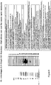
- ConA binding integral membrane proteins prepared in the same way from adult Haemonchus contortus or fourth and adult stages of O . ostertagi were compared by gel analysis and western blotting. Coomassie stained gels indicated differences in the profiles of all three fractions ( Fig 2a ), although additional bands present in the L4 but not in the adult O. ostertagi preparations were of most interest in this case.
- additional bands were still detected in the L4 fraction ( Fig 2b ).
- Serum antibody titres in the control group remained at background concentrations throughout ( Fig 3 ).
- a marked response was observed in the vaccinated group by week 5, two weeks after the second vaccination. This response reached a peak on week 8 two weeks after the third immunisation.
- FIG. 1 A flow chart depicting how these preparations were made is shown in Fig 1 , the details relating to which calves received which fraction and the dose of protein administered are presented in Table 5 whereas the SDS-PAGE profiles of the immunogens are shown in Figure 7 .
- Trial 3 Immunisation with sub-fractions of the 0.1 to 0.5M MonoQ pool.
- the object of this trial was to separate the protective antigens identified by Group 2.2 by gel filtration in order to narrow the identity of the candidate protective polypeptides. (No attempt was made to do this for the unbound fraction as too little protein was available for the task)
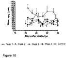
- the kinetics of the group mean egg counts of the calves in Trial 4 are shown in Fig 16 and the overall protective effects are summarised in Table 5.
- the mean egg outputs of all four groups of immunised calves were lower than the controls but there was little to choose between the efficacy of the different fractions.
- L4 preparation polypeptides which were additional to those observed in similar preparations from adult worms , were bovine in origin - perhaps from small pieces of abomasal tissue leaching from the mucosa when it was being incubated at 37C to recover the larvae.
- an immunoblot developed with serum from calves immunised with O.ostertagi proteins revealed that several of these bands could not have been bovine proteins.
Landscapes
- Health & Medical Sciences (AREA)
- Life Sciences & Earth Sciences (AREA)
- Veterinary Medicine (AREA)
- Pharmacology & Pharmacy (AREA)
- Chemical & Material Sciences (AREA)
- Public Health (AREA)
- General Health & Medical Sciences (AREA)
- Medicinal Chemistry (AREA)
- Animal Behavior & Ethology (AREA)
- Microbiology (AREA)
- Immunology (AREA)
- Mycology (AREA)
- Epidemiology (AREA)
- Tropical Medicine & Parasitology (AREA)
- Organic Chemistry (AREA)
- Nuclear Medicine, Radiotherapy & Molecular Imaging (AREA)
- General Chemical & Material Sciences (AREA)
- Chemical Kinetics & Catalysis (AREA)
- Medicines Containing Antibodies Or Antigens For Use As Internal Diagnostic Agents (AREA)
- Peptides Or Proteins (AREA)
- Micro-Organisms Or Cultivation Processes Thereof (AREA)
- Medicines Containing Material From Animals Or Micro-Organisms (AREA)
Description
- The present disclosure relates to antigens capable of raising host immune responses to nematode parasites. In particular, the disclosure provides vaccines for use in protecting against and/or reducing instances of non-blood feeding nematode parasite infections in bovine hosts.
- Ostertagia ostertagi is the most economically important helminth parasite of cattle in temperate parts of the world (1). As with all gastrointestinal nematodoses of ruminants, ostertagiasis is controlled almost entirely by the use of anthelmintic drugs combined with pasture management. Unfortunately, reports of anthelmintic resistance in bovine gastrointestinal nematodes are becoming more frequent. For example, benzimidazole (BZ) resistance was identified in cattle in New Zealand (2) and South America(3), and macrocyclic lactone (ML) resistance in New Zealand(4,5), the Americas(6,7,8) and Europe(9,10,11,12) However, as no detailed surveys have been made, the extent of the problem remains unknown and probably underestimated. It is anticipated that anthelmintic resistance in cattle nematodes including Ostertagia is likely to follow the pattern experienced with sheep, where it has reached serious proportions(13,14,15,16) although the pace at which it develops and spreads is likely to be slower.
- Alternative methods for controlling bovine ostertagiasis remain an attractive prospect, in part due to the threat of drug resistance and partly because of increasing consumer sensitivity to the possibility of chemical residues in meat and milk. One such possibility for control is by vaccination. Earlier attempts to do this, using either infection with irradiated larvae(17,18) or immunisation with crude somatic or excretory/secretory products of the parasites(19,20) were not successful. More recently promising results have been obtained using fractionated native excretory/secretory products of adult parasites, with reductions in faecal egg counts of up to 80% (21,22,23,24,25)
- De Maere V et al (2005: An aspartyl protease inhibitor of Ostertagia ostertagi: Molecular cloning, analysis of stage and tissue specific expression and vaccine trial: Molecular and Biochemical parasitology, 141 ) discloses an immunogenic composition comprising an Ostertagia ostertagi antigen.
- Wedrychowicz et al: (2002: Surface and excretory/secretory antigens of fourth-stage larvae and adult Ostertagia circumcincta: Veterinary parasitology 53 no 1-2, 1994, pages 117-132) discloses a composition comprising Ostertagia ostertagi antigens.
- Maere De V et al: (2002: Identification of potential protective antigens of Ostertagia ostertagi with local antibody probes: parasitology, 125, ) discloses Ostertagia ostertagi antigens identified by cDNA screening.
- During the last 15 years or so substantial protection against the important blood sucking ovine nematode, Haemonchus contortus, has been achieved by immunising sheep with various antigens isolated from the intestinal membranes of adult parasites, a topic which has been reviewed extensively(26,27,28). When the same gut antigen approach was tested against Ostertagia, some protection was conferred, but not at a level deemed to have practical potential(29). However as the same O. ostertagi antigens cross-protected efficiently against Haemonchus in sheep, it was reasoned that the relative failure may have been because adult Ostertagia are not blood feeders and do not ingest sufficient antibody for the gut antigen approach to be highly effective against them.
- It was hypothesised that developing fourth stage (L4) Ostertagia might be more vulnerable to this type of vaccination than adult parasites. Firstly, since they inhabit and damage the gastric glands, L4s are likely to be continuously exposed to inflammatory exudate which is richer in host immunoglobulin than the mucous surface inhabited by adult worms, in other words, L4s in vaccinated calves would be expected to ingest a bigger dose of antibody per unit weight than adult worms. Second, because developing Ostertagia L4s grow very rapidly (30), increasing their mass about 20-fold in 10 days, they may be more sensitive to digestive interference than their slower metabolizing adult counterparts.
- The present invention is as defined in the claims. In partuclar, the invention provides the antigens for use of
claim 1, the vaccine or vaccine composition for use ofclaim 2, the method ofclaim 8 and vaccine/vaccine composition ofclaim 9. - Disclosed herein are one or more antigens from the fourth stage (L4) larvae of non-blood feeding parasitic nematodes, for raising an immune response in an animal.
- Antigens from adult nematode parasites are used as vaccines for preventing, reducing or eliminating instances of animal infection. Antigens derived from adult parasitic nematodes, and in particular gut-derived antigens, can be used to raise protective host immune responses. Strategies of this type are particularly successful where the parasite is a blood-feeding nematode as they are routinely exposed to host immunoglobulin when taking blood meals. In contrast, parasites that do not feed on blood (the so-called "non-blood" feeders) are only occasionally (or in some cases never) exposed to blood and thus host circulating (humoral) antibodies to non-blood feeding parasite antigens are often ineffective.
- Without wishing to be bound by theory, the inventors hypothesise that in contrast to adult non-blood feeding nematodes, certain larval stages might be more regularly exposed to host immune factors including, for example, immunoglobulin. In particular, larvae which inhabit and damage gastric glands may frequently be exposed to host inflammatory exudate which comprises a variety of host inflammatory factors including immunoglobulin.
- The life cycle of a parasitic nematode comprises seven stages - an egg, four larval and two adult stages. The larval stages are commonly referred to as L1, L2, L3 and L4 (Borgsteede 1978) In non-blood feeding parasites, L4s inhabit host gastric glands, causing damage and inducing production of host inflammatory exudate. The L4s of all species which live in the true stomach (the abomasum in ruminants) do, but many species live in the intestines which obviously don't contain gastric glands. The intestines are lined by villi with glandular crypts between them. The L4s of the intestinal species reside in these crypts also causing local damage, so again are more likely than their adult counterparts to be exposed to inflammatory exudates.
- As such, antigens expressed by L4 stage nematode parasites may be used to raise protective host immune responses. Hereinafter, antigens expressed by L4 stage nematode parasites will be termed "L4 antigens".
- The disclosure relates to vaccines or vaccine compositions comprising one or more L4 antigens for raising immune responses in animals. Vaccines may be used prophylactically to prevent parasitic infection. In some cases the vaccines provided by this disclosure may be used to reduce infection or colonisation of a host by a nematode parasite. Animals exposed to the vaccines provided by this disclosure, may produce antibodies which bind to (or which exhibit affinity or specificity for) L4 antigens - such antibodies may otherwise be referred to as "protective" antibodies.
- The term "non-blood feeding nematode parasite" may include, for example nematode species from each of the following genera: Ostertagia, Teladorsagia, Trichostrongylus, Nematodirus, Cooperia, Chabertia, Oesophagostomum and Ascaris In particular, the disclosure relates to antigens derived from species belonging to any of the abovementioned genera and, the disclosure provides vaccines/vaccine compositions comprising antigens derived from L4 stage larvae of Ostertagia
- As such, the vaccines and vaccine compositions provided by this disclosure may be used to treat, prevent or reduce the symptoms of, diseases such as Bovine ostertagiasis.
- It should be understood that the term "antigens" may relate to, for example, proteins and/or peptides (including polypeptides and short peptide chains of one or more amino acids) including for example, glycoproteins and/or glycopeptides. In addition, the term "antigen" may relate to carbohydrate molecules. The disclosure relates to antigens expressed by L4 stage nematode parasites, such as, for example, antigens present in the cell membranes of L4 non-blood feeding nematode parasites. Antigens of this type may otherwise be known as "integral" membrane antigens. The antigens for use in raising immune responses may comprise antigens which are specifically expressed by L4 stage parasites. Antigens specific to L4 stage larvae may not be expressed by nematodes at other lifecycle stages (for example L2, L3 and adult stages). Antigens specific to L4 stage Ostertagia ostertagi are shown in, for example
Figure 2 . The antigens for use in this disclosure may comprise cell membrane antigens or antigens which are expressed by L4 stage nematodes and which are exposed to the host immune system. Such antigens may include antigens expressed on the surface or in the gut of L4 nematodes. It should also be understood that the term "antigens" relate to any fragments or immunogenic/antigenic fragments of the L4 antigens described herein. One of skill will appreciate that the term "antigens" may also encompass L4 proteins, polypeptides, peptides and/or carbohydrates which are otherwise known as "immunogens". - Antigens according to this disclosure may be obtained, purified or extracted from cell membrane preparations using a number of techniques. For example, cells obtained from nematodes may be subjected to lysis protocols (such as those involving Triton x-100) so as to fragment the cell membrane. Subsequent centrifugation techniques may be used to remove fragmented membrane debris and ultra-centrifugation to extract membrane proteins from supernates.
- Additionally, or alternatively, chromatography techniques such as, for example, those utilising affinity binding matrices, may be used to purify and/or extract membrane proteins from solution. Antigens for use may be purified/extracted using affinity matrices comprising concanavalin A (ConA) - a lectin which binds to certain structures present in, for example, carbohydrates, glycoproteins, and glycolipids. As such, "antigens" according to this disclosure may comprise proteins capable of binding ConA (ConA binding proteins) including, for example, glycoproteins proteins and/or peptides comprising a-linked mannose.
- In order to extract or purify antigens, a preparation of cell membrane proteins (for example proteins preparations prepared in accordance with the procedures outlined above), may be contacted with, for example, an affinity matrix under conditions which permit binding between the affinity matrix and antigens, for example (glycol) proteins/peptides, present in the membrane preparation. Antigens bound to an affinity matrix may be released or separated therefrom with the use of a suitable elution solution/buffer. In the case of an affinity matrix comprising ConA, antigens bound thereto may be separated or released by applying carbohydrate solutions. Additionally, material eluted from, for example, ConA affinity matrices, may be further subjected to procedures designed to remove carbohydrate. For example, material eluted from ConA affinity matrices may be contacted with Sephadex G-25 to remove carbohydrate. Antigen preparations of this type may be termed crude antigen preparations. Crude preparations of this type may be stored for prolonged periods of time at -20°C or, more preferably at -80°C.
- Crude antigen preparations of the type described above may be further processed in order to yield antigen fractions comprising fewer and more highly purified (or cleaner)/concentrated antigens. By way of example, techniques such as, for example, anion exchange and/or gel filtration may be used to prepare one or more fractions of the crude antigen preparations described above.
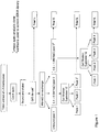
- In one embodiment, the method of obtaining L4 antigens for use in raising animal immune responses is that shown in
Figure 1 . - The various cell membrane protein preparations and fractions thereof described herein may otherwise be referred to as "antigen pools". Accordingly, the vaccines and vaccine compositions provided by this disclosure may comprise one or more of the antigen pools described herein. By way of example, vaccines for use in raising immune responses to the nematode parasite Ostertagia ostertagi may comprise one or more antigen pool(s) selected from the group consisting of (i) a pool comprising ConA binding proteins from a cell membrane preparation and (ii) fractions of ConA binding protein mixtures obtained by one or more rounds of anion exchange and/or gel filtration. The vaccine or vaccine provided by this disclosure comprises one or more of the ConA,
pool 1,pool 2 and/orpool 3 fractions described in the detailed description section (see part entitled "Preparation of Immunogens") and inFigure 1 and Table 5. The vaccine or vaccine composition provided by this disclosure may comprise the antigen pool used inTrial 1,Trial 2,Trial 3 and/orTrial 4 as shown in Table 5 andFigure 1 and described in the detailed description section. - In one embodiment, the antigens for use in this disclosure are derived from the L4 stage of the non-blood feeding bovine parasite, Ostertagia ostertagi. As such, the disclosure may provide one or more antigens from a fourth stage larvae of Ostertagia ostertagi, for raising an immune response in a bovine. Specifically, the disclosure may relate to ConA binding proteins or membrane (glyco)proteins and/or peptides from L4 stage Ostertagia ostertagi, for raising an immune response in a bovine.
- In one embodiment, the bovine is a neonatal or juvenile bovine - otherwise known as a calf.
- Antigens for use in raising animal immune responses may be obtained from whole or fragmented parasites harvested from donor animals. Donor animals may be naturally infected animals or animals which have been deliberately (or experimentally) infected with a particular parasite. For example, experimentally infected animals may be administered a dose of L3 stage parasites sometime before L4 stage parasites are harvested. One of skill will appreciate that the time between administration of an L3 dose and harvesting L4 parasites will vary depending, on for example, the life cycle of the parasite to be harvested. In the case of the non-blood feeding parasite, Ostertagia ostertagi, L3 stage larvae may be administered approximately 7 days before L4 larvae are harvested.
- In addition to providing proteins or peptide antigens from non-blood feeding L4 nematode parasites, the present disclosure relates to nucleic acid molecules encoding the same or fragments (preferably antigentic or immunogenic fragments) thereof. The nucleic acid may be DNA, RNA or a combination thereof and can include any combination of naturally occurring, chemically or enzymatically modified nucleotides. Furthermore, the nucleic acid may be double or single stranded. Within the scope of this disclosure are nucleic acid sequences that are substantially complementary to any of the L4 antigen sequences described herein.
- It should be understood that the term "substantially complementary" encompasses those nucleic acid molecules exhibiting a degree of sequence identity/homology with any of the L4 antigen nucleic acid sequences described herein - such as, for example, those presented in Tables 1 and 3 below. A sequence having a level of identity with a L4 antigen nucleic acid sequence of this disclosure may exhibit at least 40%, 50%, 60%, 65%, 70%, 75%, 80%, 85%, 90%, 95%, 96%, 97%, 98% or 99% identity with the full length L4 antigen nucleic acid sequence or any portion or fragment thereof. One of skill will appreciate that a level of sequence identity may be determined by comparing aligned nucleic acid sequences over a predetermined length so as to determine the number of positions at which an identical nucleic acid base occurs in both sequences to yield the number of matched positions, dividing the number of matched positions by the total number of nucleic acid bases in the length compared and multiplying the result by 100 to yield the percentage of sequence identity.
- The nucleic acid molecules provided by this disclosure may take the form of nucleic acid constructs or vectors such as, for example a cloning or expression cassettes/vectors. Vectors provided by this disclosure may be capable of directing the expression of nucleic acid sequences encoding L4 stage antigens in, for example, bacterial, fungal, mammalian and/or insect cells.
- Accordingly, a second aspect of this disclosure provides a vector, preferably an expression vector, comprising a nucleic acid sequence encoding any of the L4 antigens described herein (including, fragments, variants, analogues or derivatives thereof). Expression vectors suitable for use in this aspect of the disclosure may further comprise one or more promoter sequences capable of directing expression in prokaryotic or eukaryotic cells such as, for example, mammalian, fungal, bacterial, plant and/or insect cells.
- A vector provided by this disclosure may be circular or linear, single stranded or double stranded and can include DNA, RNA or a combination or modification thereof. Furthermore, vectors of this disclosure may be, for example, plasmids, cosmids or viral vectors (for example retroviral or bacteriophage vectors). Vectors provided by this disclosure may further comprise selection or marker elements, for example antibiotic resistance genes and/or optically detectable tags. A large number of suitable vectors are known and further information may be obtained from Pouwels et al. Cloning Vectors: a Laboratory Manual (1985 and supplements), Elsevier, N.Y.; and Rodriquez, et al. (ads.) Vectors: a Survey of Molecular Cloning Vectors and their Uses, Buttersworth, Boston, Mass (1988) - both of which are incorporated herein by reference.
- In addition to techniques in which L4 antigens are extracted or purified from membrane preparations from harvested parasitic organisms, antigens for use may be obtained using recombinant technology. In one embodiment, an expression vector comprising one or more nucleic acid sequences encoding L4 antigens may be used to produce recombinant L4 antigens. Accordingly, in a further aspect, the present disclosure provides host cells transfected or transformed with a vector as described herein. Eukaryotic or prokaryotic cells, such as, for example, plant, insect, mammalian, fungal and/or bacterial cells, may be transfected with one or more of the vectors described herein. One of skill in this field will be familiar with the techniques used to introduce heterologous or foreign nucleic acid sequences, such as expression vectors, into cells and these may include, for example, heat-shock treatment, use of one or more chemicals (such as calcium phosphate) to induce transformation/transfection, the use of viral carriers, microinjection and/or techniques such as electroporation. Further information regarding transformation/transfection techniques may be found in Current Protocols in Molecular Biology, Ausuble, F.M., ea., John Wiley & Sons, N.Y. (1989) which is incorporated herein by reference.
- Another aspect of this disclosure relates to a host cell transformed with any one the nucleic acid constructs described herein. Suitable host cells include prokaryotic and/or eukaryotic cells. For example, bacterial, fungal, mammalian, plant and/or insect cells are all capable of being transformed with any of the nucleic acid constructs described herein. A host cell transformed with a nucleic acid construct provided by this disclosure may be referred to as a "transformant". Where the vector comprises a selection/marker element, transformants may be selected by application of antibiotics to culture media.
- In view of the above, the present disclosure further provides a process for the production of a recombinant L4 protein or peptide for use in raising an immune response in an animal, said method comprising the step of (a) transforming a host cell with a nucleic acid sequence according to this disclosure or transfecting a host cell with a nucleic acid construct of the disclosure; (b) culturing the cells obtained in (a) under conditions in which expression of the protein takes place; and (c) isolating the expressed recombinant protein or peptide from the cell culture or/and the culture supernatant.
- Recombinant proteins/peptides produced according to the method described above may be partially purified from the host cell before being used as a vaccine. Where the polypeptide is secreted from the host cell, the cells may be separated from the media by centrifugation, the cells being pelleted and the media being the supernatant. In such a situation, the supernatant, which contains the secreted polypeptide, may be used directly as a vaccine, or in a vaccine composition. Alternatively, the polypeptide may be partially purified from this supernatant, for example using affinity chromatography.
- In one embodiment, purified L4 antigens/recombinant antigens may be admixed with another component, such as another polypeptide and/or an adjuvant, diluent or excipient. Vaccines or vaccine compositions provided by this disclosure may contain bacterial antigens used to control other diseases. For example, the vaccine or vaccine composition may be included within a multivalent vaccine which includes antigens against other bovine diseases.
- In a still further aspect, the present disclosure provides a bovine population treated or immunised with a vaccine or composition described herein. In one embodiment, the bovine population is a neonatal or juvenile bovine (i.e. calf) population.
- One of skill will appreciate that the vaccines described in this disclosure may take the form of subunit-type vaccines where by one or more proteinaceous L4 antigens are used to inoculate an animal. Additionally or alternatively, the vaccine may comprise a nucleic acid molecule (known as a DNA vaccine) encoding one or more L4 antigens to be expressed by the cells of an animal to be vaccinated.
- Suitable antigens for raising bovine immune responses to the non-blood feeding parasite Ostertagia ostertagi may include, for example, one or more antigens encoded by cDNA sequences having or comprising the following cDNA sequences (Tables 1 and 3):
Table 1: O. ostertagi L4 cDNA library screen (with antiserum from protected calves immunised with pool 3 fraction from Trial 2 - see Fig 1) Clone1.1 

Clone2.1 
Clone3.1 
Clone4.1 
Clone6.1 
Clone7.1 
Clone8.1 

Clone8.2 
Clone9.1 
Clone10.2 
Clone11.2 
Clone12.2 
Clone14.1 
Clone14.2 
Clone16.1 
Clone17.1 
Clone18.1 
Clone 18.2 
Clone19.1 
Clone20.1 TCGATCCGATCCGATCCGATCCGATCCGATCGATCGTCTCTCTCTCCCCC Clone21.1 
Clone 21.2 
Clone22.1 
Clone 22.2 

Clone23.1 
Clone 23.2 
Clone24.1 
Clone25.1 
Clone26.1 
Clone27.1 
Clone28.1 
Clone29.1 
Clone 29.2 
Clone30.1 
Clone 30.2 
Clone31.1 
Clone 31.2 
Clone32.1 
Clone 32.2 
Clone33.1 
Clone34.1 
Clone35.1 GATCGATTCCGGATCCGGATCCGGATCCGATCGATCGATCGTCGTCGCGCGACGACGCGCCCCCCCC Clone 35.2 
Clone36.1 GTCGATCGATCGATCGATCGTCGTCGTCGTCGTCGTCGTCGTCGTCTCTCTCTCTCTCTC Clone 36.2 
Clone37.1 GGCGTCGTCGTCGTCGTCGTCGTCGTCGTCTCTCTCTCTCTCTCTCCCCC Clone 37.2 
Clone38.1 
Clone 38.2 
Clone39.1 
Clone 39.2 
Clone 42.1 
Clone 45.1 
Clone48.1 

Clone49.1 
Clone50.1 
Clone51.1 
Clone 51.2 
Clone52.1 
Clone54.1 
Clone 54.2 
Clone55.1 GCGTCGATCATCATCGATCACATCATCACACACAAAAAAAAAACCCCC Clone56.1 
Clone57.1 GATCGGATCGGATCGATCGATCGACGAACGACGACGACACGAGATGTCGTCGCGCCGGA Clone58.1 GATCCGATTCCGATCCGATCCGATCCGATCGATCGTCGTCGTCGTCGTCC Clone 58.2 
Clone59.1 
Clone 59.2 
Clone60.1 
Clone61.1 
Clone 61.2 
Clone62.1 
Clone 62.2 
Clone63.1 
Clone 63.2 
Clone64.1 
Clone65.1 
Clone66.1 
Clone 66.2 
Clone67.1 
Clone 67.2 
Clone68.1 GTCGATCGATCCGAATCGAATCGATCGATCGATCGTCGTCTCTCATCTCCCACCTCCATCATATAT Clone69.1 
Clone 69.2 
Clone70.1 
Clone 70.2 
Clone71.1 
Clone72.1 
Clone73.1 GATTCGATTCGATCGATCGTCGTCTCTCTCTTTTTTTTTTTTTTT Clone74.1 
Clone75.1 
Clone76.1 
Clone77.1 
Clone78.1 
Clone 78.2 
Clone79.1 
Clone 79.2 
Clone80.1 
Clone81.1 
Clone82.1 
Clone83.1 
Clone 83.2 
Clone84.1 
Clone85.1 
Clone86.1 
Clone87.1 
Clone88.1 
Clone89.1 
Clone90.1 
Clone91.1 
Clone92.1 
Clone93.1 
Clone94.1 
Clone 94.2 

Clone95.1 TCGTCGTCGTCGATCGTCGTCGTCGACCCCACACGACCCTCACACA Clone 95.2 
Clone96.1 
Clone 96.2 
Clone97.1 
Clone98.1 GATCCGATCCGATTCCGATCCGATCCGATCCGATCGTCGTCGTCTCTCCC Clone 98.2 
Clone99.1 
Clone 99.2 

Clone100.1 
Clone101.1 
Clone102.1 
Clone103.1 
Clone104.1 
Clone105.1 
Clone106.1 
Clone 106.2 
Clone107.1 
Clone108.1 GAAAAAAAAAAAAAAAAA Clone109.1 
Clone 109.2 
Clone110.1 
Clone111.1 
Clone112.1 
Clone113.1 
Clone114.1 GCCTCATCGATCGATCATCATCATCATCATCGTCTCATCTCTCGTCGTCTCTC Clone115.1 
Clone116.1 GGGCCGTCACGTCGATCATCGACGCAACGTCGCTGTCGTGTCATCAGTCGTGACACG Clone117.1 GGGCTGGCACGGAATTCCTTCACTCACGATCGTGTTGTTTGGAGATAGCTGTGAGTGCTGCG Clone 117.2 
Clone118.1 GGGAATGTTGACAGTGAAGAGTAATTGTTCTTGTTATCATAATGTGTTTAAAAAAAAAACGCG Clone119.1 
Clone120.1 GGGATTCACGTCGGCTCATCCAGCTTGGCTCGACAGTGAATCGACCACGACAGTGTCATCCAACACA Clone 120.2 
Clone121.1 GCTCATCTCGATCGTCATCATCATCATCGCATCCCTCCTTA Clone122.1 GGCCCTTCACTCGATGTCACACATCATCGCATAGCTTATGTTATATTTGATAAGTTAGAAG Clone123.1 
Clone124.1 
Clone125.1 GGGATACTGGATCAGACAGCACACCATTCCTCTGTCTTCAGTAACCTATAGAGATATAA Clone 125.2 
Clone126.1 GGTACGAAAGGAGAACCATCGCAACAACGAACGACTCGTAGTGTTCCGGTCGTCGCACTGCAGTG Clone 126.2 
Clone127.1 GGGCGAATCATCGAATCGCGACCGCGCAATTCGTCACGTCGACTTCGTGACGCGTAGTACTGCACG Clone 127.2 
Clone128.1 
Clone 128.2 
Clone129.1 
Clone130.1 GGATCGTCCGTCGTCCGTCATCGATCACGCTCGCGTGTGCGTACACAC Clone131.1 
Clone132.1 GGTGTCCATCGTCCGATCATCGCACGATCGACGTCGCTCTCCTCCGTGC Clone133.1 
Clone134.1 
Clone 134.2 
Clone135.1 

- In a further embodiment, the antigens and/or vaccine/vaccine compositions provided by this disclosure may comprise proteins and/or peptides comprising sequences encoded by any one of the nucleic acid sequences shown in Table 1.
Table 2, below, identifies some specific proteins/peptides encoded by cDNA sequences comprising the nucleic acid sequences detailed in Table 1. O. ostertagi L4 cDNA library screen Clone I.D. 1.1 Cathepsin B6 2.1 Ser/Thr protein phosphatase (Wormbase) 3.1 GAL-1 4.1 no hit 6.1 Myosin 7.1 ?apyrase (T. circ. 40.3 kDa protein) ?thrombospondin (wormbase) 8.1 ASP-like protein ? 8.2 ? Cytochrome b5 9.1 Globin-like host-protective protein 10.2 T. circ similar to Cadherin 11.2 ASP-like protein ? 12.2 NADH dehydrogenase ND4L 14.1 G-protein coupled receptor (Wormbase) 14.2 Haemonchus est contains THR repeat element 18.1 Cathepsin B 18.2 Cysteine PRotease related family member (cpr-6) 19.1 NADH ubiquinone oxidoreductase 21.2 Unknown 22.2 Putative amino acid permease 23.1 unknown 29.1 Unknown 30.2 Unknown 31.1 O. ostertagi est 31.2 Unknown 32.2 Unknown 35.2 Prolyl Carboxy Peptidase like family member (pcp-4) 36.2 Amine transmembrane transporter activity 37.2 Unknown 38.1 Ribosomal protein 38.2 Ribosomal protein S19S 42.1 Putative cytochrome c 45.1 C-type single domain activation associated secreted protein ASP3 precursor 51.2 Putative Elongation Factor 52.1 ubiquitin 54.2 APYrase family member (apy-1) 58.2 Glyceraldehyde-3-phosphate dehydrogenase 59.2 Legumain 60.1 T. circ adult cDNA 61.1 O.ost L4 est 61.2 60S ribosomal protein 62.1 metallo panstimulin 62.2 Unknown 63.1 T. circ est 63.2 Unknown 65.1 Ribosomal protein 66.2 Formate dehydrogenase 67.2 17 kDa ES antigen protein 68.1 O. ostertagi putative ES protein 69.1 H. sapiens chromosome 469.2 Unknown 70.1 unknown 70.2 Unknown 75.1 O. ostertagi library 77.1 T. circ est 78.2 Aspartyl protease precursor 79.1 O. ostertagi similar to ATP synthase 79.2 ATPase, F1 complex, epsilon/delta subunit 80.1 O. ostertage cytochrome c oxidase 81.1 unknown 83.2 Neuropeptide-Like Protein family member (nlp-42) 87.1 Antigenic glycoprotein precursor 30kDa 89.1 unknown 91.1 unknown 92.1 lumen protein? Receptor 94.2 Unknown 95.1 unknown 95.2 Isoleucyl tRNA Synthetase family member (irs-2) 96.2 Glyceraldehyde-3-phosphate dehydrogenase 98.1 unknown 98.2 Unknown 99.1 Cadherin metallothionin 99.2 Unknown 100.1 Protein disulphide isomerase 103.1 Ribosomal protein 106.2 Cytochrome b 109.1 unknown 109.2 Metalloprotease I 112.1 Cytochrome c oxidase 113.1 Ribosomal protein 116.1 Elongation factor 117.2 Unknown 118.1 unknown 120.1 O. ostertagia 120.2 Unknown 125.2 Rab3 GTPase-activating protein 126.1 ASP3 precursor (ES protein) 126.2 C-type single domain activation associated secreted protein ASP3 precursor 127.2 Alpha-ketoglutarate-dependent sulfonate dioxygenase 128.1 Troponin 128.2 Troponin family protein 129.1 Aspartyl protease 134.2 NADH dehydrogenase subunit 4Table 3: O. ostertagi L4 cDNA library screen (with antiserum from protected calves immunised with pool 3 fraction from Trial 3 - see Fig 1)Clone 1.1 
Clone2.1 
Clone 4.1 
Clone 5.1 
Clone 6.1 
Clone 7.1 
Clone 8.1 

Clone 9.1 
Clone 10.1 CGGCATTACGGCCGGGGTCCACTTCGATGAAATAAATTTCGCAGTTGT Clone 12.1 
Clone 13.1 
Clone 14.1 
Clone 19.1 
Clone 20.1 
Clone 21.1 
Clone 23.1 
Clone 24.1 
Clone 25.1 
Clone 26.1 
Clone 28.1 
Clone 30.1 
Clone 32.1 

Clone 33.1 
Clone 37.1 
Clone 38.1 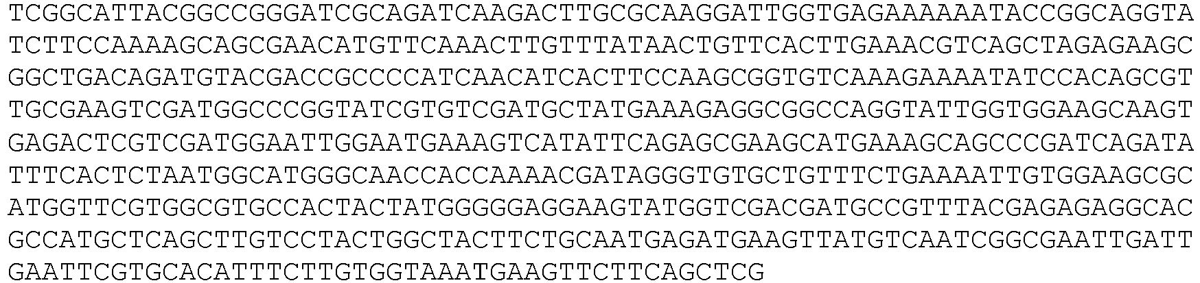
Clone 39.1 
Clone 41.1 
Clone 42.1 TTACGGCCGGGCAGCAAGTTCAACGAAATATCGTCGAAGCACCAGCACCATGCAAAGCATGCTC Clone 44.1 

Clone 45.1 
Clone 46.1 
- In a further embodiment, the antigens and/or vaccines/vaccine compositions provided by this disclosure may comprise proteins and/or peptides comprising sequences encoded by any one of the nucleic acid sequences shown in Table 3.
Table 4, below, identifies some specific proteins/peptides encoded by cDNA sequences comprising the nucleic acid sequences detailed in Table 3. O. ostertagi L4 cDNA library screen Clone I.D. 1.1 Chain A, Cekdm7a From C.Elegans, Complex With H3k4me3 Peptide And Nog 2.1 Serpentine Receptor, class H family member (srh-214) 4.1 Unknown 5.1 Ribosomal Protein, Small subunit family member (rps-13) 6.1 17 kDa ES antigen protein 7.1 putative L3 ES protein 8.1 AIDA-1b, putative 9.1 Putative L3 ES protein 12.1 Globin-like ES protein F6 13.1 C-type single domain activation associated secreted protein ASP3 precursor 14.1 DNA-directed RNA polymerases I, II, and III 14.4 kDa polypeptide 19.1 Unknown 20.1 Flavin-containing MonoOxygenase family member (fmo-1) 21.1 Unknown 23.1 Metalloprotease I 24.1 Putative L3 ES protein 25.1 60S ribosomal protein L5 26.1 Collagen col-34 28.1 Nuclear Hormone Receptor family member (nhr-9) 30.1 Seven TM Receptor family member (str-66) 32.1 C. briggsae CBR-PMS-2 protein 33.1 Barrier to Autointegration Factor family member (baf-1) 38.1 Secreted protein 4 precursor39.1 Elongation factor Tu homologue precursor 41.1 17 kDa ES antigen protein 44.1 C. briggsae CBR-CDH-4 protein 45.1 ACOnitase family member (aco-2) 46.1 INneXin family member (inx-14) - In a further embodiment, the disclosure relates to one or more of the Ostertagia ostertagi antigens identified in
Figures 6 and14 . These antigens have been identified by mass spectrometry analysis of gel slices obtained from PAGE analysis of L4 antigen preparations obtained in accordance with this disclosure. - In addition to providing L4 antigens for use in raising immune responses in animals, the present disclosure may also provide polyclonal and/or monoclonal antibodies (or antigen binding fragments thereof) that bind (or have affinity or specificity for) any of the L4 antigens described herein - including those comprising sequences encoded by the cDNA sequences of Table 1. Production and isolation of polyclonal/monoclonal antibodies specific for protein/peptide sequences is routine in the art, and further information can be found in, for example "Basic methods in Antibody production and characterisation" Howard & Bethell, 2000, Taylor & Francis Ltd. Such antibodies may be used in diagnostic procedures, as well as for passive immunisation.
- The present disclosure further provides a vaccine for use in preventing or controlling disease in bovine hosts caused by non-blood feeding nematode parasites. The vaccine may be a polypeptide or polynucleotide vaccine.
- The disclosure further provides a method for immunising bovine animals against non-blood feeding nematode parasites, said method comprising the step of administering to the bovine a vaccine of the disclosure.
- The present disclosure will now be described in detail with reference to the following figures which show:
-
-
Figure 1 . Flow chart of the methods used to prepare the antigen fractions for the calf vaccination trials. -
Figure 2 . SDS-PAGE and immunoblot analysis of ConA binding membrane proteins from H. contortus and O. ostertagi. a, coomassie blue stained. b, Immunoblot probed with sera from calves immunised with ConA binding membrane proteins from adult O. ostertagi (Smith et al, 2000). M, molecular weight markers.Lanes lanes lanes -
Figure 3 . Kinetics of the antibody response following vaccination of calves with O.ostertagi L4 antigens or adjuvant alone inExperiment 1. Closed squares = vaccinates; open triangles = controls. -
Figure 4 . Group mean faecal egg counts of vaccinated and control calves inExperiment 1. Closed squares = vaccinated animals; open triangles = controls. -
Figure 5 . Group mean worm counts of vaccinated and control calves inExperiment 1. -
Figure 6 . Mass spectrometry fingerprint analysis of the protective ConA binding fraction used inTrial 1. -
Figure 7 . SDS-PAGE of the preparations used as antigens inTrial 2.Lane 1 = ConA binding fraction;lane 2 =Pool 1;lanes Pool 2 andlane 5 =Pool 3. The remaining lanes contain molecular weight markers. -
Figure 8 . Kinetics of the antibody response following vaccination of calves with O.ostertagi L4 fractions or adjuvant alone inTrial 2. Closed squares = group 2.1. Open squares = group 2.2. Closed circles = group 2.3. Open circles = group 2.4. Open triangles = group 2.5 (controls). -
Figure 9 . Group mean faecal egg counts of vaccinated and control calves inTrial 2. Closed squares = group 2.1. Open squares = group 2.2. Closed circles = group 2.3. Open circles = group 2.4. Open triangles = group 2.5 (controls). -
Figure 10 . Group mean worm counts of the calves inTrial 2. *, P < 0.05. -
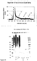
Figure 11 . Chromatography and gel profiles of the preparations used as antigens inTrial 3. -
Figure 12 . Group mean faecal egg counts of vaccinated and control calves inTrial 3. -
Figure 13 . Group mean cumulative faecal egg counts of vaccinated and control calves inTrial 3. -
Figure 14 . Mass spectrometry fingerprint analysis of theprotective Peak 3 fraction used inTrial 3. -
Figure 15 . Chromatography and gel profiles of the preparations used as antigens inTrial 4. -
Figure 16 . Group mean faecal egg counts of vaccinated and control calves inTrial 4. - All calves were reared and housed indoors in conditions designed to exclude accidental infection with nematode parasites. Those used as donors for O.ostertagi eggs or fourth stage larvae were of various breeds and aged between 3 and 12 months at the time of infection. Those used in the vaccine trials were castrated Holstein-Fresian crosses aged between 6 and 12 months at the start of each trial.
- Infective larvae were from strains of O. ostertagi which have been maintained at Moredun Research Institute for several years.
- The methods for faecal egg counting and enumeration of worm burdens have been described before(31,32). Fourth stage O. ostertagi larvae were harvested from donor calves which had been infected with a single dose of approximately 200,000 L3 seven days earlier. Soon after the animals had been killed by captive bolt and pithing, the abomasums were removed and the contents discarded. After a brief rinse in warm saline, each abomasum was pinned mucosal surface uppermost to a block of polystyrene which was then inverted and floated in a large Baermann funnel containing warm saline. Following four hours at 37°C, fourth stage larvae were drained from the base of the funnel. The funnels were then incubated at 4°C overnight by which time any larvae still in suspension had settled out and could be drawn off. All larvae were frozen at -70°C until required for antigen extraction.
- Prior to SDS-PAGE samples were heated at 100°C for 3 min in an equal volume of 63mM Tris-HCl pH6.8 containing 5% (w/v) SDS, ± 10mM DTT under non-reducing or reducing (10mM DTT) conditions and separated on 4-12% gradient acrylamide gels (BIORAD, Hercules, CA, USA). Molecular weight markers (Fermentas, Burlington, Ontario, Canada) were run on each gel and the gels were either stained with coomassie blue R250 (SIGMA, St Louis. MO, USA) (0.025 % in 40 % methanol / 10 % acetic acid) and destained in 20 % methanol / 10 % acetic acid, or silver stained as follows. After SDS-PAGE the gels were washed 3 times in distilled water, and then fixed overnight in 40 % methanol / 10 % glacial acetic acid. This was followed by incubation for 20 min in 20 % methanol / 5 % acetic acid then 4 x 15 min washes in distilled water. Gels were then incubated in 50
ml 5 mg/L DTT for 45 min, then for 40 min in 50ml 0.1 % w/v AgNO3, followed by 2 rapid washes in water and 2 washes in 25ml 3 % Na2CO3. The gels were then developed in 50ml 3 % Na2CO3 with the addition of 25 µl formalin, and the development stopped after 15 min by adding 20 ml 2.3 M citric acid. - SDS-PAGE separated proteins were transferred to PVDF membrane (Millipore, Billerica, MA, USA) using a semi-dry apparatus. Membranes were blocked in 10% Marvel (Premier Foods International, Spalding, Lines., UK) in 10mM Tris, 0.5M NaCl, 0.05% (v/v) Tween-20, 0.02% (w/v) thimerosal (TNTT), the assay diluent and wash buffer, overnight at 4°C. Periodate treatment was carried out by washing the membrane twice, for 20 minutes, in 50mM NaAc pH 4.5, then incubating for 1 h in 50mM NaIO4 / 50mM NaAc, in the dark at room temperature. After further washes of 2 x 10 minutes in 50mM NaAc, then 2 x 10 minutes in TNTT, the membrane was incubated for 30 minutes in 50mM NaBH4, after which it was washed for 3 x 10 minutes in TNTT. Membrane strips were incubated with pooled serum samples from each group, diluted 1/300 in TNTT, for 2 h at room temperature. They were then washed 3 x 5 minutes in TNTT, then incubated with rabbit anti-bovine immunoglobulin horseradish peroxidise conjugated antibody diluted 1/1000 in TNTT (P0159, DAKOcytomation, Glostrup, Denmark).
- These were estimated by the bicinchoninic protein assay reagent according to the manufacturer's instructions (Pierce, Thermo Fisher Scientific Inc., Waltham, MA, USA).
- Triton X-100 extracts of Ostertagia L4 membranes were prepared as detailed for Haemonchus (33), and diluted four-fold with 10mM Tris-HCl, 0.5M NaCl, 0.05% NaN3, 10µM MnCl2, 100µM CaCl2, pH 7.4 (Lectin Wash Buffer, LWB). The solution was pumped (8 ml/h) at 4°C through ConcanavalinA (ConA) lectin cross linked to agarose beads (Vector Laboratories, Burlingame, CA, USA) contained in a column. After thorough washing in LWB / 0.5 % reduced Triton X-100 the column was eluted with LWB / 0.25% CHAPS / 0.2M methylmannopyranoside / 0.2M methylglucopyranoside (
Figure 1 flow chart). For elution, sufficient sugar solution was pumped onto each column to cover the beads, then the flow was stopped for approximately one hour. The pump was re-started and the peak monitored at OD280 was retained as the "1 hour eluate". The elution process was then repeated exactly, except that the flow was stopped overnight to produce an "overnight eluate". The eluates were pooled and passed through a column of Sephadex G-25 to remove the sugar and exchange the buffer to 10mM Tris-HCl, 0.1% CHAPS , pH 7.4 and stored at -70°C before use as immunogens. - The ConA eluate was fractionated on a MonoQ anion exchange column, 1ml bed volume (Pharmacia, Pfizer, Kent, UK) equilibrated in 10mM Tris / 0.1 % CHAPS pH7.4. The ConA eluate was applied to the column (1 ml/min), and unbound proteins were collected. The bound proteins were eluted by a linear gradient increase in NaCl from 0 M to 1 M over 20ml, with 10 x 2ml fractions being collected. The fractions were then pooled as follows:-
Pool 1 = unbound material and proteins eluted with up to 0.1M NaCl;Pool 2 = fractions eluted between 0.1 and 0.5M NaCl andPool 3 = fractions eluted between 0.5 and 1.0M NaCl (Fig flow chart). Further batches ofPool Pool 2 containing 0.75ug of protein was separated in 2 runs was fractionated on a single 30cm column.Pool 3 was fractionated under identical conditions except two 30cm columns were coupled in series to improve the resolution. - Microtitre plates were coated overnight at 4°C with 50 µl coating protein per well (ConA eluate), at 0.5 µg/ml in 50 mM sodium bicarbonate buffer, pH 9.6. The plates were washed six times with wash buffer (PBS, 0.05% v/v Tween-20), then incubated with 200µl 10% (w/v) infasoy (Cow and Gate, Trowbridge, Wiltshire, UK) in TNTT overnight at 4°C. After washing, 50µl serum per well, diluted 1:2000 in TNTT, were added for 1 h at room temperature. The wells were re-washed and 50 µl peroxidase conjugated rabbit anti-bovine immunoglobulin diluted 1:1000 in TNTT added for 1 h at room temperature. After a final wash, 50µl o-phenylenediamine dihydrochloride substrate (Sigma) were added to each well. After 10 min in the dark, the colour reaction was stopped by addition of 25 µl 2.5 M sulphuric acid per well and OD values read at 490 nm. Each test sample was assayed in triplicate. Pooled serum taken at the time of challenge from the group of calves in
Experiment 2 immunised with the unfractionated ConA eluate was included on each plate as a reference sample, and OD values expressed relative to this value. - The ConA binding fraction used in
Trial 1 andPeak 3 employed inTrial 3 were fractionated by 1-dimensional SDS-PAGE under reducing conditions. Each sample (approximately 10 µg) was mixed with 10 µL SDS-PAGE sample buffer (0·05 m Tris,pH 6·8, containing 5% (w/v) SDS, 20% (v/v) glycerol, 0·01% (w/v) bromophenol blue and 10 mm DTT), boiled for 5 min before loading onto 10% gels with a 3% stacking gel. After protein separation, gels were stained with colloidal Coomassie Blue (SimplyBlue™ SafeStain, Invitrogen), destained in water and the image of each track captured. Mass spectrometry analysis was performed at the Moredun Research Institute's Proteomics Facility <http://www.mri.sari.ac.uk/fgu-functional-genomics-services.asp>. Each gel track was sliced horizontally into about 27equal gel slices of approximately 2·5 mm each and individual slices were finely chopped (approximately 1 mm3), transferred to clean 0·5 mL Eppendorf tubes and processed using standard in-gel reduction, alkylation and trypsinolysis steps (15). Digest supernatants of 20 µL final volume were transferred to HPLC sample vials and stored at 4°C until required for liquid chromatography-electrospray ionization-tandem mass spectrometry (LC-ESI-MS/MS) analysis. Liquid chromatography was performed using anUltimate 3000 nano-HPLC system (Dionex) comprising a WPS-3000 well-plate micro auto-sampler, a FLM-3000 flow manager and column compartment, a UVD-3000 UV detector, an LPG-3600 dual-gradient micropump and an SRD-3600 solvent rack controlled by Chromeleon chromatography software (Dionex: http://www1.dionex.com). Samples of 4 µL were applied to the column by direct injection. Peptides were eluted by the application of a 15-min linear gradient from 8% to 45% solvent B (80% acetonitrile, 0·1% formic acid) and directed through a 3-nL UV detector flow cell. LC was interfaced directly with a 3-D high capacity ion trap mass spectrometer (Esquire HCTplusTM, Bruker Daltonics) utilizing a low-volume (50 µL/min maximum) stainless steel nebuliser (Agilent, catalogue number G1946-20260) and ESI. MS/MS analysis was performed as previously described (16). A peak list file was generated from the resultant data and submitted to a local database server using the MASCOT search engine for protein database searching against NCBInr <http://www.ncbi.nlm.nih.gov/> and Nembase <http://www.nematodes.org/nematodeESTs/nembase.html> databases. The modifications used in these searches were a global modification of carbamidomethyl (C) and a variable modification of oxidation (M). The tolerances used were; for MS data, 1·5 Da, and for MS/MS data, 0·5 Da. Matches achieving a significant molecular weight search (MOWSE) score were considered significant if two peptides matched for each protein, each of which had to contain an unbroken b or y ion series of a minimum of four amino acid residues. The other criterion considered in assigning a positive identification for each protein was a concordance between the calculated theoretical molecular mass value of the protein and the observed position of the peptide on 1-D gel electrophoresis. - This was made in Lambda TriplEX (Clontech) and amplified x 1 according to the manufacturer's instructions as described previously (Skuce et al, 1999). Ten-fold dilutions of the unamplified primary library were made in SM buffer over the dilution range 10-1 - 10-5. A 10 µl aliquot of each dilution was mixed with 200 µl of XL1-Blue plating cells (OD·600 = 0.5) and incubated for 30 min at 37°C to allow the phagemids to bind to the cells. After incubation, 4 ml of NZY top agarose at 48°C was added and the mixture was plated onto pre-warmed 100 mm diameter LB-agar plates. After the top agarose solidified the plates were incubated overnight at 37°C. The 10-1 dilution plate had 293 plaques, therefore the primary library contained 2.93 x 10⁵ pfu/ml.
- The amplified cDNA library was titrated as above except that the top NZY agarose was supplemented with 100 µl of 100 mM IPTG and 80 µl of 50 mg/ml X-gal to allow the selection of a blue "wild type" phagemid plaque. Several blue plaques were identified and agar plugs containing individual plaques taken into 0.5 ml SM buffer containing 20 µl CHCl3, to prevent bacterial growth, and stored at +4°C.
- The SM buffer supernatant, prepared as above, containing a wild type Lambda TriplEx phagemid was titrated and a dilution that gave near confluent plaques was selected. This clone was plated as above and grown overnight at 37°C. After overnight incubation the plates were flooded with 5 ml of SM buffer and agitated gently on an orbital rocker for 5 h. The resulting suspension of E. coli/phagemid was divided into 1 ml aliquots and subjected 3 rounds of freezing and thawing (-80°C for 30 min followed by 37°C for 5 min) to lyse both the E. coli and phagemid. The resulting lysate was stored at -80°C until required.
- The unamplified primary library was diluted 10-1 in SM buffer and plated in NZY top agarose onto LB agar plates as above and incubated at 42°C for 6 h. A nitrocellulose filter, pre-treated with 10 mM IPTG, was placed on top of the top agarose of each plate and incubated at 37°C overnight. After overnight incubation the plates were transferred to +4°C for 1 h. The filters were marked to ensure correct orientation later and carefully lifted off the plates. The filters were washed extensively (several changes of TNTT buffer over ∼6 h) and blocked overnight in a solution of 1% w/v gelatine in TNTT at +4°C. When immunoscreening using sera from
Trial 3 the filters were blocked in TNTT alone, as this was shown to give a lower level of background staining when the blots were developed . - Filters were probed for 1 h at room temperature with pooled serum from the best protected groups (Groups 2.4 and 3.3 see Table 5 and
Fig 1 ) in trial2 and 3 (diluted to 1/400 in TNTT buffer). The serum had been pre-absorbed as follows:-500µl was mixed with 500 µl of E. coli/ lambda TriplEx freeze/thaw lysate at +4°C overnight, centrifuged and the supernatant retained for subsequent use. Bovine IgG was detected with a biotin-labelled monoclonal antibody to bovine IgG (Dako) diluted to 1/4000 in TNTT for 1 h at room temperature. InTrial 3 Bovine IgG was detected with a similar antibody (Sigma) diluted to 1/2000 - Biotin was detected with Streptavidin-HRPO conjugate (Sigma) diluted 1/2000 or 1/5000 in TNTT for 1 h at room temperature. The filters were washed between each step with 3 x 5 min washes in TNTT
- Finally, HRPO activity was revealed with 3, 3-diaminobenzidine (SigmaFast, Sigma) prepared as per the manufacturer's instructions.
- Immuno-positive plaques were picked into 0.5 ml SM buffer with 20 µl CHCl3 and subjected to a second round of screening to obtain clones of each positive plaque.
- The O. ostertagi DNA encoded in the immuno positive clones was amplified by pcr using primers directed at the pTriplEx vector sequence flanking the cloning site. The primer sequences were;
TriplEx Forward: 5'-CTC GGG AAG CGC GCC ATT GTG-3'
TriplEx Reverse: 5'-TGC GGC CGC ATG CAT AAG CTT G-3' - The PCR reaction mixture contained 2 µl of a freeze/thaw lysate prepared from individual immuno-positive clones, as template and 23 µl of a reaction mix containing 1 x reaction buffer (Bioline), 5 mM MgCl2, 200 µM dNTPs, 1 µM of each primer and 1 U Taq polymerase per reaction.
- The PCR protocol was as follows;
94° 5min, denaturation - Then 30 cycles of:-
94° 1 min 64° 1 min or 54° 2 min 72° 1 min or 3 min - Followed by
72° 7 min, sequence extension - PCR products were purified, using a proprietary clean up kit (Qiagen) and sequenced using the Pyrosequencer or sent to Eurofins (MWG) for sequencing
- Four immunisation-challenge trials were conducted with weight balanced groups of calves. The number of animals assigned to each group and the dose of antigen each group received is laid out in Table 5.
Table 5: Age and weight of the vaccinated calves, the type and dose of antigen administered to each group and summary protection data Trial Age (m) Wt range Group N Antigen Dose / mean % Protection (kg) shot (ug) eggs worms 1 6 to 8 nd 1.1 7 ConA 95 60 47 1.2 8 Adjuvant only 0 2 8 to 10 155 to 240 2.1 7 ConA 50 70 51 2.2 7 Pool 116 85 0 2.3 6 Pool 219 83 38 2.4 6 Pool 314 78 63 2.5 7 Adjuvant only 0 3 10 to 12 170 to 360 3.1 7 Pool 2 - peak 125 30 0 3.2 7 Pool 2 - peak 214 61 0 3.3 7 Pool 2 - peak 316 83 0 3.4 7 Adjuvant only 0 4 10 to 11 175 to 290 4.1 7 Pool 3 - peak 210 54 13 4.2 7 Pool 3 - peak 31 35 26 4.3 7 Pool 3 - peak 42 47 0 4.4 7 Adjuvant only 0 mean 62 22 -
Fig 1 shows a flow chart of how the different immunogen fractions were prepared. All groups were immunised three times at three week intervals and challenged with 50,000 O. ostertagi L3 one week later. Immunogens were diluted with cold phosphate buffered saline, pH 7.4, (PBS) and mixed with QuilA (Superfos Biosector) so that each calf received either 20 mg (Trial 1) or 5 mg (Trials - Arithmetic group means are shown throughout with their standard errors. Significant differences between groups were calculated by the t test in
Trial 1 and by analysis of variance followed by Tukey's test in Trials2, 3 and 4. To satisfy Bartlett's test for equal variances the egg data i was log transformed prior to analysis. - Recovery of fourth stage O. ostertagi larvae from donor calves ranged from 5 % to 20% of the dose given. The yield of ConA binding membrane proteins was approximately 0.3 mg per 100,000 fourth stage larvae.
- ConA binding integral membrane proteins, prepared in the same way from adult Haemonchus contortus or fourth and adult stages of O. ostertagi were compared by gel analysis and western blotting. Coomassie stained gels indicated differences in the profiles of all three fractions (
Fig 2a ), although additional bands present in the L4 but not in the adult O. ostertagi preparations were of most interest in this case. When the three ConA binding fractions were probed with anti-sera from calves which had been immunised with material obtained in the same way from adult Ostertagia, additional bands were still detected in the L4 fraction (Fig 2b ). - Digesta from a worm free calf was treated in exactly the same way as the L4s, but no protein peak was detected when the ConA column was eluted with sugar (not shown).
- The gel profile of the preparation used to immunise the vaccinated calves in
Trial 1 was very similar to that shown inFig 2 lane 3. - Serum antibody titres in the control group remained at background concentrations throughout (
Fig 3 ). In contrast, a marked response was observed in the vaccinated group byweek 5, two weeks after the second vaccination. This response reached a peak onweek 8 two weeks after the third immunisation. - Mean egg counts of the immunised calves were always lower than controls throughout the experiment, although the difference was not statistically significant on
Days 28 and 30 (Fig 4 ). However the group means of the cumulative eggs per gram over Days 19 to 30 were significantly different (P = 0.01), with the vaccinated animals shedding 60% fewer eggs. Significantly (P < 0.01) fewer worms were recovered from the vaccinates (1909 ± 252) compared to the controls (3621 ± 414 andFig 4 ). Small numbers of early fourth stage larvae were found in some calves but no difference between vaccinates and controls was observed. - This is shown in
Fig 6 - This trial was done partly to determine whether the level of protection detected in the first trial could be improved if fractions were prepared which were more enriched for the protective components but also to find out whether simpler fractions could be equally protective.
- A flow chart depicting how these preparations were made is shown in
Fig 1 , the details relating to which calves received which fraction and the dose of protein administered are presented in Table 5 whereas the SDS-PAGE profiles of the immunogens are shown inFigure 7 . - The kinetics of the antibody responses of each group is shown in
Figure 8 . All vaccinated groups showed a similar antibody response to O. ostertagi L4 antigen, and had a significantly (P < 0.01) higher antibody titres compared to the control group from one week after the second immunisation until the end of the experiment. - All four vaccinated groups showed significantly reduced egg counts compared to the adjuvant only control group from
Day 20 to Day 29 (Fig 9 ). The group means of the cumulative egg counts over Days 19 to 34 were significantly lower in each of the vaccinated groups compared to the controls, with the corresponding percentage protection ranging from 70 to 85 % as detailed in Table 5. OnlyGroup 4 showed a significant reduction in worm burden at necropsy, with 64% fewer worms than the control group (Fig 10 and Table 5). - 2d) Identification of components in the best protected group by cDNA library screening. The sequences and, where possible, the corresponding identities of 135 immuno positive clones selected by the calves immunised with the
pool 2 fraction are shown in Tables 1 and 2 - The object of this trial was to separate the protective antigens identified by Group 2.2 by gel filtration in order to narrow the identity of the candidate protective polypeptides. (No attempt was made to do this for the unbound fraction as too little protein was available for the task)
- The peaks separated by gel filtration together with an SDS PAGE analysis of the polypeptides present in each of the three antigen pools used to immunise the calves are shown in
Fig 11 . Details of the groups and doses of protein administered are laid out in Table 5. - The kinetics of the group mean egg counts of the calves in
Trial 3 are shown inFig 12 and the cumulative counts are presented inFig 13 . The mean egg output of the calves immunised withPeak 3 was consistently lower than the control calves with an overall reduction of 83% (Table 5). However, there was a large variance in the egg output of the control calves in this trial so this difference just failed to be statistically significant (P=0.056). The number of worms recovered from any of the vaccinated groups and the control group was very similar (Table 5). - Ten µg of the
Peak 3 fraction was separated by SDS-PAGE and subjected to mass spectrometry as described in the Methods. About 16 polypeptide bands were visibleFig 14 . Twelve significant identities were obtained ranging from about 18 to 110kDa in molecular weight and these are listed inFig 14 . - The sequences and, where possible, the corresponding identities of 46 immuno positive clones selected by the calves immunised with the
pool 3 fraction are shown in Tables 3 and 4. - As before the object of this trial was to determine whether the components responsible for the protection in Group 3.3 could be separated with a view to simplifying their identity.
- The peaks separated by gel filtration together with an SDS PAGE analysis of the polypeptides present in each of the three antigen pools used to immunise the calves are shown in
Fig 15 . Details of the groups and doses of protein administered are laid out in Table 5. - The kinetics of the group mean egg counts of the calves in
Trial 4 are shown inFig 16 and the overall protective effects are summarised in Table 5. The mean egg outputs of all four groups of immunised calves were lower than the controls but there was little to choose between the efficacy of the different fractions. - There was little doubt that the ConA binding fraction of fourth stage O.ostertagi membrane extracts contained protective antigens, since all the groups vaccinated with this antigen or one of its derivatives had lower egg counts than their respective controls. As a crude measure, the mean percent reduction in egg output of all 11 groups immunised with this preparation or sub-fractions of it was 62%. More impressively, three of these fractions reduced cumulative egg counts by more than 80% (Table 5). The effect against worm numbers was more variable however, and did not necessarily correlate with the degree of egg reduction (Table 5). However, the two best fractions did reduce worm numbers by 50% or more (Table 5).
- These protection figures were better than those achieved with the same ConA binding extract of adult O.ostertagi where eggs were only reduced by between 30 and 50% and there was no measurable effect against worm numbers (Smith et al 2000). These results support the hypothesis developed in the Introduction that fourth stage O.ostertagi were likely to be more susceptible to the gut antigen approach to vaccination than their adult counterparts and that gut membrane antigens sourced from this developing stage are likely to be more efficacious. This idea does not seem to have been mooted before and could have general applicability to various other non-blood feeding nematode parasite genera across a range of hosts.
- Obtaining large numbers of fourth stage O.ostertagi is a laborious and expensive procedure. The trials reported here were made possibly by a regular supply of donor calves which were scheduled to be culled anyway after having been the subject of unrelated studies at the Institute.
- Because it was not possible to obtain the L4s without some contaminating digesta, the possibility existed that plant material was the source of some of the bands present in the antigen preparations. This possibility was discounted when attempts to make similar preparations from worm free abomasal digesta did not yield any protein. Presumably the cellulose cell walls of the plant cells which make up the bulk of the digesta are resistant to Triton extraction.
- Another possibility was that some of the L4 preparation polypeptides, which were additional to those observed in similar preparations from adult worms , were bovine in origin - perhaps from small pieces of abomasal tissue leaching from the mucosa when it was being incubated at 37C to recover the larvae. However, an immunoblot developed with serum from calves immunised with O.ostertagi proteins revealed that several of these bands could not have been bovine proteins.
- This discovery of apparently novel bands in the L4 fraction prompted a protection trial. The encouragingly positive result from the first vaccine experiment lead onto 3 further "fractionate and vaccinate" trials where the overall objective was to determine whether simpler fractions containing fewer components would be just as if not more efficacious. It was striking how little native protein was actually required to achieve a good level of protective immunity (Table 5), but because of the difficulty and expense of obtaining large numbers of Ostertagia L4s, synthetic antigens, probably derived by recombinant DNA techniques, will be essential for a commercial vaccine. Obviously, the cDNAs of the protective polypeptides are required to do this and some progress was made in that direction through a combination of mass spectrometry and cDNA library immunoscreening. Much remains to be done however before a single protective antigen can be identified
-
- (1) Armour, J. A. and Ogbourne, C. P. Bovine ostertagiasis: a review and annotated bibliography. ;
- (2) Jackson, R., Rhodes, A. P., Pomroy, W. E., Leathwick, D. M., West, D. M., Waghorn, T. S., and Moffat, J. R. Anthelmintic resistance and management of nematode parasites on beef cattle-rearing farms in the North Island of New Zealand. N Z Vet J 2006; 54 (289-296.
- (3) Suarez, V. H. and Cristel, S. L. Anthelmintic resistance in cattle nematode in the western Pampeana Region of Argentina. Veterinary Parasitology 2007; 144 (1-2): 111-117.
- (4) Familton, A. S., Mason, P., and Coles, G. C. Anthelmintic-resistant Cooperia species in cattle. Vet Rec 2001; 149 (23): 719-720.
- (5) Waghorn, T. S., Leathwick, D. M., Rhodes, A. P., Jackson, R., Pomroy, W. E., West, D. M., and Moffat, J. R. Prevalence of anthelmintic resistance on 62 beef cattle farms in the North Island of New Zealand. N Z Vet J 2006; 54 (6): 278-282.
- (6) Gasbarre, L. C, Smith, L. L., Lichtenfels, J. R., and Pilitt, P. A. The identification of cattle nematode parasites resistant to multiple classes of anthelmintics in a commercial cattle population in the US. Proceedings of the 49th American Association of Veterinary Parasitologists, Pennsylvania. Proceedings of the 49th American Association of Veterinary Parasitologists, Pennsylvania 2004; Abstract 44 (55-
- (7) Anziani, O. S., Suarez, V., Guglielmone, A. A., Warnke, O., Grande, H., and Coles, G. C. Resistance to benzimidazole and macrocyclic lactone anthelmintics in cattle nematodes in Argentina. Veterinary Parasitology 2004; 122 (4): 303-306.
- (8) Fiel, C. A., Saumell, C. A., Steffan, P. E., and Rodriguez, E. M. Resistance of Cooperia to ivermectin treatments in grazing cattle of the Humid Pampa, Argentina. Veterinary Parasitology 2001; 97 (3): 213-219.
- (9) Stafford, K. and Coles, G. C. Nematode control practices and anthelmintic resistance in dairy calves in the south west of England. Vet Rec 1999; 144 (24): 659-661.
- (10) Coles, G. C., Watson, C. L., and Anziani, O. S. Ivermectin-resistant Cooperia in cattle. Vet Rec 2001; 148 (9): 283-284.
- (11) Coles, G. C. Anthelmintic resistance in cattle. Cattle Practice 2004; 12 (177-179.
- (12) Demeler, J., Van Zeveren, A. M. J., Kleinschmidt, N., Vercruysse, J., H÷glund, J., Koopmann, R., Cabaret, J., Claerebout, E., Areskog, M., and von Samson-Himmelstjerna, G. Monitoring the efficacy of ivermectin and albendazole against gastro intestinal nematodes of cattle in Northern Europe. Veterinary Parasitology In Press , Corrected Proof (
- (13) Love, S. C. and Coles, G. C. Anthelmintic resistance in sheep worms in New South Wales, Australia. Vet Rec 2002; 150 (3): 87-
- (14) van Wyk, J. A., Malan, F. S., and Randles, J. L. How long before resistance makes it impossible to control some field strains of Haemonchus contortus in South Africa with any of the modern anthelmintics? Veterinary Parasitology 1997; 70 (1-3): 111-122.
- (15) Waller, P. J., Echevarria, F., Eddi, C., Maciel, S., Nari, A., and Hansen, J. W. The prevalence of anthelmintic resistance in nematode parasites of sheep in Southern Latin America: General overview. Veterinary Parasitology 1996; 62 (3-4): 181-187.
- (16) Bartley, David J., Jackson, Elizabeth, Johnston, Kelly, Coop, Robert L., Mitchell, George B. B., Sales, Jill, and Jackson, Frank. A survey of anthelmintic resistant nematode parasites in Scottish sheep flocks. Veterinary Parasitology 2003; 117 (1-2): 61-71.
- (17) Armour, J. A. Ostertagia ostertagi infections in the bovine: field and experimental studies. PhD thesis, University of Glasgow. 1967;
- (18) Burger, H. J. and Pfeiffer, A. [Experimental vaccination of calves with irradiated larvae of Ostertagia ostertagi and Cooperia oncophora]. Zentralbl Veterinarmed B 1969; 16 (4): 357-367.
- (19) Herlich, H. and Douvres, F. W. Gastrointestinal nematode immunization trials in cattle. Am J Vet Res 1979; 40 (12): 1781-1782.
- (20) Hilderson, H., Berghen, P., De Graar, D. C., Claerebout, E., and Vercruysse, J. Immunisation of calves with Ostertagia ostertagi fourth stage larval antigens failed to protect calves from infection. International Journal for Parasitology 1995; 25 (6): 757-760.
- (21) Geldhof, P., Claerebout, E., Knox, D., Vercauteren, I., Looszova, A., and Vercruysse, J. Vaccination of calves against Ostertagia ostertagi with cysteine proteinase enriched protein fractions. Parasite Immunol 2002; 24 (5): 263-270.
- (22) Geldhof, P., Vercauteren, I., Gevaert, K., Staes, A., Knox, D. P., Vandekerckhove, J., Vercruysse, J., and Claerebout, E. Activation-associated secreted proteins are the most abundant antigens in a host protective fraction from Ostertagia ostertagi. Molecular and Biochemical Parasitology 2003; 128 (1): 111-114.
- (23) Vercauteren, Isabel, Geldhof, Peter, Vercruysse, Jozef, Peelaers, Iris, Van Den Broeck, Wim, Gevaert, Kris, and Claerebout, Edwin. Vaccination with an Ostertagia ostertagi Polyprotein Allergen Protects Calves against Homologous Challenge Infection. Infect Immun 2004; 72 (5): 2995-3001.
- (24) Geldhof, P., Vercauteren, I., Vercruysse, J., Knox, D. P., Van Den, Broeck W., and Claerebout, E. Validation of the protective Ostertagia ostertagi ES-thiol antigens with different adjuvantia. Parasite Immunol 2004; 26 (1): 37-43.
- (25) Meyvis, Y., Geldhof, P., Gevaert, K., Timmerman, E., Vercruysse, J., and Claerebout, E. Vaccination against Ostertagia ostertagi with subfractions of the protective ES-thiol fraction. Veterinary Parasitology 2007; 149 (3-4): 239-245.
- (26) Smith, W. D. and Zarlenga, D. S. Developments and hurdles in generating vaccines for controlling helminth parasites of grazing ruminants. Veterinary Parasitology 2006; 139 (4): 347-359.
- (27) Smith, W. D. Prospects for vaccines of helminth parasites of grazing ruminants. International Journal for Parasitology 1999; 29 (1): 17-24.
- (28) Newton, S. E. and Munn, E. A. The Development of Vaccines against Gastrointestinal Nematode Parasites, Particularly Haemonchus contortus. Parasitology Today 1999; 15 (3): 116-122.
- (29) Smith W.D., Smith, S. K., and Pettit, D. Evaluation of immunization with gut membrane glycoproteins of Ostertagia ostertagi against homologous challenge in calves and against Haemonchus contortus in sheep. .
- (30) Rose, J. H. The development of the parasitic stages of Ostertagia ostertagi. J Helminthol 1969; 43 (1): 173-184.
- (31) Smith, W. D. and Smith, S. K. Evaluation of aspects of the protection afforded to sheep immunised with a gut membrane protein of Haemonchus contortus. Res Vet Sci 1993; 55 (1): 1-9.
- (32) Murray, J. and Smith, W. D. Ingestion of host immunoglobulin by three non-blood-feeding nematode parasites of ruminants. Res Vet Sci 1994; 57 (3): 387-389.
- (33) Smith, W. D., Smith, S. K., and Murray, J. M. Protection studies with integral membrane fractions of Haemonchus contortus. Parasite Immunol 1994; 16 (5): 231-241.
Claims (9)
- Antigens from the fourth stage (L4) larvae of Ostertagia parasitic nematodes, for use in raising an immune response in an animal, wherein the L4 antigens are isolated-using the method shown in Figure 1, said method comprising the steps of:(a) obtaining a cell membrane preparation from L4 larvae of an Ostertagia parasitic nematode; and(b) contacting the cell membrane protein preparation with an affinity matrix comprising ConA under conditions which permit binding between the affinity matrix and antigens present in the cell membrane preparation;
; and(c) contacting the antigens released or separated from the affinity matrix with Sephadex G-25. - A vaccine or vaccine composition comprising L4 antigens of Ostertagia parasitic nematodes for use in raising immune responses in animals, wherein the L4 antigens are isolated, said method comprising the steps of:(a) obtaining a cell membrane preparation from L4 larvae of an Ostertagia parasitic nematode; and(b) contacting the cell membrane protein preparation with an affinity matrix comprising ConA under conditions which permit binding between the affinity matrix and antigens present in the cell membrane preparation;
; and(d) contacting the antigens released or separated from the affinity matrix with Sephadex G-25. - The antigens and/or vaccine/vaccine composition for use of claims 1 or 2, wherein the antigens and/or vaccine/vaccine composition (i) raise a prophylactic or protective immune response against the non-blood feeding parasitic nematodes and/or (ii) treat, prevent or reduce the symptoms of, diseases such as Bovine ostertagiasis.
- The antigens and/or vaccine/vaccine compositions for use of any preceding claim, wherein the L4 antigens are specifically expressed by L4 stage parasites and not at other lifecycle stages.
- The antigens and/or vaccine/vaccine composition for use of any preceding claim, wherein the L4 antigens are derived from L4 stage larvae of Ostertagia ostertagi.
- The antigens and/or vaccine/vaccine compositions for use of any preceding claim, wherein the L4 antigens comprise one or more of the antigens selected from the group consisting of: (i) antigens encoded by cDNA sequences having or comprising the cDNA sequences of Tables 1 and 3; (ii) one or more of the antigens identified in Figures 6 and 14; and (iii) ConA binding proteins or membrane (glyco) proteins and/or peptides from L4 stage Ostertagia ostertagi.
- The antigens and/or vaccine/vaccine compositions for use of any preceding claim, wherein the antigens and/or vaccine/vaccine compositions are admixed with another component, such as another polypeptide and/or an adjuvant, diluent or excipient.
- A method of preparing a vaccine and/or vaccine composition comprising one or more antigens from the fourth stage (L4) larvae of Ostertagia parasitic nematodes, for use in raising an immune response in an animal, said method comprising the steps of:(a) obtaining a cell membrane preparation from L4 larvae of an Ostertagia parasitic nematode; and(b) contacting the cell membrane protein preparation with an affinity matrix comprising ConA under conditions which permit binding between the affinity matrix and antigens present in the cell membrane preparation;
and;(c) contacting the antigens released or separated from the affinity matrix with Sephadex G-25. - A vaccine and/or vaccine composition obtainable by the methods of claim 8.
Applications Claiming Priority (2)
| Application Number | Priority Date | Filing Date | Title |
|---|---|---|---|
| GBGB0922099.7A GB0922099D0 (en) | 2009-12-18 | 2009-12-18 | Parasite Vaccine |
| PCT/GB2010/002298 WO2011073630A2 (en) | 2009-12-18 | 2010-12-20 | Parasite vaccine |
Publications (2)
| Publication Number | Publication Date |
|---|---|
| EP2512506A2 EP2512506A2 (en) | 2012-10-24 |
| EP2512506B1 true EP2512506B1 (en) | 2018-08-29 |
Family
ID=41717147
Family Applications (1)
| Application Number | Title | Priority Date | Filing Date |
|---|---|---|---|
| EP10810866.3A Active EP2512506B1 (en) | 2009-12-18 | 2010-12-20 | Parasite vaccine |
Country Status (4)
| Country | Link |
|---|---|
| US (1) | US9078841B2 (en) |
| EP (1) | EP2512506B1 (en) |
| GB (1) | GB0922099D0 (en) |
| WO (1) | WO2011073630A2 (en) |
Families Citing this family (2)
| Publication number | Priority date | Publication date | Assignee | Title |
|---|---|---|---|---|
| GB201113966D0 (en) * | 2011-08-15 | 2011-09-28 | Moredun Res Inst | Livestock improvement |
| BR112015007678B1 (en) | 2012-10-09 | 2022-12-13 | Universiteit Gent | ISOLATED COMPLEMENTARY DNA NUCLEIC ACID SEQUENCE, RECOMBINANT DNA MOLECULE, VECTOR, HOST CELL, USE OF THE FOREGOING, AND VACCINE AGAINST COOPERIA ONCOPHORA INFECTION |
-
2009
- 2009-12-18 GB GBGB0922099.7A patent/GB0922099D0/en not_active Ceased
-
2010
- 2010-12-20 US US13/516,909 patent/US9078841B2/en active Active
- 2010-12-20 EP EP10810866.3A patent/EP2512506B1/en active Active
- 2010-12-20 WO PCT/GB2010/002298 patent/WO2011073630A2/en not_active Ceased
Also Published As
| Publication number | Publication date |
|---|---|
| US9078841B2 (en) | 2015-07-14 |
| EP2512506A2 (en) | 2012-10-24 |
| WO2011073630A2 (en) | 2011-06-23 |
| WO2011073630A3 (en) | 2011-08-04 |
| GB0922099D0 (en) | 2010-02-03 |
| US20130064853A1 (en) | 2013-03-14 |
Similar Documents
| Publication | Publication Date | Title |
|---|---|---|
| Shaw et al. | Identification and characterisation of an aspartyl protease inhibitor homologue as a major allergen of Trichostrongylus colubriformis | |
| US10946077B2 (en) | Cooperia vaccine | |
| US9078841B2 (en) | Parasite vaccine | |
| US20240398915A1 (en) | Nematode vaccine | |
| AU2018201329B2 (en) | Sheep nematode vaccine | |
| US8227584B2 (en) | Ostertagia vaccine | |
| NZ232327A (en) | Vaccine for preventative treatment of liver fluke infection in ruminants comprising as an antigen glutathione-s-transferase (gst) of f. hepatica | |
| Zhang et al. | Immune responses in mice vaccinated with a DNA vaccine expressing a new elastase from Trichinella spiralis. | |
| Halliday et al. | Attempts to immunize sheep against Teladorsagia circumcincta using fourth‐stage larval extracts | |
| Halliday et al. | Protective immunization of calves against Ostertagia ostertagi using fourth stage larval extracts | |
| US8821886B2 (en) | Recombinant antigen | |
| EP3722307A1 (en) | Tick vaccine for companion animals | |
| NZ723022B2 (en) | Sheep nematode vaccine | |
| AU7505198A (en) | Parasite vaccine | |
| Arunkumar | POST MORTEM WORM COUNTS IN SHEEP AFTER IMMUNIZATION WITH PURIFIED METABOLIC PRODUCTS FROM BLOOD FEEDING OVINE GASTROINTESTINAL NEMATODE |
Legal Events
| Date | Code | Title | Description |
|---|---|---|---|
| PUAI | Public reference made under article 153(3) epc to a published international application that has entered the european phase |
Free format text: ORIGINAL CODE: 0009012 |
|
| 17P | Request for examination filed |
Effective date: 20120717 |
|
| AK | Designated contracting states |
Kind code of ref document: A2 Designated state(s): AL AT BE BG CH CY CZ DE DK EE ES FI FR GB GR HR HU IE IS IT LI LT LU LV MC MK MT NL NO PL PT RO RS SE SI SK SM TR |
|
| DAX | Request for extension of the european patent (deleted) | ||
| 17Q | First examination report despatched |
Effective date: 20131125 |
|
| STAA | Information on the status of an ep patent application or granted ep patent |
Free format text: STATUS: EXAMINATION IS IN PROGRESS |
|
| REG | Reference to a national code |
Ref country code: DE Ref legal event code: R079 Ref document number: 602010053177 Country of ref document: DE Free format text: PREVIOUS MAIN CLASS: A61K0039000000 Ipc: A61P0033000000 |
|
| GRAP | Despatch of communication of intention to grant a patent |
Free format text: ORIGINAL CODE: EPIDOSNIGR1 |
|
| STAA | Information on the status of an ep patent application or granted ep patent |
Free format text: STATUS: GRANT OF PATENT IS INTENDED |
|
| RIC1 | Information provided on ipc code assigned before grant |
Ipc: A61P 33/00 20060101AFI20180202BHEP Ipc: A61K 39/00 20060101ALI20180202BHEP |
|
| INTG | Intention to grant announced |
Effective date: 20180312 |
|
| GRAS | Grant fee paid |
Free format text: ORIGINAL CODE: EPIDOSNIGR3 |
|
| GRAA | (expected) grant |
Free format text: ORIGINAL CODE: 0009210 |
|
| STAA | Information on the status of an ep patent application or granted ep patent |
Free format text: STATUS: THE PATENT HAS BEEN GRANTED |
|
| AK | Designated contracting states |
Kind code of ref document: B1 Designated state(s): AL AT BE BG CH CY CZ DE DK EE ES FI FR GB GR HR HU IE IS IT LI LT LU LV MC MK MT NL NO PL PT RO RS SE SI SK SM TR |
|
| REG | Reference to a national code |
Ref country code: GB Ref legal event code: FG4D |
|
| REG | Reference to a national code |
Ref country code: CH Ref legal event code: EP |
|
| REG | Reference to a national code |
Ref country code: AT Ref legal event code: REF Ref document number: 1034431 Country of ref document: AT Kind code of ref document: T Effective date: 20180915 |
|
| REG | Reference to a national code |
Ref country code: IE Ref legal event code: FG4D |
|
| REG | Reference to a national code |
Ref country code: DE Ref legal event code: R096 Ref document number: 602010053177 Country of ref document: DE |
|
| REG | Reference to a national code |
Ref country code: NL Ref legal event code: MP Effective date: 20180829 |
|
| REG | Reference to a national code |
Ref country code: LT Ref legal event code: MG4D |
|
| PG25 | Lapsed in a contracting state [announced via postgrant information from national office to epo] |
Ref country code: IS Free format text: LAPSE BECAUSE OF FAILURE TO SUBMIT A TRANSLATION OF THE DESCRIPTION OR TO PAY THE FEE WITHIN THE PRESCRIBED TIME-LIMIT Effective date: 20181229 Ref country code: RS Free format text: LAPSE BECAUSE OF FAILURE TO SUBMIT A TRANSLATION OF THE DESCRIPTION OR TO PAY THE FEE WITHIN THE PRESCRIBED TIME-LIMIT Effective date: 20180829 Ref country code: LT Free format text: LAPSE BECAUSE OF FAILURE TO SUBMIT A TRANSLATION OF THE DESCRIPTION OR TO PAY THE FEE WITHIN THE PRESCRIBED TIME-LIMIT Effective date: 20180829 Ref country code: BG Free format text: LAPSE BECAUSE OF FAILURE TO SUBMIT A TRANSLATION OF THE DESCRIPTION OR TO PAY THE FEE WITHIN THE PRESCRIBED TIME-LIMIT Effective date: 20181129 Ref country code: NL Free format text: LAPSE BECAUSE OF FAILURE TO SUBMIT A TRANSLATION OF THE DESCRIPTION OR TO PAY THE FEE WITHIN THE PRESCRIBED TIME-LIMIT Effective date: 20180829 Ref country code: SE Free format text: LAPSE BECAUSE OF FAILURE TO SUBMIT A TRANSLATION OF THE DESCRIPTION OR TO PAY THE FEE WITHIN THE PRESCRIBED TIME-LIMIT Effective date: 20180829 Ref country code: GR Free format text: LAPSE BECAUSE OF FAILURE TO SUBMIT A TRANSLATION OF THE DESCRIPTION OR TO PAY THE FEE WITHIN THE PRESCRIBED TIME-LIMIT Effective date: 20181130 Ref country code: FI Free format text: LAPSE BECAUSE OF FAILURE TO SUBMIT A TRANSLATION OF THE DESCRIPTION OR TO PAY THE FEE WITHIN THE PRESCRIBED TIME-LIMIT Effective date: 20180829 Ref country code: NO Free format text: LAPSE BECAUSE OF FAILURE TO SUBMIT A TRANSLATION OF THE DESCRIPTION OR TO PAY THE FEE WITHIN THE PRESCRIBED TIME-LIMIT Effective date: 20181129 |
|
| REG | Reference to a national code |
Ref country code: AT Ref legal event code: MK05 Ref document number: 1034431 Country of ref document: AT Kind code of ref document: T Effective date: 20180829 |
|
| PG25 | Lapsed in a contracting state [announced via postgrant information from national office to epo] |
Ref country code: LV Free format text: LAPSE BECAUSE OF FAILURE TO SUBMIT A TRANSLATION OF THE DESCRIPTION OR TO PAY THE FEE WITHIN THE PRESCRIBED TIME-LIMIT Effective date: 20180829 Ref country code: HR Free format text: LAPSE BECAUSE OF FAILURE TO SUBMIT A TRANSLATION OF THE DESCRIPTION OR TO PAY THE FEE WITHIN THE PRESCRIBED TIME-LIMIT Effective date: 20180829 Ref country code: AL Free format text: LAPSE BECAUSE OF FAILURE TO SUBMIT A TRANSLATION OF THE DESCRIPTION OR TO PAY THE FEE WITHIN THE PRESCRIBED TIME-LIMIT Effective date: 20180829 |
|
| PG25 | Lapsed in a contracting state [announced via postgrant information from national office to epo] |
Ref country code: RO Free format text: LAPSE BECAUSE OF FAILURE TO SUBMIT A TRANSLATION OF THE DESCRIPTION OR TO PAY THE FEE WITHIN THE PRESCRIBED TIME-LIMIT Effective date: 20180829 Ref country code: IT Free format text: LAPSE BECAUSE OF FAILURE TO SUBMIT A TRANSLATION OF THE DESCRIPTION OR TO PAY THE FEE WITHIN THE PRESCRIBED TIME-LIMIT Effective date: 20180829 Ref country code: CZ Free format text: LAPSE BECAUSE OF FAILURE TO SUBMIT A TRANSLATION OF THE DESCRIPTION OR TO PAY THE FEE WITHIN THE PRESCRIBED TIME-LIMIT Effective date: 20180829 Ref country code: EE Free format text: LAPSE BECAUSE OF FAILURE TO SUBMIT A TRANSLATION OF THE DESCRIPTION OR TO PAY THE FEE WITHIN THE PRESCRIBED TIME-LIMIT Effective date: 20180829 Ref country code: AT Free format text: LAPSE BECAUSE OF FAILURE TO SUBMIT A TRANSLATION OF THE DESCRIPTION OR TO PAY THE FEE WITHIN THE PRESCRIBED TIME-LIMIT Effective date: 20180829 Ref country code: ES Free format text: LAPSE BECAUSE OF FAILURE TO SUBMIT A TRANSLATION OF THE DESCRIPTION OR TO PAY THE FEE WITHIN THE PRESCRIBED TIME-LIMIT Effective date: 20180829 Ref country code: PL Free format text: LAPSE BECAUSE OF FAILURE TO SUBMIT A TRANSLATION OF THE DESCRIPTION OR TO PAY THE FEE WITHIN THE PRESCRIBED TIME-LIMIT Effective date: 20180829 |
|
| PG25 | Lapsed in a contracting state [announced via postgrant information from national office to epo] |
Ref country code: SK Free format text: LAPSE BECAUSE OF FAILURE TO SUBMIT A TRANSLATION OF THE DESCRIPTION OR TO PAY THE FEE WITHIN THE PRESCRIBED TIME-LIMIT Effective date: 20180829 Ref country code: DK Free format text: LAPSE BECAUSE OF FAILURE TO SUBMIT A TRANSLATION OF THE DESCRIPTION OR TO PAY THE FEE WITHIN THE PRESCRIBED TIME-LIMIT Effective date: 20180829 Ref country code: SM Free format text: LAPSE BECAUSE OF FAILURE TO SUBMIT A TRANSLATION OF THE DESCRIPTION OR TO PAY THE FEE WITHIN THE PRESCRIBED TIME-LIMIT Effective date: 20180829 |
|
| REG | Reference to a national code |
Ref country code: DE Ref legal event code: R097 Ref document number: 602010053177 Country of ref document: DE |
|
| PLBE | No opposition filed within time limit |
Free format text: ORIGINAL CODE: 0009261 |
|
| STAA | Information on the status of an ep patent application or granted ep patent |
Free format text: STATUS: NO OPPOSITION FILED WITHIN TIME LIMIT |
|
| REG | Reference to a national code |
Ref country code: CH Ref legal event code: PL |
|
| 26N | No opposition filed |
Effective date: 20190531 |
|
| PG25 | Lapsed in a contracting state [announced via postgrant information from national office to epo] |
Ref country code: LU Free format text: LAPSE BECAUSE OF NON-PAYMENT OF DUE FEES Effective date: 20181220 Ref country code: SI Free format text: LAPSE BECAUSE OF FAILURE TO SUBMIT A TRANSLATION OF THE DESCRIPTION OR TO PAY THE FEE WITHIN THE PRESCRIBED TIME-LIMIT Effective date: 20180829 Ref country code: MC Free format text: LAPSE BECAUSE OF FAILURE TO SUBMIT A TRANSLATION OF THE DESCRIPTION OR TO PAY THE FEE WITHIN THE PRESCRIBED TIME-LIMIT Effective date: 20180829 |
|
| PG25 | Lapsed in a contracting state [announced via postgrant information from national office to epo] |
Ref country code: CH Free format text: LAPSE BECAUSE OF NON-PAYMENT OF DUE FEES Effective date: 20181231 Ref country code: LI Free format text: LAPSE BECAUSE OF NON-PAYMENT OF DUE FEES Effective date: 20181231 |
|
| PG25 | Lapsed in a contracting state [announced via postgrant information from national office to epo] |
Ref country code: MT Free format text: LAPSE BECAUSE OF NON-PAYMENT OF DUE FEES Effective date: 20181220 |
|
| PG25 | Lapsed in a contracting state [announced via postgrant information from national office to epo] |
Ref country code: TR Free format text: LAPSE BECAUSE OF FAILURE TO SUBMIT A TRANSLATION OF THE DESCRIPTION OR TO PAY THE FEE WITHIN THE PRESCRIBED TIME-LIMIT Effective date: 20180829 |
|
| PG25 | Lapsed in a contracting state [announced via postgrant information from national office to epo] |
Ref country code: PT Free format text: LAPSE BECAUSE OF FAILURE TO SUBMIT A TRANSLATION OF THE DESCRIPTION OR TO PAY THE FEE WITHIN THE PRESCRIBED TIME-LIMIT Effective date: 20180829 |
|
| PG25 | Lapsed in a contracting state [announced via postgrant information from national office to epo] |
Ref country code: MK Free format text: LAPSE BECAUSE OF NON-PAYMENT OF DUE FEES Effective date: 20180829 Ref country code: CY Free format text: LAPSE BECAUSE OF FAILURE TO SUBMIT A TRANSLATION OF THE DESCRIPTION OR TO PAY THE FEE WITHIN THE PRESCRIBED TIME-LIMIT Effective date: 20180829 Ref country code: HU Free format text: LAPSE BECAUSE OF FAILURE TO SUBMIT A TRANSLATION OF THE DESCRIPTION OR TO PAY THE FEE WITHIN THE PRESCRIBED TIME-LIMIT; INVALID AB INITIO Effective date: 20101220 |
|
| P01 | Opt-out of the competence of the unified patent court (upc) registered |
Effective date: 20230517 |
|
| PGFP | Annual fee paid to national office [announced via postgrant information from national office to epo] |
Ref country code: GB Payment date: 20251208 Year of fee payment: 16 |
|
| PGFP | Annual fee paid to national office [announced via postgrant information from national office to epo] |
Ref country code: FR Payment date: 20251217 Year of fee payment: 16 |
|
| PGFP | Annual fee paid to national office [announced via postgrant information from national office to epo] |
Ref country code: BE Payment date: 20251218 Year of fee payment: 16 |
|
| PGFP | Annual fee paid to national office [announced via postgrant information from national office to epo] |
Ref country code: IE Payment date: 20251218 Year of fee payment: 16 |
|
| PGFP | Annual fee paid to national office [announced via postgrant information from national office to epo] |
Ref country code: DE Payment date: 20251222 Year of fee payment: 16 |