EP2508616A1 - Method for screening of substance capable of promoting recovery of skin barrier function - Google Patents
Method for screening of substance capable of promoting recovery of skin barrier function Download PDFInfo
- Publication number
- EP2508616A1 EP2508616A1 EP10834671A EP10834671A EP2508616A1 EP 2508616 A1 EP2508616 A1 EP 2508616A1 EP 10834671 A EP10834671 A EP 10834671A EP 10834671 A EP10834671 A EP 10834671A EP 2508616 A1 EP2508616 A1 EP 2508616A1
- Authority
- EP
- European Patent Office
- Prior art keywords
- saposin
- prosaposin
- barrier function
- skin barrier
- substance
- Prior art date
- Legal status (The legal status is an assumption and is not a legal conclusion. Google has not performed a legal analysis and makes no representation as to the accuracy of the status listed.)
- Withdrawn
Links
- 239000000126 substance Substances 0.000 title claims abstract description 47
- 230000008591 skin barrier function Effects 0.000 title claims abstract description 36
- 238000000034 method Methods 0.000 title claims abstract description 31
- 238000011084 recovery Methods 0.000 title claims abstract description 28
- 230000001737 promoting effect Effects 0.000 title claims abstract description 21
- 238000012216 screening Methods 0.000 title claims abstract description 9
- 102100036197 Prosaposin Human genes 0.000 claims abstract description 42
- 101710152403 Prosaposin Proteins 0.000 claims abstract description 41
- 102000004958 Caspase-14 Human genes 0.000 claims abstract description 34
- 108090001132 Caspase-14 Proteins 0.000 claims abstract description 34
- 102000017852 Saposin Human genes 0.000 claims abstract description 34
- 108050007079 Saposin Proteins 0.000 claims abstract description 34
- 230000015556 catabolic process Effects 0.000 claims description 15
- 238000006731 degradation reaction Methods 0.000 claims description 15
- 230000003213 activating effect Effects 0.000 claims description 6
- 238000001262 western blot Methods 0.000 claims description 6
- 238000000354 decomposition reaction Methods 0.000 abstract 1
- 239000013558 reference substance Substances 0.000 abstract 1
- 210000003491 skin Anatomy 0.000 description 21
- 239000010410 layer Substances 0.000 description 11
- 238000004458 analytical method Methods 0.000 description 10
- 210000000434 stratum corneum Anatomy 0.000 description 9
- 239000000945 filler Substances 0.000 description 7
- 210000002510 keratinocyte Anatomy 0.000 description 7
- 239000007788 liquid Substances 0.000 description 7
- WEVYAHXRMPXWCK-UHFFFAOYSA-N Acetonitrile Chemical compound CC#N WEVYAHXRMPXWCK-UHFFFAOYSA-N 0.000 description 6
- VYPSYNLAJGMNEJ-UHFFFAOYSA-N Silicium dioxide Chemical compound O=[Si]=O VYPSYNLAJGMNEJ-UHFFFAOYSA-N 0.000 description 6
- 230000015572 biosynthetic process Effects 0.000 description 5
- 238000006243 chemical reaction Methods 0.000 description 5
- KRKNYBCHXYNGOX-UHFFFAOYSA-N citric acid Chemical compound OC(=O)CC(O)(C(O)=O)CC(O)=O KRKNYBCHXYNGOX-UHFFFAOYSA-N 0.000 description 5
- 239000002537 cosmetic Substances 0.000 description 5
- 239000000284 extract Substances 0.000 description 5
- 150000002632 lipids Chemical class 0.000 description 5
- 238000005259 measurement Methods 0.000 description 5
- 230000036572 transepidermal water loss Effects 0.000 description 5
- 238000005406 washing Methods 0.000 description 5
- 102000011727 Caspases Human genes 0.000 description 4
- 108010076667 Caspases Proteins 0.000 description 4
- 241001465754 Metazoa Species 0.000 description 4
- 210000005175 epidermal keratinocyte Anatomy 0.000 description 4
- 230000006870 function Effects 0.000 description 4
- 238000012744 immunostaining Methods 0.000 description 4
- BDAGIHXWWSANSR-UHFFFAOYSA-N methanoic acid Natural products OC=O BDAGIHXWWSANSR-UHFFFAOYSA-N 0.000 description 4
- 239000002245 particle Substances 0.000 description 4
- 238000012545 processing Methods 0.000 description 4
- 239000000243 solution Substances 0.000 description 4
- 238000001228 spectrum Methods 0.000 description 4
- 238000012360 testing method Methods 0.000 description 4
- 241000283707 Capra Species 0.000 description 3
- 201000004681 Psoriasis Diseases 0.000 description 3
- 230000009471 action Effects 0.000 description 3
- 230000004888 barrier function Effects 0.000 description 3
- 230000004069 differentiation Effects 0.000 description 3
- 238000010790 dilution Methods 0.000 description 3
- 239000012895 dilution Substances 0.000 description 3
- 229940079593 drug Drugs 0.000 description 3
- 239000003814 drug Substances 0.000 description 3
- 230000007717 exclusion Effects 0.000 description 3
- 239000000543 intermediate Substances 0.000 description 3
- 238000001294 liquid chromatography-tandem mass spectrometry Methods 0.000 description 3
- 108090000765 processed proteins & peptides Proteins 0.000 description 3
- 235000018102 proteins Nutrition 0.000 description 3
- 108090000623 proteins and genes Proteins 0.000 description 3
- 102000004169 proteins and genes Human genes 0.000 description 3
- 230000009467 reduction Effects 0.000 description 3
- 150000003839 salts Chemical class 0.000 description 3
- 239000000377 silicon dioxide Substances 0.000 description 3
- 238000004885 tandem mass spectrometry Methods 0.000 description 3
- XLYOFNOQVPJJNP-UHFFFAOYSA-N water Substances O XLYOFNOQVPJJNP-UHFFFAOYSA-N 0.000 description 3
- OSWFIVFLDKOXQC-UHFFFAOYSA-N 4-(3-methoxyphenyl)aniline Chemical compound COC1=CC=CC(C=2C=CC(N)=CC=2)=C1 OSWFIVFLDKOXQC-UHFFFAOYSA-N 0.000 description 2
- 229920000936 Agarose Polymers 0.000 description 2
- IGAZHQIYONOHQN-UHFFFAOYSA-N Alexa Fluor 555 Chemical compound C=12C=CC(=N)C(S(O)(=O)=O)=C2OC2=C(S(O)(=O)=O)C(N)=CC=C2C=1C1=CC=C(C(O)=O)C=C1C(O)=O IGAZHQIYONOHQN-UHFFFAOYSA-N 0.000 description 2
- YDNKGFDKKRUKPY-JHOUSYSJSA-N C16 ceramide Natural products CCCCCCCCCCCCCCCC(=O)N[C@@H](CO)[C@H](O)C=CCCCCCCCCCCCCC YDNKGFDKKRUKPY-JHOUSYSJSA-N 0.000 description 2
- 102100035904 Caspase-1 Human genes 0.000 description 2
- 108090000426 Caspase-1 Proteins 0.000 description 2
- 102100024308 Ceramide synthase Human genes 0.000 description 2
- 206010012438 Dermatitis atopic Diseases 0.000 description 2
- 102000004547 Glucosylceramidase Human genes 0.000 description 2
- 108010017544 Glucosylceramidase Proteins 0.000 description 2
- CRJGESKKUOMBCT-VQTJNVASSA-N N-acetylsphinganine Chemical compound CCCCCCCCCCCCCCC[C@@H](O)[C@H](CO)NC(C)=O CRJGESKKUOMBCT-VQTJNVASSA-N 0.000 description 2
- 108010007100 Pulmonary Surfactant-Associated Protein A Proteins 0.000 description 2
- 102000011971 Sphingomyelin Phosphodiesterase Human genes 0.000 description 2
- 108010061312 Sphingomyelin Phosphodiesterase Proteins 0.000 description 2
- 230000002159 abnormal effect Effects 0.000 description 2
- 201000008937 atopic dermatitis Diseases 0.000 description 2
- 239000011324 bead Substances 0.000 description 2
- 102000023732 binding proteins Human genes 0.000 description 2
- 108091008324 binding proteins Proteins 0.000 description 2
- 230000011712 cell development Effects 0.000 description 2
- 229940106189 ceramide Drugs 0.000 description 2
- ZVEQCJWYRWKARO-UHFFFAOYSA-N ceramide Natural products CCCCCCCCCCCCCCC(O)C(=O)NC(CO)C(O)C=CCCC=C(C)CCCCCCCCC ZVEQCJWYRWKARO-UHFFFAOYSA-N 0.000 description 2
- 239000003638 chemical reducing agent Substances 0.000 description 2
- 239000007857 degradation product Substances 0.000 description 2
- 108010061814 dihydroceramide desaturase Proteins 0.000 description 2
- VHJLVAABSRFDPM-QWWZWVQMSA-N dithiothreitol Chemical compound SC[C@@H](O)[C@H](O)CS VHJLVAABSRFDPM-QWWZWVQMSA-N 0.000 description 2
- 230000000694 effects Effects 0.000 description 2
- 238000001962 electrophoresis Methods 0.000 description 2
- 238000002101 electrospray ionisation tandem mass spectrometry Methods 0.000 description 2
- 238000010828 elution Methods 0.000 description 2
- 210000002615 epidermis Anatomy 0.000 description 2
- 238000011049 filling Methods 0.000 description 2
- 235000019253 formic acid Nutrition 0.000 description 2
- 239000007973 glycine-HCl buffer Substances 0.000 description 2
- 238000004128 high performance liquid chromatography Methods 0.000 description 2
- 230000007062 hydrolysis Effects 0.000 description 2
- 238000006460 hydrolysis reaction Methods 0.000 description 2
- 238000001727 in vivo Methods 0.000 description 2
- 230000006698 induction Effects 0.000 description 2
- 230000004807 localization Effects 0.000 description 2
- 230000014759 maintenance of location Effects 0.000 description 2
- 238000004519 manufacturing process Methods 0.000 description 2
- 239000000203 mixture Substances 0.000 description 2
- VVGIYYKRAMHVLU-UHFFFAOYSA-N newbouldiamide Natural products CCCCCCCCCCCCCCCCCCCC(O)C(O)C(O)C(CO)NC(=O)CCCCCCCCCCCCCCCCC VVGIYYKRAMHVLU-UHFFFAOYSA-N 0.000 description 2
- 239000002243 precursor Substances 0.000 description 2
- 230000035755 proliferation Effects 0.000 description 2
- 230000001681 protective effect Effects 0.000 description 2
- 239000000523 sample Substances 0.000 description 2
- 208000017520 skin disease Diseases 0.000 description 2
- 230000004215 skin function Effects 0.000 description 2
- 150000003408 sphingolipids Chemical class 0.000 description 2
- 208000024891 symptom Diseases 0.000 description 2
- 238000003786 synthesis reaction Methods 0.000 description 2
- 239000001993 wax Substances 0.000 description 2
- NWUYHJFMYQTDRP-UHFFFAOYSA-N 1,2-bis(ethenyl)benzene;1-ethenyl-2-ethylbenzene;styrene Chemical compound C=CC1=CC=CC=C1.CCC1=CC=CC=C1C=C.C=CC1=CC=CC=C1C=C NWUYHJFMYQTDRP-UHFFFAOYSA-N 0.000 description 1
- FWBHETKCLVMNFS-UHFFFAOYSA-N 4',6-Diamino-2-phenylindol Chemical compound C1=CC(C(=N)N)=CC=C1C1=CC2=CC=C(C(N)=N)C=C2N1 FWBHETKCLVMNFS-UHFFFAOYSA-N 0.000 description 1
- 239000012103 Alexa Fluor 488 Substances 0.000 description 1
- USFZMSVCRYTOJT-UHFFFAOYSA-N Ammonium acetate Chemical compound N.CC(O)=O USFZMSVCRYTOJT-UHFFFAOYSA-N 0.000 description 1
- 239000005695 Ammonium acetate Substances 0.000 description 1
- IJGRMHOSHXDMSA-UHFFFAOYSA-N Atomic nitrogen Chemical compound N#N IJGRMHOSHXDMSA-UHFFFAOYSA-N 0.000 description 1
- 241000894006 Bacteria Species 0.000 description 1
- OYPRJOBELJOOCE-UHFFFAOYSA-N Calcium Chemical compound [Ca] OYPRJOBELJOOCE-UHFFFAOYSA-N 0.000 description 1
- 102100025597 Caspase-4 Human genes 0.000 description 1
- 101710090338 Caspase-4 Proteins 0.000 description 1
- 102100038916 Caspase-5 Human genes 0.000 description 1
- 101710090333 Caspase-5 Proteins 0.000 description 1
- 102000005927 Cysteine Proteases Human genes 0.000 description 1
- 108010005843 Cysteine Proteases Proteins 0.000 description 1
- 206010012442 Dermatitis contact Diseases 0.000 description 1
- 201000011240 Frontotemporal dementia Diseases 0.000 description 1
- 208000015872 Gaucher disease Diseases 0.000 description 1
- 208000015439 Lysosomal storage disease Diseases 0.000 description 1
- CTQNGGLPUBDAKN-UHFFFAOYSA-N O-Xylene Chemical compound CC1=CC=CC=C1C CTQNGGLPUBDAKN-UHFFFAOYSA-N 0.000 description 1
- 208000000609 Pick Disease of the Brain Diseases 0.000 description 1
- 208000024571 Pick disease Diseases 0.000 description 1
- 238000012356 Product development Methods 0.000 description 1
- 238000010847 SEQUEST Methods 0.000 description 1
- 241000700605 Viruses Species 0.000 description 1
- 230000004913 activation Effects 0.000 description 1
- 108091006088 activator proteins Proteins 0.000 description 1
- 230000032683 aging Effects 0.000 description 1
- 235000019257 ammonium acetate Nutrition 0.000 description 1
- 229940043376 ammonium acetate Drugs 0.000 description 1
- 230000003698 anagen phase Effects 0.000 description 1
- 239000003957 anion exchange resin Substances 0.000 description 1
- 230000037365 barrier function of the epidermis Effects 0.000 description 1
- 239000012620 biological material Substances 0.000 description 1
- 230000000903 blocking effect Effects 0.000 description 1
- 229910052791 calcium Inorganic materials 0.000 description 1
- 239000011575 calcium Substances 0.000 description 1
- 239000003729 cation exchange resin Substances 0.000 description 1
- 210000004027 cell Anatomy 0.000 description 1
- 230000030833 cell death Effects 0.000 description 1
- 239000003153 chemical reaction reagent Substances 0.000 description 1
- 239000003795 chemical substances by application Substances 0.000 description 1
- -1 citric acid ions Chemical class 0.000 description 1
- 238000004140 cleaning Methods 0.000 description 1
- 239000005515 coenzyme Substances 0.000 description 1
- 208000010247 contact dermatitis Diseases 0.000 description 1
- 238000002316 cosmetic surgery Methods 0.000 description 1
- 230000007423 decrease Effects 0.000 description 1
- 230000000593 degrading effect Effects 0.000 description 1
- 230000001419 dependent effect Effects 0.000 description 1
- 238000011033 desalting Methods 0.000 description 1
- 238000000502 dialysis Methods 0.000 description 1
- 229910001873 dinitrogen Inorganic materials 0.000 description 1
- 230000002255 enzymatic effect Effects 0.000 description 1
- DNJIEGIFACGWOD-UHFFFAOYSA-N ethyl mercaptane Natural products CCS DNJIEGIFACGWOD-UHFFFAOYSA-N 0.000 description 1
- 238000011156 evaluation Methods 0.000 description 1
- 230000028023 exocytosis Effects 0.000 description 1
- 238000000605 extraction Methods 0.000 description 1
- 238000009472 formulation Methods 0.000 description 1
- 238000004108 freeze drying Methods 0.000 description 1
- 150000002305 glucosylceramides Chemical class 0.000 description 1
- RWSXRVCMGQZWBV-WDSKDSINSA-N glutathione Chemical compound OC(=O)[C@@H](N)CCC(=O)N[C@@H](CS)C(=O)NCC(O)=O RWSXRVCMGQZWBV-WDSKDSINSA-N 0.000 description 1
- 230000036074 healthy skin Effects 0.000 description 1
- 238000000338 in vitro Methods 0.000 description 1
- 230000008595 infiltration Effects 0.000 description 1
- 238000001764 infiltration Methods 0.000 description 1
- 230000002757 inflammatory effect Effects 0.000 description 1
- 239000004615 ingredient Substances 0.000 description 1
- 238000005040 ion trap Methods 0.000 description 1
- 150000002500 ions Chemical class 0.000 description 1
- 230000007246 mechanism Effects 0.000 description 1
- 238000004264 monolayer culture Methods 0.000 description 1
- 239000012120 mounting media Substances 0.000 description 1
- 230000035772 mutation Effects 0.000 description 1
- 239000006225 natural substrate Substances 0.000 description 1
- 230000002981 neuropathic effect Effects 0.000 description 1
- 230000037311 normal skin Effects 0.000 description 1
- 239000012188 paraffin wax Substances 0.000 description 1
- 230000007170 pathology Effects 0.000 description 1
- 230000037361 pathway Effects 0.000 description 1
- 239000000419 plant extract Substances 0.000 description 1
- 230000002265 prevention Effects 0.000 description 1
- 230000006337 proteolytic cleavage Effects 0.000 description 1
- 108091008025 regulatory factors Proteins 0.000 description 1
- 102000037983 regulatory factors Human genes 0.000 description 1
- 238000011160 research Methods 0.000 description 1
- 239000011347 resin Substances 0.000 description 1
- 229920005989 resin Polymers 0.000 description 1
- 239000012723 sample buffer Substances 0.000 description 1
- 239000012488 sample solution Substances 0.000 description 1
- 210000002966 serum Anatomy 0.000 description 1
- 239000002002 slurry Substances 0.000 description 1
- 238000003860 storage Methods 0.000 description 1
- 230000035882 stress Effects 0.000 description 1
- 239000000758 substrate Substances 0.000 description 1
- 239000006228 supernatant Substances 0.000 description 1
- 230000035900 sweating Effects 0.000 description 1
- 238000011144 upstream manufacturing Methods 0.000 description 1
- 210000001835 viscera Anatomy 0.000 description 1
- 239000008096 xylene Substances 0.000 description 1
- DGVVWUTYPXICAM-UHFFFAOYSA-N β‐Mercaptoethanol Chemical compound OCCS DGVVWUTYPXICAM-UHFFFAOYSA-N 0.000 description 1
Images

Classifications
-
- G—PHYSICS
- G01—MEASURING; TESTING
- G01N—INVESTIGATING OR ANALYSING MATERIALS BY DETERMINING THEIR CHEMICAL OR PHYSICAL PROPERTIES
- G01N33/00—Investigating or analysing materials by specific methods not covered by groups G01N1/00 - G01N31/00
- G01N33/48—Biological material, e.g. blood, urine; Haemocytometers
- G01N33/50—Chemical analysis of biological material, e.g. blood, urine; Testing involving biospecific ligand binding methods; Immunological testing
- G01N33/68—Chemical analysis of biological material, e.g. blood, urine; Testing involving biospecific ligand binding methods; Immunological testing involving proteins, peptides or amino acids
-
- A—HUMAN NECESSITIES
- A61—MEDICAL OR VETERINARY SCIENCE; HYGIENE
- A61K—PREPARATIONS FOR MEDICAL, DENTAL OR TOILETRY PURPOSES
- A61K8/00—Cosmetics or similar toiletry preparations
- A61K8/18—Cosmetics or similar toiletry preparations characterised by the composition
- A61K8/30—Cosmetics or similar toiletry preparations characterised by the composition containing organic compounds
- A61K8/64—Proteins; Peptides; Derivatives or degradation products thereof
-
- A—HUMAN NECESSITIES
- A61—MEDICAL OR VETERINARY SCIENCE; HYGIENE
- A61K—PREPARATIONS FOR MEDICAL, DENTAL OR TOILETRY PURPOSES
- A61K8/00—Cosmetics or similar toiletry preparations
- A61K8/18—Cosmetics or similar toiletry preparations characterised by the composition
- A61K8/30—Cosmetics or similar toiletry preparations characterised by the composition containing organic compounds
- A61K8/64—Proteins; Peptides; Derivatives or degradation products thereof
- A61K8/66—Enzymes
-
- A—HUMAN NECESSITIES
- A61—MEDICAL OR VETERINARY SCIENCE; HYGIENE
- A61P—SPECIFIC THERAPEUTIC ACTIVITY OF CHEMICAL COMPOUNDS OR MEDICINAL PREPARATIONS
- A61P17/00—Drugs for dermatological disorders
- A61P17/02—Drugs for dermatological disorders for treating wounds, ulcers, burns, scars, keloids, or the like
-
- A—HUMAN NECESSITIES
- A61—MEDICAL OR VETERINARY SCIENCE; HYGIENE
- A61Q—SPECIFIC USE OF COSMETICS OR SIMILAR TOILETRY PREPARATIONS
- A61Q17/00—Barrier preparations; Preparations brought into direct contact with the skin for affording protection against external influences, e.g. sunlight, X-rays or other harmful rays, corrosive materials, bacteria or insect stings
-
- A—HUMAN NECESSITIES
- A61—MEDICAL OR VETERINARY SCIENCE; HYGIENE
- A61Q—SPECIFIC USE OF COSMETICS OR SIMILAR TOILETRY PREPARATIONS
- A61Q19/00—Preparations for care of the skin
-
- C—CHEMISTRY; METALLURGY
- C12—BIOCHEMISTRY; BEER; SPIRITS; WINE; VINEGAR; MICROBIOLOGY; ENZYMOLOGY; MUTATION OR GENETIC ENGINEERING
- C12Q—MEASURING OR TESTING PROCESSES INVOLVING ENZYMES, NUCLEIC ACIDS OR MICROORGANISMS; COMPOSITIONS OR TEST PAPERS THEREFOR; PROCESSES OF PREPARING SUCH COMPOSITIONS; CONDITION-RESPONSIVE CONTROL IN MICROBIOLOGICAL OR ENZYMOLOGICAL PROCESSES
- C12Q1/00—Measuring or testing processes involving enzymes, nucleic acids or microorganisms; Compositions therefor; Processes of preparing such compositions
- C12Q1/34—Measuring or testing processes involving enzymes, nucleic acids or microorganisms; Compositions therefor; Processes of preparing such compositions involving hydrolase
- C12Q1/37—Measuring or testing processes involving enzymes, nucleic acids or microorganisms; Compositions therefor; Processes of preparing such compositions involving hydrolase involving peptidase or proteinase
-
- G—PHYSICS
- G01—MEASURING; TESTING
- G01N—INVESTIGATING OR ANALYSING MATERIALS BY DETERMINING THEIR CHEMICAL OR PHYSICAL PROPERTIES
- G01N33/00—Investigating or analysing materials by specific methods not covered by groups G01N1/00 - G01N31/00
- G01N33/15—Medicinal preparations ; Physical properties thereof, e.g. dissolubility
-
- G—PHYSICS
- G01—MEASURING; TESTING
- G01N—INVESTIGATING OR ANALYSING MATERIALS BY DETERMINING THEIR CHEMICAL OR PHYSICAL PROPERTIES
- G01N33/00—Investigating or analysing materials by specific methods not covered by groups G01N1/00 - G01N31/00
- G01N33/48—Biological material, e.g. blood, urine; Haemocytometers
- G01N33/50—Chemical analysis of biological material, e.g. blood, urine; Testing involving biospecific ligand binding methods; Immunological testing
- G01N33/53—Immunoassay; Biospecific binding assay; Materials therefor
-
- G—PHYSICS
- G01—MEASURING; TESTING
- G01N—INVESTIGATING OR ANALYSING MATERIALS BY DETERMINING THEIR CHEMICAL OR PHYSICAL PROPERTIES
- G01N33/00—Investigating or analysing materials by specific methods not covered by groups G01N1/00 - G01N31/00
- G01N33/48—Biological material, e.g. blood, urine; Haemocytometers
- G01N33/50—Chemical analysis of biological material, e.g. blood, urine; Testing involving biospecific ligand binding methods; Immunological testing
- G01N33/53—Immunoassay; Biospecific binding assay; Materials therefor
- G01N33/566—Immunoassay; Biospecific binding assay; Materials therefor using specific carrier or receptor proteins as ligand binding reagents where possible specific carrier or receptor proteins are classified with their target compounds
-
- G—PHYSICS
- G01—MEASURING; TESTING
- G01N—INVESTIGATING OR ANALYSING MATERIALS BY DETERMINING THEIR CHEMICAL OR PHYSICAL PROPERTIES
- G01N2333/00—Assays involving biological materials from specific organisms or of a specific nature
- G01N2333/90—Enzymes; Proenzymes
- G01N2333/914—Hydrolases (3)
- G01N2333/948—Hydrolases (3) acting on peptide bonds (3.4)
- G01N2333/95—Proteinases, i.e. endopeptidases (3.4.21-3.4.99)
- G01N2333/964—Proteinases, i.e. endopeptidases (3.4.21-3.4.99) derived from animal tissue
- G01N2333/96425—Proteinases, i.e. endopeptidases (3.4.21-3.4.99) derived from animal tissue from mammals
- G01N2333/96427—Proteinases, i.e. endopeptidases (3.4.21-3.4.99) derived from animal tissue from mammals in general
- G01N2333/9643—Proteinases, i.e. endopeptidases (3.4.21-3.4.99) derived from animal tissue from mammals in general with EC number
- G01N2333/96466—Cysteine endopeptidases (3.4.22)
Definitions
- the present invention relates to a screening method for a substance capable of promoting recovery of skin barrier function, using the degradation of prosaposin by caspase-14 as an indicator.
- the stratum corneum intercellular lipids play an important role in this protective barrier.
- the stratum corneum intercellular lipids are biosynthesized in conjunction with the differentiation in epidermal keratinocytes, and are accumulated in the Odland bodies.
- the sugar chains of glucosylceramide released between stratum corneum cells by exocytosis of the Odland bodies are cleaved with glucocerebrosidase to synthesize ceramide which is the main component of the stratum corneum intercellular lipids.
- the degradation of sphingomyelin into ceramide by sphingomyelinase is known.
- the sphingolipid activator protein, saposin is an indispensable protein in the hydrolysis of sphingolipid, and the four types of saposin (saposin A, saposin B, saposin C, and saposin D) are known to exist.
- Saposin is a factor functioning as a coenzyme for glucocerebrosidase, sphingomyelinase, and other ceramide synthases, which are necessary in the synthesis of stratum corneum lipids.
- saposin seems to be important to a skin barrier function.
- saposin A, C, or D in the internal organs causes lysosomal storage diseases (for example, Gaucher's disease and Newman-Pick Disease), and mutations in the prosaposin gene cause a neuropathic phenotype.
- lysosomal storage diseases for example, Gaucher's disease and Newman-Pick Disease
- mutations in the prosaposin gene cause a neuropathic phenotype.
- prosaposin is reduced in patients with atopic dermatitis and psoriasis (refer to Tezuka et al., Journal of Investigative Dermatology (1997) 109, 319-323 and Alessandrini et al., Journal of Investigative Dermatology (2001) 116, 394-400 , respectively).
- TEWL transepidermal water loss
- the problem to be solved by the present invention is to provide a screening method for a substance for recovery of skin barrier function, using a novel indicator.
- Caspase-14 is the newest member of the caspase family, and is expressed nearly exclusively in differentiated keratinocytes. Caspase-14 has been identified as EST homologous to caspase family members. According to the findings of recent research, the caspase-14 present in keratinocytes is processed to heterodimers that demonstrate enzymatic activity for Trp-Glu-His-Asp-AFC corresponding to the synthetic substrate for caspase-1 ( Mikolajczyk J. et al., Biochemistry 43, 10560-9 (2004 )). This hydrolysis activity requires proteolytic cleavage and the presence of a kosmotropic salt.
- caspase-14 The primary structure of caspase-14 is very similar to those of inflammatory caspases, such as caspase-1, -4 and -5, but the caspase-14 expression confined mainly to differentiated keratinocytes suggests that the expression may be involved in keratinocyte terminal differentiation in a different mode ( Lippens S. et al., Cell Death Differ. 7, 1218-24 (2000 )). However, the activation mechanism of caspase-14, its natural substrate, and regulatory factors have yet to be elucidated.
- the present inventors isolated the proteins capable of binding to caspase-14, from extracts of differentiated and cultured normal human epidermal keratinocytes (NHEK), etc., to subject them to LC/MS/MS analysis. As a result, prosaposin was identified from among the proteins capable of binding to caspase-14.
- prosaposin is expressed in from the basal layer to the granular layer
- saposin A is expressed in the uppermost layer of the granular layer
- caspase-14 is involved in the processing of prosaposin to produce an intermediate of saposin and saposin A, and have completed the present invention.
- caspase-14 exhibits an activating action only in the presence of a high concentration of kosmotropic salt, for example, citric acid.
- a high concentration of kosmotropic salt for example, citric acid.
- the function of c-aspase-14 in vivo has been unknown for a long time.
- the present inventors have discovered for the first time that, without adding a kosmotropic salt, caspase-14 restrictively decomposes prosaposin to produce an intermediate of saposin and saposin A ( FIG. 1 ).
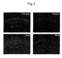
- the present inventors have discovered using immunostaining that prosaposin is expressed in from the basal layer to the granular layer and saposin A is present in the upper granular layer of the skin ( FIG. 2 ). From the results mentioned above, it is thought that caspase-14 plays a role in the processing of prosaposin in vivo.
- Saposin which is a degradation product of prosaposin, acts as a cofactor for ceramide synthase necessary in the synthesis of stratum corneum intercellular lipid, and is necessary in epidermal barrier formation.
- the search for a novel substance capable of promoting recovery of skin barrier function is possible using the degradation into saposin A by caspase-14 as an indicator without performing testing on animals.
- the present invention provides a screening method for a substance capable of promoting recovery of skin barrier function, comprising the steps of incubating caspase-14 and prosaposin in the presence of a candidate substance for the substance capable of promoting recovery of skin barrier function, and selecting the candidate substance as the substance capable of promoting recovery of skin barrier function when the degradation of prosaposin into saposin A is promoted significantly compared to a control substance.
- the candidate substance used in the method of the present invention is not limited, but includes, for example, a substance that activates caspase-14 to thereby promote the degradation of prosaposin.
- caspase-14 is a cysteine protease
- the candidate substance may be, for example, a reducing agent or a substance having a reduction action, for example, a reagent such as mercaptoethanol and dithiothreitol (DTT); and a biological material such as glutathione; and a plant extract having a reduction action.
- the candidate substance may be selected as the substance capable of promoting recovery of skin barrier function. It may be judged that "the degradation of prosaposin into saposin A is promoted significantly" based on the evaluation criterion, for example, whether the degradation of prosaposin into saposin A is promoted by 10% or more, 20% or more, 30% or more, 50% or more, 70% or more, or 100% compared to the case of the action of the control.
- the degradation of prosaposin into saposin A may be evaluated by Western blotting using an anti-saposin A antibody. It is preferable that the antibody is an anti-saposin A specific antibody. However, the present invention is not limited to the means.
- a drug selected by the screening method of the present invention is believed to be effective in improving skin in need of recovery of skin barrier function, for example, skin having reduced skin barrier function due to skin diseases, various stresses, and rough skin, etc., grafted skin having a reduced skin barrier function due to the insufficient formation of the skin barrier, and skin having a reduced skin barrier function due to grafting.
- skin barrier function means the moisture retention, the prevention of infiltration of viruses, bacteria, etc., and the like, by the skin, specifically, the epidermis.
- the function can be evaluated by measuring the transepidermal water loss (TEWL) (unit: g/m 2 ⁇ h) under the conditions where sweating does not occur. Therefore, according to the present invention, a drug selected as a substance capable of promoting recovery of skin barrier function is applied to the skin, and TEWL is measured to determine whether or not the drug actually recovers the skin barrier function.
- TEWL transepidermal water loss
- Another embodiment provides a method for promoting recovery of skin barrier function, comprising activating caspase-14 to promote the degradation of prosaposin into saposin A.
- the method for promoting recovery of skin barrier function of the present invention is preferably provided as a cosmetic method.
- this method is not limited to cosmetic methods, but may be performed for the purpose of treatment depending on the symptoms of the skin in which the recovery of the skin barrier function is intended.
- the method of the present invention may be carried out by applying the substance for activating caspase-14, for example, a reducing agent, to patients in need of recovery of function.
- the application method is not specifically limited.
- a cosmetic containing the substance as the effective ingredient may be applied to the skin in need of recovery of skin barrier function, or, a pack or a sheet comprising the substance may be placed or adhered on the skin.
- the application amount of the substance can be appropriately determined with reference to the symptoms of the skin.
- this method is not limited to methods for direct application on the skin, but may be an orally administration method depending on the substance for activating caspase-14.
- a silica frit was produced to maintain the filler in the end of the tapered opening of an unfilled capillary column (manufactured by New Objective, Inc.) having an inner diameter of 100 ⁇ m and a length of 120 mm.
- the obtained capillary column was filled with octadecylsilanized silica-type filler Aqua C18 (manufactured by Phenomenex, Inc.) having an average particle size of 5 ⁇ m until the height reached 100 mm, to obtain a reversed-phase capillary column for analysis.
- a silica frit was produced to maintain the filler in the end of the opening of an unfilled capillary column (manufactured by Agilent) having an inner diameter of 250 ⁇ m and a length of 150 mm.
- the obtained capillary column on the outlet side was filled with a mixture of a cation-exchange resin type filler Partisphere SCX resin (manufactured by Whatman, Inc.) having An average particle size of 5 ⁇ m and an anion-exchange resin type filler PolyWAXLP (manufactured by PolyLC, Inc.) having an average particle size of 5 ⁇ m at a mass ratio of 2:1 until the height reached 25 mm.
- the obtained capillary column on the inlet side was filled with octadecylsilanized silica type filler Aqua C18 (manufactured by Phenomenex, Inc.) having an average particle size of 5 ⁇ m until the height reached 25 mm.
- octadecylsilanized silica type filler Aqua C18 manufactured by Phenomenex, Inc.
- a two-phase capillary column consisting of a reversed-phase capillary column for traps and a SCX-WAX mixed capillary column was obtained.
- the filler was filled by a slurry filling method using a high pressure nitrogen gas and a pressure-type filling container.
- a supernatant containing approximately 4 ⁇ g of peptide was loaded in the two-phase type capillary column by a pressure method. Thereafter, cleaning and desalting were carried out using a mobile phase A (a mixed liquid having a volume ratio of water, acetonitrile, and formic acid of 95:5:0.1; pH approximately 2.6) having a volume 10 times or more of the sample solution.
- the two-phase type capillary column 10 was connected with the reversed-phase capillary column for analysis 20 via a through hole type union (manufactured by Upchurch Scientific, Inc) (not shown).
- Nanospace SI-2 type HPLC device manufactured by Shiseido Co., Ltd.
- a capillary having an inner diameter of 100 ⁇ m as pipework was connected therewith.
- the reversed-phase capillary column for traps 11 was disposed upstream of the SCX-WAX mixed capillary column 12 and the reversed-phase capillary column for analysis 20.
- mobile phase A mobile phase A
- mobile phase B a mixed liquid of a volume ratio of water, acetonitrile, and formic acid was 20:80:0.1
- mobile phase C mobile phase A containing 500 mM ammonium acetate; pH approximately 6.8
- the elution method of the peptide was a gradient elution method comprising a total of six steps, in which the vol% of mobile phase C added in a rectangular shape was gradually increased at each step.
- the gradient profile of Step 1 was as follows: The mobile phase A was applied for five minutes, the rate of the mobile phase B was increased from 0 vol% to 15 vol% over the next five minutes, the rate of the mobile phase B was increased to 45 vol% over the next 60 minutes, and the rate of the mobile phase B was increased to 75 vol% over the next ten minutes. Thereafter, the flow rate was maintained at this rate for five minutes.
- Steps 2 to 6 were as follows: The mobile phase A was applied for one minute, the rate of the mobile phase C was flown for four minutes as X [vol%], the rate of the mobile phase C was increased from 0 vol% to 15 vol% over the next five minutes, the rate of the mobile phase C was increased to 45 vol% over the next 60 minutes, and the rate of the mobile phase C was increased to 75 vol% over the next ten minutes. Thereafter, the flow rate was maintained at this rate for five minutes. At the time, the flow rate of a solution sent from a pump was set to 250 ⁇ L/min, and the flow rate for the column was adjusted to 300 to 400 nL/min by splitting using a resistance type capillary.
- MS/MS spectra were searched in the non-redundant human database (ftp://ftp.ncbi.nih.gov/blast/db/FASTA.nr.gz, 2007/2/8 version) using the SEQUEST algorithm operating on Bioworks Software (manufactured by Thermo Fisher Scientific, Inc.)
- Prosaposin is not processed and exists as a precursor in a monolayer culture of normal keratinocytes. Using this, the reaction of prosaposin in a precursor state with caspase-14 was examined.
- a prosaposin-containing liquid extraction from normal human epidermal keratinocytes (NHEK) was allowed to react with activated caspase-14 at 37°C for one hour in the presence or absence of 1.2 M of kosmotropic ions (citric acid ions). After dialysis in water, freeze-drying was performed. This was dissolved in an SDS electrophoresis sample buffer. After SDS electrophoresis, the production of each saposin was examined by Western blotting.
- the antibodies used respectively were an anti-prosaposin antibody (PSAP polyclonal antibody (A01): Abnova Corporation), an anti-Saposin A specific antibody (manufactured in-house), an anti-Saposin B antibody (E-15, sc-27014: Santa Cruz Biotechnology, Inc.), an anti-Saposin C antibody (H-81,sc-32875: Santa Cruz Biotechnology, Inc.), and an anti-Saposin D antibody (E-14, sc-27024R: Santa Cruz Biotechnology, Inc.)
- the dilution ratio used was 1/1000 in each case. Note that, all the antibodies except for the anti-Saposin A specific antibody recognized prosaposin. The results are shown in FIG. 1 .
- Immunostaining was performed to clarify the localization of prosaposin and saposin A using a normal skin section obtained by plastic surgery.
- a paraffin section was deparaffinized with xylene and was returned to a PBS solution. Thereafter, blocking was carried out with 10% goat serum.
- PBS washing a reaction with an anti-prosaposin antibody (PSAP mouse polyclonal antibody (A01): Abnova Corporation, dilution ratio: 1/200) or an anti-saposin A specific antibody (manufactured in-house) (dilution ratio: 1/200) was carried out at room temperature for one hour.
- a reaction with AlexaFluor 488 goat anti-mouse IgG (prosaposin) or AlexaFluor555 goat anti-rabbit IgG (saposin A) was carried out at room temperature for one hour. After washing three times with PBS, it was covered with a Vector Shield mounting medium (DAPI) (Vector Laboratories, Inc.) to perform observation by a fluorescence microscope. The results are shown in FIG.2 .
- DAPI Vector Shield mounting medium
Landscapes
- Health & Medical Sciences (AREA)
- Life Sciences & Earth Sciences (AREA)
- Engineering & Computer Science (AREA)
- Chemical & Material Sciences (AREA)
- General Health & Medical Sciences (AREA)
- Immunology (AREA)
- Molecular Biology (AREA)
- Public Health (AREA)
- Animal Behavior & Ethology (AREA)
- Veterinary Medicine (AREA)
- Hematology (AREA)
- Urology & Nephrology (AREA)
- Biomedical Technology (AREA)
- Analytical Chemistry (AREA)
- Physics & Mathematics (AREA)
- Biochemistry (AREA)
- Organic Chemistry (AREA)
- Medicinal Chemistry (AREA)
- Microbiology (AREA)
- Biotechnology (AREA)
- Food Science & Technology (AREA)
- Proteomics, Peptides & Aminoacids (AREA)
- Pathology (AREA)
- General Physics & Mathematics (AREA)
- Dermatology (AREA)
- Birds (AREA)
- Epidemiology (AREA)
- Cell Biology (AREA)
- Wood Science & Technology (AREA)
- Zoology (AREA)
- Bioinformatics & Cheminformatics (AREA)
- Biophysics (AREA)
- Pharmacology & Pharmacy (AREA)
- General Engineering & Computer Science (AREA)
- Genetics & Genomics (AREA)
- Chemical Kinetics & Catalysis (AREA)
- Nuclear Medicine, Radiotherapy & Molecular Imaging (AREA)
- General Chemical & Material Sciences (AREA)
- Investigating Or Analysing Biological Materials (AREA)
- Measuring Or Testing Involving Enzymes Or Micro-Organisms (AREA)
Abstract
Description
- The present invention relates to a screening method for a substance capable of promoting recovery of skin barrier function, using the degradation of prosaposin by caspase-14 as an indicator.
- Terminal differentiation of keratinocytes results in the formation of a protective barrier against a harmful environment, referred to as the "stratum corneum". The stratum corneum intercellular lipids play an important role in this protective barrier. The stratum corneum intercellular lipids are biosynthesized in conjunction with the differentiation in epidermal keratinocytes, and are accumulated in the Odland bodies. The sugar chains of glucosylceramide released between stratum corneum cells by exocytosis of the Odland bodies are cleaved with glucocerebrosidase to synthesize ceramide which is the main component of the stratum corneum intercellular lipids. Further, as a separate pathway, the degradation of sphingomyelin into ceramide by sphingomyelinase is known.
- The sphingolipid activator protein, saposin, is an indispensable protein in the hydrolysis of sphingolipid, and the four types of saposin (saposin A, saposin B, saposin C, and saposin D) are known to exist. Saposin is a factor functioning as a coenzyme for glucocerebrosidase, sphingomyelinase, and other ceramide synthases, which are necessary in the synthesis of stratum corneum lipids. Thus, saposin seems to be important to a skin barrier function.
- To date, it has been verified that the storage of saposin A, C, or D in the internal organs causes lysosomal storage diseases (for example, Gaucher's disease and Newman-Pick Disease), and mutations in the prosaposin gene cause a neuropathic phenotype. In association with the skin, it has been reported that prosaposin is reduced in patients with atopic dermatitis and psoriasis (refer to Tezuka et al., Journal of Investigative Dermatology (1997) 109, 319-323 and Alessandrini et al., Journal of Investigative Dermatology (2001) 116, 394-400, respectively).
- It has been known that the loss of moisture from the skin is higher in rough skin pathologies seen in various skin diseases including atopic dermatitis, psoriasis, contact dermatitis, etc., than in healthy skin. It has been thought that an increase of the so-called transepidermal water loss (TEWL) is associated with the reduction of the components which seem to have a role in the retention of moisture and a function as a barrier within the skin.
- Until now, it has been reported that the skin function declines as the skin barrier function declines, resulting in abnormal proliferation of the skin, etc. Specifically, in the case of older persons, the recovery of the declined skin barrier function requires a long time, thus, an effective and novel agent for recovery of skin barrier function has been demanded to prevent the abnormal proliferation of the skin, etc., due to the decline of the skin function with aging.
- Regarding a substance, which is studied for a formulation for promoting recovery of skin barrier and other cosmetics comprising the substance, the effect of the substance must be verified in tests on animals. However, recently, in the field of cosmetic product development, there is the trend for suppressing tests on animals from the viewpoint of ethics, and the demand for a screening method for candidate substances without applying the substances to animals has been increasing.
-
- [Non-Patent document 1] Tezuka et al., Journal of Investigative Dermatology (1997) 109, 319-323
- [Non-Patent document 2] Alessandrini et al., Journal of Investigative Dermatology (2001) 116, 394-400
- The problem to be solved by the present invention is to provide a screening method for a substance for recovery of skin barrier function, using a novel indicator.
- Caspase-14 is the newest member of the caspase family, and is expressed nearly exclusively in differentiated keratinocytes. Caspase-14 has been identified as EST homologous to caspase family members. According to the findings of recent research, the caspase-14 present in keratinocytes is processed to heterodimers that demonstrate enzymatic activity for Trp-Glu-His-Asp-AFC corresponding to the synthetic substrate for caspase-1 (Mikolajczyk J. et al., Biochemistry 43, 10560-9 (2004)). This hydrolysis activity requires proteolytic cleavage and the presence of a kosmotropic salt. The primary structure of caspase-14 is very similar to those of inflammatory caspases, such as caspase-1, -4 and -5, but the caspase-14 expression confined mainly to differentiated keratinocytes suggests that the expression may be involved in keratinocyte terminal differentiation in a different mode (Lippens S. et al., Cell Death Differ. 7, 1218-24 (2000)). However, the activation mechanism of caspase-14, its natural substrate, and regulatory factors have yet to be elucidated.
- Accordingly, the present inventors isolated the proteins capable of binding to caspase-14, from extracts of differentiated and cultured normal human epidermal keratinocytes (NHEK), etc., to subject them to LC/MS/MS analysis. As a result, prosaposin was identified from among the proteins capable of binding to caspase-14.
- The present inventors have discovered that prosaposin is expressed in from the basal layer to the granular layer, saposin A is expressed in the uppermost layer of the granular layer, and caspase-14 is involved in the processing of prosaposin to produce an intermediate of saposin and saposin A, and have completed the present invention.
- Therefore, the present application includes the following inventions:
- (1) A screening method for a substance capable of promoting recovery of skin barrier function, comprising the steps of: incubating caspase-14 and prosaposin in the presence of a candidate substance for the substance capable of promoting recovery of skin barrier function; and selecting the candidate substance as the substance capable of promoting recovery of skin barrier function when the degradation of prosaposin into saposin A is promoted significantly compared to a control substance.
- (2) A method according to (1), wherein said degradation is evaluated by Western blotting using an anti-saposin A antibody.
- Conventionally, in in vitro testing, it has been known that caspase-14 exhibits an activating action only in the presence of a high concentration of kosmotropic salt, for example, citric acid. As a result, the function of c-aspase-14 in vivo has been unknown for a long time. At this time, the present inventors have discovered for the first time that, without adding a kosmotropic salt, caspase-14 restrictively decomposes prosaposin to produce an intermediate of saposin and saposin A (
FIG. 1 ). Furthermore, the present inventors have discovered using immunostaining that prosaposin is expressed in from the basal layer to the granular layer and saposin A is present in the upper granular layer of the skin (FIG. 2 ). From the results mentioned above, it is thought that caspase-14 plays a role in the processing of prosaposin in vivo. - Saposin, which is a degradation product of prosaposin, acts as a cofactor for ceramide synthase necessary in the synthesis of stratum corneum intercellular lipid, and is necessary in epidermal barrier formation. Thus, according to the method of the present invention, the search for a novel substance capable of promoting recovery of skin barrier function is possible using the degradation into saposin A by caspase-14 as an indicator without performing testing on animals.
-
-
FIG. 1 is a Western blot chart showing the processing of prosaposin in the presence or absence of caspase-14. -
FIG. 2 is an immunostaining showing the localization of prosaposin and saposin A in normal human skin. - The present invention provides a screening method for a substance capable of promoting recovery of skin barrier function, comprising the steps of incubating caspase-14 and prosaposin in the presence of a candidate substance for the substance capable of promoting recovery of skin barrier function, and selecting the candidate substance as the substance capable of promoting recovery of skin barrier function when the degradation of prosaposin into saposin A is promoted significantly compared to a control substance.
- The candidate substance used in the method of the present invention is not limited, but includes, for example, a substance that activates caspase-14 to thereby promote the degradation of prosaposin. Since caspase-14 is a cysteine protease, the candidate substance may be, for example, a reducing agent or a substance having a reduction action, for example, a reagent such as mercaptoethanol and dithiothreitol (DTT); and a biological material such as glutathione; and a plant extract having a reduction action. If the degradation of prosaposin into saposin A is promoted significantly compared to a control substance when caspase-14 and prosaposin are incubated in the presence of the candidate substance, the candidate substance may be selected as the substance capable of promoting recovery of skin barrier function. It may be judged that "the degradation of prosaposin into saposin A is promoted significantly" based on the evaluation criterion, for example, whether the degradation of prosaposin into saposin A is promoted by 10% or more, 20% or more, 30% or more, 50% or more, 70% or more, or 100% compared to the case of the action of the control.
- The degradation of prosaposin into saposin A may be evaluated by Western blotting using an anti-saposin A antibody. It is preferable that the antibody is an anti-saposin A specific antibody. However, the present invention is not limited to the means.
- A drug selected by the screening method of the present invention is believed to be effective in improving skin in need of recovery of skin barrier function, for example, skin having reduced skin barrier function due to skin diseases, various stresses, and rough skin, etc., grafted skin having a reduced skin barrier function due to the insufficient formation of the skin barrier, and skin having a reduced skin barrier function due to grafting.
- The term "skin barrier function" as used herein means the moisture retention, the prevention of infiltration of viruses, bacteria, etc., and the like, by the skin, specifically, the epidermis. The function can be evaluated by measuring the transepidermal water loss (TEWL) (unit: g/m2·h) under the conditions where sweating does not occur. Therefore, according to the present invention, a drug selected as a substance capable of promoting recovery of skin barrier function is applied to the skin, and TEWL is measured to determine whether or not the drug actually recovers the skin barrier function.
- Another embodiment provides a method for promoting recovery of skin barrier function, comprising activating caspase-14 to promote the degradation of prosaposin into saposin A.
- The method for promoting recovery of skin barrier function of the present invention is preferably provided as a cosmetic method. However, this method is not limited to cosmetic methods, but may be performed for the purpose of treatment depending on the symptoms of the skin in which the recovery of the skin barrier function is intended. The method of the present invention may be carried out by applying the substance for activating caspase-14, for example, a reducing agent, to patients in need of recovery of function. The application method is not specifically limited. For example, a cosmetic containing the substance as the effective ingredient may be applied to the skin in need of recovery of skin barrier function, or, a pack or a sheet comprising the substance may be placed or adhered on the skin. The application amount of the substance can be appropriately determined with reference to the symptoms of the skin. Furthermore, this method is not limited to methods for direct application on the skin, but may be an orally administration method depending on the substance for activating caspase-14.
- The present invention will be further explained below with examples. Note that, the present invention is not limited thereto.
-
- 1. Caspase-14 was bound to agarose beads having an anti-caspase-14 antibody immobilized thereon, and was allowed to react with a liquid extract from normal human epidermal keratinocytes (NHEK) after induction of differentiation, in which the aerial exposure and the addition of 2 mM calcium after growth phase and confluency were performed to continue the culture. After washing with PBS, binding protein was eluted by a Glycine-HCl buffer solution (pH 2.3). Further, agarose beads having an anti-caspase-14 antibody immobilized thereon were allowed to react with a liquid extract from normal epidermal stratum corneum and a liquid extract from psoriasis patient's scales. After washing with PBS, binding protein was similarly eluted by a Glycine-HCl buffer solution (pH 2.3). These four samples were analyzed by LC/MS/MS.
- A silica frit was produced to maintain the filler in the end of the tapered opening of an unfilled capillary column (manufactured by New Objective, Inc.) having an inner diameter of 100 µm and a length of 120 mm. The obtained capillary column was filled with octadecylsilanized silica-type filler Aqua C18 (manufactured by Phenomenex, Inc.) having an average particle size of 5 µm until the height reached 100 mm, to obtain a reversed-phase capillary column for analysis.
- A silica frit was produced to maintain the filler in the end of the opening of an unfilled capillary column (manufactured by Agilent) having an inner diameter of 250 µm and a length of 150 mm. The obtained capillary column on the outlet side was filled with a mixture of a cation-exchange resin type filler Partisphere SCX resin (manufactured by Whatman, Inc.) having An average particle size of 5 µm and an anion-exchange resin type filler PolyWAXLP (manufactured by PolyLC, Inc.) having an average particle size of 5 µm at a mass ratio of 2:1 until the height reached 25 mm. The obtained capillary column on the inlet side was filled with octadecylsilanized silica type filler Aqua C18 (manufactured by Phenomenex, Inc.) having an average particle size of 5 µm until the height reached 25 mm. A two-phase capillary column consisting of a reversed-phase capillary column for traps and a SCX-WAX mixed capillary column was obtained.
- Upon the production of the reversed-phase capillary column for analysis and the two-phase type capillary column, the filler was filled by a slurry filling method using a high pressure nitrogen gas and a pressure-type filling container.
- Next, the aforementioned four samples were analyzed by the six-step MudPIT type analysis (two dimensional HPLC/ESI MS/MS).
- First, a supernatant containing approximately 4 µg of peptide was loaded in the two-phase type capillary column by a pressure method. Thereafter, cleaning and desalting were carried out using a mobile phase A (a mixed liquid having a volume ratio of water, acetonitrile, and formic acid of 95:5:0.1; pH approximately 2.6) having a volume 10 times or more of the sample solution. The two-phase type capillary column 10 was connected with the reversed-phase capillary column for analysis 20 via a through hole type union (manufactured by Upchurch Scientific, Inc) (not shown). Next, a Nanospace SI-2 type HPLC device (manufactured by Shiseido Co., Ltd.) using a capillary having an inner diameter of 100 µm as pipework was connected therewith. At the time, the reversed-phase capillary column for traps 11 was disposed upstream of the SCX-WAX mixed
capillary column 12 and the reversed-phase capillary column for analysis 20. - As the mobile phases, mobile phase A, mobile phase B (a mixed liquid of a volume ratio of water, acetonitrile, and formic acid was 20:80:0.1), and mobile phase C (mobile phase A containing 500 mM ammonium acetate; pH approximately 6.8) were used. The elution method of the peptide was a gradient elution method comprising a total of six steps, in which the vol% of mobile phase C added in a rectangular shape was gradually increased at each step.
- The gradient profile of
Step 1 was as follows: The mobile phase A was applied for five minutes, the rate of the mobile phase B was increased from 0 vol% to 15 vol% over the next five minutes, the rate of the mobile phase B was increased to 45 vol% over the next 60 minutes, and the rate of the mobile phase B was increased to 75 vol% over the next ten minutes. Thereafter, the flow rate was maintained at this rate for five minutes. - The gradient profiles of
Steps 2 to 6 were as follows: The mobile phase A was applied for one minute, the rate of the mobile phase C was flown for four minutes as X [vol%], the rate of the mobile phase C was increased from 0 vol% to 15 vol% over the next five minutes, the rate of the mobile phase C was increased to 45 vol% over the next 60 minutes, and the rate of the mobile phase C was increased to 75 vol% over the next ten minutes. Thereafter, the flow rate was maintained at this rate for five minutes. At the time, the flow rate of a solution sent from a pump was set to 250 µL/min, and the flow rate for the column was adjusted to 300 to 400 nL/min by splitting using a resistance type capillary. - Further, in the ESI MS/MS measurement, an ion trap mass spectrometer LCQ-Deca (manufactured by ThermoFisher Scientific, Inc.) was used. At the time, the peptide eluted from the reversed-phase capillary column for analysis was directly introduced into a mass spectrometer without splitting.
- Note that, a cycle of one full scan MS spectrum measurement at a mass-to-charge ratio (m/z) of 400 to 1400 and three data-dependent MS/MS spectrum measurements was repeated in the respective steps. The normalized collision energy was 35%. Three microscans were performed for each of the MS spectrum measurement and the MS/MS measurement. The dynamic exclusion settings were a repeat count of 1, a repeat duration of 0.50 min, an exclusion list size of 25, and an exclusion duration of 10.00 min.
- The obtained MS/MS spectra were searched in the non-redundant human database (ftp://ftp.ncbi.nih.gov/blast/db/FASTA.nr.gz, 2007/2/8 version) using the SEQUEST algorithm operating on Bioworks Software (manufactured by Thermo Fisher Scientific, Inc.)
- The results of analysis revealed the presence of prosaposin was common only in the reaction sample with NHEK after induction of differentiation and the sample after the reaction with the liquid extract from normal epidermal stratum corneum.
- Prosaposin is not processed and exists as a precursor in a monolayer culture of normal keratinocytes. Using this, the reaction of prosaposin in a precursor state with caspase-14 was examined. A prosaposin-containing liquid extraction from normal human epidermal keratinocytes (NHEK) was allowed to react with activated caspase-14 at 37°C for one hour in the presence or absence of 1.2 M of kosmotropic ions (citric acid ions). After dialysis in water, freeze-drying was performed. This was dissolved in an SDS electrophoresis sample buffer. After SDS electrophoresis, the production of each saposin was examined by Western blotting. The antibodies used respectively were an anti-prosaposin antibody (PSAP polyclonal antibody (A01): Abnova Corporation), an anti-Saposin A specific antibody (manufactured in-house), an anti-Saposin B antibody (E-15, sc-27014: Santa Cruz Biotechnology, Inc.), an anti-Saposin C antibody (H-81,sc-32875: Santa Cruz Biotechnology, Inc.), and an anti-Saposin D antibody (E-14, sc-27024R: Santa Cruz Biotechnology, Inc.) The dilution ratio used was 1/1000 in each case. Note that, all the antibodies except for the anti-Saposin A specific antibody recognized prosaposin. The results are shown in
FIG. 1 . - From the results in
FIG. 1 , it is understood that prosaposin degraded into a plurality of intermediates in the presence of caspase-14. Thereamong, saposin A, which is one of the final degradation products of prosaposin, was recognized by the anti-Saposin A specific antibody, thus, it is thought that caspase-14 plays a role in degrading prosaposin into saposin A. - Immunostaining was performed to clarify the localization of prosaposin and saposin A using a normal skin section obtained by plastic surgery. A paraffin section was deparaffinized with xylene and was returned to a PBS solution. Thereafter, blocking was carried out with 10% goat serum. After PBS washing, a reaction with an anti-prosaposin antibody (PSAP mouse polyclonal antibody (A01): Abnova Corporation, dilution ratio: 1/200) or an anti-saposin A specific antibody (manufactured in-house) (dilution ratio: 1/200) was carried out at room temperature for one hour. After washing three times with PBS, a reaction with AlexaFluor 488 goat anti-mouse IgG (prosaposin) or AlexaFluor555 goat anti-rabbit IgG (saposin A) was carried out at room temperature for one hour. After washing three times with PBS, it was covered with a Vector Shield mounting medium (DAPI) (Vector Laboratories, Inc.) to perform observation by a fluorescence microscope. The results are shown in
FIG.2 . - From the results in
FIG. 2 , it is understood that prosaposin was present in from the basal layer to the granular layer of normal human epidermis, and saposin A was expressed in the uppermost layer of the granular layer. These results as well as the results of the aforementioned Western blotting have supported that caspase exclusively expressed in keratinocytes degrades prosaposin in human skin, and plays an important role in skin barrier function.
Claims (4)
- A screening method for a substance capable of promoting recovery of skin barrier function, comprising the steps of: incubating caspase-14 and prosaposin in the presence of a candidate substance for the substance capable of promoting recovery of skin barrier function, and
selecting the candidate substance as the substance capable of promoting recovery of skin barrier function when the degradation of prosaposin into saposin A is promoted significantly compared to a control substance. - A method according to claim 1, wherein said degradation is evaluated by Western blotting using an anti-saposin A antibody.
- A method for promoting recovery of skin barrier function, comprising activating caspase-14 to promote the degradation of prosaposin into saposin A.
- A method according to claim 3, comprising applying to the skin a substance for activating caspase-14.
Applications Claiming Priority (2)
| Application Number | Priority Date | Filing Date | Title |
|---|---|---|---|
| JP2009277034A JP5562015B2 (en) | 2009-12-04 | 2009-12-04 | Screening method for skin barrier function recovery promoting substance |
| PCT/JP2010/071736 WO2011068221A1 (en) | 2009-12-04 | 2010-12-03 | Method for screening of substance capable of promoting recovery of skin barrier function |
Publications (2)
| Publication Number | Publication Date |
|---|---|
| EP2508616A1 true EP2508616A1 (en) | 2012-10-10 |
| EP2508616A4 EP2508616A4 (en) | 2014-01-08 |
Family
ID=44115066
Family Applications (1)
| Application Number | Title | Priority Date | Filing Date |
|---|---|---|---|
| EP10834671.9A Withdrawn EP2508616A4 (en) | 2009-12-04 | 2010-12-03 | Method for screening of substance capable of promoting recovery of skin barrier function |
Country Status (7)
| Country | Link |
|---|---|
| US (1) | US20120244140A1 (en) |
| EP (1) | EP2508616A4 (en) |
| JP (1) | JP5562015B2 (en) |
| KR (1) | KR20120112439A (en) |
| CN (1) | CN102906274A (en) |
| TW (1) | TW201144445A (en) |
| WO (1) | WO2011068221A1 (en) |
Cited By (1)
| Publication number | Priority date | Publication date | Assignee | Title |
|---|---|---|---|---|
| US9890372B2 (en) | 2013-03-19 | 2018-02-13 | Biocant—Associação De Transferência De Technologia | Aspartic proteases |
Families Citing this family (3)
| Publication number | Priority date | Publication date | Assignee | Title |
|---|---|---|---|---|
| JP5918528B2 (en) * | 2011-12-27 | 2016-05-18 | 株式会社ディーエイチシー | Skin preparation |
| KR20170014960A (en) | 2015-07-31 | 2017-02-08 | (주) 소노넷 | System and method for controlling multi-color LED lighting devices |
| JP7747518B2 (en) * | 2019-09-11 | 2025-10-01 | 株式会社 資生堂 | Agent for inhibiting/improving rough skin caused by fatigue and/or stress and screening method therefor |
Family Cites Families (3)
| Publication number | Priority date | Publication date | Assignee | Title |
|---|---|---|---|---|
| AU750170B2 (en) * | 1997-10-30 | 2002-07-11 | Human Genome Sciences, Inc. | Caspase-14 polypeptides |
| EP1227834A2 (en) * | 1999-11-10 | 2002-08-07 | Vlaams Interuniversitair Instituut voor Biotechnologie vzw. | Use of caspase-14 and caspase-14 modulators to diagnose and/or treat skin, eye and brain disorders |
| JP5570748B2 (en) * | 2009-03-31 | 2014-08-13 | 株式会社ナリス化粧品 | Functional skin external preparation with high moisturizing effect |
-
2009
- 2009-12-04 JP JP2009277034A patent/JP5562015B2/en active Active
-
2010
- 2010-12-03 TW TW099142224A patent/TW201144445A/en unknown
- 2010-12-03 EP EP10834671.9A patent/EP2508616A4/en not_active Withdrawn
- 2010-12-03 CN CN2010800548872A patent/CN102906274A/en active Pending
- 2010-12-03 KR KR1020127014142A patent/KR20120112439A/en not_active Withdrawn
- 2010-12-03 US US13/513,528 patent/US20120244140A1/en not_active Abandoned
- 2010-12-03 WO PCT/JP2010/071736 patent/WO2011068221A1/en not_active Ceased
Cited By (1)
| Publication number | Priority date | Publication date | Assignee | Title |
|---|---|---|---|---|
| US9890372B2 (en) | 2013-03-19 | 2018-02-13 | Biocant—Associação De Transferência De Technologia | Aspartic proteases |
Also Published As
| Publication number | Publication date |
|---|---|
| US20120244140A1 (en) | 2012-09-27 |
| JP2011115108A (en) | 2011-06-16 |
| JP5562015B2 (en) | 2014-07-30 |
| EP2508616A4 (en) | 2014-01-08 |
| CN102906274A (en) | 2013-01-30 |
| WO2011068221A1 (en) | 2011-06-09 |
| TW201144445A (en) | 2011-12-16 |
| KR20120112439A (en) | 2012-10-11 |
Similar Documents
| Publication | Publication Date | Title |
|---|---|---|
| Brunkhorst-Kanaan et al. | Targeted lipidomics reveal derangement of ceramides in major depression and bipolar disorder | |
| Sano et al. | Lipid synthesis is promoted by hypoxic adipocyte-derived exosomes in 3T3-L1 cells | |
| Lee et al. | Proteomic analysis of extracellular vesicles released by adipocytes of Otsuka Long-Evans Tokushima fatty (OLETF) rats | |
| Vardaki et al. | Periostin is identified as a putative metastatic marker in breast cancer-derived exosomes | |
| Chalova et al. | Determination of short-chain fatty acids as putative biomarkers of cancer diseases by modern analytical strategies and tools: a review | |
| Pardo et al. | Obesidomics: contribution of adipose tissue secretome analysis to obesity research | |
| Houerbi et al. | Secretome profiling reveals acute changes in oxidative stress, brain homeostasis, and coagulation following short-duration spaceflight | |
| Dong et al. | In vivo anti-hyperuricemia and anti-gouty arthritis effects of the ethanol extract from Amomumvillosum Lour. | |
| Zanotti et al. | Anion exchange chromatography-based purification of plant-derived nanovesicles from Brassica oleracea L.: molecular profiling and bioactivity in human cells | |
| Pshezhetsky et al. | Subcellular proteomics of cell differentiation: Quantitative analysis of the plasma membrane proteome of Caco‐2 cells | |
| US20120244140A1 (en) | Screening method for substance capable of promoting recovery of skin barrier function technical field | |
| Casati et al. | Lipidomics of cell secretome combined with the study of selected bioactive lipids in an in vitro model of osteoarthritis | |
| Ozcan et al. | Characterization of novel O-glycans isolated from tear and saliva of ocular rosacea patients | |
| Campesi et al. | Stratification of amniotic fluid cells and amniotic fluid by sex opens up new perspectives on fetal health | |
| Mirza et al. | Adiposomes from obese-diabetic individuals promote endothelial dysfunction and loss of surface caveolae | |
| Enge et al. | Longitudinal assessment of metal concentrations and copper isotope ratios in the G93A SOD1 mouse model of amyotrophic lateral sclerosis | |
| Hu et al. | Integrating transcriptomics, metabolomics, and network pharmacology to investigate multi-target effects of sporoderm-broken spores of Ganoderma lucidum on improving HFD-induced diabetic nephropathy rats | |
| Varela et al. | Lipidome profiling of neutrophil-derived extracellular vesicles unveils their contribution to the ensemble of synovial fluid-derived extracellular vesicles during joint inflammation | |
| Abdelmohsen et al. | The Secretome of Human Trophoblast Stem Cells Attenuates Senescence‐Associated Traits | |
| Williams et al. | Plastic associated endocrine disruptors reduce Nicastrin protein and potentiate inflammation in hidradenitis suppurativa skin disease | |
| Olkowicz et al. | Simultaneous accurate quantification of HO-1, CD39, and CD73 in human calcified aortic valves using multiple enzyme digestion–filter aided sample pretreatment (MED-FASP) method and targeted proteomics | |
| Miranda et al. | Loss of NAMPT and SIRT2 but not SIRT1 attenuate GLO1 expression and activity in human skeletal muscle | |
| Burd et al. | Optimizing the measurement of mitochondrial protein synthesis in human skeletal muscle | |
| Wu et al. | A comparative peptidomic characterization of cultured skeletal muscle tissues derived from db/db mice | |
| US10907208B2 (en) | Method for screening agents promoting skin barrier function and method for evaluating skin barrier function taking epidermal serine racemase and/or D-serine level as indicator |
Legal Events
| Date | Code | Title | Description |
|---|---|---|---|
| PUAI | Public reference made under article 153(3) epc to a published international application that has entered the european phase |
Free format text: ORIGINAL CODE: 0009012 |
|
| 17P | Request for examination filed |
Effective date: 20120613 |
|
| AK | Designated contracting states |
Kind code of ref document: A1 Designated state(s): AL AT BE BG CH CY CZ DE DK EE ES FI FR GB GR HR HU IE IS IT LI LT LU LV MC MK MT NL NO PL PT RO RS SE SI SK SM TR |
|
| DAX | Request for extension of the european patent (deleted) | ||
| A4 | Supplementary search report drawn up and despatched |
Effective date: 20131209 |
|
| RIC1 | Information provided on ipc code assigned before grant |
Ipc: G01N 33/53 20060101ALI20131203BHEP Ipc: C12Q 1/37 20060101AFI20131203BHEP Ipc: A61Q 19/00 20060101ALI20131203BHEP Ipc: A61K 8/96 20060101ALI20131203BHEP Ipc: G01N 33/50 20060101ALI20131203BHEP |
|
| STAA | Information on the status of an ep patent application or granted ep patent |
Free format text: STATUS: THE APPLICATION IS DEEMED TO BE WITHDRAWN |
|
| 18D | Application deemed to be withdrawn |
Effective date: 20140719 |