EP2497835A2 - Probe for detecting polymorphism in CYP3A gene, method of detecting polymorphism, method of evaluating drug efficacy, and reagent kit for detecting polymorphism - Google Patents
Probe for detecting polymorphism in CYP3A gene, method of detecting polymorphism, method of evaluating drug efficacy, and reagent kit for detecting polymorphism Download PDFInfo
- Publication number
- EP2497835A2 EP2497835A2 EP20120158915 EP12158915A EP2497835A2 EP 2497835 A2 EP2497835 A2 EP 2497835A2 EP 20120158915 EP20120158915 EP 20120158915 EP 12158915 A EP12158915 A EP 12158915A EP 2497835 A2 EP2497835 A2 EP 2497835A2
- Authority
- EP
- European Patent Office
- Prior art keywords
- base
- bases
- seq
- sequence
- fluorescently labeled
- Prior art date
- Legal status (The legal status is an assumption and is not a legal conclusion. Google has not performed a legal analysis and makes no representation as to the accuracy of the status listed.)
- Granted
Links
Images




Classifications
-
- C—CHEMISTRY; METALLURGY
- C12—BIOCHEMISTRY; BEER; SPIRITS; WINE; VINEGAR; MICROBIOLOGY; ENZYMOLOGY; MUTATION OR GENETIC ENGINEERING
- C12Q—MEASURING OR TESTING PROCESSES INVOLVING ENZYMES, NUCLEIC ACIDS OR MICROORGANISMS; COMPOSITIONS OR TEST PAPERS THEREFOR; PROCESSES OF PREPARING SUCH COMPOSITIONS; CONDITION-RESPONSIVE CONTROL IN MICROBIOLOGICAL OR ENZYMOLOGICAL PROCESSES
- C12Q1/00—Measuring or testing processes involving enzymes, nucleic acids or microorganisms; Compositions therefor; Processes of preparing such compositions
- C12Q1/68—Measuring or testing processes involving enzymes, nucleic acids or microorganisms; Compositions therefor; Processes of preparing such compositions involving nucleic acids
- C12Q1/6876—Nucleic acid products used in the analysis of nucleic acids, e.g. primers or probes
- C12Q1/6883—Nucleic acid products used in the analysis of nucleic acids, e.g. primers or probes for diseases caused by alterations of genetic material
-
- C—CHEMISTRY; METALLURGY
- C12—BIOCHEMISTRY; BEER; SPIRITS; WINE; VINEGAR; MICROBIOLOGY; ENZYMOLOGY; MUTATION OR GENETIC ENGINEERING
- C12Q—MEASURING OR TESTING PROCESSES INVOLVING ENZYMES, NUCLEIC ACIDS OR MICROORGANISMS; COMPOSITIONS OR TEST PAPERS THEREFOR; PROCESSES OF PREPARING SUCH COMPOSITIONS; CONDITION-RESPONSIVE CONTROL IN MICROBIOLOGICAL OR ENZYMOLOGICAL PROCESSES
- C12Q1/00—Measuring or testing processes involving enzymes, nucleic acids or microorganisms; Compositions therefor; Processes of preparing such compositions
- C12Q1/68—Measuring or testing processes involving enzymes, nucleic acids or microorganisms; Compositions therefor; Processes of preparing such compositions involving nucleic acids
- C12Q1/6876—Nucleic acid products used in the analysis of nucleic acids, e.g. primers or probes
-
- G—PHYSICS
- G01—MEASURING; TESTING
- G01N—INVESTIGATING OR ANALYSING MATERIALS BY DETERMINING THEIR CHEMICAL OR PHYSICAL PROPERTIES
- G01N33/00—Investigating or analysing materials by specific methods not covered by groups G01N1/00 - G01N31/00
- G01N33/48—Biological material, e.g. blood, urine; Haemocytometers
- G01N33/50—Chemical analysis of biological material, e.g. blood, urine; Testing involving biospecific ligand binding methods; Immunological testing
- G01N33/53—Immunoassay; Biospecific binding assay; Materials therefor
- G01N33/5308—Immunoassay; Biospecific binding assay; Materials therefor for analytes not provided for elsewhere, e.g. nucleic acids, uric acid, worms, mites
-
- C—CHEMISTRY; METALLURGY
- C12—BIOCHEMISTRY; BEER; SPIRITS; WINE; VINEGAR; MICROBIOLOGY; ENZYMOLOGY; MUTATION OR GENETIC ENGINEERING
- C12Q—MEASURING OR TESTING PROCESSES INVOLVING ENZYMES, NUCLEIC ACIDS OR MICROORGANISMS; COMPOSITIONS OR TEST PAPERS THEREFOR; PROCESSES OF PREPARING SUCH COMPOSITIONS; CONDITION-RESPONSIVE CONTROL IN MICROBIOLOGICAL OR ENZYMOLOGICAL PROCESSES
- C12Q2563/00—Nucleic acid detection characterized by the use of physical, structural and functional properties
- C12Q2563/107—Nucleic acid detection characterized by the use of physical, structural and functional properties fluorescence
-
- C—CHEMISTRY; METALLURGY
- C12—BIOCHEMISTRY; BEER; SPIRITS; WINE; VINEGAR; MICROBIOLOGY; ENZYMOLOGY; MUTATION OR GENETIC ENGINEERING
- C12Q—MEASURING OR TESTING PROCESSES INVOLVING ENZYMES, NUCLEIC ACIDS OR MICROORGANISMS; COMPOSITIONS OR TEST PAPERS THEREFOR; PROCESSES OF PREPARING SUCH COMPOSITIONS; CONDITION-RESPONSIVE CONTROL IN MICROBIOLOGICAL OR ENZYMOLOGICAL PROCESSES
- C12Q2600/00—Oligonucleotides characterized by their use
- C12Q2600/112—Disease subtyping, staging or classification
-
- C—CHEMISTRY; METALLURGY
- C12—BIOCHEMISTRY; BEER; SPIRITS; WINE; VINEGAR; MICROBIOLOGY; ENZYMOLOGY; MUTATION OR GENETIC ENGINEERING
- C12Q—MEASURING OR TESTING PROCESSES INVOLVING ENZYMES, NUCLEIC ACIDS OR MICROORGANISMS; COMPOSITIONS OR TEST PAPERS THEREFOR; PROCESSES OF PREPARING SUCH COMPOSITIONS; CONDITION-RESPONSIVE CONTROL IN MICROBIOLOGICAL OR ENZYMOLOGICAL PROCESSES
- C12Q2600/00—Oligonucleotides characterized by their use
- C12Q2600/156—Polymorphic or mutational markers
Definitions
- the present invention relates to a probe for detecting polymorphism in the CYP3A gene, a method of detecting polymorphism, a method of evaluating drug efficacy, and a reagent kit for detecting polymorphism.
- CYP Cytochrome P450
- CYP is an oxidase involved in metabolism of biological materials such as steroid hormones and oxidative detoxification of a drug, an environmental pollutant, or the like.
- CYP forms a superfamily including a multitude of molecular species.
- CYP3A4 and CYP3A5 are known to contain polymorphism(s) affecting metabolism of a drug or medicine, for example ( Human Mutation, 2004, Vol. 23, Issue 1, pp. 100 to 108 ).
- the CYP3A5 gene encoding CYP3A5 is located in human chromosome 7.
- a mutation (CYP3A5*3) from adenine (a) to guanine (g) is known ( Clin. Pharmacol. Ther., 2006, Vol. 80, pp. 179 to 191 , etc.).
- This base mutation causes an abnormal splicing, and the expression of CYP3A5 decreases greatly.
- the CYP3A4 gene encoding CYP3A4 is located in human chromosome 7.
- a mutation (CYP3A4*16) from cytosine (c) to guanine (g) is known ( Clin. Pharmacol. Ther., 2006, Vol. 80, pp. 179 to 191 , etc.).
- T 185th threonine
- S serine
- PCR polymerase chain reaction
- RFLP tion fragment length polymorphism
- a region containing a mutation is amplified by a PCR method, then a melting curve analysis is performed using a nucleic acid probe labeled with a fluorescent dye, and mutation in a base sequence is analyzed based on the result of the melting curve analysis (see Patent Document 1: JP2002-119291A ).
- the PCR-RFLP method needs to apply a restriction enzyme treatment to an amplification product obtained from a PCR reaction. Therefore, there is a possibility that a subsequent reaction system is contaminated with the amplification product, and this may cause false-positive and false-negative results. Further, since treatment is performed with a restriction enzyme after completion of the PCR and electrophoresis is performed thereafter, quite a long time is required until the determination is completed. Moreover, since the PCR-RFLP method requires complicated operations, the automatization thereof is difficult. On the basis of these current circumstances, further development of technologies for detecting polymorphism in the CYP3A gene has been awaited.
- the present invention is intended to provide a probe for detecting polymorphism that enables a simple detection of polymorphism in the CYP3A gene with high sensitivity; and a method of detecting polymorphism using the probe. Further, the present invention is intended to provide a method of evaluating drug efficacy using the method of detecting polymorphism. Furthermore, the present invention is intended to provide a reagent kit for detecting polymorphism using the probe for detecting polymorphism.
- the present invention is as follows:
- a probe for detecting polymorphism that enables a simple detection of polymorphism in the CYP3A gene with high sensitivity and a method of detecting polymorphism using the probe can be provided. Further, the present invention can provide a method of evaluating drug efficacy using the method of detecting polymorphism. Furthermore, the present invention can provide a reagent kit for detecting polymorphism using the probe for detecting polymorphism. The present invention can be applied to the detection of polymorphism in the CYP3A gene in a wide range of fields such as a biochemistry and the like besides medical fields.
- the probe for detecting polymorphism in the CYP3A gene according to the present invention is a probe which detects polymorphism in the CYP3A gene and includes one fluorescently labeled oligonucleotide selected from the group consisting of P1, P1', P2, P2', P3, P3', P4, P4', P5, and P5'.
- the method of detecting polymorphism in the CYP3A gene according to the present invention is a method including detecting polymorphism in the CYP3A gene using at least one of the probes which detect polymorphism in the CYP3A gene.
- the method of evaluating drug efficacy or tolerance according to the present invention is a method including detecting polymorphism in the CYP3A gene by the method of detecting polymorphism in the CYP3A gene and evaluating or predicting tolerance to the drug or the efficacy of the drug based on the presence or absence of detected polymorphism.
- the reagent kit for detecting polymorphism according to the present invention is a reagent kit including the probe which detects polymorphism in the CYP3A gene.
- the "CYP3A gene" in the present invention is already known.
- the base sequence of the CYP3A5 gene refers to the sequence from 99245813 to 99277621 of NCBI Accession No. NC000007.13.
- SEQ ID NO: 1 that shows a partial sequence of the CYP3A5 gene is the sequence from 6683 to 7483 of NCBI dbSNP Accession No. NG007938.
- the base sequence of the CYP3A4 gene refers to the sequence from 99354583 to 99381811 of NCBI Accession No. NC000007.13.
- SEQ ID NO: 2 that shows a partial sequence of the CYP3A4 gene is the sequence from 15519 to 15928 of NCBI dbSNP Accession No.
- SEQ ID NO: 3 that shows a partial sequence of the CYP3A4 gene is the sequence from 24616172 to 24616872 of NCBI dbSNP Accession No. NT007933.14.
- r indicates a or g
- s indicates c or g
- w indicates a, t, or u
- v indicates g, a, or c.
- the base (r) corresponding to the 401st base of the sequence indicated in SEQ ID NO: 1 is A (adenine) in the wild-type sequence, and the base (r) is mutated to G (guanine) in the mutant sequence.
- the polymorphism in the CYP3A5 gene of SEQ ID NO: 1 is referred to as the "CYP3A5*3 polymorphism”.
- the base (s) corresponding to the 201st base of the sequence indicated in SEQ ID NO: 2 is C (cytosine) in the wild-type sequence, and the base (s) is mutated to G (guanine) in relation to the mutant sequence.
- the polymorphism in the CYP3A4 gene of SEQ ID NO: 2 is referred to as the "CYP3A4*16 polymorphism”.
- the base (r) corresponding to the 501st base of the sequence indicated in SEQ ID NO: 3 is A (adenine) in the wild-type sequence, and the base (r) is mutated to G (guanine) in the mutant sequence.
- the polymorphism in the CYP3A4 gene of SEQ ID NO: 3 is referred to as the "CYP3A4*1B polymorphism”.
- CYP3A gene if at least one of the aforementioned three polymorphisms is a mutant type, for example, it can be determined that there is a possibility of decreasing metabolism of a drug such as the immunosuppressant, tacrolimus.
- the "Tm value” is a temperature (dissociation temperature: Tm) at which double-stranded nucleic acid dissociates, and generally is defined as a temperature at which the absorbance at 260 nm reaches 50% of the total increase in absorbance. That is, when a solution containing double-stranded nucleic acid, for example double-stranded DNA, is heated, the absorbance at 260 nm increases. The increase results from the breakage of hydrogen bonds between the strands in the double-stranded DNA resulting from the heating and the dissociation into single-stranded DNA (DNA melting).
- Tm dissociation temperature
- the Tm value is set based on this phenomenon.
- the Tm value in the present invention is a temperature at the time when the absorbance reaches 50% of the total increase in absorbance, unless otherwise noted.
- the "process” does not only refer to an independent process but also includes, for example, a case in which it cannot be clearly distinguished from other processes as long as the desired action of the present process is achieved.
- a numerical value range expressed with “to” refers to a range including numerical values before and after "to” as a minimum value and a maximum value.
- the amount of each component in composition refers to, in the case where plural materials corresponding to the component are present in the composition, the total amount of the plural materials present in the composition, unless otherwise noted.
- the expression, "the 1st to the 3rd positions from the 3' end”, is based on the premise that the 3' end of an oligonucleotide strand is counted as the 1st.
- the present invention will be described.
- the CYP3A gene polymorphism detection probe according to the present invention is a probe which detects polymorphism in the CYP3A gene and includes one fluorescently labeled oligonucleotide selected from the group consisting of P1, P1', P2, P2', P3, P3', P4, P4', P5, and P5'.
- the base (r) corresponding to the 401st base of the sequence indicated in SEQ ID NO: 1 is A (adenine) in the wild-type sequence, and the base (r) is mutated to G (guanine) in the mutant sequence.
- the polymorphism in the CYP3A5 gene of SEQ ID NO: 1 is referred to as the "CYP3A5*3 polymorphism”.
- the probe for detecting this polymorphism is also referred to as the CYP3A5*3 probe.
- one fluorescently labeled oligonucleotide selected from the group consisting of P1 and P1' is a probe capable of detecting polymorphism in the 401st base of the base sequence indicated in SEQ ID NO: 1.
- the fluorescently labeled oligonucleotide P1 of the present invention has a sequence including the 401st to the 411th bases of the sequence indicated in SEQ ID NO: 1.
- the base (r) corresponding to the 401st base of the base sequence indicated in SEQ ID NO: 1 may be A or G.
- (P1) a fluorescently labeled oligonucleotide having a sequence including the 401st to the 411th bases of the sequence indicated in SEQ ID NO: 1 and having a length of 11 bases to 50 bases, the sequence having at least 80% identity with respect to a base sequence having the same bases as SEQ ID NO: 1 with the exception that the base corresponding to the 411th base in SEQ ID NO: 1 is cytosine, and the base corresponding to the 411th base of the base sequence indicated in SEQ ID NO: 1 is labeled with a fluorescent dye.
- a base sequence having the same bases as SEQ ID NO. refers to a portion of said SEQ ID NO.'s sequence (or complementary sequence) which appears in said probe subject to the modification(s) discussed herein.
- the fluorescently labeled oligonucleotide P1 of the present invention requires a sequence having identity with respect to a base sequence having the same bases as SEQ ID NO: 1 with the exception that the base corresponding to the 411th base in SEQ ID NO: 1 is C (cytosine).
- the fluorescently labeled oligonucleotide P1 of the present invention shows at least 80% identity with respect to a base sequence having the same bases as SEQ ID NO: 1 with the exception that the base corresponding to the 411th base in SEQ ID NO: 1 is C (cytosine).
- the fluorescently labeled oligonucleotide P1 of the present invention may show at least 85% identity, at least 90% identity, at least 95% identity, at least 96% identity, at least 97% identity, at least 98% identity, or at least 99% identity (instead of at least 80% identity described above).
- the fluorescently labeled oligonucleotide P1 has a sequence having identity with respect to a base sequence having the same bases as SEQ ID NO: 1 with the exception that the base corresponding to the 411th base in SEQ ID NO: 1 is C (cytosine) and the base corresponding to the 401st base in SEQ ID NO: 1 is r (A or G).
- the fluorescently labeled oligonucleotide P1 has a sequence in which the base corresponding to the 401st base in SEQ ID NO: 1 is the mutant G
- the following can be said.
- the fluorescently labeled oligonucleotide P1 of the present invention is compared with a base sequence having the same bases as SEQ ID NO: 1 with the exception that the base corresponding to the 411th base in SEQ ID NO: 1 is C (cytosine), in the case where the identity therebetween is less than 80%, the detection sensitivity to a sample nucleic acid containing the mutant CYP3A5 gene decreases.
- the fluorescently labeled oligonucleotide P 1 according to the present invention may be the fluorescently labeled oligonucleotide that recognizes (detects) polymorphism in the 401st base in SEQ ID NO: 1, for example.
- the fluorescently labeled oligonucleotide has, in addition to a specific base sequence, a function of recognizing polymorphism in the 401st base in SEQ ID NO: 1.
- the detection sensitivity to a sample nucleic acid containing the mutant CYP3A5 gene tends to increase.
- the fluorescently labeled oligonucleotide P1' in the present invention requires a sequence being capable of hybridization under stringent conditions to the complementary strand of a base sequence having the same bases as SEQ ID NO: 1 with the exception that the base corresponding to the 411th base in SEQ ID NO: 1 is C (cytosine).
- the base (r) corresponding to the 401st base of the base sequence indicated in SEQ ID NO: 1 may be A or G.
- the hybridization can be performed by a known method or based on a known method, for example, according to the method described in Molecular Cloning 3rd (J. Sambrook et al., Cold Spring Harbor Lab. Press, 2001 ). The entire subject matter of this document is incorporated herein by reference.
- the stringent conditions refer to conditions where a specific hybrid is formed and a nonspecific hybrid is not formed.
- An example of typical stringent conditions includes a condition where hybridization is performed in about 25 mmol/L to about 50 mmol/L potassium and about 1.0 mmol/L to about 5.0 mmol/L magnesium.
- condition of the present invention includes a condition where hybridization is performed in Tris-HCl (pH8.6), 25 mmol/L KCl, and 1.5 mmol/L MgCl 2 .
- the present invention is not limited thereto.
- stringent conditions are described in Molecular Cloning 3rd (J. Sambrook et al., Cold Spring Harbor Lab. Press, 2001 ). The entire subject matter of this document is incorporated herein by reference. Those skilled in the art can easily select such conditions by changing the hybridization reaction, the salt concentration of a hybridization reaction solution, or the like.
- the fluorescently labeled oligonucleotide P1' according to the present invention may be the fluorescently labeled oligonucleotide that recognizes (detects) polymorphism in the 401st base in SEQ ID NO: 1, for example.
- the fluorescently labeled oligonucleotide has, in addition to a specific base sequence, the function of recognizing polymorphism in the 401st base in SEQ ID NO: 1.
- the detection sensitivity to a sample nucleic acid containing the mutant CYP3A5 gene tends to increase.
- the fluorescently labeled oligonucleotide P 1 or P1' in the present invention includes a fluorescently labeled oligonucleotide obtained by inserting a base(s) into, deleting a base(s) from, or substituting a base(s) with a base(s) in the fluorescently labeled oligonucleotide P1 or P1'.
- the position of the insertion, deletion, or substitution is not particularly limited as long as an action comparable to the fluorescently labeled oligonucleotide P1 or P1' can be achieved.
- the number of bases inserted, deleted, or substituted is, for example, one or more than one.
- the number of bases differs depending on the entire length of the fluorescently labeled oligonucleotide, and is, for example, from 1 base to 10 bases or from 1 base to 5 bases.
- "from” includes the integers of the given range, i.e. in this case 1 or more bases up to 10 bases.
- the fluorescently labeled oligonucleotide P 1 or P1' in the present invention includes a fluorescently labeled oligonucleotide obtained by substituting a base(s) with a base(s) in the fluorescently labeled oligonucleotide P1 or P1'.
- the position of the substitution is not particularly limited as long as an action comparable to the fluorescently labeled oligonucleotide P1 or P1' can be achieved.
- a base(s) that is(are) not positioned at the 401st to the 411th bases of the sequence indicated in SEQ ID NO: 1 may be substituted.
- the number of bases substituted is, for example, one or more than one.
- the number of bases substituted differs depending on the entire length of the fluorescently labeled oligonucleotide, and is, for example, from 1 base to 5 bases or from 1 base to 3 bases.
- the length of the fluorescently labeled oligonucleotide P1 or P1' of the present invention is required to be from 11 bases to 50 bases (mer). In the case where the length of the fluorescently labeled oligonucleotide P 1 or P1' is 10 bases or less or 51 bases or more, the detection sensitivity to polymorphism in the CYP3A gene decreases. Further, the length of the fluorescently labeled oligonucleotide P1 or P1' can be, for example, from 15 bases to 30 bases, from 15 bases to 21 bases, or from 15 bases to 20 bases. For example, there is a tendency for the detection sensitivity to increase by designing the length from 15 bases to 30 bases, for example.
- the length of the fluorescently labeled oligonucleotide P1 or P1' for example, a desirable value for the Tm value, which is the temperature at which the hybrid composed of the fluorescently labeled oligonucleotide P1 or P1' and its complementary strand dissociates, can be obtained.
- Examples of the base sequence of the fluorescently labeled oligonucleotide P1 in the present invention are illustrated below. However, the present invention is not limited thereto. 5'-ttgtctttca r tatctcttcc-3' (SEQ ID NO: 37) 5'-ttgtctttca g tatctcttcc-3' (SEQ ID NO: 4) (3FL-CYP3A5*3-mt-F1-21) 5'-ttgtctttca a tatctcttcc-3' (SEQ ID NO: 5)
- the base corresponding to the 411th base of the sequence indicated in SEQ ID NO: 1 is labeled with a fluorescent dye.
- the 411th base labeled with a fluorescent dye can be at a position of any one of the 1st to the 3rd positions from the 3' end or at the 3' end.
- the detection sensitivity to polymorphism further increases and the fluorescently labeled oligonucleotide P1 or P1' can be obtained with high productivity and sensitivity.
- one fluorescently labeled oligonucleotide selected from the group consisting of P2, P2', P3, P3', P4, and P4' (hereinafter, this is also referred to as the "fluorescently labeled oligonucleotide P2, P2', P3, P3', P4, or P4' ") is a probe capable of detecting polymorphism in the 201st base of the base sequence indicated in SEQ ID NO: 2.
- the base (s) corresponding to the 201st base of the sequence indicated in SEQ ID NO: 2 is C (cytosine) in the wild-type sequence, and the base (s) is mutated to G (guanine) in the mutant sequence.
- the polymorphism in the CYP3A4 gene of SEQ ID NO: 2 is referred to as the "CYP3A4*16 polymorphism”.
- the probe for detecting this polymorphism is also referred to as the CYP3A4*16 probe.
- the fluorescently labeled oligonucleotide P2 of the present invention has a sequence including the 201st to the 205th bases of the sequence indicated in SEQ ID NO: 2.
- the base (s) corresponding to the 201st base of the base sequence indicated in SEQ ID NO: 2 may be C or G.
- (P2) a fluorescently labeled oligonucleotide having a sequence including the 201st to the 205th bases of the sequence indicated in SEQ ID NO: 2 and having a length of 5 bases to 50 bases, the sequence having at least 80% identity with respect to a base sequence having the same bases as SEQ ID NO: 2 with the exception that the base corresponding to the 205th base in SEQ ID NO: 2 is cytosine, and the base corresponding to the 205th base of the sequence indicated in SEQ ID NO: 2 is labeled with a fluorescent dye.
- the fluorescently labeled oligonucleotide P2 of the present invention requires a sequence having identity with respect to a base sequence having the same bases as SEQ ID NO: 2 with the exception that the base corresponding to the 205th base in SEQ ID NO: 2 is C (cytosine).
- the fluorescently labeled oligonucleotide P2 of the present invention shows at least 80% identity with respect to a base sequence having the same bases as SEQ ID NO: 2 with the exception that the base corresponding to the 205th base in SEQ ID NO: 2 is C (cytosine).
- the fluorescently labeled oligonucleotide P2 of the present invention may show at least 85% identity, at least 90% identity, at least 95% identity, at least 96% identity, at least 97% identity, at least 98% identity, or at least 99% identity (instead of at least 80% identity described above).
- the fluorescently labeled oligonucleotide P2 has a sequence having identity with respect to a base sequence having the same bases as SEQ ID NO: 2 with the exception that the base corresponding to the 205th base in SEQ ID NO: 2 is C (cytosine) and the base corresponding to the 201st base in SEQ ID NO: 2 is s (C or G).
- the fluorescently labeled oligonucleotide P2 has a sequence in which the base corresponding to the 201st base in SEQ ID NO: 2 is the mutant G
- the following can be said.
- the fluorescently labeled oligonucleotide P2 of the present invention is compared with a base sequence having the same bases as SEQ ID NO: 2 with the exception that the base corresponding to the 205th base in SEQ ID NO: 2 is C (cytosine), in the case where the identity therebetween is less than 80%, the detection sensitivity to a sample nucleic acid containing the mutant CYP3A4 gene decreases.
- the fluorescently labeled oligonucleotide P2 according to the present invention may be the fluorescently labeled oligonucleotide that recognizes (detects) polymorphism in the 201st base in SEQ ID NO: 2, for example.
- the fluorescently labeled oligonucleotide has, in addition to a specific base sequence, a function of recognizing polymorphism in the 201st base in SEQ ID NO: 2.
- the detection sensitivity to a sample nucleic acid containing the mutant CYP3A4 gene tends to increase.
- the fluorescently labeled oligonucleotide P2' in the present invention requires a sequence being capable of hybridization under stringent conditions to the complementary strand of a base sequence having the same bases as SEQ ID NO: 2 with the exception that the base corresponding to the 205th base in SEQ ID NO: 2 is C (cytosine).
- the base (s) corresponding to the 201st base of the base sequence indicated in SEQ ID NO: 2 may be C or G.
- the hybridization and the stringent conditions are the same as those described above.
- the fluorescently labeled oligonucleotide P2' according to the present invention may be the fluorescently labeled oligonucleotide that recognizes (detects) polymorphism in the 201st base in SEQ ID NO: 2, for example.
- the fluorescently labeled oligonucleotide has, in addition to a specific base sequence, the function of recognizing polymorphism in the 201st base in SEQ ID NO: 2.
- the detection sensitivity to a sample nucleic acid containing the mutant CYP3A4 gene tends to increase.
- the fluorescently labeled oligonucleotide P2 or P2' in the present invention includes a fluorescently labeled oligonucleotide obtained by inserting a base(s) into, deleting a base(s) from, or substituting a base(s) with a base(s) in the fluorescently labeled oligonucleotide P2 or P2'.
- the position of the insertion, deletion, or substitution is not particularly limited as long as an action comparable to the fluorescently labeled oligonucleotide P2 or P2' can be achieved.
- the number of bases inserted, deleted, or substituted is, for example, one or more than one.
- the number of bases differs depending on the entire length of the fluorescently labeled oligonucleotide, and is, for example, from 1 base to 10 bases or from 1 base to 5 bases.
- the fluorescently labeled oligonucleotide P2 or P2' in the present invention includes a fluorescently labeled oligonucleotide obtained by substituting a base(s) with a base(s) in the fluorescently labeled oligonucleotide P2 or P2'.
- the position of the substitution is not particularly limited as long as an action comparable to the fluorescently labeled oligonucleotide P2 or P2' can be achieved.
- a base(s) that is(are) not positioned at the 201st to the 205th bases of the sequence indicated in SEQ ID NO: 2 may be substituted.
- the number of bases substituted is, for example, one or more than one.
- the number of bases substituted differs depending on the entire length of the fluorescently labeled oligonucleotide, and is, for example, from 1 base to 5 bases or from 1 base to 3 bases.
- the length of the fluorescently labeled oligonucleotide P2 or P2' of the present invention is required to be from 5 bases to 50 bases (mer). In the case where the length of the fluorescently labeled oligonucleotide P2 or P2' is 4 bases or less or 51 bases or more, the detection sensitivity to polymorphism in the CYP3A gene decreases. Further, the length of the fluorescently labeled oligonucleotide P2 or P2' can be, for example, from 10 bases to 30 bases or from 10 bases to 20 bases. For example, there is a tendency for the detection sensitivity to increase by designing the length from 10 bases to 30 bases, for example.
- the length of the fluorescently labeled oligonucleotide P2 or P2' for example, a desirable value for the Tm value, which is the temperature at which the hybrid composed of the fluorescently labeled oligonucleotide P2 or P2' and its complementary strand dissociates, can be obtained.
- Examples of the base sequence of the fluorescently labeled oligonucleotide P2 in the present invention are illustrated below. However, the present invention is not limited thereto. 5'-gatgtgatca s tagc-3' (SEQ ID NO: 38) 5'-gatgtgatca g tagc-3' (SEQ ID NO: 6) (3T-CYP3A4*16-MF1) 5'-gatgtgatca c tagc-3' (SEQ ID NO: 7)
- the base corresponding to the 205th base of the sequence indicated in SEQ ID NO: 2 is labeled with a fluorescent dye.
- the 205th base labeled with a fluorescent dye can be at a position of any one of the 1st to the 3rd positions from the 3' end or at the 3' end.
- the detection sensitivity to polymorphism further increases and the fluorescently labeled oligonucleotide P2 or P2' can be obtained with high productivity and sensitivity.
- the base (s) complementary to the 201st base of the base sequence indicated in SEQ ID NO: 2 may be G or C.
- the fluorescently labeled oligonucleotide P3 of the present invention has a sequence complementary to a sequence including the 190th to the 201st bases of the sequence indicated in SEQ ID NO: 2.
- the base (s) complementary to the 201st base of the base sequence indicated in SEQ ID NO: 2 may be C or G.
- (P3) a fluorescently labeled oligonucleotide having a sequence complementary to a sequence including the 190th to the 201st bases of the sequence indicated in SEQ ID NO: 2 and having a length of 12 bases to 50 bases, the sequence having at least 80% identity with respect to a base sequence having the same bases as the complementary strand of SEQ ID NO: 2 with the exception that the base complementary to the 190th base in SEQ ID NO: 2 is cytosine, and the base complementary to the 190th base of the sequence indicated in SEQ ID NO: 2 is labeled with a fluorescent dye.
- the fluorescently labeled oligonucleotide P3 of the present invention requires a sequence having identity with respect to a base sequence having the same bases as the complementary strand of SEQ ID NO: 2 with the exception that the base complementary to the 190th base in SEQ ID NO: 2 is C (cytosine).
- the fluorescently labeled oligonucleotide P3 of the present invention shows at least 80% identity with respect to a base sequence having the same bases as the complementary strand of SEQ ID NO: 2 with the exception that the base complementary to the 190th base in SEQ ID NO: 2 is C (cytosine).
- the fluorescently labeled oligonucleotide P3 of the present invention may show at least 85% identity, at least 90% identity, at least 95% identity, at least 96% identity, at least 97% identity, at least 98% identity, or at least 99% identity (instead of at least 80% identity described above).
- the fluorescently labeled oligonucleotide P3 has a sequence having identity with respect to a base sequence having the same bases as the complementary strand of SEQ ID NO: 2 with the exception that the base complementary to the 190th base in SEQ ID NO: 2 is C (cytosine) and the base complementary to the 201st base in SEQ ID NO: 2 is s (C or G).
- the fluorescently labeled oligonucleotide P3 has a sequence in which the base complementary to the 201st base in SEQ ID NO: 2 is the mutant C
- the following can be said.
- the fluorescently labeled oligonucleotide P3 of the present invention is compared with a base sequence having the same bases as the complementary strand of SEQ ID NO: 2 with the exception that the base complementary to the 190th base in SEQ ID NO: 2 is C (cytosine), in the case where the identity therebetween is less than 80%, the detection sensitivity to a sample nucleic acid containing the mutant CYP3A4 gene decreases.
- the fluorescently labeled oligonucleotide P3 according to the present invention may be the fluorescently labeled oligonucleotide that recognizes (detects) polymorphism in the 201st base in SEQ ID NO: 2, for example.
- the fluorescently labeled oligonucleotide has, in addition to a specific base sequence, a function of recognizing polymorphism in the 201st base in SEQ ID NO: 2.
- the detection sensitivity to a sample nucleic acid containing the mutant CYP3A4 gene tends to increase.
- the fluorescently labeled oligonucleotide P3' in the present invention requires a sequence being capable of hybridization under stringent conditions to a base sequence having the same bases as SEQ ID NO: 2 with the exception that the base complementary to the 190th base in SEQ ID NO: 2 is C (cytosine).
- the base (s) complementary to the 201st base of the base sequence indicated in SEQ ID NO: 2 may be C or G.
- the hybridization and the stringent conditions are the same as those described above.
- the fluorescently labeled oligonucleotide P3' according to the present invention may be the fluorescently labeled oligonucleotide that recognizes (detects) polymorphism in the 201st base in SEQ ID NO: 2, for example.
- the fluorescently labeled oligonucleotide has, in addition to a specific base sequence, a function of recognizing polymorphism in the 201st base in SEQ ID NO: 2.
- the detection sensitivity to a sample nucleic acid containing the mutant CYP3A4 gene tends to increase.
- the fluorescently labeled oligonucleotide P3 or P3' in the present invention includes a fluorescently labeled oligonucleotide obtained by inserting a base(s) into, deleting a base(s) from, or substituting a base(s) with a base(s) in the fluorescently labeled oligonucleotide P3 or P3'.
- the position of the insertion, deletion, or substitution is not particularly limited as long as an action comparable to the fluorescently labeled oligonucleotide P3 or P3' can be achieved.
- the number of bases inserted, deleted, or substituted is, for example, one or more than one.
- the number of bases differs depending on the entire length of the fluorescently labeled oligonucleotide, and is, for example, from 1 base to 10 bases or from 1 base to 5 bases.
- the fluorescently labeled oligonucleotide P3 or P3' in the present invention includes a fluorescently labeled oligonucleotide obtained by substituting a base(s) with a base(s) in the fluorescently labeled oligonucleotide P3 or P3'.
- the position of the substitution is not particularly limited as long as an action comparable to the fluorescently labeled oligonucleotide P3 or P3' can be achieved.
- the base(s) complementary to a base(s) that is(are) not positioned at the 190th to the 201st bases of the sequence indicated in SEQ ID NO: 3 may be substituted.
- the number of bases substituted is, for example, one or more than one.
- the number of bases substituted differs depending on the entire length of the fluorescently labeled oligonucleotide, and is, for example, from 1 base to 5 bases or from 1 base to 3 bases.
- the length of the fluorescently labeled oligonucleotide P3 or P3' of the present invention is required to be from 12 bases to 50 bases (mer). In the case where the length of the fluorescently labeled oligonucleotide P3 or P3' is 11 bases or less or 51 bases or more, the detection sensitivity to polymorphism in the CYP3A gene decreases. Further, the length of the fluorescently labeled oligonucleotide P3 or P3' can be, for example, from 12 bases to 40 bases or from 15 bases to 30 bases. For example, there is a tendency for the detection sensitivity to increase by designing the length from 12 bases to 40 bases, for example.
- the length of the fluorescently labeled oligonucleotide P3 or P3' for example, a desirable value for the Tm value, which is the temperature at which the hybrid composed of the fluorescently labeled oligonucleotide P3 or P3' and its complementary strand dissociates, can be obtained.
- Examples of the base sequence of the fluorescently labeled oligonucleotide P3 in the present invention are illustrated below. However, the present invention is not limited thereto. 5'-tgtgcta s tgatcacatcc-3' (SEQ ID NO: 39) 5'-tgtgcta c tgatcacatcc-3' (SEQ ID NO: 8) (3T-CYP3A4*16-MR1) 5'-tgtgcta g tgatcacatcc-3' (SEQ ID NO: 40)
- the base complementary to the 190th base of the sequence indicated in SEQ ID NO: 2 is labeled with a fluorescent dye.
- the base complementary to the 190th base labeled with a fluorescent dye can be at a position of any one of the 1st to the 3rd positions from the 3' end or at the 3' end.
- the detection sensitivity to polymorphism further increases and the fluorescently labeled oligonucleotide P3 or P3' can be obtained with high productivity and sensitivity.
- the base (s) complementary to the 201st base of the base sequence indicated in SEQ ID NO: 2 may be G or C.
- the fluorescently labeled oligonucleotide P4 of the present invention has a sequence complementary to a sequence including the 186th to the 201st bases of the sequence indicated in SEQ ID NO: 2.
- the base (s) complementary to the 201st base of the base sequence indicated in SEQ ID NO: 2 may be C or G.
- (P4) a fluorescently labeled oligonucleotide having a sequence complementary to a sequence including the 186th to the 201st bases of the sequence indicated in SEQ ID NO: 2 and having a length of 16 bases to 50 bases, the sequence having at least 80% identity with respect to a base sequence having the same bases as the complementary strand of SEQ ID NO: 2 with the exception that the base complementary to the 186th base in SEQ ID NO: 2 is cytosine, and the base complementary to the 186th base of the sequence indicated in SEQ ID NO: 2 is labeled with a fluorescent dye
- the fluorescently labeled oligonucleotide P4 of the present invention requires a sequence having identity with respect to a base sequence having the same bases as the complementary strand of SEQ ID NO: 2 with the exception that the base complementary to the 186th base in SEQ ID NO: 2 is C (cytosine).
- the fluorescently labeled oligonucleotide P4 of the present invention shows at least 80% identity with respect to a base sequence having the same bases as the complementary strand of SEQ ID NO: 2 with the exception that the base complementary to the 186th base in SEQ ID NO: 2 is C (cytosine).
- the fluorescently labeled oligonucleotide P4 of the present invention may show at least 85% identity, at least 90% identity, at least 95% identity, at least 96% identity, at least 97% identity, at least 98% identity, or at least 99% identity (instead of at least 80% identity described above).
- the fluorescently labeled oligonucleotide P4 has a sequence having identity with respect to a base sequence having the same bases as the complementary strand of SEQ ID NO: 2 with the exception that the base complementary to the 205th base in SEQ ID NO: 2 is C (cytosine) and the base complementary to the 201st base in SEQ ID NO: 2 is s (C or G).
- the fluorescently labeled oligonucleotide P4 has a sequence in which the base complementary to the 201st base in SEQ ID NO: 2 is the mutant C
- the following can be said.
- the fluorescently labeled oligonucleotide P4 of the present invention is compared with a base sequence having the same bases as the complementary strand of SEQ ID NO: 2 with the exception that the base complementary to the 186th base in SEQ ID NO: 2 is C (cytosine), in the case where the identity therebetween is less than 80%, the detection sensitivity to a sample nucleic acid containing the mutant CYP3A4 gene decreases.
- the fluorescently labeled oligonucleotide P4 according to the present invention may be the fluorescently labeled oligonucleotide that recognizes (detects) polymorphism in the 201st base in SEQ ID NO: 2, for example.
- the fluorescently labeled oligonucleotide has, in addition to a specific base sequence, a function of recognizing polymorphism in the 201st base in SEQ ID NO: 2.
- the detection sensitivity to a sample nucleic acid containing the mutant CYP3A4 gene tends to increase.
- the fluorescently labeled oligonucleotide P4' in the present invention requires a sequence being capable of hybridization under stringent conditions to a base sequence having the same bases as SEQ ID NO: 2 with the exception that the base complementary to the 186th base in SEQ ID NO: 2 is C (cytosine).
- the base (s) complementary to the 201st base of the base sequence indicated in SEQ ID NO: 2 may be C or G.
- the hybridization and the stringent conditions are the same as those described above.
- the fluorescently labeled oligonucleotide P4' may be the fluorescently labeled oligonucleotide that recognizes (detects) polymorphism in the 201st base in SEQ ID NO: 2, for example.
- the fluorescently labeled oligonucleotide has, in addition to a specific base sequence, a function of recognizing polymorphism in the 201st base in SEQ ID NO: 2.
- the detection sensitivity to a sample nucleic acid containing the mutant CYP3A gene tends to increase.
- the fluorescently labeled oligonucleotide P4 or P4' in the present invention includes a fluorescently labeled oligonucleotide obtained by inserting a base(s) into, deleting a base(s) from, or substituting a base(s) with a base(s) in the fluorescently labeled oligonucleotide P4 or P4'.
- the position of the insertion, deletion, or substitution is not particularly limited as long as an action comparable to the fluorescently labeled oligonucleotide P4 or P4' can be achieved.
- the number of bases inserted, deleted, or substituted is, for example, one or more than one.
- the number of bases differs depending on the entire length of the fluorescently labeled oligonucleotide, and is, for example, from 1 base to 10 bases or from 1 base to 5 bases.
- the fluorescently labeled oligonucleotide P4 or P4' in the present invention includes a fluorescently labeled oligonucleotide obtained by substituting a base(s) with a base(s) in the fluorescently labeled oligonucleotide P4 or P4'.
- the position of the substitution is not particularly limited as long as an action comparable to the fluorescently labeled oligonucleotide P4 or P4' can be achieved.
- the base(s) complementary to a base(s) that is(are) not positioned at the 186th to the 201st bases of the sequence indicated in SEQ ID NO: 2 may be substituted.
- the number of bases substituted is, for example, one or more than one.
- the number of bases substituted differs depending on the entire length of the fluorescently labeled oligonucleotide, and is, for example, from 1 base to 5 bases or from 1 base to 3 bases.
- the length of the fluorescently labeled oligonucleotide P4 or P4' of the present invention is required to be from 16 bases to 50 bases (mer). In the case where the length of the fluorescently labeled oligonucleotide P4 or P4' is 15 bases or less or 51 bases or more, the detection sensitivity to polymorphism in the CYP3A gene decreases. Further, the length of the fluorescently labeled oligonucleotide P4 or P4' can be, for example, from 16 bases to 40 bases or from 20 bases to 30 bases. For example, there is a tendency for the detection sensitivity to increase by designing the length from 16 bases to 40 bases, for example.
- the length of the fluorescently labeled oligonucleotide P4 or P4' for example, a desirable value of the Tm value, which is the temperature at which the hybrid composed of the fluorescently labeled oligonucleotide P4 or P4' and its complementary strand dissociates, can be obtained.
- Examples of the base sequence of the fluorescently labeled oligonucleotide P4 in the present invention are illustrated below. However, the present invention is not limited thereto. 5'-atgatgtgcta s tgatcacatccatgc-3' (SEQ ID NO: 41) 5'-atgatgtgcta c tgatcacatccatgc-3' (SEQ ID NO: 10) (U0-3TMR4) 5'-atgatgtgcta g tgatcacatccatgc-3' (SEQ ID NO: 42) 5'-gatgtgcta s tgatcacatccatgc-3' (SEQ ID NO: 43) 5'-gatgtgcta c tgatcacatccatgc-3' (SEQ ID NO: 44) 5'-gatgtgcta g tgatcacatccatgc-3' (SEQ ID NO: 11)
- the base complementary to the 186th base of the sequence indicated in SEQ ID NO: 2 is labeled with a fluorescent dye.
- the base complementary to the 186th base labeled with a fluorescent dye can be at a position of any one of the 1st to the 3rd positions from the 3' end or at the 3' end.
- the detection sensitivity to polymorphism further increases and the fluorescently labeled oligonucleotide P4 or P4' can be obtained with high productivity and sensitivity.
- one fluorescently labeled oligonucleotide selected from the group consisting of P5 and P5' is a probe capable of detecting polymorphism in the 501st base of the base sequence indicated in SEQ ID NO: 3.
- (P5) a fluorescently labeled oligonucleotide having a sequence complementary to a sequence including the 496th to the 501st bases of the sequence indicated in SEQ ID NO: 3 and having a length of 6 bases to 50 bases, the sequence having at least 80% identity to a base sequence having the same bases as the complementary strand of SEQ ID NO: 3 with the exception that the base complementary to the 496th base in SEQ ID NO: 3 is cytosine, and the base complementary to the 496th base of the sequence indicated in SEQ ID NO: 3 is labeled with a fluorescent dye.
- the base (r) corresponding to the 501st base of the sequence indicated in SEQ ID NO: 3 is A (adenine) in relation to the wild-type sequence, and the base (r) is mutated to G (guanine) in relation to the mutant sequence.
- the polymorphism in the CYP3A4 gene of SEQ ID NO: 3 is referred to as the "CYP3A4*1B polymorphism”.
- the probe for detecting this polymorphism is also referred to as the CYP3A4*1B probe.
- the base (y) complementary to the 501st base of the base sequence indicated in SEQ ID NO: 3 may be C or T.
- the fluorescently labeled oligonucleotide P5 of the present invention has a sequence complementary to a sequence including the 496th to the 501st bases of the sequence indicated in SEQ ID NO: 3.
- the base (y) complementary to the 501st base of the base sequence indicated in SEQ ID NO: 3 may be C or T.
- the fluorescently labeled oligonucleotide P5 of the present invention requires a sequence having identity with respect to a base sequence having the same bases as the complementary strand of SEQ ID NO: 3 with the exception that the base complementary to the 496th base in SEQ ID NO: 3 is C (cytosine).
- the fluorescently labeled oligonucleotide P5 of the present invention shows at least 80% identity with respect to a base sequence having the same bases as the complementary strand of SEQ ID NO: 3 with the exception that the base complementary to the 496th base in SEQ ID NO: 3 is C (cytosine).
- the fluorescently labeled oligonucleotide P5 of the present invention may show at least 85% identity, at least 90% identity, at least 95% identity, at least 96% identity, at least 97% identity, at least 98% identity, or at least 99% identity (instead of at least 80% identity described above).
- the fluorescently labeled oligonucleotide P5 has a sequence having identity with respect to a base sequence having the same bases as the complementary strand of SEQ ID NO: 3 with the exception that the base complementary to the 496th base in SEQ ID NO: 3 is C (cytosine) and the base complementary to the 501st base in SEQ ID NO: 3 is y (C or T). In this case, for example, the identity is the same as that described above.
- the fluorescently labeled oligonucleotide P5 has a sequence in which the base complementary to the 501st base in SEQ ID NO: 3 is the mutant C
- the following can be said.
- the fluorescently labeled oligonucleotide P5 of the present invention is compared with a base sequence having the same bases as the complementary strand of SEQ ID NO: 5 with the exception that the base complementary to the 496th base in SEQ ID NO: 5 is C (cytosine), in the case where the identity therebetween is less than 80%, the detection sensitivity to a sample nucleic acid containing the mutant CYP3A4 gene decreases.
- the fluorescently labeled oligonucleotide P5 according to the present invention may be the fluorescently labeled oligonucleotide that recognizes (detects) polymorphism in the 501st base in SEQ ID NO: 3, for example.
- the fluorescently labeled oligonucleotide has, in addition to a specific base sequence, the function of recognizing polymorphism in the 501st base in SEQ ID NO: 3.
- the detection sensitivity to a sample nucleic acid containing the mutant CYP3A4 gene tends to increase.
- the fluorescently labeled oligonucleotide P5' in the present invention requires a sequence being capable of hybridization under stringent conditions to a base sequence having the same bases as SEQ ID NO: 3 with the exception that the base complementary to the 496th base in SEQ ID NO: 3 is C (cytosine).
- the base (y) complementary to the 501st base of the base sequence indicated in SEQ ID NO: 3 may be C or T.
- the hybridization and the stringent conditions are the same as those described above.
- the fluorescently labeled oligonucleotide P5' may be the fluorescently labeled oligonucleotide that recognizes (detects) polymorphism in the 501st base in SEQ ID NO: 3, for example.
- the fluorescently labeled oligonucleotide has, in addition to a specific base sequence, the function of recognizing polymorphism in the 501st base in SEQ ID NO: 3.
- the detection sensitivity to a sample nucleic acid containing the mutant CYP3A4 gene tends to increase.
- the fluorescently labeled oligonucleotide P5 or P5' in the present invention includes a fluorescently labeled oligonucleotide obtained by inserting a base(s) into, deleting a base(s) from, or substituting a base(s) with a base(s) in the fluorescently labeled oligonucleotide P5 or P5'.
- the position of the insertion, deletion, or substitution is not particularly limited as long as an action comparable to the fluorescently labeled oligonucleotide P5 or P5' can be achieved.
- the number of bases inserted, deleted, or substituted is, for example, one or more than one.
- the number of bases differs depending on the entire length of the fluorescently labeled oligonucleotide, and is, for example, from 1 base to 10 bases or from 1 base to 5 bases.
- the fluorescently labeled oligonucleotide P5 or P5' in the present invention includes a fluorescently labeled oligonucleotide obtained by substituting a base(s) with a base(s) in the fluorescently labeled oligonucleotide P5 or P5'.
- the position of the substitution is not particularly limited as long as an action comparable to the fluorescently labeled oligonucleotide P5 or P5' can be achieved.
- the base(s) complementary to a base(s) that is(are) not positioned at the 496th to the 501st bases of the sequence indicated in SEQ ID NO: 3 may be substituted.
- the number of bases substituted is, for example, one or more than one.
- the number of bases substituted differs depending on the entire length of the fluorescently labeled oligonucleotide, and is, for example, from 1 base to 5 bases or from 1 base to 3 bases.
- the length of the fluorescently labeled oligonucleotide P5 or P5' of the present invention is required to be from 6 bases to 50 bases (mer). In the case where the length of the fluorescently labeled oligonucleotide P5 or P5' is 5 bases or less or 51 bases or more, the detection sensitivity to polymorphism in the CYP3A gene decreases. Further, the length of the fluorescently labeled oligonucleotide P5 or P5' can be, for example, from 10 bases to 40 bases or from 20 bases to 30 bases. For example, there is a tendency for the detection sensitivity to increase by designing the length from 10 bases to 40 bases, for example.
- the length of the fluorescently labeled oligonucleotide P5 or P5' for example, a desirable value of the Tm value, which is the temperature at which the hybrid composed of the fluorescently labeled oligonucleotide P5 or P5' and its complementary strand dissociates, can be obtained.
- Examples of the base sequence of the fluorescently labeled oligonucleotide P5 in the present invention are illustrated below. However, the present invention is not limited thereto. 5'-taaatcgccgctctc y tgccc-3' (SEQ ID NO: 45) 5'-taaatcgccgctctc c tgccc-3' (SEQ ID NO: 12) (3PB-CYP3A4*1B-R3) 5'-taaatcgccgctctc t tgccc-3' (SEQ ID NO: 13)
- the base complementary to the 496th base of the sequence indicated in SEQ ID NO: 3 is labeled with a fluorescent dye.
- the base complementary to the 496th base labeled with a fluorescent dye can be at a position of any one of the 1st to the 3rd positions from the 3' end or at the 3' end.
- the detection sensitivity to polymorphism further increases and the fluorescently labeled oligonucleotide P5 or P5' can be obtained with high productivity and sensitivity.
- the Tm value of the hybrid can be calculated using, for example, Meltcalc 99 free (http://www.meltcalc.com/). Conditions for the calculations are not particularly limited and examples thereof include Oligoconc [ ⁇ M] 0.2 and Na eq. [mM] 50.
- hybrids examples include: a hybrid composed of the fluorescently labeled oligonucleotide in which the bases corresponding to the detection site are mutants (mutant fluorescently labeled oligonucleotide) and a complementary sequence in which the detection site is mutant or a complementary sequence in which the detection site is wild-type; a hybrid composed of the fluorescently labeled oligonucleotide in which the bases corresponding to the detection site are wild-type (wild-type fluorescently labeled oligonucleotide) and a complementary sequence in which the detection site is mutant or a complementary sequence in which the detection site is wild-type.
- the difference between the Tm value of the hybrid composed of the mutant fluorescently labeled oligonucleotide and the mutant complementary sequence and the Tm value of the hybrid composed of the mutant fluorescently labeled oligonucleotide and the wild-type complementary sequence is, for example, at least 3°C, at least 4°C, or at least 5°C and is, for example, not more than 30°C.
- the difference between the Tm value of the hybrid composed of the wild-type fluorescently labeled oligonucleotide and the wild-type complementary sequence and the Tm value of the hybrid composed of the wild-type fluorescently labeled oligonucleotide and the mutant complementary sequence is, for example, at least 3°C, at least 4°C, at least 5°C, or at least 7°C and is, for example, not more than 30°C.
- the fluorescently labeled oligonucleotide of the present invention can be a fluorescently labeled oligonucleotide having the following characteristics. That is, the fluorescence intensity of the fluorescently labeled oligonucleotide when hybridized to its complementary strand is less (quenched) or greater than the fluorescence intensity when not hybridized to its complementary strand.
- the fluorescently labeled oligonucleotide of the present invention can be a fluorescently labeled oligonucleotide having the following characteristics. That is, the fluorescence intensity of the fluorescently labeled oligonucleotide when hybridized to its complementary strand is less than the fluorescence intensity when not hybridized to its complementary strand.
- a probe utilizing such a fluorescence quenching phenomenon is generally referred to as a guanine quenching probe and is known as a so-called QPROBE (registered trademark).
- QPROBE registered trademark
- C cytosine
- G guanine
- a known detection method may be applied.
- This type of detection method includes, for example, a Taq-Man probe method, a hybridization probe method, a molecular beacon method, and a MGB probe method.
- fluorescent dye examples include, with no particular limitation, fluorescein, phosphor, rhodamine, and polymethine dye derivatives.
- commercially available fluorescent dye examples include Pacific Blue, BODIPY FL, FluorePrime, Fluoredite, FAM, Cy3 and Cy5, and TAMRA.
- Conditions for detecting the fluorescently labeled oligonucleotide are not particularly limited and can be chosen according to the fluorescent dye used.
- the detection wavelength for Pacific Blue is 445 nm to 480 nm
- the detection wavelength for TAMRA is 585 nm to 700 nm
- the detection wavelength for BODIPY FL is 520 nm to 555 nm.
- probes having the foregoing fluorescent dyes enables simple confirmation of hybridization and dissociation in response to the change in the respective fluorescence signals.
- the fluorescent dyes can be bonded to oligonucleotides by the usual method, for example, according to the method described in JP 2002-119291 A or the like.
- a phosphate group may be added to the 3' end of the fluorescently labeled oligonucleotide, for example. This is because the addition of a phosphate group to the 3' end of the fluorescently labeled oligonucleotide makes it possible to sufficiently suppress the elongation of the probe itself due to a gene amplification reaction.
- DNA target DNA
- DNA to be detected whether or not a mutation is present can be prepared by a gene amplification method such as PCR or the like.
- the use of the fluorescently labeled oligonucleotide to which a phosphate group is added at the 3' end allows the oligonucleotide to coexist in a reaction solution for the amplification reaction. Furthermore the same effect can be obtained, for example, by adding the labeling substance (fluorescent dye) as described above to the 3' end of the probe.
- Each of the fluorescently labeled oligonucleotides described above can be used as the CYP3A gene polymorphism detection probe which detect polymorphism in the CYP3A gene, particularly, CYP3A5*3, CYP3A4*16 and /or CYP3A4*1B. Further, the CYP3A gene polymorphism detection probe can be used as a probe for the melting curve analysis that will be described below.
- the fluorescently labeled oligonucleotide of the present invention a base labeled with a fluorescent dye based on the aforementioned condition can be used.
- the fluorescently labeled oligonucleotide of the present invention can be produced by a known method known as an oligonucleotide synthesis method, for example, according to the method described in JP 2002-119291 A or the like.
- a primer is used in the present invention.
- the primer that can be used in the present invention is not particularly limited as long as a nucleic acid containing a polymorphism site of the CYP3A gene to be detected can be amplified.
- nucleic acid examples include a nucleic acid including the base corresponding to the 401st base in SEQ ID NO: 1, a nucleic acid including the base corresponding to the 201st base in SEQ ID NO: 2, and a nucleic acid including the base corresponding to the 501st base in SEQ ID NO: 3.
- the primer applied to the amplification method is not particularly limited as long as a region to which the polymorphism detection probe of the present invention can hybridize can be amplified.
- Those skilled art can design suitable primer(s) from the base sequences indicated in SEQ ID NO: 1, SEQ ID NO: 2 and SEQ ID NO: 3.
- the lengths and the Tm values of the primers can be as follows. That is, the lengths of the primers can be, for example, from 12 bases to 40 bases (mer) and from 40 bases to 70 bases or from 16 bases to 30 bases and from 55 bases to 60 bases. Further, the lengths of primers of a primer set can be different, for example, whereas the Tm values of the primers are preferably approximately the same or the difference between the Tm values of the primers is preferably less than 5°C.
- Examples of the primer that can be used for amplification of a base sequence containing a region to which the polymorphism detection probe of the present invention hybridizes in the polymorphism detection method of the present invention are as follows. However, these are merely illustrative and do not limit the present invention.
- the primer for detecting polymorphism in the 401st base (r) of the sequence indicated in SEQ ID NO: 1 is preferably at least one oligonucleotide selected from the group consisting of P6, P6', P7, and P7'.
- the hybridization and the stringent conditions are the same as those described above.
- P6 an oligonucleotide having a sequence having at least 80% identity with respect to a base sequence including the 322nd to the 344th bases of the base sequence indicated in SEQ ID NO: 1 and having a length of 23 bases to 50 bases
- P6' an oligonucleotide having a sequence being capable of hybridization under stringent conditions to the complementary strand of a base sequence including the 322nd to the 344th bases of the base sequence indicated in SEQ ID NO: 1 and having a length of 23 bases to 50 bases
- P7 an oligonucleotide having a sequence having at least 80% identity with respect to the complementary strand of a base sequence including the 446th to the 463rd bases of the base sequence indicated in SEQ ID NO: 1 and having a length of 18 bases to 50 bases
- P7' an oligonucleotide having a sequence being capable of hybridization under stringent conditions to a base sequence including the 446th to the 463rd bases of the base sequence indicated in S
- the oligonucleotide P6 may be oligonucleotide having a sequence having at least 80% identity with respect to a base sequence including the 322nd to the 344th bases of the base sequence indicated in SEQ ID NO: 1 and having a length of 23 bases to 50 bases and, at the same time, may be an oligonucleotide for amplifying a region containing the 401st base in SEQ ID NO: 1.
- the oligonucleotide P6' may be oligonucleotide having a sequence being capable of hybridization under stringent conditions to the complementary strand of a base sequence including the 322nd to the 344th bases of the base sequence indicated in SEQ ID NO: 1 and having a length of 23 bases to 50 bases and, at the same time, may be an oligonucleotide for amplifying a region containing the 401st base in SEQ ID NO: 1.
- the oligonucleotide P7 may be oligonucleotide having a sequence having at least 80% identity with respect to the complementary strand of a base sequence including the 446th to the 463rd bases of the base sequence indicated in SEQ ID NO: 1 and having a length of 18 bases to 50 bases and, at the same time, may be an oligonucleotide for amplifying a region containing the 401st base in SEQ ID NO: 1.
- the oligonucleotide P7' may be an oligonucleotide having a sequence being capable of hybridization under stringent conditions to a base sequence including the 446th to the 463rd bases of the base sequence indicated in SEQ ID NO: 1 and having a length of 18 bases to 50 bases and, at the same time, may be an oligonucleotide for amplifying a region containing the 401st base in SEQ ID NO: 1.
- primers that can be used for amplifying a region containing the 401st base (r) in SEQ ID NO: 1 in the polymorphism detection method of the present invention are as follows.
- the oligonucleotide P6 or P6' and the oligonucleotide P7 or P7' are used as a primer set.
- the primer for detecting polymorphism in the 201st base (s) of the sequence indicated in SEQ ID NO: 2 is preferably at least one oligonucleotide selected from the group consisting of P8, P8', P9, P9', P10, and P10'.
- the hybridization and the stringent conditions are the same as those described above.
- P8' an oligonucleotide having a sequence being capable of hybridization under stringent conditions to the complementary strand of a base sequence including the 118th to the 137th bases of the base sequence indicated in SEQ ID NO: 2 and having a length of 20 bases to 50 bases
- P9 an oligonucleotide having a sequence having at least 80% identity with respect to a base sequence including the 22nd to the 49th bases of the base sequence indicated in SEQ ID NO: 2 and having a length of 28 bases to 50 bases
- P9' an oligonucleotide having a sequence being capable of hybridization under stringent conditions to the complementary strand of a base sequence including the 22nd to the 49th bases of the base sequence indicated in SEQ ID NO: 2 and having having
- the oligonucleotide P8 may be an oligonucleotide having a sequence having at least 80% identity with respect to a base sequence including the 118th to the 137th bases of the base sequence indicated in SEQ ID NO: 2 and having a length of 20 bases to 50 bases and, at the same time, may be an oligonucleotide for amplifying a region containing the 201st base in SEQ ID NO: 2.
- the oligonucleotide P8' may be an oligonucleotide having a sequence being capable of hybridization under stringent conditions to the complementary strand of a base sequence including the 118th to the 137th bases of the base sequence indicated in SEQ ID NO: 2 and having a length of 20 bases to 50 bases and, at the same time, may be an oligonucleotide for amplifying a region containing the 201st base in SEQ ID NO: 2.
- the oligonucleotide P9 may be oligonucleotide having a sequence having at least 80% identity with respect to a base sequence including the 22nd to the 49th bases of the base sequence indicated in SEQ ID NO: 2 and having a length of 28 bases to 50 bases and, at the same time, may be an oligonucleotide for amplifying a region containing the 201st base in SEQ ID NO: 2.
- the oligonucleotide P9' may be an oligonucleotide having a sequence being capable of hybridization under stringent conditions to the complementary strand of a base sequence including the 22nd to the 49th bases of the base sequence indicated in SEQ ID NO: 2 and having a length of 28 bases to 50 bases and, at the same time, may be an oligonucleotide for amplifying a region containing the 201st base in SEQ ID NO: 2.
- the oligonucleotide P10 may be an oligonucleotide having a sequence having at least 80% identity with respect to the complementary strand of a base sequence including the 292nd to the 312th bases of the base sequence indicated in SEQ ID NO: 2 and having a length of 21 bases to 50 bases and, at the same time, may be oligonucleotide for amplifying a region containing the 201st base in SEQ ID NO: 2.
- the oligonucleotide P10' may be an oligonucleotide having a sequence being capable of hybridization under stringent conditions to a base sequence including the 292nd to the 312th bases of the base sequence indicated in SEQ ID NO: 2 and having a length of 21 bases to 50 bases and, at the same time, may be an oligonucleotide for amplifying a region containing the 201st base in SEQ ID NO: 2.
- primers that can be used for amplifying a region containing the 201st base (s) in SEQ ID NO: 2 in the polymorphism detection method of the present invention are as follows.
- the primer may include, for example, an additional sequence as will be described below, and examples thereof include as follows.
- the underlined CTAGA and GACGA correspond to the additional sequence.
- the oligonucleotide P8, P8', P9, or P9' and the oligonucleotide P10 or P10' are used as a primer set.
- the primer for detecting polymorphism in the 501st base (r) of the sequence indicated in SEQ ID NO: 3 is preferably at least one oligonucleotide selected from the group consisting of P11, P11', P12, and P12'.
- the hybridization and the stringent conditions are the same as those described above.
- the oligonucleotide P11 may be an oligonucleotide having a sequence having at least 80% identity with respect to a base sequence including the 432nd to the 457th bases of the base sequence indicated in SEQ ID NO: 3 and having a length of from 26 bases to 50 bases and, at the same time, may be an oligonucleotide for amplifying a region containing the 501st base in SEQ ID NO: 3.
- the oligonucleotide P11' may be an oligonucleotide having a sequence being capable of hybridization under stringent conditions to the complementary strand of a base sequence including the 432nd to the 457th bases of the base sequence indicated in SEQ ID NO: 3 and having a length of 26 bases to 50 bases and, at the same time, may be an oligonucleotide for amplifying a region containing the 501st base in SEQ ID NO: 3.
- the oligonucleotide P12 may be an oligonucleotide having a sequence having at least 80% identity with respect to the complementary strand of a base sequence including the 508th to the 539th bases of the base sequence indicated in SEQ ID NO: 3 and having a length of 32 bases to 50 bases and, at the same time, may be an oligonucleotide for amplifying a region containing the 501st base in SEQ ID NO: 3.
- the oligonucleotide P12' may be an oligonucleotide having a sequence being capable of hybridization under stringent conditions to a base sequence including the 508th to the 539th bases of the base sequence indicated in SEQ ID NO: 3 and having a length of 32 bases to 50 bases and, at the same time, may be oligonucleotide for amplifying a region containing the 501st base in SEQ ID NO: 3.
- primers that can be used for amplifying a region containing the 501st base (r) in SEQ ID NO: 3 in the polymorphism detection method of the present invention are as follows.
- the oligonucleotide P11 or P11' and the oligonucleotide P12 or P12' are used as a primer set.
- the oligonucleotides P6, P7, P8, P9, P10, P11, and P12 may show at least 85% identity, at least 90% identity, at least 95% identity, at least 96% identity, at least 97% identity, at least 98% identity, or at least 99% identity (instead of at least 80% identity described above).
- the primer in the present invention may be a primer including (or comprising) the aforementioned oligonucleotide or may be a primer consisting of the aforementioned oligonucleotide.
- the primer of the present invention may include an additional sequence at at least one of the 3' end and the 5' end of the oligonucleotide.
- the additional sequence is not particularly limited.
- the additional sequence may have a length of 1 base to 20 bases, 3 bases to 15 bases, or 5 bases to 10 bases, for example.
- the additional sequence contains GC at a concentration of about 20 to 80%, about 40 to 70%, or about 40 to 60%, for example.
- the polymorphism detection method of the present invention is not particularly limited as long as it utilizes the fluorescence labeled nucleotide as a probe.
- a polymorphism detection method using the fluorescence labeled nucleotide as a probe a polymorphism detection method utilizing Tm analysis will be described below.
- the method of detecting polymorphism in the CYP3A gene according to the present invention is a method comprising detecting polymorphism in the CYP3A gene using at least one of the aforementioned CYP3A gene polymorphism detection probes.
- the use of at least one of the aforementioned polymorphism detection probes enables a simple detection of polymorphism in the CYP3A gene with high sensitivity.
- the polymorphism detection method of the present invention is a method of detecting polymorphism in the CYP3A gene, can include the following processes (I) to (IV), and also may include the following process (V). Note here that the polymorphism detection method of the present invention is characterized by using the aforementioned polymorphism detection probe, and other configurations, conditions, and the like are not limited to the description below.
- the processes (I) to (V) are as follows:
- the present invention may include the process of amplifying the nucleic acid before (I) or at the same time as (I).
- determining the Tm value in (III) may not only include measuring the dissociation temperature of a hybrid but may also include measuring the level of the differential value of the fluorescence signal that changes in response to a change in temperature.
- the nucleic acid in the sample may be a single-stranded nucleic acid or a double-stranded nucleic acid.
- the method preferably includes melting (dissociating) the double-stranded nucleic acid in the sample into single-stranded nucleic acids by heating prior to the hybridization with the fluorescently labeled oligonucleotide. The dissociation of the double-stranded nucleic acid into single-stranded nucleic acids enables the hybridization with the fluorescently labeled oligonucleotide.
- the nucleic acid to be detected contained in the sample can be a nucleic acid inherently contained in a biological sample, for example.
- the nucleic acid to be detected is an amplification product obtained by amplifying a region containing a mutated site of the CYP3A gene by an amplification method such as PCR using the nucleic acid inherently contained in a biological sample as a template.
- the length of the amplification product is not particularly limited, and is, for example, from 50 bases to 1000 bases (mer), or from 80 bases to 200 bases.
- the nucleic acid in the sample can be, for example, cDNA that is synthesized from RNA (total RNA, mRNA, or the like) derived from a biological sample by reverse transcription PCR (RT-PCR).
- the addition ratio (molar ratio) of the polymorphism detection probe of the present invention relative to the nucleic acid in the sample.
- the addition ratio (molar ratio) of the polymorphism detection probe of the present invention relative to DNA in the sample is no more than 1 times.
- the addition ratio (molar ratio) of the polymorphism detection probe of the present invention relative to the nucleic acid in the sample can be no more than 0.1 times.
- the nucleic acid in the sample may be the total of the nucleic acid to be detected in which the polymorphism to be detected is generated and the nucleic acid which is not to be detected in which the polymorphism is not generated; or the nucleic acid in the sample may be the total of the amplification product including the sequence to be detected in which the polymorphism to be detected is generated and the amplification product including the sequence which is not to be detected in which the polymorphism is not generated.
- the addition ratio (molar ratio) of the polymorphism detection probe relative to the nucleic acid to be detected can be no more than 10 times.
- the addition ratio (molar ratio) of the polymorphism detection probe relative to the nucleic acid to be detected can be no more than 5 times or no more than 3 times.
- the addition ratio (molar ratio) of the polymorphism detection probe can be, at least 0.001 times, at least 0.01 times, or at least 0.1 times.
- the addition ratio of the polymorphism detection probe of the present invention relative to DNA may be a molar ratio relative to a double-stranded nucleic acid or a molar ratio relative to a single-stranded nucleic acid, for example.
- the measurement of the change in the signal in relation to a temperature change for determining the Tm value can be performed by measuring absorbance at 260 nm.
- the signal based on the signal of the labeling substance added to the polymorphism detection probe is measured, which is the signal that changes depending on the status of the hybrid of the single-stranded DNA and the polymorphism detection probe. Therefore, the aforementioned fluorescently labeled oligonucleotides are preferably used as the polymorphism detection probe.
- the aforementioned fluorescently labeled oligonucleotides include, for example, the fluorescently labeled oligonucleotide in which the fluorescence intensity when hybridized to its target sequence (complementary sequence) is less (quenched) than the fluorescence intensity when not hybridized to its target sequence; and the fluorescently labeled oligonucleotide in which fluorescence intensity when hybridized to its target sequence is greater than the fluorescence intensity when not hybridized to its target sequence.
- the probe of the former type does not emit a fluorescence signal or emits a weak fluorescence signal when it forms the hybrid (double-stranded DNA) with the sequence to be detected, the fluorescence signal is emitted or increased when the probe is dissociated by heating.
- the probe of the latter type emits a fluorescence signal when it forms the hybrid (double-stranded DNA) with the sequence to be detected, and the fluorescence signal is decreased (quenched) when the probe is dissociated by heating.
- the polymorphism detection method of the present invention the method of detecting the change in a signal based on a fluorescent dye will be described with reference to the specific examples. Note here that the polymorphism detection method of the present invention is characterized by using the aforementioned polymorphism detection probe, and other processes, conditions, and the like are not limited.
- the sample containing a nucleic acid that is used as a template when nucleic acid amplification is performed is not particularly limited as long as it contains a nucleic acid, especially, the CYP3A gene.
- samples that are derived from or can be derived from arbitrary biological origins such as tissues such as large intestine and lung; a blood cell such as leukocyte; whole blood; blood plasma; expectoration; oral mucosa suspension; somatic cells such as nail and hair; germ cell; milk; ascites; paraffin-embedded tissue; gastric juice; fluid obtained by gastric lavage; urine; peritoneal fluid; amniotic fluid; and cell culture.
- the method of collecting a sample and the method of preparing a sample containing a nucleic acid are not particularly limited and conventionally known methods can be employed. Further, the nucleic acid serving as a template can directly be used just as it is obtained from the origin or can be used after applying pretreatment for modifying the properties of the sample. For example, in the case where whole blood is used as a sample, isolation of genomic DNA from the whole blood can be performed by a conventionally known method. For example, a commercially available genomic DNA isolation kit (product name: GFX Genomic Blood DNA Purification kit, produced by GE Healthcare Biosciences) or the like can be used.
- GFX Genomic Blood DNA Purification kit produced by GE Healthcare Biosciences
- the polymorphism detection probe including the aforementioned fluorescently labeled oligonucleotide is added to a sample containing isolated genomic DNA.
- the polymorphism detection probe may be added to the liquid sample containing the isolated genomic DNA or may be mixed with the genomic DNA in an appropriate solvent.
- the solvent is not particularly limited, and examples thereof include known solvents such as a buffering solution such as Tris-HCl; a solvent containing KCl, MgCl 2 , MgSO 4 , glycerol, or the like; and a PCR reaction solution.
- the polymorphism detection probe may be added after the amplification to the PCR amplification product or may be added before the amplification.
- a fluorescent dye or a phosphate group is preferably added to the 3' end of the detection probe as described above.
- a method using polymerase can be employed as the method of amplifying a nucleic acid.
- examples thereof include a PCR method, an ICAN method, a LAMP method, and a NASBA method.
- amplification is performed in the presence of the probe of the present invention. It is easy for those skilled in the art to adjust reaction conditions or the like for the amplification according to the probe and polymerase used. Thereby, since polymorphism can be evaluated simply by analyzing the Tm value of the probe after amplification of the nucleic acid, there is no need to handle the amplification product after the completion of the reaction. Therefore, there is no worry about contamination by the amplification product. Further, since the detection can be performed with the same equipment as that required for amplification, there is no need to transfer a container and automatization can be done easily.
- DNA polymerase used for the PCR method commonly-used DNA polymerase can be used with no particular limitation.
- the DNA polymerase include GeneTaq (produced by NIPPON GENE CO., LTD.), PrimeSTAR Max DNA Polymerase (produced by TAKARA BIO INC.), and Taq polymerase.
- the concentration of the polymerase can be from 0.01 U to 100 U with respect to 50 ⁇ l of a reaction solution. Thereby, there is a tendency to increase the detection sensitivity to the CYP3A polymorphism.
- the PCR method can be performed by appropriately selecting commonly-used conditions.
- the number of copies of DNA (the sequence to be detected) contained in the sample can be determined by monitoring the amplification by real-time PCR.
- the fluorescence intensity changes. By monitoring this change, the number of copies and the proportion of the sequence to be detected (normal DNA or mutated DNA) contained in the sample can be evaluated.
- the fluorescently labeled oligonucleotide and the single-stranded nucleic acid in the sample are brought into contact to hybridize.
- the single-stranded nucleic acid in the sample can be prepared, for example, by dissociating the PCR amplification product obtained in the manner described above.
- heating temperature in the dissociation (dissociation process) of the PCR amplification product there is no limitation on the heating temperature in the dissociation (dissociation process) of the PCR amplification product, and an example thereof includes a temperature at which the amplification product can be dissociated.
- the temperature is, for example, 85 to 95°C.
- heating time is, for example, 1 second to 10 minutes, or 1 second to 5 minutes.
- the hybridization between the dissociated single-stranded DNA and the fluorescently labeled oligonucleotide may be performed by decreasing the heating temperature employed in the dissociation process after the dissociation process, for example.
- the temperature condition is, for example, 40 to 50°C.
- the volume and the concentration of each component in the reaction solution of the hybridization process are not particularly limited. Specific examples are as follows.
- the DNA concentration in the reaction solution can be, for example, 0.01 ⁇ mol/L to 1 ⁇ mol/L or 0.1 ⁇ mol/L to 0.5 ⁇ mol/L.
- the concentration of the fluorescently labeled oligonucleotide is, for example, in the range satisfying the addition ratio to the DNA, and is, for example, 0.001 ⁇ mol/L to 10 ⁇ mol/L or 0.001 ⁇ mol/L to 1 ⁇ mol/L.
- the obtained hybrid of the single-stranded DNA and the fluorescently labeled oligonucleotide is gradually heated to thereby measure the change in the fluorescence signal associated with the temperature increase.
- the fluorescence intensity in a hybridized state with the single-stranded DNA is less (or quenched) than in a dissociated state. Therefore, for example, the hybrid that exhibits decreasing (or quenching) fluorescence is gradually heated to thereby measure the increase in fluorescence intensity associated with the temperature increase.
- the initial temperature can be, for example, from room temperature to 85°C or from 25 to 70°C.
- the final temperature can be, for example, from 40 to 105°C.
- the temperature increase rate can be, for example, from 0.1 to 20°C/sec or from 0.3 to 5°C/sec.
- the Tm value is determined by analyzing the change in the signal. Specifically, the differential value for each temperature (-d fluorescence intensity/dt) is calculated from the resulting fluorescence intensity and the temperature that shows the lowest value is determined as the Tm value. Further, the highest point in the fluorescence intensity increase per unit time (fluorescence intensity increase amount/t) can be determined as the Tm value. In contrast, the decrease in the fluorescence intensity may be measured when the labeled probe is not a quenching probe but rather a probe in which the signal intensity increases when forming a hybrid.
- the hybrid is heated to thereby measure the change in the fluorescence signal (preferably, the increase in the fluorescence intensity) associated with the temperature increase as described.
- measurement of the change in the signal at the time of hybrid formation may be performed. That is, the temperature of the sample to which the probe is added is decreased to thereby form a hybrid, and then the change in the fluorescence signal associated with the temperature decrease is measured.
- the fluorescence intensity when the probe is added to the sample is high because the probe is in a dissociated state, the fluorescence is decreased (or quenched) when a hybrid is formed due to a decrease in the temperature. Therefore, for example, the decrease in the fluorescence intensity associated with the temperature decrease may be measured by gradually decreasing the temperature of the heated sample.
- the fluorescence intensity when the probe is added to the sample is low (or quenched) because the probe is in a dissociated state
- the fluorescence intensity is increased when a hybrid is formed due to a decrease in the temperature. Therefore, for example, the increase in the fluorescence intensity associated with the temperature decrease may be measured by gradually decreasing the temperature of the sample.
- one of the polymorphism detection probes may be used alone or two or more of them may be used in combination.
- the type of the polymorphism detection probe to be used can be chosen according to the polymorphism to be detected. In the present invention, for example, only the CYP3A5*3 polymorphism, only the CYP3A4*16 polymorphism, or only the CYP3A4*1B polymorphism may be detected, or two of them or three of them can be detected in one reaction system.
- At least one of the CYP3A5*3 polymorphism, the CYP3A4*16 polymorphism, and the CYP3A4*1B polymorphism and polymorphism other than these can be detected in one reaction system.
- the probes are preferably labeled probes including fluorescent dyes different from one another.
- the different fluorescent dyes include, for example, fluorescent dyes having different detection conditions from one another.
- the method of detecting multiple genetic polymorphisms in the same system is not particularly limited.
- the probes capable of detecting the respective polymorphisms may be mixed beforehand and then added to the sample, or the probes capable of detecting the respective polymorphisms may be added successively to the sample containing a single-stranded nucleic acid.
- the "system” refers to an independent reaction system composed of a sample containing a hybrid in which a fluorescently labeled oligonucleotide and a single-stranded nucleic acid are hybridized.
- the drug efficacy determination method of the present invention includes: detecting polymorphism in the CYP3A gene by the aforementioned polymorphism detection method; and determining (or predicting) tolerance to the drug or the efficacy of the drug based on the detection result.
- the use of the polymorphism detection probe of the present invention enables a simple detection of polymorphism in the CYP3A gene with high sensitivity. Therefore, on the basis of the polymorphism in the CYP3A gene, the simple determination of the drug efficacy or tolerance to said drug can be performed with high sensitivity.
- the tolerance to the drug or the efficacy of the drug can be determined.
- the drug efficacy determination method of the present invention is effective for deciding therapeutic strategies such as an increase in dose of a drug and a change of the drug to another therapeutic drug based on the presence or absence of polymorphism and the abundance ratio between a mutated sequence and a normal sequence.
- the drug to be determined include an immunosuppressant, a molecular target drug, and an antidepressant. Among them, the immunosuppressant and the molecular target drug are particularly used.
- a specific example of the drug includes an immunosuppressant such as tacrolimus.
- the CYP3A gene polymorphism detection reagent kit for detecting polymorphism in the CYP3A gene of the present invention includes the aforementioned polymorphism detection probe.
- the polymorphism detection reagent kit can perform, for example, a simple detection of polymorphism in the CYP3A gene by including at least one of the aforementioned polymorphism detection probes that can perform a simple and highly sensitive detection of polymorphism in the 401st base of the sequence indicated in SEQ ID NO: 1, polymorphism in the 201st base of the sequence indicated in SEQ ID NO: 2, and/or polymorphism in the 501st base of the sequence indicated in SEQ ID NO: 3 among the CYP3A gene.
- the polymorphism detection reagent kit of the present invention may further include a primer for amplifying a sequence containing the CYP3A gene polymorphism to be detected.
- the polymorphism detection reagent kit of the present invention can detect polymorphism in the CYP3A gene with high accuracy. Note here that the matters described above can be applied to the probe and the primer that can be contained in the polymorphism detection reagent kit.
- the respective fluorescently labeled oligonucleotides may be contained in a mixed state or may be contained separately. Further, in the case where two or more of the probes of the present invention are contained in a mixed state and in the case where the Tm analysis of the respective fluorescently labeled oligonucleotides and the respective sequences to be detected is performed in the same reaction system in use although two or more of the probes of the present invention are contained separately, two or more probes are preferably labeled with fluorescent dyes having emission wavelengths different from one another. By changing the type of fluorescent material in this manner, detection with respect to the respective probes can be performed in the same reaction system.
- the detection reagent kit of the present invention can further include a reagent(s) required for amplifying nucleic acid in the detection method of the present invention.
- the probe, the primer, and other reagents may be contained separately or some of them may be contained as a mixture.
- the components are not necessarily contained in separate containers that can be handled independently as long as the respective reagents are divided so that the reagents are not brought into contact one another.
- the detection reagent kit of the present invention can detect polymorphism with higher sensitivity.
- the polymorphism detection reagent kit of the present invention includes an instruction manual.
- the instruction manual include: an instruction manual describing that the mutation in the CYP3A gene is detected using the aforementioned polymorphism detection probe by making the differential melting curve with respect to a sample containing the nucleic acid to be detected and analyzing the Tm value; and an instruction manual describing various reagents that are contained or that can be additionally contained in the detection reagent kit.
- Tm analysis was performed in the presence of both a wild-type nucleic acid to be detected and a mutant nucleic acid to be detected to detect the CYP3A5*3 polymorphism and the CYP3A4*16 polymorphism in the CYP3A gene.
- Diluted blood the entire genetic material of a cell (purified genome), and a plasmid are prepared as nucleic acid samples.
- the diluted blood was prepared as follows. That is, 10 ⁇ L of whole blood collected with an EDTA blood collection tube and 70 ⁇ L of diluent 1 summarized in the following Table 2 were mixed to prepare a mixture. Then, 10 ⁇ L of the mixture and 70 ⁇ L of diluent 2 summarized in the following Table 2 were mixed and the resultant mixture was heated at 95°C for 10 minutes.
- the CYP3A5*3 polymorphism is a heterozygote of wild-type and mutant and the CYP3A4*16 polymorphism is a homozygote of wild-types.
- the purified genome was prepared by a routine method from whole blood. In the purified genome, the CYP3A5*3 polymorphism is a homozygote of mutants and the CYP3A4*16 polymorphism is a homozygote of wild-types.
- the CYP3A4*16 mutant type plasmid that includes a partial sequence of the CYP3A4 gene by insertion of the oligonucleotide having a sequence including the 1st to 411th bases in SEQ ID NO: 2 was prepared.
- the 201st base (s) in SEQ ID NO: 2 is guanine (g).
- the nucleic acid samples were mixed at the following ratios to prepare the samples 1 to 4.
- PCR reaction mixtures 50 ⁇ L of PCR reaction mixtures respectively containing the aforementioned samples were prepared. Each of the PCR reaction mixtures was prepared by mixing 4 ⁇ L of the sample and the components of the PCR reaction solution summarized in the following Table 3 at the following concentrations. PCR and Tm analysis were performed in relation to the PCR reaction mixtures using a fully automated SNPs inspection device (product name: I-DENSY (registered trademark) IS-5310, produced by Arkray Inc.). Conditions for PCR were as follows. That is, after the PCR reaction mixture was treated at 95°C for 1 minute, one cycle of treatment at 95°C for 1 second and 56°C for 15 seconds was repeated for 50 cycles. Further, the reaction mixture was treated at 95°C for 1 second and 40°C for 60 seconds.
- reaction solution was heated from 40°C to 75°C at the temperature rise rate of 1°C/3 seconds to thereby perform Tm analysis by measurement of the change in the fluorescence intensity over time at a detection wavelength according to a fluorescent dye. Further, as a control, PCR and Tm analysis were performed in the same manner as described above except that diluent 2 was used instead of the sample.
- the detection wavelength for BODIPY FL was 520 to 555 nm and the detection wavelength for TAMRA was 585 to 700 nm (hereinafter, the same applies).
- sequences of the CYP3A5*3 primer and the CYP3A4*16 primer are as follows.
- the CYP3A5*3 probe that was used is the CYP3A5*3 mutant probe having the sequence below.
- the CYP3A5*3 probe is a mutant-targeting probe for the antisense strand of the mutant CYP3A5 gene.
- the base that is underlined is a base that shows identity with respect to the CYP3A5*3 mutant sequence.
- the CYP3A5*3 probe was labeled with a fluorescent dye BODIPY FL at the 3' end.
- the CYP3A4*16 probe that was used is the CYP3A4*16 mutant probe having the sequence below.
- the CYP3A4*16 probe is a mutant-targeting probe for the sense strand of the mutant CYP3A4 gene.
- the base that is underlined is a base that is complementary to the CYP3A4*16 mutant sequence.
- the CYP3A4*16 probe was labeled with a fluorescent dye TAMRA at the 3' end.
- FIGs. 1(A) to 5(B) are graphs of the results of Tm analysis showing the change in the fluorescence intensity associated with temperature rise.
- FIGs. 1(A) and 1(B) show the results for sample 1
- FIGs. 2(A) and 2(B) show the results for sample 2
- FIGs. 3(A) and 3(B) show the results for sample 3
- FIG. 4 shows the result for sample 4
- FIGs. 5(A) and 5(B) show the results for the control.
- FIG. 4 shows the melting curve with respect to the CYP3A4*16 probe.
- the horizontal axis shows the temperature (°C) during measurement.
- the vertical axis shows the change in the fluorescence intensity (hereinafter, this is also referred to as "fluorescence change amount”), and the unit was "d fluorescence intensity increase amount/dt" (dF/dt).
- the Tm value of the CYP3A5*3 probe and the CYP3A5*3 wild-type is close to 49°C and the Tm value of the CYP3A5*3 probe and the CYP3A5*3 mutant is close to 56°C.
- the Tm value of the CYP3A4*16 probe and the CYP3A4*16 wild-type is close to 59°C and the Tm value of the CYP3A4*16 probe and the CYP3A4*16 mutant is close to 67°C.
- sample 1 containing the CYP3A5*3 polymorphism as a heterozygote of wild-type and mutant showed peaks at the Tm value in relation to the wild-type and the Tm value of the mutant.
- sample 1 containing the CYP3A4*16 polymorphism as a homozygote of wild-types showed a peak only at the Tm value in relation to the wild-type.
- sample 2 containing the CYP3A5*3 polymorphism as a homozygote of mutants showed a peak only at the Tm value in relation to the mutant.
- sample 2 containing the CYP3A4*16 polymorphism as a homozygote of wild-types showed a peak only at the Tm value in relation to the wild-type.
- sample 3 containing the CYP3A5*3 polymorphism as a homozygote of mutants showed a peak only at the Tm value in relation to the mutant.
- sample 3 containing the CYP3A4*16 polymorphism contained in the purified genome as wild-type and containing the CYP3A4*16 polymorphism contained in the plasmid as a mutant showed peaks at the Tm value in relation to the wild-type and the Tm value in relation to the mutant.
- sample 4 containing the CYP3A4*16 polymorphism as a homozygote of mutants showed a peak only at the Tm value in relation to the mutant.
- Sample 4 that does not include a base sequence of a region containing the CYP3A5*3 polymorphism did not show a peak with respect to the CYP3A5*3 polymorphism.
- the diluent 2 which is a control, did not show a peak with respect to either of the CYP3A4*16 polymorphism or the CYP3A5*3 polymorphism.
- Tm analysis was respectively performed in relation to a wild-type artificial nucleic acid and a mutant-type artificial nucleic acid to detect the polymorphism (CYP3A4*16) in the CYP3A gene using a probe that hybridizes to the antisense strand.
- a mutant artificial nucleic acid (mt) and a wild-type artificial nucleic acid (wt) that are complementary to the 173th to 222nd bases in SEQ ID NO: 2 as illustrated in the sequences below were prepared.
- the underlined bases correspond to the 201st base in SEQ ID NO: 2.
- the respective artificial nucleic acids were adjusted to 0.1 ⁇ mol/L and were used as samples.
- reaction mixtures 50 ⁇ L of reaction mixtures respectively containing the aforementioned samples were prepared.
- Each of the reaction mixtures was prepared by mixing the components of the reaction solution summarized in the following Table 4 at the following concentrations.
- Tm analysis was performed in relation to the reaction mixtures using a fully automated SNPs inspection device (product name: I-DENSY (registered trademark) IS-5310, produced by Arkray Inc.). After treating at 95°C for 1 second and 40°C for 60 seconds, the reaction solution was heated from 40°C to 75°C at the temperature rise rate of 1°C/3 seconds to thereby perform Tm analysis by measurement of the change in the fluorescence intensity over time at a detection wavelength according to a fluorescent dye.
- I-DENSY registered trademark
- the probe that was used is the CYP3A4*16 mutant probe having the sequence below.
- the probe is a mutant-targeting probe for the antisense strand of the mutant CYP3A4 gene.
- the base that is underlined is a base that shows identity with respect to the CYP3A4*16 mutant sequence.
- the probe was labeled with a fluorescent dye TAMRA at the 3' end.
- CYP3A4*16 mutant probe SEQ ID NO: 6) (3T-CYP3A4*16-MF1) 5'-gatgtgatca g tagc-(TAMRA)-3'
- FIGs. 6(A) and 6(B) are graphs of the Tm analysis showing the change in fluorescence intensity associated with temperature rise.
- (A) shows the result for mt 100% and (B) shows the result for wt 100%.
- the horizontal axis shows the temperature (°C) during measurement.
- the vertical axis shows the change in fluorescence intensity (hereinafter, this is also referred to as "fluorescence change amount”), and the unit was "d fluorescence intensity increase amount/dt" (dF/dt).
- the Tm value of the CYP3A4*16 mutant probe and mt is close to 60°C and the Tm value of the CYP3A4*16 mutant probe and wt is close to 53°C.
- the sample showed a peak only at the Tm value in relation to mt.
- the sample showed a peak only at the Tm value in relation to wt.
- Tm analysis was respectively performed in relation to a wild-type artificial nucleic acid and a mutant-type artificial nucleic acid to detect the polymorphism (CYP3A4*16) in the CYP3A gene using a probe that hybridizes to the sense strand.
- Tm analysis was performed in the same manner as in Example 2 except that the wild-type artificial nucleic acid and the mutant artificial nucleic acid described below and the CYP3A4*16 mutant probe were used.
- a mutant artificial nucleic acid (mt) and a wild-type artificial nucleic acid (wt) that are homologous to the 173th to 222nd bases in SEQ ID NO: 2 as illustrated in the sequences below were prepared.
- the underlined bases correspond to the 201st base in SEQ ID NO: 2.
- the respective artificial nucleic acids were adjusted to 0.1 ⁇ mol/L and were used as samples.
- the probe that was used is the CYP3A4*16 mutant probe having the sequence below.
- the probe is a mutant-targeting probe for the sense strand of the mutant CYP3A4 gene.
- the base that is underlined is a base that is complementary to the CYP3A4*16 mutant sequence.
- the probe was labeled with a fluorescent dye TAMRA at the 3' end.
- FIGs. 7(A) and 7(B) are graphs of the Tm analysis showing the change in fluorescence intensity associated with temperature rise.
- (A) shows the result for mt 100% and (B) shows the result for wt 100%.
- the horizontal axis shows the temperature (°C) during measurement.
- the vertical axis shows the change in fluorescence intensity (hereinafter, this is also referred to as "fluorescence change amount”), and the unit was "d fluorescence intensity increase amount/dt" (dF/dt).
- the Tm value of the CYP3A4*16 mutant probe and mt is close to 75°C and the Tm value of the CYP3A4*16 mutant probe and wt is close to 68°C.
- the sample showed a peak only at the Tm value in relation to mt.
- the sample showed a peak only at the Tm value in relation to wt.
- Tm analysis was respectively performed in relation to a wild-type artificial nucleic acid and a mutant-type artificial nucleic acid to detect the polymorphism (CYP3A5*3) in the CYP3A gene using a probe that hybridizes to the sense strand.
- Tm analysis was performed in the same manner as in Example 2 except that the wild-type artificial nucleic acid and the mutant artificial nucleic acid described below and the CYP3A5*3 mutant probe were used.
- a mutant artificial nucleic acid (mt) and a wild-type artificial nucleic acid (wt) that are homologous to the 379th to 428th bases in SEQ ID NO: 1 as illustrated in the sequences below were prepared.
- the underlined bases correspond to the 401st base in SEQ ID NO: 1.
- the respective artificial nucleic acids were adjusted to 0.1 ⁇ mol/L and were used as samples.
- the probe that was used is the CYP3A5*3 mutant probe having the sequence below.
- the probe is a mutant-targeting probe for the sense strand of the mutant CYP3A5 gene.
- the base that is underlined is a base that is complementary to the CYP3A5*3 mutant sequence.
- the CYP3A5*3 mutant probe was labeled with a fluorescent dye TAMRA at the 3' end.
- FIGs. 8(A) and 8(B) are graphs of the Tm analysis showing the change in fluorescence intensity associated with temperature rise.
- FIGs. 8(A) and 8(B) (A) shows the result for mt 100% and (B) shows the result for wt 100%.
- the horizontal axis shows the temperature (°C) during measurement.
- the vertical axis shows the change in fluorescence intensity (hereinafter, this is also referred to as "fluorescence change amount”), and the unit was "d fluorescence intensity increase amount/dt" (dF/dt).
- Tm analysis was respectively performed in relation to a wild-type artificial nucleic acid and a mutant-type artificial nucleic acid to detect the polymorphism (CYP3A4*6) in the CYP3A gene using a probe that hybridizes to the sense strand.
- Tm analysis was performed in the same manner as in Example 3 except that the CYP3A4*16 mutant probe was used.
- the probe that was used is the CYP3A4*16 mutant probe having the sequence below.
- the probe is a mutant probe for the sense strand of the mutant CYP3A4 gene.
- the base that is underlined is a base that is complementary to the CYP3A4*16 mutant sequence.
- the CYP3A4*16 mutant probe was labeled with a fluorescent dye TAMRA at the 3' end.
- FIGs. 9(A) and 9(B) are graphs of the Tm analysis showing the change in fluorescence intensity associated with temperature rise.
- FIGs. 9(A) and 9(B) (A) shows the result for mt 100% and (B) shows the result for wt 100%.
- the horizontal axis shows the temperature (°C) during measurement.
- the vertical axis shows the change in fluorescence intensity (hereinafter, this is also referred to as "fluorescence change amount”), and the unit was "d fluorescence intensity increase amount/dt" (dF/dt).
- the CYP3A4*1B polymorphism, the CYP3A4*16 polymorphism, and the CYP3A5*3 polymorphism in the CYP3A4 gene were detected using the same reaction solution.
- a purified genome, a plasmid, and pretreated whole blood were prepared as nucleic acid samples.
- the purified genome was prepared by a routine method from whole blood collected with an EDTA blood collection tube, and the purified genome adjusted to 400 copies/ ⁇ L was used as a sample.
- the CYP3A4*1B mutant type plasmid that includes a partial sequence of the CYP3A4 gene by insertion of the oligonucleotide having a sequence including the 366th to 651st bases in SEQ ID NO: 3 and the CYP3A4*16 mutant type plasmid that includes a partial sequence of the CYP3A4 gene by insertion of the oligonucleotide having a sequence including the 1st to 411th bases in SEQ ID NO: 2 were prepared.
- the 501st base in SEQ ID NO: 3 was a mutant (g).
- the 201st base in SEQ ID NO: 2 was a mutant (g).
- Each of the plasmids were adjusted to 2000 copies/ ⁇ L, the adjusted plasmids were mixed with 0.5 ⁇ L of each, and the resultant was used as a sample.
- the pretreated whole blood was prepared as follows. That is, 10 ⁇ L of the whole blood that is the same as that used for preparation of the purified genome and 70 ⁇ L of diluent 1 summarized in Table 2 were mixed to prepare a mixture, 10 ⁇ L of the mixture and 70 ⁇ L of diluent 2 summarized in Table 2 were mixed, and then the resultant was heated at 95°C for 10 minutes.
- the CYP3A4*1B polymorphism is a homozygote of wild-types
- the CYP3A5*3 polymorphism is a heterozygote of wild-type and mutant
- the CYP3A4*16 polymorphism is a homozygote of wild-types.
- PCR reaction mixtures 50 ⁇ L of PCR reaction mixtures respectively containing the aforementioned samples were prepared.
- Each of the PCR reaction mixtures was prepared by mixing the sample and the components of the PCR reaction solution summarized in the following Table 5 at the following concentrations.
- the concentration of the purified genome sample was 2 ⁇ 10 6 copies/L
- the concentration of the plasmid sample was 2 ⁇ 10 7 copies/L
- the concentration of the whole blood sample was 5.3 mL/L.
- PCR and Tm analysis were performed in relation to the PCR reaction mixtures using a fully automated SNPs inspection device (product name: I-DENSY (registered trademark) IS-5310, produced by Arkray Inc.). Conditions for PCR were as follows.
- PCR reaction mixture was treated at 95°C for 1 minute, one cycle of treatment at 95°C for 1 second and 58°C for 30 seconds was repeated for 50 cycles. Further, the reaction mixture was treated at 95°C for 1 second and 40°C for 60 seconds. Thereafter, the reaction solution was heated from 40°C to 75°C at a temperature rise rate of 1°C/3 seconds to thereby perform Tm analysis by measurement of the change in fluorescence intensity over time at a detection wavelength according to a fluorescent dye. Further, as a control, PCR and Tm analysis were performed in the same manner as described above except that diluent 2 was used instead of the sample.
- the detection wavelength for Pacific Blue was 445 to 480 nm
- the detection wavelength for BODIPY FL was 520 to 555 nm
- TAMRA was 585 to 700 nm.
- sequences of the CYP3A4*1B primer and the CYP3A4*1B probe are as follows.
- sequences of the CYP3A4*16 primer and the CYP3A4*16 probe are as follows.
- sequences of the CYP3A5*3 primer and the CYP3A5*3 probe are as follows.
- FIGs. 10(A) to 12(C) are graphs of the Tm analysis showing the change in fluorescence intensity associated with temperature rise.
- FIGs. 10(A) to 10(C) show the results of the sample containing the purified genome
- FIGs. 11(A) and 11(B) show the results of the sample containing the mixed plasmid
- FIGs. 12(A) to 12(C) show the results of the sample containing the pretreated whole blood.
- FIGs. 10(A) to 12(C) are graphs of the Tm analysis showing the change in fluorescence intensity associated with temperature rise.
- FIGs. 10(A) to 10(C) show the results of the sample containing the purified genome
- FIGs. 11(A) and 11(B) show the results of the sample containing the mixed plasmid
- FIGs. 12(A) to 12(C) show the results of the sample containing the pretreated whole blood.
- (A) shows a melting curve with respect to the CYP3A4*1B mutant probe
- (B) shows a melting curve with respect to the CYP3A5*3 mutant probe
- (C) shows a melting curve with respect to the CYP3A4*16 mutant probe.
- FIGs. 11(A) and 11(B) (A) shows a melting curve with respect to the CYP3A4*1B mutant probe and (B) shows a melting curve with respect to the CYP3A4*16 mutant probe.
- the horizontal axis shows the temperature (°C) during measurement.
- the vertical axis shows the change in fluorescence intensity (hereinafter, this is also referred to as “fluorescence change amount”), and the unit was "d fluorescence intensity increase amount/dt" (dF/dt).
- fluorescence change amount the change in fluorescence intensity
- dF/dt the unit was "d fluorescence intensity increase amount/dt" (dF/dt).
- the Tm value of the CYP3A4*1B mutant and the CYP3A4*1B mutant probe is close to 61°C
- the Tm value of the CYP3A5*3 mutant and the CYP3A5*3 mutant probe is close to 56°C
- the Tm value of the CYP3A4*16 mutant and the CYP3A4*16 mutant probe is close to 53°C.
- the sample containing the purified genome that contains the CYP3A4*1B polymorphism as a homozygote of wild-types showed a peak at a temperature lower than the Tm value in relation to the mutant.
- the sample containing the purified genome that contains the CYP3A5*3 polymorphism as a heterozygote of wild-type and mutant showed two peaks at the Tm value in relation to the mutant and at a temperature lower than this Tm value.
- FIG. 10(A) with respect to the CYP3A4*1B polymorphism
- the sample containing the purified genome that contains the CYP3A5*3 polymorphism as a heterozygote of wild-type and mutant showed two peaks at the Tm value in relation to the mutant and at a temperature lower than this Tm value.
- the sample containing the purified genome that contains the CYP3A4*16 polymorphism as a homozygote of wild-types showed a peak at a temperature lower than the Tm value in relation to the mutant.
- the sample containing the mutant plasmid showed a peak only at the Tm value in relation to the mutant.
- the sample containing the mutant plasmid showed a peak only at the Tm value in relation to the mutant. Since the plasmid sample does not contain the CYP3A5*3 polymorphism, the sample did not show a peak with respect to the CYP3A5*3 polymorphism.
- the sample containing the pretreated whole blood that contains the CYP3A4*1B polymorphism as a homozygote of wild-types showed a peak at a temperature lower than the Tm value in relation to the mutant.
- the sample containing the pretreated whole blood that contains the CYP3A5*3 polymorphism as a heterozygote of wild-type and mutant showed two peaks at the Tm value in relation to the mutant and at a temperature lower than this Tm value.
- the sample containing the pretreated whole blood that contains the CYP3A4*16 polymorphism as a homozygote of wild-types showed a peak at a temperature lower than the Tm value in relation to the mutant.
- Tm analysis was respectively performed in relation to a wild-type artificial nucleic acid and a mutant-type artificial nucleic acid to detect the polymorphism (CYP3A4*1B) in the CYP3A gene using a probe that hybridizes to the sense strand.
- a wild-type artificial nucleic acid (WT 1) and mutant artificial nucleic acids (mt 1-1 and mt 1-2) that are homologous to the 482nd to 521st bases in SEQ ID NO: 3 as illustrated in the sequences below were prepared.
- the underlined bases correspond to the 501st base in SEQ ID NO: 3.
- the bases in lower case are substituted with bases that are different from those of SEQ ID NO: 3.
- the respective artificial nucleic acids were adjusted to 0.1 ⁇ mol/L.
- Tm analysis was performed in relation to the reaction mixtures using a fully automated SNPs inspection device (product name: I-DENSY (registered trademark) IS-5310, produced by Arkray Inc.). Conditions for Tm analysis were as follows. That is, the reaction solution was treated at 95°C for 1 second and 40°C for 60 seconds. Thereafter, the reaction solution was heated from 40°C to 75°C at a temperature rise rate of 1°C/3 seconds to thereby perform Tm analysis by measurement of the change in fluorescence intensity over time at a detection wavelength according to a fluorescent dye. The detection wavelength for Pacific Blue was 445 to 480 nm.
- the probe that was used is the CYP3A4*1B mutant probe having the sequence below.
- the probe is a mutant-targeting probe for the sense strand of the mutant CYP3A4 gene.
- the base that is underlined is a base that is complementary to the CYP3A4*1B mutant sequence.
- Tm analysis was respectively performed in relation to a wild-type artificial nucleic acid and a mutant-type artificial nucleic acid to detect the polymorphism (CYP3A4*1B) in the CYP3A gene using a probe that hybridizes to the sense strand.
- the CYP3A4*1B probe that was used is the probe having the sequence below.
- the probe is a wild-type probe for the sense strand of the wild-type CYP3A4 gene.
- the base that is underlined is a base that is complementary to the CYP3A4*1B mutant sequence.
- Tm analysis was performed in the same manner as in Example 5 except that the probe 3-1 was used as the CYP3A4*1B probe.
- Tm analysis was respectively performed in relation to a wild-type artificial nucleic acid and a mutant-type artificial nucleic acid to detect the polymorphism (CYP3A4*1B) in the CYP3A gene using a probe that hybridizes to the antisense strand.
- a wild-type artificial nucleic acid (WT 2) and mutant artificial nucleic acids (mt 2-1 and mt 2-2) that are complementary to the 482nd to 521st bases in SEQ ID NO: 3 as illustrated in the sequences below were prepared.
- the underlined bases correspond to the 501st base in SEQ ID NO: 3.
- the respective artificial nucleic acids were adjusted to 0.1 ⁇ mol/L.
- the CYP3A4*1B probe that was used is the probe 2-2 having the sequence below.
- the probe is a wild-type probe for the antisense strand of the wild-type CYP3A4 gene.
- the base that is underlined is a base that is homologous to the CYP3A4*1B wild-type.
- Tm analysis was performed in the same manner as in Example 5 except that the WT 2 was used as the wild-type artificial nucleic acid, the mt 2-1 or the mt 2-2 was used as the mutant artificial nucleic acid, and the probe 2-2 was used as the CYP3A4*1B probe.
- FIGs. 13(A) to 15 are graphs of the Tm analysis showing the change in fluorescence intensity associated with temperature rise.
- FIGs. 13(A) and 13(B) show the results of Example 5
- FIG. 14 shows the result of Comparative Example 3-1
- FIG. 15 shows the result of Comparative Example 3-2.
- the horizontal axis shows the temperature (°C) during measurement.
- the vertical axis shows the change in fluorescence intensity (hereinafter, this is also referred to as "fluorescence change amount”), and the unit was "d fluorescence intensity increase amount/dt" (dF/dt).
- Example 5 in the case where the combination of WT 1 and mt 1-1 or the combination of WT 1 and mt 1-2 was added to the PCR reaction solution, peaks for WT 1 (55°C) and mt 1-1 (64°C) or mt 1-2 (64°C) were observed. On the other hand, as illustrated in FIG.
- a probe for detecting polymorphism that enables a simple detection of polymorphism in the CYP3A gene with high sensitivity and a method of detecting polymorphism using the probe is provided. Further, the present invention provides a method of evaluating a drug efficacy using the method of detecting polymorphism. Furthermore, the present invention provides a reagent kit for detecting polymorphism using the probe for detecting polymorphism.
Landscapes
- Chemical & Material Sciences (AREA)
- Life Sciences & Earth Sciences (AREA)
- Health & Medical Sciences (AREA)
- Proteomics, Peptides & Aminoacids (AREA)
- Organic Chemistry (AREA)
- Engineering & Computer Science (AREA)
- Analytical Chemistry (AREA)
- Zoology (AREA)
- Wood Science & Technology (AREA)
- Genetics & Genomics (AREA)
- Molecular Biology (AREA)
- Immunology (AREA)
- Physics & Mathematics (AREA)
- Microbiology (AREA)
- Biotechnology (AREA)
- Biochemistry (AREA)
- General Health & Medical Sciences (AREA)
- Biophysics (AREA)
- Bioinformatics & Cheminformatics (AREA)
- General Engineering & Computer Science (AREA)
- Pathology (AREA)
- Urology & Nephrology (AREA)
- Biomedical Technology (AREA)
- Hematology (AREA)
- Cell Biology (AREA)
- Tropical Medicine & Parasitology (AREA)
- Food Science & Technology (AREA)
- Medicinal Chemistry (AREA)
- General Physics & Mathematics (AREA)
- Measuring Or Testing Involving Enzymes Or Micro-Organisms (AREA)
- Investigating, Analyzing Materials By Fluorescence Or Luminescence (AREA)
Abstract
(P1) a fluorescently labeled oligonucleotide having a sequence including the 401st to the 411th bases of the sequence indicated in SEQ ID NO: 1 and having a length of 11 bases to 50 bases, the sequence having at least 80% identity with respect to a base sequence having the same bases as SEQ ID NO: 1 with the exception that the base corresponding to the 411th base in SEQ ID NO: 1 is cytosine, and the base corresponding to the 411th base of the sequence indicated in SEQ ID NO: 1 is labeled with a fluorescent dye; or
(P1') a fluorescently labeled oligonucleotide having a sequence including the 401st to the 411th bases of the sequence indicated in SEQ ID NO: 1 and having a length of 11 bases to 50 bases, the sequence being capable of hybridization under stringent conditions to the complementary strand of a base sequence having the same bases as SEQ ID NO: 1 with the exception that the base corresponding to the 411th base in SEQ ID NO: 1 is cytosine, and the base corresponding to the 411th base of the sequence indicated in SEQ ID NO: 1 is labeled with a fluorescent dye.
Description
- This application claims priority from Japanese Patent Application No.
2011-05163 filed on March 9, 2011 - The present invention relates to a probe for detecting polymorphism in the CYP3A gene, a method of detecting polymorphism, a method of evaluating drug efficacy, and a reagent kit for detecting polymorphism.
- CYP (Cytochrome P450) is an oxidase involved in metabolism of biological materials such as steroid hormones and oxidative detoxification of a drug, an environmental pollutant, or the like. CYP forms a superfamily including a multitude of molecular species. Among them, CYP3A4 and CYP3A5 are known to contain polymorphism(s) affecting metabolism of a drug or medicine, for example (Human Mutation, 2004, Vol. 23, ).
- The CYP3A5 gene encoding CYP3A5 is located in human chromosome 7. For example, with respect to the 401st base (r) in a partial sequence (SEQ ID NO: 1) of the CYP3A5 gene, a mutation (CYP3A5*3) from adenine (a) to guanine (g) is known (Clin. Pharmacol. Ther., 2006, Vol. 80, pp. 179 to 191, etc.). This base mutation causes an abnormal splicing, and the expression of CYP3A5 decreases greatly.
- Further, the CYP3A4 gene encoding CYP3A4 is located in human chromosome 7. For example, with respect to the 201st base (s) in a partial sequence (SEQ ID NO: 2) of the CYP3A4 gene, a mutation (CYP3A4*16) from cytosine (c) to guanine (g) is known (Clin. Pharmacol. Ther., 2006, Vol. 80, pp. 179 to 191, etc.). As a result of this base mutation, the 185th threonine (T) of CYP3A4 is mutated to serine (S).
- It has been reported that these mutations have an association with, for example, metabolism of a drug such as the immunosuppressant, tacrolimus (Clin. Pharmacol. Ther., 2006, Vol. 80, pp. 179 to 191). Therefore, it is considered that there is a possibility of enabling the prediction of tolerance to a drug with higher sensitivity by detecting these polymorphisms in the CYP3A gene.
- Currently, for example, a polymerase chain reaction (PCR)-restriction fragment length polymorphism (RFLP) method is known as a method for detecting polymorphism in a gene (Genet. Mol. Res., 2010, Vol. 9, ). In this method, PCR is performed using a primer that is designed so as to amplify a section containing a base to be detected, an amplification product is cleaved with a restriction enzyme that cleaves or does not cleave depending on the presence or absence of the mutation in the specific bases, and the determination of whether or not the section has been cleaved is performed by electrophoresis.
- Further, the following method is also known. That is, in the method, a region containing a mutation is amplified by a PCR method, then a melting curve analysis is performed using a nucleic acid probe labeled with a fluorescent dye, and mutation in a base sequence is analyzed based on the result of the melting curve analysis (see Patent Document 1:
JP2002-119291A - However, as described above, there are various mutations in the CYP3A gene. The PCR-RFLP method needs to apply a restriction enzyme treatment to an amplification product obtained from a PCR reaction. Therefore, there is a possibility that a subsequent reaction system is contaminated with the amplification product, and this may cause false-positive and false-negative results. Further, since treatment is performed with a restriction enzyme after completion of the PCR and electrophoresis is performed thereafter, quite a long time is required until the determination is completed. Moreover, since the PCR-RFLP method requires complicated operations, the automatization thereof is difficult. On the basis of these current circumstances, further development of technologies for detecting polymorphism in the CYP3A gene has been awaited.
- The present invention is intended to provide a probe for detecting polymorphism that enables a simple detection of polymorphism in the CYP3A gene with high sensitivity; and a method of detecting polymorphism using the probe. Further, the present invention is intended to provide a method of evaluating drug efficacy using the method of detecting polymorphism. Furthermore, the present invention is intended to provide a reagent kit for detecting polymorphism using the probe for detecting polymorphism.
- The present invention is as follows:
- <1> A probe which detects polymorphism in the CYP3A gene, the probe including one fluorescently labeled oligonucleotide selected from the group consisting of P1, P1', P2, P2', P3, P3', P4, P4', P5, and P5':
- (P1) a fluorescently labeled oligonucleotide having a sequence including the 401st to the 411th bases of the sequence indicated in SEQ ID NO: 1 and having a length of 11 bases to 50 bases, the sequence having at least 80% identity with respect to a base sequence having the same bases as SEQ ID NO: 1 with the exception that the base corresponding to the 411th base in SEQ ID NO: 1 is cytosine, and the base corresponding to the 411th base of the sequence indicated in SEQ ID NO: 1 is labeled with a fluorescent dye;
- (P1') a fluorescently labeled oligonucleotide having a sequence including the 401st to the 411th bases of the sequence indicated in SEQ ID NO: 1 and having a length of 11 bases to 50 bases, the sequence being capable of hybridization under stringent conditions to the complementary strand of a base sequence having the same bases as SEQ ID NO: 1 with the exception that the base corresponding to the 411th base in SEQ ID NO: 1 is cytosine, and the base corresponding to the 411th base of the sequence indicated in SEQ ID NO: 1 is labeled with a fluorescent dye;
- (P2) a fluorescently labeled oligonucleotide having a sequence including the 201st to the 205th bases of the sequence indicated in SEQ ID NO: 2 and having a length of 5 bases to 50 bases, the sequence having at least 80% identity with respect to a base sequence having the same bases as SEQ ID NO: 2 with the exception that the base corresponding to the 205th base in SEQ ID NO: 2 is cytosine, and the base corresponding to the 205th base of the sequence indicated in SEQ ID NO: 2 is labeled with a fluorescent dye;
- (P2') a fluorescently labeled oligonucleotide having a sequence including the 201st to the 205th bases of the sequence indicated in SEQ ID NO: 2 and having a length of 5 bases to 50 bases, the sequence being capable of hybridization under stringent conditions to the complementary strand of a base sequence having the same bases as SEQ ID NO: 2 with the exception that the base corresponding to the 205th base in SEQ ID NO: 2 is cytosine, and the base corresponding to the 205th base of the sequence indicated in SEQ ID NO: 2 is labeled with a fluorescent dye;
- (P3) a fluorescently labeled oligonucleotide having a sequence complementary to a sequence including the 190th to the 201st bases of the sequence indicated in SEQ ID NO: 2 and having a length of 12 bases to 50 bases, the sequence having at least 80% identity with respect to a base sequence having the same bases as the complementary strand of SEQ ID NO: 2 with the exception that the base complementary to the 190th base in SEQ ID NO: 2 is cytosine, and the base complementary to the 190th base of the sequence indicated in SEQ ID NO: 2 is labeled with a fluorescent dye;
- (P3') a fluorescently labeled oligonucleotide having a sequence complementary to a sequence including the 190th to the 201st bases of the sequence indicated in SEQ ID NO: 2 and having a length of 12 bases to 50 bases, the sequence being capable of hybridization under stringent conditions to a base sequence having the same bases as SEQ ID NO: 2 with the exception that the base complementary to the 190th base in SEQ ID NO: 2 is cytosine, and the base complementary to the 190th base of the sequence indicated in SEQ ID NO: 2 is labeled with a fluorescent dye;
- (P4) a fluorescently labeled oligonucleotide having a sequence complementary to a sequence including the 186th to the 201st bases of the sequence indicated in SEQ ID NO: 2 and having a length of 16 bases to 50 bases, the sequence having at least 80% identity with respect to a base sequence having the same bases as the complementary strand of SEQ ID NO: 2 with the exception that the base complementary to the 186th base in SEQ ID NO: 2 is cytosine, and the base complementary to the 186th base of the sequence indicated in SEQ ID NO: 2 is labeled with a fluorescent dye;
- (P4') a fluorescently labeled oligonucleotide having a sequence complementary to a sequence including the 186th to the 201st bases of the sequence indicated in SEQ ID NO: 2 and having a length of 16 bases to 50 bases, the sequence being capable of hybridization under stringent conditions to a base sequence having the same bases as SEQ ID NO: 2 with the exception that the base complementary to the 186th base in SEQ ID NO: 2 is cytosine, and the base complementary to the 186th base of the sequence indicated in SEQ ID NO: 2 is labeled with a fluorescent dye;
- (P5) a fluorescently labeled oligonucleotide having a sequence complementary to a sequence including the 496th to the 501st bases of the sequence indicated in SEQ ID NO: 3 and having a length of 6 bases to 50 bases, the sequence having at least 80% identity with respect to a base sequence having the same bases as the complementary strand of SEQ ID NO: 3 with the exception that the base complementary to the 496th base in SEQ ID NO: 3 is cytosine, and the base complementary to the 496th base of the sequence indicated in SEQ ID NO: 3 is labeled with a fluorescent dye; and
- (P5') a fluorescently labeled oligonucleotide having a sequence complementary to a sequence including the 496th to the 501st bases of the sequence indicated in SEQ ID NO: 3 and having a length of 6 bases to 50 bases, the sequence being capable of hybridization under stringent conditions to a base sequence having the same bases as SEQ ID NO: 3 with the exception that the base complementary to the 496th base in SEQ ID NO: 3 is cytosine, and the base complementary to the 496th base of the sequence indicated in SEQ ID NO: 3 is labeled with a fluorescent dye.
- <2> The probe of <1>, wherein the fluorescently labeled oligonucleotide recognizes or detects polymorphism in the 401st base in SEQ ID NO: 1.
- <3> The probe of <1> or <2>, wherein the base labeled with the fluorescent dye is at a position of any one of 1st to 3rd positions from the 3' end of the fluorescently labeled oligonucleotide.
- <4> The probe of any one of <1> to <3>, wherein the base labeled with the fluorescent dye is at the 3' end of the fluorescently labeled oligonucleotide.
- <5> The probe of any one of <1> to <4>, wherein the fluorescently labeled oligonucleotide emits fluorescence when not hybridized to its target sequence, and the fluorescence intensity of the fluorescently labeled oligonucleotide when hybridized to its target sequence is greater or less than the fluorescence intensity when not hybridized to its target sequence.
- <6> The probe of any of <1> to <5>, wherein the fluorescently labeled oligonucleotide emits fluorescence when not hybridized to its target sequence, and the fluorescence intensity of the fluorescently labeled oligonucleotide when hybridized to its target sequence is less than the fluorescence intensity when not hybridized to its target sequence.
- <7> The probe of any one of <1> to <6>, wherein
the fluorescently labeled oligonucleotide P1 and the fluorescently labeled oligonucleotide P1' each have a length of 15 bases to 30 bases,
the fluorescently labeled oligonucleotide P2 and the fluorescently labeled oligonucleotide P2' each have a length of 10 bases to 30 bases,
the fluorescently labeled oligonucleotide P3 and the fluorescently labeled oligonucleotide P3' each have a length of 12 bases to 40 bases,
the fluorescently labeled oligonucleotide P4 and the fluorescently labeled oligonucleotide P4' each have a length of 16 bases to 40 bases, and
the fluorescently labeled oligonucleotide P5 and the fluorescently labeled oligonucleotide P5' each have a length of 10 bases to 40 bases. - <8> The probe of any one of <1> to <7>, wherein
the fluorescently labeled oligonucleotide P1 and the fluorescently labeled oligonucleotide P1' each have a length of 15 bases to 20 bases,
the fluorescently labeled oligonucleotide P2 and the fluorescently labeled oligonucleotide P2' each have a length of 10 bases to 20 bases,
the fluorescently labeled oligonucleotide P3 and the fluorescently labeled oligonucleotide P3' each have a length of 15 bases to 30 bases,
the fluorescently labeled oligonucleotide P4 and the fluorescently labeled oligonucleotide P4' each have a length of 20 bases to 30 bases, and
the fluorescently labeled oligonucleotide P5 and the fluorescently labeled oligonucleotide P5' each have a length of 20 bases to 30 bases. - <9> The probe of any one of <1> to <8>, being a probe for melting curve analysis.
- <10> The probe of any one of <1> to <9>, having a base sequence indicated in SEQ ID NO: 37, SEQ ID NO: 38, SEQ ID NO: 39, SEQ ID NO: 41, SEQ ID NO: 43, or SEQ ID NO: 45.
- <11> The probe of any one of <1> to <9>, having a base sequence indicated in SEQ ID NO: 4, SEQ ID NO: 6, SEQ ID NO: 8, SEQ ID NO: 10, SEQ ID NO: 44, or SEQ ID NO: 12.
- <12> A method of detecting polymorphism in the CYP3A gene, the method including the use of the probe of any one of <1> to <11>. In the alternative, the present invention provides the use of the probe of any one of <1> to <11> in a method of detecting polymorphism in the CYP3A gene.
- <13> The method of <12>, further including:
- (I) contacting the probe with a single-stranded nucleic acid in a sample and hybridizing the probe and the single-stranded nucleic acid to obtain a hybrid;
- (II) measuring a change in a fluorescence signal based on dissociation of the hybrid by changing the temperature of the sample containing the hybrid in order to dissociate the hybrid;
- (III) determining the Tm value which is the temperature at which the hybrid dissociates based on the change in the fluorescence signal; and
- (IV) determining whether or not polymorphism in the CYP3A gene is present in the single-stranded nucleic acid in the sample based on the Tm value.
- <14> The method of <13>, further including amplifying the nucleic acid before (I) or at the same time as (I).
- <15> A method of evaluating or predicting drug efficacy or tolerance, the method including:
- detecting polymorphism in the CYP3A gene by the method of <12> or <13>; and
- evaluating or predicting tolerance to the drug or the efficacy of the drug based on the presence or absence of detected polymorphism.
- <16> A reagent kit for detecting polymorphism in the CYP3A gene, the reagent kit including the probe of any of <1> to <11>.
- <17> The reagent kit of <16>, further including a primer for amplifying a region including a sequence to which the probe hybridizes.
- According to the polymorphism detection method of the present invention, a probe for detecting polymorphism that enables a simple detection of polymorphism in the CYP3A gene with high sensitivity and a method of detecting polymorphism using the probe can be provided. Further, the present invention can provide a method of evaluating drug efficacy using the method of detecting polymorphism. Furthermore, the present invention can provide a reagent kit for detecting polymorphism using the probe for detecting polymorphism. The present invention can be applied to the detection of polymorphism in the CYP3A gene in a wide range of fields such as a biochemistry and the like besides medical fields.
-
- [
FIG. 1] FIGs. 1A and 1B show melting curves of a sample according to Example 1 of the present invention. - [
FIG. 2] FIGs. 2A and 2B show melting curves of another sample according to Example 1 of the present invention. - [
FIG. 3] FIGs. 3A and 3B show melting curves of yet another sample according to Example 1 of the present invention. - [
FIG. 4] FIG. 4 shows a melting curve of still another sample according to Example 1 of the present invention. - [
FIG. 5] FIGs. 5A and 5B show melting curves of still another sample according to Example 1 of the present invention. - [
FIG. 6] FIGs. 6A and 6B show melting curves of a sample according to Example 2 of the present invention. - [
FIG. 7] FIGs. 7A and 7B show melting curves of a sample according to Example 3 of the present invention. - [
FIG. 8] FIGs. 8A and 8B show melting curves of a sample according to Comparative Example 1. - [
FIG. 9] FIGs. 9A and 9B show melting curves of a sample according to Comparative Example 2. - [
FIG. 10] FIGs. 10A to 10C show melting curves of a sample according to Example 4 of the present invention. - [
FIG. 11] FIGs. 11A and 11B show melting curves of another sample according to Example 4 of the present invention. - [
FIG. 12] FIGs. 12A to 12C show melting curves of yet another sample according to Example 4 of the present invention. - [
FIG. 13] FIGs. 13A and 13B show melting curves of a sample according to Example 5 of the present invention. - [
FIG. 14] FIG. 14 shows a melting curve of a sample according to Comparative Example 3-1. - [
FIG. 15] FIG. 15 shows a melting curve of a sample according to Comparative Example 3-2. - The probe for detecting polymorphism in the CYP3A gene according to the present invention (hereinafter, this may simply be referred to as a "polymorphism detection probe") is a probe which detects polymorphism in the CYP3A gene and includes one fluorescently labeled oligonucleotide selected from the group consisting of P1, P1', P2, P2', P3, P3', P4, P4', P5, and P5'.
- The method of detecting polymorphism in the CYP3A gene according to the present invention is a method including detecting polymorphism in the CYP3A gene using at least one of the probes which detect polymorphism in the CYP3A gene. The method of evaluating drug efficacy or tolerance according to the present invention is a method including detecting polymorphism in the CYP3A gene by the method of detecting polymorphism in the CYP3A gene and evaluating or predicting tolerance to the drug or the efficacy of the drug based on the presence or absence of detected polymorphism. The reagent kit for detecting polymorphism according to the present invention is a reagent kit including the probe which detects polymorphism in the CYP3A gene.
- The "CYP3A gene" in the present invention is already known. The base sequence of the CYP3A5 gene refers to the sequence from 99245813 to 99277621 of NCBI Accession No. NC000007.13. SEQ ID NO: 1 that shows a partial sequence of the CYP3A5 gene is the sequence from 6683 to 7483 of NCBI dbSNP Accession No. NG007938. The base sequence of the CYP3A4 gene refers to the sequence from 99354583 to 99381811 of NCBI Accession No. NC000007.13. SEQ ID NO: 2 that shows a partial sequence of the CYP3A4 gene is the sequence from 15519 to 15928 of NCBI dbSNP Accession No. NG008421, and SEQ ID NO: 3 that shows a partial sequence of the CYP3A4 gene is the sequence from 24616172 to 24616872 of NCBI dbSNP Accession No. NT007933.14. In the base sequences of SEQ ID NOs: 1 to 3, r indicates a or g; s indicates c or g; w indicates a, t, or u; and v indicates g, a, or c.
- In the CYP3A5 gene, the base (r) corresponding to the 401st base of the sequence indicated in SEQ ID NO: 1 is A (adenine) in the wild-type sequence, and the base (r) is mutated to G (guanine) in the mutant sequence. Hereinafter, the polymorphism in the CYP3A5 gene of SEQ ID NO: 1 is referred to as the "CYP3A5*3 polymorphism". In the CYP3A4 gene, the base (s) corresponding to the 201st base of the sequence indicated in SEQ ID NO: 2 is C (cytosine) in the wild-type sequence, and the base (s) is mutated to G (guanine) in relation to the mutant sequence. Hereinafter, the polymorphism in the CYP3A4 gene of SEQ ID NO: 2 is referred to as the "CYP3A4*16 polymorphism".
- In the CYP3A4 gene, the base (r) corresponding to the 501st base of the sequence indicated in SEQ ID NO: 3 is A (adenine) in the wild-type sequence, and the base (r) is mutated to G (guanine) in the mutant sequence. Hereinafter, the polymorphism in the CYP3A4 gene of SEQ ID NO: 3 is referred to as the "CYP3A4*1B polymorphism".
- In the CYP3A gene, if at least one of the aforementioned three polymorphisms is a mutant type, for example, it can be determined that there is a possibility of decreasing metabolism of a drug such as the immunosuppressant, tacrolimus.
- In the present invention, with respect to the respective sequences of a sample nucleic acid in a sample to be detected, a polymorphism detection probe, and a primer, the matters described based on the complementary relationship therebetween are applied to the sequences complementary to the respective sequences unless otherwise noted. When the matters of the present invention are applied to the sequences complementary to the respective sequences, sequences recognized (or detected or bound) by the complementary sequences shall be read as the corresponding sequences complementary to the sequences described in the specification within the scope of the common technical knowledge for those skilled in the art.
- In the present invention, the "Tm value" is a temperature (dissociation temperature: Tm) at which double-stranded nucleic acid dissociates, and generally is defined as a temperature at which the absorbance at 260 nm reaches 50% of the total increase in absorbance. That is, when a solution containing double-stranded nucleic acid, for example double-stranded DNA, is heated, the absorbance at 260 nm increases. The increase results from the breakage of hydrogen bonds between the strands in the double-stranded DNA resulting from the heating and the dissociation into single-stranded DNA (DNA melting). When all double-stranded DNA dissociates to form single-stranded DNA, the absorbance exhibits an approximate 1.5-fold increase in absorbance when commencing heating (i.e. absorbance when only double-stranded DNA is present). In this manner, completion of melting can be determined. The Tm value is set based on this phenomenon. The Tm value in the present invention is a temperature at the time when the absorbance reaches 50% of the total increase in absorbance, unless otherwise noted.
- In the specification, the "process" does not only refer to an independent process but also includes, for example, a case in which it cannot be clearly distinguished from other processes as long as the desired action of the present process is achieved. Further, in the present specification, a numerical value range expressed with "to" refers to a range including numerical values before and after "to" as a minimum value and a maximum value. Further, in the present invention, the amount of each component in composition refers to, in the case where plural materials corresponding to the component are present in the composition, the total amount of the plural materials present in the composition, unless otherwise noted. In the present invention, with respect to a sequence of oligonucleotide, the expression, "the 1st to the 3rd positions from the 3' end", is based on the premise that the 3' end of an oligonucleotide strand is counted as the 1st.
Hereinafter, the present invention will be described. - The CYP3A gene polymorphism detection probe according to the present invention is a probe which detects polymorphism in the CYP3A gene and includes one fluorescently labeled oligonucleotide selected from the group consisting of P1, P1', P2, P2', P3, P3', P4, P4', P5, and P5'.
- In the CYP3A5 gene, the base (r) corresponding to the 401st base of the sequence indicated in SEQ ID NO: 1 is A (adenine) in the wild-type sequence, and the base (r) is mutated to G (guanine) in the mutant sequence. Hereinafter, the polymorphism in the CYP3A5 gene of SEQ ID NO: 1 is referred to as the "CYP3A5*3 polymorphism". Further, hereinafter, the probe for detecting this polymorphism is also referred to as the CYP3A5*3 probe.
- In the present invention, one fluorescently labeled oligonucleotide selected from the group consisting of P1 and P1' (hereinafter, this is also referred to as the "fluorescently labeled oligonucleotide P1 or P1'") is a probe capable of detecting polymorphism in the 401st base of the base sequence indicated in SEQ ID NO: 1.
- Specifically, the fluorescently labeled oligonucleotide P1 of the present invention has a sequence including the 401st to the 411th bases of the sequence indicated in SEQ ID NO: 1. In the fluorescently labeled oligonucleotide P1, for example, the base (r) corresponding to the 401st base of the base sequence indicated in SEQ ID NO: 1 may be A or G. (P1) a fluorescently labeled oligonucleotide having a sequence including the 401st to the 411th bases of the sequence indicated in SEQ ID NO: 1 and having a length of 11 bases to 50 bases, the sequence having at least 80% identity with respect to a base sequence having the same bases as SEQ ID NO: 1 with the exception that the base corresponding to the 411th base in SEQ ID NO: 1 is cytosine, and the base corresponding to the 411th base of the base sequence indicated in SEQ ID NO: 1 is labeled with a fluorescent dye. As referred to herein, reference to "a base sequence having the same bases as SEQ ID NO." (or the complementary strand of the SEQ ID NO.) refers to a portion of said SEQ ID NO.'s sequence (or complementary sequence) which appears in said probe subject to the modification(s) discussed herein.
- The fluorescently labeled oligonucleotide P1 of the present invention requires a sequence having identity with respect to a base sequence having the same bases as SEQ ID NO: 1 with the exception that the base corresponding to the 411th base in SEQ ID NO: 1 is C (cytosine). Specifically, the fluorescently labeled oligonucleotide P1 of the present invention shows at least 80% identity with respect to a base sequence having the same bases as SEQ ID NO: 1 with the exception that the base corresponding to the 411th base in SEQ ID NO: 1 is C (cytosine). Further, from the viewpoint of detection sensitivity, for example, the fluorescently labeled oligonucleotide P1 of the present invention may show at least 85% identity, at least 90% identity, at least 95% identity, at least 96% identity, at least 97% identity, at least 98% identity, or at least 99% identity (instead of at least 80% identity described above). Further, preferably, the fluorescently labeled oligonucleotide P1 has a sequence having identity with respect to a base sequence having the same bases as SEQ ID NO: 1 with the exception that the base corresponding to the 411th base in SEQ ID NO: 1 is C (cytosine) and the base corresponding to the 401st base in SEQ ID NO: 1 is r (A or G).
- For example, in the case where the fluorescently labeled oligonucleotide P1 has a sequence in which the base corresponding to the 401st base in SEQ ID NO: 1 is the mutant G, for example, the following can be said. When the fluorescently labeled oligonucleotide P1 of the present invention is compared with a base sequence having the same bases as SEQ ID NO: 1 with the exception that the base corresponding to the 411th base in SEQ ID NO: 1 is C (cytosine), in the case where the identity therebetween is less than 80%, the detection sensitivity to a sample nucleic acid containing the mutant CYP3A5 gene decreases.
- The fluorescently labeled
oligonucleotide P 1 according to the present invention may be the fluorescently labeled oligonucleotide that recognizes (detects) polymorphism in the 401st base in SEQ ID NO: 1, for example. The fluorescently labeled oligonucleotide has, in addition to a specific base sequence, a function of recognizing polymorphism in the 401st base in SEQ ID NO: 1. Therefore, for example, in the case where the fluorescently labeled oligonucleotide has a sequence in which the base corresponding to the 401st base in SEQ ID NO: 1 is the mutant G, the detection sensitivity to a sample nucleic acid containing the mutant CYP3A5 gene tends to increase. - The fluorescently labeled oligonucleotide P1' in the present invention requires a sequence being capable of hybridization under stringent conditions to the complementary strand of a base sequence having the same bases as SEQ ID NO: 1 with the exception that the base corresponding to the 411th base in SEQ ID NO: 1 is C (cytosine). In the fluorescently labeled oligonucleotide P1', for example, the base (r) corresponding to the 401st base of the base sequence indicated in SEQ ID NO: 1 may be A or G. (P1') a fluorescently labeled oligonucleotide having a sequence including the 401st to the 411th bases of the sequence indicated in SEQ ID NO: 1 and having a length of 11 bases to 50 bases, the sequence being capable of hybridization under stringent conditions to the complementary strand of a base sequence having the same bases as SEQ ID NO: 1 with the exception that the base corresponding to the 411th base in SEQ ID NO: 1 is cytosine, and the base corresponding to the 411th base of the sequence indicated in SEQ ID NO: 1 is labeled with a fluorescent dye
- The hybridization can be performed by a known method or based on a known method, for example, according to the method described in Molecular Cloning 3rd (J. Sambrook et al., Cold Spring Harbor Lab. Press, 2001). The entire subject matter of this document is incorporated herein by reference. The stringent conditions refer to conditions where a specific hybrid is formed and a nonspecific hybrid is not formed. An example of typical stringent conditions includes a condition where hybridization is performed in about 25 mmol/L to about 50 mmol/L potassium and about 1.0 mmol/L to about 5.0 mmol/L magnesium. An example of the condition of the present invention includes a condition where hybridization is performed in Tris-HCl (pH8.6), 25 mmol/L KCl, and 1.5 mmol/L MgCl2. However, the present invention is not limited thereto. Besides this, stringent conditions are described in Molecular Cloning 3rd (J. Sambrook et al., Cold Spring Harbor Lab. Press, 2001). The entire subject matter of this document is incorporated herein by reference. Those skilled in the art can easily select such conditions by changing the hybridization reaction, the salt concentration of a hybridization reaction solution, or the like.
- The fluorescently labeled oligonucleotide P1' according to the present invention may be the fluorescently labeled oligonucleotide that recognizes (detects) polymorphism in the 401st base in SEQ ID NO: 1, for example. The fluorescently labeled oligonucleotide has, in addition to a specific base sequence, the function of recognizing polymorphism in the 401st base in SEQ ID NO: 1. Therefore, for example, in the case where the fluorescently labeled oligonucleotide has a sequence in which the base corresponding to the 401st base in SEQ ID NO: 1 is the mutant G, the detection sensitivity to a sample nucleic acid containing the mutant CYP3A5 gene tends to increase.
- The fluorescently labeled
oligonucleotide P 1 or P1' in the present invention includes a fluorescently labeled oligonucleotide obtained by inserting a base(s) into, deleting a base(s) from, or substituting a base(s) with a base(s) in the fluorescently labeled oligonucleotide P1 or P1'. In the fluorescently labeled oligonucleotide obtained by inserting a base(s) into, deleting a base(s) from, or substituting a base(s) with a base(s) in the fluorescently labeled oligonucleotide P1 or P1', the position of the insertion, deletion, or substitution is not particularly limited as long as an action comparable to the fluorescently labeled oligonucleotide P1 or P1' can be achieved. The number of bases inserted, deleted, or substituted is, for example, one or more than one. The number of bases differs depending on the entire length of the fluorescently labeled oligonucleotide, and is, for example, from 1 base to 10 bases or from 1 base to 5 bases. As used herein, "from" includes the integers of the given range, i.e. in thiscase 1 or more bases up to 10 bases. - Among them, the fluorescently labeled
oligonucleotide P 1 or P1' in the present invention includes a fluorescently labeled oligonucleotide obtained by substituting a base(s) with a base(s) in the fluorescently labeled oligonucleotide P1 or P1'. In the fluorescently labeled oligonucleotide obtained by substituting a base(s) with a base(s) in the fluorescently labeled oligonucleotide P1 or P1', the position of the substitution is not particularly limited as long as an action comparable to the fluorescently labeled oligonucleotide P1 or P1' can be achieved. From the viewpoint of detection sensitivity, for example, a base(s) that is(are) not positioned at the 401st to the 411th bases of the sequence indicated in SEQ ID NO: 1 may be substituted. The number of bases substituted is, for example, one or more than one. The number of bases substituted differs depending on the entire length of the fluorescently labeled oligonucleotide, and is, for example, from 1 base to 5 bases or from 1 base to 3 bases. - The length of the fluorescently labeled oligonucleotide P1 or P1' of the present invention is required to be from 11 bases to 50 bases (mer). In the case where the length of the fluorescently labeled
oligonucleotide P 1 or P1' is 10 bases or less or 51 bases or more, the detection sensitivity to polymorphism in the CYP3A gene decreases. Further, the length of the fluorescently labeled oligonucleotide P1 or P1' can be, for example, from 15 bases to 30 bases, from 15 bases to 21 bases, or from 15 bases to 20 bases. For example, there is a tendency for the detection sensitivity to increase by designing the length from 15 bases to 30 bases, for example. Furthermore, by changing the length of the fluorescently labeled oligonucleotide P1 or P1', for example, a desirable value for the Tm value, which is the temperature at which the hybrid composed of the fluorescently labeled oligonucleotide P1 or P1' and its complementary strand dissociates, can be obtained. - Examples of the base sequence of the fluorescently labeled oligonucleotide P1 in the present invention are illustrated below. However, the present invention is not limited thereto.
5'-ttgtctttcartatctcttcc-3' (SEQ ID NO: 37)
5'-ttgtctttcagtatctcttcc-3' (SEQ ID NO: 4) (3FL-CYP3A5*3-mt-F1-21)
5'-ttgtctttcaatatctcttcc-3' (SEQ ID NO: 5) - In the fluorescently labeled oligonucleotide P1 or P1' of the present invention, it is required that the base corresponding to the 411th base of the sequence indicated in SEQ ID NO: 1 is labeled with a fluorescent dye.
- In the fluorescently labeled oligonucleotide P1 or P1', the 411th base labeled with a fluorescent dye can be at a position of any one of the 1st to the 3rd positions from the 3' end or at the 3' end. Thereby, for example, the detection sensitivity to polymorphism further increases and the fluorescently labeled oligonucleotide P1 or P1' can be obtained with high productivity and sensitivity.
- In the present invention, one fluorescently labeled oligonucleotide selected from the group consisting of P2, P2', P3, P3', P4, and P4' (hereinafter, this is also referred to as the "fluorescently labeled oligonucleotide P2, P2', P3, P3', P4, or P4' ") is a probe capable of detecting polymorphism in the 201st base of the base sequence indicated in SEQ ID NO: 2.
- In the CYP3A4 gene, the base (s) corresponding to the 201st base of the sequence indicated in SEQ ID NO: 2 is C (cytosine) in the wild-type sequence, and the base (s) is mutated to G (guanine) in the mutant sequence. Hereinafter, the polymorphism in the CYP3A4 gene of SEQ ID NO: 2 is referred to as the "CYP3A4*16 polymorphism". Hereinafter, the probe for detecting this polymorphism is also referred to as the CYP3A4*16 probe.
- Specifically, the fluorescently labeled oligonucleotide P2 of the present invention has a sequence including the 201st to the 205th bases of the sequence indicated in SEQ ID NO: 2. In the fluorescently labeled oligonucleotide P2, for example, the base (s) corresponding to the 201st base of the base sequence indicated in SEQ ID NO: 2 may be C or G. (P2) a fluorescently labeled oligonucleotide having a sequence including the 201st to the 205th bases of the sequence indicated in SEQ ID NO: 2 and having a length of 5 bases to 50 bases, the sequence having at least 80% identity with respect to a base sequence having the same bases as SEQ ID NO: 2 with the exception that the base corresponding to the 205th base in SEQ ID NO: 2 is cytosine, and the base corresponding to the 205th base of the sequence indicated in SEQ ID NO: 2 is labeled with a fluorescent dye.
- The fluorescently labeled oligonucleotide P2 of the present invention requires a sequence having identity with respect to a base sequence having the same bases as SEQ ID NO: 2 with the exception that the base corresponding to the 205th base in SEQ ID NO: 2 is C (cytosine). Specifically, the fluorescently labeled oligonucleotide P2 of the present invention shows at least 80% identity with respect to a base sequence having the same bases as SEQ ID NO: 2 with the exception that the base corresponding to the 205th base in SEQ ID NO: 2 is C (cytosine). Further, from the viewpoint of detection sensitivity, for example, the fluorescently labeled oligonucleotide P2 of the present invention may show at least 85% identity, at least 90% identity, at least 95% identity, at least 96% identity, at least 97% identity, at least 98% identity, or at least 99% identity (instead of at least 80% identity described above). Further, preferably, the fluorescently labeled oligonucleotide P2 has a sequence having identity with respect to a base sequence having the same bases as SEQ ID NO: 2 with the exception that the base corresponding to the 205th base in SEQ ID NO: 2 is C (cytosine) and the base corresponding to the 201st base in SEQ ID NO: 2 is s (C or G).
- For example, in the case where the fluorescently labeled oligonucleotide P2 has a sequence in which the base corresponding to the 201st base in SEQ ID NO: 2 is the mutant G, for example, the following can be said. When the fluorescently labeled oligonucleotide P2 of the present invention is compared with a base sequence having the same bases as SEQ ID NO: 2 with the exception that the base corresponding to the 205th base in SEQ ID NO: 2 is C (cytosine), in the case where the identity therebetween is less than 80%, the detection sensitivity to a sample nucleic acid containing the mutant CYP3A4 gene decreases.
- The fluorescently labeled oligonucleotide P2 according to the present invention may be the fluorescently labeled oligonucleotide that recognizes (detects) polymorphism in the 201st base in SEQ ID NO: 2, for example. The fluorescently labeled oligonucleotide has, in addition to a specific base sequence, a function of recognizing polymorphism in the 201st base in SEQ ID NO: 2. Therefore, for example, in the case where the fluorescently labeled oligonucleotide has a sequence in which the base corresponding to the 201st base in SEQ ID NO: 2 is the mutant G, the detection sensitivity to a sample nucleic acid containing the mutant CYP3A4 gene tends to increase.
- The fluorescently labeled oligonucleotide P2' in the present invention requires a sequence being capable of hybridization under stringent conditions to the complementary strand of a base sequence having the same bases as SEQ ID NO: 2 with the exception that the base corresponding to the 205th base in SEQ ID NO: 2 is C (cytosine). In the fluorescently labeled oligonucleotide P2', for example, the base (s) corresponding to the 201st base of the base sequence indicated in SEQ ID NO: 2 may be C or G. The hybridization and the stringent conditions are the same as those described above.
(P2') a fluorescently labeled oligonucleotide having a sequence including the 201st to the 205th bases of the sequence indicated in SEQ ID NO: 2 and having a length of 5 bases to 50 bases, the sequence being capable of hybridization under stringent conditions to the complementary strand of a base sequence having the same bases as SEQ ID NO: 2 with the exception that the base corresponding to the 205th base in SEQ ID NO: 2 is cytosine, and the base corresponding to the 205th base of the sequence indicated in SEQ ID NO: 2 is labeled with a fluorescent dye. - The fluorescently labeled oligonucleotide P2' according to the present invention may be the fluorescently labeled oligonucleotide that recognizes (detects) polymorphism in the 201st base in SEQ ID NO: 2, for example. The fluorescently labeled oligonucleotide has, in addition to a specific base sequence, the function of recognizing polymorphism in the 201st base in SEQ ID NO: 2. Therefore, for example, in the case where the fluorescently labeled oligonucleotide has a sequence in which the base corresponding to the 201st base in SEQ ID NO: 2 is the mutant G, the detection sensitivity to a sample nucleic acid containing the mutant CYP3A4 gene tends to increase.
- The fluorescently labeled oligonucleotide P2 or P2' in the present invention includes a fluorescently labeled oligonucleotide obtained by inserting a base(s) into, deleting a base(s) from, or substituting a base(s) with a base(s) in the fluorescently labeled oligonucleotide P2 or P2'. In the fluorescently labeled oligonucleotide obtained by inserting a base(s) into, deleting a base(s) from, or substituting a base(s) with a base(s) in the fluorescently labeled oligonucleotide P2 or P2', the position of the insertion, deletion, or substitution is not particularly limited as long as an action comparable to the fluorescently labeled oligonucleotide P2 or P2' can be achieved. The number of bases inserted, deleted, or substituted is, for example, one or more than one. The number of bases differs depending on the entire length of the fluorescently labeled oligonucleotide, and is, for example, from 1 base to 10 bases or from 1 base to 5 bases.
- Among them, the fluorescently labeled oligonucleotide P2 or P2' in the present invention includes a fluorescently labeled oligonucleotide obtained by substituting a base(s) with a base(s) in the fluorescently labeled oligonucleotide P2 or P2'. In the fluorescently labeled oligonucleotide obtained by substituting a base(s) with a base(s) in the fluorescently labeled oligonucleotide P2 or P2', the position of the substitution is not particularly limited as long as an action comparable to the fluorescently labeled oligonucleotide P2 or P2' can be achieved. From the viewpoint of detection sensitivity, for example, a base(s) that is(are) not positioned at the 201st to the 205th bases of the sequence indicated in SEQ ID NO: 2 may be substituted. The number of bases substituted is, for example, one or more than one. The number of bases substituted differs depending on the entire length of the fluorescently labeled oligonucleotide, and is, for example, from 1 base to 5 bases or from 1 base to 3 bases.
- The length of the fluorescently labeled oligonucleotide P2 or P2' of the present invention is required to be from 5 bases to 50 bases (mer). In the case where the length of the fluorescently labeled oligonucleotide P2 or P2' is 4 bases or less or 51 bases or more, the detection sensitivity to polymorphism in the CYP3A gene decreases. Further, the length of the fluorescently labeled oligonucleotide P2 or P2' can be, for example, from 10 bases to 30 bases or from 10 bases to 20 bases. For example, there is a tendency for the detection sensitivity to increase by designing the length from 10 bases to 30 bases, for example. Furthermore, by changing the length of the fluorescently labeled oligonucleotide P2 or P2', for example, a desirable value for the Tm value, which is the temperature at which the hybrid composed of the fluorescently labeled oligonucleotide P2 or P2' and its complementary strand dissociates, can be obtained.
- Examples of the base sequence of the fluorescently labeled oligonucleotide P2 in the present invention are illustrated below. However, the present invention is not limited thereto.
5'-gatgtgatcastagc-3' (SEQ ID NO: 38)
5'-gatgtgatcagtagc-3' (SEQ ID NO: 6) (3T-CYP3A4*16-MF1)
5'-gatgtgatcactagc-3' (SEQ ID NO: 7) - In the fluorescently labeled oligonucleotide P2 or P2' of the present invention, it is required that the base corresponding to the 205th base of the sequence indicated in SEQ ID NO: 2 is labeled with a fluorescent dye.
- In the fluorescently labeled oligonucleotide P2 or P2', the 205th base labeled with a fluorescent dye can be at a position of any one of the 1st to the 3rd positions from the 3' end or at the 3' end. Thereby, for example, the detection sensitivity to polymorphism further increases and the fluorescently labeled oligonucleotide P2 or P2' can be obtained with high productivity and sensitivity.
- In the present invention, in the fluorescently labeled oligonucleotides P3 and P3', for example, the base (s) complementary to the 201st base of the base sequence indicated in SEQ ID NO: 2 may be G or C.
- Specifically, the fluorescently labeled oligonucleotide P3 of the present invention has a sequence complementary to a sequence including the 190th to the 201st bases of the sequence indicated in SEQ ID NO: 2. In the fluorescently labeled oligonucleotide P3, for example, the base (s) complementary to the 201st base of the base sequence indicated in SEQ ID NO: 2 may be C or G.
(P3) a fluorescently labeled oligonucleotide having a sequence complementary to a sequence including the 190th to the 201st bases of the sequence indicated in SEQ ID NO: 2 and having a length of 12 bases to 50 bases, the sequence having at least 80% identity with respect to a base sequence having the same bases as the complementary strand of SEQ ID NO: 2 with the exception that the base complementary to the 190th base in SEQ ID NO: 2 is cytosine, and the base complementary to the 190th base of the sequence indicated in SEQ ID NO: 2 is labeled with a fluorescent dye. - The fluorescently labeled oligonucleotide P3 of the present invention requires a sequence having identity with respect to a base sequence having the same bases as the complementary strand of SEQ ID NO: 2 with the exception that the base complementary to the 190th base in SEQ ID NO: 2 is C (cytosine). Specifically, the fluorescently labeled oligonucleotide P3 of the present invention shows at least 80% identity with respect to a base sequence having the same bases as the complementary strand of SEQ ID NO: 2 with the exception that the base complementary to the 190th base in SEQ ID NO: 2 is C (cytosine). Further, from the viewpoint of detection sensitivity, for example, the fluorescently labeled oligonucleotide P3 of the present invention may show at least 85% identity, at least 90% identity, at least 95% identity, at least 96% identity, at least 97% identity, at least 98% identity, or at least 99% identity (instead of at least 80% identity described above). Further, preferably, the fluorescently labeled oligonucleotide P3 has a sequence having identity with respect to a base sequence having the same bases as the complementary strand of SEQ ID NO: 2 with the exception that the base complementary to the 190th base in SEQ ID NO: 2 is C (cytosine) and the base complementary to the 201st base in SEQ ID NO: 2 is s (C or G).
- For example, in the case where the fluorescently labeled oligonucleotide P3 has a sequence in which the base complementary to the 201st base in SEQ ID NO: 2 is the mutant C, for example, the following can be said. When the fluorescently labeled oligonucleotide P3 of the present invention is compared with a base sequence having the same bases as the complementary strand of SEQ ID NO: 2 with the exception that the base complementary to the 190th base in SEQ ID NO: 2 is C (cytosine), in the case where the identity therebetween is less than 80%, the detection sensitivity to a sample nucleic acid containing the mutant CYP3A4 gene decreases.
- The fluorescently labeled oligonucleotide P3 according to the present invention may be the fluorescently labeled oligonucleotide that recognizes (detects) polymorphism in the 201st base in SEQ ID NO: 2, for example. The fluorescently labeled oligonucleotide has, in addition to a specific base sequence, a function of recognizing polymorphism in the 201st base in SEQ ID NO: 2. Therefore, for example, in the case where the fluorescently labeled oligonucleotide has a sequence in which the base complementary to the 201st base in SEQ ID NO: 2 is the mutant C, the detection sensitivity to a sample nucleic acid containing the mutant CYP3A4 gene tends to increase.
- The fluorescently labeled oligonucleotide P3' in the present invention requires a sequence being capable of hybridization under stringent conditions to a base sequence having the same bases as SEQ ID NO: 2 with the exception that the base complementary to the 190th base in SEQ ID NO: 2 is C (cytosine). In the fluorescently labeled oligonucleotide P3', for example, the base (s) complementary to the 201st base of the base sequence indicated in SEQ ID NO: 2 may be C or G. The hybridization and the stringent conditions are the same as those described above.
(P3') a fluorescently labeled oligonucleotide having a sequence complementary to a sequence including the 190th to the 201st bases of the sequence indicated in SEQ ID NO: 2 and having a length of 12 bases to 50 bases, the sequence being capable of hybridization under stringent conditions to a base sequence having the same bases as SEQ ID NO: 2 with the exception that the base complementary to the 190th base in SEQ ID NO: 2 is cytosine, and the base complementary to the 190th base of the sequence indicated in SEQ ID NO: 2 is labeled with a fluorescent dye. - The fluorescently labeled oligonucleotide P3' according to the present invention may be the fluorescently labeled oligonucleotide that recognizes (detects) polymorphism in the 201st base in SEQ ID NO: 2, for example. The fluorescently labeled oligonucleotide has, in addition to a specific base sequence, a function of recognizing polymorphism in the 201st base in SEQ ID NO: 2. Therefore, for example, in the case where the fluorescently labeled oligonucleotide has a sequence in which the base complementary to the 201st base in SEQ ID NO: 2 is the mutant C, the detection sensitivity to a sample nucleic acid containing the mutant CYP3A4 gene tends to increase.
- The fluorescently labeled oligonucleotide P3 or P3' in the present invention includes a fluorescently labeled oligonucleotide obtained by inserting a base(s) into, deleting a base(s) from, or substituting a base(s) with a base(s) in the fluorescently labeled oligonucleotide P3 or P3'. In the fluorescently labeled oligonucleotide obtained by inserting a base(s) into, deleting a base(s) from, or substituting a base(s) with a base(s) in the fluorescently labeled oligonucleotide P3 or P3', the position of the insertion, deletion, or substitution is not particularly limited as long as an action comparable to the fluorescently labeled oligonucleotide P3 or P3' can be achieved. The number of bases inserted, deleted, or substituted is, for example, one or more than one. The number of bases differs depending on the entire length of the fluorescently labeled oligonucleotide, and is, for example, from 1 base to 10 bases or from 1 base to 5 bases.
- Among them, the fluorescently labeled oligonucleotide P3 or P3' in the present invention includes a fluorescently labeled oligonucleotide obtained by substituting a base(s) with a base(s) in the fluorescently labeled oligonucleotide P3 or P3'. In the fluorescently labeled oligonucleotide obtained by substituting a base(s) with a base(s) in the fluorescently labeled oligonucleotide P3 or P3', the position of the substitution is not particularly limited as long as an action comparable to the fluorescently labeled oligonucleotide P3 or P3' can be achieved. From the viewpoint of detection sensitivity, for example, the base(s) complementary to a base(s) that is(are) not positioned at the 190th to the 201st bases of the sequence indicated in SEQ ID NO: 3 may be substituted. The number of bases substituted is, for example, one or more than one. The number of bases substituted differs depending on the entire length of the fluorescently labeled oligonucleotide, and is, for example, from 1 base to 5 bases or from 1 base to 3 bases.
- The length of the fluorescently labeled oligonucleotide P3 or P3' of the present invention is required to be from 12 bases to 50 bases (mer). In the case where the length of the fluorescently labeled oligonucleotide P3 or P3' is 11 bases or less or 51 bases or more, the detection sensitivity to polymorphism in the CYP3A gene decreases. Further, the length of the fluorescently labeled oligonucleotide P3 or P3' can be, for example, from 12 bases to 40 bases or from 15 bases to 30 bases. For example, there is a tendency for the detection sensitivity to increase by designing the length from 12 bases to 40 bases, for example. Furthermore, by changing the length of the fluorescently labeled oligonucleotide P3 or P3', for example, a desirable value for the Tm value, which is the temperature at which the hybrid composed of the fluorescently labeled oligonucleotide P3 or P3' and its complementary strand dissociates, can be obtained.
- Examples of the base sequence of the fluorescently labeled oligonucleotide P3 in the present invention are illustrated below. However, the present invention is not limited thereto.
5'-tgtgctastgatcacatcc-3' (SEQ ID NO: 39)
5'-tgtgctactgatcacatcc-3' (SEQ ID NO: 8) (3T-CYP3A4*16-MR1)
5'-tgtgctagtgatcacatcc-3' (SEQ ID NO: 40) - In the fluorescently labeled oligonucleotide P3 or P3' of the present invention, it is required that the base complementary to the 190th base of the sequence indicated in SEQ ID NO: 2 is labeled with a fluorescent dye.
- In the fluorescently labeled oligonucleotide P3 or P3', the base complementary to the 190th base labeled with a fluorescent dye can be at a position of any one of the 1st to the 3rd positions from the 3' end or at the 3' end. Thereby, for example, the detection sensitivity to polymorphism further increases and the fluorescently labeled oligonucleotide P3 or P3' can be obtained with high productivity and sensitivity.
- In the present invention, in the fluorescently labeled oligonucleotides P4 and P4', for example, the base (s) complementary to the 201st base of the base sequence indicated in SEQ ID NO: 2 may be G or C.
- Specifically, the fluorescently labeled oligonucleotide P4 of the present invention has a sequence complementary to a sequence including the 186th to the 201st bases of the sequence indicated in SEQ ID NO: 2. In the fluorescently labeled oligonucleotide P4, for example, the base (s) complementary to the 201st base of the base sequence indicated in SEQ ID NO: 2 may be C or G.
(P4) a fluorescently labeled oligonucleotide having a sequence complementary to a sequence including the 186th to the 201st bases of the sequence indicated in SEQ ID NO: 2 and having a length of 16 bases to 50 bases, the sequence having at least 80% identity with respect to a base sequence having the same bases as the complementary strand of SEQ ID NO: 2 with the exception that the base complementary to the 186th base in SEQ ID NO: 2 is cytosine, and the base complementary to the 186th base of the sequence indicated in SEQ ID NO: 2 is labeled with a fluorescent dye - The fluorescently labeled oligonucleotide P4 of the present invention requires a sequence having identity with respect to a base sequence having the same bases as the complementary strand of SEQ ID NO: 2 with the exception that the base complementary to the 186th base in SEQ ID NO: 2 is C (cytosine). Specifically, the fluorescently labeled oligonucleotide P4 of the present invention shows at least 80% identity with respect to a base sequence having the same bases as the complementary strand of SEQ ID NO: 2 with the exception that the base complementary to the 186th base in SEQ ID NO: 2 is C (cytosine). Further, from the viewpoint of detection sensitivity, for example, the fluorescently labeled oligonucleotide P4 of the present invention may show at least 85% identity, at least 90% identity, at least 95% identity, at least 96% identity, at least 97% identity, at least 98% identity, or at least 99% identity (instead of at least 80% identity described above). Further, preferably, the fluorescently labeled oligonucleotide P4 has a sequence having identity with respect to a base sequence having the same bases as the complementary strand of SEQ ID NO: 2 with the exception that the base complementary to the 205th base in SEQ ID NO: 2 is C (cytosine) and the base complementary to the 201st base in SEQ ID NO: 2 is s (C or G).
- For example, in the case where the fluorescently labeled oligonucleotide P4 has a sequence in which the base complementary to the 201st base in SEQ ID NO: 2 is the mutant C, for example, the following can be said. When the fluorescently labeled oligonucleotide P4 of the present invention is compared with a base sequence having the same bases as the complementary strand of SEQ ID NO: 2 with the exception that the base complementary to the 186th base in SEQ ID NO: 2 is C (cytosine), in the case where the identity therebetween is less than 80%, the detection sensitivity to a sample nucleic acid containing the mutant CYP3A4 gene decreases.
- The fluorescently labeled oligonucleotide P4 according to the present invention may be the fluorescently labeled oligonucleotide that recognizes (detects) polymorphism in the 201st base in SEQ ID NO: 2, for example. The fluorescently labeled oligonucleotide has, in addition to a specific base sequence, a function of recognizing polymorphism in the 201st base in SEQ ID NO: 2. Therefore, for example, in the case where the fluorescently labeled oligonucleotide has a sequence in which the base complementary to the 201st base in SEQ ID NO: 2 is the mutant C, the detection sensitivity to a sample nucleic acid containing the mutant CYP3A4 gene tends to increase.
- The fluorescently labeled oligonucleotide P4' in the present invention requires a sequence being capable of hybridization under stringent conditions to a base sequence having the same bases as SEQ ID NO: 2 with the exception that the base complementary to the 186th base in SEQ ID NO: 2 is C (cytosine). In the fluorescently labeled oligonucleotide P4', for example, the base (s) complementary to the 201st base of the base sequence indicated in SEQ ID NO: 2 may be C or G. The hybridization and the stringent conditions are the same as those described above.
(P4') a fluorescently labeled oligonucleotide having a sequence complementary to a sequence including the 186th to the 201st bases of the sequence indicated in SEQ ID NO: 2 and having a length of 16 bases to 50 bases, the sequence being capable of hybridization under stringent conditions to a base sequence having the same bases as SEQ ID NO: 2 with the exception that the base complementary to the 186th base in SEQ ID NO: 2 is cytosine, and the base complementary to the 186th base of the sequence indicated in SEQ ID NO: 2 is labeled with a fluorescent dye. - The fluorescently labeled oligonucleotide P4' according to the present invention may be the fluorescently labeled oligonucleotide that recognizes (detects) polymorphism in the 201st base in SEQ ID NO: 2, for example. The fluorescently labeled oligonucleotide has, in addition to a specific base sequence, a function of recognizing polymorphism in the 201st base in SEQ ID NO: 2. Therefore, for example, in the case where the fluorescently labeled oligonucleotide has a sequence in which the base complementary to the 201st base in SEQ ID NO: 2 is the mutant C, the detection sensitivity to a sample nucleic acid containing the mutant CYP3A gene tends to increase.
- The fluorescently labeled oligonucleotide P4 or P4' in the present invention includes a fluorescently labeled oligonucleotide obtained by inserting a base(s) into, deleting a base(s) from, or substituting a base(s) with a base(s) in the fluorescently labeled oligonucleotide P4 or P4'. In the fluorescently labeled oligonucleotide obtained by inserting a base(s) into, deleting a base(s) from, or substituting a base(s) with a base(s) in the fluorescently labeled oligonucleotide P4 or P4', the position of the insertion, deletion, or substitution is not particularly limited as long as an action comparable to the fluorescently labeled oligonucleotide P4 or P4' can be achieved. The number of bases inserted, deleted, or substituted is, for example, one or more than one. The number of bases differs depending on the entire length of the fluorescently labeled oligonucleotide, and is, for example, from 1 base to 10 bases or from 1 base to 5 bases.
- Among them, the fluorescently labeled oligonucleotide P4 or P4' in the present invention includes a fluorescently labeled oligonucleotide obtained by substituting a base(s) with a base(s) in the fluorescently labeled oligonucleotide P4 or P4'. In the fluorescently labeled oligonucleotide obtained by substituting a base(s) with a base(s) in the fluorescently labeled oligonucleotide P4 or P4', the position of the substitution is not particularly limited as long as an action comparable to the fluorescently labeled oligonucleotide P4 or P4' can be achieved. From the viewpoint of detection sensitivity, for example, the base(s) complementary to a base(s) that is(are) not positioned at the 186th to the 201st bases of the sequence indicated in SEQ ID NO: 2 may be substituted. The number of bases substituted is, for example, one or more than one. The number of bases substituted differs depending on the entire length of the fluorescently labeled oligonucleotide, and is, for example, from 1 base to 5 bases or from 1 base to 3 bases.
- The length of the fluorescently labeled oligonucleotide P4 or P4' of the present invention is required to be from 16 bases to 50 bases (mer). In the case where the length of the fluorescently labeled oligonucleotide P4 or P4' is 15 bases or less or 51 bases or more, the detection sensitivity to polymorphism in the CYP3A gene decreases. Further, the length of the fluorescently labeled oligonucleotide P4 or P4' can be, for example, from 16 bases to 40 bases or from 20 bases to 30 bases. For example, there is a tendency for the detection sensitivity to increase by designing the length from 16 bases to 40 bases, for example. Furthermore, by changing the length of the fluorescently labeled oligonucleotide P4 or P4', for example, a desirable value of the Tm value, which is the temperature at which the hybrid composed of the fluorescently labeled oligonucleotide P4 or P4' and its complementary strand dissociates, can be obtained.
- Examples of the base sequence of the fluorescently labeled oligonucleotide P4 in the present invention are illustrated below. However, the present invention is not limited thereto.
5'-atgatgtgctastgatcacatccatgc-3' (SEQ ID NO: 41)
5'-atgatgtgctactgatcacatccatgc-3' (SEQ ID NO: 10) (U0-3TMR4)
5'-atgatgtgctagtgatcacatccatgc-3' (SEQ ID NO: 42)
5'-gatgtgctastgatcacatccatgc-3' (SEQ ID NO: 43)
5'-gatgtgctactgatcacatccatgc-3' (SEQ ID NO: 44)
5'-gatgtgctagtgatcacatccatgc-3' (SEQ ID NO: 11) - In the fluorescently labeled oligonucleotide P4 or P4' of the present invention, it is required that the base complementary to the 186th base of the sequence indicated in SEQ ID NO: 2 is labeled with a fluorescent dye.
- In the fluorescently labeled oligonucleotide P4 or P4', the base complementary to the 186th base labeled with a fluorescent dye can be at a position of any one of the 1st to the 3rd positions from the 3' end or at the 3' end. Thereby, for example, the detection sensitivity to polymorphism further increases and the fluorescently labeled oligonucleotide P4 or P4' can be obtained with high productivity and sensitivity.
- In the present invention, one fluorescently labeled oligonucleotide selected from the group consisting of P5 and P5' (hereinafter, this is also referred to as the "fluorescently labeled oligonucleotide P5 or P5'") is a probe capable of detecting polymorphism in the 501st base of the base sequence indicated in SEQ ID NO: 3.
(P5) a fluorescently labeled oligonucleotide having a sequence complementary to a sequence including the 496th to the 501st bases of the sequence indicated in SEQ ID NO: 3 and having a length of 6 bases to 50 bases, the sequence having at least 80% identity to a base sequence having the same bases as the complementary strand of SEQ ID NO: 3 with the exception that the base complementary to the 496th base in SEQ ID NO: 3 is cytosine, and the base complementary to the 496th base of the sequence indicated in SEQ ID NO: 3 is labeled with a fluorescent dye.
(P5') a fluorescently labeled oligonucleotide having a sequence complementary to a sequence including the 496th to the 501st bases of the sequence indicated in SEQ ID NO: 3 and having a length of 6 bases to 50 bases, the sequence being capable of hybridization under stringent conditions to a base sequence having the same bases as SEQ ID NO: 3 with the exception that the base complementary to the 496th base in SEQ ID NO: 3 is cytosine, and the base complementary to the 496th base of the sequence indicated in SEQ ID NO: 3 is labeled with a fluorescent dye. - In the CYP3A4 gene, the base (r) corresponding to the 501st base of the sequence indicated in SEQ ID NO: 3 is A (adenine) in relation to the wild-type sequence, and the base (r) is mutated to G (guanine) in relation to the mutant sequence. Hereinafter, the polymorphism in the CYP3A4 gene of SEQ ID NO: 3 is referred to as the "CYP3A4*1B polymorphism". Hereinafter, the probe for detecting this polymorphism is also referred to as the CYP3A4*1B probe.
- In the present invention, in the fluorescently labeled oligonucleotides P5 and P5', for example, the base (y) complementary to the 501st base of the base sequence indicated in SEQ ID NO: 3 may be C or T.
- Specifically, the fluorescently labeled oligonucleotide P5 of the present invention has a sequence complementary to a sequence including the 496th to the 501st bases of the sequence indicated in SEQ ID NO: 3. In the fluorescently labeled oligonucleotide P5, for example, the base (y) complementary to the 501st base of the base sequence indicated in SEQ ID NO: 3 may be C or T.
- The fluorescently labeled oligonucleotide P5 of the present invention requires a sequence having identity with respect to a base sequence having the same bases as the complementary strand of SEQ ID NO: 3 with the exception that the base complementary to the 496th base in SEQ ID NO: 3 is C (cytosine). Specifically, the fluorescently labeled oligonucleotide P5 of the present invention shows at least 80% identity with respect to a base sequence having the same bases as the complementary strand of SEQ ID NO: 3 with the exception that the base complementary to the 496th base in SEQ ID NO: 3 is C (cytosine). Further, from the viewpoint of detection sensitivity, for example, the fluorescently labeled oligonucleotide P5 of the present invention may show at least 85% identity, at least 90% identity, at least 95% identity, at least 96% identity, at least 97% identity, at least 98% identity, or at least 99% identity (instead of at least 80% identity described above). Further, preferably, the fluorescently labeled oligonucleotide P5 has a sequence having identity with respect to a base sequence having the same bases as the complementary strand of SEQ ID NO: 3 with the exception that the base complementary to the 496th base in SEQ ID NO: 3 is C (cytosine) and the base complementary to the 501st base in SEQ ID NO: 3 is y (C or T). In this case, for example, the identity is the same as that described above.
- For example, in the case where the fluorescently labeled oligonucleotide P5 has a sequence in which the base complementary to the 501st base in SEQ ID NO: 3 is the mutant C, for example, the following can be said. When the fluorescently labeled oligonucleotide P5 of the present invention is compared with a base sequence having the same bases as the complementary strand of SEQ ID NO: 5 with the exception that the base complementary to the 496th base in SEQ ID NO: 5 is C (cytosine), in the case where the identity therebetween is less than 80%, the detection sensitivity to a sample nucleic acid containing the mutant CYP3A4 gene decreases.
- The fluorescently labeled oligonucleotide P5 according to the present invention may be the fluorescently labeled oligonucleotide that recognizes (detects) polymorphism in the 501st base in SEQ ID NO: 3, for example. The fluorescently labeled oligonucleotide has, in addition to a specific base sequence, the function of recognizing polymorphism in the 501st base in SEQ ID NO: 3. Therefore, for example, in the case where the fluorescently labeled oligonucleotide has a sequence in which the base complementary to the 501st base in SEQ ID NO: 3 is the mutant C, the detection sensitivity to a sample nucleic acid containing the mutant CYP3A4 gene tends to increase.
- The fluorescently labeled oligonucleotide P5' in the present invention requires a sequence being capable of hybridization under stringent conditions to a base sequence having the same bases as SEQ ID NO: 3 with the exception that the base complementary to the 496th base in SEQ ID NO: 3 is C (cytosine). In the fluorescently labeled oligonucleotide P5', for example, the base (y) complementary to the 501st base of the base sequence indicated in SEQ ID NO: 3 may be C or T. The hybridization and the stringent conditions are the same as those described above.
- The fluorescently labeled oligonucleotide P5' according to the present invention may be the fluorescently labeled oligonucleotide that recognizes (detects) polymorphism in the 501st base in SEQ ID NO: 3, for example. The fluorescently labeled oligonucleotide has, in addition to a specific base sequence, the function of recognizing polymorphism in the 501st base in SEQ ID NO: 3. Therefore, for example, in the case where the fluorescently labeled oligonucleotide has a sequence in which the base complementary to the 501st base in SEQ ID NO: 3 is the mutant C, the detection sensitivity to a sample nucleic acid containing the mutant CYP3A4 gene tends to increase.
- The fluorescently labeled oligonucleotide P5 or P5' in the present invention includes a fluorescently labeled oligonucleotide obtained by inserting a base(s) into, deleting a base(s) from, or substituting a base(s) with a base(s) in the fluorescently labeled oligonucleotide P5 or P5'. In the fluorescently labeled oligonucleotide obtained by inserting a base(s) into, deleting a base(s) from, or substituting a base(s) with a base(s) in the fluorescently labeled oligonucleotide P5 or P5', the position of the insertion, deletion, or substitution is not particularly limited as long as an action comparable to the fluorescently labeled oligonucleotide P5 or P5' can be achieved. The number of bases inserted, deleted, or substituted is, for example, one or more than one. The number of bases differs depending on the entire length of the fluorescently labeled oligonucleotide, and is, for example, from 1 base to 10 bases or from 1 base to 5 bases.
- Among them, the fluorescently labeled oligonucleotide P5 or P5' in the present invention includes a fluorescently labeled oligonucleotide obtained by substituting a base(s) with a base(s) in the fluorescently labeled oligonucleotide P5 or P5'. In the fluorescently labeled oligonucleotide obtained by substituting a base(s) with a base(s) in the fluorescently labeled oligonucleotide P5 or P5', the position of the substitution is not particularly limited as long as an action comparable to the fluorescently labeled oligonucleotide P5 or P5' can be achieved. From the viewpoint of detection sensitivity, for example, the base(s) complementary to a base(s) that is(are) not positioned at the 496th to the 501st bases of the sequence indicated in SEQ ID NO: 3 may be substituted. The number of bases substituted is, for example, one or more than one. The number of bases substituted differs depending on the entire length of the fluorescently labeled oligonucleotide, and is, for example, from 1 base to 5 bases or from 1 base to 3 bases.
- The length of the fluorescently labeled oligonucleotide P5 or P5' of the present invention is required to be from 6 bases to 50 bases (mer). In the case where the length of the fluorescently labeled oligonucleotide P5 or P5' is 5 bases or less or 51 bases or more, the detection sensitivity to polymorphism in the CYP3A gene decreases. Further, the length of the fluorescently labeled oligonucleotide P5 or P5' can be, for example, from 10 bases to 40 bases or from 20 bases to 30 bases. For example, there is a tendency for the detection sensitivity to increase by designing the length from 10 bases to 40 bases, for example. Furthermore, by changing the length of the fluorescently labeled oligonucleotide P5 or P5', for example, a desirable value of the Tm value, which is the temperature at which the hybrid composed of the fluorescently labeled oligonucleotide P5 or P5' and its complementary strand dissociates, can be obtained.
- Examples of the base sequence of the fluorescently labeled oligonucleotide P5 in the present invention are illustrated below. However, the present invention is not limited thereto.
5'-taaatcgccgctctcytgccc-3' (SEQ ID NO: 45)
5'-taaatcgccgctctcctgccc-3' (SEQ ID NO: 12) (3PB-CYP3A4*1B-R3)
5'-taaatcgccgctctcttgccc-3' (SEQ ID NO: 13) - In the fluorescently labeled oligonucleotide P5 or P5' of the present invention, it is required that the base complementary to the 496th base of the sequence indicated in SEQ ID NO: 3 is labeled with a fluorescent dye.
- In the fluorescently labeled oligonucleotide P5 or P5', the base complementary to the 496th base labeled with a fluorescent dye can be at a position of any one of the 1st to the 3rd positions from the 3' end or at the 3' end. Thereby, for example, the detection sensitivity to polymorphism further increases and the fluorescently labeled oligonucleotide P5 or P5' can be obtained with high productivity and sensitivity.
- With respect to the fluorescently labeled oligonucleotides P1, P1', P2, P2', P3, P3', P4, P4', P5, and P5', the Tm value of the hybrid can be calculated using, for example, Meltcalc 99 free (http://www.meltcalc.com/). Conditions for the calculations are not particularly limited and examples thereof include Oligoconc [μM] 0.2 and Na eq. [mM] 50. Examples of the hybrid include: a hybrid composed of the fluorescently labeled oligonucleotide in which the bases corresponding to the detection site are mutants (mutant fluorescently labeled oligonucleotide) and a complementary sequence in which the detection site is mutant or a complementary sequence in which the detection site is wild-type; a hybrid composed of the fluorescently labeled oligonucleotide in which the bases corresponding to the detection site are wild-type (wild-type fluorescently labeled oligonucleotide) and a complementary sequence in which the detection site is mutant or a complementary sequence in which the detection site is wild-type. The difference between the Tm value of the hybrid composed of the mutant fluorescently labeled oligonucleotide and the mutant complementary sequence and the Tm value of the hybrid composed of the mutant fluorescently labeled oligonucleotide and the wild-type complementary sequence is, for example, at least 3°C, at least 4°C, or at least 5°C and is, for example, not more than 30°C. Further, the difference between the Tm value of the hybrid composed of the wild-type fluorescently labeled oligonucleotide and the wild-type complementary sequence and the Tm value of the hybrid composed of the wild-type fluorescently labeled oligonucleotide and the mutant complementary sequence is, for example, at least 3°C, at least 4°C, at least 5°C, or at least 7°C and is, for example, not more than 30°C.
- The fluorescently labeled oligonucleotide of the present invention can be a fluorescently labeled oligonucleotide having the following characteristics. That is, the fluorescence intensity of the fluorescently labeled oligonucleotide when hybridized to its complementary strand is less (quenched) or greater than the fluorescence intensity when not hybridized to its complementary strand. Among them, the fluorescently labeled oligonucleotide of the present invention can be a fluorescently labeled oligonucleotide having the following characteristics. That is, the fluorescence intensity of the fluorescently labeled oligonucleotide when hybridized to its complementary strand is less than the fluorescence intensity when not hybridized to its complementary strand.
- A probe utilizing such a fluorescence quenching phenomenon is generally referred to as a guanine quenching probe and is known as a so-called QPROBE (registered trademark). Among them, particularly preferred is an oligonucleotide designed such that the 3' end or the 5' end is cytosine (C) and labeled with a fluorescent dye such that emission decreases as the base C at its end approaches guanine (G). The use of this type of probe enables simple confirmation of hybridization and dissociation in response to the change in the signal.
- In addition to the detection method using a QPROBE, a known detection method may be applied. This type of detection method includes, for example, a Taq-Man probe method, a hybridization probe method, a molecular beacon method, and a MGB probe method.
- Examples of the fluorescent dye include, with no particular limitation, fluorescein, phosphor, rhodamine, and polymethine dye derivatives. Further, examples of the commercially available fluorescent dye include Pacific Blue, BODIPY FL, FluorePrime, Fluoredite, FAM, Cy3 and Cy5, and TAMRA.
Conditions for detecting the fluorescently labeled oligonucleotide are not particularly limited and can be chosen according to the fluorescent dye used. For example, the detection wavelength for Pacific Blue is 445 nm to 480 nm, the detection wavelength for TAMRA is 585 nm to 700 nm, and the detection wavelength for BODIPY FL is 520 nm to 555 nm.
The use of probes having the foregoing fluorescent dyes enables simple confirmation of hybridization and dissociation in response to the change in the respective fluorescence signals. The fluorescent dyes can be bonded to oligonucleotides by the usual method, for example, according to the method described inJP 2002-119291 A - Further, a phosphate group may be added to the 3' end of the fluorescently labeled oligonucleotide, for example. This is because the addition of a phosphate group to the 3' end of the fluorescently labeled oligonucleotide makes it possible to sufficiently suppress the elongation of the probe itself due to a gene amplification reaction. As will be described below, DNA (target DNA) to be detected whether or not a mutation is present can be prepared by a gene amplification method such as PCR or the like. At that time, the use of the fluorescently labeled oligonucleotide to which a phosphate group is added at the 3' end allows the oligonucleotide to coexist in a reaction solution for the amplification reaction. Furthermore the same effect can be obtained, for example, by adding the labeling substance (fluorescent dye) as described above to the 3' end of the probe.
- Each of the fluorescently labeled oligonucleotides described above can be used as the CYP3A gene polymorphism detection probe which detect polymorphism in the CYP3A gene, particularly, CYP3A5*3, CYP3A4*16 and /or CYP3A4*1B. Further, the CYP3A gene polymorphism detection probe can be used as a probe for the melting curve analysis that will be described below.
- For example, as the fluorescently labeled oligonucleotide of the present invention, a base labeled with a fluorescent dye based on the aforementioned condition can be used. Besides this, the fluorescently labeled oligonucleotide of the present invention can be produced by a known method known as an oligonucleotide synthesis method, for example, according to the method described in
JP 2002-119291 A - In the method of detecting polymorphism in the CYP3A gene that will be described below, for example, in the case where a sequence containing the CYP3A gene polymorphism to be detected is amplified by an amplification method such as PCR or the like, a primer is used. The primer that can be used in the present invention is not particularly limited as long as a nucleic acid containing a polymorphism site of the CYP3A gene to be detected can be amplified. Examples of the nucleic acid include a nucleic acid including the base corresponding to the 401st base in SEQ ID NO: 1, a nucleic acid including the base corresponding to the 201st base in SEQ ID NO: 2, and a nucleic acid including the base corresponding to the 501st base in SEQ ID NO: 3.
- The primer applied to the amplification method is not particularly limited as long as a region to which the polymorphism detection probe of the present invention can hybridize can be amplified. Those skilled art can design suitable primer(s) from the base sequences indicated in SEQ ID NO: 1, SEQ ID NO: 2 and SEQ ID NO: 3. The lengths and the Tm values of the primers can be as follows. That is, the lengths of the primers can be, for example, from 12 bases to 40 bases (mer) and from 40 bases to 70 bases or from 16 bases to 30 bases and from 55 bases to 60 bases. Further, the lengths of primers of a primer set can be different, for example, whereas the Tm values of the primers are preferably approximately the same or the difference between the Tm values of the primers is preferably less than 5°C.
- Examples of the primer that can be used for amplification of a base sequence containing a region to which the polymorphism detection probe of the present invention hybridizes in the polymorphism detection method of the present invention are as follows. However, these are merely illustrative and do not limit the present invention.
- The primer for detecting polymorphism in the 401st base (r) of the sequence indicated in SEQ ID NO: 1 is preferably at least one oligonucleotide selected from the group consisting of P6, P6', P7, and P7'. The hybridization and the stringent conditions are the same as those described above.
(P6) an oligonucleotide having a sequence having at least 80% identity with respect to a base sequence including the 322nd to the 344th bases of the base sequence indicated in SEQ ID NO: 1 and having a length of 23 bases to 50 bases
(P6') an oligonucleotide having a sequence being capable of hybridization under stringent conditions to the complementary strand of a base sequence including the 322nd to the 344th bases of the base sequence indicated in SEQ ID NO: 1 and having a length of 23 bases to 50 bases
(P7) an oligonucleotide having a sequence having at least 80% identity with respect to the complementary strand of a base sequence including the 446th to the 463rd bases of the base sequence indicated in SEQ ID NO: 1 and having a length of 18 bases to 50 bases
(P7') an oligonucleotide having a sequence being capable of hybridization under stringent conditions to a base sequence including the 446th to the 463rd bases of the base sequence indicated in SEQ ID NO: 1 and having a length of 18 bases to 50 bases. - The oligonucleotide P6 may be oligonucleotide having a sequence having at least 80% identity with respect to a base sequence including the 322nd to the 344th bases of the base sequence indicated in SEQ ID NO: 1 and having a length of 23 bases to 50 bases and, at the same time, may be an oligonucleotide for amplifying a region containing the 401st base in SEQ ID NO: 1. The oligonucleotide P6' may be oligonucleotide having a sequence being capable of hybridization under stringent conditions to the complementary strand of a base sequence including the 322nd to the 344th bases of the base sequence indicated in SEQ ID NO: 1 and having a length of 23 bases to 50 bases and, at the same time, may be an oligonucleotide for amplifying a region containing the 401st base in SEQ ID NO: 1.
- The oligonucleotide P7 may be oligonucleotide having a sequence having at least 80% identity with respect to the complementary strand of a base sequence including the 446th to the 463rd bases of the base sequence indicated in SEQ ID NO: 1 and having a length of 18 bases to 50 bases and, at the same time, may be an oligonucleotide for amplifying a region containing the 401st base in SEQ ID NO: 1. The oligonucleotide P7' may be an oligonucleotide having a sequence being capable of hybridization under stringent conditions to a base sequence including the 446th to the 463rd bases of the base sequence indicated in SEQ ID NO: 1 and having a length of 18 bases to 50 bases and, at the same time, may be an oligonucleotide for amplifying a region containing the 401st base in SEQ ID NO: 1.
- Examples of primers that can be used for amplifying a region containing the 401st base (r) in SEQ ID NO: 1 in the polymorphism detection method of the present invention are as follows.


- Further, for detecting polymorphism in the 401st base (r) in SEQ ID NO: 1, preferably, the oligonucleotide P6 or P6' and the oligonucleotide P7 or P7' are used as a primer set.
- The primer for detecting polymorphism in the 201st base (s) of the sequence indicated in SEQ ID NO: 2 is preferably at least one oligonucleotide selected from the group consisting of P8, P8', P9, P9', P10, and P10'. The hybridization and the stringent conditions are the same as those described above.
(P8) an oligonucleotide having a sequence having at least 80% identity with respect to a base sequence including the 118th to the 137th bases of the base sequence indicated in SEQ ID NO: 2 and having a length of 20 bases to 50 bases
(P8') an oligonucleotide having a sequence being capable of hybridization under stringent conditions to the complementary strand of a base sequence including the 118th to the 137th bases of the base sequence indicated in SEQ ID NO: 2 and having a length of 20 bases to 50 bases
(P9) an oligonucleotide having a sequence having at least 80% identity with respect to a base sequence including the 22nd to the 49th bases of the base sequence indicated in SEQ ID NO: 2 and having a length of 28 bases to 50 bases
(P9') an oligonucleotide having a sequence being capable of hybridization under stringent conditions to the complementary strand of a base sequence including the 22nd to the 49th bases of the base sequence indicated in SEQ ID NO: 2 and having a length of 28 bases to 50 bases
(P10) an oligonucleotide having a sequence having at least 80% identity with respect to the complementary strand of a base sequence including the 292nd to the 312th bases of the base sequence indicated in SEQ ID NO: 2 and having a length of 21 bases to 50 bases
(P10') an oligonucleotide having a sequence being capable of hybridization under stringent conditions to a base sequence including the 292nd to the 312th bases of the base sequence indicated in SEQ ID NO: 2 and having a length of 21 bases to 50 bases. - The oligonucleotide P8 may be an oligonucleotide having a sequence having at least 80% identity with respect to a base sequence including the 118th to the 137th bases of the base sequence indicated in SEQ ID NO: 2 and having a length of 20 bases to 50 bases and, at the same time, may be an oligonucleotide for amplifying a region containing the 201st base in SEQ ID NO: 2. The oligonucleotide P8' may be an oligonucleotide having a sequence being capable of hybridization under stringent conditions to the complementary strand of a base sequence including the 118th to the 137th bases of the base sequence indicated in SEQ ID NO: 2 and having a length of 20 bases to 50 bases and, at the same time, may be an oligonucleotide for amplifying a region containing the 201st base in SEQ ID NO: 2.
- The oligonucleotide P9 may be oligonucleotide having a sequence having at least 80% identity with respect to a base sequence including the 22nd to the 49th bases of the base sequence indicated in SEQ ID NO: 2 and having a length of 28 bases to 50 bases and, at the same time, may be an oligonucleotide for amplifying a region containing the 201st base in SEQ ID NO: 2. The oligonucleotide P9' may be an oligonucleotide having a sequence being capable of hybridization under stringent conditions to the complementary strand of a base sequence including the 22nd to the 49th bases of the base sequence indicated in SEQ ID NO: 2 and having a length of 28 bases to 50 bases and, at the same time, may be an oligonucleotide for amplifying a region containing the 201st base in SEQ ID NO: 2.
- The oligonucleotide P10 may be an oligonucleotide having a sequence having at least 80% identity with respect to the complementary strand of a base sequence including the 292nd to the 312th bases of the base sequence indicated in SEQ ID NO: 2 and having a length of 21 bases to 50 bases and, at the same time, may be oligonucleotide for amplifying a region containing the 201st base in SEQ ID NO: 2. The oligonucleotide P10' may be an oligonucleotide having a sequence being capable of hybridization under stringent conditions to a base sequence including the 292nd to the 312th bases of the base sequence indicated in SEQ ID NO: 2 and having a length of 21 bases to 50 bases and, at the same time, may be an oligonucleotide for amplifying a region containing the 201st base in SEQ ID NO: 2.
- Examples of primers that can be used for amplifying a region containing the 201st base (s) in SEQ ID NO: 2 in the polymorphism detection method of the present invention are as follows.
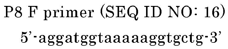

- The primer may include, for example, an additional sequence as will be described below, and examples thereof include as follows. In the sequence below, the underlined CTAGA and GACGA correspond to the additional sequence.


- Further, for detecting polymorphism in the 201st base (s) in SEQ ID NO: 2, preferably, the oligonucleotide P8, P8', P9, or P9' and the oligonucleotide P10 or P10' are used as a primer set.
- The primer for detecting polymorphism in the 501st base (r) of the sequence indicated in SEQ ID NO: 3 is preferably at least one oligonucleotide selected from the group consisting of P11, P11', P12, and P12'. The hybridization and the stringent conditions are the same as those described above.
(P11) an oligonucleotide having a sequence having at least 80% identity with respect to a base sequence including the 432nd to the 457th bases of the base sequence indicated in SEQ ID NO: 3 and having a length of 26 bases to 50 bases
(P11') an oligonucleotide having a sequence being capable of hybridization under stringent conditions to the complementary strand of a base sequence including the 432nd to the 457th bases of the base sequence indicated in SEQ ID NO: 3 and having a length of 26 bases to 50 bases
(P12) an oligonucleotide having a sequence having at least 80% identity with respect to the complementary strand of a base sequence including the 508th to the 539th bases of the base sequence indicated in SEQ ID NO: 3 and having a length of 32 bases to 50 bases
(P12') an oligonucleotide having a sequence being capable of hybridization under stringent conditions to a base sequence including the 508th to the 539th bases of the base sequence indicated in SEQ ID NO: 3 and having a length of 32 bases to 50 bases. - The oligonucleotide P11 may be an oligonucleotide having a sequence having at least 80% identity with respect to a base sequence including the 432nd to the 457th bases of the base sequence indicated in SEQ ID NO: 3 and having a length of from 26 bases to 50 bases and, at the same time, may be an oligonucleotide for amplifying a region containing the 501st base in SEQ ID NO: 3. The oligonucleotide P11' may be an oligonucleotide having a sequence being capable of hybridization under stringent conditions to the complementary strand of a base sequence including the 432nd to the 457th bases of the base sequence indicated in SEQ ID NO: 3 and having a length of 26 bases to 50 bases and, at the same time, may be an oligonucleotide for amplifying a region containing the 501st base in SEQ ID NO: 3.
- The oligonucleotide P12 may be an oligonucleotide having a sequence having at least 80% identity with respect to the complementary strand of a base sequence including the 508th to the 539th bases of the base sequence indicated in SEQ ID NO: 3 and having a length of 32 bases to 50 bases and, at the same time, may be an oligonucleotide for amplifying a region containing the 501st base in SEQ ID NO: 3. The oligonucleotide P12' may be an oligonucleotide having a sequence being capable of hybridization under stringent conditions to a base sequence including the 508th to the 539th bases of the base sequence indicated in SEQ ID NO: 3 and having a length of 32 bases to 50 bases and, at the same time, may be oligonucleotide for amplifying a region containing the 501st base in SEQ ID NO: 3.
- Examples of primers that can be used for amplifying a region containing the 501st base (r) in SEQ ID NO: 3 in the polymorphism detection method of the present invention are as follows.


- Further, for detecting polymorphism in the 501st base (r) in SEQ ID NO: 3, preferably, the oligonucleotide P11 or P11' and the oligonucleotide P12 or P12' are used as a primer set.
- The oligonucleotides P6, P7, P8, P9, P10, P11, and P12 may show at least 85% identity, at least 90% identity, at least 95% identity, at least 96% identity, at least 97% identity, at least 98% identity, or at least 99% identity (instead of at least 80% identity described above).
- The primer in the present invention may be a primer including (or comprising) the aforementioned oligonucleotide or may be a primer consisting of the aforementioned oligonucleotide. In the former case, for example, the primer of the present invention may include an additional sequence at at least one of the 3' end and the 5' end of the oligonucleotide. The additional sequence is not particularly limited. The additional sequence may have a length of 1 base to 20 bases, 3 bases to 15 bases, or 5 bases to 10 bases, for example. The additional sequence contains GC at a concentration of about 20 to 80%, about 40 to 70%, or about 40 to 60%, for example.
- The polymorphism detection method of the present invention is not particularly limited as long as it utilizes the fluorescence labeled nucleotide as a probe. As an example of the polymorphism detection method using the fluorescence labeled nucleotide as a probe, a polymorphism detection method utilizing Tm analysis will be described below.
- The method of detecting polymorphism in the CYP3A gene according to the present invention is a method comprising detecting polymorphism in the CYP3A gene using at least one of the aforementioned CYP3A gene polymorphism detection probes. According to the polymorphism detection method according to the present invention, the use of at least one of the aforementioned polymorphism detection probes enables a simple detection of polymorphism in the CYP3A gene with high sensitivity.
- The polymorphism detection method of the present invention is a method of detecting polymorphism in the CYP3A gene, can include the following processes (I) to (IV), and also may include the following process (V). Note here that the polymorphism detection method of the present invention is characterized by using the aforementioned polymorphism detection probe, and other configurations, conditions, and the like are not limited to the description below.
The processes (I) to (V) are as follows: - (I) contacting the probe with a single-stranded nucleic acid in a sample and hybridizing the probe and the single-stranded nucleic acid to obtain a hybrid;
- (II) measuring a change in the fluorescence signal based on dissociation of the hybrid by changing the temperature of the sample including the hybrid in order to dissociate the hybrid;
- (III) determining the Tm value which is the temperature at which the hybrid dissociates based on the change in the fluorescence signal;
- (IV) determining whether or not polymorphism in the CYP3A gene is present in the single-stranded nucleic acid in the sample based on the Tm value; and
- (V) determining the proportion of the single-stranded nucleic acids including polymorphism in the single-stranded nucleic acids in the sample based on the presence or absence of polymorphism.
- Further, in addition to the processes (I) to (IV) or the processes (I) to (V), the present invention may include the process of amplifying the nucleic acid before (I) or at the same time as (I). Here, determining the Tm value in (III) may not only include measuring the dissociation temperature of a hybrid but may also include measuring the level of the differential value of the fluorescence signal that changes in response to a change in temperature.
- In the present invention, the nucleic acid in the sample may be a single-stranded nucleic acid or a double-stranded nucleic acid. In the case where the nucleic acid is a double-stranded nucleic acid, for example, the method preferably includes melting (dissociating) the double-stranded nucleic acid in the sample into single-stranded nucleic acids by heating prior to the hybridization with the fluorescently labeled oligonucleotide. The dissociation of the double-stranded nucleic acid into single-stranded nucleic acids enables the hybridization with the fluorescently labeled oligonucleotide.
- In the present invention, the nucleic acid to be detected contained in the sample can be a nucleic acid inherently contained in a biological sample, for example. Preferably, since the detection accuracy can be improved, the nucleic acid to be detected is an amplification product obtained by amplifying a region containing a mutated site of the CYP3A gene by an amplification method such as PCR using the nucleic acid inherently contained in a biological sample as a template. The length of the amplification product is not particularly limited, and is, for example, from 50 bases to 1000 bases (mer), or from 80 bases to 200 bases. Further, the nucleic acid in the sample can be, for example, cDNA that is synthesized from RNA (total RNA, mRNA, or the like) derived from a biological sample by reverse transcription PCR (RT-PCR).
- In the present invention, there is no particular limitation on the addition ratio (molar ratio) of the polymorphism detection probe of the present invention relative to the nucleic acid in the sample. For example, the addition ratio (molar ratio) of the polymorphism detection probe of the present invention relative to DNA in the sample is no more than 1 times. To ensure a sufficient detection signal, the addition ratio (molar ratio) of the polymorphism detection probe of the present invention relative to the nucleic acid in the sample can be no more than 0.1 times. Here, the nucleic acid in the sample may be the total of the nucleic acid to be detected in which the polymorphism to be detected is generated and the nucleic acid which is not to be detected in which the polymorphism is not generated; or the nucleic acid in the sample may be the total of the amplification product including the sequence to be detected in which the polymorphism to be detected is generated and the amplification product including the sequence which is not to be detected in which the polymorphism is not generated. Although the ratio of the nucleic acid to be detected in nucleic acids in the sample is normally unknown, as a result, the addition ratio (molar ratio) of the polymorphism detection probe relative to the nucleic acid to be detected (the amplification product including the sequence to be detected) can be no more than 10 times. Further, the addition ratio (molar ratio) of the polymorphism detection probe relative to the nucleic acid to be detected (the amplification product including the sequence to be detected) can be no more than 5 times or no more than 3 times. There is no particular lower limit on the addition ratio (molar ratio) of the polymorphism detection probe, and for example, it can be, at least 0.001 times, at least 0.01 times, or at least 0.1 times.
- The addition ratio of the polymorphism detection probe of the present invention relative to DNA may be a molar ratio relative to a double-stranded nucleic acid or a molar ratio relative to a single-stranded nucleic acid, for example.
- In the present invention, on the basis of the aforementioned principle, the measurement of the change in the signal in relation to a temperature change for determining the Tm value can be performed by measuring absorbance at 260 nm. Preferably, the signal based on the signal of the labeling substance added to the polymorphism detection probe is measured, which is the signal that changes depending on the status of the hybrid of the single-stranded DNA and the polymorphism detection probe. Therefore, the aforementioned fluorescently labeled oligonucleotides are preferably used as the polymorphism detection probe. The aforementioned fluorescently labeled oligonucleotides include, for example, the fluorescently labeled oligonucleotide in which the fluorescence intensity when hybridized to its target sequence (complementary sequence) is less (quenched) than the fluorescence intensity when not hybridized to its target sequence; and the fluorescently labeled oligonucleotide in which fluorescence intensity when hybridized to its target sequence is greater than the fluorescence intensity when not hybridized to its target sequence.
- For example, although the probe of the former type does not emit a fluorescence signal or emits a weak fluorescence signal when it forms the hybrid (double-stranded DNA) with the sequence to be detected, the fluorescence signal is emitted or increased when the probe is dissociated by heating. Further, for example, the probe of the latter type emits a fluorescence signal when it forms the hybrid (double-stranded DNA) with the sequence to be detected, and the fluorescence signal is decreased (quenched) when the probe is dissociated by heating. Accordingly, by detecting the change in the fluorescence signal based on a fluorescence labeling under the conditions (fluorescence wavelength and the like) specific to the fluorescence labeling, progress of the melting of the hybrid and the Tm value can be determined as in the case of the measurement of absorbance at 260 nm.
- Next, with respect to the polymorphism detection method of the present invention, the method of detecting the change in a signal based on a fluorescent dye will be described with reference to the specific examples. Note here that the polymorphism detection method of the present invention is characterized by using the aforementioned polymorphism detection probe, and other processes, conditions, and the like are not limited.
- The sample containing a nucleic acid that is used as a template when nucleic acid amplification is performed is not particularly limited as long as it contains a nucleic acid, especially, the CYP3A gene. Examples thereof include samples that are derived from or can be derived from arbitrary biological origins such as tissues such as large intestine and lung; a blood cell such as leukocyte; whole blood; blood plasma; expectoration; oral mucosa suspension; somatic cells such as nail and hair; germ cell; milk; ascites; paraffin-embedded tissue; gastric juice; fluid obtained by gastric lavage; urine; peritoneal fluid; amniotic fluid; and cell culture. The method of collecting a sample and the method of preparing a sample containing a nucleic acid are not particularly limited and conventionally known methods can be employed. Further, the nucleic acid serving as a template can directly be used just as it is obtained from the origin or can be used after applying pretreatment for modifying the properties of the sample. For example, in the case where whole blood is used as a sample, isolation of genomic DNA from the whole blood can be performed by a conventionally known method. For example, a commercially available genomic DNA isolation kit (product name: GFX Genomic Blood DNA Purification kit, produced by GE Healthcare Biosciences) or the like can be used.
- Next, the polymorphism detection probe including the aforementioned fluorescently labeled oligonucleotide is added to a sample containing isolated genomic DNA. The polymorphism detection probe may be added to the liquid sample containing the isolated genomic DNA or may be mixed with the genomic DNA in an appropriate solvent. The solvent is not particularly limited, and examples thereof include known solvents such as a buffering solution such as Tris-HCl; a solvent containing KCl, MgCl2, MgSO4, glycerol, or the like; and a PCR reaction solution.
- There is no particular limitation on the timing of addition of the polymorphism detection probe, and for example, in the case where the amplification such as PCR that will be described below is performed, the polymorphism detection probe may be added after the amplification to the PCR amplification product or may be added before the amplification. In the case where the detection probe is added before the amplification such as PCR, for example, a fluorescent dye or a phosphate group is preferably added to the 3' end of the detection probe as described above.
- As the method of amplifying a nucleic acid, for example, a method using polymerase can be employed. Examples thereof include a PCR method, an ICAN method, a LAMP method, and a NASBA method. In the case where a nucleic acid is amplified by the method using polymerase, preferably, amplification is performed in the presence of the probe of the present invention. It is easy for those skilled in the art to adjust reaction conditions or the like for the amplification according to the probe and polymerase used. Thereby, since polymorphism can be evaluated simply by analyzing the Tm value of the probe after amplification of the nucleic acid, there is no need to handle the amplification product after the completion of the reaction. Therefore, there is no worry about contamination by the amplification product. Further, since the detection can be performed with the same equipment as that required for amplification, there is no need to transfer a container and automatization can be done easily.
- Further, as DNA polymerase used for the PCR method, commonly-used DNA polymerase can be used with no particular limitation. Examples of the DNA polymerase include GeneTaq (produced by NIPPON GENE CO., LTD.), PrimeSTAR Max DNA Polymerase (produced by TAKARA BIO INC.), and Taq polymerase. There is no limitation on the amount of the polymerase to be used, and a commonly-used concentration can be employed. For example, in the case where Taq polymerase is used, the concentration of the polymerase can be from 0.01 U to 100 U with respect to 50 μl of a reaction solution. Thereby, there is a tendency to increase the detection sensitivity to the CYP3A polymorphism.
- Further, the PCR method can be performed by appropriately selecting commonly-used conditions. Here, at the time of the amplification, for example, the number of copies of DNA (the sequence to be detected) contained in the sample can be determined by monitoring the amplification by real-time PCR. In other words, since the proportion of the probe that forms a hybrid increases in accordance with the amplification of DNA (the sequence to be detected) by PCR, the fluorescence intensity changes. By monitoring this change, the number of copies and the proportion of the sequence to be detected (normal DNA or mutated DNA) contained in the sample can be evaluated.
- In the polymorphism detection method of the present invention, the fluorescently labeled oligonucleotide and the single-stranded nucleic acid in the sample are brought into contact to hybridize. The single-stranded nucleic acid in the sample can be prepared, for example, by dissociating the PCR amplification product obtained in the manner described above.
- There is no limitation on the heating temperature in the dissociation (dissociation process) of the PCR amplification product, and an example thereof includes a temperature at which the amplification product can be dissociated. The temperature is, for example, 85 to 95°C. Furthermore, there is no particular limitation on the heating time, and heating time is, for example, 1 second to 10 minutes, or 1 second to 5 minutes.
- The hybridization between the dissociated single-stranded DNA and the fluorescently labeled oligonucleotide may be performed by decreasing the heating temperature employed in the dissociation process after the dissociation process, for example. The temperature condition is, for example, 40 to 50°C.
- The volume and the concentration of each component in the reaction solution of the hybridization process are not particularly limited. Specific examples are as follows. The DNA concentration in the reaction solution can be, for example, 0.01 μmol/L to 1 μmol/L or 0.1 μmol/L to 0.5 μmol/L. The concentration of the fluorescently labeled oligonucleotide is, for example, in the range satisfying the addition ratio to the DNA, and is, for example, 0.001 μmol/L to 10 μmol/L or 0.001 μmol/L to 1 μmol/L.
- Further, the obtained hybrid of the single-stranded DNA and the fluorescently labeled oligonucleotide is gradually heated to thereby measure the change in the fluorescence signal associated with the temperature increase. For example, when a QPROBE is used, the fluorescence intensity in a hybridized state with the single-stranded DNA is less (or quenched) than in a dissociated state. Therefore, for example, the hybrid that exhibits decreasing (or quenching) fluorescence is gradually heated to thereby measure the increase in fluorescence intensity associated with the temperature increase.
- There is no particular limitation on the temperature range when measuring the change in fluorescence intensity, and the initial temperature can be, for example, from room temperature to 85°C or from 25 to 70°C. The final temperature can be, for example, from 40 to 105°C. Furthermore, there is no particular limitation on the temperature increase rate, and it can be, for example, from 0.1 to 20°C/sec or from 0.3 to 5°C/sec.
- Next, the Tm value is determined by analyzing the change in the signal. Specifically, the differential value for each temperature (-d fluorescence intensity/dt) is calculated from the resulting fluorescence intensity and the temperature that shows the lowest value is determined as the Tm value. Further, the highest point in the fluorescence intensity increase per unit time (fluorescence intensity increase amount/t) can be determined as the Tm value. In contrast, the decrease in the fluorescence intensity may be measured when the labeled probe is not a quenching probe but rather a probe in which the signal intensity increases when forming a hybrid.
- Further, in the present invention, as described above, the hybrid is heated to thereby measure the change in the fluorescence signal (preferably, the increase in the fluorescence intensity) associated with the temperature increase as described. Alternatively in this method, for example, measurement of the change in the signal at the time of hybrid formation may be performed. That is, the temperature of the sample to which the probe is added is decreased to thereby form a hybrid, and then the change in the fluorescence signal associated with the temperature decrease is measured.
- In an actual example, in the case where a QPROBE is used, although the fluorescence intensity when the probe is added to the sample is high because the probe is in a dissociated state, the fluorescence is decreased (or quenched) when a hybrid is formed due to a decrease in the temperature. Therefore, for example, the decrease in the fluorescence intensity associated with the temperature decrease may be measured by gradually decreasing the temperature of the heated sample. On the other hand, in the case where a labeled probe that exhibits an increasing signal when forming a hybrid is used, although the fluorescence intensity when the probe is added to the sample is low (or quenched) because the probe is in a dissociated state, the fluorescence intensity is increased when a hybrid is formed due to a decrease in the temperature. Therefore, for example, the increase in the fluorescence intensity associated with the temperature decrease may be measured by gradually decreasing the temperature of the sample.
- In the polymorphism detection method of the present invention, for example, one of the polymorphism detection probes may be used alone or two or more of them may be used in combination. The type of the polymorphism detection probe to be used can be chosen according to the polymorphism to be detected. In the present invention, for example, only the CYP3A5*3 polymorphism, only the CYP3A4*16 polymorphism, or only the CYP3A4*1B polymorphism may be detected, or two of them or three of them can be detected in one reaction system. Further, at least one of the CYP3A5*3 polymorphism, the CYP3A4*16 polymorphism, and the CYP3A4*1B polymorphism and polymorphism other than these can be detected in one reaction system.
- In the polymorphism detection method of the present invention, in the case where two or more of the polymorphism detection probes are used, for example, the probes are preferably labeled probes including fluorescent dyes different from one another. The different fluorescent dyes include, for example, fluorescent dyes having different detection conditions from one another.
- The method of detecting multiple genetic polymorphisms in the same system is not particularly limited. For example, the probes capable of detecting the respective polymorphisms may be mixed beforehand and then added to the sample, or the probes capable of detecting the respective polymorphisms may be added successively to the sample containing a single-stranded nucleic acid. Here, the "system" refers to an independent reaction system composed of a sample containing a hybrid in which a fluorescently labeled oligonucleotide and a single-stranded nucleic acid are hybridized.
- The drug efficacy determination method of the present invention includes: detecting polymorphism in the CYP3A gene by the aforementioned polymorphism detection method; and determining (or predicting) tolerance to the drug or the efficacy of the drug based on the detection result.
- According to the polymorphism detection method, the use of the polymorphism detection probe of the present invention enables a simple detection of polymorphism in the CYP3A gene with high sensitivity. Therefore, on the basis of the polymorphism in the CYP3A gene, the simple determination of the drug efficacy or tolerance to said drug can be performed with high sensitivity.
- Further, on the basis of the presence or absence of polymorphism and the abundance ratio between a mutated sequence and a normal sequence, the tolerance to the drug or the efficacy of the drug can be determined. Further, the drug efficacy determination method of the present invention is effective for deciding therapeutic strategies such as an increase in dose of a drug and a change of the drug to another therapeutic drug based on the presence or absence of polymorphism and the abundance ratio between a mutated sequence and a normal sequence. Examples of the drug to be determined include an immunosuppressant, a molecular target drug, and an antidepressant. Among them, the immunosuppressant and the molecular target drug are particularly used. A specific example of the drug includes an immunosuppressant such as tacrolimus. Specific examples of drugs whose efficacy or tolerance may be determined based on the presence or absence of polymorphism in the CYP3A gene and the abundance ratio between a mutated sequence and a normal sequence include:
- psychotropics (which include antidepressants) such as alprazolam, estazolam, eszopiclone and midazolam which bind to specific sites on the GABAA gamma-amino-butyric acid receptor; carbamazepine which stabilizes the inactivated state of sodium channels; aripiprazole which is a D2 partial agonist; buspirone which is a serotonin 5-HT1A receptor partial agonist; citalopram and fluoxetine which are selective serotonin reuptake inhibitors: amitriptyline which is a serotonin-norepinephrine reuptake inhibitor; haloperidol which blocks D2 receptors; nefazodone which is an antagonist of the 5-HT2A receptor; pimozide which blocks the postsynaptic receptors D2 (extremely strongly), D3 (strongly), α1-adrenergic (strongly), and 5-HT2A (strongly); quetiapine which has the following pharmacological actions: D1 (IC50 = 1268nM), D2 (IC50 = 329nM), D3, and D4 receptor antagonist, 5-HT1A (IC50 = 717nM), 5-HT2A (IC50 = 148nM), 5-HT2C, and 5-HT7 receptor antagonist, mACh receptor (IC50 = >5000nM) antagonist; risperidone; sertraline; trazodone; triazolam; zaleplon; zolpidem;
- asthma medication such as fluticasone (a steroid); salmeterol; zileuton;
- antibiotics/ antifungals/ immunosuppressants such as macrolides (not azithromycin), such as tacrolimus; itraconazole; ketoconazole; telithromycin;
- anticonvulsants such as carbamazepine; ethosuximide; felbamate; tiagabine; zonisamide;
- antihistamines such as desloratadine; fexofenadine; loratadine;
- drugs of abuse/treatment such as buprenorphine; cocaine; fentanyl; ketamine; methadone; oxycodone; phencyclidine;
- calcium channel blockers and hormones/chemo-therapeutics such as cortisols; desogestrel (p); ethinyl estradiol; progestins/progesterone; vincristine; and others;
- and other drugs including antiemetics such as aprepitant and granisetron; calcimimetics such as cinacalcet; proton pump inhibitors such as esomeprazole and omeprazole; drugs for treating diabetes such as nateglinide and pioglitazone; anti-arrhythmic agents such as quinidine; drugs for treating erectile dysfunction such as sildenafil; statins (e.g. atorvastatin, lovastatin, simvastatin); anti-muscarinic agents such as tolterodine; and antivirals such as ritonavir (a HIV inhibitor) and saquinavir.
- The CYP3A gene polymorphism detection reagent kit for detecting polymorphism in the CYP3A gene of the present invention includes the aforementioned polymorphism detection probe. There is a tendency that the polymorphism detection reagent kit can perform, for example, a simple detection of polymorphism in the CYP3A gene by including at least one of the aforementioned polymorphism detection probes that can perform a simple and highly sensitive detection of polymorphism in the 401st base of the sequence indicated in SEQ ID NO: 1, polymorphism in the 201st base of the sequence indicated in SEQ ID NO: 2, and/or polymorphism in the 501st base of the sequence indicated in SEQ ID NO: 3 among the CYP3A gene.
- Further, the polymorphism detection reagent kit of the present invention may further include a primer for amplifying a sequence containing the CYP3A gene polymorphism to be detected. Thereby, the polymorphism detection reagent kit of the present invention can detect polymorphism in the CYP3A gene with high accuracy. Note here that the matters described above can be applied to the probe and the primer that can be contained in the polymorphism detection reagent kit.
- In the case where two or more fluorescently labeled oligonucleotides are contained as the polymorphism detection probe, the respective fluorescently labeled oligonucleotides may be contained in a mixed state or may be contained separately. Further, in the case where two or more of the probes of the present invention are contained in a mixed state and in the case where the Tm analysis of the respective fluorescently labeled oligonucleotides and the respective sequences to be detected is performed in the same reaction system in use although two or more of the probes of the present invention are contained separately, two or more probes are preferably labeled with fluorescent dyes having emission wavelengths different from one another. By changing the type of fluorescent material in this manner, detection with respect to the respective probes can be performed in the same reaction system.
- Further, in addition to the probe, the detection reagent kit of the present invention can further include a reagent(s) required for amplifying nucleic acid in the detection method of the present invention. The probe, the primer, and other reagents may be contained separately or some of them may be contained as a mixture.
- Here, with respect to the expression, "contained separately", the components are not necessarily contained in separate containers that can be handled independently as long as the respective reagents are divided so that the reagents are not brought into contact one another.
- For example, by including a primer set for amplifying the sequence containing a polymorphism site (a region to which a probe is hybridized), the detection reagent kit of the present invention can detect polymorphism with higher sensitivity.
- Further, preferably, the polymorphism detection reagent kit of the present invention includes an instruction manual. Examples of the instruction manual include: an instruction manual describing that the mutation in the CYP3A gene is detected using the aforementioned polymorphism detection probe by making the differential melting curve with respect to a sample containing the nucleic acid to be detected and analyzing the Tm value; and an instruction manual describing various reagents that are contained or that can be additionally contained in the detection reagent kit.
- Examples of the present invention will be described below. However, the present invention is not limited by the following Examples.
- In this Example, Tm analysis was performed in the presence of both a wild-type nucleic acid to be detected and a mutant nucleic acid to be detected to detect the CYP3A5*3 polymorphism and the CYP3A4*16 polymorphism in the CYP3A gene.
- Diluted blood, the entire genetic material of a cell (purified genome), and a plasmid are prepared as nucleic acid samples. The diluted blood was prepared as follows. That is, 10 μL of whole blood collected with an EDTA blood collection tube and 70 μL of
diluent 1 summarized in the following Table 2 were mixed to prepare a mixture. Then, 10 μL of the mixture and 70μL of diluent 2 summarized in the following Table 2 were mixed and the resultant mixture was heated at 95°C for 10 minutes. In the nucleic acid to be detected contained in the whole blood, the CYP3A5*3 polymorphism is a heterozygote of wild-type and mutant and the CYP3A4*16 polymorphism is a homozygote of wild-types. The purified genome was prepared by a routine method from whole blood. In the purified genome, the CYP3A5*3 polymorphism is a homozygote of mutants and the CYP3A4*16 polymorphism is a homozygote of wild-types. As the plasmid, the CYP3A4*16 mutant type plasmid that includes a partial sequence of the CYP3A4 gene by insertion of the oligonucleotide having a sequence including the 1st to 411th bases in SEQ ID NO: 2 was prepared. In the CYP3A4*16 mutant type plasmid, the 201st base (s) in SEQ ID NO: 2 is guanine (g). -
[Table 2] ( Diluent 1:70 μl)10 mmol/L Tris-HCl (pH 8.0) 0.1 mmol/L EDTA (pH 8.0) 0.3 w/v% SDS (Diluent 2:70 μl) 10 mmol/L Tris-HCl (pH 8.0) 0.1 mmol/L EDTA (pH 8.0) - The nucleic acid samples were mixed at the following ratios to prepare the
samples 1 to 4. -
Sample No. Diluted blood Purified genome CYP3A4*16 mutant plasmid 1 100% 0% 0% 2 0% 100% 0% 3 0% 50% 50% 4 0% 0% 100% - 50 μL of PCR reaction mixtures respectively containing the aforementioned samples were prepared. Each of the PCR reaction mixtures was prepared by mixing 4 μL of the sample and the components of the PCR reaction solution summarized in the following Table 3 at the following concentrations. PCR and Tm analysis were performed in relation to the PCR reaction mixtures using a fully automated SNPs inspection device (product name: I-DENSY (registered trademark) IS-5310, produced by Arkray Inc.). Conditions for PCR were as follows. That is, after the PCR reaction mixture was treated at 95°C for 1 minute, one cycle of treatment at 95°C for 1 second and 56°C for 15 seconds was repeated for 50 cycles. Further, the reaction mixture was treated at 95°C for 1 second and 40°C for 60 seconds. Thereafter, the reaction solution was heated from 40°C to 75°C at the temperature rise rate of 1°C/3 seconds to thereby perform Tm analysis by measurement of the change in the fluorescence intensity over time at a detection wavelength according to a fluorescent dye. Further, as a control, PCR and Tm analysis were performed in the same manner as described above except that diluent 2 was used instead of the sample. The detection wavelength for BODIPY FL was 520 to 555 nm and the detection wavelength for TAMRA was 585 to 700 nm (hereinafter, the same applies).
-
[Table 3] 1 × PCR reaction buffer 1.88 U Taq polymerase 1.5 mmol/L MgCl2 0.2 mmol/L dNTP 0.5 μmol/L CYP3A5*3 F primer 1 μmol/L CYP3A5*3 R primer 2 μmol/L CYP3A4*16 F primer 1 μmol/L CYP3A4*16 R primer 0.1 μmol/L CYP3A5*3 probe 0.4 μmol/L CYP3A4*16 probe - The sequences of the CYP3A5*3 primer and the CYP3A4*16 primer are as follows.
- (CYP3A5*3 primer)


- (CYP3A4*16 primer)


- The CYP3A5*3 probe that was used is the CYP3A5*3 mutant probe having the sequence below. The CYP3A5*3 probe is a mutant-targeting probe for the antisense strand of the mutant CYP3A5 gene. In the sequence below, the base that is underlined is a base that shows identity with respect to the CYP3A5*3 mutant sequence. The CYP3A5*3 probe was labeled with a fluorescent dye BODIPY FL at the 3' end. The CYP3A4*16 probe that was used is the CYP3A4*16 mutant probe having the sequence below. The CYP3A4*16 probe is a mutant-targeting probe for the sense strand of the mutant CYP3A4 gene. In the sequence, the base that is underlined is a base that is complementary to the CYP3A4*16 mutant sequence. The CYP3A4*16 probe was labeled with a fluorescent dye TAMRA at the 3' end.


- The results are illustrated in
FIGs. 1(A) to 5(B). FIGs. 1(A) to 5(B) are graphs of the results of Tm analysis showing the change in the fluorescence intensity associated with temperature rise.FIGs. 1(A) and 1(B) show the results forsample 1,FIGs. 2(A) and 2(B) show the results for sample 2,FIGs. 3(A) and 3(B) show the results for sample 3,FIG. 4 shows the result for sample 4, andFIGs. 5(A) and 5(B) show the results for the control. InFIGs. 1(A) to 3(B) and5(A) and 5(B) , (A) shows the melting curve with respect to the CYP3A5*3 probe and (B) shows the melting curve with respect to the CYP3A4*16 probe.FIG. 4 shows the melting curve with respect to the CYP3A4*16 probe. The horizontal axis shows the temperature (°C) during measurement. The vertical axis shows the change in the fluorescence intensity (hereinafter, this is also referred to as "fluorescence change amount"), and the unit was "d fluorescence intensity increase amount/dt" (dF/dt). The Tm value of the CYP3A5*3 probe and the CYP3A5*3 wild-type is close to 49°C and the Tm value of the CYP3A5*3 probe and the CYP3A5*3 mutant is close to 56°C. The Tm value of the CYP3A4*16 probe and the CYP3A4*16 wild-type is close to 59°C and the Tm value of the CYP3A4*16 probe and the CYP3A4*16 mutant is close to 67°C. - As illustrated in
FIG. 1(A) , with respect to the CYP3A5*3 polymorphism,sample 1 containing the CYP3A5*3 polymorphism as a heterozygote of wild-type and mutant showed peaks at the Tm value in relation to the wild-type and the Tm value of the mutant. As illustrated inFIG. 1(B) , with respect to the CYP3A4*16 polymorphism,sample 1 containing the CYP3A4*16 polymorphism as a homozygote of wild-types showed a peak only at the Tm value in relation to the wild-type. - As illustrated in
FIG. 2(A) , with respect to the CYP3A5*3 polymorphism, sample 2 containing the CYP3A5*3 polymorphism as a homozygote of mutants showed a peak only at the Tm value in relation to the mutant. As illustrated inFIG. 2(B) , with respect to the CYP3A4*16 polymorphism, sample 2 containing the CYP3A4*16 polymorphism as a homozygote of wild-types showed a peak only at the Tm value in relation to the wild-type. - As illustrated in
FIG. 3(A) , with respect to the CYP3A5*3 polymorphism, sample 3 containing the CYP3A5*3 polymorphism as a homozygote of mutants showed a peak only at the Tm value in relation to the mutant. As illustrated inFIG. 3(B) , with respect to the CYP3A4*16 polymorphism, sample 3 containing the CYP3A4*16 polymorphism contained in the purified genome as wild-type and containing the CYP3A4*16 polymorphism contained in the plasmid as a mutant showed peaks at the Tm value in relation to the wild-type and the Tm value in relation to the mutant. - As illustrated in
FIG. 4 , with respect to the CYP3A4*16 polymorphism, sample 4 containing the CYP3A4*16 polymorphism as a homozygote of mutants showed a peak only at the Tm value in relation to the mutant. Sample 4 that does not include a base sequence of a region containing the CYP3A5*3 polymorphism did not show a peak with respect to the CYP3A5*3 polymorphism. - As illustrated in
FIGs. 5(A) and 5(B) , the diluent 2, which is a control, did not show a peak with respect to either of the CYP3A4*16 polymorphism or the CYP3A5*3 polymorphism. - In this Example, Tm analysis was respectively performed in relation to a wild-type artificial nucleic acid and a mutant-type artificial nucleic acid to detect the polymorphism (CYP3A4*16) in the CYP3A gene using a probe that hybridizes to the antisense strand.
- A mutant artificial nucleic acid (mt) and a wild-type artificial nucleic acid (wt) that are complementary to the 173th to 222nd bases in SEQ ID NO: 2 as illustrated in the sequences below were prepared. In the sequences below, the underlined bases correspond to the 201st base in SEQ ID NO: 2. The respective artificial nucleic acids were adjusted to 0.1 μmol/L and were used as samples.


- 50 μL of reaction mixtures respectively containing the aforementioned samples were prepared. Each of the reaction mixtures was prepared by mixing the components of the reaction solution summarized in the following Table 4 at the following concentrations. Tm analysis was performed in relation to the reaction mixtures using a fully automated SNPs inspection device (product name: I-DENSY (registered trademark) IS-5310, produced by Arkray Inc.). After treating at 95°C for 1 second and 40°C for 60 seconds, the reaction solution was heated from 40°C to 75°C at the temperature rise rate of 1°C/3 seconds to thereby perform Tm analysis by measurement of the change in the fluorescence intensity over time at a detection wavelength according to a fluorescent dye.
-
[Table 4] 1 × reaction buffer 1.88 U Taq polymerase 1.5 mmol/L MgCl2 0.2 mmol/L dNTP 0.2 mmol/L probe 0.2 mmol/L sample - The probe that was used is the CYP3A4*16 mutant probe having the sequence below. The probe is a mutant-targeting probe for the antisense strand of the mutant CYP3A4 gene. In the sequence below, the base that is underlined is a base that shows identity with respect to the CYP3A4*16 mutant sequence. The probe was labeled with a fluorescent dye TAMRA at the 3' end.
CYP3A4*16 mutant probe (SEQ ID NO: 6) (3T-CYP3A4*16-MF1) 5'-gatgtgatcagtagc-(TAMRA)-3' - The results are illustrated in
FIGs. 6(A) and 6(B). FIGs. 6(A) and 6(B) are graphs of the Tm analysis showing the change in fluorescence intensity associated with temperature rise. InFIGs. 6(A) and 6(B) , (A) shows the result formt 100% and (B) shows the result forwt 100%. The horizontal axis shows the temperature (°C) during measurement. The vertical axis shows the change in fluorescence intensity (hereinafter, this is also referred to as "fluorescence change amount"), and the unit was "d fluorescence intensity increase amount/dt" (dF/dt). The Tm value of the CYP3A4*16 mutant probe and mt is close to 60°C and the Tm value of the CYP3A4*16 mutant probe and wt is close to 53°C. - As illustrated in
FIG. 6(A) , with respect to mt, the sample showed a peak only at the Tm value in relation to mt. On the other hand, as illustrated inFIG. 6(B) , with respect to wt, the sample showed a peak only at the Tm value in relation to wt. - In this Example, Tm analysis was respectively performed in relation to a wild-type artificial nucleic acid and a mutant-type artificial nucleic acid to detect the polymorphism (CYP3A4*16) in the CYP3A gene using a probe that hybridizes to the sense strand. Tm analysis was performed in the same manner as in Example 2 except that the wild-type artificial nucleic acid and the mutant artificial nucleic acid described below and the CYP3A4*16 mutant probe were used.
- A mutant artificial nucleic acid (mt) and a wild-type artificial nucleic acid (wt) that are homologous to the 173th to 222nd bases in SEQ ID NO: 2 as illustrated in the sequences below were prepared. In the sequences below, the underlined bases correspond to the 201st base in SEQ ID NO: 2. The respective artificial nucleic acids were adjusted to 0.1 μmol/L and were used as samples.


- The probe that was used is the CYP3A4*16 mutant probe having the sequence below. The probe is a mutant-targeting probe for the sense strand of the mutant CYP3A4 gene. In the sequence below, the base that is underlined is a base that is complementary to the CYP3A4*16 mutant sequence. The probe was labeled with a fluorescent dye TAMRA at the 3' end.

- The results are illustrated in
FIGs. 7(A) and 7(B). FIGs. 7(A) and 7(B) are graphs of the Tm analysis showing the change in fluorescence intensity associated with temperature rise. InFIGs. 7(A) and 7(B) , (A) shows the result formt 100% and (B) shows the result forwt 100%. The horizontal axis shows the temperature (°C) during measurement. The vertical axis shows the change in fluorescence intensity (hereinafter, this is also referred to as "fluorescence change amount"), and the unit was "d fluorescence intensity increase amount/dt" (dF/dt). The Tm value of the CYP3A4*16 mutant probe and mt is close to 75°C and the Tm value of the CYP3A4*16 mutant probe and wt is close to 68°C. - As illustrated in
FIG. 7(A) , with respect to mt, the sample showed a peak only at the Tm value in relation to mt. On the other hand, as illustrated inFIG. 7(B) , with respect to wt, the sample showed a peak only at the Tm value in relation to wt. - In this Comparative Example, Tm analysis was respectively performed in relation to a wild-type artificial nucleic acid and a mutant-type artificial nucleic acid to detect the polymorphism (CYP3A5*3) in the CYP3A gene using a probe that hybridizes to the sense strand. Tm analysis was performed in the same manner as in Example 2 except that the wild-type artificial nucleic acid and the mutant artificial nucleic acid described below and the CYP3A5*3 mutant probe were used.
- A mutant artificial nucleic acid (mt) and a wild-type artificial nucleic acid (wt) that are homologous to the 379th to 428th bases in SEQ ID NO: 1 as illustrated in the sequences below were prepared. In the sequences below, the underlined bases correspond to the 401st base in SEQ ID NO: 1. The respective artificial nucleic acids were adjusted to 0.1 μmol/L and were used as samples.


- The probe that was used is the CYP3A5*3 mutant probe having the sequence below. The probe is a mutant-targeting probe for the sense strand of the mutant CYP3A5 gene. In the sequence below, the base that is underlined is a base that is complementary to the CYP3A5*3 mutant sequence. The CYP3A5*3 mutant probe was labeled with a fluorescent dye TAMRA at the 3' end.

- The results are illustrated in
FIGs. 8(A) and 8(B). FIGs. 8(A) and 8(B) are graphs of the Tm analysis showing the change in fluorescence intensity associated with temperature rise. InFIGs. 8(A) and 8(B) , (A) shows the result formt 100% and (B) shows the result forwt 100%. The horizontal axis shows the temperature (°C) during measurement. The vertical axis shows the change in fluorescence intensity (hereinafter, this is also referred to as "fluorescence change amount"), and the unit was "d fluorescence intensity increase amount/dt" (dF/dt). - As illustrated in
FIGs. 8(A) and 8(B) , the sample did not show a clear peak with respect to both wt and mt. - In this Comparative Example, Tm analysis was respectively performed in relation to a wild-type artificial nucleic acid and a mutant-type artificial nucleic acid to detect the polymorphism (CYP3A4*6) in the CYP3A gene using a probe that hybridizes to the sense strand. Tm analysis was performed in the same manner as in Example 3 except that the CYP3A4*16 mutant probe was used.
- The probe that was used is the CYP3A4*16 mutant probe having the sequence below. The probe is a mutant probe for the sense strand of the mutant CYP3A4 gene. In the sequence below, the base that is underlined is a base that is complementary to the CYP3A4*16 mutant sequence. The CYP3A4*16 mutant probe was labeled with a fluorescent dye TAMRA at the 3' end.

- The results are illustrated in
FIGs. 9(A) and 9(B). FIGs. 9(A) and 9(B) are graphs of the Tm analysis showing the change in fluorescence intensity associated with temperature rise. InFIGs. 9(A) and 9(B) , (A) shows the result formt 100% and (B) shows the result forwt 100%. The horizontal axis shows the temperature (°C) during measurement. The vertical axis shows the change in fluorescence intensity (hereinafter, this is also referred to as "fluorescence change amount"), and the unit was "d fluorescence intensity increase amount/dt" (dF/dt). - As illustrated in
FIG. 9(A) , with respect to mt, the sample showed a peak only at the Tm value in relation to mt. On the other hand, as illustrated inFIG. 9(B) , with respect to wt, the sample did not show a peak. - In this Example, the CYP3A4*1B polymorphism, the CYP3A4*16 polymorphism, and the CYP3A5*3 polymorphism in the CYP3A4 gene were detected using the same reaction solution.
- A purified genome, a plasmid, and pretreated whole blood were prepared as nucleic acid samples. The purified genome was prepared by a routine method from whole blood collected with an EDTA blood collection tube, and the purified genome adjusted to 400 copies/μL was used as a sample. As the plasmid, the CYP3A4*1B mutant type plasmid that includes a partial sequence of the CYP3A4 gene by insertion of the oligonucleotide having a sequence including the 366th to 651st bases in SEQ ID NO: 3 and the CYP3A4*16 mutant type plasmid that includes a partial sequence of the CYP3A4 gene by insertion of the oligonucleotide having a sequence including the 1st to 411th bases in SEQ ID NO: 2 were prepared. In the CYP3A4*1B mutant type plasmid, the 501st base in SEQ ID NO: 3 was a mutant (g). In the CYP3A4*16 mutant type plasmid, the 201st base in SEQ ID NO: 2 was a mutant (g). Each of the plasmids were adjusted to 2000 copies/μL, the adjusted plasmids were mixed with 0.5 μL of each, and the resultant was used as a sample. The pretreated whole blood was prepared as follows. That is, 10 μL of the whole blood that is the same as that used for preparation of the purified genome and 70 μL of
diluent 1 summarized in Table 2 were mixed to prepare a mixture, 10μL of the mixture and 70 μL of diluent 2 summarized in Table 2 were mixed, and then the resultant was heated at 95°C for 10 minutes. In the nucleic acid to be detected contained in the whole blood, the CYP3A4*1B polymorphism is a homozygote of wild-types, the CYP3A5*3 polymorphism is a heterozygote of wild-type and mutant, and the CYP3A4*16 polymorphism is a homozygote of wild-types. - 50 μL of PCR reaction mixtures respectively containing the aforementioned samples were prepared. Each of the PCR reaction mixtures was prepared by mixing the sample and the components of the PCR reaction solution summarized in the following Table 5 at the following concentrations. In the PCR reaction mixture, the concentration of the purified genome sample was 2 × 10⁶ copies/L, the concentration of the plasmid sample was 2 × 10⁷ copies/L, and the concentration of the whole blood sample was 5.3 mL/L. PCR and Tm analysis were performed in relation to the PCR reaction mixtures using a fully automated SNPs inspection device (product name: I-DENSY (registered trademark) IS-5310, produced by Arkray Inc.). Conditions for PCR were as follows. That is, after the PCR reaction mixture was treated at 95°C for 1 minute, one cycle of treatment at 95°C for 1 second and 58°C for 30 seconds was repeated for 50 cycles. Further, the reaction mixture was treated at 95°C for 1 second and 40°C for 60 seconds. Thereafter, the reaction solution was heated from 40°C to 75°C at a temperature rise rate of 1°C/3 seconds to thereby perform Tm analysis by measurement of the change in fluorescence intensity over time at a detection wavelength according to a fluorescent dye. Further, as a control, PCR and Tm analysis were performed in the same manner as described above except that diluent 2 was used instead of the sample. The detection wavelength for Pacific Blue was 445 to 480 nm, the detection wavelength for BODIPY FL was 520 to 555 nm, and the detection wavelength for TAMRA was 585 to 700 nm.
-
[Table 5] 1 × PCR reaction buffer 1.88 U Taq polymerase 1.5 mmol/L MgCl2 0.2 mmol/L dNTP 0.5 μmol/L CYP3A4*1B F primer 0.25 μmol/L CYP3A4*1B R primer 0.25 μmol/L CYP3A5*3 F primer 0.5 μmol/L CYP3A5*3 R primer 2 μmol/L CYP3A4*16 F primer 4 μmol/L CYP3A4*16 R primer 0.2 μmol/L CYP3A4*1B probe 0.1 μmol/L CYP3A5*3 probe 0.4 μmol/L CYP3A4*16 probe - The sequences of the CYP3A4*1B primer and the CYP3A4*1B probe are as follows.
- (CYP3A4*1B primer)


- (CYP3A4*1B probe)

- The sequences of the CYP3A4*16 primer and the CYP3A4*16 probe are as follows.
- (CYP3A4*16 primer)


- (CYP3A4*16 probe)

- The sequences of the CYP3A5*3 primer and the CYP3A5*3 probe are as follows.
- (CYP3A5*3 primer)


- (CYP3A5*3 probe)

- The results are illustrated in
FIGs. 10(A) to 12(C). FIGs. 10(A) to 12(C) are graphs of the Tm analysis showing the change in fluorescence intensity associated with temperature rise.FIGs. 10(A) to 10(C) show the results of the sample containing the purified genome,FIGs. 11(A) and 11(B) show the results of the sample containing the mixed plasmid, andFIGs. 12(A) to 12(C) show the results of the sample containing the pretreated whole blood. InFIGs. 10(A) to 10(C) and 12(A) to 12(C) , (A) shows a melting curve with respect to the CYP3A4*1B mutant probe, (B) shows a melting curve with respect to the CYP3A5*3 mutant probe, and (C) shows a melting curve with respect to the CYP3A4*16 mutant probe. InFIGs. 11(A) and 11(B) , (A) shows a melting curve with respect to the CYP3A4*1B mutant probe and (B) shows a melting curve with respect to the CYP3A4*16 mutant probe. The horizontal axis shows the temperature (°C) during measurement. The vertical axis shows the change in fluorescence intensity (hereinafter, this is also referred to as "fluorescence change amount"), and the unit was "d fluorescence intensity increase amount/dt" (dF/dt). The Tm value of the CYP3A4*1B mutant and the CYP3A4*1B mutant probe is close to 61°C, the Tm value of the CYP3A5*3 mutant and the CYP3A5*3 mutant probe is close to 56°C, and the Tm value of the CYP3A4*16 mutant and the CYP3A4*16 mutant probe is close to 53°C. - As illustrated in
FIG. 10(A) , with respect to the CYP3A4*1B polymorphism, the sample containing the purified genome that contains the CYP3A4*1B polymorphism as a homozygote of wild-types showed a peak at a temperature lower than the Tm value in relation to the mutant. As illustrated inFIG. 10(B) , with respect to the CYP3A5*3 polymorphism, the sample containing the purified genome that contains the CYP3A5*3 polymorphism as a heterozygote of wild-type and mutant showed two peaks at the Tm value in relation to the mutant and at a temperature lower than this Tm value. As illustrated inFIG. 10(C) , with respect to the CYP3A4*16 polymorphism, the sample containing the purified genome that contains the CYP3A4*16 polymorphism as a homozygote of wild-types showed a peak at a temperature lower than the Tm value in relation to the mutant. - As illustrated in
FIG. 11(A) , with respect to the CYP3A4*1B polymorphism, the sample containing the mutant plasmid showed a peak only at the Tm value in relation to the mutant. As illustrated in 11(B), with respect to the CYP3A4*16 polymorphism, the sample containing the mutant plasmid showed a peak only at the Tm value in relation to the mutant. Since the plasmid sample does not contain the CYP3A5*3 polymorphism, the sample did not show a peak with respect to the CYP3A5*3 polymorphism. - As illustrated in
FIG. 12(A) , with respect to the CYP3A4*1B polymorphism, the sample containing the pretreated whole blood that contains the CYP3A4*1B polymorphism as a homozygote of wild-types showed a peak at a temperature lower than the Tm value in relation to the mutant. As illustrated inFIG. 12(B) , with respect to the CYP3A5*3 polymorphism, the sample containing the pretreated whole blood that contains the CYP3A5*3 polymorphism as a heterozygote of wild-type and mutant showed two peaks at the Tm value in relation to the mutant and at a temperature lower than this Tm value. As illustrated inFIG. 12(C) , with respect to the CYP3A4*16 polymorphism, the sample containing the pretreated whole blood that contains the CYP3A4*16 polymorphism as a homozygote of wild-types showed a peak at a temperature lower than the Tm value in relation to the mutant. - In this Example, Tm analysis was respectively performed in relation to a wild-type artificial nucleic acid and a mutant-type artificial nucleic acid to detect the polymorphism (CYP3A4*1B) in the CYP3A gene using a probe that hybridizes to the sense strand.
- As artificial nucleic acids of sense strand, a wild-type artificial nucleic acid (WT 1) and mutant artificial nucleic acids (mt 1-1 and mt 1-2) that are homologous to the 482nd to 521st bases in SEQ ID NO: 3 as illustrated in the sequences below were prepared. In the sequences below, the underlined bases correspond to the 501st base in SEQ ID NO: 3. In the sequences below, the bases in lower case are substituted with bases that are different from those of SEQ ID NO: 3. The respective artificial nucleic acids were adjusted to 0.1 μmol/L.


- 50 μL of reaction mixtures respectively containing the aforementioned artificial nucleic acids were prepared. Each of the reaction mixtures was prepared by mixing the components of the reaction solution summarized in the following Table 6 at the following concentrations. Tm analysis was performed in relation to the reaction mixtures using a fully automated SNPs inspection device (product name: I-DENSY (registered trademark) IS-5310, produced by Arkray Inc.). Conditions for Tm analysis were as follows. That is, the reaction solution was treated at 95°C for 1 second and 40°C for 60 seconds. Thereafter, the reaction solution was heated from 40°C to 75°C at a temperature rise rate of 1°C/3 seconds to thereby perform Tm analysis by measurement of the change in fluorescence intensity over time at a detection wavelength according to a fluorescent dye. The detection wavelength for Pacific Blue was 445 to 480 nm.
-
[Table 6] 1 × reaction buffer 1.88 U Taq polymerase 1.5 mmol/L MgCl2 0.2 mmol/L dNTP 0.2 mmol/L probe 0.2 mmol/L wild-type artificial nucleic acid (WT 1) 0.2 mmol/L mutant-type artificial nucleic acid (mt 1-1 or mt 1-2) - The probe that was used is the CYP3A4*1B mutant probe having the sequence below. The probe is a mutant-targeting probe for the sense strand of the mutant CYP3A4 gene. In the sequence below, the base that is underlined is a base that is complementary to the CYP3A4*1B mutant sequence.

- In this Comparative Example, Tm analysis was respectively performed in relation to a wild-type artificial nucleic acid and a mutant-type artificial nucleic acid to detect the polymorphism (CYP3A4*1B) in the CYP3A gene using a probe that hybridizes to the sense strand.
- The CYP3A4*1B probe that was used is the probe having the sequence below. The probe is a wild-type probe for the sense strand of the wild-type CYP3A4 gene. In the sequence below, the base that is underlined is a base that is complementary to the CYP3A4*1B mutant sequence.

- Tm analysis was performed in the same manner as in Example 5 except that the probe 3-1 was used as the CYP3A4*1B probe.
- In this Comparative Example, Tm analysis was respectively performed in relation to a wild-type artificial nucleic acid and a mutant-type artificial nucleic acid to detect the polymorphism (CYP3A4*1B) in the CYP3A gene using a probe that hybridizes to the antisense strand.
- As artificial nucleic acids of antisense strand, a wild-type artificial nucleic acid (WT 2) and mutant artificial nucleic acids (mt 2-1 and mt 2-2) that are complementary to the 482nd to 521st bases in SEQ ID NO: 3 as illustrated in the sequences below were prepared. In the sequences below, the underlined bases correspond to the 501st base in SEQ ID NO: 3. The respective artificial nucleic acids were adjusted to 0.1 μmol/L.


- The CYP3A4*1B probe that was used is the probe 2-2 having the sequence below. The probe is a wild-type probe for the antisense strand of the wild-type CYP3A4 gene. In the sequence below, the base that is underlined is a base that is homologous to the CYP3A4*1B wild-type.

- Tm analysis was performed in the same manner as in Example 5 except that the WT 2 was used as the wild-type artificial nucleic acid, the mt 2-1 or the mt 2-2 was used as the mutant artificial nucleic acid, and the probe 2-2 was used as the CYP3A4*1B probe.
- The results are illustrated in
FIGs. 13(A) to 15. FIGs. 13(A) to 15 are graphs of the Tm analysis showing the change in fluorescence intensity associated with temperature rise.FIGs. 13(A) and 13(B) show the results of Example 5,FIG. 14 shows the result of Comparative Example 3-1, andFIG. 15 shows the result of Comparative Example 3-2. The horizontal axis shows the temperature (°C) during measurement. The vertical axis shows the change in fluorescence intensity (hereinafter, this is also referred to as "fluorescence change amount"), and the unit was "d fluorescence intensity increase amount/dt" (dF/dt). - As illustrated in
FIGs. 13(A) and 13(B) , in Example 5, in the case where the combination ofWT 1 and mt 1-1 or the combination ofWT 1 and mt 1-2 was added to the PCR reaction solution, peaks for WT 1 (55°C) and mt 1-1 (64°C) or mt 1-2 (64°C) were observed. On the other hand, as illustrated inFIG. 14 , in Comparative Example 3-1, in the case where the combination ofWT 1 and mt 1-1 was added to the PCR reaction solution, a peak forWT 1 overlapped with a peak for mt 1-1; and in the case where the combination ofWT 1 and mt 1-2 was added to the PCR reaction solution, a peak forWT 1 and a peak for mt 1-2 were not observed. Further, as illustrated inFIG. 15 , in Comparative Example 3-2, in the case where the combination of WT 2 and mt 2-2 was added to the PCR reaction solution, a peak for WT 2 overlapped with a peak for mt 2-2; and in the case where the combination of WT 2 and mt 2-1 was added to the PCR reaction solution, a peak for WT 2 and a peak for mt 2-1 were not observed. - In this manner, it became evident that a simple detection of polymorphism in the CYP3A gene with high sensitivity can be performed according to the present invention.
- According to the polymorphism detection method of the present invention, a probe for detecting polymorphism that enables a simple detection of polymorphism in the CYP3A gene with high sensitivity and a method of detecting polymorphism using the probe is provided. Further, the present invention provides a method of evaluating a drug efficacy using the method of detecting polymorphism. Furthermore, the present invention provides a reagent kit for detecting polymorphism using the probe for detecting polymorphism.
- The invention may be embodied in other forms without departing from the spirit or essential characteristics thereof. The embodiments disclosed in this application are to be considered in all respects as illustrative and not limiting. The scope of the invention is indicated by the appended claims rather than by the foregoing description, and all changes which come within the meaning and range of equivalency of the claims are intended to be embraced therein.


Claims (15)
- A probe which detects polymorphism in the CYP3A gene, the probe comprising one fluorescently labeled oligonucleotide selected from the group consisting of P1, P1', P2, P2', P3, P3', P4, P4', P5, and P5':(P1) a fluorescently labeled oligonucleotide having a sequence including the 401st to the 411th bases of the sequence indicated in SEQ ID NO: 1 and having a length of 11 bases to 50 bases, the sequence having at least 80% identity with respect to a base sequence having the same bases as SEQ ID NO: 1 with the exception that the base corresponding to the 411th base in SEQ ID NO: 1 is cytosine, and the base corresponding to the 411th base of the sequence indicated in SEQ ID NO: 1 is labeled with a fluorescent dye;(P1') a fluorescently labeled oligonucleotide having a sequence including the 401st to the 411th bases of the sequence indicated in SEQ ID NO: 1 and having a length of 11 bases to 50 bases, the sequence being capable of hybridization under stringent conditions to the complementary strand of a base sequence having the same bases as SEQ ID NO: 1 with the exception that the base corresponding to the 411th base in SEQ ID NO: 1 is cytosine, and the base corresponding to the 411th base of the sequence indicated in SEQ ID NO: 1 is labeled with a fluorescent dye;(P2) a fluorescently labeled oligonucleotide having a sequence including the 201st to the 205th bases of the sequence indicated in SEQ ID NO: 2 and having a length of 5 bases to 50 bases, the sequence having at least 80% identity with respect to a base sequence having the same bases as SEQ ID NO: 2 with the exception that the base corresponding to the 205th base in SEQ ID NO: 2 is cytosine, and the base corresponding to the 205th base of the sequence indicated in SEQ ID NO: 2 is labeled with a fluorescent dye;(P2') a fluorescently labeled oligonucleotide having a sequence including the 201st to the 205th bases of the sequence indicated in SEQ ID NO: 2 and having a length of 5 bases to 50 bases, the sequence being capable of hybridization under stringent conditions to the complementary strand of a base sequence having the same bases as SEQ ID NO: 2 with the exception that the base corresponding to the 205th base in SEQ ID NO: 2 is cytosine, and the base corresponding to the 205th base of the sequence indicated in SEQ ID NO: 2 is labeled with a fluorescent dye;(P3) a fluorescently labeled oligonucleotide having a sequence complementary to a sequence including the 190th to the 201st bases of the sequence indicated in SEQ ID NO: 2 and having a length of 12 bases to 50 bases, the sequence having at least 80% identity with respect to a base sequence having the same bases as the complementary strand of SEQ ID NO: 2 with the exception that the base complementary to the 190th base in SEQ ID NO: 2 is cytosine, and the base complementary to the 190th base of the sequence indicated in SEQ ID NO: 2 is labeled with a fluorescent dye;(P3') a fluorescently labeled oligonucleotide having a sequence complementary to a sequence including the 190th to the 201st bases of the sequence indicated in SEQ ID NO: 2 and having a length of 12 bases to 50 bases, the sequence being capable of hybridization under stringent conditions to a base sequence having the same bases as SEQ ID NO: 2 with the exception that the base complementary to the 190th base in SEQ ID NO: 2 is cytosine, and the base complementary to the 190th base of the sequence indicated in SEQ ID NO: 2 is labeled with a fluorescent dye;(P4) a fluorescently labeled oligonucleotide having a sequence complementary to a sequence including the 186th to the 201st bases of the sequence indicated in SEQ ID NO: 2 and having a length of 16 bases to 50 bases, the sequence having at least 80% identity with respect to a base sequence having the same bases as the complementary strand of SEQ ID NO: 2 with the exception that the base complementary to the 186th base in SEQ ID NO: 2 is cytosine, and the base complementary to the 186th base of the sequence indicated in SEQ ID NO: 2 is labeled with a fluorescent dye;(P4') a fluorescently labeled oligonucleotide having a sequence complementary to a sequence including the 186th to the 201st bases of the sequence indicated in SEQ ID NO: 2 and having a length of 16 bases to 50 bases, the sequence being capable of hybridization under stringent conditions to a base sequence having the same bases as SEQ ID NO: 2 with the exception that the base complementary to the 186th base in SEQ ID NO: 2 is cytosine, and the base complementary to the 186th base of the sequence indicated in SEQ ID NO: 2 is labeled with a fluorescent dye;(P5) a fluorescently labeled oligonucleotide having a sequence complementary to a sequence including the 496th to the 501st bases of the sequence indicated in SEQ ID NO: 3 and having a length of 6 bases to 50 bases, the sequence having at least 80% identity with respect to a base sequence having the same bases as the complementary strand of SEQ ID NO: 3 with the exception that the base complementary to the 496th base in SEQ ID NO: 3 is cytosine, and the base complementary to the 496th base of the sequence indicated in SEQ ID NO: 3 is labeled with a fluorescent dye; and(P5') a fluorescently labeled oligonucleotide having a sequence complementary to a sequence including the 496th to the 501st bases of the sequence indicated in SEQ ID NO: 3 and having a length of 6 bases to 50 bases, the sequence being capable of hybridization under stringent conditions to a base sequence having the same bases as SEQ ID NO: 3 with the exception that the base complementary to the 496th base in SEQ ID NO: 3 is cytosine, and the base complementary to the 496th base of the sequence indicated in SEQ ID NO: 3 is labeled with a fluorescent dye.
- The probe of claim 1, wherein the fluorescently labeled oligonucleotide recognizes or detects polymorphism in the 401st base in SEQ ID NO: 1.
- The probe of claim 1 or 2, wherein the base labeled with the fluorescent dye is at a position of any one of 1st to 3rd positions from the 3' end of the fluorescently labeled oligonucleotide, preferably wherein the base labeled with the fluorescent dye is at the 3' end of the fluorescently labeled oligonucleotide.
- The probe of any one of claims 1 to 3, wherein the fluorescently labeled oligonucleotide emits fluorescence when not hybridized to its target sequence, and the fluorescence intensity of the fluorescently labeled oligonucleotide when hybridized to its target sequence is greater or less than the fluorescence intensity when not hybridized to its target sequence, preferably wherein the fluorescence intensity of the fluorescently labeled oligonucleotide when hybridized to its target sequence is less than the fluorescence intensity when not hybridized to its target sequence.
- The probe of any one of claims 1 to 4, wherein
the fluorescently labeled oligonucleotide P1 and the fluorescently labeled oligonucleotide P1' each have a length of 15 bases to 30 bases,
the fluorescently labeled oligonucleotide P2 and the fluorescently labeled oligonucleotide P2' each have a length of 10 bases to 30 bases,
the fluorescently labeled oligonucleotide P3 and the fluorescently labeled oligonucleotide P3' each have a length of 12 bases to 40 bases,
the fluorescently labeled oligonucleotide P4 and the fluorescently labeled oligonucleotide P4' each have a length of 16 bases to 40 bases, and
the fluorescently labeled oligonucleotide P5 and the fluorescently labeled oligonucleotide P5' each have a length of 10 bases to 40 bases. - The probe of any one of claims 1 to 5, wherein
the fluorescently labeled oligonucleotide P1 and the fluorescently labeled oligonucleotide P1' each have a length of 15 bases to 20 bases,
the fluorescently labeled oligonucleotide P2 and the fluorescently labeled oligonucleotide P2' each have a length of 10 bases to 20 bases,
the fluorescently labeled oligonucleotide P3 and the fluorescently labeled oligonucleotide P3' each have a length of 15 bases to 30 bases,
the fluorescently labeled oligonucleotide P4 and the fluorescently labeled oligonucleotide P4' each have a length of 20 bases to 30 bases, and
the fluorescently labeled oligonucleotide P5 and the fluorescently labeled oligonucleotide P5' each have a length of 20 bases to 30 bases. - The probe of any one of claims 1 to 6, being a probe for melting curve analysis.
- The probe of any one of claims 1 to 7, having a base sequence indicated in SEQ ID NO: 37, SEQ ID NO: 38, SEQ ID NO: 39, SEQ ID NO: 41, SEQ ID NO: 43, or SEQ ID NO: 45.
- The probe of any one of claims 1 to 7, having a base sequence indicated in SEQ ID NO: 4, SEQ ID NO: 6, SEQ ID NO: 8, SEQ ID NO: 10, SEQ ID NO: 44, or SEQ ID NO: 12.
- A use of the probe of any one of claims 1 to 9 in a method of detecting polymorphism in the CYP3A gene.
- A method of detecting polymorphism in the CYP3A gene, the method comprising:(I) contacting the probe of any one of claims 1 to 9 with a single-stranded nucleic acid in a sample and hybridizing the probe and the single-stranded nucleic acid to obtain a hybrid;(II) measuring a change in a fluorescence signal based on dissociation of the hybrid by changing the temperature of the sample comprising the hybrid in order to dissociate the hybrid;(III) determining the Tm value which is the temperature at which the hybrid dissociates based on the change in the fluorescence signal; and(IV) determining whether or not polymorphism in the CYP3A gene is present in the single-stranded nucleic acid in the sample based on the Tm value.
- The method of claim 11, further comprising amplifying the nucleic acid before (I) or at the same time as (I).
- A method of evaluating a drug efficacy or tolerance, the method comprising:detecting polymorphism in the CYP3A gene by the method of claim 11 or 12; andevaluating tolerance to the drug or the efficacy of the drug based on the presence or absence of detected polymorphism.
- A reagent kit for detecting polymorphism in the CYP3A gene, the reagent kit comprising the probe of any one of claims 1 to 9.
- The reagent kit of claim 14, further comprising a primer for amplifying a region including a sequence to which the probe hybridizes.
Applications Claiming Priority (1)
| Application Number | Priority Date | Filing Date | Title |
|---|---|---|---|
| JP2011051631 | 2011-03-09 |
Publications (3)
| Publication Number | Publication Date |
|---|---|
| EP2497835A2 true EP2497835A2 (en) | 2012-09-12 |
| EP2497835A3 EP2497835A3 (en) | 2012-10-24 |
| EP2497835B1 EP2497835B1 (en) | 2015-08-12 |
Family
ID=45808317
Family Applications (1)
| Application Number | Title | Priority Date | Filing Date |
|---|---|---|---|
| EP12158915.4A Active EP2497835B1 (en) | 2011-03-09 | 2012-03-09 | Probe for detecting polymorphism in CYP3A gene, method of detecting polymorphism, method of evaluating drug efficacy, and reagent kit for detecting polymorphism |
Country Status (5)
| Country | Link |
|---|---|
| US (1) | US9045799B2 (en) |
| EP (1) | EP2497835B1 (en) |
| JP (2) | JP2012196209A (en) |
| KR (1) | KR20120103509A (en) |
| CN (1) | CN102676652B (en) |
Cited By (1)
| Publication number | Priority date | Publication date | Assignee | Title |
|---|---|---|---|---|
| WO2014093305A3 (en) * | 2012-12-13 | 2014-08-14 | Ymir Genomics, Llc | Micrornas and uses thereof |
Families Citing this family (7)
| Publication number | Priority date | Publication date | Assignee | Title |
|---|---|---|---|---|
| JP2015128399A (en) * | 2014-01-09 | 2015-07-16 | 東洋紡株式会社 | Method of detecting cyp3a5 |
| JP2016192940A (en) * | 2015-04-01 | 2016-11-17 | 東洋鋼鈑株式会社 | PROBE FOR DETECTING CYP3A4*1b AND PROBE FOR DETECTING CYP3A5*3 |
| CN105274221A (en) * | 2015-10-14 | 2016-01-27 | 北京晋祺生物科技有限公司 | A detection kit for CYP3A5*3 |
| KR101976778B1 (en) * | 2017-06-26 | 2019-05-09 | 서울대학교산학협력단 | Biomarkers for the determination of CYP3A enzyme activity |
| CN108410961B (en) * | 2018-07-10 | 2019-01-04 | 默禾医疗科技(上海)有限公司 | A kind of kit and its method detecting CYP3A4, CYP3A5 polymorphic site |
| US20210393621A1 (en) | 2018-10-26 | 2021-12-23 | The Research Foundation For The State University Of New York | Combination serotonin specific reuptake inhibitor and serotonin 1a receptor partial agonist for reducing l-dopa-induced dyskinesia |
| CN110863040A (en) * | 2019-12-16 | 2020-03-06 | 北京和合医学诊断技术股份有限公司 | Method for detecting CYP3A5 gene polymorphism by fluorescent quantitative PCR |
Citations (1)
| Publication number | Priority date | Publication date | Assignee | Title |
|---|---|---|---|---|
| JP2002119291A (en) | 2000-08-03 | 2002-04-23 | Japan Bioindustry Association | Method for measuring nucleic acid, nucleic acid probe used therefor, and method for analyzing data obtained by the method |
Family Cites Families (18)
| Publication number | Priority date | Publication date | Assignee | Title |
|---|---|---|---|---|
| US4889818A (en) | 1986-08-22 | 1989-12-26 | Cetus Corporation | Purified thermostable enzyme |
| US5079352A (en) | 1986-08-22 | 1992-01-07 | Cetus Corporation | Purified thermostable enzyme |
| AU646430B2 (en) | 1989-12-22 | 1994-02-24 | F. Hoffmann-La Roche Ag | Recombinant expression vectors and purification methods for thermus thermophilus DNA polymerase |
| WO1992009689A1 (en) | 1990-12-03 | 1992-06-11 | Stratagene | PURIFIED THERMOSTABLE $i(PYROCOCCUS FURIOSUS) |
| GB9828619D0 (en) * | 1998-12-23 | 1999-02-17 | Janssen Pharmaceutica Nv | An assay for genotyping cytochrome expression |
| CA2383939C (en) | 2000-06-27 | 2009-12-01 | National Institute Of Advanced Industrial Science And Technology | Novel nucleic acid probes, method for determining nucleic acids by using the probes, and method for analyzing data obtained by the method |
| EP1358353B1 (en) * | 2000-12-28 | 2007-06-20 | Epidauros Biotechnologie AG | Identification of genetic determinants of polymorphic cyp3a5 expression |
| JP4305609B2 (en) * | 2002-04-23 | 2009-07-29 | 国立医薬品食品衛生研究所長 | CYP3A4 gene polymorphism affecting drug metabolism and use thereof |
| CA2525179A1 (en) * | 2003-05-08 | 2004-12-23 | Vanderbilt University | A gene equation to diagnose rheumatoid arthritis |
| US7914990B2 (en) | 2005-01-13 | 2011-03-29 | Progenika Biopharma, S.A. | Methods and products for in vitro genotyping |
| US20060172334A1 (en) | 2005-02-01 | 2006-08-03 | Marshfield Clinic | Method for detecting adverse reaction susceptibility to an HMG CoA reductase inhibitor |
| US8012718B2 (en) * | 2006-03-31 | 2011-09-06 | Genomas, Inc. | Physiogenomic method for predicting diabetes and metabolic syndromes induced by psychotropic drugs |
| CN101067149B (en) * | 2006-12-13 | 2010-12-29 | 上海生物芯片有限公司 | CYP3A detecting chip and its application |
| US20110020797A1 (en) * | 2007-02-09 | 2011-01-27 | Bristol-Myers Squibb Company | Methods For Identifying Patients With An Increased Likelihood Of Responding To DPP-IV Inhibitors |
| WO2009026116A2 (en) | 2007-08-16 | 2009-02-26 | Genizon Biosciences, Inc. | Genemap of the human genes associated with longevity |
| CN101978073B (en) * | 2008-01-25 | 2014-12-10 | 诊疗实验室 | Methods and compositions for the assessment of drug response |
| JP5478835B2 (en) * | 2008-04-04 | 2014-04-23 | G&Gサイエンス株式会社 | Genetic risk detection method for lipid metabolism disorders |
| CN102076849A (en) * | 2008-12-16 | 2011-05-25 | 爱科来株式会社 | Method for detecting controls for nucleic acid amplification, and use thereof |
-
2012
- 2012-03-09 KR KR1020120024618A patent/KR20120103509A/en not_active Ceased
- 2012-03-09 JP JP2012053197A patent/JP2012196209A/en active Pending
- 2012-03-09 CN CN201210065451.4A patent/CN102676652B/en active Active
- 2012-03-09 US US13/416,357 patent/US9045799B2/en active Active
- 2012-03-09 EP EP12158915.4A patent/EP2497835B1/en active Active
-
2016
- 2016-04-12 JP JP2016079718A patent/JP6174187B2/en active Active
Patent Citations (1)
| Publication number | Priority date | Publication date | Assignee | Title |
|---|---|---|---|---|
| JP2002119291A (en) | 2000-08-03 | 2002-04-23 | Japan Bioindustry Association | Method for measuring nucleic acid, nucleic acid probe used therefor, and method for analyzing data obtained by the method |
Non-Patent Citations (4)
| Title |
|---|
| CLIN. PHARMACOL. THER., vol. 80, 2006, pages 179 - 191 |
| GENET. MOL. RES., vol. 9, no. 1, 2010, pages 34 - 40 |
| HUMAN MUTATION, vol. 23, no. 1, 2004, pages 100 - 108 |
| J. SAMBROOK ET AL.: "Molecular Cloning", 2001, COLD SPRING HARBOR LAB. PRESS |
Cited By (2)
| Publication number | Priority date | Publication date | Assignee | Title |
|---|---|---|---|---|
| WO2014093305A3 (en) * | 2012-12-13 | 2014-08-14 | Ymir Genomics, Llc | Micrornas and uses thereof |
| US9486473B2 (en) | 2012-12-13 | 2016-11-08 | Ymir Genomics, Llc | MicroRNAs and uses thereof |
Also Published As
| Publication number | Publication date |
|---|---|
| JP2016195592A (en) | 2016-11-24 |
| KR20120103509A (en) | 2012-09-19 |
| US9045799B2 (en) | 2015-06-02 |
| CN102676652B (en) | 2015-12-02 |
| JP6174187B2 (en) | 2017-08-02 |
| EP2497835B1 (en) | 2015-08-12 |
| CN102676652A (en) | 2012-09-19 |
| US20120270215A1 (en) | 2012-10-25 |
| JP2012196209A (en) | 2012-10-18 |
| EP2497835A3 (en) | 2012-10-24 |
Similar Documents
| Publication | Publication Date | Title |
|---|---|---|
| JP6174187B2 (en) | CYP3A gene polymorphism detection probe, polymorphism detection method, drug efficacy evaluation method, and polymorphism detection reagent kit | |
| EP2447378B1 (en) | Probe for detection of polymorphism in EGFR gene, amplification primer, and use thereof | |
| US20130078631A1 (en) | Probe, and polymorphism detection method using the same | |
| CN108026583A (en) | HLA-B*15:02 single nucleotide polymorphism and its application | |
| JP2013090622A (en) | Probe for polymorphism detection, polymorphism detection method, drug efficacy determination method, and kit for polymorphism detection | |
| JP5917144B2 (en) | Probes for detecting polymorphisms in disease-related genes and uses thereof | |
| EP2520664B1 (en) | Probe, polymorphism detection method, method of evaluating drug efficacy or tolerance, disease prediction method and reagent kit | |
| JP6153758B2 (en) | Polymorph detection probe, polymorph detection method, drug efficacy determination method, and polymorph detection kit | |
| EP2520665B1 (en) | Probe, polymorphism detection method, method of evaluating drug efficacy or tolerance, and reagent kit | |
| AU2012287207A1 (en) | Method to amplify nucleic acids to generate fluorescence labeled fragments of conserved and arbitrary products | |
| JP6205216B2 (en) | Mutation detection probe, mutation detection method, efficacy determination method, and mutation detection kit | |
| EP2447377B1 (en) | Primer set for detecting EGFR exon 21 polymorphism and application thereof | |
| JP5635496B2 (en) | EGFR gene polymorphism detection probe and use thereof |
Legal Events
| Date | Code | Title | Description |
|---|---|---|---|
| PUAI | Public reference made under article 153(3) epc to a published international application that has entered the european phase |
Free format text: ORIGINAL CODE: 0009012 |
|
| AK | Designated contracting states |
Kind code of ref document: A2 Designated state(s): AL AT BE BG CH CY CZ DE DK EE ES FI FR GB GR HR HU IE IS IT LI LT LU LV MC MK MT NL NO PL PT RO RS SE SI SK SM TR |
|
| AX | Request for extension of the european patent |
Extension state: BA ME |
|
| PUAL | Search report despatched |
Free format text: ORIGINAL CODE: 0009013 |
|
| AK | Designated contracting states |
Kind code of ref document: A3 Designated state(s): AL AT BE BG CH CY CZ DE DK EE ES FI FR GB GR HR HU IE IS IT LI LT LU LV MC MK MT NL NO PL PT RO RS SE SI SK SM TR |
|
| AX | Request for extension of the european patent |
Extension state: BA ME |
|
| RIC1 | Information provided on ipc code assigned before grant |
Ipc: C12Q 1/68 20060101AFI20120918BHEP |
|
| 17P | Request for examination filed |
Effective date: 20130424 |
|
| 17Q | First examination report despatched |
Effective date: 20130909 |
|
| GRAP | Despatch of communication of intention to grant a patent |
Free format text: ORIGINAL CODE: EPIDOSNIGR1 |
|
| INTG | Intention to grant announced |
Effective date: 20150212 |
|
| GRAP | Despatch of communication of intention to grant a patent |
Free format text: ORIGINAL CODE: EPIDOSNIGR1 |
|
| INTG | Intention to grant announced |
Effective date: 20150520 |
|
| RIN1 | Information on inventor provided before grant (corrected) |
Inventor name: IGUCHI, AKI Inventor name: HIRAI, MITSUHARU |
|
| GRAS | Grant fee paid |
Free format text: ORIGINAL CODE: EPIDOSNIGR3 |
|
| GRAA | (expected) grant |
Free format text: ORIGINAL CODE: 0009210 |
|
| AK | Designated contracting states |
Kind code of ref document: B1 Designated state(s): AL AT BE BG CH CY CZ DE DK EE ES FI FR GB GR HR HU IE IS IT LI LT LU LV MC MK MT NL NO PL PT RO RS SE SI SK SM TR |
|
| RAP1 | Party data changed (applicant data changed or rights of an application transferred) |
Owner name: ARKRAY, INC. |
|
| REG | Reference to a national code |
Ref country code: GB Ref legal event code: FG4D |
|
| REG | Reference to a national code |
Ref country code: CH Ref legal event code: EP |
|
| REG | Reference to a national code |
Ref country code: AT Ref legal event code: REF Ref document number: 742219 Country of ref document: AT Kind code of ref document: T Effective date: 20150815 |
|
| REG | Reference to a national code |
Ref country code: IE Ref legal event code: FG4D |
|
| REG | Reference to a national code |
Ref country code: DE Ref legal event code: R096 Ref document number: 602012009436 Country of ref document: DE |
|
| REG | Reference to a national code |
Ref country code: LT Ref legal event code: MG4D |
|
| REG | Reference to a national code |
Ref country code: AT Ref legal event code: MK05 Ref document number: 742219 Country of ref document: AT Kind code of ref document: T Effective date: 20150812 |
|
| REG | Reference to a national code |
Ref country code: NL Ref legal event code: MP Effective date: 20150812 |
|
| REG | Reference to a national code |
Ref country code: DE Ref legal event code: R081 Ref document number: 602012009436 Country of ref document: DE Owner name: ARKRAY, INC., JP Free format text: FORMER OWNER: ARKRAY, INC., KYOTO-SHI, KYOTO, JP Ref country code: DE Ref legal event code: R082 Ref document number: 602012009436 Country of ref document: DE Representative=s name: DEHNS, GB |
|
| PG25 | Lapsed in a contracting state [announced via postgrant information from national office to epo] |
Ref country code: LV Free format text: LAPSE BECAUSE OF FAILURE TO SUBMIT A TRANSLATION OF THE DESCRIPTION OR TO PAY THE FEE WITHIN THE PRESCRIBED TIME-LIMIT Effective date: 20150812 Ref country code: LT Free format text: LAPSE BECAUSE OF FAILURE TO SUBMIT A TRANSLATION OF THE DESCRIPTION OR TO PAY THE FEE WITHIN THE PRESCRIBED TIME-LIMIT Effective date: 20150812 Ref country code: NO Free format text: LAPSE BECAUSE OF FAILURE TO SUBMIT A TRANSLATION OF THE DESCRIPTION OR TO PAY THE FEE WITHIN THE PRESCRIBED TIME-LIMIT Effective date: 20151112 Ref country code: FI Free format text: LAPSE BECAUSE OF FAILURE TO SUBMIT A TRANSLATION OF THE DESCRIPTION OR TO PAY THE FEE WITHIN THE PRESCRIBED TIME-LIMIT Effective date: 20150812 Ref country code: GR Free format text: LAPSE BECAUSE OF FAILURE TO SUBMIT A TRANSLATION OF THE DESCRIPTION OR TO PAY THE FEE WITHIN THE PRESCRIBED TIME-LIMIT Effective date: 20151113 |
|
| PG25 | Lapsed in a contracting state [announced via postgrant information from national office to epo] |
Ref country code: PL Free format text: LAPSE BECAUSE OF FAILURE TO SUBMIT A TRANSLATION OF THE DESCRIPTION OR TO PAY THE FEE WITHIN THE PRESCRIBED TIME-LIMIT Effective date: 20150812 Ref country code: PT Free format text: LAPSE BECAUSE OF FAILURE TO SUBMIT A TRANSLATION OF THE DESCRIPTION OR TO PAY THE FEE WITHIN THE PRESCRIBED TIME-LIMIT Effective date: 20151214 Ref country code: SE Free format text: LAPSE BECAUSE OF FAILURE TO SUBMIT A TRANSLATION OF THE DESCRIPTION OR TO PAY THE FEE WITHIN THE PRESCRIBED TIME-LIMIT Effective date: 20150812 Ref country code: HR Free format text: LAPSE BECAUSE OF FAILURE TO SUBMIT A TRANSLATION OF THE DESCRIPTION OR TO PAY THE FEE WITHIN THE PRESCRIBED TIME-LIMIT Effective date: 20150812 Ref country code: AT Free format text: LAPSE BECAUSE OF FAILURE TO SUBMIT A TRANSLATION OF THE DESCRIPTION OR TO PAY THE FEE WITHIN THE PRESCRIBED TIME-LIMIT Effective date: 20150812 Ref country code: RS Free format text: LAPSE BECAUSE OF FAILURE TO SUBMIT A TRANSLATION OF THE DESCRIPTION OR TO PAY THE FEE WITHIN THE PRESCRIBED TIME-LIMIT Effective date: 20150812 Ref country code: ES Free format text: LAPSE BECAUSE OF FAILURE TO SUBMIT A TRANSLATION OF THE DESCRIPTION OR TO PAY THE FEE WITHIN THE PRESCRIBED TIME-LIMIT Effective date: 20150812 Ref country code: IS Free format text: LAPSE BECAUSE OF FAILURE TO SUBMIT A TRANSLATION OF THE DESCRIPTION OR TO PAY THE FEE WITHIN THE PRESCRIBED TIME-LIMIT Effective date: 20151212 |
|
| REG | Reference to a national code |
Ref country code: FR Ref legal event code: PLFP Year of fee payment: 5 |
|
| PG25 | Lapsed in a contracting state [announced via postgrant information from national office to epo] |
Ref country code: NL Free format text: LAPSE BECAUSE OF FAILURE TO SUBMIT A TRANSLATION OF THE DESCRIPTION OR TO PAY THE FEE WITHIN THE PRESCRIBED TIME-LIMIT Effective date: 20150812 |
|
| PG25 | Lapsed in a contracting state [announced via postgrant information from national office to epo] |
Ref country code: SK Free format text: LAPSE BECAUSE OF FAILURE TO SUBMIT A TRANSLATION OF THE DESCRIPTION OR TO PAY THE FEE WITHIN THE PRESCRIBED TIME-LIMIT Effective date: 20150812 Ref country code: EE Free format text: LAPSE BECAUSE OF FAILURE TO SUBMIT A TRANSLATION OF THE DESCRIPTION OR TO PAY THE FEE WITHIN THE PRESCRIBED TIME-LIMIT Effective date: 20150812 Ref country code: DK Free format text: LAPSE BECAUSE OF FAILURE TO SUBMIT A TRANSLATION OF THE DESCRIPTION OR TO PAY THE FEE WITHIN THE PRESCRIBED TIME-LIMIT Effective date: 20150812 Ref country code: CZ Free format text: LAPSE BECAUSE OF FAILURE TO SUBMIT A TRANSLATION OF THE DESCRIPTION OR TO PAY THE FEE WITHIN THE PRESCRIBED TIME-LIMIT Effective date: 20150812 |
|
| REG | Reference to a national code |
Ref country code: DE Ref legal event code: R097 Ref document number: 602012009436 Country of ref document: DE |
|
| PG25 | Lapsed in a contracting state [announced via postgrant information from national office to epo] |
Ref country code: RO Free format text: LAPSE BECAUSE OF FAILURE TO SUBMIT A TRANSLATION OF THE DESCRIPTION OR TO PAY THE FEE WITHIN THE PRESCRIBED TIME-LIMIT Effective date: 20150812 |
|
| PLBE | No opposition filed within time limit |
Free format text: ORIGINAL CODE: 0009261 |
|
| STAA | Information on the status of an ep patent application or granted ep patent |
Free format text: STATUS: NO OPPOSITION FILED WITHIN TIME LIMIT |
|
| 26N | No opposition filed |
Effective date: 20160513 |
|
| PG25 | Lapsed in a contracting state [announced via postgrant information from national office to epo] |
Ref country code: BE Free format text: LAPSE BECAUSE OF NON-PAYMENT OF DUE FEES Effective date: 20160331 Ref country code: SI Free format text: LAPSE BECAUSE OF FAILURE TO SUBMIT A TRANSLATION OF THE DESCRIPTION OR TO PAY THE FEE WITHIN THE PRESCRIBED TIME-LIMIT Effective date: 20150812 |
|
| PG25 | Lapsed in a contracting state [announced via postgrant information from national office to epo] |
Ref country code: LU Free format text: LAPSE BECAUSE OF FAILURE TO SUBMIT A TRANSLATION OF THE DESCRIPTION OR TO PAY THE FEE WITHIN THE PRESCRIBED TIME-LIMIT Effective date: 20160309 Ref country code: MC Free format text: LAPSE BECAUSE OF FAILURE TO SUBMIT A TRANSLATION OF THE DESCRIPTION OR TO PAY THE FEE WITHIN THE PRESCRIBED TIME-LIMIT Effective date: 20150812 |
|
| REG | Reference to a national code |
Ref country code: CH Ref legal event code: PL |
|
| REG | Reference to a national code |
Ref country code: IE Ref legal event code: MM4A |
|
| PG25 | Lapsed in a contracting state [announced via postgrant information from national office to epo] |
Ref country code: BE Free format text: LAPSE BECAUSE OF FAILURE TO SUBMIT A TRANSLATION OF THE DESCRIPTION OR TO PAY THE FEE WITHIN THE PRESCRIBED TIME-LIMIT Effective date: 20150812 |
|
| PG25 | Lapsed in a contracting state [announced via postgrant information from national office to epo] |
Ref country code: CH Free format text: LAPSE BECAUSE OF NON-PAYMENT OF DUE FEES Effective date: 20160331 Ref country code: LI Free format text: LAPSE BECAUSE OF NON-PAYMENT OF DUE FEES Effective date: 20160331 Ref country code: IE Free format text: LAPSE BECAUSE OF NON-PAYMENT OF DUE FEES Effective date: 20160309 |
|
| REG | Reference to a national code |
Ref country code: FR Ref legal event code: PLFP Year of fee payment: 6 |
|
| PG25 | Lapsed in a contracting state [announced via postgrant information from national office to epo] |
Ref country code: MT Free format text: LAPSE BECAUSE OF FAILURE TO SUBMIT A TRANSLATION OF THE DESCRIPTION OR TO PAY THE FEE WITHIN THE PRESCRIBED TIME-LIMIT Effective date: 20150812 |
|
| REG | Reference to a national code |
Ref country code: FR Ref legal event code: PLFP Year of fee payment: 7 |
|
| PG25 | Lapsed in a contracting state [announced via postgrant information from national office to epo] |
Ref country code: SM Free format text: LAPSE BECAUSE OF FAILURE TO SUBMIT A TRANSLATION OF THE DESCRIPTION OR TO PAY THE FEE WITHIN THE PRESCRIBED TIME-LIMIT Effective date: 20150812 Ref country code: CY Free format text: LAPSE BECAUSE OF FAILURE TO SUBMIT A TRANSLATION OF THE DESCRIPTION OR TO PAY THE FEE WITHIN THE PRESCRIBED TIME-LIMIT Effective date: 20150812 Ref country code: HU Free format text: LAPSE BECAUSE OF FAILURE TO SUBMIT A TRANSLATION OF THE DESCRIPTION OR TO PAY THE FEE WITHIN THE PRESCRIBED TIME-LIMIT; INVALID AB INITIO Effective date: 20120309 |
|
| PG25 | Lapsed in a contracting state [announced via postgrant information from national office to epo] |
Ref country code: MK Free format text: LAPSE BECAUSE OF FAILURE TO SUBMIT A TRANSLATION OF THE DESCRIPTION OR TO PAY THE FEE WITHIN THE PRESCRIBED TIME-LIMIT Effective date: 20150812 Ref country code: TR Free format text: LAPSE BECAUSE OF FAILURE TO SUBMIT A TRANSLATION OF THE DESCRIPTION OR TO PAY THE FEE WITHIN THE PRESCRIBED TIME-LIMIT Effective date: 20150812 Ref country code: MT Free format text: LAPSE BECAUSE OF FAILURE TO SUBMIT A TRANSLATION OF THE DESCRIPTION OR TO PAY THE FEE WITHIN THE PRESCRIBED TIME-LIMIT Effective date: 20160331 |
|
| PG25 | Lapsed in a contracting state [announced via postgrant information from national office to epo] |
Ref country code: BG Free format text: LAPSE BECAUSE OF FAILURE TO SUBMIT A TRANSLATION OF THE DESCRIPTION OR TO PAY THE FEE WITHIN THE PRESCRIBED TIME-LIMIT Effective date: 20150812 |
|
| PG25 | Lapsed in a contracting state [announced via postgrant information from national office to epo] |
Ref country code: AL Free format text: LAPSE BECAUSE OF FAILURE TO SUBMIT A TRANSLATION OF THE DESCRIPTION OR TO PAY THE FEE WITHIN THE PRESCRIBED TIME-LIMIT Effective date: 20150812 |
|
| REG | Reference to a national code |
Ref country code: DE Ref legal event code: R082 Ref document number: 602012009436 Country of ref document: DE |
|
| PGFP | Annual fee paid to national office [announced via postgrant information from national office to epo] |
Ref country code: GB Payment date: 20260324 Year of fee payment: 15 |
|
| PGFP | Annual fee paid to national office [announced via postgrant information from national office to epo] |
Ref country code: DE Payment date: 20260319 Year of fee payment: 15 |
|
| PGFP | Annual fee paid to national office [announced via postgrant information from national office to epo] |
Ref country code: IT Payment date: 20260324 Year of fee payment: 15 |
|
| PGFP | Annual fee paid to national office [announced via postgrant information from national office to epo] |
Ref country code: FR Payment date: 20260320 Year of fee payment: 15 |