EP2493928B1 - Antibodies to ion channels - Google Patents
Antibodies to ion channels Download PDFInfo
- Publication number
- EP2493928B1 EP2493928B1 EP10771436.2A EP10771436A EP2493928B1 EP 2493928 B1 EP2493928 B1 EP 2493928B1 EP 10771436 A EP10771436 A EP 10771436A EP 2493928 B1 EP2493928 B1 EP 2493928B1
- Authority
- EP
- European Patent Office
- Prior art keywords
- antibody
- seq
- derived
- cdr
- artificial sequence
- Prior art date
- Legal status (The legal status is an assumption and is not a legal conclusion. Google has not performed a legal analysis and makes no representation as to the accuracy of the status listed.)
- Active
Links
Images




Classifications
-
- C—CHEMISTRY; METALLURGY
- C07—ORGANIC CHEMISTRY
- C07K—PEPTIDES
- C07K16/00—Immunoglobulins [IG], e.g. monoclonal or polyclonal antibodies
- C07K16/18—Immunoglobulins [IG], e.g. monoclonal or polyclonal antibodies against material from animals or humans
-
- C—CHEMISTRY; METALLURGY
- C07—ORGANIC CHEMISTRY
- C07K—PEPTIDES
- C07K16/00—Immunoglobulins [IG], e.g. monoclonal or polyclonal antibodies
- C07K16/18—Immunoglobulins [IG], e.g. monoclonal or polyclonal antibodies against material from animals or humans
- C07K16/28—Immunoglobulins [IG], e.g. monoclonal or polyclonal antibodies against material from animals or humans against receptors, cell surface antigens or cell surface determinants
-
- C—CHEMISTRY; METALLURGY
- C07—ORGANIC CHEMISTRY
- C07K—PEPTIDES
- C07K2317/00—Immunoglobulins specific features
- C07K2317/30—Immunoglobulins specific features characterized by aspects of specificity or valency
- C07K2317/34—Identification of a linear epitope shorter than 20 amino acid residues or of a conformational epitope defined by amino acid residues
Definitions
- the present disclosure relates to anti-ion channel antibodies directed to an E1 portion thereof and fragments of said antibodies, with functionally modifying properties, pharmaceutical compositions comprising said antibodies, use of the antibodies and compositions comprising the same, in treatment, for example in the treatment/modulation of pain and processes for generating and preparing said antibodies.
- Ion channels are pore-forming proteins that help establish and control cell membrane potential of all living cells by allowing the flow of ions down their electrochemical gradient. They are present in the membranes that surround all biological cells.
- the human genome contains more than 400 ion channel genes presenting a large diversity and play critical roles in many cellular processes such as secretion, muscular contraction and the generation and propagation of action potentials in cardiac and neuronal tissues.
- Ion channels are integral membrane proteins that may adopt large molecular structures based on the assembly of several proteins.
- Such "multi-subunit" assemblies usually involve an arrangement of identical or homologous proteins closely packed around a water-filled pore through the plane of the membrane or lipid bilayer.
- the pore-forming subunit(s) usually called the ⁇ -subunit, may be associated with auxiliary subunits, either membrane bound or cytosolic, that help to control activity and cell surface expression of the ion channel protein.
- the X-ray structure of various ion channels was recently resolved ( Doyle et al.
- the largest family of ion channel proteins is composed of voltage-gated channels including e.g. sodium, calcium and potassium ion channels, transient receptor potential ion channels hyperpolarization activated ion channels, inward rectifier ion channels, two-pore domain potassium channels and voltage gated proton channels. The latter depolarize in a pH-sensitive manner.
- voltage-gated channels including e.g. sodium, calcium and potassium ion channels, transient receptor potential ion channels hyperpolarization activated ion channels, inward rectifier ion channels, two-pore domain potassium channels and voltage gated proton channels. The latter depolarize in a pH-sensitive manner.
- Voltage-gated ion channels are a class of transmembrane proteins that provide a basis for cellular excitability in cardiac and neuronal tissues. These channels are activated either by cell hyper- or depolarizations and generate ion fluxes that lead to control of cell membrane potential. Voltage-gated sodium channels are generally responsible for the initiation of action potentials whereas voltage gated potassium channels mediate cell membrane repolarization. The fine tuned interplay between various voltage-gated ion channels is critical for the shaping of cardiac and neuronal action potentials.
- One class of voltage-gated sodium channels comprises nine different isoforms (Nav1.1-1.9) and four different sodium channel specific accessory proteins have been described (SCN1b-SCN4b).
- the distinct functional activities of those isoforms have been described in a variety of neuronal cell types ( Llinas et al., J. Physiol. 305:197-213 (1980 ); Kostyuk et al., Neuroscience 6:2423-2430 (1981 ); Bossu et al., Neurosci. Lett. 51:241-246 (1984) 1981 ; Gilly et al., Nature 309:448-450 (1984 ); French et al., Neurosci. Lett.
- the Na v 1.5 and Na v 1.4 channels are the major sodium channel isoforms expressed in the cardiac and muscular tissue, respectively whereas Na v 1.1, 1.2, 1.3, 1.6, 1.7, 1.8 and 1.9 are specifically expressed in the central and peripheral nervous system.
- the voltage-gated sodium channels were thus classified as TTX resistant (Na v 1.5, 1.8, 1.9) and TTX sensitive.
- Certain ion channels have been associated with modulation of pain (see for example PNAS Nov 6, 2001. vol 98 no. 23 13373-13378 and The Journal of Neuroscience 22, 2004 24(38) 832-836 ).
- the ion channel Na v 1.7 is believed to have the ability to modulate pain, such as neuropathic pain and thus is a particularly interesting target for therapeutic intervention.
- Na v 1.8 and Na v 1.9 are also thought to have a role in the modulation of pain.
- Na v 1.7 is a voltage-activated, tetrodotoxin-sensitive sodium channel encoded by the gene SCN9A. Both gain-of-function and loss-of-function mutations of Na v 1.7 result in clear pain-related abnormalities in humans.
- Erythromelalgia is a rare autosomal dominant disorder associated with bouts of burning pain together with heat and redness in the extremities.
- the complete inability to sense pain by an otherwise healthy individual, devoid of neuropathy, is a very rare phenotype.
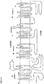
- Figure 2a is a diagrammatic representation of an ion channel, such as Na v 1.7, which comprises four domains A, B, C and D (also referred to as domain I, II, III and IV). Each domain comprises 6 transmembrane protein helixes S1, S2, S3, S4, S5 and S6. The exact amino acid number of each transmembrane protein varies depending on the database entry employed but UniProtKB/Swiss-Prot provides the following information for Na v 1.7:
- S1, S2, S3, S4, S5 and S6 refers to the entities described above or an entity corresponding to same in an alternative ion channel, including wherein a different amino acid assignment is given to the same and including the corresponding entity in natural or non-natural variants and different isotypes of the same.
- Each domain also contains extra-cellular hydrophilic loops E1, E2 and E3.
- the amino acid sequence of E1 in each domain starts after the transmembrane region S1 and ends at S2.
- E1 in each domain is distinct from E1 in other domains.
- the amino acid sequence of E2 in each domain starts after the transmembrane region S3 and ends at S4.
- E2 in each domain is distinct from E2 in other domains.
- the amino acid sequence of E3 in each domain starts after the transmembrane region S5 and ends at S6.
- E3 in each domain is also distinct from E3 in other domains.
- the Na v and Ca v ion channels comprise four domains, A, B, C and D, each containing six transmembrane protein helixes
- other ion channels such as K v ion channels, HCN ion channels and TRP ion channels comprise one domain.
- the K v ion channels, HCN ion channels and TRP ion channels comprise 6 transmembrane protein helixes S1, S2, S3, S4, S5 and S6 and three extra-cellular hydrophilic loops E1, E2 and E3 as described above.
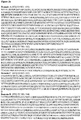
- E loops are the following amino acid residues of SEQ ID NO:105 in Figure 2c : Na v 1.7 Domain E1 amino acids E2 amino acids E3 amino acids A 146-153 206-211 272-378 B 764-774 827-832 890-942 C 1212-1224 1279-1282 1352-1430 D 1535-1545 1600-1609 1670-1735
- Na v 1.7 is expressed in the peripheral nervous system i.e. in nociceptive dorsal root ganglions (DRG), most notably in nociceptive small-diameter DRG neurons, with little representation in the brain.
- DRG nociceptive dorsal root ganglions
- Na v 1.7 distribution e.g. sensory ending
- physiology predispose it to a major role in transmitting painful stimuli.
- Neuropathic pain is a highly prevalent condition. In the United States, it is estimated to affect between 0.6 and 1.5 % of the population, or 1.8 to 4.5 million people. (Pullar and Palmer, 2003). At least 1.4 million people each year are diagnosed with painful diabetic neuropathy (PDN), post-herpetic neuropathy (PHN) or trigeminal neuralgia (TN) - three major causes of neuropathic pain. Other causes of neuropathic pain include spinal cord injuries, multiple sclerosis, phantom limb pain, post-stroke pain and HIV-associated pain. If patients with neuropathic-related chronic back pain, osteoarthritis and cancer were included, the total number would at least double.
- PDN painful diabetic neuropathy
- PPN post-herpetic neuropathy
- TN trigeminal neuralgia
- Other causes of neuropathic pain include spinal cord injuries, multiple sclerosis, phantom limb pain, post-stroke pain and HIV-associated pain. If patients with neuropathic-related chronic back pain
- Nonsteroidal anti-inflammatory drugs although frequently used, are not very effective in the treatment of neuropathic pain. Moreover, their chronic use may lead to serious gastric damage.
- opioids morphine and derivatives
- opioids morphine and derivatives
- chronic treatment such as nausea, emesis, respiratory depression, constipation and tolerance, and the potential for addiction and abuse.
- AEDs Anti-epileptic drugs
- AEDs were aimed at the treatment of chronic neuropathic pain (Renfrey, Downton and Featherstone, 2003).
- the most recent and important examples are gabapentin (Neurontin) and pregabalin (Lyrica, Frampton and Scott, 2004).
- gabapentin the gold standard for the treatment of neuropathic pain, reduces pain at best by 50% in about 40% of patients (Dworkin, 2002).
- gabapentin is not used in the treatment of cancer-related neuropathic pain.
- Na v 1.7 'loss of function' mutation in human leads to insensitivity to pain (Cox et al., 2006).
- Na v 1.7 'gain of function' mutation in human leads to the pain phenotypes erythromelalgia and paroxysmal extreme pain disorder (Dib-Hajj, Yang, Waxman, 2008).
- a peripherally acting small molecule blocking Na v 1.7 reverses hyperalgesia and allodynia in rat models of inflammatory and neuropathic pain (McGowan et al., 2009). Therefore a peripherally acting Na v 1.7 blocking antibody should be beneficial for pain therapy.
- WO2006/040357 discloses molecular targets and compounds useful in the treatment of joint degeneration and inflammatory diseases, and methods of identifying the same.
- WO2007/109324 iscloses potent and selective Nav1.7 sodium channel blockers.
- the present inventors have now established that functionally modifying antibodies can be raised against an E1 loop of ion channels involved in the modulation of pain. This is surprising because the E1 loop in each of the domains is a relatively short amino acid sequence.
- the invention provides an anti-E 1 ion channel monoclonal antibody or binding fragment thereof, which binds to an E1 extracellular loop shown in SEQ ID NO: 128, wherein said ion channel is Nav1.7 and has a function in the modulation of pain and said antibody or fragment is functionally modifying thereto by reducing the amplitude of current through a patch clamp assay by 40% or more.
- the present disclosure provides an ion channel E1 loop binding entity that functionally modifies the activity of an ion channel associated with the modulation of pain, including an antibody, an antibody fragment, a protein or proteinaceous scaffold, a nucleic acid or nucleotide, a small molecule such a synthetic molecule or the like, in particular an antibody, an antibody fragment, a protein or proteinaceous scaffold or a nucleic acid or nucleotide.
- Ion channels thought to be involved and/or associated with modulation of pain include but are not limited to Na v 1.3, Na v 1.6, Na v 1.7, Na v 1.8, Na v 1.9, Ca v 3.1, Ca v 3.2, Ca v 3.3, Ca v 2.1, Ca v 2.2, Ca v 2.3, K v 2.1, K v 2.2, K v 7.x, HCN1, HCN2, TRPV1, TRPA1, ASIC1, TRPM8, TRPV3 and TRP4.
- Sodium channels include, for Na v 1.7, Na v 1.8 or Na v 1.9, such as a Na v 1.7.
- the peptide employed for immunization may comprise at least part of an extracellular sequence of the ion channel wherein the extracellular sequence is the E1 loop and may be derived from the A domain, B domain, C domain or D domain of the ion channel.
- the peptide comprises at least a part of an E1 extracellular region derived from the A domain, B domain, C domain or D domain of the ion channel.
- the peptide comprises at least a part of an E1 extracellular region derived from the A domain or the B domain of the ion channel.
- the peptide comprises at least a part of the BE1 extracellular region.
- the ion channel is not Nav1.7.
- the present invention provides an anti-E1 ion channel antibody or binding fragment thereof, which binds to an E1 extracellular loop of the ion channel, wherein said ion channel has a function in the modulation of pain, and said antibody or fragment is functionally modifying to said ion channel after binding thereto and wherein said ion channel is not Nav1.7.
- an E1 binding anti-ion channel antibody for use in treatment, for example for use in the modulation of pain, in particular the amelioration of pain.
- an E1 binding anti-ion channel antibody for use in the modulation, for example amelioration of pain.
- an E1 binding anti-Na v 1.7 antibody for use in the modulation, for example amelioration of pain, in particular a sub-group of pain described herein.
- Functionally modifying antibody as employed herein is intended to refer to an antibody or fragment (such as a binding fragment) that changes the activity of the ion channel, for example by reducing an activity by at least 5%, for example 10 or 15% such as 20% in at least one in vitro or in vivo assay.
- Suitable in vitro assays include a patch clamp assay or other assay as described herein.
- the functionally modifying antibody reduces the amplitude of current through a patch clamp assay by 5%, 10%, 15%, 20%, 30%, 40%, 50%, 60%, 70% or more percent.
- An antibody that provides a functional modification to the ion channel and a functionally modifying antibody are terms used interchangeably herein.
- the functional modification may, for example be sufficient to block, close or inhibit the pore of the ion channel.
- This functional modification may be effected by any mechanism including, physically blocking the pore, causing a conformational change in the ion channel which for example blocks the pore or eliciting the ion channel to adopt a non-functional state (resting or inactivated state) and/or maintaining the ion channel in a non-functional state (allosteric modulation).
- the functional modification may be sufficient to reduce the cell surface levels of the ion channel protein.
- This functional modification may be effected by any mechanism including but not limited to, antibody induced internalization or endocytosis or increased cycling of the ion channel, leading to a reduce number of functional ion channel proteins at the cell surface.
- the antibody or fragment according to the present invention may be employed in combination with other entities which functionally modify the ion channel in question, for antibodies or fragment that bind an E3 region of the ion channel, for example part of the E3 region of domain A, B, C or D, such as domain A.
- an E1 binding anti- Na v 1.7 antibody In one embodiment there is provided an E1 binding anti- Na v 1.7 antibody.
- An anti- Na v 1.7 antibody is an antibody that binds specifically to Na v 1.7.
- An E1 binding anti- Na v 1.7 antibody is an antibody that binds specifically to Na v 1.7 in an E1 region.
- Specific binding is intended to refer to the fact that the antibody is selective for the relevant ion channel, for example Na v 1.7 and can distinguish it from other ion channels and proteins, for example other ion channels in the same family.
- a selective antibody is one that, for example can be used to affinity purify the relevant ion channel, such as Na v 1.7 including from other ion channels.
- the anti- Na v 1.7 antibody is specific to mammalian Na v 1.7, for example human Na v 1.7.
- the E1 binding anti-ion channel antibody for example anti-Na v 1.7 antibody cross-reacts with the relevant human ion channel and the corresponding rat ion channel.
- the functionally modifying antibody or fragment is a clonal population of antibodies, for example a monoclonal population. It has been suggested in the literature that polyclonal antibodies are required to obtain functional modification of ion channels. Therefore, it is particularly surprising that the present disclosure provides monoclonal antibodies which are functionally modifying to a relevant ion channel.
- Monoclonal as employed herein is intended to refer antibodies or fragments derived from a single cell, for example employing hybridoma technology.
- a clonal population is intended to refer to a population of antibodies or fragments with the same properties, characteristics including the same amino acid sequence and specificity.
- two or more, for example three or four clonal populations of anti-ion channel antibodies are employed in admixture, wherein the mixture comprises at least one antibody, for example two, three or four according to the present invention.
- the antibody according to the disclosure is a whole antibody.
- the antibody or fragment thereof is multivalent and/or bi-specific.
- Multivalent as employed herein is intended to refer multiple binding sites (for example at least two binding sites) in the antibody or fragment entity.
- Multivalent entities in the context of the present specification, have at least two binding sites with the same specificity. Mutivalent antibodies with binding sites that bind the same epitope will not be considered bi-specific unless the entity comprises a third binding site with different specificity to a first and second binding site.
- bi-specific as employed in the present disclosure requires two or more binding sites in the antibody or fragment to bind to different, i.e. distinct target antigens.
- the antibody or fragment entity will be considered bi-specific, within the meaning of the present disclosure.
- Binding site as employed herein is intended to refer to an area of the antibody or fragment, which specifically binds a target antigen.
- a heavy chain variable domain and light chain variable domain pairing will be considered an example of a binding site herein.
- an antibody fragment according to the disclosure is monovalent. That is to say, has only one binding site.
- the antibody is a fragment, for example a single domain antibody, a single chain Fv, a Fab, a Fab' or a pairing of a full length heavy and light chain.
- the antibody of the disclosure and fragments thereof, of use in the present invention can be from any species but is preferably derived from a monoclonal antibody, a human antibody, or are humanised fragments.
- An antibody fragment for use in the present invention can be derived from any class (e.g. IgG, IgE, IgM, IgD or IgA) or subclass of immunoglobulin molecule and may be obtained from any species including for example mouse, rat, shark, rabbit, pig, hamster, camel, llama, goat or human.
- the Kabat residue designations do not always correspond directly with the linear numbering of the amino acid residues.
- the actual linear amino acid sequence may contain fewer or additional amino acids than in the strict Kabat numbering corresponding to a shortening of, or insertion into, a structural component, whether framework or complementarity determining region (CDR), of the basic variable domain structure.
- CDR complementarity determining region
- the correct Kabat numbering of residues may be determined for a given antibody by alignment of residues of homology in the sequence of the antibody with a "standard" Kabat numbered sequence.
- the CDRs of the heavy chain variable domain are located at residues 31-35 (CDR-H1), residues 50-65 (CDR-H2) and residues 95-102 (CDR-H3) according to the Kabat numbering system. Kabat numbering will be employed herein.
- Chothia Chothia, C. and Lesk, A.M. J. Mol. Biol., 196, 901-917 (1987 )
- the loop equivalent to CDR-H1 extends from residue 26 to residue 32.
- a combination of Chothia and Kabat numbering for 'CDR-H1', would for example comprises residues 26 to 35.
- the CDRs of the light chain variable domain are located at residues 24-34 (CDR-L1), residues 50-56 (CDR-L2) and residues 89-97 (CDR-L3) according to the Kabat numbering system.
- the disclosure herein extends to an antibody comprising 1, 2, 3, 4, 5, or 6 CDR sequences disclosed in this specification.
- the disclosure extends to an antibody comprising a single variable domain or a pair of variable domain from a sequence or sequences herein.
- variable domain of the heavy chain comprises at least one of a CDR having the sequence given in the table listed above, for example where the CDR is in its "natural position".
- the natural position of a CDR such as H1, H2, H3, L1, L2 or L3 is given above in the tables, for example the natural position for the CDR of Seq ID No: 4 is H1, the natural position for the CDR of Seq ID No: 5 is H2, the natural position for the CDR of Seq ID No: 6 is H3, and so on.
- Analogous interpretations also apply to the light chain sequences.
- an antibody comprises a heavy chain wherein at least two of CDR-H1, CDR-H2 and CDR-H3 of the variable domain of the heavy chain are selected from sequences given in the tables above, for example the CDRs are in their natural position and optionally in their natural pairing.
- Natural pairing as employed herein is intended to refer to pairing of CDRs from the same antibody (i.e. from one table above).
- An example of natural pairing for CA167 00983 is Seq ID No: 1 and 2, 1 and 3, and 2 and 3.
- heavy chain variable domain may comprise the sequence given in:
- An antibody may comprises a heavy chain wherein the variable domain of the heavy chain comprises a variable domain or variable domain components as disclosed herein, for example listed herein.
- Variable domain components as employed herein is intended to refer to CDRs and combinations thereof, particularly as explicitly disclosed herein.
- An antibody may comprise a heavy chain, wherein the variable domain of the heavy chain comprises a sequence having at least 60%, 70%, 80% identity or similarity, such as at least 90%, 95% or 98% identity or similarity to a heavy chain variable region disclosed herein.
- Identity indicates that at any particular position in the aligned sequences, the amino acid residue is identical between the sequences.
- similarity indicates that, at any particular position in the aligned sequences, the amino acid residue is of a similar type between the sequences.
- leucine may be substituted for isoleucine or valine.
- Other amino acids which can often be substituted for one another include but are not limited to:
- the present invention also provides an E1 binding anti-ion channel antibody or fragment, wherein the ion channel has a role/function in the modulation of pain in vivo , in particular an antibody directed to aNa v 1.7, which selectively inhibits the function of said ion channel, said antibody or fragment having a light chain which comprises at least one CDR having the sequence given herein for light chain CDRs (see tables and lists provides), for example where the CDR is in its "natural position".
- An antibody may comprise a light chain, wherein at least two of CDR-L1, CDR-L2 and CDR-L3 in the variable domain of the light chain are selected from sequences given herein for light chain CDRs, for example the CDRs are in their natural position and optionally in their natural pairing.
- Natural pairing as employed herein is intended to refer to the fact that a CDR from the one antibody is paired/co-located in a variable domain with a CDR derived from the same antibody, in particular the same chain of the same antibody (i.e. from one table above).
- a light chain, variable domain may comprise the sequence given in:
- the present invention comprises a light chain, wherein the variable domain of the light chain comprises a variable domain or variable domain components as disclosed herein, for example from any heavy chain described.
- pair of variable domains for example a heavy chain variable domain and light chain variable domain.
- a heavy and light chain variable domain pair which is a cognate pair.
- the antibody molecules of the present invention comprise a complementary light chain or a complementary heavy chain, respectively.
- the heavy and light chain are a natural pairing, that is to say are derived from the same antibody.
- the heavy and the light chain have a non-natural pairing.
- a nucleotide sequence encoding an antibody or fragment thereof is also disclosure.
- CDR-grafted (or humanised) anti-ion channels antibody (as per the current invention).
- the term 'CDR-grafted antibody molecule' refers to an antibody molecule wherein the heavy and/or light chain contains one or more CDRs (including, if desired, one or more modified CDRs) from a donor antibody (e.g. a rat or rabbit antibody as described herein) grafted into a heavy and/or light chain variable region framework of an acceptor antibody (e.g. a human antibody).
- a donor antibody e.g. a rat or rabbit antibody as described herein
- acceptor antibody e.g. a human antibody
- any appropriate acceptor variable region framework sequence may be used having regard to the class/type of the donor antibody from which the CDRs are derived, including rat, rabbit, mouse, primate and human framework regions.
- the CDR-grafted antibody of the present invention has a variable domain comprising human acceptor framework regions as well as one or more of the CDRs derived from the donor antibody as referred to herein.
- the variable domain comprises human acceptor framework regions and non-human, preferably rat, mouse or rabbit donor CDRs.
- human frameworks which can be used in the present invention are KOL, NEWM, REI, EU, TUR, TEI, LAY and POM (Kabat et al., supra).
- KOL and NEWM can be used for the heavy chain
- REI can be used for the light chain and EU
- LAY and POM can be used for both the heavy chain and the light chain.
- human germline sequences may be used; these are available at: http-://vbase.mrc-cpe.cam.ac.uk/ .
- a database of affinity matured human V region sequences may be used as a framework.
- the acceptor heavy and light chains do not necessarily need to be derived from the same antibody and may, if desired, comprise composite chains having framework regions derived from different chains.
- the framework regions need not have exactly the same sequence as those of the acceptor antibody. For instance, unusual residues may be changed to more frequently-occurring residues for that acceptor chain class or type. Alternatively, selected residues in the acceptor framework regions may be changed so that they correspond to the residue found at the same position in the donor antibody (see Reichmann et al., 1998, Nature, 332, 323-324 ). Such changes should be kept to the minimum necessary to recover the affinity of the donor antibody.
- a protocol for selecting residues in the acceptor framework regions which may need to be changed is set forth in WO 91/09967 .
- Donor residues are residues from the donor antibody, i.e. the antibody from which the CDRs were originally derived, which may in one embodiment of the present invention are derived from rat, mouse or rabbit antibodies and may be incorporated into the final antibody or fragment as required.
- the antibody (or fragment such as a Fab or Fab' fragment) is a monoclonal, fully human, humanized or chimeric antibody fragment. In one embodiment the antibody Fab or Fab' fragments are fully human or humanised.
- antibodies for use in the present invention may therefore comprise a complete antibody molecule having full length heavy and light chains or a fragment thereof and may be, but are not limited to Fab, modified Fab, Fab', F(ab') 2 , Fv, single domain antibodies (such as VH, VL, VHH, IgNAR V domains), scFv, bi, tri or tetra-valent antibodies, bis-scFv, diabodies, triabodies, tetrabodies and epitope-binding fragments of any of the above (see for example Holliger and Hudson, 2005, Nature Biotech. 23(9):1126-1136 ; Adair and Lawson, 2005, Drug Design Reviews - Online 2(3), 209-217 ).
- the antibodies for use in the present invention may be derived from a camelid, such as a camel or llama.
- Camelids possess a functional class of antibodies devoid of light chains, referred to as heavy chain antibodies ( Hamers et al., 1993, Nature, 363, 446-448 ; Muyldermans, et al., 2001, Trends. Biochem.Sci. 26, 230-235 ).
- the antigen-combining site of these heavy-chain antibodies is limited to only three hypervariable loops (H1-H3) provided by the N-terminal variable domain (VHH).
- the first crystal structures of VHHs revealed that the H1 and H2 loops are not restricted to the known canonical structure classes defined for conventional antibodies ( Decanniere, et al., 2000, J. Mol.Biol, 300, 83-91 ).
- the H3 loops of VHHs are on average longer than those of conventional antibodies ( Nguyen et al., 2001, Adv. Immunol., 79, 261-296 ).
- a large fraction of dromedary heavy chain antibodies have a preference for binding into active sites of enzymes against which they are raised ( Lauwereys et al., 1998, EMBO J, 17, 3512-3520 ).
- the H3 loop was shown to protrude from the remaining paratope and insert in the active site of the hen egg white lysozyme ( Desmyter et al., 1996, Nat.Struct.Biol.3, 803-811 and De Genst et al., 2006, PNAS, 103, 12, 4586-4591 and WO97049805 ).
- the antibodies for use in the present invention may be derived from a cartilaginous fish, such as a shark.
- Cartilaginous fish (sharks, skates, rays and chimeras) possess an atypical immunoglobulin isotype known as IgNAR.
- IgNAR is an H-chain homodimer that does not associate with light chain. Each H chain has one variable and five constant domains.
- IgNAR V domains (or V-NAR domains) carry a number of non canonical cysteines that enable classification into two closely related subtypes, I and II.
- Type II V regions have an additional cysteine in CDRs 1 and 3 which have been proposed to form a domain-constraining disulphide bond, akin to those observed in camelid VHH domains.
- the CDR3 would then adopt a more extended conformation and protrude from the antibody framework akin to the camelid VHH.
- certain IgNAR CDR3 residues have also been demonstrated to be capable of binding in the hen egg white lysozyme active site ( Stanfield et al., 2004, Science, 305, 1770-1773 .
- VHH and IgNAR V domains are described in for example, Lauwereys et al, 1998, EMBO J. 1998, 17(13), 3512-20 ; Liu et al., 2007, BMC Biotechnol., 7, 78 ; Saerens et al., 2004, J. Biol. Chem., 279 (5), 51965-72 .
- Antibodies for use in the present invention include whole antibodies of any suitable class for example, IgA, IgD, IgE, IgG or IgM or subclass such as IgG1, IgG2, IgG3 or IgG4. and functionally active fragments or derivatives thereof and may be, but are not limited to, monoclonal, clonal, humanised, fully human or chimeric antibodies.
- the constant region employed, in the antibody or certain fragments thereof according to the disclosure is a hybrid constant region or mutated constant region.
- Hybrid constant regions comprises portions or domains from two or more distinct constant regions, for example two or more distinct human constant regions.
- hybrid constant regions include those disclosed in US2007/0041972 , where at least CH1 and the hinge region are derived from one or more IgG2 antibodies and at least a portion of the CH2 and CH3 regions are derived from one or more IgG4 CH2 and CH3 regions.
- Eculizimumab Alexion Pharmaceuticals is a humanised anti-human C5 mAb for paroxysmal nocturnal hemoglobinuria comprising a hybrid constant region. It has a hybrid chain of IgG2 derived CH1 and hinge with IgG4 derived CH2 and CH3 domains. It does not bind Fc ⁇ R nor does it activate complement. It also has low immunogenicity (low titres of anti-Eculizimumab antibodies detected in only 3 of 196 (3%) patients).
- WO 2008/090958 discloses certain hybrid constant regions comprising a chain of CH1, hinge and CH2 from IgG1 and a CH3 domain from IgG3.
- the hybrid has a higher CDC activity than that of an IgG1 or IgG3 antibody and a protein A-binding activity equivalent to that of IgG1.
- mutated constant region includes that employed in Abatacept, which is a fusion of human CTLA-4 with IgG1 hinge-Fc.
- the hinge was altered from CPPC to SPPS. The latter is O-gly.
- the mutated constant region does not mediate ADCC or CDC and has low immunogenicity (3% incidence).
- the hinge is thought to potentially have a role in complement activation.
- the functional hinge deduced from crystallographic studies, extends from 216-237 of IgG1 and consists of EPKSCDKTHT CPPCP APELLGG (SEQ ID NO: 106) upper , middle and lower hinge respectively.
- an antibody or fragment according to the disclosure comprises a functional hinge.
- Mutations/modifications to the constant region may, for example result in increased stability, for example US 2004/0191265 discloses mutagenesis of IgG1 hinge, which increased the stability of an IgG by introducing one or more amino acid modifications in the hinge region at positions 233-239 or 249 of human IgG1. This provided reduced degradation upon heating to 55°C for one week.
- modification may be effected by making point mutations in labile amino acids (e.g., histidine or threonine) or reactive amino acids (e.g., lysine or glutamic acid) in the upper hinge portion (human IgG1 residues 226-243 and corresponding residues in other IgG subtypes and/or immunoglobulins from other species) and/or in the flanking CH1 and/or CH2 sequences (human IgG1 residue 249 and corresponding residues in other IgG subtypes and/or immunoglobulins from other species).
- labile amino acids e.g., histidine or threonine
- reactive amino acids e.g., lysine or glutamic acid
- Monoclonal antibodies may be prepared by any method known in the art such as the hybridoma technique ( Kohler & Milstein, Nature, 1975, 256, 495-497 ), the trioma technique, the human B-cell hybridoma technique ( Kozbor et al., Immunology Today, 1983, 4, 72 ) and the EBV-hybridoma technique ( Cole et al., "Monoclonal Antibodies and Cancer Therapy", pp. 77-96, Alan R. Liss, Inc., 1985 ).
- Antibodies for use in the invention may also be generated using single lymphocyte antibody methods by cloning and expressing immunoglobulin variable region cDNAs generated from single lymphocytes selected for the production of specific antibodies by, for example, the methods described by Babcook, J. et al., Proc. Natl. Acad. Sci. USA, 1996, 93(15), 7843-7848 , WO 92/02551 , WO2004/051268 and WO2004/106377 .
- Humanized antibodies are antibody molecules from derived non-human species having one or more complementarity determining regions (CDRs) from the non-human species and a framework region from a human immunoglobulin molecule (see, for example, US 5,585,089 ).
- CDRs complementarity determining regions
- the antibodies for use in the present invention can also be generated using various phage display methods known in the art and include those disclosed by Brinkman et al., J. Immunol. Methods, 1995, 182, 41-50 ; Ames et al., J. Immunol. Methods, 1995, 184, 177-186 ; Kettleborough et al. Eur. J.
- transgenic mice or other organisms, including other mammals, may be used to generate humanized antibodies.
- Fully human antibodies are those antibodies in which the variable regions and the constant regions (where present) of both the heavy and the light chains are all of human origin, or substantially identical to sequences of human origin, not necessarily from the same antibody.
- Examples of fully human antibodies may include antibodies produced for example by the phage display methods described above and antibodies produced by mice in which the murine immunoglobulin variable and/or constant region genes have been replaced by their human counterparts eg. as described in general terms in EP0546073 B1 , US 5,545,806 , US 5,569,825 , US 5,625,126 , US 5,633,425 , US 5,661,016 , US5,770,429 , EP 0438474 B1 and EP0463151 B1 .
- the antibody or fragment for use in the present invention may be obtained from any whole antibody, especially a whole monoclonal antibody, using any suitable enzymatic cleavage and/or digestion techniques, for example by treatment with pepsin.
- the antibody starting material may be prepared by the use of recombinant DNA techniques involving the manipulation and re-expression of DNA encoding antibody variable and/or constant regions. Standard molecular biology techniques may be used to modify, add or delete amino acids or domains as desired. Any alterations to the variable or constant regions are still encompassed by the terms 'variable' and 'constant' regions as used herein.
- antibody fragment "starting material” may be obtained from any species including for example mouse, rat, rabbit, hamster, shark, camel, llama, goat or human. Parts of the antibody fragment may be obtained from more than one species, for example the antibody fragments may be chimeric. In one example, the constant regions are from one species and the variable regions from another. The antibody fragment starting material may also be modified. In another example, the variable region of the antibody fragment has been created using recombinant DNA engineering techniques. Such engineered versions include those created for example from natural antibody variable regions by insertions, deletions or changes in or to the amino acid sequences of the natural antibodies.
- variable region domains containing at least one CDR and, optionally, one or more framework amino acids from one antibody and the remainder of the variable region domain from a second antibody.
- the methods for creating and manufacturing these antibody fragments are well known in the art (see for example, Boss et al., US 4,816,397 ; Cabilly et al., US 6,331,415 ; Shrader et al., WO 92/02551 ; Ward et al., 1989, Nature, 341, 544 ; Orlandi et al., 1989, Proc.Natl.Acad.Sci.
- constant region domains may be human IgA, IgD, IgE, IgG or IgM domains.
- human IgG constant region domains may be used, especially of the IgG1 and IgG3 isotypes when the antibody molecule is intended for therapeutic uses and antibody effector functions are required.
- IgG2 and IgG4 isotypes may be used when the antibody molecule is intended for therapeutic purposes and antibody effector functions are not required.
- sequence variants of these constant region domains may also be used.
- IgG4 molecules in which the serine at position 241 has been changed to proline as described in Angal et al., Molecular Immunology, 1993, 30 (1), 105-108 may be used.
- antibodies may undergo a variety of posttranslational modifications.
- the type and extent of these modifications often depends on the host cell line used to express the antibody as well as the culture conditions.
- modifications may include variations in glycosylation, methionine oxidation, diketopiperazine formation, aspartate isomerization and asparagine deamidation.
- a frequent modification is the loss of a carboxy-terminal basic residue (such as lysine or arginine) due to the action of carboxypeptidases (as described in Harris, RJ. Journal of Chromatography 705:129-134, 1995 ).
- the antibody or fragment light chain comprises a CL domain, either kappa or lambda.
- the term 'antibody' of fragment as used herein may also include binding agents which comprise one or more CDRs incorporated into a biocompatible framework structure.
- the biocompatible framework structure comprises a polypeptide or portion thereof that is sufficient to form a conformationally stable structural support, or framework, or scaffold, which is able to display one or more sequences of amino acids that bind to an antigen (e.g. CDRs, a variable region etc.) in a localised surface region.
- an antigen e.g. CDRs, a variable region etc.
- Such structures can be a naturally occurring polypeptide or polypeptide 'fold' (a structural motif), or can have one or more modifications, such as additions, deletions or substitutions of amino acids, relative to a naturally occurring polypeptide or fold.
- These scaffolds can be derived from a polypeptide of any species (or of more than one species), such as a human, other mammal, other vertebrate, invertebrate, plant, bacteria or virus.
- the biocompatible framework structures are based on protein scaffolds or skeletons other than immunoglobulin domains.
- those based on fibronectin, ankyrin, lipocalin, neocarzinostain, cytochrome b, CP1 zinc finger, PST1, coiled coil, LACI-D1, Z domain and tendramisat domains may be used (See for example, Nygren and Uhlen, 1997, Current Opinion in Structural Biology, 7, 463-469 ).
- the overall charge of the antibody or fragment is neutral.
- the overall charge of the antibody or fragment is negative.
- the overall charge of the antibody or fragment is positive.
- the CDRs may be grafted or engineered into an alternative type of scaffold, for example a fibronectin or actin-binding repeats.
- the antibody or fragment comprises an effector molecule, such as a polymer, toxin including biotoxins such as venom or chemical inhibitor conjugated thereto.
- an effector molecule such as a polymer, toxin including biotoxins such as venom or chemical inhibitor conjugated thereto.
- Biotoxins and venom are natural modulators, such as blockers of cell signalling. When conjugated to an antibody or fragment according to the invention then they can be used to augment the functional effect on the ion channel, whilst maintaining the selectivity provided by the antibody.
- Tarantula venom peptide ProTxII has, for example be shown to selectively inhibit Na v 1.7, see for example Mol Pharmacol 74:1476-1484, 2008 .
- the toxin is botulinum toxin, for example botulinum toxin A.
- Other toxins include tetrodotoxin and saxitoxin.
- the antibody or fragment is conjugated to an aptamer.
- Aptamers are single stranded oligonucleotide sequences that adopt secondary and tertiary structures and that are able to bind and modulate protein activity. The conjugation of such structures to antibodies will lead to a target specific modulation of the ion channel (direct effect).
- the antibody or fragment is conjugated to chemical inhibitor such as a synthetic chemical inhibitor of an ion channel.
- chemical inhibitors include compounds of formula (I):
- DIDS 4,4-Diisothiocyanatostilbene-2,2'-disulfonic acid
- the entity conjugated to the antibody or fragment changes the overall charge of the molecule.
- effector molecule includes, for example, antineoplastic agents, drugs, toxins, biologically active proteins, for example enzymes, other antibody or antibody fragments, synthetic or naturally occurring polymers, nucleic acids and fragments thereof e.g. DNA, RNA and fragments thereof, radionuclides, particularly radioiodide, radioisotopes, chelated metals, nanoparticles and reporter groups such as fluorescent compounds or compounds which may be detected by NMR or ESR spectroscopy.
- effector molecules may include cytotoxins or cytotoxic agents including any agent that is detrimental to (e.g. kills) cells.
- examples include combrestatins, dolastatins, epothilones, staurosporin, maytansinoids, spongistatins, rhizoxin, halichondrins, roridins, hemiasterlins, taxol, cytochalasin B, gramicidin D, ethidium bromide, emetine, mitomycin, etoposide, tenoposide, vincristine, vinblastine, colchicin, doxorubicin, daunorubicin, dihydroxy anthracin dione, mitoxantrone, mithramycin, actinomycin D, 1-dehydrotestosterone, glucocorticoids, procaine, tetracaine, lidocaine, propranolol, and puromycin and analogs or homologs
- Effector molecules may also include, but are not limited to, antimetabolites (e.g . methotrexate, 6-mercaptopurine, 6-thioguanine, cytarabine, 5-fluorouracil decarbazine), alkylating agents (e.g . mechlorethamine, thioepa chlorambucil, melphalan, carmustine (BSNU) and lomustine (CCNU), cyclothosphamide, busulfan, dibromomannitol, streptozotocin, mitomycin C, and cis-dichlorodiamine platinum (II) (DDP) cisplatin), anthracyclines (e.g .
- antimetabolites e.g . methotrexate, 6-mercaptopurine, 6-thioguanine, cytarabine, 5-fluorouracil decarbazine
- alkylating agents e.g . mechlorethamine, thio
- daunorubicin (formerly daunomycin) and doxorubicin
- antibiotics e.g . dactinomycin (formerly actinomycin), bleomycin, mithramycin, anthramycin (AMC), calicheamicins or duocarmycins
- anti-mitotic agents e.g . vincristine and vinblastine.
- effector molecules may include chelated radionuclides such as 111 In and 90 Y, Lu 177 , Bismuth 213 , Californium 252 , Iridium 192 and Tungsten 188 /Rheniun 188 ; or drugs such as but not limited to, alkylphosphocholines, topoisomerase I inhibitors, taxoids and suramin.
- chelated radionuclides such as 111 In and 90 Y, Lu 177 , Bismuth 213 , Californium 252 , Iridium 192 and Tungsten 188 /Rheniun 188 ; or drugs such as but not limited to, alkylphosphocholines, topoisomerase I inhibitors, taxoids and suramin.
- effector molecules include proteins, peptides and enzymes.
- Enzymes of interest include, but are not limited to, proteolytic enzymes, hydrolases, lyases, isomerases, transferases.
- Proteins, polypeptides and peptides of interest include, but are not limited to, immunoglobulins, toxins such as abrin, ricin A, pseudomonas exotoxin, or diphtheria toxin.
- effector molecules may include detectable substances useful for example in diagnosis.
- detectable substances include various enzymes, prosthetic groups, fluorescent materials, luminescent materials, bioluminescent materials, radioactive nuclides, positron emitting metals (for use in positron emission tomography), and nonradioactive paramagnetic metal ions. See generally U.S. Patent No. 4,741,900 for metal ions which can be conjugated to antibodies for use as diagnostics.
- Suitable enzymes include horseradish peroxidase, alkaline phosphatase, beta-galactosidase, or acetylcholinesterase; suitable prosthetic groups include streptavidin, avidin and biotin; suitable fluorescent materials include umbelliferone, fluorescein, fluorescein isothiocyanate, rhodamine, dichlorotriazinylamine fluorescein, dansyl chloride and phycoerythrin; suitable luminescent materials include luminol; suitable bioluminescent materials include luciferase, luciferin, and aequorin; and suitable radioactive nuclides include 125 I, 131 I, 111 In and 99 Tc.
- the effector molecule may increase the half-life of the antibody in vivo , and/or reduce immunogenicity of the antibody and/or enhance the delivery of an antibody across an epithelial barrier to the immune system.
- suitable effector molecules of this type include polymers, albumin, albumin binding proteins or albumin binding compounds such as those described in WO05/117984 .
- the effector molecule is a polymer it may, in general, be a synthetic or a naturally occurring polymer, for example an optionally substituted straight or branched chain polyalkylene, polyalkenylene or polyoxyalkylene polymer or a branched or unbranched polysaccharide, e.g. a homo- or hetero- polysaccharide.
- synthetic polymers include optionally substituted straight or branched chain poly(propyleneglycol) poly(vinylalcohol) or derivatives thereof, especially optionally substituted poly(ethyleneglycol) such as methoxypoly(ethyleneglycol) or derivatives thereof.
- Suitable polymers include a polyalkylene polymer, such as a poly(ethyleneglycol) or, especially, a methoxypoly(ethyleneglycol) or a derivative thereof, and especially with a molecular weight in the range from about 15000Da to about 40000Da.
- the size of the polymer may be varied as desired, but will generally be in an average molecular weight range from 500Da to 50000Da, for example from 5000 to 40000Da such as from 20000 to 40000Da.
- the polymer size may in particular be selected on the basis of the intended use of the product for example ability to localize to certain tissues such as tumors or extend circulating half-life (for review see Chapman, 2002, Advanced Drug Delivery Reviews, 54, 531-545 ).
- a small molecular weight polymer for example with a molecular weight of around 5000Da.
- a higher molecular weight polymer for example having a molecular weight in the range from 20000Da to 40000Da.
- the PEG employed is releasable PEG, for example as supplied by Enzon pharmaceuticals.
- antibodies for use in the present invention are attached to poly(ethyleneglycol) (PEG) moieties.
- the antibody is an antibody fragment and the PEG molecules may be attached through any available amino acid side-chain or terminal amino acid functional group located in the antibody fragment, for example any free amino, imino, thiol, hydroxyl or carboxyl group.
- Such amino acids may occur naturally in the antibody fragment or may be engineered into the fragment using recombinant DNA methods (see for example US 5,219,996 ; US 5,667,425 ; WO98/25971 ).
- the antibody molecule of the present invention is a modified Fab fragment wherein the modification is the addition to the C-terminal end of its heavy chain one or more amino acids to allow the attachment of an effector molecule.
- the additional amino acids form a modified hinge region containing one or more cysteine residues to which the effector molecule may be attached. Multiple sites can be used to attach two or more PEG molecules.
- the Fab or Fab' is PEGylated with one or two PEG molecules.
- a PEG molecule is linked to a cysteine 171 in the light chain, for example see WO2008/038024 incorporated herein by reference.
- Fab or Fab' is PEGylated through a surface accessible cysteine.
- PEG molecules are covalently linked through a thiol group of at least one cysteine residue located in the fusion protein.
- Each polymer molecule attached to the fusion protein may be covalently linked to the sulfur atom of a cysteine residue located in the protein.
- the covalent linkage will generally be a disulphide bond or, in particular, a sulfur-carbon bond.
- thiol group is used as the point of attachment
- appropriately activated PEG molecules for example thiol selective derivatives such as maleimides and cysteine derivatives may be used.
- An activated PEG molecule may be used as the starting material in the preparation of polymer-fusion protein containing molecules as described above.
- the activated PEG molecule may be any polymer containing a thiol reactive group such as an ⁇ -halocarboxylic acid or ester, e.g. iodoacetamide, an imide, e.g. maleimide, a vinyl sulphone or a disulphide.
- a thiol reactive group such as an ⁇ -halocarboxylic acid or ester, e.g. iodoacetamide, an imide, e.g. maleimide, a vinyl sulphone or a disulphide.
- Such starting materials may be obtained commercially (for example from Nektar, formerly Shearwater Polymers Inc., Huntsville, AL, USA) or may be prepared from commercially available starting materials using conventional chemical procedures.
- Particular PEG molecules include 20K methoxy-PEG-amine (obtainable from Nektar, formerly Shearwater; Rapp Polymere; and SunBio) and M-PEG-SPA (obtainable from Nektar, formerly
- Effector molecules such a PEG molecules may be attached to antibodies or by a number of different methods, including through aldehyde sugars or more commonly through any available amino acid side-chain or terminal amino acid functional group located in the antibody fragment, for example any free amino, imino, thiol, hydroxyl or carboxyl group.
- the site of attachment of effector molecules can be either random or site specific.
- Random attachment is often achieved through amino acids such as lysine and this results in effector molecules, such as PEG molecules, being attached at a number of sites throughout the antibody fragment depending on the position of the lysines. While this has been successful in some cases the exact location and number of effector molecules, such as PEG molecules, attached cannot be controlled and this can lead to loss of activity for example if too few are attached and/or loss of affinity if for example they interfere with the antigen binding site ( Chapman 2002 Advanced Drug Delivery Reviews, 54, 531-545 ). As a result, controlled site-specific attachment of effector molecules, such as PEG molecules, is usually the method of choice.
- site specific attachment of effector molecules is most commonly achieved by attachment to cysteine residues since such residues are relatively uncommon in antibody fragments.
- Antibody hinges are popular regions for site specific attachment since these contain cysteine residues and are remote from other regions of the fusion protein likely to be involved in antigen binding. Suitable hinges either occur naturally in the fragment or may be created using recombinant DNA techniques (See for example US 5,677,425 ; WO98/25971 ; Leong et al., 2001 Cytokine, 16, 106-119 ; Chapman et al., 1999 Nature Biotechnology, 17, 780-783 ).
- site-specific cysteines may also be engineered into the antibody fragment for example to create surface exposed cysteine(s) for effector molecule attachment ( US 5,219,996 ).
- the PEG molecule is attached to a surface exposed cysteine.
- a surface exposed cysteine (free cysteine) as employed herein is intended to refer to cysteine, that when the protein is in a "natual" folded conformation, is accessible for conjugating an effector molecule, such as a PEG molecule thereto. Examples of how to engineer free cysteines of this type are also provided in US7,521,541 .
- Specific naturally occurring polymers include lactose, amylose, dextran, glycogen or derivatives thereof.
- Derivatives as used herein is intended to include reactive derivatives, for example thiol-selective reactive groups such as maleimides and the like.
- the reactive group may be linked directly or through a linker segment to the polymer. It will be appreciated that the residue of such a group will in some instances form part of the product as the linking group between the fusion protein and the polymer.
- Any suitable host cell/vector system may be used for expression of the DNA sequences encoding the fusion protein molecule of the present invention.
- Bacterial for example E . coli , and other microbial systems may be used or eukaryotic, for example mammalian, host cell expression systems may also be used.
- Suitable mammalian host cells include CHO, myeloma or hybridoma cells.
- the production of an antibody or fragment thereof according to the present invention may comprise culturing a host cell containing a vector (and/or DNA) of the present invention under conditions suitable for leading to expression of protein from DNA encoding the antibody or fragment thereof, and isolating the same.
- the cell line may be transfected with two vectors, a first vector encoding a light chain polypeptide and a second vector encoding a heavy chain polypeptide.
- a single vector may be used, the vector including sequences encoding light chain and heavy chain polypeptides.
- Methods of generating antibodies employing peptides whose sequence are derived from E1 extracellular regions of an ion channel involved in modulation of pain, for example Na v 1.7 are also disclosed. Such peptides, are used both in immunization protocols to raise polyclonal antibodies and/or in screening or panning protocols to select specific anti-ion channel antibodies, for example anti-Na v 1.7 antibodies. Uses peptides that correspond to discrete sequences of the aforementioned extracellular regions where there is maximum dissimilarity to other ion channels and isoforms thereof may be desirable.
- the selected peptides may be conventional linear peptides or cyclic peptides and comprise at least 4 consecutive amino acids from the above sequences. In either case the peptide is designed to include a single functional group that is used for subsequent conjugation to carrier protein or reporter group.
- the said functional group may be a side chain thiol of a cysteine residue, a C-terminal carboxyl or side chain carboxyl of an aspartic acid or glutamic acid residue or primary amine of an N-terminal amino group or lysine side chain residue.
- a linear peptide may for example, contain any of the following sequences, wherein the domain A, B, C or D of Na v 1.7, from which the peptide is derived is denoted in brackets.
- the cysteines which are underlined in the peptides are non-naturally occurring cysteine residues in the ion channel.:
- the cysteine residues in the following peptides may be used to attach a carrier protein.
- EHHPMTEEFKN (BE1) (SEQ ID NO: 107) EHHPMTEEFKN C , (BE1) (SEQ ID NO: 108) C PMTEEFKN, (BE1) (SEQ ID NO: 109) PMTEEFKN C , (BE1) (SEQ ID NO: 110) C EDIYIERKKTIKI, (CE1) (SEQ ID NO: 111) EDIYIEERKKTIKI C , (CE1) (SEQ ID NO: 112) C IERKKTIKI, (CE1) (SEQ ID NO: 113) IERKKTIKI C , (CE1) (SEQ ID NO: 114) C ERKKTIKI, (CE1) (SEQ ID NO: 115) ERKKTIKI C , (CE1) (SEQ ID NO: 116) C EKEGQSQHMTE, (DE1) (SEQ ID NO: 117) EKEGQSQHMTE C . (DE1) (SEQ ID NO: 118)
- sequences may be capped at either N-terminal or C-terminal with for example an Naacetyl or amide group respectively.
- Nav1.7 sequences include: AE1 MTMNNP PDW (SEQ ID NO: 119) BE1 MEHHPM TEEFK N (SEQ ID NO: 120) CE1 I ERKKTIK I (SEQ ID NO: 121) DE1 E KEGQSQHMTE (SEQ ID NO: 122)
- Na v 1.7 peptides used in generating the anti-E1 ion channel antibody may be selected from the group consisting of SEQ ID NOs:107 to 122. More specifically Na v 1.7 peptide used in generating the anti-E1 ion channel antibody may be selected from the group consisting of SEQ ID NOs:107, 108, 110 to 112, 114 to 116 and 119 to 127.
- Immunogens from other E1 loops of ion channels may similarly be prepared, for example the respective immunogen will thus comprise of at least four consecutive amino acid residues, for example shown in bold type below and additional residues at either N-terminal and / or C-terminal ends in the sequence order as shown.
- the selected peptide may be capped at the N-terminus by a functional group such as an N-acetyl and / or at the C-terminal by a functional group such as an amide.
- the peptide may be synthesized as a linear peptide or a cyclic peptide. In either case the peptide is designed to contain a single unique functional group for covalent attachment to a macromolecular carrier such as a xenogenic protein.
- the unique functional group may be the C-terminal carboxylic acid which may be derivatized directly for coupling to the carrier via amide chemistry.
- the unique functional group may be the N-terminal amino group which can be derivatized to introduce a further reactive group such as a maleimide.
- the side chain thiol may be the unique functional group which can be coupled to carrier via maleimide chemistry.
- a unique functional group may be incorporated by an additional residue (either natural or non-natural amino acid) e.g. a cysteine, at either terminus to allow specific coupling.
- an additional residue either natural or non-natural amino acid
- cysteine e.g. a cysteine
- Domain Nav1.3 Sequence AE1 MT LSNP PDW (SEQ ID NO: 131) BE1 MEHYPM TEQFSS (SEQ ID NO: 132) CE1 IE QRKTIKT (SEQ ID NO: 133) DE1 ETDDQ GKYMT L (SEQ ID NO: 134)
- a cyclic peptide as employed herein is a peptide where a sequence of amino acids are joined by a bond, such as a disulfide bond, thereby forming a loop or circle with no discernable start and/or finish.
- the cyclic peptide may be formed from a corresponding linear peptide by various means such as but not limited to the following: C-terminal carboxyl group ligation to the N-terminal alpha amino group to form a peptide bond; alternatively side chain carboxyl groups (of aspartic or glutamic acid residues) may be ligated to the side chain amino group of lysine or the N-terminal alpha amino group or the C-terminal carboxyl group may be ligated to the side chain amino group of lysine; disulphide bond formation between side chains thiols of two cysteine residue separated from each other by at least three residues in the linear sequence.
- Area of overlap as employed herein is intended to refer to where there is a repeat of two or more amino acids occurring in the sequence.
- a sequence of overlap as employed herein is intended to refer to where there is some commonality in the sequence, for example at least two, such as 3 or 4 amino acids are located in the same order in the sequence in two separate locations.
- the peptide may be cyclised by forming an amide bond.
- the peptide may be cyclised by forming a disulfide bond.
- the sequence may be ligated in a region of overlap in the linear sequence.
- Cyclic peptides may be synthesized using any suitable method known in the art.
- the cyclic peptide is synthesized using protecting groups to prevent reactions of the amino acid side chains ( Barlos, K.; Gatos, D.; Kutsogianni, S.; Papaphotiou, G.; Poulos, C.; Tsegenidis, T. Int. J. Pept. Protein Res. 1991, Vol 38, Issue 6 p 562-568 ) followed by cyclization and removal of the protecting groups ( Kessler H et al., 1989, Computer Aided Drug Design, p 461-484 ; Dekker M et al, 1990, J.
- cyclic peptide sequence for example containing only 5, 6, 7, 8, 9, 10, 11, 12, 13, 14, 15, 16, 17, 18 or 19 amino acids. This may be due the rigidity provided by cyclising the peptide.
- the cyclised peptide comprises a fragment of at least 4 consecutive amino acids from Na v 1.7, for example is selected from, wherein the domain A, B, C or D and the extracellular loop E1, E2 or E3, from which the peptide is derived is denoted in brackets: MTMNNPP (AE1) (SEQ ID NO: 123) PMTEEFKN, (BE1) (SEQ ID NO: 124) IERKKTIKI and / or (CE1) (SEQ ID NO: 125) EKEGQSQHMTE. (DE1) (SEQ ID NO: 126)
- the cyclised peptide comprises a fragment of at least 4 consecutive amino acids from Na v 1.7, and one additional cysteine residue for attachment to carrier, for example is selected from, wherein the domain A, B, C or D and the extracellular loop E1, E2 or E3, from which the peptide is derived is denoted in brackets: CTMNNPP (AE1) (SEQ ID NO: 127) CPMTEEFKN, (BE1) (SEQ ID NO: 128) CIERKKTIKI, and/or (CE1) (SEQ ID NO: 129) CEKEGQSQHMTE. (DE1) (SEQ ID NO: 130)
- the Na v 1.7 cyclic peptide used in generating the anti-E1 ion channel antibody has an amino acid sequence selected from the group consisting of SEQ ID NOs:123 to 130.
- each peptide requires covalent conjugation to a carrier protein.
- the carrier protein is selected on the basis of its 'foreigness' to the host species; thus, for immunization of rabbits or rodents examples of suitable carrier proteins are keyhole limpet hemocyanin (KLH), ovalbumin (OVA) and bovine serum albumun (BSA).
- KLH keyhole limpet hemocyanin
- OVA ovalbumin
- BSA bovine serum albumun
- the present disclosure explicitly envisages each one of the peptides described herein in separate conjugations with each one of the carriers listed above, i.e. 45 different molecules are specifically provided for immunizing a host, for example KLH-CEKEGQSQHMTE (cyclic) (SEQ ID NO: 130) or BSA-CEKEGQSQHMTE (cyclic) (SEQ ID NO: 130). Accordingly, any of the peptides having an amino acid sequence selected from SEQ ID NOs: 107 to 151 may be conjugated with each of the carrier proteins listed above.
- the carrier protein may be conjugated through a unique functional group such as a cysteine residue.
- a cysteine residue any alternative naturally occurring or non-naturally occurring residue may be used in place of a cysteine residue in order to conjugate the peptide to the carrier protein.
- An example of a non-naturally occurring residue which may be used in place of cysteine is a homocysteine residue, which is a homologue of cysteine which further comprises an additional methylene group in the side chain.
- any of the peptides having an amino acid sequence selected from SEQ ID NOs: 107 to 151, which comprise a cysteine residue may be modified to replace the cysteine residue with an alternative suitable naturally occurring or non-naturally occurring residue for conjugation to the carrier protein, such as a homocysteine residue.
- the present disclosure also extends to novel peptides disclosed herein and compositions comprising same.
- each peptide-carrier protein is required for each immunization dose per host animal.
- Alternative immunogens suitable for raising function modifying antibodies include: relevant full length human ion channels, for example Na v 1.7, truncations thereof including individual sub-domains and truncations of sub-domains; chimeric molecules with regions of ion channel fused to regions other transmembrane proteins to aid expression or present extracellular loops to the immune system and mutations of ion chanbels to constrain regions of the ion channel in a desired conformation.
- immunogens may be expressed in mammalian cells for direct cell immunization or purification of protein for immunization.
- immunogens may be expressed in E. coli or cell-free expression systems for purification of protein for immunization.
- Purified protein may be integrated into lipid vesicles or micelles for immunisation.
- ion channel versions may also be generated as lipoparticles for immunization.
- any of the above immunogens can be utilized as screening tools for identifying function-modifying antibodies.
- a method of generating antibodies in a host by immunizing for example with at least one ion channel E1 peptide-carrier protein conjugate or several different peptides (wherein at least one is E1) conjugated separately or as a mixture conjugated to the same carrier protein.
- the method involves one, two, three, four or five immunizations.
- the method involves at least two, such as two or three immunizations with the respective conjugates peptide(s).
- the second immunization employs a different conjugate, wherein the peptide(s) is (are) common but the carrier protein is different to the carrier protein employed in the first immunization.
- the third immunization employs a different conjugate wherein the peptide(s) is (are) common to that of the first and second immunization, but the carrier protein is different to that employed in the first and/or second immunization. Unwanted antibody specificities against the carrier protein may in this way be minimized.
- Suitable carrier protein combinations for sequential immunization include, KLH and Ovalbumin and BSA in any order.
- Varying the carrier may be advantageous in optimizing the response to the peptide.
- Each immunization will generally also include the administration of an adjuvant to stimulate immune responses.
- Suitable adjuvants include Freud's complete or incomplete adjuvant, and adjuvants comprising, alum, QS21, MPL and/or CPG.
- the method may further comprises a step of separating antibodies or antibody producing cells from the host.
- the host is a rodent such as a mouse or a rat, camel, llama, porcine, canine, primate, shark or rabbit, in particular a rabbit.
- the aforementioned peptides may also be conjugated to a reporter group.
- the reporter group may for instance be biotin, a fluorescent group or enzyme tag, or any group which will allow detection or isolation of anti- ion channel antibodies.
- Reporter group- peptide conjugates may be used to screen the resulting polyclonal sera and monoclonal antibodies.
- a screening ELISA may comprise streptavidin coated microwells and captured biotinylated peptide. Titrations of immune sera over this would result in peptide specific antibody binding to the surface, which in turn could be revealed by a fourth anti-species (such as anti-rabbit) IgG-peroxidase layer.
- Reporter group- peptide conjugates may also be used to isolate specific ion channel antibody like modalities by library based techniques such as phage panning.
- the method may also include a further purification step, for example to provide polyclonal or monoclonal antibodies.
- the methods may also comprise the step of producing recombinant clonal antibodies derived from said immunizations.
- clonal antibodies Before clonal antibodies can be prepared recombinantly part, such as the variable regions, or all of the antibody may need to be cloned and/or sequenced.
- the disclosure also includes pharmaceutical compositions comprising antibodies or fragments herein.
- compositions suitably comprise a therapeutically effective amount of the antibody of the invention.
- therapeutically effective amount refers to an amount of a therapeutic agent needed to treat, ameliorate or prevent a targeted disease or condition, or to exhibit a detectable therapeutic or preventative effect.
- the therapeutically effective amount can be estimated initially either in cell culture assays or in animal models, usually in rodents, rabbits, dogs, pigs or primates. The animal model may also be used to determine the appropriate concentration range and route of administration. Such information can then be used to determine useful doses and routes for administration in humans.
- the precise therapeutically effective amount for a human subject will depend upon the severity of the disease state, the general health of the subject, the age, weight and gender of the subject, diet, time and frequency of administration, drug combination(s), reaction sensitivities and tolerance/response to therapy. This amount can be determined by routine experimentation and is within the judgement of the clinician. Generally, a therapeutically effective amount will be from 0.01 mg/kg to 50 mg/kg, for example 0.1 mg/kg to 20 mg/kg. Pharmaceutical compositions may be conveniently presented in unit dose forms containing a predetermined amount of an active agent of the invention per dose.
- compositions may be administered individually to a patient or may be administered in combination (e.g. simultaneously, sequentially or separately) with other agents, drugs or hormones.
- the dose at which the antibody or fragment of the present invention is administered depends on the nature of the condition to be treated, the extent of the disease and/or symptoms present and on whether the antibody or fragment is being used prophylactically or to treat an existing condition.
- the frequency of dose will depend on the half-life of the antibody molecule and the duration of its effect. If the antibody molecule has a short half-life (e.g. 2 to 10 hours) it may be necessary to give one or more doses per day. Alternatively, if the antibody molecule has a long half life (e.g. 2 to 15 days) it may only be necessary to give a dosage once per day, once per week or even once every 1 or 2 months.
- a short half-life e.g. 2 to 10 hours
- a long half life e.g. 2 to 15 days
- the pharmaceutically acceptable carrier should not itself induce the production of antibodies harmful to the individual receiving the composition and should not be toxic.
- Suitable carriers may be large, slowly metabolised macromolecules such as proteins, polypeptides, liposomes, polysaccharides, polylactic acids, polyglycolic acids, polymeric amino acids, amino acid copolymers and inactive virus particles.
- salts can be used, for example mineral acid salts, such as hydrochlorides, hydrobromides, phosphates and sulphates, or salts of organic acids, such as acetates, propionates, malonates and benzoates.
- mineral acid salts such as hydrochlorides, hydrobromides, phosphates and sulphates
- organic acids such as acetates, propionates, malonates and benzoates.
- Pharmaceutically acceptable carriers in therapeutic compositions may additionally contain liquids such as water, saline, glycerol and ethanol. Additionally, auxiliary substances, such as wetting or emulsifying agents or pH buffering substances, may be present in such compositions. Such carriers enable the pharmaceutical compositions to be formulated as tablets, pills, dragees, capsules, liquids, gels, syrups, slurries and suspensions, for ingestion by the patient.
- Suitable forms for administration include forms suitable for parenteral administration, e.g. by injection or infusion, for example by bolus injection or continuous infusion.
- parenteral administration e.g. by injection or infusion, for example by bolus injection or continuous infusion.
- the product may take the form of a suspension, solution or emulsion in an oily or aqueous vehicle and it may contain formulatory agents, such as suspending, preservative, stabilising and/or dispersing agents.
- the antibody molecule may be in dry form, for reconstitution before use with an appropriate sterile liquid.
- compositions of the invention can be administered directly to the subject.
- the subjects to be treated can be animals. However, in one or more embodiments the compositions are adapted for administration to human subjects.
- the pH of the final formulation is not similar to the value of the isoelectric point of the antibody or fragment, for example if the pH of the formulation is 7 then a pI of from 8-9 or above may be appropriate. Whilst not wishing to be bound by theory it is thought that this may ultimately provide a final formulation with improved stability, for example the antibody or fragment remains in solution.
- compositions of this invention may be administered by any number of routes including, but not limited to, oral, intravenous, intramuscular, intra-arterial, intramedullary, intrathecal, intraventricular, transdermal, transcutaneous (for example, see WO98/20734 ), subcutaneous, intraperitoneal, intranasal, enteral, topical, sublingual, intravaginal or rectal routes. Hyposprays may also be used to administer the pharmaceutical compositions of the invention.
- the therapeutic compositions may be prepared as injectables, either as liquid solutions or suspensions. Solid forms suitable for solution in, or suspension in, liquid vehicles prior to injection may also be prepared.
- Direct delivery of the compositions will generally be accomplished by injection, subcutaneously, intraperitoneally, intravenously or intramuscularly, or delivered to the interstitial space of a tissue.
- the compositions can also be administered into a lesion. Dosage treatment may be a single dose schedule or a multiple dose schedule.
- the active ingredient in the composition will be a protein molecule. As such, it will be susceptible to degradation in the gastrointestinal tract. Thus, if the composition is to be administered by a route using the gastrointestinal tract, the composition will need to contain agents which protect the antibody from degradation but which release the antibody once it has been absorbed from the gastrointestinal tract.
- agents which protect the antibody from degradation but which release the antibody once it has been absorbed from the gastrointestinal tract are available in Remington's Pharmaceutical Sciences (Mack Publishing Company, N.J. 1991 ).
- the formulation is provided as a formulation for topical administrations including inhalation.
- Suitable inhalable preparations include inhalable powders, metering aerosols containing propellant gases or inhalable solutions free from propellant gases.
- Inhalable powders according to the disclosure containing the active substance may consist solely of the abovementioned active substances or of a mixture of the abovementioned active substances with physiologically acceptable excipient.
- These inhalable powders may include monosaccharides (e.g. glucose or arabinose), disaccharides (e.g. lactose, saccharose, maltose), oligo- and polysaccharides (e.g. dextranes), polyalcohols (e.g. sorbitol, mannitol, xylitol), salts (e.g. sodium chloride, calcium carbonate) or mixtures of these with one another.
- monosaccharides e.g. glucose or arabinose
- disaccharides e.g. lactose, saccharose, maltose
- oligo- and polysaccharides e.g. dextranes
- polyalcohols e.g. sorbitol, mannitol, xylitol
- salts e.g. sodium chloride, calcium carbonate
- Particles for deposition in the lung require a particle size less than 10 microns, such as 1-9 microns for example from 0.1 to 5 ⁇ m, in particular from 1 to 5 ⁇ m.
- the particle size of the active ingredient (such as the antibody or fragment) is of primary importance.
- propellant gases which can be used to prepare the inhalable aerosols are known in the art.
- Suitable propellant gases are selected from among hydrocarbons such as n-propane, n-butane or isobutane and halohydrocarbons such as chlorinated and/or fluorinated derivatives of methane, ethane, propane, butane, cyclopropane or cyclobutane.
- hydrocarbons such as n-propane, n-butane or isobutane
- halohydrocarbons such as chlorinated and/or fluorinated derivatives of methane, ethane, propane, butane, cyclopropane or cyclobutane.
- the abovementioned propellent gases may be used on their own or in mixtures thereof.
- Particularly suitable propellant gases are halogenated alkane derivatives selected from among TG 11, TG 12, TG 134a and TG227.
- halogenated alkane derivatives selected from among TG 11, TG 12, TG 134a and TG227.
- TG134a 1,1,1,2-tetrafluoroethane
- TG227 1,1,2,3,3,3-heptafluoropropane
- mixtures thereof are particularly suitable.
- the propellant-gas-containing inhalable aerosols may also contain other ingredients such as cosolvents, stabilisers, surface-active agents (surfactants), antioxidants, lubricants and means for adjusting the pH. All these ingredients are known in the art.
- the propellant-gas-containing inhalable aerosols according to the invention may contain up to 5 % by weight of active substance. Aerosols according to the invention contain, for example, 0.002 to 5 % by weight, 0.01 to 3 % by weight, 0.015 to 2 % by weight, 0.1 to 2 % by weight, 0.5 to 2 % by weight or 0.5 to 1 % by weight of active ingredient.
- topical administrations to the lung may also be by administration of a liquid solution or suspension formulation, for example employing a device such as a nebulizer, for example, a nebulizer connected to a compressor (e.g., the Pari LC-Jet Plus(R) nebulizer connected to a Pari Master(R) compressor manufactured by Pari Respiratory Equipment, Inc., Richmond, Va.).
- a nebulizer for example, a nebulizer connected to a compressor (e.g., the Pari LC-Jet Plus(R) nebulizer connected to a Pari Master(R) compressor manufactured by Pari Respiratory Equipment, Inc., Richmond, Va.).
- the antibody or fragment of the invention can be delivered dispersed in a solvent, e.g., in the form of a solution or a suspension. It can be suspended in an appropriate physiological solution, e.g., saline or other pharmacologically acceptable solvent or a buffered solution.
- a physiological solution e.g., saline or other pharmacologically acceptable solvent or a buffered solution.
- Buffered solutions known in the art may contain 0.05 mg to 0.15 mg disodium edetate, 8.0 mg to 9.0 mg NaCl, 0.15 mg to 0.25 mg polysorbate, 0.25 mg to 0.30 mg anhydrous citric acid, and 0.45 mg to 0.55 mg sodium citrate per 1 ml of water so as to achieve a pH of about 4.0 to 5.0.
- a suspension can employ, for example, lyophilised antibody.
- the therapeutic suspensions or solution formulations can also contain one or more excipients.
- Excipients are well known in the art and include buffers (e.g., citrate buffer, phosphate buffer, acetate buffer and bicarbonate buffer), amino acids, urea, alcohols, ascorbic acid, phospholipids, proteins (e.g., serum albumin), EDTA, sodium chloride, liposomes, mannitol, sorbitol, and glycerol. Solutions or suspensions can be encapsulated in liposomes or biodegradable microspheres.
- the formulation will generally be provided in a substantially sterile form employing sterile manufacture processes.
- This may include production and sterilization by filtration of the buffered solvent/solution used for the for the formulation, aseptic suspension of the antibody in the sterile buffered solvent solution, and dispensing of the formulation into sterile receptacles by methods familiar to those of ordinary skill in the art.
- Nebulizable formulation according to the present disclosure may be provided, for example, as single dose units (e.g., sealed plastic containers or vials) packed in foil envelopes. Each vial contains a unit dose in a volume, e.g., 2 ml, of solvent/solution buffer.
- the fusion protein molecule of the present disclosure are thought to be suitable for delivery via nebulisation.
- the antibodies and fragments of the present disclosure may be suitable for treating pain, for example neuropathic pain including painful diabetic neuropathy (PDN), post-herpetic neuropathy (PHN) or trigeminal neuralgia (TN).
- PDN painful diabetic neuropathy
- PPN post-herpetic neuropathy
- TN trigeminal neuralgia
- Other causes of neuropathic pain include spinal cord injuries, multiple sclerosis, phantom limb pain, post-stroke pain and HIV-associated pain.
- Conditions such as chronic back pain, osteoarthritis and cancer may also result in the generation of neuropathic-related pain and thus are potentially suitable for treatment with an antibody or fragment according to the present disclosure.
- the antibodies or fragments according the invention are suitable for the treatment or prophylaxis of pain, including somatic pain, visceral pain, neuropathic pain, nociceptive pain, acute pain, chronic pain, breakthrough pain and/or inflammatory pain.
- antibodies or fragments according the invention are suitable for the treatment or prophylaxis of one ore more of the following pain types: allodynia, anaesthesia dolorosa, anginal pain, breakthrough pain, complex regional pain syndrome I, complex regional pain syndrome II, hyperalgesia, hyperpathia, idiopathic pain, malignant pain, paresthesia, phantom limb pain, psychogenic pain.
- the antibody or fragment according to the disclosure is useful in the treatment of asthma, airway hyper-reactivity in asthma, chronic cough, for example in asthma and/or chronic obstructive airways.
- the antibody or fragment according to the disclosure is useful in the treatment inflammation, osteoarthritis, rheumatoid arthritis and/or pain associated with any of the same.
- the antibody or fragment according to the disclosure is useful in the treatment of pain associated with acute injuries, for examples wounds such as lacerations, incisions, burns, bullet and/or shrapnel injuries.
- the antibody or fragment according the present disclosure is likely to be useful in the treatment of pain, such as acute pain and chronic pain, neuropathic pain, nociceptive pain, visceral pain, back pain and pain associated with disease and degeneration.
- the pain may result from one or more causes, including, but not restricted to peripheral neuropathy, central neuropathy, nerve compression or entrapment syndromes such as carpal tunnel syndrome, tarsus tunnel syndrome, ulnar nerve entrapment, compression radiculopathy, lumbar spinal stenosis, sciatic nerve compression, spinal root compression, intercostal neuralgia, compression radiculopathy and radicular low back pain, spinal root lesions, back pain, neuritis, automimmune diseases, postoperative pain, dental pain, direct trauma, inflammation, HIV infection, small pox infection, herpes infection, toxic exposure, invasive cancer, chemotherapy, radiotherapy, hormonal therapy, foreign bodies, burns, congenital defect, phantom limb pain, rheumatoid arthritis, osteoarthritis, fracture pain, gout pain, fibromyalgias, multiple sclerosis, pain associated with diarrhea, irritable bowel syndrome, migraine, encephalitis, diabetes, chronic alcoholism, hypothyroidism, uremia and
- an antibody or fragment according to the present disclosure is employed as a standard in an assay for screening for ion channel inhibitors.
- Embodiments are described herein as comprising certain features/elements. The disclosure also extends to separate embodiments consisting or consisting essentially of said features/elements.
- Peptides were supplied by Peptide Protein Research Ltd., Fareham, U.K., and linear peptides were synthesized by Fmoc solid phase peptide chemistry according to the method of Atherton and Sheppard (1989). Solid Phase peptide synthesis: a practical approach. Oxford, England: IRL Press . N to C terminal cyclic peptides were synthesised as side chain protected peptides according to the method of Barlos et al Int. J. Pept. Protein Res. 1991 and cyclisation was carried out in solution phase followed by side chain deprotection according to the method of Kessler H et al., 1989, in Computer-aided drug design, methods and applications, Ed. T. J. Perun and C. L.
- mice were immunised with combinations of human Na v 1.7 peptides conjugated to either KLH, OVA or BSA (Table 1). Following 5 subcutaneous immunisations (KLH, OVA, BSA, KLH, OVA), animals were sacrificed and PBMC, spleen and bone marrow harvested. Sera was tested for binding to human biotinylated peptide in ELISA. Table 1.
- the table shows immunised rabbit number, peptide combination employed for immunisation and peptide sequence.
- B11 is a peptide from loop E1 in domain B.
- C11 is a peptide from loop E1 in domain C.
- D11 is a peptide from loop E1 in domain D.
- SLAM was performed using substantially the methods described in Tickle et al. 2009 (JALA, Vol. 14, number 5, p303-307 ). Briefly, SLAM cultures were set up using rabbit splenocytes or PBMC and supernatants were first screened for their ability to bind biotinylated peptide in a bead-based assay in the FMAT. This was a homogeneous assay using biotinylated human peptide bound to streptavidin beads (Bangs Laboratories) and revealing binding using a goat anti-rabbit Fc-Cy5 conjugate (Jackson immunoResearch). Positives from this screen were then put through a negative screen to identify nonspecific antibodies. This used streptavidin beads with no peptide or with an irrelevant peptide, revealing binding with a goat anti-rabbit Fc-Cy5 conjugate (Jackson ImmunoResearch), to identify the peptide specific binders.
- V-region genes were cloned as rabbit IgG1 full-length antibodies and re-expressed in a HEK-293 transient expression system.
- Figures 4-7 show sequences for anti- Na v 1.7 antibodies. The immunised rabbit number that the antibodies were derived from and their peptide specificities are detailed.
- Extracellular solution contained (in mM): 130 NaCl, 4 KCl, 1.5 CaCl 2 , 1 MgCl 2 , 30 glucose, 10 HEPES (pH 7.4 with Tris-Base, and 300 to 305 mOsmolar).
- Intracellular solution contained (in mM): 5 NaCl, 115 CsF, 20 CsCl, 110 HEPES, 10 EGTA free acid (pH 7.2 with CsOH, and 290 to 295 mOsmolar) and was either made fresh or kept frozen. Extracellular and intracellular solutions were filtered prior to use.
- Antibodies were directly diluted in extracellular solution and were freshly (no more than 15min) prepared before transfer to a 96-well polypropylene compound plate (Sarsted, #83.1835.500). For the experiments using selective peptide, antibodies and peptides, at equal concentrations, were preincubated at least 30-min at 4°C prior Patch Clamp experiments.
- HEK293 cells stably expressing the human Na v 1.7 channel were purchased from Upstate (Upstate, Millipore, cat.#CYL3011). Cells were cultured in T-75 (BD BioCoatTM Collagen I Cellware, Becton Dickinson Labware, Bedford, MA, #356485) flasks coated with collagen type I using standard culture medium DMEM-F12 with-Glutamine (Invitrogen, #11320) containing 10% FBS (Lonza, #DE14-802F), 1% penicillin + streptomycin (Lonza, DE17-603E), 1% non essential amino acids (Lonza, BE13-114E) and 400 ⁇ g/ml G418 (GIBCO, #10131-027).
- T-75 BD BioCoatTM Collagen I Cellware, Becton Dickinson Labware, Bedford, MA, #356485
- flasks coated with collagen type I using standard culture medium DMEM-F12 with-Glutamine (Invitrogen,
- DMEM-F12 with 15mMHEPES and L-glutamine (Lonza, #BE12-719F) containing 10% FBS (recovery media) was added to quench Accumax digestion.
- the cells were subsequently centrifuged at 1,000 rpm for 5-min in 50ml falcon tube and pellets are resuspended in 10ml of recovery media. Cells are counted (CoulterZ2) and suspended at ⁇ 0.1 million cells/ml and transferred to a 15ml screw-cap tube for minimum 90 minutes at room temperature. Cells were then centrifuged for 60-s at 1,000rpm. The pellet was gently resuspended in 1,000 ⁇ l extracellular solution and centrifuged a second time for 30-s at 1,000 rpm. Pellet was resuspended in 150 ⁇ L extracellular solution and immediately tested on the PatchXpress®.
- the AVIVA Biosciences SealChip16TM electrode arrays (purchased from Axon Instruments, Union City, CA) were manually placed in the holder of the PatchXpress® system and automatically prepared for application of the cells. Intracellular solution was injected into the bottom of each chamber, and extracellular solution was perfused into the top of the chambers through the 16-nozzle wash station. Throughout this period, the pressure controller maintained a positive pressure (+10 mmHg) from the intracellular side to keep the hole free of debris. Cells were triturated by the integrated Cavro pipetting robot prior to addition of 4 ⁇ l (containing 10K-30K cells) to each well.
- a negative control solution extracellular solution
- the interval between both additions of the same concentration of compound to a well was ⁇ 11-s.
- Antibody solution 45 ⁇ L was added online (30 ⁇ L/s) at the desired concentration with permanent aspiration. Currents were monitored continuously during the 18-min exposure to the antibody.
- the current amplitude was measured using DataXpress2 software (Axon instruments) and rundown current correction was performed by linear or exponential fitting method on the measurement associated with the last 10-15 data points after the washout period and the last 10-15 data point after the negative control addition.
- Table 3 Inhibition of Nav 1.7 currents expressed in HEK cells.
- Antibody Peptide Concentration ( ⁇ g/ml) Nav1.7 inhibition (%) CA167_00983 B11 25 41 CA167_00984 B11 25 9 CA167_00985 B11 25 12 CA167_01080 B11 25 46 CA167_01081 B11 25 33 CA167_01082 B11 25 10 CA167_01083 B11 25 16 CA167_01084 B11 25 27 CA167_01085 B11 25 27 CA167_01086 B11 25 31 R3824_B11 B11 25 68 R5825_C11 C11 25 20
- Figure 1 shows the functional effects of selected antibodies (at 25 ⁇ g/ml), in the presence or absence of specific peptide, on human Nav1.7 currents expressed in HEK cells.
- Nav1.7 currents were recorded by automated Patch Clamp using a repetitive stimulation protocol and data are presented as the normalized Nav1.7 current after the last stimulation.
- Selected antibodies were incubated in the presence of the specific peptide (25 ⁇ g/ml) for 30 minutes at 4°C and then transferred to the PatchXpress system for Nav1.7 current recordings. The presence of the peptide systematically reverses the inhibitory effect of the antibody thus indicating that inhibition of Nav1.7 currents is mediated by a specific interaction of antibodies with the Nav1.7 extracellular loops.
- HEK cells expressing recombinant human Nav1.7 channels are stimulated with a voltage step protocol at 0.1Hz for ⁇ 20 minutes.
- Data points represent the normalized peak current amplitudes (run down corrected) of Nav1.7 channels recorded every 10 seconds.
- Nav1.7 currents are reduced in the presence of the antibody (25 ⁇ g/ml) but only when repeated activation of the channel at 0.1Hz is maintained. Stimulation of Nav1.7 channels only at the end of the protocol (and after incubation of antibody) does not produce an inhibition of the Nav1.7 current.
- Dorsal Root Ganglia were isolated from 1-2 wild-type rat pups, aged between postnatal day 1 and 3. Ganglia were washed in PBS after dissection and immediately placed into a DMEM (Lonza, #BE12-604F) solution containing 2mg/ml collagenase (Sigma-Aldrich, #C2674) and incubated at 37oC for approximately 45 minutes for enzymatic digestion.
- DMEM Longza, #BE12-604F
- 2mg/ml collagenase Sigma-Aldrich, #C2674
- Collagenase solution was removed and replaced with DMEM supplemented with 10% Fetal Bovine Serum (Lonza, #DE14802F), 0.5mM L-Glutamine (Lonza, #BE17-605E), 1% Penicillin/Streptomycin (Lonza, #BE17-603E) and 20ng/ml nerve growth factor (NGF, Invitrogen). Ganglia were then mechanically triturated, centrifuged at 1000g for 5 minutes, and resuspended in the same culture medium.
- Dissociated cells were counted and diluted to a suspension of 100,000-120,000 cells/ml on glass coverslips precoated with 50 ⁇ g/ml poly-D-lysine (Sigma) and 30 ⁇ g/ml laminin (Invitrogen) and incubated at 37°C, 5% CO 2 until ready for use.
- Dissociated DRG were taken for use no more than two days in vitro (DIV) following preparation.
- Cells were visualized on an Olympus BX50WI upright microscope with an Ikegami ICD-42B CCD camera.
- Electrophysiological recordings were acquired using 5khz digital sampling and filtered at 3dB at a 3khz frequency on an Axopatch 1D (Molecular Devices) amplifier and converted to a digital signal using a Digidata 1322A analog-to-digital converter (Molecular Devices). All recordings were acquired using pClamp 10 software (Molecular Devices) and subsequently analyzed in Clampfit 10 (Molecular Devices).
- Recording electrodes were pulled from borosilicate glass pipettes on a Sutter p-97 horizontal pipette puller to a final resistance of 4.5-6M ⁇ and filled with an internal solution containing (in mM): 140 K-Methansulfonate, 5 NaCl, 1 CaCl2, 2 MgCl2, 11 EGTA, 10 HEPES, 2 Mg-ATP, and 1 Li-GTP; pH was adjusted to 7.2 with Tris-base, and osmolality was adjusted to 310 mOsm with sucrose.
- Bath solution contained (in mM): 130 NaCl, 25 glucose, 10 HEPES, 4 KCl, 2 CaCl 2 , 1 MgCl 2 , 1.25 NaPO 4 ; pH was adjusted to 7.35 with NaOH and osmolality was adjusted to 310 mOsm with sucrose.
- the liquid junction potential was calculated to be 14.2mV, all reported voltages have been corrected to compensate.
- Action Potentials were manually counted for each depolarizing step and the total number of evoked APs were summed for each time point.
- Fig3c Electrophysiology (current clamp recordings) investigations on action potential firing in cultured rat dorsal root ganglion (DRG) neurons. 1080 monoclonal antibody, at a dose of 25 ⁇ g/ml, reduces the electrically induced spike frequency of DRG neurons. Data points represent the normalized spike frequency compared to initial frequency observed at time 0 before antibody application.
- Figure 3h shows ELISA data for antibody 1080 binding to various cyclic Nav ion channel peptides Table 4.
Landscapes
- Chemical & Material Sciences (AREA)
- Health & Medical Sciences (AREA)
- Organic Chemistry (AREA)
- Immunology (AREA)
- General Health & Medical Sciences (AREA)
- Biophysics (AREA)
- Biochemistry (AREA)
- Genetics & Genomics (AREA)
- Medicinal Chemistry (AREA)
- Molecular Biology (AREA)
- Proteomics, Peptides & Aminoacids (AREA)
- Life Sciences & Earth Sciences (AREA)
- Peptides Or Proteins (AREA)
- Medicines Containing Antibodies Or Antigens For Use As Internal Diagnostic Agents (AREA)
Description
- The present disclosure relates to anti-ion channel antibodies directed to an E1 portion thereof and fragments of said antibodies, with functionally modifying properties, pharmaceutical compositions comprising said antibodies, use of the antibodies and compositions comprising the same, in treatment, for example in the treatment/modulation of pain and processes for generating and preparing said antibodies.
- Ion channels are pore-forming proteins that help establish and control cell membrane potential of all living cells by allowing the flow of ions down their electrochemical gradient. They are present in the membranes that surround all biological cells. The human genome contains more than 400 ion channel genes presenting a large diversity and play critical roles in many cellular processes such as secretion, muscular contraction and the generation and propagation of action potentials in cardiac and neuronal tissues.
- Ion channels are integral membrane proteins that may adopt large molecular structures based on the assembly of several proteins. Such "multi-subunit" assemblies usually involve an arrangement of identical or homologous proteins closely packed around a water-filled pore through the plane of the membrane or lipid bilayer. The pore-forming subunit(s), usually called the α-subunit, may be associated with auxiliary subunits, either membrane bound or cytosolic, that help to control activity and cell surface expression of the ion channel protein. The X-ray structure of various ion channels was recently resolved (Doyle et al. Science 280:69 (1998); Jiang et al., Nature 423:33 (2003); Long et al., Science 309:897 (2005)) and indicate that the organization of the pore structure is largely conserved among ion channel family members. The opening and closing of the ion channel pore, referred as the gating process, may be triggered by various cellular or biochemical processes.
- The largest family of ion channel proteins is composed of voltage-gated channels including e.g. sodium, calcium and potassium ion channels, transient receptor potential ion channels hyperpolarization activated ion channels, inward rectifier ion channels, two-pore domain potassium channels and voltage gated proton channels. The latter depolarize in a pH-sensitive manner.
- Voltage-gated ion channels, sometimes referred to voltage-sensitive ion channels, are a class of transmembrane proteins that provide a basis for cellular excitability in cardiac and neuronal tissues. These channels are activated either by cell hyper- or depolarizations and generate ion fluxes that lead to control of cell membrane potential. Voltage-gated sodium channels are generally responsible for the initiation of action potentials whereas voltage gated potassium channels mediate cell membrane repolarization. The fine tuned interplay between various voltage-gated ion channels is critical for the shaping of cardiac and neuronal action potentials.
- One class of voltage-gated sodium channels comprises nine different isoforms (Nav1.1-1.9) and four different sodium channel specific accessory proteins have been described (SCN1b-SCN4b). The distinct functional activities of those isoforms have been described in a variety of neuronal cell types (Llinas et al., J. Physiol. 305:197-213 (1980); Kostyuk et al., Neuroscience 6:2423-2430 (1981); Bossu et al., Neurosci. Lett. 51:241-246 (1984) 1981; Gilly et al., Nature 309:448-450 (1984); French et al., Neurosci. Lett. 56:289-294 (1985); Ikeda et al., J. Neurophysiol. 55:527-539 (1986); Jones et al., J. Physiol. 389:605-627 (1987); Alonso & Llinas, 1989; Gilly et al., J. Neurosci. 9:1362-1374 (1989)) and in skeletal muscle (Gonoi et al., J. Neurosci. 5:2559-2564 (1985); Weiss et al., Science 233:361-364 (1986)). The Nav1.5 and Nav1.4 channels are the major sodium channel isoforms expressed in the cardiac and muscular tissue, respectively whereas Nav1.1, 1.2, 1.3, 1.6, 1.7, 1.8 and 1.9 are specifically expressed in the central and peripheral nervous system. The use of the natural occurring toxin, tetrodotoxin (TTX), allowed to establish a pharmacological classification of the sodium channel isoforms based on their affinity to the toxin. The voltage-gated sodium channels were thus classified as TTX resistant (Nav1.5, 1.8, 1.9) and TTX sensitive.
- Certain ion channels have been associated with modulation of pain (see for example PNAS Nov 6, 2001. vol 98 no. 23 13373-13378 and The Journal of Neuroscience 22, 2004 24(38) 832-836). The ion channel Nav1.7 is believed to have the ability to modulate pain, such as neuropathic pain and thus is a particularly interesting target for therapeutic intervention. Nav1.8 and Nav1.9 are also thought to have a role in the modulation of pain.
- Nav1.7 is a voltage-activated, tetrodotoxin-sensitive sodium channel encoded by the gene SCN9A. Both gain-of-function and loss-of-function mutations of Nav1.7 result in clear pain-related abnormalities in humans.
- Originally, gain-of-function mutations in SCN9A were identified by linkage analysis as the cause of erythromelalgia (or primary erythermalgia) and paroxysmal extreme pain disorder (formerly familiar rectal pain). Erythromelalgia is a rare autosomal dominant disorder associated with bouts of burning pain together with heat and redness in the extremities. The complete inability to sense pain by an otherwise healthy individual, devoid of neuropathy, is a very rare phenotype. Very recently, two studies, reported by Cox et al (2006) and by Goldberg et al (2007)., describe such a phenotype mapped, as an autosomal-recessive trait, to chromosome 2q24.3, a region containing the gene SCN9A. In both studies, detailed neurological tests revealed that these people are able to distinguish sharp/dull and hot/cold stimuli but have a global absence of pain sensation. All had injuries to lips and/or tongue caused by biting themselves. All had frequent bruises and cuts, and most suffered fractures or osteomyelitis.
- This data constitutes strong evidence that SCN9A channelopathy, leading to loss of function of ion channel Nav1.7, is associated with insensitivity to pain, in the absence of neuropathy or of cognitive, emotional or neurological disorders, and clinically validate Nav1.7 as a pain-relevant target. Furthermore, from KO studies and animal pain models, it would appear that Nav1.7 plays a major role in inflammatory pain.
-
Figure 2a is a diagrammatic representation of an ion channel, such as Nav1.7, which comprises four domains A, B, C and D (also referred to as domain I, II, III and IV). Each domain comprises 6 transmembrane protein helixes S1, S2, S3, S4, S5 and S6. The exact amino acid number of each transmembrane protein varies depending on the database entry employed but UniProtKB/Swiss-Prot provides the following information for Nav1.7: - in domain A transmembrane protein S1, S2, S3, S4, S5 and S6 are assigned amino acids 122-145, 154-173, 187-205, 212-231, 248-271 and 379-404, respectively;
- in domain B transmembrane protein S1, S2, S3, S4, S5 and S6 are assigned amino acids 739-763, 775-798, 807-826, 833-852, 869-889 and 943-968 respectively;
- in domain C transmembrane protein S1, S2, S3, S4, S5 and S6 are assigned amino acids 1188-1211, 1225-1250, 1257-1278, 1283-1304, 1324-1351 and 1431-1457 respectively; and
- in domain D transmembrane protein S1, S2, S3, S4, S5 and S6 are assigned amino acids 1511-1534, 1546-1569, 1576-1599, 1610-1631, 1647-1669 and 1736-1760, respectively. There are a number of natural variations of the sequence that are available in public databases, for example see UniProtKB/Swiss-Prot Q15858.
- In the present disclosure S1, S2, S3, S4, S5 and S6 refers to the entities described above or an entity corresponding to same in an alternative ion channel, including wherein a different amino acid assignment is given to the same and including the corresponding entity in natural or non-natural variants and different isotypes of the same.
- Each domain also contains extra-cellular hydrophilic loops E1, E2 and E3. The amino acid sequence of E1 in each domain starts after the transmembrane region S1 and ends at S2. E1 in each domain is distinct from E1 in other domains. The amino acid sequence of E2 in each domain starts after the transmembrane region S3 and ends at S4. E2 in each domain is distinct from E2 in other domains. The amino acid sequence of E3 in each domain starts after the transmembrane region S5 and ends at S6. E3 in each domain is also distinct from E3 in other domains.
- Whilst the Nav and Cav ion channels comprise four domains, A, B, C and D, each containing six transmembrane protein helixes, other ion channels, such as Kv ion channels, HCN ion channels and TRP ion channels comprise one domain. As for each domain in the Nav and Cav ion channels, the Kv ion channels, HCN ion channels and TRP ion channels comprise 6 transmembrane protein helixes S1, S2, S3, S4, S5 and S6 and three extra-cellular hydrophilic loops E1, E2 and E3 as described above.
- In a Nav1.7 ion channel, the extracellular loops (E loops) are the following amino acid residues of SEQ ID NO:105 in
Figure 2c :Nav1.7 Domain E1 amino acids E2 amino acids E3 amino acids A 146-153 206-211 272-378 B 764-774 827-832 890-942 C 1212-1224 1279-1282 1352-1430 D 1535-1545 1600-1609 1670-1735 - The extracellular loops in some domains of Nav1.7 share similarities with extracellular loops found in other ion channels.
- Nav1.7 is expressed in the peripheral nervous system i.e. in nociceptive dorsal root ganglions (DRG), most notably in nociceptive small-diameter DRG neurons, with little representation in the brain. Nav1.7 distribution (e.g. sensory ending) and physiology predispose it to a major role in transmitting painful stimuli.
- The expression of Nav1.7 in the peripheral nervous system makes it a very attractive target for the generation of function blocking antibodies which represent an innovative approach for valuable treatment for pain with no side-effects or minimizing side effects to a tolerable level.
- Neuropathic pain is a highly prevalent condition. In the United States, it is estimated to affect between 0.6 and 1.5 % of the population, or 1.8 to 4.5 million people. (Pullar and Palmer, 2003). At least 1.4 million people each year are diagnosed with painful diabetic neuropathy (PDN), post-herpetic neuropathy (PHN) or trigeminal neuralgia (TN) - three major causes of neuropathic pain. Other causes of neuropathic pain include spinal cord injuries, multiple sclerosis, phantom limb pain, post-stroke pain and HIV-associated pain. If patients with neuropathic-related chronic back pain, osteoarthritis and cancer were included, the total number would at least double. Nonsteroidal anti-inflammatory drugs (NSAIDs) although frequently used, are not very effective in the treatment of neuropathic pain. Moreover, their chronic use may lead to serious gastric damage. On the other hand, the use of opioids (morphine and derivatives) is restricted to the most severe form of neuropathic pain, i. e., cancer-related neuropathy, because serious side-effects are associated with chronic treatment, such as nausea, emesis, respiratory depression, constipation and tolerance, and the potential for addiction and abuse. The latter have prevented the use of opioids in other neuropathies (Dellemijn, 1999; Namaka et al., 2004). Anti-epileptic drugs (AEDs) are known to attenuate abnormal neural hyperexcitability in the brain. In view of neural hyperexcitability playing a crucial role in neuropathic pain, it is understandable that AEDs were aimed at the treatment of chronic neuropathic pain (Renfrey, Downton and Featherstone, 2003). The most recent and important examples are gabapentin (Neurontin) and pregabalin (Lyrica, Frampton and Scott, 2004). However, even gabapentin, the gold standard for the treatment of neuropathic pain, reduces pain at best by 50% in about 40% of patients (Dworkin, 2002). Further, in contrast to opioids, gabapentin is not used in the treatment of cancer-related neuropathic pain.
- As stated above, Nav1.7 'loss of function' mutation in human leads to insensitivity to pain (Cox et al., 2006). Moreover, Nav1.7 'gain of function' mutation in human leads to the pain phenotypes erythromelalgia and paroxysmal extreme pain disorder (Dib-Hajj, Yang, Waxman, 2008). Additionally, a peripherally acting small molecule blocking Nav1.7 reverses hyperalgesia and allodynia in rat models of inflammatory and neuropathic pain (McGowan et al., 2009). Therefore a peripherally acting Nav1.7 blocking antibody should be beneficial for pain therapy.
- To date potent chemical inhibitors of ion channels have been identified but generally these are characterised by a poor selectivity against other ion channel isoforms. Given the ubiquitous distribution of ion channels in living organisms these non-selective inhibitors have been of limited utility.
- Whilst antibodies are clearly desirable, due to their exquisite specificity, it has not been wholly straightforward to generate functionally modifying antibodies, in part, because clonal antibodies are ultimately required for therapeutic applications and some researchers in the field have indicated that polyclonal antibodies are required for effecting modification of the function of ion channels. Klionsky et al (The Journal of Pharmacology and Experimental Therapeutics Vol 319 No. 1 page 192-198) states on page 198:
"Since no rabbit, mouse or fully human monoclonal antibodies generated against the prepore region of human TRPV1 ..were effective in blocking channel activation we hypothesise that it may not be possible to lock the channel conformation through high-affinity binders to small epitopes in this region". -
WO2006/040357 discloses molecular targets and compounds useful in the treatment of joint degeneration and inflammatory diseases, and methods of identifying the same. -
WO2007/109324 iscloses potent and selective Nav1.7 sodium channel blockers. - Black et al, Annals of Neurology vol. 64, no 6, 1 December 2008 pages 644 to 653 discloses multiple sodium channel isoforms and mitogen-activated protein kinases are present in painful human neuromas.
- Chioni et al Journal of neuroscience methods vol. 147, no. 2, 30 September 2005 pages 88 to 98 discloses a polyclonal antibody specific for the Nav1.5 voltage-gated Na +channel "neonatal" splice form.
- Sodium channels, particularly Nav1.7, Nav1.8. Nav1.9 seems to have been a particularly challenging target in respect of generating functionally modifying antibodies. However, the present inventors have now found that the activity of said ion channels can be altered employing functionally modifying antibodies, for example a clonal population of antibodies. To date whilst antibodies to ion channels have been generated it is believed that no E1 binding functionally modifying antibodies to ion channels involved in the modulation of pain have been disclosed.
- The present inventors have now established that functionally modifying antibodies can be raised against an E1 loop of ion channels involved in the modulation of pain. This is surprising because the E1 loop in each of the domains is a relatively short amino acid sequence.
- Thus the invention provides an anti-E 1 ion channel monoclonal antibody or binding fragment thereof, which binds to an E1 extracellular loop shown in SEQ ID NO: 128, wherein said ion channel is Nav1.7 and has a function in the modulation of pain and said antibody or fragment is functionally modifying thereto by reducing the amplitude of current through a patch clamp assay by 40% or more..
-
- Figure 1
- shows the functional effects of certain monoclonal antibodies on human Nav1.7 current in HEK cells.
- Figure 2a
- shows a diagrammatic representation of Nav1.7.
- Figure 2b
- shows the amino acid sequence for domain A (SEQ ID NO:101), B (SEQ ID NO:102), C (SEQ ID NO:103) and D (SEQ ID NO:104) of Nav1.7.
- Figure 2c
- shows the full amino acid sequence of Nav1.7 (SEQ ID NO:105).
- Figure 3a
- shows that the clonal 983 anti- Nav1.7 antibody reduces electrically induced DRG spike frequency in vitro.
- Figure 3b
- shows that anti- Nav1.7
monoclonal antibody 983 reduces electrically induced DRG spike frequency in vitro - Figure 3c
- shows that anti- Nav1.7
monoclonal antibody 1080 reduces electrically induced DRG spike frequency in vitro - Figure 3d(a)
- shows automated Patch Clamp analysis of recombinant human Nav1.7 channels expressed in HEK cells. 983 monoclonal antibody produces a dose-dependent inhibition of Nav1.7 currents.
- Figure 3d(b)
- shows automated Patch Clamp analysis of recombinant human Nav1.7 channels expressed in HEK cells. 1080 monoclonal antibody produces a dose-dependent inhibition of Nav1.7 currents.
- Figure 3e
- shows automated Patch Clamp analysis of recombinant rat Nav1.7 channels expressed in HEK cells. 983 monoclonal antibody produces a dose-dependent inhibition of Nav1.7 currents. 1080 monoclonal antibody produces a ∼26% inhibition of Nav1.7 currents at 25µg/ml.
- Figure 3f
- Kinetics of human Nav1.7 inhibition by 983 monoclonal antibody.
- Figure 3g
- shows ELISA data for
antibody 983 specific binding to Nav 1.7 peptide - Figure 3h
- shows ELISA data for
antibody 1080 specific binding to Nav 1.7 peptide. - Figure 4-7
- shows the amino acid sequence of certain anti- Nav1.7 antibodies.
- The present disclosure provides an ion channel E1 loop binding entity that functionally modifies the activity of an ion channel associated with the modulation of pain, including an antibody, an antibody fragment, a protein or proteinaceous scaffold, a nucleic acid or nucleotide, a small molecule such a synthetic molecule or the like, in particular an antibody, an antibody fragment, a protein or proteinaceous scaffold or a nucleic acid or nucleotide.
- Ion channels thought to be involved and/or associated with modulation of pain include but are not limited to Nav 1.3, Nav 1.6, Nav 1.7, Nav 1.8, Nav 1.9, Cav 3.1, Cav 3.2, Cav 3.3, Cav 2.1, Cav 2.2, Cav 2.3, Kv2.1, Kv 2.2, Kv 7.x, HCN1, HCN2, TRPV1, TRPA1, ASIC1, TRPM8, TRPV3 and TRP4.
- Sodium channels include, for Nav1.7, Nav1.8 or Nav1.9, such as a Nav1.7. The peptide employed for immunization may comprise at least part of an extracellular sequence of the ion channel wherein the extracellular sequence is the E1 loop and may be derived from the A domain, B domain, C domain or D domain of the ion channel. In a preferred embodiment the peptide comprises at least a part of an E1 extracellular region derived from the A domain, B domain, C domain or D domain of the ion channel. In a further preferred embodiment the peptide comprises at least a part of an E1 extracellular region derived from the A domain or the B domain of the ion channel. Preferably the peptide comprises at least a part of the BE1 extracellular region.
- In one embodiment of the present invention the ion channel is not Nav1.7.
In one embodiment the present invention provides an anti-E1 ion channel antibody or binding fragment thereof, which binds to an E1 extracellular loop of the ion channel, wherein said ion channel has a function in the modulation of pain, and said antibody or fragment is functionally modifying to said ion channel after binding thereto and wherein said ion channel is not Nav1.7. - In one embodiment there is provided an E1 binding anti-ion channel antibody for use in treatment, for example for use in the modulation of pain, in particular the amelioration of pain.
- In one embodiment there is provided an E1 binding anti-ion channel antibody for use in the modulation, for example amelioration of pain.
- In one embodiment there is provided an E1 binding anti-Nav 1.7 antibody for use in the modulation, for example amelioration of pain, in particular a sub-group of pain described herein.
- Functionally modifying antibody as employed herein is intended to refer to an antibody or fragment (such as a binding fragment) that changes the activity of the ion channel, for example by reducing an activity by at least 5%, for example 10 or 15% such as 20% in at least one in vitro or in vivo assay. Suitable in vitro assays include a patch clamp assay or other assay as described herein. In one embodiment the functionally modifying antibody reduces the amplitude of current through a patch clamp assay by 5%, 10%, 15%, 20%, 30%, 40%, 50%, 60%, 70% or more percent.
- An antibody that provides a functional modification to the ion channel and a functionally modifying antibody are terms used interchangeably herein.
- The functional modification may, for example be sufficient to block, close or inhibit the pore of the ion channel. This functional modification may be effected by any mechanism including, physically blocking the pore, causing a conformational change in the ion channel which for example blocks the pore or eliciting the ion channel to adopt a non-functional state (resting or inactivated state) and/or maintaining the ion channel in a non-functional state (allosteric modulation).
- The functional modification may be sufficient to reduce the cell surface levels of the ion channel protein. This functional modification may be effected by any mechanism including but not limited to, antibody induced internalization or endocytosis or increased cycling of the ion channel, leading to a reduce number of functional ion channel proteins at the cell surface.
- The mechanisms proposed supra for functional modification of the ion channel are examples and are not intended to be limiting in respect of ways the a functionally modifying antibody may generate a functionally modifying effect in the ion channel.
- Examination of the differences in the sequence of Nav1.7 extracellular domains vs the extracellular domains of other family members allows areas of particular interest to be identified which may be of use in the generation of antibodies, for example by generating peptides based on those sequences. In Nav 1.7 domain A amino acids 146-153, domain B amino acids 764-774, domain C amino acids 1213-1224 and 1216-1224, and domain D amino acids 1535-1545 are regions of particular difference/distinction and thus may be particularly suitable for generating antibodies.
- In one embodiment the antibody or fragment according to the present invention may be employed in combination with other entities which functionally modify the ion channel in question, for antibodies or fragment that bind an E3 region of the ion channel, for example part of the E3 region of domain A, B, C or D, such as domain A.
- In one embodiment there is provided an E1 binding anti- Nav1.7 antibody.
- An anti- Nav1.7 antibody is an antibody that binds specifically to Nav1.7. An E1 binding anti- Nav1.7 antibody is an antibody that binds specifically to Nav1.7 in an E1 region.
- Specific binding is intended to refer to the fact that the antibody is selective for the relevant ion channel, for example Nav1.7 and can distinguish it from other ion channels and proteins, for example other ion channels in the same family. A selective antibody is one that, for example can be used to affinity purify the relevant ion channel, such as Nav1.7 including from other ion channels.
- In one embodiment the anti- Nav1.7 antibody is specific to mammalian Nav1.7, for example human Nav1.7.
- In one embodiment the E1 binding anti-ion channel antibody, for example anti-Nav1.7 antibody cross-reacts with the relevant human ion channel and the corresponding rat ion channel.
- In one embodiment the functionally modifying antibody or fragment is a clonal population of antibodies, for example a monoclonal population. It has been suggested in the literature that polyclonal antibodies are required to obtain functional modification of ion channels. Therefore, it is particularly surprising that the present disclosure provides monoclonal antibodies which are functionally modifying to a relevant ion channel. Monoclonal as employed herein is intended to refer antibodies or fragments derived from a single cell, for example employing hybridoma technology.
- A clonal population is intended to refer to a population of antibodies or fragments with the same properties, characteristics including the same amino acid sequence and specificity.
- In one embodiment two or more, for example three or four clonal populations of anti-ion channel antibodies are employed in admixture, wherein the mixture comprises at least one antibody, for example two, three or four according to the present invention.
- In one embodiment the antibody according to the disclosure is a whole antibody.
- In one embodiment the antibody or fragment thereof is multivalent and/or bi-specific. Multivalent as employed herein is intended to refer multiple binding sites (for example at least two binding sites) in the antibody or fragment entity. Multivalent entities, in the context of the present specification, have at least two binding sites with the same specificity. Mutivalent antibodies with binding sites that bind the same epitope will not be considered bi-specific unless the entity comprises a third binding site with different specificity to a first and second binding site. Thus bi-specific as employed in the present disclosure requires two or more binding sites in the antibody or fragment to bind to different, i.e. distinct target antigens.
- In an entity with multiple binding sites where each binding site binds a different epitope on the same or different target antigen, then the antibody or fragment entity will be considered bi-specific, within the meaning of the present disclosure.
- Binding site as employed herein is intended to refer to an area of the antibody or fragment, which specifically binds a target antigen. A heavy chain variable domain and light chain variable domain pairing will be considered an example of a binding site herein.
- In one embodiment an antibody fragment according to the disclosure is monovalent. That is to say, has only one binding site.
- In one embodiment the antibody is a fragment, for example a single domain antibody, a single chain Fv, a Fab, a Fab' or a pairing of a full length heavy and light chain.
- The antibody of the disclosure and fragments thereof, of use in the present invention, can be from any species but is preferably derived from a monoclonal antibody, a human antibody, or are humanised fragments. An antibody fragment for use in the present invention can be derived from any class (e.g. IgG, IgE, IgM, IgD or IgA) or subclass of immunoglobulin molecule and may be obtained from any species including for example mouse, rat, shark, rabbit, pig, hamster, camel, llama, goat or human.
- The residues in antibody variable domains are conventionally numbered according to a system devised by Kabat et al. This system is set forth in Kabat et al., 1987, in Sequences of Proteins of Immunological Interest, US Department of Health and Human Services, NIH, USA (hereafter "Kabat et al. (supra)"). This numbering system is used in the present specification except where otherwise indicated.
- The Kabat residue designations do not always correspond directly with the linear numbering of the amino acid residues. The actual linear amino acid sequence may contain fewer or additional amino acids than in the strict Kabat numbering corresponding to a shortening of, or insertion into, a structural component, whether framework or complementarity determining region (CDR), of the basic variable domain structure. The correct Kabat numbering of residues may be determined for a given antibody by alignment of residues of homology in the sequence of the antibody with a "standard" Kabat numbered sequence.
- The CDRs of the heavy chain variable domain are located at residues 31-35 (CDR-H1), residues 50-65 (CDR-H2) and residues 95-102 (CDR-H3) according to the Kabat numbering system. Kabat numbering will be employed herein.
- An alternative number system, not used herein, is Chothia (Chothia, C. and Lesk, A.M. J. Mol. Biol., 196, 901-917 (1987)), where the loop equivalent to CDR-H1 extends from
residue 26 toresidue 32. A combination of Chothia and Kabat numbering for 'CDR-H1', would for example comprisesresidues 26 to 35. - The CDRs of the light chain variable domain are located at residues 24-34 (CDR-L1), residues 50-56 (CDR-L2) and residues 89-97 (CDR-L3) according to the Kabat numbering system.
- Below is provided the CDRs for various rabbit antibodies:
CA167 00983 CDR-L1 QSSQSVYKNNDLA Seq Id No: 1 CDR-L2 YASTLAS Seq Id No: 2 CDR-L3 LGSYDCSSADCNA Seq Id No: 3 CDR-H1 NYAMS Seq Id No: 4 CDR-H2 IIGKSGSTAYASWAKG Seq Id No: 5 CDR-H3 FVLL Seq Id No: 6 CA167 00984 CDR-L1 QSSQSVNNNNFLS Seq Id No: 7 CDR-L2 RASTLAS Seq Id No: 8 CDR-L3 AGGYSGNIYA Seq Id No: 9 CDR-H1 DYIIN Seq Id No: 10 CDR-H2 IMGTSGTAYYASWAKG Seq Id No: 11 CDR-H3 GGVATSNF Seq Id No: 12 CA167 00985 CDR-L1 QSSQSVYGNNWLG Seq Id No: 13 CDR-L2 SASTLAS Seq Id No: 14 CDR-L3 VGGYSGNIHV Seq Id No: 15 CDR-H1 DYDMS Seq Id No: 16 CDR-H2 TIYVSGNTYYATWAKG Seq Id No: 17 CDR-H3 AVPGSGKGL Seq Id No: 18 CA167 01080 CDR-L1 QSSQSVWKNNDLS Seq Id No: 19 CDR-L2 YASTLAS Seq Id No: 20 CDR-L3 VGSYDCSSADCNA Seq Id No: 21 CDR-H1 KWPMT Seq Id No: 22 CDR-H2 IIGRSGSTNYASWAKG Seq Id No: 23 CDR-H3 GGSYYDL Seq Id No: 24 CDR-H1 KWPMT Seq Id No: 22 CDR-H2 IIGRSGSTNYASWAKG Seq Id No: 23 CDR-H3 GGSYYDL Seq Id No: 24 CA167 01081 CDR-L1 QSSQSVDNNNYLS Seq Id No: 25 CDR-L2 DASDLAS Seq Id No: 26 CDR-L3 AGGYITSSDIFYD Seq Id No: 27 CDR-H1 TYAMS Seq Id No: 28 CDR-H2 IVGKSGIIKYASWAKG Seq Id No: 29 CDR-H3 LWSL Seq Id No: 30 CA167 01082 CDR-L1 QASQSISNWLA Seq Id No: 31 CDR-L2 RASTLAS Seq Id No: 32 CDR-L3 QSDYGIDTYGSA Seq Id No: 33 CDR-H1 SYAMT Seq Id No: 34 CDR-H2 MVRRSGTTYYASWAKG Seq Id No: 35 CDR-H3 CDNSAGDWSYGMDL Seq Id No: 36 CA167 01083 CDR-L1 QASQSVYQNNYLA Seq Id No: 37 CDR-L2 SASTLAS Seq Id No: 38 CDR-L3 LGAYDCSGVDCSA Seq Id No: 39 CDR-H1 TNAMI Seq Id No: 40 CDR-H2 VIAGSGSTSYASWAKG Seq Id No: 41 CDR-H3 GGWVSGPESL Seq Id No: 42 CA167 01084 CDR-L1 QSSPSVYGNNWLG Seq Id No: 43 CDR-L2 SASTLAS Seq Id No: 44 CDR-L3 AGGYSGNIHV Seq Id No: 45 CDR-H1 NYDMT Seq Id No: 46 CDR-H2 SIFVSGNIYYASWAKG Seq Id No: 47 CDR-H3 AILGSSKGL Seq Id No: 48 CA167 01085 CDR-L1 QASQSIYSYLA Seq Id No: 49 CDR-L2 SASYLAS Seq Id No: 50 CDR-L3 QHGYISGNVDNA Seq Id No: 51 CDR-H1 IYDMS Seq Id No: 52 CDR-H2 SIYVSGNIYYASWAKG Seq Id No: 53 CDR-H3 AVPGSSKGL Seq Id No: 54 CA167 01086 CDR-L1 QSSQSIYTNYLS Seq Id No: 55 CDR-L2 SASTLAS Seq Id No: 56 CDR-L3 QAYFTGEIFP Seq Id No: 57 CDR-H1 NYHMG Seq Id No: 58 CDR-H2 FITRGGTTYYASWAKG Seq Id No: 59 CDR-H3 GSGASGFYL Seq Id No: 60 - The disclosure herein extends to an antibody comprising 1, 2, 3, 4, 5, or 6 CDR sequences disclosed in this specification.
- The disclosure extends to an antibody comprising a single variable domain or a pair of variable domain from a sequence or sequences herein.
- The variable domain of the heavy chain comprises at least one of a CDR having the sequence given in the table listed above, for example where the CDR is in its "natural position". The natural position of a CDR such as H1, H2, H3, L1, L2 or L3 is given above in the tables, for example the natural position for the CDR of Seq ID No: 4 is H1, the natural position for the CDR of Seq ID No: 5 is H2, the natural position for the CDR of Seq ID No: 6 is H3, and so on. Analogous interpretations also apply to the light chain sequences.
- In one example an antibody comprises a heavy chain wherein at least two of CDR-H1, CDR-H2 and CDR-H3 of the variable domain of the heavy chain are selected from sequences given in the tables above, for example the CDRs are in their natural position and optionally in their natural pairing. Natural pairing as employed herein is intended to refer to pairing of CDRs from the same antibody (i.e. from one table above). An example of natural pairing for
CA167 00983 - heavy chain variable domain may comprise the sequence given in:
- SEQ ID NO:4 for CDR-H1,
- SEQ ID NO:5 for CDR-H2 and
- SEQ ID NO:6 for CDR-H3,
or - SEQ ID NO:10 for CDR-H1,
- SEQ ID NO:11 for CDR-H2 and
- SEQ ID NO:12 for CDR-H3,
or - SEQ ID NO:16 for CDR-H1,
- SEQ ID NO:17 for CDR-H2 and
- SEQ ID NO:18 for CDR-H3,
or, - SEQ ID NO:22 for CDR-H1,
- SEQ ID NO:23 for CDR-H2 and
- SEQ ID NO:24 for CDR-H3,
or - SEQ ID NO:28 for CDR-H1,
- SEQ ID NO:29 for CDR-H2 and
- SEQ ID NO:30 for CDR-H3,
or - SEQ ID NO:34 for CDR-H1,
- SEQ ID NO:35 for CDR-H2 and
- SEQ ID NO:36 for CDR-H3,
or - SEQ ID NO:40 for CDR-H1,
- SEQ ID NO:41 for CDR-H2 and
- SEQ ID NO:42 for CDR-H3,
or - SEQ ID NO:46 for CDR-H1,
- SEQ ID NO:47 for CDR-H2 and
- SEQ ID NO:48 for CDR-H3,
or - SEQ ID NO:52 for CDR-H1,
- SEQ ID NO:53 for CDR-H2 and
- SEQ ID NO:54 for CDR-H3,
or - SEQ ID NO:58 for CDR-H1,
- SEQ ID NO:59 for CDR-H2 and
- SEQ ID NO:60 for CDR-H3,
or a sequence having at least 60%, 70%, 80% such as at least 90%, 95% or 98% identity or similarity thereto. - An antibody may comprises a heavy chain wherein the variable domain of the heavy chain comprises a variable domain or variable domain components as disclosed herein, for example listed herein.
- Variable domain components as employed herein is intended to refer to CDRs and combinations thereof, particularly as explicitly disclosed herein.
- An antibody may comprise a heavy chain, wherein the variable domain of the heavy chain comprises a sequence having at least 60%, 70%, 80% identity or similarity, such as at least 90%, 95% or 98% identity or similarity to a heavy chain variable region disclosed herein.
- "Identity", as used herein, indicates that at any particular position in the aligned sequences, the amino acid residue is identical between the sequences. "Similarity", as used herein, indicates that, at any particular position in the aligned sequences, the amino acid residue is of a similar type between the sequences. For example, leucine may be substituted for isoleucine or valine. Other amino acids which can often be substituted for one another include but are not limited to:
- phenylalanine, tyrosine and tryptophan (amino acids having aromatic side chains);
- lysine, arginine and histidine (amino acids having basic side chains);
- aspartate and glutamate (amino acids having acidic side chains);
- asparagine and glutamine (amino acids having amide side chains); and
- cysteine and methionine (amino acids having sulphur-containing side chains). Degrees of identity and similarity can be readily calculated (Computational Molecular Biology, Lesk, A.M., ed., Oxford University Press, New York, 1988; Biocomputing. Informatics and Genome Projects, Smith, D.W., ed., Academic Press, New York, 1993; Computer Analysis of Sequence Data, ; Sequence Analysis in Molecular Biology, von Heinje, G., Academic Press, 1987; and Sequence Analysis Primer, Gribskov, M. and Devereux, J., eds., M Stockton Press, New York, 1991).
- The present invention also provides an E1 binding anti-ion channel antibody or fragment, wherein the ion channel has a role/function in the modulation of pain in vivo, in particular an antibody directed to aNav1.7, which selectively inhibits the function of said ion channel, said antibody or fragment having a light chain which comprises at least one CDR having the sequence given herein for light chain CDRs (see tables and lists provides), for example where the CDR is in its "natural position".
- An antibody may comprise a light chain, wherein at least two of CDR-L1, CDR-L2 and CDR-L3 in the variable domain of the light chain are selected from sequences given herein for light chain CDRs, for example the CDRs are in their natural position and optionally in their natural pairing. Natural pairing as employed herein is intended to refer to the fact that a CDR from the one antibody is paired/co-located in a variable domain with a CDR derived from the same antibody, in particular the same chain of the same antibody (i.e. from one table above).
- For the avoidance of doubt, it is understood that all permutations are included.
- A light chain, variable domain may comprise the sequence given in:
- SEQ ID NO:1 for CDR-L1,
- SEQ ID NO:2 for CDR-L2 and
- SEQ ID NO:3 for CDR-L3,
or - SEQ ID NO:7 for CDR-L1,
- SEQ ID NO:8 for CDR-L2 and
- SEQ ID NO:9 for CDR-L3,
or - SEQ ID NO:13 for CDR-L1,
- SEQ ID NO:14 for CDR-L2 and
- SEQ ID NO:15 for CDR-L3,
or, - SEQ ID NO:19 for CDR-L1,
- SEQ ID NO:20 for CDR-L2 and
- SEQ ID NO:21 for CDR-L3,
or - SEQ ID NO:25 for CDR-L1,
- SEQ ID NO:26 for CDR-L2 and
- SEQ ID NO:27 for CDR-L3,
or - SEQ ID NO:31 for CDR-L1,
- SEQ ID NO:32 for CDR-L2 and
- SEQ ID NO:33 for CDR-L3,
or - SEQ ID NO:37 for CDR-L1,
- SEQ ID NO:38 for CDR-L2 and
- SEQ ID NO:39 for CDR-L3,
or - SEQ ID NO:43 for CDR-L1,
- SEQ ID NO:44 for CDR-L2 and
- SEQ ID NO:45 for CDR-L3,
or - SEQ ID NO:49 for CDR-L1,
- SEQ ID NO:50 for CDR-L2 and
- SEQ ID NO:51 for CDR-L3,
or - SEQ ID NO:55 for CDR-L1,
- SEQ ID NO:56 for CDR-L2 and
- SEQ ID NO:57 for CDR-L3,
a sequence having at least 60%, 70%, 80% such as at least 90%, 95% or 98% identity or similarity thereto. - In one embodiment, the present invention comprises a light chain, wherein the variable domain of the light chain comprises a variable domain or variable domain components as disclosed herein, for example from any heavy chain described.
- In one embodiment there is provided pair of variable domains, for example a heavy chain variable domain and light chain variable domain. In one aspect there is provided a heavy and light chain variable domain pair which is a cognate pair.
- The antibody molecules of the present invention comprise a complementary light chain or a complementary heavy chain, respectively.
- In one embodiment the heavy and light chain are a natural pairing, that is to say are derived from the same antibody.
- In one embodiment the heavy and the light chain have a non-natural pairing.
- A nucleotide sequence encoding an antibody or fragment thereof is also disclosure.
- Also provided by the present invention is a CDR-grafted (or humanised) anti-ion channels antibody (as per the current invention).
- As used herein, the term 'CDR-grafted antibody molecule' refers to an antibody molecule wherein the heavy and/or light chain contains one or more CDRs (including, if desired, one or more modified CDRs) from a donor antibody (e.g. a rat or rabbit antibody as described herein) grafted into a heavy and/or light chain variable region framework of an acceptor antibody (e.g. a human antibody). For a review, see Vaughan et al, Nature Biotechnology, 16, 535-539, 1998.
- When the CDRs are grafted, any appropriate acceptor variable region framework sequence may be used having regard to the class/type of the donor antibody from which the CDRs are derived, including rat, rabbit, mouse, primate and human framework regions. Preferably, the CDR-grafted antibody of the present invention has a variable domain comprising human acceptor framework regions as well as one or more of the CDRs derived from the donor antibody as referred to herein. Thus, provided is a CDR-grafted antibody wherein the variable domain comprises human acceptor framework regions and non-human, preferably rat, mouse or rabbit donor CDRs.
- Examples of human frameworks which can be used in the present invention are KOL, NEWM, REI, EU, TUR, TEI, LAY and POM (Kabat et al., supra). For example, KOL and NEWM can be used for the heavy chain, REI can be used for the light chain and EU, LAY and POM can be used for both the heavy chain and the light chain. Alternatively, human germline sequences may be used; these are available at: http-://vbase.mrc-cpe.cam.ac.uk/. In a further alternative a database of affinity matured human V region sequences may be used as a framework.
- In a CDR-grafted antibody of the present invention, the acceptor heavy and light chains do not necessarily need to be derived from the same antibody and may, if desired, comprise composite chains having framework regions derived from different chains.
- Also, in a CDR-grafted antibody of the present invention, the framework regions need not have exactly the same sequence as those of the acceptor antibody. For instance, unusual residues may be changed to more frequently-occurring residues for that acceptor chain class or type. Alternatively, selected residues in the acceptor framework regions may be changed so that they correspond to the residue found at the same position in the donor antibody (see Reichmann et al., 1998, Nature, 332, 323-324). Such changes should be kept to the minimum necessary to recover the affinity of the donor antibody. A protocol for selecting residues in the acceptor framework regions which may need to be changed is set forth in
WO 91/09967 - Donor residues are residues from the donor antibody, i.e. the antibody from which the CDRs were originally derived, which may in one embodiment of the present invention are derived from rat, mouse or rabbit antibodies and may be incorporated into the final antibody or fragment as required.
- In one embodiment, the antibody (or fragment such as a Fab or Fab' fragment) is a monoclonal, fully human, humanized or chimeric antibody fragment. In one embodiment the antibody Fab or Fab' fragments are fully human or humanised.
- Thus antibodies for use in the present invention may therefore comprise a complete antibody molecule having full length heavy and light chains or a fragment thereof and may be, but are not limited to Fab, modified Fab, Fab', F(ab')2, Fv, single domain antibodies (such as VH, VL, VHH, IgNAR V domains), scFv, bi, tri or tetra-valent antibodies, bis-scFv, diabodies, triabodies, tetrabodies and epitope-binding fragments of any of the above (see for example Holliger and Hudson, 2005, Nature Biotech. 23(9):1126-1136; Adair and Lawson, 2005, Drug Design Reviews - Online 2(3), 209-217). The methods for creating and manufacturing these antibody fragments are well known in the art (see for example Verma et al., 1998, Journal of Immunological Methods, 216, 165-181). Other antibody fragments for use in the present invention include the Fab and Fab' fragments described in International patent applications
WO2005/003169 ,WO2005/003170 andWO2005/003171 . Multi-valent antibodies may comprise multiple specificities or may be monospecific (see for exampleWO 92/22853 WO05/113605 - In one example the antibodies for use in the present invention may be derived from a camelid, such as a camel or llama. Camelids possess a functional class of antibodies devoid of light chains, referred to as heavy chain antibodies (Hamers et al., 1993, Nature, 363, 446-448; Muyldermans, et al., 2001, Trends. Biochem.Sci. 26, 230-235). The antigen-combining site of these heavy-chain antibodies is limited to only three hypervariable loops (H1-H3) provided by the N-terminal variable domain (VHH). The first crystal structures of VHHs revealed that the H1 and H2 loops are not restricted to the known canonical structure classes defined for conventional antibodies (Decanniere, et al., 2000, J. Mol.Biol, 300, 83-91). The H3 loops of VHHs are on average longer than those of conventional antibodies (Nguyen et al., 2001, Adv. Immunol., 79, 261-296). A large fraction of dromedary heavy chain antibodies have a preference for binding into active sites of enzymes against which they are raised (Lauwereys et al., 1998, EMBO J, 17, 3512-3520). In one case, the H3 loop was shown to protrude from the remaining paratope and insert in the active site of the hen egg white lysozyme (Desmyter et al., 1996, Nat.Struct.Biol.3, 803-811 and De Genst et al., 2006, PNAS, 103, 12, 4586-4591 and
WO97049805 - It has been suggested that these loops can be displayed in other scaffolds and CDR libraries produced in those scaffolds (See for example
WO03050531 WO97049805 - In one example the antibodies for use in the present invention may be derived from a cartilaginous fish, such as a shark. Cartilaginous fish (sharks, skates, rays and chimeras) possess an atypical immunoglobulin isotype known as IgNAR. IgNAR is an H-chain homodimer that does not associate with light chain. Each H chain has one variable and five constant domains. IgNAR V domains (or V-NAR domains) carry a number of non canonical cysteines that enable classification into two closely related subtypes, I and II. Type II V regions have an additional cysteine in
CDRs 1 and 3 which have been proposed to form a domain-constraining disulphide bond, akin to those observed in camelid VHH domains. The CDR3 would then adopt a more extended conformation and protrude from the antibody framework akin to the camelid VHH. Indeed, like the VHH domains described above, certain IgNAR CDR3 residues have also been demonstrated to be capable of binding in the hen egg white lysozyme active site (Stanfield et al., 2004, Science, 305, 1770-1773. - Examples of methods of producing VHH and IgNAR V domains are described in for example, Lauwereys et al, 1998, EMBO J. 1998, 17(13), 3512-20; Liu et al., 2007, BMC Biotechnol., 7, 78; Saerens et al., 2004, J. Biol. Chem., 279 (5), 51965-72.
- Antibodies for use in the present invention include whole antibodies of any suitable class for example, IgA, IgD, IgE, IgG or IgM or subclass such as IgG1, IgG2, IgG3 or IgG4. and functionally active fragments or derivatives thereof and may be, but are not limited to, monoclonal, clonal, humanised, fully human or chimeric antibodies.
- In one embodiment the constant region employed, in the antibody or certain fragments thereof according to the disclosure, is a hybrid constant region or mutated constant region. Hybrid constant regions comprises portions or domains from two or more distinct constant regions, for example two or more distinct human constant regions.
- Examples of hybrid constant regions include those disclosed in
US2007/0041972 , where at least CH1 and the hinge region are derived from one or more IgG2 antibodies and at least a portion of the CH2 and CH3 regions are derived from one or more IgG4 CH2 and CH3 regions. Eculizimumab (Alexion Pharmaceuticals) is a humanised anti-human C5 mAb for paroxysmal nocturnal hemoglobinuria comprising a hybrid constant region. It has a hybrid chain of IgG2 derived CH1 and hinge with IgG4 derived CH2 and CH3 domains. It does not bind FcγR nor does it activate complement. It also has low immunogenicity (low titres of anti-Eculizimumab antibodies detected in only 3 of 196 (3%) patients). -
WO 2008/090958 discloses certain hybrid constant regions comprising a chain of CH1, hinge and CH2 from IgG1 and a CH3 domain from IgG3. The hybrid has a higher CDC activity than that of an IgG1 or IgG3 antibody and a protein A-binding activity equivalent to that of IgG1. - Further hybrid constant regions are disclosed in Tao et al., (S.L.Morrison's group) J. Exp.Med 173 1025-1028, 1991. This paper contains many IgG domain swaps from all classes but the key hybrids are g1g4 and g4g1, each joined in the CH2 domain. IgG (1-1-1/4-4) is completely unable to activate complement in contrast to IgG1. However, IgG(4-4-4/1-1) showed significant activity compared with IgG4 but was slightly impaired compared with IgG1. The key difference seems to be the hinge and many papers have since demonstrated that the hinge modulates but does not mediates complement activation.
- Tao et al., (S.L.Morrison's group) J. Exp.Med 178 661-667, 1993 discloses structural features of human IgG that determine isotype-specific differences in complement activation. Ser331 (CH2) in IgG4 prevents C1q binding and complement activation. Mutagenesis of Ser331 to Pro in IgG4 and IgG (1-1-1/4-4) allows binding and activation but at a lower level than that of IgG1. Interestingly P331S in IgG1 allows binding but not activation.
- Zucker et al., Canc Res 58 3905-3908 1998 employs Chimeric human-mouse IgG abs with shuffled constant region exons to demonstrate that muliple domains contribute to in vivo half-life. In particular this article examines half-life of IgG (1-1-1/4-4) hybrid and others. In SCID mice, IgG (1-1-1/4-4) has a significantly longer half-life than IgG4 but slightly less than IgG1. IgG (4-4-4/1-1) has the longest half-life.
- An example of a mutated constant region includes that employed in Abatacept, which is a fusion of human CTLA-4 with IgG1 hinge-Fc. The hinge was altered from CPPC to SPPS. The latter is O-gly. The mutated constant region does not mediate ADCC or CDC and has low immunogenicity (3% incidence).
- The hinge is thought to potentially have a role in complement activation. The functional hinge, deduced from crystallographic studies, extends from 216-237 of IgG1 and consists of EPKSCDKTHT CPPCPAPELLGG (SEQ ID NO: 106) upper, middle and lower hinge respectively. In one embodiment an antibody or fragment according to the disclosure comprises a functional hinge.
- Mutations/modifications to the constant region may, for example result in increased stability, for example
US 2004/0191265 discloses mutagenesis of IgG1 hinge, which increased the stability of an IgG by introducing one or more amino acid modifications in the hinge region at positions 233-239 or 249 of human IgG1. This provided reduced degradation upon heating to 55°C for one week. - Alternatively, modification may be effected by making point mutations in labile amino acids (e.g., histidine or threonine) or reactive amino acids (e.g., lysine or glutamic acid) in the upper hinge portion (human IgG1 residues 226-243 and corresponding residues in other IgG subtypes and/or immunoglobulins from other species) and/or in the flanking CH1 and/or CH2 sequences (human IgG1 residue 249 and corresponding residues in other IgG subtypes and/or immunoglobulins from other species).
- Monoclonal antibodies may be prepared by any method known in the art such as the hybridoma technique (Kohler & Milstein, Nature, 1975, 256, 495-497), the trioma technique, the human B-cell hybridoma technique (Kozbor et al., Immunology Today, 1983, 4, 72) and the EBV-hybridoma technique (Cole et al., "Monoclonal Antibodies and Cancer Therapy", pp. 77-96, Alan R. Liss, Inc., 1985).
- Antibodies for use in the invention may also be generated using single lymphocyte antibody methods by cloning and expressing immunoglobulin variable region cDNAs generated from single lymphocytes selected for the production of specific antibodies by, for example, the methods described by Babcook, J. et al., Proc. Natl. Acad. Sci. USA, 1996, 93(15), 7843-7848,
WO 92/02551 WO2004/051268 andWO2004/106377 . - Humanized antibodies are antibody molecules from derived non-human species having one or more complementarity determining regions (CDRs) from the non-human species and a framework region from a human immunoglobulin molecule (see, for example,
US 5,585,089 ). - The antibodies for use in the present invention can also be generated using various phage display methods known in the art and include those disclosed by Brinkman et al., J. Immunol. Methods, 1995, 182, 41-50; Ames et al., J. Immunol. Methods, 1995, 184, 177-186; Kettleborough et al. Eur. J. Immunol., 1994, 24, 952-958; Persic et al., Gene, 1997 187, 9-18; and Burton et al., Advances in Immunology, 1994, 57, 191-280;
WO 90/02809 WO 91/10737 WO 92/01047 WO 92/18619 WO 93/11236 WO 95/15982 WO 95/20401 US 5,698,426 ;5,223,409 ;5,403,484 ;5,580,717 ;5,427,908 ;5,750,753 ;5,821,047 ;5,571,698 ;5,427,908 ;5,516,637 ;5,780,225 ;5,658,727 ;5,733,743 ; and5,969,108 . Also, transgenic mice, or other organisms, including other mammals, may be used to generate humanized antibodies. - Fully human antibodies are those antibodies in which the variable regions and the constant regions (where present) of both the heavy and the light chains are all of human origin, or substantially identical to sequences of human origin, not necessarily from the same antibody. Examples of fully human antibodies may include antibodies produced for example by the phage display methods described above and antibodies produced by mice in which the murine immunoglobulin variable and/or constant region genes have been replaced by their human counterparts eg. as described in general terms in
EP0546073 B1 ,US 5,545,806 ,US 5,569,825 ,US 5,625,126 ,US 5,633,425 ,US 5,661,016 ,US5,770,429 ,EP 0438474 B1 andEP0463151 B1 . - The antibody or fragment for use in the present invention may be obtained from any whole antibody, especially a whole monoclonal antibody, using any suitable enzymatic cleavage and/or digestion techniques, for example by treatment with pepsin.
- Alternatively, or in addition the antibody starting material may be prepared by the use of recombinant DNA techniques involving the manipulation and re-expression of DNA encoding antibody variable and/or constant regions. Standard molecular biology techniques may be used to modify, add or delete amino acids or domains as desired. Any alterations to the variable or constant regions are still encompassed by the terms 'variable' and 'constant' regions as used herein.
- As discussed, antibody fragment "starting material" may be obtained from any species including for example mouse, rat, rabbit, hamster, shark, camel, llama, goat or human. Parts of the antibody fragment may be obtained from more than one species, for example the antibody fragments may be chimeric. In one example, the constant regions are from one species and the variable regions from another. The antibody fragment starting material may also be modified. In another example, the variable region of the antibody fragment has been created using recombinant DNA engineering techniques. Such engineered versions include those created for example from natural antibody variable regions by insertions, deletions or changes in or to the amino acid sequences of the natural antibodies. Particular examples of this type include those engineered variable region domains containing at least one CDR and, optionally, one or more framework amino acids from one antibody and the remainder of the variable region domain from a second antibody. The methods for creating and manufacturing these antibody fragments are well known in the art (see for example,
Boss et al., US 4,816,397 ;Cabilly et al., US 6,331,415 ;Shrader et al., WO 92/02551 Queen et al., US 5,585,089 ;Adair, WO91/09967 - Thus, in one embodiment constant region domains may be human IgA, IgD, IgE, IgG or IgM domains. In particular, human IgG constant region domains may be used, especially of the IgG1 and IgG3 isotypes when the antibody molecule is intended for therapeutic uses and antibody effector functions are required. Alternatively, IgG2 and IgG4 isotypes may be used when the antibody molecule is intended for therapeutic purposes and antibody effector functions are not required. It will be appreciated that sequence variants of these constant region domains may also be used. For example, IgG4 molecules in which the serine at position 241 has been changed to proline as described in Angal et al., Molecular Immunology, 1993, 30 (1), 105-108 may be used. It will also be understood by one skilled in the art that antibodies may undergo a variety of posttranslational modifications. The type and extent of these modifications often depends on the host cell line used to express the antibody as well as the culture conditions. Such modifications may include variations in glycosylation, methionine oxidation, diketopiperazine formation, aspartate isomerization and asparagine deamidation. A frequent modification is the loss of a carboxy-terminal basic residue (such as lysine or arginine) due to the action of carboxypeptidases (as described in Harris, RJ. Journal of Chromatography 705:129-134, 1995).
- In one embodiment the antibody or fragment light chain comprises a CL domain, either kappa or lambda.
- The term 'antibody' of fragment as used herein may also include binding agents which comprise one or more CDRs incorporated into a biocompatible framework structure. In one example, the biocompatible framework structure comprises a polypeptide or portion thereof that is sufficient to form a conformationally stable structural support, or framework, or scaffold, which is able to display one or more sequences of amino acids that bind to an antigen (e.g. CDRs, a variable region etc.) in a localised surface region. Such structures can be a naturally occurring polypeptide or polypeptide 'fold' (a structural motif), or can have one or more modifications, such as additions, deletions or substitutions of amino acids, relative to a naturally occurring polypeptide or fold. These scaffolds can be derived from a polypeptide of any species (or of more than one species), such as a human, other mammal, other vertebrate, invertebrate, plant, bacteria or virus.
- Typically, the biocompatible framework structures are based on protein scaffolds or skeletons other than immunoglobulin domains. For example, those based on fibronectin, ankyrin, lipocalin, neocarzinostain, cytochrome b, CP1 zinc finger, PST1, coiled coil, LACI-D1, Z domain and tendramisat domains may be used (See for example, Nygren and Uhlen, 1997, Current Opinion in Structural Biology, 7, 463-469).
- In one embodiment the overall charge of the antibody or fragment is neutral.
- In one embodiment the overall charge of the antibody or fragment is negative.
- In one embodiment the overall charge of the antibody or fragment is positive.
- In one embodiment the CDRs, may be grafted or engineered into an alternative type of scaffold, for example a fibronectin or actin-binding repeats.
- In one embodiment the antibody or fragment comprises an effector molecule, such as a polymer, toxin including biotoxins such as venom or chemical inhibitor conjugated thereto.
- Biotoxins and venom are natural modulators, such as blockers of cell signalling. When conjugated to an antibody or fragment according to the invention then they can be used to augment the functional effect on the ion channel, whilst maintaining the selectivity provided by the antibody. Tarantula venom peptide ProTxII has, for example be shown to selectively inhibit Nav1.7, see for example Mol Pharmacol 74:1476-1484, 2008. In one embodiment the toxin is botulinum toxin, for example botulinum toxin A. Other toxins include tetrodotoxin and saxitoxin.
- In one embodiment the antibody or fragment is conjugated to an aptamer. Aptamers are single stranded oligonucleotide sequences that adopt secondary and tertiary structures and that are able to bind and modulate protein activity. The conjugation of such structures to antibodies will lead to a target specific modulation of the ion channel (direct effect).
- In one embodiment the antibody or fragment is conjugated to chemical inhibitor such as a synthetic chemical inhibitor of an ion channel. Examples of chemical inhibitors include compounds of formula (I):

- wherein n is 0 or 1;
- m is 0 or 1;
- p is 0 or 1;
- Y is CH, N or NO
- X is oxygen or sulfur;
- W is oxygen, H2 or F2;
- A is N or C(R2) ;
- G is N or C(R3) ;
- D is N or C(R4) ;
- Bioorg Med Chem (2003) 11: 2099-113. RA Hill, S Rudra, B Peng, DS Roane, JK Bounds, Y g, A Adloo, T Lu, discloses certain hydroxyl substituted sulfonylureas as inhibitors.
- 4,4-Diisothiocyanatostilbene-2,2'-disulfonic acid (DIDS) has been used as an anion-transport inhibitor.
- The following are also chemical inhibitors of Nav1.7
FRET-based Assay (µM) Electrophysiology (µM) hNav1.5 hNav1.7 hNav1.8 hNav1.5 hNav1.7 
0.02 0.03 0.27 0.17 0.37 
0.18 0.03 0.30 4.30 0.55 - The compound labeled 2 in the table above N-[(R)-1-(R)-7-chloro-1-isopropyl-2-oxo-2,3,4,5-tetrahydro-1H-benzo[b]azepine-3-ylcarbamoyl)-2-(2-fluorophenyl)-ethyl]-4-fluoro-2-trifluoromethyl-benzamide is discussed in a paper by McGowan et al Anesthesia and Analgesia Vol 109, No. 3, September 2009 (entitled A Peripherally Acting Nav1.7 Sodium Channel Blocker Reverses Hyperalgesia and Allodynia on Rat Models of Inflammatory and Neuropathic Pain).
- Tarnawa et al (2007) (Blockers of voltage-gated sodium channels for the treatment of central nervous system diseases, Recent Patents on CNS Drug Discovery, 2:57) reviewed the more recent medicinal chemistry of sodium channel blockers. Several old drugs such as lidocaine, mexiletine, carbamazepine, phenytoin, lamotrigine and newly developed drugs such as lacosamide, oxcarbazepine, crobenetine, ralfinamide are sodium channel blockers that have proved to be effective in the treatment of various types of chronic pain in animal models, and some of them are used clinically also. Some other examples of chemical modulators of voltage-gated sodium channels are listed below:



- Other references describing chemical modulators of voltage-gated sodium channels: Anger et al. (2001) J. Med. Chem. 44(2):115; Hoyt et al. (2007), Bioorg. Med. Chem. Lett. 17:6172; Yang et al. (2004), J. Med. Chem. 47:1547; Benes et al. (1999) J. Med. Chem. 42:2582
-
US7456187 discloses certain potassium channel inhibitors of formula:
- R1 is aryl, heteroaryl, cycloalkyl or alkyl;
- R2 is H, alkyl, nitro, -CO2R7, CONR4R5 or halo;
- R3 is H, NR4R5, NC(O)R8, halo, trifluoromethyl, alkyl, nitrile or alkoxy;
- R4 and R5 may be the same or different, and may be H, alkyl, aryl, heteroaryl or cycloalkyl; or R4 and R5 may together form a saturated, unsaturated or partially saturated 4 to 7 member ring, wherein said ring may optionally comprise one or more further heteroatoms selected from N, O or S;
- X is O, S or NR6;
- R6 is H or alkyl;
- R7 is hydrogen, methyl or ethyl;
- R8 is methyl or ethyl;
- L is (CH2)n, where n is 1, 2 or 3; and
- Y is aryl, a heterocyclic group, alkyl, alkenyl or cycloalkyl;
or pharmaceutically acceptable salts thereof. - Bioorganic and Medical Chemistry Letters Vol 19, discloses certain inhibitors of formula:


- In one embodiment the entity conjugated to the antibody or fragment changes the overall charge of the molecule.
- The term effector molecule as used herein includes, for example, antineoplastic agents, drugs, toxins, biologically active proteins, for example enzymes, other antibody or antibody fragments, synthetic or naturally occurring polymers, nucleic acids and fragments thereof e.g. DNA, RNA and fragments thereof, radionuclides, particularly radioiodide, radioisotopes, chelated metals, nanoparticles and reporter groups such as fluorescent compounds or compounds which may be detected by NMR or ESR spectroscopy.
- Examples of effector molecules may include cytotoxins or cytotoxic agents including any agent that is detrimental to (e.g. kills) cells. Examples include combrestatins, dolastatins, epothilones, staurosporin, maytansinoids, spongistatins, rhizoxin, halichondrins, roridins, hemiasterlins, taxol, cytochalasin B, gramicidin D, ethidium bromide, emetine, mitomycin, etoposide, tenoposide, vincristine, vinblastine, colchicin, doxorubicin, daunorubicin, dihydroxy anthracin dione, mitoxantrone, mithramycin, actinomycin D, 1-dehydrotestosterone, glucocorticoids, procaine, tetracaine, lidocaine, propranolol, and puromycin and analogs or homologs thereof.
- Effector molecules may also include, but are not limited to, antimetabolites (e.g. methotrexate, 6-mercaptopurine, 6-thioguanine, cytarabine, 5-fluorouracil decarbazine), alkylating agents (e.g. mechlorethamine, thioepa chlorambucil, melphalan, carmustine (BSNU) and lomustine (CCNU), cyclothosphamide, busulfan, dibromomannitol, streptozotocin, mitomycin C, and cis-dichlorodiamine platinum (II) (DDP) cisplatin), anthracyclines (e.g. daunorubicin (formerly daunomycin) and doxorubicin), antibiotics (e.g. dactinomycin (formerly actinomycin), bleomycin, mithramycin, anthramycin (AMC), calicheamicins or duocarmycins), and anti-mitotic agents (e.g. vincristine and vinblastine).
- Other effector molecules may include chelated radionuclides such as 111In and 90Y, Lu177, Bismuth213, Californium252, Iridium192 and Tungsten188/Rheniun188; or drugs such as but not limited to, alkylphosphocholines, topoisomerase I inhibitors, taxoids and suramin.
- Other effector molecules include proteins, peptides and enzymes. Enzymes of interest include, but are not limited to, proteolytic enzymes, hydrolases, lyases, isomerases, transferases. Proteins, polypeptides and peptides of interest include, but are not limited to, immunoglobulins, toxins such as abrin, ricin A, pseudomonas exotoxin, or diphtheria toxin.
- Other effector molecules may include detectable substances useful for example in diagnosis. Examples of detectable substances include various enzymes, prosthetic groups, fluorescent materials, luminescent materials, bioluminescent materials, radioactive nuclides, positron emitting metals (for use in positron emission tomography), and nonradioactive paramagnetic metal ions. See generally
U.S. Patent No. 4,741,900 for metal ions which can be conjugated to antibodies for use as diagnostics. Suitable enzymes include horseradish peroxidase, alkaline phosphatase, beta-galactosidase, or acetylcholinesterase; suitable prosthetic groups include streptavidin, avidin and biotin; suitable fluorescent materials include umbelliferone, fluorescein, fluorescein isothiocyanate, rhodamine, dichlorotriazinylamine fluorescein, dansyl chloride and phycoerythrin; suitable luminescent materials include luminol; suitable bioluminescent materials include luciferase, luciferin, and aequorin; and suitable radioactive nuclides include 125I, 131I, 111In and 99Tc. - In another example the effector molecule may increase the half-life of the antibody in vivo, and/or reduce immunogenicity of the antibody and/or enhance the delivery of an antibody across an epithelial barrier to the immune system. Examples of suitable effector molecules of this type include polymers, albumin, albumin binding proteins or albumin binding compounds such as those described in
WO05/117984 - Where the effector molecule is a polymer it may, in general, be a synthetic or a naturally occurring polymer, for example an optionally substituted straight or branched chain polyalkylene, polyalkenylene or polyoxyalkylene polymer or a branched or unbranched polysaccharide, e.g. a homo- or hetero- polysaccharide.
- Specific optional substituents which may be present on the above-mentioned synthetic polymers include one or more hydroxy, methyl or methoxy groups.
- Specific examples of synthetic polymers include optionally substituted straight or branched chain poly(propyleneglycol) poly(vinylalcohol) or derivatives thereof, especially optionally substituted poly(ethyleneglycol) such as methoxypoly(ethyleneglycol) or derivatives thereof.
- Suitable polymers include a polyalkylene polymer, such as a poly(ethyleneglycol) or, especially, a methoxypoly(ethyleneglycol) or a derivative thereof, and especially with a molecular weight in the range from about 15000Da to about 40000Da.
- The size of the polymer may be varied as desired, but will generally be in an average molecular weight range from 500Da to 50000Da, for example from 5000 to 40000Da such as from 20000 to 40000Da. The polymer size may in particular be selected on the basis of the intended use of the product for example ability to localize to certain tissues such as tumors or extend circulating half-life (for review see Chapman, 2002, Advanced Drug Delivery Reviews, 54, 531-545). Thus, for example, where the product is intended to leave the circulation and penetrate tissue, for example for use in the treatment of a tumour, it may be advantageous to use a small molecular weight polymer, for example with a molecular weight of around 5000Da. For applications where the product remains in the circulation, it may be advantageous to use a higher molecular weight polymer, for example having a molecular weight in the range from 20000Da to 40000Da.
- In one embodiment the PEG employed is releasable PEG, for example as supplied by Enzon pharmaceuticals.
- In one example antibodies for use in the present invention are attached to poly(ethyleneglycol) (PEG) moieties. In one particular example the antibody is an antibody fragment and the PEG molecules may be attached through any available amino acid side-chain or terminal amino acid functional group located in the antibody fragment, for example any free amino, imino, thiol, hydroxyl or carboxyl group. Such amino acids may occur naturally in the antibody fragment or may be engineered into the fragment using recombinant DNA methods (see for example
US 5,219,996 ;US 5,667,425 ;WO98/25971 - In one embodiment the Fab or Fab' is PEGylated with one or two PEG molecules.
- In one embodiment a PEG molecule is linked to a cysteine 171 in the light chain, for example see
WO2008/038024 incorporated herein by reference. - In one the Fab or Fab' is PEGylated through a surface accessible cysteine.
- Suitably PEG molecules are covalently linked through a thiol group of at least one cysteine residue located in the fusion protein. Each polymer molecule attached to the fusion protein may be covalently linked to the sulfur atom of a cysteine residue located in the protein. The covalent linkage will generally be a disulphide bond or, in particular, a sulfur-carbon bond. Where a thiol group is used as the point of attachment appropriately activated PEG molecules, for example thiol selective derivatives such as maleimides and cysteine derivatives may be used. An activated PEG molecule may be used as the starting material in the preparation of polymer-fusion protein containing molecules as described above. The activated PEG molecule may be any polymer containing a thiol reactive group such as an α-halocarboxylic acid or ester, e.g. iodoacetamide, an imide, e.g. maleimide, a vinyl sulphone or a disulphide. Such starting materials may be obtained commercially (for example from Nektar, formerly Shearwater Polymers Inc., Huntsville, AL, USA) or may be prepared from commercially available starting materials using conventional chemical procedures. Particular PEG molecules include 20K methoxy-PEG-amine (obtainable from Nektar, formerly Shearwater; Rapp Polymere; and SunBio) and M-PEG-SPA (obtainable from Nektar, formerly Shearwater).
- Effector molecules such a PEG molecules may be attached to antibodies or by a number of different methods, including through aldehyde sugars or more commonly through any available amino acid side-chain or terminal amino acid functional group located in the antibody fragment, for example any free amino, imino, thiol, hydroxyl or carboxyl group. The site of attachment of effector molecules can be either random or site specific.
- Random attachment is often achieved through amino acids such as lysine and this results in effector molecules, such as PEG molecules, being attached at a number of sites throughout the antibody fragment depending on the position of the lysines. While this has been successful in some cases the exact location and number of effector molecules, such as PEG molecules, attached cannot be controlled and this can lead to loss of activity for example if too few are attached and/or loss of affinity if for example they interfere with the antigen binding site (Chapman 2002 Advanced Drug Delivery Reviews, 54, 531-545). As a result, controlled site-specific attachment of effector molecules, such as PEG molecules, is usually the method of choice.
- Site specific attachment of effector molecules, such as PEG molecules, is most commonly achieved by attachment to cysteine residues since such residues are relatively uncommon in antibody fragments. Antibody hinges are popular regions for site specific attachment since these contain cysteine residues and are remote from other regions of the fusion protein likely to be involved in antigen binding. Suitable hinges either occur naturally in the fragment or may be created using recombinant DNA techniques (See for example
US 5,677,425 ;WO98/25971 US 5,219,996 ). - Thus, in one embodiment the PEG molecule is attached to a surface exposed cysteine.
- A surface exposed cysteine (free cysteine) as employed herein is intended to refer to cysteine, that when the protein is in a "natual" folded conformation, is accessible for conjugating an effector molecule, such as a PEG molecule thereto. Examples of how to engineer free cysteines of this type are also provided in
US7,521,541 . - Specific naturally occurring polymers include lactose, amylose, dextran, glycogen or derivatives thereof.
- "Derivatives" as used herein is intended to include reactive derivatives, for example thiol-selective reactive groups such as maleimides and the like. The reactive group may be linked directly or through a linker segment to the polymer. It will be appreciated that the residue of such a group will in some instances form part of the product as the linking group between the fusion protein and the polymer.
- General methods by which the vectors may be constructed, transfection methods and culture methods are well known to those skilled in the art. In this respect, reference is made to "Current Protocols in Molecular Biology", 1999, F. M. Ausubel (ed), Wiley Interscience, New York and the Maniatis Manual produced by Cold Spring Harbor Publishing.
- Any suitable host cell/vector system may be used for expression of the DNA sequences encoding the fusion protein molecule of the present invention. Bacterial, for example E. coli, and other microbial systems may be used or eukaryotic, for example mammalian, host cell expression systems may also be used. Suitable mammalian host cells include CHO, myeloma or hybridoma cells.
- The production of an antibody or fragment thereof according to the present invention may comprise culturing a host cell containing a vector (and/or DNA) of the present invention under conditions suitable for leading to expression of protein from DNA encoding the antibody or fragment thereof, and isolating the same.
- For production of products comprising both heavy and light chains, the cell line may be transfected with two vectors, a first vector encoding a light chain polypeptide and a second vector encoding a heavy chain polypeptide. Alternatively, a single vector may be used, the vector including sequences encoding light chain and heavy chain polypeptides.
- Methods of generating antibodies employing peptides whose sequence are derived from E1 extracellular regions of an ion channel involved in modulation of pain, for example Nav1.7 are also disclosed. Such peptides, are used both in immunization protocols to raise polyclonal antibodies and/or in screening or panning protocols to select specific anti-ion channel antibodies, for example anti-Nav1.7 antibodies. Uses peptides that correspond to discrete sequences of the aforementioned extracellular regions where there is maximum dissimilarity to other ion channels and isoforms thereof may be desirable.
- Based on the amino acid sequence numbering of the Nav1.7 sequence deposited in the Swiss Prot database as human protein SCN9A (accession no. Q15858), these sequences are as follows:
- domain A amino acids 146-153,
- domain B amino acids 764-774,
- domain C amino acids 1216-1224, and
- domain D amino acids 1535-1545 are regions of particular difference/distinction and thus may be particularly suitable for generating antibodies.
- The selected peptides may be conventional linear peptides or cyclic peptides and comprise at least 4 consecutive amino acids from the above sequences. In either case the peptide is designed to include a single functional group that is used for subsequent conjugation to carrier protein or reporter group. The said functional group may be a side chain thiol of a cysteine residue, a C-terminal carboxyl or side chain carboxyl of an aspartic acid or glutamic acid residue or primary amine of an N-terminal amino group or lysine side chain residue.
- A linear peptide may for example, contain any of the following sequences, wherein the domain A, B, C or D of Nav1.7, from which the peptide is derived is denoted in brackets. The cysteines which are underlined in the peptides are non-naturally occurring cysteine residues in the ion channel.: The cysteine residues in the following peptides may be used to attach a carrier protein.
CEHHPMTEEFKN, (BE1) (SEQ ID NO: 107) EHHPMTEEFKNC, (BE1) (SEQ ID NO: 108) CPMTEEFKN, (BE1) (SEQ ID NO: 109) PMTEEFKNC, (BE1) (SEQ ID NO: 110) CEDIYIERKKTIKI, (CE1) (SEQ ID NO: 111) EDIYIEERKKTIKIC, (CE1) (SEQ ID NO: 112) CIERKKTIKI, (CE1) (SEQ ID NO: 113) IERKKTIKIC, (CE1) (SEQ ID NO: 114) CERKKTIKI, (CE1) (SEQ ID NO: 115) ERKKTIKIC, (CE1) (SEQ ID NO: 116) CEKEGQSQHMTE, (DE1) (SEQ ID NO: 117) EKEGQSQHMTEC. (DE1) (SEQ ID NO: 118) - These sequences may be capped at either N-terminal or C-terminal with for example an Naacetyl or amide group respectively.
- Other Nav1.7 sequences include:
AE1 MTMNNPPDW (SEQ ID NO: 119) BE1 MEHHPMTEEFKN (SEQ ID NO: 120) CE1 IERKKTIKI (SEQ ID NO: 121) DE1 EKEGQSQHMTE (SEQ ID NO: 122) - Nav1.7 peptides used in generating the anti-E1 ion channel antibody may be selected from the group consisting of SEQ ID NOs:107 to 122. More specifically Nav1.7 peptide used in generating the anti-E1 ion channel antibody may be selected from the group consisting of SEQ ID NOs:107, 108, 110 to 112, 114 to 116 and 119 to 127.
- Immunogens from other E1 loops of ion channels may similarly be prepared, for example the respective immunogen will thus comprise of at least four consecutive amino acid residues, for example shown in bold type below and additional residues at either N-terminal and / or C-terminal ends in the sequence order as shown. Again, the selected peptide may be capped at the N-terminus by a functional group such as an N-acetyl and / or at the C-terminal by a functional group such as an amide. The peptide may be synthesized as a linear peptide or a cyclic peptide. In either case the peptide is designed to contain a single unique functional group for covalent attachment to a macromolecular carrier such as a xenogenic protein.
- If the peptide does not contain an aspartate, glutamate or lysine residue and is capped at the N-terminal, then the unique functional group may be the C-terminal carboxylic acid which may be derivatized directly for coupling to the carrier via amide chemistry.
- If the peptide does not contain a lysine residue then the unique functional group may be the N-terminal amino group which can be derivatized to introduce a further reactive group such as a maleimide.
- If the peptide contains a single cysteine residue then the side chain thiol may be the unique functional group which can be coupled to carrier via maleimide chemistry.
- A unique functional group may be incorporated by an additional residue (either natural or non-natural amino acid) e.g. a cysteine, at either terminus to allow specific coupling.
- Examples of sequence from certain ion channels are listed below, wherein the domain A, B, C or D from which the peptide is derived is denoted for each peptide before the sequence:
Domain Nav1.3 Sequence AE1 MTLSNPPDW (SEQ ID NO: 131) BE1 MEHYPMTEQFSS (SEQ ID NO: 132) CE1 IEQRKTIKT (SEQ ID NO: 133) DE1 ETDDQGKYMTL (SEQ ID NO: 134) - A cyclic peptide as employed herein is a peptide where a sequence of amino acids are joined by a bond, such as a disulfide bond, thereby forming a loop or circle with no discernable start and/or finish. The cyclic peptide may be formed from a corresponding linear peptide by various means such as but not limited to the following: C-terminal carboxyl group ligation to the N-terminal alpha amino group to form a peptide bond; alternatively side chain carboxyl groups (of aspartic or glutamic acid residues) may be ligated to the side chain amino group of lysine or the N-terminal alpha amino group or the C-terminal carboxyl group may be ligated to the side chain amino group of lysine; disulphide bond formation between side chains thiols of two cysteine residue separated from each other by at least three residues in the linear sequence. It may be desirable to form the "ring completing bond" in an area of overlap in the linear sequence. Area of overlap as employed herein is intended to refer to where there is a repeat of two or more amino acids occurring in the sequence. Thus, a sequence of overlap as employed herein is intended to refer to where there is some commonality in the sequence, for example at least two, such as 3 or 4 amino acids are located in the same order in the sequence in two separate locations. These regions of overlap can be aligned and ligated such that an amino acid in one location replaces the corresponding amino acid in the second location to form the cyclised peptide.
- The peptide may be cyclised by forming an amide bond.
- The peptide may be cyclised by forming a disulfide bond.
- The sequence may be ligated in a region of overlap in the linear sequence.
- Cyclic peptides may be synthesized using any suitable method known in the art. In one embodiment the cyclic peptide is synthesized using protecting groups to prevent reactions of the amino acid side chains (Barlos, K.; Gatos, D.; Kutsogianni, S.; Papaphotiou, G.; Poulos, C.; Tsegenidis, T. Int. J. Pept. Protein Res. 1991, ) followed by cyclization and removal of the protecting groups (Kessler H et al., 1989, Computer Aided Drug Design, p 461-484; Dekker M et al, 1990, J. Peptide Research, 35, p 287-300; Gurrath M. et al., 1992, Eur. J. Biochem., 210, 911-921; Izumiya N. et al., 1981, Biopolymers, 20, 1785-1791; Brady S. F. et al., 1983, in Peptides, Structure and Function, Proceedings of the Eighth American Peptide Symposium, Ed. V. J. Hruby and D. H. Rick, pp. 127-130, Pierce Chemical Company, Rockford, Illinois; He J. X. et al., 1994, Lett. Peptide Sci., 1, 25-30).
- Surprisingly functionally modifying antibodies can be generated employing very short cyclic peptide sequence, for example containing only 5, 6, 7, 8, 9, 10, 11, 12, 13, 14, 15, 16, 17, 18 or 19 amino acids. This may be due the rigidity provided by cyclising the peptide.
- The cyclised peptide comprises a fragment of at least 4 consecutive amino acids from Nav 1.7, for example is selected from, wherein the domain A, B, C or D and the extracellular loop E1, E2 or E3, from which the peptide is derived is denoted in brackets:
MTMNNPP (AE1) (SEQ ID NO: 123) PMTEEFKN, (BE1) (SEQ ID NO: 124) IERKKTIKI and / or (CE1) (SEQ ID NO: 125) EKEGQSQHMTE. (DE1) (SEQ ID NO: 126) - The cyclised peptide comprises a fragment of at least 4 consecutive amino acids from Nav 1.7, and one additional cysteine residue for attachment to carrier, for example is selected from, wherein the domain A, B, C or D and the extracellular loop E1, E2 or E3, from which the peptide is derived is denoted in brackets:
CTMNNPP (AE1) (SEQ ID NO: 127) CPMTEEFKN, (BE1) (SEQ ID NO: 128) CIERKKTIKI, and/or (CE1) (SEQ ID NO: 129) CEKEGQSQHMTE. (DE1) (SEQ ID NO: 130) - Accordingly, the Nav1.7 cyclic peptide used in generating the anti-E1 ion channel antibody has an amino acid sequence selected from the group consisting of SEQ ID NOs:123 to 130.
- To prepare immunogens for the purpose of raising anti-ion channel antibodies in a host animal each peptide requires covalent conjugation to a carrier protein. The carrier protein is selected on the basis of its 'foreigness' to the host species; thus, for immunization of rabbits or rodents examples of suitable carrier proteins are keyhole limpet hemocyanin (KLH), ovalbumin (OVA) and bovine serum albumun (BSA). Each of the above peptides, whether linear or cyclic, may be conjugated through the cysteine thiol to one of each of the above proteins, where the lysine side chain amino groups of the latter have been covalently modified with a maleimide functionality to yield respectively:
- KLH-maleimide,
- Ovalbumin-maleimide, or
- BSA-maleimide.
- The present disclosure explicitly envisages each one of the peptides described herein in separate conjugations with each one of the carriers listed above, i.e. 45 different molecules are specifically provided for immunizing a host, for example KLH-CEKEGQSQHMTE (cyclic) (SEQ ID NO: 130) or BSA-CEKEGQSQHMTE (cyclic) (SEQ ID NO: 130). Accordingly, any of the peptides having an amino acid sequence selected from SEQ ID NOs: 107 to 151 may be conjugated with each of the carrier proteins listed above.
- As described above, the carrier protein may be conjugated through a unique functional group such as a cysteine residue. However, any alternative naturally occurring or non-naturally occurring residue may be used in place of a cysteine residue in order to conjugate the peptide to the carrier protein. An example of a non-naturally occurring residue which may be used in place of cysteine is a homocysteine residue, which is a homologue of cysteine which further comprises an additional methylene group in the side chain. Accordingly, any of the peptides having an amino acid sequence selected from SEQ ID NOs: 107 to 151, which comprise a cysteine residue may be modified to replace the cysteine residue with an alternative suitable naturally occurring or non-naturally occurring residue for conjugation to the carrier protein, such as a homocysteine residue.
- The present disclosure also extends to novel peptides disclosed herein and compositions comprising same.
- Generally, between 0.001 and 1 mg of each peptide-carrier protein are required for each immunization dose per host animal.
- Alternative immunogens suitable for raising function modifying antibodies include: relevant full length human ion channels, for example Nav1.7, truncations thereof including individual sub-domains and truncations of sub-domains; chimeric molecules with regions of ion channel fused to regions other transmembrane proteins to aid expression or present extracellular loops to the immune system and mutations of ion chanbels to constrain regions of the ion channel in a desired conformation.
- These immunogens may be expressed in mammalian cells for direct cell immunization or purification of protein for immunization.
- These immunogens may be expressed in E. coli or cell-free expression systems for purification of protein for immunization.
- Purified protein may be integrated into lipid vesicles or micelles for immunisation.
- These ion channel versions may also be generated as lipoparticles for immunization.
- In addition, any of the above immunogens can be utilized as screening tools for identifying function-modifying antibodies.
- Thus, in one aspect there is provided a method of generating antibodies in a host by immunizing, for example with at least one ion channel E1 peptide-carrier protein conjugate or several different peptides (wherein at least one is E1) conjugated separately or as a mixture conjugated to the same carrier protein.
- In one embodiment the method involves one, two, three, four or five immunizations.
- In one embodiment the method involves at least two, such as two or three immunizations with the respective conjugates peptide(s).
- In one embodiment the second immunization employs a different conjugate, wherein the peptide(s) is (are) common but the carrier protein is different to the carrier protein employed in the first immunization.
- Thus, in one embodiment the third immunization employs a different conjugate wherein the peptide(s) is (are) common to that of the first and second immunization, but the carrier protein is different to that employed in the first and/or second immunization. Unwanted antibody specificities against the carrier protein may in this way be minimized.
- Suitable carrier protein combinations for sequential immunization include, KLH and Ovalbumin and BSA in any order.
- Varying the carrier may be advantageous in optimizing the response to the peptide.
- Each immunization will generally also include the administration of an adjuvant to stimulate immune responses. Suitable adjuvants include Freud's complete or incomplete adjuvant, and adjuvants comprising, alum, QS21, MPL and/or CPG.
- The method may further comprises a step of separating antibodies or antibody producing cells from the host.
- In one embodiment the host is a rodent such as a mouse or a rat, camel, llama, porcine, canine, primate, shark or rabbit, in particular a rabbit.
- The aforementioned peptides may also be conjugated to a reporter group. The reporter group may for instance be biotin, a fluorescent group or enzyme tag, or any group which will allow detection or isolation of anti- ion channel antibodies. Reporter group- peptide conjugates may be used to screen the resulting polyclonal sera and monoclonal antibodies. For example, a screening ELISA may comprise streptavidin coated microwells and captured biotinylated peptide. Titrations of immune sera over this would result in peptide specific antibody binding to the surface, which in turn could be revealed by a fourth anti-species (such as anti-rabbit) IgG-peroxidase layer.
- Reporter group- peptide conjugates may also be used to isolate specific ion channel antibody like modalities by library based techniques such as phage panning.
- The method may also include a further purification step, for example to provide polyclonal or monoclonal antibodies.
- The methods may also comprise the step of producing recombinant clonal antibodies derived from said immunizations.
- Before clonal antibodies can be prepared recombinantly part, such as the variable regions, or all of the antibody may need to be cloned and/or sequenced.
- The disclosure also includes pharmaceutical compositions comprising antibodies or fragments herein.
- The pharmaceutical compositions suitably comprise a therapeutically effective amount of the antibody of the invention. The term "therapeutically effective amount" as used herein refers to an amount of a therapeutic agent needed to treat, ameliorate or prevent a targeted disease or condition, or to exhibit a detectable therapeutic or preventative effect. For any antibody, the therapeutically effective amount can be estimated initially either in cell culture assays or in animal models, usually in rodents, rabbits, dogs, pigs or primates. The animal model may also be used to determine the appropriate concentration range and route of administration. Such information can then be used to determine useful doses and routes for administration in humans.
- The precise therapeutically effective amount for a human subject will depend upon the severity of the disease state, the general health of the subject, the age, weight and gender of the subject, diet, time and frequency of administration, drug combination(s), reaction sensitivities and tolerance/response to therapy. This amount can be determined by routine experimentation and is within the judgement of the clinician. Generally, a therapeutically effective amount will be from 0.01 mg/kg to 50 mg/kg, for example 0.1 mg/kg to 20 mg/kg. Pharmaceutical compositions may be conveniently presented in unit dose forms containing a predetermined amount of an active agent of the invention per dose.
- Compositions may be administered individually to a patient or may be administered in combination (e.g. simultaneously, sequentially or separately) with other agents, drugs or hormones.
- The dose at which the antibody or fragment of the present invention is administered depends on the nature of the condition to be treated, the extent of the disease and/or symptoms present and on whether the antibody or fragment is being used prophylactically or to treat an existing condition.
- The frequency of dose will depend on the half-life of the antibody molecule and the duration of its effect. If the antibody molecule has a short half-life (e.g. 2 to 10 hours) it may be necessary to give one or more doses per day. Alternatively, if the antibody molecule has a long half life (e.g. 2 to 15 days) it may only be necessary to give a dosage once per day, once per week or even once every 1 or 2 months.
- The pharmaceutically acceptable carrier should not itself induce the production of antibodies harmful to the individual receiving the composition and should not be toxic. Suitable carriers may be large, slowly metabolised macromolecules such as proteins, polypeptides, liposomes, polysaccharides, polylactic acids, polyglycolic acids, polymeric amino acids, amino acid copolymers and inactive virus particles.
- Pharmaceutically acceptable salts can be used, for example mineral acid salts, such as hydrochlorides, hydrobromides, phosphates and sulphates, or salts of organic acids, such as acetates, propionates, malonates and benzoates.
- Pharmaceutically acceptable carriers in therapeutic compositions may additionally contain liquids such as water, saline, glycerol and ethanol. Additionally, auxiliary substances, such as wetting or emulsifying agents or pH buffering substances, may be present in such compositions. Such carriers enable the pharmaceutical compositions to be formulated as tablets, pills, dragees, capsules, liquids, gels, syrups, slurries and suspensions, for ingestion by the patient.
- Suitable forms for administration include forms suitable for parenteral administration, e.g. by injection or infusion, for example by bolus injection or continuous infusion. Where the product is for injection or infusion, it may take the form of a suspension, solution or emulsion in an oily or aqueous vehicle and it may contain formulatory agents, such as suspending, preservative, stabilising and/or dispersing agents. Alternatively, the antibody molecule may be in dry form, for reconstitution before use with an appropriate sterile liquid.
- Once formulated, the compositions of the invention can be administered directly to the subject. The subjects to be treated can be animals. However, in one or more embodiments the compositions are adapted for administration to human subjects.
- Suitably in formulations according to the present disclosure, the pH of the final formulation is not similar to the value of the isoelectric point of the antibody or fragment, for example if the pH of the formulation is 7 then a pI of from 8-9 or above may be appropriate. Whilst not wishing to be bound by theory it is thought that this may ultimately provide a final formulation with improved stability, for example the antibody or fragment remains in solution.
- The pharmaceutical compositions of this invention may be administered by any number of routes including, but not limited to, oral, intravenous, intramuscular, intra-arterial, intramedullary, intrathecal, intraventricular, transdermal, transcutaneous (for example, see
WO98/20734 - Direct delivery of the compositions will generally be accomplished by injection, subcutaneously, intraperitoneally, intravenously or intramuscularly, or delivered to the interstitial space of a tissue. The compositions can also be administered into a lesion. Dosage treatment may be a single dose schedule or a multiple dose schedule.
- It will be appreciated that the active ingredient in the composition will be a protein molecule. As such, it will be susceptible to degradation in the gastrointestinal tract. Thus, if the composition is to be administered by a route using the gastrointestinal tract, the composition will need to contain agents which protect the antibody from degradation but which release the antibody once it has been absorbed from the gastrointestinal tract. A thorough discussion of pharmaceutically acceptable carriers is available in Remington's Pharmaceutical Sciences (Mack Publishing Company, N.J. 1991).
- In one embodiment the formulation is provided as a formulation for topical administrations including inhalation.
- Suitable inhalable preparations include inhalable powders, metering aerosols containing propellant gases or inhalable solutions free from propellant gases. Inhalable powders according to the disclosure containing the active substance may consist solely of the abovementioned active substances or of a mixture of the abovementioned active substances with physiologically acceptable excipient.
- These inhalable powders may include monosaccharides (e.g. glucose or arabinose), disaccharides (e.g. lactose, saccharose, maltose), oligo- and polysaccharides (e.g. dextranes), polyalcohols (e.g. sorbitol, mannitol, xylitol), salts (e.g. sodium chloride, calcium carbonate) or mixtures of these with one another. Mono- or disaccharides are suitably used, the use of lactose or glucose, particularly but not exclusively in the form of their hydrates.
- Particles for deposition in the lung require a particle size less than 10 microns, such as 1-9 microns for example from 0.1 to 5 µm, in particular from 1 to 5 µm. The particle size of the active ingredient (such as the antibody or fragment) is of primary importance.
- The propellant gases which can be used to prepare the inhalable aerosols are known in the art. Suitable propellant gases are selected from among hydrocarbons such as n-propane, n-butane or isobutane and halohydrocarbons such as chlorinated and/or fluorinated derivatives of methane, ethane, propane, butane, cyclopropane or cyclobutane. The abovementioned propellent gases may be used on their own or in mixtures thereof.
- Particularly suitable propellant gases are halogenated alkane derivatives selected from among TG 11,
TG 12, TG 134a and TG227. Of the abovementioned halogenated hydrocarbons, TG134a (1,1,1,2-tetrafluoroethane) and TG227 (1,1,1,2,3,3,3-heptafluoropropane) and mixtures thereof are particularly suitable. - The propellant-gas-containing inhalable aerosols may also contain other ingredients such as cosolvents, stabilisers, surface-active agents (surfactants), antioxidants, lubricants and means for adjusting the pH. All these ingredients are known in the art.
- The propellant-gas-containing inhalable aerosols according to the invention may contain up to 5 % by weight of active substance. Aerosols according to the invention contain, for example, 0.002 to 5 % by weight, 0.01 to 3 % by weight, 0.015 to 2 % by weight, 0.1 to 2 % by weight, 0.5 to 2 % by weight or 0.5 to 1 % by weight of active ingredient.
- Alternatively topical administrations to the lung may also be by administration of a liquid solution or suspension formulation, for example employing a device such as a nebulizer, for example, a nebulizer connected to a compressor (e.g., the Pari LC-Jet Plus(R) nebulizer connected to a Pari Master(R) compressor manufactured by Pari Respiratory Equipment, Inc., Richmond, Va.).
- The antibody or fragment of the invention can be delivered dispersed in a solvent, e.g., in the form of a solution or a suspension. It can be suspended in an appropriate physiological solution, e.g., saline or other pharmacologically acceptable solvent or a buffered solution. Buffered solutions known in the art may contain 0.05 mg to 0.15 mg disodium edetate, 8.0 mg to 9.0 mg NaCl, 0.15 mg to 0.25 mg polysorbate, 0.25 mg to 0.30 mg anhydrous citric acid, and 0.45 mg to 0.55 mg sodium citrate per 1 ml of water so as to achieve a pH of about 4.0 to 5.0. A suspension can employ, for example, lyophilised antibody.
- The therapeutic suspensions or solution formulations can also contain one or more excipients. Excipients are well known in the art and include buffers (e.g., citrate buffer, phosphate buffer, acetate buffer and bicarbonate buffer), amino acids, urea, alcohols, ascorbic acid, phospholipids, proteins (e.g., serum albumin), EDTA, sodium chloride, liposomes, mannitol, sorbitol, and glycerol. Solutions or suspensions can be encapsulated in liposomes or biodegradable microspheres. The formulation will generally be provided in a substantially sterile form employing sterile manufacture processes.
- This may include production and sterilization by filtration of the buffered solvent/solution used for the for the formulation, aseptic suspension of the antibody in the sterile buffered solvent solution, and dispensing of the formulation into sterile receptacles by methods familiar to those of ordinary skill in the art.
- Nebulizable formulation according to the present disclosure may be provided, for example, as single dose units (e.g., sealed plastic containers or vials) packed in foil envelopes. Each vial contains a unit dose in a volume, e.g., 2 ml, of solvent/solution buffer.
- The fusion protein molecule of the present disclosure are thought to be suitable for delivery via nebulisation.
- The antibodies and fragments of the present disclosure may be suitable for treating pain, for example neuropathic pain including painful diabetic neuropathy (PDN), post-herpetic neuropathy (PHN) or trigeminal neuralgia (TN). Other causes of neuropathic pain include spinal cord injuries, multiple sclerosis, phantom limb pain, post-stroke pain and HIV-associated pain. Conditions such as chronic back pain, osteoarthritis and cancer may also result in the generation of neuropathic-related pain and thus are potentially suitable for treatment with an antibody or fragment according to the present disclosure..
- In one embodiment the antibodies or fragments according the invention are suitable for the treatment or prophylaxis of pain, including somatic pain, visceral pain, neuropathic pain, nociceptive pain, acute pain, chronic pain, breakthrough pain and/or inflammatory pain.
- In one embodiment antibodies or fragments according the invention are suitable for the treatment or prophylaxis of one ore more of the following pain types: allodynia, anaesthesia dolorosa, anginal pain, breakthrough pain, complex regional pain syndrome I, complex regional pain syndrome II, hyperalgesia, hyperpathia, idiopathic pain, malignant pain, paresthesia, phantom limb pain, psychogenic pain.
- In one embodiment the antibody or fragment according to the disclosure is useful in the treatment of asthma, airway hyper-reactivity in asthma, chronic cough, for example in asthma and/or chronic obstructive airways.
- In one embodiment the antibody or fragment according to the disclosure is useful in the treatment inflammation, osteoarthritis, rheumatoid arthritis and/or pain associated with any of the same.
- In one embodiment the antibody or fragment according to the disclosure is useful in the treatment of pain associated with acute injuries, for examples wounds such as lacerations, incisions, burns, bullet and/or shrapnel injuries.
- As discussed the antibody or fragment according the present disclosure is likely to be useful in the treatment of pain, such as acute pain and chronic pain, neuropathic pain, nociceptive pain, visceral pain, back pain and pain associated with disease and degeneration.
- The pain may result from one or more causes, including, but not restricted to peripheral neuropathy, central neuropathy, nerve compression or entrapment syndromes such as carpal tunnel syndrome, tarsus tunnel syndrome, ulnar nerve entrapment, compression radiculopathy, lumbar spinal stenosis, sciatic nerve compression, spinal root compression, intercostal neuralgia, compression radiculopathy and radicular low back pain, spinal root lesions, back pain, neuritis, automimmune diseases, postoperative pain, dental pain, direct trauma, inflammation, HIV infection, small pox infection, herpes infection, toxic exposure, invasive cancer, chemotherapy, radiotherapy, hormonal therapy, foreign bodies, burns, congenital defect, phantom limb pain, rheumatoid arthritis, osteoarthritis, fracture pain, gout pain, fibromyalgias, multiple sclerosis, pain associated with diarrhea, irritable bowel syndrome, migraine, encephalitis, diabetes, chronic alcoholism, hypothyroidism, uremia and vitamin deficiencies. Thus the antibody or fragment according to the present disclosure may be useful in the treatment or amelioration of symptoms of one or more of the above indications.
- In one embodiment an antibody or fragment according to the present disclosure is employed as a standard in an assay for screening for ion channel inhibitors.
- Comprising in the context of the present specification is intended to meaning including.
- Where technically appropriate embodiments of the invention may be combined.
- Embodiments are described herein as comprising certain features/elements. The disclosure also extends to separate embodiments consisting or consisting essentially of said features/elements.
- The present invention is further described by way of illustration only in the following examples, which refer to the accompanying Figures, in which:
- Peptides were supplied by Peptide Protein Research Ltd., Fareham, U.K., and linear peptides were synthesized by Fmoc solid phase peptide chemistry according to the method of Atherton and Sheppard (1989). Solid Phase peptide synthesis: a practical approach. Oxford, England: IRL Press. N to C terminal cyclic peptides were synthesised as side chain protected peptides according to the method of Barlos et al Int. J. Pept. Protein Res. 1991 and cyclisation was carried out in solution phase followed by side chain deprotection according to the method of Kessler H et al., 1989, in Computer-aided drug design, methods and applications, Ed. T. J. Perun and C. L. Probst, pp. 461-484, Marcel Dekker, New-York; Toniolo C., 1990, Int. J. Pept. Protein Res., 35, 287-300; Gurrath M. et al., 1992, Eur. J. Biochem., 210, 911-921; Izumiya N. et al., 1981, Biopolymers, 20, 1785-1791; Brady S. F. et al., 1983, in Peptides, Structure and Function, Proceedings of the Eighth American Peptide Symposium, Ed. V. J. Hruby and D. H. Rick, pp. 127-130, Pierce Chemical Company, Rockford, Illinois; He J. X. et al., 1994, Lett. Peptide Sci., 1, 25-30.
- Rabbits were immunised with combinations of human Nav1.7 peptides conjugated to either KLH, OVA or BSA (Table 1). Following 5 subcutaneous immunisations (KLH, OVA, BSA, KLH, OVA), animals were sacrificed and PBMC, spleen and bone marrow harvested. Sera was tested for binding to human biotinylated peptide in ELISA.
Table 1. Nav1.7 peptide immunogens Rabbit Peptides Peptide Sequence 3823 and 3824 B11, B11 - CPMTEEFKN (cyclic) (SEQ ID NO: 128) 5825 and 5826 C11, C11 - CIERKKTIKI (cyclic) (SEQ ID NO: 129) 5827 and5828 D11, D11 - Nαacetyl-EKEGQSQHMTEC-amide (SEQ ID NO: 118) - The table shows immunised rabbit number, peptide combination employed for immunisation and peptide sequence. B11 is a peptide from loop E1 in domain B. C11 is a peptide from loop E1 in domain C. D11 is a peptide from loop E1 in domain D.
- SLAM was performed using substantially the methods described in Tickle et al. 2009 (JALA, Vol. 14, number 5, p303-307). Briefly, SLAM cultures were set up using rabbit splenocytes or PBMC and supernatants were first screened for their ability to bind biotinylated peptide in a bead-based assay in the FMAT. This was a homogeneous assay using biotinylated human peptide bound to streptavidin beads (Bangs Laboratories) and revealing binding using a goat anti-rabbit Fc-Cy5 conjugate (Jackson immunoResearch). Positives from this screen were then put through a negative screen to identify nonspecific antibodies. This used streptavidin beads with no peptide or with an irrelevant peptide, revealing binding with a goat anti-rabbit Fc-Cy5 conjugate (Jackson ImmunoResearch), to identify the peptide specific binders.
- From 10 SLAM experiments, a number of, B11-specific, C11-specific and D11-specific antibody-containing wells were identified using the screens described above.
- Single B cell isolation via the fluorescent foci method and subsequent variable region gene cloning from a number of these wells successfully yielded heavy and light chain variable region gene pairs following reverse transcription (RT)-PCR. These V-region genes were cloned as rabbit IgG1 full-length antibodies and re-expressed in a HEK-293 transient expression system.
- Sequence analysis of cloned v-regions revealed the presence of a number of unique families of anti-human B11-specific (see table 2 below). DNA and amino acid sequences of these antibodies are shown in the Figures. Antibodies were expressed in a transient CHO system and subsequently purified to allow further characterisation in vitro and in vivo.
Table 2 UCB antibody number Rabbit number Peptide specificity CA167_00983 3824 B11 CA167_00984 3824 B11 CA167_00985 3824 B11 CA167_01080 3824 B11 CA167_01081 3824 B11 CA167_01082 3824 B11 CA167_01083 3824 B11 CA167_01084 3824 B11 CA167_01085 3824 B11 CA167_01086 3824 B11 -
Figures 4-7 show sequences for anti- Nav1.7 antibodies. The immunised rabbit number that the antibodies were derived from and their peptide specificities are detailed. - Extracellular solution contained (in mM): 130 NaCl, 4 KCl, 1.5 CaCl2, 1 MgCl2, 30 glucose, 10 HEPES (pH 7.4 with Tris-Base, and 300 to 305 mOsmolar). Intracellular solution contained (in mM): 5 NaCl, 115 CsF, 20 CsCl, 110 HEPES, 10 EGTA free acid (pH 7.2 with CsOH, and 290 to 295 mOsmolar) and was either made fresh or kept frozen. Extracellular and intracellular solutions were filtered prior to use. Antibodies were directly diluted in extracellular solution and were freshly (no more than 15min) prepared before transfer to a 96-well polypropylene compound plate (Sarsted, #83.1835.500). For the experiments using selective peptide, antibodies and peptides, at equal concentrations, were preincubated at least 30-min at 4°C prior Patch Clamp experiments.
- HEK293 cells stably expressing the human Nav1.7 channel (type IX voltage-gated sodium channel alpha subunit) were purchased from Upstate (Upstate, Millipore, cat.#CYL3011). Cells were cultured in T-75 (BD BioCoat™ Collagen I Cellware, Becton Dickinson Labware, Bedford, MA, #356485) flasks coated with collagen type I using standard culture medium DMEM-F12 with-Glutamine (Invitrogen, #11320) containing 10% FBS (Lonza, #DE14-802F), 1% penicillin + streptomycin (Lonza, DE17-603E), 1% non essential amino acids (Lonza, BE13-114E) and 400µg/ml G418 (GIBCO, #10131-027). Cells were plated at a density of 15,000 cells/cm2 or 8,000 cells/cm2 density for 2 or 3 days respectively before being used on PatchXpress® 7000A (Axon instrument, new part of MDS Analytical Technologies). Cells confluence never exceeded 90%. The day of the experiment, cells were harvested using Accumax (Sigma, A7089). Briefly, cells were washed twice in PBS (Lonza, #BE12-516F) without calcium and magnesium, and a 1:4 dilution of Accumax solution was added and incubated for 1.5 to 2-min at 37°C. DMEM-F12 with 15mMHEPES and L-glutamine (Lonza, #BE12-719F) containing 10% FBS (recovery media) was added to quench Accumax digestion. The cells were subsequently centrifuged at 1,000 rpm for 5-min in 50ml falcon tube and pellets are resuspended in 10ml of recovery media. Cells are counted (CoulterZ2) and suspended at ∼ 0.1 million cells/ml and transferred to a 15ml screw-cap tube for minimum 90 minutes at room temperature. Cells were then centrifuged for 60-s at 1,000rpm. The pellet was gently resuspended in 1,000 µl extracellular solution and centrifuged a second time for 30-s at 1,000 rpm. Pellet was resuspended in 150µL extracellular solution and immediately tested on the PatchXpress®.
- The AVIVA Biosciences SealChip16™ electrode arrays (purchased from Axon Instruments, Union City, CA) were manually placed in the holder of the PatchXpress® system and automatically prepared for application of the cells. Intracellular solution was injected into the bottom of each chamber, and extracellular solution was perfused into the top of the chambers through the 16-nozzle wash station. Throughout this period, the pressure controller maintained a positive pressure (+10 mmHg) from the intracellular side to keep the hole free of debris. Cells were triturated by the integrated Cavro pipetting robot prior to addition of 4 µl (containing 10K-30K cells) to each well.
- After 10-s, the pressure was switched from +4 to -30 mmHg to attract suspended cells to each of the 16 holes (electrodes). Seal formation was achieved by repeating negative pressure ramp from -1 to -35 mmHg at a rate of 1.6 mmHg / s every 36-s until a Giga Ohm Seal was obtained and verified for 20-s. Whole-cell access was achieved by rupturing the patch of membrane over the hole using a ramp increase in negative pressure from -40 to -150 mmHg at a rate of 7.5 mmHg / s with a pipette potential of -80 mV. After whole cell configuration cells are washed with extracellular solution for 66-s to remove the excess cells in the well. The cell was allowed to dialyze for 5 min, during which the access resistance was monitored. From the time of whole-cell break-in to the end of the experiment, the cells were held at -80mV between voltage protocols. A time course protocol was applied to assess the antibody potencies on sodium current elicited by a depolarizing step from -80 mV to 0 mV for 20 milliseconds at 10 seconds interval. Whole cell compensation was automatically made before each trial starts and electrical access resistance (Ra) was corrected by 65%. Linear leak substraction was performed online using a P/N leak subtraction protocol (N=4) at the holding of -80mV.
- After a stabilizing period (up to 10 min), a negative control solution (extracellular solution) was applied for 5-min, followed by two doses of antibodies. The interval between both additions of the same concentration of compound to a well was ∼11-s. Antibody solution (45 µL) was added online (30 µL/s) at the desired concentration with permanent aspiration. Currents were monitored continuously during the 18-min exposure to the antibody.
- Cells were not analyzed if:
- (1) the membrane resistance was initially <200 MOhm,
- (2) current amplitude <200pA,
- (3) an access resistance no greater then 20 MOhm and
- (4) no real stabilized current after negative control addition.
- The current amplitude was measured using DataXpress2 software (Axon instruments) and rundown current correction was performed by linear or exponential fitting method on the measurement associated with the last 10-15 data points after the washout period and the last 10-15 data point after the negative control addition.
- Current was normalized by the mean current corrected amplitude prior antibody addition. Current inhibition was estimated by the residual response after 18-min antibodies application. Data is given below in Table 3.
-
Table 3: Inhibition of Nav 1.7 currents expressed in HEK cells. Antibody Peptide Concentration (µg/ml) Nav1.7 inhibition (%) CA167_00983 B11 25 41 CA167_00984 B11 25 9 CA167_00985 B11 25 12 CA167_01080 B11 25 46 CA167_01081 B11 25 33 CA167_01082 B11 25 10 CA167_01083 B11 25 16 CA167_01084 B11 25 27 CA167_01085 B11 25 27 CA167_01086 B11 25 31 R3824_B11 B11 25 68 R5825_C11 C11 25 20 -
Figure 1 shows the functional effects of selected antibodies (at 25µg/ml), in the presence or absence of specific peptide, on human Nav1.7 currents expressed in HEK cells. Nav1.7 currents were recorded by automated Patch Clamp using a repetitive stimulation protocol and data are presented as the normalized Nav1.7 current after the last stimulation. Selected antibodies were incubated in the presence of the specific peptide (25µg/ml) for 30 minutes at 4°C and then transferred to the PatchXpress system for Nav1.7 current recordings. The presence of the peptide systematically reverses the inhibitory effect of the antibody thus indicating that inhibition of Nav1.7 currents is mediated by a specific interaction of antibodies with the Nav1.7 extracellular loops. - Automated Patch Clamp analysis of recombinant human Nav1.7 channels expressed in HEK cells. 983 monoclonal antibody produces a dose-dependent inhibition of Nav1.7 currents. Data points represent the normalized peak current amplitudes after application of a repeated voltage step protocol (end point) in the presence of antibody.
- Automated Patch Clamp analysis of recombinant human Nav1.7 channels expressed in HEK cells. 1080 monoclonal antibody produces a dose-dependent inhibition of Nav1.7 currents. Data points represent the normalized peak current amplitudes after application of a repeated voltage step protocol (end point) in the presence of antibody.
- Automated Patch Clamp analysis of recombinant rat Nav1.7 channels expressed in HEK cells. 983 monoclonal antibody produces a dose-dependent inhibition of Nav1.7 currents. 1080 monoclonal antibody produces a ∼26% inhibition of Nav1.7 currents at 25µg/ml. Data points represent the normalized peak current amplitudes after application of a repeated voltage step protocol (end point) in the presence of antibody.
- Kinetics of human Nav1.7 inhibition by 983 monoclonal antibody. HEK cells expressing recombinant human Nav1.7 channels are stimulated with a voltage step protocol at 0.1Hz for ∼20 minutes. Data points represent the normalized peak current amplitudes (run down corrected) of Nav1.7 channels recorded every 10 seconds. Nav1.7 currents are reduced in the presence of the antibody (25µg/ml) but only when repeated activation of the channel at 0.1Hz is maintained. Stimulation of Nav1.7 channels only at the end of the protocol (and after incubation of antibody) does not produce an inhibition of the Nav1.7 current. Data suggest that specific inhibition by 983 monoclonal antibody requires repetitive activation (channel cycling) of the Nav1.7 channel protein.
- Dorsal Root Ganglia were isolated from 1-2 wild-type rat pups, aged between
postnatal day 1 and 3. Ganglia were washed in PBS after dissection and immediately placed into a DMEM (Lonza, #BE12-604F) solution containing 2mg/ml collagenase (Sigma-Aldrich, #C2674) and incubated at 37oC for approximately 45 minutes for enzymatic digestion. Collagenase solution was removed and replaced with DMEM supplemented with 10% Fetal Bovine Serum (Lonza, #DE14802F), 0.5mM L-Glutamine (Lonza, #BE17-605E), 1% Penicillin/Streptomycin (Lonza, #BE17-603E) and 20ng/ml nerve growth factor (NGF, Invitrogen). Ganglia were then mechanically triturated, centrifuged at 1000g for 5 minutes, and resuspended in the same culture medium. Dissociated cells were counted and diluted to a suspension of 100,000-120,000 cells/ml on glass coverslips precoated with 50µg/ml poly-D-lysine (Sigma) and 30µg/ml laminin (Invitrogen) and incubated at 37°C, 5% CO2 until ready for use. - Dissociated DRG were taken for use no more than two days in vitro (DIV) following preparation. Cells were visualized on an Olympus BX50WI upright microscope with an Ikegami ICD-42B CCD camera. Electrophysiological recordings were acquired using 5khz digital sampling and filtered at 3dB at a 3khz frequency on an Axopatch 1D (Molecular Devices) amplifier and converted to a digital signal using a Digidata 1322A analog-to-digital converter (Molecular Devices). All recordings were acquired using
pClamp 10 software (Molecular Devices) and subsequently analyzed in Clampfit 10 (Molecular Devices). Recording electrodes were pulled from borosilicate glass pipettes on a Sutter p-97 horizontal pipette puller to a final resistance of 4.5-6MΩ and filled with an internal solution containing (in mM): 140 K-Methansulfonate, 5 NaCl, 1 CaCl2, 2 MgCl2, 11 EGTA, 10 HEPES, 2 Mg-ATP, and 1 Li-GTP; pH was adjusted to 7.2 with Tris-base, and osmolality was adjusted to 310 mOsm with sucrose. Bath solution contained (in mM): 130 NaCl, 25 glucose, 10 HEPES, 4 KCl, 2 CaCl2, 1 MgCl2, 1.25 NaPO4; pH was adjusted to 7.35 with NaOH and osmolality was adjusted to 310 mOsm with sucrose. The liquid junction potential was calculated to be 14.2mV, all reported voltages have been corrected to compensate. - After formation of a tight seal (>1 GΩ) by release of positive pressure and manual suction in voltage clamp mode, capacitative currents were compensated and the command voltage was set to -70mV. The cell membrane was ruptured and the cell allowed to dialyze intracellular solution for 5 minutes. Whole cell parameters were recorded after dialysis. Cells were rejected if whole cell capacitance was >35pF or a stable access resistance less than 3x electrode resistance could not be achieved. The amplifier was switched to current clamp mode and the resting membrane potential was recorded. The cell was then injected with a series of 1.5s duration, depolarizing current steps of increasing amplitude intended to evoke an action potential (AP) or train of APs. Cells that could not fire more than a single AP during a single step after depolarizing to a maximum of -35mV were rejected.
- Cells were subsequently treated either with control or antibody solutions by fast bath perfusion directly on to the recorded cell for 90 seconds to sufficiently fill the recording chamber, at which point both perfusion and aspiration were halted. The previous series of depolarizing current steps were repeatedly administered at two minute intervals over a period of 40 minutes, typically with a delay of 1.5s between individual steps to allow for membrane repolarization. Occasionally a constant current was injected if the resting membrane potential (RMP) adjusted over the course of the experiment in order to maintain a constant RMP of -65mV. Cells whose RMP deviated more than 20% in either the positive or negative direction or whose holding current changed more than 100pA during the course of the experiment were rejected. Individual holding currents and injected currents for each step were noted individually for each cell, as well as any electrophysiological parameters that were changed during the course of the experiment.
- Action Potentials (AP) were manually counted for each depolarizing step and the total number of evoked APs were summed for each time point. The number of APs at each time point were normalized in Microsoft Excel 2003 to the number of evoked APs at time = 0 and plotted as a function of time using Graphpad Prism 5.0 software. Each plotted data point represents the mean value of all recorded cells under the specified experimental condition, with error bars representing the calculated standard error.
-
Figure 3a Current clamp traces of evoked action potentials from representative DRG neurons before (time = 0) and following (time = 30 minutes) treatment.
potentials compared with vehicle or control antibody treated controls following antibody administration at time = 2 minutes. -
Fig 3b : The antibody 983 (25µg/ml) significantly reduced the number of evoked action potentials compared with vehicle or control antibody treated controls following antibody administration at time = 2 minutes. -
Fig3c : Electrophysiology (current clamp recordings) investigations on action potential firing in cultured rat dorsal root ganglion (DRG) neurons. 1080 monoclonal antibody, at a dose of 25 µg/ml, reduces the electrically induced spike frequency of DRG neurons. Data points represent the normalized spike frequency compared to initial frequency observed attime 0 before antibody application. -
Table 4. E1 peptides used for Nav isoform selectivity studies Peptide Name Ion channel Sequence B11.1 Nav 1.1 Cyclo[biotinyl-PEG-cysEHYPMTDHFNN] (SEQ ID NO: 152) B11.2/3 Nav 1.2 and 1.3 Cyclo[biotinyl-PEG-cysEHYPMTEQFSS] (SEQ ID NO: 153) B11.4 Nav 1.4 Cyclo[biotinyl-PEG-cysEHYPMTEHFDN] (SEQ ID NO: 154) B11.5 Nav 1.5 Cyclo[biotinyl-PEG-cysEHYNMTSEFEE] (SEQ ID NO: 155) B11.6 Nav 1.6 Cyclo[biotinyl-PEG-cysEHHPMTPQFEH] (SEQ ID NO: 156) B11.7 Nav 1.7 Cyclo[biotinyl-PEG-cysPMTEEFKN] (SEQ ID NO: 128) B11.8 Nav 1.8 Cyclo[biotinyl-PEG-cysEHHGMSPTFEA] (SEQ ID NO: 157) B11.9 Nav 1.9 Cyclo[biotinyl-PEG-cysEHHKMEASFEK] (SEQ ID NO: 158) -
Nunc 96 well plates were coated overnight at 4°C in 5ug/ml Streptavidin (Jackson 016-000-114) 100ul/well in carbonate coating buffer. Plates were washed four times in PBS/tween and 200ul/well of block (1% BSA in PBS) was added for 1 hour at RT. Plates were washed four times in PBS/tween and 100ul/well of biotinylated peptide at 5ug/ml was added for 1 hour at RT. Plates were washed four times in PBS/tween and 100ul/well of antibody added (starting at 10ug/ml diluting in block in half logs down the plate) for 1 hour at RT. Plates were washed four times in PBS/tween and 100ul/ml goat anti rabbit Fc HRP (Jackson 111-036-046) added for 1 hour at RT. Plates were washed four times in PBS/tween and 100ul/well TMB (3,3',5,5' Tetramethylbenzidine) solution added. 50ul/well of NaF was added to stop reaction and absorbance read at 630nm.Figure 3g shows ELISA data forantibody 983 binding to various cyclic Nav ion channel peptides Table 4 -
Figure 3h shows ELISA data forantibody 1080 binding to various cyclic Nav ion channel peptides Table 4. - Specific binding in both cases was only observed for the B11.7 peptide and no binding to equivalent loops from the other Nav ion channels was observed.
-
- <110> UCB Pharma S.A.
- <120> Antibodies to Ion Channels
- <130> G0120-WO01
- <150>
US61/255,202
<151> 2009-10-27 - <150>
GB0922434.6
<151> 2009-12-22 - <160> 158
- <170> PatentIn version 3.5
- <210> 1
<211> 13
<212> PRT
<213> artificial sequence - <220>
<223> derived from rabbit - <400> 1

- <210> 2
<211> 7
<212> PRT
<213> artificial sequence - <220>
<223> derived from rabbit - <400> 2

- <210> 3
<211> 13
<212> PRT
<213> artificial sequence - <220>
<223> derived from rabbit - <400> 3

- <210> 4
<211> 5
<212> PRT
<213> artificial sequence - <220>
<223> derived from rabbit - <400> 4

- <210> 5
<211> 16
<212> PRT
<213> artificial sequence - <220>
<223> derived from rabbit - <400> 5

- <210> 6
<211> 4
<212> PRT
<213> artificial sequence - <220>
<223> derived from rabbit - <400> 6

- <210> 7
<211> 13
<212> PRT
<213> artificial sequence - <220>
<223> derived from rabbit - <400> 7

- <210> 8
<211> 7
<212> PRT
<213> artificial sequence - <220>
<223> derived from rabbit - <400> 8

- <210> 9
<211> 10
<212> PRT
<213> artificial sequence - <220>
<223> derived from rabbit - <400> 9

- <210> 10
<211> 5
<212> PRT
<213> artificial sequence - <220>
<223> derived from rabbit - <400> 10

- <210> 11
<211> 16
<212> PRT
<213> artificial sequence - <220>
<223> derived from rabbit - <400> 11

- <210> 12
<211> 8
<212> PRT
<213> artificial sequence - <220>
<223> derived from rabbit - <400> 12

- <210> 13
<211> 13
<212> PRT
<213> artificial sequence - <220>
<223> derived from rabbit - <400> 13

- <210> 14
<211> 7
<212> PRT
<213> artificial sequence - <220>
<223> derived from rabbit - <400> 14

- <210> 15
<211> 10
<212> PRT
<213> artificial sequence - <220>
<223> derived from rabbit - <400> 15

- <210> 16
<211> 5
<212> PRT
<213> artificial sequence - <220>
<223> derived from rabbit - <400> 16

- <210> 17
<211> 16
<212> PRT
<213> artificial sequence - <220>
<223> derived from rabbit - <400> 17

- <210> 18
<211> 9
<212> PRT
<213> artificial sequence - <220>
<223> derived from rabbit - <400> 18

- <210> 19
<211> 13
<212> PRT
<213> artificial sequence - <220>
<223> derived from rabbit - <400> 19

- <210> 20
<211> 7
<212> PRT
<213> artificial sequence - <220>
<223> derived from rabbit - <400> 20

- <210> 21
<211> 13
<212> PRT
<213> artificial sequence - <220>
<223> derived from rabbit - <400> 21

- <210> 22
<211> 5
<212> PRT
<213> artificial sequence - <220>
<223> derived from rabbit - <400> 22

- <210> 23
<211> 16
<212> PRT
<213> artificial sequence - <220>
<223> derived from rabbit - <400> 23

- <210> 24
<211> 7
<212> PRT
<213> artificial sequence - <220>
<223> derived from rabbit - <400> 24

- <210> 25
<211> 13
<212> PRT
<213> artificial sequence - <220>
<223> derived from rabbit - <400> 25

- <210> 26
<211> 7
<212> PRT
<213> artificial sequence - <220>
<223> derived from rabbit - <400> 26

- <210> 27
<211> 13
<212> PRT
<213> artificial sequence - <220>
<223> derived from rabbit - <400> 27

- <210> 28
<211> 5
<212> PRT
<213> artificial sequence - <220>
<223> derived from rabbit - <400> 28

- <210> 29
<211> 16
<212> PRT
<213> artificial sequence - <220>
<223> derived from rabbit - <400> 29

- <210> 30
<211> 4
<212> PRT
<213> artificial sequence - <220>
<223> derived from rabbit - <400> 30

- <210> 31
<211> 11
<212> PRT
<213> artificial sequence - <220>
<223> derived from rabbit - <400> 31

- <210> 32
<211> 7
<212> PRT
<213> artificial sequence - <220>
<223> derived from rabbit - <400> 32

- <210> 33
<211> 12
<212> PRT
<213> artificial sequence - <220>
<223> derived from rabbit - <400> 33

- <210> 34
<211> 5
<212> PRT
<213> artificial sequence - <220>
<223> derived from rabbit - <400> 34

- <210> 35
<211> 16
<212> PRT
<213> artificial sequence - <220>
<223> derived from rabbit - <400> 35

- <210> 36
<211> 14
<212> PRT
<213> artificial sequence - <220>
<223> derived from rabbit - <400> 36

- <210> 37
<211> 13
<212> PRT
<213> artificial sequence - <220>
<223> derived from rabbit - <400> 37

- <210> 38
<211> 7
<212> PRT
<213> artificial sequence - <220>
<223> derived from rabbit - <400> 38

- <210> 39
<211> 13
<212> PRT
<213> artificial sequence - <220>
<223> derived from rabbit - <400> 39

- <210> 40
<211> 5
<212> PRT
<213> artificial sequence - <220>
<223> derived from rabbit - <400> 40

- <210> 41
<211> 16
<212> PRT
<213> artificial sequence - <220>
<223> derived from rabbit - <400> 41

- <210> 42
<211> 10
<212> PRT
<213> artificial sequence - <220>
<223> derived from rabbit - <400> 42

- <210> 43
<211> 13
<212> PRT
<213> artificial sequence - <220>
<223> derived from rabbit - <400> 43

- <210> 44
<211> 7
<212> PRT
<213> artificial sequence - <220>
<223> derived from rabbit - <400> 44

- <210> 45
<211> 10
<212> PRT
<213> artificial sequence - <220>
<223> derived from rabbit - <400> 45

- <210> 46
<211> 5
<212> PRT
<213> artificial sequence - <220>
<223> derived from rabbit - <400> 46

- <210> 47
<211> 16
<212> PRT
<213> artificial sequence - <220>
<223> derived from rabbit - <400> 47

- <210> 48
<211> 9
<212> PRT
<213> artificial sequence - <220>
<223> derived from rabbit - <400> 48

- <210> 49
<211> 11
<212> PRT
<213> artificial sequence - <220>
<223> derived from rabbit - <400> 49

- <210> 50
<211> 7
<212> PRT
<213> artificial sequence - <220>
<223> derived from rabbit - <400> 50


- <210> 51
<211> 12
<212> PRT
<213> artificial sequence - <220>
<223> derived from rabbit - <400> 51

- <210> 52
<211> 5
<212> PRT
<213> artificial sequence - <220>
<223> derived from rabbit - <400> 52

- <210> 53
<211> 16
<212> PRT
<213> artificial sequence - <220>
<223> derived from rabbit - <400> 53

- <210> 54
<211> 9
<212> PRT
<213> artificial sequence - <220>
<223> derived from rabbit - <400> 54

- <210> 55
<211> 12
<212> PRT
<213> artificial sequence - <220>
<223> derived from rabbit - <400> 55

- <210> 56
<211> 7
<212> PRT
<213> artificial sequence - <220>
<223> derived from rabbit - <400> 56

- <210> 57
<211> 10
<212> PRT
<213> artificial sequence - <220>
<223> derived from rabbit - <400> 57

- <210> 58
<211> 5
<212> PRT
<213> artificial sequence - <220>
<223> derived from rabbit - <400> 58

- <210> 59
<211> 16
<212> PRT
<213> artificial sequence - <220>
<223> derived from rabbit - <400> 59

- <210> 60
<211> 9
<212> PRT
<213> artificial sequence - <220>
<223> derived from rabbit - <400> 60

- <210> 61
<211> 339
<212> DNA
<213> artificial sequence - <220>
<223> derived from rabbit - <400> 61

- <210> 62
<211> 324
<212> DNA
<213> artificial sequence - <220>
<223> derived from rabbit - <400> 62

- <210> 63
<211> 113
<212> PRT
<213> artificial sequence - <220>
<223> derived from rabbit - <400> 63

- <210> 64
<211> 108
<212> PRT
<213> artificial sequence - <220>
<223> derived from rabbit - <400> 64


- <210> 65
<211> 330
<212> DNA
<213> artificial sequence - <220>
<223> derived from rabbit - <400> 65

- <210> 66
<211> 339
<212> DNA
<213> artificial sequence - <220>
<223> derived from rabbit - <400> 66

- <210> 67
<211> 110
<212> PRT
<213> artificial sequence - <220>
<223> derived from rabbit - <400> 67


- <210> 68
<211> 113
<212> PRT
<213> artificial sequence - <220>
<223> derived from rabbit - <400> 68

- <210> 69
<211> 330
<212> DNA
<213> artificial sequence - <220>
<223> derived from rabbit - <400> 69

- <210> 70
<211> 339
<212> DNA
<213> artificial sequence - <220>
<223> derived from rabbit - <400> 70

- <210> 71
<211> 110
<212> PRT
<213> artificial sequence - <220>
<223> derived from rabbit - <400> 71


- <210> 72
<211> 113
<212> PRT
<213> artificial sequence - <220>
<223> derived from rabbit - <400> 72

- <210> 73
<211> 339
<212> DNA
<213> artificial sequence - <220>
<223> derived from rabbit - <400> 73

- <210> 74
<211> 333
<212> DNA
<213> artificial sequence - <220>
<223> derived from rabbit - <400> 74

- <210> 75
<211> 113
<212> PRT
<213> artificial sequence - <220>
<223> derived from rabbit - <400> 75


- <210> 76
<211> 111
<212> PRT
<213> artificial sequence - <220>
<223> derived from rabbit - <400> 76

- <210> 77
<211> 339
<212> DNA
<213> artificial sequence - <220>
<223> derived from rabbit - <400> 77


- <210> 78
<211> 324
<212> DNA
<213> artificial sequence - <220>
<223> derived from rabbit - <400> 78

- <210> 79
<211> 113
<212> PRT
<213> artificial sequence - <220>
<223> derived from rabbit - <400> 79


- <210> 80
<211> 108
<212> PRT
<213> artificial sequence - <220>
<223> derived from rabbit - <400> 80

- <210> 81
<211> 330
<212> DNA
<213> artificial sequence - <220>
<223> derived from rabbit - <400> 81


- <210> 82
<211> 354
<212> DNA
<213> artificial sequence - <220>
<223> derived from rabbit - <400> 82

- <210> 83
<211> 110
<212> PRT
<213> artificial sequence - <220>
<223> derived from rabbit - <400> 83

- <210> 84
<211> 118
<212> PRT
<213> artificial sequence - <220>
<223> derived from rabbit - <400> 84

- <210> 85
<211> 339
<212> DNA
<213> artificial sequence - <220>
<223> derived from rabbit - <400> 85

- <210> 86
<211> 342
<212> DNA
<213> artificial sequence - <220>
<223> derived from rabbit - <400> 86

- <210> 87
<211> 113
<212> PRT
<213> artificial sequence - <220>
<223> derived from rabbit - <400> 87

- <210> 88
<211> 114
<212> PRT
<213> artificial sequence - <220>
<223> derived from rabbit - <400> 88

- <210> 89
<211> 330
<212> DNA
<213> artificial sequence - <220>
<223> derived from rabbit - <400> 89

- <210> 90
<211> 339
<212> DNA
<213> artificial sequence - <220>
<223> derived from rabbit - <400> 90

- <210> 91
<211> 110
<212> PRT
<213> artificial sequence - <220>
<223> derived from rabbit - <400> 91
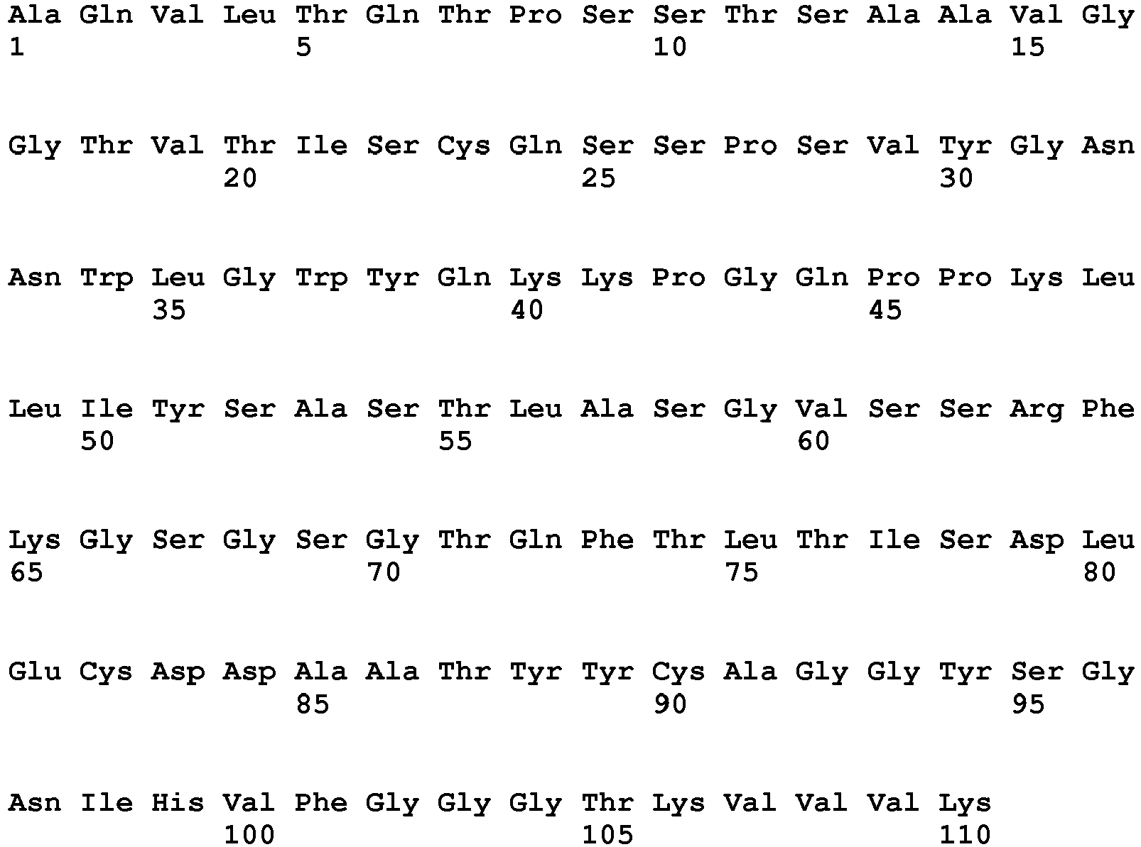
- <210> 92
<211> 113
<212> PRT
<213> artificial sequence - <220>
<223> derived from rabbit - <400> 92

- <210> 93
<211> 330
<212> DNA
<213> artificial sequence - <220>
<223> derived from rabbit - <400> 93

- <210> 94
<211> 339
<212> DNA
<213> artificial sequence - <220>
<223> derived from rabbit - <400> 94

- <210> 95
<211> 110
<212> PRT
<213> artificial sequence - <220>
<223> derived from rabbit - <400> 95

- <210> 96
<211> 113
<212> PRT
<213> artificial sequence - <220>
<223> derived from rabbit - <400> 96


- <210> 97
<211> 327
<212> DNA
<213> artificial sequence - <220>
<223> derived from rabbit - <400> 97

- <210> 98
<211> 342
<212> DNA
<213> artificial sequence - <220>
<223> derived from rabbit - <400> 98


- <210> 99
<211> 109
<212> PRT
<213> artificial sequence - <220>
<223> derived from rabbit - <400> 99

- <210> 100
<211> 114
<212> PRT
<213> artificial sequence - <220>
<223> derived from rabbit - <400> 100


- <210> 101
<211> 738
<212> PRT
<213> Homo sapiens - <400> 101


- <210> 102
<211> 449
<212> PRT
<213> Homo sapiens - <400> 102


- <210> 103
<211> 322
<212> PRT
<213> Homo sapiens - <400> 103

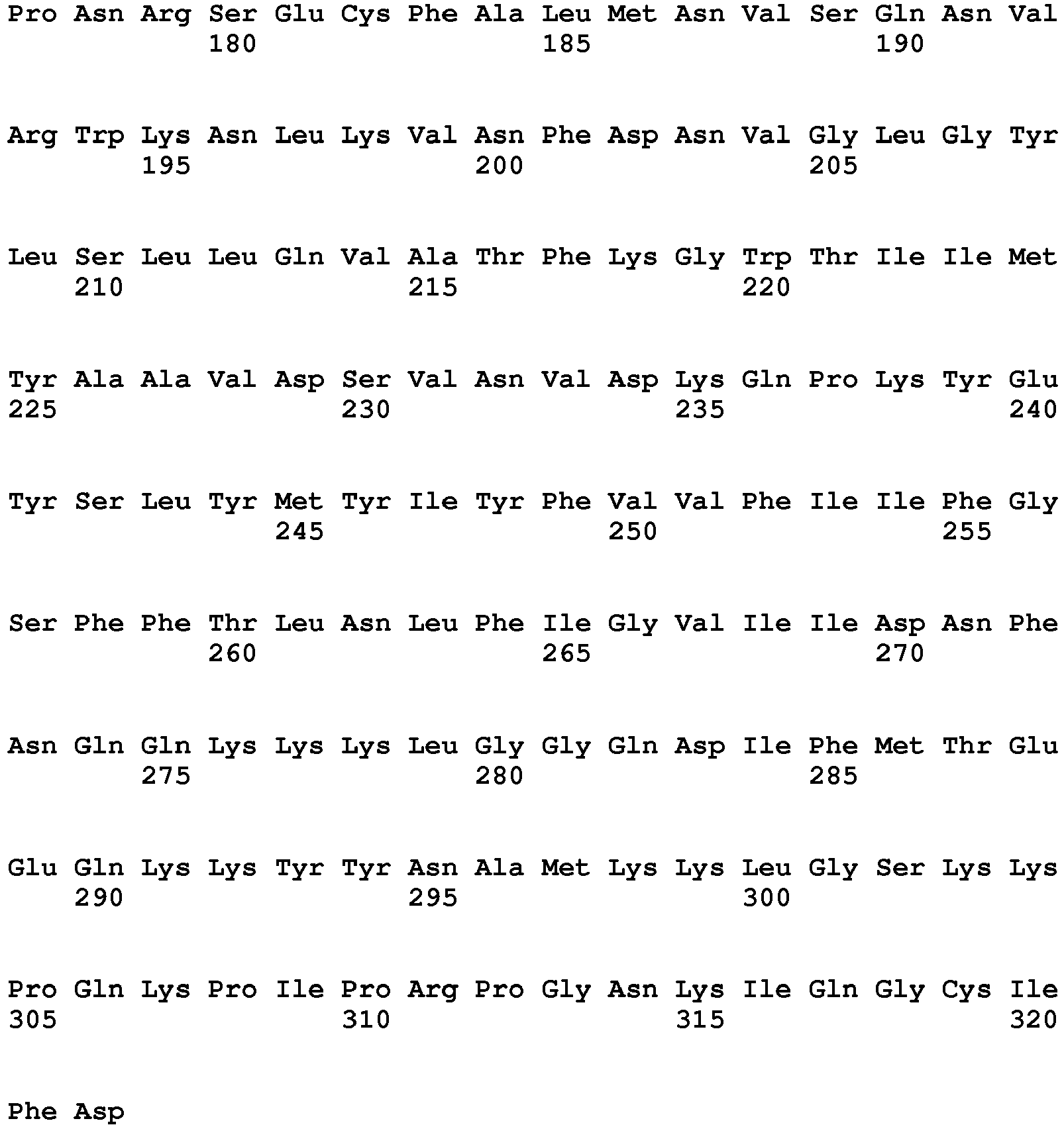
- <210> 104
<211> 478
<212> PRT
<213> Homo sapiens - <400> 104


- <210> 105
<211> 1987
<212> PRT
<213> Homo sapiens - <400> 105


- <210> 106
<211> 22
<212> PRT
<213> Homo sapiens - <400> 106

- <210> 107
<211> 12
<212> PRT
<213> artificial sequence - <220>
<223> derived from homo sapeins Nav1.7 - <400> 107

- <210> 108
<211> 12
<212> PRT
<213> artificial sequence - <220>
<223> derived from homo sapiens Nav1.7 - <400> 108

- <210> 109
<211> 9
<212> PRT
<213> artificial sequence - <220>
<223> derived from homo sapiens Nav1.7 - <400> 109

- <210> 110
<211> 9
<212> PRT
<213> artificial sequence - <220>
<223> derived from homo sapiens Nav1.7 - <400> 110

- <210> 111
<211> 14
<212> PRT
<213> artificial sequence - <220>
<223> derived from homo sapiens Nav1.7 - <400> 111

- <210> 112
<211> 15
<212> PRT
<213> artificial sequence - <220>
<223> derived from homo sapiens Nav1.7 - <400> 112

- <210> 113
<211> 10
<212> PRT
<213> artificial sequence - <220>
<223> derived from homo sapiens Nav1.7 - <400> 113

- <210> 114
<211> 10
<212> PRT
<213> artificial sequence - <220>
<223> derived from homo sapiens Nav1.7 - <400> 114

- <210> 115
<211> 9
<212> PRT
<213> artificial sequence - <220>
<223> derived from homo sapiens Nav1.7 - <400> 115

- <210> 116
<211> 9
<212> PRT
<213> artificial sequence - <220>
<223> derived from homo sapiens Nav1.7 - <400> 116

- <210> 117
<211> 12
<212> PRT
<213> artificial sequence - <220>
<223> derived from homo sapiens Nav1.7 - <400> 117

- <210> 118
<211> 12
<212> PRT
<213> artificial sequence - <220>
<223> derived from homo sapiens Nav1.7 - <400> 118

- <210> 119
<211> 9
<212> PRT
<213> artificial sequence - <220>
<223> derived from homo sapiens Nav1.7 - <400> 119

- <210> 120
<211> 12
<212> PRT
<213> artificial sequence - <220>
<223> artificial sequence - <400> 120

- <210> 121
<211> 9
<212> PRT
<213> artificial sequence - <220>
<223> derived from homo sapiens Nav1.7 - <400> 121

- <210> 122
<211> 11
<212> PRT
<213> artificial peptide - <220>
<223> derived from homo sapiens Nav1.7 - <400> 122

- <210> 123
<211> 7
<212> PRT
<213> artificial sequence - <220>
<223> derived from homo sapiens Nav1.7 - <400> 123

- <210> 124
<211> 8
<212> PRT
<213> artificial sequence - <220>
<223> derived from homo sapiens Nav1.7 - <400> 124

- <210> 125
<211> 9
<212> PRT
<213> artificial sequence - <220>
<223> derived from homo sapiens Nav1.7 - <400> 125

- <210> 126
<211> 11
<212> PRT
<213> artificial sequence - <220>
<223> derived from homo sapiens Nav1.7 - <400> 126

- <210> 127
<211> 7
<212> PRT
<213> artificial sequence - <220>
<223> derived from homo sapiens Nav1.7 - <400> 127

- <210> 128
<211> 9
<212> PRT
<213> artificial sequence - <220>
<223> derived from homo sapiens Nav1.7 - <400> 128

- <210> 129
<211> 10
<212> PRT
<213> artificial sequence - <220>
<223> derived from homo sapiens Nav1.7 - <400> 129

- <210> 130
<211> 12
<212> PRT
<213> artificial sequence - <220>
<223> derived from homo sapiens Nav1.7 - <400> 130

- <210> 131
<211> 9
<212> PRT
<213> artificial sequence - <220>
<223> derived from homo sapiens Nav1.3 - <400> 131

- <210> 132
<211> 12
<212> PRT
<213> artificial sequence - <220>
<223> derived from homo sapiens Nav1.3 - <400> 132

- <210> 133
<211> 9
<212> PRT
<213> artificial sequence - <220>
<223> derived from homo sapiens Nav1.3 - <400> 133

- <210> 134
<211> 11
<212> PRT
<213> artificial sequence - <220>
<223> derived from homo sapiens Nav1.3 - <400> 134

- <210> 135
<211> 9
<212> PRT
<213> artificial sequence - <220>
<223> derived from homo sapiens Nav1.6 - <400> 135

- <210> 136
<211> 12
<212> PRT
<213> artificial sequence - <220>
<223> derived from homo sapiens Nav1.6 - <400> 136

- <210> 137
<211> 9
<212> PRT
<213> artificial sequence - <220>
<223> derived from homo sapiens Nav1.6 - <400> 137

- <210> 138
<211> 11
<212> PRT
<213> artificial sequence - <220>
<223> derived from homo sapiens Nav1.6 - <400> 138

- <210> 139
<211> 10
<212> PRT
<213> artificial sequence - <220>
<223> derived from homo sapiens Nav1.8 - <400> 139

- <210> 140
<211> 12
<212> PRT
<213> artificial sequence - <220>
<223> derived from homo sapiens Nav1.8 - <400> 140

- <210> 141
<211> 12
<212> PRT
<213> artificial sequence - <220>
<223> derived from homo sapiens Nav1.8 - <400> 141

- <210> 142
<211> 9
<212> PRT
<213> artificial sequence - <220>
<223> derived from homo sapiens Nav1.8 - <400> 142

- <210> 143
<211> 9
<212> PRT
<213> artificial sequence - <220>
<223> derived from homo sapiens Nav1.8 - <400> 143

- <210> 144
<211> 10
<212> PRT
<213> artificial sequence - <220>
<223> derived from homo sapiens Nav1.8 - <400> 144


- <210> 145
<211> 10
<212> PRT
<213> artificial sequence - <220>
<223> derived from homo sapiens Nav1.8 - <400> 145

- <210> 146
<211> 15
<212> PRT
<213> artificial sequence - <220>
<223> derived from homo sapiens Nav1.9 - <400> 146

- <210> 147
<211> 12
<212> PRT
<213> artificial sequence - <220>
<223> derived from homo sapiens Nav1.9 - <400> 147

- <210> 148
<211> 11
<212> PRT
<213> artificial sequence - <220>
<223> derived from homo sapiens Nav1.9 - <400> 148

- <210> 149
<211> 11
<212> PRT
<213> artificial sequence - <220>
<223> derived from homo sapiens Nav1.9 - <400> 149

- <210> 150
<211> 7
<212> PRT
<213> artificial sequence - <220>
<223> derived from homo sapiens HCN1 - <400> 150

- <210> 151
<211> 7
<212> PRT
<213> artificial sequence - <220>
<223> derived from homo sapiens HCN2 - <400> 151

- <210> 152
<211> 12
<212> PRT
<213> artificial sequence - <220>
<223> derived from homo sapiens Nav1.1 - <400> 152

- <210> 153
<211> 12
<212> PRT
<213> artificial sequence - <220>
<223> derived from homo sapiens Nav1.2 and Nav1.3 - <400> 153

- <210> 154
<211> 12
<212> PRT
<213> artificial sequence - <220>
<223> derived from homo sapiens Nav1.4 - <400> 154

- <210> 155
<211> 12
<212> PRT
<213> artificial sequence - <220>
<223> derived from homo sapiens Nav1.5 - <400> 155

- <210> 156
<211> 12
<212> PRT
<213> artificial sequence - <220>
<223> derived from homo sapiens Nav1.6 - <400> 156

- <210> 157
<211> 12
<212> PRT
<213> artificial sequence - <220>
<223> derived from homo sapiens Nav.18 - <400> 157

- <210> 158
<211> 12
<212> PRT
<213> artificial sequence - <220>
<223> derived from homo sapiens Nav1.9 - <400> 158

R1 is hydrogen or C1-C4 alkyl;
R2, R3, and R4 are independently hydrogen, halogen, C1-C4 alkyl, C2-C4 alkenyl, C2-C4 alkynyl, aryl, heteroaryl, OH, OC1-C4 alkyl, CO2R1, -CN, -NO2, -NR5R6, -CF3, -OSO2CF3, or R2 and R3, or R3 and R4, respectively, may together form another six membered aromatic or heteroaromatic ring sharing A and G, or G and D, respectively containing between zero and two nitrogen atoms, and substituted with one to two of the following substituents: independently hydrogen, halogen, C1-C4 alkyl, C2-C4 alkenyl, C2-C4 alkynyl, aryl, heteroaryl, OH, OC1-C4 alkyl, CO2R1, -CN, -NO2, -NR5R6, -CF3, OSO2CF3 ;
R5 and R6 are independently hydrogen, C1-C4 alkyl, C(O)R7, C(O)NHR8, C(O)OR9, SO2R10 or may together be (CH2)jQ (CH2)k where Q is O, S, NR11, or a bond;
j is 2 to 7;
k is 0 to 2;
R7, R8, R9, R10, and R11 are independently C1-C4 alkyl, aryl, or heteroaryl,
or an enantiomer thereof, and the pharmaceutically acceptable salts thereof is a potent ligand for nicotinic acetylcholine receptors.
See
Claims (2)
- An anti-E1 ion channel monoclonal antibody or binding fragment thereof, which binds to an E1 extracellular loop shown in SEQ ID NO: 128, wherein said ion channel is Nav1.7 and has a function in the modulation of pain and said antibody or fragment is functionally modifying thereto by reducing the amplitude of current through a patch clamp assay by 40% or more.
- An anti-E1 ion channel antibody or binding fragment thereof according to claim 1, wherein the antibody or binding fragment thereof is for use in the treatment of pain.
Applications Claiming Priority (3)
| Application Number | Priority Date | Filing Date | Title |
|---|---|---|---|
| US25520209P | 2009-10-27 | 2009-10-27 | |
| GBGB0922434.6A GB0922434D0 (en) | 2009-12-22 | 2009-12-22 | antibodies and fragments thereof |
| PCT/EP2010/066274 WO2011051349A1 (en) | 2009-10-27 | 2010-10-27 | Antibodies to ion channels |
Publications (2)
| Publication Number | Publication Date |
|---|---|
| EP2493928A1 EP2493928A1 (en) | 2012-09-05 |
| EP2493928B1 true EP2493928B1 (en) | 2020-04-15 |
Family
ID=41717412
Family Applications (1)
| Application Number | Title | Priority Date | Filing Date |
|---|---|---|---|
| EP10771436.2A Active EP2493928B1 (en) | 2009-10-27 | 2010-10-27 | Antibodies to ion channels |
Country Status (7)
| Country | Link |
|---|---|
| US (2) | US8926977B2 (en) |
| EP (1) | EP2493928B1 (en) |
| JP (1) | JP5872476B2 (en) |
| CN (1) | CN102597002B (en) |
| CA (1) | CA2778671C (en) |
| GB (1) | GB0922434D0 (en) |
| WO (1) | WO2011051349A1 (en) |
Families Citing this family (58)
| Publication number | Priority date | Publication date | Assignee | Title |
|---|---|---|---|---|
| US9234037B2 (en) | 2009-10-27 | 2016-01-12 | Ucb Biopharma Sprl | Method to generate antibodies to ion channels |
| GB0922434D0 (en) | 2009-12-22 | 2010-02-03 | Ucb Pharma Sa | antibodies and fragments thereof |
| CA2778673A1 (en) | 2009-10-27 | 2011-05-05 | Karen Margrete Miller | Function modifying nav 1.7 antibodies |
| US9340590B2 (en) | 2011-03-16 | 2016-05-17 | Amgen Inc. | Potent and selective inhibitors of NaV1.3 and NaV1.7 |
| JP5363636B2 (en) * | 2011-10-21 | 2013-12-11 | ファイザー・リミテッド | New salts and medical uses |
| US10112988B2 (en) * | 2012-01-09 | 2018-10-30 | Icb International, Inc. | Methods of assessing amyloid-beta peptides in the central nervous system by blood-brain barrier permeable peptide compositions comprising a vab domain of a camelid single domain heavy chain antibody against an anti-amyloid-beta peptide |
| US10112987B2 (en) * | 2012-01-09 | 2018-10-30 | Icb International, Inc. | Blood-brain barrier permeable peptide compositions comprising a vab domain of a camelid single domain heavy chain antibody against an amyloid-beta peptide |
| US9371383B2 (en) | 2012-01-31 | 2016-06-21 | Regeneron Pharmaceuticals, Inc. | Anti-ASIC1 antibodies and uses thereof |
| AU2013215254B2 (en) | 2012-01-31 | 2017-08-31 | Regeneron Pharmaceuticals, Inc. | Anti-ASIC1 antibodies and uses thereof |
| WO2013137505A1 (en) * | 2012-03-16 | 2013-09-19 | 서울대학교산학협력단 | Novel trpv1 inhibitory peptide and anti-aging or anti-wrinkle composition for skin comprising same |
| WO2014165277A2 (en) | 2013-03-12 | 2014-10-09 | Amgen Inc. | POTENT AND SELECTIVE INHIBITORS OF Nav1.7 |
| US9636418B2 (en) | 2013-03-12 | 2017-05-02 | Amgen Inc. | Potent and selective inhibitors of NAV1.7 |
| EA201591754A1 (en) * | 2013-03-14 | 2016-01-29 | Ридженерон Фармасьютикалз, Инк. | HUMAN ANTIBODIES TO NA1.7 |
| US9290578B2 (en) * | 2013-10-21 | 2016-03-22 | Genentech, Inc. | Anti-Ly6E antibodies and methods of use |
| JP6782412B2 (en) * | 2014-11-25 | 2020-11-11 | 国立大学法人京都大学 | Pain genes and their uses |
| US11293021B1 (en) | 2016-06-23 | 2022-04-05 | Inscripta, Inc. | Automated cell processing methods, modules, instruments, and systems |
| CN106084010A (en) * | 2016-06-30 | 2016-11-09 | 丁坦 | Competitive binding Nav1.7/ KIF5b protein binding site polypeptide structure |
| IL324632A (en) | 2016-12-01 | 2026-01-01 | Regeneron Pharma | Radiolabeled anti-pd-l1 antibodies for immuno-pet imaging |
| EP3594234A4 (en) * | 2017-02-21 | 2021-01-20 | Populas Biopharmaceutical (Wuhan) Limited | NAV1.9 TARGET POLYPEPTIDE, ANTIBODY AND ANTIBODY FRAGMENT IN COMBINATION WITH IT, AND ASSOCIATED PHARMACEUTICAL COMPOSITION |
| US9982279B1 (en) | 2017-06-23 | 2018-05-29 | Inscripta, Inc. | Nucleic acid-guided nucleases |
| US10011849B1 (en) | 2017-06-23 | 2018-07-03 | Inscripta, Inc. | Nucleic acid-guided nucleases |
| HUE058668T2 (en) | 2017-06-30 | 2022-09-28 | Inscripta Inc | Automated cell handling procedures, modules, equipment and systems |
| GB201711208D0 (en) | 2017-07-12 | 2017-08-23 | Iontas Ltd | Ion channel inhibitors |
| CN109897107B (en) * | 2017-12-08 | 2022-05-17 | 深圳华大生命科学研究院 | Nano antibody and preparation method thereof |
| CN109897106B (en) * | 2017-12-08 | 2022-05-17 | 深圳华大生命科学研究院 | Nano antibody and preparation method and application thereof |
| WO2019200004A1 (en) | 2018-04-13 | 2019-10-17 | Inscripta, Inc. | Automated cell processing instruments comprising reagent cartridges |
| US10508273B2 (en) | 2018-04-24 | 2019-12-17 | Inscripta, Inc. | Methods for identifying selective binding pairs |
| US10858761B2 (en) | 2018-04-24 | 2020-12-08 | Inscripta, Inc. | Nucleic acid-guided editing of exogenous polynucleotides in heterologous cells |
| US10526598B2 (en) | 2018-04-24 | 2020-01-07 | Inscripta, Inc. | Methods for identifying T-cell receptor antigens |
| JP2021525779A (en) * | 2018-06-08 | 2021-09-27 | ヴェンタナ メディカル システムズ, インク. | Universal or normalized antibody framework for improving functionality and manufacturability |
| AU2019292919A1 (en) | 2018-06-30 | 2021-03-11 | Inscripta, Inc. | Instruments, modules, and methods for improved detection of edited sequences in live cells |
| US10532324B1 (en) | 2018-08-14 | 2020-01-14 | Inscripta, Inc. | Instruments, modules, and methods for improved detection of edited sequences in live cells |
| US11142740B2 (en) | 2018-08-14 | 2021-10-12 | Inscripta, Inc. | Detection of nuclease edited sequences in automated modules and instruments |
| US11965154B2 (en) | 2018-08-30 | 2024-04-23 | Inscripta, Inc. | Detection of nuclease edited sequences in automated modules and instruments |
| EP3870697A4 (en) | 2018-10-22 | 2022-11-09 | Inscripta, Inc. | Engineered enzymes |
| US11214781B2 (en) | 2018-10-22 | 2022-01-04 | Inscripta, Inc. | Engineered enzyme |
| CN113631713A (en) | 2019-03-25 | 2021-11-09 | 因思科瑞普特公司 | Simultaneous multiplex genome editing in yeast |
| US11001831B2 (en) | 2019-03-25 | 2021-05-11 | Inscripta, Inc. | Simultaneous multiplex genome editing in yeast |
| US10837021B1 (en) | 2019-06-06 | 2020-11-17 | Inscripta, Inc. | Curing for recursive nucleic acid-guided cell editing |
| WO2020257395A1 (en) | 2019-06-21 | 2020-12-24 | Inscripta, Inc. | Genome-wide rationally-designed mutations leading to enhanced lysine production in e. coli |
| US10927385B2 (en) | 2019-06-25 | 2021-02-23 | Inscripta, Inc. | Increased nucleic-acid guided cell editing in yeast |
| WO2021102059A1 (en) | 2019-11-19 | 2021-05-27 | Inscripta, Inc. | Methods for increasing observed editing in bacteria |
| KR20250033331A (en) | 2019-12-10 | 2025-03-07 | 인스크립타 인코포레이티드 | Novel mad nucleases |
| US10704033B1 (en) | 2019-12-13 | 2020-07-07 | Inscripta, Inc. | Nucleic acid-guided nucleases |
| CA3157127A1 (en) | 2019-12-18 | 2021-06-24 | Aamir MIR | Cascade/dcas3 complementation assays for in vivo detection of nucleic acid-guided nuclease edited cells |
| US10689669B1 (en) | 2020-01-11 | 2020-06-23 | Inscripta, Inc. | Automated multi-module cell processing methods, instruments, and systems |
| KR20220133257A (en) | 2020-01-27 | 2022-10-04 | 인스크립타 인코포레이티드 | Electroporation modules and instruments |
| US20210332388A1 (en) | 2020-04-24 | 2021-10-28 | Inscripta, Inc. | Compositions, methods, modules and instruments for automated nucleic acid-guided nuclease editing in mammalian cells |
| US11787841B2 (en) | 2020-05-19 | 2023-10-17 | Inscripta, Inc. | Rationally-designed mutations to the thrA gene for enhanced lysine production in E. coli |
| WO2022060749A1 (en) | 2020-09-15 | 2022-03-24 | Inscripta, Inc. | Crispr editing to embed nucleic acid landing pads into genomes of live cells |
| US11512297B2 (en) | 2020-11-09 | 2022-11-29 | Inscripta, Inc. | Affinity tag for recombination protein recruitment |
| CA3204158A1 (en) | 2021-01-04 | 2022-07-07 | Juhan Kim | Mad nucleases |
| US11332742B1 (en) | 2021-01-07 | 2022-05-17 | Inscripta, Inc. | Mad nucleases |
| US11884924B2 (en) | 2021-02-16 | 2024-01-30 | Inscripta, Inc. | Dual strand nucleic acid-guided nickase editing |
| MY206945A (en) | 2021-11-01 | 2025-01-21 | Shionogi & Co | Novel nav1.7 monoclonal antibody |
| AU2024346686A1 (en) * | 2023-09-23 | 2026-04-16 | Agilent Technologies, Inc. | Anti-human adam9 antibodies for in vitro diagnostics |
| CN120349417A (en) * | 2024-01-19 | 2025-07-22 | 深圳晶蛋生物医药科技有限公司 | TRPA1 antibody, and preparation method and application thereof |
| WO2025152185A1 (en) * | 2024-01-19 | 2025-07-24 | 深圳晶蛋生物医药科技有限公司 | Trpa1 antibody, and preparation method therefor and use thereof |
Citations (1)
| Publication number | Priority date | Publication date | Assignee | Title |
|---|---|---|---|---|
| WO2007023298A2 (en) * | 2005-08-26 | 2007-03-01 | Isis Innovation Limited | Antibodies |
Family Cites Families (67)
| Publication number | Priority date | Publication date | Assignee | Title |
|---|---|---|---|---|
| US4741900A (en) | 1982-11-16 | 1988-05-03 | Cytogen Corporation | Antibody-metal ion complexes |
| GB8308235D0 (en) | 1983-03-25 | 1983-05-05 | Celltech Ltd | Polypeptides |
| US4816567A (en) | 1983-04-08 | 1989-03-28 | Genentech, Inc. | Recombinant immunoglobin preparations |
| US5677425A (en) | 1987-09-04 | 1997-10-14 | Celltech Therapeutics Limited | Recombinant antibody |
| GB8720833D0 (en) | 1987-09-04 | 1987-10-14 | Celltech Ltd | Recombinant dna product |
| EP0768377A1 (en) | 1988-09-02 | 1997-04-16 | Protein Engineering Corporation | Generation and selection of recombinant varied binding proteins |
| US5223409A (en) | 1988-09-02 | 1993-06-29 | Protein Engineering Corp. | Directed evolution of novel binding proteins |
| GB8823869D0 (en) | 1988-10-12 | 1988-11-16 | Medical Res Council | Production of antibodies |
| US5530101A (en) | 1988-12-28 | 1996-06-25 | Protein Design Labs, Inc. | Humanized immunoglobulins |
| GB8928874D0 (en) | 1989-12-21 | 1990-02-28 | Celltech Ltd | Humanised antibodies |
| US5780225A (en) | 1990-01-12 | 1998-07-14 | Stratagene | Method for generating libaries of antibody genes comprising amplification of diverse antibody DNAs and methods for using these libraries for the production of diverse antigen combining molecules |
| AU7247191A (en) | 1990-01-11 | 1991-08-05 | Molecular Affinities Corporation | Production of antibodies using gene libraries |
| ES2284161T3 (en) | 1990-01-12 | 2007-11-01 | Amgen Fremont Inc. | GENERATION OF XENOGENIC ANTIBODIES. |
| US5427908A (en) | 1990-05-01 | 1995-06-27 | Affymax Technologies N.V. | Recombinant library screening methods |
| GB9015198D0 (en) | 1990-07-10 | 1990-08-29 | Brien Caroline J O | Binding substance |
| CA2090126C (en) | 1990-08-02 | 2002-10-22 | John W. Schrader | Methods for the production of proteins with a desired function |
| US5633425A (en) | 1990-08-29 | 1997-05-27 | Genpharm International, Inc. | Transgenic non-human animals capable of producing heterologous antibodies |
| US5625126A (en) | 1990-08-29 | 1997-04-29 | Genpharm International, Inc. | Transgenic non-human animals for producing heterologous antibodies |
| US5770429A (en) | 1990-08-29 | 1998-06-23 | Genpharm International, Inc. | Transgenic non-human animals capable of producing heterologous antibodies |
| US5661016A (en) | 1990-08-29 | 1997-08-26 | Genpharm International Inc. | Transgenic non-human animals capable of producing heterologous antibodies of various isotypes |
| US5545806A (en) | 1990-08-29 | 1996-08-13 | Genpharm International, Inc. | Ransgenic non-human animals for producing heterologous antibodies |
| AU8507191A (en) | 1990-08-29 | 1992-03-30 | Genpharm International, Inc. | Transgenic non-human animals capable of producing heterologous antibodies |
| US5698426A (en) | 1990-09-28 | 1997-12-16 | Ixsys, Incorporated | Surface expression libraries of heteromeric receptors |
| ES2113940T3 (en) | 1990-12-03 | 1998-05-16 | Genentech Inc | ENRICHMENT METHOD FOR PROTEIN VARIANTS WITH ALTERED UNION PROPERTIES. |
| AU662148B2 (en) | 1991-04-10 | 1995-08-24 | Scripps Research Institute, The | Heterodimeric receptor libraries using phagemids |
| GB9112536D0 (en) | 1991-06-11 | 1991-07-31 | Celltech Ltd | Chemical compounds |
| GB9113120D0 (en) | 1991-06-18 | 1991-08-07 | Kodak Ltd | Photographic processing apparatus |
| PT1696031E (en) | 1991-12-02 | 2010-06-25 | Medical Res Council | Production of anti-self antibodies from antibody segment repertoires and displayed on phage |
| US5733743A (en) | 1992-03-24 | 1998-03-31 | Cambridge Antibody Technology Limited | Methods for producing members of specific binding pairs |
| JPH09506262A (en) | 1993-12-08 | 1997-06-24 | ジェンザイム・コーポレイション | Method for producing specific antibody |
| PT744958E (en) | 1994-01-31 | 2003-11-28 | Univ Boston | BANKS OF POLYCLONE ANTIBODIES |
| FR2716640B1 (en) | 1994-02-28 | 1996-05-03 | Procedes Machines Speciales | Device for centering and blocking a workpiece with a view to running it in using an expansion lapper. |
| US5516637A (en) | 1994-06-10 | 1996-05-14 | Dade International Inc. | Method involving display of protein binding pairs on the surface of bacterial pili and bacteriophage |
| JP2978435B2 (en) | 1996-01-24 | 1999-11-15 | チッソ株式会社 | Method for producing acryloxypropyl silane |
| JP2000515002A (en) | 1996-06-27 | 2000-11-14 | フラームス・インテルウニフェルジテール・インスティテュート・フォール・ビオテヒノロジー・ヴェーゼットウェー | Recognition molecules that specifically interact with the active site or cleft of the target molecule |
| US5980898A (en) | 1996-11-14 | 1999-11-09 | The United States Of America As Represented By The U.S. Army Medical Research & Material Command | Adjuvant for transcutaneous immunization |
| GB9625640D0 (en) | 1996-12-10 | 1997-01-29 | Celltech Therapeutics Ltd | Biological products |
| AR013184A1 (en) | 1997-07-18 | 2000-12-13 | Astrazeneca Ab | SPYROZOBICYCLIC HETERO CYCLIC AMINES, PHARMACEUTICAL COMPOSITION, USE OF SUCH AMINES TO PREPARE MEDICINES AND METHOD OF TREATMENT OR PROPHYLAXIS |
| CN1227030C (en) | 1999-04-19 | 2005-11-16 | 史密丝克莱恩比彻姆生物有限公司 | Adjuvant compositions comprising saponins and immunostimulatory oligonucleotides |
| GB0129105D0 (en) | 2001-12-05 | 2002-01-23 | Celltech R&D Ltd | Expression control using variable intergenic sequences |
| US20050214857A1 (en) | 2001-12-11 | 2005-09-29 | Algonomics N.V. | Method for displaying loops from immunoglobulin domains in different contexts |
| US20060093771A1 (en) | 2002-02-15 | 2006-05-04 | Frantisek Rypacek | Polymer coating for medical devices |
| US20030235533A1 (en) | 2002-02-19 | 2003-12-25 | Idec Pharmaceuticals Corporation | Prostate specific genes and the use thereof in design or therapeutics |
| US7195879B2 (en) * | 2002-04-12 | 2007-03-27 | Ortho-Mcneil Pharmaceutical, Inc. | Method for identifying modulators of NAv ion channels |
| US7993864B2 (en) | 2002-12-03 | 2011-08-09 | Ucb Pharma S.A. | Assay for identifying antibody producing cells |
| US7608260B2 (en) | 2003-01-06 | 2009-10-27 | Medimmune, Llc | Stabilized immunoglobulins |
| GB0312481D0 (en) | 2003-05-30 | 2003-07-09 | Celltech R&D Ltd | Antibodies |
| JP2007501021A (en) | 2003-05-30 | 2007-01-25 | アレクシオン ファーマシューティカルズ, インコーポレイテッド | Antibodies and fusion proteins containing genetically engineered constant regions |
| HUE026278T2 (en) | 2003-07-01 | 2016-05-30 | Ucb Biopharma Sprl | Modified antibody fab fragments |
| GB0315457D0 (en) | 2003-07-01 | 2003-08-06 | Celltech R&D Ltd | Biological products |
| GB0315450D0 (en) | 2003-07-01 | 2003-08-06 | Celltech R&D Ltd | Biological products |
| EP1699914A2 (en) * | 2003-12-12 | 2006-09-13 | Wyeth a Corporation of the State of Delaware | Novel sodium channel |
| EP1711517A4 (en) | 2004-01-21 | 2008-02-13 | Univ Utah Res Found | MUTANT SODIUM CHANNEL NAv1.7 AND ASSOCIATED METHODS |
| GB0411186D0 (en) | 2004-05-19 | 2004-06-23 | Celltech R&D Ltd | Biological products |
| GB0412181D0 (en) | 2004-06-01 | 2004-06-30 | Celltech R&D Ltd | Biological products |
| GB0412986D0 (en) | 2004-06-10 | 2004-07-14 | Xention Discovery Ltd | Compounds |
| ES2579805T3 (en) | 2004-09-23 | 2016-08-16 | Genentech, Inc. | Antibodies and conjugates engineered with cysteine |
| WO2007109324A2 (en) * | 2006-03-21 | 2007-09-27 | Xenon Pharmaceuticals, Inc. | Potent and selective nav 1.7 sodium channel blockers |
| GB0619291D0 (en) | 2006-09-29 | 2006-11-08 | Ucb Sa | Altered antibodies |
| WO2008090958A1 (en) | 2007-01-24 | 2008-07-31 | Kyowa Hakko Kirin Co., Ltd. | Domain-exchanged recombinant antibody composition |
| WO2009033027A2 (en) | 2007-09-05 | 2009-03-12 | Medtronic, Inc. | Suppression of scn9a gene expression and/or function for the treatment of pain |
| US20100297674A1 (en) * | 2008-01-22 | 2010-11-25 | Chromocell Corporation | NOVEL CELL LINES EXPRESSING NaV AND METHODS USING THEM |
| WO2010035012A1 (en) | 2008-09-26 | 2010-04-01 | Ucb Pharma S.A. | Biological products |
| GB0922435D0 (en) | 2009-12-22 | 2010-02-03 | Ucb Pharma Sa | Method |
| GB0922434D0 (en) | 2009-12-22 | 2010-02-03 | Ucb Pharma Sa | antibodies and fragments thereof |
| CA2778673A1 (en) | 2009-10-27 | 2011-05-05 | Karen Margrete Miller | Function modifying nav 1.7 antibodies |
| US9234037B2 (en) | 2009-10-27 | 2016-01-12 | Ucb Biopharma Sprl | Method to generate antibodies to ion channels |
-
2009
- 2009-12-22 GB GBGB0922434.6A patent/GB0922434D0/en not_active Ceased
-
2010
- 2010-10-27 CN CN201080048573.1A patent/CN102597002B/en not_active Expired - Fee Related
- 2010-10-27 EP EP10771436.2A patent/EP2493928B1/en active Active
- 2010-10-27 JP JP2012535812A patent/JP5872476B2/en not_active Expired - Fee Related
- 2010-10-27 US US13/504,234 patent/US8926977B2/en not_active Expired - Fee Related
- 2010-10-27 CA CA2778671A patent/CA2778671C/en not_active Expired - Fee Related
- 2010-10-27 WO PCT/EP2010/066274 patent/WO2011051349A1/en not_active Ceased
-
2014
- 2014-11-19 US US14/547,291 patent/US9738710B2/en not_active Expired - Fee Related
Patent Citations (1)
| Publication number | Priority date | Publication date | Assignee | Title |
|---|---|---|---|---|
| WO2007023298A2 (en) * | 2005-08-26 | 2007-03-01 | Isis Innovation Limited | Antibodies |
Non-Patent Citations (3)
| Title |
|---|
| CHIONI A M ET AL: "A novel polyclonal antibody specific for the Nav1.5 voltage-gated Na<+> channel 'neonatal' splice form", JOURNAL OF NEUROSCIENCE METHODS, ELSEVIER SCIENCE PUBLISHER B.V., AMSTERDAM, NL, vol. 147, no. 2, 30 September 2005 (2005-09-30), pages 88 - 98, XP027670521, ISSN: 0165-0270, [retrieved on 20050930] * |
| HANCOCK DAVID C ET AL: "Synthetic peptides as antigens for antibody production", ANTIBODY-DRUG CONJUGATES IN: METHODS IN MOLECULAR BIOLOGY , ISSN 1064-3745; VOL. 1045; [METHODS IN MOLECULAR BIOLOGY , ISSN 1064-3745; VOL. 1045], HUMANA PRESS, US, vol. 295, 13 December 2004 (2004-12-13), pages 13 - 25, XP008102135, ISBN: 978-1-62703-541-5 * |
| XU SHANG-ZHONG ET AL: "Generation of functional ion-channel tools by E3 targeting", NATURE BIOTECHNOLOGY, GALE GROUP INC, vol. 23, no. 10, 1 October 2005 (2005-10-01), pages 1289 - 1293, XP002415111, ISSN: 1087-0156, DOI: 10.1038/NBT1148 * |
Also Published As
| Publication number | Publication date |
|---|---|
| US20120259096A1 (en) | 2012-10-11 |
| US8926977B2 (en) | 2015-01-06 |
| US20150203571A1 (en) | 2015-07-23 |
| GB0922434D0 (en) | 2010-02-03 |
| JP5872476B2 (en) | 2016-03-01 |
| EP2493928A1 (en) | 2012-09-05 |
| WO2011051349A1 (en) | 2011-05-05 |
| CA2778671C (en) | 2019-02-19 |
| CN102597002B (en) | 2017-08-22 |
| CA2778671A1 (en) | 2011-05-05 |
| JP2013508443A (en) | 2013-03-07 |
| US9738710B2 (en) | 2017-08-22 |
| CN102597002A (en) | 2012-07-18 |
Similar Documents
| Publication | Publication Date | Title |
|---|---|---|
| US9738710B2 (en) | Method of treating a patient for pain by administering an anti-ion channel antibody | |
| US10112996B2 (en) | Function modifying NAv1.7 antibodies | |
| EP2493927B1 (en) | Method to generate antibodies to ion channels | |
| US9956274B2 (en) | Method to generate antibodies to ion channels | |
| WO2021043203A1 (en) | Anti-cxcr2 antibodies and uses thereof | |
| JP6335152B2 (en) | Antibodies against ion channels | |
| EP4070816A1 (en) | Anti-gdf15 antibody | |
| TW202528346A (en) | Anti-tslp antibodies and uses thereof | |
| HK40081009A (en) | Anti-gdf15 antibody |
Legal Events
| Date | Code | Title | Description |
|---|---|---|---|
| PUAI | Public reference made under article 153(3) epc to a published international application that has entered the european phase |
Free format text: ORIGINAL CODE: 0009012 |
|
| 17P | Request for examination filed |
Effective date: 20120529 |
|
| AK | Designated contracting states |
Kind code of ref document: A1 Designated state(s): AL AT BE BG CH CY CZ DE DK EE ES FI FR GB GR HR HU IE IS IT LI LT LU LV MC MK MT NL NO PL PT RO RS SE SI SK SM TR |
|
| AX | Request for extension of the european patent |
Extension state: BA ME |
|
| RAP1 | Party data changed (applicant data changed or rights of an application transferred) |
Owner name: UCB BIOPHARMA SPRL |
|
| STAA | Information on the status of an ep patent application or granted ep patent |
Free format text: STATUS: EXAMINATION IS IN PROGRESS |
|
| 17Q | First examination report despatched |
Effective date: 20170125 |
|
| GRAP | Despatch of communication of intention to grant a patent |
Free format text: ORIGINAL CODE: EPIDOSNIGR1 |
|
| STAA | Information on the status of an ep patent application or granted ep patent |
Free format text: STATUS: GRANT OF PATENT IS INTENDED |
|
| INTG | Intention to grant announced |
Effective date: 20191024 |
|
| RAP1 | Party data changed (applicant data changed or rights of an application transferred) |
Owner name: UCB BIOPHARMA SRL |
|
| GRAJ | Information related to disapproval of communication of intention to grant by the applicant or resumption of examination proceedings by the epo deleted |
Free format text: ORIGINAL CODE: EPIDOSDIGR1 |
|
| STAA | Information on the status of an ep patent application or granted ep patent |
Free format text: STATUS: EXAMINATION IS IN PROGRESS |
|
| GRAR | Information related to intention to grant a patent recorded |
Free format text: ORIGINAL CODE: EPIDOSNIGR71 |
|
| GRAS | Grant fee paid |
Free format text: ORIGINAL CODE: EPIDOSNIGR3 |
|
| STAA | Information on the status of an ep patent application or granted ep patent |
Free format text: STATUS: GRANT OF PATENT IS INTENDED |
|
| INTC | Intention to grant announced (deleted) | ||
| GRAA | (expected) grant |
Free format text: ORIGINAL CODE: 0009210 |
|
| STAA | Information on the status of an ep patent application or granted ep patent |
Free format text: STATUS: THE PATENT HAS BEEN GRANTED |
|
| AK | Designated contracting states |
Kind code of ref document: B1 Designated state(s): AL AT BE BG CH CY CZ DE DK EE ES FI FR GB GR HR HU IE IS IT LI LT LU LV MC MK MT NL NO PL PT RO RS SE SI SK SM TR |
|
| AX | Request for extension of the european patent |
Extension state: BA ME |
|
| INTG | Intention to grant announced |
Effective date: 20200306 |
|
| REG | Reference to a national code |
Ref country code: CH Ref legal event code: EP Ref country code: GB Ref legal event code: FG4D |
|
| REG | Reference to a national code |
Ref country code: DE Ref legal event code: R096 Ref document number: 602010063926 Country of ref document: DE |
|
| REG | Reference to a national code |
Ref country code: IE Ref legal event code: FG4D |
|
| REG | Reference to a national code |
Ref country code: AT Ref legal event code: REF Ref document number: 1257178 Country of ref document: AT Kind code of ref document: T Effective date: 20200515 |
|
| REG | Reference to a national code |
Ref country code: NL Ref legal event code: MP Effective date: 20200415 |
|
| REG | Reference to a national code |
Ref country code: LT Ref legal event code: MG4D |
|
| PG25 | Lapsed in a contracting state [announced via postgrant information from national office to epo] |
Ref country code: IS Free format text: LAPSE BECAUSE OF FAILURE TO SUBMIT A TRANSLATION OF THE DESCRIPTION OR TO PAY THE FEE WITHIN THE PRESCRIBED TIME-LIMIT Effective date: 20200815 Ref country code: FI Free format text: LAPSE BECAUSE OF FAILURE TO SUBMIT A TRANSLATION OF THE DESCRIPTION OR TO PAY THE FEE WITHIN THE PRESCRIBED TIME-LIMIT Effective date: 20200415 Ref country code: PT Free format text: LAPSE BECAUSE OF FAILURE TO SUBMIT A TRANSLATION OF THE DESCRIPTION OR TO PAY THE FEE WITHIN THE PRESCRIBED TIME-LIMIT Effective date: 20200817 Ref country code: NL Free format text: LAPSE BECAUSE OF FAILURE TO SUBMIT A TRANSLATION OF THE DESCRIPTION OR TO PAY THE FEE WITHIN THE PRESCRIBED TIME-LIMIT Effective date: 20200415 Ref country code: SE Free format text: LAPSE BECAUSE OF FAILURE TO SUBMIT A TRANSLATION OF THE DESCRIPTION OR TO PAY THE FEE WITHIN THE PRESCRIBED TIME-LIMIT Effective date: 20200415 Ref country code: LT Free format text: LAPSE BECAUSE OF FAILURE TO SUBMIT A TRANSLATION OF THE DESCRIPTION OR TO PAY THE FEE WITHIN THE PRESCRIBED TIME-LIMIT Effective date: 20200415 Ref country code: NO Free format text: LAPSE BECAUSE OF FAILURE TO SUBMIT A TRANSLATION OF THE DESCRIPTION OR TO PAY THE FEE WITHIN THE PRESCRIBED TIME-LIMIT Effective date: 20200715 Ref country code: GR Free format text: LAPSE BECAUSE OF FAILURE TO SUBMIT A TRANSLATION OF THE DESCRIPTION OR TO PAY THE FEE WITHIN THE PRESCRIBED TIME-LIMIT Effective date: 20200716 |
|
| REG | Reference to a national code |
Ref country code: AT Ref legal event code: MK05 Ref document number: 1257178 Country of ref document: AT Kind code of ref document: T Effective date: 20200415 |
|
| PG25 | Lapsed in a contracting state [announced via postgrant information from national office to epo] |
Ref country code: BG Free format text: LAPSE BECAUSE OF FAILURE TO SUBMIT A TRANSLATION OF THE DESCRIPTION OR TO PAY THE FEE WITHIN THE PRESCRIBED TIME-LIMIT Effective date: 20200715 Ref country code: LV Free format text: LAPSE BECAUSE OF FAILURE TO SUBMIT A TRANSLATION OF THE DESCRIPTION OR TO PAY THE FEE WITHIN THE PRESCRIBED TIME-LIMIT Effective date: 20200415 Ref country code: RS Free format text: LAPSE BECAUSE OF FAILURE TO SUBMIT A TRANSLATION OF THE DESCRIPTION OR TO PAY THE FEE WITHIN THE PRESCRIBED TIME-LIMIT Effective date: 20200415 Ref country code: HR Free format text: LAPSE BECAUSE OF FAILURE TO SUBMIT A TRANSLATION OF THE DESCRIPTION OR TO PAY THE FEE WITHIN THE PRESCRIBED TIME-LIMIT Effective date: 20200415 |
|
| PG25 | Lapsed in a contracting state [announced via postgrant information from national office to epo] |
Ref country code: AL Free format text: LAPSE BECAUSE OF FAILURE TO SUBMIT A TRANSLATION OF THE DESCRIPTION OR TO PAY THE FEE WITHIN THE PRESCRIBED TIME-LIMIT Effective date: 20200415 |
|
| REG | Reference to a national code |
Ref country code: DE Ref legal event code: R097 Ref document number: 602010063926 Country of ref document: DE |
|
| PG25 | Lapsed in a contracting state [announced via postgrant information from national office to epo] |
Ref country code: AT Free format text: LAPSE BECAUSE OF FAILURE TO SUBMIT A TRANSLATION OF THE DESCRIPTION OR TO PAY THE FEE WITHIN THE PRESCRIBED TIME-LIMIT Effective date: 20200415 Ref country code: DK Free format text: LAPSE BECAUSE OF FAILURE TO SUBMIT A TRANSLATION OF THE DESCRIPTION OR TO PAY THE FEE WITHIN THE PRESCRIBED TIME-LIMIT Effective date: 20200415 Ref country code: ES Free format text: LAPSE BECAUSE OF FAILURE TO SUBMIT A TRANSLATION OF THE DESCRIPTION OR TO PAY THE FEE WITHIN THE PRESCRIBED TIME-LIMIT Effective date: 20200415 Ref country code: SM Free format text: LAPSE BECAUSE OF FAILURE TO SUBMIT A TRANSLATION OF THE DESCRIPTION OR TO PAY THE FEE WITHIN THE PRESCRIBED TIME-LIMIT Effective date: 20200415 Ref country code: EE Free format text: LAPSE BECAUSE OF FAILURE TO SUBMIT A TRANSLATION OF THE DESCRIPTION OR TO PAY THE FEE WITHIN THE PRESCRIBED TIME-LIMIT Effective date: 20200415 Ref country code: RO Free format text: LAPSE BECAUSE OF FAILURE TO SUBMIT A TRANSLATION OF THE DESCRIPTION OR TO PAY THE FEE WITHIN THE PRESCRIBED TIME-LIMIT Effective date: 20200415 Ref country code: CZ Free format text: LAPSE BECAUSE OF FAILURE TO SUBMIT A TRANSLATION OF THE DESCRIPTION OR TO PAY THE FEE WITHIN THE PRESCRIBED TIME-LIMIT Effective date: 20200415 Ref country code: IT Free format text: LAPSE BECAUSE OF FAILURE TO SUBMIT A TRANSLATION OF THE DESCRIPTION OR TO PAY THE FEE WITHIN THE PRESCRIBED TIME-LIMIT Effective date: 20200415 |
|
| PLBE | No opposition filed within time limit |
Free format text: ORIGINAL CODE: 0009261 |
|
| STAA | Information on the status of an ep patent application or granted ep patent |
Free format text: STATUS: NO OPPOSITION FILED WITHIN TIME LIMIT |
|
| PG25 | Lapsed in a contracting state [announced via postgrant information from national office to epo] |
Ref country code: PL Free format text: LAPSE BECAUSE OF FAILURE TO SUBMIT A TRANSLATION OF THE DESCRIPTION OR TO PAY THE FEE WITHIN THE PRESCRIBED TIME-LIMIT Effective date: 20200415 Ref country code: SK Free format text: LAPSE BECAUSE OF FAILURE TO SUBMIT A TRANSLATION OF THE DESCRIPTION OR TO PAY THE FEE WITHIN THE PRESCRIBED TIME-LIMIT Effective date: 20200415 |
|
| 26N | No opposition filed |
Effective date: 20210118 |
|
| REG | Reference to a national code |
Ref country code: DE Ref legal event code: R119 Ref document number: 602010063926 Country of ref document: DE |
|
| PG25 | Lapsed in a contracting state [announced via postgrant information from national office to epo] |
Ref country code: SI Free format text: LAPSE BECAUSE OF FAILURE TO SUBMIT A TRANSLATION OF THE DESCRIPTION OR TO PAY THE FEE WITHIN THE PRESCRIBED TIME-LIMIT Effective date: 20200415 |
|
| REG | Reference to a national code |
Ref country code: CH Ref legal event code: PL |
|
| GBPC | Gb: european patent ceased through non-payment of renewal fee |
Effective date: 20201027 |
|
| PG25 | Lapsed in a contracting state [announced via postgrant information from national office to epo] |
Ref country code: MC Free format text: LAPSE BECAUSE OF FAILURE TO SUBMIT A TRANSLATION OF THE DESCRIPTION OR TO PAY THE FEE WITHIN THE PRESCRIBED TIME-LIMIT Effective date: 20200415 Ref country code: LU Free format text: LAPSE BECAUSE OF NON-PAYMENT OF DUE FEES Effective date: 20201027 |
|
| REG | Reference to a national code |
Ref country code: BE Ref legal event code: MM Effective date: 20201031 |
|
| PG25 | Lapsed in a contracting state [announced via postgrant information from national office to epo] |
Ref country code: FR Free format text: LAPSE BECAUSE OF NON-PAYMENT OF DUE FEES Effective date: 20201031 Ref country code: DE Free format text: LAPSE BECAUSE OF NON-PAYMENT OF DUE FEES Effective date: 20210501 |
|
| PG25 | Lapsed in a contracting state [announced via postgrant information from national office to epo] |
Ref country code: GB Free format text: LAPSE BECAUSE OF NON-PAYMENT OF DUE FEES Effective date: 20201027 Ref country code: LI Free format text: LAPSE BECAUSE OF NON-PAYMENT OF DUE FEES Effective date: 20201031 Ref country code: BE Free format text: LAPSE BECAUSE OF NON-PAYMENT OF DUE FEES Effective date: 20201031 Ref country code: CH Free format text: LAPSE BECAUSE OF NON-PAYMENT OF DUE FEES Effective date: 20201031 |
|
| PG25 | Lapsed in a contracting state [announced via postgrant information from national office to epo] |
Ref country code: IE Free format text: LAPSE BECAUSE OF NON-PAYMENT OF DUE FEES Effective date: 20201027 |
|
| PG25 | Lapsed in a contracting state [announced via postgrant information from national office to epo] |
Ref country code: TR Free format text: LAPSE BECAUSE OF FAILURE TO SUBMIT A TRANSLATION OF THE DESCRIPTION OR TO PAY THE FEE WITHIN THE PRESCRIBED TIME-LIMIT Effective date: 20200415 Ref country code: MT Free format text: LAPSE BECAUSE OF FAILURE TO SUBMIT A TRANSLATION OF THE DESCRIPTION OR TO PAY THE FEE WITHIN THE PRESCRIBED TIME-LIMIT Effective date: 20200415 Ref country code: CY Free format text: LAPSE BECAUSE OF FAILURE TO SUBMIT A TRANSLATION OF THE DESCRIPTION OR TO PAY THE FEE WITHIN THE PRESCRIBED TIME-LIMIT Effective date: 20200415 |
|
| PG25 | Lapsed in a contracting state [announced via postgrant information from national office to epo] |
Ref country code: MK Free format text: LAPSE BECAUSE OF FAILURE TO SUBMIT A TRANSLATION OF THE DESCRIPTION OR TO PAY THE FEE WITHIN THE PRESCRIBED TIME-LIMIT Effective date: 20200415 |