EP2493496B1 - Use of gstp1 - Google Patents
Use of gstp1 Download PDFInfo
- Publication number
- EP2493496B1 EP2493496B1 EP10775689.2A EP10775689A EP2493496B1 EP 2493496 B1 EP2493496 B1 EP 2493496B1 EP 10775689 A EP10775689 A EP 10775689A EP 2493496 B1 EP2493496 B1 EP 2493496B1
- Authority
- EP
- European Patent Office
- Prior art keywords
- gstp1
- cardiomyopathy
- cmp
- traf2
- patients
- Prior art date
- Legal status (The legal status is an assumption and is not a legal conclusion. Google has not performed a legal analysis and makes no representation as to the accuracy of the status listed.)
- Not-in-force
Links
Images


Classifications
-
- A—HUMAN NECESSITIES
- A61—MEDICAL OR VETERINARY SCIENCE; HYGIENE
- A61K—PREPARATIONS FOR MEDICAL, DENTAL OR TOILETRY PURPOSES
- A61K38/00—Medicinal preparations containing peptides
- A61K38/16—Peptides having more than 20 amino acids; Gastrins; Somatostatins; Melanotropins; Derivatives thereof
- A61K38/43—Enzymes; Proenzymes; Derivatives thereof
- A61K38/45—Transferases (2)
-
- A—HUMAN NECESSITIES
- A61—MEDICAL OR VETERINARY SCIENCE; HYGIENE
- A61P—SPECIFIC THERAPEUTIC ACTIVITY OF CHEMICAL COMPOUNDS OR MEDICINAL PREPARATIONS
- A61P9/00—Drugs for disorders of the cardiovascular system
-
- A—HUMAN NECESSITIES
- A61—MEDICAL OR VETERINARY SCIENCE; HYGIENE
- A61P—SPECIFIC THERAPEUTIC ACTIVITY OF CHEMICAL COMPOUNDS OR MEDICINAL PREPARATIONS
- A61P9/00—Drugs for disorders of the cardiovascular system
- A61P9/04—Inotropic agents, i.e. stimulants of cardiac contraction; Drugs for heart failure
-
- A—HUMAN NECESSITIES
- A61—MEDICAL OR VETERINARY SCIENCE; HYGIENE
- A61P—SPECIFIC THERAPEUTIC ACTIVITY OF CHEMICAL COMPOUNDS OR MEDICINAL PREPARATIONS
- A61P9/00—Drugs for disorders of the cardiovascular system
- A61P9/10—Drugs for disorders of the cardiovascular system for treating ischaemic or atherosclerotic diseases, e.g. antianginal drugs, coronary vasodilators, drugs for myocardial infarction, retinopathy, cerebrovascula insufficiency, renal arteriosclerosis
-
- A—HUMAN NECESSITIES
- A61—MEDICAL OR VETERINARY SCIENCE; HYGIENE
- A61P—SPECIFIC THERAPEUTIC ACTIVITY OF CHEMICAL COMPOUNDS OR MEDICINAL PREPARATIONS
- A61P9/00—Drugs for disorders of the cardiovascular system
- A61P9/12—Antihypertensives
-
- C—CHEMISTRY; METALLURGY
- C12—BIOCHEMISTRY; BEER; SPIRITS; WINE; VINEGAR; MICROBIOLOGY; ENZYMOLOGY; MUTATION OR GENETIC ENGINEERING
- C12Q—MEASURING OR TESTING PROCESSES INVOLVING ENZYMES, NUCLEIC ACIDS OR MICROORGANISMS; COMPOSITIONS OR TEST PAPERS THEREFOR; PROCESSES OF PREPARING SUCH COMPOSITIONS; CONDITION-RESPONSIVE CONTROL IN MICROBIOLOGICAL OR ENZYMOLOGICAL PROCESSES
- C12Q1/00—Measuring or testing processes involving enzymes, nucleic acids or microorganisms; Compositions therefor; Processes of preparing such compositions
- C12Q1/68—Measuring or testing processes involving enzymes, nucleic acids or microorganisms; Compositions therefor; Processes of preparing such compositions involving nucleic acids
- C12Q1/6876—Nucleic acid products used in the analysis of nucleic acids, e.g. primers or probes
- C12Q1/6883—Nucleic acid products used in the analysis of nucleic acids, e.g. primers or probes for diseases caused by alterations of genetic material
-
- C—CHEMISTRY; METALLURGY
- C12—BIOCHEMISTRY; BEER; SPIRITS; WINE; VINEGAR; MICROBIOLOGY; ENZYMOLOGY; MUTATION OR GENETIC ENGINEERING
- C12Q—MEASURING OR TESTING PROCESSES INVOLVING ENZYMES, NUCLEIC ACIDS OR MICROORGANISMS; COMPOSITIONS OR TEST PAPERS THEREFOR; PROCESSES OF PREPARING SUCH COMPOSITIONS; CONDITION-RESPONSIVE CONTROL IN MICROBIOLOGICAL OR ENZYMOLOGICAL PROCESSES
- C12Q2600/00—Oligonucleotides characterized by their use
- C12Q2600/158—Expression markers
-
- G—PHYSICS
- G01—MEASURING; TESTING
- G01N—INVESTIGATING OR ANALYSING MATERIALS BY DETERMINING THEIR CHEMICAL OR PHYSICAL PROPERTIES
- G01N2333/00—Assays involving biological materials from specific organisms or of a specific nature
- G01N2333/90—Enzymes; Proenzymes
- G01N2333/91—Transferases (2.)
- G01N2333/9116—Transferases (2.) transferring alkyl or aryl groups other than methyl groups (2.5)
- G01N2333/91165—Transferases (2.) transferring alkyl or aryl groups other than methyl groups (2.5) general (2.5.1)
- G01N2333/91171—Transferases (2.) transferring alkyl or aryl groups other than methyl groups (2.5) general (2.5.1) with definite EC number (2.5.1.-)
- G01N2333/91177—Glutathione transferases (2.5.1.18)
-
- G—PHYSICS
- G01—MEASURING; TESTING
- G01N—INVESTIGATING OR ANALYSING MATERIALS BY DETERMINING THEIR CHEMICAL OR PHYSICAL PROPERTIES
- G01N2800/00—Detection or diagnosis of diseases
- G01N2800/32—Cardiovascular disorders
- G01N2800/325—Heart failure or cardiac arrest, e.g. cardiomyopathy, congestive heart failure
-
- Y—GENERAL TAGGING OF NEW TECHNOLOGICAL DEVELOPMENTS; GENERAL TAGGING OF CROSS-SECTIONAL TECHNOLOGIES SPANNING OVER SEVERAL SECTIONS OF THE IPC; TECHNICAL SUBJECTS COVERED BY FORMER USPC CROSS-REFERENCE ART COLLECTIONS [XRACs] AND DIGESTS
- Y02—TECHNOLOGIES OR APPLICATIONS FOR MITIGATION OR ADAPTATION AGAINST CLIMATE CHANGE
- Y02A—TECHNOLOGIES FOR ADAPTATION TO CLIMATE CHANGE
- Y02A50/00—TECHNOLOGIES FOR ADAPTATION TO CLIMATE CHANGE in human health protection, e.g. against extreme weather
- Y02A50/30—Against vector-borne diseases, e.g. mosquito-borne, fly-borne, tick-borne or waterborne diseases whose impact is exacerbated by climate change
Definitions
- the present invention relates to the use of Glutathione-S-transferase P1.
- GSTs The glutathione S-transferases (GSTs) are a multigene family of isozymes that catalyze the nucleophilic attack of the sulfur atom of glutathione (GSH) on electrophilic groups of substrate molecules. GSTs are known as detoxicating enzymes which conjugate toxic substances with GSH to become more water-soluble products which can be metabolised in the liver and finally excreted out of the body. On the basis of the amino acid sequence, the mammalian GSTs are divided into six classes: alpha, mu, omega, pi, theta and zeta.
- glutathione S-transferase P1 (GSTP1; GST-pi, GSTP1-1) is the most prevalent one in mammalian cells.
- GSTP1 has also generated interest as a neoplastic marker because it is overexpressed in a variety of different human malignancies, including lung, colon, stomach, esophagus, mouth, kidney, ovary and testicular cancers.
- Most prostate cancer types contain decreased levels of GSTP1 polypeptide relative to normal prostate tissue due to hypermethylation of the GSTP1 promoter resulting in decreased transcription of GSTP1 ( US 2009/0186360 A1 ).
- GSTP1 is determinant in cellular response to oxidative stress and protects tumor cells from apoptosis elicited by a variety of cytotoxic agents, such as H 2 O 2 , UV, cisplatin, adriamycin, etoposide, thiotepa, chlorambucil, ethacrynic acid and arsenic trioxide.
- cytotoxic agents such as H 2 O 2 , UV, cisplatin, adriamycin, etoposide, thiotepa, chlorambucil, ethacrynic acid and arsenic trioxide.
- GSTP1 is therefore considered as a promising target in tumour medicine, both as a target for drug action and as a tumor marker.
- GSTP1 also participates in the regulation of stress signalling and protects cells against apoptosis by the mechanisms related to its non-catalytic and ligand-binding activities. For example, GSTP1 prevents LPS-induced excessive production of pro-inflammatory factors and plays an anti-inflammatory role in response to LPS. GSTP1 expression, both at transcriptional and translational levels, is up-regulated by LPS stimulation. GSTP1 also functions as an endogenous inhibitor of JNK (c-Jun NH 2 -terminal kinase), via interaction with the C-terminus. It could also be demonstrated that exogenous (recombinant) GSTP1 protein could be delivered into macrophages and suppressed iNOS and COX-2 expression in cells. Furthermore, intraperitoneally administered GSTP1 protein to mice decreased mortality of endotoxic shock significantly and inhibited acute lung injury and peritonitis ( Luo et al., Mol. Immunol. 46 (2009), 848-857 ).
- EP 1 500 709 A1 suggests GSTP1 as one of 22 inflammation markers.
- Cardiomyopathies are a heterogeneous group of diseases, including the dilative (DCM), the most frequent entity among cardiomyopathies, and ischemic (ICM) forms.
- DCM dilative
- ICM ischemic
- results of the last decade's work clearly provide evidence that inflammation and enhanced release of pro-inflammatory cytokines, especially tumor necrosis factor (TNF)- ⁇ might play a role in the pathogenesis of CMP.
- TNF tumor necrosis factor
- IHD The pathology of IHD is immediately related to the development of CMP, patients currently receive medications such as beta-blockers or angiotensin converting enzyme (ACE)-blockers with the goal to ameliorate the patient's symptoms and to prevent or delay the development of (ischemic) CMP due to IHD. It is therefore common to use therapies for CMP also for IHD.
- therapies for CMP also for IHD.
- patients who develop CMP based on IHD in particular end-stage CMP, have currently only limited therapeutic options (e.g. cardiac transplantation or the use of ventricular assist devices, which can be in turn offered only to a limited number of patients).
- ischemic heart diseases also called coronary heart diseases that can result in myocardial infarction
- elevated blood pressure-induced CMP and volume-induced CMP especially elevated blood pressure-induced CMP and volume-induced CMP.
- Cardiomyopathies result in recurrent exacerbation leading to hospitalization or death. Therefore, also close surveillance and thorough clinical tests are required to achieve the optimal tailored cardiomyopathy treatment in these patients, associated with tremendous health care costs.
- a biomarker that could identify the evolution of cardiomyopathy and help to optimize treatment monitoring and outcome is therefore highly desirable. Ideally, such a test should be sensitive, specific, none-invasive, quick and cheap.
- BNP brain-type natriuretic peptide
- proBNP inactive N-terminal fragment
- circulating BNP and proBNP levels are dramatically affected by renal function and are age- and gender-dependent. In addition, it remains unknown whether proBNP could be beneficial in diagnosis of preserved as opposed to reduced ejection fraction (EF) CMP.
- the invention provides Glutathione S-transferase P1 (GSTP1) for use in the prevention or treatment of cardiomyopathies (CMPs).
- CMPs cardiomyopathies
- GSTP1 is beneficial in CMP treatment.
- GSTP1 has been described as a relevant protein in cancer medicine (e.g. US 5,427,917 A , US 5,552,277 A and WO 98/21359 A1 ) and in allergic diseases, such as asthma, especially in connection with traffic-related air pollution (e.g. Melen et al., Env. Health Persp. 116 (2008), 1077-1084 ).
- GSTP1 has also been suggested to decrease mortality of endotoxic shock and inhibition of acute lung injury and peritonitis ( Luo et al., Mol. Immunol. 46 (2009), 848-875 ).
- GSTP1 was reported to interact with TRAF2 (tumor necrosis factor receptor-associated factor 2) as a novel ligand-binding function of GSTP1 in regulating kinase signalling ( Wu et al., Oncogene 25 (2006), 5787-5800 ).
- GSTP1 is specifically upregulated in CMP patients as compared to controls. It was further found that recombinant GSTP1 mediated GSTP1-TRAF2 association, decreased pro-inflammatory JNK1 and p38 activation and TRAF2 expression dependent on its dose and the underlying form of CMP. This enabled the provision of a new therapy strategy for CMP by administration of GSTP1. Since GSTP1 ameliorates TNF- ⁇ -mediated activation of pro-inflammatory JNK1/p38 by association to TRAF2, GSTP1 is not only beneficial in CMP treatment but also for the prevention of CMP, especially in risk patients, such as hypertensive patients.
- the present invention provides the use of GSTP1 for the manufacture of a medicament for the prevention or treatment of CMP.
- the treatment of CMP according to the present invention involves the administration of an effective amount of GSTP1 to an individual in need of such treatment, e.g. human CMP patients.
- an individual in need of such treatment e.g. human CMP patients.
- patients currently receive medications such as beta-blockers or angiotensin converting enzyme (ACE)-blockers with the goal to ameliorate the patient's symptoms and to prevent or delay the development of (ischemic) CMP due to IHD.
- ACE angiotensin converting enzyme
- CMPs can be caused by myocardial infarction, can be idiopathic dilative or be caused by hypertension. All these forms can be treated with GSTP1 according to the present invention.
- ischemic CMPs e.g. caused by myocardial infarction can be treated with GSTP1
- non-ischemic forms can be treated (e.g. idiopathic dilated CMPs or hypertension- or volume-induced CMPs). Since in all these CMPs, inflammatory pathways are involved, this makes them eligible for treatment with GSTP1 according to the present invention (even rare forms of CMP, if such inflammatory pathways are involved).
- preferred CMPs are dilated cardiomyopathy, especially congestive cardiomyopathy; obstructive hypertrophic cardiomyopathy, especially hypertrophic subaortic stenosis; other hypertrophic cardiomyopathies, especially nonobstructive hypertrophic cardiomyopathy; endomyocardial (eosinophilic) disease, especially endomyocardial (tropical) fibrosis or Löffler's endocarditis; endocardial fibroelastosis, especially congenital cardiomyopathy; other restrictive cardiomyopathies, especially constrictive cardiomyopathy NOS (not otherwise specified (according to ICD-10)); alcoholic cardiomyopathy, cardiomyopathies due to drugs and other external agents, unspecified cardiomyopathies, especially cardiomyopathy (primary)(secondary) NOS; cardiomyopathy in infectious and parasitic diseases, especially cardiomyopathy in diphtheria; cardiomyopathy in metabolic diseases, especially cardiac amyloidosis; cardiomyopathy in nutritional diseases, especially nutritional diseases, especially nutritional diseases,
- CMPs to be treated (or prevented) according to the present invention are CMPs caused by ischemia, especially by myocardial infarction; by hypertension, or by myocarditis.
- GSTP1 treatment according to the present invention is therefore preferably applied in patients with acute myocarditis, preferably infective myocarditis, especially septic myocarditis; isolated myocarditis, myocarditis in a bacterial disease, especially diphtheritic, gonococcal, meningococcal, syphilitic or tuberculous myocarditis; myocarditis in a viral disease, especially influenzal myocarditis or mumps myocarditis; acute or chronic myocarditis in Chagas' disease, myocarditis in toxoplasmosis, rheumatoid myocarditis or sarcoid myocarditis.
- the preferred indications according to the present invention are therefore those listed under Chapter IX of the ICD-10 (I11-I15, especially I21, I22 and I23; I20-I25, especially I21 and I22; I40-I43, especially I42; I50-I57, especially I50 and I57).
- the main focus of the present invention is the treatment of human patients; however, based on the present findings, it is clear that also animal (mammal) CMP in which inflammation is involved can be successfully treated by GSTP1 according to the present invention.
- This may be important for farm or zoo animals (especially breeding farm animals, such as (former) racing horses) or pets, for example dogs, cats and horses.
- the present invention is also useable for the prevention of CMP it is clear that the primary focus of the present invention lies in the treatment of human CMP patients who have already been diagnosed of CMP or which have a high risk of developing CMP, for example patients with hypertension (e.g. Stage 2 patients with systolic pressure of 160 mm Hg or more and/or with diastolic pressure of 100 mm Hg or more).
- Another preferred embodiment of the preventive aspect of the present invention is for angina pectoris patients, whereby myocardial infarction might or might not have occurred in this set of patients; and heart insufficiency or heart failure ( Mc Murray et al., Lancet 365 (2005), 1877-1889 ) may or may not be present.
- heart insufficiency or heart failure Mc Murray et al., Lancet 365 (2005), 1877-1889
- Preferred routes of administration of the GSTP1 containing medicament according to the present invention are parenteral routes, preferably intraperitoneal or intravenous administration, intravenous administration being specifically preferred.
- Intravenous administration can be performed e.g. via bolus injection or by continuous intravenous delivery over a longer time period (e.g. 30 min to 6 h, especially 1 to 3 h).
- administration directly to the heart in-tramyocardial injection
- GSTP1 activity can be directly and in high concentrations delivered to the area of need, e.g. to an infarct region.
- Another preferred route of administration is administration to the coronary sinus (e.g. via a catheter inserted into the coronary sinus).
- GSTP1 Oral or mucosal routes (although described for GSTP1 in principle; see e.g. US 5,976,528 A ) are less preferred, since GSTP1 is a protein and for this route specific protection measures (enteric coating, encapsulation, etc.) are necessary as well as a significant optimisation with respect to galenic manufacture.
- Preferred dosages for administration are dosages of 0.001 to 100 mg GSTP1/kg, preferably 0.01 to 10 mg GSTP1/kg, especially 0.1 to 1 mg GSTP1/kg, to a human individual, preferably via intravenous administration; or dosages of 0.1 to 10000 U GSTP1/kg, preferably 1 to 1000 U GSTP1/kg, especially 10 to 100 U GSTP1/kg, to a human individual, preferably via intravenous administration.
- These are preferred daily dosages for intravenous application (“kg” means kg body weight of the person to be treated). During surgery, these dosages may also be applied, either as a whole or even more than one dosage; or only part of the dosage (having in mind that the GSTP1 can directly be applied during surgery in the area needed).
- Ready-to-use daily dosage forms (e.g. for a 80 kg and/or a 60 kg human individual) can be pre-manufactured and can be made storage-stable by lyophilisation or freezing (and keeping at e.g. -20°C). The dosage can then be fully or only partially consumed (e.g. based on the body weight of the person to be treated).
- the human sequence (Seq. ID. No. 1) is listed as “GSTP1_HUMAN, P09211" in the UniProtKB/SwissProt database (HGNC: 4638, Entrez Gene: 2950).
- variants exist for this human protein (homozygous as well as heterozygous), the most important variants are a Ile to Val exchange at position 105 and a Ala to Val exchange at position 114 (Melen et al., 2008).
- Other variants include a Gly to Glu exchange at position 78, a Thr to Ser exchange at position 110, a Gly to Glu exchange at position 139, a Asp to Tyr exchange at position 147 and a Asp to His exchange at position 158.
- SNPs are known without affecting the amino acid sequence.
- the CMP patient to be treated with GSTP1 according to the present invention has such an allele which results in a (heterozygous or homozygous) amino acid exchange compared to SEQ.ID.No. 1, it may be recommendable to administer to such a patient the allelic form of this protein.
- This is specifically preferred for patients who are homozygous for this allele, e.g. patients with homozygous 105Val or 114Val allele (who could then receive the 105Val or 114Val GSTP1 variant, respectively).
- the risk of developing adverse reactions, especially adverse immune reactions (which is always a topic for proteinaceous medicaments), is kept low.
- any ingredients in the pharmaceutical preparation containing GSTP1 according to the present invention eliciting or boosting an immune reaction in the patient should be kept at minimum if possible.
- Orthologs from mammals are known, e.g. from chimpanzee (Pan troglodytes; NCBI accessions: 745954, XM_001152516.1, XP_001152516.1) and mouse (Mus musculus; NCBI accessions: 148701, NM_013541.11, NP_038569.11, AK0791445, BC0020485), but also from non-mammals, such as zebrafish (Danio rerio) and worm (Caenorhabditis elegans).
- the present invention also relates to a pharmaceutical composition
- a pharmaceutical composition comprising GSTP1, preferably human recombinant GSTP1 or human placenta GSTP1, and a pharmaceutically acceptable carrier for the prevention or treatment of cardiomyopathies or ischemic heart diseases.
- the composition according to the present invention contains 1 to 100000 U GSTP1, preferably 10 to 10000 U GSTP1, especially 10 to 1000 U GSTP1.
- a unit of GSTP1 activity conjugates 1.0 micro-mole of 1-chloro-2,4-dinitrobenzene with reduced glutathione per minute at pH 6.5 at 25°C.
- a preferred composition according to the present invention also contains a buffer.
- Typical buffer systems are the carbonic acid/HCO 3 system (pH 6,2 to 8,6; neutral), carbonic acid/silicate buffer (pH 5,0 to 6,2; weakly acidic), acetic acid/acetate buffer (pH 3,7 to 5,7), phosphate buffer (NaH 2 PO 4 + Na 2 HPO 4 ; pH 5,4 to 7,8), ammonia buffer (NH 3 + H 2 O + NH 4 Cl; pH 8,2 to 10,2), TRIS/HCl (Tris(hydroxymethyl)-aminomethane; pH 7,2 to 9,0), HEPES (4-(2-Hydroxyethyl)-1-piperazinethanesulfonic acid; pH 6,8 to 8,2), HEPPS (4-(2-Hydroxyethyl)-piperazine-1-propane sulfonic acid; pH 7,3 to 8,7) or MES (2-(N-Morpholino)ethanesulfonic
- Preferred buffers according to the present invention is a phosphate buffer, a Tris-HCl buffer or a HEPES buffer, with a pH of 5.5 to 9.0, preferably of 6.0 to 8.5, especially of 6.5 to 8.0. Buffer concentrations can preferably be adjusted to 1 mM to 1 M, especially 10 mM to 0.5 M.
- Other preferred additional ingredients include stabilisers, chelators, salts, etc., such as glycerol, glucose, saccharose, maltose, EDTA or similar substances, NaCl, KCl, NH 4 Cl and similar salts usable in pharmaceutical preparations. Of course, all ingredients in a pharmaceutical preparation have to be of pharmaceutical grade quality.
- Pharmaceutically acceptable carriers preferably used are physiological saline, vegetable oils, mineral oil, aqueous sodium caroboxymethyl cellulose or aqueous polyvinylpyrrolidone; however, also sterile water can be used.
- GSTP1 to be used according to the present invention can be provided by various ways. As mentioned above, GSTP1 from human placenta is one of the preferred natural sources of GSTP1. Other preferred sources are cultures of GSTP1 (over-)expressing cells. GSTP1 preparations are also commercially available. The preferred industrial production route according to the present invention is, however, recombinant production of GSTP1. Recombinant production of GSTP1 is well established, as well as its purification from such sources (e.g. Luo et al., 2009, Wu et al., 2006; WO 98/21359 A1 ; US 5,976,528 ; etc.). Expression vectors (e.g.
- WO 98/21359 A1 , Part D (pages 40 to 51) containing the GSTP1 coding sequences are transferred in suitable host cells (such as COS, HEK 293, HeLa, VERO, W138, BHK or MDCK cells, but also yeast, plant, insect and bacterial cells (S. cerevisiae, E. coli, etc.).
- GSTP1 is then expressed and purified according to standard methods. Recombinant GSTP1 production may also use variant forms of GSTP1 with additional sequences which facilitate purification, such as metal chelating peptides, protein A domains, affinity purification tags, etc. (e.g. US 5,976,528 A , column 16).
- Such variants can - after purification or in the course of purification - be removed e.g. via cleavable linker sequences (protease, entereokinase, etc.).
- Purified batch preparations of GSTP1 may contain e.g. 1 to 200 U GSTP1 activity per mg total protein.
- Preferred pharmaceutical GSTP1 compositions contain 5 to 200 U/mg protein, especially 10 to 150 U/mg protein.
- the pharmaceutical preparation according to the present invention is always provided as an appropriately labelled product which is sterile and useable for administration to human patients according to the present invention.
- the present invention relates to GSTP1 as a diagnostic target for CMP.
- the present invention provides the use of GSTP1 for the diagnosis of CMP.
- GSTP1 is a known and established marker in tumor diagnosis, it was surprising that GSTP1 is a biomarker for CMP which is even superior to the current "gold standard", pro-BNP, with respect to sensitivity and specificity.
- the cardiomyopathies referred to above for therapeutical use of GSTP1 can also be diagnosed according to the present invention.
- the present invention therefore relates to a method for diagnosing CMP, especially the indications mentioned above, by determining the amount of GSTP1 or GSTP1 mRNA in a biological sample and comparing this amount to the GSTP1 or GSTP1 mRNA amount in a biological sample with a defined and known CMP status, especially from a healthy human individual or a patient.
- This comparison can be done practically, i.e. in that a real sample is tested in the same manner as the biological sample (side-by-side), it can also be a virtual comparison so that the determined value is compared with a known value of a healthy or diseased sample.
- Preferred techniques for determining the amount of GSTP1 include antibody-liked techniques, such as ELISA, optionally combined with electrophoresis techniques or radio immunoassay (RIA). Electrophoresis techniques may also be combined with MALDI techniques.
- GSTP1 mRNA may be determined, e.g. by quantitative real time RT-PCR.
- the preferred diagnosis target is a human patient who is suspected to have CMP, a human patient who is at risk for developing CMP or a human patient who has CMP (who has to be monitored for progression of the disease).
- Preferred biological samples according to the present invention include human blood, plasma or serum samples or human myocardial biopsy samples. These samples can also be tested by routine methods.
- a high concentration of GSTP1 in serum is indicative of CMP.
- serum concentrations of GSTP1 are below 50 ng/ml or, preferably, even below 10 ng/ml.
- a CMP diseased status can be diagnosed at a serum level at 50 ng/ml or above, preferably at 100 ng/ml or above. Severe forms of CMP can be diagnosed at 200 ng/ml or above. Of course, these levels always depend on the technique used for assaying the GSTP1 protein.
- CMP diseased status can be diagnosed if the serum level of GSTP1 is elevated by at least the factor 2, preferably by at least the factor 5, compared to a healthy status, especially the healthy status of the same patient. Severe forms could be diagnosed, if the serum GSTP1 level is elevated by at least the factor 10, especially by at least the factor 20, compared to a healthy status, especially the healthy status of the same patient.
- determining the amount of GSTP1 or GSTP1 mRNA in a sample can be performed within a very short time by standard test methods, such as ELISA or PCR.
- standard test methods such as ELISA or PCR.
- the present invention Based on the DNA and amino acid sequence according to SEQ.ID.NO.2 and SEQ.ID.NO.1 (and the data base entries mentioned above for the GSTP1 gene and the GSTP1 protein), the present invention also provides e.g. a PCR assay for identification of S. aureus infections or a sandwich ELISA assay.
- GSTP1 can be caught out of a biological sample with a GSTP1-specific antibody and detected and quantified with a second antibody directed against a different epitope of GSTP1 or an antibody being specific for the previous binding event.
- the present invention therefore also provides a kit for determining the amount of GSTP1 mRNA in a least two synthetic nucleic acid sequences for amplifying at least one nucleic acid sequence encoding GSTP1 or parts thereof, wherein at least of one of the synthetic nucleic acid sequences in the kit is selected from the group consisting of: (a) a suitable primer pair, the primers being a nucleic acid sequence comprising 10-30 consecutive nucleotides of at least one of the following: (i) the GSTP1 mRNA according to SEQ.ID.NO.2 or the complementary sequence thereof; or (ii) from the 5' or 3' noncoding region of the GSTP1 gene (e.g.
- primers being usable for polymerising a nucleotide sequence located between the primers on the GSTP1 gene or its 5'- or 3'-region (said polymerizable GSTP1 nucleotide sequence being PCR detectable and preferably up to 1000 bp, more preferred up to 500 bp, even more preferred up to 300bp in length; on the other hand, the primers may also be selected to polymerise (with PCR) the whole GSTP1 m-RNA); and (b) suitable reagents for a polymerase chain reaction (PCR).
- PCR polymerase chain reaction
- the kit further comprises: (c) instructions for use and (d) optionally, a positive and/or negative control for determining and/or quantification of GSTP1 mRNA, especially a control nucleic acid encoding GSTP1 or a fragment thereof.
- PCR reaction buffer examples include PCR reaction buffer, Mg 2+ (e.g., MgCl 2 ), dNTPs, DNA polymerases (such as reverse transcriptases and thermostable DNA polymerases (e.g., Taq-related DNA polymerases and Pfu-related DNA polymerases)), RNase, PCR reaction enhancers or inhibitors, PCR reaction monitoring agents (e.g., double-stranded DNA dye (such as SYBRTM Green), TaqManTM probes, molecular beacons, and Scorpion-sTM), and PCR-grade water.
- DNA polymerases such as reverse transcriptases and thermostable DNA polymerases (e.g., Taq-related DNA polymerases and Pfu-related DNA polymerases)
- RNase e.g., RNase
- PCR reaction enhancers or inhibitors e.g., RNase, PCR reaction enhancers or inhibitors
- PCR reaction monitoring agents e.g., double
- PCR polymerase chain reaction
- a PCR assay may use a heat-stable polymerase and two about 10 to 30 base primers: one complementary to the (+)-strand at one end of the sequence to be amplified, and the other complementary to the (-)-strand at the other end.
- the newly-synthesized biological sample comprising: (a) a primer set comprising at DNA strands can subsequently serve as additional templates for the same primer sequences, successive rounds of primer annealing, strand elongation, and dissociation may produce rapid and highly-specific amplification of the desired sequence.
- PCR also may be used to detect the existence of a defined sequence in a GSTP1 m-RNA containing sample.
- a typical PCR assay according to the present invention might start - optionally after reverse transcription of the mRNA into DNA - with two synthetic oligonucleotide primers which are specifically and complementarily binding to two regions of the target DNA or its complementary strand, respectively, encoding GSTP1 or its 5'- and/or 3' region (one for each strand) that is to be amplified. These may be added to the target DNA (that needs not be pure) in the presence of excess deoxynucleotides (dNTPs) and a thermostable DNA polymerase (e.g., Taq polymerase).
- dNTPs deoxynucleotides
- Taq polymerase e.g., Taq polymerase
- the target DNA may be repeatedly denatured (about 80-100°C, e.g. 90°C), annealed to the primers (typically at 40-65°C), and a daughter strand may be extended from the primers (typically at 65-80°C, e.g. 72°C).
- PCR is specifically suitable in the clinical diagnostics according to the present invention.
- These tests may preferably employ labels which are also suitable for quantification, such as biotin, fluorescent molecules, radioactive molecules, chromogenic substrates, chemiluminescence markers, and the like.
- labels which are also suitable for quantification, such as biotin, fluorescent molecules, radioactive molecules, chromogenic substrates, chemiluminescence markers, and the like.
- the methods for biotinylating nucleic acids are well known in the art, as are methods for introducing fluorescent molecules and radioactive molecules into oligonucleotides and nucleotides.
- Detection methods are well known for fluorescent, radioactive, chemiluminescent, chromogenic labels, as well as other commonly used labels. Briefly, chemiluminescence can be identified and quantitated most directly by their emission wavelengths and intensity.
- biotin When biotin is employed, it is detected by avidin, streptavidin or the like, which is conjugated to a detectable marker, such as an enzyme (e.g. horseradish peroxidase).
- a detectable marker such as an enzyme (e.g. horseradish peroxidase).
- Steptavidin binds with high affinity to biotin, unbound streptavidin is washed away, and the presence of horseradish peroxidase enzyme is then detected using a luminescence-emission substrate in the presence of peroxide and appropriate buffers.
- Determining the amount of GSTP1 in a sample preferably includes ELISA, RIA and FACS. Detection and quantification of GSTP1 by using anti GSTP1 antibodies is well established in the art (especially in tumor medicine) and can be readily applied for the purpose of diagnosing CMP according to the present invention (e.g. WO 98/21359 A1 ("F. Antibodies”); US 5,976,528 A , US 5,427,917 A ). Normal or standard values for GSTP1 expression are established by combining body fluids or cell extracts taken from normal mammalian subjects, preferably human, with antibody to GSTP1 under conditions suitable for complex formation. The amount of standard complex formation may be quantified by various methods, preferably by photometric means.
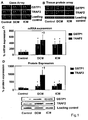
- Figure 1 shows increased GSTP1 and TRAF2 in DCM and ICM patients.
- A Representative gene array images show increased hybridization to GSTP1 and TRAF2 array sequences (arrows) of labelled cDNA probes.
- B Representative protein array images demonstrating enhanced Cy5 (red) staining for cardiac GSTP1 and TRAF2 proteins in DCM and ICM patients compared to the Cy3 (green) staining of controls.
- C Quantification of myocardial GSTP1 and TRAF2 mRNA expressions by real time RT-PCR.
- GSTP1 (*P ⁇ 0.0001) and TRAF2 (P ⁇ 0.0001) mRNA expression is significantly up-regulated in heart failure patients compared to controls.
- FIG. 1 Representative Western blot images and quantification of myocardial GSTP1 and TRAF2 protein expressions corrected for protein loading control levels.
- FIG. 2 shows GSTP1-TRAF2 interaction and activation of JNK and p38 in DCM and ICM.
- A Representative Western blot images and quantification of myocardial GSTP1-TRAF2 complex formation in DCM, ICM and controls. LV myocardial lysates were immunoprecipitated (IP) and precipitates were analyzed on Western blotts. Significantly lower GSTP1-TRAF2 association was found in DCM compared to ICM and controls (*P ⁇ 0.008).
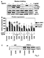
- FIG. 3 shows that GSTP1 modulates cardiac TRAF2-JNK1/p38 system in cardiac tissue cultures.
- A Representative Western blot images of TRAF2, GSTP1, active JNK and active p38 protein expressions in cardiac tissue cultures treated with 5 ⁇ g/mL recombinant GSTP1.
- B Quantification of TRAF2, active JNKs and active p38 protein expressions in DCM, ICM and control cardiac tissue cultures treated with recombinant GSTP1 (2.5, 5, 10 ⁇ g/ml) for 24 h.
- FIG. 4 shows that TNF- ⁇ abrogates sensitivity of TRAF2-JNK1/p38 cascade to GSTP1 in DCM cardiac cultures.
- A Representative Western blot images and quantification
- B Representative Western blot images and quantification
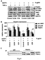
- FIG. 5 shows that higher GSTP1 concentrations rescue TRAF2-mediated JNK1/p38 downregulation following TNF- ⁇ treatment.
- A Representative Western blot images and quantifications (B) of DCM, ICM and control cardiac tissue cultures treated with TNF- ⁇ (50 ng/ml) and GSTP1 (10 ⁇ g/ml).
- B The results demonstrate that DCM cardiac tissue cultures treated with GSTP1 at 10 ⁇ g/mL and TNF- ⁇ express markedly reduced JNK and p38 activation as well as reduced TRAF2 protein expression (*P ⁇ 0.0001) as compared to TNF- ⁇ -treated controls.
- C The immunoprecipitates of DCM cardiac tissue lysates demonstrate significant (P ⁇ 0.035) increase of GSTP1-TRAF2 association at 10 ⁇ g/ml GSTP1 as compared to 5 mg/ml GSTP1.
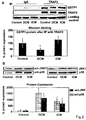
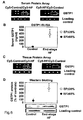
- Figure 6 shows that serum and cardiac GSTP1 is associated with CMP.
- D Representative Western blotting images and quantification of myocardial GSTP1 protein expression corrected for protein loading control levels. GSTP1 protein levels were significantly elevated in end-stage HF patients with EF ⁇ 35% versus control cardiac graft tissue (*P ⁇ 0.001).
- Figure 7 shows serum GSTP1 and proBNP association with ejection fraction (EF) and the correlation between GSTP1 and proBNP.
- EF ejection fraction
- Figure 8 shows serum GSTP1 and proBNP correlation to cardiac function.
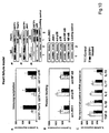
- Figure 9 shows that GSTP1 inhibits the inflammatory cytokines in a rat acute MI model.
- A Immunoprecipitation with Western blotting revealed no differences in GSTP1-TRAF2 complex formation between GSTP1-treated and control animals;
- B GSTP1 decreased activated JNK1 protein expression levels as analyzed by Western blotting;
- C GSTP1 inhibits the myocardial tissue mRNA expression of several inflammatory cytokines. *; p ⁇ 0.01.
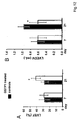
- FIG. 10 shows that GSTP1 ameliorates inflammation in the rat failing heart.
- GSTP1-treated animals had significantly higher levels of complex formation between GSTP1-TRAF2, GSTP1-JNK1 and GSTP1-p38, as shown by immunoprecipitation and Western blot analyses;
- the protein expression of activated JNK1, p38 and NF- ⁇ B is significantly lower in GSTP1-treated animals as compared to controls as shown by Western blot analyses;
- GST-P1 inhibits the mRNA expression of inflammatory cytokines TGF-B, IL-1B, IL-2 and IL-17; and increases the mRNA expression of anti-inflammatory cytokine IL-10 in failing myocardium as compared to controls. *; p ⁇ 0.001.
- Figure 11 shows that GSTP1 attenuates myocardial remodeling in a rat ischemia-induced model of heart failure.
- A Goldner trichrome collagen staining reveals significantly lower tissue remodeling in the heart of GSTP1-treated animals as compared to controls;
- B left ventricular (infarct) wall thickness is significantly larger in GSTP1-treated rats compared to controls;
- GSTP1-treated rats had a significantly lower apoptotic index as compared to controls. *; p ⁇ 0.01.
- Figure 12 shows that GSTP1 improves the left ventricular function in a rat model of ischemia-induced heart failure.
- A GSTP1-treated rats had significantly higher left ventricular ejection fraction at 21 days post-myocardial infarct;
- B the left ventricular end diastolic volume is significantly greater in controls as compared to GSTP1-treated animals. *; p ⁇ 0.05.
- ACE angiotensin converting enzyme
- BMI body mass index
- BSA body surface area
- DCM dilated cardiomyopathy
- ICM ischemic cardiomyopathy
- LVEF left ventricular ejection fraction
- PAP pulmonary artery pressure
- PCWP pulmonary capillary wedge pressure
- PVR pulmonary vascular resistance.
- Poly(A+)-RNA was isolated with the Oligotex-dT kit (Quiagen, Valencia CA) and first-strand cDNA synthesis was performed with avian myeloblastosis virus reverse transcriptase (Promega, Madison, WI) on 2 ⁇ g poly(A+) RNA. The RNA strand within the DNA-RNA duplex was degraded and products were purified on a Sephadex G-50 spun-column (Pharmacia, Uppsala, Sweden).
- first-strand cDNA was used to generate [ ⁇ -32P]dCTP-labeled second-strand cDNA for the cDNA arrays (GEArray Q Human Apoptosis Gene Array, SuperArray Bioscience, Frederick, MD) as described ( Shufer et al., Circulation 108 (2003), 1585-1591 ). Pooled cDNA hybridization signals were quantified using ImageQuant software (Molecular Dynamics, Sunnyvale, CA).
- the protein array was performed on antibody Microarray 500 (Clontech, Mountain View, CA; 507 proteins) as described ( Aharinejad et al., Circulation 120 (2009a), 11 Supp1:S198-205 ).
- the proteins in each sample were labeled with 2 different fluorescent dyes and incubated on microarray antibody-coated slides.
- the protein fluorescent signals of both slides were detected by the GenePix 4000B scanner (Molecular Dynamics, Sunnyvale, CA).
- the quantification of the signal was performed by calculating an internally normalized ratio (INR), yielding the abundance of an antigen in CMP sample relative to that in control samples by using the automated Microarray Analysis Workbook (http://bioinfo.clontech.com). Proteins with INR values outside the threshold interval were considered differentially expressed.
- Human cardiac tissue was lysed in the lysis buffer containing 20 mM Tris (pH 7.5), 135 mM NaCl, 2 mM ethylenediaminetetraacetic acid (EDTA), 2 mM dithiothreitol (DTT), 25 mM ⁇ -glycerophosphate, 2 mM sodium pyrophosphate, 10% glycerol, 1% Triton X-100, 1 mM sodium orthovanadate, 10 mM NaF and 1 mM phenylmethylsulfonyl fluoride (PMSF) supplemented with complete protease inhibitor cocktail (Roche Applied Science, Indianapolis, IN, USA) at 4 C as described (Aharinejad et al., 2005).
- Tris pH 7.5
- EDTA ethylenediaminetetraacetic acid
- DTT dithiothreitol
- PMSF phenylmethylsulfonyl fluoride
- Lysates were centrifuged (15 000 g) at 4 C for 15 min.
- GSTP1-TRAF2 complexes formation proteins 500 ⁇ g were immunoprecipitated with TRAF2 antibody (0.5 ⁇ g, BD Pharmingen, San Diego, CA).
- TRAF2 antibody 0.5 ⁇ g, BD Pharmingen, San Diego, CA.
- the precleared Protein A/G PLUS-agarose beads (Santa Cruz Biotechnology) were incubated with immunocomplexes for another 2 h and washed four times with the lysis buffer.
- the immunoprecipitates were further subjected on SDS-PAGE and presided with Western blotting analysis using anti-GSTP1 monoclonal antibody (Bethyl, Montgomery, TX).
- Protein lysates 50 ⁇ g/lane were separated by SDS-PAGE (10%) prior to electrophoretic transfer onto a Nitrocellulose membrane (Bio-Rad, Hercules, CA) as described (Aharinejad et al., 2005).
- the blots were incubated with primary human monoclonal anti-GSTP1 antibody (Bethyl, Montgomery, TX), human monoclonal anti-TRAF2 (BD Pharmingen, San Diego, CA), human polyclonal phospho-p38 (Promega, Madison, WI), human polyclonal phosphor-JNK (Santa Cruz Biotechnology, Santa Cruz, CA), human polyclonal anti-JNK (Santa Cruz Biotechnology, Santa Cruz, CA), human polyclonal anti-p38 (Santa Cruz Biotechnology, Santa Cruz, CA), prior to incubation with horseradish peroxidase-conjugated secondary antibodies (Amersham Biosciences, Piscataway, NJ).
- primary human monoclonal anti-GSTP1 antibody Bethyl, Montgomery, TX
- human monoclonal anti-TRAF2 BD Pharmingen, San Diego, CA
- human polyclonal phospho-p38 Promega, Madison, WI
- Protein loading was assessed by Ponceau S staining and immunode-tection was performed by chemiluminescence (Supersig-nal-West-Pico, Pierce, Rockford, IL). Bands were quantified by ImageQuant software and specific protein signals were normalized to loading controls and expressed as arbitrary units. The average value of three measurements in each sample was used for data analysis.
- the screening tissue profiling arrays identified higher cardiac GSTP1 and TRAF2 gene as well as protein expression levels in randomly selected patients with both DCM and ICM compared to the control individuals ( Figures 1A, 1B ).
- the myocardial expression of GSTP1 and TRAF2 was then prospectively examined by real time RT-PCR and Western blotting in the study cohort. These analyses indicated significantly elevated myocardial mRNA expression levels in DCM and ICM for both GSTP1 (P ⁇ 0.0001) and TRAF2 (P ⁇ 0.0001) as compared to controls ( Figure 1C ).
- GSTP1-treated cardiac tissue lysates were immunoprecipitated with TRAF2 antibody and immunopellets were subjected to Western blotting.
- TNF- ⁇ abrogates the sensitivity of TRAF2/JNK/p38 cascade to GST-P1 in DCM
- TRAF2 cardiac tissue protein expression in DCM was approximately two times higher as compared to ICM and controls and considering the results described above, we tested the effect of higher GSTP1 concentration following TNF- ⁇ treatment.
- the results demonstrate that DCM cardiac tissue cultures treated with GSTP1 at 10 ⁇ g/mL and TNF- ⁇ at 50 ng/mL express markedly reduced JNK and p38 activation as well as reduced TRAF2 protein expression (P ⁇ 0.0001; Figures 5A, 5B ).
- the immunoprecipitates of DCM cardiac tissue lysates demonstrate significant (P ⁇ 0.035) increase of GSTP1-TRAF2 association at 10 ⁇ g/ml as compared to 5 mg/ml GSTP1.
- TRAF2 is a central regulator of TNF- ⁇ signalling that mediates MAPK activation.
- GSTP1 can abrogate MAPK activation through association with TRAF2 and consequently suppress TNF- ⁇ signalling.
- the present data show significant elevation of active JNK and p38 in DCM myocardium compared to ICM and controls.
- Serum GSTP1 is a sensitive marker of CMP
- the study cohort comprised 141 end-stage CMP patients scheduled for cardiac transplantation and 20 patients with pre-served EF undergoing conventional isolated aortic or mitral valve surgery. All patients underwent echocardiography, coronary artery angiography, and right-heart catheterization for evaluation of ventricular function and standard hemodynamic parameters by two independent cardiologists. The case history, clinical test results, and treatment were documented, coded and blinded to the investigators of serum samples.
- the study patients were subdivided in groups based on their left ventricular EF assessed by echocardiography as follows: EF>52%; EF 52-43%; EF 42-33%, EF 32-23% and EF ⁇ 22% as described ( Lee et al., Circulation 119 (2009) 3070-3077 ).
- LV anterior left ventricular wall
- the protein fluorescent signals of both slides were detected by the GenePix 4000B scanner (Molecular Dynamics, Sunnyvale, CA).
- the quantification of the signal was performed by calculating an internally normalized ratio (INR) using the automated Microarray Analysis Workbook (http://bioinfo.clontech.com).
- Enzyme linked immunosorbent assay for GSTP1 (HEPKITTM-Pi, Biotrin International Ltd., Dublin, Ireland) was performed according to the manufacturer's protocol. The substrate reaction was quantified spectrophotometrically by using a 96-well automated microplate reader (Anthos, Salzburg, Austria) at 450 nm. N-terminal proBNP was measured in undiluted serum automatically by a chemiluminescent noncompetitive ELISA (Roche Diagnostics, Indianapolis, IN) on a Roche Elecsys 2010 analyzer.
- Real-time RT-PCR was performed on a LightCycler instrument (Roche) as described (Aharinejad et al., 2009b, Abraham et al, 2000).
- the primer sequences were sense/antisense: GSTP1: 5'-CCAAAGGTGGTGAGCTTCAT-3'/5'-TCTACCCAGCATGGAGGAAC-3'; and ⁇ 2-microglobulin: 5'-GATGAGTATGCCTGCCGTGTG-3'/5'-CAATCCAAATGCG-GCATCT-3'.
- mRNA expression levels of GSTP1 was normalized to the ⁇ 2-microglobulin signal as a housekeeping gene (Aharinejad et al., 2009b, Abraham et al, 2000). The average value of three PCR measurements in each sample was used for data analysis.
- Tissue lysates were prepared (Aharinejad et al., 2009b) and GSTP1 expression was analyzed by Western blotting using primary human monoclonal anti-GSTP1 antibody (Bethyl, Montgomery, TX). Protein bands were quantified by ImageQuant software and specific protein signals were normalized to loading controls. The average value of three measurements in each sample was used for data analysis.
- GSTP1 and proBNP serum concentrations were compared between patient groups by analysis of variance (one-way ANOVA; Tukey's test).
- PrS Spearman's rank correlation coefficients
- Univariate logistic regression was used to describe the utility of GSTP1 and proBNP as predictors of EF.
- the corresponding receiver operating characteristic curve (ROC) analyses were used to find optimal cut-off levels. Sensitivity and specificity of GSTP1 and proBNP cut-offs were calculated by table analysis. All statistical analyses were performed using SAS system for Windows, version 9.1.3 and the Enterprise Guide, version 4.1 (SAS Institute, Inc., Cary, NC). Statistical significance was set at P ⁇ 0.05. Results are expressed as mean ⁇ standard deviation.
- FIG. 6A shows the array images of GSTP1 and indicates that its serum protein levels are increased in CMP patients as compared to healthy volunteers.
- serum GSTP1 concentration was determined in the same patient cohort selected for screening analyses by GSTP1-specific ELISA. These results show that serum GSTP1 concentrations were significantly upregulated in end-stage HF patients as compared to the control samples ( Figure 6B , P ⁇ 0.001).
- tissue protein profiling was initiated using the LV myocardial samples of the same end-stage HF patients analysed by serum arrays and used LV myocardial biopsies of donated hearts as controls.
- the results of tissue profiling indicate elevated GSTP1 expression levels in myocardium of end-stage HF patients as compared to controls ( Figure 6C ).
- Western blotting analyses were performed on cardiac tissues and found significantly upregulated GSTP1 protein expression levels in patients with end-stage HF as compared to controls (P ⁇ 0.001; Figure 6D ).
- Serum GSTP1 concentrations specifically diagnosed CMP with significant association to EF independently from demographical and clinical characteristics in our patient cohort.
- markedly higher correlation coefficients were shown for serum GSTP1 association to EF as compared to proBNP.
- serum GSTP1 shows better diagnostic power in CMP patients with EF ⁇ 22% as compared to proBNP. More importantly, GSTP1 diagnosed EF ⁇ 42% whereas proBNP failed.
- proBNP is recommended to be mainly used for exclusion of CMP with normal EF in patients with symptoms attributed to CMP.
- gender and older age are associated with higher proBNP levels ( Costello-Boerrigter er al., J. Am. Coll. Cardiol. 47 (2006), 345-353 )
- the proposed diagnostic properties of proBNP might be too unspecific to differentiate CMP with preserved EF in elderly patients.
- proBNP can predict EF ⁇ 30% with a sensitivity and specificity of 90% and 71%, respectively in a CMP population with EF ⁇ 45%.
- Other studies reported that plasma proBNP detect EF ⁇ 28% with 77% sensitivity and 69% specificity in a patients cohort with EF ⁇ 50%, and it was found that proBNP can predict EF ⁇ 40% with an area under the curve of 0.69.
- proBNP had 97% sensitivity but only 26% specificity to diagnose EF ⁇ 22% with an area under the curve of 0.62.
- the lower specificity of proBNP in the present study could be explained by the fact that our cut-off level was selected to identify lower EFs as compared to other studies.
- GSTP1 is a sensitive, specific, cheap and quickly measurable serum marker superior to proBNP in CMP. Importantly, GSTP1 discriminates between preserved and reduced EF with a high sensitivity and specificity and can therefore serve as a novel tool in guiding of clinical trials in CMP patients.
- the model of ligation of the anterior branch of the left coronary artery in the rat has been selected to further prove the principle of the present invention.
- IHD is induced following the ligation of the said coronary artery branch, and over time the animals will subsequently develop CMP. Therefore, the model is best suited to show the efficacy of the suggested invention for prevention or treatment of both IHD and CMP.
- mice Male, Sprague Dawley rats (Harlan, Borchen, Germany) will be coded for experiments. Due to expected hemodynamic compromise, ventricular fibrillation, myocardial rupture and bleeding following myocardial infarction (MI), the number of surviving animals is expected to be about 60-70%. Therefore, in each group with myocardial infarction, a total of 18 animals will be included.
- MI myocardial infarction
- Rats will be anesthetized with intraperitoneal injection of Ketasol (100 mg/kg body weight) and Rompun (10 mg/kg body weight), intubated tracheally using a 14-gauge catheter and ventilated with 1.5% Isofluran at 55 cycles per minute (tidal volume: 2.5 ml) before undergoing a left lateral thoracotomy.
- the anterior branch of the left coronary artery (LCA) will be either ligated with a 7-0 polypropylene snare (Ethicon, Somerville, NJ) or left intact in sham procedure.
- LCA left coronary artery
- LV left ventricular
- mice with comparable base-line cardiac function will be coded and assigned to groups 1-8 as shown in Table 4.
- animals with comparable base-line cardiac function will be coded and assigned to groups 1-8 as shown in Table 4.
- animals with comparable base-line cardiac function will be coded and assigned to groups 1-8 as shown in Table 4.
- animals will receive immediately after MI induction and on the first day post MI 0.6 mg/100 g body weight Piritramid (Dipidolor ® in 5% glucose solution) s.c. Further, animals will receive Piritramid via their drink water for 3 days at 0.6 mg/100 g body weight (250 ml water plus 20 ml 5% glucose solution) and at days 4-7 post MI at 0.3 mg/100 g body weight.
- Piritramid Dipidolor ® in 5% glucose solution
- treatment On d7, immediately after MI induction, treatment will start in groups 1-4 as follows. Group 1 will receive daily intraperi-toneal injections of Ringer's solution for two weeks (control), while groups 2-4 will receive daily injections of recombinant GSTP1 at a dose of 10, 20 and 40 mg/body weight for two weeks, respectively. On d21 the treatment will start in groups 5-8 as follows. Group 5 will receive daily intraperitoneal injections of Ringer's solution for two weeks (control), while groups 6-8 will receive daily injections of recombinant GSTP1 at a dose of 10, 20 and 40 mg/body weight for two weeks, respectively. Group 9 includes 10 animals with sham procedure.
- Table 4 Groups of animals and their treatment Group Treatment Sacrifice No. of animals Application and dose 1 Ringer d88 18 i.p. 2 GSTP1 d88 18 i.p., 10 mg/kg bw, 2 weeks 3 GSTP1 d88 18 i.p., 20 mg/kg bw, 2 weeks 4 GSTP1 d88 18 i.p., 40 mg/kg bw, 2 weeks 5 Ringer d88 18 i.p.
- LV ejection fraction (EF), LV end systolic volume (ESV), LV end diastolic volume (EDV) using echocardiography 2.
- Heart-to-body weight ratio 3. Histology and immunocytochemistry for infarct size, infarct wall thickness and fibrosis as well as angiogenesis, inflammatory proteins including p38, june kinase and interleukins (only examples are mentioned for inflammatory pathways) 4.
- Signaling pathways 5.
- Promoter binding assays 6. TUNEL-assay for apoptosis evaluation 7.
- LVEDV LV end-diastolic
- LVESV end-systolic
- LVEF LVEDV-LVESV/LVEDV
- ventricles were cross-sectioned at the midpoint of their long axis, before they were either frozen or immersion-fixed in formalin. Paraffin-embedded specimens were used for H&E, Tunnel assay as well as Goldner trichrome collagen staining. TUNEL assays (In situ Cell Death Detection Kit) were performed according to the manufacturer's protocol (Roche Molecular Biochemicals, Basel, Switzerland) in triplicate. Slides were counterstained with 4,6-diamidino-2-phenylindole (DAPI, Molecular Probes, Eugene, OR) and embedded in AF1 antifadent (Citifluor, Sheffield, UK). Digital images were obtained by fluorescence microscopy (Nikon, Melville, NY). Morphometry was carried out as described (Aharinejad et al., 2008).
- the primer sequences were sense/antisense: GSTP1: 5'-GGCAACTGAAGCCTTTTGAG-3'/5'-TCATGGATCAGCAGCAAGTC-3'; tumor necrosis factor receptor-associated factor 2 (TRAF2): 5'-GCAGAAG-GTCTTGGAGATGG-3'/5'-GGTGGAGCAGCATTAAGGTC-3'; and ⁇ 2-microglobulin: 5'-GATGAGTATGCCTGCCGTGTG-3'/5'-CAATCCAAATGCG-GCATCT-3'. mRNA expression were calculated and plotted for treated animals as percentage relative to that in controls.
- TRF2 tumor necrosis factor receptor-associated factor 2
- ⁇ 2-microglobulin 5'-GATGAGTATGCCTGCCGTGTG-3'/5'-CAATCCAAATGCG-GCATCT-3'.
- the blots were incubated with primary human monoclonal GSTP1 (Bethyl) and TRAF2 (BD Pharmingen) or polyclonal phospho-p38 (Promega, Madison, WI), phospho-JNK1, JNK1 and p38 (Santa Cruz Biotechnology) antibodies prior to incubation with secondary antibodies (Amersham Biosciences, Piscataway, NJ). All experiments were performed in triplicate and expression levels were corrected for control loading protein.
- GSTP1 ameliorates the myocardial overexpression of inflammatory cytokines following acute MI
- GSTP1 ameliorates the myocardial expression of pro-inflammatory cytokines through TRAF2-regulated NF-kB signaling in the failing myocardium
- GSTP1-treated rats had significantly increased formation of GST-P1-TRAF2, GSTP1-JNK1 and GSTP1-p38 complexes in their failing cardiac tissue at 3 weeks post-MI as compared to controls (p ⁇ 0.001; Figure 10A ).
- GSTP1-treated rats had significantly reduced protein expression of activated JNK1 and p38 in their failing myocardial tissue as compared to controls (p ⁇ 0.001; Figure 10B ).
- GSTP1-treated rats had lower cardiac tissue mRNA expressions of TGF- ⁇ , IL-1 ⁇ , IL-2 and IL-17 as compared to controls ( P ⁇ 0.001; Figure 10C ).
- GSTP1 ameliorates remodeling in rat failing myocardium
- TNF- ⁇ The role of the inflammatory mediators in the patho-physiology of heart failure has gained a growing interest over the last two decades. Different experimental studies have described the negative inotropic effect of TNF- ⁇ on the LV function. Moreover, it has been reported that TNF- ⁇ promotes LV remodeling, cardiac hypertrophy and progressive cardiomyocytes loss through apoptosis. The growing evidence on the pathological role of inflammatory mediators in the setting of heart failure have resulted in a series of multicenter clinical trials that intended to target TNF- ⁇ in heart failure patients. However, the results of these trials were discouraging. One explanation of these results is that the low physiological levels of TNF- ⁇ might be important for cardiac tissue remodeling and repair.
- TRAF2 is a central regulator of TNF- ⁇ signaling that mediates MAPK activation.
- TRAF2 results in activation of NF- ⁇ B.
- GSTP1 is an important negative regulator of TNF- ⁇ -induced signaling by forming interactions with TRAF2.
- GSTP1 could inhibit MAPK activation in malignant cell lines indirectly through its interaction with TRAF2 or directly through its interaction with JNK1 and P38.
- GSTP1 was not only able to suppress the pro-inflammatory mediators like; IL-1, IL-2, JNK1, P38 and NF- ⁇ B but also could increase the expression of the anti-inflammatory cytokine IL-10.
- the results according to the present invention revealed that GSTP1 protects the cardiac tissue against apoptosis in the failing heart. In concert with this, it has been shown in oncological settings that GSTP1 attenuates the autophosphorylation of TRAF2-enhanced apoptosis signal-regulating kinasel (ASK1) and thus inhibits TRAF2-ASK1-induced cell apoptosis by suppressing the interaction of TRAF2 and ASK1.
- ASK1 TRAF2-enhanced apoptosis signal-regulating kinasel
- the present invention provides a novel function of GSTP1 in modulating TNF- ⁇ /TRAF2 elicited JNK1/p38 activation in heart failure.
- the selective inhibition of TNF- ⁇ /TRAF2-mediated inflammatory signaling by GSTP1 was due to the present animal model shown to be a beneficial treatment for patients with cardiomyopathies or ischemic heart diseases, especially for patients with myocardial infarction and heart failure.
Description
- The present invention relates to the use of Glutathione-S-transferase P1.
- The glutathione S-transferases (GSTs) are a multigene family of isozymes that catalyze the nucleophilic attack of the sulfur atom of glutathione (GSH) on electrophilic groups of substrate molecules. GSTs are known as detoxicating enzymes which conjugate toxic substances with GSH to become more water-soluble products which can be metabolised in the liver and finally excreted out of the body. On the basis of the amino acid sequence, the mammalian GSTs are divided into six classes: alpha, mu, omega, pi, theta and zeta. Among these isozymes, glutathione S-transferase P1 (GSTP1; GST-pi, GSTP1-1) is the most prevalent one in mammalian cells. GSTP1 has also generated interest as a neoplastic marker because it is overexpressed in a variety of different human malignancies, including lung, colon, stomach, esophagus, mouth, kidney, ovary and testicular cancers. Most prostate cancer types contain decreased levels of GSTP1 polypeptide relative to normal prostate tissue due to hypermethylation of the GSTP1 promoter resulting in decreased transcription of GSTP1 (
US 2009/0186360 A1 ). GSTP1 is determinant in cellular response to oxidative stress and protects tumor cells from apoptosis elicited by a variety of cytotoxic agents, such as H2O2, UV, cisplatin, adriamycin, etoposide, thiotepa, chlorambucil, ethacrynic acid and arsenic trioxide. GSTP1 is therefore considered as a promising target in tumour medicine, both as a target for drug action and as a tumor marker. - GSTP1 also participates in the regulation of stress signalling and protects cells against apoptosis by the mechanisms related to its non-catalytic and ligand-binding activities. For example, GSTP1 prevents LPS-induced excessive production of pro-inflammatory factors and plays an anti-inflammatory role in response to LPS. GSTP1 expression, both at transcriptional and translational levels, is up-regulated by LPS stimulation. GSTP1 also functions as an endogenous inhibitor of JNK (c-Jun NH2-terminal kinase), via interaction with the C-terminus. It could also be demonstrated that exogenous (recombinant) GSTP1 protein could be delivered into macrophages and suppressed iNOS and COX-2 expression in cells. Furthermore, intraperitoneally administered GSTP1 protein to mice decreased mortality of endotoxic shock significantly and inhibited acute lung injury and peritonitis (Luo et al., Mol. Immunol. 46 (2009), 848-857).
-
EP 1 500 709 A1 - Various isoforms of GSTP1 have also been found relevant to asthma and childhood allergic diseases due to the interaction of GSTP1 with air pollution from traffic (and tobacco smoke) in the respiratory system, as well as association with hypertension in pregnancy (Ohta et al., Sem. Thr. Hem. 29 (2003), 653-659).
- Cardiomyopathies (CMP) are a heterogeneous group of diseases, including the dilative (DCM), the most frequent entity among cardiomyopathies, and ischemic (ICM) forms. Although pharmacotherapy and surgical interventions have been improved with survival benefit in CMP, causal treatment remains to be a vague goal. Therefore, identification of unknown mechanisms that mediate CMP could be beneficial. Results of the last decade's work clearly provide evidence that inflammation and enhanced release of pro-inflammatory cytokines, especially tumor necrosis factor (TNF)-α might play a role in the pathogenesis of CMP. However, clinical trials have paradoxically indicated a more complicated role for TNF-α in CMP that casted doubts whether TNF-α is a viable therapeutic target for CMP. The pathology of IHD is immediately related to the development of CMP, patients currently receive medications such as beta-blockers or angiotensin converting enzyme (ACE)-blockers with the goal to ameliorate the patient's symptoms and to prevent or delay the development of (ischemic) CMP due to IHD. It is therefore common to use therapies for CMP also for IHD. Importantly, patients who develop CMP based on IHD, in particular end-stage CMP, have currently only limited therapeutic options (e.g. cardiac transplantation or the use of ventricular assist devices, which can be in turn offered only to a limited number of patients).
- There is still an unmet need for further drug targets and treatment regimen for efficiently preventing and treating CMP and IHD and one of its major causes, that is ischemic heart diseases (also called coronary heart diseases that can result in myocardial infarction), especially elevated blood pressure-induced CMP and volume-induced CMP.
- Cardiomyopathies result in recurrent exacerbation leading to hospitalization or death. Therefore, also close surveillance and thorough clinical tests are required to achieve the optimal tailored cardiomyopathy treatment in these patients, associated with tremendous health care costs. A biomarker that could identify the evolution of cardiomyopathy and help to optimize treatment monitoring and outcome is therefore highly desirable. Ideally, such a test should be sensitive, specific, none-invasive, quick and cheap. Although, a number of neurohumoral factors and inflammatory cytokines have been reported as diagnostic biomarkers in cardiomyopathy, brain-type natriuretic peptide (BNP) and its inactive N-terminal fragment (proBNP) have gained the broadest acceptance in clinical diagnosis and monitoring of patients with suspected or established chronic CMP. However, circulating BNP and proBNP levels are dramatically affected by renal function and are age- and gender-dependent. In addition, it remains unknown whether proBNP could be beneficial in diagnosis of preserved as opposed to reduced ejection fraction (EF) CMP.
- It is therefore also an object of the present invention to provide diagnostic means for cardiomyopathies.
- Therefore, the invention provides Glutathione S-transferase P1 (GSTP1) for use in the prevention or treatment of cardiomyopathies (CMPs). With the present invention it could be surprisingly shown that GSTP1 is beneficial in CMP treatment. GSTP1 has been described as a relevant protein in cancer medicine (e.g.
US 5,427,917 A ,US 5,552,277 A andWO 98/21359 A1 - According to the present invention it was surprisingly found that GSTP1 is specifically upregulated in CMP patients as compared to controls. It was further found that recombinant GSTP1 mediated GSTP1-TRAF2 association, decreased pro-inflammatory JNK1 and p38 activation and TRAF2 expression dependent on its dose and the underlying form of CMP. This enabled the provision of a new therapy strategy for CMP by administration of GSTP1. Since GSTP1 ameliorates TNF-α-mediated activation of pro-inflammatory JNK1/p38 by association to TRAF2, GSTP1 is not only beneficial in CMP treatment but also for the prevention of CMP, especially in risk patients, such as hypertensive patients.
- The present invention provides the use of GSTP1 for the manufacture of a medicament for the prevention or treatment of CMP. The treatment of CMP according to the present invention involves the administration of an effective amount of GSTP1 to an individual in need of such treatment, e.g. human CMP patients. For these reasons and based on the fact that the pathology of IHD is immediately related to the development of CMP, patients currently receive medications such as beta-blockers or angiotensin converting enzyme (ACE)-blockers with the goal to ameliorate the patient's symptoms and to prevent or delay the development of (ischemic) CMP due to IHD. The close relationship of the pathology of IHD with CMP that is based on the remodelling of myocardium in patients with IHD, specially in patients developing myocardial infarction due to IHD that in turn leads to remodelling of the cardiac chambers and subsequently to impaired pump function of the heart, a pathological condition that is common to CMP and IHD, specially in myocardial infarction patients, shows the effectiveness of the present invention for the prevention or treatment of CMP.
- With the present invention all forms and stages of CMP, acute and chronic forms thereof, can be treated, independently of the genesis of the CMP. Of course, the term "treatment" includes preferentially the amelioration of the disease and the prevention or slowing of progression of the disease. For example, CMPs can be caused by myocardial infarction, can be idiopathic dilative or be caused by hypertension. All these forms can be treated with GSTP1 according to the present invention. Specifically ischemic CMPs e.g. caused by myocardial infarction can be treated with GSTP1, however, also non-ischemic forms can be treated (e.g. idiopathic dilated CMPs or hypertension- or volume-induced CMPs). Since in all these CMPs, inflammatory pathways are involved, this makes them eligible for treatment with GSTP1 according to the present invention (even rare forms of CMP, if such inflammatory pathways are involved).
- Therefore, preferred CMPs according to the present invention are dilated cardiomyopathy, especially congestive cardiomyopathy; obstructive hypertrophic cardiomyopathy, especially hypertrophic subaortic stenosis; other hypertrophic cardiomyopathies, especially nonobstructive hypertrophic cardiomyopathy; endomyocardial (eosinophilic) disease, especially endomyocardial (tropical) fibrosis or Löffler's endocarditis; endocardial fibroelastosis, especially congenital cardiomyopathy; other restrictive cardiomyopathies, especially constrictive cardiomyopathy NOS (not otherwise specified (according to ICD-10)); alcoholic cardiomyopathy, cardiomyopathies due to drugs and other external agents, unspecified cardiomyopathies, especially cardiomyopathy (primary)(secondary) NOS; cardiomyopathy in infectious and parasitic diseases, especially cardiomyopathy in diphtheria; cardiomyopathy in metabolic diseases, especially cardiac amyloidosis; cardiomyopathy in nutritional diseases, especially nutritional cardiomyopathy NOS; Gouty tophi of heart or thyrotoxic heart disease.
- Specifically preferred forms of CMP to be treated (or prevented) according to the present invention are CMPs caused by ischemia, especially by myocardial infarction; by hypertension, or by myocarditis. GSTP1 treatment according to the present invention is therefore preferably applied in patients with acute myocarditis, preferably infective myocarditis, especially septic myocarditis; isolated myocarditis, myocarditis in a bacterial disease, especially diphtheritic, gonococcal, meningococcal, syphilitic or tuberculous myocarditis; myocarditis in a viral disease, especially influenzal myocarditis or mumps myocarditis; acute or chronic myocarditis in Chagas' disease, myocarditis in toxoplasmosis, rheumatoid myocarditis or sarcoid myocarditis.
- The preferred indications according to the present invention are therefore those listed under Chapter IX of the ICD-10 (I11-I15, especially I21, I22 and I23; I20-I25, especially I21 and I22; I40-I43, especially I42; I50-I57, especially I50 and I57).
- The main focus of the present invention is the treatment of human patients; however, based on the present findings, it is clear that also animal (mammal) CMP in which inflammation is involved can be successfully treated by GSTP1 according to the present invention. This may be important for farm or zoo animals (especially breeding farm animals, such as (former) racing horses) or pets, for example dogs, cats and horses.
- Although the present invention is also useable for the prevention of CMP it is clear that the primary focus of the present invention lies in the treatment of human CMP patients who have already been diagnosed of CMP or which have a high risk of developing CMP, for example patients with hypertension (
e.g. Stage 2 patients with systolic pressure of 160 mm Hg or more and/or with diastolic pressure of 100 mm Hg or more). Another preferred embodiment of the preventive aspect of the present invention is for angina pectoris patients, whereby myocardial infarction might or might not have occurred in this set of patients; and heart insufficiency or heart failure (Mc Murray et al., Lancet 365 (2005), 1877-1889) may or may not be present. Here, even continuous intravenous administration or continuous administration to the heart muscle is indicated. - Preferred routes of administration of the GSTP1 containing medicament according to the present invention are parenteral routes, preferably intraperitoneal or intravenous administration, intravenous administration being specifically preferred. Intravenous administration can be performed e.g. via bolus injection or by continuous intravenous delivery over a longer time period (e.g. 30 min to 6 h, especially 1 to 3 h). In the process of heart surgery also administration directly to the heart (in-tramyocardial injection) is preferred. With this route, GSTP1 activity can be directly and in high concentrations delivered to the area of need, e.g. to an infarct region. Another preferred route of administration is administration to the coronary sinus (e.g. via a catheter inserted into the coronary sinus). Oral or mucosal routes (although described for GSTP1 in principle; see e.g.
US 5,976,528 A ) are less preferred, since GSTP1 is a protein and for this route specific protection measures (enteric coating, encapsulation, etc.) are necessary as well as a significant optimisation with respect to galenic manufacture. - Preferred dosages for administration are dosages of 0.001 to 100 mg GSTP1/kg, preferably 0.01 to 10 mg GSTP1/kg, especially 0.1 to 1 mg GSTP1/kg, to a human individual, preferably via intravenous administration; or dosages of 0.1 to 10000 U GSTP1/kg, preferably 1 to 1000 U GSTP1/kg, especially 10 to 100 U GSTP1/kg, to a human individual, preferably via intravenous administration. These are preferred daily dosages for intravenous application ("kg" means kg body weight of the person to be treated). During surgery, these dosages may also be applied, either as a whole or even more than one dosage; or only part of the dosage (having in mind that the GSTP1 can directly be applied during surgery in the area needed). "Ready-to-use" daily dosage forms (e.g. for a 80 kg and/or a 60 kg human individual) can be pre-manufactured and can be made storage-stable by lyophilisation or freezing (and keeping at e.g. -20°C). The dosage can then be fully or only partially consumed (e.g. based on the body weight of the person to be treated).
- GSTP1 according to the present invention is also referred to as "GSTP1-1", "GST class-pi", "DFN7", "GST3", "GST π", etc. and conjugates reduced glutathione to a wide number of exogenous and endogenous hydrophobic electrophiles (EC 2.5.1.18; Catalytic activity: RX + glutathione = HX + R-S-glutathione). The human sequence (Seq. ID. No. 1) is listed as "GSTP1_HUMAN, P09211" in the UniProtKB/SwissProt database (HGNC: 4638, Entrez Gene: 2950).
-

- Several variants exist for this human protein (homozygous as well as heterozygous), the most important variants are a Ile to Val exchange at position 105 and a Ala to Val exchange at position 114 (Melen et al., 2008). Other variants include a Gly to Glu exchange at position 78, a Thr to Ser exchange at position 110, a Gly to Glu exchange at position 139, a Asp to Tyr exchange at position 147 and a Asp to His exchange at position 158. Several other SNPs are known without affecting the amino acid sequence.
- If the CMP patient to be treated with GSTP1 according to the present invention has such an allele which results in a (heterozygous or homozygous) amino acid exchange compared to SEQ.ID.No. 1, it may be recommendable to administer to such a patient the allelic form of this protein. This is specifically preferred for patients who are homozygous for this allele, e.g. patients with homozygous 105Val or 114Val allele (who could then receive the 105Val or 114Val GSTP1 variant, respectively). In such cases, the risk of developing adverse reactions, especially adverse immune reactions (which is always a topic for proteinaceous medicaments), is kept low. Anyway, any ingredients in the pharmaceutical preparation containing GSTP1 according to the present invention eliciting or boosting an immune reaction in the patient should be kept at minimum if possible.
- Orthologs from mammals are known, e.g. from chimpanzee (Pan troglodytes; NCBI accessions: 745954, XM_001152516.1, XP_001152516.1) and mouse (Mus musculus; NCBI accessions: 148701, NM_013541.11, NP_038569.11, AK0791445, BC0020485), but also from non-mammals, such as zebrafish (Danio rerio) and worm (Caenorhabditis elegans).
- The present invention also relates to a pharmaceutical composition comprising GSTP1, preferably human recombinant GSTP1 or human placenta GSTP1, and a pharmaceutically acceptable carrier for the prevention or treatment of cardiomyopathies or ischemic heart diseases.
- According to a preferred embodiment, the composition according to the present invention contains 1 to 100000 U GSTP1, preferably 10 to 10000 U GSTP1, especially 10 to 1000 U GSTP1. A unit of GSTP1 activity conjugates 1.0 micro-mole of 1-chloro-2,4-dinitrobenzene with reduced glutathione per minute at pH 6.5 at 25°C.
- A preferred composition according to the present invention also contains a buffer. Typical buffer systems are the carbonic acid/HCO3 system (
pH pH 5,0 to 6,2; weakly acidic), acetic acid/acetate buffer (pH pH 5,4 to 7,8), ammonia buffer (NH3 + H2O + NH4Cl;pH 8,2 to 10,2), TRIS/HCl (Tris(hydroxymethyl)-aminomethane;pH pH 6,8 to 8,2), HEPPS (4-(2-Hydroxyethyl)-piperazine-1-propane sulfonic acid;pH pH 5,2 to 6,7). Preferred buffers according to the present invention is a phosphate buffer, a Tris-HCl buffer or a HEPES buffer, with a pH of 5.5 to 9.0, preferably of 6.0 to 8.5, especially of 6.5 to 8.0. Buffer concentrations can preferably be adjusted to 1 mM to 1 M, especially 10 mM to 0.5 M. Other preferred additional ingredients include stabilisers, chelators, salts, etc., such as glycerol, glucose, saccharose, maltose, EDTA or similar substances, NaCl, KCl, NH4Cl and similar salts usable in pharmaceutical preparations. Of course, all ingredients in a pharmaceutical preparation have to be of pharmaceutical grade quality. - Pharmaceutically acceptable carriers preferably used are physiological saline, vegetable oils, mineral oil, aqueous sodium caroboxymethyl cellulose or aqueous polyvinylpyrrolidone; however, also sterile water can be used.
- GSTP1 to be used according to the present invention can be provided by various ways. As mentioned above, GSTP1 from human placenta is one of the preferred natural sources of GSTP1. Other preferred sources are cultures of GSTP1 (over-)expressing cells. GSTP1 preparations are also commercially available. The preferred industrial production route according to the present invention is, however, recombinant production of GSTP1. Recombinant production of GSTP1 is well established, as well as its purification from such sources (e.g. Luo et al., 2009, Wu et al., 2006;
WO 98/21359 A1 US 5,976,528 ; etc.). Expression vectors (e.g.WO 98/21359 A1 pages 40 to 51) containing the GSTP1 coding sequences are transferred in suitable host cells (such as COS, HEK 293, HeLa, VERO, W138, BHK or MDCK cells, but also yeast, plant, insect and bacterial cells (S. cerevisiae, E. coli, etc.). GSTP1 is then expressed and purified according to standard methods. Recombinant GSTP1 production may also use variant forms of GSTP1 with additional sequences which facilitate purification, such as metal chelating peptides, protein A domains, affinity purification tags, etc. (e.g.US 5,976,528 A , column 16). Such variants can - after purification or in the course of purification - be removed e.g. via cleavable linker sequences (protease, entereokinase, etc.). Purified batch preparations of GSTP1 may contain e.g. 1 to 200 U GSTP1 activity per mg total protein. Preferred pharmaceutical GSTP1 compositions contain 5 to 200 U/mg protein, especially 10 to 150 U/mg protein. The pharmaceutical preparation according to the present invention is always provided as an appropriately labelled product which is sterile and useable for administration to human patients according to the present invention. - According to another aspect, the present invention relates to GSTP1 as a diagnostic target for CMP. The present invention provides the use of GSTP1 for the diagnosis of CMP. Although GSTP1 is a known and established marker in tumor diagnosis, it was surprising that GSTP1 is a biomarker for CMP which is even superior to the current "gold standard", pro-BNP, with respect to sensitivity and specificity.
- The cardiomyopathies referred to above for therapeutical use of GSTP1 can also be diagnosed according to the present invention.
- The present invention therefore relates to a method for diagnosing CMP, especially the indications mentioned above, by determining the amount of GSTP1 or GSTP1 mRNA in a biological sample and comparing this amount to the GSTP1 or GSTP1 mRNA amount in a biological sample with a defined and known CMP status, especially from a healthy human individual or a patient. This comparison can be done practically, i.e. in that a real sample is tested in the same manner as the biological sample (side-by-side), it can also be a virtual comparison so that the determined value is compared with a known value of a healthy or diseased sample.
- Preferred techniques for determining the amount of GSTP1 include antibody-liked techniques, such as ELISA, optionally combined with electrophoresis techniques or radio immunoassay (RIA). Electrophoresis techniques may also be combined with MALDI techniques.
- Alternatively, GSTP1 mRNA may be determined, e.g. by quantitative real time RT-PCR.
- The preferred diagnosis target is a human patient who is suspected to have CMP, a human patient who is at risk for developing CMP or a human patient who has CMP (who has to be monitored for progression of the disease).
- Preferred biological samples according to the present invention include human blood, plasma or serum samples or human myocardial biopsy samples. These samples can also be tested by routine methods.
- Typically, a high concentration of GSTP1 in serum is indicative of CMP. In healthy persons, serum concentrations of GSTP1 are below 50 ng/ml or, preferably, even below 10 ng/ml. A CMP diseased status can be diagnosed at a serum level at 50 ng/ml or above, preferably at 100 ng/ml or above. Severe forms of CMP can be diagnosed at 200 ng/ml or above. Of course, these levels always depend on the technique used for assaying the GSTP1 protein. Alternatively, CMP diseased status can be diagnosed if the serum level of GSTP1 is elevated by at least the
factor 2, preferably by at least the factor 5, compared to a healthy status, especially the healthy status of the same patient. Severe forms could be diagnosed, if the serum GSTP1 level is elevated by at least thefactor 10, especially by at least thefactor 20, compared to a healthy status, especially the healthy status of the same patient. - With the present invention determining the amount of GSTP1 or GSTP1 mRNA in a sample can be performed within a very short time by standard test methods, such as ELISA or PCR. Based on the DNA and amino acid sequence according to SEQ.ID.NO.2and SEQ.ID.NO.1 (and the data base entries mentioned above for the GSTP1 gene and the GSTP1 protein), the present invention also provides e.g. a PCR assay for identification of S. aureus infections or a sandwich ELISA assay. In a suitable ELISA according to the present invention, GSTP1 can be caught out of a biological sample with a GSTP1-specific antibody and detected and quantified with a second antibody directed against a different epitope of GSTP1 or an antibody being specific for the previous binding event.

- Detection of GSTP1 transcripts (m-RNA) is possible by standard quantitative nucleic acid tests, such as quantitative RT-PCR. The present invention therefore also provides a kit for determining the amount of GSTP1 mRNA in a least two synthetic nucleic acid sequences for amplifying at least one nucleic acid sequence encoding GSTP1 or parts thereof, wherein at least of one of the synthetic nucleic acid sequences in the kit is selected from the group consisting of: (a) a suitable primer pair, the primers being a nucleic acid sequence comprising 10-30 consecutive nucleotides of at least one of the following: (i) the GSTP1 mRNA according to SEQ.ID.NO.2 or the complementary sequence thereof; or (ii) from the 5' or 3' noncoding region of the GSTP1 gene (e.g. in regions extending up to 500, preferably up to 200, especially up to 100, nucleotides into the 5'- or 3-direction or complementary sequences thereof; such sequences are present in the sequence library entries mentioned above); said primers being usable for polymerising a nucleotide sequence located between the primers on the GSTP1 gene or its 5'- or 3'-region (said polymerizable GSTP1 nucleotide sequence being PCR detectable and preferably up to 1000 bp, more preferred up to 500 bp, even more preferred up to 300bp in length; on the other hand, the primers may also be selected to polymerise (with PCR) the whole GSTP1 m-RNA); and (b) suitable reagents for a polymerase chain reaction (PCR). Preferably, the kit further comprises: (c) instructions for use and (d) optionally, a positive and/or negative control for determining and/or quantification of GSTP1 mRNA, especially a control nucleic acid encoding GSTP1 or a fragment thereof. Examples of suitable PCR reagents include PCR reaction buffer, Mg2+ (e.g., MgCl2), dNTPs, DNA polymerases (such as reverse transcriptases and thermostable DNA polymerases (e.g., Taq-related DNA polymerases and Pfu-related DNA polymerases)), RNase, PCR reaction enhancers or inhibitors, PCR reaction monitoring agents (e.g., double-stranded DNA dye (such as SYBRTM Green), TaqManTM probes, molecular beacons, and Scorpion-sTM), and PCR-grade water.
- The primers described herein are particularly useful in a polymerase chain reaction (PCR) assay. PCR is a practical system for in vitro amplification of a DNA base sequence. For example, a PCR assay may use a heat-stable polymerase and two about 10 to 30 base primers: one complementary to the (+)-strand at one end of the sequence to be amplified, and the other complementary to the (-)-strand at the other end. Because the newly-synthesized biological sample, comprising: (a) a primer set comprising at DNA strands can subsequently serve as additional templates for the same primer sequences, successive rounds of primer annealing, strand elongation, and dissociation may produce rapid and highly-specific amplification of the desired sequence. PCR also may be used to detect the existence of a defined sequence in a GSTP1 m-RNA containing sample.
- By way of example, a typical PCR assay according to the present invention might start - optionally after reverse transcription of the mRNA into DNA - with two synthetic oligonucleotide primers which are specifically and complementarily binding to two regions of the target DNA or its complementary strand, respectively, encoding GSTP1 or its 5'- and/or 3' region (one for each strand) that is to be amplified. These may be added to the target DNA (that needs not be pure) in the presence of excess deoxynucleotides (dNTPs) and a thermostable DNA polymerase (e.g., Taq polymerase). In a series (typically 20-40) of temperature cycles, the target DNA may be repeatedly denatured (about 80-100°C, e.g. 90°C), annealed to the primers (typically at 40-65°C), and a daughter strand may be extended from the primers (typically at 65-80°C, e.g. 72°C). As the daughter strands themselves act as templates for subsequent cycles, DNA fragments matching both primers are amplified exponentially, rather than linearly. The target DNA needs be neither pure nor abundant; thus, PCR is specifically suitable in the clinical diagnostics according to the present invention.
- These tests may preferably employ labels which are also suitable for quantification, such as biotin, fluorescent molecules, radioactive molecules, chromogenic substrates, chemiluminescence markers, and the like. The methods for biotinylating nucleic acids are well known in the art, as are methods for introducing fluorescent molecules and radioactive molecules into oligonucleotides and nucleotides. Detection methods are well known for fluorescent, radioactive, chemiluminescent, chromogenic labels, as well as other commonly used labels. Briefly, chemiluminescence can be identified and quantitated most directly by their emission wavelengths and intensity. When biotin is employed, it is detected by avidin, streptavidin or the like, which is conjugated to a detectable marker, such as an enzyme (e.g. horseradish peroxidase). Steptavidin binds with high affinity to biotin, unbound streptavidin is washed away, and the presence of horseradish peroxidase enzyme is then detected using a luminescence-emission substrate in the presence of peroxide and appropriate buffers.
- Determining the amount of GSTP1 in a sample preferably includes ELISA, RIA and FACS. Detection and quantification of GSTP1 by using anti GSTP1 antibodies is well established in the art (especially in tumor medicine) and can be readily applied for the purpose of diagnosing CMP according to the present invention (e.g.
WO 98/21359 A1 US 5,976,528 A ,US 5,427,917 A ). Normal or standard values for GSTP1 expression are established by combining body fluids or cell extracts taken from normal mammalian subjects, preferably human, with antibody to GSTP1 under conditions suitable for complex formation. The amount of standard complex formation may be quantified by various methods, preferably by photometric means. Quantities of GSTP1 expressed in subject, samples, control and disease, from biopsied tissues are compared with the standard values. Deviation between standard and subject values establishes the parameters for diagnosing disease. As already stated above, most preferred samples are of human heart biopsies and samples derived from human blood of individual human patients having CMP, being suspected of having CMP or IHD or being at risk of developing CMP. - The present invention is further illustrated by the following examples and the drawing figures, yet without being restricted thereto.
-
Figure 1 shows increased GSTP1 and TRAF2 in DCM and ICM patients. (A) Representative gene array images show increased hybridization to GSTP1 and TRAF2 array sequences (arrows) of labelled cDNA probes. (B) Representative protein array images demonstrating enhanced Cy5 (red) staining for cardiac GSTP1 and TRAF2 proteins in DCM and ICM patients compared to the Cy3 (green) staining of controls. (C) Quantification of myocardial GSTP1 and TRAF2 mRNA expressions by real time RT-PCR. GSTP1 (*P<0.0001) and TRAF2 (P<0.0001) mRNA expression is significantly up-regulated in heart failure patients compared to controls. (D) Representative Western blot images and quantification of myocardial GSTP1 and TRAF2 protein expressions corrected for protein loading control levels. Myocardial protein expression levels were elevated in DCM and ICM for GSTP1 (P<0.0001, P=0.0019) and TRAF2 (P<0.0001, P=0.005) as compared to controls. No significant differences were found in GSTP1 mRNA and protein expression when DCM and ICM were compared; although TRAF2 mRNA (tP=0.001) and protein (†P≤0.0001) expression levels were significantly higher in DCM as compared to ICM. *, significantly different versus controls. -
Figure 2 shows GSTP1-TRAF2 interaction and activation of JNK and p38 in DCM and ICM. (A) Representative Western blot images and quantification of myocardial GSTP1-TRAF2 complex formation in DCM, ICM and controls. LV myocardial lysates were immunoprecipitated (IP) and precipitates were analyzed on Western blotts. Significantly lower GSTP1-TRAF2 association was found in DCM compared to ICM and controls (*P≤0.008). (B) Representative Western blot images and quantification of myocardial active JNK1 and p38 protein expression in DCM, ICM and controls. In both DCM and ICM cardiac protein expression of active JNK as well as p38 (P<0.0001) is significantly upregulated as compared to controls. Moreover, DCM patients reveal a significantly higher cardiac JNK and p38 protein expression (†P<0.0001) as compared to ICM. All protein expressions were corrected for protein loading control levels. *, significantly different versus control. -
Figure 3 shows that GSTP1 modulates cardiac TRAF2-JNK1/p38 system in cardiac tissue cultures. (A) Representative Western blot images of TRAF2, GSTP1, active JNK and active p38 protein expressions in cardiac tissue cultures treated with 5 µg/mL recombinant GSTP1. (B) Quantification of TRAF2, active JNKs and active p38 protein expressions in DCM, ICM and control cardiac tissue cultures treated with recombinant GSTP1 (2.5, 5, 10 µg/ml) for 24 h. Protein expressions of TRAF2 was significantly reduced compared to untreated ones dependent on the concentration used (DCM: P=0.011 for 2.5 µg/mL, P=0.014 for 5 µg/mL, P=0.003 for 10 µg/mL; ICM: P=0.006 for 2.5 µg/mL, P=0.003 for 5 µg/mL, P=0.0001 for 10 µg/mL; controls: P=0.19 for 2.5 µg/mL, P=0.0003 for 5 µg/mL, P=0.03 for 10 µg/mL). Following GSTP1 treatment, active JNK expression declined in DCM, ICM and control cardiac tissue compared to untreated group dependent on the concentration used (DCM: P=0.0009 for 2.5 µg/mL, P=0.01 for 5 µg/mL, P=0.0003 for 10 µg/mL; ICM: P=0.0005 for 2.5 µg/mL, P=0.007 for 5 µg/mL, P<0.0001 for 10 µg/mL; controls: P=0.001 for 2.5 µg/mL, P=0.69 for 5 µg/mL, P=0.71 for 10 µg/mL). Likewise, active p38 expression decreased in response to GSTP1 treatment in DCM, ICM and control cardiac tissue compared to untreated ones dependent on the concentration used (DCM: P=0.004 for 2.5 µg/mL, P<0.0001 for 5 µg/mL, P=0.0009 for 10 µg/mL; ICM: P=0.03 for 2.5 µg/mL, P=0.0001 for 5 µg/mL, P=0.008 for 10 µg/mL; controls: P=0.008 for 2.5 µg/mL, P=0.0004 for 5 µg/mL and 10 µg/mL). The comparison of the applied GSTP1 concentrations with regard to TRAF2, JNK and p38 reduction indicated that in DCM and ICM but not controls the differences between 2.5 µg/mL and 5 µg/mL as well as 10 µg/mL were significant. TRAF2: (DCM: P=0.01 for 2.5 vs. 5 µg/mL and vs. 10 µg/mL, P=0.62 for 5 vs. 10 µg/mL; ICM: P=0.0007 for 2.5 vs. 5 µg/mL, P=0.0004 for 2.5 vs. 10 µg/mL, P=0.35 for 5 vs. 10 µg/mL; control: P=0.07 for 2.5 vs. 5 µg/mL, P=0.06 for 2.5 vs. 10 µg/mL, P=0.15 for 5 vs. 10 µg/mL). JNK: (DCM: P=0.0006 for 2.5 vs. 5 µg/mL, P=0.003 for 2.5 vs. 10 µg/mL, P=0.48 for 5 vs. 10 µg/mL; ICM: P=0.004 for 2.5 vs. 5 µg/mL, P=0.01 for 2.5 vs. 10 µg/mL, P=0.88 for 5 vs. 10 µg/mL; control: P=0.02 for 2.5 vs. 5 µg/mL, P=0.05 for 2.5 vs. 10 µg/mL, P=0.97 for 5 vs. 10 µg/mL). p38 (DCM: P=0.0002 for 2.5 vs. 5 µg/mL, P=0.04 for 2.5 vs. 10 µg/mL, P=0.76 for 5 vs. 10 µg/mL; ICM: P=0.0006 for 2.5 vs. 5 µg/mL, P=0.02 for 2.5 vs. 10 µg/mL, P=0.41 for 5 vs. 10 µg/mL; control: P=0.06 for 2.5 vs. 5 µg/mL, P=0.08 for 2.5 vs. 10 µg/mL, P=0.29 for 5 vs. 10 µg/mL). (C) Representative Western blot images of myocardial GSTP1-TRAF2 complexes from DCM, ICM and controls cardiac tissue cultures treated with recombinant GSTP1 (5 µg/ml) and subjected to immunoprecipitation (IP). GSTP1-treated cardiac tissue cultures association between GSTP1 and TRAF2 was dose and disease-independent and significantly higher (P=0.02 for 5 µg/mL GSTP1 concentration; results for 2.5 and 10 µg/mL GSTP1 concentration not shown) as compared to untreated samples. *, significantly different versus control. †, significantly different versus GSTP1 2.5 µg/ml treated tissue cultures. ‡, significantly different versus GSTP1 5 µg/ml and 10 µg/ml treated tissue -
Figure 4 shows that TNF-α abrogates sensitivity of TRAF2-JNK1/p38 cascade to GSTP1 in DCM cardiac cultures. (A) Representative Western blot images and quantification (B) of DCM, ICM and control cardiac tissue cultures treated with TNF-α (50 ng/ml) and GSTP1 (5 µg/ml). TNF-α-activated JNK (P=0.08) and p38 (P=0.75) cascade as well as TRAF2 (P=0.18) protein expression were not affected by recombinant GSTP1 in DCM cardiac tissue cultures. In ICM and controls active JNK (P<0.001; P<0.002), active p38 (P<0.0002; P<0.0001) and TRAF2 (P<0.001; P=0.003) protein expressions was markedly reduced in response to GSTP1 stimulation. The comparison of the GSTP-treated ICM and DCM cardiac tissue cultures indicate significantly lower TRAF2, actives JNK and p38 (†P<0.001) protein expressions in ICM as compared to those of DCM. (C) Representative Western blot images of immunoprecipitated cardiac tissue lysates treated with TNF-α (50 ng/ml) and GSTP1 (5 µg/ml). The failure of GSTP1 to associate with TRAF2 following TNF-α stimulation in DCM cardiac tissue was present in contrast to ICM and control cardiac tissue. -
Figure 5 shows that higher GSTP1 concentrations rescue TRAF2-mediated JNK1/p38 downregulation following TNF-α treatment. (A) Representative Western blot images and quantifications (B) of DCM, ICM and control cardiac tissue cultures treated with TNF-α (50 ng/ml) and GSTP1 (10 µg/ml). (B) The results demonstrate that DCM cardiac tissue cultures treated with GSTP1 at 10 µg/mL and TNF-α express markedly reduced JNK and p38 activation as well as reduced TRAF2 protein expression (*P<0.0001) as compared to TNF-α-treated controls. (C) The immunoprecipitates of DCM cardiac tissue lysates demonstrate significant (P≤0.035) increase of GSTP1-TRAF2 association at 10 µg/ml GSTP1 as compared to 5 mg/ml GSTP1. -
Figure 6 shows that serum and cardiac GSTP1 is associated with CMP. (A) Representative serum protein array images demonstrate enhanced Cy3 (green) and Cy5 (red) staining for serum GSTP1 protein in end-stage heart failure (HF) (EF≤35%; n=40) patients compared to controls (EF≥65%; n=40). (B) ELISA measurements indicate significant (*P<0.0001) elevation of serum GSTP1 concentration in end-stage HF patients with (EF≤35%; n=40) versus controls with (EF≥65%; n=40). (C) Representative cardiac tissue protein array images demonstrate enhanced Cy3 (green) and Cy5 (red) staining for cardiac GSTP1 protein in end-stage HF patients (EF≤35%; n=40) compared to controls (EF≥65%; n=20). (D) Representative Western blotting images and quantification of myocardial GSTP1 protein expression corrected for protein loading control levels. GSTP1 protein levels were significantly elevated in end-stage HF patients with EF≤35% versus control cardiac graft tissue (*P≤0.001). -
Figure 7 shows serum GSTP1 and proBNP association with ejection fraction (EF) and the correlation between GSTP1 and proBNP. (A) Heart failure patients with EF≤22% have significantly higher serum GSTP1 concentrations as compared to all other EF groups. Patients with EF33-42%, EF23-32% and EF≤22% have significantly higher serum GSTP1 concentration as compared to those with EF>52% and EF 43-52%. (B) Patients with EF≤22% have significantly higher serum proBNP concentration as compared to all other EF groups. No significant differences in serum proBNP were observed between all other EF groups. (C) Correlation plot of serum GSTP1 and serum proBNP measurements for all study subjects. Significant positive relationship was noted between serum GSTP1 and proBNP (r=0.47; P<0.0001). *P<0.001 versus control, tP<0.0001 versus EF>52%, ‡P<0.0001 versus EF 43-52%, §P<0.0001 versus EF 33-92%, ∥P<0.0001 versus EF 23-32.%. -
Figure 8 shows serum GSTP1 and proBNP correlation to cardiac function. (A) Correlation of serum GSTP1 and proBNP concentrations to cardiac ejection fraction (EF) in study subjects. The analysis indicates a higher significant negative correlation of EF with serum GSTP1 (r=-0.74; P<0.0001) as compared to serum proBNP concentration (r=-0.27; P=0.0006). (B) ROC curve analysis indicates at the optimal cut-off level of ≥226 ng/ml (black line; AUC=0.891, P<0.0001) a sensitivity of 81% and a specificity of 82% for serum GSTP1 and at the optimal cut-off level of ≥527 pg/ml (red line; AUC=0.624, P=0.0039) a sensitivity of 97% and a specificity of 26% for serum proBNP to identify EF≤22%. (C) ROC curve analysis indicates at the optimal cut-off level of ≥76 ng/ml (AUC=0.974, P=0.0008) a sensitivity of 93% and a specificity of 100% for serum GSTP1 to identify EF≤42%. -
Figure 9 shows that GSTP1 inhibits the inflammatory cytokines in a rat acute MI model. (A) Immunoprecipitation with Western blotting revealed no differences in GSTP1-TRAF2 complex formation between GSTP1-treated and control animals; (B) GSTP1 decreased activated JNK1 protein expression levels as analyzed by Western blotting; (C) GSTP1 inhibits the myocardial tissue mRNA expression of several inflammatory cytokines. *; p<0.01. -
Figure 10 shows that GSTP1 ameliorates inflammation in the rat failing heart. (A) GSTP1-treated animals had significantly higher levels of complex formation between GSTP1-TRAF2, GSTP1-JNK1 and GSTP1-p38, as shown by immunoprecipitation and Western blot analyses; (B) the protein expression of activated JNK1, p38 and NF-κB is significantly lower in GSTP1-treated animals as compared to controls as shown by Western blot analyses; (C) GST-P1 inhibits the mRNA expression of inflammatory cytokines TGF-B, IL-1B, IL-2 and IL-17; and increases the mRNA expression of anti-inflammatory cytokine IL-10 in failing myocardium as compared to controls. *; p<0.001. -
Figure 11 shows that GSTP1 attenuates myocardial remodeling in a rat ischemia-induced model of heart failure. (A) Goldner trichrome collagen staining reveals significantly lower tissue remodeling in the heart of GSTP1-treated animals as compared to controls; (B) left ventricular (infarct) wall thickness is significantly larger in GSTP1-treated rats compared to controls; (C) representative Tunnel assay images of rat hearts treated with GSTP1 or a control vehicle. Bar = 25 µm; (D) GSTP1-treated rats had a significantly lower apoptotic index as compared to controls. *; p<0.01. -
Figure 12 shows that GSTP1 improves the left ventricular function in a rat model of ischemia-induced heart failure. (A) GSTP1-treated rats had significantly higher left ventricular ejection fraction at 21 days post-myocardial infarct; (B) the left ventricular end diastolic volume is significantly greater in controls as compared to GSTP1-treated animals. *; p<0.05. -
- 1.: GSTP1 ameliorates inflammation in cardiomyopathy (CMP) and ischemic heart disease (IHD)
- This study was approved by the Ethics Committee of the Medical University of Vienna. Cardiac tissue samples of 70 CMP patients who gave informed consent to be enrolled (idiopathic dilated cardiomyopathy (DCM), n=35; ischemic cardiomyopathy (ICM), n=35) were used for the initial array analyses. Then, a total of 100 CMP patients (DCM, n=50; ICM, n=50) were included between January 2004 and July 2008. Table 1 summarizes the demographic, most important clinical and hemodynamic characteristics and the treatment of patients. All patients had optimized heart failure treatment and were scheduled for cardiac transplantation according to the American Heart Association criteria. All patients underwent echocardiography, coronary artery angiography, right-heart catheterization and magnetic resonance imaging for evaluation of ventricular function, myocardial viability and standard hemodynamic parameters by independent cardiologists. The case history, clinical test results, and treatment were documented and coded and blinded to the investigators of cardiac biopsies. The control group consisted of 20 heart donors with no history of cardiac disease whose hearts could not be transplanted due to quality reasons.
Table 1: demographic data, hemodynamic parameters and medication of study patients. ICM DCM P Male, % 50 48 0.4300 Age (years) 58±6 55±7 0.1521 BMI (kg/m2) 26±3 25±4 0.6688 BSA (m2) 1.9±0.2 1.9±0.2 0.5883 PAP (mmHg) 30±10 32±8 0.2817 PCWP (mmHg) 21±9 22±7 0.5264 PVR (wood units) 2.5±1.2 2.6±1.7 0.8185 LVEF (%) 19±6 18±6 0.3755 Cardiac output (L/min) 4.0±0.8 4.4±1.5 0.1647 Cardiac index (L/min/m2) 2.1±0.4 2.3±0.8 0.1952 ACE inhibitors (yes/no) 23/4 24/11 0.1299 Angiotensin II receptor antagonists, % 21 31 0.2494 Beta blocker, % 96 83 0.0973 Prostaglandin E2, % 22 20 0.8312 Amiodarone, % 26 29 0.8169 Levosimendan, % 19 20 0.6959 - ACE, angiotensin converting enzyme; BMI, body mass index; BSA, body surface area; DCM, dilated cardiomyopathy; ICM, ischemic cardiomyopathy; LVEF, left ventricular ejection fraction; PAP, pulmonary artery pressure; PCWP, pulmonary capillary wedge pressure; PVR, pulmonary vascular resistance.
- Poly(A+)-RNA was isolated with the Oligotex-dT kit (Quiagen, Valencia CA) and first-strand cDNA synthesis was performed with avian myeloblastosis virus reverse transcriptase (Promega, Madison, WI) on 2 µg poly(A+) RNA. The RNA strand within the DNA-RNA duplex was degraded and products were purified on a Sephadex G-50 spun-column (Pharmacia, Uppsala, Sweden). For reverse-strand priming, first-strand cDNA was used to generate [α-32P]dCTP-labeled second-strand cDNA for the cDNA arrays (GEArray Q Human Apoptosis Gene Array, SuperArray Bioscience, Frederick, MD) as described (Schäfer et al., Circulation 108 (2003), 1585-1591). Pooled cDNA hybridization signals were quantified using ImageQuant software (Molecular Dynamics, Sunnyvale, CA).
- The protein array was performed on antibody Microarray 500 (Clontech, Mountain View, CA; 507 proteins) as described (Aharinejad et al., Circulation 120 (2009a), 11 Supp1:S198-205). The proteins in each sample were labeled with 2 different fluorescent dyes and incubated on microarray antibody-coated slides. The protein fluorescent signals of both slides were detected by the GenePix 4000B scanner (Molecular Dynamics, Sunnyvale, CA). The quantification of the signal was performed by calculating an internally normalized ratio (INR), yielding the abundance of an antigen in CMP sample relative to that in control samples by using the automated Microarray Analysis Workbook (http://bioinfo.clontech.com). Proteins with INR values outside the threshold interval were considered differentially expressed.
- Total RNA was isolated from myocardial biopsies using TRIzol (Invitrogen, Carlsbad, CA). The tissue was homogenized in a MagNA Lyser system (Roche, Mannheim, Germany) using MagNA Lyser Green Beads for 20-30 seconds at 6000 rpm. Homogenized samples were then mixed with 200 µL chloroform, incubated 2-3 minutes at room temperature and centrifuged at 12000 g for 15 minutes at 4° C. RNA was collected and cDNA was synthesized using 2 µg total RNA and the M-MuLV-RT kit (Fermentas, St. Leon- Rot, Germany). Real-time RT-PCR was performed on a LightCycler instrument (Roche) as described (Aharinejad et al., Am J Transplant. 5 (2005), 2185-2192). The primer sequences were sense/antisense: GSTP1: 5'-GGCAACTGAAGCCTTTTGAG-3'/5'-TCATGGATCAGCAGCAAGTC-3'; TRAF2: 5'-GCAGAAGGTCTTGGAGATGG-3'/5'-GGTGGAGCAGCATTAAGGTC-3'; and β2-microglobulin: 5'-GATGAGTATGCCTGCCGTGTG-3'/5'-CAATCCAAAT-GCGGCATCT-3', mRNA expression units were determined after normalization to expression of the housekeeping gene β2-microglobulin as described previously (Aharinejad et al., 2009a and 2005; Pfaffl, Nucleic Acids Res. 29 (2001):e45) and plotted for CMP patients as percentage relative to those in controls. Measurements were performed three times. The average value of the three PCR measurements in each sample was used for data analysis.
- Human cardiac tissue was lysed in the lysis buffer containing 20 mM Tris (pH 7.5), 135 mM NaCl, 2 mM ethylenediaminetetraacetic acid (EDTA), 2 mM dithiothreitol (DTT), 25 mM β-glycerophosphate, 2 mM sodium pyrophosphate, 10% glycerol, 1% Triton X-100, 1 mM sodium orthovanadate, 10 mM NaF and 1 mM phenylmethylsulfonyl fluoride (PMSF) supplemented with complete protease inhibitor cocktail (Roche Applied Science, Indianapolis, IN, USA) at 4 C as described (Aharinejad et al., 2005). Lysates were centrifuged (15 000 g) at 4 C for 15 min. For identify GSTP1-TRAF2 complexes formation proteins (500 µg) were immunoprecipitated with TRAF2 antibody (0.5 µg, BD Pharmingen, San Diego, CA). The precleared Protein A/G PLUS-agarose beads (Santa Cruz Biotechnology) were incubated with immunocomplexes for another 2 h and washed four times with the lysis buffer. The immunoprecipitates were further subjected on SDS-PAGE and presided with Western blotting analysis using anti-GSTP1 monoclonal antibody (Bethyl, Montgomery, TX).
- Protein lysates (50 µg/lane) were separated by SDS-PAGE (10%) prior to electrophoretic transfer onto a Nitrocellulose membrane (Bio-Rad, Hercules, CA) as described (Aharinejad et al., 2005). The blots were incubated with primary human monoclonal anti-GSTP1 antibody (Bethyl, Montgomery, TX), human monoclonal anti-TRAF2 (BD Pharmingen, San Diego, CA), human polyclonal phospho-p38 (Promega, Madison, WI), human polyclonal phosphor-JNK (Santa Cruz Biotechnology, Santa Cruz, CA), human polyclonal anti-JNK (Santa Cruz Biotechnology, Santa Cruz, CA), human polyclonal anti-p38 (Santa Cruz Biotechnology, Santa Cruz, CA), prior to incubation with horseradish peroxidase-conjugated secondary antibodies (Amersham Biosciences, Piscataway, NJ). Protein loading was assessed by Ponceau S staining and immunode-tection was performed by chemiluminescence (Supersig-nal-West-Pico, Pierce, Rockford, IL). Bands were quantified by ImageQuant software and specific protein signals were normalized to loading controls and expressed as arbitrary units. The average value of three measurements in each sample was used for data analysis.
- Freshly isolated myocardium of control, DCM or ICM patients (2 to 3 mm3) were incubate (100 pieces per 6-well plate) in DMEM containing 10% fetal calf serum (Gibco, Carlsbad, CA), 50 U/ml penicillin and 250 µg/ml streptomycin (pH 7.2) at 37°C in a fully humidified air atmosphere containing 5 % CO2 as described (Schäfer et al., 2003). After 6 hours, wells were treated with recombinant GSTP1 (2.5, 5 or 10 µg/ml; Assay Design, Ann Arbor, MC) and/or TNF-α (50 ng/ml; eBioscience, San Diego, CA) (Wu et al., 2006). Following incubation for 24 hours the medium was changed and cardiac tissue culture lysates were analysed. Experiments were performed in triplicate.
- Clinical parameters, patients characteristics, mRNA expression levels of GSTP1 and TRAF2, and protein expression levels of GSTP1, TRAF2, active p38, and active JNK were compared between groups by χ2 test and one-way analysis of variance (one-way ANOVA; Tukey's test) according to the scale of the variable (continuous or categorical). All statistical analyses were performed using SAS system for Windows, version 9.1.3 and the Enterprise Guide, version 4.1 (SAS Institute, Inc., Cary, NC). Statistical significance was set at P<0.05. Results are expressed as mean ± standard deviation (SD).
- The screening tissue profiling arrays identified higher cardiac GSTP1 and TRAF2 gene as well as protein expression levels in randomly selected patients with both DCM and ICM compared to the control individuals (
Figures 1A, 1B ). To verify the results obtained in screening arrays, the myocardial expression of GSTP1 and TRAF2 was then prospectively examined by real time RT-PCR and Western blotting in the study cohort. These analyses indicated significantly elevated myocardial mRNA expression levels in DCM and ICM for both GSTP1 (P<0.0001) and TRAF2 (P<0.0001) as compared to controls (Figure 1C ). Likewise, myocardial protein expression levels were elevated in DCM and ICM for GSTP1 (P<0.0001, P=0.0019) and TRAF2 (P<0.0001, P=0.005) as compared to controls. (Figure 1D ). No significant differences were found in GSTP1 mRNA and protein expression when DCM and ICM were compared; although TRAF2 mRNA (P=0.001) and protein (P≤0.0001) expression levels were significantly higher in DCM as compared to ICM (Figures 1C, 1D ). - Since GSTP1 is physically associated with TRAF2 and forms intracellular complexes in tumor cells, it was determined whether this pathway is active in the failing myocardium. The present analyses in cardiac tissue cultures showed that GSTP1 associates with TRAF2 in ICM and control cardiac tissue, while this association was significantly less (P<0.0001) detectable in DCM (
Figure 2A ). As TRAF2 reportedly activates two major MAP Kinases, JNK and p38 known to mediate inflammation in failing myocardium, the myocardial expression of active p38 and JNK protein in the study patients was examined. The results show that in both DCM and ICM cardiac protein expression of active JNK as well as p38 (P<0.0001) is significantly upregulated as compared to controls. Moreover, DCM patients revealed a significantly higher cardiac JNK and p38 protein expression (P<0.0001) as compared to ICM (Figure 2B ). These results show that GSTP1-TRAF2 association is differentially regulated in DCM and ICM and that this may affect MAPKs activation. - To determine whether GSTP1 modulates TRAF2-mediated JNK and p38 activation, cardiac tissue cultures from the failing hearts and controls were treated with recombinant GSTP1 at three different concentrations. The result show that in GSTP1-treated tissue cultures obtained from DCM, ICM and controls, protein expressions of TRAF2 was significantly reduced compared to untreated ones. Similarly, following GSTP1 treatment, active JNK and p38 expression declined in DCM, ICM and control cardiac tissue compared to untreated group. The comparison of the applied GSTP1 concentrations with regard to TRAF2, JNK and p38 reduction show that in DCM and ICM but not controls the differences between 2.5 µg/mL and 5 µg/mL as well as 10 µg/mL were significant (
Figures 3A, 3B ). - To test whether exogenously supplemented GSTP1 affects GST-P1-TRAF2 complex formation, GSTP1-treated cardiac tissue lysates were immunoprecipitated with TRAF2 antibody and immunopellets were subjected to Western blotting. The results show that in GSTP1-treated cardiac tissue cultures association between GSTP1 and TRAF2 was dose and disease-independent and significantly higher (P=0.07 for 5 µg/mL GSTP1 concentration) as compared to untreated samples (
Figure 3C ). - These results show that in cardiac tissue cultures GSTP1 affects TRAF2 protein expression and acts as a negative regulator of JNK1 and p38 activation through GSTP1-TRAF2 complex formation.
- To determine whether GSTP1 interacts with reported TNF-α-induced JNK and p38 activation in failing myocardium cardiac tissue cultures isolated from DCM, ICM and controls were treated with recombinant TNF-α followed by GSTP1 at 5 µg/ml. These experiments demonstrate that TNF-α-activated JNK (P=0.08) and p38 (P=0.75) cascade as well as TRAF2 (P=0.18) protein expression are not affected by recombinant GSTP1 in DCM cardiac tissue cultures. However, in cultures isolated from ICM and controls JNK (P<0.001; P<0.002), p38 (P<0.0002; P<0.0001) and TRAF2 (P<0.001; P=0.003) protein expressions are markedly reduced in response to GSTP1 stimulation (
Figure 4A ). To understand the divergent results obtained for DCM versus ICM group, the cardiac tissue lysates treated with TNF-α and GSTP1 were immunoprecipitated and the immunopellets subjected to Western blotting. These analyses show failure of GSTP1 to associate with TRAF2 following TNF-α stimulation in DCM cardiac tissue (Figure 4B ). This effect, however, was not present in ICM and control cardiac tissue (Figure 4C ). These data show the failure of TNF-α-mediated TRAF2 binding to GSTP1 in DCM cardiac tissue cultures at 5 µg/ml GSTP1. - Keeping in mind that TRAF2 cardiac tissue protein expression in DCM was approximately two times higher as compared to ICM and controls and considering the results described above, we tested the effect of higher GSTP1 concentration following TNF-α treatment. The results demonstrate that DCM cardiac tissue cultures treated with GSTP1 at 10 µg/mL and TNF-α at 50 ng/mL express markedly reduced JNK and p38 activation as well as reduced TRAF2 protein expression (P<0.0001;
Figures 5A, 5B ). Importantly, the immunoprecipitates of DCM cardiac tissue lysates demonstrate significant (P≤0.035) increase of GSTP1-TRAF2 association at 10 µg/ml as compared to 5 mg/ml GSTP1. - These data show that higher GSTP1 concentrations inhibit TNF-α-induced JNK and p38 activation in DCM through GSTP1-TRAF2 complex forming and also affect TRAF2 expression.
- In the present example GSTP1 and TRAF2 were identified as novel mediators of CMP. Interaction between GSTP1 and TRAF2 has been previously identified in malignancies (Wu et al. 2006), however, connection to CMP has not been suggested before. Interestingly, the ability of GSTP1 to associate with TRAF2 differed between DCM and ICM in a TNF-α dependent manner. Although the devastating pro-inflammatory role of TNF-α signalling in CMP is well established, clinical trials have paradoxically indicated a more complicated role for TNF-α in CMP. Targeting specific pathways related to TNF-α signalling might therefore provide a more selective approach in CMP treatment. TRAF2 is a central regulator of TNF-α signalling that mediates MAPK activation. At the same time, GSTP1 can abrogate MAPK activation through association with TRAF2 and consequently suppress TNF-α signalling. The present data show significant elevation of active JNK and p38 in DCM myocardium compared to ICM and controls.
- Importantly, it was demonstrated that replenishing intracellular stores of GSTP1 with recombinant GSTP1 protein effectively suppresses systemic and localized inflammatory responses through inhibition of MAPK signalling. Intriguingly, in contrast to the findings observed in native DCM myocardium in this study, recombinant GSTP1 induced the inhibition of JNK and p38 activities as well as decreasing total TRAF2 protein expression in cardiac tissue cultures. This effect was accompanied by a considerable increase in GSTP1-TRAF2 association in all GSTP1 treated cardiac cell cultures. Moreover, maximal effects of GSTP1 attenuation on the TRAF2-JNK/p38 system were observed at 5 µg/ml and were not affected by further concentration increases. These results contradict those obtained with cardiac tissue. However, after GSTP1 treatment, expression levels of active JNK/p38 and TRAF2 were highest in cultures from DCM patients. Since no differences in GSTP1-TRAF2 complex formation upon GSTP1 incubation were observed in DCM compared to ICM and control cardiac tissue cultures, additional factors could contribute to intracellular regulation of GSTP1-TRAF2 association in CMP. The discrepancy between data obtained by analysis of myocardial samples and in vitro experiments could be explained by the presence of a soluble, circulating factor normally present in native myocardium but not in cardiac tissue cultures. Since TNF-α is highly elevated in the serum of CMP patients and has been implicated in the underlying pathology, cardiac tissue cultures were pre-incubated prior to recombinant GSTP1 administration in order to introduce the effects of TNF-α in the in vitro model. Remarkably, incubation of TNF-α stimulated cardiac tissue cultures with 5µg/ml GSTP1 did not affect GSTP1-TRAF2 association in ICM cardiac cultures whereas in DCM cardiac cultures, GSTP1-TRAF2 complex formation was completely abrogated. Moreover, unchanged high expression of active JNK/p38 as well as TRAF2 was observed exclusively in DCM cardiac tissue cultures. These results are in agreement with those obtained by analysis of myocardial biopsies and fully support that impaired GSTP1-TRAF2 interaction in DCM patients resulted in increased activity of MAPKs.
- These data show, that further increases of GSTP1 concentration restore GSTP1-TRAF2 association and inhibit JNK1 and p38 activity in DCM cardiac tissue cultures upon TNF-α stimulation. These results show that the TNF-α induced attenuation of GSTP1-TRAF2 binding which is solely associated with DCM pathology, can be abrogated by higher concentrations of GSTP1. Moreover, it was demonstrated that in ICM cardiac tissue cultures both JNK1 and p38 activity is inhibited by recombinant GSTP1 in a dose and TNF-α independent manner. These results show that GSTP1 action in ICM may occur though a TRAF2-independent manner.
- In conclusion, these results show the novel function of GST-P1 in modulating TNF-α/TRAF2 elicited JNK1/p38 activation in DCM. In addition, GSTP1-TRAF2 association is guarded by TNF-α-stimulation in DCM but not ICM or control myocardium in a GSTP1 concentration dependent manner. These findings show (see also: post published abstract Aharinejad et al., Circulation 120 (November 2009), S905; enhancement of cardiovascular sensitivity to cyclophosphamide: Haberzettl et al., Circulation 120 (November 2009), S718) the functionality of the present invention as a reliable, advanced therapeutic strategy targeting the anti-inflammatory acting of GSTP1.
- This study was approved by the Ethics Committee of the Medical University of Vienna. Serum samples of CMP patients (EF≤35°; n=40) and healthy volunteers (EF≥65%; n=40) were used for the initial array analyses. Then, a total of 161 patients who gave informed consent to be enrolled were prospectively included. The study cohort comprised 141 end-stage CMP patients scheduled for cardiac transplantation and 20 patients with pre-served EF undergoing conventional isolated aortic or mitral valve surgery. All patients underwent echocardiography, coronary artery angiography, and right-heart catheterization for evaluation of ventricular function and standard hemodynamic parameters by two independent cardiologists. The case history, clinical test results, and treatment were documented, coded and blinded to the investigators of serum samples.
- The study patients were subdivided in groups based on their left ventricular EF assessed by echocardiography as follows: EF>52%; EF 52-43%; EF 42-33%, EF 32-23% and EF≤22% as described (Lee et al., Circulation 119 (2009) 3070-3077). The control group (n=20) consisted of 10 males and 10 females, aged 30-61 years (51.8 ± 3.2 years).
- Peripheral venous blood samples were collected at CMP diagnosis or shortly before transplant (Aharinejad et al., Am. J. Transplant. 9 (2009b), 199-159). Serum samples were coded and stored in liquid nitrogen until analysis. Multiple myocardial biopsies were obtained from the anterior left ventricular wall (LV) of the explanted hearts of transplant patients (EF≤35%; n=40) and from 20 donor hearts that could not be transplanted for quality reasons (EF≥65%, mean age 45±10, 12 males, 8 females), coded and snap frozen in liquid nitrogen.
- For protein array randomly selected serum samples were obtained from end-stage CMP patients shortly before transplant and from healthy volunteers and pooled for each group. For cardiac tissue protein array the tissue lysates (Abraham et al., Circ. Res. 87 (2000), 644-647) from LV myocardial biopsies of explanted hearts in transplant patients (n=40) and donor control hearts (n=20) were used. The protein array was performed on antibody Microarray 500 (Clontech, Mountain View, CA; 507 proteins), according to the manufacturer's protocol. The proteins in each sample were labeled with 2 different fluorescent dyes (Cy3 and Cy5) and incubated on microarray antibody-coated slides. The protein fluorescent signals of both slides were detected by the GenePix 4000B scanner (Molecular Dynamics, Sunnyvale, CA). The quantification of the signal was performed by calculating an internally normalized ratio (INR) using the automated Microarray Analysis Workbook (http://bioinfo.clontech.com).
- Enzyme linked immunosorbent assay (ELISA) for GSTP1 (HEPKIT™-Pi, Biotrin International Ltd., Dublin, Ireland) was performed according to the manufacturer's protocol. The substrate reaction was quantified spectrophotometrically by using a 96-well automated microplate reader (Anthos, Salzburg, Austria) at 450 nm. N-terminal proBNP was measured in undiluted serum automatically by a chemiluminescent noncompetitive ELISA (Roche Diagnostics, Indianapolis, IN) on a Roche Elecsys 2010 analyzer.
- Total RNA was isolated from LV biopsies using TRIzol (Invitrogen, Carlsbad, CA) with a MagNA Lyser system (Roche, Mannheim, Germany). Real-time RT-PCR was performed on a LightCycler instrument (Roche) as described (Aharinejad et al., 2009b, Abraham et al, 2000). The primer sequences were sense/antisense: GSTP1: 5'-CCAAAGGTGGTGAGCTTCAT-3'/5'-TCTACCCAGCATGGAGGAAC-3'; and β2-microglobulin: 5'-GATGAGTATGCCTGCCGTGTG-3'/5'-CAATCCAAATGCG-GCATCT-3'. mRNA expression levels of GSTP1 was normalized to the β2-microglobulin signal as a housekeeping gene (Aharinejad et al., 2009b, Abraham et al, 2000). The average value of three PCR measurements in each sample was used for data analysis.
- Tissue lysates were prepared (Aharinejad et al., 2009b) and GSTP1 expression was analyzed by Western blotting using primary human monoclonal anti-GSTP1 antibody (Bethyl, Montgomery, TX). Protein bands were quantified by ImageQuant software and specific protein signals were normalized to loading controls. The average value of three measurements in each sample was used for data analysis.
- GSTP1 and proBNP serum concentrations were compared between patient groups by analysis of variance (one-way ANOVA; Tukey's test). To investigate the relationship between GSTP1 and proBNP, as well as the relationship of GSTP1 to age, PAP, PCWP, PVR, cardiac index.and creatinine, Spearman's rank correlation coefficients (rS) were computed. Univariate logistic regression was used to describe the utility of GSTP1 and proBNP as predictors of EF. The corresponding receiver operating characteristic curve (ROC) analyses were used to find optimal cut-off levels. Sensitivity and specificity of GSTP1 and proBNP cut-offs were calculated by table analysis. All statistical analyses were performed using SAS system for Windows, version 9.1.3 and the Enterprise Guide, version 4.1 (SAS Institute, Inc., Cary, NC). Statistical significance was set at P<0.05. Results are expressed as mean ± standard deviation.
- The demographic data, clinical characteristics and the most CMP-relevant medication of the study patients are shown in Table 2.
Table 2: Demographic data, clinical characteristics and the most CMP-relevant medication of the study patients. Characteristic Value Age (years) 53±13 Male/Female 119/42 (74%/26%) Diabetes mellitus 34 (21%) IDDM 22 (14%) NIDDM 12 (7%) NYHA Functional Classification II 25 (15%) III 94 (59%) IV 42 (26%) Diagnosis Valvular Disease 20 (12%) ICM 43 (27%) DCM 86 (53%) Others 12 (8%) Hemodynamic parameters EF (%) 24±14 PAP (mmHg) 31±9 PCWP (mmHg) 21±8 PVR (Wood units) 2.58±1.41 Cardiac Index (l/min/m2) 2.20±0.69 Laboratory findings Creatinine (mg/dl) 1.32±0.44 GSTP1 (ng/ml) 271±173 proBNP (pg/ml) 1200±630 Medication ACE blocker 111 (69%) Angiotensin II receptor antagonists 39 (24%) Beta blocker 131 (81%) Levosimendan (SIMPAX) 21 (13%) DCM = dilated cardiomyopathy; EF = left ventricular ejection fraction; ICM = ischemic cardiomyopathy; IDDM = insulin-dependent diabetes mellitus; NIDDM = non-insulin-dependent diabetes mellitus; NYHA = New York Heart association; PAP = pulmonary artery pressure; PCWP = pulmonary capillary wedge pressure; PVR = pulmonary vascular resistance. - In the targeted protein array screen of pooled serum samples in selected CMP patients GSTP1 was identified as a novel protein to be associated with CMP.
Figure 6A shows the array images of GSTP1 and indicates that its serum protein levels are increased in CMP patients as compared to healthy volunteers. Following these screening analyses, serum GSTP1 concentration was determined in the same patient cohort selected for screening analyses by GSTP1-specific ELISA. These results show that serum GSTP1 concentrations were significantly upregulated in end-stage HF patients as compared to the control samples (Figure 6B , P≤0.001). - To learn about the myocardial GSTP1 protein levels, a tissue protein profiling was initiated using the LV myocardial samples of the same end-stage HF patients analysed by serum arrays and used LV myocardial biopsies of donated hearts as controls. The results of tissue profiling indicate elevated GSTP1 expression levels in myocardium of end-stage HF patients as compared to controls (
Figure 6C ). To validate these findings, Western blotting analyses were performed on cardiac tissues and found significantly upregulated GSTP1 protein expression levels in patients with end-stage HF as compared to controls (P<0.001;Figure 6D ). - To understand the association of GSTP1 with CMP, its serum levels were analyzed in all study patients and plotted these results in relation to the patient's EF. By selecting this type of analysis that enables differentiation between preserved and reduced EF, significantly higher serum GSTP1 concentrations were found in patients with EF≤22% as compared to all other EF groups (
Figure 7A ; P<0.0001). In addition, CMP patients with an EF between 23-32% and EF 33-42% had significantly higher GSTP1 serum concentrations as compared to those with an EF between 43-52% or >52% (Figure 7A ; P<0.0001). The same type of analysis for serum proBNP revealed that in the present patient cohort only those with EF≤22% had significantly higher serum proBNP concentrations (P<0.0001) as compared to all other EF groups except for patients with an EF between 33-42% (Figure 7B ). No significant differences were observed for circulating proBNP when other EF groups were compared. In the entire population, a significant positive relationship was noted between GSTP1 and proBNP (r=0.47, P<0.0001;Figure 7C ). - To illustrate the correlation between serum GSTP1 and proBNP with EF a skater plot analysis was performed including all study patients (
Figure 8A ). These analyses revealed a significant negative correlation (r=-0.74; P<0.0001) between GSTP1 and EF which was higher than the correlation between proBNP and EF (r=-0.27; P=0.0006). When entering the serum GSTP1 measurements in ROC curve analysis for the EF≤22%, which was identified to be significantly different for both GSTP1 and proBNP in univariate logistic regression analyses, at an optimal cut-off level of ≥226 ng/ml serum GSTP1 had a sensitivity of 81% and a specificity of 82% to identify EF≤22% (AUC=0.891, P<0.0001). The same type of analysis at a cut-off level of ≥527 pg/ml revealed a sensitivity of 97% and a specificity of 26% for serum proBNP to diagnose CMP patients with EF≤22% (AUC=0.624, P=0.0039)(Figure 8B ). Then the ability of GSTP1 serum levels to diagnose higher EF patient groups was analyzed, as indicated by the univariate analyses shown above (proBNP was not significantly different between patients groups with EF>22%). The ROC curve analyses indicated that GSTP1 at an optimal cut-off level of ≥76 ng/ml diagnoses CMP patients with EF≤42% with a 93% sensitivity and a 100% specificity (AUC=0.974, P=0.0008) (Figure 8C ). - The associations between GSTP1 and proBNP and clinical char-acteristics of the study patients is illustrated in Table 3.
Table 3: Spearman correlation analysis between GSTPland proBNP and clinical variables of study patients GSTP proBNP Covariates rs P rs P GSTP1 ng/ml 0.4690 <0.0001 EF -0.7442 <0.0001 -0.2666 0.0006 Age (years) -0.1375 0.0241 0.1756 0.0259 PCWP (mmHg) 0.0577 0.5027 -0.0451 0.6039 PVR (Wood units) -0.0620 0.4682 -0.0594 0.4908 Cardiac Index (l/min/m2) -0.1103 0.2534 0.1531 0.1153 Creatinine (mg/dl) -0.1560 0.0686 -0.0649 0.4543 EF = ejection fraction; PAP = pulmonary artery pressure; PCWP = pulmonary capillary wedge pressure; PVR = pulmonary vascular resistance. - For both serum GSTP1 (r=-0.137; P=0.0241) and proBNP (r=0.175; P=0.0259) only a slightly but not relevant correlation with patient age was found among the study patients. No significant correlation between serum GSTP1 concentration and pulmonary artery pressure, pulmonary capillary wedge pressure, pulmonary vascular resistance, serum creatinine level and cardiac index was observed. Likewise, no association to gender and diabetes mellitus (P≥0.408) was observed for both serum GSTP1 and proBNP.
- Significant treatment gaps in the use of evidence-based management persist despite optimistic results from randomized clinical trials. The emergence of cardiac biomarkers as increasingly effectual clinical tools suggests the potential to successfully guide therapy and reduce disease burden. Innovative studies have identified a variety of novel molecular markers of diagnostic and prognostic value in CMP. ProBNP is currently accepted as a surrogate parameter in CMP monitoring, yet the clinical routine work indicates divergent results in CMP patients. The purpose of the present study was therefore to find a non-invasive and rapidly responding CMP serum marker that allows monitoring of CMP patients with both reduced and preserved EF. The targeted screening according to the present invention showed that GSTP1 is associated with end-stage CMP. Serum GSTP1 concentrations specifically diagnosed CMP with significant association to EF independently from demographical and clinical characteristics in our patient cohort. Of note, markedly higher correlation coefficients were shown for serum GSTP1 association to EF as compared to proBNP. In addition, serum GSTP1 shows better diagnostic power in CMP patients with EF≤22% as compared to proBNP. More importantly, GSTP1 diagnosed EF≤42% whereas proBNP failed.
- It is well established that EF is a determinant of cardiac risk in CMP patients. The hazard ratio for all-cause mortality increased by 39% for every 10% reduction in EF below 45%. However, CMP clinical features can occur in patients with EF>45% referred to as CMP with preserved EF. Thus the present finding that GSTP1 is able to discriminate CMP patients with EF≤42% is of important diagnostic and prognostic significance. Although proBNP is an established tool for CMP diagnosis and correlates to EF the reported values regarding proBNP are quite divergent and are evidently elevated in both CMP with preserved EF and reduced EF resulting in its limited clinical use. On the other hand proBNP is recommended to be mainly used for exclusion of CMP with normal EF in patients with symptoms attributed to CMP. However, given that gender and older age are associated with higher proBNP levels (Costello-Boerrigter er al., J. Am. Coll. Cardiol. 47 (2006), 345-353), the proposed diagnostic properties of proBNP might be too unspecific to differentiate CMP with preserved EF in elderly patients.
- Reportedly, proBNP can predict EF<30% with a sensitivity and specificity of 90% and 71%, respectively in a CMP population with EF<45%. Other studies reported that plasma proBNP detect EF<28% with 77% sensitivity and 69% specificity in a patients cohort with EF<50%, and it was found that proBNP can predict EF <40% with an area under the curve of 0.69. In the present study proBNP had 97% sensitivity but only 26% specificity to diagnose EF≤22% with an area under the curve of 0.62. The lower specificity of proBNP in the present study could be explained by the fact that our cut-off level was selected to identify lower EFs as compared to other studies. This criterion was selected to demonstrate utility of GSTP1 for diagnosing EF≤22%. Moreover, the present study had no exclusion criteria for EF and this could have influenced the test characteristics regarding proBNP that was previously shown to vary in CMP patients with preserved EF.
- The reason for serum GSTP1 rising in CMP patients is unclear. However, evidence is accumulating that GSTP1 participates in regulation of stress signalling and protects cells against apoptosis via its non-catalytic, ligand-binding activity.
- The results of the present study show that GSTP1 is a sensitive, specific, cheap and quickly measurable serum marker superior to proBNP in CMP. Importantly, GSTP1 discriminates between preserved and reduced EF with a high sensitivity and specificity and can therefore serve as a novel tool in guiding of clinical trials in CMP patients.
- In order to have a model that mimicks both IHD and CMP in humans, the model of ligation of the anterior branch of the left coronary artery in the rat has been selected to further prove the principle of the present invention. In this model, IHD is induced following the ligation of the said coronary artery branch, and over time the animals will subsequently develop CMP. Therefore, the model is best suited to show the efficacy of the suggested invention for prevention or treatment of both IHD and CMP.
- The following experiments will be performed according to the published protocol by Aharinejad et al. (Cardiovasc. Res. 79 (2008), 395-404). The investigation will conform to the Guide for the Care and Use of Laboratory Animals published by the US National Institutes of Health (NIH Publication No. 85-23, revised 1996) and will be confirmed by the Institutional Ethics Committee at the Medical University of Vienna. Prof. Dr. Seyed-hossein Aharinejad, MD, PhD will be in charge of the design and supervision of performing the study.
- Male, Sprague Dawley rats (Harlan, Borchen, Germany) will be coded for experiments. Due to expected hemodynamic compromise, ventricular fibrillation, myocardial rupture and bleeding following myocardial infarction (MI), the number of surviving animals is expected to be about 60-70%. Therefore, in each group with myocardial infarction, a total of 18 animals will be included. Rats will be anesthetized with intraperitoneal injection of Ketasol (100 mg/kg body weight) and Rompun (10 mg/kg body weight), intubated tracheally using a 14-gauge catheter and ventilated with 1.5% Isofluran at 55 cycles per minute (tidal volume: 2.5 ml) before undergoing a left lateral thoracotomy. The anterior branch of the left coronary artery (LCA) will be either ligated with a 7-0 polypropylene snare (Ethicon, Somerville, NJ) or left intact in sham procedure. At day (d) 7 post MI, left ventricular (LV) function will be assessed by echocardiography. Then, animals with comparable base-line cardiac function will be coded and assigned to groups 1-8 as shown in Table 4. To treat the expected pain following MI, animals will receive immediately after MI induction and on the first day post MI 0.6 mg/100 g body weight Piritramid (Dipidolor® in 5% glucose solution) s.c. Further, animals will receive Piritramid via their drink water for 3 days at 0.6 mg/100 g body weight (250 ml water plus 20 ml 5% glucose solution) and at days 4-7 post MI at 0.3 mg/100 g body weight.
- On d7, immediately after MI induction, treatment will start in groups 1-4 as follows.
Group 1 will receive daily intraperi-toneal injections of Ringer's solution for two weeks (control), while groups 2-4 will receive daily injections of recombinant GSTP1 at a dose of 10, 20 and 40 mg/body weight for two weeks, respectively. On d21 the treatment will start in groups 5-8 as follows. Group 5 will receive daily intraperitoneal injections of Ringer's solution for two weeks (control), while groups 6-8 will receive daily injections of recombinant GSTP1 at a dose of 10, 20 and 40 mg/body weight for two weeks, respectively. Group 9 includes 10 animals with sham procedure. On d52 and d86, the LV function will be re-assessed, and animals will be sacrificed on day 88 post MI.Table 4: Groups of animals and their treatment Group Treatment Sacrifice No. of animals Application and dose 1 Ringer d88 18 i.p. 2 GSTP1 d88 18 i.p., 10 mg/kg bw, 2 weeks 3 GSTP1 d88 18 i.p., 20 mg/kg bw, 2 weeks 4 GSTP1 d88 18 i.p., 40 mg/kg bw, 2 weeks 5 Ringer d88 18 i.p. 6 GSTP1 d88 18 i.p., 10 mg/kg bw, 2 weeks 7 GSTP1 d88 18 i.p., 20 mg/kg bw, 2 weeks 8 GSTP1 d88 18 i.p., 40 mg/kg bw, 2 weeks 9 Sham d88 10 - Total No. of animals 154 154 bw: body weight Group Treatment Sacrifice No. of animals Application and dose 1 Ringer d88 18 i.p. 2 GSTP1 d88 18 i.p., 10 mg/kg bw, 2 weeks 3 GSTP1 d88 18 i.p., 20 mg/kg bw, 2 weeks 4 GSTP1 d88 18 i.p., 40 mg/kg bw, 2 weeks 5 Ringer d88 18 i.p. 6 GSTP1 d88 18 i.p., 10 mg/kg bw, 2 weeks 7 GSTP1 d88 18 i.p., 20 mg/kg bw, 2 weeks 8 GSTP1 d88 18 i.p., 40 mg/kg bw, 2 weeks 9 Sham d88 10 Total No. of animals 154 The following parameters will be evaluated (1-11): 1. LV ejection fraction (EF), LV end systolic volume (ESV), LV end diastolic volume (EDV) using echocardiography
2. Heart-to-body weight ratio
3. Histology and immunocytochemistry for infarct size, infarct wall thickness and fibrosis as well as angiogenesis, inflammatory proteins including p38, june kinase and interleukins (only examples are mentioned for inflammatory pathways)
4. Signaling pathways
5. Promoter binding assays
6. TUNEL-assay for apoptosis evaluation
7. Quantitative RT-PCR as well as Western Blotting, and immunoprecipitation for evaluation of GSTP1 and related to its pathways, TRAFs, TNF-α and its receptors; MMPs and their inhibitors TIMPs, VEGF-A and its receptors (only examples are mentioned here)
8. ELISA of serum GSTP1 levels and related proteins - All data will be coded and stored by using Microsoft ACCESS. Analysis of variance (ANOVA; t-Test or non-parametric ANOVA) and chi2 test will be used based on dependence of the parameter to be analyzed (numeric or alphanumeric, normal distribution or not) to compare the data between the groups. The Spearmans' correlation test and logistic regression analysis will be used to assess the correlation between the evaluated parameters. SAS version 9.1.3 will be used for statistical analyses.
- 4.: Effect of GSTP1 treatment on inflammation, cardial morphology and function in a rat model of acute myocardial infarction and ischemic-induced heart failure.
- The following experiment proves the principle, whether glutathione S-transferase P1-1 (GSTP1) treatment is beneficial in counteracting the inflammatory cascades activated following acute myocardial infarction and in the process of the resulting ischemia-induced heart failure in an accepted experimental rat model.
- All experiments were approved by the Institutional Animal Care and Use Committee at the Vienna Medical University. A total number of 60 male Sprague Dawley rats (Harlan, Borchen, Germany) were coded for experiments. Due to hemodynamic compromise, ventricular fibrillation, myocardial rupture and bleeding following myocardial infarction, the number of surviving animals with comparable base-line cardiac function was 48. Rats were anaesthetized, intubated tracheally and ventilated with an oxygen/isoflurane mixture before undergoing a left lateral thoracotomy. The branch of the left coronary artery was then ligated with a 7-0 polypropylene snare (Ethicon, Somerville, NJ, USA). An intraperitoneal single dose injection of either recombinant GSTP1 (1 mg/kg dissolved in 1 ml phosphate-buffered saline (PBS); n =24) (Assay Design, Ann Arbor, MI) or 1 ml PBS (n =24) was applied 1 h after induction of myocadial infarction. The left ventricular (LV) function was assessed by echocardiography one day (n = 48) and three weeks (n = 24) after induction of myocardial infarction as described below. A total of 12 animals in each group were then sacrificed one day after treatment; and the remaining 12 animals in each group were sacrificed 3 weeks after induction of myocardial infarction (Aharinejad et al., 2008) .
- In anesthetized rats, maximal LV long-axis lengths (L) and endocardial area tracing were measured using a 15 MHz linear array scan head to calculate LV end-diastolic (LVEDV) and end-systolic (LVESV) volume and ejection fraction (LVEF = LVEDV-LVESV/LVEDV). Measurements utilized three consecutive cardiac cycles (Aharinejad et al., 2008).
- The ventricles were cross-sectioned at the midpoint of their long axis, before they were either frozen or immersion-fixed in formalin. Paraffin-embedded specimens were used for H&E, Tunnel assay as well as Goldner trichrome collagen staining. TUNEL assays (In situ Cell Death Detection Kit) were performed according to the manufacturer's protocol (Roche Molecular Biochemicals, Basel, Switzerland) in triplicate. Slides were counterstained with 4,6-diamidino-2-phenylindole (DAPI, Molecular Probes, Eugene, OR) and embedded in AF1 antifadent (Citifluor, Leicester, UK). Digital images were obtained by fluorescence microscopy (Nikon, Melville, NY). Morphometry was carried out as described (Aharinejad et al., 2008).
- Total RNA was isolated from myocardial tissues using TRIzol (Invitrogen, Carlsbad, CA) and MagNA Lyser system (Roche, Mannheim, Germany) as described (Aharinejad et al., 2008). Real-time RT-PCR measurements in triplicates were performed on a LightCycler instrument (Roche) as described (Aharinejad et al., 2008). The primer sequences were sense/antisense: GSTP1: 5'-GGCAACTGAAGCCTTTTGAG-3'/5'-TCATGGATCAGCAGCAAGTC-3'; tumor necrosis factor receptor-associated factor 2 (TRAF2): 5'-GCAGAAG-GTCTTGGAGATGG-3'/5'-GGTGGAGCAGCATTAAGGTC-3'; and β2-microglobulin: 5'-GATGAGTATGCCTGCCGTGTG-3'/5'-CAATCCAAATGCG-GCATCT-3'. mRNA expression were calculated and plotted for treated animals as percentage relative to that in controls.
- Cardiac tissue lysates were prepared as described (Schäfer et al., 2003). For GSTP1-TRAF2, GSTP1-JNK1 and GSTP1-p38 complex detection, proteins (500 µg) were immunoprecipitated with 0.5 µg of corresponding antibodies against TRAF2 (BD Pharmingen, San Diego, CA), JNK1 and p38 (Santa Cruz Biotechnology, Santa Cruz, CA). The immunocomplexes were incubated with A/G PLUS-agarose beads (Santa Cruz Biotechnology) and precipitated by Western blotting using GSTP1 antibody (Bethyl, Montgomery, TX) as described (Schäfer et al., 2003). The blots were incubated with primary human monoclonal GSTP1 (Bethyl) and TRAF2 (BD Pharmingen) or polyclonal phospho-p38 (Promega, Madison, WI), phospho-JNK1, JNK1 and p38 (Santa Cruz Biotechnology) antibodies prior to incubation with secondary antibodies (Amersham Biosciences, Piscataway, NJ). All experiments were performed in triplicate and expression levels were corrected for control loading protein.
- All parameter were compared between groups by χ2 test and one-way analysis of variance (one-way ANOVA; Tukey's test) according to the scale of the variable (continuous or categorical). All statistical analyses were performed using SAS system for Windows, version 9.1.3 and the Enterprise Guide, version 4.1 (SAS Institute, Inc., Cary, NC). Statistical significance was set at P<0.05. Results are expressed as mean ± standard deviation (SD).
- Immunoprecipitation and Western blot analyses revealed that in the acute myocardial infarction model, GSTP1 treatment did not change the formation of GSTP1-TRAF2 complexes (
Figure 9A ). Western blotting revealed that although GSTP1 could reduce the cardiac tissue protein levels of activated JNK1 (p<0.01;Figure 9B ), there were no difference in the protein expression levels of activated P38 and NF-kB as compared to controls. Moreover, GSTP1 inhibited the cardiac tissue expressions of inflammatory cytokines including TGF-β, IL-1β, IL-2 and IL-17 (p<0.01;Figure 9C ) as compared to controls; although IL-10 expression levels did not significantly change following GSTP1 treatment (Figure 9C ). - Immunoprecipitation and Western blot analyses revealed that GSTP1-treated rats had significantly increased formation of GST-P1-TRAF2, GSTP1-JNK1 and GSTP1-p38 complexes in their failing cardiac tissue at 3 weeks post-MI as compared to controls (p<0.001;
Figure 10A ). In addition, GSTP1-treated rats had significantly reduced protein expression of activated JNK1 and p38 in their failing myocardial tissue as compared to controls (p<0.001;Figure 10B ). Moreover, GSTP1-treated rats had lower cardiac tissue mRNA expressions of TGF-β, IL-1β, IL-2 and IL-17 as compared to controls (P≤0.001;Figure 10C ). In contrast, cardiac tissue mRNA expression of the anti-inflammatory cytokine IL-10 was significantly increased in GSTP1-treated animals as compared to controls (p<0.001;Figure 10C ). These results show that GSTP1 treatment inhibits TRAF2-mediated JNK1, p38 and NF-kB pro-inflammatory signaling and ameliorates myocardial cytokine expression in ischemia-induced heart failure. - The morphometric analyses revealed that LV wall thickness was increased significantly in GSTP1-treated (2.2±0.4 mm) as compared to control rats (1.3±0.5 mm;
Figures 11A and 11B ). To analyze the effect of GSTP1 treatment on rescuing myocardial tissue in the infarct and peri-infarct areas, TUNEL assay was performed. The results of these assays show significant reduction of apoptotic events in the infarct and peri-infarct area of GSTP1-treated as compared to control animals (Figures 11C and 11D ). These data show that GSTP1 treatment significantly reduces myocardial remodeling and post-MI myocardial tissue apoptosis in the rat failing heart. - Having shown that GSTP1 is effective in counteracting inflammation and myocardial remodeling in a rat model of ischemia-induced heart failure and acute myocardial infarction, the LV function in the failing rat myocardium was proven. The results showed that left ventricular ejection fraction (LVEF) at 3 weeks post-MI was significantly higher in GSTP1 treated rats as compared to untreated group (p<0.05,
Figure 12A ). Moreover, LV end-diastolic volume (LVEDV) was improved (decreased) significantly in the GSTP1 treated group at 3 weeks post-MI compared to control animals (p<0.05,Figure 12B ). These data show that GSTP1 treatment significantly improves LVEF and attenuates LV dilation associated with ischemia-induced heart failure. - The role of the inflammatory mediators in the patho-physiology of heart failure has gained a growing interest over the last two decades. Different experimental studies have described the negative inotropic effect of TNF-α on the LV function. Moreover, it has been reported that TNF-α promotes LV remodeling, cardiac hypertrophy and progressive cardiomyocytes loss through apoptosis. The growing evidence on the pathological role of inflammatory mediators in the setting of heart failure have resulted in a series of multicenter clinical trials that intended to target TNF-α in heart failure patients. However, the results of these trials were discouraging. One explanation of these results is that the low physiological levels of TNF-α might be important for cardiac tissue remodeling and repair. In concert with this argument, experimental studies have described an opposing role of TNF-α in cardiac tissue remodeling through its two different receptors TNFR1 and TNFR2. It has been shown that while TNFR1 aggravates remodeling, hypertrophy and apoptosis, TNFR2 ameliorates these events. However, the mechanism of TNFR2-mediated effect in heart failure remains unclear. Recently, it has been shown that TRAF2 is a central regulator of TNF-α signaling that mediates MAPK activation. Moreover, the interaction between TNFR2 and TRAF2 results in activation of NF-κB. Furthermore, it has been reported that GSTP1 is an important negative regulator of TNF-α-induced signaling by forming interactions with TRAF2. In line with this, GSTP1 could inhibit MAPK activation in malignant cell lines indirectly through its interaction with TRAF2 or directly through its interaction with JNK1 and P38. These results together with the findings of the present invention that GSTP1 and TRAF2 are upregulated in patients with heart failure show that GSTP1-TRAF2 complex formation is associated with the pathogenesis of heart failure. Tissue culture experiments in heart failure patients revealed that although the addition of TNF-α could suppress the anti-inflammatory effect of GSTP1, this effect can be retained by increasing the GSTP1 dose. The anti-inflammatory properties of GSTP1 were approved by the current results in vivo. The experimental model used herein revealed that GSTP1 was able to ameliorate the inflammatory reaction and decrease the cardiac remodeling after the induction of MI. Interestingly, GSTP1 was not only able to suppress the pro-inflammatory mediators like; IL-1, IL-2, JNK1, P38 and NF-κB but also could increase the expression of the anti-inflammatory cytokine IL-10. Moreover, the results according to the present invention revealed that GSTP1 protects the cardiac tissue against apoptosis in the failing heart. In concert with this, it has been shown in oncological settings that GSTP1 attenuates the autophosphorylation of TRAF2-enhanced apoptosis signal-regulating kinasel (ASK1) and thus inhibits TRAF2-ASK1-induced cell apoptosis by suppressing the interaction of TRAF2 and ASK1.
- In conclusion, the present invention provides a novel function of GSTP1 in modulating TNF-α/TRAF2 elicited JNK1/p38 activation in heart failure. The selective inhibition of TNF-α/TRAF2-mediated inflammatory signaling by GSTP1 was due to the present animal model shown to be a beneficial treatment for patients with cardiomyopathies or ischemic heart diseases, especially for patients with myocardial infarction and heart failure.
-
- <110> Medizinische Universität wien
- <120> Use of GSTP1
- <130> R 57040
- <150>
EP 09174692.5
<151> 2009 10 30 - <160> 10
- <170> PatentIn version 3.4
- <210> 1
<211> 210
<212> PRT
<213> human - <400> 1


- <210> 2
<211> 633
<212> DNA
<213> human - <400> 2

- <210> 3
<211> 20
<212> DNA
<213> artificial - <220>
<223> primer - <400> 3
ggcaactgaa gccttttgag 20 - <210> 4
<211> 20
<212> DNA
<213> artificial - <220>
<223> primer - <400> 4
tcatggatca gcagcaagtc 20 - <210> 5
<211> 20
<212> DNA
<213> artificial - <220>
<223> primer - <400> 5
- gcagaaggtc ttggagatgg 20
- <210> 6
<211> 20
<212> DNA
<213> artificial - <220>
<223> primer - <400> 6
ggtggagcag cattaaggtc 20 - <210> 7
<211> 21
<212> DNA
<213> artificial - <220>
<223> primer - <400> 7
gatgagtatg cctgccgtgtg 21 - <210> 8
<211> 19
<212> DNA
<213> artificial - <220>
<223> primer - <400> 8
caatccaaat gcggcatct 19 - <210> 9
<211> 20
<212> DNA
<213> artificial - <220>
<223> primer - <400> 9
ccaaaggtgg tgagcttcat 20 - <210> 10
<211> 20
<212> DNA
<213> artificial - <220>
<223> primer - <400> 10
tctacccagc atggaggaac 20
Claims (12)
- : Glutathione S-transferase P1 (GSTP1) for use in the prevention or treatment of cardiomyopathies.
- : GSTP1 for use according to claim 1, characterised in that the cardiomyopathy is dilated cardiomyopathy, especially congestive cardiomyopathy; obstructive hypertrophic cardiomyopathy, especially hypertrophic subaortic stenosis; other hypertrophic cardiomyopathy, especially nonobstructive hypertrophic cardiomyopathy; ischemic cardiomyopathy, endomyocardial (eosinophilic) disease, especially endomyocardial (tropical) fibrosis or Löffler's endocarditis; endocardial fibroelastosis, especially congenital cardiomyopathy; other restrictive cardiomyopathy, especially constrictive cardiomyopathy NOS; alcoholic cardiomyopathy, cardiomyopathy due to drugs and other external agents, unspecified cardiomyopathies, especially cardiomyopathy (primary)(secondary) NOS; cardiomyopathy in infectious and parasitic diseases, especially cardiomyopathy in diphtheria; cardiomyopathy in metabolic diseases, especially cardiac amyloidosis; cardiomyopathy in nutritional diseases, especially nutritional cardiomyopathy NOS; Gouty tophi of heart or thyrotoxic heart disease.
- : GSTP1 for use according to any one of claims 1 to 2, characterised in that it is used in a medicament which is parentally administered, preferably intraperitoneally or intravenously, especially intravenously.
- : GSTP1 for use according to claim 3, characterised in that the medicament is administered in a dosage of 0.001 to 100 mg GSTP1/kg, preferably 0,01 to 10 mg GSTP1/kg, especially 0,1 to 1 mg GSTP1/kg, to a human individual, preferably via intravenous administration.
- : GSTP1 for use according to claim 3 or 4, characterised in that the medicament is administered in a dosage of 0.1 to 10000 U GSTP1/kg, preferably 1 to 1000 U GSTP1/kg, especially 10 to 100 U GSTP1/kg, to a human individual, preferably via intravenous administration.
- : Pharmaceutical composition comprising GSTP1, preferably human recombinant GSTP1 or human placenta GSTP1, and a pharmaceutically acceptable carrier for the prevention or treatment of cardiomyopathies.
- : Composition according to claim 6, characterised in that it contains 1 to 100000 U GSTP1, preferably 10 to 10000 U GSTP1, especially 10 to 1000 U GSTP1.
- : Composition according to claim 6 or 7, characterised in that it contains a buffer, preferably a phosphate buffer, a Tris-HCl buffer or a HEPES buffer, with a pH of 5.5 to 9.0, preferably of 6.0 to 8.5, especially of 6.5 to 8.0.
- : Use of GSTP1 or GSTP1 mRNA for the in vitro diagnosis of cardiomyopathy.
- : Use according to claim 9, characterised in that the cardiomyopathy is a cardiomyopathy according to claim 2.
- : An in vitro method for diagnosing cardiomyopathies, especially a cardiomyopathy according to claim 2, characterised in that the amount of GSTP1 or GSTP1 mRNA in a biological sample is determined and compared to a biological sample with a defined cardiomyopathy, especially to a biological sample of a healthy individual.
- : An in vitro Method according to claim 11, characterised in that the biological sample is a human blood, plasma or serum sample or a human myocardial biopsy sample.
Priority Applications (2)
| Application Number | Priority Date | Filing Date | Title |
|---|---|---|---|
| EP10775689.2A EP2493496B1 (en) | 2009-10-30 | 2010-10-28 | Use of gstp1 |
| EP16186303.0A EP3120865B1 (en) | 2009-10-30 | 2010-10-28 | Gstp1 for the treatment of ischemic heart diseases |
Applications Claiming Priority (3)
| Application Number | Priority Date | Filing Date | Title |
|---|---|---|---|
| EP09174692 | 2009-10-30 | ||
| EP10775689.2A EP2493496B1 (en) | 2009-10-30 | 2010-10-28 | Use of gstp1 |
| PCT/AT2010/000408 WO2011050379A1 (en) | 2009-10-30 | 2010-10-28 | Use of gstp1 |
Related Child Applications (1)
| Application Number | Title | Priority Date | Filing Date |
|---|---|---|---|
| EP16186303.0A Division EP3120865B1 (en) | 2009-10-30 | 2010-10-28 | Gstp1 for the treatment of ischemic heart diseases |
Publications (2)
| Publication Number | Publication Date |
|---|---|
| EP2493496A1 EP2493496A1 (en) | 2012-09-05 |
| EP2493496B1 true EP2493496B1 (en) | 2016-08-31 |
Family
ID=42282291
Family Applications (2)
| Application Number | Title | Priority Date | Filing Date |
|---|---|---|---|
| EP10775689.2A Not-in-force EP2493496B1 (en) | 2009-10-30 | 2010-10-28 | Use of gstp1 |
| EP16186303.0A Not-in-force EP3120865B1 (en) | 2009-10-30 | 2010-10-28 | Gstp1 for the treatment of ischemic heart diseases |
Family Applications After (1)
| Application Number | Title | Priority Date | Filing Date |
|---|---|---|---|
| EP16186303.0A Not-in-force EP3120865B1 (en) | 2009-10-30 | 2010-10-28 | Gstp1 for the treatment of ischemic heart diseases |
Country Status (7)
| Country | Link |
|---|---|
| US (2) | US8758745B2 (en) |
| EP (2) | EP2493496B1 (en) |
| JP (1) | JP5863660B2 (en) |
| CN (1) | CN102573892B (en) |
| ES (1) | ES2630654T3 (en) |
| PL (1) | PL2493496T3 (en) |
| WO (1) | WO2011050379A1 (en) |
Families Citing this family (7)
| Publication number | Priority date | Publication date | Assignee | Title |
|---|---|---|---|---|
| CN109529046B (en) * | 2018-11-09 | 2020-10-27 | 北京大学 | Preparation and application of mitochondrion-targeted self-assembled protein nanoparticles |
| US20220362326A1 (en) * | 2019-09-12 | 2022-11-17 | The University Of British Columbia | Inhibiting zd17-jnk interaction as a therapy for acute myocardial infarction |
| AU2021218439A1 (en) * | 2020-02-14 | 2022-07-21 | Galaxy Ccro, Inc. | Devices and methods for treating ischaemia and acute respiratory distress syndromes |
| CN112472821B (en) * | 2020-11-19 | 2022-11-18 | 北京大学 | Application of protein nanoparticles in preparation of VSV (VSV virus) resistant product |
| CN113265423B (en) * | 2021-05-25 | 2022-08-23 | 上海家化联合股份有限公司 | Construction and application of cell strain containing luciferase reporter gene of human GSTP1ARE |
| CN116139117A (en) * | 2022-05-31 | 2023-05-23 | 西南医科大学附属医院 | Application of itaconic acid and derivative thereof in preventing and treating cardiotoxicity induced by doxorubicin |
| CN115808529B (en) * | 2022-11-24 | 2023-05-30 | 哈尔滨医科大学 | Application of protein PARS2 in preparation of reagent for diagnosing alcoholic cardiomyopathy and reagent kit |
Family Cites Families (8)
| Publication number | Priority date | Publication date | Assignee | Title |
|---|---|---|---|---|
| US5427917A (en) | 1988-02-01 | 1995-06-27 | Teijin Limited | Method of determining human acid glutathione S-transferase, reagent, therefor, kit therefor, method of diagnosing cancer in digestive organs, and monoclonal antibody for use therein |
| US5552277A (en) | 1994-07-19 | 1996-09-03 | The Johns Hopkins University School Of Medicine | Genetic diagnosis of prostate cancer |
| US5968737A (en) | 1996-11-12 | 1999-10-19 | The University Of Mississippi | Method of identifying inhibitors of glutathione S-transferase (GST) gene expression |
| US5817497A (en) | 1996-11-26 | 1998-10-06 | Incyte Pharmaceuticals | Glutathione s-transferase |
| EP1500709A1 (en) * | 2003-07-21 | 2005-01-26 | Academisch Ziekenhuis bij de Universiteit van Amsterdam | Means and methods for detecting a risk of infarction related to atherosclerosis |
| US20060154261A1 (en) * | 2005-01-07 | 2006-07-13 | Andrew Saxon | Method for determining susceptibility of an individual to allergen induced hypersensitivity |
| WO2007005426A2 (en) | 2005-06-29 | 2007-01-11 | Rules-Based Medicine, Inc. | Methods and kits for the diagnosis of acute coronary syndrome |
| CA2712772A1 (en) | 2008-01-22 | 2009-07-30 | Veridex, Llc | Detection of gstp1 hypermethylation in prostate cancer |
-
2010
- 2010-10-28 PL PL10775689T patent/PL2493496T3/en unknown
- 2010-10-28 JP JP2012535539A patent/JP5863660B2/en not_active Expired - Fee Related
- 2010-10-28 CN CN201080044454.9A patent/CN102573892B/en not_active Expired - Fee Related
- 2010-10-28 WO PCT/AT2010/000408 patent/WO2011050379A1/en active Application Filing
- 2010-10-28 EP EP10775689.2A patent/EP2493496B1/en not_active Not-in-force
- 2010-10-28 EP EP16186303.0A patent/EP3120865B1/en not_active Not-in-force
- 2010-10-28 US US13/505,078 patent/US8758745B2/en not_active Expired - Fee Related
- 2010-10-28 ES ES10775689.2T patent/ES2630654T3/en active Active
-
2014
- 2014-05-13 US US14/275,923 patent/US9375460B2/en not_active Expired - Fee Related
Also Published As
| Publication number | Publication date |
|---|---|
| US20140302002A1 (en) | 2014-10-09 |
| US9375460B2 (en) | 2016-06-28 |
| CN102573892A (en) | 2012-07-11 |
| EP3120865A1 (en) | 2017-01-25 |
| PL2493496T3 (en) | 2017-03-31 |
| WO2011050379A9 (en) | 2011-09-29 |
| ES2630654T3 (en) | 2017-08-22 |
| WO2011050379A1 (en) | 2011-05-05 |
| EP3120865B1 (en) | 2018-06-06 |
| US20120213761A1 (en) | 2012-08-23 |
| JP5863660B2 (en) | 2016-02-16 |
| EP2493496A1 (en) | 2012-09-05 |
| US8758745B2 (en) | 2014-06-24 |
| CN102573892B (en) | 2015-07-29 |
| JP2013508426A (en) | 2013-03-07 |
Similar Documents
| Publication | Publication Date | Title |
|---|---|---|
| US9375460B2 (en) | Use of GSTP1 | |
| JP2018023392A (en) | Genetic polymorphisms associated with liver fibrosis, and methods of detection and uses thereof | |
| US10765739B2 (en) | Use of MIF and MIF pathway agonists | |
| JP2008515394A (en) | Heart pressure related genes | |
| US8052960B2 (en) | Netrin-1 as a biomarker of injury and disease | |
| Hauck et al. | p21CIP1/WAF1-dependent inhibition of cardiac hypertrophy in response to Angiotensin II involves Akt/Myc and pRb signaling | |
| Hirose et al. | Nicorandil prevents Gαq-induced progressive heart failure and ventricular arrhythmias in transgenic mice | |
| US8965708B2 (en) | Method for the treatment of acquired lymphedema | |
| RU2626670C2 (en) | Method prediction of therapy effeciency for patients with type 2 sugar diabetes | |
| US20200397757A1 (en) | Cyclocreatine phosphate: a novel bioenergetic therapy to prevent and treat ischemia-induced and aging-related cardiovascular and neurodegenerative diseases | |
| US20220389074A1 (en) | Compositions and uses thereof for treating, prognosing and diagnosing pulmonary hypertension | |
| US8293481B2 (en) | Biomarkers for chronic vascular dysfunction | |
| US8623601B2 (en) | Methods of diagnosing cancer | |
| Lemay et al. | Hypusine Signaling Promotes Pulmonary Vascular Remodeling in Pulmonary Arterial Hypertension | |
| US20100034835A1 (en) | Use of inhibitors of leukotriene b4 receptor blt2 for treating asthma | |
| KR102317737B1 (en) | Compositions for diagnosis of diabetes or diabetic complications using circulating cell free DNA and method thereof | |
| JP2005287503A (en) | Method for identifying risk for thrombogenic disorder by determining tafi-ile347 polymorphism | |
| Essandoh | The Role of Tsg101 in the Development of Physiological Cardiac Hypertrophy and Cardio-Protection from Endotoxin-Induced Cardiac Dysfunction | |
| WO2020097210A1 (en) | Personalized treatment for cardiomyopathy and heart failure by measuring renin activity, pro-renin, pro-renin, receptor levels in blood | |
| KR20200112069A (en) | Use of Jmjd2b inhibitors for preventing or treating of bone diseases | |
| Hamiltona et al. | Combination of Angiotensin II and LNg-Nitroarginine methyl ester exacerbates mitochondrial dysfunction and oxidative stress to cause heart failure. 2 | |
| Elwing et al. | Idiopathic and Familial Pulmonary Arterial Hypertension | |
| Makino et al. | Articles in PresS. Am J Physiol Heart Circ Physiol (October 23, 2009). doi: 10.1152/ajpheart. 00421.2009 |
Legal Events
| Date | Code | Title | Description |
|---|---|---|---|
| PUAI | Public reference made under article 153(3) epc to a published international application that has entered the european phase |
Free format text: ORIGINAL CODE: 0009012 |
|
| 17P | Request for examination filed |
Effective date: 20120330 |
|
| AK | Designated contracting states |
Kind code of ref document: A1 Designated state(s): AL AT BE BG CH CY CZ DE DK EE ES FI FR GB GR HR HU IE IS IT LI LT LU LV MC MK MT NL NO PL PT RO RS SE SI SK SM TR |
|
| DAX | Request for extension of the european patent (deleted) | ||
| 17Q | First examination report despatched |
Effective date: 20151015 |
|
| GRAP | Despatch of communication of intention to grant a patent |
Free format text: ORIGINAL CODE: EPIDOSNIGR1 |
|
| INTG | Intention to grant announced |
Effective date: 20160331 |
|
| GRAS | Grant fee paid |
Free format text: ORIGINAL CODE: EPIDOSNIGR3 |
|
| GRAA | (expected) grant |
Free format text: ORIGINAL CODE: 0009210 |
|
| AK | Designated contracting states |
Kind code of ref document: B1 Designated state(s): AL AT BE BG CH CY CZ DE DK EE ES FI FR GB GR HR HU IE IS IT LI LT LU LV MC MK MT NL NO PL PT RO RS SE SI SK SM TR |
|
| REG | Reference to a national code |
Ref country code: CH Ref legal event code: EP Ref country code: GB Ref legal event code: FG4D |
|
| REG | Reference to a national code |
Ref country code: IE Ref legal event code: FG4D |
|
| REG | Reference to a national code |
Ref country code: DE Ref legal event code: R096 Ref document number: 602010036055 Country of ref document: DE |
|
| REG | Reference to a national code |
Ref country code: AT Ref legal event code: REF Ref document number: 824387 Country of ref document: AT Kind code of ref document: T Effective date: 20161015 |
|
| REG | Reference to a national code |
Ref country code: FR Ref legal event code: PLFP Year of fee payment: 7 |
|
| REG | Reference to a national code |
Ref country code: NL Ref legal event code: FP |
|
| REG | Reference to a national code |
Ref country code: SE Ref legal event code: TRGR |
|
| REG | Reference to a national code |
Ref country code: LT Ref legal event code: MG4D |
|
| REG | Reference to a national code |
Ref country code: AT Ref legal event code: MK05 Ref document number: 824387 Country of ref document: AT Kind code of ref document: T Effective date: 20160831 |
|
| PG25 | Lapsed in a contracting state [announced via postgrant information from national office to epo] |
Ref country code: RS Free format text: LAPSE BECAUSE OF FAILURE TO SUBMIT A TRANSLATION OF THE DESCRIPTION OR TO PAY THE FEE WITHIN THE PRESCRIBED TIME-LIMIT Effective date: 20160831 Ref country code: LT Free format text: LAPSE BECAUSE OF FAILURE TO SUBMIT A TRANSLATION OF THE DESCRIPTION OR TO PAY THE FEE WITHIN THE PRESCRIBED TIME-LIMIT Effective date: 20160831 Ref country code: NO Free format text: LAPSE BECAUSE OF FAILURE TO SUBMIT A TRANSLATION OF THE DESCRIPTION OR TO PAY THE FEE WITHIN THE PRESCRIBED TIME-LIMIT Effective date: 20161130 Ref country code: FI Free format text: LAPSE BECAUSE OF FAILURE TO SUBMIT A TRANSLATION OF THE DESCRIPTION OR TO PAY THE FEE WITHIN THE PRESCRIBED TIME-LIMIT Effective date: 20160831 Ref country code: HR Free format text: LAPSE BECAUSE OF FAILURE TO SUBMIT A TRANSLATION OF THE DESCRIPTION OR TO PAY THE FEE WITHIN THE PRESCRIBED TIME-LIMIT Effective date: 20160831 |
|
| PG25 | Lapsed in a contracting state [announced via postgrant information from national office to epo] |
Ref country code: GR Free format text: LAPSE BECAUSE OF FAILURE TO SUBMIT A TRANSLATION OF THE DESCRIPTION OR TO PAY THE FEE WITHIN THE PRESCRIBED TIME-LIMIT Effective date: 20161201 Ref country code: AT Free format text: LAPSE BECAUSE OF FAILURE TO SUBMIT A TRANSLATION OF THE DESCRIPTION OR TO PAY THE FEE WITHIN THE PRESCRIBED TIME-LIMIT Effective date: 20160831 Ref country code: LV Free format text: LAPSE BECAUSE OF FAILURE TO SUBMIT A TRANSLATION OF THE DESCRIPTION OR TO PAY THE FEE WITHIN THE PRESCRIBED TIME-LIMIT Effective date: 20160831 |
|
| PG25 | Lapsed in a contracting state [announced via postgrant information from national office to epo] |
Ref country code: RO Free format text: LAPSE BECAUSE OF FAILURE TO SUBMIT A TRANSLATION OF THE DESCRIPTION OR TO PAY THE FEE WITHIN THE PRESCRIBED TIME-LIMIT Effective date: 20160831 Ref country code: EE Free format text: LAPSE BECAUSE OF FAILURE TO SUBMIT A TRANSLATION OF THE DESCRIPTION OR TO PAY THE FEE WITHIN THE PRESCRIBED TIME-LIMIT Effective date: 20160831 |
|
| PG25 | Lapsed in a contracting state [announced via postgrant information from national office to epo] |
Ref country code: SK Free format text: LAPSE BECAUSE OF FAILURE TO SUBMIT A TRANSLATION OF THE DESCRIPTION OR TO PAY THE FEE WITHIN THE PRESCRIBED TIME-LIMIT Effective date: 20160831 Ref country code: CZ Free format text: LAPSE BECAUSE OF FAILURE TO SUBMIT A TRANSLATION OF THE DESCRIPTION OR TO PAY THE FEE WITHIN THE PRESCRIBED TIME-LIMIT Effective date: 20160831 Ref country code: PT Free format text: LAPSE BECAUSE OF FAILURE TO SUBMIT A TRANSLATION OF THE DESCRIPTION OR TO PAY THE FEE WITHIN THE PRESCRIBED TIME-LIMIT Effective date: 20170102 Ref country code: SM Free format text: LAPSE BECAUSE OF FAILURE TO SUBMIT A TRANSLATION OF THE DESCRIPTION OR TO PAY THE FEE WITHIN THE PRESCRIBED TIME-LIMIT Effective date: 20160831 Ref country code: DK Free format text: LAPSE BECAUSE OF FAILURE TO SUBMIT A TRANSLATION OF THE DESCRIPTION OR TO PAY THE FEE WITHIN THE PRESCRIBED TIME-LIMIT Effective date: 20160831 Ref country code: BG Free format text: LAPSE BECAUSE OF FAILURE TO SUBMIT A TRANSLATION OF THE DESCRIPTION OR TO PAY THE FEE WITHIN THE PRESCRIBED TIME-LIMIT Effective date: 20161130 |
|
| REG | Reference to a national code |
Ref country code: DE Ref legal event code: R097 Ref document number: 602010036055 Country of ref document: DE |
|
| PLBE | No opposition filed within time limit |
Free format text: ORIGINAL CODE: 0009261 |
|
| STAA | Information on the status of an ep patent application or granted ep patent |
Free format text: STATUS: NO OPPOSITION FILED WITHIN TIME LIMIT |
|
| REG | Reference to a national code |
Ref country code: IE Ref legal event code: MM4A |
|
| 26N | No opposition filed |
Effective date: 20170601 |
|
| REG | Reference to a national code |
Ref country code: ES Ref legal event code: FG2A Ref document number: 2630654 Country of ref document: ES Kind code of ref document: T3 Effective date: 20170822 |
|
| PG25 | Lapsed in a contracting state [announced via postgrant information from national office to epo] |
Ref country code: SI Free format text: LAPSE BECAUSE OF FAILURE TO SUBMIT A TRANSLATION OF THE DESCRIPTION OR TO PAY THE FEE WITHIN THE PRESCRIBED TIME-LIMIT Effective date: 20160831 Ref country code: LU Free format text: LAPSE BECAUSE OF NON-PAYMENT OF DUE FEES Effective date: 20161028 |
|
| REG | Reference to a national code |
Ref country code: FR Ref legal event code: PLFP Year of fee payment: 8 |
|
| PG25 | Lapsed in a contracting state [announced via postgrant information from national office to epo] |
Ref country code: IE Free format text: LAPSE BECAUSE OF NON-PAYMENT OF DUE FEES Effective date: 20161028 |
|
| PGFP | Annual fee paid to national office [announced via postgrant information from national office to epo] |
Ref country code: FR Payment date: 20171012 Year of fee payment: 8 Ref country code: DE Payment date: 20171027 Year of fee payment: 8 |
|
| PGFP | Annual fee paid to national office [announced via postgrant information from national office to epo] |
Ref country code: BE Payment date: 20171023 Year of fee payment: 8 Ref country code: NL Payment date: 20171018 Year of fee payment: 8 Ref country code: PL Payment date: 20171025 Year of fee payment: 8 Ref country code: SE Payment date: 20171012 Year of fee payment: 8 Ref country code: GB Payment date: 20171026 Year of fee payment: 8 Ref country code: IT Payment date: 20171020 Year of fee payment: 8 Ref country code: CH Payment date: 20171030 Year of fee payment: 8 Ref country code: ES Payment date: 20171103 Year of fee payment: 8 |
|
| PG25 | Lapsed in a contracting state [announced via postgrant information from national office to epo] |
Ref country code: CY Free format text: LAPSE BECAUSE OF FAILURE TO SUBMIT A TRANSLATION OF THE DESCRIPTION OR TO PAY THE FEE WITHIN THE PRESCRIBED TIME-LIMIT Effective date: 20160831 Ref country code: HU Free format text: LAPSE BECAUSE OF FAILURE TO SUBMIT A TRANSLATION OF THE DESCRIPTION OR TO PAY THE FEE WITHIN THE PRESCRIBED TIME-LIMIT; INVALID AB INITIO Effective date: 20101028 |
|
| PG25 | Lapsed in a contracting state [announced via postgrant information from national office to epo] |
Ref country code: TR Free format text: LAPSE BECAUSE OF FAILURE TO SUBMIT A TRANSLATION OF THE DESCRIPTION OR TO PAY THE FEE WITHIN THE PRESCRIBED TIME-LIMIT Effective date: 20160831 Ref country code: IS Free format text: LAPSE BECAUSE OF FAILURE TO SUBMIT A TRANSLATION OF THE DESCRIPTION OR TO PAY THE FEE WITHIN THE PRESCRIBED TIME-LIMIT Effective date: 20160831 Ref country code: MT Free format text: LAPSE BECAUSE OF NON-PAYMENT OF DUE FEES Effective date: 20161031 Ref country code: MC Free format text: LAPSE BECAUSE OF FAILURE TO SUBMIT A TRANSLATION OF THE DESCRIPTION OR TO PAY THE FEE WITHIN THE PRESCRIBED TIME-LIMIT Effective date: 20160831 Ref country code: MK Free format text: LAPSE BECAUSE OF FAILURE TO SUBMIT A TRANSLATION OF THE DESCRIPTION OR TO PAY THE FEE WITHIN THE PRESCRIBED TIME-LIMIT Effective date: 20160831 |
|
| PG25 | Lapsed in a contracting state [announced via postgrant information from national office to epo] |
Ref country code: AL Free format text: LAPSE BECAUSE OF FAILURE TO SUBMIT A TRANSLATION OF THE DESCRIPTION OR TO PAY THE FEE WITHIN THE PRESCRIBED TIME-LIMIT Effective date: 20160831 |
|
| REG | Reference to a national code |
Ref country code: DE Ref legal event code: R119 Ref document number: 602010036055 Country of ref document: DE |
|
| REG | Reference to a national code |
Ref country code: SE Ref legal event code: EUG |
|
| REG | Reference to a national code |
Ref country code: CH Ref legal event code: PL |
|
| REG | Reference to a national code |
Ref country code: NL Ref legal event code: MM Effective date: 20181101 |
|
| GBPC | Gb: european patent ceased through non-payment of renewal fee |
Effective date: 20181028 |
|
| REG | Reference to a national code |
Ref country code: BE Ref legal event code: FP Effective date: 20161128 Ref country code: BE Ref legal event code: MM Effective date: 20181031 |
|
| PG25 | Lapsed in a contracting state [announced via postgrant information from national office to epo] |
Ref country code: SE Free format text: LAPSE BECAUSE OF NON-PAYMENT OF DUE FEES Effective date: 20181029 Ref country code: DE Free format text: LAPSE BECAUSE OF NON-PAYMENT OF DUE FEES Effective date: 20190501 Ref country code: NL Free format text: LAPSE BECAUSE OF NON-PAYMENT OF DUE FEES Effective date: 20181101 |
|
| PG25 | Lapsed in a contracting state [announced via postgrant information from national office to epo] |
Ref country code: CH Free format text: LAPSE BECAUSE OF NON-PAYMENT OF DUE FEES Effective date: 20181031 Ref country code: FR Free format text: LAPSE BECAUSE OF NON-PAYMENT OF DUE FEES Effective date: 20181031 Ref country code: ES Free format text: THE PATENT HAS BEEN ANNULLED BY A DECISION OF A NATIONAL AUTHORITY Effective date: 20160831 Ref country code: LI Free format text: LAPSE BECAUSE OF NON-PAYMENT OF DUE FEES Effective date: 20181031 Ref country code: BE Free format text: LAPSE BECAUSE OF NON-PAYMENT OF DUE FEES Effective date: 20181031 |
|
| PG25 | Lapsed in a contracting state [announced via postgrant information from national office to epo] |
Ref country code: IT Free format text: LAPSE BECAUSE OF NON-PAYMENT OF DUE FEES Effective date: 20181028 Ref country code: GB Free format text: LAPSE BECAUSE OF NON-PAYMENT OF DUE FEES Effective date: 20181028 |
|
| REG | Reference to a national code |
Ref country code: ES Ref legal event code: FD2A Effective date: 20191204 |
|
| PG25 | Lapsed in a contracting state [announced via postgrant information from national office to epo] |
Ref country code: ES Free format text: THE PATENT HAS BEEN ANNULLED BY A DECISION OF A NATIONAL AUTHORITY Effective date: 20181029 |
|
| PG25 | Lapsed in a contracting state [announced via postgrant information from national office to epo] |
Ref country code: PL Free format text: LAPSE BECAUSE OF NON-PAYMENT OF DUE FEES Effective date: 20181028 |