Field of the Invention
-
The present invention relates to devices and methods for collecting, storing and analysing dried body fluid spots, in particular dried blood spots (DBS).
Background of the invention
-
Dried blood spots (DBS) have been in use since the beginning of the last century and the card format for DBSs has started to be widely used since 1963, when they were first introduced for collecting capillary blood from neonates for metabolic testing ("Guthrie test") and has since then been in widerspread use in clinical screening. Recently, DBS assays have received increasing attention to an alternative to venous blood sampling in preclinical safety assessment procedures (toxicokinetics), therapeutic drug monitoring (TDM) or forensics and drug abuse investigations (Edelbroek et al., 2009, Ther. Drug. Monit., 31(3), 327-336 ; Spooner et al., 2009, Anal. Chem., 81(4), 1557-1563 ). Recently, use of DBS for genetic analysis such as HIV testings has been reported but it is not yet widespread (Youngpairoj et al., 2008, Journal of Antimicrobial Chemotherapy, 61, 1217-1220).
-
DBS assays consist in applying a small amount of blood on filter paper which is then dried, and this has resulted early on in the development of a card format which has been maintained ever since.
-
In current DBS assays, blood samples are deposited on the designated areas of a card made of filter paper of calibrated density and thickness such that a certain area of the spot which will be formed on its surface contains a certain concentration of blood, the card is then dried, stored and transported at room temperature. For further sample processing and analysis, small areas of typically 3-6 mm diameter are punched out from the dried spot on the card with a punching hammer (e.g. manual hand punching, semi-automated punching or fully automated punching) and inserted into a test tube, the sample is solubilized in an appropriate working solution, any paper debris are removed by filtration or centrifugation and the supernatant is used for analysis. Dedicated instruments also exist for online analysis where the DBS card is hold in-between two clamps and where the dried spot is extracted by perfusing appropriate solvents (e.g. methanol/water mixture).
-
Dried blood has several advantages over liquid samples, in particular increased stability at room temperature for extended periods of time and less biohazard as most pathogens become non-infectious when the sample is dried. Therefore, handling, storage and transport of such samples is highly simplified over liquid samples.
-
However, in spite of those advantages, the DBS assays are facing several problems and limitations. In particular, these assays are suffering from the size of the sample volume (less than 10 µl), the risk of sample cross-contamination during punching of successive spots, the costs and risk of errors in sample tracing with manual punching, the need of expensive and complex robotics for automating the process in industrial settings (need of punch cleaning and dust extracting systems) and the difficulty in automating whole analysis process due to the need of transferring the sample from the card into a liquid solution for further analyses. However, in spite of these limitations, the card format for DBS assays is widely recognised as a standard and efforts have been made for automating various steps of the analysis card-format DBS samples but the card-format itself has not been really questioned so far.
-
In spite of the above efforts, there is still a need for improving the methods of analysis dried body fluid spots, in particular DBS, in order to decrease the processing costs and improve sample traceability and integrity.
Summary of the invention
-
It is an object of this invention to provide a sample handling device for preparing and storing dried body fluid spots, in particular dried blood spots, that may be further used for direct analysis of body fluid samples from said dried body fluid spots that is safe, reliable and easy to handle and store.
-
It is advantageous to provide a sample handling device that does not need further processing of the dried body fluid spots outside the sample handling device, once collected.
-
It is advantageous to provide a sample handling device that allows for cost-efficient collecting, storing and analysing of body fluid samples either manually or by automated sample processing with standard liquid handling robots (e.g. Tecan™).
-
It is advantageous to provide a sample handling device that is easy to track and identify.
-
It is advantageous to provide a sample handling device that does not require the use of filter paper of pre-calibrated density and thickness as a sample absorbing substrate.
-
It is advantageous to provide a sample handling device that allows direct re-suspending of analytes from dried body fluid spots by liquid-liquid extraction.
-
Objects of this invention have been achieved by providing a sample handling device according to claim 1.
-
Disclosed herein is a sample handling device for preparing and storing dried body fluid spots and analysing body fluid samples from said dried body fluid spots, the sample handling device including a sample absorbing substrate having a major surface configured for receiving and allowing to dry a body fluid sample to form a dried body fluid spot retained by said sample absorbing substrate and a recipient. The recipient comprises a container base comprising one or several container portions each defining an interior chamber configured for containing a liquid medium for processing the dried body fluid spot and an opening; and a removable cover comprising one or several removable cover portions configured for sealingly closing the openings of the said one or several container portions. The sample absorbing substrate is permanently fixed in each of said one or several removable cover portions or, alternatively, in each one or several container portions, and disposed such that the major surface of the sample absorbing substrate is freely accessible for reception of a body fluid sample when the removable cover portion is positioned not closed on the opening of said container portion and fully contained within the interior chamber when the removable cover portion is positioned on the opening of said container portion. The removable cover and the container base are optionally coupled together by a flexible or breakable or bendable bridging tie.
-
The sample handling device according to the invention, when in a configuration where the removable cover portion does not close the opening of the container portion ("opened" configuration), allows the preparation of a dried body fluid spot onto the major surface of the sample absorbing substrate patch by depositing a body fluid sample of a predetermined amount (e.g. a drop of a pre-determined amount) which then forms a dried body fluid spot, upon drying. The sample handling device according to the invention, when in a configuration where the removable cover portion closes the opening of the container portion ("closed" configuration), allows to store and transport the formed dried body fluid spot.
-
The sample absorbing substrate patch is an essentially flat absorbing patch made of an absorbing material defined by a contour and a thickness, which thickness is rather negligible compared to its surface size (typically a ratio surface to thickness of about 1 to about 500). Typically, the thickness of a sample absorbing substrate patch is of about 0.5 to about 5 mm. The major surface of the sample absorbing substrate patch is defined by the surface of said sample absorbing substrate for receiving a body fluid sample. Typically, the major surface of the sample absorbing substrate patch is comprises between about 7 to about 100 mm2. The contour of said essentially flat absorbing patch can be of any shape. In a particular embodiment, the contour of the major surface of the sample absorbing substrate patch is of essentially circular, ovoid, rectangular or square shape. In a further particular embodiment, the contours of the major surface of the sample absorbing substrate patch are of essentially circular shape.
-
Suitable absorbing materials which can be used to form the sample absorbing substrate patch include any filter paper, optionally pre-coated (e.g. with cell lysing buffer or a preservative solution or an internal standard solution of a chemical or a biological substance in relation with the quantification of the analytes of interest), that allows absorbing a body fluid sample when spotted on its surface, especially a blood sample, and forming a dried body fluid spot after drying. Typically, sample absorbing substrate includes cellulose filter paper for blood collection for new-born genetic screening programs (Clinical and Laboratory Standards Institute (CLST). Blood Collection on Filter Paper for Newborn Screening Programs; Approved Standard-5th Edition. CLSI document LA4-A5 (ISBN 1-56238-644-1). Clinical and Laboratory Standards Institute, 940 West Valley Road, Suite 1400, Wayne, Pennsylvania 19087-1898 USA, 2007), such as the Whatman 903® Specimen Collection Paper (Whatman, Kent, United Kingdom), or the Ahlstrom Grade 226 filter paper (Ahlstrom, Helsinki, Finland) and gel blotting paper such as the Whatman® 3MM Chr, GB003, GB004 or GB005 gel blot paper (Whatman, Kent, United Kingdom). The sample absorbing substrate patch may be pre-coated with one or a mixture of several substances having similar physico-chemical properties to those of the analyte of interest and being used as internal calibrant in the case of quantitative assays. Typically, deuterated or 13C or 15N analogues of the analytes of interest may pre-coated on the sample absorbing substrate patch as internal reference for further analysis. The thickness/absorbing capacity of the sample absorbing substrate patch can be modulated to adapt to the desired volume size of the body fluid sample or the type of body fluid sample to be collected, e.g. saliva or plasma or serum or blood. The dried body fluid spot formed on the sample absorbing substrate patch contains a predetermined amount of body fluid sample and therefore can be analysed in its integrity without punching.
-
According to an embodiment, the sample absorbing substrate patch is selected from a blood, plasma, serum and saliva absorbing substrate patch.
-
According to a further embodiment, the sample absorbing substrate is a blood absorbing substrate.
-
The sample absorbing substrate patch is freely accessible for reception of a body fluid sample when the removable cover portion is positioned not closed on the opening where one or several body fluid sample drops are dropped onto the major surface of the sample absorbing substrate patch by a drop providing instrument such as a pipette or a syringe or a pre-calibrated capillary (e.g. Microcaps from Drummond Scientific, Broomall, PA, USA).
-
In an advantageous arrangement, the sample absorbing substrate patch is permanently maintained in each of said one or several removable cover portions through one or several fixation elements. In this configuration, the said one or several fixation elements, are advantageously located on the inner surface of the removable cover portion. The said one or several fixation elements may further advantageously comprise a recess or a hollow or a protrusion to lodge the sample absorbing substrate patch and/or one or several fixing teeth to maintain the sample absorbing substrate patch on the inner surface of the removable cover portion. In this configuration, the body fluid sample may be spotted on the sample absorbing substrate patch fixed on the inner surface of the removable cover portion and air dried before apposing the removable cover portion on the opening of the container portion to close the latter before storing and/or shipping the formed dried body fluid spot.
-
In another advantageous arrangement the sample absorbing substrate patch is permanently maintained in each of said one or several container portions, through one or several fixation elements. In this configuration, the said one or several fixation elements, are advantageously located on the inner surface of the containing walls of the one or several container portions. The said one or several fixation elements may further advantageously comprise a recess or a hollow or a protrusion to lodge the sample absorbing substrate patch and/or one or several fixing teeth to maintain the sample absorbing substrate patch on the inner surface of the containing wall, for example at the bottom of the interior chamber of the container portion. In this configuration, the body fluid sample may be spotted on the sample absorbing substrate patch located at the bottom of the interior chamber of the container portion and air dried before apposing the removable cover portion on the opening of the container portion to close the latter before storing and/or shipping the formed dried body fluid spot.
-
The fixation of the sample absorbing substrate patch by said one or several fixation elements may result from moulding or squeezing the sample absorbing substrate between on the said one or several fixation elements.
-
The container portion allows containing any liquid useful to process the dried body fluid spot, e.g. to re-solubilize or re-suspend in liquid medium (e.g. a solvent or a mixture of solvents used as an extracting solution) the analytes from the dried body fluid spot before analysis. Suitable materials which can be used to form the container base and/or the container portion include polypropylene, glass, glass-coated with plastic, coated plastic and the like. The interior chamber of the container portion is made of materials suitable for handling body fluid samples, in particular their analysis such as gas chromatography or liquid chromatography with and without mass spectrometry detection,, colorimetry, ,ion mobility mass spectrometry, matrix-assisted laser desorption/ionization - mass spectrometry or immunoassays.
-
The container portion may be of any suitable form such as a tube or a well.
-
The recipient may further contain a desiccant for allowing the body fluid sample to dry on the sample absorbing substrate patch more quickly than spontaneous air drying or allowing the body fluid sample to dry on the sample absorbing substrate patch to dry when the removable cover portion is positioned on the opening of said container portion and to keep the formed dried body fluid spot in a dry state during storage or transportation. Advantageously, the desiccant is fixed in the recipient the vicinity of the sample absorbing substrate patch, preferably in the vicinity of the surface of the sample absorbing substrate patch which is opposite to the major surface of the sample absorbing substrate freely accessible for reception of a body fluid sample when the removable cover portion is positioned not closed on the opening of said container portion. Advantageously, the desiccant and the sample absorbing substrate patch are separated by a semi permeable membrane (such as a polytetrafluoroethylene membrane). Typically desiccants may be selected from water-absorbing material (e.g. molecular sieve or diatomaceous earth).
-
The removable cover portion of the recipient of a sample handling device according to the invention may be made of the same material as the container portion or a different material such as polypropylene or aluminium.
-
The one or several removable cover portions of the recipient of a sample handling device according to the invention are configured to sealingly close the openings of said one or several container portions and thereby allow, when positioned closed on the openings of the said one or several container portions, to store and transport the formed dried body fluid spot in a fully controlled environment and to ensure fluid tightness when the recipient system is filled with a liquid medium for processing the body fluid spot within the sample handling device of the invention. In a particular advantageous arrangement, the removable cover portion comprise a closing arrangement for sealingly close the opening of the container portion. Examples of closing arrangement comprise snap cap system, or screw cap system. In a particular advantageous arrangement, the closing arrangement comprises a clamping portion and/or a sealing element.
-
In a particular advantageous arrangement, the clamping portion of the closing arrangement is located on the inner surface of the removable cover portion.
-
In another particular arrangement, the one or several removable cover portions of a sample handling device according to the invention comprise on their inner surface a protrusion (for example a cylindrical protrusion when the contour of the major surface of the sample absorbing substrate patch is essentially circular), with one or several fixing teeth to permanently fix the said sample absorbing substrate patch, said protrusion fits into the opening of the container portion when the removable cover portion is positioned on the opening of said container portion such that the container portion is sealingly closed by the protrusion.
-
Advantageously, the removable cover portion and the container portion constitute two separate or non-coupled elements of the sample handling device in a configuration where the removable cover portion does not close the opening of the container portion.
-
Alternatively, the removable cover portion and the container portion are coupled together by a bridging tie, preferably in a removable manner. For example, the removable cover portion and the container portion are coupled together by a flexible or bendable or hinged bridging tie. In an advantageous arrangement, the removable cover portion and the container portion are coupled together by a breakable or detachable bridging tie. Advantageously, the bridging tie is in a form of a connecting tongue. Further advantageously, the bridging tie is able to form a hinge between the removable cover portion and the container portion. For example, the bridging tie is made of flexible or semi-flexible material such as polypropylene.
-
In an advantageous embodiment, the removable cover portion, container portion and bridging tie may be integrally made as a single component, for instance of an injected plastic material suitable for handling body fluid samples and the processing liquid medium filled in the interior chamber of the container portion.
-
Advantageously, the sample handling device according to the invention is adapted to automatic handling of samples and therefore fits into a multiple sample handler. In a particular arrangement, the container base of the sample handling device according the invention fits into a multiple sample handler.
-
In another advantageous arrangement, the sample handling device further comprises a handling support comprising a container base holding portion having an essentially flat seating surface for supporting the said container base of the sample collecting device and for maintaining it in an essentially vertical position during storage, shipping and/or analysis of the dried body fluid spot within the sample collecting device.
-
In another advantageous arrangement, the sample handling device according to the invention comprises a container base comprising several container portions in the form of wells and a handling support comprising a container base holding portion supporting the said container base having an essentially flat seating surface for maintaining it in an essentially vertical position during dried body fluid spot storage, shipping and/or analysis.
-
Further disclosed herein is a kit comprising at least one sample handling device according to the invention together with instructions of using and optionally at least one further sample handling device according to the invention wherein the sample absorbing substrate is pre-coated with an internal reference.
-
Further disclosed herein is a method for preparing non-card format dried body fluid spots that can be easily and safely stored or shipped on a traceable and easily robotable manner.
-
Further disclosed herein is a method for analysing non-card format dried body fluid spots obtainable from a method according to the invention that allows analysis of the dried body fluid spots in situ, i.e. directly from their storage environment without further heavy handling on a traceable, cost-effective and easily robotable manner.
-
Further objects and advantageous aspects of the invention will be apparent from the claims and/or from the following detailed description of embodiments of the invention with reference to the annexed drawings.
Brief description of the drawings
-
- Figure 1a is a cross-sectional view of a sample handling device according to an embodiment of the invention;
- Figure 1b is a cross-sectional view of a variant of the sample handling device of figure 1a;
- Figure 2a is a perspective view of a sample handling device according to another embodiment of the invention;
- Figure 2b is a side view of a sample handling device of Figure 2a;
- Figure 3a is a cross-sectional view of a sample handling device according to an embodiment of the invention;
- Figure 3b is a cross-sectional view of a variant of the sample handling device of figure 3a;
- Figure 4a is a perspective view of a sample handling device according to another embodiment of the invention;
- Figure 4b is a side view of the sample handling device of figure 4a;
- Figure 5 schematically illustrates the steps of an extraction of analytes from a dried body fluid sample formed in a sample collection device according to the invention by solid-liquid extraction (Figures 5A, optionally B1 and C) or by paper supported micro liquid-liquid extraction (Figures 5A, B2 and C), respectively;
- Figure 6 represents the elution profile by LC-MS for analytes of interests and phospholipids from a sample extracted from a dried blood spot by solid-liquid extraction (Figure 6A) or by micro liquid-liquid extraction (Figure 6B), as described under Example 2.
Detailed description of embodiments of the invention
-
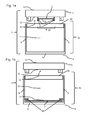
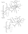
Referring to Figures 1a, 1b, 2a and 2b, a sample handling device 1 according to an embodiment of the invention comprises a sample absorbing substrate patch 2 having a major surface 23 configured for receiving and allowing to dry a body fluid sample to form a dried body fluid spot retained by said sample absorbing substrate patch and a recipient system 3 comprising a container base 4 comprising one container portion 41 comprising containing walls 9 defining an inner surface 23 and an interior chamber 11 configured for containing a liquid medium for processing the dried body fluid spot; and an opening 10, a removable cover 5 comprising a removable cover portion 51 configured for sealingly closing the opening of the container portion 41. The removable cover portion comprises an inner surface 12 facing the interior chamber 11 of the container portion 41 when the removable cover portion 51 is positioned on the opening of the container portion 41 and an outer surface 13. The sample absorbing substrate patch 2 is an essentially flat absorbing patch having a thickness 24 negligible compared to its surface.
-
The sample absorbing substrate patch 2 is permanently fixed in the removable cover portion 51 (Figures 1a, 2a and 2b) or, alternatively, in the container portion 41 (Figure 1b) and disposed such that the major surface 23 of the sample absorbing substrate patch 2 is freely accessible for reception of a body fluid sample when the removable cover portion 51 is positioned not closed on the opening 10 of the container portion 41 and fully contained within the interior chamber 11 when the removable cover portion 51 is positioned in the opening 10 of the container portion 41.
-
In the configuration illustrated on Figures 1a, 2a and 2b, the sample absorbing substrate patch 2 is permanently fixed on the inner surface 12 of removable the removable cover portion 51 through one or several fixation elements 18. The one or several fixation elements 18 comprise a protrusion 20 to lodge the sample the sample absorbing substrate patch 2 (Figures 1a, 2a, 2b) and one or several fixing teeth 21 (Figure 1a) to maintain the sample absorbing substrate patch 2 within the protrusion 20.
-
In the configuration of Figure 1b, the sample absorbing substrate patch 2 is permanently fixed at the bottom of the interior chamber 11 of the container portion 41 through one or several fixation elements 18. The fixation elements 18 comprise one or several fixing teeth 21 to maintain the sample absorbing substrate patch 2 on the inner surface 22 of the containing wall 9 at the bottom of the interior chamber 11 of the container portion 41.
-
The removable cover portion 51 comprises a closing arrangement 6 for sealingly closing the openings 10 of the container portion 41 (Figures 1a, 1b, 2a, 2b).
-
In the configuration illustrated on Figure 2a, the closing arrangement 6 comprises a clamping portion 12 and a sealing element 15 wherein the clamping portion 12 of the closing arrangement 6 is located on the inner surface 12 of the removable cover portion 51.
-
In the configuration illustrated on Figures 2a and 2b, the sample absorbing substrate patch 2 is of circular shape. Further, the removable cover portion 51 comprises on its inner surface 12, a cylindrical protrusion 20 with one or several fixing teeth to permanently fix the said sample absorbing substrate patch 2, said protrusion 20 fits into the opening 10 of the container portion 41 of the recipient 3 when the removable cover portion 51 is positioned on the opening 10 of said container portion 41 such that the said container portion 41 is sealingly closed by said protrusion 20.
-
In the configurations of Figures 1a and 1b, the removable cover portion 51 and the container portion 41 constitute two separate or non-coupled elements of the sample handling device in a configuration where the removable cover portion 51 does not close the opening of the container portion 41.
-
In the configuration of Figures 2a and 2b, the removable cover portion 51 and the container portion 41 are coupled together by a detachable bridging tie 7. The bridging tie 7 forms a hinge between the removable cover portion 51 and the container portion 41 when the removable cover portion 51 is positioned closed on the opening 10 of the container portion 4. The removable cover portion 51, container portion 41 and bridging tie 7 are integrally made as a single component.
-
In the configuration of Figures 3a and 3b, which is a variant of the configurations of Figures 1a and 1b, respectively, the recipient 3 further contains a desiccant 25 in the vicinity of the surface 26 of a sample absorbing substrate patch 2 opposite to the major surface 23 which is freely accessible for reception of a body fluid sample when the removable cover portion 5 is positioned not closed on the opening 10 of said container portion 41. The desiccant 25 and the surface 26 of a sample absorbing substrate patch 2 opposite to the major surface 23 are separated by a semi-permeable membrane 27.
-
In the configuration of Figures 4a and 4b, the sample handling device comprises a recipient 3 container base 4' comprising several container portions 41 in the form of wells each defining an interior chamber 11 configured for containing a liquid medium for processing the dried body fluid spot and an opening 10; and a removable cover 5' comprising several cover portions 51 configured for sealingly closing the openings 10 of the said several container portions 41. The sample absorbing substrate patch 2 is permanently fixed in each of said several removable cover portions 51 and disposed such that the major surface of the sample absorbing substrate patch 2 is freely accessible for reception of a body fluid sample when the removable cover portion 51 is positioned not closed on the openings 10 of said container portions 41 and fully contained within the interior chambers 11 when the removable cover portions 51 are positioned on the openings 10 of said container portions 41. The sample absorbing substrate patch 2 is permanently fixed to the inner surface 12 of each the said several removable cover portions 51 through fixation elements 18. The sample handling device further comprises a handling support 8 comprising a container base holding portion 17 and an essentially flat sitting surface 19 for maintaining the container base in an essentially vertical position during dried body fluid spot preparation, storage, shipping and/or analysis.
-
According to another aspect, the invention provides a sample handling device, a kit and methods according to the invention useful for the preparation and storing of dried body fluid spots, in particular dried blood spots, and the further analysis of body fluid samples from said stored dried body fluid spots for clinical motoring (e.g. therapeutic drug monitoring), pre-clinical investigations (e.g. pharmacokinetics), genetic screening (e.g. metabolic and genetic profiling) or quantitative or qualitative detection of illicit substances.
-
A sample handling device according to the invention allows re-suspending the analytes present on the in-situ prepared and stored dried body fluid spots within the container portion by addition of at least one solution (e.g. hydro-organic mixture such as water/methanol mixture) when the removable cover portion is open and optionally homogenizing the eluting solution for example by mechanical or wave stirring of the recipient system when the removable cover portion is closed by the closing arrangement. Re-suspension of said analytes may be carried out by liquid-liquid extraction, thereby allowing more selectivity in the extraction of analytes, analysing larger sample volumes compared to DBS assays without the need of ultra-sensitive analytic methods, which is not possible with the standard DBS assay card formats. The transfer of the analytes into liquid phase can be achieved within the sample handling device by various methods such as solid-liquid extraction (SLE), paper supported micro liquid-liquid extraction (µLLE) or microwave- or thermally-assisted extraction.
-
Typically, in a solid-liquid extraction procedure, a volume of an extraction solvent (e.g. methanol, acetonitrile), typically in the range of 5-1000 µl for a 1.5 ml container , is added into the container portion of the recipient system of a sample handling device according to the invention where the dried body fluid spot has been prepared and stored, the removable cover portion is then closed and the liquid medium is homogenized, typically by shaking or sonication (e.g. for 15 minutes) such that the analytes of interest captured in dried state on the sample absorbing substrate patch are extracted in the solvent within the container portion of the recipient system of a sample handling device. Optionally, an intermediate step of pre-wetting the dried body fluid spot on the sample absorbing substrate patch can be carried out by depositing a volume of reacting solution (e.g. hydro-organic mixture such as water/methanol mixture, enzymes, derivatization reagents or stabilizers), typically 5-50 µl (e.g. 15 µl), on the surface of the sample absorbing substrate patch before adding a volume of an extraction solvent into the container portion of the recipient system of a sample handling device in order to facilitate extraction of analytes of interest or to biologically and chemically modify the analytes of interest.
-
Typically, in a micro liquid-liquid extraction procedure, the dried body fluid spot on the absorbent substrate patch is pre-wetted by depositing a volume of a wetting solvent (e.g. mixture of water and organic solvent such as water/methanol mixtures), typically 5-50 µl (e.g. 15 µl), on the surface of the sample absorbing substrate patch before adding a volume of an organic solvent non-miscible to the wetting solvent (e.g. hexane, MTBE, ethyl acetate, hexane/MTBE mixture (e.g. 1/1), MTBE/ethyl acetate (e.g. 1/1)), typically 50-1500 µl (e.g. 350 µl), into the container portion of the recipient system of a sample handling device where the dried body fluid spot has been prepared and stored, the removable cover portion is then closed and the liquid medium is homogenized, typically by shaking or sonication (e.g. for 15 minutes). The wetting phase remains trapped/supported by the sample absorbing substrate patch and the wetting phase and the extraction phase are brought in contact during this homogenization step such that the analytes of interest are transferred from one phase to the other through a liquid-liquid extraction process within the container portion of the recipient system of the sample handling device device.
-
Various classical analytic methods useful to qualitatively and/or quantitatively analyse liquid samples could be then applied to the re-solubilized analytes including gas chromatography or liquid chromatography with and without mass spectrometry detection,, colorimetry, ,ion mobility mass spectrometry, matrix-assisted laser desorption/ionization - mass spectrometry or immunoassays.
-
According to another aspect, the invention provides a method for preparing non-card format dried body fluid spots comprising:
- (i) Providing a sample handling device according to the invention;
- (ii) Placing a pre-determined amount of a body fluid sample on the sample absorbing substrate patch from said sample handling device (typically in the range 5-100 µl) and waiting for complete drying of the said sample;
- (iii) Closing the opening of the container portion of the container base of the recipient of said collecting device with the removable closing portion of the removable cover.
-
According to another aspect, the invention provides a method for analysing non-card format dried body fluid spots comprising:
- (i) Providing a dried body fluid sample spot obtainable under step (ii) of a method for preparing non-card format dried body fluid spots according to the invention;
- (ii) Adding an extraction solvent into the container portion of container base of the recipient of the said sample handling device through the opening of said container portion;
- (iii) Closing the opening of the container portion of the container base of the recipient of said sample handling device with the removable closing portion of the removable cover;
- (iv) Homogenizing the liquid within the said container portion of the container base of the recipient (typically by shaking or sonicating);
- (v) Analyzing at least one analyte present in the liquid within the said container portion of the container base of the recipient.
-
In a further embodiment, the invention provides a method for analysing non-card format dried body fluid spots according to the invention further comprising, before step (ii), a pre-wetting step (ia) of the dried body fluid spot, by depositing a volume of the extraction solvent used under step (ii) or a volume of a wetting solvent which is non-miscible to the extraction solvent on the surface of the sample absorbing substrate patch.
-
In another further embodiment, the invention provides a method for analysing non-card format dried body fluid spots according to the invention further comprising a pre-wetting step (ia) of the dried body fluid spot with a volume of a wetting solvent which is non-miscible to the extraction solvent on the surface of the sample absorbing substrate patch.
-
In another further embodiment, the invention provides a method for analysing non-card format dried body fluid spots according to the invention further comprising a step of comparing the amount of said at least one analyte analysed under step (v) with a predetermined reference value for this analyte (e.g. value of the amount of the analyte present in a reference sample such as a sample from an healthy individual, from an individual carrying the sought marker, from the same individual before treatment, at an earlier stage of the treatment, or containing the sought substance).
-
In a further embodiment, the invention provides a method according to the invention wherein the body fluid sample is selected from saliva, blood and serum.
-
In another further embodiment, the invention provides a method according to the invention wherein the body fluid sample is blood.
EXAMPLES
GENERAL PROCEDURES & CONDITIONS
-
The following studies are conducted to support the effectiveness of a sample handling device according to the invention in the preparation, storing of dried body fluid spots and in the in situ analysis of body fluid samples extracted from said dried body fluid spots.
-
The following abbreviations refer respectively to the definitions below:
- BzEcg (Benzylecgonine), CE (Colision Energy), CocEt (Cocaethylene), DP (Declustering Potential), HPLC (high performance liquid chromatography), LC (liquid chromatography), MA (methylamphetamine), MDA (3,4-methylene-dioxy-amphetamine), MDEA (3,4-methylene-dioxy-ethylamphetamine), MDMA (3,4-methylene-dioxy-methylamphetamine), MS (mass spectrometry), MTBE (methyl t-(Saquinavir).
Example 1: Preparation of dried blood spot in a sample handling device according to the invention
-
A blood sample (typically 5 µl) is deposited onto a circular paper filter (Whatman 903®, 5 mm internal diameter) fixed by pression on the surface of the removable closing element facing the interior of the fluid container portion of a sample handling device according to the invention as shown on Figure 2A and let for complete drying (typically overnight). A dried blood spot is then formed and can be stored by closing the removable closing element and/or further analysed by re-solubilization in-situ of the dried blood spot by a method according to the invention.
Example 2: In situ extraction of analytes from a dried blood spot formed in a sample handling device according to the invention by solid-liquid extraction (SLE) or micro liquid-liquid extraction (µLLE)
-
A dried blood spot is formed as described under Example 1 where a cocktail of the following 18 analytes representative of 5 distinct groups of analytes was mixed with a liquid blood sample prior deposition of blood on the circular paper filter:
Amphetamines
-
- Amphetamine (A)
- MA
- MDA
- MDEA
- MDMA (Ecstasy)
Cocaine and its metabolites
-
- Cocaine
- Benzylecgonine
- Cocaethylene
Tricyclic antidepressants
-
- Amitriptyline
- Desipramine
- Imipramine
Antiretroviral drugs
-
- Nevirapine
- Atazanavir
- Ritonavir
- Saquinavir
Benzodiazepines
-
- Diazepam
- Flunitrazepam
- Nitrazepam
Extraction by solid-liquid extraction (SLE)
-
Extraction of the dried blood spot was then achieved by transferring the analytes of interest into liquid phase within the sample handling device according to the invention by solid-liquid extraction method as shown on Figures 5A and 5C through the addition of 350 µl of an extraction solvent (e.g. methanol) into the receptacle of the sample collection device according to the invention used for the preparation and storage of said dried blood spot, with or without a pre-wetting step of depositing about 15 µl of an extraction solvent (e.g. methanol) on the surface of the dried blood spot, typically 0-5 min before the addition of the extraction solvent into the receptacle of the sample collection device (Figure 5B1). The sample collection device was then closed and sonicated for about 15 minutes after the addition of the extraction solvent into the receptacle of the sample collection device. The presence of traces of the several analytes was investigated in the solvent by LC-MS/MS analysis under the following conditions:
Extraction conditions:
-
SLE: pre-wetting phase: 15 µl MeOH, extraction phase: 350 µl MEOH µLLE: wetting phase: 15 µl MeOH/H2O 1/1; extraction phase: 350 µl MTBE
-
Samples were sonicated for 15 min, evaporated and reconstituted in 100µl of H2O/MeOH 75/25 +0.1% HCOOH.
LC conditions
-
Column: reversed-phase C18 column ODS-AM (YMC, Kyoto, Japan) with 2.1 mm inner diameter x 50 mm long
-
Mobile phases:
- A: H2O + 0.1% HCOOH, B: MeOH + 0.1% HCOOH Gradient elution
Mass spectrometry
-
4000 Q TRAP™ (AB/MDS Sciex, Toronto, Canada) in SRM (Selected Reaction Monitoring) mode
- A: m/z 136.1 → m/z 119.1 (DP=50, CE=13)
- MA: m/z 150.2 → m/z 91.2 (DP=65, CE=26)
- MDA: m/z 180.2 → m/z 163.3 (DP=45, CE=15)
- MDMA: m/z 194.2 → m/z 163.3 (DP=65, CE=18)
- MDEA: m/z 208.2 → m/z 163.1 (DP=70, CE=19)
- Cocaine: m/z 304.2 → m/z 182.2 (DP=100, CE=28)
- BzEcg: m/z 290.2 → m/z 168.2 (DP=100, CE=28)
- CocEt: m/z 318.2 → m/z 196.2 (DP=60, CE=28)
- Diazepam: m/z 285.2 → m/z 154.0 (DP=90, CE=38)
- Flunitrazepam: m/z 314.2 → m/z 268.1 (DP=90, CE=37)
- Nitrazepam: m/z 282.2 → mlz 236.1 (DP=95, CE=35)
- Amitriptyline: m/z 278.1 → m/z 233.1 (DP=71, CE=24)
- Desipramine: m/z 267.1 → m/z 208.0 (DP=50, CE=33)
- Imipramine: m/z 281.2 → m/z 86.1 (DP=47, CE=24)
- Nevirapine: m/z 267.12 → m/z 226.1 (DP=46, CE=35)
- Atazanavir : m/z 705.1 → m/z 168.1 (DP=65, CE=66)
- Ritonavir: m/z 721.4 → m/z 296.1 (DP=46, CE=26)
- SQV: m/z 671.4 → m/z 570.4 (DP=90, CE=45)
-
It was found that only methanol was able to extract analytes with acceptable recoveries and that when other extraction solvents are used such as for example MTBE, MTBE/ethyl acetate mixtures, ethyl acetate, almost no extraction occurred if the pre-wetting step with the extraction solvent is missing). Process efficiencies (PE) are calculated as the ratio of the peak area obtained with DBS extract over the peak area of an hydro-organic standard solution of the 18 analytes cocktail. The concentration of the latter (5ng/ml) takes into account the dilution occurring during the reconstitution step. Process eficiencies are calculated with the mean of 3 determinations and are reported together with corresponding coefficients of variations in Table 1 below.
Extraction by micro liquid-liquid extraction (µLLE)
-
Extraction of the dried blood spot is alternatively achieved by transferring the analytes of interest into liquid phase within the sample handling device according to the invention by liquid-liquid extraction method as shown on Figures 5A, 5B2 and 5C through the addition of about 15 µl of a wetting solvent (e.g. mixture of water/methanol in proportion of 100/0; 75/25; 50/50; 25/75; 0/100) on the surface of the dried blood spot (Figure 5B2), typically 0-5 min before the addition of 350 µl of an extraction solvent non-miscible with the wetting solvent (e.g. MTBE, MTBE/ethyl acetate mixture 1/1, ethyl acetate) into the receptacle of the sample collection device according to the invention used for the preparation and storage of said dried blood spot (Figure 5C).
-
The sample collection device was then closed and sonicated and the presence of traces of the several analytes was investigated in the solvent by LC-MS/MS as described above.
-
It was found that a variety of extraction solvents was able to exact analytes of interest with acceptably recoveries and that the extraction process is much more selective than SLE through the variation of the nature and pH of the wetting phase and the nature of the extraction phase, depending on the nature of the analytes. Process efficiencies are calculated as described above and are reported in Table 1 below.
Table 1 | | µLLE (wetting: H2O/MeOH 1/1) | SLE |
| MTBE | MTBE/ethyl acetate | ethyl acetate | Methanol |
| CV (n=3) | PE | CV (n=3) | PE | CV (n=3) | PE | CV (n=3) | PE |
| A | 15% | 60% | 10% | 74% | 7% | 58% | 9% | 89% |
| MA | 11% | 78% | 10% | 72% | 1% | 75% | 5% | 81% |
| | | | | | | | | |
| MDA | 5% | 66% | 8% | 59% | 2% | 56% | 5% | 80% |
| MDEA | 11% | 73% | 8% | 77% | 3% | 88% | 9% | 84% |
| MDMA | 7% | 74% | 9% | 66% | 2% | 76% | 7% | 70% |
| | | | | | | | | |
| cocaine | 8% | 57% | 7% | 69% | 3% | 79% | 4% | 74% |
| BzEcg | 19% | 8% | 4% | 23% | 8% | 40% | 7% | 97% |
| CocEt | 10% | 57% | 10% | 70% | 2% | 82% | 4% | 80% |
| | | | | | | | | |
| amitriptyline | 18% | 57% | 16% | 70% | 8% | 77% | 11% | 62% |
| desipramine | 11% | 49% | 20% | 53% | 8% | 38% | 5% | 54% |
| imipramine | 13% | 54% | 16% | 65% | 8% | 69% | 6% | 62% |
| | | | | | | | | |
| diazepam | 29% | 57% | 6% | 78% | 18% | 55% | 7% | 99% |
| flunitrazepam | 8% | 74% | 5% | 82% | 26% | 62% | 7% | 89% |
| nitrazepam | 15% | 68% | 7% | 86% | 32% | 70% | 5% | 138% |
| | | | | | | | | |
| nevirapine | 16% | 74% | 9% | 83% | 3% | 92% | 9% | 95% |
| atazanavir | 11% | 92% | 9% | 86% | 11% | 78% | 5% | 85% |
| ritonavir | 16% | 86% | 8% | 64% | 5% | 66% | 7% | 65% |
| saquinavir | 13% | 68% | 9% | 63% | 2% | 79% | 7% | 52% |
-
Further, it has been investigated the ability of those extraction methods to remove the side effects due to the presence of phospholipids which are generally observed during bioanalysis by LC-MS. LC-MS detection of analytes relies on their capacity to be ionized and this ability can be altered by the co-elution of interfering compounds from the sample matrix ("matrix effects"). Phospholipids are endogenous compounds of the blood and plasma, and comprise a large variety of chemical structures. Phosphatidyl cholines (PC) and sphingomyelines (SM) are the most abundant phospholipids and represent more than 70% of the total lipid content. Phospholipids, because of their high concentration levels in blood, have been shown to be the cause of those "matrix effects" (Little et al., Journal of Chromatography B, 2006, 833, 219-230 ; Xia et al., Rapid Communications in Mass Spectrometry, 2009, 23, 2125-2138). Further, the strong hydrophobic properties of the blood phospholipids tend to make them accumulate onto the LC column under usual (reverse phase) LC conditions. This accumulation can lead to a slow elution spread over a long time period, which may result in possible elevation of baseline levels and to retention time shifts. Therefore, development of bioanalytical methods which allow removal of the majority of phospholipids is of crucial importance. LC-MS/MS analysis was carried out under the same conditions as those described for SLE experiments.
-
Figures 6A and B show the extracted analytes and the residual phospholipids when the DBS as described above is extracted by SLE or by µLLE, respectively. It clearly shows that µLLE extraction allows the removal of a large quantity of phospholipids (PC and SM) which remain in minority amounts as compared the analytes, while their amount remains majoritary as compared to the analytes after extraction by SLE.
-
Those results support the usefulness of a sample collection device and methods according to the invention for the preparation, storage and direct analysis of dried body fluid spots. Further, it supports the considerable advantage of a sample collection device and methods according to the invention over the card format DBS assays by offering the possibility of carrying out in situ liquid-liquid extraction of analytes of interest from dried body fluid spots which leads to a better efficiencies with a large variety of choice of wetting and extracting solvents, depending on the analyte of interest to be detected. Further, a sample collection device and methods according to the invention, by offering the possibility to extract analytes from dried body fluid spots by µLLE present the advantage to allow a better selectivity in the extraction than classical SLE methods, especially for phospholipid removal, which improve the analytical sensitivity, notably for LC-MS detection.
Example 3: In situ analysis of a dried blood spot formed in a sample handling device according to the invention for the quantification of saquinavir by LC-MS/MS
-
LC-MS/MS analysis was applied for the quantitation of saquinavir (antiretroviral drug) present on a DBS prepared and extracted using either a SLE or a µLLE extraction process as described in Example 2 in a device according to the invention. 8 µl of a deuterated saquinavir (SQV-d5, F.Hoffmann-La Roche, Basel) solution (50 ng/ml in MeOH/H2O 1/1) was deposited onto the filter paper disk (Whatman 903®, 5 mm internal diameter) fixed by pressure on the surface of the removable closing element facing the interior of the fluid container portion of a sample handling device according to the invention as shown on Figure 2a and allowed to dry overnight prior the application of blood samples for forming DBS as described in Example 1.
Extraction conditions
-
SLE: pre-wet: 15 µl MEOH, extraction: 350 µl MEOH
-
µLLE: wetting: 15 µl MeOH/H2O 1/1; extraction: 350 µl MTBE
-
Samples were sonicated for 15 min, evaporated and reconstituted in H2O/MeOH 75/25 +0.1% HCOOH.
LC conditions
-
Column: reversed-phase C18 column ODS-AM (YMC, Kyoto, Japan) with 2.1 mm inner diameter x 50 mm long
-
Mobile phases:
- A: H2O + 0.1% HCOOH, B: MeOH + 0.1% HCOOH Gradient elution
Mass spectrometry
-
4000 Q TRAP™ (AB/MDS Sciex, Toronto, Canada) in SRM (Selected Reaction Monitoring) mode
- SQV: m/z 671.4 -> m/z 570.4 (DP=90, CE=45)
- SQV-d5: m/z 671.5 -> m/z 575.4 (DP=90, CE=45).
-
The performance of both assays were evaluated by analyzing quality control samples (blood spiked with known amount of analyte) and calculating accuracy and precision.
Accuracy: ratio of the experimental determination of the concentration of the analyte over its expected, true value. It describes the closeness of a test result obtained by the method to the true value.
Precision: coefficient of variation of individual concentration determinations of several DBS coming from a single, homogenous blood sample (same analyte concentration). The results are presented for SLE extraction process in Table 2 and for µLLE extraction process in Table 3.
Table 2 | Concentration | Accuracy (%) | Precision |
| (ng/ml) | n=1 | n=2 | n=3 | (n=3) |
| | | | | |
| 25 | 115 | 104 | 96 | 9% |
| 50 | 108 | 98.6 | 89.6 | 9% |
| 100 | 112 | 113 | 107 | 3% |
| 250 | 114 | 112 | 101 | 6% |
| 500 | 113 | 110 | 103 | 4% |
| 1000 | 106 | 102 | 91.3 | 8% |
| 2500 | 105 | 105 | 95.7 | 5% |
| 5000 | 97.5 | 102 | 84.9 | 10% |
Table 3 | Concentration | | Accuracy | (%) | Precision |
| (ng/ml) | n=1 | n=2 | n=3 | (n=3) |
| | | | | |
| 25 | 100% | 101% | 93% | 4% |
| 50 | 112% | 101% | 101% | 6% |
| 100 | 104% | 99% | 84% | 11% |
| 250 | 112% | 103% | 100% | 6% |
| 500 | 108% | 105% | 94% | 8% |
| 1000 | 110% | 105% | 90% | 10% |
| 2500 | 113% | 99% | 94% | 10% |
| 5000 | 105% | 103% | 86% | 11% |
-
Both extraction techniques were found to be linear for the detection of the drug from 25 to 5'000 ng/ml of saquinavir which satisfy the needs for a clinical use (Wagner et al., 2008, Journal of Chromatography B, 872, 68-76). Therefore, those results support the usefulness of a sample collection device and methods according to the invention for the preparation, storage and direct analysis of dried body fluid spots by either a SLE or a µLLE extraction process on a quantitative manner.