EP2483417B1 - Methods for diagnosing and treating encephalitis or epilepsy - Google Patents
Methods for diagnosing and treating encephalitis or epilepsy Download PDFInfo
- Publication number
- EP2483417B1 EP2483417B1 EP10821183.0A EP10821183A EP2483417B1 EP 2483417 B1 EP2483417 B1 EP 2483417B1 EP 10821183 A EP10821183 A EP 10821183A EP 2483417 B1 EP2483417 B1 EP 2483417B1
- Authority
- EP
- European Patent Office
- Prior art keywords
- gaba
- another embodiment
- tumor
- receptor
- patients
- Prior art date
- Legal status (The legal status is an assumption and is not a legal conclusion. Google has not performed a legal analysis and makes no representation as to the accuracy of the status listed.)
- Active
Links
Images


Classifications
-
- G—PHYSICS
- G01—MEASURING; TESTING
- G01N—INVESTIGATING OR ANALYSING MATERIALS BY DETERMINING THEIR CHEMICAL OR PHYSICAL PROPERTIES
- G01N33/00—Investigating or analysing materials by specific methods not covered by groups G01N1/00 - G01N31/00
- G01N33/48—Biological material, e.g. blood, urine; Haemocytometers
- G01N33/50—Chemical analysis of biological material, e.g. blood, urine; Testing involving biospecific ligand binding methods; Immunological testing
- G01N33/53—Immunoassay; Biospecific binding assay; Materials therefor
- G01N33/575—Immunoassay; Biospecific binding assay; Materials therefor for cancer
- G01N33/57557—Immunoassay; Biospecific binding assay; Materials therefor for cancer of other specific parts of the body, e.g. brain
-
- A—HUMAN NECESSITIES
- A61—MEDICAL OR VETERINARY SCIENCE; HYGIENE
- A61P—SPECIFIC THERAPEUTIC ACTIVITY OF CHEMICAL COMPOUNDS OR MEDICINAL PREPARATIONS
- A61P25/00—Drugs for disorders of the nervous system
-
- A—HUMAN NECESSITIES
- A61—MEDICAL OR VETERINARY SCIENCE; HYGIENE
- A61P—SPECIFIC THERAPEUTIC ACTIVITY OF CHEMICAL COMPOUNDS OR MEDICINAL PREPARATIONS
- A61P25/00—Drugs for disorders of the nervous system
- A61P25/08—Antiepileptics; Anticonvulsants
-
- A—HUMAN NECESSITIES
- A61—MEDICAL OR VETERINARY SCIENCE; HYGIENE
- A61P—SPECIFIC THERAPEUTIC ACTIVITY OF CHEMICAL COMPOUNDS OR MEDICINAL PREPARATIONS
- A61P25/00—Drugs for disorders of the nervous system
- A61P25/14—Drugs for disorders of the nervous system for treating abnormal movements, e.g. chorea, dyskinesia
-
- A—HUMAN NECESSITIES
- A61—MEDICAL OR VETERINARY SCIENCE; HYGIENE
- A61P—SPECIFIC THERAPEUTIC ACTIVITY OF CHEMICAL COMPOUNDS OR MEDICINAL PREPARATIONS
- A61P35/00—Antineoplastic agents
-
- A—HUMAN NECESSITIES
- A61—MEDICAL OR VETERINARY SCIENCE; HYGIENE
- A61P—SPECIFIC THERAPEUTIC ACTIVITY OF CHEMICAL COMPOUNDS OR MEDICINAL PREPARATIONS
- A61P37/00—Drugs for immunological or allergic disorders
-
- G—PHYSICS
- G01—MEASURING; TESTING
- G01N—INVESTIGATING OR ANALYSING MATERIALS BY DETERMINING THEIR CHEMICAL OR PHYSICAL PROPERTIES
- G01N33/00—Investigating or analysing materials by specific methods not covered by groups G01N1/00 - G01N31/00
- G01N33/48—Biological material, e.g. blood, urine; Haemocytometers
- G01N33/50—Chemical analysis of biological material, e.g. blood, urine; Testing involving biospecific ligand binding methods; Immunological testing
- G01N33/53—Immunoassay; Biospecific binding assay; Materials therefor
- G01N33/575—Immunoassay; Biospecific binding assay; Materials therefor for cancer
- G01N33/5758—Immunoassay; Biospecific binding assay; Materials therefor for cancer involving compounds serving as markers for tumours, cancers or neoplasias, e.g. cellular determinants, receptors, heat shock/stress proteins, A-protein, oligosaccharides or metabolites
- G01N33/5759—Immunoassay; Biospecific binding assay; Materials therefor for cancer involving compounds serving as markers for tumours, cancers or neoplasias, e.g. cellular determinants, receptors, heat shock/stress proteins, A-protein, oligosaccharides or metabolites involving compounds localised on the membrane of tumour or cancer cells
-
- G—PHYSICS
- G01—MEASURING; TESTING
- G01N—INVESTIGATING OR ANALYSING MATERIALS BY DETERMINING THEIR CHEMICAL OR PHYSICAL PROPERTIES
- G01N2333/00—Assays involving biological materials from specific organisms or of a specific nature
- G01N2333/435—Assays involving biological materials from specific organisms or of a specific nature from animals; from humans
- G01N2333/705—Assays involving receptors, cell surface antigens or cell surface determinants
- G01N2333/70571—Assays involving receptors, cell surface antigens or cell surface determinants for neuromediators, e.g. serotonin receptor, dopamine receptor
-
- G—PHYSICS
- G01—MEASURING; TESTING
- G01N—INVESTIGATING OR ANALYSING MATERIALS BY DETERMINING THEIR CHEMICAL OR PHYSICAL PROPERTIES
- G01N2800/00—Detection or diagnosis of diseases
- G01N2800/28—Neurological disorders
-
- G—PHYSICS
- G01—MEASURING; TESTING
- G01N—INVESTIGATING OR ANALYSING MATERIALS BY DETERMINING THEIR CHEMICAL OR PHYSICAL PROPERTIES
- G01N2800/00—Detection or diagnosis of diseases
- G01N2800/28—Neurological disorders
- G01N2800/2857—Seizure disorders; Epilepsy
Definitions
- the invention relates to methods for diagnosing an autoimmune encephalitis or an epilepsy in a subject using an antibody to an inhibitory ⁇ -amino-butyric acid-B (GABA B ) receptor.
- GABA B inhibitory ⁇ -amino-butyric acid-B
- Synaptic plasticity is a fundamental property of neurons that underlie mechanisms of memory, learning, and cognition. Plasticity depends upon the complex interactions of ion channels and synaptic receptors, including the excitatory glutamate N-methyl-D-aspartate receptor (NMDAR) and ⁇ -amino-3-hydroxy-5-methyl-4-isoxazolepropionic acid receptor (AMPAR). Animal models of pharmacological or genetic disruption of these receptors result in severe alterations of memory, learning, behavior, and seizures. Therefore it is expected that immune responses against extracellular domains of these receptors would result in similar symptoms.
- NMDAR excitatory glutamate N-methyl-D-aspartate receptor
- AMPAR ⁇ -amino-3-hydroxy-5-methyl-4-isoxazolepropionic acid receptor
- Mitoma et al., 2003, Journal of the Neurological Sciences, 208: 51-56 discloses the dual impairment of GABA A - and GABA B -receptor-mediated synaptic responses by autoantibodies to glutamic acid decarboxylase.
- Mazzi et al., 2008, Transfusion and Apheresis Science, 39:229-233 describes a patient with paraneoplastic limbic encephalitis having autoantibodies to glutamic acid decarboxylase who was treated using therapeutic plasma exchange.
- the present invention provides a method for diagnosing autoimmune encephalitis, an occult tumor causing autoimmune encephalitis, or in a subject with epilepsy, a tumor causing the epilepsy, the method comprising testing a biological sample from a subject to determine the presence of an antibody to a GABA B receptor, wherein the presence of said antibody in said biological sample is indicative that the subject has an autoimmune encephalitis, or an occult tumor causing said autoimmune encephalitis, or where the subject has epilepsy, the presence of said antibody is indicative of a tumor in said subject causing said epilepsy.
- the antibody binds to the B1 subunit of a GABA B receptor. In another embodiment, the antibody binds to the B2 subunit of a GABA B receptor.
- the autoimmune encephalitis is a paraneoplastic autoimmune encephalitis. In an embodiment, the autoimmune encephalitis is anti-GABA B receptor encephalitis. In an embodiment, the autoimmune encephalitis is a non-paraneoplastic, autoimmune encephalitis. In an embodiment, the autoimmune encephalitis comprises a limbic encephalitis.
- the autoimmune encephalitis is associated with pathological symptoms, such as seizures, psychiatric symptoms, abnormality in cognition and behavior, a movement disorder or abnormal movements, a decreased level of consciousness, hypoventilation, amnesia or a memory deficit, or a combination thereof.
- pathological symptoms such as seizures, psychiatric symptoms, abnormality in cognition and behavior, a movement disorder or abnormal movements, a decreased level of consciousness, hypoventilation, amnesia or a memory deficit, or a combination thereof.
- the autoimmune encephalitis is associated with a tumor.
- the tumor may be a small cell lung cancer or a neuroendocrine tumor of the lung or other organs.
- the biological sample is a cerebrospinal fluid (CSF) sample. In other embodiments, the biological sample is a serum or plasma sample.
- CSF cerebrospinal fluid
- the invention relates to methods for diagnosing an autoimmune encephalitis or an epilepsy in a subject using an antibody to a GABA B receptor.
- a method for diagnosing an autoimmune encephalitis in a subject comprising testing a biological sample from the subject to determine the presence of an antibody to a GABA B receptor, wherein the presence of said antibody in said biological sample is indicative that the subject has an autoimmune encephalitis.
- a method for diagnosing an occult tumor causing autoimmune encephalitis in a subject comprising testing a biological sample from the subject to determine the presence of an antibody to a GABA B receptor, wherein the presence of said antibody in said biological sample is indicative that the subject has an occult tumor that is a cause of said autoimmune encephalitis.
- a method for diagnosing a tumor causing epilepsy in a subject with epilepsy comprising testing a biological sample from the subject to determine the presence of an antibody to a GABA B receptor, wherein the presence of said antibody in said biological sample is indicative of the presence of a tumor in said subject and said tumor is a cause of said epilepsy.
- the application also discloses a method for diagnosing a tumor in a subject having an epilepsy, comprising the step of: obtaining a biological sample from said subject; and testing said biological sample to determine the presence of an antibody to a GABA B receptor, wherein the presence of said antibody in said biological sample indicates the presence of a tumor in said subject, thereby diagnosing said tumor in said subject having said epilepsy.
- the application also discloses a method for treating an autoimmune encephalitis in a subject, comprising the steps of: detecting a tumor associated with said autoimmune encephalitis by testing a body fluid from said subject for an antibody to a GABA B receptor, wherein the presence of said antibody in said body fluid indicates the presence of said tumor in said subject and that said tumor is a cause of said autoimmune encephalitis; and treating said tumor.
- the present invention provides a method of determining a cause of an encephalitis in a subject by testing a body fluid of the subject for an antibody to a GABA B receptor.
- the presence of an antibody to the B1 subunit of a GABA B receptor in the body fluid indicates that the encephalitis is of autoimmune etiology.
- the presence of an antibody to the B2 subunit of a GABA B receptor in the body fluid indicates that the encephalitis is of autoimmune etiology.
- the methods provided herein facilitate the recognition of a severe form of autoimmune encephalitis that is often responsive to treatment.
- the methods described herein emphasize the idea that autoimmunity can affect behavior, and particularly that an antibody to a GABA B receptor may alter emotion, in one embodiment, or memory, consciousness or their combination in other independent embodiments.
- the application discloses a method of determining a cause of an autoimmune encephalitis in a subject, comprising the step of testing a body fluid of the subject for an antibody to a GABA B receptor, thereby determining a cause of an autoimmune encephalitis in a subject.
- the presence of the antibody indicates the presence of a tumor in the subject.
- the tumor is a cause of the autoimmune encephalitis.
- the present invention provides a method of diagnosing an autoimmune encephalitis in a subject, comprising the step of testing a body fluid of the subject for an antibody to the B1 subunit of a GABA B receptor, thereby diagnosing said autoimmune encephalitis in said subject.
- the biological sample used in the methods described herein is a body fluid that is tested by methods of the present invention is, in another embodiment, a cerebro-spinal fluid (CSF).
- the body fluid is plasma.
- the body fluid is any other type of fluid known in the art.
- the biological sample is amniotic fluids, blood, sera, saliva, or their combination.
- the encephalitis diagnosed by the methods of the present invention is an autoimmune encephalitis.
- the autoimmune encephalitis is a paraneoplastic encephalitis.
- the autoimmune encephalitis is a non-paraneoplastic encephalitis.
- the autoimmune encephalitis is a paraneoplastic autoimmune encephalitis.
- the autoimmune encephalitis is a non-paraneoplastic, autoimmune encephalitis.
- the autoimmune encephalitis is any other type of autoimmune encephalitis known in the art.
- the frequency of paraneoplastic anti-GABA B encephalitis, diagnosed by the methods described herein, is unknown.
- paraneoplastic anti- GABA B encephalitis is frequently unrecognized. This may be due to several features that make this disorder unique among paraneoplastic encephalitis, including in one embodiment, involvement of relatively young women between the 2 nd and 5 th decades, or, in another embodiment, the unusual presentation with prominent psychiatric manifestations, or in another embodiment, normal or atypical MRI findings, which in 75% of cases consist of mild, transient T2 or FLAIR abnormalities outside the medial temporal lobes, with cortical enhancement in certain embodiments, or in yet another embodiment, the benign appearance of the ovarian tumors.
- any of the subjects presenting the symptoms described hereinabove are diagnosed using the methods described herein.
- the autoimmune encephalitis is a limbic encephalitis. In another embodiment, the autoimmune encephalitis is associated with a limbic dysfunction. In another embodiment, the autoimmune encephalitis is not associated with a limbic dysfunction.
- limbic encephalitis causes impressive deficits that are characteristically dominated by rapid and severe loss of short-term memory.
- patients show subacute encephalitis of later adult life, mainly affecting the limbic areas with evidence of cancer in one embodiment.
- limbic encephalitis refers to a subject exhibiting severe short-term memory loss and dementia in association with bronchial carcinoma.
- the autoimmune encephalitis diagnosed by the methods of the present invention is associated with seizures.
- the autoimmune encephalitis is associated with a diencephalic syndrome.
- the autoimmune encephalitis is associated with a psychiatric symptom.
- the autoimmune encephalitis is associated with an abnormality in cognition.
- the autoimmune encephalitis is associated with an abnormality in behavior.
- the autoimmune encephalitis is associated with amnesia. In another embodiment, the autoimmune encephalitis is associated with a memory deficit. In another embodiment, the autoimmune encephalitis is associated with memory problems. In another embodiment, the autoimmune encephalitis is associated with a hypokinetic syndrome.
- the autoimmune encephalitis is associated with a movement disorder. In another embodiment, the autoimmune encephalitis is associated with abnormal movements. In another embodiment, the movement disorder is any other movement disorder known in the art.
- the autoimmune encephalitis is associated with a decreased level of consciousness. In another embodiment, the autoimmune encephalitis is associated with hypoventilation.
- the autoimmune encephalitis is associated with, dysfunction of any part of the brain or spinal cord. In another embodiment, the autoimmune encephalitis is associated with a combination of any of the above symptoms or disorders.
- the autoimmune encephalitis is associated with a tumor.
- the tumor is a neuroendocrine tumor of the lung or small cell lung cancer (SCLC).
- SCLC small cell lung cancer
- the tumor is an ovarian teratoma.
- the tumor is a thymic tumor.
- the tumor is a testicular tumor.
- the cancer associated with the encephalitis is a cervical cancer tumor.
- the cancer is a head and neck cancer tumor.
- the cancer is a breast cancer tumor.
- the cancer is an ano-genital cancer tumor.
- the cancer is a melanoma. In another embodiment, the cancer is a sarcoma. In another embodiment, the cancer is a carcinoma. In another embodiment, the cancer is a lymphoma. In another embodiment, the cancer is a leukemia. In another embodiment, the cancer is a mesothelioma. In another embodiment, the cancer is a glioma. In another embodiment, the cancer is a germ cell tumor. In another embodiment, the cancer is a choriocarcinoma.
- the cancer is pancreatic cancer. In another embodiment, the cancer is ovarian cancer. In another embodiment, the cancer is gastric cancer. In another embodiment, the cancer is a carcinomatous lesion of the pancreas. In another embodiment, the cancer is pulmonary adenocarcinoma. In another embodiment, the cancer is colorectal adenocarcinoma. In another embodiment, the cancer is pulmonary squamous adenocarcinoma. In another embodiment, the cancer is gastric adenocarcinoma. In another embodiment, the cancer is an ovarian surface epithelial neoplasm (e.g. a benign, proliferative or malignant variety thereof). In another embodiment, the cancer is an oral squamous cell carcinoma.
- the cancer is an oral squamous cell carcinoma.
- the cancer is non small-cell lung carcinoma. In another embodiment, the cancer is an endometrial carcinoma. In another embodiment, the cancer is a bladder cancer. In another embodiment, the cancer is a head and neck cancer. In another embodiment, the cancer is a prostate carcinoma.
- the cancer is an acute myelogenous leukemia (AML). In another embodiment, the cancer is a myelodysplastic syndrome (MDS). In another embodiment, the cancer is a non-small cell lung cancer (NSCLC). In another embodiment, the cancer is a Wilms' tumor. In another embodiment, the cancer is a leukemia. In another embodiment, the cancer is a lymphoma. In another embodiment, the cancer is a desmoplastic small round cell tumor. In another embodiment, the cancer is a mesothelioma (e.g. malignant mesothelioma). In another embodiment, the cancer is a gastric cancer. In another embodiment, the cancer is a colon cancer. In another embodiment, the cancer is a lung cancer.
- AML acute myelogenous leukemia
- MDS myelodysplastic syndrome
- NSCLC non-small cell lung cancer
- the cancer is a Wilms' tumor.
- the cancer is a leukemia.
- the cancer is a lymph
- the cancer is a breast cancer. In another embodiment, the cancer is a germ cell tumor. In another embodiment, the cancer is an ovarian cancer. In another embodiment, the cancer is a uterine cancer. In another embodiment, the cancer is a thyroid cancer. In another embodiment, the cancer is a hepatocellular carcinoma. In another embodiment, the cancer is a thyroid cancer. In another embodiment, the cancer is a liver cancer. In another embodiment, the cancer is a renal cancer. In another embodiment, the cancer is a kaposis. In another embodiment, the cancer is a sarcoma. In another embodiment, the cancer is another carcinoma or sarcoma. In another embodiment, the tumor is any other type of tumor known in the art.
- the present invention provides a method of determining a cause of epilepsy in a subject, comprising the step of testing a body fluid of said subject for an antibody to a GABA B receptor, thereby determining a cause of said epilepsy in said subject, wherein the antibody indicates the presence of a tumor in the subject, and the tumor is a cause of the epilepsy.
- the epilepsy diagnosed by the methods of the present invention is, in another embodiment, an idiopathic epilepsy.
- the epilepsy responds to IgG-depleting therapy.
- the epilepsy is associated with partial seizures.
- the epilepsy is associated with simple partial seizures.
- the epilepsy is associated with complex partial seizures.
- the epilepsy is associated with generalized seizures.
- the epilepsy is associated with absence (petit mal) seizures.
- the epilepsy is associated with myoclonic seizures.
- the epilepsy is associated with tonic-clonic (grand mal) seizures.
- the epilepsy is associated with West syndrome. In another embodiment, the epilepsy is associated with Lennox-Gastaut syndrome. In another embodiment, the epilepsy is associated with any other syndrome known in the art.
- the epilepsy is of no known cause. In another embodiment the epilepsy is any other type of epilepsy known in the art.
- Chronic autoimmune encephalitis refers, in another embodiment, to a primary cause of the disorder.
- the term refers to a contributing cause of the disorder.
- the term refers to an indirect causation.
- the term refers to causation via an immune response induced by the tumor.
- the term refers to a significant cause of the disorder.
- the application discloses a method for diagnosing a tumor in a subject having an encephalitis, comprising the step of testing a body fluid of said subject for an antibody to a GABA B receptor, thereby diagnosing a tumor in said subject having said encephalitis.
- the presence of the antibody indicates a presence of a tumor in the subject.
- the application also discloses a method for detecting a tumor in a subject having an encephalitis, comprising the step of testing a body fluid of said subject for an antibody to a GABA B receptor, thereby detecting said tumor in said subject having said encephalitis.
- the presence of the antibody indicates the presence of a tumor in the subject.
- the application also discloses a method for diagnosing a tumor in a subject having an epilepsy, comprising the step of testing a body fluid of said subject for an antibody to a GABA B receptor, thereby diagnosing said tumor in said subject having said epilepsy.
- the presence of the antibody indicates the presence of a tumor in the subject.
- the application discloses a method for detecting a tumor in a subject having an epilepsy, comprising the step of testing a body fluid of said subject for an antibody to a GABA B receptor, thereby detecting said tumor in said subject having said epilepsy.
- the presence of said antibody indicates a presence of a tumor in the subject.
- the application further discloses a method of diagnosing or detecting a GABA B -mediated disease in a subject, comprising the step of testing a body fluid of the subject for an antibody to an antibody to a GABA B receptor, thereby diagnosing or detecting said GABA B -mediated disease in said subject.
- GABA B receptors mediate presynaptic inhibition by two mechanisms: the activation of G-protein-coupled-inward-rectifying potassium (GIRK) channels and the inhibition of calcium channels. They also attenuate presynaptic firing frequencies. At the post-synapse, the GABA B receptors mediate inhibition by similar mechanisms and by inducing a slow inhibitory post-synaptic potential (IPSP). On a neuronal network level, GABA B receptors modulate their activity by limiting the duration of network high-activity states, preventing excessive neuronal synchronization, and allowing novel stimuli to break synchronous activity.
- GIRK G-protein-coupled-inward-rectifying potassium
- IIPSP slow inhibitory post-synaptic potential
- GABA B receptors are widely distributed in the brain and spinal cord, but are particularly abundant in the hippocampus, thalamus and cerebellum, which are the areas of more intense rat brain immunolabeling by patients' antibodies.
- the main autoantigen of patients' antibodies, the GABA B1 subunit is necessary for GABA binding (and receptor function) while the GABA B2 subunit is required for localization of the receptor to appropriate areas of the cell membrane and for G-protein coupling.
- Deletion of GABA B1 in transgenic mice results in predominant seizures, memory deficits, and increased anxiety behaviors. Both genetic and pharmacologic evidence supports an important role for GABA B receptors in controlling anxiety and regulating mood.
- the application also discloses a method of treating autoimmune encephalitis in a subject.
- the method comprises the steps of: detecting a tumor associated with an autoimmune encephalitis by testing a body fluid from said subject for an antibody to a GABA B receptor, whereby a presence of said antibody indicates the presence of said tumor in said subject and that said tumor is a cause of said autoimmune encephalitis, and treating said tumor.
- the tumor may be treated during the early stage.
- the tumor may be treated within four months of the onset of a symptom associated with autoimmune encephalitis, or within three months of the onset of a symptom associated with autoimmune encephalitis, or within two months of the onset of a symptom associated with autoimmune encephalitis, or within one month of the onset of a symptom associated with autoimmune encephalitis.
- the step of treating said tumor may comprise removing said tumor, or may comprise immunotherapy, or may comprise removing said tumor in combination with immunotherapy.
- the step of treating said tumor comprises chemotherapy or comprises removing said tumor in combination with chemotherapy.
- GABA B receptor comprises subunits.
- GABA B receptors are formed from heteromers of B1 and B2 subunits.
- both subunits are required to create a functional receptor that contains B1 and B2 subunits.
- the B1 subunit may be a B1-a subunit.
- the receptor exists as heteromers of B1 subunits and B2 subunits.
- B1 and B2 combine to form receptor subtypes with distinct pharmacological properties, localization, and ability to interact with intracellular messengers.
- the B1 subunit is a monomer of a multimer of GABA B receptor.
- the multimer is a homomer that comprises two or more subunits.
- the multimer is a homo-dimer that comprises two B1 subunits.
- the multimer is a heteromer that comprises a B1 subunit.
- the multimer is a heteromer that comprises a B1 subunit and a B2 subunit.
- the multimer is a heteromer that comprises a B1-a subunit and B2 subunit.
- the multimer is a heteromer that comprises B1-b subunit and a B2 subunit.
- the multimer is a heteromer that comprises B1-c subunit and a B2 subunit.
- the B1 subunit of GABA B receptor has the sequence as set forth below:
- the B1 subunit of GABA B receptor has the sequence as set forth below:
- the B1 subunit of GABA B receptor has the sequence as set forth below:
- the B1 subunit is a homologue of SEQ ID NOs: 1, 2, or 3. In another embodiment, the B1 subunit is a variant of SEQ ID NOs: 1, 2, or 3. In another embodiment, the B1 subunit is an isomer of SEQ ID NOs: 1, 2, or 3. In another embodiment, the B1 subunit is a fragment of SEQ ID NOs: 1, 2, or 3. In another embodiment, the B1 subunit comprises SEQ ID NOs: 1, 2, or 3.
- the B1 subunit of GABA B receptor is encoded by a nucleotide sequence having the sequence:
- the B1 subunit of GABA B receptor is encoded by a nucleotide sequence having the sequence:
- the B1 subunit of GABA B receptor is encoded by a nucleotide sequence having the sequence:
- the B1 subunit is encoded by a nucleotide molecule that is a homologue of SEQ ID NOs: 4, 5, or 6.
- the nucleotide molecule is a variant of SEQ ID NOs: 4, 5, or 6.
- the nucleotide molecule is an isomer of SEQ ID NOs: 4, 5, or 6.
- the nucleotide molecule is a fragment of SEQ ID NOs: 4, 5, or 6.
- the nucleotide molecule comprises SEQ ID NOs: 4, 5, or 6.
- the epitope recognized by an antibody detected by a method of the present invention is, in another embodiment, a conformational epitope.
- the epitope is a linear epitope.
- the epitope is any other type of epitope known in the art.
- ectopic expression of B1 subunit by nervous tissue contained in the teratomas contributes to break immune tolerance.
- a combination of factors such as an adjuvant effect of the prodromal viral-like illness that occur in most subjects, and a genetic predisposition in certain embodiments, play additional roles in the initiation of the immune response tested for using the diagnosis methods described herein.
- a pathogenic role of B1 antibodies in paraneoplastic anti-GABA B encephalitis is shown by the correlation between patients' symptoms and antibody titers.
- the subject exhibits antibodies that react with an extracellular neuronal antigen. In another embodiment, the subject exhibits antibodies that react with an antigen exposed on the cell surface of a neuron. In another embodiment, patients with antibodies to extracellular antigens exhibit, under the conditions utilized herein, enhanced responsiveness to immune therapy.
- the present invention provides a method of diagnosing autoimmune encephalitis in a subject, the method comprising testing a biological sample from the subject for an antibody to the B1 subunit of a GABA B receptor, whereby the presence of said antibody in said biological sample is indicates an autoimmune encephalitis.
- the present invention provides a method of diagnosing a tumor associated with an autoimmune encephalitis in a subject, the method comprising testing a biological sample from the subject for an antibody to the B1 subunit of a GABA B receptor, whereby the presence of said antibody indicates the presence of an occult tumor in said subject and that said tumor is a cause of said autoimmune encephalitis.
- the present invention provides a method of diagnosing a tumor causing epilepsy in a subject, the method comprising testing a biological sample from the subject for the presence of an antibody to the B1 subunit of a GABA B receptor, whereby the presence of said antibody indicates the presence of a tumor in said subject and said tumor is a cause of said epilepsy.
- the application discloses a method of diagnosing a tumor in a subject having an epilepsy, comprising the step of: obtaining a biological sample from the subject; and testing the biological sample for the presence of an antibody to the B1 subunit of a GABA B receptor, whereby the presence of said antibody indicates the presence of a tumor in said subject, thereby diagnosing a tumor in a subject having an epilepsy.
- the present application also discloses a method of treating autoimmune encephalitis in a subject, comprising the steps of: detecting a tumor associated with an autoimmune encephalitis by testing a body fluid from the subject for an antibody to the B1 subunit of a GABA B receptor, whereby a presence of said antibody indicates a presence of said tumor in said subject and that said tumor is a cause of said autoimmune encephalitis; and treating said tumor.
- the tumor may be treated within four months of the onset of a symptom associated with autoimmune encephalitis.
- ELISA enzyme-linked immunoabsorption assay
- immunocytochemistry is used to test for the presence of an antibody.
- immunoprecipitation is used to test for the presence of an antibody.
- one of the methods enumerated herein is utilized.
- neuronal tissue is fixed with PFA.
- any other method known in the art is utilized.
- Léx limbic encephalitis
- patients with LE develop subacute confusion, irritability, depression, sleep disturbances, seizures, short-term memory loss, and/or dementia.
- the pathological substrate of LE is an inflammatory disorder that predominantly involves the limbic system (hippocampi, amygdala, and cingulate gyrus).
- biopsy and autopsy studies demonstrate interstitial and perivascular infiltrates of T cells, and less frequently B cells, along with microglial activation, neuronal degeneration, and/or gliosis.
- inflammatory infiltrates are found in areas distant from the limbic system.
- the infiltrates remain mild and clinically silent. In another embodiment, the infiltrates become prominent and develop into a disorder called encephalomyelitis. Additional methods of diagnosing LE are described, for example, in Gultekin SH et al (Paraneoplastic limbic encephalitis: neurological symptoms, immunological findings and tumour association in 50 patients. Brain 2000;123:1481-1494 ).
- homology when in reference to any protein or peptide, refer, in one embodiment, to a percentage of amino acid residues in the candidate sequence that are identical with the residues of a corresponding native polypeptide, after aligning the sequences and introducing gaps, if necessary, to achieve the maximum percent homology, and not considering any conservative substitutions as part of the sequence identity. Methods and computer programs for the alignment are well known in the art.
- Homology is, in another embodiment, determined by computer algorithm for sequence alignment, by methods well described in the art.
- computer algorithm analysis of nucleic acid sequence homology can include the utilization of any number of software packages available, such as, for example, the BLAST, DOMAIN, BEAUTY (BLAST Enhanced Alignment Utility), GENPEPT and TREMBL packages.
- “homology” refers to identity to a sequence selected from SEQ ID NO: 1-6 of greater than 70%. In another embodiment, “homology” refers to identity to a sequence selected from SEQ ID NO: 1-6 of greater than 72%. In another embodiment, “homology” refers to identity to one of SEQ ID NO: 1-6 of greater than 75%. In another embodiment, “homology” refers to identity to a sequence selected from SEQ ID NO: 1-6 of greater than 78%. In another embodiment, “homology” refers to identity to one of SEQ ID NO: 1-6 of greater than 80%. In another embodiment, “homology” refers to identity to one of SEQ ID NO: 1-6 of greater than 82%.
- “homology” refers to identity to a sequence selected from SEQ ID NO: 1-6 of greater than 83%. In another embodiment, “homology” refers to identity to one of SEQ ID NO: 1-6 of greater than 85%. In another embodiment, “homology” refers to identity to one of SEQ ID NO: 1-6 of greater than 87%. In another embodiment, “homology” refers to identity to a sequence selected from SEQ ID NO: 1-6 of greater than 88%. In another embodiment, “homology” refers to identity to one of SEQ ID NO: 1-6 of greater than 90%. In another embodiment, “homology” refers to identity to one of SEQ ID NO: 1-6 of greater than 92%.
- “homology” refers to identity to a sequence selected from SEQ ID NO: 1-6 of greater than 93%. In another embodiment, “homology” refers to identity to one of SEQ ID NO: 1-6 of greater than 95%. In another embodiment, “homology” refers to identity to a sequence selected from SEQ ID NO: 1-6 of greater than 96%. In another embodiment, “homology” refers to identity to one of SEQ ID NO: 1-6 of greater than 97%. In another embodiment, “homology” refers to identity to one of SEQ ID NO: 1-6 of greater than 98%. In another embodiment, “homology” refers to identity to one of SEQ ID NO: 1-6 of greater than 99%. In another embodiment, “homology” refers to identity to one of SEQ ID NO: 1-6 of 100%.
- homology is determined via determination of candidate sequence hybridization, methods of which are well described in the art (See, for example, " Nucleic Acid Hybridization” Hames, B. D., and Higgins S. J., Eds. (1985 ); Sambrook et al., 2001, Molecular Cloning, A Laboratory Manual, Cold Spring Harbor Press, N.Y. ; and Ausubel et al., 1989, Current Protocols in Molecular Biology, Green Publishing Associates and Wiley Interscience, N.Y ).
- methods of hybridization are carried out under moderate to stringent conditions, to the complement of a DNA encoding a native caspase peptide.
- Hybridization conditions being, for example, overnight incubation at 42 °C in a solution comprising: 10-20 % formamide, 5 X SSC (150 mM NaCl, 15 mM trisodium citrate), 50 mM sodium phosphate (pH 7. 6), 5 X Denhardt's solution, 10 % dextran sulfate, and 20 ⁇ g/ml denatured, sheared salmon sperm DNA.
- Protein and/or peptide homology for any amino acid sequence listed herein is determined, in another embodiment, by methods well described in the art, including immunoblot analysis, or via computer algorithm analysis of amino acid sequences, utilizing any of a number of software packages available, via established methods. Some of these packages include the FASTA, BLAST, MPsrch or Scanps packages, and, in another embodiment, employ the use of the Smith and Waterman algorithms, and/or global/local or BLOCKS alignments for analysis, for example.
- nucleic acids or “nucleotide” refers to a string of at least two base-sugar-phosphate combinations. The term includes, in one embodiment, DNA and RNA. "Nucleotides” refers, in one embodiment, to the monomeric units of nucleic acid polymers. RNA is, in one embodiment, in the form of a tRNA (transfer RNA), snRNA (small nuclear RNA), rRNA (ribosomal RNA), mRNA (messenger RNA), anti-sense RNA, small inhibitory RNA (siRNA), micro RNA (miRNA) and ribozymes.
- transfer RNA transfer RNA
- snRNA small nuclear RNA
- rRNA ribosomal RNA
- mRNA messenger RNA
- anti-sense RNA small inhibitory RNA
- siRNA small inhibitory RNA
- miRNA micro RNA
- DNA can be, in other embodiments, in form of plasmid DNA, viral DNA, linear DNA, or chromosomal DNA or derivatives of these groups.
- these forms of DNA and RNA can be single, double, triple, or quadruple stranded.
- the term also includes, in another embodiment, artificial nucleic acids that contain other types of backbones but the same bases.
- the artificial nucleic acid is a PNA (peptide nucleic acid).
- PNA contain peptide backbones and nucleotide bases and are able to bind, in one embodiment, to both DNA and RNA molecules.
- the nucleotide is oxetane modified.
- the nucleotide is modified by replacement of one or more phosphodiester bonds with a phosphorothioate bond.
- the artificial nucleic acid contains any other variant of the phosphate backbone of native nucleic acids known in the art.
- phosphothiorate nucleic acids and PNA are known to those skilled in the art, and are described in, for example, Neilsen PE, Curr Opin Struct Biol 9:353-57 ; and Raz NK et al Biochem Biophys Res Commun. 297:1075-84 .
- the production and use of nucleic acids is known to those skilled in art and is described, for example, in Molecular Cloning, (2001), Sambrook and Russell, eds . and Methods in Enzymology: Methods for molecular cloning in eukaryotic cells (2003) Purchio and G. C. Fareed .
- Example 1 The GABA B Receptor is a Novel Autoantigen of Severe Limbic Encephalitis with Prominent Seizures
- mice Animal tissue, antibodies, and immunohistochemistry on rat brain.
- Female Wistar rats were euthanized and the brain was removed, sagittally sectioned, immersed in 4% paraformaldehyde at 4°C for 2 hours, cryoprotected with 40% sucrose for 24 hours, and snap frozen in chilled isopentane.
- Immunohistochemistry with human tissue utilized IgG purified from patients' sera and labeled with biotin. In these studies no secondary antibody was needed, avoiding background caused by irrelevant human IgG present in the tissue.
- Immunohistochemistry on frozen sections of rat brain was performed with patients' or control serum (1:250), CSF (1:10), or GABA B1 antibody using a standard avidin-biotin peroxidase method, or double immunolabeling with patients' serum or CSF and the GABA B1 antibody, followed by the appropriate Alexa Fluor secondary antibodies.
- Rat hippocampal neuronal cultures were prepared. Live neurons grown on coverslips were incubated for 1 hour at 37°C with patient or control serum (final dilution 1:200) or CSF (1:10). After removing the media and extensive washing with phosphate-buffered saline (PBS), neurons were fixed with 4% paraformaldehyde, permeabilized with 0.1% Triton X-100, and single or double immunolabeled with a guinea pig polyclonal GABA B1 receptor antibody (1:200), followed by the corresponding Alexa Fluor secondary antibodies diluted 1:2,000 (Molecular Probes, Eugene, OR). Results were photographed under a fluorescence microscope using Zeiss Axiovision software (Zeiss, Thornwood, NY).
- HEK293 cells were transfected with plasmids containing rodent GABA B1 or GABA B2 subunits of the GABA B receptor or plasmids without insert (control), using a method previously reported.
- cells were cotransfected with GABA B1 and GABA B2 in equimolar ratios. Cells were grown for 24 hours after transfection before assessment. Transfected cells were fixed in 4% paraformaldehyde, permeabilized with 0.1% Triton X-100 and then incubated with patients' serum (1:200) or CSF (100%) and the indicated guinea pig polyclonal GABA B1 receptor antibody (1:20,000) or a polyclonal GABA B2 receptor antibody (1:10,000, generated by Dr.
- Antibody titers were determined using HEK293 cells expressing GABA B1/B2 incubated with serial dilutions of serum and CSF, starting at 1/2 dilution.
- IgG subtypes were determined in serum or CSF using the indicated HEK293 transfected cells, and secondary anti-human antibodies against IgG1, IgG2, IgG3, and IgG4 (all diluted 1:200; Sigma, St. Louis, MO) as reported.
- buffer NaCl 150mM, EDTA 1mM, tris(hydroxymethyl)aminomethane [Tris]-HCl 100mM, deoxycholate acid 0.5%, 1% Triton X-100 [Sigma Labs, St. Louis, MO], pH 7.5
- protease inhibitors P8340
- Distinctive protein bands precipitated by patient serum were excised from the gel and analyzed using mass spectrometry at the proteomic facility at the University of Pennsylvania. After characterization of the antigen, frozen aliquots of the indicated pellets were separated in a sodium dodecyl sulfate polyacrylamide gel electrophoresis as described earlier, transferred to nitrocellulose (162-0115; Bio-Rad, Hercules, CA), and blotted with the indicated polyclonal antibodies against GABA B1 (1:2000) or GABA B2 (1:1000) receptor subunits. The reactivity was developed using the appropriate biotinylated secondary antibodies (1:2000) and the avidin-biotin peroxidase, diaminobenzidine method.
- Neurons were then incubated with a guinea pig polyclonal antibody against an intracellular epitope of the GABA B receptor (1:1000; Invitrogen) and a mouse monoclonal antibody against the presynaptic marker Bassoon (1:200; Stressgen, Victoria, British Columbia, Canada), washed, and incubated with the appropriate fluorescent-conjugated secondary antibodies (1:1000, Molecular Probes).
- Images were obtained using a laser-scanning confocal microscope (Leica TCS SP2; Leica, Deerfield, IL). For each image, laser light levels and detector gain and offset were adjusted so that no pixel values were saturated. Images of labeled neurons were stored digitally for subsequent analysis. Images were thresholded, areas of interest containing dendrites were selected, and the number of individual clusters along neuronal dendrites was determined using interactive software (ImageJ; Research Services Branch, National Institute of Mental Health, Bethesda, MD). The co-localization of clusters labeled with patient's antibodies, commercial GABA B antibodies and the synaptic marker Bassoon was quantified using a software Macro implemented in ImageJ.
- CSF demonstrated 9 white blood cells (WBC)/ ⁇ l, total protein concentration 35 mg/dL, glucose 71 mg/dL, and negative oligoclonal bands and cytology. PCR for herpes simplex virus, West Nile virus, and St. Louis encephalitis were negative. Hyponatremia (119 mEq/L) due to syndrome of inappropriate anti-diuretic hormone (SIADH) secretion was noted. Serum and CSF analysis of paraneoplastic antibodies was negative. Combined CT and fluorodeoxyglucose (FDG)-PET revealed mediastinal lymphadenopathy, proven by biopsy to be small-cell lung cancer (SCLC).
- SCLC small-cell lung cancer
- the patient was treated with antiepileptics (levetiracetam, valproic acid, phenytoin) and immunotherapy (intravenous immunoglobulins [IVIg], corticosteroids), immediately followed by chemotherapy with cisplatin and etoposide.
- antiepileptics levetiracetam, valproic acid, phenytoin
- immunotherapy intravenous immunoglobulins [IVIg], corticosteroids
- the neuronal cell surface antigen is the metabotropic GABA B receptor .
- the neuronal cell surface antigen is the metabotropic GABA B receptor .
- live rat hippocampal neurons were incubated with patients' antibodies and the target antigen was immunoprecipitated.
- These studies produced two protein bands of approximately 105 and 90 kDa ( Figure 3b ) that were analyzed by mass spectrometry demonstrating sequences derived from the B1 subunit of the metabotropic GABA B receptor.
- the GABA B receptor is a heterodimer composed of two subunits, B1 and B2, both with a similar molecular weight
- the inventors next examined by immunoblot and polyclonal antibodies specific for each subunit to determine whether patients' antibodies had precipitated both subunits.
- These studies confirmed that patient antibodies co-precipitated B1 and B2 subunits of the GABA B receptor ( Figure 3c ).
- the specificity of patient antibodies for GABA B receptors was determined by triple immunolabeling of cultured neurons (patient antibodies; a guinea pig polyclonal antibody to a non-competing intracellular GABA B1 receptor epitope; and a pre-synaptic marker [Bassoon]), and the degree of co-localization quantified by confocal microscopy, as reported ( Figure 4 ).
- the binding of antibodies to GABA B receptor clusters was analyzed on the dendrites of 24 neurons on 4 separate coverslips.
- HEK293 cells were transfected with GABA B1 , GABA B2 , or both receptor subunits and immunocytochemically tested with patients' antibodies. All 15 patients had serum or CSF antibodies that specifically reacted with the GABA B1 receptor subunit ( Figure 5 ), and one had additional reactivity with the GABA B2 subunit. These findings indicate that HEK293 cells expressing GABA B1/B2 or GABA B1 receptor subunits provide a substrate for an unambiguous diagnostic test. Similar studies with the 104 control cases showed that two patients, both with syndromes attributed to GAD autoimmunity, had GABA B1 receptor antibodies at low titers (CSF 1:2, serum negative), not visible with brain immunohistochemistry.
- IgG subtypes were performed in serum or CSF of 6 randomly selected patients. All had IgG1 GABA B1 antibodies; two had additional IgG3, and one IgG2 antibodies.
- EEG studies were available from 12 patients; 9 showed temporal lobe seizures, epileptiform discharges, or temporal lobe slowing; 2 had generalized slowing and 1 was normal.
- Several types of seizures were noted on EEG, including complex partial seizures (often of temporal lobe onset), status epilepticus, and subclinical seizures.
- Sex/Age (yr) Tumor Presenting Symptoms Other clinical-immunological features 08-299 F/60 SCLC Subacute onset of complex partial seizures, confusion, memory impairment SIADH 06-165 M/66 SCLC Subacute onset of seizures, confusion, memory deficit, behavioral problems N-type VGCC antibodies 06-235 F/53 SCLC Rapidly progressive memory deficits, abnormal sleeping habits, followed by frequent seizures (focal, secondarily generalized), confusion, decline in mental status leading to coma Pruritic rash with initial weakness. 08-017 M/75 Mediastinal adenopathy Subacute onset of seizures, psychosis, encephalitis. Rapidly progressed to death before definitive diagnosis or treatment.
- GAD GAD
- TPO thyroglobulin antibodies
- SCLC small cell lung cancer
- FDG/PET Fluoro-deoxyglucose/ positron emission tomography
- SIADH syndrome of inappropriate anti-diuretic hormone
- GAD glutamic-acid decarboxylase
- TPO thyroid peroxidase
- VGCC voltage-gated calcium channels.
- Table 2 Diagnostic tests, treatment and outcome.
- GABA B antibodies detected after patient's death in archived serum 08-623 FLAIR/T2 increased signal in small area of corpus callosum 95 WBC/ ⁇ L; protein 104; increased IgG index S, n/a CSF, 1:640 Corticosteroids, mycophenylate mofetil Substantial improvement. Lives independently. Seizure free (9 months) 09-086 FLAIR/T2 increased signal in left mesial temporal lobe n/a S, n/a CSF, 1:640 Corticosteroids, plasma exchange Initial substantial response to corticosteroids. Relapsed 1 month later. Died after 5 months in ICU with refractory seizures, status epilepticus, and systemic complications.
- 07-305 FLAIR/T2 increased signal in medial temporal lobes 19 WBC/ ⁇ L; protein 46 S, 1:5120 CSF, 1:2560 Corticosteroids, plasma exchange Substantial improvement. Mild residual short-term memory deficit. Seizures free (3 months) 06-026 FLAIR/T2 increased signal in medial temporal lobes 75 WBC/ ⁇ L; protein 26 ; + OCBs S, negative CSF, 1:4 Corticosteroids Full recovery (41 months) 08-278 FLAIR/T2 increased signal in medial temporal lobes 81 WBC, protein 30 S, 10,240 CSF, n/a Corticosteroids Substantial improvement. Residual short-term memory deficit. Lives independently.
- 09-083 FLAIR/T2 increased signal in medial temporal lobes 4 WBC/ ⁇ L; protein 109; 6 OCBs S, negative CSF, 1:4 Corticosteroids Full recovery, except for infrequent brief episodes of visual hallucinations (10 months) 01-696 Graus FLAIR/T2 increased signal in left medial temporal lobe Traumatic; negative cytology.
- Chemotherapy Residual short-term memory deficit; seizures controlled; died of sepsis (3 months) 07-1051 Graus Normal 0 WBC/ ⁇ L; protein 95 S, n/a CSF, 1:640 IVIg, corticosteroids, chemotherapy Seizures responded to antiepileptics; memory deficit persisted; died of cancer-related treatment (2 months) 08-114 Normal 3 WBC/ ⁇ L; protein 78; 1 OCB S, negative CSF, 1:2 IVIg No seizures or cognitive deficits. Limited response of cerebellar ataxia to IVIg (12 months) 08-398 Normal 2 WBC/ ⁇ L; Protein 52; + OCBs S, negative CSF, 1:2 IVIg, corticosteroids No seizures or cognitive deficits.
- WBC white blood cells (normal ⁇ 4 ⁇ l)
- FLAIR fluid-attenuated inversion recovery
- n/a not available
- IVIg intravenous immunoglobulin.
- ICU intensive care unit.
- the inventors of the instant application have discovered a new form of autoimmune encephalitis that associates with antibodies to extracellular epitopes of the GABA B receptor and is potentially treatment-responsive. Based on clinical, MRI, and EEG findings, the brain regions most affected are the hippocampi and temporal lobes. Therefore it is not surprising that the resulting syndrome is indistinguishable from other types of limbic encephalitis, although some clinical and immunological features may suggest GABA B receptor autoimmunity. A notable finding is the development of prominent and severe seizures in all patients, representing the main reason for medical attention or hospital admission. Two frequently associated features are the identification of a lung cancer, and the concurrent presence of autoantibodies against antigens of unclear significance with the limbic syndrome.
- GABA B receptors In contrast, pharmacological or genetic disruption of GABA B receptors result in phenotypes that closely resemble the limbic syndrome of patients with GABA B receptor antibodies. Moreover, in humans, a common GABA B receptor polymorphism associates with temporal lobe epilepsy.
- GABA B receptor encephalitis can occur without cancer association.
- anti-GABA B receptor encephalitis is similar to other synaptic autoimmunities of the central (NMDAR, AMPAR) or peripheral nervous system (acetylcholine receptor, P/Q-type VGCC) that may occur with or without cancer association. As occur in these disorders, 40% of patients with anti-GABA B receptor encephalitis (50% of those without tumor) had additional autoantibodies, indicating a propensity to autoimmunity.
- GABA B R-ab was analyzed in stored samples of serum or CSF by indirect immunofluorescence on HEK293 cells transfected with GABA B1 and GABA B2 receptor subunits.
- GABA B R-ab were detected in 10 of the 70 LE patients (14%). Eight had SCLC and two were idiopathic. One of the eight LE patients with SCLC had an additional onconeural antibody (Hu) and two GAD-ab. GABA B R-ab were identified in 7 (70%) of the 10 patients with LE and SCLC without onconeural antibodies. GABA B R-ab antibodies were not found in patients with GAD-ab and SPS, cerebellar ataxia or epilepsy. However, one patient with GAD-ab and cerebellar ataxia also presented GABA B R-ab in the setting of an anaplastic carcinoid of the thymus.
- GABA B R-ab are the most common antibodies found in LE associated with SCLC previously considered “seronegative”. In patients with GAD-ab, the frequency of GABA B R-ab is low and only observed in the context of cancer.
- LE was defined by the subacute onset of short-term memory loss, behavior change, seizures, and involvement of the temporal lobes by EEG, imaging studies, or postmortem examination. LE was considered definite paraneoplastic if a tumor was diagnosed or the serum presented well characterized onconeural antibodies (Hu, Yo, Ri, CV2, Ma2, amphiphysin). The diagnosis of definite idiopathic LE required the absence of cancer and well characterized onconeural antibodies, and a follow-up of at least three years.
- LE patients with a shorter follow-up were classified as possible idiopathic LE.
- Patients with GAD-ab were classified, as previously reported, in one of the following groups: stiff-person syndrome (SPS), cerebellar ataxia, isolated epilepsy, and paraneoplastic neurological syndromes.
- SPS stiff-person syndrome
- cerebellar ataxia cerebellar ataxia
- isolated epilepsy cerebellar ataxia
- paraneoplastic neurological syndromes The information was obtained from forms filled out by the referring neurologists, telephone interviews, and review of the clinical records. The study was approved by the Ethic Committee of the Hospital Clinic.
- Onconeural antibodies, SOX1-ab, and GAD-ab were screened by immunohistochemistry performed on frozen sections of paraformaldehyde-perfused rat cerebellum using an avidin-biotin immunoperoxidase technique and confirmed by immunoblot when indicated. GAD-ab were confirmed by radioimmunoassay.
- Neuropil antibodies were screened by immunohistochemistry on frozen sections of rat brain post-fixed with 4% paraformaldehyde. The presence of AMPA glutamate receptor antibodies was confirmed by immunofluorescence on HEK-293 cells transfected with plasmids containing the appropriate antigens, and voltage-gated potassium channel (VGKC) antibodies were confirmed by radioimmunoassay.
- VGKC voltage-gated potassium channel
- GABA B R-ab were screened on HEK293 cells transfected with plasmids containing rodent GABA B1 and GABA B2 in equimolar ratios. Positives samples were also analyzed by immunocytochemistry of rat hippocampal neuronal cultures. Both techniques have previously been described.
- HEK293 transfected cells were incubated with the patients' serum (dilution 1:20) or CSF (1:2) for 1h at 37°C, washed, fixed with 4% paraformaldehyde, incubated with a rabbit polyclonal GABA B1 antibody (1:1000) (Santa Cruz Biotechnology, sc-14006; Santa Cruz, California) followed by the appropriate Alexa Fluor secondary antibodies (Molecular Probes, Eugene, OR).
- GABA B R-ab Eleven patients tested positive for GABA B R-ab on the screening of HEK293 cells transfected with the B1 and B2 subunits of the GABA B R ( Figure 7 ). All positive samples immunoreacted in vivo with primary cultures of hippocampal neurons ( Figure 8 ). GABA B R-ab were positive in both serum and CSF in the 5 patients from whom paired samples were available. Median titer of GABA B R-ab was 1/120 (range 40-2000) in serum and 1/60 (range 20-640) in the CSF.
- GABA B R-ab was found in 10 patients with LE. Positive GABA B R-ab were identified more frequently in the group of paraneoplastic LE without onconeural antibodies (previously considered "seronegative") (Table 3). Seven (39%) of the 18 patients were GABA B R-ab-positive and all had SCLC. In total, positive GABA B R-ab were identified in 7 (70%) of the 10 patients with LE and SCLC without onconeural antibodies. The other three patients were positive for AMPAR-ab.
- GABA B R-ab and GAD-ab occurred in 2 patients with SCLC whereas the other three patients with LE and GAD-ab associated with other tumors (thymoma 2, lymphoma) and were GABA B R-ab-negative.
- GABA B R-ab were not detected in 71 patients with GAD-ab and SPS, cerebellar ataxia or epilepsy.
- one of the 6 patients with paraneoplastic neurological syndromes and GAD-ab was GABA B R-ab positive (Table 3).
- Neurological examination disclosed a normal mental status, bilateral horizontal nystagmus, and cerebellar gait ataxia. The patient was treated with oral steroids and the symptoms slowly resolved over the ensuing three months. Table 3.
- a patients previously reported in references 10 and 9;
- b included in the initial series of GABAR-ab (reference 4);
- c predominant symptom listed first;
- d Recovery before the start of chemotherapy.
- GABA B R-ab are the most common antibodies identified in patients with SCLC and LE previously considered “seronegative". Although the occurrence of GABA B R-ab and GAD-ab was observed in an initial series of 15 patients, when we tested a larger series of patients with several types of neurological syndromes associated with GAD-ab, GABA B R-ab were only identified in those who had a paraneoplastic syndrome.
Landscapes
- Health & Medical Sciences (AREA)
- Life Sciences & Earth Sciences (AREA)
- Engineering & Computer Science (AREA)
- Chemical & Material Sciences (AREA)
- Immunology (AREA)
- Biomedical Technology (AREA)
- General Health & Medical Sciences (AREA)
- Medicinal Chemistry (AREA)
- Hematology (AREA)
- Molecular Biology (AREA)
- Urology & Nephrology (AREA)
- Pharmacology & Pharmacy (AREA)
- Veterinary Medicine (AREA)
- Public Health (AREA)
- Animal Behavior & Ethology (AREA)
- Organic Chemistry (AREA)
- Nuclear Medicine, Radiotherapy & Molecular Imaging (AREA)
- General Chemical & Material Sciences (AREA)
- Chemical Kinetics & Catalysis (AREA)
- Biotechnology (AREA)
- Physics & Mathematics (AREA)
- Food Science & Technology (AREA)
- Pathology (AREA)
- General Physics & Mathematics (AREA)
- Cell Biology (AREA)
- Biochemistry (AREA)
- Analytical Chemistry (AREA)
- Bioinformatics & Cheminformatics (AREA)
- Microbiology (AREA)
- Neurosurgery (AREA)
- Neurology (AREA)
- Pain & Pain Management (AREA)
- Psychology (AREA)
- Peptides Or Proteins (AREA)
- Medicines Containing Antibodies Or Antigens For Use As Internal Diagnostic Agents (AREA)
- Measuring Or Testing Involving Enzymes Or Micro-Organisms (AREA)
Description
- The invention relates to methods for diagnosing an autoimmune encephalitis or an epilepsy in a subject using an antibody to an inhibitory γ-amino-butyric acid-B (GABAB) receptor.
- Synaptic plasticity is a fundamental property of neurons that underlie mechanisms of memory, learning, and cognition. Plasticity depends upon the complex interactions of ion channels and synaptic receptors, including the excitatory glutamate N-methyl-D-aspartate receptor (NMDAR) and α-amino-3-hydroxy-5-methyl-4-isoxazolepropionic acid receptor (AMPAR). Animal models of pharmacological or genetic disruption of these receptors result in severe alterations of memory, learning, behavior, and seizures. Therefore it is expected that immune responses against extracellular domains of these receptors would result in similar symptoms. Indeed, two recently identified disorders associated with antibodies to extracellular epitopes of the NR1 subunit of the NMDAR and the GluR1/2 subunits of the AMPAR result in encephalitides that manifest with prominent psychiatric, behavioral, and memory problems, often accompanied by seizures. In these two synaptic autoimmunities each antibody causes a dramatic and specific decrease of the levels of the target receptor in cultured neurons, suggesting they are pathogenic. Additionally, the neurological syndromes often respond to treatment, and in some patients the immune response occurs as a paraneoplastic manifestation of a tumor that expresses the target receptor, resembling in many aspects the autoimmune disorders of the neuromuscular synapse (e.g., Lambert-Eaton syndrome and myasthenia gravis). These findings along with a remarkable antibody-syndrome specificity and high frequency of some disorders (e.g., anti-NMDAR encephalitis) have drawn attention to other syndromes in which memory and behavior are impaired and seizures occur frequently. In some of these syndromes an immune mediated pathogenesis is suggested by the clinical response to empiric immunotherapy, the CSF an MRI findings suggesting limbic encephalitis, or the detection of antibodies to yet unknown extracellular neuronal antigens.
- Mitoma et al., 2003, Journal of the Neurological Sciences, 208: 51-56 discloses the dual impairment of GABAA- and GABAB-receptor-mediated synaptic responses by autoantibodies to glutamic acid decarboxylase. Mazzi et al., 2008, Transfusion and Apheresis Science, 39:229-233 describes a patient with paraneoplastic limbic encephalitis having autoantibodies to glutamic acid decarboxylase who was treated using therapeutic plasma exchange.
- A better understanding of the function of various antigens may help improve the treatment strategies. For the clinician who currently confronts these patients, however, the best chance to affect the neurologic outcome depends on: (1) the prompt diagnosis of the disorder, (2) the early discovery and treatment of the tumor, and (3) the use of immunotherapy. Accordingly, a need exists for improved methods of diagnosing autoimmune encephalitis or epileptic seizures.
- The present invention provides a method for diagnosing autoimmune encephalitis, an occult tumor causing autoimmune encephalitis, or in a subject with epilepsy, a tumor causing the epilepsy, the method comprising testing a biological sample from a subject to determine the presence of an antibody to a GABAB receptor, wherein the presence of said antibody in said biological sample is indicative that the subject has an autoimmune encephalitis, or an occult tumor causing said autoimmune encephalitis, or where the subject has epilepsy, the presence of said antibody is indicative of a tumor in said subject causing said epilepsy.
- In an embodiment, the antibody binds to the B1 subunit of a GABAB receptor. In another embodiment, the antibody binds to the B2 subunit of a GABAB receptor.
- In an embodiment, the autoimmune encephalitis is a paraneoplastic autoimmune encephalitis. In an embodiment, the autoimmune encephalitis is anti-GABAB receptor encephalitis. In an embodiment, the autoimmune encephalitis is a non-paraneoplastic, autoimmune encephalitis. In an embodiment, the autoimmune encephalitis comprises a limbic encephalitis.
- In an embodiment, the autoimmune encephalitis is associated with pathological symptoms, such as seizures, psychiatric symptoms, abnormality in cognition and behavior, a movement disorder or abnormal movements, a decreased level of consciousness, hypoventilation, amnesia or a memory deficit, or a combination thereof.
- In an embodiment, the autoimmune encephalitis is associated with a tumor. In certain embodiments, the tumor may be a small cell lung cancer or a neuroendocrine tumor of the lung or other organs.
- In an embodiment, the biological sample is a cerebrospinal fluid (CSF) sample. In other embodiments, the biological sample is a serum or plasma sample.
- Other features and advantages of the present invention will become apparent from the following detailed description examples and figures.
-
-
Figure 1 shows an MRI of a patient with GABAB receptor antibodies and limbic encephalitis. Axial FLAIR brain MRIs from case 1, taken at presentation (A) showed increased signal in the medial temporal lobes (arrows), more pronounce on the left. Repeat study at 1-month (B) showed improvement. Repeat studies at 3-, and 9-months after presentation (C,D) were stable. -
Figure 2 shows an immunolabeling of rat brain with patient's antibodies. Sagittal section of rat brain immunostained with CSF antibodies of a patient with limbic encephalitis. The extensive staining was observed in the neuropil of hippocampus, thalamus, cerebellum and cerebral cortex. -
Figure 3 shows that patients antibodies react with extracellular epitopes and precipitate the B1 and B2 subunits of the GABAB receptor. Culture of rat hipppocampal neurons were incubated (live, non-permeabilized) with the CSF of a patient with LE. The intense dot-like reactivity indicates patient's antibodies with cell surface antigens (A); scale bar = 10 µm. The precipitation of these antigens using patient's antibodies is shown in a gel in which proteins are visualized with EZBlue (B). Patient's antibodies (P) precipitated two protein bands at ∼105-90 kDa; these bands are not seen in the precipitate using CSF from a control individual (N). Sequencing of all bands using mass spectrometry demonstrated the B1 and B2 subunits of the GABAB receptor. Other smaller bands were proteolytic fragments and patient's IgG. Subsequent transfer of the gel to nitrocellulose and immunoblotting with monoclonal antibodies specific for each of the GABAB subunits confirmed that patient's antibodies precipitated the B1 and B2 subunits (Panels in D). -
Figure 4 shows that patient's antibodies specifically recognize the GABAB receptor in neuronal synapses. Confocal image of a cultured embryonic rat hippocampal neuron triple labeled with patient's antibodies (green), a guinea pig polyclonal antibody against an intracellular epitope of GABAB1 receptor (red), and the presynaptic marker bassoon (blue) (a). Area of dendrite from the same neuron showing patient's antibody staining (b), guinea pig polyclonal GABAB1 receptor antibody staining (c), both patient and guinea pig antibody staining (d), and triple stained (e). Quantification of the co-localization of labeling of the dendrites of 24 neurons is shown in (f). -
Figure 5 shows the detection of antibodies to the GABAB1 subunit using a HEK293 cell based assay. HEK293 cells transfected with B1 and B2 subunits of the GABAB receptor show reactivity with CSF from a patient with limbic encephalitis (a) and a polyclonal antibody against the B1 subunit of the GABAB receptor (b); both reactivities are merged in (c). The same cells do not react with CSF from a control individual (d); reactivities merged in (f). Immunofluorescent method; x400. -
Figure 6 shows expression of GABAB receptor by small-cell lung cancer. Consecutive sections of two SCLCs immunostained with a polyclonal antibody against GABAB1 receptor (a, c) and biotinylated IgG from a patient with GABAB1 receptor antibodies. Both antibodies show heterogeneous immunostaining indicating expression of GABAB1 receptor in both SCLCs. Avidin-biotin peroxidase method; sections mildly counterstained with hematoxylin; x400. -
Figure 7 shows detection of GABABR-ab using a HEK293 cell based assay. HEK293 cells were transfected to express GABAB1/B2 receptor and incubated live, not permeabilized, with a patient's CSF. Afterwards, cells were fixed and incubated with a polyclonal antibody against an intracellular epitope of the B1 subunit of the GABAB receptor. Note that patient's CSF stains the cell surface of cells that specifically express GABAB receptors (A), as demonstrated by the intracellular reporter antibody (B). Both reactivities are shown merged in C. Nuclei counterstained with DAPI. Scale bar=20 µm. -
Figure 8 shows Primary culture of rat hippocampal neurons incubated in vivo with CSF of a patient with GABABR-ab. There is an intense punctate reactivity in the neuronal membrane. Scale bar=20 µm. - The invention relates to methods for diagnosing an autoimmune encephalitis or an epilepsy in a subject using an antibody to a GABAB receptor.
- In one embodiment, provided herein is a method for diagnosing an autoimmune encephalitis in a subject, the method comprising testing a biological sample from the subject to determine the presence of an antibody to a GABAB receptor, wherein the presence of said antibody in said biological sample is indicative that the subject has an autoimmune encephalitis.
- In another embodiment, provided herein is a method for diagnosing an occult tumor causing autoimmune encephalitis in a subject, the method comprising testing a biological sample from the subject to determine the presence of an antibody to a GABAB receptor, wherein the presence of said antibody in said biological sample is indicative that the subject has an occult tumor that is a cause of said autoimmune encephalitis.
- In another embodiment, provided herein is a method for diagnosing a tumor causing epilepsy in a subject with epilepsy, the method comprising testing a biological sample from the subject to determine the presence of an antibody to a GABAB receptor, wherein the presence of said antibody in said biological sample is indicative of the presence of a tumor in said subject and said tumor is a cause of said epilepsy.
- The application also discloses a method for diagnosing a tumor in a subject having an epilepsy, comprising the step of: obtaining a biological sample from said subject; and testing said biological sample to determine the presence of an antibody to a GABAB receptor, wherein the presence of said antibody in said biological sample indicates the presence of a tumor in said subject, thereby diagnosing said tumor in said subject having said epilepsy.
- The application also discloses a method for treating an autoimmune encephalitis in a subject, comprising the steps of: detecting a tumor associated with said autoimmune encephalitis by testing a body fluid from said subject for an antibody to a GABAB receptor, wherein the presence of said antibody in said body fluid indicates the presence of said tumor in said subject and that said tumor is a cause of said autoimmune encephalitis; and treating said tumor.
- In one embodiment, the present invention provides a method of determining a cause of an encephalitis in a subject by testing a body fluid of the subject for an antibody to a GABAB receptor. In another embodiment, the presence of an antibody to the B1 subunit of a GABAB receptor in the body fluid indicates that the encephalitis is of autoimmune etiology. In another embodiment, the presence of an antibody to the B2 subunit of a GABAB receptor in the body fluid indicates that the encephalitis is of autoimmune etiology.
- In another embodiment, the methods provided herein facilitate the recognition of a severe form of autoimmune encephalitis that is often responsive to treatment. The methods described herein emphasize the idea that autoimmunity can affect behavior, and particularly that an antibody to a GABAB receptor may alter emotion, in one embodiment, or memory, consciousness or their combination in other independent embodiments.
- The application discloses a method of determining a cause of an autoimmune encephalitis in a subject, comprising the step of testing a body fluid of the subject for an antibody to a GABAB receptor, thereby determining a cause of an autoimmune encephalitis in a subject. In another embodiment, the presence of the antibody indicates the presence of a tumor in the subject. In another embodiment, the tumor is a cause of the autoimmune encephalitis.
- In another embodiment, the present invention provides a method of diagnosing an autoimmune encephalitis in a subject, comprising the step of testing a body fluid of the subject for an antibody to the B1 subunit of a GABAB receptor, thereby diagnosing said autoimmune encephalitis in said subject.
- The biological sample used in the methods described herein is a body fluid that is tested by methods of the present invention is, in another embodiment, a cerebro-spinal fluid (CSF). In another embodiment, the body fluid is plasma. In another embodiment, the body fluid is any other type of fluid known in the art. In another embodiment, the biological sample is amniotic fluids, blood, sera, saliva, or their combination.
- The encephalitis diagnosed by the methods of the present invention is an autoimmune encephalitis. In one embodiment, the autoimmune encephalitis is a paraneoplastic encephalitis. In another embodiment, the autoimmune encephalitis is a non-paraneoplastic encephalitis. In another embodiment, the autoimmune encephalitis is a paraneoplastic autoimmune encephalitis. In another embodiment, the autoimmune encephalitis is a non-paraneoplastic, autoimmune encephalitis. In another embodiment, the autoimmune encephalitis is any other type of autoimmune encephalitis known in the art.
- In one embodiment, the frequency of paraneoplastic anti-GABAB encephalitis, diagnosed by the methods described herein, is unknown. In another embodiment paraneoplastic anti- GABAB encephalitis is frequently unrecognized. This may be due to several features that make this disorder unique among paraneoplastic encephalitis, including in one embodiment, involvement of relatively young women between the 2nd and 5th decades, or, in another embodiment, the unusual presentation with prominent psychiatric manifestations, or in another embodiment, normal or atypical MRI findings, which in 75% of cases consist of mild, transient T2 or FLAIR abnormalities outside the medial temporal lobes, with cortical enhancement in certain embodiments, or in yet another embodiment, the benign appearance of the ovarian tumors. In one embodiment, any of the subjects presenting the symptoms described hereinabove are diagnosed using the methods described herein.
- In another embodiment, the autoimmune encephalitis is a limbic encephalitis. In another embodiment, the autoimmune encephalitis is associated with a limbic dysfunction. In another embodiment, the autoimmune encephalitis is not associated with a limbic dysfunction.
- In one embodiment, limbic encephalitis causes impressive deficits that are characteristically dominated by rapid and severe loss of short-term memory. In another embodiment, patients show subacute encephalitis of later adult life, mainly affecting the limbic areas with evidence of cancer in one embodiment. In one embodiment, the term "limbic encephalitis" refers to a subject exhibiting severe short-term memory loss and dementia in association with bronchial carcinoma.
- In another embodiment, the autoimmune encephalitis diagnosed by the methods of the present invention is associated with seizures. In another embodiment, the autoimmune encephalitis is associated with a diencephalic syndrome. In another embodiment, the autoimmune encephalitis is associated with a psychiatric symptom. In another embodiment, the autoimmune encephalitis is associated with an abnormality in cognition. In another embodiment, the autoimmune encephalitis is associated with an abnormality in behavior.
- In another embodiment, the autoimmune encephalitis is associated with amnesia. In another embodiment, the autoimmune encephalitis is associated with a memory deficit. In another embodiment, the autoimmune encephalitis is associated with memory problems. In another embodiment, the autoimmune encephalitis is associated with a hypokinetic syndrome.
- In another embodiment, the autoimmune encephalitis is associated with a movement disorder. In another embodiment, the autoimmune encephalitis is associated with abnormal movements. In another embodiment, the movement disorder is any other movement disorder known in the art.
- In another embodiment, the autoimmune encephalitis is associated with a decreased level of consciousness. In another embodiment, the autoimmune encephalitis is associated with hypoventilation.
- In another embodiment, the autoimmune encephalitis is associated with, dysfunction of any part of the brain or spinal cord. In another embodiment, the autoimmune encephalitis is associated with a combination of any of the above symptoms or disorders.
- In one embodiment, the autoimmune encephalitis is associated with a tumor. In one embodiment, the tumor is a neuroendocrine tumor of the lung or small cell lung cancer (SCLC). In another embodiment, the tumor is an ovarian teratoma. In another embodiment, the tumor is a thymic tumor.
- In another embodiment, the tumor is a testicular tumor. In another embodiment, the cancer associated with the encephalitis is a cervical cancer tumor. In another embodiment, the cancer is a head and neck cancer tumor. In another embodiment, the cancer is a breast cancer tumor. In another embodiment, the cancer is an ano-genital cancer tumor.
- In another embodiment, the cancer is a melanoma. In another embodiment, the cancer is a sarcoma. In another embodiment, the cancer is a carcinoma. In another embodiment, the cancer is a lymphoma. In another embodiment, the cancer is a leukemia. In another embodiment, the cancer is a mesothelioma. In another embodiment, the cancer is a glioma. In another embodiment, the cancer is a germ cell tumor. In another embodiment, the cancer is a choriocarcinoma.
- In another embodiment, the cancer is pancreatic cancer. In another embodiment, the cancer is ovarian cancer. In another embodiment, the cancer is gastric cancer. In another embodiment, the cancer is a carcinomatous lesion of the pancreas. In another embodiment, the cancer is pulmonary adenocarcinoma. In another embodiment, the cancer is colorectal adenocarcinoma. In another embodiment, the cancer is pulmonary squamous adenocarcinoma. In another embodiment, the cancer is gastric adenocarcinoma. In another embodiment, the cancer is an ovarian surface epithelial neoplasm (e.g. a benign, proliferative or malignant variety thereof). In another embodiment, the cancer is an oral squamous cell carcinoma. In another embodiment, the cancer is non small-cell lung carcinoma. In another embodiment, the cancer is an endometrial carcinoma. In another embodiment, the cancer is a bladder cancer. In another embodiment, the cancer is a head and neck cancer. In another embodiment, the cancer is a prostate carcinoma.
- In another embodiment, the cancer is an acute myelogenous leukemia (AML). In another embodiment, the cancer is a myelodysplastic syndrome (MDS). In another embodiment, the cancer is a non-small cell lung cancer (NSCLC). In another embodiment, the cancer is a Wilms' tumor. In another embodiment, the cancer is a leukemia. In another embodiment, the cancer is a lymphoma. In another embodiment, the cancer is a desmoplastic small round cell tumor. In another embodiment, the cancer is a mesothelioma (e.g. malignant mesothelioma). In another embodiment, the cancer is a gastric cancer. In another embodiment, the cancer is a colon cancer. In another embodiment, the cancer is a lung cancer. In another embodiment, the cancer is a breast cancer. In another embodiment, the cancer is a germ cell tumor. In another embodiment, the cancer is an ovarian cancer. In another embodiment, the cancer is a uterine cancer. In another embodiment, the cancer is a thyroid cancer. In another embodiment, the cancer is a hepatocellular carcinoma. In another embodiment, the cancer is a thyroid cancer. In another embodiment, the cancer is a liver cancer. In another embodiment, the cancer is a renal cancer. In another embodiment, the cancer is a kaposis. In another embodiment, the cancer is a sarcoma. In another embodiment, the cancer is another carcinoma or sarcoma. In another embodiment, the tumor is any other type of tumor known in the art.
- In another embodiment, the present invention provides a method of determining a cause of epilepsy in a subject, comprising the step of testing a body fluid of said subject for an antibody to a GABAB receptor, thereby determining a cause of said epilepsy in said subject, wherein the antibody indicates the presence of a tumor in the subject, and the tumor is a cause of the epilepsy.
- The epilepsy diagnosed by the methods of the present invention is, in another embodiment, an idiopathic epilepsy. In another embodiment, the epilepsy responds to IgG-depleting therapy. In another embodiment, the epilepsy is associated with partial seizures. In another embodiment, the epilepsy is associated with simple partial seizures. In another embodiment, the epilepsy is associated with complex partial seizures. In another embodiment, the epilepsy is associated with generalized seizures. In another embodiment, the epilepsy is associated with absence (petit mal) seizures. In another embodiment, the epilepsy is associated with myoclonic seizures. In another embodiment, the epilepsy is associated with tonic-clonic (grand mal) seizures.
- In another embodiment, the epilepsy is associated with West syndrome. In another embodiment, the epilepsy is associated with Lennox-Gastaut syndrome. In another embodiment, the epilepsy is associated with any other syndrome known in the art.
- In another embodiment the epilepsy is of no known cause. In another embodiment the epilepsy is any other type of epilepsy known in the art.
- "Cause of" an autoimmune encephalitis, epilepsy, etc, refers, in another embodiment, to a primary cause of the disorder. In another embodiment, the term refers to a contributing cause of the disorder. In another embodiment, the term refers to an indirect causation. In another embodiment, the term refers to causation via an immune response induced by the tumor. In another embodiment, the term refers to a significant cause of the disorder.
- The application discloses a method for diagnosing a tumor in a subject having an encephalitis, comprising the step of testing a body fluid of said subject for an antibody to a GABAB receptor, thereby diagnosing a tumor in said subject having said encephalitis. In another embodiment, the presence of the antibody indicates a presence of a tumor in the subject.
- The application also discloses a method for detecting a tumor in a subject having an encephalitis, comprising the step of testing a body fluid of said subject for an antibody to a GABAB receptor, thereby detecting said tumor in said subject having said encephalitis. In another embodiment, the presence of the antibody indicates the presence of a tumor in the subject.
- The application also discloses a method for diagnosing a tumor in a subject having an epilepsy, comprising the step of testing a body fluid of said subject for an antibody to a GABAB receptor, thereby diagnosing said tumor in said subject having said epilepsy. In another embodiment, the presence of the antibody indicates the presence of a tumor in the subject.
- The application discloses a method for detecting a tumor in a subject having an epilepsy, comprising the step of testing a body fluid of said subject for an antibody to a GABAB receptor, thereby detecting said tumor in said subject having said epilepsy. In another embodiment, the presence of said antibody indicates a presence of a tumor in the subject.
- The application further discloses a method of diagnosing or detecting a GABAB-mediated disease in a subject, comprising the step of testing a body fluid of the subject for an antibody to an antibody to a GABAB receptor, thereby diagnosing or detecting said GABAB-mediated disease in said subject.
- GABAB receptors mediate presynaptic inhibition by two mechanisms: the activation of G-protein-coupled-inward-rectifying potassium (GIRK) channels and the inhibition of calcium channels. They also attenuate presynaptic firing frequencies. At the post-synapse, the GABAB receptors mediate inhibition by similar mechanisms and by inducing a slow inhibitory post-synaptic potential (IPSP). On a neuronal network level, GABAB receptors modulate their activity by limiting the duration of network high-activity states, preventing excessive neuronal synchronization, and allowing novel stimuli to break synchronous activity. GABAB receptors are widely distributed in the brain and spinal cord, but are particularly abundant in the hippocampus, thalamus and cerebellum, which are the areas of more intense rat brain immunolabeling by patients' antibodies. The main autoantigen of patients' antibodies, the GABAB1 subunit is necessary for GABA binding (and receptor function) while the GABAB2 subunit is required for localization of the receptor to appropriate areas of the cell membrane and for G-protein coupling. Deletion of GABAB1 in transgenic mice results in predominant seizures, memory deficits, and increased anxiety behaviors. Both genetic and pharmacologic evidence supports an important role for GABAB receptors in controlling anxiety and regulating mood.
- The application also discloses a method of treating autoimmune encephalitis in a subject. The method comprises the steps of: detecting a tumor associated with an autoimmune encephalitis by testing a body fluid from said subject for an antibody to a GABAB receptor, whereby a presence of said antibody indicates the presence of said tumor in said subject and that said tumor is a cause of said autoimmune encephalitis, and treating said tumor. The tumor may be treated during the early stage. The tumor may be treated within four months of the onset of a symptom associated with autoimmune encephalitis, or within three months of the onset of a symptom associated with autoimmune encephalitis, or within two months of the onset of a symptom associated with autoimmune encephalitis, or within one month of the onset of a symptom associated with autoimmune encephalitis.
- As disclosed herein, the step of treating said tumor may comprise removing said tumor, or may comprise immunotherapy, or may comprise removing said tumor in combination with immunotherapy. In another embodiment, the step of treating said tumor comprises chemotherapy or comprises removing said tumor in combination with chemotherapy.
- In one embodiment, GABAB receptor comprises subunits. In another embodiment, GABAB receptors are formed from heteromers of B1 and B2 subunits. In another embodiment, both subunits are required to create a functional receptor that contains B1 and B2 subunits.
- The B1 subunit may be a B1-a subunit. In another embodiment, the receptor exists as heteromers of B1 subunits and B2 subunits. In one embodiment, B1 and B2 combine to form receptor subtypes with distinct pharmacological properties, localization, and ability to interact with intracellular messengers.
- In another embodiment, the B1 subunit is a monomer of a multimer of GABAB receptor. In another embodiment, the multimer is a homomer that comprises two or more subunits. In another embodiment, the multimer is a homo-dimer that comprises two B1 subunits. In another embodiment, the multimer is a heteromer that comprises a B1 subunit. In another embodiment, the multimer is a heteromer that comprises a B1 subunit and a B2 subunit. In another embodiment, the multimer is a heteromer that comprises a B1-a subunit and B2 subunit. In another embodiment, the multimer is a heteromer that comprises B1-b subunit and a B2 subunit. In another embodiment, the multimer is a heteromer that comprises B1-c subunit and a B2 subunit.
- In one embodiment, the B1 subunit of GABAB receptor has the sequence as set forth below:

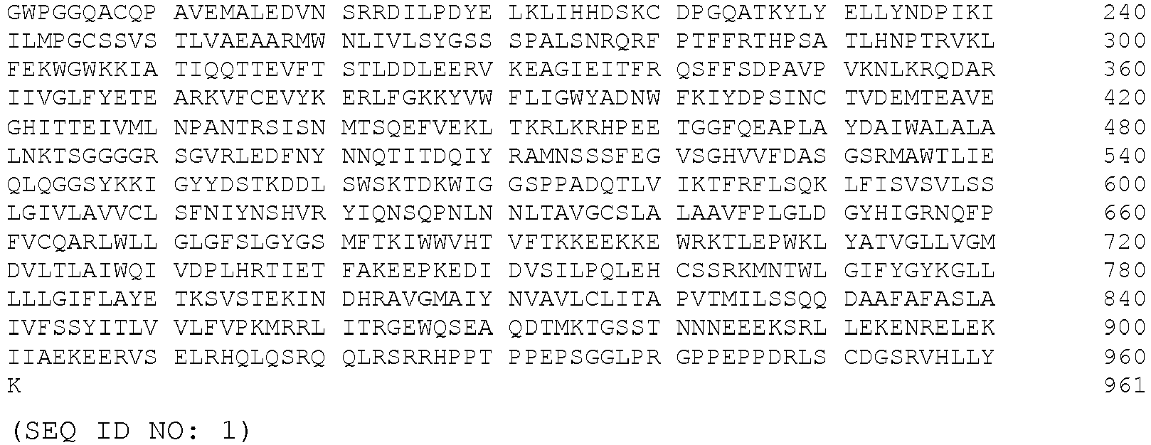
- In another embodiment, the B1 subunit of GABAB receptor has the sequence as set forth below:

- In another embodiment, the B1 subunit of GABAB receptor has the sequence as set forth below:

- In another embodiment, the B1 subunit is a homologue of SEQ ID NOs: 1, 2, or 3. In another embodiment, the B1 subunit is a variant of SEQ ID NOs: 1, 2, or 3. In another embodiment, the B1 subunit is an isomer of SEQ ID NOs: 1, 2, or 3. In another embodiment, the B1 subunit is a fragment of SEQ ID NOs: 1, 2, or 3. In another embodiment, the B1 subunit comprises SEQ ID NOs: 1, 2, or 3.
- In another embodiment, the B1 subunit of GABAB receptor is encoded by a nucleotide sequence having the sequence:


- In another embodiment, the B1 subunit of GABAB receptor is encoded by a nucleotide sequence having the sequence:


- In another embodiment, the B1 subunit of GABAB receptor is encoded by a nucleotide sequence having the sequence:


- In another embodiment, the B1 subunit is encoded by a nucleotide molecule that is a homologue of SEQ ID NOs: 4, 5, or 6. In another embodiment, the nucleotide molecule is a variant of SEQ ID NOs: 4, 5, or 6. In another embodiment, the nucleotide molecule is an isomer of SEQ ID NOs: 4, 5, or 6. In another embodiment, the nucleotide molecule is a fragment of SEQ ID NOs: 4, 5, or 6. In another embodiment, the nucleotide molecule comprises SEQ ID NOs: 4, 5, or 6.
- The epitope recognized by an antibody detected by a method of the present invention is, in another embodiment, a conformational epitope. In another embodiment, the epitope is a linear epitope. In another embodiment, the epitope is any other type of epitope known in the art.
- In one embodiment, ectopic expression of B1 subunit by nervous tissue contained in the teratomas contributes to break immune tolerance. In another embodiment, a combination of factors such as an adjuvant effect of the prodromal viral-like illness that occur in most subjects, and a genetic predisposition in certain embodiments, play additional roles in the initiation of the immune response tested for using the diagnosis methods described herein.
- In one embodiment, a pathogenic role of B1 antibodies in paraneoplastic anti-GABAB encephalitis is shown by the correlation between patients' symptoms and antibody titers.
- In another embodiment, the subject exhibits antibodies that react with an extracellular neuronal antigen. In another embodiment, the subject exhibits antibodies that react with an antigen exposed on the cell surface of a neuron. In another embodiment, patients with antibodies to extracellular antigens exhibit, under the conditions utilized herein, enhanced responsiveness to immune therapy.
- In another embodiment, the present invention provides a method of diagnosing autoimmune encephalitis in a subject, the method comprising testing a biological sample from the subject for an antibody to the B1 subunit of a GABAB receptor, whereby the presence of said antibody in said biological sample is indicates an autoimmune encephalitis.
- In another embodiment, the present invention provides a method of diagnosing a tumor associated with an autoimmune encephalitis in a subject, the method comprising testing a biological sample from the subject for an antibody to the B1 subunit of a GABAB receptor, whereby the presence of said antibody indicates the presence of an occult tumor in said subject and that said tumor is a cause of said autoimmune encephalitis.
- In another embodiment, the present invention provides a method of diagnosing a tumor causing epilepsy in a subject, the method comprising testing a biological sample from the subject for the presence of an antibody to the B1 subunit of a GABAB receptor, whereby the presence of said antibody indicates the presence of a tumor in said subject and said tumor is a cause of said epilepsy.
- The application discloses a method of diagnosing a tumor in a subject having an epilepsy, comprising the step of: obtaining a biological sample from the subject; and testing the biological sample for the presence of an antibody to the B1 subunit of a GABAB receptor, whereby the presence of said antibody indicates the presence of a tumor in said subject, thereby diagnosing a tumor in a subject having an epilepsy.
- The present application also discloses a method of treating autoimmune encephalitis in a subject, comprising the steps of: detecting a tumor associated with an autoimmune encephalitis by testing a body fluid from the subject for an antibody to the B1 subunit of a GABAB receptor, whereby a presence of said antibody indicates a presence of said tumor in said subject and that said tumor is a cause of said autoimmune encephalitis; and treating said tumor. The tumor may be treated within four months of the onset of a symptom associated with autoimmune encephalitis.
- Methods for testing a reactivity of a body fluid against neuronal antigens are well known in the art. In one embodiment, enzyme-linked immunoabsorption assay (ELISA) is used to test for the presence of an antibody. In another embodiment, immunocytochemistry is used to test for the presence of an antibody. In another embodiment, immunoprecipitation is used to test for the presence of an antibody. In another embodiment, one of the methods enumerated herein is utilized. In another embodiment, neuronal tissue is fixed with PFA. In another embodiment, any other method known in the art is utilized.
- Methods and kits for detection of antibodies are well known in the art, and are described, for example, in Ances BM et al (Treatment-responsive limbic encephalitis identified by neuropil antibodies: MRI and PET correlates. Brain 2005;128:1764-1777) and Vitaliani et al (Paraneoplastic encephalitis, psychiatric symptoms, and hypoventilation in ovarian teratoma. Ann Neurol 2005;58:594-604.).
- Methods for diagnosing limbic encephalitis (LE) are well known in the art. In another embodiment, patients with LE develop subacute confusion, irritability, depression, sleep disturbances, seizures, short-term memory loss, and/or dementia. In another embodiment, the pathological substrate of LE is an inflammatory disorder that predominantly involves the limbic system (hippocampi, amygdala, and cingulate gyrus). In another embodiment, biopsy and autopsy studies demonstrate interstitial and perivascular infiltrates of T cells, and less frequently B cells, along with microglial activation, neuronal degeneration, and/or gliosis. In another embodiment, inflammatory infiltrates are found in areas distant from the limbic system. In another embodiment, the infiltrates remain mild and clinically silent. In another embodiment, the infiltrates become prominent and develop into a disorder called encephalomyelitis. Additional methods of diagnosing LE are described, for example, in Gultekin SH et al (Paraneoplastic limbic encephalitis: neurological symptoms, immunological findings and tumour association in 50 patients. Brain 2000;123:1481-1494).
- The terms "homology," "homologous," etc, when in reference to any protein or peptide, refer, in one embodiment, to a percentage of amino acid residues in the candidate sequence that are identical with the residues of a corresponding native polypeptide, after aligning the sequences and introducing gaps, if necessary, to achieve the maximum percent homology, and not considering any conservative substitutions as part of the sequence identity. Methods and computer programs for the alignment are well known in the art.
- Homology is, in another embodiment, determined by computer algorithm for sequence alignment, by methods well described in the art. For example, computer algorithm analysis of nucleic acid sequence homology can include the utilization of any number of software packages available, such as, for example, the BLAST, DOMAIN, BEAUTY (BLAST Enhanced Alignment Utility), GENPEPT and TREMBL packages.
- In another embodiment, "homology" refers to identity to a sequence selected from SEQ ID NO: 1-6 of greater than 70%. In another embodiment, "homology" refers to identity to a sequence selected from SEQ ID NO: 1-6 of greater than 72%. In another embodiment, "homology" refers to identity to one of SEQ ID NO: 1-6 of greater than 75%. In another embodiment, "homology" refers to identity to a sequence selected from SEQ ID NO: 1-6 of greater than 78%. In another embodiment, "homology" refers to identity to one of SEQ ID NO: 1-6 of greater than 80%. In another embodiment, "homology" refers to identity to one of SEQ ID NO: 1-6 of greater than 82%. In another embodiment, "homology" refers to identity to a sequence selected from SEQ ID NO: 1-6 of greater than 83%. In another embodiment, "homology" refers to identity to one of SEQ ID NO: 1-6 of greater than 85%. In another embodiment, "homology" refers to identity to one of SEQ ID NO: 1-6 of greater than 87%. In another embodiment, "homology" refers to identity to a sequence selected from SEQ ID NO: 1-6 of greater than 88%. In another embodiment, "homology" refers to identity to one of SEQ ID NO: 1-6 of greater than 90%. In another embodiment, "homology" refers to identity to one of SEQ ID NO: 1-6 of greater than 92%. In another embodiment, "homology" refers to identity to a sequence selected from SEQ ID NO: 1-6 of greater than 93%. In another embodiment, "homology" refers to identity to one of SEQ ID NO: 1-6 of greater than 95%. In another embodiment, "homology" refers to identity to a sequence selected from SEQ ID NO: 1-6 of greater than 96%. In another embodiment, "homology" refers to identity to one of SEQ ID NO: 1-6 of greater than 97%. In another embodiment, "homology" refers to identity to one of SEQ ID NO: 1-6 of greater than 98%. In another embodiment, "homology" refers to identity to one of SEQ ID NO: 1-6 of greater than 99%. In another embodiment, "homology" refers to identity to one of SEQ ID NO: 1-6 of 100%.
- In another embodiment, homology is determined via determination of candidate sequence hybridization, methods of which are well described in the art (See, for example, "Nucleic Acid Hybridization" Hames, B. D., and Higgins S. J., Eds. (1985); Sambrook et al., 2001, Molecular Cloning, A Laboratory Manual, Cold Spring Harbor Press, N.Y.; and Ausubel et al., 1989, Current Protocols in Molecular Biology, Green Publishing Associates and Wiley Interscience, N.Y). In other embodiments, methods of hybridization are carried out under moderate to stringent conditions, to the complement of a DNA encoding a native caspase peptide. Hybridization conditions being, for example, overnight incubation at 42 °C in a solution comprising: 10-20 % formamide, 5 X SSC (150 mM NaCl, 15 mM trisodium citrate), 50 mM sodium phosphate (pH 7. 6), 5 X Denhardt's solution, 10 % dextran sulfate, and 20 µg/ml denatured, sheared salmon sperm DNA.
- Protein and/or peptide homology for any amino acid sequence listed herein is determined, in another embodiment, by methods well described in the art, including immunoblot analysis, or via computer algorithm analysis of amino acid sequences, utilizing any of a number of software packages available, via established methods. Some of these packages include the FASTA, BLAST, MPsrch or Scanps packages, and, in another embodiment, employ the use of the Smith and Waterman algorithms, and/or global/local or BLOCKS alignments for analysis, for example.
- As used herein, "nucleic acids" or "nucleotide" refers to a string of at least two base-sugar-phosphate combinations. The term includes, in one embodiment, DNA and RNA. "Nucleotides" refers, in one embodiment, to the monomeric units of nucleic acid polymers. RNA is, in one embodiment, in the form of a tRNA (transfer RNA), snRNA (small nuclear RNA), rRNA (ribosomal RNA), mRNA (messenger RNA), anti-sense RNA, small inhibitory RNA (siRNA), micro RNA (miRNA) and ribozymes. The use of siRNA and miRNA has been described (Caudy AA et al, Genes & Devel 16: 2491-96 and references cited therein). DNA can be, in other embodiments, in form of plasmid DNA, viral DNA, linear DNA, or chromosomal DNA or derivatives of these groups. In addition, these forms of DNA and RNA can be single, double, triple, or quadruple stranded. The term also includes, in another embodiment, artificial nucleic acids that contain other types of backbones but the same bases. In one embodiment, the artificial nucleic acid is a PNA (peptide nucleic acid). PNA contain peptide backbones and nucleotide bases and are able to bind, in one embodiment, to both DNA and RNA molecules. In another embodiment, the nucleotide is oxetane modified. In another embodiment, the nucleotide is modified by replacement of one or more phosphodiester bonds with a phosphorothioate bond. In another embodiment, the artificial nucleic acid contains any other variant of the phosphate backbone of native nucleic acids known in the art. The use of phosphothiorate nucleic acids and PNA are known to those skilled in the art, and are described in, for example, Neilsen PE, Curr Opin Struct Biol 9:353-57; and Raz NK et al Biochem Biophys Res Commun. 297:1075-84. The production and use of nucleic acids is known to those skilled in art and is described, for example, in Molecular Cloning, (2001), Sambrook and Russell, eds. and Methods in Enzymology: Methods for molecular cloning in eukaryotic cells (2003) Purchio and G. C. Fareed.
- Patients and Controls. The inventors of the instant application studied 410 patients for encephalitis suspected to be paraneoplastic or immune mediated against neuronal cell surface antigens. Antibodies were identified in 357: 275 had antibodies to the NR1 subunit of the NMDAR, 27 voltage-gated potassium channels (VGKC), 19 glutamic acid decarboxylase (GAD), 15 GluR1/2 subunits of the AMPAR, 11 Ma2, 8 HuD, and 2 against CRMP5. Of the remaining 53 patients, 15 had serum or CSF antibodies against neuronal cell surface antigens predominantly visible in the neuropil of rat brain, all showing a similar immunostaining. These features and the dramatic response to treatment of the index case (patient # 1) focused the current study in these 15 patients. Information was obtained by the authors or provided by referring physicians. CSF or serum from 104 patients, including 91 randomly selected from the above and 13 with GAD-antibody associated syndromes served as controls.
- Animal tissue, antibodies, and immunohistochemistry on rat brain. Female Wistar rats were euthanized and the brain was removed, sagittally sectioned, immersed in 4% paraformaldehyde at 4°C for 2 hours, cryoprotected with 40% sucrose for 24 hours, and snap frozen in chilled isopentane.
- Immunohistochemistry on rat brain and human tissue. Paraffin-embedded tissue was deparaffinized and the antigens retrieved. Seven-micrometer-thick frozen (or 4 µm paraffin) tissue sections were serially incubated with 0.3% H2O2 for 20 minutes, 10% goat serum for 1 hour, and patient or control serum (1:250), CSF (1:10), or a guinea pig polyclonal antibody against an intracellular epitope of the GABAB receptor (1:200; Invitrogen, Carlsbad, CA) at 4°C overnight. After using the appropriate secondary antibodies (all 1:2,000), reactivities were developed with the avidin-biotin-peroxidase method. Results were photographed under a fluorescence microscope using Zeiss Axiovision software (Zeiss, Thornwood, NY).
- Immunohistochemistry with human tissue (SCLC tumors) utilized IgG purified from patients' sera and labeled with biotin. In these studies no secondary antibody was needed, avoiding background caused by irrelevant human IgG present in the tissue.
- Immunohistochemistry on frozen sections of rat brain was performed with patients' or control serum (1:250), CSF (1:10), or GABAB1 antibody using a standard avidin-biotin peroxidase method, or double immunolabeling with patients' serum or CSF and the GABAB1 antibody, followed by the appropriate Alexa Fluor secondary antibodies.
- Immunocytochemistry on neuronal cultures and HEK293 cells. Rat hippocampal neuronal cultures were prepared. Live neurons grown on coverslips were incubated for 1 hour at 37°C with patient or control serum (final dilution 1:200) or CSF (1:10). After removing the media and extensive washing with phosphate-buffered saline (PBS), neurons were fixed with 4% paraformaldehyde, permeabilized with 0.1% Triton X-100, and single or double immunolabeled with a guinea pig polyclonal GABAB1 receptor antibody (1:200), followed by the corresponding Alexa Fluor secondary antibodies diluted 1:2,000 (Molecular Probes, Eugene, OR). Results were photographed under a fluorescence microscope using Zeiss Axiovision software (Zeiss, Thornwood, NY).
- HEK293 cells were transfected with plasmids containing rodent GABAB1 or GABAB2 subunits of the GABAB receptor or plasmids without insert (control), using a method previously reported. In other experiments, cells were cotransfected with GABAB1 and GABAB2 in equimolar ratios. Cells were grown for 24 hours after transfection before assessment. Transfected cells were fixed in 4% paraformaldehyde, permeabilized with 0.1% Triton X-100 and then incubated with patients' serum (1:200) or CSF (100%) and the indicated guinea pig polyclonal GABAB1 receptor antibody (1:20,000) or a polyclonal GABAB2 receptor antibody (1:10,000, generated by Dr. Moss) overnight at 4°C, washed in PBS, and incubated with the appropriate Alexa Fluor secondary antibodies (1:2,000; Molecular Probes). Results were photographed under a fluorescence microscope using Zeiss Axiovision software (Zeiss, Thornwood, NY).
- Antibody titers were determined using HEK293 cells expressing GABAB1/B2 incubated with serial dilutions of serum and CSF, starting at 1/2 dilution.
- Patients' antibody IgG subtypes were determined in serum or CSF using the indicated HEK293 transfected cells, and secondary anti-human antibodies against IgG1, IgG2, IgG3, and IgG4 (all diluted 1:200; Sigma, St. Louis, MO) as reported.
- Immunoprecipitation and immunoblot. Live neurons obtained as above, were grown in 100mm wells (
density 106 neurons/well), and incubated at 37°C with filtered patient serum (diluted 1:500) for 1 hour. Neurons were then washed with PBS, lysed with buffer (NaCl 150mM, EDTA 1mM, tris(hydroxymethyl)aminomethane [Tris]-HCl 100mM, deoxycholate acid 0.5%, 1% Triton X-100 [Sigma Labs, St. Louis, MO], pH 7.5) containing protease inhibitors (P8340; Sigma Labs), and centrifuged at 16.1 x 10³ g for 20 minutes at 4°C. The supernatant was retained and incubated with protein A/G agarose beads (20423; Pierce, Rockford, IL) overnight at 4°C, centrifuged, and the pellet containing the beads with patients' antibodies bound to the target cell surface antigen was then washed with PBS, aliquoted, and kept at -80°C. An aliquot of this pellet was resuspended in Laemmli buffer, boiled for 10 minutes, separated in a 4 to 15% sodium dodecyl sulfate polyacrylamide gel electrophoresis, and the proteins visualized with EZBlue gel staining (G1041; Sigma Labs). Distinctive protein bands precipitated by patient serum were excised from the gel and analyzed using mass spectrometry at the proteomic facility at the University of Pennsylvania. After characterization of the antigen, frozen aliquots of the indicated pellets were separated in a sodium dodecyl sulfate polyacrylamide gel electrophoresis as described earlier, transferred to nitrocellulose (162-0115; Bio-Rad, Hercules, CA), and blotted with the indicated polyclonal antibodies against GABAB1 (1:2000) or GABAB2 (1:1000) receptor subunits. The reactivity was developed using the appropriate biotinylated secondary antibodies (1:2000) and the avidin-biotin peroxidase, diaminobenzidine method. - Quantitative analysis of GABAB receptor clusters using confocal microscopy. Fourteen to 21-day in vitro (div) live rat hippocampal neurons were incubated with patient CSF (1:30 dilution in Neuro-Basal B27 medium; GIBCO, Carlsbad, CA) for 24 hours, washed, fixed in paraformaldehyde (4% paraformaldehyde, 4% sucrose in PBS) for 5 minutes, permeabilized with 0.25% Triton X-100 for 10 minutes, and blocked with 5% normal goat serum for 1 hour. Neurons were then incubated with a guinea pig polyclonal antibody against an intracellular epitope of the GABAB receptor (1:1000; Invitrogen) and a mouse monoclonal antibody against the presynaptic marker Bassoon (1:200; Stressgen, Victoria, British Columbia, Canada), washed, and incubated with the appropriate fluorescent-conjugated secondary antibodies (1:1000, Molecular Probes).
- Images were obtained using a laser-scanning confocal microscope (Leica TCS SP2; Leica, Deerfield, IL). For each image, laser light levels and detector gain and offset were adjusted so that no pixel values were saturated. Images of labeled neurons were stored digitally for subsequent analysis. Images were thresholded, areas of interest containing dendrites were selected, and the number of individual clusters along neuronal dendrites was determined using interactive software (ImageJ; Research Services Branch, National Institute of Mental Health, Bethesda, MD). The co-localization of clusters labeled with patient's antibodies, commercial GABAB antibodies and the synaptic marker Bassoon was quantified using a software Macro implemented in ImageJ.
- Index Patient (Case #1 in tables): A 60-year-old woman was hospitalized for confusion, memory problems, and new onset generalized tonic-clonic and partial complex seizures refractory to treatment. At examination, she was disoriented to place and time, and had extremely poor concentration and short-term memory. Aside from saccadic pursuits with lateral gaze, no cranial nerve abnormalities were noted. Strength, sensation, reflexes, and coordination were normal. MRI of the brain revealed bilateral medial temporal lobe fluid-attenuated inversion recovery (FLAIR) increased signal compatible with limbic encephalitis (
Figure 1a ). EEGs showed diffuse slowing and bilateral periodic lateralized epileptiform discharges (PLEDS). CSF demonstrated 9 white blood cells (WBC)/µl,total protein concentration 35 mg/dL, glucose 71 mg/dL, and negative oligoclonal bands and cytology. PCR for herpes simplex virus, West Nile virus, and St. Louis encephalitis were negative. Hyponatremia (119 mEq/L) due to syndrome of inappropriate anti-diuretic hormone (SIADH) secretion was noted. Serum and CSF analysis of paraneoplastic antibodies was negative. Combined CT and fluorodeoxyglucose (FDG)-PET revealed mediastinal lymphadenopathy, proven by biopsy to be small-cell lung cancer (SCLC). The patient was treated with antiepileptics (levetiracetam, valproic acid, phenytoin) and immunotherapy (intravenous immunoglobulins [IVIg], corticosteroids), immediately followed by chemotherapy with cisplatin and etoposide. There was a rapid and dramatic improvement of short term memory and cognition, and the seizures completely resolved. After completion of these treatments the patient received standard prophylactic whole brain radiation therapy. Repeat brain MRI one month after symptom presentation showed improvement of the abnormal FLAIR signal (Figure 1b ); subsequent MRIs 3- and 9-months later were unchanged except for progressive general atrophy, likely secondary to radiation (Figure 1c, d ). One year after symptom presentation, the patient had only mild memory and cognitive deficits and lived independently. - Detection of antibodies against a novel neuronal cell surface autoantigen. Analysis of serum and CSF of the index case and 14 additional patients from the indicated selection of cases showed reactivity with the neuropil of rat brain (
Figure 2 ), in a pattern different from that reported with the NR1 subunit of the NMDAR, GluR1/2 subunits of the AMPAR, or VGKC antibodies. To determine whether the antibodies reacted with extracellular epitopes, non-fixed and non-permeabilized cultures of rat hippocampal neurons were incubated with patients' serum or CSF, showing intense reactivity with the cell surface (Figure 3 a) . - The neuronal cell surface antigen is the metabotropic GABAB receptor . To identify the cell surface antigen, live rat hippocampal neurons were incubated with patients' antibodies and the target antigen was immunoprecipitated. These studies produced two protein bands of approximately 105 and 90 kDa (
Figure 3b ) that were analyzed by mass spectrometry demonstrating sequences derived from the B1 subunit of the metabotropic GABAB receptor. Because the GABAB receptor is a heterodimer composed of two subunits, B1 and B2, both with a similar molecular weight, the inventors next examined by immunoblot and polyclonal antibodies specific for each subunit to determine whether patients' antibodies had precipitated both subunits. These studies confirmed that patient antibodies co-precipitated B1 and B2 subunits of the GABAB receptor (Figure 3c ). - The specificity of patient antibodies for GABAB receptors was determined by triple immunolabeling of cultured neurons (patient antibodies; a guinea pig polyclonal antibody to a non-competing intracellular GABAB1 receptor epitope; and a pre-synaptic marker [Bassoon]), and the degree of co-localization quantified by confocal microscopy, as reported (
Figure 4 ). The binding of antibodies to GABAB receptor clusters was analyzed on the dendrites of 24 neurons on 4 separate coverslips. This showed that 103±0.8% of patient antibody-labeled GABAB receptor clusters co-localized with clusters labeled by the guinea pig polyclonal GABAB receptor antibody, and 107±0.7% of guinea pig antibody-labeled clusters co-localized with those labeled by patient antibodies (numbers slightly higher than 100% occur due to overlapping of a few clusters labeled by patient antibodies with two guinea pig antibody-labeled clusters and vice-versa). These results indicate that essentially all patient's anti-neuronal cell surface antibodies target the GABAB receptors and that virtually all neuronal GABAB receptors are labeled by patient's antibodies. - To determine which of the two GABAB receptor subunits contained the target epitope, HEK293 cells were transfected with GABAB1, GABAB2, or both receptor subunits and immunocytochemically tested with patients' antibodies. All 15 patients had serum or CSF antibodies that specifically reacted with the GABAB1 receptor subunit (
Figure 5 ), and one had additional reactivity with the GABAB2 subunit. These findings indicate that HEK293 cells expressing GABAB1/B2 or GABAB1 receptor subunits provide a substrate for an unambiguous diagnostic test. Similar studies with the 104 control cases showed that two patients, both with syndromes attributed to GAD autoimmunity, had GABAB1 receptor antibodies at low titers (CSF 1:2, serum negative), not visible with brain immunohistochemistry. - Analysis of antibody IgG subtypes was performed in serum or CSF of 6 randomly selected patients. All had IgG1 GABAB1 antibodies; two had additional IgG3, and one IgG2 antibodies.
- Neurological symptoms. Demographic information, clinical features, treatment, and outcome are shown in Tables 1 and 2. Among the 15 patients with high titer GABAB1 receptor antibodies and limbic encephalitis (cases #1-15), the median age was 62 years (range, 24-75); 8 were men. Thirteen patients presented with subacute onset of seizures, confusion, and severe memory impairment, and in two (#3 and 13) the memory deficit and confusion preceded the seizures by a few weeks. Most seizures had a temporal lobe onset with secondary generalization, and three patients had status epilepticus. Ten patients had uni- or bilateral medial temporal lobe FLAIR/T2 increased signal consistent with limbic encephalitis, 1 had a small area of increased FLAIR signal in the corpus callosum, and 4 had normal brain MRI. The CSF was abnormal in 9 of 10 patients for whom data was available. The most common abnormality was lymphocytic pleocytosis in 8 patients. EEG studies were available from 12 patients; 9 showed temporal lobe seizures, epileptiform discharges, or temporal lobe slowing; 2 had generalized slowing and 1 was normal. Several types of seizures were noted on EEG, including complex partial seizures (often of temporal lobe onset), status epilepticus, and subclinical seizures.
- The two control cases with low titer of GABAB1 antibodies (cases #16 and 17 in Tables 1 and 2) developed different syndromes in association with high titer GAD antibodies in serum and CSF. Neither of these 2 patients developed seizures or limbic dysfunction. One had progressive cerebellar ataxia, and the other gait instability, muscle stiffness, rigidity, myoclonus, and dysarthria, categorized as encephalomyelitis with rigidity.
Table 1: Demographic Features and symptoms Case No. Sex/Age (yr) Tumor Presenting Symptoms Other clinical-immunological features 08-299 F/60 SCLC Subacute onset of complex partial seizures, confusion, memory impairment SIADH 06-165 M/66 SCLC Subacute onset of seizures, confusion, memory deficit, behavioral problems N-type VGCC antibodies 06-235 F/53 SCLC Rapidly progressive memory deficits, abnormal sleeping habits, followed by frequent seizures (focal, secondarily generalized), confusion, decline in mental status leading to coma Pruritic rash with initial weakness. 08-017 M/75 Mediastinal adenopathy Subacute onset of seizures, psychosis, encephalitis. Rapidly progressed to death before definitive diagnosis or treatment. Poor respiratory status, refused intubation 08-152 M/68 Neuroendocrine tumor of the lung Subacute onset of seizures, status epilepticus, confusion - 08-623 F/43 CT and FDG/PET negative Subacute onset of secondarily generalized tonic-clonic seizures, bizarre behaviors, delusions, paranoia, memory impairment. N-type VGCC antibodies 09-086 M/69 CT and FDG/PET negative Subacute onset of right temporal lobe seizures, status epilepticus, severe encephalopathy, severe memory deficit, confusion Prior history of bipolar disorder 07-305 F/24 CT and FDG/PET negative Subacute onset of seizures, status epilepticus, confusion, fever. Required intubation and ventilation due to poor level of consciousness and airway protection N-type VGCC antibodies 06-026 M/63 CT and FDG/PET negative Subacute onset of seizures, memory deficit, paranoia, psychosis, gustatory hallucinations TPO and GAD antibodies; hypothyroidism and type 2 diabetes mellitus 08-278 F/45 Benign ovarian mass Subacute onset of complex partial an generalized seizures and short-term memory deficits - 09-176 F/62 CT chest, abdomen, pelvis negative Subacute onset generalized seizures, confusion, decreased level of consciousness, fluent aphasia, abnormal orolingual movements. - 07-004 M/29 None Subacute onset of left temporal lobe epilepsy; generalized tonic-clonic refractory seizures. No cognitive deterioration. Childhood seizures 09-083 F/30 CT and FDG/PET negative 3 month history of severe memory deficit, followed by seizures (generalized, subclinical) GAD antibodies without endocrinopathy 01-696 Graus M/69 SCLC Subacute onset of generalized tonic-clonic seizures, worsened short-term memory deficit Mild short-term memory deficit from past history of subarachnoid hemorrhage 07-1051 Graus M/70 SCLC Subacute onset of seizures (partial motor and generalized). Severe short-term memory loss, confabulation, visual hallucinations, disorientation, agitation GAD, TPO, and SOX1 antibodies. No endocrinopathy 08-114 F/63 CT and FDG/PET negative 1 year progression of cerebellar ataxia. Normal mental status, no seizures, no muscle spasms or stiffness GAD antibodies, adult onset insulin dependent diabetes mellitus 08-398 F/61 CT and FDG/PET negative 6 week history of gait disturbance, lower extremity myoclonus and stiffness; dysphagia, dysarthria, nystagmus, left gaze palsy. No seizures or cognitive symptoms GAD, TPO and thyroglobulin antibodies (mild thyroid dysfunction) SCLC: small cell lung cancer; FDG/PET: Fluoro-deoxyglucose/ positron emission tomography; SIADH: syndrome of inappropriate anti-diuretic hormone; GAD: glutamic-acid decarboxylase; TPO: thyroid peroxidase; VGCC: voltage-gated calcium channels. Table 2: Diagnostic tests, treatment and outcome. Case No. MRI CSF Serum CSF titers Chronological list of treatments Outcome (duration of follow-up) 08-299 FLAIR/T2 increased signal in medial temporal lobes 9 WBC/µL; protein 35; no OCBsS, 1:640 CSF, 1:160 IVIg, corticosteroids, chemotherapy Substantial improvement. Mild residual short term memory deficit. Lives independently. Seizure free (12 months) 06-165 Normal Normal S, 1:1280 CSF, n/a Corticosteroids, IVIg, chemotherapy Substantial improvement. Died of metastatic disease (15 months) 06-235 FLAIR/T2 increased signal in medial temporal lobes n/a S, 1:160 CSF, n/a Tumor removal (lobectomy) IVIg Partial improvement after tumor removal and IVIg (4 months); lost to follow-up. 08-017 Normal n/a S, 1:2560 CSF, 1:640 None Died soon after presentation of rapidly progressive respiratory failure 08-152 FLAIR/T2 increased signal in medial temporal lobes n/a S, 1:1280 CSF, n/a Supportive Died 6 months after symptom presentation. GABAB antibodies detected after patient's death in archived serum 08-623 FLAIR/T2 increased signal in small area of corpus callosum 95 WBC/ µL; protein 104; increased IgG index S, n/a CSF, 1:640 Corticosteroids, mycophenylate mofetil Substantial improvement. Lives independently. Seizure free (9 months) 09-086 FLAIR/T2 increased signal in left mesial temporal lobe n/a S, n/a CSF, 1:640 Corticosteroids, plasma exchange Initial substantial response to corticosteroids. Relapsed 1 month later. Died after 5 months in ICU with refractory seizures, status epilepticus, and systemic complications. 07-305 FLAIR/T2 increased signal in medial temporal lobes 19 WBC/ µL; protein 46 S, 1:5120 CSF, 1:2560 Corticosteroids, plasma exchange Substantial improvement. Mild residual short-term memory deficit. Seizures free (3 months) 06-026 FLAIR/T2 increased signal in medial temporal lobes 75 WBC/ µL; protein 26 ; + OCBs S, negative CSF, 1:4 Corticosteroids Full recovery (41 months) 08-278 FLAIR/T2 increased signal in medial temporal lobes 81 WBC, protein 30 S, 10,240 CSF, n/a Corticosteroids Substantial improvement. Residual short-term memory deficit. Lives independently. Seizure free (72 months) 09-176 Normal 20 WBC/ µL; protein 22 S, 1:40 CSF, 1:40 Corticosteroids Full recovery (6 months) 07-004 FLAIR/T2 increased signal in left medial temporal lobe and insula 950 WBC/ µL; + OCB S, negative CSF, 1:10 Symptomatic Temporal lobe biopsy 20 months after symptom presentation showing reactive astrocytosis, without inflammation. No follow-up available after biopsy.09-083 FLAIR/T2 increased signal in medial temporal lobes 4 WBC/µL; protein 109; 6 OCBs S, negative CSF, 1:4 Corticosteroids Full recovery, except for infrequent brief episodes of visual hallucinations (10 months) 01-696 Graus FLAIR/T2 increased signal in left medial temporal lobe Traumatic; negative cytology. Chemotherapy Residual short-term memory deficit; seizures controlled; died of sepsis (3 months) 07-1051 Graus Normal 0 WBC/µL; protein 95 S, n/a CSF, 1:640 IVIg, corticosteroids, chemotherapy Seizures responded to antiepileptics; memory deficit persisted; died of cancer-related treatment (2 months) 08-114 Normal 3 WBC/µL; protein 78; 1 OCB S, negative CSF, 1:2 IVIg No seizures or cognitive deficits. Limited response of cerebellar ataxia to IVIg (12 months) 08-398 Normal 2 WBC/µL; Protein 52; + OCBs S, negative CSF, 1:2 IVIg, corticosteroids No seizures or cognitive deficits. Full recovery after steroids and IVIg (12 months) WBC: white blood cells (normal <4 µl), FLAIR: fluid-attenuated inversion recovery; n/a: not available; IVIg: intravenous immunoglobulin. ICU: intensive care unit. - Other autoantibodies . In addition to GABAB antibodies, 6 of 15 patients (40%) had antibodies to one or more of the following: 3 GAD, 2 thyroid peroxidase (TPO), 3 N-type VGCC, and 1 SOX1 antibodies. Only 1 of the 3 patients with GAD antibodies had endocrinopathy, and 1 of the 3 patients with VGCC antibodies had a SCLC. The patient with SOX1 antibodies had a SCLC.
- Associated Tumors. Seven patients had a lung nodule or mediastinal adenopathy, with pathological confirmation of SCLC or neuroendocrine tumor in 6. In all instances the tumor was detected at the time of neurological symptom presentation. Since most lung tumors were diagnosed by needle biopsy, no tissue was available for analysis of GABAB receptor expression. Yet, 3 of 4 SCLCs from control cases (without antibodies or encephalitis) showed reactivity with a guinea pig polyclonal antibody to GABAB1 receptor and patients' biotinylated IgG suggesting that these receptors are expressed by SCLC.
- Treatment and Outcome. Nine patients (60%) had substantial neurological response to immunotherapy or treatment of the tumor. The median follow-up of these patients is 10 months (3-72); 1 subsequently died of tumor progression (15 months) and 1 was lost to follow-up (4 months). Six patients did not show sustained neurological improvement; three (#4, 14, 15 in Tables 1 and 2) died soon after presentation of the disorder as a result of tumor or chemotherapy-related complications, and the other three were diagnosed with GABAB receptor antibodies after death (#5 and 7) or were lost to follow-up (#12). In the latter three cases an autoimmune process was only considered in patient #7 who received corticosteroids and plasma exchange; the other two patients did not receive immunotherapy. Overall, excluding the two patients that were lost to follow-up, neurological improvement correlated with prompt tumor control or immunotherapy (Fisher's exact test = 0.003).
- The inventors of the instant application have discovered a new form of autoimmune encephalitis that associates with antibodies to extracellular epitopes of the GABAB receptor and is potentially treatment-responsive. Based on clinical, MRI, and EEG findings, the brain regions most affected are the hippocampi and temporal lobes. Therefore it is not surprising that the resulting syndrome is indistinguishable from other types of limbic encephalitis, although some clinical and immunological features may suggest GABAB receptor autoimmunity. A notable finding is the development of prominent and severe seizures in all patients, representing the main reason for medical attention or hospital admission. Two frequently associated features are the identification of a lung cancer, and the concurrent presence of autoantibodies against antigens of unclear significance with the limbic syndrome. In contrast, pharmacological or genetic disruption of GABAB receptors result in phenotypes that closely resemble the limbic syndrome of patients with GABAB receptor antibodies. Moreover, in humans, a common GABAB receptor polymorphism associates with temporal lobe epilepsy.
- Forty seven percent of patients with encephalitis and GABAB receptor antibodies had a lung nodule or mediastinal adenopathy which pathological, radiological and demographic features (age range 53-75 years, all smokers) were consistent with a SCLC or neuroendocrine tumor. Therefore, GABAB autoimmunity is likely involved in a number of patients with limbic encephalitis and SCLC previously considered "without antibodies" or attributed to antibodies to intracellular antigens, particular if a dramatic (and unexpected) improvement occurred after treatment of the tumor or immunotherapy. Moreover, anti-GABAB receptor encephalitis can occur without cancer association. Five of such patients were young (median age 30 years, range 24-45), without history of smoking, negative comprehensive cancer screening including CT/FDG-PET, and in some cases a long-term follow-up (41 and 72 months), making unlikely the presence of an occult cancer in all cases. In this respect, anti-GABAB receptor encephalitis is similar to other synaptic autoimmunities of the central (NMDAR, AMPAR) or peripheral nervous system (acetylcholine receptor, P/Q-type VGCC) that may occur with or without cancer association. As occur in these disorders, 40% of patients with anti-GABAB receptor encephalitis (50% of those without tumor) had additional autoantibodies, indicating a propensity to autoimmunity. The most interesting immunological overlap was with antibodies to GAD (an intracellular antigen), bringing into consideration that a subset of patients with limbic encephalitis attributed to GAD autoimmunity may have antibodies to extracellular epitopes of the GABAB receptor as a plausible cause of the symptoms. All together, antibodies to the GABAB1 receptor can be used to diagnose encephalitis, usually manifesting as a limbic syndrome, but with early and prominent seizures. By the time antibodies are determined the serum titers can be very low, and examining both serum and CSF is preferred. Identification of these antibodies should prompt the search for a SCLC, although approximately 50% of patients do not have cancer. Treatment of the tumor and immunotherapy with corticosteroids, IVIg, or plasma exchange often results in improvement. The dramatic and specific antibody binding to GABAB receptor in live neurons and the similarity with experimental phenotypes in which the function of the receptor is abrogated shows the antibodies are pathogenic.
- We analyzed the frequency of GABABR-ab in 147 patients with LE or neurological syndromes associated with GAD-ab. We examined the presence of GABABR-ab in 70 LE patients (33 paraneoplastic with onconeural antibodies, 18 paraneoplastic without onconeural antibodies, and 19 idiopathic with either GAD-abor seronegative), and 77 patients with GAD-ab-associated neurological syndromes other than LE (29 stiff- person syndrome (SPS), 28 cerebellar ataxia, 14 epilepsy, and 6 with diverse paraneoplastic neurological syndromes). GABABR-ab were analyzed in stored samples of serum or CSF by indirect immunofluorescence on HEK293 cells transfected with GABAB1 and GABAB2 receptor subunits.
- GABABR-ab were detected in 10 of the 70 LE patients (14%). Eight had SCLC and two were idiopathic. One of the eight LE patients with SCLC had an additional onconeural antibody (Hu) and two GAD-ab. GABABR-ab were identified in 7 (70%) of the 10 patients with LE and SCLC without onconeural antibodies. GABABR-ab antibodies were not found in patients with GAD-ab and SPS, cerebellar ataxia or epilepsy. However, one patient with GAD-ab and cerebellar ataxia also presented GABABR-ab in the setting of an anaplastic carcinoid of the thymus.
- Our results show that GABABR-ab are the most common antibodies found in LE associated with SCLC previously considered "seronegative". In patients with GAD-ab, the frequency of GABABR-ab is low and only observed in the context of cancer.
- Patients. We reviewed all patients with final diagnosis of LE, or with other neurological syndromes associated with GAD-ab whose serum or CSF was sent to our laboratory (Barcelona, Spain) for analysis of antineuronal antibodies. LE was defined by the subacute onset of short-term memory loss, behavior change, seizures, and involvement of the temporal lobes by EEG, imaging studies, or postmortem examination. LE was considered definite paraneoplastic if a tumor was diagnosed or the serum presented well characterized onconeural antibodies (Hu, Yo, Ri, CV2, Ma2, amphiphysin). The diagnosis of definite idiopathic LE required the absence of cancer and well characterized onconeural antibodies, and a follow-up of at least three years. LE patients with a shorter follow-up were classified as possible idiopathic LE. Patients with GAD-ab were classified, as previously reported, in one of the following groups: stiff-person syndrome (SPS), cerebellar ataxia, isolated epilepsy, and paraneoplastic neurological syndromes. The information was obtained from forms filled out by the referring neurologists, telephone interviews, and review of the clinical records. The study was approved by the Ethic Committee of the Hospital Clinic.
- Immunological studies. Onconeural antibodies, SOX1-ab, and GAD-ab were screened by immunohistochemistry performed on frozen sections of paraformaldehyde-perfused rat cerebellum using an avidin-biotin immunoperoxidase technique and confirmed by immunoblot when indicated. GAD-ab were confirmed by radioimmunoassay. Neuropil antibodies were screened by immunohistochemistry on frozen sections of rat brain post-fixed with 4% paraformaldehyde. The presence of AMPA glutamate receptor antibodies was confirmed by immunofluorescence on HEK-293 cells transfected with plasmids containing the appropriate antigens, and voltage-gated potassium channel (VGKC) antibodies were confirmed by radioimmunoassay.
- GABABR-ab were screened on HEK293 cells transfected with plasmids containing rodent GABAB1 and GABAB2 in equimolar ratios. Positives samples were also analyzed by immunocytochemistry of rat hippocampal neuronal cultures. Both techniques have previously been described. Briefly, HEK293 transfected cells were incubated with the patients' serum (dilution 1:20) or CSF (1:2) for 1h at 37°C, washed, fixed with 4% paraformaldehyde, incubated with a rabbit polyclonal GABAB1 antibody (1:1000) (Santa Cruz Biotechnology, sc-14006; Santa Cruz, California) followed by the appropriate Alexa Fluor secondary antibodies (Molecular Probes, Eugene, OR). For immunocytochemistry of rat hippocampal neuronal cultures, live neurons grown on coverslips were incubated with the patients' serum (1:100) or CSF (1:2) for 1h at 37°C, washed, fixed with 4% paraformaldehyde, and immunoreacted with anti-human IgG Alexa Fluor secondary antibody. Results were photographed under a fluorescence microscope using Zeiss Axiovision software (Zeiss, Thornwood, NY). To confirm the specificity of the neuronal reactivity, all positive samples were pre-absorbed with the non neuronal cell line HEK293 to remove antibodies that could react with non neuronal specific surface antigens.
- Eleven patients tested positive for GABABR-ab on the screening of HEK293 cells transfected with the B1 and B2 subunits of the GABABR (
Figure 7 ). All positive samples immunoreacted in vivo with primary cultures of hippocampal neurons (Figure 8 ). GABABR-ab were positive in both serum and CSF in the 5 patients from whom paired samples were available. Median titer of GABABR-ab was 1/120 (range 40-2000) in serum and 1/60 (range 20-640) in the CSF. - We found GABABR-ab in 10 patients with LE. Positive GABABR-ab were identified more frequently in the group of paraneoplastic LE without onconeural antibodies (previously considered "seronegative") (Table 3). Seven (39%) of the 18 patients were GABABR-ab-positive and all had SCLC. In total, positive GABABR-ab were identified in 7 (70%) of the 10 patients with LE and SCLC without onconeural antibodies. The other three patients were positive for AMPAR-ab. The coincidence of GABABR-ab and GAD-ab occurred in 2 patients with SCLC whereas the other three patients with LE and GAD-ab associated with other tumors (thymoma 2, lymphoma) and were GABABR-ab-negative.
- In this study, we analyzed 33 patients with LE and onconeural antibodies, and only one, with Hu-ab and SCLC, tested positive for GABABR-ab. However, 4 of the 7 LE patients with GABABR-ab without onconeural antibodies presented antibodies against intracellular antigens (Table 4). Two patients had GAD-ab (one also SOX1-ab), one Hu-ab, and, in two cases previously reported, one had brain serine/threonine kinase (BRSK)2-ab and the other SOX1 and VGKC-ab.
- No tumor was identified in the remaining 2 GABABR-ab-positive patients but the follow-up is too short to classify them as definite idiopathic LE. None of the 5 patients with idiopathic LE and GAD-ab were positive for GABABR-ab (Table 3).
- A summary of the clinical features of the GABABR-ab-positive patients is presented in Table 4. Nine of the 10 patients with GABABR-ab and LE were men. Median age was 60 years (range.47-70 years). Seizures were the predominant and presenting symptom in 8 patients and 2 required admission to the intensive care unit for control of the seizures. All patients also presented confusion, disorientation, memory loss, or behavior changes consistent with encephalitis predominantly involving the limbic system. The CSF disclosed mild lymphocyte pleocytosis in 4 patients. Brain MRI showed increased fluid-attenuated inversion recovery signal in one or both hippocampus and amygdala in 7 patients. In 4 of them the initial brain MRI was reported normal. Only 1 patient had hyponatremia. Seven patients were treated with steroids, intravenous immunoglobulins or combination of both drugs. Three of the 8 patients with SCLC were also treated with chemotherapy. Only 2 patients made a complete recovery (one without cancer) and none of them had concurrent antineuronal antibodies. Partial responses to the indicated treatments were achieved in 4 with a relapse in one of them.
- GABABR-ab were not detected in 71 patients with GAD-ab and SPS, cerebellar ataxia or epilepsy. In contrast, one of the 6 patients with paraneoplastic neurological syndromes and GAD-ab was GABABR-ab positive (Table 3). She was a 57 year-old woman with a known anaplastic carcinoid of the thymus and bone metastases. She developed nausea, vomiting, gait instability and diplopia. Neurological examination disclosed a normal mental status, bilateral horizontal nystagmus, and cerebellar gait ataxia. The patient was treated with oral steroids and the symptoms slowly resolved over the ensuing three months.
Table 3. Frequency of GABABR-ab in 147 patients with limbic encephalitis or GAD-ab-associated neurological syndromes Syndrome Number of sera (CSF) GABABR-ab positive (%) Comments on positive cases Paraneoplastic LE 51 (21) 8 (16) With onconeural-ab1 33 (14) 1 (3) Hu-ab with SCLC Without onconeural-ab2 18 (7) 7 (39) All SCLC, GAD-ab: 2 Idiopathic LE3 19 (15) 2 (14) Short follow-up GAD-ab-positive, non-LE 77 (48) 1 (1) Stiff-person syndrome 29 (16) 0 (0) Cerebellar ataxia 28 (19) 0 (0) Epilepsy 14 (9) 0 (0) Paraneoplastic 6 (4) 1 (17) Cerebellar ataxia 1. Hu-ab (26), Ma2-ab (4), amphiphysin-ab (3). Lung cancer in 18 patients
2. GAD-ab in five patients. Lung cancer in 11 patients (SCLC:10; NSCLC:1)
3. GAD-ab in five patients. Definite idiopathic LE: 7 patients
SCLC: small cell lung carcinoma; LE: limbic encephalitisTable 4. Clinical features and outcome of patients with positive GABABR antibodies Case Age/ sex Cancer Presenting symptomsc MRI temporal lesions CSF pleocytosis (WBC) Other antineuronal-ab Treatment Outcome (months) 1 60/M SCLC Status epilepticus Left Yes (unknown) None Steroids, IVIg Partial control of seizures (dead from ICU complications) 2 57/F SCLC Seizures, behavior change Bilateral No None Steroids, IVIg Complete recovery d 3 66/M SCLC Seizures, confusion Normal Yes (18) GAD Steroids, IVIg Not available (short f-up) 4a 47/M SCLC Seizures, behavior change, memory impairment Bilateral Yes (20) SOX1, VGKC Steroids, IVIg, chemotherapy Partial recovery, relapsing course (dead from cancer progression) 5b 69/M SCLC Seizures, memory impairment, confusion Left Traumatic None Chemotherapy Partial response. Dead from cancer-related treatment(3 months) 6b 70/M SCLC Seizures, memory impairment, confusion Normal No GAD, SOX1 Steroids, IVIg, chemotherapy No response. Dead from cancer-related treatment(2 months) 7 58/M SCLC Seizures, memory impairment Bilateral Yes (15) Hu Steroids, IVIg, chemotherapy No response (dead from LE) 8a 61/M SCLC Memory impairment Bilateral No BRSK2 None No response. Lost when tumor was diagnosed 9 months later 9 61/M No Confusion, seizures, behavior change Normal No None IVIg Partial improvement. Severe ICU neuropathy 10 50/M No Seizures, behavior and memory impairment Bilateral No None Antiepileptics only Complete recovery 11 57/F Carcino id of thymus Subacute cerebellar ataxia Normal No GAD Steroids Complete recovery SCLC: small cell lung cancer; WBC: white blood cells; BRSK2: Brain serine/threonine kinase 2. a: patients previously reported in references 10 and 9; b: included in the initial series of GABAR-ab (reference 4); c: predominant symptom listed first; d: Recovery before the start of chemotherapy. - To see if GABABR-ab associate with other cases of paraneoplastic cerebellar degeneration (PCD), we analyzed the serum or CSF of a series of 45 patients with PCD and lung cancer (35 with SCLC). The majority (73%) were included in a previous study. These patients had Hu-ab (15%) or voltage-gated calcium channel antibodies (50%). However, all were negative for GABABR-ab.
- We found that GABABR-ab are the most common antibodies identified in patients with SCLC and LE previously considered "seronegative". Although the occurrence of GABABR-ab and GAD-ab was observed in an initial series of 15 patients, when we tested a larger series of patients with several types of neurological syndromes associated with GAD-ab, GABABR-ab were only identified in those who had a paraneoplastic syndrome.
- At the time of the initial description of Hu-ab as markers of neurological syndromes associated with SCLC, we observed that up to 50% of patients with LE were "seronegative". The syndrome of these patients was highly restricted to the limbic system and seemed to improve more often after treatment of the cancer than that of patients with Hu-ab. In the current study, 7 of 10 (70%) patients with LE and SCLC had GABABR-ab. The other 3 patients were positive for AMPAR-ab. Taken together, all patients with LE and SCLC previously considered seronegative for classical paraneoplastic (onconeural) antibodies had antibodies against synaptic receptors.
- In the current study we show that Hu-ab and GABABR-ab only occurred in 1 of 33 patients, suggesting that this specific association is uncommon. In contrast, we confirm that 40% of these patients had antibodies that are markers of the underlying SCLC (SOX1, BRSK2) or directed against VGKC or GAD.
- In this study, all patients with concurrent GABABR-ab and GAD-ab had a paraneoplastic disorder. In the initial series, 2 of the 8 patients with idiopathic LE encephalitis had additional GAD-ab. We did not find GABABR-ab in patients with LE or isolated epilepsy with GAD-ab except in the two patients with SCLC. Our five patients with idiopathic LE and GAD-ab were women with a median age of 29 years and four presented with seizures. To determine how often both antibodies coincide in patients with idiopathic LE, we suggest routinely looking for GABABR-ab in all patients with LE suspected to be related with GAD-ab.
- Having described preferred embodiments of the invention with reference to the accompanying drawings, it is to be understood that the invention is not limited to the precise embodiments, and that various changes and modifications may be effected therein by those skilled in the art without departing from the scope of the invention as defined in the appended claims.
-
- <110> DALMAU, Josep
- <120> GABA (B) RECEPTOR ANTIBODIES ASSOCIATE WITH ENCEPHALITIS, MEMORY DEFICITS AND SEIZURES
- <130> P-73365-USP
- <140>
US 09/999,999
<141> 2009-09-11 - <160> 6
- <170> PatentIn version 3.5
- <210> 1
<211> 961
<212> PRT
<213> Homo sapiens - <400> 1


- <210> 2
<211> 844
<212> PRT
<213> Homo sapiens - <400> 2


- <210> 3
<211> 899
<212> PRT
<213> Homo sapiens - <400> 3


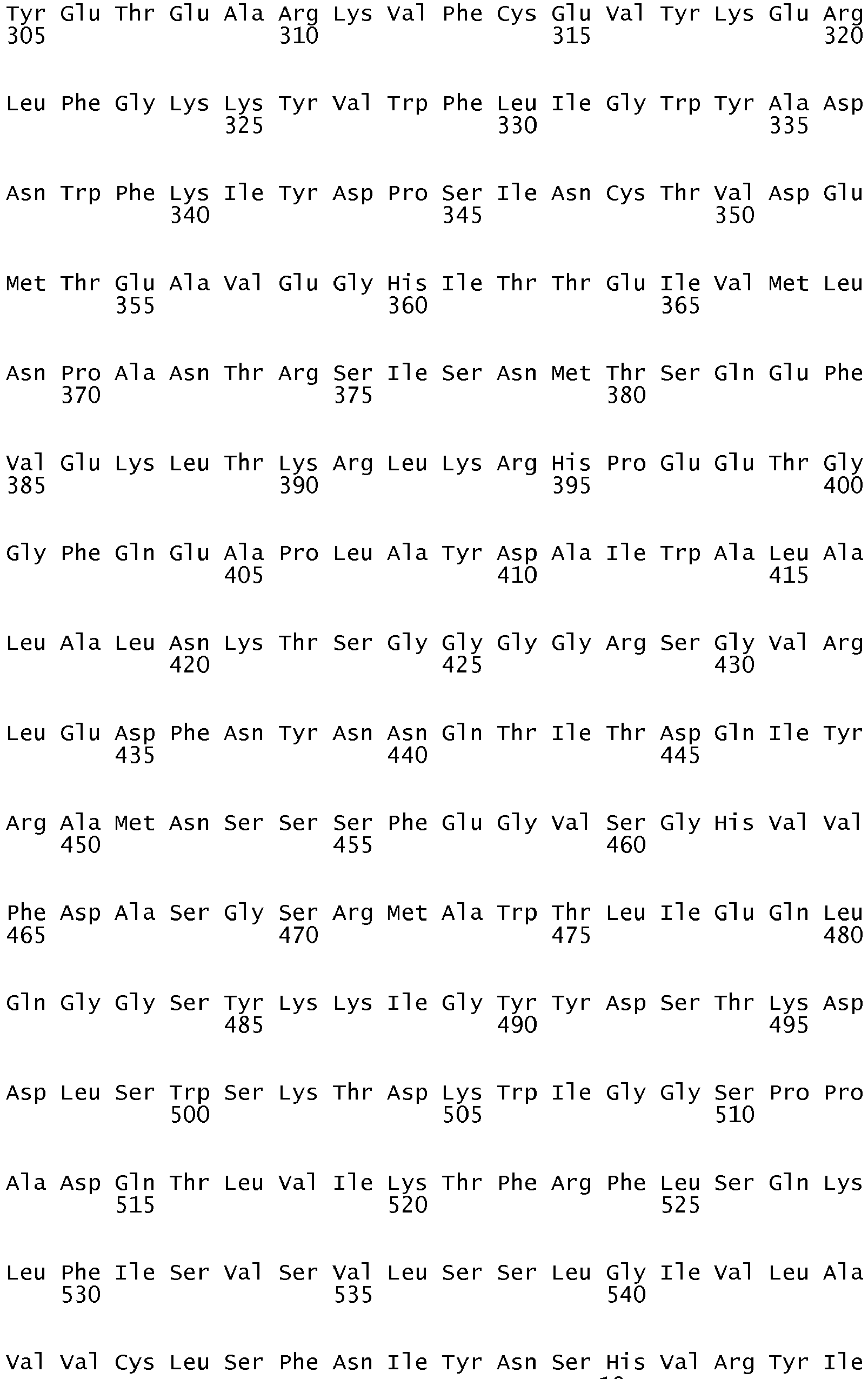


- <210> 4
<211> 4547
<212> DNA
<213> Homo sapiens - <400> 4


- <210> 5
<211> 4297
<212> DNA
<213> Homo sapiens - <400> 5


- <210> 6
<211> 4361
<212> DNA
<213> Homo sapiens - <400> 6


Claims (13)
- A method for diagnosinga) autoimmune encephalitis,b) an occult tumor causing autoimmune encephalitis, orc) in a subject with epilepsy, a tumor causing the epilepsy,the method comprising testing a biological sample from a subject to determine the presence of an antibody to a GABAB receptor, wherein the presence of said antibody in said biological sample is indicative that the subject hasa) an autoimmune encephalitis, orb) an occult tumor causing said autoimmune encephalitis, orc) where the subject has epilepsy, the presence of said antibody is indicative of a tumor in said subject causing said epilepsy.
- A method according to claim 1, wherein said antibody binds to the B1 subunit of said GABAB receptor.
- A method according to claim 1, wherein said antibody binds to the B2 subunit of said GABAB receptor.
- A method according to claim 1, wherein said autoimmune encephalitis is a paraneoplastic autoimmune encephalitis.
- A method according to claim 1, wherein the autoimmune encephalitis is anti-GABAB receptor encephalitis.
- A method according to claim 1, wherein said autoimmune encephalitis is a non-paraneoplastic, autoimmune encephalitis.
- A method according to claim 1, wherein said autoimmune encephalitis comprises a limbic encephalitis.
- A method according to claim 1, wherein said autoimmune encephalitis is associated with pathological symptoms.
- A method according to claim 8, wherein the symptoms are seizures; psychiatric symptoms; abnormality in cognition and behavior; a movement disorder or abnormal movements; a decreased level of consciousness; hypoventilation; amnesia or a memory deficit; or a combination thereof.
- A method according to claim 1, wherein said autoimmune encephalitis is associated with a tumor.
- A method according to claim 10, wherein said tumor is a small cell lung cancer or a neuroendocrine tumor of the lung or other organs.
- A method according to claim 1, wherein the biological sample is a cerebrospinal fluid (CSF) sample.
- A method according to claim 1, wherein the biological sample is a serum or plasma sample.
Applications Claiming Priority (2)
| Application Number | Priority Date | Filing Date | Title |
|---|---|---|---|
| US24684309P | 2009-09-29 | 2009-09-29 | |
| PCT/US2010/050746 WO2011041433A1 (en) | 2009-09-29 | 2010-09-29 | Methods for diagnosing and treating encephalitis or epilepsy |
Publications (3)
| Publication Number | Publication Date |
|---|---|
| EP2483417A1 EP2483417A1 (en) | 2012-08-08 |
| EP2483417A4 EP2483417A4 (en) | 2013-11-13 |
| EP2483417B1 true EP2483417B1 (en) | 2017-03-29 |
Family
ID=43826630
Family Applications (1)
| Application Number | Title | Priority Date | Filing Date |
|---|---|---|---|
| EP10821183.0A Active EP2483417B1 (en) | 2009-09-29 | 2010-09-29 | Methods for diagnosing and treating encephalitis or epilepsy |
Country Status (6)
| Country | Link |
|---|---|
| US (1) | US8685656B2 (en) |
| EP (1) | EP2483417B1 (en) |
| CN (2) | CN109142721B (en) |
| DK (1) | DK2483417T3 (en) |
| ES (1) | ES2625660T3 (en) |
| WO (1) | WO2011041433A1 (en) |
Cited By (3)
| Publication number | Priority date | Publication date | Assignee | Title |
|---|---|---|---|---|
| EP3505936A1 (en) | 2017-12-29 | 2019-07-03 | Euroimmun Medizinische Labordiagnostika AG | Method for the diagnosis of a borna virus infection |
| US10725035B2 (en) | 2017-06-16 | 2020-07-28 | Euroimmun Medizinische Labordiagnostika Ag | Diagnosis of a neuroautoimmune disease |
| EP3872492A1 (en) | 2020-02-28 | 2021-09-01 | Seramun Diagnostica GmbH | Identifying borna disease virus (bodv) infection through detection of antibodies against the bodv glycoprotein |
Families Citing this family (16)
| Publication number | Priority date | Publication date | Assignee | Title |
|---|---|---|---|---|
| EP2863231A1 (en) * | 2013-10-17 | 2015-04-22 | Institut D'Investigaciones Biomédiques August Pi i Sunyer | Diagnostic method for detecting a GABA(A) related autoimmune disease and related subject-matter |
| GB201409234D0 (en) | 2014-05-23 | 2014-07-09 | Isis Innovation | Method |
| EP3026434B1 (en) | 2014-11-25 | 2022-01-05 | Euroimmun Medizinische Labordiagnostika AG | A novel diagnostically relevant autoantibody |
| KR101739703B1 (en) * | 2016-09-13 | 2017-05-24 | (주) 어드밴스드 엔티 | The method of diagnosing encephalitis and the apparatus thereof based on TRAIL measurement |
| WO2018088803A1 (en) * | 2016-11-08 | 2018-05-17 | (주) 어드밴스드 엔티 | Autoimmune encephalitis type determination method using hla genotype testing |
| WO2018226098A2 (en) * | 2017-06-07 | 2018-12-13 | Erasmus University Medical Center Rotterdam | Methods for typing neurological disorders and cancer, and devices for use therein |
| EP3629021A1 (en) | 2018-09-26 | 2020-04-01 | Euroimmun Medizinische Labordiagnostika AG | Diagnosis of a neuroautoimmune disease |
| WO2021021996A1 (en) * | 2019-08-01 | 2021-02-04 | Aptinyx Inc. | Methods of treating disorders associated with elevated levels of antibodies that interact with the nmda receptor |
| US11644463B2 (en) * | 2019-08-30 | 2023-05-09 | Euroimmun Medizinische Labordiagnostika Ag | Detection of an autoantibody |
| CN114375396B (en) | 2019-09-13 | 2026-03-10 | 欧蒙医学实验诊断股份公司 | Improved NMDA receptor autoantibody detection |
| US11835519B2 (en) | 2019-12-17 | 2023-12-05 | Euroimmun Medizinische Labordiagnostika Ag | Autoantibodies to Septin-7 and diagnosis of neurological disease |
| DE202020000743U1 (en) | 2020-02-24 | 2020-03-17 | Euroimmun Medizinische Labordiagnostika Ag | Carrier and kit for the diagnosis of an autoimmune neurological disease |
| CN111450234B (en) * | 2020-04-07 | 2023-11-03 | 中山大学附属第三医院 | Application of physiological saline combined with serum antibody adsorbent in the preparation of drugs for the treatment of autoimmune encephalitis |
| CN112724236B (en) * | 2021-01-15 | 2022-03-04 | 武汉大学 | An antigenic peptide of amphoteric protein 1 and its antibody and application |
| CN113238045B (en) * | 2021-04-27 | 2021-12-28 | 南方医科大学南方医院 | Application of CRMP2 and Anti-CRMP2 Antibodies |
| CN120098151B (en) * | 2025-05-12 | 2025-07-25 | 成都海默云因医学检验实验室有限公司 | A recombinant protein and detection reagent for detecting anti-GABABR antibody encephalitis |
Family Cites Families (9)
| Publication number | Priority date | Publication date | Assignee | Title |
|---|---|---|---|---|
| US5731410A (en) | 1994-11-28 | 1998-03-24 | University Of Utah Research Foundation | Peptide for blocking autoantibody-evoked activation of glutamate receptor type 3 (GLUR3) |
| US20030235557A1 (en) * | 1998-09-30 | 2003-12-25 | Corixa Corporation | Compositions and methods for WT1 specific immunotherapy |
| CN1764474B (en) * | 2003-03-26 | 2011-02-02 | 惠氏公司 | Immunogenic compositions and methods |
| EP1651755B1 (en) * | 2003-08-04 | 2010-10-13 | IMBA-Institut für Molekulare Biotechnologie GmbH | Method for immunotherapy of tumors |
| US20070122417A1 (en) * | 2003-09-30 | 2007-05-31 | Patrick Holt | Immunotherapy method |
| KR20070057965A (en) * | 2004-09-21 | 2007-06-07 | 신타 파마슈티칼스 코프. | Compounds for Inflammation and Immune Related Applications |
| AU2005298689A1 (en) * | 2004-10-25 | 2006-05-04 | Cytos Biotechnology Ag | Gastric inhibitory polypeptide (GIP) antigen arrays and uses thereof |
| PL1848435T3 (en) * | 2005-01-25 | 2016-08-31 | Synta Pharmaceuticals Corp | Compounds against inflammations and immune-related uses |
| ES2443490T3 (en) | 2006-08-15 | 2014-02-19 | The Trustees Of The University Of Pennsylvania | Methods and compositions for the diagnosis and treatment of autoimmune encephalitis or epilepsy |
-
2010
- 2010-09-29 EP EP10821183.0A patent/EP2483417B1/en active Active
- 2010-09-29 CN CN201810893614.5A patent/CN109142721B/en active Active
- 2010-09-29 CN CN201080053945.XA patent/CN102803502B/en active Active
- 2010-09-29 DK DK10821183.0T patent/DK2483417T3/en active
- 2010-09-29 US US13/498,355 patent/US8685656B2/en active Active
- 2010-09-29 ES ES10821183.0T patent/ES2625660T3/en active Active
- 2010-09-29 WO PCT/US2010/050746 patent/WO2011041433A1/en not_active Ceased
Cited By (4)
| Publication number | Priority date | Publication date | Assignee | Title |
|---|---|---|---|---|
| US10725035B2 (en) | 2017-06-16 | 2020-07-28 | Euroimmun Medizinische Labordiagnostika Ag | Diagnosis of a neuroautoimmune disease |
| US11371991B2 (en) | 2017-06-16 | 2022-06-28 | Euroimmun Medizinische Labordiagnostika Ag | Diagnosis of a neuroautoimmune disease |
| EP3505936A1 (en) | 2017-12-29 | 2019-07-03 | Euroimmun Medizinische Labordiagnostika AG | Method for the diagnosis of a borna virus infection |
| EP3872492A1 (en) | 2020-02-28 | 2021-09-01 | Seramun Diagnostica GmbH | Identifying borna disease virus (bodv) infection through detection of antibodies against the bodv glycoprotein |
Also Published As
| Publication number | Publication date |
|---|---|
| US8685656B2 (en) | 2014-04-01 |
| CN109142721B (en) | 2021-10-26 |
| ES2625660T3 (en) | 2017-07-20 |
| WO2011041433A9 (en) | 2012-08-16 |
| WO2011041433A1 (en) | 2011-04-07 |
| CN109142721A (en) | 2019-01-04 |
| US20120213803A1 (en) | 2012-08-23 |
| CN102803502A (en) | 2012-11-28 |
| CN102803502B (en) | 2019-06-04 |
| EP2483417A4 (en) | 2013-11-13 |
| EP2483417A1 (en) | 2012-08-08 |
| DK2483417T3 (en) | 2017-05-15 |
Similar Documents
| Publication | Publication Date | Title |
|---|---|---|
| EP2483417B1 (en) | Methods for diagnosing and treating encephalitis or epilepsy | |
| US7972796B2 (en) | Methods for diagnosis of encephalitis | |
| US9250250B2 (en) | Methods for diagnosing and treating an LGI1 related autoimmune disease | |
| Lancaster et al. | Antibodies to the GABAB receptor in limbic encephalitis with seizures: case series and characterisation of the antigen | |
| Gadoth et al. | Microtubule‐associated protein 1 B: Novel paraneoplastic biomarker | |
| Lai et al. | Investigation of LGI1 as the antigen in limbic encephalitis previously attributed to potassium channels: a case series | |
| EP2895507B1 (en) | Diagnostic method for detecting an autoimmune disease and related subject-matter | |
| US11041005B2 (en) | Methods for detecting autoantibodies against a GABA(A) receptor alpha 1 subunit and/or beta 3 subunit in autoimmune seizure and/or encephalitis | |
| Iorio et al. | Clinical characteristics and outcome of patients with autoimmune encephalitis: clues for paraneoplastic aetiology | |
| Li et al. | Autoantibodies detection in anti-N-methyl-D-aspartate receptor encephalitis | |
| US20140377883A1 (en) | Peripherin-specific autoantibodies as a marker for neurological and endocrinological disease | |
| WO2014122275A1 (en) | Method for the diagnosis and monitoring of autoinmune diseases | |
| EP2816354B1 (en) | Biomarker for complex regional pain syndrome (CRPS) | |
| 배지연 | Discovery of novel antibody in autoimmune encephalities | |
| US20220120744A1 (en) | Assessing and treating germ cell tumors and paraneoplastic autoimmunity | |
| JP2025179829A (en) | Biomarkers, screening methods, methods for detecting biomarkers and methods for aiding in the diagnosis of demyelinating diseases | |
| Constantinescu et al. | Antibodies to the GABA (B) receptor in limbic encephalitis with seizures: Case series and characterisation of the... |
Legal Events
| Date | Code | Title | Description |
|---|---|---|---|
| PUAI | Public reference made under article 153(3) epc to a published international application that has entered the european phase |
Free format text: ORIGINAL CODE: 0009012 |
|
| 17P | Request for examination filed |
Effective date: 20120330 |
|
| AK | Designated contracting states |
Kind code of ref document: A1 Designated state(s): AL AT BE BG CH CY CZ DE DK EE ES FI FR GB GR HR HU IE IS IT LI LT LU LV MC MK MT NL NO PL PT RO SE SI SK SM TR |
|
| DAX | Request for extension of the european patent (deleted) | ||
| A4 | Supplementary search report drawn up and despatched |
Effective date: 20131016 |
|
| RIC1 | Information provided on ipc code assigned before grant |
Ipc: G01N 33/564 20060101ALI20131010BHEP Ipc: G01N 33/566 20060101ALI20131010BHEP Ipc: C12Q 1/00 20060101AFI20131010BHEP |
|
| 17Q | First examination report despatched |
Effective date: 20160712 |
|
| GRAP | Despatch of communication of intention to grant a patent |
Free format text: ORIGINAL CODE: EPIDOSNIGR1 |
|
| INTG | Intention to grant announced |
Effective date: 20161019 |
|
| RIN1 | Information on inventor provided before grant (corrected) |
Inventor name: DALMAU, JOSEP |
|
| GRAS | Grant fee paid |
Free format text: ORIGINAL CODE: EPIDOSNIGR3 |
|
| GRAA | (expected) grant |
Free format text: ORIGINAL CODE: 0009210 |
|
| AK | Designated contracting states |
Kind code of ref document: B1 Designated state(s): AL AT BE BG CH CY CZ DE DK EE ES FI FR GB GR HR HU IE IS IT LI LT LU LV MC MK MT NL NO PL PT RO SE SI SK SM TR |
|
| REG | Reference to a national code |
Ref country code: GB Ref legal event code: FG4D |
|
| REG | Reference to a national code |
Ref country code: CH Ref legal event code: EP |
|
| REG | Reference to a national code |
Ref country code: AT Ref legal event code: REF Ref document number: 879811 Country of ref document: AT Kind code of ref document: T Effective date: 20170415 |
|
| REG | Reference to a national code |
Ref country code: IE Ref legal event code: FG4D |
|
| REG | Reference to a national code |
Ref country code: NL Ref legal event code: FP |
|
| REG | Reference to a national code |
Ref country code: DE Ref legal event code: R096 Ref document number: 602010041179 Country of ref document: DE |
|
| REG | Reference to a national code |
Ref country code: DK Ref legal event code: T3 Effective date: 20170511 |
|
| REG | Reference to a national code |
Ref country code: SE Ref legal event code: TRGR |
|
| REG | Reference to a national code |
Ref country code: CH Ref legal event code: NV Representative=s name: NOVAGRAAF INTERNATIONAL SA, CH |
|
| REG | Reference to a national code |
Ref country code: NO Ref legal event code: T2 Effective date: 20170329 |
|
| REG | Reference to a national code |
Ref country code: ES Ref legal event code: FG2A Ref document number: 2625660 Country of ref document: ES Kind code of ref document: T3 Effective date: 20170720 |
|
| PG25 | Lapsed in a contracting state [announced via postgrant information from national office to epo] |
Ref country code: FI Free format text: LAPSE BECAUSE OF FAILURE TO SUBMIT A TRANSLATION OF THE DESCRIPTION OR TO PAY THE FEE WITHIN THE PRESCRIBED TIME-LIMIT Effective date: 20170329 Ref country code: HR Free format text: LAPSE BECAUSE OF FAILURE TO SUBMIT A TRANSLATION OF THE DESCRIPTION OR TO PAY THE FEE WITHIN THE PRESCRIBED TIME-LIMIT Effective date: 20170329 Ref country code: LT Free format text: LAPSE BECAUSE OF FAILURE TO SUBMIT A TRANSLATION OF THE DESCRIPTION OR TO PAY THE FEE WITHIN THE PRESCRIBED TIME-LIMIT Effective date: 20170329 Ref country code: GR Free format text: LAPSE BECAUSE OF FAILURE TO SUBMIT A TRANSLATION OF THE DESCRIPTION OR TO PAY THE FEE WITHIN THE PRESCRIBED TIME-LIMIT Effective date: 20170630 |
|
| REG | Reference to a national code |
Ref country code: FR Ref legal event code: PLFP Year of fee payment: 8 |
|
| PG25 | Lapsed in a contracting state [announced via postgrant information from national office to epo] |
Ref country code: LV Free format text: LAPSE BECAUSE OF FAILURE TO SUBMIT A TRANSLATION OF THE DESCRIPTION OR TO PAY THE FEE WITHIN THE PRESCRIBED TIME-LIMIT Effective date: 20170329 Ref country code: BG Free format text: LAPSE BECAUSE OF FAILURE TO SUBMIT A TRANSLATION OF THE DESCRIPTION OR TO PAY THE FEE WITHIN THE PRESCRIBED TIME-LIMIT Effective date: 20170629 |
|
| PG25 | Lapsed in a contracting state [announced via postgrant information from national office to epo] |
Ref country code: SK Free format text: LAPSE BECAUSE OF FAILURE TO SUBMIT A TRANSLATION OF THE DESCRIPTION OR TO PAY THE FEE WITHIN THE PRESCRIBED TIME-LIMIT Effective date: 20170329 Ref country code: RO Free format text: LAPSE BECAUSE OF FAILURE TO SUBMIT A TRANSLATION OF THE DESCRIPTION OR TO PAY THE FEE WITHIN THE PRESCRIBED TIME-LIMIT Effective date: 20170329 Ref country code: CZ Free format text: LAPSE BECAUSE OF FAILURE TO SUBMIT A TRANSLATION OF THE DESCRIPTION OR TO PAY THE FEE WITHIN THE PRESCRIBED TIME-LIMIT Effective date: 20170329 Ref country code: IT Free format text: LAPSE BECAUSE OF FAILURE TO SUBMIT A TRANSLATION OF THE DESCRIPTION OR TO PAY THE FEE WITHIN THE PRESCRIBED TIME-LIMIT Effective date: 20170329 Ref country code: EE Free format text: LAPSE BECAUSE OF FAILURE TO SUBMIT A TRANSLATION OF THE DESCRIPTION OR TO PAY THE FEE WITHIN THE PRESCRIBED TIME-LIMIT Effective date: 20170329 |
|
| PG25 | Lapsed in a contracting state [announced via postgrant information from national office to epo] |
Ref country code: PL Free format text: LAPSE BECAUSE OF FAILURE TO SUBMIT A TRANSLATION OF THE DESCRIPTION OR TO PAY THE FEE WITHIN THE PRESCRIBED TIME-LIMIT Effective date: 20170329 Ref country code: IS Free format text: LAPSE BECAUSE OF FAILURE TO SUBMIT A TRANSLATION OF THE DESCRIPTION OR TO PAY THE FEE WITHIN THE PRESCRIBED TIME-LIMIT Effective date: 20170729 Ref country code: SM Free format text: LAPSE BECAUSE OF FAILURE TO SUBMIT A TRANSLATION OF THE DESCRIPTION OR TO PAY THE FEE WITHIN THE PRESCRIBED TIME-LIMIT Effective date: 20170329 Ref country code: PT Free format text: LAPSE BECAUSE OF FAILURE TO SUBMIT A TRANSLATION OF THE DESCRIPTION OR TO PAY THE FEE WITHIN THE PRESCRIBED TIME-LIMIT Effective date: 20170731 |
|
| REG | Reference to a national code |
Ref country code: DE Ref legal event code: R097 Ref document number: 602010041179 Country of ref document: DE |
|
| PLBE | No opposition filed within time limit |
Free format text: ORIGINAL CODE: 0009261 |
|
| STAA | Information on the status of an ep patent application or granted ep patent |
Free format text: STATUS: NO OPPOSITION FILED WITHIN TIME LIMIT |
|
| 26N | No opposition filed |
Effective date: 20180103 |
|
| PG25 | Lapsed in a contracting state [announced via postgrant information from national office to epo] |
Ref country code: MC Free format text: LAPSE BECAUSE OF FAILURE TO SUBMIT A TRANSLATION OF THE DESCRIPTION OR TO PAY THE FEE WITHIN THE PRESCRIBED TIME-LIMIT Effective date: 20170329 Ref country code: SI Free format text: LAPSE BECAUSE OF FAILURE TO SUBMIT A TRANSLATION OF THE DESCRIPTION OR TO PAY THE FEE WITHIN THE PRESCRIBED TIME-LIMIT Effective date: 20170329 |
|
| REG | Reference to a national code |
Ref country code: IE Ref legal event code: MM4A |
|
| REG | Reference to a national code |
Ref country code: BE Ref legal event code: MM Effective date: 20170930 |
|
| PG25 | Lapsed in a contracting state [announced via postgrant information from national office to epo] |
Ref country code: LU Free format text: LAPSE BECAUSE OF NON-PAYMENT OF DUE FEES Effective date: 20170929 |
|
| PG25 | Lapsed in a contracting state [announced via postgrant information from national office to epo] |
Ref country code: IE Free format text: LAPSE BECAUSE OF NON-PAYMENT OF DUE FEES Effective date: 20170929 |
|
| REG | Reference to a national code |
Ref country code: FR Ref legal event code: PLFP Year of fee payment: 9 |
|
| PG25 | Lapsed in a contracting state [announced via postgrant information from national office to epo] |
Ref country code: BE Free format text: LAPSE BECAUSE OF NON-PAYMENT OF DUE FEES Effective date: 20170930 |
|
| PG25 | Lapsed in a contracting state [announced via postgrant information from national office to epo] |
Ref country code: MT Free format text: LAPSE BECAUSE OF NON-PAYMENT OF DUE FEES Effective date: 20170929 |
|
| REG | Reference to a national code |
Ref country code: GB Ref legal event code: 732E Free format text: REGISTERED BETWEEN 20181220 AND 20181224 |
|
| PG25 | Lapsed in a contracting state [announced via postgrant information from national office to epo] |
Ref country code: HU Free format text: LAPSE BECAUSE OF FAILURE TO SUBMIT A TRANSLATION OF THE DESCRIPTION OR TO PAY THE FEE WITHIN THE PRESCRIBED TIME-LIMIT; INVALID AB INITIO Effective date: 20100929 |
|
| REG | Reference to a national code |
Ref country code: AT Ref legal event code: UEP Ref document number: 879811 Country of ref document: AT Kind code of ref document: T Effective date: 20170329 |
|
| PG25 | Lapsed in a contracting state [announced via postgrant information from national office to epo] |
Ref country code: CY Free format text: LAPSE BECAUSE OF NON-PAYMENT OF DUE FEES Effective date: 20170329 |
|
| PG25 | Lapsed in a contracting state [announced via postgrant information from national office to epo] |
Ref country code: MK Free format text: LAPSE BECAUSE OF FAILURE TO SUBMIT A TRANSLATION OF THE DESCRIPTION OR TO PAY THE FEE WITHIN THE PRESCRIBED TIME-LIMIT Effective date: 20170329 |
|
| PG25 | Lapsed in a contracting state [announced via postgrant information from national office to epo] |
Ref country code: TR Free format text: LAPSE BECAUSE OF FAILURE TO SUBMIT A TRANSLATION OF THE DESCRIPTION OR TO PAY THE FEE WITHIN THE PRESCRIBED TIME-LIMIT Effective date: 20170329 |
|
| PG25 | Lapsed in a contracting state [announced via postgrant information from national office to epo] |
Ref country code: AL Free format text: LAPSE BECAUSE OF FAILURE TO SUBMIT A TRANSLATION OF THE DESCRIPTION OR TO PAY THE FEE WITHIN THE PRESCRIBED TIME-LIMIT Effective date: 20170329 |
|
| P01 | Opt-out of the competence of the unified patent court (upc) registered |
Effective date: 20230524 |
|
| REG | Reference to a national code |
Ref country code: CH Ref legal event code: U11 Free format text: ST27 STATUS EVENT CODE: U-0-0-U10-U11 (AS PROVIDED BY THE NATIONAL OFFICE) Effective date: 20251001 |
|
| PGFP | Annual fee paid to national office [announced via postgrant information from national office to epo] |
Ref country code: DE Payment date: 20250919 Year of fee payment: 16 Ref country code: DK Payment date: 20250922 Year of fee payment: 16 |
|
| PGFP | Annual fee paid to national office [announced via postgrant information from national office to epo] |
Ref country code: NO Payment date: 20250919 Year of fee payment: 16 |
|
| PGFP | Annual fee paid to national office [announced via postgrant information from national office to epo] |
Ref country code: NL Payment date: 20250922 Year of fee payment: 16 |
|
| PGFP | Annual fee paid to national office [announced via postgrant information from national office to epo] |
Ref country code: GB Payment date: 20250923 Year of fee payment: 16 |
|
| PGFP | Annual fee paid to national office [announced via postgrant information from national office to epo] |
Ref country code: FR Payment date: 20250922 Year of fee payment: 16 Ref country code: AT Payment date: 20250918 Year of fee payment: 16 |
|
| PGFP | Annual fee paid to national office [announced via postgrant information from national office to epo] |
Ref country code: SE Payment date: 20250922 Year of fee payment: 16 |
|
| PGFP | Annual fee paid to national office [announced via postgrant information from national office to epo] |
Ref country code: CH Payment date: 20251001 Year of fee payment: 16 |
|
| PGFP | Annual fee paid to national office [announced via postgrant information from national office to epo] |
Ref country code: ES Payment date: 20251020 Year of fee payment: 16 |