EP2482825B1 - Piscine reovirus diagnostic compositions - Google Patents
Piscine reovirus diagnostic compositions Download PDFInfo
- Publication number
- EP2482825B1 EP2482825B1 EP10821408.1A EP10821408A EP2482825B1 EP 2482825 B1 EP2482825 B1 EP 2482825B1 EP 10821408 A EP10821408 A EP 10821408A EP 2482825 B1 EP2482825 B1 EP 2482825B1
- Authority
- EP
- European Patent Office
- Prior art keywords
- prv
- seq
- nucleic acid
- sequence
- hsmi
- Prior art date
- Legal status (The legal status is an assumption and is not a legal conclusion. Google has not performed a legal analysis and makes no representation as to the accuracy of the status listed.)
- Revoked
Links
- 241001436114 Piscine orthoreovirus Species 0.000 title claims description 315
- 239000000203 mixture Substances 0.000 title description 12
- 150000007523 nucleic acids Chemical class 0.000 claims description 201
- 102000039446 nucleic acids Human genes 0.000 claims description 102
- 108020004707 nucleic acids Proteins 0.000 claims description 102
- 125000003729 nucleotide group Chemical group 0.000 claims description 97
- 239000002773 nucleotide Substances 0.000 claims description 96
- 238000000034 method Methods 0.000 claims description 84
- 108091028043 Nucleic acid sequence Proteins 0.000 claims description 74
- 230000000295 complement effect Effects 0.000 claims description 66
- 239000000523 sample Substances 0.000 claims description 59
- 241000972773 Aulopiformes Species 0.000 claims description 45
- 235000019515 salmon Nutrition 0.000 claims description 45
- 230000003321 amplification Effects 0.000 claims description 29
- 238000003199 nucleic acid amplification method Methods 0.000 claims description 29
- 239000012472 biological sample Substances 0.000 claims description 25
- 108090000765 processed proteins & peptides Proteins 0.000 description 157
- 102000004196 processed proteins & peptides Human genes 0.000 description 154
- 229920001184 polypeptide Polymers 0.000 description 151
- 239000012634 fragment Substances 0.000 description 88
- 108090000623 proteins and genes Proteins 0.000 description 87
- 235000019688 fish Nutrition 0.000 description 56
- 241000251468 Actinopterygii Species 0.000 description 55
- 102000004169 proteins and genes Human genes 0.000 description 55
- 238000009396 hybridization Methods 0.000 description 53
- 235000018102 proteins Nutrition 0.000 description 53
- 239000013615 primer Substances 0.000 description 51
- 241000894007 species Species 0.000 description 51
- 235000001014 amino acid Nutrition 0.000 description 49
- 108020004414 DNA Proteins 0.000 description 42
- 239000003795 chemical substances by application Substances 0.000 description 42
- 125000003275 alpha amino acid group Chemical group 0.000 description 38
- 241001465754 Metazoa Species 0.000 description 37
- 241000700605 Viruses Species 0.000 description 37
- 238000004458 analytical method Methods 0.000 description 35
- 150000001413 amino acids Chemical class 0.000 description 34
- 241000702652 Aquareovirus Species 0.000 description 32
- 108700026244 Open Reading Frames Proteins 0.000 description 32
- 108091032973 (ribonucleotides)n+m Proteins 0.000 description 31
- FAPWRFPIFSIZLT-UHFFFAOYSA-M Sodium chloride Chemical compound [Na+].[Cl-] FAPWRFPIFSIZLT-UHFFFAOYSA-M 0.000 description 30
- 241000702244 Orthoreovirus Species 0.000 description 28
- ZHNUHDYFZUAESO-UHFFFAOYSA-N Formamide Chemical compound NC=O ZHNUHDYFZUAESO-UHFFFAOYSA-N 0.000 description 25
- 239000000427 antigen Substances 0.000 description 24
- 102000036639 antigens Human genes 0.000 description 24
- 108091007433 antigens Proteins 0.000 description 24
- 238000004422 calculation algorithm Methods 0.000 description 24
- 238000003556 assay Methods 0.000 description 22
- 210000004027 cell Anatomy 0.000 description 22
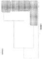
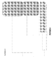
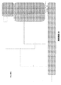
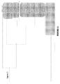
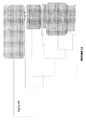
- 238000013081 phylogenetic analysis Methods 0.000 description 21
- 230000014509 gene expression Effects 0.000 description 20
- 108091034117 Oligonucleotide Proteins 0.000 description 17
- 201000010099 disease Diseases 0.000 description 17
- 208000037265 diseases, disorders, signs and symptoms Diseases 0.000 description 17
- 108091081021 Sense strand Proteins 0.000 description 16
- 230000003612 virological effect Effects 0.000 description 16
- 241000702263 Reovirus sp. Species 0.000 description 15
- DBMJMQXJHONAFJ-UHFFFAOYSA-M Sodium laurylsulphate Chemical compound [Na+].CCCCCCCCCCCCOS([O-])(=O)=O DBMJMQXJHONAFJ-UHFFFAOYSA-M 0.000 description 15
- 239000011780 sodium chloride Substances 0.000 description 15
- 239000001509 sodium citrate Substances 0.000 description 15
- 238000006467 substitution reaction Methods 0.000 description 15
- 208000015181 infectious disease Diseases 0.000 description 14
- 241000588724 Escherichia coli Species 0.000 description 12
- 241000277263 Salmo Species 0.000 description 12
- 230000027455 binding Effects 0.000 description 12
- 238000005406 washing Methods 0.000 description 12
- 108060003951 Immunoglobulin Proteins 0.000 description 11
- 102000018358 immunoglobulin Human genes 0.000 description 11
- 238000005070 sampling Methods 0.000 description 11
- 239000000243 solution Substances 0.000 description 11
- HRXKRNGNAMMEHJ-UHFFFAOYSA-K trisodium citrate Chemical compound [Na+].[Na+].[Na+].[O-]C(=O)CC(O)(CC([O-])=O)C([O-])=O HRXKRNGNAMMEHJ-UHFFFAOYSA-K 0.000 description 11
- 229940038773 trisodium citrate Drugs 0.000 description 11
- 239000013598 vector Substances 0.000 description 11
- 241001042466 Mammalian orthoreovirus Species 0.000 description 10
- 230000000692 anti-sense effect Effects 0.000 description 10
- 238000005516 engineering process Methods 0.000 description 10
- 239000013612 plasmid Substances 0.000 description 10
- 238000003752 polymerase chain reaction Methods 0.000 description 10
- 108091033319 polynucleotide Proteins 0.000 description 10
- 102000040430 polynucleotide Human genes 0.000 description 10
- 239000002157 polynucleotide Substances 0.000 description 10
- 241001516406 Avian orthoreovirus Species 0.000 description 9
- 238000007405 data analysis Methods 0.000 description 9
- 238000009826 distribution Methods 0.000 description 9
- 102000037865 fusion proteins Human genes 0.000 description 9
- 108020001507 fusion proteins Proteins 0.000 description 9
- 238000003018 immunoassay Methods 0.000 description 9
- 210000003734 kidney Anatomy 0.000 description 9
- 239000000700 radioactive tracer Substances 0.000 description 9
- 238000003753 real-time PCR Methods 0.000 description 9
- 238000012360 testing method Methods 0.000 description 9
- 241001293013 Baboon orthoreovirus Species 0.000 description 8
- 241000238847 Nelson Bay orthoreovirus Species 0.000 description 8
- 241000283973 Oryctolagus cuniculus Species 0.000 description 8
- 241000303280 Reptilian orthoreovirus Species 0.000 description 8
- 239000002253 acid Substances 0.000 description 8
- 241000702247 Reoviridae Species 0.000 description 7
- FKNQFGJONOIPTF-UHFFFAOYSA-N Sodium cation Chemical compound [Na+] FKNQFGJONOIPTF-UHFFFAOYSA-N 0.000 description 7
- 230000000694 effects Effects 0.000 description 7
- 238000000338 in vitro Methods 0.000 description 7
- 210000004165 myocardium Anatomy 0.000 description 7
- 239000002751 oligonucleotide probe Substances 0.000 description 7
- 150000003839 salts Chemical class 0.000 description 7
- 238000012216 screening Methods 0.000 description 7
- 229910001415 sodium ion Inorganic materials 0.000 description 7
- 241000894006 Bacteria Species 0.000 description 6
- 125000003412 L-alanyl group Chemical group [H]N([H])[C@@](C([H])([H])[H])(C(=O)[*])[H] 0.000 description 6
- 108020005187 Oligonucleotide Probes Proteins 0.000 description 6
- 238000013459 approach Methods 0.000 description 6
- 238000009360 aquaculture Methods 0.000 description 6
- 244000144974 aquaculture Species 0.000 description 6
- 239000013604 expression vector Substances 0.000 description 6
- 230000004927 fusion Effects 0.000 description 6
- 238000003364 immunohistochemistry Methods 0.000 description 6
- 238000004519 manufacturing process Methods 0.000 description 6
- 108020004999 messenger RNA Proteins 0.000 description 6
- 239000002987 primer (paints) Substances 0.000 description 6
- 238000012175 pyrosequencing Methods 0.000 description 6
- 230000009467 reduction Effects 0.000 description 6
- 230000008685 targeting Effects 0.000 description 6
- 210000001519 tissue Anatomy 0.000 description 6
- FWMNVWWHGCHHJJ-SKKKGAJSSA-N 4-amino-1-[(2r)-6-amino-2-[[(2r)-2-[[(2r)-2-[[(2r)-2-amino-3-phenylpropanoyl]amino]-3-phenylpropanoyl]amino]-4-methylpentanoyl]amino]hexanoyl]piperidine-4-carboxylic acid Chemical compound C([C@H](C(=O)N[C@H](CC(C)C)C(=O)N[C@H](CCCCN)C(=O)N1CCC(N)(CC1)C(O)=O)NC(=O)[C@H](N)CC=1C=CC=CC=1)C1=CC=CC=C1 FWMNVWWHGCHHJJ-SKKKGAJSSA-N 0.000 description 5
- 101150083038 EF1A gene Proteins 0.000 description 5
- 101150075239 L1 gene Proteins 0.000 description 5
- 201000002481 Myositis Diseases 0.000 description 5
- 230000000890 antigenic effect Effects 0.000 description 5
- 229940079593 drug Drugs 0.000 description 5
- 239000003814 drug Substances 0.000 description 5
- 230000002068 genetic effect Effects 0.000 description 5
- 230000003053 immunization Effects 0.000 description 5
- 238000002649 immunization Methods 0.000 description 5
- 238000007901 in situ hybridization Methods 0.000 description 5
- 239000000463 material Substances 0.000 description 5
- 238000002844 melting Methods 0.000 description 5
- 230000008018 melting Effects 0.000 description 5
- 239000013642 negative control Substances 0.000 description 5
- 210000002966 serum Anatomy 0.000 description 5
- 210000002027 skeletal muscle Anatomy 0.000 description 5
- 210000000952 spleen Anatomy 0.000 description 5
- 238000007619 statistical method Methods 0.000 description 5
- 238000001262 western blot Methods 0.000 description 5
- 102000040650 (ribonucleotides)n+m Human genes 0.000 description 4
- WZUVPPKBWHMQCE-UHFFFAOYSA-N Haematoxylin Chemical compound C12=CC(O)=C(O)C=C2CC2(O)C1C1=CC=C(O)C(O)=C1OC2 WZUVPPKBWHMQCE-UHFFFAOYSA-N 0.000 description 4
- 108060004795 Methyltransferase Proteins 0.000 description 4
- 108010067390 Viral Proteins Proteins 0.000 description 4
- JLCPHMBAVCMARE-UHFFFAOYSA-N [3-[[3-[[3-[[3-[[3-[[3-[[3-[[3-[[3-[[3-[[3-[[5-(2-amino-6-oxo-1H-purin-9-yl)-3-[[3-[[3-[[3-[[3-[[3-[[5-(2-amino-6-oxo-1H-purin-9-yl)-3-[[5-(2-amino-6-oxo-1H-purin-9-yl)-3-hydroxyoxolan-2-yl]methoxy-hydroxyphosphoryl]oxyoxolan-2-yl]methoxy-hydroxyphosphoryl]oxy-5-(5-methyl-2,4-dioxopyrimidin-1-yl)oxolan-2-yl]methoxy-hydroxyphosphoryl]oxy-5-(6-aminopurin-9-yl)oxolan-2-yl]methoxy-hydroxyphosphoryl]oxy-5-(6-aminopurin-9-yl)oxolan-2-yl]methoxy-hydroxyphosphoryl]oxy-5-(6-aminopurin-9-yl)oxolan-2-yl]methoxy-hydroxyphosphoryl]oxy-5-(6-aminopurin-9-yl)oxolan-2-yl]methoxy-hydroxyphosphoryl]oxyoxolan-2-yl]methoxy-hydroxyphosphoryl]oxy-5-(5-methyl-2,4-dioxopyrimidin-1-yl)oxolan-2-yl]methoxy-hydroxyphosphoryl]oxy-5-(4-amino-2-oxopyrimidin-1-yl)oxolan-2-yl]methoxy-hydroxyphosphoryl]oxy-5-(5-methyl-2,4-dioxopyrimidin-1-yl)oxolan-2-yl]methoxy-hydroxyphosphoryl]oxy-5-(5-methyl-2,4-dioxopyrimidin-1-yl)oxolan-2-yl]methoxy-hydroxyphosphoryl]oxy-5-(6-aminopurin-9-yl)oxolan-2-yl]methoxy-hydroxyphosphoryl]oxy-5-(6-aminopurin-9-yl)oxolan-2-yl]methoxy-hydroxyphosphoryl]oxy-5-(4-amino-2-oxopyrimidin-1-yl)oxolan-2-yl]methoxy-hydroxyphosphoryl]oxy-5-(4-amino-2-oxopyrimidin-1-yl)oxolan-2-yl]methoxy-hydroxyphosphoryl]oxy-5-(4-amino-2-oxopyrimidin-1-yl)oxolan-2-yl]methoxy-hydroxyphosphoryl]oxy-5-(6-aminopurin-9-yl)oxolan-2-yl]methoxy-hydroxyphosphoryl]oxy-5-(4-amino-2-oxopyrimidin-1-yl)oxolan-2-yl]methyl [5-(6-aminopurin-9-yl)-2-(hydroxymethyl)oxolan-3-yl] hydrogen phosphate Polymers Cc1cn(C2CC(OP(O)(=O)OCC3OC(CC3OP(O)(=O)OCC3OC(CC3O)n3cnc4c3nc(N)[nH]c4=O)n3cnc4c3nc(N)[nH]c4=O)C(COP(O)(=O)OC3CC(OC3COP(O)(=O)OC3CC(OC3COP(O)(=O)OC3CC(OC3COP(O)(=O)OC3CC(OC3COP(O)(=O)OC3CC(OC3COP(O)(=O)OC3CC(OC3COP(O)(=O)OC3CC(OC3COP(O)(=O)OC3CC(OC3COP(O)(=O)OC3CC(OC3COP(O)(=O)OC3CC(OC3COP(O)(=O)OC3CC(OC3COP(O)(=O)OC3CC(OC3COP(O)(=O)OC3CC(OC3COP(O)(=O)OC3CC(OC3COP(O)(=O)OC3CC(OC3COP(O)(=O)OC3CC(OC3COP(O)(=O)OC3CC(OC3CO)n3cnc4c(N)ncnc34)n3ccc(N)nc3=O)n3cnc4c(N)ncnc34)n3ccc(N)nc3=O)n3ccc(N)nc3=O)n3ccc(N)nc3=O)n3cnc4c(N)ncnc34)n3cnc4c(N)ncnc34)n3cc(C)c(=O)[nH]c3=O)n3cc(C)c(=O)[nH]c3=O)n3ccc(N)nc3=O)n3cc(C)c(=O)[nH]c3=O)n3cnc4c3nc(N)[nH]c4=O)n3cnc4c(N)ncnc34)n3cnc4c(N)ncnc34)n3cnc4c(N)ncnc34)n3cnc4c(N)ncnc34)O2)c(=O)[nH]c1=O JLCPHMBAVCMARE-UHFFFAOYSA-N 0.000 description 4
- 150000007513 acids Chemical class 0.000 description 4
- 239000000969 carrier Substances 0.000 description 4
- 239000002299 complementary DNA Substances 0.000 description 4
- 230000029087 digestion Effects 0.000 description 4
- 239000000539 dimer Substances 0.000 description 4
- 230000030279 gene silencing Effects 0.000 description 4
- 238000012165 high-throughput sequencing Methods 0.000 description 4
- 230000002452 interceptive effect Effects 0.000 description 4
- 238000010369 molecular cloning Methods 0.000 description 4
- 239000000178 monomer Substances 0.000 description 4
- 210000003205 muscle Anatomy 0.000 description 4
- 210000000056 organ Anatomy 0.000 description 4
- 230000007170 pathology Effects 0.000 description 4
- 239000008188 pellet Substances 0.000 description 4
- 238000012163 sequencing technique Methods 0.000 description 4
- NLJMYIDDQXHKNR-UHFFFAOYSA-K sodium citrate Chemical compound O.O.[Na+].[Na+].[Na+].[O-]C(=O)CC(O)(CC([O-])=O)C([O-])=O NLJMYIDDQXHKNR-UHFFFAOYSA-K 0.000 description 4
- 229960005486 vaccine Drugs 0.000 description 4
- 108090000565 Capsid Proteins Proteins 0.000 description 3
- 102100023321 Ceruloplasmin Human genes 0.000 description 3
- 108010047041 Complementarity Determining Regions Proteins 0.000 description 3
- SHIBSTMRCDJXLN-UHFFFAOYSA-N Digoxigenin Natural products C1CC(C2C(C3(C)CCC(O)CC3CC2)CC2O)(O)C2(C)C1C1=CC(=O)OC1 SHIBSTMRCDJXLN-UHFFFAOYSA-N 0.000 description 3
- BWGNESOTFCXPMA-UHFFFAOYSA-N Dihydrogen disulfide Chemical compound SS BWGNESOTFCXPMA-UHFFFAOYSA-N 0.000 description 3
- 238000002965 ELISA Methods 0.000 description 3
- 102000004190 Enzymes Human genes 0.000 description 3
- 108090000790 Enzymes Proteins 0.000 description 3
- 241000282414 Homo sapiens Species 0.000 description 3
- 108010067060 Immunoglobulin Variable Region Proteins 0.000 description 3
- 102000017727 Immunoglobulin Variable Region Human genes 0.000 description 3
- 125000000570 L-alpha-aspartyl group Chemical group [H]OC(=O)C([H])([H])[C@]([H])(N([H])[H])C(*)=O 0.000 description 3
- 125000002842 L-seryl group Chemical group O=C([*])[C@](N([H])[H])([H])C([H])([H])O[H] 0.000 description 3
- 102000057297 Pepsin A Human genes 0.000 description 3
- 108090000284 Pepsin A Proteins 0.000 description 3
- 230000015572 biosynthetic process Effects 0.000 description 3
- 239000000872 buffer Substances 0.000 description 3
- 210000000234 capsid Anatomy 0.000 description 3
- 238000010382 chemical cross-linking Methods 0.000 description 3
- 238000003776 cleavage reaction Methods 0.000 description 3
- 230000002596 correlated effect Effects 0.000 description 3
- 210000004748 cultured cell Anatomy 0.000 description 3
- 238000001514 detection method Methods 0.000 description 3
- 238000003745 diagnosis Methods 0.000 description 3
- QONQRTHLHBTMGP-UHFFFAOYSA-N digitoxigenin Natural products CC12CCC(C3(CCC(O)CC3CC3)C)C3C11OC1CC2C1=CC(=O)OC1 QONQRTHLHBTMGP-UHFFFAOYSA-N 0.000 description 3
- SHIBSTMRCDJXLN-KCZCNTNESA-N digoxigenin Chemical compound C1([C@@H]2[C@@]3([C@@](CC2)(O)[C@H]2[C@@H]([C@@]4(C)CC[C@H](O)C[C@H]4CC2)C[C@H]3O)C)=CC(=O)OC1 SHIBSTMRCDJXLN-KCZCNTNESA-N 0.000 description 3
- 229940088598 enzyme Drugs 0.000 description 3
- 239000000284 extract Substances 0.000 description 3
- -1 for example Chemical group 0.000 description 3
- 210000005003 heart tissue Anatomy 0.000 description 3
- 210000004408 hybridoma Anatomy 0.000 description 3
- 229940072221 immunoglobulins Drugs 0.000 description 3
- 239000012678 infectious agent Substances 0.000 description 3
- 238000005304 joining Methods 0.000 description 3
- 238000011551 log transformation method Methods 0.000 description 3
- 238000009364 mariculture Methods 0.000 description 3
- 239000011159 matrix material Substances 0.000 description 3
- 238000007899 nucleic acid hybridization Methods 0.000 description 3
- 244000144977 poultry Species 0.000 description 3
- 230000000750 progressive effect Effects 0.000 description 3
- 238000000746 purification Methods 0.000 description 3
- 238000011002 quantification Methods 0.000 description 3
- 230000007017 scission Effects 0.000 description 3
- 230000017960 syncytium formation Effects 0.000 description 3
- 238000013518 transcription Methods 0.000 description 3
- 230000035897 transcription Effects 0.000 description 3
- 238000011179 visual inspection Methods 0.000 description 3
- 101000666833 Autographa californica nuclear polyhedrosis virus Uncharacterized 20.8 kDa protein in FGF-VUBI intergenic region Proteins 0.000 description 2
- 101000977023 Azospirillum brasilense Uncharacterized 17.8 kDa protein in nodG 5'region Proteins 0.000 description 2
- 101000977027 Azospirillum brasilense Uncharacterized protein in nodG 5'region Proteins 0.000 description 2
- 101000962005 Bacillus thuringiensis Uncharacterized 23.6 kDa protein Proteins 0.000 description 2
- 101000961984 Bacillus thuringiensis Uncharacterized 30.3 kDa protein Proteins 0.000 description 2
- 102100029226 Cancer-related nucleoside-triphosphatase Human genes 0.000 description 2
- HEDRZPFGACZZDS-UHFFFAOYSA-N Chloroform Chemical compound ClC(Cl)Cl HEDRZPFGACZZDS-UHFFFAOYSA-N 0.000 description 2
- 208000035473 Communicable disease Diseases 0.000 description 2
- 238000009007 Diagnostic Kit Methods 0.000 description 2
- 101000644901 Drosophila melanogaster Putative 115 kDa protein in type-1 retrotransposable element R1DM Proteins 0.000 description 2
- 101000785191 Drosophila melanogaster Uncharacterized 50 kDa protein in type I retrotransposable element R1DM Proteins 0.000 description 2
- 241000196324 Embryophyta Species 0.000 description 2
- 108010067770 Endopeptidase K Proteins 0.000 description 2
- 101000747704 Enterobacteria phage N4 Uncharacterized protein Gp1 Proteins 0.000 description 2
- 101000747702 Enterobacteria phage N4 Uncharacterized protein Gp2 Proteins 0.000 description 2
- 101000861206 Enterococcus faecalis (strain ATCC 700802 / V583) Uncharacterized protein EF_A0048 Proteins 0.000 description 2
- 101000769180 Escherichia coli Uncharacterized 11.1 kDa protein Proteins 0.000 description 2
- 101000758599 Escherichia coli Uncharacterized 14.7 kDa protein Proteins 0.000 description 2
- 229920001917 Ficoll Polymers 0.000 description 2
- 241000238631 Hexapoda Species 0.000 description 2
- 108010054477 Immunoglobulin Fab Fragments Proteins 0.000 description 2
- 102000001706 Immunoglobulin Fab Fragments Human genes 0.000 description 2
- 206010061218 Inflammation Diseases 0.000 description 2
- ROHFNLRQFUQHCH-YFKPBYRVSA-N L-leucine Chemical compound CC(C)C[C@H](N)C(O)=O ROHFNLRQFUQHCH-YFKPBYRVSA-N 0.000 description 2
- 125000003440 L-leucyl group Chemical group O=C([*])[C@](N([H])[H])([H])C([H])([H])C(C([H])([H])[H])([H])C([H])([H])[H] 0.000 description 2
- 125000000769 L-threonyl group Chemical group [H]N([H])[C@]([H])(C(=O)[*])[C@](O[H])(C([H])([H])[H])[H] 0.000 description 2
- 101000768930 Lactococcus lactis subsp. cremoris Uncharacterized protein in pepC 5'region Proteins 0.000 description 2
- 101000976301 Leptospira interrogans Uncharacterized 35 kDa protein in sph 3'region Proteins 0.000 description 2
- 101000976302 Leptospira interrogans Uncharacterized protein in sph 3'region Proteins 0.000 description 2
- 101000778886 Leptospira interrogans serogroup Icterohaemorrhagiae serovar Lai (strain 56601) Uncharacterized protein LA_2151 Proteins 0.000 description 2
- ROHFNLRQFUQHCH-UHFFFAOYSA-N Leucine Natural products CC(C)CC(N)C(O)=O ROHFNLRQFUQHCH-UHFFFAOYSA-N 0.000 description 2
- TWRXJAOTZQYOKJ-UHFFFAOYSA-L Magnesium chloride Chemical compound [Mg+2].[Cl-].[Cl-] TWRXJAOTZQYOKJ-UHFFFAOYSA-L 0.000 description 2
- 241000124008 Mammalia Species 0.000 description 2
- 238000010629 Molecular evolutionary genetics analysis Methods 0.000 description 2
- 208000009525 Myocarditis Diseases 0.000 description 2
- 206010028851 Necrosis Diseases 0.000 description 2
- 101000658690 Neisseria meningitidis serogroup B Transposase for insertion sequence element IS1106 Proteins 0.000 description 2
- 108091005461 Nucleic proteins Proteins 0.000 description 2
- 108010075285 Nucleoside-Triphosphatase Proteins 0.000 description 2
- 108090000526 Papain Proteins 0.000 description 2
- 241000295697 Pimephales promelas Species 0.000 description 2
- 241000288906 Primates Species 0.000 description 2
- 101000748660 Pseudomonas savastanoi Uncharacterized 21 kDa protein in iaaL 5'region Proteins 0.000 description 2
- 108091030071 RNAI Proteins 0.000 description 2
- 238000011529 RT qPCR Methods 0.000 description 2
- 108020004511 Recombinant DNA Proteins 0.000 description 2
- 102000007056 Recombinant Fusion Proteins Human genes 0.000 description 2
- 108010008281 Recombinant Fusion Proteins Proteins 0.000 description 2
- 101000584469 Rice tungro bacilliform virus (isolate Philippines) Protein P1 Proteins 0.000 description 2
- 101001121571 Rice tungro bacilliform virus (isolate Philippines) Protein P2 Proteins 0.000 description 2
- 241000283984 Rodentia Species 0.000 description 2
- 240000004808 Saccharomyces cerevisiae Species 0.000 description 2
- 241000277289 Salmo salar Species 0.000 description 2
- 241001660014 Salmon pancreas disease virus Species 0.000 description 2
- 101000818096 Spirochaeta aurantia Uncharacterized 15.5 kDa protein in trpE 3'region Proteins 0.000 description 2
- 101000818098 Spirochaeta aurantia Uncharacterized protein in trpE 3'region Proteins 0.000 description 2
- 101000766081 Streptomyces ambofaciens Uncharacterized HTH-type transcriptional regulator in unstable DNA locus Proteins 0.000 description 2
- 101001026590 Streptomyces cinnamonensis Putative polyketide beta-ketoacyl synthase 2 Proteins 0.000 description 2
- 101000804403 Synechococcus elongatus (strain PCC 7942 / FACHB-805) Uncharacterized HIT-like protein Synpcc7942_1390 Proteins 0.000 description 2
- 101000750910 Synechococcus elongatus (strain PCC 7942 / FACHB-805) Uncharacterized HTH-type transcriptional regulator Synpcc7942_2319 Proteins 0.000 description 2
- 101000750896 Synechococcus elongatus (strain PCC 7942 / FACHB-805) Uncharacterized protein Synpcc7942_2318 Proteins 0.000 description 2
- 101000644897 Synechococcus sp. (strain ATCC 27264 / PCC 7002 / PR-6) Uncharacterized protein SYNPCC7002_B0001 Proteins 0.000 description 2
- 108700005077 Viral Genes Proteins 0.000 description 2
- 108020000999 Viral RNA Proteins 0.000 description 2
- 101000916321 Xenopus laevis Transposon TX1 uncharacterized 149 kDa protein Proteins 0.000 description 2
- 101000916336 Xenopus laevis Transposon TX1 uncharacterized 82 kDa protein Proteins 0.000 description 2
- 101001000760 Zea mays Putative Pol polyprotein from transposon element Bs1 Proteins 0.000 description 2
- 101000760088 Zymomonas mobilis subsp. mobilis (strain ATCC 10988 / DSM 424 / LMG 404 / NCIMB 8938 / NRRL B-806 / ZM1) 20.9 kDa protein Proteins 0.000 description 2
- 101000678262 Zymomonas mobilis subsp. mobilis (strain ATCC 10988 / DSM 424 / LMG 404 / NCIMB 8938 / NRRL B-806 / ZM1) 65 kDa protein Proteins 0.000 description 2
- 230000002159 abnormal effect Effects 0.000 description 2
- 230000004913 activation Effects 0.000 description 2
- 238000007792 addition Methods 0.000 description 2
- 125000000539 amino acid group Chemical group 0.000 description 2
- 230000000840 anti-viral effect Effects 0.000 description 2
- 238000002819 bacterial display Methods 0.000 description 2
- 230000001580 bacterial effect Effects 0.000 description 2
- 230000005540 biological transmission Effects 0.000 description 2
- 244000144987 brood Species 0.000 description 2
- 239000005018 casein Substances 0.000 description 2
- BECPQYXYKAMYBN-UHFFFAOYSA-N casein, tech. Chemical compound NCCCCC(C(O)=O)N=C(O)C(CC(O)=O)N=C(O)C(CCC(O)=N)N=C(O)C(CC(C)C)N=C(O)C(CCC(O)=O)N=C(O)C(CC(O)=O)N=C(O)C(CCC(O)=O)N=C(O)C(C(C)O)N=C(O)C(CCC(O)=N)N=C(O)C(CCC(O)=N)N=C(O)C(CCC(O)=N)N=C(O)C(CCC(O)=O)N=C(O)C(CCC(O)=O)N=C(O)C(COP(O)(O)=O)N=C(O)C(CCC(O)=N)N=C(O)C(N)CC1=CC=CC=C1 BECPQYXYKAMYBN-UHFFFAOYSA-N 0.000 description 2
- 235000021240 caseins Nutrition 0.000 description 2
- 238000004113 cell culture Methods 0.000 description 2
- 230000001413 cellular effect Effects 0.000 description 2
- 238000012512 characterization method Methods 0.000 description 2
- 210000004978 chinese hamster ovary cell Anatomy 0.000 description 2
- 238000010367 cloning Methods 0.000 description 2
- 230000000875 corresponding effect Effects 0.000 description 2
- 239000003431 cross linking reagent Substances 0.000 description 2
- 230000000120 cytopathologic effect Effects 0.000 description 2
- OPTASPLRGRRNAP-UHFFFAOYSA-N cytosine Chemical compound NC=1C=CNC(=O)N=1 OPTASPLRGRRNAP-UHFFFAOYSA-N 0.000 description 2
- 206010061428 decreased appetite Diseases 0.000 description 2
- 238000012217 deletion Methods 0.000 description 2
- 230000037430 deletion Effects 0.000 description 2
- 238000011161 development Methods 0.000 description 2
- 230000018109 developmental process Effects 0.000 description 2
- 238000010790 dilution Methods 0.000 description 2
- 239000012895 dilution Substances 0.000 description 2
- 230000002255 enzymatic effect Effects 0.000 description 2
- 230000009368 gene silencing by RNA Effects 0.000 description 2
- UYTPUPDQBNUYGX-UHFFFAOYSA-N guanine Chemical compound O=C1NC(N)=NC2=C1N=CN2 UYTPUPDQBNUYGX-UHFFFAOYSA-N 0.000 description 2
- 229910052739 hydrogen Inorganic materials 0.000 description 2
- 239000001257 hydrogen Substances 0.000 description 2
- 230000001900 immune effect Effects 0.000 description 2
- 230000028993 immune response Effects 0.000 description 2
- 238000001114 immunoprecipitation Methods 0.000 description 2
- 230000004054 inflammatory process Effects 0.000 description 2
- 238000011081 inoculation Methods 0.000 description 2
- 238000003780 insertion Methods 0.000 description 2
- 230000037431 insertion Effects 0.000 description 2
- 238000002955 isolation Methods 0.000 description 2
- 230000000670 limiting effect Effects 0.000 description 2
- 210000004185 liver Anatomy 0.000 description 2
- 230000004807 localization Effects 0.000 description 2
- 230000007246 mechanism Effects 0.000 description 2
- 238000012986 modification Methods 0.000 description 2
- 230000004048 modification Effects 0.000 description 2
- 230000017074 necrotic cell death Effects 0.000 description 2
- 239000002245 particle Substances 0.000 description 2
- 231100000915 pathological change Toxicity 0.000 description 2
- 230000036285 pathological change Effects 0.000 description 2
- 229940111202 pepsin Drugs 0.000 description 2
- 235000013855 polyvinylpyrrolidone Nutrition 0.000 description 2
- 230000008569 process Effects 0.000 description 2
- 238000002818 protein evolution Methods 0.000 description 2
- 230000004850 protein–protein interaction Effects 0.000 description 2
- 238000010839 reverse transcription Methods 0.000 description 2
- 238000003757 reverse transcription PCR Methods 0.000 description 2
- 238000002702 ribosome display Methods 0.000 description 2
- 238000002864 sequence alignment Methods 0.000 description 2
- 229940083575 sodium dodecyl sulfate Drugs 0.000 description 2
- 235000019333 sodium laurylsulphate Nutrition 0.000 description 2
- 239000002904 solvent Substances 0.000 description 2
- 238000000547 structure data Methods 0.000 description 2
- 239000000126 substance Substances 0.000 description 2
- 230000009182 swimming Effects 0.000 description 2
- 238000012546 transfer Methods 0.000 description 2
- 238000013519 translation Methods 0.000 description 2
- 108091005703 transmembrane proteins Proteins 0.000 description 2
- 102000035160 transmembrane proteins Human genes 0.000 description 2
- 239000013638 trimer Substances 0.000 description 2
- 239000013603 viral vector Substances 0.000 description 2
- 230000001018 virulence Effects 0.000 description 2
- QKNYBSVHEMOAJP-UHFFFAOYSA-N 2-amino-2-(hydroxymethyl)propane-1,3-diol;hydron;chloride Chemical compound Cl.OCC(N)(CO)CO QKNYBSVHEMOAJP-UHFFFAOYSA-N 0.000 description 1
- 102100037651 AP-2 complex subunit sigma Human genes 0.000 description 1
- 101710204899 Alpha-agglutinin Proteins 0.000 description 1
- 241000710929 Alphavirus Species 0.000 description 1
- 108091093088 Amplicon Proteins 0.000 description 1
- 241000024188 Andala Species 0.000 description 1
- 244000105975 Antidesma platyphyllum Species 0.000 description 1
- 101100107610 Arabidopsis thaliana ABCF4 gene Proteins 0.000 description 1
- 206010003445 Ascites Diseases 0.000 description 1
- 241000271566 Aves Species 0.000 description 1
- 101000634116 Avian reovirus (strain S1133) Sigma-C capsid protein Proteins 0.000 description 1
- 244000063299 Bacillus subtilis Species 0.000 description 1
- 235000014469 Bacillus subtilis Nutrition 0.000 description 1
- 108010077805 Bacterial Proteins Proteins 0.000 description 1
- 108091003079 Bovine Serum Albumin Proteins 0.000 description 1
- 201000009030 Carcinoma Diseases 0.000 description 1
- 206010062746 Carditis Diseases 0.000 description 1
- 108091026890 Coding region Proteins 0.000 description 1
- 108020004635 Complementary DNA Proteins 0.000 description 1
- 241000557626 Corvus corax Species 0.000 description 1
- 241000938605 Crocodylia Species 0.000 description 1
- 102000053602 DNA Human genes 0.000 description 1
- 239000003155 DNA primer Substances 0.000 description 1
- 230000004568 DNA-binding Effects 0.000 description 1
- 101100208245 Danio rerio thbs4b gene Proteins 0.000 description 1
- 102000007260 Deoxyribonuclease I Human genes 0.000 description 1
- 108010008532 Deoxyribonuclease I Proteins 0.000 description 1
- 241000702421 Dependoparvovirus Species 0.000 description 1
- 102000015781 Dietary Proteins Human genes 0.000 description 1
- 108010010256 Dietary Proteins Proteins 0.000 description 1
- KCXVZYZYPLLWCC-UHFFFAOYSA-N EDTA Chemical compound OC(=O)CN(CC(O)=O)CCN(CC(O)=O)CC(O)=O KCXVZYZYPLLWCC-UHFFFAOYSA-N 0.000 description 1
- 208000004232 Enteritis Diseases 0.000 description 1
- 108700039887 Essential Genes Proteins 0.000 description 1
- 241000206602 Eukaryota Species 0.000 description 1
- 108050001049 Extracellular proteins Proteins 0.000 description 1
- 108010058643 Fungal Proteins Proteins 0.000 description 1
- 241000233866 Fungi Species 0.000 description 1
- 241000609060 Grass carp reovirus Species 0.000 description 1
- 239000012981 Hank's balanced salt solution Substances 0.000 description 1
- 101000806914 Homo sapiens AP-2 complex subunit sigma Proteins 0.000 description 1
- 101000582950 Homo sapiens Platelet factor 4 Proteins 0.000 description 1
- 108010021625 Immunoglobulin Fragments Proteins 0.000 description 1
- 102000008394 Immunoglobulin Fragments Human genes 0.000 description 1
- 102100034343 Integrase Human genes 0.000 description 1
- 101710203526 Integrase Proteins 0.000 description 1
- 101150062031 L gene Proteins 0.000 description 1
- 125000002061 L-isoleucyl group Chemical group [H]N([H])[C@]([H])(C(=O)[*])[C@](C([H])([H])[H])([H])C(C([H])([H])[H])([H])[H] 0.000 description 1
- 125000001176 L-lysyl group Chemical group [H]N([H])[C@]([H])(C(=O)[*])C([H])([H])C([H])([H])C([H])([H])C(N([H])[H])([H])[H] 0.000 description 1
- 101150027802 L2 gene Proteins 0.000 description 1
- 241000186660 Lactobacillus Species 0.000 description 1
- 241000316144 Macrodon ancylodon Species 0.000 description 1
- 241001480512 Mammalian orthoreovirus 3 Species 0.000 description 1
- 238000000585 Mann–Whitney U test Methods 0.000 description 1
- 108010052285 Membrane Proteins Proteins 0.000 description 1
- 102000016397 Methyltransferase Human genes 0.000 description 1
- BACYUWVYYTXETD-UHFFFAOYSA-N N-Lauroylsarcosine Chemical compound CCCCCCCCCCCC(=O)N(C)CC(O)=O BACYUWVYYTXETD-UHFFFAOYSA-N 0.000 description 1
- 206010028980 Neoplasm Diseases 0.000 description 1
- 238000000636 Northern blotting Methods 0.000 description 1
- 241001503694 Orthoreovirus S1 Species 0.000 description 1
- 238000012408 PCR amplification Methods 0.000 description 1
- 208000016222 Pancreatic disease Diseases 0.000 description 1
- 108091005804 Peptidases Proteins 0.000 description 1
- 102000035195 Peptidases Human genes 0.000 description 1
- 102000002508 Peptide Elongation Factors Human genes 0.000 description 1
- 108010068204 Peptide Elongation Factors Proteins 0.000 description 1
- 239000002202 Polyethylene glycol Substances 0.000 description 1
- 239000004365 Protease Substances 0.000 description 1
- 102000001708 Protein Isoforms Human genes 0.000 description 1
- 108010029485 Protein Isoforms Proteins 0.000 description 1
- 229940096437 Protein S Drugs 0.000 description 1
- 206010037549 Purpura Diseases 0.000 description 1
- 244000088415 Raphanus sativus Species 0.000 description 1
- 235000006140 Raphanus sativus var sativus Nutrition 0.000 description 1
- 208000008104 Reoviridae Infections Diseases 0.000 description 1
- 238000012952 Resampling Methods 0.000 description 1
- 101100068078 Saccharomyces cerevisiae (strain ATCC 204508 / S288c) GCN4 gene Proteins 0.000 description 1
- 238000012300 Sequence Analysis Methods 0.000 description 1
- 108020004459 Small interfering RNA Proteins 0.000 description 1
- 238000002105 Southern blotting Methods 0.000 description 1
- 101150053966 THBS4 gene Proteins 0.000 description 1
- 102000002938 Thrombospondin Human genes 0.000 description 1
- 108060008245 Thrombospondin Proteins 0.000 description 1
- 102100029219 Thrombospondin-4 Human genes 0.000 description 1
- 108091023040 Transcription factor Proteins 0.000 description 1
- 102000040945 Transcription factor Human genes 0.000 description 1
- 241001325280 Tricardia watsonii Species 0.000 description 1
- 239000007984 Tris EDTA buffer Substances 0.000 description 1
- 241000700618 Vaccinia virus Species 0.000 description 1
- 241000251539 Vertebrata <Metazoa> Species 0.000 description 1
- 241000544286 Vibrio anguillarum Species 0.000 description 1
- 208000036142 Viral infection Diseases 0.000 description 1
- HCHKCACWOHOZIP-UHFFFAOYSA-N Zinc Chemical compound [Zn] HCHKCACWOHOZIP-UHFFFAOYSA-N 0.000 description 1
- 102000019997 adhesion receptor Human genes 0.000 description 1
- 108010013985 adhesion receptor Proteins 0.000 description 1
- 210000000577 adipose tissue Anatomy 0.000 description 1
- 239000011543 agarose gel Substances 0.000 description 1
- 238000000137 annealing Methods 0.000 description 1
- 208000022531 anorexia Diseases 0.000 description 1
- 230000000578 anorexic effect Effects 0.000 description 1
- 239000003443 antiviral agent Substances 0.000 description 1
- 229940121357 antivirals Drugs 0.000 description 1
- 238000011888 autopsy Methods 0.000 description 1
- 210000003719 b-lymphocyte Anatomy 0.000 description 1
- 230000001588 bifunctional effect Effects 0.000 description 1
- 230000004071 biological effect Effects 0.000 description 1
- 210000004369 blood Anatomy 0.000 description 1
- 239000008280 blood Substances 0.000 description 1
- 210000001124 body fluid Anatomy 0.000 description 1
- 229940098773 bovine serum albumin Drugs 0.000 description 1
- 238000009395 breeding Methods 0.000 description 1
- 230000001488 breeding effect Effects 0.000 description 1
- 244000309464 bull Species 0.000 description 1
- 238000010804 cDNA synthesis Methods 0.000 description 1
- 210000004413 cardiac myocyte Anatomy 0.000 description 1
- 230000015556 catabolic process Effects 0.000 description 1
- 239000012677 causal agent Substances 0.000 description 1
- 230000001364 causal effect Effects 0.000 description 1
- 230000007910 cell fusion Effects 0.000 description 1
- 210000000170 cell membrane Anatomy 0.000 description 1
- 210000002421 cell wall Anatomy 0.000 description 1
- 238000006243 chemical reaction Methods 0.000 description 1
- 239000003153 chemical reaction reagent Substances 0.000 description 1
- 239000003638 chemical reducing agent Substances 0.000 description 1
- 230000009850 completed effect Effects 0.000 description 1
- 239000000470 constituent Substances 0.000 description 1
- 238000010276 construction Methods 0.000 description 1
- 238000012258 culturing Methods 0.000 description 1
- 125000000151 cysteine group Chemical group N[C@@H](CS)C(=O)* 0.000 description 1
- 229940104302 cytosine Drugs 0.000 description 1
- 230000003247 decreasing effect Effects 0.000 description 1
- 238000006731 degradation reaction Methods 0.000 description 1
- 238000004925 denaturation Methods 0.000 description 1
- 230000036425 denaturation Effects 0.000 description 1
- 230000001419 dependent effect Effects 0.000 description 1
- 230000000368 destabilizing effect Effects 0.000 description 1
- 239000003599 detergent Substances 0.000 description 1
- 229960000633 dextran sulfate Drugs 0.000 description 1
- 238000002405 diagnostic procedure Methods 0.000 description 1
- 235000021245 dietary protein Nutrition 0.000 description 1
- 238000003748 differential diagnosis Methods 0.000 description 1
- 230000003467 diminishing effect Effects 0.000 description 1
- 238000002845 discoloration Methods 0.000 description 1
- 231100000676 disease causative agent Toxicity 0.000 description 1
- 238000010494 dissociation reaction Methods 0.000 description 1
- 230000005593 dissociations Effects 0.000 description 1
- 238000007877 drug screening Methods 0.000 description 1
- 239000003596 drug target Substances 0.000 description 1
- 235000013601 eggs Nutrition 0.000 description 1
- 238000001493 electron microscopy Methods 0.000 description 1
- 210000001174 endocardium Anatomy 0.000 description 1
- 208000010932 epithelial neoplasm Diseases 0.000 description 1
- 210000003527 eukaryotic cell Anatomy 0.000 description 1
- 230000007717 exclusion Effects 0.000 description 1
- 238000002474 experimental method Methods 0.000 description 1
- 238000009313 farming Methods 0.000 description 1
- 238000001914 filtration Methods 0.000 description 1
- 239000007850 fluorescent dye Substances 0.000 description 1
- 235000013305 food Nutrition 0.000 description 1
- 238000013467 fragmentation Methods 0.000 description 1
- 238000006062 fragmentation reaction Methods 0.000 description 1
- 239000013505 freshwater Substances 0.000 description 1
- 230000000799 fusogenic effect Effects 0.000 description 1
- 108010064833 guanylyltransferase Proteins 0.000 description 1
- 235000009424 haa Nutrition 0.000 description 1
- 238000003306 harvesting Methods 0.000 description 1
- 230000003067 hemagglutinative effect Effects 0.000 description 1
- 208000006454 hepatitis Diseases 0.000 description 1
- 231100000283 hepatitis Toxicity 0.000 description 1
- 239000012145 high-salt buffer Substances 0.000 description 1
- 230000002962 histologic effect Effects 0.000 description 1
- 102000052196 human PF4 Human genes 0.000 description 1
- 230000002209 hydrophobic effect Effects 0.000 description 1
- 210000002865 immune cell Anatomy 0.000 description 1
- 238000012405 in silico analysis Methods 0.000 description 1
- 230000002779 inactivation Effects 0.000 description 1
- 238000011534 incubation Methods 0.000 description 1
- 230000006698 induction Effects 0.000 description 1
- 230000002458 infectious effect Effects 0.000 description 1
- 230000008595 infiltration Effects 0.000 description 1
- 238000001764 infiltration Methods 0.000 description 1
- 238000002347 injection Methods 0.000 description 1
- 239000007924 injection Substances 0.000 description 1
- 230000000155 isotopic effect Effects 0.000 description 1
- 210000003292 kidney cell Anatomy 0.000 description 1
- 238000002372 labelling Methods 0.000 description 1
- 229910001629 magnesium chloride Inorganic materials 0.000 description 1
- 210000004962 mammalian cell Anatomy 0.000 description 1
- 239000003550 marker Substances 0.000 description 1
- 238000013178 mathematical model Methods 0.000 description 1
- 230000013011 mating Effects 0.000 description 1
- 230000001404 mediated effect Effects 0.000 description 1
- 230000005541 medical transmission Effects 0.000 description 1
- 239000012528 membrane Substances 0.000 description 1
- MYWUZJCMWCOHBA-VIFPVBQESA-N methamphetamine Chemical compound CN[C@@H](C)CC1=CC=CC=C1 MYWUZJCMWCOHBA-VIFPVBQESA-N 0.000 description 1
- 244000005700 microbiome Species 0.000 description 1
- 238000002887 multiple sequence alignment Methods 0.000 description 1
- 238000001964 muscle biopsy Methods 0.000 description 1
- 208000010125 myocardial infarction Diseases 0.000 description 1
- 210000000107 myocyte Anatomy 0.000 description 1
- 238000002663 nebulization Methods 0.000 description 1
- 230000001338 necrotic effect Effects 0.000 description 1
- 238000006386 neutralization reaction Methods 0.000 description 1
- 238000010606 normalization Methods 0.000 description 1
- 102000044158 nucleic acid binding protein Human genes 0.000 description 1
- 108700020942 nucleic acid binding protein Proteins 0.000 description 1
- 238000006384 oligomerization reaction Methods 0.000 description 1
- 238000005457 optimization Methods 0.000 description 1
- 239000006259 organic additive Substances 0.000 description 1
- 239000003960 organic solvent Substances 0.000 description 1
- 230000008520 organization Effects 0.000 description 1
- 229940055729 papain Drugs 0.000 description 1
- 235000019834 papain Nutrition 0.000 description 1
- 230000008506 pathogenesis Effects 0.000 description 1
- 230000007918 pathogenicity Effects 0.000 description 1
- 230000001575 pathological effect Effects 0.000 description 1
- 102000013415 peroxidase activity proteins Human genes 0.000 description 1
- 108040007629 peroxidase activity proteins Proteins 0.000 description 1
- 206010034754 petechiae Diseases 0.000 description 1
- 239000013600 plasmid vector Substances 0.000 description 1
- 229920001223 polyethylene glycol Polymers 0.000 description 1
- 229940093429 polyethylene glycol 6000 Drugs 0.000 description 1
- 239000001267 polyvinylpyrrolidone Substances 0.000 description 1
- 230000001124 posttranscriptional effect Effects 0.000 description 1
- 230000001581 pretranslational effect Effects 0.000 description 1
- 230000002265 prevention Effects 0.000 description 1
- 125000002924 primary amino group Chemical group [H]N([H])* 0.000 description 1
- 230000000644 propagated effect Effects 0.000 description 1
- 235000019833 protease Nutrition 0.000 description 1
- 238000012207 quantitative assay Methods 0.000 description 1
- 238000003762 quantitative reverse transcription PCR Methods 0.000 description 1
- 230000002285 radioactive effect Effects 0.000 description 1
- 230000009257 reactivity Effects 0.000 description 1
- 230000002829 reductive effect Effects 0.000 description 1
- 230000001105 regulatory effect Effects 0.000 description 1
- 230000010076 replication Effects 0.000 description 1
- 230000000717 retained effect Effects 0.000 description 1
- 230000001177 retroviral effect Effects 0.000 description 1
- 210000003296 saliva Anatomy 0.000 description 1
- 239000013535 sea water Substances 0.000 description 1
- 230000003248 secreting effect Effects 0.000 description 1
- 230000035945 sensitivity Effects 0.000 description 1
- 238000010206 sensitivity analysis Methods 0.000 description 1
- 238000000926 separation method Methods 0.000 description 1
- 235000015170 shellfish Nutrition 0.000 description 1
- FQENQNTWSFEDLI-UHFFFAOYSA-J sodium diphosphate Chemical compound [Na+].[Na+].[Na+].[Na+].[O-]P([O-])(=O)OP([O-])([O-])=O FQENQNTWSFEDLI-UHFFFAOYSA-J 0.000 description 1
- 229940048086 sodium pyrophosphate Drugs 0.000 description 1
- 239000007787 solid Substances 0.000 description 1
- 230000002269 spontaneous effect Effects 0.000 description 1
- 239000000758 substrate Substances 0.000 description 1
- 208000024891 symptom Diseases 0.000 description 1
- ABZLKHKQJHEPAX-UHFFFAOYSA-N tetramethylrhodamine Chemical compound C=12C=CC(N(C)C)=CC2=[O+]C2=CC(N(C)C)=CC=C2C=1C1=CC=CC=C1C([O-])=O ABZLKHKQJHEPAX-UHFFFAOYSA-N 0.000 description 1
- 235000019818 tetrasodium diphosphate Nutrition 0.000 description 1
- 239000001577 tetrasodium phosphonato phosphate Substances 0.000 description 1
- 125000003396 thiol group Chemical class [H]S* 0.000 description 1
- 230000002103 transcriptional effect Effects 0.000 description 1
- 230000009261 transgenic effect Effects 0.000 description 1
- 241000701161 unidentified adenovirus Species 0.000 description 1
- 241000701447 unidentified baculovirus Species 0.000 description 1
- 241001515965 unidentified phage Species 0.000 description 1
- 241001430294 unidentified retrovirus Species 0.000 description 1
- 210000002700 urine Anatomy 0.000 description 1
- 238000002255 vaccination Methods 0.000 description 1
- 230000009385 viral infection Effects 0.000 description 1
- 239000000304 virulence factor Substances 0.000 description 1
- 230000007923 virulence factor Effects 0.000 description 1
- 210000005253 yeast cell Anatomy 0.000 description 1
- 239000011701 zinc Substances 0.000 description 1
- 229910052725 zinc Inorganic materials 0.000 description 1
Images


Classifications
-
- A—HUMAN NECESSITIES
- A61—MEDICAL OR VETERINARY SCIENCE; HYGIENE
- A61K—PREPARATIONS FOR MEDICAL, DENTAL OR TOILETRY PURPOSES
- A61K39/00—Medicinal preparations containing antigens or antibodies
- A61K39/12—Viral antigens
- A61K39/15—Reoviridae, e.g. calf diarrhea virus
-
- A—HUMAN NECESSITIES
- A61—MEDICAL OR VETERINARY SCIENCE; HYGIENE
- A61K—PREPARATIONS FOR MEDICAL, DENTAL OR TOILETRY PURPOSES
- A61K31/00—Medicinal preparations containing organic active ingredients
- A61K31/70—Carbohydrates; Sugars; Derivatives thereof
- A61K31/7088—Compounds having three or more nucleosides or nucleotides
-
- A—HUMAN NECESSITIES
- A61—MEDICAL OR VETERINARY SCIENCE; HYGIENE
- A61K—PREPARATIONS FOR MEDICAL, DENTAL OR TOILETRY PURPOSES
- A61K39/00—Medicinal preparations containing antigens or antibodies
- A61K39/12—Viral antigens
-
- A—HUMAN NECESSITIES
- A61—MEDICAL OR VETERINARY SCIENCE; HYGIENE
- A61P—SPECIFIC THERAPEUTIC ACTIVITY OF CHEMICAL COMPOUNDS OR MEDICINAL PREPARATIONS
- A61P31/00—Antiinfectives, i.e. antibiotics, antiseptics, chemotherapeutics
- A61P31/12—Antivirals
-
- A—HUMAN NECESSITIES
- A61—MEDICAL OR VETERINARY SCIENCE; HYGIENE
- A61P—SPECIFIC THERAPEUTIC ACTIVITY OF CHEMICAL COMPOUNDS OR MEDICINAL PREPARATIONS
- A61P31/00—Antiinfectives, i.e. antibiotics, antiseptics, chemotherapeutics
- A61P31/12—Antivirals
- A61P31/14—Antivirals for RNA viruses
-
- A—HUMAN NECESSITIES
- A61—MEDICAL OR VETERINARY SCIENCE; HYGIENE
- A61P—SPECIFIC THERAPEUTIC ACTIVITY OF CHEMICAL COMPOUNDS OR MEDICINAL PREPARATIONS
- A61P31/00—Antiinfectives, i.e. antibiotics, antiseptics, chemotherapeutics
- A61P31/12—Antivirals
- A61P31/20—Antivirals for DNA viruses
- A61P31/22—Antivirals for DNA viruses for herpes viruses
-
- A—HUMAN NECESSITIES
- A61—MEDICAL OR VETERINARY SCIENCE; HYGIENE
- A61P—SPECIFIC THERAPEUTIC ACTIVITY OF CHEMICAL COMPOUNDS OR MEDICINAL PREPARATIONS
- A61P37/00—Drugs for immunological or allergic disorders
- A61P37/02—Immunomodulators
- A61P37/04—Immunostimulants
-
- C—CHEMISTRY; METALLURGY
- C07—ORGANIC CHEMISTRY
- C07K—PEPTIDES
- C07K14/00—Peptides having more than 20 amino acids; Gastrins; Somatostatins; Melanotropins; Derivatives thereof
- C07K14/005—Peptides having more than 20 amino acids; Gastrins; Somatostatins; Melanotropins; Derivatives thereof from viruses
-
- C—CHEMISTRY; METALLURGY
- C07—ORGANIC CHEMISTRY
- C07K—PEPTIDES
- C07K16/00—Immunoglobulins [IGs], e.g. monoclonal or polyclonal antibodies
- C07K16/08—Immunoglobulins [IGs], e.g. monoclonal or polyclonal antibodies against material from viruses
- C07K16/10—Immunoglobulins [IGs], e.g. monoclonal or polyclonal antibodies against material from viruses from RNA viruses
-
- C—CHEMISTRY; METALLURGY
- C12—BIOCHEMISTRY; BEER; SPIRITS; WINE; VINEGAR; MICROBIOLOGY; ENZYMOLOGY; MUTATION OR GENETIC ENGINEERING
- C12N—MICROORGANISMS OR ENZYMES; COMPOSITIONS THEREOF; PROPAGATING, PRESERVING, OR MAINTAINING MICROORGANISMS; MUTATION OR GENETIC ENGINEERING; CULTURE MEDIA
- C12N7/00—Viruses; Bacteriophages; Compositions thereof; Preparation or purification thereof
-
- G—PHYSICS
- G01—MEASURING; TESTING
- G01N—INVESTIGATING OR ANALYSING MATERIALS BY DETERMINING THEIR CHEMICAL OR PHYSICAL PROPERTIES
- G01N33/00—Investigating or analysing materials by specific methods not covered by groups G01N1/00 - G01N31/00
- G01N33/48—Biological material, e.g. blood, urine; Haemocytometers
- G01N33/50—Chemical analysis of biological material, e.g. blood, urine; Testing involving biospecific ligand binding methods; Immunological testing
- G01N33/53—Immunoassay; Biospecific binding assay; Materials therefor
- G01N33/5308—Immunoassay; Biospecific binding assay; Materials therefor for analytes not provided for elsewhere, e.g. nucleic acids, uric acid, worms, mites
-
- A—HUMAN NECESSITIES
- A61—MEDICAL OR VETERINARY SCIENCE; HYGIENE
- A61K—PREPARATIONS FOR MEDICAL, DENTAL OR TOILETRY PURPOSES
- A61K39/00—Medicinal preparations containing antigens or antibodies
- A61K2039/51—Medicinal preparations containing antigens or antibodies comprising whole cells, viruses or DNA/RNA
- A61K2039/525—Virus
- A61K2039/5252—Virus inactivated (killed)
-
- A—HUMAN NECESSITIES
- A61—MEDICAL OR VETERINARY SCIENCE; HYGIENE
- A61K—PREPARATIONS FOR MEDICAL, DENTAL OR TOILETRY PURPOSES
- A61K39/00—Medicinal preparations containing antigens or antibodies
- A61K2039/51—Medicinal preparations containing antigens or antibodies comprising whole cells, viruses or DNA/RNA
- A61K2039/525—Virus
- A61K2039/5254—Virus avirulent or attenuated
-
- A—HUMAN NECESSITIES
- A61—MEDICAL OR VETERINARY SCIENCE; HYGIENE
- A61K—PREPARATIONS FOR MEDICAL, DENTAL OR TOILETRY PURPOSES
- A61K39/00—Medicinal preparations containing antigens or antibodies
- A61K2039/55—Medicinal preparations containing antigens or antibodies characterised by the host/recipient, e.g. newborn with maternal antibodies
- A61K2039/552—Veterinary vaccine
-
- A—HUMAN NECESSITIES
- A61—MEDICAL OR VETERINARY SCIENCE; HYGIENE
- A61K—PREPARATIONS FOR MEDICAL, DENTAL OR TOILETRY PURPOSES
- A61K39/00—Medicinal preparations containing antigens or antibodies
- A61K2039/70—Multivalent vaccine
-
- C—CHEMISTRY; METALLURGY
- C12—BIOCHEMISTRY; BEER; SPIRITS; WINE; VINEGAR; MICROBIOLOGY; ENZYMOLOGY; MUTATION OR GENETIC ENGINEERING
- C12N—MICROORGANISMS OR ENZYMES; COMPOSITIONS THEREOF; PROPAGATING, PRESERVING, OR MAINTAINING MICROORGANISMS; MUTATION OR GENETIC ENGINEERING; CULTURE MEDIA
- C12N2720/00—MICROORGANISMS OR ENZYMES; COMPOSITIONS THEREOF; PROPAGATING, PRESERVING, OR MAINTAINING MICROORGANISMS; MUTATION OR GENETIC ENGINEERING; CULTURE MEDIA dsRNA viruses
- C12N2720/00011—Details
- C12N2720/12011—Reoviridae
- C12N2720/12022—New viral proteins or individual genes, new structural or functional aspects of known viral proteins or genes
-
- C—CHEMISTRY; METALLURGY
- C12—BIOCHEMISTRY; BEER; SPIRITS; WINE; VINEGAR; MICROBIOLOGY; ENZYMOLOGY; MUTATION OR GENETIC ENGINEERING
- C12N—MICROORGANISMS OR ENZYMES; COMPOSITIONS THEREOF; PROPAGATING, PRESERVING, OR MAINTAINING MICROORGANISMS; MUTATION OR GENETIC ENGINEERING; CULTURE MEDIA
- C12N2720/00—MICROORGANISMS OR ENZYMES; COMPOSITIONS THEREOF; PROPAGATING, PRESERVING, OR MAINTAINING MICROORGANISMS; MUTATION OR GENETIC ENGINEERING; CULTURE MEDIA dsRNA viruses
- C12N2720/00011—Details
- C12N2720/12011—Reoviridae
- C12N2720/12034—Use of virus or viral component as vaccine, e.g. live-attenuated or inactivated virus, VLP, viral protein
-
- C—CHEMISTRY; METALLURGY
- C12—BIOCHEMISTRY; BEER; SPIRITS; WINE; VINEGAR; MICROBIOLOGY; ENZYMOLOGY; MUTATION OR GENETIC ENGINEERING
- C12N—MICROORGANISMS OR ENZYMES; COMPOSITIONS THEREOF; PROPAGATING, PRESERVING, OR MAINTAINING MICROORGANISMS; MUTATION OR GENETIC ENGINEERING; CULTURE MEDIA
- C12N2720/00—MICROORGANISMS OR ENZYMES; COMPOSITIONS THEREOF; PROPAGATING, PRESERVING, OR MAINTAINING MICROORGANISMS; MUTATION OR GENETIC ENGINEERING; CULTURE MEDIA dsRNA viruses
- C12N2720/00011—Details
- C12N2720/12011—Reoviridae
- C12N2720/12211—Orthoreovirus, e.g. mammalian orthoreovirus
- C12N2720/12222—New viral proteins or individual genes, new structural or functional aspects of known viral proteins or genes
-
- C—CHEMISTRY; METALLURGY
- C12—BIOCHEMISTRY; BEER; SPIRITS; WINE; VINEGAR; MICROBIOLOGY; ENZYMOLOGY; MUTATION OR GENETIC ENGINEERING
- C12N—MICROORGANISMS OR ENZYMES; COMPOSITIONS THEREOF; PROPAGATING, PRESERVING, OR MAINTAINING MICROORGANISMS; MUTATION OR GENETIC ENGINEERING; CULTURE MEDIA
- C12N2720/00—MICROORGANISMS OR ENZYMES; COMPOSITIONS THEREOF; PROPAGATING, PRESERVING, OR MAINTAINING MICROORGANISMS; MUTATION OR GENETIC ENGINEERING; CULTURE MEDIA dsRNA viruses
- C12N2720/00011—Details
- C12N2720/12011—Reoviridae
- C12N2720/12211—Orthoreovirus, e.g. mammalian orthoreovirus
- C12N2720/12234—Use of virus or viral component as vaccine, e.g. live-attenuated or inactivated virus, VLP, viral protein
-
- Y—GENERAL TAGGING OF NEW TECHNOLOGICAL DEVELOPMENTS; GENERAL TAGGING OF CROSS-SECTIONAL TECHNOLOGIES SPANNING OVER SEVERAL SECTIONS OF THE IPC; TECHNICAL SUBJECTS COVERED BY FORMER USPC CROSS-REFERENCE ART COLLECTIONS [XRACs] AND DIGESTS
- Y02—TECHNOLOGIES OR APPLICATIONS FOR MITIGATION OR ADAPTATION AGAINST CLIMATE CHANGE
- Y02A—TECHNOLOGIES FOR ADAPTATION TO CLIMATE CHANGE
- Y02A50/00—TECHNOLOGIES FOR ADAPTATION TO CLIMATE CHANGE in human health protection, e.g. against extreme weather
- Y02A50/30—Against vector-borne diseases, e.g. mosquito-borne, fly-borne, tick-borne or waterborne diseases whose impact is exacerbated by climate change
Definitions
- Atlantic salmon (Salmo salar L.) are amongst the most popular of farmed fish, accounting for annual production of more than 1 million tons. Atlantic salmon mariculture has been associated with epidemics of infectious diseases that threaten not only local production, but also wild fish coming into close proximity to marine pens, or fish escaping from them.
- HSMI Heart and skeletal muscle inflammation
- HSMI is transmissible as disclosed in WO2005/121325 A1 but the causal agent has not been previously isolated. HSMI is an important disease that threatens aquaculture. Identification of the causative agent of this disease can enable diagnosis of infection, containment of infection and development of vaccines to prevent disease.
- the invention relates to Piscine reovirus (PRV), a novel orthoreovirus-like virus associated with Salmon HSMI, and isolated nucleic acids sequences and peptides thereof.
- PRV Piscine reovirus
- Described herein is an isolated nucleic acid having a sequence selected from the group consisting of: SEQ ID NOs: 1-10.
- Described herein its an isolated nucleic acid which is a variant of any one of SEQ ID NOs: 1-10 and has at least about 85% identity to SEQ ID NO: 1-10.
- the variant may have at least about 90%, about 95.5%, about 96%, about 96.5%, about 97%, about 97.5%, about 98%, about 98.5%, about 99%, about 99.5% or about 99.9% identity to that of any one of SEQ ID NO: 1-10.
- the identity may be determined by analysis with a sequence comparison algorithm.
- the sequence comparison algorithm may be FASTA version 3.0t78 using default parameters.
- Described herein is an isolated nucleic acid complementary to a sequence selected from the group consisting of: SEQ ID NOs: 1-10.
- Described herein is an isolated nucleic acid comprising 10 consecutive nucleotides complementary to a sequence selected from the group consisting of: SEQ ID NOs: 1-10
- Described herein is an isolated nucleic acid which is a complementary to a variant of any one of SEQ ID NOs: 1-10 and wherein the variant has at least about 85% identity to SEQ ID NO: 1-10.
- the variant may have at least about 90%, about 95.5%, about 96%, about 96.5%, about 97%, about 97.5%, about 98%, about 98.5%, about 99%, about 99.5% or about 99.9% identity to that of any one of SEQ ID NO: 1-10.
- the identity may be determined by analysis with a sequence comparison algorithm.
- the sequence comparison algorithm may be FASTA version 3.0t78 using default parameters.
- Described herein is an isolated polypeptide comprising 8 consecutive amino acids having a sequence selected from the group consisting of: SEQ ID NOs: 29-40. Described herein is
- an isolated polypeptide which is a variant of any one of SEQ ID NOs: 29-40 and has at least about 85% identity to SEQ ID NO: 29-40.
- the variant may have at least about 90%, about 95.5%, about 96%, about 96.5%, about 97%, about 97.5%, about 98%, about 98.5%, about 99%, about 99.5% or about 99.9% identity to that of any one of SEQ ID NO: 29-40.
- the identity may be determined by analysis with a sequence comparison algorithm.
- the sequence comparison algorithm may be FASTA version 3.0t78 using default parameters.
- Described herein is an isolated diagnostic antibody that specifically binds to a polypeptide encoded by the nucleotide sequence shown in any one of SEQ ID NO: 1-10.
- the diagnostic antibody that specifically binds to a polypeptide having the sequence of any of SEQ ID NO: 29-40.
- the diagnostic antibody is a polyclonal antibody.
- the diagnostic antibody may be a monoclonal antibody.
- an oligonucleotide probe comprising from about 10 nucleotides to about 50 nucleotides, wherein at least about 10 contiguous nucleotides are at least 95 % complementary to a nucleic acid target region within a nucleic acid sequence selected from the group consisting of: SEQ ID NO: 1-10.
- the probe may be at least about 95.5%, about 96%, about 96.5%, about 97%, about 97.5%, about 98%, about 98.5%, about 99%, about 99.5% or about 99.9% complementary to SEQ ID NO: 1-10.
- the oligonucleotide probe may consist essentially of from about 10 to about 50 nucleotides.
- Described herein is a synthetic nucleic acid which has a sequence consisting of from about 10 to about 30 consecutive nucleotides from a nucleic acid sequence selected from the group consisting of: SEQ ID NOS: 1-10.
- the invention provides a method for determining the presence or absence of PRV in a biological sample derived from salmon, the method comprising: a) contacting nucleic acid from a biological sample derived from salmon with at least one primer which is a synthetic nucleic acid which has a sequence consisting of from about 10 to about 30 consecutive nucleotides from a nucleic acid sequence selected from the group consisting of: SEQ ID NOS: 1-10, b) subjecting the nucleic acid and the primer to amplification conditions, and c) determining the presence or absence of amplification product, wherein the presence of amplification product indicates the presence of RNA associated with PRV in the sample.
- Described herein is a synthetic nucleic acid which has a sequence consisting of from about 10 to about 30 consecutive nucleotides from a nucleic acid sequence which is complementary to a nucleic acid sequence selected from the group of sequences consisting of SEQ ID NO: 1-10.
- the invention provides a method for determining the presence or absence of PRV in a biological sample derived from salmon, the method comprising: a) contacting nucleic acid from a biological sample derived from salmon with at least one primer which is a synthetic nucleic acid which has a sequence consisting of from about 10 to about 30 consecutive nucleotides from a nucleic acid sequence selected from the group consisting of: SEQ ID NOS: 1-10, b) subjecting the nucleic acid and the primer to amplification conditions, and c) determining the presence or absence of amplification product, wherein the presence of amplification product indicates the presence of RNA associated with PRV in the sample.
- the invention provides a primer set for determining the presence or absence of PRV in a biological sample derived from salmon, wherein the primer set is defined as in claim 3.
- a method for determining whether or not a sample contains PRV comprising: a) contacting a biological sample with an antibody that specifically binds a polypeptide encoded by the nucleic sequence acid of any one of SEQ ID NO: 1-10, and b) determining whether or not the antibody binds to an antigen in the biological sample, wherein binding indicates that the biological sample contains PRV.
- the determining may comprise use of a lateral flow assay or ELISA.
- Described herein is a method for determining whether or not a biological sample has been infected by PRV, the method comprising: a) determining whether or not a biological sample contains antibody that specifically binds a polypeptide encoded by the nucleic sequence acid of any one of SEQ ID NO: 1-10.
- RNA interfering RNA
- iRNA interfering RNA
- RNA interfering RNA
- iRNA interfering RNA
- Described herein is a method for reducing the levels of a viral protein, viral mRNA or viral titer in a cell in an animal comprising: administering an iRNA agent to an animal, wherein the iRNA agent comprises a sense strand having at least 15 contiguous nucleotides complementary to gene from a PRV comprising a nucleic acid sequence selected from the group of sequences consisting of SEQ ID NO: 1-10 and an antisense strand having at least 15 contiguous nucleotides complementary to the sense strand.
- the method may further comprise co-administering a second iRNA agent to the animal, wherein the second iRNA agent comprises a sense strand having at least 15 or more contiguous nucleotides complementary to second gene from the PRV comprising a nucleic acid sequence selected from the group of sequences consisting of SEQ ID NO: 1-10 and an antisense strand having at least 15 or more contiguous nucleotides complementary to the sense strand.
- Described herein is a method of reducing the levels of a viral protein from at least one gene of a PRV in a cell in an animal, the method comprising administering an iRNA agent to an animal, wherein the iRNA agent comprises a sense strand having at least 15 or more contiguous nucleotides selected from the group of sequences consisting of SEQ ID NO: 1-10 complementary to a gene from a PRV and an antisense strand having at least 15 or more contiguous nucleotides complementary to the sense strand of a nucleic acid sequence selected from the group of sequences consisting of SEQ ID NO: 1-10.
- the sample used in conjunction with any of the methods described herein is from a teleost.
- the sample used in conjunction with any of the methods described herein is from a salmon.
- Described herein is an isolated virus comprising any one of the nucleic acid sequences of SEQ ID NOS: 1-10.
- Described herein is an isolated virus comprising a polypeptide encoded by the nucleic sequence acid of any one of SEQ ID NO: 1-10.
- PRV refers to isolates of the Piscine reoviruses described herein.
- animal refers to a vertebrate, including, but not limited to a teleost (e.g. salmon).
- a teleost e.g. salmon
- PRV polypeptide includes a PRV polypeptide, a PRV polypeptide fragment or a PRV polypeptide variant, or a polypeptide substantially identical to a PRV polypeptide.
- antibody refers to a diagnostic antibody that binds to a PRV polypeptide, a PRV polypeptide fragment or a PRV polypeptide variant, or a polypeptide substantially identical to a PRV polypeptide and does not inhibit, neutralize or reduce the activity or function of a PRV polypeptide or a PRV.
- the term antibody specifically excludes antibodies which bind a PRV polypeptide, a PRV polypeptide fragment or a PRV polypeptide variant, or a polypeptide substantially identical to a PRV polypeptide and which inhibit, neutralize or reduce the activity or function of the polypeptide or the PRV.
- HSMI appears 5 to 9 months after fish are transferred from fresh water to ocean pens ( Kongtorp et al., J Fish Dis 27, 351-358 (2004 )), but outbreaks have been recorded as early as 14 days following seawater transfer.
- Affected fish are anorexic and display abnormal swimming behavior.
- Autopsy findings typically include a pale heart, yellow liver, ascites, swollen spleen and petechiae in the perivisceral fat.
- the pathology is further characterized by epi-, endo- and myocarditis, myocardial necrosis, myositis and necrosis of red skeletal muscle, and up to 20% mortality ( Kongtorp et al., Dis Aquat Organ 59, 217-224 (2004 )). While mortality is variable (up to 20%), morbidity may be very high in affected cages. HSMI is diagnosed on the basis of histopathology. The major pathological changes occur in the myocardium and red skeletal muscle, where extensive inflammation and multifocal necrosis of myocytes are evident.
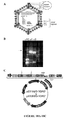
- Virus-like particles have been observed ( Watanabe, K. et al., Dis Aquat Organ 70, 183-192 (2006 )); however, efforts to implicate an infectious agent by using culture, subtractive cloning and consensus polymerase chain reaction have been unsuccessful.
- HSMI is associated with infection with a novel reovirus termed Piscine reovirus (PRV).
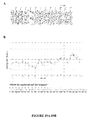
- PRV was identified through high-throughput pyrosequencing of serum and heart tissue of experimentally infected fish using novel frequency analysis methods as well as standard alignment methods. Described herein are PRV nucleic acid sequences.
- Described herein are expression constructs, for example plasmids and vectors, and isolated nucleic acids which comprise PRV nucleic acid sequences of SEQ ID NOs: 1-10, fragments, complementary sequences, and/or variants thereof.
- nucleic acid sequences and polypeptides described herein may be useful for multiple applications, including, but not limited to, generation of diagnostic antibodies and diagnostic nucleic acids.
- polypeptide comprising the amino acid sequence of any one of SEQ ID NOs: 29-40.
- Described herein is an isolated PRV nucleic acid having the sequence of any of SEQ ID NOs: 1-10, or a fragment thereof.
- Described herein is an isolated PRV nucleic acid which comprises consecutive nucleotides having a sequence selected from the group consisting of any of SEQ ID NOs: 1-10, or a fragment thereof.
- an isolated PRV nucleic acid which comprises consecutive nucleotides having a sequence selected from a variant of any of SEQ ID NOs: 1-10 or a fragment thereof.
- the variant may have at least about 85% identity to SEQ ID NOs: 1-10, or a fragment thereof.
- the variant may have at least about 90%, about 95.5%, about 96%, about 96.5%, about 97%, about 97.5%, about 98%, about 98.5%, about 99%, about 99.5% or about 99.9% identity to that of any one of SEQ ID NOs: 1-10, or a fragment thereof.
- Described herein is an isolated PRV nucleic acid complementary to a PRV nucleic acid sequence in any of SEQ ID NOs: 1-10, or a fragment thereof.
- Described herein is an isolated PRV nucleic acid which comprises consecutive nucleotides complementary to a PRV nucleic acid sequence in any of SEQ ID NOs: 1-10, or a fragment thereof.
- an isolated PRV nucleic acid which comprises consecutive nucleotides complementary to a PRV nucleic acid sequence in any of SEQ ID NOs: 1-10, or a fragment thereof.
- the variant may have at least about 85% identity to SEQ ID NOs: 1-10, or a fragment thereof.
- the variant may have at least about 90%, about 95.5%, about 96%, about 96.5%, about 97%, about 97.5%, about 98%, about 98.5%, about 99%, about 99.5% or about 99.9% identity to that of any one of SEQ ID NOs: 1-10, or a fragment thereof.
- Described herein is an isolated PRV nucleic acid having a sequence substantially identical to a PRV nucleic acid sequence in any of SEQ ID NOs: 1-10, or a fragment thereof.
- Described herein is an isolated PRV nucleic acid having a sequence substantially identical to a sequence complementary to a PRV nucleic acid sequence in any of SEQ ID NOs: 1-10, or a fragment thereof.
- an oligonucleotide probe which comprises from about 10 nucleotides to about 50 nucleotides, wherein at least about 10 contiguous nucleotides are at least 95 % complementary to a nucleic acid target region within a PRV nucleic acid sequence in any of SEQ ID NOs: 1-10, wherein the oligonucleotide probe hybridizes to the nucleic acid target region under moderate to highly stringent conditions to form a detectable nucleic acid target:oligonucleotide probe duplex.
- the oligonucleotide probe may be at least about 95.5%, about 96%, about 96.5%, about 97%, about 97.5%, about 98%, about 98.5%, about 99%, about 99.5% or about 99.9% complementary to SEQ ID NOs: 1-10.
- the oligonucleotides probe may consist essentially of from about 10 to about 50 nucleotides.
- Polynucleotides homologous to the sequences illustrated in the SEQ ID NOs 1-10 can be identified, e.g., by hybridization to each other under stringent or under highly stringent conditions.
- Single stranded polynucleotides hybridize when they associate based on a variety of well characterized physical-chemical forces, such as hydrogen bonding, solvent exclusion, base stacking and the like.
- the stringency of a hybridization reflects the degree of sequence identity of the nucleic acids involved, such that the higher the stringency, the more similar are the two polynucleotide strands. Stringency is influenced by a variety of factors, including temperature, salt concentration and composition, organic and non-organic additives, solvents, etc. present in both the hybridization and wash solutions and incubations.
- the invention is directed to primer sets comprising isolated nucleic acids as described herein, which primer set are suitable for amplification of nucleic acids from samples which comprises Piscine reoviruses represented by any one of SEQ ID NO: 1-10, or variants thereof.
- Primer sets can comprise any suitable combination of primers which would allow amplification of a target nucleic acid sequences in a sample which comprises Piscine reoviruses represented by any one of SEQ ID NO: 1-10, or variants thereof.
- Amplification can be performed by any suitable method known in the art, for example but not limited to PCR, RT-PCR, transcription mediated amplification (TMA).
- Hybridization conditions refers to conditions under which a probe, primer or oligonucleotide will hybridize to its target sequence, and can hybridize, for example but not limited to, variants of the disclosed polynucleotide sequences, including allelic or splice variants, or sequences that encode orthologs or paralogs of presently disclosed polypeptides.
- the precise conditions for stringent hybridization are typically sequence-dependent and will be different in different circumstances. Longer sequences hybridize specifically at higher temperatures than shorter sequences. Generally, stringent conditions are selected to be about 5°C. lower than the thermal melting point (Tm) for the specific sequence at a defined ionic strength and pH.
- the Tm is the temperature (under defined ionic strength, pH and nucleic acid concentration) at which 50% of the probes complementary to the target sequence hybridize to the target sequence at equilibrium. Since the target sequences are generally present at excess, at Tm, 50% of the probes are occupied at equilibrium.
- stringent conditions will be those in which the salt concentration is less than about 1.0 M sodium ion, typically about 0.01 to 1.0 M sodium ion (or other salts) at pH 7.0 to 8.3 and the temperature is at least about 30°C. for short probes, primers or oligonucleotides (e.g., 10 nt to 50 nt) and at least about 60°C. for longer probes, primers and oligonucleotides. Stringent conditions may also be achieved with the addition of destabilizing agents, such as formamide.
- stringency is determined by the temperature, ionic strength, and concentration of denaturing agents (e.g., formamide) used in a hybridization and washing procedure.
- denaturing agents e.g., formamide
- the degree to which two nucleic acids hybridize under various conditions of stringency is correlated with the extent of their similarity. Numerous variations are possible in the conditions and means by which nucleic acid hybridization can be performed to isolate nucleic sequences having similarity to the nucleic acid sequences known in the art and are not limited to those explicitly disclosed herein.
- nucleic acid sequences having various degrees of similarity such as, for example, nucleic acid sequences having 60% identity, or about 70% identity, or about 80% or greater identity with disclosed nucleic acid sequences.
- Stringent conditions are known to those skilled in the art and can be found in Current Protocols In Molecular Biology, John Wiley & Sons, N.Y. (1989), 6.3.1-10.3.6 .
- the conditions are such that sequences at least about 65%, 70%, 75%, 85%, 90%, 95%, 98%, or 99% homologous to each other typically remain hybridized to each other.
- a non-limiting example of stringent hybridization conditions is hybridization in a high salt buffer comprising 6X sodium chloride/sodium citrate (SSC), 50 mM Tris-HCl (pH 7.5), 1 nM EDTA, 0.02% PVP, 0.02% Ficoll, 0.02% BSA, and 500 mg/ml denatured salmon sperm DNA at 65°C. This hybridization is followed by one or more washes in 0.2X SSC, 0.01% BSA at 50°C.
- Another non-limiting example of stringent hybridization conditions are hybridization in 6X sodium chloride/sodium citrate (SSC) at about 45°C, followed by one or more washes in 0.2X SSC, 0.1% SDS at 50-65°C. Examples of moderate to low stringency hybridization conditions are well known in the art.
- Tm melting temperature
- Hybridization experiments are generally conducted in a buffer of pH between 6.8 to 7.4, although the rate of hybridization is nearly independent of pH at ionic strengths likely to be used in the hybridization buffer (Anderson et al. (1985) supra).
- one or more of the following may be used to reduce non-specific hybridization: sonicated salmon sperm DNA or another non-complementary DNA, bovine serum albumin, sodium pyrophosphate, sodium dodecylsulfate (SDS), polyvinyl-pyrrolidone, ficoll and Denhardt's solution.
- Dextran sulfate and polyethylene glycol 6000 act to exclude DNA from solution, thus raising the effective probe DNA concentration and the hybridization signal within a given unit of time.
- conditions of even greater stringency may be desirable or required to reduce non-specific and/or background hybridization. These conditions may be created with the use of higher temperature, lower ionic strength and higher concentration of a denaturing agent such as formamide.
- Stringency conditions can be adjusted to screen for moderately similar fragments such as homologous sequences from distantly related organisms, or to highly similar fragments.
- the stringency can be adjusted either during the hybridization step or in the post-hybridization washes.
- Salt concentration, formamide concentration, hybridization temperature and probe lengths are variables that can be used to alter stringency. As a general guidelines high stringency is typically performed at Tm-5°C to Tm -20°C, moderate stringency at Tm-20°C to Tm-35°C and low stringency at Tm-35°SC to Tm-50°C for duplex>150 base pairs.
- Hybridization may be performed at low to moderate stringency (25-50°C below Tm), followed by post-hybridization washes at increasing stringencies.
- Maximum rates of hybridization in solution are determined empirically to occur at Tm-25°C for DNA-DNA duplex and Tm -15°C for RNA-DNA duplex.
- the degree of dissociation may be assessed after each wash step to determine the need for subsequent, higher stringency wash steps.
- High stringency conditions may be used to select for nucleic acid sequences with high degrees of identity to the disclosed sequences.
- An example of stringent hybridization conditions obtained in a filter-based method such as a Southern or northern blot for hybridization of complementary nucleic acids that have more than 100 complementary residues is about 5°C. to 20°C. lower than the thermal melting point (Tm) for the specific sequence at a defined ionic strength and pH.
- Conditions used for hybridization may include about 0.02 M to about 0.15 M sodium chloride, about 0.5% to about 5% casein, about 0.02% SDS or about 0.1% N-laurylsarcosine, about 0.001 M to about 0.03 M sodium citrate, at hybridization temperatures between about 50°C and about 70°C.
- high stringency conditions are about 0.02 M sodium chloride, about 0.5% casein, about 0.02% SDS, about 0.001 M sodium citrate, at a temperature of about 50°C.
- Nucleic acid molecules that hybridize under stringent conditions will typically hybridize to a probe based on either the entire DNA molecule or selected portions, e.g., to a unique subsequence, of the DNA.
- Stringent salt concentration will ordinarily be less than about 750 mM NaCl and 75 mM trisodium citrate. Increasingly stringent conditions may be obtained with less than about 500 mM NaCl and 50 mM trisodium citrate, to even greater stringency with less than about 250 mM NaCl and 25 mM trisodium citrate.
- Low stringency hybridization can be obtained in the absence of organic solvent, e.g., formamide, whereas in certain embodiments high stringency hybridization may be obtained in the presence of at least about 35% formamide, and in other embodiments in the presence of at least about 50% formamide.
- stringent temperature conditions will ordinarily include temperatures of at least about 30°C, and in other embodiment at least about 37°C, and in other embodiments at least about 42°C with formamide present. Varying additional parameters, such as hybridization time, the concentration of detergent, e.g., sodium dodecyl sulfate (SDS) and ionic strength, are well known to those skilled in the art. Various levels of stringency are accomplished by combining these various conditions as needed. In a certain embodiment, hybridization will occur at 30°C in 750 mM NaCl, 75 mM trisodium citrate, and 1% SDS.
- SDS sodium dodecyl sulfate
- hybridization will occur at 37°C in 500 mM NaCl, 50 mM trisodium citrate, 1% SDS, 35% formamide. In another embodiment, hybridization will occur at 42C in 250 mM NaCl, 25 mM trisodium citrate, 1% SDS, 50% formamide. Useful variations on these conditions will be readily apparent to those skilled in the art.
- wash stringency may also vary in stringency; the post-hybridization wash steps primarily determine hybridization specificity, with the most critical factors being temperature and the ionic strength of the final wash solution.
- Wash stringency can be increased by decreasing salt concentration or by increasing temperature.
- Stringent salt concentration for the wash steps can be less than about 30 mM NaCl and 3 mM trisodium citrate, and in certain embodiments less than about 15 mM NaCl and 1.5 mM trisodium citrate.
- the wash conditions may be under conditions of 0.1XSSC to 2.0XSSC and 0.1% SDS at 50-65°C, with, for example, two steps of 10-30 min.
- stringent wash conditions includes about 2.0XSSC, 0.1% SDS at 65°C and washing twice, each wash step being about 30 min.
- the temperature for the wash solutions will ordinarily be at least about 25°C, and for greater stringency at least about 42°C.
- Hybridization stringency may be increased further by using the same conditions as in the hybridization steps, with the wash temperature raised about 3°C to about 5°C, and stringency may be increased even further by using the same conditions except the wash temperature is raised about 6°C to about 9°C.
- wash steps may be performed at a lower temperature, e.g., 50°C.
- An example of a low stringency wash step employs a solution and conditions of at least 25° C in 30 mM NaCl, 3 mM trisodium citrate, and 0.1% SDS over 30 min. Greater stringency may be obtained at 42° C in 15 mM NaCl, with 1.5 mM trisodium citrate, and 0.1% SDS over 30 min. Even higher stringency wash conditions are obtained at 65°C-68°C in a solution of 15 mM NaCl, 1.5 mM trisodium citrate, and 0.1% SDS. Wash procedures will generally employ at least two final wash steps. Additional variations on these conditions will be readily apparent to those skilled in the art.
- Stringency conditions can be selected such that an oligonucleotide that is perfectly complementary to the coding oligonucleotide hybridizes to the coding oligonucleotide with at least about a 5-10X higher signal to noise ratio than the ratio for hybridization of the perfectly complementary oligonucleotide to a nucleic acid. It may be desirable to select conditions for a particular assay such that a higher signal to noise ratio, that is, about 15X or more, is obtained.
- an animal nucleic acid will hybridize to a unique coding oligonucleotide with at least a 2X or greater signal to noise ratio as compared to hybridization of the coding oligonucleotide to a nucleic acid encoding known polypeptide.
- the particular signal will depend on the label used in the relevant assay, e.g., a fluorescent label, a calorimetric label, a radioactive label, or the like.
- Labeled hybridization or PCR probes for detecting related polynucleotide sequences may be produced by oligolabeling, nick translation, end-labeling, or PCR amplification using a labeled nucleotide.
- polynucleotide sequences that are capable of hybridizing to the claimed polynucleotide sequences, including any of the nucleic acid sequences disclosed herein, and fragments thereof under various conditions of stringency (See, for example, Wahl and Berger (1987) Methods Enzymol. 152: 399-407 ; and Kimmel (1987) Methods Enzymol. 152: 507-511 ).
- hybridization conditions that are highly stringent, and means for achieving them, are well known in the art. See, for example, Sambrook et al.
- the isolated nucleic acid which can be used as primers and/or probes can comprise about 4, 5, 6, 7, 8, 9, 10, 11, 12, 13, 14, 15, 16, 17, 18, 19, 20, 21, 22, 23, 24, 25, 26, 27, 28, 29, 30, 31, 32, 33, 34, 35, 36, 37, 38, 39, 40 consecutive nucleotides from any one of SEQ ID NO: 1-10, or sequences complementary to any one of SEQ ID NO: 1-10.
- the isolated nucleic acid which can be used as primers and/or probes can comprise from about 4, 5, 6, 7, 8, 9, 10, 11, 12, 13, 14, 15, 16, 17, 18, 19, 20, 21, 22, 23, 24, 25, 26, 27, 28, 29, 30, 31, 32, 33, 34, 35, 36, 37, 38, 39, 40 and up to about 20, 25, 30, 35, 40, 45, 50, 55, 60, 65, 70, 75, 80, 85, 90, 95 and 100 consecutive nucleotides from any one of SEQ ID NO: 1-10, or sequences complementary to any one of SEQ ID NO: 1-10.
- Primer nd/or probes can be labeled by any suitable molecule and/or label known in the art, for example but not limited to fluorescent tags suitable for use in Real Time PCR amplification, for example TaqMan, cybergreen, TAMRA and/or FAM probes; radiolabels, and so forth.
- the oligonucleotide primers and/or probe may further comprise a detectable non-isotopic label selected from the group consisting of: a fluorescent molecule, a chemiluminescent molecule, an enzyme, a cofactor, an enzyme substrate, and a hapten.
- Described herein is a synthetic nucleic acid which has a sequence consisting of from about 10 to about 30 consecutive nucleotides from a nucleic acid sequence selected from the group of sequences consisting of SEQ ID NO: 1-10.
- Described herein is a synthetic nucleic acid which has a sequence consisting of from about 10 to about 30 consecutive nucleotides from a nucleic acid consisting of consecutive nucleotides having a sequence which is a variant of any one of SEQ ID NOS 1-10 having at least about 95% identity to SEQ ID NO: 1-10.
- the variant may have at least about 95.5%, about 96%, about 96.5%, about 97%, about 97.5%, about 98%, about 98.5%, about 99%, about 99.5% or about 99.9% identity to SEQ ID NO: 1-10.
- composition comprising one or more nucleic acids having a sequence consisting of from about 10 to about 30 consecutive nucleotides from a nucleic acids sequence selected from the group of sequences consisting of SEQ ID NO: 1-10.
- composition comprising one or more nucleic acids having a sequence consisting of from about 10 to about 30 consecutive nucleotides from a nucleic acid consisting of consecutive nucleotides having a sequence which is a variant of any one of SEQ ID NOS 1-10 having at least about 95% identity to SEQ ID NO: 1-10.
- the variant may have at least about 95.5%, about 96%, about 96.5%, about 97%, about 97.5%, about 98%, about 98.5%, about 99%, about 99.5% or about 99.9% identity to SEQ ID NO: 1-10.
- Described herein is a synthetic nucleic acid which has a sequence consisting of from about 10 to about 30 consecutive nucleotides from a nucleic acid sequence which is complementary to a nucleic acid sequence selected from the group of sequences consisting of SEQ ID NO: 1-10.
- Described herein is a synthetic nucleic acid which has a sequence consisting of from about 10 to about 30 consecutive nucleotides complementary to a nucleic acid consisting of consecutive nucleotides having a sequence which is a variant of any one of SEQ ID NOS 1-10 having at least about 95% identity to SEQ ID NO: 1-10.
- the variant may have at least about 95.5%, about 96%, about 96.5%, about 97%, about 97.5%, about 98%, about 98.5%, about 99%, about 99.5% or about 99.9% identity to SEQ ID NO: 1-10.
- composition comprising one or more synthetic nucleic acids which have a sequence consisting of from about 10 to about 30 consecutive nucleotides from a nucleic acid sequence which is complementary to a nucleic acid sequence selected from the group of sequences consisting of SEQ ID NO: 1-10.
- composition comprising one or more synthetic nucleic acids which have a sequence consisting of from about 10 to about 30 consecutive nucleotides complementary to a nucleic acid consisting of consecutive nucleotides having a sequence which is a variant of any one of SEQ ID NOS 1-10 having at least about 95% identity to SEQ ID NO: 1-10.
- the variant may have at least about 95.5%, about 96%, about 96.5%, about 97%, about 97.5%, about 98%, about 98.5%, about 99%, about 99.5% or about 99.9% identity to SEQ ID NO: 1-10.
- nucleic acid sequences such as primers and probes, comprising nucleic acid sequences derived from any one of SEQ ID NO: 1-10.
- primers and probes may be useful for detecting the presence of the PRV of the invention, for example in samples of bodily fluids such as blood, saliva, or urine from an animal, and thus may be useful in the diagnosis of PRV infection.
- probes can detect polynucleotides of SEQ ID NO: 1-10 in samples which comprise PRV represented by SEQ ID NO: 1-10.
- the isolated nucleic acids which can be used as primer and/probes are of sufficient length to allow hybridization with, i.e. formation of duplex with a corresponding target nucleic acid sequence, a nucleic acid sequences of any one of SEQ ID NO: 1-10, or a variant thereof.
- Described herein is an isolated nucleic acid sequence comprising from about 10 to about 50 consecutive nucleotides from any one of SEQ ID NO: 1-10 or a sequence complementary to any one of SEQ ID NO: 1-10. Described herein is an isolated nucleic acid sequence comprising from about 10 to about 100 consecutive nucleotides from any one of SEQ ID NO: 1-10 or a sequence complementary to any one of SEQ ID NO: 1-10. Described herein is an isolated nucleic acid sequence comprising from about 10 to about 200 consecutive nucleotides from any one of SEQ ID NO: 1-10 or a sequence complementary to any one of SEQ ID NO: 1-10.
- Described herein is an isolated nucleic acid sequence comprising from about 10 to about 300 consecutive nucleotides from any one of SEQ ID NO: 1-10 or a sequence complementary to any one of SEQ ID NO: 1-10. Described herein is an isolated nucleic acid sequence comprising from about 10 to about 400 consecutive nucleotides from SEQ ID NO: 1 or a sequence complementary to any one of SEQ ID NO: 1-10. Described herein is an isolated nucleic acid sequence comprising from about 10 to about 500 consecutive nucleotides from any one of SEQ ID NO: 1-10 or a sequence complementary to any one of SEQ ID NO: 1-10.
- Described herein is an isolated nucleic acid sequence comprising from about 10 to about 600 consecutive nucleotides from any one of SEQ ID NO: 1-10 or a sequence complementary to any one of SEQ ID NO: 1-10. Described herein is an isolated nucleic acid sequence comprising from about 10 to about 700 consecutive nucleotides from any one of SEQ ID NO: 1-10 or a sequence complementary to any one of SEQ ID NO: 1-10. Described herein is an isolated nucleic acid sequence comprising from about 10 to about 800 consecutive nucleotides from any one of SEQ ID NO: 1-10 or a sequence complementary to any one of SEQ ID NO: 1-10.
- Described herein is an isolated nucleic acid sequence comprising from about 10 to about 900 consecutive nucleotides from any one of SEQ ID NO: 1-10 or a sequence complementary to any one of SEQ ID NO: 1-10. Described herein is an isolated nucleic acid sequence comprising from about 10 to about 1000 consecutive nucleotides from any one of SEQ ID NO: 1-10 or a sequence complementary to any one of SEQ ID NO: 1-10. Described herein is an isolated nucleic acid sequence comprising from about 10 to about 1500 consecutive nucleotides from any one of SEQ ID NO: 1-10 or a sequence complementary to any one of SEQ ID NO: 1-10.
- Described herein is an isolated nucleic acid sequence comprising from about 10 to about 2000 consecutive nucleotides from any one of SEQ ID NO: 1-10 or a sequence complementary to any one of SEQ ID NO: 1-10. Described herein is an isolated nucleic acid sequence comprising from about 10 to about 2500 consecutive nucleotides from any one of SEQ ID NO: 1-10 or a sequence complementary to any one of SEQ ID NO: 1-10. Described herein is an isolated nucleic acid sequence comprising from about 10 to about 3000 consecutive nucleotides from any one of SEQ ID NO: 1-10 or a sequence complementary to any one of SEQ ID NO: 1-10.
- Described herein is an isolated nucleic acid sequence comprising from about 10 to about 3500 consecutive nucleotides from any one of SEQ ID NO: 1-10 or a sequence complementary to any one of SEQ ID NO: 1-10. Described herein is an isolated nucleic acid sequence comprising from about 10 to about 3600 consecutive nucleotides from any one of SEQ ID NO: 1-10 or a sequence complementary to any one of SEQ ID NO: 1-10. Described herein is an isolated nucleic acid sequence comprising from about 10 to about 3621 consecutive nucleotides from any one of SEQ ID NO: 1-10 or a sequence complementary to any one of SEQ ID NO: 1-10.
- a primer set for determining the presence or absence of the PRV in a biological sample, wherein the primer set comprises at least one synthetic nucleic acid sequence selected from the group consisting of: a synthetic nucleic acid which has a sequence consisting of from about 10 to about 30 consecutive nucleotides from a nucleic acids sequence selected from the group of sequences consisting of SEQ ID NO: 1-10, a synthetic nucleic acid which has a sequence consisting of from about 10 to about 30 consecutive nucleotides from a nucleic acids sequence which is complementary to a nucleic acid sequence selected from the group of sequences consisting of SEQ ID NO: 1-10.
- the biological sample is derived from an animal suspected of having the PRV.
- a method for determining the presence or absence of a PRV in a biological sample comprising: a) contacting nucleic acid from a biological sample with at least one primer which is a nucleic acid sequence consisting of from about 10 to about 30 consecutive nucleotides from a nucleic acids sequence which is complementary to a nucleic acid sequence selected from the group of sequences consisting of SEQ ID NO: 1-10, b) subjecting the nucleic acid and the primer to amplification conditions, and c) determining the presence or absence of amplification product, wherein the presence of amplification product indicates the presence of RNA associated with PRV in the sample.
- the biological sample may be derived from a animal suspected of having a PRV.
- a method for determining the presence or absence of the PRV in a biological sample comprising: a) contacting nucleic acid from a biological sample with at least one primer which is a synthetic nucleic acid which has a sequence consisting of from about 10 to about 30 consecutive nucleotides from a nucleic acids sequence selected from the group of sequences consisting of SEQ ID NO: 1-10, b) subjecting the nucleic acid and the primer to amplification conditions, and c) determining the presence or absence of amplification product, wherein the presence of amplification product indicates the presence of RNA associated with PRV in the sample.
- RNA interfering RNA
- Described herein is a method of reducing the levels of a viral protein, viral mRNA or viral titer in a cell in an animal comprising: administering at least one iRNA agent to an animal, wherein the iRNA agent comprising a sense strand having at least 15 contiguous nucleotides complementary to gene from a PRV comprising any of SEQ ID NO: 1-10 and an antisense strand having at least 15 contiguous nucleotides complementary to the sense strand.
- the iRNA agent is administered to an animal.
- the iRNA agent may be administered via nebulization to an animal.
- the method may further comprise co-administering a second iRNA agent to the animal, wherein the second iRNA agent comprising a sense strand having at least 15 or more contiguous nucleotides complementary to second gene from the PRV, and an antisense strand having at least 15 or more contiguous nucleotides complementary to the sense strand.
- Described herein is a method of reducing the levels of a viral protein in a cell in an animal comprising the step of administering an iRNA agent to an animal, wherein the iRNA agent comprises a sense strand having at least 15 or more contiguous nucleotides complementary to a gene from a PRV comprising SEQ ID NO: 1-10 and an antisense strand having at least 15 or more contiguous nucleotides complementary to the sense strand.
- iRNA molecules which target nucleic acids from PRV, for example but not limited to SEQ ID NO: 1-10, and variants thereof, and silence a target gene.
- RNA agent (abbreviation for "interfering RNA agent”) as used herein, is an RNA agent, which can down-regulate the expression of a target gene, e.g. a PRV gene.
- An iRNA agent may act by one or more of a number of mechanisms, including post-transcriptional cleavage of a target mRNA sometimes referred to in the art as RNAi, or pre-transcriptional or pre-translational mechanisms.
- An iRNA agent can be a double stranded (ds) iRNA agent.
- a “ds iRNA agent” (abbreviation for "double stranded iRNA agent”), as used herein, is an iRNA agent which includes more than one, and in certain embodiments two, strands in which interchain hybridization can form a region of duplex structure.
- a “strand” herein refers to a contiguous sequence of nucleotides (including non-naturally occurring or modified nucleotides). The two or more strands may be, or each form a part of, separate molecules, or they may be covalently interconnected, e.g. by a linker, e.g. a polyethyleneglycol linker, to form but one molecule. At least one strand can include a region which is sufficiently complementary to a target RNA.
- RNA strand Such strand is termed the "antisense strand".
- a second strand comprised in the dsRNA agent which comprises a region complementary to the antisense strand is termed the "sense strand".
- a ds iRNA agent can also be formed from a single RNA molecule which is, at least partly; self-complementary, forming, e.g., a hairpin or panhandle structure, including a duplex region.
- the term "strand” refers to one of the regions of the RNA molecule that is complementary to another region of the same RNA molecule.
- iRNA agents as described herein can mediate silencing of a gene, e.g., by RNA degradation.
- RNA is also referred to herein as the RNA to be silenced.
- a gene is also referred to as a target gene.
- the RNA to be silenced is a gene product of a PRV gene.
- RNAi refers to the ability of an agent to silence, in a sequence specific manner, a target gene.
- "Silencing a target gene” means the process whereby a cell containing and/or secreting a certain product of the target gene when not in contact with the agent, will contain and/or secret at least 10%, 20%, 30%, 40%, 50%, 60%, 70%, 80%, or 90% less of such gene product when contacted with the agent, as compared to a similar cell which has not been contacted with the agent.
- product of the target gene can, for example, be a messenger RNA (mRNA), a protein, or a regulatory element.
- silencing of a target gene can result in a reduction in "viral titer" in the cell or in the animal, wherein “reduction in viral titer” refers to a decrease in the number of viable virus produced by a cell or found in an organism undergoing the silencing of a viral target gene. Reduction in the cellular amount of virus produced can lead to a decrease in the amount of measurable virus produced in the tissues of an animal undergoing treatment and a reduction in the severity of the symptoms of the viral infection.
- iRNA agents of the present description are also referred to as "antiviral iRNA agents”.
- PRV gene refers to any one of the genes identified in the PRV genome.
- Described herein are methods for reducing viral titer in an animal, by administering to an animal, at least one iRNA which inhibits the expression of a PRV gene.
- Described herein are methods for identifying and/or generating anti-viral drugs. For example, methods for identifying drugs that bind to and/or inhibit the function of the PRV-encoded proteins described herein, or that inhibit the replication or pathogenicity of the PRV described herein. Methods of identifying drugs that affect or inhibit a particular drug target, such as high throughput drug screening methods, are well known in the art and can readily be applied to the proteins and viruses described herein.
- Described herein is an isolated PRV polypeptide encoded by a PRV nucleic acid sequence in any of SEQ ID NOs: 1-10, or a fragment thereof.
- PRV polypeptides and amino acid sequences described herein may be useful for, inter alia, expression of PRV -encoded proteins or fragments, variants, or derivatives thereof, generation of diagnostic antibodies against PRV proteins, generation of primers and probes for detecting PRV and/or for diagnosing PRV infection, and screening for drugs effective against Piscine reoviruses as described herein.
- the PRV polypeptide fragment can be a polypeptide comprising about 8 consecutive amino acids of a PRV polypeptide described herein.
- the fragment can be a polypeptide comprising about 10 consecutive amino acids of a PRV polypeptide described herein.
- the fragment can be a polypeptide comprising about 14 consecutive amino acids of a PRV polypeptide described herein.
- the fragment can be a polypeptide comprising about 16 consecutive amino acids of a PRV polypeptide described herein.
- the fragment can be a polypeptide comprising about 18 consecutive amino acids of a PRV polypeptide described herein.
- In another The fragment can be a polypeptide comprising about 20 consecutive amino acids of a PRV polypeptide described herein.
- the fragment can be a polypeptide comprising about 21 or more consecutive amino acids of a PRV polypeptide described herein.
- the PRV polypeptide fragment can be a polypeptide comprising from about 8 to about 50, about 8 to about 100, about 8 to about 200, about 8 to about 300, about 8 to about 400, about 8 to about 500, about 8 to about 600, about 8 to about 700, about 8 to about 800, about 8 to about 900 or more consecutive amino acids from a PRV polypeptide.
- Described herein is an isolated PRV polypeptide encoded by a nucleic acid which comprises consecutive nucleotides having a sequence selected from a PRV nucleic acid sequence in any of SEQ ID NOs: 1-10, or a fragment thereof.
- an isolated PRV polypeptide encoded by a nucleic acid which comprises consecutive nucleotides having a sequence selected from a variant of a PRV nucleic acid sequence in any of SEQ ID NOs: 1-10 or a fragment thereof.
- the variant may have at least about 85% identity to SEQ ID NOs: 1-10, or a fragment thereof.
- the variant may have has at least about 90%, about 95.5%, about 96%, about 96.5%, about 97%, about 97.5%, about 98%, about 98.5%, about 99%, about 99.5% or about 99.9% identity to that of any one of SEQ ID NOs: 1-10, or a fragment thereof.
- Described herein is an isolated PRV polypeptide encoded by a nucleic acid complementary a PRV nucleic acid sequence in any of SEQ ID NOs: 1-10, or a fragment thereof.
- Described herein is an isolated PRV polypeptide encoded by a nucleic acid which comprises consecutive nucleotides a PRV nucleic acid sequence in any of SEQ ID NOs: 1-10, or a fragment thereof.
- Described herein is an isolated PRV polypeptide encoded by a nucleic acid having a sequence substantially identical to a PRV nucleic acid sequence in any of SEQ ID NOs: 1-10, or a fragment thereof.
- Described herein is an isolated PRV polypeptide encoded by a nucleic acid having a sequence substantially identical to a sequence complementary to a PRV nucleic acid sequence in any of SEQ ID NOs: 1-10, or a fragment thereof.
- Described herein is an isolated PRV polypeptide having the sequence of any of SEQ ID NOs: 29-40, or a fragment thereof.
- Described herein is an isolated PRV polypeptide which comprises consecutive amino acids having a sequence selected from the group consisting of any of SEQ ID NOs: 29-40, or a fragment thereof.
- an isolated PRV polypeptide which comprises consecutive amino acids having a sequence selected from a variant of any of SEQ ID NOs: 29-40, or a fragment thereof.
- the variant may have at least about 85% identity to SEQ ID NOs: 29-40, or a fragment thereof.
- the variant may have has at least about 90%, about 95.5%, about 96%, about 96.5%, about 97%, about 97.5%, about 98%, about 98.5%, about 99%, about 99.5% or about 99.9% identity to that of any one of SEQ ID NOs: 1-10, or a fragment thereof.
- Described herein is an isolated PRV polypeptide having a sequence substantially identical to a PRV amino acid sequence in any of SEQ ID NOs: 29-40, or a fragment thereof.
- Described herein is an isolated PRV polypeptide encoded by a PRV nucleic acid sequence in any of SEQ ID NOs: 1-10, or a fragment thereof.
- the isolated PRV polypeptide fragment can be a polypeptide comprising about 8 consecutive amino acids of a PRV amino acid sequence of any of SEQ ID NOs: 29-40.
- the fragment can be a polypeptide comprising about 10 consecutive amino acids of a PRV amino acid sequence of any of SEQ ID NOs: 29-40.
- the fragment can be a polypeptide comprising about 14 consecutive amino acids of a PRV amino acid sequence of any of SEQ ID NOs: 29-40.
- the fragment can be a polypeptide comprising about 16 consecutive amino acids of a PRV amino acid sequence of any of SEQ ID NOs: 29-40.
- the fragment can be a polypeptide comprising about 18 consecutive amino acids of a PRV amino acid sequence of any of SEQ ID NOs: 29-40.
- the fragment can be a polypeptide comprising about 20 consecutive amino acids of a PRV amino acid sequence of any of SEQ ID NOs: 29-40.
- the fragment can be a polypeptide comprising about 21 or more consecutive amino acids of a PRV amino acid sequence of any of SEQ ID NOs: 29-40.
- the isolated PRV polypeptide fragment can be a polypeptide comprising from about 8 to about 50, about 8 to about 100, about 8 to about 200, about 8 to about 300, about 8 to about 400, about 8 to about 500, about 8 to about 600, about 8 to about 700, about 8 to about 800, about 8 to about 900 or more consecutive amino acids of a PRV amino acid sequence of any of SEQ ID NOs: 29-40.
- Described herein is an isolated PRV polypeptide which comprises consecutive amino acids having a sequence selected from a PRV amino acid sequence of any of SEQ ID NOs: 29-40.
- an isolated PRV polypeptide which comprises consecutive nucleotides having a sequence selected from a variant a PRV amino acid sequence of any of SEQ ID NOs: 29-40, or a fragment thereof.
- the variant has at least about 85% identity to any of SEQ ID NOs: 29-40, or a fragment thereof.
- the variant may have at least about 90%, about 95.5%, about 96%, about 96.5%, about 97%, about 97.5%, about 98%, about 98.5%, about 99%, about 99.5% or about 99.9% identity to any of SEQ ID NOs: 29-40, or a fragment thereof.
- Described herein is an isolated PRV polypeptide substantially identical to variant a PRV amino acid sequence of any of SEQ ID NOs: 29-40, or a fragment thereof.
- substantially identical in the context of two nucleic acids or polypeptides, refers to two or more sequences or subsequences that have at least of at least 98%, at least 99% or higher nucleotide or amino acid residue identity, when compared and aligned for maximum correspondence, as measured using one of the following sequence comparison algorithms or by visual inspection.
- polypeptides that a substantially identical to the PRV polypeptides described herein can also be used to generate diagnostic antibodies that bind to the PRV polypeptides described herein.
- Percent identity in the context of two or more nucleic acids or polypeptide sequences, refers to the percentage of nucleotides or amino acids that two or more sequences or subsequences contain which are the same.
- a specified percentage of amino acid residues or nucleotides can have a specified identity over a specified region, when compared and aligned for maximum correspondence over a comparison window, or designated region as measured using one of the following sequence comparison algorithms or by manual alignment and visual inspection.
- a PRV polypeptide which is a variant of a PRV polypeptide and has at least about 90%, about 95.5%, about 96%, about 96.5%, about 97%, about 97.5%, about 98%, about 98.5%, about 99%, about 99.5% or about 99.9% identity to a PRV polypeptide shown in SEQ ID NOs 29-40.
- PRV polypeptides and the antibodies and antibody generation methods related thereto encompass PRV polypeptides isolated from different virus isolates that have sequence identity levels of at least about 90%, while still representing the same PRV protein with the same immunological characteristics.
- sequence identities can be determined by analysis with a sequence comparison algorithm or by a visual inspection. Protein and/or nucleic acid sequence identities (homologies) can be evaluated using any of the variety of sequence comparison algorithms and programs known in the art.
- sequence comparison typically one sequence acts as a reference sequence, to which test sequences are compared.
- test and reference sequences are entered into a computer, subsequence coordinates are designated, if necessary, and sequence algorithm program parameters are designated. Default program parameters can be used, or alternative parameters can be designated.
- sequence comparison algorithm then calculates the percent sequence identities for the test sequences relative to the reference sequence, based on the program parameters.
- sequence comparison of nucleic acids and proteins the BLAST and BLAST 2.2.2. or FASTA version 3.0t78 algorithms and the default parameters discussed below can be used.
- BLAST and BLAST 2.0 are used, with the parameters described herein, to determine percent sequence identity for the nucleic acids and proteins of the invention.
- Software for performing BLAST analyses is publicly available through the National Center for Biotechnology Information (http://www ncbi.nlm.nih.gov/).
- the BLAST algorithm parameters W, T, and X determine the sensitivity and speed of the alignment.
- W wordlength
- E expectation
- the BLAST algorithm also performs a statistical analysis of the similarity between two sequences (see, e.g., Karlin & Altschul, Proc. Natl. Acad. Sci. U.S.A. 90:5873-5787, 1993 ).
- One measure of similarity provided by the BLAST algorithm is the smallest sum probability (P(N)), which provides an indication of the probability by which a match between two nucleotide or amino acid sequences would occur by chance.
- P(N) the smallest sum probability
- a nucleic acid is considered similar to a reference sequence if the smallest sum probability in a comparison of the test nucleic acid to the reference nucleic acid is less than about 0.2, less than about 0.01, and less than about 0.001.
- PILEUP creates a multiple sequence alignment from a group of related sequences using progressive, pairwise alignments to show relationship and percent sequence identity. It also plots a tree or dendogram showing the clustering relationships used to create the alignment. PILEUP uses a simplification of the progressive alignment method of Feng & Doolittle, J. Mol. Evol. 35:351-360, 1987 . The method used is similar to the method described by Higgins & Sharp, CABIOS 5:151-153, 1989 . The program can align up to 300 sequences, each of a maximum length of 5,000 nucleotides or amino acids. The multiple alignment procedure begins with the pairwise alignment of the two most similar sequences, producing a cluster of two aligned sequences.
- This cluster is then aligned to the next most related sequence or cluster of aligned sequences.
- Two clusters of sequences are aligned by a simple extension of the pairwise alignment of two individual sequences.
- the final alignment is achieved by a series of progressive, pairwise alignments.
- the program is run by designating specific sequences and their amino acid or nucleotide coordinates for regions of sequence comparison and by designating the program parameters.
- PILEUP a reference sequence is compared to other test sequences to determine the percent sequence identity relationship using the following parameters: default gap weight (3.00), default gap length weight (0.10), and weighted end gaps.
- PILEUP can be obtained from the GCG sequence analysis software package, e.g., version 7.0 ( Devereaux et al., Nuc. Acids Res. 12:387-395, 1984 .
- ClustalW performs multiple pairwise comparisons between groups of sequences and assembles them into a multiple alignment based on homology. Gap open and Gap extension penalties were 10 and 0.05 respectively.
- BLOSUM algorithm can be used as a protein weight matrix ( Henikoff and Henikoff, Proc. Natl. Acad. Sci. U.S.A. 89:10915-10919, 1992 ).
- a computer readable medium having stored thereon (i) a nucleic acid sequence selected from the group consisting of: a PRV nucleic acid sequence in any of SEQ ID NOs: 1-10, a sequence substantially identical to a PRV nucleic acid sequence in any of SEQ ID NOs: 1-10; a sequence variant of a PRV nucleic acid sequence in any of SEQ ID NOs: 1-10; or (ii) an amino acid sequence encoded by a nucleic acid sequence selected from the group consisting of: a PRV nucleic acid sequence in any of SEQ ID NOs: 1-10, an amino acid sequence encoded by a sequence substantially identical to a PRV nucleic acid sequence in any of SEQ ID NOs: 1-10; an amino acid sequence encoded by a sequence variant of a PRV nucleic acid sequence in any of SEQ ID NOs: 1-10.
- the PRV nucleic acid sequences described herein may be useful for, inter alia, expression of PRV -encoded proteins or fragments, variants, or derivatives thereof, generation of diagnostic antibodies against PRV proteins (e.g. for determining whether an animal has been infected with PRV), generation of primers and probes for detecting PRV and/or for diagnosing PRV infection, and screening for drugs effective against Piscine reoviruses as described herein.
- polypeptides described herein may be suitable for use as antigens to detect antibodies against PRV represented by SEQ ID NOs: 1-10, and variants thereof.
- the polypeptides described herein which comprise antigenic determinants can be used in various immunoassays to identify animals exposed to and/or samples which comprise PRV represented by SEQ ID NO: 1-10, and variants thereof.
- a diagnostic PRV antibody that binds a PRV, a PRV polypeptide, a PRV polypeptide fragment or a PRV polypeptide variant, or a polypeptide substantially identical to a PRV polypeptide and wherein the antibody is an antibody that binds a PRV or a PRV polypeptide but does not inhibit, neutralize or reduce the activity or function of the polypeptide or the PRV.
- the diagnostic antibody may be a polyclonal antibody, a monoclonal antibody, a teleost antibody or a chimeric antibody. Methods for purifying immunoglobulins from teleosts are also known in the art.
- a method for determining whether or not a sample contains a PRV comprising: (a) providing an immunoassay comprising a diagnostic antibody against a PRV derived antigen, (b) contacting the diagnostic antibody with a biological sample, (c) detecting binding between antigens in the test sample and the diagnostic antibody.
- the immunoassays may be a lateral flow assay or ELISA.
- the biological sample may be derived from an animal suspected of having a PRV.
- Described herein is a method for determining whether or not a sample contains antibodies against PRV, the method comprising: (a) providing an immunoassay comprising an antigen from a PRV, (b) contacting the antigen with a biological sample, (c) detecting binding between antibodies in the test sample and the antigen.
- diagnostic antibodies described herein can also be used to purify polypeptides of any polypeptide encoded by the nucleic sequence acid of any one of SEQ ID NO: 1-10, polypeptides comprising the sequence of any of SEQ ID NOs: 29-40, or variants or fragments thereof.
- the diagnostic antibodies may be used to identify expression and localization of a PRV polypeptide or variants or fragments thereof. Analysis of expression and localization of PRV polypeptides, or variants or fragments thereof, can be useful in diagnosing a PRV infection or for determining potential role of a PRV polypeptide.
- the antibodies may be used in various immunoassays to identify animals exposed to and/or samples which comprise antigens from PRV.
- any suitable immunoassay which can lead to formation of antigen-antibody complex can also be used. Variations and different formats of immunoassays, for example but not limited to ELISA, lateral flow assays for detection of analytes in samples, immunoprecipitation, are known in the art.
- the antigen and/or the antibody can be labeled by any suitable label or method known in the art. For example enzymatic immunoassays may use solid supports, or immunoprecipitation. Immunoassays which amplify the signal from the antigen-antibody immune complex can also be used with the methods described herein.
- Methods for assaying a sample include, but are not limited to, methods which can detect the presence of nucleic acids, methods which can detect the presence of PRV polypeptides, methods which can detect the presence of antibodies against PRV polypeptides, or any polypeptide encoded by a PRV nucleic acid.
- a PRV diagnostic kit comprising a PRV nucleic acid, a PRV nucleic acid fragment or a PRV nucleic acid variant, a nucleic acid substantially identical to a PRV nucleic acid, or a PRV diagnostic antibody.
- diagnostic antibodies or nucleic acid are used for diagnostic purposes, it is not necessary to use the entire nucleic acid or diagnostic antibody to detect a PRV or a PRV polypeptide in an animal or in a sample.
- Described herein are methods for generating diagnostic antibodies that bind to the PRV polypeptides described herein by generating antibodies that bind to a fragment of a polypeptide described herein.
- nucleic acid vector comprising a PRV nucleic acid sequence, a PRV nucleic acid fragment or a PRV nucleic acid variant, or a nucleic acid substantially identical to a PRV nucleic acid.
- nucleic acid vector encoding a PRV polypeptide, a PRV polypeptide fragment or a PRV polypeptide variant, or a polypeptide substantially identical to a PRV polypeptide.
- vectors include, but are not limited to retroviral, adenoviral, adeno-associated viral, lentiviral, and vesiculostomatitis viral vectors.
- a host organism comprising a nucleic acid vector encoding a PRV polypeptide, a PRV polypeptide fragment, a PRV polypeptide variant, a polypeptide substantially identical to a PRV polypeptide or a the diagnostic PRV antibody that binds a PRV, a PRV polypeptide, a PRV polypeptide fragment or a PRV polypeptide variant, or a polypeptide substantially identical to a PRV polypeptide and which does not inhibit, neutralize or reduce the activity or function of the polypeptide or the PRV.
- the host organism may be a prokaryote, a eukaryote, or a fungus.
- the organism may be a teleost (e.g. a salmon).
- the PRV nucleic acid sequences can be delivered to cultured cells, for example by transfecting cultured cells with plasmids or expression vectors containing PRV nucleic acid sequences, or by infecting cultured cells with recombinant viruses containing PRV nucleic acid sequences.
- PRV polypeptides may then be expressed in a host cell or expression system and purified.
- a host cell may be a cell of bacterial origin, e.g. Escherichia coli, Bacillus subtilis and Lactobacillus species, in combination with bacteria-based plasmids as pBR322, or bacterial expression vectors as pGEX, or with bacteriophages.
- the host cell may also be of eukaryotic origin, e.g. yeast-cells in combination with yeast-specific vector molecules, or higher eukaryotic cells like insect cells ( Luckow et al; Bio-technology 6: 47-55 (1988 )) in combination with vectors or recombinant baculoviruses, plant cells in combination with e.g. Ti-plasmid based vectors or plant viral vectors ( Barton, K. A. et al; Cell 32: 1033 (1983 ), mammalian cells like Hela cells, Chinese Hamster Ovary cells (CHO) or Crandell Feline Kidney-cells, also with appropriate vectors or recombinant viruses.
- yeast-cells in combination with yeast-specific vector molecules
- higher eukaryotic cells like insect cells ( Luckow et al; Bio-technology 6: 47-55 (1988 )
- vectors or recombinant baculoviruses plant cells in combination with e.g. Ti-plasmid
- in vitro expression systems such as in-vitro transcription and in-vitro translation systems can also be used to generate the PRV polypeptides described herein.
- the purified proteins can then be incorporated into compositions suitable for administration to animals. Methods and techniques for expression and purification of recombinant proteins are well known in the art, and any such suitable methods may be used.
- Plasmids and expression vectors can include a promoter for directing transcription of the nucleic acid.
- the nucleic acid sequence encoding PRV polypeptides may also be incorporated into a suitable recombinant virus for administration to the animal.
- suitable viruses include, but are not limited to, vaccinia viruses, retroviruses, adenoviruses and adeno-associated viruses.
- One of skill in the art will be able to select a suitable plasmid, expression vector, or recombinant virus for delivery of the PRV nucleic acid sequences.
- Direct vaccination with DNA encoding proteins has been successful for many different proteins. (As reviewed in e.g. Donnelly et al. The Immunologist 2: 20-26 (1993 )).
- the diagnostic PRV antibodies described herein can also be generated using live recombinant carriers capable of expressing the polypeptides described herein.
- Live recombinant carriers are micro-organisms or viruses in which additional genetic information, e.g. a nucleic acid sequence encoding a PRV polypeptide, or a fragment thereof has been cloned. Animals infected with such live recombinant carriers will produce an immunological response not only against the immunogens of the carrier, but also against the PRV polypeptide or PRV polypeptide fragment.
- live recombinant carriers suitable for use with the methods described herein includes Vibrio anguillarum ( Singer, J. T. et al. New Developments in Marine Biotechnology, p.
- the diagnostic PRV antibodies described herein can also be generated by any other method known in the art.
- Exemplary alternative in-vitro antibody generation technologies, transgenic animal technologies and hybridoma technologies See, e.g., Ausubel, et al., ed., Current Protocols in Molecular Biology, John Wiley & Sons, Inc., NY, N.Y. (1987-2001 ); Sambrook, et al., Molecular Cloning: A Laboratory Manual, 2nd Edition, Cold Spring Harbor, N.Y. (1989 ); Harlow and Lane, Antibodies, A Laboratory Manual, Cold Spring Harbor, N.Y.
- Ribosome display is a method of translating mRNAs into their cognate proteins while keeping the protein attached to the RNA.
- the nucleic acid coding sequence is recovered by RT-PCR ( Mattheakis, L. C. et al. 1994. Proc Natl Acad Sci USA 91, 9022 ).
- Yeast display is based on the construction of fusion proteins of the membrane-associated alpha-agglutinin yeast adhesion receptor, agal and aga2, a part of the mating type system ( Broder, et al. 1997. Nature Biotechnology, 15:553-7 ).
- Bacterial display is based fusion of the target to exported bacterial proteins that associate with the cell membrane or cell wall ( Chen and Georgiou 2002. Biotechnol Bioeng, 79:496-503 ). In comparison to hybridoma technology, phage and other antibody display methods afford the opportunity to manipulate selection against the antigen target in vitro and without the limitation of the possibility of host effects on the antigen or vice versa.
- the diagnostic PRV antibodies described herein that bind a PRV polypeptide, a PRV polypeptide fragment or a PRV polypeptide variant, or a polypeptide substantially identical to a PRV polypeptide and which do not inhibit, neutralize or reduce the activity or function of the polypeptide or the PRV may be obtained by selecting from libraries, e.g. a phage library.
- a phage library can be created by inserting a library of random oligonucleotides or a library of polynucleotides containing sequences of interest, such as from the B-cells of an immunized animal ( Smith, G. P. 1985. Science 228: 1315-1317 ).
- Antibody phage libraries contain heavy (H) and light (L) chain variable region pairs in one phage allowing the expression of single-chain Fv fragments or Fab fragments ( Hoogenboom, et al. 2000, Immunol Today 21(8) 371-10 ).
- the diversity of a phagemid library can be manipulated to increase and/or alter the immunospecificities of the monoclonal antibodies of the library to produce and subsequently identify additional, teleost monoclonal antibodies.
- the heavy (H) chain and light (L) chain immunoglobulin molecule encoding genes can be randomly mixed (shuffled) to create new HL pairs in an assembled immunoglobulin molecule.
- H and L chain encoding genes can be mutagenized in a complementarity determining region (CDR) of the variable region of the immunoglobulin polypeptide, and subsequently screened for desirable affinity and neutralization capabilities.
- CDR complementarity determining region
- Antibody libraries also can be created synthetically by selecting one or more framework sequences and introducing collections of CDR cassettes derived from antibody repertoires or through designed variation ( Kretzschmar and von Ruden 2000, Current Opinion in Biotechnology, 13:598-602 ). The positions of diversity are not limited to CDRs but can also include the framework segments of the variable regions or may include other than antibody variable regions, such as peptides.
- Pepsin or papain digestion of whole diagnostic PRV antibodies that bind PRV polypeptides can be used to generate diagnostic PRV antibodies fragments that bind PRV polypeptides.
- an Fab fragment consists of a monovalent antigen-binding fragment of an diagnostic antibody molecule, and can be produced by digestion of a whole diagnostic antibody molecule with the enzyme papain, to yield a fragment consisting of an intact light chain and a portion of a heavy chain.
- An (Fab') 2 fragment of an diagnostic antibody can be obtained by treating a whole diagnostic antibody molecule with the enzyme pepsin, without subsequent reduction.
- An Fab' fragment of an diagnostic antibody molecule can be obtained from (Fab') 2 by reduction with a thiol reducing agent, which yields a molecule consisting of an intact light chain and a portion of a heavy chain. Two Fab' fragments are obtained per diagnostic antibody molecule treated in this manner.
- Diagnostic PRV antibodies can be produced through chemical crosslinking of the selected molecules (which have been produced by synthetic means or by expression of nucleic acid that encode the polypeptides) or through recombinant DNA technology combined with in vitro, or cellular expression of the polypeptide, and subsequent oligomerization.
- Antibodies can be similarly produced through recombinant technology and expression, fusion of hybridomas that produce antibodies with different epitope specificities, or expression of multiple nucleic acid encoding antibody variable chains with different epitopic specificities in a single cell.
- the diagnostic antibodies described herein may be either joined directly or indirectly through covalent or non-covalent binding, e.g. via a multimerization domain, to produce multimers.
- a "multimerization domain” mediates non-covalent protein-protein interactions. Specific examples include coiled-coil (e.g., leucine zipper structures) and alpha-helical protein sequences. Sequences that mediate protein-protein binding via Van der Waals' forces, hydrogen bonding or charge-charge bonds can also be used as multimerization domains. Additional examples include basic-helix-loop-helix domains and other protein sequences that mediate heteromeric or homomeric protein-protein interactions among nucleic acid binding proteins (e.g., DNA binding transcription factors, such as TAFs).
- multimerization domain p53 residues 319 to 360 which mediate tetramer formation.
- human platelet factor 4 which self-assembles into tetramers.
- extracellular protein TSP4 a member of the thrombospondin family, which can form pentamers.
- Additional specific examples are the leucine zippers of jun, fos, and yeast protein GCN4.
- the diagnostic antibodies described herein s may be directly linked to each other via a chemical cross linking agent or can be connected via a linker sequence (e.g., a peptide sequence) to form multimers.
- a linker sequence e.g., a peptide sequence
- the diagnostic antibodies described herein can be polyclonal or monoclonal.
- the antibodies can also be chimeric (i.e., a combination of sequences from more than one species, for example, a chimeric mouse-salmon immunoglobulin).
- chimeric i.e., a combination of sequences from more than one species, for example, a chimeric mouse-salmon immunoglobulin.
- the presence of such protein sequences form other species can lead to the rapid clearance of the antibodies or can lead to the generation of an immune response against the antibody by an antibody.
- the diagnostic antibodies described herein can be antibodies that bind to other molecules (antigens) via heavy and light chain variable domains, V H and V L , respectively.
- the antibodies described herein include, but are not limited to IgY, IgY( ⁇ Fc)), IgG, IgD, IgA, IgM, IgE, and IgL.
- the antibodies may be intact immunoglobulin molecules, two full length heavy chains linked by disulfide bonds to two full length light chains, as well as subsequences (i.e. fragments) of immunoglobulin molecules, with or without constant region, that bind to an epitope of an antigen, or subsequences thereof (i.e. fragments) of immunoglobulin molecules, with or without constant region, that bind to an epitope of an antigen.
- Antibodies may comprise full length heavy and light chain variable domains, V H and V L , individually or in any combination.
- the basic immunoglobulin (antibody) structural unit can comprise a tetramer.
- Each tetramer can be composed of two identical pairs of polypeptide chains, each pair having one "light” (about 25 kD) and one "heavy” chain (about 50-70 kD).
- the N-terminus of each chain defines a variable region of about 100 to 110 or more amino acids primarily responsible for antigen recognition.
- the terms variable light chain (V 1 ) and variable heavy chain (V H ) refer to these light and heavy chains respectively.
- the diagnostic antibodies described herein may exist as intact immunoglobulins or as a number of well characterized fragments produced by digestion with various peptidases.
- pepsin digests an antibody below the disulfide linkages in the hinge region to produce F(ab)' 2 , a dimer of Fab which itself is a light chain joined to V H -C H 1 by a disulfide bond.
- the F(ab)' 2 may be reduced under mild conditions to break the disulfide linkage in the hinge region thereby converting the F(ab)' 2 dimer into an Fab' monomer.
- the Fab' monomer is essentially an Fab with part of the hinge region (see, Fundamental Immunology, W. E.
- Fab' domain is defined in terms of the digestion of an intact antibody, one of skill will appreciate that such Fab' fragments may be synthesized de novo either chemically or by utilizing recombinant DNA methodology.
- the Fab' regions may be derived from antibodies of animal origin or may be chimeric ( Morrison et al., Proc Natl. Acad. Sci. USA 81, 6851-10855 (1984 ) ( Jones et al., Nature 321, 522-525 (1986 ), and published UK patent application No. 8707252 ).
- the diagnostic antibodies described herein can include or be derived from any mammal, such as but not limited to, a fish, a human, a mouse, a rabbit, a rat, a rodent, a primate, or any combination thereof and includes isolated fish, human, primate, rodent, mammalian, chimeric, humanized and/or CDR-grafted or CDR-adapted antibodies, immunoglobulins, cleavage products and other portions and variants thereof.
- the antibody is purified.
- the diagnostic antibodies described herein include full length antibodies, subsequences (e.g., single chain forms), dimers, trimers, tetramers, pentamers, hexamers or any other higher order oligomer that retains at least a part of antigen binding activity of monomer.
- Multimers can comprise heteromeric or homomeric combinations of full length antibody, subsequences, unmodified or modified as set forth herein and known in the art.
- Antibody multimers are useful for increasing antigen avidity in comparison to monomer due to the multimer having multiple antigen binding sites.
- Antibody multimers are also useful for producing oligomeric (e.g., dimer, trimer, tertamer, etc.) combinations of different antibodies thereby producing compositions of antibodies that are multifunctional (e.g., bifunctional, trifunctional, tetrafunctional, etc.).
- diagnostic antibody subsequences include, for example, Fab, Fab', (Fab') 2 , Fv, or single chain antibody (SCA) fragment (e.g., scFv).
- Subsequences include portions which retain at least part of the function or activity of full length sequence. For example, an antibody subsequence will retain the ability to selectively bind to an antigen even though the binding affinity of the subsequence may be greater or less than the binding affinity of the full length antibody.
- An Fv fragment is a fragment containing the variable region of a light chain V L and the variable region of a heavy chain V H expressed as two chains.
- the association may be non-covalent or may be covalent, such as a chemical cross-linking agent or an intermolecular disulfide bond ( Inbar et al., (1972) Proc. Natl. Acad Sci. USA 69:2659 ; Sandhu (1992) Crit. Rev. Biotech. 12:437 ).
- subsequences of the diagnostic antibodies described herein such as separation of heavy chains to form monovalent light-heavy chain fragments, further cleavage of fragments, or other enzymatic, chemical, or genetic techniques may also be used, provided that the subsequences bind to the antigen to which the intact antibody binds.
- a single chain antibody is a genetically engineered or enzymatically digested antibody containing the variable region of a light chain V L and the variable region of a heavy chain, optionally linked by a flexible linker, such as a polypeptide sequence, in either V L -linker-V H orientation or in V H -linker-V L orientation.
- a single chain Fv fragment can be produced by linking two variable domains via a disulfide linkage between two cysteine residues.
- PRV nucleic acids and polypeptides described herein can be used to generate diagnostic antibodies that that can be used to detect the present or absence of a PRV or a PRV polypeptide in an animal or in a sample
- a 200nt fragment that is approximately 50% homologous at the amino acid level to mammalian Orthoreoviruses was obtained through high throughput sequencing of samples obtained from farmed salmon with HSMI in Norway. Quantitative PCR assays of muscle tissue from salmon with HSMI and normal salmon reveals a higher viral load in salmon with HSMI.
- Example 2 Heart and skeletal muscle inflammation of farmed salmon is associated with infection with a novel reovirus
- database alignment analysis at the nucleotide level revealed no evidence of infection, the predicted amino acid sequence of one 265 nucleotide read was 49% similar to the core-spike protein ⁇ 2 of Mammalian orthoreovirus 3 (AF378009).
- the HSMI serum sample with the highest genetic load by PCR (3.0 x10⁶ genome copies/ ⁇ l) was selected for additional pyrosequencing yielding 120,705 reads.
- a suite of bioinformatic tools was used to identify viral sequences.
- BLASTN and BLASTX Altschul et al., J Mol Biol 215, 403-410 (1990 )
- detected 1.5% and 53.9% of the predicted viral genome respectively, enabling identification of segments L1, L2, L3, M1, M2 and M3 ( Figure 1 ).
- FASTX Pearson et al., Genomics 46, 24-36(1997 )
- FASTX Pearson et al., Genomics 46, 24-36(1997 )
- FASTX Pearson et al., Genomics 46, 24-36(1997 )
- FASTX Pearson et al., Genomics 46, 24-36(1997 )
- FASTX Pearson et al., Genomics 46, 24-36(1997 )
- the genome of the PRV comprises at least 10 RNA segments (GenBank Accession numbers GU994013-GU994022).
- Reoviruses are non-enveloped icosahedral viruses with double-stranded RNA genomes comprising 10-12 segments. Twelve genera are defined based on host range, number of genome segments, G/C content, and antigenic relationships.
- a phylogenetic tree constructed using L gene segment sequences of known reoviruses indicate that PRV represents a distinct genetic lineage branching off the root of the aquareovirus and orthoreovirus genera, viruses of fish and shellfish, reptiles, birds and mammals ( Figure 2 ).
- PRV gene segments confirmed the divergence of PRV sequence with respect to other Reoviruses ( Figures 5 to 12 ).
- All PRV gene segments contained the 3' terminal nucleotides (UCAUC-3') found in orthoreoviruses and aquareoviruses ( Attoui et al., J Gen Virol 83, 1941-1951 (2002 )); however, the 5' terminal nucleotides (5'-GAUAAA/U) were unique.
- the orthoreoviruses have polycistronic segments in either S1 or S4. Whereas aquareovirus species C are polycistronic in the S7 (the orthoreovirus S1 homolog), the other aquareovirus species are not ( Attoui et al., J Gen Virol 83, 1941-1951 (2002 )).
- homologues of the ⁇ 1, ⁇ 2, ⁇ 3, ⁇ l, ⁇ 2, ⁇ 3, ⁇ 2 and ⁇ NS sequences of PRV are found in orthoreoviruses and aquareoviruses, the ⁇ 1 and ⁇ 3 sequences and the small putative open reading frames observed in S2 and S1 appear distinctive.
- the structure of the latter is similar to a fusion-associated small transmembrane (FAST) reovirus protein ( Shmulevitz et al., EMBO J 19, 902-912 (2000 )) ( Figure 13 ).
- Reovirus FAST proteins are nonstructural, single-pass membrane proteins that induce cell-cell fusion and syncytium formation ( Shmulevitz et al., EMBO J 19, 902-912 (2000 )). Taken together these data provide compelling evidence that PRV is the prototype of a new reovirus genus equally distant to the orthoreovirus and aquareovirus genera.
- PRV was detected in only sixteen of these samples (24.2%). Two of these sixteen samples were positive by the cutoff established for farmed salmon with relative log ratios of 6.70 and 7.58; the other fourteen had L1/EF1A log ratios well below the 5.00 cutoff (range, -.20 to 4.57).
- Negative 1M Wild Healthy Wild healthy Muscle 4.0E+01 1.6 + Negative 2M Wild Healthy Wild healthy Muscle . . - Negative 3M Wild Healthy Wild healthy Muscle 1.7E+02 2.23 + Negative 1Mi Wild Healthy Wild healthy Spleen . . - Negative 2Mi Wild Healthy Wild healthy Spleen . . - Negative 3Mi Wild Healthy Wild healthy Spleen ⁇ ⁇ - Negative 521 - 6 Wild Healthy Wild healthy Various organs 2.7E+00 0.42 + Negative a Ratio of virus burden (quantitated through the L1 viral gene), normalized using a salmon housekeeping gene (EF1A) and adjusted by a factor of 108.
- b Log transformation of the adjusted ratio L1/EF1A.
- c Virus detection by real time RT-PCR.
- d For statistical analyses, samples were considered positive whenever the adjusted log ratio was higher than 5.00
- PRV RNA was distributed throughout the myocardium and endocardium of salmon with HSMI ( Figure 4A, 4B ) but not detected in normal salmon or salmon infected with salmon pancreas disease virus ( Figure 4C, 4D )
- HSMI was experimentally-induced in normal Atlantic salmon by inoculation with heart and kidney extracts from fish with HSMI or cohabitation with fish with HSMI.
- RNA extracted from heart tissue from Atlantic salmon with experimentally-induced HSMI was used as template for high throughput pyrosequencing. Sequences were analyzed using a suite of bioinformatic applications available at the GreenePortal website (http://tako.cpmc.columbia edu/Tools), including FASD, a method whereby the statistical distribution of oligonucleotide frequencies within an unknown sequence set is compared to frequencies calculated for known sequence sets.
- RNA extracts were treated with DNase I and cDNA generated by using the Superscript II system for reverse transcription primed by random octamers that were linked to an arbitrary defined 17-mer primer sequence ( Palacios et al., Emerg Infect Dis 13, 73-81 (2007 )).
- the resulting cDNA was treated with RNase H and then randomly amplified by the polymerase chain reaction (PCR); applying a 9:1 mixture of a primer corresponding to the defined 17-mer sequence and the random octamer-linked 17-mer primer, respectively ( Palacios et al., Emerg Infect Dis 13, 73-81 (2007 )).
- Products >70 base pairs (bp) were selected by column purification and ligated to specific linkers for sequencing on the 454 Genome Sequencer FLX without fragmentation of the cDNA ( Margulies, M. et al., Nature 437, 376-380 (2005 ); Palacios et al. N Engl J Med 358, 991-998 (2008 ); Cox-Foster et al., Science 318, 283-287 (2007 )).
- Phylogenetic analysis were performed using p-distance as model of amino acid substitution as accepted by ICTV for analysis of the Reoviridae family.
- MEGA was used to produce phylogenetic trees, reconstructed through the Neighbor Joining (NJ) method.
- Quantitative assays were established based upon virus specific sequences obtained from the high throughput sequencing for several reovirus segments.
- Six different realtime assays were designed targeting genome fragment L1, L2 and M3 (SYBR green) as well as L1 and S4 (MGB assays) (See Table 2 for a list of the primers).
- Samples from different organs from experimentally infected fish were positive while samples from non-infected control fish were negative.
- the real-time PCR for segment L1 was performed using the QIAGEN OneStep kit.
- Six ⁇ l of template RNA were denatured (95°C/5 min). Reactions were performed using the following concentrations: 400 nM primer, 300 nM probe and 1.25 mM MgCl 2 .
- Amplifications were done in a Stratagene Mx3005P real-time PCR machine (Stratagene) with the following cycle parameters: 30 min at 50°C (reverse transcription), 15 min at 94°C (RT inactivation and PCR polymerase activation), 45 cycles of 94°C/15 sec, 54°C/30 sec and 72°C /15 sec.
- Standard curves were created using RNA pooled from three fish with high viral loads. Standard curves were made in duplicates for both the MGB assay and the EF1A assay ( Olsvik et al., BMC Mol Biol 6, 21 (2005 )) and relative viral RNA loads for field samples were calculated by using normalization against EF1A.
- In situ hybridization was performed in compliance with the protocol from GeneDetect (Auckland, New Zealand) with some modifications using LNA probes targeting L2. Sections were permeabilized using 40 ⁇ g ml-1 Proteinase K in TE buffer at 37°C for 15 min followed by hybridization with a mixture of two 5' and 3' double DIG labeled LNA probes (5'-CACCATCAGTGAACTTAGGAGCAAC-3' and 5'-CATACTCCAAGATCATCGCCAGCA-3') (250 nM each) for 18 hours at 50°C. Stringency washes were carried out at 60°C.
- Sections were incubated with a mouse monoclonal anti-DIG-HRP overnight at 4°C and stained using a Tyramide Signal Amplification System (Perkin Elmer, MA, USA) according to the manufacturer's protocol. Sections were counterstained with Meyer's hematoxylin solution. Negative controls included were samples from non-infected fish from experimental trial, head kidney samples from non-infected fish as a source of immune cells, salmon with pancreatic disease (a differential diagnosis to HSMI), and samples from material sent for diagnostics at random.
- a Tyramide Signal Amplification System Perkin Elmer, MA, USA
- StatView version 5.0.1 software for Windows was used for all statistical analyses. Samples without detectable L1 viral gene transcripts were excluded from statistical analysis. Log transformations were performed for all other samples after calculating L1/ EF1A ratios (adjusted by a factor of 108). Log-transformed data were retained to facilitate graphical display of group differences, though distributions were not normalized by this method; thus, nonparametric analytic approaches were employed (Mann-Whitney U-test for comparison of healthy and HSMI fish; Kruskal-Wallis for comparisons of healthy and early, middle and peak phase HSMI fish). For all tests, statistical significance was assumed where p ⁇ 0.05.
- Syncytium formation and vacuolization can be observed after infecting epithelioma paplosum cyprini (EPC) cells and fat head minnow (FHM) cells with tissue homogenate from HSMI diagnosed fish, however the cytopathic effect (CPE) is rarely seen after 2 to 4 passages.
- EPC epithelioma paplosum cyprini
- FHM fat head minnow
- Virus-like particles of 60 to 80 nm diameter are been observed in necrotic cardiomyocytes diagnosed with HSMI.
- Chloroform sensitivity analysis shows that PRV belongs to the Reoviridae family, which is a family of naked viruses.
- Example 11 Screening of heart samples from experimental challenge
- the open reading frame (ORF), minus the 126 first nucleotides, of the M2 genomic segment (SEQ ID NO: 5) encoding the ⁇ l protein was cloned in the pET100 plasmid and expressed as His-tag fusion protein in E.coli, purified. Then ⁇ 1 protein is posttranscriptionally cleaved into u1c in mammalian orthoreovirus in a process wherein 42 aa are removed from the N-terminus of ⁇ 1.
- the protein was used for immunization of a rabbit to obtain polyclonal, ⁇ 1C-specific antiserum.
- the antiserum recognizes the ⁇ 1c protein as found in Western blots of E. coli His-tag fusion protein and different negative controls ( Figure 20 ).
- the antiserum recognizes PRV, as has been shown in immunohistochemistry of hearts of fish with HSMI.
- the open reading frame (ORF), from nucleotide 29-1018 of the S1 genomic segment (SEQ ID NO: 2) encoding the ⁇ 3 protein (330 amino acids long) (SEQ ID NO: 39) was cloned in the pET101 plasmid and expressed as His-tag fusion protein in E. coli, purified and used for immunization for a rabbit to obtain polyclonal, ⁇ 3-specific antiserum.
- the antiserum recognizes the s3 protein as found in western blots of E. coli His-tag fusion protein and different negative controls.
- the antiserum recognizes native PRV, as has been shown in immunohistochemistry of heart of fish with HSMI.
- the open reading frame (ORF), from the nucleotide 22-1281 of the S2 genomic segment (SEQ ID NO: 2) encoding the ⁇ 1 protein (420 amino acids long) (SEQ ID NO: 35) was cloned in the pET101 plasmid and expressed as a His-tag fusion protein in E. Coli, purified and used for immunization of a rabbit to obtain polyclonal, ⁇ 2-specific antiserum.
- the antiserum recognizes the ⁇ 1 protein as found in western blots of E. coli His-tag fusion protein ( Figure 21 ) and in immunohistochemistry of hearts of fish with HSMO.
- ORF open reading frame
- FAST fusion-associated small transmembrane protein
- Rabbits were immunized (3 rd booster) with recombinant proteins expressed in E. coli.
- the outer capsid proteins sigma-1 (SEQ ID NO: 35), sigma-3 (SEQ ID NO: 37) and mu-1C (SEQ ID NO: 33) were expressed and injected, in addition to a synthetic peptide of the FAST protein of S1 (SEQ ID NO: 40).
- Specific antibodies targeting the FAST protein can increase chances of culturing the virus, as the FAST protein is involved in syncytium formation.
- PRV virus segments were cloned and expressed in insect and fish cell lines to examine potential virulence factors virus characterization and virulence studies. The hemagglutinating properties of the virus will also be tested and samples from geographically distant areas will be sequenced to study potential differences and strain variations.
- Material for screening of brood stocks kept under different conditions can be obtained, inter alia, from one or more commercial breeding companies.
Description
- Fish are an increasingly important source of food and income; global annual consumption projected to rise from 110 million tons in 2010 to more than 200 million tons in 2030. Whereas rates of wild fish capture are flat or declining due to overfishing and loss of habitat, the global mariculture harvest is growing at a rate in excess of 8% per annum. However, the emergence of infectious diseases in aquaculture threatens production and may also impact wild fish populations. Atlantic salmon (Salmo salar L.) are amongst the most popular of farmed fish, accounting for annual production of more than 1 million tons. Atlantic salmon mariculture has been associated with epidemics of infectious diseases that threaten not only local production, but also wild fish coming into close proximity to marine pens, or fish escaping from them. Heart and skeletal muscle inflammation (HSMI) is a frequently fatal disease of farmed Atlantic salmon. First recognized in one farm in Norway in 1999 (Kongtorp et al., J Fish Dis 27, 351-358 (2004)), HSMI was subsequently implicated in outbreaks in other farms in Norway and the United Kingdom (Ferguson et al., J Fish Dis 28, 119-123 (2005)). Although pathology and disease transmission studies indicated an infectious basis, efforts to identify an agent were unsuccessful.
- HSMI is transmissible as disclosed in
WO2005/121325 A1 but the causal agent has not been previously isolated. HSMI is an important disease that threatens aquaculture. Identification of the causative agent of this disease can enable diagnosis of infection, containment of infection and development of vaccines to prevent disease. - There is a need for a diagnostic test, a vaccine or and a method of treating animals having HSMI.
- The invention is defined in the claim.
- The invention relates to Piscine reovirus (PRV), a novel orthoreovirus-like virus associated with Salmon HSMI, and isolated nucleic acids sequences and peptides thereof.
- Described herein is an isolated nucleic acid having a sequence selected from the group consisting of: SEQ ID NOs: 1-10.
- Described herein its an isolated nucleic acid which is a variant of any one of SEQ ID NOs: 1-10 and has at least about 85% identity to SEQ ID NO: 1-10. The variant may have at least about 90%, about 95.5%, about 96%, about 96.5%, about 97%, about 97.5%, about 98%, about 98.5%, about 99%, about 99.5% or about 99.9% identity to that of any one of SEQ ID NO: 1-10. The identity may be determined by analysis with a sequence comparison algorithm. The sequence comparison algorithm may be FASTA version 3.0t78 using default parameters.
- Described herein is an isolated nucleic acid complementary to a sequence selected from the group consisting of: SEQ ID NOs: 1-10.
- Described herein is an isolated nucleic acid comprising 10 consecutive nucleotides complementary to a sequence selected from the group consisting of: SEQ ID NOs: 1-10
- Described herein is an isolated nucleic acid which is a complementary to a variant of any one of SEQ ID NOs: 1-10 and wherein the variant has at least about 85% identity to SEQ ID NO: 1-10. The variant may have at least about 90%, about 95.5%, about 96%, about 96.5%, about 97%, about 97.5%, about 98%, about 98.5%, about 99%, about 99.5% or about 99.9% identity to that of any one of SEQ ID NO: 1-10. The identity may be determined by analysis with a sequence comparison algorithm. The sequence comparison algorithm may be FASTA version 3.0t78 using default parameters.
- Described herein is isolated polypeptide having a sequence selected from the group consisting of: SEQ ID NOs: 29-40.
- Described herein is an isolated polypeptide comprising 8 consecutive amino acids having a sequence selected from the group consisting of: SEQ ID NOs: 29-40. Described herein is
- Described herein is an isolated polypeptide which is a variant of any one of SEQ ID NOs: 29-40 and has at least about 85% identity to SEQ ID NO: 29-40. The variant may have at least about 90%, about 95.5%, about 96%, about 96.5%, about 97%, about 97.5%, about 98%, about 98.5%, about 99%, about 99.5% or about 99.9% identity to that of any one of SEQ ID NO: 29-40. The identity may be determined by analysis with a sequence comparison algorithm. The sequence comparison algorithm may be FASTA version 3.0t78 using default parameters.
- Described herein is an isolated diagnostic antibody that specifically binds to a polypeptide encoded by the nucleotide sequence shown in any one of SEQ ID NO: 1-10.
- Described herein is an isolated diagnostic antibody that specifically binds to a polypeptide having the sequence of any of SEQ ID NO: 29-40. In one embodiment, the diagnostic antibody is a polyclonal antibody. the diagnostic antibody may be a monoclonal antibody.
- Described herein is an oligonucleotide probe comprising from about 10 nucleotides to about 50 nucleotides, wherein at least about 10 contiguous nucleotides are at least 95 % complementary to a nucleic acid target region within a nucleic acid sequence selected from the group consisting of: SEQ ID NO: 1-10. The probe may be at least about 95.5%, about 96%, about 96.5%, about 97%, about 97.5%, about 98%, about 98.5%, about 99%, about 99.5% or about 99.9% complementary to SEQ ID NO: 1-10. The oligonucleotide probe may consist essentially of from about 10 to about 50 nucleotides.
- Described herein is a synthetic nucleic acid which has a sequence consisting of from about 10 to about 30 consecutive nucleotides from a nucleic acid sequence selected from the group consisting of: SEQ ID NOS: 1-10.
- The invention provides a method for determining the presence or absence of PRV in a biological sample derived from salmon, the method comprising: a) contacting nucleic acid from a biological sample derived from salmon with at least one primer which is a synthetic nucleic acid which has a sequence consisting of from about 10 to about 30 consecutive nucleotides from a nucleic acid sequence selected from the group consisting of: SEQ ID NOS: 1-10, b) subjecting the nucleic acid and the primer to amplification conditions, and c) determining the presence or absence of amplification product, wherein the presence of amplification product indicates the presence of RNA associated with PRV in the sample.
- Described herein is a synthetic nucleic acid which has a sequence consisting of from about 10 to about 30 consecutive nucleotides from a nucleic acid sequence which is complementary to a nucleic acid sequence selected from the group of sequences consisting of SEQ ID NO: 1-10.
- The invention provides a method for determining the presence or absence of PRV in a biological sample derived from salmon, the method comprising: a) contacting nucleic acid from a biological sample derived from salmon with at least one primer which is a synthetic nucleic acid which has a sequence consisting of from about 10 to about 30 consecutive nucleotides from a nucleic acid sequence selected from the group consisting of: SEQ ID NOS: 1-10, b) subjecting the nucleic acid and the primer to amplification conditions, and c) determining the presence or absence of amplification product, wherein the presence of amplification product indicates the presence of RNA associated with PRV in the sample.
- The invention provides a primer set for determining the presence or absence of PRV in a biological sample derived from salmon, wherein the primer set is defined as in
claim 3. - Described herein is a method for determining whether or not a sample contains PRV, the method comprising: a) contacting a biological sample with an antibody that specifically binds a polypeptide encoded by the nucleic sequence acid of any one of SEQ ID NO: 1-10, and b) determining whether or not the antibody binds to an antigen in the biological sample, wherein binding indicates that the biological sample contains PRV. The determining may comprise use of a lateral flow assay or ELISA.
- Described herein is a method for determining whether or not a biological sample has been infected by PRV, the method comprising: a) determining whether or not a biological sample contains antibody that specifically binds a polypeptide encoded by the nucleic sequence acid of any one of SEQ ID NO: 1-10.
- Described herein is an interfering RNA (iRNA) comprising a sense strand having at least 15 contiguous nucleotides complementary to the anti-sense strand of a gene from a virus comprising a nucleic acid sequence selected from the group of sequences consisting of SEQ ID NO: 1-10.
- Described herein is an interfering RNA (iRNA) comprising an anti-sense strand having at least 15 contiguous nucleotides complementary to the sense strand of gene from a virus comprising a nucleic acid sequence selected from the group of sequences consisting of SEQ ID NO: 1-10.
- Described herein is a method for reducing the levels of a viral protein, viral mRNA or viral titer in a cell in an animal comprising: administering an iRNA agent to an animal, wherein the iRNA agent comprises a sense strand having at least 15 contiguous nucleotides complementary to gene from a PRV comprising a nucleic acid sequence selected from the group of sequences consisting of SEQ ID NO: 1-10 and an antisense strand having at least 15 contiguous nucleotides complementary to the sense strand. The method may further comprise co-administering a second iRNA agent to the animal, wherein the second iRNA agent comprises a sense strand having at least 15 or more contiguous nucleotides complementary to second gene from the PRV comprising a nucleic acid sequence selected from the group of sequences consisting of SEQ ID NO: 1-10 and an antisense strand having at least 15 or more contiguous nucleotides complementary to the sense strand.
- Described herein is a method of reducing the levels of a viral protein from at least one gene of a PRV in a cell in an animal, the method comprising administering an iRNA agent to an animal, wherein the iRNA agent comprises a sense strand having at least 15 or more contiguous nucleotides selected from the group of sequences consisting of SEQ ID NO: 1-10 complementary to a gene from a PRV and an antisense strand having at least 15 or more contiguous nucleotides complementary to the sense strand of a nucleic acid sequence selected from the group of sequences consisting of SEQ ID NO: 1-10.
- The sample used in conjunction with any of the methods described herein is from a teleost.
- The sample used in conjunction with any of the methods described herein is from a salmon.
- Described herein is an isolated virus comprising any one of the nucleic acid sequences of SEQ ID NOS: 1-10.
- Described herein is an isolated virus comprising a polypeptide encoded by the nucleic sequence acid of any one of SEQ ID NO: 1-10.
-
-
Figure 1 . Piscine reovirus (PRV) sequence obtained by pyrosequencing. Assembled sequence data mapped against the concatenated sequences of PRV. Genomic regions identified by BLASTN, BLASTX, FASTX, and FASD are shown in red, blue, green, and orange respectively. -
Figure 2 . Phylogenetic analysis of the RNA-dependent RNA-polymerase of Reoviridae. Full length amino acid sequences were aligned using the ClustalX14 implemented on MEGA software (Tamura et al., Mol Biol Evol 24, 1596-1599 (2007)) and refined using T-Coffee (Notredame et al., J Mol Biol 302, 205-217 (2000)) to incorporate protein structure data. Phylogenetic analysis was performed using p-distance as model of amino acid substitution as implemented by ICTV for analysis of the Reoviridae family (Mertens et al., T. Family Reoviridae. 447-454 (Elsevier Academic Press, 2005)). MEGA was used to produce phylogenetic trees, reconstructed through the Neighbor Joining (NJ) method. The statistical significance of a particular tree topology was evaluated by bootstrap re-sampling of the sequences 1000 times. -
Figure 3 . Graphical representation of group differences in the log ratio of virus load normalized to a salmon host gene. Nonparametric approaches were used to determine statistical significance for comparisons of the relative viral load among healthy and HSMI-affected farmed fish. Log transformations, which did not normalize log ratio distributions, were nonetheless performed for all samples after calculating L1 (virus) / EF1A (housekeeping) ratios to aid in graphical representation.Figure 3A shows a comparison of adjusted log ratio in mixed heart and kidney samples from healthy farmed fish and farmed fish with HSMI; *, p<0.0001 (Mann-Whitney U).Figure 3B shows a comparison of adjusted log ratios in farmed fish without HSMI (healthy farmed fish), in the early phase of an HSMI outbreak, in the middle of an HSMI outbreak, and during the peak of an HSMI outbreak; **, p<0.0005; *, p<0.01 (individual Mann-Whitney U). Adjusted log ratios also differed significantly across all four farmed fish groups (p<0.0001; Kruskal-Wallis). -
Figure 4 . In situ hybridization was performed using locked nucleic acid (LNA) probes targeting the L2 segment of the Piscine reovirus. Sections were permeabilized using proteinase K followed by hybridization with digoxigenin (DIG)-labeled LNA probes. Sections were incubated with a mouse monoclonal anti-DIG-horse radish peroxidase and stained using a Tyramide Signal Amplification System. Sections were counterstained with Meyer's hematoxylin solution.Figure 4A shows heart from HSMI-infected fish (10x).Figure 4B shows heart from HSMI-infected fish (40x).Figure 4C shows heart from non-infected fish (40x).Figure 4D shows heart from a fish infected with salmon pancreas disease virus. -
Figure 5 . Phylogenetic analysis of the Lambda-1 ORF of the Aquareovirus and Orthoreovirus. Bayesian phylogenetic analyses of sequence differences among segments λ1, λ2, λ3, µ1, µ2, µ3, σ2 and σNS (σ1 and σ3 of aquareovirus and orthoreovirus had different genomic organizations) were conducted using BEAST, BEAUti and Tracer analysis software packages. Preliminary analyses were run for 10,000,000 generations with the Dayhoff amino acid substitution model to select the clock and demographic models most appropriate for each ORF. An analysis of the marginal likelihoods indicated that the relaxed lognormal molecular clock and constant population size model was chosen for all datasets. Final data analyses included MCMC chain lengths of 5,000,000 - 30,000,000 generations, with sampling every 1000 states. Colored boxes indicate representatives of different reovirus genera or species. Green, Aquareovirus genus; blue, species I (mammalian orthoreovirus); red, species II (avian orthoreovirus); purple, species III (Nelson Bay orthoreovirus); orange, species IV (reptilian orthoreovirus) and light blue, species V (Baboon orthoreovirus). -
Figure 6 . Phylogenetic analysis of the Lambda-2 ORF of the Aquareovirus and Orthoreovirus. Bayesian phylogenetic analyses of sequence differences among segments λ1, λ2, λ3, µ1, µ2, µ3, σ2 and σNS (σ1 and σ3 of aquareovirus and orthoreovirus had different genomic organizations) were conducted using BEAST, BEAUti and Tracer analysis software packages. Preliminary analyses were run for 10,000,000 generations with the Dayhoff amino acid substitution model to select the clock and demographic models most appropriate for each ORF. An analysis of the marginal likelihoods indicated that the relaxed lognormal molecular clock and constant population size model was chosen for all datasets. Final data analyses included MCMC chain lengths of 5,000,000 - 30,000,000 generations, with sampling every 1000 states. Colored boxes indicate representatives of different reovirus genera or species. Green, Aquareovirus genus; blue, species I (mammalian orthoreovirus); red, species II (avian orthoreovirus); purple, species III (Nelson Bay orthoreovirus); orange, species IV (reptilian orthoreovirus) and light blue, species V (Baboon orthoreovirus). -
Figure 7 . Phylogenetic analysis of the Lambda-3 ORF of the Aquareovirus and Orthoreovirus. Bayesian phylogenetic analyses of sequence differences among segments λ1, λ2, λ3, µ1, µ2, µ3, σ2 and σNS (σ1 and σ3 of aquareovirus and orthoreovirus had different genomic organizations) were conducted using BEAST, BEAUti and Tracer analysis software packages. Preliminary analyses were run for 10,000,000 generations with the Dayhoff amino acid substitution model to select the clock and demographic models most appropriate for each ORF. An analysis of the marginal likelihoods indicated that the relaxed lognormal molecular clock and constant population size model was chosen for all datasets. Final data analyses included MCMC chain lengths of 5,000,000 - 30,000,000 generations, with sampling every 1000 states. Colored boxes indicate representatives of different reovirus genera or species. Green, Aquareovirus genus; blue, species I (mammalian orthoreovirus); red, species II (avian orthoreovirus); purple, species III (Nelson Bay orthoreovirus); orange, species IV (reptilian orthoreovirus) and light blue, species V (Baboon orthoreovirus). -
Figure 8 . Phylogenetic analysis of the Mu-1 ORF of the Aquareovirus and Orthoreovirus. Bayesian phylogenetic analyses of sequence differences among segments λ1, λ2, λ3, µ1, µ2, µ3, σ2 and σNS (σ1 and σ3 of aquareovirus and orthoreovirus had different genomic organizations) were conducted using BEAST, BEAUti and Tracer analysis software packages. Preliminary analyses were run for 10,000,000 generations with the Dayhoff amino acid substitution model to select the clock and demographic models most appropriate for each ORF. An analysis of the marginal likelihoods indicated that the relaxed lognormal molecular clock and constant population size model was chosen for all datasets. Final data analyses included MCMC chain lengths of 5,000,000 - 30,000,000 generations, with sampling every 1000 states. Colored boxes indicate representatives of different reovirus genera or species. Green, Aquareovirus genus; blue, species I (mammalian orthoreovirus); red, species II (avian orthoreovirus); purple, species III (Nelson Bay orthoreovirus); orange, species IV (reptilian orthoreovirus) and light blue, species V (Baboon orthoreovirus). -
Figure 9 . Phylogenetic analysis of the Mu-2 ORF of the Aquareovirus and Orthoreovirus. Bayesian phylogenetic analyses of sequence differences among segments λ1, λ2, λ3, µ1, µ2, µ3, σ2 and σNS (σ1 and σ3 of aquareovirus and orthoreovirus had different genomic organizations) were conducted using BEAST, BEAUti and Tracer analysis software packages. Preliminary analyses were run for 10,000,000 generations with the Dayhoff amino acid substitution model to select the clock and demographic models most appropriate for each ORF. An analysis of the marginal likelihoods indicated that the relaxed lognormal molecular clock and constant population size model was chosen for all datasets. Final data analyses included MCMC chain lengths of 5,000,000 - 30,000,000 generations, with sampling every 1000 states. Colored boxes indicate representatives of different reovirus genera or species. Green, Aquareovirus genus; blue, species I (mammalian orthoreovirus); red, species II (avian orthoreovirus); purple, species III (Nelson Bay orthoreovirus); orange, species IV (reptilian orthoreovirus) and light blue, species V (Baboon orthoreovirus). -
Figure 10 . Phylogenetic analysis of the Mu-3 ORF of the Aquareovirus and Orthoreovirus. Bayesian phylogenetic analyses of sequence differences among segments λ1, X2, λ3, µ1, µ2, µ3, σ2 and σNS (σ1 and σ3 of aquareovirus and orthoreovirus had different genomic organizations) were conducted using BEAST, BEAUti and Tracer analysis software packages. Preliminary analyses were run for 10,000,000 generations with the Dayhoff amino acid substitution model to select the clock and demographic models most appropriate for each ORF. An analysis of the marginal likelihoods indicated that the relaxed lognormal molecular clock and constant population size model was chosen for all datasets. Final data analyses included MCMC chain lengths of 5,000,000 - 30,000,000 generations, with sampling every 1000 states. Colored boxes indicate representatives of different reovirus genera or species. Green, Aquareovirus genus; blue, species I (mammalian orthoreovirus); red, species II (avian orthoreovirus); purple, species III (Nelson Bay orthoreovirus); orange, species IV (reptilian orthoreovirus) and light blue, species V (Baboon orthoreovirus). -
Figure 11 . Phylogenetic analysis of the Sigma-2 ORF of the Aquareovirus and Orthoreovirus. Bayesian phylogenetic analyses of sequence differences among segments λ1, λ2, λ3, µ1, µ2, µ3, σ2 and σNS (σ1 and σ3 of aquareovirus and orthoreovirus had different genomic organizations) were conducted using BEAST, BEAUti and Tracer analysis software packages. Preliminary analyses were run for 10,000,000 generations with the Dayhoff amino acid substitution model to select the clock and demographic models most appropriate for each ORF. An analysis of the marginal likelihoods indicated that the relaxed lognormal molecular clock and constant population size model was chosen for all datasets. Final data analyses included MCMC chain lengths of 5,000,000 - 30,000,000 generations, with sampling every 1000 states. Colored boxes indicate representatives of different reovirus genera or species. Green, Aquareovirus genus; blue, species I (mammalian orthoreovirus); red, species II (avian orthoreovirus); purple, species III (Nelson Bay orthoreovirus); orange, species IV (reptilian orthoreovirus) and light blue, species V (Baboon orthoreovirus). -
Figure 12 . Phylogenetic analysis of the Sigma-NS ORF of the Aquareovirus and Orthoreovirus. Bayesian phylogenetic analyses of sequence differences among segments λ1, λ2, λ3, µ1, µ2, µ3, σ2 and σNS (σ1 and σ3 of aquareovirus and orthoreovirus had different genomic organizations) were conducted using BEAST, BEAUti and Tracer analysis software packages. Preliminary analyses were run for 10,000,000 generations with the Dayhoff amino acid substitution model to select the clock and demographic models most appropriate for each ORF. An analysis of the marginal likelihoods indicated that the relaxed lognormal molecular clock and constant population size model was chosen for all datasets. Final data analyses included MCMC chain lengths of 5,000,000 - 30,000,000 generations, with sampling every 1000 states. Colored boxes indicate representatives of different reovirus genera or species. Green, Aquareovirus genus; blue, species I (mammalian orthoreovirus); red, species II (avian orthoreovirus); purple, species III (Nelson Bay orthoreovirus); orange, species IV (reptilian orthoreovirus) and light blue, species V (Baboon orthoreovirus). -
Figure 13 . Putative ORF of S1 has characteristics similar to FAST proteins. Hydrophobicity plots of ARV (red) and PRV (blue) obtained using the Kyle-Doolittle algorithm implemented in the program TopPred (available at http://mobyle.pasteur fr/cgibin/portal.py?form=toppred). Sequence analysis show that PRV contains the primary components of a FAST protein: hydrophobic region (HP), transmembrane domain (TM) and basic region (BR). -
Figure 14 . The pathology of PRV infection can include liver discoloration, heamopericardium, congestion in fatty tissue and swollen spleen. -
Figure 15 . Coverage by pyrosequencing. -
Figure 16 . Phylogenetic analysis of PRV, Orthoreovirus and Aquareovirus. -
Figure 17 . Diagnosis of HSMI showing infiltration of the epicardium as well as severe inflammation of the myocardium. -
Figure 18 . A schematic illustration for a method for generating antibodies against σ1, σ3 and µ1C.Figure 18A shows outer capsid proteins σ1, σ3, λ2, µ1c and inner capsid proteins λ1, σ2, µ2, and λ3.Figure 18B shows amplification of σ1, σ3 and µ1C full length segments by PCR.Figure 18C shows that the amplified segments can be cloned into an expression vector to make an expression construct. The expression can be used to express antigens in an expression system (e.g. E. coli). The antigens can then be purified and used to immunize rabbits. -
Figure 19 . Peptide antigen.Figure 19A shows FAST (fusion-associated small transmembrane protein encoded by S4.Figure 19B shows the variation of the antigenic index as a function of amino acid position. The higher the antigenic index, the more likely should be that antibodies would "see" those groups of residues. -
Figure 20 . The antiserum recognizes the µ1C protein as found in Western blots of E.coli His-tag fusion protein. Lines 11-13, eluates of purified protein; L14-15, dilutions of pellet of induced bacteria, L16-L17 pellet of non-induced bacteria -
Figure 21 . The antiserum recognizes the σ2 protein as found in western blots of E. coli His-tag fusion protein and different negative controls. Lines 2-4, eluates of purified protein; L5-6, dilutions of pellet of induced bacteria, L7-L8 pellet of non-induced bacteria. -
Figure 22 . PRV Illustration. - The singular forms "a," "an," and "the" include plural reference unless the context clearly dictates otherwise.
- The term "about" is used herein to mean approximately, in the region of, roughly, or around. When the term "about" is used in conjunction with a numerical range, it modifies that range by extending the boundaries above and below the numerical values set forth. In general, the term "about" is used herein to modify a numerical value above and below the stated value by a variance of 20%.
- As used herein, "PRV" refers to isolates of the Piscine reoviruses described herein.
- As used herein, the term "animal" refers to a vertebrate, including, but not limited to a teleost (e.g. salmon).
- As used herein, the term "PRV polypeptide" includes a PRV polypeptide, a PRV polypeptide fragment or a PRV polypeptide variant, or a polypeptide substantially identical to a PRV polypeptide.
- As used herein, the term "antibody" refers to a diagnostic antibody that binds to a PRV polypeptide, a PRV polypeptide fragment or a PRV polypeptide variant, or a polypeptide substantially identical to a PRV polypeptide and does not inhibit, neutralize or reduce the activity or function of a PRV polypeptide or a PRV. The term antibody specifically excludes antibodies which bind a PRV polypeptide, a PRV polypeptide fragment or a PRV polypeptide variant, or a polypeptide substantially identical to a PRV polypeptide and which inhibit, neutralize or reduce the activity or function of the polypeptide or the PRV.
- Mariculture, aquaculture in marine environments, is an increasingly important source of dietary protein for human consumption. HSMI appears 5 to 9 months after fish are transferred from fresh water to ocean pens (Kongtorp et al., J Fish Dis 27, 351-358 (2004)), but outbreaks have been recorded as early as 14 days following seawater transfer. Affected fish are anorexic and display abnormal swimming behavior. Autopsy findings typically include a pale heart, yellow liver, ascites, swollen spleen and petechiae in the perivisceral fat. The pathology is further characterized by epi-, endo- and myocarditis, myocardial necrosis, myositis and necrosis of red skeletal muscle, and up to 20% mortality (Kongtorp et al., Dis Aquat Organ 59, 217-224 (2004)). While mortality is variable (up to 20%), morbidity may be very high in affected cages. HSMI is diagnosed on the basis of histopathology. The major pathological changes occur in the myocardium and red skeletal muscle, where extensive inflammation and multifocal necrosis of myocytes are evident.
- Disease can be induced in naive fish by experimental injection with tissue homogenate from HSMI diseased fish or by cohabitation with fish with HSMI (Kongtorp et al., J Fish Dis 27, 351-358 (2004)). Virus-like particles have been observed (Watanabe, K. et al., Dis Aquat Organ 70, 183-192 (2006)); however, efforts to implicate an infectious agent by using culture, subtractive cloning and consensus polymerase chain reaction have been unsuccessful.
- HSMI is associated with infection with a novel reovirus termed Piscine reovirus (PRV). PRV was identified through high-throughput pyrosequencing of serum and heart tissue of experimentally infected fish using novel frequency analysis methods as well as standard alignment methods. Described herein are PRV nucleic acid sequences.
- Described herein are expression constructs, for example plasmids and vectors, and isolated nucleic acids which comprise PRV nucleic acid sequences of SEQ ID NOs: 1-10, fragments, complementary sequences, and/or variants thereof.
- The nucleic acid sequences and polypeptides described herein may be useful for multiple applications, including, but not limited to, generation of diagnostic antibodies and diagnostic nucleic acids.
- Described herein is a polypeptide comprising the amino acid sequence of any one of SEQ ID NOs: 29-40.
- Described herein is an isolated PRV nucleic acid having the sequence of any of SEQ ID NOs: 1-10, or a fragment thereof.
- Described herein is an isolated PRV nucleic acid which comprises consecutive nucleotides having a sequence selected from the group consisting of any of SEQ ID NOs: 1-10, or a fragment thereof.
- Described herein is an isolated PRV nucleic acid which comprises consecutive nucleotides having a sequence selected from a variant of any of SEQ ID NOs: 1-10 or a fragment thereof. The variant may have at least about 85% identity to SEQ ID NOs: 1-10, or a fragment thereof. The variant may have at least about 90%, about 95.5%, about 96%, about 96.5%, about 97%, about 97.5%, about 98%, about 98.5%, about 99%, about 99.5% or about 99.9% identity to that of any one of SEQ ID NOs: 1-10, or a fragment thereof.
- Described herein is an isolated PRV nucleic acid complementary to a PRV nucleic acid sequence in any of SEQ ID NOs: 1-10, or a fragment thereof.
- Described herein is an isolated PRV nucleic acid which comprises consecutive nucleotides complementary to a PRV nucleic acid sequence in any of SEQ ID NOs: 1-10, or a fragment thereof.
- Described herein is an isolated PRV nucleic acid which comprises consecutive nucleotides complementary to a PRV nucleic acid sequence in any of SEQ ID NOs: 1-10, or a fragment thereof. The variant may have at least about 85% identity to SEQ ID NOs: 1-10, or a fragment thereof. The variant may have at least about 90%, about 95.5%, about 96%, about 96.5%, about 97%, about 97.5%, about 98%, about 98.5%, about 99%, about 99.5% or about 99.9% identity to that of any one of SEQ ID NOs: 1-10, or a fragment thereof.
- Described herein is an isolated PRV nucleic acid having a sequence substantially identical to a PRV nucleic acid sequence in any of SEQ ID NOs: 1-10, or a fragment thereof.
- Described herein is an isolated PRV nucleic acid having a sequence substantially identical to a sequence complementary to a PRV nucleic acid sequence in any of SEQ ID NOs: 1-10, or a fragment thereof.
- Described herein is an oligonucleotide probe which comprises from about 10 nucleotides to about 50 nucleotides, wherein at least about 10 contiguous nucleotides are at least 95 % complementary to a nucleic acid target region within a PRV nucleic acid sequence in any of SEQ ID NOs: 1-10, wherein the oligonucleotide probe hybridizes to the nucleic acid target region under moderate to highly stringent conditions to form a detectable nucleic acid target:oligonucleotide probe duplex. The oligonucleotide probe may be at least about 95.5%, about 96%, about 96.5%, about 97%, about 97.5%, about 98%, about 98.5%, about 99%, about 99.5% or about 99.9% complementary to SEQ ID NOs: 1-10. The oligonucleotides probe may consist essentially of from about 10 to about 50 nucleotides.
- Polynucleotides homologous to the sequences illustrated in the SEQ ID NOs 1-10 can be identified, e.g., by hybridization to each other under stringent or under highly stringent conditions. Single stranded polynucleotides hybridize when they associate based on a variety of well characterized physical-chemical forces, such as hydrogen bonding, solvent exclusion, base stacking and the like. The stringency of a hybridization reflects the degree of sequence identity of the nucleic acids involved, such that the higher the stringency, the more similar are the two polynucleotide strands. Stringency is influenced by a variety of factors, including temperature, salt concentration and composition, organic and non-organic additives, solvents, etc. present in both the hybridization and wash solutions and incubations.
- In certain aspects, the invention is directed to primer sets comprising isolated nucleic acids as described herein, which primer set are suitable for amplification of nucleic acids from samples which comprises Piscine reoviruses represented by any one of SEQ ID NO: 1-10, or variants thereof. Primer sets can comprise any suitable combination of primers which would allow amplification of a target nucleic acid sequences in a sample which comprises Piscine reoviruses represented by any one of SEQ ID NO: 1-10, or variants thereof. Amplification can be performed by any suitable method known in the art, for example but not limited to PCR, RT-PCR, transcription mediated amplification (TMA).
- Hybridization conditions: As used herein, the phrase "stringent hybridization conditions" refers to conditions under which a probe, primer or oligonucleotide will hybridize to its target sequence, and can hybridize, for example but not limited to, variants of the disclosed polynucleotide sequences, including allelic or splice variants, or sequences that encode orthologs or paralogs of presently disclosed polypeptides. The precise conditions for stringent hybridization are typically sequence-dependent and will be different in different circumstances. Longer sequences hybridize specifically at higher temperatures than shorter sequences. Generally, stringent conditions are selected to be about 5°C. lower than the thermal melting point (Tm) for the specific sequence at a defined ionic strength and pH. The Tm is the temperature (under defined ionic strength, pH and nucleic acid concentration) at which 50% of the probes complementary to the target sequence hybridize to the target sequence at equilibrium. Since the target sequences are generally present at excess, at Tm, 50% of the probes are occupied at equilibrium. Typically, stringent conditions will be those in which the salt concentration is less than about 1.0 M sodium ion, typically about 0.01 to 1.0 M sodium ion (or other salts) at pH 7.0 to 8.3 and the temperature is at least about 30°C. for short probes, primers or oligonucleotides (e.g., 10 nt to 50 nt) and at least about 60°C. for longer probes, primers and oligonucleotides. Stringent conditions may also be achieved with the addition of destabilizing agents, such as formamide.
- Nucleic acid hybridization methods are disclosed in detail by Kashima et al. (1985) Nature 313:402-404, and Sambrook et al. (1989) Molecular Cloning: A Laboratory Manual, 2nd Ed., Cold Spring Harbor Laboratory, Cold Spring Harbor, N.Y ("Sambrook"); and by Haymes et al., "Nucleic Acid Hybridization: A Practical Approach", IRL Press, Washington, D.C. (1985).
- In general, stringency is determined by the temperature, ionic strength, and concentration of denaturing agents (e.g., formamide) used in a hybridization and washing procedure. The degree to which two nucleic acids hybridize under various conditions of stringency is correlated with the extent of their similarity. Numerous variations are possible in the conditions and means by which nucleic acid hybridization can be performed to isolate nucleic sequences having similarity to the nucleic acid sequences known in the art and are not limited to those explicitly disclosed herein. Such an approach may be used to isolate polynucleotide sequences having various degrees of similarity with disclosed nucleic acid sequences, such as, for example, nucleic acid sequences having 60% identity, or about 70% identity, or about 80% or greater identity with disclosed nucleic acid sequences.
- Stringent conditions are known to those skilled in the art and can be found in Current Protocols In Molecular Biology, John Wiley & Sons, N.Y. (1989), 6.3.1-10.3.6. In certain embodiments, the conditions are such that sequences at least about 65%, 70%, 75%, 85%, 90%, 95%, 98%, or 99% homologous to each other typically remain hybridized to each other. A non-limiting example of stringent hybridization conditions is hybridization in a high salt buffer comprising 6X sodium chloride/sodium citrate (SSC), 50 mM Tris-HCl (pH 7.5), 1 nM EDTA, 0.02% PVP, 0.02% Ficoll, 0.02% BSA, and 500 mg/ml denatured salmon sperm DNA at 65°C. This hybridization is followed by one or more washes in 0.2X SSC, 0.01% BSA at 50°C. Another non-limiting example of stringent hybridization conditions are hybridization in 6X sodium chloride/sodium citrate (SSC) at about 45°C, followed by one or more washes in 0.2X SSC, 0.1% SDS at 50-65°C. Examples of moderate to low stringency hybridization conditions are well known in the art.
- Stability of DNA duplexes is affected by such factors as base composition, length, and degree of base pair mismatch. Hybridization conditions may be adjusted to allow DNAs of different sequence relatedness to hybridize. The melting temperature (Tm) is defined as the temperature when 50% of the duplex molecules have dissociated into their constituent single strands. The melting temperature of a perfectly matched duplex, where the hybridization buffer contains formamide as a denaturing agent, may be estimated by the following equation: DNA-DNA: Tm(°C)=81.5+16.6(log [Na+])+0.41(% G+C)-0.62(% formamide)-500/L (1) DNA-RNA: Tm(°C)=79.8+18.5(log [Na+])+0.58(% G+C)+0.12(% G+C)2-0.5(% formamide)-820/L (2) RNA-RNA: Tm(C)=79.8+18.5(log [Na+])+0.58(%G+C)+0.12(%G+C)2-0.35(% formamide)-820/L (3), where L is the length of the duplex formed, [Na+] is the molar concentration of the sodium ion in the hybridization or washing solution, and % G+C is the percentage of (guanine+cytosine) bases in the hybrid. For imperfectly matched hybrids, approximately 1°C is required to reduce the melting temperature for each 1% mismatch.
- Hybridization experiments are generally conducted in a buffer of pH between 6.8 to 7.4, although the rate of hybridization is nearly independent of pH at ionic strengths likely to be used in the hybridization buffer (Anderson et al. (1985) supra). In addition, one or more of the following may be used to reduce non-specific hybridization: sonicated salmon sperm DNA or another non-complementary DNA, bovine serum albumin, sodium pyrophosphate, sodium dodecylsulfate (SDS), polyvinyl-pyrrolidone, ficoll and Denhardt's solution. Dextran sulfate and polyethylene glycol 6000 act to exclude DNA from solution, thus raising the effective probe DNA concentration and the hybridization signal within a given unit of time. In some instances, conditions of even greater stringency may be desirable or required to reduce non-specific and/or background hybridization. These conditions may be created with the use of higher temperature, lower ionic strength and higher concentration of a denaturing agent such as formamide.
- Stringency conditions can be adjusted to screen for moderately similar fragments such as homologous sequences from distantly related organisms, or to highly similar fragments. The stringency can be adjusted either during the hybridization step or in the post-hybridization washes. Salt concentration, formamide concentration, hybridization temperature and probe lengths are variables that can be used to alter stringency. As a general guidelines high stringency is typically performed at Tm-5°C to Tm -20°C, moderate stringency at Tm-20°C to Tm-35°C and low stringency at Tm-35°SC to Tm-50°C for duplex>150 base pairs. Hybridization may be performed at low to moderate stringency (25-50°C below Tm), followed by post-hybridization washes at increasing stringencies. Maximum rates of hybridization in solution are determined empirically to occur at Tm-25°C for DNA-DNA duplex and Tm -15°C for RNA-DNA duplex. Optionally, the degree of dissociation may be assessed after each wash step to determine the need for subsequent, higher stringency wash steps.
- High stringency conditions may be used to select for nucleic acid sequences with high degrees of identity to the disclosed sequences. An example of stringent hybridization conditions obtained in a filter-based method such as a Southern or northern blot for hybridization of complementary nucleic acids that have more than 100 complementary residues is about 5°C. to 20°C. lower than the thermal melting point (Tm) for the specific sequence at a defined ionic strength and pH. Conditions used for hybridization may include about 0.02 M to about 0.15 M sodium chloride, about 0.5% to about 5% casein, about 0.02% SDS or about 0.1% N-laurylsarcosine, about 0.001 M to about 0.03 M sodium citrate, at hybridization temperatures between about 50°C and about 70°C. In certain embodiments, high stringency conditions are about 0.02 M sodium chloride, about 0.5% casein, about 0.02% SDS, about 0.001 M sodium citrate, at a temperature of about 50°C. Nucleic acid molecules that hybridize under stringent conditions will typically hybridize to a probe based on either the entire DNA molecule or selected portions, e.g., to a unique subsequence, of the DNA.
- Stringent salt concentration will ordinarily be less than about 750 mM NaCl and 75 mM trisodium citrate. Increasingly stringent conditions may be obtained with less than about 500 mM NaCl and 50 mM trisodium citrate, to even greater stringency with less than about 250 mM NaCl and 25 mM trisodium citrate. Low stringency hybridization can be obtained in the absence of organic solvent, e.g., formamide, whereas in certain embodiments high stringency hybridization may be obtained in the presence of at least about 35% formamide, and in other embodiments in the presence of at least about 50% formamide. In certain embodiments, stringent temperature conditions will ordinarily include temperatures of at least about 30°C, and in other embodiment at least about 37°C, and in other embodiments at least about 42°C with formamide present. Varying additional parameters, such as hybridization time, the concentration of detergent, e.g., sodium dodecyl sulfate (SDS) and ionic strength, are well known to those skilled in the art. Various levels of stringency are accomplished by combining these various conditions as needed. In a certain embodiment, hybridization will occur at 30°C in 750 mM NaCl, 75 mM trisodium citrate, and 1% SDS. In another embodiment, hybridization will occur at 37°C in 500 mM NaCl, 50 mM trisodium citrate, 1% SDS, 35% formamide. In another embodiment, hybridization will occur at 42C in 250 mM NaCl, 25 mM trisodium citrate, 1% SDS, 50% formamide. Useful variations on these conditions will be readily apparent to those skilled in the art.
- The washing steps that follow hybridization may also vary in stringency; the post-hybridization wash steps primarily determine hybridization specificity, with the most critical factors being temperature and the ionic strength of the final wash solution. Wash stringency can be increased by decreasing salt concentration or by increasing temperature. Stringent salt concentration for the wash steps can be less than about 30 mM NaCl and 3 mM trisodium citrate, and in certain embodiments less than about 15 mM NaCl and 1.5 mM trisodium citrate. For example, the wash conditions may be under conditions of 0.1XSSC to 2.0XSSC and 0.1% SDS at 50-65°C, with, for example, two steps of 10-30 min. One example of stringent wash conditions includes about 2.0XSSC, 0.1% SDS at 65°C and washing twice, each wash step being about 30 min. The temperature for the wash solutions will ordinarily be at least about 25°C, and for greater stringency at least about 42°C. Hybridization stringency may be increased further by using the same conditions as in the hybridization steps, with the wash temperature raised about 3°C to about 5°C, and stringency may be increased even further by using the same conditions except the wash temperature is raised about 6°C to about 9°C. For identification of less closely related homolog, wash steps may be performed at a lower temperature, e.g., 50°C.
- An example of a low stringency wash step employs a solution and conditions of at least 25° C in 30 mM NaCl, 3 mM trisodium citrate, and 0.1% SDS over 30 min. Greater stringency may be obtained at 42° C in 15 mM NaCl, with 1.5 mM trisodium citrate, and 0.1% SDS over 30 min. Even higher stringency wash conditions are obtained at 65°C-68°C in a solution of 15 mM NaCl, 1.5 mM trisodium citrate, and 0.1% SDS. Wash procedures will generally employ at least two final wash steps. Additional variations on these conditions will be readily apparent to those skilled in the art.
- Stringency conditions can be selected such that an oligonucleotide that is perfectly complementary to the coding oligonucleotide hybridizes to the coding oligonucleotide with at least about a 5-10X higher signal to noise ratio than the ratio for hybridization of the perfectly complementary oligonucleotide to a nucleic acid. It may be desirable to select conditions for a particular assay such that a higher signal to noise ratio, that is, about 15X or more, is obtained. Accordingly, an animal nucleic acid will hybridize to a unique coding oligonucleotide with at least a 2X or greater signal to noise ratio as compared to hybridization of the coding oligonucleotide to a nucleic acid encoding known polypeptide. The particular signal will depend on the label used in the relevant assay, e.g., a fluorescent label, a calorimetric label, a radioactive label, or the like. Labeled hybridization or PCR probes for detecting related polynucleotide sequences may be produced by oligolabeling, nick translation, end-labeling, or PCR amplification using a labeled nucleotide.
- Described herein are polynucleotide sequences that are capable of hybridizing to the claimed polynucleotide sequences, including any of the nucleic acid sequences disclosed herein, and fragments thereof under various conditions of stringency (See, for example, Wahl and Berger (1987) Methods Enzymol. 152: 399-407; and Kimmel (1987) Methods Enzymol. 152: 507-511). With regard to hybridization, conditions that are highly stringent, and means for achieving them, are well known in the art. See, for example, Sambrook et al. (1989) "Molecular Cloning: A Laboratory Manual" (2nd ed., Cold Spring Harbor Laboratory); Berger and Kimmel, eds., (1987) "Guide to Molecular Cloning Techniques", In Methods in Enzymology:152: 467-469; and Anderson and Young (1985) "Quantitative Filter Hybridisation." In: Hames and Higgins, ed., Nucleic Acid Hybridisation, A Practical Approach. Oxford, IRL Press, 73-111.
- The isolated nucleic acid which can be used as primers and/or probes can comprise about 4, 5, 6, 7, 8, 9, 10, 11, 12, 13, 14, 15, 16, 17, 18, 19, 20, 21, 22, 23, 24, 25, 26, 27, 28, 29, 30, 31, 32, 33, 34, 35, 36, 37, 38, 39, 40 consecutive nucleotides from any one of SEQ ID NO: 1-10, or sequences complementary to any one of SEQ ID NO: 1-10. The isolated nucleic acid which can be used as primers and/or probes can comprise from about 4, 5, 6, 7, 8, 9, 10, 11, 12, 13, 14, 15, 16, 17, 18, 19, 20, 21, 22, 23, 24, 25, 26, 27, 28, 29, 30, 31, 32, 33, 34, 35, 36, 37, 38, 39, 40 and up to about 20, 25, 30, 35, 40, 45, 50, 55, 60, 65, 70, 75, 80, 85, 90, 95 and 100 consecutive nucleotides from any one of SEQ ID NO: 1-10, or sequences complementary to any one of SEQ ID NO: 1-10. Primer nd/or probes can be labeled by any suitable molecule and/or label known in the art, for example but not limited to fluorescent tags suitable for use in Real Time PCR amplification, for example TaqMan, cybergreen, TAMRA and/or FAM probes; radiolabels, and so forth. The oligonucleotide primers and/or probe may further comprise a detectable non-isotopic label selected from the group consisting of: a fluorescent molecule, a chemiluminescent molecule, an enzyme, a cofactor, an enzyme substrate, and a hapten.
- Described herein is a synthetic nucleic acid which has a sequence consisting of from about 10 to about 30 consecutive nucleotides from a nucleic acid sequence selected from the group of sequences consisting of SEQ ID NO: 1-10.
- Described herein is a synthetic nucleic acid which has a sequence consisting of from about 10 to about 30 consecutive nucleotides from a nucleic acid consisting of consecutive nucleotides having a sequence which is a variant of any one of SEQ ID NOS 1-10 having at least about 95% identity to SEQ ID NO: 1-10. The variant may have at least about 95.5%, about 96%, about 96.5%, about 97%, about 97.5%, about 98%, about 98.5%, about 99%, about 99.5% or about 99.9% identity to SEQ ID NO: 1-10.
- Described herein is a composition comprising one or more nucleic acids having a sequence consisting of from about 10 to about 30 consecutive nucleotides from a nucleic acids sequence selected from the group of sequences consisting of SEQ ID NO: 1-10.
- Described herein is a composition comprising one or more nucleic acids having a sequence consisting of from about 10 to about 30 consecutive nucleotides from a nucleic acid consisting of consecutive nucleotides having a sequence which is a variant of any one of SEQ ID NOS 1-10 having at least about 95% identity to SEQ ID NO: 1-10. The variant may have at least about 95.5%, about 96%, about 96.5%, about 97%, about 97.5%, about 98%, about 98.5%, about 99%, about 99.5% or about 99.9% identity to SEQ ID NO: 1-10.
- Described herein is a synthetic nucleic acid which has a sequence consisting of from about 10 to about 30 consecutive nucleotides from a nucleic acid sequence which is complementary to a nucleic acid sequence selected from the group of sequences consisting of SEQ ID NO: 1-10.
- Described herein is a synthetic nucleic acid which has a sequence consisting of from about 10 to about 30 consecutive nucleotides complementary to a nucleic acid consisting of consecutive nucleotides having a sequence which is a variant of any one of SEQ ID NOS 1-10 having at least about 95% identity to SEQ ID NO: 1-10. The variant may have at least about 95.5%, about 96%, about 96.5%, about 97%, about 97.5%, about 98%, about 98.5%, about 99%, about 99.5% or about 99.9% identity to SEQ ID NO: 1-10.
- Described herein is a composition comprising one or more synthetic nucleic acids which have a sequence consisting of from about 10 to about 30 consecutive nucleotides from a nucleic acid sequence which is complementary to a nucleic acid sequence selected from the group of sequences consisting of SEQ ID NO: 1-10.
- Described herein is a composition comprising one or more synthetic nucleic acids which have a sequence consisting of from about 10 to about 30 consecutive nucleotides complementary to a nucleic acid consisting of consecutive nucleotides having a sequence which is a variant of any one of SEQ ID NOS 1-10 having at least about 95% identity to SEQ ID NO: 1-10. The variant may have at least about 95.5%, about 96%, about 96.5%, about 97%, about 97.5%, about 98%, about 98.5%, about 99%, about 99.5% or about 99.9% identity to SEQ ID NO: 1-10.
- Described herein are isolated nucleic acid sequences such as primers and probes, comprising nucleic acid sequences derived from any one of SEQ ID NO: 1-10. Such primers and/or probes may be useful for detecting the presence of the PRV of the invention, for example in samples of bodily fluids such as blood, saliva, or urine from an animal, and thus may be useful in the diagnosis of PRV infection. Such probes can detect polynucleotides of SEQ ID NO: 1-10 in samples which comprise PRV represented by SEQ ID NO: 1-10. The isolated nucleic acids which can be used as primer and/probes are of sufficient length to allow hybridization with, i.e. formation of duplex with a corresponding target nucleic acid sequence, a nucleic acid sequences of any one of SEQ ID NO: 1-10, or a variant thereof.
- Described herein is an isolated nucleic acid sequence comprising from about 10 to about 50 consecutive nucleotides from any one of SEQ ID NO: 1-10 or a sequence complementary to any one of SEQ ID NO: 1-10. Described herein is an isolated nucleic acid sequence comprising from about 10 to about 100 consecutive nucleotides from any one of SEQ ID NO: 1-10 or a sequence complementary to any one of SEQ ID NO: 1-10. Described herein is an isolated nucleic acid sequence comprising from about 10 to about 200 consecutive nucleotides from any one of SEQ ID NO: 1-10 or a sequence complementary to any one of SEQ ID NO: 1-10. Described herein is an isolated nucleic acid sequence comprising from about 10 to about 300 consecutive nucleotides from any one of SEQ ID NO: 1-10 or a sequence complementary to any one of SEQ ID NO: 1-10. Described herein is an isolated nucleic acid sequence comprising from about 10 to about 400 consecutive nucleotides from SEQ ID NO: 1 or a sequence complementary to any one of SEQ ID NO: 1-10. Described herein is an isolated nucleic acid sequence comprising from about 10 to about 500 consecutive nucleotides from any one of SEQ ID NO: 1-10 or a sequence complementary to any one of SEQ ID NO: 1-10. Described herein is an isolated nucleic acid sequence comprising from about 10 to about 600 consecutive nucleotides from any one of SEQ ID NO: 1-10 or a sequence complementary to any one of SEQ ID NO: 1-10. Described herein is an isolated nucleic acid sequence comprising from about 10 to about 700 consecutive nucleotides from any one of SEQ ID NO: 1-10 or a sequence complementary to any one of SEQ ID NO: 1-10. Described herein is an isolated nucleic acid sequence comprising from about 10 to about 800 consecutive nucleotides from any one of SEQ ID NO: 1-10 or a sequence complementary to any one of SEQ ID NO: 1-10. Described herein is an isolated nucleic acid sequence comprising from about 10 to about 900 consecutive nucleotides from any one of SEQ ID NO: 1-10 or a sequence complementary to any one of SEQ ID NO: 1-10. Described herein is an isolated nucleic acid sequence comprising from about 10 to about 1000 consecutive nucleotides from any one of SEQ ID NO: 1-10 or a sequence complementary to any one of SEQ ID NO: 1-10. Described herein is an isolated nucleic acid sequence comprising from about 10 to about 1500 consecutive nucleotides from any one of SEQ ID NO: 1-10 or a sequence complementary to any one of SEQ ID NO: 1-10. Described herein is an isolated nucleic acid sequence comprising from about 10 to about 2000 consecutive nucleotides from any one of SEQ ID NO: 1-10 or a sequence complementary to any one of SEQ ID NO: 1-10. Described herein is an isolated nucleic acid sequence comprising from about 10 to about 2500 consecutive nucleotides from any one of SEQ ID NO: 1-10 or a sequence complementary to any one of SEQ ID NO: 1-10. Described herein is an isolated nucleic acid sequence comprising from about 10 to about 3000 consecutive nucleotides from any one of SEQ ID NO: 1-10 or a sequence complementary to any one of SEQ ID NO: 1-10. Described herein is an isolated nucleic acid sequence comprising from about 10 to about 3500 consecutive nucleotides from any one of SEQ ID NO: 1-10 or a sequence complementary to any one of SEQ ID NO: 1-10. Described herein is an isolated nucleic acid sequence comprising from about 10 to about 3600 consecutive nucleotides from any one of SEQ ID NO: 1-10 or a sequence complementary to any one of SEQ ID NO: 1-10. Described herein is an isolated nucleic acid sequence comprising from about 10 to about 3621 consecutive nucleotides from any one of SEQ ID NO: 1-10 or a sequence complementary to any one of SEQ ID NO: 1-10.
- Described herein is a primer set for determining the presence or absence of the PRV in a biological sample, wherein the primer set comprises at least one synthetic nucleic acid sequence selected from the group consisting of: a synthetic nucleic acid which has a sequence consisting of from about 10 to about 30 consecutive nucleotides from a nucleic acids sequence selected from the group of sequences consisting of SEQ ID NO: 1-10, a synthetic nucleic acid which has a sequence consisting of from about 10 to about 30 consecutive nucleotides from a nucleic acids sequence which is complementary to a nucleic acid sequence selected from the group of sequences consisting of SEQ ID NO: 1-10. In one embodiment, the biological sample is derived from an animal suspected of having the PRV.
- Described herein is a method for determining the presence or absence of a PRV in a biological sample, the method comprising: a) contacting nucleic acid from a biological sample with at least one primer which is a nucleic acid sequence consisting of from about 10 to about 30 consecutive nucleotides from a nucleic acids sequence which is complementary to a nucleic acid sequence selected from the group of sequences consisting of SEQ ID NO: 1-10, b) subjecting the nucleic acid and the primer to amplification conditions, and c) determining the presence or absence of amplification product, wherein the presence of amplification product indicates the presence of RNA associated with PRV in the sample. The biological sample may be derived from a animal suspected of having a PRV.
- Described herein is a method for determining the presence or absence of the PRV in a biological sample, the method comprising: a) contacting nucleic acid from a biological sample with at least one primer which is a synthetic nucleic acid which has a sequence consisting of from about 10 to about 30 consecutive nucleotides from a nucleic acids sequence selected from the group of sequences consisting of SEQ ID NO: 1-10, b) subjecting the nucleic acid and the primer to amplification conditions, and c) determining the presence or absence of amplification product, wherein the presence of amplification product indicates the presence of RNA associated with PRV in the sample.
- Described herein is an interfering RNA (iRNA) comprising a sense strand having at least 15 contiguous nucleotides complementary to a nucleic acid sequence of any of SEQ ID NO: 1-10.
- Described herein is a method of reducing the levels of a viral protein, viral mRNA or viral titer in a cell in an animal comprising: administering at least one iRNA agent to an animal, wherein the iRNA agent comprising a sense strand having at least 15 contiguous nucleotides complementary to gene from a PRV comprising any of SEQ ID NO: 1-10 and an antisense strand having at least 15 contiguous nucleotides complementary to the sense strand. In one embodiment, the iRNA agent is administered to an animal. The iRNA agent may be administered via nebulization to an animal. The method may further comprise co-administering a second iRNA agent to the animal, wherein the second iRNA agent comprising a sense strand having at least 15 or more contiguous nucleotides complementary to second gene from the PRV, and an antisense strand having at least 15 or more contiguous nucleotides complementary to the sense strand.
- Described herein is a method of reducing the levels of a viral protein in a cell in an animal comprising the step of administering an iRNA agent to an animal, wherein the iRNA agent comprises a sense strand having at least 15 or more contiguous nucleotides complementary to a gene from a PRV comprising SEQ ID NO: 1-10 and an antisense strand having at least 15 or more contiguous nucleotides complementary to the sense strand.
- Described herein are iRNA molecules which target nucleic acids from PRV, for example but not limited to SEQ ID NO: 1-10, and variants thereof, and silence a target gene.
- An "iRNA agent" (abbreviation for "interfering RNA agent") as used herein, is an RNA agent, which can down-regulate the expression of a target gene, e.g. a PRV gene. An iRNA agent may act by one or more of a number of mechanisms, including post-transcriptional cleavage of a target mRNA sometimes referred to in the art as RNAi, or pre-transcriptional or pre-translational mechanisms. An iRNA agent can be a double stranded (ds) iRNA agent.
- A "ds iRNA agent" (abbreviation for "double stranded iRNA agent"), as used herein, is an iRNA agent which includes more than one, and in certain embodiments two, strands in which interchain hybridization can form a region of duplex structure. A "strand" herein refers to a contiguous sequence of nucleotides (including non-naturally occurring or modified nucleotides). The two or more strands may be, or each form a part of, separate molecules, or they may be covalently interconnected, e.g. by a linker, e.g. a polyethyleneglycol linker, to form but one molecule. At least one strand can include a region which is sufficiently complementary to a target RNA. Such strand is termed the "antisense strand". A second strand comprised in the dsRNA agent which comprises a region complementary to the antisense strand is termed the "sense strand". However, a ds iRNA agent can also be formed from a single RNA molecule which is, at least partly; self-complementary, forming, e.g., a hairpin or panhandle structure, including a duplex region. In such case, the term "strand" refers to one of the regions of the RNA molecule that is complementary to another region of the same RNA molecule.
- iRNA agents as described herein, including ds iRNA agents and siRNA agents, can mediate silencing of a gene, e.g., by RNA degradation. For convenience, such RNA is also referred to herein as the RNA to be silenced. Such a gene is also referred to as a target gene. In certain embodiments, the RNA to be silenced is a gene product of a PRV gene.
- As used herein, the phrase "mediates RNAi" refers to the ability of an agent to silence, in a sequence specific manner, a target gene. "Silencing a target gene" means the process whereby a cell containing and/or secreting a certain product of the target gene when not in contact with the agent, will contain and/or secret at least 10%, 20%, 30%, 40%, 50%, 60%, 70%, 80%, or 90% less of such gene product when contacted with the agent, as compared to a similar cell which has not been contacted with the agent. Such product of the target gene can, for example, be a messenger RNA (mRNA), a protein, or a regulatory element.
- In the anti viral uses of the present description, silencing of a target gene can result in a reduction in "viral titer" in the cell or in the animal, wherein "reduction in viral titer" refers to a decrease in the number of viable virus produced by a cell or found in an organism undergoing the silencing of a viral target gene. Reduction in the cellular amount of virus produced can lead to a decrease in the amount of measurable virus produced in the tissues of an animal undergoing treatment and a reduction in the severity of the symptoms of the viral infection. iRNA agents of the present description are also referred to as "antiviral iRNA agents".
- As used herein, a "PRV gene" refers to any one of the genes identified in the PRV genome.
- Described herein are methods for reducing viral titer in an animal, by administering to an animal, at least one iRNA which inhibits the expression of a PRV gene.
- Described herein are methods for identifying and/or generating anti-viral drugs. For example, methods for identifying drugs that bind to and/or inhibit the function of the PRV-encoded proteins described herein, or that inhibit the replication or pathogenicity of the PRV described herein. Methods of identifying drugs that affect or inhibit a particular drug target, such as high throughput drug screening methods, are well known in the art and can readily be applied to the proteins and viruses described herein.
- Described herein is an isolated PRV polypeptide encoded by a PRV nucleic acid sequence in any of SEQ ID NOs: 1-10, or a fragment thereof.
- The PRV polypeptides and amino acid sequences described herein may be useful for, inter alia, expression of PRV -encoded proteins or fragments, variants, or derivatives thereof, generation of diagnostic antibodies against PRV proteins, generation of primers and probes for detecting PRV and/or for diagnosing PRV infection, and screening for drugs effective against Piscine reoviruses as described herein.
- The PRV polypeptide fragment can be a polypeptide comprising about 8 consecutive amino acids of a PRV polypeptide described herein. The fragment can be a polypeptide comprising about 10 consecutive amino acids of a PRV polypeptide described herein. The fragment can be a polypeptide comprising about 14 consecutive amino acids of a PRV polypeptide described herein. The fragment can be a polypeptide comprising about 16 consecutive amino acids of a PRV polypeptide described herein. The fragment can be a polypeptide comprising about 18 consecutive amino acids of a PRV polypeptide described herein. In another The fragment can be a polypeptide comprising about 20 consecutive amino acids of a PRV polypeptide described herein. The fragment can be a polypeptide comprising about 21 or more consecutive amino acids of a PRV polypeptide described herein.
- The PRV polypeptide fragment can be a polypeptide comprising from about 8 to about 50, about 8 to about 100, about 8 to about 200, about 8 to about 300, about 8 to about 400, about 8 to about 500, about 8 to about 600, about 8 to about 700, about 8 to about 800, about 8 to about 900 or more consecutive amino acids from a PRV polypeptide.
- Described herein is an isolated PRV polypeptide encoded by a nucleic acid which comprises consecutive nucleotides having a sequence selected from a PRV nucleic acid sequence in any of SEQ ID NOs: 1-10, or a fragment thereof.
- Described herein is an isolated PRV polypeptide encoded by a nucleic acid which comprises consecutive nucleotides having a sequence selected from a variant of a PRV nucleic acid sequence in any of SEQ ID NOs: 1-10 or a fragment thereof. The variant may have at least about 85% identity to SEQ ID NOs: 1-10, or a fragment thereof. The variant may have has at least about 90%, about 95.5%, about 96%, about 96.5%, about 97%, about 97.5%, about 98%, about 98.5%, about 99%, about 99.5% or about 99.9% identity to that of any one of SEQ ID NOs: 1-10, or a fragment thereof.
- Described herein is an isolated PRV polypeptide encoded by a nucleic acid complementary a PRV nucleic acid sequence in any of SEQ ID NOs: 1-10, or a fragment thereof.
- Described herein is an isolated PRV polypeptide encoded by a nucleic acid which comprises consecutive nucleotides a PRV nucleic acid sequence in any of SEQ ID NOs: 1-10, or a fragment thereof.
- Described herein is an isolated PRV polypeptide encoded by a nucleic acid having a sequence substantially identical to a PRV nucleic acid sequence in any of SEQ ID NOs: 1-10, or a fragment thereof.
- Described herein is an isolated PRV polypeptide encoded by a nucleic acid having a sequence substantially identical to a sequence complementary to a PRV nucleic acid sequence in any of SEQ ID NOs: 1-10, or a fragment thereof.
- Described herein is an isolated PRV polypeptide having the sequence of any of SEQ ID NOs: 29-40, or a fragment thereof.
- Described herein is an isolated PRV polypeptide which comprises consecutive amino acids having a sequence selected from the group consisting of any of SEQ ID NOs: 29-40, or a fragment thereof.
- Described herein is an isolated PRV polypeptide which comprises consecutive amino acids having a sequence selected from a variant of any of SEQ ID NOs: 29-40, or a fragment thereof. The variant may have at least about 85% identity to SEQ ID NOs: 29-40, or a fragment thereof. The variant may have has at least about 90%, about 95.5%, about 96%, about 96.5%, about 97%, about 97.5%, about 98%, about 98.5%, about 99%, about 99.5% or about 99.9% identity to that of any one of SEQ ID NOs: 1-10, or a fragment thereof.
- Described herein is an isolated PRV polypeptide having a sequence substantially identical to a PRV amino acid sequence in any of SEQ ID NOs: 29-40, or a fragment thereof.
- Described herein is an isolated PRV polypeptide encoded by a PRV nucleic acid sequence in any of SEQ ID NOs: 1-10, or a fragment thereof.
- The isolated PRV polypeptide fragment can be a polypeptide comprising about 8 consecutive amino acids of a PRV amino acid sequence of any of SEQ ID NOs: 29-40. The fragment can be a polypeptide comprising about 10 consecutive amino acids of a PRV amino acid sequence of any of SEQ ID NOs: 29-40. The fragment can be a polypeptide comprising about 14 consecutive amino acids of a PRV amino acid sequence of any of SEQ ID NOs: 29-40. The fragment can be a polypeptide comprising about 16 consecutive amino acids of a PRV amino acid sequence of any of SEQ ID NOs: 29-40. The fragment can be a polypeptide comprising about 18 consecutive amino acids of a PRV amino acid sequence of any of SEQ ID NOs: 29-40. The fragment can be a polypeptide comprising about 20 consecutive amino acids of a PRV amino acid sequence of any of SEQ ID NOs: 29-40. The fragment can be a polypeptide comprising about 21 or more consecutive amino acids of a PRV amino acid sequence of any of SEQ ID NOs: 29-40.
- The isolated PRV polypeptide fragment can be a polypeptide comprising from about 8 to about 50, about 8 to about 100, about 8 to about 200, about 8 to about 300, about 8 to about 400, about 8 to about 500, about 8 to about 600, about 8 to about 700, about 8 to about 800, about 8 to about 900 or more consecutive amino acids of a PRV amino acid sequence of any of SEQ ID NOs: 29-40.
- Described herein is an isolated PRV polypeptide which comprises consecutive amino acids having a sequence selected from a PRV amino acid sequence of any of SEQ ID NOs: 29-40.
- Described herein is an isolated PRV polypeptide which comprises consecutive nucleotides having a sequence selected from a variant a PRV amino acid sequence of any of SEQ ID NOs: 29-40, or a fragment thereof. In one embodiment, the variant has at least about 85% identity to any of SEQ ID NOs: 29-40, or a fragment thereof. The variant may have at least about 90%, about 95.5%, about 96%, about 96.5%, about 97%, about 97.5%, about 98%, about 98.5%, about 99%, about 99.5% or about 99.9% identity to any of SEQ ID NOs: 29-40, or a fragment thereof.
- Described herein is an isolated PRV polypeptide substantially identical to variant a PRV amino acid sequence of any of SEQ ID NOs: 29-40, or a fragment thereof.
- "Substantially identical," in the context of two nucleic acids or polypeptides, refers to two or more sequences or subsequences that have at least of at least 98%, at least 99% or higher nucleotide or amino acid residue identity, when compared and aligned for maximum correspondence, as measured using one of the following sequence comparison algorithms or by visual inspection. Thus, polypeptides that a substantially identical to the PRV polypeptides described herein can also be used to generate diagnostic antibodies that bind to the PRV polypeptides described herein.
- "Percent identity" in the context of two or more nucleic acids or polypeptide sequences, refers to the percentage of nucleotides or amino acids that two or more sequences or subsequences contain which are the same. A specified percentage of amino acid residues or nucleotides can have a specified identity over a specified region, when compared and aligned for maximum correspondence over a comparison window, or designated region as measured using one of the following sequence comparison algorithms or by manual alignment and visual inspection. Described herein is a PRV polypeptide which is a variant of a PRV polypeptide and has at least about 90%, about 95.5%, about 96%, about 96.5%, about 97%, about 97.5%, about 98%, about 98.5%, about 99%, about 99.5% or about 99.9% identity to a PRV polypeptide shown in SEQ ID NOs 29-40.
- It will be understood that, for the particular PRV polypeptides described here, natural variations can exist between individual PRV strains. These variations may be demonstrated by (an) amino acid difference(s) in the overall sequence or by deletions, substitutions, insertions, inversions or additions of (an) amino acid(s) in said sequence. Amino acid substitutions which do not essentially alter biological and immunological activities, have been described, e.g. by Neurath et al in "The Proteins" Academic Press New York (1979). Amino acid replacements between
related amino 15 acids or replacements which have occurred frequently in evolution are, inter alia, Ser/Ala, Ser/Gly, Asp/Gly, Asp/Asn, Ile/Val (see Dayhof, M. D., Atlas of protein sequence and structure, Nat. Biomed. Res. Found., Washington D.C., 1978, vol. 5, suppl. 3). Other amino acid substitutions 20 include Asp/Glu, Thr/Ser, Ala/Gly, Ala/Thr, Ser/Asn, Ala/ Val, Thr/Phe, Ala/Pro, Lys/Arg, Leu/Ile, Leu/Val and Ala/ Glu. Based on this information, Lipman and Pearson developed a method for rapid and sensitive protein comparison (Science, 227, 1435-1441, 1985) and determining the functional similarity between homologous proteins. Such amino acid substitutions of the exemplary embodiments of this invention, as well as variations having deletions and/or insertions are within the scope of the invention as long as the resulting proteins retain their immune reactivity. It is know that polypeptide sequences having one or more amino acid sequence variations as compared to a reference polypeptide may still be useful for generating antibodies that bind the reference polypeptide. Thus in certain embodiments, the PRV polypeptides and the antibodies and antibody generation methods related thereto encompass PRV polypeptides isolated from different virus isolates that have sequence identity levels of at least about 90%, while still representing the same PRV protein with the same immunological characteristics. - The sequence identities can be determined by analysis with a sequence comparison algorithm or by a visual inspection. Protein and/or nucleic acid sequence identities (homologies) can be evaluated using any of the variety of sequence comparison algorithms and programs known in the art.
- For sequence comparison, typically one sequence acts as a reference sequence, to which test sequences are compared. When using a sequence comparison algorithm, test and reference sequences are entered into a computer, subsequence coordinates are designated, if necessary, and sequence algorithm program parameters are designated. Default program parameters can be used, or alternative parameters can be designated. The sequence comparison algorithm then calculates the percent sequence identities for the test sequences relative to the reference sequence, based on the program parameters. For sequence comparison of nucleic acids and proteins, the BLAST and BLAST 2.2.2. or FASTA version 3.0t78 algorithms and the default parameters discussed below can be used.
- An example of an algorithm that is suitable for determining percent sequence identity and sequence similarity is the FASTA algorithm, which is described in Pearson, W. R. & Lipman, D. J., Proc. Natl. Acad. Sci. U.S.A. 85: 2444, 1988. See also W. R. Pearson, Methods Enzymol. 266: 227-258, 1996. Exemplary parameters used in a FASTA alignment of DNA sequences to calculate percent identity are optimized, BL50 Matrix 15: -5, k-tuple=2; joining penalty=40, optimization=28; gap penalty -12, gap length penalty=-2; and width=16.
- Another example of algorithm that is suitable for determining percent sequence identity and sequence similarity are the BLAST and BLAST 2.0 algorithms, which are described in Altschul et al., Nuc. Acids Res. 25:3389-3402, 1977; and Altschul et al., J. Mol. Biol. 215:403-410, 1990, respectively. BLAST and BLAST 2.0 are used, with the parameters described herein, to determine percent sequence identity for the nucleic acids and proteins of the invention. Software for performing BLAST analyses is publicly available through the National Center for Biotechnology Information (http://www ncbi.nlm.nih.gov/). The BLAST algorithm parameters W, T, and X determine the sensitivity and speed of the alignment. The BLASTN program (for nucleotide sequences) uses as defaults a wordlength (W) of 11, an expectation (E) of 10, M=5, N=-4 and a comparison of both strands. For amino acid sequences, the BLASTP program uses as defaults a wordlength of 3, and expectation (E) of 10, and the BLOSUM62 scoring matrix (see Henikoff & Henikoff, Proc. Natl. Acad. Sci. U.S.A. 89:10915, 1989) alignments (B) of 50, expectation (E) of 10, M=5, N=-4, and a comparison of both strands.
- The BLAST algorithm also performs a statistical analysis of the similarity between two sequences (see, e.g., Karlin & Altschul, Proc. Natl. Acad. Sci. U.S.A. 90:5873-5787, 1993). One measure of similarity provided by the BLAST algorithm is the smallest sum probability (P(N)), which provides an indication of the probability by which a match between two nucleotide or amino acid sequences would occur by chance. For example, a nucleic acid is considered similar to a reference sequence if the smallest sum probability in a comparison of the test nucleic acid to the reference nucleic acid is less than about 0.2, less than about 0.01, and less than about 0.001.
- Another example of a useful algorithm is PILEUP. PILEUP creates a multiple sequence alignment from a group of related sequences using progressive, pairwise alignments to show relationship and percent sequence identity. It also plots a tree or dendogram showing the clustering relationships used to create the alignment. PILEUP uses a simplification of the progressive alignment method of Feng & Doolittle, J. Mol. Evol. 35:351-360, 1987. The method used is similar to the method described by Higgins & Sharp, CABIOS 5:151-153, 1989. The program can align up to 300 sequences, each of a maximum length of 5,000 nucleotides or amino acids. The multiple alignment procedure begins with the pairwise alignment of the two most similar sequences, producing a cluster of two aligned sequences. This cluster is then aligned to the next most related sequence or cluster of aligned sequences. Two clusters of sequences are aligned by a simple extension of the pairwise alignment of two individual sequences. The final alignment is achieved by a series of progressive, pairwise alignments. The program is run by designating specific sequences and their amino acid or nucleotide coordinates for regions of sequence comparison and by designating the program parameters. Using PILEUP, a reference sequence is compared to other test sequences to determine the percent sequence identity relationship using the following parameters: default gap weight (3.00), default gap length weight (0.10), and weighted end gaps. PILEUP can be obtained from the GCG sequence analysis software package, e.g., version 7.0 (Devereaux et al., Nuc. Acids Res. 12:387-395, 1984.
- Another example of an algorithm that is suitable for multiple DNA and amino acid sequence alignments is the CLUSTALW program (Thompson, J. D. et al., Nucl. Acids. Res. 22:4673-4680, 1994). ClustalW performs multiple pairwise comparisons between groups of sequences and assembles them into a multiple alignment based on homology. Gap open and Gap extension penalties were 10 and 0.05 respectively. For amino acid alignments, the BLOSUM algorithm can be used as a protein weight matrix (Henikoff and Henikoff, Proc. Natl. Acad. Sci. U.S.A. 89:10915-10919, 1992).
- Described herein is a computer readable medium having stored thereon (i) a nucleic acid sequence selected from the group consisting of: a PRV nucleic acid sequence in any of SEQ ID NOs: 1-10, a sequence substantially identical to a PRV nucleic acid sequence in any of SEQ ID NOs: 1-10; a sequence variant of a PRV nucleic acid sequence in any of SEQ ID NOs: 1-10; or (ii) an amino acid sequence encoded by a nucleic acid sequence selected from the group consisting of: a PRV nucleic acid sequence in any of SEQ ID NOs: 1-10, an amino acid sequence encoded by a sequence substantially identical to a PRV nucleic acid sequence in any of SEQ ID NOs: 1-10; an amino acid sequence encoded by a sequence variant of a PRV nucleic acid sequence in any of SEQ ID NOs: 1-10.
- The PRV nucleic acid sequences described herein may be useful for, inter alia, expression of PRV -encoded proteins or fragments, variants, or derivatives thereof, generation of diagnostic antibodies against PRV proteins (e.g. for determining whether an animal has been infected with PRV), generation of primers and probes for detecting PRV and/or for diagnosing PRV infection, and screening for drugs effective against Piscine reoviruses as described herein.
- The polypeptides described herein may be suitable for use as antigens to detect antibodies against PRV represented by SEQ ID NOs: 1-10, and variants thereof. The polypeptides described herein which comprise antigenic determinants can be used in various immunoassays to identify animals exposed to and/or samples which comprise PRV represented by SEQ ID NO: 1-10, and variants thereof.
- Described herein is a diagnostic PRV antibody that binds a PRV, a PRV polypeptide, a PRV polypeptide fragment or a PRV polypeptide variant, or a polypeptide substantially identical to a PRV polypeptide and wherein the antibody is an antibody that binds a PRV or a PRV polypeptide but does not inhibit, neutralize or reduce the activity or function of the polypeptide or the PRV. The diagnostic antibody may be a polyclonal antibody, a monoclonal antibody, a teleost antibody or a chimeric antibody. Methods for purifying immunoglobulins from teleosts are also known in the art. See, for example, Havarstein et al, Dev Comp Immunol 1988, 12(4):773-85; Al-Harbi et al, Bull Eur Ass Fish Pathol 1993,13:40-4; Itami et al, Nippon Suisan Gakkaishi 1988, 54(9):1611-7.
- Described herein is a method for determining whether or not a sample contains a PRV, the method comprising: (a) providing an immunoassay comprising a diagnostic antibody against a PRV derived antigen, (b) contacting the diagnostic antibody with a biological sample, (c) detecting binding between antigens in the test sample and the diagnostic antibody. The immunoassays may be a lateral flow assay or ELISA. The biological sample may be derived from an animal suspected of having a PRV.
- Described herein is a method for determining whether or not a sample contains antibodies against PRV, the method comprising: (a) providing an immunoassay comprising an antigen from a PRV, (b) contacting the antigen with a biological sample, (c) detecting binding between antibodies in the test sample and the antigen.
- The diagnostic antibodies described herein can also be used to purify polypeptides of any polypeptide encoded by the nucleic sequence acid of any one of SEQ ID NO: 1-10, polypeptides comprising the sequence of any of SEQ ID NOs: 29-40, or variants or fragments thereof.
- The diagnostic antibodies may be used to identify expression and localization of a PRV polypeptide or variants or fragments thereof. Analysis of expression and localization of PRV polypeptides, or variants or fragments thereof, can be useful in diagnosing a PRV infection or for determining potential role of a PRV polypeptide.
- The antibodies may be used in various immunoassays to identify animals exposed to and/or samples which comprise antigens from PRV.
- Any suitable immunoassay which can lead to formation of antigen-antibody complex can also be used. Variations and different formats of immunoassays, for example but not limited to ELISA, lateral flow assays for detection of analytes in samples, immunoprecipitation, are known in the art. The antigen and/or the antibody can be labeled by any suitable label or method known in the art. For example enzymatic immunoassays may use solid supports, or immunoprecipitation. Immunoassays which amplify the signal from the antigen-antibody immune complex can also be used with the methods described herein.
- Described herein are methods for assaying a sample to determine the presence or absence of a PRV polypeptide, or a fragment or a variant thereof. Methods for assaying a sample, include, but are not limited to, methods which can detect the presence of nucleic acids, methods which can detect the presence of PRV polypeptides, methods which can detect the presence of antibodies against PRV polypeptides, or any polypeptide encoded by a PRV nucleic acid.
- Described herein is a PRV diagnostic kit comprising a PRV nucleic acid, a PRV nucleic acid fragment or a PRV nucleic acid variant, a nucleic acid substantially identical to a PRV nucleic acid, or a PRV diagnostic antibody.
- One of skill in the art will recognize that when diagnostic antibodies or nucleic acid are used for diagnostic purposes, it is not necessary to use the entire nucleic acid or diagnostic antibody to detect a PRV or a PRV polypeptide in an animal or in a sample. Described herein are methods for generating diagnostic antibodies that bind to the PRV polypeptides described herein by generating antibodies that bind to a fragment of a polypeptide described herein. Thus, described herein are diagnostic kits for detecting PRV infection or the presence of PRV in a sample, that comprise a PRV nucleic acid or a PRV diagnostic antibody.
- Described herein is a nucleic acid vector comprising a PRV nucleic acid sequence, a PRV nucleic acid fragment or a PRV nucleic acid variant, or a nucleic acid substantially identical to a PRV nucleic acid.
- Described herein is a nucleic acid vector encoding a PRV polypeptide, a PRV polypeptide fragment or a PRV polypeptide variant, or a polypeptide substantially identical to a PRV polypeptide. Non-limiting examples of vectors include, but are not limited to retroviral, adenoviral, adeno-associated viral, lentiviral, and vesiculostomatitis viral vectors.
- Described herein is a host organism comprising a nucleic acid vector encoding a PRV polypeptide, a PRV polypeptide fragment, a PRV polypeptide variant, a polypeptide substantially identical to a PRV polypeptide or a the diagnostic PRV antibody that binds a PRV, a PRV polypeptide, a PRV polypeptide fragment or a PRV polypeptide variant, or a polypeptide substantially identical to a PRV polypeptide and which does not inhibit, neutralize or reduce the activity or function of the polypeptide or the PRV. The host organism may be a prokaryote, a eukaryote, or a fungus. The organism may be a teleost (e.g. a salmon).
- To produce the PRV polypeptides and diagnostic PRV antibodies described herein, the PRV nucleic acid sequences can be delivered to cultured cells, for example by transfecting cultured cells with plasmids or expression vectors containing PRV nucleic acid sequences, or by infecting cultured cells with recombinant viruses containing PRV nucleic acid sequences. PRV polypeptides may then be expressed in a host cell or expression system and purified. A host cell may be a cell of bacterial origin, e.g. Escherichia coli, Bacillus subtilis and Lactobacillus species, in combination with bacteria-based plasmids as pBR322, or bacterial expression vectors as pGEX, or with bacteriophages. The host cell may also be of eukaryotic origin, e.g. yeast-cells in combination with yeast-specific vector molecules, or higher eukaryotic cells like insect cells (Luckow et al; Bio-technology 6: 47-55 (1988)) in combination with vectors or recombinant baculoviruses, plant cells in combination with e.g. Ti-plasmid based vectors or plant viral vectors (Barton, K. A. et al; Cell 32: 1033 (1983), mammalian cells like Hela cells, Chinese Hamster Ovary cells (CHO) or Crandell Feline Kidney-cells, also with appropriate vectors or recombinant viruses. In vitro expression systems, such as in-vitro transcription and in-vitro translation systems can also be used to generate the PRV polypeptides described herein. The purified proteins can then be incorporated into compositions suitable for administration to animals. Methods and techniques for expression and purification of recombinant proteins are well known in the art, and any such suitable methods may be used.
- Any suitable plasmid or expression vector capable of driving expression of a polypeptide may be used. Plasmids and expression vectors can include a promoter for directing transcription of the nucleic acid. The nucleic acid sequence encoding PRV polypeptides may also be incorporated into a suitable recombinant virus for administration to the animal. Examples of suitable viruses include, but are not limited to, vaccinia viruses, retroviruses, adenoviruses and adeno-associated viruses. One of skill in the art will be able to select a suitable plasmid, expression vector, or recombinant virus for delivery of the PRV nucleic acid sequences. Direct vaccination with DNA encoding proteins has been successful for many different proteins. (As reviewed in e.g. Donnelly et al. The Immunologist 2: 20-26 (1993)).
- The diagnostic PRV antibodies described herein can also be generated using live recombinant carriers capable of expressing the polypeptides described herein. Live recombinant carriers are micro-organisms or viruses in which additional genetic information, e.g. a nucleic acid sequence encoding a PRV polypeptide, or a fragment thereof has been cloned. Animals infected with such live recombinant carriers will produce an immunological response not only against the immunogens of the carrier, but also against the PRV polypeptide or PRV polypeptide fragment. Non-limiting examples of live recombinant carriers suitable for use with the methods described herein includes Vibrio anguillarum (Singer, J. T. et al. New Developments in Marine Biotechnology, p. 303-306, Eds. Le Gal and Halvorson, Plenum Press, New York, 1998), and alphavirus-vectors (Sondra Schlesinger and Thomas W. Dubensky Jr. Alphavirus vectors for gene expression and vaccines. Current opinion in Biotechnology, 10:434439 (1999)
- The diagnostic PRV antibodies described herein can also be generated by any other method known in the art. Exemplary alternative in-vitro antibody generation technologies, transgenic animal technologies and hybridoma technologies. See, e.g., Ausubel, et al., ed., Current Protocols in Molecular Biology, John Wiley & Sons, Inc., NY, N.Y. (1987-2001); Sambrook, et al., Molecular Cloning: A Laboratory Manual, 2nd Edition, Cold Spring Harbor, N.Y. (1989); Harlow and Lane, Antibodies, A Laboratory Manual, Cold Spring Harbor, N.Y. (1989); Colligan, et al., eds., Current Protocols in Immunology, John Wiley & Sons, Inc., NY (1994-2001); Colligan et al., Current Protocols in Protein Science, John Wiley & Sons, NY, N.Y., (1997-2001).
- In-vitro technologies suitable for generating the diagnostic PRV antibodies described herein include, but are not limited to, ribosome display, yeast display, and bacterial display technologies. Ribosome display is a method of translating mRNAs into their cognate proteins while keeping the protein attached to the RNA. The nucleic acid coding sequence is recovered by RT-PCR (Mattheakis, L. C. et al. 1994. Proc Natl Acad Sci USA 91, 9022). Yeast display is based on the construction of fusion proteins of the membrane-associated alpha-agglutinin yeast adhesion receptor, agal and aga2, a part of the mating type system (Broder, et al. 1997. Nature Biotechnology, 15:553-7). Bacterial display is based fusion of the target to exported bacterial proteins that associate with the cell membrane or cell wall (Chen and Georgiou 2002. Biotechnol Bioeng, 79:496-503). In comparison to hybridoma technology, phage and other antibody display methods afford the opportunity to manipulate selection against the antigen target in vitro and without the limitation of the possibility of host effects on the antigen or vice versa.
- For example, the diagnostic PRV antibodies described herein that bind a PRV polypeptide, a PRV polypeptide fragment or a PRV polypeptide variant, or a polypeptide substantially identical to a PRV polypeptide and which do not inhibit, neutralize or reduce the activity or function of the polypeptide or the PRV may be obtained by selecting from libraries, e.g. a phage library. A phage library can be created by inserting a library of random oligonucleotides or a library of polynucleotides containing sequences of interest, such as from the B-cells of an immunized animal (Smith, G. P. 1985. Science 228: 1315-1317). Antibody phage libraries contain heavy (H) and light (L) chain variable region pairs in one phage allowing the expression of single-chain Fv fragments or Fab fragments (Hoogenboom, et al. 2000, Immunol Today 21(8) 371-10). The diversity of a phagemid library can be manipulated to increase and/or alter the immunospecificities of the monoclonal antibodies of the library to produce and subsequently identify additional, teleost monoclonal antibodies. For example, the heavy (H) chain and light (L) chain immunoglobulin molecule encoding genes can be randomly mixed (shuffled) to create new HL pairs in an assembled immunoglobulin molecule. Additionally, either or both the H and L chain encoding genes can be mutagenized in a complementarity determining region (CDR) of the variable region of the immunoglobulin polypeptide, and subsequently screened for desirable affinity and neutralization capabilities. Antibody libraries also can be created synthetically by selecting one or more framework sequences and introducing collections of CDR cassettes derived from antibody repertoires or through designed variation (Kretzschmar and von Ruden 2000, Current Opinion in Biotechnology, 13:598-602). The positions of diversity are not limited to CDRs but can also include the framework segments of the variable regions or may include other than antibody variable regions, such as peptides.
- Other antibody generation techniques suitable for generating diagnostic PRV antibodies against the PRV polypeptides described herein include, the PEPSCAN technique described in Geysen et al (Patent Application
WO 84/03564 WO 86/06487 U.S. Pat. No. 4,833,092 , Proc. Natl. Acad. Sci. 81: 3998-4002 (1984), J. Imm. Meth. 102, 259-274 (1987). - Pepsin or papain digestion of whole diagnostic PRV antibodies that bind PRV polypeptides can be used to generate diagnostic PRV antibodies fragments that bind PRV polypeptides. In particular, an Fab fragment consists of a monovalent antigen-binding fragment of an diagnostic antibody molecule, and can be produced by digestion of a whole diagnostic antibody molecule with the enzyme papain, to yield a fragment consisting of an intact light chain and a portion of a heavy chain. An (Fab')2 fragment of an diagnostic antibody can be obtained by treating a whole diagnostic antibody molecule with the enzyme pepsin, without subsequent reduction. An Fab' fragment of an diagnostic antibody molecule can be obtained from (Fab')2 by reduction with a thiol reducing agent, which yields a molecule consisting of an intact light chain and a portion of a heavy chain. Two Fab' fragments are obtained per diagnostic antibody molecule treated in this manner.
- Diagnostic PRV antibodies can be produced through chemical crosslinking of the selected molecules (which have been produced by synthetic means or by expression of nucleic acid that encode the polypeptides) or through recombinant DNA technology combined with in vitro, or cellular expression of the polypeptide, and subsequent oligomerization. Antibodies can be similarly produced through recombinant technology and expression, fusion of hybridomas that produce antibodies with different epitope specificities, or expression of multiple nucleic acid encoding antibody variable chains with different epitopic specificities in a single cell.
- The diagnostic antibodies described herein may be either joined directly or indirectly through covalent or non-covalent binding, e.g. via a multimerization domain, to produce multimers. A "multimerization domain" mediates non-covalent protein-protein interactions. Specific examples include coiled-coil (e.g., leucine zipper structures) and alpha-helical protein sequences. Sequences that mediate protein-protein binding via Van der Waals' forces, hydrogen bonding or charge-charge bonds can also be used as multimerization domains. Additional examples include basic-helix-loop-helix domains and other protein sequences that mediate heteromeric or homomeric protein-protein interactions among nucleic acid binding proteins (e.g., DNA binding transcription factors, such as TAFs). One specific example of a multimerization domain is p53 residues 319 to 360 which mediate tetramer formation. Another example is human platelet factor 4, which self-assembles into tetramers. Yet another example is extracellular protein TSP4, a member of the thrombospondin family, which can form pentamers. Additional specific examples are the leucine zippers of jun, fos, and yeast protein GCN4.
- The diagnostic antibodies described herein s may be directly linked to each other via a chemical cross linking agent or can be connected via a linker sequence (e.g., a peptide sequence) to form multimers.
- The diagnostic antibodies described herein can be polyclonal or monoclonal. The antibodies can also be chimeric (i.e., a combination of sequences from more than one species, for example, a chimeric mouse-salmon immunoglobulin). The presence of such protein sequences form other species can lead to the rapid clearance of the antibodies or can lead to the generation of an immune response against the antibody by an antibody.
- The diagnostic antibodies described herein can be antibodies that bind to other molecules (antigens) via heavy and light chain variable domains, VH and VL, respectively. The antibodies described herein include, but are not limited to IgY, IgY(ΔFc)), IgG, IgD, IgA, IgM, IgE, and IgL. The antibodies may be intact immunoglobulin molecules, two full length heavy chains linked by disulfide bonds to two full length light chains, as well as subsequences (i.e. fragments) of immunoglobulin molecules, with or without constant region, that bind to an epitope of an antigen, or subsequences thereof (i.e. fragments) of immunoglobulin molecules, with or without constant region, that bind to an epitope of an antigen. Antibodies may comprise full length heavy and light chain variable domains, VH and VL, individually or in any combination.
- The basic immunoglobulin (antibody) structural unit can comprise a tetramer. Each tetramer can be composed of two identical pairs of polypeptide chains, each pair having one "light" (about 25 kD) and one "heavy" chain (about 50-70 kD). The N-terminus of each chain defines a variable region of about 100 to 110 or more amino acids primarily responsible for antigen recognition. The terms variable light chain (V1) and variable heavy chain (VH) refer to these light and heavy chains respectively.
- The diagnostic antibodies described herein may exist as intact immunoglobulins or as a number of well characterized fragments produced by digestion with various peptidases. In particular, pepsin digests an antibody below the disulfide linkages in the hinge region to produce F(ab)'2, a dimer of Fab which itself is a light chain joined to VH-
C H1 by a disulfide bond. The F(ab)'2 may be reduced under mild conditions to break the disulfide linkage in the hinge region thereby converting the F(ab)'2 dimer into an Fab' monomer. The Fab' monomer is essentially an Fab with part of the hinge region (see, Fundamental Immunology, W. E. Paul, ed., Raven Press, N.Y. (1993) for more antibody fragment terminology). While the Fab' domain is defined in terms of the digestion of an intact antibody, one of skill will appreciate that such Fab' fragments may be synthesized de novo either chemically or by utilizing recombinant DNA methodology. The Fab' regions may be derived from antibodies of animal origin or may be chimeric (Morrison et al., Proc Natl. Acad. Sci. USA 81, 6851-10855 (1984) (Jones et al., Nature 321, 522-525 (1986), and published UK patent application No.8707252 - The diagnostic antibodies described herein can include or be derived from any mammal, such as but not limited to, a fish, a human, a mouse, a rabbit, a rat, a rodent, a primate, or any combination thereof and includes isolated fish, human, primate, rodent, mammalian, chimeric, humanized and/or CDR-grafted or CDR-adapted antibodies, immunoglobulins, cleavage products and other portions and variants thereof. In one embodiment the antibody is purified.
- The diagnostic antibodies described herein include full length antibodies, subsequences (e.g., single chain forms), dimers, trimers, tetramers, pentamers, hexamers or any other higher order oligomer that retains at least a part of antigen binding activity of monomer. Multimers can comprise heteromeric or homomeric combinations of full length antibody, subsequences, unmodified or modified as set forth herein and known in the art. Antibody multimers are useful for increasing antigen avidity in comparison to monomer due to the multimer having multiple antigen binding sites. Antibody multimers are also useful for producing oligomeric (e.g., dimer, trimer, tertamer, etc.) combinations of different antibodies thereby producing compositions of antibodies that are multifunctional (e.g., bifunctional, trifunctional, tetrafunctional, etc.).
- Specific examples of diagnostic antibody subsequences include, for example, Fab, Fab', (Fab')2, Fv, or single chain antibody (SCA) fragment (e.g., scFv). Subsequences include portions which retain at least part of the function or activity of full length sequence. For example, an antibody subsequence will retain the ability to selectively bind to an antigen even though the binding affinity of the subsequence may be greater or less than the binding affinity of the full length antibody.
- An Fv fragment is a fragment containing the variable region of a light chain VL and the variable region of a heavy chain VH expressed as two chains. The association may be non-covalent or may be covalent, such as a chemical cross-linking agent or an intermolecular disulfide bond (Inbar et al., (1972) Proc. Natl. Acad Sci. USA 69:2659; Sandhu (1992) Crit. Rev. Biotech. 12:437).
- Other methods of producing subsequences of the diagnostic antibodies described herein, such as separation of heavy chains to form monovalent light-heavy chain fragments, further cleavage of fragments, or other enzymatic, chemical, or genetic techniques may also be used, provided that the subsequences bind to the antigen to which the intact antibody binds.
- A single chain antibody ("SCA") is a genetically engineered or enzymatically digested antibody containing the variable region of a light chain VL and the variable region of a heavy chain, optionally linked by a flexible linker, such as a polypeptide sequence, in either VL-linker-VH orientation or in VH-linker-VL orientation. Alternatively, a single chain Fv fragment can be produced by linking two variable domains via a disulfide linkage between two cysteine residues. Methods for producing scFv antibodies are described, for example, by Whitlow et al., (1991) In: Methods: A Companion to Methods in Enzymology 2:97;
U.S. Pat. No. 4,946,778 ; and Pack et al., (1993) Bio/Technology 11:1271. - The PRV nucleic acids and polypeptides described herein can be used to generate diagnostic antibodies that that can be used to detect the present or absence of a PRV or a PRV polypeptide in an animal or in a sample
- The following examples illustrate the present invention, and are set forth to aid in the understanding of the invention, and should not be construed to limit in any way the scope of the invention as defined in the claims which follow thereafter.
- A 200nt fragment that is approximately 50% homologous at the amino acid level to mammalian Orthoreoviruses was obtained through high throughput sequencing of samples obtained from farmed salmon with HSMI in Norway. Quantitative PCR assays of muscle tissue from salmon with HSMI and normal salmon reveals a higher viral load in salmon with HSMI.
- RNA extracted from heart of a salmon with experimentally induced HMSI was pyrosequenced (Margulies, M. et al., Nature 437, 376-380 (2005)) yielding 106,073 reads ranging in size up to 598 nucleotide (average=349.7, SD=149.5). Although database alignment analysis at the nucleotide level revealed no evidence of infection, the predicted amino acid sequence of one 265 nucleotide read was 49% similar to the core-spike protein λ2 of Mammalian orthoreovirus 3 (AF378009). A real time PCR assay based on this sequence was used to test for the presence of the candidate virus in RNA extracts of heart and serum obtained from salmon with HSMI in association with spontaneous outbreaks (n=20) or experimental infection (n=20), and in non-infected control fish (n=20). All samples from salmon with HSMI contained the candidate sequences. No sequences were found in the control salmon without HSMI.
- The HSMI serum sample with the highest genetic load by PCR (3.0 x10⁶ genome copies/µl) was selected for additional pyrosequencing yielding 120,705 reads. A suite of bioinformatic tools was used to identify viral sequences. In the first phase of analysis, BLASTN and BLASTX (Altschul et al., J Mol Biol 215, 403-410 (1990)) detected 1.5% and 53.9% of the predicted viral genome, respectively, enabling identification of segments L1, L2, L3, M1, M2 and M3 (
Figure 1 ). Implementation of FASTX (Pearson et al., Genomics 46, 24-36(1997)) yielded an additional 5.5% of the genome and detected motifs in the S1 segment as well as additional sequences in the L2 and M3 segments. Frequency Analysis of Sequence Data (FASD) (Trifonov et al, (submitted)), a program that predicts taxonomy based on nucleotide frequency and order rather than sequence alignment, detected new sequences representing the S1, S2, S3 and S4 segments (Figure 1 ) that comprised an additional 11.8% of the final viral genome assembly. In total, approximately 17 kilobases of sequence (72.8% of the genome) was obtained by pyrosequencing (Figure 1 ). Gaps between fragments and the termini of gene segments were completed by PCR cloning. All sequence was verified by classical dideoxy sequencing by using primers designed along the draft sequence. - Consistent with the genome organization characteristic for members of the family Reoviridae, the genome of the PRV comprises at least 10 RNA segments (GenBank Accession numbers GU994013-GU994022). Reoviruses are non-enveloped icosahedral viruses with double-stranded RNA genomes comprising 10-12 segments. Twelve genera are defined based on host range, number of genome segments, G/C content, and antigenic relationships. A phylogenetic tree constructed using L gene segment sequences of known reoviruses indicate that PRV represents a distinct genetic lineage branching off the root of the aquareovirus and orthoreovirus genera, viruses of fish and shellfish, reptiles, birds and mammals (
Figure 2 ). Analysis of all ten PRV gene segments confirmed the divergence of PRV sequence with respect to other Reoviruses (Figures 5 to 12 ). All PRV gene segments contained the 3' terminal nucleotides (UCAUC-3') found in orthoreoviruses and aquareoviruses (Attoui et al., J Gen Virol 83, 1941-1951 (2002)); however, the 5' terminal nucleotides (5'-GAUAAA/U) were unique. - The orthoreoviruses have polycistronic segments in either S1 or S4. Whereas aquareovirus species C are polycistronic in the S7 (the orthoreovirus S1 homolog), the other aquareovirus species are not (Attoui et al., J Gen Virol 83, 1941-1951 (2002)). PRV has a putative open reading frame (ORF) in the 5'-end of S2 (71 aa, pI=8.8, 8 kDa), and a putative ORF in 5'-end of S1 (124 aa, pI=4.8, 13 kDa). Although homologues of the λ1, λ2, λ3, µl, µ2, µ3, σ2 and σNS sequences of PRV are found in orthoreoviruses and aquareoviruses, the σ1 and σ3 sequences and the small putative open reading frames observed in S2 and S1 appear distinctive. The structure of the latter is similar to a fusion-associated small transmembrane (FAST) reovirus protein (Shmulevitz et al., )) (
Figure 13 ). Reovirus FAST proteins are nonstructural, single-pass membrane proteins that induce cell-cell fusion and syncytium formation (Shmulevitz et al., )). Taken together these data provide compelling evidence that PRV is the prototype of a new reovirus genus equally distant to the orthoreovirus and aquareovirus genera. - The prevalence of PRV infection in farmed and wild salmon was examined using real time PCR assays targeting genome segments L1, L2, M3 and S4. Levels of viral RNA were quantitated using an MGB assay against L1 wherein results were normalized to elongation factor 1A (EF1A) using the formula by Pfaffl (Pfaffl et al., )). Heart and kidney samples from 29 salmon representing three different HSMI outbreaks were studied (Table 1) and 10 samples from healthy farmed fish. Twenty-eight of the 29 (96.5%) known HSMI samples and none of the 10 (0%) healthy salmon samples were positive as defined by L1/EF1A gene log ratio ≥ 5.00. Only one of 29 HSMI samples was negative; this sample originated from a salmon net harboring fish in the early phase of HSMI, prior to the onset of fish mortality (
Figure 3 ). In fish with signs of severe disease, including abnormal swimming behavior, anorexia and histologic evidence of pancarditis and myositis (Kongtorp et al., )), the median adjusted L1/EF1A gene log ratio was 10.36 (IQR, 0.94). The L1/EF1A gene log ratio was correlated not only with the presence or absence of HSMI, but also, with severity of disease at the time of sampling. The log ratios were lowest in healthy farmed salmon (log ratio range, -0.23 to 3.89; n=10), higher in salmon collected in the early phase of an HSMI outbreak (range, 4.34 to 7.66; n=10), and highest in salmon obtained at the peak of an HSMI outbreak (range, 8.52 to 11.90; n=10). To study the prevalence and relative levels of PRV in healthy wild salmon from different geographic locations, 66 samples obtained from nine coastal rivers in Norway were tested. PRV was detected in only sixteen of these samples (24.2%). Two of these sixteen samples were positive by the cutoff established for farmed salmon with relative log ratios of 6.70 and 7.58; the other fourteen had L1/EF1A log ratios well below the 5.00 cutoff (range, -.20 to 4.57). No PRV transcripts were detected in any of the remaining wild salmon samples (n=50).Table 1. Viral burden data. Sample ID Fish type Disease status Outbreak group/disease phase Tissue L1/EF1A gene ratio (adjusted)a log L1/EF1A gene ratiob Virus detectionc Positive/negative (min=5.00)d 408-1 Farmed HSMI Farmed HSMI - peak phase Heart/kidnev 3.3E+08 8.52 + Positive 408-2 Farmed HSMI Farmed HSMI - peak phase Heart/kidnev 1.1E+10 10.06 + Positive 408-3 Farmed HSMI Farmed HSMI - peak phase Heart/kidnev 6.4E+09 9.80 + Positive 408-4 Farmed HSMI Farmed HSMI - peak phase Heart/kidney 1.8E+09 9.26 + Positive 408-5 Farmed HSMI Farmed HSMI - peak phase Heart/kidnev 5.5E+10 10.74 + Positive 408-6 Farmed HSMI Farmed HSMI - peak phase Heart/kidnev 7.1E+09 9.85 + Positive 408-7 Farmed HSMI Farmed HSMI - peak phase Heart/kidnev 6.9E+10 10.84 + Positive 408-8 Farmed HSMI Farmed HSMI - peak phase Heart/ kidnev 8.0E+11 11.90 + Positive 408-9 Farmed HSMI Farmed HSMI - peak phase Heart/kidney 5.2E+10 10.71 + Positive 408-10 Farmed HSMI Farmed HSMI - peak phase Heart/kidnev 4.6E+10 10.67 + Positive SK300 Farmed HSMI Farmed HSMI outbreak Heart/kidnev 2.8E+10 10.45 + Positive SK301 Farmed HSMI Farmed HSMI outbreak Heart/kidnev 1.6E+09 9.20 + Positive SK302 Farmed HSMI Farmed HSMI outbreak Heart/kidnev 1.9E+09 9.28 + Positive SK303 Farmed HSMI Farmed HSMI outbreak Heart/kidnev 1.9E+07 7.28 + Positive SK304 Farmed HSMI Farmed HSMI outbreak Heart/kidnev 2.7E+08 8.44 + Positive SK305 Farmed HSMI Farmed HSMI outbreak Heart/kidnev 2.6E+07 7.42 + Positive SK306 Farmed HSMI Farmed HSMI outbreak Heart/kidnev 5.6E+08 8.75 + Positive SK307 Farmed HSMI Farmed HSMI outbreak Heart/kidnev 5.9E+08 8.77 + Positive SK308 Farmed HSMI Farmed HSMI outbreak Heart/kidney 2.0E+09 9.29 + Positive 562-1 Farmed HSMI Farmed HSMI - early phase Heart/kidnev 1.4E+06 6.14 + Positive 562-2 Farmed HSMI Farmed HSMI - early phase Heart/kidnev 1.5E+06 6.16 + Positive 562-3 Farmed HSMI Farmed HSMI - early phase Heart/kidnev 1.3E+06 6.10 + Positive 562-4 Farmed HSMI Farmed HSMI - early phase Heart/kidnev 9.6E+05 5.98 + Positive 562-5 Farmed HSMI Farmed HSMI - early phase Heart/kidnev 2.2E+04 4.34 + Negative 562-6 Farmed HSMI Farmed HSMI - early phase Heart/kidney 1.6E+07 7.22 + Positive 562-7 Farmed HSMI Farmed HSMI - early phase Heart/kidnev 4.6E+07 7.66 + Positive 562-8 Farmed HSMI Farmed HSMI - early phase Heart/kidnev 1.5E+05 5.18 + Positive 562-9 Farmed HSMI Farmed HSMI - early phase Heart/kidnev 2.8E+05 5.44 + Positive 562-10 Farmed HSMI Farmed HSMI - early phase Heart/kidnev 1.2E+07 7.07 + Positive PD 3511 Farmed Healthy Farmed healthy Heart/kidnev 7.6E+02 2.88 + Negative PD 3512 Farmed Healthy Farmed healthy Heart/kidnev 1.2E+02 2.07 + Negative PD 3513 Farmed Healthy Farmed healthy Heart/kidnev 2.5E+03 3.41 + Negative PD 3514 Farmed Healthy Farmed healthy Heart/kidnev 7.9E+03 3.90 + Negative PD 3515 Farmed Healthy Farmed healthy Heart/kidnev 4.8E+03 3.68 + Negative PD 3516 Farmed Healthy Farmed healthy Heart/kidnev 4.2E+01 1.62 + Negative PD 3517 Farmed Healthy Farmed healthy Heart/kidnev 4.5E+03 3.65 + Negative PD 3518 Farmed Healthy Farmed healthy Heart/kidnev 5.8E-01 -0.23 + Negative PD 3519 Farmed Healthy Farmed healthy Heart/kidney 1.1E+03 3.02 + Negative PD 3520 Farmed Healthy Farmed healthy Heart/kidnev 2.1E+03 3.32 + Negative SF/08 350 Wild Healthy Wild healthy Heart . . - Negative SF/08 351 Wild Healthy Wild healthy Heart 4.5E+02 2.66 + Negative SF/08 353 Wild Healthy Wild healthy Heart . . - Negative SF/08 354 Wild Healthy Wild healthy Heart 5.0E+02 2.7 + Negative SF/08 315 Wild Healthy Wild healthy Heart · · - Negative SF/08 316 Wild Healthy Wild healthy Heart · · - Negative SF/08 319 Wild Healthy Wild healthy Heart · · - Negative SF/08 321 Wild Healthy Wild healthy Heart · · - Negative SF/08 325 Wild Healthy Wild healthy Heart · · - Negative SF/08 332 Wild Healthy Wilds healthy Heart · · - Negative SF/08 338 Wild Healthy Wild healthy Heart 6.3E-01 -0.2 + Negative SF/08 48 Wild Healthy Wild healthy Heart · · - Negative SF/08 50 Wild Healthy Wild healthy Heart · · - Negative SF/08 53 Wild Healthy Wild healthy Heart · · - Negative SF/08 56 Wild Healthy Wild healthy Heart · · - Negative SF/08 60 Wild Healthy Wild healthy Heart · · - Negative SF/08 61 Wild Healthy Wild healthy Heart 5.0E+03 3.7 + Negative SF/08 62 Wild Healthy Wild healthy Heart . . - Negative SF/08 63 Wild Healthy Wild healthy Heart . . - Negative SF/08 64 Wild Healthy Wild healthy Heart 3.1E+03 3.49 + Negative SF/08 432 Wild Healthy Wild healthy Heart · · - Negative SF/08 438 Wild Healthy Wild healthy Heart · · - Negative SF/08 440 Wild Healthy Wild healthy Heart · · - Negative SF/08 442 Wild Healthy Wild healthy Heart · · - Negative SF/08 444 Wild Healthy Wild healthy Heart 5.1E+02 2.71 + Negative SF/08 446 Wild Healthy Wild healthy Heart . . - Negative SF/08 447 Wild Healthy Wild healthy Heart . . - Negative SF/08 452 Wild Healthy Wild healthy Heart 3.7E+04 4.57 + Negative SF/08 453 Wild Healthy Wild healthy Heart · · - Negative SF/08 463 Wild Healthy Wild healthy Heart · · - Negative SF/08 464 Wild Healthy Wild healthy Heart · · - Negative SF/08 477 Wild Healthy Wild healthy Heart · · - Negative SF/08 491 Wild Healthy Wild healthy Heart · · - Negative SF/08 497 Wild Healthy Wild healthy Heart · · - Negative SF/08 508 Wild Healthy Wild healthy Heart · · - Negative SF/08 511 Wild Healthy Wild healthy Heart · · - Negative SF/08 517 Wild Healthy Wild healthy Heart · · - Negative SF/08 518 Wild Healthy Wild healthy Heart 1.7E+01 1.23 + Negative SF/08 519 Wild Healthy Wild healthy Heart . . - Negative SF/08 522 Wild Healthy Wild healthy Heart . . - Negative SF/08 198 Wild Healthy Wild healthy Heart 5.0E+06 6.7 + Positive SF/08 200 Wild Healthy Wild healthy Heart · · - Negative SF/08 201 Wild Healthy Wild healthy Heart · · - Negative SF/08 205 Wild Healthy Wild healthy Heart · · - Negative SF/08 206 Wild Healthy Wild healthy Heart · · - Negative SF/08 207 Wild Healthy Wild healthy Heart · · - Negative SF/08 208 Wild Healthy Wild healthy Heart 3.8E+07 7.58 + Positive SF/08 209 Wild Healthy Wild healthy Heart . . - Negative SF/08 210 Wild Healthy Wild healthy Heart . . - Negative SF/08 211 Wild Healthy Wild healthy Heart . . - Negative 1 - 13 Wild Healthy Wild healthy Heart 1.2E+01 1.08 + Negative 1 - 14 Wild Healthy Wild healthy Heart · · - Negative 1 - 21 Wild Healthy Wild healthy Heart · · - Negative 1 - 22 Wild Healthy Wild healthy Heart · · - Negative 1 - 23 Wild Healthy Wild healthy Heart · · - Negative 1 - 24 Wild Healthy Wild healthy Heart 1.7E+00 0.24 + Negative 1H Wild Healthy Wild healthy Heart 5.4E+01 1.73 + Negative 2H Wild Healthy Wild healthy Heart . . - Negative 3H Wild Healthy Wild healthy Heart . . - Negative 1M Wild Healthy Wild healthy Muscle 4.0E+01 1.6 + Negative 2M Wild Healthy Wild healthy Muscle . . - Negative 3M Wild Healthy Wild healthy Muscle 1.7E+02 2.23 + Negative 1Mi Wild Healthy Wild healthy Spleen . . - Negative 2Mi Wild Healthy Wild healthy Spleen . . - Negative 3Mi Wild Healthy Wild healthy Spleen · · - Negative 521 - 6 Wild Healthy Wild healthy Various organs 2.7E+00 0.42 + Negative a= Ratio of virus burden (quantitated through the L1 viral gene), normalized using a salmon housekeeping gene (EF1A) and adjusted by a factor of 108. b = Log transformation of the adjusted ratio L1/EF1A. c = Virus detection by real time RT-PCR. d = For statistical analyses, samples were considered positive whenever the adjusted log ratio was higher than 5.00 - The anatomic distribution of PRV in relation to pathology was tested through in situ hybridization using probes to L2 gene RNA. PRV RNA was distributed throughout the myocardium and endocardium of salmon with HSMI (
Figure 4A, 4B ) but not detected in normal salmon or salmon infected with salmon pancreas disease virus (Figure 4C, 4D ) - Implication of a microbe in a disease via Koch's postulate requires demonstration that an agent is specific for that disease, and that disease can be reproduced in a naive host by inoculation with the agent propagated in culture following isolation from an affected host. Although fulfillment of this postulate is compelling evidence of causation the criteria are unduly stringent. Some agents cannot be cultured. Additionally, genetic and other factors may contribute to pathogenesis. PRV has not been cultured. Furthermore, PRV has been found in farmed fish that do not show clinical signs of HSMI. Moreover, PRV has been also detected in low quantities in wild Atlantic salmon. Nonetheless, the tissue distribution and load of PRV are correlated with disease in naturally and experimentally infected salmon. Analogies between commercial poultry production and Atlantic salmon aquaculture may be informative Reoviruses are also implicated in numerous diseases of poultry, including enteritis, myocarditis, and hepatitis (Jones, )). Both poultry production and aquaculture confine animals at high density in conditions that are conducive to transmission of infectious agents and may reduce resistance to disease by induction of stress.
- Unlike terrestrial animal farming, where contact between domestic and free ranging wild animals of the same or closely related species is easily monitored and controlled, ocean based aquaculture is an open system wherein farmed fish may incubate and transmit infectious agents to already diminishing stocks of wild fish. PRV will be isolated in cell culture and prevention or modification of the disease will be performed disease through use of specific drugs or vaccines. Nonetheless, the results described herein show that a causal relationship can exists, measures to control PRV can be undertaken because PRV threatens domestic salmon production and also has the potential for transmission to wild salmon populations.
- HSMI was experimentally-induced in normal Atlantic salmon by inoculation with heart and kidney extracts from fish with HSMI or cohabitation with fish with HSMI. RNA extracted from heart tissue from Atlantic salmon with experimentally-induced HSMI was used as template for high throughput pyrosequencing. Sequences were analyzed using a suite of bioinformatic applications available at the GreenePortal website (http://tako.cpmc.columbia edu/Tools), including FASD, a method whereby the statistical distribution of oligonucleotide frequencies within an unknown sequence set is compared to frequencies calculated for known sequence sets. Seven of ten segments of a novel reovirus, piscine reovirus (PRV), were identified using alignment and a motif-based program; three additional segments were identified using FASD. Quantitative real time PCR assays of samples from fish collected during outbreaks of HSMI and from fish with experimentally-induced HSMI confirmed association between PRV and HSMI. In situ hybridization confirmed the presence of PRV sequences in heart of fish with HSMI.
- Identification of PRV by high-throughput sequencing Healthy Atlantic salmon produced at an experimental facility (VESO, Vikan; Namsos, Norway), with an average weight of 50 g were inoculated with cardiac tissue from field outbreaks of HSMI and served as donors for material for the high-throughput sequencing. Non-inoculated fish served as negative controls (Kongtorp and Taksdal, J Fish Dis 32, 253-262 (2009)). Three heart muscle biopsies were diluted 1:10 in HBSS, filtrated through a 0.22µm filter and inactivated in TRIzol LS reagent. Several serum samples were inactivated directly in TRIzol LS. Total RNA extracts were treated with DNase I and cDNA generated by using the Superscript II system for reverse transcription primed by random octamers that were linked to an arbitrary defined 17-mer primer sequence (Palacios et al., Emerg Infect )). The resulting cDNA was treated with RNase H and then randomly amplified by the polymerase chain reaction (PCR); applying a 9:1 mixture of a primer corresponding to the defined 17-mer sequence and the random octamer-linked 17-mer primer, respectively (Palacios et al., Emerg Infect )). Products >70 base pairs (bp) were selected by column purification and ligated to specific linkers for sequencing on the 454 Genome Sequencer FLX without fragmentation of the cDNA (Margulies, M. et al., Nature 437, 376-380 (2005); Palacios et al. N Engl J Med 358, 991-998 (2008); Cox-Foster et al., Science 318, 283-287 (2007)).
- Removal of primer sequences, redundancy filtering, and sequence assembly were performed with software programs accessible through the analysis applications at the GreenePortal website (http://156.145.83 115/Tools). When traditional BLASTN, BLASTX and FASTX analysis failed to identify the origin of the sequence read, FASD was applied (Trifonov et al, (submitted)), a novel method based on the statistical distribution of oligonucleotide frequencies. The probability of a given segment to belong to a class of viruses is computed from their distribution of oligonucleotide frequencies in comparison with the calculated for other segments. A statistic measure was developed to assess the significance of the relation between segments. The p-value estimates the likelihood that an oligonucleotide distribution is derived from a different one. Thus, highly related distributions present a high p-value.
- Conventional PCRs were performed with HotStar polymerase on PTC- 200 thermocyclers an enzyme activation step of 5 min at 95°C was followed by 45 cycles of denaturation at 95°C for 1 min, annealing at 55 °C for 1 min, and extension at 72°C for 1 to 3 min depending on the expected amplicon size. Amplification products were run on 1% agarose gels, purified and directly sequenced in both directions with ABI PRISM Big Dye Terminator 1.1 Cycle Sequencing kits on ABI PRISM 3700 DNA Analyzers.
- Programs of the Geneious package (Biomatters, New Zealand) were used for sequence assembly and analysis. Sequences were downloaded from GenBank and aligned using the ClustalX (Thompson et al., Curr )) implementation on the MEGA software (Tamura et al., Mol Biol Evol 24, 1596-1599 (2007)). The amino acid alignments obtained were further refined using T-Coffee (Notredame et al., J Mol Biol 302, 205-217 (2000)) to incorporate protein structure data on the alignment. To evaluate the robustness of the approach, the ability to find and align motifs previously identified as conserved among Reoviridae was used as a marker. Phylogenetic analysis were performed using p-distance as model of amino acid substitution as accepted by ICTV for analysis of the Reoviridae family. MEGA was used to produce phylogenetic trees, reconstructed through the Neighbor Joining (NJ) method.
- The statistical significance of a particular tree topology was evaluated by bootstrap resampling of the sequences 1000 times. Bayesian phylogenetic analyses of the sequence differences among segments λ1, λ2, λ3, µl, µ2, µ3, σ2 and σ3 (σ1 and σNS of aquareovirus and orthoreovirus had different genomic organizations) were conducted using the BEAST, BEAUti and Tracer analysis software packages.
- Preliminary analyses were run for 10,000,000 generations with the Dayhoff amino acid substitution model to select the clock and demographic models most appropriate for each ORF. An analysis of the marginal likelihoods indicated that the relaxed lognormal molecular clock and constant population size model was chosen for all datasets. Final data analyses included MCMC chain lengths of 5,000,000 - 30,000,000 generations, with sampling every 1000 states (
Figures 5-12 ). - Quantitative assays were established based upon virus specific sequences obtained from the high throughput sequencing for several reovirus segments. Six different realtime assays were designed targeting genome fragment L1, L2 and M3 (SYBR green) as well as L1 and S4 (MGB assays) (See Table 2 for a list of the primers). Samples from different organs from experimentally infected fish were positive while samples from non-infected control fish were negative. For further screening, the real-time PCR for segment L1 was performed using the QIAGEN OneStep kit. Six µl of template RNA were denatured (95°C/5 min). Reactions were performed using the following concentrations: 400 nM primer, 300 nM probe and 1.25 mM MgCl2. Amplifications were done in a Stratagene Mx3005P real-time PCR machine (Stratagene) with the following cycle parameters: 30 min at 50°C (reverse transcription), 15 min at 94°C (RT inactivation and PCR polymerase activation), 45 cycles of 94°C/15 sec, 54°C/30 sec and 72°C /15 sec. Standard curves were created using RNA pooled from three fish with high viral loads. Standard curves were made in duplicates for both the MGB assay and the EF1A assay (Olsvik et al., BMC Mol Biol 6, 21 (2005)) and relative viral RNA loads for field samples were calculated by using normalization against EF1A.
Table 2: Primers for realtime assays for targeting genome fragment L1, L2 and M3 (SYBR green) as well as L1 and S4. Primer name Assay type Target segment Sequence (5'-3') SEQ ID NO AqureoGT70F SYBR green L2 (1577-1561) AGGATGTATGCCACTAGCTCC SEQ ID NO: 11 AqureoGT70R SYBR green L2 1513-1536) GCTGGTAACTGGCTTACTGCTAAT SEQ ID NO: 12 AquareoHC86F SYBR green L1 (3832-3810) ATGTCACAACTTGAGTCAGTTCC SEQ ID NO: 13 AquareoHC86R SYBR green L1 (3747-3770) GATACAGCTACCCAACATGATTGA SEQ ID NO: 14 AquareoNS86F SYBR green M3 (2119-2096) TCAGTGCGGGGAACTCTAGTGGCA SEQ ID NO: 15 AquareoNS86R SYBR green M3 (2025-2048) GACGACCTTGAACGCACGAGCGTG SEQ ID NO: 16 Salmon_Reo_F SYBR green L2 (1767-1792) TGCTGGCGATGATCTTGGAGTATGC SEQ ID NO: 17 Salmon_Reo_R SYBR green L2 (1908-1935) 
SEQ ID NO: 18 L1_2671F MGB assay L1 (3277-3257) TGCTAACACTCCAGGAGTCATTG SEQ ID NO: 19 L1_2729R MGB assay L1 (3219-3241) TGAATCCGCTGCAGATGAGTA SEQ ID NO:20 L1 MGB probe MGB assay L1 (3243-3256) FAM - CGCCGGTAGCTCT - MGBNFQ SEQ ID NO: 21 S4_F1 MGB assay S1 (399-417) ACAGTCGCGGTTCAAACGA SEQ ID NO: 22 S4_R2 MGB assay S1 (460-441) AAGGCGTCGCTTAGCTTCAA SEQ ID NO: 23 S4 MGB probe MGB assay S1 (419-433) FAM - AGACCAGACAGACGC - MGBNFQ SEQ ID NO: 24 ELAF TAQMAN Elongation CCACAGACAAGCCCCTTCGT SEQ ID NO: 25 ELAR TAQMAN factor A CCTTCAGGGTTCCAGTCTCCA SEQ ID NO: 26 ELA probe TAQMAN 
SEQ ID NO: 27 - In situ hybridization was performed in compliance with the protocol from GeneDetect (Auckland, New Zealand) with some modifications using LNA probes targeting L2. Sections were permeabilized using 40 µg ml-1 Proteinase K in TE buffer at 37°C for 15 min followed by hybridization with a mixture of two 5' and 3' double DIG labeled LNA probes (5'-CACCATCAGTGAACTTAGGAGCAAC-3' and 5'-CATACTCCAAGATCATCGCCAGCA-3') (250 nM each) for 18 hours at 50°C. Stringency washes were carried out at 60°C.
- Sections were incubated with a mouse monoclonal anti-DIG-HRP overnight at 4°C and stained using a Tyramide Signal Amplification System (Perkin Elmer, MA, USA) according to the manufacturer's protocol. Sections were counterstained with Meyer's hematoxylin solution. Negative controls included were samples from non-infected fish from experimental trial, head kidney samples from non-infected fish as a source of immune cells, salmon with pancreatic disease (a differential diagnosis to HSMI), and samples from material sent for diagnostics at random.
- StatView version 5.0.1 software for Windows (SAS Institute, Cary, NC, USA) was used for all statistical analyses. Samples without detectable L1 viral gene transcripts were excluded from statistical analysis. Log transformations were performed for all other samples after calculating L1/ EF1A ratios (adjusted by a factor of 108). Log-transformed data were retained to facilitate graphical display of group differences, though distributions were not normalized by this method; thus, nonparametric analytic approaches were employed (Mann-Whitney U-test for comparison of healthy and HSMI fish; Kruskal-Wallis for comparisons of healthy and early, middle and peak phase HSMI fish). For all tests, statistical significance was assumed where p<0.05.
- Syncytium formation and vacuolization can be observed after infecting epithelioma paplosum cyprini (EPC) cells and fat head minnow (FHM) cells with tissue homogenate from HSMI diagnosed fish, however the cytopathic effect (CPE) is rarely seen after 2 to 4 passages.
- Experimental challenge by injecting Atlantic salmon with material from HSMI diagnosed fish shows pathological changes consistent with HSMI.
- Virus-like particles of 60 to 80 nm diameter are been observed in necrotic cardiomyocytes diagnosed with HSMI. Chloroform sensitivity analysis shows that PRV belongs to the Reoviridae family, which is a family of naked viruses.
- Heart samples were screened by RT-qPCR for quantification of virus after challenge of Atlantic salmon with tissue homogenate from HSMI diagnosed fish. 10 weeks post challenge (wpc), 4 of 5 fish were positive for the virus (Table 3). The results are consistent with the pathological findings.
Table 3: Quantification of virus in heart samples after challenge. Wpc = weeks post challenge. Wpc 0 1 2 3 4 5 6 8 10 Positive (Ct) 0/5 1/5 (40) 1/5 (38) 0/5 0/5 0/5 0/5 1/5 (39) 4/5 (21-36) - The open reading frame (ORF), minus the 126 first nucleotides, of the M2 genomic segment (SEQ ID NO: 5) encoding the µl protein was cloned in the pET100 plasmid and expressed as His-tag fusion protein in E.coli, purified. Then µ1 protein is posttranscriptionally cleaved into u1c in mammalian orthoreovirus in a process wherein 42 aa are removed from the N-terminus of µ1. The protein was used for immunization of a rabbit to obtain polyclonal, µ1C-specific antiserum. The antiserum recognizes the µ1c protein as found in Western blots of E. coli His-tag fusion protein and different negative controls (
Figure 20 ). The antiserum recognizes PRV, as has been shown in immunohistochemistry of hearts of fish with HSMI. - The open reading frame (ORF), from nucleotide 29-1018 of the S1 genomic segment (SEQ ID NO: 2) encoding the σ3 protein (330 amino acids long) (SEQ ID NO: 39) was cloned in the pET101 plasmid and expressed as His-tag fusion protein in E. coli, purified and used for immunization for a rabbit to obtain polyclonal, σ3-specific antiserum. The antiserum recognizes the s3 protein as found in western blots of E. coli His-tag fusion protein and different negative controls. The antiserum recognizes native PRV, as has been shown in immunohistochemistry of heart of fish with HSMI.
- The open reading frame (ORF), from the nucleotide 22-1281 of the S2 genomic segment (SEQ ID NO: 2) encoding the σ1 protein (420 amino acids long) (SEQ ID NO: 35) was cloned in the pET101 plasmid and expressed as a His-tag fusion protein in E. Coli, purified and used for immunization of a rabbit to obtain polyclonal, σ2-specific antiserum. The antiserum recognizes the σ1 protein as found in western blots of E. coli His-tag fusion protein (
Figure 21 ) and in immunohistochemistry of hearts of fish with HSMO. - The open reading frame (ORF), from nucleotide 39-983 of the S4 genomic segment (SEQ ID NO: 3) encoding the σ2 protein (SEQ ID NO: 38) (315 amino acids long) was cloned in the pET100 plasmid and expressed in E.coli. Purification of protein is ongoing.
- Peptides were synthesized form the amino acid sequnces of assumed antigenic region from the fusion-associated small transmembrane protein (FAST) protein (SEQ ID NO: 40) encoded by S1 (SEQ ID NO: 2) (nucleotide 108-479, +1 frame relative to the ORF of σ3) and was used for immunization of a rabbit to obtain polyclonal FAST-specific antiserum. Currently it is being tested by immunohistochemistry of hearts of fish with HSMI of the antiserum recognizes PRV infected cells.
- Rabbits were immunized (3rd booster) with recombinant proteins expressed in E. coli. The outer capsid proteins sigma-1 (SEQ ID NO: 35), sigma-3 (SEQ ID NO: 37) and mu-1C (SEQ ID NO: 33) were expressed and injected, in addition to a synthetic peptide of the FAST protein of S1 (SEQ ID NO: 40). Specific antibodies targeting the FAST protein can increase chances of culturing the virus, as the FAST protein is involved in syncytium formation.
- The sera raised against the µ1, σ3 and putative σ2 proteins all give positive signals in immunohistochemistry of hearts from salmon with HSMI. The serum against the µ1 protein works best and gives a good signal to noise ratio in immunohistochemistry.
Table 4: Annotation of ORFs proteins. Based on in silico analysis. Genomic Segment of PRV PRV Proteins PRV Protein SEQ ID NO Putative Function of PRV Compared to MRV, ARV and GCRV L3 (SEQ ID NO: 9) λ3, 144.3 kDa, 1286 aa SEQ ID NO: 31 RNA-dependent RNA polymerase L2 (SEQ ID NO: 10) λ2, 143.7 kDa, 1290 aa SEQ ID NO: 30 Guanylyltransferase, methyltransferase L1 (SEQ ID NO: 8) λ1, 141.1 kDa, 1282 aa SEQ ID NO: 29 Helicase, NTPase M1 (SEQ ID NO: 6) µ2, 86.1 kDa, 760 aa SEQ ID NO: 33 NTPase M2 (SEQ ID NO: 5) µ1, 74.2 kDa, 687 aa SEQ ID NO: 32 Outer capsid M3 (SEQ ID NO: 7) µNS, 83.5 kDa, 752 aa SEQ ID NO: 34 dsRNA binding S2 (SEQ ID NO: 4) σ1, 45.9 kDA, 420 aa (S2 ORF 1) SEQ ID NO: 35 Inner capsid S2 (SEQ ID NO: 4) σ1s, 10.9kDa, 71aa (S2 ORF 2) SEQ ID NO: 36 Inner capsid S4 (SEQ ID NO: 3) σ2, 34.6 kDa, 315 aa SEQ ID NO: 38 Cell attachment, primary serotype determinant S3 (SEQ ID NO: 1) σNS, 39.1 kDa, 354 aa SEQ ID NO: 37 dsRNA binding S1 (SEQ ID NO: 2) σ3 37.0 kDa, 330 aa (S1 ORF 1) SEQ ID NO: 39 Zinc mettaloprotein S1 (SEQ ID NO: 2) FAST 13.0 kDa, 124 aa (S1 ORF 2) SEQ ID NO: 40 FAST protein - PRV virus segments were cloned and expressed in insect and fish cell lines to examine potential virulence factors virus characterization and virulence studies. The hemagglutinating properties of the virus will also be tested and samples from geographically distant areas will be sequenced to study potential differences and strain variations.
- Material from the National Gene Bank will be screened for presence of the virus in wild salmon populations and examined for possible vertical transfer of virus.
- Material for screening of brood stocks kept under different conditions can be obtained, inter alia, from one or more commercial breeding companies.
-
- Kongtorp, R. T., Kjerstad, A., Taksdal, T., Guttvik, A. & Falk, K. Heart and skeletal muscle inflammation in Atlantic salmon, Salmo salar L: a new infectious disease. J Fish Dis 27, 351-358, doi:10.1111/j.1365-2761.2004.00549.xJFD549 [pii] (2004).
- Ferguson, H. W., Kongtorp, R. T., Taksdal, T., Graham, D. & Falk, K. An outbreak of disease resembling heart and skeletal muscle inflammation in Scottish farmed salmon, Salmo salar L., with observations on myocardial regeneration. J Fish Dis 28, 119-123, doi:JFD602 [pii] 10.1111/j.1365-2761.2004.00602.x (2005).
- Kongtorp, R. T., Taksdal, T. & Lyngoy, A. Pathology of heart and skeletal muscle inflammation (HSMI) in farmed Atlantic salmon Salmo salar. Dis Aquat Organ 59, 217-224 (2004).
- Watanabe, K. et al. Virus-like particles associated with heart and skeletal muscle inflammation (HSMI). Dis Aquat Organ 70, 183-192 (2006).
- Margulies, M. et al. Genome sequencing in microfabricated high-density picolitre reactors. Nature 437, 376-380 (2005).
- Altschul, S. F., Gish, W., Miller, W., Myers, E. W. & Lipman, D. J. Basic local alignment search tool. J Mol Biol 215, 403-410, doi:10.1006/jmbi.1990.9999 S0022283680799990 [pii] (1990).
- Pearson, W. R., Wood, T., Zhang, Z. & Miller, W. Comparison of DNA sequences with protein sequences. Genomics 46, 24-36, doi:S0888-7543(97)94995-8 [pii] 10.1006/geno.1997.4995 (1997).
- Trifonov, V. & Rabadan, R. Frequency analysis techniques for discovery of novel microorganisms. mBio (submitted).
- Attoui, H. et al. Common evolutionary origin of aquareoviruses and orthoreoviruses revealed by genome characterization of Golden shiner reovirus, Grass carp reovirus, Striped bass reovirus and golden ide reovirus (genus Aquareovirus, family Reoviridae). J Gen Virol 83, 1941-1951 (2002).
- Shmulevitz, M. & Duncan, R. A new class of fusion-associated small transmembrane (FAST) proteins encoded by the non-enveloped fusogenic reoviruses. ).
- Pfaffl, M. W. A new mathematical model for relative quantification in real-time RT-PCR. ).
- Kongtorp, R. T., Halse, M., Taksdal, T. & Falk, K. Longitudinal study of a natural outbreak of heart and skeletal muscle inflammation in Atlantic salmon, Salmo salar L. ).
- Jones, R. C. Avian reovirus infections. ).
- Thompson, J. D., Gibson, T. J. & Higgins, D. G. Multiple sequence alignment using ClustalW and ClustalX. Curr ).
- Tamura, K., Dudley, J., Nei, M. & Kumar, S. MEGA4: Molecular Evolutionary Genetics Analysis (MEGA) software version 4.0. Mol Biol Evol 24, 1596-1599, doi:msm092 [pii] 10.1093/molbev/msm092 (2007).
- Notredame, C., Higgins, D. G. & Heringa, J. T-Coffee: A novel method for fast and accurate multiple sequence alignment. J Mol Biol 302, 205-217, doi:10.1006/jmbi.2000.4042 S0022-2836(00)94042-7 [pii] (2000).
- Mertens, P., Attoui, H., Duncan, R. & Dermody, T. Family Reoviridae. 447-454 (Elsevier Academic Press, 2005).
- Kongtorp, R. T. & Taksdal, T. Studies with experimental transmission of heart and skeletal muscle inflammation in Atlantic salmon, Salmo salar L. J Fish Dis 32, 253-262, doi:JFD983 [pii] 10.1111/j.1365-2761.2008.00983.x (2009).
- Palacios, G. et al. Panmicrobial oligonucleotide array for diagnosis of infectious diseases. Emerg Infect ).
- Palacios, G. et al. A new arenavirus in a cluster of fatal transplant-associated diseases. N Engl J Med 358, 991-998, doi:NEJMoa073785 [pii] 10.1056/NEJMoa073785 (2008).
- Cox-Foster, D. L. et al. A metagenomic survey of microbes in honey bee colony collapse disorder. Science 318, 283-287, doi:1146498 [pii] 10.1126/science.1146498 (2007).
- Olsvik, P. A., Lie, K. K., Jordal, A. E., Nilsen, T. O. & Hordvik, I. Evaluation of potential reference genes in real-time RT-PCR studies of Atlantic salmon. BMC Mol Biol 6, 21, doi:1471-2199-6-21 [pii] 10.1186/1471-2199-6-21 (2005).
- Kongtorp R.T., Taksdal T. & Lyngoy A. (2004b) Pathology of heart and skeletal muscle inflammation (HSMI) in farmed Atlantic salmon Salmo salar. Diseases of Aquatic Organisms 59, 217-224.
- Kongtorp R.T., Kjerstad A., Guttvik A., Taksdal T. & Falk K. (2004a) Heart and skeletal muscle inflammation in Atlantic salmon, Salmo salar L.: a new infectious disease. Journal of Fish Diseases 27, 351-358.
- Eliassen T.M., Solbakk I.T., Evensen Ø. & Gravningen K. (2004) Isolation of heart and poster skeletal muscle inflammation virus (HSMIV) from salmon. 6th International Symposium on Viruses of Lower Vertebrates, Hokkaido, Japan.
- Watanabe K., Karlsen M., Devold M., Isdal E., Litlabo A. & Nylund A. (2006) Virus-like particles associated with heart and skeletal muscle inflammation (HSMI). Diseases of Aquatic Organisms 70, 183-192.
- Kongtorp R.T., Halse M., Taksdal T. & Falk K. (2006) Longitudinal study of a natural outbreak of heart and skeletal muscle inflammation in Atlantic salmon, Salmo salar L. Journal of .
- Studies with experimental transmission of heart and skeletal muscle inflammation in Atlantic salmon, Salmo salar L. Kongtorp RT, Taksdal T. J Fish Dis. 2009 Mar;32(3):253-62. Epub 2009 Feb 18. PMID: 19236557
-
- <110> THE TRUSTEES OF COLUMBIA UNIVERSITY IN THE CITY OF NEW YORK
- <120> PISCINE REOVIRUS DIAGNOSTIC COMPOSITIONS
- <130> 19240.847WO2
- <140>
<141> - <150>
61/380,594
<151> 2010-09-07 - <150>
61/325,047
<151> 2010-04-16 - <150>
61/248,058
<151> 2009-10-02 - <160> 44
- <170> PatentIn version 3.5
- <210> 1
<211> 1143
<212> DNA
<213> Piscine reovirus - <400> 1


- <210> 2
<211> 1081
<212> DNA
<213> Piscine reovirus - <400> 2

- <210> 3
<211> 1040
<212> DNA
<213> Piscine reovirus - <400> 3


- <210> 4
<211> 1329
<212> DNA
<213> Piscine reovirus - <400> 4


- <210> 5
<211> 2179
<212> DNA
<213> Piscine reovirus - <400> 5


- <210> 6
<211> 2383
<212> DNA
<213> Piscine reovirus - <400> 6


- <210> 7
<211> 2403
<212> DNA
<213> Piscine reovirus - <400> 7


- <210> 8
<211> 3911
<212> DNA
<213> Piscine reovirus - <400> 8


- <210> 9
<211> 3916
<212> DNA
<213> Piscine reovirus - <400> 9


- <210> 10
<211> 3935
<212> DNA
<213> Piscine reovirus - <400> 10


- <210> 11
<211> 21
<212> DNA
<213> Artificial Sequence - <220>
<223> Description of Artificial Sequence: Synthetic primer - <400> 11
gtagtgtatg ccactagctc c 21 - <210> 12
<211> 24
<212> DNA
<213> Artificial Sequence - <220>
<223> Description of Artificial Sequence: Synthetic primer - <400> 12
gctggtaact ggcttactgc taat 24 - <210> 13
<211> 23
<212> DNA
<213> Artificial Sequence - <220>
<223> Description of Artificial Sequence: Synthetic primer - <400> 13
atgtcacaac ttgagtcagt tcc 23 - <210> 14
<211> 24
<212> DNA
<213> Artificial Sequence - <220>
<223> Description of Artificial Sequence: Synthetic primer - <400> 14
gatacagcta cccaacatga ttga 24 - <210> 15
<211> 24
<212> DNA
<213> Artificial Sequence - <220>
<223> Description of Artificial Sequence: Synthetic primer - <400> 15
tcagtgcggg gaactctagt ggca 24 - <210> 16
<211> 24
<212> DNA
<213> Artificial Sequence - <220>
<223> Description of Artificial Sequence: Synthetic primer - <400> 16
gacgaccttg aacgcacgag cgtg 24 - <210> 17
<211> 25
<212> DNA
<213> Artificial Sequence - <220>
<223> Description of Artificial Sequence: Synthetic primer - <400> 17
tgctggcgat gatcttggag tatgc 25 - <210> 18
<211> 27
<212> DNA
<213> Artificial Sequence - <220>
<223> Description of Artificial Sequence: Synthetic primer - <400> 18
acaccatcag tgaacttagg agcaaca 27 - <210> 19
<211> 23
<212> DNA
<213> Artificial Sequence - <220>
<223> Description of Artificial Sequence: Synthetic primer - <400> 19
tgctaacact ccaggagtca ttg 23 - <210> 20
<211> 21
<212> DNA
<213> Artificial Sequence - <220>
<223> Description of Artificial Sequence: Synthetic primer - <400> 20
tgaatccgct gcagatgagt a 21 - <210> 21
<211> 13
<212> DNA
<213> Artificial Sequence - <220>
<223> Description of Artificial Sequence: Synthetic probe - <400> 21
cgccggtagc tct 13 - <210> 22
<211> 19
<212> DNA
<213> Artificial Sequence - <220>
<223> Description of Artificial Sequence: Synthetic primer - <400> 22
acagtcgcgg ttcaaacga 19 - <210> 23
<211> 20
<212> DNA
<213> Artificial Sequence - <220>
<223> Description of Artificial Sequence: Synthetic primer - <400> 23
aaggcgtcgc ttagcttcaa 20 - <210> 24
<211> 15
<212> DNA
<213> Artificial Sequence - <220>
<223> Description of Artificial Sequence: Synthetic probe - <400> 24
agaccagaca gacgc 15 - <210> 25
<211> 20
<212> DNA
<213> Artificial Sequence - <220>
<223> Description of Artificial Sequence: Synthetic primer - <400> 25
ccacagacaa gccccttcgt 20 - <210> 26
<211> 21
<212> DNA
<213> Artificial Sequence - <220>
<223> Description of Artificial Sequence: Synthetic primer - <400> 26
ccttcagggt tccagtctcc a 21 - <210> 27
<211> 34
<212> DNA
<213> Artificial Sequence - <220>
<223> Description of Artificial Sequence: Synthetic probe - <400> 27
aggtacagtt ccaataccac cgattttgta aacg 34 - <210> 28
<211> 25
<212> DNA
<213> Artificial Sequence - <220>
<223> Description of Artificial Sequence: Synthetic probe - <400> 28
caccatcagt gaacttagga gcaac 25 - <210> 29
<211> 1282
<212> PRT
<213> Piscine reovirus - <400> 29


- <210> 30
<211> 1290
<212> PRT
<213> Piscine reovirus - <400> 30


- <210> 31
<211> 1286
<212> PRT
<213> Piscine reovirus - <400> 31


- <210> 32
<211> 687
<212> PRT
<213> Piscine reovirus - <400> 32


- <210> 33
<211> 760
<212> PRT
<213> Piscine reovirus - <400> 33


- <210> 34
<211> 752
<212> PRT
<213> Piscine reovirus - <400> 34


- <210> 35
<211> 420
<212> PRT
<213> Piscine reovirus - <400> 35


- <210> 36
<211> 71
<212> PRT
<213> Piscine reovirus - <400> 36

- <210> 37
<211> 354
<212> PRT
<213> Piscine reovirus - <400> 37


- <210> 38
<211> 315
<212> PRT
<213> Piscine reovirus - <400> 38


- <210> 39
<211> 330
<212> PRT
<213> Piscine reovirus - <400> 39


- <210> 40
<211> 124
<212> PRT
<213> Piscine reovirus - <400> 40


- <210> 41
<211> 24
<212> DNA
<213> Artificial Sequence - <220>
<223> Description of Artificial Sequence: Synthetic probe - <400> 41
catactccaa gatcatcgcc agca 24 - <210> 42
<211> 6
<212> PRT
<213> Artificial Sequence - <220>
<223> Description of Artificial Sequence: Synthetic 6xHis tag - <400> 42

- <210> 43
<211> 11
<212> DNA
<213> Artificial Sequence - <220>
<223> Description of Artificial Sequence: Synthetic oligonucleotide - <400> 43
cccttaaggg c 11 - <210> 44
<211> 15
<212> DNA
<213> Artificial Sequence - <220>
<223> Description of Artificial Sequence: Synthetic oligonucleotide - <400> 44
gcccttggtg aaggg 15
Claims (3)
- A method for determining the presence or absence of piscine reovirus (PRV) in a biological sample derived from salmon, the method comprising:a) contacting nucleic acid from a biological sample derived from salmon with at least one primer which is a synthetic nucleic acid which has a sequence consisting of from 10 to 30 consecutive nucleotides from a nucleic acid sequence selected of any one of SEQ ID NOs: 1-10,b) subjecting the nucleic acid and the primer to amplification conditions, andc) determining the presence or absence of amplification product, wherein the presence of amplification product indicates the presence of RNA associated with PRV in the sample.
- A method for determining the presence or absence of piscine reovirus (PRV) in a biological sample derived from salmon, the method comprising:a) contacting nucleic acid from a biological sample derived from salmon with at least one primer which is a synthetic nucleic acid which has a sequence consisting of from 10 to 30 consecutive nucleotides complementary to a nucleic acid sequence selected of any one of SEQ ID NOs: 1- 10, andb) subjecting the nucleic acid and the primer to amplification conditions, andc) determining the presence or absence of amplification product, wherein the presence of amplification product indicates the presence of RNA associated with PRV in the sample.
- A primer set for determining the presence or absence of piscine reovirus (PRV) in a biological sample derived from salmon, wherein the primer set comprises at least one synthetic nucleic acid sequence consisting of from 10 to 30 consecutive nucleotides from a nucleic acid sequence selected of any one of SEQ ID NOs: 1-10 or consisting of from 10 to 30 consecutive nucleotides complementary to a nucleic acid sequence selected of any one of SEQ ID NOs: 1- 10; and where the primer set comprise any suitable combination of primers which would allow amplification of a target nucleic acid sequence in a sample which comprises PRV represented by any one of SEQ ID NO: 1-10, or variants thereof, the variant having at least 85% identity to that of any one of SEQ ID Nos: 1-10.
Applications Claiming Priority (4)
| Application Number | Priority Date | Filing Date | Title |
|---|---|---|---|
| US24805809P | 2009-10-02 | 2009-10-02 | |
| US32504710P | 2010-04-16 | 2010-04-16 | |
| US38059410P | 2010-09-07 | 2010-09-07 | |
| PCT/US2010/051348 WO2011041790A1 (en) | 2009-10-02 | 2010-10-04 | Piscine reovirus diagnostic compositions |
Publications (3)
| Publication Number | Publication Date |
|---|---|
| EP2482825A1 EP2482825A1 (en) | 2012-08-08 |
| EP2482825A4 EP2482825A4 (en) | 2013-06-26 |
| EP2482825B1 true EP2482825B1 (en) | 2016-11-23 |
Family
ID=43826692
Family Applications (2)
| Application Number | Title | Priority Date | Filing Date |
|---|---|---|---|
| EP10821408.1A Revoked EP2482825B1 (en) | 2009-10-02 | 2010-10-04 | Piscine reovirus diagnostic compositions |
| EP10821407.3A Active EP2482824B1 (en) | 2009-10-02 | 2010-10-04 | Piscine reovirus immunogenic compositions |
Family Applications After (1)
| Application Number | Title | Priority Date | Filing Date |
|---|---|---|---|
| EP10821407.3A Active EP2482824B1 (en) | 2009-10-02 | 2010-10-04 | Piscine reovirus immunogenic compositions |
Country Status (8)
| Country | Link |
|---|---|
| US (3) | US9366667B2 (en) |
| EP (2) | EP2482825B1 (en) |
| JP (1) | JP2013506672A (en) |
| CN (1) | CN102647988A (en) |
| CA (2) | CA2776534A1 (en) |
| CL (5) | CL2012000821A1 (en) |
| DK (1) | DK179132B1 (en) |
| WO (2) | WO2011041790A1 (en) |
Families Citing this family (20)
| Publication number | Priority date | Publication date | Assignee | Title |
|---|---|---|---|---|
| EP2482825B1 (en) | 2009-10-02 | 2016-11-23 | The Trustees of Columbia University in the City of New York | Piscine reovirus diagnostic compositions |
| CA2922621C (en) * | 2013-08-30 | 2023-05-23 | Pharmaq As | Method for propagating piscine orthoreovirus |
| IL230970A0 (en) | 2014-02-13 | 2014-09-30 | Univ Ramot | Tilapia lake virus vaccines |
| CA2887840C (en) * | 2014-04-18 | 2020-04-14 | UpstartDNA Co. Ltd. | Methods for detecting pathogen in coldwater fish |
| NO346439B1 (en) * | 2014-06-03 | 2022-08-15 | Veterinærinstituttet | New reovirus infecting rainbow trout |
| WO2016075277A1 (en) | 2014-11-14 | 2016-05-19 | Patogen Analyse As | Novel fish virus and method for detection |
| SG10201901053TA (en) * | 2014-12-15 | 2019-03-28 | Univ Columbia | Novel tilapia virus and uses thereof |
| EP3720486A1 (en) | 2017-12-06 | 2020-10-14 | Intervet International B.V. | Use of a non-structural protein from prv to protect against heart and skeletal muscle inflammation |
| CN111004864A (en) * | 2018-10-05 | 2020-04-14 | 福又达生物科技股份有限公司 | Shrimp pathogen detection method |
| US10688050B1 (en) | 2018-12-21 | 2020-06-23 | Synthon B.V. | Pharmaceutical composition comprising ibrutinib |
| NO344967B1 (en) | 2019-01-30 | 2020-08-03 | Patogen As | Piscine Orthoreovirus Virulence Markers |
| CN109971890A (en) * | 2019-04-30 | 2019-07-05 | 上海海洋大学 | The droplet type digital pcr detection kit and its primer special and probe of II type grass carp reovirus |
| CN110079507B (en) * | 2019-05-06 | 2022-05-20 | 中国海洋大学 | Monoclonal antibody of anti-flounder mucosal immunoglobulin IgT and application thereof |
| CN110408637B (en) * | 2019-08-08 | 2020-12-25 | 中国水产科学研究院长江水产研究所 | Grass carp bleeding yeast oral vaccine and application |
| US20230105140A1 (en) | 2019-12-16 | 2023-04-06 | Intervet Inc. | Inactivated piscine orthoreovirus vaccine |
| NO346211B1 (en) | 2020-07-14 | 2022-04-19 | Patogen As | Piscine Orthoreovirus Virulence Markers |
| CN112206317B (en) * | 2020-10-12 | 2023-09-22 | 浙江省淡水水产研究所 | Preparation method of grass carp hemorrhagic disease bivalent nucleic acid bacterial ghost vaccine |
| CN112048575A (en) * | 2020-10-26 | 2020-12-08 | 深圳市莱孚生物科技有限公司 | Primer, probe and detection method for identifying grass carp hemorrhagic fever virus |
| CN113527475B (en) * | 2021-06-07 | 2022-04-29 | 浙江省农业科学院 | Hybridoma cell secreting novel duck reovirus sigma C protein monoclonal antibody, monoclonal antibody and application |
| CN114164159B (en) * | 2021-06-21 | 2023-10-03 | 湖南师范大学 | Bivalent vaccine for preventing and treating salmonicida and Edwardsiella tarda infection of fish, and preparation method and application thereof |
Citations (3)
| Publication number | Priority date | Publication date | Assignee | Title |
|---|---|---|---|---|
| US20060024715A1 (en) * | 2004-07-02 | 2006-02-02 | Affymetrix, Inc. | Methods for genotyping polymorphisms in humans |
| WO2007020983A1 (en) * | 2005-08-18 | 2007-02-22 | Japan Science And Technology Agency | Wheat breeding method using genetic map of diploid wheat created by utilizing barley est sequence |
| WO2011041790A1 (en) | 2009-10-02 | 2011-04-07 | The Trustees Of Columbia University In The City Of New York | Piscine reovirus diagnostic compositions |
Family Cites Families (28)
| Publication number | Priority date | Publication date | Assignee | Title |
|---|---|---|---|---|
| US4235871A (en) | 1978-02-24 | 1980-11-25 | Papahadjopoulos Demetrios P | Method of encapsulating biologically active materials in lipid vesicles |
| US4501728A (en) | 1983-01-06 | 1985-02-26 | Technology Unlimited, Inc. | Masking of liposomes from RES recognition |
| NZ207394A (en) | 1983-03-08 | 1987-03-06 | Commw Serum Lab Commission | Detecting or determining sequence of amino acids |
| NZ215865A (en) | 1985-04-22 | 1988-10-28 | Commw Serum Lab Commission | Method of determining the active site of a receptor-binding analogue |
| WO1986006487A1 (en) | 1985-04-22 | 1986-11-06 | Commonwealth Serum Laboratories Commission | Method for determining mimotopes |
| US4737323A (en) | 1986-02-13 | 1988-04-12 | Liposome Technology, Inc. | Liposome extrusion method |
| GB8607679D0 (en) | 1986-03-27 | 1986-04-30 | Winter G P | Recombinant dna product |
| US4946778A (en) | 1987-09-21 | 1990-08-07 | Genex Corporation | Single polypeptide chain binding molecules |
| US4837028A (en) | 1986-12-24 | 1989-06-06 | Liposome Technology, Inc. | Liposomes with enhanced circulation time |
| EP0538496B1 (en) | 1991-08-26 | 2003-10-29 | Baxter Healthcare S.A. | Recombinant fowlpox virus with intact FPV-tk-gene |
| FR2737730B1 (en) | 1995-08-10 | 1997-09-05 | Pasteur Merieux Serums Vacc | PROCESS FOR PURIFYING VIRUSES BY CHROMATOGRAPHY |
| JP4323720B2 (en) | 1998-05-08 | 2009-09-02 | インターベツト・インターナシヨナル・ベー・ベー | Fish pancreatic disease virus structural proteins and uses thereof |
| US20080256662A1 (en) * | 1999-05-10 | 2008-10-16 | Byrum Joseph R | Nucleic acid molecules and other molecules associated with plants |
| ATE310089T1 (en) | 1999-08-03 | 2005-12-15 | Akzo Nobel Nv | WHITE SPOT SYNDROME VIRUS DERIVED PROTEINS AND THEIR USES |
| DK1200132T4 (en) | 1999-08-07 | 2010-04-19 | Novartis Ag | Nucleic acid and amino acid sequences of infectious salmon anemia virus and their use as vaccines |
| ATE286507T1 (en) | 1999-10-18 | 2005-01-15 | Akzo Nobel Nv | DNA CODING FOR THE STRUCTURAL PROTEIN-1 FROM THE INFECTIOUS SALMON ANEMIA VIRUS AND THEIR USES |
| EP1282710B1 (en) | 2000-03-11 | 2008-03-05 | Novartis AG | Fish vaccine against piscirickettsia salmonis |
| CA2325088A1 (en) | 2000-12-01 | 2002-06-01 | Fusogenix Inc. | Novel membrane fusion proteins derived from poikilothermic reovirus |
| EP1578987A2 (en) | 2001-08-03 | 2005-09-28 | Diversa Corporation | Epoxide hydrolases, nucleic acids encoding them and methods for making and using them |
| US7670604B2 (en) * | 2002-12-13 | 2010-03-02 | Aurelium Biopharma, Inc. | Vimentin directed diagnostics and therapeutics for multidrug resistant neoplastic disease |
| EP1585761B1 (en) | 2003-01-20 | 2013-08-21 | Thomas Rudel | L-amino acid oxidase with cytotoxic activity from aplysia punctata |
| WO2005121325A1 (en) | 2004-06-11 | 2005-12-22 | Pharmaq As | Heart and skeletal muscle inflammation (hsmi) virus |
| JP4911529B2 (en) | 2005-02-22 | 2012-04-04 | 日本碍子株式会社 | Light modulator |
| US8088976B2 (en) | 2005-02-24 | 2012-01-03 | Monsanto Technology Llc | Methods for genetic control of plant pest infestation and compositions thereof |
| DK179025B1 (en) * | 2005-09-16 | 2017-08-28 | Intervet Int Bv | fish vaccine |
| AU2008222523A1 (en) * | 2007-03-07 | 2008-09-12 | Nventa Biopharmaceuticals Corporation | Double-stranded locked nucleic acid compositions |
| JP5577103B2 (en) * | 2007-03-12 | 2014-08-20 | オンコリティクス バイオテク,インコーポレーテッド | Reovirus with modified sequence |
| JPWO2010024284A1 (en) * | 2008-09-01 | 2012-01-26 | 国立大学法人北海道大学 | Fish antiviral vaccine, fish immunostimulant, and fish immunization method |
-
2010
- 2010-10-04 EP EP10821408.1A patent/EP2482825B1/en not_active Revoked
- 2010-10-04 CA CA2776534A patent/CA2776534A1/en not_active Abandoned
- 2010-10-04 US US13/499,867 patent/US9366667B2/en active Active
- 2010-10-04 EP EP10821407.3A patent/EP2482824B1/en active Active
- 2010-10-04 JP JP2012532138A patent/JP2013506672A/en active Pending
- 2010-10-04 WO PCT/US2010/051348 patent/WO2011041790A1/en active Application Filing
- 2010-10-04 CN CN2010800446027A patent/CN102647988A/en active Pending
- 2010-10-04 US US13/499,874 patent/US9395356B2/en active Active
- 2010-10-04 CA CA2776386A patent/CA2776386C/en active Active
- 2010-10-04 WO PCT/US2010/051346 patent/WO2011041789A1/en active Application Filing
-
2011
- 2011-05-11 DK DKPA201170227A patent/DK179132B1/en active
-
2012
- 2012-04-02 CL CL2012000821A patent/CL2012000821A1/en unknown
- 2012-04-02 CL CL2012000822A patent/CL2012000822A1/en unknown
-
2013
- 2013-10-10 CL CL2013002907A patent/CL2013002907A1/en unknown
- 2013-10-11 CL CL2013002933A patent/CL2013002933A1/en unknown
-
2016
- 2016-01-29 CL CL2016000240A patent/CL2016000240A1/en unknown
- 2016-06-14 US US15/181,916 patent/US20160287694A1/en not_active Abandoned
Patent Citations (3)
| Publication number | Priority date | Publication date | Assignee | Title |
|---|---|---|---|---|
| US20060024715A1 (en) * | 2004-07-02 | 2006-02-02 | Affymetrix, Inc. | Methods for genotyping polymorphisms in humans |
| WO2007020983A1 (en) * | 2005-08-18 | 2007-02-22 | Japan Science And Technology Agency | Wheat breeding method using genetic map of diploid wheat created by utilizing barley est sequence |
| WO2011041790A1 (en) | 2009-10-02 | 2011-04-07 | The Trustees Of Columbia University In The City Of New York | Piscine reovirus diagnostic compositions |
Non-Patent Citations (18)
| Title |
|---|
| CORNEL MULHARDT: "Der Experimentator: Molekularbiologie/ Genomics", 2003, Munchen, article "Die polymerase kettenreaktion (pcr)", pages: 73 - 83, XP055485069 |
| DATABASE ENA [O] "Reovirus sp. Salmo/GP-2010/NOR segment S4, partial sequence", XP055414736, retrieved from ebi Database accession no. GU994021 |
| DATABASE ENA [O] 27 July 2010 (2010-07-27), "Reovirus sp. Salmo/GP-2010/NOR segment L1, partial sequence", XP055413736, Database accession no. GU994013 |
| DATABASE ENA [O] 27 July 2010 (2010-07-27), "Reovirus sp. Salmo/GP-2010/NOR segment L1, partial sequence", XP055413736, retrieved from ebi Database accession no. GU994013 |
| DATABASE ENA [O] 27 July 2010 (2010-07-27), "Reovirus sp. Salmo/GP-2010/NOR segment L2, partial sequence", XP055413743, retrieved from ebi Database accession no. GU994014 |
| DATABASE ENA [O] 27 July 2010 (2010-07-27), "Reovirus sp. Salmo/GP-2010/NOR segment L3, partial sequence", XP055414711, retrieved from ebi Database accession no. GU994015 |
| DATABASE ENA [O] 27 July 2010 (2010-07-27), "Reovirus sp. Salmo/GP-2010/NOR segment M1, partial sequence", XP055414725, retrieved from ebi Database accession no. GU994017 |
| DATABASE ENA [O] 27 July 2010 (2010-07-27), "Reovirus sp. Salmo/GP-2010/NOR segment M2, partial sequence", XP055414723, retrieved from ebi Database accession no. GU994016 |
| DATABASE ENA [O] 27 July 2010 (2010-07-27), "Reovirus sp. Salmo/GP-2010/NOR segment M3, partial sequence", XP055414730, retrieved from ebi Database accession no. GU994018 |
| DATABASE ENA [O] 27 July 2010 (2010-07-27), "Reovirus sp. Salmo/GP-2010/NOR segment S1, partial sequence", XP055414740, retrieved from ebi Database accession no. GU994022 |
| DATABASE ENA [O] 27 July 2010 (2010-07-27), "Reovirus sp. Salmo/GP-2010/NOR segment S2, partial sequence", XP055414731, retrieved from ebi Database accession no. GU994019 |
| DATABASE ENA [O] 27 July 2010 (2010-07-27), "Reovirus sp. Salmo/GP-2010/NOR segment S3, partial sequence", XP002695285, retrieved from ebi Database accession no. GU994020 |
| DATABASE ENA [O] 27 July 2010 (2010-07-27), "Reovirus sp. Salmo/GP-2010/NOR segment S4, partial sequence", XP055414736, retrieved from ebi Database accession no. GU994021 |
| DATABASE ENA [O] 27 July 2010 (2010-07-27), REOVIRUS SP. SALMO/GP-2010/NOR SEGMENT L3, PARTIAL SEQUENCE, XP055414711, retrieved from ebi Database accession no. GU994015 |
| DATABASE ENA [O] 27 July 2010 (2010-07-27), REOVIRUS SP. SALMO/GP-2010/NOR SEGMENT M2, PARTIAL SEQUENCE, XP055414723, retrieved from Ebi Database accession no. GU994016 |
| DATABASE ENA [O] 27 October 2010 (2010-10-27), "Reovirus sp. Salmo/GP-2010/NOR segment S1, partial sequence", XP055414740, retrieved from ebi Database accession no. G11994022 |
| GUSTAVO PALACIOS ET AL.: "Heart and Skeletal Muscle Inflammation of Farmed Salmon Is Associated with Infection with a Novel Reovirus", PLOS ONE, vol. 5, no. 7, 1 January 2010 (2010-01-01), pages e11487, XP055059358 |
| PALACIOS ET AL.: "Heart and Skeletal Muscle Inflammation of Farmed Salmon Is Associated with Infection with a Novel Reovirus", PLOS ONE, vol. 5, no. 7, 9 July 2010 (2010-07-09), pages e11487, XP055243158 |
Also Published As
| Publication number | Publication date |
|---|---|
| CL2013002933A1 (en) | 2014-06-27 |
| DK179132B1 (en) | 2017-11-27 |
| CA2776386C (en) | 2018-02-27 |
| US9366667B2 (en) | 2016-06-14 |
| US20130058968A1 (en) | 2013-03-07 |
| DK201170227A (en) | 2011-05-11 |
| CL2013002907A1 (en) | 2014-03-21 |
| JP2013506672A (en) | 2013-02-28 |
| CL2012000821A1 (en) | 2012-08-03 |
| CA2776386A1 (en) | 2011-04-07 |
| CN102647988A (en) | 2012-08-22 |
| EP2482825A4 (en) | 2013-06-26 |
| CL2012000822A1 (en) | 2012-07-20 |
| WO2011041790A1 (en) | 2011-04-07 |
| EP2482824A1 (en) | 2012-08-08 |
| CL2016000240A1 (en) | 2016-07-29 |
| EP2482824B1 (en) | 2016-11-30 |
| EP2482825A1 (en) | 2012-08-08 |
| EP2482824A4 (en) | 2013-06-19 |
| CA2776534A1 (en) | 2011-04-07 |
| US20130072542A1 (en) | 2013-03-21 |
| US9395356B2 (en) | 2016-07-19 |
| WO2011041789A1 (en) | 2011-04-07 |
| US20160287694A1 (en) | 2016-10-06 |
Similar Documents
| Publication | Publication Date | Title |
|---|---|---|
| EP2482825B1 (en) | Piscine reovirus diagnostic compositions | |
| Guo et al. | A new strain of Crimean-Congo hemorrhagic fever virus isolated from Xinjiang, China | |
| Wang et al. | Laminin receptor is an interacting partner for viral outer capsid protein VP5 in grass carp reovirus infection | |
| WO2011098484A1 (en) | Trichodysplasia spinulosa- associated polyomavirus (tsv) | |
| Carranza et al. | Genetic characterisation and analysis of infectious bronchitis virus isolated from Brazilian flocks between 2010 and 2015 | |
| Li et al. | Seroepidemiological investigation of Crimean-Congo hemorrhagic fever virus in sheep and camels of Inner Mongolia of China | |
| JP2016505267A (en) | Adenovirus and use thereof | |
| NO20161983A1 (en) | New reovirus infecting rainbow trout | |
| KR102097994B1 (en) | Noble severe fever with thrombocytopenia syndrome viruses | |
| WO2018224516A1 (en) | Detection and treatment of pmcv infection in cleaner fish | |
| EP2413960B1 (en) | Diagnostic test for avian nephritis virus | |
| Tong et al. | Identification and genomic characterization of emerging CRESS DNA viruses in thoroughbred horses in China | |
| RU2798051C2 (en) | New fish totivirus | |
| Maloney et al. | A hybrid sequencing and assembly strategy for generating culture free Giardia genomes | |
| DK201570090A1 (en) | Predicting Resistance to Disease | |
| DK181456B1 (en) | Novel fish totivirus | |
| Ma et al. | Coinfection of SFTSV Genotype A with Rickettsia among Farmers in Northeast | |
| US20150093403A1 (en) | Canine circovirus sequences and uses thereof | |
| JP2022520756A (en) | New fish coronavirus | |
| Maluleke | Host tissue specificity of selected South African isolates of Rift Valley fever virus | |
| JP6158171B2 (en) | Synthetic hepatitis C genome, production method thereof and use thereof | |
| Klempa | Dobrava and Tula hantaviruses from Central Europe | |
| Leach | Genetics of bovine vaccination |
Legal Events
| Date | Code | Title | Description |
|---|---|---|---|
| PUAI | Public reference made under article 153(3) epc to a published international application that has entered the european phase |
Free format text: ORIGINAL CODE: 0009012 |
|
| 17P | Request for examination filed |
Effective date: 20120424 |
|
| AK | Designated contracting states |
Kind code of ref document: A1 Designated state(s): AL AT BE BG CH CY CZ DE DK EE ES FI FR GB GR HR HU IE IS IT LI LT LU LV MC MK MT NL NO PL PT RO RS SE SI SK SM TR |
|
| DAX | Request for extension of the european patent (deleted) | ||
| RIC1 | Information provided on ipc code assigned before grant |
Ipc: A61K 39/15 20060101ALI20130418BHEP Ipc: A61K 31/7088 20060101AFI20130418BHEP Ipc: C07K 14/005 20060101ALI20130418BHEP Ipc: C07K 16/10 20060101ALI20130418BHEP |
|
| A4 | Supplementary search report drawn up and despatched |
Effective date: 20130527 |
|
| 17Q | First examination report despatched |
Effective date: 20141001 |
|
| REG | Reference to a national code |
Ref country code: NO Ref legal event code: TC |
|
| TPAC | Observations filed by third parties |
Free format text: ORIGINAL CODE: EPIDOSNTIPA |
|
| TPAC | Observations filed by third parties |
Free format text: ORIGINAL CODE: EPIDOSNTIPA |
|
| REG | Reference to a national code |
Ref country code: NO Ref legal event code: CHAD Owner name: ACAPO AS, NO |
|
| TPAC | Observations filed by third parties |
Free format text: ORIGINAL CODE: EPIDOSNTIPA |
|
| GRAP | Despatch of communication of intention to grant a patent |
Free format text: ORIGINAL CODE: EPIDOSNIGR1 |
|
| INTG | Intention to grant announced |
Effective date: 20160621 |
|
| GRAS | Grant fee paid |
Free format text: ORIGINAL CODE: EPIDOSNIGR3 |
|
| GRAA | (expected) grant |
Free format text: ORIGINAL CODE: 0009210 |
|
| AK | Designated contracting states |
Kind code of ref document: B1 Designated state(s): AL AT BE BG CH CY CZ DE DK EE ES FI FR GB GR HR HU IE IS IT LI LT LU LV MC MK MT NL NO PL PT RO RS SE SI SK SM TR |
|
| REG | Reference to a national code |
Ref country code: GB Ref legal event code: FG4D |
|
| RIN1 | Information on inventor provided before grant (corrected) |
Inventor name: BRUN, EDGAR Inventor name: PALACIOS, GUSTAVO Inventor name: KONGTORP, RUTH, TORIL Inventor name: LIPKIN, W., IAN |
|
| REG | Reference to a national code |
Ref country code: CH Ref legal event code: EP |
|
| REG | Reference to a national code |
Ref country code: IE Ref legal event code: FG4D |
|
| REG | Reference to a national code |
Ref country code: AT Ref legal event code: REF Ref document number: 847252 Country of ref document: AT Kind code of ref document: T Effective date: 20161215 |
|
| REG | Reference to a national code |
Ref country code: DE Ref legal event code: R096 Ref document number: 602010038320 Country of ref document: DE |
|
| PG25 | Lapsed in a contracting state [announced via postgrant information from national office to epo] |
Ref country code: LV Free format text: LAPSE BECAUSE OF FAILURE TO SUBMIT A TRANSLATION OF THE DESCRIPTION OR TO PAY THE FEE WITHIN THE PRESCRIBED TIME-LIMIT Effective date: 20161123 |
|
| REG | Reference to a national code |
Ref country code: LT Ref legal event code: MG4D |
|
| REG | Reference to a national code |
Ref country code: NL Ref legal event code: MP Effective date: 20161123 |
|
| REG | Reference to a national code |
Ref country code: NO Ref legal event code: T2 Effective date: 20161123 |
|
| REG | Reference to a national code |
Ref country code: AT Ref legal event code: MK05 Ref document number: 847252 Country of ref document: AT Kind code of ref document: T Effective date: 20161123 |
|
| PG25 | Lapsed in a contracting state [announced via postgrant information from national office to epo] |
Ref country code: LT Free format text: LAPSE BECAUSE OF FAILURE TO SUBMIT A TRANSLATION OF THE DESCRIPTION OR TO PAY THE FEE WITHIN THE PRESCRIBED TIME-LIMIT Effective date: 20161123 Ref country code: GR Free format text: LAPSE BECAUSE OF FAILURE TO SUBMIT A TRANSLATION OF THE DESCRIPTION OR TO PAY THE FEE WITHIN THE PRESCRIBED TIME-LIMIT Effective date: 20170224 Ref country code: NL Free format text: LAPSE BECAUSE OF FAILURE TO SUBMIT A TRANSLATION OF THE DESCRIPTION OR TO PAY THE FEE WITHIN THE PRESCRIBED TIME-LIMIT Effective date: 20161123 Ref country code: SE Free format text: LAPSE BECAUSE OF FAILURE TO SUBMIT A TRANSLATION OF THE DESCRIPTION OR TO PAY THE FEE WITHIN THE PRESCRIBED TIME-LIMIT Effective date: 20161123 |
|
| PG25 | Lapsed in a contracting state [announced via postgrant information from national office to epo] |
Ref country code: HR Free format text: LAPSE BECAUSE OF FAILURE TO SUBMIT A TRANSLATION OF THE DESCRIPTION OR TO PAY THE FEE WITHIN THE PRESCRIBED TIME-LIMIT Effective date: 20161123 Ref country code: FI Free format text: LAPSE BECAUSE OF FAILURE TO SUBMIT A TRANSLATION OF THE DESCRIPTION OR TO PAY THE FEE WITHIN THE PRESCRIBED TIME-LIMIT Effective date: 20161123 Ref country code: ES Free format text: LAPSE BECAUSE OF FAILURE TO SUBMIT A TRANSLATION OF THE DESCRIPTION OR TO PAY THE FEE WITHIN THE PRESCRIBED TIME-LIMIT Effective date: 20161123 Ref country code: RS Free format text: LAPSE BECAUSE OF FAILURE TO SUBMIT A TRANSLATION OF THE DESCRIPTION OR TO PAY THE FEE WITHIN THE PRESCRIBED TIME-LIMIT Effective date: 20161123 Ref country code: PL Free format text: LAPSE BECAUSE OF FAILURE TO SUBMIT A TRANSLATION OF THE DESCRIPTION OR TO PAY THE FEE WITHIN THE PRESCRIBED TIME-LIMIT Effective date: 20161123 Ref country code: PT Free format text: LAPSE BECAUSE OF FAILURE TO SUBMIT A TRANSLATION OF THE DESCRIPTION OR TO PAY THE FEE WITHIN THE PRESCRIBED TIME-LIMIT Effective date: 20170323 Ref country code: AT Free format text: LAPSE BECAUSE OF FAILURE TO SUBMIT A TRANSLATION OF THE DESCRIPTION OR TO PAY THE FEE WITHIN THE PRESCRIBED TIME-LIMIT Effective date: 20161123 |
|
| PG25 | Lapsed in a contracting state [announced via postgrant information from national office to epo] |
Ref country code: SK Free format text: LAPSE BECAUSE OF FAILURE TO SUBMIT A TRANSLATION OF THE DESCRIPTION OR TO PAY THE FEE WITHIN THE PRESCRIBED TIME-LIMIT Effective date: 20161123 Ref country code: CZ Free format text: LAPSE BECAUSE OF FAILURE TO SUBMIT A TRANSLATION OF THE DESCRIPTION OR TO PAY THE FEE WITHIN THE PRESCRIBED TIME-LIMIT Effective date: 20161123 Ref country code: DK Free format text: LAPSE BECAUSE OF FAILURE TO SUBMIT A TRANSLATION OF THE DESCRIPTION OR TO PAY THE FEE WITHIN THE PRESCRIBED TIME-LIMIT Effective date: 20161123 Ref country code: RO Free format text: LAPSE BECAUSE OF FAILURE TO SUBMIT A TRANSLATION OF THE DESCRIPTION OR TO PAY THE FEE WITHIN THE PRESCRIBED TIME-LIMIT Effective date: 20161123 Ref country code: EE Free format text: LAPSE BECAUSE OF FAILURE TO SUBMIT A TRANSLATION OF THE DESCRIPTION OR TO PAY THE FEE WITHIN THE PRESCRIBED TIME-LIMIT Effective date: 20161123 |
|
| REG | Reference to a national code |
Ref country code: DE Ref legal event code: R026 Ref document number: 602010038320 Country of ref document: DE |
|
| PLBI | Opposition filed |
Free format text: ORIGINAL CODE: 0009260 |
|
| PG25 | Lapsed in a contracting state [announced via postgrant information from national office to epo] |
Ref country code: IT Free format text: LAPSE BECAUSE OF FAILURE TO SUBMIT A TRANSLATION OF THE DESCRIPTION OR TO PAY THE FEE WITHIN THE PRESCRIBED TIME-LIMIT Effective date: 20161123 Ref country code: SM Free format text: LAPSE BECAUSE OF FAILURE TO SUBMIT A TRANSLATION OF THE DESCRIPTION OR TO PAY THE FEE WITHIN THE PRESCRIBED TIME-LIMIT Effective date: 20161123 Ref country code: BG Free format text: LAPSE BECAUSE OF FAILURE TO SUBMIT A TRANSLATION OF THE DESCRIPTION OR TO PAY THE FEE WITHIN THE PRESCRIBED TIME-LIMIT Effective date: 20170223 Ref country code: BE Free format text: LAPSE BECAUSE OF FAILURE TO SUBMIT A TRANSLATION OF THE DESCRIPTION OR TO PAY THE FEE WITHIN THE PRESCRIBED TIME-LIMIT Effective date: 20161123 |
|
| PLBI | Opposition filed |
Free format text: ORIGINAL CODE: 0009260 |
|
| PLAX | Notice of opposition and request to file observation + time limit sent |
Free format text: ORIGINAL CODE: EPIDOSNOBS2 |
|
| 26 | Opposition filed |
Opponent name: ZOETIS SERVICES LLC Effective date: 20170823 |
|
| 26 | Opposition filed |
Opponent name: FISH VET GROUP NORGE AS Effective date: 20170823 |
|
| PG25 | Lapsed in a contracting state [announced via postgrant information from national office to epo] |
Ref country code: SI Free format text: LAPSE BECAUSE OF FAILURE TO SUBMIT A TRANSLATION OF THE DESCRIPTION OR TO PAY THE FEE WITHIN THE PRESCRIBED TIME-LIMIT Effective date: 20161123 |
|
| PLBB | Reply of patent proprietor to notice(s) of opposition received |
Free format text: ORIGINAL CODE: EPIDOSNOBS3 |
|
| REG | Reference to a national code |
Ref country code: DE Ref legal event code: R119 Ref document number: 602010038320 Country of ref document: DE |
|
| PG25 | Lapsed in a contracting state [announced via postgrant information from national office to epo] |
Ref country code: MC Free format text: LAPSE BECAUSE OF FAILURE TO SUBMIT A TRANSLATION OF THE DESCRIPTION OR TO PAY THE FEE WITHIN THE PRESCRIBED TIME-LIMIT Effective date: 20161123 |
|
| REG | Reference to a national code |
Ref country code: CH Ref legal event code: PL |
|
| REG | Reference to a national code |
Ref country code: FR Ref legal event code: ST Effective date: 20180629 |
|
| PG25 | Lapsed in a contracting state [announced via postgrant information from national office to epo] |
Ref country code: LI Free format text: LAPSE BECAUSE OF NON-PAYMENT OF DUE FEES Effective date: 20171031 Ref country code: DE Free format text: LAPSE BECAUSE OF NON-PAYMENT OF DUE FEES Effective date: 20180501 Ref country code: CH Free format text: LAPSE BECAUSE OF NON-PAYMENT OF DUE FEES Effective date: 20171031 Ref country code: LU Free format text: LAPSE BECAUSE OF NON-PAYMENT OF DUE FEES Effective date: 20171004 |
|
| PG25 | Lapsed in a contracting state [announced via postgrant information from national office to epo] |
Ref country code: FR Free format text: LAPSE BECAUSE OF NON-PAYMENT OF DUE FEES Effective date: 20171031 |
|
| PG25 | Lapsed in a contracting state [announced via postgrant information from national office to epo] |
Ref country code: MT Free format text: LAPSE BECAUSE OF NON-PAYMENT OF DUE FEES Effective date: 20171004 |
|
| REG | Reference to a national code |
Ref country code: DE Ref legal event code: R064 Ref document number: 602010038320 Country of ref document: DE Ref country code: DE Ref legal event code: R103 Ref document number: 602010038320 Country of ref document: DE |
|
| PGFP | Annual fee paid to national office [announced via postgrant information from national office to epo] |
Ref country code: IS Payment date: 20181018 Year of fee payment: 9 Ref country code: NO Payment date: 20181026 Year of fee payment: 9 Ref country code: IE Payment date: 20181024 Year of fee payment: 9 |
|
| PGFP | Annual fee paid to national office [announced via postgrant information from national office to epo] |
Ref country code: GB Payment date: 20181011 Year of fee payment: 9 |
|
| RDAF | Communication despatched that patent is revoked |
Free format text: ORIGINAL CODE: EPIDOSNREV1 |
|
| STAA | Information on the status of an ep patent application or granted ep patent |
Free format text: STATUS: THE PATENT HAS BEEN GRANTED |
|
| PG25 | Lapsed in a contracting state [announced via postgrant information from national office to epo] |
Ref country code: HU Free format text: LAPSE BECAUSE OF FAILURE TO SUBMIT A TRANSLATION OF THE DESCRIPTION OR TO PAY THE FEE WITHIN THE PRESCRIBED TIME-LIMIT; INVALID AB INITIO Effective date: 20101004 |
|
| RDAG | Patent revoked |
Free format text: ORIGINAL CODE: 0009271 |
|
| STAA | Information on the status of an ep patent application or granted ep patent |
Free format text: STATUS: PATENT REVOKED |
|
| 27W | Patent revoked |
Effective date: 20181205 |
|
| GBPR | Gb: patent revoked under art. 102 of the ep convention designating the uk as contracting state |
Effective date: 20181205 |
|
| PG25 | Lapsed in a contracting state [announced via postgrant information from national office to epo] |
Ref country code: MK Free format text: LAPSE BECAUSE OF FAILURE TO SUBMIT A TRANSLATION OF THE DESCRIPTION OR TO PAY THE FEE WITHIN THE PRESCRIBED TIME-LIMIT Effective date: 20161123 |
|
| PG25 | Lapsed in a contracting state [announced via postgrant information from national office to epo] |
Ref country code: CY Free format text: LAPSE BECAUSE OF FAILURE TO SUBMIT A TRANSLATION OF THE DESCRIPTION OR TO PAY THE FEE WITHIN THE PRESCRIBED TIME-LIMIT Effective date: 20161123 |
|
| PG25 | Lapsed in a contracting state [announced via postgrant information from national office to epo] |
Ref country code: AL Free format text: LAPSE BECAUSE OF FAILURE TO SUBMIT A TRANSLATION OF THE DESCRIPTION OR TO PAY THE FEE WITHIN THE PRESCRIBED TIME-LIMIT Effective date: 20161123 |
|
| PG25 | Lapsed in a contracting state [announced via postgrant information from national office to epo] |
Ref country code: TR Free format text: LAPSE BECAUSE OF FAILURE TO SUBMIT A TRANSLATION OF THE DESCRIPTION OR TO PAY THE FEE WITHIN THE PRESCRIBED TIME-LIMIT Effective date: 20161123 |