EP2455102A1 - Cancer therapeutic agent comprising antibody against cancer-specific membrane antigen - Google Patents
Cancer therapeutic agent comprising antibody against cancer-specific membrane antigen Download PDFInfo
- Publication number
- EP2455102A1 EP2455102A1 EP10799845A EP10799845A EP2455102A1 EP 2455102 A1 EP2455102 A1 EP 2455102A1 EP 10799845 A EP10799845 A EP 10799845A EP 10799845 A EP10799845 A EP 10799845A EP 2455102 A1 EP2455102 A1 EP 2455102A1
- Authority
- EP
- European Patent Office
- Prior art keywords
- cancer
- klrg1
- cells
- positive
- peripheral blood
- Prior art date
- Legal status (The legal status is an assumption and is not a legal conclusion. Google has not performed a legal analysis and makes no representation as to the accuracy of the status listed.)
- Granted
Links
Images






Classifications
-
- A—HUMAN NECESSITIES
- A61—MEDICAL OR VETERINARY SCIENCE; HYGIENE
- A61K—PREPARATIONS FOR MEDICAL, DENTAL OR TOILETRY PURPOSES
- A61K39/00—Medicinal preparations containing antigens or antibodies
- A61K39/395—Antibodies; Immunoglobulins; Immune serum, e.g. antilymphocytic serum
-
- C—CHEMISTRY; METALLURGY
- C07—ORGANIC CHEMISTRY
- C07K—PEPTIDES
- C07K16/00—Immunoglobulins [IG], e.g. monoclonal or polyclonal antibodies
- C07K16/18—Immunoglobulins [IG], e.g. monoclonal or polyclonal antibodies against material from animals or humans
- C07K16/32—Immunoglobulins [IG], e.g. monoclonal or polyclonal antibodies against material from animals or humans against translation products of oncogenes
-
- A—HUMAN NECESSITIES
- A61—MEDICAL OR VETERINARY SCIENCE; HYGIENE
- A61K—PREPARATIONS FOR MEDICAL, DENTAL OR TOILETRY PURPOSES
- A61K35/00—Medicinal preparations containing materials or reaction products thereof with undetermined constitution
- A61K35/12—Materials from mammals; Compositions comprising non-specified tissues or cells; Compositions comprising non-embryonic stem cells; Genetically modified cells
- A61K35/14—Blood; Artificial blood
-
- A—HUMAN NECESSITIES
- A61—MEDICAL OR VETERINARY SCIENCE; HYGIENE
- A61P—SPECIFIC THERAPEUTIC ACTIVITY OF CHEMICAL COMPOUNDS OR MEDICINAL PREPARATIONS
- A61P35/00—Antineoplastic agents
-
- C—CHEMISTRY; METALLURGY
- C12—BIOCHEMISTRY; BEER; SPIRITS; WINE; VINEGAR; MICROBIOLOGY; ENZYMOLOGY; MUTATION OR GENETIC ENGINEERING
- C12N—MICROORGANISMS OR ENZYMES; COMPOSITIONS THEREOF; PROPAGATING, PRESERVING, OR MAINTAINING MICROORGANISMS; MUTATION OR GENETIC ENGINEERING; CULTURE MEDIA
- C12N5/00—Undifferentiated human, animal or plant cells, e.g. cell lines; Tissues; Cultivation or maintenance thereof; Culture media therefor
- C12N5/0081—Purging biological preparations of unwanted cells
- C12N5/0087—Purging against subsets of blood cells, e.g. purging alloreactive T cells
-
- A—HUMAN NECESSITIES
- A61—MEDICAL OR VETERINARY SCIENCE; HYGIENE
- A61K—PREPARATIONS FOR MEDICAL, DENTAL OR TOILETRY PURPOSES
- A61K35/00—Medicinal preparations containing materials or reaction products thereof with undetermined constitution
- A61K35/12—Materials from mammals; Compositions comprising non-specified tissues or cells; Compositions comprising non-embryonic stem cells; Genetically modified cells
- A61K2035/124—Materials from mammals; Compositions comprising non-specified tissues or cells; Compositions comprising non-embryonic stem cells; Genetically modified cells the cells being hematopoietic, bone marrow derived or blood cells
-
- A—HUMAN NECESSITIES
- A61—MEDICAL OR VETERINARY SCIENCE; HYGIENE
- A61K—PREPARATIONS FOR MEDICAL, DENTAL OR TOILETRY PURPOSES
- A61K39/00—Medicinal preparations containing antigens or antibodies
- A61K2039/505—Medicinal preparations containing antigens or antibodies comprising antibodies
-
- C—CHEMISTRY; METALLURGY
- C07—ORGANIC CHEMISTRY
- C07K—PEPTIDES
- C07K2317/00—Immunoglobulins specific features
- C07K2317/70—Immunoglobulins specific features characterized by effect upon binding to a cell or to an antigen
- C07K2317/73—Inducing cell death, e.g. apoptosis, necrosis or inhibition of cell proliferation
-
- C—CHEMISTRY; METALLURGY
- C07—ORGANIC CHEMISTRY
- C07K—PEPTIDES
- C07K2317/00—Immunoglobulins specific features
- C07K2317/70—Immunoglobulins specific features characterized by effect upon binding to a cell or to an antigen
- C07K2317/73—Inducing cell death, e.g. apoptosis, necrosis or inhibition of cell proliferation
- C07K2317/732—Antibody-dependent cellular cytotoxicity [ADCC]
Definitions
- the present invention relates to a therapeutic agent for cancer comprising an antibody reacting with a cancer-specific membrane antigen expressed in epithelial cancer cells.
- the present invention further relates to a device for removing cells which is used in combination with the aforementioned therapeutic agent for cancer, and a combined therapeutic system of the device for removing cells and the therapeutic agent for cancer.
- Non-surgical therapeutic techniques for treating breast cancer were likely to be determined depending on estrogen receptor (ER) and progesterone receptor (PgR) expression conditions in the past.
- Drug therapy such as hormonal therapy or anticancer drug therapy, is useful, and the response rate and viability have been improved.
- ER estrogen receptor
- PgR progesterone receptor
- HER2 human epidermal growth factor receptor-related 2
- molecular-targeted techniques such as a trastuzumab-based antibody therapy against HER2-positive breast cancer, have been employed, the usefulness thereof has been demonstrated, and such techniques are expected as leading therapeutic methods in the future.
- Breast cancer cells express a variety of growth factors and receptors thereof and form signal transduction systems associated with growth.
- An example thereof is the HER2 cancer gene that was identified in 1984, which is a member of the human epidermal growth factor receptor (EGFR, HER) family.
- EGFR human epidermal growth factor receptor
- a receptor is transmembrane glycoprotein which is classified as HER1, HER2, HER3, or HER4 based on structural similarities (Non-patent document 1). It is considered that the EGF receptor family contributes to signal transduction via dimer formation, and a receptor to which a ligand has bound forms a dimer with another receptor, thereby contributing to signal transduction.
- HER2/neu which defines the HER2 receptor, is considered to be a proto-oncogene, and it is present in chromosome 17 (17q21.1) (Non-patent document 2).
- Amplification and overexpression of the HER2/neu gene are observed in a variety of cancers, such as ovarian cancer, lung cancer, and gastric cancer, as well as in breast cancer.
- HER2-positive breast cancer accounts for 20% to 30% of all breast cancer cases. The expression level of this gene in adult normal tissue is very low. The natural course is specific, and recurrence takes place at an early stage when the patient would not undergo systemic treatment. Since the correlation of HER2 amplification with a poor prognosis of breast cancer was reported in 1987, a poor clinical course for HER2-positive metastatic breast cancer has been demonstrated by much research.
- trastuzumab (trade name: Herceptin®) was prepared as a humanized antibody by transplanting only the variable region of an antigen-binding site of the mouse monoclonal antibody reacting with the extracellular domain of the HER2 receptor into the constant region of human IgG1.
- HER2 excites a variety of signal transduction pathway networks including PI3K and MAPK. It is considered that trastuzumab binds to the HER2 receptor to inhibit such signal transduction pathway and it induces termination of a cell cycle, apoptosis, inhibition of angiogenesis, and the like to directly inhibit tumor cell growth. Also, inhibition of the Src tyrosine kinase, activation of PTEN involved therewith, and dephosphorylation of Akt have been reported.
- trastuzumab targets HER2, and the therapeutic effects thereof significantly vary depending on HER2 protein expression level. It is known that the positive ratio significantly varies depending on assay technique and materials used. In order to inspect HER2 protein overexpression and DNA amplification, the immunohistochemical (IHC) method and fluorescence in situ hybridization (FISH) are extensively employed. Also, trastuzumab has been found to be very effective for postoperative adjuvant therapy involving a plurality of large-scale clinical testings, including the HERA test.
- IHC immunohistochemical
- FISH fluorescence in situ hybridization
- trastuzumab would not alter the entire natural course of HER2-overexpressing breast cancer, and it is said that the number of cases in which patients react with the first administration of trastuzumab alone is approximately one-third or lower than that for HER2-overexpressing breast cancer.
- microscopic metastatic cancer similarly, it is suggested that a considerable percentage of tumors are tolerant to trastuzumab.
- the mechanism of trastuzumab tolerance has not been clearly elucidated.
- trastuzumab tolerance (1) insufficient inhibition of the HER2 extracellular region due to insufficient accession of trastuzumab; (2) a lowered HER2 expression level; (3) an altered HER2 regulator located downstream (e.g., lowered p27 kiP1 and quenching or inactivation of PTEN); (4) the occurrence of signal transmission by an alternate pathway (overexpression of the insulin-like growth factor I receptor (IGF1R)); and (5) lowered immunity (lowered ADCC activity, in particular).
- IGF1R insulin-like growth factor I receptor
- the tolerance mechanism related to immunity of (5) above has not been actively examined.
- the functions of the NK cell which is a factor associated with ADCC activity as a main action mechanism of trastuzumab, have become elucidated.
- Inhibitory receptors are expressed in NK cells, and the killer cell lectin-like receptor G1 (KLRG1) was identified as one such inhibitory receptor.
- KLRG1 killer cell lectin-like receptor G1
- KLRG1 was discovered as a functional molecule expressed in the RBL-2H3 rat basophilic leukemia cell line (MAFA: the mast cell function-associated antigen) (Non-patent document 9). KLRG1 crosslinks with the anti-KLRG1 antibody in the RBL-2H3 cell, and the degranulation reaction caused by Fc receptor stimulation is inhibited. As a result of cDNA cloning of rat KLRG1, it has been reported by Pecht et al.
- KLRG1 is a homodimer of the type II transmembrane protein having a C-type lectin-like structure in the extracellular region and an immunoreceptor tyrosine-based inhibitory motif (ITIM) in the intracellular region (Non-patent document 10).
- ITIM immunoreceptor tyrosine-based inhibitory motif
- KLRG1 is expressed in some NK or T cells in the case of humans and mice (Non-patent documents 11 to 13). In contrast, it is known that expression thereof is observed in approximately 50% of the peripheral blood NK cells of healthy individuals.
- trastuzumab therapy is expanding from recurrence therapy to postoperative adjuvant therapy for the purpose of recurrence prevention and preoperative therapy. Further improvement in curability of HER2-positive cases is expected. Since the application range is expanded, it is expected that targets of trastuzumab administration is significantly increased. At present, however, there are many groups for which trastuzumab administration is ineffective among target cases. It is accordingly an object of the present invention to provide a therapeutic agent for cancer comprising an antibody reacting with a cancer-specific membrane antigen wherein the agent is administered by a novel method for decreasing the number of clinical cases for which trastuzumab administration is ineffective. It is another object of the present invention to provide a device for removing cells which is used in combination with the aforementioned therapeutic agent for cancer, and a combined therapeutic system of the device for removing cells and the therapeutic agent for cancer.
- the present inventors have conducted concentrated studies in order to attain the above object. As a result, they discovered that selective removal of KLRG1-positive immunocytes in the peripheral blood of a living individual at the time of administration of the anti-HER2 monoclonal antibody (i.e., trastuzumab) would realize exertion of anti-cancer effects in a living individual for which administration of the anti-HER2 monoclonal antibody had been ineffective in the past. This has led to the completion of the present invention. Specifically, the present invention provides the following.
- the present invention provides a cancer cell-damaging agent comprising an antibody reacting with a cancer-specific membrane antigen expressed in epithelial cancer cells, said antibody having antibody-dependent cell cytotoxicity, wherein the cancer cell-damaging agent is used for epithelial cancer cells which is positive for the cancer-specific membrane antigen and positive for the KLRG1 ligand, and is used in combination with mononuclear cells in which the KLRG1-positive immunocytes had been selectively removed by a device for removing cells which has higher affinity for KLRG1-positive immunocytes than for KLRG1-negative immunocytes.
- the present invention provides a device for removing cells which has higher affinity for KLRG1-positive immunocytes than for KLRG1-negative immunocytes, which is used for selectively removing KLRG1-positive immunocytes from a cell population which comprises mononuclear cells and epithelial cancer cells which is positive for a cancer-specific membrane antigen expressed in epithelial cancer cells and positive for the KLRG1 ligand, before or while a cancer cell-damaging agent comprising an antibody reacting with the cancer-specific membrane antigen, said antibody having antibody-dependent cell cytotoxicity, is contacted with the cell population.
- the present invention provides a combined treatment system of a device for removing cells and a cancer cell-damaging agent, which comprises (i) a device for removing cells which has higher affinity for KLRG1-positive immunocytes than for KLRG1-negative immunocytes, which is used for selectively removing KLRG1-positive immunocytes from a cell population which comprises mononuclear cells and epithelial cancer cells which is positive for a cancer-specific membrane antigen expressed in epithelial cancer cells and positive for the KLRG1 ligand, and (ii) a cancer cell-damaging agent comprising an antibody reacting with the cancer-specific membrane antigen, said antibody having antibody-dependent cell cytotoxicity, wherein the cancer cell-damaging agent is administered to the cell population during or after the operation of selectively removing the KLRG1-positive immunocytes by the device for removing cells.
- the present invention provides a device for removing cells for increasing an antibody-dependent cell cytotoxicity of a therapeutic agent for cancer comprising an antibody reacting with a cancer-specific membrane antigen expressed in epithelial cancer cells, said antibody having antibody-dependent cell cytotoxicity, which comprises (i) a device for removing cells which has higher affinity for KLRG1-positive immunocytes than for KLRG1-negative immunocytes, and (ii) an instruction wherein a step of extracorporeally circulating a peripheral blood of a living individual with epithelial cancer which is positive for the cancer-specific membrane antigen and positive for the KLRG1 ligand and selectively removing KLRG1-positive immunocytes from the peripheral blood by the device for removing cells and a step of administering the therapeutic agent for cancer to the living individual are recited.
- the present invention provides a combined therapeutic system of a device for removing cells and a therapeutic agent for cancer which comprise (i) a device for removing cells which has higher affinity for KLRG1-positive immunocytes than for KLRG1-negative immunocytes, (ii) a therapeutic agent for cancer comprising an antibody reacting with a cancer-specific membrane antigen expressed in epithelial cancer cells, said antibody having antibody-dependent cell cytotoxicity, and (iii) an instruction wherein a step of extracorporeally circulating a peripheral blood of a living individual with epithelial cancer which is positive for the cancer-specific membrane antigen and positive for the KLRG1 ligand and selectively removing KLRG1-positive immunocytes from the peripheral blood by the device for removing cells and a step of administering the therapeutic agent for cancer to the living individual are recited.
- the present invention provides a method for reinforcing antibody-dependent cell cytotoxicity of an antibody reacting with cancer-specific membrane antigen expressed in epithelial cancer cells on the epithelial cancer cells, which comprises bringing the antibody reacting with the cancer-specific membrane antigen into contact with mononuclear cells in which the KLRG1-positive immunocytes had been selectively removed and epithelial cancer cells that are positive for the cancer-specific membrane antigen and positive for the KLRG1 ligand.
- the present invention provides a method for treating a living individual with epithelial cancer which is positive for the cancer-specific membrane antigen expressed in epithelial cancer cells and positive for the KLRG1 ligand, which comprises a step of administering, to the living individual, an antibody reacting with the cancer-specific membrane antigen expressed in epithelial cancer cells, and a step of selectively removing KLRG1-positive immunocytes in the peripheral blood of the living individual.
- the present invention provides a use of mononuclear cells in which the KLRG1-positive immunocytes had been selectively removed, for reinforcement of the antibody-dependent cell cytotoxicity of an antibody reacting with the cancer-specific membrane antigen against epithelial cancer cells, which are positive for the cancer-specific membrane antigen expressed in epithelial cancer cells and positive for the KLRG1 ligand, which comprises bringing an antibody reacting with the cancer-specific membrane antigen expressed in epithelial cancer cells into contact with mononuclear cells in which the KLRG1-positive immunocytes had been selectively removed and with the epithelial cancer cells.
- the present invention provides a use of antibody reacting with a cancer-specific membrane antigen expressed in epithelial cancer cells, said antibody having antibody-dependent cell cytotoxicity, for the production of a therapeutic agent for cancer wherein the therapeutic agent for cancer is used for living individual with epithelial cancer which is positive for the cancer-specific membrane antigen expresses in epithelial cancer cells and positive for the KLRG1 ligand, and the time of initiation of administration of the therapeutic agent for cancer is during or after the period when KLRG1-positive immunocytes in the peripheral blood of the living individual is selectively reduced ex vivo.
- the present invention provides a use of antibody reacting with a cancer-specific membrane antigen expressed in epithelial cancer cells, said antibody having antibody-dependent cell cytotoxicity, for the production of a combined therapeutic system of a device for removing cells and a therapeutic agent for cancer, which comprises (i) a device for removing cells which has higher affinity for KLRG1-positive immunocytes than for KLRG1-negative immunocytes, which is used for extracorporeally circulating a peripheral blood of a living individual with epithelial cancer which is positive for the cancer-specific membrane antigen expressed in epithelial cancer cells and positive for the KLRG1 ligand, and selectively removing KLRG1-positive immunocytes in the peripheral blood, and (ii) a therapeutic agent for cancer comprising an antibody reacting with the cancer-specific membrane antigen, said antibody having antibody-dependent cell cytotoxicity, wherein the therapeutic agent for cancer is administered while or after a blood purification therapy for selectively removing KLRG1-positive immunocytes in the
- effective anti-cancer effects can be attained even in a living individual in which anti-cancer effects could not be attained in the past through administration of an antibody reacting with the cancer-specific membrane antigen expressed in epithelial cancer cells.
- sufficient cell-damaging effects can be attained even in such a case that cell-damaging activity on epithelial cancer cells is not obtained when an antibody reacting with the cancer-specific membrane antigen expressed in epithelial cancer cells, said antibody having antibody-dependent cell cytotoxicity, is contacted with a cell population which comprises mononuclear cells and epithelial cancer cells which is positive for the cancer-specific membrane antigen and positive for the KLRG1 ligand.
- the therapeutic agent for cancer of the present invention comprises an antibody reacting with a cancer-specific membrane antigen expressed in epithelial cancer cells, said antibody having antibody-dependent cell cytotoxicity, wherein the therapeutic agent for cancer is used for living individual with epithelial cancer which is positive for the cancer-specific membrane antigen and positive for the KLRG1 ligand, and the time of initiation of administration of the therapeutic agent for cancer is during or after the period when KLRG1-positive immunocytes in the peripheral blood of the living individual is selectively reduced ex vivo.
- the step of selectively reducing KLRG1-positive immunocytes in the peripheral blood of the living individual ex vivo is performed by blood purification therapy wherein the peripheral blood of a living individual is extracorporeally circulated and KLRG1-positive immunocytes in the peripheral blood is selectively removed by a device for removing cells which has higher affinity for KLRG1-positive immunocytes than for KLRG1-negative immunocytes.
- the phrase "is performed by blood purification therapy” used herein includes both "is performed during blood purification therapy” and "is performed after blood purification therapy".
- living individual used herein extensively refers to living biological bodies. Examples include humans and animals other than humans, with humans being preferable. Specific examples include epithelial cancer patients.
- the time of initiation of administration of the therapeutic agent for cancer of the present invention is preferably during or after the performance of blood purification therapy wherein the peripheral blood of the living individual is extracorporeally circulated and KLRG1-positive immunocytes in the peripheral blood is selectively removed by a device for removing cells which has higher affinity for KLRG1-positive immunocytes than for KLRG1-negative immunocytes.
- the term "after the performance of blood purification therapy” refers to a period of time during which the effects of the blood purification therapy are sustained, starting from immediately after the initiation of blood purification therapy.
- the term “during the performance of blood purification therapy” refers to a period of time from the initiation to the completion of blood purification therapy.
- the term “during the performance of blood purification therapy” refers to a continuous period of time from the first blood purification therapy to the last blood purification therapy.
- the term “during the performance of blood purification therapy)" refers to one or two courses of such treatment.
- examples of epithelial cancers include breast cancer, gastric cancer, ovarian cancer, lung cancer, esophageal cancer, colon cancer, duodenal cancer, pancreatic cancer, head and neck cancer, gallbladder cancer, biliary tract cancer, and salivary gland cancer, with breast cancer and gastric cancer being preferable.
- cancer-specific membrane antigen expressed in epithelial cancer cells examples include HER2, the carcinoembryonic antigen (CEA), mucin 1 (MUC-1), the epithelial cell adhesion molecule (EpCAM), the epidermal growth factor receptor (EGFR) and the cancer antigen 125 (CA125), with HER2 and CEA being preferable.
- CEA carcinoembryonic antigen
- MUC-1 mucin 1
- EpCAM epithelial cell adhesion molecule
- EGFR epidermal growth factor receptor
- CA125 cancer antigen 125
- Antibodies reacting with a cancer-specific membrane antigen expressed in epithelial cancer cells are not particularly limited, provided that such antibodies react with the aforementioned cancer-specific membrane antigens.
- Preferable examples include the anti-HER2 antibody and the anti-CEA antibody.
- a preferable example of the anti-HER2 antibody is trastuzumab.
- KLRG1-positive immunocytes in the peripheral blood of a living individual are selectively removed.
- Types of KLRG1-positive immunocytes are not particularly limited. Examples include KLRG1-positive NK cells and KLRG1-positive T cells, with KLRG1-positive NK cells being preferable.
- KLRG1 ligands include those selected from the group consisting of E-cadherin, N-cadherin, and R-cadherin.
- the peripheral blood of a living individual is subjected to extracorporeal circulation, and the peripheral blood is brought into contact with a water-insoluble carrier on which a substance having affinity for KLRG1 has been immobilized to selectively remove KLRG1-positive immunocytes in the peripheral blood of a living individual.
- methods for selectively removing KLRG1-positive immunocytes in the peripheral blood of a living individual are not particularly limited.
- an apparatus having an inlet and an outlet and having means for selectively removing KLRG1-positive immunocytes from the peripheral blood may be used to allow the peripheral blood to flow through the inlet, and the peripheral blood from which the KLRG1-positive immunocytes have been selectively removed and which is discharged through the outlet is recovered.
- the KLRG1-positive immunocytes can be selectively removed from the blood.
- the peripheral blood from which the KLRG1-positive immunocytes have been selectively removed and which is discharged through the outlet may be returned into the body to selectively remove the KLRG1-positive immunocytes in the peripheral blood of a living individual.
- An example of a means for selectively removing KLRG1-positive immunocytes that can be used in the present invention is a water-insoluble carrier on which a substance having affinity for KLRG1 is immobilized.
- substances having affinity for KLRG1 that can be used include an antibody reacting with KLRG1 or a fragment thereof and KLRG1 ligands, such as E-cadherin, N-cadherin, R-cadherin, or a fragment of any thereof.
- KLRG1 ligands such as E-cadherin, N-cadherin, R-cadherin, or a fragment of any thereof.
- a variety of water-insoluble carriers can be used without particular limitation. Examples thereof include a porous body, a flat membrane, a nonwoven fabric, a woven fabric, and particles (e.g., magnetic particles), with magnetic particles or nonwoven fabric being preferable.
- a water-insoluble carrier is a nonwoven fabric, a monofilament or multi
- the particle diameter is preferably between 50 ⁇ m and 10 mm. A particle diameter of less than 50 ⁇ m is not preferable since prevention of particle discharge is likely to become difficult. A particle diameter of more than 10 mm is not preferable since the size of a surface area is likely to be insufficient.
- the particle diameter is preferably 80 ⁇ m to less than 8 mm, and most preferably 100 ⁇ m to less than 6 mm.
- a variety of materials can be used for the water-insoluble carrier in the present invention without particular limitation, provided that such materials are less likely to damage blood cells.
- Examples include an organic polymer, an inorganic polymer, and a metal.
- An organic polymer is particularly preferable since it is excellent in processability, such as cutting.
- organic polymeric materials include: naturally-occurring polymers, such as cellulosic materials, such as cellulose, cellulose monoacetate, cellulose diacetate, or cellulose triacetate, and/or derivatives thereof; polyesters, such as polyethylene terephthalate and polybutylene terephthalate; polyolefins, such as polyethylene and polypropylene; and polymeric materials, such as polyvinylidene fluoride, polyamide, polyimide, polyurethane, polysulfone, and polyacrylonitrile. Also, materials having surfaces that have been modified with hydrophilic polymeric materials via coating, radiation grafting, or other means for the purpose of imparting hydrophilic properties to such materials can be useful.
- naturally-occurring polymers such as cellulosic materials, such as cellulose, cellulose monoacetate, cellulose diacetate, or cellulose triacetate, and/or derivatives thereof
- polyesters such as polyethylene terephthalate and polybutylene terephthalate
- a column having a blood inlet and a blood outlet may be used to allow the peripheral blood to flow through the blood inlet into an apparatus equipped with a water-insoluble carrier on which a substance having affinity for KLRG1 has been immobilized.
- KLRG1-positive immunocytes in the peripheral blood can be selectively removed.
- the adsorption column may be connected to the extracorporeal circulation system used for blood purification therapy or other techniques to implement the present invention.
- the treatment of the present invention involving extracorporeal circulation as described above can be generally carried out by circulating the blood at a flow rate of 30 ml/minute for 30 minutes to 90 hours per instance of treatment, although it varies depending on, for example, the target and the disease conditions of the target.
- the flow rate and duration can be adequately determined in accordance with, for example, the quantity or adsorption properties of the carrier to be used.
- the treatment of the present invention via extracorporeal circulation as described above may be carried out before or during the administration of an antibody reacting with a cancer-specific membrane antigen.
- extracorporeal circulation is carried out before antibody administration, an antibody is administered after the extracorporeal circulation treatment while the effects of extracorporeal circulation treatment are sustained.
- the term “during the administration of an antibody” refers to the period of time from the initiation of antibody administration to the completion thereof. When antibody administration is carried out a plurality of times, the term refers to the continuous period of time from the first to the last antibody administration. When a course of treatment that involves performance of antibody administration once in two or three weeks is carried out over the period of 3 months and such course of treatment is carried out once or twice, for example, the term “during the administration of an antibody” refers to one or two courses of such treatment.
- the blood is subjected to contact processing by the batch method, and the pooled blood can be administered intravenously.
- This treatment can be implemented substantially in accordance with treatment involving extracorporeal circulation.
- the amount of the sampled blood and the amount of the treated blood to be administered are each approximately 150 to 450 ml per day, in general.
- an anticoagulant can be added to the blood for blood anticoagulation purposes.
- Any anticoagulant can be used without particular limitation, provided that such compound has anticoagulation activity.
- Preferable examples thereof include heparin, low-molecular-weight heparin, nafamostat mesilate, gabexate mesilate, Argatroban, and sodium citrate, with heparin or nafamostat mesilate being more preferable.
- an extracorporeal circulation system comprising: (1) a blood sampling means; (2) a blood circulating means; (3) an apparatus having a blood inlet and a blood outlet and having a means for selectively reducing KLRG1-positive immunocytes from the blood; and (4) a retransfusion means may be used to selectively remove KLRG1-positive immunocytes in the peripheral blood of a living individual.
- the blood sampling means is a part of an apparatus used for extracting blood. Examples thereof include a spike needle, a reservoir syringe, a blood collection needle, and a catheter.
- the retransfusion means is a part of an apparatus used for retransfusing blood. Examples include those used for the blood sampling means.
- the blood circulating means is, for example, a conduit pipe in which blood flows.
- the conduit pipe may be in any shape, provided that it is hollow. Any material can be used for a conduit pipe, provided that such material would not adversely affect the blood to a significant extent. Examples of such materials include polyvinyl chloride, polyethylene, and polypropylene.
- an apparatus comprising a means for selectively removing KLRG1-positive immunocytes, such as a water-insoluble carrier on which a substance having affinity for KLRG1 has been immobilized as described above, can be used.
- the "device for removing cells having higher affinity for KLRG1-positive immunocytes than for YLRG1-negative immunocytes" of the present invention can be used for selectively removing KLRG1-positive immunocytes in the peripheral blood by subjecting the peripheral blood of a living individual to extracorporeal circulation before or during the administration of a therapeutic agent for cancer comprising an antibody reacting with the cancer-specific membrane antigen expressed in epithelial cancer cells and having antibody-dependent cell cytotoxicity to the living individual having epithelial cancer which is positive for the cancer-specific membrane antigen and positive for the KLRG1 ligand.
- a combined therapeutic system of a device for removing cells and a therapeutic agent for cancer can be constructed by combining (i) a device for removing cells which has higher affinity for KLRG1-positive immunocytes than for KLRG1-negative immunocytes, which is used for extracorporeally circulating a peripheral blood of a living individual with epithelial cancer which is positive for the cancer-specific membrane antigen expressed in epithelial cancer cells and positive for the KLRG1 ligand, and selectively removing KLRG1-positive immunocytes in the peripheral blood, with (ii) a therapeutic agent for cancer comprising an antibody reacting with the cancer-specific membrane antigen, said antibody having antibody-dependent cell cytotoxicity, wherein the therapeutic agent for cancer is administered while or after a blood purification therapy for selectively removing KLRG1-positive immunocytes in the peripheral blood is performed.
- the present invention provides a method for treating a living individual with epithelial cancer which is positive for a cancer-specific membrane antigen expressed in epithelial cancer cells and positive for the KLRG1 ligand, which comprises step (a) of selectively reducing KLRG1-positive immunocytes in the peripheral blood of a living individual with epithelial cancer ex vivo; and step (b) of administering, to the living individual, a therapeutic agent for cancer comprising an antibody reacting with the cancer-specific membrane antigen expressed in epithelial cancer cells and having antibody-dependent cell cytotoxicity.
- KLRG1-positive immunocytes refers a state in which adsorption of the KLRG1-positive immunocytes (B) is higher than that of the KLRG1-negative immunocytes (A).
- B adsorption of the KLRG1-positive immunocytes
- A KLRG1-negative immunocytes
- a therapeutic agent for cancer is preferably administered during or after step (a) of selectively reducing KLRG1-positive immunocytes from the peripheral blood of the living individual by extracorporeal circulation using the cell adsorbers having higher affinity for KLRG1-positive immunocytes than for KLRG1-negative immunocytes.
- step (a) refers to a period of time during which the effects of step (a) are sustained, starting from immediately after the implementation of step (a).
- the term “during step (a)” refers to a period of time from the initiation to the completion of step (a).
- step (a) When step (a) is repeated a plurality of times as a set of treatments, the term “during step (a)” refers to a continuous period of time from the first treatment to the last treatment of step (a).
- step (a) is a course of treatment in which extracorporeal circulation is performed once a week over a period of 3 months, and is carried out once or twice, for example, the term “during step (a)” refers to one or two courses of such treatment.
- HER2-positive human breast cancer cell lines (SKBR3 and HCC1569) were purchased from ATCC. SKBR3 was confirmed to be pan-cadherin-negative and E- and N-cadherin-negative. In contrast, HCC1569 was confirmed to be pan-cadherin-positive and E- and N-cadherin-positive.
- SKBR3 was cultured in DMEM (Dulbecco's modified Eagle's medium) (Sigma Aldrich, MO, U.S.A.) with the addition of fetal bovine serum (FBS, Invitrogen Corporation, U.S.A.), penicillin, and streptomycin.
- HCC1569 was cultured in RPMI-1640 medium (Sigma Aldrich, MO, U.S.A.) with the addition of fetal porcine serum, penicillin, and streptomycin.
- siRNA As CDH1 and CDH2 siRNA, Silencer Select Pre-designed siRNA (Ambion Inc., U.S.A.) was purchased and used. Silencer Select Negative control#l siRNA (Ambion) was used as a control. The final concentration of siRNA was set at 5 nM. Synthetic siRNA was introduced into a cell using DamaFECT Transfection Reagents 2 (Darmacon Inc., U.S.A.).
- the breast cancer cells (HCC1569) were sowed in a 6-well plate at 5 x 10 5 cells/well and cultured in an incubator at 37°C in the presence of 5% CO 2 overnight.
- 2 ⁇ l of 2 ⁇ M siRNA was added to 198 ⁇ l of OPTI-MEM (Invitrogen Inc., U.S.A.)
- 2 ⁇ l of another 2 ⁇ M siRNA was added to 4 ⁇ l of a reagent and 196 ⁇ l of OPTI-MEM
- the resultants were mixed with each other, and the resulting mixture was incubated at room temperature for 20 minutes to form a complex.
- 2 ⁇ l of another 2 ⁇ M siRNA was added to 196 ⁇ l of OPTI-MEM
- 2 ⁇ l of another 2 ⁇ M siRNA was added to 8 ⁇ l of a reagent and 192 ⁇ l of OPTI-MEM
- the resulting mixtures were treated in the same manner as with the case of the molecules to be subjected to single-knockdown.
- the culture solution in the 6-well plate used for culture on the previous day was suctioned, and 1,600 ⁇ l of OPTI-MEM was added. Thereafter, the complex was added to the wells in amounts of 200 ⁇ l each and cultured in an incubator at 37°C in the presence of 5% CO 2 .
- mRNA was extracted from cells 48 hours later, and the effects of RNAi were analyzed via quantitative real-time RT-PCR.
- the 6-well plate was washed with PBS, 1 ml of Trizol was added thereto, and cells were scraped from the plate with the use of a cell scraper and transferred to a 1.5-ml) tube.
- 0.2 ml of chloroform was added to 1 ml of Trizol, and the resultant was centrifuged at 15,000 rpm for 5 minutes.
- the uppermost layer of the three resulting separate layers was transferred to another tube, isopropanol was added thereto in an amount of 0.5 ml/ml, and the resultant was centrifuged at 15,000 rpm for 5 minutes.
- RNA expression was assayed via quantitative real-time RT-PCR.
- Reverse transcription was carried out using the PrimeScript RT reagent Kit (TaKaRa).
- PrimeScript Buffer (2 ⁇ l, for real-time)
- 0.5 ⁇ l of PrimeScript RT Enzyme MixI 0.5 ⁇ l of Random 6 mers
- 6 ⁇ l of Rnase Free dH20 were added to 1 ⁇ g of total RNA, and reverse transcription was carried out at 37°C for 15 minutes to obtain cDNA.
- Quantitative real-time RT-PCR was carried out with the use of the Smart Cycler II System (TaKaRa, Japan).
- the PCR product is quantified based on the fluorescence level of a fluorescent dye, SYBR Green.
- the amplification curve of the PCR product is drawn based on the fluorescence level of the SYBR Green, and a calibration curve is prepared by designating the cycle number at which the amplification curve crosses a threshold value as the threshold cycle (Cy).
- the amount of target RNA can be deduced based on the resulting calibration curve.
- the C ⁇ value of the target gene was standardized in comparison with the endogenous reference gene (GAPDH).
- Trastuzumab was purchased from Chugai Pharmaceutical Co., Ltd.
- Mouse IgG ⁇ isotype (BioLegend, U.S.A.) was purchased and used as a control antibody.
- a rabbit anti-human KLRG1 antibody (Santa Cruz Biotechnology, U.S.A.) was used for recognition of the KLRG1 receptor on the NK cells.
- peripheral blood was sampled from healthy individuals, and the mononuclear cell layer was recovered via Ficoll-Hypaque gradient centrifugation at 3,300 rpm for 30 minutes with the use of the Vacutainer blood collection tube (BD, U.S.A.).
- the recovered peripheral blood mononuclear cells were labeled with the rabbit anti-human KLRG1 antibody and with goat anti-rabbit IgG MicroBeads (MiltenyiBiotec GmbH, Gladbach, Germany), and the KLRG1-expressing cells were removed with the use of the MACS MS column.
- the two types HER2-positive breast cancer cell lines the SKBR3 cells (pan-cadherin negative) and the HCC1569 cells (pan-cadherin positive: E-cadherin positive, N-cadherin positive), were subjected to the following examination.
- ADCC assay with the use of the peripheral blood mononuclear cells was carried out via the 51 Cr release test.
- the peripheral blood mononuclear cells sampled from healthy individuals were used as effector cells (E), and the HCC1569 cells in which E-cadherin or N-cadherin had been knocked down via introduction of SKBR3, HCC1569, siRNA CDH1, or siRNA CDH2 were used as the target cells (T).
- the target cells (1 x 10 6 cells) were cultured in the presence of 50 ⁇ l of Na 2 51 CrO 4 (Perkin Elmer, Japan) in an incubator at 37°C in the presence of 5% CO 2 for 1.5 hours, and the target cells were labeled with 51 Cr chromic acid.
- the labeled target cells (5 x 10 3 cells) were suspended in 50 ⁇ l of a culture solution, the suspension was sowed on a 96-well U-bottom plate, and mononuclear cells were added to the wells at a T:E ratio of 1:0, 1:1, 1:20, or 1:50. Further, trastuzumab (21 ⁇ g/ml) or a control (the mouse IgG1 antibody (21 ⁇ g/ml) or a culture solution alone) was added in an amount of 100 ⁇ l to each of the wells, and coculture was carried out in an incubator at 37°C in the presence of 5% CO 2 for 4 hours.
- the culture product was centrifuged at 1,500 rpm for 5 minutes, 50 ⁇ l of the supernatant was sampled, and the radioactivity was assayed with the use of a y counter.
- the percent-specific 51 Cr release value was determined by the following equation and the resulting value was designated as representing ADCC activity.
- ADCC activity 100 x ( 51 Cr release when effector cells were added - spontaneous 51 Cr release when effector cells were not added) / (maximal 51 Cr release upon addition of Triton X without addition of effector cells - spontaneous 51 Cr release when effector cells were not added)
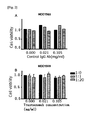
- trastuzumab-resistance cases In order to convert trastuzumab-resistance cases into trastuzumab-effective cases, methods for confirmation and derepression of ADCC activity with the use of NK cells were examined. As a result, no change was observed in SKBR3 (pan-cadherin negative) in terms of tumor cell viability at a trastuzumab concentration of 0, 0.021 mg/ml, or 0.105 mg/ml when the peripheral blood mononuclear cells were not cultured together. When the trastuzumab concentration was set at 0, no change was observed in tumor cell viability even when the tumor cells were cultured with the same or 20 times greater the amount of the peripheral blood mononuclear cells.
- the trastuzumab concentration was set at 0.021 mg/ml and the tumor cells were cultured with the same amount of the peripheral blood mononuclear cells, tumor cell viability was significantly lowered, compared with a case in which the peripheral blood mononuclear cells were not cultured together.
- tumor cell viability was further lowered, and tumor cell viability was significantly lowered, compared with a case in which the peripheral blood mononuclear cells were not cultured together and in which the tumor cells were cultured with the same amount of the peripheral blood mononuclear cells.
- tumor cell viability was lowered when the tumor cells were cultured with 20 times greater the amount of the peripheral blood mononuclear cells. That is, tumor cell viability was significantly lowered, compared with a case in which the peripheral blood mononuclear cells were not cultured together and in which the tumor cells were cultured with the same amount of the peripheral blood mononuclear cells.
- tumor cell viability was likely to be lowered, compared with a case in which the peripheral blood mononuclear cells were not cultured together, although no statistically significant difference was observed.
- the results demonstrate that tumor cell viability of SKBR3, which would not involve cadherin expression, would be lowered depending on the number of peripheral blood mononuclear cells, regardless of the trastuzumab concentration ( Fig. 1 ).
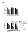
- the trastuzumab concentration was set at 0.021 mg/ml and the tumor cells were cultured with the same amount of the peripheral blood mononuclear cells, further, no change was observed in tumor cell viability, as with the case in which the peripheral blood mononuclear cells were not cultured together. No change was observed in tumor cell viability when the tumor cells were cultured with 20 times greater the amount of the peripheral blood mononuclear cells. Such tendency was also observed when the trastuzumab concentration was set at 0.105 mg/ml, and no change was observed in tumor cell viability even when the tumor cells were cultured with the same or 20 times greater the amount of the peripheral blood mononuclear cells. The results demonstrate that tumor cell viability ofHCC1569 involving cadherin expression would not be changed regardless of the trastuzumab concentration and the peripheral blood mononuclear cell count ( Fig. 2 ).
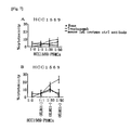
- the trastuzumab concentration was set at 0.021 mg/ml and 0.105 mg/ml and the tumor cells were cultured with 20 times greater the amount of the peripheral blood mononuclear cells, however, tumor cell viability was significantly lowered, compared with a case in which the peripheral blood mononuclear cells were not cultured together.
- tumor cell viability was likely to be lowered, although no statistically significant difference was observed ( Fig. 3 ).
- pan-cadherin negative tumor cells would be lowered depending on the number of peripheral blood mononuclear cells, regardless of the trastuzumab concentration.
- pan-cadherin positive tumor cells also, the number of viable tumor cells was not likely to decrease depending on the number of peripheral blood mononuclear cells, regardless of the trastuzumab concentration.
- the trastuzumab concentration was set at 0.021 mg/ml and the tumor cells were cultured with the same amount of the peripheral blood mononuclear cells, however, tumor cell viability was significantly lowered, compared with a case in which the peripheral blood mononuclear cells were not cultured together.
- the tumor cells were cultured with 20 times greater the amount of the peripheral blood mononuclear cells, tumor cell viability was further lowered, and tumor cell viability was significantly lowered, compared with a case in which the peripheral blood mononuclear cells were not cultured together.
- the trastuzumab concentration was set at 0.105 mg/ml and the tumor cells were cultured with the same amount of the peripheral blood mononuclear cells, tumor cell viability was significantly lowered, compared with a case in which the peripheral blood mononuclear cells were not cultured together.
- the tumor cells were cultured with 20 times greater the amount of the peripheral blood mononuclear cells, tumor cell viability was further lowered, and tumor cell viability was significantly lowered, compared with the cases in which the peripheral blood mononuclear cells were not cultured together and in which the tumor cells were cultured with the same amount of the peripheral blood mononuclear cells ( Fig. 4 ).
- the trastuzumab concentration was set at 0.021 mg/ml and the tumor cells were cultured with 20 times greater the amount of the peripheral blood mononuclear cells, however, the number of viable tumor cells decreased, and tumor cell viability was significantly lowered, compared with a case in which the peripheral blood mononuclear cells were not cultured together.
- tumor cell viability was likely to be lowered, compared with a case in which the peripheral blood mononuclear cells were not cultured together, although no statistically significant difference was observed.
- the trastuzumab concentration was set at 0.105 mg/ml and the tumor cells were cultured with 20 times greater the amount of the peripheral blood mononuclear cells, tumor cell viability was significantly lowered, compared with a case in which the peripheral blood mononuclear cells were not cultured together.
- tumor cell viability was likely to be lowered, compared with a case in which the peripheral blood mononuclear cells were not cultured together, although no statistically significant difference was observed ( Fig. 5 ).
- the E-cadherin- or N-cadherin-positive cell lines are resistant to trastuzumab.
- trastuzumab resistance of such cell lines was attenuated.
- ADCC activity which is a main mechanism of trastuzumab. It was thus deduced that the immunological mechanism was strongly associated with trastuzumab resistance.
- N-hydroxymethyl-2-iodoacetamide (0.6 g), 5.7 ml of concentrated sulfuric acid, and 7.2 ml of nitrobenzene were added to a glass flask, the flask was subjected to agitation at room temperature to dissolve the contents, and 0.036 g of paraformaldehyde was further added, followed by agitation.
- Nonwoven polypropylene fabric (0.3 g, average fiber diameter: 3.8 ⁇ m; fiber weight per unit area: 80 g/m 2 ; thickness: approximately 0.55 mm) was introduced thereinto and subjected to a reaction at room temperature for 24 hours under light-shielded conditions. Thereafter, the nonwoven fabric was removed, washed with ethanol and pure water, and dehydrated in vacuo to obtain activated nonwoven fabrics.
- the activated nonwoven fabric was cut into 4 circular pieces each with a diameter of 0.68 cm, the four circular pieces were soaked in calcium- and magnesium-free phosphate buffer (hereafter abbreviated as "PBS(-)”) comprising 0.4 ml of the rabbit anti-human KLRG1 polyclonal antibody purified with an affinity column (SANTA CRUZ BIOTECHNOLOGY, INC.; hereafter abbreviated as the "anti-KLRG1 antibody”) (16 ⁇ g of anti-KLRG1 antibody per 0.4 ml) at room temperature for 1 hour, and the resultant was further soaked for approximately 24 hours in cold storage (2°C to 8°C) to immobilize the anti-KLRG1 antibody.
- PBS(-) calcium- and magnesium-free phosphate buffer
- anti-KLRG1 antibody an affinity column
- the activated nonwoven fabric samples on which the anti-KLRG1 antibody had been immobilized were superposed in a container having an inlet and an outlet (volume: about 1 ml), and the nonwoven fabric samples were washed with 3 ml of PBS(-) from the inlet of the container. Subsequently, 0.4 ml of a 0.2% polyoxyethylene sorbitan monolaurate/PBS(-) solution (hereafter abbreviated as a "Tween20 solution”) was added, the fabric samples were soaked therein at room temperature for 2.5 hours, and the resultant was washed with 3 ml of PBS(-) to obtain KLRG1-positive cell adsorbers.
- Tween20 solution polyoxyethylene sorbitan monolaurate/PBS(-) solution
- KLRG1-positive cell adsorbers (average fiber diameter: 3.8 ⁇ m; fiber weight per unit area: 80 g/m 2 ; thickness: approximately 0.5 mm) and a 10 mM ascorbic acid solution in PBS(-) (a filling fluid, molecular weight of ascorbic acid: 176) were used to fill a container having an inlet and an outlet (volume: about 1 ml) to prepare KLRG1-positive cell adsorbers.
- the ACD-A-added human fresh blood before introduction into the column and the blood recovered at the column outlet were independently diluted with PBS(-) at 1:1, the resultants were superposed on Ficoll-Paque PLUS (GE Healthcare), and centrifugation was carried out at 400 ⁇ g for 30 minutes to obtain a mononuclear cell suspension.
- KLRG1-positive cells in NK cells in the mononuclear cell suspension were assayed with the use of the anti-human CD3 antibody, the anti-human CD56 antibody, and the anti-human KLRG1 antibody directly or indirectly labeled with fluorescence using FACSCaliburTM (Becton Dickinson).
- FACSCaliburTM Becton Dickinson
- KLRG1-NK cells The adsorption of CD3-CD56 + KLRG1-negative cells in the NK cells (hereafter abbreviated as "KLRG1-NK cells”) was 19.1%, that of lymphocytes was 16.9%, and that of platelets was 15.8%. This indicates that KLRG1 + NK cells were specifically adsorbed (Table 1).
- the adsorption of the KLRG1 + NK cells was found to be 83.9%.
- the adsorption of the KLRG1-NK cells was 10.5%, that of lymphocytes was 5.7%, and that of platelets was 7.7%. This indicates that KLRG1 + NK cells were specifically adsorbed (Table 1).
- the adsorption of the KLRG1 + NK cells was found to be 90.3%.
- the adsorption of the KLRG1-NK cells was 0.0%, that of lymphocytes was 27.8%, and that of platelets was 0.0%. This indicates that KLRG1 + NK cells were specifically adsorbed (Table 1).
- the adsorption of the KLRG1 + NK cells was found to be 32.7%.
- the adsorption of the KLRG1 - NK cells was 8.8%, that of lymphocytes was 22.2%, and that of platelets was 26.4%. This indicates that KLRG1 + NK cells were specifically adsorbed (Table 1).
- the HER2-positive human gastric cancer cell line (MKN-7) was provided by RIKEN Cell Bank.
- the HER2-positive human gastric cancer cell line (NCI-N87) was purchased from ATCC.
- MKN-7 was confirmed to be pan-cadherin-negative and E-cadherin-negative.
- NCI-N87 was confirmed to be pan-cadherin-positive and E-cadherin-positive.
- MKN-7 and NCI-N87 were cultured in the same manner as in Example 1, except that fetal bovine serum, penicillin, and streptomycin were added to RPMI-1640 medium (Sigma Aldrich, MO, U.S.A.).
- MKN-7 pan-cadherin negative did not exhibit any change in tumor cell viability at a trastuzumab concentration of 0, 0.021 mg/ml, or 0.105 mg/ml, when the peripheral blood mononuclear cells were not cultured together.
- trastuzumab concentration was set at 0, no change was observed in tumor cell viability even when the tumor cells were cultured with the same or 20 times greater the amount of the peripheral blood mononuclear cells.
- the trastuzumab concentration was set at 0.021 mg/ml and the tumor cells were cultured with the same amount of peripheral blood mononuclear cells, however, tumor cell viability was likely to be lowered, compared with a case in which the peripheral blood mononuclear cells were not cultured together, although no statistically significant difference was observed.
- the tumor cells were cultured with 20 times greater the amount of the peripheral blood mononuclear cells, tumor cell viability was further lowered, and tumor cell viability was significantly lowered, compared with the cases in which the peripheral blood mononuclear cells were not cultured together and in which the tumor cells were cultured with the same amount of the peripheral blood mononuclear cells.
- the trastuzumab concentration was set at 0.105 mg/ml and the tumor cells were cultured with the same amount of the peripheral blood mononuclear cells, tumor cell viability was significantly lowered, compared with a case in which the peripheral blood mononuclear cells were not cultured together.
- tumor cell viability was further lowered, and tumor cell viability was significantly lowered compared with the cases in which the peripheral blood mononuclear cells were not cultured together and in which the tumor cells were cultured with the same amount of the peripheral blood mononuclear cells.
- the results demonstrate that tumor cell viability of MKN-7, which would not involve cadherin expression, would be lowered depending on the number of peripheral blood mononuclear cells, regardless of the trastuzumab concentration ( Fig. 8A ).
- the trastuzumab concentration was set at 0.021 mg/ml and the tumor cells were cultured with 20 times greater the amount of the peripheral blood mononuclear cells from which KLRG1-expressing cells had been removed, the number of viable tumor cells decreased, and tumor cell viability was significantly lowered, compared with the cases in which the peripheral blood mononuclear cells were not cultured together and in which the tumor cells were cultured with the same amount of the peripheral blood mononuclear cells from which KLRG1-expressing cells had been removed.
- tumor cell viability was likely to be lowered, compared with a case in which the peripheral blood mononuclear cells were not cultured together, although no statistically significant difference was observed.
- trastuzumab concentration was set at 0.105 mg/ml and the tumor cells were cultured with the same amount of the peripheral blood mononuclear cells from which KLRG1-expressing cells had been removed, tumor cell viability was significantly lowered compared with a case in which the peripheral blood mononuclear cells were not cultured together.
- tumor cell viability was further lowered, and tumor cell viability was significantly lowered compared with the cases in which the peripheral blood mononuclear cells were not cultured together and in which the tumor cells were cultured with the same amount of the peripheral blood mononuclear cells from which KLRG1-expressing cells had been removed.
- the results demonstrate that tumor cell viability of MKN-7, which would not involve cadherin expression, would be lowered depending on the number of peripheral blood mononuclear cells from which KLRG1-expressing cells had been removed, regardless of the trastuzumab concentration ( Fig. 8B ).
- NCI-N87 did not exhibit any change in tumor cell viability at a trastuzumab concentration of 0, 0.021 mg/ml, or 0.105 mg/ml, when the peripheral blood mononuclear cells were not cultured together.
- trastuzumab concentration was set at 0, no change was observed in tumor cell viability even when the tumor cells were cultured with the same or 20 times greater the amount of the peripheral blood mononuclear cells.
- the trastuzumab concentration was set at 0.021 mg/ml and the tumor cells were cultured with the same amount of the peripheral blood mononuclear cells from which KLRG1-expressing cells had been removed, however, tumor cell viability was likely to be lowered, compared with a case in which the peripheral blood mononuclear cells were not cultured together, although no statistically significant difference was observed.
- the tumor cells were cultured with 20 times greater the amount of the peripheral blood mononuclear cells from which KLRG1-expressing cells had been removed, tumor cell viability was further lowered, and tumor cell viability was significantly lowered, compared with a case in which the peripheral blood mononuclear cells were not cultured together.
- trastuzumab concentration 0.105 mg/ml and the tumor cells were cultured with the same amount of the peripheral blood mononuclear cells from which KLRG1-expressing cells had been removed, tumor cell viability was significantly lowered, compared with a case in which the peripheral blood mononuclear cells were not cultured together.
- tumor cell viability was further lowered, and tumor cell viability was significantly lowered compared with cases in which the peripheral blood mononuclear cells were not cultured together and in which the tumor cells were cultured with the same amount of the peripheral blood mononuclear cells from which KLRG1-expressing cells had been removed ( Fig. 9B ).
- the E-cadherin-positive HER2-positive human gastric cancer cell line exhibited resistance to trastuzumab and the E-cadherin-negative HER2-positive human gastric cancer cell line exhibited sensitivity to trastuzumab. Since the existence of peripheral blood mononuclear cells was essential for the expression of trastuzumab sensitivity, involvement of ADCC activity was deduced.
- trastuzumab resistance of the E-cadherin-positive cell line was found to be quenched.
- mice Female NOD/SCID mice (6-week-old, non-obese diabetic/severe combined immune deficient mice) were purchased from CLEA Japan, Inc. All animals were maintained and handled in accordance with the guidelines of the institution based on approved protocols.
- HER2-positive pan-cadherin-positive human breast cancer cells (HCC1569) (5.0 x 10 6 cells) were suspended in 0.2 ml of PBS, and the NOD/SCID mice were inoculated hypodermically with the suspension.
- These tumor-bearing mice were divided into the following five groups: 1) the control group; 2) the group to which the human peripheral blood mononuclear cells from which KLRG1-expressing cells had been removed were administered; 3) the group to which trastuzumab was administered; 4) the group to which the human peripheral blood mononuclear cells and trastuzumab were administered; and 5) the group to which the human peripheral blood mononuclear cells from which KLRG1-expressing cells had been removed and trastuzumab were administered.
- trastuzumab was administered intraperitoneally in an amount of 0.005 mg per g of mouse body weight.
- Human peripheral blood mononuclear cells were administered intraperitoneally in an amount of 5.0 x 10 6 cells per mouse.
- Administration of trastuzumab and human peripheral blood mononuclear cells was initiated 4 weeks after HCC1569 inoculation, and administration was carried out once a week for 4 weeks.
- T ⁇ u ⁇ m ⁇ o ⁇ r v ⁇ o ⁇ l ⁇ u ⁇ m ⁇ e m ⁇ m 3 l ⁇ e ⁇ n ⁇ g ⁇ t ⁇ h ⁇ w ⁇ i ⁇ d ⁇ t ⁇ h ⁇ t ⁇ h ⁇ i ⁇ c ⁇ k ⁇ n ⁇ e ⁇ s ⁇ s
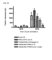
- Fig. 10 shows the data regarding the tumor volume 28 days (4 weeks) and 56 days (8 weeks) after HCC1569 inoculation.
- tumor volume was found to have increased in the control group, the group to which the human peripheral blood mononuclear cells from which KLRG1-expressing cells had been removed were administered, the group to which trastuzumab was administered, and the group to which the human peripheral blood mononuclear cells and trastuzumab were administered. No increase in tumor volume was observed in the group to which the human peripheral blood mononuclear cells from which KLRG1-expressing cells had been removed and trastuzumab were administered.
- mice KLRG1-positive cell adsorbing materials were used to prepare mouse KLRG1-positive cell adsorbing materials in accordance with the method of Example 2. Further, the mouse KLRG1-positive cell adsorbing materials were used to fill a container having an inlet and an outlet (volume: about 0.05 ml) in accordance with the method of Example 2 to prepare mouse KLRG1-positive cell adsorber.
- ACD-A acid citrate dextrose solution-A, Terumo Corporation
- the blood samples before and after treatment with the mouse KLRG1-positive cell adsorbers were stained with the fluorescence-labeled anti-mouse CD3 antibody, anti-mouse CD49b antibody, and anti-mouse KLRG1 antibody, and CD3- ⁇ CD49b + cells were analyzed with a flow cytometer (Cytomics FC 500, Beckman Coulter) as the NK cells.
- the circuits used for mouse extracorporeal circulation were prepared using a silicone tube (inner diameter: 0.5 mm; outer diameter: 4 mm), two urethane tubes (outer diameter: 0.6 mm; inner diameter: 0.3 mm), and mini-fittings (VPY106, ISIS Co., Ltd.) in combination.
- the internal volume of the circuit was 0.09 ml.
- a Perista Pump (type SJ-1211H, ATTO) was used as the power source for extracorporeal circulation, and the flow rate was set at 0.064 ml/min.
- ACD-A was continuously administered at 0.008 ml/min with the use of a microsyringe pump from mini-fittings in the circuits.
- the mouse KLRG1-positive cell adsorbers of Example 8 were connected to the circuits, the solution of ACD-A and physiological saline mixed at 11:14 was introduced thereinto, and priming was then carried out for 15 minutes.
- Extracorporeal circulation and trastuzumab administration were carried out once a week over a period of 3 weeks, and trastuzumab was administered intraperitoneally in an amount of 0.005 mg per g of mouse body weight. All animals were maintained and handled in accordance with the guidelines of the institution based on approved protocols.
- the tumor bearing mice were subjected to isoflurane anesthesia, and the skin was incised to expose both cervical veins.
- Surflo F&F Teumo Corporation
- Jikken Doubutsu No Ketsuekigaku Haematology of Experimental Animals, Masatoshi Seki et al., Life Science, 1981
- the total blood amounts of the mice were calculated, and the blood that was twice the amount of the entire blood was treated via extracorporeal circulation.
- physiological saline was introduced over a period of 2 minutes and 30 seconds to retransfuse the blood in the mouse KLRG1-positive cell adsorbers and the circuits into the mice. Bleeding was stopped and the wounds were then stitched up.
- Tumor volume was measured in the same manner as in Example 7 (3).
- the percentage of increase in tumor volume was 185% one week later, 307% two weeks later, and 615% 3 weeks later compared with the tumor volume before treatment.
- the tumor volume was also likely to increase, the percentage of increase was 145% one week later, 306% two weeks later, and 655% 3 weeks later.
- the percentage of increase was 83% one week later, 141% two weeks later, and 177% 3 weeks later.
Landscapes
- Health & Medical Sciences (AREA)
- Life Sciences & Earth Sciences (AREA)
- Chemical & Material Sciences (AREA)
- Organic Chemistry (AREA)
- Engineering & Computer Science (AREA)
- General Health & Medical Sciences (AREA)
- Medicinal Chemistry (AREA)
- Genetics & Genomics (AREA)
- Zoology (AREA)
- Biomedical Technology (AREA)
- Biotechnology (AREA)
- Bioinformatics & Cheminformatics (AREA)
- Immunology (AREA)
- Wood Science & Technology (AREA)
- Biochemistry (AREA)
- Molecular Biology (AREA)
- Microbiology (AREA)
- Cell Biology (AREA)
- Pharmacology & Pharmacy (AREA)
- Animal Behavior & Ethology (AREA)
- Public Health (AREA)
- Veterinary Medicine (AREA)
- Hematology (AREA)
- Epidemiology (AREA)
- Biophysics (AREA)
- General Engineering & Computer Science (AREA)
- Proteomics, Peptides & Aminoacids (AREA)
- Oncology (AREA)
- General Chemical & Material Sciences (AREA)
- Nuclear Medicine, Radiotherapy & Molecular Imaging (AREA)
- Developmental Biology & Embryology (AREA)
- Virology (AREA)
- Chemical Kinetics & Catalysis (AREA)
- Mycology (AREA)
- Medicines Containing Antibodies Or Antigens For Use As Internal Diagnostic Agents (AREA)
- External Artificial Organs (AREA)
- Medicines Containing Material From Animals Or Micro-Organisms (AREA)
- Peptides Or Proteins (AREA)
Abstract
Description
- The present invention relates to a therapeutic agent for cancer comprising an antibody reacting with a cancer-specific membrane antigen expressed in epithelial cancer cells. The present invention further relates to a device for removing cells which is used in combination with the aforementioned therapeutic agent for cancer, and a combined therapeutic system of the device for removing cells and the therapeutic agent for cancer.
- In recent years, the number of patients who have been afflicted with and died of breast cancer is increasing at a significant level in Japan. Non-surgical therapeutic techniques for treating breast cancer were likely to be determined depending on estrogen receptor (ER) and progesterone receptor (PgR) expression conditions in the past. Drug therapy, such as hormonal therapy or anticancer drug therapy, is useful, and the response rate and viability have been improved. However, approximately 50% of human epidermal growth factor receptor-related 2 (HER2)-positive breast cancer cases have been hormone receptor-negative, and such cases have not been the targets of hormonal therapy. In recent years, molecular-targeted techniques, such as a trastuzumab-based antibody therapy against HER2-positive breast cancer, have been employed, the usefulness thereof has been demonstrated, and such techniques are expected as leading therapeutic methods in the future.
- Breast cancer cells express a variety of growth factors and receptors thereof and form signal transduction systems associated with growth. An example thereof is the HER2 cancer gene that was identified in 1984, which is a member of the human epidermal growth factor receptor (EGFR, HER) family. A receptor is transmembrane glycoprotein which is classified as HER1, HER2, HER3, or HER4 based on structural similarities (Non-patent document 1). It is considered that the EGF receptor family contributes to signal transduction via dimer formation, and a receptor to which a ligand has bound forms a dimer with another receptor, thereby contributing to signal transduction.
- HER2/neu, which defines the HER2 receptor, is considered to be a proto-oncogene, and it is present in chromosome 17 (17q21.1) (Non-patent document 2). Amplification and overexpression of the HER2/neu gene are observed in a variety of cancers, such as ovarian cancer, lung cancer, and gastric cancer, as well as in breast cancer. HER2-positive breast cancer accounts for 20% to 30% of all breast cancer cases. The expression level of this gene in adult normal tissue is very low. The natural course is specific, and recurrence takes place at an early stage when the patient would not undergo systemic treatment. Since the correlation of HER2 amplification with a poor prognosis of breast cancer was reported in 1987, a poor clinical course for HER2-positive metastatic breast cancer has been demonstrated by much research.
- In 1986, it was reported that the anti-HER2 monoclonal antibody would inhibit malignant traits of neu transformed cells, which led to the development of trastuzumab targeting HER2. An mu4D5 mouse monoclonal antibody reacting with an extracellular domain of the HER2 receptor, which is a prototype of trastuzumab, exhibits activity of inhibiting growth of HER2-positive tumor cells in vitro (Non-patent document 3). Also, the antibody-dependent cell cytotoxicity (ADCC) of mouse splenic cells against SKBR3, which is a HER2-positive human breast cancer cell line, is reinforced by the mouse monoclonal antibody, and it is reported that ADCC activity also contributes to anti-tumor effects in vivo (Non-patent document 4). If a mouse antibody is subjected to clinical applications without modification, the problem of appearance of the human anti-mouse antibody (HAMA) arises. Thus, trastuzumab (trade name: Herceptin®) was prepared as a humanized antibody by transplanting only the variable region of an antigen-binding site of the mouse monoclonal antibody reacting with the extracellular domain of the HER2 receptor into the constant region of human IgG1.
- HER2 excites a variety of signal transduction pathway networks including PI3K and MAPK. It is considered that trastuzumab binds to the HER2 receptor to inhibit such signal transduction pathway and it induces termination of a cell cycle, apoptosis, inhibition of angiogenesis, and the like to directly inhibit tumor cell growth. Also, inhibition of the Src tyrosine kinase, activation of PTEN involved therewith, and dephosphorylation of Akt have been reported. Further, it has been demonstrated that the Fc receptor that is expressed in immunocytes, including the NK cells, binds to the Fc region of trastuzumab, which has bound to the tumor cells, to exhibit the effects of killing tumor cells (ADCC activity) (Non-patent
documents 5 to 8). Such effects of direct inhibition of tumor cell growth and ADCC activity are considered to be the main mechanisms of trastuzumab. - Trastuzumab targets HER2, and the therapeutic effects thereof significantly vary depending on HER2 protein expression level. It is known that the positive ratio significantly varies depending on assay technique and materials used. In order to inspect HER2 protein overexpression and DNA amplification, the immunohistochemical (IHC) method and fluorescence in situ hybridization (FISH) are extensively employed. Also, trastuzumab has been found to be very effective for postoperative adjuvant therapy involving a plurality of large-scale clinical testings, including the HERA test.
- Based on a variety of studies that have been heretofore conducted, HER2 inhibition via trastuzumab administration was found to significantly influence the natural course of breast cancer. However, it has also been found that trastuzumab would not alter the entire natural course of HER2-overexpressing breast cancer, and it is said that the number of cases in which patients react with the first administration of trastuzumab alone is approximately one-third or lower than that for HER2-overexpressing breast cancer. In the case of microscopic metastatic cancer, similarly, it is suggested that a considerable percentage of tumors are tolerant to trastuzumab. However, the mechanism of trastuzumab tolerance has not been clearly elucidated. At present, the following possibilities are suggested as the mechanisms of trastuzumab tolerance: (1) insufficient inhibition of the HER2 extracellular region due to insufficient accession of trastuzumab; (2) a lowered HER2 expression level; (3) an altered HER2 regulator located downstream (e.g., lowered p27kiP1 and quenching or inactivation of PTEN); (4) the occurrence of signal transmission by an alternate pathway (overexpression of the insulin-like growth factor I receptor (IGF1R)); and (5) lowered immunity (lowered ADCC activity, in particular).
- The tolerance mechanism related to immunity of (5) above has not been actively examined. In recent years, the functions of the NK cell, which is a factor associated with ADCC activity as a main action mechanism of trastuzumab, have become elucidated. Inhibitory receptors are expressed in NK cells, and the killer cell lectin-like receptor G1 (KLRG1) was identified as one such inhibitory receptor. Most of the ligands of inhibitory receptors that have been found with regard to NK cells were associated with the MHC class I molecule. In 2006, M. Ito et al. reported that the KLRG1 ligand was not of the MHC class I molecule.
- KLRG1 was discovered as a functional molecule expressed in the RBL-2H3 rat basophilic leukemia cell line (MAFA: the mast cell function-associated antigen) (Non-patent document 9). KLRG1 crosslinks with the anti-KLRG1 antibody in the RBL-2H3 cell, and the degranulation reaction caused by Fc receptor stimulation is inhibited. As a result of cDNA cloning of rat KLRG1, it has been reported by Pecht et al. that KLRG1 is a homodimer of the type II transmembrane protein having a C-type lectin-like structure in the extracellular region and an immunoreceptor tyrosine-based inhibitory motif (ITIM) in the intracellular region (Non-patent document 10). KLRG1 is expressed in some NK or T cells in the case of humans and mice (Non-patent documents 11 to 13). In contrast, it is known that expression thereof is observed in approximately 50% of the peripheral blood NK cells of healthy individuals.
-
- Non-patent document 1: Pinkas-Kramarski, R., 1. Arloy and Y Yarden (1997) ErbB receptors and EGF-like ligands: cell lineage determination and oncogenesis through combinatorial signaling. J Mammary G1and Biol Neoplasia, 2:97-107.
- Non-patent document 2: Schechter, A.L., D.F. Stem, L. Vaidyanathan, S.J. Decker, J.A. Drebin, M.I. Greene and R.A. Weinberg (1984) The neu oncogene: an erb-B-related gene encoding a 185,000-Mr tumour antigen. Nature, 312:513-516.
- Non-patent document 3: Hudziak RM, Lewis GD, Winget M, Fendly BM, Shepard HM, Ullrich A (1989) p185HER2 monoclonal antibody has antiproliferative effects in vitro and sensitizes human breast tumor cells to tumor necrosis factor. Mol Cell Biol, 9(3):1165-72.
- Non-patent document 4: Piccart-Gebhart MJ, Procter M, Leyland-Jones B, Goldhirsch A, Untch M, Smith I, Gianni L, Baselga J, Bell R, Jackisch C, Cameron D, Dowsett M, Barrios CH, Steger G, Huang CS, Andersson M, Inbar M, Lichinitser M, Láng I, Nitz U, Iwata H, Thomssen C, Lohrisch C, Suter TM, Rüschoff J, Suto T, Greatorex V, Ward C, Straehle C, McFadden E, Dolci MS, Gelber RD; Herceptin Adjuvant (HERA) Trial Study Team (2005) Trastuzumab after adjuvant chemotherapy in HER2-positive breast cancer. N Engl J Med, 353(16):1659-72
- Non-patent document 5: Clynes RA, Towers TL, Presta LG, Ravetch JV (2000) Inhibitory Fc receptors modulate in vivo cytoxicity against tumor targets. Nat Med, 6(4):443-6.
- Non-patent document 6: Izumi Y, Xu L, di Tomaso E, Fukumura D, Jain RK (2002) Tumour biology: herceptin acts as an anti-angiogenic cocktail. Nature, 416(6878):279-80
- Non-patent document 7: Gennari R, Menard S, Fagnoni F, Ponchio L, Scelsi M, Tagliabue E, Castiglioni F, Villani L, Magalotti C, Gibelli N, Oliviero B, Ballardini B, Da Prada G, Zambelli A, Costa A (2004) Pilot study of the mechanism of action of preoperative trastuzumab in patients with primary operable breast tumors overexpressing HER2. Clin Cancer Res., 10(17):5650-5
- Non-patent document 8: Barok, et al., (2007) Trastuzumab causes antibody-dependent cellular cytotoxicity-mediated growth inhibition of .
- Non-patent document 9: Ortega Soto E, Pecht I (1988) A monoclonal antibody that inhibits secretion from rat basophilic leukemia cells and binds to a novel membrane component. J Immunol, 141(12):4324-32.
- Non-patent document 10: Abramson J, Xu R, Pecht I (2002) An unusual inhibitory receptor--the mast cell function-associated antigen (MAFA). Mol Immunol, 38(16-18):1307-13.
- Non-patent document 11: Hanke T, Corral L, Vance RE, Raulet DH (1998) 2F1 antigen, the mouse homolog of the rat "mast cell fiulction-associated antigen", is a lectin-like type II transmembrane receptor expressed by natural killer cells. Eur J Immunol., 28(12):4409-17
- Non-patent document 12: Butcher S, Arney KL, Cook GP (1998) MAFA-L, an ITIM-containing receptor encoded by the human NK cell gene complex and expressed by basophils and NK cells. Eur J Immunol., 28(11):3755-62
- Non-patent document 13: Blaser C, Kaufmann M, Pircher H (1998) Virus-activated CD8 T cells and lymphokine-activated NK cells express the mast cell function-associated antigen, an inhibitory C-type lectin. J Immunol., 161(12):6451-4
- Application of trastuzumab therapy is expanding from recurrence therapy to postoperative adjuvant therapy for the purpose of recurrence prevention and preoperative therapy. Further improvement in curability of HER2-positive cases is expected. Since the application range is expanded, it is expected that targets of trastuzumab administration is significantly increased. At present, however, there are many groups for which trastuzumab administration is ineffective among target cases. It is accordingly an object of the present invention to provide a therapeutic agent for cancer comprising an antibody reacting with a cancer-specific membrane antigen wherein the agent is administered by a novel method for decreasing the number of clinical cases for which trastuzumab administration is ineffective. It is another object of the present invention to provide a device for removing cells which is used in combination with the aforementioned therapeutic agent for cancer, and a combined therapeutic system of the device for removing cells and the therapeutic agent for cancer.
- The present inventors have conducted concentrated studies in order to attain the above object. As a result, they discovered that selective removal of KLRG1-positive immunocytes in the peripheral blood of a living individual at the time of administration of the anti-HER2 monoclonal antibody (i.e., trastuzumab) would realize exertion of anti-cancer effects in a living individual for which administration of the anti-HER2 monoclonal antibody had been ineffective in the past. This has led to the completion of the present invention. Specifically, the present invention provides the following.
-
- [1] A therapeutic agent for cancer comprising an antibody reacting with a cancer-specific membrane antigen expressed in epithelial cancer cells, said antibody having antibody-dependent cell cytotoxicity, wherein the therapeutic agent for cancer is used for living individual with epithelial cancer which is positive for the cancer-specific membrane antigen and positive for the KLRG1 ligand, and the time of initiation of administration of the therapeutic agent for cancer is during or after the period when KLRG1-positive immunocytes in the peripheral blood of the living individual is selectively reduced ex vivo.
- [2] The therapeutic agent for cancer according to [1], wherein step of selectively reducing KLRG1-positive immunocytes in the peripheral blood of the living individual ex vivo is performed by blood purification therapy wherein the peripheral blood of a living individual is extracorporeally circulated and KLRG1-positive immunocytes in the peripheral blood is selectively removed by a device for removing cells which has higher affinity for KLRG1-positive immunocytes than for KLRG1-negative immunocytes.
- [3] The therapeutic agent for cancer according to [1] or [2], wherein the antibody-dependent cell cytotoxicity of the antibody reacting with a cancer-specific membrane antigen on the epithelial cancer cell which is positive for the cancer-specific membrane antigen and positive for the KLRG1 ligand, is increased by selectively removing KLRG1-positive immunocytes in the peripheral blood of the living individual.
- [4] The therapeutic agent for cancer according to any of [1] to [3], wherein the cancer-specific membrane antigen expressed in epithelial cancer cells is HER2.
- [5] The therapeutic agent for cancer according to [4], wherein an antibody reacting with the cancer-specific membrane antigen expressed in epithelial cancer cells is trastuzumab.
- [6] The therapeutic agent for cancer according to any of [1] to [3], wherein the cancer-specific membrane antigen expressed in epithelial cancer cells is CEA.
- [7] The therapeutic agent for cancer according to any of [1] to [6], wherein the KLRG1-positive immunocytes are KLRG1-positive NK cells.
- [8] The therapeutic agent for cancer according to any of [1] to [7], wherein the KLRG1 ligand is selected from the group consisting of E-cadherin, N-cadherin and R-cadherin.
- [9] The therapeutic agent for cancer according to any of [1] to [8], wherein the epithelial cancer is breast cancer.
- [10] The therapeutic agent for cancer according to any of [1] to [8], wherein the epithelial cancer is gastric cancer.
- [11] The therapeutic agent for cancer according to any of [1] to [10], wherein the peripheral blood of a living individual is extracorporeally circulated, and the peripheral blood is allowed to be contacted with a water-insoluble carrier on which a substance having affinity for KLRG1 is immobilized, and KLRG1-positive immunocytes in the peripheral blood of the living individual are selectively removed.
- [12] The therapeutic agent for cancer according to [11], wherein the substance having affinity for KLRG1 is an antibody reacting with KLRG1.
- [13] The therapeutic agent for cancer according to [11], wherein the substance having affinity for KLRG1 is E-cadherin.
- [14] The therapeutic agent for cancer according to any of [11] to [13], wherein the water-insoluble carrier is magnetic particles.
- [15] The therapeutic agent for cancer according to any of [11] to [13], wherein the water-insoluble carrier is a nonwoven fabric.
-
- [16] A device for removing cells which has higher affinity for KLRG1-positive immunocytes than for KLRG1-negative immunocytes, which is used for extracorporeally circulating a peripheral blood of a living individual and selectively removing KLRG1-positive immunocytes in the peripheral blood before or while a therapeutic agent for cancer comprising an antibody reacting with a cancer-specific membrane antigen expressed in epithelial cancer cells, said antibody having antibody-dependent cell cytotoxicity, is administered to living individual with epithelial cancer which is positive for the cancer-specific membrane antigen and positive for the KLRG1 ligand.
- [17] The device for removing cells according to [16], wherein the antibody-dependent cell cytotoxicity of the antibody reacting with a cancer-specific membrane antigen on the epithelial cancer cell which is positive for the cancer-specific membrane antigen and positive for the KLRG1 ligand, is increased by selectively removing KLRG1-positive immunocytes in the peripheral blood of the living individual.
- [18] The device for removing cells according to [16] or [17], wherein the cancer-specific membrane antigen expressed in epithelial cancer cells is HER2.
- [19] The device for removing cells according to [18], wherein an antibody reacting with the cancer-specific membrane antigen expressed in epithelial cancer cells is trastuzumab.
- [20] The device for removing cells according to [16] or [17], wherein the cancer-specific membrane antigen expressed in epithelial cancer cells is CEA.
- [21] The device for removing cells according to any of [16] to [20], wherein the KLRG1-positive immunocytes are KLRG1-positive NK cells.
- [22] The device for removing cells according to any of [16] to [21], wherein the KLRG1 ligand is selected from the group consisting of E-cadherin, N-cadherin and R-cadherin.
- [23] The device for removing cells according to any of [16] to [22], wherein the epithelial cancer is breast cancer.
- [24] The device for removing cells according to any of [16] to [22], wherein the epithelial cancer is gastric cancer.
- [25] The device for removing cells according to any of [16] to [24], wherein the peripheral blood of a living individual is allowed to be contacted with a water-insoluble carrier on which a substance having affinity for KLRG1 is immobilized, and KLRG1-positive immunocytes in the peripheral blood of the living individual are selectively removed.
- [26] The device for removing cells according to [25], wherein the substance having affinity for KLRG1 is an antibody reacting with KLRG1.
- [27] The device for removing cells according to [25], wherein the substance having affinity for KLRG1 is E-cadherin.
- [28] The device for removing cells according to any of [25] to [27], wherein the water-insoluble carrier is magnetic particles.
- [29] The device for removing cells according to any of [25] to [27], wherein the water-insoluble carrier is a nonwoven fabric.
-
- [30] A combined therapeutic system of a device for removing cells and a therapeutic agent for cancer, which comprises (i) a device for removing cells which has higher affinity for KLRG1-positive immunocytes than for KLRG1-negative immunocytes, which is used for extracorporeally circulating a peripheral blood of a living individual with epithelial cancer which is positive for the cancer-specific membrane antigen expressed in epithelial cancer cells and positive for the KLRG1 ligand, and selectively removing KLRG1-positive immunocytes in the peripheral blood, and (ii) a therapeutic agent for cancer comprising an antibody reacting with the cancer-specific membrane antigen, said antibody having antibody-dependent cell cytotoxicity, wherein the therapeutic agent for cancer is administered while or after a blood purification therapy for selectively removing KLRG1-positive immunocytes in the peripheral blood is performed.
- [31] The combined therapeutic system according to [30], wherein the antibody-dependent cell cytotoxicity of the antibody reacting with a cancer-specific membrane antigen on the epithelial cancer cell which is positive for the cancer-specific membrane antigen and positive for the KLRG1 ligand, is increased by selectively removing KLRG1-positive immunocytes in the peripheral blood of the living individual.
- [32] The combined therapeutic system according to [31], wherein the cancer-specific membrane antigen expressed in epithelial cancer cells is HER2.
- [33] The combined therapeutic system according to [32], wherein an antibody reacting with the cancer-specific membrane antigen expressed in epithelial cancer cells is trastuzumab.
- [34] The combined therapeutic system according to [30] or [31], wherein the cancer-specific membrane antigen expressed in epithelial cancer cells is CEA.
- [35] The combined therapeutic system according to any of [30] to [34], wherein the KLRG1-positive immunocytes are KLRG1-positive NK cells.
- [36] The combined therapeutic system according to any of [30] to [35], wherein the KLRG1 ligand is selected from the group consisting of E-cadherin, N-cadherin and R-cadherin.
- [37] The combined therapeutic system according to any of [30] to [36], wherein the epithelial cancer is breast cancer.
- [38] The combined therapeutic system according to any of [30] to [36], wherein the epithelial cancer is gastric cancer.
- [39] The combined therapeutic system according to any of [30] to [38], wherein the peripheral blood of a living individual is extracorporeally circulated, and the peripheral blood is allowed to be contacted with a water-insoluble carrier on which a substance having affinity for KLRG1 is immobilized, and KLRG1-positive immunocytes in the peripheral blood of the living individual are selectively removed.
- [40] The combined therapeutic system according to [39], wherein the substance having affinity for KLRG1 is an antibody reacting with KLRG1.
- [41] The combined therapeutic system according to [39], wherein the substance having affinity for KLRG1 is E-cadherin.
- [42] The combined therapeutic system according to any of [39] to [41], wherein the water-insoluble carrier is magnetic particles.
- [43] The combined therapeutic system according to any of [39] to [41], wherein the water-insoluble carrier is a nonwoven fabric.
-
- [44] A method for treating a living individual with epithelial cancer comprising:
- step (a) of selectively reducing KLRG1-positive immunocytes in the peripheral blood of a living individual with epithelial cancer ex vivo, which is positive for a cancer-specific membrane antigen expressed in epithelial cancer cells and positive for a KLRG1 ligand; and
- step (b) of administering, to the living individual, a therapeutic agent for cancer comprising an antibody reacting with the cancer-specific membrane antigen expressed in epithelial cancer cells and having antibody-dependent cell cytotoxicity.
- [45] A therapeutic agent for cancer comprising an antibody reacting with a cancer-specific membrane antigen expressed in epithelial cancer cells, said antibody having antibody-dependent cell cytotoxicity, wherein the therapeutic agent for cancer is used for living individual with epithelial cancer which is positive for the cancer-specific membrane antigen and positive for the KLRG1 ligand, the time of initiation of administration of the therapeutic agent for cancer is during or after the period where KLRG1-positive immunocytes in the peripheral blood of the living individual is selectively reduced ex vivo by a device for removing cells which has higher affinity for KLRG1-positive immunocytes than for KLRG1-negative immunocytes.
- Further, the present invention provides a cancer cell-damaging agent comprising an antibody reacting with a cancer-specific membrane antigen expressed in epithelial cancer cells, said antibody having antibody-dependent cell cytotoxicity, wherein the cancer cell-damaging agent is used for epithelial cancer cells which is positive for the cancer-specific membrane antigen and positive for the KLRG1 ligand, and is used in combination with mononuclear cells in which the KLRG1-positive immunocytes had been selectively removed by a device for removing cells which has higher affinity for KLRG1-positive immunocytes than for KLRG1-negative immunocytes.
- Further, the present invention provides a device for removing cells which has higher affinity for KLRG1-positive immunocytes than for KLRG1-negative immunocytes, which is used for selectively removing KLRG1-positive immunocytes from a cell population which comprises mononuclear cells and epithelial cancer cells which is positive for a cancer-specific membrane antigen expressed in epithelial cancer cells and positive for the KLRG1 ligand, before or while a cancer cell-damaging agent comprising an antibody reacting with the cancer-specific membrane antigen, said antibody having antibody-dependent cell cytotoxicity, is contacted with the cell population.
- Further, the present invention provides a combined treatment system of a device for removing cells and a cancer cell-damaging agent, which comprises (i) a device for removing cells which has higher affinity for KLRG1-positive immunocytes than for KLRG1-negative immunocytes, which is used for selectively removing KLRG1-positive immunocytes from a cell population which comprises mononuclear cells and epithelial cancer cells which is positive for a cancer-specific membrane antigen expressed in epithelial cancer cells and positive for the KLRG1 ligand, and (ii) a cancer cell-damaging agent comprising an antibody reacting with the cancer-specific membrane antigen, said antibody having antibody-dependent cell cytotoxicity, wherein the cancer cell-damaging agent is administered to the cell population during or after the operation of selectively removing the KLRG1-positive immunocytes by the device for removing cells.
- Further, the present invention provides a device for removing cells for increasing an antibody-dependent cell cytotoxicity of a therapeutic agent for cancer comprising an antibody reacting with a cancer-specific membrane antigen expressed in epithelial cancer cells, said antibody having antibody-dependent cell cytotoxicity, which comprises (i) a device for removing cells which has higher affinity for KLRG1-positive immunocytes than for KLRG1-negative immunocytes, and (ii) an instruction wherein a step of extracorporeally circulating a peripheral blood of a living individual with epithelial cancer which is positive for the cancer-specific membrane antigen and positive for the KLRG1 ligand and selectively removing KLRG1-positive immunocytes from the peripheral blood by the device for removing cells and a step of administering the therapeutic agent for cancer to the living individual are recited.
- Further, the present invention provides a combined therapeutic system of a device for removing cells and a therapeutic agent for cancer which comprise (i) a device for removing cells which has higher affinity for KLRG1-positive immunocytes than for KLRG1-negative immunocytes, (ii) a therapeutic agent for cancer comprising an antibody reacting with a cancer-specific membrane antigen expressed in epithelial cancer cells, said antibody having antibody-dependent cell cytotoxicity, and (iii) an instruction wherein a step of extracorporeally circulating a peripheral blood of a living individual with epithelial cancer which is positive for the cancer-specific membrane antigen and positive for the KLRG1 ligand and selectively removing KLRG1-positive immunocytes from the peripheral blood by the device for removing cells and a step of administering the therapeutic agent for cancer to the living individual are recited.
- Further, the present invention provides a method for reinforcing antibody-dependent cell cytotoxicity of an antibody reacting with cancer-specific membrane antigen expressed in epithelial cancer cells on the epithelial cancer cells, which comprises bringing the antibody reacting with the cancer-specific membrane antigen into contact with mononuclear cells in which the KLRG1-positive immunocytes had been selectively removed and epithelial cancer cells that are positive for the cancer-specific membrane antigen and positive for the KLRG1 ligand.
- Further, the present invention provides a method for treating a living individual with epithelial cancer which is positive for the cancer-specific membrane antigen expressed in epithelial cancer cells and positive for the KLRG1 ligand, which comprises a step of administering, to the living individual, an antibody reacting with the cancer-specific membrane antigen expressed in epithelial cancer cells, and a step of selectively removing KLRG1-positive immunocytes in the peripheral blood of the living individual.
- Further, the present invention provides a use of mononuclear cells in which the KLRG1-positive immunocytes had been selectively removed, for reinforcement of the antibody-dependent cell cytotoxicity of an antibody reacting with the cancer-specific membrane antigen against epithelial cancer cells, which are positive for the cancer-specific membrane antigen expressed in epithelial cancer cells and positive for the KLRG1 ligand, which comprises bringing an antibody reacting with the cancer-specific membrane antigen expressed in epithelial cancer cells into contact with mononuclear cells in which the KLRG1-positive immunocytes had been selectively removed and with the epithelial cancer cells.
- Further, the present invention provides a use of antibody reacting with a cancer-specific membrane antigen expressed in epithelial cancer cells, said antibody having antibody-dependent cell cytotoxicity, for the production of a therapeutic agent for cancer wherein the therapeutic agent for cancer is used for living individual with epithelial cancer which is positive for the cancer-specific membrane antigen expresses in epithelial cancer cells and positive for the KLRG1 ligand, and the time of initiation of administration of the therapeutic agent for cancer is during or after the period when KLRG1-positive immunocytes in the peripheral blood of the living individual is selectively reduced ex vivo.
- Further, the present invention provides a use of antibody reacting with a cancer-specific membrane antigen expressed in epithelial cancer cells, said antibody having antibody-dependent cell cytotoxicity, for the production of a combined therapeutic system of a device for removing cells and a therapeutic agent for cancer, which comprises (i) a device for removing cells which has higher affinity for KLRG1-positive immunocytes than for KLRG1-negative immunocytes, which is used for extracorporeally circulating a peripheral blood of a living individual with epithelial cancer which is positive for the cancer-specific membrane antigen expressed in epithelial cancer cells and positive for the KLRG1 ligand, and selectively removing KLRG1-positive immunocytes in the peripheral blood, and (ii) a therapeutic agent for cancer comprising an antibody reacting with the cancer-specific membrane antigen, said antibody having antibody-dependent cell cytotoxicity, wherein the therapeutic agent for cancer is administered while or after a blood purification therapy for selectively removing KLRG1-positive immunocytes in the peripheral blood is performed.
- According to the present invention, effective anti-cancer effects can be attained even in a living individual in which anti-cancer effects could not be attained in the past through administration of an antibody reacting with the cancer-specific membrane antigen expressed in epithelial cancer cells. Further, according to the present invention, sufficient cell-damaging effects can be attained even in such a case that cell-damaging activity on epithelial cancer cells is not obtained when an antibody reacting with the cancer-specific membrane antigen expressed in epithelial cancer cells, said antibody having antibody-dependent cell cytotoxicity, is contacted with a cell population which comprises mononuclear cells and epithelial cancer cells which is positive for the cancer-specific membrane antigen and positive for the KLRG1 ligand.
-
- [
Fig. 1] Fig. 1 shows cytotoxicity of trastuzumab and peripheral blood mononuclear cells on SKBR3 breast cancer cells. The SKBR3 tumor cells were sowed on a 35-mm dish in an amount of 1 × 10⁵ cells, and trastuzumab and the peripheral blood mononuclear cells sampled from healthy individuals were added 24 hours later. Further, the number of viable tumor cells was determined 24 hours later. The mean and the standard deviation of triplicate assays are shown. Statistical analysis was carried out via two-way ANOVA. (A: group to which control antibody was administered; B: group to which trastuzumab was administered) - [
Fig. 2] Fig. 2 shows cytotoxicity of trastuzumab and peripheral blood mononuclear cells on HCC1569 breast cancer cells. The HCC1569 tumor cells were sowed on a 35-mm dish in an amount of 1 × 10⁵ cells, and trastuzumab and the peripheral blood mononuclear cells sampled from healthy individuals were added 24 hours later. Further, the number of viable tumor cells was determined 24 hours later. The mean and the standard deviation of triplicate assays are shown. Statistical analysis was carried out via two-way ANOVA. (A: the control group; B: group to which trastuzumab was administered) - [
Fig. 3] Fig. 3 shows the effects of E-cadherin and N-cadherin suppression on cytotoxicity of trastuzumab and peripheral blood mononuclear cells on HCC1569 breast cancer cells. The HCC1569 cells in which cadherin had been knocked down by siRNA were sowed on a 35-mm dish in an amount of 1 × 10⁵ cells, and trastuzumab and the peripheral blood mononuclear cells sampled from healthy individuals were added 24 hours later. Further, the number of viable tumor cells was determined 24 hours later. The mean and the standard deviation of triplicate assays are shown. Statistical analysis was carried out via two-way ANOVA. (A: control; B: E-cadherin and N-cadherin knocked down HCC1569) - [
Fig. 4] Fig. 4 shows cytotoxicity of trastuzumab and peripheral blood mononuclear cells from which KLRG1-expressing cells had been removed on the HCC1569 breast cancer cells. The HCC1569 (T) tumor cells were sowed on a 35-mm dish in an amount of 1 × 10⁵ cells, trastuzumab and peripheral blood mononuclear cells sampled from healthy individuals were subjected to sorting, peripheral blood mononuclear cells (E) from which KLRG1-expressing cells had been removed were added 24 hours later, and the number of viable tumor cells was determined 24 hours later. The mean and the standard deviation of triplicate assays are shown. Statistical analysis was carried out via two-way ANOVA. (A: control; B: HCC1569) - [
Fig. 5] Fig. 5 shows cytotoxicity of trastuzumab and peripheral blood mononuclear cells from which KLRG1-expressing cells had been removed on the SKBR3 breast cancer cells. The SKBR3 (T) tumor cells were sowed on a 35-mm dish in an amount of 1 × 10⁵ cells, and trastuzumab and peripheral blood mononuclear cells sampled from healthy individuals were subjected to sorting, with peripheral blood mononuclear cells (E) from which KLRG1-expressing cells had been removed being added 24 hours later. The number of viable tumor cells was determined 24 hours later. The mean and the standard deviation of triplicate assays are shown. Statistical analysis was carried out via two-way ANOVA. (A: control; B: SKBR3) - [
Fig. 6] Fig. 6 shows the effects of KLRG1 expression in peripheral blood mononuclear cells on ADCC activity against SKBR3 breast cancer cells. The SKBR3 (T) tumor cells (1 x 10⁶ cells) were cultured in the presence of 50 µl of Na2 51CrO4 in an incubator at 37°C in the presence of 5% CO2 for 1.5 hours, the labeled 5×10³ SKBR3 cells were sowed on a 96-well plate, mononuclear cells (E) were added to the wells at a T:E ratio of 1:0, 1:1, 1:20, or 1:50, coculture was carried out in an incubator at 37°C in the presence of 5% CO2 for 4 hours, and the radioactivity of 51Cr released from the tumor cells was assayed. The mean and the standard deviation of triplicate assays are shown. (A: untreated peripheral blood mononuclear cells; B: peripheral blood mononuclear cells from which KLRG1-expressing cells were removed) - [
Fig. 7] Fig. 7 shows the effects of KLRG1 expression in peripheral blood mononuclear cells on ADCC activity against HCC1569 breast cancer cells. The HCC1569 (T) tumor cells (1 x 10⁶ cells) were cultured in the presence of 50 µl of Na2 51CrO4 in an incubator at 37°C in the presence of 5% CO2 for 1.5 hours, the labeled 5×10³ HCC1569 cells were sowed on a 96-well plate, mononuclear cells (E) were added to the wells at a T:E ratio of 1:0, 1:1, 1:20, or 1:50, coculture was carried out in an incubator at 37°C in the presence of 5% CO2 for 4 hours, and the radioactivity of 51Cr released from the tumor cells was assayed. The mean and the standard deviation of triplicate assays are shown. (A: untreated peripheral blood mononuclear cells; B: peripheral blood mononuclear cells from which KLRG1-expressing cells were removed) - [
Fig. 8] Fig. 8 shows cytotoxicity of trastuzumab and peripheral blood mononuclear cells or peripheral blood mononuclear cells from which KLRG1-expressing cells had been removed on MKN-7 gastric cancer cells. An experiment was carried out in the same manner as in Example 1. The mean and the standard deviation of triplicate assays are shown. Statistical analysis was carried out via two-way ANOVA. (A: peripheral blood mononuclear cells; B: peripheral blood mononuclear cells from which KLRG1-expressing cells were removed) - [
Fig. 9] Fig. 9 shows cytotoxicity of trastuzumab and peripheral blood mononuclear cells or peripheral blood mononuclear cells from which KLRG I-expressing cells had been removed on NCI-N87 gastric cancer cells. An experiment was carried out in the same manner as in Example 1. The mean and the standard deviation of triplicate assays are shown. Statistical analysis was carried out via two-way ANOVA. (A: peripheral blood mononuclear cells, B: peripheral blood mononuclear cells from which KLRG1-expressing cells were removed) - [
Fig. 10] Fig. 10 shows the effects of suppressing the growth ofHCC1569 breast cancer cells administered to NOD/SCID mice of trastuzumab and human peripheral blood mononuclear cells from which KLRG1-expressing cells were removed. - Hereafter, the present invention is described in detail.
The therapeutic agent for cancer of the present invention comprises an antibody reacting with a cancer-specific membrane antigen expressed in epithelial cancer cells, said antibody having antibody-dependent cell cytotoxicity, wherein the therapeutic agent for cancer is used for living individual with epithelial cancer which is positive for the cancer-specific membrane antigen and positive for the KLRG1 ligand, and the time of initiation of administration of the therapeutic agent for cancer is during or after the period when KLRG1-positive immunocytes in the peripheral blood of the living individual is selectively reduced ex vivo. - The step of selectively reducing KLRG1-positive immunocytes in the peripheral blood of the living individual ex vivo is performed by blood purification therapy wherein the peripheral blood of a living individual is extracorporeally circulated and KLRG1-positive immunocytes in the peripheral blood is selectively removed by a device for removing cells which has higher affinity for KLRG1-positive immunocytes than for KLRG1-negative immunocytes.
The phrase "is performed by blood purification therapy" used herein includes both "is performed during blood purification therapy" and "is performed after blood purification therapy". - The term "living individual" used herein extensively refers to living biological bodies. Examples include humans and animals other than humans, with humans being preferable. Specific examples include epithelial cancer patients.
- The time of initiation of administration of the therapeutic agent for cancer of the present invention is preferably during or after the performance of blood purification therapy wherein the peripheral blood of the living individual is extracorporeally circulated and KLRG1-positive immunocytes in the peripheral blood is selectively removed by a device for removing cells which has higher affinity for KLRG1-positive immunocytes than for KLRG1-negative immunocytes. The term "after the performance of blood purification therapy" refers to a period of time during which the effects of the blood purification therapy are sustained, starting from immediately after the initiation of blood purification therapy. The term "during the performance of blood purification therapy" refers to a period of time from the initiation to the completion of blood purification therapy. When blood purification therapy is repeated a plurality of times continuously, The term "during the performance of blood purification therapy" refers to a continuous period of time from the first blood purification therapy to the last blood purification therapy.
For example, when the blood purification therapy is a course of treatment in which blood purification is performed once a week over a period of 3 months, and is carried out once or twice, the term "during the performance of blood purification therapy)" refers to one or two courses of such treatment. - In the present invention, examples of epithelial cancers include breast cancer, gastric cancer, ovarian cancer, lung cancer, esophageal cancer, colon cancer, duodenal cancer, pancreatic cancer, head and neck cancer, gallbladder cancer, biliary tract cancer, and salivary gland cancer, with breast cancer and gastric cancer being preferable.
- Examples of the cancer-specific membrane antigen expressed in epithelial cancer cells include HER2, the carcinoembryonic antigen (CEA), mucin 1 (MUC-1), the epithelial cell adhesion molecule (EpCAM), the epidermal growth factor receptor (EGFR) and the cancer antigen 125 (CA125), with HER2 and CEA being preferable.
- Antibodies reacting with a cancer-specific membrane antigen expressed in epithelial cancer cells are not particularly limited, provided that such antibodies react with the aforementioned cancer-specific membrane antigens. Preferable examples include the anti-HER2 antibody and the anti-CEA antibody. A preferable example of the anti-HER2 antibody is trastuzumab.
- In the present invention, KLRG1-positive immunocytes in the peripheral blood of a living individual are selectively removed. Types of KLRG1-positive immunocytes are not particularly limited. Examples include KLRG1-positive NK cells and KLRG1-positive T cells, with KLRG1-positive NK cells being preferable.
- Examples of KLRG1 ligands include those selected from the group consisting of E-cadherin, N-cadherin, and R-cadherin.
- According to the present invention, preferably, the peripheral blood of a living individual is subjected to extracorporeal circulation, and the peripheral blood is brought into contact with a water-insoluble carrier on which a substance having affinity for KLRG1 has been immobilized to selectively remove KLRG1-positive immunocytes in the peripheral blood of a living individual.
- In the present invention, methods for selectively removing KLRG1-positive immunocytes in the peripheral blood of a living individual are not particularly limited. For example, an apparatus having an inlet and an outlet and having means for selectively removing KLRG1-positive immunocytes from the peripheral blood may be used to allow the peripheral blood to flow through the inlet, and the peripheral blood from which the KLRG1-positive immunocytes have been selectively removed and which is discharged through the outlet is recovered. Thus, the KLRG1-positive immunocytes can be selectively removed from the blood. The peripheral blood from which the KLRG1-positive immunocytes have been selectively removed and which is discharged through the outlet may be returned into the body to selectively remove the KLRG1-positive immunocytes in the peripheral blood of a living individual.
- An example of a means for selectively removing KLRG1-positive immunocytes that can be used in the present invention is a water-insoluble carrier on which a substance having affinity for KLRG1 is immobilized. Examples of substances having affinity for KLRG1 that can be used include an antibody reacting with KLRG1 or a fragment thereof and KLRG1 ligands, such as E-cadherin, N-cadherin, R-cadherin, or a fragment of any thereof. A variety of water-insoluble carriers can be used without particular limitation. Examples thereof include a porous body, a flat membrane, a nonwoven fabric, a woven fabric, and particles (e.g., magnetic particles), with magnetic particles or nonwoven fabric being preferable. When a water-insoluble carrier is a nonwoven fabric, a monofilament or multifilament may be used, and a porous or atypical filament may be used.
- When a water-insoluble carrier is in a particulate form, spherical forms, polygonally spherical forms, or any other forms can be useful. A smooth or irregular surface may be used, provided that such surface is capable of selectively removing KLRG1-positive immunocytes. The particle diameter is preferably between 50 µm and 10 mm. A particle diameter of less than 50 µm is not preferable since prevention of particle discharge is likely to become difficult. A particle diameter of more than 10 mm is not preferable since the size of a surface area is likely to be insufficient. The particle diameter is preferably 80 µm to less than 8 mm, and most preferably 100 µm to less than 6 mm.
- A variety of materials can be used for the water-insoluble carrier in the present invention without particular limitation, provided that such materials are less likely to damage blood cells. Examples include an organic polymer, an inorganic polymer, and a metal. An organic polymer is particularly preferable since it is excellent in processability, such as cutting. Examples of organic polymeric materials include: naturally-occurring polymers, such as cellulosic materials, such as cellulose, cellulose monoacetate, cellulose diacetate, or cellulose triacetate, and/or derivatives thereof; polyesters, such as polyethylene terephthalate and polybutylene terephthalate; polyolefins, such as polyethylene and polypropylene; and polymeric materials, such as polyvinylidene fluoride, polyamide, polyimide, polyurethane, polysulfone, and polyacrylonitrile. Also, materials having surfaces that have been modified with hydrophilic polymeric materials via coating, radiation grafting, or other means for the purpose of imparting hydrophilic properties to such materials can be useful.
- In the present invention, for example, a column having a blood inlet and a blood outlet may be used to allow the peripheral blood to flow through the blood inlet into an apparatus equipped with a water-insoluble carrier on which a substance having affinity for KLRG1 has been immobilized. Thus, KLRG1-positive immunocytes in the peripheral blood can be selectively removed. The adsorption column may be connected to the extracorporeal circulation system used for blood purification therapy or other techniques to implement the present invention.
- The treatment of the present invention involving extracorporeal circulation as described above can be generally carried out by circulating the blood at a flow rate of 30 ml/minute for 30 minutes to 90 hours per instance of treatment, although it varies depending on, for example, the target and the disease conditions of the target. The flow rate and duration can be adequately determined in accordance with, for example, the quantity or adsorption properties of the carrier to be used. Also, the treatment of the present invention via extracorporeal circulation as described above may be carried out before or during the administration of an antibody reacting with a cancer-specific membrane antigen. When extracorporeal circulation is carried out before antibody administration, an antibody is administered after the extracorporeal circulation treatment while the effects of extracorporeal circulation treatment are sustained. The term "during the administration of an antibody" refers to the period of time from the initiation of antibody administration to the completion thereof. When antibody administration is carried out a plurality of times, the term refers to the continuous period of time from the first to the last antibody administration. When a course of treatment that involves performance of antibody administration once in two or three weeks is carried out over the period of 3 months and such course of treatment is carried out once or twice, for example, the term "during the administration of an antibody" refers to one or two courses of such treatment.
- According to the present invention, the blood is subjected to contact processing by the batch method, and the pooled blood can be administered intravenously. This treatment can be implemented substantially in accordance with treatment involving extracorporeal circulation. The amount of the sampled blood and the amount of the treated blood to be administered are each approximately 150 to 450 ml per day, in general.
- When treating the blood, an anticoagulant can be added to the blood for blood anticoagulation purposes. Any anticoagulant can be used without particular limitation, provided that such compound has anticoagulation activity. Preferable examples thereof include heparin, low-molecular-weight heparin, nafamostat mesilate, gabexate mesilate, Argatroban, and sodium citrate, with heparin or nafamostat mesilate being more preferable.
- According to the present invention, further, an extracorporeal circulation system comprising: (1) a blood sampling means; (2) a blood circulating means; (3) an apparatus having a blood inlet and a blood outlet and having a means for selectively reducing KLRG1-positive immunocytes from the blood; and (4) a retransfusion means may be used to selectively remove KLRG1-positive immunocytes in the peripheral blood of a living individual.
- The blood sampling means is a part of an apparatus used for extracting blood. Examples thereof include a spike needle, a reservoir syringe, a blood collection needle, and a catheter. The retransfusion means is a part of an apparatus used for retransfusing blood. Examples include those used for the blood sampling means. The blood circulating means is, for example, a conduit pipe in which blood flows. The conduit pipe may be in any shape, provided that it is hollow. Any material can be used for a conduit pipe, provided that such material would not adversely affect the blood to a significant extent. Examples of such materials include polyvinyl chloride, polyethylene, and polypropylene.
- As the "device for removing cell having higher affinity for KLRG1-positive immunocytes than for KLRG1-negative immunocytes" used in the present invention, an apparatus comprising a means for selectively removing KLRG1-positive immunocytes, such as a water-insoluble carrier on which a substance having affinity for KLRG1 has been immobilized as described above, can be used. The "device for removing cells having higher affinity for KLRG1-positive immunocytes than for YLRG1-negative immunocytes" of the present invention can be used for selectively removing KLRG1-positive immunocytes in the peripheral blood by subjecting the peripheral blood of a living individual to extracorporeal circulation before or during the administration of a therapeutic agent for cancer comprising an antibody reacting with the cancer-specific membrane antigen expressed in epithelial cancer cells and having antibody-dependent cell cytotoxicity to the living individual having epithelial cancer which is positive for the cancer-specific membrane antigen and positive for the KLRG1 ligand.
- Further, in the present invention, a combined therapeutic system of a device for removing cells and a therapeutic agent for cancer can be constructed by combining (i) a device for removing cells which has higher affinity for KLRG1-positive immunocytes than for KLRG1-negative immunocytes, which is used for extracorporeally circulating a peripheral blood of a living individual with epithelial cancer which is positive for the cancer-specific membrane antigen expressed in epithelial cancer cells and positive for the KLRG1 ligand, and selectively removing KLRG1-positive immunocytes in the peripheral blood, with (ii) a therapeutic agent for cancer comprising an antibody reacting with the cancer-specific membrane antigen, said antibody having antibody-dependent cell cytotoxicity, wherein the therapeutic agent for cancer is administered while or after a blood purification therapy for selectively removing KLRG1-positive immunocytes in the peripheral blood is performed.
- Further, the present invention provides a method for treating a living individual with epithelial cancer which is positive for a cancer-specific membrane antigen expressed in epithelial cancer cells and positive for the KLRG1 ligand, which comprises step (a) of selectively reducing KLRG1-positive immunocytes in the peripheral blood of a living individual with epithelial cancer ex vivo; and step (b) of administering, to the living individual, a therapeutic agent for cancer comprising an antibody reacting with the cancer-specific membrane antigen expressed in epithelial cancer cells and having antibody-dependent cell cytotoxicity.
- The term "selectively reducing KLRG1-positive immunocytes" used herein refers a state in which adsorption of the KLRG1-positive immunocytes (B) is higher than that of the KLRG1-negative immunocytes (A). A correlation of B > A is sufficient, that of B/A > 2 is preferable, that of B/A > 3 is more preferable, and that of B/A > 4 is most preferable.
- According to the method of the present invention, a therapeutic agent for cancer is preferably administered during or after step (a) of selectively reducing KLRG1-positive immunocytes from the peripheral blood of the living individual by extracorporeal circulation using the cell adsorbers having higher affinity for KLRG1-positive immunocytes than for KLRG1-negative immunocytes. The term "after step (a)" refers to a period of time during which the effects of step (a) are sustained, starting from immediately after the implementation of step (a). The term "during step (a)" refers to a period of time from the initiation to the completion of step (a). When step (a) is repeated a plurality of times as a set of treatments, the term "during step (a)" refers to a continuous period of time from the first treatment to the last treatment of step (a). When step (a) is a course of treatment in which extracorporeal circulation is performed once a week over a period of 3 months, and is carried out once or twice, for example, the term "during step (a)" refers to one or two courses of such treatment.
- When administration of a therapeutic agent for cancer and selective reduction of KLRG1-positive immunocytes is independently carried out a plurality of times as a set of treatments, it is preferable that all the procedures for administration of therapeutic agents for cancer be carried out between the first and the last implementations of step (a) or after the last implementation.
- The present invention is described in greater detail with reference to the following examples, although the present invention is not limited thereto.
- HER2-positive human breast cancer cell lines (SKBR3 and HCC1569) were purchased from ATCC. SKBR3 was confirmed to be pan-cadherin-negative and E- and N-cadherin-negative. In contrast, HCC1569 was confirmed to be pan-cadherin-positive and E- and N-cadherin-positive. SKBR3 was cultured in DMEM (Dulbecco's modified Eagle's medium) (Sigma Aldrich, MO, U.S.A.) with the addition of fetal bovine serum (FBS, Invitrogen Corporation, U.S.A.), penicillin, and streptomycin. HCC1569 was cultured in RPMI-1640 medium (Sigma Aldrich, MO, U.S.A.) with the addition of fetal porcine serum, penicillin, and streptomycin.
- As CDH1 and CDH2 siRNA, Silencer Select Pre-designed siRNA (Ambion Inc., U.S.A.) was purchased and used. Silencer Select Negative control#l siRNA (Ambion) was used as a control. The final concentration of siRNA was set at 5 nM. Synthetic siRNA was introduced into a cell using DamaFECT Transfection Reagents 2 (Darmacon Inc., U.S.A.).
- The breast cancer cells (HCC1569) were sowed in a 6-well plate at 5 x 10⁵ cells/well and cultured in an incubator at 37°C in the presence of 5% CO2 overnight. Regarding the molecules to be subjected to single-knockdown, 2 µl of 2 µM siRNA was added to 198 µl of OPTI-MEM (Invitrogen Inc., U.S.A.), 2 µl of another 2 µM siRNA was added to 4 µl of a reagent and 196 µl of OPTI-MEM, the resultants were mixed with each other, and the resulting mixture was incubated at room temperature for 20 minutes to form a complex. Regarding the molecules to be subjected to double-knockdown, 2 µl of another 2 µM siRNA was added to 196 µl of OPTI-MEM, 2 µl of another 2 µM siRNA was added to 8 µl of a reagent and 192 µl of OPTI-MEM, and the resulting mixtures were treated in the same manner as with the case of the molecules to be subjected to single-knockdown. The culture solution in the 6-well plate used for culture on the previous day was suctioned, and 1,600 µl of OPTI-MEM was added. Thereafter, the complex was added to the wells in amounts of 200 µl each and cultured in an incubator at 37°C in the presence of 5% CO2. mRNA was extracted from cells 48 hours later, and the effects of RNAi were analyzed via quantitative real-time RT-PCR.
- RNA was extracted with the use of Trizol Reagent (Invitrogen). The 6-well plate was washed with PBS, 1 ml of Trizol was added thereto, and cells were scraped from the plate with the use of a cell scraper and transferred to a 1.5-ml) tube. Subsequently, 0.2 ml of chloroform was added to 1 ml of Trizol, and the resultant was centrifuged at 15,000 rpm for 5 minutes. The uppermost layer of the three resulting separate layers was transferred to another tube, isopropanol was added thereto in an amount of 0.5 ml/ml, and the resultant was centrifuged at 15,000 rpm for 5 minutes. The supernatant was removed, and the pellet was washed with 75% ethanol and dried at room temperature. Rnase-free water (10 µl) was added, and the resultant was allowed to stand at room temperature for 30 minutes for pellet dissolution. Thereafter, RNA expression was assayed via quantitative real-time RT-PCR.
- Reverse transcription was carried out using the PrimeScript RT reagent Kit (TaKaRa). PrimeScript Buffer (2 µl, for real-time), 0.5 µl of PrimeScript RT Enzyme MixI, 0.5 µl of Random 6 mers, and 6 µl of Rnase Free dH20 were added to 1 µg of total RNA, and reverse transcription was carried out at 37°C for 15 minutes to obtain cDNA.
- In order to prepare samples of known concentrations and copy numbers used for the preparation of a calibration curve, CDH1, CDH2, and GAPDH primers were used, the HCC1569 cells cultured in a static state were used as a template, and a PCR product was obtained. In order to prepare a calibration curve, standard samples of five different dilution ratios were prepared. GAPDH was designated as the endogenous reference gene.
- Quantitative real-time RT-PCR was carried out with the use of the Smart Cycler II System (TaKaRa, Japan). A mixture of 12.5 µl of SYBR Green I, 1 µl each of 10 µM primers (forward and reverse primers), 2 µl each of cDNAs obtained via reverse transcription, and 8.5 µl of distilled water was added to a reaction tube.
- The PCR product is quantified based on the fluorescence level of a fluorescent dye, SYBR Green. The amplification curve of the PCR product is drawn based on the fluorescence level of the SYBR Green, and a calibration curve is prepared by designating the cycle number at which the amplification curve crosses a threshold value as the threshold cycle (Cy). The amount of target RNA can be deduced based on the resulting calibration curve. The Cγ value of the target gene was standardized in comparison with the endogenous reference gene (GAPDH).
- Trastuzumab was purchased from Chugai Pharmaceutical Co., Ltd. Mouse IgGκ isotype (BioLegend, U.S.A.) was purchased and used as a control antibody. A rabbit anti-human KLRG1 antibody (Santa Cruz Biotechnology, U.S.A.) was used for recognition of the KLRG1 receptor on the NK cells.
- The peripheral blood was sampled from healthy individuals, and the mononuclear cell layer was recovered via Ficoll-Hypaque gradient centrifugation at 3,300 rpm for 30 minutes with the use of the Vacutainer blood collection tube (BD, U.S.A.). The recovered peripheral blood mononuclear cells were labeled with the rabbit anti-human KLRG1 antibody and with goat anti-rabbit IgG MicroBeads (MiltenyiBiotec GmbH, Gladbach, Germany), and the KLRG1-expressing cells were removed with the use of the MACS MS column.
- The two types HER2-positive breast cancer cell lines, the SKBR3 cells (pan-cadherin negative) and the HCC1569 cells (pan-cadherin positive: E-cadherin positive, N-cadherin positive), were subjected to the following examination.
-
- (5-1) Tumor cells (T) were sowed on a 35-mm dish in amounts of 1 x 10⁵ cells, trastuzumab and the peripheral blood mononuclear cells (E) sampled from healthy individuals were added 24 hours later, and the number of viable tumor cells was determined 24 hours thereafter. Trastuzumab was added at different concentrations of 0, 0.021, and 0.105 mg/ml. The ratios of the tumor cells to the peripheral blood mononuclear cells (T:E) were set at 1:0, 1:1, and 1:20.
- (5-2) The HER2-positive breast cancer cell line, HCC1569 (pan-cadherin positive, E-cadherin positive, and N-cadherin positive), was subjected to E-cadherin (CDH1) and/or N-cadherin (CDH2) knockdown, and the cell line was examined in the same manner as in (5-1).
- (5-3) The peripheral blood mononuclear cells were sorted in accordance with the occurrence of KLRG1 expression, and the cells were examined in the same manner as in (5-1) with the use of the peripheral blood mononuclear cells from which the KLRG1-positive cells had been removed.
- ADCC assay with the use of the peripheral blood mononuclear cells was carried out via the 51Cr release test. The peripheral blood mononuclear cells sampled from healthy individuals were used as effector cells (E), and the HCC1569 cells in which E-cadherin or N-cadherin had been knocked down via introduction of SKBR3, HCC1569, siRNA CDH1, or siRNA CDH2 were used as the target cells (T). The target cells (1 x 10⁶ cells) were cultured in the presence of 50 µl of Na2 51CrO4 (Perkin Elmer, Japan) in an incubator at 37°C in the presence of 5% CO2 for 1.5 hours, and the target cells were labeled with 51Cr chromic acid. The labeled target cells (5 x 10³ cells) were suspended in 50 µl of a culture solution, the suspension was sowed on a 96-well U-bottom plate, and mononuclear cells were added to the wells at a T:E ratio of 1:0, 1:1, 1:20, or 1:50. Further, trastuzumab (21 µg/ml) or a control (the mouse IgG1 antibody (21 µg/ml) or a culture solution alone) was added in an amount of 100 µl to each of the wells, and coculture was carried out in an incubator at 37°C in the presence of 5% CO2 for 4 hours. Thereafter, the culture product was centrifuged at 1,500 rpm for 5 minutes, 50 µl of the supernatant was sampled, and the radioactivity was assayed with the use of a y counter. The percent-specific 51Cr release value was determined by the following equation and the resulting value was designated as representing ADCC activity.
- ADCC activity = 100 x (51Cr release when effector cells were added - spontaneous 51Cr release when effector cells were not added) / (maximal 51Cr release upon addition of Triton X without addition of effector cells - spontaneous 51Cr release when effector cells were not added)
- In order to convert trastuzumab-resistance cases into trastuzumab-effective cases, methods for confirmation and derepression of ADCC activity with the use of NK cells were examined. As a result, no change was observed in SKBR3 (pan-cadherin negative) in terms of tumor cell viability at a trastuzumab concentration of 0, 0.021 mg/ml, or 0.105 mg/ml when the peripheral blood mononuclear cells were not cultured together. When the trastuzumab concentration was set at 0, no change was observed in tumor cell viability even when the tumor cells were cultured with the same or 20 times greater the amount of the peripheral blood mononuclear cells. When the trastuzumab concentration was set at 0.021 mg/ml and the tumor cells were cultured with the same amount of the peripheral blood mononuclear cells, tumor cell viability was significantly lowered, compared with a case in which the peripheral blood mononuclear cells were not cultured together. When the tumor cells were cultured with 20 times greater the amount of the peripheral blood mononuclear cells, tumor cell viability was further lowered, and tumor cell viability was significantly lowered, compared with a case in which the peripheral blood mononuclear cells were not cultured together and in which the tumor cells were cultured with the same amount of the peripheral blood mononuclear cells. When the trastuzumab concentration was set at 0.105 mg/ml, tumor cell viability was lowered when the tumor cells were cultured with 20 times greater the amount of the peripheral blood mononuclear cells. That is, tumor cell viability was significantly lowered, compared with a case in which the peripheral blood mononuclear cells were not cultured together and in which the tumor cells were cultured with the same amount of the peripheral blood mononuclear cells. When the tumor cells were cultured with the same amount of the peripheral blood mononuclear cells, tumor cell viability was likely to be lowered, compared with a case in which the peripheral blood mononuclear cells were not cultured together, although no statistically significant difference was observed. The results demonstrate that tumor cell viability of SKBR3, which would not involve cadherin expression, would be lowered depending on the number of peripheral blood mononuclear cells, regardless of the trastuzumab concentration (
Fig. 1 ). - No change was observed in the HCC1569 cells (pan-cadherin positive, E-cadherin positive, and N-cadherin positive) in terms of tumor cell viability at a trastuzumab concentration of 0, 0.021 mg/ml, or 0.105 mg/ml, when the peripheral blood mononuclear cells were not cultured together. When the trastuzumab concentration was set at 0, also, no change was observed in tumor cell viability even when the tumor cells were cultured with the same or 20 times greater the amount of the peripheral blood mononuclear cells. When the trastuzumab concentration was set at 0.021 mg/ml and the tumor cells were cultured with the same amount of the peripheral blood mononuclear cells, further, no change was observed in tumor cell viability, as with the case in which the peripheral blood mononuclear cells were not cultured together. No change was observed in tumor cell viability when the tumor cells were cultured with 20 times greater the amount of the peripheral blood mononuclear cells. Such tendency was also observed when the trastuzumab concentration was set at 0.105 mg/ml, and no change was observed in tumor cell viability even when the tumor cells were cultured with the same or 20 times greater the amount of the peripheral blood mononuclear cells. The results demonstrate that tumor cell viability ofHCC1569 involving cadherin expression would not be changed regardless of the trastuzumab concentration and the peripheral blood mononuclear cell count (
Fig. 2 ). - In the HCC1569 cells subjected to double-knockdown of E-cadherin (CDH1) and N-cadherin (CDH2), no change was observed in tumor cell viability at a trastuzumab concentration of 0, 0.021 mg/ml, or 0.105 mg/ml, when the peripheral blood mononuclear cells were not cultured together. When the trastuzumab concentration was set at 0, also, no statistically significant difference was observed in tumor cell viability even when the tumor cells were cultured with the same or 20 times greater the amount of the peripheral blood mononuclear cells. When the trastuzumab concentration was set at 0.021 mg/ml and 0.105 mg/ml and the tumor cells were cultured with 20 times greater the amount of the peripheral blood mononuclear cells, however, tumor cell viability was significantly lowered, compared with a case in which the peripheral blood mononuclear cells were not cultured together. When the tumor cells were cultured with the same amount of the peripheral blood mononuclear cells, tumor cell viability was likely to be lowered, although no statistically significant difference was observed (
Fig. 3 ). - The test on the tumor cells demonstrates that tumor cell viability of the pan-cadherin negative tumor cells would be lowered depending on the number of peripheral blood mononuclear cells, regardless of the trastuzumab concentration. In pan-cadherin positive tumor cells, also, the number of viable tumor cells was not likely to decrease depending on the number of peripheral blood mononuclear cells, regardless of the trastuzumab concentration.
- A similar experiment was carried out using the peripheral blood mononuclear cells from which the KLRG1-expressing cells had been removed.
In the untreated HCC1569 cells in which cadherin is expressed, no change was observed in tumor cell viability at a trastuzumab concentration of 0, 0.021 mg/ml, or 0.105 mg/ml, when the peripheral blood mononuclear cells were not cultured together. When the trastuzumab concentration was set at 0, no change was observed in tumor cell viability even when the tumor cells were cultured with the same or 20 times greater the amount of the peripheral blood mononuclear cells. When the trastuzumab concentration was set at 0.021 mg/ml and the tumor cells were cultured with the same amount of the peripheral blood mononuclear cells, however, tumor cell viability was significantly lowered, compared with a case in which the peripheral blood mononuclear cells were not cultured together. When the tumor cells were cultured with 20 times greater the amount of the peripheral blood mononuclear cells, tumor cell viability was further lowered, and tumor cell viability was significantly lowered, compared with a case in which the peripheral blood mononuclear cells were not cultured together. When the trastuzumab concentration was set at 0.105 mg/ml and the tumor cells were cultured with the same amount of the peripheral blood mononuclear cells, tumor cell viability was significantly lowered, compared with a case in which the peripheral blood mononuclear cells were not cultured together. When the tumor cells were cultured with 20 times greater the amount of the peripheral blood mononuclear cells, tumor cell viability was further lowered, and tumor cell viability was significantly lowered, compared with the cases in which the peripheral blood mononuclear cells were not cultured together and in which the tumor cells were cultured with the same amount of the peripheral blood mononuclear cells (Fig. 4 ). - In the case of SKBR3 that would not involve cadherin expression, no change was observed in tumor cell viability at a trastuzumab concentration of 0, 0.021 mg/ml, or 0.105 mg/ml, when the peripheral blood mononuclear cells were not cultured together. When the trastuzumab concentration was set at 0, no change was observed in tumor cell viability even when the tumor cells were cultured with the same or 20 times greater the amount of the peripheral blood mononuclear cells. When the trastuzumab concentration was set at 0.021 mg/ml and the tumor cells were cultured with 20 times greater the amount of the peripheral blood mononuclear cells, however, the number of viable tumor cells decreased, and tumor cell viability was significantly lowered, compared with a case in which the peripheral blood mononuclear cells were not cultured together. When the tumor cells were cultured with the same amount of the peripheral blood mononuclear cells, tumor cell viability was likely to be lowered, compared with a case in which the peripheral blood mononuclear cells were not cultured together, although no statistically significant difference was observed. When the trastuzumab concentration was set at 0.105 mg/ml and the tumor cells were cultured with 20 times greater the amount of the peripheral blood mononuclear cells, tumor cell viability was significantly lowered, compared with a case in which the peripheral blood mononuclear cells were not cultured together. When the tumor cells were cultured with the same amount of the peripheral blood mononuclear cells, tumor cell viability was likely to be lowered, compared with a case in which the peripheral blood mononuclear cells were not cultured together, although no statistically significant difference was observed (
Fig. 5 ). - As a result of the 4h-51Cr release test, which was conducted for further confirmation, cytotoxicity on SKBR3 was observed depending on the number of peripheral blood mononuclear cells, regardless of KLRG1 expression (
Fig. 6 ). When the untreated HCC1569 cells were examined with the use of the untreated peripheral blood mononuclear cells, ADCC activity was not observed regardless of the peripheral blood mononuclear cell count. In the untreated HCC1569 cells, however, trastuzumab-induced ADCC activity was observed as a result of an examination using peripheral blood mononuclear cells from which the KLRG1-expressing cells had been removed. This depends on the number of peripheral blood mononuclear cells (Fig. 7 ). - Among the HER2-positive human breast cancer cell lines, the E-cadherin- or N-cadherin-positive cell lines are resistant to trastuzumab. By lowering the E- and N-cadherin expression levels, however, trastuzumab resistance of such cell lines was attenuated. As a result of examination of the mechanisms of trastuzumab with which cadherin is involved, cadherin was confirmed to be involved in ADCC activity, which is a main mechanism of trastuzumab. It was thus deduced that the immunological mechanism was strongly associated with trastuzumab resistance. By selectively reducing the NK cells that express KLRG1 (i.e., the inhibitory receptors that recognize E- and N-cadherins in the NK cells as ligands), trastuzumab resistance was quenched.
- N-hydroxymethyl-2-iodoacetamide (0.6 g), 5.7 ml of concentrated sulfuric acid, and 7.2 ml of nitrobenzene were added to a glass flask, the flask was subjected to agitation at room temperature to dissolve the contents, and 0.036 g of paraformaldehyde was further added, followed by agitation. Nonwoven polypropylene fabric (0.3 g, average fiber diameter: 3.8 µm; fiber weight per unit area: 80 g/m2; thickness: approximately 0.55 mm) was introduced thereinto and subjected to a reaction at room temperature for 24 hours under light-shielded conditions. Thereafter, the nonwoven fabric was removed, washed with ethanol and pure water, and dehydrated in vacuo to obtain activated nonwoven fabrics.
- The activated nonwoven fabric was cut into 4 circular pieces each with a diameter of 0.68 cm, the four circular pieces were soaked in calcium- and magnesium-free phosphate buffer (hereafter abbreviated as "PBS(-)") comprising 0.4 ml of the rabbit anti-human KLRG1 polyclonal antibody purified with an affinity column (SANTA CRUZ BIOTECHNOLOGY, INC.; hereafter abbreviated as the "anti-KLRG1 antibody") (16 µg of anti-KLRG1 antibody per 0.4 ml) at room temperature for 1 hour, and the resultant was further soaked for approximately 24 hours in cold storage (2°C to 8°C) to immobilize the anti-KLRG1 antibody. The activated nonwoven fabric samples on which the anti-KLRG1 antibody had been immobilized were superposed in a container having an inlet and an outlet (volume: about 1 ml), and the nonwoven fabric samples were washed with 3 ml of PBS(-) from the inlet of the container. Subsequently, 0.4 ml of a 0.2% polyoxyethylene sorbitan monolaurate/PBS(-) solution (hereafter abbreviated as a "Tween20 solution") was added, the fabric samples were soaked therein at room temperature for 2.5 hours, and the resultant was washed with 3 ml of PBS(-) to obtain KLRG1-positive cell adsorbers.
- The four above KLRG1-positive cell adsorbers (average fiber diameter: 3.8 µm; fiber weight per unit area: 80 g/m2; thickness: approximately 0.5 mm) and a 10 mM ascorbic acid solution in PBS(-) (a filling fluid, molecular weight of ascorbic acid: 176) were used to fill a container having an inlet and an outlet (volume: about 1 ml) to prepare KLRG1-positive cell adsorbers.
- The ACD-A (acid citrate dextrose solution-A)-added human fresh blood (2.0 ml; blood: ACD - A = 8:1) was introduced through the inlet into the KLRG1-positive cell adsorber with the use of a syringe pump at a flow rate of 1.0 ml/min, and the treated blood was recovered at the column outlet. The ACD-A-added human fresh blood before introduction into the column and the blood recovered at the column outlet were independently diluted with PBS(-) at 1:1, the resultants were superposed on Ficoll-Paque PLUS (GE Healthcare), and centrifugation was carried out at 400 × g for 30 minutes to obtain a mononuclear cell suspension. The KLRG1-positive cells in NK cells in the mononuclear cell suspension were assayed with the use of the anti-human CD3 antibody, the anti-human CD56 antibody, and the anti-human KLRG1 antibody directly or indirectly labeled with fluorescence using FACSCalibur™ (Becton Dickinson). As a result, the adsorption of CD3-CD56+ KLRG1-positive cells in the NK cells (hereafter abbreviated as "KLRG1+ NK cells") was found to be 93.2%. The adsorption of CD3-CD56+ KLRG1-negative cells in the NK cells (hereafter abbreviated as "KLRG1-NK cells") was 19.1%, that of lymphocytes was 16.9%, and that of platelets was 15.8%. This indicates that KLRG1+NK cells were specifically adsorbed (Table 1).
- The KLRG1-positive cell adsorbing materials were prepared using nonwoven fabric (average fiber diameter: 16.9 µm; fiber weight per unit area: 80 g/m2; thickness: approximately 0.8 mm), and 12 pieces of such nonwoven fabric was used to fill, so as to prepare the KLRG1-positive cell adsorbers. Subsequently, the adsorbability of KLRG1-positive cells was evaluated in the same manner as in Example 2, except that 2.0 ml of the ACD-A-added human fresh blood (blood: ACD - A = 8:1) was introduced through the inlet into the KLRG1-positive cell adsorber with the use of a syringe pump at a flow rate of 0.2 ml/min. As a result, the adsorption of the KLRG1+NK cells was found to be 83.9%. The adsorption of the KLRG1-NK cells was 10.5%, that of lymphocytes was 5.7%, and that of platelets was 7.7%. This indicates that KLRG1+NK cells were specifically adsorbed (Table 1).
- The KLRG1-positive cell adsorber was prepared in the same manner as in Example 2, except that the KLRG1-positive cell adsorbing materials were prepared with the use of the anti-mouse KLRG1 monoclonal antibody that cross-reacts with human KLRG1. Subsequently, the adsorbability of KLRG1-positive cells was evaluated in the same manner as in Example 2, except that 2.0 ml of the ACD-A-added human fresh blood (blood: ACD - A = 8:1) was introduced through the inlet into the KLRG1-positive cell adsorber with the use of a syringe pump at a flow rate of 0.2 ml/min. As a result, the adsorption of the KLRG1+NK cells was found to be 90.3%. The adsorption of the KLRG1-NK cells was 0.0%, that of lymphocytes was 27.8%, and that of platelets was 0.0%. This indicates that KLRG1+NK cells were specifically adsorbed (Table 1).
- The KLRG1-positive cell adsorber was prepared in the same manner as in Example 2, except that activated nonwoven fabric on which human E-cadherin (Cat. No: 648-EC, R&D Systems, Inc.) had been immobilized instead of the rabbit anti-human KLRG1 polyclonal antibody was used. Subsequently, the adsorbability of KLRG1-positive cells was evaluated in the same manner as in Example 2, except that 2.0 ml of the ACD-A-added human fresh blood (blood: ACD - A = 8:1) was introduced through the inlet into the KLRG1-positive cell adsorber with the use of a syringe pump at a flow rate of 0.2 ml/min. As a result, the adsorption of the KLRG1+NK cells was found to be 32.7%. The adsorption of the KLRG1-NK cells was 8.8%, that of lymphocytes was 22.2%, and that of platelets was 26.4%. This indicates that KLRG1+NK cells were specifically adsorbed (Table 1).
-
Table 1 Adsorption of KLRG1+NK cells (%) Adsorption of KLRG1NK cells (%) Adsorption of lymphocytes (%) Adsorption of platelets (%) Example 2 93.2 19.1 16.9 15.8 Example 3 83.9 10.5 5.7 7.7 Example 4 90.3 0.0 27.8 0.0 Example 5 32.7 8.8 22.2 26.4 - The HER2-positive human gastric cancer cell line (MKN-7) was provided by RIKEN Cell Bank. The HER2-positive human gastric cancer cell line (NCI-N87) was purchased from ATCC. MKN-7 was confirmed to be pan-cadherin-negative and E-cadherin-negative. NCI-N87 was confirmed to be pan-cadherin-positive and E-cadherin-positive. MKN-7 and NCI-N87 were cultured in the same manner as in Example 1, except that fetal bovine serum, penicillin, and streptomycin were added to RPMI-1640 medium (Sigma Aldrich, MO, U.S.A.).
- MKN-7 (pan-cadherin negative) did not exhibit any change in tumor cell viability at a trastuzumab concentration of 0, 0.021 mg/ml, or 0.105 mg/ml, when the peripheral blood mononuclear cells were not cultured together. When the trastuzumab concentration was set at 0, no change was observed in tumor cell viability even when the tumor cells were cultured with the same or 20 times greater the amount of the peripheral blood mononuclear cells. When the trastuzumab concentration was set at 0.021 mg/ml and the tumor cells were cultured with the same amount of peripheral blood mononuclear cells, however, tumor cell viability was likely to be lowered, compared with a case in which the peripheral blood mononuclear cells were not cultured together, although no statistically significant difference was observed. When the tumor cells were cultured with 20 times greater the amount of the peripheral blood mononuclear cells, tumor cell viability was further lowered, and tumor cell viability was significantly lowered, compared with the cases in which the peripheral blood mononuclear cells were not cultured together and in which the tumor cells were cultured with the same amount of the peripheral blood mononuclear cells. When the trastuzumab concentration was set at 0.105 mg/ml and the tumor cells were cultured with the same amount of the peripheral blood mononuclear cells, tumor cell viability was significantly lowered, compared with a case in which the peripheral blood mononuclear cells were not cultured together. When the tumor cells were cultured with 20 times greater the amount of the peripheral blood mononuclear cells, tumor cell viability was further lowered, and tumor cell viability was significantly lowered compared with the cases in which the peripheral blood mononuclear cells were not cultured together and in which the tumor cells were cultured with the same amount of the peripheral blood mononuclear cells. The results demonstrate that tumor cell viability of MKN-7, which would not involve cadherin expression, would be lowered depending on the number of peripheral blood mononuclear cells, regardless of the trastuzumab concentration (
Fig. 8A ). - A similar experiment was carried out using peripheral blood mononuclear cells from which KLRG1-expressing cells had been removed.
No change was observed in tumor cell viability at a trastuzumab concentration of 0, 0.021 mg/ml, or 0.105 mg/ml when the peripheral blood mononuclear cells were not cultured together. When the trastuzumab concentration was set at 0, no change was observed in tumor cell viability even when the tumor cells were cultured with the same or 20 times greater the amount of the peripheral blood mononuclear cells from which KLRG1-expressing cells had been removed. When the trastuzumab concentration was set at 0.021 mg/ml and the tumor cells were cultured with 20 times greater the amount of the peripheral blood mononuclear cells from which KLRG1-expressing cells had been removed, the number of viable tumor cells decreased, and tumor cell viability was significantly lowered, compared with the cases in which the peripheral blood mononuclear cells were not cultured together and in which the tumor cells were cultured with the same amount of the peripheral blood mononuclear cells from which KLRG1-expressing cells had been removed. When the tumor cells were cultured with the same amount of the peripheral blood mononuclear cells from which KLRG1-expressing cells had been removed, tumor cell viability was likely to be lowered, compared with a case in which the peripheral blood mononuclear cells were not cultured together, although no statistically significant difference was observed. When the trastuzumab concentration was set at 0.105 mg/ml and the tumor cells were cultured with the same amount of the peripheral blood mononuclear cells from which KLRG1-expressing cells had been removed, tumor cell viability was significantly lowered compared with a case in which the peripheral blood mononuclear cells were not cultured together. When the tumor cells were cultured with 20 times greater the amount of the peripheral blood mononuclear cells from which KLRG1-expressing cells had been removed, tumor cell viability was further lowered, and tumor cell viability was significantly lowered compared with the cases in which the peripheral blood mononuclear cells were not cultured together and in which the tumor cells were cultured with the same amount of the peripheral blood mononuclear cells from which KLRG1-expressing cells had been removed. The results demonstrate that tumor cell viability of MKN-7, which would not involve cadherin expression, would be lowered depending on the number of peripheral blood mononuclear cells from which KLRG1-expressing cells had been removed, regardless of the trastuzumab concentration (Fig. 8B ). - NCI-N87 (pan-cadherin positive and E-cadherin positive) did not exhibit any change in tumor cell viability at a trastuzumab concentration of 0, 0.021 mg/ml, or 0.105 mg/ml, when the peripheral blood mononuclear cells were not cultured together. When the trastuzumab concentration was set at 0, no change was observed in tumor cell viability even when the tumor cells were cultured with the same or 20 times greater the amount of the peripheral blood mononuclear cells. When the trastuzumab concentration was set at 0.021 mg/ml and the tumor cells were cultured with the same amount of peripheral blood mononuclear cells, tumor cell viability did not change as in the case in which the peripheral blood mononuclear cells were not cultured together. Also, tumor cell viability did not change when the tumor cells were cultured with 20 times greater the amount of the peripheral blood mononuclear cells. Such tendency was also observed when the trastuzumab concentration was set at 0.105 mg/ml, and no change was observed in tumor cell viability even when the tumor cells were cultured with the same or 20 times greater the amount of the peripheral blood mononuclear cells. The results demonstrate that tumor cell viability of NCI-N87 involving cadherin expression would not be changed, regardless of the trastuzumab concentration and the peripheral blood mononuclear cell count (
Fig. 9A ). - A similar experiment was carried out using peripheral blood mononuclear cells from which KLRG1-expressing cells had been removed.
No change was observed in tumor cell viability at a trastuzumab concentration of 0, 0.021 mg/ml, or 0.105 mg/ml, when the peripheral blood mononuclear cells were not cultured together. When the trastuzumab concentration was set at 0, no change was observed in tumor cell viability even when the tumor cells were cultured with the same or 20 times greater the amount of the peripheral blood mononuclear cells from which KLRG1-expressing cells had been removed. When the trastuzumab concentration was set at 0.021 mg/ml and the tumor cells were cultured with the same amount of the peripheral blood mononuclear cells from which KLRG1-expressing cells had been removed, however, tumor cell viability was likely to be lowered, compared with a case in which the peripheral blood mononuclear cells were not cultured together, although no statistically significant difference was observed. When the tumor cells were cultured with 20 times greater the amount of the peripheral blood mononuclear cells from which KLRG1-expressing cells had been removed, tumor cell viability was further lowered, and tumor cell viability was significantly lowered, compared with a case in which the peripheral blood mononuclear cells were not cultured together. When the trastuzumab concentration was set at 0.105 mg/ml and the tumor cells were cultured with the same amount of the peripheral blood mononuclear cells from which KLRG1-expressing cells had been removed, tumor cell viability was significantly lowered, compared with a case in which the peripheral blood mononuclear cells were not cultured together. When the tumor cells were cultured with 20 times greater the amount of the peripheral blood mononuclear cells from which KLRG1-expressing cells had been removed, tumor cell viability was further lowered, and tumor cell viability was significantly lowered compared with cases in which the peripheral blood mononuclear cells were not cultured together and in which the tumor cells were cultured with the same amount of the peripheral blood mononuclear cells from which KLRG1-expressing cells had been removed (Fig. 9B ). - As with the case of the HER2-positive human breast cancer cell line, it was confirmed that the E-cadherin-positive HER2-positive human gastric cancer cell line exhibited resistance to trastuzumab and the E-cadherin-negative HER2-positive human gastric cancer cell line exhibited sensitivity to trastuzumab. Since the existence of peripheral blood mononuclear cells was essential for the expression of trastuzumab sensitivity, involvement of ADCC activity was deduced. By selectively reducing the NK cells that express KLRG1 (i.e., the inhibitory receptor that recognizes E-cadherins in the NK cells as ligands) from the peripheral blood mononuclear cells, trastuzumab resistance of the E-cadherin-positive cell line was found to be quenched.
- Female NOD/SCID mice (6-week-old, non-obese diabetic/severe combined immune deficient mice) were purchased from CLEA Japan, Inc. All animals were maintained and handled in accordance with the guidelines of the institution based on approved protocols.
- HER2-positive pan-cadherin-positive human breast cancer cells (HCC1569) (5.0 x 10⁶ cells) were suspended in 0.2 ml of PBS, and the NOD/SCID mice were inoculated hypodermically with the suspension. These tumor-bearing mice were divided into the following five groups: 1) the control group; 2) the group to which the human peripheral blood mononuclear cells from which KLRG1-expressing cells had been removed were administered; 3) the group to which trastuzumab was administered; 4) the group to which the human peripheral blood mononuclear cells and trastuzumab were administered; and 5) the group to which the human peripheral blood mononuclear cells from which KLRG1-expressing cells had been removed and trastuzumab were administered. Trastuzumab was administered intraperitoneally in an amount of 0.005 mg per g of mouse body weight. Human peripheral blood mononuclear cells were administered intraperitoneally in an amount of 5.0 x 10⁶ cells per mouse. Administration of trastuzumab and human peripheral blood mononuclear cells was initiated 4 weeks after HCC1569 inoculation, and administration was carried out once a week for 4 weeks.
- The length, width, and thickness of the tumor were measured using calipers once a week, and the tumor volume was calculated by the following equation.

-
Fig. 10 shows the data regarding the tumor volume 28 days (4 weeks) and 56 days (8 weeks) after HCC1569 inoculation. As a result of comparison of the data from 28 days and 56 days after HCC1569 inoculation, tumor volume was found to have increased in the control group, the group to which the human peripheral blood mononuclear cells from which KLRG1-expressing cells had been removed were administered, the group to which trastuzumab was administered, and the group to which the human peripheral blood mononuclear cells and trastuzumab were administered. No increase in tumor volume was observed in the group to which the human peripheral blood mononuclear cells from which KLRG1-expressing cells had been removed and trastuzumab were administered. - The activated nonwoven fabric of Example 2, which had been cut into 6 circular pieces each with a diameter of 0.48 cm, and the anti-mouse KLRG1 monoclonal antibody (eBioscience) were used to prepare mouse KLRG1-positive cell adsorbing materials in accordance with the method of Example 2. Further, the mouse KLRG1-positive cell adsorbing materials were used to fill a container having an inlet and an outlet (volume: about 0.05 ml) in accordance with the method of Example 2 to prepare mouse KLRG1-positive cell adsorber.
- The ACD-A (acid citrate dextrose solution-A, Terumo Corporation)-added mouse fresh blood (3.0 ml; blood: ACD - A = 8:1) was introduced through the inlet into the mouse KLRG1-positive cell adsorbers with the use of a syringe pump at a flow rate of 0.064 ml/min, and the treated blood was recovered at the outlet of the mouse KLRG1-positive cell adsorbers. The blood samples before and after treatment with the mouse KLRG1-positive cell adsorbers were stained with the fluorescence-labeled anti-mouse CD3 antibody, anti-mouse CD49b antibody, and anti-mouse KLRG1 antibody, and CD3-·CD49b+ cells were analyzed with a flow cytometer (
Cytomics FC 500, Beckman Coulter) as the NK cells. - The adsorption of CD3-, CD49b+, and KLRG1+ mouse KLRG1+ NK cells was 98.4%, that of CD3-, CD49b+, and KLRG1- mouse KLRG1- NK cells was 45.4%, that of mouse lymphocytes was 26.3%, and that of mouse platelets was -2.5%. This indicates that mouse KLRG1+ cells were specifically adsorbed from the mouse blood (Table 2).
- [Table 2]
Table 2 Adsorption of mouse KLRG1+NK cells (%) Adsorption of mouse KLRG1-NK cells (%) Adsorption of mouse lymphocytes (%) Adsorption of mouse platelets (%) Example 8 98.4% 45.4% 26.3% -2.3% - The circuits used for mouse extracorporeal circulation were prepared using a silicone tube (inner diameter: 0.5 mm; outer diameter: 4 mm), two urethane tubes (outer diameter: 0.6 mm; inner diameter: 0.3 mm), and mini-fittings (VPY106, ISIS Co., Ltd.) in combination. The internal volume of the circuit was 0.09 ml. A Perista Pump (type SJ-1211H, ATTO) was used as the power source for extracorporeal circulation, and the flow rate was set at 0.064 ml/min. During extracorporeal circulation, ACD-A was continuously administered at 0.008 ml/min with the use of a microsyringe pump from mini-fittings in the circuits. The mouse KLRG1-positive cell adsorbers of Example 8 were connected to the circuits, the solution of ACD-A and physiological saline mixed at 11:14 was introduced thereinto, and priming was then carried out for 15 minutes.
- Nude mice (BALB/c-nu/nu, 6-week-old, female, Japan SLC, Inc.) were inoculated hypodermically with a suspension of 5.0 × 10⁶ HER2-positive pan-cadherin-positive human breast cancer cells (HCC1569) in 0.2 ml of PBS. These tumor bearing mice were divided into the following three
groups 4 weeks later: a) the control group; b) the group to which trastuzumab was administered; and c) the group subjected to extracorporeal circulation of the blood in an amount twice the amount of the entire blood and trastuzumab administration. Each group consisted of 6 mice (n = 6). Extracorporeal circulation and trastuzumab administration were carried out once a week over a period of 3 weeks, and trastuzumab was administered intraperitoneally in an amount of 0.005 mg per g of mouse body weight. All animals were maintained and handled in accordance with the guidelines of the institution based on approved protocols. - The tumor bearing mice were subjected to isoflurane anesthesia, and the skin was incised to expose both cervical veins. Surflo F&F (Terumo Corporation) was allowed to indwell in both cervical veins, which were separately designated as a blood removal port and a retransfusion port, and connected to the circuits. With reference to Jikken Doubutsu No Ketsuekigaku (Haematology of Experimental Animals, Masatoshi Seki et al., Life Science, 1981), the total blood amounts of the mice were calculated, and the blood that was twice the amount of the entire blood was treated via extracorporeal circulation. Thereafter, physiological saline was introduced over a period of 2 minutes and 30 seconds to retransfuse the blood in the mouse KLRG1-positive cell adsorbers and the circuits into the mice. Bleeding was stopped and the wounds were then stitched up.
- Tumor volume was measured in the same manner as in Example 7 (3).
- Table 3 shows average values of tumor volumes for each group before treatment and 1 to 3 weeks after treatment and the percentages of increase in tumor volume after treatment relative to that before treatment for n = 6 (n = 5:
group c 3 weeks later). -
Table 3 Number of days after treatment Tumor volume (mm3) of each group (n = 6) (percentage of changes) a) Control group b) Group subjected to trastuzumab administration c) Group in which 200% blood was treated via extracorporeal circulation and to which trastuzumab was administered Before treatment 149.5 (100%) 164.1 (100%) 202.6 (100%) 1 week later 276.4 (185%) 238.3 (145%) 168.8 (83%) 2 week later 458.6 (307%) 502.7 (306%) 286.6 (141%) 3 week later 919.8 (615%) 1074.1 (655%) 357.9 (177%) - Regarding the control group ((a) in Table 3), the percentage of increase in tumor volume was 185% one week later, 307% two weeks later, and 615% 3 weeks later compared with the tumor volume before treatment. Regarding the group to which trastuzumab was administered ((b) in Table 3), the tumor volume was also likely to increase, the percentage of increase was 145% one week later, 306% two weeks later, and 655% 3 weeks later.
- Regarding the group that was subjected to extracorporeal circulation of the blood in an amount twice the amount of the entire blood and trastuzumab administration ((c) in Table 3), the percentage of increase was 83% one week later, 141% two weeks later, and 177% 3 weeks later. The group that was subjected to extracorporeal circulation of the blood in an amount twice the amount of the entire blood and trastuzumab administration ((c) in Table 3) exhibited a tendency of decrease in tumor volume one week later, and tumor increase was suppressed at more significant levels three weeks later, compared with the control group ((a) in Table 3) and the group to which trastuzumab was administered ((b) in Table 3) (p = 0.018 and p = 0.038, respectively).
- As is apparent from the above results of experiment, use of treatment via extracorporeal circulation in combination with antibody drugs yields significant anti-cancer effects on a living individual with epithelial cancer, which is positive for a KLRG1 ligand.
Claims (45)
- A therapeutic agent for cancer comprising an antibody reacting with a cancer-specific membrane antigen expressed in epithelial cancer cells, said antibody having antibody-dependent cell cytotoxicity, wherein the therapeutic agent for cancer is used for living individual with epithelial cancer which is positive for the cancer-specific membrane antigen and positive for the KLRG1 ligand, and the time of initiation of administration of the therapeutic agent for cancer is during or after the period when KLRG1-positive immunocytes in the peripheral blood of the living individual is selectively reduced ex vivo.
- The therapeutic agent for cancer according to claim 1, wherein step of selectively reducing KLRG1-positive immunocytes in the peripheral blood of the living individual ex vivo is performed by blood purification therapy wherein the peripheral blood of a living individual is extracorporeally circulated and KLRG1-positive immunocytes in the peripheral blood is selectively removed by a device for removing cells which has higher affinity for KLRG1-positive immunocytes than for KLRG1-negative immunocytes.
- The therapeutic agent for cancer according to claim 1 or 2, wherein the antibody-dependent cell cytotoxicity of the antibody reacting with a cancer-specific membrane antigen on the epithelial cancer cell which is positive for the cancer-specific membrane antigen and positive for the KLRG1 ligand, is increased by selectively removing KLRG1-positive immunocytes in the peripheral blood of the living individual.
- The therapeutic agent for cancer according to any of claims 1 to 3, wherein the cancer-specific membrane antigen expressed in epithelial cancer cells is HER2.
- The therapeutic agent for cancer according to claim 4, wherein an antibody reacting with the cancer-specific membrane antigen expressed in epithelial cancer cells is trastuzumab.
- The therapeutic agent for cancer according to any of claims 1 to 3, wherein the cancer-specific membrane antigen expressed in epithelial cancer cells is CEA.
- The therapeutic agent for cancer according to any of claims 1 to 6, wherein the KLRG1-positive immunocytes are KLRG1-positive NK cells.
- The therapeutic agent for cancer according to any of claims 1 to 7, wherein the KLRG1 ligand is selected from the group consisting of E-cadherin, N-cadherin and R-cadherin.
- The therapeutic agent for cancer according to any of claims 1 to 8, wherein the epithelial cancer is breast cancer.
- The therapeutic agent for cancer according to any of claims 1 to 8, wherein the epithelial cancer is gastric cancer.
- The therapeutic agent for cancer according to any of claims 1 to 10, wherein the peripheral blood of a living individual is extracorporeally circulated, and the peripheral blood is allowed to be contacted with a water-insoluble carrier on which a substance having affinity for KLRG1 is immobilized, and KLRG1-positive immunocytes in the peripheral blood of the living individual are selectively removed.
- The therapeutic agent for cancer according to claim 11, wherein the substance having affinity for KLRG1 is an antibody reacting with KLRG1.
- The therapeutic agent for cancer according to claim 11, wherein the substance having affinity for KLRG1 is E-cadherin.
- The therapeutic agent for cancer according to any of claims 11 to 13, wherein the water-insoluble carrier is magnetic particles.
- The therapeutic agent for cancer according to any of claims 11 to 13, wherein the water-insoluble carrier is a nonwoven fabric.
- A device for removing cells which has higher affinity for KLRG1-positive immunocytes than for KLRG1-negative immunocytes, which is used for extracorporeally circulating a peripheral blood of a living individual and selectively removing KLRG1-positive immunocytes in the peripheral blood before or while a therapeutic agent for cancer comprising an antibody reacting with a cancer-specific membrane antigen expressed in epithelial cancer cells, said antibody having antibody-dependent cell cytotoxicity, is administered to living individual with epithelial cancer which is positive for the cancer-specific membrane antigen and positive for the KLRG1 ligand.
- The device for removing cells according to claim 16, wherein the antibody-dependent cell cytotoxicity of the antibody reacting with a cancer-specific membrane antigen on the epithelial cancer cell which is positive for the cancer-specific membrane antigen and positive for the KLRG1 ligand, is increased by selectively removing KLRG1-positive immunocytes in the peripheral blood of the living individual.
- The device for removing cells according to claim 16 or 17, wherein the cancer-specific membrane antigen expressed in epithelial cancer cells is HER2.
- The device for removing cells according to claim 18, wherein an antibody reacting with the cancer-specific membrane antigen expressed in epithelial cancer cells is trastuzumab.
- The device for removing cells according to claim 16 or 17, wherein the cancer-specific membrane antigen expressed in epithelial cancer cells is CEA.
- The device for removing cells according to any of claims 16 to 20, wherein the KLRG1-positive immunocytes are KLRG1-positive NK cells.
- The device for removing cells according to any of claims 16 to 21, wherein the KLRG1 ligand is selected from the group consisting of E-cadherin, N-cadherin and R-cadherin.
- The device for removing cells according to any of claims 16 to 22, wherein the epithelial cancer is breast cancer.
- The device for removing cells according to any of claims 16 to 22, wherein the epithelial cancer is gastric cancer.
- The device for removing cells according to any of claims 16 to 24, wherein the peripheral blood of a living individual is allowed to be contacted with a water-insoluble carrier on which a substance having affinity for KLRG1 is immobilized, and KLRG1-positive immunocytes in the peripheral blood of the living individual are selectively removed.
- The device for removing cells according to claim 25, wherein the substance having affinity for KLRG1 is an antibody reacting with KLRG1.
- The device for removing cells according to claim 25, wherein the substance having affinity for KLRG1 is E-cadherin.
- The device for removing cells according to any of claims 25 to 27, wherein the water-insoluble carrier is magnetic particles.
- The device for removing cells according to any of claims 25 to 27, wherein the water-insoluble carrier is a nonwoven fabric.
- A combined therapeutic system of a device for removing cells and a therapeutic agent for cancer, which comprises (i) a device for removing cells which has higher affinity for KLRG1-positive immunocytes than for KLRG1-negative immunocytes, which is used for extracorporeally circulating a peripheral blood of a living individual with epithelial cancer which is positive for the cancer-specific membrane antigen expressed in epithelial cancer cells and positive for the KLRG1 ligand, and selectively removing KLRG1-positive immunocytes in the peripheral blood, and (ii) a therapeutic agent for cancer comprising an antibody reacting with the cancer-specific membrane antigen, said antibody having antibody-dependent cell cytotoxicity, wherein the therapeutic agent for cancer is administered while or after a blood purification therapy for selectively removing KLRG1-positive immunocytes in the peripheral blood is performed.
- The combined therapeutic system according to claim 30, wherein the antibody-dependent cell cytotoxicity of the antibody reacting with a cancer-specific membrane antigen on the epithelial cancer cell which is positive for the cancer-specific membrane antigen and positive for the KLRG1 ligand, is increased by selectively removing KLRG1-positive immunocytes in the peripheral blood of the living individual.
- The combined therapeutic system according to claim 31, wherein the cancer-specific membrane antigen expressed in epithelial cancer cells is HER2.
- The combined therapeutic system according to claim 32, wherein an antibody reacting with the cancer-specific membrane antigen expressed in epithelial cancer cells is trastuzumab.
- The combined therapeutic system according to claims 30 or 31, wherein the cancer-specific membrane antigen expressed in epithelial cancer cells is CEA.
- The combined therapeutic system according to any of claims 30 to 34, wherein the KLRG1-positive immunocytes are KLRG1-positive NK cells.
- The combined therapeutic system according to any of claims 30 to 35, wherein the KLRG1 ligand is selected from the group consisting of E-cadherin, N-cadherin and R-cadherin.
- The combined therapeutic system according to any of claims 30 to 36, wherein the epithelial cancer is breast cancer.
- The combined therapeutic system according to any of 30 to 36, wherein the epithelial cancer is gastric cancer.
- The combined therapeutic system according to any of claims 30 to 38, wherein the peripheral blood of a living individual is extracorporeally circulated, and the peripheral blood is allowed to be contacted with a water-insoluble carrier on which a substance having affinity for KLRG1 is immobilized, and KLRG1-positive immunocytes in the peripheral blood of the living individual are selectively removed.
- The combined therapeutic system according to claim 39, wherein the substance having affinity for KLRG1 is an antibody reacting with KLRG1.
- The combined therapeutic system according to claim 39, wherein the substance having affinity for KLRG1 is E-cadherin.
- The combined therapeutic system according to any of claims 39 to 41, wherein the water-insoluble carrier is magnetic particles.
- The combined therapeutic system according to any of claims 39 to 41, wherein the water-insoluble carrier is a nonwoven fabric.
- A method for treating a living individual with epithelial cancer comprising:step (a) of selectively reducing KLRG1-positive immunocytes in the peripheral blood of a living individual with epithelial cancer ex vivo, which is positive for a cancer-specific membrane antigen expressed in epithelial cancer cells and positive for a KLRG1 ligand; andstep (b) of administering, to the living individual, a therapeutic agent for cancer comprising an antibody reacting with the cancer-specific membrane antigen expressed in epithelial cancer cells and having antibody-dependent cell cytotoxicity.
- A therapeutic agent for cancer comprising an antibody reacting with a cancer-specific membrane antigen expressed in epithelial cancer cells, said antibody having antibody-dependent cell cytotoxicity, wherein the therapeutic agent for cancer is used for living individual with epithelial cancer which is positive for the cancer-specific membrane antigen and positive for the KLRG1 ligand, the time of initiation of administration of the therapeutic agent for cancer is during or after the period when KLRG1-positive immunocytes in the peripheral blood of the living individual is selectively reduced ex vivo by a device for removing cells which has higher affinity for KLRG1-positive immunocytes than for KLRG1-negative immunocytes.
Applications Claiming Priority (4)
| Application Number | Priority Date | Filing Date | Title |
|---|---|---|---|
| JP2009165458 | 2009-07-14 | ||
| JP2009200410 | 2009-08-31 | ||
| US24700209P | 2009-09-30 | 2009-09-30 | |
| PCT/JP2010/061860 WO2011007787A1 (en) | 2009-07-14 | 2010-07-14 | Cancer therapeutic agent comprising antibody against cancer-specific membrane antigen |
Publications (3)
| Publication Number | Publication Date |
|---|---|
| EP2455102A1 true EP2455102A1 (en) | 2012-05-23 |
| EP2455102A4 EP2455102A4 (en) | 2013-01-23 |
| EP2455102B1 EP2455102B1 (en) | 2017-03-15 |
Family
ID=43449394
Family Applications (1)
| Application Number | Title | Priority Date | Filing Date |
|---|---|---|---|
| EP10799845.2A Not-in-force EP2455102B1 (en) | 2009-07-14 | 2010-07-14 | Cancer therapeutic agent comprising antibody against cancer-specific membrane antigen |
Country Status (7)
| Country | Link |
|---|---|
| US (1) | US8637084B2 (en) |
| EP (1) | EP2455102B1 (en) |
| JP (2) | JP5804513B2 (en) |
| KR (1) | KR101364428B1 (en) |
| CN (1) | CN102481363B (en) |
| TW (1) | TWI393572B (en) |
| WO (1) | WO2011007787A1 (en) |
Cited By (1)
| Publication number | Priority date | Publication date | Assignee | Title |
|---|---|---|---|---|
| EP2687849A1 (en) * | 2012-07-20 | 2014-01-22 | ISA Pharmaceuticals B.V | A process for providing an optimized immune therapy composition |
Families Citing this family (4)
| Publication number | Priority date | Publication date | Assignee | Title |
|---|---|---|---|---|
| JP2019518791A (en) * | 2016-06-03 | 2019-07-04 | ザ・ブリガム・アンド・ウィメンズ・ホスピタルThe Brigham and Women’s Hospital | KLRG1 signaling therapy |
| CA3036835A1 (en) * | 2016-09-16 | 2018-03-22 | The Brigham And Women's Hospital, Inc. | Klrg1 depletion therapy |
| CN110740748A (en) * | 2017-01-17 | 2020-01-31 | 芝加哥大学 | Dysfunctional antigen-specific CD8+ T cells in the tumor microenvironment |
| CN111166766A (en) * | 2018-11-09 | 2020-05-19 | 清华大学 | Antitumor drug and preparation method thereof |
Family Cites Families (7)
| Publication number | Priority date | Publication date | Assignee | Title |
|---|---|---|---|---|
| JPS56130160A (en) * | 1980-03-19 | 1981-10-12 | Terumo Corp | Device for removing immunity inhibiting cell in body fluid |
| US5567443A (en) * | 1993-08-04 | 1996-10-22 | Japan Immunoresearch Laboratories Co., Ltd. | Method of treating inflammatory diseases |
| JP3939391B2 (en) * | 1997-03-21 | 2007-07-04 | 旭化成メディカル株式会社 | Cell removal method |
| JP2000217909A (en) * | 1998-11-24 | 2000-08-08 | Toray Ind Inc | Adsorbent |
| JP2007509040A (en) * | 2003-10-11 | 2007-04-12 | イネックス ファーマシューティカルズ コーポレイション | Methods and compositions for enhancing innate immunity and antibody-dependent cytotoxicity |
| CA2566823A1 (en) * | 2003-11-05 | 2005-05-19 | Board Of Regents, The University Of Texas System | Diagnostic and therapeutic methods and compositions involving pten and breast cancer |
| WO2008031531A1 (en) | 2006-09-15 | 2008-03-20 | F. Hoffmann-La Roche Ag | Tumor therapy with a combination of anti-her2 antibodies |
-
2010
- 2010-07-13 US US12/835,010 patent/US8637084B2/en not_active Expired - Fee Related
- 2010-07-14 CN CN201080032008.6A patent/CN102481363B/en not_active Expired - Fee Related
- 2010-07-14 EP EP10799845.2A patent/EP2455102B1/en not_active Not-in-force
- 2010-07-14 TW TW099123205A patent/TWI393572B/en not_active IP Right Cessation
- 2010-07-14 WO PCT/JP2010/061860 patent/WO2011007787A1/en not_active Ceased
- 2010-07-14 KR KR1020127001208A patent/KR101364428B1/en not_active Expired - Fee Related
- 2010-07-14 JP JP2011522821A patent/JP5804513B2/en not_active Expired - Fee Related
-
2014
- 2014-09-26 JP JP2014197370A patent/JP5919353B2/en not_active Expired - Fee Related
Cited By (1)
| Publication number | Priority date | Publication date | Assignee | Title |
|---|---|---|---|---|
| EP2687849A1 (en) * | 2012-07-20 | 2014-01-22 | ISA Pharmaceuticals B.V | A process for providing an optimized immune therapy composition |
Also Published As
| Publication number | Publication date |
|---|---|
| KR101364428B1 (en) | 2014-02-17 |
| JP2015003923A (en) | 2015-01-08 |
| CN102481363B (en) | 2015-07-29 |
| US20110091471A1 (en) | 2011-04-21 |
| JPWO2011007787A1 (en) | 2012-12-27 |
| TW201105349A (en) | 2011-02-16 |
| CN102481363A (en) | 2012-05-30 |
| KR20120030145A (en) | 2012-03-27 |
| JP5804513B2 (en) | 2015-11-04 |
| EP2455102B1 (en) | 2017-03-15 |
| US8637084B2 (en) | 2014-01-28 |
| WO2011007787A1 (en) | 2011-01-20 |
| JP5919353B2 (en) | 2016-05-18 |
| TWI393572B (en) | 2013-04-21 |
| EP2455102A4 (en) | 2013-01-23 |
Similar Documents
| Publication | Publication Date | Title |
|---|---|---|
| Nasiri et al. | CAR-T cell therapy in triple-negative breast cancer: Hunting the invisible devil | |
| Ames et al. | NK cells preferentially target tumor cells with a cancer stem cell phenotype | |
| Li et al. | EZH2 inhibitors suppress colorectal cancer by regulating macrophage polarization in the tumor microenvironment | |
| Choi et al. | Anti-tumor effects of anti-PD-1 antibody, pembrolizumab, in humanized NSG PDX mice xenografted with dedifferentiated liposarcoma | |
| Martins et al. | Enhancing anti-EGFRvIII CAR T cell therapy against glioblastoma with a paracrine SIRPγ-derived CD47 blocker | |
| EP3338794A1 (en) | Toxicity management for anti-tumor activity of cars | |
| EP2455102B1 (en) | Cancer therapeutic agent comprising antibody against cancer-specific membrane antigen | |
| EP3663763B1 (en) | Compositions and methods for modulating an immune response | |
| EP3263599A1 (en) | Anti-cancer agent and antimetastatic agent using fstl1, and concomitant drug for same | |
| CN106222141B (en) | NK cell culture fluids and cell culture processes | |
| Liu et al. | Neutrophil extracellular traps impede cancer metastatic seeding via protease-activated receptor 2-mediated downregulation of phagocytic checkpoint CD24 | |
| CN118290584A (en) | A pharmaceutical composition comprising NK cells and its application in preparing a drug for treating tumors | |
| JP2024521105A (en) | Compositions and methods for treating lung cancer | |
| KR102670312B1 (en) | Composition comprising wdr34 inhibitor for inhibiting growth of cancer stem cells and uses thereof | |
| WO2017040686A1 (en) | Identification of circulating cancer associated fibroblasts | |
| Ma et al. | BFAR Promotes Neutrophil Infiltration and Immunosuppressive Reprogramming through the PRP19–YBX1 Axis to Induce Immune Evasion in Gastric Cancer | |
| WO2023165618A1 (en) | Methods for treating cancer | |
| CN113372444A (en) | Compositions and uses for preventing or reversing T-cell failure by inhibiting extracellular nucleotidase and antibody-mediated targeted phagocytosis | |
| US20150017091A1 (en) | Detection and treatment of metastatic disease | |
| EP2739310A1 (en) | Improved methods and compositions for modulation of olfml3 mediated angiogenesis | |
| Ahmed et al. | Exploring the tumor microenvironment in solid cancer: From biology to therapy | |
| Urayama et al. | NK Cells Preferentially Target Tumor Cells | |
| JP5781300B2 (en) | Cancer therapeutic agent containing antibody | |
| Veltman | Novel strategies to target therapy resistant cancer stem cells | |
| CN112300281B (en) | Pharmaceutical composition containing NK cells and use thereof for treating cancer |
Legal Events
| Date | Code | Title | Description |
|---|---|---|---|
| PUAI | Public reference made under article 153(3) epc to a published international application that has entered the european phase |
Free format text: ORIGINAL CODE: 0009012 |
|
| 17P | Request for examination filed |
Effective date: 20120125 |
|
| AK | Designated contracting states |
Kind code of ref document: A1 Designated state(s): AL AT BE BG CH CY CZ DE DK EE ES FI FR GB GR HR HU IE IS IT LI LT LU LV MC MK MT NL NO PL PT RO SE SI SK SM TR |
|
| RAP1 | Party data changed (applicant data changed or rights of an application transferred) |
Owner name: ASAHI KASEI MEDICAL CO., LTD. Owner name: NATIONAL CANCER CENTER |
|
| DAX | Request for extension of the european patent (deleted) | ||
| REG | Reference to a national code |
Ref country code: DE Ref legal event code: R079 Ref document number: 602010040825 Country of ref document: DE Free format text: PREVIOUS MAIN CLASS: A61K0039395000 Ipc: C07K0016320000 |
|
| A4 | Supplementary search report drawn up and despatched |
Effective date: 20130103 |
|
| RIC1 | Information provided on ipc code assigned before grant |
Ipc: C07K 16/32 20060101AFI20121219BHEP Ipc: A61P 35/00 20060101ALI20121219BHEP Ipc: A61K 39/395 20060101ALI20121219BHEP Ipc: A61K 35/14 20060101ALI20121219BHEP |
|
| 17Q | First examination report despatched |
Effective date: 20150120 |
|
| GRAP | Despatch of communication of intention to grant a patent |
Free format text: ORIGINAL CODE: EPIDOSNIGR1 |
|
| INTG | Intention to grant announced |
Effective date: 20160921 |
|
| STAA | Information on the status of an ep patent application or granted ep patent |
Free format text: STATUS: GRANT OF PATENT IS INTENDED |
|
| GRAS | Grant fee paid |
Free format text: ORIGINAL CODE: EPIDOSNIGR3 |
|
| GRAA | (expected) grant |
Free format text: ORIGINAL CODE: 0009210 |
|
| STAA | Information on the status of an ep patent application or granted ep patent |
Free format text: STATUS: THE PATENT HAS BEEN GRANTED |
|
| AK | Designated contracting states |
Kind code of ref document: B1 Designated state(s): AL AT BE BG CH CY CZ DE DK EE ES FI FR GB GR HR HU IE IS IT LI LT LU LV MC MK MT NL NO PL PT RO SE SI SK SM TR |
|
| REG | Reference to a national code |
Ref country code: CH Ref legal event code: EP Ref country code: GB Ref legal event code: FG4D |
|
| REG | Reference to a national code |
Ref country code: IE Ref legal event code: FG4D |
|
| REG | Reference to a national code |
Ref country code: AT Ref legal event code: REF Ref document number: 875463 Country of ref document: AT Kind code of ref document: T Effective date: 20170415 |
|
| REG | Reference to a national code |
Ref country code: DE Ref legal event code: R096 Ref document number: 602010040825 Country of ref document: DE |
|
| REG | Reference to a national code |
Ref country code: FR Ref legal event code: PLFP Year of fee payment: 8 |
|
| REG | Reference to a national code |
Ref country code: NL Ref legal event code: MP Effective date: 20170315 |
|
| REG | Reference to a national code |
Ref country code: LT Ref legal event code: MG4D |
|
| PG25 | Lapsed in a contracting state [announced via postgrant information from national office to epo] |
Ref country code: HR Free format text: LAPSE BECAUSE OF FAILURE TO SUBMIT A TRANSLATION OF THE DESCRIPTION OR TO PAY THE FEE WITHIN THE PRESCRIBED TIME-LIMIT Effective date: 20170315 Ref country code: LT Free format text: LAPSE BECAUSE OF FAILURE TO SUBMIT A TRANSLATION OF THE DESCRIPTION OR TO PAY THE FEE WITHIN THE PRESCRIBED TIME-LIMIT Effective date: 20170315 Ref country code: GR Free format text: LAPSE BECAUSE OF FAILURE TO SUBMIT A TRANSLATION OF THE DESCRIPTION OR TO PAY THE FEE WITHIN THE PRESCRIBED TIME-LIMIT Effective date: 20170616 Ref country code: NO Free format text: LAPSE BECAUSE OF FAILURE TO SUBMIT A TRANSLATION OF THE DESCRIPTION OR TO PAY THE FEE WITHIN THE PRESCRIBED TIME-LIMIT Effective date: 20170615 Ref country code: FI Free format text: LAPSE BECAUSE OF FAILURE TO SUBMIT A TRANSLATION OF THE DESCRIPTION OR TO PAY THE FEE WITHIN THE PRESCRIBED TIME-LIMIT Effective date: 20170315 |
|
| REG | Reference to a national code |
Ref country code: AT Ref legal event code: MK05 Ref document number: 875463 Country of ref document: AT Kind code of ref document: T Effective date: 20170315 |
|
| PG25 | Lapsed in a contracting state [announced via postgrant information from national office to epo] |
Ref country code: SE Free format text: LAPSE BECAUSE OF FAILURE TO SUBMIT A TRANSLATION OF THE DESCRIPTION OR TO PAY THE FEE WITHIN THE PRESCRIBED TIME-LIMIT Effective date: 20170315 Ref country code: BG Free format text: LAPSE BECAUSE OF FAILURE TO SUBMIT A TRANSLATION OF THE DESCRIPTION OR TO PAY THE FEE WITHIN THE PRESCRIBED TIME-LIMIT Effective date: 20170615 Ref country code: LV Free format text: LAPSE BECAUSE OF FAILURE TO SUBMIT A TRANSLATION OF THE DESCRIPTION OR TO PAY THE FEE WITHIN THE PRESCRIBED TIME-LIMIT Effective date: 20170315 |
|
| PG25 | Lapsed in a contracting state [announced via postgrant information from national office to epo] |
Ref country code: NL Free format text: LAPSE BECAUSE OF FAILURE TO SUBMIT A TRANSLATION OF THE DESCRIPTION OR TO PAY THE FEE WITHIN THE PRESCRIBED TIME-LIMIT Effective date: 20170315 |
|
| PG25 | Lapsed in a contracting state [announced via postgrant information from national office to epo] |
Ref country code: IT Free format text: LAPSE BECAUSE OF FAILURE TO SUBMIT A TRANSLATION OF THE DESCRIPTION OR TO PAY THE FEE WITHIN THE PRESCRIBED TIME-LIMIT Effective date: 20170315 Ref country code: EE Free format text: LAPSE BECAUSE OF FAILURE TO SUBMIT A TRANSLATION OF THE DESCRIPTION OR TO PAY THE FEE WITHIN THE PRESCRIBED TIME-LIMIT Effective date: 20170315 Ref country code: RO Free format text: LAPSE BECAUSE OF FAILURE TO SUBMIT A TRANSLATION OF THE DESCRIPTION OR TO PAY THE FEE WITHIN THE PRESCRIBED TIME-LIMIT Effective date: 20170315 Ref country code: CZ Free format text: LAPSE BECAUSE OF FAILURE TO SUBMIT A TRANSLATION OF THE DESCRIPTION OR TO PAY THE FEE WITHIN THE PRESCRIBED TIME-LIMIT Effective date: 20170315 Ref country code: ES Free format text: LAPSE BECAUSE OF FAILURE TO SUBMIT A TRANSLATION OF THE DESCRIPTION OR TO PAY THE FEE WITHIN THE PRESCRIBED TIME-LIMIT Effective date: 20170315 Ref country code: SK Free format text: LAPSE BECAUSE OF FAILURE TO SUBMIT A TRANSLATION OF THE DESCRIPTION OR TO PAY THE FEE WITHIN THE PRESCRIBED TIME-LIMIT Effective date: 20170315 Ref country code: AT Free format text: LAPSE BECAUSE OF FAILURE TO SUBMIT A TRANSLATION OF THE DESCRIPTION OR TO PAY THE FEE WITHIN THE PRESCRIBED TIME-LIMIT Effective date: 20170315 |
|
| PG25 | Lapsed in a contracting state [announced via postgrant information from national office to epo] |
Ref country code: PT Free format text: LAPSE BECAUSE OF FAILURE TO SUBMIT A TRANSLATION OF THE DESCRIPTION OR TO PAY THE FEE WITHIN THE PRESCRIBED TIME-LIMIT Effective date: 20170717 Ref country code: SM Free format text: LAPSE BECAUSE OF FAILURE TO SUBMIT A TRANSLATION OF THE DESCRIPTION OR TO PAY THE FEE WITHIN THE PRESCRIBED TIME-LIMIT Effective date: 20170315 Ref country code: IS Free format text: LAPSE BECAUSE OF FAILURE TO SUBMIT A TRANSLATION OF THE DESCRIPTION OR TO PAY THE FEE WITHIN THE PRESCRIBED TIME-LIMIT Effective date: 20170715 Ref country code: PL Free format text: LAPSE BECAUSE OF FAILURE TO SUBMIT A TRANSLATION OF THE DESCRIPTION OR TO PAY THE FEE WITHIN THE PRESCRIBED TIME-LIMIT Effective date: 20170315 |
|
| REG | Reference to a national code |
Ref country code: DE Ref legal event code: R097 Ref document number: 602010040825 Country of ref document: DE |
|
| PLBE | No opposition filed within time limit |
Free format text: ORIGINAL CODE: 0009261 |
|
| STAA | Information on the status of an ep patent application or granted ep patent |
Free format text: STATUS: NO OPPOSITION FILED WITHIN TIME LIMIT |
|
| PG25 | Lapsed in a contracting state [announced via postgrant information from national office to epo] |
Ref country code: DK Free format text: LAPSE BECAUSE OF FAILURE TO SUBMIT A TRANSLATION OF THE DESCRIPTION OR TO PAY THE FEE WITHIN THE PRESCRIBED TIME-LIMIT Effective date: 20170315 |
|
| 26N | No opposition filed |
Effective date: 20171218 |
|
| PG25 | Lapsed in a contracting state [announced via postgrant information from national office to epo] |
Ref country code: SI Free format text: LAPSE BECAUSE OF FAILURE TO SUBMIT A TRANSLATION OF THE DESCRIPTION OR TO PAY THE FEE WITHIN THE PRESCRIBED TIME-LIMIT Effective date: 20170315 |
|
| REG | Reference to a national code |
Ref country code: CH Ref legal event code: PL |
|
| REG | Reference to a national code |
Ref country code: IE Ref legal event code: MM4A |
|
| PG25 | Lapsed in a contracting state [announced via postgrant information from national office to epo] |
Ref country code: IE Free format text: LAPSE BECAUSE OF NON-PAYMENT OF DUE FEES Effective date: 20170714 Ref country code: CH Free format text: LAPSE BECAUSE OF NON-PAYMENT OF DUE FEES Effective date: 20170731 Ref country code: LI Free format text: LAPSE BECAUSE OF NON-PAYMENT OF DUE FEES Effective date: 20170731 |
|
| REG | Reference to a national code |
Ref country code: BE Ref legal event code: MM Effective date: 20170731 |
|
| REG | Reference to a national code |
Ref country code: FR Ref legal event code: PLFP Year of fee payment: 9 |
|
| PG25 | Lapsed in a contracting state [announced via postgrant information from national office to epo] |
Ref country code: LU Free format text: LAPSE BECAUSE OF NON-PAYMENT OF DUE FEES Effective date: 20170714 |
|
| PG25 | Lapsed in a contracting state [announced via postgrant information from national office to epo] |
Ref country code: BE Free format text: LAPSE BECAUSE OF NON-PAYMENT OF DUE FEES Effective date: 20170731 |
|
| PG25 | Lapsed in a contracting state [announced via postgrant information from national office to epo] |
Ref country code: MT Free format text: LAPSE BECAUSE OF NON-PAYMENT OF DUE FEES Effective date: 20170714 |
|
| PG25 | Lapsed in a contracting state [announced via postgrant information from national office to epo] |
Ref country code: HU Free format text: LAPSE BECAUSE OF FAILURE TO SUBMIT A TRANSLATION OF THE DESCRIPTION OR TO PAY THE FEE WITHIN THE PRESCRIBED TIME-LIMIT; INVALID AB INITIO Effective date: 20100714 Ref country code: MC Free format text: LAPSE BECAUSE OF FAILURE TO SUBMIT A TRANSLATION OF THE DESCRIPTION OR TO PAY THE FEE WITHIN THE PRESCRIBED TIME-LIMIT Effective date: 20170315 |
|
| PG25 | Lapsed in a contracting state [announced via postgrant information from national office to epo] |
Ref country code: CY Free format text: LAPSE BECAUSE OF NON-PAYMENT OF DUE FEES Effective date: 20170315 |
|
| PG25 | Lapsed in a contracting state [announced via postgrant information from national office to epo] |
Ref country code: MK Free format text: LAPSE BECAUSE OF FAILURE TO SUBMIT A TRANSLATION OF THE DESCRIPTION OR TO PAY THE FEE WITHIN THE PRESCRIBED TIME-LIMIT Effective date: 20170315 |
|
| PG25 | Lapsed in a contracting state [announced via postgrant information from national office to epo] |
Ref country code: TR Free format text: LAPSE BECAUSE OF FAILURE TO SUBMIT A TRANSLATION OF THE DESCRIPTION OR TO PAY THE FEE WITHIN THE PRESCRIBED TIME-LIMIT Effective date: 20170315 |
|
| PG25 | Lapsed in a contracting state [announced via postgrant information from national office to epo] |
Ref country code: AL Free format text: LAPSE BECAUSE OF FAILURE TO SUBMIT A TRANSLATION OF THE DESCRIPTION OR TO PAY THE FEE WITHIN THE PRESCRIBED TIME-LIMIT Effective date: 20170315 |
|
| PGFP | Annual fee paid to national office [announced via postgrant information from national office to epo] |
Ref country code: GB Payment date: 20220606 Year of fee payment: 13 |
|
| PGFP | Annual fee paid to national office [announced via postgrant information from national office to epo] |
Ref country code: FR Payment date: 20220609 Year of fee payment: 13 |
|
| PGFP | Annual fee paid to national office [announced via postgrant information from national office to epo] |
Ref country code: DE Payment date: 20220531 Year of fee payment: 13 |
|
| REG | Reference to a national code |
Ref country code: DE Ref legal event code: R119 Ref document number: 602010040825 Country of ref document: DE |
|
| GBPC | Gb: european patent ceased through non-payment of renewal fee |
Effective date: 20230714 |
|
| PG25 | Lapsed in a contracting state [announced via postgrant information from national office to epo] |
Ref country code: DE Free format text: LAPSE BECAUSE OF NON-PAYMENT OF DUE FEES Effective date: 20240201 Ref country code: GB Free format text: LAPSE BECAUSE OF NON-PAYMENT OF DUE FEES Effective date: 20230714 |
|
| PG25 | Lapsed in a contracting state [announced via postgrant information from national office to epo] |
Ref country code: FR Free format text: LAPSE BECAUSE OF NON-PAYMENT OF DUE FEES Effective date: 20230731 |