EP2412825A1 - Leukemia stem cell markers - Google Patents
Leukemia stem cell markers Download PDFInfo
- Publication number
- EP2412825A1 EP2412825A1 EP10756142A EP10756142A EP2412825A1 EP 2412825 A1 EP2412825 A1 EP 2412825A1 EP 10756142 A EP10756142 A EP 10756142A EP 10756142 A EP10756142 A EP 10756142A EP 2412825 A1 EP2412825 A1 EP 2412825A1
- Authority
- EP
- European Patent Office
- Prior art keywords
- genes
- genes consisting
- gene
- seq
- leukemic stem
- Prior art date
- Legal status (The legal status is an assumption and is not a legal conclusion. Google has not performed a legal analysis and makes no representation as to the accuracy of the status listed.)
- Granted
Links
Images


Classifications
-
- A—HUMAN NECESSITIES
- A61—MEDICAL OR VETERINARY SCIENCE; HYGIENE
- A61K—PREPARATIONS FOR MEDICAL, DENTAL OR TOILETRY PURPOSES
- A61K31/00—Medicinal preparations containing organic active ingredients
- A61K31/70—Carbohydrates; Sugars; Derivatives thereof
- A61K31/7088—Compounds having three or more nucleosides or nucleotides
- A61K31/713—Double-stranded nucleic acids or oligonucleotides
-
- G—PHYSICS
- G01—MEASURING; TESTING
- G01N—INVESTIGATING OR ANALYSING MATERIALS BY DETERMINING THEIR CHEMICAL OR PHYSICAL PROPERTIES
- G01N33/00—Investigating or analysing materials by specific methods not covered by groups G01N1/00 - G01N31/00
- G01N33/48—Biological material, e.g. blood, urine; Haemocytometers
- G01N33/50—Chemical analysis of biological material, e.g. blood, urine; Testing involving biospecific ligand binding methods; Immunological testing
- G01N33/53—Immunoassay; Biospecific binding assay; Materials therefor
- G01N33/575—Immunoassay; Biospecific binding assay; Materials therefor for cancer
- G01N33/57505—Immunoassay; Biospecific binding assay; Materials therefor for cancer of the blood, e.g. leukaemia
-
- A—HUMAN NECESSITIES
- A61—MEDICAL OR VETERINARY SCIENCE; HYGIENE
- A61K—PREPARATIONS FOR MEDICAL, DENTAL OR TOILETRY PURPOSES
- A61K35/00—Medicinal preparations containing materials or reaction products thereof with undetermined constitution
- A61K35/12—Materials from mammals; Compositions comprising non-specified tissues or cells; Compositions comprising non-embryonic stem cells; Genetically modified cells
- A61K35/28—Bone marrow; Haematopoietic stem cells; Mesenchymal stem cells of any origin, e.g. adipose-derived stem cells
-
- A—HUMAN NECESSITIES
- A61—MEDICAL OR VETERINARY SCIENCE; HYGIENE
- A61P—SPECIFIC THERAPEUTIC ACTIVITY OF CHEMICAL COMPOUNDS OR MEDICINAL PREPARATIONS
- A61P35/00—Antineoplastic agents
- A61P35/02—Antineoplastic agents specific for leukemia
-
- A—HUMAN NECESSITIES
- A61—MEDICAL OR VETERINARY SCIENCE; HYGIENE
- A61P—SPECIFIC THERAPEUTIC ACTIVITY OF CHEMICAL COMPOUNDS OR MEDICINAL PREPARATIONS
- A61P43/00—Drugs for specific purposes, not provided for in groups A61P1/00-A61P41/00
-
- C—CHEMISTRY; METALLURGY
- C12—BIOCHEMISTRY; BEER; SPIRITS; WINE; VINEGAR; MICROBIOLOGY; ENZYMOLOGY; MUTATION OR GENETIC ENGINEERING
- C12Q—MEASURING OR TESTING PROCESSES INVOLVING ENZYMES, NUCLEIC ACIDS OR MICROORGANISMS; COMPOSITIONS OR TEST PAPERS THEREFOR; PROCESSES OF PREPARING SUCH COMPOSITIONS; CONDITION-RESPONSIVE CONTROL IN MICROBIOLOGICAL OR ENZYMOLOGICAL PROCESSES
- C12Q1/00—Measuring or testing processes involving enzymes, nucleic acids or microorganisms; Compositions therefor; Processes of preparing such compositions
- C12Q1/68—Measuring or testing processes involving enzymes, nucleic acids or microorganisms; Compositions therefor; Processes of preparing such compositions involving nucleic acids
- C12Q1/6876—Nucleic acid products used in the analysis of nucleic acids, e.g. primers or probes
- C12Q1/6883—Nucleic acid products used in the analysis of nucleic acids, e.g. primers or probes for diseases caused by alterations of genetic material
- C12Q1/6886—Nucleic acid products used in the analysis of nucleic acids, e.g. primers or probes for diseases caused by alterations of genetic material for cancer
-
- C—CHEMISTRY; METALLURGY
- C12—BIOCHEMISTRY; BEER; SPIRITS; WINE; VINEGAR; MICROBIOLOGY; ENZYMOLOGY; MUTATION OR GENETIC ENGINEERING
- C12Q—MEASURING OR TESTING PROCESSES INVOLVING ENZYMES, NUCLEIC ACIDS OR MICROORGANISMS; COMPOSITIONS OR TEST PAPERS THEREFOR; PROCESSES OF PREPARING SUCH COMPOSITIONS; CONDITION-RESPONSIVE CONTROL IN MICROBIOLOGICAL OR ENZYMOLOGICAL PROCESSES
- C12Q2600/00—Oligonucleotides characterized by their use
- C12Q2600/158—Expression markers
Definitions
- the present invention relates to leukemic stem cell markers and the field of treatment of acute myeloid leukemia.
- AML Acute myeloid leukemia
- LSCs rare leukemic stem cells
- the present inventors have succeeded in the development of an animal model capable of reproducing features of human, rather than mouse, AML, particularly AML of individual patients, rather than a cell line, and permitting long-term assessment (Non-patent Document 5, Patent Application PCT/JP2008/068892 ).
- the present inventors further identified using a neonatal NOD/SCID/IL2rg KO mouse model, which is one of the most sensitive human stem cell assays, that CD34+CD38-AML cells meet all criteria for cancer stem cells recommended by the American Association for Cancer Research (Non-patent Document 6).
- CD34+CD38- AML cells self-renew, produce non-stem leukemia cells, and have the exclusive capability of causing AML in living organisms.
- AML stem cells are present predominantly in the endosteal region of the bone marrow; when human AML transplantation recipient mice were treated with chemotherapeutic agents, the great majority of chemotherapy-resistant AML cells were found in osteoblast niches.
- AML stem cells (not CD34+CD38+ and CD34-AML cells) are stationary and hence exhibit resistance to cell cycle-dependent chemotherapeutic agents.
- a problem to be solved is to find a molecular target that is specific for human leukemic stem cells (LSCs) and provide a therapeutic means that will lead to radical treatment of acute myeloid leukemia (AML) and the like.
- LSCs human leukemic stem cells
- AML acute myeloid leukemia
- the present inventors found sets of genes differentially expressed between LSCs and non-stem cells, and proposed the possibility that these genes serve as therapeutic targets for AML ( Ishikawa F. et al., Nature Biotechnol 25:1315-1321, 2007 and PCT/JP2008/068892 ), but were unable to rule out the possibility that the genes are at the same time differentially expressed in normal hematopoietic stem cells (HSCs) as well.
- HSCs normal hematopoietic stem cells
- the present inventors succeeded in developing a mouse model enabling reproduction of human AML (mice generated by transplanting a fraction containing leukemic stem cells derived from a human AML patient to NOD/SCID/IL2rg null mice), transplanting a small number of bone marrow cells derived from an AML patient, and reconstructing the pathology of AML in the animal model.
- the present inventors then prepared LSCs derived from an AML patient and those from an AML transplantation recipient mouse, as well as bone marrow samples and cord blood samples (HSCs are contained) derived from healthy donors, conducted a comprehensive analysis, and have developed the present invention.
- the present invention provides the following.
- the present invention has been developed as a result of succeeding in analyzing the comprehensive expression profiling of leukemic stem cells (LSCs) derived from human primary AML, and identifying LSC-specific targets for separating LSCs from HSCs. Therefore, the leukemic stem cell markers found in the present invention make it possible not only to distinguish between non-stem cells and LSCs, but also to distinguish between normal hematopoietic stem cells (HSCs) and LSCs, which have been thought to be difficult to distinguish from each other.
- HSCs normal hematopoietic stem cells
- a therapeutic agent that acts specifically on LSCs that are the source of onset or recurrence of AML can be provided.
- LSCs from bone marrow cells of a patient or a donor using a cell sorter such as FACS
- a leukemic stem cell marker found in the present invention as an index.
- FACS cell sorter
- the presence or absence of LSCs in a collected biological sample or in a body can be determined with a leukemic stem cell marker found in the present invention as an index, whereby recurrences or the initial onset of acute myeloid leukemia can also be predicted.
- the initial onset of leukemia refers to a state in which leukemia has developed for the first time, or is likely to develop
- a recurrence of leukemia refers to a state in which leukemia has developed again, or is likely to develop, after treatment or remission of initial-onset leukemia.
- the tissue where leukemia recurs or is likely to recur is not limited to initial-onset tissue, and may be another tissue. Therefore, the concept of recurrence is understood to include infiltration and metastasis.
- treatment of leukemia encompasses all treatments, including administration of anticancer agents, radiotherapy, and bone marrow transplantation.
- leukemic stem cells may be a CD34+ cell fraction derived from the bone marrow, with preference given to CD34+CD38- cell fraction.
- the crude substance containing LSC can be recovered from the bone marrow of a test subject or patient by a conventional method, cell fractions containing the LSC can be obtained by flow cytometry and the like using CD34 and CD38 cell surface marker molecules. Note that separation of LSC from HSC is difficult. Furthermore, it is also possible to further sort LSCs with another cell surface marker molecule selected from among leukemic stem cell markers found by the present invention, as an index.
- the present invention provides a test method for predicting the initial onset or a recurrence of acute myeloid leukemia.
- the test method of the present invention comprises, (1) a step of measuring the expression level of leukemic stem cell marker genes in a biological sample collected from a subject for a transcription product or translation product of the gene as an analyte, and (2) a step of comparing the expression levels obtained in the measuring step with the expression level in healthy persons.
- Leukemic stem cell marker genes targeted in the present invention are leukemic stem cell-specific markers sorted from a set of genes expressed differentially in the CD34+CD38- cell fraction than in the CD34+CD38+ cell fraction by the present inventors on the basis of their unique viewpoint, and comprise 2 to 218 genes selected from among the following leukemic stem cell marker genes (hereinafter sometimes simply abbreviated as "marker genes” or “markers”) (1).
- the marker genes (1) preferably consist of 3 or more, 5 or more, 10 or more, 15 or more, 20 or more, or 25 or more, genes.
- the individual genes that constitute the aforementioned leukemic stem cell marker genes are publicly known, and the base sequences and amino acid sequences thereof are also known.
- symbol names, gene IDs, location chromosomes, characteristics and the like are shown in Table 1.
- IL2RA also called CD25 has the gene ID 3559, is located on chromosome 10, and encodes interleukin 2 receptor alpha.
- the IL2RA protein is a transmembranous receptor localized on the cell membrane.
- MAMDC2 256691 228885_at 8155754 50.0231 1.8485 9 MAM domain containing 2 extracellular space ME1 4199 204059_s_at 8127854 3.167 6.0952 6 malio enzyme 1, NADP(+)-dependent, cytosolic cytoplasm MGAT4A 11320 228039_at 8054135 2.488 2.145 2 mannosyl (alpha-1,3-)-glycoprotein beta-1,4-N- extracellular MIRN21 406991 224917_at 8008885 7.1437 2.2647 17 microRNA 21 space unknown MKKS 8195 218138_at 8064967 5.0082 2.1926 20 McKusick-Kaufman syndrome MNDA 4332 204959_at 7906377 7.9908 6.7427 1 myeloid cell nuclear differentiation antigen cytoplasm nucleus MPO 4353 203949_at 8016932 3.4405 2.9167 17 myeloperoxidase cytoplasm apoptosis MS
- the marker genes (2) consist of 2 to 58 genes, more preferably consist of 3 or more, 5 or more, 10 or more, 15 or more, 20 or more, or 25 or more, genes.
- the following marker genes (3), out of the marker genes (2) be used as an index.
- the marker genes (3) are more preferable because normally 5 times or higher differential expression is observed in LSCs than in HSCs.
- the marker genes (3) consist of 2 to 35 genes, more preferably consist of 3 or more, 5 or more, 10 or more, 15 or more, 20 or more, or 25 or more, genes.
- the subject in the test method of the present invention is not particularly limited, as far as it is a mammal, including a human, a human suspected of suffering the initial onset or a recurrence of leukemia is preferred.
- the biological sample to be measured by the test method of the present invention is not particularly limited, as far as it can be collected from a mammal, preferably from a human; examples include humoral samples such as blood, bone marrow fluid, and lymph fluid, and solid samples such as lymph nodes, blood vessels, bone marrow, brain, spleen, and skin.
- RNA can be isolated from the biological sample by a conventional method. Ordinary methods for RNA extraction are well known in the relevant technical field, and are disclosed in standard textbooks of molecular biology, including Ausubel et al., Current Protocols of Molecular Biology, John Wiley and Sons (1997 ) and the like. Specifically, isolation of RNA can be achieved using purification kits, buffer solution sets, and proteases obtained from their manufacturers, such as Qiagen, as directed by the manufacturers.
- the method of measuring the expression level of a marker gene for a transcription product as an analyte is not particularly limited; available methods include Northern blotting and in situ hybridization ( Parker & Barnes, Methods in Molecular Biology 106: 247-283 (1999 )); RNase protection assay ( Hod, Biotechniques 13: 852-854 (1992 )); reverse transcription polymerase chain reaction (RT-PCR) ( Weis et al., Trends in Genetics 8: 263-264 (1992 )); realtime quantitative RT-PCR ( Held et al., Genome Research 6: 986-994 (1996 )); microarray analysis and the like.
- RT-PCR reverse transcription polymerase chain reaction
- Microarray analysis can be performed using the Affymetrix GeneChip technique, the microarray technique of Agilent Technologies or the microarray technique of Incyte with a commercially available apparatus, as directed by the manufacturer. Details of realtime quantitative RT-PCR are described in Examples below. Examples of the base sequences of primers and probes that are suitably used for realtime quantitative RT-PCR are listed in Table 3 and the sequence listing.
- protein can be isolated from the biological sample according to a conventional method. Ordinary methods for protein extraction are well known in the relevant technical field, and are disclosed in standard textbooks of molecular biology, including Ausubel et al., Current Protocols of Molecular Biology. John Wiley and Sons (1997 ) and the like. Isolation of protein can be achieved using purification kits, buffer solution sets, and protease inhibitors obtained from their manufacturers, as directed by the manufacturers.
- the method of measuring the expression level of a marker gene for a translation product as an analyte is not particularly limited; available methods include the immunohistochemical method, the proteomics method and the like.
- the immunohistochemical method comprises detecting the expression using an antibody specific for each marker gene product. Protocols and kits for the immunohistochemical method are well known in the relevant technical field, and are commercially available.
- the proteomics method comprises examining overall changes in protein expression in a certain sample.
- the proteomics method generally comprises the following steps: (1) separation of various proteins in the sample by 2-D gel electrophoresis (2-D PAGE), (2) identification of the various proteins recovered from this gel by, for example, mass analysis or N-terminal sequencing, and (3) data analysis using bioinfbrmatics.
- the proteomics method is a useful method for supplementing other gene expression profiling methods, and can be used alone, or in combination with another method, to detect products of marker genes of the present invention.
- a cell surface marker is the target, a' measuring method using flow cytometry is possible.
- the results of measurements of the expression levels of 2 to 218 kinds of marker genes in a biological sample show that the expression levels of 2 kinds or more thereof are significantly higher than reference values (gene expression differs about 2 fold or more, preferably about 4 fold or more, more preferably about 6 fold or more, most preferably about 10 fold or more), the possible presence of a leukemic stem cell in the sample or the subject's body is suggested.
- useful reference values include comparator values such as mean expression levels for healthy persons and mean levels for the subject before onset.
- the suggestion of the possible presence of leukemic stem cell leads to prediction of the initial onset or a recurrence of leukemia in the subject. It is preferable that the presence or absence of the initial onset or a recurrence of leukemia be checked by another test.
- test method of the present invention when the results of measurements of the expression levels of 2 to 218 kinds of marker genes in a biological sample show that the expression levels of 2 kinds or more thereof are significantly higher than reference values (gene expression differs about 2 fold or more, preferably about 4 fold or more, more preferably about 6 fold or more, most preferably about 10 fold or more), the possible presence of a leukemic stem cell in the sample or the body of the source from which the sample has been collected is suggested.
- useful reference values include comparator values such as mean expression levels for healthy persons and mean expression level for the subject before onset. In this case, the suggestion of the possible presence of a leukemic stem cell leads to prediction that the treatment is not completely effective on the cancer in the leukemia patient.
- the expression levels of the aforementioned 2 kinds or more are significantly lower (for example, substantially zero), it can be predicted that leukemic stem cells are absent in the sample. In this case, it is thought that the treatment of leukemia eliminated leukemic stem cells and is effective. Furthermore, it is preferable that the test method be combined with other examinations to achieve multiangle confirmation of a therapeutic effect on leukemia.
- the test method of the present invention it is possible to detect leukemic stem cells in a living organism before leukemia occurs initially or recurs, and predict the onset. Alternatively, it is also possible to detect the onset of leukemia in the initial stage and lead to early treatment of cancer patients. Furthermore, it is also possible to evaluate the therapeutic effect on leukemia patients with the presence or absence of leukemic stem cells as an index.
- the present invention also provides a therapeutic agent for acute myeloid leukemia that targets leukemic stem cells, comprising as an active ingredient a substance capable of suppressing the expression of a leukemic stem cell marker gene or a substance capable of suppressing the activity of a translation product of the gene.
- Molecular targets for the therapeutic agent of the present invention are the above-described leukemic stem cell marker genes, and any marker may be selected according to the purpose of treatment.
- the therapeutic agent of the present invention targets stem cells, out of leukemic stem cells, that are present in bone marrow niches, are in the stationary phase of cell cycle, and are resistant to anticancer agents, it is recommended that a substance capable of suppressing the expression of genes selected from the group consisting of AK5, BIK, DOK2, FCGR2A, IL2RA, LRG1, SUCNR1 and WT1 (hereinafter also referred to as marker genes (4)) or a substance capable of suppressing the activity of a translation product of the gene be selected.
- At least one active ingredient is contained in the therapeutic agent of the present invention, and it is preferable that two or more be combined according to the purpose of treatment. Two or more active ingredients may be contained in a single pharmaceutical preparation, or may be contained in separate pharmaceutical preparations. Described below are active ingredients.
- Substances capable of suppressing the expression of a leukemic stem cell marker gene include, for example, antisense nucleic acids, RNAi-inducible nucleic acids and the like.
- Substances capable of suppressing the activity of a translation product of a leukemic stem cell marker gene include, for example, aptamers, antibodies and the like.
- the substance may be an inhibitory substance that acts directly or indirectly on each marker.
- the kind of the antisense nucleic acid may be DNA or RNA, or a DNA/RNA chimera.
- Other important factors for the designing of antisense nucleic acids include increases in water-solubility and cell membrane permeability and the like; these can also be cleared by choosing appropriate dosage forms such as those using liposome or microspheres.
- the length of the antisense nucleic acid is not particularly limited, as far as the antisense nucleic acid is capable of specifically hybridizing with the transcription product; the antisense nucleic acid may be a sequence comprising about 15 nucleotides for the shortest, or comprising a sequence complementary to the entire sequence of the transcription product for the longest. Taking into account the issues of the ease of synthesis, antigenicity and the like, oligonucleotides consisting of, for example, about 15.or more nucleotides, preferably about 15 to about 100 nucleotides, more preferably about 18 to about 50 nucleotides, can be mentioned as examples. Furthermore, the antisense nucleic acid may be one that not only hybridizes with the transcription product to inhibit the translation, but also is capable of binding to a double-stranded DNA to form a triple strand (triplex) to inhibit the transcription into mRNA.
- RNAi-inducible nucleic acid refers to a polynucleotide, preferably an RNA, capable of inducing the RNA interference (RNAi) effect when introduced into cells.
- the RNAi effect refers to the phenomenon in which a double-stranded RNA comprising the same nucleic acid sequence as that of mRNA, or a partial sequence thereof, suppresses the expression of the mRNA.
- the double-stranded structure may be configured by different strands, or may be a double strand conferred by a stem-loop structure of one RNA.
- RNAi-inducing nucleic acids include siRNAs, miRNAs and the like, with preference given to siRNAs.
- the siRNA is not particularly limited, as far as it can induce an RNAi, and the siRNA can be, for example, 19 to 27 bases long, preferably 21 to 25 bases long.
- An aptamer refers to a polynucleotide having a binding activity (or inhibitory activity) on a specified target molecule.
- An aptamer is an RNA, a DNA, a modified nucleotide or a mixture thereof.
- the aptamer can be in a linear or circular form.
- the length of the aptamer is not particularly limited, and is normally about 16 to about 200 nucleotides; for example, the length is about 100 nucleotides or less, preferably about 50 nucleotides or less, and more preferably about 40 nucleotides or less.
- the length of the aptamer may be, for example, about 18, about 20, about 25 or about 30, nucleotides or more.
- the aptamer for increasing the bindability, stability, drug delivering quality and the like, may be one wherein a sugar residue (e.g., ribose) of each nucleotide is modified.
- a sugar residue e.g., ribose
- portions of the sugar residue where it is modified include ones wherein the oxygen atom at the 2'-position, 3'-position and/or 4'-position of the sugar residue is replaced with another atom and the like.
- types of modifications include fluorination, O-alkylation, O-allylation, S-alkylation, S-allylation and amination (see, e.g., Sproat et al., (1991) Nucle. Acid. Res. 19, 733-738 ; Cotton et al., (1991) Nucl. Acid.
- the aptamer may also be one wherein a purine or pyrimidine is altered. Examples of such alterations include alteration of the 5-position pyrimidine, alteration of the 8-position purine, alteration with an exocyclic amine, substitution with 4-thiouridine, and substitution with 5-bromo or 5-iodo-uracil.
- the phosphate group contained in the aptamer of the present invention may be altered to make it resistant to nucleases and hydrolysis. For example, the phosphate group may be substituted with a thioate, a dithioate or an amidate.
- An aptamer can be prepared according to available reports (for example, Ellington et al., (1990) Nature, 346, 818-822 ; Tuerk et al., (1990) Science, 249, 505-510 ).
- the antibody may be a polyclonal antibody (antiserum) or a monoclonal antibody, and can be prepared by a commonly known immunological technique.
- the monoclonal antibody may be of any isotype, IgG, IgM, IgA, IgD, IgE, or the like, IgG or IgM is preferable.
- the polyclonal antibody can be acquired by subcutaneously or intraperitoneally administering the above-described antigen (as required, may be prepared as a complex crosslinked to a carrier protein such as bovine serum albumin or KLH ( K eyhole L impet H emocyanin)), along with a commercially available adjuvant (for example, Freund's complete or incomplete adjuvant), to an animal about 2 to 4 times at intervals of 2 to 3 weeks (the antibody titer of partially drawn serum has been determined by a known antigen-antibody reaction and its elevation has been confirmed in advance), collecting whole blood about 3 to 10 days after final immunization, and purifying the antiserum.
- Animals to receive the antigen include mammals such as rats, mice, rabbits, goat, guinea pigs, and hamsters.
- the monoclonal antibody can also be prepared by cell fusion.
- the above-described antigen along with a commercially available adjuvant, is subcutaneously or intraperitoneally administered to a mouse 2 to 4 times, and 3 days after final administration, the spleen or lymph nodes are collected, and leukocytes are collected.
- leukocytes and myeloma cells for example, NS-1, P3X63Ag8 and the like
- This cell fusion may be performed by the PEG method or the voltage pulse method.
- a hybridoma that produces the desired monoclonal antibody can be selected by detecting an antibody that binds specifically to the antigen in the culture supernatant, using a widely known EIA or RIA method and the like. Cultivation of the hybridoma that produces the monoclonal antibody can be performed in vitro, or in vivo such as in ascitic fluid of a mouse or rat, preferably a mouse, and the antibody can be acquired from the culture supernatant of the hybridoma or the ascitic fluid of the animal.
- the antibody may be a chimeric antibody, a humanized antibody or a human antibody.
- a chimeric antibody means a monoclonal antibody derived from immunoglobulins of animal species having mutually different variable regions and constant regions.
- the chimeric antibody can be a mouse/human chimeric monoclonal antibody whose variable region is a variable region derived from a mouse immunoglobulin, and whose constant region is a constant region derived from a human immunoglobulin.
- the constant region derived from a human immunoglobulin has an amino acid sequence unique depending on the isotype, IgG, IgM, IgA, IgD, IgE or the like, and the constant region of a recombinant chimeric monoclonal antibody in the present invention may be the constant region of a human immunoglobulin belonging to any isotype.
- the constant region of human IgG is preferable.
- a chimeric antibody can be prepared by a method known per se.
- a mouse/human chimeric monoclonal antibody can be prepared according to available reports (e.g., Jikken Igaku (extra issue), Vol. 6, No.10, 1988 and JP-B-HEI-3-73280 ).
- a mouse/human chimeric monoclonal antibody can be prepared by inserting the C H gene acquired from the DNA that encodes a human immunoglobulin (C gene that encodes H chain constant region) downstream of the active V H gene acquired from the DNA that encodes a mouse monoclonal antibody, isolated from a hybridoma that produces the mouse monoclonal antibody (rearranged VDJ gene that encodes H chain variable region), and inserting the C L gene acquired from the DNA that encodes a human immunoglobulin (C gene that encodes L chain constant region) downstream of the active V L gene acquired from the DNA that encodes a mouse monoclonal antibody, isolated from the hybridoma (rearranged VJ gene that encodes L chain variable region), into one or separate expression vectors in a way that allows the expression of each gene, transforming a host cell with the expression vector, and culturing the transformant cell.
- C H gene acquired from the DNA that encodes a human immunoglobulin (C gene that encodes H chain constant region) downstream of the active V
- a humanized antibody means a monoclonal antibody prepared by a gene engineering technique, for example, a human type monoclonal antibody wherein some or all of the complementarity-determining regions of the ultra-variable region thereof are derived from a mouse monoclonal antibody, and the framework region of the variable region thereof and the constant region thereof are derived from a human immunoglobulin.
- the complementarity-determining regions of the ultra-variable region are three regions that are present in the ultra-variable region in the variable region of the antibody, and that complementarily bind directly to the antigen (complementarity-determining regions; CDR1, CDR2, CDR3), and the framework regions of the variable region are four relatively highly conserved regions interposing the front and back of the three complementarity-determining regions (frameworks; FR1, FR2, FR3, FR4).
- a humanized antibody means, for example, a monoclonal antibody wherein all regions other than some or all of the complementarity-determining regions of the ultra-variable region of a mouse monoclonal antibody are replaced with corresponding regions of a human immunoglobulin.
- a humanized antibody can be prepared by a method known per se.
- a recombinant humanized antibody derived from a mouse monoclonal antibody can be prepared according to available reports (e.g., Japanese Patent Application Kohyo Publication No. HEI-4-506458 and JP-A-SHO-62-296890 ).
- mice H chain CDR gene and at least one mouse L chain CDR gene corresponding to the mouse H chain CDR gene are isolated, and from a human immunoglobulin gene, the human H chain gene that encodes all regions other than the human H chain CDR corresponding to the mouse H chain CDR and the human L chain gene that encodes all regions other than the human L chain CDR corresponding to the mouse L chain CDR are isolated.
- the mouse H chain CDR gene and human H chain gene isolated are introduced into an appropriate expression vector expressibly; likewise, the mouse L chain CDR gene and the human L chain gene are introduced into another appropriate expression vector expressively.
- mouse H chain CDR gene/human H chain gene and the mouse L chain CDR gene/human L chain gene can be introduced into the same expression vector expressively.
- transforming a host cell with the expression vector thus prepared it is possible to obtain a cell that produces a humanized antibody, and by culturing the cell, the desired humanized antibody can be obtained from the culture supernatant.
- a human antibody means an antibody wherein all regions comprising the variable regions and constant regions of the H chain and L chain constituting an immunoglobulin are derived from the gene that encodes a human immunoglobulin.
- a human antibody can be prepared by a method known per se.
- a human antibody can be produced by immunologically sensitizing with an antigen a transgenic animal prepared by incorporating at least a human immunoglobulin gene into a gene locus of a non-human mammal such as a mouse, in the same way as the above-described method of preparing a polyclonal antibody or a monoclonal antibody.
- a transgenic mouse that produces a human antibody can be prepared according to available reports ( Nature Genetics, Vol.15, p.146-156, 1997 ; Nature Genetics, Vol.7, p.13-21, 1994 ; Japanese Patent Application Kohyo Publication No. HEI-4-504365 ; International Patent Application Publication WO94/25585 ; Nature, Vol.368, p.856-859, 1994 ; and Japanese Patent Application Kohyo Publication No. HEI-6-500233 ).
- the antibody may be a part of the above-mentioned antibody (e.g., monoclonal antibody).
- the antibody may be a fragment such as F(ab') 2 , Fab', Fab, Fv and the like, a conjugate molecule prepared by genetic engineering such as scFv, scFv-Fc, minibody, diabody and the like, or a derivative thereof, which is modified by a molecule and the like having a protein stabilizing action such as polyethylene glycol (PEG) and the like, and the like.
- PEG polyethylene glycol
- the above-described antibody may be in the form of an immunoconjugate bound with various anticancer substances and the like by a conventional method.
- the antibody functions as a drug delivery system for delivering an anticancer agent to LSCs.
- Anticancer substances to be combined include, but are not limited to, cisplatin, carboplatin, cyclophosphamide, melphalan, carmusulin, methotrexate, 5-fluorouracil, cytarabine (AraC), mercaptopurine, daunorubicin, idarubicin, mitoxantrone, thioguanine, azacitidine, amsacrine, doxorubicin, tretinoin, allopurinol, prednisone (prednisolone), epirubicin, vinblastine, vincristine, dactinomycin (actinomycin), mitomycin C, taxol, L-asparaginase,
- the agent of the present invention can comprise, in addition to a substance capable of suppressing the expression of a leukemic stem cell marker gene or the activity of a translation product of the gene, an optionally chosen carrier, for example, a pharmaceutically acceptable carrier.
- pharmaceutically acceptable carriers include, but are not limited to, excipients such as sucrose, starch, mannitol, sorbitol, lactose, glucose, cellulose, talc, calcium phosphate and calcium carbonate; binders such as cellulose, methylcellulose, hydroxypropylcellulose, polypropylpyrrolidone, gelatin, acacia, polyethylene glycol, sucrose and starch; disintegrants such as starch, carboxymethylcellulose, hydroxypropyl starch, sodium-glycol-starch, sodium hydrogen carbonate, calcium phosphate and calcium citrate; lubricants such as magnesium stearate, Aerosil, talc and sodium lauryl sulfate; flavoring agents such as citric acid, ment
- Preparations suitable for oral administration are liquids prepared by dissolving an effective amount of a substance in a diluent such as water or physiological saline, capsules, sachets or tablets containing an effective amount of a substance in the form of solids or granules, suspensions prepared by suspending an effective amount of a substance in an appropriate dispersant, emulsions prepared by dispersing and emulsifying a solution of an effective amount of a substance in an appropriate dispersant, or powders, granules and the like.
- a diluent such as water or physiological saline
- capsules, sachets or tablets containing an effective amount of a substance in the form of solids or granules
- suspensions prepared by suspending an effective amount of a substance in an appropriate dispersant
- emulsions prepared by dispersing and emulsifying a solution of an effective amount of a substance in an appropriate dispersant, or powders, granul
- Preparations suitable for parenteral administration are aqueous and non-aqueous isotonic sterile injectable liquids, which may contain an antioxidant, a buffer solution, a bacteriostatic agent, an isotonizing agent and the like.
- Aqueous and non-aqueous sterile suspensions can also be mentioned, which may contain a suspending agent, a solubilizer, a thickening agent, a stabilizer, an antiseptic and the like.
- These preparations can be enclosed in containers such as ampoules and vials for unit dosage or a plurality of dosages. It is also possible to freeze-dry the active ingredient and a pharmaceutically acceptable carrier, and store the preparation in a state that may be dissolved or suspended in an appropriate sterile vehicle just before use.
- the dosage of the agent of the present invention varies depending on the activity and choice of active ingredient, the mode of administration (e.g., oral, parenteral), the seriousness of disease, the animal species which is the subject of administration, the drug tolerance, body weight and age of the subject of administration, and the like, and cannot be generalized, it is normally about 0.001 mg to about 5.0 g as the amount of active ingredient per day for an adult.
- the subject of administration of the agent of the present invention is not particularly limited, as far as it is an animal species having a hematopoietic tissue (bone marrow), and possibly contracting acute myeloid leukemia, and it is preferably a mammal, more preferably a human.
- the present invention also provides a method for producing a sample containing hematopoietic cells for autologous transplantation or allogeneic transplantation for a patient with acute myeloid leukemia.
- the production method of the present invention comprises,
- the leukemic stem cell marker genes are as mentioned above; for the purpose of purging, however, it is preferred to target at least one kind of cell surface marker gene selected from among the following set of genes:
- Bone marrow aspiration is performed on the sternum or ilium on the basis of, for example, the method described in S.E. Haynesworth et al., Bone, 13, 81 (1992) and the like. Specifically, the skin surface of the portion for aspirating the bone marrow is disinfected, and topical anesthesia is performed. The subperiosteal region, in particular, is anesthetized sufficiently. The inner cylinder of the puncture needle is removed, a 10 mL syringe containing 5000 units of heparin is attached, and the required amount of bone marrow fluid is quickly aspirated.
- bone marrow fluid On average, 10 mL to 20 mL of bone marrow fluid is aspirated. The puncture needle is removed, and astriction is performed for about 10 minutes. The bone marrow fluid acquired is centrifuged at 1,000 ⁇ g, and bone marrow cells are recovered, after which the bone marrow cells are washed with PBS (Phosphate Buffered Saline). After the washing step is repeated several times, a sample containing hematopoietic cells can be obtained.
- PBS Phosphate Buffered Saline
- peripheral blood collection is performed from a vein. Specifically, the skin surface of the portion for peripheral blood collection is disinfected. The inner cylinder of the injection needle is removed, a 10 mL syringe containing 5000 units of heparin is attached, and the required amount of peripheral blood is quickly aspirated. On average, 10 mL to 20 mL of peripheral blood is aspirated. The injection needle is removed, and astriction is performed for about 10 minutes. The peripheral blood acquired is centrifuged at 1,000 ⁇ g, and peripheral blood cells are recovered, after which the peripheral blood cells are washed with PBS (Phosphate Buffered Saline). After the washing step is repeated several times, a sample containing hematopoietic cells can be obtained.
- PBS Phosphate Buffered Saline
- the substances that recognize a translation product of the marker genes for use in this step include antibodies described above, with particular preference given to antibodies against at least one kind of cell surface marker selected from among ADFP, ALOX5AP, CACNB4, CD33, CD3D, CD93, CD97, CLEC12A, DOK2, FCER1G, FCGR2A, GPR34, GPR84, HCST, HOMER3, IL2RA, IL2RG, IL3RA, ITGB2, LY86, P2RY5, PTH2R, SUCNR1, TNFRSF4, TYROBP and VNN1.
- the antibodies are fluorescently labeled, and preferable fluorescent dyes used for the labeling are fluorescent substances commonly used for flow cytometry.
- fluorescent dyes include FITC (fluorescein isothiocyanate), PE (phycoerythrin), PerCP (peridinin-chlorophyll-protein), PerCP-Cy5.5, PE-Cy5, PE-Cy7, PE-TR (PE-Texas Red), APC (allophycocyanin), APC-Cy7 and the like.
- Conditions for the contacting are not particularly limited, as far as a contact between the above-mentioned cell surface marker (antigen) and the antibody can be achieved.
- cell sorting can easily be accomplished by combining with flow cytometry.
- the sample in contact with a fluorescently labeled antibody is set to a flow cytometer, and the cells bound to the antibody are sorted; leukemic stem cells can be separated from the sample.
- the thus-obtained LSC-purged sample can be used for the treatment of AML patients, without the fear of recurrences, as the LSCs have been efficiently eliminated, whereas HSCs have been concentrated escaping elimination.
- BMMNCs bone marrow mononuclear cells derived from healthy donors were obtained from Cambrex (Walkerville, MD). BMMNCs and CBMNCs (cord blood mononuclear cells) derived from AML patients were isolated using density gradient centrifugation.
- BMMNC cells from AML patients were labeled with fluorescent dye-coupled mouse anti-hCD3, anti-hCD4, anti-hCD8, anti-hCD34 and anti-hCD38 monoclonal antibodies (BD Biosciences, San Jose, CA), and recipient BMMNC cells were labeled with mouse anti-hCD45, anti-hCD34 and anti-hCD38 monoclonal antibodies (BD Biosciences); the cells were sorted using FACSAria (BD Biosciences). Doublets were eliminated via analyzing FSC/SSC height and FSC/SSC width. After the sorting, the purity of hCD34+hCD38- and hCD34+ cells was higher than 98%.
- BMMNCs of AML patients, recipient peripheral blood or recipient BM was labeled with the above-described fluorescent dye-coupled mouse anti-hCD3, anti-hCD4, anti-hCD8, anti-hCD34 and anti-hCD38 monoclonal antibodies or mouse anti-hCD45, anti-hCD34 and anti-hCD38 monoclonal antibodies.
- Biotinylated cRNAs were synthesized using Two-Cycle Target Labeling Kit (Affymetrix) for Human Genome U133 plus 2.0 GeneChip (Affymetrix).
- Affymetrix Two-Cycle Target Labeling Kit
- a first round of cDNA synthesis and cRNA amplification were performed using MessageAmp Premier RNA Amplification Kit (Applied Biosystems), and a subsequent second round of cDNA synthesis, biotinylation and fragmentation were performed using WT cDNA Synthesis and Terminal Labeling kits (Affymetrix).
- Hybridization, washing, staining and scanning were performed according to the manufacturers' instruction.
- the microarray data for each platform was separately analyzed using Bioconductor package (http://www.bioconductor.org/).
- the signal intensities of probe sets on the microarray platforms were normalized with GC-RMA program ( Zhijin et al., J. Am. Stat. Assoc., 99, 909-917, 2004 ).
- the normalized data was analyzed with RankProd program ( Hong et al., Bioinformatics, 22, 2825-2827, 2006 ) to select genes differentially expressed between LSCs and HSCs with the cutoff p value of 0.01 and the false-positive estimation of 0.05%.
- the gene When a significantly higher level of expression was observed in LSC than in HSC commonly in both the microarray platforms, the gene was selected as a significant candidate LSC marker gene ( Fig. 5 , Table 1).
- the gene IL2RA which gave a high hit rate for Human Gene 1.0ST GeneChip, and provided favorable results in the protein level analysis, was also selected as a candidate marker gene, since it is expressed in stem cells resistant to anticancer drugs as described below (Table 1).
- the localization and the biological function of the candidates were annotated based on information from Ingenuity Pathway Analysis Database (Ingenuity Systems) and Gene Ontology Annotation Database (http://www.ebi.ac.uk/GOA/).
- RNA from HSCs or LSCs was subjected to cDNA amplification using WT-Ovation RNA Amplification System (Nugen).
- the cDNA products were diluted 1:7.5 in TE, and 1 ⁇ l of the dilution products was used per 25 ⁇ l of qPCR reaction.
- the sequences of doubly-labeled fluorescent probes and gene specific primers were listed in Table 3.
- PCR reactions were performed using LightCycler 480 (Roche Applied Science) with Platinum Quantitative PCR SuperMix-UDG (Invitrogen). The abundance of the respective transcripts was calculated by the standard curve method ( Methods, 25, 386-401, 2001 ). When any of Kruskal-Wallis, Wilcoxon-Mann-Whitney and Student's t-test in Kaleida Graph software package showed P ⁇ 0.05, it was determined there is a significant difference in the expression level between LSC and HSC.
- NOD.Cg-Prkdc scid Il2rg tmlWjl /Sz mice were developed at The Jackson Laboratory (Bar Harbor, ME) by backcrossing a complete null mutation at the Il2rg locus onto the NOD.Cg-Prkdc scid (NOD/SCID) strain ( Shultz, L.D. et al. Multiple defects in innate and adaptive immunologic function in NOD/LtSz-scid mice. J Immunol 154, 180-191 (1995 )). Mice were bred and maintained under defined flora with irradiated food and acidified water at the animal facility of RIKEN and at The Jackson Laboratory according to guidelines established by the Institutional Animal Committees at the respective institutions.
- NOD/SCID/IL2rg null mice received 150 cGy of total body irradiation using a 137 Cs-source irradiator, followed by intravenous injection of ANL cells within two hours.
- the recipients were subjected to blood sampling from retro-orbital every 3 - 4 weeks, and human AML transplantation chimerism in peripheral blood was assessed.
- Para-formaldehyde-fixed decalcified paraffin-embedded sections were prepared from a femoral bone of a primary AML transplantation recipient.
- the primary antibodies used for labeling were a mouse anti-human CD45 monoclonal antibody (DAKO, Denmark) and a rabbit anti-CD32 monoclonal antibody (Abcam, UK).
- Laser scanning confocal imaging was obtained using Zeiss LSM Exciter and LSM 710 (Carl Zeiss).
- Para-formaldehyde-fixed decalcified paraffin-embedded sections were prepared from a femoral bone of a recipient of transplantation of primary AML treated with an anticancer agent, and stained with antibodies against DAPI (nuclear staining: blue); various markers (FCGR2A, AK5, DOK2, LRG1, BIK, IL2RA, WT1, SUCNR1: red); and stationary cell markers (green: CD34 (FCGR2A, AK5, DOK2, LRG1, BIK) or Ki67 (IL2RA, WT1, SUCNR1). Laser scanning confocal imaging was obtained using Zeiss LSM Exciter and LSM 710 (Carl Zeiss) ( FIG. 7 ).
- Table 2 List of genes whose transcription product is expressed in larger amounts in AML CD34+CD38- LSCs than in normal CD34+CD38- HSCs
- Table 3 List of primers, probes and PCR products used in qRT-PCR
- Hs_ADFP Hs_ADFP-F GTAGAGTGGAAAAGGAGCATTGG (SEQ ID NO: 5) 64. 7 23 150 Hs_ADFP-R TACACCTTGGATGTTGGACAAGG (SEQ ID NO: 6) 65.
- 9 22 Hs_AK5-Probe CCTCATCCTCATCGCGGTCGGCATCA (SEQ ID NO: 7) 65.
- Hs_CACNBA-Probe AGCGAATGAGGCACAGCAACCACTCC (SEQ ID NO: 19) 77. 0 26 Hs_CACN84 Hs_CACNB4-F CCACAGCAATTTCTGGGTTACAG (SEQ ID NO: 20) 86. 4 23 145 Hs_CACNB4-R GACAAGCGTTCCTACTCTTCC (SEQ ID NO : 21) 65. 0 22 Hs_CCL5-Prabo AACCCAGCAGTCSTCTTTGTCACCCG (SEQ ID NO: 22) 76. 7 26 Hs_CCL5 Ns_CCL5-F TCAAGGAGTATTTCTACACCAGTGG (SEQ ID NO: 23) 64.
- Hs_CD3D Hs_CD3D-F TGGTACTGGCTACCCTTCTCTC (SEQ ID NO: 29) 63.
- 3 22 148 Hs_CD3D-R TCCAGTGTTGTAATGTCTGAGAGC (SEQ ID NO: 30) 63.
- 2 21 132 Hs_CD93-R GTGGCTGGTGACTCTAGTGTC (SEQ ID NO: 33) 61.
- Hs_HGF Hs_HGF-F GCCATGATTTGACCTCTATGAAAAC SEQ ID NO: 74
- 66. 2 26 114 Hs_HGF-R GCTGACATTTGATGCCACTCTTAG (SEQ ID NO: 75) 65. 7 24 Hs_HLX-Probe CCTTCGTGAGCACAGCATAGGGACCT (SEQ ID NO: 76) 74. 3
- Hs_HLX I Hs_HLX-F CAGTTCAGCATCAGTTCCAAGAC SEQ ID NO: 77) 64.
- 9 70 Hs_HLK-R TTGTACGTCTGCGGCATGG (SEQ ID NO: 78) 88.
- Hs_HDNER3-probe TCGCCCACGGAGCCCTCAGACAA (SEQ ID NO: 79) 79.
- 5 26 Hs_IL2RG Hs_IL2RG-F CCTAGAGGATCTTGTTACTGAATACC (SEQ ID NO: 83) 61.
- Hs-P2RY5 Hs_P2RY5-F AGAGGTTATAATGTGAATCCCAAAGG (SEQ ID NO: 113) 64. 1 26 126 Hs_P2RY5-R TAAAGGAGTCATTATAGAAGCAGTGG (SEQ ID NO: 114) 62. 4 26 Hs_PDE9A-Probe TCCACCCAAGGCTCTGCGACTTCCAT (SEQ ID NO: 115) 78. 0 26 Hs_PDE9A Hs_PDE9A-F GACTACAGCAACGAGGAGCAC (SEQ ID NO: 116) 63. 9 21 142 Hs_PDE9A-R GGTCGCTCTGCATAAAATATTCCTC (SEQ ID NO: 117) 66.
- Hs_PDK1-Probe TCGTGTTGAGACCTCCCGCGCAGT (SEQ ID NO: 118) 78. 2 24 Hs_PDK1 Hs_PDK1-F ACATGTATTCAACTGCACCAAGAC (SEQ ID NO: 119) 63. 8 24 81 Hs_PDK1-R TGGGCAATCCATAACCAAACC (SEQ ID NO: 120) 68. 3 22 Hs_PRKCD-Probe TTGCCGTAGGTCCCACTGTTGTCTTGC (SEQ ID NO: 121) 76. 3 27 Hs_PRKCD Hs_PRKCD-F GATCAGACTCAGCCTCCTCAGA (SEQ ID NO: 122) 64.
- Hs_RRB8A-Probe TTTTGACTCCCTGGTTGCTCCCCTGG (SEQ ID NO: 133) 77. 1 26 Hs_RAB8A Hs_RAB8A-F GCCAACATCAATGTGGAAAATGC (SEQ ID NO: 134) 69. 0 23 140 Hs_RAB8A-R CTGTCCTCTCTGCTGGTC (SEQ ID NO: 135 64. 3 20 Hs_RABIF-Probe CGCCGTAGGATTGCCGTCA (SEQ ID NO: 136) 64. 9 23 Hs_RABIF Hs_RABIF-F AGGGACCGCTCTCTTCTCTCTC (SEQ ID NO: 137) 63.
- Hs_TIMP1-Probe TGGTCCGTCCACAAGCAATGAGTGCC (SEQ ID NO: 146) 78. 6 26 Hs_TIMP1 Hs_TIMP1-F ACTGTTGGCTGTGAGGAATGG (SEQ ID NO: 149) 66. 4 21 112 Hs_TIMP1-R CCTTTTCAGAGCCTTGGAGGAG (SEQ ID NO: 150) 67. 2 22 Hs_TNF-Probe CCCGAGTGACAAGCCTGTAGCCCAT (SEQ ID NO: 161) 75. 1 25 Hs_TNF Hs_TNF-F CCAGGCAGTCAGATCATCTTCTC (SEQ ID NO: 162) 66.
- Hs_TNF-R CTCTCAGCTCCACGCCATTG (SEQ ID NO: 153) 68.
- 3 20 Hs_TNFRSF4-Prode CCTTGGCTGGGAAGCACACCCTGG (SEQ ID NO: 164) 76. 6 24 Hs_TNFRSF Hs_TNFRSF4-F CCTGCAAGCCCTGGACCA (SEQ ID NO: 155) 70. 0 18 78 Hs_TNFRSF4-R AGATTGCGTCCGAGCTATTGC (SEQ ID NO: 156) 67.
- 4 21 Hs_TNFSF138-Probe TCTTCTGGACCCTGAACGGCACGCT (SEQ ID NO: 167) 77.
- Hs_ZWINT Hs_ZWINT-F CCCAGAGGAAACGCACACAAC (SEQ ID NO: 173) 67.
- 8 21 124 Hs_ZWINT-R TGCAGAGCTTCTCCTGTTGTAG (SEQ ID NO: 174) 65. 4 23
- the list includes 57 genes, the mRNA levels of which were significantly (P ⁇ 0.05; according to Kruskal-Wallis, Wilcoxon-Mann-Whitney or Student's t-test) higher in LSCs than in HSCs.
- the columns in Table 2 indicates, in the order from the left column, Entrez Gene ID (Column A), HUGO Gene Symbol (Column B), localization (Column C), molecular function (Column D), biological process (Column E), P values from each statistical test (Columns F-H), ratio of median values of the mRNA levels (Column I), and the number of LSC samples showing a higher expression level than the mRNA levels for the HSC samples (Column J).
- the present inventors previously reported that LSCs derived from bone marrow (BM) of AML patient origin and LSCs derived from BM of a mouse receiving transplantation of AML patient BM have similar transcription profiles ( Nature Biotechnology, 2007 , ibid). Based on this finding, the present inventors performed a comprehensive transcriptome analysis to compare LSCs and normal hematopoietic stem cells (HSCs), using two array platforms: Human Genome U133 plus 2.0 GeneChips (BM derived from 16 AML patients and BM derived from 5 AML transplantation recipient mice were compared with BM derived from 2 healthy donors and cord blood (CB) derived from 5 healthy donors) and Human Gene 1.0ST GeneChips (BM derived from 1 AML patient and BM derived from 5 AML transplantation recipient mice were compared with CB from 1 healthy donor and BM from 4).
- BM bone marrow
- CB cord blood
- CD34+CD38- HSCs were also purified from normal BM and CB samples ( FIG. 1 ).
- the onset of AML by LSCs and the lack of reconstitution of normal immunity were confirmed, and long-time transplantation and multi-lineage (T/B/bone marrow) differentiation of HSCs were confirmed ( FIG. 1 ).
- CD34+CD38+ cells or CD34- cells derived from the AML transplantation recipient mice did not come from the HSCs, but retained the nature of the LSCs.
- genes that exhibit a significantly higher (p value ⁇ 0.01, percentage of false positivity ⁇ 0.05) array signal in LSCs than in HSCs on both the two microarray platforms were extracted using RankProd (Bioinformatics 22, 2825, 2006) mounted on the Bioconductor package. A total of 217 gene candidates met the criteria ( FIG. 5 , Table 1); further, IL2R was added to make a total of 218 gene candidates.
- the mRNA contents concerning 57 genes out of the 121 genes were statistically higher in LSCs than in HSCs. Of the 57 genes, 35 genes were identified as excellent LSC markers. The reason was that 1) the median expression levels of these genes were 5 times or higher in LSCs, and that 2) their mRNA contents were higher in all LSC samples tested than in each HSC population tested ( FIG. 2 ).
- FCGR2A(CD32) exhibited a strong correlation with LSCs in a significant ratio of the AML patients tested, and this was selected for further functional analysis.
- the great majority (>80%) of AML stem cells expressed this antigen ( FIG. 3 ).
- in vivo NOD/SCID/IL2rg KO transplantation assay was performed using purified LSCs derived from three patients with AML.
- purified CD34+CD38-CD32+ and CD34+CD38-CD32- cells were transplanted to sub-lethally irradiated recipients, AML developed exclusively from the CD32+ fraction ( FIG. 4 ).
- any LSC-targeting treatment is thought to be best used along with a commonly used chemotherapeutic agent that is effective in removing non-LSC AML cells, it is important to confirm that the target molecule is continuously expressed even after chemotherapy. Accordingly, the present inventors examined whether the expression of CD32 was maintained after chemotherapy, and confirmed the expression of CD32 in BM, spleens and peripheral blood (PB) of AML transplantation recipient mice after AraC treatment ( FIG. 6 ). Also, CD32-expressing cells were found by immunofluorescent labeling in both the membrane region and central region of bone marrow ( FIG. 4 ).
- CD32 in normal human HSCs was assessed.
- the frequency of CD32+ cells was 9.8% +/- SD ( FIG. 4A ).
- CD32+ cells were detected exclusively in the CD34+CD38-CD133- fraction ( FIG. 4a ). It was found by heterologous transplantation assay that not the CD34+CD38-CD32+ fraction but the CD34+CD38-CD133+CD32- fraction contains HSCs ( FIG. 4B ).
- CD34+CD38-CD32+ cells have the capability of producing bone marrow-series and erythrocyte-series hematopoietic colonies.
- CD34+CD38-CD32- cells have the capability of producing bone marrow-series and erythrocyte-series hematopoietic colonies.
- the lack of the capability of in vivo long-term hematopoiesis reconstitution in CD32+ normal HSCs suggests the possibility that therapeutic agents targeting CD32 expression cells may help avoid important adverse reactions related to the normal hematopoietic and immune systems without affecting HSCs.
- the present inventors first confirmed by neonatal NOD/SCID/IL2rg KO mouse transplantation assay that in AML patient samples lacking the expression of CD32 by LSCs, CD34+CD38- cells possess the LSC function ( FIG. 1 ), and then examined the expression of ITGB2(CD18), CD93, (as well as CD25, CD132, OX41, and CD97), CD33, CD3D and TNF ⁇ by flow cytometry. Combination of the antigens CD32, ITGB2, CD93, 97 and 33 enabled good separation of LSCs from normal HSCs in 31 patients out of 47 patients.
- the list of LSC-specific genes identified using the two sets of microarrays and quantitative PCR includes genes that are expressed preferentially in bone marrow progeny, but their expression is limited in HSCs.
- FCGR2A, HCK and NCF4 are highly expressed in mature bone marrow cells and mediate the phagocytosis by immunoconjugates and subsequent superoxide production ( Prot Natl Acad Sci USA, 97, 1725; 2000 ; J Exp Med 191, 669, 2000 ; Nat Cell Biol, 3, 679, 2001 ; J Biol Chem 279, 1415, 2004 ).
- CD3D which is a constituent of the CD3 conjugate, transmits in mature T lymphocytes a T cell receptor signal via the ITAM motif thereof. Therefore, at least a particular ratio of AMLs can develop via abnormal regulation of differentiation in the stem cell stage.
- CD33 is a well recognized immunological marker of AML cells, and serves as a target for antibody pharmaceuticals such as gemtuzumab ozogamicin ( Leukemia 19: 176, 2005 ).
- CD97 has been reported to be accumulated in colorectal cancers that infiltrate lymphatic vessels ( Am J Pathol 161, 1657, 2002 ). Overexpression of these molecules in LSCs suggests that a therapeutic method that targets these molecules may be effective not only on LSCs, but also on mature AML cells. It should be noted that gene products of BIK, HOMER3.
- WT1 Genes Chromosomes Cancer 47, 8-20, 2008
- CLEC12A encoding CLL-1
- the candidate genes were classified into the following sets:
- the gene set 1 includes candidates that specifically target LSCs and do not affect HSCs, for example, promising candidates for the development of therapeutic agents such as antibody pharmaceuticals based on the lack of the aforementioned candidates in normal HSCs.
- the genes included in the gene set 2 (the most promising candidate is CD33) encode biomarkers having high applicability to ex vivo purging of LSCs for separating LSCs from HSCs and the like against the background of autologous transplantation of hematopoietic stem cells.
- this molecule is expressed at the level of stem cells, which exhibit recurrences and anticancer agent resistance, has been unknown.
- the present inventors found that this molecule is expressed in leukemic stem cells that are present in niches and are in the stationary phase of cell cycle, and have shown that the molecule is of significance as a target molecule for killing leukemic stem cells, which have been unable to be killed by conventional chemotherapy and radiotherapy.
- peripheral blood was collected from 47 patients with AML in various stages, samples containing hematopoietic cells were prepared, and FCGR2A(CD32a), FCGR2B(CD32b), IL2RA(CD25), ITGB2(CD18) and CD93 positivity rates in leukemic stem cells contained in the samples were examined.
- FCGR2A(CD32a), FCGR2B(CD32b), IL2RA(CD25), ITGB2(CD18) and CD93 positivity rates in leukemic stem cells contained in the samples were examined. The results are shown in Table 4.
- a therapeutic agent that acts specifically on LSCs that are the source of onset or recurrence of AML can be provided. It is possible to specifically remove LSCs from bone marrow cells of a patient or a donor using a cell sorter such as FACS, with a leukemic stem cell marker found in the present invention as an index. This will increase effectiveness of purging for autologous or allogeneic bone marrow transplantation, and enable to significantly prevent recurrences or the initial onset of acute myeloid leukemia.
- the presence or absence of LSCs in a collected biological sample or in a living organism can be determined with a leukemic stem cell marker found in the present invention as an index, whereby recurrences or the initial onset of acute myeloid leukemia can also be predicted.
Landscapes
- Health & Medical Sciences (AREA)
- Life Sciences & Earth Sciences (AREA)
- Chemical & Material Sciences (AREA)
- Immunology (AREA)
- Engineering & Computer Science (AREA)
- General Health & Medical Sciences (AREA)
- Organic Chemistry (AREA)
- Medicinal Chemistry (AREA)
- Proteomics, Peptides & Aminoacids (AREA)
- Zoology (AREA)
- Molecular Biology (AREA)
- Pharmacology & Pharmacy (AREA)
- Animal Behavior & Ethology (AREA)
- Public Health (AREA)
- Veterinary Medicine (AREA)
- Pathology (AREA)
- Analytical Chemistry (AREA)
- Biotechnology (AREA)
- Hematology (AREA)
- Biochemistry (AREA)
- Cell Biology (AREA)
- Wood Science & Technology (AREA)
- Genetics & Genomics (AREA)
- Developmental Biology & Embryology (AREA)
- Biomedical Technology (AREA)
- Epidemiology (AREA)
- Microbiology (AREA)
- Oncology (AREA)
- Bioinformatics & Cheminformatics (AREA)
- Physics & Mathematics (AREA)
- General Engineering & Computer Science (AREA)
- Hospice & Palliative Care (AREA)
- Biophysics (AREA)
- Urology & Nephrology (AREA)
- Nuclear Medicine, Radiotherapy & Molecular Imaging (AREA)
- General Chemical & Material Sciences (AREA)
- Virology (AREA)
- Chemical Kinetics & Catalysis (AREA)
- General Physics & Mathematics (AREA)
- Food Science & Technology (AREA)
Abstract
Description
- The present invention relates to leukemic stem cell markers and the field of treatment of acute myeloid leukemia.
- Acute myeloid leukemia (AML) is the most common/highly frequent (onset rate) adult leukemia, characterized by the clonal expansion of immature myeloblasts initiating from rare leukemic stem cells (LSCs) (non-patent documents 1 - 3). The functional and molecular characteristics of human LSCs are largely undetermined. Although conventional chemotherapeutic agents can temporarily remit AML, recurrence later is the difficult problem that prevents us from helping patients. For the development of an effective therapeutic agent or treatment method, elucidation of the recurrence mechanism by clarifying the leukemia features unknown to date is strongly desired.
- A recent study demonstrated that a certain ratio of leukemias and cancers consists of a heterogenous cell fraction and is not configured with a homogenous cell population capable of clonal proliferation. Lapidot and Dick identified such heterogeneity in acute myeloid leukemia (AML) and reported that CD34+CD38- cells are transplanted selectively in CB17-scid and NOD/SCID mice (Non-patent Document 4).
- The present inventors have succeeded in the development of an animal model capable of reproducing features of human, rather than mouse, AML, particularly AML of individual patients, rather than a cell line, and permitting long-term assessment (Non-patent Document 5, Patent Application
PCT/JP2008/068892 -
- non-patent document 1: Passegue, E., Jamieson, C.H., Ailles, L.E. & Weissman, I.L. Normal and leukemic hematopoiesis: are leukemias a stem cell disorder or a reacquisition of stem cell characteristics? Proc Natl Acad Sci U S A 100 ).
- non-patent document 2: Hope, K.J., Jin, L. & Dick, J.E. Acute myeloid leukemia originates from a hierarchy of leukemic stem cell classes that differ in self-renewal capacity. Nat Immunol 5, 738-743 (2004).
- non-patent document 3: Jordan, C.T. & Guzman, M.L. Mechanisms controlling pathogenesis and survival of leukemic stem cells. Oncogene 23, 7178-7187 (2004).
- non-patent document 4: Lapidot, T. et al. A cell initiating human acute myeloid leukaemia after transplantation into SCID mice. Nature 367, 645-648 (1994).
- non-patent document 5: Ishikawa, F. et al. Chemotherapy-resistant human AML stem cells home to and engraft within the bone marrow endosteal region. Nature Biotechnol 25:1315-1321 (2007).
- non-patent document 6: Clarke, M.F. et al. Cancer stem cellsperspectives on current status and future directions: AACR Workshop on cancer stem cells. Cancer Res 66, 9339-9344 (2006).
- A problem to be solved is to find a molecular target that is specific for human leukemic stem cells (LSCs) and provide a therapeutic means that will lead to radical treatment of acute myeloid leukemia (AML) and the like.
- The present inventors found sets of genes differentially expressed between LSCs and non-stem cells, and proposed the possibility that these genes serve as therapeutic targets for AML (Ishikawa F. et al., Nature Biotechnol 25:1315-1321, 2007 and
PCT/JP2008/068892 - Accordingly, the present invention provides the following.
- [1] A test method for predicting the initial onset or a recurrence of acute myeloid leukemia, comprising
- (1) a step of measuring the expression level of leukemic stem cell marker genes in a biological sample collected from a subject for a transcription product or translation product of the genes as an analyte, and
- (2) a step of comparing the expression levels obtained in the measuring step with a reference value;
wherein the leukemic stem cell marker genes are 2 - 218 genes selected from the group consisting of:- cell membrane- or extracellularly-localized genes consisting of ADFP, ALOX5AP, AZU1, C3AR1, CACNB4, CALCRL, CCL4, CCL5, CD33, CD36, CD3D, CD86, CD9, CD93, CD96, CD97, CFD, CHI3L1, CLEC12A, CLECL1, COCH, CST7, CXCL1, DOK2, EMR2, FCER1G, FCGR2A, FUCA2, GPR109B, GPR160, GPR34, GPR84, HAVCR2, HBEGF, HCST, HGF, HLA-DOB, HOMER3, IFI30, IL13RA1, IL2RA, IL2RG, IL3RA, INHBA, ITGB2, LGALS1, LRG1, LY86, MAMDC2, MGAT4A, P2RY14, P2RY5, PLAUR, PPBP, PRG2, PRSS21, PTH2R, PTX3, REEP5, RNASE2, RXFP1, SLC31A2, SLC43A3, SLC6A6, SLC7A6, STX7, SUCNR1, TACSTD2, TIMP1, TM4SF1, TM9SF1, TNF, TNFRSF4, TNFSF13B, TYROBP, UTS2 and VNN1;
- cell cycle-related genes consisting of AURKA, C13orf34, CCNA1, DSCC1, FAM33A, HPGD, NEK6, PYHIN1, RASSF4, TXNL4B and ZWINT; apoptosis-related genes consisting of MPO, IER3, BIK, TXNDC1, GADD45B and NAIP;
- signaling-related genes consisting of AK5, ARHGAP18, ARRB1, DUSP6, FYB, HCK, LPXN, MS4A3, PAK1IP1, PDE9A, PDK1, PRKAR1A, PRKCD, PXK, RAB20, RAB8A, RABIF, RASGRP3, RGS18 and S100A11;
- transcription factor genes consisting of WT1, MYC and HLX; and other genes consisting of ACTR2, ALOX5, ANXA2P2, ATL3, ATP6V1B2, ATP6V1C1, ATP6V1D, C12orf5, C17orf60, C18orf19, C1GALT1C1, C1orf135, C1orf163, C1orf186, C6orf150, CALML4, CCT5, CLC, COMMD8, COTL1, COX17, CRIP1, CSTA, CTSA, CTSC, CTSG, CYBB, CYP2E1, DENND3, DHRS3, DLAT, DLEU2, DPH3, EFHD2, ENC1, EXOSC3, FAM107B, FAM129A, FAM38B, FBXO22, FLJ14213, FNDC3B, GNPDA1, GRPEL1, GTSF1, HIG2, HN1, HVCN1, IDH1, IDH3A, IKIP, KIF2C, KYNU, LCMT2, ME1, MIRN21, MKKS, MNDA, MTHFD2, MYO1B, MYO1F NAGA, NCF2, NCF4, NDUFAF1, NP, NRIP3, OBFC2A, PARP8, PDLIM1, PDSS1, PGM2, PIGK, PIWIL4, PPCDC, PPIF, PRAME, PUS7, RPP40, RRM2, S100A16, S100A8, S100P, S100Z, SAMHD1, SH2D1A, SPCS2, SPPL2A, TESC, THEX1, TMEM30A, TMEM33, TRIP13, TUBB6, UBASH3B, UGCG, VSTM1, WDR4, WIT1, WSB2 and ZNF253;
- [2] The test method according to [1], wherein the leukemic stem cell marker genes are 2 - 58 genes selected from the group consisting of:
- cell membrane- or extracellularly-localized genes consisting of ADFP, ALOX5AP, CACNB4, CCL5, CD33, CD3D, CD93, CD97, CLEC12A, DOK2, FCER1G, FCGR2A, FUCA2, GPR34, GPR84, HCST, HGF, HOMER3, IL2RA, IL2RG, IL3RA, ITGB2, LGALS1, LRG1, LY86, MGAT4A, P2RY5, PRSS21, PTH2R, RNASE2, SLC43A3, SUCNR1, TIMP1, TNF, TNFRSF4, TNFSF13B, TYROBP and VNN1; cell cycle-related genes consisting of ZWINT, NEK6 and TXNL4B; an apoptosis-related gene consisting of BIK; signaling-related genes consisting of AK5, ARHGAP18, FYB, HCK, LPXN, PDE9A, PDK1, PRKCD, RAB20, RAB8A and RABIF; transcription factor genes consisting of WT1 and HLX; and other genes consisting of CYBB, CTSC and NCF4.
- [3] A therapeutic agent for acute myeloid leukemia that targets leukemic stem cells, comprising as an active ingredient a substance capable of suppressing the expression of a gene selected from among leukemic stem cell marker genes consisting of the following set of genes:
- cell membrane- or extracellularly-localized genes consisting of ADFP, ALOX5AP, AZU1, C3AR1, CACNB4, CALCRL, CCL4, CCL5, CD33, CD36, CD3D, CD86, CD9, CD93, CD96, CD97, CFD, CHI3L1, CLEC12A, CLECL1, COCH, CST7, CXCL1, DOK2, EMR2, FCER1G, FCGR2A, FUCA2, GPR109B, GPR160, GPR34, GPR84, HAVCR2, HBEGF, HCST, HGF, HLA-DOB, HOMER3, IFI30, IL13RA1, IL2RA, IL2RG, IL3RA, INHBA, ITGB2, LGALS1, LRG1, LY86, MAMDC2, MGAT4A, P2RY14, P2RY5, PLAUR, PPBP, PRG2, PRSS21, PTH2R, PTX3, REEP5, RNASE2, RXFP1, SLC31A2, SLC43A3, SLC6A6, SLC7A6, STX7, SUCNR1, TACSTD2, TIMP1, TM4SF1, TM9SF1, TNF, TNFRSF4, TNFSF13B, TYROBP, UTS2 and VNN1;
- cell cycle-related genes consisting of AURKA, C13orf34, CCNA1, DSCC1, FAM33A, HPGD, NEK6, PYHIN1, RASSF4, TXNL4B and ZWINT; apoptosis-related genes consisting of MPO, IER3, BIK, TXNDC1, GADD45B and NAIP;
- signaling-related genes consisting of AK5, ARHGAP18, ARRB1, DUSP6, FYB, HCK, LPXN, MS4A3, PAK1IP1, PDE9A, PDK1, PRKAR1A, PRKCD, PXK, RAB20, RAB8A, RABIF, RASGRP3, RGS18 and S100A11;
- transcription factor genes consisting of WT1, MYC and HLX; and other genes consisting of ACTR2, ALOX5, ANXA2P2, ATL3, ATP6V1B2, ATP6V1C1, ATP6V1D, C12orf5, C17orf60, C18orf19, C1GALT1C1, C1orf135, C1orf163, C1orf186, C6orf150, CALML4, CCT5, CLC, COMMD8, COTL1, COX17, CRIP1, CSTA, CTSA, CTSC, CTSG, CYBB, CYP2E1, DENND3, DHRS3, DLAT, DLEU2, DPH3, EFHD2, ENC1, EXOSC3, FAM107B, FAM129A, FAM38B, FBXO22, FLJ14213, FNDC3B, GNPDA1, GRPEL1, GTSF1, HIG2, HN1, HVCN1, IDH1, IDH3A, IKIP, KIF2C, KYNU, LCMT2, ME1, MIRN21, MKKS, MNDA, MTHFD2, MYO1B, MYO1F, NAGA, NCF2, NCF4, NDUFAF1, NP, NRIP3, OBFC2A, PARP8, PDLIM1, PDSS1, PGM2, PIGK, PIWIL4, PPCDC, PPIF, PRAME, PUS7, RPP40, RRM2, S100A16, S100A8, S100P, S100Z, SAMHD1, SH2D1A, SPCS2, SPPL2A, TESC, THEX1, TMEM30A, TMEM33, TRIP13, TUBB6, UBASH3B, UGCG, VSTM1, WDR4, WIT1, WSB2 and ZNF253;
- [4] The therapeutic agent according to [3], wherein the leukemic stem cell marker gene is selected from the group consisting of:
- cell membrane- or extracellularly-localized genes consisting of ADFP, ALOX5AP, CACNB4, CCL5, CD33, CD3D, CD93, CD97, CLEC12A, DOK2, FCER1G, FCGR2A, FUCA2, GPR34, GPR84, HCST, HGF, HOMER3, IL2RA, IL2RG, IL3RA, ITGB2, LGALS1, LRG1, LY86, MGAT4A, P2RY5, PRSS21, PTH2R, RNASE2, SLC43A3, SUCNR1, TIMP1, TNF, TNFRSF4, TNFSF13B, TYROBP and VNN1; cell cycle-related genes consisting of ZWINT, NEK6 and TXNL4B; an apoptosis-related gene consisting of BIK; signaling-related genes consisting of AK5, ARHGAP18, FYB, HCK, LPXN, PDE9A, PDK1, PRKCD, RAB20, RAB8A and RABIF; transcription factors consisting of WT1 and HLX; and other genes consisting of CYBB, CTSC and NCF4.
- [5] The therapeutic agent according to [3], wherein the leukemic stem cell marker gene is selected from the group consisting of:
- cell membrane- or extracellularly-localized genes consisting of ALOX5AP, CACNB4, CCL5, CD33, CD3D, CD93, CD97, CLEC12A, DOK2, FCGR2A, GPR84, HCST, HOMER3, ITGB2, LGALS1, LRG1, PTH2R, RNASE2, TNF, TNFSF13B, TYROBP and VNN1; a cell cycle-related gene consisting of NEK6; an apoptosis-related gene consisting of BIK; signaling-related genes consisting of AK5, FYB, HCK, LPXN, PDE9A, PDK1, PRKCD and RAB20; a transcription factor gene consisting of WT1; and other genes consisting of CTSC and NCF4.
- [6] The therapeutic agent according to [3], wherein the leukemic stem cell marker gene is a marker expressed in stem cells that are present in bone marrow niches, are in the stationary phase of cell cycle, and are resistant to anticancer agents, selected from the group consisting of AK5, BIK, DOK2, FCGR2A, IL2RA, LRG1, SUCNR1 and WT1.
- [7] The therapeutic agent according to any one of [3] to [6], wherein the substance capable of suppressing the expression of the gene is an antisense nucleic acid or an RNAi-inducible nucleic acid.
- [8] The therapeutic agent according to any one of [3] to [6], wherein the substance capable of suppressing the activity of a translation product is an aptamer or an antibody.
- [9] The therapeutic agent according to [8], wherein the antibody is an immunoconjugate of an antibody and an anticancer substance.
- [10] A production method of a sample containing hematopoietic cells for autologous transplantation or allogeneic transplantation for a patient with acute myeloid leukemia, comprising:
- a) a step of collecting a sample containing hematopoietic cells from the patient or a donor;
- b) a step of bringing the collected sample into contact with a substance that recognizes a translation product of at least one kind of leukemic stem cell marker gene selected from among the following set of genes:
- cell membrane- or extracellularly-localized genes consisting of ADFP, ALOX5AP, AZU1, C3AR1, CACNB4, CALCRL, CCL4, CCL5, CD33, CD36, CD3D, CD86, CD9, CD93, CD96, CD97, CFD, CHI3L1, CLEC12A, CLECL1, COCH, CST7, CXCL1, DOK2, EMR2, FCER1G, FCGR2A, FUCA2, GPR109B, GPR160, GPR34, GPR84, HAVCR2, HBEGF, HCST, HGF, HLA-DOB, HOMER3, IFI30, IL13RA1, IL2RA, IL2RG, IL3RA, INHBA, ITGB2, LGALS1, LRG1, LY86, MAMDC2, MGAT4A, P2RY14, P2RY5, PLAUR, PPBP, PRG2, PRSS21, PTH2R, PTX3, REEP5, RNASE2, RXFP1, SLC31A2, SLC43A3, SLC6A6, SLC7A6, STX7, SUCNR1, TACSTD2, TIMP1, TM4SF1, TM9SF1, TNF, TNFRSF4, TNFSF13B, TYROBP, UTS2 and VNN1;
- cell cycle-related genes consisting of AURKA, C13orf34, CCNA1, DSCC1, FAM33A, HPGD, NEK6, PYHIN1, RASSF4, TXNL4B and ZWINT; apoptosis-related genes consisting of MPO, IER3, BIK, TXNDC1, GADD45B and NAIP;
- signaling-related genes consisting of AK5, ARHGAP18, ARRB1, DUSP6, FYB, HCK, LPXN, MS4A3, PAK1IP1, PDE9A, PDK1, PRKAR1A, PRKCD, PXK, RAB20, RAB8A, RABIF, RASGRP3, RGS18 and S100A11;
- transcription factor genes consisting of WT1, MYC and HLX; and other genes consisting of ACTR2, ALOX5, ANXA2P2, ATL3, ATP6V1B2, ATP6V1C1, ATP6V1D, C12orf5, C17orf60, C18orf19, C1GALT1C1, C1orf135, C1orf163, C1orf186, C6orf150, CALML4, CCT5, CLC, COMMD8, COTL1, COX17, CRIP1, CSTA, CTSA, CTSC, CTSG, CYBB, CYP2E1, DENND3, DHRS3, DLAT, DLEU2, DPH3, EFHD2, ENC1, EXOSC3, FAM107B, FAM129A, FAM38B, FBXO22, FLJ14213, FNDC3B, GNPDA1, GRPEL1, GTSF1, HIG2, HN1, HVCN1, IDH1, IDH3A, IKIP, KIF2C, KYNU, LCMT2, ME1, MIRN21, MKKS, MNDA, MTHFD2, MYO1B, MYO1F NAGA, NCF2, NCF4, NDUFAF1, NP, NRIP3, OBFC2A, PARP8, PDLIM1, PDSS1, PGM2, PIGK, PIWIL4, PPCDC, PPIF, PRAME, PUS7, RPP40, RRM2, S100A16, S100A8, S100P, S100Z, SAMHD1, SH2D1A, SPCS2, SPPL2A, TESC, THEX1, TMEM30A, TMEM33, TRIP13, TUBB6, UBASH3B, UGCG, VSTM1, WDR4, WIT1, WSB2 and ZNF253; and
- c) a step of sorting cells to which the substance has bound, and obtaining the sample from which leukemic stem cells have been purged.
- [11] The production method according to [10], wherein the leukemic stem cell marker is at least one kind of cell surface marker gene selected from among ADFP, ALOX5AP, CACNB4, CD33, CD3D, CD93, CD97, CLEC12A, DOK2, FCER1G, FCGR2A, GPR34, GPR84, HCST, HOMER3, IL2RA, IL2RG, IL3RA, ITGB2, LY86, P2RY5, PTH2R, SUCNR1, TNFRSF4, TYROBP and VNN1.
- [12] A method for preventing or treating acute myeloid leukemia that targets leukemic stem cells, comprising administering, to a subject, an effective amount of a substance capable of suppressing the expression of a gene selected from among leukemic stem cell marker genes consisting of the following set of genes:
- cell membrane- or extracellularly-localized genes consisting of ADFP, ALOX5AP, AZU1, C3AR1, CACNB4, CALCRL, CCL4, CCL5, CD33, CD36, CD3D, CD86, CD9, CD93, CD96, CD97, CFD, CHI3L1, CLEC12A, CLECL1, COCH, CST7, CXCL1, DOK2, EMR2, FCER1G, FCGR2A, FUCA2, GPR109B, GPR160, GPR34, GPR84, HAVCR2, HBEGF, HCST, HGF, HLA-DOB, HOMER3, IFI30, IL13RA1, IL2RA, IL2RG, IL3RA, INHBA, ITGB2, LGALS1, LRG1, LY86, MAMDC2, MGAT4A, P2RY14, P2RY5, PLAUR, PPBP, PRG2, PRSS21, PTH2R, PTX3, REEP5, RNASE2, RXFP1, SLC31A2, SLC43A3, SLC6A6, SLC7A6, STX7, SUCNR1, TACSTD2, TIMP1, TM4SF1, TM9SF1, TNF, TNFRSF4, TNFSF13B, TYROBP, UTS2 and VNN1;
- cell cycle-related genes consisting of AURKA, C13orf34, CCNA1, DSCC1, FAM33A, HPGD, NEK6, PYHIN1, RASSF4, TXNL4B and ZWINT; apoptosis-related genes consisting of MPO, IER3, BIK, TXNDC1, GADD45B and NAIP;
- signaling-related genes consisting of AK5, ARHGAP18, ARRB1, DUSP6, FYB, HCK, LPXN, MS4A3, PAK1IP1, PDE9A, PDK1, PRKAR1A, PRKCD, PXK, RAB20, RAB8A, RABIF, RASGRP3, RGS18 and S100A11;
- transcription factor genes consisting of WT1, MYC and HLX; and other genes consisting of ACTR2, ALOX5, ANXA2P2, ATL3, ATP6V1B2, ATP6V1C1, ATP6V1D, C12orf5, C17orf60, C18orf19, C1GALT1C1, C1orf135, C1orf163, C1orf186, C6orf150, CALML4, CCT5, CLC, COMMD8, COTL1, COX17, CRIP1, CSTA, CTSA, CTSC, CTSG, CYBB, CYP2E1, DENND3, DHRS3, DLAT, DLEU2, DPH3, EFHD2, ENC1, EXOSC3, FAM107B, FAM129A, FAM38B, FBXO22, FLJ14213, FNDC3B, GNPDA1, GRPEL1, GTSF1, HIG2, HN1, HVCN1, IDH1, IDH3A, IKIP, KIF2C, KYNU, LCMT2, ME1, MIRN21, MKKS, MNDA, MTHFD2, MYO1B MYO1F NAGA, NCF2, NCF4, NDUFAF1, NP, NRIP3, OBFC2A, PARP8, PDLIM1, PDSS1, PGM2, PIGK, PIWIL4, PPCDC, PPIF, PRAME, PUS7, RPP40, RRM2, S100A16, S100A8, S100P, S100Z, SAMHD1, SH2D1A, SPCS2, SPPL2A, TESC, THEX1, TMEM30A, TMEM33, TRIP13, TUBB6, UBASH3B, UGCG, VSTM1, WDR4, WIT1, WSB2 and ZNF253;
- The present invention has been developed as a result of succeeding in analyzing the comprehensive expression profiling of leukemic stem cells (LSCs) derived from human primary AML, and identifying LSC-specific targets for separating LSCs from HSCs. Therefore, the leukemic stem cell markers found in the present invention make it possible not only to distinguish between non-stem cells and LSCs, but also to distinguish between normal hematopoietic stem cells (HSCs) and LSCs, which have been thought to be difficult to distinguish from each other. By using a leukemic stem cell marker found in the present invention as a molecular target, a therapeutic agent that acts specifically on LSCs that are the source of onset or recurrence of AML can be provided.
Also, it is possible to specifically remove LSCs from bone marrow cells of a patient or a donor using a cell sorter such as FACS, with a leukemic stem cell marker found in the present invention as an index. This will lead to the effective removal of the true source of onset or recurrences of AML. Therefore, recurrences of AML can be prevented significantly.
Furthermore, the presence or absence of LSCs in a collected biological sample or in a body can be determined with a leukemic stem cell marker found in the present invention as an index, whereby recurrences or the initial onset of acute myeloid leukemia can also be predicted. -
-
FIG. 1 shows the results of transplantation of normal CD34+CD38- HSCs and AML CD34+CD38- LSCs. (Upper panel) Transplantation of normal CD34+CD38- cells resulted in efficient reconstitution of human CD45+ hematopoietic cells. Because differentiation into normal human immunocytes such as CDllc+ ordinary dendritic cells, CD123-high plasmacytoid dendritic cells, T cells and B cells is observed in human CD45+ cells, it is seen that the CD34+CD38- are hematopoietic stem cells. (Lower panel) When AML CD34+CD38- cells were transplanted, AML developed in recipient mice. Recipient BM was completely occupied by human CD45+ cells, rather than by mouse cells. Because the transplanted human cells did not contain any of normal immunocyte subsets such as dendritic cells, T cells or B cells, the CD34+CD38- cells were shown to contain no normal hematopoietic stem cells and were identified as leukemic stem cells. -
FIG. 2 shows genes expressed in larger amounts in AML CD34+CD38- LSCs than in normal CD34+CD38- HSCs. The heat map includes qPCR data on 35 prominent LSC markers: 1) their functions and localization are suitable for the development of anti-AML drugs, 2) their mRNA contents are significantly (P<0.05) higher in LSCs than in HSCs, 3) the median of their mRNA contents are 5 times or more higher in LSCs than in HSCs, and 4) their mRNA contents are higher in all LSC samples tested than in various HSC samples. In this panel, red, yellow and green indicate high, moderate, and low expression, respectively, as shown by the reference color code in the lower left in this figure.Value 1 indicates the mean for mRNAs in CD34+CD38- HSCs. -
FIG. 3 shows flow cytometry. The expression of LSC-specific molecule candidates (CD32, ITGB2, CD93 and CD33) was analyzed by flow cytometry. Each histogram shows relative expression in LSCs obtained from five AML patients versus that in normal HSCs. -
FIG. 4 shows the results of functional assay and histological experiments of CD32. The expression of CD32 and the expression of CD133 were again analyzed by FACS. According to the expression pattern of CD32, AML patients were classified under the categories AML-a and AML-b. Normal HSCs were identified exclusively in the CD32- fraction. Likewise, leukemia induction activity was observed in the CD32- fraction of the AML-a group. In contrast, in AML-b, CD32+ cells exhibited the capability of initiating AML in vivo. In AML-b, CD32+ cells were detected in both the membrane region and central region of the bone marrow. -
FIG. 5 shows heat map charts of gene candidates whose transcription products are more highly expressed in AML CD34+CD38- LSCs than in normal CD34+CD38- HSCs. 217 genes were classified on the basis of gene ontology under six categories: 1) cell membrane and extracellular, 2) cell cycle, 3) apoptosis, 4) signaling, 5) transcription factors, and 6) others. Gene expression levels on two microarray platforms (U133 plus 2.0 and Gene 1.0ST) are separately shown. In each panel, red, yellow and green indicate high, moderate and low expression, respectively. -
FIG. 6 is a flow cytometric representation showing that the expression of CD32, one of the above-described candidate genes, does not undergo down regulation in AML patients after chemotherapy. -
FIG. 7 shows immunofluorescent staining of the expression of various marker genes in leukemic stem cells that are present in bone marrow niches and are in the stationary phase of cell cycle. The results for each gene are shown with a set of four photographs obtained using the DAPI antibody (nuclear staining) for blue staining in the upper left, an antibody against the marker for red staining in the lower left, and an antibody against the cell cycle marker CD34 (in the case of FCGR2A, AK5, DOK2, LRG1, BIK) or the Ki67 antibody (in the case of IL2RA, WT1, SUCNR1) for green staining in the upper right. Shown in the lower right are merged results. - In the present invention, the initial onset of leukemia refers to a state in which leukemia has developed for the first time, or is likely to develop, and a recurrence of leukemia refers to a state in which leukemia has developed again, or is likely to develop, after treatment or remission of initial-onset leukemia. The tissue where leukemia recurs or is likely to recur is not limited to initial-onset tissue, and may be another tissue. Therefore, the concept of recurrence is understood to include infiltration and metastasis.
- In the present invention, treatment of leukemia encompasses all treatments, including administration of anticancer agents, radiotherapy, and bone marrow transplantation.
- In the present invention, leukemic stem cells (LSC) may be a CD34+ cell fraction derived from the bone marrow, with preference given to CD34+CD38- cell fraction. The crude substance containing LSC can be recovered from the bone marrow of a test subject or patient by a conventional method, cell fractions containing the LSC can be obtained by flow cytometry and the like using CD34 and CD38 cell surface marker molecules. Note that separation of LSC from HSC is difficult. Furthermore, it is also possible to further sort LSCs with another cell surface marker molecule selected from among leukemic stem cell markers found by the present invention, as an index.
- The present invention provides a test method for predicting the initial onset or a recurrence of acute myeloid leukemia. The test method of the present invention comprises, (1) a step of measuring the expression level of leukemic stem cell marker genes in a biological sample collected from a subject for a transcription product or translation product of the gene as an analyte, and
(2) a step of comparing the expression levels obtained in the measuring step with the expression level in healthy persons. - Leukemic stem cell marker genes targeted in the present invention are leukemic stem cell-specific markers sorted from a set of genes expressed differentially in the CD34+CD38- cell fraction than in the CD34+CD38+ cell fraction by the present inventors on the basis of their unique viewpoint, and comprise 2 to 218 genes selected from among the following leukemic stem cell marker genes (hereinafter sometimes simply abbreviated as "marker genes" or "markers") (1). The marker genes (1) preferably consist of 3 or more, 5 or more, 10 or more, 15 or more, 20 or more, or 25 or more, genes.
- Marker genes (1):
- cell membrane- or extracellularly-localized genes consisting of ADFP, ALOX5AP, AZU1, C3AR1, CACNB4, CALCRL, CCL4, CCL5, CD33, CD36, CD3D, CD86, CD9, CD93, CD96, CD97, CFD, CHI3L1, CLEC12A, CLECL1, COCH, CST7, CXCL1, DOK2, EMR2, FCER1G, FCGR2A, FUCA2, GPR109B, GPR160, GPR34, GPR84, HAVCR2, HBEGF, HCST, HGF, HLA-DOB, HOMER3, IFI30, IL13RA1, IL2RA, IL2RG, IL3RA, INHBA, ITGB2, LGALS1, LRG1, LY86, MAMDC2, MGAT4A, P2RY14, P2RY5, PLAUR, PPBP, PRG2, PRSS21, PTH2R, PTX3, REEP5, RNASE2, RXFP1, SLC31A2, SLC43A3, SLC6A6, SLC7A6, STX7, SUCNR1, TACSTD2, TIMP1, TM4SF1, TM9SF1, TNF, TNFRSF4, TNFSF13B, TYROBP, UTS2 and VNN1;
- cell cycle-related genes consisting of AURKA, C13orf34, CCNA1, DSCC1, FAM33A, HPGD, NEK6, PYHIN1, RASSF4, TXNL4B and ZWINT; apoptosis-related genes consisting of MPO, IER3, BIK, TXNDC1, GADD45B and NAIP;
- signaling-related genes consisting of AK5, ARHGAP18, ARRB1, DUSP6, FYB, HCK, LPXN, MS4A3, PAK1IP1, PDE9A, PDK1, PRKAR1A,. PRKCD, PXK, RAB20, RAB8A, RABIF, RASGRP3, RGS18 and S100A11;
- transcription factor genes consisting of WT1, MYC and HLX; and other genes consisting of ACTR2, ALOX5, ANXA2P2, ATL3, ATP6V1B2, ATP6V1C1, ATP6V1D, C12orf5, C17orf60, C18orf19, C1GALT1C1, C1orf135, C1orf163, C1orf186, C6orf150, CALML4, CCT5, CLC, COMMD8, COTL1, COX17, CRIP1, CSTA, CTSA, CTSC, CTSG, CYBB, CYP2E1, DENND3, DHRS3, DLAT, DLEU2, DPH3, EFHD2, ENC1, EXOSC3, FAM107B, FAM129A, FAM38B, FBXO22, FLJ14213, FNDC3B, GNPDA1, GRPEL1, GTSF1, HIG2, HN1, HVCN1, IDH1, IDH3A, IKIP, KIF2C, KYNU, LCMT2, ME1, MIRN21, MKKS, MNDA, MTHFD2, MYO1B, MYO1F NAGA, NCF2, NCF4, NDUFAF1, NP, NRIP3, OBFC2A, PARP8, PDLIM1, PDSS1, PGM2, PIGK, PIWIL4, PPCDC, PPIF, PRAME, PUS7, RPP40, RRM2, S100A16, S100A8, S100P, S100Z, SAMHD1, SH2D1A, SPCS2, SPPL2A, TESC, THEX1, TMEM30A, TMEM33, TRIP13, TUBB6, UBASH3B, UGCG, VSTM1, WDR4, WIT1, WSB2 and ZNF253.
- The individual genes that constitute the aforementioned leukemic stem cell marker genes are publicly known, and the base sequences and amino acid sequences thereof are also known. For the marker genes except IL2RA, symbol names, gene IDs, location chromosomes, characteristics and the like are shown in Table 1. IL2RA, also called CD25 has the gene ID 3559, is located on
chromosome 10, and encodesinterleukin 2 receptor alpha. The IL2RA protein is a transmembranous receptor localized on the cell membrane. -
[Table 1-1] symbol ganeID probeID U133 probeID GeneST Fold Change Fold Change (AML/Heaith y,U133) (AML/Heatth y, GeneST) chrom osome explanation function location ACTR2 10097 1558015_s_at 8042337 4.2638 1.8198 2 ARP2 antin-ralated protein 2 homolog (yeast) cytoplasm process ADFP 123 209122_at 8160297 3.2797 2.3209 9 adipose differentiation-related protein call membrane AK5 26289 222862_s_at 7902452 3.1895 16.5796 1 adenyleta kinase 5 signaling molecule cytoplasm ALOX5 240 204446_a_at 7927215 18.4004 2.6463 10 arachldonate 5-lipoxygenase oytoplasm ALOX5AP 241 204174-st 7968344 2.8061 9.5599 13 arachtdonate 5-lipoxygenase-aotivating protein cell membrane ANXA2P2 304 208816_x_at 8154836 4.5495 4.8032 9 annexin A2 pseudogene 2 unknown ARHGAP18 93883 225171_at 8129458 4.3548 26895 6 Rho GTPase activating protein 18 signaling molecule unknown ARRB1 408 222912_at 7950473 3.8043 1.9404 11 arreatin, beta 1 signaling molecule cytoplasm ATL3 25923 223452_s_at 7948997 3.6187 1.8605 11 atlastin 3 unknown ATP6V1B2 526 201089_at 8144931 4.0733 2.0284 8 ATPase,H+ transporting lysasomal 56/58kDa, V1 subunit B2 cytoplasm ATP6V1G1 528 202872_at 8147724 3.2006 2.3894 8 ATPase,H+ transporting lysosomal 42kDa, V1 subunit C1 cytoplasm ATP6V1D 51382 208889_x_at 7979698 4.6313 2.2873 14 ATPasa, H+transporting, lysosomal 34kDa, V1 subunit D cytoplasm AURKA 6790 208079_s_at 8067167 20444 2.4771 20 aurora kinase A signaling molecule nucleus call cycle AZU1 566 214575_s_at 8024038 3.3641 4.0737 19 azurocidin 1 extraoellular space BIK 638 205780_at 8073605 5.3934 8.8991 22 BCL2-Interacting killer (apoptosis-inducing) oytoplasm apoptosis C12orf5 57103 219099_at 7953211 6.1678 4.4117 12 chromosome 12 open reading frame 5 unknown C13orf34 79866 219544_at 7969374 3.7631 2.3038 13 chromosome 13 open reading frame 34 unknown call cycle G17orf60 284021 217513_at 8009243 3.2027 3.7201 17 chromosome 17 open reading frame 60 unknown C18orf19 125228 235022_at 8022404 2828 1.8669 18 chromosome 18 open frame 19 unknown C1GALT1C1 29071 219283_at 8174820 4.6168 2.2689 X reading G1GALT1-specific chaperone 1 unknown C1orf135 79000 22001 Lat 7913852 2.8168 2.9438 1 chromosome 1 open reading frame 135 unknown C1orf183 65260 219420_s_at 7916219 2.9798 2.4301 1 chromosome 1 reading frame 163 unknown C1orf186 440712 230381_at 7923875 10.2556 2.3674 1 open chromosome 1 open reading frame 186 unknown C3AR1 719 209906_at 7960874 8.2753 4.4025 12 complement component 3a receptor 1 transmembranous receptor cell membrane C6orf150 115004 1559051_s_at 8127534 2.9862 3.4927 6 receptor chromosome 6 frame 150 unknown CACNB4 785 207693_at 8055872 2.8303 2.6543 2 open reading calcium channel, voltage-dependent, beta 4 subunit cell membrane CALCRL 10203 206331_at 8057578 2.5601 2.2266 2 calcitanin recedtar-like transmembranous receptor cell membrane -
Table 1-2] CALML4 91860 221879_at 7989968 2.5411 2.2694 15 calmodulin-like 4 unknown CCL4 6351 204103_at 8008602 12.0201 2.1087 17 chemokine (C-C motif) ligand 4 cytokine and growth factor extracellular space CCL5 6352 1405_i_at 8014316 13.0433 10.0074 17 chemokine (C-C motif) ligand 5 cytokine and growth factor extracellular space immunity, cell adhesion immunity, cell adhesion CCNA1 8900 205899_at 7968637 3.325 3.9705 13 cyclin A1 nucleus cell cycle CCT5 22948 229068_at 8104449 2828 2.0068 5 chaperonin containing TCP1, subunit 5 (epsilon) cytoplasm CD33 945 206120_at 8030804 3.4258 4.0167 19 CD33 molecula signaling molecule cell membrane cell adhesion CD36 940 228766_at 8133876 6.3287 2.1815 CD36 molecule (thrombospondin 7 receptor) cell membrane CD3D 915 213539_at 7962056 6.673 11.2019 11 CD3d molecule, delta (CD3-TOR complex) transmembranous cell membrane receptor CD86 942 210895_s_at 8082035 3.6193 4.1863 3 CD86 molecule transmembranous receptor cell membrane CD9 928 201005_at 7953291 28.019 1.7512 12 CD9 molecule cell membrane CD93 22918 202878_s_at 8065359 13.7302 1.9706 20 CD93 molecule cell membrane cell adhesion CD96 10225 206761_at 8081564 4.247 4.9945 3 CD96 molecule cell membrane CD97 976 202910_s_st 8026300 7.6085 2.0902 19 CD97 molecule transmembranous transmembranous cell membrane immunity, cell adhesion CFD 1675 205382_s_at 8024062 7.7147 3.955 19 complement factor D (adipsin) extracellular space CHI3L1 1116 209395_at 7923547 3.3299 2.3625 1 chltinase 3-like 1 (cartilage glycoprotein-39) extracellular space CLC 1178 206207_at 8036755 4.9719 20.5111 19 Charcot-Leyden crystal protein cytoplasm CLEC12A 160364 1552398_a_at 7953901 14.7668 10.4421 C-type lectin domain family 12, member C-type lectin domain family 12 member A cell membrane CLECL1 160365 244413_at 7961069 3.2716 9.2279 12 C-type lactin-like 1 cell membrane COCH 1690 205229_s_at 7973797 2.6217 2.5193 14 coagulation factor C homolog, cochlin (Limulus polyphemus) extracellular space GOMMDB 54951 218351_at 8100145 4.696 22621 4 GOMM domain containing 8 unknown GOTL1 23406 224683_at 8003171 4.2253 1.9761 16 coactoain-like 1 (Dlotyostelium) cytoplasm COX17 10063 203880_at 7968972 2.9867 20975 3 COX17 cytochrome c oxidase assembly homolog (S.cerevisiae) cytoplasm CRIP1 1396 205081_at 7977409 10.2268 1.9145 14 cysteine-rich protein 1 (intestinal) cytoplasm CST7 8530 210140_at 8061416 4.5721 4.3944 20 cystatin F (leukocystatin) extracellular space CSTA 1475 204971_at 8082058 15.2724 8.2554 3 cystatin A (stefin A) cytoplasm CTSA 5476 200661_at 8063078 7.4704 2.0204 20 cathepsin A cytoplasm CTSC 1075 201487_at 7950906 4.4802 3.1109 11 cathepsin G cytoplasm immunity GTSG 1511 205853_at 7978351 4.5766 5.8038 14 cathepsin G cytoplasm Immunity CXCL1 2919 204470_at 8095697 11.0827 2.1273 4 chemokine (C-X-C motif) ligand 1 (melanoma growth stimulating cytokine and growth factor extracellular space -
[Table 1-3] CYBB 1536 203823_s_at 8166730 3.9235 4.1921 X cytochrome b-245, beta polypeptide cytoplasm immunity CYP2E1 1571 209975_at 7931643 2.58 2.2438 10 cytochrome P450, family 2, subfamily E, polypeptide 1 cytoplasm DENND3 22898 212975_at 8148476 2.9132 2.0985 8 DENN/MADD domain containing 3 unknown DHRS3 9240 202481_at 7912537 3.7799 2.499 1 dehydrogenase/reductase (SDR family) member 3 cytoplasm DLAT 1737 212588_s_at 7943827 5.313 2.2002 11 dihydrolipoamide S-acetyltransferase cytoplasm DLEU2 8847 1556821_x_at 7971653 2.876 3.9304 13 deleted in lymphocytic leukemia 2 (non-protein coding) unknown DOK2 9046 214054_at 8149638 5.6934 3.0391 8 docking protein 2,56kDa signaling molecule cell membrane DPH3 285381 225200_at 8085660 2.875 2.0278 3 DPH3, KTI11 homolog (S. cerevisiae) cytoplasm DSCC1 79075 219000_s_at 8152582 2.5694 2.6348 8 defective in sister chromatid cohesion 1 homolog (S. cerevisias) nucleus cell cycle DUSP6 1848 208893_s_at 7965335 3.9521 2.0696 12 dual specificity phosphatase 6 signaling molecule cytoplasm EFHD2 79180 222483_at 7898161 3.0525 2.0378 1 EF-hand domain family, member D2 unknown EMR2 30817 207610_s_at 8034873 10.5352 2.0458 19 egf-like module containing, mucin-like, hormone receptor-like 2 cell membrane ENC1 8507 201341_at 8112615 6.3235 1.8298 5 ectodermal-neural cortex (with BTB-like domain) nucleus EXOSC3 51010 227816_x_at 8161242 3.051 2.019 9 exosome component 3 nucleus FAM107B 83641 223068_at 7932160 11.3343 2.1322 10 family with sequence similarity 107, member B nucleus FAM128A 116496 217966_s_at 7922846 7.1413 1.714 1 family with sequence similarity 129, member A cytoplasm FAM33A 348235 225684_et 8017133 2.6048 2.0228 17 family with sequence similarity 33, member A nucleus cell cycle FAM38B 63895 219602_s_at 8022283 2.7684 24385 18 family with sequence similarity 38, member B unknown FBXO22 26263 225734_at 7985053 3.3569 1.8734 15 F-box protein 22 unknown FCER1G 2207 204232_at 7906720 5.172 4.5138 1 Fo fragment of IgE, high affinity 1, receptor for, gamma polypeptide transmembranous cell receptor membrane immunity, apoptosis FCGR2A 2212 203561_at 7906757 3.6163 4.589S 1 Fc fragment of IgG, low affinity IIa, receptor (CD32) transmembranous receptor cell membrane FLJ14213 79899 233379_at 7939383 3.2662 1.8592 11 protor-2 unknown FNDC3B 64778 222692_s_at 8083901 4.0438 1.815 3 fibronectin type III domain containing 3B unknown FUCA2 2519 223120_at 8129974 2.7625 2.214 6 fucosidase, alpha-L- 2, plasma extracellular space FYB 2533 227266_s_at 8111739 5.75 3.4782 5 FYN binding protein (FYB-120/130) signaling molecule nucleus immunity GADD45B 4616 209305_s_at 8024485 8.2835 1.8588 19 growth arrest and DNA-damage-inducible, beta cytoplasm apoptosis GNPDA1 10007 202382_s_at 8114787 4.3678 1.8908 5 glucosamine-6-phosphate deaminase 1 cytoplasm GPR109B 8843 205220_at 7967322 25.4362 5.9615 12 G protein-caupled receptor 109B transmembranous receptor cell membrane GPR160 26996 223423_at 8083839 2.4634 2.4379 3 G protein-coupled receptor 160 transmembranous receptor cell membrane GPR34 2857 223820_at 8166906 3.7631 2.7359 X G protein-coupled receptor 34 transmembranous cell receptor membrane -
[Table 1-4] GPR84 53831 223767_at 7963770 3.8766 2.6827 12 G protein-couplad receptor 84 transmembranous receptor cell membrane GRPEL1 80273 212432_at 8099246 8.8722 2.1952 4 GrpE-like 1, mitochondrial (E. coli) cytoplasm GTSF1 121355 227711_at 7963817 8.6795 5.142 12 gametocyte specific factor 1 cytoplasm HAVCR2 84868 235458_at 8115464 3.8093 2.0482 5 hepatitis A virus cellular receptor 2 transmembranous receptor cell membrane HBEGF 1839 203821_at 8114572 19.1502 3.4502 5 heparin-binding EGF-like growth factor cytokine and growth factor extracellular HCK 3055 208018_s_at 8061668 17.6625 4.7152 20 hemopoietic cell kinase signaling molecule cytoplasm space HCST 10870 223640_at 8028104 4.0478 2.9073 19 hematopoietic cell signal transducer cell membrane HGF 3082 210997_at 8140556 4.5823 2.7163 7 hepatocyte growth factor (hepapoiatin A; scatter factor) cytokine and growth factor extracellular space HIG2 29923 218507_at 8135915 26299 2.0696 7 hypoxia-induoible protein 2 unknown HLA-DOB 3112 205671_s_at 8178833 2.9282 1.8281 6 major histocompatibility complex, class II, transmembranous DO beta receptor cell membrane HLX 3142 214438_at 7909890 4.7545 1.972 1 H2.0-like homeobox transcription factor nucleus HN1 51155 217755_at 8018305 3.5232 2.6057 17 hematological and neurological expressed nucleus HOMER3 9454 204047_at 8035566 13.8417 4.0343 19 homer homolog 3 (Drosophila) signaling molecule cell membrane HPGD 3248 203914_x_at 8103769 1.9977 5.1611 4 hydroxyprostaglandin dehydrogenase 15-(NAD) cytoplasm cell cycle HVCN1 84329 226879_at 7966356 3.112 2.231 12 hydrogen voltsge-gated channel 1 unknown IDH1 3417 201193_at 8058552 2.5681 2.1009 2 isocitrate dehydrogenase 1 (NADP+), soluble cytoplasm IDH3A 3419 202089_s_at 7985134 3.9339 2.6845 15 isocitrate dehydrogenase 3 (NAD+) alpha cytoplasm IER3 8870 201631_s_at 8179704 2.9818 2.6543 6 immediate early response 3 cytoplasm apoptosis IFI30 10437 201422_at 8026971 11.7514 3.0596 19 interferon, gamma-inducible protein 30 extracellular space IKIP 121457 227295_at 7965681 3.5724 1.8803 12 IKK Interacting protein unknown IL13RA1 3597 201881_at 8169580 7.1957 2.4697 X interleukin 13 receptor, alpha 1 transmembranous receptor cell membrane IL2RG 3581 204116_at 8173444 2.2169 2.6537 X interleukin 2 receptor, gamma (severe combined immunodeficiency) transmembranous receptor cell membrane immunity IL3RA 3563 206148_at 8176323 3.392 2.9718 X|Y interleukin 3 receptor, alpha (low affinity) transmembranous receptor cell membrane INHBA 3624 210511_s_at 8139207 7.886 7 1.977 inhibin, beta A cytokine and growth factor extracellular space ITGB2 3689 1555349_a_at 8070826 3.4371 2.3718 21 integrin, beta 2 (complement component signaling molecule cell 3 receptor 3 and 4 membrane cell adhesion, apoptosis KIF2C 11004 209408_at 7901010 2.4566 2.3144 1 kinesin family member 20 nucleus KYNU 8942 217388_s_at 8045539 21.2871 4.5148 2 kynureninase (L-kynurenine hydrolase) cytoplasm LOMT2 9836 204012_s_at 7986077 2.8288 1.8921 15 leucine carboxyl methyltransferase 2 unknown LGALS1 3956 201105_at 8072876 17.9891 7.0421 22 lectin, galactoside-binding, soluble, 1 extracellular space apoptosis LPXN 9404 218250_s_at 7948332 6.1566 5.0537 11 leupaxin signaling molecule cytoplasm cell adhesion LRG1 116844 228848_at 8032834 5.7066 2.2013 19 leucine-rich alpha-2-glycoprotein 1 extracellular space LY86 9450 205859_at 8116734 9.8638 11.3294 6 lymphocyte antigen 86 cell membrane immunity, apoptosis -
[Table 1-5] MAMDC2 256691 228885_at 8155754 50.0231 1.8485 9 MAM domain containing 2 extracellular space ME1 4199 204059_s_at 8127854 3.167 6.0952 6 malio enzyme 1, NADP(+)-dependent, cytosolic cytoplasm MGAT4A 11320 228039_at 8054135 2.488 2.145 2 mannosyl (alpha-1,3-)-glycoprotein beta-1,4-N- extracellular MIRN21 406991 224917_at 8008885 7.1437 2.2647 17 microRNA 21 space unknown MKKS 8195 218138_at 8064967 5.0082 2.1926 20 McKusick-Kaufman syndrome MNDA 4332 204959_at 7906377 7.9908 6.7427 1 myeloid cell nuclear differentiation antigen cytoplasm nucleus MPO 4353 203949_at 8016932 3.4405 2.9167 17 myeloperoxidase cytoplasm apoptosis MS4A3 932 210254_at 7940216 2.9166 6.6468 11 membrane-spanning 4-domains, subfamily A, member 3 signaling molecule cytoplasm MTHFD2 10797 201761_at 8042830 2.7123 1.9426 2 methylenetetrahydrofolate dehydrogenase (NADP+ dependent) 2, cytoplasm MYC 4609 202431_s_at 8148317 4.8528 1.9292 8 v-myo myelocytomatosis viral oncogene homolog (avian) transcription nucleus MYO18 4430 212364_at 8047127 3.1023 1.9101 2 myosin IB factor cytoplasm MY01F 4542 213733_at 8033605 3.93 2.5391 19 myosin IF cytoplasm NAGA 4688 202943_s_at 8076403 2.7565 2.4116 22 N-acetylgalactosaminidase, alpha- cytoplasm NAIP 4871 239944_at 8177527 3.9106 2.0172 5 NLR family, apoptosis inhibitory protein cytoplasm apoptosis NCF2 4688 209949_at 7922773 8.0756 3.4526 1 neutrophil cytosolic factor 2 cytoplasm NCF4 4689 205147_x_at 8072744 3.0753 3.3081 22 neutrophil cytosolic factor 4, 40kDa cytoplasm immunity NDUFAF1 51103 204125_at 7987642 4.6031 2.1624 15 NADH dehydrogenese (ubiquinone) 1 alpha subcomplex, assembly factor cytoplasm NEK6 10783 223159_s_at 8157761 5.1566 3.4558 9 NIMA (never in mitosis gene a)-related kinase 6 signaling molecule nucleus cell cycle, apoptosis NP 4860 201695_s_at 7973067 8.9531 2.1755 14 nucleoside phosphorylase nucleus NRIP3 56675 219557_s_at 7946446 3.2732 3.6133 11 nuclear receptor interacting protein 3 unknown OBFC2A 64859 222872_x_at 8047161 13.5591 2.7032 2 oligonucleotide/oligosaccharide-binding fold containing 2A nucleus P2RY14 9934 206637_at 8091511 3.0798 2.4295 3 purinergic receptor P2Y, G-protein coupled, 14 transmembranous receptor cell membrane P2RY5 10161 218589_at 7971565 1.5532 2.7233 13 purinergic receptor P2Y, G-protein coupled, 5 transmembranous receptor cell membrane PAK1IP1 55003 218886_at 8116848 4.0989 2.4834 6 PAK1 interacting protein 1 signaling molecule nucleus PARP8 79668 219033_at 8105191 7.2902 29745 5 poly (ADP-ribose) polymerase family, member 8 nucleus PDE9A 5152 205593_s_at 8068833 5.8044 3.5595 21 phosphodiesterase 9A signaling molecule cytoplasm PDK1 5163 226452_at 8046408 2.6927 2.4033 2 pyruvate dehydrogenase kinase, isozyme 1 signaling molecule cytoplasm PDUM1 9124 208690_s_at 7935180 15.0721 1.5551 10 PDZ and LIM domain 1 cytoplasm PDSS1 23590 220865_s_at 7926807 2.7366 2.4594 10 prenyl (decaprenyl) diphosphate synthase, subunit 1 unknown PGM2 55276 225366_at 8094556 3.8421 2.1135 4 phosphoglucomutase 2 cytoplasm PIGK 10026 209707_at 7917088 4.7506 2.9006 1 phosphatidylinositol glycan anchor biosynthesis, class K cytolasm cytoplasm PIWIL4 143689 230480_at 7943240 3.308 1.9009 11 piwl-like 4 (Drosophila) unknown PLAUR 5329 210845_s_at 8037374 6.5367 1.697 19 plasminogen activator, urokinase receptor transmembranous receptor cell membrane -
[Table 1-6] PPBP 5473 214146_s_at 8100971 12.1498 1.5966 4 pro-platelet basic protein (chemoklne (C- oytoklne X-C motif) ligand 7) and growth factor extracellular space PPCDC 60480 218066_at 7984943 3.3971 2.0648 15 phosphopantothenoyloysteine decarboxylase unknown PPIF 10105 201489_at 7928589 5.105 2.4099 10 peptidylprolyl Isomerase F (cyclophilin F) cytoplasm PRAME 23532 204086_at 8074856 2.9345 7.1984 22 preferentially expressed antigen In melenoma nucleus PRG2 5553 211743_s_at 7948221 5.7443 5.1313 11 proteoglycan 2, bone marrow (natural killer cell activator, extracellular space PRKAR1A 5573 200604_s_at 8009457 2.5728 1.9221 17 protein kinase, cAMP-dependent, regulatory, type 1, alpha (tissue signaling molecule cytoplasm PRKCD 5580 202545_at 8080487 11.8684 4.4878 3 protein kinase C, delta signaling molecule cytoplasm PRSS21 10942 220051_at 10942 220051_at 7992722 2.8945 2.4783 16 protease, serine, 21 (testisin) extracellular space PTH2R 5746 206772_at 8047910 32.9485 2.6488 2 parathyroid hormone 2 receptor transmembranous receptor cell membrane PTX3 5806 206157_at 8083594 1.371 1.7203 3 pentraxin-related gene, rapidly Induced by IL-1 beta extracellular PUS7 54517 218984_at 8142061 3.473 2.0097 7 pseudouridylate synthase 7 homolog (S. cerevisiae) space unknown PXK 54899 1552275_s_at 8080781 2.9549 22995 3 PX domain containing serine/threanine kinase signaling molecule cytoplasm PYHIN1 149628 240413_at 7906386 3.6814 1.9866 1 pyrin and HIN domain family, member 1 nucleus call cycle RAB20 55647 219622_at 7972805 42758 1.9982 13 RAB20, member RAS oncogene family signaling molecule cytoplasm RABSA 4218 208819_at 8026520 3.089 20472 19 RAB8A, member RAS oncogene family signaling molecule cytoplasm RABIF 5877 204478_s_at 7923483 6.673 1.9163 1 RAB Interacting factor signaling molecule unknown RASGRP3 25780 205801_s_at 8041422 12.2403 2.3489 2 RAS guanyl releasing protein 3 (calcium and DAG-reaulated) signaling molecule cytoplasm RASSF4 83937 226436_at 7927186 5.1219 1.8566 10 Ras association (RalGDS/AF-6) domain family member 4 unknown call cycle REEP5 7905 208873_s_at 8113542 1.9895 24938 5 receptor accessory protein 5 extracallular space RGS18 64407 223809_at 7908376 18.2071 3.0532 1 regulator of G-protein signaling 18 signaling molecule cytoplasm RNASE2 6036 206111_at 7973110 6.2056 30.3509 14 ribonuclease, RNase A family, 2 (liver, eosinophil-derived extracellular RPP40 10799 213427_at 8123717 2.5261 2.3867 6 ribonualease P/MRP 40kDa subunit space nucleus RRM2 6241 201890_at 8040223 1.8022 1.9629 2 ribonucleotide reductasa M2 polypeptide cytoplasm RXFP1 59350 231804_at 8098060 8.4366 4.7218 4 relaxin/insulin-like family peptide receptor 1 transmembranous receptor cell membrane S100A11 6282 200660_at 7920128 2.6989 1.9757 1 S100 calcium blinding protein A11 signaling molecule cytoplasm S100A16 140576 227998_at 7920291 6.5974 5.2295 1 S100 calcium binding protein A16 nucleus S100A8 6279 202917_s_at 7920244 3.7254 4.1401 1 S100 calcium binding protein A8 cytoplasm S100P 6286 204351_at 8093950 2.8439 4.35 4 S100 calcium binding protein P cytoplasm S100Z 170591 1554876_a_at 8106411 2.5656 3.2588 5 S100 calcium binding protein Z unknown SAMHD1 25939 204502_at 8066117 3.5478 3.315 20 SAM domain and HD domain 1 nucleus immunity SH2D1A 4068 210116_at 8169792 5.4768 4.944 X SH2 domain protein 1A cytoplasm SLC31A2 1318 204204_at 8157264 6.5551 1.9871 9 solute carrier family 31 (copper transporters), member 2 cell membrane SLC43A3 29015 213113_s_at 7948229 4.0265 25036 11 solute carrier family 43, member 3 extracellular space -
[Table 1-7] SL06A6 6533 211030_s_at 8078014 3.3096 1.9332 3 solute carrier family 6 (neurotransmitter transporter, call membrane SLC7A6 9057 203579_s_at 7996772 2.637 2.0624 16 solute carrier family 7 (cationio amino acid transporter, y+ call membrane SPCS2 9789 201239_s_at 7914180 2.8247 2.8859 11 signal peptidase complex subunit 2 homolog (S. cerevisise) cytoplasm SPPL2A 84888 226353_at 7988753 3.6826 3.1645 15 signal peptide peptidase-like 2A unknown STX7 8417 212831_at 8129590 3.044 1.957 6 syntaxin 7 cell membrane SUGNR1 56670 223939_at 8083422 10.3593 8.9094 3 succinate receptor 1 transmembranous receptor cell membrane TACSTD2 4070 202286_s_at 7916584 4.5271 2.3856 1 tumor-associated calcium signal transducer 2 cell membrane TESC 54997 218872_at 7966749 6.7818 4.194 12 tescalcin unknown THEX1 90459 226416_at 8144516 3.539 2.084 8 three prime histone mRNA exonuolease 1 unknown TIMP1 7076 201666_at 8167185 2.6582 1.8176 X TIMP metallopeptidase inhibitor 1 extracellular space TM4SF1 4071 215034_s_at 8091411 13.9334 3.1159 3 transmembrane 4L six family member 1 cell membrane TM9SF1 10548 209149_s_at 7978166 3.8307 1.9283 14 transmembrane 9 superfamily member 1 cell membrane TMEM30A 55754 232591_s_at 8127637 3.436 1.8579 6 transmembrane protein 30A unknown TMEM33 55161 218465_at 8094830 2.7565 2.4022 4 transmembrane protein 33 unknown TNF 7124 207113_s_at 8179263 5.16 3.3831 6 tumor necrosis factor (TNF superfamily, member 2) cytokine and growth factor extracellular space immunity, apoptosis TNFRSF4 7293 214228_x_at 7911413 4.2204 2.4055 1 tumor necrosis factor receptor superfamily, member 4 transmembranous cell receptor membrane immunity TNFSF13B 10673 223501_at 7969986 9.7537 2.3209 13 tumor necrosis factor (ligand) superfamily, member 13b cytokine and growth factor extracellular space immunity TRIP13 9319 204033_at 8104234 2.146 2.0662 5 thyroid hormone receptor interactor 13 cytoplasm TUBB6 84617 209191_at 8020220 5.4543 1.8033 18 tubulin, beta 6 cytoplasm TXNDC1 81542 208097_s_at 7974303 2.8632 1.9163 14 thioredoxin domain containing 1 cytoplasm apoptosis TXNL4B 54957 222748_s_at 8002660 4.1397 1.9453 16 thioredoxin-like 4B nucleus cell cycle TYROBP 7305 204122_at 8036224 21.8206 5.4076 19 TYRO Protein tyrosine kinase binding protein signaling molecule cell membrane UBASH3B 84959 238587_at 7944722 3.5884 2.858 11 ubiquitin associated and SH3 domain containing, B unknown UGOG 7357 221765_at 8157216 4.0106 2.3729 9 glucosyltransferase cytoplasm UTS2 10911 220784_s_at 7912136 5.3996 6.4651 1 urotensin 2 extracellular space VNN1 8876 205844_at 8129618 11.9292 5.0877 6 vanin 1 call membrane immunity, apoptosis, call adhesion VSTM1 284415 236818_at 8039109 2.8594 5.2732 19 V-set and transmembrane domain containing 1 unknown WDR4 10785 241837_s_at 8070615 2.8509 2.2356 21 WD repeat domain 4 nucleus WIT1 51352 206954_at 7939131 2.8415 2.6555 11 Wilms tumor upstream neighbor 1 unknown WSB2 55884 201780_s_at 7966829 3.5848 1.9757 12 WD repeat and SOCS box-containing 2 unknown WT1 7490 206067_s_at 7947363 93.6087 1.7707 11 Wilms tumor 1 transcription factor nucleus call cycle ZNF253 56242 242919_at 8027241 2.6622 2.176 19 zinc finger protein 253 nucleus ZWINT 11130 204026_s_at 7933707 1.9796 2.3419 10 ZW10 interactor nucleus cell cycle - When the test method of the present invention is intended to more clearly distinguish between LSCs and HSCs, it is preferable that the following marker genes (2), for example, out of the above-described marker genes (1), be used as an index. In this mode of embodiment, the marker genes (2) consist of 2 to 58 genes, more preferably consist of 3 or more, 5 or more, 10 or more, 15 or more, 20 or more, or 25 or more, genes. When the test method of the present invention is intended to still more clearly distinguish between LSCs and HSCs, it is preferable that the following marker genes (3), out of the marker genes (2), be used as an index. The marker genes (3) are more preferable because normally 5 times or higher differential expression is observed in LSCs than in HSCs. In this mode of embodiment, the marker genes (3) consist of 2 to 35 genes, more preferably consist of 3 or more, 5 or more, 10 or more, 15 or more, 20 or more, or 25 or more, genes.
- Marker genes (2):
- cell membrane- or extracellularly-localized genes consisting of ADFP, ALOX5AP, CACNB4, CCL5, CD33, CD3D, CD93, CD97, CLEC12A, DOK2, FCER1G, FCGR2A, FUCA2, GPR34, GPR84, HCST, HGF, HOMER3, IL2RA, IL2RG, IL3RA, ITGB2, LGALS1, LRG1, LY86, MGAT4A, P2RY5, PRSS21, PTH2R, RNASE2, SLC43A3, SUCNR1, TIMP1, TNF, TNFRSF4, TNFSF13B, TYROBP and VNN1; cell cycle-related genes consisting of ZWINT, NEK6 and TXNL4B; an apoptosis-related gene consisting of BIK; signaling-related genes consisting of AK5, ARHGAP18, FYB, HCK, LPXN, PDE9A, PDK1, PRKCD, RAB20, RAB8A and RABIF; transcription factor genes consisting of WT1 and HLX; and other genes consisting of CYBB, CTSC and NCF4.
- Marker genes (3):
- cell membrane- or extracellularly-localized genes consisting of ALOX5AP, CACNB4, CCL5, CD33, CD3D, CD93, CD97, CLEC12A, DOK2, FCGR2A, GPR84, HCST, HOMER3, ITGB2, LGALS1, LRG1, PTH2R, RNASE2, TNF, TNFSF13B, TYROBP and VNN1; a cell cycle-related gene consisting of NEK6; an apoptosis-related gene consisting of BIK; signaling-related genes consisting of AK5, FYB, HCK, LPXN, PDE9A, PDK1, PRKCD and RAB20; a transcription factor gene consisting of WT1; and other genes consisting of CTSC and NCF4.
- Although the subject in the test method of the present invention is not particularly limited, as far as it is a mammal, including a human, a human suspected of suffering the initial onset or a recurrence of leukemia is preferred.
- The biological sample to be measured by the test method of the present invention is not particularly limited, as far as it can be collected from a mammal, preferably from a human; examples include humoral samples such as blood, bone marrow fluid, and lymph fluid, and solid samples such as lymph nodes, blood vessels, bone marrow, brain, spleen, and skin.
- In the test method of the present invention, the expression level of a marker gene is measured for a transcription product or translation product of the gene as an analyte. When the transcription product is the analyte, RNA can be isolated from the biological sample by a conventional method. Ordinary methods for RNA extraction are well known in the relevant technical field, and are disclosed in standard textbooks of molecular biology, including Ausubel et al., Current Protocols of Molecular Biology, John Wiley and Sons (1997) and the like. Specifically, isolation of RNA can be achieved using purification kits, buffer solution sets, and proteases obtained from their manufacturers, such as Qiagen, as directed by the manufacturers.
- The method of measuring the expression level of a marker gene for a transcription product as an analyte is not particularly limited; available methods include Northern blotting and in situ hybridization (Parker & Barnes, Methods in Molecular Biology 106: 247-283 (1999)); RNase protection assay (Hod, Biotechniques 13: 852-854 (1992)); reverse transcription polymerase chain reaction (RT-PCR) (Weis et al., Trends in Genetics 8: 263-264 (1992)); realtime quantitative RT-PCR (Held et al., Genome Research 6: 986-994 (1996)); microarray analysis and the like. Microarray analysis can be performed using the Affymetrix GeneChip technique, the microarray technique of Agilent Technologies or the microarray technique of Incyte with a commercially available apparatus, as directed by the manufacturer. Details of realtime quantitative RT-PCR are described in Examples below. Examples of the base sequences of primers and probes that are suitably used for realtime quantitative RT-PCR are listed in Table 3 and the sequence listing.
- When the translation product of a marker gene is the analyte, protein can be isolated from the biological sample according to a conventional method. Ordinary methods for protein extraction are well known in the relevant technical field, and are disclosed in standard textbooks of molecular biology, including Ausubel et al., Current Protocols of Molecular Biology. John Wiley and Sons (1997) and the like. Isolation of protein can be achieved using purification kits, buffer solution sets, and protease inhibitors obtained from their manufacturers, as directed by the manufacturers.
- The method of measuring the expression level of a marker gene for a translation product as an analyte is not particularly limited; available methods include the immunohistochemical method, the proteomics method and the like. The immunohistochemical method comprises detecting the expression using an antibody specific for each marker gene product. Protocols and kits for the immunohistochemical method are well known in the relevant technical field, and are commercially available. The proteomics method comprises examining overall changes in protein expression in a certain sample. The proteomics method generally comprises the following steps: (1) separation of various proteins in the sample by 2-D gel electrophoresis (2-D PAGE), (2) identification of the various proteins recovered from this gel by, for example, mass analysis or N-terminal sequencing, and (3) data analysis using bioinfbrmatics. The proteomics method is a useful method for supplementing other gene expression profiling methods, and can be used alone, or in combination with another method, to detect products of marker genes of the present invention. When a cell surface marker is the target, a' measuring method using flow cytometry is possible.
- When the results of measurements of the expression levels of 2 to 218 kinds of marker genes in a biological sample show that the expression levels of 2 kinds or more thereof are significantly higher than reference values (gene expression differs about 2 fold or more, preferably about 4 fold or more, more preferably about 6 fold or more, most preferably about 10 fold or more), the possible presence of a leukemic stem cell in the sample or the subject's body is suggested. Here, useful reference values include comparator values such as mean expression levels for healthy persons and mean levels for the subject before onset. The suggestion of the possible presence of leukemic stem cell leads to prediction of the initial onset or a recurrence of leukemia in the subject. It is preferable that the presence or absence of the initial onset or a recurrence of leukemia be checked by another test.
- In the test method of the present invention, when the results of measurements of the expression levels of 2 to 218 kinds of marker genes in a biological sample show that the expression levels of 2 kinds or more thereof are significantly higher than reference values (gene expression differs about 2 fold or more, preferably about 4 fold or more, more preferably about 6 fold or more, most preferably about 10 fold or more), the possible presence of a leukemic stem cell in the sample or the body of the source from which the sample has been collected is suggested. Here, useful reference values include comparator values such as mean expression levels for healthy persons and mean expression level for the subject before onset. In this case, the suggestion of the possible presence of a leukemic stem cell leads to prediction that the treatment is not completely effective on the cancer in the leukemia patient. Conversely, when the expression levels of the aforementioned 2 kinds or more are significantly lower (for example, substantially zero), it can be predicted that leukemic stem cells are absent in the sample. In this case, it is thought that the treatment of leukemia eliminated leukemic stem cells and is effective. Furthermore, it is preferable that the test method be combined with other examinations to achieve multiangle confirmation of a therapeutic effect on leukemia.
- As stated above, by applying the test method of the present invention, it is possible to detect leukemic stem cells in a living organism before leukemia occurs initially or recurs, and predict the onset. Alternatively, it is also possible to detect the onset of leukemia in the initial stage and lead to early treatment of cancer patients. Furthermore, it is also possible to evaluate the therapeutic effect on leukemia patients with the presence or absence of leukemic stem cells as an index.
- The present invention also provides a therapeutic agent for acute myeloid leukemia that targets leukemic stem cells, comprising as an active ingredient a substance capable of suppressing the expression of a leukemic stem cell marker gene or a substance capable of suppressing the activity of a translation product of the gene.
- Molecular targets for the therapeutic agent of the present invention are the above-described leukemic stem cell marker genes, and any marker may be selected according to the purpose of treatment. When the therapeutic agent of the present invention targets stem cells, out of leukemic stem cells, that are present in bone marrow niches, are in the stationary phase of cell cycle, and are resistant to anticancer agents, it is recommended that a substance capable of suppressing the expression of genes selected from the group consisting of AK5, BIK, DOK2, FCGR2A, IL2RA, LRG1, SUCNR1 and WT1 (hereinafter also referred to as marker genes (4)) or a substance capable of suppressing the activity of a translation product of the gene be selected. By selecting 2 to 8 (preferably 2 to 5) out of the eight genes constituting the marker genes (4) and using them as molecular targets, it is highly likely possible to exterminate leukemic stem cells of a large number of patients. Therefore, at least one active ingredient is contained in the therapeutic agent of the present invention, and it is preferable that two or more be combined according to the purpose of treatment. Two or more active ingredients may be contained in a single pharmaceutical preparation, or may be contained in separate pharmaceutical preparations.
Described below are active ingredients. - Substances capable of suppressing the expression of a leukemic stem cell marker gene include, for example, antisense nucleic acids, RNAi-inducible nucleic acids and the like.
- Substances capable of suppressing the activity of a translation product of a leukemic stem cell marker gene include, for example, aptamers, antibodies and the like. The substance may be an inhibitory substance that acts directly or indirectly on each marker.
- Described below are active ingredients of the therapeutic agent of the present invention.
- The kind of the antisense nucleic acid may be DNA or RNA, or a DNA/RNA chimera. The antisense nucleic acid may be one having a natural type phosphoric acid diester bond, or a modified nucleotide such as of the thiophosphate type (P=O in phosphate linkage replaced with P=S), 2'-O-methyl type and the like, which are stable to decomposing enzymes. Other important factors for the designing of antisense nucleic acids include increases in water-solubility and cell membrane permeability and the like; these can also be cleared by choosing appropriate dosage forms such as those using liposome or microspheres. The length of the antisense nucleic acid is not particularly limited, as far as the antisense nucleic acid is capable of specifically hybridizing with the transcription product; the antisense nucleic acid may be a sequence comprising about 15 nucleotides for the shortest, or comprising a sequence complementary to the entire sequence of the transcription product for the longest. Taking into account the issues of the ease of synthesis, antigenicity and the like, oligonucleotides consisting of, for example, about 15.or more nucleotides, preferably about 15 to about 100 nucleotides, more preferably about 18 to about 50 nucleotides, can be mentioned as examples. Furthermore, the antisense nucleic acid may be one that not only hybridizes with the transcription product to inhibit the translation, but also is capable of binding to a double-stranded DNA to form a triple strand (triplex) to inhibit the transcription into mRNA.
- An RNAi-inducible nucleic acid refers to a polynucleotide, preferably an RNA, capable of inducing the RNA interference (RNAi) effect when introduced into cells. The RNAi effect refers to the phenomenon in which a double-stranded RNA comprising the same nucleic acid sequence as that of mRNA, or a partial sequence thereof, suppresses the expression of the mRNA. To obtain the RNAi effect, it is preferable to use, for example, a double-stranded RNA having the same nucleic acid sequence as that of the target mRNA comprising at least 19 continuous bases (or a partial sequence thereof). The double-stranded structure may be configured by different strands, or may be a double strand conferred by a stem-loop structure of one RNA. Examples of RNAi-inducing nucleic acids include siRNAs, miRNAs and the like, with preference given to siRNAs. The siRNA is not particularly limited, as far as it can induce an RNAi, and the siRNA can be, for example, 19 to 27 bases long, preferably 21 to 25 bases long.
- An aptamer refers to a polynucleotide having a binding activity (or inhibitory activity) on a specified target molecule. An aptamer is an RNA, a DNA, a modified nucleotide or a mixture thereof. The aptamer can be in a linear or circular form. The length of the aptamer is not particularly limited, and is normally about 16 to about 200 nucleotides; for example, the length is about 100 nucleotides or less, preferably about 50 nucleotides or less, and more preferably about 40 nucleotides or less. The length of the aptamer may be, for example, about 18, about 20, about 25 or about 30, nucleotides or more. The aptamer, for increasing the bindability, stability, drug delivering quality and the like, may be one wherein a sugar residue (e.g., ribose) of each nucleotide is modified. Examples of portions of the sugar residue where it is modified include ones wherein the oxygen atom at the 2'-position, 3'-position and/or 4'-position of the sugar residue is replaced with another atom and the like. Examples of types of modifications include fluorination, O-alkylation, O-allylation, S-alkylation, S-allylation and amination (see, e.g., Sproat et al., (1991) Nucle. Acid. Res. 19, 733-738; Cotton et al., (1991) Nucl. Acid. Res. 19, 2629-2635). The aptamer may also be one wherein a purine or pyrimidine is altered. Examples of such alterations include alteration of the 5-position pyrimidine, alteration of the 8-position purine, alteration with an exocyclic amine, substitution with 4-thiouridine, and substitution with 5-bromo or 5-iodo-uracil. The phosphate group contained in the aptamer of the present invention may be altered to make it resistant to nucleases and hydrolysis. For example, the phosphate group may be substituted with a thioate, a dithioate or an amidate. An aptamer can be prepared according to available reports (for example, Ellington et al., (1990) Nature, 346, 818-822; Tuerk et al., (1990) Science, 249, 505-510).
- The antibody may be a polyclonal antibody (antiserum) or a monoclonal antibody, and can be prepared by a commonly known immunological technique. Although the monoclonal antibody may be of any isotype, IgG, IgM, IgA, IgD, IgE, or the like, IgG or IgM is preferable.
- For example, the polyclonal antibody can be acquired by subcutaneously or intraperitoneally administering the above-described antigen (as required, may be prepared as a complex crosslinked to a carrier protein such as bovine serum albumin or KLH (Keyhole Limpet Hemocyanin)), along with a commercially available adjuvant (for example, Freund's complete or incomplete adjuvant), to an animal about 2 to 4 times at intervals of 2 to 3 weeks (the antibody titer of partially drawn serum has been determined by a known antigen-antibody reaction and its elevation has been confirmed in advance), collecting whole blood about 3 to 10 days after final immunization, and purifying the antiserum. Animals to receive the antigen include mammals such as rats, mice, rabbits, goat, guinea pigs, and hamsters.
- The monoclonal antibody can also be prepared by cell fusion. For example, the above-described antigen, along with a commercially available adjuvant, is subcutaneously or intraperitoneally administered to a
mouse 2 to 4 times, and 3 days after final administration, the spleen or lymph nodes are collected, and leukocytes are collected. These leukocytes and myeloma cells (for example, NS-1, P3X63Ag8 and the like) are cell-fused to obtain a hybridoma that produces a monoclonal antibody against the factor. This cell fusion may be performed by the PEG method or the voltage pulse method. A hybridoma that produces the desired monoclonal antibody can be selected by detecting an antibody that binds specifically to the antigen in the culture supernatant, using a widely known EIA or RIA method and the like. Cultivation of the hybridoma that produces the monoclonal antibody can be performed in vitro, or in vivo such as in ascitic fluid of a mouse or rat, preferably a mouse, and the antibody can be acquired from the culture supernatant of the hybridoma or the ascitic fluid of the animal. - The antibody may be a chimeric antibody, a humanized antibody or a human antibody.
- A chimeric antibody means a monoclonal antibody derived from immunoglobulins of animal species having mutually different variable regions and constant regions. For example, the chimeric antibody can be a mouse/human chimeric monoclonal antibody whose variable region is a variable region derived from a mouse immunoglobulin, and whose constant region is a constant region derived from a human immunoglobulin. The constant region derived from a human immunoglobulin has an amino acid sequence unique depending on the isotype, IgG, IgM, IgA, IgD, IgE or the like, and the constant region of a recombinant chimeric monoclonal antibody in the present invention may be the constant region of a human immunoglobulin belonging to any isotype. The constant region of human IgG is preferable.
- A chimeric antibody can be prepared by a method known per se. For example, a mouse/human chimeric monoclonal antibody can be prepared according to available reports (e.g., Jikken Igaku (extra issue), Vol. 6, No.10, 1988 and
JP-B-HEI-3-73280
and inserting the CL gene acquired from the DNA that encodes a human immunoglobulin (C gene that encodes L chain constant region) downstream of the active VL gene acquired from the DNA that encodes a mouse monoclonal antibody, isolated from the hybridoma (rearranged VJ gene that encodes L chain variable region), into one or separate expression vectors in a way that allows the expression of each gene, transforming a host cell with the expression vector, and culturing the transformant cell. - A humanized antibody means a monoclonal antibody prepared by a gene engineering technique, for example, a human type monoclonal antibody wherein some or all of the complementarity-determining regions of the ultra-variable region thereof are derived from a mouse monoclonal antibody, and the framework region of the variable region thereof and the constant region thereof are derived from a human immunoglobulin. The complementarity-determining regions of the ultra-variable region are three regions that are present in the ultra-variable region in the variable region of the antibody, and that complementarily bind directly to the antigen (complementarity-determining regions; CDR1, CDR2, CDR3), and the framework regions of the variable region are four relatively highly conserved regions interposing the front and back of the three complementarity-determining regions (frameworks; FR1, FR2, FR3, FR4). Hence, a humanized antibody means, for example, a monoclonal antibody wherein all regions other than some or all of the complementarity-determining regions of the ultra-variable region of a mouse monoclonal antibody are replaced with corresponding regions of a human immunoglobulin.
- A humanized antibody can be prepared by a method known per se. For example, a recombinant humanized antibody derived from a mouse monoclonal antibody can be prepared according to available reports (e.g., Japanese Patent Application Kohyo Publication No.
HEI-4-506458 JP-A-SHO-62-296890 - A human antibody means an antibody wherein all regions comprising the variable regions and constant regions of the H chain and L chain constituting an immunoglobulin are derived from the gene that encodes a human immunoglobulin.
- A human antibody can be prepared by a method known per se. For example, a human antibody can be produced by immunologically sensitizing with an antigen a transgenic animal prepared by incorporating at least a human immunoglobulin gene into a gene locus of a non-human mammal such as a mouse, in the same way as the above-described method of preparing a polyclonal antibody or a monoclonal antibody. For example, a transgenic mouse that produces a human antibody can be prepared according to available reports (Nature Genetics, Vol.15, p.146-156, 1997; Nature Genetics, Vol.7, p.13-21, 1994; Japanese Patent Application Kohyo Publication No.
HEI-4-504365 WO94/25585 HEI-6-500233 - The antibody may be a part of the above-mentioned antibody (e.g., monoclonal antibody). The antibody may be a fragment such as F(ab')2, Fab', Fab, Fv and the like, a conjugate molecule prepared by genetic engineering such as scFv, scFv-Fc, minibody, diabody and the like, or a derivative thereof, which is modified by a molecule and the like having a protein stabilizing action such as polyethylene glycol (PEG) and the like, and the like.
- The above-described antibody may be in the form of an immunoconjugate bound with various anticancer substances and the like by a conventional method. In this case, the antibody functions as a drug delivery system for delivering an anticancer agent to LSCs. Anticancer substances to be combined include, but are not limited to, cisplatin, carboplatin, cyclophosphamide, melphalan, carmusulin, methotrexate, 5-fluorouracil, cytarabine (AraC), mercaptopurine, daunorubicin, idarubicin, mitoxantrone, thioguanine, azacitidine, amsacrine, doxorubicin, tretinoin, allopurinol, prednisone (prednisolone), epirubicin, vinblastine, vincristine, dactinomycin (actinomycin), mitomycin C, taxol, L-asparaginase, etoposide, colchicine, deferoxamine mesylate, camptothecin and the like. Furthermore, the antibody may be an immunoconjugate with a radionuclide, toxin and the like.
- The agent of the present invention can comprise, in addition to a substance capable of suppressing the expression of a leukemic stem cell marker gene or the activity of a translation product of the gene, an optionally chosen carrier, for example, a pharmaceutically acceptable carrier. Examples of pharmaceutically acceptable carriers include, but are not limited to, excipients such as sucrose, starch, mannitol, sorbitol, lactose, glucose, cellulose, talc, calcium phosphate and calcium carbonate; binders such as cellulose, methylcellulose, hydroxypropylcellulose, polypropylpyrrolidone, gelatin, acacia, polyethylene glycol, sucrose and starch; disintegrants such as starch, carboxymethylcellulose, hydroxypropyl starch, sodium-glycol-starch, sodium hydrogen carbonate, calcium phosphate and calcium citrate; lubricants such as magnesium stearate, Aerosil, talc and sodium lauryl sulfate; flavoring agents such as citric acid, menthol, glycyrrhizin ammonium salt, glycine and orange powder; preservatives such as sodium benzoate, sodium hydrogen sulfite, methyl paraben and propyl paraben; stabilizers such as citric acid, sodium citrate and acetic acid; suspending agents such as methylcellulose, polyvinylpyrrolidone and aluminum stearate; dispersing agents such as surfactants; diluents such as water, physiological saline and orange juice; base waxes such as cacao butter, polyethylene glycol and refined kerosene; and the like.
- Preparations suitable for oral administration are liquids prepared by dissolving an effective amount of a substance in a diluent such as water or physiological saline, capsules, sachets or tablets containing an effective amount of a substance in the form of solids or granules, suspensions prepared by suspending an effective amount of a substance in an appropriate dispersant, emulsions prepared by dispersing and emulsifying a solution of an effective amount of a substance in an appropriate dispersant, or powders, granules and the like.
- Preparations suitable for parenteral administration (e.g., intravenous injection, subcutaneous injection, intramuscular injection, topical injection and the like) are aqueous and non-aqueous isotonic sterile injectable liquids, which may contain an antioxidant, a buffer solution, a bacteriostatic agent, an isotonizing agent and the like. Aqueous and non-aqueous sterile suspensions can also be mentioned, which may contain a suspending agent, a solubilizer, a thickening agent, a stabilizer, an antiseptic and the like. These preparations can be enclosed in containers such as ampoules and vials for unit dosage or a plurality of dosages. It is also possible to freeze-dry the active ingredient and a pharmaceutically acceptable carrier, and store the preparation in a state that may be dissolved or suspended in an appropriate sterile vehicle just before use.
- Although the dosage of the agent of the present invention varies depending on the activity and choice of active ingredient, the mode of administration (e.g., oral, parenteral), the seriousness of disease, the animal species which is the subject of administration, the drug tolerance, body weight and age of the subject of administration, and the like, and cannot be generalized, it is normally about 0.001 mg to about 5.0 g as the amount of active ingredient per day for an adult.
- The subject of administration of the agent of the present invention is not particularly limited, as far as it is an animal species having a hematopoietic tissue (bone marrow), and possibly contracting acute myeloid leukemia, and it is preferably a mammal, more preferably a human.
- The present invention also provides a method for producing a sample containing hematopoietic cells for autologous transplantation or allogeneic transplantation for a patient with acute myeloid leukemia. The production method of the present invention comprises,
- a) a step of collecting a sample containing hematopoietic cells from the patient or a donor,
- b) a step of bringing the collected sample into contact with at least one kind of substance that recognizes a translation product of a leukemic stem cell marker gene, and
- c) a step of sorting cells to which the above-described substance has been bound, and obtaining the sample from which leukemic stem cells have been purged. Accordingly, the present invention makes it possible to substantially remove leukemic stem cells from a sample containing hematopoietic cells for autologous transplantation or allogeneic transplantation, and provide a sample for transplantation without the fear of recurrences.
- The leukemic stem cell marker genes are as mentioned above; for the purpose of purging, however, it is preferred to target at least one kind of cell surface marker gene selected from among the following set of genes:
- ADFP, ALOX5AP, CACNB4, CD33, CD3D, CD93, CD97, CLEC12A, DOK2, FCER1G, FCGR2A, GPR34, GPR84, HCST, HOMER3, IL2RA, IL2RG, IL3RA, ITGB2, LY86, P2RY5, PTH2R, SUCNR1, TNFRSF4, TYROBP and VNN1.
- Sample collection is normally achieved by bone marrow aspiration or peripheral blood collection. Bone marrow aspiration is performed on the sternum or ilium on the basis of, for example, the method described in S.E. Haynesworth et al., Bone, 13, 81 (1992) and the like. Specifically, the skin surface of the portion for aspirating the bone marrow is disinfected, and topical anesthesia is performed. The subperiosteal region, in particular, is anesthetized sufficiently. The inner cylinder of the puncture needle is removed, a 10 mL syringe containing 5000 units of heparin is attached, and the required amount of bone marrow fluid is quickly aspirated. On average, 10 mL to 20 mL of bone marrow fluid is aspirated. The puncture needle is removed, and astriction is performed for about 10 minutes. The bone marrow fluid acquired is centrifuged at 1,000×g, and bone marrow cells are recovered, after which the bone marrow cells are washed with PBS (Phosphate Buffered Saline). After the washing step is repeated several times, a sample containing hematopoietic cells can be obtained.
- In the case of peripheral blood, collection is performed from a vein. Specifically, the skin surface of the portion for peripheral blood collection is disinfected. The inner cylinder of the injection needle is removed, a 10 mL syringe containing 5000 units of heparin is attached, and the required amount of peripheral blood is quickly aspirated. On average, 10 mL to 20 mL of peripheral blood is aspirated. The injection needle is removed, and astriction is performed for about 10 minutes. The peripheral blood acquired is centrifuged at 1,000×g, and peripheral blood cells are recovered, after which the peripheral blood cells are washed with PBS (Phosphate Buffered Saline). After the washing step is repeated several times, a sample containing hematopoietic cells can be obtained.
- The substances that recognize a translation product of the marker genes for use in this step include antibodies described above, with particular preference given to antibodies against at least one kind of cell surface marker selected from among ADFP, ALOX5AP, CACNB4, CD33, CD3D, CD93, CD97, CLEC12A, DOK2, FCER1G, FCGR2A, GPR34, GPR84, HCST, HOMER3, IL2RA, IL2RG, IL3RA, ITGB2, LY86, P2RY5, PTH2R, SUCNR1, TNFRSF4, TYROBP and VNN1. Preferably, the antibodies are fluorescently labeled, and preferable fluorescent dyes used for the labeling are fluorescent substances commonly used for flow cytometry. Specific examples of fluorescent dyes include FITC (fluorescein isothiocyanate), PE (phycoerythrin), PerCP (peridinin-chlorophyll-protein), PerCP-Cy5.5, PE-Cy5, PE-Cy7, PE-TR (PE-Texas Red), APC (allophycocyanin), APC-Cy7 and the like. Conditions for the contacting are not particularly limited, as far as a contact between the above-mentioned cell surface marker (antigen) and the antibody can be achieved.
- In this step, cell sorting can easily be accomplished by combining with flow cytometry. The sample in contact with a fluorescently labeled antibody is set to a flow cytometer, and the cells bound to the antibody are sorted; leukemic stem cells can be separated from the sample.
- The thus-obtained LSC-purged sample can be used for the treatment of AML patients, without the fear of recurrences, as the LSCs have been efficiently eliminated, whereas HSCs have been concentrated escaping elimination.
- The present invention is hereinafter described in detail by means of the following Examples, by which, however, the invention is not limited in any way.
- All experiments were conducted with the approval of the Institutional Review Board for Human Research of the RIKEN Research Center for Allergy and Immunology. Leukemia cells derived from AML patients were collected with informed consent in writing. CB (cord blood) derived from healthy donors, along with informed consent in writing, was collected by the Tokyo Cord Blood Bank. BMMNCs (bone marrow mononuclear cells) derived from healthy donors were obtained from Cambrex (Walkerville, MD). BMMNCs and CBMNCs (cord blood mononuclear cells) derived from AML patients were isolated using density gradient centrifugation.
- For fluorescence-activated cell sorting (FACS), BMMNC cells from AML patients were labeled with fluorescent dye-coupled mouse anti-hCD3, anti-hCD4, anti-hCD8, anti-hCD34 and anti-hCD38 monoclonal antibodies (BD Biosciences, San Jose, CA), and recipient BMMNC cells were labeled with mouse anti-hCD45, anti-hCD34 and anti-hCD38 monoclonal antibodies (BD Biosciences); the cells were sorted using FACSAria (BD Biosciences). Doublets were eliminated via analyzing FSC/SSC height and FSC/SSC width. After the sorting, the purity of hCD34+hCD38- and hCD34+ cells was higher than 98%. For flow cytometric analysis, BMMNCs of AML patients, recipient peripheral blood or recipient BM was labeled with the above-described fluorescent dye-coupled mouse anti-hCD3, anti-hCD4, anti-hCD8, anti-hCD34 and anti-hCD38 monoclonal antibodies or mouse anti-hCD45, anti-hCD34 and anti-hCD38 monoclonal antibodies.
- Total RNA was extracted using TRIzol Reagent (Invitrogen), and the integrity of the RNA was then assessed with Agilent Bioanalyzer. Biotinylated cRNAs were synthesized using Two-Cycle Target Labeling Kit (Affymetrix) for Human Genome U133 plus 2.0 GeneChip (Affymetrix). For Human Gene 1.0ST GeneChip (Affymetrix), a first round of cDNA synthesis and cRNA amplification were performed using MessageAmp Premier RNA Amplification Kit (Applied Biosystems), and a subsequent second round of cDNA synthesis, biotinylation and fragmentation were performed using WT cDNA Synthesis and Terminal Labeling kits (Affymetrix). Hybridization, washing, staining and scanning were performed according to the manufacturers' instruction. Firstly, the microarray data for each platform was separately analyzed using Bioconductor package (http://www.bioconductor.org/). The signal intensities of probe sets on the microarray platforms were normalized with GC-RMA program (Zhijin et al., J. Am. Stat. Assoc., 99, 909-917, 2004). For each platform, the normalized data was analyzed with RankProd program (Hong et al., Bioinformatics, 22, 2825-2827, 2006) to select genes differentially expressed between LSCs and HSCs with the cutoff p value of 0.01 and the false-positive estimation of 0.05%. When a significantly higher level of expression was observed in LSC than in HSC commonly in both the microarray platforms, the gene was selected as a significant candidate LSC marker gene (
Fig. 5 , Table 1). In addition, the gene IL2RA, which gave a high hit rate for Human Gene 1.0ST GeneChip, and provided favorable results in the protein level analysis, was also selected as a candidate marker gene, since it is expressed in stem cells resistant to anticancer drugs as described below (Table 1). The localization and the biological function of the candidates were annotated based on information from Ingenuity Pathway Analysis Database (Ingenuity Systems) and Gene Ontology Annotation Database (http://www.ebi.ac.uk/GOA/). - Ten ng of total RNA from HSCs or LSCs was subjected to cDNA amplification using WT-Ovation RNA Amplification System (Nugen). The cDNA products were diluted 1:7.5 in TE, and 1 µl of the dilution products was used per 25 µl of qPCR reaction. The sequences of doubly-labeled fluorescent probes and gene specific primers (Sigma-Aldrich) were listed in Table 3. PCR reactions were performed using LightCycler 480 (Roche Applied Science) with Platinum Quantitative PCR SuperMix-UDG (Invitrogen). The abundance of the respective transcripts was calculated by the standard curve method (Methods, 25, 386-401, 2001). When any of Kruskal-Wallis, Wilcoxon-Mann-Whitney and Student's t-test in Kaleida Graph software package showed P<0.05, it was determined there is a significant difference in the expression level between LSC and HSC.
- NOD.Cg-PrkdcscidIl2rgtmlWjl/Sz (NOD/SCID/IL2rgγnull) mice were developed at The Jackson Laboratory (Bar Harbor, ME) by backcrossing a complete null mutation at the Il2rg locus onto the NOD.Cg-Prkdcscid (NOD/SCID) strain (Shultz, L.D. et al. Multiple defects in innate and adaptive immunologic function in NOD/LtSz-scid mice. J Immunol 154, 180-191 (1995)). Mice were bred and maintained under defined flora with irradiated food and acidified water at the animal facility of RIKEN and at The Jackson Laboratory according to guidelines established by the Institutional Animal Committees at the respective institutions.
- Newborn (within 2 days of birth) NOD/SCID/IL2rgnull mice received 150 cGy of total body irradiation using a 137Cs-source irradiator, followed by intravenous injection of ANL cells within two hours. The recipients were subjected to blood sampling from retro-orbital every 3 - 4 weeks, and human AML transplantation chimerism in peripheral blood was assessed.
- Para-formaldehyde-fixed decalcified paraffin-embedded sections were prepared from a femoral bone of a primary AML transplantation recipient. The primary antibodies used for labeling were a mouse anti-human CD45 monoclonal antibody (DAKO, Denmark) and a rabbit anti-CD32 monoclonal antibody (Abcam, UK). Laser scanning confocal imaging was obtained using Zeiss LSM Exciter and LSM 710 (Carl Zeiss).
- Para-formaldehyde-fixed decalcified paraffin-embedded sections were prepared from a femoral bone of a recipient of transplantation of primary AML treated with an anticancer agent, and stained with antibodies against DAPI (nuclear staining: blue); various markers (FCGR2A, AK5, DOK2, LRG1, BIK, IL2RA, WT1, SUCNR1: red); and stationary cell markers (green: CD34 (FCGR2A, AK5, DOK2, LRG1, BIK) or Ki67 (IL2RA, WT1, SUCNR1). Laser scanning confocal imaging was obtained using Zeiss LSM Exciter and LSM 710 (Carl Zeiss) (
FIG. 7 ). - Table 2: List of genes whose transcription product is expressed in larger amounts in AML CD34+CD38- LSCs than in normal CD34+CD38- HSCs
-
[Table 2] GeneID Gene name Location Function Process Statistics Ration of median values (LSC/HSC) Number of LSC samples showing a higher expression than any HSC samples (Maximum: 5) T-test Wilcoxon-Mann-Whimey Kruskal-Wallis 123 ADFP cell membrane 0.011 0,032 0.027 5.5 4 26289 AK5 cytoplasm signaling molecule, 0.014 0.018 0.013 >10000 5 241 ALOX5AP cell membrane 0.125 0,016 0.014 33.8 5 93663 ARHGAP18 unknown signaling molecule 0.033 0.063 0.050 2.3 4 638 BIK cytoplasm epoptosis 0.087 0.016 0.014 129.4 5 785 CACNB4 cell membrane 0.009 0.016 0.014 50.4 5 6352 CCL5 extracellular space cytokine immunity, cell adhesion 0.018 0.016 0.014 58.4 5 945 CD33 cell membrane signaling molecule cell adhesion 0.002 0.016 0.014 8.0 5 915 CD3D cell membrane transmembranous receptor 0,086 0.015 0.011 >10000 5 22918 CD93 cell membrane cell adhesion 0.018 0.016 0.014 27.1 5 976 CD97 cell membrane transmembranous receptor immunity, call adhesion 0.014 0.016 0.014 5.7 5 160364 GLEC12A cell membrane 0.049 0.016 0.014 180.2 5 1075 CTSC cytoplasm immunity <0.001 0.016 0.014 6.6 5 1586 CYBB cytoplasm immunity 0.107 0.086 0.027 659.5 4 9046 DOK2 cell membrane signaling molecule 0.080 0.019 0.014 31.5 5 2207 FCER1G cell membrane transmembranous receptor immunity, apoptosis 0.042 0.016 0.014 24.5 4 2212 FCGR2A cell membrane transmembranous receptor 0.347 0.016 0.014 21.1 5 2519 FUCA2 extracellular space 0.005 0.016 0.014 2.7 5 2533 FYB nucleus signaling molecule immunity 0.031 0.018 0.013 5.4 5 2857 GPR34 cell membrane transmembranous receptor 0.085 0.016 0.014 4.3 5 53881 GPR84 cell membrane transmembranous receptor 0.259 0.016 0.014 852L 9 3055 HCK cytoplasm signaling molecule 0.031 0.016 0.014 82.6 5 10870 HCST call membrane <0.001 0.016 0.014 17.3 5 3082 HGF extracellular space growth factor 0.034 0.191 0.142 27.2 4 3142 HLX nucleus transcriptional regulation molecule 0.003 0.016 0.014 2.9 5 9454 HOMER3 cell membrane signaling molecule 0.081 0.018 0.013 830.1 5 3561 IL2RG cell membrane transmembranous receptor immunity 0.039 0.016 0.014 3.0 5 3563 IL3RA cell membrane transmembranous receptor 0.142 0.016 0.014 2.7 5 3689 ITGB2 cell membrane signaling molecule cell adhesion, apoptosis 0.016 0.016 0.014 5.6 5 8956 LGALS1 extracellular space apoptosis 0.011 0.016 0.014 34.5 5 9404 LPXN cytoplasm signaling molecule cell adhesion 0.008 0.016 0.014 5.2 5 116844 LRG1 extracellular space 0.028 0.016 0.014 18.8 5 9450 LY86 cell membrane immunity, apoptosis 0.166 0.065 0.049 14.8 4 11320 MGAT4A extracellular space 0.031 0.068 0.050 2.6 4 4689 NCF4 cytoplasm immunity 0.008 0.016 0.014 5.7 5 10788 NEK6 nucleus signaling molecule cell cycle, apoptosis 0.007 0.016 0.014 5.3 5 10161 P2RY5 cell membrane transmembranous receptor 0.043 0.063 0.050 20.8 4 5152 PDE9A cytoplasm signaling molecule 0.140 0.016 0.014 42.0 5 5163 PDK1 cytoplasm signaling molecule 0.004 0.016 0.014 11.7 5 5580 PRKCD cytoplasm signaling molecule 0.059 0.016 0.014 24.2 5 10942 PRSS21 extracellular space 0.023 0.191 0.142 43.3 4 5746 PTH2R cell membrane transmembranous receptor 0.138 0.019 0.014 9.3 6 55647 RAB20 cytoplasm signaling molecule 0.117 0.019 0.014 157.1 6 4218 BAB8A cytoplasm signaling molecule 0.017 0.068 0.050 2.1 8 5877 RABIF unknown signaling molecule 0.008 0.016 0.014 8.1 5 6086 RNASE2 extracellular space 0.152 0.016 0.014 88.1 5 29015 SLC43A3 extracellular space 0.013 0.016 0.014 8.3 5 56670 SUCNR1 cell membrane transmembranous receptor 0.075 0.082 0.027 29.9 4 7076 TMP1 extracellular space 0.020 0.032 0.027 3.4 4 7124 TNF extracellular space cytokine immunity, apoptosis 0.325 0.016 0.014 2855.3 5 7298 TNFRSF4 cell membrane transmembranous receptor immunity 0.327 0.034 0.025 >10000 4 10673 TNFSF13B extracellular space cytokine immunity 0.017 0.016 0.014 6.1 5 54957 TXNL4B nucleus cell cycle 0.045 0.082 0.027 3.4 4 7305 TYROBP cell membrane signaling molecules 0.016 0.016 0.014 14.8 5 8876 VNN1 cell membrane immunity, apoptsis, cell adhesion 0.151 0.016 0.014 11.0 5 7490 WT1 nucleus transcriptional regulation molecule cell cycle 0.164 0.019 0.014 100.6 5 11130 ZWINT nucleus cell cycle 0.078 0.032 0.027 2.2 4 - Table 3: List of primers, probes and PCR products used in qRT-PCR
-
[Table 3-1] Gene Primer/Probe Sequence Tm[°C] b.p. Prospective sequence of PCR product PCR product h. p. Hs_ACTR2-Probe TCCTGGCCTGCCATCACGGTTGGA (SEQ ID NO: 1) 64. 7 24 Hs_ACTR2 Hs_ACTR2-F GTGCTTTCTGGAGGGTCTACTATG (SEQ ID NO: 2) 63. 9 24 
140 Hs_ACTR2-R GGTGGGTCTTCAATGCGATC (SEQ ID NO: 3) 69. 8 21 Hs_ADFP-Probe ACTGATGAGTCCCACTGTGCTGAGCA (SEQ ID NO: 4) 73. 9 26 Hs_ADFP Hs_ADFP-F GTAGAGTGGAAAAGGAGCATTGG (SEQ ID NO: 5) 64. 7 23 
150 Hs_ADFP-R TACACCTTGGATGTTGGACAAGG (SEQ ID NO: 6) 65. 9 22 Hs_AK5-Probe CCTCATCCTCATCGCGGTCGGCATCA (SEQ ID NO: 7) 65. 9 26 Hs_AK5 Hs_AR5-F GCTGCTCCATTGGTTAAATACTTCC (SEQ ID NO: 8) 66. 4 25 
108 Hs_AK5-R GTTGTGAACTGCCATGCTGATG (SEQ ID NO: 9) 67. 7 22 Hs_ALDX5AP-Probe AGAACGCAGAGCACCCCTGGCTACAT (SEQ ID NO: 10) 76. 4 26 Hs_ALOX5A Hs_ALDX6AP-F AGTACTTTGTCGGTTACCTAGGAG (SEQ ID NO: 11) 60. 5 24 
116 Hs_ALDX5AP-R GTAATAGTTGAATATGCCAGCAACG (SEQ ID NO: 12) 64. 1 25 Hs_ARHOAP18-Probe TGACGCTGTCGAGAATCTTCCAACCAAG (SEQ ID NO: 13) 74. 7 28 Hs_ARHGAP1 Hs_ARHGAP18-F GCTCAGTGTGGAGTATCTCAAAG (SEQ ID NO: 14) 81. 8 23 
124 Hs_ARHGAP18-R CTTCAGTGTGTCCCTGTTTGC (SEQ ID NO: 15) 64. 6 21 Hs_BIK-Probe CGCCTGGCCCAGCTCTCCGADD (SEQ ID NO: 16) 68. 9 22 Hs_BlK Hs_BIX-F AGATGGACGTGAGCCTCAGG (SEQ ID NO: 17) 66. 7 20 
103 Hs_SIK-R TCAGTCTGGTCGTAGATGAAAGC (SEQ ID NO: 18) 64. 4 23 Hs_CACNBA-Probe AGCGAATGAGGCACAGCAACCACTCC (SEQ ID NO: 19) 77. 0 26 Hs_CACN84 Hs_CACNB4-F CCACAGCAATTTCTGGGTTACAG (SEQ ID NO: 20) 86. 4 23 
145 Hs_CACNB4-R GACAAGCGTTCCTACTCTTCC (SEQ ID NO: 21) 65. 0 22 Hs_CCL5-Prabo AACCCAGCAGTCSTCTTTGTCACCCG (SEQ ID NO: 22) 76. 7 26 Hs_CCL5 Ns_CCL5-F TCAAGGAGTATTTCTACACCAGTGG (SEQ ID NO: 23) 64. 0 26 
109 Ns_CCLS-R TCCCGAACCCATTTCTTCTCTG (SEQ ID NO: 24) 68. 2 22 Hs_CD33-Prabo TACCACAGGGTCAGCCTCCCCGAAAC (SEQ ID NO: 26) 77. 1 26 Hs_CD33 Hs_CD33-F CAGCADTGGGCAGGAATGAC (SEQ ID NO: 26) 68. 1 20 
136 Hs_CD33-R TCCTCATCCATCTCCACAGTAGG (SEQ ID NO: 27) 66. 3 23 Hs_CD3D-Probo TGTTCCCACCGATTCCCTCTACCCATO (SEQ ID NO: 28) 76. 2 26 Hs_CD3D Hs_CD3D-F TGGTACTGGCTACCCTTCTCTC (SEQ ID NO: 29) 63. 3 22 
148 Hs_CD3D-R TCCAGTGTTGTAATGTCTGAGAGC (SEQ ID NO: 30) 63. 5 24 Hs_CDD3-Probe AGGGCCACCTCAGTTTCAGCAGTCTD (SEQ ID NO: 31) 74. 7 26 Hs_CD93 Hs_CD93-F MAATGCGGAGACAGTTACTCC (SEQ ID NO: 32) 65. 2 21 
132 Hs_CD93-R GTGGCTGGTGACTCTAGTGTC (SEQ ID NO: 33) 61. 4 21 Hs_Re_CO97-Probe CSCCTTCCTCTACCTGCTGCACTGC (SEQ ID NO: 34) 76. 0 25 Hs_CD97 Hs_Re_CD97-F CTATGTGTTTACCATCCTCAACTGC (SEQ ID NO: 35) 64. 4 25 
99 Hs_Re_CDD7-R GCCCACTTOCGGTATTCTTCC (SEQ ID NO: 36) 67. 5 21 Hs_CLEC12A-Probe CCTCTCCACCACACTGCAAACAATAGCCAC (SEQ ID NO: 37) 76. 7 30 Hs_CLEC12 Hs_CLEC12A-F ACATGAATATCTCCAACAACATCAGG (SEQ ID NO: 38) 64. 9 26 
143 Hs_CLEC12A-R GCTGTCCTTATGCCAAATCCATC (SEQ ID NO: 39) 67. 3 23 Hs_CTSC-Probe CCAGCGCGATGTCAACTGCTCGGTT (SEQ ID NO: 40) 78. 7 26 Hs_CTSC Hs_CTSC-F TCTTCCAGGTGGGCTCCCAG (SEQ ID NO: 47) 67. 9 19 
128 Hs_CTSC-R GCCAGAATTGCCAAGGTCATC (SEQ ID NO: 42) 67. 7 21 Hs_CYBB-Probe TGCCAACAGGGTCACAGACCAGGTACA (SEQ ID NO: 43) 77. 4 26 Hs_CYBB Hs_CYBB-F TGATCCTTATTCAGTAGCACTCTCTG (SEQ ID NO: 44) 63. 5 26 
160 Hs_CYBB-R AGCGTGATGACAACTCCAGTG (SEQ ID NO: 46) 66. 3 21 Hs_DDK2-Probe CCTCTCCAGAGACGCAGCGACGGC (SEQ ID NO: 46) 79. 3 24 Hs_DDK2 Hs_DDK2-F AGCTGTACGACTGGCCCTAC (SEQ ID NO: 47) 63. 2 20 
121 Hs_DDK2-R TGCCGGGTTTCGAACTCAAAG (SEQ ID NO: 48) 70. 1 21 Hs_FCER1G-Probe AGCACCAGGSSCCAGGAGACTTACGA (SEQ ID NO: 49) 72. 2 26 Hs_FCER18 Hs_FCER1G-F GAGAAATCAGATGGTGTTTAGACG (SEQ ID NO: 60) 63. 4 24 
87 Hs_FCER1G-R CTACTGTGGTGGTTTCTCATGC (SEQ ID NO: 61) 63. 4 22 Hs_FCGR2A-Probe TGTCCCAGAAACCTGTGGCTGCTTCA (SEQ ID NO: 62) 76. 7 26 Hs_FCGR2A Hs_FCGR2A-F GATGACTATGGAGACCCAAATGTC (SEQ ID NO: 63) 64. 4 24 
137 Hs_FCGR2A-R CAAGTTTCAGCACAGCCTTTGG (SEQ ID NO: 64) 67. 8 22 Hs_FUCA2-Probe CCCAGTAGTTTCACCTCTGTTGCCCC (SEQ ID NO: 56) 73. 5 26 Hs_FUCA2 Hs_FUCA2-F TTTCTTAAATGGCCCACATCAGG (SEQ ID NO: 56) 67. 5 23 
110 Hs_FUCA2-R GAAATCCAGTTAAGTGGCTGTCC (SEQ ID NO: 57) 64. 8 23 Hs_FYB-Probe AGCCAACCACCATGAAAGCATCTCAC (SEQ ID NO: 58) 78. 1 26 Hs_FYB Hs_FYB-F ACCACCTCCACCATCCCATC (SEQ ID NO: 59) 68. 3 20 
147 Hs_FYB-R ACACGATCTTGATTGTCTTCATTGAC (SEQ ID NO: 60) 65. 9 26 Hs_GPR34-Probe TGGCCTTACTCCTCCCACAGAATGCG (SEQ ID NO: 61) 76. 4 26 Hs_GPR34 Hs_GPR34-F CATACCATAACAATGACGACAACTTC (SEQ ID NO: 62) 64. 1 26 
135 Hs_GPR34-R CATGGGACAGGTAGTAACATTTGG (SEQ ID NO: 63) 65. 0 24 Hs_GPR84-Probe AGCCCAGCACAGACTCATGGTAGCAG (SEQ ID NO: 64) 73. 8 26 Hs_GPR84 Hs_GPR84-F CTTTGGGTGAGTTGAACTTCTTCC (SEQ ID NO: 65) 65. 8 24 
144 Hs_GPR84-R CCCAGCTAACCTGCAACATAACG (SEQ ID NO: 66) 65. 3 22 Hs_HCK-Probe ACCCTCCGCTTCAGCCACAAGTTTCCTC (SEQ ID NO: 67) 76. 2 26 Hs_HCK Hs_HCK-F CCCTTCCTACTCCCAGACACC (SEQ ID NO: 88) 65. 3 21 
84 Hs_HCK-R AGAGATTTTCCAGTCCAACCTACC (SEQ ID NO: 69) 64. 3 24 Hs_HCST-Probe CCTGCTTTTGCTCCCAGTGGCTGC (SEQ ID NO: 70) 76. 9 24 Hs_HCST Hs_HCST-F GATCCATCTGGGTCACATCCTC (SEQ ID NO: 71) 66. 8 22 
98 Hs_HCST-R CAGGGTAAAAGGCAGGGAGTG (SEQ ID NO: 72) 66. 7 21 Hs_HGF-Probe TGTTCCCTTGTAGCTGCGTCCTTTACCA (SEQ ID NO: 73) 74. 4 28 Hs_HGF Hs_HGF-F GCCATGATTTGACCTCTATGAAAAC (SEQ ID NO: 74) 66. 2 26 
114 Hs_HGF-R GCTGACATTTGATGCCACTCTTAG (SEQ ID NO: 75) 65. 7 24 Hs_HLX-Probe CCTTCGTGAGCACAGCATAGGGACCT (SEQ ID NO: 76) 74. 3 26 Hs_HLX I Hs_HLX-F CAGTTCAGCATCAGTTCCAAGAC (SEQ ID NO: 77) 64. 9 23 
70 Hs_HLK-R TTGTACGTCTGCGGCATGG (SEQ ID NO: 78) 88. 2 19 Hs_HDNER3-probe TCGCCCACGGAGCCCTCAGACAA (SEQ ID NO: 79) 79. 3 23 Hs_HDMER3 Hs_HDMER3-F AAACTGTTCCGCAGCCAGAG (SEQ ID NO: 80) 66. 6 20 
110 Hs_HDMER3-R AACTCGGCCTCCCACTGTAC (SEQ ID NO: 61) 66. 3 20 Hs_IL2RG-probe TCAGCCAGTCCCTTAGACACACCACT (SEQ ID NO: 82) 71. 5 26 Hs_IL2RG Hs_IL2RG-F CCTAGAGGATCTTGTTACTGAATACC (SEQ ID NO: 83) 61. 1 26 
104 Hs_IL2RG-R AGAGTCATTCACGTAGTCTGG (SEQ ID NO: 84) 60. 0 22 Hs_IL3RA-Probe TTGCCCGCCTCCCAGACCACCA (SEQ ID NO: 85) 66. 1 22 Hs_IL3RA Hs_IL3RA-F CCCGCATCCCTCACATGAAAG (SEQ ID NO: 86) 70. 7 21 
104 Hs_IL3RA-R AGTCACCAGACACTCCTCCAG (SEQ ID NO: 87) 63. 4 21 -
[Table 3-2] Hs_ITGB2-Probo TCTGACCACCTGAGCGACCTCCGG (SEQ ID NO: 88) 78. 4 26 Hs_ITGB2 Hs_ITGB2-F TCCTGCTGGTCATCATCTGGAAGG (SEQ ID NO: 89) 68. 7 21 
150 Hs_ITGB2-R CAGCAAACTTGGGGTTCATGAC (SEQ ID NO: 90) 67. 5 22 Hs_LGALS1-Probe TTCGTACCATCTGGCAGCTTGACGGTCA (SEQ ID NO: 91) 78. 6 29 Hs_LGALS1 Hs_LGALS1-F GCGGGAGGCTGTCTTTCC (SEQ ID NO: 92) 67. 2 18 
127 Hs_LGALS1-R CAGGTTGAGGCGGTTGGG (SEQ ID NO: 93) 69. 0 18 Hs_LPXN-Probe TGCCAGCATCTGCTCTCACTGCAACC (SEQ ID NO: 94) 77. 6 26 Hs_LPXM Hs_LPXN-F TTGAAGCCCAAGAGCCAAAGG (SEQ ID NO: 95) 68. 5 20 
150 Hs_LPXN-R TCCTGCTTGTCTGGTAAGTGC (SEQ ID NO: 96) 64. 2 21 Hs_LRG1-Probe AGACCTTGCCACCTGACCTCCTGAG (SEQ ID NO: 97) 73. 3 26 Hs_LRG1 Hs_LRG1-F CCTTGACCTTGGGGAGAACC (SEQ ID NO: 98) 66. 8 20 
85 Hs_LRG1-R TTCTAGATGTAGCCGTTCAATGC (SEQ ID NO: 99) 63. 2 26 Hs_LY88-Probe CTATCCCATCTDTGAGGCGGCTCTGC (SEQ ID NO: 100) 76. 4 26 Hs_LY86 Hs_LY86-F ATGTCTCAAOGCTCATCTGTTTTG (SEQ ID NO: 101) 65. 3 24 
118 Hs_LY86-R TGACAGGCCCAGCATAGTAAATC (SEQ ID NO: 102) 65. 9 23 Hs_MGAT4A-Probe TTGGCTCCTGGACCATATTCTCTGGGT (SEQ ID NO: 103) 74. 1 27 Hs_MGAT4A Hs_MGA4A-F ATATTCATGTTTTACAAGGAGAAACCC (SEQ ID NO: 104) 63. 9 27 
116 Hs_MGAT4A-R AGATTTGCTTTCTGTCTATCACAATG (SEQ ID NO: 105) 63. 2 26 Hs_NCF4-Probe GGTTGACCGCATGGCAGCTCCGAG (SEQ ID NO: 106) 66. 4 24 Hs_NCF4 Hs_NCF4-F AGAGCGTGTCCCCACAGG (SEQ ID NO: 107) 66. 6 18 
133 Hs_NCF4-R CGACTGAGGAGGAAGATCACATC (SEQ ID NO: 108) 66. 1 23 Hs_NEK6-Probe ACTGGTCAGCATGTGCATCTGCCCTG (SEQ ID NO: 109) 77. 4 26 Hs_HEK6 Hs_NEK6-F GGGGAGCACTACTCCGAGAAG (SEQ ID NO: 130) 66. 2 21 
87 Hs_NEK6-R CACGTATCCGATGTCAGGTCTC (SEQ ID NO: 111) 26. 6 22 Hs_P2RY5-Probe ACGGTTACCATCGTAAAGGCACSTCCAATT (SEQ ID NO: 112) 75. 6 30 Hs-P2RY5 Hs_P2RY5-F AGAGGTTATAATGTGAATCCCAAAGG (SEQ ID NO: 113) 64. 1 26 
126 Hs_P2RY5-R TAAAGGAGTCATTATAGAAGCAGTGG (SEQ ID NO: 114) 62. 4 26 Hs_PDE9A-Probe TCCACCCAAGGCTCTGCGACTTCCAT (SEQ ID NO: 115) 78. 0 26 Hs_PDE9A Hs_PDE9A-F GACTACAGCAACGAGGAGCAC (SEQ ID NO: 116) 63. 9 21 
142 Hs_PDE9A-R GGTCGCTCTGCATAAAATATTCCTC (SEQ ID NO: 117) 66. 4 26 Hs_PDK1-Probe TCGTGTTGAGACCTCCCGCGCAGT (SEQ ID NO: 118) 78. 2 24 Hs_PDK1 Hs_PDK1-F ACATGTATTCAACTGCACCAAGAC (SEQ ID NO: 119) 63. 8 24 
81 Hs_PDK1-R TGGGCAATCCATAACCAAACC (SEQ ID NO: 120) 68. 3 22 Hs_PRKCD-Probe TTGCCGTAGGTCCCACTGTTGTCTTGC (SEQ ID NO: 121) 76. 3 27 Hs_PRKCD Hs_PRKCD-F GATCAGACTCAGCCTCCTCAGA (SEQ ID NO: 122) 64. 9 22 
125 Hs_PRKCD-R GCTGGTGCCCTGCGAGAT (SEQ ID NO: 123) 68. 0 18 Hs_PRSS21-Probe AGACCCCTCCTGGCCGCTACTCTTTT (SEQ ID NO: 124) 74. 2 26 Hs_PRSS2T Hs_PKSS21-F TGGCCCAGAGTGGCATGTC (SEQ ID NO: 126) 69. 9 19 
105 Hs_PRSS21-R GGCTCAGGTAGGCTCAGACC (SEQ ID NO: 126) 66. 2 20 Hs_PTH2R-Probe CCAGCACCTCGGATCAGCCAGAGTTG (SEQ ID NO: 127) 76. 5 26 Hs_PTH2R Hs_PTH2R-F GGGTTTCCAGCAGCATTTGTTG (SEQ ID NO: 128) 66. 1 22 
96 Hs_PTH2R-R CCACTTGATGTCTCCAGCACTAAG (SEQ ID NO: 129) 68. 1 24 Hs_RAB20-Probe ACATCTCCATCTGGGACACCGCAGGG (SEQ ID NO: 130) 78. 6 26 Hs_RAB20 Hs_RAB20-F GCCTTCTACCTGAAGCAGTGG (SEQ ID NO: 131) 65. 0 21 
140 Hs_RAB20-R TGCCGGTGATTCACATCATAGG (SEQ ID NO: 132) 68. 8 22 Hs_RRB8A-Probe TTTTGACTCCCTGGTTGCTCCCCTGG (SEQ ID NO: 133) 77. 1 26 Hs_RAB8A Hs_RAB8A-F GCCAACATCAATGTGGAAAATGC (SEQ ID NO: 134) 69. 0 23 
140 Hs_RAB8A-R CTGTCCTCTCTGCTGGTC (SEQ ID NO: 135 64. 3 20 Hs_RABIF-Probe CGCCGTAGGATTGCCGTCA (SEQ ID NO: 136) 64. 9 23 Hs_RABIF Hs_RABIF-F AGGGACCGCTCTCTTCTCTC (SEQ ID NO: 137) 63. 9 20 
119 Hs_RABIF-R CCTCAACCAGCCAGTGTTCC (SEQ ID NO: 138) 66. 9 20 Hs_RNASE2-Probe AGTCTCCGCGCTGTAGCTCCTGTGA (SEQ ID NO: 139) 74. 7 26 Hs_RNASE2 Hs_PNASE2-F CCCTGAACCCCAGAACAACC (SEQ ID NO: 140) 67. 7 20 
91 Hs_RNASE2-R GGGAAGTGAACAGTTTTGGAACC (SEQ ID NO: 141) 66. 5 23 Hs_SLC43A3-Probo CCTTGTCGGATGTGGTGTCTCTGTCTC (SEQ ID NO: 142) 76. 3 26 Hs_SLC43A Hs_SLC43A3-F AAGCTCTTTGGGCTGGTGATG (SEQ ID NO: 143) 67. 6 21 
148 Hs_SLC43A3-R GGTGGAAGAATGTCAGAAGAATGG (SEQ ID NO: 144) 66. 6 24 Hs_SUCNR1-Probe AAGGACTCCCACAACGAACTCAATCCCA (SEQ ID NO: 146) 75. 6 28 Hs_SUCNRT Hs_SUCNR1-F TACGACATGCTGGGGOATCATG (SEQ ID NO: 146) 68. 0 21 
150 Hs_SUCNR1-R GTAGCCTAAACAACAATGGTATTTC (SEQ ID NO: 147) 64. 1 26 Hs_TIMP1-Probe TGGTCCGTCCACAAGCAATGAGTGCC (SEQ ID NO: 146) 78. 6 26 Hs_TIMP1 Hs_TIMP1-F ACTGTTGGCTGTGAGGAATGG (SEQ ID NO: 149) 66. 4 21 
112 Hs_TIMP1-R CCTTTTCAGAGCCTTGGAGGAG (SEQ ID NO: 150) 67. 2 22 Hs_TNF-Probe CCCGAGTGACAAGCCTGTAGCCCAT (SEQ ID NO: 161) 75. 1 25 Hs_TNF Hs_TNF-F CCAGGCAGTCAGATCATCTTCTC (SEQ ID NO: 162) 66. 2 23 
140 Hs_TNF-R CTCTCAGCTCCACGCCATTG (SEQ ID NO: 153) 68. 3 20 Hs_TNFRSF4-Prode CCTTGGCTGGGAAGCACACCCTGG (SEQ ID NO: 164) 76. 6 24 Hs_TNFRSF Hs_TNFRSF4-F CCTGCAAGCCCTGGACCA (SEQ ID NO: 155) 70. 0 18 
78 Hs_TNFRSF4-R AGATTGCGTCCGAGCTATTGC (SEQ ID NO: 156) 67. 4 21 Hs_TNFSF138-Probe TCTTCTGGACCCTGAACGGCACGCT (SEQ ID NO: 167) 77. 6 25 Hs_TNFSF13 Hs_TNFSF13B-F ACCAGCTCCAGGAGAAGGC (SEQ ID NO: 158) 66. 8 19 
97 Hs_TNFSF13B-R CAGTTGCAAGCAGTCTTGAGTG (SEQ ID NO: 159) 65. 0 22 Hs_TXNL48-Probe CTCCTTACTCGTCCACGCCGCCTCA (SEQ ID NO: 160) 77. 8 25 Hs_TXNL4B Hs_TXNL48-F TCCGAGAAGTGGTTGCTGAC (SEQ ID NO: 161) 65. 4 20 
100 Hs_TXNL4B-R TCTTGAAATAACCCAAATGTGAATCC (SEQ ID NO: 162) 66. 3 26 Hs_TYROBP-Probe CGCTGTAGACATCCGACCTCTGACCC (SEQ ID NO: 163) 74. 9 26 Hs_TYROBF Hs_TYROBP-F CTGAGACCGAGTCGCCTTATC (SEQ ID NO: 164) 65. 0 21 
80 Hs_TYRDBP-R ATACGCCTCTGTGTGTTGAG (SEQ ID NO: 165) 64. 0 21 Hs_VNN1-Probe AGTACCGATAACAGCCATGCACTGTGC (SEQ ID NO: 168) 72. 9 27 Hs_VNH1 Hs_VNN1-F AGTGCTGTGATGATGGACAATTAC (SEQ ID NO: 167) 69. 6 24 
82 Hs_VNN1-R TTGCTCTCCTGTGCAGAAGG (SEQ ID NO: 168) 65. 6 20 Hs_WT1-Probo TCTCACCAGTGTGCTTCCTGCTGTGC (SEQ ID NO: 169) 76. 1 26 Hs_WT1 Hs_WT1-F GTCGGCATCTGAGACCAGTG (SEQ ID NO: 170) 68. 0 20 
149 Hs_WT1-R GTTCACAGTCCTTGAAGTCACAC (SEQ ID NO: 171) 62. 6 23 Hs_ZWINT-Probe CCACTGGTTCTGGACTGCTCTGCGTT (SEQ ID NO: 172) 75. 5 26 Hs_ZWINT Hs_ZWINT-F CCCAGAGGAAACGCACACAAC (SEQ ID NO: 173) 67. 8 21 124 Hs_ZWINT-R TGCAGAGCTTCTCCTGTTGTAG (SEQ ID NO: 174) 65. 4 23 
- For subsequent qPCR analysis, 121 genes that encode molecules in the following categories were selected from among the 217 genes identified in microarray experiments:
- 1) Those located on the cell membrane or in extracellular spaces,
- 2) cytokines, growth factors, transmembranous receptors, protein kinases, phosphatases, transcriptional regulation molecules, and/or signaling molecules, and
- 3) those involved in immune regulation, cell cycle, apoptosis, and/or cell adhesion.
- The list includes 57 genes, the mRNA levels of which were significantly (P<0.05; according to Kruskal-Wallis, Wilcoxon-Mann-Whitney or Student's t-test) higher in LSCs than in HSCs. The columns in Table 2 indicates, in the order from the left column, Entrez Gene ID (Column A), HUGO Gene Symbol (Column B), localization (Column C), molecular function (Column D), biological process (Column E), P values from each statistical test (Columns F-H), ratio of median values of the mRNA levels (Column I), and the number of LSC samples showing a higher expression level than the mRNA levels for the HSC samples (Column J).
- The present inventors previously reported that LSCs derived from bone marrow (BM) of AML patient origin and LSCs derived from BM of a mouse receiving transplantation of AML patient BM have similar transcription profiles (Nature Biotechnology, 2007, ibid). Based on this finding, the present inventors performed a comprehensive transcriptome analysis to compare LSCs and normal hematopoietic stem cells (HSCs), using two array platforms: Human Genome U133 plus 2.0 GeneChips (BM derived from 16 AML patients and BM derived from 5 AML transplantation recipient mice were compared with BM derived from 2 healthy donors and cord blood (CB) derived from 5 healthy donors) and Human Gene 1.0ST GeneChips (BM derived from 1 AML patient and BM derived from 5 AML transplantation recipient mice were compared with CB from 1 healthy donor and BM from 4). Since a previous study had revealed that AML stem cells are present exclusively in the CD34+CD38- fraction, >1.2x10⁴ CD34+CD38- cells were recovered with a purity of >98% (
FIG. 1 ). Using the same method, CD34+CD38- HSCs were also purified from normal BM and CB samples (FIG. 1 ). By intravenously injecting the aforementioned purified HSCs and LSCs to neonatal NOD/SCID/IL2rg KO mice, the onset of AML by LSCs and the lack of reconstitution of normal immunity were confirmed, and long-time transplantation and multi-lineage (T/B/bone marrow) differentiation of HSCs were confirmed (FIG. 1 ). Not the CD34+CD38+ cells or CD34- cells derived from the AML transplantation recipient mice, but the CD34+CD38- bone marrow cells caused leukemia in secondary recipients. These data suggest that the transplanted CD34+CD38- cells did not come from the HSCs, but retained the nature of the LSCs. To analyze the expression data set obtained, genes that exhibit a significantly higher (p value <0.01, percentage of false positivity <0.05) array signal in LSCs than in HSCs on both the two microarray platforms were extracted using RankProd (Bioinformatics 22, 2825, 2006) mounted on the Bioconductor package. A total of 217 gene candidates met the criteria (FIG. 5 , Table 1); further, IL2R was added to make a total of 218 gene candidates. - Next, to demonstrate the expression levels of candidates for separating LSCs and HSCs, quantitative PCR (qPCR) was performed for each candidate gene using LSCs derived from BM of 5 AML patients and HSCs derived from BM of 4 (Table 3). Out of the 217 genes identified, 121 genes that encode molecules in the following categories were selected as candidates best suiting for the development of pharmaceuticals, and subjected to subsequent analysis. The three categories are as follows:
- 1) those located on the cell membrane or in extracellular spaces,
- 2) cytokines, growth factors, transmembranous receptors, protein kinases, phosphatases, transcriptional regulation molecules, and/or signaling molecules, and
- 3) those involved in immune regulation, cell cycle, apoptosis, and/or cell adhesion.
- As shown in Table 2, the mRNA contents concerning 57 genes out of the 121 genes were statistically higher in LSCs than in HSCs. Of the 57 genes, 35 genes were identified as excellent LSC markers. The reason was that 1) the median expression levels of these genes were 5 times or higher in LSCs, and that 2) their mRNA contents were higher in all LSC samples tested than in each HSC population tested (
FIG. 2 ). - To confirm the expression of these LSC-specific candidate molecules at the protein level, the quality of monoclonal antibodies and polyclonal antibodies that can be utilized for the 35 candidate molecules, respectively, was verified, and flow cytometric analysis was performed using antibodies proven to be effective and 32 AML patient samples. Through the flow cytometric analysis, the following aspects were examined in each candidate molecule: 1) localization (on cell surfaces or in cells), 2) frequency of positive cells, and 3) expression intensity. Out of the 57 candidate molecules thus assessed, FCGR2A(CD32), ITGB2(CD18), CD93, CD33, CD3D and TNF(TNFa) were found to have the most promising expression level/pattern for LSC-specific markers/targets. In particular, the expression of FCGR2A(CD32) exhibited a strong correlation with LSCs in a significant ratio of the AML patients tested, and this was selected for further functional analysis. In 9 of the 32 AML patients tested, the great majority (>80%) of AML stem cells expressed this antigen (
FIG. 3 ). To confirm that the expression of CD32 correlates exclusively with the function, in vivo NOD/SCID/IL2rg KO transplantation assay was performed using purified LSCs derived from three patients with AML. When purified CD34+CD38-CD32+ and CD34+CD38-CD32- cells were transplanted to sub-lethally irradiated recipients, AML developed exclusively from the CD32+ fraction (FIG. 4 ). Because any LSC-targeting treatment is thought to be best used along with a commonly used chemotherapeutic agent that is effective in removing non-LSC AML cells, it is important to confirm that the target molecule is continuously expressed even after chemotherapy. Accordingly, the present inventors examined whether the expression of CD32 was maintained after chemotherapy, and confirmed the expression of CD32 in BM, spleens and peripheral blood (PB) of AML transplantation recipient mice after AraC treatment (FIG. 6 ). Also, CD32-expressing cells were found by immunofluorescent labeling in both the membrane region and central region of bone marrow (FIG. 4 ). This finding, in view of the previous report by the present inventors that chemotherapy-resistant LSCs are present in BM osteoblast niches, further supports CD32 as a candidate for LSC target therapy (Ishikawa F. et al. Nature Biotechnol 25:1315-1321, 2007 andPCT/JP2008/068892 - Next, the expression of CD32 in normal human HSCs was assessed. In the primary human CB CD34+CD38- population, the frequency of CD32+ cells was 9.8% +/- SD (
FIG. 4A ). When the expression of CD133 in this fraction was analyzed, CD32+ cells were detected exclusively in the CD34+CD38-CD133- fraction (FIG. 4a ). It was found by heterologous transplantation assay that not the CD34+CD38-CD32+ fraction but the CD34+CD38-CD133+CD32- fraction contains HSCs (FIG. 4B ). Furthermore, it was suggested by in vitro colony-forming cell (CFC) assay that not CD34+CD38-CD32+ cells but CD34+CD38-CD32- cells have the capability of producing bone marrow-series and erythrocyte-series hematopoietic colonies. The lack of the capability of in vivo long-term hematopoiesis reconstitution in CD32+ normal HSCs suggests the possibility that therapeutic agents targeting CD32 expression cells may help avoid important adverse reactions related to the normal hematopoietic and immune systems without affecting HSCs. - The present inventors first confirmed by neonatal NOD/SCID/IL2rg KO mouse transplantation assay that in AML patient samples lacking the expression of CD32 by LSCs, CD34+CD38- cells possess the LSC function (
FIG. 1 ), and then examined the expression of ITGB2(CD18), CD93, (as well as CD25, CD132, OX41, and CD97), CD33, CD3D and TNFα by flow cytometry. Combination of the antigens CD32, ITGB2, CD93, 97 and 33 enabled good separation of LSCs from normal HSCs in 31 patients out of 47 patients. - The list of LSC-specific genes identified using the two sets of microarrays and quantitative PCR (Table 2) includes genes that are expressed preferentially in bone marrow progeny, but their expression is limited in HSCs. For example, FCGR2A, HCK and NCF4 are highly expressed in mature bone marrow cells and mediate the phagocytosis by immunoconjugates and subsequent superoxide production (Prot Natl Acad Sci USA, 97, 1725; 2000; J Exp Med 191, 669, 2000; Nat Cell Biol, 3, 679, 2001; J Biol Chem 279, 1415, 2004). Meanwhile, CD3D, which is a constituent of the CD3 conjugate, transmits in mature T lymphocytes a T cell receptor signal via the ITAM motif thereof. Therefore, at least a particular ratio of AMLs can develop via abnormal regulation of differentiation in the stem cell stage.
- Another feature of the list is the involvement of genes expressed remarkably in cancer cells and leukemia cells. For example, CD33 is a well recognized immunological marker of AML cells, and serves as a target for antibody pharmaceuticals such as gemtuzumab ozogamicin (Leukemia 19: 176, 2005). Furthermore, CD97 has been reported to be accumulated in colorectal cancers that infiltrate lymphatic vessels (Am J Pathol 161, 1657, 2002). Overexpression of these molecules in LSCs suggests that a therapeutic method that targets these molecules may be effective not only on LSCs, but also on mature AML cells. It should be noted that gene products of BIK, HOMER3. WT1 (Genes Chromosomes Cancer 47, 8-20, 2008) and CLEC12A (encoding CLL-1) (Blood 110, 2659-2666, 2007) have
been proposed as marker molecules for LSC/AML blasts, and this demonstrates that the findings of the present invention agree with available reports. - By analyzing the expression levels/patterns, the candidate genes were classified into the following sets:
- 1) a set of genes that encode molecules expressed in a significant ratio of LSCs at the RNA and protein levels, but expressed in only a small ratio of HSCs (or not expressed),
and - 2) a set of genes expressed at the protein level in LSCs and HSCs, but whose expression intensity as determined by flow cytometry allows separation of LSCs from HSCs.
- The gene set 1 includes candidates that specifically target LSCs and do not affect HSCs, for example, promising candidates for the development of therapeutic agents such as antibody pharmaceuticals based on the lack of the aforementioned candidates in normal HSCs. The genes included in the gene set 2 (the most promising candidate is CD33) encode biomarkers having high applicability to ex vivo purging of LSCs for separating LSCs from HSCs and the like against the background of autologous transplantation of hematopoietic stem cells.
- As shown in
FIG. 7 , it was found by identifying the location and the phase in cell cycle by imaging using an antibody against each marker (FCGR2A, AK5, DOK2, LRG1, BIK, IL2RA, WT1 and SUCNR1) and a stationary cell-specific marker, that these molecules are abundantly present in the endosteum (niches), where stem cells exhibiting anticancer agent resistance are present, and are expressed in leukemic stem cells while in the stationary phase of cell cycle. Therefore, targeting these individual marker molecules is thought to be largely contributory to overcoming recurrences of leukemia.
WT1 has been shown to be expressed in a wide variety of tumors, including leukemia. However, whether this molecule is expressed at the level of stem cells, which exhibit recurrences and anticancer agent resistance, has been unknown. The present inventors found that this molecule is expressed in leukemic stem cells that are present in niches and are in the stationary phase of cell cycle, and have shown that the molecule is of significance as a target molecule for killing leukemic stem cells, which have been unable to be killed by conventional chemotherapy and radiotherapy. - Also, peripheral blood was collected from 47 patients with AML in various stages, samples containing hematopoietic cells were prepared, and FCGR2A(CD32a), FCGR2B(CD32b), IL2RA(CD25), ITGB2(CD18) and CD93 positivity rates in leukemic stem cells contained in the samples were examined. The results are shown in Table 4.
-
[Table 4] n Anv marker CD32-a CD32-b CD25 CD18 CD93 AML M0 2 2 2 0 0 0 0 % marker positive 100.0 100.0 0.0 0.0 0.0 0.0 AML M1 7 4 0 2 2 3 0 % marker positive 57.1 0.0 28.6 28.6 42.9 0.0 AML M2 14 9 5 4 4 5 1 % marker positive 64.3 35.7 28.6 28.6 35.7 7.1 AML M4 4 4 3 1 1 2 1 % marker positive 100.0 75.0 25.0 25.0 50.0 25.0 Other AML 3 1 1 0 0 0 0 % marker positive 33.3 33.3 0.0 0.0 0.0 0.0 MDS/AML 17 11 3 6 9 0 1 % marker positive 64.7 17.6 35.3 52.9 0.0 5.9 All cases 47 31 14 13 16 10 3 % marker positive 66.0 29.8 27.7 34.0 21.3 6.4 - From Table 4, it is seen that by combining 4 kinds of markers FCGR2A(CD32a), IL2RA(CD25), ITGB2(CD18) and CD93, leukemic stem cells can be distinguished at a high rate, and by combining pharmaceuticals that target these 4 kinds of genes, over 60% of leukemia cells can be exterminated.
- By using a leukemic stem cell marker found in the present invention as a molecular target, a therapeutic agent that acts specifically on LSCs that are the source of onset or recurrence of AML can be provided. It is possible to specifically remove LSCs from bone marrow cells of a patient or a donor using a cell sorter such as FACS, with a leukemic stem cell marker found in the present invention as an index. This will increase effectiveness of purging for autologous or allogeneic bone marrow transplantation, and enable to significantly prevent recurrences or the initial onset of acute myeloid leukemia. Furthermore, the presence or absence of LSCs in a collected biological sample or in a living organism can be determined with a leukemic stem cell marker found in the present invention as an index, whereby recurrences or the initial onset of acute myeloid leukemia can also be predicted.
- This application is based on a patent application No.
2009-072400 


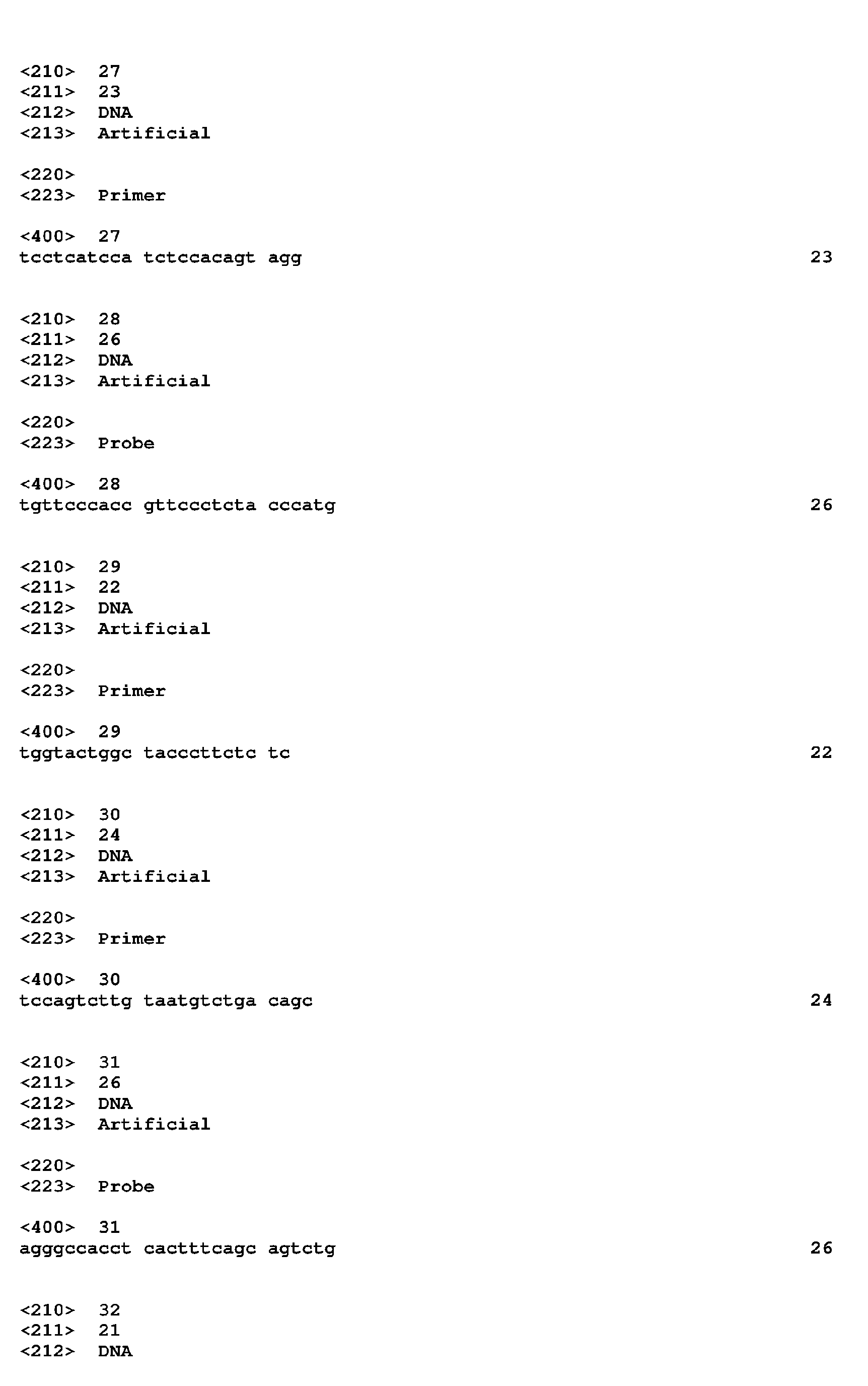


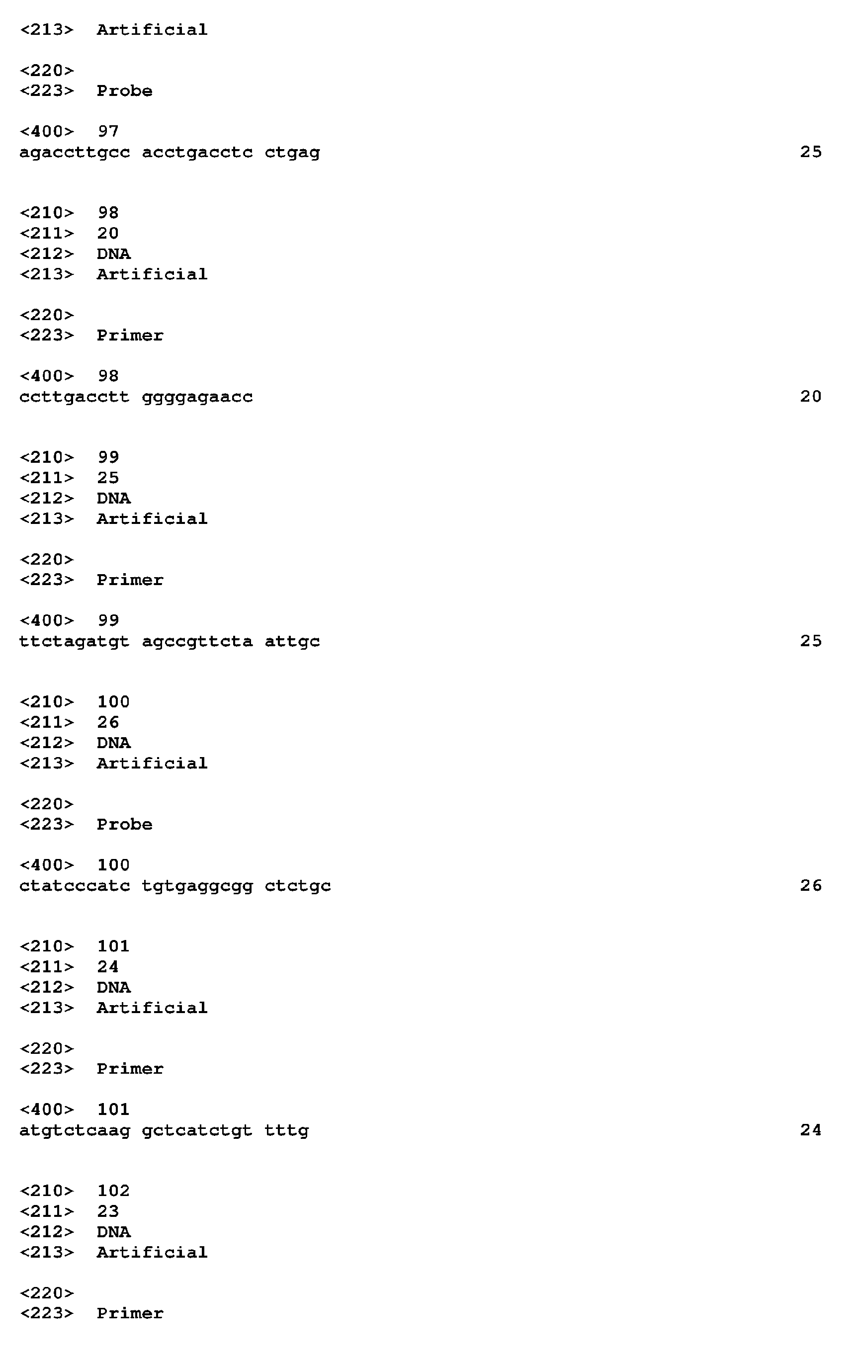


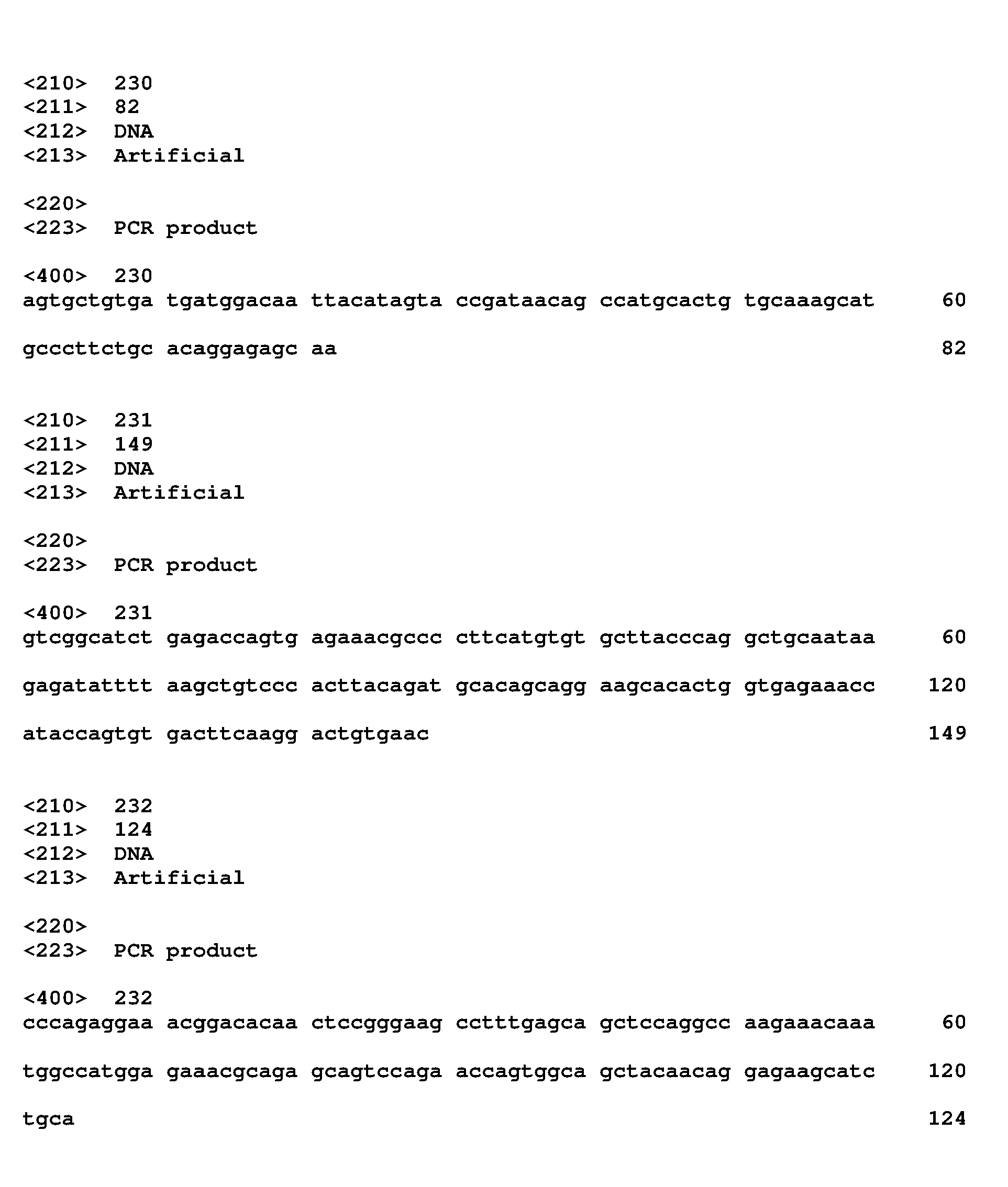
Claims (12)
- A test method for predicting the initial onset or a recurrence of acute myeloid leukemia, comprising(1) a step of measuring the expression level of leukemic stem cell marker genes in a biological sample collected from a subject for a transcription product or translation product of the genes as an analyte, and(2) a step of comparing the expression levels obtained in the measuring step with a reference value;
wherein the leukemic stem cell marker genes are 2 - 218 genes selected from the group consisting of:cell membrane- or extracellularly-localized genes consisting of ADFP, ALOX5AP, AZU1, C3AR1, CACNB4, CALCRL, CCL4, CCL5, CD33, CD36, CD3D, CD86, CD9, CD93, CD96, CD97, CFD, CHI3L1, CLEC12A, CLECL1, COCH, CST7, CXCL1, DOK2, EMR2, FCER1G, FCGR2A, FUCA2, GPR109B, GPR160, GPR34, GPR84, HAVCR2, HBEGF, HCST, HGF, HLA-DOB, HOMER3, IFI30, IL13RA1, IL2RA, IL2RG, IL3RA, INHBA, ITGB2, LGALS1, LRG1, LY86, MAMDC2, MGAT4A, P2RY14, P2RY5, PLAUR, PPBP, PRG2, PRSS21, PTH2R, PTX3, REEP5, RNASE2, RXFP1, SLC31A2, SLC43A3, SLC6A6, SLC7A6, STX7, SUCNR1, TACSTD2, TIMP1, TM4SF1, TM9SF1, TNF, TNFRSF4, TNFSF13B, TYROBP, UTS2 and VNN1;cell cycle-related genes consisting of AURKA, Cl3orf34, CCNA1, DSCC1, FAM33A, HPGD, NEK6, PYHIN1, RASSF4, TXNL4B and ZWINT; apoptosis-related genes consisting of MPO, IER3, BIK, TXNDC1, GADD45B and NAIP;signaling-related genes consisting of AK5, ARHGAP18, ARRB1, DUSP6, FYB, HCK, LPXN, MS4A3, PAK1IP1, PDE9A, PDK1, PRKAR1A, PRKCD, PXK, RAB20, RAB8A, RABIF, RASGRP3, RGS18 and S100A11;transcription factor genes consisting of WT1, MYC and HLX; and other genes consisting of ACTR2, ALOX5, ANXA2P2, ATL3, ATP6V1B2, ATP6V1C1, ATP6V1D, C12orf5, C17orf60, C18orf19, C1GALT1C1, C1orf135, C1orf163, C1orf186, C6orf150, CALML4, CCT5, CLC, COMMD8, COTL1, COX17, CRIP1, CSTA, CTSA, CTSC, CTSG, CYBB, CYP2E1, DENND3, DHRS3, DLAT, DLEU2, DPH3, EFHD2, ENC1, EXOSC3, FAM107B, FAM129A, FAM38B, FBX022, FLJ14213, FNDC3B, GNPDA1, GRPEL1, GTSF1, HIG2, HN1, HVCN1, IDH1, IDH3A, IKIP, KIF2C, KYNU, LCMT2, ME1, MIRN21, MKKS, MNDA, MTHFD2, MYO1B MYO1F, NAGA, NCF2, NCF4, NDUFAF1, NP, NRIP3, OBFC2A, PARP8, PDLIM1, PDSS1, PGM2, PIGK, PIWIL4, PPCDC, PPIF, PRAME, PUS7, RPP40, RRM2, S100A16, S100A8, S100P, S100Z, SAMHD1, SH2D1A, SPCS2, SPPL2A, TESC, THEX1, TMEM30A, TMEM33, TRIP13, TUBB6, UBASH3B, UGCG, VSTM1, WDR4, WIT1, WSB2 and ZNF253;and wherein when the expression of two or more leukemic stem cell marker genes in the subject is significantly higher than the reference value, a possible presence of a leukemic stem cell in the collected biological sample or the subject's body is suggested. - The test method according to claim 1, wherein the leukemic stem cell marker genes are 2 - 58 genes selected from the group consisting of:cell membrane- or extracellularly-localized genes consisting of ADFP, ALOX5AP, CACNB4, CCL5, CD33, CD3D, CD93, CD97, CLEC12A, DOK2, FCER1G, FCGR2A, FUCA2, GPR34, GPR84, HCST, HGF, HOMER3, IL2RA, IL2RG, IL3RA, ITGB2, LGALS1, LRG1, LY86, MGAT4A, P2RY5, PRSS21, PTH2R, RNASE2, SLC43A3, SUCNR1, TIMP1, TNF, TNFRSF4, TNFSF13B, TYROBP and VNN1; cell cycle-related genes consisting of ZWINT, NEK6 and TXNL4B; an apoptosis-related gene consisting of BIK; signaling-related genes consisting of AK5, ARHGAP18, FYB, HCK, LPXN, PDE9A, PDK1, PRKCD, RAB20, RAB8A and RABIF; transcription factor genes consisting of WT1 and HLX; and other genes consisting of CYBB, CTSC and NCF4.
- A therapeutic agent for acute myeloid leukemia that targets leukemic stem cells, comprising as an active ingredient a substance capable of suppressing the expression of a gene selected from among leukemic stem cell marker genes consisting of the following set of genes:cell membrane- or extracellularly-localized genes consisting of ADFP, ALOX5AP, AZU1, C3AR1, CACNB4, CALCRL, CCL4, CCL5, CD33, CD36, CD3D, CD86, CD9, CD93, CD96, CD97, CFD, CHI3L1, CLEC12A, CLECL1, COCH, CST7, CXCL1, DOK2, EMR2, FCER1G, FCGR2A, FUCA2, GPR109B, GPR160, GPR34, GPR84, HAVCR2, HBEGF, HCST, HGF, HLA-DOB, HOMER3, IFI30, IL13RA1, IL2RA, IL2RG, IL3RA, INHBA, ITGB2, LGALS1, LRG1, LY86, MAMDC2, MGAT4A, P2RY14, P2RY5, PLAUR, PPBP, PRG2, PRSS21, PTH2R, PTX3, REEP5, RNASE2, RXFP1, SLC31A2, SLC43A3, SLC6A6, SLC7A6, STX7, SUCNR1, TACSTD2, TIMP1, TM4SF1, TM9SF1, TNF, TNFRSF4, TNFSF13B, TYROBP, UTS2 and VNN1;cell cycle-related genes consisting of AURKA, C13orf34, CCNA1, DSCC1, FAM33A, HPGD, NEK6, PYHIN1, RASSF4, TXNL4B and ZWINT; apoptosis-related genes consisting of MPO, IER3, BIK, TXNDC1, GADD45B and NAIP;signaling-related genes consisting of AK5, ARHGAP18, ARRB1, DUSP6, FYB, HCK, LPXN, MS4A3, PAK1IP1, PDE9A, PDK1, PRKAR1A, PRKCD, PXK, RAB20, RAB8A, RABIF, RASGRP3, RGS18 and S100A11;transcription factor genes consisting of WT1, MYC and HLX; and other genes consisting of ACTR2, ALOX5, ANXA2P2, ATL3, ATP6V1B2, ATP6V1C1, ATP6V1D, C12orf5, C17orf60, C18orf19, C1GALT1C1, C1orf135, C1orf163, C1orf186, C6orf150, CALML4, CCT5, CLC, COMMD8, COTL1, COX17, CRIP1, CSTA, CTSA, CTSC, CTSG, CYBB, CYP2E1, DENND3, DHRS3, DLAT, DLEU2, DPH3, EFHD2, ENC1, EXOSC3, FAM107B, FAM129A, FAM38B, FBX022, FLJ14213, FNDC3B, GNPDA1, GRPEL1, GTSF1, HIG2, HN1, HVCN1, IDH1, IDH3A, IKIP, KIF2C, KYNU, LCMT2, ME1, MIRN21, MKKS, MNDA, MTHFD2, MY01B, MY01F, NAGA, NCF2, NCF4, NDUFAF1, NP, NRIP3, OBFC2A, PARP8, PDLIM1, PDSS1, PGM2, PIGK, PIWIL4, PPCDC, PPIF, PRAME, PUS7, RPP40, RRM2, S100A16, S100A8, S100P, S100Z, SAMHD1, SH2DIA, SPCS2, SPPL2A, TESC, THEX1, TMEM30A, TMEM33, TRIP13, TUBB6, UBASH3B, UGCG, VSTM1, WDR4, WIT1, WSB2 and ZNF253;or a substance capable of suppressing the activity of a translation product of the gene.
- The therapeutic agent according to claim 3, wherein the leukemic stem cell marker gene is selected from the group consisting of:cell membrane- or extracellularly-localized genes consisting of ADFP, ALOX5AP, CACNB4, CCL5, CD33, CD3D, CD93, CD97, CLEC12A, DOK2, FCER1G, FCGR2A, FUCA2, GPR34, GPR84, HCST, HGF, HOMER3, IL2RA, IL2RG, IL3RA, ITGB2, LGALS1, LRG1, LY86, MGAT4A, P2RY5, PRSS21, PTH2R, RNASE2, SLC43A3, SUCNR1, TIMP1, TNF, TNFRSF4, TNFSF13B, TYROBP and VNN1; cell cycle-related genes consisting of ZWINT, NEK6 and TXNL4B; an apoptosis-related gene consisting of BIK; signaling-related genes consisting of AK5, ARHGAP18, FYB, HCK, LPXN, PDE9A, PDK1, PRKCD, RAB20, RAB8A and RABIF; transcription factor genes consisting of WT1 and HLX; and other genes consisting of CYBB, CTSC and NCF4.
- The therapeutic agent according to claim 3, wherein the leukemic stem cell marker gene is selected from the group consisting of:cell membrane- or extracellularly-localized genes consisting of ALOX5AP, CACNB4, CCL5, CD33, CD3D, CD93, CD97, CLEC12A, DOK2, FCGR2A, GPR84, HCST, HOMER3, ITGB2, GALS1, LRG1, PTH2R, RNASE2, TNF, TNFSF13B, TYROBP and VNN1; a cell cycle-related gene consisting of NEK6; an apoptosis-related gene consisting of BIK; signaling-related genes consisting of AK5, FYB, HCK, LPXN, PDE9A, PDK1, PRKCD and RAB20; a transcription factor gene consisting of WT1; and other genes consisting of CTSC and NCF4.
- The therapeutic agent according to claim 3, wherein the leukemic stem cell marker gene is a marker expressed in stem cells that are present in bone marrow niches, are in the stationary phase of cell cycle, and are resistant to anticancer agents, selected from the group consisting of AK5, BIK, DOK2, FCGR2A, IL2RA, LRG1., SUCNR1 and WT1.
- The therapeutic agent according to any one of claims 3 to 6, wherein the substance capable of suppressing the expression of the gene is an antisense nucleic acid or an RNAi-inducible nucleic acid.
- The therapeutic agent according to any one of claims 3 to 6, wherein the substance capable of suppressing the activity of the translation product is an aptamer or an antibody.
- The therapeutic agent according to claim 8, wherein the antibody is an immunoconjugate of an antibody and an anticancer substance.
- A production method of a sample containing hematopoietic cells for autologous transplantation or allogeneic transplantation for a patient with acute myeloid leukemia, comprising:a) a step of collecting a sample containing hematopoietic cells from the patient or a donor;b) a step of bringing the collected sample into contact with a substance that recognizes a translation product of at least one kind of leukemic stem cell marker gene selected from among the following set of genes:cell membrane- or extracellularly-localized genes consisting of ADFP, ALOX5AP, AZU1, C3AR1, CACNB4, CALCRL, CCL4, CCL5, CD33, CD36, CD3D, CD86, CD9, CD93, CD96, CD97, CFD, CHI3L1, CLEC12A, CLECL1, COCH, CST7, CXCL1, DOK2, EMR2, FCER1G, FCGR2A, FUCA2, GPR109B, GPR160, GPR34, GPR84, HAVCR2, HBEGF, HCST, HGF, HLA-DOB, HOMER3, IFI30, IL13RA1, IL2RA, IL2RG, IL3RA, INHBA, ITGB2, LGALS1, LRG1, LY86, MAMDC2, MGAT4A, P2RY14, P2RY5, PLAUR, PPBP, PRG2, PRSS21, PTH2R, PTX3, REEP5, RNASE2, RXFP1, SLC31A2, SLC43A3, SLC6A6, SLC7A6, STX7, SUCNR1, TACSTD2, TIMP1, TM4SF1, TM9SF1, TNF, TNFRSF4, TNFSF13B, TYROBP, UTS2 and VNN1;cell cycle-related genes consisting of AURKA, C13orf34, CCNA1, DSCC1, FAM33A, HPGD, NEK6, PYHIN1, RASSF4, TXNL4B and ZWINT; apoptosis-related genes consisting of MPO, IER3, BIK, TXNDC1, GADD45B and NAIP;signaling-related genes consisting of AK5, ARHGAP18, ARRB1, DUSP6, FYB, HCK, LPXN, MS4A3, PAK1IP1, PDE9A, PDK1, PRKAR1A, PRKCD, PXK, RAB20, RAB8A, RABIF, RASGRP3, RGS18 and S100A11;transcription factor genes consisting of WT1, MYC and HLX; and other genes consisting of ACTR2, ALOX5, ANXA2P2, ATL3, ATP6V1B2, ATP6V1C1, ATP6V1D, C12orf5, C17orf60, C18orf19, C1GALT1C1, C1orf135, C1orf163, C1orf186, C6orf150, CALML4, CCT5, CLC, COMMD8, COTL1, COX17, CRIP1, CSTA, CTSA, CTSC, CTSG, CYBB, CYP2E1, DENND3, DHRS3, DLAT, DLEU2, DPH3, EFHD2, ENC1, EXOSC3, FAM107B, FAM129A, FAM38B, FBX022, FLJ14213, FNDC3B, GNPDA1, GRPEL1, GTSF1, HIG2, HN1, HVCN1, IDH1, IDH3A, IKIP, KIF2C, KYNU, LCMT2, ME1, MIRN21, MKKS, MNDA, MTHFD2, MY01B, MYO1F, NAGA, NCF2, NCF4, NDUFAF1, NP, NRIP3, OBFC2A, PARP8, PDLIM1, PDSS1, PGM2, PIGK, PIWIL4, PPCDC, PPIF, FRAME, PUS7, RPP40, RRM2, S100A16, S100A8, S100P, S100Z, SAMHD1, SH2D1A, SPCS2, SPPL2A, TESC, THEX1, TMEM30A, TMEM33, TRIP13, TUBB6, UBASH3B, UGCG, VSTM1, WDR4, WIT1, WSB2 and ZNF253; andc) a step of sorting cells to which the substance has bound, and obtaining the sample from which leukemic stem cells have been purged.
- The production method according to claim 10, wherein the leukemic stem cell marker is at least one kind of cell surface marker gene selected from among ADFP, ALOX5AP, CACNB4, CD33, CD3D, CD93, CD97, CLEC12A, DOK2, FCER1G, FCGR2A, GPR34, GPR84, HCST, HOMER3, IL2RA, IL2RG, IL3RA, ITGB2, LY86, P2RY5, PTH2R, SUCNR1, TNFRSF4, TYROBP and VNN1.
- A method for preventing or treating acute myeloid leukemia that targets leukemic stem cells, comprising administering, to a subject, an effective amount of a substance capable of suppressing the expression of a gene selected from among leukemic stem cell marker genes consisting of the following set of genes:cell membrane- or extracellularly-localized genes consisting of ADFP, ALOX5AP, AZU1, C3AR1, CACNB4, CALCRL, CCL4, CCL5, CD33, CD36, CD3D, CD86, CD9, CD93, CD96, CD97, CFD, CHI3L1, CLEC12A, CLECL1, COCH, CST7, CXCL1, DOK2, EMR2, FCER1G, FCGR2A, FUCA2, GPR109B, GPR160, GPR34, GPR84, HAVCR2, HBEGF, HCST, HGF, HLA-DOB, HOMER3, IFI30, IL13RA1, IL2RA, IL2RG, IL3RA, INHBA, ITGB2, LGALS1, LRG1, LY86, MAMDC2, MGAT4A, P2RY14, P2RY5, PLAUR, PPBP, PRG2, PRSS21, PTH2R, PTX3, REEP5, RNASE2, RXFP1, SLC31A2, SLC43A3, SLC6A6, SLC7A6, STX7, SUCNR1, TACSTD2, TIMP1, TM4SF1, TM9SF1, TNF, TNFRSF4, TNFSF13B, TYROBP, UTS2 and VNN1;cell cycle-related genes consisting of AURKA, C13orf34, CCNA1, DSCC1, FAM33A, HPGD, NEK6, PYHIN1, RASSF4, TXNL4B and ZWINT; apoptosis-related genes consisting of MPO, IER3, BIK, TXNDC1, GADD45B and NAIP;signaling-related genes consisting of AK5, ARHGAP18, ARRB1, DUSP6, FYB, HCK, LPXN, MS4A3, PAK1IP1, PDE9A, PDK1, PRKAR1A, PRKCD, PXK, RAB20, RAB8A, RABIF, RASGRP3, RGS18 and S100A11;transcription factor genes consisting of WT1, MYC and HLX; and other genes consisting of ACTR2, ALOX5, ANXA2P2, ATL3, ATP6V1B2, ATP6V1C1, ATP6V1D, C12orf5, C17orf60, C18orf19, C1GALT1C1, Clorf135, Clorf163, Clorf186, C6orf150, CALML4, CCT5, CLC, COMMD8, COTL1, COX17, CRIP1, CSTA, CTSA, CTSC, CTSG, CYBB, CYP2E1, DENND3, DHRS3, DLAT, DLEU2, DPH3, EFHD2, ENC1, EXOSC3, FAM107B, FAM129A, FAM38B, FBXO22, FLJ14213, FNDC3B, GNPDA1, GRPEL1, GTSF1, HIG2, HN1, HVCN1, IDH1, IDH3A, IKIP, KIF2C, KYNU, LCMT2, ME1, MIRN21, MKKS, MNDA, MTHFD2, MYO1B MYO1F NAGA, NCF2, NCF4, NDUFAF1, NP, NRIP3, OBFC2A, PARP8, PDLIM1, PDSS1, PGM2, PIGK, PIWIL4, PPCDC, PPIF, PRAME, PUS7, RPP40, RRM2, S100A16, S100A8, S100P, S100Z, SAMHD1, SH2D1A, SPCS2, SPPL2A, TESC, THEX1, TMEM30A, TMEM33, TRIP13, TUBB6, UBASH3B, UGCG, VSTM1, WDR4, WIT1, WSB2 and ZNF253;or a substance capable of suppressing the activity of a translation product of the gene.
Applications Claiming Priority (2)
| Application Number | Priority Date | Filing Date | Title |
|---|---|---|---|
| JP2009072400 | 2009-03-24 | ||
| PCT/JP2010/055131 WO2010110346A1 (en) | 2009-03-24 | 2010-03-24 | Leukemia stem cell markers |
Publications (4)
| Publication Number | Publication Date |
|---|---|
| EP2412825A1 true EP2412825A1 (en) | 2012-02-01 |
| EP2412825A4 EP2412825A4 (en) | 2013-10-23 |
| EP2412825B1 EP2412825B1 (en) | 2017-09-20 |
| EP2412825B8 EP2412825B8 (en) | 2018-01-10 |
Family
ID=42781039
Family Applications (1)
| Application Number | Title | Priority Date | Filing Date |
|---|---|---|---|
| EP10756142.5A Not-in-force EP2412825B8 (en) | 2009-03-24 | 2010-03-24 | Leukemia stem cell markers |
Country Status (4)
| Country | Link |
|---|---|
| US (2) | US20120070450A1 (en) |
| EP (1) | EP2412825B8 (en) |
| JP (2) | JPWO2010110346A1 (en) |
| WO (1) | WO2010110346A1 (en) |
Cited By (12)
| Publication number | Priority date | Publication date | Assignee | Title |
|---|---|---|---|---|
| WO2013139479A1 (en) * | 2012-03-21 | 2013-09-26 | Institut Gustave Roussy (Igr) | New diagnostic markers of specific chronic myelomonocytic leukemia (cmml) |
| US9605070B2 (en) | 2014-01-31 | 2017-03-28 | Novartis Ag | Antibody molecules to TIM-3 and uses thereof |
| EP3272880A3 (en) * | 2013-03-15 | 2018-04-11 | Fundació Institut de Recerca Biomèdica IRB (Barcelona) | Method for the diagnosis, prognosis and treatment of metastatic cancer |
| CN109187987A (en) * | 2018-08-23 | 2019-01-11 | 中国人民解放军第三0九医院 | Application of the MS4A3 albumen as marker in diagnostic activities tuberculosis |
| US10570204B2 (en) | 2013-09-26 | 2020-02-25 | The Medical College Of Wisconsin, Inc. | Methods for treating hematologic cancers |
| US10752687B2 (en) | 2014-01-24 | 2020-08-25 | Novartis Ag | Antibody molecules to PD-1 and uses thereof |
| US10793642B2 (en) | 2014-12-11 | 2020-10-06 | Inbiomotion S.L. | Binding members for human c-MAF |
| EP3877413A1 (en) * | 2018-11-06 | 2021-09-15 | INSERM (Institut National de la Santé et de la Recherche Médicale) | Methods and pharmaceutical compositions for the treatment of acute myeloid leukemia by eradicating leukemic stem cells |
| US11344620B2 (en) | 2014-09-13 | 2022-05-31 | Novartis Ag | Combination therapies |
| US12252535B2 (en) | 2014-03-14 | 2025-03-18 | Novartis Ag | Antibody molecules to LAG-3 and uses thereof |
| US12600777B2 (en) | 2015-07-29 | 2026-04-14 | Novartis Ag | Combination therapies comprising antibody molecules to LAG-3 |
| US12630633B2 (en) | 2016-12-16 | 2026-05-19 | Novartis Ag | Antibody molecules to PD-1 and uses thereof |
Families Citing this family (37)
| Publication number | Priority date | Publication date | Assignee | Title |
|---|---|---|---|---|
| GB201107118D0 (en) | 2011-04-27 | 2011-06-08 | Imp Innovations Ltd | Method of diagnosis and prognosis |
| US9334306B2 (en) * | 2011-07-09 | 2016-05-10 | The Regents Of The University Of California | Leukemia stem cell targeting ligands and methods of use |
| WO2013048345A1 (en) * | 2011-09-28 | 2013-04-04 | Agency For Science, Technology And Research | Methods and pharmaceutical compositions for treating cancer |
| WO2013102641A1 (en) * | 2012-01-05 | 2013-07-11 | Deutsches Krebsforschungszentrum | Means and methods for treating or diagnosing idh1 r132h mutant-positive cancers |
| WO2013133450A1 (en) * | 2012-03-06 | 2013-09-12 | 株式会社バイオマトリックス研究所 | Pharmaceutical composition for treating cancer |
| US9604988B2 (en) | 2012-07-27 | 2017-03-28 | Riken | Agent for treating or inhibiting recurrence of acute myeloid leukemia |
| KR101470125B1 (en) * | 2013-02-15 | 2014-12-08 | 순천향대학교 산학협력단 | RNASE2 as a peripheral blood protein biomarker to predict aspirin exacerbated respiratory disease |
| US9573988B2 (en) | 2013-02-20 | 2017-02-21 | Novartis Ag | Effective targeting of primary human leukemia using anti-CD123 chimeric antigen receptor engineered T cells |
| JP6683986B2 (en) * | 2013-07-10 | 2020-04-22 | 国立大学法人 東京大学 | Cancer stem cell molecular marker |
| CA2926087C (en) * | 2013-10-10 | 2023-03-14 | Beth Israel Deaconess Medical Center, Inc. | Tm4sf1 binding proteins and methods of using same |
| EP3138923B1 (en) | 2014-04-28 | 2020-01-15 | The Catholic University Of Korea Industry-Academic | Method for predicting prognosis of acute myeloid leukemia relapse |
| WO2015167210A1 (en) * | 2014-04-28 | 2015-11-05 | 가톨릭대학교 산학협력단 | Method for predicting prognosis of acute myeloid leukemia relapse |
| CA2953663A1 (en) * | 2014-06-25 | 2015-12-30 | Tel Hashomer Medical Research Infrastructure And Services Ltd. | Identification of cancer stem cells and use of same for diagnosis and treatment |
| SG11201700770PA (en) | 2014-08-19 | 2017-03-30 | Novartis Ag | Anti-cd123 chimeric antigen receptor (car) for use in cancer treatment |
| AU2016256911B2 (en) | 2015-05-07 | 2022-03-31 | Agenus Inc. | Anti-OX40 antibodies and methods of use thereof |
| WO2016201365A2 (en) * | 2015-06-12 | 2016-12-15 | Visani Giuseppe | Methods for treating cancers |
| EP3377079A1 (en) * | 2015-11-19 | 2018-09-26 | Abbvie Stemcentrx LLC | Novel anti-emr2 antibodies and methods of use |
| SG10201912984WA (en) | 2015-12-02 | 2020-03-30 | Agenus Inc | Antibodies and methods of use thereof |
| IL298653A (en) | 2015-12-11 | 2023-01-01 | Immatics Biotechnologies Gmbh | Novel peptides and combination of peptides for use in immunotherapy against various cancers |
| JP7075125B2 (en) * | 2016-05-25 | 2022-05-25 | イマティクス バイオテクノロジーズ ゲーエムベーハー | A novel peptide, peptide combination as a target and for use in immunotherapy for gallbladder cancer and cholangiocarcinoma and other cancers |
| MX389710B (en) | 2016-05-27 | 2025-03-20 | Agenus Inc | Antibodies to T-cell immunoglobulin protein and mucin domain 3 (TIM-3) and methods of using them. |
| MA46770A (en) | 2016-11-09 | 2019-09-18 | Agenus Inc | ANTI-OX40 ANTIBODIES, ANTI-GITR ANTIBODIES, AND PROCESSES FOR USE |
| KR101970764B1 (en) * | 2017-05-19 | 2019-04-22 | 아주대학교산학협력단 | COTL1 Protein Involved in Maintaining Homeostasis of Hematopoietic Stem Cells and Use Thereof |
| SG11202002753QA (en) * | 2017-10-26 | 2020-05-28 | Nat Univ Singapore | A new approach for universal monitoring of minimal residual disease in acute myeloid leukemia |
| CN107828740B (en) * | 2017-11-29 | 2020-06-09 | 武汉大学 | Homer3 monoclonal antibody and application thereof |
| GB201720077D0 (en) * | 2017-12-01 | 2018-01-17 | Univ Oxford Innovation Ltd | Leukaemic stem cell |
| US20200385714A1 (en) | 2017-12-11 | 2020-12-10 | Roche Innovation Center Copenhagen A/S | Oligonucleotides for modulating fndc3b expression |
| WO2020047164A1 (en) | 2018-08-28 | 2020-03-05 | Vor Biopharma, Inc | Genetically engineered hematopoietic stem cells and uses thereof |
| AR117327A1 (en) | 2018-12-20 | 2021-07-28 | 23Andme Inc | ANTI-CD96 ANTIBODIES AND METHODS OF USE OF THEM |
| KR102176419B1 (en) * | 2019-01-28 | 2020-11-09 | 고려대학교 산학협력단 | Method for prediction of treatment response in acute myelogenous leukemia using CD markers derived from exosomes |
| CN110132930B (en) * | 2019-06-12 | 2023-11-14 | 东北师范大学 | Multi-angle excitation detection device for laser-induced chlorophyll fluorescence and its analysis method |
| US12340542B2 (en) * | 2020-02-14 | 2025-06-24 | Mgi Tech Co., Ltd. | Method for analyzing droplets based on images, computer device and storage medium |
| CN113209300A (en) * | 2021-05-17 | 2021-08-06 | 北京大学人民医院 | Application of GILT as effect target spot for improving chemotherapy drug sensitivity of acute myeloid leukemia patient |
| CN113293202B (en) * | 2021-07-02 | 2022-02-22 | 广东莱恩医药研究院有限公司 | Real-time fluorescent quantitative PCR kit for quantitatively detecting mRNA content in organism, detection method and application |
| CN114908165A (en) * | 2022-04-11 | 2022-08-16 | 浙江大学 | Biomarker for diagnosis and treatment of acute myelogenous leukemia and application thereof |
| CN119082310B (en) * | 2024-10-28 | 2025-08-22 | 北京大学人民医院 | Biomarkers related to AML diagnosis and prognosis prediction and their applications |
| CN119345372A (en) * | 2024-10-28 | 2025-01-24 | 北京大学人民医院 | Application of CD132 in the treatment of AML |
Family Cites Families (14)
| Publication number | Priority date | Publication date | Assignee | Title |
|---|---|---|---|---|
| JPS6147500A (en) | 1984-08-15 | 1986-03-07 | Res Dev Corp Of Japan | Chimera monoclonal antibody and its preparation |
| GB8607679D0 (en) | 1986-03-27 | 1986-04-30 | Winter G P | Recombinant dna product |
| GB8928874D0 (en) | 1989-12-21 | 1990-02-28 | Celltech Ltd | Humanised antibodies |
| ES2284161T3 (en) | 1990-01-12 | 2007-11-01 | Amgen Fremont Inc. | GENERATION OF XENOGENIC ANTIBODIES. |
| AU8507191A (en) | 1990-08-29 | 1992-03-30 | Genpharm International, Inc. | Transgenic non-human animals capable of producing heterologous antibodies |
| AU6819494A (en) | 1993-04-26 | 1994-11-21 | Genpharm International, Inc. | Transgenic non-human animals capable of producing heterologous antibodies |
| US6828151B2 (en) * | 2001-12-04 | 2004-12-07 | Isis Pharmaceuticals, Inc. | Antisense modulation of hematopoietic cell protein tyrosine kinase expression |
| GB2343679A (en) * | 1998-11-16 | 2000-05-17 | Alison Miriam Davies | Autologous transplantation and method for making cells dormant |
| CA2434690A1 (en) * | 2001-01-16 | 2002-09-06 | Curagen Corporation | Proteins, polynucleotides encoding them and methods of using the same |
| WO2005045434A2 (en) * | 2003-11-04 | 2005-05-19 | Roche Diagnostics Gmbh | Method for distinguishing aml subtypes with recurring genetic aberrations |
| CA2558366A1 (en) * | 2004-02-23 | 2005-09-01 | Erasmus Universiteit Rotterdam | Classification, diagnosis and prognosis of acute myeloid leukemia by gene expression profiling |
| JP2008068892A (en) | 2006-09-13 | 2008-03-27 | Tsubakimoto Chain Co | Sample storage system for drug discovery |
| WO2009051238A1 (en) * | 2007-10-18 | 2009-04-23 | Riken | Mouse having human leukemic stem cell and leukemic non-stem cell amplified therein, and method for production thereof |
| CA3108119C (en) * | 2008-01-15 | 2024-01-16 | The Board Of Trustees Of The Leland Stanford Junior University | Markers of acute myeloid leukemia stem cells |
-
2010
- 2010-03-24 US US13/258,993 patent/US20120070450A1/en not_active Abandoned
- 2010-03-24 WO PCT/JP2010/055131 patent/WO2010110346A1/en not_active Ceased
- 2010-03-24 JP JP2011506104A patent/JPWO2010110346A1/en active Pending
- 2010-03-24 EP EP10756142.5A patent/EP2412825B8/en not_active Not-in-force
-
2014
- 2014-03-20 US US14/220,842 patent/US20140274788A1/en not_active Abandoned
-
2015
- 2015-07-07 JP JP2015135949A patent/JP6247253B2/en not_active Expired - Fee Related
Cited By (19)
| Publication number | Priority date | Publication date | Assignee | Title |
|---|---|---|---|---|
| WO2013139479A1 (en) * | 2012-03-21 | 2013-09-26 | Institut Gustave Roussy (Igr) | New diagnostic markers of specific chronic myelomonocytic leukemia (cmml) |
| EP3272880A3 (en) * | 2013-03-15 | 2018-04-11 | Fundació Institut de Recerca Biomèdica IRB (Barcelona) | Method for the diagnosis, prognosis and treatment of metastatic cancer |
| US11591599B2 (en) | 2013-03-15 | 2023-02-28 | Fundació Institut De Recerca Biomèdica (Irb Barcelona) | Method for the diagnosis, prognosis and treatment of cancer metastasis |
| US10570204B2 (en) | 2013-09-26 | 2020-02-25 | The Medical College Of Wisconsin, Inc. | Methods for treating hematologic cancers |
| US11708412B2 (en) | 2013-09-26 | 2023-07-25 | Novartis Ag | Methods for treating hematologic cancers |
| US10752687B2 (en) | 2014-01-24 | 2020-08-25 | Novartis Ag | Antibody molecules to PD-1 and uses thereof |
| US11827704B2 (en) | 2014-01-24 | 2023-11-28 | Novartis Ag | Antibody molecules to PD-1 and uses thereof |
| US10472419B2 (en) | 2014-01-31 | 2019-11-12 | Novartis Ag | Antibody molecules to TIM-3 and uses thereof |
| US9884913B2 (en) | 2014-01-31 | 2018-02-06 | Novartis Ag | Antibody molecules to TIM-3 and uses thereof |
| US10981990B2 (en) | 2014-01-31 | 2021-04-20 | Novartis Ag | Antibody molecules to TIM-3 and uses thereof |
| US11155620B2 (en) | 2014-01-31 | 2021-10-26 | Novartis Ag | Method of detecting TIM-3 using antibody molecules to TIM-3 |
| US9605070B2 (en) | 2014-01-31 | 2017-03-28 | Novartis Ag | Antibody molecules to TIM-3 and uses thereof |
| US12252535B2 (en) | 2014-03-14 | 2025-03-18 | Novartis Ag | Antibody molecules to LAG-3 and uses thereof |
| US11344620B2 (en) | 2014-09-13 | 2022-05-31 | Novartis Ag | Combination therapies |
| US10793642B2 (en) | 2014-12-11 | 2020-10-06 | Inbiomotion S.L. | Binding members for human c-MAF |
| US12600777B2 (en) | 2015-07-29 | 2026-04-14 | Novartis Ag | Combination therapies comprising antibody molecules to LAG-3 |
| US12630633B2 (en) | 2016-12-16 | 2026-05-19 | Novartis Ag | Antibody molecules to PD-1 and uses thereof |
| CN109187987A (en) * | 2018-08-23 | 2019-01-11 | 中国人民解放军第三0九医院 | Application of the MS4A3 albumen as marker in diagnostic activities tuberculosis |
| EP3877413A1 (en) * | 2018-11-06 | 2021-09-15 | INSERM (Institut National de la Santé et de la Recherche Médicale) | Methods and pharmaceutical compositions for the treatment of acute myeloid leukemia by eradicating leukemic stem cells |
Also Published As
| Publication number | Publication date |
|---|---|
| EP2412825B1 (en) | 2017-09-20 |
| JPWO2010110346A1 (en) | 2012-10-04 |
| EP2412825A4 (en) | 2013-10-23 |
| US20140274788A1 (en) | 2014-09-18 |
| JP6247253B2 (en) | 2017-12-13 |
| US20120070450A1 (en) | 2012-03-22 |
| JP2015231375A (en) | 2015-12-24 |
| WO2010110346A1 (en) | 2010-09-30 |
| EP2412825B8 (en) | 2018-01-10 |
Similar Documents
| Publication | Publication Date | Title |
|---|---|---|
| EP2412825B1 (en) | Leukemia stem cell markers | |
| AU2020200114B2 (en) | Methods and assays relating to circulating tumor cells | |
| AU2013296233B2 (en) | Biomarker associated with risk of melanoma reoccurrence | |
| JP2020126067A5 (en) | ||
| JP6860476B2 (en) | Methods and Compositions for Prostate Cancer Treatment | |
| EP3695408A1 (en) | Methods and compositions for detecting and modulating an immunotherapy resistance gene signature in cancer | |
| US20200123258A1 (en) | Targeting b cells to enhance response to immune checkpoint blockade | |
| CN111148518A (en) | Methods of modulating regulatory T cells and immune responses using CDK4/6 inhibitors | |
| JP2011504102A (en) | Methods and compositions for assessing B cell lymphoma responsiveness to treatment with anti-CD40 antibodies | |
| JP2010178650A (en) | Test method for predicting recurrence of solid cancer and recurrence prophylactic | |
| Vishnyakova et al. | Distinct gene expression patterns for CD14++ and CD16++ monocytes in preeclampsia | |
| CN115992226A (en) | Application of reagent for detecting C1Q expression level in preparation of acute myelogenous leukemia diagnosis or prognosis judgment and treatment reagent | |
| US20240398831A1 (en) | Compositions comprising small molecule regulators of tumor immunity and methods of using same | |
| AU2022229805B2 (en) | Methods of treating red blood cell disorders | |
| US20180251851A1 (en) | Ectopic lymphoid structures as targets for liver cancer detection, risk prediction and therapy | |
| EP2831593B1 (en) | S100a8/a9 as a diagnostic marker and a therapeutic target | |
| EP4281783A2 (en) | Methods to quantify rate of clonal expansion and methods for treating clonal hematopoiesis and hematologic malignancies | |
| WO2017214189A1 (en) | Methods and compositions for detection and diagnosis of bladder cancer | |
| KR102872228B1 (en) | Novel biomarkers for predicting prognosis of lung cancer and uses thereof | |
| JP2020530015A (en) | How to Treat Cancer with Farnesyltransferase Inhibitors | |
| WO2024246865A1 (en) | Transplant immunosuppression | |
| Svensson | The Tumour, the Host and Chemotherapy Response in Pancreatic Cancer | |
| WO2024227553A1 (en) | Biomarkers for use in cancer treatment and predicting responsiveness to a cancer therapy | |
| WO2025122856A1 (en) | Use of fibroblast growth factor receptor 3 mutations and tumor mutational burden to predict response to immunotherapies | |
| EP4057005A1 (en) | Biomarker for use in cancer therapy |
Legal Events
| Date | Code | Title | Description |
|---|---|---|---|
| PUAI | Public reference made under article 153(3) epc to a published international application that has entered the european phase |
Free format text: ORIGINAL CODE: 0009012 |
|
| 17P | Request for examination filed |
Effective date: 20111020 |
|
| AK | Designated contracting states |
Kind code of ref document: A1 Designated state(s): AT BE BG CH CY CZ DE DK EE ES FI FR GB GR HR HU IE IS IT LI LT LU LV MC MK MT NL NO PL PT RO SE SI SK SM TR |
|
| DAX | Request for extension of the european patent (deleted) | ||
| RIC1 | Information provided on ipc code assigned before grant |
Ipc: C12Q 1/68 20060101AFI20130909BHEP Ipc: A61K 35/28 20060101ALI20130909BHEP Ipc: A61K 31/713 20060101ALI20130909BHEP |
|
| A4 | Supplementary search report drawn up and despatched |
Effective date: 20130925 |
|
| RIC1 | Information provided on ipc code assigned before grant |
Ipc: C12Q 1/68 20060101AFI20130918BHEP Ipc: A61K 35/28 20060101ALI20130918BHEP Ipc: A61K 31/713 20060101ALI20130918BHEP |
|
| 17Q | First examination report despatched |
Effective date: 20150422 |
|
| STAA | Information on the status of an ep patent application or granted ep patent |
Free format text: STATUS: EXAMINATION IS IN PROGRESS |
|
| GRAP | Despatch of communication of intention to grant a patent |
Free format text: ORIGINAL CODE: EPIDOSNIGR1 |
|
| STAA | Information on the status of an ep patent application or granted ep patent |
Free format text: STATUS: GRANT OF PATENT IS INTENDED |
|
| INTG | Intention to grant announced |
Effective date: 20170331 |
|
| GRAS | Grant fee paid |
Free format text: ORIGINAL CODE: EPIDOSNIGR3 |
|
| GRAA | (expected) grant |
Free format text: ORIGINAL CODE: 0009210 |
|
| STAA | Information on the status of an ep patent application or granted ep patent |
Free format text: STATUS: THE PATENT HAS BEEN GRANTED |
|
| AK | Designated contracting states |
Kind code of ref document: B1 Designated state(s): AT BE BG CH CY CZ DE DK EE ES FI FR GB GR HR HU IE IS IT LI LT LU LV MC MK MT NL NO PL PT RO SE SI SK SM TR |
|
| REG | Reference to a national code |
Ref country code: GB Ref legal event code: FG4D |
|
| REG | Reference to a national code |
Ref country code: CH Ref legal event code: EP |
|
| RIN2 | Information on inventor provided after grant (corrected) |
Inventor name: HIJIKATA, ATSUSHI Inventor name: OZAWA, HIDETOSHI Inventor name: OHARA, OSAMU Inventor name: ISHIKAWA, FUMIHIKO Inventor name: KITAMURA, HIROSHI Inventor name: SHULTZ, LEONARD D. Inventor name: SAITO, YORIKO |
|
| REG | Reference to a national code |
Ref country code: AT Ref legal event code: REF Ref document number: 930161 Country of ref document: AT Kind code of ref document: T Effective date: 20171015 |
|
| REG | Reference to a national code |
Ref country code: IE Ref legal event code: FG4D |
|
| RAP2 | Party data changed (patent owner data changed or rights of a patent transferred) |
Owner name: RIKEN |
|
| REG | Reference to a national code |
Ref country code: DE Ref legal event code: R096 Ref document number: 602010045380 Country of ref document: DE |
|
| RAP2 | Party data changed (patent owner data changed or rights of a patent transferred) |
Owner name: RIKEN Owner name: THE JACKSON LABORATORY |
|
| REG | Reference to a national code |
Ref country code: NL Ref legal event code: MP Effective date: 20170920 |
|
| PG25 | Lapsed in a contracting state [announced via postgrant information from national office to epo] |
Ref country code: SE Free format text: LAPSE BECAUSE OF FAILURE TO SUBMIT A TRANSLATION OF THE DESCRIPTION OR TO PAY THE FEE WITHIN THE PRESCRIBED TIME-LIMIT Effective date: 20170920 Ref country code: NO Free format text: LAPSE BECAUSE OF FAILURE TO SUBMIT A TRANSLATION OF THE DESCRIPTION OR TO PAY THE FEE WITHIN THE PRESCRIBED TIME-LIMIT Effective date: 20171220 Ref country code: FI Free format text: LAPSE BECAUSE OF FAILURE TO SUBMIT A TRANSLATION OF THE DESCRIPTION OR TO PAY THE FEE WITHIN THE PRESCRIBED TIME-LIMIT Effective date: 20170920 Ref country code: LT Free format text: LAPSE BECAUSE OF FAILURE TO SUBMIT A TRANSLATION OF THE DESCRIPTION OR TO PAY THE FEE WITHIN THE PRESCRIBED TIME-LIMIT Effective date: 20170920 Ref country code: HR Free format text: LAPSE BECAUSE OF FAILURE TO SUBMIT A TRANSLATION OF THE DESCRIPTION OR TO PAY THE FEE WITHIN THE PRESCRIBED TIME-LIMIT Effective date: 20170920 |
|
| REG | Reference to a national code |
Ref country code: LT Ref legal event code: MG4D |
|
| REG | Reference to a national code |
Ref country code: AT Ref legal event code: MK05 Ref document number: 930161 Country of ref document: AT Kind code of ref document: T Effective date: 20170920 |
|
| PG25 | Lapsed in a contracting state [announced via postgrant information from national office to epo] |
Ref country code: BG Free format text: LAPSE BECAUSE OF FAILURE TO SUBMIT A TRANSLATION OF THE DESCRIPTION OR TO PAY THE FEE WITHIN THE PRESCRIBED TIME-LIMIT Effective date: 20171220 Ref country code: GR Free format text: LAPSE BECAUSE OF FAILURE TO SUBMIT A TRANSLATION OF THE DESCRIPTION OR TO PAY THE FEE WITHIN THE PRESCRIBED TIME-LIMIT Effective date: 20171221 Ref country code: LV Free format text: LAPSE BECAUSE OF FAILURE TO SUBMIT A TRANSLATION OF THE DESCRIPTION OR TO PAY THE FEE WITHIN THE PRESCRIBED TIME-LIMIT Effective date: 20170920 |
|
| REG | Reference to a national code |
Ref country code: FR Ref legal event code: PLFP Year of fee payment: 9 |
|
| PG25 | Lapsed in a contracting state [announced via postgrant information from national office to epo] |
Ref country code: NL Free format text: LAPSE BECAUSE OF FAILURE TO SUBMIT A TRANSLATION OF THE DESCRIPTION OR TO PAY THE FEE WITHIN THE PRESCRIBED TIME-LIMIT Effective date: 20170920 |
|
| PG25 | Lapsed in a contracting state [announced via postgrant information from national office to epo] |
Ref country code: CZ Free format text: LAPSE BECAUSE OF FAILURE TO SUBMIT A TRANSLATION OF THE DESCRIPTION OR TO PAY THE FEE WITHIN THE PRESCRIBED TIME-LIMIT Effective date: 20170920 Ref country code: ES Free format text: LAPSE BECAUSE OF FAILURE TO SUBMIT A TRANSLATION OF THE DESCRIPTION OR TO PAY THE FEE WITHIN THE PRESCRIBED TIME-LIMIT Effective date: 20170920 Ref country code: PL Free format text: LAPSE BECAUSE OF FAILURE TO SUBMIT A TRANSLATION OF THE DESCRIPTION OR TO PAY THE FEE WITHIN THE PRESCRIBED TIME-LIMIT Effective date: 20170920 Ref country code: RO Free format text: LAPSE BECAUSE OF FAILURE TO SUBMIT A TRANSLATION OF THE DESCRIPTION OR TO PAY THE FEE WITHIN THE PRESCRIBED TIME-LIMIT Effective date: 20170920 |
|
| PGFP | Annual fee paid to national office [announced via postgrant information from national office to epo] |
Ref country code: GB Payment date: 20180321 Year of fee payment: 9 |
|
| PG25 | Lapsed in a contracting state [announced via postgrant information from national office to epo] |
Ref country code: IT Free format text: LAPSE BECAUSE OF FAILURE TO SUBMIT A TRANSLATION OF THE DESCRIPTION OR TO PAY THE FEE WITHIN THE PRESCRIBED TIME-LIMIT Effective date: 20170920 Ref country code: SK Free format text: LAPSE BECAUSE OF FAILURE TO SUBMIT A TRANSLATION OF THE DESCRIPTION OR TO PAY THE FEE WITHIN THE PRESCRIBED TIME-LIMIT Effective date: 20170920 Ref country code: AT Free format text: LAPSE BECAUSE OF FAILURE TO SUBMIT A TRANSLATION OF THE DESCRIPTION OR TO PAY THE FEE WITHIN THE PRESCRIBED TIME-LIMIT Effective date: 20170920 Ref country code: SM Free format text: LAPSE BECAUSE OF FAILURE TO SUBMIT A TRANSLATION OF THE DESCRIPTION OR TO PAY THE FEE WITHIN THE PRESCRIBED TIME-LIMIT Effective date: 20170920 Ref country code: IS Free format text: LAPSE BECAUSE OF FAILURE TO SUBMIT A TRANSLATION OF THE DESCRIPTION OR TO PAY THE FEE WITHIN THE PRESCRIBED TIME-LIMIT Effective date: 20180120 Ref country code: EE Free format text: LAPSE BECAUSE OF FAILURE TO SUBMIT A TRANSLATION OF THE DESCRIPTION OR TO PAY THE FEE WITHIN THE PRESCRIBED TIME-LIMIT Effective date: 20170920 |
|
| PGFP | Annual fee paid to national office [announced via postgrant information from national office to epo] |
Ref country code: FR Payment date: 20180312 Year of fee payment: 9 |
|
| REG | Reference to a national code |
Ref country code: DE Ref legal event code: R097 Ref document number: 602010045380 Country of ref document: DE |
|
| PLBE | No opposition filed within time limit |
Free format text: ORIGINAL CODE: 0009261 |
|
| STAA | Information on the status of an ep patent application or granted ep patent |
Free format text: STATUS: NO OPPOSITION FILED WITHIN TIME LIMIT |
|
| PG25 | Lapsed in a contracting state [announced via postgrant information from national office to epo] |
Ref country code: DK Free format text: LAPSE BECAUSE OF FAILURE TO SUBMIT A TRANSLATION OF THE DESCRIPTION OR TO PAY THE FEE WITHIN THE PRESCRIBED TIME-LIMIT Effective date: 20170920 |
|
| PGFP | Annual fee paid to national office [announced via postgrant information from national office to epo] |
Ref country code: DE Payment date: 20180403 Year of fee payment: 9 |
|
| 26N | No opposition filed |
Effective date: 20180621 |
|
| REG | Reference to a national code |
Ref country code: CH Ref legal event code: PL |
|
| PG25 | Lapsed in a contracting state [announced via postgrant information from national office to epo] |
Ref country code: MC Free format text: LAPSE BECAUSE OF FAILURE TO SUBMIT A TRANSLATION OF THE DESCRIPTION OR TO PAY THE FEE WITHIN THE PRESCRIBED TIME-LIMIT Effective date: 20170920 Ref country code: SI Free format text: LAPSE BECAUSE OF FAILURE TO SUBMIT A TRANSLATION OF THE DESCRIPTION OR TO PAY THE FEE WITHIN THE PRESCRIBED TIME-LIMIT Effective date: 20170920 |
|
| REG | Reference to a national code |
Ref country code: BE Ref legal event code: MM Effective date: 20180331 |
|
| REG | Reference to a national code |
Ref country code: IE Ref legal event code: MM4A |
|
| PG25 | Lapsed in a contracting state [announced via postgrant information from national office to epo] |
Ref country code: LU Free format text: LAPSE BECAUSE OF NON-PAYMENT OF DUE FEES Effective date: 20180324 |
|
| PG25 | Lapsed in a contracting state [announced via postgrant information from national office to epo] |
Ref country code: IE Free format text: LAPSE BECAUSE OF NON-PAYMENT OF DUE FEES Effective date: 20180324 |
|
| PG25 | Lapsed in a contracting state [announced via postgrant information from national office to epo] |
Ref country code: CH Free format text: LAPSE BECAUSE OF NON-PAYMENT OF DUE FEES Effective date: 20180331 Ref country code: LI Free format text: LAPSE BECAUSE OF NON-PAYMENT OF DUE FEES Effective date: 20180331 Ref country code: BE Free format text: LAPSE BECAUSE OF NON-PAYMENT OF DUE FEES Effective date: 20180331 |
|
| REG | Reference to a national code |
Ref country code: DE Ref legal event code: R119 Ref document number: 602010045380 Country of ref document: DE |
|
| GBPC | Gb: european patent ceased through non-payment of renewal fee |
Effective date: 20190324 |
|
| PG25 | Lapsed in a contracting state [announced via postgrant information from national office to epo] |
Ref country code: MT Free format text: LAPSE BECAUSE OF NON-PAYMENT OF DUE FEES Effective date: 20180324 Ref country code: DE Free format text: LAPSE BECAUSE OF NON-PAYMENT OF DUE FEES Effective date: 20191001 Ref country code: GB Free format text: LAPSE BECAUSE OF NON-PAYMENT OF DUE FEES Effective date: 20190324 |
|
| PG25 | Lapsed in a contracting state [announced via postgrant information from national office to epo] |
Ref country code: FR Free format text: LAPSE BECAUSE OF NON-PAYMENT OF DUE FEES Effective date: 20190331 |
|
| PG25 | Lapsed in a contracting state [announced via postgrant information from national office to epo] |
Ref country code: TR Free format text: LAPSE BECAUSE OF FAILURE TO SUBMIT A TRANSLATION OF THE DESCRIPTION OR TO PAY THE FEE WITHIN THE PRESCRIBED TIME-LIMIT Effective date: 20170920 |
|
| PG25 | Lapsed in a contracting state [announced via postgrant information from national office to epo] |
Ref country code: HU Free format text: LAPSE BECAUSE OF FAILURE TO SUBMIT A TRANSLATION OF THE DESCRIPTION OR TO PAY THE FEE WITHIN THE PRESCRIBED TIME-LIMIT; INVALID AB INITIO Effective date: 20100324 Ref country code: PT Free format text: LAPSE BECAUSE OF FAILURE TO SUBMIT A TRANSLATION OF THE DESCRIPTION OR TO PAY THE FEE WITHIN THE PRESCRIBED TIME-LIMIT Effective date: 20170920 |
|
| PG25 | Lapsed in a contracting state [announced via postgrant information from national office to epo] |
Ref country code: CY Free format text: LAPSE BECAUSE OF FAILURE TO SUBMIT A TRANSLATION OF THE DESCRIPTION OR TO PAY THE FEE WITHIN THE PRESCRIBED TIME-LIMIT Effective date: 20170920 Ref country code: MK Free format text: LAPSE BECAUSE OF NON-PAYMENT OF DUE FEES Effective date: 20170920 |