EP2398913B1 - Method for obtaining immunoglobulin encoding nucleic acid - Google Patents
Method for obtaining immunoglobulin encoding nucleic acid Download PDFInfo
- Publication number
- EP2398913B1 EP2398913B1 EP10705301.9A EP10705301A EP2398913B1 EP 2398913 B1 EP2398913 B1 EP 2398913B1 EP 10705301 A EP10705301 A EP 10705301A EP 2398913 B1 EP2398913 B1 EP 2398913B1
- Authority
- EP
- European Patent Office
- Prior art keywords
- primer
- seq
- immunoglobulin
- polymerase chain
- chain reaction
- Prior art date
- Legal status (The legal status is an assumption and is not a legal conclusion. Google has not performed a legal analysis and makes no representation as to the accuracy of the status listed.)
- Active
Links
Images






Classifications
-
- C—CHEMISTRY; METALLURGY
- C12—BIOCHEMISTRY; BEER; SPIRITS; WINE; VINEGAR; MICROBIOLOGY; ENZYMOLOGY; MUTATION OR GENETIC ENGINEERING
- C12P—FERMENTATION OR ENZYME-USING PROCESSES TO SYNTHESISE A DESIRED CHEMICAL COMPOUND OR COMPOSITION OR TO SEPARATE OPTICAL ISOMERS FROM A RACEMIC MIXTURE
- C12P21/00—Preparation of peptides or proteins
- C12P21/005—Glycopeptides, glycoproteins
-
- C—CHEMISTRY; METALLURGY
- C12—BIOCHEMISTRY; BEER; SPIRITS; WINE; VINEGAR; MICROBIOLOGY; ENZYMOLOGY; MUTATION OR GENETIC ENGINEERING
- C12Q—MEASURING OR TESTING PROCESSES INVOLVING ENZYMES, NUCLEIC ACIDS OR MICROORGANISMS; COMPOSITIONS OR TEST PAPERS THEREFOR; PROCESSES OF PREPARING SUCH COMPOSITIONS; CONDITION-RESPONSIVE CONTROL IN MICROBIOLOGICAL OR ENZYMOLOGICAL PROCESSES
- C12Q1/00—Measuring or testing processes involving enzymes, nucleic acids or microorganisms; Compositions therefor; Processes of preparing such compositions
- C12Q1/68—Measuring or testing processes involving enzymes, nucleic acids or microorganisms; Compositions therefor; Processes of preparing such compositions involving nucleic acids
- C12Q1/6844—Nucleic acid amplification reactions
- C12Q1/6858—Allele-specific amplification
-
- C—CHEMISTRY; METALLURGY
- C07—ORGANIC CHEMISTRY
- C07K—PEPTIDES
- C07K16/00—Immunoglobulins [IG], e.g. monoclonal or polyclonal antibodies
-
- C—CHEMISTRY; METALLURGY
- C12—BIOCHEMISTRY; BEER; SPIRITS; WINE; VINEGAR; MICROBIOLOGY; ENZYMOLOGY; MUTATION OR GENETIC ENGINEERING
- C12P—FERMENTATION OR ENZYME-USING PROCESSES TO SYNTHESISE A DESIRED CHEMICAL COMPOUND OR COMPOSITION OR TO SEPARATE OPTICAL ISOMERS FROM A RACEMIC MIXTURE
- C12P19/00—Preparation of compounds containing saccharide radicals
- C12P19/26—Preparation of nitrogen-containing carbohydrates
- C12P19/28—N-glycosides
- C12P19/30—Nucleotides
- C12P19/34—Polynucleotides, e.g. nucleic acids, oligoribonucleotides
-
- C—CHEMISTRY; METALLURGY
- C12—BIOCHEMISTRY; BEER; SPIRITS; WINE; VINEGAR; MICROBIOLOGY; ENZYMOLOGY; MUTATION OR GENETIC ENGINEERING
- C12Q—MEASURING OR TESTING PROCESSES INVOLVING ENZYMES, NUCLEIC ACIDS OR MICROORGANISMS; COMPOSITIONS OR TEST PAPERS THEREFOR; PROCESSES OF PREPARING SUCH COMPOSITIONS; CONDITION-RESPONSIVE CONTROL IN MICROBIOLOGICAL OR ENZYMOLOGICAL PROCESSES
- C12Q1/00—Measuring or testing processes involving enzymes, nucleic acids or microorganisms; Compositions therefor; Processes of preparing such compositions
- C12Q1/68—Measuring or testing processes involving enzymes, nucleic acids or microorganisms; Compositions therefor; Processes of preparing such compositions involving nucleic acids
- C12Q1/6804—Nucleic acid analysis using immunogens
-
- C—CHEMISTRY; METALLURGY
- C12—BIOCHEMISTRY; BEER; SPIRITS; WINE; VINEGAR; MICROBIOLOGY; ENZYMOLOGY; MUTATION OR GENETIC ENGINEERING
- C12Q—MEASURING OR TESTING PROCESSES INVOLVING ENZYMES, NUCLEIC ACIDS OR MICROORGANISMS; COMPOSITIONS OR TEST PAPERS THEREFOR; PROCESSES OF PREPARING SUCH COMPOSITIONS; CONDITION-RESPONSIVE CONTROL IN MICROBIOLOGICAL OR ENZYMOLOGICAL PROCESSES
- C12Q1/00—Measuring or testing processes involving enzymes, nucleic acids or microorganisms; Compositions therefor; Processes of preparing such compositions
- C12Q1/68—Measuring or testing processes involving enzymes, nucleic acids or microorganisms; Compositions therefor; Processes of preparing such compositions involving nucleic acids
- C12Q1/6876—Nucleic acid products used in the analysis of nucleic acids, e.g. primers or probes
- C12Q1/6881—Nucleic acid products used in the analysis of nucleic acids, e.g. primers or probes for tissue or cell typing, e.g. human leukocyte antigen [HLA] probes
-
- C—CHEMISTRY; METALLURGY
- C07—ORGANIC CHEMISTRY
- C07K—PEPTIDES
- C07K2317/00—Immunoglobulins specific features
- C07K2317/10—Immunoglobulins specific features characterized by their source of isolation or production
- C07K2317/14—Specific host cells or culture conditions, e.g. components, pH or temperature
-
- C—CHEMISTRY; METALLURGY
- C07—ORGANIC CHEMISTRY
- C07K—PEPTIDES
- C07K2317/00—Immunoglobulins specific features
- C07K2317/20—Immunoglobulins specific features characterized by taxonomic origin
- C07K2317/21—Immunoglobulins specific features characterized by taxonomic origin from primates, e.g. man
-
- C—CHEMISTRY; METALLURGY
- C07—ORGANIC CHEMISTRY
- C07K—PEPTIDES
- C07K2317/00—Immunoglobulins specific features
- C07K2317/50—Immunoglobulins specific features characterized by immunoglobulin fragments
- C07K2317/55—Fab or Fab'
-
- C—CHEMISTRY; METALLURGY
- C12—BIOCHEMISTRY; BEER; SPIRITS; WINE; VINEGAR; MICROBIOLOGY; ENZYMOLOGY; MUTATION OR GENETIC ENGINEERING
- C12Q—MEASURING OR TESTING PROCESSES INVOLVING ENZYMES, NUCLEIC ACIDS OR MICROORGANISMS; COMPOSITIONS OR TEST PAPERS THEREFOR; PROCESSES OF PREPARING SUCH COMPOSITIONS; CONDITION-RESPONSIVE CONTROL IN MICROBIOLOGICAL OR ENZYMOLOGICAL PROCESSES
- C12Q2537/00—Reactions characterised by the reaction format or use of a specific feature
- C12Q2537/10—Reactions characterised by the reaction format or use of a specific feature the purpose or use of
- C12Q2537/143—Multiplexing, i.e. use of multiple primers or probes in a single reaction, usually for simultaneously analyse of multiple analysis
-
- C—CHEMISTRY; METALLURGY
- C12—BIOCHEMISTRY; BEER; SPIRITS; WINE; VINEGAR; MICROBIOLOGY; ENZYMOLOGY; MUTATION OR GENETIC ENGINEERING
- C12Q—MEASURING OR TESTING PROCESSES INVOLVING ENZYMES, NUCLEIC ACIDS OR MICROORGANISMS; COMPOSITIONS OR TEST PAPERS THEREFOR; PROCESSES OF PREPARING SUCH COMPOSITIONS; CONDITION-RESPONSIVE CONTROL IN MICROBIOLOGICAL OR ENZYMOLOGICAL PROCESSES
- C12Q2549/00—Reactions characterised by the features used to influence the efficiency or specificity
- C12Q2549/10—Reactions characterised by the features used to influence the efficiency or specificity the purpose being that of reducing false positive or false negative signals
- C12Q2549/119—Reactions characterised by the features used to influence the efficiency or specificity the purpose being that of reducing false positive or false negative signals using nested primers
-
- C—CHEMISTRY; METALLURGY
- C12—BIOCHEMISTRY; BEER; SPIRITS; WINE; VINEGAR; MICROBIOLOGY; ENZYMOLOGY; MUTATION OR GENETIC ENGINEERING
- C12Q—MEASURING OR TESTING PROCESSES INVOLVING ENZYMES, NUCLEIC ACIDS OR MICROORGANISMS; COMPOSITIONS OR TEST PAPERS THEREFOR; PROCESSES OF PREPARING SUCH COMPOSITIONS; CONDITION-RESPONSIVE CONTROL IN MICROBIOLOGICAL OR ENZYMOLOGICAL PROCESSES
- C12Q2600/00—Oligonucleotides characterized by their use
- C12Q2600/16—Primer sets for multiplex assays
Definitions
- the present invention relates to a method and means for obtaining immunoglobulin encoding nucleic acid from a single immunoglobulin producing cell with a multiplexed polymerase chain reaction (PCR), and also to a method for producing an immunoglobulin whereby the immunoglobulin encoding nucleic acid is obtained from a single immunoglobulin producing cell in combination with in vitro translation. Also encompassed by the current specification is a method for characterization of recombinantly produced human Fab-fragments.
- PCR polymerase chain reaction
- Immunglobulins from immunoglobulin producing cell can be performed according to methods known in the art. Such methods are e.g. hybridoma technique. A different method is based on the identification of the nucleic acid sequence of the immunoglobulin. Usually it is sufficient to identify the sequence of the variable regions or even only the CDR regions or only the CDR3 region.
- the mRNA is isolated from a pool of immunoglobulin producing cells and is used for the construction of a cDNA-library encoding the CDR regions of the immunoglobulin. The cDNA-library is then transfected into a suitable host cell, such as NS0 or CHO, and screened for specific immunoglobulin production.
- WO 2008/104184 reports a method for cloning cognate antibodies.
- the efficient generation of monoclonal antibodies from single human B cells is reported by Tiller et al. ( Tiller, T., et al., J. Immunol. Meth. 329 (2007) 112-124 ).
- Braeuninger et al. Braeuninger, A., et al., Blood 93 (1999) 2679-2687 ) report the molecular analysis of single B cells from T-cell-rich B-cell lymphoma.
- Systematic design and testing of nested (RT-) PCR primer is reported by Rohatgi et al. ( Rohatgi, S., et al, J. Immunol. Meth.
- the current invention is directed in specific embodiments of an aspect to a method for providing a human monoclonal antibody comprising the in vitro translation of a nucleic acid encoding human immunoglobulin G fragments whereby the nucleic acid is obtained by specific amplification of cDNA fragments obtained from the mRNA of a single immunoglobulin producing human B-cell, plasmablast or plasma cell or a B-cell of an animal comprising a human immunoglobulin locus.
- the invention comprises a method for the multiplex semi-nested PCR and multiplex one tube RT-GSP-PCR (RT-Gene Specific Primer-PCR) for the amplification of cognate IgG HC and IgG LC chains (human IgG isotype) from a single B-cell or plasmablast or plasma cell.
- RT-GSP-PCR RT-Gene Specific Primer-PCR
- the Fab PCR product is subsequently transcribed to mRNA and translated in vitro in E.coli lysate. The expression was examined using ELISA and Western blot.
- a method for obtaining a nucleic acid encoding an immunoglobulin variable domain from a single cell by a multiplex one tube RT-GSP-PCR comprising the following step:
- the methods according to the invention are characterized in that the 5'-primer employed in the first polymerase chain reaction bind in the coding region for the leader peptide of the immunoglobulin.
- the methods according to the invention are characterized in that the 5'-primer employed in the second polymerase chain reaction or the 5'-primer employed in a multiplex one tube RT-GSP-PCR bind in the coding region of the first framework region of the immunoglobulin.
- the methods according to the invention are characterized in that the primer employed in the second polymerase chain reaction provide for overhangs encoding the translational start codon ATG for 5'-primer and/or the translational stop codon TTA for 3'-primer.
- the methods according to the invention are characterized in comprising the additional step of:
- the methods according to the invention are characterized in that six 5'-primer and one 3'-primer are employed in the first polymerase chain reaction.
- the methods are characterized in that
- the immunoglobulin variable domain can be an immunoglobulin heavy chain variable domain or an immunoglobulin kappa light chain variable domain or an immunoglobulin lambda light chain variable domain.
- a further aspect of the current invention is a method for producing an immunoglobulin Fab-fragment comprising the following steps:
- immunoglobulin an immunoglobulin of class G (IgG).
- methods for producing an immunoglobulin Fab fragment or an immunoglobulin is the obtaining of the nucleic acid by a method according to an aspect of the current invention.
- B-cells were isolated from peripheral blood. With the limited dilution approach, single cells were placed in a 96 well microtiter plate. The mRNA of these cells was extracted.
- a multiplex polymerase chain reaction is used for the amplification of heavy and light chain variable domains simultaneously in the same polymerase chain reaction.
- the current approach provides for an increased sensitivity and an increased amount of amplified sequences.
- the use of gene-specific primer in both, i.e. all, polymerase chain reactions enhances the specificity and accuracy of the methods.
- the encoded domains comprise cysteine residues suitable for the formation of interchain disulfide bonds.
- immunoglobulin denotes a protein consisting of one or more polypeptide(s) substantially encoded by immunoglobulin genes.
- the recognized immunoglobulin genes include the different constant region genes as well as the myriad immunoglobulin variable region genes.
- Immunoglobulins may exist in a variety of formats, including, for example, Fv, Fab, and F(ab) 2 as well as single chains (scFv) or diabodies (e.g. Huston, J.S., et al., Proc. Natl. Acad. Sci. USA 85 (1988) 5879-5883 ; Bird, R.E., et al., Science 242 (1988) 423-426 ; Hood, L.E. et al., Immunology, Benjamin N.Y., 2nd edition (1984 ); Hunkapiller, T. and Hood, L., Nature 323 (1986) 15-16 ).
- An immunoglobulin in general comprises two so called light chain polypeptides (light chain) and two so called heavy chain polypeptides (heavy chain).
- Each of the heavy and light chain polypeptides contains a variable domain (variable region) (generally the amino terminal portion of the polypeptide chain) comprising binding regions that are able to interact with an antigen.
- Each of the heavy and light chain polypeptides comprises a constant region (generally the carboxyl terminal portion).
- the constant region of the heavy chain mediates the binding of the antibody i) to cells bearing a Fc gamma receptor (Fc ⁇ R, such as phagocytic cells, or ii) to cells bearing the neonatal Fc receptor (FcRn) also known as Brambell receptor.
- Fc ⁇ R Fc gamma receptor
- FcRn neonatal Fc receptor
- variable domain of an immunoglobulin's light or heavy chain in turn comprises different segments, i.e. four framework regions (FR) and three hypervariable regions (CDR).
- immunoglobulins Genetic engineering of immunoglobulins is e.g. described in Morrison, S.L., et al., Proc. Natl. Acad Sci. USA 81 (1984) 6851-6855 ; US 5,202,238 and US 5,204,244 ; Riechmann, L., et al., Nature 332 (1988) 323-327 ; Neuberger, M.S., et al., Nature 314 (1985) 268-270 ; Lonberg, N., Nat. Biotechnol. 23 (2005) 1117-1125 .
- chimeric immunoglobulin denotes an immunoglobulin, preferably a monoclonal immunoglobulin, comprising a variable domain, i.e. binding region, from a first non-human species and at least a portion of a constant region derived from a second different source or species.
- Chimeric immunoglobulins are generally prepared by recombinant DNA techniques.
- chimeric immunoglobulins comprise a mouse, rat, hamster, rabbit, or sheep variable domain and a human constant region.
- the human heavy chain constant region is a human IgG constant region.
- the human light chain constant region is a kappa chain or a lambda chain.
- chimeric immunoglobulins encompassed by the present invention are those in which the class or subclass of the non-human immunoglobulin from which the variable domain is derived has been changed. Such immunoglobulins are also referred to as "class-switched immunoglobulins".
- Class-switched immunoglobulins are also those in which the constant region has differences from the wild-type constant region sequence that result in an immunoglobulin with different properties, e.g. in regard to C1q binding and/or Fc receptor (FcR) binding.
- Fc part of an immunoglobulin is not directly involved in binding to the antigen, but exhibit various effector functions.
- immunoglobulins are divided in the classes: IgA, IgD, IgE, IgG, and IgM. Some of these classes are further divided into subclasses, i.e. IgG in IgG1, IgG2, IgG3, and IgG4, or IgA in IgA1 and IgA2.
- immunoglobulin class to which an immunoglobulin belongs the heavy chain constant regions of immunoglobulins are called ⁇ (IgA), ⁇ (IgD), ⁇ (IgE), ⁇ (IgG), and ⁇ (IgM), respectively.
- the immunoglobulins according to the invention belong in one embodiment to the IgG class.
- an "Fc part of an immunoglobulin” is a term well known to the skilled artisan and defined on basis of the papain cleavage of immunoglobulins.
- the immunoglobulin contains as Fc part a human Fc part or an Fc part derived from human origin.
- the Fc part either an Fc part of a human immunoglobulin of the subclass IgG4 or IgG1 or is an Fc part of a human antibody of the subclass IgG1, IgG2, or IgG3, which is modified in such a way that no Fc ⁇ receptor (e.g. Fc ⁇ RIIIa) binding and/or no C1q binding as defined below can be detected.
- Fc ⁇ receptor e.g. Fc ⁇ RIIIa
- the Fc part is a human Fc part, in another embodiment a human IgG4 or IgG1 subclass Fc part or a mutated Fc part from human IgG1 subclass. In a further embodiment the Fc part is from human IgG1 subclass with mutations L234A and L235A. While IgG4 shows reduced Fc ⁇ receptor (Fc ⁇ RIIIa) binding, immunoglobulins of other IgG subclasses show strong binding.
- Pro238, Asp265, Asp270, Asn297 (loss of Fc carbohydrate), Pro329, Leu234, Leu235, Gly236, Gly237, Ile253, Ser254, Lys288, Thr307, Gln311, Asn434, or/and His435 are residues which, if altered, provide also reduced Fc ⁇ receptor binding ( Shields, R.L., et al., J. Biol. Chem. 276 (2001) 6591-6604 ; Lund, J., et al., FASEB J. 9 (1995) 115-119 ; Morgan, A., et al., Immunology 86 (1995) 319-324 ; EP 0 307 434 ).
- the immunoglobulin is in regard to Fc ⁇ receptor binding of IgG4 or IgG1 subclass or of IgG1 or IgG2 subclass, with a mutation in L234, L235, and/or D265, and/or contains the PVA236 mutation.
- the mutations are S228P, L234A, L235A, L235E, and/or PVA236 (PVA236 means that the amino acid sequence ELLG (given in one letter amino acid code) from amino acid position 233 to 236 of IgG1 or EFLG of IgG4 is replaced by PVA).
- the mutations are S228P of IgG4, and L234A and L235A of IgG1.
- the Fc part of an immunoglobulin is directly involved in ADCC (antibody-dependent cell-mediated cytotoxicity) and CDC (complement-dependent cytotoxicity).
- An immunoglobulin which does not bind Fc ⁇ receptor and/or complement factor C1q does not elicit antibody-dependent cellular cytotoxicity (ADCC) and/or complement dependent cytotoxicity (CDC).
- the heavy chain constant region has an amino acid sequences of SEQ ID NO: 01, or SEQ ID NO: 02, or SEQ ID NO: 01 with mutations L234A and L235A, or SEQ ID NO: 02 with mutation S228P
- the light chain constant region has an amino acid sequence of SEQ ID NO: 03 or SEQ ID NO: 04.
- Humanized or “CDR-grafted” forms of non-human (e.g. rodent or rabbit) immunoglobulins are immunoglobulins that contain partial sequences derived from a non-human immunoglobulin and partial sequences derived from a human immunoglobulin.
- humanized immunoglobulins are derived from a human immunoglobulin (recipient or acceptor immunoglobulin), in which residues from a hypervariable region are replaced by residues from a hypervariable region of a non-human species (donor immunoglobulin), such as mouse, rat, hamster, rabbit, or non-human primate, having the desired specificity and affinity (see e.g.
- framework region (FR) residues of the acceptor immunoglobulin are replaced by corresponding non-human residues.
- humanized immunoglobulins may comprise further modifications, e.g. amino acid residues that are not found in the acceptor immunoglobulin or in the donor immunoglobulin. Such modifications result in variants of such recipient or donor immunoglobulin, which are homologous but not identical to the corresponding parent sequence.
- a humanized immunoglobulin comprises one or more amino acid residues introduced into it from a source which is non-human. These non-human amino acid residues are often referred to as "import" residues, which are typically taken from an "import” variable domain. Humanization can be essentially performed following the method of Winter and co-workers by substituting hypervariable region sequences for the corresponding sequences of a non-human immunoglobulin (see e.g. Winter, G. and Harris, W.J., Immunol. Today 14 (1993) 243-246 ).
- human immunoglobulin denotes an immunoglobulin having variable and constant regions (domains) derived from human germ line immunoglobulin sequences and having high sequence similarity or identity with these germ line sequences.
- the variable heavy chain region is in one embodiment derived from germline sequence DP-50 (GenBank LO6618) and the variable light chain region is derived from germline sequence L6 (GenBank X01668) or the variable heavy chain region is derived DP-61 (GenBank M99682) and the variable light chain region is derived from germline sequence L15 (GenBank K01323).
- the constant regions of the antibody are constant regions of human IgG1 or IgG4 type or a variant thereof. Such regions can be allotypic and are described by, e.g., Johnson, G. and Wu, T.T., Nucleic Acids Res. 28 (2000) 214-218 , and the databases referenced therein.
- recombinant immunoglobulin denotes an immunoglobulin that is prepared, expressed, or created by recombinant means, such as immunoglobulins isolated from host cells, such as E.coli, NS0, BHK, or CHO cells, or from an animal (e.g. a mouse or rabbit) that is transgenic for human immunoglobulin genes.
- "Recombinant human immunoglobulins” according to the invention have in one embodiment variable and constant regions in a rearranged form. The recombinant human immunoglobulins according to the invention have been subjected to in vivo somatic hypermutation.
- amino acid sequences of the VH and VL regions of the recombinant human immunoglobulins are sequences that can be assigned to defined human germ line VH and VL sequences, but may not naturally exist within the human antibody germ line repertoire in vivo.
- the term "monoclonal immunoglobulin” as used herein refers to an immunoglobulin obtained from a population of substantially homogeneous immunoglobulins, i.e. the individual immunoglobulins of the population are identical except for naturally occurring mutations that may be present in minor amounts. Monoclonal immunoglobulins are highly specific, being directed against a single antigenic site. Furthermore, in contrast to polyclonal immunoglobulin preparations, which include different immunoglobulins directed against different antigenic sites (determinants or epitopes), each monoclonal immunoglobulin is directed against a single antigenic site. In addition to their specificity, the monoclonal immunoglobulins are advantageous in that they may be synthesized uncontaminated by other immunoglobulins. The modifier "monoclonal" indicates the character of the immunoglobulin as being obtained from a substantially homogeneous population of immunoglobulins and is not to be construed as requiring production of the immunoglobulin by any particular method.
- Immunoglobulins having "conservative sequence modifications”, which are amino acid sequence modifications which do not affect or alter the characteristics of the immunoglobulin, are denoted as "variant immunoglobulins". Modifications can be introduced by standard techniques known in the art, such as site-directed mutagenesis and PCR-mediated mutagenesis. Conservative amino acid substitutions include ones in which the amino acid residue is replaced with an amino acid residue having a similar side chain. These families include amino acids with basic side chains (e.g. lysine, arginine, histidine), acidic side chains (e.g. aspartic acid, glutamic acid), uncharged polar side chains (e.g.
- glycine asparagine, glutamine, serine, threonine, tyrosine, cysteine, tryptophan
- non-polar side chains e.g. alanine, valine, leucine, isoleucine, proline, phenylalanine, methionine
- beta-branched side chains e.g. threonine, valine, isoleucine
- aromatic side chains e.g. tyrosine, phenylalanine, tryptophan, histidine.
- variable domain (variable domain of a light chain (V L ), variable domain of a heavy chain (V H )) as used herein denotes each of the individual domains of a pair of light and heavy chains of an immunoglobulin which are directly involved in the binding of the target antigen.
- the variable domains are generally the N-terminal domains of light and heavy chains.
- the variable domains of the light and heavy chain have the same general structure, i.e. they possess an "immunoglobulin framework", and each domain comprises four "framework regions” (FR), whose sequences are widely conserved, connected by three “hypervariable regions” (or “complementarity determining regions", CDRs).
- FR framework regions
- CDR complementary determining region
- HVR hypervariable region
- FR Framework regions
- the framework regions adopt a ⁇ -sheet conformation and the CDRs form loops connecting the ⁇ -sheet structure.
- the CDRs in each chain are held in their three-dimensional structure by the framework regions and form together with the CDRs from the other chain the antigen binding site.
- the immunoglobulin heavy and light chain CDR3 region plays a particularly important role in the binding specificity/affinity of the immunoglobulin.
- CDR and FR regions are determined according to the standard definition of Kabat, E.A., et al., Sequences of Proteins of Immunological Interest, 5th ed., Public Health Service, National Institutes of Health, Bethesda, MD (1991 ).
- amino acid denotes the group of carboxy ⁇ -amino acids, which directly or in form of a precursor can be encoded by nucleic acid.
- the individual amino acids are encoded by nucleic acids consisting of three nucleotides, so called codons or base-triplets. Each amino acid is encoded by at least one codon. The encoding of the same amino acid by different codons is known as "degeneration of the genetic code”.
- amino acid denotes the naturally occurring carboxy ⁇ -amino acids and comprises alanine (three letter code: ala, one letter code: A), arginine (arg, R), asparagine (asn, N), aspartic acid (asp, D), cysteine (cys, C), glutamine (gln, Q), glutamic acid (glu, E), glycine (gly, G), histidine (his, H), isoleucine (ile, I), leucine (leu, L), lysine (lys, K), methionine (met, M), phenylalanine (phe, F), proline (pro, P), serine (ser, S), threonine (thr, T), tryptophan (trp, W), tyrosine (tyr, Y), and valine (val, V).
- alanine three letter code: ala, one letter code: A
- arginine arg, R
- nucleic acid or a “nucleic acid sequence”, which terms are used interchangeably within this application, refers to a polymeric molecule consisting of the individual nucleotides (also called bases) 'a', 'c', 'g', and 't' (or 'u' in RNA), i.e. to DNA, RNA, or modifications thereof.
- This polynucleotide molecule can be a naturally occurring polynucleotide molecule or a synthetic polynucleotide molecule or a combination of one or more naturally occurring polynucleotide molecules with one or more synthetic polynucleotide molecules.
- nucleic acid can either be isolated, or integrated in another nucleic acid, e.g. in an expression cassette, a plasmid, or the chromosome of a host cell.
- a nucleic acid is characterized by its nucleic acid sequence consisting of individual nucleotides.
- nucleic acid is characterized by its nucleic acid sequence consisting of individual nucleotides and likewise by the amino acid sequence of a polypeptide encoded thereby.
- a nucleic acid encoding a monoclonal immunoglobulin can be obtained from a single cell with a method according to the invention comprising a polymerase chain reaction (PCR). Further it has been found that with a combination of the PCR method according to the invention and an in vitro translation method the nucleic acid encoding a monoclonal immunoglobulin can be obtained from a single cell and the encoded immunoglobulin can be provided in quantities sufficient for the characterization of the immunoglobulin's binding properties. In order to amplify the very low amount of mRNA obtained from a single cell, the individual PCR (polymerase chain reaction) has to be very sensitive and a combination of more than one PCR has to be performed.
- PCR polymerase chain reaction
- the method according to the current invention for obtaining the nucleic acid encoding an immunoglobulin form a single cell comprises a multiplex semi-nested PCR for the amplification of cognate IgG HC and IgG LC (human IgG isotype) from a single B-cell.
- IgG HC and IgG LC human IgG isotype
- the PCR product was transformed to a nucleic acid encoding the corresponding Fab-fragment. Thereafter the Fab-fragment was translated in vitro in E.coli lysate. The expression was confirmed using ELISA and Western blot methods.
- one aspect of the current invention is a method employing the following steps i) isolating with magnetic micro-beads coated with human CD 19 B-cells from peripheral blood, ii) depositing single cells e.g. by limited dilution or FACS, iii) extracting the mRNA of the individualized B-cells, iv) obtaining one or more nucleic acids encoding at least the variable domains (VH and VL) of the immunoglobulin produced by the individualized B-cell, v) translating in vitro a linear RNA template, and optionally vi) characterizing the binding properties of the immunoglobulin or immunoglobulin fragment.
- the IgG-specific PCR amplification according to the current invention was optimized and modified resulting in an increase in determined OD-values and, thus, obtained immunoglobulin or immunoglobulin fragment after in vitro translation.
- PCR polymerase chain reaction
- PCR a method for specifically amplifying a region of nucleic acids, e.g. of DNA or RNA. This method has been developed by K. Mullis (see e.g. Winkler, M.E., et al., Proc. Natl. Acad. Sci. USA 79 (1982) 2181-2185 ).
- the region can be a single gene, a part of a gene, a coding or a non-coding sequence.
- Most PCR methods typically amplify DNA fragments of hundreds of base pairs (bp), although some techniques allow for amplification of fragments up to 40 kilo base pairs (kb) in size.
- a basic PCR set up requires several components and reagents. These components include a nucleic acid template that contains the region to be amplified, two primer complementary to the 5'- and 3'-end of the region to be amplified, a polymerase, such as Taq polymerase or another thermostable polymerase, deoxynucleotide triphosphates (dNTPs) from which the polymerase synthesizes a new strand, a buffer solution providing a suitable chemical environment for optimum activity and stability of the polymerase, divalent cations, generally Mg 2+ , and finally, monovalent cations like potassium ions.
- a polymerase such as Taq polymerase or another thermostable polymerase
- dNTPs deoxynucleotide triphosphates
- PCR denotes two successive polymerase chain reactions each employing at least a pair of PCR primer, wherein in the first polymerase chain reaction a first pair of primer is employed and in the second polymerase chain reaction a second pair of primer is employed.
- first and second pair of primer one of the primer is the same and the other primer is changed, whereby the distance, i.e. the number of nucleotides, between the 3'-end of the first primer and the 5'-end of the second primer is reduced in the pair of primer used in the second polymerase chain reaction compared to the pair of primer used in the first polymerase chain reaction.
- the changed primer is either the sense primer or the anti-sense primer.
- the first PCR amplifies a sequence as seen in any PCR experiment.
- One primer of the second pair of primer, i.e. the nested primer, for the second PCR binds within the first PCR product and produces a second PCR product that is shorter than the first one.
- the technique because it uses four specific primer, rather than two, has greater specificity than regular PCR. It can also yield detectable product in cases where simple PCR fails to do so.
- multiplex polymerase chain reaction or “multiplex PCR”, which are used interchangeably within the current application, denote a polymerase chain reaction employing multiple, unique primer in a single PCR reaction/mixture to produce amplicons of varying sizes specific to different DNA sequences.
- additional information can be obtained from a single test run that otherwise would require several times the reagents and more time to perform. Annealing temperatures for each primer sets must be optimized to work correctly within a single reaction.
- amplicon sizes should be different enough to form distinct bands when visualized by gel electrophoresis.
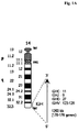
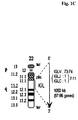
- chromosomal loci containing the immunoglobulin encoding genes are located on chromosomes 2, 14, and 22 (see Figure 1 ).
- the human immunoglobulin G heavy chain locus can be found on chromosome 14 (14q32.2) with the chromosomal orientation in the locus: telomere - 5'-end-V H -D-J H -C H -3'-end - centromere.
- the V H segments on the chromosome are classified as depicted in the following Table 1.
- Table 1 Grouping of the V H -genes into V H families according to Matsuda, F., et al., J. Exp. Med.
- V H family Number of family members Genes with open reading frame V H 1 14 9 / 11 V H 2 4 3 V H 3 65 22 V H 4 32 7 / 11 V H 5 2 2 V H 6 1 1 V H 7 5 1
- the locus for the human immunoglobulin G light chains of the types kappa ( ⁇ ) and lambda ( ⁇ ) is located on two different chromosomes, chromosomes 2 and 22.
- the kappa light chain locus can be found on the short arm of chromosome 2 (2p11.2) and comprises 40 functional V ⁇ -gene segments. These are grouped in seven families.
- the locus also comprises 5 J ⁇ -genes and a single C ⁇ -gene ( Schable, K.F. and Zachau, H.G., Biol. Chem. Hoppe Seyler 374 (1993) 1001-1022 ; Lefranc, M.P., Exp. Clin. Immunogenet. 18 (2001) 161-174 ).
- the lambda light chain locus can be found on the long arm of chromosome 22 (22p11.2) and comprises 73-74 V ⁇ -gene of which 30 are functional. These are grouped in ten families which in addition are grouped in three clusters. The locus also comprises 7 J ⁇ -genes, of which 5 are functional. Table 3: Grouping of the V ⁇ -genes into V ⁇ families according to Frippiat, J.P., et al., Hum. Mol. Genet. 4 (1995) 983-991; Farner, N.L., et al., J. Immunol. 162 (1999) 2137-2145; Lefranc, M.P., Exp. Clin. Immunogenet.
- V ⁇ family Number of functional genes Cluster V ⁇ 1 5 B V ⁇ 2 5 A V ⁇ 3 8 A V ⁇ 4 3 A-C V ⁇ 5 3 B V ⁇ 6 1 C V ⁇ 7 2 B V ⁇ 8 1 C V ⁇ 9 1 B V ⁇ 10 1 c
- the PCR-based amplification of the nucleic acid encoding an IgG HC and LC or at least the variable domain thereof from a single immunoglobulin producing cell is based on the single cell deposition of B-lymphocytes followed by a PCR based nucleic acid amplification with specific primer for the variable domain of the heavy and light chain.
- the outcome of the PCR is essentially depending on the employed PCR primer. At best the employed primer should cover all V-genes, should not be prone to dimer formation and should specifically bind to the cDNA encoding the immunoglobulin.
- the nucleic acid encoding an immunoglobulin variable domain is obtained from cDNA.
- the amplification of the nucleic acid encoding the heavy and light chain can be performed in one polymerase chain reaction.
- the primer are chosen in order to provide for the amplification of nucleic acids of approximately the same length in order to allow for the same PCR conditions.
- primer for the nucleic acid encoding the heavy chain are employed whereof one is binding in the heavy chain C H 1 region, thus, providing for a nucleic acid fragment of comparable size to that of the corresponding nucleic acid encoding the light chain.
- the 5'-primer employed in the first polymerase chain reaction bind in the coding region for the leader peptide of the immunoglobulin.
- the 5'-primer employed in the second polymerase chain reaction bind in the coding region for the first framework region of the immunoglobulin.
- the primer employed in the second polymerase chain reaction provide for overhangs encoding the translational start codon ATG for 5'-primer and/or the translational stop codon TTA for 3'-primer. This overhang is useful in an optional following overlapping polymerase chain reaction for the generation of nucleic acids for the in vitro translation of the obtained nucleic acid.
- the immunoglobulin variable domain can be an immunoglobulin heavy chain variable domain or an immunoglobulin kappa light chain variable domain or an immunoglobulin lambda light chain variable domain.
- the primer employed in the first polymerase chain reaction for obtaining a nucleic acid encoding an immunoglobulin heavy chain variable domain have the nucleic acid sequence of SEQ ID NO: 05 and/or 06, and SEQ ID NO: 07 and/or 08, and SEQ ID NO: 09, and SEQ ID NO: 10 and/or 11, and SEQ ID NO: 12, and SEQ ID NO: 13, and SEQ ID NO: 14 and/or 15.
- Table 4 Primer employed in the first polymerase chain reaction for obtaining a nucleic acid encoding an immunoglobulin heavy chain variable domain.
- Primer descriptiOn Sequence Denotation SEQ ID NO: V H primer binding in the leader peptide coding region TCACCATGGACTG(C/G)ACCTGGA V H L-1 05, 06 CCATGGACACACTTTG(C/T)TCCAC V H L-2 07, 08 TCACCATGGAGTTTGGGCTGAGC V H L-3 09 V H L-4 10, 11 ATGGGGTCAACCGCCATCCT V H L-5 12 ACAATGTCTGTCTCCTTCCTCAT V H L-6 13 primer binding in the constant region coding region GCCAGGGGGAAGAC(C/G)GATG huC H -II 14, 15
- the primer employed in the first polymerase chain reaction for obtaining a nucleic acid encoding an immunoglobulin kappa light chain variable domain have the nucleic acid sequence of SEQ ID NO: 16 to 20.
- Table 5 Primer employed in the first polymerase chain reaction for obtaining a nucleic acid encoding an immunoglobulin kappa light chain variable domain.
- the primer employed in the first polymerase chain reaction for obtaining a nucleic acid encoding an immunoglobulin lambda light chain variable domain have the nucleic acid sequence of SEQ ID NO: 21, and SEQ ID NO: 22, and SEQ ID NO: 23 and/or 24 and/or 25 and/or 26, and SEQ ID NO: 27 and/or 28.
- Table 6 Primer employed in the first polymerase chain reaction for obtaining a nucleic acid encoding an immunoglobulin lambda light chain variable domain.
- the primer employed in the second polymerase chain reaction for obtaining a nucleic acid encoding an immunoglobulin heavy chain variable domain have the nucleic acid sequence of SEQ ID NO: 29 and/or 30, and SEQ ID NO: 31, and SEQ ID NO: 32 and/or 33, and SEQ ID NO: 34 and/or 35, and SEQ ID NO: 36, and SEQ ID NO: 37 and/or 38, and SEQ ID NO: 39 and/or 40, and SEQ ID NO: 41, and SEQ ID NO: 42, and SEQ ID NO: 43 and/or 44, and SEQ ID NO: 45, and SEQ ID NO: 46 and/or 47, and SEQ ID NO: 48, and SEQ ID NO: 49 and/or 50, and SEQ ID NO: 51 and/or 52.
- Table 7 Primer employed in the second polymerase chain reaction for obtaining a nucleic acid encoding an immunoglobulin heavy chain variable domain.
- Primer description Sequence Denotation SEQ ID NO: V H L-1a 29, 30 V H L-1b 31 V H L-1c 32, 33 V H L-1d 34, 35 V H L-2a 36 V H primer binding in the FR1 coding region V H L-2b 37, 38 V H L-3a 39, 40 V H L-3b 41 V H L-3c 42 V H L-4a 43, 44 V H L-4b 45 V H L-5a 46, 47 V H L-6a 48 V H L-7a 49, 50 primer binding in the constant region coding region huC H -III 51, 52
- the primer employed in the second polymerase chain reaction for obtaining a nucleic acid encoding an immunoglobulin kappa light chain variable domain have the nucleic acid sequence of SEQ ID NO: 53 and/or 54, and SEQ ID NO: 55 and/or 56, and SEQ ID NO: 57 and/or 58, and SEQ ID NO: 59, and SEQ ID NO: 60, and SEQ ID NO: 61 and/or 62, and SEQ ID NO: 63 and/or 64, and SEQ ID NO: 65, and SEQ ID NO: 66, and SEQ ID NO: 67, and SEQ ID NO: 68, and SEQ ID NO: 69, and SEQ ID NO: 70, and SEQ ID NO: 71.
- Table 8 Primer employed in the second polymerase chain reaction for obtaining a nucleic acid encoding an immunoglobulin kappa light chain variable domain.
- Primer description Sequence Denotation SEQ ID NO: V ⁇ L-1a 53, 54 V ⁇ L-1b 55, 56 V ⁇ L-1c 57, 58 V ⁇ L-1d 59 V ⁇ L-2a 60 V ⁇ primer binding in the FR1 coding region V ⁇ L-2b 61, 62 V ⁇ L-3a 63, 64 V ⁇ L-3b 65 V ⁇ L-3c 66 V ⁇ L-4a 67 V ⁇ L-5a 68 V ⁇ L-6a 69 V ⁇ L-6b 70 primer binding in the constant region coding region huC ⁇ -III 71
- the primer employed in the second polymerase chain reaction for obtaining a nucleic acid encoding an immunoglobulin lambda light chain variable domain have the nucleic acid sequence of SEQ ID NO: 72, and SEQ ID NO: 73 and/or 74, and SEQ ID NO: 75, and SEQ ID NO: 76, and SEQ ID NO: 77 and/or 78, and SEQ ID NO: 79, and SEQ ID NO: 80, and SEQ ID NO: 81, and SEQ ID NO: 82 and/or 83, and SEQ ID NO: 84 and/or 85, and SEQ ID NO: 86, and SEQ ID NO: 87 and/or 88, and SEQ ID NO: 89, and SEQ ID NO: 90 and/or 91, and SEQ ID NO: 92, and SEQ ID NO: 93.
- the primer employed in the polymerase chain reaction provide for overhangs encoding the translational start codon ATG for 5'-primer and/or the translational stop codon TTA for 3'-primer. This overhang is useful in an optional following overlapping polymerase chain reaction for the generation of nucleic acids for the in vitro translation of the obtained nucleic acid.
- the immunoglobulin variable domain can be an immunoglobulin heavy chain variable domain or an immunoglobulin kappa light chain variable domain or an immunoglobulin lambda light chain variable domain.
- the primer employed in the first two-step polymerase chain reaction for obtaining a nucleic acid encoding an immunoglobulin heavy chain variable domain have the nucleic acid sequence of SEQ ID NO: 94, and SEQ ID NO: 95, and SEQ ID NO: 96 and/or 97 and/or 98 and/or 99, and SEQ ID NO: 100 and/or 101 and/or 102 and/or 103, and SEQ ID NO: 104 and/or 105 and/or 106.
- Table 10 Primer employed in the first two-step polymerase chain reaction for obtaining a nucleic acid encoding an immunoglobulin heavy chain variable domain.
- Primer description Sequence Denotation SEQ ID NO: V H primer huV H -1 94 binding in the FR1 coding region huV H -2 95 huV H -3 96, 97, 98, 99 huV H -4 100, 101, 102, 103 primer binding in the constant region coding region huC H -2 104, 105, 106
- the primer employed in the first two-step polymerase chain reaction for obtaining a nucleic acid encoding an immunoglobulin kappa light chain variable domain have the nucleic acid sequence of SEQ ID NO: 107 and/or 108 and/or 109 and/or 110, and SEQ ID NO: 111 and/or 112, and SEQ ID NO: 113, and SEQ ID NO: 114, and SEQ ID NO: 115.
- Table 11 Primer employed in the first two-step polymerase chain reaction for obtaining a nucleic acid encoding an immunoglobulin kappa light chain variable domain.
- Primer description Sequence Denotation SEQ ID NO: V ⁇ primer binding in the FR1 coding region huV ⁇ -1 107, 108, 109, 110 huV ⁇ -2 111,112 huV ⁇ -3 113 huV ⁇ -4 114 primer binding in the constant region coding region huC ⁇ -2 115
- the primer employed in the first two-step polymerase chain reaction for obtaining a nucleic acid encoding an immunoglobulin lambda light chain variable domain have the nucleic acid sequence of SEQ ID NO: 116, and SEQ ID NO: 117, and SEQ ID NO: 118 and/or 119, and SEQ ID NO: 120 and/or 121 and/or 122 and/or 123 and/or 124 and/or 125.
- Table 12 Primer employed in the first two-step polymerase chain reaction for obtaining a nucleic acid encoding an immunoglobulin lambda light chain variable domain.
- Primer description Sequence Denotation SEQ ID NO: V ⁇ primer binding in the FR1 coding region huV ⁇ -1 116 huV ⁇ -2 117 huV ⁇ -3 118, 119 primer binding in the constant region coding region huC ⁇ -2 120, 121, 122, 123, 124, 125
- the primer employed in the second two-step polymerase chain reaction have the nucleic acid sequence of SEQ ID NO: 126 and SEQ ID NO: 127.
- Table 13 Primer employed in the second two-step polymerase chain reaction.
- Primer description Sequence Denotation SEQ ID NO: first primer CTTTAAGAAGGAGATATACCATG LTGS-lfp 126 second primer ATCGTATGGGTAGCTGG LTGS-rfp 127
- Another aspect of the current invention is a method for obtaining a nucleic acid encoding at least an immunoglobulin variable domain from a single cell comprising the following steps:
- the 5'-primer employed in the first polymerase chain reaction bind in the coding region for the leader peptide of the immunoglobulin.
- the 5'-primer employed in the second polymerase chain reaction bind in the coding region for the first framework region of the immunoglobulin.
- the primer employed in the second polymerase chain reaction provide for overhangs encoding the translational start codon ATG for 5'-primer and/or the translational stop codon TTA for 3'-primer. This overhang is useful in an optional following overlapping polymerase chain reaction for the generation of nucleic acids for the in vitro translation of the obtained nucleic acid.
- an immunoglobulin variable domain encoding nucleic acid
- six 5'-primer and one 3'-primer are employed in the first polymerase chain reaction.
- four 5'-primer and one 3'-primer are employed in the second polymerase chain reaction.
- the immunoglobulin variable domain is an immunoglobulin heavy chain variable domain or an immunoglobulin kappa light chain variable domain or an immunoglobulin lambda light chain variable domain.
- the primer employed in the first polymerase chain reaction for obtaining a nucleic acid encoding an immunoglobulin heavy chain variable domain have the nucleic acid sequence of SEQ ID NO: 05 and/or 06, and SEQ ID NO: 07 and/or 08, and SEQ ID NO: 09, and SEQ ID NO: 10 and/or 11, and SEQ ID NO: 12, and SEQ ID NO: 13, and SEQ ID NO: 104 and/or 105 and/or 106.
- Table 14 Primer employed in the first polymerase chain reaction for obtaining a nucleic acid encoding an immunoglobulin heavy chain variable domain.
- the primer employed in the first polymerase chain reaction for obtaining a nucleic acid encoding an immunoglobulin kappa light chain variable domain have the nucleic acid sequence of SEQ ID NO: 16, and SEQ ID NO: 17, and SEQ ID NO: 18, and SEQ ID NO: 19, and SEQ ID NO: 115.
- Table 15 Primer employed in the first polymerase chain reaction for obtaining a nucleic acid encoding an immunoglobulin kappa light chain variable domain.
- Primer description Sequence Denotation SEQ ID NO: V ⁇ primer binding in the leader peptide coding region GCTCAGCTCCTGGGGCTCCTG V ⁇ L-1 16 CTGGGGCTGCTAATGCTCTGG V ⁇ L-2 17 TTCCTCCTGCTACTCTGGCTC V ⁇ L-3 18 CAGACCCAGGTCTTCATTTCT V ⁇ L-4 19 primer binding in the constant region coding region huC ⁇ -2 115
- the primer employed in the first polymerase chain reaction for obtaining a nucleic acid encoding an immunoglobulin lambda light chain variable domain have the nucleic acid sequence of SEQ ID NO: 21, and SEQ ID NO: 22, and SEQ ID NO: 23 and/or 24 and/or 25 and/or 26, and SEQ ID NO: 120 and/or 121 and/or 122 and/or 123 and/or 124 and/or 125.
- Table 16 Primer employed in the first polymerase chain reaction for obtaining a nucleic acid encoding an immunoglobulin lambda light chain variable domain.
- Primer description Sequence Denotation SEQ ID NO: V ⁇ primer binding in the leader peptide coding region CCTCTCCTCCTCACCCTCCT V ⁇ L-1 21 CTCCTCACTCAGGGCACA V ⁇ L-2 22 ATGGCCTGGA(T/C)C(C/G)CTCTCC V ⁇ L-3 23, 24, 25, 26 primer binding in the constant region coding region huC ⁇ -2 120, 121, 122, 123, 124, 125
- the primer employed in the second polymerase chain reaction for obtaining a nucleic acid encoding an immunoglobulin heavy chain variable domain have the nucleic acid sequence of SEQ ID NO: 128, and SEQ ID NO: 129, and SEQ ID NO: 130, and SEQ ID NO: 131, and SEQ ID NO: 132, and SEQ ID NO: 133, and SEQ ID NO: 134, and SEQ ID NO: 135, and SEQ ID NO: 136, and SEQ ID NO: 137, and SEQ ID NO: 138, and SEQ ID NO: 139, and SEQ ID NO: 140, SEQ and ID NO: 141, and SEQ ID NO: 104 and/or 105 and/or 106.
- Table 17 Primer employed in the second polymerase chain reaction for obtaining a nucleic acid encoding an immunoglobulin heavy chain variable domain.
- the primer employed in the second polymerase chain reaction for obtaining a nucleic acid encoding an immunoglobulin kappa light chain variable domain have the nucleic acid sequence of SEQ ID NO: 53 and/or 54, and SEQ ID NO: 55 and/or 56, and SEQ ID NO: 57 and/or 58, and SEQ ID NO: 59, and SEQ ID NO: 60, and SEQ ID NO: 61 and/or 62, SEQ ID NO: 63 and/or 64, and SEQ ID NO: 65, and SEQ ID NO: 66, and SEQ ID NO: 67, and SEQ ID NO: 68, and SEQ ID NO: 69, and SEQ ID NO: 70, and SEQ ID NO: 115.
- Table 18 Primer employed in the second polymerase chain reaction for obtaining a nucleic acid encoding an immunoglobulin kappa light chain variable domain.
- Primer description Sequence Denotatio n SEQ ID NO: V ⁇ L-1a 53, 54 V ⁇ L-1b 55, 56 V ⁇ L-1c 57, 58 V ⁇ L-1d 59 V ⁇ L-2a 60 V ⁇ primer binding in the FR1 coding region V ⁇ L-2b 61, 62 V ⁇ L-3a 63, 64 V ⁇ L-3b 65 V ⁇ L-3c 66 V ⁇ L-4a 67 V ⁇ L-5a 68 V ⁇ L-6a 69 V ⁇ L-6b 70 primer binding in the constant region coding region huC ⁇ -2 115
- the primer employed in the second polymerase chain reaction for obtaining a nucleic acid encoding an immunoglobulin lambda light chain variable domain have the nucleic acid sequence of SEQ ID NO: 72, and SEQ ID NO: 73 and/or 74, and SEQ ID NO: 75, and SEQ ID NO: 76, and SEQ ID NO: 77 and/or 78, and SEQ ID NO: 79, and SEQ ID NO: 80, and SEQ ID NO: 81, and SEQ ID NO: 82 and/or 83, and SEQ ID NO: 84 and/or 85, and SEQ ID NO: 86, and SEQ ID NO: 87 and/or 88, and SEQ ID NO: 89, and SEQ ID NO: 90 and/or 91, and SEQ ID NO: 92, and SEQ ID NO: 120 or 121 or 122 or 123 or 124 or 125.
- Table 19 Primer employed in the second polymerase chain reaction for obtaining a nucleic acid encoding an immunoglobulin lambda light chain variable domain.
- Primer description Sequence Denotatio n SEQ ID NO: V ⁇ L-1a 72 V ⁇ L-1b 73, 74 V ⁇ L-1c 75 V ⁇ L-2 76 V ⁇ L-3a 77, 78 V ⁇ L-3b 79 V ⁇ primer binding in the FR1 coding region V ⁇ L-3c 80 V ⁇ L-3d 81 V ⁇ L-4 82, 83 V ⁇ L-5 84, 85 V ⁇ L-6 86 V ⁇ L-7 87, 88 V ⁇ L-8 89 V ⁇ L-4/9 90, 91 V ⁇ L-10 92 primer binding in the constant region coding region huC ⁇ -2 120, 121, 122, 123, 124, 125
- nucleic acid encoding the light chain variable domain and nucleic acid encoding the heavy chain variable domain are obtained in one polymerase chain reaction by a combination of the different 5'- and 3'-primer in a single multiplex polymerase chain reaction.
- the 5'-primer employed in the multiplex one tube reverse transcription gene specific primer polymerase chain reaction binds in the coding region for the first framework region of the immunoglobulin.
- the primer employed in the RT-GSP-PCR reaction provide for overhangs encoding the translational start codon ATG for the 5'-primer and/or the translational stop codon TTA for the 3'-primer. This overhang is useful in an optional following overlapping polymerase chain reaction for the generation of nucleic acids for the in vitro translation of the obtained nucleic acid.
- This method is for obtaining an immunoglobulin heavy chain variable domain with a RT-GSP-PCR reaction.
- the immunoglobulin variable domain can be an immunoglobulin heavy chain variable domain or an immunoglobulin kappa light chain variable domain or an immunoglobulin lambda light chain variable domain.
- the primer employed in the multiplex one tube RT-GSP-PCR for obtaining a nucleic acid encoding an immunoglobulin heavy chain variable domain have the nucleic acid sequence of SEQ ID NO: 142 and 143.
- Table 20 Primer employed in the multiplex one tube RT-GSP-PCR reaction for obtaining a nucleic acid encoding an immunoglobulin heavy chain variable domain.
- Primer description Sequence Denotation SEQ ID NO: V H primer binding in the FR1 coding region V H -lfp 142 primer binding in the constant region coding region V H -rfp 143
- the primer employed in the multiplex one tube RT-GSP-PCR for obtaining a nucleic acid encoding an immunoglobulin kappa light chain variable domain have the nucleic acid sequence of SEQ ID NO: 144 and 145.
- Table 21 Primer employed in the multiplex one tube RT-GSP-PCR reaction for obtaining a nucleic acid encoding an immunoglobulin kappa light chain variable domain.
- Primer description Sequence Denotation SEQ ID NO: V ⁇ primer binding in the FR1 coding region VL(k)-lfp 144 primer binding in the constant region coding region VL(k)-rfp 145
- the nucleic acids encoding the cognate immunoglobulin VH and VL domains can be obtained from a single cell whereby the encoded immunoglobulin variable domain is provided as Fab fragment in quantities sufficient for the characterization of the immunoglobulin's binding properties.
- the individual PCR polymerase chain reaction
- the individual PCR has to be very sensitive and a combination of more than one PCR has to be performed.
- a “cell-free in vitro translation system” denotes a cell-free lysate of a prokaryotic or eukaryotic, preferably of a prokaryotic, cell containing ribosomes, tRNA, ATP, CGTP, nucleotides, and amino acids.
- the prokaryote is E.coli.
- Cell-free in vitro translation is a method which has been known in the state of the art for a long time.
- Spirin et al. developed in 1988 a continuous-flow cell-free (CFCF) translation and coupled transcription/translation system in which a relatively high amount of protein synthesis occurs ( Spirin, A.S., et al., Science 242 (1988) 1162-1164 ).
- CFCF continuous-flow cell-free
- cell lysates containing ribosomes were used for translation or transcription/translation.
- Such cell-free extracts from E.coli were developed by, for example, Zubay ( Zubay, G., et al., Ann. Rev.
- one aspect of the current invention is a method for producing an immunoglobulin Fab fragment comprising the following steps:
- the nucleic acid encoding the immunoglobulin variable domains is obtained with a method according to the previous aspects of the current invention.
- the obtaining the nucleic acid encoding the immunoglobulin light and heavy chain variable domain form a single cell comprises a multiplex polymerase chain reaction according to the invention for the amplification of cognate IgG HC and IgG LC (human IgG isotype) from a single B-cell.
- IgG HC and IgG LC human IgG isotype
- a method employing the following steps i) isolating with magnetic micro-beads coated with the human CD 19 B-cells from peripheral blood, ii) depositing single cells by limited dilution or FACS, iii) extracting the mRNA of the individualized B-cells, iv) obtaining the nucleic acid encoding at least the variable domains of the immunoglobulin produced by the individualized B-cell, v) in vitro translating the linear mRNA template, and optionally vi) characterizing the binding properties of the immunoglobulin or immunoglobulin fragment.
- nucleic acids encoding the variable domain of the light and heavy immunoglobulin chain are further modified.
- nucleic acid encoding the variable domain is combined with a nucleic acid encoding an immunoglobulin constant region.
- the nucleic acid encoding the light chain variable domain can be combined with a nucleic acid encoding human kappa light chain constant domain of SEQ ID NO: 03 or with a nucleic acid encoding human lambda light chain variable domain of SEQ ID NO: 04.
- the nucleic acid encoding the heavy chain variable domain can be combined with a nucleic acid encoding human immunoglobulin G1 (IgG1) constant region of SEQ ID NO: 01 or with a nucleic acid encoding human immunoglobulin G4 (IgG4) constant region of SEQ ID NO: 02.
- the nucleic acid encoding the heavy chain variable domain is combined with a nucleic acid encoding human immunoglobulin G1 (IgG1) constant region 1 (C H 1).
- the nucleic acid molecules encoding the complete immunoglobulin heavy and light chain or a fragment thereof are in the following referred to as structural genes. They can be located on the same expression plasmid or can alternatively be located on different expression plasmids.
- the assembly of the immunoglobulin or Fab-fragment takes place before the secretion of the immunoglobulin to the cultivation medium and, thus, within the expressing cell. Therefore, the nucleic acid molecules encoding the immunoglobulin chains are expressed in the same host cell. If after recombinant expression a mixture of immunoglobulins is obtained, these can be separated and purified by methods known to a person skilled in the art. These methods are well established and widespread used for immunoglobulin purification and are employed either alone or in combination.
- Such methods are, for example, affinity chromatography using microbial-derived proteins (e.g. protein A or protein G affinity chromatography), ion exchange chromatography (e.g. cation exchange (carboxymethyl resins), anion exchange (amino ethyl resins) and mixed-mode exchange chromatography), thiophilic adsorption (e.g. with beta-mercaptoethanol and other SH ligands), hydrophobic interaction or aromatic adsorption chromatography (e.g. with phenyl-sepharose, aza-arenophilic resins, or m-aminophenylboronic acid), metal chelate affinity chromatography (e.g.
- microbial-derived proteins e.g. protein A or protein G affinity chromatography
- ion exchange chromatography e.g. cation exchange (carboxymethyl resins), anion exchange (amino ethyl resins) and mixed-mode exchange chromatography
- Ni(II)- and Cu(II)-affinity material size exclusion chromatography
- preparative electrophoretic methods such as gel electrophoresis, capillary electrophoresis
- the conjugates can be tailor-made on the nucleic acid/gene level.
- the nucleic acid sequences encoding immunoglobulin light and heavy chains are known and can be obtained for example from genomic databases.
- the elements required for the construction of an expression plasmid for the expression of the immunoglobulin obtained with a method according to the invention are, for example, an expression cassette for the immunoglobulin light chain, an expression cassette for the immunoglobulin heavy chain (alternatively the light chain and the heavy chain structural genes can be contained in the same expression cassette, e.g. as bicistronic expression element), a selection marker, and an E.coli replication as well as selection unit.
- An expression cassette comprises in general a promoter, a DNA segment encoding a secretion signal sequence, the structural gene, and a terminator/polyadenylation signal.
- the elements are assembled in an operatively linked form either on one plasmid encoding all chains of the immunoglobulin, or on two plasmids each encoding one chain of the immunoglobulin.
- the plasmid(s) is (are) introduced into a suitable host cell. Proteins are produced in mammalian cells such as CHO cells, NS0 cells, Sp2/0 cells, COS cells, HEK cells, K562 cells, BHK cells, PER.C6® cells, and the like.
- the conjugate is expressed in a CHO cell, or a BHK cell, or a HEK cell, or NS0 cell.
- the regulatory elements of the plasmid have to be selected in a way that they are functional in the selected host cell.
- the host cell is cultivated under conditions suitable for the expression of the immunoglobulin, which are known to a person of skill in the art.
- the expressed immunoglobulin chains are functionally assembled and the fully processed immunoglobulin is secreted into the medium.
- an "expression plasmid” is a nucleic acid providing all required elements for the expression of the comprised structural gene(s) in a host cell.
- an expression plasmid comprises a prokaryotic plasmid propagation unit, e.g. for E. coli, comprising an origin of replication, and a selectable marker, an eukaryotic selection marker, and one or more expression cassettes for the expression of the structural gene(s) of interest each comprising a promoter, a structural gene, and a transcription terminator including a polyadenylation signal.
- Gene expression is usually placed under the control of a promoter, and such a structural gene is said to be “operably linked to" the promoter.
- a regulatory element and a core promoter are operably linked if the regulatory element modulates the activity of the core promoter.
- “Operably linked” refers to a juxtaposition of two or more components, wherein the components so described are in a relationship permitting them to function in their intended manner.
- the term “linking ... in operable form” denotes the combination of two or more individual nucleic acids in a way that the individual nucleic acids are operably linked in the final nucleic acid.
- a promoter and/or enhancer are operably linked to a coding sequence, if it acts in cis to control or modulate the transcription of the linked sequence.
- the DNA sequences that are "operably linked” are contiguous and, where necessary to join two protein encoding regions such as first domain and a second domain, e.g.
- a polyadenylation site is operably linked to a coding sequence if it is located at the downstream end of the coding sequence such that transcription proceeds through the coding sequence into the polyadenylation sequence.
- a translation stop codon is operably linked to an exonic nucleic acid sequence if it is located at the downstream end (3' end) of the coding sequence such that translation proceeds through the coding sequence to the stop codon and is terminated there. Linking is accomplished by recombinant methods known in the art, e.g., using PCR methodology and/or by ligation at convenient restriction sites. If convenient restriction sites do not exist, then synthetic oligonucleotide adaptors or linkers are used in accord with conventional practice.
- under conditions suitable for the expression of denotes conditions which are used for the cultivation of a cell capable of expressing a heterologous polypeptide and which are known to or can easily be determined by a person skilled in the art. It is known to a person skilled in the art that these conditions may vary depending on the type of cell cultivated and type of polypeptide expressed. In general the cell is cultivated at a temperature, e.g. between 20 °C and 40 °C, and for a period of time sufficient to allow effective production of the conjugate, e.g. for of from 4 days to 28 days, in a volume of 0.01 liter to 10 7 liter.
- Samples used in this approach are B-cells and plasma cells isolated from the peripheral blood of healthy donor and tissue (spleen, bone marrow) of transgenic mice for human IgG.
- Solid tissue is first of all manually disaggregated in DMEM in separate tubes. In the later steps, gentle handling and low temperature minimize cell lysis, which is important for the future positive isolation of the cells of interest and to keep the source of mRNA intact.
- Disaggregated tissue is suspended by the delicate addition of cell separation media for making of a different cell type gradient (Leucosep-tubes (Greiner Bio-One) with Ficoll density gradient). Suspended cells are purified by centrifugation on the cold separation medium for 20 min.
- PBMC plasma cells
- lymphocytes are washed in cold buffer (PBS (phosphate buffered saline), 0.1 % (w/v) BSA (bovine serum albumin), 2 mM EDTA (ethylene diamin tetra acetate)) and the supernatant is carefully discarded to keep only the lymphocytes. Lymphocytes are than resuspended in PBS and mixed by carefully pipetting. Centrifugation is effectuated for 5 min. at 800 x g and 22 °C to pellet the cells.
- PBS phosphate buffered saline
- BSA bovine serum albumin
- 2 mM EDTA ethylene diamin tetra acetate
- B-cells and plasma cells are pretreated with murine and human FC blocker to block unspecific binding of Abs on their cells surface.
- Cells are washed once with buffer (PBS, 0.1 % (w/v) BSA, 2 mM EDTA), centrifuged and resuspended in PBS. Only the CD19+ B-cells and CD138+ plasma cells were used. To prevent mRNA degradation an RNAse Inhibitor is added.
- the positive isolation of the CD 19+ B-cells (Dynal Biotech Dynabeads CD 19 Pan B) from the mouse spleen has been carried out according to the manufacturer's instructions.
- the selection of the CD138+ plasma cells (StemCell Technologies EasySep Human CD138 Selection Kit) has been carried out following the manufacturer's instructions.
- Cells are counted and, by the principle of the limiting-dilution culture, deposited as single cell into the wells of 96-well PCR plates or 384-well plates. Plates are sealed with PCR Film and immediately placed on ice. Sorted cells can be used immediately in RT-PCR (reverse transcriptase polymerase chain reaction) or stored at -20 °C for short-term use or -80 °C for long-term use. Single-cell sorting was performed on a FACSAria cell-sorting system (Becton Dickinson). Cells that stained positive for CD19, highly positive for CD38 and intermediately positive for CD45 were collected and designated plasma cells (PC).
- PC plasma cells
- Table 22 Components used for the classical lysis. Component Volume ( ⁇ l) Final concentration Water, PCR grade 1.75 5xRT Reaction Buffer 1.00 RNAse Inhibitor (40 U/ ⁇ l) 0.25 5 U Gene Specific antisense primer 1.00 0.02 ⁇ M Igepal 0,01% 1.00 0.01 % Final volume 5.00
- Table 23 Block cycler program for the cell lysis. Temperature (°C) Time (sec) 65 60 55 30 45 30 35 20 23 120 4 ⁇
- RT plates are briefly centrifuged and placed from ice to 55 °C for 60 min. (with heated lid), heated to 85 °C for 2 min. (to inactivate the reverse transcriptase), in a Block cycler (LightCycler 480, Roche Diagnostics GmbH, Mannheim, Germany). Single-stranded cDNA was stored at -20 °C shortly after the reverse transcription reaction to avoid degradation of the cDNA. A control synthesis reaction was simultaneously performed without cells to test for contamination. Table 24: Components used for the reverse transcription reaction.
- B-cells and plasma cells must be lysed before the reverse transcription reaction.
- Table 25 Block cycler program for the first PCR. Temperature (°C) Time Number of cycles 95 2 min. 1 94 15 sec. 35 55 30 sec. 72 1 min. 72 10 min. 1 4 ⁇
- Table 26 Primer used for the first PCR. Ig heavy chain primer Ig light chain ( ⁇ ) primer Ig light chain ( ⁇ ) primer V H L-1 V ⁇ L-1 V ⁇ L-1 V H L-2 V ⁇ L-2 V ⁇ L-2 V H L-3 V ⁇ L-3 V ⁇ L-3 V H L-4 VK ⁇ -4 huC ⁇ -II V H L-5 huC ⁇ -II V H L-6 huC H -II
- the ⁇ and ⁇ light chains and the heavy chains were subsequently amplified with a second-round PCR according to the following protocol.
- the second PCR was performed to increase the amount of cDNA copies and to amplify only from the variable part of the light chain (LC) and heavy chain (HC) to the C H 1 region.
- the HC amplification was performed using 16 primer, the kappa LC using 17 primer, and the lambda LC using 14 primer.
- the genes were amplified in a total volume of 20 ⁇ l using 2 ⁇ l of the first PCR product, 10 ⁇ l of High Fidelity PCR Master (containing 0.4 ⁇ M each dNTPs, double concentrated reaction buffer (with 3 mM MgCl 2 ), 0.02 ⁇ M of each primer, all from the High Fidelity PCR Master Kit (Roche Diagnostics GmbH, Mannheim, Germany) using the following second PCR program: 2 min. at 95 °C, 45 cycles of 94 °C for 15 sec., 55 °C for 30 sec., 72 °C for 1 min., following 10 min. at 72 °C. Table 27: Components used for the second PCR.
- B-cells and plasma cells must be distributed directly into the wells of 96-well PCR plates (Eppendorf), containing all the necessary PCR reagents in a volume of 10 ⁇ l, except for reverse transcriptase, DNA polymerase, buffer and dNTPs and frozen at -80 °C for later processing.
- Reverse transcription and PCR were performed in one step (one step Multiplex RT-PCR).
- the isolated, sorted and stored cells were used as raw material for the reverse transcription or RT-PCR. All necessary reagents were thawed at room temperature. All primer were synthesized in the MOLBIOL TIB GmbH laboratories. The plates and all other reagents were kept on ice during the entire procedure. For cDNA syntheses the gene specific primer with extensions were used directly.
- the enzyme complex consists of two Sensiscript reverse transcriptases and one Omniscript polymerase (Qiagen OneStep RT PCR).
- the rewriting of the mRNA into cDNA was performed by the Sensiscript complex (Qiagen OneStep RT PCR) and the amplification of the cDNA was performed using the HotStarTaq DNA Polymerase (Qiagen OneStep RT PCR), which is a chemically form of a recombinant 94 kDa DNA polymerase (deoxynucleoside-triphosphate: DNA deoxynucleotidyltransferase, EC 2.7.7.7), originally isolated from Thermus aquaticus expressed in E. coli.
- the cells were sorted in a 96-well PCR plate and stored in a volume of 10 ⁇ l, containing 5 ⁇ l PCR H 2 O grade, 1 ⁇ l 0.1 ⁇ M primer for VH and VL, 1 ⁇ l RNAse inhibitor 20 U/reaction and 3 ⁇ l Tris 1.5 mM. Before adding the other 10 ⁇ l for performing the PCR reaction, the cells stored at -60 °C were briefly centrifuged (20 sec. at 1400 rpm) to collect the liquid and cells on the bottom of the wells. Table 30: Master Mix 1 used for the RT-PCR.
- Master Mix 1 Final concentration/well volume/well ( ⁇ l) H 2 O 5 primer V H / VL(k) 0.1 ⁇ M 1 RNAse Inhibitor 20 U/reaction 1 Tris-buffer 1.5 mM 3 B/Plasma cells final volume 10
- Table 31 Master Mix 2 used for the RT-PCR. Master Mix 2 Final concentration/well volume/well ( ⁇ l) H 2 O 1x 2.2 5x Buffer 1x 4 dNTP 10 mM each 400 ⁇ M each 0.8 5x Q-Solution 0.25x 1 One Step RT PCR Enzyme mix 1.2 RNAse Inhibitor 20U 1 final volume 10
- the second Master Mix contained 2.2 ⁇ l H 2 O PCR grade, 4 ⁇ l of 1x buffer, 0.8 ⁇ l of dNTPs 400 ⁇ M each, 1 ⁇ l of Q-solution 0.25x, 1.2 ⁇ l of the enzyme complex and 1 ⁇ l of RNAse inhibitor 20U.
- Table 32 Primer used for the RT-PCR.
- the purification of the previously amplified PCR products was performed by removing unincorporated primer, dNTPs, DNA polymerases and salts used during PCR amplification in order to avoid interference in downstream applications.
- Agencourt AMPure was used.
- the buffer is optimized to selectively bind PCR amplicons 100 bp and larger to paramagnetic beads. Excess oligonucleotides, nucleotides, salts, and enzymes can be removed using a simple washing procedure.
- the resulting purified PCR product is essentially free of contaminants and can be used in the following applications: Fluorescent DNA sequencing (including capillary electrophoresis), microarray spotting, cloning and primer extension genotyping.
- Fluorescent DNA sequencing including capillary electrophoresis
- microarray spotting cloning and primer extension genotyping.
- the work flow for 96-well format started with gently shaking the beads stored in buffer to resuspend any magnetic particle that may have settled.
- the correct volume of 36 ⁇ l of beads solution was added to the 20 ⁇ l of sample and the mix was pipetted 10 times up and down.
- the following step was incubating for 10 minutes and afterwards the reaction plate was placed onto a magnetic plate for 10 minutes to separate beads from solution.
- the cleared solution (supernatant) was aspirated from the reaction plate and discard.
- the amplified DNA of the second PCR was afterwards linked by an overlapping extension PCR method with the following components, necessary for the transcription/translation step: a ribosome binding site (RBS), a T7 promoter and a T7 terminator sequences.
- RBS ribosome binding site
- T7 promoter a T7 promoter
- T7 terminator sequences 2 ⁇ l of the second PCR were taken to a final volume of 20 ⁇ l containing: 10.7 ⁇ l water, 2 ⁇ l of 10x reaction buffer with MgCl 2 (10 mM), 0.8 ⁇ l of DMSO, 0.5 ⁇ l dNTPs (10 mM each), 1.6 ⁇ l T7 promotor and terminator primer (6 ⁇ M each), 0.4 ⁇ l C-terminal HA-Tag primer and 0.4 ⁇ l of enzyme blend, all from the RTS E.coli Linear Template Generation Set, HA-Tag (Roche Diagnostics GmbH, Mannheim, Germany).
- the in vitro coupled transcription and translation was carried out following the manufacturer's protocol RTS 100 E.coli Disulfide Kit (Roche Diagnostics GmbH, Mannheim, Germany) with components as reported (see Table 12). 4 ⁇ l of each overlapping PCR product was transcribed and translated in a total volume of 50 ⁇ l, at 37 °C for 20 hours in the RTS Proteo Master Instrument (Roche Diagnostics GmbH, Mannheim, Germany). A control reaction was performed under identical conditions without cDNA template. GFP (green fluorescent protein) vectors were added to the reaction system for autoradiography as positive control.
- RTS 100 E.coli Disulfide Kit Roche Diagnostics GmbH, Mannheim, Germany
- a 384-well plate (Nunc GmbH & Co. KG, Thermo Fisher Scientific, Langenselbold, Germany) was coated with 50 ⁇ l (1:1000 in PBS) goat anti-human IgG Fab fragment (produced by Bethyl Laboratories Inc., obtained from Biomol GmbH, Hamburg, Germany, 1 mg/l ml) incubated at 4 °C overnight.
- the plate was washed three times with washing solution (100 ⁇ l PBST (phosphate buffered saline Tween-20)) and 60 ⁇ l of Blocking solution (0.25% CroteinC (w/v)/0.5% Tween (w/v)/PBS) was added, incubated for 1 h at room temperature.
- PBST phosphate buffered saline Tween-20
- Another washing step (3x100 ⁇ l PBST) was performed and 37.5 ⁇ l sample was transferred, as well as 37.5 ml negative control (negative control from the in vitro transcription/translation) and 37.5 ⁇ l positive control, containing 0.75 ⁇ l of human recombinant Fab fragment (Roche Diagnostics GmbH, Mannheim, Germany). The samples were titrated to a 1:3 dilution. The plate was incubated for 1.5 h at room temperature. After a washing step (3x100 ⁇ l PBST), 25 ⁇ l goat anti-human IgG F(ab') 2 (Dianova, Hamburg, Germany; 0.8 mg/ml (1:2000 diluted in Blocking Solution)) was added and incubated for 1 h at room temperature.
- the last washing step (3x100 ⁇ l PBST) was performed and 25 ⁇ l of TMB (POD Substrate, Roche Diagnostics GmbH, Mannheim, Germany, Art-No: 1 484 281) was pipetted into each well. After 2-3 minutes the absorption signal was detected at 405 nm and 495 nm (Tecan, Safire 2; Tecan Kunststoff GmbH, Crailsheim, Germany).
- TMB POD Substrate, Roche Diagnostics GmbH, Mannheim, Germany, Art-No: 1 484 281
- the IOTest Beta Mark was used for Vb analysis (Imunotech/Beckman Coulter). Streptavidin conjugated FITC, PE, or APC (all BD Pharmingen) were used for visualization of biotinylated antibodies. Dead cells were excluded by propidium iodide staining. Appropriate isotype-matched, irrelevant control mAbs were used to determine the level of background staining. Cells were analyzed using a FACS Calibur and sorted using a FACSAria (Becton Dickinson Immunocytometry Systems, Mountain View, CA, USA).
- Single B-cells of a mouse having a human immunoglobulin locus have been obtained as outlined above.
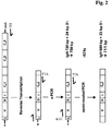
- the 3'-primer of the first and second primer set employed in the polymerase chain reaction were identical.
- the 5'-primer of the first and second primer set different insofar as the primer of the first 5'-primer set bound in the region encoding the leader peptide and the primer of the second 5'-primer set bound in the FR1 region.
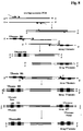
- a scheme of this polymerase chain reaction is given in Figure 2 .
- the employed sets of primer for the first polymerase chain reaction are denoted in the following list: Ig heavy chain primer Ig light chain ( ⁇ ) primer Ig light chain ( ⁇ ) primer VHL-1 V ⁇ L-1 V ⁇ L-1 V H L-2 V ⁇ L-2 V ⁇ L-2 V H L-3 V ⁇ L-3 V ⁇ L-3 V H L-4 V ⁇ L-4 huC ⁇ -III V H L-5 huC ⁇ -II V H L-6 huC H -II
- the employed sets of primer for the second polymerase chain reaction are denoted in the following list: IgG heavy chain primer IgG light chain ( ⁇ ) primer IgG light chain ( ⁇ ) primer V H -1a SEQ ID NO: 128 V ⁇ L-1a SEQ ID NO: 53, 54 V ⁇ L-1a SEQ ID NO: 72 V H -1b SEQ ID NO: 129 V ⁇ L-1b SEQ ID NO: 55, 56 V ⁇ L-1b SEQ ID NO: 73, 74 V H -1c SEQ ID NO: 130 V ⁇ L-1c SEQ ID NO: 57, 58 V ⁇ L-1c SEQ ID NO: 75 V H -1d SEQ ID NO: 131 V ⁇ L-1d SEQ ID NO: 59 V ⁇ L-2 SEQ ID NO: 76 V H -2a SEQ ID NO: 132 V ⁇ L-2a SEQ ID NO: 60 V ⁇ L-3a SEQ ID NO: 77, 78 V H -2b SEQ ID NO: 133
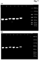
- Figure 3 the agarose gel of the amplified nucleic acid fragments obtained in this polymerase chain reaction is shown.
- the samples were analyzed after 40 amplification cycles with an annealing temperature of 50 °C and 55 °C, respectively.
- the blanks (water) were negative and the size of the fragments correlated well to the expected sizes of 750 bp (IgG HC) and 665 bp (IgG LC), respectively, after the first polymerase chain reaction and of 711 bp and 688 bp, respectively, after the second polymerase chain reaction.
- the employed sets of primer for the first polymerase chain reaction are denoted in the following list: Ig heavy chain primer Ig light chain ( ⁇ ) primer Ig light chain ( ⁇ ) primer V H L-1 SEQ ID NO: 05, 06 V ⁇ L-1 SEQ ID NO: 16 V ⁇ L-1 SEQ ID NO: 21 V H L-2 SEQ ID NO: 07, 08 V ⁇ L-2 SEQ ID NO: 17 V ⁇ L-2 SEQ ID NO: 22 V H L-3 SEQ ID NO: 09 V ⁇ L-3 SEQ ID NO: 18 V ⁇ L-3 SEQ ID NO: 23, 24, 25, 26 V H L-4 SEQ ID NO: 10, 11 V ⁇ L-4 SEQ ID NO: 19 huC ⁇ -II SEQ ID NO: 27, 28 V H L-5 SEQ ID NO: 12 huC ⁇ -II SEQ ID NO: 20 V H L-6 SEQ ID NO: 13 huC H -II SEQ ID NO: 14, 15
- the employed sets of primer for the second polymerase chain reaction are denoted in the following list: IgG heavy chain primer IgG light chain ( ⁇ ) primer IgG light chain ( ⁇ ) primer V H L-1a SEQ ID NO: 29, 30 V ⁇ L-1a SEQ ID NO: 53, 54 V ⁇ L-1a SEQ ID NO: 72 V H L-1b SEQ ID NO: 31 V ⁇ L-1b SEQ ID NO: ?55, 56 V ⁇ L-1b SEQ ID NO: 73, 74 V H L-1c SEQ ID NO: 32, 33 V ⁇ L-1c SEQ ID NO: 57, 58 V ⁇ L-1c SEQ ID NO: 75 V H L-1d SEQ ID NO: 34, 35 V ⁇ L-1d SEQ ID NO: 59 V ⁇ L-2a SEQ ID NO: 76 V H L-2a SEQ ID NO: 36 V ⁇ L-2a V ⁇ L-2a SEQ ID NO: 60 V ⁇ L-3a V ⁇ L-3a SEQ ID NO:
- Figure 5 the agarose gel of the amplified nucleic acid fragments obtained in this polymerase chain reaction is shown.
- the samples were analyzed after 40 amplification cycles with an annealing temperature of 50 °C and 55 °C, respectively.
- the blanks (water) were negative and the size of the fragments correlated well to the expected sizes of 471 bp (IgG HC) and 413 bp (IgG LC), respectively, after the first polymerase chain reaction and of 442 bp and 399 bp, respectively, after the second polymerase chain reaction.
- the employed sets of primer for the first and second polymerase chain reaction are denoted in the following list: Ig heavy chain primer Ig light chain ( ⁇ ) primer Ig light chain ( ⁇ ) primer huV H -1 SEQ ID NO: 94 huV ⁇ -1 SEQ ID NO: 107, 108, 109, 110 huV ⁇ -1 SEQ ID NO: 116 huV H -2 SEQ ID NO: 95 huV ⁇ -2 SEQ ID NO: 111,112 huV ⁇ -2 SEQ ID NO: 117 huV H -3 SEQ ID NO: 96,97,98,99 huV ⁇ -3 SEQ ID NO: 113 huV ⁇ -3 SEQ ID NO: 118,119 huV H -4 SEQ ID NO: 100, 101, 102, 103 huV ⁇ -4 SEQ ID NO: 114 huC ⁇ -2 SEQ ID NO: 120, 121, 122, 123
- Figure 7 the agarose gel of the amplified nucleic acid fragments obtained in this polymerase chain reaction is shown.
- the samples were analyzed after 40 amplification cycles with an annealing temperature of 50 °C and 55 °C, respectively.
- the blanks (water) were negative and the size of the fragments correlated well to the expected sizes of 711 bp (IgG HC) and 688 bp (IgG LC), respectively, after the first and second polymerase chain reaction.
- first polymerase chain reaction gene specific primer have been designed comprising the necessary overlapping sequences to the regulatory DNA regions of the T7 phage.
- second polymerase chain reaction the product of the first PCR was combined with nucleic acid fragments comprising the regulatory sequences and encoding the tag-sequence, respectively.
- a 3'-terminal extension was achieved by hybridization with the nucleic acid fragments comprising the regulatory elements. This linear expression construct is further amplified with the help of two terminal primer.
- primers comprise the following sequence: 5'-CTTTAAGAAGGAGATATACC+ATG+15-20 bp of the gene-specific sequence (5'- primer, SEQ ID NO: 126) or 5'-ATCGTATGGGTAGCTGGTCCC+TTA+15-20 bp of the gene-specific sequence (3'-primer, SEQ ID NO: 127).
- lanes 1, 5 and 9 represent the blank water controls.
- the heavy chain nucleic acid are contained in lanes 4, 8, and 12, and the kappa light chains in lanes 3, 7, and 11.
- Lanes 2, 6, and 10 show combined samples of both chains. All nucleic acids have the expected size (see Table 38).
- Table 38 Size of the linear expression constructs. immunoglobulin chain two fixed primer sets one fixed primer set two variable primer sets IgG HC ⁇ 1110 bp ⁇ 1110 bp ⁇ 822 bp IgG LC( ⁇ ) ⁇ 1089bp ⁇ 1089bp - 799 bp
- nucleic acids obtained with a two-step polymerase chain reaction with two variable primer sets does not provide for a linear expression construct which allows the in vitro production of the encoded Fab immunoglobulin fragment.
- the two-step polymerase chain reaction with one fixed and one variable set of primer employed in separated successive polymerase chain reactions allows for the subsequent provision of a linear expression construct and the in vitro translation of IgG HC and IgG LC comprising immunoglobulin Fab fragment.
- control samples yielded no signal in an agarose gel.
- Single deposited B-cells also showed no signal.
- IgG HC and IgG LC( ⁇ ) could be amplified from control samples.
- Two-step polymerase chain reaction with one fixed set of primer and one variable set of primer
- the concentrations of the obtained human kappa Fab immunoglobulin fragments are between 100 and 550 ng/ml.
- Lanes 9 to 11 of Figure 16 show IgG HC and IgG LC( ⁇ ) obtained from a single cell without the addition of IgG HC positive control.
- the obtained amount of human Fab immunoglobulin fragment was between 180 and 330 ng/ml.
Landscapes
- Chemical & Material Sciences (AREA)
- Life Sciences & Earth Sciences (AREA)
- Organic Chemistry (AREA)
- Health & Medical Sciences (AREA)
- Wood Science & Technology (AREA)
- Zoology (AREA)
- Engineering & Computer Science (AREA)
- Proteomics, Peptides & Aminoacids (AREA)
- Molecular Biology (AREA)
- Immunology (AREA)
- Genetics & Genomics (AREA)
- Biochemistry (AREA)
- General Health & Medical Sciences (AREA)
- Analytical Chemistry (AREA)
- Microbiology (AREA)
- Biotechnology (AREA)
- Bioinformatics & Cheminformatics (AREA)
- General Engineering & Computer Science (AREA)
- Biophysics (AREA)
- Physics & Mathematics (AREA)
- Chemical Kinetics & Catalysis (AREA)
- General Chemical & Material Sciences (AREA)
- Medicinal Chemistry (AREA)
- Cell Biology (AREA)
- Pathology (AREA)
- Preparation Of Compounds By Using Micro-Organisms (AREA)
- Peptides Or Proteins (AREA)
- Measuring Or Testing Involving Enzymes Or Micro-Organisms (AREA)
Description
- The present invention relates to a method and means for obtaining immunoglobulin encoding nucleic acid from a single immunoglobulin producing cell with a multiplexed polymerase chain reaction (PCR), and also to a method for producing an immunoglobulin whereby the immunoglobulin encoding nucleic acid is obtained from a single immunoglobulin producing cell in combination with in vitro translation. Also encompassed by the current specification is a method for characterization of recombinantly produced human Fab-fragments.
- Since the establishment of hybridoma technology (Cole, S.P.C., et al., Monoclonal Antibodies and Cancer Therapy, Alan R. Liss, p. 77 (1985); and Boerner, P., et al., J. Immunol. 147 (1991) 86-95), monoclonal immunoglobulins have emerged to play a pivotal role in scientific research, human healthcare and diagnostics. Consequently, the generation of monoclonal, especially therapeutic, immunoglobulins is a field undergoing intensive research. In this respect, the hybridoma technology and phage display technology (Hoogenboom, H.R., and Winter, G., J. Mol. Biol. 227 (1992) 381-388; Marks, J.D., et al., J. Mol. Biol. 222 (1991) 581-597) are, amongst others, two commonly used technologies for the generation of monoclonal immunoglobulins. In hybridoma technology obtaining of stable clones is a hurdle, thus, diminishing diversity of the antibodies, as only a limited number of B-cells are successfully fused, propagated and thereafter characterized. Similarly, a drawback of phage or yeast display-based combinatorial library approaches is the random pairing of the immunoglobulin heavy and light chains. The dissociation of the original heavy and light chain pairing, and non-cognate pairing, necessitate the screening of a large number of immunoglobulin producing cells in order to identify heavy and light chain pairs of high affinity. In addition, such non-cognate pairs may display unwanted cross-reactivity to human antigens. Finally, the genetic diversity of target-specific immunoglobulins identified by selection and screening of combinatorial libraries is commonly limited due to inherent selection biases.
- Generation of immunoglobulins from immunoglobulin producing cell can be performed according to methods known in the art. Such methods are e.g. hybridoma technique. A different method is based on the identification of the nucleic acid sequence of the immunoglobulin. Usually it is sufficient to identify the sequence of the variable regions or even only the CDR regions or only the CDR3 region. For example, the mRNA is isolated from a pool of immunoglobulin producing cells and is used for the construction of a cDNA-library encoding the CDR regions of the immunoglobulin. The cDNA-library is then transfected into a suitable host cell, such as NS0 or CHO, and screened for specific immunoglobulin production.
-
WO 2008/104184 reports a method for cloning cognate antibodies. The efficient generation of monoclonal antibodies from single human B cells is reported by Tiller et al. (Tiller, T., et al., J. Immunol. Meth. 329 (2007) 112-124). Braeuninger et al. (Braeuninger, A., et al., Blood 93 (1999) 2679-2687) report the molecular analysis of single B cells from T-cell-rich B-cell lymphoma. Systematic design and testing of nested (RT-) PCR primer is reported by Rohatgi et al. (Rohatgi, S., et al, J. Immunol. Meth. 339 (2008) 205-219). InWO 02/13862 - The current invention is directed in specific embodiments of an aspect to a method for providing a human monoclonal antibody comprising the in vitro translation of a nucleic acid encoding human immunoglobulin G fragments whereby the nucleic acid is obtained by specific amplification of cDNA fragments obtained from the mRNA of a single immunoglobulin producing human B-cell, plasmablast or plasma cell or a B-cell of an animal comprising a human immunoglobulin locus.
- With this method it is possible to characterize each of a number of provided B-cells with respect to the antigen binding characteristics of the produced immunoglobulin. Thus, no loss of immunoglobulin diversity occurs. As the analyzed B-cells are mature B-cells obtained after the in vivo maturation process it is very unlikely that their produced immunoglobulins show cross-reactivity with other antigens.
- The invention comprises a method for the multiplex semi-nested PCR and multiplex one tube RT-GSP-PCR (RT-Gene Specific Primer-PCR) for the amplification of cognate IgG HC and IgG LC chains (human IgG isotype) from a single B-cell or plasmablast or plasma cell. The Fab PCR product is subsequently transcribed to mRNA and translated in vitro in E.coli lysate. The expression was examined using ELISA and Western blot.
- Herein reported is a method for obtaining a nucleic acid encoding an immunoglobulin variable domain from a single cell comprising the following steps:
- obtaining a first nucleic acid composition by performing a first polymerase chain reaction with three to six 5'-primer and one 3'-primer,
- obtaining a nucleic acid encoding an immunoglobulin variable domain by performing with the composition obtained in the first polymerase chain reaction a second polymerase chain reaction with thirteen to sixteen 5'-primer and one 3'-primer,
- Herein reported is a method for obtaining a nucleic acid encoding an immunoglobulin variable domain from a single cell comprising the following steps:
- obtaining a nucleic acid composition by performing a first polymerase chain reaction with four to six 5'-primer and one 3'-primer,
- obtaining a nucleic acid encoding an immunoglobulin variable domain by performing with the composition obtained in the first polymerase chain reaction a second polymerase chain reaction with thirteen to fifteen 5'-primer and one 3'-primer,
- Herein reported is a method for obtaining a nucleic acid encoding an immunoglobulin variable domain from a single cell by a multiplex one tube RT-GSP-PCR comprising the following step:
- performing a reverse transcription and polymerase chain reaction in one step with one 5'-primer and one 3'-primer.
- In one embodiment the methods according to the invention are characterized in that the 5'-primer employed in the first polymerase chain reaction bind in the coding region for the leader peptide of the immunoglobulin. In another embodiment the methods according to the invention are characterized in that the 5'-primer employed in the second polymerase chain reaction or the 5'-primer employed in a multiplex one tube RT-GSP-PCR bind in the coding region of the first framework region of the immunoglobulin. In a further embodiment the methods according to the invention are characterized in that the primer employed in the second polymerase chain reaction provide for overhangs encoding the translational start codon ATG for 5'-primer and/or the translational stop codon TTA for 3'-primer. The methods according to the invention are characterized in comprising the additional step of:
- providing a single cell and obtaining the mRNA of this cell.
- In a further embodiment the methods according to the previous embodiment are characterized in comprising the following second step:
- obtaining cDNA from the provided mRNA with a reverse transcriptase polymerase chain reaction (RT-PCR).
- In another embodiment the methods according to the invention are characterized in that six 5'-primer and one 3'-primer are employed in the first polymerase chain reaction. In a further embodiment of the current invention the methods are characterized in that
- a) for obtaining a nucleic acid encoding an immunoglobulin heavy chain variable domain the primer in the first polymerase chain reaction comprise the nucleic acids of SEQ ID NO: 05 and/or 06, 07 and/or 08, 09, 10 and/or 11, 12, 13, and 104 and/or 105 and/or 106, and the primer in the second polymerase chain reaction comprise the nucleic acids of SEQ ID NO: 128, 129, 130, 131, 132, 133, 134, 135, 136, 137, 138, 139, 140, 141, and 104 and/or 105 and/or 106, and/or 142, and/or 143,
- b) for obtaining a nucleic acid encoding an immunoglobulin kappa light chain variable domain the primer in the first polymerase chain reaction comprise the nucleic acids of SEQ ID NO: 16, 17, 18, 19, and 115, and the primer in the second polymerase chain reaction comprise the nucleic acids of SEQ ID NO: 53 and/or 54, 55 and/or 56, 57 and/or 58, 59, 60, 61 and/or 62, 63 and/or 64, 65, 66, 67, 68, 69, 70, and/or 115, and/or 144, and/or 145,
- c) for obtaining a nucleic acid encoding an immunoglobulin lambda light chain variable domain the primer in the first polymerase chain reaction comprise the nucleic acids of SEQ ID NO: 21, 22, 23 and/or 24 and/or 25 and/or 26, and 120 and/or 121 and/or 122 and/or 123 and/or 124 and/or 125, and the primer in the second polymerase chain reaction comprise the nucleic acids of SEQ ID NO: 72, 73 and/or 74, 75, 76, 77 and/or 78, 79, 80, 81, 82 and/or 83, 84 and/or 85, 86, 87 and/or 88, 89, 90 and/or 91, 92, and 120 and/or 121 and/or 122 and/or 123 and/or 124 and/or 125.
- The immunoglobulin variable domain can be an immunoglobulin heavy chain variable domain or an immunoglobulin kappa light chain variable domain or an immunoglobulin lambda light chain variable domain.
- A further aspect of the current invention is a method for producing an immunoglobulin Fab-fragment comprising the following steps:
- providing a single immunoglobulin producing cell,
- obtaining from the cell the nucleic acid encoding the immunoglobulin light and heavy chain variable domains, optionally also encoding a part of the light chain constant domain and a part of the
heavy chain C H1 domain,
with a method comprising the following steps:- performing a first polymerase chain reaction with four to six 5'-primer and one 3'-primer,
- performing with the product of the first polymerase chain reaction a second polymerase chain reaction with thirteen to fifteen 5'-primer and one 3'-primer,
whereby in the second polymerase chain reaction the number of nucleotides between the 5'-end of each of the 5'-primer and the 3'-end of the 3'-primer is reduced compared to the number of nucleotides between the 5'-end of each of the 5'-primer and the 3'-end of the 3'-primer in the first polymerase chain reaction, when bound to the nucleic acid to be amplified, - generating a linear expression matrix comprising the obtained nucleic acid,
- translating in vitro the nucleic acid and thereby producing the immunoglobulin Fab fragment.
- Herein reported is a method for producing an immunoglobulin comprising the following steps:
- providing a single immunoglobulin producing cell,
- obtaining from the cell the nucleic acid encoding the immunoglobulin light and heavy chain variable domains,
- linking the nucleic acid encoding the light chain variable domain with a nucleic acid encoding an immunoglobulin light chain constant domain in operable form, and linking the nucleic acid encoding the heavy chain variable domain with a nucleic acid encoding an immunoglobulin heavy chain constant region in operable form,
- transfecting a eukaryotic or a prokaryotic cell with the nucleic acids obtained in the previous step,
- cultivating the transfected cell, in one embodiment under conditions suitable for the expression of the immunoglobulin,
- recovering the immunoglobulin from the cell or the cultivation medium and thereby producing an immunoglobulin.
- In one embodiment of all methods according to the invention is the immunoglobulin an immunoglobulin of class G (IgG). In one embodiment of the methods for producing an immunoglobulin Fab fragment or an immunoglobulin is the obtaining of the nucleic acid by a method according to an aspect of the current invention.
- Herein reported is a method for obtaining a nucleic acid encoding an immunoglobulin variable domain from a single cell comprising the following steps:
- performing a first polymerase chain reaction with three to six 5'-primer and one 3'-primer,
- performing with the product of the first polymerase chain reaction a second polymerase chain reaction with thirteen to sixteen 5'-primer and one 3'-primer, thereby obtaining a nucleic acid encoding an immunoglobulin variable domain,
- By employing magnetic micro-beads coated with the human pan B-cell marker,
- CD19 (see e.g. Bertrand, F.E., III, et al., Blood 90 (1997) 736-744), B-cells were isolated from peripheral blood. With the limited dilution approach, single cells were placed in a 96 well microtiter plate. The mRNA of these cells was extracted.
- It has been found that by performing the IgG-specific PCR amplification according to the current invention for obtaining nucleic acid encoding an immunoglobulin variable domain from a single cell in a thereafter following production of the respective immunoglobulin or Fab-fragment in one embodiment OD-values in the range of from 0.5 to 2.0 were obtained, and concomitantly, e.g., Fab-fragment yields of from 180 to 310 ng/ml were obtained.
- With the methods according to the current invention a multiplex polymerase chain reaction is used for the amplification of heavy and light chain variable domains simultaneously in the same polymerase chain reaction. In contrast to the amplification of the heavy chain variable domain and the light chain variable domain in separate reactions the current approach provides for an increased sensitivity and an increased amount of amplified sequences. The use of gene-specific primer in both, i.e. all, polymerase chain reactions enhances the specificity and accuracy of the methods.
- More complex gene structure in the case of human IgG requires a different strategy for the primer design, placement and polymerase chain reaction for the sensitivity and accuracy required.
- Thus, herein is employed a multiplex polymerase chain reaction either without or with the linkage of the heavy and light chain regions that are amplified. For the in vitro translation of the obtained nucleic acids it is beneficial that the encoded domains comprise cysteine residues suitable for the formation of interchain disulfide bonds.
- Methods and techniques known to a person skilled in the art, which are useful for carrying out the current invention, are described e.g. in Ausubel, F.M., ed., Current Protocols in Molecular Biology, Volumes I to III (1997), Wiley and Sons; Sambrook, J., et al., Molecular Cloning: A Laboratory Manual, Second Edition, Cold Spring Harbor Laboratory Press, Cold Spring Harbor, N.Y. (1989), Morrison, S.L., et al., Proc. Natl. Acad. Sci. USA 81 (1984) 6851-6855;
US 5,202,238 andUS 5,204,244 . - The term "immunoglobulin" denotes a protein consisting of one or more polypeptide(s) substantially encoded by immunoglobulin genes. The recognized immunoglobulin genes include the different constant region genes as well as the myriad immunoglobulin variable region genes. Immunoglobulins may exist in a variety of formats, including, for example, Fv, Fab, and F(ab)2 as well as single chains (scFv) or diabodies (e.g. Huston, J.S., et al., Proc. Natl. Acad. Sci. USA 85 (1988) 5879-5883; Bird, R.E., et al., Science 242 (1988) 423-426; Hood, L.E. et al., Immunology, Benjamin N.Y., 2nd edition (1984); Hunkapiller, T. and Hood, L., Nature 323 (1986) 15-16).
- An immunoglobulin in general comprises two so called light chain polypeptides (light chain) and two so called heavy chain polypeptides (heavy chain). Each of the heavy and light chain polypeptides contains a variable domain (variable region) (generally the amino terminal portion of the polypeptide chain) comprising binding regions that are able to interact with an antigen. Each of the heavy and light chain polypeptides comprises a constant region (generally the carboxyl terminal portion). The constant region of the heavy chain mediates the binding of the antibody i) to cells bearing a Fc gamma receptor (FcγR, such as phagocytic cells, or ii) to cells bearing the neonatal Fc receptor (FcRn) also known as Brambell receptor. It also mediates the binding to some factors including factors of the classical complement system such as component (C1q). The variable domain of an immunoglobulin's light or heavy chain in turn comprises different segments, i.e. four framework regions (FR) and three hypervariable regions (CDR).
- Genetic engineering of immunoglobulins is e.g. described in Morrison, S.L., et al., Proc. Natl. Acad Sci. USA 81 (1984) 6851-6855;
US 5,202,238 andUS 5,204,244 ; Riechmann, L., et al., Nature 332 (1988) 323-327; Neuberger, M.S., et al., Nature 314 (1985) 268-270; Lonberg, N., Nat. Biotechnol. 23 (2005) 1117-1125. - The term "chimeric immunoglobulin" denotes an immunoglobulin, preferably a monoclonal immunoglobulin, comprising a variable domain, i.e. binding region, from a first non-human species and at least a portion of a constant region derived from a second different source or species. Chimeric immunoglobulins are generally prepared by recombinant DNA techniques. In one embodiment chimeric immunoglobulins comprise a mouse, rat, hamster, rabbit, or sheep variable domain and a human constant region. In one embodiment the human heavy chain constant region is a human IgG constant region. In another embodiment the human light chain constant region is a kappa chain or a lambda chain.
- Other forms of chimeric immunoglobulins encompassed by the present invention are those in which the class or subclass of the non-human immunoglobulin from which the variable domain is derived has been changed. Such immunoglobulins are also referred to as "class-switched immunoglobulins". Forms of "class-switched immunoglobulins" encompassed by the present invention are also those in which the constant region has differences from the wild-type constant region sequence that result in an immunoglobulin with different properties, e.g. in regard to C1q binding and/or Fc receptor (FcR) binding. The "Fc part" of an immunoglobulin is not directly involved in binding to the antigen, but exhibit various effector functions. Depending on the amino acid sequence of the constant region of the heavy chain, immunoglobulins are divided in the classes: IgA, IgD, IgE, IgG, and IgM. Some of these classes are further divided into subclasses, i.e. IgG in IgG1, IgG2, IgG3, and IgG4, or IgA in IgA1 and IgA2. According to the immunoglobulin class to which an immunoglobulin belongs the heavy chain constant regions of immunoglobulins are called α (IgA), δ (IgD), ε (IgE), γ (IgG), and µ (IgM), respectively. The immunoglobulins according to the invention belong in one embodiment to the IgG class. An "Fc part of an immunoglobulin" is a term well known to the skilled artisan and defined on basis of the papain cleavage of immunoglobulins. In one embodiment of the invention the immunoglobulin contains as Fc part a human Fc part or an Fc part derived from human origin. In a further embodiment of the invention is the Fc part either an Fc part of a human immunoglobulin of the subclass IgG4 or IgG1 or is an Fc part of a human antibody of the subclass IgG1, IgG2, or IgG3, which is modified in such a way that no Fcγ receptor (e.g. FcγRIIIa) binding and/or no C1q binding as defined below can be detected. In one embodiment the Fc part is a human Fc part, in another embodiment a human IgG4 or IgG1 subclass Fc part or a mutated Fc part from human IgG1 subclass. In a further embodiment the Fc part is from human IgG1 subclass with mutations L234A and L235A. While IgG4 shows reduced Fcγ receptor (FcγRIIIa) binding, immunoglobulins of other IgG subclasses show strong binding. However Pro238, Asp265, Asp270, Asn297 (loss of Fc carbohydrate), Pro329, Leu234, Leu235, Gly236, Gly237, Ile253, Ser254, Lys288, Thr307, Gln311, Asn434, or/and His435 are residues which, if altered, provide also reduced Fcγ receptor binding (Shields, R.L., et al., J. Biol. Chem. 276 (2001) 6591-6604; Lund, J., et al., FASEB J. 9 (1995) 115-119; Morgan, A., et al., Immunology 86 (1995) 319-324;
EP 0 307 434 - "Humanized" or "CDR-grafted" forms of non-human (e.g. rodent or rabbit) immunoglobulins are immunoglobulins that contain partial sequences derived from a non-human immunoglobulin and partial sequences derived from a human immunoglobulin. For the most part, humanized immunoglobulins are derived from a human immunoglobulin (recipient or acceptor immunoglobulin), in which residues from a hypervariable region are replaced by residues from a hypervariable region of a non-human species (donor immunoglobulin), such as mouse, rat, hamster, rabbit, or non-human primate, having the desired specificity and affinity (see e.g. Morrison, S.L., et al., Proc. Natl. Acad. Sci. USA 81 (1984) 6851-6855;
US 5,202,238 ;US 5,204,244 ). In some instances, framework region (FR) residues of the acceptor immunoglobulin are replaced by corresponding non-human residues. Furthermore, humanized immunoglobulins may comprise further modifications, e.g. amino acid residues that are not found in the acceptor immunoglobulin or in the donor immunoglobulin. Such modifications result in variants of such recipient or donor immunoglobulin, which are homologous but not identical to the corresponding parent sequence. - Methods for humanizing non-human immunoglobulin have been described in the art. Generally, a humanized immunoglobulin comprises one or more amino acid residues introduced into it from a source which is non-human. These non-human amino acid residues are often referred to as "import" residues, which are typically taken from an "import" variable domain. Humanization can be essentially performed following the method of Winter and co-workers by substituting hypervariable region sequences for the corresponding sequences of a non-human immunoglobulin (see e.g. Winter, G. and Harris, W.J., Immunol. Today 14 (1993) 243-246).
- The term "human immunoglobulin" as used herein, denotes an immunoglobulin having variable and constant regions (domains) derived from human germ line immunoglobulin sequences and having high sequence similarity or identity with these germ line sequences. The variable heavy chain region is in one embodiment derived from germline sequence DP-50 (GenBank LO6618) and the variable light chain region is derived from germline sequence L6 (GenBank X01668) or the variable heavy chain region is derived DP-61 (GenBank M99682) and the variable light chain region is derived from germline sequence L15 (GenBank K01323). The constant regions of the antibody are constant regions of human IgG1 or IgG4 type or a variant thereof. Such regions can be allotypic and are described by, e.g., Johnson, G. and Wu, T.T., Nucleic Acids Res. 28 (2000) 214-218, and the databases referenced therein.
- The term "recombinant immunoglobulin" as used herein denotes an immunoglobulin that is prepared, expressed, or created by recombinant means, such as immunoglobulins isolated from host cells, such as E.coli, NS0, BHK, or CHO cells, or from an animal (e.g. a mouse or rabbit) that is transgenic for human immunoglobulin genes. "Recombinant human immunoglobulins" according to the invention have in one embodiment variable and constant regions in a rearranged form. The recombinant human immunoglobulins according to the invention have been subjected to in vivo somatic hypermutation. Thus, the amino acid sequences of the VH and VL regions of the recombinant human immunoglobulins are sequences that can be assigned to defined human germ line VH and VL sequences, but may not naturally exist within the human antibody germ line repertoire in vivo.
- The term "monoclonal immunoglobulin" as used herein refers to an immunoglobulin obtained from a population of substantially homogeneous immunoglobulins, i.e. the individual immunoglobulins of the population are identical except for naturally occurring mutations that may be present in minor amounts. Monoclonal immunoglobulins are highly specific, being directed against a single antigenic site. Furthermore, in contrast to polyclonal immunoglobulin preparations, which include different immunoglobulins directed against different antigenic sites (determinants or epitopes), each monoclonal immunoglobulin is directed against a single antigenic site. In addition to their specificity, the monoclonal immunoglobulins are advantageous in that they may be synthesized uncontaminated by other immunoglobulins. The modifier "monoclonal" indicates the character of the immunoglobulin as being obtained from a substantially homogeneous population of immunoglobulins and is not to be construed as requiring production of the immunoglobulin by any particular method.
- Immunoglobulins having "conservative sequence modifications", which are amino acid sequence modifications which do not affect or alter the characteristics of the immunoglobulin, are denoted as "variant immunoglobulins". Modifications can be introduced by standard techniques known in the art, such as site-directed mutagenesis and PCR-mediated mutagenesis. Conservative amino acid substitutions include ones in which the amino acid residue is replaced with an amino acid residue having a similar side chain. These families include amino acids with basic side chains (e.g. lysine, arginine, histidine), acidic side chains (e.g. aspartic acid, glutamic acid), uncharged polar side chains (e.g. glycine, asparagine, glutamine, serine, threonine, tyrosine, cysteine, tryptophan), non-polar side chains (e.g. alanine, valine, leucine, isoleucine, proline, phenylalanine, methionine), beta-branched side chains (e.g. threonine, valine, isoleucine), and aromatic side chains (e.g. tyrosine, phenylalanine, tryptophan, histidine). Thus, a predicted amino acid residue not essential for antigen binding in an immunoglobulin can be replaced with another amino acid residue from the same side chain family.
- The term "variable domain" (variable domain of a light chain (VL), variable domain of a heavy chain (VH)) as used herein denotes each of the individual domains of a pair of light and heavy chains of an immunoglobulin which are directly involved in the binding of the target antigen. The variable domains are generally the N-terminal domains of light and heavy chains. The variable domains of the light and heavy chain have the same general structure, i.e. they possess an "immunoglobulin framework", and each domain comprises four "framework regions" (FR), whose sequences are widely conserved, connected by three "hypervariable regions" (or "complementarity determining regions", CDRs). The terms "complementary determining region" (CDR) or "hypervariable region" (HVR), which are used interchangeably within the current application, denote the amino acid residues of an antibody which are mainly involved in antigen-binding. "Framework" regions (FR) are those variable domain regions other than the hypervariable regions. Therefore, the light and heavy chain variable domains of an immunoglobulin comprise from N- to C-terminus the regions FR1, CDR1, FR2, CDR2, FR3, CDR3, and FR4. The framework regions adopt a β-sheet conformation and the CDRs form loops connecting the β-sheet structure. The CDRs in each chain are held in their three-dimensional structure by the framework regions and form together with the CDRs from the other chain the antigen binding site. The immunoglobulin heavy and light chain CDR3 region plays a particularly important role in the binding specificity/affinity of the immunoglobulin. CDR and FR regions are determined according to the standard definition of Kabat, E.A., et al., Sequences of Proteins of Immunological Interest, 5th ed., Public Health Service, National Institutes of Health, Bethesda, MD (1991).
- The term "amino acid" as used within this application denotes the group of carboxy α-amino acids, which directly or in form of a precursor can be encoded by nucleic acid. The individual amino acids are encoded by nucleic acids consisting of three nucleotides, so called codons or base-triplets. Each amino acid is encoded by at least one codon. The encoding of the same amino acid by different codons is known as "degeneration of the genetic code". The term "amino acid" as used within this application denotes the naturally occurring carboxy α-amino acids and comprises alanine (three letter code: ala, one letter code: A), arginine (arg, R), asparagine (asn, N), aspartic acid (asp, D), cysteine (cys, C), glutamine (gln, Q), glutamic acid (glu, E), glycine (gly, G), histidine (his, H), isoleucine (ile, I), leucine (leu, L), lysine (lys, K), methionine (met, M), phenylalanine (phe, F), proline (pro, P), serine (ser, S), threonine (thr, T), tryptophan (trp, W), tyrosine (tyr, Y), and valine (val, V).
- A "nucleic acid" or a "nucleic acid sequence", which terms are used interchangeably within this application, refers to a polymeric molecule consisting of the individual nucleotides (also called bases) 'a', 'c', 'g', and 't' (or 'u' in RNA), i.e. to DNA, RNA, or modifications thereof. This polynucleotide molecule can be a naturally occurring polynucleotide molecule or a synthetic polynucleotide molecule or a combination of one or more naturally occurring polynucleotide molecules with one or more synthetic polynucleotide molecules. Also encompassed by this definition are naturally occurring polynucleotide molecules in which one or more nucleotides are changed (e.g. by mutagenesis), deleted, or added. A nucleic acid can either be isolated, or integrated in another nucleic acid, e.g. in an expression cassette, a plasmid, or the chromosome of a host cell. A nucleic acid is characterized by its nucleic acid sequence consisting of individual nucleotides.
- To a person skilled in the art procedures and methods are well known to convert an amino acid sequence, e.g. of a polypeptide, into a corresponding nucleic acid sequence encoding this amino acid sequence. Therefore, a nucleic acid is characterized by its nucleic acid sequence consisting of individual nucleotides and likewise by the amino acid sequence of a polypeptide encoded thereby.
- It has now been found that a nucleic acid encoding a monoclonal immunoglobulin can be obtained from a single cell with a method according to the invention comprising a polymerase chain reaction (PCR). Further it has been found that with a combination of the PCR method according to the invention and an in vitro translation method the nucleic acid encoding a monoclonal immunoglobulin can be obtained from a single cell and the encoded immunoglobulin can be provided in quantities sufficient for the characterization of the immunoglobulin's binding properties. In order to amplify the very low amount of mRNA obtained from a single cell, the individual PCR (polymerase chain reaction) has to be very sensitive and a combination of more than one PCR has to be performed.
- Thus, it has been found that based on the amplification of nucleic acid encoding cognate IgG HC (immunoglobulin G heavy chain) and IgG LC (immunoglobulin G light chain) of an IgG isotype immunoglobulin from a single cell with subsequent in vitro translation of the obtained amplified nucleic acid Fab fragments or complete immunoglobulins can be provided. With this method a high sensitive method for obtaining information about an immunoglobulin produced by a single cell is provided. This is possible even from the minute amounts of mRNA of a single cell. The method according to the invention allows for the biochemical characterization of the binding characteristics of an immunoglobulin expressed by a single B-cell. Thus, with this method characterization of a higher diversity as opposed to the hybridoma technology is possible. Furthermore, as the cognate immunoglobulin chains are obtained from mature B-cells after antigen contact, selectively the nucleic acids encoding high specific and correctly assembled immunoglobulins are obtained.
- The method according to the current invention for obtaining the nucleic acid encoding an immunoglobulin form a single cell comprises a multiplex semi-nested PCR for the amplification of cognate IgG HC and IgG LC (human IgG isotype) from a single B-cell. For characterization of the binding characteristics of the immunoglobulin encoded by the obtained nucleic acid, the PCR product was transformed to a nucleic acid encoding the corresponding Fab-fragment. Thereafter the Fab-fragment was translated in vitro in E.coli lysate. The expression was confirmed using ELISA and Western blot methods.
- In general one aspect of the current invention is a method employing the following steps i) isolating with magnetic micro-beads coated with human CD 19 B-cells from peripheral blood, ii) depositing single cells e.g. by limited dilution or FACS, iii) extracting the mRNA of the individualized B-cells, iv) obtaining one or more nucleic acids encoding at least the variable domains (VH and VL) of the immunoglobulin produced by the individualized B-cell, v) translating in vitro a linear RNA template, and optionally vi) characterizing the binding properties of the immunoglobulin or immunoglobulin fragment.
- The IgG-specific PCR amplification according to the current invention was optimized and modified resulting in an increase in determined OD-values and, thus, obtained immunoglobulin or immunoglobulin fragment after in vitro translation.
- Three novel PCR-based approaches were established which are highly sensitive and result in high recovery of the amplified nucleic acids encoding the immunoglobulin's heavy and light chains or fragments thereof. Also provided is a method for the expression of functional and stable Fab fragments after in vitro translation of nucleic acid obtained with the PCR-based method according to the invention.
- The terms "polymerase chain reaction" and "PCR", which are used interchangeably in this application, denote a method for specifically amplifying a region of nucleic acids, e.g. of DNA or RNA. This method has been developed by K. Mullis (see e.g. Winkler, M.E., et al., Proc. Natl. Acad. Sci. USA 79 (1982) 2181-2185). The region can be a single gene, a part of a gene, a coding or a non-coding sequence. Most PCR methods typically amplify DNA fragments of hundreds of base pairs (bp), although some techniques allow for amplification of fragments up to 40 kilo base pairs (kb) in size. A basic PCR set up requires several components and reagents. These components include a nucleic acid template that contains the region to be amplified, two primer complementary to the 5'- and 3'-end of the region to be amplified, a polymerase, such as Taq polymerase or another thermostable polymerase, deoxynucleotide triphosphates (dNTPs) from which the polymerase synthesizes a new strand, a buffer solution providing a suitable chemical environment for optimum activity and stability of the polymerase, divalent cations, generally Mg2+, and finally, monovalent cations like potassium ions.
- The term "semi-nested PCR" denotes two successive polymerase chain reactions each employing at least a pair of PCR primer, wherein in the first polymerase chain reaction a first pair of primer is employed and in the second polymerase chain reaction a second pair of primer is employed. In the first and second pair of primer one of the primer is the same and the other primer is changed, whereby the distance, i.e. the number of nucleotides, between the 3'-end of the first primer and the 5'-end of the second primer is reduced in the pair of primer used in the second polymerase chain reaction compared to the pair of primer used in the first polymerase chain reaction. The changed primer is either the sense primer or the anti-sense primer. The first PCR amplifies a sequence as seen in any PCR experiment. One primer of the second pair of primer, i.e. the nested primer, for the second PCR binds within the first PCR product and produces a second PCR product that is shorter than the first one. The technique, because it uses four specific primer, rather than two, has greater specificity than regular PCR. It can also yield detectable product in cases where simple PCR fails to do so.
- The terms "multiplex polymerase chain reaction" or "multiplex PCR", which are used interchangeably within the current application, denote a polymerase chain reaction employing multiple, unique primer in a single PCR reaction/mixture to produce amplicons of varying sizes specific to different DNA sequences. By targeting multiple genes at once, additional information can be obtained from a single test run that otherwise would require several times the reagents and more time to perform. Annealing temperatures for each primer sets must be optimized to work correctly within a single reaction. Besides, amplicon sizes should be different enough to form distinct bands when visualized by gel electrophoresis.
- In the human genome the chromosomal loci containing the immunoglobulin encoding genes are located on
chromosomes Figure 1 ). The human immunoglobulin G heavy chain locus can be found on chromosome 14 (14q32.2) with the chromosomal orientation in the locus: telomere - 5'-end-VH-D-JH-CH-3'-end - centromere. The VH segments on the chromosome are classified as depicted in the following Table 1.Table 1: Grouping of the VH-genes into VH families according to Matsuda, F., et al., J. Exp. Med. 188 (1998) 2151-2162 and Tomlinson, I.M., et al., V Base sequence directory 1999. VH family Number of family members Genes with open reading frame V H1 14 9 / 11 V H24 3 V H365 22 V H432 7 / 11 V H52 2 V H61 1 V H75 1 - The human immunoglobulin G heavy chain locus comprises overall 123-129 VH-genes, of which 51 are functional, 23 functional D-genes (D=diversity), grouped in seven families, 6 functional JH-genes (J=joining) and in the most
frequent haplotype 9 functional CH-genes (C=constant). - The locus for the human immunoglobulin G light chains of the types kappa (κ) and lambda (λ) is located on two different chromosomes,
chromosomes Table 2: Grouping of the Vκ-genes into Vκ families according to Foster, S.J., et al., J. Clin. Invest. 99 (1997) 1614-1627. Vκ family Number of functional genes V κ1 19 V κ29 V κ37 V κ41 V κ51 V κ63 - The lambda light chain locus can be found on the long arm of chromosome 22 (22p11.2) and comprises 73-74 Vλ-gene of which 30 are functional. These are grouped in ten families which in addition are grouped in three clusters. The locus also comprises 7 Jλ-genes, of which 5 are functional.
Table 3: Grouping of the Vλ-genes into Vλ families according to Frippiat, J.P., et al., Hum. Mol. Genet. 4 (1995) 983-991; Farner, N.L., et al., J. Immunol. 162 (1999) 2137-2145; Lefranc, M.P., Exp. Clin. Immunogenet. 18 (2001) 242-254. Vλ family Number of functional genes Cluster V λ1 5 B V λ2 5 A V λ38 A V λ43 A-C V λ5 3 B V λ6 1 C V λ7 2 B V λ8 1 C V λ9 1 B V λ10 1 c - The PCR-based amplification of the nucleic acid encoding an IgG HC and LC or at least the variable domain thereof from a single immunoglobulin producing cell, e.g. from a single B-cell, is based on the single cell deposition of B-lymphocytes followed by a PCR based nucleic acid amplification with specific primer for the variable domain of the heavy and light chain. The outcome of the PCR is essentially depending on the employed PCR primer. At best the employed primer should cover all V-genes, should not be prone to dimer formation and should specifically bind to the cDNA encoding the immunoglobulin. Thus, in one embodiment the nucleic acid encoding an immunoglobulin variable domain is obtained from cDNA.
- Due to the large number of functional genes on the human immunoglobulin G locus it is necessary to employ different primer in the PCR reaction in order to cover as many known genes as possible. Therefore, a set of degenerated primer has been established which is also an aspect of the current invention. The amplification of the nucleic acid encoding the heavy and light chain can be performed in one polymerase chain reaction. In this embodiment the primer are chosen in order to provide for the amplification of nucleic acids of approximately the same length in order to allow for the same PCR conditions. In this embodiment primer for the nucleic acid encoding the heavy chain are employed whereof one is binding in the
heavy chain C H1 region, thus, providing for a nucleic acid fragment of comparable size to that of the corresponding nucleic acid encoding the light chain. - Reported herein is a method for obtaining a nucleic acid encoding at least an immunoglobulin variable domain from a single cell comprising the following steps:
- obtaining a first nucleic acid composition by performing a first polymerase chain reaction with three to six 5'-primer and one 3'-primer,
- obtaining a nucleic acid encoding an immunoglobulin variable domain by performing with the composition obtained in the first polymerase chain reaction a second polymerase chain reaction with thirteen to sixteen 5'-primer and one 3'-primer,
- The 5'-primer employed in the first polymerase chain reaction bind in the coding region for the leader peptide of the immunoglobulin. The 5'-primer employed in the second polymerase chain reaction bind in the coding region for the first framework region of the immunoglobulin. The primer employed in the second polymerase chain reaction provide for overhangs encoding the translational start codon ATG for 5'-primer and/or the translational stop codon TTA for 3'-primer. This overhang is useful in an optional following overlapping polymerase chain reaction for the generation of nucleic acids for the in vitro translation of the obtained nucleic acid. The immunoglobulin variable domain can be an immunoglobulin heavy chain variable domain or an immunoglobulin kappa light chain variable domain or an immunoglobulin lambda light chain variable domain.
- The primer employed in the first polymerase chain reaction for obtaining a nucleic acid encoding an immunoglobulin heavy chain variable domain have the nucleic acid sequence of SEQ ID NO: 05 and/or 06, and SEQ ID NO: 07 and/or 08, and SEQ ID NO: 09, and SEQ ID NO: 10 and/or 11, and SEQ ID NO: 12, and SEQ ID NO: 13, and SEQ ID NO: 14 and/or 15.
Table 4: Primer employed in the first polymerase chain reaction for obtaining a nucleic acid encoding an immunoglobulin heavy chain variable domain. Primer descriptiOn Sequence Denotation SEQ ID NO: VH primer binding in the leader peptide coding region TCACCATGGACTG(C/G)ACCTGGA VHL-1 05, 06 CCATGGACACACTTTG(C/T)TCCAC VHL-2 07, 08 TCACCATGGAGTTTGGGCTGAGC VHL-3 09 
VHL-4 10, 11 ATGGGGTCAACCGCCATCCT VHL-5 12 ACAATGTCTGTCTCCTTCCTCAT VHL-6 13 primer binding in the constant region coding region GCCAGGGGGAAGAC(C/G)GATG huCH- II 14, 15 - The primer employed in the first polymerase chain reaction for obtaining a nucleic acid encoding an immunoglobulin kappa light chain variable domain have the nucleic acid sequence of SEQ ID NO: 16 to 20.
Table 5: Primer employed in the first polymerase chain reaction for obtaining a nucleic acid encoding an immunoglobulin kappa light chain variable domain. Primer description Sequence Denotation SEQ ID NO: Vκ primer binding in the leader peptide coding region GCTCAGCTCCTGGGGCTCCTG VκL-1 16 CTGGGGCTGCTAATGCTCTGG VκL-2 17 TTCCTCCTGCTACTCTGGCTC VκL-3 18 CAGACCCAGGTCTTCATTTCT VκL-4 19 primer binding in the constant region coding region TTTCAACTGCTCATCAGATGGCGG huCκ - II 20 - The primer employed in the first polymerase chain reaction for obtaining a nucleic acid encoding an immunoglobulin lambda light chain variable domain have the nucleic acid sequence of SEQ ID NO: 21, and SEQ ID NO: 22, and SEQ ID NO: 23 and/or 24 and/or 25 and/or 26, and SEQ ID NO: 27 and/or 28.
Table 6: Primer employed in the first polymerase chain reaction for obtaining a nucleic acid encoding an immunoglobulin lambda light chain variable domain. Primer description sequence denotation SEQ ID NO: Vλ primer binding in the leader peptide coding region CCTCTCCTCCTCACCCTCCT VλL-1 21 CTCCTCACTCAGGGCACA VλL-2 22 ATGGCCTGGA(T/C)C(C/G)CTCTCC VλL-3 23, 24, 25, 26 primer binding in the constant region coding region AGCTCCTCAGAGGAGGG(C/T)GG CλII 27,28 - The primer employed in the second polymerase chain reaction for obtaining a nucleic acid encoding an immunoglobulin heavy chain variable domain have the nucleic acid sequence of SEQ ID NO: 29 and/or 30, and SEQ ID NO: 31, and SEQ ID NO: 32 and/or 33, and SEQ ID NO: 34 and/or 35, and SEQ ID NO: 36, and SEQ ID NO: 37 and/or 38, and SEQ ID NO: 39 and/or 40, and SEQ ID NO: 41, and SEQ ID NO: 42, and SEQ ID NO: 43 and/or 44, and SEQ ID NO: 45, and SEQ ID NO: 46 and/or 47, and SEQ ID NO: 48, and SEQ ID NO: 49 and/or 50, and SEQ ID NO: 51 and/or 52.
Table 7: Primer employed in the second polymerase chain reaction for obtaining a nucleic acid encoding an immunoglobulin heavy chain variable domain. Primer description Sequence Denotation SEQ ID NO: 
VHL- 1a 29, 30 
VHL-1b 31 
VHL- 1c 32, 33 
VHL- 1d 34, 35 
VHL-2a 36 VH primer binding in the FR1 coding region 
VHL- 2b 37, 38 
VHL- 3a 39, 40 
VHL-3b 41 
VHL-3c 42 
VHL- 4a 43, 44 
VHL-4b 45 
VHL- 5a 46, 47 
VHL-6a 48 
VHL- 7a 49, 50 primer binding in the constant region coding region 
huCH- III 51, 52 - The primer employed in the second polymerase chain reaction for obtaining a nucleic acid encoding an immunoglobulin kappa light chain variable domain have the nucleic acid sequence of SEQ ID NO: 53 and/or 54, and SEQ ID NO: 55 and/or 56, and SEQ ID NO: 57 and/or 58, and SEQ ID NO: 59, and SEQ ID NO: 60, and SEQ ID NO: 61 and/or 62, and SEQ ID NO: 63 and/or 64, and SEQ ID NO: 65, and SEQ ID NO: 66, and SEQ ID NO: 67, and SEQ ID NO: 68, and SEQ ID NO: 69, and SEQ ID NO: 70, and SEQ ID NO: 71.
Table 8: Primer employed in the second polymerase chain reaction for obtaining a nucleic acid encoding an immunoglobulin kappa light chain variable domain. Primer description Sequence Denotation SEQ ID NO: 
VκL- 1a 53, 54 
VκL- 1b 55, 56 
VκL- 1c 57, 58 
VκL-1d 59 
VκL-2a 60 Vκ primer binding in the FR1 coding region 
VκL- 2b 61, 62 
VκL-3a 63, 64 
VκL-3b 65 
VκL-3c 66 
VκL-4a 67 
VκL-5a 68 
VκL-6a 69 
VκL- 6b 70 primer binding in the constant region coding region 
huCκ- III 71 - The primer employed in the second polymerase chain reaction for obtaining a nucleic acid encoding an immunoglobulin lambda light chain variable domain have the nucleic acid sequence of SEQ ID NO: 72, and SEQ ID NO: 73 and/or 74, and SEQ ID NO: 75, and SEQ ID NO: 76, and SEQ ID NO: 77 and/or 78, and SEQ ID NO: 79, and SEQ ID NO: 80, and SEQ ID NO: 81, and SEQ ID NO: 82 and/or 83, and SEQ ID NO: 84 and/or 85, and SEQ ID NO: 86, and SEQ ID NO: 87 and/or 88, and SEQ ID NO: 89, and SEQ ID NO: 90 and/or 91, and SEQ ID NO: 92, and SEQ ID NO: 93.
Table 9: Primer employed in the second polymerase chain reaction for obtaining a nucleic acid encoding an immunoglobulin lambda light chain variable domain. Primer description Sequence Denotation SEQ ID NO: 
VλL-1a 72 
VλL-1b 73, 74 
VλL-1c 75 
VλL-2a 76 
VλL-3a 77, 78 
VλL-3b 79 Vλ primer binding in the FR1 coding region 
VλL-3c 80 
VλL-3d 81 
VλL- 4a 82, 83 
VλL-5a 84, 85 
VλL-6a 86 
VλL-7a 87, 88 
VλL-8a 89 
VλL-4/9a 90, 91 
VλL-10a 92 primer binding in the constant region coding region 
huCλ-III 93 - Reported herein is a method for obtaining a nucleic acid encoding at least an immunoglobulin variable domain from a single cell comprising the following step:
- obtaining a first nucleic acid composition by performing a first polymerase chain reaction with three or four 5'-primer and one 3'-primer,
- obtaining a nucleic acid encoding at least an immunoglobulin variable domain by performing with the composition obtained in the first polymerase chain reaction a second polymerase chain reaction with one 5'-primer and one 3'-primer,
- The primer employed in the polymerase chain reaction provide for overhangs encoding the translational start codon ATG for 5'-primer and/or the translational stop codon TTA for 3'-primer. This overhang is useful in an optional following overlapping polymerase chain reaction for the generation of nucleic acids for the in vitro translation of the obtained nucleic acid. The immunoglobulin variable domain can be an immunoglobulin heavy chain variable domain or an immunoglobulin kappa light chain variable domain or an immunoglobulin lambda light chain variable domain.
- The primer employed in the first two-step polymerase chain reaction for obtaining a nucleic acid encoding an immunoglobulin heavy chain variable domain have the nucleic acid sequence of SEQ ID NO: 94, and SEQ ID NO: 95, and SEQ ID NO: 96 and/or 97 and/or 98 and/or 99, and SEQ ID NO: 100 and/or 101 and/or 102 and/or 103, and SEQ ID NO: 104 and/or 105 and/or 106.
Table 10: Primer employed in the first two-step polymerase chain reaction for obtaining a nucleic acid encoding an immunoglobulin heavy chain variable domain. Primer description Sequence Denotation SEQ ID NO: VH primer 
huVH-1 94 binding in the FR1 coding region 
huVH-2 95 
huVH-3 96, 97, 98, 99 
huVH-4 100, 101, 102, 103 primer binding in the constant region coding region 
huCH-2 104, 105, 106 - The primer employed in the first two-step polymerase chain reaction for obtaining a nucleic acid encoding an immunoglobulin kappa light chain variable domain have the nucleic acid sequence of SEQ ID NO: 107 and/or 108 and/or 109 and/or 110, and SEQ ID NO: 111 and/or 112, and SEQ ID NO: 113, and SEQ ID NO: 114, and SEQ ID NO: 115.
Table 11: Primer employed in the first two-step polymerase chain reaction for obtaining a nucleic acid encoding an immunoglobulin kappa light chain variable domain. Primer description Sequence Denotation SEQ ID NO: Vκ primer binding in the FR1 coding region 
huVκ-1 107, 108, 109, 110 
huVκ-2 111,112 
huVκ-3 113 
huVκ-4 114 primer binding in the constant region coding region 
huCκ-2 115 - The primer employed in the first two-step polymerase chain reaction for obtaining a nucleic acid encoding an immunoglobulin lambda light chain variable domain have the nucleic acid sequence of SEQ ID NO: 116, and SEQ ID NO: 117, and SEQ ID NO: 118 and/or 119, and SEQ ID NO: 120 and/or 121 and/or 122 and/or 123 and/or 124 and/or 125.
Table 12: Primer employed in the first two-step polymerase chain reaction for obtaining a nucleic acid encoding an immunoglobulin lambda light chain variable domain. Primer description Sequence Denotation SEQ ID NO: Vλ primer binding in the FR1 coding region 
huVλ-1 116 
huVλ-2 117 
huVλ-3 118, 119 primer binding in the constant region coding region 
huCλ-2 120, 121, 122, 123, 124, 125 - The primer employed in the second two-step polymerase chain reaction have the nucleic acid sequence of SEQ ID NO: 126 and SEQ ID NO: 127.
Table 13: Primer employed in the second two-step polymerase chain reaction. Primer description Sequence Denotation SEQ ID NO: first primer CTTTAAGAAGGAGATATACCATG LTGS-lfp 126 second primer ATCGTATGGGTAGCTGG LTGS-rfp 127 - Another aspect of the current invention is a method for obtaining a nucleic acid encoding at least an immunoglobulin variable domain from a single cell comprising the following steps:
- obtaining a first nucleic acid composition by performing a first polymerase chain reaction with four to six 5'-primer and one 3'-primer,
- obtaining a nucleic acid encoding at least an immunoglobulin variable domain by performing with the composition obtained in the first polymerase chain reaction a second polymerase chain reaction with thirteen to fifteen 5'-primer and one 3'-primer,
- In one embodiment of this method the 5'-primer employed in the first polymerase chain reaction bind in the coding region for the leader peptide of the immunoglobulin. In another embodiment the 5'-primer employed in the second polymerase chain reaction bind in the coding region for the first framework region of the immunoglobulin. In another embodiment the primer employed in the second polymerase chain reaction provide for overhangs encoding the translational start codon ATG for 5'-primer and/or the translational stop codon TTA for 3'-primer. This overhang is useful in an optional following overlapping polymerase chain reaction for the generation of nucleic acids for the in vitro translation of the obtained nucleic acid. In one embodiment of this method for obtaining an immunoglobulin variable domain encoding nucleic acid six 5'-primer and one 3'-primer are employed in the first polymerase chain reaction. In another embodiment four 5'-primer and one 3'-primer are employed in the second polymerase chain reaction. In one embodiment the immunoglobulin variable domain is an immunoglobulin heavy chain variable domain or an immunoglobulin kappa light chain variable domain or an immunoglobulin lambda light chain variable domain.
- In one embodiment of the methods according to the invention the primer employed in the first polymerase chain reaction for obtaining a nucleic acid encoding an immunoglobulin heavy chain variable domain have the nucleic acid sequence of SEQ ID NO: 05 and/or 06, and SEQ ID NO: 07 and/or 08, and SEQ ID NO: 09, and SEQ ID NO: 10 and/or 11, and SEQ ID NO: 12, and SEQ ID NO: 13, and SEQ ID NO: 104 and/or 105 and/or 106.
Table 14: Primer employed in the first polymerase chain reaction for obtaining a nucleic acid encoding an immunoglobulin heavy chain variable domain. Primer description Sequence Denotation SEQ ID NO: VH primer binding in the leader peptide coding region TCACCATGGACTG(C/G)ACCTGGA VHL-1 05, 06 CCATGGACACACTTTG(C/T)TCCAC VHL-2 07, 08 TCACCATGGAGTTTGGGCTGAGC VHL-3 09 AGAACATGAAACA(C/T)CTGTGGTTCTT VHL-4 10, 11 ATGGGGTCAACCGCCATCCT VHL-5 12 ACAATGTCTGTCTCCTTCCTCAT VHL-6 13 primer binding in the constant region coding region 
huCH-2 104, 105, 106 - In one embodiment of the methods according to the invention the primer employed in the first polymerase chain reaction for obtaining a nucleic acid encoding an immunoglobulin kappa light chain variable domain have the nucleic acid sequence of SEQ ID NO: 16, and SEQ ID NO: 17, and SEQ ID NO: 18, and SEQ ID NO: 19, and SEQ ID NO: 115.
Table 15: Primer employed in the first polymerase chain reaction for obtaining a nucleic acid encoding an immunoglobulin kappa light chain variable domain. Primer description Sequence Denotation SEQ ID NO: Vκ primer binding in the leader peptide coding region GCTCAGCTCCTGGGGCTCCTG VκL-1 16 CTGGGGCTGCTAATGCTCTGG VκL-2 17 TTCCTCCTGCTACTCTGGCTC VκL-3 18 CAGACCCAGGTCTTCATTTCT VκL-4 19 primer binding in the constant region coding region 
huCκ-2 115 - In one embodiment of the methods according to the invention the primer employed in the first polymerase chain reaction for obtaining a nucleic acid encoding an immunoglobulin lambda light chain variable domain have the nucleic acid sequence of SEQ ID NO: 21, and SEQ ID NO: 22, and SEQ ID NO: 23 and/or 24 and/or 25 and/or 26, and SEQ ID NO: 120 and/or 121 and/or 122 and/or 123 and/or 124 and/or 125.
Table 16: Primer employed in the first polymerase chain reaction for obtaining a nucleic acid encoding an immunoglobulin lambda light chain variable domain. Primer description Sequence Denotation SEQ ID NO: Vλ primer binding in the leader peptide coding region CCTCTCCTCCTCACCCTCCT VλL-1 21 CTCCTCACTCAGGGCACA VλL-2 22 ATGGCCTGGA(T/C)C(C/G)CTCTCC VλL-3 23, 24, 25, 26 primer binding in the constant region coding region 
huCλ-2 120, 121, 122, 123, 124, 125 - In one embodiment of the methods according to the invention the primer employed in the second polymerase chain reaction for obtaining a nucleic acid encoding an immunoglobulin heavy chain variable domain have the nucleic acid sequence of SEQ ID NO: 128, and SEQ ID NO: 129, and SEQ ID NO: 130, and SEQ ID NO: 131, and SEQ ID NO: 132, and SEQ ID NO: 133, and SEQ ID NO: 134, and SEQ ID NO: 135, and SEQ ID NO: 136, and SEQ ID NO: 137, and SEQ ID NO: 138, and SEQ ID NO: 139, and SEQ ID NO: 140, SEQ and ID NO: 141, and SEQ ID NO: 104 and/or 105 and/or 106.
Table 17: Primer employed in the second polymerase chain reaction for obtaining a nucleic acid encoding an immunoglobulin heavy chain variable domain. Primer description Sequence Denotation SEQ ID NO: VH primer binding in the FR1 coding region 
VH-1a 128 
VH-1b 129 
VH-1c 130 
VH-1d 131 
VH-2a 132 
VH-2b 133 
VH-3a 134 
VH-3b 135 
VH-3c 136 
VH-4a 137 
VH-4b 138 
VH-5a 139 
VH-6a 140 
VH-7a 141 primer binding in the constant region coding region 
huCH-2 104, 105, 106 - In one embodiment of the methods according to the invention the primer employed in the second polymerase chain reaction for obtaining a nucleic acid encoding an immunoglobulin kappa light chain variable domain have the nucleic acid sequence of SEQ ID NO: 53 and/or 54, and SEQ ID NO: 55 and/or 56, and SEQ ID NO: 57 and/or 58, and SEQ ID NO: 59, and SEQ ID NO: 60, and SEQ ID NO: 61 and/or 62, SEQ ID NO: 63 and/or 64, and SEQ ID NO: 65, and SEQ ID NO: 66, and SEQ ID NO: 67, and SEQ ID NO: 68, and SEQ ID NO: 69, and SEQ ID NO: 70, and SEQ ID NO: 115.
Table 18: Primer employed in the second polymerase chain reaction for obtaining a nucleic acid encoding an immunoglobulin kappa light chain variable domain. Primer description Sequence Denotatio n SEQ ID NO: 
VκL- 1a 53, 54 
VκL- 1b 55, 56 
VκL- 1c 57, 58 
VκL-1d 59 
VκL-2a 60 Vκ primer binding in the FR1 coding region 
VκL- 2b 61, 62 
VκL-3a 63, 64 
VκL-3b 65 
VκL-3c 66 
VκL-4a 67 
VκL-5a 68 
VκL-6a 69 
VκL- 6b 70 primer binding in the constant region coding region 
huCκ-2 115 - In one embodiment of the methods according to the invention the primer employed in the second polymerase chain reaction for obtaining a nucleic acid encoding an immunoglobulin lambda light chain variable domain have the nucleic acid sequence of SEQ ID NO: 72, and SEQ ID NO: 73 and/or 74, and SEQ ID NO: 75, and SEQ ID NO: 76, and SEQ ID NO: 77 and/or 78, and SEQ ID NO: 79, and SEQ ID NO: 80, and SEQ ID NO: 81, and SEQ ID NO: 82 and/or 83, and SEQ ID NO: 84 and/or 85, and SEQ ID NO: 86, and SEQ ID NO: 87 and/or 88, and SEQ ID NO: 89, and SEQ ID NO: 90 and/or 91, and SEQ ID NO: 92, and SEQ ID NO: 120 or 121 or 122 or 123 or 124 or 125.
Table 19: Primer employed in the second polymerase chain reaction for obtaining a nucleic acid encoding an immunoglobulin lambda light chain variable domain. Primer description Sequence Denotatio n SEQ ID NO: 
VλL-1a 72 
VλL-1b 73, 74 
VλL-1c 75 
VλL-2 76 
VλL-3a 77, 78 
VλL-3b 79 Vλ primer binding in the FR1 coding region 
VλL-3c 80 
VλL-3d 81 
VλL-4 82, 83 
VλL-5 84, 85 
VλL-6 86 
VλL-7 87, 88 
VλL-8 89 
VλL-4/9 90, 91 
VλL-10 92 primer binding in the constant region coding region 
huCλ-2 120, 121, 122, 123, 124, 125 - In one embodiment of the methods according to the invention the nucleic acid encoding the light chain variable domain and nucleic acid encoding the heavy chain variable domain are obtained in one polymerase chain reaction by a combination of the different 5'- and 3'-primer in a single multiplex polymerase chain reaction.
- Reported herein is a method for obtaining a nucleic acid encoding at least an immunoglobulin variable domain from a single cell comprising the following step:
- performing a reverse transcription and polymerase chain reaction in one step with a set of primer comprising one 5'-primer and one 3'-primer.
- The 5'-primer employed in the multiplex one tube reverse transcription gene specific primer polymerase chain reaction (RT-GSP-PCR) binds in the coding region for the first framework region of the immunoglobulin. The primer employed in the RT-GSP-PCR reaction provide for overhangs encoding the translational start codon ATG for the 5'-primer and/or the translational stop codon TTA for the 3'-primer. This overhang is useful in an optional following overlapping polymerase chain reaction for the generation of nucleic acids for the in vitro translation of the obtained nucleic acid. This method is for obtaining an immunoglobulin heavy chain variable domain with a RT-GSP-PCR reaction. The immunoglobulin variable domain can be an immunoglobulin heavy chain variable domain or an immunoglobulin kappa light chain variable domain or an immunoglobulin lambda light chain variable domain.
- The primer employed in the multiplex one tube RT-GSP-PCR for obtaining a nucleic acid encoding an immunoglobulin heavy chain variable domain have the nucleic acid sequence of SEQ ID NO: 142 and 143.
Table 20: Primer employed in the multiplex one tube RT-GSP-PCR reaction for obtaining a nucleic acid encoding an immunoglobulin heavy chain variable domain. Primer description Sequence Denotation SEQ ID NO: VH primer binding in the FR1 coding region 
VH-lfp 142 primer binding in the constant region coding region 
VH-rfp 143 - The primer employed in the multiplex one tube RT-GSP-PCR for obtaining a nucleic acid encoding an immunoglobulin kappa light chain variable domain have the nucleic acid sequence of SEQ ID NO: 144 and 145.
Table 21: Primer employed in the multiplex one tube RT-GSP-PCR reaction for obtaining a nucleic acid encoding an immunoglobulin kappa light chain variable domain. Primer description Sequence Denotation SEQ ID NO: Vκ primer binding in the FR1 coding region 
VL(k)-lfp 144 primer binding in the constant region coding region 
VL(k)-rfp 145 - Further it has been found that with a combination of the PCR method according to the invention and a cell-free in vitro translation system the nucleic acids encoding the cognate immunoglobulin VH and VL domains can be obtained from a single cell whereby the encoded immunoglobulin variable domain is provided as Fab fragment in quantities sufficient for the characterization of the immunoglobulin's binding properties. In order to amplify the very low amount of mRNA obtained from a single cell, the individual PCR (polymerase chain reaction) has to be very sensitive and a combination of more than one PCR has to be performed.
- A "cell-free in vitro translation system" according to the invention denotes a cell-free lysate of a prokaryotic or eukaryotic, preferably of a prokaryotic, cell containing ribosomes, tRNA, ATP, CGTP, nucleotides, and amino acids. In one embodiment the prokaryote is E.coli.
- Cell-free in vitro translation is a method which has been known in the state of the art for a long time. Spirin et al. developed in 1988 a continuous-flow cell-free (CFCF) translation and coupled transcription/translation system in which a relatively high amount of protein synthesis occurs (Spirin, A.S., et al., Science 242 (1988) 1162-1164). For such cell-free in vitro translation, cell lysates containing ribosomes were used for translation or transcription/translation. Such cell-free extracts from E.coli were developed by, for example, Zubay (Zubay, G., et al., Ann. Rev. Genetics 7 (1973) 267-287) and were used by Pratt (Pratt, J.M., et al., Nucleic Acids Research 9 (1981) 4459-4474; and Pratt, J.M., et al., Transcription and Translation: A Practical Approach, Hames and Higgins (eds.), 179-209, IRL Press, 1984). Further developments of the cell-free protein synthesis are reported in
US 5,478,730 ,US 5,571,690 ,EP 0 932 664WO 99/50436 WO 00/58493 WO 00/55353 WO 98/31827 - It has been found that based on the amplification of nucleic acid encoding cognate IgG HC (immunoglobulin G heavy chain) and IgG LC (immunoglobulin G light chain) of an IgG isotype immunoglobulin from a single cell and the subsequent in vitro translation of the obtained nucleic acid to provide Fab fragments of said immunoglobulin a high sensitive method for obtaining information about an immunoglobulin produced by a single cell from the minute amounts of mRNA obtainable is provided. The method according to the invention permits the investigation of the expressed immunoglobulin from a single B-cell, thus, providing higher diversity as opposed to the hybridoma technology. Furthermore, since the cognate immunoglobulin variable domains or immunoglobulin chains are obtained from mature B-cells after antigen contact, selectively the nucleic acid encoding high specific and correctly assembled immunoglobulins can be obtained.
- Therefore, one aspect of the current invention is a method for producing an immunoglobulin Fab fragment comprising the following steps:
- providing a single immunoglobulin producing cell,
- obtaining from the cell the nucleic acid encoding the immunoglobulin light and heavy chain variable domains, optionally also encoding a part of the light chain constant domain and a part of the
heavy chain C H1 domain, - generating a linear expression matrix comprising the obtained nucleic acid,
- translating in vitro the nucleic acid and thereby producing the immunoglobulin Fab fragment.
- In one embodiment the nucleic acid encoding the immunoglobulin variable domains is obtained with a method according to the previous aspects of the current invention. In one embodiment of method according to the invention the obtaining the nucleic acid encoding the immunoglobulin light and heavy chain variable domain form a single cell comprises a multiplex polymerase chain reaction according to the invention for the amplification of cognate IgG HC and IgG LC (human IgG isotype) from a single B-cell. For characterization of the binding characteristics of the immunoglobulin encoded by the obtained nucleic acid the nucleic acid is subsequently translated in vitro in an E.coli lysate to an immunoglobulin Fab fragment.
- In general reported herein is a method employing the following steps i) isolating with magnetic micro-beads coated with the human CD 19 B-cells from peripheral blood, ii) depositing single cells by limited dilution or FACS, iii) extracting the mRNA of the individualized B-cells, iv) obtaining the nucleic acid encoding at least the variable domains of the immunoglobulin produced by the individualized B-cell, v) in vitro translating the linear mRNA template, and optionally vi) characterizing the binding properties of the immunoglobulin or immunoglobulin fragment.
- For the recombinant production of an immunoglobulin comprising the variable domains obtained from a single cell with a method according to the invention the obtained nucleic acids encoding the variable domain of the light and heavy immunoglobulin chain are further modified. At first the nucleic acid encoding the variable domain is combined with a nucleic acid encoding an immunoglobulin constant region. The nucleic acid encoding the light chain variable domain can be combined with a nucleic acid encoding human kappa light chain constant domain of SEQ ID NO: 03 or with a nucleic acid encoding human lambda light chain variable domain of SEQ ID NO: 04. The nucleic acid encoding the heavy chain variable domain can be combined with a nucleic acid encoding human immunoglobulin G1 (IgG1) constant region of SEQ ID NO: 01 or with a nucleic acid encoding human immunoglobulin G4 (IgG4) constant region of SEQ ID NO: 02. The nucleic acid encoding the heavy chain variable domain is combined with a nucleic acid encoding human immunoglobulin G1 (IgG1) constant region 1 (CH1).
- The nucleic acid molecules encoding the complete immunoglobulin heavy and light chain or a fragment thereof are in the following referred to as structural genes. They can be located on the same expression plasmid or can alternatively be located on different expression plasmids. The assembly of the immunoglobulin or Fab-fragment takes place before the secretion of the immunoglobulin to the cultivation medium and, thus, within the expressing cell. Therefore, the nucleic acid molecules encoding the immunoglobulin chains are expressed in the same host cell. If after recombinant expression a mixture of immunoglobulins is obtained, these can be separated and purified by methods known to a person skilled in the art. These methods are well established and widespread used for immunoglobulin purification and are employed either alone or in combination. Such methods are, for example, affinity chromatography using microbial-derived proteins (e.g. protein A or protein G affinity chromatography), ion exchange chromatography (e.g. cation exchange (carboxymethyl resins), anion exchange (amino ethyl resins) and mixed-mode exchange chromatography), thiophilic adsorption (e.g. with beta-mercaptoethanol and other SH ligands), hydrophobic interaction or aromatic adsorption chromatography (e.g. with phenyl-sepharose, aza-arenophilic resins, or m-aminophenylboronic acid), metal chelate affinity chromatography (e.g. with Ni(II)- and Cu(II)-affinity material), size exclusion chromatography, and preparative electrophoretic methods (such as gel electrophoresis, capillary electrophoresis) (Vijayalakshmi, M.A., Appl. Biochem. Biotech. 75 (1998) 93-102).
- With recombinant engineering methods known to a person skilled in the art the conjugates can be tailor-made on the nucleic acid/gene level. The nucleic acid sequences encoding immunoglobulin light and heavy chains are known and can be obtained for example from genomic databases. The elements required for the construction of an expression plasmid for the expression of the immunoglobulin obtained with a method according to the invention are, for example, an expression cassette for the immunoglobulin light chain, an expression cassette for the immunoglobulin heavy chain (alternatively the light chain and the heavy chain structural genes can be contained in the same expression cassette, e.g. as bicistronic expression element), a selection marker, and an E.coli replication as well as selection unit. An expression cassette comprises in general a promoter, a DNA segment encoding a secretion signal sequence, the structural gene, and a terminator/polyadenylation signal. The elements are assembled in an operatively linked form either on one plasmid encoding all chains of the immunoglobulin, or on two plasmids each encoding one chain of the immunoglobulin. For the expression of the structural genes the plasmid(s) is (are) introduced into a suitable host cell. Proteins are produced in mammalian cells such as CHO cells, NS0 cells, Sp2/0 cells, COS cells, HEK cells, K562 cells, BHK cells, PER.C6® cells, and the like. In one embodiment the conjugate is expressed in a CHO cell, or a BHK cell, or a HEK cell, or NS0 cell. The regulatory elements of the plasmid have to be selected in a way that they are functional in the selected host cell. For expression the host cell is cultivated under conditions suitable for the expression of the immunoglobulin, which are known to a person of skill in the art. The expressed immunoglobulin chains are functionally assembled and the fully processed immunoglobulin is secreted into the medium.
- An "expression plasmid" is a nucleic acid providing all required elements for the expression of the comprised structural gene(s) in a host cell. Typically, an expression plasmid comprises a prokaryotic plasmid propagation unit, e.g. for E. coli, comprising an origin of replication, and a selectable marker, an eukaryotic selection marker, and one or more expression cassettes for the expression of the structural gene(s) of interest each comprising a promoter, a structural gene, and a transcription terminator including a polyadenylation signal. Gene expression is usually placed under the control of a promoter, and such a structural gene is said to be "operably linked to" the promoter. Similarly, a regulatory element and a core promoter are operably linked if the regulatory element modulates the activity of the core promoter.
- "Operably linked" refers to a juxtaposition of two or more components, wherein the components so described are in a relationship permitting them to function in their intended manner. The term "linking ... in operable form" denotes the combination of two or more individual nucleic acids in a way that the individual nucleic acids are operably linked in the final nucleic acid. For example, a promoter and/or enhancer are operably linked to a coding sequence, if it acts in cis to control or modulate the transcription of the linked sequence. Generally, but not necessarily, the DNA sequences that are "operably linked" are contiguous and, where necessary to join two protein encoding regions such as first domain and a second domain, e.g. an immunoglobulin variable domain and an immunoglobbulin constant domain or constant region, contiguous and in (reading) frame. A polyadenylation site is operably linked to a coding sequence if it is located at the downstream end of the coding sequence such that transcription proceeds through the coding sequence into the polyadenylation sequence. A translation stop codon is operably linked to an exonic nucleic acid sequence if it is located at the downstream end (3' end) of the coding sequence such that translation proceeds through the coding sequence to the stop codon and is terminated there. Linking is accomplished by recombinant methods known in the art, e.g., using PCR methodology and/or by ligation at convenient restriction sites. If convenient restriction sites do not exist, then synthetic oligonucleotide adaptors or linkers are used in accord with conventional practice.
- Thus, reported herein is a method for producing an immunoglobulin comprising the following steps:
- providing a single immunoglobulin producing cell,
- obtaining from this cell the nucleic acid encoding the immunoglobulin light and heavy chain variable domains,
- linking the nucleic acid encoding the light chain variable domain with a nucleic acid encoding an immunoglobulin light chain constant domain of SEQ ID NO: 03 or SEQ ID NO: 04 in operable form and linking the nucleic acid encoding the heavy chain variable domain with a nucleic acid encoding an immunoglobulin heavy chain constant region of SEQ ID NO: 01 or SEQ ID NO: 02 in operable form,
- transfecting a eukaryotic or prokaryotic cell with the nucleic acids of the previous step,
- cultivating the transfected cell under conditions suitable for the expression of the immunoglobulin,
- recovering the immunoglobulin from the cell or the cultivation medium and thereby producing an immunoglobulin.
- The term "under conditions suitable for the expression of" denotes conditions which are used for the cultivation of a cell capable of expressing a heterologous polypeptide and which are known to or can easily be determined by a person skilled in the art. It is known to a person skilled in the art that these conditions may vary depending on the type of cell cultivated and type of polypeptide expressed. In general the cell is cultivated at a temperature, e.g. between 20 °C and 40 °C, and for a period of time sufficient to allow effective production of the conjugate, e.g. for of from 4 days to 28 days, in a volume of 0.01 liter to 107 liter.
- The following examples, sequence listing and figures are provided to aid the understanding of the present invention, the true scope of which is set forth in the appended claims.
Description of the Sequence Listing SEQ ID NO: 01 human IgG1 heavy chain constant region SEQ ID NO: 02 human IgG4 heavy chain constant region SEQ ID NO: 03 human IgG kappa light chain constant domain SEQ ID NO: 04 human IgG lambda light chain constant domain SEQ ID NO: 05 primer VHL-1 variant 1 SEQ ID NO: 06 primer VHL-1 variant 2 SEQ ID NO: 07 primer VHL-2 variant 1 SEQ ID NO: 08 primer VHL-2 variant 2 SEQ ID NO: 09 primer VHL-3 SEQ ID NO: 10 primer VHL-4 variant 1 SEQ ID NO: 11 primer VHL-4 variant 2 SEQ ID NO: 12 primer VHL-5 SEQ ID NO: 13 primer VHL-6 SEQ ID NO: 14 primer huCH-II variant 1 SEQ ID NO: 15 primer huCH-II variant 2 SEQ ID NO: 16 primer VkL-1 SEQ ID NO: 17 primer VkL-2 SEQ ID NO: 18 primer VkL-3 SEQ ID NO: 19 primer VkL-4 SEQ ID NO: 20 primer huCk-II SEQ ID NO: 21 primer V1L-1 SEQ ID NO: 22 primer V1L-2 SEQ ID NO: 23 primer V1L-3 variant 1 SEQ ID NO: 24 primer V1L-3 variant 2 SEQ ID NO: 25 primer V1L-3 variant 3 SEQ ID NO: 26 primer V1L-3 variant 4 SEQ ID NO: 27 primer huC1-II variant 1 SEQ ID NO: 28 primer huC1-II variant 2 SEQ ID NO: 29 primer VHL-1a variant 1 SEQ ID NO: 30 primer VHL-1a variant 2 SEQ ID NO: 31 primer VHL-1b SEQ ID NO: 32 primer VHL-1c variant 1 SEQ ID NO: 33 primer VHL-1c variant 2 SEQ ID NO: 34 primer VHL-1d variant 1 SEQ ID NO: 35 primer VHL-1d variant 2 SEQ ID NO: 36 primer VHL-2a SEQ ID NO: 37 primer VHL-2b variant 1 SEQ ID NO: 38 primer VHL-2b variant 2 SEQ ID NO: 39 primer VHL-3a variant 1 SEQ ID NO: 40 primer VHL-3a variant 2 SEQ ID NO: 41 primer VHL-3b SEQ ID NO: 42 primer VHL-3c SEQ ID NO: 43 primer VHL-4a variant 1 SEQ ID NO: 44 primer VHL-4a variant 2 SEQ ID NO: 45 primer VHL-4b SEQ ID NO: 46 primer VHL-5a variant 1 SEQ ID NO: 47 primer VHL-5a variant 2 SEQ ID NO: 48 primer VHL-6a SEQ ID NO: 49 primer VHL-7a variant 1 SEQ ID NO: 50 primer VHL-7a variant 2 SEQ ID NO: 51 primer huCH-III variant 1 SEQ ID NO: 52 primer huCH-III variant 2 SEQ ID NO: 53 primer VkL-1a variant 1 SEQ ID NO: 54 primer VkL-1a variant 2 SEQ ID NO: 55 primer VkL-1b variant 1 SEQ ID NO: 56 primer VkL-1b variant 2 SEQ ID NO: 57 primer VkL-1c variant 1 SEQ ID NO: 58 primer VkL-1c variant 2 SEQ ID NO: 59 primer VkL-1d SEQ ID NO: 60 primer VkL-2a SEQ ID NO: 61 primer VkL-2b variant 1 SEQ ID NO: 62 primer VkL-2b variant 2 SEQ ID NO: 63 primer VkL-3a variant 1 SEQ ID NO: 64 primer VkL-3a variant 2 SEQ ID NO: 65 primer VkL-3b SEQ ID NO: 66 primer VkL-3c SEQ ID NO: 67 primer VkL-4a SEQ ID NO: 68 primer VkL-5a SEQ ID NO: 69 primer VkL-6a SEQ ID NO: 70 primer VkL-6b SEQ ID NO: 71 primer huCk-III SEQ ID NO: 72 primer VlL-1a SEQ ID NO: 73 primer VlL-1b variant 1 SEQ ID NO: 74 primer VlL-1b variant 2 SEQ ID NO: 75 primer VlL-1c SEQ ID NO: 76 primer VlL-2a SEQ ID NO: 77 primer VlL-3a variant 1 SEQ ID NO: 78 primer VlL-3a variant 2 SEQ ID NO: 79 primer VlL-3b SEQ ID NO: 80 primer VlL-3c SEQ ID NO: 81 primer VlL-3d SEQ ID NO: 82 primer VlL-4 variant 1 SEQ ID NO: 83 primer VlL-4 variant 2 SEQ ID NO: 84 primer VlL-5 variant 1 SEQ ID NO: 85 primer VlL-5 variant 2 SEQ ID NO: 86 primer VlL-6 SEQ ID NO: 87 primer VlL-7 variant 1 SEQ ID NO: 88 primer VlL-7 variant 2 SEQ ID NO: 89 primer VlL-8 SEQ ID NO: 90 primer VlL-4/9 variant 1 SEQ ID NO: 91 primer VlL-4/9 variant 2 SEQ ID NO: 92 primer VlL-10 SEQ ID NO: 93 primer huCl-III SEQ ID NO: 94 primer huVH-1 SEQ ID NO: 95 primer huVH-2 SEQ ID NO: 96 primer huVH-3 variant 1 SEQ ID NO: 97 primer huVH-3 variant 2 SEQ ID NO: 98 primer huVH-3 variant 3 SEQ ID NO: 99 primer huVH-3 variant 4 SEQ ID NO: 100 primer huVH-4 variant 1 SEQ ID NO: 101 primer huVH-4 variant 2 SEQ ID NO: 102 primer huVH-4 variant 3 SEQ ID NO: 103 primer huVH-4 variant 4 SEQ ID NO: 104 primer huCH-2 variant 1 SEQ ID NO: 105 primer huCH-2 variant 2 SEQ ID NO: 106 primer huCH-2 variant 3 SEQ ID NO: 107 primer huVk-1 variant 1 SEQ ID NO: 108 primer huVk-1 variant 2 SEQ ID NO: 109 primer huVk-1 variant 3 SEQ ID NO: 110 primer huVk-1 variant 4 SEQ ID NO: 111 primer huVk-2 variant 1 SEQ ID NO: 112 primer huVk-2 variant 2 SEQ ID NO: 113 primer huVk-3 SEQ ID NO: 114 primer huVk-4 SEQ ID NO: 115 primer huCk-2 SEQ ID NO: 116 primer huVl-1 SEQ ID NO: 117 primer huVl-2 SEQ ID NO: 118 primer huVl-3 variant 1 SEQ ID NO: 119 primer huVl-3 variant 2 SEQ ID NO: 120 primer huCl-2 variant 1 SEQ ID NO: 121 primer huCl-2 variant 2 SEQ ID NO: 122 primer huCl-2 variant 3 SEQ ID NO: 123 primer huCl-2 variant 4 SEQ ID NO: 124 primer huCl-2 variant 5 SEQ ID NO: 125 primer huCl-2 variant 6 SEQ ID NO: 126 primer LTGS-lfp SEQ ID NO: 127 primer LTGS-rfp SEQ ID NO: 128 primer VH-1a SEQ ID NO: 129 primer VH-1b SEQ ID NO: 130 primer VH-1c SEQ ID NO: 131 primer VH-1d SEQ ID NO: 132 primer VH-2a SEQ ID NO: 133 primer VH-2b SEQ ID NO: 134 primer VH-3a SEQ ID NO: 135 primer VH-3b SEQ ID NO: 136 primer VH-3c SEQ ID NO: 137 primer VH-4a SEQ ID NO: 138 primer VH-4b SEQ ID NO: 139 primer VH-5a SEQ ID NO: 140 primer VH-6a SEQ ID NO: 141 primer VH-7a SEQ ID NO: 142 primer VH-lfp SEQ ID NO: 143 primer VH-rfp SEQ ID NO: 144 primer VL(k)-lfp SEQ ID NO: 145 primer VL(k)-rfp -
- Figure 1
- Chromosomal localization of the human immunoglobulin G heavy chain locus (A), the human immunoglobulin kappa light chain locus (B) and of the human immunoglobulin lambda light chain locus (C).
- Figure 2
- Scheme for the polymerase chain reaction for the immunoglobulin light chain with a first and a second primer set.
- Figure 3
- Agarose gel analysis of the amplified nucleic acid after the first (A) and the second (B) polymerase chain reaction with different primer sets; (A) 1 - IgG HC and IgG LC(κ) 55°C; 2 - IgG LC(κ) 55°C; 3 -
IgG HC 55°C; 4 - IgG HC and IgG LC(κ) 50°C; 5 - IgG LC(κ) 50°C; 6 -IgG HC 50°C; 7 - H2O PCRI; (B) 1 - IgG LC(κ) 55°C; 2 - IgG HC and IgG LC(κ) 55°C; 3 -IgG HC 55°C; 4 - IgG HC and IgG LC(κ) 50°C; 5 - IgG LC(κ) 50°C; 6 -IgG HC 50°C; 7 - H2O PCRII; 8 - H2O PCRI. - Figure 4
- Scheme for the polymerase chain reaction for the immunoglobulin heavy chain with a different second primer set in the two polymerase chain reactions.
- Figure 5
- Agarose gel analysis of the amplified nucleic acid after the first (A) and the second (B) polymerase chain reaction with different primer sets; (A) 1 - IgG HC and IgG LC(κ) 55°C; 2 - IgG LC(κ) 55°C; 3 -
IgG HC 55°C; 4 - IgG HC and IgG LC(κ) 50°C; 5 - IgG LC(κ) 50°C; 6 -IgG HC 50°C; 7 - H2O PCRI; (B) 1 - IgG HC and IgG LC(κ) 55°C; 2 - IgG LC(κ) 55°C; 3 -IgG HC 55°C; 4 - IgG HC und IgG LC(κ) 50°C; 5 - IgG LC(κ) 50°C; 6 -IgG HC 50°C; 7 - H2O PCRII; 8 - H2O PCRI. - Figure 6
- Scheme for the polymerase chain reaction for the immunoglobulin heavy chain with identical primer set in the two polymerase chain reactions.
- Figure 7
- Agarose gel analysis of the amplified nucleic acid after the first (A) and the second (B) polymerase chain reaction with different primer sets; (A) 1 - IgG HC and IgG LC(κ) 55°C; 2 - IgG LC(κ) 55°C; 3 -
IgG HC 55°C; 4 - IgG HC and IgG LC(κ) 50°C; 5 - IgG LC(κ) 50°C; 6 -IgG HC 50°C; 7 - H2O PCRI; (B) 1 - IgG LC(κ) 55°C; 2 - IgG HC and IgG LC(κ) 55°C; 3 -IgG HC 55°C; 4 - IgG HC and IgG LC(κ) 50°C; 5 - IgG LC(κ) 50°C; 6 -IgG HC 50°C; 7 - H2O PCRII; 8 - H2O PCRI. - Figure 8
- Scheme of overlapping extension PCR exemplified with a C-terminal HA-tag.
- Figure 9
- Agarose gel analysis of the linear expression constructs of the three different polymerase chain reactions of examples 1 to 3.
- Figure 10
- Comparison of the result of the three polymerase chain reactions according to the invention after in vitro translation (determination at 450 nm, reference wavelength at 620 nm, background substracted).
- Figure 11
- Agarose gel analysis of the amplified nucleic acid after the first the second polymerase chain reaction with two identical sets of primer; 1 - no RT, 2 - 37 single cells, 38 - H2O cDNA, 39 - control mRNA, 40 - H2O control mRNA, 41 - H2O PCRII, 42 - IgG HC/IgG LC(κ), 43 - IgG HC, 44 - IgG LC(κ), 45 - H2O PCRI, 46 - GFP, 46 - H2O GFP.
- Figure 12
- Agarose gel analysis of the amplified nucleic acid after the second polymerase chain reaction with one variable and one fixed set of primer.
- Figure 13
- Agarose gel analysis of the linear expression constructs obtained from nucleic acid after the second polymerase chain reaction with one variable and one fixed set of primer.
- Figure 14
- Result of the in vitro translation of an IgG specific two-step polymerase chain reaction of a single cell with one fixed set of primer and one variable set of primer.
- Figure 15
- Human Fab immunoglobulin fragments after single cell polymerase chain reaction and in vitro translation; 1-8 human Fab fragments after in vitro translation of IgG HC and IgG LC(κ) obtained from a single cell and addition of IgG HC control sample, 9-11 human Fab fragments after in vitro translation of IgG HC and IgG LC(κ) obtained from a single cell, 12 human Fab fragments after in vitro translation of IgG HC and IgG LC(λ) obtained from a single cell.
- Figure 16
- Western blot analysis after a two-step polymerase chain reaction with one fixed set of primer and one variable set of primer and in vitro translation; (A) 1 - IgG HC/IgG LC from a single cell combined with IgG HC, 2 - 4 IgG HC/IgG LC from a single cell, 5 - 7 IgG HC/IgG LC(κ) control, 8 - IgG HC and IgG LC(κ) control, 9 - negative control, 10 - standard Fab 0.5 ng/ml, 11 -
standard Fab 50 ng/ml, 12 -standard Fab 5 µg/ml; (B) 1 -standard Fab 5 µg/ml, 2 -standard Fab 50 ng/ml, 3 - standard Fab 0.5 ng/ml, 3 - negative control, 5 - 12 IgG HC/IgG LC from a single cell combined with IgG HC. - Samples used in this approach are B-cells and plasma cells isolated from the peripheral blood of healthy donor and tissue (spleen, bone marrow) of transgenic mice for human IgG. Solid tissue is first of all manually disaggregated in DMEM in separate tubes. In the later steps, gentle handling and low temperature minimize cell lysis, which is important for the future positive isolation of the cells of interest and to keep the source of mRNA intact. Disaggregated tissue is suspended by the delicate addition of cell separation media for making of a different cell type gradient (Leucosep-tubes (Greiner Bio-One) with Ficoll density gradient). Suspended cells are purified by centrifugation on the cold separation medium for 20 min. at 800 x g and 22 °C in a centrifuge without breaking in order to enrich for plasma cells (PBMC) and lymphocytes. Cells are washed in cold buffer (PBS (phosphate buffered saline), 0.1 % (w/v) BSA (bovine serum albumin), 2 mM EDTA (ethylene diamin tetra acetate)) and the supernatant is carefully discarded to keep only the lymphocytes. Lymphocytes are than resuspended in PBS and mixed by carefully pipetting. Centrifugation is effectuated for 5 min. at 800 x g and 22 °C to pellet the cells. B-cells and plasma cells are pretreated with murine and human FC blocker to block unspecific binding of Abs on their cells surface. Cells are washed once with buffer (PBS, 0.1 % (w/v) BSA, 2 mM EDTA), centrifuged and resuspended in PBS. Only the CD19+ B-cells and CD138+ plasma cells were used. To prevent mRNA degradation an RNAse Inhibitor is added. The positive isolation of the
CD 19+ B-cells (DynalBiotech Dynabeads CD 19 Pan B) from the mouse spleen has been carried out according to the manufacturer's instructions. The selection of the CD138+ plasma cells (StemCell Technologies EasySep Human CD138 Selection Kit) has been carried out following the manufacturer's instructions. - Cells are counted and, by the principle of the limiting-dilution culture, deposited as single cell into the wells of 96-well PCR plates or 384-well plates. Plates are sealed with PCR Film and immediately placed on ice. Sorted cells can be used immediately in RT-PCR (reverse transcriptase polymerase chain reaction) or stored at -20 °C for short-term use or -80 °C for long-term use. Single-cell sorting was performed on a FACSAria cell-sorting system (Becton Dickinson). Cells that stained positive for CD19, highly positive for CD38 and intermediately positive for CD45 were collected and designated plasma cells (PC). Additional gates on forward scatter/side scatter and side scatter width/side scatter height were included to select live lymphocytes and singlets, respectively. Single cells were distributed directly into the wells of 96-well PCR plates (Eppendorf), containing all the necessary PCR reagents in a volume of 10 µl, except for reverse transcriptase, DNA polymerase, buffer and dNTPs and frozen at -80 °C for later processing.
- To be able to amplificate the mRNA in a polymerase chain reaction, B-cells and plasma cells must be lysed before the reverse transcription reaction.
Table 22: Components used for the classical lysis. Component Volume (µl) Final concentration Water, PCR grade 1.75 5xRT Reaction Buffer 1.00 RNAse Inhibitor (40 U/µl) 0.25 5 U Gene Specific antisense primer 1.00 0.02 µM Igepal 0,01% 1.00 0.01 % Final volume 5.00 Table 23: Block cycler program for the cell lysis. Temperature (°C) Time (sec) 65 60 55 30 45 30 35 20 23 120 4 ∞ - Plates with lysed cells are spun briefly in the centrifuge for 30 sec. to collect liquid and cells in the bottom of wells. The RT (reverse transcriptase) reaction as well as all the PCR reaction was made in a 96-well plate. To each well containing 5 µl template is added 2.5 µl of water, 1 µl cold RT reaction buffer, 1 µl dNTPs, 0.25 µl RNAse Inhibitor (40 U/µl), 0.25 µl reverse transcriptase (20 U/µl), all from the First Strand cDNA Synthesis Kit (Roche Diagnostics GmbH, Mannheim, Germany) for a total volume of 10 µl. RT plates are briefly centrifuged and placed from ice to 55 °C for 60 min. (with heated lid), heated to 85 °C for 2 min. (to inactivate the reverse transcriptase), in a Block cycler (LightCycler 480, Roche Diagnostics GmbH, Mannheim, Germany). Single-stranded cDNA was stored at -20 °C shortly after the reverse transcription reaction to avoid degradation of the cDNA. A control synthesis reaction was simultaneously performed without cells to test for contamination.
Table 24: Components used for the reverse transcription reaction. Component Volume (µl) Final concentration Water, PCR grade 6.8 PCR Master 10 Semi-nested Primer IgG HC 0.4 0.02 µM Semi-nested Primer IgG LC (k) 0.4 0.02 µM Semi-nested Primer IgG LC (1) 0.4 0.02 µM cDNA template from RT reaction 2 Final volume 20 - To be able to amplificate the mRNA in a polymerase chain reaction, B-cells and plasma cells must be lysed before the reverse transcription reaction.
-
Table 25: Block cycler program for the first PCR. Temperature (°C) Time Number of cycles 95 2 min. 1 94 15 sec. 35 55 30 sec. 72 1 min. 72 10 min. 1 4 ∞ Table 26: Primer used for the first PCR. Ig heavy chain primer Ig light chain (κ) primer Ig light chain (λ) primer VHL-1 VκL-1 VλL-1 VHL-2 VκL-2 VλL-2 VHL-3 VκL-3 VλL-3 VHL-4 VKκ-4 huCλ-II VHL-5 huCλ-II VHL-6 huCH-II - The κ and λ light chains and the heavy chains were subsequently amplified with a second-round PCR according to the following protocol. Using semi-nested primer, the second PCR was performed to increase the amount of cDNA copies and to amplify only from the variable part of the light chain (LC) and heavy chain (HC) to the
C H1 region. The HC amplification was performed using 16 primer, the kappa LC using 17 primer, and the lambda LC using 14 primer. The genes were amplified in a total volume of 20 µl using 2 µl of the first PCR product, 10 µl of High Fidelity PCR Master (containing 0.4 µM each dNTPs, double concentrated reaction buffer (with 3 mM MgCl2), 0.02 µM of each primer, all from the High Fidelity PCR Master Kit (Roche Diagnostics GmbH, Mannheim, Germany) using the following second PCR program: 2 min. at 95 °C, 45 cycles of 94 °C for 15 sec., 55 °C for 30 sec., 72 °C for 1 min., following 10 min. at 72 °C.Table 27: Components used for the second PCR. Component Volume (µl) Final concentration Water, PCR grade 7.8 High Fidelity PCR Master 10 1.8 mM MgCl2 Two-step Primer IgG HC 0.4 0.02 µM Two-step Primer IgG LC(κ) 0.4 0.02 µM Two-step Primer IgG LC(λ) 0.4 0.02 µM First PCR product 2 Final volume 20 Table 28: Block cycler program for the second PCR. Temperature (°C) Time Number of cycles 95 2 min. 1 94 15 sec. 55 30 sec. 45 72 1 min. 72 10 min. 1 4 ∞ Table 29: Primer used for the second PCR IgG heavy chain primer IgG light chain (κ) primer IgG light chain (λ) primer VH1a Vκ1a Vλ1a VH1b Vκ1b Vλ1b VH1c Vκ1c Vλ1c VH1d Vκ1d Vλ2 VH2a Vκ2a Vλ3a VH2b Vκ2b Vλ3b VH3a Vκ3a Vλ3c VH3b Vκ3b Vλ3d VH3c Vκ3c Vλ4 VH4a Vκ4a Vλ5 VH4b Vκ5a Vλ6 VH5a Vκ6a Vλ7 V H6aVκ6b Vλ8 VH7a CκIII Vλ4/9 CHIII Vλ10 CλIII - To be able to amplificate the mRNA in a polymerase chain reaction, B-cells and plasma cells must be distributed directly into the wells of 96-well PCR plates (Eppendorf), containing all the necessary PCR reagents in a volume of 10 µl, except for reverse transcriptase, DNA polymerase, buffer and dNTPs and frozen at -80 °C for later processing.
- Reverse transcription and PCR were performed in one step (one step Multiplex RT-PCR). The isolated, sorted and stored cells were used as raw material for the reverse transcription or RT-PCR. All necessary reagents were thawed at room temperature. All primer were synthesized in the MOLBIOL TIB GmbH laboratories. The plates and all other reagents were kept on ice during the entire procedure. For cDNA syntheses the gene specific primer with extensions were used directly. The enzyme complex consists of two Sensiscript reverse transcriptases and one Omniscript polymerase (Qiagen OneStep RT PCR). The rewriting of the mRNA into cDNA was performed by the Sensiscript complex (Qiagen OneStep RT PCR) and the amplification of the cDNA was performed using the HotStarTaq DNA Polymerase (Qiagen OneStep RT PCR), which is a chemically form of a recombinant 94 kDa DNA polymerase (deoxynucleoside-triphosphate: DNA deoxynucleotidyltransferase, EC 2.7.7.7), originally isolated from Thermus aquaticus expressed in E. coli. The cells were sorted in a 96-well PCR plate and stored in a volume of 10 µl, containing 5 µl PCR H2O grade, 1 µl 0.1 µM primer for VH and VL, 1 µl RNAse inhibitor 20 U/reaction and 3 µl Tris 1.5 mM. Before adding the other 10 µl for performing the PCR reaction, the cells stored at -60 °C were briefly centrifuged (20 sec. at 1400 rpm) to collect the liquid and cells on the bottom of the wells.
Table 30: Master Mix 1 used for the RT-PCR.Master Mix 1Final concentration/well volume/well (µl) H2O 5 primer VH / VL(k) 0.1 µM 1 RNAse Inhibitor 20 U/ reaction 1 Tris-buffer 1.5 mM 3 B/Plasma cells final volume 10 Table 31: Master Mix 2 used for the RT-PCR.Master Mix 2Final concentration/well volume/well (µl) H2O 1x 2.2 5x Buffer 1x 4 dNTP 10 mM each400 µM each 0.8 5x Q-Solution 0.25x 1 One Step RT PCR Enzyme mix 1.2 RNAse Inhibitor 20U 1 final volume 10 - 10 µl per well of
Master Mix 2 were added to the cells. The second Master Mix contained 2.2 µl H2O PCR grade, 4 µl of 1x buffer, 0.8 µl ofdNTPs 400 µM each, 1 µl of Q-solution 0.25x, 1.2 µl of the enzyme complex and 1 µl of RNAse inhibitor 20U.Table 32: Primer used for the RT-PCR. Ig heavy chain primer Ig light chain (κ) primer VH-lfp SEQ ID NO: 142 VL(k)-lfp SEQ ID NO: 144 VH-rfp SEQ ID NO: 143 VL(k)-rfp SEQ ID NO: 145 Table 33: Block cycler program for the RT-GSP- PCR. Temperature Time Step Cycles 50 ° C 30 min. reverse transcription 1 95 ° C 15 min. denaturation 1 94 ° C 40 sec. denaturation 11 Temperature Time Step Cycles 52 ° C 1 min. annealing 72 ° C 1 min. elongation 94 ° C 41 sec. denaturation 29 60 ° C 1 min. annealing 72 ° C 1 min. elongation 72 ° C 10 min. final elongation 1 4 °C ∞ cooling - To improve the efficiency of the generation of linear template for the in vitro translation in the next overlapping PCR (third PCR) the purification of the previously amplified PCR products was performed by removing unincorporated primer, dNTPs, DNA polymerases and salts used during PCR amplification in order to avoid interference in downstream applications. Agencourt AMPure was used. The buffer is optimized to selectively bind
PCR amplicons 100 bp and larger to paramagnetic beads. Excess oligonucleotides, nucleotides, salts, and enzymes can be removed using a simple washing procedure. The resulting purified PCR product is essentially free of contaminants and can be used in the following applications: Fluorescent DNA sequencing (including capillary electrophoresis), microarray spotting, cloning and primer extension genotyping. The work flow for 96-well format started with gently shaking the beads stored in buffer to resuspend any magnetic particle that may have settled. The correct volume of 36 µl of beads solution was added to the 20 µl of sample and the mix was pipetted 10 times up and down. The following step was incubating for 10 minutes and afterwards the reaction plate was placed onto a magnetic plate for 10 minutes to separate beads from solution. The cleared solution (supernatant) was aspirated from the reaction plate and discard. For the beads-cDNA washing 200 µl of 70 % ethanol were dispersed per well and incubated at room temperature for at least 30 seconds. The ethanol was aspirated out and discarded. The washing step was performed two times and then the reaction plate was left to air-dry for 20 minutes at room temperature. It followed with the addition of 40 µl of elution buffer and the mix was again pipetted 10 times up and down. After the cDNA dissociation from the magnetic beads, the purified DNA was transferred into a new plate. - The amplified DNA of the second PCR was afterwards linked by an overlapping extension PCR method with the following components, necessary for the transcription/translation step: a ribosome binding site (RBS), a T7 promoter and a T7 terminator sequences. For this PCR, 2 µl of the second PCR were taken to a final volume of 20 µl containing: 10.7 µl water, 2 µl of 10x reaction buffer with MgCl2 (10 mM), 0.8 µl of DMSO, 0.5 µl dNTPs (10 mM each), 1.6 µl T7 promotor and terminator primer (6 µM each), 0.4 µl C-terminal HA-Tag primer and 0.4 µl of enzyme blend, all from the RTS E.coli Linear Template Generation Set, HA-Tag (Roche Diagnostics GmbH, Mannheim, Germany). Finally, the overlapping PCR products were used as template for in vitro transcription using Escherichia coli lysate and the resulting functional Fab was screened against the F(ab')2 IgG by enzyme-linked immunoadsorbent assay (ELISA).
Table 34: Components used for the third PCR. Component Volume (µl) Final concentration Water, PCR grade 10.7 10x Reaction Buffer with MgCl2 (10 mM) 2 1x DMSO 0.8 PCR Nucleotide mix (10 mM each) 0.5 250 µM Working solution T7 Prom Primer (6 µM) 1.6 0.48 µM Working solution T7 Term Primer (6 µM) 1.6 0.48 µM Working solution C-term HA-tag (6 µM) 0.4 0.48 µM Enzyme Blend 0.4 PCR 2product 2 Final volume 20 Table 35: Block cycler program for the third PCR. Temperature (°C) Time Number of cycles 95 4 min. 1 95 1 min. 60 1 min. 45 72 1 min. 30 sec. 72 7 min. 1 4 ∞ - The gel electrophoresis analysis (1 % agarose gel, Invitrogen Corp., USA) was performed to evaluate the amplification and the specificity of the cDNA templates with the appropriate controls.
Table 36: Gel analysis protocol. Component Volume (µl) Migration time H2O 6 5x Orange G 3 PCR product 6 Final volume 15 Volume for gel 10 20 min. - The in vitro coupled transcription and translation was carried out following the manufacturer's
protocol RTS 100 E.coli Disulfide Kit (Roche Diagnostics GmbH, Mannheim, Germany) with components as reported (see Table 12). 4 µl of each overlapping PCR product was transcribed and translated in a total volume of 50 µl, at 37 °C for 20 hours in the RTS Proteo Master Instrument (Roche Diagnostics GmbH, Mannheim, Germany). A control reaction was performed under identical conditions without cDNA template. GFP (green fluorescent protein) vectors were added to the reaction system for autoradiography as positive control. After the in vitro transcription/translation, the 50 µl reaction mixture was transferred in 75 µl PBS (1:2.5 dilution) and incubated at 4 °C overnight for the correct folding and maturation of the protein.Table 37: Components for the in vitro transcription and translation. Mix Component Volume (µl) Mix 1: E. coli lysate 25 Lysate activator 1 Final volume 26 incubate for 10-20 min. at RT Mix 2: Feeding mix 640 Amino acid mix 140 Methionine 20 H2O 200 Final volume 1000 Mix 3: Reaction mix 7 Amino acid mix 7 Methionine 1 Mix 125 GroE Supplement 5 RNAse inhibitor 1 PCR 3product 4 Final volume 50 - A 384-well plate (Nunc GmbH & Co. KG, Thermo Fisher Scientific, Langenselbold, Germany) was coated with 50 µl (1:1000 in PBS) goat anti-human IgG Fab fragment (produced by Bethyl Laboratories Inc., obtained from Biomol GmbH, Hamburg, Germany, 1 mg/l ml) incubated at 4 °C overnight. The plate was washed three times with washing solution (100 µl PBST (phosphate buffered saline Tween-20)) and 60 µl of Blocking solution (0.25% CroteinC (w/v)/0.5% Tween (w/v)/PBS) was added, incubated for 1 h at room temperature. Another washing step (3x100 µl PBST) was performed and 37.5 µl sample was transferred, as well as 37.5 ml negative control (negative control from the in vitro transcription/translation) and 37.5 µl positive control, containing 0.75 µl of human recombinant Fab fragment (Roche Diagnostics GmbH, Mannheim, Germany). The samples were titrated to a 1:3 dilution. The plate was incubated for 1.5 h at room temperature. After a washing step (3x100 µl PBST), 25 µl goat anti-human IgG F(ab')2 (Dianova, Hamburg, Germany; 0.8 mg/ml (1:2000 diluted in Blocking Solution)) was added and incubated for 1 h at room temperature. The last washing step (3x100 µl PBST) was performed and 25 µl of TMB (POD Substrate, Roche Diagnostics GmbH, Mannheim, Germany, Art-No: 1 484 281) was pipetted into each well. After 2-3 minutes the absorption signal was detected at 405 nm and 495 nm (Tecan,
Safire 2; Tecan Deutschland GmbH, Crailsheim, Germany). - For FACS analysis and cell sorting monoclonal antibodies, either biotinylated or conjugated with either FITC (fluorescein isothiocyanate), PE (Phycoerythrin), or APC (allophycocyanin) against the following antigens were used: CD3 (UCHT1), CD4 (13B8.2), CD8 (B9.11), CD40 (MAB89), CD80 (MAB104), CD83 (HB15a), CD86 (HA5.2B7) (all available from Imunotech/Beckman Coulter, Marseille, France), CD19 (HIB19), CD20 (2H7), CD34(581), IL-3Ra/CD123 (9F5), CD11c (B-ly6) CD14 (M5E2), CD24, CD22a, CD38, CD138 (all available from BD Pharmingen, San Diego, CA, USA), CD45 (HI30), CD45RA (MEM56), HLA-DR (TU36) (all available from Caltag, Burlingame, CA, USA), TLR2 (TL2.1), TLRR4 (HTA125), TCRab (IP26), (all available from Bioscience, San Diego, CA), BDCA-1, BDCA-2, BDCA-4, CD25 (4E3) (all available from Miltenyi Biotec, Bergisch Gladbach, Germany), IgM (Jackson Immunoresearch, West Grove, PA, USA), CCR7 (3D12, provided by M. Lipp, Berlin, Germany). The IOTest Beta Mark was used for Vb analysis (Imunotech/Beckman Coulter). Streptavidin conjugated FITC, PE, or APC (all BD Pharmingen) were used for visualization of biotinylated antibodies. Dead cells were excluded by propidium iodide staining. Appropriate isotype-matched, irrelevant control mAbs were used to determine the level of background staining. Cells were analyzed using a FACS Calibur and sorted using a FACSAria (Becton Dickinson Immunocytometry Systems, Mountain View, CA, USA).
- Single B-cells of a mouse having a human immunoglobulin locus have been obtained as outlined above. The 3'-primer of the first and second primer set employed in the polymerase chain reaction were identical. The 5'-primer of the first and second primer set different insofar as the primer of the first 5'-primer set bound in the region encoding the leader peptide and the primer of the second 5'-primer set bound in the FR1 region. A scheme of this polymerase chain reaction is given in
Figure 2 . - The employed sets of primer for the first polymerase chain reaction are denoted in the following list:
Ig heavy chain primer Ig light chain (κ) primer Ig light chain (λ) primer VHL-1 VκL-1 VλL-1 VHL-2 VκL-2 VλL-2 VHL-3 VκL-3 VλL-3 VHL-4 VκL-4 huCλ-III VHL-5 huCκ-II VHL-6 huCH-II - The employed sets of primer for the second polymerase chain reaction are denoted in the following list:
IgG heavy chain primer IgG light chain (κ) primer IgG light chain (λ) primer VH-1a SEQ ID NO: 128 VκL-1a SEQ ID NO: 53, 54 VλL-1a SEQ ID NO: 72 VH-1b SEQ ID NO: 129 VκL-1b SEQ ID NO: 55, 56 VλL-1b SEQ ID NO: 73, 74 VH-1c SEQ ID NO: 130 VκL-1c SEQ ID NO: 57, 58 VλL-1c SEQ ID NO: 75 VH-1d SEQ ID NO: 131 VκL-1d SEQ ID NO: 59 VλL-2 SEQ ID NO: 76 VH-2a SEQ ID NO: 132 VκL-2a SEQ ID NO: 60 VλL-3a SEQ ID NO: 77, 78 VH-2b SEQ ID NO: 133 VκL-2b SEQ ID NO: 61, 62 VλL-3b SEQ ID NO: 79 VH-3a SEQ ID NO: 134 hVκL-3a SEQ ID NO: 63, 64 VλL-3c SEQ ID NO: 80 VH-3b SEQ ID NO: 135 VκL-3b SEQ ID NO: 65 VλL-3d SEQ ID NO: 81 VH-3c SEQ ID NO: 136 VκL-3c SEQ ID NO: 66 VλL-4 SEQ ID NO: 82, 83 VH-4a SEQ ID NO: 137 VκL-4a SEQ ID NO: 67 VλL-5 SEQ ID NO: 84, 85 VH-4b SEQ ID NO: 138 VκL-5a SEQ ID NO: 68 VλL-6 SEQ ID NO: 86 VH-5a SEQ ID NO: 139 VκL-6a SEQ ID NO: 69 VλL-7 SEQ ID NO: 87, 88 VH-6a SEQ ID NO: 140 VκL-6b SEQ ID NO: 70 VλL-8 SEQ ID NO: 89 VH-7a SEQ ID NO: 141 huCκ-2 SEQ ID NO: 115 VλL-4/9 SEQ ID NO: 90, 91 huCH-2 SEQ ID NO: 104, 105, 106 VλL-10 SEQ ID NO: 92 huCλ-2 SEQ ID NO: 120, 121, 122, 123, 124, 125 - In
Figure 3 the agarose gel of the amplified nucleic acid fragments obtained in this polymerase chain reaction is shown. The samples were analyzed after 40 amplification cycles with an annealing temperature of 50 °C and 55 °C, respectively. The blanks (water) were negative and the size of the fragments correlated well to the expected sizes of 750 bp (IgG HC) and 665 bp (IgG LC), respectively, after the first polymerase chain reaction and of 711 bp and 688 bp, respectively, after the second polymerase chain reaction. - Single B-cells of a mouse having a human immunoglobulin locus have been obtained as outlined above. The 5'-primer and the 3'-primer of the first and second primer set were different to each other insofar as the primer of each of the second sets bound in a more inward region of the nucleic acid. A scheme of this polymerase chain reaction is given in
Figure 4 . - The employed sets of primer for the first polymerase chain reaction are denoted in the following list:
Ig heavy chain primer Ig light chain (κ) primer Ig light chain (λ) primer VHL-1 SEQ ID NO: 05, 06 VκL-1 SEQ ID NO: 16 VλL-1 SEQ ID NO: 21 VHL-2 SEQ ID NO: 07, 08 VκL-2 SEQ ID NO: 17 VλL-2 SEQ ID NO: 22 VHL-3 SEQ ID NO: 09 VκL-3 SEQ ID NO: 18 VλL-3 SEQ ID NO: 23, 24, 25, 26 VHL-4 SEQ ID NO: 10, 11 VκL-4 SEQ ID NO: 19 huCλ-II SEQ ID NO: 27, 28 VHL-5 SEQ ID NO: 12 huCκ-II SEQ ID NO: 20 VHL-6 SEQ ID NO: 13 huCH-II SEQ ID NO: 14, 15 - The employed sets of primer for the second polymerase chain reaction are denoted in the following list:
IgG heavy chain primer IgG light chain (κ) primer IgG light chain (λ) primer VHL-1a SEQ ID NO: 29, 30 VκL-1a SEQ ID NO: 53, 54 VλL-1a SEQ ID NO: 72 VHL-1b SEQ ID NO: 31 VκL-1b SEQ ID NO: ?55, 56 VλL-1b SEQ ID NO: 73, 74 VHL-1c SEQ ID NO: 32, 33 VκL-1c SEQ ID NO: 57, 58 VλL-1c SEQ ID NO: 75 VHL-1d SEQ ID NO: 34, 35 VκL-1d SEQ ID NO: 59 VλL-2a SEQ ID NO: 76 VHL-2a SEQ ID NO: 36 VκL-2a VκL-2a SEQ ID NO: 60 VλL-3a VλL-3a SEQ ID NO: 77, 78 VHL-2b SEQ ID NO: 37, 38 VκL-2b SEQ ID NO: 61, 62 VλL-3b SEQ ID NO: 79 VHL-3a SEQ ID NO: 39, 40 VκL-3a SEQ ID NO: 63, 64 VλL-3c SEQ ID NO: 80 VHL-3b SEQ ID NO: 41 VκL-3b SEQ ID NO: 65 VλL-3d SEQ ID NO: 81 VHL-3c SEQ ID NO: 42 VκL-3c SEQ ID NO: 66 VλL-4a SEQ ID NO: 82, 83 VHL-4a SEQ ID NO: 43, 44 VκL-4a SEQ ID NO: 67 VλL-5a SEQ ID NO: 84, 85 VHL-4b SEQ ID NO: 45 VκL-5a SEQ ID NO: 68 VλL-6a SEQ ID NO: 86 VHL-5a SEQ ID NO: 46, 47 VκL-6a SEQ ID NO: 69 VλL-7a SEQ ID NO: 87, 88 VHL-6a SEQ ID NO: 48 VκL-6b SEQ ID NO: 70 VλL-8a SEQ ID NO: 89 VHL-7a SEQ ID NO: 49, 50 huCκ-III SEQ ID NO: 71 VλL-4/9a SEQ ID NO: 90, 91 huCH-III SEQ ID NO: 51, 52 VλL-10a SEQ ID NO: 92 huCλ-III SEQ ID NO: 93 - In
Figure 5 the agarose gel of the amplified nucleic acid fragments obtained in this polymerase chain reaction is shown. The samples were analyzed after 40 amplification cycles with an annealing temperature of 50 °C and 55 °C, respectively. The blanks (water) were negative and the size of the fragments correlated well to the expected sizes of 471 bp (IgG HC) and 413 bp (IgG LC), respectively, after the first polymerase chain reaction and of 442 bp and 399 bp, respectively, after the second polymerase chain reaction. - Single B-cells of a mouse having a human immunoglobulin locus have been obtained as outlined above. The 5'-primer and the 3'-primer of the first and second primer set were identical. A scheme of this polymerase chain reaction is given in
Figure 6 . - The employed sets of primer for the first and second polymerase chain reaction are denoted in the following list:
Ig heavy chain primer Ig light chain (κ) primer Ig light chain (λ) primer huVH-1 SEQ ID NO: 94 huVκ-1 SEQ ID NO: 107, 108, 109, 110 huVλ-1 SEQ ID NO: 116 huVH-2 SEQ ID NO: 95 huVκ-2 SEQ ID NO: 111,112 huVλ-2 SEQ ID NO: 117 huVH-3 SEQ ID NO: 96,97,98,99 huVκ-3 SEQ ID NO: 113 huVλ-3 SEQ ID NO: 118,119 huVH-4 SEQ ID NO: 100, 101, 102, 103 huVκ-4 SEQ ID NO: 114 huCλ-2 SEQ ID NO: 120, 121, 122, 123, 124, 125 huCH-2 SEQ ID NO: 104, 105, 106 huCκ-2 SEQ ID NO: 115 - In
Figure 7 the agarose gel of the amplified nucleic acid fragments obtained in this polymerase chain reaction is shown. The samples were analyzed after 40 amplification cycles with an annealing temperature of 50 °C and 55 °C, respectively. The blanks (water) were negative and the size of the fragments correlated well to the expected sizes of 711 bp (IgG HC) and 688 bp (IgG LC), respectively, after the first and second polymerase chain reaction. - For the first polymerase chain reaction gene specific primer have been designed comprising the necessary overlapping sequences to the regulatory DNA regions of the T7 phage. For the second polymerase chain reaction the product of the first PCR was combined with nucleic acid fragments comprising the regulatory sequences and encoding the tag-sequence, respectively. A 3'-terminal extension was achieved by hybridization with the nucleic acid fragments comprising the regulatory elements. This linear expression construct is further amplified with the help of two terminal primer. These primer comprise the following sequence: 5'-CTTTAAGAAGGAGATATACC+ATG+15-20 bp of the gene-specific sequence (5'- primer, SEQ ID NO: 126) or 5'-ATCGTATGGGTAGCTGGTCCC+TTA+15-20 bp of the gene-specific sequence (3'-primer, SEQ ID NO: 127).
- In
Figure 9 lanes lanes lanes Lanes Table 38: Size of the linear expression constructs. immunoglobulin chain two fixed primer sets one fixed primer set two variable primer sets IgG HC ∼ 1110 bp ∼ 1110 bp ∼ 822 bp IgG LC(κ) ∼ 1089bp ∼ 1089bp - 799 bp - In vitro translation is carried out as outlined above.
- As can be seem from
Figure 10 nucleic acids obtained with a two-step polymerase chain reaction with two variable primer sets does not provide for a linear expression construct which allows the in vitro production of the encoded Fab immunoglobulin fragment. In contrast the two-step polymerase chain reaction with one fixed and one variable set of primer employed in separated successive polymerase chain reactions allows for the subsequent provision of a linear expression construct and the in vitro translation of IgG HC and IgG LC comprising immunoglobulin Fab fragment. - In contrast to this is the two-step polymerase chain reaction comprising one fixed set of primer more efficient in the multiplex format as the polymerase chain reaction employing two fixed sets of primer. By employing only one fixed set of primer up to 5-times higher optical densities can be achieved.
- As can be seen from
Figure 11 the control samples yielded no signal in an agarose gel. Single deposited B-cells also showed no signal. IgG HC and IgG LC(κ) could be amplified from control samples. - As can be seen from
Figure 13 all nucleic acids, except forsample 2, obtained with a two-step polymerase chain reaction with one fixed set of primer and one variable set of primer allowed for providing a linear expression construct for the production of IgG HC and IgG LC. Thus, these multiplex polymerase chain reaction are well suited. - The concentrations of the obtained human kappa Fab immunoglobulin fragments are between 100 and 550 ng/ml.
Lanes 9 to 11 ofFigure 16 show IgG HC and IgG LC(κ) obtained from a single cell without the addition of IgG HC positive control. Here the obtained amount of human Fab immunoglobulin fragment was between 180 and 330 ng/ml. -
- <110> F. Hoffmann-La Roche AG
- <120> Method for obtaining immunoglobulin encoding nucleic acid
- <130> 25241 WO
- <150>
EP09002396.1
<151> 2009-02-20 - <160> 145
- <170> PatentIn version 3.5
- <210> 1
<211> 330
<212> PRT
<213> Homo sapiens - <400> 1


- <210> 2
<211> 327
<212> PRT
<213> Homo sapiens - <400> 2


- <210> 3
<211> 107
<212> PRT
<213> Homo sapiens - <400> 3


- <210> 4
<211> 104
<212> PRT
<213> Homo sapiens - <400> 4

- <210> 5
<211> 21
<212> DNA
<213> Homo sapiens - <400> 5
tcaccatgga ctgcacctgg a 21 - <210> 6
<211> 21
<212> DNA
<213> Homo sapiens - <400> 6
tcaccatgga ctggacctgg a 21 - <210> 7
<211> 22
<212> DNA
<213> Homo sapiens - <400> 7
ccatggacac actttgctccac 22 - <210> 8
<211> 22
<212> DNA
<213> Homo sapiens - <400> 8
ccatggacac actttgttccac 22 - <210> 9
<211> 23
<212> DNA
<213> Homo sapiens - <400> 9
tcaccatgga gtttgggctg agc 23 - <210> 10
<211> 25
<212> DNA
<213> Homo sapiens - <400> 10
agaacatgaa acacctgtgg ttctt 25 - <210> 11
<211> 25
<212> DNA
<213> Homo sapiens - <400> 11
agaacatgaa acatctgtgg ttctt 25 - <210> 12
<211> 20
<212> DNA
<213> Homo sapiens - <400> 12
atggggtcaa ccgccatcct 20 - <210> 13
<211> 23
<212> DNA
<213> Homo sapiens - <400> 13
acaatgtctg tctccttcctcat 23 - <210> 14
<211> 19
<212> DNA
<213> Artificial - <220>
<223> huCH-II - <400> 14
gccaggggga agaccgatg 19 - <210> 15
<211> 19
<212> DNA
<213> Homo sapiens - <400> 15
gccaggggga agacggatg 19 - <210> 16
<211> 21
<212> DNA
<213> Homo sapiens - <400> 16
gctcagctcc tggggctcctg 21 - <210> 17
<211> 21
<212> DNA
<213> Homo sapiens - <400> 17
ctggggctgc taatgctctgg 21 - <210> 18
<211> 21
<212> DNA
<213> Homo sapiens - <400> 18
ttcctcctgc tactctggctc 21 - <210> 19
<211> 21
<212> DNA
<213> Homo sapiens - <400> 19
cagacccagg tcttcatttct 21 - <210> 20
<211> 24
<212> DNA
<213> Homo sapiens - <400> 20
tttcaactgc tcatcagatg gcgg 24 - <210> 21
<211> 20
<212> DNA
<213> Homo sapiens - <400> 21
cctctcctcc tcaccctcct 20 - <210> 22
<211> 18
<212> DNA
<213> Homo sapiens - <400> 22
ctcctcactc agggcaca 18 - <210> 23
<211> 19
<212> DNA
<213> Homo sapiens - <400> 23
atggcctgga tccctctcc 19 - <210> 24
<211> 19
<212> DNA
<213> Homo sapiens - <400> 24
atggcctgga cccctctcc 19 - <210> 25
<211> 19
<212> DNA
<213> Homo sapiens - <400> 25
atggcctgga tcgctctcc 19 - <210> 26
<211> 19
<212> DNA
<213> Homo sapiens - <400> 26
atggcctgga ccgctctcc 19 - <210> 27
<211> 20
<212> DNA
<213> Homo sapiens - <400> 27
agctcctcag aggagggcgg 20 - <210> 28
<211> 20
<212> DNA
<213> Homo sapiens - <400> 28
agctcctcag aggagggtgg 20 - <210> 29
<211> 38
<212> DNA
<213> Homo sapiens - <400> 29
ctttaagaag gagatatacc atggtgcagc tggtgcag 38 - <210> 30
<211> 38
<212> DNA
<213> Homo sapiens - <400> 30
ctttaagaag gagatatacc atggttcagc tggtgcag 38 - <210> 31
<211> 41
<212> DNA
<213> Homo sapiens - <400> 31
ctttaagaag gagatatacc atgcaggtcc agcttgtgcag 41 - <210> 32
<211> 41
<212> DNA
<213> Homo sapiens - <400> 32
ctttaagaag gagatatacc atggaggtcc agctggtacag 41 - <210> 33
<211> 41
<212> DNA
<213> Homo sapiens - <400> 33
ctttaagaag gagatatacc atgcaggtcc agctggtacag 41 - <210> 34
<211> 41
<212> DNA
<213> Homo sapiens - <400> 34
ctttaagaag gagatatacc atgcaaatgc agctggtgcag 41 - <210> 35
<211> 41
<212> DNA
<213> Homo sapiens - <400> 35
ctttaagaag gagatatacc atgcagatgc agctggtgcag 41 - <210> 36
<211> 41
<212> DNA
<213> Homo sapiens - <400> 36
ctttaagaag gagatatacc atgcagatca ccttgaaggag 41 - <210> 37
<211> 41
<212> DNA
<213> Homo sapiens - <400> 37
ctttaagaag gagatatacc atgcaggtca ccttgaaggag 41 - <210> 38
<211> 41
<212> DNA
<213> Homo sapiens - <400> 38
ctttaagaag gagatatacc atgcaggtca ccttgagggag 41 - <210> 39
<211> 41
<212> DNA
<213> Homo sapiens - <400> 39
ctttaagaag gagatatacc atggaagtgc agctggtggag 41 - <210> 40
<211> 41
<212> DNA
<213> Homo sapiens - <400> 40
ctttaagaag gagatatacc atggaggtgc agctggtggag 41 - <210> 41
<211> 41
<212> DNA
<213> Homo sapiens - <400> 41
ctttaagaag gagatatacc atgcaggtgc agctggtggag 41 - <210> 42
<211> 41
<212> DNA
<213> Homo sapiens - <400> 42
ctttaagaag gagatatacc atggaggtgc agctgttggag 41 - <210> 43
<211> 41
<212> DNA
<213> Homo sapiens - <400> 43
ctttaagaag gagatatacc atgcagctgc agctgcaggag 41 - <210> 44
<211> 41
<212> DNA
<213> Homo sapiens - <400> 44
ctttaagaag gagatatacc atgcaggtgc agctgcaggag 41 - <210> 45
<211> 41
<212> DNA
<213> Homo sapiens - <400> 45
ctttaagaag gagatatacc atgcaggtgc agctacagcag 41 - <210> 46
<211> 41
<212> DNA
<213> Homo sapiens - <400> 46
ctttaagaag gagatatacc atggaagtgc agctggtgcag 41 - <210> 47
<211> 41
<212> DNA
<213> Homo sapiens - <400> 47
ctttaagaag gagatatacc atggaggtgc agctggtgcag 41 - <210> 48
<211> 41
<212> DNA
<213> Homo sapiens - <400> 48
ctttaagaag gagatatacc atgcaggtac agctgcagcag 41 - <210> 49
<211> 41
<212> DNA
<213> Homo sapiens - <400> 49
ctttaagaag gagatatacc atgcaggtcc agctggtgca a 41 - <210> 50
<211> 41
<212> DNA
<213> Homo sapiens - <400> 50
ctttaagaag gagatatacc atgcaggtgc agctggtgca a 41 - <210> 51
<211> 45
<212> DNA
<213> Homo sapiens - <400> 51
atcgtatggg tagctggtcc cttagaccga tgggcccttg gtgga 45 - <210> 52
<211> 45
<212> DNA
<213> Homo sapiens - <400> 52
atcgtatggg tagctggtcc cttagacgga tgggcccttg gtgga 45 - <210> 53
<211> 41
<212> DNA
<213> Homo sapiens - <400> 53
ctttaagaag gagatatacc atgaacatcc agatgacccag 41 - <210> 54
<211> 41
<212> DNA
<213> Homo sapiens - <900> 54
ctttaagaag gagatatacc atggacatcc agatgacccag 41 - <210> 55
<211> 41
<212> DNA
<213> Homo sapiens - <400> 55
ctttaagaag gagatatacc atggacatcc agttgacccag 41 - <210> 56
<211> 41
<212> DNA
<213> Homo sapiens - <400> 56
ctttaagaag gagatatacc atggccatcc agttgacccag 41 - <210> 57
<211> 41
<212> DNA
<213> Homo sapiens - <400> 57
ctttaagaag gagatatacc atggccatcc agatgacccag 41 - <210> 58
<211> 41
<212> DNA
<213> Homo sapiens - <400> 58
ctttaagaag gagatatacc atggccatcc ggatgacccag 41 - <210> 59
<211> 41
<212> DNA
<213> Homo sapiens - <400> 59
ctttaagaag gagatatacc atggtcatct ggatgacccag 41 - <210> 60
<211> 41
<212> DNA
<213> Homo sapiens - <400> 60
ctttaagaag gagatatacc atggatattg tgatgacccag 41 - <210> 61
<211> 41
<212> DNA
<213> Homo sapiens - <400> 61
ctttaagaag gagatatacc atggatattg tgatgactcag 41 - <210> 62
<211> 41
<212> DNA
<213> Homo sapiens - <400> 62
ctttaagaag gagatatacc atggatgttg tgatgactcag 41 - <210> 63
<211> 41
<212> DNA
<213> Homo sapiens - <400> 63
ctttaagaag gagatatacc atggaaattg tgttgacacag 41 - <210> 64
<211> 41
<212> DNA
<213> Homo sapiens - <400> 64
ctttaagaag gagatatacc atggaaattg tgttgacgcag 41 - <210> 65
<211> 41
<212> DNA
<213> Homo sapiens - <400> 65
ctttaagaag gagatatacc atggaaatag tgatgacgcag 41 - <210> 66
<211> 41
<212> DNA
<213> Homo sapiens - <400> 66
ctttaagaag gagatatacc atggaaattg taatgacacag 41 - <210> 67
<211> 41
<212> DNA
<213> Homo sapiens - <400> 67
ctttaagaag gagatatacc atggacatcg tgatgacccag 41 - <210> 68
<211> 41
<212> DNA
<213> Homo sapiens - <400> 68
ctttaagaag gagatatacc atggaaacga cactcacgcag 41 - <210> 69
<211> 41
<212> DNA
<213> Homo sapiens - <400> 69
ctttaagaag gagatatacc atggaaattg tgctcactcag 41 - <210> 70
<211> 41
<212> DNA
<213> Homo sapiens - <400> 70
ctttaagaag gagatatacc atggatgttg tgatgacacag 41 - <210> 71
<211> 44
<212> DNA
<213> Homo sapiens - <400> 71
atcgtatggg tagctggtcc cttaaagatg aagacagatg gtgc 44 - <210> 72
<211> 41
<212> DNA
<213> Homo sapiens - <400> 72
ctttaagaag gagatatacc atgcagtctg tgctgactcag 41 - <210> 73
<211> 41
<212> DNA
<213> Homo sapiens - <400> 73
ctttaagaag gagatatacc atgcagtctg tgctgacgcag 41 - <210> 74
<211> 41
<212> DNA
<213> Homo sapiens - <400> 74
ctttaagaag gagatatacc atgcagtctg tgttgacgcag 41 - <210> 75
<211> 41
<212> DNA
<213> Homo sapiens - <400> 75
ctttaagaag gagatatacc atgcagtctg tcgtgacgcag 41 - <210> 76
<211> 41
<212> DNA
<213> Homo sapiens - <400> 76
ctttaagaag gagatatacc atgcagtctg ccctgactcag 41 - <210> 77
<211> 41
<212> DNA
<213> Homo sapiens - <400> 77
ctttaagaag gagatatacc atgtcctatg agctgactcag 41 - <210> 78
<211> 41
<212> DNA
<213> Homo sapiens - <400> 78
ctttaagaag gagatatacc atgtcctatg tgctgactcag 41 - <210> 79
<211> 41
<212> DNA
<213> Homo sapiens - <400> 79
ctttaagaag gagatatacc atgtcctatg agctgacacag 41 - <210> 80
<211> 41
<212> DNA
<213> Homo sapiens - <400> 80
ctttaagaag gagatatacc atgtcttctg agctgactcag 41 - <210> 81
<211> 41
<212> DNA
<213> Homo sapiens - <400> 81
ctttaagaag gagatatacc atgtcctatg agctgatgcag 41 - <210> 82
<211> 41
<212> DNA
<213> Homo sapiens - <400> 82
ctttaagaag gagatatacc atgcagcctg tgctgactca a 41 - <210> 83
<211> 41
<212> DNA
<213> Homo sapiens - <400> 83
ctttaagaag gagatatacc atgcagcttg tgctgactca a 41 - <210> 84
<211> 41
<212> DNA
<213> Homo sapiens - <400> 84
ctttaagaag gagatatacc atgcagcctg tgctgactcag 41 - <210> 85
<211> 41
<212> DNA
<213> Homo sapiens - <400> 85
ctttaagaag gagatatacc atgcaggctg tgctgactcag 41 - <210> 86
<211> 41
<212> DNA
<213> Homo sapiens - <400> 86
ctttaagaag gagatatacc atgaatttta tgctgactcag 41 - <210> 87
<211> 41
<212> DNA
<213> Homo sapiens - <400> 87
ctttaagaag gagatatacc atgcagactg tggtgactcag 41 - <210> 88
<211> 41
<212> DNA
<213> Homo sapiens - <400> 88
ctttaagaag gagatatacc atgcaggctg tggtgactcag 41 - <210> 89
<211> 41
<212> DNA
<213> Homo sapiens - <400> 89
ctttaagaag gagatatacc atgcagactg tggtgacccag 41 - <210> 90
<211> 41
<212> DNA
<213> Homo sapiens - <400> 90
ctttaagaag gagatatacc atgcagcctg tgctgactcag 41 - <210> 91
<211> 41
<212> DNA
<213> Homo sapiens - <400> 91
ctttaagaag gagatatacc atgctgcctg tgctgactcag 41 - <210> 92
<211> 41
<212> DNA
<213> Homo sapiens - <400> 92
ctttaagaag gagatatacc atgcaggcag ggctgactcag 41 - <210> 93
<211> 40
<212> DNA
<213> Homo sapiens - <400> 93
atcgtatggg tagctggtcc cttagggaac agagtgaccg 40 - <210> 94
<211> 43
<212> DNA
<213> Homo sapiens - <400> 94
ctttaagaag gagatatacc atgcaggtgc agctggtgca gtc 43 - <210> 95
<211> 46
<212> DNA
<213> Homo sapiens - <400> 95
ctttaagaag gagatatacc atgcaggtca acttaaggga gtctgg 46 - <210> 96
<211> 42
<212> DNA
<213> Homo sapiens - <400> 96
ctttaagaag gagatatacc atgaggtgca gctgctgcagtc 42 - <210> 97
<211> 42
<212> DNA
<213> Homo sapiens - <400> 97
ctttaagaag gagatatacc atgaggtgca gctgctggagtc 42 - <210> 98
<211> 42
<212> DNA
<213> Homo sapiens - <400> 98
ctttaagaag gagatatacc atgaggtgca gctggtgcagtc 42 - <210> 99
<211> 42
<212> DNA
<213> Homo sapiens - <400> 99
ctttaagaag gagatatacc atgaggtgca gctggtggagtc 42 - <210> 100
<211> 43
<212> DNA
<213> Homo sapiens - <400> 100
ctttaagaag gagatatacc atgcaggtac agctgcagca gtc 43 - <210> 101
<211> 43
<212> DNA
<213> Homo sapiens - <400> 101
ctttaagaag gagatatacc atgcaggtac agctgcagga gtc 43 - <210> 102
<211> 43
<212> DNA
<213> Homo sapiens - <400> 102
ctttaagaag gagatatacc atgcaggtgc agctgcagca gtc 43 - <210> 103
<211> 43
<212> DNA
<213> Homo sapiens - <400> 103
ctttaagaag gagatatacc atgcaggtgc agctgcagga gtc 43 - <210> 104
<211> 67
<212> DNA
<213> Homo sapiens - <400> 104
atcgtatggg tagctggtcc cttagtggtg gtggtggtgg tgaactctct tgtccacctt 60 -
ggtgttg 67 - <210> 105
<211> 67
<212> DNA
<213> Homo sapiens - <400> 105
atcgtatggg tagctggtcc cttagtggtg gtggtggtgg tgaactgtct tgtccacctt 60 -
ggtgttg 67 - <210> 106
<211> 67
<212> DNA
<213> Homo sapiens - <400> 106
atcgtatggg tagctggtcc cttagtggtg gtggtggtgg tgaactttct tgtccacctt 60 -
ggtgttg 67 - <210> 107
<211> 44
<212> DNA
<213> Homo sapiens - <400> 107
ctttaagaag gagatatacc atggacatcc agatgaccca gtct 44 - <210> 108
<211> 44
<212> DNA
<213> Homo sapiens - <400> 108
ctttaagaag gagatatacc atggacatcg agatgaccca gtct 44 - <210> 109
<211> 44
<212> DNA
<213> Homo sapiens - <400> 109
ctttaagaag gagatatacc atggacatcg agatgaccca gtct 44 - <210> 110
<211> 44
<212> DNA
<213> Homo sapiens - <400> 110
ctttaagaag gagatatacc atggacatcg tgatgaccca gtct 44 - <210> 111
<211> 46
<212> DNA
<213> Homo sapiens - <400> 111
ctttaagaag gagatatacc atggatattg tgatgactca gtctcc 46 - <210> 112
<211> 46
<212> DNA
<213> Homo sapiens - <400> 112
ctttaagaag gagatatacc atggatattg tgctgactca gtctcc 46 - <210> 113
<211> 46
<212> DNA
<213> Homo sapiens - <400> 113
ctttaagaag gagatatacc atggaaattg tgttgacgca gtctcc 46 - <210> 114
<211> 45
<212> DNA
<213> Homo sapiens - <400> 114
ctttaagaag gagatatacc atggaaacga cactcacgca gtctc 45 - <210> 115
<211> 46
<212> DNA
<213> Homo sapiens - <400> 115
atcgtatggg tagctggtcc cttaacactc tcccctgttg aagctc 46 - <210> 116
<211> 43
<212> DNA
<213> Homo sapiens - <400> 116
ctttaagaag gagatatacc atgcagtctg tgctgactcagcc 43 - <210> 117
<211> 43
<212> DNA
<213> Homo sapiens - <400> 117
ctttaagaag gagatatacc atgcagtctg ccctgactcagcc 43 - <210> 118
<211> 43
<212> DNA
<213> Homo sapiens - <400> 118
ctttaagaag gagatatacc atgtcctatg agctgacacagcc 43 - <210> 119
<211> 43
<212> DNA
<213> Homo sapiens - <400> 119
ctttaagaag gagatatacc atgtcctatg agctgactcagcc 43 - <210> 120
<211> 46
<212> DNA
<213> Homo sapiens - <400> 120
atcgtatggg tagctggtcc cttatgaaca ttccgcaggg gcaact 46 - <210> 121
<211> 46
<212> DNA
<213> Homo sapiens - <400> 121
atcgtatggg tagctggtcc cttatgaaca ttctgcaggg gcaact 46 - <210> 122
<211> 46
<212> DNA
<213> Homo sapiens - <400> 122
atcgtatggg tagctggtcc cttatgaaca ttccgtaggg gcaact 46 - <210> 123
<211> 46
<212> DNA
<213> Homo sapiens - <400> 123
atcgtatggg tagctggtcc cttatgaaca ttccgcaggg gctact 46 - <210> 124
<211> 46
<212> DNA
<213> Homo sapiens - <400> 124
atcgtatggg tagctggtcc cttatgaaca ttctgtaggg gcaact 46 - <210> 125
<211> 46
<212> DNA
<213> Homo sapiens - <400> 125
atcgtatggg tagctggtcc cttatgaaca ttctgtaggg gctact 46 - <210> 126
<211> 23
<212> DNA
<213> Homo sapiens - <400> 126
ctttaagaag gagatatacc atg 23 - <210> 127
<211> 24
<212> DNA
<213> Homo sapiens - <400> 127
atcgtatggg tagctggtcc ctta 24 - <210> 128
<211> 41
<212> DNA
<213> Homo sapiens - <400> 128
ctttaagaag gagatatacc atgcaggtkc agctggtgcag 41 - <210> 129
<211> 41
<212> DNA
<213> Homo sapiens - <400> 129
ctttaagaag gagatatacc atgcaggtcc agcttgtgcag 41 - <210> 130
<211> 41
<212> DNA
<213> Homo sapiens - <400> 130
ctttaagaag gagatatacc atgsaggtcc agctggtacag 41 - <210> 131
<211> 41
<212> DNA
<213> Homo sapiens - <400> 131
ctttaagaag gagatatacc atgcaratgc agctggtgcag 41 - <210> 132
<211> 41
<212> DNA
<213> Homo sapiens - <400> 132
ctttaagaag gagatatacc atgcagatca ccttgaaggag 41 - <210> 133
<211> 41
<212> DNA
<213> Homo sapiens - <400> 133
ctttaagaag gagatatacc atgcaggtca ccttgarggag 41 - <210> 134
<211> 41
<212> DNA
<213> Homo sapiens - <400> 134
ctttaagaag gagatatacc atggargtgc agctggtggag 41 - <210> 135
<211> 41
<212> DNA
<213> Homo sapiens - <400> 135
ctttaagaag gagatatacc atgcaggtgc agctggtggag 41 - <210> 136
<211> 41
<212> DNA
<213> Homo sapiens - <400> 136
ctttaagaag gagatatacc atggaggtgc agctgttggag 41 - <210> 137
<211> 41
<212> DNA
<213> Homo sapiens - <400> 137
ctttaagaag gagatatacc atgcagstgc agctgcaggag 41 - <210> 138
<211> 41
<212> DNA
<213> Homo sapiens - <400> 138
ctttaagaag gagatatacc atgcaggtgc agctacagcag 41 - <210> 139
<211> 41
<212> DNA
<213> Homo sapiens - <400> 139
ctttaagaag gagatatacc atggargtgc agctggtgcag 41 - <210> 140
<211> 41
<212> DNA
<213> Homo sapiens - <400> 140
ctttaagaag gagatatacc atgcaggtac agctgcagcag 41 - <210> 141
<211> 41
<212> DNA
<213> Homo sapiens - <400> 141
ctttaagaag gagatatacc atgcaggtac agctggtgca a 41 - <210> 142
<211> 48
<212> DNA
<213> Artificial - <220>
<223> VH-lfp - <400> 142
ctttaagaag gagatatacc atgaactbtc ttgtccacct tggtgttg 48 - <210> 143
<211> 49
<212> DNA
<213> Artificial - <220>
<223> VH-rfp - <400> 143
atcgtatggg tagctggtcc cttaaactbt cttgtccacc ttggtgttg 49 - <210> 144
<211> 45
<212> DNA
<213> Artificial - <220>
<223> VL(k)-lfp - <400> 144
ctttaagaag gagatatacc atgacactct cccctgttga agctc 45 - <210> 145
<211> 46
<212> DNA
<213> Artificial - <220>
<223> VL(k)-rfp - <400> 145
atcgtatggg tagctggtcc cttaacactc tcccctgttg aagctc 46
whereby, when bound to the nucleic acid to be amplified, the number of nucleotides between the 5'-end of each of the 5'-primer and the 3'-end of the 3'-primer in the second polymerase chain reaction is reduced compared to the number of nucleotides between the 5'-end of each of the 5'-primer and the 3'-end of the 3'-primer in the first polymerase chain reaction.
whereby in the second polymerase chain reaction the number of nucleotides between the 5'-end of each of the 5'-primer and the 3'-end of the 3'-primer is smaller compared to the number of nucleotides between the 5'-end of each of the 5'-primer and the 3'-end of the 3'-primer in the first polymerase chain reaction.
Claims (8)
- Method for producing an immunoglobulin Fab fragment comprising the following steps:- providing a single immunoglobulin producing cell,- obtaining from said cell the nucleic acid encoding the immunoglobulin light and heavy chain variable domains, also encoding a part of the light chain constant domain and a part of the heavy chain CH1 domain, with a method comprising the following steps:- performing a first polymerase chain reaction with four to six 5'-primer and one 3'-primer,- performing with the product of the first polymerase chain reaction a second polymerase chain reaction with thirteen to fifteen 5'-primer and one 3'-primer,whereby in the second polymerase chain reaction the 3'-primer is the same as in the first polymerase chain reaction and at least one 5'-primer is changed, and
whereby in the second polymerase chain reaction the number of nucleotides between the 5'-end of each of the 5'-primer and the 3'-end of the 3'-primer is reduced compared to the number of nucleotides between the 5'-end of each of the 5'-primer and the 3'-end of the 3'-primer in the first polymerase chain reaction, when bound to the nucleic acid to be amplified,- generating a linear expression matrix comprising the obtained nucleic acid,- translating in vitro said nucleic acid and thereby producing the immunoglobulin Fab fragment. - Method according to claim 1, characterized in that the 5'-primer employed in the first polymerase chain reaction bind in the coding region for the leader peptide of the immunoglobulin.
- Method according to any one of the preceding claims, characterized in that the 5'-primer employed in the second polymerase chain reaction bind in the coding region for the first framework region of the immunoglobulin.
- Method according to any one of the preceding claims, characterized in that the primer employed in the second polymerase chain reaction provide for overhangs encoding the translational start codon ATG for 5'-primer and/or the translational stop codon TTA for 3'-primer.
- Method according to any one of the preceding claims, characterized in that said method comprises the first step of:- providing a single cell and obtaining the mRNA of said cell.
- Method according to claim 5, characterized in that said method comprises the following second step:- obtaining cDNA from said mRNA with a reverse transcriptase polymerase chain reaction.
- Method according to any one of the preceding claims, characterized in that six 5'-primer and one 3'-primer are employed in the first polymerase chain reaction.
- Method according to any one of claims 1 to 6, characterized in that four 5'-primer and one 3'-primer are employed in the first polymerase chain reaction.
Priority Applications (1)
| Application Number | Priority Date | Filing Date | Title |
|---|---|---|---|
| EP10705301.9A EP2398913B1 (en) | 2009-02-20 | 2010-02-18 | Method for obtaining immunoglobulin encoding nucleic acid |
Applications Claiming Priority (3)
| Application Number | Priority Date | Filing Date | Title |
|---|---|---|---|
| EP09002396 | 2009-02-20 | ||
| EP10705301.9A EP2398913B1 (en) | 2009-02-20 | 2010-02-18 | Method for obtaining immunoglobulin encoding nucleic acid |
| PCT/EP2010/001008 WO2010094475A1 (en) | 2009-02-20 | 2010-02-18 | Method for obtaining immunoglobulin encoding nucleic acid |
Publications (2)
| Publication Number | Publication Date |
|---|---|
| EP2398913A1 EP2398913A1 (en) | 2011-12-28 |
| EP2398913B1 true EP2398913B1 (en) | 2016-03-30 |
Family
ID=40790896
Family Applications (1)
| Application Number | Title | Priority Date | Filing Date |
|---|---|---|---|
| EP10705301.9A Active EP2398913B1 (en) | 2009-02-20 | 2010-02-18 | Method for obtaining immunoglobulin encoding nucleic acid |
Country Status (8)
| Country | Link |
|---|---|
| US (3) | US9399670B2 (en) |
| EP (1) | EP2398913B1 (en) |
| JP (2) | JP5684734B2 (en) |
| CN (1) | CN102325903B (en) |
| CA (1) | CA2751669C (en) |
| ES (1) | ES2570460T3 (en) |
| SG (1) | SG173669A1 (en) |
| WO (1) | WO2010094475A1 (en) |
Families Citing this family (9)
| Publication number | Priority date | Publication date | Assignee | Title |
|---|---|---|---|---|
| SG173669A1 (en) | 2009-02-20 | 2011-09-29 | Hoffmann La Roche | Method for obtaining immunoglobulin encoding nucleic acid |
| CN103687873B (en) * | 2011-07-12 | 2016-10-19 | 埃克斯生物科技公司 | Identification of affinity matured human antibodies |
| US20150024434A1 (en) * | 2011-09-21 | 2015-01-22 | Hoffmann-La Roche Inc. | METHOD FOR OBTAINING FAB FRAGMENTS FROM SINGLE ANTIBODY PRODUCING CELLS BY MULTIPLEXED CPR IN COMBINATION WITH TaqMan PROBES |
| CN114517189A (en) | 2014-11-14 | 2022-05-20 | 亿明达股份有限公司 | Polymerase enzyme |
| WO2019046817A1 (en) | 2017-09-01 | 2019-03-07 | Life Technologies Corporation | Compositions and methods for immune repertoire sequencing |
| EP3824103A1 (en) | 2018-07-18 | 2021-05-26 | Life Technologies Corporation | Compositions and methods for immune repertoire sequencing |
| US20220002802A1 (en) * | 2018-07-18 | 2022-01-06 | Life Technologies Corporation | Compositions and methods for immune repertoire sequencing |
| KR20220130756A (en) * | 2020-01-22 | 2022-09-27 | 라이프 테크놀로지스 코포레이션 | Immune repertoire biomarkers in autoimmune diseases and immunodeficiency disorders |
| JP2023184046A (en) * | 2022-06-17 | 2023-12-28 | 株式会社日立製作所 | Highly sensitive single cell analysis method using improved cell suspension for cell sorter |
Family Cites Families (18)
| Publication number | Priority date | Publication date | Assignee | Title |
|---|---|---|---|---|
| EP0307434B2 (en) | 1987-03-18 | 1998-07-29 | Scotgen Biopharmaceuticals, Inc. | Altered antibodies |
| US5204244A (en) | 1987-10-27 | 1993-04-20 | Oncogen | Production of chimeric antibodies by homologous recombination |
| US5202238A (en) | 1987-10-27 | 1993-04-13 | Oncogen | Production of chimeric antibodies by homologous recombination |
| US5478730A (en) | 1988-12-21 | 1995-12-26 | Institute Of Protein Research | Method of preparing polypeptides in cell-free translation system |
| US5571690A (en) | 1991-05-08 | 1996-11-05 | The University Of Virginia Patents Foundation | Method for the cell-free synthesis of proteins |
| GB9613418D0 (en) | 1996-06-26 | 1996-08-28 | Crc Technology Ltd | A cell-free system for initiation of DNA replication |
| EP0953055A1 (en) | 1997-01-21 | 1999-11-03 | Daniel Favre | Production of biologically active polypeptides |
| RU2148649C1 (en) | 1998-03-31 | 2000-05-10 | Институт белка РАН | Method and apparatus for preparing polypeptides in acellular system (versions) |
| JP4707237B2 (en) | 1999-03-17 | 2011-06-22 | ザ ボード オブ トラスティーズ オブ ザ リーランド スタンフォード ジュニア ユニバーシティ | Method for in vitro polymer biosynthesis using exogenous amino acids and a novel ATP regeneration system |
| RU2169154C2 (en) | 1999-03-25 | 2001-06-20 | Институт белка РАН | Method of synthesis of polypeptides in cell-free system |
| CN1273191C (en) | 2000-08-11 | 2006-09-06 | 法弗里尔公司 | Method and composition for altering a B cell mediated pathology |
| US7425328B2 (en) | 2003-04-22 | 2008-09-16 | Purdue Pharma L.P. | Tissue factor antibodies and uses thereof |
| CA2542656A1 (en) | 2003-10-16 | 2005-05-06 | Genomic Health, Inc. | Qrt-pcr assay system for gene expression profiling |
| US8399200B2 (en) | 2004-05-26 | 2013-03-19 | Fraunhofer-Gesellschaft zur Förderung der angewandten Forschung e.V. | Isolation of allergen-specific immunoglobulin genes from human B-cells from atopy sufferers |
| JP5669397B2 (en) | 2007-03-01 | 2015-02-12 | シムフォゲン・アクティーゼルスカブSymphogen A/S | Methods for cloning cognate antibodies |
| JP5205597B2 (en) | 2007-06-01 | 2013-06-05 | 静岡県 | Method for analyzing and identifying antibody genes at the level of one cell |
| SG173669A1 (en) | 2009-02-20 | 2011-09-29 | Hoffmann La Roche | Method for obtaining immunoglobulin encoding nucleic acid |
| US20130029340A1 (en) | 2010-01-15 | 2013-01-31 | Steffen Mergemeier | Method for detecting more than one target in a pcr-based approach applying an un-specific dye which is not interfering with the emission of fluorophore-labeled probes |
-
2010
- 2010-02-18 SG SG2011058252A patent/SG173669A1/en unknown
- 2010-02-18 CN CN201080008523.0A patent/CN102325903B/en active Active
- 2010-02-18 ES ES10705301T patent/ES2570460T3/en active Active
- 2010-02-18 JP JP2011550471A patent/JP5684734B2/en active Active
- 2010-02-18 CA CA2751669A patent/CA2751669C/en not_active Expired - Fee Related
- 2010-02-18 WO PCT/EP2010/001008 patent/WO2010094475A1/en not_active Ceased
- 2010-02-18 EP EP10705301.9A patent/EP2398913B1/en active Active
- 2010-02-18 US US13/202,446 patent/US9399670B2/en active Active
-
2014
- 2014-07-22 JP JP2014148958A patent/JP2014221069A/en active Pending
-
2016
- 2016-06-21 US US15/188,870 patent/US20170107550A1/en not_active Abandoned
-
2024
- 2024-07-02 US US18/762,599 patent/US20250137028A1/en active Pending
Also Published As
| Publication number | Publication date |
|---|---|
| JP2012518392A (en) | 2012-08-16 |
| US20170107550A1 (en) | 2017-04-20 |
| US20250137028A1 (en) | 2025-05-01 |
| CN102325903B (en) | 2015-06-03 |
| US20110306094A1 (en) | 2011-12-15 |
| SG173669A1 (en) | 2011-09-29 |
| EP2398913A1 (en) | 2011-12-28 |
| ES2570460T3 (en) | 2016-05-18 |
| WO2010094475A1 (en) | 2010-08-26 |
| US9399670B2 (en) | 2016-07-26 |
| CA2751669A1 (en) | 2010-08-26 |
| CA2751669C (en) | 2017-11-21 |
| JP2014221069A (en) | 2014-11-27 |
| CN102325903A (en) | 2012-01-18 |
| JP5684734B2 (en) | 2015-03-18 |
Similar Documents
| Publication | Publication Date | Title |
|---|---|---|
| US20250137028A1 (en) | Method for obtaining immunoglobulin encoding nucleic acid | |
| EP3697441B1 (en) | Method for generating multispecific antibodies from monospecific antibodies | |
| CA2736430A1 (en) | Improved antibody libraries | |
| SG175407A1 (en) | A collection and methods for its use | |
| AU2018241535A1 (en) | Tumor antigen presentation inducer constructs and uses thereof | |
| KR20160107304A (en) | Bi-specific cd3 and cd19 antigen-binding constructs | |
| CN108367004A (en) | CD3 combination polypeptides | |
| US20250270294A1 (en) | A Method for Producing a Recombinant Allotypespecific Rabbit Monoclonal Antibody | |
| CN107090459A (en) | Identify the human antibodies of affinity maturation | |
| US20150024434A1 (en) | METHOD FOR OBTAINING FAB FRAGMENTS FROM SINGLE ANTIBODY PRODUCING CELLS BY MULTIPLEXED CPR IN COMBINATION WITH TaqMan PROBES | |
| Juste et al. | Cloning of the antibody κ light chain V-gene from murine hybridomas by bypassing the aberrant MOPC21-derived transcript | |
| CN113272327A (en) | Anti-rabbit CD19 antibodies and methods of use thereof | |
| WO2021037886A1 (en) | METHODS FOR RAPID cDNA PRODUCTION AND CLONING | |
| CN102203286B (en) | Determination of immunoglobulin-encoding nucleic acids | |
| RU2775914C2 (en) | Method for amplification of nucleic acids of heavy and light chains of human immunoglobulins for the creation of recombinant antibodies, primer compositions (variants) | |
| CN120209152A (en) | Anti-Taq DNA polymerase antibodies and uses thereof | |
| Juste et al. | Cloning of the antibody light chain V-gene from murine hybridomas by bypassing the aberrant MOPC21-derived transcript | |
| KR20250162886A (en) | human CD6 binding molecule | |
| HK40058395A (en) | Anti-rabbit cd19 antibodies and methods of use |
Legal Events
| Date | Code | Title | Description |
|---|---|---|---|
| PUAI | Public reference made under article 153(3) epc to a published international application that has entered the european phase |
Free format text: ORIGINAL CODE: 0009012 |
|
| 17P | Request for examination filed |
Effective date: 20110920 |
|
| AK | Designated contracting states |
Kind code of ref document: A1 Designated state(s): AT BE BG CH CY CZ DE DK EE ES FI FR GB GR HR HU IE IS IT LI LT LU LV MC MK MT NL NO PL PT RO SE SI SK SM TR |
|
| DAX | Request for extension of the european patent (deleted) | ||
| 17Q | First examination report despatched |
Effective date: 20130208 |
|
| GRAP | Despatch of communication of intention to grant a patent |
Free format text: ORIGINAL CODE: EPIDOSNIGR1 |
|
| INTG | Intention to grant announced |
Effective date: 20151014 |
|
| GRAS | Grant fee paid |
Free format text: ORIGINAL CODE: EPIDOSNIGR3 |
|
| GRAA | (expected) grant |
Free format text: ORIGINAL CODE: 0009210 |
|
| RIN1 | Information on inventor provided before grant (corrected) |
Inventor name: LIFKE, ALEXANDER Inventor name: KRELL, HANS WILLI Inventor name: MADIN, KAIRAT Inventor name: WEILKE, CHRISTIAN Inventor name: LIFKE, VALERIA |
|
| AK | Designated contracting states |
Kind code of ref document: B1 Designated state(s): AT BE BG CH CY CZ DE DK EE ES FI FR GB GR HR HU IE IS IT LI LT LU LV MC MK MT NL NO PL PT RO SE SI SK SM TR |
|
| REG | Reference to a national code |
Ref country code: GB Ref legal event code: FG4D |
|
| REG | Reference to a national code |
Ref country code: CH Ref legal event code: EP |
|
| REG | Reference to a national code |
Ref country code: AT Ref legal event code: REF Ref document number: 785456 Country of ref document: AT Kind code of ref document: T Effective date: 20160415 |
|
| REG | Reference to a national code |
Ref country code: IE Ref legal event code: FG4D |
|
| REG | Reference to a national code |
Ref country code: ES Ref legal event code: FG2A Ref document number: 2570460 Country of ref document: ES Kind code of ref document: T3 Effective date: 20160518 |
|
| REG | Reference to a national code |
Ref country code: DE Ref legal event code: R096 Ref document number: 602010031668 Country of ref document: DE |
|
| REG | Reference to a national code |
Ref country code: LT Ref legal event code: MG4D |
|
| PG25 | Lapsed in a contracting state [announced via postgrant information from national office to epo] |
Ref country code: FI Free format text: LAPSE BECAUSE OF FAILURE TO SUBMIT A TRANSLATION OF THE DESCRIPTION OR TO PAY THE FEE WITHIN THE PRESCRIBED TIME-LIMIT Effective date: 20160330 Ref country code: HR Free format text: LAPSE BECAUSE OF FAILURE TO SUBMIT A TRANSLATION OF THE DESCRIPTION OR TO PAY THE FEE WITHIN THE PRESCRIBED TIME-LIMIT Effective date: 20160330 Ref country code: NO Free format text: LAPSE BECAUSE OF FAILURE TO SUBMIT A TRANSLATION OF THE DESCRIPTION OR TO PAY THE FEE WITHIN THE PRESCRIBED TIME-LIMIT Effective date: 20160630 Ref country code: GR Free format text: LAPSE BECAUSE OF FAILURE TO SUBMIT A TRANSLATION OF THE DESCRIPTION OR TO PAY THE FEE WITHIN THE PRESCRIBED TIME-LIMIT Effective date: 20160701 |
|
| REG | Reference to a national code |
Ref country code: NL Ref legal event code: MP Effective date: 20160330 |
|
| REG | Reference to a national code |
Ref country code: AT Ref legal event code: MK05 Ref document number: 785456 Country of ref document: AT Kind code of ref document: T Effective date: 20160330 |
|
| PG25 | Lapsed in a contracting state [announced via postgrant information from national office to epo] |
Ref country code: LV Free format text: LAPSE BECAUSE OF FAILURE TO SUBMIT A TRANSLATION OF THE DESCRIPTION OR TO PAY THE FEE WITHIN THE PRESCRIBED TIME-LIMIT Effective date: 20160330 Ref country code: SE Free format text: LAPSE BECAUSE OF FAILURE TO SUBMIT A TRANSLATION OF THE DESCRIPTION OR TO PAY THE FEE WITHIN THE PRESCRIBED TIME-LIMIT Effective date: 20160330 Ref country code: LT Free format text: LAPSE BECAUSE OF FAILURE TO SUBMIT A TRANSLATION OF THE DESCRIPTION OR TO PAY THE FEE WITHIN THE PRESCRIBED TIME-LIMIT Effective date: 20160330 |
|
| PG25 | Lapsed in a contracting state [announced via postgrant information from national office to epo] |
Ref country code: NL Free format text: LAPSE BECAUSE OF FAILURE TO SUBMIT A TRANSLATION OF THE DESCRIPTION OR TO PAY THE FEE WITHIN THE PRESCRIBED TIME-LIMIT Effective date: 20160330 |
|
| PG25 | Lapsed in a contracting state [announced via postgrant information from national office to epo] |
Ref country code: IS Free format text: LAPSE BECAUSE OF FAILURE TO SUBMIT A TRANSLATION OF THE DESCRIPTION OR TO PAY THE FEE WITHIN THE PRESCRIBED TIME-LIMIT Effective date: 20160730 Ref country code: PL Free format text: LAPSE BECAUSE OF FAILURE TO SUBMIT A TRANSLATION OF THE DESCRIPTION OR TO PAY THE FEE WITHIN THE PRESCRIBED TIME-LIMIT Effective date: 20160330 Ref country code: EE Free format text: LAPSE BECAUSE OF FAILURE TO SUBMIT A TRANSLATION OF THE DESCRIPTION OR TO PAY THE FEE WITHIN THE PRESCRIBED TIME-LIMIT Effective date: 20160330 |
|
| PG25 | Lapsed in a contracting state [announced via postgrant information from national office to epo] |
Ref country code: RO Free format text: LAPSE BECAUSE OF FAILURE TO SUBMIT A TRANSLATION OF THE DESCRIPTION OR TO PAY THE FEE WITHIN THE PRESCRIBED TIME-LIMIT Effective date: 20160330 Ref country code: PT Free format text: LAPSE BECAUSE OF FAILURE TO SUBMIT A TRANSLATION OF THE DESCRIPTION OR TO PAY THE FEE WITHIN THE PRESCRIBED TIME-LIMIT Effective date: 20160801 Ref country code: SK Free format text: LAPSE BECAUSE OF FAILURE TO SUBMIT A TRANSLATION OF THE DESCRIPTION OR TO PAY THE FEE WITHIN THE PRESCRIBED TIME-LIMIT Effective date: 20160330 Ref country code: SM Free format text: LAPSE BECAUSE OF FAILURE TO SUBMIT A TRANSLATION OF THE DESCRIPTION OR TO PAY THE FEE WITHIN THE PRESCRIBED TIME-LIMIT Effective date: 20160330 Ref country code: AT Free format text: LAPSE BECAUSE OF FAILURE TO SUBMIT A TRANSLATION OF THE DESCRIPTION OR TO PAY THE FEE WITHIN THE PRESCRIBED TIME-LIMIT Effective date: 20160330 Ref country code: CZ Free format text: LAPSE BECAUSE OF FAILURE TO SUBMIT A TRANSLATION OF THE DESCRIPTION OR TO PAY THE FEE WITHIN THE PRESCRIBED TIME-LIMIT Effective date: 20160330 |
|
| PG25 | Lapsed in a contracting state [announced via postgrant information from national office to epo] |
Ref country code: BE Free format text: LAPSE BECAUSE OF FAILURE TO SUBMIT A TRANSLATION OF THE DESCRIPTION OR TO PAY THE FEE WITHIN THE PRESCRIBED TIME-LIMIT Effective date: 20160330 |
|
| REG | Reference to a national code |
Ref country code: DE Ref legal event code: R097 Ref document number: 602010031668 Country of ref document: DE |
|
| REG | Reference to a national code |
Ref country code: FR Ref legal event code: PLFP Year of fee payment: 8 |
|
| PG25 | Lapsed in a contracting state [announced via postgrant information from national office to epo] |
Ref country code: DK Free format text: LAPSE BECAUSE OF FAILURE TO SUBMIT A TRANSLATION OF THE DESCRIPTION OR TO PAY THE FEE WITHIN THE PRESCRIBED TIME-LIMIT Effective date: 20160330 |
|
| PLBE | No opposition filed within time limit |
Free format text: ORIGINAL CODE: 0009261 |
|
| STAA | Information on the status of an ep patent application or granted ep patent |
Free format text: STATUS: NO OPPOSITION FILED WITHIN TIME LIMIT |
|
| 26N | No opposition filed |
Effective date: 20170103 |
|
| PG25 | Lapsed in a contracting state [announced via postgrant information from national office to epo] |
Ref country code: SI Free format text: LAPSE BECAUSE OF FAILURE TO SUBMIT A TRANSLATION OF THE DESCRIPTION OR TO PAY THE FEE WITHIN THE PRESCRIBED TIME-LIMIT Effective date: 20160330 |
|
| PG25 | Lapsed in a contracting state [announced via postgrant information from national office to epo] |
Ref country code: MC Free format text: LAPSE BECAUSE OF FAILURE TO SUBMIT A TRANSLATION OF THE DESCRIPTION OR TO PAY THE FEE WITHIN THE PRESCRIBED TIME-LIMIT Effective date: 20160330 |
|
| REG | Reference to a national code |
Ref country code: IE Ref legal event code: MM4A |
|
| PG25 | Lapsed in a contracting state [announced via postgrant information from national office to epo] |
Ref country code: LU Free format text: LAPSE BECAUSE OF NON-PAYMENT OF DUE FEES Effective date: 20170218 |
|
| REG | Reference to a national code |
Ref country code: FR Ref legal event code: PLFP Year of fee payment: 9 |
|
| PG25 | Lapsed in a contracting state [announced via postgrant information from national office to epo] |
Ref country code: IE Free format text: LAPSE BECAUSE OF NON-PAYMENT OF DUE FEES Effective date: 20170218 |
|
| PG25 | Lapsed in a contracting state [announced via postgrant information from national office to epo] |
Ref country code: MT Free format text: LAPSE BECAUSE OF NON-PAYMENT OF DUE FEES Effective date: 20170218 |
|
| PG25 | Lapsed in a contracting state [announced via postgrant information from national office to epo] |
Ref country code: HU Free format text: LAPSE BECAUSE OF FAILURE TO SUBMIT A TRANSLATION OF THE DESCRIPTION OR TO PAY THE FEE WITHIN THE PRESCRIBED TIME-LIMIT; INVALID AB INITIO Effective date: 20100218 |
|
| PG25 | Lapsed in a contracting state [announced via postgrant information from national office to epo] |
Ref country code: BG Free format text: LAPSE BECAUSE OF FAILURE TO SUBMIT A TRANSLATION OF THE DESCRIPTION OR TO PAY THE FEE WITHIN THE PRESCRIBED TIME-LIMIT Effective date: 20160330 |
|
| PG25 | Lapsed in a contracting state [announced via postgrant information from national office to epo] |
Ref country code: CY Free format text: LAPSE BECAUSE OF NON-PAYMENT OF DUE FEES Effective date: 20160330 |
|
| PG25 | Lapsed in a contracting state [announced via postgrant information from national office to epo] |
Ref country code: MK Free format text: LAPSE BECAUSE OF FAILURE TO SUBMIT A TRANSLATION OF THE DESCRIPTION OR TO PAY THE FEE WITHIN THE PRESCRIBED TIME-LIMIT Effective date: 20160330 |
|
| PGFP | Annual fee paid to national office [announced via postgrant information from national office to epo] |
Ref country code: ES Payment date: 20200302 Year of fee payment: 11 Ref country code: IT Payment date: 20200220 Year of fee payment: 11 |
|
| PGFP | Annual fee paid to national office [announced via postgrant information from national office to epo] |
Ref country code: TR Payment date: 20200211 Year of fee payment: 11 |
|
| PG25 | Lapsed in a contracting state [announced via postgrant information from national office to epo] |
Ref country code: IT Free format text: LAPSE BECAUSE OF NON-PAYMENT OF DUE FEES Effective date: 20210218 |
|
| REG | Reference to a national code |
Ref country code: ES Ref legal event code: FD2A Effective date: 20220511 |
|
| PG25 | Lapsed in a contracting state [announced via postgrant information from national office to epo] |
Ref country code: ES Free format text: LAPSE BECAUSE OF NON-PAYMENT OF DUE FEES Effective date: 20210219 |
|
| PG25 | Lapsed in a contracting state [announced via postgrant information from national office to epo] |
Ref country code: TR Free format text: LAPSE BECAUSE OF NON-PAYMENT OF DUE FEES Effective date: 20210218 |
|
| REG | Reference to a national code |
Ref country code: CH Ref legal event code: U11 Free format text: ST27 STATUS EVENT CODE: U-0-0-U10-U11 (AS PROVIDED BY THE NATIONAL OFFICE) Effective date: 20260301 |
|
| PGFP | Annual fee paid to national office [announced via postgrant information from national office to epo] |
Ref country code: GB Payment date: 20260122 Year of fee payment: 17 |
|
| PGFP | Annual fee paid to national office [announced via postgrant information from national office to epo] |
Ref country code: DE Payment date: 20260121 Year of fee payment: 17 |
|
| PGFP | Annual fee paid to national office [announced via postgrant information from national office to epo] |
Ref country code: FR Payment date: 20260121 Year of fee payment: 17 |
|
| PGFP | Annual fee paid to national office [announced via postgrant information from national office to epo] |
Ref country code: CH Payment date: 20260301 Year of fee payment: 17 |