EP2392674A2 - Cellular biomarker antioxidant assay and uses thereof - Google Patents
Cellular biomarker antioxidant assay and uses thereof Download PDFInfo
- Publication number
- EP2392674A2 EP2392674A2 EP11162936A EP11162936A EP2392674A2 EP 2392674 A2 EP2392674 A2 EP 2392674A2 EP 11162936 A EP11162936 A EP 11162936A EP 11162936 A EP11162936 A EP 11162936A EP 2392674 A2 EP2392674 A2 EP 2392674A2
- Authority
- EP
- European Patent Office
- Prior art keywords
- cell
- expression
- oxidative stress
- antioxidant
- level
- Prior art date
- Legal status (The legal status is an assumption and is not a legal conclusion. Google has not performed a legal analysis and makes no representation as to the accuracy of the status listed.)
- Withdrawn
Links
- 239000000090 biomarker Substances 0.000 title claims abstract description 62
- 230000001413 cellular effect Effects 0.000 title description 11
- 238000002792 antioxidant assay Methods 0.000 title description 3
- 230000036542 oxidative stress Effects 0.000 claims abstract description 319
- 238000000034 method Methods 0.000 claims abstract description 81
- 238000012360 testing method Methods 0.000 claims abstract description 50
- -1 combinations Substances 0.000 claims abstract description 28
- 230000014509 gene expression Effects 0.000 claims description 147
- 108090000623 proteins and genes Proteins 0.000 claims description 125
- 230000004044 response Effects 0.000 claims description 100
- 101150064107 fosB gene Proteins 0.000 claims description 61
- 239000000411 inducer Substances 0.000 claims description 36
- 231100000673 dose–response relationship Toxicity 0.000 claims description 32
- 108010018242 Transcription Factor AP-1 Proteins 0.000 claims description 19
- 102100036407 Thioredoxin Human genes 0.000 claims description 16
- 230000008859 change Effects 0.000 claims description 16
- 102000004169 proteins and genes Human genes 0.000 claims description 14
- 230000002207 retinal effect Effects 0.000 claims description 9
- 108020004999 messenger RNA Proteins 0.000 claims description 8
- 210000001525 retina Anatomy 0.000 claims description 8
- 238000003752 polymerase chain reaction Methods 0.000 claims description 7
- 101000730643 Homo sapiens Zinc finger protein PLAGL1 Proteins 0.000 claims description 6
- 102100032570 Zinc finger protein PLAGL1 Human genes 0.000 claims description 6
- 102000016761 Haem oxygenases Human genes 0.000 claims description 5
- 108050006318 Haem oxygenases Proteins 0.000 claims description 5
- 102100039165 Heat shock protein beta-1 Human genes 0.000 claims description 5
- 101150026829 JUNB gene Proteins 0.000 claims description 5
- 238000005259 measurement Methods 0.000 claims description 5
- 108060008226 thioredoxin Proteins 0.000 claims description 5
- 229940094937 thioredoxin Drugs 0.000 claims description 5
- 102100033369 Glutathione S-transferase A4 Human genes 0.000 claims description 4
- 101710100504 Heat shock protein beta-1 Proteins 0.000 claims description 4
- 101000870514 Homo sapiens Glutathione S-transferase A4 Proteins 0.000 claims description 4
- 101001006892 Homo sapiens Krueppel-like factor 10 Proteins 0.000 claims description 4
- 101000577541 Homo sapiens Neuronal regeneration-related protein Proteins 0.000 claims description 4
- 102000004890 Interleukin-8 Human genes 0.000 claims description 4
- 108090001007 Interleukin-8 Proteins 0.000 claims description 4
- 102100027798 Krueppel-like factor 10 Human genes 0.000 claims description 4
- 102000003792 Metallothionein Human genes 0.000 claims description 4
- 108090000157 Metallothionein Proteins 0.000 claims description 4
- XKTZWUACRZHVAN-VADRZIEHSA-N interleukin-8 Chemical compound C([C@H](NC(=O)[C@H](CC(O)=O)NC(=O)[C@H](CC=1C2=CC=CC=C2NC=1)NC(=O)[C@@H](NC(C)=O)CCSC)C(=O)N[C@@H](CC(O)=O)C(=O)N[C@@H](CC(O)=O)C(=O)N[C@@H](CC(C)C)C(=O)N[C@@H](CC(N)=O)C(=O)N[C@@H](CC=1C=CC=CC=1)C(=O)N[C@@H]([C@@H](C)O)C(=O)NCC(=O)N[C@@H](CCSC)C(=O)N1[C@H](CCC1)C(=O)N1[C@H](CCC1)C(=O)N[C@@H](C)C(=O)N[C@H](CC(O)=O)C(=O)N[C@H](CCC(O)=O)C(=O)N[C@H](CC(O)=O)C(=O)N[C@H](CC=1C=CC(O)=CC=1)C(=O)N[C@H](CO)C(=O)N1[C@H](CCC1)C(N)=O)C1=CC=CC=C1 XKTZWUACRZHVAN-VADRZIEHSA-N 0.000 claims description 4
- 229940096397 interleukin-8 Drugs 0.000 claims description 4
- DIGQNXIGRZPYDK-WKSCXVIASA-N (2R)-6-amino-2-[[2-[[(2S)-2-[[2-[[(2R)-2-[[(2S)-2-[[(2R,3S)-2-[[2-[[(2S)-2-[[2-[[(2S)-2-[[(2S)-2-[[(2R)-2-[[(2S,3S)-2-[[(2R)-2-[[(2S)-2-[[(2S)-2-[[(2S)-2-[[2-[[(2S)-2-[[(2R)-2-[[2-[[2-[[2-[(2-amino-1-hydroxyethylidene)amino]-3-carboxy-1-hydroxypropylidene]amino]-1-hydroxy-3-sulfanylpropylidene]amino]-1-hydroxyethylidene]amino]-1-hydroxy-3-sulfanylpropylidene]amino]-1,3-dihydroxypropylidene]amino]-1-hydroxyethylidene]amino]-1-hydroxypropylidene]amino]-1,3-dihydroxypropylidene]amino]-1,3-dihydroxypropylidene]amino]-1-hydroxy-3-sulfanylpropylidene]amino]-1,3-dihydroxybutylidene]amino]-1-hydroxy-3-sulfanylpropylidene]amino]-1-hydroxypropylidene]amino]-1,3-dihydroxypropylidene]amino]-1-hydroxyethylidene]amino]-1,5-dihydroxy-5-iminopentylidene]amino]-1-hydroxy-3-sulfanylpropylidene]amino]-1,3-dihydroxybutylidene]amino]-1-hydroxy-3-sulfanylpropylidene]amino]-1,3-dihydroxypropylidene]amino]-1-hydroxyethylidene]amino]-1-hydroxy-3-sulfanylpropylidene]amino]-1-hydroxyethylidene]amino]hexanoic acid Chemical compound C[C@@H]([C@@H](C(=N[C@@H](CS)C(=N[C@@H](C)C(=N[C@@H](CO)C(=NCC(=N[C@@H](CCC(=N)O)C(=NC(CS)C(=N[C@H]([C@H](C)O)C(=N[C@H](CS)C(=N[C@H](CO)C(=NCC(=N[C@H](CS)C(=NCC(=N[C@H](CCCCN)C(=O)O)O)O)O)O)O)O)O)O)O)O)O)O)O)N=C([C@H](CS)N=C([C@H](CO)N=C([C@H](CO)N=C([C@H](C)N=C(CN=C([C@H](CO)N=C([C@H](CS)N=C(CN=C(C(CS)N=C(C(CC(=O)O)N=C(CN)O)O)O)O)O)O)O)O)O)O)O)O DIGQNXIGRZPYDK-WKSCXVIASA-N 0.000 claims description 3
- 102100026802 72 kDa type IV collagenase Human genes 0.000 claims description 3
- 101000627872 Homo sapiens 72 kDa type IV collagenase Proteins 0.000 claims description 3
- 101000871708 Homo sapiens Proheparin-binding EGF-like growth factor Proteins 0.000 claims description 3
- 101000818510 Homo sapiens Zinc-activated ligand-gated ion channel Proteins 0.000 claims description 3
- 102000004374 Insulin-like growth factor binding protein 3 Human genes 0.000 claims description 3
- 108090000965 Insulin-like growth factor binding protein 3 Proteins 0.000 claims description 3
- 102000004371 Insulin-like growth factor binding protein 5 Human genes 0.000 claims description 3
- 108090000961 Insulin-like growth factor binding protein 5 Proteins 0.000 claims description 3
- 102000004883 Insulin-like growth factor-binding protein 6 Human genes 0.000 claims description 3
- 108090001014 Insulin-like growth factor-binding protein 6 Proteins 0.000 claims description 3
- 102100037512 Metallothionein-1G Human genes 0.000 claims description 3
- 101710196491 Metallothionein-1G Proteins 0.000 claims description 3
- 102100031742 Metallothionein-1H Human genes 0.000 claims description 3
- 101710196486 Metallothionein-1H Proteins 0.000 claims description 3
- 102100031782 Metallothionein-1L Human genes 0.000 claims description 3
- 101710196487 Metallothionein-1L Proteins 0.000 claims description 3
- 102100031781 Metallothionein-1X Human genes 0.000 claims description 3
- 101710196503 Metallothionein-1X Proteins 0.000 claims description 3
- 102100031347 Metallothionein-2 Human genes 0.000 claims description 3
- 101710196499 Metallothionein-2A Proteins 0.000 claims description 3
- 102100028745 Neuronal regeneration-related protein Human genes 0.000 claims description 3
- 102100036220 PC4 and SFRS1-interacting protein Human genes 0.000 claims description 3
- 102100033762 Proheparin-binding EGF-like growth factor Human genes 0.000 claims description 3
- 102100020847 Protein FosB Human genes 0.000 claims description 3
- 102100021143 Zinc-activated ligand-gated ion channel Human genes 0.000 claims description 3
- 108010093345 lens epithelium-derived growth factor Proteins 0.000 claims description 3
- 230000002596 correlated effect Effects 0.000 claims description 2
- 108010051542 Early Growth Response Protein 1 Proteins 0.000 claims 1
- 102100023226 Early growth response protein 1 Human genes 0.000 claims 1
- 102100040352 Heat shock 70 kDa protein 1A Human genes 0.000 claims 1
- 102100040407 Heat shock 70 kDa protein 1B Human genes 0.000 claims 1
- 101001037759 Homo sapiens Heat shock 70 kDa protein 1A Proteins 0.000 claims 1
- 101001037968 Homo sapiens Heat shock 70 kDa protein 1B Proteins 0.000 claims 1
- 239000003112 inhibitor Substances 0.000 claims 1
- 239000003963 antioxidant agent Substances 0.000 abstract description 131
- 239000000203 mixture Substances 0.000 abstract description 51
- 230000003078 antioxidant effect Effects 0.000 abstract description 49
- 208000037265 diseases, disorders, signs and symptoms Diseases 0.000 abstract description 23
- 238000009472 formulation Methods 0.000 abstract description 23
- 208000002780 macular degeneration Diseases 0.000 abstract description 20
- 206010064930 age-related macular degeneration Diseases 0.000 abstract description 19
- 235000016709 nutrition Nutrition 0.000 abstract description 15
- 238000011282 treatment Methods 0.000 abstract description 15
- 238000000338 in vitro Methods 0.000 abstract description 11
- 239000008194 pharmaceutical composition Substances 0.000 abstract description 11
- 230000004792 oxidative damage Effects 0.000 abstract description 10
- 238000012216 screening Methods 0.000 abstract description 10
- 230000001225 therapeutic effect Effects 0.000 abstract description 9
- 201000010099 disease Diseases 0.000 abstract description 7
- 208000024891 symptom Diseases 0.000 abstract description 4
- 230000008685 targeting Effects 0.000 abstract description 3
- 210000004027 cell Anatomy 0.000 description 210
- 238000013518 transcription Methods 0.000 description 71
- 230000035897 transcription Effects 0.000 description 71
- 235000006708 antioxidants Nutrition 0.000 description 67
- CIWBSHSKHKDKBQ-JLAZNSOCSA-N Ascorbic acid Chemical compound OC[C@H](O)[C@H]1OC(=O)C(O)=C1O CIWBSHSKHKDKBQ-JLAZNSOCSA-N 0.000 description 56
- 108010018924 Heme Oxygenase-1 Proteins 0.000 description 34
- 102000002737 Heme Oxygenase-1 Human genes 0.000 description 33
- 102100027584 Protein c-Fos Human genes 0.000 description 33
- 210000001519 tissue Anatomy 0.000 description 32
- 230000000694 effects Effects 0.000 description 30
- 235000019154 vitamin C Nutrition 0.000 description 30
- 239000011718 vitamin C Substances 0.000 description 30
- ZZZCUOFIHGPKAK-UHFFFAOYSA-N D-erythro-ascorbic acid Natural products OCC1OC(=O)C(O)=C1O ZZZCUOFIHGPKAK-UHFFFAOYSA-N 0.000 description 27
- 229930003268 Vitamin C Natural products 0.000 description 27
- 239000003795 chemical substances by application Substances 0.000 description 23
- 238000003556 assay Methods 0.000 description 22
- 230000035882 stress Effects 0.000 description 22
- 150000001875 compounds Chemical class 0.000 description 21
- 239000003550 marker Substances 0.000 description 21
- 238000003753 real-time PCR Methods 0.000 description 21
- 108091032973 (ribonucleotides)n+m Proteins 0.000 description 19
- 108010071563 Proto-Oncogene Proteins c-fos Proteins 0.000 description 19
- 238000007792 addition Methods 0.000 description 19
- 108091023040 Transcription factor Proteins 0.000 description 18
- 238000000423 cell based assay Methods 0.000 description 18
- 102000040945 Transcription factor Human genes 0.000 description 17
- 208000035475 disorder Diseases 0.000 description 16
- 208000030533 eye disease Diseases 0.000 description 16
- 230000002441 reversible effect Effects 0.000 description 16
- 101000670189 Homo sapiens Ribulose-phosphate 3-epimerase Proteins 0.000 description 15
- 230000001143 conditioned effect Effects 0.000 description 15
- 239000003636 conditioned culture medium Substances 0.000 description 14
- 102000016938 Catalase Human genes 0.000 description 13
- 108010053835 Catalase Proteins 0.000 description 13
- 210000001508 eye Anatomy 0.000 description 13
- 230000001965 increasing effect Effects 0.000 description 13
- 230000006698 induction Effects 0.000 description 13
- 239000002953 phosphate buffered saline Substances 0.000 description 13
- 238000011002 quantification Methods 0.000 description 12
- 238000012207 quantitative assay Methods 0.000 description 12
- 230000001105 regulatory effect Effects 0.000 description 12
- 238000004458 analytical method Methods 0.000 description 11
- 108010036164 Glutathione synthase Proteins 0.000 description 10
- 102100034294 Glutathione synthetase Human genes 0.000 description 10
- 101000852559 Homo sapiens Thioredoxin Proteins 0.000 description 10
- 238000002123 RNA extraction Methods 0.000 description 10
- 102000004190 Enzymes Human genes 0.000 description 9
- 108090000790 Enzymes Proteins 0.000 description 9
- 101001050288 Homo sapiens Transcription factor Jun Proteins 0.000 description 9
- HCHKCACWOHOZIP-UHFFFAOYSA-N Zinc Chemical compound [Zn] HCHKCACWOHOZIP-UHFFFAOYSA-N 0.000 description 9
- 230000009467 reduction Effects 0.000 description 9
- 230000000699 topical effect Effects 0.000 description 9
- KBPHJBAIARWVSC-XQIHNALSSA-N trans-lutein Natural products CC(=C/C=C/C=C(C)/C=C/C=C(C)/C=C/C1=C(C)CC(O)CC1(C)C)C=CC=C(/C)C=CC2C(=CC(O)CC2(C)C)C KBPHJBAIARWVSC-XQIHNALSSA-N 0.000 description 9
- 108050004195 AP-1 transcription factor Proteins 0.000 description 8
- CURLTUGMZLYLDI-UHFFFAOYSA-N Carbon dioxide Chemical compound O=C=O CURLTUGMZLYLDI-UHFFFAOYSA-N 0.000 description 8
- 102100023132 Transcription factor Jun Human genes 0.000 description 8
- 230000033228 biological regulation Effects 0.000 description 8
- 238000002474 experimental method Methods 0.000 description 8
- 229910052500 inorganic mineral Inorganic materials 0.000 description 8
- 239000011159 matrix material Substances 0.000 description 8
- 239000011707 mineral Substances 0.000 description 8
- 239000000243 solution Substances 0.000 description 8
- 239000011701 zinc Substances 0.000 description 8
- 229910052725 zinc Inorganic materials 0.000 description 8
- 235000016804 zinc Nutrition 0.000 description 8
- 102000015936 AP-1 transcription factor Human genes 0.000 description 7
- 108700002232 Immediate-Early Genes Proteins 0.000 description 7
- OENHQHLEOONYIE-UKMVMLAPSA-N all-trans beta-carotene Natural products CC=1CCCC(C)(C)C=1/C=C/C(/C)=C/C=C/C(/C)=C/C=C/C=C(C)C=CC=C(C)C=CC1=C(C)CCCC1(C)C OENHQHLEOONYIE-UKMVMLAPSA-N 0.000 description 7
- 235000013734 beta-carotene Nutrition 0.000 description 7
- 239000011648 beta-carotene Substances 0.000 description 7
- TUPZEYHYWIEDIH-WAIFQNFQSA-N beta-carotene Natural products CC(=C/C=C/C=C(C)/C=C/C=C(C)/C=C/C1=C(C)CCCC1(C)C)C=CC=C(/C)C=CC2=CCCCC2(C)C TUPZEYHYWIEDIH-WAIFQNFQSA-N 0.000 description 7
- 229960002747 betacarotene Drugs 0.000 description 7
- 230000001419 dependent effect Effects 0.000 description 7
- 239000003642 reactive oxygen metabolite Substances 0.000 description 7
- 238000011160 research Methods 0.000 description 7
- 230000011506 response to oxidative stress Effects 0.000 description 7
- OENHQHLEOONYIE-JLTXGRSLSA-N β-Carotene Chemical compound CC=1CCCC(C)(C)C=1\C=C\C(\C)=C\C=C\C(\C)=C\C=C\C=C(/C)\C=C\C=C(/C)\C=C\C1=C(C)CCCC1(C)C OENHQHLEOONYIE-JLTXGRSLSA-N 0.000 description 7
- GVJHHUAWPYXKBD-UHFFFAOYSA-N (±)-α-Tocopherol Chemical compound OC1=C(C)C(C)=C2OC(CCCC(C)CCCC(C)CCCC(C)C)(C)CCC2=C1C GVJHHUAWPYXKBD-UHFFFAOYSA-N 0.000 description 6
- QRYRORQUOLYVBU-VBKZILBWSA-N Carnosic acid Natural products CC([C@@H]1CC2)(C)CCC[C@]1(C(O)=O)C1=C2C=C(C(C)C)C(O)=C1O QRYRORQUOLYVBU-VBKZILBWSA-N 0.000 description 6
- 101000974934 Homo sapiens Cyclic AMP-dependent transcription factor ATF-2 Proteins 0.000 description 6
- 230000008901 benefit Effects 0.000 description 6
- 150000004665 fatty acids Chemical class 0.000 description 6
- 239000002609 medium Substances 0.000 description 6
- 230000001590 oxidative effect Effects 0.000 description 6
- 230000001681 protective effect Effects 0.000 description 6
- 229940088594 vitamin Drugs 0.000 description 6
- 229930003231 vitamin Natural products 0.000 description 6
- 235000013343 vitamin Nutrition 0.000 description 6
- 239000011782 vitamin Substances 0.000 description 6
- NCYCYZXNIZJOKI-UHFFFAOYSA-N vitamin A aldehyde Natural products O=CC=C(C)C=CC=C(C)C=CC1=C(C)CCCC1(C)C NCYCYZXNIZJOKI-UHFFFAOYSA-N 0.000 description 6
- 235000019165 vitamin E Nutrition 0.000 description 6
- 239000011709 vitamin E Substances 0.000 description 6
- 102100036442 Glutathione reductase, mitochondrial Human genes 0.000 description 5
- 101000997829 Homo sapiens Glial cell line-derived neurotrophic factor Proteins 0.000 description 5
- 108010041559 Methionine Sulfoxide Reductases Proteins 0.000 description 5
- 102000000532 Methionine Sulfoxide Reductases Human genes 0.000 description 5
- 102100026741 Microsomal glutathione S-transferase 1 Human genes 0.000 description 5
- 102000005431 Molecular Chaperones Human genes 0.000 description 5
- 108010006519 Molecular Chaperones Proteins 0.000 description 5
- 102100031344 Thioredoxin-interacting protein Human genes 0.000 description 5
- 101710114149 Thioredoxin-interacting protein Proteins 0.000 description 5
- 230000004913 activation Effects 0.000 description 5
- 230000003321 amplification Effects 0.000 description 5
- 229910002092 carbon dioxide Inorganic materials 0.000 description 5
- 108091092328 cellular RNA Proteins 0.000 description 5
- 230000036755 cellular response Effects 0.000 description 5
- 230000006378 damage Effects 0.000 description 5
- 239000003814 drug Substances 0.000 description 5
- 239000012091 fetal bovine serum Substances 0.000 description 5
- 230000005764 inhibitory process Effects 0.000 description 5
- 235000012680 lutein Nutrition 0.000 description 5
- 239000001656 lutein Substances 0.000 description 5
- 229960005375 lutein Drugs 0.000 description 5
- KBPHJBAIARWVSC-RGZFRNHPSA-N lutein Chemical compound C([C@H](O)CC=1C)C(C)(C)C=1\C=C\C(\C)=C\C=C\C(\C)=C\C=C\C=C(/C)\C=C\C=C(/C)\C=C\[C@H]1C(C)=C[C@H](O)CC1(C)C KBPHJBAIARWVSC-RGZFRNHPSA-N 0.000 description 5
- ORAKUVXRZWMARG-WZLJTJAWSA-N lutein Natural products CC(=C/C=C/C=C(C)/C=C/C=C(C)/C=C/C1=C(C)CCCC1(C)C)C=CC=C(/C)C=CC2C(=CC(O)CC2(C)C)C ORAKUVXRZWMARG-WZLJTJAWSA-N 0.000 description 5
- 239000000463 material Substances 0.000 description 5
- 108010074917 microsomal glutathione S-transferase-I Proteins 0.000 description 5
- 238000003199 nucleic acid amplification method Methods 0.000 description 5
- 239000000047 product Substances 0.000 description 5
- 230000000069 prophylactic effect Effects 0.000 description 5
- 230000002103 transcriptional effect Effects 0.000 description 5
- XLYOFNOQVPJJNP-UHFFFAOYSA-N water Substances O XLYOFNOQVPJJNP-UHFFFAOYSA-N 0.000 description 5
- FJHBOVDFOQMZRV-XQIHNALSSA-N xanthophyll Natural products CC(=C/C=C/C=C(C)/C=C/C=C(C)/C=C/C1=C(C)CC(O)CC1(C)C)C=CC=C(/C)C=CC2C=C(C)C(O)CC2(C)C FJHBOVDFOQMZRV-XQIHNALSSA-N 0.000 description 5
- YBJHBAHKTGYVGT-ZKWXMUAHSA-N (+)-Biotin Chemical compound N1C(=O)N[C@@H]2[C@H](CCCCC(=O)O)SC[C@@H]21 YBJHBAHKTGYVGT-ZKWXMUAHSA-N 0.000 description 4
- JKQXZKUSFCKOGQ-JLGXGRJMSA-N (3R,3'R)-beta,beta-carotene-3,3'-diol Chemical compound C([C@H](O)CC=1C)C(C)(C)C=1/C=C/C(/C)=C/C=C/C(/C)=C/C=C/C=C(C)C=CC=C(C)C=CC1=C(C)C[C@@H](O)CC1(C)C JKQXZKUSFCKOGQ-JLGXGRJMSA-N 0.000 description 4
- FPIPGXGPPPQFEQ-UHFFFAOYSA-N 13-cis retinol Natural products OCC=C(C)C=CC=C(C)C=CC1=C(C)CCCC1(C)C FPIPGXGPPPQFEQ-UHFFFAOYSA-N 0.000 description 4
- 102000005869 Activating Transcription Factors Human genes 0.000 description 4
- RYGMFSIKBFXOCR-UHFFFAOYSA-N Copper Chemical compound [Cu] RYGMFSIKBFXOCR-UHFFFAOYSA-N 0.000 description 4
- 102100023033 Cyclic AMP-dependent transcription factor ATF-2 Human genes 0.000 description 4
- 239000006144 Dulbecco’s modified Eagle's medium Substances 0.000 description 4
- 241000196324 Embryophyta Species 0.000 description 4
- 102100039326 Gamma-crystallin S Human genes 0.000 description 4
- 108091010837 Glial cell line-derived neurotrophic factor Proteins 0.000 description 4
- 101000745467 Homo sapiens Gamma-crystallin S Proteins 0.000 description 4
- MWUXSHHQAYIFBG-UHFFFAOYSA-N Nitric oxide Chemical compound O=[N] MWUXSHHQAYIFBG-UHFFFAOYSA-N 0.000 description 4
- 102100032891 Superoxide dismutase [Mn], mitochondrial Human genes 0.000 description 4
- 239000012163 TRI reagent Substances 0.000 description 4
- FPIPGXGPPPQFEQ-BOOMUCAASA-N Vitamin A Natural products OC/C=C(/C)\C=C\C=C(\C)/C=C/C1=C(C)CCCC1(C)C FPIPGXGPPPQFEQ-BOOMUCAASA-N 0.000 description 4
- JKQXZKUSFCKOGQ-LQFQNGICSA-N Z-zeaxanthin Natural products C([C@H](O)CC=1C)C(C)(C)C=1C=CC(C)=CC=CC(C)=CC=CC=C(C)C=CC=C(C)C=CC1=C(C)C[C@@H](O)CC1(C)C JKQXZKUSFCKOGQ-LQFQNGICSA-N 0.000 description 4
- QOPRSMDTRDMBNK-RNUUUQFGSA-N Zeaxanthin Natural products CC(=C/C=C/C=C(C)/C=C/C=C(C)/C=C/C1=C(C)CCC(O)C1(C)C)C=CC=C(/C)C=CC2=C(C)CC(O)CC2(C)C QOPRSMDTRDMBNK-RNUUUQFGSA-N 0.000 description 4
- 239000002253 acid Substances 0.000 description 4
- JKQXZKUSFCKOGQ-LOFNIBRQSA-N all-trans-Zeaxanthin Natural products CC(=C/C=C/C=C(C)/C=C/C=C(C)/C=C/C1=C(C)CC(O)CC1(C)C)C=CC=C(/C)C=CC2=C(C)CC(O)CC2(C)C JKQXZKUSFCKOGQ-LOFNIBRQSA-N 0.000 description 4
- FPIPGXGPPPQFEQ-OVSJKPMPSA-N all-trans-retinol Chemical compound OC\C=C(/C)\C=C\C=C(/C)\C=C\C1=C(C)CCCC1(C)C FPIPGXGPPPQFEQ-OVSJKPMPSA-N 0.000 description 4
- 230000015572 biosynthetic process Effects 0.000 description 4
- 239000007975 buffered saline Substances 0.000 description 4
- 238000006243 chemical reaction Methods 0.000 description 4
- 239000010949 copper Substances 0.000 description 4
- 229910052802 copper Inorganic materials 0.000 description 4
- 235000014113 dietary fatty acids Nutrition 0.000 description 4
- 229940079593 drug Drugs 0.000 description 4
- 239000013583 drug formulation Substances 0.000 description 4
- 239000000284 extract Substances 0.000 description 4
- 239000000194 fatty acid Substances 0.000 description 4
- 229930195729 fatty acid Natural products 0.000 description 4
- OVBPIULPVIDEAO-LBPRGKRZSA-N folic acid Chemical compound C=1N=C2NC(N)=NC(=O)C2=NC=1CNC1=CC=C(C(=O)N[C@@H](CCC(O)=O)C(O)=O)C=C1 OVBPIULPVIDEAO-LBPRGKRZSA-N 0.000 description 4
- 101150078861 fos gene Proteins 0.000 description 4
- 230000001939 inductive effect Effects 0.000 description 4
- 239000007924 injection Substances 0.000 description 4
- 238000002347 injection Methods 0.000 description 4
- 239000002674 ointment Substances 0.000 description 4
- 239000000825 pharmaceutical preparation Substances 0.000 description 4
- 239000000843 powder Substances 0.000 description 4
- 230000002829 reductive effect Effects 0.000 description 4
- 210000003491 skin Anatomy 0.000 description 4
- 241000894007 species Species 0.000 description 4
- 108010045815 superoxide dismutase 2 Proteins 0.000 description 4
- 238000002560 therapeutic procedure Methods 0.000 description 4
- 230000003827 upregulation Effects 0.000 description 4
- 235000019155 vitamin A Nutrition 0.000 description 4
- 239000011719 vitamin A Substances 0.000 description 4
- 229940045997 vitamin a Drugs 0.000 description 4
- 150000003722 vitamin derivatives Chemical class 0.000 description 4
- 235000008210 xanthophylls Nutrition 0.000 description 4
- 150000003735 xanthophylls Chemical class 0.000 description 4
- 235000010930 zeaxanthin Nutrition 0.000 description 4
- 239000001775 zeaxanthin Substances 0.000 description 4
- 229940043269 zeaxanthin Drugs 0.000 description 4
- 102000055157 Complement C1 Inhibitor Human genes 0.000 description 3
- 108700040183 Complement C1 Inhibitor Proteins 0.000 description 3
- LFQSCWFLJHTTHZ-UHFFFAOYSA-N Ethanol Chemical compound CCO LFQSCWFLJHTTHZ-UHFFFAOYSA-N 0.000 description 3
- WQZGKKKJIJFFOK-GASJEMHNSA-N Glucose Chemical compound OC[C@H]1OC(O)[C@H](O)[C@@H](O)[C@@H]1O WQZGKKKJIJFFOK-GASJEMHNSA-N 0.000 description 3
- 241001465754 Metazoa Species 0.000 description 3
- 108010071382 NF-E2-Related Factor 2 Proteins 0.000 description 3
- 206010028980 Neoplasm Diseases 0.000 description 3
- OUUQCZGPVNCOIJ-UHFFFAOYSA-M Superoxide Chemical compound [O-][O] OUUQCZGPVNCOIJ-UHFFFAOYSA-M 0.000 description 3
- 108010021188 Superoxide Dismutase-1 Proteins 0.000 description 3
- 102000008221 Superoxide Dismutase-1 Human genes 0.000 description 3
- 229930003427 Vitamin E Natural products 0.000 description 3
- 238000009825 accumulation Methods 0.000 description 3
- 239000004480 active ingredient Substances 0.000 description 3
- 239000000443 aerosol Substances 0.000 description 3
- 230000009286 beneficial effect Effects 0.000 description 3
- 239000006227 byproduct Substances 0.000 description 3
- 239000002775 capsule Substances 0.000 description 3
- 230000015556 catabolic process Effects 0.000 description 3
- 238000004113 cell culture Methods 0.000 description 3
- 229920002770 condensed tannin Polymers 0.000 description 3
- 239000006071 cream Substances 0.000 description 3
- 238000006731 degradation reaction Methods 0.000 description 3
- 230000001066 destructive effect Effects 0.000 description 3
- 238000011161 development Methods 0.000 description 3
- 230000018109 developmental process Effects 0.000 description 3
- LOKCTEFSRHRXRJ-UHFFFAOYSA-I dipotassium trisodium dihydrogen phosphate hydrogen phosphate dichloride Chemical compound P(=O)(O)(O)[O-].[K+].P(=O)(O)([O-])[O-].[Na+].[Na+].[Cl-].[K+].[Cl-].[Na+] LOKCTEFSRHRXRJ-UHFFFAOYSA-I 0.000 description 3
- 239000003937 drug carrier Substances 0.000 description 3
- 239000000839 emulsion Substances 0.000 description 3
- 108700025906 fos Genes Proteins 0.000 description 3
- WIGCFUFOHFEKBI-UHFFFAOYSA-N gamma-tocopherol Natural products CC(C)CCCC(C)CCCC(C)CCCC1CCC2C(C)C(O)C(C)C(C)C2O1 WIGCFUFOHFEKBI-UHFFFAOYSA-N 0.000 description 3
- 239000001963 growth medium Substances 0.000 description 3
- 208000014674 injury Diseases 0.000 description 3
- 239000007788 liquid Substances 0.000 description 3
- 230000007246 mechanism Effects 0.000 description 3
- 239000002207 metabolite Substances 0.000 description 3
- 238000002493 microarray Methods 0.000 description 3
- 239000003921 oil Substances 0.000 description 3
- 235000019198 oils Nutrition 0.000 description 3
- 239000013641 positive control Substances 0.000 description 3
- 230000029279 positive regulation of transcription, DNA-dependent Effects 0.000 description 3
- 238000002360 preparation method Methods 0.000 description 3
- 150000003254 radicals Chemical class 0.000 description 3
- 238000010839 reverse transcription Methods 0.000 description 3
- 239000000523 sample Substances 0.000 description 3
- 230000009469 supplementation Effects 0.000 description 3
- 239000003826 tablet Substances 0.000 description 3
- 230000002123 temporal effect Effects 0.000 description 3
- 238000011144 upstream manufacturing Methods 0.000 description 3
- 239000003981 vehicle Substances 0.000 description 3
- 229940046009 vitamin E Drugs 0.000 description 3
- GJJVAFUKOBZPCB-ZGRPYONQSA-N (r)-3,4-dihydro-2-methyl-2-(4,8,12-trimethyl-3,7,11-tridecatrienyl)-2h-1-benzopyran-6-ol Chemical class OC1=CC=C2OC(CC/C=C(C)/CC/C=C(C)/CCC=C(C)C)(C)CCC2=C1 GJJVAFUKOBZPCB-ZGRPYONQSA-N 0.000 description 2
- TVLSKGDBUQMDPR-UHFFFAOYSA-N 2,3-Dimethoxy-5-methyl-6-(3-methyl-2-buten-1-yl)-1,4-benzenediol Chemical class COC1=C(O)C(C)=C(CC=C(C)C)C(O)=C1OC TVLSKGDBUQMDPR-UHFFFAOYSA-N 0.000 description 2
- 102100022900 Actin, cytoplasmic 1 Human genes 0.000 description 2
- 108010085238 Actins Proteins 0.000 description 2
- JDLKFOPOAOFWQN-VIFPVBQESA-N Allicin Natural products C=CCS[S@](=O)CC=C JDLKFOPOAOFWQN-VIFPVBQESA-N 0.000 description 2
- XUHLIQGRKRUKPH-GCXOYZPQSA-N Alliin Natural products N[C@H](C[S@@](=O)CC=C)C(O)=O XUHLIQGRKRUKPH-GCXOYZPQSA-N 0.000 description 2
- 102100032197 Alpha-crystallin A chain Human genes 0.000 description 2
- 239000004475 Arginine Substances 0.000 description 2
- IJGRMHOSHXDMSA-UHFFFAOYSA-N Atomic nitrogen Chemical compound N#N IJGRMHOSHXDMSA-UHFFFAOYSA-N 0.000 description 2
- 102100032850 Beta-1-syntrophin Human genes 0.000 description 2
- 201000004569 Blindness Diseases 0.000 description 2
- XUSYGBPHQBWGAD-PJSUUKDQSA-N Carnosol Chemical compound CC([C@@H]1C2)(C)CCC[C@@]11C(=O)O[C@@H]2C2=C1C(O)=C(O)C(C(C)C)=C2 XUSYGBPHQBWGAD-PJSUUKDQSA-N 0.000 description 2
- MMFRMKXYTWBMOM-UHFFFAOYSA-N Carnosol Natural products CCc1cc2C3CC4C(C)(C)CCCC4(C(=O)O3)c2c(O)c1O MMFRMKXYTWBMOM-UHFFFAOYSA-N 0.000 description 2
- 208000002177 Cataract Diseases 0.000 description 2
- ACTIUHUUMQJHFO-UHFFFAOYSA-N Coenzym Q10 Natural products COC1=C(OC)C(=O)C(CC=C(C)CCC=C(C)CCC=C(C)CCC=C(C)CCC=C(C)CCC=C(C)CCC=C(C)CCC=C(C)CCC=C(C)CCC=C(C)C)=C(C)C1=O ACTIUHUUMQJHFO-UHFFFAOYSA-N 0.000 description 2
- 108020004414 DNA Proteins 0.000 description 2
- RTZKZFJDLAIYFH-UHFFFAOYSA-N Diethyl ether Chemical compound CCOCC RTZKZFJDLAIYFH-UHFFFAOYSA-N 0.000 description 2
- AOJJSUZBOXZQNB-TZSSRYMLSA-N Doxorubicin Chemical compound O([C@H]1C[C@@](O)(CC=2C(O)=C3C(=O)C=4C=CC=C(C=4C(=O)C3=C(O)C=21)OC)C(=O)CO)[C@H]1C[C@H](N)[C@H](O)[C@H](C)O1 AOJJSUZBOXZQNB-TZSSRYMLSA-N 0.000 description 2
- 108010070675 Glutathione transferase Proteins 0.000 description 2
- 102000005720 Glutathione transferase Human genes 0.000 description 2
- 108010045100 HSP27 Heat-Shock Proteins Proteins 0.000 description 2
- 101000920937 Homo sapiens Alpha-crystallin A chain Proteins 0.000 description 2
- 101000868444 Homo sapiens Beta-1-syntrophin Proteins 0.000 description 2
- 101000931462 Homo sapiens Protein FosB Proteins 0.000 description 2
- 101150088952 IGF1 gene Proteins 0.000 description 2
- XEEYBQQBJWHFJM-UHFFFAOYSA-N Iron Chemical compound [Fe] XEEYBQQBJWHFJM-UHFFFAOYSA-N 0.000 description 2
- 206010065973 Iron Overload Diseases 0.000 description 2
- ODKSFYDXXFIFQN-BYPYZUCNSA-P L-argininium(2+) Chemical compound NC(=[NH2+])NCCC[C@H]([NH3+])C(O)=O ODKSFYDXXFIFQN-BYPYZUCNSA-P 0.000 description 2
- QIVBCDIJIAJPQS-VIFPVBQESA-N L-tryptophane Chemical compound C1=CC=C2C(C[C@H](N)C(O)=O)=CNC2=C1 QIVBCDIJIAJPQS-VIFPVBQESA-N 0.000 description 2
- GUBGYTABKSRVRQ-QKKXKWKRSA-N Lactose Natural products OC[C@H]1O[C@@H](O[C@H]2[C@H](O)[C@@H](O)C(O)O[C@@H]2CO)[C@H](O)[C@@H](O)[C@H]1O GUBGYTABKSRVRQ-QKKXKWKRSA-N 0.000 description 2
- 241000124008 Mammalia Species 0.000 description 2
- OVBPIULPVIDEAO-UHFFFAOYSA-N N-Pteroyl-L-glutaminsaeure Natural products C=1N=C2NC(N)=NC(=O)C2=NC=1CNC1=CC=C(C(=O)NC(CCC(O)=O)C(O)=O)C=C1 OVBPIULPVIDEAO-UHFFFAOYSA-N 0.000 description 2
- 206010029155 Nephropathy toxic Diseases 0.000 description 2
- 102100031701 Nuclear factor erythroid 2-related factor 2 Human genes 0.000 description 2
- 241000237502 Ostreidae Species 0.000 description 2
- 102000052575 Proto-Oncogene Human genes 0.000 description 2
- 108700020978 Proto-Oncogene Proteins 0.000 description 2
- XUHLIQGRKRUKPH-UHFFFAOYSA-N S-allyl-L-cysteine sulfoxide Natural products OC(=O)C(N)CS(=O)CC=C XUHLIQGRKRUKPH-UHFFFAOYSA-N 0.000 description 2
- BUGBHKTXTAQXES-UHFFFAOYSA-N Selenium Chemical compound [Se] BUGBHKTXTAQXES-UHFFFAOYSA-N 0.000 description 2
- VYPSYNLAJGMNEJ-UHFFFAOYSA-N Silicium dioxide Chemical compound O=[Si]=O VYPSYNLAJGMNEJ-UHFFFAOYSA-N 0.000 description 2
- FAPWRFPIFSIZLT-UHFFFAOYSA-M Sodium chloride Chemical compound [Na+].[Cl-] FAPWRFPIFSIZLT-UHFFFAOYSA-M 0.000 description 2
- QIVBCDIJIAJPQS-UHFFFAOYSA-N Tryptophan Natural products C1=CC=C2C(CC(N)C(O)=O)=CNC2=C1 QIVBCDIJIAJPQS-UHFFFAOYSA-N 0.000 description 2
- 208000027418 Wounds and injury Diseases 0.000 description 2
- 150000007513 acids Chemical class 0.000 description 2
- 230000032683 aging Effects 0.000 description 2
- MBMBGCFOFBJSGT-KUBAVDMBSA-N all-cis-docosa-4,7,10,13,16,19-hexaenoic acid Chemical compound CC\C=C/C\C=C/C\C=C/C\C=C/C\C=C/C\C=C/CCC(O)=O MBMBGCFOFBJSGT-KUBAVDMBSA-N 0.000 description 2
- 235000010081 allicin Nutrition 0.000 description 2
- JDLKFOPOAOFWQN-UHFFFAOYSA-N allicin Chemical compound C=CCSS(=O)CC=C JDLKFOPOAOFWQN-UHFFFAOYSA-N 0.000 description 2
- 235000015295 alliin Nutrition 0.000 description 2
- XUHLIQGRKRUKPH-DYEAUMGKSA-N alliin Chemical compound OC(=O)[C@@H](N)C[S@@](=O)CC=C XUHLIQGRKRUKPH-DYEAUMGKSA-N 0.000 description 2
- HIMXGTXNXJYFGB-UHFFFAOYSA-N alloxan Chemical compound O=C1NC(=O)C(=O)C(=O)N1 HIMXGTXNXJYFGB-UHFFFAOYSA-N 0.000 description 2
- AMPHKYRLSOPVBX-YFKPBYRVSA-N allylcysteine Chemical compound OC(=O)[C@H](CS)NCC=C AMPHKYRLSOPVBX-YFKPBYRVSA-N 0.000 description 2
- 150000001413 amino acids Chemical class 0.000 description 2
- 230000003466 anti-cipated effect Effects 0.000 description 2
- 230000006907 apoptotic process Effects 0.000 description 2
- 238000013459 approach Methods 0.000 description 2
- ODKSFYDXXFIFQN-UHFFFAOYSA-N arginine Natural products OC(=O)C(N)CCCNC(N)=N ODKSFYDXXFIFQN-UHFFFAOYSA-N 0.000 description 2
- MDKCFLQDBWCQCV-UHFFFAOYSA-N benzyl isothiocyanate Chemical compound S=C=NCC1=CC=CC=C1 MDKCFLQDBWCQCV-UHFFFAOYSA-N 0.000 description 2
- 230000008512 biological response Effects 0.000 description 2
- 229960002685 biotin Drugs 0.000 description 2
- 235000020958 biotin Nutrition 0.000 description 2
- 239000011616 biotin Substances 0.000 description 2
- 239000000872 buffer Substances 0.000 description 2
- 239000001569 carbon dioxide Substances 0.000 description 2
- 235000004654 carnosol Nutrition 0.000 description 2
- 235000021466 carotenoid Nutrition 0.000 description 2
- 150000001747 carotenoids Chemical class 0.000 description 2
- 230000005779 cell damage Effects 0.000 description 2
- 239000002771 cell marker Substances 0.000 description 2
- 210000000170 cell membrane Anatomy 0.000 description 2
- 230000004663 cell proliferation Effects 0.000 description 2
- 230000004637 cellular stress Effects 0.000 description 2
- 239000003153 chemical reaction reagent Substances 0.000 description 2
- 235000017471 coenzyme Q10 Nutrition 0.000 description 2
- ACTIUHUUMQJHFO-UPTCCGCDSA-N coenzyme Q10 Chemical compound COC1=C(OC)C(=O)C(C\C=C(/C)CC\C=C(/C)CC\C=C(/C)CC\C=C(/C)CC\C=C(/C)CC\C=C(/C)CC\C=C(/C)CC\C=C(/C)CC\C=C(/C)CCC=C(C)C)=C(C)C1=O ACTIUHUUMQJHFO-UPTCCGCDSA-N 0.000 description 2
- 239000000356 contaminant Substances 0.000 description 2
- 210000004087 cornea Anatomy 0.000 description 2
- 210000000695 crystalline len Anatomy 0.000 description 2
- 238000012136 culture method Methods 0.000 description 2
- 230000007423 decrease Effects 0.000 description 2
- 230000003412 degenerative effect Effects 0.000 description 2
- 238000013461 design Methods 0.000 description 2
- 238000001514 detection method Methods 0.000 description 2
- DVSZKTAMJJTWFG-UHFFFAOYSA-N docosa-2,4,6,8,10,12-hexaenoic acid Chemical compound CCCCCCCCCC=CC=CC=CC=CC=CC=CC(O)=O DVSZKTAMJJTWFG-UHFFFAOYSA-N 0.000 description 2
- 239000002552 dosage form Substances 0.000 description 2
- 230000001819 effect on gene Effects 0.000 description 2
- 239000003974 emollient agent Substances 0.000 description 2
- 150000002148 esters Chemical class 0.000 description 2
- 230000008622 extracellular signaling Effects 0.000 description 2
- 238000000605 extraction Methods 0.000 description 2
- 229930003944 flavone Natural products 0.000 description 2
- 150000002213 flavones Chemical class 0.000 description 2
- 235000011949 flavones Nutrition 0.000 description 2
- 229960000304 folic acid Drugs 0.000 description 2
- 235000019152 folic acid Nutrition 0.000 description 2
- 239000011724 folic acid Substances 0.000 description 2
- 238000003500 gene array Methods 0.000 description 2
- 230000012010 growth Effects 0.000 description 2
- 239000000833 heterodimer Substances 0.000 description 2
- 238000013537 high throughput screening Methods 0.000 description 2
- JYGXADMDTFJGBT-VWUMJDOOSA-N hydrocortisone Chemical compound O=C1CC[C@]2(C)[C@H]3[C@@H](O)C[C@](C)([C@@](CC4)(O)C(=O)CO)[C@@H]4[C@@H]3CCC2=C1 JYGXADMDTFJGBT-VWUMJDOOSA-N 0.000 description 2
- 230000001976 improved effect Effects 0.000 description 2
- NOESYZHRGYRDHS-UHFFFAOYSA-N insulin Chemical compound N1C(=O)C(NC(=O)C(CCC(N)=O)NC(=O)C(CCC(O)=O)NC(=O)C(C(C)C)NC(=O)C(NC(=O)CN)C(C)CC)CSSCC(C(NC(CO)C(=O)NC(CC(C)C)C(=O)NC(CC=2C=CC(O)=CC=2)C(=O)NC(CCC(N)=O)C(=O)NC(CC(C)C)C(=O)NC(CCC(O)=O)C(=O)NC(CC(N)=O)C(=O)NC(CC=2C=CC(O)=CC=2)C(=O)NC(CSSCC(NC(=O)C(C(C)C)NC(=O)C(CC(C)C)NC(=O)C(CC=2C=CC(O)=CC=2)NC(=O)C(CC(C)C)NC(=O)C(C)NC(=O)C(CCC(O)=O)NC(=O)C(C(C)C)NC(=O)C(CC(C)C)NC(=O)C(CC=2NC=NC=2)NC(=O)C(CO)NC(=O)CNC2=O)C(=O)NCC(=O)NC(CCC(O)=O)C(=O)NC(CCCNC(N)=N)C(=O)NCC(=O)NC(CC=3C=CC=CC=3)C(=O)NC(CC=3C=CC=CC=3)C(=O)NC(CC=3C=CC(O)=CC=3)C(=O)NC(C(C)O)C(=O)N3C(CCC3)C(=O)NC(CCCCN)C(=O)NC(C)C(O)=O)C(=O)NC(CC(N)=O)C(O)=O)=O)NC(=O)C(C(C)CC)NC(=O)C(CO)NC(=O)C(C(C)O)NC(=O)C1CSSCC2NC(=O)C(CC(C)C)NC(=O)C(NC(=O)C(CCC(N)=O)NC(=O)C(CC(N)=O)NC(=O)C(NC(=O)C(N)CC=1C=CC=CC=1)C(C)C)CC1=CN=CN1 NOESYZHRGYRDHS-UHFFFAOYSA-N 0.000 description 2
- 238000002955 isolation Methods 0.000 description 2
- 150000002540 isothiocyanates Chemical class 0.000 description 2
- 235000015110 jellies Nutrition 0.000 description 2
- 108700025907 jun Genes Proteins 0.000 description 2
- 239000008101 lactose Substances 0.000 description 2
- 230000000670 limiting effect Effects 0.000 description 2
- 150000002632 lipids Chemical class 0.000 description 2
- AGBQKNBQESQNJD-UHFFFAOYSA-M lipoate Chemical compound [O-]C(=O)CCCCC1CCSS1 AGBQKNBQESQNJD-UHFFFAOYSA-M 0.000 description 2
- 235000019136 lipoic acid Nutrition 0.000 description 2
- 239000006210 lotion Substances 0.000 description 2
- HQKMJHAJHXVSDF-UHFFFAOYSA-L magnesium stearate Chemical compound [Mg+2].CCCCCCCCCCCCCCCCCC([O-])=O.CCCCCCCCCCCCCCCCCC([O-])=O HQKMJHAJHXVSDF-UHFFFAOYSA-L 0.000 description 2
- WPBNNNQJVZRUHP-UHFFFAOYSA-L manganese(2+);methyl n-[[2-(methoxycarbonylcarbamothioylamino)phenyl]carbamothioyl]carbamate;n-[2-(sulfidocarbothioylamino)ethyl]carbamodithioate Chemical compound [Mn+2].[S-]C(=S)NCCNC([S-])=S.COC(=O)NC(=S)NC1=CC=CC=C1NC(=S)NC(=O)OC WPBNNNQJVZRUHP-UHFFFAOYSA-L 0.000 description 2
- 238000012544 monitoring process Methods 0.000 description 2
- 230000007694 nephrotoxicity Effects 0.000 description 2
- 231100000417 nephrotoxicity Toxicity 0.000 description 2
- 235000020660 omega-3 fatty acid Nutrition 0.000 description 2
- 229940012843 omega-3 fatty acid Drugs 0.000 description 2
- 239000006014 omega-3 oil Substances 0.000 description 2
- 150000002898 organic sulfur compounds Chemical class 0.000 description 2
- 235000020636 oyster Nutrition 0.000 description 2
- 239000006072 paste Substances 0.000 description 2
- 150000002978 peroxides Chemical class 0.000 description 2
- 230000006461 physiological response Effects 0.000 description 2
- 235000018192 pine bark supplement Nutrition 0.000 description 2
- 239000000902 placebo Substances 0.000 description 2
- 229940068196 placebo Drugs 0.000 description 2
- 239000000419 plant extract Substances 0.000 description 2
- 229920001184 polypeptide Polymers 0.000 description 2
- 150000008442 polyphenolic compounds Chemical class 0.000 description 2
- 235000013824 polyphenols Nutrition 0.000 description 2
- 239000003755 preservative agent Substances 0.000 description 2
- 230000002265 prevention Effects 0.000 description 2
- 102000004196 processed proteins & peptides Human genes 0.000 description 2
- 108090000765 processed proteins & peptides Proteins 0.000 description 2
- 230000004224 protection Effects 0.000 description 2
- 229940106796 pycnogenol Drugs 0.000 description 2
- 150000004059 quinone derivatives Chemical class 0.000 description 2
- 150000004053 quinones Chemical class 0.000 description 2
- 239000000790 retinal pigment Substances 0.000 description 2
- 101150018078 rpe gene Proteins 0.000 description 2
- 239000011669 selenium Substances 0.000 description 2
- 229910052711 selenium Inorganic materials 0.000 description 2
- 238000003196 serial analysis of gene expression Methods 0.000 description 2
- 230000019491 signal transduction Effects 0.000 description 2
- 239000011780 sodium chloride Substances 0.000 description 2
- 239000007921 spray Substances 0.000 description 2
- 230000000638 stimulation Effects 0.000 description 2
- UCSJYZPVAKXKNQ-HZYVHMACSA-N streptomycin Chemical compound CN[C@H]1[C@H](O)[C@@H](O)[C@H](CO)O[C@H]1O[C@@H]1[C@](C=O)(O)[C@H](C)O[C@H]1O[C@@H]1[C@@H](NC(N)=N)[C@H](O)[C@@H](NC(N)=N)[C@H](O)[C@H]1O UCSJYZPVAKXKNQ-HZYVHMACSA-N 0.000 description 2
- 238000001356 surgical procedure Methods 0.000 description 2
- 239000000375 suspending agent Substances 0.000 description 2
- 239000000725 suspension Substances 0.000 description 2
- 229960002663 thioctic acid Drugs 0.000 description 2
- 239000003104 tissue culture media Substances 0.000 description 2
- 229930003799 tocopherol Natural products 0.000 description 2
- 239000011732 tocopherol Substances 0.000 description 2
- 125000002640 tocopherol group Chemical class 0.000 description 2
- 235000019149 tocopherols Nutrition 0.000 description 2
- 229930003802 tocotrienol Natural products 0.000 description 2
- 239000011731 tocotrienol Substances 0.000 description 2
- 229940068778 tocotrienols Drugs 0.000 description 2
- 235000019148 tocotrienols Nutrition 0.000 description 2
- 230000001052 transient effect Effects 0.000 description 2
- SOECUQMRSRVZQQ-UHFFFAOYSA-N ubiquinone-1 Chemical compound COC1=C(OC)C(=O)C(CC=C(C)C)=C(C)C1=O SOECUQMRSRVZQQ-UHFFFAOYSA-N 0.000 description 2
- 230000004393 visual impairment Effects 0.000 description 2
- 235000019156 vitamin B Nutrition 0.000 description 2
- 239000011720 vitamin B Substances 0.000 description 2
- DDMOUSALMHHKOS-UHFFFAOYSA-N 1,2-dichloro-1,1,2,2-tetrafluoroethane Chemical compound FC(F)(Cl)C(F)(F)Cl DDMOUSALMHHKOS-UHFFFAOYSA-N 0.000 description 1
- IIZPXYDJLKNOIY-JXPKJXOSSA-N 1-palmitoyl-2-arachidonoyl-sn-glycero-3-phosphocholine Chemical compound CCCCCCCCCCCCCCCC(=O)OC[C@H](COP([O-])(=O)OCC[N+](C)(C)C)OC(=O)CCC\C=C/C\C=C/C\C=C/C\C=C/CCCCC IIZPXYDJLKNOIY-JXPKJXOSSA-N 0.000 description 1
- FHVDTGUDJYJELY-UHFFFAOYSA-N 6-{[2-carboxy-4,5-dihydroxy-6-(phosphanyloxy)oxan-3-yl]oxy}-4,5-dihydroxy-3-phosphanyloxane-2-carboxylic acid Chemical compound O1C(C(O)=O)C(P)C(O)C(O)C1OC1C(C(O)=O)OC(OP)C(O)C1O FHVDTGUDJYJELY-UHFFFAOYSA-N 0.000 description 1
- 208000007848 Alcoholism Diseases 0.000 description 1
- 235000019489 Almond oil Nutrition 0.000 description 1
- 102100040743 Alpha-crystallin B chain Human genes 0.000 description 1
- 102000010565 Apoptosis Regulatory Proteins Human genes 0.000 description 1
- 108010063104 Apoptosis Regulatory Proteins Proteins 0.000 description 1
- 101150065955 Atf3 gene Proteins 0.000 description 1
- 201000001320 Atherosclerosis Diseases 0.000 description 1
- 208000032116 Autoimmune Experimental Encephalomyelitis Diseases 0.000 description 1
- 208000023275 Autoimmune disease Diseases 0.000 description 1
- 102100029334 Beta-crystallin A3 Human genes 0.000 description 1
- 108091003079 Bovine Serum Albumin Proteins 0.000 description 1
- 208000014644 Brain disease Diseases 0.000 description 1
- KSFOVUSSGSKXFI-GAQDCDSVSA-N CC1=C/2NC(\C=C3/N=C(/C=C4\N\C(=C/C5=N/C(=C\2)/C(C=C)=C5C)C(C=C)=C4C)C(C)=C3CCC(O)=O)=C1CCC(O)=O Chemical compound CC1=C/2NC(\C=C3/N=C(/C=C4\N\C(=C/C5=N/C(=C\2)/C(C=C)=C5C)C(C=C)=C4C)C(C)=C3CCC(O)=O)=C1CCC(O)=O KSFOVUSSGSKXFI-GAQDCDSVSA-N 0.000 description 1
- 101100098985 Caenorhabditis elegans cct-3 gene Proteins 0.000 description 1
- 208000031229 Cardiomyopathies Diseases 0.000 description 1
- 206010048610 Cardiotoxicity Diseases 0.000 description 1
- 206010007748 Cataract cortical Diseases 0.000 description 1
- 208000005590 Choroidal Neovascularization Diseases 0.000 description 1
- 206010060823 Choroidal neovascularisation Diseases 0.000 description 1
- 102000008186 Collagen Human genes 0.000 description 1
- 108010035532 Collagen Proteins 0.000 description 1
- 206010010356 Congenital anomaly Diseases 0.000 description 1
- 206010053138 Congenital aplastic anaemia Diseases 0.000 description 1
- 229920002261 Corn starch Polymers 0.000 description 1
- 108010064003 Crystallins Proteins 0.000 description 1
- 102000014824 Crystallins Human genes 0.000 description 1
- 206010058202 Cystoid macular oedema Diseases 0.000 description 1
- 102000004127 Cytokines Human genes 0.000 description 1
- 108090000695 Cytokines Proteins 0.000 description 1
- 206010012689 Diabetic retinopathy Diseases 0.000 description 1
- 239000004338 Dichlorodifluoromethane Substances 0.000 description 1
- MYMOFIZGZYHOMD-UHFFFAOYSA-N Dioxygen Chemical compound O=O MYMOFIZGZYHOMD-UHFFFAOYSA-N 0.000 description 1
- 208000019800 Early-onset non-syndromic cataract Diseases 0.000 description 1
- 208000032274 Encephalopathy Diseases 0.000 description 1
- 241000792859 Enema Species 0.000 description 1
- 108700039887 Essential Genes Proteins 0.000 description 1
- 208000031969 Eye Hemorrhage Diseases 0.000 description 1
- 201000004939 Fanconi anemia Diseases 0.000 description 1
- 208000008265 Favism Diseases 0.000 description 1
- 102000039539 Fos family Human genes 0.000 description 1
- 108091067362 Fos family Proteins 0.000 description 1
- 208000018522 Gastrointestinal disease Diseases 0.000 description 1
- 108010010803 Gelatin Proteins 0.000 description 1
- 206010018364 Glomerulonephritis Diseases 0.000 description 1
- ZWQVYZXPYSYPJD-RYUDHWBXSA-N Glu-Gly-Phe Chemical compound OC(=O)CC[C@H](N)C(=O)NCC(=O)N[C@H](C(O)=O)CC1=CC=CC=C1 ZWQVYZXPYSYPJD-RYUDHWBXSA-N 0.000 description 1
- 206010018444 Glucose-6-phosphate dehydrogenase deficiency Diseases 0.000 description 1
- 108010063907 Glutathione Reductase Proteins 0.000 description 1
- 102000009465 Growth Factor Receptors Human genes 0.000 description 1
- 108010009202 Growth Factor Receptors Proteins 0.000 description 1
- 108010004889 Heat-Shock Proteins Proteins 0.000 description 1
- 208000018565 Hemochromatosis Diseases 0.000 description 1
- 208000032843 Hemorrhage Diseases 0.000 description 1
- 241000700721 Hepatitis B virus Species 0.000 description 1
- 101000891982 Homo sapiens Alpha-crystallin B chain Proteins 0.000 description 1
- 101000919139 Homo sapiens Beta-crystallin A3 Proteins 0.000 description 1
- 101000599951 Homo sapiens Insulin-like growth factor I Proteins 0.000 description 1
- 206010020751 Hypersensitivity Diseases 0.000 description 1
- 206010020772 Hypertension Diseases 0.000 description 1
- 206010061218 Inflammation Diseases 0.000 description 1
- 102000004877 Insulin Human genes 0.000 description 1
- 108090001061 Insulin Proteins 0.000 description 1
- 102100037852 Insulin-like growth factor I Human genes 0.000 description 1
- 101150038517 JUN gene Proteins 0.000 description 1
- 102000015617 Janus Kinases Human genes 0.000 description 1
- 108010024121 Janus Kinases Proteins 0.000 description 1
- 102000039537 Jun family Human genes 0.000 description 1
- 108091067369 Jun family Proteins 0.000 description 1
- 208000019926 Keshan disease Diseases 0.000 description 1
- 201000002772 Kwashiorkor Diseases 0.000 description 1
- 239000002211 L-ascorbic acid Substances 0.000 description 1
- 235000000069 L-ascorbic acid Nutrition 0.000 description 1
- QAQJMLQRFWZOBN-LAUBAEHRSA-N L-ascorbyl-6-palmitate Chemical compound CCCCCCCCCCCCCCCC(=O)OC[C@H](O)[C@H]1OC(=O)C(O)=C1O QAQJMLQRFWZOBN-LAUBAEHRSA-N 0.000 description 1
- 239000011786 L-ascorbyl-6-palmitate Substances 0.000 description 1
- ZDXPYRJPNDTMRX-VKHMYHEASA-N L-glutamine Chemical compound OC(=O)[C@@H](N)CCC(N)=O ZDXPYRJPNDTMRX-VKHMYHEASA-N 0.000 description 1
- 229930182816 L-glutamine Natural products 0.000 description 1
- 235000010643 Leucaena leucocephala Nutrition 0.000 description 1
- 240000007472 Leucaena leucocephala Species 0.000 description 1
- 206010067125 Liver injury Diseases 0.000 description 1
- 102000043136 MAP kinase family Human genes 0.000 description 1
- 108091054455 MAP kinase family Proteins 0.000 description 1
- 208000001344 Macular Edema Diseases 0.000 description 1
- 235000019759 Maize starch Nutrition 0.000 description 1
- 208000002720 Malnutrition Diseases 0.000 description 1
- 108010000684 Matrix Metalloproteinases Proteins 0.000 description 1
- 102000002274 Matrix Metalloproteinases Human genes 0.000 description 1
- 102000001776 Matrix metalloproteinase-9 Human genes 0.000 description 1
- 108010015302 Matrix metalloproteinase-9 Proteins 0.000 description 1
- 229920000168 Microcrystalline cellulose Polymers 0.000 description 1
- QAADZYUXQLUXFX-UHFFFAOYSA-N N-phenylmethylthioformamide Natural products S=CNCC1=CC=CC=C1 QAADZYUXQLUXFX-UHFFFAOYSA-N 0.000 description 1
- 108010057466 NF-kappa B Proteins 0.000 description 1
- 102000003945 NF-kappa B Human genes 0.000 description 1
- 206010029164 Nephrotic syndrome Diseases 0.000 description 1
- 208000012902 Nervous system disease Diseases 0.000 description 1
- 208000007125 Neurotoxicity Syndromes Diseases 0.000 description 1
- 108091005461 Nucleic proteins Proteins 0.000 description 1
- 206010033645 Pancreatitis Diseases 0.000 description 1
- 208000018737 Parkinson disease Diseases 0.000 description 1
- 229930182555 Penicillin Natural products 0.000 description 1
- JGSARLDLIJGVTE-MBNYWOFBSA-N Penicillin G Chemical compound N([C@H]1[C@H]2SC([C@@H](N2C1=O)C(O)=O)(C)C)C(=O)CC1=CC=CC=C1 JGSARLDLIJGVTE-MBNYWOFBSA-N 0.000 description 1
- 240000006711 Pistacia vera Species 0.000 description 1
- 208000005374 Poisoning Diseases 0.000 description 1
- 239000002202 Polyethylene glycol Substances 0.000 description 1
- 239000013614 RNA sample Substances 0.000 description 1
- 238000011529 RT qPCR Methods 0.000 description 1
- 206010063837 Reperfusion injury Diseases 0.000 description 1
- 208000007135 Retinal Neovascularization Diseases 0.000 description 1
- 206010038848 Retinal detachment Diseases 0.000 description 1
- 208000017442 Retinal disease Diseases 0.000 description 1
- 208000007014 Retinitis pigmentosa Diseases 0.000 description 1
- 206010038923 Retinopathy Diseases 0.000 description 1
- 101000999689 Saimiriine herpesvirus 2 (strain 11) Transcriptional regulator ICP22 homolog Proteins 0.000 description 1
- 206010039921 Selenium deficiency Diseases 0.000 description 1
- DBMJMQXJHONAFJ-UHFFFAOYSA-M Sodium laurylsulphate Chemical compound [Na+].CCCCCCCCCCCCOS([O-])(=O)=O DBMJMQXJHONAFJ-UHFFFAOYSA-M 0.000 description 1
- 239000004141 Sodium laurylsulphate Substances 0.000 description 1
- 229920002472 Starch Polymers 0.000 description 1
- 238000000692 Student's t-test Methods 0.000 description 1
- 102000019197 Superoxide Dismutase Human genes 0.000 description 1
- 108010012715 Superoxide dismutase Proteins 0.000 description 1
- 208000002903 Thalassemia Diseases 0.000 description 1
- 102000004338 Transferrin Human genes 0.000 description 1
- 108090000901 Transferrin Proteins 0.000 description 1
- 108090001012 Transforming Growth Factor beta Proteins 0.000 description 1
- 102000004887 Transforming Growth Factor beta Human genes 0.000 description 1
- 206010047631 Vitamin E deficiency Diseases 0.000 description 1
- 108091007916 Zinc finger transcription factors Proteins 0.000 description 1
- 102000038627 Zinc finger transcription factors Human genes 0.000 description 1
- 239000012190 activator Substances 0.000 description 1
- 239000000654 additive Substances 0.000 description 1
- 229940009456 adriamycin Drugs 0.000 description 1
- 201000007930 alcohol dependence Diseases 0.000 description 1
- 229940072056 alginate Drugs 0.000 description 1
- 235000010443 alginic acid Nutrition 0.000 description 1
- 229920000615 alginic acid Polymers 0.000 description 1
- JAZBEHYOTPTENJ-JLNKQSITSA-N all-cis-5,8,11,14,17-icosapentaenoic acid Chemical compound CC\C=C/C\C=C/C\C=C/C\C=C/C\C=C/CCCC(O)=O JAZBEHYOTPTENJ-JLNKQSITSA-N 0.000 description 1
- 208000026935 allergic disease Diseases 0.000 description 1
- 230000007815 allergy Effects 0.000 description 1
- 239000008168 almond oil Substances 0.000 description 1
- 230000004075 alteration Effects 0.000 description 1
- 229940126575 aminoglycoside Drugs 0.000 description 1
- 206010002026 amyotrophic lateral sclerosis Diseases 0.000 description 1
- 229930014669 anthocyanidin Natural products 0.000 description 1
- 235000008758 anthocyanidins Nutrition 0.000 description 1
- 230000003110 anti-inflammatory effect Effects 0.000 description 1
- 229940053200 antiepileptics fatty acid derivative Drugs 0.000 description 1
- 239000003125 aqueous solvent Substances 0.000 description 1
- 239000008135 aqueous vehicle Substances 0.000 description 1
- 206010003246 arthritis Diseases 0.000 description 1
- 229960005070 ascorbic acid Drugs 0.000 description 1
- 235000010385 ascorbyl palmitate Nutrition 0.000 description 1
- 101150003525 atf-2 gene Proteins 0.000 description 1
- 239000012298 atmosphere Substances 0.000 description 1
- QVGXLLKOCUKJST-UHFFFAOYSA-N atomic oxygen Chemical compound [O] QVGXLLKOCUKJST-UHFFFAOYSA-N 0.000 description 1
- 230000001363 autoimmune Effects 0.000 description 1
- 201000004988 autoimmune vasculitis Diseases 0.000 description 1
- 239000011230 binding agent Substances 0.000 description 1
- 230000004071 biological effect Effects 0.000 description 1
- 229960000074 biopharmaceutical Drugs 0.000 description 1
- 239000008366 buffered solution Substances 0.000 description 1
- FUFJGUQYACFECW-UHFFFAOYSA-L calcium hydrogenphosphate Chemical compound [Ca+2].OP([O-])([O-])=O FUFJGUQYACFECW-UHFFFAOYSA-L 0.000 description 1
- 201000011510 cancer Diseases 0.000 description 1
- 229960004424 carbon dioxide Drugs 0.000 description 1
- 231100000259 cardiotoxicity Toxicity 0.000 description 1
- 210000000748 cardiovascular system Anatomy 0.000 description 1
- 239000000969 carrier Substances 0.000 description 1
- 230000003532 cataractogenesis Effects 0.000 description 1
- 150000001765 catechin Chemical class 0.000 description 1
- 235000005487 catechin Nutrition 0.000 description 1
- ADRVNXBAWSRFAJ-UHFFFAOYSA-N catechin Natural products OC1Cc2cc(O)cc(O)c2OC1c3ccc(O)c(O)c3 ADRVNXBAWSRFAJ-UHFFFAOYSA-N 0.000 description 1
- 230000022131 cell cycle Effects 0.000 description 1
- 230000025084 cell cycle arrest Effects 0.000 description 1
- 230000030833 cell death Effects 0.000 description 1
- 230000010261 cell growth Effects 0.000 description 1
- 208000037887 cell injury Diseases 0.000 description 1
- 230000008809 cell oxidative stress Effects 0.000 description 1
- 239000001913 cellulose Substances 0.000 description 1
- 235000010980 cellulose Nutrition 0.000 description 1
- 229920002678 cellulose Polymers 0.000 description 1
- HVYWMOMLDIMFJA-DPAQBDIFSA-N cholesterol Chemical compound C1C=C2C[C@@H](O)CC[C@]2(C)[C@@H]2[C@@H]1[C@@H]1CC[C@H]([C@H](C)CCCC(C)C)[C@@]1(C)CC2 HVYWMOMLDIMFJA-DPAQBDIFSA-N 0.000 description 1
- 210000003161 choroid Anatomy 0.000 description 1
- 229940110456 cocoa butter Drugs 0.000 description 1
- 235000019868 cocoa butter Nutrition 0.000 description 1
- 229920001436 collagen Polymers 0.000 description 1
- 238000010835 comparative analysis Methods 0.000 description 1
- 230000000052 comparative effect Effects 0.000 description 1
- 238000013270 controlled release Methods 0.000 description 1
- 230000001276 controlling effect Effects 0.000 description 1
- 230000000875 corresponding effect Effects 0.000 description 1
- 208000029511 cortical cataract Diseases 0.000 description 1
- 239000002537 cosmetic Substances 0.000 description 1
- 210000004748 cultured cell Anatomy 0.000 description 1
- 238000012258 culturing Methods 0.000 description 1
- 201000010206 cystoid macular edema Diseases 0.000 description 1
- 230000009089 cytolysis Effects 0.000 description 1
- 230000007547 defect Effects 0.000 description 1
- 230000002950 deficient Effects 0.000 description 1
- 239000003405 delayed action preparation Substances 0.000 description 1
- 230000003111 delayed effect Effects 0.000 description 1
- 206010012601 diabetes mellitus Diseases 0.000 description 1
- 230000001904 diabetogenic effect Effects 0.000 description 1
- 238000010586 diagram Methods 0.000 description 1
- 235000019700 dicalcium phosphate Nutrition 0.000 description 1
- PXBRQCKWGAHEHS-UHFFFAOYSA-N dichlorodifluoromethane Chemical compound FC(F)(Cl)Cl PXBRQCKWGAHEHS-UHFFFAOYSA-N 0.000 description 1
- 235000019404 dichlorodifluoromethane Nutrition 0.000 description 1
- 229940042935 dichlorodifluoromethane Drugs 0.000 description 1
- 229940087091 dichlorotetrafluoroethane Drugs 0.000 description 1
- 235000005911 diet Nutrition 0.000 description 1
- 230000000378 dietary effect Effects 0.000 description 1
- 235000015872 dietary supplement Nutrition 0.000 description 1
- 239000007884 disintegrant Substances 0.000 description 1
- 239000002270 dispersing agent Substances 0.000 description 1
- 235000020669 docosahexaenoic acid Nutrition 0.000 description 1
- 229940090949 docosahexaenoic acid Drugs 0.000 description 1
- 239000012154 double-distilled water Substances 0.000 description 1
- 239000006196 drop Substances 0.000 description 1
- 238000001035 drying Methods 0.000 description 1
- 235000013399 edible fruits Nutrition 0.000 description 1
- 230000002900 effect on cell Effects 0.000 description 1
- 230000002500 effect on skin Effects 0.000 description 1
- 235000020673 eicosapentaenoic acid Nutrition 0.000 description 1
- 229960005135 eicosapentaenoic acid Drugs 0.000 description 1
- JAZBEHYOTPTENJ-UHFFFAOYSA-N eicosapentaenoic acid Natural products CCC=CCC=CCC=CCC=CCC=CCCCC(O)=O JAZBEHYOTPTENJ-UHFFFAOYSA-N 0.000 description 1
- 230000008030 elimination Effects 0.000 description 1
- 238000003379 elimination reaction Methods 0.000 description 1
- 239000003995 emulsifying agent Substances 0.000 description 1
- 238000005538 encapsulation Methods 0.000 description 1
- 239000002158 endotoxin Substances 0.000 description 1
- 239000007920 enema Substances 0.000 description 1
- 229940079360 enema for constipation Drugs 0.000 description 1
- 238000005516 engineering process Methods 0.000 description 1
- 230000007613 environmental effect Effects 0.000 description 1
- 210000002615 epidermis Anatomy 0.000 description 1
- 210000003743 erythrocyte Anatomy 0.000 description 1
- BEFDCLMNVWHSGT-UHFFFAOYSA-N ethenylcyclopentane Chemical compound C=CC1CCCC1 BEFDCLMNVWHSGT-UHFFFAOYSA-N 0.000 description 1
- 239000003889 eye drop Substances 0.000 description 1
- 239000003925 fat Substances 0.000 description 1
- 235000019197 fats Nutrition 0.000 description 1
- 239000000945 filler Substances 0.000 description 1
- 229930003949 flavanone Natural products 0.000 description 1
- 150000002208 flavanones Chemical class 0.000 description 1
- 235000011981 flavanones Nutrition 0.000 description 1
- HVQAJTFOCKOKIN-UHFFFAOYSA-N flavonol Natural products O1C2=CC=CC=C2C(=O)C(O)=C1C1=CC=CC=C1 HVQAJTFOCKOKIN-UHFFFAOYSA-N 0.000 description 1
- 150000002216 flavonol derivatives Chemical class 0.000 description 1
- 235000011957 flavonols Nutrition 0.000 description 1
- NWKFECICNXDNOQ-UHFFFAOYSA-N flavylium Chemical compound C1=CC=CC=C1C1=CC=C(C=CC=C2)C2=[O+]1 NWKFECICNXDNOQ-UHFFFAOYSA-N 0.000 description 1
- 239000012530 fluid Substances 0.000 description 1
- 239000011888 foil Substances 0.000 description 1
- 239000012634 fragment Substances 0.000 description 1
- 235000021588 free fatty acids Nutrition 0.000 description 1
- 238000007306 functionalization reaction Methods 0.000 description 1
- 239000007789 gas Substances 0.000 description 1
- 231100001014 gastrointestinal tract lesion Toxicity 0.000 description 1
- 229920000159 gelatin Polymers 0.000 description 1
- 239000008273 gelatin Substances 0.000 description 1
- 235000019322 gelatine Nutrition 0.000 description 1
- 235000011852 gelatine desserts Nutrition 0.000 description 1
- 229930184727 ginkgolide Natural products 0.000 description 1
- 239000008103 glucose Substances 0.000 description 1
- 229960002743 glutamine Drugs 0.000 description 1
- 125000005456 glyceride group Chemical group 0.000 description 1
- 230000005283 ground state Effects 0.000 description 1
- 239000003102 growth factor Substances 0.000 description 1
- 102000028718 growth factor binding proteins Human genes 0.000 description 1
- 108091009353 growth factor binding proteins Proteins 0.000 description 1
- 230000036541 health Effects 0.000 description 1
- 238000005534 hematocrit Methods 0.000 description 1
- 231100000753 hepatic injury Toxicity 0.000 description 1
- 230000001632 homeopathic effect Effects 0.000 description 1
- 229960000890 hydrocortisone Drugs 0.000 description 1
- 230000007062 hydrolysis Effects 0.000 description 1
- 238000006460 hydrolysis reaction Methods 0.000 description 1
- 230000002209 hydrophobic effect Effects 0.000 description 1
- TUJKJAMUKRIRHC-UHFFFAOYSA-N hydroxyl Chemical compound [OH] TUJKJAMUKRIRHC-UHFFFAOYSA-N 0.000 description 1
- 239000001866 hydroxypropyl methyl cellulose Substances 0.000 description 1
- 235000010979 hydroxypropyl methyl cellulose Nutrition 0.000 description 1
- 229920003088 hydroxypropyl methyl cellulose Polymers 0.000 description 1
- UFVKGYZPFZQRLF-UHFFFAOYSA-N hydroxypropyl methyl cellulose Chemical compound OC1C(O)C(OC)OC(CO)C1OC1C(O)C(O)C(OC2C(C(O)C(OC3C(C(O)C(O)C(CO)O3)O)C(CO)O2)O)C(CO)O1 UFVKGYZPFZQRLF-UHFFFAOYSA-N 0.000 description 1
- 230000001631 hypertensive effect Effects 0.000 description 1
- 238000005286 illumination Methods 0.000 description 1
- 230000001900 immune effect Effects 0.000 description 1
- 239000007943 implant Substances 0.000 description 1
- 238000002513 implantation Methods 0.000 description 1
- 230000006872 improvement Effects 0.000 description 1
- 238000001727 in vivo Methods 0.000 description 1
- 230000004054 inflammatory process Effects 0.000 description 1
- 238000001802 infusion Methods 0.000 description 1
- 230000002401 inhibitory effect Effects 0.000 description 1
- 229940125396 insulin Drugs 0.000 description 1
- 102000028416 insulin-like growth factor binding Human genes 0.000 description 1
- 108091022911 insulin-like growth factor binding Proteins 0.000 description 1
- 102000006495 integrins Human genes 0.000 description 1
- 108010044426 integrins Proteins 0.000 description 1
- 230000003993 interaction Effects 0.000 description 1
- 230000008810 intracellular oxidative stress Effects 0.000 description 1
- 238000010255 intramuscular injection Methods 0.000 description 1
- 239000007927 intramuscular injection Substances 0.000 description 1
- 239000003456 ion exchange resin Substances 0.000 description 1
- 229920003303 ion-exchange polymer Polymers 0.000 description 1
- 229910052742 iron Inorganic materials 0.000 description 1
- 208000012947 ischemia reperfusion injury Diseases 0.000 description 1
- CJWQYWQDLBZGPD-UHFFFAOYSA-N isoflavone Natural products C1=C(OC)C(OC)=CC(OC)=C1C1=COC2=C(C=CC(C)(C)O3)C3=C(OC)C=C2C1=O CJWQYWQDLBZGPD-UHFFFAOYSA-N 0.000 description 1
- 150000002515 isoflavone derivatives Chemical class 0.000 description 1
- 235000008696 isoflavones Nutrition 0.000 description 1
- 208000017169 kidney disease Diseases 0.000 description 1
- 201000006370 kidney failure Diseases 0.000 description 1
- 238000012332 laboratory investigation Methods 0.000 description 1
- 235000010445 lecithin Nutrition 0.000 description 1
- 239000000787 lecithin Substances 0.000 description 1
- 229940067606 lecithin Drugs 0.000 description 1
- 239000002502 liposome Substances 0.000 description 1
- 239000007937 lozenge Substances 0.000 description 1
- 239000000314 lubricant Substances 0.000 description 1
- 235000019359 magnesium stearate Nutrition 0.000 description 1
- 238000012423 maintenance Methods 0.000 description 1
- 230000014759 maintenance of location Effects 0.000 description 1
- 201000004792 malaria Diseases 0.000 description 1
- 230000001404 mediated effect Effects 0.000 description 1
- 229910052751 metal Inorganic materials 0.000 description 1
- 239000002184 metal Substances 0.000 description 1
- 229910021645 metal ion Inorganic materials 0.000 description 1
- 125000002496 methyl group Chemical group [H]C([H])([H])* 0.000 description 1
- 235000010270 methyl p-hydroxybenzoate Nutrition 0.000 description 1
- 229940016286 microcrystalline cellulose Drugs 0.000 description 1
- 235000019813 microcrystalline cellulose Nutrition 0.000 description 1
- 239000008108 microcrystalline cellulose Substances 0.000 description 1
- 239000011785 micronutrient Substances 0.000 description 1
- 235000013369 micronutrients Nutrition 0.000 description 1
- 230000004048 modification Effects 0.000 description 1
- 238000012986 modification Methods 0.000 description 1
- 238000010369 molecular cloning Methods 0.000 description 1
- 239000003147 molecular marker Substances 0.000 description 1
- 208000010125 myocardial infarction Diseases 0.000 description 1
- 239000006199 nebulizer Substances 0.000 description 1
- 230000004112 neuroprotection Effects 0.000 description 1
- 239000002547 new drug Substances 0.000 description 1
- 229910052757 nitrogen Inorganic materials 0.000 description 1
- 239000000041 non-steroidal anti-inflammatory agent Substances 0.000 description 1
- 229940021182 non-steroidal anti-inflammatory drug Drugs 0.000 description 1
- 239000002687 nonaqueous vehicle Substances 0.000 description 1
- 102000039446 nucleic acids Human genes 0.000 description 1
- 108020004707 nucleic acids Proteins 0.000 description 1
- 150000007523 nucleic acids Chemical class 0.000 description 1
- 235000018343 nutrient deficiency Nutrition 0.000 description 1
- 235000003170 nutritional factors Nutrition 0.000 description 1
- 210000001328 optic nerve Anatomy 0.000 description 1
- 238000005457 optimization Methods 0.000 description 1
- 210000000056 organ Anatomy 0.000 description 1
- 210000003463 organelle Anatomy 0.000 description 1
- 239000007800 oxidant agent Substances 0.000 description 1
- 230000003647 oxidation Effects 0.000 description 1
- 238000007254 oxidation reaction Methods 0.000 description 1
- 150000002926 oxygen Chemical class 0.000 description 1
- 239000001301 oxygen Substances 0.000 description 1
- 229910052760 oxygen Inorganic materials 0.000 description 1
- 230000008557 oxygen metabolism Effects 0.000 description 1
- 238000007911 parenteral administration Methods 0.000 description 1
- 230000001575 pathological effect Effects 0.000 description 1
- 230000007170 pathology Effects 0.000 description 1
- 229940049954 penicillin Drugs 0.000 description 1
- 239000008046 pharmaceutical antioxidant Substances 0.000 description 1
- 239000000546 pharmaceutical excipient Substances 0.000 description 1
- 229940127557 pharmaceutical product Drugs 0.000 description 1
- 230000026731 phosphorylation Effects 0.000 description 1
- 238000006366 phosphorylation reaction Methods 0.000 description 1
- 238000007539 photo-oxidation reaction Methods 0.000 description 1
- 108091008695 photoreceptors Proteins 0.000 description 1
- 230000035790 physiological processes and functions Effects 0.000 description 1
- 239000004033 plastic Substances 0.000 description 1
- 229920003023 plastic Polymers 0.000 description 1
- 231100000572 poisoning Toxicity 0.000 description 1
- 230000000607 poisoning effect Effects 0.000 description 1
- 229920001223 polyethylene glycol Polymers 0.000 description 1
- 229920000642 polymer Polymers 0.000 description 1
- 239000002861 polymer material Substances 0.000 description 1
- 229920000136 polysorbate Polymers 0.000 description 1
- 229920000036 polyvinylpyrrolidone Polymers 0.000 description 1
- 239000001267 polyvinylpyrrolidone Substances 0.000 description 1
- 235000013855 polyvinylpyrrolidone Nutrition 0.000 description 1
- 229920001592 potato starch Polymers 0.000 description 1
- 230000003334 potential effect Effects 0.000 description 1
- 230000003389 potentiating effect Effects 0.000 description 1
- 230000002335 preservative effect Effects 0.000 description 1
- 230000008569 process Effects 0.000 description 1
- 230000000770 proinflammatory effect Effects 0.000 description 1
- 239000003380 propellant Substances 0.000 description 1
- 238000011321 prophylaxis Methods 0.000 description 1
- 235000010232 propyl p-hydroxybenzoate Nutrition 0.000 description 1
- QELSKZZBTMNZEB-UHFFFAOYSA-N propylparaben Chemical class CCCOC(=O)C1=CC=C(O)C=C1 QELSKZZBTMNZEB-UHFFFAOYSA-N 0.000 description 1
- 238000001243 protein synthesis Methods 0.000 description 1
- 229950003776 protoporphyrin Drugs 0.000 description 1
- 238000004445 quantitative analysis Methods 0.000 description 1
- 238000006479 redox reaction Methods 0.000 description 1
- 230000029501 response to L-ascorbic acid Effects 0.000 description 1
- 230000003938 response to stress Effects 0.000 description 1
- 230000004264 retinal detachment Effects 0.000 description 1
- 210000003583 retinal pigment epithelium Anatomy 0.000 description 1
- 238000003757 reverse transcription PCR Methods 0.000 description 1
- 238000012552 review Methods 0.000 description 1
- 206010039073 rheumatoid arthritis Diseases 0.000 description 1
- 150000003839 salts Chemical class 0.000 description 1
- 238000005070 sampling Methods 0.000 description 1
- 210000003786 sclera Anatomy 0.000 description 1
- 210000002966 serum Anatomy 0.000 description 1
- 239000002453 shampoo Substances 0.000 description 1
- 238000010008 shearing Methods 0.000 description 1
- 208000007056 sickle cell anemia Diseases 0.000 description 1
- 238000007086 side reaction Methods 0.000 description 1
- 230000011664 signaling Effects 0.000 description 1
- 239000000377 silicon dioxide Substances 0.000 description 1
- 230000000391 smoking effect Effects 0.000 description 1
- 235000019333 sodium laurylsulphate Nutrition 0.000 description 1
- 229940079832 sodium starch glycolate Drugs 0.000 description 1
- 239000008109 sodium starch glycolate Substances 0.000 description 1
- 229920003109 sodium starch glycolate Polymers 0.000 description 1
- 239000012453 solvate Substances 0.000 description 1
- 239000002904 solvent Substances 0.000 description 1
- 235000010199 sorbic acid Nutrition 0.000 description 1
- 239000004334 sorbic acid Substances 0.000 description 1
- 229940075582 sorbic acid Drugs 0.000 description 1
- 239000000600 sorbitol Substances 0.000 description 1
- 235000010356 sorbitol Nutrition 0.000 description 1
- 238000002798 spectrophotometry method Methods 0.000 description 1
- 239000003381 stabilizer Substances 0.000 description 1
- 230000000087 stabilizing effect Effects 0.000 description 1
- 239000008107 starch Substances 0.000 description 1
- 229940032147 starch Drugs 0.000 description 1
- 235000019698 starch Nutrition 0.000 description 1
- 230000004936 stimulating effect Effects 0.000 description 1
- 229960005322 streptomycin Drugs 0.000 description 1
- 239000000126 substance Substances 0.000 description 1
- 239000013589 supplement Substances 0.000 description 1
- 239000000829 suppository Substances 0.000 description 1
- 239000002511 suppository base Substances 0.000 description 1
- 239000004094 surface-active agent Substances 0.000 description 1
- 230000002459 sustained effect Effects 0.000 description 1
- 230000002195 synergetic effect Effects 0.000 description 1
- 239000006188 syrup Substances 0.000 description 1
- 235000020357 syrup Nutrition 0.000 description 1
- 230000009897 systematic effect Effects 0.000 description 1
- 239000000454 talc Substances 0.000 description 1
- 229910052623 talc Inorganic materials 0.000 description 1
- ZRKFYGHZFMAOKI-QMGMOQQFSA-N tgfbeta Chemical compound C([C@H](NC(=O)[C@H](C(C)C)NC(=O)CNC(=O)[C@H](CCC(O)=O)NC(=O)[C@H](CCCNC(N)=N)NC(=O)[C@H](CC(N)=O)NC(=O)[C@H](CC(C)C)NC(=O)[C@H]([C@@H](C)O)NC(=O)[C@H](CCC(O)=O)NC(=O)[C@H]([C@@H](C)O)NC(=O)[C@H](CC(C)C)NC(=O)CNC(=O)[C@H](C)NC(=O)[C@H](CO)NC(=O)[C@H](CCC(N)=O)NC(=O)[C@@H](NC(=O)[C@H](C)NC(=O)[C@H](C)NC(=O)[C@@H](NC(=O)[C@H](CC(C)C)NC(=O)[C@@H](N)CCSC)C(C)C)[C@@H](C)CC)C(=O)N[C@@H]([C@@H](C)O)C(=O)N[C@@H](C(C)C)C(=O)N[C@@H](CC=1C=CC=CC=1)C(=O)N[C@@H](C)C(=O)N1[C@@H](CCC1)C(=O)N[C@@H]([C@@H](C)O)C(=O)N[C@@H](CC(N)=O)C(=O)N[C@@H](CCC(O)=O)C(=O)N[C@@H](C)C(=O)N[C@@H](CC=1C=CC=CC=1)C(=O)N[C@@H](CCCNC(N)=N)C(=O)N[C@@H](C)C(=O)N[C@@H](CC(C)C)C(=O)N1[C@@H](CCC1)C(=O)N1[C@@H](CCC1)C(=O)N[C@@H](CCCNC(N)=N)C(=O)N[C@@H](CCC(O)=O)C(=O)N[C@@H](CCCNC(N)=N)C(=O)N[C@@H](CO)C(=O)N[C@@H](CCCNC(N)=N)C(=O)N[C@@H](CC(C)C)C(=O)N[C@@H](CC(C)C)C(O)=O)C1=CC=C(O)C=C1 ZRKFYGHZFMAOKI-QMGMOQQFSA-N 0.000 description 1
- 231100001274 therapeutic index Toxicity 0.000 description 1
- 238000011285 therapeutic regimen Methods 0.000 description 1
- 230000036962 time dependent Effects 0.000 description 1
- 238000011200 topical administration Methods 0.000 description 1
- 231100000331 toxic Toxicity 0.000 description 1
- 230000002588 toxic effect Effects 0.000 description 1
- 210000001585 trabecular meshwork Anatomy 0.000 description 1
- 238000012546 transfer Methods 0.000 description 1
- 239000012581 transferrin Substances 0.000 description 1
- 230000014616 translation Effects 0.000 description 1
- 238000002054 transplantation Methods 0.000 description 1
- 230000008736 traumatic injury Effects 0.000 description 1
- CYRMSUTZVYGINF-UHFFFAOYSA-N trichlorofluoromethane Chemical compound FC(Cl)(Cl)Cl CYRMSUTZVYGINF-UHFFFAOYSA-N 0.000 description 1
- 229940029284 trichlorofluoromethane Drugs 0.000 description 1
- 230000002792 vascular Effects 0.000 description 1
- 235000015112 vegetable and seed oil Nutrition 0.000 description 1
- 239000008158 vegetable oil Substances 0.000 description 1
- 230000035899 viability Effects 0.000 description 1
- 150000003700 vitamin C derivatives Chemical class 0.000 description 1
- 239000000080 wetting agent Substances 0.000 description 1
Images









Classifications
-
- G—PHYSICS
- G01—MEASURING; TESTING
- G01N—INVESTIGATING OR ANALYSING MATERIALS BY DETERMINING THEIR CHEMICAL OR PHYSICAL PROPERTIES
- G01N33/00—Investigating or analysing materials by specific methods not covered by groups G01N1/00 - G01N31/00
- G01N33/48—Biological material, e.g. blood, urine; Haemocytometers
- G01N33/50—Chemical analysis of biological material, e.g. blood, urine; Testing involving biospecific ligand binding methods; Immunological testing
- G01N33/5005—Chemical analysis of biological material, e.g. blood, urine; Testing involving biospecific ligand binding methods; Immunological testing involving human or animal cells
- G01N33/5008—Chemical analysis of biological material, e.g. blood, urine; Testing involving biospecific ligand binding methods; Immunological testing involving human or animal cells for testing or evaluating the effect of chemical or biological compounds, e.g. drugs, cosmetics
- G01N33/5044—Chemical analysis of biological material, e.g. blood, urine; Testing involving biospecific ligand binding methods; Immunological testing involving human or animal cells for testing or evaluating the effect of chemical or biological compounds, e.g. drugs, cosmetics involving specific cell types
-
- C—CHEMISTRY; METALLURGY
- C12—BIOCHEMISTRY; BEER; SPIRITS; WINE; VINEGAR; MICROBIOLOGY; ENZYMOLOGY; MUTATION OR GENETIC ENGINEERING
- C12Q—MEASURING OR TESTING PROCESSES INVOLVING ENZYMES, NUCLEIC ACIDS OR MICROORGANISMS; COMPOSITIONS OR TEST PAPERS THEREFOR; PROCESSES OF PREPARING SUCH COMPOSITIONS; CONDITION-RESPONSIVE CONTROL IN MICROBIOLOGICAL OR ENZYMOLOGICAL PROCESSES
- C12Q1/00—Measuring or testing processes involving enzymes, nucleic acids or microorganisms; Compositions therefor; Processes of preparing such compositions
- C12Q1/68—Measuring or testing processes involving enzymes, nucleic acids or microorganisms; Compositions therefor; Processes of preparing such compositions involving nucleic acids
- C12Q1/6876—Nucleic acid products used in the analysis of nucleic acids, e.g. primers or probes
- C12Q1/6883—Nucleic acid products used in the analysis of nucleic acids, e.g. primers or probes for diseases caused by alterations of genetic material
-
- G—PHYSICS
- G01—MEASURING; TESTING
- G01N—INVESTIGATING OR ANALYSING MATERIALS BY DETERMINING THEIR CHEMICAL OR PHYSICAL PROPERTIES
- G01N33/00—Investigating or analysing materials by specific methods not covered by groups G01N1/00 - G01N31/00
- G01N33/48—Biological material, e.g. blood, urine; Haemocytometers
- G01N33/50—Chemical analysis of biological material, e.g. blood, urine; Testing involving biospecific ligand binding methods; Immunological testing
- G01N33/5005—Chemical analysis of biological material, e.g. blood, urine; Testing involving biospecific ligand binding methods; Immunological testing involving human or animal cells
- G01N33/5091—Chemical analysis of biological material, e.g. blood, urine; Testing involving biospecific ligand binding methods; Immunological testing involving human or animal cells for testing the pathological state of an organism
-
- C—CHEMISTRY; METALLURGY
- C12—BIOCHEMISTRY; BEER; SPIRITS; WINE; VINEGAR; MICROBIOLOGY; ENZYMOLOGY; MUTATION OR GENETIC ENGINEERING
- C12Q—MEASURING OR TESTING PROCESSES INVOLVING ENZYMES, NUCLEIC ACIDS OR MICROORGANISMS; COMPOSITIONS OR TEST PAPERS THEREFOR; PROCESSES OF PREPARING SUCH COMPOSITIONS; CONDITION-RESPONSIVE CONTROL IN MICROBIOLOGICAL OR ENZYMOLOGICAL PROCESSES
- C12Q2600/00—Oligonucleotides characterized by their use
- C12Q2600/106—Pharmacogenomics, i.e. genetic variability in individual responses to drugs and drug metabolism
-
- C—CHEMISTRY; METALLURGY
- C12—BIOCHEMISTRY; BEER; SPIRITS; WINE; VINEGAR; MICROBIOLOGY; ENZYMOLOGY; MUTATION OR GENETIC ENGINEERING
- C12Q—MEASURING OR TESTING PROCESSES INVOLVING ENZYMES, NUCLEIC ACIDS OR MICROORGANISMS; COMPOSITIONS OR TEST PAPERS THEREFOR; PROCESSES OF PREPARING SUCH COMPOSITIONS; CONDITION-RESPONSIVE CONTROL IN MICROBIOLOGICAL OR ENZYMOLOGICAL PROCESSES
- C12Q2600/00—Oligonucleotides characterized by their use
- C12Q2600/158—Expression markers
-
- G—PHYSICS
- G01—MEASURING; TESTING
- G01N—INVESTIGATING OR ANALYSING MATERIALS BY DETERMINING THEIR CHEMICAL OR PHYSICAL PROPERTIES
- G01N2800/00—Detection or diagnosis of diseases
- G01N2800/16—Ophthalmology
Definitions
- the invention generally relates to cell-based systems and methods for assaying, screening and identifying effective and appropriate antioxidant agents or combinations thereof. Such agents are useful for preventing, treating, or reducing symptoms of a wide variety of disorders associated with oxidative damage to cells. More particularly, the systems provide cell type-appropriate methods for optimizing antioxidant formulations for targeted therapeutic applications, such as treating age-related degenerative conditions.
- Oxidative stress can result in accumulation of these destructive molecules and their reactive byproducts, which can alter and destroy cell membranes, proteins or genetic material of cells by "oxidizing" them, and can render them dysfunctional and in some cases destructive.
- Antioxidant therapy is widely used in an attempt to counter the devastating effects on cells of reactive oxygen species.
- Numerous antioxidant agents have been identified and tested for efficacy in reducing cellular oxidative stress in a wide variety of conditions.
- AMD Age-related macular degeneration
- an exemplary cell type important for conditions involving oxidative damage i.e., the retinal pigment epithelial (RPE) cell of the eye, the target of oxidative damage in age-related macular degeneration
- OS oxidative stress
- the cell When subjected to oxidative stress, the cell up- or down-regulates the expression of specific cellular markers involved in the response of the cell to stress ("biomarkers of OS"). The relative level of stress experienced by the cell can be monitored by analyzing expression of one or more of these biomarkers of OS under specific conditions in vitro.
- a cell-based assay system and method has been developed that is useful for measuring OS levels in a cell in a reproducible and quantitative fashion.
- the system has a wide variety of useful applications, including identifying new antioxidant agents, and developing formulations of antioxidant compositions optimized to reduce oxidative stress in a cell.
- Preferred embodiments of the cell-based system employ specific cell types for discovery of antioxidant compositions and formulations having particular efficacy for conditions based on oxidative damage to specific cell types.
- a particularly preferred assay utilizes the RPE cells of the eye, which are a target of oxidative damage and the focus of consequent pathology in age-related macular degeneration.
- the cell-based systems and methods of the invention it is possible to identify and test antioxidant formulations, minerals, nutritional and pharmaceutical formulations at physiologic levels, and to determine their efficacy in reducing the level of oxidative stress in any cell type or tissue of interest. Based upon proven efficacy in the target tissue in vitro , the cell-based assays are a useful proxy for, or adjunct to, human population-based clinical trials as predictors of potential therapeutic efficacy of drug formulations.
- one important aspect of the invention is a method for measuring oxidative stress in a cell and, more particularly, the cellular response to that stress.
- the method includes providing isolated cell populations maintained in culture. Each population comprises a cell type that expresses at least one biomarker of OS that responds to OS by changing its expression level in a quantitative manner.
- the cell populations are maintained under conditions in which the expression levels of the biomarkers of OS remain unchanged in absence of an inducer of OS.
- an inducer of OS is provided to a test group and not to a control group of the cell populations.
- the level of expression of the biomarker of OS is determined in the test and control groups, and a change in the expression level of the biomarker in the test group is correlated with the level of oxidative stress in the cell.
- Preferred biomarkers of OS respond to oxidative stress in a reproducible, quantitative manner.
- Useful molecular markers of OS include one or more of FosB, JunB, cFos, Fos L, ATF3, CRYBA, TXN, heme oxygenase (HO-1), EGR-1, C1 inhibitor, AP-1, IGFBP-3, IGFBP-5, IGFBP-6, PLAGL1 (ZAC1 / LOT1), TIEG, P311, metallothionein 1X, metallothionein 1L, metallothionein 1H, metallothionein 1H-like, metallothionein 1G, metallothionein 2A, ETR 101, thioredoxin ⁇ 3, HSP A1A, HSP A1B, HSP-27, interleukin 8, M-GST3, GSTA4, MMP2, DTR, HOS-1, and LEDGF.
- biomarkers of OS are those in which the level of expression changes in response to the level of OS in a dose-dependent manner.
- Preferred biomarkers of OS that demonstrate dose-dependent responses to inducers of OS include the genes FosB, Jun B, cFos, ATF3, and CRYBA.
- Measurement of expression levels of the molecular markers of OS in the cell can be that of mRNA or protein.
- a preferred means of measuring levels of mRNA expression is by using the polymerase chain reaction (PCR).
- Certain embodiments of the cell-based system of the invention are particularly suitable for assay of antioxidants of potential use in the retina. Accordingly, these systems utilize cell populations that comprise at least one cell type of the retina.
- the cell type is a retinal pigmented epithelial (RPE) cell.
- RPE retinal pigmented epithelial
- Another aspect of the invention is a method for identifying or testing an antioxidant agent.
- the method comprises providing isolated cell populations, each comprising a cell type that expresses at least one marker that responds to oxidative stress by changing its expression level in a quantitative manner.
- the cell populations are maintained under conditions in which the expression level of the marker is unchanged in the absence of an inducer of oxidative stress.
- oxidative stress is created by adding an inducer of oxidative stress to a first test group of the cell populations.
- An antioxidant agent is added to a portion of the first test group, to produce a second test group comprising the inducer of OS and the added antioxidant agent.
- the level of expression of at least one marker of oxidative stress in the cells of the first and second test groups is determined.
- a quantitative change in expression level of the marker between the first and second groups indicates that the agent is an effective antioxidant.
- One non-limiting variation of the method for identifying or testing an antioxidant agent is performed in a multi-well format (such as a 96-well plate) and is useful for testing multiple antioxidant agents in combination, and in a variety of concentrations.
- the latter method is particularly suited to screening of candidate antioxidant agents and formulations in a high-throughput setting.
- the invention provides antioxidant agents identified according to the above-described methods.
- the nutritional or pharmaceutical composition includes single or multiple classes of antioxidants such as water-soluble vitamin antioxidants; mineral cofactors of antioxidant enzymes; factors that increase the biosynthesis of antioxidant enzymes such as the B vitamins including biotin and folic acid, vitamin C, zinc, copper, manganese, and selenium; oil soluble antioxidants such as carotenoids including pro-vitamin A homologues such as ⁇ -carotene, retinoids, and the xanthophylls lutein and zeaxanthin; interfacially active antioxidants such as vitamin E, other tocopherols and tocotrienols; water- and oil-soluble polyphenols including flavones; herb- and plant-derived antioxidants such as carnosol and carnosic acid, organosulfur compounds like allylcysteine, alliin and all
- Particularly preferred embodiments of the nutritional or pharmaceutical compositions are based on the response of OS-related biomarkers of ocular cells, such as RPE cells, and are optimized to reduce OS in RPE cells, a prime target of oxidative damage in aging disorders of the eye such as age-related macular degeneration.
- An important aspect of the invention is a cell-based system for identifying and testing antioxidant agents and formulations that can reproducibly and quantitatively assess the physiological response of a cell to oxidative stress, and, importantly, the reduction in stress experienced by the cell in the presence of an antioxidant agent. Furthermore, the system provides a means of developing antioxidant agents and formulations that are optimized to reduce oxidative damage in specific cell types that are the target of oxidative damage in particular disorders associated with oxidative stress.
- OS Oxidative Stress
- OS-Related Disorders and Antioxidants
- disorder refers to an ailment, disease, illness, clinical condition, or pathological condition. Particular disorders involving oxidative stress are described infra.
- antioxidant refers to compounds that neutralize the activity of reactive oxygen species or inhibit the cellular damage done by the reactive species or their reactive byproducts or metabolites.
- reactive oxygen species or “oxidative species,” as used herein, refer to oxygen derivatives from oxygen metabolism or the transfer of electrons, resulting in the formation of "free radicals” (e.g., superoxides or hydroxyl radicals).
- Oxidative stress refers to a state of a cell or tissue of an animal; in vitro or in vivo (or a process by which this state is achieved) whereby a cell or tissue is subj ected to oxidative species that can cause cell damage and disease.
- oxidative stress can involve accumulation of destructive molecules such as free radicals that damage components of the cell including cell membranes, proteins or genetic material by "oxidizing" them.
- an "inducer of oxidative stress,” as the term is used herein, refers to any molecule, compound, composition, or more generally a physical or chemical condition that causes a cell to experience oxidative stress.
- an inducer of oxidative stress is H 2 O 2 .
- Other peroxides such as lipid peroxides, superoxide, hydroxyl free radical, photoreactive lipids and proteins are all known to contribute to oxidative stress.
- an “antioxidant agent,” as used herein, refers to any molecule, compound, composition, formulation, nutritional factor or supplement, or treatment that assists in the prevention or treatment of disorders, or complications of disorders, caused by inducers of oxidative stress such as reactive oxygen species.
- Antioxidant agents typically act by inhibiting oxidation of cellular components.
- Such compounds include, by way of example and without limitation, Vitamin E, a fat-soluble, naturally occurring vitamin, which has particularly good anti-oxidant properties, as well as its derivatives, and Vitamin C, as well as its derivatives. Ascorbyl palmitate has been shown to be an effective derivative of vitamin C in some cases.
- Vitamin A for example as ⁇ -carotene, is also known as an antioxidant.
- Antioxidant compositions and formulations can further comprise minerals such as copper and zinc, and other components such as xanthophylls (e.g., lutein, zeaxanthin) and omega-three fatty acids (e.g., docosahexaenoic acid, (DHA) and eicosapentaenoic acid (EPA)).
- minerals such as copper and zinc
- other components such as xanthophylls (e.g., lutein, zeaxanthin) and omega-three fatty acids (e.g., docosahexaenoic acid, (DHA) and eicosapentaenoic acid (EPA)).
- DHA docosahexaenoic acid
- EPA eicosapentaenoic acid
- Nutritional and/or pharmaceutical compositions in accordance with the invention can comprise single or multiple classes of antioxidants including but not limited to: water-soluble vitamin antioxidants and mineral cofactors of antioxidant enzymes or factors that increase their biosynthesis such as the B vitamins including biotin and folic acid, vitamin C, zinc, copper, manganese, selenium; oil soluble antioxidants such as carotenoids including especially pro-vitamin A homologues such as beta carotene, retinoids, the xanthophylls lutein and zeaxanthin; interfacially active antioxidants such as vitamin E, other tocopherols and tocotrienols; water- and oil-soluble polyphenols including flavones, isoflavones, flavanones, flavonols, catechins, g
- oxidative stress (OS)-related disorders examples include, but are not limited to: smoking, ischemia-reperfusion injury (e.g., stroke/myocardial infarction and organ transplantation); cancer; diabetes; aging; arthritis associated with age; fatigue associated with age; alcoholism; red blood cell defects (e.g., favism, malaria, sickle cell anemia, Fanconi's anemia, and protoporphyrin photo-oxidation); iron overload (e.g., nutritional deficiencies, Kwashiorkor, thalassemia, dietary iron overload, idiopathic hemochromatosis); kidney disorders (e.g., metal ion-mediated nephrotoxicity, aminoglycoside nephrotoxicity, and autoimmune nephrotic syndromes); gastrointestinal disorders (e.g., oral iron poisoning, endotoxin liver injury, free fatty acid-induced pancrea
- the disclosed cell-based systems, and methods of use thereof, are based on quantitative measurement of physiological responses to oxidative stress (OS) by one or more cell types.
- the cell types may be selected as appropriate according to need, for example, for development of an optimized antioxidant therapy for a particular disorder.
- the inventive cell-based systems can provide target cell- or target tissue-specific information.
- myriad factors which are subject to variability can be determined by "interrogating" cells, including: the identity of specific gene sets that are affected by oxidative stress in a particular cell or tissue type; quantitative aspects of the response of a particular cell type(s) to oxidative stress at the level of gene or protein expression; and, very importantly for the development of new antioxidant therapies, the level of reduction of oxidative stress experienced by a particular cell type under defined conditions in the presence of, or following treatment with, a test antioxidant agent or formulation.
- a fundamental aspect of the invention is the monitoring of particular gene transcripts or gene products which serve as quantitative molecular indicators ("markers,” or “biomarkers”) of the level of oxidative stress experienced by a cell under a particular set of conditions.
- markers or “biomarkers”
- the terms "marker/biomarker of oxidative stress,” “molecular marker/biomarker of stress,” “cellular marker/biomarker of oxidative stress” and the like refer to any nucleic acid or protein molecule or effective fragment or metabolite or stimulated second messenger thereof that responds to oxidative stress experienced by a cell in a reproducible, quantitative manner.
- the cell-based assay systems and methods utilize isolated cell populations that express markers of oxidative stress.
- the biomarkers respond to oxidative stress by changing expression level in a reproducible, quantitative manner.
- An important criterion for inclusion as a "marker/biomarker of oxidative stress" is an OS-specific response in gene expression, i.e., the maintenance of an unchanged level of expression under normal (control) conditions in vitro, in absence of an inducer of oxidative stress.
- the cells are divided into control and test populations.
- An inducer of oxidative stress is added to the test populations of cells but not to the control groups, and at selected intervals after addition of the OS inducer, the level of expression of at least one marker of oxidative stress is measured and compared in the test and control populations.
- a difference in the level of expression of the marker in the presence and absence of the inducer of OS provides a quantitative measure of the level of OS experienced by the cell under the particular experimental conditions.
- biomarkers of OS suitable for use in a cell-based system in accordance with the invention will depend upon the cell type(s) used in the system, and can be determined experimentally. It is first necessary to select a cell type of interest, for determination of the marker genes, and study of their gene expression in the cell under various control and test conditions.
- a suitable assay system can assess, for example, cellular stress in RPE cells of the eye.
- the cell population in the assay can include any suitable population of isolated RPE cells.
- an immortalized RPE cell line as are known in the art, is preferred for ease of handling and culturing.
- the assays may use isolated populations of RPE cells derived from patients (for example during retinal surgery), or from donor eyes of patients known to have suffered during life from a particular eye disorder such as age-related macular degeneration.
- One preferred assay using RPE cells utilizes one or more stress-related biomarkers selected from FosB, JunB, cFos, Fos L, ATF3, CRYBA, TXN, heme oxygenase (HO-1), EGR-1, C1 inhibitor, AP-1, IGFBP-3, IGFBP-5, IGFBP-6, PLAGL1 (ZAC1lLOT1), TIEG, P311, metallothionein 1X, metallothionein 1L, metallothionein 1H, metallothionein 1H-like, metallothionein 1G, metallothionein 2A, ETR 101, thioredoxin ⁇ 3, HSP A1A, HSP A1B, HSP-27, interleukin 8, M-GST3, GSTA4, MMP2, DTR, HOS-1, and LEDGF.
- stress-related biomarkers selected from FosB, JunB, cFos, Fos L, ATF3, CRYBA, TX
- biomarkers are members of the Fos and Jun families.
- these genes are known play a major role in directing the cellular responses to extracellular signaling by inducing diverse patterns of gene expression (Karin & Shaulian). Transcription of these genes is rapidly induced in the absence of new protein synthesis (hence they are termed "immediate early genes, IEG").
- IEG intermediate early genes
- the IEG are known to activate the transcription of genes controlling cell proliferation, and commitment to, or protection from, apoptosis through interactions with binding sites in upstream regulatory elements.
- genes are activated by several well-described signaling cascades, including the mitogen-activated protein kinases, the Janus kinase/signal transducer and activator of transcription (Jak/STAT) cascade, nuclear factor kappa-B, and others.
- the Fos and Jun family proteins (together with ATF) bind as homodimers and/or heterodimers, to form various AP-1 transcription factor complexes. The relative ratio of these homo- and heterodimer complexes is believed to induce different biological responses and activate distinct biological programs within cells. Expression of the IEGs themselves is controlled through transcription factor binding to upstream regulatory elements.
- Each AP-1 family gene has both common and unique regulatory loci, some of which are specific for a particular signaling pathway.
- marker genes can be identified in the cell type using any of a number of well-known techniques in the art of molecular biology useful for detection of gene transcripts that are differentially expressed in particular cellular states. Such methods include but are not limited to: differential display, Serial Analysis of Gene Expression (SAGE), subtractive cDNAcloning, screening of gene chips, etc.
- SAGE Serial Analysis of Gene Expression
- Example 8 describes a method of screening a gene array with probes made from cultured RPE cells in the presence and absence of an inducer of oxidative stress, to identify OS-induced molecular markers in this cell type.
- Another important aspect of the invention is a method for identifying or testing an antioxidant agent.
- the methods utilize cell-based assays in accord with the invention to determine the level of oxidative stress experienced by a cell under various conditions as a means to test candidate antioxidant compounds for their efficacy in reducing oxidative stress in the cell.
- Efficacy of a candidate compound or formulation in vitro can be shown, for example, by demonstrating reduced indices of stress in a cell subjected to OS in the presence of the compound, relative to that of a control cell subjected to OS alone. Efficacy is assessed on the basis of reproducible, quantitative changes in the responses of specific molecular markers of oxidative stress in suitable cell types.
- cell populations comprising any suitable cell type or combination of cell types are selected, as discussed above.
- he cells are plated in tissue culture wells and grown to confluence prior to performing assays involving oxidative stress.
- a format particularly well suited for high-throughput screening of test antioxidant agents is a multi-well plate.
- An exemplary system for performance of the method is a 96-well format, as shown schematically in Figs. 14A and 14B .
- the biomarkers of OS in the cells exhibit a quantitative response to oxidative stress, such as a reproducible, quantitative increase in the level of gene expression (up-regulation) in response to incremental changes in the concentration of the inducer of oxidative stress (i.e., a dose-response relationship between concentration of inducer and level of induced gene expression).
- up-regulation a reproducible, quantitative increase in the level of gene expression (up-regulation) in response to incremental changes in the concentration of the inducer of oxidative stress
- a dose-response relationship between concentration of inducer and level of induced gene expression i.e., a dose-response relationship between concentration of inducer and level of induced gene expression.
- a multi-well format is convenient for performing assays that include a plurality of replicate cultures to be compared.
- one variation of the method is performed in an "antioxidant screening matrix" suitable for simultaneous testing of a plurality of antioxidant agents (agents "X,” “Y,” and “Z” in the drawings) and/or combinations thereof, at several different concentrations.
- antioxidant agents agents "X,” “Y,” and “Z” in the drawings
- Many different experimental setups can be envisioned, as will be appreciated by those of skill in the art.
- the arrangement shown in the top portion of Fig. 14A (columns 1-8; rows A-F) is a matrix designed to test effects of antioxidant agent "X" (in eight concentrations) in combination with antioxidant agent "Y” (in six concentrations).
- the lower half of the matrix (columns 1-8; rows G-L) permits assessment of the effects of a combination of three antioxidants, i.e., agents "X,” “Y,” and “Z.”
- agents "X,” "Y,” and “Z” i.e., agents "X,” "Y,” and "Z.”
- Fig. 14A the same eight concentrations of agent "X” and 6 concentrations of agent "Y” are used, with a single concentration (in this case 100 ⁇ M) of agent "Z" further added to the wells in rows G-L.
- Fig. 14B in the example matrix shown in the upper half of the Figure, the same eight concentrations of agent "X” as in Fig. 14A are used. This agent is combined for testing with six concentrations of agent "Z.” In the lower half of the matrix, the same combinations of agents "X” and “Z” are tested as in the upper half, with the further addition of agent "Y” at a single concentration (in this case, 200 ⁇ M).
- the level of expression of at least one marker of oxidative stress in the cells is determined in the various groups.
- a quantitative difference in expression level of the marker between cells in the first (OS-only) and second (OS + antioxidant agent) test groups indicates that the agent is a prophylactic antioxidant agent.
- the most efficacious combinations of antioxidant agents, and the concentrations of each, can be determined on the basis of optimized change (typically reduction) of expression of the marker gene(s) of interest.
- exemplary OS marker genes for analysis in retinal cells include, but are not limited to FosB, JunB, EGR-1 and heme oxygenase (HO-1). Further details pertaining to amplification of these transcripts and analysis by qPCR are provided in the Examples section, infra.
- the systems and methods of the invention are believed to be especially useful for identifying new antioxidant agents (in particular those having beneficial effects on particular cell types in need of antioxidant therapy), and for developing new, more effective nutritional or pharmaceutical formulations of known antioxidants comprising new combinations of agents, and/or optimized concentrations thereof.
- yet a further aspect of the invention is a nutritional or pharmaceutical composition
- a nutritional or pharmaceutical composition comprising an antioxidant agent or agents, identified according to the above methods using a cell-based assay of oxidative stress.
- Agents that reduce oxidative stress may be formulated into nutritional or pharmaceutical preparations for administration to mammals for prevention or treatment of disorders in which oxidative species have been implicated.
- the mammal is a human.
- compositions comprising a compound of the invention formulated in a compatible pharmaceutical carrier may be prepared, packaged, and labeled for treatment.
- pharmaceutical carrier or “pharmaceutically acceptable carrier” refers to a carrier medium that does not interfere with the effectiveness of the biological activity of the active ingredient, is chemically inert, and is not toxic to the patient or animal subject to whom it is administered.
- a carrier of choice also may protect the prophylactic antioxidant(s) during preparation, shelf life, administration, or targeting from degradation by environmental exposure to oxygen, reactive oxygen species, intracellular oxidative stress or other unwanted side reactions such as hydrolysis, degradation, or functionalization.
- the carrier may assist in targeting to tissue, cell, organelle, or biomolecule.
- the complex may be formulated in an appropriate buffer, for example, phosphate buffered saline or other physiologically compatible solutions.
- an appropriate buffer for example, phosphate buffered saline or other physiologically compatible solutions.
- the resulting complex may be formulated with a surfactant such as Tween, or a polymer such as polyethylene glycol or a biologically innocuous solvent such as a simple alcohol or ester.
- the compounds and their physiologically acceptable solvates may be formulated for administration by inhalation or insufflation (either through the mouth or the nose) or topical, oral, buccal, parenteral, rectal administration or, in the case of tumors, directly injected into a solid tumor.
- the pharmaceutical preparation may be in liquid form, for example, solutions, syrups or suspensions, or may be presented as a drug or nutritional product for reconstitution with water or other suitable vehicle before use.
- Such liquid preparations may be prepared by conventional means with pharmaceutically acceptable additives such as suspending agents (e.g., sorbitol syrup, cellulose derivatives or hydrogenated edible fats); emulsifying agents (e.g., lecithin or acacia); non-aqueous vehicles (e.g. almond oil, oily esters, or fractionated vegetable oils); and preservatives (e.g. methyl or propyl-p-hydroxybenzoates or sorbic acid).
- suspending agents e.g., sorbitol syrup, cellulose derivatives or hydrogenated edible fats
- emulsifying agents e.g., lecithin or acacia
- non-aqueous vehicles e.g. almond oil, oily esters, or fractionated vegetable oils
- preservatives e.g.
- the nutritional or pharmaceutical compositions may take the form of, for example, tablets or capsules or softgels prepared by conventional means with pharmaceutically acceptable excipients such as binding agents (e.g., pregelatinized maize starch, polyvinyl pyrrolidone or hydroxypropyl methylcellulose); fillers (e.g., lactose, microcrystalline cellulose or calcium hydrogen phosphate); lubricants (e.g., magnesium stearate, talc or silica); disintegrants (e.g., potato starch or sodium starch glycolate); or wetting agents (e.g., sodium lauryl sulphate).
- binding agents e.g., pregelatinized maize starch, polyvinyl pyrrolidone or hydroxypropyl methylcellulose
- fillers e.g., lactose, microcrystalline cellulose or calcium hydrogen phosphate
- lubricants e.g., magnesium stearate, talc or silica
- disintegrants
- compositions may take the form of tablets or lozenges formulated in conventional manner.
- the compounds for use according to the present invention are conveniently delivered in the form of an aerosol spray presentation from pressurized packs or a nebulizer, with the use of a suitable propellant, e.g., dichlorodifluoromethane, trichlorofluoromethane, dichlorotetrafluoroethane, carbon dioxide or other suitable gas.
- a suitable propellant e.g., dichlorodifluoromethane, trichlorofluoromethane, dichlorotetrafluoroethane, carbon dioxide or other suitable gas.
- a suitable propellant e.g., dichlorodifluoromethane, trichlorofluoromethane, dichlorotetrafluoroethane, carbon dioxide or other suitable gas.
- a suitable propellant e.g., dichlorodifluoromethane, trichlorofluoromethane, dichlorotetrafluoroethane, carbon dioxide or
- the compounds may be formulated for parenteral administration by injection, e.g., by bolus injection or continuous infusion.
- Formulations for injection may be presented in unit dosage form, e.g., in ampoules or in multi-dose containers, with an added preservative.
- the compositions may take such forms as suspensions, solutions or emulsions in oily or aqueous vehicles, and may contain formulatory agents such as suspending, stabilizing and/or dispersing agents.
- the active ingredient(s) may be in powder form for constitution with a suitable vehicle, e.g., sterile pyrogen-free water, before use.
- the compounds may also be formulated in rectal compositions such as suppositories or retention enemas, e.g., containing conventional suppository bases such as cocoa butter or other glycerides.
- rectal compositions such as suppositories or retention enemas, e.g., containing conventional suppository bases such as cocoa butter or other glycerides.
- the compounds may also be formulated as a topical application, such as a cream or lotion.
- compositions for topical application there may be cited all compositions usually, employed for topically administering therapeutics, for example creams, jellies, dressings, shampoos, tinctures, pastes, ointments, salves, powders, liquid or semiliquid formulations and the like.
- Application of the compositions may be by aerosol, for example with a propellent such as nitrogen, carbon dioxide, a freon, or without a propellent such as a pump spray, drops, lotions, or a semisolid such as a thickened composition which can be applied by a swab.
- compositions formulated for topical application to the skin can be incorporated into a patch, such as a transdermal patch.
- the compositions can also be formulated in a collagen matrix such as artificial skin.
- application to the skin “topical application,” and “application to a body surface” are intended to encompass application of the composition to either an intact body surface or to wounded body surface.
- topical application to intact skin would involve application to the epidermis
- topical application to a third degree burn would involve application to deeper subepidermal and even dermal tissues exposed to the surface after the burn.
- antioxidant agents are formulated for application to the eye.
- application to the eye encompasses topical application, for example of an eyedrop or emollient formulation to the cornea or sclera of the eye, and further includes application of an appropriately formulated pharmaceutical preparation to interior structures of the eye, such as the retina or RPE, for example during ophthalmic surgery.
- the compounds may also be formulated as a depot preparation.
- Such long-acting formulations may be administered by implantation (for example, subcutaneously or intramuscularly) or by intramuscular injection.
- the compounds may be formulated with suitable polymeric or hydrophobic materials (for example, as an emulsion in an acceptable oil) or ion exchange resins, or as sparingly soluble derivatives, for example, as a sparingly soluble salt.
- suitable polymeric or hydrophobic materials for example, as an emulsion in an acceptable oil
- ion exchange resins for example, as sparingly soluble derivatives, for example, as a sparingly soluble salt.
- Liposomes and emulsions are well known examples of delivery vehicles or carriers for hydrophilic drugs.
- compositions may, if desired, be presented in a pack or dispenser device which may contain one or more unit dosage forms containing the active ingredient.
- the pack may for example comprise metal or plastic foil, such as a blister pack.
- the pack or dispenser device may be accompanied by instructions for administration.
- kits for carrying out the therapeutic regimens of the invention comprise in one or more containers therapeutically or prophylactically effective amounts of the compositions in pharmaceutically acceptable form.
- the composition in a vial of a kit of the invention may be in the form of a pharmaceutically acceptable solution, e.g., in combination with sterile saline, dextrose solution, or buffered solution, or other pharmaceutically acceptable sterile fluid.
- the complex may be lyophilized or desiccated; in this instance, the kit optionally further comprises in a container a pharmaceutically acceptable solution (e.g., saline, dextrose solution, etc.), preferably sterile, to reconstitute the complex to form a solution for injection purposes.
- kits of the invention further comprises a needle or syringe, preferably packaged in sterile form, for injecting the complex, and/or a packaged alcohol pad. Instructions are optionally included for administration of compositions by a clinician or by the patient.
- the sites and mechanisms of administration are of considerable variability.
- topical administration may be appropriate.
- location of either an injected bolus or an implant can be variable requiring optimization and coordination of concentration, duration and location. The latter could range from topical, for example with transport enhancement, to intraocular, or an intermediate location such as sub-tenons or juxtascleral.
- localized and targeted delivery can be achieved by oral administration.
- New technologies permitting delivery from intraocular devices or specialized reservoir-based contact lenses may be suitable.
- efficiency and therapeutic index probably will dictate the selection of route and mechanism of administration.
- ARPE19 human RPE cell line
- DMEM/F12 with 10% FBS
- the DMEM/F12 media was removed and the cells were re-fed with 3 ml defined NR-1 media (37°C) (BioSource, Camarillo, CA) for 3 days to equilibrate gene expression.
- Parallel cultures were used as a source of "conditioned" NR-1 media (“CM”)for the rinses described below.
- CM conditioned
- gene-specific primers can be designed from the library of Human Genome U133 Plus 2.0 Array 3'-probe sets (Affymetrix) using Primer Express software v1.5a (ABI).
- Genes of interest in studies of oxidative stress include but are not limited to: catalase (CAT), superoxide dismutase (SOD 1-3), glutathione synthetase (GSS), FBJ murine osteosarcoma viral oncogene homolog B (FosB), jun B proto-oncogene (JunB), heme oxygenase-1 (HO-1) and early growth response factor-1 (EGR-1).
- CAT catalase
- SOD 1-3 superoxide dismutase
- GSS glutathione synthetase
- FosB FBJ murine osteosarcoma viral oncogene homolog B
- JosB jun B proto-oncogene
- JunB heme oxygenase-1
- EGR-1 early growth
- PCR primers useful to amplify these genes are listed below:
- RNA samples are cleaned of genomic DNA contaminants by treatment with DNAse I (Ambion).
- the RNA is stored in DNAse-free water at 0.2 ⁇ g/ ⁇ l.
- About one ⁇ g of RNA is reversed transcribed (RT) in 20 ⁇ l of reaction volume using an RT System (Promega).
- the RT product is diluted 1:5 and real-time PCR is performed, for example using the ABI PRISM 7700.
- Figure 1 the effect of tissue culture rinse conditions on FosB gene expression is shown.
- Figure 1 demonstrates that rinsing the cells with buffered saline or conditioned media alone induces FosB expression. Progressively milder rinse conditions reduce this effect at 1 and 4 hours, but do not eliminate it.
- the rinse conditions are as follows: 1) No rinse; "no wash” (immediate RNA isolation without manipulation); 2) Half media; “Half Med” (limited removal of media and replacement with conditioned media from the same well); 3) Media replacement; “Med Rep” (gentle removal with a pipettor and dropwise addition of conditioned media); 4) phosphate buffered saline; “PBS” (aspiration followed by rinse with phosphate buffered saline, then addition of conditioned media); and 5) Media replacement + CM; "Med” (aspiration followed by rinsing with conditioned media, then addition of conditioned media).
- each decrease in the control (Ct) value represents a 2-fold increase in transcription of the FosB mRNA.
- Ct control
- This example shows that using optimized culture conditions as described in the above Example, accurate quantification of OS-specific changes in gene expression can be achieved using the cell-based assay.
- OS-induced transcriptional responses were measured by quantitative RT-PCR (qPCR) following RNA isolation using the "no touch” method at 1 hour and 4 hours after OS, and at 8 hours after OS (H 2 O 2 removed by "Half med” rinse after 1 hour)
- the data from qPCR showed a significant and stress-specific response of FosB transcription in the RPE following OS.
- the control (Ct) value for FosB expression at time 0 was 28.
- the data clearly demonstrates that the OS-specific increase in FosB transcription is dose-dependent at both 1 and 4 hours after OS under the described experimental conditions, except for only a slight inversion of the 100 and 200 ⁇ M curves at 4-hours ( Figure 2 ).
- This Example describes effects of culture conditions on expression a several biomarkers, i.e., JunB, EGR-1 and HO-1 in a cell-based assay in accordance with the invention.
- FIG. 3 shows the effect of tissue culture rinse conditions on JunB gene expression in RPE cells. Rinsing the cells with buffered saline or conditioned media alone, in absence of an exogenous stress-inducing agent, induced JunB expression in these cells.
- FIG. 3 shows the effect of tissue culture rinse conditions on JunB gene expression in RPE cells. Rinsing the cells with buffered saline or conditioned media alone, in absence of an exogenous stress-inducing agent, induced JunB expression in these cells.
- FosB a similar response pattern was seen for JunB in which gene expression increased by 8- to 16-fold 1 hour after a standard rinse. Expression also increased to similar levels 4 hours after gentle rinsing ( Figure 3 ; P ⁇ 0.05 to ⁇ 0.001). There was no change in gene expression in the "no touch" group. JunB expression remained elevated (8-fold) for up to 24 hours after the less gentle rinse conditions.
- This example describes studies demonstrating comparative effects of oxidative stress in RPE cells with respect to expression of genes in several families of transcription factors and/or protective chaperone proteins (i.e., Fos, Jun, ATF, HO-1, and crystallin).
- RPE retinal pigment epithelial
- ARPE-19 Human retinal pigment epithelial cells (ARPE-19) were cultured in DMEM/F12 media supplemented with 10% fetal bovine serum (FBS, Atlanta Biologicals, Norcross, GA) plus L-glutamine, penicillin, and streptomycin in an atmosphere of humidified 95% air and 5% CO2 at 37°C until confluent in single wells of 6-well plates.
- the cells were initially grown in DMEM/F12 with FBS to reduce the time to reach confluence.
- Cells were then rinsed and fed with NR-1 media (BioSource, Camarillo, CA).
- NR-1 is a chemically defined tissue culture medium supplemented with EGF, insulin, hydrocortisone, and transferrin. After feeding with NR-1, the confluent cells were cultured for 3 days to stabilize gene expression.
- rinsing cells in vitro with buffered saline or media can induce profound increases in the expression of IEG transcription factors.
- Oxidative stress was induced by the addition of stock H 2 O 2 solution to the NR-1 media to bring the final media concentration to the desired level (0 to 500 ⁇ M H 2 O 2 ).
- the OS was not rinsed from the culture; rather, the NR-1 medium was aspirated and TRI Reagent (Sigma, St. Louis, MO) was immediately added to lyse the cells and stabilize the RNA at the designated time-points.
- TRI Reagent Sigma, St. Louis, MO
- Genes chosen for real-time PCR analysis from pilot microarray data were selected from the Human Genome U133 Plus 2.0 Array library (Affymetrix, Santa Clara, CA). These included:
- Target sequences for these genes were obtained from GeneChip array information at NetAffx Analysis Center (http:/lwww.affymetrix.com/analysis/index.affx).
- the primers (except those obtained from SuperArray) were designed using Primer Express® software v1.5a (ABI, Foster City, CA) and synthesized at IDT (Coralville, IA). Optimized primers for genes ATF2, ATF3, CRYBA, CRYBB, CRYGS, cFos, FosL, GSR, MSRA, MGST1, TXN, TXNIP, NFE2L2, and HSP 27 (acronyms defmed below) were obtained commercially from SuperArray (Frederick, MD).
- RNA was reversed transcribed (RT) in 20 ⁇ l of reaction volume using the Reverse Transcription System using the manufacturer's recommended protocol (Promega, Madison, WI).
- the RT product was diluted 1:5 with DNase-free water and quantitative PCR (qPCR) amplification was performed in 50 ⁇ l of buffer containing 1x SYBR® Green PCR Master mix (ABI), optimized forward and reverse qPCR primers and 5 ⁇ l of the 1:5 diluted RT product.
- the qPCR reaction was started at a 50°C hold for 2 minutes, then 95°C hold for 10 minutes, followed by 40 cycles of 95°C for 15 seconds, and 60°C for 1 minute.
- the reaction was performed using the ABI PRISM® 7700 Sequence Detection System.
- FosB The expression of FosB was strongly induced in response to oxidative stress (OS) for up to 4 hours after the stress. Transcription increased 16-fold within 1 hour of OS and remained elevated, increasing to 64-fold by 4 hours, at which point it was maximal (P ⁇ 0.001) ( FIG.. 4 ). FosB expression returned to baseline levels within 24 hours after OS when the OS was removed from the media using our published methods. There was a strong, dose-dependent correlation between the levels of OS and the induced fold-changes in FosB transcription at both time points ( FIG. 4 ).
- the c-Fos gene demonstrates an increase in transcription 1 hour after OS, similar to that seen for FosB, although quantitatively slightly less.
- OS threshold of between about 100 and 200 ⁇ M H 2 O 2 below which there is no detectable increase in c-Fos expression.
- Transcription of c-Fos increases up to 12-fold after 1 hour of OS and remains elevated with only a small decay after 4 hours to 8-fold elevation over controls ( P ⁇ 0.001).
- the transcriptional response above about 200 ⁇ M H 2 O 2 is dose-dependent.
- the stable temporal pattern of c-Fos activation differs from FosB , which continues to increase over the first 4 hours after stress.
- the transcription level of c-Fos peaks at 1 hour, but remains activated over 4 hours for the higher levels of OS. There is, however, a return to baseline levels of c-Fos expression over 4 hours in those cells exposed to the lowest level of OS in which a response was detected (200 ⁇ M H 2 O 2 ).
- the FosL gene also shows a pattern of transcriptional activation after OS similar to FosB, but at significantly lowers levels ( FIG 6 ). Transcription increases 2-fold after 1 hour of OS, at the two levels of OS tested (i.e., 100 ⁇ M and 500 ⁇ MH 2 O 2 ). As with FosB, FosL transcription continues to increase up to 4 hours after OS, at which time it is increased 3-fold compared to controls for the highest OS dose ( P ⁇ 0.005). The response is dose-dependent for the levels of OS tested; however, the increase in the level of transcription is significantly less than that seen for FosB and c-Fos.
- JunB As noted above, transcription of JunB shows an increase of ⁇ 4-fold after 1 hour at the highest tested level of OS (i.e., 500 ⁇ M H 2 O 2 ), which remains stable and elevated over 4 hours ( P ⁇ 0.001), ( FIG. 7 ). However, levels of OS below about 500 ⁇ M H 2 O 2 do not induce JunB transcription at 1 hour, suggesting a high threshold response of JunB to OS. A different threshold response is seen at 4 hours for levels of OS ⁇ about 100 ⁇ M H 2 O 2 . The level of transcriptional activation at 4 hours is ⁇ 4-fold and similar for all levels of OS ⁇ about 100 ⁇ M H 2 O 2 ( P ⁇ 0.004). Although the responses seen at 1 hour (500 ⁇ MH 2 O 2 ) and at 4 hours ( ⁇ 100 ⁇ M) are dose-related, the response of JunB to OS appears to be a more qualitative threshold response.
- OS i.e., 500 ⁇ M H 2 O 2
- ATF3 gene transcription demonstrates a mixed threshold and somewhat dose-dependent response at both 1 hour and 4 hours after OS.
- the increase in transcription is ⁇ 3-folk higher than levels of OS at 1 hour ( P ⁇ 0.022), and up to 8-fold higher for the highest level of OS (500 ⁇ M H 2 O 2 ) at 4 hours ( P ⁇ 0.04).
- the 100 ⁇ M H 2 O 2 concentration threshold response is similar to the lower OS thresholds seen for both c-Fos and JunB.
- There is a dose-dependent response seen at 1 and 4 hours after the highest levels of OS (>200 M H 2 O 2 ), although it is not strictly quantitative.
- ATF3 demonstrates a mixed threshold and quantitative response to OS, similar to c-Fos.
- CRYBA gene demonstrates a dose-dependent and quantitative OS response at both the 1-hour and 4-hour time points ( FIG. 12 ).
- Transcription of CRYBA after 500 ⁇ M H 2 O 2 stress increases 4-fold over control levels and is maximal after 1 hour ( P ⁇ 0.01), remaining relatively stable over 4 hours.
- a quantitative relationship is seen at 1 and 4 hours after stress, for all levels of stress tested ( FIG. 12 ).
- GSR glutathione reductase
- MGST1 microsomal glutathione-S-transferase-1
- MSRA methionine sulfoxide reductase A
- TXN thioredoxin
- TXNIP thioredoxin interacting protein
- OS-response genes including GSS, CAT, SOD-1, GSR, MGST1, MSRA, and TXNIP do not demonstrate responses, even to high levels of OS within the first 4 hours and accordingly are likely to respond by a delayed or secondary transcriptional response in the cell.
- the pattern of early gene regulation by OS is predicted to influence this secondary OS response through alterations in AP-1 family heterodimer formation and ratios resulting from quantitative changes in early phase transcription.
- ATF3 4-Hr CRYBA 1-Hr CRYBA 4-Hr 0 ⁇ M 0 0 0 0 0 0 0 0 50 ⁇ M 2 2 0 1 0 0 0 -2 100 ⁇ M 2 6 0 2 0 0 1 3 200 ⁇ M 6 6 4 3 2 3 2 1 350 ⁇ M 6 20 7 16 2 8 3 3 500 ⁇ M 16 64 12 32 3 6 4 4
- a threshold response is seen for the gene HO-1 above about 50 ⁇ M H 2 O 2 .
- a more quantitative threshold response is seen for JunB in which a threshold response was seen for only the highest level of OS at 1-hour.
- a similar level of induced transcription is seen for cells exposed to all levels of OS greater than 50 ⁇ M H 2 O 2 4-hours after OS (Table 4). Table 4.
- Another possible mechanism may relate to c-Jun regulation during cell growth and proliferation.
- the confluent status of the cells in our assay may inhibit or down regulate c-Jun activation in response to OS. It may be postulated that induction of AP-1 gene expression is due to the role of these transcription factors in regulating the cell cycle. Thus, we may be sampling different stages of activation, independent of the OS. However we do not believe that this is likely, since cells are confluent and undisturbed for three days prior to the OS. Transcription levels for the AP-1 family and other genes in our studies are reproducibly low and consistent. The data suggest that the responses seen are OS-specific.
- HO-1 and TXN are regulated via threshold ( HO-1 ) and dose-dependent ( TXN ) signaling, respectively.
- HO-1 and TXN are regulated via threshold ( HO-1 ) and dose-dependent ( TXN ) signaling, respectively.
- Seven other OS-response genes were examined and failed to show any up-regulation during the first 4 hours after OS. These genes likely act as secondary responders to OS and may be activated by induction via the AP-1 initial phase response.
- Quantitative gene regulation of the crystallin CRYBA is specific for this chaperone protein and does not appear to reflect non-specific up-regulation of stress-response genes in the cell for the times and conditions investigated. Induced transcription was not seen for three other crystallins, nor for the heat shock protein HSP27 or transcription factor NFE2L2.
- the studies disclosed herein establish the basis for an inexpensive, high throughput screening methodology that can be used to quantify OS at a cellular level, for example by using quantitative PCR.
- the method can be physiologically validated through analysis of stress-associated molecular responses in the target RPE, retina, and potentially other tissues. Using this method, it is possible to identify and test antioxidant formulations, minerals and proprietary drugs and formulations thereof at physiologic levels, and to determine their efficacy in reducing the level of OS in the retina and other tissues. Based upon proven efficacy in the target tissue in vitro, the cell-based assays offer a useful proxy for, or prerequisite/adjunct to, population-based clinical trials by providing a method to predict the potential therapeutic efficacy of drug formulations in advance of costly human testing.
- This Example describes an Antioxidant Screening Matrix according to an embodiment of the invention, by which testing of candidate antioxidant molecules and/or compositions can be performed in a high-throughput manner.
- RPE or other cell types of interest are grown to confluence in tissue culture wells.
- a convenient format for many applications is a multi-well plate, such as a 96-well format, depicted schematically in FIGS. 14A and B .
- defined culture media is supplemented with varying concentrations of antioxidant vitamins, minerals, or drug formulations according to expected ranges of therapeutic efficacy.
- Various combinations of these compounds are tested to determine, for example, lowest effective dose, synergistic effects, or other parameters.
- the culture media of test wells is spiked with H 2 O 2 (or other oxidizer) to achieve a pre-determined level of OS in the cultures.
- H 2 O 2 or other oxidizer
- RNA is isolated from each well without rinsing, and the level of expression of a cellular marker of stress-induced gene transcription (for example FosB or other transcription factor) is determined.
- FosB stress-induced gene transcription
- the level of induction of FosB transcription for a given level of OS is predetermined in the system and reflected in values obtained for control cultures subjected to OS without addition of exogenous antioxidant molecules or compositions. Accordingly, any observed changes, either increases or decreases in levels of marker gene induction for a known OS relative to controls, can be attributed to a therapeutic (protective) effect of the antioxidant or other compounds introduced into the tissue culture media.
- the antioxidant protection afforded by specific compounds and combinations thereof can be determined and optimized using this method.
- the cell-based assay may be applied not only for the testing on new drugs on a cellular level, but also for the development of proprietary multivitamin or drug formulations.
- Examples above describing assays of RPE cells demonstrate quantitative changes in the expression of various genes including transcription factors, chaperone proteins, and antioxidant genes in response to oxidative stress.
- This example describes an assay in which it is demonstrated that the molecular responses of human RPE cells to measured levels of OS can be modulated by antioxidant vitamin treatment.
- Confluent ARPE-19 cells were cultured for three days in defined NR-1 medium in the presence of varying concentrations of vitamin C (0.01 mM to 0.2 mM) to stabilize gene expression.
- the vitamin C was removed 24 hours prior to treatment with 500 ⁇ M H 2 O 2 .
- RNA was isolated from the cells using a no-rinse method after 1 hour or 4 hours of OS, and compared to no-OS controls. Gene-specific expression was quantified by real-time PCR on an ABI 7700 System.
- FIGS. 15-21 expression of four AP-1 transcription factors, crystallin CRYBA and heme oxygenase-1 was quantified. More particularly, expression of the following genes is shown: HO-1 ( FIG. 15 ); FosB ( FIG. 16 ); JunB ( FIG. 17 ); ATF3 ( FIG. 18 ); CRYBA ( FIG. 20 ); and cFOS ( FIG. 21 ), as well as control gene ⁇ -action ( FIG. 19 ).
- FIGS. 22-25 Results of further studies using Vitamin C are shown in FIGS. 22-25 .
- the data show that Vitamin C pretreatment modulates gene expression after oxidative stress (OS) in a preferred panel of genes.
- OS oxidative stress
- Replicate cultures were treated for two days in media containing L-ascorbic acid (Vitamin C) at concentrations of 0.0001, 0.01, 0.05, 0.1 or 0.2 mM, then incubated in medium without Vitamin C for 24 hours, and subjected to oxidative stress (500 ⁇ M H 2 O 2 ) for 1 or 4 hours.
- oxidative stress 500 ⁇ M H 2 O 2
- Ct refers to the cycle at which maximal amplification occurs, and is directly related to the number of gene transcripts present and is a log value.
- a change in Ct of 2 is a 4-fold increase in transcription; a change in Ct of 3 is an 8-fold increase, and so on.
- the therapeutic effect is demonstrated by a reduction in Ct following OS.
- OS strongly induces gene expression in our gene panel, and the number of transcripts (amount of induction) is proportional to the level of OS. Therefore, protective effects of an antioxidant such as Vitamin C would be expected to reduce the level of transcripts.
- a protective effect is reflected in an inverse dose dependent (DD) relationship between the level of antioxidant agent (Vitamin C) and the level of transcripts. Higher pretreatment levels with the antioxidant should lead to a lower level of transcripts after OS (a lower change in Ct).
- An extremely low dose of Vitamin C (0.0001mM) was chosen in these studies as a homeopathic dose that is not expected to be protective and should be similar to the positive control. This control was added to ensure that all cultures were treated in the same way, as we have determined that even minimal manipulations can have significant effects upon expression of these genes.
- Figure 22 shows inhibition of OS-induced FosB transcription after pretreatment with Vitamin C. There is a modest dose response at 1 hour, with higher levels of Vitamin C showing slightly reduced levels of FosB induction. The positive controls are not transcribed at the level we would predict within 1 hour. However, the effect is quite clear after 4 hours.
- Figure 23 shows corresponding inhibition of OS-induced cFos transcription after pretreatment with Vitamin C using a no-rinse method. This gene shows a modest threshold response at 1 hour. There is an apparent dose-dependence at the lower concentrations of Vitamin C. The 0.2mM dose shows a strong dose-dependence at 1 hour. After 4 hours, a threshold response for the lower levels of Vitamin C is still seen.
- Results for biomarker CRYBA are shown in FIG. 24 .
- the DD response is evident at 1 hour, although the positive controls are in the same range.
- the DD response is more evident at 4 hours.
- Figure 25 illustrates results for ATF3.
- this gene we do not see a response to Vitamin C pretreatment, whereas we do see a strong DD response to OS. A slight threshold response may be present at 4 hours, suggesting that the protective effects of Vitamin C could be gene-specific and not merely due to a non-specific reduction in transcription factor gene expression.
- This Example describes methods and results of screening of a gene array to identify genes expressed in human RPE cells that exhibit quantitative changes in mRNA expression in response to oxidative stress. Once identified and confirmed to exhibit a quantitative response to OS, these genes are useful as biomarkers of cellular stress in a cell-based assay according to the invention.
- the panel of candidate genes included genes from several functional classes including: antioxidant proteins; transcription factors (TF); anti-apoptotic proteins; and chaperone proteins. Temporal changes in the transcription of some of these genes were also seen at different time points after induction of OS.
Landscapes
- Health & Medical Sciences (AREA)
- Life Sciences & Earth Sciences (AREA)
- Engineering & Computer Science (AREA)
- Chemical & Material Sciences (AREA)
- Immunology (AREA)
- Biomedical Technology (AREA)
- Molecular Biology (AREA)
- Hematology (AREA)
- Analytical Chemistry (AREA)
- Urology & Nephrology (AREA)
- General Health & Medical Sciences (AREA)
- Physics & Mathematics (AREA)
- Biotechnology (AREA)
- Proteomics, Peptides & Aminoacids (AREA)
- Microbiology (AREA)
- Organic Chemistry (AREA)
- Pathology (AREA)
- Biochemistry (AREA)
- Cell Biology (AREA)
- Tropical Medicine & Parasitology (AREA)
- General Physics & Mathematics (AREA)
- Bioinformatics & Cheminformatics (AREA)
- Zoology (AREA)
- Medicinal Chemistry (AREA)
- Wood Science & Technology (AREA)
- Food Science & Technology (AREA)
- Genetics & Genomics (AREA)
- Biophysics (AREA)
- Toxicology (AREA)
- General Engineering & Computer Science (AREA)
- Physiology (AREA)
- Measuring Or Testing Involving Enzymes Or Micro-Organisms (AREA)
- Medicines That Contain Protein Lipid Enzymes And Other Medicines (AREA)
Abstract
Description
- The present application claims priority under 35 U.S.C. §119(e) to
U.S. Provisional Application Nos. 60/676,504, filed April 29, 2005 60/773,996, filed February 16, 2006 - The invention generally relates to cell-based systems and methods for assaying, screening and identifying effective and appropriate antioxidant agents or combinations thereof. Such agents are useful for preventing, treating, or reducing symptoms of a wide variety of disorders associated with oxidative damage to cells. More particularly, the systems provide cell type-appropriate methods for optimizing antioxidant formulations for targeted therapeutic applications, such as treating age-related degenerative conditions.
- A very wide range of disorders affecting all major systems in the body involve damage caused to tissues and cells by oxidative stress, a condition wherein cells and tissue are subjected to exposure to damaging reactive oxygen species including free radicals such as the hydroxyl radical, peroxide, singlet oxygen, superoxide, and their reactive byproducts. Oxidative stress can result in accumulation of these destructive molecules and their reactive byproducts, which can alter and destroy cell membranes, proteins or genetic material of cells by "oxidizing" them, and can render them dysfunctional and in some cases destructive.
- Antioxidant therapy is widely used in an attempt to counter the devastating effects on cells of reactive oxygen species. Numerous antioxidant agents have been identified and tested for efficacy in reducing cellular oxidative stress in a wide variety of conditions.
- In the eye, several serious disorders are thought to be caused or exacerbated by oxidative stress. Age-related macular degeneration (AMD) is a potentially blinding condition that affects about 10 million older Americans, and this number is expected to double in the near future. Up to 90% of AMD sufferers have the so-called "dry" form of the disease, for which there is no effective treatment or cure. One symptom of the "dry" form of the disease is the accumulation of metabolites in the form of dysfunctional proteins and lipids that the cells are unable to remove, ultimately threatening their viability.
- Results from epidemiological studies of normal patient populations and those with AMD have indicated that some patients at higher risk of developing AMD are deficient in certain micronutrients such as β-carotene, lutein, and zinc, or may benefit by increasing levels above their basal levels. Subsequent to these observations, a series of prospective clinical trials has been and continues to be conducted, either alone or jointly by the National Institutes of Health and private industry, the most important of these known as the Age-Related Eye Disease Study (AREDS). Based in part on clinical observations, laboratory investigation, and compromise, a combination of antioxidant agents and minerals (including vitamin A as β-carotene, vitamins E and C, zinc, and copper) was tested for its efficacy in reducing the symptoms in patients with AMD. The results showed an effect for the most rapidly changing and most severely afflicted AMD patients (i.e., patients in
Categories Category - What is needed is a method for identifying novel antioxidants, or combinations of antioxidants, that would benefit a significant fraction of AMD patients. Also needed are methods for determining the combinations of antioxidants that would most benefit each category of AMD patient, respectively. See reports related to the AREDS clinical trials listed in the References section, infra.
- Imminently, an updated AREDS trial will be underway in an attempt to improve the patient outcome by adding other components such as the xanthophylls lutein and zeaxanthin, omega-three fatty acids (e.g., DHA and EPA), and by optimizing the amounts of the vitamins and minerals found to be effective during the first ARED Study.
- Unfortunately, studies such as the AREDS trials are time-consuming, very expensive and provide no guarantee of success or improved results. To improve the success rate of prospective studies of antioxidant agents and formulations, a clear need exists to develop ways to optimize testing of proposed agents and formulations for reduction of oxidative stress in a reproducible, physiologically relevant manner in advance of testing them in patients.
- It has been discovered that an exemplary cell type important for conditions involving oxidative damage (i.e., the retinal pigment epithelial (RPE) cell of the eye, the target of oxidative damage in age-related macular degeneration) demonstrates a reproducible and quantifiable molecular response to oxidative stress (OS). When subjected to oxidative stress, the cell up- or down-regulates the expression of specific cellular markers involved in the response of the cell to stress ("biomarkers of OS"). The relative level of stress experienced by the cell can be monitored by analyzing expression of one or more of these biomarkers of OS under specific conditions in vitro.
- Based on these observations, a cell-based assay system and method has been developed that is useful for measuring OS levels in a cell in a reproducible and quantitative fashion. The system has a wide variety of useful applications, including identifying new antioxidant agents, and developing formulations of antioxidant compositions optimized to reduce oxidative stress in a cell. Preferred embodiments of the cell-based system employ specific cell types for discovery of antioxidant compositions and formulations having particular efficacy for conditions based on oxidative damage to specific cell types. A particularly preferred assay utilizes the RPE cells of the eye, which are a target of oxidative damage and the focus of consequent pathology in age-related macular degeneration.
- Using the cell-based systems and methods of the invention, it is possible to identify and test antioxidant formulations, minerals, nutritional and pharmaceutical formulations at physiologic levels, and to determine their efficacy in reducing the level of oxidative stress in any cell type or tissue of interest. Based upon proven efficacy in the target tissue in vitro, the cell-based assays are a useful proxy for, or adjunct to, human population-based clinical trials as predictors of potential therapeutic efficacy of drug formulations.
- Accordingly, one important aspect of the invention is a method for measuring oxidative stress in a cell and, more particularly, the cellular response to that stress. The method includes providing isolated cell populations maintained in culture. Each population comprises a cell type that expresses at least one biomarker of OS that responds to OS by changing its expression level in a quantitative manner. The cell populations are maintained under conditions in which the expression levels of the biomarkers of OS remain unchanged in absence of an inducer of OS. For measurement of OS in the cells, an inducer of OS is provided to a test group and not to a control group of the cell populations. The level of expression of the biomarker of OS is determined in the test and control groups, and a change in the expression level of the biomarker in the test group is correlated with the level of oxidative stress in the cell.
- Preferred biomarkers of OS respond to oxidative stress in a reproducible, quantitative manner. Useful molecular markers of OS include one or more of FosB, JunB, cFos, Fos L, ATF3, CRYBA, TXN, heme oxygenase (HO-1), EGR-1, C1 inhibitor, AP-1, IGFBP-3, IGFBP-5, IGFBP-6, PLAGL1 (ZAC1/LOT1), TIEG, P311, metallothionein 1X, metallothionein 1L, metallothionein 1H, metallothionein 1H-like, metallothionein 1G, metallothionein 2A, ETR 101, thioredoxin δ3, HSP A1A, HSP A1B, HSP-27,
interleukin 8, M-GST3, GSTA4, MMP2, DTR, HOS-1, and LEDGF. - Particularly preferred biomarkers of OS are those in which the level of expression changes in response to the level of OS in a dose-dependent manner. Preferred biomarkers of OS that demonstrate dose-dependent responses to inducers of OS include the genes FosB, Jun B, cFos, ATF3, and CRYBA.
- Measurement of expression levels of the molecular markers of OS in the cell can be that of mRNA or protein. A preferred means of measuring levels of mRNA expression is by using the polymerase chain reaction (PCR).
- Certain embodiments of the cell-based system of the invention are particularly suitable for assay of antioxidants of potential use in the retina. Accordingly, these systems utilize cell populations that comprise at least one cell type of the retina. In a particularly preferred embodiment, the cell type is a retinal pigmented epithelial (RPE) cell.
- Another aspect of the invention is a method for identifying or testing an antioxidant agent. The method comprises providing isolated cell populations, each comprising a cell type that expresses at least one marker that responds to oxidative stress by changing its expression level in a quantitative manner. The cell populations are maintained under conditions in which the expression level of the marker is unchanged in the absence of an inducer of oxidative stress. In the inventive method, oxidative stress is created by adding an inducer of oxidative stress to a first test group of the cell populations. An antioxidant agent is added to a portion of the first test group, to produce a second test group comprising the inducer of OS and the added antioxidant agent. The level of expression of at least one marker of oxidative stress in the cells of the first and second test groups is determined. A quantitative change in expression level of the marker between the first and second groups indicates that the agent is an effective antioxidant.
- One non-limiting variation of the method for identifying or testing an antioxidant agent is performed in a multi-well format (such as a 96-well plate) and is useful for testing multiple antioxidant agents in combination, and in a variety of concentrations. The latter method is particularly suited to screening of candidate antioxidant agents and formulations in a high-throughput setting.
- In further aspect, the invention provides antioxidant agents identified according to the above-described methods.
- Yet another aspect of the invention is a nutritional or pharmaceutical composition comprising pharmaceutical antioxidants, nutritional antioxidants, or a combination thereof, optimized to reduce oxidative stress or damage in a cell type of choice. The nutritional or pharmaceutical composition includes single or multiple classes of antioxidants such as water-soluble vitamin antioxidants; mineral cofactors of antioxidant enzymes; factors that increase the biosynthesis of antioxidant enzymes such as the B vitamins including biotin and folic acid, vitamin C, zinc, copper, manganese, and selenium; oil soluble antioxidants such as carotenoids including pro-vitamin A homologues such as β-carotene, retinoids, and the xanthophylls lutein and zeaxanthin; interfacially active antioxidants such as vitamin E, other tocopherols and tocotrienols; water- and oil-soluble polyphenols including flavones; herb- and plant-derived antioxidants such as carnosol and carnosic acid, organosulfur compounds like allylcysteine, alliin and allicin, and fatty acid forms such as lipoic acid; fatty acid antioxidants with both conjugated and unconjugated unsaturated chains including omega-3 fatty acids, such as EPA (eicosapentaeneoic acid) and DHA (docosahexaeneoic acid); antioxidant amino acids and polypeptides from either essential acids like tryptophan and arginine or their derivatives or plant or tissue extracts such as oyster extracts; isothiocyanates known to be inducers of antioxidant enzymes such glutathione transferase; quinones such as ubiquinols and quinone derivatives such as alkyl quinones, the coenzymes-Q, such as CoQ10; and multifunctional antioxidants such as plant extracts known to regulate nitric oxide, including pycnogenol.
- Particularly preferred embodiments of the nutritional or pharmaceutical compositions are based on the response of OS-related biomarkers of ocular cells, such as RPE cells, and are optimized to reduce OS in RPE cells, a prime target of oxidative damage in aging disorders of the eye such as age-related macular degeneration.
- Other aspects and advantages of the invention are discussed below.
-
-
Figure 1 is a graph showing the effect of tissue culture conditions on FosB gene expression in a cell-based assay according to an embodiment of the invention. -
Figure 2 is a graph showing quantification of transcription of cellular biomarker FosB in RPE cells exposed to varying concentrations of H2O2, an inducer of oxidative stress (OS), according to an embodiment of the invention. -
Figure 3 is a graph showing the effect of tissue culture conditions on gene expression of biomarker JunB in a cell-based assay according to an embodiment of the invention. -
Figure 4 is a graph showing quantification of transcription of biomarker FosB in cultured RPE cells after addition of varying concentrations of an inducer of OS. -
Figure 5 is a graph showing quantification of transcription of biomarker c-Fos in cultured RPE cells after addition of varying concentrations of an inducer of OS. -
Figure 6 is a graph showing quantification of transcription of biomarker FosL in cultured RPE cells after addition of varying concentrations of an inducer of OS. -
Figure 7 is a graph showing quantification of transcription of biomarker JunB in cultured RPE cells after addition of varying concentrations of an inducer of OS. -
Figure 8 is a graph showing quantification of transcription of biomarker cJun in cultured RPE cells after addition of varying concentrations of an inducer of OS. -
Figure 9 is a graph showing quantification of transcription of biomarker ATF2 in cultured RPE cells after addition of varying concentrations of an inducer of OS. -
Figure 10 is a graph showing quantification of transcription of biomarker ATF3 in cultured RPE cells after addition of varying concentrations of an inducer of OS. -
Figure 11 is a graph showing quantification of transcription of biomarker HO-1 in cultured RPE cells after addition of varying concentrations of an inducer of OS. -
Figure 12 is a graph showing quantification of transcription of biomarker CRYBA in cultured RPE cells after addition of varying concentrations of an inducer of OS. -
Figure 13 is a graph showing quantification of transcription of redox gene biomarkers CAT, SOD2 and GSS in cultured RPE cells after addition of varying concentrations of an inducer of OS. -
Figures 14A-B are two schematic diagrams showing a screening matrix for testing antioxidant molecules and compositions, according to an embodiment of the invention. -
Figure 15 is a graph showing quantitative assay of OS-related biomarker HO-1 in stressed human RPE cells treated with an antioxidant agent, according to an embodiment of the invention. -
Figure 16 is graph showing quantitative assay of OS-related biomarker FosB in stressed human RPE cells treated with an antioxidant agent, according to an embodiment of the invention. -
Figure 17 is a graph showing quantitative assay of OS-related biomarker JunB in stressed human RPE cells treated with an antioxidant agent, according to an embodiment of the invention. -
Figure 18 is a graph showing quantitative assay of OS-related biomarker ATF3 in stressed human RPE cells treated with an antioxidant agent, according to an embodiment of the invention. -
Figure 19 is a graph showing quantitative assay of expression of a control gene, β-actin, in stressed human RPE cells treated with an antioxidant agent, according to an embodiment of the invention. -
Figure 20 is a graph showing quantitative assay of OS-related biomarker CRYBA1 in stressed human RPE cells treated with an antioxidant agent, according to an embodiment of the invention. -
Figure 21 is a graph showing quantitative assay of OS-related biomarker cFos in stressed human RPE cells treated with an antioxidant agent, according to an embodiment of the invention. -
Figure 22 is a graph showing quantitative assay of inhibition of OS-induced transcription of biomarker FosB in human RPE cells treated with an antioxidant agent, according to an embodiment of the invention. -
Figure 23 is a graph showing quantitative assay of inhibition of OS-induced transcription of biomarker cFos in human RPE cells treated with an antioxidant agent, according to an embodiment of the invention. -
Figure 24 is a graph showing quantitative assay of inhibition of OS-induced transcription of biomarker CRYBA in human RPE cells treated with an antioxidant agent, according to an embodiment of the invention. -
Figure 25 is a graph showing quantitative assay of OS-induced transcription of biomarker ATF3 in human RPE cells, according to an embodiment of the invention. Treatment with Vitamin C does not inhibit OS-induced transcription of this biomarker in these cells. - As discussed, large human populations are currently used in clinical trials to test antioxidant agents and formulations for possible efficacy in disorders involving damage caused by reactive oxygen species. This approach is costly and time-consuming, and cannot provide information as to the biological or biochemical basis for any beneficial response, either by an individual or a population.
- An important aspect of the invention is a cell-based system for identifying and testing antioxidant agents and formulations that can reproducibly and quantitatively assess the physiological response of a cell to oxidative stress, and, importantly, the reduction in stress experienced by the cell in the presence of an antioxidant agent. Furthermore, the system provides a means of developing antioxidant agents and formulations that are optimized to reduce oxidative damage in specific cell types that are the target of oxidative damage in particular disorders associated with oxidative stress.
- As used herein, the term "disorder" refers to an ailment, disease, illness, clinical condition, or pathological condition. Particular disorders involving oxidative stress are described infra.
- As used herein, the term "antioxidant" refers to compounds that neutralize the activity of reactive oxygen species or inhibit the cellular damage done by the reactive species or their reactive byproducts or metabolites. The terms "reactive oxygen species," or "oxidative species," as used herein, refer to oxygen derivatives from oxygen metabolism or the transfer of electrons, resulting in the formation of "free radicals" (e.g., superoxides or hydroxyl radicals).
- "Oxidative stress," as used herein, refers to a state of a cell or tissue of an animal; in vitro or in vivo (or a process by which this state is achieved) whereby a cell or tissue is subj ected to oxidative species that can cause cell damage and disease. For example, oxidative stress can involve accumulation of destructive molecules such as free radicals that damage components of the cell including cell membranes, proteins or genetic material by "oxidizing" them.
- An "inducer of oxidative stress," as the term is used herein, refers to any molecule, compound, composition, or more generally a physical or chemical condition that causes a cell to experience oxidative stress. One example of an inducer of oxidative stress is H2O2. Other peroxides such as lipid peroxides, superoxide, hydroxyl free radical, photoreactive lipids and proteins are all known to contribute to oxidative stress.
- An "antioxidant agent," as used herein, refers to any molecule, compound, composition, formulation, nutritional factor or supplement, or treatment that assists in the prevention or treatment of disorders, or complications of disorders, caused by inducers of oxidative stress such as reactive oxygen species. Antioxidant agents typically act by inhibiting oxidation of cellular components. Such compounds include, by way of example and without limitation, Vitamin E, a fat-soluble, naturally occurring vitamin, which has particularly good anti-oxidant properties, as well as its derivatives, and Vitamin C, as well as its derivatives. Ascorbyl palmitate has been shown to be an effective derivative of vitamin C in some cases. Vitamin A, for example as β-carotene, is also known as an antioxidant. Antioxidant compositions and formulations can further comprise minerals such as copper and zinc, and other components such as xanthophylls (e.g., lutein, zeaxanthin) and omega-three fatty acids (e.g., docosahexaenoic acid, (DHA) and eicosapentaenoic acid (EPA)).
- In many circumstances combinations of antioxidants may prove more beneficial than a single antioxidant because a combination of antioxidants can potentiate a full cascade of redox reactions, ultimately reducing the oxidative stress to water or an equivalent ground state. Nutritional and/or pharmaceutical compositions in accordance with the invention can comprise single or multiple classes of antioxidants including but not limited to: water-soluble vitamin antioxidants and mineral cofactors of antioxidant enzymes or factors that increase their biosynthesis such as the B vitamins including biotin and folic acid, vitamin C, zinc, copper, manganese, selenium; oil soluble antioxidants such as carotenoids including especially pro-vitamin A homologues such as beta carotene, retinoids, the xanthophylls lutein and zeaxanthin; interfacially active antioxidants such as vitamin E, other tocopherols and tocotrienols; water- and oil-soluble polyphenols including flavones, isoflavones, flavanones, flavonols, catechins, ginkgolides, anthocyanidins, and proanthocyanidins, and their oligomers and functionalized derivatives, especially glycosidic, ether, and fatty acid derivatives; herb- and plant-derived antioxidants such as carnosol and carnosic acid, organosulfur compounds like allylcysteine, alliin and allicin, and fatty acid forms like lipoic acid; fatty acid antioxidants with both conjugated and unconjugated unsaturated chains such as omega-3 fatty acids, especially EPA (eicosapentaeneoic acid) and DHA (docosahexaeneoic acid); antioxidant amino acids and polypeptides from either essential acids like tryptophan and arginine or their derivatives, or plant or tissue extracts such as oyster extracts; isothiocyanates, like plant- or fruit-derived benzyl isothiocyanate, known to be inducers of antioxidant enzymes such glutathione transferase; quinones such as ubiquinols and quinone derivatives such as alkyl quinones, the coenzymes-Q, such as CoQ10; and multifunctional antioxidants such as plant extracts such as pycnogenol, known to regulate nitric oxide.
- Examples of clinical conditions and settings in which oxidative species have been implicated (also referred to herein by the term "oxidative stress (OS)-related disorders," and like terms) include, but are not limited to: smoking, ischemia-reperfusion injury (e.g., stroke/myocardial infarction and organ transplantation); cancer; diabetes; aging; arthritis associated with age; fatigue associated with age; alcoholism; red blood cell defects (e.g., favism, malaria, sickle cell anemia, Fanconi's anemia, and protoporphyrin photo-oxidation); iron overload (e.g., nutritional deficiencies, Kwashiorkor, thalassemia, dietary iron overload, idiopathic hemochromatosis); kidney disorders (e.g., metal ion-mediated nephrotoxicity, aminoglycoside nephrotoxicity, and autoimmune nephrotic syndromes); gastrointestinal disorders (e.g., oral iron poisoning, endotoxin liver injury, free fatty acid-induced pancreatitis, nonsteroidal antiinflammatory drug-induced gastrointestinal tract lesions, and diabetogenic actions of alloxan); inflammatory-immune injury (e.g., rheumatoid arthritis, glomerulonephritis, autoimmune diseases, vasculitis, and hepatitis B virus); nervous system disorders (e.g., Parkinson's disease, amyotrophic lateral sclerosis, neurotoxic disorders, allergic encephalomyelitis, potentiation of traumatic injury, hypertensive cerebrovascular injury, and vitamin E deficiency); disorders of the heart and cardiovascular system (e.g., atherosclerosis, adriamycin cardiotoxicity, Keshan disease (selenium deficiency) and alcohol cardiomyopathy); disorders of the eye (e.g., ocular inflammation or allergy, photic retinopathy, retinal detachment, diabetic retinopathy, cystoid macular edema, ocular hemorrhage, including retinal and choroidal neovascularization and hemorrhage, cataractogenesis, retinitis pigmentosa, and degenerative retinal conditions including congenital and age-related forms of macular degeneration and vascular obstruction or occlusion).
- The disclosed cell-based systems, and methods of use thereof, are based on quantitative measurement of physiological responses to oxidative stress (OS) by one or more cell types. The cell types may be selected as appropriate according to need, for example, for development of an optimized antioxidant therapy for a particular disorder. Unlike an entire human being or animal subject receiving a drug or compound, the inventive cell-based systems can provide target cell- or target tissue-specific information. As shown in Examples infra, myriad factors which are subject to variability can be determined by "interrogating" cells, including: the identity of specific gene sets that are affected by oxidative stress in a particular cell or tissue type; quantitative aspects of the response of a particular cell type(s) to oxidative stress at the level of gene or protein expression; and, very importantly for the development of new antioxidant therapies, the level of reduction of oxidative stress experienced by a particular cell type under defined conditions in the presence of, or following treatment with, a test antioxidant agent or formulation.
- A fundamental aspect of the invention is the monitoring of particular gene transcripts or gene products which serve as quantitative molecular indicators ("markers," or "biomarkers") of the level of oxidative stress experienced by a cell under a particular set of conditions. As used herein, the terms "marker/biomarker of oxidative stress," "molecular marker/biomarker of stress," "cellular marker/biomarker of oxidative stress" and the like, refer to any nucleic acid or protein molecule or effective fragment or metabolite or stimulated second messenger thereof that responds to oxidative stress experienced by a cell in a reproducible, quantitative manner. In the practice of the invention, the cell-based assay systems and methods utilize isolated cell populations that express markers of oxidative stress. Preferably the biomarkers respond to oxidative stress by changing expression level in a reproducible, quantitative manner. An important criterion for inclusion as a "marker/biomarker of oxidative stress" is an OS-specific response in gene expression, i.e., the maintenance of an unchanged level of expression under normal (control) conditions in vitro, in absence of an inducer of oxidative stress.
- To measure the response of a cell to oxidative stress, cells are cultured under conditions suitable for assays of OS. As further described in Examples below, great care must be taken to ensure that such conditions are met for the particular cell type and assay methodology in use. As shown, failure to establish and test appropriate culture reagents, conditions and methods of handling cells and reagents can result in erroneous conclusions regarding OS-induced gene expression. For example, certain methods of rinsing cultured cells, and aspirating and replacing media before RNA extraction can significantly affect expression of OS-related gene transcripts in absence of any addition of an exogenous inducer of OS. Accordingly, appropriate conditions must be established, and cellular markers of OS selected for analysis must be those that respond specifically to an inducer of OS. Preferred biomarkers respond to OS in a quantitative, dose-dependent manner, as illustrated in the Examples, infra.
- In the practice of the cell-based assay, the cells are divided into control and test populations. An inducer of oxidative stress is added to the test populations of cells but not to the control groups, and at selected intervals after addition of the OS inducer, the level of expression of at least one marker of oxidative stress is measured and compared in the test and control populations. A difference in the level of expression of the marker in the presence and absence of the inducer of OS provides a quantitative measure of the level of OS experienced by the cell under the particular experimental conditions.
- As discussed, biomarkers of OS suitable for use in a cell-based system in accordance with the invention will depend upon the cell type(s) used in the system, and can be determined experimentally. It is first necessary to select a cell type of interest, for determination of the marker genes, and study of their gene expression in the cell under various control and test conditions.
- As an example, to determine the response of a retinal cell to oxidative stress, and to test the therapeutic potential of candidate antioxidant agents on a cell type of the retina, a suitable assay system can assess, for example, cellular stress in RPE cells of the eye. The cell population in the assay can include any suitable population of isolated RPE cells. In some applications, an immortalized RPE cell line, as are known in the art, is preferred for ease of handling and culturing. For determining cellular responses to OS in disease states, the assays may use isolated populations of RPE cells derived from patients (for example during retinal surgery), or from donor eyes of patients known to have suffered during life from a particular eye disorder such as age-related macular degeneration.
- One preferred assay using RPE cells utilizes one or more stress-related biomarkers selected from FosB, JunB, cFos, Fos L, ATF3, CRYBA, TXN, heme oxygenase (HO-1), EGR-1, C1 inhibitor, AP-1, IGFBP-3, IGFBP-5, IGFBP-6, PLAGL1 (ZAC1lLOT1), TIEG, P311, metallothionein 1X, metallothionein 1L, metallothionein 1H, metallothionein 1H-like, metallothionein 1G, metallothionein 2A, ETR 101, thioredoxin δ3, HSP A1A, HSP A1B, HSP-27,
interleukin 8, M-GST3, GSTA4, MMP2, DTR, HOS-1, and LEDGF. - Particularly preferred among these biomarkers are members of the Fos and Jun families. As key components of the AP-1 transcription factor complex, these genes are known play a major role in directing the cellular responses to extracellular signaling by inducing diverse patterns of gene expression (Karin & Shaulian). Transcription of these genes is rapidly induced in the absence of new protein synthesis (hence they are termed "immediate early genes, IEG"). The IEG are known to activate the transcription of genes controlling cell proliferation, and commitment to, or protection from, apoptosis through interactions with binding sites in upstream regulatory elements. These genes are activated by several well-described signaling cascades, including the mitogen-activated protein kinases, the Janus kinase/signal transducer and activator of transcription (Jak/STAT) cascade, nuclear factor kappa-B, and others.
- Extracellular signaling leads to activation of transcription factors via phosphorylation. The Fos and Jun family proteins (together with ATF) bind as homodimers and/or heterodimers, to form various AP-1 transcription factor complexes. The relative ratio of these homo- and heterodimer complexes is believed to induce different biological responses and activate distinct biological programs within cells. Expression of the IEGs themselves is controlled through transcription factor binding to upstream regulatory elements. Each AP-1 family gene has both common and unique regulatory loci, some of which are specific for a particular signaling pathway.
- Other embodiments of the invention utilize cultures comprising other cell types of the retina such as photoreceptors, or mixed cultures, for example comprising cell types of the choroid, optic nerve, trabecular meshwork, cornea, lens, etc. Those of skill in the art will recognize that these are merely examples and that any other suitable cell type from any tissue or source in the body can be used.
- To identify suitable biomarkers of oxidative stress for use in a particular cell type, marker genes can be identified in the cell type using any of a number of well-known techniques in the art of molecular biology useful for detection of gene transcripts that are differentially expressed in particular cellular states. Such methods include but are not limited to: differential display, Serial Analysis of Gene Expression (SAGE), subtractive cDNAcloning, screening of gene chips, etc. Example 8 below describes a method of screening a gene array with probes made from cultured RPE cells in the presence and absence of an inducer of oxidative stress, to identify OS-induced molecular markers in this cell type.
- See, generally, treatises such as Sambrook et al. in Molecular Cloning: A Laboratory Manual (2d ed. 1989); and Ausubel et al., Current Protocols in Molecular Biology, John Wiley & Sons, New York, 1989, for general disclosure relating to recognized immunological and molecular assays that can be used to detect cell markers, once identified by methods described above.
- Another important aspect of the invention is a method for identifying or testing an antioxidant agent. The methods utilize cell-based assays in accord with the invention to determine the level of oxidative stress experienced by a cell under various conditions as a means to test candidate antioxidant compounds for their efficacy in reducing oxidative stress in the cell. Efficacy of a candidate compound or formulation in vitro can be shown, for example, by demonstrating reduced indices of stress in a cell subjected to OS in the presence of the compound, relative to that of a control cell subjected to OS alone. Efficacy is assessed on the basis of reproducible, quantitative changes in the responses of specific molecular markers of oxidative stress in suitable cell types.
- The methods of identifying and testing antioxidant agents in accord with the invention include the following general steps:
- (a) providing isolated cell populations, each comprising a cell type that expresses at least one marker that responds to oxidative stress by changing its expression level in a quantitative manner,
- (b) maintaining the cell populations under conditions in which the expression level of the marker is unchanged in the absence of an inducer of oxidative stress;
- (c) adding an inducer of oxidative stress to the cell populations of step (b), to form a first test group;
- (d) adding an antioxidant agent to a portion of the first test group of step (c), to form a second test group comprising the antioxidant agent; and
- (e) determining the level of expression of at least one marker of oxidative stress in the cells of the first and second test groups, wherein a quantitative difference in expression level of said marker between said first and second test groups indicates that the agent is an effective antioxidant agent capable of providing prophylaxis. The antioxidant agent can be added before, during, or after exposure to the oxidative stress.
- In the practice of the method, cell populations comprising any suitable cell type or combination of cell types are selected, as discussed above. Typically he cells are plated in tissue culture wells and grown to confluence prior to performing assays involving oxidative stress.
- A format particularly well suited for high-throughput screening of test antioxidant agents is a multi-well plate. An exemplary system for performance of the method is a 96-well format, as shown schematically in
Figs. 14A and 14B . - As discussed, the biomarkers of OS in the cells exhibit a quantitative response to oxidative stress, such as a reproducible, quantitative increase in the level of gene expression (up-regulation) in response to incremental changes in the concentration of the inducer of oxidative stress (i.e., a dose-response relationship between concentration of inducer and level of induced gene expression). Using the system, baseline levels of induction of gene expression in response to known levels of an OS inducer are established or known, and this value(s) serves as a control for comparison with levels of gene expression detected under conditions of induced OS in the presence of one or more test antioxidant agents.
- A multi-well format is convenient for performing assays that include a plurality of replicate cultures to be compared. Referring to
Figs. 14A and 14B , one variation of the method is performed in an "antioxidant screening matrix" suitable for simultaneous testing of a plurality of antioxidant agents (agents "X," "Y," and "Z" in the drawings) and/or combinations thereof, at several different concentrations. Many different experimental setups can be envisioned, as will be appreciated by those of skill in the art. As but one example, the arrangement shown in the top portion ofFig. 14A (columns 1-8; rows A-F) is a matrix designed to test effects of antioxidant agent "X" (in eight concentrations) in combination with antioxidant agent "Y" (in six concentrations). - The lower half of the matrix (columns 1-8; rows G-L) permits assessment of the effects of a combination of three antioxidants, i.e., agents "X," "Y," and "Z." In the example shown in
Fig. 14A (lower), the same eight concentrations of agent "X" and 6 concentrations of agent "Y" are used, with a single concentration (in thiscase 100 µM) of agent "Z" further added to the wells in rows G-L. - Referring now to
Fig. 14B , in the example matrix shown in the upper half of the Figure, the same eight concentrations of agent "X" as inFig. 14A are used. This agent is combined for testing with six concentrations of agent "Z." In the lower half of the matrix, the same combinations of agents "X" and "Z" are tested as in the upper half, with the further addition of agent "Y" at a single concentration (in this case, 200 µM). - For determining whether or not an agent is a prophylactic antioxidant agent, or for determining the optimal combination of antioxidant agents for formulating an antioxidant nutritional/pharmaceutical product, the level of expression of at least one marker of oxidative stress in the cells is determined in the various groups. A quantitative difference in expression level of the marker between cells in the first (OS-only) and second (OS + antioxidant agent) test groups (typically, a lowering of expression of a gene up-regulated by oxidative stress) indicates that the agent is a prophylactic antioxidant agent. The most efficacious combinations of antioxidant agents, and the concentrations of each, can be determined on the basis of optimized change (typically reduction) of expression of the marker gene(s) of interest.
- Methods for quantifying changes in gene expression are well known in the art of molecular biology. See, generally Sambrook, and Ausubel, supra. A preferred quantitative method for analysis of gene expression levels is quantitative real-time PCR (qPCR). Following extraction of RNA from the cultures on the tissue culture dishes, levels of gene transcription of the marker genes are detected by reverse transcription and amplification of marker gene sequences with primers specific for genes that respond to oxidative stress, preferably in a quantitative manner. As shown below, exemplary OS marker genes for analysis in retinal cells (RPE cells) include, but are not limited to FosB, JunB, EGR-1 and heme oxygenase (HO-1). Further details pertaining to amplification of these transcripts and analysis by qPCR are provided in the Examples section, infra.
- The systems and methods of the invention are believed to be especially useful for identifying new antioxidant agents (in particular those having beneficial effects on particular cell types in need of antioxidant therapy), and for developing new, more effective nutritional or pharmaceutical formulations of known antioxidants comprising new combinations of agents, and/or optimized concentrations thereof.
- Accordingly, yet a further aspect of the invention is a nutritional or pharmaceutical composition comprising an antioxidant agent or agents, identified according to the above methods using a cell-based assay of oxidative stress.
- Agents that reduce oxidative stress, identified by the methods listed supra, may be formulated into nutritional or pharmaceutical preparations for administration to mammals for prevention or treatment of disorders in which oxidative species have been implicated. In a preferred embodiment, the mammal is a human.
- Compositions comprising a compound of the invention formulated in a compatible pharmaceutical carrier may be prepared, packaged, and labeled for treatment. As used herein, the term "pharmaceutical carrier" or "pharmaceutically acceptable carrier" refers to a carrier medium that does not interfere with the effectiveness of the biological activity of the active ingredient, is chemically inert, and is not toxic to the patient or animal subject to whom it is administered. A carrier of choice also may protect the prophylactic antioxidant(s) during preparation, shelf life, administration, or targeting from degradation by environmental exposure to oxygen, reactive oxygen species, intracellular oxidative stress or other unwanted side reactions such as hydrolysis, degradation, or functionalization. The carrier may assist in targeting to tissue, cell, organelle, or biomolecule.
- If the complex is water-soluble, then it may be formulated in an appropriate buffer, for example, phosphate buffered saline or other physiologically compatible solutions. Alternatively, if the resulting complex has poor solubility in aqueous solvents, then it may be formulated with a surfactant such as Tween, or a polymer such as polyethylene glycol or a biologically innocuous solvent such as a simple alcohol or ester.
- The compounds and their physiologically acceptable solvates may be formulated for administration by inhalation or insufflation (either through the mouth or the nose) or topical, oral, buccal, parenteral, rectal administration or, in the case of tumors, directly injected into a solid tumor.
- For oral administration, the pharmaceutical preparation may be in liquid form, for example, solutions, syrups or suspensions, or may be presented as a drug or nutritional product for reconstitution with water or other suitable vehicle before use. Such liquid preparations may be prepared by conventional means with pharmaceutically acceptable additives such as suspending agents (e.g., sorbitol syrup, cellulose derivatives or hydrogenated edible fats); emulsifying agents (e.g., lecithin or acacia); non-aqueous vehicles (e.g. almond oil, oily esters, or fractionated vegetable oils); and preservatives (e.g. methyl or propyl-p-hydroxybenzoates or sorbic acid).
- The nutritional or pharmaceutical compositions may take the form of, for example, tablets or capsules or softgels prepared by conventional means with pharmaceutically acceptable excipients such as binding agents (e.g., pregelatinized maize starch, polyvinyl pyrrolidone or hydroxypropyl methylcellulose); fillers (e.g., lactose, microcrystalline cellulose or calcium hydrogen phosphate); lubricants (e.g., magnesium stearate, talc or silica); disintegrants (e.g., potato starch or sodium starch glycolate); or wetting agents (e.g., sodium lauryl sulphate). The tablets may be coated by methods well known in the art. Preparations for oral administration may be suitably formulated to give controlled release of the active compound. Capsules or encapsulations may be improved by substituting gelatin-free polymer materials, such as alginate, in order to eliminate risk from encephalopathies.
- For buccal administration, the compositions may take the form of tablets or lozenges formulated in conventional manner.
- For administration by inhalation, the compounds for use according to the present invention are conveniently delivered in the form of an aerosol spray presentation from pressurized packs or a nebulizer, with the use of a suitable propellant, e.g., dichlorodifluoromethane, trichlorofluoromethane, dichlorotetrafluoroethane, carbon dioxide or other suitable gas. In the case of a pressurized aerosol the dosage unit may be determined by providing a valve to deliver a metered amount. Capsules and cartridges of, e.g., gelatin for use in an inhaler or insufflator may be formulated containing a powder mix of the compound and a suitable powder base such as lactose or starch.
- The compounds may be formulated for parenteral administration by injection, e.g., by bolus injection or continuous infusion. Formulations for injection may be presented in unit dosage form, e.g., in ampoules or in multi-dose containers, with an added preservative. The compositions may take such forms as suspensions, solutions or emulsions in oily or aqueous vehicles, and may contain formulatory agents such as suspending, stabilizing and/or dispersing agents. Alternatively, the active ingredient(s) may be in powder form for constitution with a suitable vehicle, e.g., sterile pyrogen-free water, before use.
- The compounds may also be formulated in rectal compositions such as suppositories or retention enemas, e.g., containing conventional suppository bases such as cocoa butter or other glycerides. The compounds may also be formulated as a topical application, such as a cream or lotion.
- As appropriate compositions for topical application there may be cited all compositions usually, employed for topically administering therapeutics, for example creams, jellies, dressings, shampoos, tinctures, pastes, ointments, salves, powders, liquid or semiliquid formulations and the like. Application of the compositions may be by aerosol, for example with a propellent such as nitrogen, carbon dioxide, a freon, or without a propellent such as a pump spray, drops, lotions, or a semisolid such as a thickened composition which can be applied by a swab. In particular compositions, semisolid compositions such as salves, creams, pastes, jellies, emollients, ointments and the like will conveniently be used. Compositions formulated for topical application to the skin can be incorporated into a patch, such as a transdermal patch. The compositions can also be formulated in a collagen matrix such as artificial skin. As used herein, the expressions "application to the skin," "topical application," and "application to a body surface" are intended to encompass application of the composition to either an intact body surface or to wounded body surface. For example, topical application to intact skin (for example, for a cosmetic purpose) would involve application to the epidermis, whereas topical application to a third degree burn would involve application to deeper subepidermal and even dermal tissues exposed to the surface after the burn.
- Some preferred embodiments of the antioxidant agents are formulated for application to the eye. As used herein, the term "application to the eye" encompasses topical application, for example of an eyedrop or emollient formulation to the cornea or sclera of the eye, and further includes application of an appropriately formulated pharmaceutical preparation to interior structures of the eye, such as the retina or RPE, for example during ophthalmic surgery.
- In addition to the formulations described previously, the compounds may also be formulated as a depot preparation. Such long-acting formulations may be administered by implantation (for example, subcutaneously or intramuscularly) or by intramuscular injection. Thus, for example, the compounds may be formulated with suitable polymeric or hydrophobic materials (for example, as an emulsion in an acceptable oil) or ion exchange resins, or as sparingly soluble derivatives, for example, as a sparingly soluble salt. Liposomes and emulsions are well known examples of delivery vehicles or carriers for hydrophilic drugs.
- The compositions may, if desired, be presented in a pack or dispenser device which may contain one or more unit dosage forms containing the active ingredient. The pack may for example comprise metal or plastic foil, such as a blister pack. The pack or dispenser device may be accompanied by instructions for administration.
- The invention also provides kits for carrying out the therapeutic regimens of the invention. Such kits comprise in one or more containers therapeutically or prophylactically effective amounts of the compositions in pharmaceutically acceptable form. The composition in a vial of a kit of the invention may be in the form of a pharmaceutically acceptable solution, e.g., in combination with sterile saline, dextrose solution, or buffered solution, or other pharmaceutically acceptable sterile fluid. Alternatively, the complex may be lyophilized or desiccated; in this instance, the kit optionally further comprises in a container a pharmaceutically acceptable solution (e.g., saline, dextrose solution, etc.), preferably sterile, to reconstitute the complex to form a solution for injection purposes.
- In another embodiment, a kit of the invention further comprises a needle or syringe, preferably packaged in sterile form, for injecting the complex, and/or a packaged alcohol pad. Instructions are optionally included for administration of compositions by a clinician or by the patient.
- The sites and mechanisms of administration are of considerable variability. For small transportable prophylactic antioxidants, topical administration may be appropriate. For higher-molecular-weight prophylactic antioxidants the location of either an injected bolus or an implant can be variable requiring optimization and coordination of concentration, duration and location. The latter could range from topical, for example with transport enhancement, to intraocular, or an intermediate location such as sub-tenons or juxtascleral. And in some circumstances localized and targeted delivery can be achieved by oral administration. New technologies permitting delivery from intraocular devices or specialized reservoir-based contact lenses may be suitable. Ultimately, efficiency and therapeutic index probably will dictate the selection of route and mechanism of administration.
- The invention is further illustrated by reference to the following non-limiting Examples.
- The following materials and methods generally can be used in the practice of the invention, and are further described in particular Examples below.
- To evaluate the net influence of rinse conditions (also termed "wash" conditions) on OS-induced changes in RPE cell gene expression, ARPE19 (human RPE cell line) cells were grown in 6-well plates in DMEM/F12 (with 10% FBS) until confluent. The DMEM/F12 media was removed and the cells were re-fed with 3 ml defined NR-1 media (37°C) (BioSource, Camarillo, CA) for 3 days to equilibrate gene expression. Parallel cultures were used as a source of "conditioned" NR-1 media ("CM")for the rinses described below. Conditioned, defined media was used to minimize induction of gene expression by higher concentrations of growth factors, glucose, and other potentially stimulating components in "fresh" media and FBS. The effect of rinse conditions alone on RPE gene expression (independent of OS) was examined by isolating RNA at specified time points (1-hour and 4-hours) after rinsing the cells at an arbitrary "0 time point" using the protocols listed in Table 1. Table 1. Rinse conditions for assay of oxidative stress No touch Direct RNA isolation from cells without rinsing. Media replacement Cells rinsed once with conditioned NR-1 media (37°C) after 1 hour of OS. RNA isolated at subsequent time points without additional rinsing PBS Rinse Cells rinsed twice with PBS after 1 hour of OS then re-cultured in conditioned NR-1 media (37°C). RNA isolated as above. Media Rinse 1 Cells rinsed twice with conditioned NR-1 media (37°C) after 1 hour of OS, then re-cultured in conditioned NR-1 media (37°C). RNA isolated as above. Media Rinse 2 Cells rinsed twice with conditioned NR-1 media (37°C) as above, by aspirating only 2/3 volume of media after 1 hour of OS, then re-cultured in conditioned NR-1 media
Table1. Rinse conditions for assay of oxidative stress No touch Direct Rna isolation from cells without rinsing. Media replacement Cells rinsed once with conditioned NR-1 media (37°C) after 1 hour of OS. RNA isolated at subsequent time points without additional rinsing. PBS Rinse Cells rinsed twice with PBS after 1 hour of OS then re-cultured in conditioned NR-1 media (37°C). RNA isolated as above. Media Rinse1 Cells rinsed twice with conditioned NR-1 media (37°C) after 1 hour of OS, then re-cultured in conditioned NR-1 media (37°C). RNA isolated as above. Media Rinse2 Cells rinsed twice with conditioned NR-1 media (37°C) as above, by aspirating only 2/3 volume of media after 1 hour of OS, then re-cultured in conditioned NR-1 media - For OS studies, ARPE19 cells were incubated for 1 hour at 37°C in conditioned NR-1 media containing 50-500
µM H 202, Cells were either not rinsed (1-, 2-, and 4-hour isolations) or rinsed using the Media Rinse 2 method (4-, 8-, and 24-hour isolations). At designated time-points, NR-1 medium was aspirated and the total cellular RNA was immediately isolated in TRI reagent (Sigma). Single wells were used in each plate to minimize any influence of temperature shifts on gene expression. All procedures were performed in a dim red light environment to reduce any influence of light on gene expression. RNA was isolated either before the onset of OS (0-hour control), or at 1, 2, 4, 8, or 24 hours after the start of the 1-hour OS treatment. - To determine the influence of shearing forces on the RPE cells, aspiration of media was performed either manually or by vacuum extraction. All additions of culture media and solutions were performed using a manual pipettor.
- Generally, gene-specific primers can be designed from the library of Human Genome U133 Plus 2.0 Array 3'-probe sets (Affymetrix) using Primer Express software v1.5a (ABI). Genes of interest in studies of oxidative stress include but are not limited to: catalase (CAT), superoxide dismutase (SOD 1-3), glutathione synthetase (GSS), FBJ murine osteosarcoma viral oncogene homolog B (FosB), jun B proto-oncogene (JunB), heme oxygenase-1 (HO-1) and early growth response factor-1 (EGR-1). For a further listing of molecular markers of oxidative stress, see also Example 6, and Table 2, infra.
- PCR primers useful to amplify these genes are listed below:
- Fos B
forward: 5'- GTG TGA GCG CTT CTG CAG C - 3' (SEQ ID NO:1)
reverse: 5'- CCA ATT CAA CGG CTC GCT T - 3' (SEQ ID NO:2) - JunB
forward: 5'- CCT TCC ACC TCG ACG TTT ACA - 3' (SEQ ID NO:3)
reverse: 5'- AAT CGA GTC TGT TTC CAG CAG AA - 3' (SEQ ID NO:4) - EGR-1
forward: 5'- TTT CAC GTC TTG GTG CCT TTT - 3' (SEQ ID NO:5)
reverse: 5'- CCC TCA CAA TTG CAC ATG TCA - 3' (SEQ ID NO:6) - HO-1
forward: 5'- GCC TAT GGC ATC TTC CCC A - 3' (SEQ ID NO:7)
reverse: 5'- TTC GCC CCC TCT GAA GTT TA - 3' (SEQ ID NO:8) - SOD2
forward: 5' - TGC TGC TTG TCC AAA TCA GG - 3' (SEQ ID NO:9)
reverse: 5'- CAC ACA TCA ATC CCC AGC AGT - 3' (SEQ ID NO:10) - GSS
forward: 5'- AAG GTC CAT GAA CCC TGC C - 3' (SEQ ID NO:11)
reverse: 5'- GGC ACT GGA ACC TGC TGA AA - 3' (SEQ ID NO:12) - CAT
forward: 5'- CAG GTG CGG GCA TTC TAT GT - 3' (SEQ ID NO:13)
reverse: 5'- CCG GCA ATG TTC TCA CAC AG - 3' (SEQ ID NO:14) - Total cellular RNA is isolated by direct lysis in TRI reagent following aspiration of the media. RNA samples are cleaned of genomic DNA contaminants by treatment with DNAse I (Ambion). The RNA is stored in DNAse-free water at 0.2µg/µl. About one µg of RNA is reversed transcribed (RT) in 20µl of reaction volume using an RT System (Promega). The RT product is diluted 1:5 and real-time PCR is performed, for example using the ABI PRISM 7700.
- In preliminary studies, fluctuations in gene expression were observed in response to the tissue culture protocols used for the cell-based assays. This example describes a systematic analysis to identify the factors responsible for non-specific cellular responses. Major inducing factors are identified and techniques to minimize them in quantitative studies are described.
- Human ARPE-19 cells were seeded into 6-well tissue culture plates and grown to confluence. After reaching confluence, the cells were cultured for three days in defined NR-1 medium to stabilize gene expression. RNA was isolated from the cells at 0, 1, 4, and 24 hours following different rinse conditions. These included: i) no touch control, ii) manual pipette aspiration followed by media replacement with conditioned media, iii) vacuum aspiration followed by PBS rinse, then media replacement with conditioned media, iv) vacuum aspiration followed by conditioned media rinse, then media replacement with conditioned media, and v) manual pipette aspiration of half of the media (x2), followed by media replacement with conditioned media. Total cellular RNA was isolated at each time point noted. Gene expression was quantified using real-time RT-PCR methods.
- The results are summarized as follows:
- Standard vacuum aspiration followed by rinsing with either PBS or conditioned media induced very large quantitative changes in the expression of FosB, JunB, and EGR-1 (the most responsive genes) within one hour. The effect of rinse conditions alone on the expression of transcription factors FosB, JunB, and EGR-1. Rinsing the cells with PBS or conditioned media induced very large increases in gene expression within one hour. The peak level of induction increased by as much as 64- to 128-fold within 1 hour, depending on the rinse conditions. This effect was significantly reduced by more gentle rinsing methods but expression still increased by as much as 8-fold after 4 hours. All genes returned to basal levels of expression by 24 hours.
- The effect of media exchange was much less for other genes; for example only a small effect on HO-1 was observed after 4 hours. Only with elimination of all manipulations, demonstrated using a no-touch control, were all changes in gene expression eliminated (except in the case of EGR-1). Overall, these studies demonstrated that routine tissue culture methods can induce significant changes in gene expression in human RPE cell cultures. Without intending to be bound by theory, it is believed that this may be due to mechanical sheer stress, transient cell drying, changes in osmolarity, or other causes. The potential confounding effects of such culture methods used in studies of OS must be carefully controlled in order to accurately determine OS-specific responses.
- Results of studies with the individual genes are described in further detail below. Referring to
Figure 1 , the effect of tissue culture rinse conditions on FosB gene expression is shown. Significantly,Figure 1 demonstrates that rinsing the cells with buffered saline or conditioned media alone induces FosB expression. Progressively milder rinse conditions reduce this effect at 1 and 4 hours, but do not eliminate it. In the Figure, the rinse conditions are as follows: 1) No rinse; "no wash" (immediate RNA isolation without manipulation); 2) Half media; "Half Med" (limited removal of media and replacement with conditioned media from the same well); 3) Media replacement; "Med Rep" (gentle removal with a pipettor and dropwise addition of conditioned media); 4) phosphate buffered saline; "PBS" (aspiration followed by rinse with phosphate buffered saline, then addition of conditioned media); and 5) Media replacement + CM; "Med" (aspiration followed by rinsing with conditioned media, then addition of conditioned media). - The data depicted in
Figure 1 are presented on an inverted scale, i.e., each decrease in the control (Ct) value represents a 2-fold increase in transcription of the FosB mRNA. For example, as can be seen, transcription increases 64-fold at 1 hour in cells rinsed with buffered saline (PBS curve, ΔCt = 6, or 26). The data clearly show that the "no rinse" technique has no effect on gene expression at any time point (dotted line). The gentler "half media" and "media replacement" techniques have no significant effect on gene expression at 1 hour, but do induce an increase in gene expression at the 4 hour time point, which returns to baseline after 24 hours. Conversely, the PBS and media rinse techniques strongly induce FosB gene expression at one hour. These effects were sustained for 4 hours and then returned to baseline after 24 hours. These data indicate that the FosB transcription factor is exquisitely sensitive to media conditions and techniques that are routinely used in tissue culture. To our knowledge, this important methodological finding has not previously been appreciated or reported. - This example shows that using optimized culture conditions as described in the above Example, accurate quantification of OS-specific changes in gene expression can be achieved using the cell-based assay.
- Assays of oxidative stress as described above with RPE cells were performed using the "no touch" technique, to determine whether OS-specific responses of FosB gene expression occurred during the first 8 hours after induction of stress. In addition, a dose-response study of FosB gene expression was performed to determine whether the degree of OS could be quantified using this molecular response.
- OS-induced transcriptional responses were measured by quantitative RT-PCR (qPCR) following RNA isolation using the "no touch" method at 1 hour and 4 hours after OS, and at 8 hours after OS (H2O2 removed by "Half med" rinse after 1 hour)
- Referring to
Figure 2 , the data from qPCR showed a significant and stress-specific response of FosB transcription in the RPE following OS. As can be seen, the control (Ct) value for FosB expression attime 0 was 28. FosB transcription increased 16-fold at 1 hour in cells treated with 500 µM H2O2 (ΔCt = 4, or 24). FosB transcription increased 64-fold after 4 hours for the same level of OS (ΔCt = 6, or 26). The data clearly demonstrates that the OS-specific increase in FosB transcription is dose-dependent at both 1 and 4 hours after OS under the described experimental conditions, except for only a slight inversion of the 100 and 200 µM curves at 4-hours (Figure 2 ). This dose-dependent response is stable and preserved after 8 hours, although there is a small rinse-induced increase in expression at 8 hours. This rinse effect appears to account for the slight increase in transcription seen at 8 hours, relative to the 4-hour time point. We interpret this to show that FosB expression after 4 hours is effectively stable for an additional 4 hours. - This Example describes effects of culture conditions on expression a several biomarkers, i.e., JunB, EGR-1 and HO-1 in a cell-based assay in accordance with the invention.
- Quantitative JunB gene expression studies were performed by qPCR using the cell-based assay with RPE cells and the "no-touch" and modified rinse conditions as described above.
- Referring to
Figure 3 , a method-induced increase in JunB transcription was observed at both 1 and 4 hours after subjecting the cultures to OS. More specifically,FIG. 3 shows the effect of tissue culture rinse conditions on JunB gene expression in RPE cells. Rinsing the cells with buffered saline or conditioned media alone, in absence of an exogenous stress-inducing agent, induced JunB expression in these cells. As with FosB, a similar response pattern was seen for JunB in which gene expression increased by 8- to16-fold 1 hour after a standard rinse. Expression also increased tosimilar levels 4 hours after gentle rinsing (Figure 3 ; P<0.05 to <0.001). There was no change in gene expression in the "no touch" group. JunB expression remained elevated (8-fold) for up to 24 hours after the less gentle rinse conditions. - These studies demonstrate that the significant increase in JunB gene expression seen in both qPCR and in microarray studies can be due to tissue culture manipulation rather than OS. However, as in the case of FosB, this undesired effect could be eliminated using a "no-touch" approach as described above that did not include a change in the media before RNA extraction. Similar studies were conducted to study effects of culture conditions on EGR-1 and HO-1 gene expression. The results showed a strong response pattern for EGR-1 in which gene expression increased by 64- to
128-fold 1 hour after a standard rinse. Results of quantitative studies showed that expression levels remained elevated at 4 hours but returned toward baseline levels after 24 hours (P<0.05 to <0.001). EGR-1 expression was seen to increase up to 4-fold under control conditions in which FosB and JunB expression levels were stable. The reason for this transient increase in expression under tightly controlled conditions was not determined. - Unlike the above transcription factors, there was no demonstrable effect of rinse conditions on HO-1 expression after 1 hour. However, a 2-4-fold increase in expression was seen 4 hours after both standard and gentle rinses (P<0.05). HO-1 expression returned to baseline levels after 24 hours.
- This example describes studies demonstrating comparative effects of oxidative stress in RPE cells with respect to expression of genes in several families of transcription factors and/or protective chaperone proteins (i.e., Fos, Jun, ATF, HO-1, and crystallin).
- Human retinal pigment epithelial (RPE) cells (ARPE-19) were cultured in DMEM/F12 media supplemented with 10% fetal bovine serum (FBS, Atlanta Biologicals, Norcross, GA) plus L-glutamine, penicillin, and streptomycin in an atmosphere of humidified 95% air and 5% CO2 at 37°C until confluent in single wells of 6-well plates. The cells were initially grown in DMEM/F12 with FBS to reduce the time to reach confluence. Cells were then rinsed and fed with NR-1 media (BioSource, Camarillo, CA). NR-1 is a chemically defined tissue culture medium supplemented with EGF, insulin, hydrocortisone, and transferrin. After feeding with NR-1, the confluent cells were cultured for 3 days to stabilize gene expression.
- As shown above, rinsing cells in vitro with buffered saline or media can induce profound increases in the expression of IEG transcription factors. To minimize the potential effects of temperature and CO2 fluctuations on gene expression when cells are removed from the incubator, only a single well was used in each tissue culture plate. All procedures were performed under dim red light illumination to minimize the potential influence of light on RPE gene expression.
- Oxidative stress was induced by the addition of stock H2O2 solution to the NR-1 media to bring the final media concentration to the desired level (0 to 500 µM H2O2). The OS was not rinsed from the culture; rather, the NR-1 medium was aspirated and TRI Reagent (Sigma, St. Louis, MO) was immediately added to lyse the cells and stabilize the RNA at the designated time-points. An OS of 500 µM H2O2 for up to 4-hours is 55% of the dose required to induce 10% cell death (LD10) in our confluent cell culture assays (MTT data not shown).
- Total cellular RNA was isolated from the cells at
time points - Genes chosen for real-time PCR analysis from pilot microarray data were selected from the Human Genome U133 Plus 2.0 Array library (Affymetrix, Santa Clara, CA). These included:
- FBJ murine osteosarcoma viral oncogene homolog B (FosB):
- forward: 5'- GTG TGA GCG CTT CTG CAG C - 3' (SEQ ID NO:1);
- reverse: 5'- CCA ATT CAA CGG CTC GCT T- 3' (SEQ ID NO:2);
- jun B proto-oncogene (JunB):
- forward: 5'- CCT TCC ACC TCG ACG TTT ACA - 3' (SEQ ID NO:3);
- reverse: 5'- AAT CGA GTC TGT TTC CAG CAG AA - 3' (SEQ ID NO:4);
- superoxide dismutase-2 (SOD2):
- forward, 5'- TGC TGC TTG TCC AAA TCA GG - 3' (SEQ ID NO:9);
- reverse, 5'- CAC ACA TCA ATC CCC AGC AGT - 3' (SEQ ID NO:10).
- glutathione synthetase (GSS):
- forward, 5'- AAG GTC CAT GAA CCC TGC C - 3' (SEQ ID NO:11);
- reverse, 5'- GGC ACT GGA ACC TGC TGA AA - 3' (SEQ ID NO:12);
- catalase (CAT):
- forward, 5'- CAG GTG CGG GCA TTC TAT GT - 3' (SEQ ID NO:13);
- reverse, 5'- CCG GCA ATG TTC TCA CAC AG - 3' (SEQ ID NO:14);
- heme oxygenase-1 (HO-1):
- forward, 5' - TTT CAC GTC TTG GTG CCT TTT - 3' (SEQ ID NO:15);
- reverse, 5'- CCC TCA CAA TTG CAC ATG TCA - 3' (SEQ ID NO:16)
- insulin-like growth factor 1 (IGF 1):
- forward, 5' - CCA CCC TTG AGA ATC CTT CCT - 3' (SEQ ID NO:17);
- reverse, 5' - GGA GCC ACA GAG CAT GAG ATG - 3' (SEQ ID NO:18);
- β-action:
- forward, 5' - CAC CCT GAA GTA CCC CAT CG - 3' (SEQ ID NO:19);
- reverse, 5' - TGC CAG ATT TTC TCC ATG TCG - 3' (SEQ ID NO:20).
- Target sequences for these genes were obtained from GeneChip array information at NetAffx Analysis Center (http:/lwww.affymetrix.com/analysis/index.affx).
- The primers (except those obtained from SuperArray) were designed using Primer Express® software v1.5a (ABI, Foster City, CA) and synthesized at IDT (Coralville, IA). Optimized primers for genes ATF2, ATF3, CRYBA, CRYBB, CRYGS, cFos, FosL, GSR, MSRA, MGST1, TXN, TXNIP, NFE2L2, and HSP 27 (acronyms defmed below) were obtained commercially from SuperArray (Frederick, MD).
- One µg of total cellular RNA was reversed transcribed (RT) in 20 µl of reaction volume using the Reverse Transcription System using the manufacturer's recommended protocol (Promega, Madison, WI). The RT product was diluted 1:5 with DNase-free water and quantitative PCR (qPCR) amplification was performed in 50µl of buffer containing 1x SYBR® Green PCR Master mix (ABI), optimized forward and reverse qPCR primers and 5µl of the 1:5 diluted RT product. The qPCR reaction was started at a 50°C hold for 2 minutes, then 95°C hold for 10 minutes, followed by 40 cycles of 95°C for 15 seconds, and 60°C for 1 minute. The reaction was performed using the ABI PRISM® 7700 Sequence Detection System.
- The data were analyzed using a student T test with SPSS software (SPSS Inc., Chicago, IL). All values graphed are the mean and standard deviations of triplicate samples. Our previous studies demonstrated high quantitative fidelity for the expression levels in replicate tissue culture studies from experiment to experiment (Yang et al. JCB, in press). The standard deviation (S.D.) within a given experiment (e.g., JunB) ranged from about 0.05 to 0.37 cycles (n=6). CT values varied by less than half a cycle from experiment to experiment in stable control wells, but varied by up to one cycle when measured after stimulation. Due to the large quantitative fold-changes compared to the controls seen within experiments, replicate studies were not performed in every case. Selected data sets were replicated by repeat culture and RNA analysis where indicated, to clarify the variances in the qPCR data. Fold-changes are reported in integer form, closest to the nearest cycle for clarity.
- FosB. The expression of FosB was strongly induced in response to oxidative stress (OS) for up to 4 hours after the stress. Transcription increased 16-fold within 1 hour of OS and remained elevated, increasing to 64-fold by 4 hours, at which point it was maximal (P<0.001) (
FIG.. 4 ). FosB expression returned to baseline levels within 24 hours after OS when the OS was removed from the media using our published methods. There was a strong, dose-dependent correlation between the levels of OS and the induced fold-changes in FosB transcription at both time points (FIG. 4 ). - c-Fos and FosL. Based on the results seen for FosB, we examined the OS responses of two other AP-1 family Fos genes, i.e., cellular Fos (c-Fos) and the Fos-like protein (FosL). Transcriptional responses to OS were quantified using the same method at 1 and 4 hours after OS.
- Referring now to
FIG. 5 , the c-Fos gene demonstrates an increase intranscription 1 hour after OS, similar to that seen for FosB, although quantitatively slightly less. There is an OS threshold of between about 100 and 200 µM H2O2 below which there is no detectable increase in c-Fos expression. Transcription of c-Fos increases up to 12-fold after 1 hour of OS and remains elevated with only a small decay after 4 hours to 8-fold elevation over controls (P<0.001). In addition to the threshold response seen, the transcriptional response above about 200 µM H2O2 is dose-dependent. - The stable temporal pattern of c-Fos activation differs from FosB, which continues to increase over the first 4 hours after stress. The transcription level of c-Fos peaks at 1 hour, but remains activated over 4 hours for the higher levels of OS. There is, however, a return to baseline levels of c-Fos expression over 4 hours in those cells exposed to the lowest level of OS in which a response was detected (200 µM H2O2).
- The FosL gene also shows a pattern of transcriptional activation after OS similar to FosB, but at significantly lowers levels (
FIG 6 ). Transcription increases 2-fold after 1 hour of OS, at the two levels of OS tested (i.e., 100 µM and 500µMH2O2). As with FosB, FosL transcription continues to increase up to 4 hours after OS, at which time it is increased 3-fold compared to controls for the highest OS dose (P<0.005). The response is dose-dependent for the levels of OS tested; however, the increase in the level of transcription is significantly less than that seen for FosB and c-Fos. - JunB. As noted above, transcription of JunB shows an increase of ~4-fold after 1 hour at the highest tested level of OS (i.e., 500µM H2O2), which remains stable and elevated over 4 hours (P<0.001), (
FIG. 7 ). However, levels of OS below about 500 µM H2O2 do not induce JunB transcription at 1 hour, suggesting a high threshold response of JunB to OS. A different threshold response is seen at 4 hours for levels of OS ≥ about 100 µM H2O2. The level of transcriptional activation at 4 hours is ~4-fold and similar for all levels of OS ≥ about 100µM H2O2 (P<0.004). Although the responses seen at 1 hour (500 µMH2O2) and at 4 hours (≥100 µM) are dose-related, the response of JunB to OS appears to be a more qualitative threshold response. - c-Jun. Unlike JunB, even high levels of OS do not increase transcription of the c-Jun gene under our experimental conditions (
FIG. 8 ). No change is seen compared to controls at either the 1-hour or 4-hour time points even at the highest level of OS. Whereas c-Fos transcription appears to mirror that of FosB after OS, the same relationship does not hold true for c-Jun and JunB. - The quantitative nature of AP-1 transcription factor gene expression in OS responses in the RPE suggested to us that other genes associated with the AP-1 complex might also show quantitative gene regulation in OS. We therefore examined the expression of two AP-1 related genes, i.e., activating transcription factors ATF2 and ATF3.
- The results showed that high levels of oxidative stress do not increase transcription of the ATF2 gene under our experimental conditions. Referring to
FIG. 9 , no change is seen compared with controls at either the 1-hour or 4-hour time points after OS. - Conversely, ATF3 gene transcription demonstrates a mixed threshold and somewhat dose-dependent response at both 1 hour and 4 hours after OS. Referring to
FIG. 10 , the increase in transcription is ~3-folk higher than levels of OS at 1 hour (P<0.022), and up to 8-fold higher for the highest level of OS (500 µM H2O2) at 4 hours (P<0.04). There is an OS threshold of between about 100 and 200 µM H2O2, below which there was no detectable increase in ATF3 expression. The 100 µM H2O2 concentration threshold response is similar to the lower OS thresholds seen for both c-Fos and JunB. There is a dose-dependent response seen at 1 and 4 hours after the highest levels of OS (>200 M H2O2), although it is not strictly quantitative. Thus, ATF3 demonstrates a mixed threshold and quantitative response to OS, similar to c-Fos. - To summarize, these results demonstrate that selected members of three AP-1 transcription factor complex gene families (i.e., Fos, Jun, ATF) are up-regulated in a dose-dependent fashion in response to increasing levels of OS. These include: FosB, c-Fos, (and to a lesser degree FosL); JunB (but not c-Jun); and ATF3 (but not ATF2). Two of these genes, i.e., c-Fos and ATF3 show a mixed threshold effect, where transcription is induced in a dose-dependent fashion, but only after exceeding a threshold level of OS. The JunB gene shows a threshold response that is quantitative at 1 and 4 hours after OS, but which does not appear to be dose-dependent.
- We quantified the expression of HO-1 for up to 4 hours after exposure to OS. No significant response was seen after 1 hour. As shown in
FIG. 11 , there is a 3- to 6-fold increase in HO-1 transcription seen for OS ≥100 µM. This appeared to be a purely qualitative threshold response and is not dose-dependent (P<0.018). This threshold response is similar to that seen for JunB. - The recognized role of the crystallin proteins as molecular chaperones that protect other proteins from stress-induced changes and degradation suggested to us that these genes would be potential candidates for OS-induced response genes in the RPE. We examined the transcription of 4 crystallin genes, i.e., CRYAA, CRYBA, CRYBB, and CRYGS, following OS.
- We had preliminarily observed that CRYAB is not significantly induced by OS. Transcription of the above-identified crystallin genes was quantified at 1 and 4 hours in cells exposed to 100 µM and 500 µM H2O2 -induced OS, and compared to controls. The results confirmed that OS does not induce the CRYAA, CRYBB, and CRYGS crystallin genes in our assay.
- By contrast, the CRYBA gene demonstrates a dose-dependent and quantitative OS response at both the 1-hour and 4-hour time points (
FIG. 12 ). Transcription of CRYBA after 500 µM H2O2 stress increases 4-fold over control levels and is maximal after 1 hour (P<0.01), remaining relatively stable over 4 hours. A quantitative relationship is seen at 1 and 4 hours after stress, for all levels of stress tested (FIG. 12 ). - We quantified the expression of GSS, CAT, and SOD-1 over 4-hours after OS to determine the temporal kinetics of the anticipated OS-response. With the exception of a small increase in GSS expression at one hour, there is no significant increase in the transcription in these redox genes during the first 4 hours after OS (
FIG. 13 ). - We examined the expression of numerous other genes known to participate in regulating the redox state of the cell, to determine whether they were activated early following OS. These genes included: glutathione reductase (GSR), microsomal glutathione-S-transferase-1 (MGST1), methionine sulfoxide reductase A (MSRA), thioredoxin (TXN, and thioredoxin interacting protein (TXNIP). The only one of these genes to demonstrate a significant response to OS in the first 4 hours following stress is TXN, which shows a modest 2-fold response at 4 hours (P<0.014). Gene expression of β-actin is used as a control since this housekeeping gene is not anticipated to change to any significant degree in response to OS. As expected, no change in β-action expression is seen.
- The foregoing results may be summarized as follows:
- We have identified eight genes that are either moderately or strongly up-regulated in response to OS in RPE cells in vitro. These include: five genes associated with the AP-1 transcription factor family; two OS response genes; and a crystallin gene. The increase in transcription induced by OS is quantitative, showing dose-dependent, threshold or mixed responses, depending on the gene.
- In contrast, known OS-response genes including GSS, CAT, SOD-1, GSR, MGST1, MSRA, and TXNIP do not demonstrate responses, even to high levels of OS within the first 4 hours and accordingly are likely to respond by a delayed or secondary transcriptional response in the cell. The pattern of early gene regulation by OS is predicted to influence this secondary OS response through alterations in AP-1 family heterodimer formation and ratios resulting from quantitative changes in early phase transcription.
- It is recognized that extracellular signals, including OS, have a significant effect upon immediate early gene (IEG) expression. As discussed above, particular care was taken to control for many of these factors including fluctuations in light, temperature and CO2 content. We show herein that rinsing cells prior to isolating RNA strongly induces the transcription of FosB, JunB, HO-1, and other genes in vitro. Excluding the rinse step from the protocol eliminates this significant method-induced effect and permits us to isolate and examine the OS-response in the RPE.
- Exposing the cells to OS levels of up to 500µM H2O2 over 4 hours might be expected to continue to stimulate transcription continuously over the time of exposure. Indeed, we did see an increase FosB transcripts over 4-hours. However, with the exception of FosB, (and to a minor degree, JunB), transcription was stable or declined from the 1- to 4-hour time point for the other genes examined. This suggests that the transcription response is concentration-dependent and not exposure-time dependent. It also may be the case that these genes are simply influenced by a gradient in concentrations, with some genes more responsive than others. For once there were no perceived gradient over the cell a gradient-dependent response would dissipate. These interpretations are also supported by the fact that lower (although not insignificant) levels of OS (up to 100µM H2O2) failed to induce the expression of several of the early response genes examined during the 4-hour OS exposure. If the stimulation of transcription was time-dependent, it might be expected that we would see an increase in gene expression over the 4-hour exposure to low levels of OS. Finally, our previous studies comparing IEG expression between a 4-hour OS exposure and 1-hour exposure (followed by our gentle wash method) measured after 4 hours show no differences in levels of induced gene expression.
- Three types of early transcription responses to OS were noted in our studies. A dose-dependent response was seen for genes FosB, CRYBA, FosL, and TXN. A mixed dose-dependent and threshold response was seen for genes cFos and ATF3. Finally, a threshold response was seen for genes JunB and HO-1. These results are summarized in Table 2.
Table 2. Gene-Specific Responses to Oxidative Stress Dose-Dependent and Mixed OS-Response Genes Threshold OS-Response Genes No Early OS Response FosB JunB cJun cFos HO-1 ATF2 ATF3 CRYBB CRYBA CRYGS FosL GSS TXN CAT SOD-1 GSR MSRA MGST1 TXNIP NFE2L2 HSP27 IGF- 1 - These studies demonstrate that different levels of OS can quantitatively regulate the expression of AP-1 transcription factor genes, and other genes modulating the cellular responses to OS. We identified strongly dose-dependent responses in FosB and cFos. A similar dose-dependent response is seen for the crystallin gene CRYBA, but not for three other crystallin genes.
- Several genes demonstrate a qualitative threshold response, above which there is a quantitative response to increasing levels of OS. These mixed response genes are members of AP-1 transcription factor families, i.e. c-Fos and ATF3. For each of these genes, there is no increase in transcription detected below about100 µM H2O2; however, above this level of OS, a clearly quantitative response is seen (Table 3).
Table 3. Fold Changes in Expression in Dose-Dependent and Mixed OS-Response Genes Oxidative Stress [H2O2] FosB 1-Hr FosB 4-Hr cFos 1-Hr cFos 4-Hr ATF3 1-Hr. ATF3 4-Hr CRYBA 1-Hr CRYBA 4- Hr 0 µM 0 0 0 0 0 0 0 0 50 µM 2 2 0 1 0 0 0 -2 100 µM 2 6 0 2 0 0 1 3 200 µM 6 6 4 3 2 3 2 1 350 µM 6 20 7 16 2 8 3 3 500 µM 16 64 12 32 3 6 4 4 - A threshold response is seen for the gene HO-1 above about 50 µM H2O2. A more quantitative threshold response is seen for JunB in which a threshold response was seen for only the highest level of OS at 1-hour. However, a similar level of induced transcription is seen for cells exposed to all levels of OS greater than 50µM H2O2 4-hours after OS (Table 4).
Table 4. Fold Changes in Threshold Response Genes Oxidative Stress [H2O2] JunB 1-Hr JunB 4-Hr HO-1 1-Hr HO-1 4- Hr 0 µM 0 0 0 0 50 µM 0 1 2 2 100 µM 0 8 2 8 200 µM 0 4 2 8 350 µM 0 4 2 6 500 µM 3 4 2 6 - The selective up-regulation of certain AP-1 family genes FosB and c-Fos (and to a lesser degree, FosL), JunB (but not c-Jun), and ATF3 (but not ATF2) suggests that the quantitative gene regulation by OS is gene- or promoter-specific and not due simply to a generalized increase in transcription factor gene expression. Without intending to be bound to any particular theory, we suggest that differences in the upstream regulatory elements in Fos and Jun genes activated by OS, compared to c-Jun, may account for the fact that c-Jun is not activated by OS. Thus, regulatory elements and transcription factors other than AP-1 may contribute to the pattern of AP-1 gene expression seen following OS.
- Another possible mechanism may relate to c-Jun regulation during cell growth and proliferation. The confluent status of the cells in our assay may inhibit or down regulate c-Jun activation in response to OS. It may be postulated that induction of AP-1 gene expression is due to the role of these transcription factors in regulating the cell cycle. Thus, we may be sampling different stages of activation, independent of the OS. However we do not believe that this is likely, since cells are confluent and undisturbed for three days prior to the OS. Transcription levels for the AP-1 family and other genes in our studies are reproducibly low and consistent. The data suggest that the responses seen are OS-specific.
- As discussed, we have also identified three genes that are not transcription factors that show quantitative gene regulation, i.e., HO-1, CRYBA, and to a minor degree TXN. HO-1 and TXN are regulated via threshold (HO-1) and dose-dependent (TXN) signaling, respectively. Seven other OS-response genes were examined and failed to show any up-regulation during the first 4 hours after OS. These genes likely act as secondary responders to OS and may be activated by induction via the AP-1 initial phase response. Quantitative gene regulation of the crystallin CRYBA is specific for this chaperone protein and does not appear to reflect non-specific up-regulation of stress-response genes in the cell for the times and conditions investigated. Induced transcription was not seen for three other crystallins, nor for the heat shock protein HSP27 or transcription factor NFE2L2.
- The above Examples demonstrate that a stereotypical molecular response to OS exists in cells such as the RPE cells of the eye, and presumably other retinal and non-retinal cell types and tissues. This molecular response, as identified for example in the FosB and JunB transcription factor genes, appears to be strongly dose-dependent. The dose-dependence can be quantified and used to determine the relative level of stress perceived by a cell under a specific set of environmental conditions.
- The studies disclosed herein establish the basis for an inexpensive, high throughput screening methodology that can be used to quantify OS at a cellular level, for example by using quantitative PCR. The method can be physiologically validated through analysis of stress-associated molecular responses in the target RPE, retina, and potentially other tissues. Using this method, it is possible to identify and test antioxidant formulations, minerals and proprietary drugs and formulations thereof at physiologic levels, and to determine their efficacy in reducing the level of OS in the retina and other tissues. Based upon proven efficacy in the target tissue in vitro, the cell-based assays offer a useful proxy for, or prerequisite/adjunct to, population-based clinical trials by providing a method to predict the potential therapeutic efficacy of drug formulations in advance of costly human testing.
- This Example describes an Antioxidant Screening Matrix according to an embodiment of the invention, by which testing of candidate antioxidant molecules and/or compositions can be performed in a high-throughput manner.
- RPE or other cell types of interest are grown to confluence in tissue culture wells. A convenient format for many applications is a multi-well plate, such as a 96-well format, depicted schematically in
FIGS. 14A and B . Defined culture media is supplemented with varying concentrations of antioxidant vitamins, minerals, or drug formulations according to expected ranges of therapeutic efficacy. Various combinations of these compounds are tested to determine, for example, lowest effective dose, synergistic effects, or other parameters. - To perform the assay, the culture media of test wells is spiked with H2O2 (or other oxidizer) to achieve a pre-determined level of OS in the cultures. At one or more selected intervals, for example after 1 hour or 4 hours, RNA is isolated from each well without rinsing, and the level of expression of a cellular marker of stress-induced gene transcription (for example FosB or other transcription factor) is determined. As shown above, the level of induction of FosB transcription for a given level of OS is predetermined in the system and reflected in values obtained for control cultures subjected to OS without addition of exogenous antioxidant molecules or compositions. Accordingly, any observed changes, either increases or decreases in levels of marker gene induction for a known OS relative to controls, can be attributed to a therapeutic (protective) effect of the antioxidant or other compounds introduced into the tissue culture media.
- The antioxidant protection afforded by specific compounds and combinations thereof can be determined and optimized using this method. As will be readily apparent to those of skill in this art, the cell-based assay may be applied not only for the testing on new drugs on a cellular level, but also for the development of proprietary multivitamin or drug formulations.
- Examples above describing assays of RPE cells demonstrate quantitative changes in the expression of various genes including transcription factors, chaperone proteins, and antioxidant genes in response to oxidative stress. This example describes an assay in which it is demonstrated that the molecular responses of human RPE cells to measured levels of OS can be modulated by antioxidant vitamin treatment.
- Confluent ARPE-19 cells were cultured for three days in defined NR-1 medium in the presence of varying concentrations of vitamin C (0.01 mM to 0.2 mM) to stabilize gene expression. The vitamin C was removed 24 hours prior to treatment with 500 µM H2O2. RNA was isolated from the cells using a no-rinse method after 1 hour or 4 hours of OS, and compared to no-OS controls. Gene-specific expression was quantified by real-time PCR on an ABI 7700 System.
- Referring to
FIGS. 15-21 , expression of four AP-1 transcription factors, crystallin CRYBA and heme oxygenase-1 was quantified. More particularly, expression of the following genes is shown: HO-1 (FIG. 15 ); FosB (FIG. 16 ); JunB (FIG. 17 ); ATF3 (FIG. 18 ); CRYBA (FIG. 20 ); and cFOS(FIG. 21 ), as well as control gene β-action (FIG. 19 ). - The data show that quantitative changes in the level of transcription of FosB, cFos, JunB, ATF3, and CRYBA occurred within one hour of OS. The peak level of induction one hour after OS varies from 5-fold to 128-fold, depending on the gene. Importantly, there is a statistically significant and dose-related reduction in AP-1 gene expression (2- to 8-fold) in cells pretreated with vitamin C. By contrast, heme oxygenase-1 (HO-1)
transcription 1 hour and 4 hours after OS does not demonstrate any response to pretreatment with Vitamin C (FIG. 15 ). The results of this study using an exemplary antioxidant agent, i.e., Vitamin C, demonstrate that treatment with the agent can modulate the expression of AP-1 transcription factors and crystallin CRYBA in RPE cells under conditions of oxidative stress. As shown, modulation of the transcriptional response appears to be dose-dependent and quantitative in nature. - Results of further studies using Vitamin C are shown in
FIGS. 22-25 . The data show that Vitamin C pretreatment modulates gene expression after oxidative stress (OS) in a preferred panel of genes. Experiments were carried out as described above. Briefly, ARPE19 cells were grown to confluence. Replicate cultures were treated for two days in media containing L-ascorbic acid (Vitamin C) at concentrations of 0.0001, 0.01, 0.05, 0.1 or 0.2 mM, then incubated in medium without Vitamin C for 24 hours, and subjected to oxidative stress (500 µM H2O2) for 1 or 4 hours. Following RNA extraction and reverse transcription, real-time PCR was used to assess levels of gene transcripts, as described above. The data presented inFIGS. 22-25 show change in Ct after stress for the different levels of Vitamin C, at an OS of 500 µM H2O2 for 1 or 4 hours. The data are normalized to 0 attime 0 in order to minimize the mild variability in starting values for the Ct seen within each experiment. (Ct refers to the cycle at which maximal amplification occurs, and is directly related to the number of gene transcripts present and is a log value.) For example, a change in Ct of 2 is a 4-fold increase in transcription; a change in Ct of 3 is an 8-fold increase, and so on. - The therapeutic effect is demonstrated by a reduction in Ct following OS. OS strongly induces gene expression in our gene panel, and the number of transcripts (amount of induction) is proportional to the level of OS. Therefore, protective effects of an antioxidant such as Vitamin C would be expected to reduce the level of transcripts. A protective effect is reflected in an inverse dose dependent (DD) relationship between the level of antioxidant agent (Vitamin C) and the level of transcripts. Higher pretreatment levels with the antioxidant should lead to a lower level of transcripts after OS (a lower change in Ct). An extremely low dose of Vitamin C (0.0001mM) was chosen in these studies as a homeopathic dose that is not expected to be protective and should be similar to the positive control. This control was added to ensure that all cultures were treated in the same way, as we have determined that even minimal manipulations can have significant effects upon expression of these genes.
- The results of these studies are as follows.
Figure 22 shows inhibition of OS-induced FosB transcription after pretreatment with Vitamin C. There is a modest dose response at 1 hour, with higher levels of Vitamin C showing slightly reduced levels of FosB induction. The positive controls are not transcribed at the level we would predict within 1 hour. However, the effect is quite clear after 4 hours. -
Figure 23 shows corresponding inhibition of OS-induced cFos transcription after pretreatment with Vitamin C using a no-rinse method. This gene shows a modest threshold response at 1 hour. There is an apparent dose-dependence at the lower concentrations of Vitamin C. The 0.2mM dose shows a strong dose-dependence at 1 hour. After 4 hours, a threshold response for the lower levels of Vitamin C is still seen. - Results for biomarker CRYBA are shown in
FIG. 24 . The DD response is evident at 1 hour, although the positive controls are in the same range. The DD response is more evident at 4 hours. -
Figure 25 illustrates results for ATF3. In the case of this gene, we do not see a response to Vitamin C pretreatment, whereas we do see a strong DD response to OS. A slight threshold response may be present at 4 hours, suggesting that the protective effects of Vitamin C could be gene-specific and not merely due to a non-specific reduction in transcription factor gene expression. - Taken together, results of studies using Vitamin C as a test antioxidant in an RPE cell-based assay system in accordance with the invention show that AP-1 transcription factors FosB, c-Fos and ATF3, and the crystallin CRYBA are quantitatively up-regulated early in response to oxidative stress in RPE cells. There is a dose-dependent reduction in the activation of transcription of FosB, c-Fos, and CRYBA by OS following pretreatment with Vitamin C. This effect appears to demonstrate some degree of gene specificity, as the response of ATF3 and HO-1 to OS does not appear to be reduced by pretreatment with Vitamin C.
- By examining the level of OS-induced transcriptional activation of specific genes such as AP-1 family genes in cell types of choice, one can use the assay to detect and quantify the biological response of cells such as the RPE cell to therapeutic interventions in vitro. As demonstrated herein, such molecular-based responses can be used in the assay for assessing the physiological state of the cell and its susceptibility to specific stressors. Furthermore, this Example using a known antioxidant (Vitamin C) demonstrates the utility of the inventive cellular biomarker antioxidant assay to test the efficacy of candidate antioxidant agents for reducing levels of oxidative stress experienced by cells such as the RPE cells of the eye.
- This Example describes methods and results of screening of a gene array to identify genes expressed in human RPE cells that exhibit quantitative changes in mRNA expression in response to oxidative stress. Once identified and confirmed to exhibit a quantitative response to OS, these genes are useful as biomarkers of cellular stress in a cell-based assay according to the invention.
- We performed microarray studies of mRNA from RPE cells exposed to high levels of OS and compared them to control cell mRNA to identify genes that altered transcription in response to OS under experimental conditions described above. Using primer pairs to bracket the probe set regions used on the Affymetrix chip, we performed qPCR analyses of gene expression for a panel of candidate genes that demonstrated a high degree of amplification under OS conditions.
- This study identified several hundred genes, the expression of which changed more than 2-fold (up or down) in response to oxidative stress. From these, we identified a panel of-25 genes whose expression changed between 4-fold and 50-fold in direct response to the OS (Table 5, infra).
Table 5. Molecular Markers of OS in Cells Upregulated Gene Activity Fold Change 0 hr vs. CtrlFold Change 4hr vs. Ctrl C1 Inhibitor anti-inflammatory neuroprotection? 64 34 AP-1 transcription factor 58 Fos 48/34 58 Jun B 20 14 IGFBP's growth factor binding proteins; independent transcription factors 3/23 -5/28 -3 -5 1/4 14/3 -6 4 4 PLAGL1 (ZAC1/LOT1) zinc finger transcription factor; apoptosis, cell cycle arrest 23/4 23/8 EGR-1 early growth response 20 7 TIEG TGF-β inducible EGR 10 10 P311 upregulates integrins and GFs 16/7 11/7 Metallothioneins anti-oxidant enzymes 1X 7 11 1L 7 10 1H 5 7 1H-like 5 8 1G 4 6 2A 5 8 ETR 101 immediate early protein 12 11 Thioredoxin δ anti-oxidant enzyme 9 9 Heat shock protein chaperone HSP A1A 4 8 HSP A1B 4 4 Interleukin 8pro-inflammatory cytokine 3 45 M- GST3 anti-oxidant enzyme 3 4 GSTA4 anti-oxidant enzyme 2 3 MMP 2matrix metalloproteinase 9 11 DTR growth factor receptor 2/4 9/12 - The panel of candidate genes included genes from several functional classes including: antioxidant proteins; transcription factors (TF); anti-apoptotic proteins; and chaperone proteins. Temporal changes in the transcription of some of these genes were also seen at different time points after induction of OS.
- It is believed that a review of the references will increase appreciation of the present invention. The disclosures of all references cited herein are incorporated herein by reference.
- Age-Related Eye Disease Study Research Group. The Age-Related Eye Disease Study (AREDS): design implications. AREDS Report No. 1. Control Clin Trials. 1999 Dec;20(6):573-600.
- Age-Related Eye Disease Study Research Group. Risk factors associated with age-related macular degeneration. A case-control study in the age-related eye disease study: Age-Related Eye Disease Study Report No. 3. Ophthalmology. 2000 Dec;107(12):2224-32.
- Age-Related Eye Disease Study Research Group. The age-related eye disease study (AREDS) system for classifying cataracts from photographs: AREDS Report No. 4. Am J Ophthalmol. 2001 1 Feb;131 (2):167-75.
- Age-Related Eye Disease Study Research Group. Risk factors associated with age-related nuclear and cortical cataract: a case-control study in the Age-Related Eye Disease Study, AREDS Report No. 5. Ophthalmology. 2001 Aug;108(8):1400-8.
- Age-Related Eye Disease Study Research Group. The effect of five-year zinc supplementation on serum zinc, serum cholesterol and hematocrit in persons randomly assigned to treatment group in the age-related eye disease study: AREDS Report No. 7. J Nutr. 2002 Apr;132(4):697-702
- Age-Related Eye Disease Study Research Group. A randomized, placebo-controlled, clinical trial of high-dose supplementation with vitamins C and E, beta carotene, and zinc for age-related macular degeneration and vision loss: AREDS Report No. 8. Arch Ophthalmol. 2001 Oct;119(10):1417-36.
- Age-Related Eye Disease Study Research Group. A randomized, placebo-controlled, clinical trial of high-dose supplementation with vitamins C and E and beta carotene for age-related cataract and vision loss: AREDS Report No. 9. Arch Ophthalmol. 2001 Oct;119(10):1439-52.
- Seigel D. AREDS investigators distort findings. Arch Ophthalmol. 2002 Jan;120(1):100-1.
- Sackett CS, Schenning S. The age-related eye disease study: the results of the clinical trial. Insight. 2002 Jan-Mar;27(1):5-7.
- Hammond BR Jr, Johnson MA. The age-related eye disease study (AREDS) Nutr Rev. 2002 Sep;60(9):283-8.
- The invention has been described in detail with reference to preferred embodiments thereof. However, it will be appreciated that those skilled in the art, upon consideration of this disclosure, may make modifications and improvements within the spirit and scope of the invention.


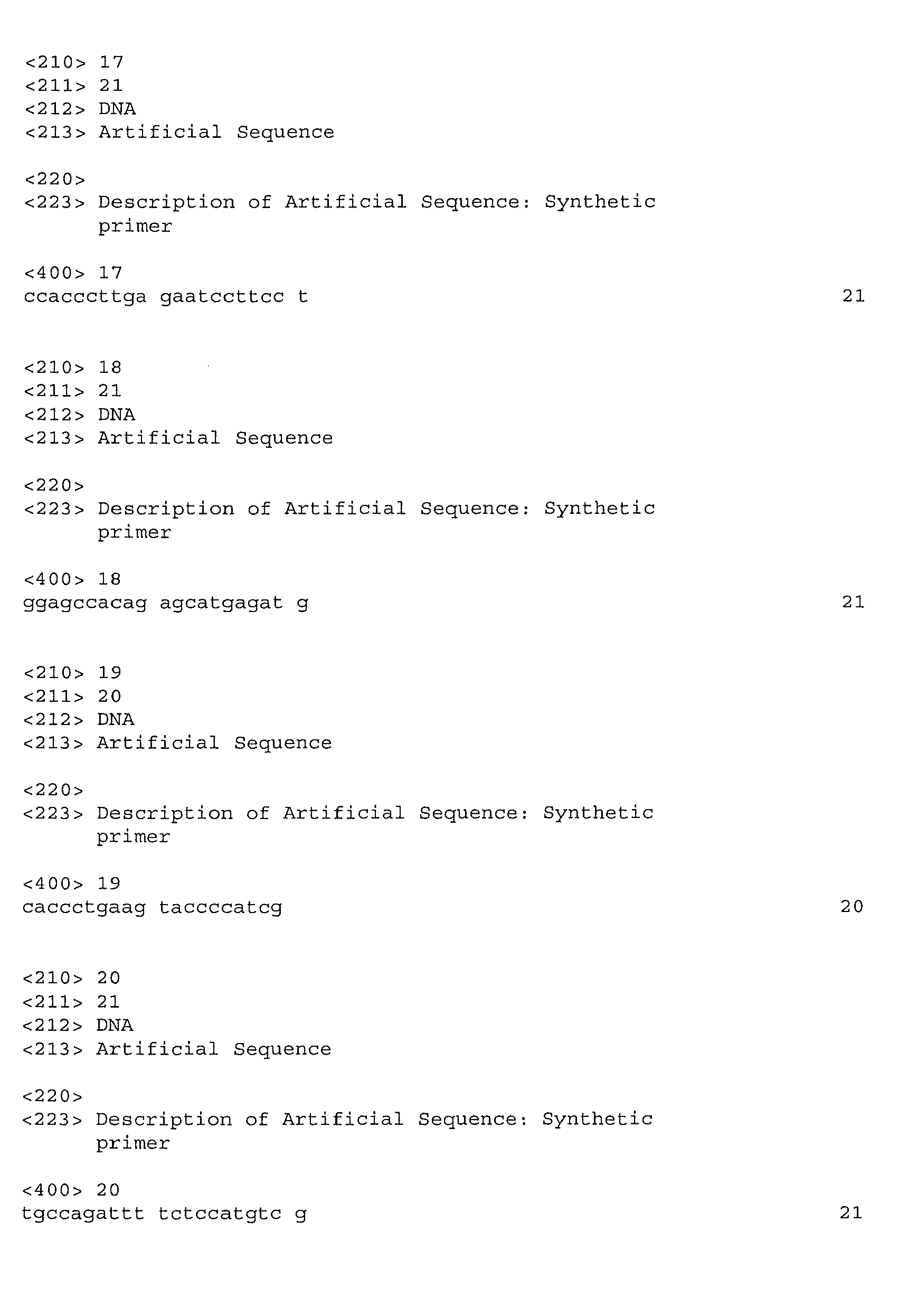
Claims (8)
- A method for measuring oxidative stress in a cell, comprising:(a) providing isolated cell populations, each population comprising a cell type that expresses at least one biomarker of oxidative stress (OS) that responds to OS by changing its expression level in a quantitative manner, said cells maintained under conditions in which said expression level is unchanged in absence of an inducer of OS;(b) providing an inducer of OS to a test group and not to a control group of the cell populations of step (a), and(c) determining the level of expression of said biomarker in the test and control groups of step (b), wherein a change in the expression level of said biomarker in the test group is correlated with the level of oxidative stress in the cell.
- The method of claim 1, wherein the biomarker of OS is selected from the group consisting of FosB, JunB, cFos, Fos L, ATF3, CRYBA, TXN, heme oxygenase (HO-1), EGR-1, Cl inhibitor, AP-1, IGFBP-3, IGFBP-5*, IGFBP-6, PLAGLI (ZAC1/LOT1), TIEG, P311, metallothionein 1X, metallothionein 1L, metallothionein 1H, metallothionein 1H-like, metallothionein 1G, metallothionein 2A, ETR 101, thioredoxin δ3 HSPA1A, HSPA1B, HSP-27, interleukin 8, M-GST3, GSTA4, MMP2, DTR, HOS-1, and LEDGF.
- The method of claim 1, wherein the level of expression of the biomarker changes in response to the level of OS in a dose-dependent manner.
- The method of claim 3, wherein the biomarker of OS is selected from the group consisting of FosB, Jun B, cFos, ATF3, and CRYBA.
- The method of claim 1, wherein measurement of the level of expression of the biomarker of OS in a cell or population is determined from mRNA or protein.
- The method of claim 5, wherein mRNA level is determined using polymerase chain reaction (PCR).
- The method of claim 1, wherein the cell population comprises a cell type of the retina.
- The method of claim 7, wherein the cell type is a retinal pigmented epithelial (RPE) cell.
Applications Claiming Priority (3)
| Application Number | Priority Date | Filing Date | Title |
|---|---|---|---|
| US67650405P | 2005-04-29 | 2005-04-29 | |
| US77399606P | 2006-02-16 | 2006-02-16 | |
| EP06751724A EP1880020A4 (en) | 2005-04-29 | 2006-04-28 | CELL BIOMARKER ANTIOXIDANT ASSAY AND CORRESPONDING USES THEREOF |
Related Parent Applications (1)
| Application Number | Title | Priority Date | Filing Date |
|---|---|---|---|
| EP06751724.3 Division | 2006-04-28 |
Publications (2)
| Publication Number | Publication Date |
|---|---|
| EP2392674A2 true EP2392674A2 (en) | 2011-12-07 |
| EP2392674A3 EP2392674A3 (en) | 2012-03-07 |
Family
ID=37308516
Family Applications (2)
| Application Number | Title | Priority Date | Filing Date |
|---|---|---|---|
| EP11162936A Withdrawn EP2392674A3 (en) | 2005-04-29 | 2006-04-28 | Cellular biomarker antioxidant assay and uses thereof |
| EP06751724A Withdrawn EP1880020A4 (en) | 2005-04-29 | 2006-04-28 | CELL BIOMARKER ANTIOXIDANT ASSAY AND CORRESPONDING USES THEREOF |
Family Applications After (1)
| Application Number | Title | Priority Date | Filing Date |
|---|---|---|---|
| EP06751724A Withdrawn EP1880020A4 (en) | 2005-04-29 | 2006-04-28 | CELL BIOMARKER ANTIOXIDANT ASSAY AND CORRESPONDING USES THEREOF |
Country Status (3)
| Country | Link |
|---|---|
| US (1) | US20080312128A1 (en) |
| EP (2) | EP2392674A3 (en) |
| WO (1) | WO2006118988A2 (en) |
Cited By (1)
| Publication number | Priority date | Publication date | Assignee | Title |
|---|---|---|---|---|
| US9447468B2 (en) | 2011-01-07 | 2016-09-20 | Vtv Therapeutics Llc | Compounds that modulate oxidative stress |
Families Citing this family (14)
| Publication number | Priority date | Publication date | Assignee | Title |
|---|---|---|---|---|
| US20070190590A1 (en) * | 2006-02-10 | 2007-08-16 | Canon Kabushiki Kaisha | Information acquisition apparatus on concentration of thioredoxins in sample, stress level information acquisition apparatus and stress level judging method |
| US20080269142A1 (en) * | 2006-10-23 | 2008-10-30 | Ian Blair | Endogenous thiadiazabicyclo glutathione-adduct |
| HUP0700552A2 (en) * | 2007-08-27 | 2009-03-30 | Janos Dr Feher | Method and composition inhibiting inflammation |
| EP2264454A1 (en) * | 2009-06-11 | 2010-12-22 | Universiteit Maastricht | Method for screening compounds for preventing the adverse effects of reactive oxygen species on eukaryotic cells |
| EP2531614B1 (en) * | 2010-02-05 | 2018-07-18 | The Procter and Gamble Company | Transcriptional profiling and biomarker-based methods for identifying and evaluating agents for antioxidant efficacy in cosmetic skin care formulations |
| KR101907411B1 (en) | 2010-03-12 | 2018-10-12 | 베르그 엘엘씨 | INTRAVENOUS FORMULATIONS OF COENZYME Q10 (CoQ10) AND METHODS OF USE THEREOF |
| US9125940B2 (en) | 2011-02-03 | 2015-09-08 | Zhuning Ma | Compositions and methods for treating macular edema |
| US8715648B2 (en) | 2011-02-16 | 2014-05-06 | Pivotal Therapeutics Inc. | Method for treating obesity with anti-obesity formulations and omega 3 fatty acids for the reduction of body weight in cardiovascular disease patients (CVD) and diabetics |
| US8952000B2 (en) | 2011-02-16 | 2015-02-10 | Pivotal Therapeutics Inc. | Cholesterol absorption inhibitor and omega 3 fatty acids for the reduction of cholesterol and for the prevention or reduction of cardiovascular, cardiac and vascular events |
| US8951514B2 (en) | 2011-02-16 | 2015-02-10 | Pivotal Therapeutics Inc. | Statin and omega 3 fatty acids for reduction of apolipoprotein-B levels |
| US9119826B2 (en) | 2011-02-16 | 2015-09-01 | Pivotal Therapeutics, Inc. | Omega 3 fatty acid for use as a prescription medical food and omega 3 fatty acid diagniostic assay for the dietary management of cardiovascular patients with cardiovascular disease (CVD) who are deficient in blood EPA and DHA levels |
| CN107095863A (en) | 2011-06-17 | 2017-08-29 | 博格有限责任公司 | Inhalable drug composition |
| WO2016069069A1 (en) * | 2014-10-29 | 2016-05-06 | Avon Products, Inc. | Screening method for identifying active agents |
| CN114235722B (en) * | 2021-11-01 | 2023-06-09 | 江苏省人民医院(南京医科大学第一附属医院) | A method for establishing a prediction model of the influence of fat emulsion on drug kinetics in vivo |
Family Cites Families (4)
| Publication number | Priority date | Publication date | Assignee | Title |
|---|---|---|---|---|
| EP1162886A4 (en) * | 1999-02-23 | 2003-02-12 | River Laboratories Charles | System and method for evaluating agents which prevent oxidative damage |
| AU2001273215A1 (en) * | 2000-07-07 | 2002-01-21 | Case Western Reserve University | Olfactory neuron cultures and method of making and using the same |
| WO2002095061A1 (en) * | 2001-05-18 | 2002-11-28 | New York University | Methods of screening test substances for treating or preventing diseases involving an oxidative stress |
| BE1014949A3 (en) * | 2001-08-14 | 2004-07-06 | Probiox | Oxidative stress detection method and kit for its implementation. |
-
2006
- 2006-04-28 WO PCT/US2006/016159 patent/WO2006118988A2/en not_active Ceased
- 2006-04-28 US US11/912,446 patent/US20080312128A1/en not_active Abandoned
- 2006-04-28 EP EP11162936A patent/EP2392674A3/en not_active Withdrawn
- 2006-04-28 EP EP06751724A patent/EP1880020A4/en not_active Withdrawn
Non-Patent Citations (12)
Cited By (2)
| Publication number | Priority date | Publication date | Assignee | Title |
|---|---|---|---|---|
| US9447468B2 (en) | 2011-01-07 | 2016-09-20 | Vtv Therapeutics Llc | Compounds that modulate oxidative stress |
| US9707239B2 (en) | 2011-01-07 | 2017-07-18 | Vtv Therapeutics Llc | Compounds that modulate oxidative stress |
Also Published As
| Publication number | Publication date |
|---|---|
| WO2006118988A2 (en) | 2006-11-09 |
| EP1880020A4 (en) | 2009-05-20 |
| US20080312128A1 (en) | 2008-12-18 |
| EP1880020A2 (en) | 2008-01-23 |
| WO2006118988A3 (en) | 2007-07-12 |
| EP2392674A3 (en) | 2012-03-07 |
Similar Documents
| Publication | Publication Date | Title |
|---|---|---|
| US20080312128A1 (en) | Cellular Biomarker Antioxidant Assay and Uses Thereof | |
| Poh et al. | Evidence that NLRC4 inflammasome mediates apoptotic and pyroptotic microglial death following ischemic stroke | |
| Takemura et al. | Metformin suppresses interleukin (IL)-1β-induced IL-8 production, aromatase activation, and proliferation of endometriotic stromal cells | |
| Brown et al. | Apolipoprotein E isoform mediated regulation of nitric oxide release | |
| Wang et al. | The role of miR-146a in dorsal root ganglia neurons of experimental diabetic peripheral neuropathy | |
| Tatone et al. | The aging ovary—the poor granulosa cells | |
| Adaikalakoteswari et al. | Oxidative DNA damage and augmentation of poly (ADP-ribose) polymerase/nuclear factor-kappa B signaling in patients with type 2 diabetes and microangiopathy | |
| Ding et al. | Activated microglia induce the production of reactive oxygen species and promote apoptosis of co-cultured retinal microvascular pericytes | |
| Soto et al. | Silymarin induces recovery of pancreatic function after alloxan damage in rats | |
| Fujimaki et al. | Wnt protein-mediated satellite cell conversion in adult and aged mice following voluntary wheel running | |
| Kim et al. | Antioxidant α-lipoic acid inhibits osteoclast differentiation by reducing nuclear factor-κB DNA binding and prevents in vivo bone resorption induced by receptor activator of nuclear factor-κB ligand and tumor necrosis factor-α | |
| Ebeling et al. | Improving retinal mitochondrial function as a treatment for age-related macular degeneration | |
| Mohamed et al. | Mitochondrial DNA deletions and the aging heart | |
| Bian et al. | Signaling pathways for glycated human serum albumin-induced IL-8 and MCP-1 secretion in human RPE cells | |
| JP5490532B2 (en) | Treatment and prevention of pancreatic cancer with δ-tocotrienol | |
| Russell-Guzmán et al. | Activation of the ROS/TXNIP/NLRP3 pathway disrupts insulin-dependent glucose uptake in skeletal muscle of insulin-resistant obese mice | |
| Książek et al. | Oxidative stress contributes to accelerated development of the senescent phenotype in human peritoneal mesothelial cells exposed to high glucose | |
| Yang et al. | High glucose induction of DNA-binding activity of the transcription factor NFκB in patients with diabetic nephropathy | |
| WO2017053222A1 (en) | Novel compositions and methods for treating or preventing dermal disorders | |
| Chatterjee et al. | Autophagy protects peripheral blood mononuclear cells against inflammation, oxidative and nitrosative stress in diabetic dyslipidemia | |
| Zhang et al. | Retinoic acid and tumor necrosis factor-α induced monocytic cell gene expression is regulated in part by induction of transcription factor MafB | |
| Tanaka et al. | Neuroprotective action of genipin on tunicamycin-induced cytotoxicity in neuro2a cells | |
| Mihailović et al. | β-Glucan administration to diabetic rats alleviates oxidative stress by lowering hyperglycaemia, decreasing non-enzymatic glycation and protein O-GlcNAcylation | |
| Sun et al. | Vitamin D ameliorates Aeromonas hydrophila-induced iron-dependent oxidative damage of grass carp splenic macrophages by manipulating Nrf2-mediated antioxidant pathway | |
| Guo et al. | Urolithin A alleviates cell senescence by inhibiting ferroptosis and enhances corneal epithelial wound healing |
Legal Events
| Date | Code | Title | Description |
|---|---|---|---|
| AC | Divisional application: reference to earlier application |
Ref document number: 1880020 Country of ref document: EP Kind code of ref document: P |
|
| AK | Designated contracting states |
Kind code of ref document: A2 Designated state(s): AT BE BG CH CY CZ DE DK EE ES FI FR GB GR HU IE IS IT LI LT LU LV MC NL PL PT RO SE SI SK TR |
|
| PUAI | Public reference made under article 153(3) epc to a published international application that has entered the european phase |
Free format text: ORIGINAL CODE: 0009012 |
|
| PUAL | Search report despatched |
Free format text: ORIGINAL CODE: 0009013 |
|
| AK | Designated contracting states |
Kind code of ref document: A3 Designated state(s): AT BE BG CH CY CZ DE DK EE ES FI FR GB GR HU IE IS IT LI LT LU LV MC NL PL PT RO SE SI SK TR |
|
| RIC1 | Information provided on ipc code assigned before grant |
Ipc: G01N 33/50 20060101ALI20120131BHEP Ipc: C12Q 1/68 20060101AFI20120131BHEP Ipc: C07H 21/04 20060101ALI20120131BHEP |
|
| STAA | Information on the status of an ep patent application or granted ep patent |
Free format text: STATUS: THE APPLICATION IS DEEMED TO BE WITHDRAWN |
|
| 18D | Application deemed to be withdrawn |
Effective date: 20120908 |