EP2392358B1 - Surgical grafts for repairing chondral defects - Google Patents
Surgical grafts for repairing chondral defects Download PDFInfo
- Publication number
- EP2392358B1 EP2392358B1 EP11152942.6A EP11152942A EP2392358B1 EP 2392358 B1 EP2392358 B1 EP 2392358B1 EP 11152942 A EP11152942 A EP 11152942A EP 2392358 B1 EP2392358 B1 EP 2392358B1
- Authority
- EP
- European Patent Office
- Prior art keywords
- collagen
- implant
- cells
- mscs
- chondrocyte
- Prior art date
- Legal status (The legal status is an assumption and is not a legal conclusion. Google has not performed a legal analysis and makes no representation as to the accuracy of the status listed.)
- Active
Links
Images
Classifications
-
- A—HUMAN NECESSITIES
- A61—MEDICAL OR VETERINARY SCIENCE; HYGIENE
- A61L—METHODS OR APPARATUS FOR STERILISING MATERIALS OR OBJECTS IN GENERAL; DISINFECTION, STERILISATION OR DEODORISATION OF AIR; CHEMICAL ASPECTS OF BANDAGES, DRESSINGS, ABSORBENT PADS OR SURGICAL ARTICLES; MATERIALS FOR BANDAGES, DRESSINGS, ABSORBENT PADS OR SURGICAL ARTICLES
- A61L27/00—Materials for grafts or prostheses or for coating grafts or prostheses
- A61L27/36—Materials for grafts or prostheses or for coating grafts or prostheses containing ingredients of undetermined constitution or reaction products thereof, e.g. transplant tissue, natural bone, extracellular matrix
- A61L27/38—Materials for grafts or prostheses or for coating grafts or prostheses containing ingredients of undetermined constitution or reaction products thereof, e.g. transplant tissue, natural bone, extracellular matrix containing added animal cells
- A61L27/3804—Materials for grafts or prostheses or for coating grafts or prostheses containing ingredients of undetermined constitution or reaction products thereof, e.g. transplant tissue, natural bone, extracellular matrix containing added animal cells characterised by specific cells or progenitors thereof, e.g. fibroblasts, connective tissue cells, kidney cells
- A61L27/3817—Cartilage-forming cells, e.g. pre-chondrocytes
-
- A—HUMAN NECESSITIES
- A61—MEDICAL OR VETERINARY SCIENCE; HYGIENE
- A61L—METHODS OR APPARATUS FOR STERILISING MATERIALS OR OBJECTS IN GENERAL; DISINFECTION, STERILISATION OR DEODORISATION OF AIR; CHEMICAL ASPECTS OF BANDAGES, DRESSINGS, ABSORBENT PADS OR SURGICAL ARTICLES; MATERIALS FOR BANDAGES, DRESSINGS, ABSORBENT PADS OR SURGICAL ARTICLES
- A61L27/00—Materials for grafts or prostheses or for coating grafts or prostheses
- A61L27/14—Macromolecular materials
- A61L27/22—Polypeptides or derivatives thereof, e.g. degradation products
- A61L27/24—Collagen
-
- A—HUMAN NECESSITIES
- A61—MEDICAL OR VETERINARY SCIENCE; HYGIENE
- A61L—METHODS OR APPARATUS FOR STERILISING MATERIALS OR OBJECTS IN GENERAL; DISINFECTION, STERILISATION OR DEODORISATION OF AIR; CHEMICAL ASPECTS OF BANDAGES, DRESSINGS, ABSORBENT PADS OR SURGICAL ARTICLES; MATERIALS FOR BANDAGES, DRESSINGS, ABSORBENT PADS OR SURGICAL ARTICLES
- A61L27/00—Materials for grafts or prostheses or for coating grafts or prostheses
- A61L27/36—Materials for grafts or prostheses or for coating grafts or prostheses containing ingredients of undetermined constitution or reaction products thereof, e.g. transplant tissue, natural bone, extracellular matrix
- A61L27/38—Materials for grafts or prostheses or for coating grafts or prostheses containing ingredients of undetermined constitution or reaction products thereof, e.g. transplant tissue, natural bone, extracellular matrix containing added animal cells
- A61L27/3839—Materials for grafts or prostheses or for coating grafts or prostheses containing ingredients of undetermined constitution or reaction products thereof, e.g. transplant tissue, natural bone, extracellular matrix containing added animal cells characterised by the site of application in the body
- A61L27/3843—Connective tissue
- A61L27/3852—Cartilage, e.g. meniscus
Definitions
- the present invention relates to surgical grafts for repairing a chondral defect.
- MSCs Mesenchymal stem cells
- Kavalkovich et al. provided a pellet culture method mimicking the environment of embryonic cartilage development, wherein a high cell density of cells was obtained but a tightly aggregated cell mass with a large amount of cells forming only a small volume of chondral tissue was present.
- alginate or agarose scaffolds for cartilage tissue engineering have the following drawbacks: poor cell adhesion, and uncontrollable degradation of alginate following the diffusion of divalent cations into the surrounding medium.
- alginate matrices were used in the in vivo applications and reported to have severe foreign body giant cell reactions and immunological responses when implanted to treat full-thickness defects in cartilage in experimental animals ( Hunziker, Articular cartilage repair: basic science and clinical progress. A review of the current status and prospects. Osteoarthritis Cartilage 2001; 0:432-63 ).
- alginate matrices have not been employed in human patients for articular cartilage repair.
- microspheres by combining mesenchymal stem cells with a collagen type I solution of 0.05 - 0.3 % w/v has also been described. Chondrogenic differentiation is induced in a chondrogenic induction medium which contains TGF-beta 3. The produced micro-masses integrate well in host tissue and be used to treat e.g. arthritis (Hui et al., In vitro chondrogenic differentiation of human mesenchymal stem cells in collagen microspheres: Influence of cell seeding density and collagen concentration. Biomaterials 29 (2008); 3201-3212).
- the present invention is based on two unexpected discoveries that (1) an implant containing a collagen matrix embedded with chondrocyte-like cells repairs chondral defects when implanted at a chondral defect site, and (2) no gap is formed between the implant and the tissues at the implanting site.
- one aspect of this invention features a chondral defect-reparing implant that contains a collagen matrix embedded with chondrocyte-like cells and supplemented with transforming growth factor beta one (TGF- ⁇ 1).
- This implant free of lacunae, is prepared by cultivating mesenchymal stem cells (MSCs) in a collagen (e.g., type-I or type-II collagen) containing medium containing collagen at a concentration of 1% to 10% w/v and supplemented with TGF- ⁇ 1 for 7 to 21 days and under conditions allowing differentiation of MSCs to the chondrocyte-like cells and formation of the implant.
- the chondrocyte-like cells secret glycosaminoglycan.
- the MSCs can be obtained from bone marrow, adipose tissue, muscle tissue, dental pulp of deciduous baby teeth, umbilical cord blood, Wharton's jelly, placenta, or cord lining membrane.
- the cultivating step mentioned above is performed by culturing the MSCs in a collagen containing medium supplemented with TGF- ⁇ 1 for 7 to 21 days under conditions allowing formation of a collagen matrix and differentiation of the MSCs into chondrocyte-like cells, which are embedded in the collagen matrix.
- the collagen containing medium has a collagen concentration of 1% to 10% w/v, (e.g., about 3% w/v). This cultivating step can be carried out in a capsule.
- the present invention also relates to an implant as described above for its use as a medicament.
- the present invention relates to an implant as described above for its use as a medicament for treating a chondral defect.
- the chondral defect is chosen in the group consisting of immature bone, severely degenerative arthritis in which total joint arthroplasty is needed, other defects such as cruciate ligament rupture and meniscus injury, arthritis such as rheumatoid arthritis and gouty arthritis, and infectious disease such as AIDS and Hepatitis B.
- the present invention features a method for repairing a chondral defect in a patient with an implant comprising a collagen matrix embedded with chondrocyte-like cells and transforming growth factor beta one (TGF- ⁇ 1) wherein the chondrocyte-like cells being differentiated from mesenchymal stem cells (MSCs) and secreting glycosaminoglycan, whereby the chondral defect can be repaired without any gap between the graft.
- TGF- ⁇ 1 transforming growth factor beta one
- an element means one element or more than one element.
- meenchymal stem cells or “MSCs” used herein refers to multipotent stem cells derived from adult tissues including but not limited to bone marrow, adipose tissue, muscle tissue, dental pulp of deciduous baby teeth, umbilical cord blood, or Wharton's jelly.
- chondrocytes refers to mature cartilage cells, which are embedded within the lacunae (small cavities) in the matrix. Chondrocytes arise by differentiation of mesenchymal chondrogenic cells into chondroblasts, which are the earliest cells to produce cartilage matrix. Mature chondrocytes are large secretory cells with a spherical nucleus and a prominent nucleolus. Therefore, the formation of lacuna in chondrocytes is regarded as a mature chondrocyte character.
- chondrocyte-like cells refers to the cells that possess the properties of the chondrocyte secretory cells, such as secreting glycosaminoglycan (GAG), but do not form lacunae, a typical character of mature chondrocytes.
- GAG glycosaminoglycan
- a collagen matrix refers to a tissue-engineered product formed by culturing the MSCs in a collagen containing medium, which is embedded with chondrocyte-like cells that are capable of secreting glycosaminoglycan (GAG) but without lacunae.
- the present invention provides a method for repairing a chondral defect in a patient comprising:
- the implant containing a collagen matrix embedded with chondrocyte-like cells and transforming growth factor beta one (TGF- ⁇ 1) which may be obtained by cultivating MSCs in a collagen containing medium supplemented with transforming growth factor beta one (TGF- ⁇ 1) under conditions allowing differentiation of the MSCs to the chondrocyte-like cells and formation of the implant.
- TGF- ⁇ 1 transforming growth factor beta one
- the MSCs were cultivated in a collagen (e.g., type-I or Type-II collagen) containing medium supplemented with TGF- ⁇ 1 for differentiation from the MSCs to chondrocyte-like cells that were characterized by evaluating the glycosaminoglycan (GAG) content to ensure that the MSCs were induced to cartilage-like tissue, and detecting whether lacunae was formed.
- a collagen e.g., type-I or Type-II collagen
- TGF- ⁇ 1 glycosaminoglycan
- the MSCs were expanded, and cultured, and then embedded and cultured in a medium containing collagen, and supplemented with transforming growth factor beta one (TGF- ⁇ 1) under conditions for chondrogenic induction into chondrocyte-like cells.
- TGF- ⁇ 1 transforming growth factor beta one
- the collagen matrix embedded with chondrocyte-like cells that secrete glycosaminoglycan (GAG) is collected.
- the collagen matrix can be examined via routine methods to confirm that no lacunae formation occurs and that the chondrocyte-like cells embedded therein secret GAG.
- any standard methods, conditions or technology may be used to allow the MSCs to differentiate to the chondrocyte-like cells and formation of the collagen matrix as an implant.
- collagen may be type-I or type-II collagen, or combination thereof.
- concentration of collagen is from 1% to 10% w/v, and more preferably about 3%.
- the implant may be easily delivered to a chondral defect site by a syringe-like piston.
- the implant is loaded in a capsule and the implant may be placed by squeezing the implant out of the capsule and applying it to the chondral defect site.
- the invention also provides an implant for repairing a chondral defect, comprising a collagen matrix embedded with chondrocyte-like cells and supplemented with transforming growth factor beta one (TGF- ⁇ 1), the chondrocyte-like cells being differentiated from mesenchymal stem cells (MSCs) and secreting glycosaminoglycan, wherein the implant is free of lacunae.
- TGF- ⁇ 1 transforming growth factor beta one
- the present invention also relates to an implant as described above for its use as a medicament.
- the present invention relates to an implant as described above for its use as a medicament for treating a chondral defect.
- the chondral defect is chosen in the group consisting of immature bone, severely degenerative arthritis in which total joint arthroplasty is needed, other defects such as cruciate ligament rupture and meniscus injury, arthritis such as rheumatoid arthritis and gouty arthritis, and infectious disease such as AIDS and Hepatitis B.
- the implant may be prepared by the method comprising:
- Example 1 Isolation and culture of human MSCs
- the cells were cultured at 0.5 mL aliquots with a final cell density of 2.6x10 6 cells/cm 2 to gel in 24-well plates at 37°C for 1 hour followed by the addition of 2.0 mL medium per well containing: serum-free DMEM-LG containing ITS+ Premix, 50 ug/mL L-ascorbic acid-2-phosphate, 10 -7 M dexamethasone, supplemented with 10 ng/mL TGF- ⁇ 1 (Pepro Tech; Rocky Hill, NJ).
- the cells were cultured in the just-described medium for 7 to 21 days, with the medium being changed every three days, to allow the cells dispersive embedded in the collagen containing medium to obtain a collagen matrix embedded with chondrocyte-like cells.
- the cell/collagen mixtures were analyzed 7, 14 and 21 days after the cell seeding to ensure if glycosaminoglycan (GAG) was secreted and the lacunae had not yet been formed to obtain the cell-matrix complex
- the cell-matrix complexes were harvested, embedded in paraffin and cut into 5 ⁇ m slices.
- the slices were deparaffinized, and rehydrated by sequential immersion in 100% xylene, 100% ethanol, 70% ethanol, and water.
- Hematoxylin and eosin staining (Muto,Tokyo, Japan) of sample sections was used to evaluate the cell morphology.
- Alcian Blue staining (Sigma) was used to evaluate the glycosaminoglycan (GAG) content.
- the knee function of all 10 patients was scored by the International Knee Documentation Committee Subjective Knee Form (IKDC) and X-ray examination was done preoperatively and at 1.5, 3, 6 and 12 months of follow-up. Magnetic Resonance Imaging (MRI) was carried out 6 months and 12 months after operation. Arthroscopic examination and biopsy was performed 12 months after operation if patients accept the proposal. Student t-test was used for statistical analysis.
- IKDC International Knee Documentation Committee Subjective Knee Form
- MRI Magnetic Resonance Imaging
- the cell-matrix complex comprising chondrocyte-like cells secreting GAG but without lacunae and the matrix prepared therefrom according to the invention, exhibited excellent efficacy in repairing a chondral defect in a patient, after transplanting onto the recipient site, where hyaline cartilage was formed without any gap between the recipient site and the surgical graft created by the cell-matrix complex.
Landscapes
- Health & Medical Sciences (AREA)
- Life Sciences & Earth Sciences (AREA)
- Engineering & Computer Science (AREA)
- Biomedical Technology (AREA)
- Chemical & Material Sciences (AREA)
- Veterinary Medicine (AREA)
- Epidemiology (AREA)
- Cell Biology (AREA)
- Public Health (AREA)
- General Health & Medical Sciences (AREA)
- Dermatology (AREA)
- Medicinal Chemistry (AREA)
- Oral & Maxillofacial Surgery (AREA)
- Transplantation (AREA)
- Animal Behavior & Ethology (AREA)
- Zoology (AREA)
- Chemical Kinetics & Catalysis (AREA)
- Botany (AREA)
- Vascular Medicine (AREA)
- Biophysics (AREA)
- Rheumatology (AREA)
- Urology & Nephrology (AREA)
- Materials For Medical Uses (AREA)
- Prostheses (AREA)
- Micro-Organisms Or Cultivation Processes Thereof (AREA)
Description
- The present invention relates to surgical grafts for repairing a chondral defect.
- Autogenous chondrocyte transplanation has been used to treat the chondral defect since 1994, comprising collecting chondrocytes from normal cartilage tissues, expanding these cells in a medium in vitro as much as possible, and then filling the chondral defect by implanting the expanded cells in artificial scaffolds. However, there are still many problems; for example, the source of autogenous chrondrocytes is limited, the proliferation ability of chondrocytes is low, and their unique phenotypes may be lost during expansion (Saadeh et al., Human cartilage engineering: chondrocyte extraction, proliferation, and characterization for construct development. Ann Plast Surg 1999; 42:509-13).
- Mesenchymal stem cells (MSCs) are able to differentiate into cells of connective tissue lineages including cartilage and thus becomes an attractive cell source for cartilage tissue engineering. Many methods have been used in chondrogenic induction of MSCs. Kavalkovich et al. provided a pellet culture method mimicking the environment of embryonic cartilage development, wherein a high cell density of cells was obtained but a tightly aggregated cell mass with a large amount of cells forming only a small volume of chondral tissue was present. Therefore, a considerably large number of the cell pellets were required for repairing a defective cartilage, and the large pellets exhibited a periphery of viable cells, but the pellet center became necrotic (Kavalkovich et al., Chondrogenic differentiation of human mesenchymal stem cells within an alginate layer culture system. In Vitro Cell Dev Biol Anim 2002;38:457-66.). In addition, pellets are difficult to handle and to mold into various shapes needed for defect repair. Given the above, the pellet culture is impractical for surgical applications. Furthermore, it was found that embedding human MSCs (hMSCs) in agarose or alginate gels could be cast into various shapes and caused substantial chondrogenesis (Huang et al., Chondrogenesis of human bone marrow-derived mesenchymal stem cells in agarose culture. Anat Rec A Discov Mol Cell Evol Biol 2004; 278:428-36; and Ma et al., Chondrogenesis of human mesenchymal stem cells encapsulated in alginate beads. J Biomed Mater Res A 2003; 64:273-81). However, the alginate or agarose scaffolds for cartilage tissue engineering have the following drawbacks: poor cell adhesion, and uncontrollable degradation of alginate following the diffusion of divalent cations into the surrounding medium. In addition, alginate matrices were used in the in vivo applications and reported to have severe foreign body giant cell reactions and immunological responses when implanted to treat full-thickness defects in cartilage in experimental animals (Hunziker, Articular cartilage repair: basic science and clinical progress. A review of the current status and prospects. Osteoarthritis Cartilage 2001; 0:432-63). Hence, alginate matrices have not been employed in human patients for articular cartilage repair.
- The formation of microspheres by combining mesenchymal stem cells with a collagen type I solution of 0.05 - 0.3 % w/v has also been described. Chondrogenic differentiation is induced in a chondrogenic induction medium which contains TGF-
beta 3. The produced micro-masses integrate well in host tissue and be used to treat e.g. arthritis (Hui et al., In vitro chondrogenic differentiation of human mesenchymal stem cells in collagen microspheres: Influence of cell seeding density and collagen concentration. Biomaterials 29 (2008); 3201-3212). - It is still desired to develop a surgical graft for repairing a chondral defect in a patient.
- The present invention is based on two unexpected discoveries that (1) an implant containing a collagen matrix embedded with chondrocyte-like cells repairs chondral defects when implanted at a chondral defect site, and (2) no gap is formed between the implant and the tissues at the implanting site.
- Accordingly, one aspect of this invention features a chondral defect-reparing implant that contains a collagen matrix embedded with chondrocyte-like cells and supplemented with transforming growth factor beta one (TGF-β1). This implant, free of lacunae, is prepared by cultivating mesenchymal stem cells (MSCs) in a collagen (e.g., type-I or type-II collagen) containing medium containing collagen at a concentration of 1% to 10% w/v and supplemented with TGF-β1 for 7 to 21 days and under conditions allowing differentiation of MSCs to the chondrocyte-like cells and formation of the implant. The chondrocyte-like cells secret glycosaminoglycan. The MSCs can be obtained from bone marrow, adipose tissue, muscle tissue, dental pulp of deciduous baby teeth, umbilical cord blood, Wharton's jelly, placenta, or cord lining membrane.
- The cultivating step mentioned above is performed by culturing the MSCs in a collagen containing medium supplemented with TGF-β1 for 7 to 21 days under conditions allowing formation of a collagen matrix and differentiation of the MSCs into chondrocyte-like cells, which are embedded in the collagen matrix. The collagen containing medium has a collagen concentration of 1% to 10% w/v, (e.g., about 3% w/v). This cultivating step can be carried out in a capsule.
- Accordingly the present invention also relates to an implant as described above for its use as a medicament. In another aspect, the present invention relates to an implant as described above for its use as a medicament for treating a chondral defect.
- Preferably, the chondral defect is chosen in the group consisting of immature bone, severely degenerative arthritis in which total joint arthroplasty is needed, other defects such as cruciate ligament rupture and meniscus injury, arthritis such as rheumatoid arthritis and gouty arthritis, and infectious disease such as AIDS and Hepatitis B.
- The various embodiments of the present invention are described in details below. Other characteristics of the present invention will be clearly presented by the following detailed descriptions and drawings about the various embodiments and claims.
- For the purpose of illustrating the invention, there are shown in the drawings embodiments which are presently preferred. It should be understood, however, that the invention is not limited to the preferred embodiments shown.
- In the drawings:
-
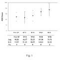
Fig. 1 shows the IKDC scoring of the 9 patients treated with the method according to the invention, wherein a significant improvement by Student's t-test was found 6 months and 12 months after operation. -
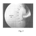
Fig. 2 is an image showing the arthroscopic observation of a patient treated with the method according to the invention 12 months after the transplantation; wherein there was no gap between the recipient site and the surgical graft created by the cell-matrix complex. - The present invention features a method for repairing a chondral defect in a patient with an implant comprising a collagen matrix embedded with chondrocyte-like cells and transforming growth factor beta one (TGF-β1) wherein the chondrocyte-like cells being differentiated from mesenchymal stem cells (MSCs) and secreting glycosaminoglycan, whereby the chondral defect can be repaired without any gap between the graft.
- Unless defined otherwise, all technical and scientific terms used herein have the meaning commonly understood by a person skilled in the art to which this invention belongs. As used herein, the following terms have the meanings ascribed to them unless specified otherwise.
- The articles "a" and "an" are used herein to refer to one or more than one (i.e., at least one) of the grammatical object of the article. By way of example, "an element" means one element or more than one element.
- The term "mesenchymal stem cells" or "MSCs" used herein refers to multipotent stem cells derived from adult tissues including but not limited to bone marrow, adipose tissue, muscle tissue, dental pulp of deciduous baby teeth, umbilical cord blood, or Wharton's jelly.
- As used herein, the term "chondrocytes" refers to mature cartilage cells, which are embedded within the lacunae (small cavities) in the matrix. Chondrocytes arise by differentiation of mesenchymal chondrogenic cells into chondroblasts, which are the earliest cells to produce cartilage matrix. Mature chondrocytes are large secretory cells with a spherical nucleus and a prominent nucleolus. Therefore, the formation of lacuna in chondrocytes is regarded as a mature chondrocyte character.
- As used herein, the term "chondrocyte-like cells" refers to the cells that possess the properties of the chondrocyte secretory cells, such as secreting glycosaminoglycan (GAG), but do not form lacunae, a typical character of mature chondrocytes.
- As used herein, the term "a collagen matrix" refers to a tissue-engineered product formed by culturing the MSCs in a collagen containing medium, which is embedded with chondrocyte-like cells that are capable of secreting glycosaminoglycan (GAG) but without lacunae.
- The present invention provides a method for repairing a chondral defect in a patient comprising:
- providing an implant containing a collagen matrix embedded with chondrocyte-like cells and transforming growth factor beta one (TGF-β1), the chondrocyte-like cells being differentiated from mesenchymal stem cells (MSCs) and secreting glycosaminoglycan, and
- placing the implant in the patient at a chondral defect site; wherein the implant is free of lacunae.
- In the invention, the implant containing a collagen matrix embedded with chondrocyte-like cells and transforming growth factor beta one (TGF-β1), which may be obtained by cultivating MSCs in a collagen containing medium supplemented with transforming growth factor beta one (TGF-β1) under conditions allowing differentiation of the MSCs to the chondrocyte-like cells and formation of the implant.
- In one example of the invention, the MSCs were cultivated in a collagen (e.g., type-I or Type-II collagen) containing medium supplemented with TGF-β1 for differentiation from the MSCs to chondrocyte-like cells that were characterized by evaluating the glycosaminoglycan (GAG) content to ensure that the MSCs were induced to cartilage-like tissue, and detecting whether lacunae was formed. The collagen matrix embedded with chondrocyte-like cells were collected; which is a tissue-engineered construct used as an implant.
- In one example of the invention, the MSCs were expanded, and cultured, and then embedded and cultured in a medium containing collagen, and supplemented with transforming growth factor beta one (TGF-β1) under conditions for chondrogenic induction into chondrocyte-like cells. The collagen matrix embedded with chondrocyte-like cells that secrete glycosaminoglycan (GAG) is collected. The collagen matrix can be examined via routine methods to confirm that no lacunae formation occurs and that the chondrocyte-like cells embedded therein secret GAG.
- In the invention, any standard methods, conditions or technology may be used to allow the MSCs to differentiate to the chondrocyte-like cells and formation of the collagen matrix as an implant.
- In one example of the invention, collagen may be type-I or type-II collagen, or combination thereof. The concentration of collagen is from 1% to 10% w/v, and more preferably about 3%.
- According to the invention, the implant may be easily delivered to a chondral defect site by a syringe-like piston. In one embodiment of the invention, the implant is loaded in a capsule and the implant may be placed by squeezing the implant out of the capsule and applying it to the chondral defect site.
- Accordingly, the invention also provides an implant for repairing a chondral defect, comprising a collagen matrix embedded with chondrocyte-like cells and supplemented with transforming growth factor beta one (TGF-β1), the chondrocyte-like cells being differentiated from mesenchymal stem cells (MSCs) and secreting glycosaminoglycan, wherein the implant is free of lacunae.
- Accordingly, the present invention also relates to an implant as described above for its use as a medicament. In another aspect, the present invention relates to an implant as described above for its use as a medicament for treating a chondral defect.
- Preferably, the chondral defect is chosen in the group consisting of immature bone, severely degenerative arthritis in which total joint arthroplasty is needed, other defects such as cruciate ligament rupture and meniscus injury, arthritis such as rheumatoid arthritis and gouty arthritis, and infectious disease such as AIDS and Hepatitis B.
- According to the invention, the implant may be prepared by the method comprising:
- providing mesenchymal stem cells (MSCs); and
- cultivating the MSCs in a collagen containing medium supplemented with transforming growth factor beta one (TGF-β1) under conditions allowing differentiation of the MSCs to the chondrocyte-like cells and formation of the implant.
- The present invention will now be described more specifically with reference to the following embodiments, which are provided for the purpose of demonstration rather than limitation.
- Ten (10) ml of heparinized bone marrow blood was aspirated from the ilium of the pelvis, and bone marrow MSCs were isolated therefrom. The MSCs were cultured in Dulbecco's modified Eagle's medium with low glucose containing 10% fetal bovine serum at 37°C under 5% carbon dioxide and 95% humidity. The medium was changed twice a week. The cells were trypsinized and subcultured at a ratio of 1:3 every week, and then harvested and embedded in Porcogen™ collagen solution (3% type-I/type-III collagen, SunMax Biotech., Taiwan) containing serum-free DMEM-LG only. The cells were cultured at 0.5 mL aliquots with a final cell density of 2.6x10⁶ cells/cm2 to gel in 24-well plates at 37°C for 1 hour followed by the addition of 2.0 mL medium per well containing: serum-free DMEM-LG containing ITS+ Premix, 50 ug/mL L-ascorbic acid-2-phosphate, 10-7 M dexamethasone, supplemented with 10 ng/mL TGF-β1 (Pepro Tech; Rocky Hill, NJ). The cells were cultured in the just-described medium for 7 to 21 days, with the medium being changed every three days, to allow the cells dispersive embedded in the collagen containing medium to obtain a collagen matrix embedded with chondrocyte-like cells. The cell/collagen mixtures were analyzed 7, 14 and 21 days after the cell seeding to ensure if glycosaminoglycan (GAG) was secreted and the lacunae had not yet been formed to obtain the cell-matrix complexes.
- The cell-matrix complexes were harvested, embedded in paraffin and cut into 5 µm slices. The slices were deparaffinized, and rehydrated by sequential immersion in 100% xylene, 100% ethanol, 70% ethanol, and water. Hematoxylin and eosin staining (Muto,Tokyo, Japan) of sample sections was used to evaluate the cell morphology. In addition, Alcian Blue staining (Sigma) was used to evaluate the glycosaminoglycan (GAG) content.
- Patients with chondral or osteochondral defects from osteochondritis dissecans or osteonecrosis of medial femoral condyle, or limited chondral defects of osteoarthritis which was located on the weight-bearing site were selected. However, patients with immature bone, severely degenerative arthritis in which total joint arthroplasty is needed, other defects such as cruciate ligament rupture and meniscus injury, arthritis such as rheumatoid arthritis and gouty arthritis, and infectious disease such as AIDS and Hepatitis B; patients completely bed-ridden above three months; patients taking corticosteroids longer than two weeks, and alcoholism are excluded. Totally, 10 patients were recruited. Among them, six were female and four were male. The patients' average age was 66 (ranging from 47 to 83).
- An anterior midline incision was made on the knee and the articular surface of the medial femoral condyle was exposed using a medial parapatellar approach. The knee flexed extremely to made access to the osteochondral defect easy. The bone defect was debrided and filled with cancellous bone taken from the upper tibia. Then, the cell-matrix complexes as obtained in Example 1 as surgical grafts were transplanted onto the defects. Finally, periosteum taken from the proximal tibia was used to cover the surgical graft. After operation, all patients received regular Quadriceps muscle training and exercise to improve range of motion of the knee.
- The knee function of all 10 patients was scored by the International Knee Documentation Committee Subjective Knee Form (IKDC) and X-ray examination was done preoperatively and at 1.5, 3, 6 and 12 months of follow-up. Magnetic Resonance Imaging (MRI) was carried out 6 months and 12 months after operation. Arthroscopic examination and biopsy was performed 12 months after operation if patients accept the proposal. Student t-test was used for statistical analysis.
- Except one patient who did not want to come back for evaluation 6 months after operation, the remained 9 patients were followed up regularly up to 12 months. As shown in
Fig. 1 , the IKDC scoring of the patients treated with the method according to the invention had significant improvement by Student's t-test was found 6 months and 12 months after operation. All 9 patients felt much better 6 months after operation. Four patients were followed up more than 12 months and 3 of them received arthroscopy. The chondral defects were found in the hyaline cartilage with moderate hardness that were merged without any gap between the recipient site and the graft (seeFig.2 ). - Given the above, the cell-matrix complex comprising chondrocyte-like cells secreting GAG but without lacunae and the matrix prepared therefrom according to the invention, exhibited excellent efficacy in repairing a chondral defect in a patient, after transplanting onto the recipient site, where hyaline cartilage was formed without any gap between the recipient site and the surgical graft created by the cell-matrix complex.
Claims (9)
- A method for preparing an implant for repairing a chondral defect, comprising:providing mesenchymal stem cells (MSCs);cultivating the MSCs in a collagen medium containing collagen at a concentration of 1 % to 10% w/v and supplemented with transforming growth factor beta one (TGF-β1) for 7 to 21 days under conditions allowing formation of a collagen matrix and differentiation of the MSCs to chondrocyte-like cells, which are embedded in the collagen matrix ;which is characterized in that the chondrocyte-like cells secrete glycosaminoglycan and the implant is free of lacunae.
- The method according to claim 1, wherein the MSCs are isolated from bone marrow, adipose tissue, muscle tissue, dental pulp of deciduous baby teeth, umbilical cord blood, Wharton's jelly, placenta, or cord lining membrane.
- The method according to claim 2, wherein the collagen is contained in the medium at a concentration of 3% w/v.
- The method according to any of claims 1 to 3, wherein the medium contains type-I or type-II collagen.
- The method according to claims 1, wherein the cultivating step is performed in a capsule.
- An implant for repairing a chondral defect, obtainable by the method according to any of claims 1 to 5.
- An implant according to claim 6, for its use as a medicament.
- An implant according to claim 6, for its use as a medicament for treating a chondral defect.
- The implant according to claim 8, wherein the chondral defect is chosen in the group consisting of immature bone, degenerative arthritis in which total joint arthroplasty is needed, cruciate ligament rupture, meniscus injury, rheumatoid arthritis, gouty arthritis, AIDS, and Hepatitis B.
Applications Claiming Priority (1)
| Application Number | Priority Date | Filing Date | Title |
|---|---|---|---|
| US12/700,469 US8574614B2 (en) | 2010-02-04 | 2010-02-04 | Surgical grafts for repairing chondral defects |
Publications (3)
| Publication Number | Publication Date |
|---|---|
| EP2392358A2 EP2392358A2 (en) | 2011-12-07 |
| EP2392358A3 EP2392358A3 (en) | 2012-04-04 |
| EP2392358B1 true EP2392358B1 (en) | 2015-04-08 |
Family
ID=44341891
Family Applications (1)
| Application Number | Title | Priority Date | Filing Date |
|---|---|---|---|
| EP11152942.6A Active EP2392358B1 (en) | 2010-02-04 | 2011-02-01 | Surgical grafts for repairing chondral defects |
Country Status (8)
| Country | Link |
|---|---|
| US (1) | US8574614B2 (en) |
| EP (1) | EP2392358B1 (en) |
| JP (2) | JP5764807B2 (en) |
| CN (1) | CN102145195B (en) |
| ES (1) | ES2541486T3 (en) |
| PT (1) | PT2392358E (en) |
| SG (3) | SG190660A1 (en) |
| TW (1) | TWI469993B (en) |
Families Citing this family (17)
| Publication number | Priority date | Publication date | Assignee | Title |
|---|---|---|---|---|
| US8883210B1 (en) | 2010-05-14 | 2014-11-11 | Musculoskeletal Transplant Foundation | Tissue-derived tissuegenic implants, and methods of fabricating and using same |
| US9352003B1 (en) | 2010-05-14 | 2016-05-31 | Musculoskeletal Transplant Foundation | Tissue-derived tissuegenic implants, and methods of fabricating and using same |
| US10130736B1 (en) | 2010-05-14 | 2018-11-20 | Musculoskeletal Transplant Foundation | Tissue-derived tissuegenic implants, and methods of fabricating and using same |
| ES2725564T3 (en) | 2011-11-08 | 2019-09-24 | Auxocell Laboratories Inc | Systems and methods of cellular processing |
| RU2494469C1 (en) * | 2012-04-02 | 2013-09-27 | Федеральное государственное бюджетное учреждение "Новосибирский научно-исследовательский институт травматологии и ортопедии" Министерства здравоохранения и социального развития Российской Федерации (ФГБУ "ННИИТО" Минздравсоцразвития России) | Method for experimental correction of intervertebral disc defect |
| CN103387959A (en) * | 2012-10-19 | 2013-11-13 | 夏佑红 | Stem cell from deciduous teeth, and preparation method and application thereof |
| CN102899287B (en) * | 2012-10-24 | 2013-08-21 | 天津和泽干细胞科技有限公司 | Method for inducing differentiation from mesenchymal stem cells to cartilage cells and application of mesenchymal stem cells in osteoarthritis |
| EP3027235A1 (en) | 2013-07-30 | 2016-06-08 | Musculoskeletal Transplant Foundation | Acellular soft tissue-derived matrices and methods for preparing same |
| US9993748B2 (en) | 2014-08-11 | 2018-06-12 | Auxocell Laboratories, Inc. | Centrifuge clip and method |
| USD748462S1 (en) | 2014-08-11 | 2016-02-02 | Auxocell Laboratories, Inc. | Centrifuge clip |
| EP3297694A1 (en) | 2015-05-21 | 2018-03-28 | Musculoskeletal Transplant Foundation | Modified demineralized cortical bone fibers |
| US10912864B2 (en) | 2015-07-24 | 2021-02-09 | Musculoskeletal Transplant Foundation | Acellular soft tissue-derived matrices and methods for preparing same |
| US11052175B2 (en) | 2015-08-19 | 2021-07-06 | Musculoskeletal Transplant Foundation | Cartilage-derived implants and methods of making and using same |
| CN111821518B (en) * | 2019-04-15 | 2023-01-10 | 弗元(上海)生物科技有限公司 | Preparation method of tissue engineering bone with regeneration capacity |
| CN111821519A (en) * | 2019-04-15 | 2020-10-27 | 弗元(上海)生物科技有限公司 | A kind of preparation method of tissue engineered cartilage and its application |
| CN115845142B (en) * | 2022-12-23 | 2024-08-13 | 武汉大学 | Preparation method of cartilage-like micro-tissue for rapidly repairing jawbone defect |
| CN116983478A (en) * | 2023-07-31 | 2023-11-03 | 江苏博赛孚医疗科技有限公司 | Organic matter-embedded polyglycolic acid cartilage scaffold and its preparation method and application |
Citations (3)
| Publication number | Priority date | Publication date | Assignee | Title |
|---|---|---|---|---|
| WO1999008728A1 (en) | 1997-08-14 | 1999-02-25 | Sulzer Innotec Ag | Composition and device for in vivo cartilage repair |
| WO2001092473A2 (en) | 2000-05-31 | 2001-12-06 | Fraunhofer-Gesellschaft zur Förderung der angewandten Forschung e.V. | Method for cultivating a cartilage replacement and a biomatrix produced according to this method |
| EP1760144A2 (en) | 2004-05-21 | 2007-03-07 | Universidad de Málaga | Cartilage and bone repair composition |
Family Cites Families (7)
| Publication number | Priority date | Publication date | Assignee | Title |
|---|---|---|---|---|
| US5226914A (en) * | 1990-11-16 | 1993-07-13 | Caplan Arnold I | Method for treating connective tissue disorders |
| JP4531135B2 (en) * | 1996-12-06 | 2010-08-25 | オシリス セラピューティクス,インコーポレイテッド | Improved chondrogenic differentiation of human mesenchymal stem cells |
| US6197586B1 (en) * | 1997-12-12 | 2001-03-06 | The Regents Of The University Of California | Chondrocyte-like cells useful for tissue engineering and methods |
| US20030050709A1 (en) * | 2001-02-23 | 2003-03-13 | Ulrich Noth | Trabecular bone-derived human mesenchymal stem cells |
| ATE312615T1 (en) * | 2001-08-14 | 2005-12-15 | Medipost Co Ltd | COMPOSITION FOR THE TREATMENT OF DAMAGE TO THE ARTicular cartilage |
| US20040258731A1 (en) * | 2001-11-21 | 2004-12-23 | Tsuyoshi Shimoboji | Preparation approriate for cartilage tissue formation |
| CN100477971C (en) * | 2005-05-27 | 2009-04-15 | 中国人民解放军总医院 | Arthrodial cartilage proto micro-carrier |
-
2010
- 2010-02-04 US US12/700,469 patent/US8574614B2/en active Active
- 2010-06-11 TW TW99119088A patent/TWI469993B/en active
-
2011
- 2011-01-24 SG SG2013037825A patent/SG190660A1/en unknown
- 2011-01-24 SG SG10201402811YA patent/SG10201402811YA/en unknown
- 2011-01-24 SG SG2011004793A patent/SG173284A1/en unknown
- 2011-01-27 JP JP2011015143A patent/JP5764807B2/en active Active
- 2011-01-30 CN CN201110034680.5A patent/CN102145195B/en active Active
- 2011-02-01 EP EP11152942.6A patent/EP2392358B1/en active Active
- 2011-02-01 ES ES11152942.6T patent/ES2541486T3/en active Active
- 2011-02-01 PT PT111529426T patent/PT2392358E/en unknown
-
2015
- 2015-01-15 JP JP2015006164A patent/JP2015091401A/en not_active Withdrawn
Patent Citations (3)
| Publication number | Priority date | Publication date | Assignee | Title |
|---|---|---|---|---|
| WO1999008728A1 (en) | 1997-08-14 | 1999-02-25 | Sulzer Innotec Ag | Composition and device for in vivo cartilage repair |
| WO2001092473A2 (en) | 2000-05-31 | 2001-12-06 | Fraunhofer-Gesellschaft zur Förderung der angewandten Forschung e.V. | Method for cultivating a cartilage replacement and a biomatrix produced according to this method |
| EP1760144A2 (en) | 2004-05-21 | 2007-03-07 | Universidad de Málaga | Cartilage and bone repair composition |
Non-Patent Citations (18)
| Title |
|---|
| BARRY, BOYNTON ET AL.: "Chondrogenic differentiation of mesenchymal stem", EXPERIMENTAL CELL RESEARCH, vol. 268, no. 2, 15 August 2001 (2001-08-15), pages 189 - 200, XP001183120 |
| BHOSALE, RICHARDSON, ARTICULAR CARTILAGE STRUCTURE INJURIES AND REVIEW OF MANAGEMENT, vol. 87, no. 1, 2008, pages 77 - 95, XP055262494 |
| BOEUF, RICHTER: "Chondrogenesis of mesenchymal stem cells: role of tissue source and inducing factors", STEM CELL RESEARCH & THERAPY, vol. 1, no. 31, December 2010 (2010-12-01), pages 1 - 9, XP055262543 |
| BOSNAKOVSKI, MIZUNO ET AL.: "Chondrogenic differentiation of bovine bone marrow mesenchymal stem cells (MSCs) in different hydrogels: influence of collagen type II extracellular matrix on MSC chondrogenesis", BIOTECHNOLOGY AND BIOENGINEERING, vol. 93, no. 6, April 2006 (2006-04-01), pages 1152 - 1163, XP002495753 |
| BRITTBERG, LINDAHL, CHAPTER 18: TISSUE ENGINEERING OF CARTILAGE, 2008, pages 533 - 558, XP055262498 |
| BUCKWALTER J A ET AL.: "Articular cartilage", THE JOURNAL OF BONE AND JOINT SURGERY, vol. 79-A, no. 4, April 1997 (1997-04-01), pages 600 - 611, XP055262505 |
| CAPLAN, KOUTROUPAS: "The control of muscle and cartilage development in the chick limb: the role of differential vascularization", J. EMBROYL. EXP. MORPH., vol. 29, no. 3, June 1973 (1973-06-01), pages 571 - 583, XP055262508 |
| CHEN, ZHANG ET AL.: "Chondrogenic differentiation of umbilical cord-derived mesenchymal stem cells in type I collagen-hydrogel for cartilage engineering", INJURYCHONDROGENIC DIFFERENTIATION OF UMBILICAL CORD-DERIVED, vol. 44, no. 4, April 2013 (2013-04-01), pages 540 - 549, XP055262546 |
| DICKHUT, DEXHEIMER ET AL.: "Chondrogenesis of human mesenchymal stem cells by local transforming growth factor-beta delivery in a biphasic resorbable carrier", TISSUE ENG PART A., vol. 16, no. 2, February 2010 (2010-02-01), pages 453 - 464, XP055262519 |
| FENSKY, REICHERT ET AL.: "Chondrogenic predifferentiation of human mesenchymal stem cells in collagen type I hydrogels", BIOMED TECH, vol. 59, no. 5, October 2014 (2014-10-01), pages 375 - 383, XP055262550 |
| FILARDO, MADRY ET AL.: "Mesenchymal stem cells for the treatment of cartilage lesions: from preclinical findings to clinical application in orthopaedics", KNEE SURG SPORTS TRAUMATOL ARTHROSC, vol. 21, no. 8, August 2013 (2013-08-01), pages 1717 - 1719, XP055262552 |
| FOX, A J S ET AL.: "The basic science of articular cartilage: Structure, Composition, and Function", SPORTS HEALTH, vol. 1, no. 6, October 2009 (2009-10-01), pages 461 - 468, XP055262521 |
| HUANG, KAZMI ET AL.: "Chondrogenic potential of progenitor cells derived from human bone marrow and adipose tissue: a patient-matched comparison", JOURNAL OF ORTHOPAEDIC RESEARCH, vol. 23, no. 6, November 2005 (2005-11-01), pages 1383 - 1389, XP027720662 |
| HUI, CHEUNG, K M C ET AL.: "In vitro chondrogenic differentiation of human mesenchymal stem cells in collagen microspheres: Influence of cell seeding density and collagen concentration", BIOMATERIALS, vol. 29, no. 22, August 2008 (2008-08-01), pages 3201 - 3212, XP022673743 |
| NÖTH, U. ET AL.: "Chondrogenic differentiation of human mesenchymal stem cells in collagen type I hydrogels", JOURNAL OF BIOMEDICAL MATERIALS RESEARCH. PART A, vol. 83A, no. 3, May 2007 (2007-05-01), pages 626 - 635, XP008159655 |
| STECK, FISCHER ET AL.: "Mesenchymal stem cell differentiation in an experimental cartilage defect: restriction of hypertrophy to bone-close neocartilage", STEM CELLS AND DEVELOPMENT, vol. 18, no. 7, September 2009 (2009-09-01), pages 969 - 978, XP055262528 |
| WAGNER, HO: "Mesenchymal stem cell preparations--comparing apples and Oranges", STEM CELL REVIEWS, vol. 3, no. 4, 18 September 2007 (2007-09-18), pages 239 - 248, XP002545736 |
| ZSCHARNACK, POESEL ET AL.: "Low oxygen expansion improves subsequent chondrogenesis of ovine bone-marrow-derived mesenchymal stem cells in collagen type I hydrogel", CELLS TISSUES ORGANS, vol. 190, no. 2, 2009, pages 8 - 93, XP055262535 |
Also Published As
| Publication number | Publication date |
|---|---|
| SG10201402811YA (en) | 2014-07-30 |
| EP2392358A2 (en) | 2011-12-07 |
| PT2392358E (en) | 2015-08-26 |
| ES2541486T3 (en) | 2015-07-21 |
| TW201127851A (en) | 2011-08-16 |
| CN102145195A (en) | 2011-08-10 |
| SG190660A1 (en) | 2013-06-28 |
| TWI469993B (en) | 2015-01-21 |
| JP2015091401A (en) | 2015-05-14 |
| CN102145195B (en) | 2015-08-26 |
| US20110189254A1 (en) | 2011-08-04 |
| US8574614B2 (en) | 2013-11-05 |
| JP2011161225A (en) | 2011-08-25 |
| JP5764807B2 (en) | 2015-08-19 |
| SG173284A1 (en) | 2011-08-29 |
| EP2392358A3 (en) | 2012-04-04 |
Similar Documents
| Publication | Publication Date | Title |
|---|---|---|
| US8574614B2 (en) | Surgical grafts for repairing chondral defects | |
| Cancedda et al. | Tissue engineering and cell therapy of cartilage and bone | |
| Nathan et al. | Cell-based therapy in the repair of osteochondral defects: a novel use for adipose tissue | |
| Liu et al. | Recent progress in cartilage tissue engineering—our experience and future directions | |
| Wang et al. | Cartilage regeneration in SCID mice using a highly organized three-dimensional alginate scaffold | |
| Vilquin et al. | Mesenchymal stem cells in bone and cartilage repair: current status | |
| Cho et al. | Engineered co-culture strategies using stem cells for facilitated chondrogenic differentiation and cartilage repair | |
| González-Fernández et al. | Assessment of regeneration in meniscal lesions by use of mesenchymal stem cells derived from equine bone marrow and adipose tissue | |
| CN109152864A (en) | A method for preparing three-dimensional cartilage organoid blocks | |
| US20100197020A1 (en) | Tissue engineering tendon and construction methods in vitro thereof | |
| US20040030404A1 (en) | Method for cultivating a cartilage replacement and a biomatrix produced according to this method | |
| KR101718669B1 (en) | Composition for treatment of cartilage damage and method for preparation of artificial cartilage | |
| AU2014281818B2 (en) | Adipose cells for chondrocyte applications | |
| Bahrani et al. | Differentiation of adipose-derived stem cells into ear auricle cartilage in rabbits | |
| JP2009530334A (en) | Method for maintaining, improving and repairing the cartilage phenotype of chondrocytes | |
| RU2301677C1 (en) | Biotransplant for treatment of degenerative and traumatic disease of cartilage tissue and method for its preparing | |
| US20050002982A1 (en) | Hybrid tissues for tissue engineering | |
| KR20160095677A (en) | Composition for treatment of cartilage damage and method for preparation of artificial cartilage | |
| CN111514374A (en) | A method for promoting cartilage repair based on tissue engineered cartilage constructed by co-culture system | |
| EP3025735B1 (en) | Methods for complex tissue engineering | |
| CN103874763A (en) | Preparation of parental cell bank from foetal tissue | |
| Haasper et al. | A system for engineering an osteochondral construct in the shape of an articular surface: preliminary results | |
| JP5373427B2 (en) | Use of synovial cells and minced cartilage fragments in cartilage repair | |
| CN121046323A (en) | A method and preparation system for preparing stem cell preparations for repairing joint damage | |
| CN121648357A (en) | Bone repair material for promoting cartilage regeneration and preparation method and application thereof |
Legal Events
| Date | Code | Title | Description |
|---|---|---|---|
| AK | Designated contracting states |
Kind code of ref document: A2 Designated state(s): AL AT BE BG CH CY CZ DE DK EE ES FI FR GB GR HR HU IE IS IT LI LT LU LV MC MK MT NL NO PL PT RO RS SE SI SK SM TR |
|
| AX | Request for extension of the european patent |
Extension state: BA ME |
|
| PUAI | Public reference made under article 153(3) epc to a published international application that has entered the european phase |
Free format text: ORIGINAL CODE: 0009012 |
|
| PUAL | Search report despatched |
Free format text: ORIGINAL CODE: 0009013 |
|
| AK | Designated contracting states |
Kind code of ref document: A3 Designated state(s): AL AT BE BG CH CY CZ DE DK EE ES FI FR GB GR HR HU IE IS IT LI LT LU LV MC MK MT NL NO PL PT RO RS SE SI SK SM TR |
|
| AX | Request for extension of the european patent |
Extension state: BA ME |
|
| RIC1 | Information provided on ipc code assigned before grant |
Ipc: A61L 27/38 20060101ALI20120224BHEP Ipc: A61L 27/24 20060101AFI20120224BHEP |
|
| 17P | Request for examination filed |
Effective date: 20121004 |
|
| 17Q | First examination report despatched |
Effective date: 20121211 |
|
| GRAP | Despatch of communication of intention to grant a patent |
Free format text: ORIGINAL CODE: EPIDOSNIGR1 |
|
| INTG | Intention to grant announced |
Effective date: 20141030 |
|
| GRAS | Grant fee paid |
Free format text: ORIGINAL CODE: EPIDOSNIGR3 |
|
| RAP1 | Party data changed (applicant data changed or rights of an application transferred) |
Owner name: FAWELL MEDICINE CO. |
|
| RIN1 | Information on inventor provided before grant (corrected) |
Inventor name: LIN, FENG-HUEI Inventor name: YEN, CHUN-CHE Inventor name: LIU, HWA-CHANG Inventor name: LEE, SHING-MOU |
|
| GRAA | (expected) grant |
Free format text: ORIGINAL CODE: 0009210 |
|
| AK | Designated contracting states |
Kind code of ref document: B1 Designated state(s): AL AT BE BG CH CY CZ DE DK EE ES FI FR GB GR HR HU IE IS IT LI LT LU LV MC MK MT NL NO PL PT RO RS SE SI SK SM TR |
|
| REG | Reference to a national code |
Ref country code: GB Ref legal event code: FG4D |
|
| REG | Reference to a national code |
Ref country code: CH Ref legal event code: EP |
|
| REG | Reference to a national code |
Ref country code: IE Ref legal event code: FG4D |
|
| REG | Reference to a national code |
Ref country code: AT Ref legal event code: REF Ref document number: 720029 Country of ref document: AT Kind code of ref document: T Effective date: 20150515 |
|
| REG | Reference to a national code |
Ref country code: DE Ref legal event code: R096 Ref document number: 602011015364 Country of ref document: DE Effective date: 20150521 |
|
| REG | Reference to a national code |
Ref country code: CH Ref legal event code: NV Representative=s name: MICHELI AND CIE SA, CH |
|
| REG | Reference to a national code |
Ref country code: SE Ref legal event code: TRGR |
|
| REG | Reference to a national code |
Ref country code: ES Ref legal event code: FG2A Ref document number: 2541486 Country of ref document: ES Kind code of ref document: T3 Effective date: 20150721 |
|
| REG | Reference to a national code |
Ref country code: DE Ref legal event code: R082 Ref document number: 602011015364 Country of ref document: DE Representative=s name: HOEGER, STELLRECHT & PARTNER PATENTANWAELTE MB, DE |
|
| REG | Reference to a national code |
Ref country code: AT Ref legal event code: MK05 Ref document number: 720029 Country of ref document: AT Kind code of ref document: T Effective date: 20150408 |
|
| REG | Reference to a national code |
Ref country code: NL Ref legal event code: VDEP Effective date: 20150408 |
|
| REG | Reference to a national code |
Ref country code: PT Ref legal event code: SC4A Free format text: AVAILABILITY OF NATIONAL TRANSLATION Effective date: 20150707 |
|
| REG | Reference to a national code |
Ref country code: LT Ref legal event code: MG4D |
|
| PG25 | Lapsed in a contracting state [announced via postgrant information from national office to epo] |
Ref country code: NL Free format text: LAPSE BECAUSE OF FAILURE TO SUBMIT A TRANSLATION OF THE DESCRIPTION OR TO PAY THE FEE WITHIN THE PRESCRIBED TIME-LIMIT Effective date: 20150408 |
|
| PG25 | Lapsed in a contracting state [announced via postgrant information from national office to epo] |
Ref country code: HR Free format text: LAPSE BECAUSE OF FAILURE TO SUBMIT A TRANSLATION OF THE DESCRIPTION OR TO PAY THE FEE WITHIN THE PRESCRIBED TIME-LIMIT Effective date: 20150408 Ref country code: NO Free format text: LAPSE BECAUSE OF FAILURE TO SUBMIT A TRANSLATION OF THE DESCRIPTION OR TO PAY THE FEE WITHIN THE PRESCRIBED TIME-LIMIT Effective date: 20150708 Ref country code: FI Free format text: LAPSE BECAUSE OF FAILURE TO SUBMIT A TRANSLATION OF THE DESCRIPTION OR TO PAY THE FEE WITHIN THE PRESCRIBED TIME-LIMIT Effective date: 20150408 Ref country code: LT Free format text: LAPSE BECAUSE OF FAILURE TO SUBMIT A TRANSLATION OF THE DESCRIPTION OR TO PAY THE FEE WITHIN THE PRESCRIBED TIME-LIMIT Effective date: 20150408 |
|
| PG25 | Lapsed in a contracting state [announced via postgrant information from national office to epo] |
Ref country code: GR Free format text: LAPSE BECAUSE OF FAILURE TO SUBMIT A TRANSLATION OF THE DESCRIPTION OR TO PAY THE FEE WITHIN THE PRESCRIBED TIME-LIMIT Effective date: 20150709 Ref country code: LV Free format text: LAPSE BECAUSE OF FAILURE TO SUBMIT A TRANSLATION OF THE DESCRIPTION OR TO PAY THE FEE WITHIN THE PRESCRIBED TIME-LIMIT Effective date: 20150408 Ref country code: AT Free format text: LAPSE BECAUSE OF FAILURE TO SUBMIT A TRANSLATION OF THE DESCRIPTION OR TO PAY THE FEE WITHIN THE PRESCRIBED TIME-LIMIT Effective date: 20150408 Ref country code: RS Free format text: LAPSE BECAUSE OF FAILURE TO SUBMIT A TRANSLATION OF THE DESCRIPTION OR TO PAY THE FEE WITHIN THE PRESCRIBED TIME-LIMIT Effective date: 20150408 Ref country code: IS Free format text: LAPSE BECAUSE OF FAILURE TO SUBMIT A TRANSLATION OF THE DESCRIPTION OR TO PAY THE FEE WITHIN THE PRESCRIBED TIME-LIMIT Effective date: 20150808 |
|
| REG | Reference to a national code |
Ref country code: DE Ref legal event code: R026 Ref document number: 602011015364 Country of ref document: DE |
|
| PG25 | Lapsed in a contracting state [announced via postgrant information from national office to epo] |
Ref country code: DK Free format text: LAPSE BECAUSE OF FAILURE TO SUBMIT A TRANSLATION OF THE DESCRIPTION OR TO PAY THE FEE WITHIN THE PRESCRIBED TIME-LIMIT Effective date: 20150408 Ref country code: EE Free format text: LAPSE BECAUSE OF FAILURE TO SUBMIT A TRANSLATION OF THE DESCRIPTION OR TO PAY THE FEE WITHIN THE PRESCRIBED TIME-LIMIT Effective date: 20150408 |
|
| PLBI | Opposition filed |
Free format text: ORIGINAL CODE: 0009260 |
|
| REG | Reference to a national code |
Ref country code: FR Ref legal event code: PLFP Year of fee payment: 6 |
|
| PLAX | Notice of opposition and request to file observation + time limit sent |
Free format text: ORIGINAL CODE: EPIDOSNOBS2 |
|
| PG25 | Lapsed in a contracting state [announced via postgrant information from national office to epo] |
Ref country code: PL Free format text: LAPSE BECAUSE OF FAILURE TO SUBMIT A TRANSLATION OF THE DESCRIPTION OR TO PAY THE FEE WITHIN THE PRESCRIBED TIME-LIMIT Effective date: 20150408 Ref country code: RO Free format text: LAPSE BECAUSE OF NON-PAYMENT OF DUE FEES Effective date: 20150408 Ref country code: CZ Free format text: LAPSE BECAUSE OF FAILURE TO SUBMIT A TRANSLATION OF THE DESCRIPTION OR TO PAY THE FEE WITHIN THE PRESCRIBED TIME-LIMIT Effective date: 20150408 Ref country code: SK Free format text: LAPSE BECAUSE OF FAILURE TO SUBMIT A TRANSLATION OF THE DESCRIPTION OR TO PAY THE FEE WITHIN THE PRESCRIBED TIME-LIMIT Effective date: 20150408 |
|
| 26 | Opposition filed |
Opponent name: UNIVERSITAET LEIPZIG Effective date: 20160108 |
|
| PG25 | Lapsed in a contracting state [announced via postgrant information from national office to epo] |
Ref country code: SI Free format text: LAPSE BECAUSE OF FAILURE TO SUBMIT A TRANSLATION OF THE DESCRIPTION OR TO PAY THE FEE WITHIN THE PRESCRIBED TIME-LIMIT Effective date: 20150408 Ref country code: BE Free format text: LAPSE BECAUSE OF NON-PAYMENT OF DUE FEES Effective date: 20160229 |
|
| PLAF | Information modified related to communication of a notice of opposition and request to file observations + time limit |
Free format text: ORIGINAL CODE: EPIDOSCOBS2 |
|
| PG25 | Lapsed in a contracting state [announced via postgrant information from national office to epo] |
Ref country code: BE Free format text: LAPSE BECAUSE OF FAILURE TO SUBMIT A TRANSLATION OF THE DESCRIPTION OR TO PAY THE FEE WITHIN THE PRESCRIBED TIME-LIMIT Effective date: 20150408 |
|
| PLBB | Reply of patent proprietor to notice(s) of opposition received |
Free format text: ORIGINAL CODE: EPIDOSNOBS3 |
|
| PG25 | Lapsed in a contracting state [announced via postgrant information from national office to epo] |
Ref country code: MC Free format text: LAPSE BECAUSE OF FAILURE TO SUBMIT A TRANSLATION OF THE DESCRIPTION OR TO PAY THE FEE WITHIN THE PRESCRIBED TIME-LIMIT Effective date: 20150408 Ref country code: LU Free format text: LAPSE BECAUSE OF FAILURE TO SUBMIT A TRANSLATION OF THE DESCRIPTION OR TO PAY THE FEE WITHIN THE PRESCRIBED TIME-LIMIT Effective date: 20160201 |
|
| REG | Reference to a national code |
Ref country code: IE Ref legal event code: MM4A |
|
| PG25 | Lapsed in a contracting state [announced via postgrant information from national office to epo] |
Ref country code: IE Free format text: LAPSE BECAUSE OF NON-PAYMENT OF DUE FEES Effective date: 20160201 |
|
| REG | Reference to a national code |
Ref country code: FR Ref legal event code: PLFP Year of fee payment: 7 |
|
| PLBP | Opposition withdrawn |
Free format text: ORIGINAL CODE: 0009264 |
|
| PLBD | Termination of opposition procedure: decision despatched |
Free format text: ORIGINAL CODE: EPIDOSNOPC1 |
|
| REG | Reference to a national code |
Ref country code: DE Ref legal event code: R100 Ref document number: 602011015364 Country of ref document: DE |
|
| PLBM | Termination of opposition procedure: date of legal effect published |
Free format text: ORIGINAL CODE: 0009276 |
|
| STAA | Information on the status of an ep patent application or granted ep patent |
Free format text: STATUS: OPPOSITION PROCEDURE CLOSED |
|
| 27C | Opposition proceedings terminated |
Effective date: 20170312 |
|
| PG25 | Lapsed in a contracting state [announced via postgrant information from national office to epo] |
Ref country code: MT Free format text: LAPSE BECAUSE OF FAILURE TO SUBMIT A TRANSLATION OF THE DESCRIPTION OR TO PAY THE FEE WITHIN THE PRESCRIBED TIME-LIMIT Effective date: 20150408 |
|
| REG | Reference to a national code |
Ref country code: FR Ref legal event code: PLFP Year of fee payment: 8 |
|
| PG25 | Lapsed in a contracting state [announced via postgrant information from national office to epo] |
Ref country code: CY Free format text: LAPSE BECAUSE OF FAILURE TO SUBMIT A TRANSLATION OF THE DESCRIPTION OR TO PAY THE FEE WITHIN THE PRESCRIBED TIME-LIMIT Effective date: 20150408 Ref country code: HU Free format text: LAPSE BECAUSE OF FAILURE TO SUBMIT A TRANSLATION OF THE DESCRIPTION OR TO PAY THE FEE WITHIN THE PRESCRIBED TIME-LIMIT; INVALID AB INITIO Effective date: 20110201 Ref country code: SM Free format text: LAPSE BECAUSE OF FAILURE TO SUBMIT A TRANSLATION OF THE DESCRIPTION OR TO PAY THE FEE WITHIN THE PRESCRIBED TIME-LIMIT Effective date: 20150408 |
|
| PG25 | Lapsed in a contracting state [announced via postgrant information from national office to epo] |
Ref country code: MK Free format text: LAPSE BECAUSE OF FAILURE TO SUBMIT A TRANSLATION OF THE DESCRIPTION OR TO PAY THE FEE WITHIN THE PRESCRIBED TIME-LIMIT Effective date: 20150408 Ref country code: TR Free format text: LAPSE BECAUSE OF FAILURE TO SUBMIT A TRANSLATION OF THE DESCRIPTION OR TO PAY THE FEE WITHIN THE PRESCRIBED TIME-LIMIT Effective date: 20150408 Ref country code: MT Free format text: LAPSE BECAUSE OF FAILURE TO SUBMIT A TRANSLATION OF THE DESCRIPTION OR TO PAY THE FEE WITHIN THE PRESCRIBED TIME-LIMIT Effective date: 20160229 |
|
| PG25 | Lapsed in a contracting state [announced via postgrant information from national office to epo] |
Ref country code: BG Free format text: LAPSE BECAUSE OF FAILURE TO SUBMIT A TRANSLATION OF THE DESCRIPTION OR TO PAY THE FEE WITHIN THE PRESCRIBED TIME-LIMIT Effective date: 20150408 |
|
| PG25 | Lapsed in a contracting state [announced via postgrant information from national office to epo] |
Ref country code: AL Free format text: LAPSE BECAUSE OF FAILURE TO SUBMIT A TRANSLATION OF THE DESCRIPTION OR TO PAY THE FEE WITHIN THE PRESCRIBED TIME-LIMIT Effective date: 20150408 |
|
| REG | Reference to a national code |
Ref country code: CH Ref legal event code: PUE Owner name: KARTIGEN BIOMEDICINE INC., VG Free format text: FORMER OWNER: FAWELL MEDICINE CO., TW |
|
| REG | Reference to a national code |
Ref country code: ES Ref legal event code: PC2A Owner name: KARTIGEN BIOMEDICINE INC. Effective date: 20181218 |
|
| REG | Reference to a national code |
Ref country code: DE Ref legal event code: R082 Ref document number: 602011015364 Country of ref document: DE Representative=s name: HOEGER, STELLRECHT & PARTNER PATENTANWAELTE MB, DE Ref country code: DE Ref legal event code: R081 Ref document number: 602011015364 Country of ref document: DE Owner name: KARTIGEN BIOMEDICINE INC., ROAD TOWN, VG Free format text: FORMER OWNER: FAWELL MEDICINE CO., TAIPEI CITY, TW |
|
| REG | Reference to a national code |
Ref country code: GB Ref legal event code: 732E Free format text: REGISTERED BETWEEN 20190117 AND 20190123 |
|
| REG | Reference to a national code |
Ref country code: DE Ref legal event code: R082 Ref document number: 602011015364 Country of ref document: DE Representative=s name: HOEGER, STELLRECHT & PARTNER PATENTANWAELTE MB, DE |
|
| REG | Reference to a national code |
Ref country code: CH Ref legal event code: U11 Free format text: ST27 STATUS EVENT CODE: U-0-0-U10-U11 (AS PROVIDED BY THE NATIONAL OFFICE) Effective date: 20260301 |
|
| PGFP | Annual fee paid to national office [announced via postgrant information from national office to epo] |
Ref country code: SE Payment date: 20260227 Year of fee payment: 16 |
|
| PGFP | Annual fee paid to national office [announced via postgrant information from national office to epo] |
Ref country code: GB Payment date: 20260227 Year of fee payment: 16 |
|
| PGFP | Annual fee paid to national office [announced via postgrant information from national office to epo] |
Ref country code: ES Payment date: 20260302 Year of fee payment: 16 |
|
| PGFP | Annual fee paid to national office [announced via postgrant information from national office to epo] |
Ref country code: DE Payment date: 20260227 Year of fee payment: 16 |
|
| PGFP | Annual fee paid to national office [announced via postgrant information from national office to epo] |
Ref country code: IT Payment date: 20260219 Year of fee payment: 16 |
|
| PGFP | Annual fee paid to national office [announced via postgrant information from national office to epo] |
Ref country code: FR Payment date: 20260225 Year of fee payment: 16 |
|
| PGFP | Annual fee paid to national office [announced via postgrant information from national office to epo] |
Ref country code: CH Payment date: 20260301 Year of fee payment: 16 Ref country code: PT Payment date: 20260120 Year of fee payment: 16 |