EP2391722B1 - Non-ad5 adenoviral vectors and methods and uses related thereto - Google Patents
Non-ad5 adenoviral vectors and methods and uses related thereto Download PDFInfo
- Publication number
- EP2391722B1 EP2391722B1 EP10704194.9A EP10704194A EP2391722B1 EP 2391722 B1 EP2391722 B1 EP 2391722B1 EP 10704194 A EP10704194 A EP 10704194A EP 2391722 B1 EP2391722 B1 EP 2391722B1
- Authority
- EP
- European Patent Office
- Prior art keywords
- cancer
- adenoviral vector
- tumor
- cells
- htert
- Prior art date
- Legal status (The legal status is an assumption and is not a legal conclusion. Google has not performed a legal analysis and makes no representation as to the accuracy of the status listed.)
- Not-in-force
Links
- 239000013598 vector Substances 0.000 title claims description 109
- 238000000034 method Methods 0.000 title claims description 15
- 210000004027 cell Anatomy 0.000 claims description 170
- 206010028980 Neoplasm Diseases 0.000 claims description 164
- 241000282414 Homo sapiens Species 0.000 claims description 57
- 230000000174 oncolytic effect Effects 0.000 claims description 55
- 201000011510 cancer Diseases 0.000 claims description 46
- 108700019146 Transgenes Proteins 0.000 claims description 42
- 241001465754 Metazoa Species 0.000 claims description 32
- 102100039620 Granulocyte-macrophage colony-stimulating factor Human genes 0.000 claims description 31
- 241000701161 unidentified adenovirus Species 0.000 claims description 29
- 230000010076 replication Effects 0.000 claims description 27
- 238000011282 treatment Methods 0.000 claims description 27
- 230000014509 gene expression Effects 0.000 claims description 25
- 239000007924 injection Substances 0.000 claims description 22
- 238000002347 injection Methods 0.000 claims description 22
- 206010060862 Prostate cancer Diseases 0.000 claims description 21
- 101000716693 Homo sapiens Sodium/iodide cotransporter Proteins 0.000 claims description 20
- 208000000236 Prostatic Neoplasms Diseases 0.000 claims description 19
- 108010017213 Granulocyte-Macrophage Colony-Stimulating Factor Proteins 0.000 claims description 17
- 239000008194 pharmaceutical composition Substances 0.000 claims description 16
- 210000001519 tissue Anatomy 0.000 claims description 16
- 238000004519 manufacturing process Methods 0.000 claims description 13
- 230000036961 partial effect Effects 0.000 claims description 12
- 206010009944 Colon cancer Diseases 0.000 claims description 10
- 239000003814 drug Substances 0.000 claims description 10
- 206010058467 Lung neoplasm malignant Diseases 0.000 claims description 9
- 206010061902 Pancreatic neoplasm Diseases 0.000 claims description 9
- 108010017842 Telomerase Proteins 0.000 claims description 9
- 201000005202 lung cancer Diseases 0.000 claims description 9
- 208000020816 lung neoplasm Diseases 0.000 claims description 9
- 208000015486 malignant pancreatic neoplasm Diseases 0.000 claims description 9
- 208000008443 pancreatic carcinoma Diseases 0.000 claims description 9
- 201000002528 pancreatic cancer Diseases 0.000 claims description 8
- 206010006187 Breast cancer Diseases 0.000 claims description 7
- 108010029697 CD40 Ligand Proteins 0.000 claims description 7
- 102100032937 CD40 ligand Human genes 0.000 claims description 7
- 208000026310 Breast neoplasm Diseases 0.000 claims description 6
- 208000003445 Mouth Neoplasms Diseases 0.000 claims description 6
- 208000014018 liver neoplasm Diseases 0.000 claims description 6
- 230000003472 neutralizing effect Effects 0.000 claims description 6
- 208000001333 Colorectal Neoplasms Diseases 0.000 claims description 5
- 208000008839 Kidney Neoplasms Diseases 0.000 claims description 5
- 206010033128 Ovarian cancer Diseases 0.000 claims description 5
- 206010061535 Ovarian neoplasm Diseases 0.000 claims description 5
- 206010038389 Renal cancer Diseases 0.000 claims description 5
- 229940022399 cancer vaccine Drugs 0.000 claims description 5
- 238000009566 cancer vaccine Methods 0.000 claims description 5
- 238000002512 chemotherapy Methods 0.000 claims description 5
- 230000002601 intratumoral effect Effects 0.000 claims description 5
- 201000010982 kidney cancer Diseases 0.000 claims description 5
- 238000001959 radiotherapy Methods 0.000 claims description 5
- 206010005949 Bone cancer Diseases 0.000 claims description 4
- 208000018084 Bone neoplasm Diseases 0.000 claims description 4
- 208000003174 Brain Neoplasms Diseases 0.000 claims description 4
- 208000009458 Carcinoma in Situ Diseases 0.000 claims description 4
- 208000015634 Rectal Neoplasms Diseases 0.000 claims description 4
- 208000024770 Thyroid neoplasm Diseases 0.000 claims description 4
- 208000002458 carcinoid tumor Diseases 0.000 claims description 4
- 208000006990 cholangiocarcinoma Diseases 0.000 claims description 4
- 208000029742 colonic neoplasm Diseases 0.000 claims description 4
- 201000000052 gastrinoma Diseases 0.000 claims description 4
- 201000007116 gestational trophoblastic neoplasm Diseases 0.000 claims description 4
- 208000014829 head and neck neoplasm Diseases 0.000 claims description 4
- 238000001990 intravenous administration Methods 0.000 claims description 4
- 208000012987 lip and oral cavity carcinoma Diseases 0.000 claims description 4
- 201000007270 liver cancer Diseases 0.000 claims description 4
- 206010038038 rectal cancer Diseases 0.000 claims description 4
- 201000001275 rectum cancer Diseases 0.000 claims description 4
- 201000002510 thyroid cancer Diseases 0.000 claims description 4
- 102000006992 Interferon-alpha Human genes 0.000 claims description 3
- 108010047761 Interferon-alpha Proteins 0.000 claims description 3
- 108090000467 Interferon-beta Proteins 0.000 claims description 3
- 102000003996 Interferon-beta Human genes 0.000 claims description 3
- 108010074328 Interferon-gamma Proteins 0.000 claims description 3
- 102000008070 Interferon-gamma Human genes 0.000 claims description 3
- 108060008682 Tumor Necrosis Factor Proteins 0.000 claims description 3
- 229960003130 interferon gamma Drugs 0.000 claims description 3
- 229960001388 interferon-beta Drugs 0.000 claims description 3
- 229960000575 trastuzumab Drugs 0.000 claims description 3
- 206010061424 Anal cancer Diseases 0.000 claims description 2
- 208000007860 Anus Neoplasms Diseases 0.000 claims description 2
- 206010004272 Benign hydatidiform mole Diseases 0.000 claims description 2
- 206010004593 Bile duct cancer Diseases 0.000 claims description 2
- 206010005003 Bladder cancer Diseases 0.000 claims description 2
- 206010007270 Carcinoid syndrome Diseases 0.000 claims description 2
- 206010008342 Cervix carcinoma Diseases 0.000 claims description 2
- 208000005243 Chondrosarcoma Diseases 0.000 claims description 2
- 208000006332 Choriocarcinoma Diseases 0.000 claims description 2
- 206010014733 Endometrial cancer Diseases 0.000 claims description 2
- 206010014759 Endometrial neoplasm Diseases 0.000 claims description 2
- 208000000461 Esophageal Neoplasms Diseases 0.000 claims description 2
- 208000006168 Ewing Sarcoma Diseases 0.000 claims description 2
- 201000008808 Fibrosarcoma Diseases 0.000 claims description 2
- 208000022072 Gallbladder Neoplasms Diseases 0.000 claims description 2
- 201000003741 Gastrointestinal carcinoma Diseases 0.000 claims description 2
- 206010018404 Glucagonoma Diseases 0.000 claims description 2
- 208000017604 Hodgkin disease Diseases 0.000 claims description 2
- 208000010747 Hodgkins lymphoma Diseases 0.000 claims description 2
- 208000006937 Hydatidiform mole Diseases 0.000 claims description 2
- 208000007766 Kaposi sarcoma Diseases 0.000 claims description 2
- 206010061523 Lip and/or oral cavity cancer Diseases 0.000 claims description 2
- 206010062038 Lip neoplasm Diseases 0.000 claims description 2
- 206010027406 Mesothelioma Diseases 0.000 claims description 2
- 208000034578 Multiple myelomas Diseases 0.000 claims description 2
- 208000001894 Nasopharyngeal Neoplasms Diseases 0.000 claims description 2
- 206010061306 Nasopharyngeal cancer Diseases 0.000 claims description 2
- 206010029260 Neuroblastoma Diseases 0.000 claims description 2
- 208000005890 Neuroma Diseases 0.000 claims description 2
- 208000015914 Non-Hodgkin lymphomas Diseases 0.000 claims description 2
- 201000010133 Oligodendroglioma Diseases 0.000 claims description 2
- 208000010191 Osteitis Deformans Diseases 0.000 claims description 2
- 208000000035 Osteochondroma Diseases 0.000 claims description 2
- 208000027868 Paget disease Diseases 0.000 claims description 2
- 208000000821 Parathyroid Neoplasms Diseases 0.000 claims description 2
- 208000002471 Penile Neoplasms Diseases 0.000 claims description 2
- 206010034299 Penile cancer Diseases 0.000 claims description 2
- 208000009565 Pharyngeal Neoplasms Diseases 0.000 claims description 2
- 208000007913 Pituitary Neoplasms Diseases 0.000 claims description 2
- 206010035226 Plasma cell myeloma Diseases 0.000 claims description 2
- 206010036832 Prolactinoma Diseases 0.000 claims description 2
- 201000000582 Retinoblastoma Diseases 0.000 claims description 2
- 208000004337 Salivary Gland Neoplasms Diseases 0.000 claims description 2
- 206010061934 Salivary gland cancer Diseases 0.000 claims description 2
- 206010039491 Sarcoma Diseases 0.000 claims description 2
- 208000000453 Skin Neoplasms Diseases 0.000 claims description 2
- 208000021712 Soft tissue sarcoma Diseases 0.000 claims description 2
- 206010041329 Somatostatinoma Diseases 0.000 claims description 2
- 208000005718 Stomach Neoplasms Diseases 0.000 claims description 2
- 208000000389 T-cell leukemia Diseases 0.000 claims description 2
- 208000028530 T-cell lymphoblastic leukemia/lymphoma Diseases 0.000 claims description 2
- 206010042971 T-cell lymphoma Diseases 0.000 claims description 2
- 208000027585 T-cell non-Hodgkin lymphoma Diseases 0.000 claims description 2
- 208000024313 Testicular Neoplasms Diseases 0.000 claims description 2
- 206010057644 Testis cancer Diseases 0.000 claims description 2
- 206010043515 Throat cancer Diseases 0.000 claims description 2
- 208000000728 Thymus Neoplasms Diseases 0.000 claims description 2
- 206010062129 Tongue neoplasm Diseases 0.000 claims description 2
- 206010044002 Tonsil cancer Diseases 0.000 claims description 2
- 208000006842 Tonsillar Neoplasms Diseases 0.000 claims description 2
- 208000023915 Ureteral Neoplasms Diseases 0.000 claims description 2
- 206010046392 Ureteric cancer Diseases 0.000 claims description 2
- 208000007097 Urinary Bladder Neoplasms Diseases 0.000 claims description 2
- 208000006105 Uterine Cervical Neoplasms Diseases 0.000 claims description 2
- 208000002495 Uterine Neoplasms Diseases 0.000 claims description 2
- 208000014070 Vestibular schwannoma Diseases 0.000 claims description 2
- 208000004354 Vulvar Neoplasms Diseases 0.000 claims description 2
- 208000008383 Wilms tumor Diseases 0.000 claims description 2
- 201000008629 Zollinger-Ellison syndrome Diseases 0.000 claims description 2
- 208000004064 acoustic neuroma Diseases 0.000 claims description 2
- 201000005188 adrenal gland cancer Diseases 0.000 claims description 2
- 208000024447 adrenal gland neoplasm Diseases 0.000 claims description 2
- 201000011165 anus cancer Diseases 0.000 claims description 2
- 208000026900 bile duct neoplasm Diseases 0.000 claims description 2
- 230000003197 catalytic effect Effects 0.000 claims description 2
- 201000010881 cervical cancer Diseases 0.000 claims description 2
- 210000002808 connective tissue Anatomy 0.000 claims description 2
- 230000002124 endocrine Effects 0.000 claims description 2
- 201000004101 esophageal cancer Diseases 0.000 claims description 2
- 208000024519 eye neoplasm Diseases 0.000 claims description 2
- 201000010175 gallbladder cancer Diseases 0.000 claims description 2
- 206010017758 gastric cancer Diseases 0.000 claims description 2
- 208000015419 gastrin-producing neuroendocrine tumor Diseases 0.000 claims description 2
- 210000001035 gastrointestinal tract Anatomy 0.000 claims description 2
- 201000005459 gum cancer Diseases 0.000 claims description 2
- 201000010235 heart cancer Diseases 0.000 claims description 2
- 208000024348 heart neoplasm Diseases 0.000 claims description 2
- 206010073071 hepatocellular carcinoma Diseases 0.000 claims description 2
- 206010022498 insulinoma Diseases 0.000 claims description 2
- 201000002313 intestinal cancer Diseases 0.000 claims description 2
- 238000001361 intraarterial administration Methods 0.000 claims description 2
- 238000007918 intramuscular administration Methods 0.000 claims description 2
- 201000006721 lip cancer Diseases 0.000 claims description 2
- 208000026037 malignant tumor of neck Diseases 0.000 claims description 2
- 208000026045 malignant tumor of parathyroid gland Diseases 0.000 claims description 2
- 208000027202 mammary Paget disease Diseases 0.000 claims description 2
- 201000001441 melanoma Diseases 0.000 claims description 2
- 210000002418 meninge Anatomy 0.000 claims description 2
- 206010027191 meningioma Diseases 0.000 claims description 2
- 201000005962 mycosis fungoides Diseases 0.000 claims description 2
- 210000005036 nerve Anatomy 0.000 claims description 2
- 201000008106 ocular cancer Diseases 0.000 claims description 2
- 210000003254 palate Anatomy 0.000 claims description 2
- 208000021255 pancreatic insulinoma Diseases 0.000 claims description 2
- 201000001219 parotid gland cancer Diseases 0.000 claims description 2
- 201000002628 peritoneum cancer Diseases 0.000 claims description 2
- 201000008006 pharynx cancer Diseases 0.000 claims description 2
- 208000028591 pheochromocytoma Diseases 0.000 claims description 2
- 201000002511 pituitary cancer Diseases 0.000 claims description 2
- 201000003437 pleural cancer Diseases 0.000 claims description 2
- 208000030153 prolactin-producing pituitary gland adenoma Diseases 0.000 claims description 2
- 210000003705 ribosome Anatomy 0.000 claims description 2
- 201000000849 skin cancer Diseases 0.000 claims description 2
- 201000002314 small intestine cancer Diseases 0.000 claims description 2
- 206010062261 spinal cord neoplasm Diseases 0.000 claims description 2
- 201000011549 stomach cancer Diseases 0.000 claims description 2
- 201000003120 testicular cancer Diseases 0.000 claims description 2
- 201000009377 thymus cancer Diseases 0.000 claims description 2
- 201000006134 tongue cancer Diseases 0.000 claims description 2
- 208000029387 trophoblastic neoplasm Diseases 0.000 claims description 2
- 201000011294 ureter cancer Diseases 0.000 claims description 2
- 201000005112 urinary bladder cancer Diseases 0.000 claims description 2
- 206010046766 uterine cancer Diseases 0.000 claims description 2
- 206010046885 vaginal cancer Diseases 0.000 claims description 2
- 208000013139 vaginal neoplasm Diseases 0.000 claims description 2
- 208000006542 von Hippel-Lindau disease Diseases 0.000 claims description 2
- 201000005102 vulva cancer Diseases 0.000 claims description 2
- 102000003390 tumor necrosis factor Human genes 0.000 claims 1
- 241000700605 Viruses Species 0.000 description 117
- 241000699670 Mus sp. Species 0.000 description 47
- XMBWDFGMSWQBCA-UHFFFAOYSA-N hydrogen iodide Chemical compound I XMBWDFGMSWQBCA-UHFFFAOYSA-N 0.000 description 31
- 108020004414 DNA Proteins 0.000 description 30
- 108090000623 proteins and genes Proteins 0.000 description 28
- 238000002474 experimental method Methods 0.000 description 25
- 210000004881 tumor cell Anatomy 0.000 description 25
- 210000004369 blood Anatomy 0.000 description 18
- 239000008280 blood Substances 0.000 description 18
- 238000001727 in vivo Methods 0.000 description 18
- 208000015181 infectious disease Diseases 0.000 description 18
- 210000000056 organ Anatomy 0.000 description 18
- 230000022534 cell killing Effects 0.000 description 17
- 230000003612 virological effect Effects 0.000 description 17
- 101000746373 Homo sapiens Granulocyte-macrophage colony-stimulating factor Proteins 0.000 description 15
- 238000003556 assay Methods 0.000 description 15
- 238000000338 in vitro Methods 0.000 description 13
- 239000013612 plasmid Substances 0.000 description 13
- 238000013459 approach Methods 0.000 description 12
- 206010061218 Inflammation Diseases 0.000 description 11
- 238000004458 analytical method Methods 0.000 description 11
- 210000000234 capsid Anatomy 0.000 description 11
- 239000000835 fiber Substances 0.000 description 11
- 238000003384 imaging method Methods 0.000 description 11
- 230000004054 inflammatory process Effects 0.000 description 11
- 210000004185 liver Anatomy 0.000 description 11
- 239000000047 product Substances 0.000 description 11
- 230000001225 therapeutic effect Effects 0.000 description 11
- 239000003795 chemical substances by application Substances 0.000 description 10
- 230000006378 damage Effects 0.000 description 10
- 230000000694 effects Effects 0.000 description 10
- 238000001415 gene therapy Methods 0.000 description 10
- 238000002560 therapeutic procedure Methods 0.000 description 10
- 230000004614 tumor growth Effects 0.000 description 10
- 210000003462 vein Anatomy 0.000 description 10
- 230000029812 viral genome replication Effects 0.000 description 10
- 238000012217 deletion Methods 0.000 description 9
- 230000037430 deletion Effects 0.000 description 9
- 210000001685 thyroid gland Anatomy 0.000 description 9
- 231100000070 MTS assay Toxicity 0.000 description 8
- 238000000719 MTS assay Methods 0.000 description 8
- 238000009825 accumulation Methods 0.000 description 8
- 230000002950 deficient Effects 0.000 description 8
- 230000004048 modification Effects 0.000 description 8
- 238000012986 modification Methods 0.000 description 8
- 108020003175 receptors Proteins 0.000 description 8
- 102000005962 receptors Human genes 0.000 description 8
- 241000699673 Mesocricetus auratus Species 0.000 description 7
- 241000699666 Mus <mouse, genus> Species 0.000 description 7
- 230000003321 amplification Effects 0.000 description 7
- 239000000203 mixture Substances 0.000 description 7
- 238000003199 nucleic acid amplification method Methods 0.000 description 7
- 230000003389 potentiating effect Effects 0.000 description 7
- 102000004169 proteins and genes Human genes 0.000 description 7
- 210000002966 serum Anatomy 0.000 description 7
- 239000006144 Dulbecco’s modified Eagle's medium Substances 0.000 description 6
- 108060001084 Luciferase Proteins 0.000 description 6
- 239000005089 Luciferase Substances 0.000 description 6
- 108700026226 TATA Box Proteins 0.000 description 6
- 230000008901 benefit Effects 0.000 description 6
- 238000010367 cloning Methods 0.000 description 6
- 238000005516 engineering process Methods 0.000 description 6
- 210000003743 erythrocyte Anatomy 0.000 description 6
- 239000001963 growth medium Substances 0.000 description 6
- 210000003494 hepatocyte Anatomy 0.000 description 6
- 238000005259 measurement Methods 0.000 description 6
- 239000002245 particle Substances 0.000 description 6
- 210000003819 peripheral blood mononuclear cell Anatomy 0.000 description 6
- 230000002441 reversible effect Effects 0.000 description 6
- FVAUCKIRQBBSSJ-UHFFFAOYSA-M sodium iodide Chemical compound [Na+].[I-] FVAUCKIRQBBSSJ-UHFFFAOYSA-M 0.000 description 6
- 210000002784 stomach Anatomy 0.000 description 6
- 102000007469 Actins Human genes 0.000 description 5
- 108010085238 Actins Proteins 0.000 description 5
- 108091003079 Bovine Serum Albumin Proteins 0.000 description 5
- 241000598171 Human adenovirus sp. Species 0.000 description 5
- 230000003833 cell viability Effects 0.000 description 5
- 230000029087 digestion Effects 0.000 description 5
- 239000012091 fetal bovine serum Substances 0.000 description 5
- 230000028993 immune response Effects 0.000 description 5
- 230000001404 mediated effect Effects 0.000 description 5
- 206010061289 metastatic neoplasm Diseases 0.000 description 5
- 239000013641 positive control Substances 0.000 description 5
- 230000002285 radioactive effect Effects 0.000 description 5
- 238000007920 subcutaneous administration Methods 0.000 description 5
- 230000004083 survival effect Effects 0.000 description 5
- 210000002700 urine Anatomy 0.000 description 5
- SNBCLPGEMZEWLU-QXFUBDJGSA-N 2-chloro-n-[[(2r,3s,5r)-3-hydroxy-5-(5-methyl-2,4-dioxopyrimidin-1-yl)oxolan-2-yl]methyl]acetamide Chemical compound O=C1NC(=O)C(C)=CN1[C@@H]1O[C@H](CNC(=O)CCl)[C@@H](O)C1 SNBCLPGEMZEWLU-QXFUBDJGSA-N 0.000 description 4
- 108090000790 Enzymes Proteins 0.000 description 4
- 102000004190 Enzymes Human genes 0.000 description 4
- 241000193096 Human adenovirus B3 Species 0.000 description 4
- NTIZESTWPVYFNL-UHFFFAOYSA-N Methyl isobutyl ketone Chemical compound CC(C)CC(C)=O NTIZESTWPVYFNL-UHFFFAOYSA-N 0.000 description 4
- 206010028851 Necrosis Diseases 0.000 description 4
- 108091028043 Nucleic acid sequence Proteins 0.000 description 4
- 238000011529 RT qPCR Methods 0.000 description 4
- FAPWRFPIFSIZLT-UHFFFAOYSA-M Sodium chloride Chemical compound [Na+].[Cl-] FAPWRFPIFSIZLT-UHFFFAOYSA-M 0.000 description 4
- 208000007536 Thrombosis Diseases 0.000 description 4
- 230000004913 activation Effects 0.000 description 4
- 230000001154 acute effect Effects 0.000 description 4
- 230000001640 apoptogenic effect Effects 0.000 description 4
- 238000010790 dilution Methods 0.000 description 4
- 239000012895 dilution Substances 0.000 description 4
- 238000009472 formulation Methods 0.000 description 4
- 230000006698 induction Effects 0.000 description 4
- 230000002458 infectious effect Effects 0.000 description 4
- 238000003780 insertion Methods 0.000 description 4
- 230000037431 insertion Effects 0.000 description 4
- 238000007912 intraperitoneal administration Methods 0.000 description 4
- 231100001231 less toxic Toxicity 0.000 description 4
- 230000001394 metastastic effect Effects 0.000 description 4
- 230000017074 necrotic cell death Effects 0.000 description 4
- 239000002773 nucleotide Substances 0.000 description 4
- 125000003729 nucleotide group Chemical group 0.000 description 4
- 238000011275 oncology therapy Methods 0.000 description 4
- 230000005855 radiation Effects 0.000 description 4
- 239000011780 sodium chloride Substances 0.000 description 4
- 239000013603 viral vector Substances 0.000 description 4
- 201000009030 Carcinoma Diseases 0.000 description 3
- 206010068051 Chimerism Diseases 0.000 description 3
- 241000282412 Homo Species 0.000 description 3
- 101150032643 IVa2 gene Proteins 0.000 description 3
- 102100020870 La-related protein 6 Human genes 0.000 description 3
- 108050008265 La-related protein 6 Proteins 0.000 description 3
- 108700019961 Neoplasm Genes Proteins 0.000 description 3
- 102000048850 Neoplasm Genes Human genes 0.000 description 3
- KYIKRXIYLAGAKQ-UHFFFAOYSA-N abcn Chemical compound C1CCCCC1(C#N)N=NC1(C#N)CCCCC1 KYIKRXIYLAGAKQ-UHFFFAOYSA-N 0.000 description 3
- 238000010171 animal model Methods 0.000 description 3
- 230000000259 anti-tumor effect Effects 0.000 description 3
- 210000000612 antigen-presenting cell Anatomy 0.000 description 3
- 230000033228 biological regulation Effects 0.000 description 3
- AIYUHDOJVYHVIT-UHFFFAOYSA-M caesium chloride Chemical compound [Cl-].[Cs+] AIYUHDOJVYHVIT-UHFFFAOYSA-M 0.000 description 3
- 238000004113 cell culture Methods 0.000 description 3
- 238000002648 combination therapy Methods 0.000 description 3
- 210000002889 endothelial cell Anatomy 0.000 description 3
- 108010048367 enhanced green fluorescent protein Proteins 0.000 description 3
- 210000002950 fibroblast Anatomy 0.000 description 3
- 239000012634 fragment Substances 0.000 description 3
- 239000005556 hormone Substances 0.000 description 3
- 229940088597 hormone Drugs 0.000 description 3
- 210000000987 immune system Anatomy 0.000 description 3
- 230000036039 immunity Effects 0.000 description 3
- 210000003734 kidney Anatomy 0.000 description 3
- 230000002147 killing effect Effects 0.000 description 3
- 210000004072 lung Anatomy 0.000 description 3
- 230000003211 malignant effect Effects 0.000 description 3
- 238000010172 mouse model Methods 0.000 description 3
- 239000013642 negative control Substances 0.000 description 3
- 230000002688 persistence Effects 0.000 description 3
- 230000000750 progressive effect Effects 0.000 description 3
- 208000023958 prostate neoplasm Diseases 0.000 description 3
- 238000000746 purification Methods 0.000 description 3
- 230000006798 recombination Effects 0.000 description 3
- 238000005215 recombination Methods 0.000 description 3
- 230000002829 reductive effect Effects 0.000 description 3
- 230000003362 replicative effect Effects 0.000 description 3
- 230000004044 response Effects 0.000 description 3
- 238000003757 reverse transcription PCR Methods 0.000 description 3
- 239000000243 solution Substances 0.000 description 3
- 210000000952 spleen Anatomy 0.000 description 3
- 238000010561 standard procedure Methods 0.000 description 3
- 210000000130 stem cell Anatomy 0.000 description 3
- 239000006228 supernatant Substances 0.000 description 3
- 238000012360 testing method Methods 0.000 description 3
- 230000014616 translation Effects 0.000 description 3
- 230000037455 tumor specific immune response Effects 0.000 description 3
- 210000003606 umbilical vein Anatomy 0.000 description 3
- 210000002845 virion Anatomy 0.000 description 3
- 108091032973 (ribonucleotides)n+m Proteins 0.000 description 2
- CXURGFRDGROIKG-UHFFFAOYSA-N 3,3-bis(chloromethyl)oxetane Chemical compound ClCC1(CCl)COC1 CXURGFRDGROIKG-UHFFFAOYSA-N 0.000 description 2
- APRZHQXAAWPYHS-UHFFFAOYSA-N 4-[5-[3-(carboxymethoxy)phenyl]-3-(4,5-dimethyl-1,3-thiazol-2-yl)tetrazol-3-ium-2-yl]benzenesulfonate Chemical compound S1C(C)=C(C)N=C1[N+]1=NC(C=2C=C(OCC(O)=O)C=CC=2)=NN1C1=CC=C(S([O-])(=O)=O)C=C1 APRZHQXAAWPYHS-UHFFFAOYSA-N 0.000 description 2
- 208000007788 Acute Liver Failure Diseases 0.000 description 2
- 208000010507 Adenocarcinoma of Lung Diseases 0.000 description 2
- 208000036832 Adenocarcinoma of ovary Diseases 0.000 description 2
- 206010002091 Anaesthesia Diseases 0.000 description 2
- 108020004635 Complementary DNA Proteins 0.000 description 2
- IGXWBGJHJZYPQS-SSDOTTSWSA-N D-Luciferin Chemical compound OC(=O)[C@H]1CSC(C=2SC3=CC=C(O)C=C3N=2)=N1 IGXWBGJHJZYPQS-SSDOTTSWSA-N 0.000 description 2
- AOJJSUZBOXZQNB-TZSSRYMLSA-N Doxorubicin Chemical compound O([C@H]1C[C@@](O)(CC=2C(O)=C3C(=O)C=4C=CC=C(C=4C(=O)C3=C(O)C=21)OC)C(=O)CO)[C@H]1C[C@H](N)[C@H](O)[C@H](C)O1 AOJJSUZBOXZQNB-TZSSRYMLSA-N 0.000 description 2
- 241000710188 Encephalomyocarditis virus Species 0.000 description 2
- 101710145505 Fiber protein Proteins 0.000 description 2
- 206010067125 Liver injury Diseases 0.000 description 2
- 241001529936 Murinae Species 0.000 description 2
- 241000699660 Mus musculus Species 0.000 description 2
- 108700026244 Open Reading Frames Proteins 0.000 description 2
- 206010061328 Ovarian epithelial cancer Diseases 0.000 description 2
- 101710173835 Penton protein Proteins 0.000 description 2
- 238000011579 SCID mouse model Methods 0.000 description 2
- 238000011053 TCID50 method Methods 0.000 description 2
- 102000000852 Tumor Necrosis Factor-alpha Human genes 0.000 description 2
- 241000021375 Xenogenes Species 0.000 description 2
- 208000037844 advanced solid tumor Diseases 0.000 description 2
- 230000002411 adverse Effects 0.000 description 2
- 230000037005 anaesthesia Effects 0.000 description 2
- 230000005809 anti-tumor immunity Effects 0.000 description 2
- 230000002238 attenuated effect Effects 0.000 description 2
- 238000011888 autopsy Methods 0.000 description 2
- 230000008827 biological function Effects 0.000 description 2
- 230000029918 bioluminescence Effects 0.000 description 2
- 238000005415 bioluminescence Methods 0.000 description 2
- 210000000601 blood cell Anatomy 0.000 description 2
- 239000000969 carrier Substances 0.000 description 2
- 230000030833 cell death Effects 0.000 description 2
- 238000001516 cell proliferation assay Methods 0.000 description 2
- 230000001413 cellular effect Effects 0.000 description 2
- 238000010276 construction Methods 0.000 description 2
- 238000007796 conventional method Methods 0.000 description 2
- 230000000875 corresponding effect Effects 0.000 description 2
- 230000009089 cytolysis Effects 0.000 description 2
- 230000000120 cytopathologic effect Effects 0.000 description 2
- 238000002784 cytotoxicity assay Methods 0.000 description 2
- 231100000263 cytotoxicity test Toxicity 0.000 description 2
- 238000011161 development Methods 0.000 description 2
- 210000003527 eukaryotic cell Anatomy 0.000 description 2
- 230000006870 function Effects 0.000 description 2
- 230000012010 growth Effects 0.000 description 2
- 210000002216 heart Anatomy 0.000 description 2
- 208000006454 hepatitis Diseases 0.000 description 2
- 231100000304 hepatotoxicity Toxicity 0.000 description 2
- 230000008105 immune reaction Effects 0.000 description 2
- 230000002163 immunogen Effects 0.000 description 2
- 230000001024 immunotherapeutic effect Effects 0.000 description 2
- 230000001965 increasing effect Effects 0.000 description 2
- 230000000977 initiatory effect Effects 0.000 description 2
- 238000011081 inoculation Methods 0.000 description 2
- 210000000265 leukocyte Anatomy 0.000 description 2
- 230000000670 limiting effect Effects 0.000 description 2
- 208000018191 liver inflammation Diseases 0.000 description 2
- 230000007056 liver toxicity Effects 0.000 description 2
- 201000005249 lung adenocarcinoma Diseases 0.000 description 2
- 230000007246 mechanism Effects 0.000 description 2
- 108020004999 messenger RNA Proteins 0.000 description 2
- 230000011278 mitosis Effects 0.000 description 2
- 238000012544 monitoring process Methods 0.000 description 2
- 210000000822 natural killer cell Anatomy 0.000 description 2
- 210000004882 non-tumor cell Anatomy 0.000 description 2
- 150000007523 nucleic acids Chemical group 0.000 description 2
- 238000011580 nude mouse model Methods 0.000 description 2
- 244000309459 oncolytic virus Species 0.000 description 2
- 208000013371 ovarian adenocarcinoma Diseases 0.000 description 2
- 201000006588 ovary adenocarcinoma Diseases 0.000 description 2
- 230000035515 penetration Effects 0.000 description 2
- 230000010412 perfusion Effects 0.000 description 2
- 108090000765 processed proteins & peptides Proteins 0.000 description 2
- 201000001514 prostate carcinoma Diseases 0.000 description 2
- 230000007115 recruitment Effects 0.000 description 2
- 238000011160 research Methods 0.000 description 2
- 238000012163 sequencing technique Methods 0.000 description 2
- 231100000161 signs of toxicity Toxicity 0.000 description 2
- 235000009518 sodium iodide Nutrition 0.000 description 2
- 238000011301 standard therapy Methods 0.000 description 2
- 238000007619 statistical method Methods 0.000 description 2
- 230000007863 steatosis Effects 0.000 description 2
- 231100000240 steatosis hepatitis Toxicity 0.000 description 2
- 239000007929 subcutaneous injection Substances 0.000 description 2
- 238000010254 subcutaneous injection Methods 0.000 description 2
- 231100000027 toxicology Toxicity 0.000 description 2
- 231100000041 toxicology testing Toxicity 0.000 description 2
- 238000013518 transcription Methods 0.000 description 2
- 230000035897 transcription Effects 0.000 description 2
- 238000001890 transfection Methods 0.000 description 2
- 238000012546 transfer Methods 0.000 description 2
- 238000013519 translation Methods 0.000 description 2
- VSNHCAURESNICA-NJFSPNSNSA-N 1-oxidanylurea Chemical compound N[14C](=O)NO VSNHCAURESNICA-NJFSPNSNSA-N 0.000 description 1
- NDMPLJNOPCLANR-UHFFFAOYSA-N 3,4-dihydroxy-15-(4-hydroxy-18-methoxycarbonyl-5,18-seco-ibogamin-18-yl)-16-methoxy-1-methyl-6,7-didehydro-aspidospermidine-3-carboxylic acid methyl ester Natural products C1C(CC)(O)CC(CC2(C(=O)OC)C=3C(=CC4=C(C56C(C(C(O)C7(CC)C=CCN(C67)CC5)(O)C(=O)OC)N4C)C=3)OC)CN1CCC1=C2NC2=CC=CC=C12 NDMPLJNOPCLANR-UHFFFAOYSA-N 0.000 description 1
- AOJJSUZBOXZQNB-VTZDEGQISA-N 4'-epidoxorubicin Chemical compound O([C@H]1C[C@@](O)(CC=2C(O)=C3C(=O)C=4C=CC=C(C=4C(=O)C3=C(O)C=21)OC)C(=O)CO)[C@H]1C[C@H](N)[C@@H](O)[C@H](C)O1 AOJJSUZBOXZQNB-VTZDEGQISA-N 0.000 description 1
- FWMNVWWHGCHHJJ-SKKKGAJSSA-N 4-amino-1-[(2r)-6-amino-2-[[(2r)-2-[[(2r)-2-[[(2r)-2-amino-3-phenylpropanoyl]amino]-3-phenylpropanoyl]amino]-4-methylpentanoyl]amino]hexanoyl]piperidine-4-carboxylic acid Chemical compound C([C@H](C(=O)N[C@H](CC(C)C)C(=O)N[C@H](CCCCN)C(=O)N1CCC(N)(CC1)C(O)=O)NC(=O)[C@H](N)CC=1C=CC=CC=1)C1=CC=CC=C1 FWMNVWWHGCHHJJ-SKKKGAJSSA-N 0.000 description 1
- NMUSYJAQQFHJEW-KVTDHHQDSA-N 5-azacytidine Chemical compound O=C1N=C(N)N=CN1[C@H]1[C@H](O)[C@H](O)[C@@H](CO)O1 NMUSYJAQQFHJEW-KVTDHHQDSA-N 0.000 description 1
- WYWHKKSPHMUBEB-UHFFFAOYSA-N 6-Mercaptoguanine Natural products N1C(N)=NC(=S)C2=C1N=CN2 WYWHKKSPHMUBEB-UHFFFAOYSA-N 0.000 description 1
- STQGQHZAVUOBTE-UHFFFAOYSA-N 7-Cyan-hept-2t-en-4,6-diinsaeure Natural products C1=2C(O)=C3C(=O)C=4C(OC)=CC=CC=4C(=O)C3=C(O)C=2CC(O)(C(C)=O)CC1OC1CC(N)C(O)C(C)O1 STQGQHZAVUOBTE-UHFFFAOYSA-N 0.000 description 1
- 108010006654 Bleomycin Proteins 0.000 description 1
- 102100031650 C-X-C chemokine receptor type 4 Human genes 0.000 description 1
- 101150071146 COX2 gene Proteins 0.000 description 1
- 101100114534 Caenorhabditis elegans ctc-2 gene Proteins 0.000 description 1
- GAGWJHPBXLXJQN-UORFTKCHSA-N Capecitabine Chemical compound C1=C(F)C(NC(=O)OCCCCC)=NC(=O)N1[C@H]1[C@H](O)[C@H](O)[C@@H](C)O1 GAGWJHPBXLXJQN-UORFTKCHSA-N 0.000 description 1
- GAGWJHPBXLXJQN-UHFFFAOYSA-N Capecitabine Natural products C1=C(F)C(NC(=O)OCCCCC)=NC(=O)N1C1C(O)C(O)C(C)O1 GAGWJHPBXLXJQN-UHFFFAOYSA-N 0.000 description 1
- 190000008236 Carboplatin Chemical compound 0.000 description 1
- 208000005623 Carcinogenesis Diseases 0.000 description 1
- 208000032544 Cicatrix Diseases 0.000 description 1
- 206010010144 Completed suicide Diseases 0.000 description 1
- 206010010741 Conjunctivitis Diseases 0.000 description 1
- 241000709687 Coxsackievirus Species 0.000 description 1
- 241000699800 Cricetinae Species 0.000 description 1
- CMSMOCZEIVJLDB-UHFFFAOYSA-N Cyclophosphamide Chemical compound ClCCN(CCCl)P1(=O)NCCCO1 CMSMOCZEIVJLDB-UHFFFAOYSA-N 0.000 description 1
- UHDGCWIWMRVCDJ-CCXZUQQUSA-N Cytarabine Chemical compound O=C1N=C(N)C=CN1[C@H]1[C@@H](O)[C@H](O)[C@@H](CO)O1 UHDGCWIWMRVCDJ-CCXZUQQUSA-N 0.000 description 1
- 102000004127 Cytokines Human genes 0.000 description 1
- 108090000695 Cytokines Proteins 0.000 description 1
- 102000053602 DNA Human genes 0.000 description 1
- 230000004543 DNA replication Effects 0.000 description 1
- 102000016911 Deoxyribonucleases Human genes 0.000 description 1
- 108010053770 Deoxyribonucleases Proteins 0.000 description 1
- 101710201734 E3 protein Proteins 0.000 description 1
- 241000196324 Embryophyta Species 0.000 description 1
- HTIJFSOGRVMCQR-UHFFFAOYSA-N Epirubicin Natural products COc1cccc2C(=O)c3c(O)c4CC(O)(CC(OC5CC(N)C(=O)C(C)O5)c4c(O)c3C(=O)c12)C(=O)CO HTIJFSOGRVMCQR-UHFFFAOYSA-N 0.000 description 1
- 241000282326 Felis catus Species 0.000 description 1
- 102000003974 Fibroblast growth factor 2 Human genes 0.000 description 1
- 108090000379 Fibroblast growth factor 2 Proteins 0.000 description 1
- GHASVSINZRGABV-UHFFFAOYSA-N Fluorouracil Chemical compound FC1=CNC(=O)NC1=O GHASVSINZRGABV-UHFFFAOYSA-N 0.000 description 1
- 238000011770 Fox Chase SCID mouse Methods 0.000 description 1
- 208000005577 Gastroenteritis Diseases 0.000 description 1
- 101710094396 Hexon protein Proteins 0.000 description 1
- 101000922348 Homo sapiens C-X-C chemokine receptor type 4 Proteins 0.000 description 1
- 101000914324 Homo sapiens Carcinoembryonic antigen-related cell adhesion molecule 5 Proteins 0.000 description 1
- 101000914321 Homo sapiens Carcinoembryonic antigen-related cell adhesion molecule 7 Proteins 0.000 description 1
- 101000961414 Homo sapiens Membrane cofactor protein Proteins 0.000 description 1
- 101000617725 Homo sapiens Pregnancy-specific beta-1-glycoprotein 2 Proteins 0.000 description 1
- 101000701902 Homo sapiens Serpin B4 Proteins 0.000 description 1
- 206010062904 Hormone-refractory prostate cancer Diseases 0.000 description 1
- 206010021143 Hypoxia Diseases 0.000 description 1
- XDXDZDZNSLXDNA-TZNDIEGXSA-N Idarubicin Chemical compound C1[C@H](N)[C@H](O)[C@H](C)O[C@H]1O[C@@H]1C2=C(O)C(C(=O)C3=CC=CC=C3C3=O)=C3C(O)=C2C[C@@](O)(C(C)=O)C1 XDXDZDZNSLXDNA-TZNDIEGXSA-N 0.000 description 1
- XDXDZDZNSLXDNA-UHFFFAOYSA-N Idarubicin Natural products C1C(N)C(O)C(C)OC1OC1C2=C(O)C(C(=O)C3=CC=CC=C3C3=O)=C3C(O)=C2CC(O)(C(C)=O)C1 XDXDZDZNSLXDNA-UHFFFAOYSA-N 0.000 description 1
- PIWKPBJCKXDKJR-UHFFFAOYSA-N Isoflurane Chemical compound FC(F)OC(Cl)C(F)(F)F PIWKPBJCKXDKJR-UHFFFAOYSA-N 0.000 description 1
- 101150008942 J gene Proteins 0.000 description 1
- 238000010824 Kaplan-Meier survival analysis Methods 0.000 description 1
- FBOZXECLQNJBKD-ZDUSSCGKSA-N L-methotrexate Chemical compound C=1N=C2N=C(N)N=C(N)C2=NC=1CN(C)C1=CC=C(C(=O)N[C@@H](CCC(O)=O)C(O)=O)C=C1 FBOZXECLQNJBKD-ZDUSSCGKSA-N 0.000 description 1
- 239000005517 L01XE01 - Imatinib Substances 0.000 description 1
- 108700018351 Major Histocompatibility Complex Proteins 0.000 description 1
- 108010090054 Membrane Glycoproteins Proteins 0.000 description 1
- 102000012750 Membrane Glycoproteins Human genes 0.000 description 1
- 102100039373 Membrane cofactor protein Human genes 0.000 description 1
- 206010027476 Metastases Diseases 0.000 description 1
- 108010092801 Midkine Proteins 0.000 description 1
- 102000016776 Midkine Human genes 0.000 description 1
- 101000713102 Mus musculus C-C motif chemokine 1 Proteins 0.000 description 1
- ZDZOTLJHXYCWBA-VCVYQWHSSA-N N-debenzoyl-N-(tert-butoxycarbonyl)-10-deacetyltaxol Chemical compound O([C@H]1[C@H]2[C@@](C([C@H](O)C3=C(C)[C@@H](OC(=O)[C@H](O)[C@@H](NC(=O)OC(C)(C)C)C=4C=CC=CC=4)C[C@]1(O)C3(C)C)=O)(C)[C@@H](O)C[C@H]1OC[C@]12OC(=O)C)C(=O)C1=CC=CC=C1 ZDZOTLJHXYCWBA-VCVYQWHSSA-N 0.000 description 1
- 238000011785 NMRI mouse Methods 0.000 description 1
- 238000011786 NMRI nude mouse Methods 0.000 description 1
- 206010028813 Nausea Diseases 0.000 description 1
- 102000043276 Oncogene Human genes 0.000 description 1
- 108700020796 Oncogene Proteins 0.000 description 1
- 101150000187 PTGS2 gene Proteins 0.000 description 1
- 229930012538 Paclitaxel Natural products 0.000 description 1
- 241000009328 Perro Species 0.000 description 1
- 206010037660 Pyrexia Diseases 0.000 description 1
- 241000700159 Rattus Species 0.000 description 1
- 101000864964 Rattus norvegicus Septin-9 Proteins 0.000 description 1
- 208000035415 Reinfection Diseases 0.000 description 1
- 241001068263 Replication competent viruses Species 0.000 description 1
- 206010057190 Respiratory tract infections Diseases 0.000 description 1
- 102100030326 Serpin B4 Human genes 0.000 description 1
- 102100020886 Sodium/iodide cotransporter Human genes 0.000 description 1
- 210000001744 T-lymphocyte Anatomy 0.000 description 1
- BPEGJWRSRHCHSN-UHFFFAOYSA-N Temozolomide Chemical compound O=C1N(C)N=NC2=C(C(N)=O)N=CN21 BPEGJWRSRHCHSN-UHFFFAOYSA-N 0.000 description 1
- 208000033781 Thyroid carcinoma Diseases 0.000 description 1
- AUYYCJSJGJYCDS-LBPRGKRZSA-N Thyrolar Chemical class IC1=CC(C[C@H](N)C(O)=O)=CC(I)=C1OC1=CC=C(O)C(I)=C1 AUYYCJSJGJYCDS-LBPRGKRZSA-N 0.000 description 1
- JXLYSJRDGCGARV-WWYNWVTFSA-N Vinblastine Natural products O=C(O[C@H]1[C@](O)(C(=O)OC)[C@@H]2N(C)c3c(cc(c(OC)c3)[C@]3(C(=O)OC)c4[nH]c5c(c4CCN4C[C@](O)(CC)C[C@H](C3)C4)cccc5)[C@@]32[C@H]2[C@@]1(CC)C=CCN2CC3)C JXLYSJRDGCGARV-WWYNWVTFSA-N 0.000 description 1
- 108020005202 Viral DNA Proteins 0.000 description 1
- 108010067390 Viral Proteins Proteins 0.000 description 1
- 208000036142 Viral infection Diseases 0.000 description 1
- 241001088892 Virus-associated RNAs Species 0.000 description 1
- 230000003213 activating effect Effects 0.000 description 1
- 230000009056 active transport Effects 0.000 description 1
- 230000002730 additional effect Effects 0.000 description 1
- 108010084938 adenovirus receptor Proteins 0.000 description 1
- 239000002671 adjuvant Substances 0.000 description 1
- SHGAZHPCJJPHSC-YCNIQYBTSA-N all-trans-retinoic acid Chemical compound OC(=O)\C=C(/C)\C=C\C=C(/C)\C=C\C1=C(C)CCCC1(C)C SHGAZHPCJJPHSC-YCNIQYBTSA-N 0.000 description 1
- 238000000540 analysis of variance Methods 0.000 description 1
- 210000004102 animal cell Anatomy 0.000 description 1
- 238000000137 annealing Methods 0.000 description 1
- 230000001772 anti-angiogenic effect Effects 0.000 description 1
- 230000003466 anti-cipated effect Effects 0.000 description 1
- 230000002421 anti-septic effect Effects 0.000 description 1
- 239000000427 antigen Substances 0.000 description 1
- 108091007433 antigens Proteins 0.000 description 1
- 102000036639 antigens Human genes 0.000 description 1
- 239000002246 antineoplastic agent Substances 0.000 description 1
- 229940064004 antiseptic throat preparations Drugs 0.000 description 1
- -1 antiseptics Substances 0.000 description 1
- 229960002756 azacitidine Drugs 0.000 description 1
- LMEKQMALGUDUQG-UHFFFAOYSA-N azathioprine Chemical compound CN1C=NC([N+]([O-])=O)=C1SC1=NC=NC2=C1NC=N2 LMEKQMALGUDUQG-UHFFFAOYSA-N 0.000 description 1
- 229960002170 azathioprine Drugs 0.000 description 1
- 230000001580 bacterial effect Effects 0.000 description 1
- 230000004071 biological effect Effects 0.000 description 1
- 230000015572 biosynthetic process Effects 0.000 description 1
- 229960001561 bleomycin Drugs 0.000 description 1
- OYVAGSVQBOHSSS-UAPAGMARSA-O bleomycin A2 Chemical compound N([C@H](C(=O)N[C@H](C)[C@@H](O)[C@H](C)C(=O)N[C@@H]([C@H](O)C)C(=O)NCCC=1SC=C(N=1)C=1SC=C(N=1)C(=O)NCCC[S+](C)C)[C@@H](O[C@H]1[C@H]([C@@H](O)[C@H](O)[C@H](CO)O1)O[C@@H]1[C@H]([C@@H](OC(N)=O)[C@H](O)[C@@H](CO)O1)O)C=1N=CNC=1)C(=O)C1=NC([C@H](CC(N)=O)NC[C@H](N)C(N)=O)=NC(N)=C1C OYVAGSVQBOHSSS-UAPAGMARSA-O 0.000 description 1
- 230000000903 blocking effect Effects 0.000 description 1
- 210000000988 bone and bone Anatomy 0.000 description 1
- 201000008274 breast adenocarcinoma Diseases 0.000 description 1
- 239000000872 buffer Substances 0.000 description 1
- 239000007853 buffer solution Substances 0.000 description 1
- 238000004364 calculation method Methods 0.000 description 1
- 230000036952 cancer formation Effects 0.000 description 1
- 239000012830 cancer therapeutic Substances 0.000 description 1
- 229960004117 capecitabine Drugs 0.000 description 1
- 239000002775 capsule Substances 0.000 description 1
- 229960004562 carboplatin Drugs 0.000 description 1
- 231100000504 carcinogenesis Toxicity 0.000 description 1
- 230000022131 cell cycle Effects 0.000 description 1
- 230000006037 cell lysis Effects 0.000 description 1
- 210000000170 cell membrane Anatomy 0.000 description 1
- 230000019522 cellular metabolic process Effects 0.000 description 1
- 238000006243 chemical reaction Methods 0.000 description 1
- 239000003153 chemical reaction reagent Substances 0.000 description 1
- 230000001767 chemoprotection Effects 0.000 description 1
- JCKYGMPEJWAADB-UHFFFAOYSA-N chlorambucil Chemical compound OC(=O)CCCC1=CC=C(N(CCCl)CCCl)C=C1 JCKYGMPEJWAADB-UHFFFAOYSA-N 0.000 description 1
- 229960004630 chlorambucil Drugs 0.000 description 1
- 108091006090 chromatin-associated proteins Proteins 0.000 description 1
- DQLATGHUWYMOKM-UHFFFAOYSA-L cisplatin Chemical compound N[Pt](N)(Cl)Cl DQLATGHUWYMOKM-UHFFFAOYSA-L 0.000 description 1
- 229960004316 cisplatin Drugs 0.000 description 1
- 239000013599 cloning vector Substances 0.000 description 1
- 238000000576 coating method Methods 0.000 description 1
- 201000010989 colorectal carcinoma Diseases 0.000 description 1
- 238000004440 column chromatography Methods 0.000 description 1
- 239000002299 complementary DNA Substances 0.000 description 1
- 238000011109 contamination Methods 0.000 description 1
- 230000002596 correlated effect Effects 0.000 description 1
- 230000004940 costimulation Effects 0.000 description 1
- 230000000139 costimulatory effect Effects 0.000 description 1
- 239000013078 crystal Substances 0.000 description 1
- 229960004397 cyclophosphamide Drugs 0.000 description 1
- 229960000684 cytarabine Drugs 0.000 description 1
- 210000001151 cytotoxic T lymphocyte Anatomy 0.000 description 1
- 229940127089 cytotoxic agent Drugs 0.000 description 1
- 231100000135 cytotoxicity Toxicity 0.000 description 1
- 230000003013 cytotoxicity Effects 0.000 description 1
- 229960000975 daunorubicin Drugs 0.000 description 1
- STQGQHZAVUOBTE-VGBVRHCVSA-N daunorubicin Chemical compound O([C@H]1C[C@@](O)(CC=2C(O)=C3C(=O)C=4C=CC=C(C=4C(=O)C3=C(O)C=21)OC)C(C)=O)[C@H]1C[C@H](N)[C@H](O)[C@H](C)O1 STQGQHZAVUOBTE-VGBVRHCVSA-N 0.000 description 1
- 230000003247 decreasing effect Effects 0.000 description 1
- 230000008260 defense mechanism Effects 0.000 description 1
- 230000003111 delayed effect Effects 0.000 description 1
- 210000004443 dendritic cell Anatomy 0.000 description 1
- 230000001419 dependent effect Effects 0.000 description 1
- 201000010099 disease Diseases 0.000 description 1
- 208000037265 diseases, disorders, signs and symptoms Diseases 0.000 description 1
- 230000009429 distress Effects 0.000 description 1
- 229960003668 docetaxel Drugs 0.000 description 1
- ZWAOHEXOSAUJHY-ZIYNGMLESA-N doxifluridine Chemical compound O[C@@H]1[C@H](O)[C@@H](C)O[C@H]1N1C(=O)NC(=O)C(F)=C1 ZWAOHEXOSAUJHY-ZIYNGMLESA-N 0.000 description 1
- 229950005454 doxifluridine Drugs 0.000 description 1
- 229960004679 doxorubicin Drugs 0.000 description 1
- 239000003937 drug carrier Substances 0.000 description 1
- 238000004520 electroporation Methods 0.000 description 1
- 239000000839 emulsion Substances 0.000 description 1
- 210000002472 endoplasmic reticulum Anatomy 0.000 description 1
- 210000001163 endosome Anatomy 0.000 description 1
- 239000003623 enhancer Substances 0.000 description 1
- 229960001904 epirubicin Drugs 0.000 description 1
- 229930013356 epothilone Natural products 0.000 description 1
- 150000003883 epothilone derivatives Chemical class 0.000 description 1
- VJJPUSNTGOMMGY-MRVIYFEKSA-N etoposide Chemical compound COC1=C(O)C(OC)=CC([C@@H]2C3=CC=4OCOC=4C=C3[C@@H](O[C@H]3[C@@H]([C@@H](O)[C@@H]4O[C@H](C)OC[C@H]4O3)O)[C@@H]3[C@@H]2C(OC3)=O)=C1 VJJPUSNTGOMMGY-MRVIYFEKSA-N 0.000 description 1
- 229960005420 etoposide Drugs 0.000 description 1
- 239000000945 filler Substances 0.000 description 1
- 238000011049 filling Methods 0.000 description 1
- 229960002949 fluorouracil Drugs 0.000 description 1
- 230000004907 flux Effects 0.000 description 1
- 230000003325 follicular Effects 0.000 description 1
- SDUQYLNIPVEERB-QPPQHZFASA-N gemcitabine Chemical compound O=C1N=C(N)C=CN1[C@H]1C(F)(F)[C@H](O)[C@@H](CO)O1 SDUQYLNIPVEERB-QPPQHZFASA-N 0.000 description 1
- 229960005277 gemcitabine Drugs 0.000 description 1
- 238000001476 gene delivery Methods 0.000 description 1
- 230000002068 genetic effect Effects 0.000 description 1
- 230000003862 health status Effects 0.000 description 1
- 230000006801 homologous recombination Effects 0.000 description 1
- 238000002744 homologous recombination Methods 0.000 description 1
- 238000001794 hormone therapy Methods 0.000 description 1
- 230000005745 host immune response Effects 0.000 description 1
- 102000046157 human CSF2 Human genes 0.000 description 1
- 210000005260 human cell Anatomy 0.000 description 1
- 230000001146 hypoxic effect Effects 0.000 description 1
- 229960000908 idarubicin Drugs 0.000 description 1
- KTUFNOKKBVMGRW-UHFFFAOYSA-N imatinib Chemical compound C1CN(C)CCN1CC1=CC=C(C(=O)NC=2C=C(NC=3N=C(C=CN=3)C=3C=NC=CC=3)C(C)=CC=2)C=C1 KTUFNOKKBVMGRW-UHFFFAOYSA-N 0.000 description 1
- 229960002411 imatinib Drugs 0.000 description 1
- 230000001900 immune effect Effects 0.000 description 1
- 230000006872 improvement Effects 0.000 description 1
- 230000005917 in vivo anti-tumor Effects 0.000 description 1
- 238000011065 in-situ storage Methods 0.000 description 1
- 238000010348 incorporation Methods 0.000 description 1
- 238000011534 incubation Methods 0.000 description 1
- 239000000411 inducer Substances 0.000 description 1
- 230000001939 inductive effect Effects 0.000 description 1
- 230000005764 inhibitory process Effects 0.000 description 1
- 239000012212 insulator Substances 0.000 description 1
- 102000006495 integrins Human genes 0.000 description 1
- 108010044426 integrins Proteins 0.000 description 1
- 230000003993 interaction Effects 0.000 description 1
- 239000007928 intraperitoneal injection Substances 0.000 description 1
- PNDPGZBMCMUPRI-UHFFFAOYSA-N iodine Chemical compound II PNDPGZBMCMUPRI-UHFFFAOYSA-N 0.000 description 1
- 229960002725 isoflurane Drugs 0.000 description 1
- 210000003292 kidney cell Anatomy 0.000 description 1
- 239000003446 ligand Substances 0.000 description 1
- 150000002632 lipids Chemical class 0.000 description 1
- 239000007788 liquid Substances 0.000 description 1
- 230000007774 longterm Effects 0.000 description 1
- 201000009546 lung large cell carcinoma Diseases 0.000 description 1
- 210000004698 lymphocyte Anatomy 0.000 description 1
- 210000002540 macrophage Anatomy 0.000 description 1
- 230000014759 maintenance of location Effects 0.000 description 1
- 239000000463 material Substances 0.000 description 1
- 239000011159 matrix material Substances 0.000 description 1
- 230000035800 maturation Effects 0.000 description 1
- HAWPXGHAZFHHAD-UHFFFAOYSA-N mechlorethamine Chemical compound ClCCN(C)CCCl HAWPXGHAZFHHAD-UHFFFAOYSA-N 0.000 description 1
- 229960004961 mechlorethamine Drugs 0.000 description 1
- 239000002609 medium Substances 0.000 description 1
- GLVAUDGFNGKCSF-UHFFFAOYSA-N mercaptopurine Chemical compound S=C1NC=NC2=C1NC=N2 GLVAUDGFNGKCSF-UHFFFAOYSA-N 0.000 description 1
- 229960001428 mercaptopurine Drugs 0.000 description 1
- 230000009401 metastasis Effects 0.000 description 1
- 229960000485 methotrexate Drugs 0.000 description 1
- 229960001156 mitoxantrone Drugs 0.000 description 1
- KKZJGLLVHKMTCM-UHFFFAOYSA-N mitoxantrone Chemical compound O=C1C2=C(O)C=CC(O)=C2C(=O)C2=C1C(NCCNCCO)=CC=C2NCCNCCO KKZJGLLVHKMTCM-UHFFFAOYSA-N 0.000 description 1
- 210000003205 muscle Anatomy 0.000 description 1
- 230000035772 mutation Effects 0.000 description 1
- 210000000581 natural killer T-cell Anatomy 0.000 description 1
- 230000008693 nausea Effects 0.000 description 1
- 210000005170 neoplastic cell Anatomy 0.000 description 1
- 231100000252 nontoxic Toxicity 0.000 description 1
- 230000003000 nontoxic effect Effects 0.000 description 1
- 238000013421 nuclear magnetic resonance imaging Methods 0.000 description 1
- 108020004707 nucleic acids Proteins 0.000 description 1
- 102000039446 nucleic acids Human genes 0.000 description 1
- 210000004940 nucleus Anatomy 0.000 description 1
- 238000001543 one-way ANOVA Methods 0.000 description 1
- DWAFYCQODLXJNR-BNTLRKBRSA-L oxaliplatin Chemical compound O1C(=O)C(=O)O[Pt]11N[C@@H]2CCCC[C@H]2N1 DWAFYCQODLXJNR-BNTLRKBRSA-L 0.000 description 1
- 229960001756 oxaliplatin Drugs 0.000 description 1
- 229960001592 paclitaxel Drugs 0.000 description 1
- 230000036407 pain Effects 0.000 description 1
- 230000037361 pathway Effects 0.000 description 1
- 239000008188 pellet Substances 0.000 description 1
- 229960005079 pemetrexed Drugs 0.000 description 1
- QOFFJEBXNKRSPX-ZDUSSCGKSA-N pemetrexed Chemical compound C1=N[C]2NC(N)=NC(=O)C2=C1CCC1=CC=C(C(=O)N[C@@H](CCC(O)=O)C(O)=O)C=C1 QOFFJEBXNKRSPX-ZDUSSCGKSA-N 0.000 description 1
- 239000000546 pharmaceutical excipient Substances 0.000 description 1
- 229920000642 polymer Polymers 0.000 description 1
- 238000011176 pooling Methods 0.000 description 1
- 238000010149 post-hoc-test Methods 0.000 description 1
- 230000008569 process Effects 0.000 description 1
- 230000000644 propagated effect Effects 0.000 description 1
- 238000001243 protein synthesis Methods 0.000 description 1
- 238000011002 quantification Methods 0.000 description 1
- 230000001105 regulatory effect Effects 0.000 description 1
- 208000023504 respiratory system disease Diseases 0.000 description 1
- 230000000717 retained effect Effects 0.000 description 1
- 229930002330 retinoic acid Natural products 0.000 description 1
- 230000001177 retroviral effect Effects 0.000 description 1
- 231100000241 scar Toxicity 0.000 description 1
- 230000037387 scars Effects 0.000 description 1
- 239000013605 shuttle vector Substances 0.000 description 1
- 108010013351 sodium-iodide symporter Proteins 0.000 description 1
- FVAUCKIRQBBSSJ-VVUPZWBASA-M sodium;iodine-123(1-) Chemical compound [Na+].[123I-] FVAUCKIRQBBSSJ-VVUPZWBASA-M 0.000 description 1
- FVAUCKIRQBBSSJ-FXMLPJBTSA-M sodium;iodine-125(1-) Chemical compound [Na+].[125I-] FVAUCKIRQBBSSJ-FXMLPJBTSA-M 0.000 description 1
- 239000007787 solid Substances 0.000 description 1
- 238000002798 spectrophotometry method Methods 0.000 description 1
- 230000003019 stabilising effect Effects 0.000 description 1
- 239000003381 stabilizer Substances 0.000 description 1
- 230000004936 stimulating effect Effects 0.000 description 1
- 230000000638 stimulation Effects 0.000 description 1
- 230000020382 suppression by virus of host antigen processing and presentation of peptide antigen via MHC class I Effects 0.000 description 1
- 238000001356 surgical procedure Methods 0.000 description 1
- 239000000725 suspension Substances 0.000 description 1
- 230000002195 synergetic effect Effects 0.000 description 1
- 230000009885 systemic effect Effects 0.000 description 1
- 239000003826 tablet Substances 0.000 description 1
- 230000008685 targeting Effects 0.000 description 1
- RCINICONZNJXQF-MZXODVADSA-N taxol Chemical compound O([C@@H]1[C@@]2(C[C@@H](C(C)=C(C2(C)C)[C@H](C([C@]2(C)[C@@H](O)C[C@H]3OC[C@]3([C@H]21)OC(C)=O)=O)OC(=O)C)OC(=O)[C@H](O)[C@@H](NC(=O)C=1C=CC=CC=1)C=1C=CC=CC=1)O)C(=O)C1=CC=CC=C1 RCINICONZNJXQF-MZXODVADSA-N 0.000 description 1
- 102000055501 telomere Human genes 0.000 description 1
- 108091035539 telomere Proteins 0.000 description 1
- 210000003411 telomere Anatomy 0.000 description 1
- 229960004964 temozolomide Drugs 0.000 description 1
- NRUKOCRGYNPUPR-QBPJDGROSA-N teniposide Chemical compound COC1=C(O)C(OC)=CC([C@@H]2C3=CC=4OCOC=4C=C3[C@@H](O[C@H]3[C@@H]([C@@H](O)[C@@H]4O[C@@H](OC[C@H]4O3)C=3SC=CC=3)O)[C@@H]3[C@@H]2C(OC3)=O)=C1 NRUKOCRGYNPUPR-QBPJDGROSA-N 0.000 description 1
- 229960001278 teniposide Drugs 0.000 description 1
- 239000002562 thickening agent Substances 0.000 description 1
- 208000013077 thyroid gland carcinoma Diseases 0.000 description 1
- 239000005495 thyroid hormone Substances 0.000 description 1
- 229940036555 thyroid hormone Drugs 0.000 description 1
- 229960003087 tioguanine Drugs 0.000 description 1
- MNRILEROXIRVNJ-UHFFFAOYSA-N tioguanine Chemical compound N1C(N)=NC(=S)C2=NC=N[C]21 MNRILEROXIRVNJ-UHFFFAOYSA-N 0.000 description 1
- 230000003867 tiredness Effects 0.000 description 1
- 208000016255 tiredness Diseases 0.000 description 1
- 231100000419 toxicity Toxicity 0.000 description 1
- 230000001988 toxicity Effects 0.000 description 1
- 230000032258 transport Effects 0.000 description 1
- 230000005909 tumor killing Effects 0.000 description 1
- 229960000653 valrubicin Drugs 0.000 description 1
- ZOCKGBMQLCSHFP-KQRAQHLDSA-N valrubicin Chemical compound O([C@H]1C[C@](CC2=C(O)C=3C(=O)C4=CC=CC(OC)=C4C(=O)C=3C(O)=C21)(O)C(=O)COC(=O)CCCC)[C@H]1C[C@H](NC(=O)C(F)(F)F)[C@H](O)[C@H](C)O1 ZOCKGBMQLCSHFP-KQRAQHLDSA-N 0.000 description 1
- 230000002792 vascular Effects 0.000 description 1
- 230000035899 viability Effects 0.000 description 1
- 229960003048 vinblastine Drugs 0.000 description 1
- JXLYSJRDGCGARV-XQKSVPLYSA-N vincaleukoblastine Chemical compound C([C@@H](C[C@]1(C(=O)OC)C=2C(=CC3=C([C@]45[C@H]([C@@]([C@H](OC(C)=O)[C@]6(CC)C=CCN([C@H]56)CC4)(O)C(=O)OC)N3C)C=2)OC)C[C@@](C2)(O)CC)N2CCC2=C1NC1=CC=CC=C21 JXLYSJRDGCGARV-XQKSVPLYSA-N 0.000 description 1
- 229960004528 vincristine Drugs 0.000 description 1
- OGWKCGZFUXNPDA-XQKSVPLYSA-N vincristine Chemical compound C([N@]1C[C@@H](C[C@]2(C(=O)OC)C=3C(=CC4=C([C@]56[C@H]([C@@]([C@H](OC(C)=O)[C@]7(CC)C=CCN([C@H]67)CC5)(O)C(=O)OC)N4C=O)C=3)OC)C[C@@](C1)(O)CC)CC1=C2NC2=CC=CC=C12 OGWKCGZFUXNPDA-XQKSVPLYSA-N 0.000 description 1
- OGWKCGZFUXNPDA-UHFFFAOYSA-N vincristine Natural products C1C(CC)(O)CC(CC2(C(=O)OC)C=3C(=CC4=C(C56C(C(C(OC(C)=O)C7(CC)C=CCN(C67)CC5)(O)C(=O)OC)N4C=O)C=3)OC)CN1CCC1=C2NC2=CC=CC=C12 OGWKCGZFUXNPDA-UHFFFAOYSA-N 0.000 description 1
- 229960004355 vindesine Drugs 0.000 description 1
- UGGWPQSBPIFKDZ-KOTLKJBCSA-N vindesine Chemical compound C([C@@H](C[C@]1(C(=O)OC)C=2C(=CC3=C([C@]45[C@H]([C@@]([C@H](O)[C@]6(CC)C=CCN([C@H]56)CC4)(O)C(N)=O)N3C)C=2)OC)C[C@@](C2)(O)CC)N2CCC2=C1N=C1[C]2C=CC=C1 UGGWPQSBPIFKDZ-KOTLKJBCSA-N 0.000 description 1
- 229960002066 vinorelbine Drugs 0.000 description 1
- GBABOYUKABKIAF-GHYRFKGUSA-N vinorelbine Chemical compound C1N(CC=2C3=CC=CC=C3NC=22)CC(CC)=C[C@H]1C[C@]2(C(=O)OC)C1=CC([C@]23[C@H]([C@]([C@H](OC(C)=O)[C@]4(CC)C=CCN([C@H]34)CC2)(O)C(=O)OC)N2C)=C2C=C1OC GBABOYUKABKIAF-GHYRFKGUSA-N 0.000 description 1
- 230000009385 viral infection Effects 0.000 description 1
- 238000000316 virotherapy Methods 0.000 description 1
- XLYOFNOQVPJJNP-UHFFFAOYSA-N water Substances O XLYOFNOQVPJJNP-UHFFFAOYSA-N 0.000 description 1
- 230000003442 weekly effect Effects 0.000 description 1
Images


Classifications
-
- A—HUMAN NECESSITIES
- A61—MEDICAL OR VETERINARY SCIENCE; HYGIENE
- A61K—PREPARATIONS FOR MEDICAL, DENTAL OR TOILETRY PURPOSES
- A61K38/00—Medicinal preparations containing peptides
- A61K38/16—Peptides having more than 20 amino acids; Gastrins; Somatostatins; Melanotropins; Derivatives thereof
- A61K38/17—Peptides having more than 20 amino acids; Gastrins; Somatostatins; Melanotropins; Derivatives thereof from animals; from humans
- A61K38/19—Cytokines; Lymphokines; Interferons
- A61K38/193—Colony stimulating factors [CSF]
-
- C—CHEMISTRY; METALLURGY
- C12—BIOCHEMISTRY; BEER; SPIRITS; WINE; VINEGAR; MICROBIOLOGY; ENZYMOLOGY; MUTATION OR GENETIC ENGINEERING
- C12N—MICROORGANISMS OR ENZYMES; COMPOSITIONS THEREOF; PROPAGATING, PRESERVING, OR MAINTAINING MICROORGANISMS; MUTATION OR GENETIC ENGINEERING; CULTURE MEDIA
- C12N15/00—Mutation or genetic engineering; DNA or RNA concerning genetic engineering, vectors, e.g. plasmids, or their isolation, preparation or purification; Use of hosts therefor
- C12N15/09—Recombinant DNA-technology
- C12N15/63—Introduction of foreign genetic material using vectors; Vectors; Use of hosts therefor; Regulation of expression
- C12N15/79—Vectors or expression systems specially adapted for eukaryotic hosts
- C12N15/85—Vectors or expression systems specially adapted for eukaryotic hosts for animal cells
- C12N15/86—Viral vectors
- C12N15/861—Adenoviral vectors
-
- A—HUMAN NECESSITIES
- A61—MEDICAL OR VETERINARY SCIENCE; HYGIENE
- A61K—PREPARATIONS FOR MEDICAL, DENTAL OR TOILETRY PURPOSES
- A61K31/00—Medicinal preparations containing organic active ingredients
- A61K31/70—Carbohydrates; Sugars; Derivatives thereof
- A61K31/7088—Compounds having three or more nucleosides or nucleotides
-
- A—HUMAN NECESSITIES
- A61—MEDICAL OR VETERINARY SCIENCE; HYGIENE
- A61K—PREPARATIONS FOR MEDICAL, DENTAL OR TOILETRY PURPOSES
- A61K35/00—Medicinal preparations containing materials or reaction products thereof with undetermined constitution
- A61K35/66—Microorganisms or materials therefrom
- A61K35/76—Viruses; Subviral particles; Bacteriophages
- A61K35/761—Adenovirus
-
- A—HUMAN NECESSITIES
- A61—MEDICAL OR VETERINARY SCIENCE; HYGIENE
- A61K—PREPARATIONS FOR MEDICAL, DENTAL OR TOILETRY PURPOSES
- A61K38/00—Medicinal preparations containing peptides
- A61K38/16—Peptides having more than 20 amino acids; Gastrins; Somatostatins; Melanotropins; Derivatives thereof
- A61K38/17—Peptides having more than 20 amino acids; Gastrins; Somatostatins; Melanotropins; Derivatives thereof from animals; from humans
- A61K38/177—Receptors; Cell surface antigens; Cell surface determinants
-
- A—HUMAN NECESSITIES
- A61—MEDICAL OR VETERINARY SCIENCE; HYGIENE
- A61P—SPECIFIC THERAPEUTIC ACTIVITY OF CHEMICAL COMPOUNDS OR MEDICINAL PREPARATIONS
- A61P19/00—Drugs for skeletal disorders
- A61P19/08—Drugs for skeletal disorders for bone diseases, e.g. rachitism, Paget's disease
-
- A—HUMAN NECESSITIES
- A61—MEDICAL OR VETERINARY SCIENCE; HYGIENE
- A61P—SPECIFIC THERAPEUTIC ACTIVITY OF CHEMICAL COMPOUNDS OR MEDICINAL PREPARATIONS
- A61P35/00—Antineoplastic agents
-
- A—HUMAN NECESSITIES
- A61—MEDICAL OR VETERINARY SCIENCE; HYGIENE
- A61P—SPECIFIC THERAPEUTIC ACTIVITY OF CHEMICAL COMPOUNDS OR MEDICINAL PREPARATIONS
- A61P35/00—Antineoplastic agents
- A61P35/02—Antineoplastic agents specific for leukemia
-
- C—CHEMISTRY; METALLURGY
- C12—BIOCHEMISTRY; BEER; SPIRITS; WINE; VINEGAR; MICROBIOLOGY; ENZYMOLOGY; MUTATION OR GENETIC ENGINEERING
- C12N—MICROORGANISMS OR ENZYMES; COMPOSITIONS THEREOF; PROPAGATING, PRESERVING, OR MAINTAINING MICROORGANISMS; MUTATION OR GENETIC ENGINEERING; CULTURE MEDIA
- C12N15/00—Mutation or genetic engineering; DNA or RNA concerning genetic engineering, vectors, e.g. plasmids, or their isolation, preparation or purification; Use of hosts therefor
- C12N15/09—Recombinant DNA-technology
- C12N15/63—Introduction of foreign genetic material using vectors; Vectors; Use of hosts therefor; Regulation of expression
- C12N15/79—Vectors or expression systems specially adapted for eukaryotic hosts
- C12N15/85—Vectors or expression systems specially adapted for eukaryotic hosts for animal cells
- C12N15/86—Viral vectors
-
- C—CHEMISTRY; METALLURGY
- C12—BIOCHEMISTRY; BEER; SPIRITS; WINE; VINEGAR; MICROBIOLOGY; ENZYMOLOGY; MUTATION OR GENETIC ENGINEERING
- C12N—MICROORGANISMS OR ENZYMES; COMPOSITIONS THEREOF; PROPAGATING, PRESERVING, OR MAINTAINING MICROORGANISMS; MUTATION OR GENETIC ENGINEERING; CULTURE MEDIA
- C12N5/00—Undifferentiated human, animal or plant cells, e.g. cell lines; Tissues; Cultivation or maintenance thereof; Culture media therefor
- C12N5/10—Cells modified by introduction of foreign genetic material
-
- C—CHEMISTRY; METALLURGY
- C12—BIOCHEMISTRY; BEER; SPIRITS; WINE; VINEGAR; MICROBIOLOGY; ENZYMOLOGY; MUTATION OR GENETIC ENGINEERING
- C12N—MICROORGANISMS OR ENZYMES; COMPOSITIONS THEREOF; PROPAGATING, PRESERVING, OR MAINTAINING MICROORGANISMS; MUTATION OR GENETIC ENGINEERING; CULTURE MEDIA
- C12N7/00—Viruses; Bacteriophages; Compositions thereof; Preparation or purification thereof
-
- A—HUMAN NECESSITIES
- A61—MEDICAL OR VETERINARY SCIENCE; HYGIENE
- A61K—PREPARATIONS FOR MEDICAL, DENTAL OR TOILETRY PURPOSES
- A61K48/00—Medicinal preparations containing genetic material which is inserted into cells of the living body to treat genetic diseases; Gene therapy
-
- C—CHEMISTRY; METALLURGY
- C12—BIOCHEMISTRY; BEER; SPIRITS; WINE; VINEGAR; MICROBIOLOGY; ENZYMOLOGY; MUTATION OR GENETIC ENGINEERING
- C12N—MICROORGANISMS OR ENZYMES; COMPOSITIONS THEREOF; PROPAGATING, PRESERVING, OR MAINTAINING MICROORGANISMS; MUTATION OR GENETIC ENGINEERING; CULTURE MEDIA
- C12N2710/00—MICROORGANISMS OR ENZYMES; COMPOSITIONS THEREOF; PROPAGATING, PRESERVING, OR MAINTAINING MICROORGANISMS; MUTATION OR GENETIC ENGINEERING; CULTURE MEDIA dsDNA viruses
- C12N2710/00011—Details
- C12N2710/10011—Adenoviridae
- C12N2710/10311—Mastadenovirus, e.g. human or simian adenoviruses
- C12N2710/10322—New viral proteins or individual genes, new structural or functional aspects of known viral proteins or genes
-
- C—CHEMISTRY; METALLURGY
- C12—BIOCHEMISTRY; BEER; SPIRITS; WINE; VINEGAR; MICROBIOLOGY; ENZYMOLOGY; MUTATION OR GENETIC ENGINEERING
- C12N—MICROORGANISMS OR ENZYMES; COMPOSITIONS THEREOF; PROPAGATING, PRESERVING, OR MAINTAINING MICROORGANISMS; MUTATION OR GENETIC ENGINEERING; CULTURE MEDIA
- C12N2710/00—MICROORGANISMS OR ENZYMES; COMPOSITIONS THEREOF; PROPAGATING, PRESERVING, OR MAINTAINING MICROORGANISMS; MUTATION OR GENETIC ENGINEERING; CULTURE MEDIA dsDNA viruses
- C12N2710/00011—Details
- C12N2710/10011—Adenoviridae
- C12N2710/10311—Mastadenovirus, e.g. human or simian adenoviruses
- C12N2710/10332—Use of virus as therapeutic agent, other than vaccine, e.g. as cytolytic agent
-
- C—CHEMISTRY; METALLURGY
- C12—BIOCHEMISTRY; BEER; SPIRITS; WINE; VINEGAR; MICROBIOLOGY; ENZYMOLOGY; MUTATION OR GENETIC ENGINEERING
- C12N—MICROORGANISMS OR ENZYMES; COMPOSITIONS THEREOF; PROPAGATING, PRESERVING, OR MAINTAINING MICROORGANISMS; MUTATION OR GENETIC ENGINEERING; CULTURE MEDIA
- C12N2710/00—MICROORGANISMS OR ENZYMES; COMPOSITIONS THEREOF; PROPAGATING, PRESERVING, OR MAINTAINING MICROORGANISMS; MUTATION OR GENETIC ENGINEERING; CULTURE MEDIA dsDNA viruses
- C12N2710/00011—Details
- C12N2710/10011—Adenoviridae
- C12N2710/10311—Mastadenovirus, e.g. human or simian adenoviruses
- C12N2710/10341—Use of virus, viral particle or viral elements as a vector
- C12N2710/10343—Use of virus, viral particle or viral elements as a vector viral genome or elements thereof as genetic vector
-
- C—CHEMISTRY; METALLURGY
- C12—BIOCHEMISTRY; BEER; SPIRITS; WINE; VINEGAR; MICROBIOLOGY; ENZYMOLOGY; MUTATION OR GENETIC ENGINEERING
- C12N—MICROORGANISMS OR ENZYMES; COMPOSITIONS THEREOF; PROPAGATING, PRESERVING, OR MAINTAINING MICROORGANISMS; MUTATION OR GENETIC ENGINEERING; CULTURE MEDIA
- C12N2710/00—MICROORGANISMS OR ENZYMES; COMPOSITIONS THEREOF; PROPAGATING, PRESERVING, OR MAINTAINING MICROORGANISMS; MUTATION OR GENETIC ENGINEERING; CULTURE MEDIA dsDNA viruses
- C12N2710/00011—Details
- C12N2710/10011—Adenoviridae
- C12N2710/10311—Mastadenovirus, e.g. human or simian adenoviruses
- C12N2710/10341—Use of virus, viral particle or viral elements as a vector
- C12N2710/10345—Special targeting system for viral vectors
-
- C—CHEMISTRY; METALLURGY
- C12—BIOCHEMISTRY; BEER; SPIRITS; WINE; VINEGAR; MICROBIOLOGY; ENZYMOLOGY; MUTATION OR GENETIC ENGINEERING
- C12N—MICROORGANISMS OR ENZYMES; COMPOSITIONS THEREOF; PROPAGATING, PRESERVING, OR MAINTAINING MICROORGANISMS; MUTATION OR GENETIC ENGINEERING; CULTURE MEDIA
- C12N2710/00—MICROORGANISMS OR ENZYMES; COMPOSITIONS THEREOF; PROPAGATING, PRESERVING, OR MAINTAINING MICROORGANISMS; MUTATION OR GENETIC ENGINEERING; CULTURE MEDIA dsDNA viruses
- C12N2710/00011—Details
- C12N2710/10011—Adenoviridae
- C12N2710/10311—Mastadenovirus, e.g. human or simian adenoviruses
- C12N2710/10371—Demonstrated in vivo effect
-
- C—CHEMISTRY; METALLURGY
- C12—BIOCHEMISTRY; BEER; SPIRITS; WINE; VINEGAR; MICROBIOLOGY; ENZYMOLOGY; MUTATION OR GENETIC ENGINEERING
- C12N—MICROORGANISMS OR ENZYMES; COMPOSITIONS THEREOF; PROPAGATING, PRESERVING, OR MAINTAINING MICROORGANISMS; MUTATION OR GENETIC ENGINEERING; CULTURE MEDIA
- C12N2830/00—Vector systems having a special element relevant for transcription
- C12N2830/008—Vector systems having a special element relevant for transcription cell type or tissue specific enhancer/promoter combination
-
- C—CHEMISTRY; METALLURGY
- C12—BIOCHEMISTRY; BEER; SPIRITS; WINE; VINEGAR; MICROBIOLOGY; ENZYMOLOGY; MUTATION OR GENETIC ENGINEERING
- C12N—MICROORGANISMS OR ENZYMES; COMPOSITIONS THEREOF; PROPAGATING, PRESERVING, OR MAINTAINING MICROORGANISMS; MUTATION OR GENETIC ENGINEERING; CULTURE MEDIA
- C12N2830/00—Vector systems having a special element relevant for transcription
- C12N2830/60—Vector systems having a special element relevant for transcription from viruses
-
- C—CHEMISTRY; METALLURGY
- C12—BIOCHEMISTRY; BEER; SPIRITS; WINE; VINEGAR; MICROBIOLOGY; ENZYMOLOGY; MUTATION OR GENETIC ENGINEERING
- C12N—MICROORGANISMS OR ENZYMES; COMPOSITIONS THEREOF; PROPAGATING, PRESERVING, OR MAINTAINING MICROORGANISMS; MUTATION OR GENETIC ENGINEERING; CULTURE MEDIA
- C12N2830/00—Vector systems having a special element relevant for transcription
- C12N2830/80—Vector systems having a special element relevant for transcription from vertebrates
- C12N2830/85—Vector systems having a special element relevant for transcription from vertebrates mammalian
-
- C—CHEMISTRY; METALLURGY
- C12—BIOCHEMISTRY; BEER; SPIRITS; WINE; VINEGAR; MICROBIOLOGY; ENZYMOLOGY; MUTATION OR GENETIC ENGINEERING
- C12N—MICROORGANISMS OR ENZYMES; COMPOSITIONS THEREOF; PROPAGATING, PRESERVING, OR MAINTAINING MICROORGANISMS; MUTATION OR GENETIC ENGINEERING; CULTURE MEDIA
- C12N2840/00—Vectors comprising a special translation-regulating system
- C12N2840/20—Vectors comprising a special translation-regulating system translation of more than one cistron
- C12N2840/203—Vectors comprising a special translation-regulating system translation of more than one cistron having an IRES
Definitions
- the present invention relates to the field of medicine. Specifically, the invention relates to cancer therapies. More specifically, the present invention relates to oncolytic human adenoviral vectors and cells and pharmaceutical compositions comprising said vectors. The present invention also relates to a use of said vectors in the manufacture of a medicament for treating cancer in a subject. Furthermore, the present invention relates to a method of producing an adenoviral vector.
- Cancer can be treated with surgery, hormonal therapies, chemotherapies and/or radiotherapies but in many cases, cancers, which are often characterized by an advanced stage, cannot be cured with present therapeutics. Therefore, novel cancer cell targeted approaches such as gene therapies are needed.
- cancer gene therapies are to introduce a therapeutic gene into a tumor cell.
- These therapeutic genes introduced to a target cell may for example correct mutated genes, suppress active oncogenes or generate additional properties to the cell.
- Suitable exogenous therapeutic genes include but are not limited to immunotherapeutic, anti-angiogenic, chemoprotective and "suicide" genes, and they can be introduced to a cell by utilizing modified virus vectors or non-viral methods including electroporation, gene gun and lipid or polymer coatings.
- optimal viral vectors include an efficient capability to find specific target cells and express the viral genome in the target cells. Furthermore, optimal vectors have to stay active in the target tissues or cells. All these properties of viral vectors have been developed during the last decades and for example retroviral, adenoviral and adeno-associated viral vectors have been widely studied in biomedicine.
- Oncolytic adenoviruses are a promising tool for treatment of cancers. Tumor cells are killed by oncolytic adenoviruses due to a replication of the virus in a tumor cell, the last phase of the replication resulting in a release of thousands of virions into the surrounding tumor tissues for effective tumor penetration and vascular re-infection. Treatment approaches take advantage of molecular differences between the normal and tumor cells. Tumor cells allow replication of the virus while normal cells are spared due to engineered changes in the virus genome, which prevent replication in non-tumor cells. An optimal oncolytic adenovirus would infect and replicate only in cancer cells and would be potent enough to kill all cancer cells before the immunological response neutralizes the virus.
- oncolytic adenoviruses can also be armed with different therapeutic transgenes.
- This approach combines the advantages of conventional gene delivery with the potency of replication competent agents.
- One goal of arming viruses is induction of an immune reaction towards the cells that allow virus replication.
- Virus replication alone although immunogenic, is normally not enough to induce effective anti-tumor immunity.
- viruses can be armed with stimulatory proteins such as cytokines and by facilitating the introduction of tumor antigens to dendritic cells. Introduction of immunotherapeutic genes into tumor cells and furthermore, translation of the proteins, leads to activation of the immune response and efficient destruction of tumor cells.
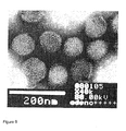
- Adenoviruses are medium-sized (90-100nm), nonenveloped icosahedral viruses, which have double stranded linear DNA of about 36 kilo base pairs in a protein capsid.
- the viral capsid has fiber structures, which participate in attachment of the virus to the target cell.
- the knob domain of the fiber protein binds to the receptor of the target cell (e.g. CD46 or coxsackievirus adenovirus receptor (CAR)), secondly, the virus interacts with an integrin molecule and thirdly, the virus is endocytosed into the target cell.
- the viral genome is transported from endosomes into the nucleus and the replication machinery of the target cell is utilized also for viral purposes ( Russell, W.C. 2000, J General Virol 81, 2573-2604 ).
- the adenoviral genome has early (E1-E4), intermediate (IX and IVa2) and late genes (L1-L5), which are transcribed in sequential order.
- Early gene products affect defense mechanisms, cell cycle and cellular metabolism of the host cell.
- Intermediate and late genes encode structural viral proteins for production of new virions ( Wu and Nemerow, 2004, Trends Microbiol 12: 162-168 ; Russell, W.C. 2000, J General Virol 81, 2573-2604 ; Volpers, C. and Kochanek, S. 2004, J Gene Med 6 suppl 1, S164-71 ; Kootstra, N.A. and Verma, I.M. 2003, Annu Rev Pharmacol Toxicol 43, 413-439 ).
- Serotypes are classified into six subgroups A-F and different serotypes are known to be associated with different conditions i.e. respiratory diseases, conjunctivitis and gastroenteritis. Nevertheless, all cancer trials utilizing recombinant adenoviruses have featured subgroup C viruses, which in most cases has been Ad5. Advantages of serotype chimeric capsids in the context of an otherwise serotype 5 virus have also been recognised. Even though serotype chimerism allows partial escape from neutralizing antibodies (Nab) existing against Ad5, the fact that most of the virus capsid is still from Ad5 renders the escape incomplete ( Särkioja, M. et al. 2008, Gene Ther 15: 921-929 ).
- ColoAd1 a complex Ad3/Ad11p chimeric virus ( Kuhn I et al. 2008, PLoS ONE 3: e2409 ).
- ColoAd1 was made by pooling an array of serotypes, then passaging the pools under conditions that invite recombination between serotypes. The authors call this method "Directed Evolution". These highly diverse viral pools were then placed under stringent directed selection to generate and identify potent agents.
- the Ad3/Ad11p chimeric virus was 2-3 orders of magnitude more potent and selective than the parent serotypes or the most clinically advanced oncolytic Ad, ONYX-015, in vitro.
- the document WO2005010149 describes a.o. human type 3 adenovirus and uses thereof, e.g. for ablating neoplastic cells in a cell population.
- WO2005030261 describes oncolytic human adenoviral vectors e.g. wherein expression of the E1A region is controlled by a promoter from the human telomerase (hTERT) gene.
- hTERT human telomerase
- the present invention provides a cancer therapeutic tool with the aforementioned properties by utilizing oncolytic human non-Ad5 viruses.
- the object of the disclosure is to provide novel methods and means for achieving the above-mentioned properties of adenoviruses and thus, solving the problems of current cancer therapies. More specifically, the invention provides novel methods and means for gene therapy as characterized in the claims.
- the present application describes construction of oncolytic human Ad3 vectors and their use in tumor cells lines and animal models, and it provides an alternative for oncolytic serotype 5 adenoviral vectors in virotherapies.
- the vector of the invention is the first selectively oncolytic human adenovirus based only on serotype 3, as characterized in the claims.
- the vector is also the first non-Ad5 based oncolytic adenovirus controlled by a tumor specific promoter, as characterized in the claims.
- the present invention relates to a fully serotype 3 oncolytic human adenoviral vector, as characterized in the claims.
- the present invention further relates to a cell comprising the adenoviral vector of the invention, as characterized in the claims.
- the present invention also relates to a pharmaceutical composition comprising the adenoviral vector of the invention, as characterized in the claims.
- the present invention relates to a use of the adenoviral vector of the invention in the manufacture of a medicament for treating cancer in a subject, as characterized in the claims.
- the present invention relates to the vector or pharmaceutical composition of the invention as an in situ cancer vaccine. Also, the present invention relates to a vector of the invention for use in the treatment of cancer in a subject.
- an adenoviral vector of the invention comprising:
- the present invention provides a tool for treatment of cancers, which are refractory to current approaches. Also, restrictions regarding tumor types suitable for treatment remain few in the present invention compared to many other treatments. In fact, all solid tumors may be treated with the proposed invention.
- the treatment can be given intratumorally, intracavitary, intra-arterially, intravenously and in a combination of these.
- Ad3 based viruses are able to enter cancer stem cell type cells better than Ad5 viruses. Efficacy of the virus of the present invention was shown in tumor cell lines and the virus was at least as potent as Ad5 or Ad5/3 based controls in several murine models of human cancer.
- the vector of the invention may also assure the expression and persistence of a transgene(s). Induction of an immune reaction towards cells that allow replication of unarmed viruses is normally not strong enough to lead to development of therapeutic tumor immunity. In order to overcome this weakness, the present invention provides armed viruses with a potent inducer of anti-tumor immunity (e.g. GM-CSF), as recited in the claims.
- GM-CSF potent inducer of anti-tumor immunity
- the present invention also relates to human sodium iodide symporter (hNIS) transgene as recited in the claims.
- Said in hNIS may target radioiodide to the target cell.
- This approach allows tumor cells to be killed due to the oncolytic effect of the virus and due to radiation induced cell death.
- the approach also takes advantage of synergy between radiation and oncolytic adenovirus replication.
- radioiodide therapy can be used to treat cancers of non-thyroid origin.
- An additional useful aspect of hNIS is non-invasive imaging of transgene expression, which allows monitoring viral spread and persistence.
- the present invention also solves a problem related to therapeutic resistance of conventional treatments as well as other adenoviral therapies.
- the use as claimed or the vector for the use as claimed may be for administration to the highly potential subject group of patients with high anti-Ad5 Nab titers or previously treated with Ad5 agents.
- the vectors and uses as characterized in the claims provide for selective treatments, without toxicity or damages in healthy tissues.
- advantages of the present invention may include different and reduced side effects in comparison to other therapeutics.
- the approach is synergistic with many other forms of therapy including chemotherapy and radiation therapy, and can therefore be used in combination regimens.
- the present invention provides a more simple, more effective, inexpensive, non-toxic and/or safer tool for cancer therapy. Furthermore, helper viruses are not needed.
- novel products of the invention enable further improvements in cancer therapy.
- an oncolytic human adenovirus vector based on serotype 3 and as recited in the claims can be constructed and it allows tumor cell killing.
- an icosahedral capsid consists of at least three major proteins: hexon, penton base, and fiber, along with minor proteins such as VI, VIII, IX, IIIa, and IVa2 ( Russell W.C. 2000, J General Virol 81, 2573-2604 ; Sirena D et al. 2005, Virology 343: 283-298 ).
- a fully serotype 3 oncolytic human adenoviral vector as recited in the claims comprises one or more regions selected from the group consisting of E1, E2, E3, E4, intermediate and late regions.
- an oncolytic human adenoviral vector as recited in the claims comprises the following regions: a left ITR, E1, pIX, pIVa2, E2, VA1, VA2, late region, E3 or partial E3, E4, and a right ITR.
- the regions may be in any order in the vector, but in one embodiment of the invention, the regions are in a sequential order in the 5' to 3' direction.
- Open reading frames ORFs
- ORFs may be in the same DNA strand or in different DNA strands.
- Ad3 adenovirus serotype 3
- Ad3 refers to the genome or partial genome of human Ad3, which comprises one or several regions selected from the group consisting of E1, pIX, pIVa2, E2, VA1, VA2, late region, E3 or partial E3, and E4 of Ad3 origin.
- partial region refers to a region, which lacks any part compared to a corresponding wild type region.
- Partial E3 refers to the E3 region lacking gp19k.
- VA1 and VA2 refer to virus associated RNAs 1 and 2, which are transcribed by the adenovirus but are not translated. VA1 and VA2 have a role in combating cellular defence mechanisms.
- exogenous tissue or tumor-specific promoters are common in recombinant adenoviral vectors and they can also be utilized in the present invention.
- viral replication can be restricted to target cells for example by promoters, which include but are not limited to hTERT, variants of hTERT, CEA, SLP, Cox-2, Midkine, E2F, variants of E2F, CXCR4, SCCA2 and TTS.
- promoters include but are not limited to hTERT, variants of hTERT, CEA, SLP, Cox-2, Midkine, E2F, variants of E2F, CXCR4, SCCA2 and TTS.
- other genes such as E1B or E4, can also be regulated by such a promoter in a vector according to the invention.
- an oncolytic human adenoviral vector as recited in the claims comprises the Ad3 E1A region under an hTERT promoter.
- Ad3-hTERT-E1A contains the human telomerase catalytic domain promoter up-stream of the E1A transcription site for tumor specific replication.
- telomere activation is a critical step in human carcinogenesis, and most human tumors feature activity of telomerase. This feature is closely linked with activity of hTERT promoter, which has been suggested to be active in most human tumors and therefore represents a useful tumor specific promoter.
- an oncolytic human adenoviral vector as recited in the claims is capable of replicating only in cells with telomerase activity.
- an oncolytic adenoviral vector as recited in the claims comprises the Ad3 E3 region under a replication activated Ad3 E3 promoter.
- the transgene is placed into a gp19k deleted E3 region, under the E3 promoter. This restricts the transgene expression to tumor cells that allow replication of the virus and subsequent activation of the E3 promoter.
- the E3 promoter may be any exogenous or endogenous promoter known in the art, preferably an endogenous promoter.
- Exogenous insulators i.e. blocking elements against unspecific enhancers, the left ITR, the native E1A promoter or chromatin proteins may also be included in recombinant adenoviral vectors. Any additional components or modifications may optionally be used but are not obligatory in the vectors of the present invention.
- the E3 region is nonessential for viral replication in vitro, but the E3 proteins have an important role in the regulation of the host immune response i.e. in the inhibition of both innate and specific immune responses.
- the vector of the invention may have deletion in the gp19k region in the E3.
- the gp19k gene product is known to bind and sequester major histocompatibility complex 1 (MHC1) molecules in the endoplasmic reticulum, and to prevent the recognition of infected cells by cytotoxic T-lymphocytes. Since many tumors are deficient in MHC1, deletion of gp19k increases the tumor selectivity of viruses (the virus is cleared faster than the wild type virus from normal cells but there is no difference in tumor cells).
- MHC1 major histocompatibility complex 1
- an oncolytic adenoviral vector as recited in the claims comprises one or more transgenes.
- the transgene is placed in E1 under the hTERT promoter and/or in E3 under the replication activated Ad3 E3 promoter.
- an oncolytic human adenoviral vector as recited in the claims comprises the transgene in the place of the deleted gp19k in the E3 region.
- an oncolytic adenoviral vector as recited in the claims comprises an internal ribosomal entry site (IRES) between the transgene and the E1A region.
- IRES refers to a nucleotide sequence that enables the initiation of the translation in the middle of a messenger RNA sequence in the protein synthesis.
- IRES is from encephalomyocarditis virus (EMCV), which is obtained from Clontech vector pIRES2-DsRed2.
- an oncolytic adenoviral vector comprises the hTERT promoter, transgene, IRES and E1A.
- the transgene is selected from the group consisting of a granulocyte-macrophage colony-stimulating factor (GM-CSF), human sodium iodide symporter (hNIS), interferon alpha, interferon beta, interferon gamma, tumor necrosis factor alpha, CD40L, trastuzumab and other monoclonal antibodies.
- GM-CSF granulocyte-macrophage colony-stimulating factor
- hNIS human sodium iodide symporter
- interferon alpha interferon beta
- interferon gamma tumor necrosis factor alpha
- CD40L trastuzumab
- trastuzumab trastuzumab
- GM-CSF participates in the immune response by acting through various mechanisms including the recruitment of natural killer (NK) cell and the stimulation of antigen presenting cells (APC). APC can then recruit, activate and target T-cells towards the tumor.
- the nucleotide sequence encoding GM-CSF may be from any animal, such as a human, ape, rat, mouse, hamster, dog or cat, but preferably GM-CSF is encoded by a human sequence.
- the nucleotide sequence encoding GM-CSF may be modified in order to improve the effects of GM-CSF, or unmodified, i.e. of a wild type. In a preferred embodiment of the invention, the nucleic acid sequence encoding GM-CSF is of a wild type.
- an oncolytic human adenoviral vector as recited in the claims comprises hNIS (SEQ ID NO. 15), which targets radioiodide to the target cell.
- hNIS sodium iodide symporter
- This approach allows tumor cells to be killed due to the oncolytic effect of the virus and due to radiation induced cell death, and the approach also takes advantage of synergy between radiation and oncolytic adenovirus replication.
- An additional useful aspect of hNIS is non-invasive imaging of transgene expression, which allows monitoring viral spread and persistence.
- hNIS is an integral plasma membrane glycoprotein mainly expressed in thyroid follicular cells ( Dohan O et al. 2003, Endocr Rev. 24: 48-77 ).
- the biological function of hNIS is to mediate active transport of iodine, which is a crucial component for thyroid hormone biosynthesis. This transporting ability of hNIS has been used for half a century in radioiodide therapy of thyroid carcinoma, where radioactive iodide molecules ( 131 I) are used to internally radiate cancer cells of thyroid origin.
- interferon alpha, interferon beta, interferon gamma, tumor necrosis factor, CD40L, trastuzumab and/or other monoclonal antibodies may also be utilized as transgenes in a vector or use as claimed for inducing the immune system of the patient against tumor cells, e.g., by activating natural killer cells, T-cells and/or macrophages.
- the vector of the invention as claimed may also comprise other modifications than partial deletions of E3 and insertion of transgenes and/or promoters as mentioned above.
- Any capsid modification i.e. modification of hexon, fibre and/or penton base proteins known in the art, which improves delivery of the virus to the tumor cell, may also be utilized in the present invention.
- capsid refers to the protein shell of the virus, which includes hexon, fiber and penton base proteins.
- Modifications may be genetic and/or physical modifications and include but are not limited to modifications for incorporating ligands, which recognize specific cellular receptors and/or block native receptor binding, for replacing the fiber or knob domain of an adenoviral vector with a knob of other adenovirus (chimerism) and for adding specific molecules (e.g. FGF2) to adenoviruses. Therefore, capsid modifications include but are not limited to incorporation of small peptide motif(s), peptide(s), chimerism(s) or mutation(s) into the fiber (e.g. into the knob, tail or shaft part), hexon and/or penton base.
- Expression cassettes are used for expressing transgenes in a target, such as a cell, by utilizing vectors.
- expression cassette refers to a DNA vector or a part thereof comprising nucleotide sequences, which encode cDNAs or genes, and nucleotide sequences, which control and/or regulate the expression of said cDNAs or genes. Similar or different expression cassettes may be inserted to one vector or to several different vectors.
- Ad3 vectors of the present invention as claimed may comprise either one or several expression cassettes. However, only one expression cassette is adequate.
- the oncolytic human adenoviral vector as claimed comprises at least one expression cassette.
- the oncolytic human adenoviral vector as claimed comprises only one expression cassette.
- a cell comprising the adenoviral vector of the invention as claimed may be any cell, such as a eukaryotic cell, bacterial cell, animal cell, human cell, mouse cell etc.
- the cell may be used for producing the adenoviral vector in vitro or in vivo, or the cell may be a target, such as a tumor cell, which has been infected with the adenoviral vector.
- the cancer is any solid tumor.
- the cancer is selected from a group consisting of nasopharyngeal cancer, synovial cancer, hepatocellular cancer, renal cancer, cancer of connective tissues, melanoma, lung cancer, bowel cancer, colon cancer, rectal cancer, colorectal cancer, brain cancer, throat cancer, oral cancer, liver cancer, bone cancer, pancreatic cancer, choriocarcinoma, gastrinoma, pheochromocytoma, prolactinoma, T-cell leukemia/lymphoma, neuroma, von Hippel-Lindau disease, Zollinger-Ellison syndrome, adrenal cancer, anal cancer, bile duct cancer, bladder cancer, ureter cancer, brain cancer, oligodendroglioma, neuroblastoma, mening
- a pharmaceutical composition of the invention comprises at least one type of the vectors of the invention as claimed. Furthermore, the composition may comprise at least two, three or four different vectors of the invention as claimed. In addition to the vector of the invention as claimed, a pharmaceutical composition may a so comprise any other vectors, such as other adenoviral vectors, other therapeutically effective agents, any other agents, such as pharmaceutically acceptable carriers, buffers, excipients, adjuvants, antiseptics, filling, stabilising or thickening agents, and/or any components normally found in corresponding products.
- any other vectors such as other adenoviral vectors, other therapeutically effective agents, any other agents, such as pharmaceutically acceptable carriers, buffers, excipients, adjuvants, antiseptics, filling, stabilising or thickening agents, and/or any components normally found in corresponding products.
- the pharmaceutical composition may be in any form, such as solid, semisolid or liquid form, suitable for administration.
- a formulation can be selected from a group consisting of, but not limited to, solutions, emulsions, suspensions, tablets, pellets and capsules.
- the oncolytic adenoviral vector or pharmaceutical composition is provided as an in situ cancer vaccine.
- an "in situ cancer vaccine” refers to a cancer vaccine, which both kills tumor cells and also increases the immune response against tumor cells.
- Tumor cell lysis also helps to present tumor fragments and epitopes to APCs and furthermore, costimulation is produced by inflammation.
- an epitope independent (i.e. not HLA restricted) response is produced in the context of each tumor and therefore takes place in situ.
- a tumor specific immune response is activated in the target cell as well as the surrounding cells, e.g. in the target tissue.
- the effective dose of vectors depends on at least the subject in need of the treatment, the tumor type, the location of the tumor and the stage of the tumor.
- the dose may vary for example from about 10 8 viral particles (VP) to about 10 14 VP, preferably from about 5x10 9 VP to about 10 13 VP and more preferably from about 8x10 9 VP to about 10 12 VP.
- compositions may be produced by any conventional processes known in the art, for example by utilizing any one of the following: batch, fed-batch and perfusion culture modes, column-chromatography purification, CsCl gradient purification and perfusion modes with low-shear cell retention devices.
- the vector or pharmaceutical composition of the invention may be administered to any eukaryotic subject selected from a group consisting of plants, animals and human beings.
- the vector for the use according to the invention or the use according to the invention in the manufacture of a medicament is for administration, to a human or an animal.
- An animal may be selected from a group consisting of pets, domestic animals and production animals.
- NAb neutralizing antibodies
- any conventional method may be used for administration of the vector or composition to a subject.
- the route of administration depends on the formulation or the form of the composition, the disease, the location of tumors, the patient, comorbidities and other factors.
- the formulation may be for administration conducted through an intratumoral, intramuscular, intra-arterial, intravenous, intrapleural, intravesicular, intracavitary or peritoneal injection, or an oral administration.
- oncolytic human adenoviral vectors of the invention may have therapeutic effects.
- oncolytic adenoviral vectors or pharmaceutical compositions are formulated for administration several times during the treatment period.
- Oncolytic human adenoviral vectors or pharmaceutical compositions may be administered for example from 1 to 10 times in the first 2 weeks, 4 weeks, monthly or during the treatment period.
- formulation is for administration three to seven times in the first 2 weeks, then at 4 weeks and then monthly.
- the length of the treatment period may vary, and may, for example, last from two to 12 months or more.
- NAb neutralizing antibody
- the vectors of the invention may vary between treatments.
- the vector of the invention for use in gene therapy is effective alone, but combination of adenoviral gene therapy with any other therapies, such as traditional therapy, may be more effective than either one alone.
- each agent of the combination therapy may work independently in the tumor tissue, the adenoviral vectors may sensitize cells to chemotherapy or radiotherapy and/or chemotherapeutic agents may enhance the level of virus replication or effect the receptor status of the target cells.
- the agents of combination therapy may be administered simultaneously or sequentially.
- the vector for the use according to the invention or the use according to the invention in the manufacture of a medicament is for administration, with concurrent radiotherapy or radioiodide to a subject.
- the vector for the use according to the invention or the use according to the invention in the manufacture of a medicament is for administration, with concurrent chemotherapy to a subject.
- concurrent refers to a therapy, which has been administered before, after or simultaneously with the gene therapy relying on the invention. The period for a concurrent therapy may vary from minutes to several weeks. Preferably the concurrent therapy lasts for some hours.
- Agents suitable for combination therapy include but are not limited to All-trans retinoic acid, Azacitidine, Azathioprine, Bleomycin, Carboplatin, Capecitabine, Cisplatin, Chlorambucil, Cyclophosphamide, Cytarabine, Daunorubicin, Docetaxel, Doxifluridine, Doxorubicin, Epirubicin, Epothilone, Etoposide, Fluorouracil, Gemcitabine, Hydroxyurea, Idarubicin, Imatinib, Mechlorethamine, Mercaptopurine, Methotrexate, Mitoxantrone, Oxaliplatin, Paclitaxel, Pemetrexed, Temozolomide, Teniposide, Tioguanine, Valrubicin, Vinblastine, Vincristine, Vindesine and Vinorelbine.
- Methods of producing adenoviral vectors include any conventional methods known in the art. Usually a transgene(s) is inserted into a shuttle plasmid and then homologous recombination is performed between the shuttle plasmid and a plasmid containing the remainder of the viral genome ("rescue plasmid"). The new genome can then be cut from the plasmid and transfected to virus production cells. Alternatively, the rescue plasmid can be co-transfected into producer cells for recombination in those cells.
- a plasmid construct is used for placing a transgene(s) to the adenoviral vector.
- the adenoviral vector is produced in a cell line.
- eukaryotic cells are transfected with the adenoviral vector and thereafter virus-producing cells are selected, expanded and tested.
- the viral vectors of the present invention also need extensive purification before administration to animals or human beings.
- 293 cells were purchased from Microbix (Toronto, Canada). 293-2v6-11 cells contain a ponasteron-inducible E4orf6 region ( Mohammadi E.S. et al. 2004, Nucleic Acids Res 32: 2652-2659 ). 911-1c11 cells were kept in 1mg/ml G418 ( Sirena, D. et al. 2005, Virology 343: 283-298 ). SKOV3.ip1 ovarian adenocarcinoma cell line was obtained from Dr. Price (M. D. Anderson Cancer Center, Houston, TX). Firefly luciferin-expressing ovarian adenocarcinoma cell line SKOV3-luc was kindly provided by Dr.
- ATCC Manassas, VA
- ACHN kidney cell carcinoma A549 lung adenocarcinoma
- FHS173WE human fibroblast cell line FHS173WE human fibroblast cell line
- HTC116 colorectal carcinoma CAMA-1 breast adenocarcinoma
- PANC-1 pancreatic epithelioid carcinoma All cell lines were cultured under recommended conditions.
- Ad3-hTERT-E1 For cloning of Ad3-hTERT-E1 three different PCR were performed on Ad3-wt plasmid pKSB2Ad3wt ( Sirena, D. et al. 2005, Virology 343: 283-298 ), two of them to exclude the original TATA-box from the Ad3wt sequence ( Figures 1-3 ).
- the first PCR product included LITR with a Mlul digestion site until the TATA-box (forward primer: 5'-CAT GGT ACC CAA GTG TGT CGC TGT CGA GT-3' (SEQ ID NO: 1), reverse primer 5'-CAT GAT ATC AGC GAT CAG CTG ACA CCT AC-3' (SEQ ID NO: 2); adding Kpnl site in front and EcoRV site at the end), the second started directly after the TATA-box until the first half of the E1A region (forward primer: 5'-CAT GAT ATC GTG CCA GCG AGA AGA GTT TT-3' (SEQ ID NO: 3), reverse primer 5'-CAT TCT AGA GCG AGC ACA ATA GTT CTT TCA-3' (SEQ ID NO: 4); adding EcoRV site at front and Xbal at the end).
- the respectively digested first two PCR products around the TATA-box were cloned into Kpnl/Xbal digested pUC19 cloning vector resulting in pGBAd3E1, including a TATA-box excluded start of the Ad3 sequence.
- the hTERT promoter derived from pBT255 was digested with KpnI/XhoI, blunted, and cloned in front of the E1 region by a EcoRV digestion of pGBAd3E1 resulting in pGBAd-3hTERT-E1.
- the third PCR product with the end of the Ad3 sequence was now cloned into NotI/blunted pShuttle, followed by digestion with HindIII, and subsequently cloned into HindIII digested pGBAd3-hTERT-E1 resulting in pSGBAd3-hTERT-E1 as shuttle vector with end and modified start of the adenovirus 3 genome.
- pSGBAd3-hTERT-E1 was digested with XhoI/MscI, resulting in the plasmid being opened up in between the modified start and preserved end of the adenovirus 3 sequence, and homologous recombined with Mlul digested and thus backbone-free pKSB2Ad3wt, to reduce background due to same resistance gene and to achieve a plasmid with Ad3-hTERT-E1 sequence (pKGB-Ad3-hTERT-E1).
- Ad3-hTERT-E1A virus was rescued by transfecting 911-1c11 cells with Mlul-digested pKGB-Ad3-hTERT-E1. Subsequent amplification of a first large Ad3-hTERT-E1A virus stock was done in 293-2v6-11 cells, followed by 2 nd amplification in A549 cells. After amplification the viruses were purified on double cesium chloride gradients. The stocks were confirmed not to have serotype 5 contamination by PCR (for the standard method, see Kanerva, A. et al. 2003, Mol Ther 8: 449-458 ). Then the presence of the hTERT promoter in front of the E1A was confirmed ( Fig.
- the primers used to get the 270bp band were 5'-GGT TAT GCC AGG GTG GAG TA-3' forward (SEQ ID NO: 5) and 5'-AAG GTG AAG GGG CAG GAC-3' reverse (SEQ ID NO: 6).
- the primers used were 5'-AGC CCC TCC CCT TCC TTT-3' forward (SEQ ID NO: 7) and 5'-CCC GGT CTC ACT GGA GAT AA-3' reverse (SEQ ID NO: 8).
- the PCR bands were then sequenced and the expected sequence was obtained (see SEQ ID NO: 9 and Figure 10 ).
- Table 1 for adenoviruses relevant to this invention.
- Table 1 Adenoviruses Virus name Back-bone sero-type Fiber knob sero-type E1A Ratio (VP/pfu) Reference Ad5 wt 5 5 Wild type 19 ATCC (Ad300 wt strain) Ad5/3-hTERT-E1A 5 3 Controlled by hTERT promoter (mediates selectivity to telomerase active cells) 10 Bauerschmitz, G.J. et al.
- the cells were plated on 96-well plates, 10 000 cells/well and rows were infected the next day with decreasing 10-fold dilutions from 10 -5 to 10 -12 of Ad3-hTERT-E1A, each dilution in ten duplicates.
- the infection was done in DMEM, 2% FBS.
- the plates were observed by microscope and the virus wells were compared to the mock wells. From the observations pfu/ml was calculated in the similar way as in TCID 50 (Adeasy manual, Agilent Technologies, Inc. 2008).
- the media (DMEM, 5% FBS) was changed every 4 to 7 days.
- the cells were plated at 10 000 cells/well on 96 well plates. The next day, the cells in triplicates or quadruplicates were infected with the viruses at 0.1 to 100 VP/well. The infection was done in 2% fetal bovine serum (FBS). The plates were regularly observed and the growth media was changed every 3-5 days. Six to twenty days later, as optimal for each line, the cell viability was analyzed with MTS assay (Cell Titer 96 AQueous One Solution Cell Proliferation Assay, Promega).
- FBS fetal bovine serum
- MTS analyzes were performed on the following days: PC-3MM2 on day 8, A549 on day 17, HTC116 on day 6, SKOV3.ip1 on day 18, HUVEC on day 11, FSH174WE on day 13, LNM-35/EGFP on day 10, CAMA-1 on day 11, PANC-1 on day 20 and ACHN on day 11.
- the results are shown in Figures 4A-D .
- Ad3-hTERT-E1A was less toxic than the control viruses, including wild type Ad3.
- Ad3-hTERT-E1A was not able to kill cells that lack the Ad3 receptor.
- Ad3-hTERT-E1A The ability of Ad3-hTERT-E1A to kill cancer cells in vitro was analyzed by infecting monolayers representing seven different tumor types ( Figures 4A-D and Figures 7A-C ). In all malignant cell lines Ad3-hTERT-E1A showed complete oncolysis, while the non-replicative Ad5/31ucl virus showed no cell killing (p ⁇ 0.05). However, as anticipated based on experience from growing and titering the virus, Ad3-hTERT-E1A was somewhat slower than the serotype 5 viruses.
- HUVEC human umbilical vein endothelial cells
- FSH173WE fibroblasts
- Fig. 5 For analyzing the tumor selectivity, we infected HUVEC (human umbilical vein endothelial cells) and FSH173WE (fibroblasts) with the same viruses and performed cell killing assays ( Fig. 5 ). No cell killing was seen at low virus concentrations and at higher doses Ad3-hTERT-E1A was less toxic than the control viruses Ad3wt, Ad5wt, Ad5/3-hTERT-E1A, Ad5/3- ⁇ 24 (0.1, 1 and 10 VP/cell P ⁇ 0.05). From the previous work with Ad5/3 chimeras, we knew that LNM-35/EGFP cannot be infected with Ad5/3 and thus may lack the Ad3 receptor ( Särkioja, M. et al. 2006, Cancer 107: 1578-1588 ). Fittingly, no cell killing was seen with Ad3-hTERT-E1A or Ad3 wild type.
- mice All animal experiments were approved by the Experimental Animal Committee of the University of Helsinki and the Provincial Government of Southern Finland. Mice where frequently monitored for their health status and euthanized as soon as signs for pain or distress was noticed.
- subcutaneus model female NMRI nude mice were used (Charles River, Germany). They were ordered at the age of 3-4 weeks and quarantined for 2 weeks.
- intraperitoneal model female fox chase SCID mice (Charles River, Germany) were used. They were ordered at the age of 6-7 weeks and quarantined for 4 months.
- Ad3-hTERT-E1A was at least as oncolytic as Ad5 and Ad5/3 based controls in vivo .
- Subcutaneous tumors may not be optimal surrogates of human cancers and therefore we proceeded to utilize an orthotopic model featuring intraperitoneally disseminated carcinomatosis induced with luciferase expressing SKOV3Luc cells ( Figures 6C, D ).
- Ad3-hTERT-E1A significantly reduced the luciferase signal as compared to PBS treated mice (p ⁇ 0.0001). There was no difference in efficacy between the different replication competent viruses.
- One out of the 7 mice treated with Ad3-hTERT-E1A survived until the end of the experiment (day 120) and may have been cured as no evidence of tumor could be detected in luciferase imaging or autopsy and its behavior was normal.
- Ad5wt 5/5: Many apoptotic hepatocytes throughout the parenchyme, endothelitis and damage in central veins (at the portal area same can not be seen), no pericentral hepatocyte destruction, steatosis, nekrosis, no lymfocytes in the portal area, acute liver damage, damage througout the parenchyme.
- Ad5/3-hTERT-deltagp19k 5/5: Almost normal, some point necrosis (not as much as in Ad5wt), some apoptotic cells, mitosis of hepatocytes, (lobular) inflammation in the parenchyme, inflammation (as with Ad3) around the veins.
- Ad5/3- ⁇ 24 5/5: Acute fulminant liver failure, 50% of tissue in necrosis, damage stronger near the portal area where all cells are dead, possible fatty generation, some mitoses.
- Nonparametric Man-Whitney test (SPSS 15.0 for Windows) was used to compare two independent samples for all the in vitro and some of the in vivo data.
- the analysis of tumor size was performed using a repeated measures model with PROC MIXED (SAS Ver. 9.1). Models were run with the tumor size measurements in the natural metric and log transformed. The effects of the treatment group, time in days and the interaction of the treatment group and time were evaluated by F tests. Curvature in the models was tested for by a quadratic term for time. The a priori planned comparisons of specific differences in predicted treatment means averaged over time and at the last timepoint were computed by t-statistics.
- Viral constructs comprising HNIS and/or GMCSF and/or CD40L
- HNIS and/or GMCSF transgene is (are) added to the human Ad3 based vector of the invention by utilizing methods known in the art or as described in the above examples or in the examples below, wherein a non-Ad5 adenoviral vector of the present invention, for example Ad3-hTERT-E1A, can be used instead of the Ad5/3- ⁇ 24 backbone (see also Figures 1 and 2 ):
- Andorgen independent prostate cancer cell lines 22Rv1, PC-3, DU-145 and lung adenocarcinoma cell line A549 were purchased from ATCC (Manassas, VA).
- PC-3MM2 cells are a metastatic hormone refractory sub line of PC-3 (courtesy of Judith J. Fidler, MD Anderson Cancer Center, Houston, TX).
- Human embryonic kidney epithelial 293 cells were purchased from Microbix (Toronto, Canada).
- Cell line 911 was obtained from Dr Alex J. van der Eb (University of Leiden, The Netherlands). All cell lines were cultured in the recommended conditions.
- Adenoviral plasmids were linearized and transfected into 911 cells with Superfect (Qiagen, Valencia, CA) following the manufacturer's instructions. Adenovirus colonies were picked and viruses were propagated in A549 cells and purified using standard techniques. Ad5/3- ⁇ 24 and Ad5/3luc1 viruses were constructed previously ( Kanerva, A. et al. 2003, Mol Ther 8: 449-58 ; Kanerva, A, et al. 2002, Clin Cancer Res 8: 275-80 ). Viral particles (VP) were determined with spectrophotometry and plaque forming units (pfu) with TCID50 assay.
- Titers were Ad5/3- ⁇ 24- hNIS: 1.1 x 10 12 VP/ml; 9.0 x 10 10 pfu/ml, Ad5/3- ⁇ 24- ⁇ gp19K: 1.5 x 10 12 VP/ml; 2.8 x 10 11 pfu/ml, Ad5/3- ⁇ 24: 1.7 x 10 12 VP/ml and Ad5/3luc1: 6.9 x 10 11 VP/ml.
- Prostate cancer cells were infected with Ad5/3- ⁇ 24-hNIS and Ad5/3- ⁇ 24- ⁇ gp19K (10 VP/cell) for 2 h at +37°C. Cells were harvested 24 and 48 h after infection. Total RNA was isolated using the RNeasy Kit (Qiagen) and treated with DNase before RT-PCR. 350 ng of total RNA was used for each reaction.
- Amplification (35 cycles, annealing at +54°C) was carried out with the OneStep RTPCR Kit (Qiagen) using hNIS-specific primers (Forward 5'-CTT CTG AAC TCG GTC CTC AC-3' (SEQ ID NO: 10); Reverse 5'-TCC AGA ATG TAT AGC GGC TC-3' (SEQ ID NO: 11) and ⁇ -actin-specific primers (Forward 5'- CGA GGC CCA GAG CAA GAC A -3' (SEQ ID NO: 12); Reverse 5'-CAC AGC TTC TCC TTA ATG TCA CG -3' (SEQ ID NO: 13).
- PCR product size for HNIS and ⁇ -action was 453 bp and 482 bp, respectively.
- Prostate cancer cells were infected with Ad5/3- ⁇ 24-hNIS and Ad5/3- ⁇ 24- ⁇ gp19K (10 VP/cell) for 2 h at +37°C. Cells were washed with 1 x PBS 24 and 48 h post infections and incubated with 7.4 kBq of sodium iodide [ 125 I]Nal (MAP Medical Technologies Oy, Tikkakoski, Finland) for 20 min at room temperature. Cells were washed twice with 1 x PBS followed by lysis using 300 ⁇ l of Cell Culture Lysis Reagent for each sample (Promega, Madison, WI). Radioactivity was quantified with a well counter connected with a multi-channel analyzer Atomlab 950 (Biodex Medical System, Shirley, NY).
- Prostate cancer cells were infected with Ad5/3- ⁇ 24-hNIS, Ad5/3- ⁇ 24- ⁇ gp19K, Ad5/3- ⁇ 24 and Ad5/3luc1 using 0.01, 0.1, 1, 10 and 100 VP/cell. Growth medium was replaced every other day. Cell viability was determined 6 days post infection (p.i.) for PC-3MM2, 7 days p.i. for DU-145 and PC-3 and 9 days p.i. for 22Rv1 cells.
- Cell line samples 5 x 10 4 prostate cancer cells/well were plated in triplicates in 1 ml of 5% GM on 24-well plates, incubated overnight and infected with Ad5/3- ⁇ 24-hNIS (5 vp/cell) for 2 h at +37°C. At 8, 24, 48 and 72 h post infection cells and growth media were collected, frozen at -80°C and freeze-thawn three times. For TCID50, the cells were spun down and the supernatant was added to 293 cells.
- Tissue samples Organs collected from mice treated with Ad5/3- ⁇ 24-hNIS and 131 I were collected, weighed, manually homogenized and diluted into 800 ⁇ l of DMEM. They were freeze-thawed at -80°C three times. For TCID50, the tissue samples were spun down and the supernatant was added to 293 cells.
- mice Male NMRI/nude mice were purchased from Taconic (Ejby, Denmark). Tumors were established in eight-week old mice by subcutaneous injection of 5 x 10 6 PC-3MM2 cells under medetomidineketamine- 0.9% saline (1:2:7) anesthesia. In the imaging experiment, 3 mice received intratumorally saline (upper left tumor), 7 x 10 8 VP Ad5/3- ⁇ 24- ⁇ gp19K (upper right tumor) and 7 x 10 8 VP Ad5/3- ⁇ 24-hNIS (lower right and left tumors) 11 and 12 days after tumor cell inoculation.
- mice received intravenously 1.85 MBq of sodium iodide [ 123 I]Nal (MAP Medical Technologies Oy, Tikkakoski, Finland) followed by imaging with gamma camera Toshiba 7200A/UI (Toshiba, Tokyo, Japan) 0.5 h, 1 h, 1.5 h, 2h, 4h and 13h after iodide exposure. Animal regulation did not allow analysis of later time points. The gamma energy peak 159 keV was recorded using 20% window, and 150 kilocounts were collected per image. The used matrix size was 256 x 256 x 16, this made the pixel size 1.08 mm x 1.08 mm.
- mice were randomized into 4 groups (6 mice/group, 2 tumors/mouse): mock, 131I, Ad5/3- ⁇ 24-hNIS and Ad5/3- ⁇ 24-hNIS + 131 I.
- 6 and 7 days after tumor cell inoculation mice received intratumorally either growth medium or 7 x 10⁸ VP of Ad5/3- ⁇ 24-hNIS per tumor.
- mice received intraperitoneally 50 MBq of carrier-free sodium iodide ([ 131 I]Nal, MAP Medical Technologies Oy, Tikkakoski, Finland). Tumor size was measured every other day and the volume calculated using the formula (length x width2 x 0.5).
- mice When mice were killed due to the tumor size, the organs were collected, weighed and their radioactivity determined with a gamma counter (Wizard 3, PerkinElmer, Turku, Finland) for 180 seconds. The results are expressed as percentage from initial iodide dose per gram of tissue (ID%/g). The whole body emitted radioactivity was measured at days 9, 10, 11, 13, 14, 15, and 17. On days 9-11, measurements were performed on a dose calibrator (CRC-120, Capintec, Inc., Ramsey, NJ), and after day 11 on a whole-body measurement apparatus consisting of a lead measurement chamber and an UniSpec tube-base multichannel analyzer with sodium iodide scintillation crystal (Canberra Industries, Inc., Meridien, CT). The mice were weighed at day 14, and the same weight was used in calculations throughout the experiment.
- a dose calibrator CRC-120, Capintec, Inc., Ramsey, NJ
- a whole-body measurement apparatus consisting of a lead measurement chamber and an UniSpec tube-base
- mice were killed when the tumor size reached 15 mm in any diameter, as required by animal regulations.
- the therapeutic in vivo experiment had to be stopped on day 17 due to animal husbandry constraints. All the animal experiments were approved by the national committee for animal experimentation in Finland (State Provincial Office of Southern Finland, Hämeenlinna, Finland).
- Analysis of the iodide uptake was performed by one-way analysis of variance (ANOVA) with Bonferroni's post-hoc test.
- the analysis of the tumor volume over time between experimental groups was performed using a repeated measures model with PROC MIXED (SAS Ver. 9.1, Cary, NC).
- the tumor volume measurements were log transformed for normality.
- the a priori planned comparisons of specific differences in predicted treatment means averaged over time and at each time point by t-statistics and Tukey-Kramer model was used for adjustments. For all analyses a two-sided p value of ⁇ 0.05 was deemed statistically significant.
- HNIS specific mRNA is detected in Ad5/3- ⁇ 24-hNIS infected prostate cancer cells
- a new virus containing the Ad3 knob in the Ad5 fiber shaft, a 24 bp deletion in the E1A region and HNIS inserted in the E3 was constructed.
- hormone refractory prostate cancer cell lines PC-3MM2, 22Rv1, PC-3 and DU-145 were infected with Ad5/3- ⁇ 24-hNlS or Ad5/3- ⁇ 24- ⁇ gp19K (a positive control without a transgene) and analysed with RT-PCR 24 and 48h after infection. All Ad5/3- ⁇ 24-hNIS infected cell lines featured a 454 bp sized, HNIS specific amplification product, suggesting that HNIS is expressed from the virus genome. In contrast, Ad5/3- ⁇ 24- ⁇ gp19K infected cells or cells per se did not yield to an amplification product.
- Ad5/3- ⁇ 24-hNIS can mediate efficient iodide uptake into prostate cancer cells
- prostate cancer cells were infected with Ad5/3- ⁇ 24-hNIS or Ad5/3- ⁇ 24- ⁇ gp19K followed by exposure to 125 I and quantification of the iodide accumulation ( Fig. 11 ).
- Ad5/3- ⁇ 24-hNIS resulted in a significantly higher iodide uptake in all examined cell lines.
- Ad5/3- ⁇ 24-hNIS caused up to 2.5 ( p ⁇ 0.001) and 3 ( p ⁇ 0.001) times higher iodide accumulation in PC-3MM2 and 22Rv1 cells, respectively.
- Ad5/3- ⁇ 24-hNIS replicates in and causes oncolysis of prostate cancer cells
- Prostate cancer cell lines were infected with Ad5/3- ⁇ 24-hNIS, Ad5/3- ⁇ 24- ⁇ gp19K, Ad5/3- ⁇ 24 and Ad5/3luc1 followed by determination of cell viability 6 (PC3-MM2), 7 (PC-3 and DU-145) or 9 (22Rv1) days post infection ( Fig. 12 ).
- Ad5/3- ⁇ 24-hNIS showed efficient cell killing in all examined prostate cancer cell lines. The most rapid and efficient oncolysis was seen in PC-3MM2 cells, where almost complete cell killing was achieved 6 days after infection using 1 VP/cell of Ad5/3- ⁇ 24-hNIS. In addition, oncolysis was similar to that seen with Ad5/3- ⁇ 24- ⁇ gp19K.
- Ad5/3- ⁇ 24-hNIS In other prostate cancer cell lines, the oncolytic potency of Ad5/3- ⁇ 24-hNIS was slightly less than Ad5/3- ⁇ 24- ⁇ gp19K. In fact, Ad5/3- ⁇ 24- ⁇ gp19K which has the partial deletion in E3, showed the highest oncolytic potency in all examined cell lines. It is possible that the larger genome size of Ad5/3- ⁇ 24-hNIS may impact the speed of oncolysis as compared to Ad5/3- ⁇ 24- ⁇ gp19K.
- the HNIS transgene is ⁇ 2.2 kbp, which together with the 1 kbp ⁇ gp19K keeps the genome size under 105%, which has been suggested important for retaining virus functionality.
- the replicativity of Ad5/3- ⁇ 24-hNIS was assessed in PC-3 and 22Rv1 cells where the TCID50 assay showed the increase in infectious particles in function of time.
- Ad5/3- ⁇ 24-hNIS can mediate radioactive iodide uptake into prostate cancer tumors
- Subcutaneous PC-3MM2 tumors were established in nude/NMRI male mice.
- the lower tumors were infected intratumorally on two consecutive days with 7 x 10 8 VP/tumor of Ad5/3- ⁇ 24-hNIS and the upper right tumor with the same dose of Ad5/3- ⁇ 24- ⁇ gp19k ( Figs. 13A and B ).
- the upper left tumor was mock treated.
- a day after the last viral injection 1.85 MBq of 123 I per mouse was administered intravenously and mice were imaged with a gamma camera from 0.5 to 13 h after the 123 I injection.
- Some iodide accumulation into Ad5/3- ⁇ 24-hNIS treated tumors was seen already 0.5 h after the 123 I injection.
- Adenoviruses lose their capsid when they enter cells and thus the input virus cannot be detected with TCID50; it requires production of new virions.
- the iodide uptake remained low in other organs including mock or Ad5/3- ⁇ 24- ⁇ gp19K treated tumors ( ⁇ 1% of the initial dose).
- Ad5/3- ⁇ 24-hNIS with 131 I inhibits tumor growth in vivo
- Radioactivity was measured starting from one day after the iodide injection. Radioactivity declined rapidly during the next 3 days as expected by clearance through kidneys.
- mice When tumor size reached 15 mm in any diameter, mice were killed and organs were collected for radioactivity measurements. Radioactivity declined over time and most radioactivity was seen in the thyroid gland and stomach, and briefly in the heart probably due to propagation of 131 I by circulation. In other organs the radioactivity remained low. Importantly, the virus injection did not affect iodide biodistribution and therefore the safety profile of the approach resembles that of radioiodide therapy for thyroid cancer.
- GM-CSF is under the E3 promoter, which results in replication associate transgene expression, which starts about 8h after infection. E3 is intact except for deletion of 6.7Klgp19K.
- Ad5/3-D24-GM-CSF and Ad5-RGDD24-GM-CSF were constructed identically, except a rescue plasmid featuring either a knob from serotype 3, or RGD-4C in the Ad5 fiber HI-loop were used.
- Ad5Luc1 is a replication deficient virus and acts as a negative control.
- Ad5wt is a wild type Ad5 virus (strain Ad300wt) and was used as a positive control.
- Ad5-d24-E3 harbors an isogenic 24bp deletion in E1 but is intact in E3. VP indicates virus particles.
- Ad5-D24-GMCSF had oncolytic activity similar to positive controls in vitro, and therefore transgene production did not compromise the oncolytic potency of the virus ( Figures 15a-c ). Similar data was shown for Ad5/3-D24-GM-CSF and Ad5-RGD-D24-GM-CSF ( Figure 15d ).
- Ad5D24-GMCSF was able to express the transgene
- A549 cell line was infected with 1000 VP/cell and media were collected over time.
- the concentration of GMCSF ( Figure 16a ) in the media was measured by FACSARRAY.
- TF1 cell line whose growth and survival is strictly dependent on human GMCSF, was treated with media collected from a A549 cell line previously infected with Ad5D24-GMCSF.
- the TF1 viability was assessed over time by the MTS assay.
- the result of this experiment was that the virus expressed-GMCSF was able to stimulate growth of such cell line, and no difference was found with the same cell line treated with human recombinant GMCSF (Sigma) ( Figure 16b ).
- Ad5-D24-GM-CSF The in vivo efficacy of Ad5-D24-GM-CSF was tested in immune competent Syrian hamsters, which are semipermissive for human adenovirus replication (mice are non-permissive) ( Ying B. et al. 2009, Cancer Gene Ther doi:10.1038/cgt.2009.6.). 7x10 6 HapT1 pancreatic cancer cells were injected subcutaneously and 1x10 9 virus particles (VP) of Ad5-D24-GM-CSF or Ad5D24E3 (which does not express GM-CSF) were injected intratumorally on day 0, 2 and 4. The mock group was injected with the same volume of growth media at the same indicated time points.
- VP virus particles
- Figure 17b shows that intratumoral injections of Ad5-D24-GMCSF resulted in high levels of hGM-CSF in the serum of Syrian hamsters.
- Human GM-CSF is known to be active in Syrian hamsters ( Cho, Exp Toxicol Pathol 2006 vol. 57 (4) pp. 321-8 ).
- all animals were tumor-free by day sixteen except for mock group ( Figure 17a ). Tumor scars were still analyzed for two additional weeks to assess whether reappearance of the tumor might have occurred. However, on day 32 there was still no sign of tumor in these animals so that the first part of the experiment was terminated and animals of the Mock group were euthanized.
- the data indicates that Ad5-D24-GM-CSF has an antitumor activity in immune competent tumor bearing animals, and it is able to elicit tumor-specific immunity to the degree of rejecting subsequent challenge of the same tumor.
- Ad3-hTERT-CD40L-EIA was constructed and analyzed for biodistribution in mice ( Figures 20a-b ).
- Murine biodistribution at 6h after 5x10 10 VP intravenously ( Figure 20a ) suggested that much of the serotype 3 virus stays still in the blood and blood rich organs, such as spleen, lung and the liver. All major organs were analyzed by qPCR ( Figure 20b ). Entry to other organs was found attenuated.
- blood clots ⁇ -actin was used for the serum values. Due to the lack of ⁇ -actin in blood compartments, blood values are best compared with absolute values as done in Fig 20a .
Landscapes
- Health & Medical Sciences (AREA)
- Life Sciences & Earth Sciences (AREA)
- Chemical & Material Sciences (AREA)
- Engineering & Computer Science (AREA)
- Genetics & Genomics (AREA)
- General Health & Medical Sciences (AREA)
- Bioinformatics & Cheminformatics (AREA)
- Organic Chemistry (AREA)
- Zoology (AREA)
- Medicinal Chemistry (AREA)
- Wood Science & Technology (AREA)
- Biotechnology (AREA)
- Pharmacology & Pharmacy (AREA)
- Animal Behavior & Ethology (AREA)
- Public Health (AREA)
- Veterinary Medicine (AREA)
- Biomedical Technology (AREA)
- Virology (AREA)
- General Engineering & Computer Science (AREA)
- Microbiology (AREA)
- Epidemiology (AREA)
- Biochemistry (AREA)
- Immunology (AREA)
- Molecular Biology (AREA)
- Gastroenterology & Hepatology (AREA)
- Proteomics, Peptides & Aminoacids (AREA)
- Chemical Kinetics & Catalysis (AREA)
- Physics & Mathematics (AREA)
- Plant Pathology (AREA)
- Biophysics (AREA)
- Nuclear Medicine, Radiotherapy & Molecular Imaging (AREA)
- General Chemical & Material Sciences (AREA)
- Mycology (AREA)
- Cell Biology (AREA)
- Physical Education & Sports Medicine (AREA)
- Orthopedic Medicine & Surgery (AREA)
- Oncology (AREA)
- Hematology (AREA)
- Rheumatology (AREA)
- Medicines Containing Material From Animals Or Micro-Organisms (AREA)
Description
- The present invention relates to the field of medicine. Specifically, the invention relates to cancer therapies. More specifically, the present invention relates to oncolytic human adenoviral vectors and cells and pharmaceutical compositions comprising said vectors. The present invention also relates to a use of said vectors in the manufacture of a medicament for treating cancer in a subject. Furthermore, the present invention relates to a method of producing an adenoviral vector.
- Cancer can be treated with surgery, hormonal therapies, chemotherapies and/or radiotherapies but in many cases, cancers, which are often characterized by an advanced stage, cannot be cured with present therapeutics. Therefore, novel cancer cell targeted approaches such as gene therapies are needed.
- During the last twenty years gene transfer technology has been under intensive examination. The aim of cancer gene therapies is to introduce a therapeutic gene into a tumor cell. These therapeutic genes introduced to a target cell may for example correct mutated genes, suppress active oncogenes or generate additional properties to the cell. Suitable exogenous therapeutic genes include but are not limited to immunotherapeutic, anti-angiogenic, chemoprotective and "suicide" genes, and they can be introduced to a cell by utilizing modified virus vectors or non-viral methods including electroporation, gene gun and lipid or polymer coatings.
- Requirements of optimal viral vectors include an efficient capability to find specific target cells and express the viral genome in the target cells. Furthermore, optimal vectors have to stay active in the target tissues or cells. All these properties of viral vectors have been developed during the last decades and for example retroviral, adenoviral and adeno-associated viral vectors have been widely studied in biomedicine.
- To further improve tumor penetration and local amplification of the anti-tumor effect, selectively oncolytic agents, e.g. conditionally replicating adenoviruses, have been constructed. Oncolytic adenoviruses are a promising tool for treatment of cancers. Tumor cells are killed by oncolytic adenoviruses due to a replication of the virus in a tumor cell, the last phase of the replication resulting in a release of thousands of virions into the surrounding tumor tissues for effective tumor penetration and vascular re-infection. Treatment approaches take advantage of molecular differences between the normal and tumor cells. Tumor cells allow replication of the virus while normal cells are spared due to engineered changes in the virus genome, which prevent replication in non-tumor cells. An optimal oncolytic adenovirus would infect and replicate only in cancer cells and would be potent enough to kill all cancer cells before the immunological response neutralizes the virus.
- Making certain deletions or adding tissue specific promoters can make the virus more selective while adding transgenes can make the approach more potent. Indeed, in addition to replication mediated cell killing, oncolytic adenoviruses can also be armed with different therapeutic transgenes. This approach combines the advantages of conventional gene delivery with the potency of replication competent agents. One goal of arming viruses is induction of an immune reaction towards the cells that allow virus replication. Virus replication alone, although immunogenic, is normally not enough to induce effective anti-tumor immunity. To strengthen induction of therapeutic immunity, viruses can be armed with stimulatory proteins such as cytokines and by facilitating the introduction of tumor antigens to dendritic cells. Introduction of immunotherapeutic genes into tumor cells and furthermore, translation of the proteins, leads to activation of the immune response and efficient destruction of tumor cells.
- Adenoviruses are medium-sized (90-100nm), nonenveloped icosahedral viruses, which have double stranded linear DNA of about 36 kilo base pairs in a protein capsid. The viral capsid has fiber structures, which participate in attachment of the virus to the target cell. First, the knob domain of the fiber protein binds to the receptor of the target cell (e.g. CD46 or coxsackievirus adenovirus receptor (CAR)), secondly, the virus interacts with an integrin molecule and thirdly, the virus is endocytosed into the target cell. Next, the viral genome is transported from endosomes into the nucleus and the replication machinery of the target cell is utilized also for viral purposes (Russell, W.C. 2000, J General Virol 81, 2573-2604).
- The adenoviral genome has early (E1-E4), intermediate (IX and IVa2) and late genes (L1-L5), which are transcribed in sequential order. Early gene products affect defense mechanisms, cell cycle and cellular metabolism of the host cell. Intermediate and late genes encode structural viral proteins for production of new virions (Wu and Nemerow, 2004, Trends Microbiol 12: 162-168; Russell, W.C. 2000, J General Virol 81, 2573-2604; Volpers, C. and Kochanek, S. 2004, J Gene Med 6 ; Kootstra, N.A. and Verma, I.M. 2003, Annu Rev Pharmacol Toxicol 43, 413-439).
- More than 50 different serotypes of adenoviruses have been found in humans. Serotypes are classified into six subgroups A-F and different serotypes are known to be associated with different conditions i.e. respiratory diseases, conjunctivitis and gastroenteritis. Nevertheless, all cancer trials utilizing recombinant adenoviruses have featured subgroup C viruses, which in most cases has been Ad5. Advantages of serotype chimeric capsids in the context of an otherwise
serotype 5 virus have also been recognised. Even though serotype chimerism allows partial escape from neutralizing antibodies (Nab) existing against Ad5, the fact that most of the virus capsid is still from Ad5 renders the escape incomplete (Särkioja, M. et al. 2008, Gene Ther 15: 921-929). - Thus, there is a need for a fully non-Ad5 oncolytic virus, which is able to escape from Nab existing against Ad5. These viruses are useful especially in the context of pre-existing NAb against Ad5. This situation might arise as a result of natural infection or subsequent to treatment with an Ad5 based oncolytic virus.
- Currently, little has been published on
serotype 3 adenoviruses in the context of gene therapy. Wild type Ad3 is known to cause mainly respiratory infections and conjuctivitis in humans. The complete DNA sequence was reported in 2005 and it has only 62.75% identity withserotype 5. The genomic organisation is similar to other human adenoviruses having early and delayed early transcription units, including late and major late units (Sirena D et al. 2005, Virology 343: 283-298). A few years ago a wild type Ad3 virus was studied under normoxic and hypoxic conditions (Shen, B.H. et al. 2006, Gene Ther 13: 986-990). Recently, the same group published ColoAd1, a complex Ad3/Ad11p chimeric virus (Kuhn I et al. 2008, PLoS ONE 3: e2409). ColoAd1 was made by pooling an array of serotypes, then passaging the pools under conditions that invite recombination between serotypes. The authors call this method "Directed Evolution". These highly diverse viral pools were then placed under stringent directed selection to generate and identify potent agents. In their experiments they found out that the Ad3/Ad11p chimeric virus was 2-3 orders of magnitude more potent and selective than the parent serotypes or the most clinically advanced oncolytic Ad, ONYX-015, in vitro. These results were further supported by in vivo and ex vivo studies. ColoAd1, however, given the approach utilized to develop it, lacks a rational basis for tumor selectivity. Also, its activity and safety in vivo is poorly understood as it has only been studied in one animal model, featuring colon cancer xenografts in mice. - The document
WO2005010149 describes a.o.human type 3 adenovirus and uses thereof, e.g. for ablating neoplastic cells in a cell population. - The document
US2005214923 describes oncolytic human adenoviral vectors comprising the E1A region under an hTERT promoter (see e.g. Example 4: OV1025). - The document
WO2005030261 describes oncolytic human adenoviral vectors e.g. wherein expression of the E1A region is controlled by a promoter from the human telomerase (hTERT) gene. - Therefore, more efficient and accurate gene transfer based on non-Ad5 vectors as well as increased specificity and sufficient tumor killing ability of non-Ad5 gene therapies are warranted. Safety records of therapeutic vectors must also be excellent. The present invention provides a cancer therapeutic tool with the aforementioned properties by utilizing oncolytic human non-Ad5 viruses.
- The invention in its broadest sense is as characterized in the independent claims.
- The object of the disclosure is to provide novel methods and means for achieving the above-mentioned properties of adenoviruses and thus, solving the problems of current cancer therapies. More specifically, the invention provides novel methods and means for gene therapy as characterized in the claims.
- The present application describes construction of oncolytic human Ad3 vectors and their use in tumor cells lines and animal models, and it provides an alternative for
oncolytic serotype 5 adenoviral vectors in virotherapies. The vector of the invention is the first selectively oncolytic human adenovirus based only onserotype 3, as characterized in the claims. The vector is also the first non-Ad5 based oncolytic adenovirus controlled by a tumor specific promoter, as characterized in the claims. - The present invention relates to a fully
serotype 3 oncolytic human adenoviral vector, as characterized in the claims. - The present invention further relates to a cell comprising the adenoviral vector of the invention, as characterized in the claims.
- The present invention also relates to a pharmaceutical composition comprising the adenoviral vector of the invention, as characterized in the claims.
- Furthermore, the present invention relates to a use of the adenoviral vector of the invention in the manufacture of a medicament for treating cancer in a subject, as characterized in the claims.
- Also, the present invention relates to the vector or pharmaceutical composition of the invention as an in situ cancer vaccine. Also, the present invention relates to a vector of the invention for use in the treatment of cancer in a subject.
- Still, the present invention relates to a method as recited in the claims of producing an adenoviral vector of the invention comprising:
- i) providing a DNA vector comprising at least partial Ad3 DNA and optionally one or several promoters and/or optionally one or several transgenes, and
- ii) inserting the remainder of the Ad3 genome and optionally one or several promoters and/or optionally one or several transgenes to the vector.
- The present invention provides a tool for treatment of cancers, which are refractory to current approaches. Also, restrictions regarding tumor types suitable for treatment remain few in the present invention compared to many other treatments. In fact, all solid tumors may be treated with the proposed invention. The treatment can be given intratumorally, intracavitary, intra-arterially, intravenously and in a combination of these.
- Ad3 based viruses are able to enter cancer stem cell type cells better than Ad5 viruses. Efficacy of the virus of the present invention was shown in tumor cell lines and the virus was at least as potent as Ad5 or Ad5/3 based controls in several murine models of human cancer.
- Besides enabling the transport of the vector to the site of interest the vector of the invention may also assure the expression and persistence of a transgene(s). Induction of an immune reaction towards cells that allow replication of unarmed viruses is normally not strong enough to lead to development of therapeutic tumor immunity. In order to overcome this weakness, the present invention provides armed viruses with a potent inducer of anti-tumor immunity (e.g. GM-CSF), as recited in the claims.
- The present invention also relates to human sodium iodide symporter (hNIS) transgene as recited in the claims. Said in hNIS may target radioiodide to the target cell. This approach allows tumor cells to be killed due to the oncolytic effect of the virus and due to radiation induced cell death. The approach also takes advantage of synergy between radiation and oncolytic adenovirus replication. By introducing hNIS as a transgene, radioiodide therapy can be used to treat cancers of non-thyroid origin. An additional useful aspect of hNIS is non-invasive imaging of transgene expression, which allows monitoring viral spread and persistence.
- The present invention also solves a problem related to therapeutic resistance of conventional treatments as well as other adenoviral therapies. The use as claimed or the vector for the use as claimed may be for administration to the highly potential subject group of patients with high anti-Ad5 Nab titers or previously treated with Ad5 agents.
- Furthermore, the vectors and uses as characterized in the claims provide for selective treatments, without toxicity or damages in healthy tissues. Also, advantages of the present invention may include different and reduced side effects in comparison to other therapeutics. Importantly, the approach is synergistic with many other forms of therapy including chemotherapy and radiation therapy, and can therefore be used in combination regimens.
- Compared to adenoviral tools of the prior art, the present invention provides a more simple, more effective, inexpensive, non-toxic and/or safer tool for cancer therapy. Furthermore, helper viruses are not needed.
- The novel products of the invention enable further improvements in cancer therapy.
-
-
Figures 1a-e show schemas of Ad3-hTERT-hNIS-E1A (SEQ ID NO. 14); hNIS (SEQ ID NO. 15), Ad3-hTERT-GMCSF-E1A (SEQ ID NO. 16); GM-CSF (SEQ ID NO. 17), Ad3-hTERT-E1A-E3-hNIS, Ad3-hTERT-E1A-E3-GMCSF, and Ad3-hTERT-CD40L-E1A (SEQ ID NO. 20) partial; CD40L (SEQ ID NO 21). -
Figures 2a-k show the cloning of viruses described inFigures 1a-e . The cloning of viruses with the transgenes in E1A is demonstrated inFigures 2a and2c-g , the cloning of viruses with transgenes in the deleted Ad3 E3gp19k locus is demonstrated inFigures 2b and2h-k . -
Figure 3 shows a) the structure of Ad3-hTERT-E1A (SEQ ID NO. 18); hTERT (SEQ ID NO. 19). The hTERT promoter (295bp) was inserted to replace the TATA-box in front of the E1A region of theadenovirus 3. Arrows indicate the places of PCR primers used inFigure 3b . b) PCR of Ad3-hTERT-E1A. The PCR confirms that the hTERT promoter is on the right place and that the backbone isserotype 3. On the left we can see a band of 270bps and on the right 210bps from both sides of the hTERT promoter insertion area. These fragments have been sequenced and they gave the expected sequence. As negative controls Ad3wt, Ad5/3-hTERT-Δgp and MilliQ water were used. The presence of wildtype adenovirus serotype -
Figures 4A-D show cell killing assays with cancer cell lines. PC3-MM2 (prostate cancer) (A), A549 (lung cancer) (B), HTC116 (colon cancer) (C) and SKOV3.ip1 (ovarian cancer) (D) cells were used. Ad3-hTERT-E1A showed cell killing in all cell lines (P<0.05 versus Ad5/3luc1, the replication deficient control, at 10 VP/cell). Ad300wt, Ad5/3-hTERT-Agp and Ad5/3-Δ24 killed cells faster than Ad3-based viruses. Bars indicate SE. -
Figures 5A-C show cell killing assays with non-malignant and Ad3 receptor deficient cells. HUVEC (human umbilical vein endothelial cells) (A) and FSH173WE (fibroblasts) (B) were used to represent non-tumor cells. Ad3-hTERT-E1A did not differ from replication deficient Ad5/3-luc1 at low doses and killed significantly less cells than positive controls Ad3wt, Ad300wt, Ad5/3-Δ24 at 0.1, 1 and 10 VP/cell (P<0.05). It was known that LNM-35 (lung cancer) (C) cells lack the Ad3 receptor (Särkioja, M. et al. 2006, Cancer 107: 1578-1588). Ad3wt and Ad3-hTERT-E1A did not show any cytotoxicity on these cells. Bars indicate SE. -
Figure 6 shows the in vivo efficacy of Ad3-hTERT-E1A in (A) PC3-MM2 (highly metastatic hormone refractory prostate carcinoma) tumors in nude mice. 109 VP was injected into each tumor weekly for a total of three injections and tumors were measured every 2-3 days. Tumors treated with Ad3-hTERT-E1A grew more slowly than PBS treated tumors (P=0.0035). (B) Similar data were seen in mice with A549 (lung cancer) tumors, although the PBS group had to be terminated early due to large tumor size. On day 17 a difference bordering on significance (P=0.051) was seen between PBS and Ad3-hTERT-E1A. Also, onday 30 Ad3-hTERT-E1A was found significantly (P=0.01) better than Ad5/3-hTERT-E1A suggesting the utility of Ad3-hTERT-E1A in this model. (C) Luciferase expressing SKOV3-luc ovarian cancer cells were grown intraperitoneally in SCID mice. Asingle intraperitoneal 109 VP virus injection was performed and the number of tumor cells as a function of time was estimated with repeated luciferase imaging of live animals. A significant difference in the luciferase signal was seen between all virus treated groups and the PBS group (P<0.0001), and there were no differences between virus groups. Please note that the Ad300wt virus (wild type Ad5) could only be included in C-D. Bars indicate SE. (D) In survival analysis, all virus treated groups survived longer than the PBS group (P≤0.01). The only long term survivor from the experiment was one out of 7 mice from the Ad3-hTERT-E1A group. It was healthy and tumor free in imaging and autopsy at 100 days, when the experiment was ended. -
Figures 7A-C show cell killing assays with CAMA-1 (breast cancer) (A), PANC-1 (pancreas cancer) (B) and ACHN (renal cancer) (C) cells. Ad3-hTERT-E1A showed cell killing in all of these cancer types. In all experiments Ad3-hTERT-E1A was significally more efficient than the replication deficient control Ad5/3-luc1 in 100VP/cell (P<0.05). -
Figure 8 shows in vivo antitumor efficacy of Ad3-hTERT. A bioluminescence picture was taken onday 28. -
Figure 9 shows an electron microscope picture of the Ad3-hTERT-E1A. -
Figure 10 shows partial sequence of Ad3-hTERT-E1. The sequenced DNA is marked in bold. Underlined is the sequence of hTERT from pubmed. (Bases missing from the sequenced Ad3-hTERT-E1 compared to Ad3 backbone from pubmed are marked in brackets). BIG LETTERS IS THE hTERT INSERTION. Primers used are marked in cursive. -
Figure 11 shows Ad5/3-Δ24-hNIS mediated iodide uptake in prostate cancer cells. Cells were infected with 10 VP/cell of Ad5/3-Δ24-hNIS or Ad5/3-Δ24-Δgp19K and exposed to 125I 24 and 48 h post infection. The cells were analysed with gamma spectrometer to determine the radioactive iodide content of the cells. Non-infected cells were used as a negative control. Error bars present SEM. * p<0.05; ** p<0.01 and *** p<0.001. -
Figure 12 shows Ad5/3-Δ24-hNIS replication and oncolysis in prostate cancer cells without radioiodide. The cells were infected with various viral doses (0-100 VP/cell) and the cell viability was determined 6-9 days later by MTS assay. A replication deficient Ad5/31uc1 and oncolytic Ad5/3-Δ24 were used as controls. Error bars present SEM. -
Figure 13 shows Ad5/3-Δ24-hNIS mediated iodide uptake in vivo. Mice bearing subcutaneous prostate tumors received Ad5/3-Δ24-hNIS intratumorally (lower flank tumors), Ad5/3-Δ24-Δgp19K (upper right tumor) or saline (upper left tumor) followed by an intravenous 123I injection (1.85 MBq/mouse). The iodide uptake was imaged A) 2 h and B) 13 h after the intravenous 123I-injection with gamma camera. The pixel size was 1.08 mm x 1.08 mm. C) Biodistribution of 123I 13 h after the 123I injection. Bars present SEM, n=3, except n=6 in hNIS tumors. -
Figure 14 shows Ad5/3-Δ24-hNIS anti-tumor efficacy in vivo. Subcutaneous prostate tumor bearing mice (6 mice/group, 12 tumors/group) received Ad5/3-Δ24-hNIS or growth medium intratumorally on two consecutive days followed by a 50 MBq intraperitoneal injection of 131I or saline. The tumor size was measured every other day. The tumor growth rate was significantly slower in the combination treated group (Ad5/3-Δ24-hNIS + 131I) when compared to all the other groups (*** p < 0.001). The experiment had to be ended at a predetermined time point of 17 days after the iodide injection due to animal husbandry constraints. -
Figures 15a-d show that the GMCSF expression does not impair the virus replication and cell killing effect.Figure 15a represents results of a MTS assay showing the lung cancer derived (A549) cell killing efficiency of the new generated virus Ad5-D24-GMCSF.Figure 15b represents results of a MTS assay showing killing of JIMT-1 cancer initiating cells ("cancer stem cells") with Ad5-D24-GMCSF.Figure 15c represents results of a MTS assay showing the breast cancer cells (MDA-MB-436) killing efficiency with the new generated virus Ad5-D24-GMCSF.Figure 15d represents the results of a MTS assay showing the MDA-MB-436 killing efficiency with the new generated viruses Ad5-D24-GMCSF, Ad5-RGD-D24-GMCSF and Ad5/3-D24-GMCSF. -
Figure 16a shows the adenovirus-coupled expression of human GMCSF. The A549 cell line was infected with Ad5D24 or Ad5D24-GMCSF, media were collected over time and analyzed for the expression of GMCSF by FACSARRAY.Figure 16b shows that the adenovirus-expressed GMCSF retains its biological activity in human lymphocytes. TF1 cells, which require human GMCSF for staying alive, were cultured in the presence of human recombinant GMCSF (E. coli-produced, purchased from Sigma) or supernatant from Ad5-D24-GMCSF infected cells. -
Figure 17a shows the in vivo efficiency of Ad5-D24-GMCSF in Syrian Hamsters (permissive for human adenovirus replication) bearing pancreatic cancer tumors. Both Ad5D24 and Ad5-D24-GMCSF eradicate the tumors within 16 days following the treatments. 1 x 10⁹ VP of virus were administered ondays -
Figure 17b shows that an intratumoral injection of Ad5-D24-GMCSF resulted in high levels of hGMCSF in the serum of Syrian Hamsters. The animals treated with Ad5D24E3 or Ad5-D24-GMCSF were sampled onday 4 and the concentration of human GMCSF in the serum was assessed by FACSARRAY.Figure 17c shows that curing of HapT1 tumors with Ad5-D24-GMCSF (but not Ad5D24) protected Syrian hamsters from a subsequent rechallenge with HapT1. This demonstrates that Ad5-D24-GMCSF can induce a tumor specific immune response. The animals previously treated with Ad5D24 or Ad5D24-GMCSF (Figure 17a ) were re-challenged with the same tumor and the tumor growth was measured over time. -
Figure 17d shows induction of a tumor specific immune response by Ad5-D24-GMCSF; curation of HapT1 tumors with Ad5-D24-GMCSF did not protect Syrian hamsters from HaK tumors. The animals with HapT1 tumors previously treated with Ad5D24, or Ad5D24-GMCSF (Figure 17a ) were re-challenged with a different tumor and the tumor growth was measured over time. -
Figures 18a-c show toxicology in mice. In toxicity studies, Ad3-hTERT-E1A was found less toxic than the Ad5 and Ad5/3 control viruses in an immune competent murine model. The histology of all major organs and basic blood values were analyzed. 72h after 8x10¹⁰ VP given intravenously, significant differences were seen in liver histology and in liver enzymes. Withserotype 3 only minor liver inflammation close to nonportal veins was seen in some mice. At the same time point anddose serotype Fig 18c ). Here is the full histopathological report: PBS: 5/5 Normal. No mitoses. No inflammation. Ad3wt: Mostly normal. Mitoses in 5/5 samples. Minor inflammation near (nonportal)veins in theparenchyme 5/5. Ad3-hTERT-E1A: Mostly normal. No mitoses. Borderline inflammation near (nonportal) veins in the parenchyme were seen in 3/5 samples. Ad5wt: 5/5: Many apoptotic hepatocytes throughout the parenchyme, endothelitis and damage in central veins (at the portal area same can not be seen), no pericentral hepatocyte destruction, steatosis, nekrosis, no lymfocytes in the portal area, acute liver damage, damage througout the parenchyme. Ad5/3-hTERT-deltagp19k: 5/5: Almost normal, some point necrosis (not as much as in Ad5wt), some apoptotic cells, mitosis of hepatocytes, (lobular) inflammation in the parenchyme, inflammation (as with Ad3) around the veins. Ad5/3-Δ24: 5/5: Acute fulminant liver failure, 50% of tissue in necrosis, damage stronger near the portal area where all cells are dead, possible fatty generation, some mitoses. -
Figure 19 shows virus kinetics in 2 human patients with advanced solid tumors, refractory to standard therapies, which were treated with Ad3-hTERT-E1A. The blood cells were separated using Percoll gradients and the DNA was extracted. The samples were analyzed by qPCR. Withpatient 1 the virus seems to be cleared from the platelets and peripheral blood mononuclear cells (PBMC) in an hour. Withpatient 2 much of the virus seems to go to the PBMCs and plasma before rapid clearance from blood. With both patients no virus was detected from the red blood cells (RBC). Results suggest that treatment of cancer patients with Ad3-hTERT-E1A might be safe.Patient 1 gave two virus free urine samples suggesting that the virus is not secreted to the urine. -
Figures 20a-b show biodistribution in mice. Murine biodistribution at 6h after 5x10¹⁰ VP intravenously suggested that much of theserotype 3 virus stays still in the blood and blood rich organs such as spleen, lung and the liver. All major organs were analyzed by qPCR (Figure 20b ). Entry to other organs was found attenuated. In this assay blood clots β-actin was used for the serum values. Due to the lack of β-actin in blood compartments, blood values are best compared with absolute values as done inFig 20a . The results suggest that Ad3 virus does not associate with RBCs or serum but is abundantly present in blood clots and plasma. Thus WBCs and platelets are possible Ad3 virus carriers in mice. - The invention in its broadest sense is as characterized in the independent claims.
- In the present invention, it is shown that an oncolytic human adenovirus vector based on
serotype 3 and as recited in the claims can be constructed and it allows tumor cell killing. - In Ad3, as well as in other adenoviruses, an icosahedral capsid consists of at least three major proteins: hexon, penton base, and fiber, along with minor proteins such as VI, VIII, IX, IIIa, and IVa2 (Russell W.C. 2000, J General Virol 81, 2573-2604; Sirena D et al. 2005, Virology 343: 283-298).
- Sirena et al. (2005, Virology 343: 283-298) have reported the complete nucleotide sequence of a wild type Ad3 (GeneBank accession no. DQ086466). The Ad3 genome contains early (E1-4), intermediate (IX and IVa2) and the major late unit (MLTU) regions flanked by left and right inverted terminal repeats (LITR and RITR, respectively), which contain the sequences required for the DNA replication.
- In one embodiment of the invention, a fully
serotype 3 oncolytic human adenoviral vector as recited in the claims comprises one or more regions selected from the group consisting of E1, E2, E3, E4, intermediate and late regions. - In one embodiment of the invention, an oncolytic human adenoviral vector as recited in the claims comprises the following regions: a left ITR, E1, pIX, pIVa2, E2, VA1, VA2, late region, E3 or partial E3, E4, and a right ITR.
- The regions may be in any order in the vector, but in one embodiment of the invention, the regions are in a sequential order in the 5' to 3' direction. Open reading frames (ORFs) may be in the same DNA strand or in different DNA strands.
- As used herein, the expression "adenovirus serotype 3 (Ad3)" refers to the genome or partial genome of human Ad3, which comprises one or several regions selected from the group consisting of E1, pIX, pIVa2, E2, VA1, VA2, late region, E3 or partial E3, and E4 of Ad3 origin.
- As used herein, the expression "partial" region refers to a region, which lacks any part compared to a corresponding wild type region. "Partial E3" refers to the E3 region lacking gp19k.
- As used herein, the expressions "VA1" and "VA2" refer to virus associated
RNAs - The insertion of endogenous or exogenous elements may enhance effects of the vectors in target cells. The use of exogenous tissue or tumor-specific promoters is common in recombinant adenoviral vectors and they can also be utilized in the present invention. For example, viral replication can be restricted to target cells for example by promoters, which include but are not limited to hTERT, variants of hTERT, CEA, SLP, Cox-2, Midkine, E2F, variants of E2F, CXCR4, SCCA2 and TTS. In addition to the hTERT promoter control of the E1A region as recited in the claims, other genes, such as E1B or E4, can also be regulated by such a promoter in a vector according to the invention.
- In the invention, an oncolytic human adenoviral vector as recited in the claims comprises the Ad3 E1A region under an hTERT promoter. Ad3-hTERT-E1A contains the human telomerase catalytic domain promoter up-stream of the E1A transcription site for tumor specific replication.
- In recent decades research on telomeres and telomerases has progressed rapidly. Telomerase activation is a critical step in human carcinogenesis, and most human tumors feature activity of telomerase. This feature is closely linked with activity of hTERT promoter, which has been suggested to be active in most human tumors and therefore represents a useful tumor specific promoter. In one embodiment of the invention, an oncolytic human adenoviral vector as recited in the claims is capable of replicating only in cells with telomerase activity.
- In one embodiment of the invention, an oncolytic adenoviral vector as recited in the claims comprises the Ad3 E3 region under a replication activated Ad3 E3 promoter. In one such specific embodiment of the present invention, the transgene is placed into a gp19k deleted E3 region, under the E3 promoter. This restricts the transgene expression to tumor cells that allow replication of the virus and subsequent activation of the E3 promoter. The E3 promoter may be any exogenous or endogenous promoter known in the art, preferably an endogenous promoter.
- Exogenous insulators i.e. blocking elements against unspecific enhancers, the left ITR, the native E1A promoter or chromatin proteins may also be included in recombinant adenoviral vectors. Any additional components or modifications may optionally be used but are not obligatory in the vectors of the present invention.
- The E3 region is nonessential for viral replication in vitro, but the E3 proteins have an important role in the regulation of the host immune response i.e. in the inhibition of both innate and specific immune responses. The vector of the invention may have deletion in the gp19k region in the E3. The gp19k gene product is known to bind and sequester major histocompatibility complex 1 (MHC1) molecules in the endoplasmic reticulum, and to prevent the recognition of infected cells by cytotoxic T-lymphocytes. Since many tumors are deficient in MHC1, deletion of gp19k increases the tumor selectivity of viruses (the virus is cleared faster than the wild type virus from normal cells but there is no difference in tumor cells).
- In one embodiment of the invention, an oncolytic adenoviral vector as recited in the claims comprises one or more transgenes. In one such embodiment of the invention, the transgene is placed in E1 under the hTERT promoter and/or in E3 under the replication activated Ad3 E3 promoter. In one embodiment of the invention, an oncolytic human adenoviral vector as recited in the claims comprises the transgene in the place of the deleted gp19k in the E3 region.
- In one embodiment of the invention, an oncolytic adenoviral vector as recited in the claims comprises an internal ribosomal entry site (IRES) between the transgene and the E1A region. IRES refers to a nucleotide sequence that enables the initiation of the translation in the middle of a messenger RNA sequence in the protein synthesis. In one such embodiment of the invention IRES is from encephalomyocarditis virus (EMCV), which is obtained from Clontech vector pIRES2-DsRed2. In one such embodiment of the invention, an oncolytic adenoviral vector comprises the hTERT promoter, transgene, IRES and E1A.
- In one embodiment of the invention as recited in the claims, the transgene is selected from the group consisting of a granulocyte-macrophage colony-stimulating factor (GM-CSF), human sodium iodide symporter (hNIS), interferon alpha, interferon beta, interferon gamma, tumor necrosis factor alpha, CD40L, trastuzumab and other monoclonal antibodies.
- GM-CSF participates in the immune response by acting through various mechanisms including the recruitment of natural killer (NK) cell and the stimulation of antigen presenting cells (APC). APC can then recruit, activate and target T-cells towards the tumor. The nucleotide sequence encoding GM-CSF may be from any animal, such as a human, ape, rat, mouse, hamster, dog or cat, but preferably GM-CSF is encoded by a human sequence. The nucleotide sequence encoding GM-CSF may be modified in order to improve the effects of GM-CSF, or unmodified, i.e. of a wild type. In a preferred embodiment of the invention, the nucleic acid sequence encoding GM-CSF is of a wild type.
- In one embodiment of the invention, an oncolytic human adenoviral vector as recited in the claims comprises hNIS (SEQ ID NO. 15), which targets radioiodide to the target cell. The expression of the sodium iodide symporter (hNIS) as a transgene is a strategy for targeting systemically applicable radioisotopes, e.g., radioiodide to tumors. This approach allows tumor cells to be killed due to the oncolytic effect of the virus and due to radiation induced cell death, and the approach also takes advantage of synergy between radiation and oncolytic adenovirus replication. An additional useful aspect of hNIS is non-invasive imaging of transgene expression, which allows monitoring viral spread and persistence.
- hNIS is an integral plasma membrane glycoprotein mainly expressed in thyroid follicular cells (Dohan O et al. 2003, Endocr Rev. 24: 48-77). The biological function of hNIS is to mediate active transport of iodine, which is a crucial component for thyroid hormone biosynthesis. This transporting ability of hNIS has been used for half a century in radioiodide therapy of thyroid carcinoma, where radioactive iodide molecules (131I) are used to internally radiate cancer cells of thyroid origin.
- In the present invention, interferon alpha, interferon beta, interferon gamma, tumor necrosis factor, CD40L, trastuzumab and/or other monoclonal antibodies may also be utilized as transgenes in a vector or use as claimed for inducing the immune system of the patient against tumor cells, e.g., by activating natural killer cells, T-cells and/or macrophages.
- The vector of the invention as claimed may also comprise other modifications than partial deletions of E3 and insertion of transgenes and/or promoters as mentioned above. Any capsid modification, i.e. modification of hexon, fibre and/or penton base proteins known in the art, which improves delivery of the virus to the tumor cell, may also be utilized in the present invention. As used herein "capsid" refers to the protein shell of the virus, which includes hexon, fiber and penton base proteins. Modifications may be genetic and/or physical modifications and include but are not limited to modifications for incorporating ligands, which recognize specific cellular receptors and/or block native receptor binding, for replacing the fiber or knob domain of an adenoviral vector with a knob of other adenovirus (chimerism) and for adding specific molecules (e.g. FGF2) to adenoviruses. Therefore, capsid modifications include but are not limited to incorporation of small peptide motif(s), peptide(s), chimerism(s) or mutation(s) into the fiber (e.g. into the knob, tail or shaft part), hexon and/or penton base.
- Expression cassettes are used for expressing transgenes in a target, such as a cell, by utilizing vectors. As used herein, the term "expression cassette" refers to a DNA vector or a part thereof comprising nucleotide sequences, which encode cDNAs or genes, and nucleotide sequences, which control and/or regulate the expression of said cDNAs or genes. Similar or different expression cassettes may be inserted to one vector or to several different vectors. Ad3 vectors of the present invention as claimed may comprise either one or several expression cassettes. However, only one expression cassette is adequate. In one embodiment of the invention, the oncolytic human adenoviral vector as claimed comprises at least one expression cassette. In another embodiment of the invention, the oncolytic human adenoviral vector as claimed comprises only one expression cassette.
- A cell comprising the adenoviral vector of the invention as claimed may be any cell, such as a eukaryotic cell, bacterial cell, animal cell, human cell, mouse cell etc. For example, the cell may be used for producing the adenoviral vector in vitro or in vivo, or the cell may be a target, such as a tumor cell, which has been infected with the adenoviral vector.
- Any cancers or tumors, including both malignant and benign tumors as well as primary tumors and metastasis may be targets of gene therapies. In a specific embodiment of the invention, the cancer is any solid tumor. In one embodiment of the invention, the cancer is selected from a group consisting of nasopharyngeal cancer, synovial cancer, hepatocellular cancer, renal cancer, cancer of connective tissues, melanoma, lung cancer, bowel cancer, colon cancer, rectal cancer, colorectal cancer, brain cancer, throat cancer, oral cancer, liver cancer, bone cancer, pancreatic cancer, choriocarcinoma, gastrinoma, pheochromocytoma, prolactinoma, T-cell leukemia/lymphoma, neuroma, von Hippel-Lindau disease, Zollinger-Ellison syndrome, adrenal cancer, anal cancer, bile duct cancer, bladder cancer, ureter cancer, brain cancer, oligodendroglioma, neuroblastoma, meningioma, spinal cord tumor, bone cancer, osteochondroma, chondrosarcoma, Ewing's sarcoma, cancer of unknown primary site, carcinoid, carcinoid of gastrointestinal tract, fibrosarcoma, breast cancer, Paget's disease, cervical cancer, colorectal cancer, rectal cancer, esophagus cancer, gall bladder cancer, head cancer, eye cancer, neck cancer, kidney cancer, Wilms' tumor, liver cancer, Kaposi's sarcoma, prostate cancer, lung cancer, testicular cancer, Hodgkin's disease, non-Hodgkin's lymphoma, oral cancer, skin cancer, mesothelioma, multiple myeloma, ovarian cancer, endocrine pancreatic cancer, glucagonoma, pancreatic cancer, parathyroid cancer, penis cancer, pituitary cancer, soft tissue sarcoma, retinoblastoma, small intestine cancer, stomach cancer, thymus cancer, thyroid cancer, trophoblastic cancer, hydatidiform mole, uterine cancer, endometrial cancer, vagina cancer, vulva cancer, acoustic neuroma, mycosis fungoides, insulinoma, carcinoid syndrome, somatostatinoma, gum cancer, heart cancer, lip cancer, meninges cancer, mouth cancer, nerve cancer, palate cancer, parotid gland cancer, peritoneum cancer, pharynx cancer, pleural cancer, salivary gland cancer, tongue cancer and tonsil cancer.
- A pharmaceutical composition of the invention comprises at least one type of the vectors of the invention as claimed. Furthermore, the composition may comprise at least two, three or four different vectors of the invention as claimed. In addition to the vector of the invention as claimed, a pharmaceutical composition may a so comprise any other vectors, such as other adenoviral vectors, other therapeutically effective agents, any other agents, such as pharmaceutically acceptable carriers, buffers, excipients, adjuvants, antiseptics, filling, stabilising or thickening agents, and/or any components normally found in corresponding products.
- The pharmaceutical composition may be in any form, such as solid, semisolid or liquid form, suitable for administration. A formulation can be selected from a group consisting of, but not limited to, solutions, emulsions, suspensions, tablets, pellets and capsules.
- In one embodiment of the invention, the oncolytic adenoviral vector or pharmaceutical composition is provided as an in situ cancer vaccine. As used herein an "in situ cancer vaccine" refers to a cancer vaccine, which both kills tumor cells and also increases the immune response against tumor cells. Virus replication is a strong danger signal to the immune system (=needed for a TH1 type response), and thus acts as a powerful costimulatory phenomenon to the GM-CSF mediated maturation and activation of APCs, and the recruitment of NK cells. Tumor cell lysis also helps to present tumor fragments and epitopes to APCs and furthermore, costimulation is produced by inflammation. Thus, an epitope independent (i.e. not HLA restricted) response is produced in the context of each tumor and therefore takes place in situ. A tumor specific immune response is activated in the target cell as well as the surrounding cells, e.g. in the target tissue.
- The effective dose of vectors depends on at least the subject in need of the treatment, the tumor type, the location of the tumor and the stage of the tumor. The dose may vary for example from about 108 viral particles (VP) to about 1014 VP, preferably from about 5x10⁹ VP to about 1013 VP and more preferably from about 8x10⁹ VP to about 1012 VP.
- The pharmaceutical compositions may be produced by any conventional processes known in the art, for example by utilizing any one of the following: batch, fed-batch and perfusion culture modes, column-chromatography purification, CsCl gradient purification and perfusion modes with low-shear cell retention devices.
- The vector or pharmaceutical composition of the invention may be administered to any eukaryotic subject selected from a group consisting of plants, animals and human beings. In a preferred embodiment the vector for the use according to the invention or the use according to the invention in the manufacture of a medicament is for administration, to a human or an animal. An animal may be selected from a group consisting of pets, domestic animals and production animals.
- Most adults have been exposed to the most widely used adenovirus serotype Ad5 and therefore, the immune system can rapidly produce neutralizing antibodies (NAb) against them. In fact, the prevalence of anti-Ad5 NAb may be up to 50%. It has been shown that NAb can be induced against most of the multiple immunogenic proteins of the adenoviral capsid. In one embodiment of the invention, the subject has high amounts of anti-Ad5 neutralizing antibodies. In one embodiment of the invention, the subject has previously been treated with Ad5.
- Any conventional method may be used for administration of the vector or composition to a subject. The route of administration depends on the formulation or the form of the composition, the disease, the location of tumors, the patient, comorbidities and other factors. In one embodiment of the invention, the formulation may be for administration conducted through an intratumoral, intramuscular, intra-arterial, intravenous, intrapleural, intravesicular, intracavitary or peritoneal injection, or an oral administration.
- Only one administration of oncolytic human adenoviral vectors of the invention may have therapeutic effects. However, in one embodiment of the invention, oncolytic adenoviral vectors or pharmaceutical compositions are formulated for administration several times during the treatment period. Oncolytic human adenoviral vectors or pharmaceutical compositions may be administered for example from 1 to 10 times in the first 2 weeks, 4 weeks, monthly or during the treatment period. In one embodiment of the invention, formulation is for administration three to seven times in the first 2 weeks, then at 4 weeks and then monthly. The length of the treatment period may vary, and may, for example, last from two to 12 months or more.
- Nevertheless, in the context of advanced tumor masses, a single round of treatment may not eradicate the tumor. Therefore, re-administration of the virus is likely to be required for increasing efficacy. A key factor limiting systemic re-administration is the neutralizing antibody (NAb) response induced by the virus. In order to avoid neutralizing antibodies in a subject, the vectors of the invention may vary between treatments.
- The vector of the invention for use in gene therapy is effective alone, but combination of adenoviral gene therapy with any other therapies, such as traditional therapy, may be more effective than either one alone. For example, each agent of the combination therapy may work independently in the tumor tissue, the adenoviral vectors may sensitize cells to chemotherapy or radiotherapy and/or chemotherapeutic agents may enhance the level of virus replication or effect the receptor status of the target cells. The agents of combination therapy may be administered simultaneously or sequentially.
- In one embodiment of the invention, the vector for the use according to the invention or the use according to the invention in the manufacture of a medicament is for administration, with concurrent radiotherapy or radioiodide to a subject. In another embodiment of the invention, the vector for the use according to the invention or the use according to the invention in the manufacture of a medicament is for administration, with concurrent chemotherapy to a subject. As used herein "concurrent" refers to a therapy, which has been administered before, after or simultaneously with the gene therapy relying on the invention. The period for a concurrent therapy may vary from minutes to several weeks. Preferably the concurrent therapy lasts for some hours.
- Agents suitable for combination therapy include but are not limited to All-trans retinoic acid, Azacitidine, Azathioprine, Bleomycin, Carboplatin, Capecitabine, Cisplatin, Chlorambucil, Cyclophosphamide, Cytarabine, Daunorubicin, Docetaxel, Doxifluridine, Doxorubicin, Epirubicin, Epothilone, Etoposide, Fluorouracil, Gemcitabine, Hydroxyurea, Idarubicin, Imatinib, Mechlorethamine, Mercaptopurine, Methotrexate, Mitoxantrone, Oxaliplatin, Paclitaxel, Pemetrexed, Temozolomide, Teniposide, Tioguanine, Valrubicin, Vinblastine, Vincristine, Vindesine and Vinorelbine.
- Methods of producing adenoviral vectors include any conventional methods known in the art. Usually a transgene(s) is inserted into a shuttle plasmid and then homologous recombination is performed between the shuttle plasmid and a plasmid containing the remainder of the viral genome ("rescue plasmid"). The new genome can then be cut from the plasmid and transfected to virus production cells. Alternatively, the rescue plasmid can be co-transfected into producer cells for recombination in those cells.
- In one embodiment of the present invention as claimed, a plasmid construct is used for placing a transgene(s) to the adenoviral vector.
- In one embodiment of the invention as claimed, the adenoviral vector is produced in a cell line. Usually, eukaryotic cells are transfected with the adenoviral vector and thereafter virus-producing cells are selected, expanded and tested. The viral vectors of the present invention also need extensive purification before administration to animals or human beings.
- The present invention is illustrated by the following examples, which are not intended to be limiting in any way.
- 293 cells were purchased from Microbix (Toronto, Canada). 293-2v6-11 cells contain a ponasteron-inducible E4orf6 region (Mohammadi E.S. et al. 2004, Nucleic Acids Res 32: 2652-2659). 911-1c11 cells were kept in 1mg/ml G418 (Sirena, D. et al. 2005, Virology 343: 283-298). SKOV3.ip1 ovarian adenocarcinoma cell line was obtained from Dr. Price (M. D. Anderson Cancer Center, Houston, TX). Firefly luciferin-expressing ovarian adenocarcinoma cell line SKOV3-luc was kindly provided by Dr. Negrin (Stanford Medical School, Stanford, CA). PC-3MM2 highly metastatic hormone refractory subline of prostate carcinoma was a kind gift of Isaiah J. Findler (MD Anderson Cancer Center, Houston, TX). LNM35/EGFP, a highly lymphogenous metastatic subline of a human large cell carcinoma of the lung, was provided by Takashi Takahashi (The Honda Research Institute, Japan). The human umbilical vein endothelial cells known as the HUVEC were bought from Lonza (Basel, Switzerland). The following cell lines were obtained from ATCC (Manassas, VA): ACHN kidney cell carcinoma, A549 lung adenocarcinoma, FHS173WE human fibroblast cell line, HTC116 colorectal carcinoma, CAMA-1 breast adenocarcinoma, PANC-1 pancreatic epithelioid carcinoma. All cell lines were cultured under recommended conditions.
- For cloning of Ad3-hTERT-E1 three different PCR were performed on Ad3-wt plasmid pKSB2Ad3wt (Sirena, D. et al. 2005, Virology 343: 283-298), two of them to exclude the original TATA-box from the Ad3wt sequence (
Figures 1-3 ). The first PCR product included LITR with a Mlul digestion site until the TATA-box (forward primer: 5'-CAT GGT ACC CAA GTG TGT CGC TGT CGA GT-3' (SEQ ID NO: 1), reverse primer 5'-CAT GAT ATC AGC GAT CAG CTG ACA CCT AC-3' (SEQ ID NO: 2); adding Kpnl site in front and EcoRV site at the end), the second started directly after the TATA-box until the first half of the E1A region (forward primer: 5'-CAT GAT ATC GTG CCA GCG AGA AGA GTT TT-3' (SEQ ID NO: 3), reverse primer 5'-CAT TCT AGA GCG AGC ACA ATA GTT CTT TCA-3' (SEQ ID NO: 4); adding EcoRV site at front and Xbal at the end). The third PCR amplified 1600 bp of the end ofadenovirus type 3 including RITR. The respectively digested first two PCR products around the TATA-box were cloned into Kpnl/Xbal digested pUC19 cloning vector resulting in pGBAd3E1, including a TATA-box excluded start of the Ad3 sequence. The hTERT promoter derived from pBT255 was digested with KpnI/XhoI, blunted, and cloned in front of the E1 region by a EcoRV digestion of pGBAd3E1 resulting in pGBAd-3hTERT-E1. - The third PCR product with the end of the Ad3 sequence was now cloned into NotI/blunted pShuttle, followed by digestion with HindIII, and subsequently cloned into HindIII digested pGBAd3-hTERT-E1 resulting in pSGBAd3-hTERT-E1 as shuttle vector with end and modified start of the
adenovirus 3 genome. Finally pSGBAd3-hTERT-E1 was digested with XhoI/MscI, resulting in the plasmid being opened up in between the modified start and preserved end of theadenovirus 3 sequence, and homologous recombined with Mlul digested and thus backbone-free pKSB2Ad3wt, to reduce background due to same resistance gene and to achieve a plasmid with Ad3-hTERT-E1 sequence (pKGB-Ad3-hTERT-E1). - The recombinant Ad3-hTERT-E1A virus was rescued by transfecting 911-1c11 cells with Mlul-digested pKGB-Ad3-hTERT-E1. Subsequent amplification of a first large Ad3-hTERT-E1A virus stock was done in 293-2v6-11 cells, followed by 2nd amplification in A549 cells. After amplification the viruses were purified on double cesium chloride gradients. The stocks were confirmed not to have
serotype 5 contamination by PCR (for the standard method, see Kanerva, A. et al. 2003, Mol Ther 8: 449-458). Then the presence of the hTERT promoter in front of the E1A was confirmed (Fig. 3b ). The primers used to get the 270bp band were 5'-GGT TAT GCC AGG GTG GAG TA-3' forward (SEQ ID NO: 5) and 5'-AAG GTG AAG GGG CAG GAC-3' reverse (SEQ ID NO: 6). For the 210bp band, the primers used were 5'-AGC CCC TCC CCT TCC TTT-3' forward (SEQ ID NO: 7) and 5'-CCC GGT CTC ACT GGA GAT AA-3' reverse (SEQ ID NO: 8). The PCR bands were then sequenced and the expected sequence was obtained (see SEQ ID NO: 9 andFigure 10 ). - Finally the Ad3-hTERT-E1A was observed by electron microscope (see
Figure 9 ). - Please see Table 1 for adenoviruses relevant to this invention.
Table 1. Adenoviruses Virus name Back-bone sero-type Fiber knob sero-type E1A Ratio (VP/pfu) Reference Ad5 wt 5 5 Wild type 19 ATCC (Ad300 wt strain) Ad5/3-hTERT- E1A 5 3 Controlled by hTERT promoter (mediates selectivity to telomerase active cells) 10 Bauerschmitz, G.J. et al. 2008, Cancer Res 68: 5533-5539 (Named here Ad5/3-hTERT-Δgp) Ad5/3- Δ24 5 3 24bp deletion (mediates selctivity to p16/Rb pathway mutant tumor cells) 22 Kanerva, A. et al. 2003, Mol Ther 8: 449-458. Ad5/ 3Iuc1 5 3 Deleted (makes virus replication deficient) 5 Kanerva, A. et al. 2002, Clin Cancer Res 8: 275-280. Ad3 wt 3 3 Wild type 9 Sirena, D. et al. 2005, Virology 343: 283-298. Ad3-hTERT- E1A 3 3 Controlled by hTERT promoter (mediates selectivity to telomerase active cells) 9 This application - The methodology established for Ad5 based viruses had to be optimized to account for the slower replication of Ad3 wild type and Ad3-hTERT-E1A. To estimate a functional titer, the usual 10-day cytopathic effect assay was not sufficient and therefore we developed a more dynamic progressive infectivity assay (
Fig. 3c ), where the cytopathic effect was allowed to develop until the titer plateaued. This represents the actual functional titer of the virus. - The cells were plated on 96-well plates, 10 000 cells/well and rows were infected the next day with decreasing 10-fold dilutions from 10-5 to 10-12 of Ad3-hTERT-E1A, each dilution in ten duplicates. The infection was done in DMEM, 2% FBS. The plates were observed by microscope and the virus wells were compared to the mock wells. From the observations pfu/ml was calculated in the similar way as in TCID50 (Adeasy manual, Agilent Technologies, Inc. 2008). The media (DMEM, 5% FBS) was changed every 4 to 7 days.
- In vitro, the virus was slower in reaching the maximum titer and the functional infectivity assay required 35 days.
- The cells were plated at 10 000 cells/well on 96 well plates. The next day, the cells in triplicates or quadruplicates were infected with the viruses at 0.1 to 100 VP/well. The infection was done in 2% fetal bovine serum (FBS). The plates were regularly observed and the growth media was changed every 3-5 days. Six to twenty days later, as optimal for each line, the cell viability was analyzed with MTS assay (Cell Titer 96 AQueous One Solution Cell Proliferation Assay, Promega). MTS analyzes were performed on the following days: PC-3MM2 on
day 8, A549 onday 17, HTC116 onday 6, SKOV3.ip1 onday 18, HUVEC onday 11, FSH174WE onday 13, LNM-35/EGFP onday 10, CAMA-1 onday 11, PANC-1 onday 20 and ACHN onday 11. The results are shown inFigures 4A-D . - Although both fully Ad3 viruses eventually killed all cancer cells in a cytotoxicity assay, they seemed slower than Ad5 based oncolytic agents, including a 5/3 serotype chimera. In non-cancer cells, Ad3-hTERT-E1A was less toxic than the control viruses, including wild type Ad3. Ad3-hTERT-E1A was not able to kill cells that lack the Ad3 receptor.
- The ability of Ad3-hTERT-E1A to kill cancer cells in vitro was analyzed by infecting monolayers representing seven different tumor types (
Figures 4A-D andFigures 7A-C ). In all malignant cell lines Ad3-hTERT-E1A showed complete oncolysis, while the non-replicative Ad5/31ucl virus showed no cell killing (p<0.05). However, as anticipated based on experience from growing and titering the virus, Ad3-hTERT-E1A was somewhat slower than theserotype 5 viruses. - For analyzing the tumor selectivity, we infected HUVEC (human umbilical vein endothelial cells) and FSH173WE (fibroblasts) with the same viruses and performed cell killing assays (
Fig. 5 ). No cell killing was seen at low virus concentrations and at higher doses Ad3-hTERT-E1A was less toxic than the control viruses Ad3wt, Ad5wt, Ad5/3-hTERT-E1A, Ad5/3-Δ24 (0.1, 1 and 10 VP/cell P<0.05). From the previous work with Ad5/3 chimeras, we knew that LNM-35/EGFP cannot be infected with Ad5/3 and thus may lack the Ad3 receptor (Särkioja, M. et al. 2006, Cancer 107: 1578-1588). Fittingly, no cell killing was seen with Ad3-hTERT-E1A or Ad3 wild type. - All animal experiments were approved by the Experimental Animal Committee of the University of Helsinki and the Provincial Government of Southern Finland. Mice where frequently monitored for their health status and euthanized as soon as signs for pain or distress was noticed. For the subcutaneus model female NMRI nude mice were used (Charles River, Germany). They were ordered at the age of 3-4 weeks and quarantined for 2 weeks. For the intraperitoneal model female fox chase SCID mice (Charles River, Germany) were used. They were ordered at the age of 6-7 weeks and quarantined for 4 months.
- We tested the efficacy of Ad3-hTERT-E1A in mice bearing PC-3MM2 prostate cancer xenografts (
Fig. 6A ). Ad3-hTERT-E1A was able to reduce tumor growth significantly in comparison to PBS injections (P=0.0035). Interestingly, even though the virus was slower than positive controls in vitro, there was no difference in efficacy in vivo. Ad3-hTERT-E1A was at least as oncolytic as Ad5 and Ad5/3 based controls in vivo. In mice with A549 xenografts (Figure 6B ), the Ad3-hTERT-E1A group had the smallest tumors fromday 17 onwards, but because of rapid tumor growth in the PBS group, and termination of that group atday 17, the experiment lost much statistical power. At day 17 a borderline difference (P=0.051) could be seen comparing Ad3-hTERT-E1A and the PBS. Atday 30 Ad3-hTERT-E1A had reduced tumor growth significantly (P=0.01) better than Ad5/3-hTERT-E1A, which is known to be a highly potent oncolytic adenovirus (Bauerschmitz, G. J. et al. 2008, Cancer Res 68: 5533-5539). - Subcutaneous tumors may not be optimal surrogates of human cancers and therefore we proceeded to utilize an orthotopic model featuring intraperitoneally disseminated carcinomatosis induced with luciferase expressing SKOV3Luc cells (
Figures 6C, D ). Ad3-hTERT-E1A significantly reduced the luciferase signal as compared to PBS treated mice (p<0.0001). There was no difference in efficacy between the different replication competent viruses. Ad3-hTERT-E1A extended the median survival of mice from 34 to 46 days (PBS versus Ad3-hTERT-E1A), which resulted in a significant difference in Kaplan-Meier survival analysis (p=0.01). One out of the 7 mice treated with Ad3-hTERT-E1A survived until the end of the experiment (day 120) and may have been cured as no evidence of tumor could be detected in luciferase imaging or autopsy and its behavior was normal. - Mice where injected subcutaneously to both flanks with 2 x 10⁶ PC-3MM2 or 5 x 10⁶A549 cells. After 13 and 11 days, respectively, the tumors had developed to measurable sizes and the mice were treated intratumoral by 109VP in PBS or only PBS. Injection volume per tumor was 50 µl. The injection was repeated after one and two weeks. The tumors were measured frequently and the volume was calculated V = L x H2 x 0.52. The measured volume before the first treatment was regarded as 100% and the tumor growth was compared to this. With the A549 group some of the tumors treated with PBS grew aggressively and the mice had to be euthanized after two and a half weeks.
- An orthotopic model of peritoneally disseminated ovarian cancer was developed by injecting 5 x 10⁶ SKOV3-luc cells intraperitoneally in 300 µl of pure DMEM into SCID mice. After three days the mice (n=7) were imagined noninvasively and treated intraperitoneally by injecting PBS or 109VP in PBS per mouse. The mice were imaged on
day Fig. 8 ). For bioluminescence imaging, 150 mg/kg D-luciferin (Promega, Madison, WI, USA) was injected intraperitoneal and captured 10min later with 10s expousure time, If/stop, medium binning and open filter. During imaging the mice were in isoflurane gas anesthesia. The images were overlaid with Living Image 2.50 (Xenogen). Total flux (photons/s) was measured by drawing regions of interest (ROI) around the peritoneal area of the mice. Background was subtracted. - In toxicity studies (
Figures 18a-c ), Ad3-hTERT-E1A was found less toxic than the Ad5 and Ad5/3 control viruses in an immune competent murine model. The histology of all major organs and basic blood values were analyzed. 72h after 8x10¹⁰ VP given intravenously, significant differences were seen in liver histology and in liver enzymes. Withserotype 3 only minor liver inflammation close to nonportal veins was seen in some mice. At the same time point anddose serotype Fig 18c ). Here is the full histopathological report: PBS: 5/5 Normal. No mitoses. No inflammation. Ad3wt: Mostly normal. Mitoses in 5/5 samples. Minor inflammation near (nonportal)veins in theparenchyme 5/5. Ad3-hTERT-E1A: Mostly normal. No mitoses. Borderline inflammation near (nonportal) veins in the parenchyme were seen in 3/5 samples. Ad5wt: 5/5: Many apoptotic hepatocytes throughout the parenchyme, endothelitis and damage in central veins (at the portal area same can not be seen), no pericentral hepatocyte destruction, steatosis, nekrosis, no lymfocytes in the portal area, acute liver damage, damage througout the parenchyme. Ad5/3-hTERT-deltagp19k: 5/5: Almost normal, some point necrosis (not as much as in Ad5wt), some apoptotic cells, mitosis of hepatocytes, (lobular) inflammation in the parenchyme, inflammation (as with Ad3) around the veins. Ad5/3-Δ24: 5/5: Acute fulminant liver failure, 50% of tissue in necrosis, damage stronger near the portal area where all cells are dead, possible fatty generation, some mitoses. - So far 8 patients with advanced solid tumors, refractory to standard therapies, have been treated with Ad3-hTERT-E1A. The safety has been good with doses of 5x10¹⁰ up to 5x10¹¹ virus particles and dose escalation continues. Overall, all patients have experienced grade 1-2 adverse events, typically fever, nausea and tiredness. No
grade 3 or higher adverse events have been seen so far. Complete analyses, including efficacy and immunological data are being processed. - From the first two patients blood samples were taken, as shown in
Figure 19 . The blood cells were separated using Percoll gradients and the DNA was extracted. The samples were analyzed by qPCR. Withpatient 1 the virus seems to be cleared from the platelets and peripheral blood mononuclear cells (PBMC) in an hour. Withpatient 2 much of the virus seems to go to the PBMCs and plasma before rapid clearance from blood. With both patients no virus was detected from the red blood cells (RBC). Results suggest that treatment of cancer patients with Ad3-hTERT-E1A might be safe.Patient 1 gave two virus free urine samples suggesting that the virus is not secreted to the urine. - Nonparametric Man-Whitney test (SPSS 15.0 for Windows) was used to compare two independent samples for all the in vitro and some of the in vivo data. The analysis of tumor size was performed using a repeated measures model with PROC MIXED (SAS Ver. 9.1). Models were run with the tumor size measurements in the natural metric and log transformed. The effects of the treatment group, time in days and the interaction of the treatment group and time were evaluated by F tests. Curvature in the models was tested for by a quadratic term for time. The a priori planned comparisons of specific differences in predicted treatment means averaged over time and at the last timepoint were computed by t-statistics. For the A549 experiment we compared the groups at 17 and 30 days and the PC-3MM2 at 30 days and time-averaged. Survival was evaluated using the Kaplan-Meier survival plot with log rank regression (SPSS 15.0 for Windows). For all analyses a two-sided P value of <0.05 was deemed statistically significant.
- HNIS and/or GMCSF transgene is (are) added to the human Ad3 based vector of the invention by utilizing methods known in the art or as described in the above examples or in the examples below, wherein a non-Ad5 adenoviral vector of the present invention, for example Ad3-hTERT-E1A, can be used instead of the Ad5/3-Δ24 backbone (see also
Figures 1 and2 ): - Andorgen independent prostate cancer cell lines 22Rv1, PC-3, DU-145 and lung adenocarcinoma cell line A549 were purchased from ATCC (Manassas, VA). PC-3MM2 cells are a metastatic hormone refractory sub line of PC-3 (courtesy of Isaiah J. Fidler, MD Anderson Cancer Center, Houston, TX). Human embryonic kidney epithelial 293 cells were purchased from Microbix (Toronto, Canada). Cell line 911 was obtained from Dr Alex J. van der Eb (University of Leiden, The Netherlands). All cell lines were cultured in the recommended conditions.
- To create Ad5/3-Δ24-hNIS, EcoRI-digested and blunted HNIS fragment (from pcDNA3-hNIS, courtesy of Steve Russell, Mayo Clinic, Rochester, MN) was inserted into BsiWI-Mfel-digested and blunted pTHSN (Kanerva A. et al. Gene Ther 12: 87-94) to obtain pTHSN-hNIS. To make the control virus Ad5/3-Δ24-Δgp19K, BsiWI-Mfel-digested and blunted pTHSN was self-ligated resulting in pTHSN-Δgp19K. In order to obtain pAd5/3-Δ24-hNIS and pAd5/3-Δ24Δgp19K, Pmel-linearized pShuttle-Δ24 (Suzuki, K. et al. 2002, Clin Cancer Res 8: 3348-59) and pAd5/3- E1-hNIS or pAd5/3-E1-Δgp19K were electroporated into BJ5183 cells.
- Adenoviral plasmids were linearized and transfected into 911 cells with Superfect (Qiagen, Valencia, CA) following the manufacturer's instructions. Adenovirus colonies were picked and viruses were propagated in A549 cells and purified using standard techniques. Ad5/3-Δ24 and Ad5/3luc1 viruses were constructed previously (Kanerva, A. et al. 2003, Mol Ther 8: 449-58; Kanerva, A, et al. 2002, Clin Cancer Res 8: 275-80). Viral particles (VP) were determined with spectrophotometry and plaque forming units (pfu) with TCID50 assay. Titers were Ad5/3-Δ24- hNIS: 1.1 x 10¹² VP/ml; 9.0 x 10¹⁰pfu/ml, Ad5/3-Δ24-Δgp19K: 1.5 x 10¹² VP/ml; 2.8 x 10¹¹ pfu/ml, Ad5/3-Δ24: 1.7 x 10¹² VP/ml and Ad5/3luc1: 6.9 x 10¹¹ VP/ml.
- Prostate cancer cells were infected with Ad5/3-Δ24-hNIS and Ad5/3-Δ24-Δgp19K (10 VP/cell) for 2 h at +37°C. Cells were harvested 24 and 48 h after infection. Total RNA was isolated using the RNeasy Kit (Qiagen) and treated with DNase before RT-PCR. 350 ng of total RNA was used for each reaction. Amplification (35 cycles, annealing at +54°C) was carried out with the OneStep RTPCR Kit (Qiagen) using hNIS-specific primers (Forward 5'-CTT CTG AAC TCG GTC CTC AC-3' (SEQ ID NO: 10); Reverse 5'-TCC AGA ATG TAT AGC GGC TC-3' (SEQ ID NO: 11) and β-actin-specific primers (Forward 5'- CGA GGC CCA GAG CAA GAC A -3' (SEQ ID NO: 12); Reverse 5'-CAC AGC TTC TCC TTA ATG TCA CG -3' (SEQ ID NO: 13). PCR product size for HNIS and β-action was 453 bp and 482 bp, respectively.
- Prostate cancer cells were infected with Ad5/3-Δ24-hNIS and Ad5/3-Δ24-Δgp19K (10 VP/cell) for 2 h at +37°C. Cells were washed with 1 x
PBS 24 and 48 h post infections and incubated with 7.4 kBq of sodium iodide [125I]Nal (MAP Medical Technologies Oy, Tikkakoski, Finland) for 20 min at room temperature. Cells were washed twice with 1 x PBS followed by lysis using 300 µl of Cell Culture Lysis Reagent for each sample (Promega, Madison, WI). Radioactivity was quantified with a well counter connected with a multi-channel analyzer Atomlab 950 (Biodex Medical System, Shirley, NY). - Prostate cancer cells were infected with Ad5/3-Δ24-hNIS, Ad5/3-Δ24-Δgp19K, Ad5/3-Δ24 and Ad5/3luc1 using 0.01, 0.1, 1, 10 and 100 VP/cell. Growth medium was replaced every other day. Cell viability was determined 6 days post infection (p.i.) for PC-3MM2, 7 days p.i. for DU-145 and PC-3 and 9 days p.i. for 22Rv1 cells.
- -9 days post infection with CellTiter 96 AQueous One Solution cell proliferation assay [also called the 3-(4,5-dimethylthiazol-2-yl)-5-(3-carboxy-methoxyphenyl)-2-(4-sulfophenyl)-2H-tetrazolium [MTS] assay; Promega, Madison, WI).
- Cell line samples: 5 x 10⁴ prostate cancer cells/well were plated in triplicates in 1 ml of 5% GM on 24-well plates, incubated overnight and infected with Ad5/3-Δ24-hNIS (5 vp/cell) for 2 h at +37°C. At 8, 24, 48 and 72 h post infection cells and growth media were collected, frozen at -80°C and freeze-thawn three times. For TCID50, the cells were spun down and the supernatant was added to 293 cells.
- Tissue samples: Organs collected from mice treated with Ad5/3-Δ24-hNIS and 131I were collected, weighed, manually homogenized and diluted into 800 µl of DMEM. They were freeze-thawed at -80°C three times. For TCID50, the tissue samples were spun down and the supernatant was added to 293 cells.
- For both experiments, 1044 293 cells were plated in 100 µl of 5% DMEM into 96-well plates, incubated overnight, and infected with the supernantants of aboce experiments. After 10 days, the development of CPE was assessed to estimate the titer.
- Male NMRI/nude mice were purchased from Taconic (Ejby, Denmark). Tumors were established in eight-week old mice by subcutaneous injection of 5 x 10⁶ PC-3MM2 cells under medetomidineketamine- 0.9% saline (1:2:7) anesthesia. In the imaging experiment, 3 mice received intratumorally saline (upper left tumor), 7 x 10⁸ VP Ad5/3-Δ24-Δgp19K (upper right tumor) and 7 x 10⁸ VP Ad5/3-Δ24-hNIS (lower right and left tumors) 11 and 12 days after tumor cell inoculation. A day after the last viral injection mice received intravenously 1.85 MBq of sodium iodide [123I]Nal (MAP Medical Technologies Oy, Tikkakoski, Finland) followed by imaging with gamma camera Toshiba 7200A/UI (Toshiba, Tokyo, Japan) 0.5 h, 1 h, 1.5 h, 2h, 4h and 13h after iodide exposure. Animal regulation did not allow analysis of later time points. The gamma energy peak 159 keV was recorded using 20% window, and 150 kilocounts were collected per image. The used matrix size was 256 x 256 x 16, this made the pixel size 1.08 mm x 1.08 mm. At 13 h, various organs (heart, lung, liver, spleen, kidney, thyroid, stomach, muscle, blood and bone) and tumors were collected, weighed and analysed with a well counter connected with a multi-channel analyzer Atomlab 950 (Biodex Medical System, Shirley, NY). Results are expressed as percentage from initial iodide dose per gram of tissue (ID%/g).
- For the therapeutic experiment, tumor bearing mice were randomized into 4 groups (6 mice/group, 2 tumors/mouse): mock, 131I, Ad5/3-Δ24-hNIS and Ad5/3-Δ24-hNIS + 131I. 6 and 7 days after tumor cell inoculation mice received intratumorally either growth medium or 7 x 10⁸ VP of Ad5/3-Δ24-hNIS per tumor. A day after the last viral injection, mice received intraperitoneally 50 MBq of carrier-free sodium iodide ([131I]Nal, MAP Medical Technologies Oy, Tikkakoski, Finland). Tumor size was measured every other day and the volume calculated using the formula (length x width2 x 0.5). When mice were killed due to the tumor size, the organs were collected, weighed and their radioactivity determined with a gamma counter (
Wizard 3, PerkinElmer, Turku, Finland) for 180 seconds. The results are expressed as percentage from initial iodide dose per gram of tissue (ID%/g). The whole body emitted radioactivity was measured atdays day 11 on a whole-body measurement apparatus consisting of a lead measurement chamber and an UniSpec tube-base multichannel analyzer with sodium iodide scintillation crystal (Canberra Industries, Inc., Meridien, CT). The mice were weighed atday 14, and the same weight was used in calculations throughout the experiment. - In all experiments, mice were killed when the tumor size reached 15 mm in any diameter, as required by animal regulations. The therapeutic in vivo experiment had to be stopped on
day 17 due to animal husbandry constraints. All the animal experiments were approved by the national committee for animal experimentation in Finland (State Provincial Office of Southern Finland, Hämeenlinna, Finland). - Analysis of the iodide uptake was performed by one-way analysis of variance (ANOVA) with Bonferroni's post-hoc test. The analysis of the tumor volume over time between experimental groups was performed using a repeated measures model with PROC MIXED (SAS Ver. 9.1, Cary, NC). The tumor volume measurements were log transformed for normality. The a priori planned comparisons of specific differences in predicted treatment means averaged over time and at each time point by t-statistics and Tukey-Kramer model was used for adjustments. For all analyses a two-sided p value of <0.05 was deemed statistically significant.
- A new virus containing the Ad3 knob in the Ad5 fiber shaft, a 24 bp deletion in the E1A region and HNIS inserted in the E3 was constructed. To assess the expression of the transgene from the virus, hormone refractory prostate cancer cell lines PC-3MM2, 22Rv1, PC-3 and DU-145 were infected with Ad5/3-Δ24-hNlS or Ad5/3-Δ24-Δgp19K (a positive control without a transgene) and analysed with RT-
PCR 24 and 48h after infection. All Ad5/3-Δ24-hNIS infected cell lines featured a 454 bp sized, HNIS specific amplification product, suggesting that HNIS is expressed from the virus genome. In contrast, Ad5/3-Δ24-Δgp19K infected cells or cells per se did not yield to an amplification product. - In order to assess the functionality of the HNIS expressed from the virus, prostate cancer cells were infected with Ad5/3-Δ24-hNIS or Ad5/3-Δ24-Δgp19K followed by exposure to 125I and quantification of the iodide accumulation (
Fig. 11 ). When compared to non-infected or control virus treated cells, Ad5/3-Δ24-hNIS resulted in a significantly higher iodide uptake in all examined cell lines. In comparison to the mock infection, Ad5/3-Δ24-hNIS caused up to 2.5 (p<0.001) and 3 (p<0.001) times higher iodide accumulation in PC-3MM2 and 22Rv1 cells, respectively. In PC-3 and DU-145 cells a more modest, but still significantly enhanced accumulation was seen when compared to mock treated cells (up to 1.6 (p<0.05) and 1.3 (p<0.001) times higher levels, respectively). Ad5/3-Δ24-Δgp19K treatment did not result in iodide accumulation, suggesting that iodide uptake was mediated by HNIS expressed from Ad5/3-Δ24-hNIS rather than from viral infection or replication per se. - Prostate cancer cell lines were infected with Ad5/3-Δ24-hNIS, Ad5/3-Δ24-Δgp19K, Ad5/3-Δ24 and Ad5/3luc1 followed by determination of cell viability 6 (PC3-MM2), 7 (PC-3 and DU-145) or 9 (22Rv1) days post infection (
Fig. 12 ). Ad5/3-Δ24-hNIS showed efficient cell killing in all examined prostate cancer cell lines. The most rapid and efficient oncolysis was seen in PC-3MM2 cells, where almost complete cell killing was achieved 6 days after infection using 1 VP/cell of Ad5/3-Δ24-hNIS. In addition, oncolysis was similar to that seen with Ad5/3-Δ24-Δgp19K. In other prostate cancer cell lines, the oncolytic potency of Ad5/3-Δ24-hNIS was slightly less than Ad5/3-Δ24-Δgp19K. In fact, Ad5/3-Δ24-Δgp19K which has the partial deletion in E3, showed the highest oncolytic potency in all examined cell lines. It is possible that the larger genome size of Ad5/3-Δ24-hNIS may impact the speed of oncolysis as compared to Ad5/3-Δ24-Δgp19K. The HNIS transgene is ~2.2 kbp, which together with the 1 kbp Δgp19K keeps the genome size under 105%, which has been suggested important for retaining virus functionality. The replicativity of Ad5/3-Δ24-hNIS was assessed in PC-3 and 22Rv1 cells where the TCID50 assay showed the increase in infectious particles in function of time. - Subcutaneous PC-3MM2 tumors were established in nude/NMRI male mice. The lower tumors were infected intratumorally on two consecutive days with 7 x 10⁸ VP/tumor of Ad5/3-Δ24-hNIS and the upper right tumor with the same dose of Ad5/3-Δ24-Δgp19k (
Figs. 13A and B ). The upper left tumor was mock treated. A day after the last viral injection, 1.85 MBq of 123I per mouse was administered intravenously and mice were imaged with a gamma camera from 0.5 to 13 h after the 123I injection. Some iodide accumulation into Ad5/3-Δ24-hNIS treated tumors was seen already 0.5 h after the 123I injection. Two hours after the iodide exposure, a strong iodide accumulation was seen throughout Ad5/3-Δ24-hNIS treated tumors, whereas tumors injected with a control virus or saline did not uptake iodide (Fig. 13A ). After 2h, the iodide accumulation reached a plateau and remained relatively constant up to the last imaging time point of the experiment 13 h after the iodide injection (Fig. 13B ). In addition, iodide was heavily accumulated into thyroid and stomach due to the endogenous NIS expression of these organs. A high stomach uptake is typically seen in mice but not in humans. In patients, it would be possible to protect the normal thyroid from radioiodide, but this was not done in this mouse study. Since clearance of the radioactive iodide occurs through the urine, iodide accumulation in the bladder was seen. - To quantify 123I biodistribution in vivo, organs and tumors were collected after imaging and their radioactive content was determined (
Fig. 13C ). Because of the endogenous HNIS expression, the thyroid and stomach captured about 30% of the injected dose. Nevertheless, the Ad5/3-Δ24-hNIS treated tumors accumulated more than 6% of the total iodide dose. This correlated with large amounts of infectious virus present in tumors, while only trace amounts were seen in the thyroid and stomach. Since tumors were collected more than 10 days after the virus injection and TCID50 measures infectious virus, high TCID50 values probably reflect virus replication. Adenoviruses lose their capsid when they enter cells and thus the input virus cannot be detected with TCID50; it requires production of new virions. The iodide uptake remained low in other organs including mock or Ad5/3-Δ24-Δgp19K treated tumors (<1% of the initial dose). - To assess the efficacy of Ad5/3-Δ24-hNIS in vivo, prostate tumor bearing mice were treated with 131I or Ad5/3-Δ24-hNIS alone, or with their combination (
Fig. 14 ). When tumors were treated with Ad5/3-Δ24-hNIS without radioiodide, tumor growth was significantly slower than in the mock or 131I alone groups (p<0.05 for both). When the mice received both Ad5/3-Δ24-hNIS and 131I, tumor sizes were significantly smaller than in any other group (all p <0.001). An adjusted pairwise analysis indicated a significant difference between Ad5/3-Δ24-hNIS + 131I versus virus alone, 131I alone or mock already on day 2 (all p <0.05). The adjusted pairwise p-values between Ad5/3-Δ24-HNIS + 131I versus virus alone, 131I alone or mock ondays day 17, although some of the mice were still in good condition. - Whole body emitted radioactivity was measured starting from one day after the iodide injection. Radioactivity declined rapidly during the next 3 days as expected by clearance through kidneys.
- When tumor size reached 15 mm in any diameter, mice were killed and organs were collected for radioactivity measurements. Radioactivity declined over time and most radioactivity was seen in the thyroid gland and stomach, and briefly in the heart probably due to propagation of 131I by circulation. In other organs the radioactivity remained low. Importantly, the virus injection did not affect iodide biodistribution and therefore the safety profile of the approach resembles that of radioiodide therapy for thyroid cancer.
-
- PCR out hGM-CSF,
- create Sunl/Munl sites => 445 bp (pORF-GM-CSF as a template)
- Sunl/Munl digestion of PCR product and pTHSN
- Sticky-end ligation =>
- PmeI linearized pShuttle-D24 + pTG3602 => pAd5-D24
- Ad5-D24-GM-CSF
- Homol recomb: SrfI linearized pAd5-D24 + FspI linearized
- pTHSN-GM-CSF => pAd5-D24-GM-CSF
- Pacl linearization & transfection => Ad5-D24-GM-CSF
- All phases of the cloning were confirmed with PCR and multiple restriction digestions. The shuttle plasmid pTHSN-GMCSF was sequenced. Absence of wild type E1 was confirmed with PCR. The E1 region, transgene and fiber were checked in the final virus with sequencing and PCR, which was then taken to the clean lab for production. To this end, viral DNA was extracted by over night (ON) incubation with an appropriate buffer solution and after PCR and sequencing was performed to analyze the integrity of the fiber as well as the GMCSF cDNA. All phases of the virus production, including transfection, were done on A549 cells to avoid risk of wild type recombination as described before (Kanerva, A. et al. 2003, ; Baeurschmitz, G.J. et al. 2006, ). GM-CSF is under the E3 promoter, which results in replication associate transgene expression, which starts about 8h after infection. E3 is intact except for deletion of 6.7Klgp19K. Ad5/3-D24-GM-CSF and Ad5-RGDD24-GM-CSF were constructed identically, except a rescue plasmid featuring either a knob from
serotype 3, or RGD-4C in the Ad5 fiber HI-loop were used. - The in vitro efficacy of D24-GM-CSF type viruses was studied in lung cancer cells (A549), breast cancer stem cell derived explant cells (JIMT-1) and breast cancer cells (MDA-MB-436) utilizing MTS cell killing assays. The MTS assay is currently the standard method to assess cell viability in cancer gene therapy publications. Ad5Luc1 is a replication deficient virus and acts as a negative control. Ad5wt is a wild type Ad5 virus (strain Ad300wt) and was used as a positive control. Ad5-d24-E3 harbors an isogenic 24bp deletion in E1 but is intact in E3. VP indicates virus particles.
- In summary, Ad5-D24-GMCSF had oncolytic activity similar to positive controls in vitro, and therefore transgene production did not compromise the oncolytic potency of the virus (
Figures 15a-c ). Similar data was shown for Ad5/3-D24-GM-CSF and Ad5-RGD-D24-GM-CSF (Figure 15d ). - To test whether Ad5D24-GMCSF was able to express the transgene, A549 cell line was infected with 1000 VP/cell and media were collected over time. The concentration of GMCSF (
Figure 16a ) in the media was measured by FACSARRAY. In addition to that, we also analyzed whether the virus-expressed GMCSF retained its biological function. To this end, TF1 cell line, whose growth and survival is strictly dependent on human GMCSF, was treated with media collected from a A549 cell line previously infected with Ad5D24-GMCSF. The TF1 viability was assessed over time by the MTS assay. The result of this experiment was that the virus expressed-GMCSF was able to stimulate growth of such cell line, and no difference was found with the same cell line treated with human recombinant GMCSF (Sigma) (Figure 16b ). - The in vivo efficacy of Ad5-D24-GM-CSF was tested in immune competent Syrian hamsters, which are semipermissive for human adenovirus replication (mice are non-permissive) (Ying B. et al. 2009, Cancer Gene Ther doi:10.1038/cgt.2009.6.). 7x10⁶ HapT1 pancreatic cancer cells were injected subcutaneously and 1x10⁹ virus particles (VP) of Ad5-D24-GM-CSF or Ad5D24E3 (which does not express GM-CSF) were injected intratumorally on
day Figure 17b shows that intratumoral injections of Ad5-D24-GMCSF resulted in high levels of hGM-CSF in the serum of Syrian hamsters. Human GM-CSF is known to be active in Syrian hamsters (Cho, Exp Toxicol Pathol 2006 vol. 57 (4) pp. 321-8). Interestingly, all animals were tumor-free by day sixteen except for mock group (Figure 17a ). Tumor scars were still analyzed for two additional weeks to assess whether reappearance of the tumor might have occurred. However, onday 32 there was still no sign of tumor in these animals so that the first part of the experiment was terminated and animals of the Mock group were euthanized. The remaining treated animals were at this point challenged with the same tumor in the right side of the upper body by subcutaneous injection of 7x10⁶ HapT1 cells, while on the left side were challenged with a different tumor (1x10⁶ of HaK tumor) for which the animals were naïve. Tumor growth was measured over time and is reported inFigures 17c-d . Interestingly, the animals that were previously treated with Ad5D24GMCSF completely rejected the HapT1 tumor challenge, while Hak tumors grew normally, whereas in the animals that were previously treated with Ad5D24E3 HapT1 and HaK tumors grew independently (Figures 17c-d ). - In summary, the data indicates that Ad5-D24-GM-CSF has an antitumor activity in immune competent tumor bearing animals, and it is able to elicit tumor-specific immunity to the degree of rejecting subsequent challenge of the same tumor.
- Using methods similar to those described above Ad3-hTERT-CD40L-EIA was constructed and analyzed for biodistribution in mice (
Figures 20a-b ). Murine biodistribution at 6h after 5x10¹⁰ VP intravenously (Figure 20a ) suggested that much of theserotype 3 virus stays still in the blood and blood rich organs, such as spleen, lung and the liver. All major organs were analyzed by qPCR (Figure 20b ). Entry to other organs was found attenuated. In this assay blood clots β-actin was used for the serum values. Due to the lack of β-actin in blood compartments, blood values are best compared with absolute values as done inFig 20a . The results suggest that Ad3 virus does not associate with RBCs or serum but is abundantly present in blood clots and plasma. Thus WBCs and platelets are possible Ad3 virus carriers in mice. -
- <110> Oncos Therapeutics
- <120> Non-Ad5 adenoviral vectors and methods and uses related thereto
- <130> 2090771PC
- <160> 21
- <170> PatentIn version 3.5
- <210> 1
<211> 29
<212> DNA
<213> Artificial - <220>
<223> primer - <400> 1
catggtaccc aagtgtgtcg ctgtcgagt 29 - <210> 2
<211> 29
<212> DNA
<213> Artificial - <220>
<223> primer - <400> 2
catgatatca gcgatcagct gacacctac 29 - <210> 3
<211> 29
<212> DNA
<213> Artificial - <220>
<223> primer - <400> 3
catgatatcg tgccagcgag aagagtttt 29 - <210> 4
<211> 30
<212> DNA
<213> Artificial - <220>
<223> primer - <400> 4
cattctagag cgagcacaat agttctttca 30 - <210> 5
<211> 20
<212> DNA
<213> Artificial - <220>
<223> primer - <400> 5
ggttatgcca gggtggagta 20 - <210> 6
<211> 18
<212> DNA
<213> Artificial - <220>
<223> primer - <400> 6
aaggtgaagg ggcaggac 18 - <210> 7
<211> 18
<212> DNA
<213> Artificial - <220>
<223> primer - <400> 7
agcccctccc cttccttt 18 - <210> 8
<211> 20
<212> DNA
<213> Artificial - <220>
<223> primer - <400> 8
cccggtctca ctggagataa 20 - <210> 9
<211> 719
<212> DNA
<213> Artificial - <220>
<223> Ad3-hTERT-E1 - <400> 9


- <210> 10
<211> 20
<212> DNA
<213> Artificial - <220>
<223> primer - <400> 10
cttctgaact cggtcctcac 20 - <210> 11
<211> 20
<212> DNA
<213> Artificial - <220>
<223> primer - <400> 11
tccagaatgt atagcggctc 20 - <210> 12
<211> 19
<212> DNA
<213> Artificial - <220>
<223> primer - <400> 12
cgaggcccag agcaagaca 19 - <210> 13
<211> 23 - <212> DNA
<213> Artificial - <220>
<223> primer - <400> 13
cacagcttct ccttaatgtc acg 23 - <210> 14
<211> 38084
<212> DNA
<213> Artificial Sequence - <220>
<223> Ad3-hTERT-hNIS-E1A - <400> 14


- <210> 15
<211> 1932
<212> DNA
<213> Artificial Sequence - <220>
<223> hNIS - <400> 15


- <210> 16
<211> 36587
<212> DNA
<213> Artificial Sequence - <220>
<223> Ad3-hTERT-GM-CSF-E1A - <400> 16


- <210> 17
<211> 435
<212> DNA
<213> Artificial Sequence - <220>
<223> GM-CSF - <400> 17

- <210> 18
<211> 35613
<212> DNA
<213> Artificial Sequence - <220>
<223> Ad3-hTERT-E1A - <400> 18


- <210> 19
<211> 254
<212> DNA
<213> Artificial Sequence - <220>
<223> hTERT - <400> 19

- <210> 20
<211> 36923
<212> DNA
<213> Artificial Sequence - <220>
<223> Ad3-hTERT-CD40L-E1A - <400> 20


- <210> 21
<211> 770
<212> DNA
<213> Artificial Sequence - <220>
<223> CD40L - <400> 21


Claims (18)
- An oncolytic human adenoviral vector comprising a fully serotype 3 backbone and a tumor specific human telomerase catalytic domain hTERT promoter controlling the adenovirus serotype 3 Ad3 E1A region.
- An oncolytic human adenoviral vector according to claim 1, comprising a replication activated Ad3 E3 promoter controlling the Ad3 E3 region.
- An oncolytic human adenoviral vector according to claim 1 or 2 comprising one or more transgenes, such as granulocyte-macrophage colony-stimulating factor (GM-CSF), human sodium iodide symporter (hNIS), interferon alpha, interferon beta, interferon gamma, tumor necrosis factor, CD40L, trastuzumab and other monoclonal antibodies.
- An oncolytic human adenoviral vector according to claim 3, wherein the transgene is placed in E1 under the hTERT promoter and/or in E3 under the replication activated Ad3 E3 promoter.
- An oncolytic human adenoviral vector according to any one of claims 1-4 comprising internal ribosomal entry site (IRES) between the transgene and the E1A region.
- An oncolytic human adenoviral vector according to any one of claims 1-5 comprising the hTERT promoter, transgene, IRES and E1A.
- An oncolytic human adenoviral vector according to any one of the previous claims comprising at least one expression cassette.
- A cell comprising the adenoviral vector according to any one of claims 1-7.
- A pharmaceutical composition comprising the adenoviral vector according to any one of claims 1-7.
- An oncolytic human adenoviral vector or a pharmaceutical composition according to any one of the previous claims as an in situ cancer vaccine.
- An adenoviral vector according to any one of claims 1-7 for use in the treatment of cancer in a subject.
- Use of an adenoviral vector according to any one of claims 1-7 in the manufacture of a medicament for treating cancer in a subject.
- A use according to claim 12 or an adenoviral vector for use according to claim 11, wherein the cancer is selected from a group consisting of nasopharyngeal cancer, synovial cancer, hepatocellular cancer, renal cancer, cancer of connective tissues, melanoma, lung cancer, bowel cancer, colon cancer, rectal cancer, colorectal cancer, brain cancer, throat cancer, oral cancer, liver cancer, bone cancer, pancreatic cancer, choriocarcinoma, gastrinoma, pheochromocytoma, prolactinoma, T-cell leukemia/lymphoma, neuroma, von Hippel-Lindau disease, Zollinger-Ellison syndrome, adrenal cancer, anal cancer, bile duct cancer, bladder cancer, ureter cancer, brain cancer, oligodendroglioma, neuroblastoma, meningioma, spinal cord tumor, bone cancer, osteochondroma, chondrosarcoma, Ewing's sarcoma, cancer of unknown primary site, carcinoid, carcinoid of gastrointestinal tract, fibrosarcoma, breast cancer, Paget's disease, cervical cancer, colorectal cancer, rectal cancer, esophagus cancer, gall bladder cancer, head cancer, eye cancer, neck cancer, kidney cancer, Wilms' tumor, liver cancer, Kaposi's sarcoma, prostate cancer, lung cancer, testicular cancer, Hodgkin's disease, non-Hodgkin's lymphoma, oral cancer, skin cancer, mesothelioma, multiple myeloma, ovarian cancer, endocrine pancreatic cancer, glucagonoma, pancreatic cancer, parathyroid cancer, penis cancer, pituitary cancer, soft tissue sarcoma, retinoblastoma, small intestine cancer, stomach cancer, thymus cancer, thyroid cancer, trophoblastic cancer, hydatidiform mole, uterine cancer, endometrial cancer, vagina cancer, vulva cancer, acoustic neuroma, mycosis fungoides, insulinoma, carcinoid syndrome, somatostatinoma, gum cancer, heart cancer, lip cancer, meninges cancer, mouth cancer, nerve cancer, palate cancer, parotid gland cancer, peritoneum cancer, pharynx cancer, pleural cancer, salivary gland cancer, tongue cancer and tonsil cancer.
- A use according to claim 12 or 13 or an adenoviral vector for use according to claim 11 or 13, wherein the subject is a human or an animal.
- A use according to any one of claims 12-14 or an adenoviral vector for use according to any one of claims 11, 13 or 14 wherein the adenoviral vector or pharmaceutical composition is administered through an intratumoral, intramuscular, intra-arterial, intravenous, intrapleural, intravesicular, intracavitary or peritoneal injection, or an oral administration, preferably several times during the treatment period.
- A use according to any one of claims 12-15 or an adenoviral vector for use according to any one of claims 11 or 13-15, wherein the adenoviral vector or pharmaceutical composition is administered concurrently with radiotherapy or radioiodide to a subject, or concurrent chemotherapy to a subject.
- A use according to any one of claims 12-16 or an adenoviral vector for use according to any one of claims 11 or 13-16 wherein the subject to be treated has high amounts of anti-Ad5 neutralizing antibodies and/or has previously been treated with Ad5.
- A method of producing an adenoviral vector according to any one of claims 1-7 comprising:i) providing a DNA vector comprising at least partial Ad3 DNA and one or several tumor or tissue specific promoters and/or optionally one or several transgenes, andii) inserting the remainder of the Ad3 genome and optionally one or several tumor or tissue specific promoters and/or optionally one or several transgenes to the vector;whereby an oncolytic human adenoviral vector comprising a fully serotype 3 backbone and a tumor-specific hTERT promoter controlling the Ad3 E1A region is produced.
Applications Claiming Priority (3)
| Application Number | Priority Date | Filing Date | Title |
|---|---|---|---|
| FI20090030A FI20090030A0 (en) | 2009-02-02 | 2009-02-02 | Oncolytic virus |
| FI20095751A FI121574B (en) | 2009-02-02 | 2009-07-02 | Non-Ad5 adenoviral vectors and related methods and uses |
| PCT/IB2010/050452 WO2010086838A2 (en) | 2009-02-02 | 2010-02-02 | Non-ad5 adenoviral vectors and methods and uses related thereto |
Publications (2)
| Publication Number | Publication Date |
|---|---|
| EP2391722A2 EP2391722A2 (en) | 2011-12-07 |
| EP2391722B1 true EP2391722B1 (en) | 2014-06-11 |
Family
ID=40404558
Family Applications (1)
| Application Number | Title | Priority Date | Filing Date |
|---|---|---|---|
| EP10704194.9A Not-in-force EP2391722B1 (en) | 2009-02-02 | 2010-02-02 | Non-ad5 adenoviral vectors and methods and uses related thereto |
Country Status (12)
| Country | Link |
|---|---|
| US (1) | US20120063995A1 (en) |
| EP (1) | EP2391722B1 (en) |
| JP (1) | JP2012516682A (en) |
| KR (1) | KR20110110371A (en) |
| CN (1) | CN102325887A (en) |
| AU (1) | AU2010209334A1 (en) |
| BR (1) | BRPI1008159A2 (en) |
| CA (1) | CA2750770A1 (en) |
| FI (2) | FI20090030A0 (en) |
| RU (1) | RU2011136280A (en) |
| SG (1) | SG173502A1 (en) |
| WO (1) | WO2010086838A2 (en) |
Families Citing this family (8)
| Publication number | Priority date | Publication date | Assignee | Title |
|---|---|---|---|---|
| BR112013006669A2 (en) | 2010-09-24 | 2019-09-24 | Oncos Therapeutics Oy | oncolytic adenoviral vectors and related methods and uses |
| KR102502974B1 (en) | 2013-04-18 | 2023-02-23 | 틸트 바이오세러퓨틱스 오이 | Enhanced adoptive cell therapy |
| CN104946602A (en) * | 2015-06-11 | 2015-09-30 | 华中科技大学同济医学院附属同济医院 | Recombinant oncolytic adenovirus Ad5-P16 with tumor tissue targeting and cancer suppressor gene repairing performance and application thereof |
| EP3783032A1 (en) * | 2016-09-12 | 2021-02-24 | Targovax Oy | Combining adenovirus and checkpoint inhibitors for treating cancer |
| CN108261426B (en) * | 2017-01-04 | 2019-04-05 | 杭州康万达医药科技有限公司 | Pharmaceutical composition and its application in the drug for the treatment of tumour and/or cancer |
| CA3052086A1 (en) * | 2017-01-30 | 2018-08-02 | Epicentrx, Inc. | Tumor selective tata-box and caat-box mutants |
| WO2019191494A1 (en) * | 2018-03-28 | 2019-10-03 | Epicentrx, Inc. | Personalized cancer vaccines |
| CA3104469A1 (en) * | 2018-06-19 | 2019-12-26 | H. Lee Moffitt Cancer Center And Research Institute, Inc. | Oncolytic virus or antigen presenting cell mediated cancer therapy using type i interferon and cd40-ligand |
Family Cites Families (3)
| Publication number | Priority date | Publication date | Assignee | Title |
|---|---|---|---|---|
| CA2527369A1 (en) * | 2003-07-18 | 2005-02-03 | Onyx Pharmaceuticals, Inc. | Subgroup b adenoviral vectors for treating disease |
| WO2005030261A1 (en) * | 2003-08-28 | 2005-04-07 | Cell Genesys, Inc. | Oncolytic adenoviral vectors encoding gm-csf |
| US7473418B2 (en) * | 2004-03-25 | 2009-01-06 | Cell Genesys, Inc. | Pan cancer oncolytic vectors and methods of use thereof |
-
2009
- 2009-02-02 FI FI20090030A patent/FI20090030A0/en not_active Application Discontinuation
- 2009-07-02 FI FI20095751A patent/FI121574B/en not_active IP Right Cessation
-
2010
- 2010-02-02 WO PCT/IB2010/050452 patent/WO2010086838A2/en active Application Filing
- 2010-02-02 CN CN2010800087768A patent/CN102325887A/en active Pending
- 2010-02-02 CA CA2750770A patent/CA2750770A1/en not_active Abandoned
- 2010-02-02 EP EP10704194.9A patent/EP2391722B1/en not_active Not-in-force
- 2010-02-02 US US13/147,288 patent/US20120063995A1/en not_active Abandoned
- 2010-02-02 KR KR1020117020181A patent/KR20110110371A/en not_active Application Discontinuation
- 2010-02-02 SG SG2011055563A patent/SG173502A1/en unknown
- 2010-02-02 AU AU2010209334A patent/AU2010209334A1/en not_active Abandoned
- 2010-02-02 JP JP2011547047A patent/JP2012516682A/en active Pending
- 2010-02-02 BR BRPI1008159A patent/BRPI1008159A2/en not_active IP Right Cessation
- 2010-02-02 RU RU2011136280/10A patent/RU2011136280A/en not_active Application Discontinuation
Also Published As
| Publication number | Publication date |
|---|---|
| WO2010086838A3 (en) | 2010-11-25 |
| RU2011136280A (en) | 2013-03-10 |
| JP2012516682A (en) | 2012-07-26 |
| FI20090030A0 (en) | 2009-02-02 |
| WO2010086838A2 (en) | 2010-08-05 |
| FI121574B (en) | 2011-01-14 |
| FI20095751A0 (en) | 2009-07-02 |
| US20120063995A1 (en) | 2012-03-15 |
| AU2010209334A1 (en) | 2011-09-08 |
| EP2391722A2 (en) | 2011-12-07 |
| CN102325887A (en) | 2012-01-18 |
| CA2750770A1 (en) | 2010-08-05 |
| BRPI1008159A2 (en) | 2016-03-29 |
| FI20095751A (en) | 2010-08-03 |
| SG173502A1 (en) | 2011-09-29 |
| KR20110110371A (en) | 2011-10-06 |
Similar Documents
| Publication | Publication Date | Title |
|---|---|---|
| AU2019216631B2 (en) | Enhanced adoptive cell therapy | |
| US11578104B2 (en) | Adenovirus comprising an albumin-binding moiety | |
| EP2391722B1 (en) | Non-ad5 adenoviral vectors and methods and uses related thereto | |
| RU2520823C2 (en) | Adenoviral vectors and related methods and applications | |
| US9410129B2 (en) | Recombinant serotype 5 (Ad5) adenoviral vectors | |
| JP2014500004A (en) | Oncolytic adenoviral vector encoding anti-CTLA-4 monoclonal antibody | |
| Rivera et al. | Mode of transgene expression after fusion to early or late viral genes of a conditionally replicating adenovirus via an optimized internal ribosome entry site in vitro and in vivo | |
| EP1196616B1 (en) | Replication-competent anti-cancer vectors | |
| JP2004033186A (en) | Tumor-melting virus selectively propagating in tumor cell |
Legal Events
| Date | Code | Title | Description |
|---|---|---|---|
| PUAI | Public reference made under article 153(3) epc to a published international application that has entered the european phase |
Free format text: ORIGINAL CODE: 0009012 |
|
| 17P | Request for examination filed |
Effective date: 20110901 |
|
| AK | Designated contracting states |
Kind code of ref document: A2 Designated state(s): AT BE BG CH CY CZ DE DK EE ES FI FR GB GR HR HU IE IS IT LI LT LU LV MC MK MT NL NO PL PT RO SE SI SK SM TR |
|
| DAX | Request for extension of the european patent (deleted) | ||
| 17Q | First examination report despatched |
Effective date: 20120531 |
|
| GRAP | Despatch of communication of intention to grant a patent |
Free format text: ORIGINAL CODE: EPIDOSNIGR1 |
|
| INTG | Intention to grant announced |
Effective date: 20140121 |
|
| GRAS | Grant fee paid |
Free format text: ORIGINAL CODE: EPIDOSNIGR3 |
|
| GRAA | (expected) grant |
Free format text: ORIGINAL CODE: 0009210 |
|
| AK | Designated contracting states |
Kind code of ref document: B1 Designated state(s): AT BE BG CH CY CZ DE DK EE ES FI FR GB GR HR HU IE IS IT LI LT LU LV MC MK MT NL NO PL PT RO SE SI SK SM TR |
|
| REG | Reference to a national code |
Ref country code: GB Ref legal event code: FG4D |
|
| REG | Reference to a national code |
Ref country code: CH Ref legal event code: EP |
|
| REG | Reference to a national code |
Ref country code: IE Ref legal event code: FG4D |
|
| REG | Reference to a national code |
Ref country code: AT Ref legal event code: REF Ref document number: 672285 Country of ref document: AT Kind code of ref document: T Effective date: 20140715 |
|
| REG | Reference to a national code |
Ref country code: DE Ref legal event code: R096 Ref document number: 602010016617 Country of ref document: DE Effective date: 20140724 |
|
| PG25 | Lapsed in a contracting state [announced via postgrant information from national office to epo] |
Ref country code: NO Free format text: LAPSE BECAUSE OF FAILURE TO SUBMIT A TRANSLATION OF THE DESCRIPTION OR TO PAY THE FEE WITHIN THE PRESCRIBED TIME-LIMIT Effective date: 20140911 Ref country code: LT Free format text: LAPSE BECAUSE OF FAILURE TO SUBMIT A TRANSLATION OF THE DESCRIPTION OR TO PAY THE FEE WITHIN THE PRESCRIBED TIME-LIMIT Effective date: 20140611 Ref country code: FI Free format text: LAPSE BECAUSE OF FAILURE TO SUBMIT A TRANSLATION OF THE DESCRIPTION OR TO PAY THE FEE WITHIN THE PRESCRIBED TIME-LIMIT Effective date: 20140611 Ref country code: GR Free format text: LAPSE BECAUSE OF FAILURE TO SUBMIT A TRANSLATION OF THE DESCRIPTION OR TO PAY THE FEE WITHIN THE PRESCRIBED TIME-LIMIT Effective date: 20140912 |
|
| REG | Reference to a national code |
Ref country code: NL Ref legal event code: VDEP Effective date: 20140611 |
|
| REG | Reference to a national code |
Ref country code: AT Ref legal event code: MK05 Ref document number: 672285 Country of ref document: AT Kind code of ref document: T Effective date: 20140611 |
|
| REG | Reference to a national code |
Ref country code: LT Ref legal event code: MG4D |
|
| PG25 | Lapsed in a contracting state [announced via postgrant information from national office to epo] |
Ref country code: SE Free format text: LAPSE BECAUSE OF FAILURE TO SUBMIT A TRANSLATION OF THE DESCRIPTION OR TO PAY THE FEE WITHIN THE PRESCRIBED TIME-LIMIT Effective date: 20140611 Ref country code: LV Free format text: LAPSE BECAUSE OF FAILURE TO SUBMIT A TRANSLATION OF THE DESCRIPTION OR TO PAY THE FEE WITHIN THE PRESCRIBED TIME-LIMIT Effective date: 20140611 Ref country code: HR Free format text: LAPSE BECAUSE OF FAILURE TO SUBMIT A TRANSLATION OF THE DESCRIPTION OR TO PAY THE FEE WITHIN THE PRESCRIBED TIME-LIMIT Effective date: 20140611 |
|
| PG25 | Lapsed in a contracting state [announced via postgrant information from national office to epo] |
Ref country code: CZ Free format text: LAPSE BECAUSE OF FAILURE TO SUBMIT A TRANSLATION OF THE DESCRIPTION OR TO PAY THE FEE WITHIN THE PRESCRIBED TIME-LIMIT Effective date: 20140611 Ref country code: EE Free format text: LAPSE BECAUSE OF FAILURE TO SUBMIT A TRANSLATION OF THE DESCRIPTION OR TO PAY THE FEE WITHIN THE PRESCRIBED TIME-LIMIT Effective date: 20140611 Ref country code: ES Free format text: LAPSE BECAUSE OF FAILURE TO SUBMIT A TRANSLATION OF THE DESCRIPTION OR TO PAY THE FEE WITHIN THE PRESCRIBED TIME-LIMIT Effective date: 20140611 Ref country code: RO Free format text: LAPSE BECAUSE OF FAILURE TO SUBMIT A TRANSLATION OF THE DESCRIPTION OR TO PAY THE FEE WITHIN THE PRESCRIBED TIME-LIMIT Effective date: 20140611 Ref country code: PT Free format text: LAPSE BECAUSE OF FAILURE TO SUBMIT A TRANSLATION OF THE DESCRIPTION OR TO PAY THE FEE WITHIN THE PRESCRIBED TIME-LIMIT Effective date: 20141013 Ref country code: SK Free format text: LAPSE BECAUSE OF FAILURE TO SUBMIT A TRANSLATION OF THE DESCRIPTION OR TO PAY THE FEE WITHIN THE PRESCRIBED TIME-LIMIT Effective date: 20140611 |
|
| PG25 | Lapsed in a contracting state [announced via postgrant information from national office to epo] |
Ref country code: PL Free format text: LAPSE BECAUSE OF FAILURE TO SUBMIT A TRANSLATION OF THE DESCRIPTION OR TO PAY THE FEE WITHIN THE PRESCRIBED TIME-LIMIT Effective date: 20140611 Ref country code: IS Free format text: LAPSE BECAUSE OF FAILURE TO SUBMIT A TRANSLATION OF THE DESCRIPTION OR TO PAY THE FEE WITHIN THE PRESCRIBED TIME-LIMIT Effective date: 20141011 Ref country code: AT Free format text: LAPSE BECAUSE OF FAILURE TO SUBMIT A TRANSLATION OF THE DESCRIPTION OR TO PAY THE FEE WITHIN THE PRESCRIBED TIME-LIMIT Effective date: 20140611 Ref country code: NL Free format text: LAPSE BECAUSE OF FAILURE TO SUBMIT A TRANSLATION OF THE DESCRIPTION OR TO PAY THE FEE WITHIN THE PRESCRIBED TIME-LIMIT Effective date: 20140611 |
|
| REG | Reference to a national code |
Ref country code: DE Ref legal event code: R097 Ref document number: 602010016617 Country of ref document: DE |
|
| PLBE | No opposition filed within time limit |
Free format text: ORIGINAL CODE: 0009261 |
|
| STAA | Information on the status of an ep patent application or granted ep patent |
Free format text: STATUS: NO OPPOSITION FILED WITHIN TIME LIMIT |
|
| PG25 | Lapsed in a contracting state [announced via postgrant information from national office to epo] |
Ref country code: DK Free format text: LAPSE BECAUSE OF FAILURE TO SUBMIT A TRANSLATION OF THE DESCRIPTION OR TO PAY THE FEE WITHIN THE PRESCRIBED TIME-LIMIT Effective date: 20140611 Ref country code: IT Free format text: LAPSE BECAUSE OF FAILURE TO SUBMIT A TRANSLATION OF THE DESCRIPTION OR TO PAY THE FEE WITHIN THE PRESCRIBED TIME-LIMIT Effective date: 20140611 |
|
| 26N | No opposition filed |
Effective date: 20150312 |
|
| REG | Reference to a national code |
Ref country code: DE Ref legal event code: R097 Ref document number: 602010016617 Country of ref document: DE Effective date: 20150312 |
|
| PG25 | Lapsed in a contracting state [announced via postgrant information from national office to epo] |
Ref country code: BE Free format text: LAPSE BECAUSE OF FAILURE TO SUBMIT A TRANSLATION OF THE DESCRIPTION OR TO PAY THE FEE WITHIN THE PRESCRIBED TIME-LIMIT Effective date: 20140611 |
|
| PG25 | Lapsed in a contracting state [announced via postgrant information from national office to epo] |
Ref country code: SI Free format text: LAPSE BECAUSE OF FAILURE TO SUBMIT A TRANSLATION OF THE DESCRIPTION OR TO PAY THE FEE WITHIN THE PRESCRIBED TIME-LIMIT Effective date: 20140611 |
|
| REG | Reference to a national code |
Ref country code: DE Ref legal event code: R119 Ref document number: 602010016617 Country of ref document: DE |
|
| PG25 | Lapsed in a contracting state [announced via postgrant information from national office to epo] |
Ref country code: LU Free format text: LAPSE BECAUSE OF FAILURE TO SUBMIT A TRANSLATION OF THE DESCRIPTION OR TO PAY THE FEE WITHIN THE PRESCRIBED TIME-LIMIT Effective date: 20150202 |
|
| REG | Reference to a national code |
Ref country code: CH Ref legal event code: PL |
|
| GBPC | Gb: european patent ceased through non-payment of renewal fee |
Effective date: 20150202 |
|
| PG25 | Lapsed in a contracting state [announced via postgrant information from national office to epo] |
Ref country code: CH Free format text: LAPSE BECAUSE OF NON-PAYMENT OF DUE FEES Effective date: 20150228 Ref country code: LI Free format text: LAPSE BECAUSE OF NON-PAYMENT OF DUE FEES Effective date: 20150228 Ref country code: MC Free format text: LAPSE BECAUSE OF FAILURE TO SUBMIT A TRANSLATION OF THE DESCRIPTION OR TO PAY THE FEE WITHIN THE PRESCRIBED TIME-LIMIT Effective date: 20140611 |
|
| REG | Reference to a national code |
Ref country code: IE Ref legal event code: MM4A |
|
| REG | Reference to a national code |
Ref country code: FR Ref legal event code: ST Effective date: 20151030 |
|
| PG25 | Lapsed in a contracting state [announced via postgrant information from national office to epo] |
Ref country code: IE Free format text: LAPSE BECAUSE OF NON-PAYMENT OF DUE FEES Effective date: 20150202 Ref country code: DE Free format text: LAPSE BECAUSE OF NON-PAYMENT OF DUE FEES Effective date: 20150901 Ref country code: GB Free format text: LAPSE BECAUSE OF NON-PAYMENT OF DUE FEES Effective date: 20150202 |
|
| PG25 | Lapsed in a contracting state [announced via postgrant information from national office to epo] |
Ref country code: FR Free format text: LAPSE BECAUSE OF NON-PAYMENT OF DUE FEES Effective date: 20150302 |
|
| PG25 | Lapsed in a contracting state [announced via postgrant information from national office to epo] |
Ref country code: MT Free format text: LAPSE BECAUSE OF FAILURE TO SUBMIT A TRANSLATION OF THE DESCRIPTION OR TO PAY THE FEE WITHIN THE PRESCRIBED TIME-LIMIT Effective date: 20140611 |
|
| PG25 | Lapsed in a contracting state [announced via postgrant information from national office to epo] |
Ref country code: SM Free format text: LAPSE BECAUSE OF FAILURE TO SUBMIT A TRANSLATION OF THE DESCRIPTION OR TO PAY THE FEE WITHIN THE PRESCRIBED TIME-LIMIT Effective date: 20140611 Ref country code: BG Free format text: LAPSE BECAUSE OF FAILURE TO SUBMIT A TRANSLATION OF THE DESCRIPTION OR TO PAY THE FEE WITHIN THE PRESCRIBED TIME-LIMIT Effective date: 20140611 Ref country code: HU Free format text: LAPSE BECAUSE OF FAILURE TO SUBMIT A TRANSLATION OF THE DESCRIPTION OR TO PAY THE FEE WITHIN THE PRESCRIBED TIME-LIMIT; INVALID AB INITIO Effective date: 20100202 |
|
| PG25 | Lapsed in a contracting state [announced via postgrant information from national office to epo] |
Ref country code: CY Free format text: LAPSE BECAUSE OF FAILURE TO SUBMIT A TRANSLATION OF THE DESCRIPTION OR TO PAY THE FEE WITHIN THE PRESCRIBED TIME-LIMIT Effective date: 20140611 |
|
| PG25 | Lapsed in a contracting state [announced via postgrant information from national office to epo] |
Ref country code: TR Free format text: LAPSE BECAUSE OF FAILURE TO SUBMIT A TRANSLATION OF THE DESCRIPTION OR TO PAY THE FEE WITHIN THE PRESCRIBED TIME-LIMIT Effective date: 20140611 |
|
| PG25 | Lapsed in a contracting state [announced via postgrant information from national office to epo] |
Ref country code: MK Free format text: LAPSE BECAUSE OF FAILURE TO SUBMIT A TRANSLATION OF THE DESCRIPTION OR TO PAY THE FEE WITHIN THE PRESCRIBED TIME-LIMIT Effective date: 20140611 |