EP2362218A2 - Methods of monitoring the efficacy of farnesyltransferase inhibitors - Google Patents
Methods of monitoring the efficacy of farnesyltransferase inhibitors Download PDFInfo
- Publication number
- EP2362218A2 EP2362218A2 EP11158145A EP11158145A EP2362218A2 EP 2362218 A2 EP2362218 A2 EP 2362218A2 EP 11158145 A EP11158145 A EP 11158145A EP 11158145 A EP11158145 A EP 11158145A EP 2362218 A2 EP2362218 A2 EP 2362218A2
- Authority
- EP
- European Patent Office
- Prior art keywords
- alkyl
- hydrogen
- alkyloxy
- lps
- sample
- Prior art date
- Legal status (The legal status is an assumption and is not a legal conclusion. Google has not performed a legal analysis and makes no representation as to the accuracy of the status listed.)
- Granted
Links
- 239000003528 protein farnesyltransferase inhibitor Substances 0.000 title claims abstract description 110
- 238000000034 method Methods 0.000 title claims abstract description 45
- 238000012544 monitoring process Methods 0.000 title description 6
- PLHJCIYEEKOWNM-HHHXNRCGSA-N tipifarnib Chemical compound CN1C=NC=C1[C@](N)(C=1C=C2C(C=3C=C(Cl)C=CC=3)=CC(=O)N(C)C2=CC=1)C1=CC=C(Cl)C=C1 PLHJCIYEEKOWNM-HHHXNRCGSA-N 0.000 claims abstract description 167
- 229950009158 tipifarnib Drugs 0.000 claims abstract description 124
- 238000011282 treatment Methods 0.000 claims abstract description 111
- 239000002158 endotoxin Substances 0.000 claims description 175
- 229920006008 lipopolysaccharide Polymers 0.000 claims description 171
- 229910052739 hydrogen Inorganic materials 0.000 claims description 139
- 239000001257 hydrogen Substances 0.000 claims description 139
- 125000004435 hydrogen atom Chemical group [H]* 0.000 claims description 138
- 125000004169 (C1-C6) alkyl group Chemical group 0.000 claims description 125
- -1 IL-1β Proteins 0.000 claims description 103
- 229940124226 Farnesyltransferase inhibitor Drugs 0.000 claims description 102
- 150000001875 compounds Chemical class 0.000 claims description 70
- 108090000623 proteins and genes Proteins 0.000 claims description 69
- 239000008280 blood Substances 0.000 claims description 67
- 210000003819 peripheral blood mononuclear cell Anatomy 0.000 claims description 63
- 108060008682 Tumor Necrosis Factor Proteins 0.000 claims description 62
- 102000000852 Tumor Necrosis Factor-alpha Human genes 0.000 claims description 62
- MZOFCQQQCNRIBI-VMXHOPILSA-N (3s)-4-[[(2s)-1-[[(2s)-1-[[(1s)-1-carboxy-2-hydroxyethyl]amino]-4-methyl-1-oxopentan-2-yl]amino]-5-(diaminomethylideneamino)-1-oxopentan-2-yl]amino]-3-[[2-[[(2s)-2,6-diaminohexanoyl]amino]acetyl]amino]-4-oxobutanoic acid Chemical compound OC[C@@H](C(O)=O)NC(=O)[C@H](CC(C)C)NC(=O)[C@H](CCCN=C(N)N)NC(=O)[C@H](CC(O)=O)NC(=O)CNC(=O)[C@@H](N)CCCCN MZOFCQQQCNRIBI-VMXHOPILSA-N 0.000 claims description 59
- 210000004369 blood Anatomy 0.000 claims description 56
- 210000004027 cell Anatomy 0.000 claims description 55
- 102100021943 C-C motif chemokine 2 Human genes 0.000 claims description 52
- 108090001005 Interleukin-6 Proteins 0.000 claims description 51
- 102100030412 Matrix metalloproteinase-9 Human genes 0.000 claims description 48
- 102000004889 Interleukin-6 Human genes 0.000 claims description 47
- 108010015302 Matrix metalloproteinase-9 Proteins 0.000 claims description 46
- 101710155857 C-C motif chemokine 2 Proteins 0.000 claims description 45
- 102000004169 proteins and genes Human genes 0.000 claims description 35
- 230000004044 response Effects 0.000 claims description 31
- 125000001997 phenyl group Chemical group [H]C1=C([H])C([H])=C(*)C([H])=C1[H] 0.000 claims description 28
- 125000004356 hydroxy functional group Chemical group O* 0.000 claims description 27
- 108010044012 STAT1 Transcription Factor Proteins 0.000 claims description 26
- 108010077432 Myeloid Differentiation Factor 88 Proteins 0.000 claims description 25
- 102000010168 Myeloid Differentiation Factor 88 Human genes 0.000 claims description 25
- 125000004916 (C1-C6) alkylcarbonyl group Chemical group 0.000 claims description 23
- 239000002253 acid Substances 0.000 claims description 23
- 150000003839 salts Chemical class 0.000 claims description 23
- 108700012434 CCL3 Proteins 0.000 claims description 21
- 102000000013 Chemokine CCL3 Human genes 0.000 claims description 21
- 239000000284 extract Substances 0.000 claims description 20
- 230000014509 gene expression Effects 0.000 claims description 20
- 125000004093 cyano group Chemical group *C#N 0.000 claims description 19
- 230000007423 decrease Effects 0.000 claims description 19
- 210000002381 plasma Anatomy 0.000 claims description 17
- 125000002924 primary amino group Chemical group [H]N([H])* 0.000 claims description 17
- 239000003550 marker Substances 0.000 claims description 15
- 125000006700 (C1-C6) alkylthio group Chemical group 0.000 claims description 13
- 125000004400 (C1-C12) alkyl group Chemical group 0.000 claims description 12
- 125000004951 trihalomethoxy group Chemical group 0.000 claims description 12
- 125000004953 trihalomethyl group Chemical group 0.000 claims description 12
- 125000000882 C2-C6 alkenyl group Chemical group 0.000 claims description 11
- 238000005259 measurement Methods 0.000 claims description 11
- 125000003917 carbamoyl group Chemical group [H]N([H])C(*)=O 0.000 claims description 10
- 125000002883 imidazolyl group Chemical group 0.000 claims description 9
- 210000002966 serum Anatomy 0.000 claims description 9
- 125000006619 (C1-C6) dialkylamino group Chemical group 0.000 claims description 8
- 150000001413 amino acids Chemical class 0.000 claims description 8
- QVGXLLKOCUKJST-UHFFFAOYSA-N atomic oxygen Chemical group [O] QVGXLLKOCUKJST-UHFFFAOYSA-N 0.000 claims description 8
- 229910052760 oxygen Inorganic materials 0.000 claims description 8
- 239000001301 oxygen Substances 0.000 claims description 8
- 230000004936 stimulating effect Effects 0.000 claims description 8
- 239000012997 ficoll-paque Substances 0.000 claims description 7
- ORTFAQDWJHRMNX-UHFFFAOYSA-N hydroxidooxidocarbon(.) Chemical group O[C]=O ORTFAQDWJHRMNX-UHFFFAOYSA-N 0.000 claims description 7
- NINIDFKCEFEMDL-UHFFFAOYSA-N Sulfur Chemical group [S] NINIDFKCEFEMDL-UHFFFAOYSA-N 0.000 claims description 6
- 229910052717 sulfur Chemical group 0.000 claims description 6
- 239000011593 sulfur Chemical group 0.000 claims description 6
- 125000004890 (C1-C6) alkylamino group Chemical group 0.000 claims description 5
- 238000005119 centrifugation Methods 0.000 claims description 5
- 238000000926 separation method Methods 0.000 claims description 5
- PLHJCIYEEKOWNM-UHFFFAOYSA-N 6-[amino-(4-chlorophenyl)-(3-methylimidazol-4-yl)methyl]-4-(3-chlorophenyl)-1-methylquinolin-2-one Chemical compound CN1C=NC=C1C(N)(C=1C=C2C(C=3C=C(Cl)C=CC=3)=CC(=O)N(C)C2=CC=1)C1=CC=C(Cl)C=C1 PLHJCIYEEKOWNM-UHFFFAOYSA-N 0.000 claims description 4
- PXBRQCKWGAHEHS-UHFFFAOYSA-N dichlorodifluoromethane Chemical group FC(F)(Cl)Cl PXBRQCKWGAHEHS-UHFFFAOYSA-N 0.000 claims description 3
- 230000000737 periodic effect Effects 0.000 claims description 2
- 125000001475 halogen functional group Chemical group 0.000 claims 8
- 102000006381 STAT1 Transcription Factor Human genes 0.000 claims 3
- 125000004448 alkyl carbonyl group Chemical group 0.000 claims 1
- 206010040047 Sepsis Diseases 0.000 abstract description 11
- 230000001225 therapeutic effect Effects 0.000 abstract description 7
- 230000005764 inhibitory process Effects 0.000 description 83
- RYMZZMVNJRMUDD-UHFFFAOYSA-N SJ000286063 Natural products C12C(OC(=O)C(C)(C)CC)CC(C)C=C2C=CC(C)C1CCC1CC(O)CC(=O)O1 RYMZZMVNJRMUDD-UHFFFAOYSA-N 0.000 description 70
- 229960002855 simvastatin Drugs 0.000 description 70
- RYMZZMVNJRMUDD-HGQWONQESA-N simvastatin Chemical compound C([C@H]1[C@@H](C)C=CC2=C[C@H](C)C[C@@H]([C@H]12)OC(=O)C(C)(C)CC)C[C@@H]1C[C@@H](O)CC(=O)O1 RYMZZMVNJRMUDD-HGQWONQESA-N 0.000 description 70
- 150000003254 radicals Chemical class 0.000 description 62
- 125000005843 halogen group Chemical group 0.000 description 54
- 230000000694 effects Effects 0.000 description 50
- 229940121710 HMGCoA reductase inhibitor Drugs 0.000 description 39
- 239000000523 sample Substances 0.000 description 38
- 102000004890 Interleukin-8 Human genes 0.000 description 35
- 108090001007 Interleukin-8 Proteins 0.000 description 35
- 235000018102 proteins Nutrition 0.000 description 31
- 230000028327 secretion Effects 0.000 description 30
- 230000006698 induction Effects 0.000 description 27
- 108091032973 (ribonucleotides)n+m Proteins 0.000 description 26
- 238000013518 transcription Methods 0.000 description 26
- 230000035897 transcription Effects 0.000 description 26
- 239000000203 mixture Substances 0.000 description 24
- 102000004127 Cytokines Human genes 0.000 description 23
- 108090000695 Cytokines Proteins 0.000 description 23
- 102100029904 Signal transducer and activator of transcription 1-alpha/beta Human genes 0.000 description 23
- 239000002471 hydroxymethylglutaryl coenzyme A reductase inhibitor Substances 0.000 description 20
- 108010007508 Farnesyltranstransferase Proteins 0.000 description 19
- 102000007317 Farnesyltranstransferase Human genes 0.000 description 19
- 238000002474 experimental method Methods 0.000 description 18
- 125000003118 aryl group Chemical group 0.000 description 16
- 238000001727 in vivo Methods 0.000 description 16
- 208000037265 diseases, disorders, signs and symptoms Diseases 0.000 description 15
- 230000003110 anti-inflammatory effect Effects 0.000 description 14
- 108010057466 NF-kappa B Proteins 0.000 description 13
- 102100023050 Nuclear factor NF-kappa-B p105 subunit Human genes 0.000 description 13
- 239000003814 drug Substances 0.000 description 13
- 238000000338 in vitro Methods 0.000 description 13
- 230000002757 inflammatory effect Effects 0.000 description 13
- 238000004458 analytical method Methods 0.000 description 12
- HVYWMOMLDIMFJA-DPAQBDIFSA-N cholesterol Chemical compound C1C=C2C[C@@H](O)CC[C@]2(C)[C@@H]2[C@@H]1[C@@H]1CC[C@H]([C@H](C)CCCC(C)C)[C@@]1(C)CC2 HVYWMOMLDIMFJA-DPAQBDIFSA-N 0.000 description 12
- 239000002299 complementary DNA Substances 0.000 description 12
- 229940079593 drug Drugs 0.000 description 12
- 230000002401 inhibitory effect Effects 0.000 description 12
- 238000004519 manufacturing process Methods 0.000 description 12
- 108091003079 Bovine Serum Albumin Proteins 0.000 description 11
- 206010061218 Inflammation Diseases 0.000 description 11
- 239000002585 base Substances 0.000 description 11
- 210000000601 blood cell Anatomy 0.000 description 11
- 239000012091 fetal bovine serum Substances 0.000 description 11
- 230000004054 inflammatory process Effects 0.000 description 11
- 230000037361 pathway Effects 0.000 description 11
- 125000004178 (C1-C4) alkyl group Chemical group 0.000 description 10
- IJGRMHOSHXDMSA-UHFFFAOYSA-N Atomic nitrogen Chemical compound N#N IJGRMHOSHXDMSA-UHFFFAOYSA-N 0.000 description 10
- 238000002965 ELISA Methods 0.000 description 10
- 230000015556 catabolic process Effects 0.000 description 10
- 238000006731 degradation reaction Methods 0.000 description 10
- 238000002360 preparation method Methods 0.000 description 10
- 102000016914 ras Proteins Human genes 0.000 description 10
- 108010014186 ras Proteins Proteins 0.000 description 10
- 241001465754 Metazoa Species 0.000 description 9
- 239000003795 chemical substances by application Substances 0.000 description 9
- 201000010099 disease Diseases 0.000 description 9
- 230000017306 interleukin-6 production Effects 0.000 description 9
- 230000001404 mediated effect Effects 0.000 description 9
- 230000026731 phosphorylation Effects 0.000 description 9
- 238000006366 phosphorylation reaction Methods 0.000 description 9
- 125000001424 substituent group Chemical group 0.000 description 9
- 201000001320 Atherosclerosis Diseases 0.000 description 8
- 102100034871 C-C motif chemokine 8 Human genes 0.000 description 8
- 239000012980 RPMI-1640 medium Substances 0.000 description 8
- 230000015572 biosynthetic process Effects 0.000 description 8
- 230000006126 farnesylation Effects 0.000 description 8
- 101001033249 Homo sapiens Interleukin-1 beta Proteins 0.000 description 7
- 101000974732 Homo sapiens Potassium channel subfamily K member 17 Proteins 0.000 description 7
- 102000000589 Interleukin-1 Human genes 0.000 description 7
- 108010002352 Interleukin-1 Proteins 0.000 description 7
- 102100039065 Interleukin-1 beta Human genes 0.000 description 7
- 235000001014 amino acid Nutrition 0.000 description 7
- 229940024606 amino acid Drugs 0.000 description 7
- 230000000692 anti-sense effect Effects 0.000 description 7
- 230000003828 downregulation Effects 0.000 description 7
- DHMTURDWPRKSOA-RUZDIDTESA-N lonafarnib Chemical compound C1CN(C(=O)N)CCC1CC(=O)N1CCC([C@@H]2C3=C(Br)C=C(Cl)C=C3CCC3=CC(Br)=CN=C32)CC1 DHMTURDWPRKSOA-RUZDIDTESA-N 0.000 description 7
- 238000003786 synthesis reaction Methods 0.000 description 7
- XUKUURHRXDUEBC-KAYWLYCHSA-N Atorvastatin Chemical compound C=1C=CC=CC=1C1=C(C=2C=CC(F)=CC=2)N(CC[C@@H](O)C[C@@H](O)CC(O)=O)C(C(C)C)=C1C(=O)NC1=CC=CC=C1 XUKUURHRXDUEBC-KAYWLYCHSA-N 0.000 description 6
- XUKUURHRXDUEBC-UHFFFAOYSA-N Atorvastatin Natural products C=1C=CC=CC=1C1=C(C=2C=CC(F)=CC=2)N(CCC(O)CC(O)CC(O)=O)C(C(C)C)=C1C(=O)NC1=CC=CC=C1 XUKUURHRXDUEBC-UHFFFAOYSA-N 0.000 description 6
- CURLTUGMZLYLDI-UHFFFAOYSA-N Carbon dioxide Chemical compound O=C=O CURLTUGMZLYLDI-UHFFFAOYSA-N 0.000 description 6
- 101000990902 Homo sapiens Matrix metalloproteinase-9 Proteins 0.000 description 6
- 102100034343 Integrase Human genes 0.000 description 6
- 101150053046 MYD88 gene Proteins 0.000 description 6
- 102100024134 Myeloid differentiation primary response protein MyD88 Human genes 0.000 description 6
- 102100022757 Potassium channel subfamily K member 17 Human genes 0.000 description 6
- 108010092799 RNA-directed DNA polymerase Proteins 0.000 description 6
- 230000004913 activation Effects 0.000 description 6
- 229960005370 atorvastatin Drugs 0.000 description 6
- 125000004432 carbon atom Chemical group C* 0.000 description 6
- 125000001309 chloro group Chemical group Cl* 0.000 description 6
- 235000012000 cholesterol Nutrition 0.000 description 6
- 230000003247 decreasing effect Effects 0.000 description 6
- 208000035475 disorder Diseases 0.000 description 6
- 208000027866 inflammatory disease Diseases 0.000 description 6
- 238000002347 injection Methods 0.000 description 6
- 239000007924 injection Substances 0.000 description 6
- 238000010208 microarray analysis Methods 0.000 description 6
- 102000002574 p38 Mitogen-Activated Protein Kinases Human genes 0.000 description 6
- 238000003753 real-time PCR Methods 0.000 description 6
- 230000001105 regulatory effect Effects 0.000 description 6
- 230000011664 signaling Effects 0.000 description 6
- 239000000243 solution Substances 0.000 description 6
- 230000000638 stimulation Effects 0.000 description 6
- 239000006228 supernatant Substances 0.000 description 6
- 230000014616 translation Effects 0.000 description 6
- 125000002023 trifluoromethyl group Chemical group FC(F)(F)* 0.000 description 6
- 102100030346 Antigen peptide transporter 1 Human genes 0.000 description 5
- 102100030343 Antigen peptide transporter 2 Human genes 0.000 description 5
- 102100031092 C-C motif chemokine 3 Human genes 0.000 description 5
- 102100021703 C3a anaphylatoxin chemotactic receptor Human genes 0.000 description 5
- 102100034597 E3 ubiquitin-protein ligase TRIM22 Human genes 0.000 description 5
- 102100024165 G1/S-specific cyclin-D1 Human genes 0.000 description 5
- 102100039928 Gamma-interferon-inducible protein 16 Human genes 0.000 description 5
- 102100023951 Glucosamine 6-phosphate N-acetyltransferase Human genes 0.000 description 5
- 101000777387 Homo sapiens C-C motif chemokine 3 Proteins 0.000 description 5
- 101000946794 Homo sapiens C-C motif chemokine 8 Proteins 0.000 description 5
- 101000858060 Homo sapiens C-X-C motif chemokine 11 Proteins 0.000 description 5
- 101000896583 Homo sapiens C3a anaphylatoxin chemotactic receptor Proteins 0.000 description 5
- 101000924389 Homo sapiens Cytosol aminopeptidase Proteins 0.000 description 5
- 101000848629 Homo sapiens E3 ubiquitin-protein ligase TRIM22 Proteins 0.000 description 5
- 101000960209 Homo sapiens Gamma-interferon-inducible protein 16 Proteins 0.000 description 5
- 101000904068 Homo sapiens Glucosamine 6-phosphate N-acetyltransferase Proteins 0.000 description 5
- 101001044927 Homo sapiens Insulin-like growth factor-binding protein 3 Proteins 0.000 description 5
- 101001054334 Homo sapiens Interferon beta Proteins 0.000 description 5
- 101001082065 Homo sapiens Interferon-induced protein with tetratricopeptide repeats 1 Proteins 0.000 description 5
- 101001065568 Homo sapiens Lymphocyte antigen 6E Proteins 0.000 description 5
- 101000637249 Homo sapiens Nexilin Proteins 0.000 description 5
- 101001082142 Homo sapiens Pentraxin-related protein PTX3 Proteins 0.000 description 5
- 101000869643 Homo sapiens Relaxin receptor 1 Proteins 0.000 description 5
- 101000760337 Homo sapiens Urokinase plasminogen activator surface receptor Proteins 0.000 description 5
- 102100026720 Interferon beta Human genes 0.000 description 5
- 102100027355 Interferon-induced protein with tetratricopeptide repeats 1 Human genes 0.000 description 5
- 102100032131 Lymphocyte antigen 6E Human genes 0.000 description 5
- 241000699670 Mus sp. Species 0.000 description 5
- 102100031801 Nexilin Human genes 0.000 description 5
- BFHAYPLBUQVNNJ-UHFFFAOYSA-N Pectenotoxin 3 Natural products OC1C(C)CCOC1(O)C1OC2C=CC(C)=CC(C)CC(C)(O3)CCC3C(O3)(O4)CCC3(C=O)CC4C(O3)C(=O)CC3(C)C(O)C(O3)CCC3(O3)CCCC3C(C)C(=O)OC2C1 BFHAYPLBUQVNNJ-UHFFFAOYSA-N 0.000 description 5
- 102100027351 Pentraxin-related protein PTX3 Human genes 0.000 description 5
- 102100032420 Protein S100-A9 Human genes 0.000 description 5
- 102100032444 Relaxin receptor 1 Human genes 0.000 description 5
- 206010040070 Septic Shock Diseases 0.000 description 5
- 102100024689 Urokinase plasminogen activator surface receptor Human genes 0.000 description 5
- 208000006673 asthma Diseases 0.000 description 5
- 230000008901 benefit Effects 0.000 description 5
- 229910002092 carbon dioxide Inorganic materials 0.000 description 5
- 230000016396 cytokine production Effects 0.000 description 5
- 230000001419 dependent effect Effects 0.000 description 5
- 238000009472 formulation Methods 0.000 description 5
- 238000003119 immunoblot Methods 0.000 description 5
- 238000011534 incubation Methods 0.000 description 5
- 230000021995 interleukin-8 production Effects 0.000 description 5
- 229910052757 nitrogen Inorganic materials 0.000 description 5
- 239000002674 ointment Substances 0.000 description 5
- 230000034190 positive regulation of NF-kappaB transcription factor activity Effects 0.000 description 5
- 239000003981 vehicle Substances 0.000 description 5
- OINNEUNVOZHBOX-QIRCYJPOSA-K 2-trans,6-trans,10-trans-geranylgeranyl diphosphate(3-) Chemical compound CC(C)=CCC\C(C)=C\CC\C(C)=C\CC\C(C)=C\COP([O-])(=O)OP([O-])([O-])=O OINNEUNVOZHBOX-QIRCYJPOSA-K 0.000 description 4
- VWFJDQUYCIWHTN-YFVJMOTDSA-N 2-trans,6-trans-farnesyl diphosphate Chemical compound CC(C)=CCC\C(C)=C\CC\C(C)=C\CO[P@](O)(=O)OP(O)(O)=O VWFJDQUYCIWHTN-YFVJMOTDSA-N 0.000 description 4
- 102100027314 Beta-2-microglobulin Human genes 0.000 description 4
- 102100031102 C-C motif chemokine 4 Human genes 0.000 description 4
- 102100035904 Caspase-1 Human genes 0.000 description 4
- 108090000426 Caspase-1 Proteins 0.000 description 4
- 102000019034 Chemokines Human genes 0.000 description 4
- 108010012236 Chemokines Proteins 0.000 description 4
- 102000029816 Collagenase Human genes 0.000 description 4
- 108060005980 Collagenase Proteins 0.000 description 4
- 101000904064 Dictyostelium discoideum Glucosamine 6-phosphate N-acetyltransferase 1 Proteins 0.000 description 4
- 238000008157 ELISA kit Methods 0.000 description 4
- VWFJDQUYCIWHTN-UHFFFAOYSA-N Farnesyl pyrophosphate Natural products CC(C)=CCCC(C)=CCCC(C)=CCOP(O)(=O)OP(O)(O)=O VWFJDQUYCIWHTN-UHFFFAOYSA-N 0.000 description 4
- OINNEUNVOZHBOX-XBQSVVNOSA-N Geranylgeranyl diphosphate Natural products [P@](=O)(OP(=O)(O)O)(OC/C=C(\CC/C=C(\CC/C=C(\CC/C=C(\C)/C)/C)/C)/C)O OINNEUNVOZHBOX-XBQSVVNOSA-N 0.000 description 4
- 101000777471 Homo sapiens C-C motif chemokine 4 Proteins 0.000 description 4
- 101001076407 Homo sapiens Interleukin-1 receptor antagonist protein Proteins 0.000 description 4
- 101000979338 Homo sapiens Nuclear factor NF-kappa-B p100 subunit Proteins 0.000 description 4
- 101000999079 Homo sapiens Radiation-inducible immediate-early gene IEX-1 Proteins 0.000 description 4
- 101000669447 Homo sapiens Toll-like receptor 4 Proteins 0.000 description 4
- 101000761740 Homo sapiens Ubiquitin/ISG15-conjugating enzyme E2 L6 Proteins 0.000 description 4
- 102100022708 Insulin-like growth factor-binding protein 3 Human genes 0.000 description 4
- 102100026018 Interleukin-1 receptor antagonist protein Human genes 0.000 description 4
- 102000001776 Matrix metalloproteinase-9 Human genes 0.000 description 4
- 102100023059 Nuclear factor NF-kappa-B p100 subunit Human genes 0.000 description 4
- 102100036900 Radiation-inducible immediate-early gene IEX-1 Human genes 0.000 description 4
- FAPWRFPIFSIZLT-UHFFFAOYSA-M Sodium chloride Chemical compound [Na+].[Cl-] FAPWRFPIFSIZLT-UHFFFAOYSA-M 0.000 description 4
- 102000005406 Tissue Inhibitor of Metalloproteinase-3 Human genes 0.000 description 4
- 108010031429 Tissue Inhibitor of Metalloproteinase-3 Proteins 0.000 description 4
- 102100039360 Toll-like receptor 4 Human genes 0.000 description 4
- 102100024843 Ubiquitin/ISG15-conjugating enzyme E2 L6 Human genes 0.000 description 4
- 150000007513 acids Chemical class 0.000 description 4
- 208000010668 atopic eczema Diseases 0.000 description 4
- 229960002424 collagenase Drugs 0.000 description 4
- 230000004069 differentiation Effects 0.000 description 4
- 231100000673 dose–response relationship Toxicity 0.000 description 4
- 239000003112 inhibitor Substances 0.000 description 4
- 239000000463 material Substances 0.000 description 4
- 239000002609 medium Substances 0.000 description 4
- 201000006417 multiple sclerosis Diseases 0.000 description 4
- 239000003921 oil Substances 0.000 description 4
- 235000019198 oils Nutrition 0.000 description 4
- 239000008194 pharmaceutical composition Substances 0.000 description 4
- 238000003752 polymerase chain reaction Methods 0.000 description 4
- 238000002203 pretreatment Methods 0.000 description 4
- 230000002829 reductive effect Effects 0.000 description 4
- 230000002441 reversible effect Effects 0.000 description 4
- 206010039073 rheumatoid arthritis Diseases 0.000 description 4
- 230000036303 septic shock Effects 0.000 description 4
- UCSJYZPVAKXKNQ-HZYVHMACSA-N streptomycin Chemical compound CN[C@H]1[C@H](O)[C@@H](O)[C@H](CO)O[C@H]1O[C@@H]1[C@](C=O)(O)[C@H](C)O[C@H]1O[C@@H]1[C@@H](NC(N)=N)[C@H](O)[C@@H](NC(N)=N)[C@H](O)[C@H]1O UCSJYZPVAKXKNQ-HZYVHMACSA-N 0.000 description 4
- 239000003826 tablet Substances 0.000 description 4
- 238000011287 therapeutic dose Methods 0.000 description 4
- 238000002560 therapeutic procedure Methods 0.000 description 4
- 230000006433 tumor necrosis factor production Effects 0.000 description 4
- SIEXHGZWGJLLAC-OSTWSGHESA-N (2s)-2-[[(2s)-2-[(2s,3s)-2-[[(2r)-2-amino-3-sulfanylpropyl]amino]-3-methylpentoxy]-3-phenylpropanoyl]amino]-4-methylsulfonylbutanoic acid Chemical compound SC[C@H](N)CN[C@@H]([C@@H](C)CC)CO[C@H](C(=O)N[C@@H](CCS(C)(=O)=O)C(O)=O)CC1=CC=CC=C1 SIEXHGZWGJLLAC-OSTWSGHESA-N 0.000 description 3
- KJTLQQUUPVSXIM-ZCFIWIBFSA-M (R)-mevalonate Chemical compound OCC[C@](O)(C)CC([O-])=O KJTLQQUUPVSXIM-ZCFIWIBFSA-M 0.000 description 3
- GTVAUHXUMYENSK-RWSKJCERSA-N 2-[3-[(1r)-3-(3,4-dimethoxyphenyl)-1-[(2s)-1-[(2s)-2-(3,4,5-trimethoxyphenyl)pent-4-enoyl]piperidine-2-carbonyl]oxypropyl]phenoxy]acetic acid Chemical compound C1=C(OC)C(OC)=CC=C1CC[C@H](C=1C=C(OCC(O)=O)C=CC=1)OC(=O)[C@H]1N(C(=O)[C@@H](CC=C)C=2C=C(OC)C(OC)=C(OC)C=2)CCCC1 GTVAUHXUMYENSK-RWSKJCERSA-N 0.000 description 3
- 206010001052 Acute respiratory distress syndrome Diseases 0.000 description 3
- OLCWFLWEHWLBTO-HSZRJFAPSA-N BMS-214662 Chemical compound C=1C=CSC=1S(=O)(=O)N([C@@H](C1)CC=2C=CC=CC=2)CC2=CC(C#N)=CC=C2N1CC1=CN=CN1 OLCWFLWEHWLBTO-HSZRJFAPSA-N 0.000 description 3
- 102100025279 C-X-C motif chemokine 11 Human genes 0.000 description 3
- 0 CC1=C(C2)C2C*1 Chemical compound CC1=C(C2)C2C*1 0.000 description 3
- 108010052495 Calgranulin B Proteins 0.000 description 3
- 108010055204 Chemokine CCL8 Proteins 0.000 description 3
- 206010009900 Colitis ulcerative Diseases 0.000 description 3
- 208000011231 Crohn disease Diseases 0.000 description 3
- 108010058546 Cyclin D1 Proteins 0.000 description 3
- 102100034560 Cytosol aminopeptidase Human genes 0.000 description 3
- KJTLQQUUPVSXIM-UHFFFAOYSA-N DL-mevalonic acid Natural products OCCC(O)(C)CC(O)=O KJTLQQUUPVSXIM-UHFFFAOYSA-N 0.000 description 3
- IAZDPXIOMUYVGZ-UHFFFAOYSA-N Dimethylsulphoxide Chemical compound CS(C)=O IAZDPXIOMUYVGZ-UHFFFAOYSA-N 0.000 description 3
- LFQSCWFLJHTTHZ-UHFFFAOYSA-N Ethanol Chemical compound CCO LFQSCWFLJHTTHZ-UHFFFAOYSA-N 0.000 description 3
- 102000014150 Interferons Human genes 0.000 description 3
- 108010050904 Interferons Proteins 0.000 description 3
- 108010023335 Member 2 Subfamily B ATP Binding Cassette Transporter Proteins 0.000 description 3
- 102100028134 Mitochondrial potassium channel ATP-binding subunit Human genes 0.000 description 3
- 241000699666 Mus <mouse, genus> Species 0.000 description 3
- 208000002193 Pain Diseases 0.000 description 3
- 239000013614 RNA sample Substances 0.000 description 3
- 238000011529 RT qPCR Methods 0.000 description 3
- 102000003714 TNF receptor-associated factor 6 Human genes 0.000 description 3
- 108090000009 TNF receptor-associated factor 6 Proteins 0.000 description 3
- 101800000849 Tachykinin-associated peptide 2 Proteins 0.000 description 3
- 201000006704 Ulcerative Colitis Diseases 0.000 description 3
- 239000000654 additive Substances 0.000 description 3
- 201000000028 adult respiratory distress syndrome Diseases 0.000 description 3
- 125000000217 alkyl group Chemical group 0.000 description 3
- 238000010171 animal model Methods 0.000 description 3
- UVJYAKBJSGRTHA-ZCRGAIPPSA-N arglabin Chemical compound C1C[C@H]2C(=C)C(=O)O[C@@H]2[C@@H]2C(C)=CC[C@]32O[C@]31C UVJYAKBJSGRTHA-ZCRGAIPPSA-N 0.000 description 3
- UVJYAKBJSGRTHA-UHFFFAOYSA-N arglabin Natural products C1CC2C(=C)C(=O)OC2C2C(C)=CCC32OC31C UVJYAKBJSGRTHA-UHFFFAOYSA-N 0.000 description 3
- 239000002775 capsule Substances 0.000 description 3
- 239000013592 cell lysate Substances 0.000 description 3
- 229960005110 cerivastatin Drugs 0.000 description 3
- SEERZIQQUAZTOL-ANMDKAQQSA-N cerivastatin Chemical compound COCC1=C(C(C)C)N=C(C(C)C)C(\C=C\[C@@H](O)C[C@@H](O)CC(O)=O)=C1C1=CC=C(F)C=C1 SEERZIQQUAZTOL-ANMDKAQQSA-N 0.000 description 3
- 239000000306 component Substances 0.000 description 3
- 208000029078 coronary artery disease Diseases 0.000 description 3
- 239000006071 cream Substances 0.000 description 3
- 239000003937 drug carrier Substances 0.000 description 3
- 102000006602 glyceraldehyde-3-phosphate dehydrogenase Human genes 0.000 description 3
- 108020004445 glyceraldehyde-3-phosphate dehydrogenase Proteins 0.000 description 3
- 229940079322 interferon Drugs 0.000 description 3
- 238000003468 luciferase reporter gene assay Methods 0.000 description 3
- 230000007246 mechanism Effects 0.000 description 3
- 108020004999 messenger RNA Proteins 0.000 description 3
- 125000002496 methyl group Chemical group [H]C([H])([H])* 0.000 description 3
- 238000002493 microarray Methods 0.000 description 3
- 238000010172 mouse model Methods 0.000 description 3
- 201000008482 osteoarthritis Diseases 0.000 description 3
- 239000008188 pellet Substances 0.000 description 3
- 230000003389 potentiating effect Effects 0.000 description 3
- 239000000843 powder Substances 0.000 description 3
- 102000005962 receptors Human genes 0.000 description 3
- 108020003175 receptors Proteins 0.000 description 3
- 238000012340 reverse transcriptase PCR Methods 0.000 description 3
- 229930195734 saturated hydrocarbon Natural products 0.000 description 3
- 230000019491 signal transduction Effects 0.000 description 3
- 229960005322 streptomycin Drugs 0.000 description 3
- 239000000126 substance Substances 0.000 description 3
- 239000000725 suspension Substances 0.000 description 3
- 208000024891 symptom Diseases 0.000 description 3
- 150000003505 terpenes Chemical class 0.000 description 3
- XLYOFNOQVPJJNP-UHFFFAOYSA-N water Substances O XLYOFNOQVPJJNP-UHFFFAOYSA-N 0.000 description 3
- 238000001262 western blot Methods 0.000 description 3
- XKBOADWPZLOLCO-UHFFFAOYSA-N (4-chlorophenyl)-[5-(3-chlorophenyl)tetrazolo[1,5-a]quinazolin-7-yl]-(3-methylimidazol-4-yl)methanamine Chemical compound CN1C=NC=C1C(N)(C=1C=C2C(N3N=NN=C3N=C2C=2C=C(Cl)C=CC=2)=CC=1)C1=CC=C(Cl)C=C1 XKBOADWPZLOLCO-UHFFFAOYSA-N 0.000 description 2
- HGDNILFTURGNIL-UHFFFAOYSA-N (4-chlorophenyl)-[5-(3-chlorophenyl)tetrazolo[1,5-a]quinolin-7-yl]-(3-methylimidazol-4-yl)methanamine Chemical compound CN1C=NC=C1C(N)(C=1C=C2C(N3N=NN=C3C=C2C=2C=C(Cl)C=CC=2)=CC=1)C1=CC=C(Cl)C=C1 HGDNILFTURGNIL-UHFFFAOYSA-N 0.000 description 2
- 125000004209 (C1-C8) alkyl group Chemical group 0.000 description 2
- 102100029077 3-hydroxy-3-methylglutaryl-coenzyme A reductase Human genes 0.000 description 2
- 208000002874 Acne Vulgaris Diseases 0.000 description 2
- 239000004475 Arginine Substances 0.000 description 2
- 238000011725 BALB/c mouse Methods 0.000 description 2
- 208000009137 Behcet syndrome Diseases 0.000 description 2
- 206010006458 Bronchitis chronic Diseases 0.000 description 2
- 206010006895 Cachexia Diseases 0.000 description 2
- 108010008980 Chemokine CXCL11 Proteins 0.000 description 2
- 102000006577 Chemokine CXCL11 Human genes 0.000 description 2
- 206010010741 Conjunctivitis Diseases 0.000 description 2
- 201000003883 Cystic fibrosis Diseases 0.000 description 2
- 201000004624 Dermatitis Diseases 0.000 description 2
- 206010012434 Dermatitis allergic Diseases 0.000 description 2
- 206010014561 Emphysema Diseases 0.000 description 2
- 206010015150 Erythema Diseases 0.000 description 2
- 208000010201 Exanthema Diseases 0.000 description 2
- 208000009386 Experimental Arthritis Diseases 0.000 description 2
- GLZPCOQZEFWAFX-UHFFFAOYSA-N Geraniol Chemical compound CC(C)=CCCC(C)=CCO GLZPCOQZEFWAFX-UHFFFAOYSA-N 0.000 description 2
- WQZGKKKJIJFFOK-GASJEMHNSA-N Glucose Natural products OC[C@H]1OC(O)[C@H](O)[C@@H](O)[C@@H]1O WQZGKKKJIJFFOK-GASJEMHNSA-N 0.000 description 2
- DHMQDGOQFOQNFH-UHFFFAOYSA-N Glycine Chemical compound NCC(O)=O DHMQDGOQFOQNFH-UHFFFAOYSA-N 0.000 description 2
- 208000009329 Graft vs Host Disease Diseases 0.000 description 2
- 101000652570 Homo sapiens Antigen peptide transporter 1 Proteins 0.000 description 2
- 101000980756 Homo sapiens G1/S-specific cyclin-D1 Proteins 0.000 description 2
- UFHFLCQGNIYNRP-UHFFFAOYSA-N Hydrogen Chemical compound [H][H] UFHFLCQGNIYNRP-UHFFFAOYSA-N 0.000 description 2
- 108090000895 Hydroxymethylglutaryl CoA Reductases Proteins 0.000 description 2
- 208000004454 Hyperalgesia Diseases 0.000 description 2
- 208000011200 Kawasaki disease Diseases 0.000 description 2
- ROHFNLRQFUQHCH-YFKPBYRVSA-N L-leucine Chemical compound CC(C)C[C@H](N)C(O)=O ROHFNLRQFUQHCH-YFKPBYRVSA-N 0.000 description 2
- ROHFNLRQFUQHCH-UHFFFAOYSA-N Leucine Natural products CC(C)CC(N)C(O)=O ROHFNLRQFUQHCH-UHFFFAOYSA-N 0.000 description 2
- 108060001084 Luciferase Proteins 0.000 description 2
- 239000005089 Luciferase Substances 0.000 description 2
- KDXKERNSBIXSRK-UHFFFAOYSA-N Lysine Natural products NCCCCC(N)C(O)=O KDXKERNSBIXSRK-UHFFFAOYSA-N 0.000 description 2
- 239000004472 Lysine Substances 0.000 description 2
- 102000019149 MAP kinase activity proteins Human genes 0.000 description 2
- 108040008097 MAP kinase activity proteins Proteins 0.000 description 2
- TWRXJAOTZQYOKJ-UHFFFAOYSA-L Magnesium chloride Chemical compound [Mg+2].[Cl-].[Cl-] TWRXJAOTZQYOKJ-UHFFFAOYSA-L 0.000 description 2
- 101710106113 Mitochondrial potassium channel ATP-binding subunit Proteins 0.000 description 2
- PCZOHLXUXFIOCF-UHFFFAOYSA-N Monacolin X Natural products C12C(OC(=O)C(C)CC)CC(C)C=C2C=CC(C)C1CCC1CC(O)CC(=O)O1 PCZOHLXUXFIOCF-UHFFFAOYSA-N 0.000 description 2
- 241001529936 Murinae Species 0.000 description 2
- 206010028980 Neoplasm Diseases 0.000 description 2
- MWUXSHHQAYIFBG-UHFFFAOYSA-N Nitric oxide Chemical compound O=[N] MWUXSHHQAYIFBG-UHFFFAOYSA-N 0.000 description 2
- 238000000636 Northern blotting Methods 0.000 description 2
- 241000283973 Oryctolagus cuniculus Species 0.000 description 2
- 229930182555 Penicillin Natural products 0.000 description 2
- JGSARLDLIJGVTE-MBNYWOFBSA-N Penicillin G Chemical compound N([C@H]1[C@H]2SC([C@@H](N2C1=O)C(O)=O)(C)C)C(=O)CC1=CC=CC=C1 JGSARLDLIJGVTE-MBNYWOFBSA-N 0.000 description 2
- 201000004681 Psoriasis Diseases 0.000 description 2
- 238000002123 RNA extraction Methods 0.000 description 2
- 206010039085 Rhinitis allergic Diseases 0.000 description 2
- 206010039710 Scleroderma Diseases 0.000 description 2
- PMZURENOXWZQFD-UHFFFAOYSA-L Sodium Sulfate Chemical compound [Na+].[Na+].[O-]S([O-])(=O)=O PMZURENOXWZQFD-UHFFFAOYSA-L 0.000 description 2
- 238000000692 Student's t-test Methods 0.000 description 2
- 102000002689 Toll-like receptor Human genes 0.000 description 2
- 108020000411 Toll-like receptor Proteins 0.000 description 2
- 102000040945 Transcription factor Human genes 0.000 description 2
- 108091023040 Transcription factor Proteins 0.000 description 2
- 206010052779 Transplant rejections Diseases 0.000 description 2
- 206010000496 acne Diseases 0.000 description 2
- 239000004480 active ingredient Substances 0.000 description 2
- 201000010105 allergic rhinitis Diseases 0.000 description 2
- 125000004397 aminosulfonyl group Chemical group NS(=O)(=O)* 0.000 description 2
- 230000003092 anti-cytokine Effects 0.000 description 2
- ODKSFYDXXFIFQN-UHFFFAOYSA-N arginine Natural products OC(=O)C(N)CCCNC(N)=N ODKSFYDXXFIFQN-UHFFFAOYSA-N 0.000 description 2
- 125000004104 aryloxy group Chemical group 0.000 description 2
- 238000003556 assay Methods 0.000 description 2
- 239000012131 assay buffer Substances 0.000 description 2
- 201000008937 atopic dermatitis Diseases 0.000 description 2
- 239000011324 bead Substances 0.000 description 2
- 230000009286 beneficial effect Effects 0.000 description 2
- WQZGKKKJIJFFOK-VFUOTHLCSA-N beta-D-glucose Chemical compound OC[C@H]1O[C@@H](O)[C@H](O)[C@@H](O)[C@@H]1O WQZGKKKJIJFFOK-VFUOTHLCSA-N 0.000 description 2
- 230000004071 biological effect Effects 0.000 description 2
- 206010006451 bronchitis Diseases 0.000 description 2
- 125000003178 carboxy group Chemical group [H]OC(*)=O 0.000 description 2
- 239000000969 carrier Substances 0.000 description 2
- 238000004113 cell culture Methods 0.000 description 2
- 230000014564 chemokine production Effects 0.000 description 2
- 208000007451 chronic bronchitis Diseases 0.000 description 2
- 230000001684 chronic effect Effects 0.000 description 2
- 206010009887 colitis Diseases 0.000 description 2
- 230000000875 corresponding effect Effects 0.000 description 2
- 239000012228 culture supernatant Substances 0.000 description 2
- 238000007405 data analysis Methods 0.000 description 2
- 201000001981 dermatomyositis Diseases 0.000 description 2
- 206010012601 diabetes mellitus Diseases 0.000 description 2
- 238000010790 dilution Methods 0.000 description 2
- 239000012895 dilution Substances 0.000 description 2
- 201000005884 exanthem Diseases 0.000 description 2
- 239000008103 glucose Substances 0.000 description 2
- 208000024908 graft versus host disease Diseases 0.000 description 2
- 150000004677 hydrates Chemical class 0.000 description 2
- 230000000260 hypercholesteremic effect Effects 0.000 description 2
- RAXXELZNTBOGNW-UHFFFAOYSA-N imidazole Natural products C1=CNC=N1 RAXXELZNTBOGNW-UHFFFAOYSA-N 0.000 description 2
- 230000004968 inflammatory condition Effects 0.000 description 2
- 230000028709 inflammatory response Effects 0.000 description 2
- 206010022000 influenza Diseases 0.000 description 2
- 230000019189 interleukin-1 beta production Effects 0.000 description 2
- 239000000543 intermediate Substances 0.000 description 2
- 230000003834 intracellular effect Effects 0.000 description 2
- 238000002955 isolation Methods 0.000 description 2
- 239000007788 liquid Substances 0.000 description 2
- 229960004844 lovastatin Drugs 0.000 description 2
- PCZOHLXUXFIOCF-BXMDZJJMSA-N lovastatin Chemical compound C([C@H]1[C@@H](C)C=CC2=C[C@H](C)C[C@@H]([C@H]12)OC(=O)[C@@H](C)CC)C[C@@H]1C[C@@H](O)CC(=O)O1 PCZOHLXUXFIOCF-BXMDZJJMSA-N 0.000 description 2
- QLJODMDSTUBWDW-UHFFFAOYSA-N lovastatin hydroxy acid Natural products C1=CC(C)C(CCC(O)CC(O)CC(O)=O)C2C(OC(=O)C(C)CC)CC(C)C=C21 QLJODMDSTUBWDW-UHFFFAOYSA-N 0.000 description 2
- 238000002844 melting Methods 0.000 description 2
- 230000008018 melting Effects 0.000 description 2
- 210000004379 membrane Anatomy 0.000 description 2
- 239000012528 membrane Substances 0.000 description 2
- 125000000325 methylidene group Chemical group [H]C([H])=* 0.000 description 2
- 208000001725 mucocutaneous lymph node syndrome Diseases 0.000 description 2
- 125000004433 nitrogen atom Chemical group N* 0.000 description 2
- 231100000252 nontoxic Toxicity 0.000 description 2
- 230000003000 nontoxic effect Effects 0.000 description 2
- 150000007530 organic bases Chemical class 0.000 description 2
- 229940049954 penicillin Drugs 0.000 description 2
- YBYRMVIVWMBXKQ-UHFFFAOYSA-N phenylmethanesulfonyl fluoride Chemical compound FS(=O)(=O)CC1=CC=CC=C1 YBYRMVIVWMBXKQ-UHFFFAOYSA-N 0.000 description 2
- 239000006187 pill Substances 0.000 description 2
- 239000013612 plasmid Substances 0.000 description 2
- 210000004180 plasmocyte Anatomy 0.000 description 2
- 239000002243 precursor Substances 0.000 description 2
- 230000013823 prenylation Effects 0.000 description 2
- 230000008569 process Effects 0.000 description 2
- 230000000069 prophylactic effect Effects 0.000 description 2
- 230000013498 protein farnesylation Effects 0.000 description 2
- 125000004076 pyridyl group Chemical group 0.000 description 2
- 206010037844 rash Diseases 0.000 description 2
- 238000011160 research Methods 0.000 description 2
- 238000012552 review Methods 0.000 description 2
- 239000002453 shampoo Substances 0.000 description 2
- 210000003491 skin Anatomy 0.000 description 2
- 239000011780 sodium chloride Substances 0.000 description 2
- 238000002415 sodium dodecyl sulfate polyacrylamide gel electrophoresis Methods 0.000 description 2
- 239000007787 solid Substances 0.000 description 2
- 230000002269 spontaneous effect Effects 0.000 description 2
- 239000007921 spray Substances 0.000 description 2
- KDYFGRWQOYBRFD-UHFFFAOYSA-N succinic acid Chemical compound OC(=O)CCC(O)=O KDYFGRWQOYBRFD-UHFFFAOYSA-N 0.000 description 2
- 230000004083 survival effect Effects 0.000 description 2
- 230000008961 swelling Effects 0.000 description 2
- 208000011580 syndromic disease Diseases 0.000 description 2
- 201000000596 systemic lupus erythematosus Diseases 0.000 description 2
- 230000002103 transcriptional effect Effects 0.000 description 2
- 230000004614 tumor growth Effects 0.000 description 2
- 238000005406 washing Methods 0.000 description 2
- CRDAMVZIKSXKFV-FBXUGWQNSA-N (2-cis,6-cis)-farnesol Chemical compound CC(C)=CCC\C(C)=C/CC\C(C)=C/CO CRDAMVZIKSXKFV-FBXUGWQNSA-N 0.000 description 1
- 239000000260 (2E,6E)-3,7,11-trimethyldodeca-2,6,10-trien-1-ol Substances 0.000 description 1
- MTCFGRXMJLQNBG-REOHCLBHSA-N (2S)-2-Amino-3-hydroxypropansäure Chemical compound OC[C@H](N)C(O)=O MTCFGRXMJLQNBG-REOHCLBHSA-N 0.000 description 1
- SUMXFQRFMMRQJN-UHFFFAOYSA-N (4-chlorophenyl)-(3-methylimidazol-4-yl)-(5-phenylimidazo[1,2-a]quinolin-7-yl)methanol Chemical compound CN1C=NC=C1C(O)(C=1C=C2C(N3C=CN=C3C=C2C=2C=CC=CC=2)=CC=1)C1=CC=C(Cl)C=C1 SUMXFQRFMMRQJN-UHFFFAOYSA-N 0.000 description 1
- VLYOWFSEQNDOCR-UHFFFAOYSA-N (4-chlorophenyl)-(3-methylimidazol-4-yl)-[5-(3-methylphenyl)tetrazolo[1,5-a]quinolin-7-yl]methanamine Chemical compound CC1=CC=CC(C=2C3=CC(=CC=C3N3N=NN=C3C=2)C(N)(C=2N(C=NC=2)C)C=2C=CC(Cl)=CC=2)=C1 VLYOWFSEQNDOCR-UHFFFAOYSA-N 0.000 description 1
- JUMDFLZNHJCMKW-UHFFFAOYSA-N (4-chlorophenyl)-[5-(3-chlorophenyl)-1,5-dihydrotetrazolo[1,5-a]quinazolin-7-yl]-(3-methylimidazol-4-yl)methanol Chemical compound CN1C=NC=C1C(O)(C=1C=C2C(N3N=NN=C3NC2C=2C=C(Cl)C=CC=2)=CC=1)C1=CC=C(Cl)C=C1 JUMDFLZNHJCMKW-UHFFFAOYSA-N 0.000 description 1
- BSYDYMQDGQNDHZ-UHFFFAOYSA-N (4-chlorophenyl)-[5-(3-chlorophenyl)-1-methyl-[1,2,4]triazolo[4,3-a]quinolin-7-yl]-(3-methylimidazol-4-yl)methanol Chemical compound C12=CC(C(O)(C=3N(C=NC=3)C)C=3C=CC(Cl)=CC=3)=CC=C2N2C(C)=NN=C2C=C1C1=CC=CC(Cl)=C1 BSYDYMQDGQNDHZ-UHFFFAOYSA-N 0.000 description 1
- HQMMMMYFNVTBTJ-UHFFFAOYSA-N (4-chlorophenyl)-[5-(3-chlorophenyl)imidazo[1,2-a]quinolin-7-yl]-(3-methylimidazol-4-yl)methanol Chemical compound CN1C=NC=C1C(O)(C=1C=C2C(N3C=CN=C3C=C2C=2C=C(Cl)C=CC=2)=CC=1)C1=CC=C(Cl)C=C1 HQMMMMYFNVTBTJ-UHFFFAOYSA-N 0.000 description 1
- DMWYQXADRYJCSG-UHFFFAOYSA-N (4-chlorophenyl)-[5-(3-chlorophenyl)tetrazolo[1,5-a]quinazolin-7-yl]-(3-methylimidazol-4-yl)methanol Chemical compound CN1C=NC=C1C(O)(C=1C=C2C(N3N=NN=C3N=C2C=2C=C(Cl)C=CC=2)=CC=1)C1=CC=C(Cl)C=C1 DMWYQXADRYJCSG-UHFFFAOYSA-N 0.000 description 1
- CUJPFPXNDSIBPG-UHFFFAOYSA-N 1,3-propanediyl Chemical group [CH2]C[CH2] CUJPFPXNDSIBPG-UHFFFAOYSA-N 0.000 description 1
- OMIVCRYZSXDGAB-UHFFFAOYSA-N 1,4-butanediyl Chemical group [CH2]CC[CH2] OMIVCRYZSXDGAB-UHFFFAOYSA-N 0.000 description 1
- AXAVXPMQTGXXJZ-UHFFFAOYSA-N 2-aminoacetic acid;2-amino-2-(hydroxymethyl)propane-1,3-diol Chemical compound NCC(O)=O.OCC(N)(CO)CO AXAVXPMQTGXXJZ-UHFFFAOYSA-N 0.000 description 1
- 125000006024 2-pentenyl group Chemical group 0.000 description 1
- 125000003903 2-propenyl group Chemical group [H]C([*])([H])C([H])=C([H])[H] 0.000 description 1
- BMYNFMYTOJXKLE-UHFFFAOYSA-N 3-azaniumyl-2-hydroxypropanoate Chemical compound NCC(O)C(O)=O BMYNFMYTOJXKLE-UHFFFAOYSA-N 0.000 description 1
- 125000004975 3-butenyl group Chemical group C(CC=C)* 0.000 description 1
- WOYOASASOHZEDR-UHFFFAOYSA-N 4-(3-chlorophenyl)-6-[(4-chlorophenyl)-hydroxy-(3-methylimidazol-4-yl)methyl]-1-methylquinolin-2-one Chemical compound CN1C=NC=C1C(O)(C=1C=C2C(C=3C=C(Cl)C=CC=3)=CC(=O)N(C)C2=CC=1)C1=CC=C(Cl)C=C1 WOYOASASOHZEDR-UHFFFAOYSA-N 0.000 description 1
- PWJUGDVYRMWMRM-UHFFFAOYSA-N 6-[(4-chlorophenyl)-(3-methylimidazol-4-yl)methyl]-4-(3-ethoxyphenyl)-1-methylquinolin-2-one;hydrochloride Chemical compound Cl.CCOC1=CC=CC(C=2C3=CC(=CC=C3N(C)C(=O)C=2)C(C=2N(C=NC=2)C)C=2C=CC(Cl)=CC=2)=C1 PWJUGDVYRMWMRM-UHFFFAOYSA-N 0.000 description 1
- QOFQBGVDXIFFKM-UHFFFAOYSA-N 6-[(4-chlorophenyl)-hydroxy-(3-methylimidazol-4-yl)methyl]-4-(3-ethoxyphenyl)-1-methylquinolin-2-one Chemical compound CCOC1=CC=CC(C=2C3=CC(=CC=C3N(C)C(=O)C=2)C(O)(C=2N(C=NC=2)C)C=2C=CC(Cl)=CC=2)=C1 QOFQBGVDXIFFKM-UHFFFAOYSA-N 0.000 description 1
- ALKQAQOKGRWMSJ-UHFFFAOYSA-N 6-[amino-(4-chlorophenyl)-(3-methylimidazol-4-yl)methyl]-4-(3-ethoxyphenyl)-1-methylquinolin-2-one Chemical compound CCOC1=CC=CC(C=2C3=CC(=CC=C3N(C)C(=O)C=2)C(N)(C=2N(C=NC=2)C)C=2C=CC(Cl)=CC=2)=C1 ALKQAQOKGRWMSJ-UHFFFAOYSA-N 0.000 description 1
- HAOBPBDFPYHWNM-UHFFFAOYSA-N 7-[(4-fluorophenyl)-imidazol-1-ylmethyl]-5-phenylimidazo[1,2-a]quinoline Chemical compound C1=CC(F)=CC=C1C(N1C=NC=C1)C1=CC=C2N3C=CN=C3C=C(C=3C=CC=CC=3)C2=C1 HAOBPBDFPYHWNM-UHFFFAOYSA-N 0.000 description 1
- HBAQYPYDRFILMT-UHFFFAOYSA-N 8-[3-(1-cyclopropylpyrazol-4-yl)-1H-pyrazolo[4,3-d]pyrimidin-5-yl]-3-methyl-3,8-diazabicyclo[3.2.1]octan-2-one Chemical class C1(CC1)N1N=CC(=C1)C1=NNC2=C1N=C(N=C2)N1C2C(N(CC1CC2)C)=O HBAQYPYDRFILMT-UHFFFAOYSA-N 0.000 description 1
- 108010085238 Actins Proteins 0.000 description 1
- 102000007469 Actins Human genes 0.000 description 1
- 102100027211 Albumin Human genes 0.000 description 1
- 108010088751 Albumins Proteins 0.000 description 1
- 239000005995 Aluminium silicate Substances 0.000 description 1
- 108091093088 Amplicon Proteins 0.000 description 1
- 241000796533 Arna Species 0.000 description 1
- 208000036487 Arthropathies Diseases 0.000 description 1
- 102100038108 Arylamine N-acetyltransferase 1 Human genes 0.000 description 1
- DCXYFEDJOCDNAF-UHFFFAOYSA-N Asparagine Natural products OC(=O)C(N)CC(N)=O DCXYFEDJOCDNAF-UHFFFAOYSA-N 0.000 description 1
- 208000032116 Autoimmune Experimental Encephalomyelitis Diseases 0.000 description 1
- 241000894006 Bacteria Species 0.000 description 1
- 201000006474 Brain Ischemia Diseases 0.000 description 1
- 206010048962 Brain oedema Diseases 0.000 description 1
- COVZYZSDYWQREU-UHFFFAOYSA-N Busulfan Chemical compound CS(=O)(=O)OCCCCOS(C)(=O)=O COVZYZSDYWQREU-UHFFFAOYSA-N 0.000 description 1
- 102100032367 C-C motif chemokine 5 Human genes 0.000 description 1
- 108010074051 C-Reactive Protein Proteins 0.000 description 1
- 102100032752 C-reactive protein Human genes 0.000 description 1
- 102000004500 CCR1 Receptors Human genes 0.000 description 1
- 108010017319 CCR1 Receptors Proteins 0.000 description 1
- 102000004497 CCR2 Receptors Human genes 0.000 description 1
- 108010017312 CCR2 Receptors Proteins 0.000 description 1
- 239000004215 Carbon black (E152) Substances 0.000 description 1
- 206010008120 Cerebral ischaemia Diseases 0.000 description 1
- 108010055165 Chemokine CCL4 Proteins 0.000 description 1
- 102000001326 Chemokine CCL4 Human genes 0.000 description 1
- 108010055166 Chemokine CCL5 Proteins 0.000 description 1
- 206010053567 Coagulopathies Diseases 0.000 description 1
- 229940046168 CpG oligodeoxynucleotide Drugs 0.000 description 1
- 102100024457 Cyclin-dependent kinase 9 Human genes 0.000 description 1
- 108020004414 DNA Proteins 0.000 description 1
- 102000016911 Deoxyribonucleases Human genes 0.000 description 1
- 108010053770 Deoxyribonucleases Proteins 0.000 description 1
- 229920002307 Dextran Polymers 0.000 description 1
- SNRUBQQJIBEYMU-UHFFFAOYSA-N Dodecane Natural products CCCCCCCCCCCC SNRUBQQJIBEYMU-UHFFFAOYSA-N 0.000 description 1
- 239000006144 Dulbecco’s modified Eagle's medium Substances 0.000 description 1
- KCXVZYZYPLLWCC-UHFFFAOYSA-N EDTA Chemical compound OC(=O)CN(CC(O)=O)CCN(CC(O)=O)CC(O)=O KCXVZYZYPLLWCC-UHFFFAOYSA-N 0.000 description 1
- 206010014824 Endotoxic shock Diseases 0.000 description 1
- 102000004190 Enzymes Human genes 0.000 description 1
- 108090000790 Enzymes Proteins 0.000 description 1
- YQYJSBFKSSDGFO-UHFFFAOYSA-N Epihygromycin Natural products OC1C(O)C(C(=O)C)OC1OC(C(=C1)O)=CC=C1C=C(C)C(=O)NC1C(O)C(O)C2OCOC2C1O YQYJSBFKSSDGFO-UHFFFAOYSA-N 0.000 description 1
- 206010015866 Extravasation Diseases 0.000 description 1
- 208000031969 Eye Hemorrhage Diseases 0.000 description 1
- 108010022535 Farnesyl-Diphosphate Farnesyltransferase Proteins 0.000 description 1
- 102100022086 GRB2-related adapter protein 2 Human genes 0.000 description 1
- 208000012671 Gastrointestinal haemorrhages Diseases 0.000 description 1
- 239000005792 Geraniol Substances 0.000 description 1
- GLZPCOQZEFWAFX-YFHOEESVSA-N Geraniol Natural products CC(C)=CCC\C(C)=C/CO GLZPCOQZEFWAFX-YFHOEESVSA-N 0.000 description 1
- WHUUTDBJXJRKMK-UHFFFAOYSA-N Glutamic acid Natural products OC(=O)C(N)CCC(O)=O WHUUTDBJXJRKMK-UHFFFAOYSA-N 0.000 description 1
- 239000004471 Glycine Substances 0.000 description 1
- 206010019280 Heart failures Diseases 0.000 description 1
- 241000282412 Homo Species 0.000 description 1
- 101000977771 Homo sapiens Interleukin-1 receptor-associated kinase 4 Proteins 0.000 description 1
- 101001076408 Homo sapiens Interleukin-6 Proteins 0.000 description 1
- 101000577953 Homo sapiens Mitochondrial potassium channel ATP-binding subunit Proteins 0.000 description 1
- 101000869693 Homo sapiens Protein S100-A9 Proteins 0.000 description 1
- 101000595548 Homo sapiens TIR domain-containing adapter molecule 1 Proteins 0.000 description 1
- 101000611183 Homo sapiens Tumor necrosis factor Proteins 0.000 description 1
- VEXZGXHMUGYJMC-UHFFFAOYSA-N Hydrochloric acid Chemical compound Cl VEXZGXHMUGYJMC-UHFFFAOYSA-N 0.000 description 1
- 208000035150 Hypercholesterolemia Diseases 0.000 description 1
- 208000035154 Hyperesthesia Diseases 0.000 description 1
- 102100022875 Hypoxia-inducible factor 1-alpha Human genes 0.000 description 1
- 108050009527 Hypoxia-inducible factor-1 alpha Proteins 0.000 description 1
- 230000004950 I-kappaB phosphorylation Effects 0.000 description 1
- 108091058560 IL8 Proteins 0.000 description 1
- DGAQECJNVWCQMB-PUAWFVPOSA-M Ilexoside XXIX Chemical compound C[C@@H]1CC[C@@]2(CC[C@@]3(C(=CC[C@H]4[C@]3(CC[C@@H]5[C@@]4(CC[C@@H](C5(C)C)OS(=O)(=O)[O-])C)C)[C@@H]2[C@]1(C)O)C)C(=O)O[C@H]6[C@@H]([C@H]([C@@H]([C@H](O6)CO)O)O)O.[Na+] DGAQECJNVWCQMB-PUAWFVPOSA-M 0.000 description 1
- 206010022489 Insulin Resistance Diseases 0.000 description 1
- 102000003996 Interferon-beta Human genes 0.000 description 1
- 108090000467 Interferon-beta Proteins 0.000 description 1
- 102100023533 Interleukin-1 receptor-associated kinase 4 Human genes 0.000 description 1
- 102000013462 Interleukin-12 Human genes 0.000 description 1
- 108010065805 Interleukin-12 Proteins 0.000 description 1
- 206010022998 Irritability Diseases 0.000 description 1
- 206010023129 Jaundice cholestatic Diseases 0.000 description 1
- 208000012659 Joint disease Diseases 0.000 description 1
- 208000003456 Juvenile Arthritis Diseases 0.000 description 1
- 206010059176 Juvenile idiopathic arthritis Diseases 0.000 description 1
- JNUGFGAVPBYSHF-UHFFFAOYSA-N L-778,123 (free base) Chemical compound ClC1=CC=CC(N2C(CN(CC=3N(C=NC=3)CC=3C=CC(=CC=3)C#N)CC2)=O)=C1 JNUGFGAVPBYSHF-UHFFFAOYSA-N 0.000 description 1
- XUJNEKJLAYXESH-REOHCLBHSA-N L-Cysteine Chemical compound SC[C@H](N)C(O)=O XUJNEKJLAYXESH-REOHCLBHSA-N 0.000 description 1
- ONIBWKKTOPOVIA-BYPYZUCNSA-N L-Proline Chemical compound OC(=O)[C@@H]1CCCN1 ONIBWKKTOPOVIA-BYPYZUCNSA-N 0.000 description 1
- QNAYBMKLOCPYGJ-REOHCLBHSA-N L-alanine Chemical compound C[C@H](N)C(O)=O QNAYBMKLOCPYGJ-REOHCLBHSA-N 0.000 description 1
- ODKSFYDXXFIFQN-BYPYZUCNSA-P L-argininium(2+) Chemical compound NC(=[NH2+])NCCC[C@H]([NH3+])C(O)=O ODKSFYDXXFIFQN-BYPYZUCNSA-P 0.000 description 1
- DCXYFEDJOCDNAF-REOHCLBHSA-N L-asparagine Chemical compound OC(=O)[C@@H](N)CC(N)=O DCXYFEDJOCDNAF-REOHCLBHSA-N 0.000 description 1
- CKLJMWTZIZZHCS-REOHCLBHSA-N L-aspartic acid Chemical compound OC(=O)[C@@H](N)CC(O)=O CKLJMWTZIZZHCS-REOHCLBHSA-N 0.000 description 1
- WHUUTDBJXJRKMK-VKHMYHEASA-N L-glutamic acid Chemical compound OC(=O)[C@@H](N)CCC(O)=O WHUUTDBJXJRKMK-VKHMYHEASA-N 0.000 description 1
- ZDXPYRJPNDTMRX-VKHMYHEASA-N L-glutamine Chemical compound OC(=O)[C@@H](N)CCC(N)=O ZDXPYRJPNDTMRX-VKHMYHEASA-N 0.000 description 1
- HNDVDQJCIGZPNO-YFKPBYRVSA-N L-histidine Chemical compound OC(=O)[C@@H](N)CC1=CN=CN1 HNDVDQJCIGZPNO-YFKPBYRVSA-N 0.000 description 1
- AGPKZVBTJJNPAG-WHFBIAKZSA-N L-isoleucine Chemical compound CC[C@H](C)[C@H](N)C(O)=O AGPKZVBTJJNPAG-WHFBIAKZSA-N 0.000 description 1
- KDXKERNSBIXSRK-YFKPBYRVSA-N L-lysine Chemical compound NCCCC[C@H](N)C(O)=O KDXKERNSBIXSRK-YFKPBYRVSA-N 0.000 description 1
- FFEARJCKVFRZRR-BYPYZUCNSA-N L-methionine Chemical compound CSCC[C@H](N)C(O)=O FFEARJCKVFRZRR-BYPYZUCNSA-N 0.000 description 1
- AYFVYJQAPQTCCC-GBXIJSLDSA-N L-threonine Chemical compound C[C@@H](O)[C@H](N)C(O)=O AYFVYJQAPQTCCC-GBXIJSLDSA-N 0.000 description 1
- QIVBCDIJIAJPQS-VIFPVBQESA-N L-tryptophane Chemical compound C1=CC=C2C(C[C@H](N)C(O)=O)=CNC2=C1 QIVBCDIJIAJPQS-VIFPVBQESA-N 0.000 description 1
- OUYCCCASQSFEME-QMMMGPOBSA-N L-tyrosine Chemical compound OC(=O)[C@@H](N)CC1=CC=C(O)C=C1 OUYCCCASQSFEME-QMMMGPOBSA-N 0.000 description 1
- KZSNJWFQEVHDMF-BYPYZUCNSA-N L-valine Chemical compound CC(C)[C@H](N)C(O)=O KZSNJWFQEVHDMF-BYPYZUCNSA-N 0.000 description 1
- GDBQQVLCIARPGH-UHFFFAOYSA-N Leupeptin Natural products CC(C)CC(NC(C)=O)C(=O)NC(CC(C)C)C(=O)NC(C=O)CCCN=C(N)N GDBQQVLCIARPGH-UHFFFAOYSA-N 0.000 description 1
- 239000012097 Lipofectamine 2000 Substances 0.000 description 1
- WHXSMMKQMYFTQS-UHFFFAOYSA-N Lithium Chemical compound [Li] WHXSMMKQMYFTQS-UHFFFAOYSA-N 0.000 description 1
- 206010067125 Liver injury Diseases 0.000 description 1
- 102000043136 MAP kinase family Human genes 0.000 description 1
- 108091054455 MAP kinase family Proteins 0.000 description 1
- FYYHWMGAXLPEAU-UHFFFAOYSA-N Magnesium Chemical compound [Mg] FYYHWMGAXLPEAU-UHFFFAOYSA-N 0.000 description 1
- NPPQSCRMBWNHMW-UHFFFAOYSA-N Meprobamate Chemical compound NC(=O)OCC(C)(CCC)COC(N)=O NPPQSCRMBWNHMW-UHFFFAOYSA-N 0.000 description 1
- 208000010718 Multiple Organ Failure Diseases 0.000 description 1
- 101001076414 Mus musculus Interleukin-6 Proteins 0.000 description 1
- 201000003793 Myelodysplastic syndrome Diseases 0.000 description 1
- 108010064998 N-acetyltransferase 1 Proteins 0.000 description 1
- MBBZMMPHUWSWHV-BDVNFPICSA-N N-methylglucamine Chemical compound CNC[C@H](O)[C@@H](O)[C@H](O)[C@H](O)CO MBBZMMPHUWSWHV-BDVNFPICSA-N 0.000 description 1
- 229910020700 Na3VO4 Inorganic materials 0.000 description 1
- 206010029164 Nephrotic syndrome Diseases 0.000 description 1
- 208000036110 Neuroinflammatory disease Diseases 0.000 description 1
- 102100038847 Neuropeptides B/W receptor type 1 Human genes 0.000 description 1
- 101710150346 Neuropeptides B/W receptor type 1 Proteins 0.000 description 1
- 239000000020 Nitrocellulose Substances 0.000 description 1
- 201000005267 Obstructive Jaundice Diseases 0.000 description 1
- 108091034117 Oligonucleotide Proteins 0.000 description 1
- 102000004316 Oxidoreductases Human genes 0.000 description 1
- 108090000854 Oxidoreductases Proteins 0.000 description 1
- 229910019142 PO4 Inorganic materials 0.000 description 1
- 206010033645 Pancreatitis Diseases 0.000 description 1
- 235000019483 Peanut oil Nutrition 0.000 description 1
- 102000035195 Peptidases Human genes 0.000 description 1
- 108091005804 Peptidases Proteins 0.000 description 1
- 206010035664 Pneumonia Diseases 0.000 description 1
- 229920001213 Polysorbate 20 Polymers 0.000 description 1
- ZLMJMSJWJFRBEC-UHFFFAOYSA-N Potassium Chemical compound [K] ZLMJMSJWJFRBEC-UHFFFAOYSA-N 0.000 description 1
- TUZYXOIXSAXUGO-UHFFFAOYSA-N Pravastatin Natural products C1=CC(C)C(CCC(O)CC(O)CC(O)=O)C2C(OC(=O)C(C)CC)CC(O)C=C21 TUZYXOIXSAXUGO-UHFFFAOYSA-N 0.000 description 1
- 206010036595 Premature delivery Diseases 0.000 description 1
- ONIBWKKTOPOVIA-UHFFFAOYSA-N Proline Natural products OC(=O)C1CCCN1 ONIBWKKTOPOVIA-UHFFFAOYSA-N 0.000 description 1
- 239000004365 Protease Substances 0.000 description 1
- 206010037660 Pyrexia Diseases 0.000 description 1
- 238000011530 RNeasy Mini Kit Methods 0.000 description 1
- 108700008625 Reporter Genes Proteins 0.000 description 1
- 208000013616 Respiratory Distress Syndrome Diseases 0.000 description 1
- 102000006382 Ribonucleases Human genes 0.000 description 1
- 108010083644 Ribonucleases Proteins 0.000 description 1
- 239000006146 Roswell Park Memorial Institute medium Substances 0.000 description 1
- CGNLCCVKSWNSDG-UHFFFAOYSA-N SYBR Green I Chemical compound CN(C)CCCN(CCC)C1=CC(C=C2N(C3=CC=CC=C3S2)C)=C2C=CC=CC2=[N+]1C1=CC=CC=C1 CGNLCCVKSWNSDG-UHFFFAOYSA-N 0.000 description 1
- 229940124639 Selective inhibitor Drugs 0.000 description 1
- MTCFGRXMJLQNBG-UHFFFAOYSA-N Serine Natural products OCC(N)C(O)=O MTCFGRXMJLQNBG-UHFFFAOYSA-N 0.000 description 1
- 102100037997 Squalene synthase Human genes 0.000 description 1
- 229920002472 Starch Polymers 0.000 description 1
- RAHZWNYVWXNFOC-UHFFFAOYSA-N Sulphur dioxide Chemical compound O=S=O RAHZWNYVWXNFOC-UHFFFAOYSA-N 0.000 description 1
- 102100036073 TIR domain-containing adapter molecule 1 Human genes 0.000 description 1
- AYFVYJQAPQTCCC-UHFFFAOYSA-N Threonine Natural products CC(O)C(N)C(O)=O AYFVYJQAPQTCCC-UHFFFAOYSA-N 0.000 description 1
- 239000004473 Threonine Substances 0.000 description 1
- 208000007536 Thrombosis Diseases 0.000 description 1
- 102000008235 Toll-Like Receptor 9 Human genes 0.000 description 1
- 108010060818 Toll-Like Receptor 9 Proteins 0.000 description 1
- 108010018242 Transcription Factor AP-1 Proteins 0.000 description 1
- 102100023132 Transcription factor Jun Human genes 0.000 description 1
- 102000004357 Transferases Human genes 0.000 description 1
- 108090000992 Transferases Proteins 0.000 description 1
- 239000007983 Tris buffer Substances 0.000 description 1
- 239000013504 Triton X-100 Substances 0.000 description 1
- 229920004890 Triton X-100 Polymers 0.000 description 1
- QIVBCDIJIAJPQS-UHFFFAOYSA-N Tryptophan Natural products C1=CC=C2C(CC(N)C(O)=O)=CNC2=C1 QIVBCDIJIAJPQS-UHFFFAOYSA-N 0.000 description 1
- 108060008683 Tumor Necrosis Factor Receptor Proteins 0.000 description 1
- 102000003990 Urokinase-type plasminogen activator Human genes 0.000 description 1
- 108090000435 Urokinase-type plasminogen activator Proteins 0.000 description 1
- 208000024780 Urticaria Diseases 0.000 description 1
- KZSNJWFQEVHDMF-UHFFFAOYSA-N Valine Natural products CC(C)C(N)C(O)=O KZSNJWFQEVHDMF-UHFFFAOYSA-N 0.000 description 1
- 208000027418 Wounds and injury Diseases 0.000 description 1
- 230000035508 accumulation Effects 0.000 description 1
- 238000009825 accumulation Methods 0.000 description 1
- 230000002378 acidificating effect Effects 0.000 description 1
- 230000009471 action Effects 0.000 description 1
- 230000001154 acute effect Effects 0.000 description 1
- 206010069351 acute lung injury Diseases 0.000 description 1
- 239000000443 aerosol Substances 0.000 description 1
- 230000036428 airway hyperreactivity Effects 0.000 description 1
- 235000004279 alanine Nutrition 0.000 description 1
- 150000001298 alcohols Chemical class 0.000 description 1
- 239000003513 alkali Substances 0.000 description 1
- 201000009961 allergic asthma Diseases 0.000 description 1
- 206010053552 allodynia Diseases 0.000 description 1
- 208000004631 alopecia areata Diseases 0.000 description 1
- 235000012211 aluminium silicate Nutrition 0.000 description 1
- 150000001408 amides Chemical class 0.000 description 1
- 125000003277 amino group Chemical group 0.000 description 1
- 150000003863 ammonium salts Chemical class 0.000 description 1
- 230000003321 amplification Effects 0.000 description 1
- 238000002399 angioplasty Methods 0.000 description 1
- 208000022531 anorexia Diseases 0.000 description 1
- 238000011319 anticancer therapy Methods 0.000 description 1
- 230000006907 apoptotic process Effects 0.000 description 1
- 125000005129 aryl carbonyl group Chemical group 0.000 description 1
- 125000005110 aryl thio group Chemical group 0.000 description 1
- 235000009582 asparagine Nutrition 0.000 description 1
- 229960001230 asparagine Drugs 0.000 description 1
- 235000003704 aspartic acid Nutrition 0.000 description 1
- 238000003149 assay kit Methods 0.000 description 1
- 125000004429 atom Chemical group 0.000 description 1
- JUHORIMYRDESRB-UHFFFAOYSA-N benzathine Chemical compound C=1C=CC=CC=1CNCCNCC1=CC=CC=C1 JUHORIMYRDESRB-UHFFFAOYSA-N 0.000 description 1
- OQFSQFPPLPISGP-UHFFFAOYSA-N beta-carboxyaspartic acid Natural products OC(=O)C(N)C(C(O)=O)C(O)=O OQFSQFPPLPISGP-UHFFFAOYSA-N 0.000 description 1
- DHCLVCXQIBBOPH-UHFFFAOYSA-N beta-glycerol phosphate Natural products OCC(CO)OP(O)(O)=O DHCLVCXQIBBOPH-UHFFFAOYSA-N 0.000 description 1
- GHRQXJHBXKYCLZ-UHFFFAOYSA-L beta-glycerolphosphate Chemical compound [Na+].[Na+].CC(CO)OOP([O-])([O-])=O GHRQXJHBXKYCLZ-UHFFFAOYSA-L 0.000 description 1
- 239000011230 binding agent Substances 0.000 description 1
- 239000000090 biomarker Substances 0.000 description 1
- 210000004204 blood vessel Anatomy 0.000 description 1
- 230000037396 body weight Effects 0.000 description 1
- 208000006752 brain edema Diseases 0.000 description 1
- 125000001246 bromo group Chemical group Br* 0.000 description 1
- 125000000484 butyl group Chemical group [H]C([*])([H])C([H])([H])C([H])([H])C([H])([H])[H] 0.000 description 1
- 238000010805 cDNA synthesis kit Methods 0.000 description 1
- 159000000007 calcium salts Chemical class 0.000 description 1
- 201000011510 cancer Diseases 0.000 description 1
- 239000001569 carbon dioxide Substances 0.000 description 1
- 125000002915 carbonyl group Chemical group [*:2]C([*:1])=O 0.000 description 1
- 238000012754 cardiac puncture Methods 0.000 description 1
- 230000010036 cardiovascular benefit Effects 0.000 description 1
- 239000006143 cell culture medium Substances 0.000 description 1
- 230000012292 cell migration Effects 0.000 description 1
- 230000004663 cell proliferation Effects 0.000 description 1
- 230000005754 cellular signaling Effects 0.000 description 1
- 206010008118 cerebral infarction Diseases 0.000 description 1
- 230000008859 change Effects 0.000 description 1
- 239000003153 chemical reaction reagent Substances 0.000 description 1
- 238000003776 cleavage reaction Methods 0.000 description 1
- 230000035602 clotting Effects 0.000 description 1
- 230000015271 coagulation Effects 0.000 description 1
- 238000005345 coagulation Methods 0.000 description 1
- 239000005516 coenzyme A Substances 0.000 description 1
- 229940093530 coenzyme a Drugs 0.000 description 1
- 230000009918 complex formation Effects 0.000 description 1
- 229940125782 compound 2 Drugs 0.000 description 1
- 238000011109 contamination Methods 0.000 description 1
- 238000001816 cooling Methods 0.000 description 1
- 235000005687 corn oil Nutrition 0.000 description 1
- 239000002285 corn oil Substances 0.000 description 1
- 230000002596 correlated effect Effects 0.000 description 1
- 235000012343 cottonseed oil Nutrition 0.000 description 1
- 239000002385 cottonseed oil Substances 0.000 description 1
- 238000013211 curve analysis Methods 0.000 description 1
- 235000018417 cysteine Nutrition 0.000 description 1
- XUJNEKJLAYXESH-UHFFFAOYSA-N cysteine Natural products SCC(N)C(O)=O XUJNEKJLAYXESH-UHFFFAOYSA-N 0.000 description 1
- 230000006378 damage Effects 0.000 description 1
- 206010061428 decreased appetite Diseases 0.000 description 1
- 125000002704 decyl group Chemical group [H]C([H])([H])C([H])([H])C([H])([H])C([H])([H])C([H])([H])C([H])([H])C([H])([H])C([H])([H])C([H])([H])C([H])([H])* 0.000 description 1
- 230000002939 deleterious effect Effects 0.000 description 1
- 238000004925 denaturation Methods 0.000 description 1
- 230000036425 denaturation Effects 0.000 description 1
- 238000001514 detection method Methods 0.000 description 1
- 238000011161 development Methods 0.000 description 1
- 229960002086 dextran Drugs 0.000 description 1
- 229960000633 dextran sulfate Drugs 0.000 description 1
- 230000010339 dilation Effects 0.000 description 1
- 239000003085 diluting agent Substances 0.000 description 1
- 230000009266 disease activity Effects 0.000 description 1
- 208000009190 disseminated intravascular coagulation Diseases 0.000 description 1
- 125000003438 dodecyl group Chemical group [H]C([H])([H])C([H])([H])C([H])([H])C([H])([H])C([H])([H])C([H])([H])C([H])([H])C([H])([H])C([H])([H])C([H])([H])C([H])([H])C([H])([H])* 0.000 description 1
- 239000002552 dosage form Substances 0.000 description 1
- 239000006196 drop Substances 0.000 description 1
- 238000012377 drug delivery Methods 0.000 description 1
- 238000001962 electrophoresis Methods 0.000 description 1
- 201000002491 encephalomyelitis Diseases 0.000 description 1
- 238000005516 engineering process Methods 0.000 description 1
- 229940088598 enzyme Drugs 0.000 description 1
- 230000004887 epithelial permeability Effects 0.000 description 1
- 210000003743 erythrocyte Anatomy 0.000 description 1
- 125000001495 ethyl group Chemical group [H]C([H])([H])C([H])([H])* 0.000 description 1
- DEFVIWRASFVYLL-UHFFFAOYSA-N ethylene glycol bis(2-aminoethyl)tetraacetic acid Chemical compound OC(=O)CN(CC(O)=O)CCOCCOCCN(CC(O)=O)CC(O)=O DEFVIWRASFVYLL-UHFFFAOYSA-N 0.000 description 1
- 238000011124 ex vivo culture Methods 0.000 description 1
- 208000012997 experimental autoimmune encephalomyelitis Diseases 0.000 description 1
- 230000036251 extravasation Effects 0.000 description 1
- 239000003889 eye drop Substances 0.000 description 1
- 229940012356 eye drops Drugs 0.000 description 1
- 201000007249 familial juvenile hyperuricemic nephropathy Diseases 0.000 description 1
- 229930002886 farnesol Natural products 0.000 description 1
- 229940043259 farnesol Drugs 0.000 description 1
- 125000004030 farnesyl group Chemical group [H]C([*])([H])C([H])=C(C([H])([H])[H])C([H])([H])C([H])([H])C([H])=C(C([H])([H])[H])C([H])([H])C([H])([H])C([H])=C(C([H])([H])[H])C([H])([H])[H] 0.000 description 1
- 230000027950 fever generation Effects 0.000 description 1
- 230000020764 fibrinolysis Effects 0.000 description 1
- 238000000684 flow cytometry Methods 0.000 description 1
- 125000001153 fluoro group Chemical group F* 0.000 description 1
- 230000006870 function Effects 0.000 description 1
- 239000000499 gel Substances 0.000 description 1
- 229940113087 geraniol Drugs 0.000 description 1
- 230000006130 geranylgeranylation Effects 0.000 description 1
- 235000013922 glutamic acid Nutrition 0.000 description 1
- 239000004220 glutamic acid Substances 0.000 description 1
- ZDXPYRJPNDTMRX-UHFFFAOYSA-N glutamine Natural products OC(=O)C(N)CCC(N)=O ZDXPYRJPNDTMRX-UHFFFAOYSA-N 0.000 description 1
- 150000002314 glycerols Chemical class 0.000 description 1
- 150000002334 glycols Chemical class 0.000 description 1
- 210000002175 goblet cell Anatomy 0.000 description 1
- 239000011544 gradient gel Substances 0.000 description 1
- 230000012010 growth Effects 0.000 description 1
- 125000004997 halocarbonyl group Chemical group 0.000 description 1
- 210000003128 head Anatomy 0.000 description 1
- 230000003862 health status Effects 0.000 description 1
- 208000035861 hematochezia Diseases 0.000 description 1
- 231100000753 hepatic injury Toxicity 0.000 description 1
- 125000003187 heptyl group Chemical group [H]C([*])([H])C([H])([H])C([H])([H])C([H])([H])C([H])([H])C([H])([H])C([H])([H])[H] 0.000 description 1
- 125000004051 hexyl group Chemical group [H]C([H])([H])C([H])([H])C([H])([H])C([H])([H])C([H])([H])C([H])([H])* 0.000 description 1
- HNDVDQJCIGZPNO-UHFFFAOYSA-N histidine Natural products OC(=O)C(N)CC1=CN=CN1 HNDVDQJCIGZPNO-UHFFFAOYSA-N 0.000 description 1
- 102000052611 human IL6 Human genes 0.000 description 1
- 238000009396 hybridization Methods 0.000 description 1
- XGIHQYAWBCFNPY-AZOCGYLKSA-N hydrabamine Chemical class C([C@@H]12)CC3=CC(C(C)C)=CC=C3[C@@]2(C)CCC[C@@]1(C)CNCCNC[C@@]1(C)[C@@H]2CCC3=CC(C(C)C)=CC=C3[C@@]2(C)CCC1 XGIHQYAWBCFNPY-AZOCGYLKSA-N 0.000 description 1
- 229930195733 hydrocarbon Natural products 0.000 description 1
- 201000001421 hyperglycemia Diseases 0.000 description 1
- 206010020718 hyperplasia Diseases 0.000 description 1
- 210000000987 immune system Anatomy 0.000 description 1
- 230000036039 immunity Effects 0.000 description 1
- 238000003365 immunocytochemistry Methods 0.000 description 1
- 238000003125 immunofluorescent labeling Methods 0.000 description 1
- 238000003364 immunohistochemistry Methods 0.000 description 1
- 230000001976 improved effect Effects 0.000 description 1
- 238000010348 incorporation Methods 0.000 description 1
- 239000004615 ingredient Substances 0.000 description 1
- 208000014674 injury Diseases 0.000 description 1
- 150000007529 inorganic bases Chemical class 0.000 description 1
- 230000002608 insulinlike Effects 0.000 description 1
- 102000014909 interleukin-1 receptor activity proteins Human genes 0.000 description 1
- 108040006732 interleukin-1 receptor activity proteins Proteins 0.000 description 1
- 229940117681 interleukin-12 Drugs 0.000 description 1
- 230000000968 intestinal effect Effects 0.000 description 1
- 125000002346 iodo group Chemical group I* 0.000 description 1
- AGPKZVBTJJNPAG-UHFFFAOYSA-N isoleucine Natural products CCC(C)C(N)C(O)=O AGPKZVBTJJNPAG-UHFFFAOYSA-N 0.000 description 1
- 229960000310 isoleucine Drugs 0.000 description 1
- NLYAJNPCOHFWQQ-UHFFFAOYSA-N kaolin Chemical compound O.O.O=[Al]O[Si](=O)O[Si](=O)O[Al]=O NLYAJNPCOHFWQQ-UHFFFAOYSA-N 0.000 description 1
- 210000000265 leukocyte Anatomy 0.000 description 1
- 230000023404 leukocyte cell-cell adhesion Effects 0.000 description 1
- 206010024378 leukocytosis Diseases 0.000 description 1
- GDBQQVLCIARPGH-ULQDDVLXSA-N leupeptin Chemical compound CC(C)C[C@H](NC(C)=O)C(=O)N[C@@H](CC(C)C)C(=O)N[C@H](C=O)CCCN=C(N)N GDBQQVLCIARPGH-ULQDDVLXSA-N 0.000 description 1
- 108010052968 leupeptin Proteins 0.000 description 1
- 150000002632 lipids Chemical class 0.000 description 1
- 229910052744 lithium Inorganic materials 0.000 description 1
- 150000004668 long chain fatty acids Chemical class 0.000 description 1
- 239000006210 lotion Substances 0.000 description 1
- 239000000314 lubricant Substances 0.000 description 1
- 239000012139 lysis buffer Substances 0.000 description 1
- 239000011777 magnesium Substances 0.000 description 1
- 229910052749 magnesium Inorganic materials 0.000 description 1
- 229910001629 magnesium chloride Inorganic materials 0.000 description 1
- 231100000682 maximum tolerated dose Toxicity 0.000 description 1
- 239000012533 medium component Substances 0.000 description 1
- 229910052751 metal Inorganic materials 0.000 description 1
- 239000002184 metal Substances 0.000 description 1
- 229930182817 methionine Natural products 0.000 description 1
- 244000000010 microbial pathogen Species 0.000 description 1
- 230000003278 mimic effect Effects 0.000 description 1
- 150000007522 mineralic acids Chemical class 0.000 description 1
- 230000003990 molecular pathway Effects 0.000 description 1
- 230000023185 monocyte chemotactic protein-1 production Effects 0.000 description 1
- 150000004682 monohydrates Chemical class 0.000 description 1
- 210000005087 mononuclear cell Anatomy 0.000 description 1
- 208000029744 multiple organ dysfunction syndrome Diseases 0.000 description 1
- 125000001421 myristyl group Chemical group [H]C([*])([H])C([H])([H])C([H])([H])C([H])([H])C([H])([H])C([H])([H])C([H])([H])C([H])([H])C([H])([H])C([H])([H])C([H])([H])C([H])([H])C([H])([H])C([H])([H])[H] 0.000 description 1
- 125000001624 naphthyl group Chemical group 0.000 description 1
- 230000031990 negative regulation of inflammatory response Effects 0.000 description 1
- 230000022632 negative regulation of interleukin-6 secretion Effects 0.000 description 1
- 230000003959 neuroinflammation Effects 0.000 description 1
- 229920001220 nitrocellulos Polymers 0.000 description 1
- 125000001400 nonyl group Chemical group [H]C([*])([H])C([H])([H])C([H])([H])C([H])([H])C([H])([H])C([H])([H])C([H])([H])C([H])([H])C([H])([H])[H] 0.000 description 1
- 238000010606 normalization Methods 0.000 description 1
- 210000001331 nose Anatomy 0.000 description 1
- 238000003199 nucleic acid amplification method Methods 0.000 description 1
- 208000001797 obstructive sleep apnea Diseases 0.000 description 1
- 125000002347 octyl group Chemical group [H]C([*])([H])C([H])([H])C([H])([H])C([H])([H])C([H])([H])C([H])([H])C([H])([H])C([H])([H])[H] 0.000 description 1
- 239000006186 oral dosage form Substances 0.000 description 1
- 150000007524 organic acids Chemical class 0.000 description 1
- 235000005985 organic acids Nutrition 0.000 description 1
- 125000003854 p-chlorophenyl group Chemical group [H]C1=C([H])C(*)=C([H])C([H])=C1Cl 0.000 description 1
- 125000000913 palmityl group Chemical group [H]C([*])([H])C([H])([H])C([H])([H])C([H])([H])C([H])([H])C([H])([H])C([H])([H])C([H])([H])C([H])([H])C([H])([H])C([H])([H])C([H])([H])C([H])([H])C([H])([H])C([H])([H])C([H])([H])[H] 0.000 description 1
- 238000007911 parenteral administration Methods 0.000 description 1
- 239000006072 paste Substances 0.000 description 1
- 230000008506 pathogenesis Effects 0.000 description 1
- 230000007170 pathology Effects 0.000 description 1
- 239000000312 peanut oil Substances 0.000 description 1
- 239000003961 penetration enhancing agent Substances 0.000 description 1
- 235000019371 penicillin G benzathine Nutrition 0.000 description 1
- 125000001147 pentyl group Chemical group C(CCCC)* 0.000 description 1
- 201000001245 periodontitis Diseases 0.000 description 1
- 206010034674 peritonitis Diseases 0.000 description 1
- 230000035699 permeability Effects 0.000 description 1
- 239000010452 phosphate Substances 0.000 description 1
- 230000036470 plasma concentration Effects 0.000 description 1
- 239000000256 polyoxyethylene sorbitan monolaurate Substances 0.000 description 1
- 235000010486 polyoxyethylene sorbitan monolaurate Nutrition 0.000 description 1
- 239000011591 potassium Substances 0.000 description 1
- 229910052700 potassium Inorganic materials 0.000 description 1
- 229960002965 pravastatin Drugs 0.000 description 1
- TUZYXOIXSAXUGO-PZAWKZKUSA-N pravastatin Chemical compound C1=C[C@H](C)[C@H](CC[C@@H](O)C[C@@H](O)CC(O)=O)[C@H]2[C@@H](OC(=O)[C@@H](C)CC)C[C@H](O)C=C21 TUZYXOIXSAXUGO-PZAWKZKUSA-N 0.000 description 1
- 238000011533 pre-incubation Methods 0.000 description 1
- 125000001844 prenyl group Chemical group [H]C([*])([H])C([H])=C(C([H])([H])[H])C([H])([H])[H] 0.000 description 1
- 108091005629 prenylated proteins Proteins 0.000 description 1
- 230000008741 proinflammatory signaling process Effects 0.000 description 1
- 230000002035 prolonged effect Effects 0.000 description 1
- 230000001737 promoting effect Effects 0.000 description 1
- 125000001436 propyl group Chemical group [H]C([*])([H])C([H])([H])C([H])([H])[H] 0.000 description 1
- 238000000746 purification Methods 0.000 description 1
- LISFMEBWQUVKPJ-UHFFFAOYSA-N quinolin-2-ol Chemical group C1=CC=C2NC(=O)C=CC2=C1 LISFMEBWQUVKPJ-UHFFFAOYSA-N 0.000 description 1
- 238000009790 rate-determining step (RDS) Methods 0.000 description 1
- 229940044551 receptor antagonist Drugs 0.000 description 1
- 239000002464 receptor antagonist Substances 0.000 description 1
- 230000007115 recruitment Effects 0.000 description 1
- 230000009467 reduction Effects 0.000 description 1
- 238000003571 reporter gene assay Methods 0.000 description 1
- 208000023504 respiratory system disease Diseases 0.000 description 1
- 208000037803 restenosis Diseases 0.000 description 1
- 239000012723 sample buffer Substances 0.000 description 1
- 230000007017 scission Effects 0.000 description 1
- 238000004062 sedimentation Methods 0.000 description 1
- 239000008159 sesame oil Substances 0.000 description 1
- 235000011803 sesame oil Nutrition 0.000 description 1
- 239000011734 sodium Substances 0.000 description 1
- 229910052708 sodium Inorganic materials 0.000 description 1
- FQENQNTWSFEDLI-UHFFFAOYSA-J sodium diphosphate Chemical compound [Na+].[Na+].[Na+].[Na+].[O-]P([O-])(=O)OP([O-])([O-])=O FQENQNTWSFEDLI-UHFFFAOYSA-J 0.000 description 1
- 229940048086 sodium pyrophosphate Drugs 0.000 description 1
- 229910052938 sodium sulfate Inorganic materials 0.000 description 1
- 235000011152 sodium sulphate Nutrition 0.000 description 1
- 239000002904 solvent Substances 0.000 description 1
- 239000003549 soybean oil Substances 0.000 description 1
- 235000012424 soybean oil Nutrition 0.000 description 1
- 239000004544 spot-on Substances 0.000 description 1
- 235000019698 starch Nutrition 0.000 description 1
- 239000008223 sterile water Substances 0.000 description 1
- 229960005137 succinic acid Drugs 0.000 description 1
- 235000000346 sugar Nutrition 0.000 description 1
- 150000008163 sugars Chemical class 0.000 description 1
- 150000003462 sulfoxides Chemical class 0.000 description 1
- 230000008093 supporting effect Effects 0.000 description 1
- 230000001629 suppression Effects 0.000 description 1
- 238000001356 surgical procedure Methods 0.000 description 1
- 239000000375 suspending agent Substances 0.000 description 1
- 239000006188 syrup Substances 0.000 description 1
- 235000020357 syrup Nutrition 0.000 description 1
- 238000007910 systemic administration Methods 0.000 description 1
- 230000008718 systemic inflammatory response Effects 0.000 description 1
- 238000012360 testing method Methods 0.000 description 1
- 235000019818 tetrasodium diphosphate Nutrition 0.000 description 1
- 239000001577 tetrasodium phosphonato phosphate Substances 0.000 description 1
- 229940124597 therapeutic agent Drugs 0.000 description 1
- 230000036962 time dependent Effects 0.000 description 1
- 238000011200 topical administration Methods 0.000 description 1
- 230000000699 topical effect Effects 0.000 description 1
- CRDAMVZIKSXKFV-UHFFFAOYSA-N trans-Farnesol Natural products CC(C)=CCCC(C)=CCCC(C)=CCO CRDAMVZIKSXKFV-UHFFFAOYSA-N 0.000 description 1
- 230000007704 transition Effects 0.000 description 1
- 238000013519 translation Methods 0.000 description 1
- 230000005945 translocation Effects 0.000 description 1
- 125000002889 tridecyl group Chemical group [H]C([*])([H])C([H])([H])C([H])([H])C([H])([H])C([H])([H])C([H])([H])C([H])([H])C([H])([H])C([H])([H])C([H])([H])C([H])([H])C([H])([H])C([H])([H])[H] 0.000 description 1
- LENZDBCJOHFCAS-UHFFFAOYSA-N tris Chemical compound OCC(N)(CO)CO LENZDBCJOHFCAS-UHFFFAOYSA-N 0.000 description 1
- IHIXIJGXTJIKRB-UHFFFAOYSA-N trisodium vanadate Chemical compound [Na+].[Na+].[Na+].[O-][V]([O-])([O-])=O IHIXIJGXTJIKRB-UHFFFAOYSA-N 0.000 description 1
- 102000003298 tumor necrosis factor receptor Human genes 0.000 description 1
- 230000002476 tumorcidal effect Effects 0.000 description 1
- 208000001072 type 2 diabetes mellitus Diseases 0.000 description 1
- OUYCCCASQSFEME-UHFFFAOYSA-N tyrosine Natural products OC(=O)C(N)CC1=CC=C(O)C=C1 OUYCCCASQSFEME-UHFFFAOYSA-N 0.000 description 1
- 230000034512 ubiquitination Effects 0.000 description 1
- 238000010798 ubiquitination Methods 0.000 description 1
- 125000002948 undecyl group Chemical group [H]C([*])([H])C([H])([H])C([H])([H])C([H])([H])C([H])([H])C([H])([H])C([H])([H])C([H])([H])C([H])([H])C([H])([H])C([H])([H])[H] 0.000 description 1
- 229960005356 urokinase Drugs 0.000 description 1
- 239000004474 valine Substances 0.000 description 1
- 208000019553 vascular disease Diseases 0.000 description 1
- 125000000391 vinyl group Chemical group [H]C([*])=C([H])[H] 0.000 description 1
- 235000012431 wafers Nutrition 0.000 description 1
- 239000011534 wash buffer Substances 0.000 description 1
- DFKDOZMCHOGOBR-NCSQYGPNSA-N zaragozic acid A Chemical compound C([C@@H](C)[C@H](OC(C)=O)C(=C)CC[C@]12[C@H](O)[C@H]([C@](O2)(C(O)=O)[C@@](O)([C@H](O1)C(O)=O)C(O)=O)OC(=O)/C=C/[C@@H](C)C[C@@H](C)CC)C1=CC=CC=C1 DFKDOZMCHOGOBR-NCSQYGPNSA-N 0.000 description 1
- DFKDOZMCHOGOBR-UHFFFAOYSA-N zaragozic acid A Natural products O1C(C(O)(C(O2)C(O)=O)C(O)=O)(C(O)=O)C(OC(=O)C=CC(C)CC(C)CC)C(O)C21CCC(=C)C(OC(C)=O)C(C)CC1=CC=CC=C1 DFKDOZMCHOGOBR-UHFFFAOYSA-N 0.000 description 1
Images


Classifications
-
- C—CHEMISTRY; METALLURGY
- C12—BIOCHEMISTRY; BEER; SPIRITS; WINE; VINEGAR; MICROBIOLOGY; ENZYMOLOGY; MUTATION OR GENETIC ENGINEERING
- C12Q—MEASURING OR TESTING PROCESSES INVOLVING ENZYMES, NUCLEIC ACIDS OR MICROORGANISMS; COMPOSITIONS OR TEST PAPERS THEREFOR; PROCESSES OF PREPARING SUCH COMPOSITIONS; CONDITION-RESPONSIVE CONTROL IN MICROBIOLOGICAL OR ENZYMOLOGICAL PROCESSES
- C12Q1/00—Measuring or testing processes involving enzymes, nucleic acids or microorganisms; Compositions therefor; Processes of preparing such compositions
- C12Q1/68—Measuring or testing processes involving enzymes, nucleic acids or microorganisms; Compositions therefor; Processes of preparing such compositions involving nucleic acids
- C12Q1/6876—Nucleic acid products used in the analysis of nucleic acids, e.g. primers or probes
- C12Q1/6883—Nucleic acid products used in the analysis of nucleic acids, e.g. primers or probes for diseases caused by alterations of genetic material
-
- A—HUMAN NECESSITIES
- A61—MEDICAL OR VETERINARY SCIENCE; HYGIENE
- A61K—PREPARATIONS FOR MEDICAL, DENTAL OR TOILETRY PURPOSES
- A61K31/00—Medicinal preparations containing organic active ingredients
- A61K31/33—Heterocyclic compounds
- A61K31/395—Heterocyclic compounds having nitrogen as a ring hetero atom, e.g. guanethidine or rifamycins
- A61K31/435—Heterocyclic compounds having nitrogen as a ring hetero atom, e.g. guanethidine or rifamycins having six-membered rings with one nitrogen as the only ring hetero atom
- A61K31/47—Quinolines; Isoquinolines
- A61K31/4709—Non-condensed quinolines and containing further heterocyclic rings
-
- A—HUMAN NECESSITIES
- A61—MEDICAL OR VETERINARY SCIENCE; HYGIENE
- A61P—SPECIFIC THERAPEUTIC ACTIVITY OF CHEMICAL COMPOUNDS OR MEDICINAL PREPARATIONS
- A61P7/00—Drugs for disorders of the blood or the extracellular fluid
-
- G—PHYSICS
- G01—MEASURING; TESTING
- G01N—INVESTIGATING OR ANALYSING MATERIALS BY DETERMINING THEIR CHEMICAL OR PHYSICAL PROPERTIES
- G01N33/00—Investigating or analysing materials by specific methods not covered by groups G01N1/00 - G01N31/00
- G01N33/48—Biological material, e.g. blood, urine; Haemocytometers
- G01N33/50—Chemical analysis of biological material, e.g. blood, urine; Testing involving biospecific ligand binding methods; Immunological testing
- G01N33/5005—Chemical analysis of biological material, e.g. blood, urine; Testing involving biospecific ligand binding methods; Immunological testing involving human or animal cells
- G01N33/5008—Chemical analysis of biological material, e.g. blood, urine; Testing involving biospecific ligand binding methods; Immunological testing involving human or animal cells for testing or evaluating the effect of chemical or biological compounds, e.g. drugs, cosmetics
- G01N33/5044—Chemical analysis of biological material, e.g. blood, urine; Testing involving biospecific ligand binding methods; Immunological testing involving human or animal cells for testing or evaluating the effect of chemical or biological compounds, e.g. drugs, cosmetics involving specific cell types
- G01N33/5047—Cells of the immune system
-
- G—PHYSICS
- G01—MEASURING; TESTING
- G01N—INVESTIGATING OR ANALYSING MATERIALS BY DETERMINING THEIR CHEMICAL OR PHYSICAL PROPERTIES
- G01N33/00—Investigating or analysing materials by specific methods not covered by groups G01N1/00 - G01N31/00
- G01N33/48—Biological material, e.g. blood, urine; Haemocytometers
- G01N33/50—Chemical analysis of biological material, e.g. blood, urine; Testing involving biospecific ligand binding methods; Immunological testing
- G01N33/68—Chemical analysis of biological material, e.g. blood, urine; Testing involving biospecific ligand binding methods; Immunological testing involving proteins, peptides or amino acids
- G01N33/6863—Cytokines, i.e. immune system proteins modifying a biological response such as cell growth proliferation or differentiation, e.g. TNF, CNF, GM-CSF, lymphotoxin, MIF or their receptors
-
- C—CHEMISTRY; METALLURGY
- C12—BIOCHEMISTRY; BEER; SPIRITS; WINE; VINEGAR; MICROBIOLOGY; ENZYMOLOGY; MUTATION OR GENETIC ENGINEERING
- C12Q—MEASURING OR TESTING PROCESSES INVOLVING ENZYMES, NUCLEIC ACIDS OR MICROORGANISMS; COMPOSITIONS OR TEST PAPERS THEREFOR; PROCESSES OF PREPARING SUCH COMPOSITIONS; CONDITION-RESPONSIVE CONTROL IN MICROBIOLOGICAL OR ENZYMOLOGICAL PROCESSES
- C12Q2600/00—Oligonucleotides characterized by their use
- C12Q2600/158—Expression markers
-
- G—PHYSICS
- G01—MEASURING; TESTING
- G01N—INVESTIGATING OR ANALYSING MATERIALS BY DETERMINING THEIR CHEMICAL OR PHYSICAL PROPERTIES
- G01N2800/00—Detection or diagnosis of diseases
- G01N2800/26—Infectious diseases, e.g. generalised sepsis
-
- G—PHYSICS
- G01—MEASURING; TESTING
- G01N—INVESTIGATING OR ANALYSING MATERIALS BY DETERMINING THEIR CHEMICAL OR PHYSICAL PROPERTIES
- G01N2800/00—Detection or diagnosis of diseases
- G01N2800/52—Predicting or monitoring the response to treatment, e.g. for selection of therapy based on assay results in personalised medicine; Prognosis
Definitions
- the present invention relates to the use of farnesyltransferase inhibitors for the treatment of sepsis or septic shock.
- the invention also relates to methods of determining or monitoring a patient's response to treatment with a farnesyltransferase inhibitor.
- statins inhibit the activity of 3-hydroxyl-3-methylglutaryl-coenzyme A (HMG-CoA) reductase, the rate-limiting enzyme in cholesterol biosynthesis (Brown 1990), and are widely prescribed to lower cholesterol in hyperlipidemic patients. Recent observations from clinical trials suggest that statins provide cardiovascular benefit beyond their lipid lowering effects. The recognition that atherosclerosis involves an inflammatory component suggests that some of the beneficial effects of statins may be related to their modulation of immunity and inflammation (Schonbeck U 2004).
- HMG-CoA 3-hydroxyl-3-methylglutaryl-coenzyme A
- Inflammation is normally an acute response by the immune system to microbial pathogens, chemicals or physical injury, and is characterized by redness, heat, swelling and pain. These symptoms are the result of a cascade of events including cytokine and chemokine production, cell migration and accumulation, coagulation, fibrinolysis, blood vessel dilation and increased permeability, extravasation of plasma and proteins. If the inflammatory response is not appropriately regulated, it can progress to a chronic state, and be the cause of inflammatory diseases, a major cause of morbidity and mortality. A functional relationship between inflammation and cancer has been recognized for a long time, but the molecular pathways underlying this link were unknown.
- LPS lipopolysaccharide
- statins in inflammatory diseases have been supported by several animal studies using LPS treatment as well as other animal models for inflammatory diseases. Cerivastatin was shown to reduce the serum levels of TNF- ⁇ , IL-1 ⁇ , nitric oxide, and improve survival of mice in an LPS-induced sepsis model (Ando, Takamura et al. 2000).
- Atorvastatin and lovastatin were reported to prevent or reverse experimental autoimmune encephalomyelitis (EAE) (Stanislaus, Pahan et al. 1999; Youssef, Stueve et al. 2002).
- Simvastatin has shown anti-inflammatory activity in collagen-induced arthritis (Leung, Sattar et al. 2003), and in an allergic asthma model (McKay A 2004) It was also found that pravastatin relieved cachexia, hematochezia and intestinal epithelial permeability in dextran sulfate-induced colitis ( Sasaki M, et al. 2003. J Pharmacol Exp Ther 305(1), 78-85 ).
- MCP-1, IL-1b, IL-6, MMP-9, MyD88 and TNF- ⁇ have been implicated in the pathology of atherosclerosis (Gu, Okada et al. 1998; Aiello, Bourassa et al. 1999; Dawson, Kuziel et al. 1999; Ito, Ikeda et al. 2002; Haddy, Sass et al. 2003; Kudi, Niwa et al. 2003; Luttun, Lutgens et al. 2004; Michelsen, Wong et al. 2004) and sepsis (Bossink, Paemen et al. 1995; Weighardt, Kaiser-Moore et al.
- Statins are used for treatment of atherosclerosis, where their beneficial effect are believed to be due both to lipid-lowering-dependent and -independent effects.
- Rezaie-Majd et al. 2002. Arterioscler Throb Vasc Biol 22, 1194-9 ) reported that simvastatin reduced both protein and RNA levels of the cytokines, IL-6, IL-8 and monocyte chemoattractant protein-1 (MCP-1), in hypercholesterolemic patients after 6 weeks of treatment.
- statins for inflammatory diseases has been highlighted by recent findings in the Trial of Atorvastatin in Rheumatoid Arthritis (TARA), which have shown that the disease activity score, erythrocyte sedimentation and C-reactive protein were significantly improved compared to existing therapy after 6 months treatment with atorvastatin (McCarey DW 2004).
- statins have anti-inflammatory effects in animal models.
- statins have anti-inflammatory properties in human clinical trials at therapeutic doses.
- animal models and human clinical observations demonstrate that statins have anti-inflammatory properties, the mechanism underlying these properties is currently unclear.
- statins affect many different processes and it is not known which processes are required for anti-inflammatory effects in vivo.
- transcription factors such as NF- ⁇ B, AP-1 and hypoxia-inducible factor-1 ⁇ , which regulate the transcription of many inflammatory genes, is down-regulated in vitro by simvastatin, atorvastatin and lovastatin (Ortego M 1999; Dichtl, Dulak et al. 2003).
- Statins inhibit mevalonate synthesis, the rate-limiting step in production of cholesterol (Brown 1990) and also decrease the synthesis of the isoprenoids, farnesyl pyrophosphate (FPP) and geranylgeranyl pyrophosphate (GGPP) (see Figure 1 ).
- GGPP and FPP are required for prenylation, activation and sub-cellular location of numerous intracellular proteins (Casey PJ. 1995; Zhang FL 1996).
- exogenous GGPP or geraniol
- FPP or farnesol
- statin-mediated anti-inflammatory functions including: (1) leukocyte adhesion (Liu L 1999); (2) cell proliferation/apoptosis (Guijarro C 1998; Wen-Bin Zhong 2003); and (3) NF- ⁇ B activity (Ortego M 1999), and (4) LPS-induced MMP-9 production (Wong, Lumma et al. 2001).
- squalestatin a selective inhibitor of squalene synthase and cholesterol synthesis, is unable to mimic the inhibitory effect of a statin on LPS-induced leukocyte recruitment and chemokine production (Diomede L 2001).
- statins suppress LPS-induced MMP-9 secretion by THP-1 cells, but that inhibition of farnesyltransferase (Ftase, also referred to as farnesyl protein transferase or FPTase) has no effect ( Wong, B., et al. 2001. J Leukoc Biol 69, 959-62 ).
- FTIs were originally developed to prevent post-translational farnesylation and activation of Ras oncoproteins (Prendergast and Rane 2001). Recent studies demonstrated inhibition by an FTI of NF- ⁇ B activation ( Takada, Y., et al. 2004. J Biol Chem 279, 26287-99 ) and downregulation of inflammatory gene expression through suppression of Ras-dependent NF- ⁇ B activation (Na et al. 2004). Thus FTIs share some, but not all, properties with statins.
- FTase inhibitors are known. For example, see U.S. Patent No. 6,451,812 , which describes certain FTIs and their use in treating arthropathies such as rheumatoid arthritis, osteoarthritis, juvenile arthritis, and gout.
- WO-97/21701 and U.S. Patent No. 6,037,350 describe the preparation, formulation and pharmaceutical properties of certain farnesyl transferase inhibiting (imidazoly-5-yl)methyl-2-quinolinone derivatives of formulas (I), (II) and (III), as well as intermediates of formula (II) and (III) that are metabolized in vivo to the compounds of formula (I).
- the compounds of formulas (I), (II) and (III) are represented by the pharmaceutically acceptable acid or base addition salts and the stereochemically isomeric forms thereof, wherein
- WO-97/16443 and U.S, Patent No. 5,968,952 describe the preparation, formulation and pharmaceutical properties of farnesyltransferase inhibiting compounds of formula (IV), as well as intermediates of formula (V) and (VI) that are metabolized in vivo to the compounds of formula (IV).
- the compounds of formulas (IV), (V) and (VI) are represented by the pharmaceutically acceptable acid or base addition salts and the stereochemically isomeric forms thereof, wherein
- WO-98/40383 and U.S. Patent No. 6,187,786 disclose the preparation, formulation and pharmaceutical properties of farnesyltransferase inhibiting compounds of formula (VII) the pharmaceutically acceptable acid addition salts and the stereochemically isomeric forms thereof, wherein the dotted line represents an optional bond;
- X is oxygen or sulfur;
- WO-98149157 and U.S. Patent No. 6,117,432 concern the preparation, formulation and pharmaceutical properties of farnesyltransferase inhibiting compounds of formula (VIII) the pharmaceutically acceptable acid addition salts and the stereochemically isomeric forms thereof, wherein
- potent FTIs including tipifarnib (also known as R 115777 or ZARNESTRA ® ), have been described in the literature. See, e.g., Kurzrock, R. et al. (2003 ), WO 98/43629 , and WO 02/40015 .
- Other known farnesyltransferase inhibitors include Arglabin (i.e-1(R)-10-epoxy-5(S),7(S)-guaia-3(4),11(13)-dien-6, 12-olide described in WO-98/28303 (NuOncology Labs); perrilyl alcohol described in WO-99/45912 (Wisconsin Genetics); SCH-66336, i.e.
- FTIs have been studied in the area of anti-cancer therapy. More recently, the use of certain FTIs as therapeutic agents for treating inflammatory diseases, particularly those involving an immune component, and for treating pain associated with inflammatory conditions has been disclosed. See WO 98/43629 , which describes the use of certain FTIs for treating conditions including ulcerative colitis, Crohn's disease, allergic rhinitis, graft-vs-host disease, conjunctivitis, asthma, rheumatoid arthritis, osteoarthritis, ARDS, Behcet's disease, transplant rejection, uticaria, allergic dermatitis, allopecia areata, scleroderma, exanthem, eczema, dermatomyositis, acne, diabetes, systemic lupus erythematosis, Kawasaki's disease, multiple sclerosis, emphysema, cystic fibrosis, chronic bronchitis, and psoria
- the invention relates to a method of treating a subject for sepsis or septic shock, comprising administering to a subject in need of such treatment a therapeutically effective amount of a farnesyltransferase inhibitor.
- the farnesyltransferase inhibitor is selected from the farnesyltransferase inhibitor compounds disclosed herein. More preferably, the farnesyltransferase inhibitor is tipifarnib.
- the invention in another general aspect, relates to methods for determining a subject's response to treatment with a farnesyltransferase inhibitor.
- One general embodiment of such a method comprises: (a) taking an untreated blood sample from the subject before treatment with the farnesyltransferase inhibitor, and taking a treated blood sample from the subject at least one time after treatment with the farnesyltransferase inhibitor has commenced; (b) optionally isolating cells, plasma or serum from the untreated blood sample to obtain an untreated sample extract, and isolating cells, plasma or serum from each treated blood sample to obtain a treated sample extract; (c) optionally measuring a level of IL-8 in the untreated blood sample or extract, measuring a level of IL-8 in each treated blood sample or extract, and comparing the levels of IL-8 to obtain a control measurement; and (d) measuring a level of at least one of IL-6, MCP-1, IL-1 ⁇ , MMP-9, and TNF- ⁇ in the untreated blood sample or
- the extracts are plasma samples (isolated by, e.g., centrifuging the blood).
- the extracts are peripheral blood mononuclear cells isolated by, e.g., Ficoll-Paque gradient. Before measuring, the blood or cells may be stimulated with LPS.
- Another general embodiment relates to a method for monitoring a subject's response to treatment with a farnesyltransferase inhibitor, comprising: (a) taking an untreated blood sample from the subject before treatment with the farnesyltransferase inhibitor, and taking a treated blood sample from the subject at least one time after treatment with the farnesyltransferase inhibitor has commenced; (b) optionally isolating peripheral blood mononuclear cells (PBMC) from the untreated blood sample to obtain an untreated PBMC sample, and isolating peripheral blood mononuclear cells from each treated blood sample to obtain a treated PBMC sample; (c) stimulating the untreated blood or PBMC sample with lipopolysaccharide to obtain an LPS-stimulated untreated blood or PBMC sample, and stimulating each treated blood or PBMC sample with lipopolysaccharide to obtain an LPS-stimulated treated blood or PBMC sample; (d) isolating RNA of the untreated LPS-stimul
- plural treated blood samples from the subject are taken over periodic intervals of time after treatment with the farnesyltransferase inhibitor has commenced.
- FPTase farnesyltransferase
- Table 2 Genes for which LPS-induced transcription was downregulated by inhibition of farnesyltransferase, as measured by cDNA microarray analysis Accession number GeneName GeneDescription NM_002982 CCL2 monocyte chemotactic protein 1 (MCP-1) NM_005623 CCL8 monocyte chemotactic protein 2 (MCP-2) NM_002983 CCL3 small inducible cytokine A3 (homologous to mouse Mip-1 a) (SCYA3) NM_002984 CCL4 small inducible cytokine A4 (homologous to mouse Mip-1b) (SCYA4) NM_005409 CXCL11 small inducible cytokine subfamily B, member 11 (SCYB11) NM_000576 IL1 B interleukin 1, beta NM_000577 IL1 RN interteukin-1 receptor antagonist NM_002852 PTX3 pentaxin-related gene, rapidly induced by IL-1B NM
- one or more of the above-identified genes and proteins may be used as a marker to study a subject's response to treatment with an FTI.
- Inhibition of FPTase may decrease LPS-induced transcription of, e.g., IL-1 ⁇ , MCP-1, MMP-9, MyD88, STAT1, MIP-1 ⁇ , TNF- ⁇ or IL-6. Consequently, transcription of IL-1 ⁇ , MCP-1, MMP-9, MyD88, STAT1, MIP-1 ⁇ , TNF- ⁇ or IL-6 may be decreased by administration of a farnesyltransferase inhibitor and these may be used as a panel of markers for monitoring patients in a clinical study. Inhibition of FPTase may also decrease or inhibit transcription of IL-1 ⁇ , MCP-1, MMP-9, MyD88, STAT1, MIP-1 ⁇ , TNF- ⁇ or IL-6 induced by agents other than LPS.
- IL-1 ⁇ LPS-induced transcription of, e.g., IL-1 ⁇ , MCP-1, MMP-9, MyD88, STAT1, MIP-1 ⁇ , TNF- ⁇ or IL-6.
- administering may be useful to treat diseases and medical conditions mediated through transcription of IL-1 ⁇ , MCP-1, MMP-9, MyD88, STAT1, MIP-1 ⁇ , TNF- ⁇ or IL-6.
- Inhibition of FPTase may also decrease LPS-induced protein translation or secretion of IL-1 ⁇ , MCP-1, MMP-9, MyD88, STAT1, MIP-1 ⁇ , TNF- ⁇ or IL-6.
- a disease or medical condition mediated by LPS-induced protein translation or secretion of IL-1 ⁇ , MCP-1, MMP-9, MyD88, STAT1, MIP-1 ⁇ , TNF- ⁇ or IL-6 may be treated by administering to a subject in need of such treatment a farnesyltransferase inhibitor, and the progress of treatment may be monitored by measuring levels of such markers.
- inhibition of FPTase may serve to inhibit protein translation or secretion of IL-1 ⁇ , MCP-1, MMP-9, MyD88, STAT1, MIP-1 ⁇ , TNF- ⁇ or IL-6 induced by agents other than LPS
- administration of an FTI may be useful to treat diseases and medical conditions mediated through protein translation or secretion of IL-1 ⁇ , MCP-1, MMP-9, MyD88, STAT1, MIP-1 ⁇ , TNF- ⁇ and/or IL-6.
- Inhibition of FPTase may also serve to inhibit transcription or translation of MyD88 or STAY 1.
- Inhibition of transcription or protein translation or secretion of TNF- ⁇ , IL-1 ⁇ , MCP-1, IL-6, MMP-9, or MIP-1 ⁇ may also be attained by administration of a farnesyltransferase inhibitor, and the progress of treatment may be monitored by measuring levels of this set of markers.
- FTIs may be used to treat inflammation and inflammatory conditions.
- Diseases and medical conditions that may benefit from treatment with an FTI include: atherosclerosis and other vascular diseases such as thrombosis, restenosis after angioplasty and/or stenting, and vein-graft disease after bypass surgery, as well as ulcerative colitis, Crohn's disease, allergic rhinitis, graft-versus-host disease, conjunctivitis, asthma, rheumatoid arthritis, osteoarthritis, ARDS, Behcet's disease, transplant rejection, urticaria, allergic dermatitis, alopecia areata, scleroderma, exanthema, eczema, dermatomyositis, acne, diabetes, systemic lupus erythematosis, Kawasaki's disease, multiple sclerosis, emphysema, cystic fibrosis, chronic bronchitis and psoriasis.
- FTIs may also be used to treat LPS-induced inflammation in particular.
- a therapeutically effective amount of an FTI may be administered to treat a patient in need of treatment for one of the following diseases or medical conditions: sepsis, endotoxic shock, multiple organ dysfunction syndrome, periodontitis, peritonitis, anorexia, microvascular leukocytosis, pulmonary inflammation, airway hyperreactivity, acute lung injury, acute respiratory distress syndrome, chronic airway disease, goblet cell hyperplasia, pancreatitis, nephrotic syndrome, pyrexia, liver injury, premature delivery, Crohn's disease, ulcerative colitis, cerebral edema, atherosclerosis, cardiac failure, obstructive jaundice, neuroinflammation, cerebral ischemia, hyperalgesia, rectal allodynia, disseminated intravascular coagulation syndrome, hyperglycemia, insulin resistance, obstructive sleep apnea.
- FTIs may also be used to treat atherosclerosis.
- a therapeutically effective amount of an FTI may be administered to a subject diagnosed with atherosclerosis to treat the subject in need of such treatment.
- FTIs may be used to treat asthma.
- a therapeutically effective amount of an FTI may therefore be administered to a subject in need of treatment for asthma.
- a therapeutically effective amount of an FTI may be administered to a subject diagnosed with multiple sclerosis (MS) to treat the subject in need of such treatment.
- MS multiple sclerosis
- a therapeutically effective amount of an FTI may be administered to a subject diagnosed with or experiencing sepsis or septic shock to treat the subject in need of such treatment.
- the monitoring methods of the invention are therefore useful in connection with determining FTI efficacy in treating the above-described indications.
- the present invention provides various methods for determining a subject's response to treatment, such as for one of the diseases or medical conditions described above, with a farnesyltransferase inhibitor (FTI).
- FTI farnesyltransferase inhibitor
- an untreated blood sample and an FTI-treated blood sample are taken.
- the following may be used as types of "blood samples”: whole blood; plasma (e.g., separated by centrifugation); serum from plasma (e.g., obtained by clotting or serum separator tubes); or total cells or isolated subsets (e.g., PBMC).
- IL-8 protein levels may optionally be determined before and after treatment with an FTI (e.g., tipifarnib), e.g., by using immunofluorescence staining, flow cytometry, immunocytochemistry, immunohistochemistry, immunoblotting, ELISA, or multiplex cytokine array analysis.
- FTI e.g., tipifarnib
- other measurements may be used as a control, or the method of the present invention may be practiced without the use of a control.
- levels of albumin protein can be used as a control.
- the invention may be practiced without an internal control by comparing measurements in an untreated blood sample to an FTI- treated blood sample.
- protein levels for one or more of the genes or proteins in Tables 2, 3, and 4 may be measured before and after FTI treatment, e.g., by using any of the methods described above, where a decrease is indicative of a response to treatment.
- IL-8 RNA levels may optionally be measured as a control before and after FTI treatment (under conditions where IL-8 levels do not change with treatment, this gene serves as a control), e.g., by performing RNase protection, reverse transcriptase (RT) polymerase chain reaction (PCR), RT and real-time quantitative PCR, Northern blotting, and cDNA- or oligonucleotide- microarray analysis.
- RT reverse transcriptase
- PCR reverse transcriptase
- RT reverse transcriptase
- PCR reverse transcriptase
- RT reverse transcriptase
- real-time quantitative PCR e.g., Northern blotting, and cDNA- or oligonucleotide- microarray analysis.
- other measurements may be used as a control, or the method of the present invention may be practiced without the use of a control.
- GPDH glyceraldehyde-3-phosphate dehydrogenase
- beta-actin glyceraldehyde-3-phosphate dehydrogenase
- RNA levels of one or more of the genes or proteins listed in Tables 2, 3, and 4 are measured before and after treatment with an FTI (where a decrease with treatment reflects response).
- whole blood, total cells, or PBMC or other subsets of cells are treated with LPS.
- levels of IL-8 are optionally measured in protein or RNA, or as otherwise known in the art, as described above.
- the levels of one or more markers of Tables 2, 3, and 4 are measured in LPS-treated samples of protein or RNA as in the other embodiments, with a decrease in protein/RNA levels with treatment with an FTI over time indicated response to the treatment.
- an agent other than LPS that also induces RNA or protein for the marker(s) may be used in place of LPS to treat the samples, and then protein or RNA levels of optionally IL-8 (or other controls) and one or more markers may be measured as before to monitor response to FTI treatment.
- the response of a subject undergoing such treatment comprises the following steps: (1) taking a blood sample from said subject before FTI treatment and after FTI treatment, and optionally at different times during FTI treatment; (2) optionally separating cells and plasma (preferably by centrifugation); (3) optionally measuring a level of IL-8 or other control (preferably by immunoblotting , ELISA or multiplex cytokine array analysis) in the plasma before and after FTI treatment as a control; (4) measuring (preferably by immunoblotting or ELISA or multiplex cytokine array analysis) a level of one or more of the markers identified in Tables 2, 3, and 4, preferably one or more of IL-6, MCP-1, IL-1 ⁇ , MMP-9, TNF- ⁇ in the plasma before and after FTI treatment; and (5) comparing the levels of such marker(s) measured before and after FTI treatment.
- decreased levels of IL-6, MCP-1, IL-1 ⁇ , MMP-9, MIP-1 ⁇ and/or TNF- ⁇ after and optionally
- the present invention provides a method for determining response to treatment with a farnesyltransferase inhibitor (FTI) comprising: (1) taking a blood sample from the subject before FTI treatment and after FTI treatment, and optionally at different times during FTI treatment; (2) optionally isolating peripheral blood mononuclear cells (PBMC) from blood (preferably by Ficoll-Paque gradient separation); (3) stimulating the isolated PBMC with LPS (preferably 10 ng/ml LPS); (4) optionally measuring a level of IL-8 or other control (preferably by immunoblotting or ELISA or multiplex cytokine array analysis) in the plasma before and after FTI treatment as a control; (4) measuring (preferably by immunoblotting or ELISA or multiplex cytokine array analysis) a level of one or more of the markers of Tables 2, 3, and 4 (e.g., IL-6, MCP-1, DL-1 ⁇ , MMP-9, MIP-1 ⁇ , and/orTNF- ⁇ ) in the PBMC before
- FTI far
- the present invention provides a method for determining response to treatment with a farnesyltransferase inhibitor (FTI), in a subject in need of treatment therewith, comprising the following steps: (1) taking a blood sample from said subject before FTI treatment, after FTI treatment, and optionally at different times during FTI treatment; (2) isolating peripheral blood mononuclear cells (PBMC) from blood (preferably by Ficoll-Paque gradient separation), (3) stimulating the isolated PBMC with LPS (preferably 10 ng/ml LPS); (4) isolating RNA from the blood or PBMC samples; (5) measuring RNA expression (preferably by Northern blotting, cDNA- or oligonucleotide- microarray analysis, or reverse transcriptase and real-time PCR) of one or more markers (preferably IL-1 ⁇ , MCP-1, MMP-9, MyD88, STAT1 and/or IL-6) in the RNA samples; (6) optionally measuring RNA expression of IL-8 (or others) as
- Examples of farnesyltransferase inhibitors which may be employed in the methods or treatments in accordance with the present invention include compounds of formula (I), (II), (III), (IV), (V), (VI), (VII), (VIII) or (IX) above.
- Preferred FTIs include compounds of formula (I), (II) or (III): the pharmaceutically acceptable acid or base addition salts and the stereochemically isomeric forms thereof, wherein
- R 4 or R 5 may also be bound to one of the nitrogen atoms in the imidazole ring.
- the hydrogen on the nitrogen is replaced by R 4 or R 5 and the meaning of R 4 and R 5 when bound to the nitrogen is limited to hydrogen, Ar 1 , C 1-6 alkyl, hydroxyC 1-6 alkyl, C 1-6 alkyloxyC 1-6 alkyl, C 1-6 alkyloxycarbonyl, C 1-6 alkylS(O)C 1-6 alkyl, C 1-6 alkylS(O) 2 C 1-6 alkyl.
- substituent R 18 is situated on the 5 or 7 position of the quinolinone moiety and substituent R 19 is situated on the 8 position when R 18 is on the 7-position.
- FTIs are those compounds of formula (I) wherein X is oxygen.
- examples of useful FPTase-inhibiting compounds are these compounds of formula (I) wherein the dotted line represents a bond, so as to form a double bond.
- Still another group of exemplary compounds are those compounds of formula (I) wherein R 3 is hydrogen or halo; and R 2 is halo, C 1-6 alkyl, C 2-6 alkenyl, C 1-6 alkyloxy, trihalomethoxy or hydroxyC 1-6 alkyloxy.
- a further group of illustrative compounds are those compounds of formula (I) wherein R 2 and R 3 are on adjacent positions and taken together to form a bivalent radical of formula (a-1), (a-2) or (a-3).
- a still further group of exemplary compounds are those compounds of formula (I) wherein R 5 is hydrogen and R 4 is hydrogen or C 1-6 alkyl.
- Yet another group of illustrative compounds are those compounds of formula (I) wherein R 7 is hydrogen; and R 6 is C 1-6 alkyl or halo, preferably chloro, especially 4-chloro.
- Another exemplary group of compounds are those compounds of formula (I) wherein R 8 is hydrogen, hydroxy, haloC 1-6 alkyl, hydroxyC 1-6 alkyl, cyanoC 1-6 alky), C 1-6 alkyloxycarbonylC 1-6 alkyl, imidazolyl, or a radical of formula -NR 11 R 12 wherein R 11 is hydrogen or C 1-12 alkyl and R 12 is hydrogen, C 1-6 alkyl, C 1-6 alkyloxy, hydroxy, C 1-6 alkyloxyC 1-6 alkylcarbonyl, or a radical of formula -Alk 2 -OR 13 wherein R 13 is hydrogen or C 1-6 alkyl.
- Especially preferred compounds are:
- Tigifarnib or ZARNESTRA ® is an especially preferred FTI.
- X 1 -X 2 -X 3 is a trivalent radical of formula (x-2), (x-3) or (x-4), >Y1-Y2 is a trivalent radical of formula (y-2), (y-3) or (y-4), r and s are 1, t is 0, R 1 is halo, preferably chloro, and most preferably 3-chloro or R 1 is C 1-4 alkyl, preferably 3-methyl, R 2 is halo, preferably chloro, and most preferably 4-chloro, R 3 is a radical of formula (b-1) or (b-3), R 4 is a radical of formula (c-2), R 6 is C 1-4 alkyl, R 9 is hydrogen, R 10 and R 11 are hydrogen and R 12 is hydrogen or hydroxy.
- halo encompasses fluoro, chloro, bromo and iodo
- C 1-6 alkyl encompasses straight and branched chained saturated hydrocarbon radicals having from 1 to 6 carbon atoms such as, for example, methyl, ethyl, propyl, butyl, pentyl, hexyl and the like
- C 1-8 alkyl encompasses the straight and branched chained saturated hydrocarbon radicals as defined for C 1-6 alkyl as well as the higher homologues thereof containing 7 or 8 carbon atoms, such as heptyl or octyl
- C 1-12 alkyl likewise encompasses C 1-8 alkyl and the higher homologues thereof containing 9 to 12 carbon atoms, such as nonyl, decyl, undecyl, dodecyl
- C 1-16 alkyl similarly encompasses C 1-12 alkyl and the higher homologue
- S(O) refers to a sulfoxide
- S(O) 2 refers to a sulfon.
- natural amino acid refers to a natural amino acid that is bound via a covalent amide linkage formed by loss of a molecule of water between the carboxyl group of the amino acid and the amino group of the remainder of the molecule (examples of natural amino acids are glycine, alanine, valine, leucine, isoleucine, methionine, proline, phenylanaline, tryptophan, serine, threonine, cysteine, tyrosine, asparagine, glutamine, aspartic acid, glutamic acid, lysine, arginine, and histidine).
- the pharmaceutically acceptable acid or base addition salts as mentioned hereinabove are meant to comprise the therapeutically active non-toxic acid and non-toxic base addition salt forms which the compounds of formulas (I), (II), (III), (IV), (V), (VI), (VII), (VIII) or (IX) are able to form.
- the compounds of formulas (I), (II), (III), (IV), (V), (VI), (VII), (VIII) or (IX) which have basic properties can be converted in their pharmaceutically acceptable acid addition salts by treating the base form with an appropriate acid.
- Appropriate acids include, for example, inorganic acids such as hydrohalic acids, e.g.
- hydrochloric or hydrobromic acid sulfuric; nitric; phosphoric and the like acids; or organic acids, such as acetic, propanoic, hydroxyacetic, lactic, pyruvic, oxalic, malonic, succinic ( i.e . butanedioic acid), maleic, fumaric, malic, tartaric, citric, methanesulfonic, ethanesulfonic, benzenesulfonic, p-toluenesulfonic, cyclamic, salicylic, p-aminosalicylic, pamoic and the like acids.
- organic acids such as acetic, propanoic, hydroxyacetic, lactic, pyruvic, oxalic, malonic, succinic ( i.e . butanedioic acid), maleic, fumaric, malic, tartaric, citric, methanesulfonic, ethanesulfonic,
- the compounds of formulae (I), (II), (III), (IV), (V), (VI), (VII), (VIII) or (IX) which have acidic properties may be converted in their pharmaceutically acceptable base addition salts by treating the acid form with a suitable organic or inorganic base.
- Appropriate base salt forms comprise, for example, the ammonium salts, the alkali and earth alkaline metal salts, e.g. the lithium, sodium, potassium, magnesium, calcium salts and the like, salts with organic bases, e.g. the benzathine, N-methyl-D-glucamine, hydrabamine salts, and salts with amino acids, for example, arginine, lysine and the like.
- Acid and base addition salts also comprise the hydrates and the solvent addition forms which the compounds of formulae (I), (II), (III), (IV), (V), (VI), (VII), (VIII) or (IX) are able to form.
- Examples of such forms are e.g. hydrates, alcoholates and the like.
- the chemical designation of a compound should be understood as encompassing the mixture of all possible stereochemically isomeric forms which the compound may possess. Such mixture may contain all diastereomers and/or enantiomers of the basic molecular structure of the compound.
- farnesyltransferase inhibitors which can be employed in accordance with the present include Arglabin, perrilyl alcohol, SCH-66336, 2(S)-[2(S)-[2(R)-amino-3-mercapto]propylamino-3(S)-methyl]-pentyloxy-3-phenylpropionyl-methionine sulfone (Merck); L778123, BMS 214662, Pfizer compounds A and B described above.
- Suitable dosages or therapeutically effective amounts for the compounds Arglabin ( WO98/28303 ), perrilyl alcohol ( WO 99/45712 ), SCH-66336 ( US 5,874,442 ), L778123 ( WO 00/01691 ), 2(S)-[2(S)-[2(R)-amino-3-mercapto]propylamino-3(S)-methyl]-pentyloxy-3-phenylpropionyl-methionine sulfone ( WO94/10138 ), BMS 214662 ( WO 97/30992 ), Pfizer compounds A and B ( WO 00/12499 and WO 00/12498 ) are given in the published patent specifications or are known to or can be readily determined by a person skilled in the art.
- a variety of farnesyltransferase inhibitors can be prepared and formulated into pharmaceutical compositions by methods described in the art, such as the publications cited herein.
- suitable examples can be found in WO-97/21701 .
- Compounds of formulae (IV), (V), and (VI) can be prepared and formulated using methods described in WO 97/16443 , compounds of formulae (VII) and (VIII) according to methods described in WO 98/40383 and WO 98/49157 and compounds of formula (IX) according to methods described in WO 00/39082 respectively.
- Tipifarnib (R115777) and its less active enantiomer can be synthesized by methods described in WO 97121701 .
- compositions a therapeutically effective amount of the particular compound, optionally in addition salt form, as the active ingredient is combined or admixed with a pharmaceutically acceptable carrier, vehicle, or diluent, which may take a wide variety of forms depending on the form of preparation desired for administration.
- a pharmaceutically acceptable carrier such as oral, percutaneous, or parenteral administration; or topical administration, such as via inhalation, a nose spray, eye drops or via a cream, gel, shampoo or the like.
- any of the usual pharmaceutical media may be employed, such as, for example, water, glycols, oils, alcohols and the like in the case of oral liquid preparations such as suspensions, syrups, elixirs and solutions; or solid carriers such as starches, sugars, kaolin, lubricants, binders, disintegrating agents and the like in the case of powders, pills, capsules and tablets.
- tablets and capsules represent a preferred oral dosage unit form, in which case solid pharmaceutical carriers are obviously employed.
- the carrier will usually comprise sterile water, at least in large part, though other ingredients, for example, to aid solubility, may be included.
- injectable solutions for example, may be prepared in which the carrier comprises saline solution, glucose solution or a mixture of saline and glucose solution.
- injectable solutions containing compounds of formula (I) may be formulated in an oil for prolonged action.
- Appropriate oils for this purpose are, for example, peanut oil, sesame oil, cottonseed oil, corn oil, soy bean oil, synthetic glycerol esters of long chain fatty acids and mixtures of these and other oils.
- compositions suitable for percutaneous administration may also be prepared in which case appropriate liquid carriers, suspending agents and the like may be employed.
- the carrier optionally comprises a penetration enhancing agent and/or a suitable wettable agent, optionally combined with suitable additives of any nature in minor proportions, which additives do not cause any significant deleterious effects on the skin. Said additives may facilitate the administration to the skin and/or may be helpful for preparing the desired compositions.
- These compositions may be administered in various ways, e.g., as a transdermal patch, as a spot-on or as an ointment.
- compositions for topical application may include all compositions usually employed for topically administering drugs e.g.
- compositions may be by aerosol, e.g. with a propellent such as nitrogen, carbon dioxide, a freon, or without a propellent such as a pump spray, drops, lotions, or a semisolid such as a thickened composition which can be applied by a swab.
- a propellent such as nitrogen, carbon dioxide, a freon
- a propellent such as a pump spray
- drops lotions
- a semisolid compositions such as salves, creams, gellies, ointments and the like will conveniently be used.
- Dosage unit or unitary form refers to physically discrete units suitable as unitary dosages, each unit containing a predetermined quantity of active ingredient calculated to produce the desired therapeutic effect in association with the required pharmaceutical carrier.
- dosage unit forms are tablets (including scored or coated tablets), capsules, pills, powder packets, wafers, injectable solutions or suspensions, teaspoonfuls, tablespoonfuls and the like, and segregated multiples thereof.
- a farnesyltransferase inhibiting compound or a composition containing such a compound is used in a therapeutic method of the invention to treat a subject diagnosed with or suffering from a disorder or condition mediated through farnesyltransferase inhibition.
- Treatments according to the invention comprise administering to a subject an effective amount of an FTI or composition containing an FTI to treat the disorder or condition.
- the term "treat” or “treating” as used herein is intended to refer to administration of an compound or composition to a subject in need of treatment for the purpose of effecting a desired therapeutic or prophylactic benefit through inhibition of farnesyltransferase activity.
- Treating includes reversing, ameliorating, alleviating, inhibiting the progress of, lessening the severity of, or preventing a disorder or condition, or one or more symptoms of such disorder or condition.
- subject refers to a mammalian patient, such as a human.
- an effective amount of a TFI compound or composition is administered to a patient suffering from or diagnosed as having the specified disorder or condition.
- An "effective amount” means an amount or dose generally sufficient to bring about the desired therapeutic or prophylactic benefit in subjects undergoing treatment.
- Effective amounts or doses of the agents of the present invention may be ascertained by routine methods such as modeling, dose escalation studies or clinical trials, and by taking into consideration routine factors, e.g., the mode or route of administration or drug delivery, the pharmacokinetics of the agent, the severity and course of the disorder or condition, the subject's previous or ongoing therapy, the subject's health status and response to drugs, and the judgment of the treating physician.
- An exemplary dose is in the range of from about 0.001 to about 200 mg per kg of subject's body weight per day, preferably about 0.05 to 100 mg/kg/day, or about 1 to 35 mg/kg/day, in single or divided dosage units (e.g., BID, TID, QID).
- an illustrative dosage amount is from about 0.05 to about 7 g/day, or about 0.2 to about 2.5 g/day.
- statin statin
- tipifarnib Rostenedione
- ZarnestraTM a potent, orally active inhibitor
- Reagent chemicals were purchased from Sigma-Aldrich (St. Louis, MO). Simvastatin (active form) was purchased from Calbiochem (La Jolla, CA). Tipifarnib (R115777) and its less active enantiomer were obtained from Johnson & Johnson Pharmaceutical Research & Development, LLC. Human THP-1 cells were obtained from the American Type Culture Collection (Manassas, VA). RPMI-1640 cell culture medium and Penicillin-Streptomycin were purchased from Sigma-Aldrich, and fetal bovine serum (FBS) from Gibco/Invitrogen (Carlsbad, CA). LPS was from Sigma-Aldrich.
- THP-1 cells were cultured in RPMI1640 medium, supplemented with 10% FBS/100 units/ml Penicillin/10 ⁇ g/ml Streptomycin at 37°C, under 5% CO 2 . The components in the medium were all tested for endotoxin contamination and shown to contain less than 0.3 endotoxin units/ml.
- THP-1 cells were harvested and washed twice with PBS, then resuspended at 4.5x10 5 /ml in 20ml RPMI 1640 medium containing 0.5%FBS/ 100ng/ml LPS and simvastatin (10 ⁇ M), or tipifarnib (5 ⁇ M).
- Cells were cultured in medium containing 0.5%FBS/RPMI 1640, in the absence or presence of 100ng/ml LPS and simvastatin (5 ⁇ M and 10 ⁇ M), or tipifarnib (2 ⁇ M and 5 ⁇ M). Supernatants were collected at different time points as indicated in the figures and were kept at ⁇ -20°C until analysis of cytokine production.
- RNA samples were stored at -80°C.
- RNA Five ⁇ g of total RNA was linearly amplified to double-stranded cDNA using T7-based amplification.
- the cDNA was purified using the QIAquick 96 PCR purification kit (QIAGEN Valencia, CA), and used to generate amplified (a)RNA using AmpliScribeTM T7 High Yield Transcription Kit (Epicentre, Madison, WI).
- Ten ⁇ g of purified aRNA was reverse transcribed to cDNA using the SuperScript RTII kit (Invitrogen) and labeled by direct incorporation of Cy3-dCTP.
- the generated cDNA probe was used for hybridizing to cDNA microarrays.
- the human cDNA microarrays were custom made as described previously (Peterson KS 2004), and contained approximately 8,000 human genes and ESTs. Each clone was spotted in duplicate on the array. RNA preparation, cDNA probe synthesis and hybridization were performed as described by Shaw et al. (Shaw KJ 2003).
- Genes were selected as LPS-regulated if the ratios between LPS-treated and non-treated samples were greater than 1.5-fold at more than two time points. Genes that were further regulated by the statin or FTI were selected if their expression was at least 40% lower, at one or more time points, for treatment by LPS and statin, or LPS and FTI, versus LPS alone. After removing the redundancy, there were 22 genes selected as LPS-induced genes that were inhibited by statin or FTI treatment.
- Real-time PCR was carried out with the LightCycler FastStart DNA Master plus SYBR Green I kit (Roche Diagnostics) according to the manufacturer's instructions.
- the 20 ⁇ L final volume contained: 4 mM MgCl 2 , 0.5 ⁇ M of each primer, 2 ⁇ L Master mix and 2 ⁇ L of cDNA.
- the PCR profile was as follows: (i) denaturation at 95°C for 10 min. (ii) 45 cycles of 0 s at 95 °C, 5 s at 54-58°C (depending on Tm of the primers), 6-16 s (determined by the length of amplicon) at 72 °C. (iii) melting curves for 0 s at 95°C, 15s at 65°C and 0 s at 95 °C. (iv) cooling at 40°C for 30 s. Transition rates were: 20 °C/s for all steps except 0.1 °C/s for 95 °C segment 3 of the melting curves.
- the primers used for real-time PCR were as follows (5' to 3'):
- mice Female BALB/c mice (6-7 week old; Charles River Laboratories, Inc. MA). Simvastatin and tipifarnib were dissolved in 20% cyclodextran at a concentration of 10 mg/ml and orally administered to mice at a dose level of 50mg/kg at 24 hours, 17 hours and 1 hour before LPS injection.
- the control mice received the vehicle (20% cyclodextran) only.
- Blood samples were collected either by eye bleeding or cardiac puncture at 1 and 2 hours after LPS injection in one experiment, and at 2 and 3 hours after LPS injection in a second experiment. Plasma samples were collected by centrifugation, and stored at ⁇ -20°C before being tested for cytokines using individual ELISA kits or Luminex analysis (see below).
- Human IL-6, ILB, IL1 ⁇ , MCP-1, MMP-9, and mouse IL-6, IL8, TNF ⁇ , MCP-1 and IL-1 ⁇ were measured with ELISA kits (R&D Systems) according to the manufacturer's instructions.
- Samples were tested for cytokines using the Bio-plex cytokine assay kit (Bio-Rad Laboratories, Inc.) according to the manufacturer's instructions. Briefly, 50 ⁇ L anti-cytokine antibody conjugated beads were added to wells of a filter plate pre-equilibrated with assay buffer. The beads were washed, then 50 ⁇ L of undiluted cell culture supernatant or 4-fold diluted plasma samples were added and incubated for 2 hours. Biotinylated anti-cytokine antibody (25 ⁇ L) was added to each well and the plate was incubated with shaking for 1.5 hours. Streptavidin-PE (50 ⁇ L) was added to each well and the plate was incubated with shaking for 45 min.
- Bio-plex cytokine assay kit Bio-Rad Laboratories, Inc.
- PBMC peripheral blood mononuclear cells
- Heparinized human blood (25 ml) from a healthy donor was overlaid into a 50 ml conical tubes containing 20 ml Ficoll-Paque, and centrifuged for 20 min at 2000 rpm at room temperature and then allowed to stop without using the brake on the centrifuge. The upper layer was aspirated, leaving the mononuclear cell layer undisturbed at the interface. Cells were carefully transferrred the to a new 250 ml conical tube. The conical tube was filled with PBS, mixed and centrifuged at 1500rpm for 15 minutes at room temperature. The supernatant was carefully and completely removed. The cell pellet was resupended in 50 ml of PBS and centrifuged at 1500 rpm for 5 minutes. This wash step was repeated once more. The cells were counted and resuspended cells at 1.7x10 6 /ml in low-endotoxin RPMI1640/10%FBS.
- Human PBMC (180 ⁇ l at 1.7x10 6 /ml) were pipetted into each well of a 96-well plate. 10ul of tipifarnib (R115777) or its less active enantiomer (100-fold less active for inhibition of farnesyltransferase) were added to make final concentrations of 5 ⁇ M and 2 ⁇ M. Cells were incubated at 37°C, under 5% CO 2 for 1 hour, then 10 ⁇ l LPS (diluted in medium) were added per well to reach a final concentration of 10ng/ml. Cells were cultured at 37°C, under 5% CO 2 . Supernatants were collected at 16 hours, 24 hours and 40 hours of LPS treatment, and IL-6 secretion was quantified.
- HEK293 cells were transfected with the NF- ⁇ B-LH plasmid (Biomyx) using Lipofectamine 2000, and stable clones were selected by culture in hygromycin (176 ⁇ g/ml).
- the resulting NF- ⁇ B-LH-transfected cells were seeded in DMEM/10% FCS in 96-well plates for use in luciferase reporter assays.
- cells were treated with tipifarnib for 18 hours, then stimulated with TNF- ⁇ (10ng/ml) for 4 hours, after which NF- ⁇ B activity was measured using the Steady-Glo® luciferase reporter assay system (Promega).
- NF- ⁇ B activation was calculated as the fold-induction by TNF- ⁇ over unstimulated control samples.
- THP-1 cells in 0.5 % PBS/RPMI were pre-treated with or without tipifarnib for 18 hours, and subsequently stimulated with LPS (100 ng/ml), TNF- ⁇ (10 ng/ml) or IL-1 ⁇ (5 ng/ml) for 30 minutes.
- Cells were harvested, washed three times in ice-cold PBS, and lysed in lysis buffer (20 mM Tris (pH 7.5), 150 mM NaCl, 1 mM EDTA, 1 mM EGTA, 1% Triton X-100, 2.5 mM sodium pyrophosphate, 1 mM ⁇ -glycerolphosphate, 1 mM Na 3 VO 4 , 1 ⁇ g/ml Leupeptin, 1 mM PMSF).
- the primary antibodies used were rabbit polyclonal anti-I ⁇ B ⁇ , and -p38 (Santa Cruz), mouse monoclonal anti-H-Ras (Chemicon), mouse monoclonal anti-phospho-p38 and rabbit polyclonal anti-phospho-Erk (Cell Signaling Technology).
- MCP-1 and -2 chemokines
- IL-1 ⁇ cytokines
- STAT-1 interferon ⁇ 1
- receptors urokinase receptor
- MMP-9 proteases
- Tables 1a, 1b, 1c and 1d show LPS-induced genes that were inhibited by simvastatin or tipifarnib. Genes were selected (see Table 1a for description of genes) as LPS-induced if the ratio between LPS-treated and non-treated samples was greater than 1.5-fold at more than two time points. These are shown as bold italicized numbers in the "Fold-change (LPS versus control)" column ( Table 1.b ). Gene expression during treatment with LPS and the statin (simvastatin/LPS) ( Table 1.c ), or LPS and the FTI (tipifarnib/LPS) ( Table 1.d ), was compared with LPS alone at corresponding time points. Genes that showed at least 1.5-fold decrease in induction by one or both drugs, at one or more time points were selected for inclusion in the table. Genes for which
- MMP-9 induction by LPS increased up to 12 hours, at which time point a 50% decrease in induction was observed for simvastatin, and no induction in the presence of tipifarnib ( Figure 2 .c).
- Induction of IL-8 by LPS was evident as early as 3 hours and was unaffected by the drugs at this time point.
- IL-8 induction was inhibited by simvastatin, while the FTI showed no significant effect ( Figure 2 .d).
- STAT1 induction was inhibited by tipifarnib at 6 and 12 hours, while inhibition by the statin was only observed at 12 hours ( Figure 2 .e).
- Myeloid differentiation factor, MyD88 the adaptor for Toll-like receptor-mediated responses
- simvastatin the adaptor for Toll-like receptor-mediated responses
- tipifarnib showed almost complete inhibition of MyD88 induction, down to levels similar to that in unchallenged cells.
- IL-6 transcription did not meet the criteria from the microarray analysis for inclusion in Table 1, analysis by quantitative real-time PCR showed inhibition by simvastatin, and particularly tipifarnib, of the peak of LPS-induced IL-6 transcription at 6 hours ( Figure 2g ). At 12 hours, only tipifarnib showed inhibition.
- simvastatin and tipifarnib were able to inhibit LPS induction of numerous inflammatory genes and tipifarnib showed stronger inhibition in most cases. Tipifarnib also showed downregulation of some transcripts not affected by the statin.
- cytokine and MMP-9 secretion by THP-1 cells in response to LPS was examined at different time points over 48 hours, in the presence or absence of simvastatin (5 and 10 ⁇ M) or tipifarnib (2 and 5 ⁇ M), respectively.
- MCP-1 secretion ( Figure 3 .a) was dose-dependently inhibited by simvastatin at 22 and 30 h after LPS addition, but the inhibitory effect was lost at 48 hours.
- Tipifarnib showed dose-dependent inhibition of MCP-1 secretion up to 48 hours. Even at 2 ⁇ M, the extent of inhibition by tipifarnib was much greater than that observed for 10 ⁇ M simvastatin.
- Both drugs showed inhibition of IL-6 secretion ( Figure 3 .b), the extent of which was again greater in the presence of tipifarnib. Inhibition by 5 ⁇ M simvastatin was observed only at 30 hours, and at 10 ⁇ M, was observed at both 30 and 48 hours. The extent of inhibition of IL-6 release was similar for 2 and 5 ⁇ M tipifarnib and inhibition was observed at all time points.
- IC 50 of approximately 1 ⁇ M was obtained for tipifarnib inhibition of LPS-induced IL-6 secretion in a separate experiment ( Figure 4 ).
- the IC 50 of approximately 1 ⁇ M was obtained for inhibition of LPS-induced IL-6 release by tipifarnib in the following manner: THP-1 cells were cultured in RPMI-1640 medium, containing 10% FBS, 100 units/ml Penicillin, 10 mg/ml Streptomycin, at 37°C, under 5% CO 2 . Medium components were certified to contain less than 0.3 endotoxin units/ml.
- THP-1 cells at 3.4 x 10⁵/ml were cultured in 96-well plates in 0.5%FBS/RPMI 1640, in the absence or presence of 100ng/ml LPS plus or minus tipifarnib (starting from 5uM, followed by 3-fold series dilution).
- the concentrations of tipifarnib were: 5 ⁇ M, 1.67 ⁇ M, 0.56uM, 0.19 uM and 0,06uM.
- Supernatants were collected at 48hr.
- IL6 was tested using ELISA kit from R&D Systems. Results are shown in Figure 4 .
- simvastatin and tipifarnib showed significant inhibition of LPS-induced IL-1 ⁇ transcription ( Figure 2 .a). However, the results on IL-1 ⁇ secretion were more complex ( Figure 3 .c). While tipifarnib showed dose-dependent inhibition of IL-1 ⁇ secretion, simvastatin at 5 ⁇ M had little effect, and at 10 ⁇ M showed significant stimulation of IL-1 ⁇ secretion at 22 and 30 hours. Simvastatin inhibited LPS-induced IL-8 production at 22, 30 and 48 hours, showing dose-dependence at 48h ( Figure 3 .d ). In contrast, tipifarnib, showed no significant inhibition of IL-8 production, except for 5 ⁇ M at 22 hours.
- LPS-induced TNF- ⁇ production by THP-1 cells peaked between 5 and 10 hours, and then showed a secondary induction after 22 h up to 48 hours.
- the initial peak of induction appeared to be unaffected by the presence of either inhibitor, but the second phase of induction showed dose-dependent inhibition by simvastatin, and, more significantly, by tipifarnib ( Figure 3 .f ).
- tipifarnib totally inhibited the second phase of LPS-induced TNF- ⁇ production, while 2 ⁇ M tipifarnib showed a similar extent of inhibition to 10 ⁇ M simvastatin.
- PBMC peripheral blood mononuclear cells
- the enantiomer of tipifarnib is about 100-fold less active than tipifarnib (R115777) for inhibition of farnesyltransferase (Venet M., "R115777 as farnesyl protein transferase inhibitor: Synthesis and SAR.” ACS Meeting 2001, 222nd:Chicago (Abs MEDI 5)). IL-6 could not be detected after incubation of human PBMC for up to 40 hours.
- the concentration required for inhibition in PBMC was higher than that required for inhibition in THP-1 cells and this difference may be related to the different concentrations of fetal bovine serum in these two experiments (10% and 0.5% FBS, respectively) because tipifarnib is known to be highly protein-bound.
- statin and the FTI were their highly significant inhibition of LPS-induced TNF- ⁇ production at I and 2 hours ( Figure 5 .a).
- statin Especially striking effects of the statin and the FTI were their highly significant inhibition of LPS-induced TNF- ⁇ production at I and 2 hours ( Figure 5 .a).
- cerivastatin (Ando, Takamura et al. 2000). This effect has now been observed for an FTI.
- results show significant downregulation of numerous LPS-induced cytokines in vivo by tipifarnib, and of TNF- ⁇ and MIP-1 ⁇ for simvastatin.
- tipifarnib The effect of tipifarnib on several signalling pathways was also examined in THP-1 cells.
- Cells were treated with 2 ⁇ M tipifarnib for 18 hours, then stimulated with LPS or TNF- ⁇ for 4 hours.
- Cell lysates were tested for p38 expression and p38 phosphorylation by western blot analysis. Phosphorylation of p38 was significantly increased by LPS and TNF- ⁇ stimulation, and only the LPS-induced increase was partially reduced by tipifarnib treatment ( Figure 7 .a ). No significant inhibition was observed for TNF- ⁇ induced p38 phosphorylation.
- P38 protein expression was not affected by LPS or TNF- ⁇ stimulation and/or tipifarnib treatment.
- IL-1 ⁇ was also included as a stimulus besides LPS and TNF- ⁇ , and these three stimuli all significantly increased h ⁇ B- ⁇ degradation.
- tipifarnib significantly inhibited LPS-induced I ⁇ B degradation, but showed no inhibition of I ⁇ B degradation induced by TNF- ⁇ and IL-1 ⁇ ( Figure 7 .b ).
- the level of ERK phosphorylation was similar in control and stimulated cells, and tipifarnib pre-treatment reduced the ERK phosphorylation to a similar extent in all samples. The effect of tipifarnib on H-Ras farnesylation was also examined.
- H-Ras The level of farnesylated H-Ras was not significantly changed in the absence or presence of stimuli. Tipifarnib partially inhibited H-Ras farnesylation to the same degree in the different treatment groups, and this inhibition correlated with the extent of inhibition of ERK phosphorylation
- statins Some of the anti-inflammatory effects of statins appear to be mediated through their inhibition of protein prenylation, including famesylation.
- statin statin
- FTI tipifarnib
- Both drugs inhibited transcription and secretion of IL-6, MCP-1 and MMP-9, and the secondary phase of TNF- ⁇ secretion in vitro.
- Simvastatin and tipifarnib had contrasting effects on inhibition of LPS-induced IL-8 and IL-1 ⁇ .
- Simvastatin inhibited IL-8 mRNA induction and IL-8 secretion by THP-1 cells, as previously reported ( Ito, T., et al. 2002. Atherosclerosis 165, 51-5 ; Takata, M., et al. 2001. Br J Pharmacol 134, 753-62 ; Terkeltaub, R., et al. 1994. J Leukoc Biol 55, 749-55 .)
- statin While both the statin and the FTI inhibited LPS-induced IL-1 ⁇ transcription, only the FTI showed inhibition of IL-1 ⁇ secretion. Simvastatin showed no effect on IL-1 ⁇ secretion at 5 ⁇ M, and induction at 10 ⁇ M. Similar potentiation of inflammatory mediators by statin treatment has previously been observed in numerous studies ( Terkeltaub, R., et al. 1994. J Leukoc Biol 55, 749-55 ; Sadeghi, M. M., et al. 2000. J Immunol 165, 2712-8 ; Sun, D. et al. 2003. Cell Immunol 223, 52-62 ; Monick, M. M., et al. 2003. J Immunol 171, 2625-30 ; and Matsumoto, M., et al. 2004. J Immunol 172, 7377-84 .)
- the difference between the effects of simvastatin on transcription and secretion of IL-1 ⁇ may be related to the requirement for cleavage of the immature precursor protein by caspase-1 for secretion.
- a lipophilic statin similar to simvastatin, has been shown to stimulate caspase-1 activity and IL-1 ⁇ secretion in human peripheral blood mononuclear cells (Montero, Hernandez et al. 2000).
- LPS has been shown to activate caspase-1 in THP-1 cells, and this activation contributes to LPS induction of IL-1 ⁇ secretion (Schumann, Belka et al. 1998) Additional activation of caspase-1 by simvastatin may therefore potentiate LPS-induced secretion of mature IL-1 ⁇ protein, even though transcription is inhibited.
- the studies of the present invention additionally showed downregulation of LPS-induced MIP-1 ⁇ by simvastatin. This has not previously been shown in vivo, although a prior study reported decreased spontaneous release of MIP-1 ⁇ in cultured, cryopreserved PBMCs from patients after six months treatment with atorvastatin ( Waehre T, et al. 2003. J Am Coll Cardiol 41(9), 1460-7 ).
- simvastatin In the studies of the present invention, inhibition of IL-8 production in vivo by either simvastatin or tipifarnib was not observed, although downregulation of this cytokine in vitro by simvastatin was observed. Similarly, Waehre at al observed a modest decrease in spontaneous, and no decrease in LPS-induced IL-8 from PBMC before and after atorvastatin treatment. In contrast, it has been reported that simvastatin reduced serum levels of IL-8 in hypercholesterolemic patients after 6 weeks of treatment ( Rezaie-Majd, A., et al. 2002.
- the differences in effects on IL-8 production may be related to different statins, different patient populations (CAD versus hypercholesterolemia), and/or in vivo versus ex vivo culture measurements.
- the studies of the present invention provide in vivo evidence for global anti-inflammatory effects of FTIs, tipifarnib in particular, including downregulation of LPS-induced TNF ⁇ , IL-6, IL-1 ⁇ and MIP-1 ⁇ .
- Previous research with tipifarnib has shown inhibition of cachexia ( U.S. Published Patent Application No. 2004/0157773 and WO 02/085364 ), and reduction of disease scores in dextran sodium sulphate-induced colitis ( WO 02/43733 ) and collagen-induced arthritis ( WO 00/01386 ), but the mechanism for these effects was not reported.
- TNF- ⁇ The major roles of the TNF- ⁇ , IL-6, and IL-1 ⁇ in the pathogenesis of these diseases suggest that their inhibition by the FTI may have been responsible for the observed amelioration. It is of interest to note that in Phase I studies with tipifarnib in myelodysplastic syndrome, the only correlation with positive response to treatment was a decrease in serum levels of TNF- ⁇ (Kurzrock, Kantarjian et al. 2003).
- tipifarnib 300 mg twice a day was the maximum tolerated dose in a number of studies (Head and Johnston 2003). At this dose, plasma levels reach approximately 2 ⁇ M (Karp, Lancet et al. 2001), at which concentration significant effects were obtained in vitro in the experiments of the present invention.
- the doses used in the LPS-induced murine inflammation model of the present invention were similar to those used in reported tumoricidal effects in mouse models (End, Smets et al. 2001).
- cytokines such as IL-1 ⁇ and IL-8 showed significant transcriptional induction by LPS as early as 3 hours in vitro.
- drug treatment in vitro was started at the same time as the LPS stimulation, the earliest signs of transcriptional inhibition were only observed after 6 hours, and of cytokine release only after 22 hours.
- the initial peak of LPS-induced TNF- ⁇ release was unaffected by the presence of either inhibitor, but the second phase after 22 hours, showed dose- and time-dependent inhibition by simvastatin and tipifarnib.
- statins and FTIs may be related to their inhibition of common steps in the mevalonate pathway, and/or the recent evidence that both classes of drugs inhibit activation of NF ⁇ B (Ortego M 1999; Takada, Khuri et al. 2004, Na et al. 2004).
- tipifarnib did not affect TNF- ⁇ induced NF- ⁇ B activity (as measured by a luciferase reporter assay).
- tipifarnib showed no inhibition of TNF- ⁇ induced I ⁇ B degradation in THP-1 cells. The reason for such contrasting results is not clear, but it can be noted that these two FTIs have also shown different efficacy in clinical trials for different tumor types. (Lancet and Karp, 2003)
- the Ras/Raf/MAPK pathway has been well studied.
- the studies of the present invention demonstrate that tipifarnib partially inhibits ERK1/2 phosphorylation, and that the extent of inhibition correlates with that of inhibition of Ras farnesylation both in the absence or in the presence of inflammatory stimuli. This data confirms that tipifarnib suppresses ERK1/2 phosphorylation through Ras inhibition.
- TNF- ⁇ and IL-1 interact with their own receptors to trigger the corresponding inflammatory signaling cascades.
- TNF- ⁇ signaling links to IKK through TNFR/RIP ( Aggarwal, B. Nat Rev Immunol 2003 ).
- IL-1/IL-1R and LPS/TLR4 signaling both link to IKK through the shared MyD88/IRAK4/TRAF6/TAK pathway ( Takeda, S. A. K. Nature Reviews Immunology 2004 ).
- IKK phosphorylates I ⁇ B, which leads to the ubiquitination and degradation of I ⁇ B, and thus enables the translocation of NF- ⁇ B to the nucleus and induction of target gene expression such as TNF- ⁇ , It-1 ⁇ , IL-6, and others.
- tipifarnib did not inhibit TNF- ⁇ and IL-1 induced I ⁇ B degradation, suggesting that the target protein for tipifarnib is not involved in these two signal pathways.
- the LPS/TLR4 pathway also induces I ⁇ B degradation through MyD88-independent pathways, i.e. through TRIF and TRAM ( Takeda, S. A. K. Nature Reviews Immunology, 2004 ).
- the specific inhibition by tipifarnib only of LPS-induced I ⁇ B degradation suggests that the protein affected by tipifarnib is either associated with TLR4 or involved in a pathway exclusive for LPS/TLR4, such as the MyD88-independent pathway.
- Ras is involved in CpG oligonucleotide signaling as an early event, by associating with TLR9 and promoting IRAK/TRAF6 complex formation ( Xu, H. A, J Bio Chem. 2003 ).
- Another study has shown that Ras controls TRAF-6-dependent induction of NF- ⁇ B ( Caunt, C. J. J Bio Chem. 2001 )
- inhibition of Ras farnesylation was consistently observed in control, LPS-, IL-1- and TNF- ⁇ treated cells, but inhibition of I ⁇ B degradation was only observed in the LPS-treated cells. This lack of correlation indicates that Ras is unlikely the target protein involved in mediating the anti-inflammatory effects of tipifarnib.
Abstract
Description
- This application claims priority to
U.S. Provisional Application for Patent No. 60/625,204, filed November 5, 2004 U.S. Provisional Application for Patent No. 60/708,075, filed August 12, 2005 - The present invention relates to the use of farnesyltransferase inhibitors for the treatment of sepsis or septic shock. The invention also relates to methods of determining or monitoring a patient's response to treatment with a farnesyltransferase inhibitor.
- Statins inhibit the activity of 3-hydroxyl-3-methylglutaryl-coenzyme A (HMG-CoA) reductase, the rate-limiting enzyme in cholesterol biosynthesis (Brown 1990), and are widely prescribed to lower cholesterol in hyperlipidemic patients. Recent observations from clinical trials suggest that statins provide cardiovascular benefit beyond their lipid lowering effects. The recognition that atherosclerosis involves an inflammatory component suggests that some of the beneficial effects of statins may be related to their modulation of immunity and inflammation (Schonbeck U 2004).
- Inflammation is normally an acute response by the immune system to microbial pathogens, chemicals or physical injury, and is characterized by redness, heat, swelling and pain. These symptoms are the result of a cascade of events including cytokine and chemokine production, cell migration and accumulation, coagulation, fibrinolysis, blood vessel dilation and increased permeability, extravasation of plasma and proteins. If the inflammatory response is not appropriately regulated, it can progress to a chronic state, and be the cause of inflammatory diseases, a major cause of morbidity and mortality. A functional relationship between inflammation and cancer has been recognized for a long time, but the molecular pathways underlying this link were unknown. Recent studies have shown that inflammation-induced tumor growth is mediated through activation of the transcription factor, NF-κB, and production of the inflammatory mediator, TNF-α (Luo, Maeda et al. 2004; Pikarsky, Porat et al. 2004).
- Treatment of animals with lipopolysaccharide (LPS) from gram-negative bacteria is used to experimentally induce a systemic inflammatory response, including the classic inflammatory symptoms of redness, heat, swelling and pain. LPS administration to animals serves as a model for LPS-induced sepsis and inflammation-induced tumor growth, as well as a more general model for inflammatory responses induced by other agents in inflammatory diseases. LPS treatment of cells or animals has been widely used as an experimental model to assess the roles of mediators such as cytokines and chemokines in inflammation, and to study agents that can modulate the production of such mediators.
- Potential therapeutic effects of statins in inflammatory diseases have been supported by several animal studies using LPS treatment as well as other animal models for inflammatory diseases. Cerivastatin was shown to reduce the serum levels of TNF-α, IL-1β, nitric oxide, and improve survival of mice in an LPS-induced sepsis model (Ando, Takamura et al. 2000).
- Atorvastatin and lovastatin were reported to prevent or reverse experimental autoimmune encephalomyelitis (EAE) (Stanislaus, Pahan et al. 1999; Youssef, Stueve et al. 2002). Simvastatin has shown anti-inflammatory activity in collagen-induced arthritis (Leung, Sattar et al. 2003), and in an allergic asthma model (McKay A 2004) It was also found that pravastatin relieved cachexia, hematochezia and intestinal epithelial permeability in dextran sulfate-induced colitis (Sasaki M, et al. 2003. J Pharmacol Exp Ther 305(1), 78-85).
- MCP-1, IL-1b, IL-6, MMP-9, MyD88 and TNF-α have been implicated in the pathology of atherosclerosis (Gu, Okada et al. 1998; Aiello, Bourassa et al. 1999; Dawson, Kuziel et al. 1999; Ito, Ikeda et al. 2002; Haddy, Sass et al. 2003; Kirii, Niwa et al. 2003; Luttun, Lutgens et al. 2004; Michelsen, Wong et al. 2004) and sepsis (Bossink, Paemen et al. 1995; Weighardt, Kaiser-Moore et al. 2002; Das 2003; Lalu, Gao et al. 2003; Mancuso, Midiri et al. 2004). Statins are used for treatment of atherosclerosis, where their beneficial effect are believed to be due both to lipid-lowering-dependent and -independent effects. Rezaie-Majd et al. (2002. Arterioscler Throb Vasc Biol 22, 1194-9) reported that simvastatin reduced both protein and RNA levels of the cytokines, IL-6, IL-8 and monocyte chemoattractant protein-1 (MCP-1), in hypercholesterolemic patients after 6 weeks of treatment. In patients with coronary artery disease (CAD), gene expression of the chemokines, MIP-1α, MIP-1β, IL-8, and receptors, CCR1 and CCR2, was significantly down-regulated after treatment with atovastatin or simvastatin for six months (Waehre T 2003). The therapeutic potential of statins for inflammatory diseases has been highlighted by recent findings in the Trial of Atorvastatin in Rheumatoid Arthritis (TARA), which have shown that the disease activity score, erythrocyte sedimentation and C-reactive protein were significantly improved compared to existing therapy after 6 months treatment with atorvastatin (McCarey DW 2004).
- Sepsis and septic shock are complex inflammatory syndromes. Recent data has shown that statin therapy is associated with a decreased rate of severe sepsis (Almog, Shefer et al. 2004) and improves survival in a murine model of sepsis (Ando, Takamura et al. 2000).
- Many observations of anti-inflammatory effects for statins in animal models have been at higher than therapeutic doses. However, there is evidence for anti-inflammatory effects of statins in human clinical trials at therapeutic doses (Rezaie-Majd, Maca et al. 2002; Waehre T 2003; McCarey DW 2004). While animal models and human clinical observations demonstrate that statins have anti-inflammatory properties, the mechanism underlying these properties is currently unclear. In vitro studies on cells have shown that statins affect many different processes and it is not known which processes are required for anti-inflammatory effects in vivo. For example, it is known that the activation of transcription factors such as NF-κB, AP-1 and hypoxia-inducible factor-1α, which regulate the transcription of many inflammatory genes, is down-regulated in vitro by simvastatin, atorvastatin and lovastatin (Ortego M 1999; Dichtl, Dulak et al. 2003).
- Statins inhibit mevalonate synthesis, the rate-limiting step in production of cholesterol (Brown 1990) and also decrease the synthesis of the isoprenoids, farnesyl pyrophosphate (FPP) and geranylgeranyl pyrophosphate (GGPP) (see
Figure 1 ). GGPP and FPP are required for prenylation, activation and sub-cellular location of numerous intracellular proteins (Casey PJ. 1995; Zhang FL 1996). - Studies have shown that exogenous GGPP (or geraniol) and/or FPP (or farnesol) can reverse certain statin-mediated anti-inflammatory functions including: (1) leukocyte adhesion (Liu L 1999); (2) cell proliferation/apoptosis (Guijarro C 1998; Wen-Bin Zhong 2003); and (3) NF-κB activity (Ortego M 1999), and (4) LPS-induced MMP-9 production (Wong, Lumma et al. 2001). In contrast, squalestatin, a selective inhibitor of squalene synthase and cholesterol synthesis, is unable to mimic the inhibitory effect of a statin on LPS-induced leukocyte recruitment and chemokine production (Diomede L 2001).
- It has been reported that statins suppress LPS-induced MMP-9 secretion by THP-1 cells, but that inhibition of farnesyltransferase (Ftase, also referred to as farnesyl protein transferase or FPTase) has no effect (Wong, B., et al. 2001. J Leukoc Biol 69, 959-62).
- FTIs were originally developed to prevent post-translational farnesylation and activation of Ras oncoproteins (Prendergast and Rane 2001). Recent studies demonstrated inhibition by an FTI of NF-κB activation (Takada, Y., et al. 2004. J Biol Chem 279, 26287-99) and downregulation of inflammatory gene expression through suppression of Ras-dependent NF-κB activation (Na et al. 2004). Thus FTIs share some, but not all, properties with statins.
- A variety of FTase inhibitors are known. For example, see
U.S. Patent No. 6,451,812 , which describes certain FTIs and their use in treating arthropathies such as rheumatoid arthritis, osteoarthritis, juvenile arthritis, and gout. -
WO-97/21701 U.S. Patent No. 6,037,350 , describe the preparation, formulation and pharmaceutical properties of certain farnesyl transferase inhibiting (imidazoly-5-yl)methyl-2-quinolinone derivatives of formulas (I), (II) and (III), as well as intermediates of formula (II) and (III) that are metabolized in vivo to the compounds of formula (I). The compounds of formulas (I), (II) and (III) are represented by

- the dotted line represents an optional bond;
- X is oxygen or sulfur;
- R1 is hydrogen, C1-12alkyl, Ar1, Ar2C1-6alkyl, quinolinylC1-6alkyl, pyridylC1-6alkyl, hydroxyC1-6alkyl, C1-6alkyloxyC1-6alkyl, mono- or di(C1-6alkyl)aminoC1-6alkyl, aminoC1-6alkyl,
or a radical of formula -Alk1-C(=O)-R9, -Alkl1-S(O)-R9 or -Alk1-S(O)2-R9,
wherein Alk1 is C1-6alkanediyl,- R9 is hydroxy, C1-6alkyl, C1-6alkyloxy, amino, C1-8alkylamino or C1-8alkylamino substituted with C1-6alkyloxycarbonyl;
- R2, R3 and R16 each independently are hydrogen, hydroxy, halo, cyano, C1-6alkyl, C1-6alkyloxy, hydroxyC1-6alkyloxy, C1-6alkyloxyC1-6alkyloxy, aminoC1-6alkyloxy, mono- or di(C1-6alkyl)aminoC1-6alkyloxy, Ar1, Ar2C1-6alkyl, Ar2oxy, Ar2C1-6alkyloxy, hydroxycarbonyl, C1-6alkyloxycarbonyl, trihalomethyl, trihalomethoxy, C2-6alkenyl, 4,4-dimethyloxazolyl; or when on adjacent positions R2 and R3 taken together may form a bivalent radical of formula
-O-CH2-O- (a-1),
-O-CH2-CH2-O- (a-2),
-O-CH=CH- (a-3),
-O-CH2-CH2- (a-4),
-O-CH2-CH2-CH2- (a-5)
, or
-CH=CH-CH=CH- (a-6);
- R4 and R5 each independently are hydrogen, halo, Ar1, C1-6alkyl, hydroxyC1-6alkyl, C1-6alkyloxyC1-6alkyl, C1-6alkyloxy, C1-6alkylthio, amino, hydroxycarbonyl, C1-6alkyloxycarbonyl, C 1-6alkylS(O)C 1-6alkyl or C 1-6alkylS(O)2C1-6alkyl;
- R6 and R7 each independently are hydrogen, halo, cyano, C1-6alkyl, C1-6alkyloxy, Ar2oxy, trihalomethyl, C1-6alkylthio, di(C1-6alkyl)amino, or
when on adjacent positions R6 and R7 taken together may form a bivalent radical of formula
-O-CH2-O- (c-1)
, or
-CH=CH-CH=CH- (c-2);
- R8 is hydrogen, C1-6alkyl, cyano, hydroxycarbonyl, C1-6alkyloxycarbonyl, C1-6alkylcarbonylC1-6alkyl, cyanoC1-6alkyl, C1-6alkyloxycarbonylC1-6alkyl, carboxyC1-6alkyl, hydroxyC1-6alkyl, aminoC1-6alkyl, mono- or di(C1-6alkyl)aminoC1-6alkyl, imidazolyl, haloC1-6alkyl, C1-6alkyloxyC1-6alkyl, aminocarbonylC1-6alkyl, or a radical of formula
-O-R10 (b-1),
-S-R10 (b-2),
-N-R11R12 (b-3),
- wherein R10 is hydrogen, C1-6alkyl, C1-6alkylcarbonyl, Ar1, Ar2C1-6alkyl, C 1-6alkyloxycarbonylC1-6alkyl, or a radical of formula -Alk2-OR13 or -Alk2-NR14R15;
- R11 is hydrogen, C1-12alkyl, Ar1 or Ar2C1-6alkyl;
- R12 is hydrogen, C1-6alkyl, C1-16alkylcarbonyl, C1-6alkyloxycarbonyl, C1-6alkylaminocarbonyl, Ar1, Ar2C1-6alkyl, C1-6alkylcarbonylC1-6alkyl, a natural amino acid, Ar1carbonyl, Ar2C1-6alkylcarbonyl, aminocarbonylcarbonyl, C1-6alkyloxyC1-6alkylcarbonyl, hydroxy, C1-6alkyloxy, aminocarbonyl, di(C 1-6alkyl)aminoC1-6alkylcarbonyl, amino, C1-6alkylamino, C1-6alkylcarbonylamino, or a radical of formula -Alk2-OR13 or - Alk2-NR14R15;
- wherein Alk2 is C1-6alkanediyl;
- R13 is hydrogen, C1-6alkyl, C1-6alkylcarbonyl, hydroxyC1-6alkyl, Ar1 or Ar2C1-6alkyl;
- R14 is hydrogen, C1-6alkyl, Ar1 or Ar2C1-6alkyl;
- R15 is hydrogen, C1-6alkyl, C1-6alkylcarbonyl, Ar1 or Ar2C1-6alkyl;
- R17 is hydrogen, halo, cyano, C1-6alkyl, C1-6alkyloxycarbonyl, Ar1;
- R18 is hydrogen, C1-6alkyl, C1-6alkyloxy or halo;
- R19 is hydrogen or C1-6alkyl;
- Ar1 is phenyl or phenyl substituted with C1-6alkyl, hydroxy, amino, C1-6alkyloxy or halo; and
- Ar2 is phenyl or phenyl substituted with C1-6alkyl, hydroxy, amino, C1-6alkyloxy or halo.
-
WO-97/16443 U.S, Patent No. 5,968,952 describe the preparation, formulation and pharmaceutical properties of farnesyltransferase inhibiting compounds of formula (IV), as well as intermediates of formula (V) and (VI) that are metabolized in vivo to the compounds of formula (IV). The compounds of formulas (IV), (V) and (VI) are represented by

- the dotted line represents an optional bond;
- X is oxygen or sulfur;
- R1 is hydrogen, C1-12alkyl, Ar1, Ar2C1-6alkyl, quinolinylC1-6alkyl, pyridylC1-6alkyl, hydroxyC1-6alkyl, C1-6alkyloxyC1-6alkyl, mono- or di(C1-6alkyl)aminoC1-6alkyl, aminoC1-6alkyl, or a radical of formula -Alk1-C(=O)-R9, -Alk1-S(O)-R9 or -Alk1-S(O)2-R9, wherein Alk1 is C1-6alkanediyl,
- R9 is hydroxy, C1-6alkyl, C1-6alkyloxy, amino, C1-8alkylamino or C1-8alkylamino substituted with C1-6alkyloxycarbonyl;
- R2 and R3 each independently are hydrogen, hydroxy, halo, cyano, C1-6alkyl, C1-6alkyloxy, hydroxyC1-6alkyloxy, C1-6alkyloxyC1-6alkyloxy, aminoC1-6alkyloxy, mono- or di(C1-6alkyl)aminoC1-6alkyloxy, Ar1, Ar2C1-6alkyl, Ar2oxy, Ar2C1-6alkyloxy, hydroxycarbonyl, C1-6alkyloxycarbonyl, trihalomethyl, trihalomethoxy, C2-6alkenyl; or when on adjacent positions R2 and R3 taken together may form a bivalent radical of formula
-O-CH2-O- (a-1),
-O-CH2-CH2-O- (a-2),
-O-CH=CH- (a-3),
-O-CH2-CH2- (a-4),
-O-CH2-CH2-CH2- (a-5)
, or
-CH=CH-CH=CH- (a-6);
- R4 and R5 each independently are hydrogen, Ar1, C1-6alkyl, C1-6alkyloxyC1-6alkyl, C1-6alkyloxy, C1-6alkylthio, amino, hydroxycarbonyl, C1-6alkyloxycarbonyl, C1-6alkylS(O)C1-6alkyl or C1-6alkyIS(O)2C1-6alkyl;
- R6 and R7 each independently are hydrogen, halo, cyano, C1-6alkyl, C1-6alkyloxy or Ar2oxy;
- R8 is hydrogen, C1-6alkyl, cyano, hydroxycarbonyl, C1-6alkyloxycarbonyl, C1-6alkylcarbonylC1-6alkyl, cyanoC1-6alkyl, C1-6alkyloxycarbonylC1-6alkyl, hydroxycarbonylC1-6alkyl, hydroxyC1-6alkyl, aminoC1-6alkyl, mono- or di(C1-6alkyl)aminoC1-6alkyl, haloC1-6alkyl, C1-6alkyloxyC1-6alkyl, aminocarbonylC1-6alkyl, Ar1, Ar2C1-6alkyloxyC1-6alkyl, C1-6alkylthioC1-6alkyl;
- R10 is hydrogen, C1-6alkyl, C1-6alkyloxy or halo;
- R11 is hydrogen or C1-6alkyl;
- Ar1 is phenyl or phenyl substituted with C1-6alkyl,hydroxy,amino,C1-6alkyloxy or halo;
- Ar2 is phenyl or phenyl substituted with C1-6alkyl,hydroxy,amino,C1-6alkyloxy or halo.
-
WO-98/40383 U.S. Patent No. 6,187,786 disclose the preparation, formulation and pharmaceutical properties of farnesyltransferase inhibiting compounds of formula (VII)
the dotted line represents an optional bond;
X is oxygen or sulfur;
-A- is a bivalent radical of formula-CH=CH- (a-1), -CH2-S- (a-6), -CH2-CH2- (a-2), -CH2-CH2-S- (a-7), -CH2-CH2-CH2- (a-3), -CH=N- (a-8), -CH2-O- (a-4), -N=N- (a-9), or -CH2-CH2-O- (a-5), -CO-NH- (a-10);
R1 and R2 each independently are hydrogen, hydroxy, halo, cyano, C1-6alkyl, trihalomethyl, trihalomethoxy, C2-6alkenyl, C1-6alkyloxy, hydroxyC1-6alkyloxy, C1-6alkyloxyC1-6alkyloxy, C1-6alkyloxycarbonyl, aminoC1-6alkyloxy, mono- or di(C1-6alkyl)aminoC-6alkyloxy, Ar2, Ar2-C1-6alkyl, Ar2-oxy,
Ar2-C1-6alkyloxy; or when on adjacent positions R1 and R2 taken together may form a bivalent radical of formula
-O-CH2-O- (b-1),
-O-CH2-CH2-O- (b-2),
-O-CH=CH- (b-3),
-O-CH2-CH2- (b-4),
-O-CH2-CH2-CH2- (b-5)
, or
-CH=CH-CH=CH- (b-6);
R3 and R4 each independently are hydrogen, halo, cyano, C1-6alkyl, C1-6alkyloxy, Ar3-oxy, C1-6alkylthio, di(C1-6alkyl)amino, trihalomethyl, trihalomethoxy, or when on adjacent positions R3 and R4 taken together may form a bivalent radical of formula
-O-CH2-O- (c-1),
-O-CH2-CH2-O- (c-2)
, or
-CH=CH-CH=CH- (c-3);
R5 is a radical of formula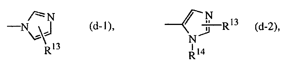
- wherein R13 is hydrogen, halo, Ar4, C1-6alkyl, hydroxyC1-6alkyl, C1-6alkyloxyC1 -6alkyl, C1-6alkyloxy, C1-6alkylthio, amino, C1-6alkyloxycarbonyl, C1-6alkylS(O)C1-6alkyl or C1-6alkylS(O)2C1-6alkyl;
- R14 hydrogen, C1-6alkyl or di(C1-4alkyl)aminosulfonyl;
- R8 is hydrogen, C1-6alkyl, Ar7 or Ar7-C1-6alkyl;
- R9 is hydrogen, C1-6alkyl, C1-6alkylcarbonyl, C1-6alkyloxycarbonyl, C1-6alkylaminocarbonyl, Ar8, Ar8-C1-6alkyl, C1-6alkylcarbonyl-C1-6alkyl, Ar8-carbonyl, Ar8-C1-6alkylcarbonyl, aminocarbonylcarbonyl, C1-6alkyloxyC1-6alkylearbonyl, hydroxy, C1-6alkyloxy, aminocarbonyl, di(C1-6alkyl)aminoC1-6alkylcarbonyl, amino, C1-6alkylamino, C1-6alkylcarbonylamino, or a radical of formula -Alk-OR10 or -Alk-NR11R12;
- R10 is hydrogen, C1-6alkyl, C1-6alkylcarbonyl, hydroxyC1-6alkyl, Ar9 or Ar9-C1-6alkyl;
- R11 is hydrogen, C1-6alkyl, C1-6alkylcarbonyl, Ar10 or Ar10-C1-6alkyl;
- R12 is hydrogen, C1-6alkyl, Ar11 or Ar11-C1-6alkyl; and
-
WO-98149157 U.S. Patent No. 6,117,432 concern the preparation, formulation and pharmaceutical properties of farnesyltransferase inhibiting compounds of formula (VIII)
- the dotted line represents an optional bond;
- X is oxygen or sulfur;
- R1 and R2 each independently are hydrogen, hydroxy, halo, cyano, C1-6alkyl, trihalomethyl, trihalomethoxy, C2-6alkenyl, C1-6alkyloxy, hydroxyC1-6alkyloxy, C1-6alkyloxyC1-6alkyloxy, C1-6alkyloxycarbonyl, aminoC1-6alkyloxy, mono- or di(C1-6alkyl)aminoC1-6alkyloxy, Ar1, Ar1C1-6alkyl, Ar1oxy or Ar1C1-6alkyloxy;
- R3 and R4 each independently are hydrogen, halo, cyano, C1-6alkyl, C1-6alkyloxy, Ar1oxy, C1-6alkylthio, di(C1-6alkyl)amino, trihalomethyl or trihalomethoxy;
- R5 is hydrogen, halo, C1-6alkyl, cyano, haloC1-6alkyl, hydroxyC1-6alkyl, cyanoC1-6alkyl, aminoC1-6alkyl, C1-6alkyloxyC1-6alkyl, C1-6alkylthioC1-6alkyl, aminocarbonylC1-6alkyl, C1-6alkyloxycarbonylC1-6alkyl, C1-6alkylcarbonyl-C1-6alkyl, C1-6alkyloxycarbonyl, mono- or di(C1-6alkyl)aminoC1-6alkyl, Ar1, Ar1C1-6alkyloxyC1-6alkyl; or a radical of formula
-O-R10 (a-1),
-S-R10 (a-2),
-N-R11R12 (a-3),
- wherein R10 is hydrogen, C1-6alkyl, C1-6alkylcarbonyl, Ar1, Ar1C1-6alkyl, C1-6alkyloxycarbonylC1-6alkyl, or a radical of formula -Alk-OR13 or -Alk-NR14R15;
- R11 is hydrogen, C1-6alkyl, Ar1 or Ar1C1-6alkyl;
- µR12 is hydrogen, C1-6alkyl, C1-6alkylcarbonyl, C1-6alkyloxycarbonyl, C1-6alkylaminocarbonyl, Ar1, Ar1C1-6alkyl, C1-6alkylcarbonyl-C1-6alkyl, Ar1carbonyl, Ar1C1-6alkylearbonyl, aminocarbonylcarbonyl, C1-6alkyloxyC1-6alkylcarbonyl, hydroxy, C1-6alkyloxy, aminocarbonyl, di(C1-6alkyl)aminoC1-6alkylcarbonyl, amino, C1-6alkylamino, C1-6alkylcarbonylamino,
or a radical of formula -Alk-OR13 or -Alk-NR14R15;
wherein Alk is C1-6alkanediyl;- R13 is hydrogen, C1-6alkyl, C1-6alkylcarbonyl, hydroxyC1-6alkyl, Ar1 or Ar1C1-6alkyl;
- R14 is hydrogen, C1-6alkyl, Ar1 or Ar1C1-6alkyl;
- R15 is hydrogen, C1-6alkyl, C1-6alkylcarbonyl, Ar1 or Ar1C1-6alkyl;
- R6 is a radical of formula

- wherein R16 is hydrogen, halo, Ar1, C1-6alkyl, hydroxyC1-6alkyl, C1-6alkyloxyC1-6alkyl, C1-6alkyloxy, C1-6alkylthio, amino, C1-6alkyloxycarbonyl, C1-6alkylthioC1-6alkyl, C1-6alkylS(O)C1-6alkyl or C1-6alkylS(O)2C1-6alkyl;
- R17 is hydrogen, C1-6alkyl or di(C1-4alkyl)ammosulfonyl;
- R7 is hydrogen or C1-6alkyl provided that the dotted line does not represent a bond;
- R8 is hydrogen, C1-6alkyl or Ar2CH2 or Het1CH2;
- R9 is hydrogen, C1-6alkyl , C1-6alkyloxy or halo; or
- R8 and R9 taken together to form a bivalent radical of formula
-CH=CH- (c-1),
-CH2-CH2- (c-2),
-CH2-CH2-CH2- (c-3),
-CH2-O- (c-4)
, or
-CH2-CH2-O- (c-5);
- Ar1 is phenyl; or phenyl substituted with 1 or 2 substituents each independently selected from halo, C1-6alkyl, C1-6alkyloxy or trifluoromethyl;
- Ar2 is phenyl; or phenyl substituted with 1 or 2 substituents each independently selected from halo, C1-6alkyl, C1-6alkyloxy or trifluoromethyl; and
- Het1 is pyridinyl; pyridinyl substituted with 1 or 2 substituents each independently selected from halo, C1-6alkyl, C1-6alkyloxy or trifluoromethyl.
-
WO-00/39082 U.S. Patent No. 6,458,800 describe the preparation, formulation and pharmaceutical properties of farnesyltransferase inhibiting compounds of formula (IX)
=X1-X2-X3- is a trivalent radical of formula=N-CR6=CR7- (x-1), =CR6-CR7=CR8- (x-6), =N-N=CR6- (x-2), =CR6-N=CR7- (x-7), =N-NH-C(=O)- (x-3), =CR6-NH-C(=O)- (x-8), or =N-N=N- (x-4), =CR6-N=N- (x-9); =N-CR6=N- (x-5),
>Y1-Y2- is a trivalent radical of formula
>CH-CHR9- (y-1),
>C=N- (y-2),
>CH-NR9- (y-3)
, or
>C=CR9- (y-4);
wherein each R9 independently is hydrogen, halo, halocarbonyl, aminocarbonyl, hydroxyC1-4alkyl, cyano, carboxyl, C1-4alkyl, C1-4alkyloxy, C1-4alkyloxyC1-4alkyl, C1-4alkyloxycarbonyl, mono- or di(C1-4alkyl)amino, mono- or di(C1-4alkyl)aminoC1-4alkyl, aryl;
r and s are each independently 0, 1, 2, 3, 4 or 5;
t is 0, 1, 2 or 3;
each R1 and R2 are independently hydroxy, halo, cyano, C1-6alkyl, trihalomethyl, trihalomethoxy, C2-6alkenyl, C1-6alkyloxy, hydroxyC1-6alkyloxy, C1-6alkylthio, C1-6alkyloxyC1-6alkyloxy, C1-6alkyloxycarbonyl, aminoC1-6alkyloxy, mono- or di(C1-6alkyl)amino, mono- or di(C1-6alkyl)aminoC1-6alkyloxy, aryl, arylC1-6alkyl, aryloxy or arylC1-6alkyloxy, hydroxycarbonyl, C1-6alkyloxycarbonyl, aminocarbonyl, aminoC1-6alkyl, mono- or di(C1-6alkyl)aminocarbonyl, mono- or di(C1-6alkyl)aminoC1-6alkyl; or
two R1 or R2 substituents adjacent to one another on the phenyl ring may independently form together a bivalent radical of formula
-O-CH2-O- (a-1),
-O-CH2-CH2-O- (a-2),
-O=CH=CH- (a-3),
-O-CH2-CH2- (a-4),
-O-CH2-CH2- CH2- (a-5)
, or
-CH=CH-CH=CH- (a-6);
R3 is hydrogen, halo, C1-6alkyl, cyano, halo1-6alkyl, hydroxy1-6alkyl, cyanoC1-6alkyl, aminoC1-6alkyl, C1-6alkyloxyC1-6alkyl, C1-6alkylthioC1-6alkyl, aminocarbonylC1-6alkyl, hydroxycarbonyl, hydroxycarbonylC1-6alkyl, C1-6alkyloxycarbonylC1-6alkyl, C1-6alkylcarbonylC1-6alkyl, C1-6alkyloxycarbonyl, aryl, arylC1-6alkyloxyC1-6alkyl, mono- or di(C1-6alkyl)aminoC1-6alkyl;
or a radical of formula
-O-R10 (b-1),
-S-R10 (b-2),
-NR11R12 (b-3),
wherein R10 is hydrogen, C1-6alkyl, C1-6alkylcarbonyl, aryl, arylC1-6alkyl, C1-6alkyloxycarbonylC1-6alkyl, or a radical of formula -Alk-OR13 or -Alk-NR14R15; - R11 is hydrogen, C1-6alkyl, aryl or arylC1-6alkyl;
- R12 is hydrogen, C1-6alkyl, aryl, hydroxy, amino, C1-6alkyloxy, C1-6alkylcarbonylC1-6alkyl, arylC1-6alkyl, C1-6alkylcarbonylamino, mono-or di(C1-6alkyl)amino, C1-6alkylcarbonyl, aminocarbonyl, arylcarbonyl, haloC1-6alkylcarbonyl, arylC1-6alkylcarbonyl, C1-6alkyloxycarbonyl, C1-6alkyloxyC1-6alkylcarbonyl, mono- or di(C1-6alkyl)aminocarbonyl wherein the alkyl moiety may optionally be substituted by one or more substituents independently selected from aryl or C1-3alkyloxycarbonyl, aminocarbonylcarbonyl, mono- or di(C1-6alkyl)aminoC1-6alkylcarbonyl, or a radical of formula -Alk-OR13 or -Alk-NR14R15;
- wherein Alk is C1-6alkanediyl;
- R13 is hydrogen, C1-6alkyl, C1-6alkylcarbonyl, hydroxyC1-6alkyl, aryl or arylC1-6alkyl;
- R14 is hydrogen, C1-6alkyl, aryl or arylC1-6alkyl;
- R15 is hydrogen, C1-6alkyl, C1-6alkylcarbonyl, aryl or arylC1-6alkyl;
- Additionally, potent FTIs, including tipifarnib (also known as R 115777 or ZARNESTRA®), have been described in the literature. See, e.g.,
Kurzrock, R. et al. (2003 WO 98/43629 WO 02/40015 WO-98/28303 WO-99/45912 U.S. Patent No. 5874442 (Schering); L778123, i.e. 1-(3-chlorophenyl)-4-[1-(4-cyanobenzyl)-5-imidazolylmethyl]-2-piperazinone, described inWO-00/01691 WO-94/10138 WO 97/30992 WO-00/12498 WO-00/12499 
- The use of certain of such FTIs has been studied in the area of anti-cancer therapy. More recently, the use of certain FTIs as therapeutic agents for treating inflammatory diseases, particularly those involving an immune component, and for treating pain associated with inflammatory conditions has been disclosed. See
WO 98/43629 - Although various FTIs are known, their biological effects in cells and animals, and therefore their therapeutic applications, have not been fully elucidated.
- In one general aspect, the invention relates to a method of treating a subject for sepsis or septic shock, comprising administering to a subject in need of such treatment a therapeutically effective amount of a farnesyltransferase inhibitor. Preferably, the farnesyltransferase inhibitor is selected from the farnesyltransferase inhibitor compounds disclosed herein. More preferably, the farnesyltransferase inhibitor is tipifarnib.
- In another general aspect, the invention relates to methods for determining a subject's response to treatment with a farnesyltransferase inhibitor. One general embodiment of such a method comprises: (a) taking an untreated blood sample from the subject before treatment with the farnesyltransferase inhibitor, and taking a treated blood sample from the subject at least one time after treatment with the farnesyltransferase inhibitor has commenced; (b) optionally isolating cells, plasma or serum from the untreated blood sample to obtain an untreated sample extract, and isolating cells, plasma or serum from each treated blood sample to obtain a treated sample extract; (c) optionally measuring a level of IL-8 in the untreated blood sample or extract, measuring a level of IL-8 in each treated blood sample or extract, and comparing the levels of IL-8 to obtain a control measurement; and (d) measuring a level of at least one of IL-6, MCP-1, IL-1β, MMP-9, and TNF-α in the untreated blood sample or extract, measuring a level of said at least one of IL-6, MCP-1, IL-1β, MMP-9, and TNF-α in each treated blood sample or extract, and comparing the measured levels, where a decrease indicates a response to the treatment.
- In one preferred embodiment, the extracts are plasma samples (isolated by, e.g., centrifuging the blood). In another preferred embodiment, the extracts are peripheral blood mononuclear cells isolated by, e.g., Ficoll-Paque gradient. Before measuring, the blood or cells may be stimulated with LPS.
- Another general embodiment relates to a method for monitoring a subject's response to treatment with a farnesyltransferase inhibitor, comprising: (a) taking an untreated blood sample from the subject before treatment with the farnesyltransferase inhibitor, and taking a treated blood sample from the subject at least one time after treatment with the farnesyltransferase inhibitor has commenced; (b) optionally isolating peripheral blood mononuclear cells (PBMC) from the untreated blood sample to obtain an untreated PBMC sample, and isolating peripheral blood mononuclear cells from each treated blood sample to obtain a treated PBMC sample; (c) stimulating the untreated blood or PBMC sample with lipopolysaccharide to obtain an LPS-stimulated untreated blood or PBMC sample, and stimulating each treated blood or PBMC sample with lipopolysaccharide to obtain an LPS-stimulated treated blood or PBMC sample; (d) isolating RNA of the untreated LPS-stimulated blood or PBMC sample, and isolating RNA of each treated LPS-stimulated blood or PBMC sample; (e) optionally measuring expression of IL-8 in the RNA of the untreated LPS-stimulated blood or PBMC sample, measuring expression of IL-8 in the RNA of each treated LPS-stimulated blood or PBMC sample, and comparing said measurements of RNA expression of IL-8 as a control; (f) measuring expression of at least one of IL-1β, MCP-1, MMP-9, MyD88, STAT1, and IL-6 in the RNA of the untreated LPS-stimulated blood or PBMC sample, measuring expression of at least one of II-1β, MCP-1, MMP-9, MyD88, STAT1, and IL-6 in the RNA of each treated LPS-stimulated blood or PBMC sample, and comparing the expression measurements, where a decrease indicates a response to the treatment.
- In preferred embodiments, plural treated blood samples from the subject (e.g., a patient in a clinical trial) are taken over periodic intervals of time after treatment with the farnesyltransferase inhibitor has commenced.
- Other embodiments, features, advantages, and aspects of the invention will become apparent from the detailed description below in reference to the drawing figures.
-
-
Figure 1 shows the mevalonate pathway for synthesis of isoprenoids and cholesterol, indicating the sites for inhibition of HMG CoA reductase by simvastatin, and of farnesyltransferase activity by tipifarnib. Inhibition of HMG CoA reductase by statins leads to inhibition of cholesterol synthesis, and of isoprenoid production, required for both farnesylation and geranylgeranylation of proteins. Inhibition of farnesyltransferase by FTIs selectively inhibits farnesylation of proteins such as Ras. -
Figures 2a-2g show the effect of simvastatin and tipifarnib on LPS-induced transcription of IL-1β (Figure 2 .a), MCP-1 (Figure 2 .b), MMP-9 (Figure 2 .c), IL-8 (Figure 2 .d), STAT1 (Figure 2 .e), MyD88 (Figure 2 .f) and IL-6 (Figure 2 .g). THP-1 cells were cultured in the absence (no treatment) or presence of LPS (100 ng/ml), plus or minus simvastatin (10 µM), or tipifarnib (5 µM). RNA was isolated at the indicated times for cDNA preparation as described in "Materials and Methods". Relative mRNA levels compared to GAPDH were determined by real-time PCR. -
Figures 3a-3f show the effect of simvastatin and tipifarnib on LPS-induced secretion of MCP-1 (Figure 3 .a), IL-6 (Figure 3 .b), IL-1β (Figure 3 .c), IL-8 (Figure 3 .d), MMP-9 (Figure 3 .e) and TNF-α (Figure 3 .f). THP-1 cells cultured in the absence (no treatment) or presence of 100ng/ml LPS, plus or minus simvastatin (5 and 10µM) or tipifarnib (2 and 5 µM), respectively. At the indicated time points, supernatanats were analyzed for cytokines, chemokines or MMP-9 by ELISA. Data points are means of triplicate incubations, for which standard deviations were typically 2 to 13 % of the mean value. -
Figure 4 shows inhibition of LPS-induced IL-6 release by tipifarnib. THP-1 cells at 3.4 x 10⁵/ml were cultured in 96-well plates in 0.5%FBS/RPMI 1640, in the absence or presence of 100ng/ml LPS, plus or minus tipifarnib (starting from 5 µM, followed by 3-fold series dilution). The concentrations of tipifarnib were: 5 µM, 1.67 µM, 0.56 µM, 0.19 µM and 0.06 µM. Supernatants were collected at 48hr. IL6 was quantified using ELISA kit from R&D Systems. -
Figures 5a-5e show the effect of simvastatin and tipifarnib on LPS-induced cytokine production in vivo. BALB/c mice (n = 4 or 5 per group) were treated orally with simvastatin or tipifarnib in 20% cyclodextran, at a dose of 50mg/kg, 24 hours, 17 hours and 1 hour before LPS injection. LPS (20 µg) was administered intraperitoneally. Plasma samples were collected at 1 hour and 2 hours in the first experiment, and 2 hours and 3 hours after LPS injection in the second experiment, and tested for TNF-α (Figure 5 .a), IL-6 (Figure 5 .b), IL-1β (Figure 5 .c), MIP-1α (Figure 5 .d) and MCP-1 (Figure 5 .e), using ELISA or Luminex as described in "Materials and Methods" herein. Data are presented as the mean values for 4 or 5 mice in each group, and the standard deviations from the means are indicated by error bars. Statistical significance for differences in production of inflammatory mediators in simvastatin- or tipifarnib-versus vehicle-treated animals was assessed using the Student's t test. The statistically significant differences are indicated by asterisks inFigures 5a through 5e as follows: * means P value less than 0.05; ** means P value less than 0.01. The lower the P value, the more significant the difference. -
Figure 6 shows inhibition of LPS-induced IL-6 production in human PBMC by tipfarnib, but not by the less active enantiomer of tipifarnib. Human PBMC were incubated at 37°C for 1 hour in the absence or presence of 5 µM and 2 µM tipifarnib (RH5777) or its less active enantiomer (100-fold less active for inhibition of farnesyltransferase) after which LPS (or vehicle only for no treatment) was added to a final concentration of 10 ng/ml. Supernatants were collected at 16 hours, 24 hours and 40 hours of LPS treatment, and IL-6 secretion was quantified by ELISA. Statistical significance for differences in production of IL-6 in tipifarnib- versus vehicle-treated PBMC was assessed using the Student's t test. The statistically significant differences are indicated by asterisks ** means P value less than 0.01. The lower the P value, the more significant the difference. -
Figure 7a-7b shows the effect of tipifarnib on LPS, TNF-α, and It-1 -induced signaling. THP-1 cells were cultured in the absence or presence of 2 µM tipifarnib for 18 hours, then incubated with LPS and TNF-α (Figure 7 .a), or LPS, TNF-α and IL-1α (Figure 7 .b) for 30 minutes. Cell lysates were analyzed by Western blotting, using antibodies against p38, phosphorylated p38, phospho-ERK1/2, IKB, and H-Ras. -
Figure 8 shows that tipifarnib does not inhibit NF-κB-dependent reporter gene expression induced by TNF-α. NF-κB-LH transfected HEK293 cells were incubated in the absence or presence of 2 and 5 µM tipifarnib for 18 hours, then stimulated with TNF-α for 4 hours. Luciferase activity was measured and expressed as fold-change in the presence of TNF-α versus no TNF-α, or TNF-α/tipifarnib versus tipifarnib. Values are presented as fold increase ± S.D. - The terms "comprising", "including", and "containing" are used herein in their open, non-limited sense.
- For the sake of brevity, the disclosures of all patents and other publications cited herein are incorporated by reference.
- The biological effects of FTIs have now been further elucidated, as discussed in the Experimentals section below. Inhibition of farnesyltransferase (FPTase) has been found to downregulate the genes and proteins identified in Tables 2, 3, and 4. Thus, the levels of such genes and protein may be monitored as biological markers to determine a patient's response to treatment with an FTI. For example, various subsets of LPS-induced genes and/or proteins shown in the following tables may be downregulated by administration of a farnesyltransferase inhibitor and therefore used as markers in clinical studies.
Table 2: Genes for which LPS-induced transcription was downregulated by inhibition of farnesyltransferase, as measured by cDNA microarray analysis Accession number GeneName GeneDescription NM_002982 CCL2 monocyte chemotactic protein 1 (MCP-1) NM_005623 CCL8 monocyte chemotactic protein 2 (MCP-2) NM_002983 CCL3 small inducible cytokine A3 (homologous to mouse Mip-1 a) (SCYA3) NM_002984 CCL4 small inducible cytokine A4 (homologous to mouse Mip-1b) (SCYA4) NM_005409 CXCL11 small inducible cytokine subfamily B, member 11 (SCYB11) NM_000576 IL1 B interleukin 1, beta NM_000577 IL1 RN interteukin-1 receptor antagonist NM_002852 PTX3 pentaxin-related gene, rapidly induced by IL-1B NM_007315 STAT1 signal transducer and activator of transcription 1NM_002176 IFNB1 interferon, beta 1, fibroblastNM_005531 IFI16 interferon, qamma- inducible protein 16NM_001548 IFIT1 interferon-induced protein with tetratricopeptide repeats 1NM_002462 MX1 myxovirus (influenza) resistance 1NM_000598 IGFBP3 insulin-like growth factor-bindinq protein-3 gene NM_053056 CCND1 cyclin D1 (PRAD1: parathyroid adenomatosis 1) NM_002965 S100A9 S100 calcium-binding protein A9 (calaranulin B) NM_002468 MYD88 myleoid differentiation primary response protein NM_000593 TAP1 transporter 1, ATP-binding cassette, sub-family B (MDR/TAP) NM_000544 TAP2 transporter 2, ATP-binding cassette, sub-family B (MDR/TAP) NM_007188 ABCBB ATP-bindinq cassette, sub-family B (MDRfTAP), member 8 NM_031460 KCNK17 potassium channel subfamily K, member 17 (TASK-4) NM_002659 PLAUR plasminogen activator, urokinase receptor NM_004054 C3AR1 C3a anaphylatoxin chemotactic receptor (C3a-R) NM_004048 B2M beta-2-microqlobulin NM_015907 LAP3 leucine aminopeptidase NM_004994 MMP9 type IV collagenase, matrix metalloproteinase 9 NM_006074 TRIM22 tripartite motif -containing 22 NM_144573 NEXN nexilin (F actin binding protein) NM_021634 LGR7 leucine-rich repeat-containinq G protein-coupled receptor 7 NM_198066 GNPNAT1 glucosamine-phosphate N-acetyltransf erase 1 NM_002346 LY6E lymphocyte antigen 6 complex, locus E Table 3: Genes for which LPS-induced transcription was downregulated by inhibition of farnesyltransferase, as measured by reverse transcriptase and real-time PCR analysis Accession number GeneName GeneDescription NM_000576 IL1B interleukin 1, beta NM_002982 CCL2 monocyte chemotactic protein 1 (MCP-1) NM_004994 MMP9 type IV collagenase, matrix metalloproteinase 9 NM_007315 STAT1 signal transducer and activator of transcription 1 NM_002468 MYD88 myleoid differentiation primary response protein NM_000600 IL6 interleukin 6 Table 4 Proteins for which LPS-induced production was downregulated by inhibition of farnesyltransferase, as measured by ELISA or Luminex analysis Accession number Protein name Protein description NP_000585 TNF-α tumor necrosis factor (TNF superfamily, member 2) NP_000591 IL-6 interleukin 6, interferon beta 2 NP_000567 IL-1β interleukin 1, beta proprotein NP_002973 CCL2 MCP-1, small inducible cytokine A2 precursor NP_002974 CCL3 small inducible cytokine A3 (homologous to mouse Mip-1a) AAG32620 EL12p40 interleukin 12 p40 subunit NP_004985 MMP-9 type IV collagenase, matrix metalloproteinase 9 - As described above, one or more of the above-identified genes and proteins may be used as a marker to study a subject's response to treatment with an FTI.
- Inhibition of FPTase may decrease LPS-induced transcription of, e.g., IL-1β, MCP-1, MMP-9, MyD88, STAT1, MIP-1α, TNF-α or IL-6. Consequently, transcription of IL-1β, MCP-1, MMP-9, MyD88, STAT1, MIP-1α, TNF-α or IL-6 may be decreased by administration of a farnesyltransferase inhibitor and these may be used as a panel of markers for monitoring patients in a clinical study. Inhibition of FPTase may also decrease or inhibit transcription of IL-1β, MCP-1, MMP-9, MyD88, STAT1, MIP-1α, TNF-α or IL-6 induced by agents other than LPS. Thus, administration of an FTI to a patient may be useful to treat diseases and medical conditions mediated through transcription of IL-1β, MCP-1, MMP-9, MyD88, STAT1, MIP-1α, TNF-α or IL-6. Inhibition of FPTase may also decrease LPS-induced protein translation or secretion of IL-1β, MCP-1, MMP-9, MyD88, STAT1, MIP-1α, TNF-α or IL-6. Thus, a disease or medical condition mediated by LPS-induced protein translation or secretion of IL-1β, MCP-1, MMP-9, MyD88, STAT1, MIP-1α, TNF-α or IL-6 may be treated by administering to a subject in need of such treatment a farnesyltransferase inhibitor, and the progress of treatment may be monitored by measuring levels of such markers. Furthermore, since inhibition of FPTase may serve to inhibit protein translation or secretion of IL-1β, MCP-1, MMP-9, MyD88, STAT1, MIP-1α, TNF-α or IL-6 induced by agents other than LPS, administration of an FTI may be useful to treat diseases and medical conditions mediated through protein translation or secretion of IL-1β, MCP-1, MMP-9, MyD88, STAT1, MIP-1α, TNF-α and/or IL-6. Inhibition of FPTase may also serve to inhibit transcription or translation of MyD88 or
STAY 1. Inhibition of transcription or protein translation or secretion of TNF-α, IL-1β, MCP-1, IL-6, MMP-9, or MIP-1α may also be attained by administration of a farnesyltransferase inhibitor, and the progress of treatment may be monitored by measuring levels of this set of markers. - Accordingly, FTIs may be used to treat inflammation and inflammatory conditions. Diseases and medical conditions that may benefit from treatment with an FTI include: atherosclerosis and other vascular diseases such as thrombosis, restenosis after angioplasty and/or stenting, and vein-graft disease after bypass surgery, as well as ulcerative colitis, Crohn's disease, allergic rhinitis, graft-versus-host disease, conjunctivitis, asthma, rheumatoid arthritis, osteoarthritis, ARDS, Behcet's disease, transplant rejection, urticaria, allergic dermatitis, alopecia areata, scleroderma, exanthema, eczema, dermatomyositis, acne, diabetes, systemic lupus erythematosis, Kawasaki's disease, multiple sclerosis, emphysema, cystic fibrosis, chronic bronchitis and psoriasis.
- FTIs may also be used to treat LPS-induced inflammation in particular. Thus, a therapeutically effective amount of an FTI may be administered to treat a patient in need of treatment for one of the following diseases or medical conditions: sepsis, endotoxic shock, multiple organ dysfunction syndrome, periodontitis, peritonitis, anorexia, microvascular leukocytosis, pulmonary inflammation, airway hyperreactivity, acute lung injury, acute respiratory distress syndrome, chronic airway disease, goblet cell hyperplasia, pancreatitis, nephrotic syndrome, pyrexia, liver injury, premature delivery, Crohn's disease, ulcerative colitis, cerebral edema, atherosclerosis, cardiac failure, obstructive jaundice, neuroinflammation, cerebral ischemia, hyperalgesia, rectal allodynia, disseminated intravascular coagulation syndrome, hyperglycemia, insulin resistance, obstructive sleep apnea.
- FTIs may also be used to treat atherosclerosis. Thus, a therapeutically effective amount of an FTI may be administered to a subject diagnosed with atherosclerosis to treat the subject in need of such treatment.
- Furthermore, FTIs may be used to treat asthma. A therapeutically effective amount of an FTI may therefore be administered to a subject in need of treatment for asthma.
- Additionally, a therapeutically effective amount of an FTI may be administered to a subject diagnosed with multiple sclerosis (MS) to treat the subject in need of such treatment.
- Moreover, a therapeutically effective amount of an FTI may be administered to a subject diagnosed with or experiencing sepsis or septic shock to treat the subject in need of such treatment.
- Accordingly, the monitoring methods of the invention are therefore useful in connection with determining FTI efficacy in treating the above-described indications.
- Exemplary farnesyltransferase-inhibiting compounds useful in such treatments are described below. An especially preferred FTI is tipifarnib.
- The present invention provides various methods for determining a subject's response to treatment, such as for one of the diseases or medical conditions described above, with a farnesyltransferase inhibitor (FTI). In such methods, an untreated blood sample and an FTI-treated blood sample are taken. The following may be used as types of "blood samples": whole blood; plasma (e.g., separated by centrifugation); serum from plasma (e.g., obtained by clotting or serum separator tubes); or total cells or isolated subsets (e.g., PBMC).
- As a control measurement useful with any of the above types of blood samples, IL-8 protein levels may optionally be determined before and after treatment with an FTI (e.g., tipifarnib), e.g., by using immunofluorescence staining, flow cytometry, immunocytochemistry, immunohistochemistry, immunoblotting, ELISA, or multiplex cytokine array analysis. As one skilled in the art will realize, other measurements may be used as a control, or the method of the present invention may be practiced without the use of a control. For example, in plasma or serum, levels of albumin protein can be used as a control. Additionally, the invention may be practiced without an internal control by comparing measurements in an untreated blood sample to an FTI- treated blood sample.
- As the marker(s) useful for monitoring progress of treatment with any of the blood samples, protein levels for one or more of the genes or proteins in Tables 2, 3, and 4 may be measured before and after FTI treatment, e.g., by using any of the methods described above, where a decrease is indicative of a response to treatment.
- Alternatively, with whole blood or total cells or particular subsets of cells (e.g., PBMC), IL-8 RNA levels may optionally be measured as a control before and after FTI treatment (under conditions where IL-8 levels do not change with treatment, this gene serves as a control), e.g., by performing RNase protection, reverse transcriptase (RT) polymerase chain reaction (PCR), RT and real-time quantitative PCR, Northern blotting, and cDNA- or oligonucleotide- microarray analysis. As one skilled in the art will realize, other measurements may be used as a control, or the method of the present invention may be practiced without the use of a control. Other commonly employed control measurements included within the scope of the present invention include mRNA levels of glyceraldehyde-3-phosphate dehydrogenase (GAPDH) or beta-actin. Additionally, the present invention may be practiced without an internal control by comparing measurements in an untreated blood sample to an FTI- treated blood sample.
- For this general embodiment used with the specified types of blood samples, the RNA levels of one or more of the genes or proteins listed in Tables 2, 3, and 4 are measured before and after treatment with an FTI (where a decrease with treatment reflects response).
- In another alternative general embodiment, whole blood, total cells, or PBMC or other subsets of cells are treated with LPS. As a control, levels of IL-8 are optionally measured in protein or RNA, or as otherwise known in the art, as described above. The levels of one or more markers of Tables 2, 3, and 4 are measured in LPS-treated samples of protein or RNA as in the other embodiments, with a decrease in protein/RNA levels with treatment with an FTI over time indicated response to the treatment. Likewise, an agent other than LPS that also induces RNA or protein for the marker(s) may be used in place of LPS to treat the samples, and then protein or RNA levels of optionally IL-8 (or other controls) and one or more markers may be measured as before to monitor response to FTI treatment.
- In one preferred embodiment, the response of a subject undergoing such treatment comprises the following steps: (1) taking a blood sample from said subject before FTI treatment and after FTI treatment, and optionally at different times during FTI treatment; (2) optionally separating cells and plasma (preferably by centrifugation); (3) optionally measuring a level of IL-8 or other control (preferably by immunoblotting , ELISA or multiplex cytokine array analysis) in the plasma before and after FTI treatment as a control; (4) measuring (preferably by immunoblotting or ELISA or multiplex cytokine array analysis) a level of one or more of the markers identified in Tables 2, 3, and 4, preferably one or more of IL-6, MCP-1, IL-1β, MMP-9, TNF-α in the plasma before and after FTI treatment; and (5) comparing the levels of such marker(s) measured before and after FTI treatment. In one particular embodiment, decreased levels of IL-6, MCP-1, IL-1β, MMP-9, MIP-1α and/or TNF-α after, and optionally during FTI treatment, relative to before FTI treatment, would indicate a response to FTI treatment.
- In a further embodiment, the present invention provides a method for determining response to treatment with a farnesyltransferase inhibitor (FTI) comprising: (1) taking a blood sample from the subject before FTI treatment and after FTI treatment, and optionally at different times during FTI treatment; (2) optionally isolating peripheral blood mononuclear cells (PBMC) from blood (preferably by Ficoll-Paque gradient separation); (3) stimulating the isolated PBMC with LPS (preferably 10 ng/ml LPS); (4) optionally measuring a level of IL-8 or other control (preferably by immunoblotting or ELISA or multiplex cytokine array analysis) in the plasma before and after FTI treatment as a control; (4) measuring (preferably by immunoblotting or ELISA or multiplex cytokine array analysis) a level of one or more of the markers of Tables 2, 3, and 4 (e.g., IL-6, MCP-1, DL-1β, MMP-9, MIP-1α, and/orTNF-α) in the PBMC before and after FTI treatment; and (5) comparing the levels of such marker(s) measured before and after FTI treatment, wherein decreased levels of marker(s) after, and optionally during FTI treatment, relative to before FTI treatment, would indicate a response to FTI treatment.
- In another embodiment, the present invention provides a method for determining response to treatment with a farnesyltransferase inhibitor (FTI), in a subject in need of treatment therewith, comprising the following steps: (1) taking a blood sample from said subject before FTI treatment, after FTI treatment, and optionally at different times during FTI treatment; (2) isolating peripheral blood mononuclear cells (PBMC) from blood (preferably by Ficoll-Paque gradient separation), (3) stimulating the isolated PBMC with LPS (preferably 10 ng/ml LPS); (4) isolating RNA from the blood or PBMC samples; (5) measuring RNA expression (preferably by Northern blotting, cDNA- or oligonucleotide- microarray analysis, or reverse transcriptase and real-time PCR) of one or more markers (preferably IL-1β, MCP-1, MMP-9, MyD88, STAT1 and/or IL-6) in the RNA samples; (6) optionally measuring RNA expression of IL-8 (or others) as a control; and (7) comparing the levels of the marker(s) in RNA measured before and after FTI treatment, wherein decreased LPS-induced expression of the marker(s) after, and optionally during FTI treatment, relative to before FTI treatment, would indicate a response to FTI treatment.
- Examples of farnesyltransferase inhibitors which may be employed in the methods or treatments in accordance with the present invention include compounds of formula (I), (II), (III), (IV), (V), (VI), (VII), (VIII) or (IX) above. Preferred FTIs include compounds of formula (I), (II) or (III):


- the dotted line represents an optional bond;
- X is oxygen or sulfur;
- R1 is hydrogen, C1-12alkyl, Ar1, Ar2C1-6alkyl, quinolinylC1-6alkyl, pyridylC1-6alkyl, hydroxyC1-6alkyl, C1-6alkyloxyC1-6a1kyl, mono- or di(C1-6alkyl)aminoC1-6aikyl, aminoC1-6alkyl, or a radical of formula -AlkLC(=O)-R9, -Alk1-S(O)-R9 or -Alk1-S(O}2-R9,
wherein Alk1 is C1-6alkanediyl,- R9 is hydroxy, C1-6alkyl, C1-6alkyloxy, amino, C1-8alkylamino or C1-8alkylamino substituted with C1-6alkyloxycarbonyl;
- R2, R3 and R16 each independently are hydrogen, hydroxy, halo, cyano, C1-6alkyl, C1-6alkyloxy, hydroxyC1-6alkyloxy, C1-6alkyloxyC1-6alkyloxy, aminoC1-6alkyloxy, mono- or di(C1-6alkyl)aminoC1-6alkyloxy, Ar1 Ar2C1-6alkyl, Ar2oxy, Ar2C1-6alkyloxy, hydroxycarbonyl, C1-6alkyloxycarbonyl, trihalomethyl, trihalomethoxy, C2-6alkenyl, 4,4-dimethyloxazolyl; or
when on adjacent positions R2 and R3 taken together may form a bivalent radical of formula
-O-CH2-O- (a-1),
-O-CH2-CH2-O- (a-2),
-O-CH=CH- (a-3),
-O-CH2-CH2- (a-4),
-O-CH2-CH2-CH2- (a-5)
, or
-CH=CH-CH=CH- (a-6);
- R4 and R5 each independently are hydrogen, halo, Ar1, C1-6alkyl, hydroxyC1-6alkyl, C1-6alkyloxyC1-6alkyl , C1-6alkyloxy, C1-6alkylthio, amino, hydroxycarbonyl, C1-6alkyloxycarbonyl, C1-6alkylS(O)C1-6alkyl or C1-6alkylS(O)2C1-6alkyl;
- R6 and R7 each independently are hydrogen, halo, cyano, C1-6alkyl, C1-6aikyloxy, Ar2oxy, trihalomethyl, C1-6alkylthio, di(C1-6alkyl)amino, or
when on adjacent positions R6 and R7 taken together may form a bivalent radical of formula
-O-CH2-O- (c-1)
, or
-CH=CH-CH=CH- (c-2);
- R8 is hydrogen, C1-6alkyl, cyano, hydroxycarbonyl, C1-6alkyloxycarbonyl, C1-6alkylcarbonylC1-6alkyl, cyanoC1-6alkyl, C1-6alkyloxycarbonylC1-6alkyl, carboxyC1-6alkyl, hydroxyC1-6alkyl, aminoC1-6alkyl, mono- or di(C1-6alkyl)aminoC1-6alkyl, imidazolyl, haloC1-6alkyl, C1-6alkyloxyC1-6alkyl, aminocarbonylC1-6alkyl, or a radical of formula
-O-R10 (b-1),
-S-R10 (b-2),
-N-R11R12 (b-3),
- wherein R10 is hydrogen, C1-6alkyl, C1-6alkylcarbonyl, Ar1, Ar2C1-6alkyl, C1-6alkyloxycarbonylC1-6alkyl, or a radical of formula -Alk2-OR13 or -Alk2-NR14R15;
- R11 is hydrogen, C1-12alkyl Ar1 or Ar2C1-6alkyl;
- R12 is hydrogen, C1-6alkyl, C1-16alkylcarbonyl, C1-6alkyloxycarbonyl, C1-6alkylaminocarbonyl, Ar1, Ar2C1-6alkyl, C1-6alkylcarbonylC1-6alkyl, a natural amino acid, Ar1carbonyl, Ar2C1-66alkylcarbonyl, aminocarbonylcarbonyl, C1-6alkyloxyC1-6alkylcarbonyl, hydroxy, C1-6alkyloxy, aminocarbonyl, di(C1-6alkyl)aminoC1-6alkylcarbonyl, amino, C1-6alkylamino, C1-6alkylcarbonylamino,
or a radical of formula -Alk2-OR13 or -Alk2-NR14R15;
wherein Alk2 is C1-6alkanediyl;- R13 is hydrogen, C1-6alkyl, C1-6alkylcarbonyl, hydroxyC1-balkyl, Ar1 or Ar2C1-6alkyl;
- R14 is hydrogen, C1-6alkyl., Ar1 or Ar2C1-6alkyl;
- R15 is hydrogen, C1-6alkyl, C1-6alkylcarbonyl, Ar1 or Ar2C1-6alkyl;
- R17 is hydrogen, halo, cyano, C1-6alkyl, C1-6alkyloxycarbonyl, Ar1;
- R18 is hydrogen, C1-6alkyl, C1-6alkyloxy or halo;
- R19 is hydrogen or C1-6alkyl;
- Ar1 is phenyl or phenyl substituted with C1-6alkyl, hydroxy, amino, C1-6alkyloxy or halo; and
- Ar2 is phenyl or phenyl substituted with C1-6alkyl, hydroxy, amino, C1-6alkyloxy or halo.
- In Formulas (I), (II) and (III), R4 or R5 may also be bound to one of the nitrogen atoms in the imidazole ring. In that case the hydrogen on the nitrogen is replaced by R4 or R5 and the meaning of R4 and R5 when bound to the nitrogen is limited to hydrogen, Ar1, C1-6alkyl, hydroxyC1-6alkyl, C1-6alkyloxyC1-6alkyl, C1-6alkyloxycarbonyl, C1-6alkylS(O)C1-6alkyl, C1-6alkylS(O)2C1-6alkyl.
- Preferably the substituent R18 is situated on the 5 or 7 position of the quinolinone moiety and substituent R19 is situated on the 8 position when R18 is on the 7-position.
- Preferred examples of FTIs are those compounds of formula (I) wherein X is oxygen.
- Also, examples of useful FPTase-inhibiting compounds are these compounds of formula (I) wherein the dotted line represents a bond, so as to form a double bond.
- Another group of illustrative compounds are those compounds of formula (I) wherein R1 is hydrogen, C1-6alkyl, C1-6alkyloxyC1-6alkyl, di(C1-6alkyl)aminoC1-6alkyl, or a radical of formula -Alkl1-C(=O)-R9, wherein Alk1 is methylene and R9 is C1-8alkylamino substituted with C1-6alkyloxycarbonyl.
- Still another group of exemplary compounds are those compounds of formula (I) wherein R3 is hydrogen or halo; and R2 is halo, C1-6alkyl, C2-6alkenyl, C1-6alkyloxy, trihalomethoxy or hydroxyC1-6alkyloxy.
- A further group of illustrative compounds are those compounds of formula (I) wherein R2 and R3 are on adjacent positions and taken together to form a bivalent radical of formula (a-1), (a-2) or (a-3).
- A still further group of exemplary compounds are those compounds of formula (I) wherein R5 is hydrogen and R4 is hydrogen or C1-6alkyl.
- Yet another group of illustrative compounds are those compounds of formula (I) wherein R7 is hydrogen; and R6 is C1-6alkyl or halo, preferably chloro, especially 4-chloro.
- Another exemplary group of compounds are those compounds of formula (I) wherein R8 is hydrogen, hydroxy, haloC1-6alkyl, hydroxyC1-6alkyl, cyanoC1-6alky), C1-6alkyloxycarbonylC1-6alkyl, imidazolyl, or a radical of formula -NR11R12 wherein R11 is hydrogen or C1-12alkyl and R12 is hydrogen, C1-6alkyl, C1-6alkyloxy, hydroxy, C1-6alkyloxyC1-6alkylcarbonyl, or a radical of formula -Alk2-OR13 wherein R13 is hydrogen or C1-6alkyl.
- Preferred compounds are those compounds wherein R1 is hydrogen, C1-6alkyl, C1-6alkyloxyC1-6alkyl, di(C1-6alkyl)arninoC1-6alkyl, or a radical of formula -Alk1-C(=O)-R9, wherein Alk1 is methylene and R9 is C1-8alkylamino substituted with C1-6alkyloxycarbonyl; R2 is halo, C1-6alkyl, C2-6alkenyl, C1-6alkyloxy, trihalomethoxy, hydroxyC1-6alkyloxy or Ar1; R3 is hydrogen; R4 is methyl bound to the nitrogen in 3-position of the imidazole; R5 is hydrogen; R6 is chloro; R7 is hydrogen; R8 is hydrogen, hydroxy, haloC1-6alkyl, hydroxyC1-6alkyl, cyanoC1-6alkyl, C1-6alkyloxycarbonylC1-6alkyl, imidazolyl, or a radical of formula - NR11R12 wherein R11 is hydrogen or C1-12alkyl and R12 is hydrogen, C1-6alkyl, C1-6alkyloxy, C1-6alkyloxyC1-6alkylcarbonyl, or a radical of formula -Alk2-OR13 wherein R13 is C1-6alkyl; R17 is hydrogen and R18 is hydrogen.
- Especially preferred compounds are:
- 4-(3-chlorophenyl)-6-[(4-chlorophenyl)hydroxy(1-methyl-1H-imidazol-5-yl)methyl]-1-methyl-2(1H)-quinolinone;
- 6-[amino(4-chlorophenyl)-1-methyl-1H-imidazol-5-ylmethyl]-4-(3-chlorophenyl)-1-methyl-2(1H)-quinolinone;
- 6-[(4-chlorophenyl)hydroxy(1-methyl-1H-imidazol-5-yl)methyl]-4-(3-ethoxyphenyl)-1-methyl-2(1H)-quinolinone;
- 6-[(4-chlorophenyl)(1-methyl-1H-imidazol-5-yl)methyl]-4-(3-ethoxyphenyl)-1-methyl-2(1H)-quinolinone monohydrochloride.monohydrate;
- 6-[amino(4-chlorophenyl)(1-methyl-1H-imidazol-5-yl)methyl]-4-(3-ethoxyphenyl)-1-methyl-2(1H)-quinolinone;
- 6-amino(4-chlorophenyl)(1-methyl-1H-imidazol-5-yl)methyl]-1-methyl-4-(3-propylphenyl)-2(1H)-quinolinone; a stereoisomeric form thereof or a pharmaceutically acceptable acid or base addition salt; and
- (+)-6-[amino(4-chlorophenyl)(1-methyl-1H-imidazol-5-yl)methyl]-4-(3-chlorophenyl)-1-methyl-2(1H)-quinolinone (tipifarnib; Compound 75 in Table 1 of
WO 97/21701 - Tigifarnib or ZARNESTRA® is an especially preferred FTI.
- Further preferred embodiments of the present invention include compounds of formula (IX) wherein one or more of the following apply:
- =X1-X2-X3 is a trivalent radical of formula (x-1), (x-2), (x-3), (x-4) or (x-9) wherein each R6 independently is hydrogen, C1-4alkyl, C1-4alkyloxycarbonyl, amino or aryl and R7 is hydrogen;
- >Y1-Y2- is a trivalent radical of formula (y-1), (y-2), (y-3), or (y-4) wherein each R9 independently is hydrogen, halo, carboxyl, C1-4alkyl or C1-4alkyloxycarbonyl;
- r is 0, 1 or 2;
- s is 0 or 1;
- t is 0;
- R1 is halo, C1-6alkyl or two R1 substituents ortho to one another on the phenyl ring may independently form together a bivalent radical of formula (a-1);
- R2 is halo;
- R3 is halo or a radical of formula (b-1) or (b-3) wherein
R10 is hydrogen or a radical of formula -Alk-OR13.
R11 is hydrogen; - R12 is hydrogen, C1-6alkyl, C1-6alkylcarbonyl, hydroxy, C1-6alkyloxy or mono- or di(C1-6alkyl)aminoC1-6alkylcarbonyl;
Alk is C1-6lkanediyl and R13 is hydrogen; - R4 is a radical of formula (c-1) or (c-2) wherein
R16 is hydrogen, halo or mono- or di(C1-4alkyl)amino;
R17 is hydrogen or C1-6alkyl; - aryl is phenyl.
- An exemplary group of compounds consists of those compounds of formula (IX) wherein =X1-X2-X3 is a trivalent radical of formula (x-1), (x-2), (x-3), (x-4) or (x-9), >Yl-Y2 is a trivalent radical of formula (y-2), (y-3) or (y-4), r is 0 or 1, s is 1, t is 0, R1 is halo, C(1-4)alkyl or forms a bivalent radical of formula (a-1), R2 is halo or C1-4alkyl, R3 is hydrogen or a radical of formula (b-1) or (b-3), R4 is a radical of formula (c-1) or (c-2), R6 is hydrogen, C1-4alkyl or phenyl, R7 is hydrogen, R9 is hydrogen or C1-4alkyl, R10 is hydrogen or -Alk-OR13, R11 is hydrogen and R12 is hydrogen or C1-6alkylcarbonyl and R13 is hydrogen;
- Preferred compounds are those compounds of formula (IX) wherein =X1-X2-X3 is a trivalent radical of formula (x-1) or (x-4), >Y1-Y2 is a trivalent radical of formula (y-4), r is 0 or 1, s is 1, t is 0, R1 is halo, preferably chloro and most preferably 3-chloro, R2 is halo, preferably 4-chloro or 4-fluoro, R3 is hydrogen or a radical of formula (b-1) or (b-3), R4 is a radical of formula (c-1) or (c-2), R6 is hydrogen, R7 is hydrogen, R9 is hydrogen, R10 is hydrogen, R11 is hydrogen and R12 is hydrogen.
- Other preferred compounds are those compounds of formula (IX) wherein =X1-X2-X3 is a trivalent radical of formula (x-2), (x-3) or (x-4), >Y1-Y2 is a trivalent radical of formula (y-2), (y-3) or (y-4), r and s are 1, t is 0, R1 is halo, preferably chloro, and most preferably 3-chloro or R1 is C1-4alkyl, preferably 3-methyl, R2 is halo, preferably chloro, and most preferably 4-chloro, R3 is a radical of formula (b-1) or (b-3), R4 is a radical of formula (c-2), R6 is C1-4alkyl, R9 is hydrogen, R10 and R11 are hydrogen and R12 is hydrogen or hydroxy.
- Especially preferred compounds of formula (IX) are:
- 7-[(4-fluorophenyl)(1H-imidazol-1-yl)methyl]-5-phenylimidazo[1,2-a]quinoline;
- α-(4-chlorophenyl)-α-(1-methyl-1H-imidazol-5-yl)-5-phenylimidazo[1,2-a]quinoline-7-methanol;
- 5-(3-chlorophenyl)-α-(4-chlorophenyl)-α-(1-methyl-1H-imidazol-5-yl)-imidazo[1,2-a]quinoline-7-methanol;
- 05-(3-chlorophenyl)-α-(4-chlorophenyl)-α-(1-methyl-1H-imidazol-5-yl)imidazo[1,2-a]quinoline-7-methanamine;
- 5-(3-chlorophenyl)-α(4-chlorophenyl)-α-(1-methyl-1H-imidazol-5-yl)tetrazolo[1,5-a]quinoline-7-methanamine;
- 5-(3-chlorophenyl)-α-(4-chlorophenyl)-1-methyl-α-(1-methyl-1H-imidazol-5-yl)-1,2,4-triazolo[4,3-a]quinoline-7-methanol;
- 5-(3-chlorophenyl)-α-(4-chlorophenyl)-α-(1-methyl-1H-imidazol-5-yl)tetrazolo[1,5-a]quinoline-7-methanamine;
- 5-(3-chlorophenyl)-α-(4-chlorophenyl)-α-(1-methyl-1H-imidazol-5-yl)tetrazolo[1,5-a]quinazoline-7-methanol;
- 5-(3-chlorophenyl)-α-(4-chlorophenyl)-4,5-dihydro-α-(1-methyl-1H-imidazol-5-yl)tetrazolo[1,5-a]quinazoline-7-methanol;
- 5-(3-chlorophenyl)-α-(4-chlorophenyl)-α-(1-methyl-1H-imidazol-5-yl)tetrazolo[1,5-a]quinazoline-7-methanamine;
- 5-(3-chlorophenyl)-α-(4-chlorophenyl)-N-hydroxy-α-(1-methyl-1H-imidazol-5-yl)tetrahydro[1,5-a]quinoline-7-methanamine;
- α-(4-chlorophenyl)-α-(1-methyl-1H-imidazol-5-yl)-5-(3-methylphenyl)tetrazolo[1,5-a]quinoline-7-methanamine; the pharmaceutically acceptable acid addition salts and the stereochemically isomeric forms thereof.
- 5-(3-chlorophenyl)-α-(4-chlorophenyl)-α-(1-methyl-1H-imidazol-5-yl)tetrazolo[1,5-a]quinazoline-7-methanamine, especially the (-) enantiomer, and its pharmaceutically acceptable acid addition salts are especially preferred.
- As used in the foregoing definitions and hereinafter "halo" encompasses fluoro, chloro, bromo and iodo; "C1-6alkyl" encompasses straight and branched chained saturated hydrocarbon radicals having from 1 to 6 carbon atoms such as, for example, methyl, ethyl, propyl, butyl, pentyl, hexyl and the like; "C1-8alkyl encompasses the straight and branched chained saturated hydrocarbon radicals as defined for C1-6alkyl as well as the higher homologues thereof containing 7 or 8 carbon atoms, such as heptyl or octyl; "C1-12alkyl" likewise encompasses C1-8alkyl and the higher homologues thereof containing 9 to 12 carbon atoms, such as nonyl, decyl, undecyl, dodecyl; "C1-16 alkyl" similarly encompasses C1-12alkyl and the higher homologues thereof containing 13 to 16 carbon atoms, such as tridecyl, tetradecyl, pentedecyl and hexadecyl;."C2_6alkenyl" encompasses straight and branched chain hydrocarbon radicals containing one double bond and having from 2 to 6 carbon atoms, such as ethenyl, 2-propenyl, 3-butenyl, 2-pentenyl, 3-pentenyl, 3-methyl-2-butenyl, and the like; and "C1-6alkanediyl" encompasses bivalent straight and branched chained saturated hydrocarbon radicals having from 1 to 6 carbon atoms, such as methylene, 1,2-ethanediyl, 1,3-propanediyl, 1,4-butanediyl, 1,5-pentanediyl, 1,6-hexanediyl and the branched isomers thereof. The term "C(=O)" refers to a carbonyl group, "S(O)" refers to a sulfoxide, and "S(O)2" refers to a sulfon. The term "natural amino acid" refers to a natural amino acid that is bound via a covalent amide linkage formed by loss of a molecule of water between the carboxyl group of the amino acid and the amino group of the remainder of the molecule (examples of natural amino acids are glycine, alanine, valine, leucine, isoleucine, methionine, proline, phenylanaline, tryptophan, serine, threonine, cysteine, tyrosine, asparagine, glutamine, aspartic acid, glutamic acid, lysine, arginine, and histidine).
- The pharmaceutically acceptable acid or base addition salts as mentioned hereinabove are meant to comprise the therapeutically active non-toxic acid and non-toxic base addition salt forms which the compounds of formulas (I), (II), (III), (IV), (V), (VI), (VII), (VIII) or (IX) are able to form. The compounds of formulas (I), (II), (III), (IV), (V), (VI), (VII), (VIII) or (IX) which have basic properties can be converted in their pharmaceutically acceptable acid addition salts by treating the base form with an appropriate acid. Appropriate acids include, for example, inorganic acids such as hydrohalic acids, e.g. hydrochloric or hydrobromic acid; sulfuric; nitric; phosphoric and the like acids; or organic acids, such as acetic, propanoic, hydroxyacetic, lactic, pyruvic, oxalic, malonic, succinic (i.e. butanedioic acid), maleic, fumaric, malic, tartaric, citric, methanesulfonic, ethanesulfonic, benzenesulfonic, p-toluenesulfonic, cyclamic, salicylic, p-aminosalicylic, pamoic and the like acids.
- The compounds of formulae (I), (II), (III), (IV), (V), (VI), (VII), (VIII) or (IX) which have acidic properties may be converted in their pharmaceutically acceptable base addition salts by treating the acid form with a suitable organic or inorganic base. Appropriate base salt forms comprise, for example, the ammonium salts, the alkali and earth alkaline metal salts, e.g. the lithium, sodium, potassium, magnesium, calcium salts and the like, salts with organic bases, e.g. the benzathine, N-methyl-D-glucamine, hydrabamine salts, and salts with amino acids, for example, arginine, lysine and the like.
- Acid and base addition salts also comprise the hydrates and the solvent addition forms which the compounds of formulae (I), (II), (III), (IV), (V), (VI), (VII), (VIII) or (IX) are able to form. Examples of such forms are e.g. hydrates, alcoholates and the like.
- The compounds of formulae (I), (II), (III), (IV), (V), (VI), (VII), (VIII) or (IX), as used hereinbefore, encompass all stereochemically isomeric forms of the depicted structural formulae (all possible compounds made up of the same atoms bonded by the same sequence of bonds but having different three-dimensional structures that are not interchangeable). Unless otherwise mentioned or indicated, the chemical designation of a compound should be understood as encompassing the mixture of all possible stereochemically isomeric forms which the compound may possess. Such mixture may contain all diastereomers and/or enantiomers of the basic molecular structure of the compound. All stereochemically isomeric forms of the compounds of formulae (I), (II), (III), (IV), (V), (VI), (VII), (VIII) or (IX) both in pure form or in admixture with each other are intended to be embraced within the scope of the depicted formulae.
- Some of the compounds of formulae (I), (II), (III), (IV), (V), (VI), (VII), (VIII) or (IX) may also exist in their tautomeric forms. Such forms, although not explicitly shown in the above formulae, are intended to be included within the scope thereof.
- Thus, unless indicated otherwise hereinafter, the term "compounds of formulae (I), (II), (III), (IV), (V), (VI), (VII), (VIII) or (IX)" is meant to include also the pharmaceutically acceptable acid or base addition salts and all stereoisomeric and tautomeric forms.
- Other farnesyltransferase inhibitors which can be employed in accordance with the present include Arglabin, perrilyl alcohol, SCH-66336, 2(S)-[2(S)-[2(R)-amino-3-mercapto]propylamino-3(S)-methyl]-pentyloxy-3-phenylpropionyl-methionine sulfone (Merck); L778123, BMS 214662, Pfizer compounds A and B described above. Suitable dosages or therapeutically effective amounts for the compounds Arglabin (
WO98/28303 WO 99/45712 US 5,874,442 ), L778123 (WO 00/01691 WO94/10138 WO 97/30992 WO 00/12499 WO 00/12498 - A variety of farnesyltransferase inhibitors can be prepared and formulated into pharmaceutical compositions by methods described in the art, such as the publications cited herein. For example, for the compounds of formulae (I), (II) and (III) suitable examples can be found in
WO-97/21701 WO 97/16443 WO 98/40383 WO 98/49157 WO 00/39082 WO 97121701 - To prepare the aforementioned pharmaceutical compositions, a therapeutically effective amount of the particular compound, optionally in addition salt form, as the active ingredient is combined or admixed with a pharmaceutically acceptable carrier, vehicle, or diluent, which may take a wide variety of forms depending on the form of preparation desired for administration. These pharmaceutical compositions are desirably in unitary dosage form suitable, preferably, for systemic administration such as oral, percutaneous, or parenteral administration; or topical administration, such as via inhalation, a nose spray, eye drops or via a cream, gel, shampoo or the like. For example, in preparing the compositions in oral dosage form, any of the usual pharmaceutical media may be employed, such as, for example, water, glycols, oils, alcohols and the like in the case of oral liquid preparations such as suspensions, syrups, elixirs and solutions; or solid carriers such as starches, sugars, kaolin, lubricants, binders, disintegrating agents and the like in the case of powders, pills, capsules and tablets.
- Because of their ease in administration, tablets and capsules represent a preferred oral dosage unit form, in which case solid pharmaceutical carriers are obviously employed. For parenteral compositions, the carrier will usually comprise sterile water, at least in large part, though other ingredients, for example, to aid solubility, may be included. Injectable solutions, for example, may be prepared in which the carrier comprises saline solution, glucose solution or a mixture of saline and glucose solution. Injectable solutions containing compounds of formula (I) may be formulated in an oil for prolonged action. Appropriate oils for this purpose are, for example, peanut oil, sesame oil, cottonseed oil, corn oil, soy bean oil, synthetic glycerol esters of long chain fatty acids and mixtures of these and other oils.
- Injectable suspensions may also be prepared in which case appropriate liquid carriers, suspending agents and the like may be employed. In the compositions suitable for percutaneous administration, the carrier optionally comprises a penetration enhancing agent and/or a suitable wettable agent, optionally combined with suitable additives of any nature in minor proportions, which additives do not cause any significant deleterious effects on the skin. Said additives may facilitate the administration to the skin and/or may be helpful for preparing the desired compositions. These compositions may be administered in various ways, e.g., as a transdermal patch, as a spot-on or as an ointment. As appropriate, compositions for topical application may include all compositions usually employed for topically administering drugs e.g. creams, gellies, dressings, shampoos, tinctures, pastes, ointments, salves, powders and the like. Application of said compositions may be by aerosol, e.g. with a propellent such as nitrogen, carbon dioxide, a freon, or without a propellent such as a pump spray, drops, lotions, or a semisolid such as a thickened composition which can be applied by a swab. In particular, semisolid compositions such as salves, creams, gellies, ointments and the like will conveniently be used.
- It is especially advantageous to formulate the aforementioned pharmaceutical compositions in dosage unit form for ease of administration and uniformity of dosage. Dosage unit or unitary form refers to physically discrete units suitable as unitary dosages, each unit containing a predetermined quantity of active ingredient calculated to produce the desired therapeutic effect in association with the required pharmaceutical carrier. Examples of such dosage unit forms are tablets (including scored or coated tablets), capsules, pills, powder packets, wafers, injectable solutions or suspensions, teaspoonfuls, tablespoonfuls and the like, and segregated multiples thereof.
- A farnesyltransferase inhibiting compound or a composition containing such a compound is used in a therapeutic method of the invention to treat a subject diagnosed with or suffering from a disorder or condition mediated through farnesyltransferase inhibition. Treatments according to the invention comprise administering to a subject an effective amount of an FTI or composition containing an FTI to treat the disorder or condition. The term "treat" or "treating" as used herein is intended to refer to administration of an compound or composition to a subject in need of treatment for the purpose of effecting a desired therapeutic or prophylactic benefit through inhibition of farnesyltransferase activity. Treating includes reversing, ameliorating, alleviating, inhibiting the progress of, lessening the severity of, or preventing a disorder or condition, or one or more symptoms of such disorder or condition. The term "subject" refers to a mammalian patient, such as a human.
- In a treatment method according to the invention, an effective amount of a TFI compound or composition is administered to a patient suffering from or diagnosed as having the specified disorder or condition. An "effective amount" means an amount or dose generally sufficient to bring about the desired therapeutic or prophylactic benefit in subjects undergoing treatment.
- Effective amounts or doses of the agents of the present invention may be ascertained by routine methods such as modeling, dose escalation studies or clinical trials, and by taking into consideration routine factors, e.g., the mode or route of administration or drug delivery, the pharmacokinetics of the agent, the severity and course of the disorder or condition, the subject's previous or ongoing therapy, the subject's health status and response to drugs, and the judgment of the treating physician. An exemplary dose is in the range of from about 0.001 to about 200 mg per kg of subject's body weight per day, preferably about 0.05 to 100 mg/kg/day, or about 1 to 35 mg/kg/day, in single or divided dosage units (e.g., BID, TID, QID). For a 70-kg human, an illustrative dosage amount is from about 0.05 to about 7 g/day, or about 0.2 to about 2.5 g/day.
- To better understand and illustrate the invention and its exemplary embodiments and advantages, reference is made to the following experimental section.
- In the following studies the effects, in vitro and in vivo, of a statin (simvastatin) are compared to those of the FTI, tipifarnib (R115777, Zarnestra™), a potent, orally active inhibitor. The results confirm that some, but not all, of the anti-inflammatory effects of statins are mediated through inhibition of protein farnesylation. Tipifarnib clearly showed global inhibition of LPS-induced inflammation in vitro and in vivo.
- Reagent chemicals were purchased from Sigma-Aldrich (St. Louis, MO). Simvastatin (active form) was purchased from Calbiochem (La Jolla, CA). Tipifarnib (R115777) and its less active enantiomer were obtained from Johnson & Johnson Pharmaceutical Research & Development, LLC. Human THP-1 cells were obtained from the American Type Culture Collection (Manassas, VA). RPMI-1640 cell culture medium and Penicillin-Streptomycin were purchased from Sigma-Aldrich, and fetal bovine serum (FBS) from Gibco/Invitrogen (Carlsbad, CA). LPS was from Sigma-Aldrich.
- THP-1 cells were cultured in RPMI1640 medium, supplemented with 10% FBS/100 units/ml Penicillin/10 µg/ml Streptomycin at 37°C, under 5% CO2. The components in the medium were all tested for endotoxin contamination and shown to contain less than 0.3 endotoxin units/ml. For RNA isolation, THP-1 cells were harvested and washed twice with PBS, then resuspended at 4.5x10⁵/ml in 20ml RPMI 1640 medium containing 0.5%FBS/ 100ng/ml LPS and simvastatin (10 µM), or tipifarnib (5 µM). For the untreated control group (no LPS, no compound) and LPS alone group, equivalent volumes of DMSO were added to the medium. Cells were cultured at 37°C and pellets were collected at Ohr, 1hr, 3hr, 6hr, 12hr & 24hr. Cell pellets were stored at -80°C if RNA isolation was not conducted immediately. For experiments to study cytokine secretion in culture supernatants, THP-1 cells were washed twice with PBS, resuspended at 3.4 x10⁵/ml and cultured in 96-well plates at 250 µL per well at 37°C. Cells were cultured in medium containing 0.5%FBS/RPMI 1640, in the absence or presence of 100ng/ml LPS and simvastatin (5 µM and 10 µM), or tipifarnib (2 µM and 5 µM). Supernatants were collected at different time points as indicated in the figures and were kept at ≤ -20°C until analysis of cytokine production.
- Total RNA was isolated using RNeasy mini kit from QIAGEN (Valencia, CA) according to the manufacturer's instructions. RNA samples were stored at -80°C.
- Five µg of total RNA was linearly amplified to double-stranded cDNA using T7-based amplification. The cDNA was purified using the QIAquick 96 PCR purification kit (QIAGEN Valencia, CA), and used to generate amplified (a)RNA using AmpliScribe™ T7 High Yield Transcription Kit (Epicentre, Madison, WI). Ten µg of purified aRNA was reverse transcribed to cDNA using the SuperScript RTII kit (Invitrogen) and labeled by direct incorporation of Cy3-dCTP. The generated cDNA probe was used for hybridizing to cDNA microarrays.
- The human cDNA microarrays were custom made as described previously (Peterson KS 2004), and contained approximately 8,000 human genes and ESTs. Each clone was spotted in duplicate on the array. RNA preparation, cDNA probe synthesis and hybridization were performed as described by Shaw et al. (Shaw KJ 2003).
- Data normalization and preparation was performed as previously described (Peterson KS 2004).
- Genes were selected as LPS-regulated if the ratios between LPS-treated and non-treated samples were greater than 1.5-fold at more than two time points. Genes that were further regulated by the statin or FTI were selected if their expression was at least 40% lower, at one or more time points, for treatment by LPS and statin, or LPS and FTI, versus LPS alone. After removing the redundancy, there were 22 genes selected as LPS-induced genes that were inhibited by statin or FTI treatment.
- Two to three micrograms of DNase-treated total RNA were reverse transcribed to cDNA using the Transcriptor First Strand cDNA Synthesis Kit (Roche Diagnostics Corporation, Indianapolis, IN) according to the instructions.
- Real-time PCR was carried out with the LightCycler FastStart DNA Masterplus SYBR Green I kit (Roche Diagnostics) according to the manufacturer's instructions. The 20µL final volume contained: 4 mM MgCl2, 0.5µM of each primer, 2 µL Master mix and 2 µL of cDNA.
- The PCR profile was as follows: (i) denaturation at 95°C for 10 min. (ii) 45 cycles of 0 s at 95 °C, 5 s at 54-58°C (depending on Tm of the primers), 6-16 s (determined by the length of amplicon) at 72 °C. (iii) melting curves for 0 s at 95°C, 15s at 65°C and 0 s at 95 °C. (iv) cooling at 40°C for 30 s. Transition rates were: 20 °C/s for all steps except 0.1 °C/s for 95 °
C segment 3 of the melting curves. - For data analysis, the baseline adjustment was carried out in the "arithmetic" mode, and the fluorescence curve analysis was carried out in the "Second Derivative Maximum" mode of the LightCycler™ software (Version 3.5).
- The primers used for real-time PCR were as follows (5' to 3'):
- IL-1β sense,GGATAACGAGGCTTATGTGCACG (SEQ ID NO:1);
antisense, GGACATGGAGAACACCACTTGTTG (SEQ ID NO:2). - MCP-1 sense, AGCCAGATGCAATCAATGCCC (SEQ ID NO:3);
antisense, CCTTGGCCACAATGGTCTTGAA (SEQ ID NO:4). - MMP-9 sense, ACCGCTATGGTTACACTCGG (SEQ ID NO:5);
antisense, AGGGACCACAACTCGTCATC (SEQ ID NO:6). - IL-8 sense, CTGATTTCTGCAGCTCTGTGTGAA (SEQ ID NO:7);
antisense, TGGCATCTTCACTGATTCTTGGA (SEQ ID NO:8). - STAT1 sense, TTCAGAGCTCGTTTGTGGTG (SEQ ID NO:9);
antisense, AGAGGTCGTCTCGAGGTCAA (SEQ ID NO:10). - MyD88 sense, TGGGACCCAGCATTGAGGAGGATT (SEQ ID NO:11);
antisense, AAACTGGATGTCGCTGGGGCAA (SEQ ID NO:12). - IL6 sense, TGGATTCAATGAGGAGACTTGC (SEQ ID NO:13);
antisense, CAGGAACTGGATCAGGACTT (SEQ ID NO:14). - The experiments were performed on female BALB/c mice (6-7 week old; Charles River Laboratories, Inc. MA). Simvastatin and tipifarnib were dissolved in 20% cyclodextran at a concentration of 10 mg/ml and orally administered to mice at a dose level of 50mg/kg at 24 hours, 17 hours and 1 hour before LPS injection. The control mice received the vehicle (20% cyclodextran) only. 20 µg LPS (Sigma), diluted in PBS, was injected into each mouse intraperitoneally. Blood samples were collected either by eye bleeding or cardiac puncture at 1 and 2 hours after LPS injection in one experiment, and at 2 and 3 hours after LPS injection in a second experiment. Plasma samples were collected by centrifugation, and stored at ≤ -20°C before being tested for cytokines using individual ELISA kits or Luminex analysis (see below).
- Human IL-6, ILB, IL1β, MCP-1, MMP-9, and mouse IL-6, IL8, TNFα, MCP-1 and IL-1β were measured with ELISA kits (R&D Systems) according to the manufacturer's instructions.
- Samples were tested for cytokines using the Bio-plex cytokine assay kit (Bio-Rad Laboratories, Inc.) according to the manufacturer's instructions. Briefly, 50 µL anti-cytokine antibody conjugated beads were added to wells of a filter plate pre-equilibrated with assay buffer. The beads were washed, then 50 µL of undiluted cell culture supernatant or 4-fold diluted plasma samples were added and incubated for 2 hours. Biotinylated anti-cytokine antibody (25 µL) was added to each well and the plate was incubated with shaking for 1.5 hours. Streptavidin-PE (50 µL) was added to each well and the plate was incubated with shaking for 45 min. between incubations, wells were washed 3 times with 100 µL wash buffer and unbound analytes were filtered using a vacuum manifold. Following the last wash step, 125 µL of assay buffer was added to each well and the plate was placed on a shaker for 10 min. The plate was read on a Luminex100 instrument (Luminex Corporation, Austin, TX) according to the manufacturer's instructions. The concentration of cytokine was determined from a standard curve assayed at the same time.
- Heparinized human blood (25 ml) from a healthy donor was overlaid into a 50 ml conical tubes containing 20 ml Ficoll-Paque, and centrifuged for 20 min at 2000 rpm at room temperature and then allowed to stop without using the brake on the centrifuge. The upper layer was aspirated, leaving the mononuclear cell layer undisturbed at the interface. Cells were carefully transferrred the to a new 250 ml conical tube. The conical tube was filled with PBS, mixed and centrifuged at 1500rpm for 15 minutes at room temperature. The supernatant was carefully and completely removed. The cell pellet was resupended in 50 ml of PBS and centrifuged at 1500 rpm for 5 minutes. This wash step was repeated once more. The cells were counted and resuspended cells at 1.7x10⁶/ml in low-endotoxin RPMI1640/10%FBS.
- Human PBMC (180 µl at 1.7x10⁶/ml) were pipetted into each well of a 96-well plate. 10ul of tipifarnib (R115777) or its less active enantiomer (100-fold less active for inhibition of farnesyltransferase) were added to make final concentrations of 5 µM and 2 µM. Cells were incubated at 37°C, under 5% CO2 for 1 hour, then 10 µl LPS (diluted in medium) were added per well to reach a final concentration of 10ng/ml. Cells were cultured at 37°C, under 5% CO2. Supernatants were collected at 16 hours, 24 hours and 40 hours of LPS treatment, and IL-6 secretion was quantified.
- HEK293 cells were transfected with the NF-κB-LH plasmid (Biomyx) using Lipofectamine 2000, and stable clones were selected by culture in hygromycin (176 µg/ml). The resulting NF-κB-LH-transfected cells were seeded in DMEM/10% FCS in 96-well plates for use in luciferase reporter assays. On the following day, cells were treated with tipifarnib for 18 hours, then stimulated with TNF-α (10ng/ml) for 4 hours, after which NF-κB activity was measured using the Steady-Glo® luciferase reporter assay system (Promega). NF-κB activation was calculated as the fold-induction by TNF-α over unstimulated control samples.
- THP-1 cells in 0.5 % PBS/RPMI were pre-treated with or without tipifarnib for 18 hours, and subsequently stimulated with LPS (100 ng/ml), TNF-α (10 ng/ml) or IL-1α (5 ng/ml) for 30 minutes. Cells were harvested, washed three times in ice-cold PBS, and lysed in lysis buffer (20 mM Tris (pH 7.5), 150 mM NaCl, 1 mM EDTA, 1 mM EGTA, 1% Triton X-100, 2.5 mM sodium pyrophosphate, 1 mM β-glycerolphosphate, 1 mM Na3VO4, 1 µg/ml Leupeptin, 1 mM PMSF). Cell lysates from each sample were mixed with an equal amount of 2x SDS-PAGE sample buffer (Invitrogen), and proteins were separated by electrophoresis in Tris-glycine 10-20% gradient gels (Novex), and transferred to nitrocellulose membranes (PROTRAN; Schleicher & Schuell). The membranes were blocked with 3% BSA in PBS-0.05%Tween-20, and sequentially incubated with primary antibodies and HRP- conjugated secondary antibodies (anti-rabbit IgG or anti-mouse IgG and IgM; PIERCE), followed by ECL detection (Amersham Biosciences). The primary antibodies used were rabbit polyclonal anti-IκBα, and -p38 (Santa Cruz), mouse monoclonal anti-H-Ras (Chemicon), mouse monoclonal anti-phospho-p38 and rabbit polyclonal anti-phospho-Erk (Cell Signaling Technology).
- Initial analysis of global effects for simvastatin and tipifarnib on transcription of inflammatory genes was performed by cDNA microarray analysis of RNA samples from THP-1 cells, treated with LPS, in the presence or absence of simvastatin or tipifarnib, respectively. Out of 121 genes showing at least 1.5-fold induction by LPS at 2 time points, 37 showed at least 1.5-fold decrease in induction by one or both drugs, at one or more time points (see Tables 1a, 1b, 1c and 1d below). By these criteria, simvastatin showed downregulation of 15, and tipifarnib of 34, LPS-induced transcripts. Included in the genes down-regulated by the drugs were chemokines (MCP-1 and -2), cytokines (IL-1β), interferon pathway genes, (STAT-1, interferon β1), receptors (urokinase receptor) and proteases (MMP-9).
Table 1.a Accession number GeneName GeneDescription NM_002982 CCL2 monocyte chemotactic protein 1 (MCP-1) NM_005623 CCL8 monocyte chemotactic protein 2 (MCP-2) NM_002983 CCL3 small inducible cytokine A3 (homologous to mouse Mip-1a) (SCYA3) NM_002964 CCL4 small inducible cytokine A4 (homologous to mouse Mip-1b) (SCYA4) NM_005409 CXCL11 small inducible cytokine subfamily B, member 11 (SCYB11) NM_000584 IL8 interleukin 8 NM_000576 IL1B interleukin 1, beta NM_000577 IL1RN interleukin-1 receptor antagonist NM_002852 PTX3 pentaxin-related gene, rapidly induced by IL-1B NM_007315 STAT1 signal transducer and activator of transcription 1NM_002176 IFNB1 interferon, beta 1, fibroblastNM_005531 IFI16 interferon, gamma- inducible protein 16NM_001548 IFIT1 interferon-induced protein with tetratricopeptide repeats 1NM_002462 MX1 myxovirus (influenza) resistance 1NM_000598 IGFBP3 insulin-like growth factor-binding protein-3 gene NM_053056 CCND1 cyclin D1 (PRAD1: parathyroid adenomatosis 1) NM_002965 S100A9 S100 calcium-binding protein A9 (calgranulin B) NM_003897 IER3 immediate early response 3 NM_002488 MYD88 myleoid differentiation primary response protein NM_002502 NFKB2 nuclear factor of kappa light polypeptide gene enhancer in B-cells 2 NM_000593 TAP1 transporter 1, ATP-binding cassette, sub-family B (MDR/TAP) NM_000544 TAP2 transporter 2, ATP-binding cassette, sub-family B (MDR/TAP) NM_007188 ABCB8 ATP-binding cassette, sub-family B (MDR/TAP), member 8 NM_031460 KCNK17 potassium channel, subfamily K, member 17 (TASK-4) NM_002659 PLAUR plasminogen activator, urokinase receptor NM_004054 C3AR1 C3a anaphylatoxin chemotactic receptor (C3a-R) NM_004048 B2M beta-2-microglobulin NM_015907 LAP3 leucine aminopeptidase NM_004994 MMP9 type IV collagenase, matrix metalloproteinase 9 NM_000362 TIMP3 tissue inhibitor of metalloproteinase 3 NM_006074 TRIM22 tripartite motif-containing 22 NM_004223 UBE2L6 ubiquitin-conjugating enzyme E2L 6 NM_144573 NEXN nexilin (F actin binding protein) NM_021634 LGR7 leucine-rich repeat-containing G protein-coupled receptor 7 NM_198066 GNPNAT1 qlucosamine-phosphate N-acetyltransferase 1 NM_002346 LY6E lymphocyte antigen 6 complex, locus E Table 1.b Accession number Gene Name Fold-change (LPS vs control) 1hr 3hr 6hr 12hr 24hr NM_002982 CCL2 1.00 1.41 9.85 9.85 21.11 NM_005623 CCL8 1.00 1.00 3.48 2.30 3.73 NM_002983 CCL3 1.32 4.92 6.06 4.29 7.46 NM_002984 CCL4 1.32 5.28 8.00 5.66 6.96 NM_005409 CXCL11 1.00 1.15 7.46 3.03 2.30 NM_000584 IL8 1.41 8.00 6.06 22.63 22.63 NM_000576 IL1B 1.41 5.66 8.00 6.96 8.00 NM_000577 IL1RN 1.07 1.00 3.25 1.87 4.29 NM_002852 PTX3 -1.07 2.00 4.00 3.03 3.48 NM_007315 STAT1 -1.23 1.15 4.29 3.73 4.29 NM_002176 IFNB1 1,07 1.23 3.25 6.06 1.87 NM_005531 IFI16 -1.07 1.00 1.74 2.00 2.83 NM_001548 IFIT1 1.00 1.07 2.83 2.83 3.03 NM_002462 MX1 1.00 1.07 4.92 3.03 3.25 NM_000598 IGFBP3 1.00 1.15 1.62 4.00 7.46 NM_053056 CCND1 1.00 1.07 1.23 1.74 2.00 NM_002965 S100A9 -1.07 -1.07 1.00 1.521 1.74 NM_003897 IER3 1.23 1.41 1.87 2.00 2.14 NM_002468 MYD88 1.00 1.07 3.25 1.74 1.74 NM_002502 NFKB2 1.07 1.41 2.83 2.46 1.52 NM_000593 TAP1 1.23 1.32 4.92 2.14 1.74 NM_000544 TAP2 1.07 1.07 2.00 1.87 1.41 NM_007188 ABC88 1.00 1.00 2.30 1.52 1.52 NM_031460 KCNK17 1.07 1.23 3.03 1.52 2.00 NM_002659 PLAUR -1.07 1.15 1.62 1.87 3.25 NM_004054 C3AR1 -1.07 1.15 2.00 2.30 3.48 NM_004048 82M -1.07 1.07 1.62 1.52 2.14 NM_015907 LAP3 -1.07 1.00 2.46 2.30 4.00 MM_004994 MMP9 -1.07 -1.07 1.52 3.48 13.93 NM_000362 TIMP3 NA NA 2.46 -1.41 -2.00 NM_006074 TRIM22 1.00 1.00 2.83 3.03 2.83 NM_004223 UBE2L6 1.00 -1.07 5.28 5.28 4.59 NM_144573 NEXN 1.00 1.00 2.00 1.52 1.52 NM_021634 LGR7 1.07 -1.07 1.62 1.52 1.87 NM_198066 GNPNAT1 1.00 1.00 3.25 2.14 1.74 NM_002346 LY6E 1.15 1.00 3.03 3.73 5.28 Table 1.c Accession number GeneName Fold-change (simvastatin/LPS vs LPS) 1hr 3hr 6hr 12hr 24hr NM_002982 CCL2 1.00 -1.23 -1.62 -2.14 NA NM_005623 CCL8 -1.07 -1.07 -1.32 -1.52 -1.23 NM_002983 CCL3 1.15 1.00 -1.62 -1.15 1.41 NM_002984 CCL4 -1.07 1.74 -1.32 -1.15 1.41 NM_005409 CXCL11 1.00 1.00 -1.15 1.32 -1.32 NM_000584 IL8 -1.15 -1.23 -1.07 -1.74 1.23 NM_000576 IL1B 1.00 -1.15 -1.62 -2.14 -2.00 NM_000577 IL1RN -1.15 -1.07 -1.15 -1.07 -1.62 NM_002852 PTX3 -1.07 -1.07 -1.07 -1.23 1.23 NM_007315 STAT1 1.07 1.07 -1.32 1.07 -1.52 NM_002176 IFNB1 -1.15 -1.15 1.23 -1.87 -1.23 NM_005531 IFI16 1.00 1.00 1.00 -1.07 -1.32 NM_001548 IFIT1 1.00 1.00 1.00 -1.07 -1.15 NM_002462 MX1 1.00 1.07 -1.15 1.23 -1.41 NM_000598 IGFBP3 1.00 1.07 1.00 1.15 1.07 NM_053056 CCND1 -1.07 1.00 -1.07 -1.62 -1.52 NM_002965 S100A9 -1.07 -1.07 -1.07 -1.32 1.41 NM_003897 IER3 1.00 1.00 -1.23 -1.62 1.23 NM_002468 MYD88 1.07 1.07 -1.07 1.00 1.00 NM_002502 NFKB2 -1.07 1.00 -1.74 -1.07 1.41 NM_000593 TAP1 -1.07 1.15 -1.41 1.00 1.15 NM_000544 TAP2 1.00 1.00 -1.23 1.00 -1.15 NM_007188 ABCB8 1.00 1.07 1.00 1.00 -1.07 NM_031460 KCNK17 1.00 1.00 1.07 1.00 -1.32 NM_002659 PLAUR 1.00 -1.15 -1.23 -1.87 -1.41 NM_004054 C3AR1 -1.15 1.00 1.00 -1.32 1.32 NM_004048 B2M 1.07 1.00 1.07 -1.07 1.00 NM_015907 LAP3 1.00 1.00 1.15 1.07 -1.52 NM_004994 MMP9 1.00 1.00 -1.15 -1.32 -2.14 NM_000362 TIMP3 NA NA -2.64 -1.07 -1.07 NM_006074 TRIM22 1.00 1.07 1.07 1.07 -1.15 NM_004223 UBE2L6 -1.07 1.00 1.00 1.15 -1.62 NM_144573 NEXN -1.07 1.07 -1.07 1.15 -1.32 NM_021634 LGR7 1.07 1.15 -1.07 -1.41 -1.32 NM_198066 GNPNAT1 1.00 1.00 -1.07 1.15 -1.23 NM_002346 LY6E 1.15 1.00 -1.07 1.52 -1.15 Table I.d Accession number Gene Name Fold-change (tipifarnib/LPS vs LPS) 1hr 3hr 6hr 12hr 24hr NM_002982 CCL2 -1.07 -1.32 -2.83 -3.25 -4.29 NM_005623 CCL8 -1.07 1.00 -2.30 -2.30 -3.48 NM_002983 CCL3 -1.07 1.00 -1.07 1.23 -1.52 NM_002984 CCL4 1.07 1.87 1.07 -1.52 -1.52 NM_005409 CXCL11 1.00 -1.07 -2.83 -1.52 -2.00 NM_000584 IL8 -1.07 -1.23 -1.15 1.32 -1.07 NM_000576 IL1B 1.07 1.32 -1.87 -2.46 -1.23 NM_000577 IL1RN -1.15 -1.15 -1.62 -1.15 -3.7 NM_002852 PTX3 1.15 -1.15 -1.32 -1.32 -2.00 NM_007315 STAT1 1.23 -1.32 -2.14 1.00 -1.62 NM_002176 IFNB1 -1.23 -1.15 -1.62 -3.03 2.14 NM_005531 IFI16 1.07 1.00 -1.32 -1.23 -1.74 NM_001548 IFIT1 1.00 1.00 -1.41 -1.32 -2.14 NM_002462 MX1 1.00 -1.07 -1.74 1.32 -1.32 NM_000598 IGFBP3 1.00 -1.07 1.00 -1.52 -2.00 NM_053056 CCND1 1.00 -1.07 -1.15 -1.62 -1.23 NM_002965 S100A9 1.00 -1.15 -1.07 -2.00 -1.62 NM_003897 IER3 1.32 1.52 1.15 1.00 1.23 NM_002468 MYD88 1.07 -1.15 -2.14 -1.23 -1.32 NM_002502 NFKB2 -1.07 1.23 -1.07 -1.32 1.52 NM_000593 TAP1 -1.23 1.15 -1.74 -1.15 -1.23 NM_000544 TAP2 1.07 -1.15 -1.62 -1.32 -1.23 NM_007188 ABCB8 1.07 1.00 -1.62 -1.23 -1.23 NM_031460 KCNK17 -1.07 -1.15 -1.32 1.00 -1.74 NM_002659 PLAUR 1.00 -1.23 -1.62 -1.87 -1.62 NM_004054 C3AR1 1.00 1.07 -1.23 -1.62 -1.74 NM_004048 B2M 1.15 1.00 -1.23 -1.62 1.15 NM_015907 LAP3 -1.07 -1.07 -1.62 -1.23 -2.83 NM_004994 MMP9 -1.07 -1.07 -1.41 -1.74 -1.41 NM_000362 TIMP3 NA -1.41 -1.23 -1.15 1.15 NM_006074 TRIM22 1.00 1.00 -1.62 -1.07 -1.52 NM_004223 UBE2L6 1.00 -1.07 -1.41 -1.15 -1.41 NM_144573 NEXN 1.00 1.00 -1.41 -1.15 -1.62 NM_021634 LGR7 1.23 -1.15 -1.41 -1.62 -1.52 NM_198066 GNPNAT1 1.00 1.00 -2.00 -1.41 -1.74 NM_002346 LY6E 1.52 -1.15 -1.52 1.15 -1.32 - Tables 1a, 1b, 1c and 1d show LPS-induced genes that were inhibited by simvastatin or tipifarnib. Genes were selected (see Table 1a for description of genes) as LPS-induced if the ratio between LPS-treated and non-treated samples was greater than 1.5-fold at more than two time points. These are shown as bold italicized numbers in the "Fold-change (LPS versus control)" column (Table 1.b). Gene expression during treatment with LPS and the statin (simvastatin/LPS) (Table 1.c), or LPS and the FTI (tipifarnib/LPS) (Table 1.d), was compared with LPS alone at corresponding time points. Genes that showed at least 1.5-fold decrease in induction by one or both drugs, at one or more time points were selected for inclusion in the table. Genes for which
- The results of the microarray analysis were confirmed for selected genes by real-time PCR analysis (
Figure 2 ). Transcription of IL-1β was induced by LPS as early as 3 hours, and the induction was unaffected by simvastatin or tipifarnib up to this time point. However, after 6 hours, and to a greater extent, after 12 hours the LPS-induced induction significantly downregulated by simvastatin and tipifarnib (Figure 2 .a). MCP-1 was induced by LPS from 6 hours, and this induction was inhibited significantly by simvastatin and tipifarnib (Figure 2 .b). MMP-9 induction by LPS increased up to 12 hours, at which time point a 50% decrease in induction was observed for simvastatin, and no induction in the presence of tipifarnib (Figure 2 .c). Induction of IL-8 by LPS was evident as early as 3 hours and was unaffected by the drugs at this time point. At 6 and 12 hours, IL-8 induction was inhibited by simvastatin, while the FTI showed no significant effect (Figure 2 .d). STAT1 induction was inhibited by tipifarnib at 6 and 12 hours, while inhibition by the statin was only observed at 12 hours (Figure 2 .e). Myeloid differentiation factor, MyD88, the adaptor for Toll-like receptor-mediated responses, was significantly upregulated by LPS at 6 and 12 hours, and this was marginally inhibited by simvastatin at 12 hours (Figure 2 .f). In contrast, tipifarnib showed almost complete inhibition of MyD88 induction, down to levels similar to that in unchallenged cells. While IL-6 transcription did not meet the criteria from the microarray analysis for inclusion in Table 1, analysis by quantitative real-time PCR showed inhibition by simvastatin, and particularly tipifarnib, of the peak of LPS-induced IL-6 transcription at 6 hours (Figure 2g ). At 12 hours, only tipifarnib showed inhibition. - Thus, both simvastatin and tipifarnib were able to inhibit LPS induction of numerous inflammatory genes and tipifarnib showed stronger inhibition in most cases. Tipifarnib also showed downregulation of some transcripts not affected by the statin.
- To further investigate the effects of simvastatin and tipifarnib at the protein level, cytokine and MMP-9 secretion by THP-1 cells in response to LPS was examined at different time points over 48 hours, in the presence or absence of simvastatin (5 and 10 µM) or tipifarnib (2 and 5 µM), respectively.
- MCP-1 secretion (
Figure 3 .a) was dose-dependently inhibited by simvastatin at 22 and 30 h after LPS addition, but the inhibitory effect was lost at 48 hours. Tipifarnib showed dose-dependent inhibition of MCP-1 secretion up to 48 hours. Even at 2 µM, the extent of inhibition by tipifarnib was much greater than that observed for 10 µM simvastatin. Both drugs showed inhibition of IL-6 secretion (Figure 3 .b), the extent of which was again greater in the presence of tipifarnib. Inhibition by 5 µM simvastatin was observed only at 30 hours, and at 10 µM, was observed at both 30 and 48 hours. The extent of inhibition of IL-6 release was similar for 2 and 5 µM tipifarnib and inhibition was observed at all time points. - An IC50 of approximately 1 µM was obtained for tipifarnib inhibition of LPS-induced IL-6 secretion in a separate experiment (
Figure 4 ). The IC50 of approximately 1 µM was obtained for inhibition of LPS-induced IL-6 release by tipifarnib in the following manner: THP-1 cells were cultured in RPMI-1640 medium, containing 10% FBS, 100 units/ml Penicillin, 10 mg/ml Streptomycin, at 37°C, under 5% CO2. Medium components were certified to contain less than 0.3 endotoxin units/ml. THP-1 cells at 3.4 x 10⁵/ml were cultured in 96-well plates in 0.5%FBS/RPMI 1640, in the absence or presence of 100ng/ml LPS plus or minus tipifarnib (starting from 5uM, followed by 3-fold series dilution). The concentrations of tipifarnib were: 5 µM, 1.67µM, 0.56uM, 0.19 uM and 0,06uM. Supernatants were collected at 48hr. IL6 was tested using ELISA kit from R&D Systems. Results are shown inFigure 4 . - Both simvastatin and tipifarnib showed significant inhibition of LPS-induced IL-1β transcription (
Figure 2 .a). However, the results on IL-1β secretion were more complex (Figure 3 .c). While tipifarnib showed dose-dependent inhibition of IL-1β secretion, simvastatin at 5 µM had little effect, and at 10 µM showed significant stimulation of IL-1β secretion at 22 and 30 hours. Simvastatin inhibited LPS-induced IL-8 production at 22, 30 and 48 hours, showing dose-dependence at 48h (Figure 3 .d). In contrast, tipifarnib, showed no significant inhibition of IL-8 production, except for 5 µM at 22 hours. These results are consistent with the lack of effect for tipifarnib on IL-8 transcription (Figure 2 .d). Both simvastatin and tipifarnib dose-dependently inhibited LPS-induced MMP-9 secretion at 30 and 48 hours (Figure 3 .e). - LPS-induced TNF-α production by THP-1 cells peaked between 5 and 10 hours, and then showed a secondary induction after 22 h up to 48 hours. The initial peak of induction appeared to be unaffected by the presence of either inhibitor, but the second phase of induction showed dose-dependent inhibition by simvastatin, and, more significantly, by tipifarnib (
Figure 3 .f). At 5 µM, tipifarnib totally inhibited the second phase of LPS-induced TNF-α production, while 2 µM tipifarnib showed a similar extent of inhibition to 10 µM simvastatin. - This experiment was performed to test the effect of tipifarnib on LPS-induced IL-6 production in human peripheral blood mononuclear cells (PBMC) as an example of how response to treatment with tipifarnib could be monitored in isolated PBMC from patients treated with tipifarnib. The enantiomer of tipifarnib is about 100-fold less active than tipifarnib (R115777) for inhibition of farnesyltransferase (Venet M., "R115777 as farnesyl protein transferase inhibitor: Synthesis and SAR." ACS Meeting 2001, 222nd:Chicago (Abs MEDI 5)). IL-6 could not be detected after incubation of human PBMC for up to 40 hours. Incubation of human PBMC with LPS strongly induced IL-6 production after 16 hours and production continued to increase up to 40 hours. No difference in production was noted when the less active enantiomer was added to the incubation at 2 or 5 µM. The presence of tipifarnib at 2 µM during LPS stimulation had no effect on IL-6 production but at 5 µM, highly significant inhibition of IL-6 production was observed after 16 and 40 hours (P < 0.01). At 24 hours, the mean value in the presence of 5 µM tipifarnib was approximately 40 % lower than in its absence, but the data did not reach statistical significance (P = 0.104). Thus, tipifarnib showed dose-dependent inhibiton of LPS-induced IL-6 production in human PBMC. The concentration required for inhibition in PBMC was higher than that required for inhibition in THP-1 cells and this difference may be related to the different concentrations of fetal bovine serum in these two experiments (10% and 0.5% FBS, respectively) because tipifarnib is known to be highly protein-bound.
- To follow up on our in vitro observations, the effects of simvastatin and tipifarnib pretreatment were tested in a murine model of LPS-induced inflammation. The compounds were orally administered at 24 hours, 17 hours, and 1 hour before LPS injection.
- Especially striking effects of the statin and the FTI were their highly significant inhibition of LPS-induced TNF-α production at I and 2 hours (
Figure 5 .a). A similar result has previously been obtained for another statin, cerivastatin (Ando, Takamura et al. 2000). This effect has now been observed for an FTI. - No significant effects were seen for simvastatin on IL-6 and IL-1β production, while approximately 50% inhibition of IL-6 production (
Figure 5 .b), and almost complete inhibition of IL-1β production (Figure 5 .c), was observed in tipifarnib-treated animals after 3 hours. - Approximately 50% inhibition of MIP-1α production by both the statin and the FTI was observed at 2 hours in the first experiment (
Figure 5 .d). In the second experiment, similar inhibition for tipifarnib was observed at 2 and 3 hours, and less, but significant inhibition for simvastatin at 2 hours. No significant effects were observed for simvastatin treatment on MCP-1 production, while tipifarnib showed significant inhibition at 2 hours and a decrease that did not reach statistical significance at 3 hours (Figure 5 .e). - In the second experiment, IL12-p40 and -p70 induction by LPS was inhibited by tipifarnib at 3 hours, while simvastatin had no significant effect (results not shown). No effects of simvastatin or tipifarnib on LPS-induced IL,-8 or RANTES were observed (results not shown).
- Overall, the results show significant downregulation of numerous LPS-induced cytokines in vivo by tipifarnib, and of TNF-α and MIP-1α for simvastatin.
- It was recently reported that TNF-α induced NF-κB activity was inhibited by another FTI (SCH 66336) (Takada, Y., et al. 2004. J Biol Chem 279, 26287-99). To compare the effects of tipifarnib to this finding, NF-κB activation was tested in HEK 293 cells, stably transfected with an NF-κB luciferase reporter plasmid. Cells were treated with tipifarnib overnight, followed by stimulation with TNF-α for 4 hours. In contrast to the results reported for SCH 66336, TNF-α induced NF-κB activity was not inhibited by tipifarnib at 2 or 5 µM (
Figure 8 ). - The effect of tipifarnib on several signalling pathways was also examined in THP-1 cells. Cells were treated with 2 µM tipifarnib for 18 hours, then stimulated with LPS or TNF-α for 4 hours. Cell lysates were tested for p38 expression and p38 phosphorylation by western blot analysis. Phosphorylation of p38 was significantly increased by LPS and TNF-α stimulation, and only the LPS-induced increase was partially reduced by tipifarnib treatment (
Figure 7 .a). No significant inhibition was observed for TNF-α induced p38 phosphorylation. P38 protein expression was not affected by LPS or TNF-α stimulation and/or tipifarnib treatment. - In a separate experiment, IL-1α was also included as a stimulus besides LPS and TNF-α, and these three stimuli all significantly increased hκB-α degradation. Interestingly, tipifarnib significantly inhibited LPS-induced IκB degradation, but showed no inhibition of IκB degradation induced by TNF-α and IL-1α (
Figure 7 .b). Similar results were observed in three separate experiments. The level of ERK phosphorylation was similar in control and stimulated cells, and tipifarnib pre-treatment reduced the ERK phosphorylation to a similar extent in all samples. The effect of tipifarnib on H-Ras farnesylation was also examined. The level of farnesylated H-Ras was not significantly changed in the absence or presence of stimuli. Tipifarnib partially inhibited H-Ras farnesylation to the same degree in the different treatment groups, and this inhibition correlated with the extent of inhibition of ERK phosphorylation - Some of the anti-inflammatory effects of statins appear to be mediated through their inhibition of protein prenylation, including famesylation. In the comparison of the statin (simvastatin) to the FTI (tipifarnib), the FTI showed similar, if not superior, anti-inflammatory activity.
- Both drugs inhibited transcription and secretion of IL-6, MCP-1 and MMP-9, and the secondary phase of TNF-α secretion in vitro.
- In the experiments of the present invention, it was found that both simvastatin and tipifarnib dose-dependently inhibited LPS-induced MMP-9 secretion.
- Inhibition of STAT1 transcription induced by LPS was observed for both tipifarnib and for simvastatin. Previous studies have shown inhibition of STAT1 phosphorylation by statins (Huang, K.C., et al. 2003. Journal of Biomedical Science (Basel, Switzerland) 10, 396-405; Chung, H. K., et al. 2002. Experimental and Molecular Medicine 34, 451-461; and Horiuchi, M., et al. 2003. Circulation 107, 106-112. In contrast, the present invention provides the first evidence for inhibition of STAT1 transcription by tipifarnib and, to a lesser extent, simvastatin.
- LPS-induced MyD88 transcription was significantly inhibited by tipifarnib, but not by simvastatin, suggesting that selective inhibition of farnesylation may inhibit signaling through Toll-like receptors.
- Simvastatin and tipifarnib had contrasting effects on inhibition of LPS-induced IL-8 and IL-1β. Simvastatin inhibited IL-8 mRNA induction and IL-8 secretion by THP-1 cells, as previously reported (Ito, T., et al. 2002. Atherosclerosis 165, 51-5; Takata, M., et al. 2001. Br J Pharmacol 134, 753-62; Terkeltaub, R., et al. 1994. J Leukoc Biol 55, 749-55.)
- In contrast, however, no inhibition of LPS-induced IL-8 transcription or secretion was observed in the studies of the present application for the FTI, tipifarnib, at concentrations that were highly effective at inhibiting IL-6, MCP-1 and MMP-9 production. These results suggest that LPS-induced IL-8 induction does not require protein farnesylation under the conditions in the studies shown in the present application. However under certain conditions, for example, pre-incubation of human PBMC with an FTI overnight, followed by LPS treatment for 24 hours, some inhibition of LPS-induced IL-8 production could be observed.
- While both the statin and the FTI inhibited LPS-induced IL-1β transcription, only the FTI showed inhibition of IL-1β secretion. Simvastatin showed no effect on IL-1β secretion at 5 µM, and induction at 10 µM. Similar potentiation of inflammatory mediators by statin treatment has previously been observed in numerous studies (Terkeltaub, R., et al. 1994. J Leukoc Biol 55, 749-55; Sadeghi, M. M., et al. 2000. J Immunol 165, 2712-8; Sun, D. et al. 2003. Cell Immunol 223, 52-62; Monick, M. M., et al. 2003. J Immunol 171, 2625-30; and Matsumoto, M., et al. 2004. J Immunol 172, 7377-84.)
- The difference between the effects of simvastatin on transcription and secretion of IL-1β may be related to the requirement for cleavage of the immature precursor protein by caspase-1 for secretion. A lipophilic statin, similar to simvastatin, has been shown to stimulate caspase-1 activity and IL-1β secretion in human peripheral blood mononuclear cells (Montero, Hernandez et al. 2000).
- LPS has been shown to activate caspase-1 in THP-1 cells, and this activation contributes to LPS induction of IL-1β secretion (Schumann, Belka et al. 1998) Additional activation of caspase-1 by simvastatin may therefore potentiate LPS-induced secretion of mature IL-1β protein, even though transcription is inhibited.
- The effects of simvastatin and tipifarnib pre-treatment on in vivo induction of cytokines in mice by LPS were also analyzed. The most striking effects of the statin and the FTI were their highly significant inhibition of LPS-induced TNF-α production.
- Simvastatin pre-treatment gave similar inhibition of TNFα and IL-1β to that observed previously for cerivastatin (Ando, Takamura et al. 2000), although in the present study, the inhibition of IL-1β did not reach statistical significance.
- The studies of the present invention additionally showed downregulation of LPS-induced MIP-1α by simvastatin. This has not previously been shown in vivo, although a prior study reported decreased spontaneous release of MIP-1α in cultured, cryopreserved PBMCs from patients after six months treatment with atorvastatin (Waehre T, et al. 2003. J Am Coll Cardiol 41(9), 1460-7).
- In the studies of the present invention, inhibition of IL-8 production in vivo by either simvastatin or tipifarnib was not observed, although downregulation of this cytokine in vitro by simvastatin was observed. Similarly, Waehre at al observed a modest decrease in spontaneous, and no decrease in LPS-induced IL-8 from PBMC before and after atorvastatin treatment. In contrast, it has been reported that simvastatin reduced serum levels of IL-8 in hypercholesterolemic patients after 6 weeks of treatment (Rezaie-Majd, A., et al. 2002. Arterioscler Thromb Vasc Biol 22, 1194-9.) The differences in effects on IL-8 production may be related to different statins, different patient populations (CAD versus hypercholesterolemia), and/or in vivo versus ex vivo culture measurements.
- The studies of the present invention provide in vivo evidence for global anti-inflammatory effects of FTIs, tipifarnib in particular, including downregulation of LPS-induced TNFα, IL-6, IL-1β and MIP-1α. Previous research with tipifarnib has shown inhibition of cachexia (
U.S. Published Patent Application No. 2004/0157773 andWO 02/085364 WO 02/43733 WO 00/01386 - The concentrations of simvastatin used in the in vitro and in vivo studies reported of the present invention were higher than typical therapeutic doses, but similar to those previously used in other in vitro studies and murine efficacy models (Leung, Sattar et al. 2003; McKay A 2004). Numerous studies have now shown anti-inflammatory activity for statins at therapeutic doses in humans (Rezaie-Majd, Maca et al. 2002; Waehre T 2003; McCarey DW 2004), indicating that the results of in vitro and animal studies have clinical relevance.
- In the case of the FTI, tipifarnib, 300 mg twice a day was the maximum tolerated dose in a number of studies (Head and Johnston 2003). At this dose, plasma levels reach approximately 2 µM (Karp, Lancet et al. 2001), at which concentration significant effects were obtained in vitro in the experiments of the present invention. The doses used in the LPS-induced murine inflammation model of the present invention were similar to those used in reported tumoricidal effects in mouse models (End, Smets et al. 2001).
- Inhibition of inflammatory mediators by the statin and the FTI appeared to require pre-treatment time. For example, cytokines such as IL-1β and IL-8 showed significant transcriptional induction by LPS as early as 3 hours in vitro. When drug treatment in vitro was started at the same time as the LPS stimulation, the earliest signs of transcriptional inhibition were only observed after 6 hours, and of cytokine release only after 22 hours. The initial peak of LPS-induced TNF-α release was unaffected by the presence of either inhibitor, but the second phase after 22 hours, showed dose- and time-dependent inhibition by simvastatin and tipifarnib. In contrast, when animals were pre-treated with the drugs for 24 hours before LPS challenge in vivo, inhibition of TNF-α release could be observed as early as one hour after the challenge. The lag time in the inhibition of inflammatory responses may indicate a requirement to deplete intracellular pools of particular prenylated proteins.
- The anti-inflammatory effects of statins and FTIs may be related to their inhibition of common steps in the mevalonate pathway, and/or the recent evidence that both classes of drugs inhibit activation of NFκB (Ortego M 1999; Takada, Khuri et al. 2004, Na et al. 2004).
- To explore the underlying mechanism for the anti-inflammatory activity of tipifarnib, the effect of tipifarnib on different pro-inflammatory signaling pathways was examined. The FTI, SCH 66336, was previously shown to inhibit TNF-α induced IκB phosphorylation, degradation and NF-κB activity (Takada, Khuri et al. 2004). However, in the studies supporting the present invention, tipifarnib did not affect TNF-α induced NF-κB activity (as measured by a luciferase reporter assay). In addition, tipifarnib showed no inhibition of TNF-α induced IκB degradation in THP-1 cells. The reason for such contrasting results is not clear, but it can be noted that these two FTIs have also shown different efficacy in clinical trials for different tumor types. (Lancet and Karp, 2003)
- The Ras/Raf/MAPK pathway has been well studied. The studies of the present invention demonstrate that tipifarnib partially inhibits ERK1/2 phosphorylation, and that the extent of inhibition correlates with that of inhibition of Ras farnesylation both in the absence or in the presence of inflammatory stimuli. This data confirms that tipifarnib suppresses ERK1/2 phosphorylation through Ras inhibition.
- LPS, TNF-α and IL-1 interact with their own receptors to trigger the corresponding inflammatory signaling cascades. TNF-α signaling links to IKK through TNFR/RIP (Aggarwal, B. Nat Rev Immunol 2003). IL-1/IL-1R and LPS/TLR4 signaling both link to IKK through the shared MyD88/IRAK4/TRAF6/TAK pathway (Takeda, S. A. K. Nature Reviews Immunology 2004). IKK phosphorylates IκB, which leads to the ubiquitination and degradation of IκB, and thus enables the translocation of NF-κB to the nucleus and induction of target gene expression such as TNF-α, It-1β, IL-6, and others. In the studies of the present invention, tipifarnib did not inhibit TNF-α and IL-1 induced IκB degradation, suggesting that the target protein for tipifarnib is not involved in these two signal pathways. Besides the common MyD88 dependent pathway shared with IL-1, the LPS/TLR4 pathway also induces IκB degradation through MyD88-independent pathways, i.e. through TRIF and TRAM (Takeda, S. A. K. Nature Reviews Immunology, 2004). The specific inhibition by tipifarnib only of LPS-induced IκB degradation suggests that the protein affected by tipifarnib is either associated with TLR4 or involved in a pathway exclusive for LPS/TLR4, such as the MyD88-independent pathway. A recent study has shown that Ras is involved in CpG oligonucleotide signaling as an early event, by associating with TLR9 and promoting IRAK/TRAF6 complex formation (Xu, H. A, J Bio Chem. 2003). Another study has shown that Ras controls TRAF-6-dependent induction of NF-κB (Caunt, C. J. J Bio Chem. 2001) In the present invention, inhibition of Ras farnesylation was consistently observed in control, LPS-, IL-1- and TNF-α treated cells, but inhibition of IκB degradation was only observed in the LPS-treated cells. This lack of correlation indicates that Ras is unlikely the target protein involved in mediating the anti-inflammatory effects of tipifarnib.
- Although the invention has been illustrated above by reference to a detailed description and to exemplary and preferred embodiments, the invention is intended not to be limited by the foregoing description, but to be defined by the following claims as properly construed under principles of patent law.
-
- Aggarwal, B. (2003). Signalling pathways of the TNF superfamily: a double-edged sword. Nat Rev Immunol 3(9):745 .
- Aiello, R., et al. (1999). "Monocyte chemoattractant protein-1 accelerates atherosclerosis in apolipoprotein E-deficient mice." Arterioscler Thromb Vasc Biol 19:1518.
- Almog, Y., et al. (2004). "Prior statin therapy is associated with a decreased rate of severe sepsis." Circulation: 110:880.
- Aiello, R. J., P. A. Bourassa, et al. (1999). "Monocyte chemoattractant protein-1 accelerates atherosclerosis in apolipoprotein E-deficient mice." Arterioscler Thromb Vasc Biol 19(6): 1518-25.
- Almog, Y., A. Shefer, et al. (2004). "Prior statin therapy is associated with a decreased rate of severe sepsis." Circulation 110(7): 880-5.
- Ando, H., T. Takamura, et al. (2000). "Cerivastatin improves survival of mice with lipopolysaccharide-induced sepsis." J Pharmacol Exp Ther 294(3): 1043-6.
- Bossink, A. W., L. Paemen, et al. (1995). "Plasma levels of the chemokines monocyte chemotactic proteins-1 and -2 are elevated in human sepsis." Blood 86(10): 3841-7.
- Brown, J. L. G. M. S. (1990). "Regulation of the mevalonate pathway." Nature 343: 425-430.
- Casey, PJ. (1995). "Protein lipidation in cell signaling." Science 268(5208): 221-5.
- Caunt, C. J. K.-T., Endre; Carlotti, Franco; Chapman, Robert; Qwarnstrom, Eva E. 2001. Ras controls tumor necrosis factor receptor-associated factor (TRAF)6-dependent induction of nuclear factor-kB. Selective regulation through receptor signaling components. Journal of Biological Chemistry 276(9):6280
- Das, U. N. (2003). "Current advances in sepsis and septic shock with particular emphasis on the role of insulin." Med Sci Monit 9(8): RA181-92.
- Dawson, T. C., W. A. Kuziel, et al. (1999). "Absence of CC chemokine receptor-2 reduces atherosclerosis in apolipoprotein E-deficient mice." Atherosclerosis 143(1): 205-11.
- Dichtl, W., J. Dulak, et al. (2003). "HMG-CoA reductase inhibitors regulate inflammatory transcription factors in human endothelial and vascular smooth muscle cells." Arterioscler Thromb Vasc Biol 23(1): 58-63.
- Diomede L, A. D., Sottocorno M, Polentarutti N, Donati MB, Bianchi M, Fruscella P, Salmona M (2001). "The in vivo anti-inflammatory effect of statins is mediated by nonsterol mevalonate products." Arterioscler Thromb Vasc Biol 21: 1327-1332.
- End, D. W., G. Smets, et al. (2001). "Characterization of the antitumor effects of the selective farnesyl protein transferase inhibitor R115777 in vivo and in vitro." Cancer Res 61(1): 131-7.
- Gu, L., Y. Okada, et al. (1998). "Absence of monocyte chemoattractant protein-1 reduces atherosclerosis in low density lipoprotein receptor-deficient mice." Mol Cell 2(2): 275-81.
- Guijarro C, B.-C. L., Ortego M, Alonso C, Ortiz A, Plaza JJ, Diaz C, Hernandez G, Edigo J. (1998). "3-Hydroxy-3-methylglutaryl coenzyme A reductase and isoprenylation inhibitors induce apoptosis of vascular smooth muscle cells in culture." Circ Res 83: 490-500.
- Haddy, N., C. Sass, et al. (2003). "IL-6, TNF-alpha and atherosclerosis risk indicators in a healthy family population: the STANISLAS cohort." Atherosclerosis 170(2): 277-83.
- Head, J. E. and S. R. Johnston (2003). "Protein farnesyltransferase inhibitors." Expert Opin Emerg Drugs 8(1): 163-78.
- Ito, T., U. Ikeda, et al. (2002). "HMG-CoA reductase inhibitors reduce interleukin-6 synthesis in human vascular smooth muscle cells." Cardiovasc Drugs Ther 16(2): 121-6.
- Karp, J. E., J. E. Lancet, et al. (2001). "Clinical and biologic activity of the farnesyltransferase inhibitor R115777 in adults with refractory and relapsed acute leukemias: a .
- Kirii, H., T. Niwa, et al. (2003). "Lack of interleukin-1beta decreases the severity of atherosclerosis in ApoE-deficient mice." Arterioscler Thromb Vasc Biol 23(4): 656-60.
- Kurzrock, R., H. M. Kantarjian, et al. (2003). "Farnesyltransferase inhibitor R115777 in myelodysplastic syndrome: clinical and biologic activities in the .
- Lalu, M. M., C. Q. Gao, et al. (2003). "Matrix metalloproteinase inhibitors attenuate endotoxemia induced cardiac dysfunction: a potential role for MMP-9." Mol Cell Biochem 251(1-2): 61-6.
- Lancet, J.E., Karp, J.E. (2003). "Farnesyltransferase inhibitors in hematologic malignancies: new horizons in therapy." Blood 102(12) 3880-9)
- Leung, B. P., N. Sattar, et al. (2003). "A novel anti-inflammatory role for simvastatin in inflammatory arthritis." Journal of Immunology 170(3): 1524-1530.
- Liu L, M. P., Kovach NL, Bailey R, Hamilton AD, Sebti SM, Harlan JM (1999). "Integrin-dependent leukocyte adhesion involves geranylgeranylated protein(s)." J Biol Chem 274: 33334-33340.
- Luo, J. L., S. Maeda, et al. (2004). "Inhibition of NF-kappaB in cancer cells converts inflammation- induced tumor growth mediated by TNFalpha to TRAIL-mediated tumor regression." Cancer Cell 6(3): 297-305.
- Luttun, A., E. Lutgens, et al. (2004). "Loss of matrix metalloproteinase-9 or matrix metalloproteinase-12 protects apolipoprotein E-deficient mice against atherosclerotic media destruction but differentially affects plaque growth." Circulation 109(11): 1408-14.
- Mancuso, G., A. Midiri, et al. (2004). "Dual role of TLR2 and myeloid differentiation factor 88 in a mouse model of invasive group B streptococcal disease." J Immunol 172(10): 6324-9.
- McCarey DW, M. I., Madhok R, Hampson R, Scherbakov O, Ford I, Capell HA, Sattar N (2004). "Trial of Atorvastatin in Rheumatoid Arthritis (TARA): double-blind, randomised placebo-controlled trial." Lancet 363(9426): 2015-21.
- McKay A, L. B., McInnes IB, Thomson NC, Liew FY. (2004). "A novel anti-inflammatory role of simvastatin in a murine model of allergic asthma." J Immunol. 172(5): 2903-8.
- Michelsen, K. S., M. H. Wong, et al. (2004). "Lack of Toll-.
- Montero, M. T., O. Hernandez, et al. (2000). "Hydroxymethylglutaryl-coenzyme A reductase inhibition stimulates caspase-1 activity and Th1-cytokine release in peripheral blood mononuclear cells." Atherosclerosis (Shannon, Ireland) 153(2): 303-313.
- Na, H. J., S. J. Lee, et al. (2004). "Inhibition of farnesyltransferase prevents collagen-induced arthritis by down-regulation of inflammatory gene expression through suppression of p21(ras)-dependent NF-kappaB activation." J Immunol 173(2): 1276-83.
- Ortego M, B. C., Hernandez-Presa MA, Tunon J, Diaz C, Hernandez G, Egido J (1999). "Atorvastatin reduces NF-kappaB activation and chemokine expression in vascular smooth muscle cells and mononuclear cells." Atherosclerosis 147(2): 253-61.
- Peterson KS, H. J., Zhu J, D'Agati V, Liu X, Miller N, Erlander MG, Jackson MR, Winchester RJ (2004). "Characterization of heterogeneity in the molecular pathogenesis of lupus nephritis from transcriptional profiles of laser-captured glomeruli." J Clin Invest 113(12): 1722-33.
- Pikarsky, E., R. M. Porat, et al. (2004). "NF-kappaB functions as a tumour promoter in inflammation-associated cancer." Nature 431(7007): 461-6.
- Prendergast, G. C. and N. Rane (2001). "Farnesyltransferase inhibitors: mechanism and applications." Expert Opin Investig Drugs 10(12): 2105-16.
- Rezaie-Majd, A., T. Maca, et al. (2002). "Simvastatin reduces expression of cytokines interleukin-6, interleukin-8, and monocyte chemoattractant protein-1 in circulating monocytes from hypercholesterolemic patients." Arterioscler Thromb Vasc Biol 22(7): 1194-9.
- Schonbeck U, L. P. S. U., Libby P (2004). "Inflammation, immunity, and HMG-CoA reductase inhibitors: statins as antiinflammatory agents?" Circulation 109(21 Suppl 1): II18-26.
- Schumann, R. R., C. Belka, et al. (1998). "Lipopolysaccharide activates caspase-1 (interleukin-1-converting enzyme) in cultured monocytic and endothelial cells." Blood 91(2): 577-84.
- Shaw KJ, M. N., Liu X, Lerner D, Wan J, Bittner A, Morrow BJ (2003). "Comparison of the changes in global gene expression of Escherichia coli induced by four bactericidal agents." J Mol Microbiol Biotechnol 5(2): 105-22.
- Stanislaus, R., K. Pahan, et al. (1999). "Amelioration of experimental allergic encephalomyelitis in Lewis rats by lovastatin." Neurosci Lett 269(2): 71-4.
- Takada, Y., F. R. Khuri, et al. (2004). "Protein farnesyltransferase inhibitor (SCH 66336) abolishes NF-kappaB activation induced by various carcinogens and inflammatory stimuli leading to suppression of NF-kappaB-regulated gene expression and up-regulation of apoptosis." J Biol Chem 279(25): 26287-99.
- Takeda, S. A. K. (2004). Toll-like receptor signalling. Nature Reviews Immunology 4:499.Waehre T, D. J., Gullestad L, Holm AM, Pedersen TR, Arnesen KE, Torsvik H, Froland SS, Semb AG, Aukrust P (2003). "Hydroxymethylglutaryl coenzyme a reductase inhibitors down-regulate chemokines and chemokine receptors in patients with coronary artery disease." J Am Coll Cardiol 41(9): 1460-7.
- Weighardt, H., S. Kaiser-Moore, et al. (2002). "Cutting edge: myeloid differentiation factor 88 deficiency improves resistance against sepsis caused by polymicrobial infection." J Immunol 169(6): 2823-7.
- Wong, B., W. C. Lumma, et al. (2001). "Statins suppress THP-1 cell migration and secretion of .
- Xu, H. A., Huazhang; Yu, Yizhi; Zhang, Minghui; Qi, Runzi; Cao, Xuetao. (2003) Ras Participates in CpG Oligodeoxynucleotide Signaling through Association with Toll- ).
- Youssef, S., O. Stueve, et al. (2002). "The HMG-CoA reductase inhibitor, atorvastatin, promotes a Th2 bias and reverses paralysis in central nervous system autoimmune disease." Nature (London, United Kingdom) 420(6911): 78-84.
- Zhang FL, C. P. (1996). "Protein prenylation: molecular mechanisms and functional consequences." Annu Rev Biochem 65: 241-269.
- Zhong, W-B, Wang, C. Y.Chang, T. C. and Lee, W. S. (2003). "Lovastatin Induces Apoptosis of Anaplastic Thyroid Cancer Cells via Inhibition of Protein Geranylgeranylation and de Novo Protein Synthesis." Endocrinology 144(9): 3852-3859.
-O-R7 (e-1),
-S-R7 (e-2),
-N-R8R9 (e-3),
wherein R7 is hydrogen, C1-6alkyl, C1-6alkylcarbonyl, Ar6, Ar6-C1-6alkyl, C1-6alkyloxycarbonylC1-6alkyl, or a radical of formula -Alk-OR10 or -Alk-NR11R12;

R5 is C1-6alkyl , C1-6alkyloxy or halo;
aryl is phenyl, naphthalenyl or phenyl substituted with 1 or more substituents each independently selected from halo, C1-6alkyl, C1-6alkyloxy or trifluoromethyl.


Claims (15)
- A method for determining a subject's response to treatment with a farnesyltransferase inhibitor, comprising the steps:(a) (i) taking an untreated blood sample from the subject before treatment with the farnesyltransferase inhibitor, and (ii) taking a treated blood sample from the subject at least one time after treatment with the farnesyltransferase inhibitor has commenced;(b) (i) measuring a level of at least one marker selected from the group consisting of the genes and proteins identified in Tables 2, 3, and 4 in the untreated blood sample, and (ii) measuring a level of the marker in the treated blood sample, and (iii) comparing said levels of the marker, where a decrease from the level of the marker measured in the untreated blood sample to the level of the marker measured in the treated blood sample indicates a response to the treatment.
- The method of claim 1 wherein the method comprises the steps:(a) (i) taking an untreated blood sample from the subject before treatment with the farnesyltransferase inhibitor, and (ii) taking a treated blood sample from the subject at least one time after treatment with the farnesyltransferase inhibitor has commenced;(b) (i) isolating cells or plasma from the untreated blood sample to obtain an untreated sample extract, and (ii) isolating cells or plasma from each treated blood sample to obtain a treated sample extract;(c) (i) measuring a level of at least one marker selected from the group consisting of the genes and proteins identified in Tables 2, 3, and 4 at least one of IL-6, MCP-1, IL-1β, MMP-9, and TNF-α in the untreated sample extract, and (ii) measuring a level of said at least one of IL-6, MCP-1, IL-1β, MMP-9, and TNF-α in each treated sample extract, and (iii) comparing said levels of the at least one of IL-6, MCP-1, IL-1β, MMP-9, MIP-1α and TNF-α, where a decrease from the level of the at least one of IL-6, MCP-1, IL-1β, MMP-9, MIP-1α and TNF-α measured in the untreated sample extract to the level of the at least one ofIL-6, MCP-1, IL-1β, MMP-9, MIP-1α and TNF-α measured in the treated sample extract indicates a response to the treatment.
- The method of claim 1, wherein the method comprises the steps:(a) (i) taking an untreated blood sample from the subject before treatment with the farnesyltransferase inhibitor, and (ii) taking a treated blood sample from the subject at least one time after treatment with the farnesyltransferase inhibitor has commenced;(b) (i) isolating peripheral blood mononuclear cells from the untreated blood sample to obtain untreated PBMC sample, and (ii) isolating peripheral blood mononuclear cells from each treated blood sample to obtain treated PBMC sample;(c) (i) stimulating the untreated PBMC sample with lipopolysaccharide to obtain an LPS-stimulated untreated PBMC sample, and (ii) stimulating each treated PBMC sample with lipopolysaccharide to obtain an LPS-stimulated treated PBMC sample;(d) (i) isolating RNA of the untreated LPS-stimulated PBMC sample, and (ii) isolating RNA of each treated LPS-stimulated PBMC sample;(e) (i) measuring expression of at least one of IL-1β, MCP-1, MMP-9, MyD88, STAT1, and IL-6 in the RNA of the untreated LPS-stimulated PBMC sample, and (ii) measuring expression of said at least one of IL-1β, MCP-1, MMP-9, MyD88, STAT1, and IL-6 in the RNA of each treated LPS-stimulated PBMC sample, and (iii) comparing said expression measurements of the at least one of IL-1β, MCP-1, MMP-9, MyD88, STAT1, and IL-6, where a decrease indicates a response to the treatment.
- A method as defined in any one of claims 1, 2, or 3, wherein the farnesyltransferase inhibitor comprises a compound of formula (I):
 a stereoisomeric form thereof, a pharmaceutically acceptable acid or base addition salt thereof, whereinthe dotted line represents an optional bond;X is oxygen or sulfur;R1 is hydrogen, C1-12alkyl, Ar1, Ar2C1-6alkyl, quinolinylC1-6alkyl, pyridylC1-6alkyl, hydroxyC1-6alkyl, C1-6alkyloxyC1-6alkyl, mono- or di(C1-6alkyl)aminoC1-6alkyl, aminoC1-6alkyl, or a radical of formula -Alk1-C(=O)-R9, -Alk1-S(O)-R9 or -Alk1-S(O)2-R9, wherein Alk1 is C1-6alkanediyl, R9 is hydroxy, C1-6alkyl, C1-6alkyloxy, amino, C1-8alkylamino or C1-8alkylamino substituted with C1-6alkyloxycarbonyl;R2, R3 and R16 each independently are hydrogen, hydroxy, halo, cyano, C1-6alkyl, C1-6alkyloxy, hydroxyC1-6alkyloxy, C1-6alkyloxyC1-6alkyloxy, amino-C1-6alkyloxy, mono- or di(C1-6alkyl)aminoC1-6alkyloxy, Ar1, Ar2C1-6alkyl, Ar2oxy, Ar2C1-6alkyloxy, hydroxycarbonyl, C1-6alkyloxycarbonyl, trihalomethyl, trihalomethoxy, C2-6alkenyl, 4,4-dimethyloxazolyl; orwhen on adjacent positions R2 and R3 taken together may form a bivalent radical of formula
-O-CH2-O- (a-1),
-O-CH2-CH2-O- (a-2),
-O-CH=CH- (a-3),
-O-CH2-CH2- (a-4),
-O-CH2-CH2-CH2- (a-5)
, or
-CH=CH-CH=CH- (a-6);
R4 and R5 each independently are hydrogen, halo, Ar1, C1-6alkyl, hydroxy-C1-6alkyl, C1-6alkyloxyC1-6alkyl, C1-6alkyloxy, C1-6alkylthio, amino, hydroxycarbonyl, C1-6alkyloxycarbonyl, C1-6alkylS(O)C1-6alkyl or C1-6alkylS(O)2C1-6alkyl;R6 and R7 each independently are hydrogen, halo, cyano, C1-6alkyl, C1-6alkyloxy, Ar2oxy, trihalomethyl, C1-6alkylthio, di(C1-6alkyl)amino, or when on adjacent positions R6 and R7 taken together may form a bivalent radical of formula
-O-CH2-O- (c-1)
, or
-CH=CH-CH=CH- (c-2);
R8 is hydrogen, C1-6alkyl, cyano, hydroxycarbonyl, C1-6alkyloxycarbonyl, C1-6alkylcarbonylC1-6alkyl, cyanoC1-6alkyl, C1-6alkyloxycarbonylC1-6alkyl, carboxyC1-6alkyl, hydroxyC1-6alkyl, aminoC1-6alkyl, mono- or di(C1-6alkyl)-aminoC1-6alkyl, imidazolyl, haloC1-6alkyl, C1-6alkyloxyC1-6alkyl, aminocarbonylC1-6alkyl, or a radical of formula
-O-R10 (b-1),
-S-R10 (b-2),
-N-R11R12 (b-3),
wherein R10 is hydrogen, C1-6alkyl, C1-6alkylcarbonyl, Ar1, Ar2C1-6alkyl, C1-6alkyloxycarbonylC1-6alkyl, a radical or formula -Alk2-OR13 or -Alk2-NR14R15;R11 is hydrogen, C1-12alkyl, Ar1 or Ar2C1-6alkyl;R12 is hydrogen, C1-6alkyl, C1-16alkylcarbonyl, C1-6alkyloxycarbonyl, C1-6alkylaminocarbonyl, Ar1, Ar2C1-6alkyl, C1-6alkylcarbonylC1-6alkyl, a natural amino acid, Ar1carbonyl, Ar2C1-6alkylcarbonyl, aminocarbonylcarbonyl, C1-6alkyloxyC1-6alkylcarbonyl, hydroxy, C1-6alkyloxy, aminocarbonyl, di(C1-6alkyl)aminoC1-6 alkylcarbonyl, amino, C1-6alkylamino, C1-6alkylcarbonylamino, or a radical of formula -Alk2-OR13 or -Alk2-NR14R15; wherein Alk2 is C1-6alkanediyl; R13 is hydrogen, C1-6alkyl, C1-6alkylcarbonyl, hydroxyC1-6alkyl, Ar1 or Ar2C1-6alkyl; R14 is hydrogen, C1-6alkyl, Ar1 or Ar2C1-6alkyl; R15 is hydrogen, C1-6alkyl, C1-6alkylcarbonyl, Ar1 or Ar2C1-6alkyl;R17 is hydrogen, halo, cyano, C1-6alkyl, C1-6alkyloxycarbonyl, Ar1;R18 is hydrogen, C1-6alkyl, C1-6alkyloxy or halo;R19 is hydrogen or C1-6alkyl;Ar1 is phenyl or phenyl substituted with C1-6alkyl, hydroxy, amino, C1-6alkyloxy or halo; andAr2 is phenyl or phenyl substituted with C1-6alkyl, hydroxy, amino, C1-6alkyloxy or halo. - The method of claim 4, wherein said farnesyltransferase inhibitor is:(a) a compound of formula (I) wherein X is oxygen and the dotted line represents a bond; or(b) a compound of formula (I) wherein R1 is hydrogen, C1-6alkyl, C1-6alkyloxy-C1-6alkyl or, mono- or di(C1-6alkyl)aminoC1-6alkyl; R2 is halo, C1-6alkyl, C2-6alkenyl, C1-6alkyloxy, trihalomethoxy, or hydroxyC1-6alkyloxy; and R3 is hydrogen; or(c) (+)-6-[amino(4-chlorophenyl)(1-methyl-1H-imidazol-5-yl)methyl]-4-(3-chlorophenyl)-1-methyl-2(1H)-quinolinone; or a pharmaceutically acceptable acid addition salt thereof.
- The method of claim 4, wherein R8 is hydrogen, hydroxy, haloC1-6alkyl, hydroxyC1-6alkyl, cyanoC1-6alkyl, C1-6alkyloxycarbonylC1-6alkyl, imidazolyl, or a radical of formula -NR11R12 wherein R11 is hydrogen or C1-12alkyl and R12 is hydrogen, C1-6alkyl, C1-6alkyloxy, C1-6alkyloxyC1-6alkylcarbonyl, hydroxy, or a radical of formula -Alk2-OR13 wherein R13 is hydrogen or C1-6alkyl.
- A method as defined in claim 1, said at least one marker is selected from the group consisting ofIL-6, MCP-1, IL-1β, MMP-9, and TNF-α.
- A method as defined in any one of claims 1 or 2, wherein in substep (a)(ii), plural treated blood samples from the subject are taken over periodic intervals of time after treatment with the farnesyltransferase inhibitor has commenced.
- A method as defined in claim 1, wherein the blood sample is whole blood, plasma, serum, total cells, or peripheral blood mononuclear cells.
- A method as defined in claim 2, wherein substeps (b)(i) and (b)(ii) comprise: (i) isolating plasma from the untreated blood sample by centrifugation to obtain the untreated sample extract, and (ii) isolating plasma from each treated blood sample by centrifugation to obtain the treated sample extract.
- A method as defined in claim 2, wherein substeps (b)(i) and (b)(ii) comprise: (i) isolating peripheral blood mononuclear cells from the untreated blood sample by Ficoll-Paque gradient separation to obtain the untreated sample extract, and (ii) isolating peripheral blood mononuclear cells by Ficoll-Paque gradient separation from each treated blood sample to obtain the treated sample extract.
- A method as defined in claim 2, further comprising: (i) stimulating the untreated sample extract with lipopolysaccharide, and (ii) stimulating the treated sample extract with lipopolysaccharide.
- A method as defined in claim 3, wherein substeps (b)(i) and (b)(ii) each comprises isolating the peripheral blood mononuclear cells using Ficoll-Paque gradient separation.
- A method as defined in claim 13, wherein the subject is a patient in a clinical trial.
- A method as defined in claim 1 or 14, wherein the farnesyltransferase inhibitor used to treat the subject is tipifarnib.
Priority Applications (3)
| Application Number | Priority Date | Filing Date | Title |
|---|---|---|---|
| PL11158145T PL2362218T3 (en) | 2004-11-05 | 2005-11-02 | Methods of monitoring the efficacy of farnesyltransferase inhibitors |
| SI200531911T SI2362218T1 (en) | 2004-11-05 | 2005-11-02 | Methods of monitoring the efficacy of farnesyltransferase inhibitors |
| CY20141100974T CY1116656T1 (en) | 2004-11-05 | 2014-11-25 | METHODS OF MONITORING THE EFFECTIVENESS OF FARNEZYL TRANSFER SUSPENSIONS |
Applications Claiming Priority (3)
| Application Number | Priority Date | Filing Date | Title |
|---|---|---|---|
| US62520404P | 2004-11-05 | 2004-11-05 | |
| US70807505P | 2005-08-12 | 2005-08-12 | |
| EP05851365A EP1815247B1 (en) | 2004-11-05 | 2005-11-02 | Therapeutic use of farnesyltransferase inhibitors and methods of monitoring the efficacy thereof |
Related Parent Applications (2)
| Application Number | Title | Priority Date | Filing Date |
|---|---|---|---|
| EP05851365.6 Division | 2005-11-02 | ||
| EP05851365A Division EP1815247B1 (en) | 2004-11-05 | 2005-11-02 | Therapeutic use of farnesyltransferase inhibitors and methods of monitoring the efficacy thereof |
Publications (3)
| Publication Number | Publication Date |
|---|---|
| EP2362218A2 true EP2362218A2 (en) | 2011-08-31 |
| EP2362218A3 EP2362218A3 (en) | 2011-10-26 |
| EP2362218B1 EP2362218B1 (en) | 2014-08-27 |
Family
ID=35840451
Family Applications (2)
| Application Number | Title | Priority Date | Filing Date |
|---|---|---|---|
| EP05851365A Active EP1815247B1 (en) | 2004-11-05 | 2005-11-02 | Therapeutic use of farnesyltransferase inhibitors and methods of monitoring the efficacy thereof |
| EP11158145.0A Active EP2362218B1 (en) | 2004-11-05 | 2005-11-02 | Methods of monitoring the efficacy of farnesyltransferase inhibitors |
Family Applications Before (1)
| Application Number | Title | Priority Date | Filing Date |
|---|---|---|---|
| EP05851365A Active EP1815247B1 (en) | 2004-11-05 | 2005-11-02 | Therapeutic use of farnesyltransferase inhibitors and methods of monitoring the efficacy thereof |
Country Status (12)
| Country | Link |
|---|---|
| US (3) | US20060111398A1 (en) |
| EP (2) | EP1815247B1 (en) |
| DK (2) | DK1815247T3 (en) |
| ES (2) | ES2522830T3 (en) |
| HK (1) | HK1110651A1 (en) |
| HR (1) | HRP20130399T1 (en) |
| ME (1) | ME01509B (en) |
| PL (2) | PL2362218T3 (en) |
| PT (2) | PT1815247E (en) |
| RS (1) | RS52741B (en) |
| SI (1) | SI2362218T1 (en) |
| WO (1) | WO2006052718A2 (en) |
Families Citing this family (27)
| Publication number | Priority date | Publication date | Assignee | Title |
|---|---|---|---|---|
| US20050288298A1 (en) * | 2004-03-18 | 2005-12-29 | The Brigham And Women's Hospital, Inc. | Methods for the treatment of synucleinopathies |
| JP2007538004A (en) * | 2004-03-18 | 2007-12-27 | ザ ブライハム アンド ウイメンズ ホスピタル, インコーポレイテッド | How to treat synucleinopathy |
| WO2005089502A2 (en) * | 2004-03-18 | 2005-09-29 | The Brigham And Women's Hospital, Inc. | Methods for the treatment of synucleinopathies |
| US20070293539A1 (en) * | 2004-03-18 | 2007-12-20 | Lansbury Peter T | Methods for the treatment of synucleinopathies |
| WO2009151683A2 (en) * | 2008-03-12 | 2009-12-17 | Link Medicine Corporation | Quinolinone farnesyl transferase inhibitors for the treatment of synucleinopathies and other indications |
| US20100331363A1 (en) * | 2008-11-13 | 2010-12-30 | Link Medicine Corporation | Treatment of mitochondrial disorders using a farnesyl transferase inhibitor |
| AU2009313906A1 (en) * | 2008-11-13 | 2010-05-20 | Link Medicine Corporation | Treatment of proteinopathies using a farnesyl transferase inhibitor |
| US20110060005A1 (en) * | 2008-11-13 | 2011-03-10 | Link Medicine Corporation | Treatment of mitochondrial disorders using a farnesyl transferase inhibitor |
| WO2010057006A1 (en) * | 2008-11-13 | 2010-05-20 | Link Medicine Corporation | Azaquinolinone derivatives and uses thereof |
| KR20150070348A (en) | 2012-10-16 | 2015-06-24 | 얀센 파마슈티카 엔.브이. | Heteroaryl linked quinolinyl modulators of rorγt |
| SG11201502369XA (en) | 2012-10-16 | 2015-05-28 | Janssen Pharmaceutica Nv | Methylene linked quinolinyl modulators of ror-gamma-t |
| AU2013331505A1 (en) | 2012-10-16 | 2015-04-30 | Janssen Pharmaceutica Nv | Phenyl linked quinolinyl modulators of ROR-gamma-t |
| WO2015057626A1 (en) | 2013-10-15 | 2015-04-23 | Janssen Pharmaceutica Nv | QUINOLINYL MODULATORS OF RORyT |
| US10555941B2 (en) | 2013-10-15 | 2020-02-11 | Janssen Pharmaceutica Nv | Alkyl linked quinolinyl modulators of RORγt |
| JP6423423B2 (en) | 2013-10-15 | 2018-11-14 | ヤンセン ファーマシューティカ エヌ.ベー. | Rorγt alkyl-linked quinolinyl modulator |
| US9284308B2 (en) | 2013-10-15 | 2016-03-15 | Janssen Pharmaceutica Nv | Methylene linked quinolinyl modulators of RORγt |
| US9403816B2 (en) | 2013-10-15 | 2016-08-02 | Janssen Pharmaceutica Nv | Phenyl linked quinolinyl modulators of RORγt |
| US9221804B2 (en) | 2013-10-15 | 2015-12-29 | Janssen Pharmaceutica Nv | Secondary alcohol quinolinyl modulators of RORγt |
| US9328095B2 (en) | 2013-10-15 | 2016-05-03 | Janssen Pharmaceutica Nv | Heteroaryl linked quinolinyl modulators of RORgammat |
| US11311519B2 (en) | 2014-05-01 | 2022-04-26 | Eiger Biopharmaceuticals, Inc. | Treatment of hepatitis delta virus infection |
| US10076512B2 (en) | 2014-05-01 | 2018-09-18 | Eiger Biopharmaceuticals, Inc. | Treatment of hepatitis delta virus infection |
| WO2016000028A1 (en) * | 2014-07-02 | 2016-01-07 | Bionomics Limited | Predictive biomarkers |
| US20180338993A1 (en) * | 2014-12-04 | 2018-11-29 | Eiger Biopharmaceuticals, Inc. | Treatment of hepatitis delta virus infection |
| HUE054068T2 (en) | 2015-04-21 | 2021-08-30 | Eiger Biopharmaceuticals Inc | Pharmaceutical compositions comprising lonafarnib and ritonavir |
| HUE057306T2 (en) | 2015-08-17 | 2022-04-28 | Kura Oncology Inc | Methods of treating cancer patients with farnesyl transferase inhibitors |
| CN105510187B (en) * | 2016-01-28 | 2018-01-23 | 青岛啤酒股份有限公司 | Method for quickly judging wort fermentation degree in beer saccharification process |
| EA201991091A1 (en) | 2016-11-03 | 2019-11-29 | WAYS OF TREATMENT OF PATIENTS WITH MALIGNANT NEW FORMATIONS USING PHARNESYL TRANSFERASE INHIBITORS |
Citations (20)
| Publication number | Priority date | Publication date | Assignee | Title |
|---|---|---|---|---|
| WO1994010138A1 (en) | 1992-10-29 | 1994-05-11 | Merck & Co., Inc. | Inhibitors of farnesyl-protein transferase |
| WO1997016443A1 (en) | 1995-10-31 | 1997-05-09 | Janssen Pharmaceutica N.V. | Farnesyl transferase inhibiting 2-quinolone derivatives |
| WO1997021701A1 (en) | 1995-12-08 | 1997-06-19 | Janssen Pharmaceutica N.V. | Farnesyl protein transferase inhibiting (imidazol-5-yl)methyl-2-quinolinone derivatives |
| WO1997030992A1 (en) | 1996-02-26 | 1997-08-28 | Bristol-Myers Squibb Company | Inhibitors of farnesyl protein transferase |
| WO1998028303A1 (en) | 1996-12-20 | 1998-07-02 | Tovarischestvo S Ogranichennoi Otvetstvennostju 'tabifarm' | METHOD AND DEVICE FOR PRODUCTION OF LYOPHILIZED HYDROCHLORIDE-1β,10β-EPOXY-13-DIMETHYLAMINO-GUAIA-3(4)-EN-6,12-OLIDE |
| WO1998040383A1 (en) | 1997-03-10 | 1998-09-17 | Janssen Pharmaceutica N.V. | Farnesyl transferase inhibiting 1,8-annelated quinolinone derivatives substituted with n- or c-linked imidazoles |
| WO1998043629A1 (en) | 1997-04-02 | 1998-10-08 | Ferring B.V. | Anti-inflammatory medicament containing farnesyl protein transferase inhibitor, its preparation and use |
| WO1998049157A1 (en) | 1997-04-25 | 1998-11-05 | Janssen Pharmaceutica N.V. | Farnesyltransferase inhibiting quinazolinones |
| US5874442A (en) | 1995-12-22 | 1999-02-23 | Schering-Plough Corporation | Tricyclic amides useful for inhibition of G-protein function and for treatment of proliferative disease |
| WO1999045712A1 (en) | 1998-03-05 | 1999-09-10 | Formula One Administration Limited | Data communication system |
| WO1999045912A1 (en) | 1998-03-12 | 1999-09-16 | Wisconsin Alumni Research Foundation | Monoterpenoid derivatives for treatment of cancer |
| WO2000001691A1 (en) | 1998-07-01 | 2000-01-13 | Merck & Co., Inc. | Process for making farnesyl-protein transferase inhibitors |
| WO2000001386A1 (en) | 1998-07-06 | 2000-01-13 | Janssen Pharmaceutica N.V. | Farnesyl protein transferase inhibitors for treating arthropathies |
| WO2000012499A1 (en) | 1998-08-27 | 2000-03-09 | Pfizer Products Inc. | Alkynyl-substituted quinolin-2-one derivatives useful as anticancer agents |
| WO2000012498A1 (en) | 1998-08-27 | 2000-03-09 | Pfizer Products Inc. | Quinolin-2-one derivatives useful as anticancer agents |
| WO2000039082A2 (en) | 1998-12-23 | 2000-07-06 | Janssen Pharmaceutica N.V. | 1,2-annelated quinoline derivatives |
| US6117432A (en) | 1995-04-20 | 2000-09-12 | Societe D'exploitation De Produits Pour Les Industries Chimiques (S.E.P.P.I.C.) | Therapeutic composition comprising an antigen or an in vivo generator of a compound comprising an amino acid sequence |
| WO2002040015A1 (en) | 2000-09-29 | 2002-05-23 | Institute Of Ophthalmology | Protein prenyl transferase inhibitors in the treatment of neuroinflammatory disease |
| WO2002043733A1 (en) | 2000-11-28 | 2002-06-06 | Janssen Pharmaceutica N.V. | Farnesyl protein transferase inhibitors for the treatment of inflammatory bowel disease |
| WO2002085364A1 (en) | 2001-04-25 | 2002-10-31 | Janssen Pharmaceutica N.V. | Farnesyl protein transferase inhibitors for treating cachexia |
Family Cites Families (1)
| Publication number | Priority date | Publication date | Assignee | Title |
|---|---|---|---|---|
| AU2003291482A1 (en) * | 2002-11-12 | 2004-06-03 | Becton, Dickinson And Company | Diagnosis of sepsis or sirs using biomarker profiles |
-
2005
- 2005-11-02 PL PL11158145T patent/PL2362218T3/en unknown
- 2005-11-02 PT PT58513656T patent/PT1815247E/en unknown
- 2005-11-02 ME MEP-2013-46A patent/ME01509B/en unknown
- 2005-11-02 EP EP05851365A patent/EP1815247B1/en active Active
- 2005-11-02 US US11/265,445 patent/US20060111398A1/en not_active Abandoned
- 2005-11-02 SI SI200531911T patent/SI2362218T1/en unknown
- 2005-11-02 ES ES11158145.0T patent/ES2522830T3/en active Active
- 2005-11-02 PT PT111581450T patent/PT2362218E/en unknown
- 2005-11-02 DK DK05851365.6T patent/DK1815247T3/en active
- 2005-11-02 ES ES05851365T patent/ES2403060T3/en active Active
- 2005-11-02 DK DK11158145.0T patent/DK2362218T3/en active
- 2005-11-02 PL PL05851365T patent/PL1815247T3/en unknown
- 2005-11-02 EP EP11158145.0A patent/EP2362218B1/en active Active
- 2005-11-02 WO PCT/US2005/039948 patent/WO2006052718A2/en active Application Filing
- 2005-11-02 RS RS20130146A patent/RS52741B/en unknown
-
2008
- 2008-02-05 HK HK08101410.2A patent/HK1110651A1/en unknown
-
2009
- 2009-02-02 US US12/363,810 patent/US20110195419A1/en not_active Abandoned
-
2012
- 2012-03-27 US US13/431,200 patent/US20120196766A1/en not_active Abandoned
-
2013
- 2013-05-08 HR HRP20130399TT patent/HRP20130399T1/en unknown
Patent Citations (26)
| Publication number | Priority date | Publication date | Assignee | Title |
|---|---|---|---|---|
| WO1994010138A1 (en) | 1992-10-29 | 1994-05-11 | Merck & Co., Inc. | Inhibitors of farnesyl-protein transferase |
| US6117432A (en) | 1995-04-20 | 2000-09-12 | Societe D'exploitation De Produits Pour Les Industries Chimiques (S.E.P.P.I.C.) | Therapeutic composition comprising an antigen or an in vivo generator of a compound comprising an amino acid sequence |
| US5968952A (en) | 1995-10-31 | 1999-10-19 | Janssen Pharmaceutica, N.V. | Farnesyl transferase inhibiting 2-quinolone derivatives |
| WO1997016443A1 (en) | 1995-10-31 | 1997-05-09 | Janssen Pharmaceutica N.V. | Farnesyl transferase inhibiting 2-quinolone derivatives |
| WO1997021701A1 (en) | 1995-12-08 | 1997-06-19 | Janssen Pharmaceutica N.V. | Farnesyl protein transferase inhibiting (imidazol-5-yl)methyl-2-quinolinone derivatives |
| US6037350A (en) | 1995-12-08 | 2000-03-14 | Janssen Pharmaceutica, N.V. | Farnesyl protein transferase inhibiting (imidazol-5-yl)methyl-2-quionlinone derivatives |
| US5874442A (en) | 1995-12-22 | 1999-02-23 | Schering-Plough Corporation | Tricyclic amides useful for inhibition of G-protein function and for treatment of proliferative disease |
| WO1997030992A1 (en) | 1996-02-26 | 1997-08-28 | Bristol-Myers Squibb Company | Inhibitors of farnesyl protein transferase |
| WO1998028303A1 (en) | 1996-12-20 | 1998-07-02 | Tovarischestvo S Ogranichennoi Otvetstvennostju 'tabifarm' | METHOD AND DEVICE FOR PRODUCTION OF LYOPHILIZED HYDROCHLORIDE-1β,10β-EPOXY-13-DIMETHYLAMINO-GUAIA-3(4)-EN-6,12-OLIDE |
| WO1998040383A1 (en) | 1997-03-10 | 1998-09-17 | Janssen Pharmaceutica N.V. | Farnesyl transferase inhibiting 1,8-annelated quinolinone derivatives substituted with n- or c-linked imidazoles |
| US6187786B1 (en) | 1997-03-10 | 2001-02-13 | Janssen Pharmaceutica N.V. | Farnesyl transferase inhibiting 1,8-annelated quinolinone derivatives substituted with N- or C-linked imidazoles |
| WO1998043629A1 (en) | 1997-04-02 | 1998-10-08 | Ferring B.V. | Anti-inflammatory medicament containing farnesyl protein transferase inhibitor, its preparation and use |
| WO1998049157A1 (en) | 1997-04-25 | 1998-11-05 | Janssen Pharmaceutica N.V. | Farnesyltransferase inhibiting quinazolinones |
| WO1999045712A1 (en) | 1998-03-05 | 1999-09-10 | Formula One Administration Limited | Data communication system |
| WO1999045912A1 (en) | 1998-03-12 | 1999-09-16 | Wisconsin Alumni Research Foundation | Monoterpenoid derivatives for treatment of cancer |
| WO2000001691A1 (en) | 1998-07-01 | 2000-01-13 | Merck & Co., Inc. | Process for making farnesyl-protein transferase inhibitors |
| WO2000001386A1 (en) | 1998-07-06 | 2000-01-13 | Janssen Pharmaceutica N.V. | Farnesyl protein transferase inhibitors for treating arthropathies |
| US6451812B1 (en) | 1998-07-06 | 2002-09-17 | Janssen Pharmaceutica N.V. | Farnesyl protein transferase inhibitors for treating arthropathies |
| WO2000012498A1 (en) | 1998-08-27 | 2000-03-09 | Pfizer Products Inc. | Quinolin-2-one derivatives useful as anticancer agents |
| WO2000012499A1 (en) | 1998-08-27 | 2000-03-09 | Pfizer Products Inc. | Alkynyl-substituted quinolin-2-one derivatives useful as anticancer agents |
| WO2000039082A2 (en) | 1998-12-23 | 2000-07-06 | Janssen Pharmaceutica N.V. | 1,2-annelated quinoline derivatives |
| US6458800B1 (en) | 1998-12-23 | 2002-10-01 | Janssen Pharmaceutica N.V. | 1,2-annelated quinoline derivatives |
| WO2002040015A1 (en) | 2000-09-29 | 2002-05-23 | Institute Of Ophthalmology | Protein prenyl transferase inhibitors in the treatment of neuroinflammatory disease |
| WO2002043733A1 (en) | 2000-11-28 | 2002-06-06 | Janssen Pharmaceutica N.V. | Farnesyl protein transferase inhibitors for the treatment of inflammatory bowel disease |
| WO2002085364A1 (en) | 2001-04-25 | 2002-10-31 | Janssen Pharmaceutica N.V. | Farnesyl protein transferase inhibitors for treating cachexia |
| US20040157773A1 (en) | 2001-04-25 | 2004-08-12 | End David William | Farnesyl protein transferase inhibitors for treating cachexia |
Non-Patent Citations (73)
| Title |
|---|
| AGGARWAL, B.: "Signalling pathways of the TNF superfamily: a double-edged sword", NAT REV IMMUNOL, vol. 3, no. 9, 2003, pages 745 |
| AIELLO, R. ET AL.: "Monocyte chemoattractant protein- accelerates atherosclerosis in apolipoprotein E-deficient mice", ARTERIOSCLER THROMB VASC BIOL, vol. 19, 1999, pages 1518 |
| AIELLO, R. J.; P. A. BOURASSA ET AL.: "Monocyte chemoattractant protein-1 accelerates atherosclerosis in apolipoprotein E-deficient mice", ARTERIOSCLER THROMB VASC BIOL, vol. 19, no. 6, 1999, pages 1518 - 25 |
| ALMOG, Y. ET AL.: "Prior statin therapy is associated with a decreased rate of severe sepsis", CIRCULATION, vol. 110, 2004, pages 880, XP008068935, DOI: doi:10.1161/01.CIR.0000138932.17956.F1 |
| ALMOG, Y.; A. SHEFER ET AL.: "Prior statin therapy is associated with a decreased rate of severe sepsis", CIRCULATION, vol. 110, no. 7, 2004, pages 880 - 5, XP008068935, DOI: doi:10.1161/01.CIR.0000138932.17956.F1 |
| ANDO, H.; T. TAKAMURA ET AL.: "Cerivastatin improves survival of mice with lipopolysaccharide-induced sepsis", J PHARMACOL EXP THER, vol. 294, no. 3, 2000, pages 1043 - 6, XP001168622 |
| BOSSINK, A. W.; L. PAEMEN ET AL.: "Plasma levels of the chemokines monocyte chemotactic proteins-1 and -2 are elevated in human sepsis", BLOOD, vol. 86, no. 10, 1995, pages 3841 - 7, XP002963170 |
| BROWN, J. L. G. M. S.: "Regulation of the mevalonate pathway", NATURE, vol. 343, 1990, pages 425 - 430, XP002920021, DOI: doi:10.1038/343425a0 |
| CASEY, PJ.: "Protein lipidation in cell signaling", SCIENCE, vol. 268, no. 5208, 1995, pages 221 - 5 |
| CAUNT, C. J. K.-T., ENDRE; CARLOTTI, FRANCO; CHAPMAN, ROBERT; QWARNSTROM, EVA E: "Ras controls tumor necrosis factor receptor-associated factor (TRAF)6- dependent induction of nuclear factor-kB. Selective regulation through receptor signaling components", JOURNAL OF BIOLOGICAL CHEMISTRY, vol. 276, no. 9, 2001, pages 6280 |
| CAUNT, C., J. J BIO CHEM., 2001 |
| CHUNG, H. K.; 2002 ET AL., EXPERIMENTAL AND MOLECULAR MEDICINE, vol. 34, pages 451 - 461 |
| DAS, U. N.: "Current advances in sepsis and septic shock with particular emphasis on the role of insulin", MED SCI MONIT, vol. 9, no. 8, 2003, pages 181 - 92 |
| DAWSON, T. C.; W. A. KUZIEL ET AL.: "Absence of CC chemokine receptor-2 reduces atherosclerosis in apolipoprotein E-deficient mice", ATHEROSCLEROSIS, vol. 143, no. 1, 1999, pages 205 - 11, XP001000558, DOI: doi:10.1016/S0021-9150(98)00318-9 |
| DICHTL, W.; J. DULAK ET AL.: "HMG-CoA reductase inhibitors regulate inflammatory transcription factors in human endothelial and vascular smooth muscle cells", ARTERIOSCLER THROMB VASE BIOL, vol. 23, no. 1, 2003, pages 58 - 63 |
| DIOMEDE L, A. D; SOTTOCORNO M; POLENTARUTTI N; DONATI MB; BIANCHI M; FRUSCELLA P; SALMONA M: "The in vivo anti-inflammatory effect of statins is mediated by nonsterol mevalonate products", ARTERIOSCLER THROMB VASE BIOL, vol. 21, 2001, pages 1327 - 1332 |
| END, D. W.; G. SMETS ET AL.: "Characterization of the antitumor effects of the selective farnesyl protein transferase inhibitor R115777 in vivo and in vitro", CANCER RES, vol. 61, no. 1, 2001, pages 131 - 7, XP055232604 |
| GU, L.; Y. OKADA ET AL.: "Absence of monocyte chemoattractant protein-1 reduces atherosclerosis in low density lipoprotein receptor-deficient mice", MOL CELL, vol. 2, no. 2, 1998, pages 275 - 81 |
| GUIJARRO C, B.-C. L.; ORTEGO M; ALONSO C; ORTIZ A; PLAZA JJ; DIAZ C; HERNANDEZ G; EDIGO J.: "3-Hydroxy-3-methylglutaryl coenzyme A reductase and isoprenylation inhibitors induce apoptosis of vascular smooth muscle cells in culture", CIRC RES, vol. 83, 1998, pages 490 - 500, XP002938896 |
| HADDY, N.; C. SASS ET AL.: "IL-6, TNF-alpha and atherosclerosis risk indicators in a healthy family population: the STANISLAS cohort", ATHEROSCLEROSIS, vol. 170, no. 2, 2003, pages 277 - 83 |
| HEAD, J. E.; S. R. JOHNSTON: "Protein farnesyltransferase inhibitors", EXPERT OPIN EMERG DRUGS, vol. 8, no. 1, 2003, pages 163 - 78 |
| HORIUCHI, M. ET AL., CIRCULATION, vol. 107, 2003, pages 106 - 112 |
| HUANG, K.C. ET AL., JOURNAL OF BIOMEDICAL SCIENCE (BASEL, SWITZERLAND), vol. 10, 2003, pages 396 - 405 |
| ITO, T. ET AL., ATHEROSCLEROSIS, vol. 165, 2002, pages 51 - 5 |
| ITO, T.; U. IKEDA ET AL.: "HMG-CoA reductase inhibitors reduce interleukin-6 synthesis in human vascular smooth muscle cells", CARDIOVASC DRUGS THER, vol. 16, no. 2, 2002, pages 121 - 6 |
| KARP, J. E.; J. E. LANCET ET AL.: "Clinical and biologic activity of the farnesyltransferase inhibitor R115777 in adults with refractory and relapsed acute leukemias: a phase 1 clinical-laboratory correlative trial", BLOOD, vol. 97, no. 11, 2001, pages 3361 - 9 |
| KIRII, H.; T. NIWA ET AL.: "Lack of interleukin-lbeta decreases the severity of atherosclerosis in ApoE-deficient mice", ARTERIOSCLER THROMB VASE BIOL, vol. 23, no. 4, 2003, pages 656 - 60 |
| KURZROCK, R.; H. M. KANTARJIAN ET AL.: "Farnesyltransferase inhibitor Rl 15777 in myelodysplastic syndrome: clinical and biologic activities in the phase 1 setting", BLOOD, vol. 102, no. 13, 2003, pages 4527 - 34 |
| LALU, M. M.; C. Q. GAO ET AL.: "Matrix metalloproteinase inhibitors attenuate endotoxemia induced cardiac dysfunction: a potential role for MMP-9", MOL CELL BIOCHEM, vol. 251, no. 1-2, 2003, pages 61 - 6 |
| LANCET, J.E.; KARP, J.E.: "Farnesyltransferase inhibitors in hematologic malignancies: new horizons in therapy", BLOOD, vol. 102, no. 12, 2003, pages 3880 - 9 |
| LEUNG, B. P.; N. SATTAR ET AL.: "A novel anti-inflammatory role for simvastatin in inflammatory arthritis", JOURNAL OF IMMUNOLOGY, vol. 170, no. 3, 2003, pages 1524 - 1530 |
| LIU L; M. P.; KOVACH NL; BAILEY R; HAMILTON AD; SEBTI SM; HARLAN JM: "Integrin-dependent leukocyte adhesion involves geranylgeranylated protein(s)", J BIOL CHEM, vol. 274, 1999, pages 33334 - 33340 |
| LUO, J. L.; S. MAEDA ET AL.: "Inhibition of NF-kappaB in cancer cells converts inflammation- induced tumor growth mediated by TNFalpha to TRAIL- mediated tumor regression", CANCER CELL, vol. 6, no. 3, 2004, pages 297 - 305 |
| LUTTUN, A.; E. LUTGENS ET AL.: "Loss of matrix metalloproteinase-9 or matrix metalloproteinase-12 protects apolipoprotein E-deficient mice against atherosclerotic media destruction but differentially affects plaque growth", CIRCULATION, vol. 109, no. 11, 2004, pages 1408 - 14 |
| MANCUSO, G.; A. MIDIRI ET AL.: "Dual role ofTLR2 and myeloid differentiation factor 88 in a mouse model of invasive group B streptococcal disease", J IMMUNOL, vol. 172, no. 10, 2004, pages 6324 - 9 |
| MATSUMOTO, M. ET AL., J IMMUNOL, vol. 172, 2004, pages 7377 - 84 |
| MCCAREY DW, M. L; MADHOK R; HAMPSON R; SCHERBAKOV O; FORD I; CAPELL HA; SATTAR N: "Trial of Atorvastatin in Rheumatoid Arthritis (TARA): double-blind, randomised placebo-controlled trial", LANCET, vol. 363, no. 9426, 2004, pages 2015 - 21 |
| MCKAY A, L. B.; MCINNES IB; THOMSON NC; LIEW FY: "A novel anti- inflammatory role of simvastatin in a murine model of allergic asthma", J IMMUNOL., vol. 172, no. 5, 2004, pages 2903 - 8 |
| MICHELSEN, K. S.; M. H. WONG ET AL.: "Lack of Toll-like receptor 4 or myeloid differentiation factor 88 reduces atherosclerosis and alters plaque phenotype in mice deficient in apolipoprotein E", PROCEEDINGS OF THE NATIONAL ACADEMY OF SCIENCES OF THE UNITED STATES OF AMERICA, vol. 101, no. 29, 2004, pages 10679 - 10684 |
| MONICK, M. M. ET AL., J IMMUNOL, vol. 171, 2003, pages 2625 - 30 |
| MONTERO, M. T.; O. HERNANDEZ ET AL.: "Hydroxymethylglutaryl-coenzyme A reductase inhibition stimulates caspase-1 activity and Thl-cytokine release in peripheral blood mononuclear cells", ATHEROSCLEROSIS (SHANNON, IRELAND), vol. 153, no. 2, 2000, pages 303 - 313 |
| NA, H. J.; S. J. LEE ET AL.: "Inhibition of farnesyltransferase prevents collagen-induced arthritis by down-regulation of inflammatory gene expression through suppression of p21(ras)-dependent NF-kappaB activation", J TMMUNOL, vol. 173, no. 2, 2004, pages 1276 - 83 |
| ORTEGO M, B. C.; HERNANDEZ-PRESA MA; TUNON J; DIAZ C; HERNANDEZ G; EGIDO J: "Atorvastatin reduces NF-kappaB activation and chemokine expression in vascular smooth muscle cells and mononuclear cells", ATHEROSCLEROSIS, vol. 147, no. 2, 1999, pages 253 - 61, XP002938065, DOI: doi:10.1016/S0021-9150(99)00193-8 |
| PETERSON KS; H. J.; ZHU J; D'AGATI V; LIU X; MILLER N; ERLANDER MG; JACKSON MR; WINCHESTER RJ: "Characterization of heterogeneity in the molecular pathogenesis of lupus nephritis from transcriptional profiles of laser-captured glomeruli", J CLIN INVEST, vol. 113, no. 12, 2004, pages 1722 - 33 |
| PIKARSKY, E.; R. M. PORAT ET AL.: "NF-kappaB functions as a tumour promoter in inflammation-associated cancer", NATURE, vol. 431, no. 7007, 2004, pages 461 - 6, XP002356259, DOI: doi:10.1038/nature02924 |
| PRENDERGAST, G. C.; N. RANE: "Farnesyltransferase inhibitors: mechanism and applications", EXPERT OPIN INVESTIG DRUGS, vol. 10, no. 12, 2001, pages 2105 - 16, XP008013059, DOI: doi:10.1517/13543784.10.12.2105 |
| REZAIE-MAJD ET AL., ARTERIOSCLER THROMB VASE BIOL, vol. 22, 2002, pages 1194 - 9 |
| REZAIE-MAJD, A. ET AL., ARTERIOSCLER THROMB VASC BIOL, vol. 22, 2002, pages 1194 - 9 |
| REZAIE-MAJD, A.; T. MACA ET AL.: "Simvastatin reduces expression of cytokines interleukin-6, interieukin-8, and monocyte chemoattractant protein-1 in circulating monocytes from hypercholesterolemic patients", ARTERIOSCLER THROMB VASC BIOL, vol. 22, no. 7, 2002, pages 1194 - 9 |
| SADEGHI, M. M. ET AL., J IMMUNOL, vol. 165, 2000, pages 2712 - 8 |
| SASAKI M ET AL., J PHARMACOL EXP THER, vol. 305, no. 1, 2003, pages 78 - 85 |
| SCHONBECK U, L. P. S. U.; LIBBY P: "Inflammation, immunity, and HMG-CoA reductase inhibitors: statins as antiinflammatory agents?", CIRCULATION, vol. 109, no. 21, 2004, pages 11118 - 26 |
| SCHUMANN, R. R.; C. BELKA ET AL.: "Lipopolysaccharide activates caspase-1 (interleukin-1-convertíng enzyme) in cultured monocytic and endothelial cells", BLOOD, vol. 91, no. 2, 1998, pages 577 - 84 |
| SHAW KJ, M. N.; LIU X; LERNER D; WAN J; BITTNER A; MORROW BJ: "Comparison of the changes in global gene expression of Escherichia coli induced by four bactericidal agents", J MOL MICROBIOL BIOTECHNOL, vol. 5, no. 2, 2003, pages 105 - 22, XP008050388, DOI: doi:10.1159/000069981 |
| STANISLAUS, R.; K. PAHAN ET AL.: "Amelioration of experimental allergic encephalomyelitis in Lewis rats by lovastatin", NEUROSCI LETT, vol. 269, no. 2, 1999, pages 71 - 4, XP001070259, DOI: doi:10.1016/S0304-3940(99)00414-0 |
| SUN, D. ET AL., CELL IMMUNOL, vol. 223, 2003, pages 52 - 62 |
| TAKADA, Y. ET AL., J BIOL CHEM, vol. 279, 2004, pages 26287 - 99 |
| TAKADA, Y. ET AL., J BIOL CHERN, vol. 279, 2004, pages 26287 - 99 |
| TAKADA, Y.; F. R. KHURI ET AL.: "Protein farnesyltransferase inhibitor (SCH 66336) abolishes NF-kappaB activation induced by various carcinogens and inflammatory stimuli leading to suppression of NF-kappaB-regulated gene expression and up-regulation of apoptosis", J BIOL CHEM, vol. 279, no. 25, 2004, pages 26287 - 99 |
| TAKATA, M. ET AL., BR J PHARMACOL, vol. 134, 2001, pages 753 - 62 |
| TAKEDA, S. A. K., NATURE REVIEWS IMMUNOLOGV, 2004 |
| TAKEDA, S. A. K.: "Toll-like receptor signalling", NATURE REVIEWS IMMUNOLOGY, vol. 4, 2004, pages 499, XP009128219, DOI: doi:10.1038/nri1391 |
| TERKELTAUB, R. ET AL., J LEUKOC BIOL, vol. 55, 1994, pages 749 - 55 |
| WAEHRE T ET AL., J AM COLL CARDIO, vol. 141, no. 9, 2003, pages 1460 - 7 |
| WAEHRE T, D. J.; GULLESTAD L; HOLM AM; PEDERSEN TR; ARNESEN KE; TORSVIK H; FROLAND SS; SEMB AG; AUKRUST P: "Hydroxymethylglutaryl coenzyme a reductase inhibitors down-regulate chemokines and chemokine receptors in patients with coronary artery disease", J AM COLL CARDIOL, vol. 41, no. 9, 2003, pages 1460 - 7, XP002463674, DOI: doi:10.1016/S0735-1097(03)00263-8 |
| WEIGHARDT, H.; S. KAISER-MOORE ET AL.: "Cutting edge: myeloid differentiation factor 88 deficiency improves resistance against sepsis caused by polymicrobial infection", J IMMUNOL, vol. 169, no. 6, 2002, pages 2823 - 7 |
| WONG, B. ET AL., J LEUKOC BIOL, vol. 69, 2001, pages 959 - 62 |
| WONG, B.; W. C. LUMMA ET AL.: "Statins suppress THP-1 cell migration and secretion of matrix metalloproteinase 9 by inhibiting geranylgeranylation", J LEUKOC BIOL, vol. 69, no. 6, 2001, pages 959 - 62 |
| XU, H. A, J BIO CHEM., 2003 |
| XU, H. A.; HUAZHANG; YU, YIZHI; ZHANG, MINGHUI; QI, RUNZI; CAO, XUETAO: "Ras Participates in CpG Oligodeoxynucleotide Signaling through Association with Toll-like Receptor 9 and Promotion of Interleukin-1 Receptor-associated Kinase/Tumor Necrosis Factor Receptor-associated Factor 6 Complex Formation in Macrophages", JOURNAL OF BIOLOGICAL CHEMISTRY, vol. 278, no. 38, 2003 |
| YOUSSEF, S.; O. STUEVE ET AL.: "The HMG-CoA reductase inhibitor, atorvastatin, promotes a Th2 bias and reverses paralysis in central nervous system autoimmune disease", NATURE (LONDON, UNITED KINGDOM), vol. 420, no. 6911, 2002, pages 78 - 84, XP002470483, DOI: doi:10.1038/nature01158 |
| ZHANG FL, C. P.: "Protein prenylation: molecular mechanisms and functional consequences", ANNU REV BIOCHEM, vol. 65, 1996, pages 241 - 269 |
| ZHONG, W-B; WANG, C. Y; CHANG, T. C.; LEE, W. S.: "Lovastatin Induces Apoptosis of Anaplastic Thyroid Cancer Cells via Inhibition of Protein Geranylgeranylation and de Novo Protein Synthesis", ENDOCRINOLOGY, vol. 144, no. 9, 2003, pages 3852 - 3859 |
Also Published As
| Publication number | Publication date |
|---|---|
| ME01509B (en) | 2014-04-20 |
| PT2362218E (en) | 2014-12-04 |
| ES2522830T3 (en) | 2014-11-18 |
| PL1815247T3 (en) | 2013-08-30 |
| US20110195419A1 (en) | 2011-08-11 |
| PT1815247E (en) | 2013-04-23 |
| EP2362218A3 (en) | 2011-10-26 |
| EP2362218B1 (en) | 2014-08-27 |
| DK2362218T3 (en) | 2014-11-17 |
| EP1815247B1 (en) | 2013-02-20 |
| ES2403060T3 (en) | 2013-05-13 |
| US20120196766A1 (en) | 2012-08-02 |
| HRP20130399T1 (en) | 2013-06-30 |
| RS52741B (en) | 2013-08-30 |
| DK1815247T3 (en) | 2013-03-11 |
| WO2006052718A2 (en) | 2006-05-18 |
| PL2362218T3 (en) | 2015-02-27 |
| US20060111398A1 (en) | 2006-05-25 |
| EP1815247A2 (en) | 2007-08-08 |
| WO2006052718A3 (en) | 2007-06-28 |
| WO2006052718A8 (en) | 2007-02-08 |
| SI2362218T1 (en) | 2014-12-31 |
| HK1110651A1 (en) | 2008-07-18 |
Similar Documents
| Publication | Publication Date | Title |
|---|---|---|
| EP2362218B1 (en) | Methods of monitoring the efficacy of farnesyltransferase inhibitors | |
| Allavena et al. | Anti-inflammatory properties of the novel antitumor agent yondelis (trabectedin): inhibition of macrophage differentiation and cytokine production | |
| Zoja et al. | Imatinib ameliorates renal disease and survival in murine lupus autoimmune disease | |
| Ahmad et al. | The role of poly (ADP-ribose) polymerase-1 inhibitor in carrageenan-induced lung inflammation in mice | |
| EP2513336B1 (en) | Method for characterizing host immune function by ex vivo induction of offensive and defensive immune markers | |
| Solleti et al. | Serpine2 deficiency results in lung lymphocyte accumulation and bronchus-associated lymphoid tissue formation | |
| US20050153991A1 (en) | Methods of treatment of chronic lymphocytic leukemia using roscovitine | |
| EP2215471B1 (en) | Methods for prognosing the ability of a zearalenone analog compound to treat cancer | |
| Xue et al. | Anti-inflammatory activity in vitro and in vivo of the protein farnesyltransferase inhibitor tipifarnib | |
| KR20210045394A (en) | Method for detecting and treating cancer with adenosine pathway activation | |
| Zhou et al. | (5R)-5-Hydroxytriptolide attenuated collagen-induced arthritis in DBA/1 mice via suppressing interferon-γ production and its related signaling | |
| Sato et al. | Increased expression of CCAAT/enhancer binding protein-β and-δ and monocyte chemoattractant protein-1 genes in aortas from hyperinsulinaemic rats | |
| KR20210043578A (en) | Method for detecting and treating cancer with adenosine pathway activation | |
| US20230241075A1 (en) | Irak degraders and uses thereof | |
| US20180231556A1 (en) | Methods of diagnosing and treating cancer | |
| Raychaudhuri et al. | Circulating monocytes from patients with primary pulmonary hypertension are hyporesponsive | |
| Lindström et al. | OP0014 Comparison of Anterior Uveitis Occurrence During Treatment with Secukinumab, Adalimumab, Infliximab And Etanercept In Spondyloarthritis | |
| van der Heijden et al. | CATECHOLAMINES INDUCE TRAINED IMMUNITY IN VITRO AND IN VIVO | |
| WO2021025554A1 (en) | Combination comprising an activator of the glucocorticoid receptor and an inhibitor of ifg-1 signaling for carcinoma treatment | |
| Koga et al. | THU0057 Kn-93, an Inhibitor of Calcium/Calmodulin-Dependent Protein Kinase Iv, Promotes Generation and Function of Foxp3+ Regulatory T Cells in Mrl/Lpr Mice | |
| Wither et al. | THU0265 B Cell Phenotypic Changes in anti-Nuclear Antibody Positive Individuals Prior To The Onset of Systemic Autoimmune Rheumatic Disease | |
| Nishina et al. | FRI0001 Baseline soluble interleukin-6 receptor levels predict the clinical effectiveness of tocilizumab in patients with rheumatoid arthritis | |
| Yong-Jin et al. | FRI0352 ubiquitin E3 ligase F box component, Fbxo3 modulates pro-inflammatory gene expressions in rheumatoid arthritis via regulation of nuclear factor-kappaB | |
| Benesch | Autotaxin and Tumor-Promoting Inflammation | |
| Li et al. | Ru Zhou, Wei Tang, Yong-Xin Ren, Pei-Lan He, Fan Zhang, Li-Ping Shi, Yun-Feng Fu |
Legal Events
| Date | Code | Title | Description |
|---|---|---|---|
| PUAI | Public reference made under article 153(3) epc to a published international application that has entered the european phase |
Free format text: ORIGINAL CODE: 0009012 |
|
| AC | Divisional application: reference to earlier application |
Ref document number: 1815247 Country of ref document: EP Kind code of ref document: P |
|
| AK | Designated contracting states |
Kind code of ref document: A2 Designated state(s): AT BE BG CH CY CZ DE DK EE ES FI FR GB GR HU IE IS IT LI LT LU LV MC NL PL PT RO SE SI SK TR |
|
| AX | Request for extension of the european patent |
Extension state: AL BA HR MK YU |
|
| PUAL | Search report despatched |
Free format text: ORIGINAL CODE: 0009013 |
|
| AK | Designated contracting states |
Kind code of ref document: A3 Designated state(s): AT BE BG CH CY CZ DE DK EE ES FI FR GB GR HU IE IS IT LI LT LU LV MC NL PL PT RO SE SI SK TR |
|
| AX | Request for extension of the european patent |
Extension state: AL BA HR MK YU |
|
| RIC1 | Information provided on ipc code assigned before grant |
Ipc: C12Q 1/68 20060101ALI20110919BHEP Ipc: A61K 31/4709 20060101ALI20110919BHEP Ipc: G01N 33/50 20060101AFI20110919BHEP Ipc: G01N 33/68 20060101ALI20110919BHEP Ipc: A61P 7/00 20060101ALI20110919BHEP |
|
| 17P | Request for examination filed |
Effective date: 20120426 |
|
| 18D | Application deemed to be withdrawn |
Effective date: 20120503 |
|
| D18D | Application deemed to be withdrawn (deleted) | ||
| 17Q | First examination report despatched |
Effective date: 20130703 |
|
| GRAP | Despatch of communication of intention to grant a patent |
Free format text: ORIGINAL CODE: EPIDOSNIGR1 |
|
| INTG | Intention to grant announced |
Effective date: 20140314 |
|
| GRAS | Grant fee paid |
Free format text: ORIGINAL CODE: EPIDOSNIGR3 |
|
| GRAA | (expected) grant |
Free format text: ORIGINAL CODE: 0009210 |
|
| AC | Divisional application: reference to earlier application |
Ref document number: 1815247 Country of ref document: EP Kind code of ref document: P |
|
| AK | Designated contracting states |
Kind code of ref document: B1 Designated state(s): AT BE BG CH CY CZ DE DK EE ES FI FR GB GR HU IE IS IT LI LT LU LV MC NL PL PT RO SE SI SK TR |
|
| AX | Request for extension of the european patent |
Extension state: AL BA HR MK YU |
|
| REG | Reference to a national code |
Ref country code: CH Ref legal event code: EP Ref country code: CH Ref legal event code: NV Representative=s name: E. BLUM AND CO. AG PATENT- UND MARKENANWAELTE , CH |
|
| REG | Reference to a national code |
Ref country code: AT Ref legal event code: REF Ref document number: 684756 Country of ref document: AT Kind code of ref document: T Effective date: 20140915 |
|
| REG | Reference to a national code |
Ref country code: IE Ref legal event code: FG4D |
|
| REG | Reference to a national code |
Ref country code: DE Ref legal event code: R096 Ref document number: 602005044621 Country of ref document: DE Effective date: 20141009 |
|
| REG | Reference to a national code |
Ref country code: RO Ref legal event code: EPE |
|
| REG | Reference to a national code |
Ref country code: SE Ref legal event code: TRGR |
|
| REG | Reference to a national code |
Ref country code: DK Ref legal event code: T3 Effective date: 20141110 |
|
| REG | Reference to a national code |
Ref country code: ES Ref legal event code: FG2A Ref document number: 2522830 Country of ref document: ES Kind code of ref document: T3 Effective date: 20141118 |
|
| REG | Reference to a national code |
Ref country code: PT Ref legal event code: SC4A Free format text: AVAILABILITY OF NATIONAL TRANSLATION Effective date: 20140923 |
|
| REG | Reference to a national code |
Ref country code: NL Ref legal event code: T3 |
|
| REG | Reference to a national code |
Ref country code: EE Ref legal event code: FG4A Ref document number: E009956 Country of ref document: EE Effective date: 20141111 |
|
| REG | Reference to a national code |
Ref country code: GR Ref legal event code: EP Ref document number: 20140402389 Country of ref document: GR Effective date: 20141223 |
|
| REG | Reference to a national code |
Ref country code: SK Ref legal event code: T3 Ref document number: E 17478 Country of ref document: SK |
|
| REG | Reference to a national code |
Ref country code: PL Ref legal event code: T3 |
|
| REG | Reference to a national code |
Ref country code: DE Ref legal event code: R097 Ref document number: 602005044621 Country of ref document: DE |
|
| REG | Reference to a national code |
Ref country code: HU Ref legal event code: AG4A Ref document number: E023101 Country of ref document: HU |
|
| PLBE | No opposition filed within time limit |
Free format text: ORIGINAL CODE: 0009261 |
|
| STAA | Information on the status of an ep patent application or granted ep patent |
Free format text: STATUS: NO OPPOSITION FILED WITHIN TIME LIMIT |
|
| 26N | No opposition filed |
Effective date: 20150528 |
|
| REG | Reference to a national code |
Ref country code: FR Ref legal event code: PLFP Year of fee payment: 12 |
|
| REG | Reference to a national code |
Ref country code: FR Ref legal event code: PLFP Year of fee payment: 13 |
|
| REG | Reference to a national code |
Ref country code: FR Ref legal event code: PLFP Year of fee payment: 14 |
|
| PGFP | Annual fee paid to national office [announced via postgrant information from national office to epo] |
Ref country code: DK Payment date: 20221111 Year of fee payment: 18 |
|
| PGFP | Annual fee paid to national office [announced via postgrant information from national office to epo] |
Ref country code: PL Payment date: 20221019 Year of fee payment: 18 Ref country code: CH Payment date: 20221103 Year of fee payment: 18 Ref country code: BE Payment date: 20221019 Year of fee payment: 18 |
|
| P01 | Opt-out of the competence of the unified patent court (upc) registered |
Effective date: 20230527 |
|
| PGFP | Annual fee paid to national office [announced via postgrant information from national office to epo] |
Ref country code: GB Payment date: 20230928 Year of fee payment: 19 |
|
| PGFP | Annual fee paid to national office [announced via postgrant information from national office to epo] |
Ref country code: NL Payment date: 20231013 Year of fee payment: 19 Ref country code: FR Payment date: 20230929 Year of fee payment: 19 |
|
| PGFP | Annual fee paid to national office [announced via postgrant information from national office to epo] |
Ref country code: LU Payment date: 20231026 Year of fee payment: 19 |
|
| PGFP | Annual fee paid to national office [announced via postgrant information from national office to epo] |
Ref country code: SK Payment date: 20231013 Year of fee payment: 19 |
|
| PGFP | Annual fee paid to national office [announced via postgrant information from national office to epo] |
Ref country code: GR Payment date: 20231012 Year of fee payment: 19 |
|
| PGFP | Annual fee paid to national office [announced via postgrant information from national office to epo] |
Ref country code: MC Payment date: 20231027 Year of fee payment: 19 |
|
| PGFP | Annual fee paid to national office [announced via postgrant information from national office to epo] |
Ref country code: ES Payment date: 20231208 Year of fee payment: 19 |
|
| PGFP | Annual fee paid to national office [announced via postgrant information from national office to epo] |
Ref country code: IS Payment date: 20231026 Year of fee payment: 19 |
|
| PGFP | Annual fee paid to national office [announced via postgrant information from national office to epo] |
Ref country code: TR Payment date: 20231101 Year of fee payment: 19 Ref country code: SI Payment date: 20231016 Year of fee payment: 19 Ref country code: SE Payment date: 20231002 Year of fee payment: 19 Ref country code: RO Payment date: 20231013 Year of fee payment: 19 Ref country code: PT Payment date: 20231102 Year of fee payment: 19 Ref country code: LV Payment date: 20231003 Year of fee payment: 19 Ref country code: LT Payment date: 20231019 Year of fee payment: 19 Ref country code: IT Payment date: 20231010 Year of fee payment: 19 Ref country code: IE Payment date: 20231009 Year of fee payment: 19 Ref country code: HU Payment date: 20231019 Year of fee payment: 19 Ref country code: FI Payment date: 20231116 Year of fee payment: 19 Ref country code: EE Payment date: 20231003 Year of fee payment: 19 Ref country code: DE Payment date: 20230929 Year of fee payment: 19 Ref country code: CZ Payment date: 20231018 Year of fee payment: 19 Ref country code: CY Payment date: 20231006 Year of fee payment: 19 Ref country code: BG Payment date: 20231016 Year of fee payment: 19 Ref country code: AT Payment date: 20231025 Year of fee payment: 19 |
|
| PGFP | Annual fee paid to national office [announced via postgrant information from national office to epo] |
Ref country code: PL Payment date: 20231030 Year of fee payment: 19 Ref country code: BE Payment date: 20231016 Year of fee payment: 19 |