EP2359851A2 - MVA expressing modified hiv envelope, gag, and pol genes - Google Patents
MVA expressing modified hiv envelope, gag, and pol genes Download PDFInfo
- Publication number
- EP2359851A2 EP2359851A2 EP10012379A EP10012379A EP2359851A2 EP 2359851 A2 EP2359851 A2 EP 2359851A2 EP 10012379 A EP10012379 A EP 10012379A EP 10012379 A EP10012379 A EP 10012379A EP 2359851 A2 EP2359851 A2 EP 2359851A2
- Authority
- EP
- European Patent Office
- Prior art keywords
- mva
- gag
- hiv
- env
- pol
- Prior art date
- Legal status (The legal status is an assumption and is not a legal conclusion. Google has not performed a legal analysis and makes no representation as to the accuracy of the status listed.)
- Withdrawn
Links
Images


Classifications
-
- C—CHEMISTRY; METALLURGY
- C12—BIOCHEMISTRY; BEER; SPIRITS; WINE; VINEGAR; MICROBIOLOGY; ENZYMOLOGY; MUTATION OR GENETIC ENGINEERING
- C12N—MICROORGANISMS OR ENZYMES; COMPOSITIONS THEREOF; PROPAGATING, PRESERVING, OR MAINTAINING MICROORGANISMS; MUTATION OR GENETIC ENGINEERING; CULTURE MEDIA
- C12N15/00—Mutation or genetic engineering; DNA or RNA concerning genetic engineering, vectors, e.g. plasmids, or their isolation, preparation or purification; Use of hosts therefor
- C12N15/09—Recombinant DNA-technology
- C12N15/63—Introduction of foreign genetic material using vectors; Vectors; Use of hosts therefor; Regulation of expression
- C12N15/79—Vectors or expression systems specially adapted for eukaryotic hosts
- C12N15/85—Vectors or expression systems specially adapted for eukaryotic hosts for animal cells
- C12N15/86—Viral vectors
-
- C—CHEMISTRY; METALLURGY
- C07—ORGANIC CHEMISTRY
- C07K—PEPTIDES
- C07K14/00—Peptides having more than 20 amino acids; Gastrins; Somatostatins; Melanotropins; Derivatives thereof
- C07K14/005—Peptides having more than 20 amino acids; Gastrins; Somatostatins; Melanotropins; Derivatives thereof from viruses
-
- A—HUMAN NECESSITIES
- A61—MEDICAL OR VETERINARY SCIENCE; HYGIENE
- A61K—PREPARATIONS FOR MEDICAL, DENTAL OR TOILETRY PURPOSES
- A61K39/00—Medicinal preparations containing antigens or antibodies
- A61K2039/51—Medicinal preparations containing antigens or antibodies comprising whole cells, viruses or DNA/RNA
- A61K2039/525—Virus
- A61K2039/5256—Virus expressing foreign proteins
-
- A—HUMAN NECESSITIES
- A61—MEDICAL OR VETERINARY SCIENCE; HYGIENE
- A61K—PREPARATIONS FOR MEDICAL, DENTAL OR TOILETRY PURPOSES
- A61K39/00—Medicinal preparations containing antigens or antibodies
- A61K2039/51—Medicinal preparations containing antigens or antibodies comprising whole cells, viruses or DNA/RNA
- A61K2039/53—DNA (RNA) vaccination
-
- A—HUMAN NECESSITIES
- A61—MEDICAL OR VETERINARY SCIENCE; HYGIENE
- A61K—PREPARATIONS FOR MEDICAL, DENTAL OR TOILETRY PURPOSES
- A61K39/00—Medicinal preparations containing antigens or antibodies
- A61K2039/57—Medicinal preparations containing antigens or antibodies characterised by the type of response, e.g. Th1, Th2
-
- C—CHEMISTRY; METALLURGY
- C12—BIOCHEMISTRY; BEER; SPIRITS; WINE; VINEGAR; MICROBIOLOGY; ENZYMOLOGY; MUTATION OR GENETIC ENGINEERING
- C12N—MICROORGANISMS OR ENZYMES; COMPOSITIONS THEREOF; PROPAGATING, PRESERVING, OR MAINTAINING MICROORGANISMS; MUTATION OR GENETIC ENGINEERING; CULTURE MEDIA
- C12N2710/00—MICROORGANISMS OR ENZYMES; COMPOSITIONS THEREOF; PROPAGATING, PRESERVING, OR MAINTAINING MICROORGANISMS; MUTATION OR GENETIC ENGINEERING; CULTURE MEDIA dsDNA viruses
- C12N2710/00011—Details
- C12N2710/24011—Poxviridae
- C12N2710/24111—Orthopoxvirus, e.g. vaccinia virus, variola
- C12N2710/24141—Use of virus, viral particle or viral elements as a vector
- C12N2710/24143—Use of virus, viral particle or viral elements as a vector viral genome or elements thereof as genetic vector
-
- C—CHEMISTRY; METALLURGY
- C12—BIOCHEMISTRY; BEER; SPIRITS; WINE; VINEGAR; MICROBIOLOGY; ENZYMOLOGY; MUTATION OR GENETIC ENGINEERING
- C12N—MICROORGANISMS OR ENZYMES; COMPOSITIONS THEREOF; PROPAGATING, PRESERVING, OR MAINTAINING MICROORGANISMS; MUTATION OR GENETIC ENGINEERING; CULTURE MEDIA
- C12N2740/00—Reverse transcribing RNA viruses
- C12N2740/00011—Details
- C12N2740/10011—Retroviridae
- C12N2740/16011—Human Immunodeficiency Virus, HIV
- C12N2740/16111—Human Immunodeficiency Virus, HIV concerning HIV env
- C12N2740/16122—New viral proteins or individual genes, new structural or functional aspects of known viral proteins or genes
-
- C—CHEMISTRY; METALLURGY
- C12—BIOCHEMISTRY; BEER; SPIRITS; WINE; VINEGAR; MICROBIOLOGY; ENZYMOLOGY; MUTATION OR GENETIC ENGINEERING
- C12N—MICROORGANISMS OR ENZYMES; COMPOSITIONS THEREOF; PROPAGATING, PRESERVING, OR MAINTAINING MICROORGANISMS; MUTATION OR GENETIC ENGINEERING; CULTURE MEDIA
- C12N2740/00—Reverse transcribing RNA viruses
- C12N2740/00011—Details
- C12N2740/10011—Retroviridae
- C12N2740/16011—Human Immunodeficiency Virus, HIV
- C12N2740/16211—Human Immunodeficiency Virus, HIV concerning HIV gagpol
- C12N2740/16222—New viral proteins or individual genes, new structural or functional aspects of known viral proteins or genes
-
- C—CHEMISTRY; METALLURGY
- C12—BIOCHEMISTRY; BEER; SPIRITS; WINE; VINEGAR; MICROBIOLOGY; ENZYMOLOGY; MUTATION OR GENETIC ENGINEERING
- C12N—MICROORGANISMS OR ENZYMES; COMPOSITIONS THEREOF; PROPAGATING, PRESERVING, OR MAINTAINING MICROORGANISMS; MUTATION OR GENETIC ENGINEERING; CULTURE MEDIA
- C12N2830/00—Vector systems having a special element relevant for transcription
- C12N2830/15—Vector systems having a special element relevant for transcription chimeric enhancer/promoter combination
-
- C—CHEMISTRY; METALLURGY
- C12—BIOCHEMISTRY; BEER; SPIRITS; WINE; VINEGAR; MICROBIOLOGY; ENZYMOLOGY; MUTATION OR GENETIC ENGINEERING
- C12N—MICROORGANISMS OR ENZYMES; COMPOSITIONS THEREOF; PROPAGATING, PRESERVING, OR MAINTAINING MICROORGANISMS; MUTATION OR GENETIC ENGINEERING; CULTURE MEDIA
- C12N2830/00—Vector systems having a special element relevant for transcription
- C12N2830/60—Vector systems having a special element relevant for transcription from viruses
Definitions
- the invention provides modified vaccinia Ankara (MVA), a replication-deficient strain of vaccinia virus, expressing human immunodeficiency virus (HIV) env, gag, and pol genes.
- VAA modified vaccinia Ankara
- HAV human immunodeficiency virus
- Vaccinia virus a member of the genus Orthopoxvirus in the family of Poxviridae, was used as live vaccine to immunize against the human smallpox disease.
- Successful worldwide vaccination with vaccinia virus culminated in the eradication of variola virus, the causative agent of the smallpox ("The global eradication of smallpox. Final report of the global commission for the certification of smallpox eradication".
- WHO declaration vaccination has been universally discontinued except for people at high risk of poxvirus infections (e.g. laboratory workers).
- vaccinia viruses have also been used to engineer viral vectors for recombinant gene expression and for the potential use as recombinant live vaccines ( Mackett, M. et al. 1982 PNAS USA 79:7415-7419 ; Smith, G.L. et al. 1984 Biotech Genet Engin Rev 2:383-407 ).
- This entails DNA sequences (genes) which code for foreign antigens being introduced, with the aid of DNA recombination techniques, into the genome of the vaccinia viruses.
- the gene is integrated at a site in the viral DNA which is non-essential for the life cycle of the virus, it is possible for the newly produced recombinant vaccinia virus to be infectious, that is to say able to infect foreign cells and thus to express the integrated DNA sequence ( EP Patent Applications No. 83,286 and No. 110,385 ).
- the recombinant vaccinia viruses prepared in this way can be used, on the one hand, as live vaccines for the prophylaxis of infectious diseases, on the other hand, for the preparation of heterologous proteins in eukaryotic cells.
- modified vaccinia Ankara has been generated by long-term serial passages of the Ankara strain of vaccinia virus (CVA) on chicken embryo fibroblasts (for review see Mayr, A. et al. 1975 Infection 3:6-14 ; Swiss Patent No. 568,392 ).
- the MVA virus is publicly available from American Type Culture Collection as ATCC No. VR-1508.
- MVA is distinguished by its great attenuation, that is to say by diminished virulence and ability to replicate in primate cells while maintaining good immunogenicity.
- the MVA virus has been analyzed to determine alterations in the genome relative to the parental CVA strain. Six major deletions of genomic DNA (deletion I, II, III, IV, V, and VI) totaling 31,000 base pairs have been identified ( Meyer, H. et al. 1991 J Gen Virol 72:1031-1038 ). The resulting MVA virus became severely host cell restricted to avian cells.
- MVA is characterized by its extreme attenuation. When tested in a variety of animal models, MVA was proven to be avirulent even in immunosuppressed animals. More importantly, the excellent properties of the MVA strain have been demonstrated in extensive clinical trials ( Mayr A. et al. 1978 Monilial Bakteriol [B] 167:375-390 ; Stickl et al. 1974 Dtsch Med Wschr 99:2386-2392 ). During these studies in over 120,000 humans, including high-risk patients, no side effects were associated with the use of MVA vaccine.
- MVA replication in human cells was found to be blocked late in infection preventing the assembly to mature infectious virions. Nevertheless, MVA was able to express viral and recombinant genes at high levels even in non-permissive cells and was proposed to serve as an efficient and exceptionally safe gene expression vector ( Sutter, G. and Moss, B. 1992 PNAS USA 89:10847-10851 ). Additionally, novel vaccinia vector vaccines were established on the basis of MVA having foreign DNA sequences inserted at the site of deletion III within the MVA genome ( Sutter, G. et al. 1994 Vaccine 12:1032-1040 ).
- the recombinant MVA vaccinia viruses can be prepared as set out hereinafter.
- a DNA-construct which contains a DNA-sequence which codes for a foreign polypeptide flanked by MVA DNA sequences adjacent to a naturally occurring deletion, e.g. deletion III, or other non-essential sites, within the MVA genome is introduced into cells infected with MVA, to allow homologous recombination.
- Once the DNA-construct has been introduced into the eukaryotic cell and the foreign DNA has recombined with the viral DNA, it is possibleto isolate the desired recombinant vaccinia virus in a manner know per se, preferably with the aid of a marker.
- the DNA-construct to be inserted can be linear or circular.
- a plasmid or polymerase chain reaction product is preferred.
- the DNA-construct contains sequences flanking the left and the right side of a naturally occurring deletion, e.g. deletion III, within the MVA genome.
- the foreign DNA sequence is inserted between the sequences flanking the naturally occurring deletion.
- regulatory sequences which are required for the transcription of the gene, to be present on the DNA.
- promoters are known to those skilled in the art, and include for example those of the vaccinia 11 kDa gene as are described in EP-A-198,328 , and those of the 7.5 kDa gene ( EP-A-110,385 ).
- the DNA-construct can be introduced into the MVA infected cells by transfection, for example by means of calcium phosphate precipitation ( Graham et al. 1973 Virol 52:456-467 ; Wigler et al. 1979 Cell 16:777-785 ), by means of electroporation ( Neumann et al. 1982 EMBO J 1:841-845 ), by microinjection ( Graessmann et al. 1983 Meth Enzymol 101:482-492 ), by means of liposomes ( Straubinger et al. 1983 Meth Enzymo/ 101:512-527 ), by means of spheroplasts ( Schaffner 1980 PNAS USA 77:2163-2167 ) or by other methods known to those skilled in the art.
- transfection for example by means of calcium phosphate precipitation ( Graham et al. 1973 Virol 52:456-467 ; Wigler et al. 1979 Cell 16:777-785 ), by means of electroporation ( Neumann et al. 1982
- HIV-2 is more closely related to SIV smm , a virus isolated from sooty mangabey monkeys in the wild, than to HIV-1. It is currently believed that HIV-2 represents a zoonotic transmission of SIV smm to man.
- SIV cpz A series of lentiviral isolates from captive chimpanzees, designated SIV cpz , are close genetic relatives of HIV-1.
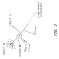
- the M group of HIV-1 which includes over 95% of the global virus isolates, consists of at least eight discrete clades (A, B, C, D, F, G, H, and J), based on the sequence of complete viral genomes.
- HIV-1 group 0 has been recovered from individuals living in Cameroon, Gabon, and Equatorial Guinea; their genomes share less than 50% identity in nucleotide sequence with group M viruses.
- group N HIV-I strains have been identified in infected Cameroonians, fail to react serologically in standard whole-virus enzyme-linked immunosorbent assay (ELISA), yet are readily detectable by conventional Western blot analysis.
- ELISA enzyme-linked immunosorbent assay
- HIV-1 genetic variation comes from studies of group M viruses of diverse geographic origin. Data collected during the past decade indicate that the HIV-1 population present within an infected individual can vary from 6% to 10% in nucleotide sequence. HIV-1 isolates within a clade may exhibit nucleotide distances of 15% in gag and up to 30% in gp120 coding sequences. Interclade genetic variation may range between 30% and 40% depending on the gene analyzed.
- Clade A viruses are genetically the most divergent and were the most common HIV-1 subtype in Africa early in the epidemic. With the rapid spread of HIV-1 to southern Africa during the mid to late 1990s, clade C viruses have become the dominant subtype and now account for 48% of HIV-1 infections worldwide. Clade B viruses, the most intensively studied HIV-1 subtype, remain the most prevalent isolates in Europe and North America.
- HIV-1 recombinants will be found in geographic areas such as Africa, South America, and Southeast Asia, where multiple HIV-1 subtypes coexist and may account for more than 10% of circulating HIV-1 strains. Molecularly, the genomes of these recombinant viruses resemble patchwork mosaics, with juxtaposed diverse HIV-1 subtype segments, reflecting the multiple crossover events contributing to their generation.
- HIV-1 recombinants have arisen in Africa and a majority contains segments originally derived from clade A viruses.
- the composition of the predominant circulating strain consists of a clade A gag plus pol gene segment and a clade E env gene.
- the clade E env gene in Thai HIV-1 strains is closely related to the clade E env present in virus isolates from the Central African -Republic, it is believed that the original recombination event occurred in Africa, with the subsequent introduction of a descendent virus into Thailand.
- no full-length HIV-1 subtype E isolate i.e., with subtype E gag, pol, and env genes has been reported to date.
- HIV-1 isolates using the CXCR4 receptor are usually T cell line (TCL)-tropic syncytium inducing (SI) strains, whereas those exclusively utilizing the CCR5 receptor (R5 viruses) are predominantly macrophage (M)-tropic and non-syncytium inducing (NSI).
- TCL T cell line
- SI macrophage
- NBI non-syncytium inducing
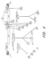
- the three primary HN-1 translation products are initially synthesized as polyprotein precursors, which are subsequently processed by viral or cellular proteases into mature particle-associated proteins ( Fig. 4 ).
- the 55-kd Gag precursor Pr55 Gag is cleaved into the matrix (MA), capsid (CA), nucleocapsid (NC), and p6 proteins.
- Retroviral Gag proteins are generally synthesized as polyprotein precursors; the HIV-1 Gag precursor has been named, based on its apparent molecular mass, Pr55 Gag .
- the mRNA for Pr55 Gag is the unspliced 9.2-kb transcript ( Fig. 4 ) that requires Rev for its expression in the cytoplasm.
- PR viral protease
- MA is localized immediately inside the lipid bilayer of the viral envelope
- CA forms the outer portion of the cone-shaped core structure in the center of the particle
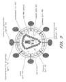
- NC is present in the core in a ribonucleoprotein complex with the viral RNA genome ( Fig. 5 ).
- the HIV Pr55 Gag precursor oligomerizes following its translation and is targeted to the plasma membrane, where particles of sufficient size and density to be visible by EM are assembled.
- Formation of virus-like particles by Pr55 Gag is a self-assembly process, with critical Gag-Gag interactions taking place between multiple domains along the Gag precursor.
- the assembly of virus-like particles does not require the participation of genomic RNA (although the presence of nucleic acid appears to be essential), pol-encoded enzymes, or Env glycoproteins, but the production of infectious virions requires the encapsidation of the viral RNA genome and the incorporation of the Env glycoproteins and the Gag-Pol polyprotein precursor Pr160 Gag-Pol .
- telomeres Downstream of gag lies the most highly conserved region of the HIV genome, the pol gene, which encodes three enzymes: PR, RT, and IN (see Fig. 4 ).
- RT and IN are required, respectively, for reverse transcription of the viral RNA genome to a double-stranded DNA copy, and for the integration of the viral DNA into the host cell chromosome.
- PR plays a critical role late in the life cycle by mediating the production of mature, infectious virions.
- the pol gene products are derived by enzymatic cleavage of a 160-kd Gag-Pol fusion protein, referred to as pr160Gag-PoI. This fusion protein is produced by ribosomal frameshifting during translation of Pr55 Gag (see Fig. 4 ).
- the frame-shifting mechanism for Gag-Pol expression also utilized by many other retroviruses, ensures that the pol derived proteins are expressed at a low level, approximately 5% to 10% that of Gag.
- the N-terminus of Pr160 Gag-Pol is myristylated and targeted to the plasma membrane.
- retroviral Gag proteins are initially synthesized as polyprotein precursors that are cleaved to generate smaller products. Subsequent studies demonstrated that the processing function is provided by a viral rather than a cellular enzyme, and that proteolytic digestion of the Gag and Gag-Pol precursors is essential for virus infectivity. Sequence analysis of retroviral PRs indicated that they are related to cellular "aspartic" proteases such as pepsin and renin. Like these cellular enzymes, retroviral PRs use two apposed Asp residues at the active site to coordinate a water molecule that catalyzes the hydrolysis of a peptide bond in the target protein.
- retroviral PRs function as true dimers.
- X-ray crystallographic data from HIV-1 PR indicate that the two monomers are held together in part by a four-stranded antiparallel ⁇ -sheet derived from both N- and C-terminal ends of each monomer.
- the substrate-binding site is located within a cleft formed between the- two monomers.
- the HIV PR dimer contains flexible "flaps" that overhang the binding site and may stabilize the substrate within the cleft; the active-site Asp residues lie in the center of the dimer.
- the primary sequences of retroviral PRs - are highly divergent, yet their structures are remarkably similar.
- Retroviral RTs have three enzymatic activities: (a) RNA-directed DNA polymerization (for minus-strand DNA synthesis), (b) RNaseH activity (for the degradation of the tRNA primer and genomic RNA present in DNA-RNA hybrid intermediates), and (c) DNA-directed DNA polymerization (for second- or plus-strand DNA synthesis).
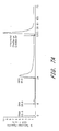
- the mature HIV-1 RT holoenzyme is a heterodimer of 66 and 51 kd subunits.
- the 51-kd subunit (p51) is derived from the 66-l:d (p66) subunit by proteolytic removal of the C-terminal 15-lcd RNaseH domain of p66 by PR (see Fig. 4 ).
- the crystal structure of,HIV-1 RT reveals a highly asymmetric folding in which the orientations of the p66 and p51 subunits differ substantially.
- the p66 subunit can be visualized as a right hand, with the polymerase active site within the palm, and a deep template-binding cleft formed by the palm, fingers, and thumb subdomains.
- the polymerase domain is linked to RNaseH by the connection subdomain.
- the active site located in the palm, contains three critical Asp residues (110, 185, and 186) in close proximity, and two coordinated Mg 2+ ions. Mutation of these Asp residues abolishes RT polymerizing activity.
- the orientation of the three active-site Asp residues is similar to that observed in other DNA polymerases (e.g., the Klenow fragment of E. coli DNA poll).
- the p51 subunit appears to be rigid and does not form a polymerizing cleft; Asp 110, 185, and 186 of this subunit are buried within the molecule.
- Approximately 18 base pairs of the primer-template duplex lie in the nucleic acid binding cleft, stretching from the polymerase active site to the RNaseH domain.
- the presence of a dideoxynucleotide at the 3' end of the primer allows visualization of the catalytic complex trapped just prior to attack on the incoming dNTP.
- Comparison with previously obtained structures suggests a model whereby the fingers close in to trap the template and dNTP prior to nucleophilic attack of the 3'-OH of the primer on the incoming dNTP.
- the fingers adopt a more open configuration, thereby releasing the pyrophosphate and enabling RT to bind the next dNTP.
- the structure of the HIV-1 RNaseH has also been determined by x-ray crystallography; this domain displays a global folding similar to that of E. coli RNaseH.
- retrovirus replication is the insertion, of a DNA copy of the viral genome into the host cell chromosome following reverse transcription.
- the integrated viral DNA (the provirus) serves as the template for the synthesis of viral RNAs and is maintained as part of the host cell genome for the lifetime of the infected cell.
- Retroviral mutants deficient in the ability to integrate generally fail to establish a productive infection.
- the integration of viral DNA is catalyzed by integrase, a 32-kd protein generated by PR-mediated cleavage of the C-terminal portion of the HIV-1 Gag-Pol polyprotein (see Fig. 4 ) .
- Retroviral IN proteins are composed of three structurally and functionally distinct domains: an N-terrninal, zinc-finger-containing domain, a core domain, and a relatively nonconserved C-terminal domain. Because of its low solubility, it has not yet been possible to crystallize the entire 288-amino-acid HIV-1 IN protein. However, the structure of all three domains has been solved independently by x-ray crystallography or NMR methods. The crystal structure of the core domain of the avian sarcoma virus IN has also been determined.
- the N-terminal domain (residues 1 to 55), whose structure was solved by NMR spectroscopy, is composed of four helices with a zinc coordinated by amino acids His-12, His-16, Cys-40, and Cys-43.
- the structure of the N-terminal domain is reminiscent of helical DNA binding proteins that contain a so-called helix-turn-helix motif; however, in the HIV-I structure this motif contributes to dimer formation.
- helix-turn-helix motif a so-called helix-turn-helix motif
- this motif contributes to dimer formation.
- poor solubility hampered efforts to solve the structure of the core domain.
- attempts at crystallography were successful when it was observed that a Phe-to-Lys change at IN residue 185 greatly increased solubility without disrupting in vitro catalytic activity.
- Each monomer of the HIV-1 IN core domain (IN residues 50 to 212) is composed of a five-stranded ⁇ -sheet flanked by helices; this structure bears striking resemblance to other polynucleotidyl transferases including RNaseH and the bacteriophage MuA transposase.
- Three highly conserved residues are found in analogous positions in other polynucleotidyl transferases; in HIV-1 IN these are Asp-64, Asp-116 and Glu-152, the so-called D,D-35-E motif. Mutations at these positions block HIV IN function both in vivo and in vitro.
- the HIV Env glycoproteins play a major role in the virus life cycle. They contain the determinants that interact with the CD4 receptor and coreceptor, and they catalyze the fusion reaction between the lipid bilayer of the viral envelope and the host cell plasma membrane. In addition, the HIV Env glycoproteins contain epitopes that elicit immune responses that are important from both diagnostic and vaccine development perspectives.
- the HIV Env glycoprotein is synthesized from the singly spliced 4.3-kb Vpu/Env bicistronic mRNA (see Fig. 4 ); translation occurs on ribosomes associated with the rough endoplasmic reticulum (ER).
- the 160-lcd polyprotein precursor (gp 160) is an integral membrane protein that is anchored to cell membranes by a hydrophobic stop-transfer signal in the domain destined to be the mature TM Env glycoprotein, gp41 ( Fig. 6 ).
- the gp160 is cotranslationally glycosylated, forms disulfide bonds, and undergoes oligomerization in the ER.
- the predominant oligomeric form appears to be a trimer, although dimers and tetramers are also observed.
- the gp160 is transported to the Golgi, where, like other retroviral envelope precursor proteins, it is proteolytically cleaved by cellular enzymes to the mature SU glycoprotein gp 120 arid TM glycoprotein gp41 (see Fig. 6 ).
- the cellular enzyme responsible for cleavage of retroviral Env precursors following a highly conserved Lys/Arg-X-Lys/Arg-Arg motif is furin or a furin-like protease, although other enzymes may also catalyze gp160 processing.
- gp160 Cleavage of gp160 is required for Env-induced fusion activity and virus infectivity. Subsequent to gp160 cleavage, gpl20 and gp41 form a noncovalent association that is critical for transport of the Env complex from the Golgi to the cell surface. The gp120-gp41 interaction is fairly weak, and a substantial amount of gp120 is shed from the surface of Env-expressing cells.
- the HIV Env glycoprotein complex in particular the SU (gp 120) domain, is very heavily glycosylated; approximately half the molecular mass of gp160 is composed of oligosaccharide side chains.
- SU gp 120 domain
- oligosaccharide side chains During transport of Env from its site of synthesis in the ER to the plasma membrane, many of the side chains are modified by the addition of complex sugars.
- the numerous oligosaccharide side chains form what could be imagined as a sugar cloud obscuring much of gp120 from host immune recognition.
- gp120 contains interspersed conserved (Q to C 5 ) and variable (V 1 to V 5 ) domains.
- the Cys residues present in the gp120s of different isolates are highly conserved and form disulfide bonds that link the first four variable regions in large loops.
- a primary function of viral Env glycoproteins is to promote a membrane fusion reaction between the lipid bilayers of the viral envelope and host cell membranes. This - membrane fusion event enables the viral core to gain entry into the host cell cytoplasm.
- a number of regions in both gp120 and gp41 have been implicated, directly or indirectly, in Env-mediated membrane fusion.
- N-helix N-terminal helix
- peptides derived from these sequences exhibit potent antiviral activity in culture.
- the structure of the ectodomain of HIV-1 and SIV gp4l, the two helical motifs in particular, has been the focus of structural analyses in recent years.
- gp41 adopts a hypothetical conformation that allows the fusion peptide to insert into the target lipid bilayer.
- the formation of the gp41 six-helix bundle brings the viral and cellular membranes together and membrane fusion takes place.
- the present invention relates to generation of a CD8 + T cell immune response against an antigen and also eliciting an antibody response. More particularly, the present invention relates to "prime and boost" immunization regimes in which the immune response induced by administration of a priming composition is boosted by administration of a boosting composition.
- the present invention is based on inventors' experimental demonstration that effective boosting can be achieved using modified vaccinia Ankara (MVA) vectors, following priming with any of a variety of different types of priming compositions including recombinant MVA itself.
- MVA modified vaccinia Ankara
- T lymphocytes of the CD8 + type also known as cytotoxic T lymphocytes (CTL).
- CTL cytotoxic T lymphocytes
- An important function of CD8 + cells is secretion of gamma interferon (IFNy), and this provides a measure of CD8 + T cell immune response.
- IFNy gamma interferon
- a second component of the immune response is antibody directed to the proteins of the pathogen.
- the present invention employs MVA which, as the experiments described below show, has been found to be an effective means for providing a boost to a CD8 + T cell immune response primed to antigen using any of a variety of different priming compositions and also eliciting an antibody response.
- the experimental work described below demonstrates that use of embodiments of the present invention allows for recombinant MVA virus expressing an HIV antigen to boost a CD8 + T cell immune response primed by a DNA vaccine and also eliciting an antibody response.
- the MVA was found to induce a CD8 + T cell response after intradermal, intramuscular or mucosal immunization.
- Recombinant MVA has also been shown to prime an immune response that is boosted by one or more inoculations of recombinant MVA.
- Non-human primates immunized with plasmid DNA and boosted with the MVA were effectively protected against intramucosal challenge with live virus.
- a vaccination regime used intradermal, intramuscular or mucosal immunization for both prime and boost can be employed, constituting a general immunization regime suitable for inducing CD8 + T cells and also eliciting an antibody response, e.g. in humans.
- the present invention in various aspects and embodiments employs an MVA vector encoding an HIV antigen for boosting a CD8 + T cell immune response to the antigen primed by previous administration of nucleic acid encoding the antigen and also eliciting an antibody response.
- a general aspect of the present invention provides for the use of an MVA vector for boosting a CD8 + T cell immune response to an HIV antigen and also eliciting an antibody response.
- One aspect of the present invention provides a method of boosting a CD8 + T cell immune response to an HIV antigen in an individual, and also eliciting an antibody response, the method including provision in the individual of an MVA vector including nucleic acid encoding the antigen operably linked to regulatory sequences for production of antigen in the individual by expression from the nucleic acid, whereby a CD8 + T cell immune response to the antigen previously primed in the individual is boosted.
- An immune response to an HIV antigen may be primed by immunization with plasmid DNA or by infection with an infectious agent.
- a further aspect of the invention provides a method of inducing a CD8 + T cell immune response to an HIV antigen in an individual; and also eliciting an antibody response, the method comprising administering to the individual a priming composition comprising nucleic acid encoding the antigen and then administering a boosting composition which comprises an MVA vector including nucleic acid encoding the antigen operably linked to regulatory sequences for production of antigen in the individual by expression from the nucleic acid.
- a further aspect provides for use of an MVA vector, as disclosed, in the manufacture of a medicament for administration to a mammal to boost a CD8 + T cell immune response to an HIV antigen, and also eliciting an antibody response.
- a medicament is ' generally for administration following prior administration of a priming composition comprising nucleic acid encoding the antigen.
- the priming composition may comprise any viral vector, such as a vaccinia virus vector such as a replication-deficient strain such as modified vaccinia Ankara (MVA) or NYVAC ( Tartaglia et al. 1992 Virology 118:217-232 ), an avipox vector such as fowlpox or canarypox, e.g. the strain known as ALVAC ( Paoletti et al. 1994 Dev Biol Stand 82:65-69 ), or an adenovirus vector or a vesicular stomatitis virus vector or an alphavirus vector.
- a viral vector such as a vaccinia virus vector such as a replication-deficient strain such as modified vaccinia Ankara (MVA) or NYVAC ( Tartaglia et al. 1992 Virology 118:217-232 ), an avipox vector such as fowlpox or canarypox, e.g. the strain
- the priming composition may comprise DNA encoding the antigen, such DNA preferably being in the form of a circular plasmid that is not capable of replicating in mammalian cells.
- Any selectable marker should not be resistance to an antibiotic used clinically, so for example Kanamycin resistance is preferred to Ampicillin resistance.
- Antigen expression should be driven by a promoter which is active in mammalian cells, for instance the cytomegalovirus immediate early (CMV IE) promoter.
- CMV IE cytomegalovirus immediate early
- a priming composition is followed by boosting with a boosting composition, or first and second boosting compositions, the first and second boosting compositions being the same or different from one another.
- Still further boosting compositions may be employed without departing from the present invention.
- a triple immunization regime employs DNA, then adenovirus as a first boosting composition, then MVA as a second boosting composition, optionally followed by a further (third) boosting composition or subsequent boosting administration of one or other or both of the same or different vectors.
- DNA then MVA then adenovirus, optionally followed by subsequent boosting administration of one or other or both of the same or different vectors.
- the antigen to be encoded in respective priming and boosting compositions need not be identical, but should share at least one CD8 + T cell epitope.
- the antigen may correspond to a complete antigen, or a fragment thereof.
- Peptide epitopes or artificial strings of epitopes may be employed, more efficiently cutting out unnecessary protein sequence in the antigen and encoding sequence in the vector or vectors.
- One .or more additional epitopes may be included, for instance epitopes which are recognized by T helper cells, especially epitopes recognized in individuals of different HLA types.
- An HIV antigen of the invention to be encoded by a recombinant MVA virus includes polypeptides having immunogenic activity elicited by an amino acid sequence of an HIV Env, Gag, Pol, Vif, Vpr, Tat, Rev, Vpu, or Nef amino acid sequence as at least one CD8 + T cell epitope.
- This amino acid sequence substantially corresponds to at least one 10-900 amino acid fragment and/or consensus sequence of a known HIV Env or Pol; or at least one 10-4.50 amino acid fragment and/or consensus sequence of a kown H1V Gag; or at least one 10-100 amino acid fragment and/or consensus sequence of a known HIV Vif, Vpr, Tat, Rev, Vpu, or Nef.
- Env is optionally deleted of subsequences. For example, regions of the gp120 surface and gp41 transmembrane cleavage products can be deleted.
- Gag is optionally deleted of subsequences.
- regions of the matrix protein (p17), regions of the capsid protein (p24), regions of the nucleocapsid protein (p7), and regions of p6 (the C-terminal peptide of the Gag polyprotein) can be deleted.
- Pol is optionally deleted of subsequences.
- regions of the protease protein (plO) regions of the reverse transcriptase protein (p66/p5l), and regions of the integrase protein (p32) can be deleted.
- Such an HIV Env, Gag, or Pol can have overall identity of at least 50% to a known Env, Gag, or Pol protein amino acid sequence, such as 50-99% identity, or any range or value therein, while eliciting an immunogenic response against at least one strain of an HIV.
- Percent identify can be determined, for example, by comparing sequence information using the GAP computer program, version 6.0, available from the University of Wisconsin Genetics Computer Group (UWGCG).
- the GAP program utilizes the alignment method of Needleman and Wunsch (J Mol Biol 1970 48:443 ), as revised by Smith and Waterman (Adv Appl Math 1981 2:482 ). Briefly, the GAP program defines identity as the number of aligned symbols (i.e., nucleotides or amino acids) which are identical, divided by the total number of symbols in the shorter of the two sequences.
- the preferred default parameters for the GAP program include: (1) a unitary comparison matrix (containing a value of 1 for identities and 0 for non-identities) and the weighted comparison matrix of Gribskov and Burgess (Nucl Acids Res 1986 14:6745 ), as described by Schwartz and Dayhoff (eds., Atlas of Protein Sequence and Structure, National Biomedical Research Foundation, Washington, D.C. 1979, pp. 353-358 ); (2) a penalty of 3.0 for each gap and an additional 0.10 penalty for each symbol in each gap; and (3) no penalty for end gaps.
- an Env of the present invention is a variant form of at least one HIV envelope protein.
- the Env is composed of gp120 and the membrane-spanning and ectodomain of gp41 but lacks part or all of the cytoplasmic domain of gp4l.
- HIV sequences are readily available from commercial and institutional HIV sequence databases, such as GENBANK, or as published compilations, such as Myers et al. eds., Human Retroviruses and AIDS, A Compilation and Analysis of Nucleic Acid and Amino Acid Sequences, Vol. I and IT, Theoretical Biology and Biophysics, Los Alamos, N. Mex. (1993 ), or http://hiv-web.lanl.gov/.
- Substitutions or insertions of an HIV Env, Gag, or Pol to obtain an additional HIV Env, Gag, or Pol, encoded by a nucleic acid for use in a recombinant MVA virus of the present invention can include substitutions or insertions of at least one amino acid residue (e.g., 1-25 amino acids).
- at least one amino acid e.g., 1-25 amino acids
- substitutions, insertions or deletions are identified based on safety features, expression levels, immunogenicity and compatibility with high replication rates of MVA.
- Amino acid sequence variations in an HIV Env, Gag, or Pol of the present invention can be prepared e.g., by mutations in the DNA.
- HIV Env, Gag, or Pol include, for example, deletions, insertions or substitutions of nucleotides coding for different amino acid residues within the amino acid sequence.
- mutations that will be made in nucleic acid encoding an HIV Env, Gag, or Pol must not place the sequence out of reading frame and preferably will not create complementary domains that could produce secondary mRNA structures.
- HIV Env, Gag, or Pol-encoding nucleic acid of the present invention can also be prepared by amplification or site-directed mutagenesis of nucleotides in DNA or RNA encoding an HIV Env, Gag, or Pol and thereafter synthesizing or reverse transcribing the encoding DNA to produce DNA or RNA encoding an HIV Env, Gag, or Pol, based on the teaching and guidance presented herein.
- Recombinant MVA viruses expressing HIV Env, Gag, or Pol of the present invention include a finite set of HIV Env, Gag, or Pol-encoding sequences as substitution nucleotides that can be routinely obtained by one of ordinary skill in the art, without undue experimentation, based on the teachings and guidance presented herein.
- substitution nucleotides can be routinely obtained by one of ordinary skill in the art, without undue experimentation, based on the teachings and guidance presented herein.
- regulatory sequences for expression of the encoded antigen will include a natural, modified or synthetic poxvirus promoter.
- promoter is meant a sequence of nucleotides from which transcription may be initiated of DNA operably linked downstream (i.e. in the 3' direction on the sense strand of double-stranded DNA).
- operably linked means joined as part of the same nucleic acid molecule, suitably positioned and oriented for transcription to be initiated from the promoter.
- DNA operably linked to a promoter is "under transcriptional initiation regulation" of the promoter.
- Terminator fragments include terminator fragments, polyadenylation sequences, marker genes and other sequences.
- Other regulatory sequences including terminator fragments, polyadenylation sequences, marker genes and other sequences may be included as appropriate, in accordance with the knowledge and practice of the ordinary person skilled in the art: see, for example, Moss, B. (2001). Poxviridae: the viruses and their replication. In Fields Virology, D.M. Knipe, and P.M. Howley, eds. (Philadelphia, Lippincott Williams & Willdns), pp. 2849-2883 .
- Promoters for use in aspects and embodiments of the present invention must be compatible with poxvirus expression systems and include natural, modified and synthetic sequences.
- Either or both of the priming and boosting compositions may include an adjuvant, such as granulocyte macrophage-colony stimulating factor (GM-CSF) or encoding nucleic acid therefor.
- an adjuvant such as granulocyte macrophage-colony stimulating factor (GM-CSF) or encoding nucleic acid therefor.
- GM-CSF granulocyte macrophage-colony stimulating factor
- Administration of the boosting composition is generally about 1 to 6 months after administration of the priming composition, preferably about 1 to 3 months.
- administration of priming composition, boosting composition, or both priming and boosting compositions is intradermal, intramuscular or mucosal immunization.
- MVA vaccines may be achieved by using a needle to inject a suspension of the virus.
- a needleless injection device to administer a virus suspension (using, e.g., BiojectorTM needleless injector) or a resuspended freeze-dried powder containing the vaccine, providing for manufacturing individually prepared doses that do not need cold storage. This would be a great advantage for a vaccine that is needed in rural areas of Africa.
- MVA is a virus with an excellent safety record in human immunizations.
- the generation of recombinant viruses can be accomplished simply, and they can be manufactured reproducibly in large quantities.
- Intradermal, intramuscular or mucosal administration of recombinant MVA virus is therefore highly suitable for prophylactic or therapeutic vaccination of humans against AIDS which can be controlled by a CD8 + T cell response.
- the individual may have AIDS such that delivery of the antigen and generation of a CD8 + T cell immune response to the antigen is of benefit or has a therapeutically beneficial effect.
- administration will have prophylactic aim to generate an immune response against HIV or AIDS before infection or development of symptoms.
- compositions may comprise a pharmaceutically acceptable excipient, carrier, buffer, stabilizer or other materials well known to those skilled in the art. Such materials should be non-toxic and should not interfere with the efficacy of the active ingredient.
- the precise nature of the carrier or other material may depend on the route of administration, e.g. intravenous, cutaneous or subcutaneous, nasal, intramuscular, intraperitoneal routes.
- administration is preferably intradermal, intramuscular or mucosal.
- Physiological saline solution dextrose or other saccharide solution or glycols such as ethylene glycol, propylene glycol or polyethylene glycol may be included.
- the active ingredient will be in the form of a parenterally acceptable aqueous solution which is pyrogen-free and has suitable pH, isotonicity and stability.
- a parenterally acceptable aqueous solution which is pyrogen-free and has suitable pH, isotonicity and stability.
- isotonic vehicles such as Sodium Chloride Injection, Ringer's Injection, Lactated Ringer's Injection.
- Preservatives, stabilizers, buffers, antioxidants and/or other additives may be included as required.
- a slow-release formulation may be employed.
- the particles may be administered to an individual, particularly human or other primate. Administration may be to another mammal, e.g. rodent such as mouse, rat or hamster, guinea pig, rabbit, sheep, goat, pig, horse, cow, donkey, dog or cat.
- rodent such as mouse, rat or hamster, guinea pig, rabbit, sheep, goat, pig, horse, cow, donkey, dog or cat.
- Administration is preferably in a "prophylactically effective amount” or a “therapeutically effective amount” (as the case may be, although prophylaxis may be considered therapy), this being sufficient to show benefit to the individual.
- the actual amount administered, and rate and time-course of administration, will depend on the nature and severity of what is being treated. Prescription of treatment, e.g. decisions on dosage etc, is within the responsibility of general practitioners and other medical doctors, or in a veterinary context a veterinarian, and typically takes account of the disorder to be treated, the condition of the individual patient, the site of delivery, the method of administration and other factors know to practitioners. Examples of the techniques and protocols mentioned above can be found in Remington's Pharmaceutical Sciences, 16th edition, 1980, Osol, A. (ed .).
- DNA is administered at a dose of 250 vg to 2.5 mg/injection, followed by MVA at a dose of 10 6 to 10 9 infectious virus particles/injection.
- a composition may be administered alone or in combination with other treatments, either simultaneously or sequentially dependent upon the condition to be treated.
- Delivery to a non-human mammal need not be for a therapeutic purpose, but may be for use in an experimental context, for instance in investigation of mechanisms of immune responses to an antigen of interest, e.g. protection against HIV or AIDS.
- SHIV-89.6 The 89.6 chimera of simian and human immunodeficiency viruses (SHIV-89.6) was used for the construction of immunogens and its highly pathogenic derivative, SHIV-89.6P, for challenge ( G.B. Karlsson et al. 1997 J ViroI71:4218 ). SHIV-89.6 and SHIV-89.6F do not generate cross-neutralizing antibody ( D.C.. Montefiori et al. 1998 J Virol 72:3427 ) and allowed us to address the ability of vaccine-raised T cells and non-neutralizing antibodies to control an immunodeficiency virus challenge.
- Modified vaccinia Ankara was used for the construction of the recombinant poxvirus.
- MVA has been highly effective at boosting DNA-primed CD8 T cells and enjoys the safety feature of not replicating efficiently in human or monkey cells ( H.L. Robinson et al. 2000 AIDS Reviews 2:105 ).
- the DNA prime (DNA/89.6) expressed simian immunodeficiency virus (SIV) Gag, Pol, Vif, Vpx, and Vpr and human immunodeficiency virus-1 (HIV-1) Env, Tat, and Rev from a single transcript ( R.J. Gorelick et al. 1999 Virology 253:259 ; M.M. Sauter et al. 1996 J Cell Biol 132:795 ).
- SIV simian immunodeficiency virus
- HIV-1 human immunodeficiency virus-1
- SHIV-89.6 sequences were cloned into the vector pGA2 using ClaI and RsrII sites. This cloning deleted both long terminal repeats (LTRs) and nef.
- the SHIV-89.6 sequences also were internally mutated for a 12-base pair region encoding the first four amino acids of the second zinc finger in .nucleocapsid. This mutation renders SHIV viruses noninfectious ( R.J. Gorelick et al. 1999 Virology 253:259 ).
- a mutation in gp41 converted the tyrosine at position 710 to cysteine to achieve better expression of Env on the plasma membrane of DNA-expressing cells ( M.M.
- pGA2 uses the CMV immediate early promoter without intron A and the bovine growth hormone polyadenylation sequence to express vaccine inserts.
- Vaccine DNA was produced by Althea (San Diego, CA). In transient transfections of 293T cells, DNA/89.6 produced about 300 ng of Gag and 85 ng of Env per 1 ⁇ 10 6 cells.
- the rMVA booster (MVA/89.6) expressed SIV Gag, Pol, and HIV-1 Env under the control of vaccinia virus early/late promoters.
- the MVA double recombinant virus expressed both the HIV 89.6 Env and the SIV 239 Gag-Pol, which were inserted into deletion II and deletion III of MVA, respectively.
- the 89.6 Env protein was truncated for the COOH-terminal 115 amino acids of gp4l.
- the modified H5 promoter controlled the expression of both foreign genes.
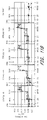
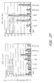
- Vaccination was accomplished by priming with DNA at 0 and 8 weeks and boosting with rMVA at 24 weeks ( Fig. 7A ).
- I.d. and i.m. DNA immunizations were delivered in phosphate-buffered saline (PBS) with a needleless jet injector (Bioject, Portland, OR) to deliver five i.d. 100- ⁇ l injections to each outer thigh for the 2.5-mg dose of DNA or one i.d. 100- ⁇ l injection to the right outer thigh for the 250- ⁇ g dose of plasmid.
- I.m. deliveries of DNA were done with one 0.5-ml injection of DNA in PBS to each outer thigh for the 2.5-mg dose and one 100- ⁇ l injection to the right outer thigh for the 250- ⁇ g dose. 1 ⁇ 10 8 pfu of MVA189.6 was administered both i.d.
- Control animals received 2.5 mg of the pGA2 vector without vaccine insert with the Bioject device to deliver five 100- ⁇ l doses i.d. to each outer thigh.
- the control MVA booster immunization consisted of 2 ⁇ 10 8 pfu of MVA without an insert delivered i.d. and i.m. as described for MVA/89.6.
- Animal numbers are as follows: 1, RBr-5*; 2, RIm-5*; 3, RQf-5*; 4, RZe-5; 5, Room-5; 6, RDm-5; 7, RAj-5*; 8, RJi-5*; 9, RAl-5*; 10, RDe-5*; 11, RAi-5; 12, RPr-5; 13, RKw-4*; 14, RWz-5*; 15, RGo-5; 16, RLp-4; 17, RWd-6; 18, RAt-5; 19, RPb-5*; 20, RIi-5*; 21, RIq-5; 22, RSp-4; 23, RSn-5; 24, RGd-6; 25, RMb-5*; 26, RGy-5*; 27, RUs-4; and 28, RPm-5. Animals with the A*01 allele are indicated with asterisks.
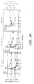
- DNA priming followed by rMVA boosting generated high frequencies of virus-specific T cells that peaked at one week following the rMVA booster ( Fig. 7 ).
- the frequencies of T cells recognizing the Gag-CM9 epitope were assessed by means of Mamu-A*01 tetramers, and the frequencies of T cells recognizing epitopes throughout Gag were assessed with pools of overlapping peptides and an enzyme-linked immunospot (ELISPOT) assay ( C.A. Power et al. 1999 J Immunol Methods 227:99 ).
- ELISPOT enzyme-linked immunospot
- peripheral blood mononuclear cells PBMC
- PBMC peripheral blood mononuclear cells
- FITC fluorescein isothiocyanate
- PerCP CD8 conjugated to peridinin chlorophyl protein
- GTPYDINQM Gag-CM9- Mamu - A * 01 tetramer
- APC allophycocyanin
- Cells were washed twice with cold PBS containing 2% fetal bovine serum (FBS), fixed with 1% paraformaldehyde in PBS, and analyzed within 24 hrs on a FACScaliber (Becton Dickinson, San Jose, CA). Cells were initially gated on lymphocyte populations with forward scatter and side scatter and then on CD3 cells. The CD3 cells were then analyzed for CD8 and tetramer-binding cells. About 150,000 lymphocytes were acquired for each sample. Data were analyzed using FloJo software (Tree Star, San Carlos, CA).
- IFN-y ELISPOTs For interferon- ⁇ (IFN-y) ELISPOTs, MULTISCREEN 96 well filtration plates (Millipore Inc. Bedford, MA) were coated overnight with antibody to human IFN- ⁇ (Clone B27, Pharmingen, San Diego, CA) at a concentration of 2 ⁇ g/ml in sodium bicarbonate buffer (pH 9.6) at 8° to 10°C. Plates were washed two times with RPMI medium and then blocked for 1 hour with complete medium (RPMI containing 10% FBS) at 37°C. Plates were washed five more times with plain RPMI medium, and cells were seeded in duplicate in 100 ⁇ l complete medium at numbers ranging from 2x10 4 to 5x10 5 cells per well.
- IFN-y interferon- ⁇
- Peptide pools were added to each well to a final concentration of 2 ⁇ g/ml of each peptide in a volume of 100 ⁇ l in complete medium.
- Cells were cultured at 37°C for about 36 hrs under 5% CO 2 . Plates were washed six times with wash buffer (PBS with 0.05% Tween-20) and then incubated with 1 ⁇ g of biotinylated antibody to human IFN- ⁇ per milliliter (clone 7-86-1; Diapharma Group, West Chester, OH) diluted in wash buffer containing 2% FBS. Plates were incubated for 2 hrs at 37°C and washed six times with wash buffer.
- wash buffer PBS with 0.05% Tween-20
- ELISPOTs The frequencies of ELISPOTs are approximate because different dilutions of cells have different efficiencies of spot formation in the absence of feeder cells ( C.A. Power et al. 1999 J Immunol Methods 227: 99 ). The same dilution of cells was used for all animals at a given time point, but different dilutions were used to detect memory and acute responses.
- Gag-CM9 tetramer analyses were restricted to macaques that expressed the Mamu-A*01 histocompatibility type, whereas ELISPOT responses did not depend on a specific histocompatibility type.
- the DNA immunizations raised low levels of memory cells that expanded to high frequencies within 1 week of the rMVA booster ( Fig. 7 and 12 ).
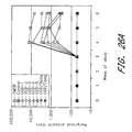
- CD8 cells specific to the Gag-CM9 epitope expanded to frequencies as high as 19% of total CD8 T cells ( Fig. 12 ). This peak of specific cells underwent a 10- to 100-fold contraction into the DNA/MVA memory pool ( Fig. 7A and 12 ).
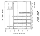
- ELISPOTs for three pools of Gag peptides also underwent a major expansion (frequencies up to 4000 spots for 1x10 6 PBMC) before contracting from 5- to 20-fold into the DNA/MVA memory response ( Fig. 7B ).
- the frequencies of ELISPOTs were the same in macaques with and without the A*01 histocompatibility type (P>0.2).
- Simple linear regression was used to estimate correlations between postbooster and postchallenge ELISPOT responses, between memory and postchallenge ELISPOT responses, and between logarithmically transformed viral loads and ELISPOT frequencies. Comparisons between vaccine and control groups and A*01 and non A*01 macaques were performed by means of two-sample t tests with logarithmically transformed viral load and ELISPOT responses. Two-way analyses of variance were used to examine the effects of dose and route of administration on peak DNA/MVA ELISPOTs, on memory DNA/MVA ELISPOTs, and on logarithmically transformed Gag antibody data.
- the rank order for the height of the ELISPOTs in the vaccine groups was 2.5 mg i.d. > 2.5 mg i.m. > 250 ⁇ g i.d. > 250 ⁇ g i.m. ( Fig. 7B ) .
- the IFN- ⁇ ELISPOTs included both CD4 and CD8 cells. Gag-CM9-specific CD8 cells had good lytic activity after restimulation with peptide.
- the highly pathogenic SHIV-89.6P challenge was administered intrarectally at 7 months after the rMVA booster, when vaccine-raised T cells were in memory ( Fig. 7 ).
- the challenge stock (5.7 x 10 9 copies of viral RNA per milliliter) was produced by one intravenous followed by one intrarectal passage in rhesus macaques of the original SHIV-89.6P stock ( G.B. Karlsson et al. 1997 J Virol 71:4218 ). Lymphoid cells were harvested from the intrarectally infected animal at peak viremia, CD8-depleted, and mitogen-stimulated for stock production. Before intrarectal challenge, fasted animals were anesthetized (ketamine, 10 mg/kg) and placed on their stomach with the pelvic region slightly elevated. A feeding tube (8Fr (2.7 mm) x 16 inches (41 cm); Sherwood Medical, St.
- a syringe containing 20 intrarectal infectious doses in 2 ml of RPMI-1640 plus 10% FBS was attached to the tube and the inoculum was slowly injected into the rectum.
- the feeding tube was flushed with 3.0 ml of RPMI without FBS and then slowly withdrawn. Animals were left in place, with pelvic regions slightly elevated, for a period of ten minutes after the challenge.
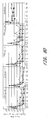
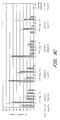
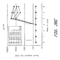
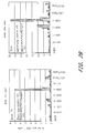
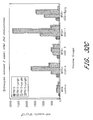
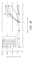
- titers of plasma viral RNA were at least 10-fold lower in the vaccine groups (geometric means of 1x10 7 to 5x10 7 ) than in the control animals (geometric mean of 4x10 8 ) ( Fig. 8A ) ( S. Staprans et al. in: Viral Genome Methods K. Adolph, ed. CRC Press, Boca Raton, FL, 1996 pp. 167-184 ; R. Hofmann-Lebmann et al. 2000 AIDS Res Hum Retroviruses 16:1247 ).
- RNA from 150 ⁇ l of ACD anticoagulated plasma was directly extracted with the QIAamp Viral RNA kit (Qiagen), eluted in 60 ⁇ l of AVE buffer, and frozen at -80°C until SHIV RNA quantitation was performed.
- Five microliters of purified plasma RNA was reverse-transcribed in a final 20- ⁇ l volume containing 50 mM KCl, 10 mM Tris-HCl (pH 8.3), 4 mM MgCl 2 , 1 mM each deoxynucleotide triphosphate (dNTP), 2.5 ⁇ M random hexamers, 20 units MultiScribe RT, and 8 units ribonuclease inhibitor.
- reaction mix was adjusted to a final volume of 50 ⁇ l containing 50 mM KCl, 10 mM Tris-HCl (pH 8.3), 4 mM MgCl 2 , 0.4 mM each dNTP, 0.2 ⁇ M forward primer, 0.2 ⁇ M reverse primer, 0.1 ⁇ M probe, and 5 units AmpliTaq Gold DNA polymerase (all reagents from PerkinElmer Applied Biosystems, Foster City, CA).
- the primer sequences within a conserved portion of the SIV gag gene are the same as those described previously ( S.
- both high-dose DNA-primed groups and the low-dose i.d. DNA-primed group had reduced their geometric mean loads to about 1000 copies of viral RNA per milliliter.
- the low-dose i.m. DNA-primed group had a geometric mean of 6x10 3 copies of viral RNA and the nonvaccinated controls had a geometric mean of 2 x 10 6 .
- the low-dose i.m. group had reduced its geometric mean copies of viral RNA to 1000.
- the 24 vaccinated animals only one animal, animal number 22 in the low-dose i.m. group, had intermittent viral loads above 1x10 4 copies per milliliter ( Fig 8D ) .
- Containment of the viral challenge was associated with a burst of antiviral T cells ( FIG. 7 and 9A ) .
- the frequency of tetramer + cells in the peripheral blood had decreased, potentially reflecting the recruitment of specific T cells to the site of infection ( Fig. 9A ) .
- tetramer + cells in the peripheral blood had expanded to frequencies as high as, or higher than, after the rMVA booster ( Fig. 7 and 9A ) .
- the majority of the tetramer + cells produced IFN- ⁇ in response to a 6-hour peptide stimulation ( Fig. 9B ) ( S.L. Waldrop et al. 1997 J Clin Invest 99:1739 ) and did not have the "stunned" IFN- ⁇ negative phenotype sometimes observed in viral infections ( F. Lechner et al. 2000 J Exp Med 191:1499 ).
- PBMC peripheral blood mononuclear cells
- CTPYDINQM Gag-CM9 peptide
- SEQ ID NO: 6 Gag-CM9 peptide
- RPMI RPMI containing 10% FBS and monensin (10 ⁇ g/ml) was added, and the cells were cultured for an additional 5 hrs at 37°C at an angle of 5° under 5% CO 2 .
- Cells were surface stained with antibodies to CD8 conjugated to PerCP (clone SKI, Becton Dickinson) at 8° to 10°C for 30 min, washed twice with cold PBS containing 2% FBS, and fixed and permeabilized with Cytofix/Cytoperm solution (Phanningen).
- T cells The postchallenge burst of T cells contracted concomitant with the decline of the viral load.
- virus-specific T cells were present at about one-tenth of their peak height ( Figs. 7A and 9A ) .
- the naive animals mounted a modest primary response ( Figs. 7B and 9A ).
- Tetramer cells peaked at less than 1% of total CD8 cells ( Fig. 9A ), and IFN- ⁇ -producing ELISPOTs were present at a mean frequency of about 300 as opposed to the much higher frequencies of 1000 to 6000 in the vaccine groups ( Fig. 7B ) (P ⁇ 0.05).
- the tetramer + cells in the control group like those in the vaccine group, produced IFN- ⁇ after peptide stimulation ( Fig. 9B ) .
- IFN- ⁇ after peptide stimulation
- three of the four controls had undetectable levels of IFN- ⁇ -producing ELISPOTs. This rapid loss of antiviral T cells in the presence of high viral loads may reflect the lack of CD4 help.
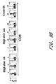
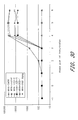
- T cell proliferative responses demonstrated that virus-specific CD4 cells had survived the challenge and were available to support the antiviral immune response ( Fig. 9C ) .
- Stimulation indices are the counts of tritiated thymidine incorporated in PBMC stimulated with 89.6 antigens divided by the counts of tritiated thymidine incorporated by the same PBMC stimulated with mock antigen.
- mean stimulation indices for Gag-Pol-Env or Gag-Pol proteins ranged from 35 to 14 in the vaccine groups but were undetectable in the control group. Consistent with the proliferation assays, intracellular cytokine assays demonstrated the presence of virus-specific CD4 cells in vaccinated but not control animals.
- the overall rank order of the vaccine groups for the magnitude of the proliferative response was 2.5 mg i.d. > 2.5 mg i.m. > 250 ⁇ g i.d. > 250 ⁇ g i.m.
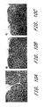
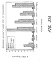
- lymph nodes from the vaccinated animals were morphologically intact and responding to the infection, whereas those from the infected controls had been functionally destroyed ( Fig. 10 ) .
- Nodes from vaccinated animals contained large numbers of reactive secondary follicles with expanded germinal centers and discrete dark and light zones ( Fig. 10A ) .
- lymph nodes from the nonvaccinated control animals showed follicular and paracortical depletion ( Fig. 10B ), while those from unvaccinated and unchallenged animals displayed normal numbers of minimally reactive germinal centers ( Fig. 10C ) .
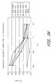
- Germinal centers occupied ⁇ 0.05% of total lymph node area in the infected controls, 2% of the lymph node area in the uninfected controls, and up to 18% of the lymph node area in the vaccinated groups ( Fig. 10D ) . More vigorous immune reactivity in the low-dose than the high-dose DNA-primed animals was suggested by more extensive germinal centers in the low dose group ( Fig. 10D ) .
- in situ hybridization for viral RNA revealed rare virus-expressing cells in lymph nodes from 3 of the 24 vaccinated macaques, whereas virus-expressing cells were readily detected in lymph nodes from each of the infected control animals.
- the cytomorphology of infected lymph node cells was consistent with a macrophage phenotype.
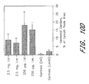
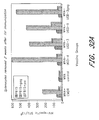
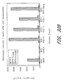
- the prime/boost strategy raised low levels of antibody to Gag and undetectable levels of antibody to Env ( Fig. 11 ) .
- Postchallenge antibodies to both Env and Gag underwent anamnestic responses with total Gag antibody reaching heights approaching 1 mg/ml and total Env antibody reaching heights of up to 100 ⁇ g/ml.
- Enzyme-linked immunosorbent assays for total antibody to Gag used bacterially produced SIV gag p27 to coat wells (2 ⁇ g per milliliter in bicarbonate buffer).
- ELISAs for antibody to Env antibody used 89.6 Env produced in transiently transfected 293T cells and captured with sheep antibody against Env (catalog number 6205; International Enzymes, Fairbrook CA).
- Standard curves for Gag and Env ELISAs were produced with serum from a SHIV-89.6-infected macaque with known amounts of immunoglobulin G (IgG) specific for Gag or Env, Bound antibody was detected with peroxidase-conjugated goat antibody to macaque IgG (catalog # YNGMOIGGFCP; Accurate Chemical, Westbury, NY) and TMB substrate (Catalog # T3405; Sigma, St. Louis, MO). Sera were assayed at threefold dilutions in duplicate wells. Dilutions of test sera were per-formed in whey buffer (4% whey and 0.1% tween 20 in IX PBS).
- Blocking buffer consisted of whey buffer plus 0.5% nonfat dry milk. Reactions were stopped with 2M H 2 SO 4 and the optical density read at 450 nm. Standard curves were fitted and sample concentrations were interpolated as ⁇ g of antibody per ml of serum using SOFTmax 2.3 software (Molecular Devices, Sunnyvale, CA).
- the high-dose DNA-primed animals had slightly lower geometric mean levels of viral RNA (7x10 2 and 5x10 2 ) than the low-dose DNA-primed animals (9x10 2 and 1x10 3 ).
- the DNA/MVA vaccine controlled the infection, rapidly reducing viral loads to near or below 1000 copies of viral RNA per milliliter of blood. Containment, rather than prevention of infection, affords the opportunity to establish a chronic infection ( H.L. Robinson et al. 1999 Nat Med 5:526 ). By rapidly reducing viral loads, a multiprotein DNA/MVA vaccine will extend the prospect for long-term non-progression and limit HIV transmission ( J.W. Mellors et al. 1996 Science 272:1167 ; T.C. Quinn et al. 2000 N Engl J Med 342:921 ).
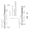
- This disclosure describes the construction of a modified vaccinia Ankara (MVA) recombinant virus, MVA/HIV clade B recombinant virus expressing the HIV strain ADA env and the HXB2 gag pol (MVA/HIV ADA env + HXB2 gag pol).
- MVA/HIV 48 the lab name of MVA/HIV 48 will be used, which denotes the plasmid from which the construct comes.
- the HIV gag-pol genes were derived from the Clade B infectious HXB2 virus.
- the gag-pol gene was truncated so that most of the integrate coding sequences were removed and amino acids 185, 266, and 478 were mutated to inactivate reverse transcriptase, inhibit strand transfer activity, and inhibit the RNaseH activity, respectively.
- the Clade B CCR5 tropic envelope gene was derived from the primary ADA isolate; TTTTTNT (SEQ ID NO: 14) sequences were mutated without changing coding capacity to prevent premature transcription termination and the cytoplasmic tail was truncated in order to improve surface expression, immunogenicity, and stability of the MVA vector.
- the HIV genes were inserted into a plasmid transfer vector so that gag-pol gene was regulated by the modified H5 early/late vaccinia virus promoter and the env gene was regulated by the newly designed early/late Psyn II promoter to provide similar high levels of expression.
- a self deleting GUS reporter gene was included to allow detection and isolation of the recombinant virus.
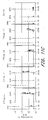
- the HIV genes were flanked by MVA sequences to allow homologous recombination into the deletion 3 site so that the recombinant MVA would remain TK positive for stability and high expression in resting cells. The recombinant MVA was isolated and shown to express abundant amounts of gag-pol-env and to process gag.
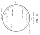
- the plasmid transfer vector used to make the MVA recombinant virus, pLW-48, ( Figure 15 ) by homologous recombination was constructed as follows:
- nucleotide sequences determined by sequencing a DNA molecule herein were determined using an automated DNA sequencer, and all amino acid sequences of polypeptides encoded by DNA molecules determined herein were predicted by translation of a DICTA sequence determined as above. Therefore, as is known in the art for any DNA sequence determined by this automated approach, any nucleotide sequence determined herein may contain some errors. Nucleotide sequences determined by automation are typically at least about 90% identical, more typically at least about 95% to at least about 99.9% identical to the actual nucleotide sequence of the sequenced DNA molecule. The actual sequence can be more precisely determined by other approaches including manual DNA sequencing methods well known in the art.
- a single insertion or deletion in a determined nucleotide sequence compared to the actual sequence will cause a frame shift in translation of the nucleotide sequence such that the predicted amino acid sequence encoded by a determined nucleotide sequence will be completely different from the amino acid sequence actually encoded by the sequenced DNA molecule, beginning at the point of such an insertion or deletion.
- MVA/HIV 48 which has high expression of the ADA truncated envelope and the HXB2 gag pol.
- the MVA recombinant virus is made using a transiently expressed GUS marker that is deleted in the final virus.
- High expression of the ADA envelope is possible because of a new hybrid early/late promoter, Psyn II.
- the envelope has been truncated because we have shown truncation of the envelope enhances the amount of protein on the surface of the infected cells, and hence enhances immunogenicity; stability of the recombinant is also enhanced.
- the MVA recombinant makes gag particles which have been shown by pelleting the particles through sucrose and analyzing by PAGE. Gag particles with envelope protein on the surface have also been visualized in the electron microscope.
- promoters were designed for gene expression in MVA or other poxviruses. Promoters were modified to allow expression at early and late times after infection and to reduce possibility of homologous recombination between identical sequences when multiple promoters are used in same MVA vector. Promoters are placed upstream of protein coding sequence.
- Recombinant MVA vaccines have been successful in generating SIV and SHIV specific humoral and CD8 T cell responses in non-human primates and, alone or in combination with DNA vaccines, have provided protection in rhesus macaques from disease after pathogenic SHIV challenge.
- An overall program goal is to conduct clinical vaccine trials in Africa using vaccines that induce both neutralizing antibody and CD8 T cell specific responses and that are based upon representative full-length HIV-1 sequences isolated from the target vaccine cohorts.
- the predominant incident and prevalent HIV-1 subtype in Kenya is subtype D.
- Several R5 subtype D HIV-1 strains were selected and used to prepare recombinant MVA vaccines expressing env, gag, protease and RT.
- rMVA-UGD recombinant MVA expressing subtype D env and gag/pol were prepared in primary CEF cells using gamma-irradiated FBS from a USDA approved source and selected using GFP expression. These rMVA-UGD were further plaque-purified and amplified to titers sufficient for in-vivo immunogenicity studies. Pre-clinical humoral and cellular immunogenicity of the various rMVA-UGD were then assessed in BALB/c mice.
- This example describes the construction of 5 recombinant Modified Vaccinia Virus Ankara (MVA) viruses expressing envelope ( env ) and gagpol genes from HIV-1 clade D isolates from Kenya.
- MVA Modified Vaccinia Virus Ankara
- Env and gagpol genes were PCR amplified from Kenyan HIV-1 clade D isolates by short term co-cultures on normal human PBMC ( Harris et al. 2002 AIDS Research and Human Retroviruses 18:1281 ) using the oligonucleotides shown in Table G and cloned into pCR2.1-TOPO (Invitrogen). (HIV-I infected individuals contain a population or quasi-species of related but distinct viruses.
- the resulting amplified env genes have a C-germinal deletion of 115 amino acids that was previously shown to enhance expression and yield a more stable recombinant virus.
- the resulting gagpol genes have a deletion of the entire integrase and Rnase H portions of the genes.
- several mutations were made by site-directed mutagenesis (Quik Change from Stratagene). In the env genes, silent mutations were made to eliminate two naturally occurring vaccinia virus early transcription termination signals (TTTTTNT, SEQ ID NO: 14) ( Earl et.
- PCR2.1-TOPO plasmids containing the amplified genes were first characterized with respect to the orientation of the gene. Clones in which the gene was oriented properly with respect to the T7 promoter were chosen and protein expression was analyzed as previously described ( Earl et al. 1997 J Virol 71:2674 ). Briefly, BS-C-1 cells were infected with vTF7-3 ( Fuerst et al. 1986 PNAS USA 83:8122 ), a recombinant vaccinia virus expressing T7 RNA polymerase, transfected with a plasmid, and metabolically labeled. Cell lysates were subjected to immunoprecipitation with serum pooled from several HIV-1 clade D-infected individuals.
- Proteins were analyzed by SDS-polyacrylamide gel electrophoresis and visualized by autoradiography. One env and one gagpol DNA clone from each clade D isolate was chosen for construction of recombinant MVA viruses. DNA sequencing was performed to confirm the integrity of each gene. Sequences are presented in Appendix 1.
- MVA shuttle plasmids Two MVA shuttle plasmids, pLAS-1 and pLAS-2 ( Figure 18 ), were used for construction of recombinant MVA viruses.
- DNA sequences of both plasmids are presented in Appendix 2.
- both plasmids contain a cassette with the gene for green fluorescent protein (GFP) driven by the vaccinia p11 promoter.
- GFP green fluorescent protein
- This cassette is flanked by direct repeats that will readily recombine to eliminate GFP during virus propagation.
- GFP is used as a positive screening marker in early rounds of plaque purification, and as a negative screening marker in final recombinant virus selection ( Figure 22 ).
- MVA flanking sequences in pLAS-1 and pLAS-2 direct recombination into deletion III (Del III) and deletion II (Del II) of MVA, respectively.
- Gagpol genes from 2 isolates were cloned separately into pLAS-1 for insertion into Del III of MVA.
- the plasmids were named pLAS-1/UGDBgag and pLAS-1/UGD/Cgag ( Figure 19 & Table I ).
- a short open reading frame precedes the env open reading frame. This open reading frame is out of frame with env and terminates at approximately nucleotide 75 in the env gene.
- Env genes from three isolates were cloned separately into MVA shuttle plasmid pLAS-2, for insertion into Del II of MVA. Plasmids were named pLAS-2/UGDBenv, pLAS-2/UGD/Cenv, and pLAS-2/UGD/Denv ( Figure 20 & Table I ).
- pLAS-2/UGDBenv pLAS-2/UGD/Cenv
- pLAS-2/UGD/Denv Figure 20 & Table I .
- MVA 1974/NIH Clone 1 was used as the parent for all recombinant viruses. It was derived from a stock of MVA at passage 572, prepared on 2/22/1974 in the laboratory of A. Mayr in Germany. After receipt in the Laboratory of Viral Diseases, this stock was passaged a total of 6 times in chicken embryo fibroblast (CEF) cells, including 3 clonal purifications: Amplification was performed on the final, clonally purified virus. All CEF cells were derived from specific pathogen-free (SPAFAS) eggs.
- CEF chicken embryo fibroblast
- Recombinant viruses expressing gagpol CEF cells were infected with MVA 1974/NIH Clone 1 and transfected with either pLAS-1/UGDBgag or pLAS-1/UGD/Cgag for insertion into Del III. Two to three rounds of plaque purification were performed based on GFP expression. Further rounds of plaque purification were performed by picking plaques based on lack of GFP expression and concomitant positive gag expression as measured by immunostaining using a monoclonal antibody to HIV-1 p24 (183-H12-5C; obtained from the NIH AIDS Research and Reference Reagent Program) ( Figure 22 ). Recombinant gagpol-expressing viruses were amplified and characterized for gag expression by immunoprecipitation as described above. The two viruses were named MVA/UGD/Bgag and MVA/UGD/Cgag. These viruses were then used as the parent in making gagpol/env recombinant viruses (see below).
- Recombinant viruses expressing gagpol and env Recombinant viruses, MVA/UGD/Bgag and MVA/UGD/Cgag were used as parent viruses for insertion of env genes.
- CEF cells were infected with either MVA/UGD/Bgag or MVA/UGD/Cgag and subsequently transfected with one of the pLAS-2-env-containing plasmids described above ( Figure 23 & Table I ).
- the first two rounds of plaque purification were performed based on GFP expression.
- plaques were selected based on loss of GFP expression and positive gag and env expression as measured by immunostaining in duplicate cultures ( Figure 22 ).
- a total of 5 gagpol/env-expressing viruses (MVA/UGD-1 through -5) were amplified and characterized ( Table J ).
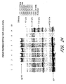
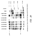
- the 5 MVA/UGD viruses have been characterized for gene expression and function. Immunoprecipitation of env and gag proteins is shown in Figure 24 .
- BS-C-1 cells were infected with individual recombinant viruses at a multiplicity of infection of 10, metabolically labeled, and lysates were subjected to immunoprecipitation with a pool of sera from HIV-1 clade D infected individuals.
- Viruses expressing gagpol only (MVA/UGD/Bgag and Cgag) were included, as was non-recombinant MVA as a negative control and MVA/CMDR as a positive control. The latter virus expresses gagpol/env from a Clade E HIV-1 isolate. All viruses produced high levels of gag protein and efficient processing into p24 was observed. In addition, all env-expressing viruses produced high levels of env protein (gp160).
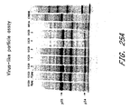
- Figure 25 demonstrates that the gag and env proteins produced by the MVA/UGD viruses are functional.
- Virus-like particles were obtained by centrifugation of the supernatant of infected cells through a sucrose cushion ( Karacostas et al. 1993 Virology 193:661 ). Pelleted material was then separated by SDS-polyacrylamide gel electrophoresis and analyzed by autoradiography (Panel A). As seen, p55 and p24 gag proteins were found in the pellet indicating that virus-like particles were formed.
- Panel B shows results of an assay in which env-expressing cells (infected with MVA/UGD virus) were mixed with cells expressing CD4 and co-receptor (X4 or R5) ( Nussbaum, Broder, & Berger 1994 J. Virol 68:5411 ). Fusion was measured by beta-galactosidase activity in cell lysates. As shown, all five MVA/UGD viruses induced fusion with CD4/R5-expressing cells.
- mice were immunized with individual MVA/UGD viruses, non-recombinant MVA (negative control), or MVA/CMDR (positive control - expressing clade E.gagpol/env) at weeks 0 and 3.
- the dose was 10 7 infectious units per immunization and the route was intraperitoneal.
- Humoral and cell mediated responses were measured and are shown in Figures 26-28 .
- Antibody responses after 2 immunizations are shown in Figure 26 .
- T cell responses were measured with several assays.
- gag and pol peptide-specific intracellular interferon gamma (IFN- y) responses were measured by intracellular cytokine staining.
- Splenocytes were collected 3 weeks after immunization, stimulated in-vitro for 7 days, and then cultured overnight with peptide-pulsed P815 cells.
- Brefeldin A was added to prevent secretion of INF- y.
- CD3 positive, CD8 positive, IFN- ⁇ positive cells were enumerated by flow cytometry. Analyses were performed after one and two immunizations.
- gag- and pol-specific responses were observed after two immunizations ( Figure 27 ) (samples from animals immunized with MVA/UGD-2, and 5 were not assayed).
- gag- and pol-specific INF- y responses were measured by ELISPOT ( Figure 28 A & B ). Briefly, splenocytes from immunized mice were mixed with gag or pol peptide-pulsed P815 cells in 96-well nitrocellulose plates coated with anti-IFN-y antibody. After overnight incubation, spots were visualized by sequential incubation with anti-IFN- ⁇ biotin antibody, straptavidin-HRP, and AEC substrate.
- Gag peptide-specific responses were found after one immunization and were boosted in most groups after the second immunization.
- Pol peptide-specific responses were found in several groups after two immunizations.
- gag peptide-specific responses were measured by tetramer staining (H-2Kd gag LAI tetramer: AMQMLKETI, SEQ ID NO: 63 ) ( Figure 29 ).
- Splenocytes were stimulated in vitro with either gag peptide or MVA/CM240gagpol, a recombinant virus expressing a clade E gagpol.CD3 positive, CD8 positive, tetramer positive cells were enumerated by flow cytometry. Positive tetramer staining was observed with cells from several groups of mice.
- a second mouse immunogenicity study was performed to confirm the humoral and cellular immunogenicity of MVA/UGD-3 and MVA/UGD-4.
- BALB/c mice (10 per group) were administered intraperitoneal immunizations of 10 7 infectious units of MVA at weeks 0 and 3.
- Five mice per group were sacrificed two weeks after the 1 st and 2 nd immunizations and spleens were removed for assessment of cellular immunogenicity.
- Sera were collected from each mouse at weeks -1, 0, 1, 2, 3, 4 and 5.
- Splenocytes and sera were pooled together by group.
- HIV gag-specific serum IgG responses were detected from all MVA/UGD-immunized groups after the 2 nd immunization ( Figure 30 ). These gag-specific responses were predominantly of subclass IgG2a for both MVA/UGD-3 and MVA/UGD-4 demonstrating a Th1-type response ( Table Q ).
- HIV-specific cell-mediated immunity was assessed by four separate assays: (1) intracellular IFN- ⁇ staining by flow cytometry (ICS), (2) IFN- ⁇ secretion by ELISPOT, (3) gag-peptide specific tetramer staining and (4) cytotoxic T lymphocyte (CTL) killing.
- ICS intracellular IFN- ⁇ staining by flow cytometry (ICS), (2) IFN- ⁇ secretion by ELISPOT, (3) gag-peptide specific tetramer staining and (4) cytotoxic T lymphocyte (CTL) killing.
- ICS Splenocytes were collected two weeks after the 1 st immunization, stimulated for 7 days with MVA-infected P815 cells and then incubated overnight with P815 cells pulsed with a gag or pol peptide previously shown to be target of CD8 T cells in BALB/c mice ( Casimiro et al.
- IFN- ⁇ ELISPOT HIV gag-specific IFN- y responses were detected by ELISPOT without prior in-vitro stimulation after both the 1 st ( Figure 32A ) and 2 nd immunization ( Figure 32B ) with a boost detected after the 2 nd immunization. HIV gag-specific responses were stronger than the pol-specific responses. HIV pol-specific responses were detectable after a 7-day in-vitro stimulation with P815 cells pulsed with pol peptide ( Figure 32C ).
- Tetramer staining HIV gag peptide-specific responses were measured by tetramer staining (H-2Kd gag LAI tetramer: AMQMLKETI, SEQ ID NO: 63 ) ( Figure 33 ).
- Splenocytes were stained pre or post a 7-day in-vitro stimulation with P815 cells either pulsed with gag peptide or infected with MVA/CM240gag/pol, a recombinant virus expressing a subtype E gagpol.
- CD3 positive/CD8 positive/tetramer positive cells were enumerated by flow cytometry.
- HIV-1 sequences from Kenya were selected.
- the predominant incident and prevalent HIV-1 subtype in Kenya is subtype A.
- Several R5 subtype A HIV-1 strains were selected and used to prepare recombinant MVA vaccines expressing env and gagpol (gag, protease and RT).
- env and gagpol gagpol
- One gagpol and two env clones from pure Kenyan subtype A isolates were selected.
- HIV-1 Isolate name GenBank Accession # Publication 00KE-KER2008 AF457052 AIDS 16:1809 (2002) 00KE-KNH1144 AF457066 " 00KE-KNH1207 AF457068 "
- the MVA/KEA viruses were characterized for gene expression and function. Immunoprecipitation of env and gag protiens is shown in Figure 35 . BSC-1 cells were infected with individual recombinant viruses at a multiplicity of infection of 10, metabolically labeled, and lysates were subjected to imunoprecipitation with a pool of antibodies including: monoclonal antibody T24 (env), monoclonal antibody 183-H12-5C (gag), and pooled HIV-1+ sera. MVA/UGD and WR/vEJW-1 were included as positive controls for env and gag expression, respectively. All MVA/KEA viruses express high levels of env and/or gag, as expected.
- Virus-like particles were obtained by centrifugation of the supernatant of infected cells through a sucrose cushion ( Karacostas et al. 1993 Virology 193:661 ). Gag p24 protein was found in the pelleted material indicating the formation of virus-like particles ( Figure 36 ).
- a cell-cell fusion assay was used to assess the function of expressed, membrane bound env.
- env-expressing cells infected with MVA/KEA virus
- X4 or R5 co-receptor
- Fusion was measured by beta-galactosidase activity in cell lysates. All viruses expressing membrane bound env induced fusion with CD4/R5-expressing cells ( Figure 37 ).
- the 5 recombinant MVA viruses expressing env, gag and pol from Kenyan subtype A HIV-1 isolates (MVA/KEA-1 through MVA/KEA-5), the 3 viruses expressing subtype A env alone (MVA/KEA-6 through MVA/KEA-8) and the MVA expressing subtype A gag/pol (MVA/KEA-9) were evaluated in an in-vivo mouse immunogenicity study designed to measure the humoral and cellular immunogenicity of these vaccines.
- BALB/c mice (10 per group) were administered intraperitoneal immunizations of 10 7 infectious units of individual MVA/KEA viruses at weeks 0 and 3.
- mice per group Five mice per group were sacrificed two weeks after the 1 st and 2 nd immunizations and spleens were removed for assessment of cellular immunogenicity. Sera were collected from each mouse at weeks -1, 0, 1, 2, 3, 4 and 5. Splenocytes and sera were pooled together by group. HIV env-specific serum IgG responses were detected from all MVA/KEA-immunized groups after the 2 nd immunization ( Figure 38 ). While env-specific responses were detected in all groups except for the control group, they were strongest in MVA/KEA-3, 4, 5, 6 and 8.
- HIV-specific cell-mediated immunity was assessed by three assays: (1) intracellular IFN- ⁇ staining by flow cytometry (ICS), (2) IFN- ⁇ secretion by ELISPOT, and (3) gag-peptide specific tetramer staining.
- ICS flow cytometry
- IFN- ⁇ secretion by ELISPOT IFN- ⁇ secretion by ELISPOT
- gag-peptide specific tetramer staining HIV-specific cell-mediated immunity was assessed by three assays: (1) intracellular IFN- ⁇ staining by flow cytometry (ICS), (2) IFN- ⁇ secretion by ELISPOT, and (3) gag-peptide specific tetramer staining.
- (1) ICS Splenocytes were collected two weeks after the 2 nd immunization, stimulated for 7 days with MVA-infected P815 cells and then incubated overnight with P815 cells pulsed with a gag or pol peptide previously shown to be target of CD8 T cells
- HIV gag-specific responses were detected for each of the groups immunized with MVA/KEA viruses expressing env, gag and pol or gag and pol, but not control immunized, mice as evidenced by the production of intracellular IFN- y after peptide stimulation ( Figure 39 ).
- Splenocytes positive for HIV gag ranged from 7% to 22% for the MVA/KEA-immunized mice.
- IFN- ⁇ ELISPOT HIV gag-specific IFN- y responses from splenocytes without prior in-vitro stimulation were detected in all groups receiving MVA/KEA viruses expressing gag after the 1 st immunization ( Figure 40 ).
- Tetramer staining HIV gag peptide-specific responses were measured by tetramer staining (H-2Kd gag LAI tetramer: AMQMLKETI, SEQ ID NO: 63 ) ( Figure 41 ). Splenocytes were stained pre-IVS. CD3 positive CD8 positive, tetramer positive cells were enumerated by flow cytometry. Similar to the above ICS and IFN- ⁇ ELISPOT results, positive tetramer staining was observed for all of the groups immunized with MVA-KEA expressing gag.
- HIV-1 sequences from Africa were selected.
- the predominant incident and prevalent HIV-1 subtype in Africa is subtype C.
- Several R5 subtype C HIV-1 strains were selected and used to prepare recombinant MVA vaccines expressing env, gag, protease and RT.
- One gagpol and one env clone from pure African subtype C isolates were selected.
- HIV-1 Isolate name GenBank Accession # Publication 00TZA-125 AY253304 AIDS Res & Hum Retrov, in press 00TZA-246 AY253308
- Steps described in EXAMPLE 5 were followed for the selected subtype C clones. These include:
- the KEA viruses were characterized for gene expression. Immunoprecipitation of env and gag proteins is shown in Figure 42 . BSC-1 cells were infected with individual recombinant viruses at a multiplicity of infection of 10, metabolically labeled, and lysates were subjected to immunoprecipitation with a pool of sera from HIV-1 infected individuals. MVA/CMDR and MVA were included as positive and negative controls, respectively. The MVA/KEA virus expresses high levels of env and gag.
- Virus-like particles were obtained by centrifugation of the supernatant of infected cells through a sucrose cushion ( Karacostas et al. 1993 Virology 193:661 ). Gag p24 protein was found in the pelleted material indicating the formation of virus-like particles ( Figure 43 ).
- the stability of the inserted env and gagpol genes in recombinant viruses from each of the subtypes was tested after serial passage in CEF cells.
- Viruses were grown in CEF cells using a procedure that mimics that used for expansion of virus for large scale vaccine production, i.e. infection at low multiplicity, growth for 3 days at 37C, harvesting of virus from cell lysates.
- the virus stocks were tested for stability of the inserts using a 3-day immunostaining protocol. In this protocol, CEF cell monolayers were infected at a low multiplicity allowing for visualization of individual viral foci. After 3 days, the monolayers were fixed and then stained with monoclonal antibodies specific for either env or gag.
- gagpol genes Clade D Kenyan HIV-1 isolates gagpol pol C-terminal truncation RT active site mutations DNA sequence at C-termiuus # of last nucleotide amino acid sequence at C-terminus # of last amino acid ammo acid nucleotide # amino acid nucleotide # 99UGA03349 3065 KEPIVGAETF (SEQ ID NO: 32) 1022 asp to glu T toG 692 2075 asp to his GAT to CAC 767 2298 & 2300 99UGA07412 3077 KEPNGAETF (SEQ ID NO:34) 1026 asp to glu T to G 696 2087 asp to his GAT to CAC 771 2310 & 2312 Table I.
- UGD Plasmids Plasmid parent plasmid HIV isolate Insertion site in MVA Direction in relation to vaccinia pLAS-1/UGD/Bgag pLAS-1 99UGA03349 Del III same pLAS-1 /UGD/Cgag pLAS-1 99UGA07412 Del III same pLAS-2/UGD/Benv pLAS-2 99UGA03349 Del II same pLAS-2/UGD/Cenv pLAS-2 99UGA07412 Del II same pLAS-2/UGD/Denv pLAS-2 98UG57128 Del II same pLAS-2/UGD/revCenv pLAS-2 99UGA07412 Del II reverse pLAS-2/UGD/revDenv pLAS-2 98UG57128 Del II reverse Table J.
- KEA Plasmids Plasmid parent plasmid HIV isolate Insertion site in MVA Direction in relation to vaccinia in pLAS-1/KER2008gag pLAS-1 00KE-KER2008 Del III same pLAS-2/KIVH1144env pLAS-2 00KE-KNH1144 Del II same pLAS-2/KNH1207env pLAS-2 OOKE-KNH1207 Del II same pLAS-2/KNH1144gp140 pLAS-2 00KE-KNH1144 Del II same pLAS-2/KNH1144(Y/A) pLAS-2 00KE-KNH1144 Del II same pLAS-2/KNH1144(Y/S) pLAS-2 00KB-KNH1144 Del II same Table M.
- gagpol Clade C African HIV-1 isolates gagpol pol C-terminal truncation RT active site mutations DNA sequence at C-terminus # of last nucleotide of gagpol amino acid sequence at C-terminus of pol # of last amino acid in pol amino acid nucleotide amino acid # in pol nucleotide # in gagpol amino acid nucleotide amino acid # in gagpol 00TZA-246 3083 KEPIVGAETF (SEQIDNO: 50) 601 asp to glu TtoG 271 2093 asp to his GAT to CAC 346 2316 & 2318 Table O.
- TZC Plasmids Plasmid parent plasmid HIV isolate Insertion site in MVA Direction in relation to vaccinia pLAS-1/TZC246gag pLAS-1 00TZA-246 Del III same pLAS-2/TZC125env pLAS-2 00TZA-125 Del II same Table P.
- TZC Viruses Virus gag/pol HIV isolate gag/pol Direction in relation to vaccinia env HIV isolate env Direction in relation to vaccinia MVA/TZC-1 00TZA-246 same 00TZA-125 same MVA/TZC-2 00TZA-246 same - MVA/TZC-3 - 00TZA-125 same Table Q.
- the invention furthermore comprises the following items:
Abstract
Description
- The invention provides modified vaccinia Ankara (MVA), a replication-deficient strain of vaccinia virus, expressing human immunodeficiency virus (HIV) env, gag, and pol genes.
- Cellular immunity plays an important role in the control of immunodeficiency virus infections (P.J. Goulder et al. 1999 AIDS 13:S121). Recently, a DNA vaccine designed to enhance cellular immunity by cytokine augmentation successfully contained a highly virulent immunodeficiency virus challenge (D.H. Barouch et al. 2000 Science 290:486). Another promising approach to raising cellular immunity is DNA priming followed by recombinant poxvirus boosters (H.L. Robinson et al. 2000 AIDS Rev 2:105). This heterologous prime/boost regimen induces 10- to 100-fold higher frequencies of T cells than priming and boosting with DNA or recombinant poxvirus vaccines alone. Previously, investigators showed that boosting a DNA-primed response with a poxvirus was superior to boosting with DNA or protein for the control of a non-pathogenic immunodeficiency virus (H.L. Robinson et al. 1999 Nat Med 5:526). There is a need for the control of a pathogenic immunodeficiency virus.
- Here we report that DNA priming followed by a recombinant modified vaccinia Ankara (rMVA) booster has controlled a highly pathogenic immunodeficiency virus challenge in a rhesus macaque model. Both the DNA and rMVA components of the vaccine expressed multiple immunodeficiency virus proteins. Two DNA inoculations at 0 and 8 weeks and a single rMVA booster at 24 weeks effectively controlled an intrarectal challenge administered seven months after the booster. These findings are envisioned as indicating that a relatively simple multiprotein DNA/MVA vaccine-can help to control the acquired immune deficiency syndrome (AIDS) epidemic. We also report that inoculations of rMVA induce good immune responses even without DNA priming.
-
-
Figure 1 . Phylogenetic relationships of HIV-1 and HIV-2 based on identity of pol gene sequences. SIVcpz and SIVsmm are subhuman primate lentiviruses recovered from a chimpanzee and sooty mangabey monkey, respectively. -
Figure 2 . Phylogenetic relationships of HIV-1 groups M, N and O with four different SIVcpz isolates based on full-length pol gene sequences. The bar indicates a genetic distance of 0.1 (10% nucleotide divergence) and the asterisk positions group N HIV-1 isolates based on env sequences. -
Figure 3 . Tropic and biologic properties of HIV-1 isolates. -
Figure 4 . HIV-encoded proteins. The location of the HIV genes, the sizes of primary translation products (in some cases polyproteins), and the processed mature viral proteins are indicated. -
Figure 5 . Schematic representation of a mature HIV-1 virion. -
Figure 6 . Linear representation of the HIV-1 Env glycoprotein. The arrow indicates the site of gp160 cleavage to gp120 and gp41. In gp120, cross-hatched areas represent variable domains (V1 to V5) and open boxes depict conserved sequences (C1 to C5). In the gp41 ectodomain, several domains are indicated: the N-terminal fusion peptide, and the two ectodomain helices (N- and C-helix). The membrane-spanning domain is represented by a black box. In the gp41 cytoplasmic domain, the Tyr-X-X-Leu (YXXL) endocytosis motif (SEQ ID NO: 9) and two predicted helical domains (helix-1 and -2) are shown. Amino acid numbers are indicated. -
Figure 7 . Temporal frequencies of Gag-specific T cells. (A) Gag-specific CD8 T cell responses raised by DNA priming and rMVA booster immunizations. The schematic presents mean Gag-CM9-tetramer data generated in the high-dose i.d. DNA-immunized animals. (B) Gag-specific IFN-γ ELISPOTs in A*01 (open bars) and non-A*01 (filled bars) macaques at various times before challenge and at two weeks after challenge. Three pools of 10 to 13 Gag peptides (22-mers overlapping by 12) were used for the analyses. The numbers above data bars represent the arithmetic mean ± SD for the ELISPOTs within each group. The numbers at the top of the graphs designate individual animals. *, data not available; #, <20 ELISPOTs per 1×10⁶ peripheral blood mononuclear cells (PBMC). Temporal data for Gag-CM9-Mamu-A*01 tetramer-specific T cells can be found inFigure 12 . -
Figure 8 . Temporal viral loads, CD4 counts, and survival after challenge of vaccinated and control animals. (A) Geometric mean viral loads and (B) geometric mean CD4 counts. (C) Survival curve for vaccinated and control animals. The dotted line represents all 24 vaccinated animals. (D) Viral loads and (E) CD4 counts for individual animals in the vaccine and control groups. The key to animal numbers is presented in (E). Assays for the first 12 weeks after challenge had a detection level of 1000 copies of RNA per milliliter of plasma. Animals with loads below 1000 were scored with a load of 500. Forweeks -
Figure 9 . Postchallenge T cell responses in vaccine and control groups. (A) Temporal tetramer+ cells (dashed line) and viral loads (solid line). (B) Intracellular cytokine assays for IFN-γ production in response to stimulation with the Gag-CM9 peptide at two weeks after challenge. This ex vivo assay allows evaluation of the functional status of the peak postchallenge tetramer+ cells displayed inFigure 7A. (C) Proliferation assay at 12 weeks after challenge. Gag-Pol-Env (open bars) and Gag-Pol (hatched bars) produced by transient transfections were used for stimulation. Supernatants from mock-transfected cultures served as control antigen. Stimulation indices are the growth of cultures in the presence of viral antigens divided by the growth of cultures in the presence of mock antigen. -
Figure 10 . Lymph node histomorphology at 12 weeks after challenge. (A) Typical lymph node from a vaccinated macaque showing evidence of follicular hyperplasia characterized by the presence of numerous secondary follicles with expanded germinal centers and discrete dark and light zones. (B) Typical lymph node from an infected control animal showing follicular depletion and paracortical lymphocellular atrophy. (C) A representative lymph node from an age-matched, uninfected macaque displaying nonreactive germinal centers. (D) The percentage of the total lymph node area occupied by germinal centers was measured to give a non-specific indicator of follicular hyperplasia. Data for uninfected controls are for four age-matched rhesus macaques. -
Figure 11 . Temporal antibody responses. Micrograms of total Gag (A) or Env (B) antibody were determined with ELISAs. The titers of neutralizing antibody for SHIV-89.6 (C) and SHIV-89.6P (D) were determined with MT-2 cell killing and neutral red staining (D.C. Montefiori et al. 1988 J Clin Microbiol 26:231). Titers are the reciprocal of the serum dilution giving 50% neutralization of the indicated viruses grown in human PBMC. Symbols for animals are the same as inFigure 8 . -
Figure 12 . Gag-CM9-Mamu-A*01 tetramer-specific T cells in Mamu-A*01 vaccinated and control macaques at various times before challenge and at two weeks after challenge. The number at the upper right corner of each plot represents the frequency of tetramer-specific CD8 T cells as a % of total CD8 T cells. The numbers above each column of FACS data designate individual animals. -
Figure 13 . Map of plasmid transfer vector pLW-48. -
Figure 14 A-I . Sequences of plasmid transfer vector pLW-48, Psy II promoter (which controls ADA envelope expression), ADA envelope truncated, PmH5 promoter (which controls HXB2 gag pol expression), and HXB2 gag pol (with safety mutations, Δ integrase). -
Figure 15 . Plasmid transfer vector pLW-48 and making MVA recombinant virus MVA/HIV 48. -
Figure 16 . A clade B gag pol. -
Figure 17 . Sequence of new Psyn II promoter. -
Figure 18 . pLAS-1 and pLAS-2. -
Figure 19 . pLAS-1/UGDgag. -
Figure 20 . pLAS-2/UGDenv. -
Figure 21 . pLAS-2/UGDrev env. -
Figure 22 . Schematic for recombinant MVA production. -
Figure 23 . Overview of making recombinant MVA/UGD viruses. -
Figure 24 . Immunoprecipitation analysis. -
Figure 25 . Functional analysis of expressed proteins. A, Virus-like partikle assay. B. Env fusion assay. -
Figure 26 . MVA/LTGD induced HIV env-and gag-specific antibody responses. A. HIV p24-specofoc serum IgG responses. B. HIV env-specific serum IgG responses. C. MVA/UGD induced HIV env- and gag-specific antibody responses (study 1). -
Figure 27 . MVA/LTGD induced gag-specific intracellular IFN-γ production. -
Figure 28A . MVA/UGD induced gag-specific IFN-γ ELISPOT. -
Figure 28 B. MVA/UGD induced pol-specific IFN-γ ELISPOT. -
Figure 29 . MVA/UGD induced gag-specific tetramer staining. -
Figure 30 . MVA/UGD induced gag-specific antibody responses (study 2). -
Figure 31A & B. MVA/UGD induced gag-and pol-specific intracellular IFN-γ production (study 2). -
Figure 32 A, B, & C. MVA/UGD induced gag-and pol-specific IFN-γ ELISPOT (study 2). -
Figure 33 . MVA/UGD induced gag-specific tetramer staining (study 2). -
Figure 34 . MVA/UGD induced gag-specific cytotoxic T cell killing. -
Figure 35 . Immunoprecipitation analysis of cell lysates (MVA/KEA). -
Figure 36 . Gag particle assay (MVA/KEA). -
Figure 37 . Fusion assay (MVA/KEA). -
Figure 38 . MVA/KEA induced HIV-1 env-specific antibody responses. -
Figure 39 . MVA/KEA induced gag-specific intracellular IFN-γ production. -
Figure 40 . MVA/KEA induced gag-specific tetramer staining. -
Figure 41 . MVA/KEA induced gag-specific IFN-γ ELISPOT. -
Figure 42 . Immunoprecipitation analysis of cell lysates (MVA/TZC) . -
Figure 43 . Fusion assay (MVA/TZC). -
-
Appendix 1. DNA sequences of gagpol and env genes from Ugandan HIV-1 clade D isolates. -
Appendix 2. DNA sequences of MVA shuttle plasmids, pLAS-1 and pLAS-2. -
Appendix 3. DNA sequences of gagpol and env genes from Kenyan HIV-1 clade A isolates. -
Appendix 4. DNA sequences of gagpol and env genes from Tanzanian HIV-1 clade C isolates. -
Appendix 5. American Type Culture Collection, Budapest Treaty Deposit Form BP/1, questions 1-8, regardingMVA 1974/NIH Clone 1. -
Appendix 6. American Tissue Type Collection, Additional Information Required When Depositing A Virus for Patent Purposes, regardingMVA 1974/NIH Clone 1. - The following microorganism has been deposited in accordance with the terms of the Budapest Treaty with the American Type Culture Collection (ATCC), Manassas, VA, on the date indicated:
Microorganism Accession No. Date MVA 1974/ NIH Clone 1PTA-5095 March 27, 2003 -
MVA 1974/NIH Clone 1 was deposited as ATCC Accession No. PTA-5095 on March 27, 2003 with the American Type Culture Collection (ATCC), 10801 University Blvd., Manassas, VA 20110-2209, USA. This deposit was made under the provisions of the Budapest Treaty on the International Recognition of the Deposit of Microorganisms for the Purposes of Patent Procedure and the Regulations thereunder (Budapest Treaty). This assures maintenance of a viable culture of the deposit for 30 years from date of deposit. The deposit will be made available by ATCC under the terms of the Budapest Treaty, and subject to an agreement between Applicant and ATCC which assures permanent and unrestricted availability of the progeny of the culture of the deposit to the public upon issuance of the pertinent U.S. patent or upon laying open to the public of any U.S. or foreign patent application, whichever comes first, and assures availability of the progeny to one determined by the U.S. Commissioner of Patents and Trademarks to be entitled thereto according to 35 USC § 122 and the Commissioner's rules pursuant thereto (including 37 CFR § 1.14). Availability of the deposited strain is not to be construed as a license to practice the invention in contravention of the rights granted under the authority of any government in accordance with its patent laws. - Vaccinia virus, a member of the genus Orthopoxvirus in the family of Poxviridae, was used as live vaccine to immunize against the human smallpox disease. Successful worldwide vaccination with vaccinia virus culminated in the eradication of variola virus, the causative agent of the smallpox ("The global eradication of smallpox. Final report of the global commission for the certification of smallpox eradication". History of Public Health, No. 4, Geneva: World Health Organization, 1980). Since that WHO declaration, vaccination has been universally discontinued except for people at high risk of poxvirus infections (e.g. laboratory workers).
- More recently, vaccinia viruses have also been used to engineer viral vectors for recombinant gene expression and for the potential use as recombinant live vaccines (Mackett, M. et al. 1982 PNAS USA 79:7415-7419; Smith, G.L. et al. 1984 Biotech Genet Engin Rev 2:383-407). This entails DNA sequences (genes) which code for foreign antigens being introduced, with the aid of DNA recombination techniques, into the genome of the vaccinia viruses. If the gene is integrated at a site in the viral DNA which is non-essential for the life cycle of the virus, it is possible for the newly produced recombinant vaccinia virus to be infectious, that is to say able to infect foreign cells and thus to express the integrated DNA sequence (
EP Patent Applications No. 83,286 110,385 - For vector applications health risks would be lessened by the use of a highly attenuated vaccinia virus strain. Several such strains of vaccinia virus were especially developed to avoid undesired side effects of smallpox vaccination. Thus, the modified vaccinia Ankara (MVA) has been generated by long-term serial passages of the Ankara strain of vaccinia virus (CVA) on chicken embryo fibroblasts (for review see Mayr, A. et al. 1975 Infection 3:6-14; Swiss Patent No.
568,392 - Furthermore, MVA is characterized by its extreme attenuation. When tested in a variety of animal models, MVA was proven to be avirulent even in immunosuppressed animals. More importantly, the excellent properties of the MVA strain have been demonstrated in extensive clinical trials (Mayr A. et al. 1978 Zentralbl Bakteriol [B] 167:375-390; Stickl et al. 1974 Dtsch Med Wschr 99:2386-2392). During these studies in over 120,000 humans, including high-risk patients, no side effects were associated with the use of MVA vaccine.
- MVA replication in human cells was found to be blocked late in infection preventing the assembly to mature infectious virions. Nevertheless, MVA was able to express viral and recombinant genes at high levels even in non-permissive cells and was proposed to serve as an efficient and exceptionally safe gene expression vector (Sutter, G. and Moss, B. 1992 PNAS USA 89:10847-10851). Additionally, novel vaccinia vector vaccines were established on the basis of MVA having foreign DNA sequences inserted at the site of deletion III within the MVA genome (Sutter, G. et al. 1994 Vaccine 12:1032-1040).
- The recombinant MVA vaccinia viruses can be prepared as set out hereinafter. A DNA-construct which contains a DNA-sequence which codes for a foreign polypeptide flanked by MVA DNA sequences adjacent to a naturally occurring deletion, e.g. deletion III, or other non-essential sites, within the MVA genome, is introduced into cells infected with MVA, to allow homologous recombination. Once the DNA-construct has been introduced into the eukaryotic cell and the foreign DNA has recombined with the viral DNA, it is possibleto isolate the desired recombinant vaccinia virus in a manner know per se, preferably with the aid of a marker. The DNA-construct to be inserted can be linear or circular. A plasmid or polymerase chain reaction product is preferred. The DNA-construct contains sequences flanking the left and the right side of a naturally occurring deletion, e.g. deletion III, within the MVA genome. The foreign DNA sequence is inserted between the sequences flanking the naturally occurring deletion. For the expression of a DNA sequence or gene, it is necessary for regulatory sequences, which are required for the transcription of the gene, to be present on the DNA. Such regulatory sequences (called promoters) are known to those skilled in the art, and include for example those of the
vaccinia 11 kDa gene as are described inEP-A-198,328 EP-A-110,385 - The etiological agent of acquired immune deficiency syndrome (AIDS) is recognized to be a retrovirus exhibiting characteristics typical of the lentivirus genus, referred to as human immunodeficiency virus (HIV). The phylogenetic relationships of the human lentiviruses are shown in
Figure 1 . HIV-2 is more closely related to SIVsmm, a virus isolated from sooty mangabey monkeys in the wild, than to HIV-1. It is currently believed that HIV-2 represents a zoonotic transmission of SIVsmm to man. A series of lentiviral isolates from captive chimpanzees, designated SIVcpz, are close genetic relatives of HIV-1. - The earliest phylogenetic analyses of BIV-1 isolates focused on samples from Europe/North America and Africa; discrete clusters of viruses were identified from these two areas of the world. Distinct genetic subtypes or clades of HIV-1 were subsequently defined and classified into three groups: M (major); O (outlier); and N (non-M or O) (
Fig. 2 ). The M group of HIV-1, which includes over 95% of the global virus isolates, consists of at least eight discrete clades (A, B, C, D, F, G, H, and J), based on the sequence of complete viral genomes. Members of HIV-1group 0 have been recovered from individuals living in Cameroon, Gabon, and Equatorial Guinea; their genomes share less than 50% identity in nucleotide sequence with group M viruses. The more recently discovered group N HIV-I strains have been identified in infected Cameroonians, fail to react serologically in standard whole-virus enzyme-linked immunosorbent assay (ELISA), yet are readily detectable by conventional Western blot analysis. - Most current knowledge about HIV-1 genetic variation comes from studies of group M viruses of diverse geographic origin. Data collected during the past decade indicate that the HIV-1 population present within an infected individual can vary from 6% to 10% in nucleotide sequence. HIV-1 isolates within a clade may exhibit nucleotide distances of 15% in gag and up to 30% in gp120 coding sequences. Interclade genetic variation may range between 30% and 40% depending on the gene analyzed.
- All of the HIV-1 group M subtypes can be found in Africa. Clade A viruses are genetically the most divergent and were the most common HIV-1 subtype in Africa early in the epidemic. With the rapid spread of HIV-1 to southern Africa during the mid to late 1990s, clade C viruses have become the dominant subtype and now account for 48% of HIV-1 infections worldwide. Clade B viruses, the most intensively studied HIV-1 subtype, remain the most prevalent isolates in Europe and North America.
- High rates of genetic recombination are a hallmark of retroviruses. It was initially' believed that simultaneous infections by genetically diverse virus strains were not likely to be established in individuals at risk for HIV-1. By 1995, however, it became apparent that a significant fraction of the HIV-1 group M global diversity included interclade viral recombinants. It is now appreciated that HIV -1 recombinants will be found in geographic areas such as Africa, South America, and Southeast Asia, where multiple HIV-1 subtypes coexist and may account for more than 10% of circulating HIV-1 strains. Molecularly, the genomes of these recombinant viruses resemble patchwork mosaics, with juxtaposed diverse HIV-1 subtype segments, reflecting the multiple crossover events contributing to their generation. Most HIV-1 recombinants have arisen in Africa and a majority contains segments originally derived from clade A viruses. In Thailand, for example, the composition of the predominant circulating strain consists of a clade A gag plus pol gene segment and a clade E env gene. Because the clade E env gene in Thai HIV-1 strains is closely related to the clade E env present in virus isolates from the Central African -Republic, it is believed that the original recombination event occurred in Africa, with the subsequent introduction of a descendent virus into Thailand. Interestingly, no full-length HIV-1 subtype E isolate (i.e., with subtype E gag, pol, and env genes) has been reported to date.
- The discovery that a and chemokine receptors function as coreceptors for virus fusion and entry into susceptible CD4+ cells has led to a revised classification scheme for HIV-1 (
Fig. 3 ). Isolates can now be grouped on the basis of chemokine receptor utilization in fusion assays in which HIV-1 gp120 and CD4+ coreceptor proteins are expressed in separate cells. As indicated inFigure 3 , HIV-1 isolates using the CXCR4 receptor (now designated X4 viruses) are usually T cell line (TCL)-tropic syncytium inducing (SI) strains, whereas those exclusively utilizing the CCR5 receptor (R5 viruses) are predominantly macrophage (M)-tropic and non-syncytium inducing (NSI). The dual-tropic R5/X4 strains, which may comprise the majority of patient isolates and exhibit a continuum of tropic phenotypes, are frequently SI. - As is the case for all replication-competent retroviruses, the three primary HN-1 translation products, all encoding structural proteins, are initially synthesized as polyprotein precursors, which are subsequently processed by viral or cellular proteases into mature particle-associated proteins (
Fig. 4 ). The 55-kd Gag precursor Pr55Gag is cleaved into the matrix (MA), capsid (CA), nucleocapsid (NC), and p6 proteins. Autocatalysis of the 160-kd Gag-Pol polyprotein, Pr160Gag-Pol, gives rise to the protease (PR), the heterodimeric reverse transcriptase (RT), and the integrase (IN) proteins, whereas proteolytic digestion by a cellular enzyme(s) converts the glycosylated 160-kd Env precursor gp160 to the gp120 surface (SU) and gp41 transmembrane (TM) cleavage products. The remaining six BIV-1-encoded proteins (Vif, Vpr, Tat, Rev, Vpu, and Nef) are the primary translation products of spliced mRNAs. - The Gag proteins of HIV, like those of other retroviruses, are necessary and sufficient for the formation of noninfectious, virus-like particles. Retroviral Gag proteins are generally synthesized as polyprotein precursors; the HIV-1 Gag precursor has been named, based on its apparent molecular mass, Pr55Gag. As noted previously, the mRNA for Pr55Gag is the unspliced 9.2-kb transcript (
Fig. 4 ) that requires Rev for its expression in the cytoplasm. When the pol ORF is present, the viral protease (PR) cleaves Pr55Gag during or shortly after budding from the cell to generate the mature Gag proteins p17 (MA), p24 (CA), p7 (NC), and p6 (seeFig. 4 ). In the virion, MA is localized immediately inside the lipid bilayer of the viral envelope, CA forms the outer portion of the cone-shaped core structure in the center of the particle, and NC is present in the core in a ribonucleoprotein complex with the viral RNA genome (Fig. 5 ). - The HIV Pr55Gag precursor oligomerizes following its translation and is targeted to the plasma membrane, where particles of sufficient size and density to be visible by EM are assembled. Formation of virus-like particles by Pr55Gag is a self-assembly process, with critical Gag-Gag interactions taking place between multiple domains along the Gag precursor. The assembly of virus-like particles does not require the participation of genomic RNA (although the presence of nucleic acid appears to be essential), pol-encoded enzymes, or Env glycoproteins, but the production of infectious virions requires the encapsidation of the viral RNA genome and the incorporation of the Env glycoproteins and the Gag-Pol polyprotein precursor Pr160Gag-Pol.
- Downstream of gag lies the most highly conserved region of the HIV genome, the pol gene, which encodes three enzymes: PR, RT, and IN (see
Fig. 4 ). RT and IN are required, respectively, for reverse transcription of the viral RNA genome to a double-stranded DNA copy, and for the integration of the viral DNA into the host cell chromosome. PR plays a critical role late in the life cycle by mediating the production of mature, infectious virions. The pol gene products are derived by enzymatic cleavage of a 160-kd Gag-Pol fusion protein, referred to as pr160Gag-PoI. This fusion protein is produced by ribosomal frameshifting during translation of Pr55Gag (seeFig. 4 ). The frame-shifting mechanism for Gag-Pol expression, also utilized by many other retroviruses, ensures that the pol derived proteins are expressed at a low level, approximately 5% to 10% that of Gag. Like Pr55Gag, the N-terminus of Pr160Gag-Pol is myristylated and targeted to the plasma membrane. - Early pulse-chase studies performed with avian retroviruses clearly indicated that retroviral Gag proteins are initially synthesized as polyprotein precursors that are cleaved to generate smaller products. Subsequent studies demonstrated that the processing function is provided by a viral rather than a cellular enzyme, and that proteolytic digestion of the Gag and Gag-Pol precursors is essential for virus infectivity. Sequence analysis of retroviral PRs indicated that they are related to cellular "aspartic" proteases such as pepsin and renin. Like these cellular enzymes, retroviral PRs use two apposed Asp residues at the active site to coordinate a water molecule that catalyzes the hydrolysis of a peptide bond in the target protein. Unlike the cellular aspartic proteases, which function as pseudodimers (using two folds within the same molecule to generate the active site), retroviral PRs function as true dimers. X-ray crystallographic data from HIV-1 PR indicate that the two monomers are held together in part by a four-stranded antiparallel β-sheet derived from both N- and C-terminal ends of each monomer. The substrate-binding site is located within a cleft formed between the- two monomers. Like their cellular homologs, the HIV PR dimer contains flexible "flaps" that overhang the binding site and may stabilize the substrate within the cleft; the active-site Asp residues lie in the center of the dimer. Interestingly, although some limited amino acid homology is observed surrounding active-site residues, the primary sequences of retroviral PRs - are highly divergent, yet their structures are remarkably similar.
- By definition, retroviruses possess the ability to convert their single-stranded RNA genomes into double-stranded DNA during the early stages of the infection process. The enzyme that catalyzes this reaction is RT, in conjunction with its associated RNaseH activity. Retroviral RTs have three enzymatic activities: (a) RNA-directed DNA polymerization (for minus-strand DNA synthesis), (b) RNaseH activity (for the degradation of the tRNA primer and genomic RNA present in DNA-RNA hybrid intermediates), and (c) DNA-directed DNA polymerization (for second- or plus-strand DNA synthesis).
- The mature HIV-1 RT holoenzyme is a heterodimer of 66 and 51 kd subunits. The 51-kd subunit (p51) is derived from the 66-l:d (p66) subunit by proteolytic removal of the C-terminal 15-lcd RNaseH domain of p66 by PR (see
Fig. 4 ). The crystal structure of,HIV-1 RT reveals a highly asymmetric folding in which the orientations of the p66 and p51 subunits differ substantially. The p66 subunit can be visualized as a right hand, with the polymerase active site within the palm, and a deep template-binding cleft formed by the palm, fingers, and thumb subdomains. The polymerase domain is linked to RNaseH by the connection subdomain. The active site, located in the palm, contains three critical Asp residues (110, 185, and 186) in close proximity, and two coordinated Mg2+ ions. Mutation of these Asp residues abolishes RT polymerizing activity. The orientation of the three active-site Asp residues is similar to that observed in other DNA polymerases (e.g., the Klenow fragment of E. coli DNA poll). The p51 subunit appears to be rigid and does not form a polymerizing cleft;Asp 110, 185, and 186 of this subunit are buried within the molecule. Approximately 18 base pairs of the primer-template duplex lie in the nucleic acid binding cleft, stretching from the polymerase active site to the RNaseH domain. - In the RT-primer-template-dNTP structure, the presence of a dideoxynucleotide at the 3' end of the primer allows visualization of the catalytic complex trapped just prior to attack on the incoming dNTP. Comparison with previously obtained structures suggests a model whereby the fingers close in to trap the template and dNTP prior to nucleophilic attack of the 3'-OH of the primer on the incoming dNTP. After the addition of the incoming dNTP to the growing chain, it has been proposed that the fingers adopt a more open configuration, thereby releasing the pyrophosphate and enabling RT to bind the next dNTP. The structure of the HIV-1 RNaseH has also been determined by x-ray crystallography; this domain displays a global folding similar to that of E. coli RNaseH.
- A distinguishing feature of retrovirus replication is the insertion, of a DNA copy of the viral genome into the host cell chromosome following reverse transcription. The integrated viral DNA (the provirus) serves as the template for the synthesis of viral RNAs and is maintained as part of the host cell genome for the lifetime of the infected cell. Retroviral mutants deficient in the ability to integrate generally fail to establish a productive infection.
- The integration of viral DNA is catalyzed by integrase, a 32-kd protein generated by PR-mediated cleavage of the C-terminal portion of the HIV-1 Gag-Pol polyprotein (see
Fig. 4 ). - Retroviral IN proteins are composed of three structurally and functionally distinct domains: an N-terrninal, zinc-finger-containing domain, a core domain, and a relatively nonconserved C-terminal domain. Because of its low solubility, it has not yet been possible to crystallize the entire 288-amino-acid HIV-1 IN protein. However, the structure of all three domains has been solved independently by x-ray crystallography or NMR methods. The crystal structure of the core domain of the avian sarcoma virus IN has also been determined. The N-terminal domain (
residues 1 to 55), whose structure was solved by NMR spectroscopy, is composed of four helices with a zinc coordinated by amino acids His-12, His-16, Cys-40, and Cys-43. The structure of the N-terminal domain is reminiscent of helical DNA binding proteins that contain a so-called helix-turn-helix motif; however, in the HIV-I structure this motif contributes to dimer formation. Initially, poor solubility hampered efforts to solve the structure of the core domain. However, attempts at crystallography were successful when it was observed that a Phe-to-Lys change at IN residue 185 greatly increased solubility without disrupting in vitro catalytic activity. Each monomer of the HIV-1 IN core domain (INresidues 50 to 212) is composed of a five-stranded β-sheet flanked by helices; this structure bears striking resemblance to other polynucleotidyl transferases including RNaseH and the bacteriophage MuA transposase. Three highly conserved residues are found in analogous positions in other polynucleotidyl transferases; in HIV-1 IN these are Asp-64, Asp-116 and Glu-152, the so-called D,D-35-E motif. Mutations at these positions block HIV IN function both in vivo and in vitro. The close proximity of these three amino acids in the crystal structure of both avian sarcoma virus and HIV-1 core domains supports the hypothesis that these residues play a central role in catalysis of the polynucleotidyl transfer reaction that is at the heart of the integration process. The C-terminal domain, whose structure has been solved by NMR methods, adopts a five-stranded β-barrel folding topology reminiscent of a Src homology 3 (SH3) domain. Recently, the x-ray structures of SIV and Rous sarcoma virus IN protein fragments encompassing both the core and C-terminal domains have been solved. - The HIV Env glycoproteins play a major role in the virus life cycle. They contain the determinants that interact with the CD4 receptor and coreceptor, and they catalyze the fusion reaction between the lipid bilayer of the viral envelope and the host cell plasma membrane. In addition, the HIV Env glycoproteins contain epitopes that elicit immune responses that are important from both diagnostic and vaccine development perspectives.
- The HIV Env glycoprotein is synthesized from the singly spliced 4.3-kb Vpu/Env bicistronic mRNA (see
Fig. 4 ); translation occurs on ribosomes associated with the rough endoplasmic reticulum (ER). The 160-lcd polyprotein precursor (gp 160) is an integral membrane protein that is anchored to cell membranes by a hydrophobic stop-transfer signal in the domain destined to be the mature TM Env glycoprotein, gp41 (Fig. 6 ). The gp160 is cotranslationally glycosylated, forms disulfide bonds, and undergoes oligomerization in the ER. The predominant oligomeric form appears to be a trimer, although dimers and tetramers are also observed. The gp160 is transported to the Golgi, where, like other retroviral envelope precursor proteins, it is proteolytically cleaved by cellular enzymes to the mature SU glycoprotein gp 120 arid TM glycoprotein gp41 (seeFig. 6 ). The cellular enzyme responsible for cleavage of retroviral Env precursors following a highly conserved Lys/Arg-X-Lys/Arg-Arg motif is furin or a furin-like protease, although other enzymes may also catalyze gp160 processing. Cleavage of gp160 is required for Env-induced fusion activity and virus infectivity. Subsequent to gp160 cleavage, gpl20 and gp41 form a noncovalent association that is critical for transport of the Env complex from the Golgi to the cell surface. The gp120-gp41 interaction is fairly weak, and a substantial amount of gp120 is shed from the surface of Env-expressing cells. - The HIV Env glycoprotein complex, in particular the SU (gp 120) domain, is very heavily glycosylated; approximately half the molecular mass of gp160 is composed of oligosaccharide side chains. During transport of Env from its site of synthesis in the ER to the plasma membrane, many of the side chains are modified by the addition of complex sugars. The numerous oligosaccharide side chains form what could be imagined as a sugar cloud obscuring much of gp120 from host immune recognition. As shown in
Figure 6 , gp120 contains interspersed conserved (Q to C5) and variable (V1 to V5) domains. The Cys residues present in the gp120s of different isolates are highly conserved and form disulfide bonds that link the first four variable regions in large loops. - A primary function of viral Env glycoproteins is to promote a membrane fusion reaction between the lipid bilayers of the viral envelope and host cell membranes. This - membrane fusion event enables the viral core to gain entry into the host cell cytoplasm. A number of regions in both gp120 and gp41 have been implicated, directly or indirectly, in Env-mediated membrane fusion. Studies of the HA2 hemagglutinin protein of the orthomyxoviruses and the F protein of the paramyxoviruses indicated that a highly hydrophobic domain at the N-terminus of these proteins, referred to as the fusion peptide, plays a critical role in membrane fusion. Mutational analyses demonstrated that an analogous domain was located at the N-terminus of the BIV-1, HIV-2, and SIV TM glycoproteins (see
Fig. 6 ). Nonhydrophobic substitutions within this region of gp41 greatly reduced or blocked syncytium formation and resulted in the production of noninfectious progeny virions. - C-terminal to the gp41 fusion peptide are two amphipathic helical domains (see
Fig. 6 ) which play a central role in membrane fusion. Mutations in the N-terminal helix (referred to as the N-helix), which contains a Leu zipper-like heptad repeat motif, impair infectivity and membrane fusion activity, and peptides derived from these sequences exhibit potent antiviral activity in culture. The structure of the ectodomain of HIV-1 and SIV gp4l, the two helical motifs in particular, has been the focus of structural analyses in recent years. Structures were determined by x-ray crystallography or NMR spectroscopy either for fusion proteins containing the helical domains, a mixture of peptides derived from the N- and C-helices, or in the case of the SIV structure, the intact gp41 ectodomain sequence fromresidue 27 to 149. These studies obtained fundamentally similar trimeric structures, in which the two helical domains pack in an antiparallel fashion to generate a six-helix bundle. The N-helices form a coiled-coil in the center of the bundle, with the C-helices packing into hydrophobic grooves on the outside. - In the steps leading to membrane fusion CD4 binding induces conformation changes in Env that facilitate coreceptor binding. Following the formation of a ternary gp120/CD4/coreceptor complex, gp41 adopts a hypothetical conformation that allows the fusion peptide to insert into the target lipid bilayer. The formation of the gp41 six-helix bundle (which involves antiparallel interactions between the gp41 N- and C-helices) brings the viral and cellular membranes together and membrane fusion takes place.
- The present invention relates to generation of a CD8+ T cell immune response against an antigen and also eliciting an antibody response. More particularly, the present invention relates to "prime and boost" immunization regimes in which the immune response induced by administration of a priming composition is boosted by administration of a boosting composition. The present invention is based on inventors' experimental demonstration that effective boosting can be achieved using modified vaccinia Ankara (MVA) vectors, following priming with any of a variety of different types of priming compositions including recombinant MVA itself.
- A major protective component of the immune response against a number of pathogens is mediated by T lymphocytes of the CD8+ type, also known as cytotoxic T lymphocytes (CTL). An important function of CD8+ cells is secretion of gamma interferon (IFNy), and this provides a measure of CD8+ T cell immune response. A second component of the immune response is antibody directed to the proteins of the pathogen.
- The present invention employs MVA which, as the experiments described below show, has been found to be an effective means for providing a boost to a CD8+ T cell immune response primed to antigen using any of a variety of different priming compositions and also eliciting an antibody response.
- Remarkably, the experimental work described below demonstrates that use of embodiments of the present invention allows for recombinant MVA virus expressing an HIV antigen to boost a CD8+ T cell immune response primed by a DNA vaccine and also eliciting an antibody response. The MVA was found to induce a CD8+ T cell response after intradermal, intramuscular or mucosal immunization. Recombinant MVA has also been shown to prime an immune response that is boosted by one or more inoculations of recombinant MVA.
- Non-human primates immunized with plasmid DNA and boosted with the MVA were effectively protected against intramucosal challenge with live virus. Advantageously, the inventors found that a vaccination regime used intradermal, intramuscular or mucosal immunization for both prime and boost can be employed, constituting a general immunization regime suitable for inducing CD8+ T cells and also eliciting an antibody response, e.g. in humans.
- The present invention in various aspects and embodiments employs an MVA vector encoding an HIV antigen for boosting a CD8+ T cell immune response to the antigen primed by previous administration of nucleic acid encoding the antigen and also eliciting an antibody response.
- A general aspect of the present invention provides for the use of an MVA vector for boosting a CD8+ T cell immune response to an HIV antigen and also eliciting an antibody response.
- One aspect of the present invention provides a method of boosting a CD8+ T cell immune response to an HIV antigen in an individual, and also eliciting an antibody response, the method including provision in the individual of an MVA vector including nucleic acid encoding the antigen operably linked to regulatory sequences for production of antigen in the individual by expression from the nucleic acid, whereby a CD8+ T cell immune response to the antigen previously primed in the individual is boosted.
- An immune response to an HIV antigen may be primed by immunization with plasmid DNA or by infection with an infectious agent.
- A further aspect of the invention provides a method of inducing a CD8+ T cell immune response to an HIV antigen in an individual; and also eliciting an antibody response, the method comprising administering to the individual a priming composition comprising nucleic acid encoding the antigen and then administering a boosting composition which comprises an MVA vector including nucleic acid encoding the antigen operably linked to regulatory sequences for production of antigen in the individual by expression from the nucleic acid.
- A further aspect provides for use of an MVA vector, as disclosed, in the manufacture of a medicament for administration to a mammal to boost a CD8+ T cell immune response to an HIV antigen, and also eliciting an antibody response. Such a medicament is ' generally for administration following prior administration of a priming composition comprising nucleic acid encoding the antigen.
- The priming composition may comprise any viral vector, such as a vaccinia virus vector such as a replication-deficient strain such as modified vaccinia Ankara (MVA) or NYVAC (Tartaglia et al. 1992 Virology 118:217-232), an avipox vector such as fowlpox or canarypox, e.g. the strain known as ALVAC (Paoletti et al. 1994 Dev Biol Stand 82:65-69), or an adenovirus vector or a vesicular stomatitis virus vector or an alphavirus vector.
- The priming composition may comprise DNA encoding the antigen, such DNA preferably being in the form of a circular plasmid that is not capable of replicating in mammalian cells. Any selectable marker should not be resistance to an antibiotic used clinically, so for example Kanamycin resistance is preferred to Ampicillin resistance. Antigen expression should be driven by a promoter which is active in mammalian cells, for instance the cytomegalovirus immediate early (CMV IE) promoter.
- In particular embodiments of the various aspects of the present invention, administration of a priming composition is followed by boosting with a boosting composition, or first and second boosting compositions, the first and second boosting compositions being the same or different from one another. Still further boosting compositions may be employed without departing from the present invention. In one embodiment, a triple immunization regime employs DNA, then adenovirus as a first boosting composition, then MVA as a second boosting composition, optionally followed by a further (third) boosting composition or subsequent boosting administration of one or other or both of the same or different vectors. Another option is DNA then MVA then adenovirus, optionally followed by subsequent boosting administration of one or other or both of the same or different vectors.
- The antigen to be encoded in respective priming and boosting compositions (however many boosting compositions are employed) need not be identical, but should share at least one CD8+ T cell epitope. The antigen may correspond to a complete antigen, or a fragment thereof. Peptide epitopes or artificial strings of epitopes may be employed, more efficiently cutting out unnecessary protein sequence in the antigen and encoding sequence in the vector or vectors. One .or more additional epitopes may be included, for instance epitopes which are recognized by T helper cells, especially epitopes recognized in individuals of different HLA types.
- An HIV antigen of the invention to be encoded by a recombinant MVA virus includes polypeptides having immunogenic activity elicited by an amino acid sequence of an HIV Env, Gag, Pol, Vif, Vpr, Tat, Rev, Vpu, or Nef amino acid sequence as at least one CD8+ T cell epitope. This amino acid sequence substantially corresponds to at least one 10-900 amino acid fragment and/or consensus sequence of a known HIV Env or Pol; or at least one 10-4.50 amino acid fragment and/or consensus sequence of a kown H1V Gag; or at least one 10-100 amino acid fragment and/or consensus sequence of a known HIV Vif, Vpr, Tat, Rev, Vpu, or Nef.
- Although a full length Env precursor sequence is presented for use in the present invention, Env is optionally deleted of subsequences. For example, regions of the gp120 surface and gp41 transmembrane cleavage products can be deleted.
- Although a full length Gag precursor sequence is presented for use in the present invention, Gag is optionally deleted of subsequences. For example, regions of the matrix protein (p17), regions of the capsid protein (p24), regions of the nucleocapsid protein (p7), and regions of p6 (the C-terminal peptide of the Gag polyprotein) can be deleted.
- Although a full length Pol precursor sequence is presented for use in the present invention, Pol is optionally deleted of subsequences. For example, regions of the protease protein (plO), regions of the reverse transcriptase protein (p66/p5l), and regions of the integrase protein (p32) can be deleted.
- Such an HIV Env, Gag, or Pol can have overall identity of at least 50% to a known Env, Gag, or Pol protein amino acid sequence, such as 50-99% identity, or any range or value therein, while eliciting an immunogenic response against at least one strain of an HIV.
- Percent identify can be determined, for example, by comparing sequence information using the GAP computer program, version 6.0, available from the University of Wisconsin Genetics Computer Group (UWGCG). The GAP program utilizes the alignment method of Needleman and Wunsch (J Mol Biol 1970 48:443), as revised by Smith and Waterman (Adv Appl Math 1981 2:482). Briefly, the GAP program defines identity as the number of aligned symbols (i.e., nucleotides or amino acids) which are identical, divided by the total number of symbols in the shorter of the two sequences. The preferred default parameters for the GAP program include: (1) a unitary comparison matrix (containing a value of 1 for identities and 0 for non-identities) and the weighted comparison matrix of Gribskov and Burgess (Nucl Acids Res 1986 14:6745), as described by Schwartz and Dayhoff (eds., Atlas of Protein Sequence and Structure, National Biomedical Research Foundation, Washington, D.C. 1979, pp. 353-358); (2) a penalty of 3.0 for each gap and an additional 0.10 penalty for each symbol in each gap; and (3) no penalty for end gaps.
- In a preferred embodiment, an Env of the present invention is a variant form of at least one HIV envelope protein. Preferably, the Env is composed of gp120 and the membrane-spanning and ectodomain of gp41 but lacks part or all of the cytoplasmic domain of gp4l.
- Known HIV sequences are readily available from commercial and institutional HIV sequence databases, such as GENBANK, or as published compilations, such as Myers et al. eds., Human Retroviruses and AIDS, A Compilation and Analysis of Nucleic Acid and Amino Acid Sequences, Vol. I and IT, Theoretical Biology and Biophysics, Los Alamos, N. Mex. (1993), or http://hiv-web.lanl.gov/.
- Substitutions or insertions of an HIV Env, Gag, or Pol to obtain an additional HIV Env, Gag, or Pol, encoded by a nucleic acid for use in a recombinant MVA virus of the present invention, can include substitutions or insertions of at least one amino acid residue (e.g., 1-25 amino acids). Alternatively, at least one amino acid (e.g., 1-25 amino acids) can be deleted from an HIV Env, Gag, or Pol sequence. Preferably, such substitutions, insertions or deletions are identified based on safety features, expression levels, immunogenicity and compatibility with high replication rates of MVA.
- Amino acid sequence variations in an HIV Env, Gag, or Pol of the present invention can be prepared e.g., by mutations in the DNA. Such HIV Env, Gag, or Pol include, for example, deletions, insertions or substitutions of nucleotides coding for different amino acid residues within the amino acid sequence. Obviously, mutations that will be made in nucleic acid encoding an HIV Env, Gag, or Pol must not place the sequence out of reading frame and preferably will not create complementary domains that could produce secondary mRNA structures.
- HIV Env, Gag, or Pol-encoding nucleic acid of the present invention can also be prepared by amplification or site-directed mutagenesis of nucleotides in DNA or RNA encoding an HIV Env, Gag, or Pol and thereafter synthesizing or reverse transcribing the encoding DNA to produce DNA or RNA encoding an HIV Env, Gag, or Pol, based on the teaching and guidance presented herein.
- Recombinant MVA viruses expressing HIV Env, Gag, or Pol of the present invention, include a finite set of HIV Env, Gag, or Pol-encoding sequences as substitution nucleotides that can be routinely obtained by one of ordinary skill in the art, without undue experimentation, based on the teachings and guidance presented herein. For a detailed description of protein chemistry and structure, see Schulz, G.E. et al., 1978 Principles of Protein Structure, Springer-Verlag, New York, N.Y., and Creighton, T.E., 1983 Proteins: Structure and Molecular Properties, W. H. Freeman & Co., San Francisco, CA. For a presentation of nucleotide sequence substitutions, such as codon preferences, see Ausubel et al. eds. Current Protocols in Molecular Biology, Greene Publishing Assoc., New York, N.Y. 1994 at §§ A.1.1-A.1.24, and Sambrook, J. et al. 1989 Molecular Cloning: A Laboratory Manual, Second Edition, Cold Spring Harbor Laboratory Press, Cold Spring Harbor, N.Y. at Appendices C and D.
- Thus, one of ordinary skill in the art, given the teachings and guidance presented herein, will know how to substitute other amino acid residues in other positions of an HIV env, gag, or pol DNA or RNA to obtain alternative HIV Env, Gag, or Pol, including substitutional, deletional or insertional variants.
- Within the MVA vector, regulatory sequences for expression of the encoded antigen will include a natural, modified or synthetic poxvirus promoter. By "promoter" is meant a sequence of nucleotides from which transcription may be initiated of DNA operably linked downstream (i.e. in the 3' direction on the sense strand of double-stranded DNA). "Operably linked" means joined as part of the same nucleic acid molecule, suitably positioned and oriented for transcription to be initiated from the promoter. DNA operably linked to a promoter is "under transcriptional initiation regulation" of the promoter. Other regulatory sequences including terminator fragments, polyadenylation sequences, marker genes and other sequences may be included as appropriate, in accordance with the knowledge and practice of the ordinary person skilled in the art: see, for example, Moss, B. (2001). Poxviridae: the viruses and their replication. In Fields Virology, D.M. Knipe, and P.M. Howley, eds. (Philadelphia, Lippincott Williams & Willdns), pp. 2849-2883. Many known techniques and protocols for manipulation of nucleic acid, for example in preparation of nucleic acid constructs, mutagenesis, sequencing, introduction of DNA into cells and gene expression, and analysis of proteins, are described in detail in Current Protocols in Molecular Biology, 1998 Ausubel et al. eds., John Wiley & Sons.
- Promoters for use in aspects and embodiments of the present invention must be compatible with poxvirus expression systems and include natural, modified and synthetic sequences.
- Either or both of the priming and boosting compositions may include an adjuvant, such as granulocyte macrophage-colony stimulating factor (GM-CSF) or encoding nucleic acid therefor.
- Administration of the boosting composition is generally about 1 to 6 months after administration of the priming composition, preferably about 1 to 3 months.
- Preferably, administration of priming composition, boosting composition, or both priming and boosting compositions, is intradermal, intramuscular or mucosal immunization.
- Administration of MVA vaccines may be achieved by using a needle to inject a suspension of the virus. An alternative is the use of a needleless injection device to administer a virus suspension (using, e.g., Biojector™ needleless injector) or a resuspended freeze-dried powder containing the vaccine, providing for manufacturing individually prepared doses that do not need cold storage. This would be a great advantage for a vaccine that is needed in rural areas of Africa.
- MVA is a virus with an excellent safety record in human immunizations. The generation of recombinant viruses can be accomplished simply, and they can be manufactured reproducibly in large quantities. Intradermal, intramuscular or mucosal administration of recombinant MVA virus is therefore highly suitable for prophylactic or therapeutic vaccination of humans against AIDS which can be controlled by a CD8+ T cell response.
- The individual may have AIDS such that delivery of the antigen and generation of a CD8+ T cell immune response to the antigen is of benefit or has a therapeutically beneficial effect.
- Most likely, administration will have prophylactic aim to generate an immune response against HIV or AIDS before infection or development of symptoms.
- Components to be administered in accordance with the present invention may be formulated in pharmaceutical compositions. These compositions may comprise a pharmaceutically acceptable excipient, carrier, buffer, stabilizer or other materials well known to those skilled in the art. Such materials should be non-toxic and should not interfere with the efficacy of the active ingredient. The precise nature of the carrier or other material may depend on the route of administration, e.g. intravenous, cutaneous or subcutaneous, nasal, intramuscular, intraperitoneal routes.
- As noted, administration is preferably intradermal, intramuscular or mucosal.
- Physiological saline solution, dextrose or other saccharide solution or glycols such as ethylene glycol, propylene glycol or polyethylene glycol may be included.
- For intravenous, cutaneous, subcutaneous, intramuscular or mucosal injection, or injection at the site of affliction, the active ingredient will be in the form of a parenterally acceptable aqueous solution which is pyrogen-free and has suitable pH, isotonicity and stability. Those of relevant skill in the art are well able to prepare suitable solutions using, for example, isotonic vehicles such as Sodium Chloride Injection, Ringer's Injection, Lactated Ringer's Injection. Preservatives, stabilizers, buffers, antioxidants and/or other additives may be included as required.
- A slow-release formulation may be employed.
- Following production of MVA particles and optional formulation of such particles into compositions, the particles may be administered to an individual, particularly human or other primate. Administration may be to another mammal, e.g. rodent such as mouse, rat or hamster, guinea pig, rabbit, sheep, goat, pig, horse, cow, donkey, dog or cat.
- Administration is preferably in a "prophylactically effective amount" or a "therapeutically effective amount" (as the case may be, although prophylaxis may be considered therapy), this being sufficient to show benefit to the individual. The actual amount administered, and rate and time-course of administration, will depend on the nature and severity of what is being treated. Prescription of treatment, e.g. decisions on dosage etc, is within the responsibility of general practitioners and other medical doctors, or in a veterinary context a veterinarian, and typically takes account of the disorder to be treated, the condition of the individual patient, the site of delivery, the method of administration and other factors know to practitioners. Examples of the techniques and protocols mentioned above can be found in Remington's Pharmaceutical Sciences, 16th edition, 1980, Osol, A. (ed.).
- In one preferred regimen, DNA is administered at a dose of 250 vg to 2.5 mg/injection, followed by MVA at a dose of 106 to 109 infectious virus particles/injection.
- A composition may be administered alone or in combination with other treatments, either simultaneously or sequentially dependent upon the condition to be treated.
- Delivery to a non-human mammal need not be for a therapeutic purpose, but may be for use in an experimental context, for instance in investigation of mechanisms of immune responses to an antigen of interest, e.g. protection against HIV or AIDS.
- Further aspects and embodiments of the present invention will be apparent to those of ordinary skill in the art, in view of the above disclosure and following experimental exemplification, included by way of illustration and not limitation, and with reference to the attached figures.
- Here we tested DNA priming and poxvirus boosting for the ability to protect against a highly pathogenic mucosal challenge. The 89.6 chimera of simian and human immunodeficiency viruses (SHIV-89.6) was used for the construction of immunogens and its highly pathogenic derivative, SHIV-89.6P, for challenge (G.B. Karlsson et al. 1997 J ViroI71:4218 ). SHIV-89.6 and SHIV-89.6F do not generate cross-neutralizing antibody (D.C.. Montefiori et al. 1998 J Virol 72:3427) and allowed us to address the ability of vaccine-raised T cells and non-neutralizing antibodies to control an immunodeficiency virus challenge. Modified vaccinia Ankara (MVA) was used for the construction of the recombinant poxvirus. MVA has been highly effective at boosting DNA-primed CD8 T cells and enjoys the safety feature of not replicating efficiently in human or monkey cells (H.L. Robinson et al. 2000 AIDS Reviews 2:105).
- To ensure a broad immune response both the DNA and recombinant MVA (rMVA) components of the vaccine expressed multiple immunodeficiency virus proteins. The DNA prime (DNA/89.6) expressed simian immunodeficiency virus (SIV) Gag, Pol, Vif, Vpx, and Vpr and human immunodeficiency virus-1 (HIV-1) Env, Tat, and Rev from a single transcript (R.J. Gorelick et al. 1999 Virology 253:259; M.M. Sauter et al. 1996 J Cell Biol 132:795).
- Molecularly cloned SHIV-89.6 sequences were cloned into the vector pGA2 using ClaI and RsrII sites. This cloning deleted both long terminal repeats (LTRs) and nef. The SHIV-89.6 sequences also were internally mutated for a 12-base pair region encoding the first four amino acids of the second zinc finger in .nucleocapsid. This mutation renders SHIV viruses noninfectious (R.J. Gorelick et al. 1999 Virology 253:259). A mutation in gp41 converted the tyrosine at position 710 to cysteine to achieve better expression of Env on the plasma membrane of DNA-expressing cells (M.M. Sauter et al. 1996 J Cell Biol 132:795). pGA2 uses the CMV immediate early promoter without intron A and the bovine growth hormone polyadenylation sequence to express vaccine inserts. Vaccine DNA was produced by Althea (San Diego, CA). In transient transfections of 293T cells, DNA/89.6 produced about 300 ng of Gag and 85 ng of Env per 1×10⁶ cells.
- The rMVA booster (MVA/89.6) expressed SIV Gag, Pol, and HIV-1 Env under the control of vaccinia virus early/late promoters.
- The MVA double recombinant virus expressed both the HIV 89.6 Env and the SIV 239 Gag-Pol, which were inserted into deletion II and deletion III of MVA, respectively. The 89.6 Env protein was truncated for the COOH-terminal 115 amino acids of gp4l. The modified H5 promoter controlled the expression of both foreign genes.
- Vaccination was accomplished by priming with DNA at 0 and 8 weeks and boosting with rMVA at 24 weeks (
Fig. 7A ). - I.d. and i.m. DNA immunizations were delivered in phosphate-buffered saline (PBS) with a needleless jet injector (Bioject, Portland, OR) to deliver five i.d. 100-µl injections to each outer thigh for the 2.5-mg dose of DNA or one i.d. 100-µl injection to the right outer thigh for the 250-µg dose of plasmid. I.m. deliveries of DNA were done with one 0.5-ml injection of DNA in PBS to each outer thigh for the 2.5-mg dose and one 100-µl injection to the right outer thigh for the 250-µg dose. 1×10⁸ pfu of MVA189.6 was administered both i.d. and i.m. with a needle. One 100-µl dose was delivered to each outer thigh for the i.d. dose and one 500-µl dose to each outer thigh for the i.m dose. Control animals received 2.5 mg of the pGA2 vector without vaccine insert with the Bioject device to deliver five 100-µl doses i.d. to each outer thigh. The control MVA booster immunization consisted of 2×10⁸ pfu of MVA without an insert delivered i.d. and i.m. as described for MVA/89.6.
- Four groups of six rhesus macaques each were primed with either 2.5 mg (high-dose) or 250 µg (low-dose) of DNA by intradermal (i.d.) or intramuscular (i.m.) routes using a needleless jet injection device (Bioject, Portland, OR) (T.M. Allen et al. 2000 J Immullo1164:4968 ).
- Young adult rhesus macaques from the Yerkes breeding colony were cared for under guidelines established by the Animal Welfare Act and the NIH "Guide for the Care and Use of Laboratory Animals" with protocols approved by the Emory University Institutional Animal Care and Use Committee. Macaques were typed for the Mamu-A*01 allele with polymerase chain reaction (PCR) analyses (M.A. Egan et al. 2000 J Virol 74:7485; I. Ourmanov et al. 2000 J Virol 74:2740). Two or more animals containing at least one Mamu-A*01 allele were assigned to each group. Animal numbers are as follows: 1, RBr-5*; 2, RIm-5*; 3, RQf-5*; 4, RZe-5; 5, Room-5; 6, RDm-5; 7, RAj-5*; 8, RJi-5*; 9, RAl-5*; 10, RDe-5*; 11, RAi-5; 12, RPr-5; 13, RKw-4*; 14, RWz-5*; 15, RGo-5; 16, RLp-4; 17, RWd-6; 18, RAt-5; 19, RPb-5*; 20, RIi-5*; 21, RIq-5; 22, RSp-4; 23, RSn-5; 24, RGd-6; 25, RMb-5*; 26, RGy-5*; 27, RUs-4; and 28, RPm-5. Animals with the A*01 allele are indicated with asterisks.
- Gene gun deliveries of DNA were not used because these had primed non-protective immune responses in a 1996 - 98 trial (H.L. Robinson et al. 1999 Nat Med 5:526). The MVA/89.6 booster immunization (2x10⁸ plaque-forming units, pfu) was injected with a needle both i.d. and i.m. A control group included two mock immunized animals and two naive animals. The challenge was given at 7 months after the rMVA booster to test for the generation of long-term immunity. Because most HIV-1 infections are transmitted across mucosal surfaces, an intrarectal challenge was administered.
- DNA priming followed by rMVA boosting generated high frequencies of virus-specific T cells that peaked at one week following the rMVA booster (
Fig. 7 ). The frequencies of T cells recognizing the Gag-CM9 epitope were assessed by means of Mamu-A*01 tetramers, and the frequencies of T cells recognizing epitopes throughout Gag were assessed with pools of overlapping peptides and an enzyme-linked immunospot (ELISPOT) assay (C.A. Power et al. 1999 J Immunol Methods 227:99). - For tetramer analyses, about 1x10⁶ peripheral blood mononuclear cells (PBMC) were surface-stained with antibodies to CD3 conjugated to fluorescein isothiocyanate (FITC) (FN-18; Biosource International, Camarillo, CA), CD8 conjugated to peridinin chlorophyl protein (PerCP) (SK1; Becton Dickinson, San Jose, CA), and Gag-CM9 (CTPYDINQM)-Mamu-A*01 tetramer (SEQ ID NO: 6) conjugated to allophycocyanin (APC), in a volume of 100 µ1 at 8° to 10°C for 30 min. Cells were washed twice with cold PBS containing 2% fetal bovine serum (FBS), fixed with 1% paraformaldehyde in PBS, and analyzed within 24 hrs on a FACScaliber (Becton Dickinson, San Jose, CA). Cells were initially gated on lymphocyte populations with forward scatter and side scatter and then on CD3 cells. The CD3 cells were then analyzed for CD8 and tetramer-binding cells. About 150,000 lymphocytes were acquired for each sample. Data were analyzed using FloJo software (Tree Star, San Carlos, CA).
- For interferon-γ (IFN-y) ELISPOTs, MULTISCREEN 96 well filtration plates (Millipore Inc. Bedford, MA) were coated overnight with antibody to human IFN-γ (Clone B27, Pharmingen, San Diego, CA) at a concentration of 2 µg/ml in sodium bicarbonate buffer (pH 9.6) at 8° to 10°C. Plates were washed two times with RPMI medium and then blocked for 1 hour with complete medium (RPMI containing 10% FBS) at 37°C. Plates were washed five more times with plain RPMI medium, and cells were seeded in duplicate in 100 µl complete medium at numbers ranging from 2x10⁴ to 5x10⁵ cells per well. Peptide pools were added to each well to a final concentration of 2 µg/ml of each peptide in a volume of 100 µl in complete medium. Cells were cultured at 37°C for about 36 hrs under 5% CO2. Plates were washed six times with wash buffer (PBS with 0.05% Tween-20) and then incubated with 1 µg of biotinylated antibody to human IFN-γ per milliliter (clone 7-86-1; Diapharma Group, West Chester, OH) diluted in wash buffer containing 2% FBS. Plates were incubated for 2 hrs at 37°C and washed six times with wash buffer. Avidin-horseradish peroxidase (Vector Laboratories, Burlingame, CA) was added to each well and incubated for 30 to 60 min at 37°C. Plates were washed six times with wash buffer and spots were developed using stable DAB as substrate (Research Genetics, Huntsville, AL). Spots were counted with a stereo dissecting microscope. An ovalbumin peptide (SIINFEKL) (SEQ ID NO: 7) was included as a control in each analysis. Background spots for the ovalbumin peptide were generally <5 for 5x10⁵ PBMCs. This background when normalized for 1x10⁶ PBMC was <10. Only ELISPOT counts of twice the background (≥20) were considered significant. The frequencies of ELISPOTs are approximate because different dilutions of cells have different efficiencies of spot formation in the absence of feeder cells (C.A. Power et al. 1999 J Immunol Methods 227: 99). The same dilution of cells was used for all animals at a given time point, but different dilutions were used to detect memory and acute responses.
- Gag-CM9 tetramer analyses were restricted to macaques that expressed the Mamu-A*01 histocompatibility type, whereas ELISPOT responses did not depend on a specific histocompatibility type. As expected, the DNA immunizations raised low levels of memory cells that expanded to high frequencies within 1 week of the rMVA booster (
Fig. 7 and12 ). In Mamu-A*01 macaques, CD8 cells specific to the Gag-CM9 epitope expanded to frequencies as high as 19% of total CD8 T cells (Fig. 12 ). This peak of specific cells underwent a 10- to 100-fold contraction into the DNA/MVA memory pool (Fig. 7A and12 ). ELISPOTs for three pools of Gag peptides also underwent a major expansion (frequencies up to 4000 spots for 1x10⁶ PBMC) before contracting from 5- to 20-fold into the DNA/MVA memory response (Fig. 7B ). The frequencies of ELISPOTs were the same in macaques with and without the A*01 histocompatibility type (P>0.2). - Simple linear regression was used to estimate correlations between postbooster and postchallenge ELISPOT responses, between memory and postchallenge ELISPOT responses, and between logarithmically transformed viral loads and ELISPOT frequencies. Comparisons between vaccine and control groups and A*01 and non A*01 macaques were performed by means of two-sample t tests with logarithmically transformed viral load and ELISPOT responses. Two-way analyses of variance were used to examine the effects of dose and route of administration on peak DNA/MVA ELISPOTs, on memory DNA/MVA ELISPOTs, and on logarithmically transformed Gag antibody data.
- At both peak and memory phases of the vaccine response, the rank order for the height of the ELISPOTs in the vaccine groups was 2.5 mg i.d. > 2.5 mg i.m. > 250 µg i.d. > 250 µg i.m. (
Fig. 7B ). The IFN-γ ELISPOTs included both CD4 and CD8 cells. Gag-CM9-specific CD8 cells had good lytic activity after restimulation with peptide. - The highly pathogenic SHIV-89.6P challenge was administered intrarectally at 7 months after the rMVA booster, when vaccine-raised T cells were in memory (
Fig. 7 ). - The challenge stock (5.7 x 10⁹ copies of viral RNA per milliliter) was produced by one intravenous followed by one intrarectal passage in rhesus macaques of the original SHIV-89.6P stock (G.B. Karlsson et al. 1997 J Virol 71:4218). Lymphoid cells were harvested from the intrarectally infected animal at peak viremia, CD8-depleted, and mitogen-stimulated for stock production. Before intrarectal challenge, fasted animals were anesthetized (ketamine, 10 mg/kg) and placed on their stomach with the pelvic region slightly elevated. A feeding tube (8Fr (2.7 mm) x 16 inches (41 cm); Sherwood Medical, St. Louis, MO) was inserted into the rectum for a distance of 15 to 20 cm. Following insertion of the feeding tube, a syringe containing 20 intrarectal infectious doses in 2 ml of RPMI-1640 plus 10% FBS was attached to the tube and the inoculum was slowly injected into the rectum. After delivery of the inoculum, the feeding tube was flushed with 3.0 ml of RPMI without FBS and then slowly withdrawn. Animals were left in place, with pelvic regions slightly elevated, for a period of ten minutes after the challenge.
- The challenge infected all of the vaccinated and control animals (
Fig. 8 ). However, by 2 weeks after challenge, titers of plasma viral RNA were at least 10-fold lower in the vaccine groups (geometric means of 1x10⁷ to 5x10⁷) than in the control animals (geometric mean of 4x10⁸) (Fig. 8A ) (S. Staprans et al. in: Viral Genome Methods K. Adolph, ed. CRC Press, Boca Raton, FL, 1996 pp. 167-184; R. Hofmann-Lebmann et al. 2000 AIDS Res Hum Retroviruses 16:1247 ). - For the determination of SHIV copy number, viral RNA from 150 µl of ACD anticoagulated plasma was directly extracted with the QIAamp Viral RNA kit (Qiagen), eluted in 60 µl of AVE buffer, and frozen at -80°C until SHIV RNA quantitation was performed. Five microliters of purified plasma RNA was reverse-transcribed in a final 20-µl volume containing 50 mM KCl, 10 mM Tris-HCl (pH 8.3), 4 mM MgCl2, 1 mM each deoxynucleotide triphosphate (dNTP), 2.5 µM random hexamers, 20 units MultiScribe RT, and 8 units ribonuclease inhibitor. Reactions were incubated at 25°C for 10 min, followed by incubation at 42°C for 20 min, and inactivation of reverse transcriptase at 99°C for 5 min. The reaction mix was adjusted to a final volume of 50 µl containing 50 mM KCl, 10 mM Tris-HCl (pH 8.3), 4 mM MgCl2, 0.4 mM each dNTP, 0.2 µM forward primer, 0.2 µM reverse primer, 0.1 µM probe, and 5 units AmpliTaq Gold DNA polymerase (all reagents from PerkinElmer Applied Biosystems, Foster City, CA). The primer sequences within a conserved portion of the SIV gag gene are the same as those described previously (S. Staprans et al. in: Viral Genome Methods K. Adolph, ed. CRC Press, Boca Raton, FL, 1996 pp. 167-184). A PerkinElmer Applied Biosystems 7700 Sequence Detection System was used with the PCR profile: 95°C for 10 min, followed by 40 cycles at 93°C for 30 s, and 59.5°C for 1 min. PCR product accumulation was monitored with the 7700 sequence detector and a probe to an internal conserved gag gene sequence: 6FAM-CTGTCTGCGTCATTTGGTGC-Tamra (SEQ ID NO: 8), where FAM and Tamra denote the reporter and quencher dyes. SHIV RNA copy number was determined by comparison with an external standard curve consisting of virion-derived SIVmac239 RNA quantified by the SIV bDNA method (Bayer Diagnostics, Emeryville, CA). All specimens were extracted and amplified in duplicate, with the mean result reported. With a 0.15-ml plasma input, the assay has a sensitivity of 103 RNA copies per milliliter of plasma and a linear dynamic range of 103 to 108 RNA copies (R2 = 0.995). The intraassay coefficient of variation was <20% for samples containing >104 SHIV RNA copies per milliliter, and <25% for samples containing 103 to 104 SHIV RNA copies per milliliter. To more accurately quantitate low SHIV RNA copy number in vaccinated animals at
weeks - By 8 weeks after challenge, both high-dose DNA-primed groups and the low-dose i.d. DNA-primed group had reduced their geometric mean loads to about 1000 copies of viral RNA per milliliter. At this time, the low-dose i.m. DNA-primed group had a geometric mean of 6x10³ copies of viral RNA and the nonvaccinated controls had a geometric mean of 2 x 10⁶. By 20 weeks after challenge, even the low-dose i.m. group had reduced its geometric mean copies of viral RNA to 1000. Among the 24 vaccinated animals, only one animal,
animal number 22 in the low-dose i.m. group, had intermittent viral loads above 1x10⁴ copies per milliliter (Fig 8D ). - By 5 weeks after challenge, all of the nonvaccinated controls had undergone a profound depletion of CD4 cells (
Fig 8B ). All of the vaccinated animals maintained their CD4 cells, with the exception ofanimal 22 in the low dose i.m. group (see above), which underwent a slow CD4 decline (Fig. 8E ). By 23 weeks after challenge, three of the four control animals had succumbed to AIDS (Fig. 8C ). These animals had variable degrees of enterocolitis with diarrhea, cryptosporidiosis, colicystitis, enteric campylobacter infection, splenomegaly, lymphadenopathy, and SIV-associated giant cell pneumonia. In contrast, all 24 vaccinated animals maintained their health. - Containment of the viral challenge was associated with a burst of antiviral T cells (
Fig. 7 and9A ). At one week after challenge, the frequency of tetramer+ cells in the peripheral blood had decreased, potentially reflecting the recruitment of specific T cells to the site of infection (Fig. 9A ). However, by two weeks after challenge, tetramer+ cells in the peripheral blood had expanded to frequencies as high as, or higher than, after the rMVA booster (Fig. 7 and9A ). The majority of the tetramer+ cells produced IFN-γ in response to a 6-hour peptide stimulation (Fig. 9B ) (S.L. Waldrop et al. 1997 J Clin Invest 99:1739) and did not have the "stunned" IFN-γ negative phenotype sometimes observed in viral infections (F. Lechner et al. 2000 J Exp Med 191:1499 ). - For intracellular cytokine assays, about 1x10⁶ PBMC were stimulated for 1 hour at 37°C in 5 ml polypropylene tubes with 100 µg of Gag-CM9 peptide (CTPYDINQM) (SEQ ID NO: 6) per milliliter in a volume of 100 µl RPMI containing 0.1% bovine serum albumin (BSA) and 1 µg of antibody to human CD28 and 1 µg of antibody to human CD49d (Pharmingen, San Diego, CA) per milliliter. Then, 900 µl of RPMI containing 10% FBS and monensin (10 µg/ml) was added, and the cells were cultured for an additional 5 hrs at 37°C at an angle of 5° under 5% CO2. Cells were surface stained with antibodies to CD8 conjugated to PerCP (clone SKI, Becton Dickinson) at 8° to 10°C for 30 min, washed twice with cold PBS containing 2% FBS, and fixed and permeabilized with Cytofix/Cytoperm solution (Phanningen). Cells were then incubated with antibodies to human CD3 (clone FN-18; Biosource International, Camarillo, CA) and IFN-γ (Clone B27; Pharmingen) conjugated to FITC and phycoerythrin, respectively, in Perm wash solution (pharmingen) for 30 min at 4°C. Cells were washed twice with Perm wash, once with plain FBS, and resuspended in 1% paraformaldehyde in PBS. About 150,000 lymphocytes were acquired on the FACScaliber and analyzed with FloJo software.
- The postchallenge burst of T cells contracted concomitant with the decline of the viral load. By 12 weeks after challenge, virus-specific T cells were present at about one-tenth of their peak height (
Figs. 7A and9A ). In contrast to the vigorous secondary response in the vaccinated animals, the naive animals mounted a modest primary response (Figs. 7B and9A ). Tetramer cells peaked at less than 1% of total CD8 cells (Fig. 9A ), and IFN-γ-producing ELISPOTs were present at a mean frequency of about 300 as opposed to the much higher frequencies of 1000 to 6000 in the vaccine groups (Fig. 7B ) (P<0.05). - The tetramer+ cells in the control group, like those in the vaccine group, produced IFN-γ after peptide stimulation (
Fig. 9B ). By 12 weeks after challenge, three of the four controls had undetectable levels of IFN-γ-producing ELISPOTs. This rapid loss of antiviral T cells in the presence of high viral loads may reflect the lack of CD4 help. - T cell proliferative responses demonstrated that virus-specific CD4 cells had survived the challenge and were available to support the antiviral immune response (
Fig. 9C ). - About 0.2 million PBMC were stimulated in triplicate for 5 days with the indicated antigen in 200 µl of RPMI at 37°C under 5% CO2. Supernatants from 293T cells transfected with DNA expressing either SHIV-89.6 Gag and Pol or SHIV-89.6 Gag, Pol and Env were used directly as antigens (final concentration of ~0.5 µg of p27 Gag per milliliter). Supernatants from mock DNA (vector alone)-transfected cells served as negative controls. On day six, cells were pulsed with 1 µCi of tritiated thymidine per well for 16 to 20 hours. Cells were harvested with an automated cell harvester (TOMTEC, Harvester 96, Model 1010, Hamden, CT) and counted with a Wallac 1450 MICROBETA Scintillation counter (Gaithersburg, MD). Stimulation indices are the counts of tritiated thymidine incorporated in PBMC stimulated with 89.6 antigens divided by the counts of tritiated thymidine incorporated by the same PBMC stimulated with mock antigen.
- At 12 weeks after challenge, mean stimulation indices for Gag-Pol-Env or Gag-Pol proteins ranged from 35 to 14 in the vaccine groups but were undetectable in the control group. Consistent with the proliferation assays, intracellular cytokine assays demonstrated the presence of virus-specific CD4 cells in vaccinated but not control animals. The overall rank order of the vaccine groups for the magnitude of the proliferative response was 2.5 mg i.d. > 2.5 mg i.m. > 250 µg i.d. > 250 µg i.m.
- At 12 weeks after challenge, lymph nodes from the vaccinated animals were morphologically intact and responding to the infection, whereas those from the infected controls had been functionally destroyed (
Fig. 10 ). Nodes from vaccinated animals contained large numbers of reactive secondary follicles with expanded germinal centers and discrete dark and light zones (Fig. 10A ). By contrast, lymph nodes from the nonvaccinated control animals showed follicular and paracortical depletion (Fig. 10B ), while those from unvaccinated and unchallenged animals displayed normal numbers of minimally reactive germinal centers (Fig. 10C ). Germinal centers occupied < 0.05% of total lymph node area in the infected controls, 2% of the lymph node area in the uninfected controls, and up to 18% of the lymph node area in the vaccinated groups (Fig. 10D ). More vigorous immune reactivity in the low-dose than the high-dose DNA-primed animals was suggested by more extensive germinal centers in the low dose group (Fig. 10D ). At 12 weeks after challenge, in situ hybridization for viral RNA revealed rare virus-expressing cells in lymph nodes from 3 of the 24 vaccinated macaques, whereas virus-expressing cells were readily detected in lymph nodes from each of the infected control animals. In the controls, which had undergone a profound depletion in CD4 T cells, the cytomorphology of infected lymph node cells was consistent with a macrophage phenotype. - The prime/boost strategy raised low levels of antibody to Gag and undetectable levels of antibody to Env (
Fig. 11 ). Postchallenge, antibodies to both Env and Gag underwent anamnestic responses with total Gag antibody reaching heights approaching 1 mg/ml and total Env antibody reaching heights of up to 100 µg/ml. - Enzyme-linked immunosorbent assays (ELISAs) for total antibody to Gag used bacterially produced SIV gag p27 to coat wells (2 µg per milliliter in bicarbonate buffer). ELISAs for antibody to Env antibody used 89.6 Env produced in transiently transfected 293T cells and captured with sheep antibody against Env (catalog number 6205; International Enzymes, Fairbrook CA). Standard curves for Gag and Env ELISAs were produced with serum from a SHIV-89.6-infected macaque with known amounts of immunoglobulin G (IgG) specific for Gag or Env, Bound antibody was detected with peroxidase-conjugated goat antibody to macaque IgG (catalog # YNGMOIGGFCP; Accurate Chemical, Westbury, NY) and TMB substrate (Catalog # T3405; Sigma, St. Louis, MO). Sera were assayed at threefold dilutions in duplicate wells. Dilutions of test sera were per-formed in whey buffer (4% whey and 0.1
% tween 20 in IX PBS). Blocking buffer consisted of whey buffer plus 0.5% nonfat dry milk. Reactions were stopped with 2M H2SO4 and the optical density read at 450 nm. Standard curves were fitted and sample concentrations were interpolated as µg of antibody per ml of serum using SOFTmax 2.3 software (Molecular Devices, Sunnyvale, CA). - By 2 weeks after challenge, neutralizing antibodies for the 89.6 immunogen, but not the SHN-89.6P challenge, were present in the high-dose DNA-primed groups (geometric mean titers of 352 in the i.d. and 303 in the i.m. groups) (
Fig. 11C ) (D.C. Montefiori et al. 1988 J Clin. Microbiol 26:231). By 5 weeks after challenge, neutralizing antibody to 89.6P had been generated (geometric mean titers of 200 in the high-dose i.d. and 126 in the high-dose i.m. group) (Fig. 11D ) and neutralizing antibody to 89.6 had started to decline. By 16 to 20 weeks after challenge, antibodies to Gag and Env had fallen in most animals. - Our results demonstrate that a multiprotein DNA/MVA vaccine can raise a memory immune response capable of controlling a highly virulent mucosal imnunodeficiency virus challenge. Our levels of viral control were more favorable than have been achieved using only DNA (M.A. Egan et al. 2000 J Viro/ 74:7485) or rMVA vaccines (I. Ourmanov et al. 2000 J Virol 74:2740) and were comparable to those obtained for DNA immunizations adjuvanted with interleulan-2 (D.H. Barouch et al. 2000 Science 290:486). All of these previous studies have used more than three vaccine inoculations, none have used mucosal challenges, and most have challenged at peak effector responses and not allowed a prolonged post vaccination period to test for "long term" efficacy.
- The dose of DNA had statistically significant effects on both cellular and humoral responses (P<0.05), whereas the route of DNA administration affected only humoral responses. Intradermal DNA delivery was about 10 times more effective than i.m. inoculations for generating antibody to Gag (P = 0.02). Neither route nor dose of DNA appeared to have a significant effect on protection. At 20 weeks after challenge, the high-dose DNA-primed animals had slightly lower geometric mean levels of viral RNA (7x10² and 5x10²) than the low-dose DNA-primed animals (9x10² and 1x10³).
- The DNA/MVA vaccine controlled the infection, rapidly reducing viral loads to near or below 1000 copies of viral RNA per milliliter of blood. Containment, rather than prevention of infection, affords the opportunity to establish a chronic infection (H.L. Robinson et al. 1999 Nat Med 5:526). By rapidly reducing viral loads, a multiprotein DNA/MVA vaccine will extend the prospect for long-term non-progression and limit HIV transmission (J.W. Mellors et al. 1996 Science 272:1167; T.C. Quinn et al. 2000 N Engl J Med 342:921).
- This disclosure describes the construction of a modified vaccinia Ankara (MVA) recombinant virus, MVA/HIV clade B recombinant virus expressing the HIV strain ADA env and the HXB2 gag pol (MVA/HIV ADA env + HXB2 gag pol). For amplification, the lab name of MVA/
HIV 48 will be used, which denotes the plasmid from which the construct comes. - The HIV gag-pol genes were derived from the Clade B infectious HXB2 virus. The gag-pol gene was truncated so that most of the integrate coding sequences were removed and amino acids 185, 266, and 478 were mutated to inactivate reverse transcriptase, inhibit strand transfer activity, and inhibit the RNaseH activity, respectively. The Clade B CCR5 tropic envelope gene was derived from the primary ADA isolate; TTTTTNT (SEQ ID NO: 14) sequences were mutated without changing coding capacity to prevent premature transcription termination and the cytoplasmic tail was truncated in order to improve surface expression, immunogenicity, and stability of the MVA vector. The HIV genes were inserted into a plasmid transfer vector so that gag-pol gene was regulated by the modified H5 early/late vaccinia virus promoter and the env gene was regulated by the newly designed early/late Psyn II promoter to provide similar high levels of expression. A self deleting GUS reporter gene was included to allow detection and isolation of the recombinant virus. The HIV genes were flanked by MVA sequences to allow homologous recombination into the
deletion 3 site so that the recombinant MVA would remain TK positive for stability and high expression in resting cells. The recombinant MVA was isolated and shown to express abundant amounts of gag-pol-env and to process gag. Production of HIV-like particles was demonstrated by centrifugation and by electron microscopy. The presence of env in the HIV-like particles was demonstrated by immunoelectron microscopy.Table of Sequences Description SEQ ID NO FIG. NO pLW-48 map N/ A 13 pLW-48 sequence 1 14 Psyn II promoter 2 14 ADA envelope truncated 3 14 PmH5 promoter 4 14 HXB2 gag pol 5 14 - The plasmid transfer vector used to make the MVA recombinant virus, pLW-48, (
Figure 15 ) by homologous recombination was constructed as follows: - 1. From the commercially obtained plasmid, pGem-4Z (Promega), flanking areas on either side of deletion III, designated
flank 1 andflank 2, containing 926 and 520 base pairs respectively, were amplified by PCR from the MVA stains of vaccinia virus. Within these flanks, a promoter, the mH5, which had been modified from the originally published sequence by changing two bases that had been shown by previously published work to increase the expression of the cloned gene, was added. - 2. A clade B gag pol (
Figure 16 ) was truncated so that the integrase was removed and was cloned into the plasmid so that it was controlled by the mH5 promoter. This gene contained the complete HXB2 sequence of the gag. The pol gene has reverse transcriptase safety mutations in amino acid 185 within the active site of RT, in amino acid 266 which inhibits strand transfer activity, and at amino acid 478 which inhibits the RNaseH activity. In addition, the integrase gene was deleted past EcoRI site. - 3. A direct repeat of 280 basepairs, corresponding to the last 280 base pairs of
MVA flank 1, was added afterflank 1. - 4. The p11 promoter and GUS reporter gene were added between the two direct repeats of
flank 1 so that this screening marker could initially be used for obtaining the recombinant virus, yet deleted out in the final recombinant virus (Scheiflinger, F. et al. 1998 Arch Virol 143:467-474; Carroll, M.W. and B. Moss 1995 BioTechniques 19:352-355). - 5. A new promoter, Psyn II, was designed to allow for increased expression of the ADA env. The sequence of this new early/late promoter is given in
Figure 17 . - 6. A truncated version of the ADA envelope with a silent 5TNT mutation was obtained by PCR and inserted in the plasmid under the control of the Psyn II promoter. The envelope was truncated in the cytoplasmic tail of the gp41 gene, deleting 115 amino acids of the cytoplasmic tail. This truncation was shown to increase the amount of envelope protein on the surface of infected cells and enhance immunogenicity of the envelope protein in mice, and stability of the recombinant virus in tissue culture.
-
- 1. MVA virus, which may be obtained from ATCC Number VR-1508, was plaque purified three times by terminal dilutions in chicken embryo fibroblasts (CEF), which were made from 9 day old SPF Premium SPAFAS fertile chicken eggs, distributed by B and E Eggs, Stevens, PA.
- 2. Secondary CEF cells were infected at an MOI of 0.05 of MVA and transfected with 2 µg of pLW-48, the plasmid described above. Following a two-day incubation at 37°C, the virus was harvested, frozen and thawed 3x, and plated out on CEF plates.
- 3. At 4 days, those foci of infection that stained blue after addition of X-gluc substrate, indicating that recombination had occurred between the plasmid and the infecting virus, were picked and inoculated on CEF plates. Again, those foci that stained blue were picked.
- 4. These GUS containing foci were plated out in triplicate and analyzed for GUS staining (which we wanted to now delete) and ADA envelope expression. Individual foci were picked from the 3rd replicate plates of those samples that had about equal numbers of mixed populations of GUS staining and nonstaining foci as well as mostly envelope staining foci.
- 5. These foci were again plated out in triplicate, and analyzed the same way. After 5 passages, a virus was derived which expressed the envelope protein but which had deleted the GUS gene because of the double repeat. By immunostaining, this virus also expressed the gag pol protein.
-
- 1. Aliquots of MVA/
HIV 48 infected cell lysates were analyzed by radioimmunoprecipitation and immunostaining with monoclonal antibodies for expression of both the envelope and gag pol protein. In both of these tests, each of these proteins was detected. - 2. The recombinant virus was shown to produce gag particles in the supernatant of infected cells by pelleting the 35S-labeled particles on a 20% sucrose cushion.
- 3. Gag particles were also visualized both outside and budding from cells as well as within vacuoles of cells in the electron microscope in thin sections. These gag particles had envelope protein on their surface.
- Unless otherwise indicated, all nucleotide sequences determined by sequencing a DNA molecule herein were determined using an automated DNA sequencer, and all amino acid sequences of polypeptides encoded by DNA molecules determined herein were predicted by translation of a DICTA sequence determined as above. Therefore, as is known in the art for any DNA sequence determined by this automated approach, any nucleotide sequence determined herein may contain some errors. Nucleotide sequences determined by automation are typically at least about 90% identical, more typically at least about 95% to at least about 99.9% identical to the actual nucleotide sequence of the sequenced DNA molecule. The actual sequence can be more precisely determined by other approaches including manual DNA sequencing methods well known in the art. As is also known in the art, a single insertion or deletion in a determined nucleotide sequence compared to the actual sequence will cause a frame shift in translation of the nucleotide sequence such that the predicted amino acid sequence encoded by a determined nucleotide sequence will be completely different from the amino acid sequence actually encoded by the sequenced DNA molecule, beginning at the point of such an insertion or deletion.
- In summary, we have made a recombinant MVA virus, MVA/
HIV 48, which has high expression of the ADA truncated envelope and the HXB2 gag pol. The MVA recombinant virus is made using a transiently expressed GUS marker that is deleted in the final virus. High expression of the ADA envelope is possible because of a new hybrid early/late promoter, Psyn II. In addition, the envelope has been truncated because we have shown truncation of the envelope enhances the amount of protein on the surface of the infected cells, and hence enhances immunogenicity; stability of the recombinant is also enhanced. The MVA recombinant makes gag particles which have been shown by pelleting the particles through sucrose and analyzing by PAGE. Gag particles with envelope protein on the surface have also been visualized in the electron microscope. - Additional modified or synthetic promoters were designed for gene expression in MVA or other poxviruses. Promoters were modified to allow expression at early and late times after infection and to reduce possibility of homologous recombination between identical sequences when multiple promoters are used in same MVA vector. Promoters are placed upstream of protein coding sequence.
- m7.5 promoter (SEQ ID NO: 10):

- Psyn II promoter (SEQ, ID NO: 2):

- Psyn III promoter (SEQ ID NO: 11):

- Psyn IV promoter (SEQ ID NO: 12):

- Psyn V promoter (SEQ ID NO: 13):

-
- Table A: MVA/48 immunization - guinea pigs.
Groups of guinea pigs were immunized atdays - Table B: Frequencies of HIV-1 gag-specific T cells following immunization of mice with MVA/48.
Groups of BalbC mice were immunized atdays - Table C: DNA prime and MVA/48 boost - total ELISPOTS per animal.
Ten rhesus macaques were primed (weeks 0 and 8) with a DNA vaccine expressing HIV-1 antigens including Ada envelope and HXB2 gagpol. Atweek 24 the animals,were boosted intramuscularly with 1 x10⁸ infectious units of MVA48, Fresh peripheral blood mononuclear cells (PBMC) were analyzed for production of gamma interferon in an ELISPOT assay as follows: PBMC were incubated for 30-36 hours in the presence of pools of overlapping peptides corresponding to the individual HIV-1 antigens in the vaccines. The total number of gamma interferon-producing cells from each animal is shown in the table. T cell responses to DNA vaccination were limited (weeks 2-20). However, boosting with MVA/48 resulted in very strong HIV-1-specific T cell responses in all animals (week 25). - Table D: Antibody response following immunization of macaques with MVA/SHIV KB9.
Groups of rhesus macaques were immunized with 2 x10⁸ infectious units of MVA/SHIV-KB9 atweeks - Table E: Frequencies of gag CM-9-specific CD3/CD8 T cells following immunization of macaques with MVA/SIV-KB9.
Groups of MamuA*01 positive rhesus macaques were immunized with 2 x10⁸ infectious units of MVA/SHIV-KB9 atweeks - Table F: Frequencies of specific T cells following immunization of macaques with MVA/SHIV KB9.
Groups of macaques were immunized with MVA/SHIV-KB9 as described above. MVA/SHIV-KB9 expresses 5 genes from the chimeric virus, SHIV-89.6P: envelope, gag polymerase, tat, and nef. Thus, the frequencies ofT cells specific for each of the 5 antigens were analyzed using pools of peptides corresponding to each individual protein. Fresh PBMC were stimulated with pools of peptides for 30-36 hours in vitro. Gamma interferon-producing cells were enumerated in an ELISPOT assay. The total number of cells specific for each antigen is given as "total # spots". In addition, the number of responding- animals and average # of spots per group is shown. PBMC were analyzed at one week after the first immunization (1a) and one week after the second immunization (2a). Another group of 7 animals was immunized with non-recombinant MVA. In these animals, no spots above background levels were detected. Thus, a single immunization with MVA/SHIV-KB9 elicited strong SHIV-specific T cell responses in all animals. Gag and envelope responses were the strongest; most animals had responses to gag, all animals had responses to envelope. The Elispot responses were also observed after the second immunization with MVA/SHIV-KB9, albeit at lower levels. At both times, the rank order of responses was: tonsilar > ID > IM. We show good immune response to nef and some immune response to tat. - Recombinant MVA vaccines have been successful in generating SIV and SHIV specific humoral and CD8 T cell responses in non-human primates and, alone or in combination with DNA vaccines, have provided protection in rhesus macaques from disease after pathogenic SHIV challenge. An overall program goal is to conduct clinical vaccine trials in Africa using vaccines that induce both neutralizing antibody and CD8 T cell specific responses and that are based upon representative full-length HIV-1 sequences isolated from the target vaccine cohorts. The predominant incident and prevalent HIV-1 subtype in Uganda is subtype D. Several R5 subtype D HIV-1 strains were selected and used to prepare recombinant MVA vaccines expressing env, gag, protease and RT. Initially, multiple env and gag/pol clones from 3 pure Ugandan subtype D isolates were selected. These sequences were separately cloned into pCR2.1 and tested for expression levels in-vitro by immunoprecipitation, and for envelope function as assessed by envelope-mediated fusion with CD4 and CCR5 or CXCR4 expressing cells. Based on these in-vitro analyses, several R5 subtype D env and gag/pol sequences were selected and cloned into MVA shuttle plasmids containing GFP and the modified H5 promoter for sequential cloning into deletions II and III, respectively, of MVA. The parent MVA used was a 1974 stock chosen to eliminate FDA concerns regarding potential BSE contamination. Several recombinant MVA (rMVA-UGD) expressing subtype D env and gag/pol were prepared in primary CEF cells using gamma-irradiated FBS from a USDA approved source and selected using GFP expression. These rMVA-UGD were further plaque-purified and amplified to titers sufficient for in-vivo immunogenicity studies. Pre-clinical humoral and cellular immunogenicity of the various rMVA-UGD were then assessed in BALB/c mice.
- This example describes the construction of 5 recombinant Modified Vaccinia Virus Ankara (MVA) viruses expressing envelope (env) and gagpol genes from HIV-1 clade D isolates from Uganda.
- Env and gagpol genes from three Ugandan clade D HIV-1 isolates were used:
HIV-1 Isolates name GenBank Accession # Lab designation (LVD) 99UGA03349 AF484518 B 99UGA07412 AF484477 C 98UG57128 AF484502 D - Env and gagpol genes were PCR amplified from Ugandan HIV-1 clade D isolates by short term co-cultures on normal human PBMC (Harris et al. 2002 AIDS Research and Human Retroviruses 18:1281) using the oligonucleotides shown in Table G and cloned into pCR2.1-TOPO (Invitrogen). (HIV-I infected individuals contain a population or quasi-species of related but distinct viruses. Upon co-culture, multiple viruses can emerge such that the sequences of individual amplified genes from the co-culture may differ from the sequence of the full genome.) The resulting amplified env genes have a C-germinal deletion of 115 amino acids that was previously shown to enhance expression and yield a more stable recombinant virus. The resulting gagpol genes have a deletion of the entire integrase and Rnase H portions of the genes. Within both the env and gagpol genes, several mutations were made by site-directed mutagenesis (Quik Change from Stratagene). In the env genes, silent mutations were made to eliminate two naturally occurring vaccinia virus early transcription termination signals (TTTTTNT, SEQ ID NO: 14) (Earl et. al 1990 J. Virol 64:2451). In the pol genes, two mutations were made in separate locations in the active site of reverse transcriptase to abolish enzymatic activity (Larder et. al 1987 Nature 327:716)(see Tables H(i) & (ii) for details on changes made to env and gagpol genes).
- PCR2.1-TOPO plasmids containing the amplified genes were first characterized with respect to the orientation of the gene. Clones in which the gene was oriented properly with respect to the T7 promoter were chosen and protein expression was analyzed as previously described (Earl et al. 1997 J Virol 71:2674). Briefly, BS-C-1 cells were infected with vTF7-3 (Fuerst et al. 1986 PNAS USA 83:8122), a recombinant vaccinia virus expressing T7 RNA polymerase, transfected with a plasmid, and metabolically labeled. Cell lysates were subjected to immunoprecipitation with serum pooled from several HIV-1 clade D-infected individuals. Proteins were analyzed by SDS-polyacrylamide gel electrophoresis and visualized by autoradiography. One env and one gagpol DNA clone from each clade D isolate was chosen for construction of recombinant MVA viruses. DNA sequencing was performed to confirm the integrity of each gene. Sequences are presented in
Appendix 1. - Two MVA shuttle plasmids, pLAS-1 and pLAS-2 (
Figure 18 ), were used for construction of recombinant MVA viruses. DNA sequences of both plasmids are presented inAppendix 2. In both plasmids, the foreign gene is driven by the modified H5 promoter. In addition, both plasmids contain a cassette with the gene for green fluorescent protein (GFP) driven by the vaccinia p11 promoter. This cassette is flanked by direct repeats that will readily recombine to eliminate GFP during virus propagation. Thus, GFP is used as a positive screening marker in early rounds of plaque purification, and as a negative screening marker in final recombinant virus selection (Figure 22 ). MVA flanking sequences in pLAS-1 and pLAS-2 direct recombination into deletion III (Del III) and deletion II (Del II) of MVA, respectively. - Gagpol genes from 2 isolates (99UGA03349 and 99UGA07412) were cloned separately into pLAS-1 for insertion into Del III of MVA. The plasmids were named pLAS-1/UGDBgag and pLAS-1/UGD/Cgag (
Figure 19 & Table I). When the env gene is cloned into the NotI restriction site, a short open reading frame precedes the env open reading frame. This open reading frame is out of frame with env and terminates at approximately nucleotide 75 in the env gene. - Env genes from three isolates (99UGA03349, 99UGA07412, 98UG57128) were cloned separately into MVA shuttle plasmid pLAS-2, for insertion into Del II of MVA. Plasmids were named pLAS-2/UGDBenv, pLAS-2/UGD/Cenv, and pLAS-2/UGD/Denv (
Figure 20 & Table I). When the env gene is cloned into the NotI restriction site, a short open reading frame precedes the env open reading frame. This open reading frame is out of frame with env and terminates at approximately nucleotide 75 in the env gene. - Foreign genes in pLAS-1 and pLAS-2 recombine into the vaccinia genome in the same orientation as the surrounding vaccinia genes. To test the effect of reversing the orientation of the env gene on the level of gene expression and stability of viruses, two of the env genes and their promoters were excised from pLAS-2 with restriction endonucleases BspE1 and EcoRV; sticky ends were filled in with Klenow enzyme; and the fragments were then reinserted into pLAS-2 the opposite orientation (
Figure 21 ). Plasmids were named pLAS-2/UGD/revCenv and pLAS-2/UGD/revDenv (Table I). - Parent MVA:
MVA 1974/NIH Clone 1 was used as the parent for all recombinant viruses. It was derived from a stock of MVA at passage 572, prepared on 2/22/1974 in the laboratory of A.Mayr in Germany. After receipt in the Laboratory of Viral Diseases, this stock was passaged a total of 6 times in chicken embryo fibroblast (CEF) cells, including 3 clonal purifications: Amplification was performed on the final, clonally purified virus. All CEF cells were derived from specific pathogen-free (SPAFAS) eggs. - Recombinant viruses expressing gagpol: CEF cells were infected with
MVA 1974/NIH Clone 1 and transfected with either pLAS-1/UGDBgag or pLAS-1/UGD/Cgag for insertion into Del III. Two to three rounds of plaque purification were performed based on GFP expression. Further rounds of plaque purification were performed by picking plaques based on lack of GFP expression and concomitant positive gag expression as measured by immunostaining using a monoclonal antibody to HIV-1 p24 (183-H12-5C; obtained from the NIH AIDS Research and Reference Reagent Program) (Figure 22 ). Recombinant gagpol-expressing viruses were amplified and characterized for gag expression by immunoprecipitation as described above. The two viruses were named MVA/UGD/Bgag and MVA/UGD/Cgag. These viruses were then used as the parent in making gagpol/env recombinant viruses (see below). - Recombinant viruses expressing gagpol and env: Recombinant viruses, MVA/UGD/Bgag and MVA/UGD/Cgag were used as parent viruses for insertion of env genes. Thus, CEF cells were infected with either MVA/UGD/Bgag or MVA/UGD/Cgag and subsequently transfected with one of the pLAS-2-env-containing plasmids described above (
Figure 23 & Table I). As above, the first two rounds of plaque purification were performed based on GFP expression. In subsequent rounds of purification, plaques were selected based on loss of GFP expression and positive gag and env expression as measured by immunostaining in duplicate cultures (Figure 22 ). A total of 5 gagpol/env-expressing viruses (MVA/UGD-1 through -5) were amplified and characterized (Table J). - The 5 MVA/UGD viruses have been characterized for gene expression and function. Immunoprecipitation of env and gag proteins is shown in
Figure 24 . BS-C-1 cells were infected with individual recombinant viruses at a multiplicity of infection of 10, metabolically labeled, and lysates were subjected to immunoprecipitation with a pool of sera from HIV-1 clade D infected individuals. Viruses expressing gagpol only (MVA/UGD/Bgag and Cgag) were included, as was non-recombinant MVA as a negative control and MVA/CMDR as a positive control. The latter virus expresses gagpol/env from a Clade E HIV-1 isolate. All viruses produced high levels of gag protein and efficient processing into p24 was observed. In addition, all env-expressing viruses produced high levels of env protein (gp160). -
Figure 25 demonstrates that the gag and env proteins produced by the MVA/UGD viruses are functional. Virus-like particles were obtained by centrifugation of the supernatant of infected cells through a sucrose cushion (Karacostas et al. 1993 Virology 193:661). Pelleted material was then separated by SDS-polyacrylamide gel electrophoresis and analyzed by autoradiography (Panel A). As seen, p55 and p24 gag proteins were found in the pellet indicating that virus-like particles were formed. Panel B shows results of an assay in which env-expressing cells (infected with MVA/UGD virus) were mixed with cells expressing CD4 and co-receptor (X4 or R5) (Nussbaum, Broder, & Berger 1994 J. Virol 68:5411). Fusion was measured by beta-galactosidase activity in cell lysates. As shown, all five MVA/UGD viruses induced fusion with CD4/R5-expressing cells. - Groups of Balb/c mice were immunized with individual MVA/UGD viruses, non-recombinant MVA (negative control), or MVA/CMDR (positive control - expressing clade E.gagpol/env) at
weeks Figures 26-28 . - Antibody responses after 2 immunizations are shown in
Figure 26 . Reciprocal endpoint ELISA titers to p24 at various times after immunization are shown in Panel A. All UGD viruses elicited gag-specific antibodies after 2 immunizations. Env-specific responses are shown in Panel B. In this experiment, pooled sera from groups of mice were used to immunoprecipitate metabolically labeled, autologous gp160 proteins. As seen, sera from mice immunized with MVA/UGD-1, -3, and -4 reacted with gp160 (the other viruses were not tested in this assay). Reciprocal endpoint titers to gp140 env at various times after immunization are shown in Panel C. All UGD viruses elicited env-specific antibodies after 2 immunizations. - T cell responses were measured with several assays. First, gag and pol peptide-specific intracellular interferon gamma (IFN- y) responses were measured by intracellular cytokine staining. Splenocytes were collected 3 weeks after immunization, stimulated in-vitro for 7 days, and then cultured overnight with peptide-pulsed P815 cells. Brefeldin A was added to prevent secretion of INF- y. CD3 positive, CD8 positive, IFN-γ positive cells were enumerated by flow cytometry. Analyses were performed after one and two immunizations. Both gag- and pol-specific responses were observed after two immunizations (
Figure 27 ) (samples from animals immunized with MVA/UGD-2, and 5 were not assayed). Second, gag- and pol-specific INF- y responses were measured by ELISPOT (Figure 28 A & B ). Briefly, splenocytes from immunized mice were mixed with gag or pol peptide-pulsed P815 cells in 96-well nitrocellulose plates coated with anti-IFN-y antibody. After overnight incubation, spots were visualized by sequential incubation with anti-IFN- γ biotin antibody, straptavidin-HRP, and AEC substrate. Spots were enumerated using a Zeiss ELISPOT reader. Gag peptide-specific responses were found after one immunization and were boosted in most groups after the second immunization. Pol peptide-specific responses were found in several groups after two immunizations. Third, gag peptide-specific responses were measured by tetramer staining (H-2Kd gag LAI tetramer: AMQMLKETI, SEQ ID NO: 63) (Figure 29 ). Splenocytes were stimulated in vitro with either gag peptide or MVA/CM240gagpol, a recombinant virus expressing a clade E gagpol.CD3 positive, CD8 positive, tetramer positive cells were enumerated by flow cytometry. Positive tetramer staining was observed with cells from several groups of mice. - A second mouse immunogenicity study was performed to confirm the humoral and cellular immunogenicity of MVA/UGD-3 and MVA/UGD-4. BALB/c mice (10 per group) were administered intraperitoneal immunizations of 107 infectious units of MVA at
weeks Figure 30 ). These gag-specific responses were predominantly of subclass IgG2a for both MVA/UGD-3 and MVA/UGD-4 demonstrating a Th1-type response (Table Q). - HIV-specific cell-mediated immunity was assessed by four separate assays: (1) intracellular IFN-γ staining by flow cytometry (ICS), (2) IFN- γ secretion by ELISPOT, (3) gag-peptide specific tetramer staining and (4) cytotoxic T lymphocyte (CTL) killing. (1) ICS: Splenocytes were collected two weeks after the 1st immunization, stimulated for 7 days with MVA-infected P815 cells and then incubated overnight with P815 cells pulsed with a gag or pol peptide previously shown to be target of CD8 T cells in BALB/c mice (Casimiro et al. 2002 J Virol 76:185), Brefeldin A was included in the overnight incubation to prevent cytokine secretion. Both HIV gag- and pol-specific responses were detected for the MVA/UGD-immunized, but not control immunized, mice as evidenced by the production of intracellular INF- y after peptide stimulation (
Figure 31A ). For example, 8.5% and 5% of splenocyte lymphocytes from MVA/LTGD-4-immunized mice were positive for gag and pol, respectively. Similar results were obtained for the MVA/UGD immunized mice after the 2nd immunization (Figure 31B ). (2) IFN- γ ELISPOT: HIV gag-specific IFN- y responses were detected by ELISPOT without prior in-vitro stimulation after both the 1st (Figure 32A ) and2 nd immunization (Figure 32B ) with a boost detected after the 2nd immunization. HIV gag-specific responses were stronger than the pol-specific responses. HIV pol-specific responses were detectable after a 7-day in-vitro stimulation with P815 cells pulsed with pol peptide (Figure 32C ). (3) Tetramer staining: HIV gag peptide-specific responses were measured by tetramer staining (H-2Kd gag LAI tetramer: AMQMLKETI, SEQ ID NO: 63) (Figure 33 ). Splenocytes were stained pre or post a 7-day in-vitro stimulation with P815 cells either pulsed with gag peptide or infected with MVA/CM240gag/pol, a recombinant virus expressing a subtype E gagpol. CD3 positive/CD8 positive/tetramer positive cells were enumerated by flow cytometry. Positive tetramer staining was observed for all of the MVA/UGD immunized groups both pre-IVS and post-IVS with MVA-infected P815 cells. (4) CTL: Splenocytes removed 2 weeks after the 1st immunization were stimulated in-vitro with MVA/CME (a recombinant MVA expressing env and gagpol from a subtype E HIV-1 isolate) infected P815 cells for 7 days and tested for the ability to lyse P815 cells pulsed with gag peptide. Splenocytes from all MVA/UGD, but not MVA control, immunized mice efficiently lysed peptide-pulsed P815 cells at E:T ratios of 20:1 (Figure 34 ). - As part of the overall program goal to conduct clinical vaccine trials in Africa, HIV-1 sequences from Kenya were selected. The predominant incident and prevalent HIV-1 subtype in Kenya is subtype A. Several R5 subtype A HIV-1 strains were selected and used to prepare recombinant MVA vaccines expressing env and gagpol (gag, protease and RT). One gagpol and two env clones from pure Kenyan subtype A isolates were selected.
HIV-1 Isolate name GenBank Accession # Publication 00KE-KER2008 AF457052 AIDS 16:1809 (2002) 00KE-KNH1144 AF457066 " 00KE-KNH1207 AF457068 " - All the steps described in EXAMPLE 5 for construction of subtype D recombinant MVA viruses were followed for the selected subtype A clones These include:
- PCR amplification of truncated genes and cloning into pCR2.1.
- Testing for in vitro expression.
- Testing for env and gag function.
- Site directed mutagenesis in env to eliminate vaccinia virus early transcription termination sites.
- Site directed mutagenesis in pol to inactivate enzymatic activity.
- Cloning into MVA shuttle plasmids pLAS-1 (gagpol) and pLAS-2 (env).
- Recombination of gagpol into
MVA 1974/NIH Clone 1 using primary CEF cells. - Recombination of env into the recombinant virus expressing gag to produce a single virus expressing both gagpol and env.
- Recombination of env into
MVA 1974/NIH Clone 1 to produce a virus expressing env only. - In addition to the mutations described in EXAMPLE 5 and utilized with the Ugandan subtype D env genes, two other mutations were introduced into one of the Kenyan clade A env genes (KNH1144.). First, the tyr at position 717 was mutated using site directed mutagenesis to either ala or ser. This mutation has been shown to increase cell surface expression of env proteins (Rowell et .al. 1995 J Immunol 155:473 ; LaBranche et al. 1995 J Virol 69:5217). Second, the env protein was further truncated at the C-terminus just prior to the transmembrane domain yielding a soluble, secreted form of the protein. Published studies have shown that immunization with this form of the env protein results in enhanced antibody production as compared to membrane bound env.
- The specifics of deletions and mutations in env and gagpol genes for KEA isolates are given in Table K. Plasmids and viruses expressing KEA env and gagpol genes are given in Table L and M.
- The MVA/KEA viruses were characterized for gene expression and function. Immunoprecipitation of env and gag protiens is shown in
Figure 35 . BSC-1 cells were infected with individual recombinant viruses at a multiplicity of infection of 10, metabolically labeled, and lysates were subjected to imunoprecipitation with a pool of antibodies including: monoclonal antibody T24 (env), monoclonal antibody 183-H12-5C (gag), and pooled HIV-1+ sera. MVA/UGD and WR/vEJW-1 were included as positive controls for env and gag expression, respectively. All MVA/KEA viruses express high levels of env and/or gag, as expected. - Virus-like particles were obtained by centrifugation of the supernatant of infected cells through a sucrose cushion (Karacostas et al. 1993 Virology 193:661). Gag p24 protein was found in the pelleted material indicating the formation of virus-like particles (
Figure 36 ). - A cell-cell fusion assay was used to assess the function of expressed, membrane bound env. In this assay env-expressing cells (infected with MVA/KEA virus) were mixed with cells expressing CD4 and co-receptor (X4 or R5) (Nussbaum, et al. 1994 J Virol 68:5411). Fusion was measured by beta-galactosidase activity in cell lysates. All viruses expressing membrane bound env induced fusion with CD4/R5-expressing cells (
Figure 37 ). - The 5 recombinant MVA viruses expressing env, gag and pol from Kenyan subtype A HIV-1 isolates (MVA/KEA-1 through MVA/KEA-5), the 3 viruses expressing subtype A env alone (MVA/KEA-6 through MVA/KEA-8) and the MVA expressing subtype A gag/pol (MVA/KEA-9) were evaluated in an in-vivo mouse immunogenicity study designed to measure the humoral and cellular immunogenicity of these vaccines. BALB/c mice (10 per group) were administered intraperitoneal immunizations of 107 infectious units of individual MVA/KEA viruses at
weeks Figure 38 ). While env-specific responses were detected in all groups except for the control group, they were strongest in MVA/KEA-3, 4, 5, 6 and 8. - HIV-specific cell-mediated immunity was assessed by three assays: (1) intracellular IFN- γ staining by flow cytometry (ICS), (2) IFN- γ secretion by ELISPOT, and (3) gag-peptide specific tetramer staining. (1) ICS: Splenocytes were collected two weeks after the 2nd immunization, stimulated for 7 days with MVA-infected P815 cells and then incubated overnight with P815 cells pulsed with a gag or pol peptide previously shown to be target of CD8 T cells in BALB/c mice (Casimiro et al. 2002 J Virol 76:185). Brefeldin A was included during the overnight incubation to prevent cytokine secretion. HIV gag-specific responses were detected for each of the groups immunized with MVA/KEA viruses expressing env, gag and pol or gag and pol, but not control immunized, mice as evidenced by the production of intracellular IFN- y after peptide stimulation (
Figure 39 ). Splenocytes positive for HIV gag ranged from 7% to 22% for the MVA/KEA-immunized mice. (2) IFN- γ ELISPOT: HIV gag-specific IFN- y responses from splenocytes without prior in-vitro stimulation were detected in all groups receiving MVA/KEA viruses expressing gag after the 1st immunization (Figure 40 ). (3) Tetramer staining: HIV gag peptide-specific responses were measured by tetramer staining (H-2Kd gag LAI tetramer: AMQMLKETI, SEQ ID NO: 63) (Figure 41 ). Splenocytes were stained pre-IVS. CD3 positive CD8 positive, tetramer positive cells were enumerated by flow cytometry. Similar to the above ICS and IFN-γ ELISPOT results, positive tetramer staining was observed for all of the groups immunized with MVA-KEA expressing gag. - As part of the overall program goal to conduct clinical vaccine trials in Africa, HIV-1 sequences from Tanzania were selected. The predominant incident and prevalent HIV-1 subtype in Tanzania is subtype C. Several R5 subtype C HIV-1 strains were selected and used to prepare recombinant MVA vaccines expressing env, gag, protease and RT. One gagpol and one env clone from pure Tanzanian subtype C isolates were selected.
HIV-1 Isolate name GenBank Accession # Publication 00TZA-125 AY253304 AIDS Res & Hum Retrov, in press 00TZA-246 AY253308 - Steps described in EXAMPLE 5 were followed for the selected subtype C clones. These include:
- PCR amplification of truncated genes and cloning into pCR2.1.
- Testing for in vitro expression.
- Testing for env and gag function.
- Site directed mutagenesis in env to eliminate vaccinia virus early transcription termination sites.
- Site directed mutagenesis in pol to inactivate enzymatic activity.
- Cloning into MVA shuttle plasmids pLAS-1 (gagpol) and pLAS-2 (env).
- Recombination of gagpol into
MVA 1974/NIH Clone 1 using primary CEF cells. - Recombination of env into the recombinant virus expressing gag to produce a single virus expressing both gagpol and env.
- Recombination of env into
MVA 1974/NIH Clone 1 to produce a virus expressing env only. - The specifics of deletions and mutations in env and gagpol genes for TZC isolates is given in Table N. Plasmids and viruses expressing TZC env and gagpol genes are given in Table O and P.
- The KEA viruses were characterized for gene expression. Immunoprecipitation of env and gag proteins is shown in
Figure 42 . BSC-1 cells were infected with individual recombinant viruses at a multiplicity of infection of 10, metabolically labeled, and lysates were subjected to immunoprecipitation with a pool of sera from HIV-1 infected individuals. MVA/CMDR and MVA were included as positive and negative controls, respectively. The MVA/KEA virus expresses high levels of env and gag. - Virus-like particles were obtained by centrifugation of the supernatant of infected cells through a sucrose cushion (Karacostas et al. 1993 Virology 193:661). Gag p24 protein was found in the pelleted material indicating the formation of virus-like particles (
Figure 43 ). - The stability of the inserted env and gagpol genes in recombinant viruses from each of the subtypes was tested after serial passage in CEF cells. Viruses were grown in CEF cells using a procedure that mimics that used for expansion of virus for large scale vaccine production, i.e. infection at low multiplicity, growth for 3 days at 37C, harvesting of virus from cell lysates. After repeated passage, the virus stocks were tested for stability of the inserts using a 3-day immunostaining protocol. In this protocol, CEF cell monolayers were infected at a low multiplicity allowing for visualization of individual viral foci. After 3 days, the monolayers were fixed and then stained with monoclonal antibodies specific for either env or gag. Staining and non-staining foci were enumerated and results are shown in Table R Very few non-staining foci were detected after 10-11 passages of each virus indicating that the inserted genes are stable after repeated passage in culture.
Table G. Oligonucleotides used for PCR amplification of env and gagpol genes Clade D Ugandan HIV-1 isolates envelope 5' primer 3' primer 99UGA03349 

99UGA07412 

98UG57128 

Clade D Ugandan HIV-1 isolates gagpol 5' primer 3' primer 99UGA03349 

99UGA07412 

Table H(i) . Deletions/mutations in env genes Clade D Ugandan HIV-1 isolates envelope C-terminal truncation TTTTTNT silent mutations DNA sequence at C-terminus # of last nucleotide amino acid sequence at C-terminus # of last amino acid amino acid nucleotide # amino acid nucleotides # 99UGA0334 
2178 GIEEEGGEQG (SEQ ID NO: 26) 726 phe to phe T to C 168 504 phe to phe T to C 374 1122 99UGA07412 
2211 GIEEEGGEQG (SEQ ID NO: 28) 737 phe to phe T to C 173 519 phe to phe T to C 384 1152 98UG57128 
2262 GTEGEGGEQG (SEQ ID NO: 30) 754 phe to phe TtoC 182 546 phe to phe TtoC 391 1173 Table H(ii). Deletions/mutations in gagpol genes Clade D Ugandan HIV-1 isolates gagpol pol C-terminal truncation RT active site mutations DNA sequence at C-termiuus # of last nucleotide amino acid sequence at C-terminus # of last amino acid ammo acid nucleotide # amino acid nucleotide # 99UGA03349 
3065 KEPIVGAETF (SEQ ID NO: 32) 1022 asp to glu T toG 692 2075 asp to his GAT to CAC 767 2298 & 2300 99UGA07412 
3077 KEPNGAETF (SEQ ID NO:34) 1026 asp to glu T to G 696 2087 asp to his GAT to CAC 771 2310 & 2312 Table I. UGD Plasmids Plasmid parent plasmid HIV isolate Insertion site in MVA Direction in relation to vaccinia pLAS-1/UGD/Bgag pLAS-1 99UGA03349 Del III same pLAS-1 /UGD/Cgag pLAS-1 99UGA07412 Del III same pLAS-2/UGD/Benv pLAS-2 99UGA03349 Del II same pLAS-2/UGD/Cenv pLAS-2 99UGA07412 Del II same pLAS-2/UGD/Denv pLAS-2 98UG57128 Del II same pLAS-2/UGD/revCenv pLAS-2 99UGA07412 Del II reverse pLAS-2/UGD/revDenv pLAS-2 98UG57128 Del II reverse Table J. UGD Viruses Virus gag/pol HIV isolate gagpol Direction in relation to vaccinia env HIV isolate Env Direction in relation to vaccinia MVA/UGD-1 99UGA07412 same 99UGA07412 Same MVA/UGD-2 99UGA03 349 same 98UG57128 Reverse MVA/UGD-3 99UGA07412 same 99UGA03349 Same MVA/UGD-4 99UGA03349 same 99UGA07412 Reverse MVA/UGD-5 99UGA03349 same 98UG57128 Same MVA/UGD-Bgag 99UGA03349 same - - MVA/UGD-Cgag 99UGA07412 same - - Table K(i). Deletions/mutations in env genes (KEA) Clade A Kenyan HIV-1 isolates envelope C-terminal truncation TTTTTNT mutations Tyrosine mutations DNA sequence at C-terminus # of last nucleotide amino acid sequence at C-terminus # of last amino acid amino acid nucleotide # ammo acid nucleotide # 00KE-KNH1207 
2209 GRIEGEGGEQD (SEQ ID NO: 36) 736 phe to phe TtoC 378 1134 00KE-KNH1144 
2239 GRIEGEGGEQD (SEQ ID NO: 38) 746 phe to phe T to C 389 1167 00KE-KNH1144 
2239 GRIEGEGGEQD (SEQ ID NO: 40) 746 tyr to ala TAC to GCG 717 2149 to 2151 00KE-KNF1144 
2239 GRIEGEGGEQD (SEQ ID NO: 42) 746 tyr to ser TAC to AGC 717 2149 to 2151 00KE-KNH1144 
2064 DISNWLWYIR (SEQ ED NO: 44) 688 Table K(ii) Mutations/deletions in gagpol genes (KEA) Clade A Kenyan HIV-1 isolates gagpol pol C-terminal truncation RT active site mutations DNA sequence at C-terminus # of last nucleotide in gagpol amino acid sequence at C-terminus # of last amino acid in pol amino acid nucleotid e #amino acid # in pol nucleotide # in gagpol amino acid nucleotide . #amino acid # in pol nucteotide # in gagpol# 00KE-KER2008 
3074 KDPIAGAETF (SEQ ID NO: 46) 595 asp to glu T to G 265 2084 asp to his GAT to CAC 340 2307 to 2309 Table L. KEA Plasmids Plasmid parent plasmid HIV isolate Insertion site in MVA Direction in relation to vaccinia in pLAS-1/KER2008gag pLAS-1 00KE-KER2008 Del III same pLAS-2/KIVH1144env pLAS-2 00KE-KNH1144 Del II same pLAS-2/KNH1207env pLAS-2 OOKE-KNH1207 Del II same pLAS-2/KNH1144gp140 pLAS-2 00KE-KNH1144 Del II same pLAS-2/KNH1144(Y/A) pLAS-2 00KE-KNH1144 Del II same pLAS-2/KNH1144(Y/S) pLAS-2 00KB-KNH1144 Del II same Table M. KEA Viruses Virus gag/pol HIV isolate gag/pol Direction in relation to vaccinia env HIV isolate env Direction in relation to vaccinia MVA/KEA-1 00KE-KER2008 same 00KE-KNH1144 same MVA/KEA-2 00KE-KER2008 same 00KE-KNH1207 MVA/KEA-3 00KE-KER2008 same 00KB-KNH1144 (gp140) same MVA/KEA-4 00KE-KER2008 same 00KE-KNH1144 (Y/A) same MVA/KEA-5 00KE-KER2008 same 00KE-KNH1144 (Y/S) same MVA/KBA-6 00KE-KNH1144 same MVA/KEA-7 - 00KE-KNH1207 same MVA/KEA-8 00KE-KNH1144 (gp140) same MVA/KEA-9 00KE-KER2008 same - Table N(i). Mutations/deletions in env genes (TZC) Clade C Tanzanian HIV-1 isolates envelope C-terminal truncation TTTTTNT silent mutations DNA sequence at C-terminus # of last nucleotide amino acid sequence at C-terminus # of last amino acid amino acid nucleotide # 00TZA-125 
2223 GIEEEGGEQD (SEQ ID NO: 48) 741 phe to phe T to C 396 1161 Table N(ii). Mutations/deletions in gagpol (TZC) Clade C Tanzanian HIV-1 isolates gagpol pol C-terminal truncation RT active site mutations DNA sequence at C-terminus # of last nucleotide of gagpol amino acid sequence at C-terminus of pol # of last amino acid in pol amino acid nucleotide amino acid # in pol nucleotide # in gagpol amino acid nucleotide amino acid # in pol nucleotide # in gagpol 00TZA-246 
3083 KEPIVGAETF (SEQIDNO: 50) 601 asp to glu TtoG 271 2093 asp to his GAT to CAC 346 2316 & 2318 Table O. TZC Plasmids Plasmid parent plasmid HIV isolate Insertion site in MVA Direction in relation to vaccinia pLAS-1/TZC246gag pLAS-1 00TZA-246 Del III same pLAS-2/TZC125env pLAS-2 00TZA-125 Del II same Table P. TZC Viruses Virus gag/pol HIV isolate gag/pol Direction in relation to vaccinia env HIV isolate env Direction in relation to vaccinia MVA/TZC-1 00TZA-246 same 00TZA-125 same MVA/TZC-2 00TZA-246 same - MVA/TZC-3 - 00TZA-125 same Table Q. HIV-1 gag IgG2a/IgG1 ratios Group Week1 Ratio G2a/G1 Week2 Ratio G2a/G1 Week3 Ratio G2a/G1 MVA/CMDR G1 34,100 2.5 34,100 2.5 29,900 2 G2a 85,300 85,300 59,700 MVA control G1 <100 - <100 - <100 G2a <100 <100 <100 MVA/UGD-4 G1 34,100 1.6 25,600 1.6 19,200 1.8 G2a 55,500 41,600 35,200 MVA/UGD-3 G1 3,200 3.3 3,200 5.3 3,200 5.3 G2a 10,600 17,100 17,100 MVA/UGD-1 G1 9,600 0.7 4,800 1.3 4,800 1.3 G2a 6,400 6,400 6,400 MVA/UGD gag G1 4,800 6.2 3,200 9.3 2,400 12.4 G2a 29,900 29,900 29,900 Table R Stability of inserts in rMVA viruses Virus Gag Env total foci # non-staining foci # ala total foci # non-staining foci # % After 10 passages MVA/UGD-4 270 3 1.1 270 3 1.1 After 11 passages MVA/KEA-1 360 2 0.56 310 0 0 MVA/KEA-2 230 3 1.3 280 0 0 MVAKEA-3 340 0 0 340 0 0 MVA/KEA-4 210 0 0 210 0 0 MVA/KEA-5 220 1 0.45 210 0 0 After 11 passages MVA/TZC 600 5 0.83 800 1 0.13 


- While the present invention has been described in some detail for purposes of clarity and understanding, one skilled in the art will appreciate that various changes in form and detail can be made without departing from the true scope of the invention. All figures, tables, and appendices, as well as patents, applications, and publications, referenced to above, are hereby incorporated by reference.
- The invention furthermore comprises the following items:
- 1. A pharmaceutical composition comprising a recombinant MVA virus expressing an HIV env, gag, and pol gene or modified gene thereof for production of an HIV Env, Gag, and Pol antigen by expression from said recombinant MY A virus, wherein said HIV env gene is modified to encode an HIV Env protein composed of gp120 and the, membrane-spanni.ng and ectodomain of gp41 but lacking part or all of the cytoplasmic domain of gp4l, and a pharmaceutically acceptable carrier, wherein said HIV env, gag, and pol genes are isolatable from an individual infected with Ugandan clade D isolate 99UGA03349, 99LJGA07412, or 98UG57128. ,
- 2. The pharmaceutical composition of
item 1 comprising 99UGA03349 gagpol inAppendix 1 or sequence having at least about 90%, 95% or 99.9% identity thereto. - 3. The pharmaceutical composition of
item 1 comprising 99UGA07412 gagpol inAppendix 1 or sequence having at least about 90%, 95% or 99.9% identity thereto. - 4. The pharmaceutical composition of
e6im 1 comprising 99UGA03349 envelope inAppendix 1 or sequence having at least about 90%, 95% or 99.9% identity thereto. - 5. The pharmaceutical composition of
item 1 comprising 99UGA07412 envelope inAppendix 1 or sequence having at least about 90%, 95% or 99.9% identity thereto. - 6. The pharmaceutical composition of
item 1 comprising 98UG57128 envelope inAppendix 1 or sequence having at least about 90%, 95% or 99.9% identity thereto. - 7. The pharmaceutical composition of
item 1, wherein said recombinant MVA virus is MVA/UGD-1 defined as comprising 99UGA07412 gagpol inAppendix 1 or sequence having at least about 90%, 95% or 99.9% identity thereto, and 99UGA07412 envelope inAppendix 1 or sequence having at least about 90%, 95% or 99.9% identity thereto. - 8. The pharmaceutical composition of
item 1, wherein said recombinant MVA virus is MVA/UGD-2 defined as comprising 99UGA03349 gagpol inAppendix 1 or sequence having at least about 90%, 95% or 99.9% identity thereto, and 98UG57128 envelope inAppendix 1 or sequence having at least about 90%, 95% or 99.9% identity thereto. - 9. The pharmaceutical composition of
item 1, wherein said recombinant MVA virus is MVA/UGD-3 defined as comprising 99UGA07412 gagpol inAppendix 1 or sequence having at least about 90%, 95% or 99.9% identity thereto, and 99UGA03349 envelope inAppendix 1 or sequence having at least about 90%, 95% or 99.9% identity thereto. - 10. The pharmaceutical composition of
item 1, wherein said recombinant MVA virus is MVA/UGD-4 defined as comprising 99UGA03349 gagpol inAppendix 1 or sequence having at least about 90%, 95% or 99.9% identity thereto, and 99UGA07412 envelope inAppendix 1 or sequence having at least about 90%, 95% or 99.9% identity thereto. - 11. The pharmaceutical composition of
item 1, wherein said recombinant MVA virus is MVA/UGD-5 defined as comprising 99UGA03349 gagpol inAppendix 1 or sequence having at least about 90%, 95% or 99.9% identity thereto, and 98UG57128 envelope inAppendix 1 or sequence having at least about 90%, 95% or 99.9% identity thereto. ',-\.ert\. - 12. The pharmaceutical composition of
dam 1 wherein said recombinant MVA virus additionally expresses an additional HIV gene or modified gene thereof for production of an HN antigen by expression from said recombinant MVA virus, wherein said additional HIV gene is a member selected from the group consisting of vif, vpr, tat, rev, vpu, and nef. - 13. An MVA shuttle plasmid comprising pLAS-1 of
Appendix 2 or sequence having at least about 90%, 95% or 99.9% identity thereto, or pLAS-2 ofAppendix 2 or sequence having at least about 90%, 95% or 99.9% identity thereto. - 14. A method of making a recombinant MVA virus comprising preparing the MVA shuttle plasmid of Claim. 13 and combining said MVA shuttle plasmid with a MVA virus to produce said recombinant MVA virus, and isolating said recombinant MVA virus.
- 15. A method of boosting a CD8+ T cell immune response to an HIV Env, Gag, or Pol antigen in a primate, the method comprising provision in the primate of a composition of claim'1, whereby a CD8+ T cell immune response to the antigen previously primed in the primate is boosted.
- 16. A method of inducing a CD8+ T cell immune response to an HIV Env, Gag, or Pol antigen in a primate, the method comprising provision in the primate of a composition of
item 1 whereby a CD8+ T cell immune response to the antigen in the primate is induced. - 17. A method of inducing a CD8+ T cell immune response to an HIV Env, Gag, or Pol antigen in a primate, the method comprising provision in the primate of a priming composition comprising nucleic acid encoding said antigen and then provision in the primate of a boosting composition which comprises
item 1, whereby a CD8+ T cell immune response to the antigen is induced. - 18. The method of
item 15, wherein the primate is a human. - 19. The method of
item 15, wherein administration of the recombinant MVA virus is by needleless injection. - 20. The method of
item 15, wherein the priming composition comprises plasmid DNA encoding said antigen. - 21.
MVA 1974/NIH Clone 1. - 22. A pharmaceutical composition comprising a recombinant MVA virus expressing an HIV env, gag, and pol gene or modified gene thereof for production of an HIV Env, Gag, and Pol antigen by expression from said recombinant MVA virus, wherein said HIV env gene is modified to encode an HIV Env protein composed of gp 120 and the membrane-spanning and ectodomain of gp41 but lacking part or all of the cytoplasmic domain of gp4. 1, and a pharmaceutically acceptable carrier, wherein said HIV env, gag, and pol genes are isolatable from an individual infected with Kenyan clade A isolate OOKE-KER2008, OOIC-E-KNH1 144, or OOKE-KNH1207.
- 23. The pharmaceutical composition of
item 22 comprising OOKE-KER2008 gagpol inAppendix 3 or sequence having at least about 90%, 95% or 99.9% identity thereto. - 24. The pharmaceutical composition of
item 22 comprising OOKE-KNH1144 envelope inAppendix 3 or sequence having at least about 90%, 95% or 99.9% identity thereto. - 25. The pharmaceutical composition of-
claim 22 comprising OOKE-KNH1207 envelope inAppendix 3 or sequence having at least about 90%, 95% or 99.9% identity thereto. - 26. The pharmaceutical composition of
item 22, wherein said recombinant MVA virus is MVA/KEA-1 defined as comprising OOKE-KER2008 gagpol inAppendix 3 or sequence having at least about 90%, 95% or 99.9% identity thereto, and OOKE-KNH1144 envelope inAppendix 3 or sequence having at least about 90%, 95% or 99.9% identity thereto. - 27. The pharmaceutical composition of
item 22, wherein said recombinant MVA virus is MVA/KEA-2 defined as comprising OOKE-KER2008 gagpol inAppendix 3 or sequence having at least about 90%, 95% or 99.9% identity thereto, and OOKE-KNH1207 envelope inAppendix 3 or sequence having at least about 90%, 95% or 99.9% identity thereto. - 28. The pharmaceutical composition of
item 22, wherein said recombinant MVA virus is MVA/KEA-3 defined as comprising 00KE-KER2008 gagpol inAppendix 3 or sequence having at least about 90%, 95% or 99.9% identity thereto, and OOKE-KNH1144 envelope inAppendix 3 or sequence having at least about 90%, 95% or 99.9% identity thereto. - 29. The pharmaceutical composition of
item 22, wherein said recombinant MVA virus is MVA/KEA-4 defined as comprising 00KE-KER2008 gagpol in Appendix 3or sequence having at least about 90%, 95% or 99.9% identity thereto, and OOKE-KNH1144 envelope inAppendix 3 or sequence having at least about 90%, 95% or 99.9% identity thereto. , i%em - 30. The pharmaceutical composition of
item 22, wherein said recombinant MVA virus is MVA/KEA-5 defined as comprising OOKE-KER2008 gagpol inAppendix 3 or sequence having at least about 90%, 95% or 99.9% identity thereto, and OOKE-KNH1144 envelope inAppendix 3 or sequence having at least about 90%, 95% or 99.9% identity thereto. - 31. The pharmaceutical composition of
item 22 wherein said recombinant MVA virus additionally expresses an additional HIV gene or modified gene thereof for production of an HIV antigen by expression from said recombinant MVA virus, wherein said additional HIV gene is a member selected from the group consisting of vif, vpr, tat, rev, vpu, and nef. - 32. A method of boosting a CD8+ T cell immune response to an HIV Env, Gag, or Pol antigen in a primate, the method comprising provision in the primate of a composition of
item 22, whereby a CD8+ T cell immune response to the antigen previously primed in the primate i2s boosted. - 33. A method of inducing a CD8+ T cell immune response to an HIV Env, Gag, or -Pol antigen in a primate, the method comprising provision in the primate of a composition of
item 22, whereby a CD8+ T cell immune response to the antigen in the primate is induced. - 34. A method of inducing a CD8+ T cell immune response to an HIV Env, Gag, or Pol antigen in a primate, the method comprising provision in the primate of a priming composition comprising nucleic acid encoding said antigen and then provision in the primate of a boosting composition which comprises
item 22, whereby a CD8+ T cell immune response to the antigen is induced. - 35. The method of
item 32, wherein the primate is a human - 36. The method of
item 32, wherein administration of the recombinant MVA virus is by needleless injection. - 37. The method of
item 32, wherein the priming composition comprises plasmid DNA encoding said antigen. - 38. A pharmaceutical composition comprising a recombinant MVA virus expressing an HIV env, gag, and pol gene or modified gene thereof for production of an HIV Env, Gag, and Pol antigen by expression from said recombinant MVA virus, wherein said HIV env gene is modified to encode an HIV Env protein composed of gp120 and the membrane-spanning and ectodomain of gp41 but lacking part or all of the cytoplasmic domain of gp41, and a pharmaceutically acceptable carrier, wherein said HIV env, gag, and pol genes are isolatable from an individual infected with Tanzanian clade C isolate OOTZA-246 or 00TZA-125.
- 39. The pharmaceutical composition of item 38 comprising OOTZA-246 gagpol in
Appendix 4 or sequence having at least about 90%, 95% or 99.9% identity thereto. - 40. The pharmaceutical composition of item 38 comprising 00TZA-125 envelope in
Appendix 4 or sequence having at least about 90%, 95% or 99.9% identity thereto. - 41.The pharmaceutical composition of item 38, wherein said recombinant MVA virus is MVA/TZC-1 defined as comprising OOTZA-246 gagpol in
Appendix 4 or sequence having at least about 90%, 95% or 99.9% identity thereto, and OOTZA-12,5 envelope inAppendix 4 or sequence having at least about 90%, 95% or 99.9% identity thereto. - 42. The pharmaceutical composition of item 38 wherein said recombinant MVA virus additionally expresses an additional HIV, gene or modified gene thereof for production of an HIV antigen by expression from said recombinant MVA virus, wherein said additional HIV gene is a member selected from the group consisting of vif, vpr, tat, rev, vpu, and nef.
- 43. A method of boosting a CD8+ T cell immune response to an HIV Env, Gag, or Pol antigen in a primate, the method comprising provision in the primate of a composition of item 38, whereby a CD8+ T cell immune response to the antigen previously primed in the primate is boosted.
- 44. A method of inducing a CD8+ T cell-immune response to an HIV Env, Gag, or Pol antigen in a primate, the method comprising provision in the primate of a composition of item 38, whereby a CD8+ T cell immune response to the antigen in the primate is induced.
- 45. A method of inducing a CD8+ T cell immune response to an HIV Env, Gag, or Pol antigen in a primate, the method comprising provision in the primate of a priming composition comprising nucleic acid encoding said antigen and then provision in the primate of a boosting composition which comprises claim- 38, whereby a CD8+ T cell immune response to the antigen is induced.
- 46. The method of item 43, wherein the primate is a human.
- 47. The method of item 43, wherein administration of the recombinant MVA virus is by needleless injection.
- 48. The method of item 43, wherein the priming composition comprises plasmid DNA encoding said antigen.
| Animal # |
| Route | day | 0 |
|
|
|
|
| 885 | Mi7A | I.M. | <20 | LM. | 31 | I.M. | 24 | |
| 891 | " | of | <20 | " | 85 | " | <20 | |
| 882 | MVA/48 | I.M. | <20 | LM. | <20 | I.M. | 5,524 | |
| 883 | " | " | <20 | it | 68 | " | 691 | |
| 886 | " | it | <20 | " | <20 | " | 4,249 | |
| 890 | " | " | <20 | It | 180 | " | 89 | |
| 879 | MVA/48 | I.D. | <20 | LD. | <20 | I.D. | 817 | |
| 881 | " | " | <20 | " | <20 | " | 234 | |
| 888 | " | o | <20 | " | 24 | " | 112 | |
| 889 | " | n | <20 | " | 22 | " | 376 |
| Group | P815 cells + gag peptide | gag peptide |
no | |||
| MVA control | ||||||
| 0 | 2 | 0 | 4 | 1 | 2 | |
| MVA/48 (IP) | >500 | >500 | >500 | >500 | 8 | 8 |
| MVA/48(ID) | 12 | 5 | 49 | 33 | 4 | 2 |
| MVA/48 (IM) | 22 | 18 | 66 | 49 | 12 | 8 |
| Animal # | WEEKS | ||||||
| -2 | 2 | 6 | 102 | 142 | 202 | 252 | |
|
|
4 | 731* | < | 47 | 43 | 50 | 3905 |
| |
5 | 997* | < | < | < | 8 | 205 |
| Roa | <1 | < | 1 | < | < | < | 245 |
| RHc | < | < | < | < | < | < | 535 |
| Ryl | < | < | < | < | < | < | 4130 |
| RQk | < | 46 | < | < | < | < | 630 |
| RDr | < | < | < | 14 | < | < | 1965 |
| RZc | < | 5 | < | 58 | < | < | 925 |
| RSf | < | 118 | < | < | < | 20 | 5570 |
| Ras | < | 69 | < | < | < | < | 1435 |
| |
9 | 1966 | 1 . | 119 | 43 | 78 | 19545 |
| Geo Mean | 4.5 | 105.3 | 1.0 | 33.7 | 43.0 | 20.0 | 1147.7 |
| DNA primes were at 0 and 8 weeks and MVA/48 boost was at 24 weeks 1 < = Background (2x the number of ELISPOTs in the unstimulated control + 10) 2 Costimulatory antibodies were added to the ELISPOT incubations * Animals from this bleed date exhibited higher than usual ELISPOTs. |
| Animal # | Route | KB9env ELISA titer | KB9 average | env elisa std dev. | SHIV-89.6 Nab titer | SHIV-89.6P Nab titer | SHIV-89.6 # pos animals | SHIV-89.6P # pos animals |
| 598 | tonsil | 25,600 | 31,086 | 20,383 | <20 | <20 | 3 | 2 |
| 601 | " | 51,200 | <20 | <20 | ||||
| 606 | " | 25,600 | <20 | <20 | ||||
| 642 | t, | 51,200 | 75 | 31 | ||||
| 646 | " | 51,200 | 61 | 48 | ||||
| 653 | " | 6,400 | <20 | <20 | ||||
| 654 | " | 6,400 | 22 | <20 | ||||
| 602 | i.d. | 25,600 | 18,800 | 15,341 | 38 | <20 | 2 | 4 |
| 604 | " | 12,800 | <20 | 262 | ||||
| 608 | " | 3,200 | 20 | 66 | ||||
| 637 | " | 12,800 | <20 | 35 | ||||
| 638 | " | 51,200 | <20 | <20 | ||||
| 645 | " | 25,600 | <20 | <20 | ||||
| 647 | " | 12,800 | 32 | 162 | ||||
| 650 | " | 6,400 | <20 | <20 | ||||
| 599 | i.m. | 6,400 | 17,000 | 16,516 | <20 | <20 | 0 | 3 |
| 600 | " | 6,400 | <20 | 29 | ||||
| 609 | " | 6,400 | <20 | <20 | ||||
| 639 | " | 51,200 | <20 | 85 | ||||
| 640 | " | 12,800 | <20 | <20 | ||||
| 641 | " | 25,600 | <20 | 41 | ||||
| 649 | 1,600 | <20 | <20 | |||||
| 651 | " | 25,600 | 20 | <20 | ||||
| 603 | Control | <100 | <100 | <20 | <20 | 0 | 0 | |
| 605 | " | <100 | <20 | <20 | ||||
| 607 | x | <100 | <20 | <20 | ||||
| 643 | of | <100 | <20 | <20 | ||||
| 644 | it | <100 | <20 | <20 | ||||
| 648 | n | <100 | <20 | <20 | ||||
| 652 | n | <100 | <20 | <20 |
| Animal # | Route | Virus | pre-bleed | 1a | 1b | 1d | 2a | 2b | 2c | 2d |
| 598 | Tonsil | MVA/K | 0.018 | 0.41 | 0.79 | 0.25 | 2.64 | 1.13 | 0.51 | 0.21 |
| B9 | ||||||||||
| 601 | " | 0.071 | 0.34 | 0.38 | 0.27 | 0.83 | 0.7 | 0.36 | 0.039 | |
| 646 | " | " | 0.022 | 0.68 | 0.76 | 0.43 | 1.12 | 0.91 | 0.53 | 0.15 |
| 653 | " | " | 0.041 | 0.69 | 0.85 | 0.53 | 0.68 | 0.49 | 0.47 | 0.3 |
| 648 | " | MVA | 0.033 | 0.039 | 0.022 | 0.058 | 0.033 | 0.013 | ||
| 602 | i.d. | MVA/K | 0.019 | 0.17 | 0.92 | 0.5 | 0.95 | 0.59 | 0.5 | 0.2 |
| B9 | ||||||||||
| 604 | " | " | 0.013 | 0.11 | 0.38 | 0.32 | 0.44 | 0.38 | 0.19 | 0.25 |
| 650 | " | " | 0.095 | 0.17 | 0.6 | 0.23 | 2.87 | 1.12 | 0.9 | 0.16 |
| 647 | " | " | 0.032 | 0.22 | 0.38 | 0.14 | 0.84 | 0.91 | 0.34 | 0.17 |
| 652 | " | MVA | 0.041 | 0.038 | 0.059 | 0.025 | 0.022 | 0.026 | 0.055 | |
| 599 | i.m. | MVA/K | 0.081 | 0.31 | 0.082 | 0.12 | 0.054 | 0.11 | ||
| |
||||||||||
| 600 | " | " | 0.034 | 0.15 | 0.41 | 0.17 | 0.29 | 0.27 | 0.16 | 0.049 |
| 649 | " | 0.00486 | 0.35 | 1.34 | 0.56 | 2.42 | 0.77 | 0.69 | 0.22 | |
| 651 | " | " | 0.049 | 0.12 | 0.69 | 0.25 | 1.01 | 0.32 | 0.24 | 0.22 |
| 603 | " | MVA | 0.024 | 0.087 | 0.073 | 0.082 | 0.027 | 0.17 |
| Gag specific | Tat specific | Nef specific | Env specific | Total | |||||||||
| Study groups | # responding animals | Total # spots | average # spots | # responding animals | total # spots | average # spots | # responding animals | total # spots | average # spots | # Responding animals | Total # spots | Average # spots | # Responding |
| tonsil 1a | |||||||||||||
| 4/6 | 1325 | 221 | 0/6 | 0 | 0 | 3/6 | 195 | 33 | 6/6 | 8760 | 1460 | 6/6 | |
| |
5/6 | 1405 | 234 | 0/6 | 0 | 0 | 1/6 | 560 | 93 | 6/6 | 4485 | 748 | 6/6 |
| i.d. |
7/7 | 1335 | 191 | 0/7 | 0 | 0 | 2/7 | 215 | 31 | 7/7 | 7320 | 1046 | 7/7 |
| i.d. |
4/7 | 755 | 108 | 0/7 | 0 | 0 | 1/7 | 55 | 8 | 7/7 | 2700 | 386 | 7/7 |
| i.m. |
7/7 | 925 | 132 | 1/7 | 60 | 9 | 3/7 | 180 | 26 | 7/7 | 5490 | 784 | 7/7 |
| i.m. |
4/7 | 250 | 36 | 0/7 | 0 | 0 | 0/7 | 0 | 0 | 6/7 | 2205 | 315 | 6/7 |


Claims (11)
- A pharmaceutical composition comprising a recombinant MVA virus expressing an HIV env, gag, and pol gene or modified gene thereof for production of an HIV Env, Gag, and Pol antigen by expression from said recombinant MVA virus, wherein said HIV env gene is modified to encode an HIV Env protein composed of gp120 and the membrane-spanning and ectodomain of gp41 but lacking part or all of the cytoplasmic domain of gp41, and a pharmaceutically acceptable carrier, wherein said HIV env, gag, and pol genes are isolatable from an individual infected with Tanzanian clade C isolate OOTZA-246 or 00TZA-125.
- The pharmaceutical composition of claim 1 comprising OOTZA-246 gagpol in Appendix 4 or sequence having at least about 90%, 95% or 99.9% identity thereto.
- The pharmaceutical composition of claim 1 comprising OOTZA-125 envelope in Appendix 4 or sequence having at least about 90%, 95% or 99.9% identity thereto.
- The pharmaceutical composition of claim 1, wherein said recombinant MVA virus is MVA/TZC-1 defined as comprising OOTZA-246 gagpol in Appendix 4 or sequence having at least about 90%, 95% or 99.9% identity thereto, and OOTZA-125 envelope in Appendix 4 or sequence having at least about 90%, 95% or 99.9% identity thereto.
- The pharmaceutical composition of claim 1 wherein said recombinant MVA virus additionally expresses an additional HIV gene or modified gene thereof for production of an HIV antigen by expression from said recombinant MVA virus, wherein said additional HIV gene is a member selected from the group consisting of vif, vpr, tat, rev, vpu, and nef.
- The pharmaceutical composition of any of claims 1 to 5 for use in boosting a CD8+ T cell immune response to an HIV Env, Gag, or Pol antigen in a primate, whereby provision of the composition to the primate boosts a CD8+ T cell immune response to the antigen previously primed in the primate.
- The pharmaceutical composition of any of claims 1 to 5 for use in inducing a CD8+ T cell immune response to an HIV Env, Gag, or Pol antigen in a primate, whereby the pharmaceutical composition induces a CD8+ T cell immune response to the antigen when administered to the.
- The pharmaceutical composition of any one of claims 1 to 5 for use in inducing a CB8+ T cell immune response to an HIV Env, Gag or Pol antigen in a primate, wherein the pharmaceutical composition is to be administered to the primate subsequent to administration of a priming composition comprising a nucleic acid molecule encoding the HIV antigen.
- The pharmaceutical composition for use of claim 6, wherein the primate is a human.
- The pharmaceutical composition for use of claim 6, wherein the recombinant MVA virus is to be administered by needleless injection.
- The pharmaceutical composition for use of claim 6, wherein the priming composition comprises plasmid DNA encoding said antigen.
Applications Claiming Priority (2)
| Application Number | Priority Date | Filing Date | Title |
|---|---|---|---|
| US45917503P | 2003-03-28 | 2003-03-28 | |
| EP04758588A EP1610816B1 (en) | 2003-03-28 | 2004-03-29 | Mva virus expressing modified hiv envelope, gag, and pol genes |
Related Parent Applications (1)
| Application Number | Title | Priority Date | Filing Date |
|---|---|---|---|
| EP04758588.0 Division | 2004-03-29 |
Publications (2)
| Publication Number | Publication Date |
|---|---|
| EP2359851A2 true EP2359851A2 (en) | 2011-08-24 |
| EP2359851A3 EP2359851A3 (en) | 2011-08-31 |
Family
ID=33131868
Family Applications (4)
| Application Number | Title | Priority Date | Filing Date |
|---|---|---|---|
| EP04758588A Expired - Lifetime EP1610816B1 (en) | 2003-03-28 | 2004-03-29 | Mva virus expressing modified hiv envelope, gag, and pol genes |
| EP10012379A Withdrawn EP2359851A3 (en) | 2003-03-28 | 2004-03-29 | MVA expressing modified hiv envelope, gag, and pol genes |
| EP10012380A Withdrawn EP2363143A3 (en) | 2003-03-28 | 2004-03-29 | MVA expressing modified hiv envelope, gag, and pol genes |
| EP10012381A Withdrawn EP2371380A1 (en) | 2003-03-28 | 2004-03-29 | MVA expressing modified hiv envelope, gag, and pol genes |
Family Applications Before (1)
| Application Number | Title | Priority Date | Filing Date |
|---|---|---|---|
| EP04758588A Expired - Lifetime EP1610816B1 (en) | 2003-03-28 | 2004-03-29 | Mva virus expressing modified hiv envelope, gag, and pol genes |
Family Applications After (2)
| Application Number | Title | Priority Date | Filing Date |
|---|---|---|---|
| EP10012380A Withdrawn EP2363143A3 (en) | 2003-03-28 | 2004-03-29 | MVA expressing modified hiv envelope, gag, and pol genes |
| EP10012381A Withdrawn EP2371380A1 (en) | 2003-03-28 | 2004-03-29 | MVA expressing modified hiv envelope, gag, and pol genes |
Country Status (5)
| Country | Link |
|---|---|
| US (2) | US7303754B2 (en) |
| EP (4) | EP1610816B1 (en) |
| AU (1) | AU2004226345B2 (en) |
| CA (1) | CA2520637A1 (en) |
| WO (1) | WO2004087201A2 (en) |
Families Citing this family (32)
| Publication number | Priority date | Publication date | Assignee | Title |
|---|---|---|---|---|
| WO2004087201A2 (en) * | 2003-03-28 | 2004-10-14 | The Government Of The United States Of America, As Represented By The Secretary, Department Of Health And Human Services | Mva virus expressing modified hiv envelope, gag, and pol genes |
| EP2319860A3 (en) * | 2004-08-27 | 2011-06-15 | The Government of the United States of America, as represented by the Secretary, Department of Health and Human Services | Recombinant MVA viruses expressing clade A/G, clade B, and clade C modified HIV env, gag and pol genes |
| PT1855720T (en) * | 2005-02-23 | 2016-12-14 | Bavarian Nordic As | Use of a modified proxvirus for the rapid induction of immunity against a poxvirus or other infectious agents |
| ES2281252B1 (en) | 2005-07-27 | 2009-02-16 | Consejo Superior De Investigaciones Cientificas | RECOMBINANT VECTORS BASED ON THE MODIFIED ANKARA VIRUS (MVA) AS PREVENTIVE AND THERAPEUTIC VACCINES AGAINST AIDS. |
| CN101020052A (en) * | 2006-02-16 | 2007-08-22 | 中国疾病预防控制中心性病艾滋病预防控制中心 | AIDS vaccine based on replicative vaccinia virus vector |
| CA2655934A1 (en) * | 2006-06-19 | 2007-12-27 | Progenics Pharmaceuticals, Inc. | Soluble stabilized trimeric hiv env proteins and uses thereof |
| WO2007149490A1 (en) * | 2006-06-19 | 2007-12-27 | Progenics Pharmaceuticals, Inc. | Processes for recovering stabilized formulations of trimers of retroviral envelope (env) proteins |
| US9133478B2 (en) | 2006-08-25 | 2015-09-15 | The United States Of America, As Represented By The Secretary, Department Of Health And Human Services | Modified vaccinia Ankara (MVA) virus recombinants comprising heterologous coding sequences inserted into the intergenic regions between essential genes |
| CA2667358A1 (en) | 2006-10-23 | 2008-05-29 | Progenics Pharmaceuticals, Inc. | Modified gp140 envelope polypeptides of hiv-1 isolates, compositions, stabilized trimeric complexes, and uses thereof |
| US8268327B2 (en) | 2007-04-27 | 2012-09-18 | Bavarian Nordic A/S | Immediate protection against pathogens via MVA |
| AU2010306559B2 (en) | 2009-10-16 | 2015-07-16 | The U.S.A. As Represented By The Secretary, Department Of Health And Human Services . | Recombinant modified vaccinia Ankara (MVA) vaccinia virus containing restructured insertion sites |
| CA2793959C (en) | 2010-03-25 | 2019-06-04 | Oregon Health & Science University | Cmv glycoproteins and recombinant vectors |
| WO2012137071A2 (en) | 2011-04-06 | 2012-10-11 | Biovaxim Limited | Pharmaceutical compositions for preventing and/or treating an hiv disease in humans |
| RS57397B8 (en) | 2011-06-10 | 2020-01-31 | Univ Oregon Health & Science | Cmv glycoproteins and recombinant vectors |
| AU2012216792A1 (en) | 2011-09-12 | 2013-03-28 | International Aids Vaccine Initiative | Immunoselection of recombinant vesicular stomatitis virus expressing HIV-1 proteins by broadly neutralizing antibodies |
| ES2683820T3 (en) | 2011-09-26 | 2018-09-28 | Theravectys | Use of non-subtype B GAG proteins for lentiviral encapsidation |
| ES2631608T3 (en) | 2012-06-27 | 2017-09-01 | International Aids Vaccine Initiative | Env-glycoprotein variant of HIV-1 |
| EP2848937A1 (en) | 2013-09-05 | 2015-03-18 | International Aids Vaccine Initiative | Methods of identifying novel HIV-1 immunogens |
| EP2873423B1 (en) | 2013-10-07 | 2017-05-31 | International Aids Vaccine Initiative | Soluble hiv-1 envelope glycoprotein trimers |
| WO2015127108A1 (en) * | 2014-02-19 | 2015-08-27 | Duke University | Trimeric hiv-1 envelopes compositions and uses thereof |
| LT3197489T (en) | 2014-09-26 | 2021-07-26 | Beth Israel Deaconess Medical Center, Inc. | Methods and compositions for inducing protective immunity against human immunodeficiency virus infection |
| EP3069730A3 (en) | 2015-03-20 | 2017-03-15 | International Aids Vaccine Initiative | Soluble hiv-1 envelope glycoprotein trimers |
| EP3072901A1 (en) | 2015-03-23 | 2016-09-28 | International Aids Vaccine Initiative | Soluble hiv-1 envelope glycoprotein trimers |
| ME03545B (en) | 2015-12-15 | 2020-07-20 | Janssen Vaccines & Prevention Bv | Human immunodeficiency virus antigens, vectors, compositions, and methods of use thereof |
| KR102389489B1 (en) | 2016-06-16 | 2022-04-22 | 얀센 백신스 앤드 프리벤션 비.브이. | HIV Vaccine Formulations |
| CA3035759A1 (en) | 2016-09-02 | 2018-03-08 | Janssen Vaccines & Prevention B.V. | Methods for inducing an immune response against human immunodeficiency virus infection in subjects undergoing antiretroviral treatment |
| EP3747463A1 (en) | 2016-09-15 | 2020-12-09 | Janssen Vaccines & Prevention B.V. | Trimer stabilizing hiv envelope protein mutations |
| KR20200015759A (en) | 2017-06-15 | 2020-02-12 | 얀센 백신스 앤드 프리벤션 비.브이. | Poxvirus vectors encoding HIV antigens, and methods of using the same |
| BR112020000867A2 (en) | 2017-07-19 | 2020-07-21 | Janssen Vaccines & Prevention B.V. | hiv envelope protein mutations stabilizing the trimer |
| WO2019018724A1 (en) | 2017-07-21 | 2019-01-24 | Janssen Vaccines & Prevention B.V. | Methods for safe induction of cross-clade immunity against human immunodeficiency virus infection in human |
| US20190083620A1 (en) | 2017-09-18 | 2019-03-21 | Janssen Vaccines & Prevention B.V. | Methods for inducing an immune response against human immunodeficiency virus infection in subjects undergoing antiretroviral treatment |
| WO2020237052A1 (en) | 2019-05-22 | 2020-11-26 | Janssen Vaccines & Prevention B.V. | Methods for inducing an immune response against human immunodeficiency virus infection in subjects undergoing antiretroviral treatment |
Citations (4)
| Publication number | Priority date | Publication date | Assignee | Title |
|---|---|---|---|---|
| CH568392A5 (en) | 1971-09-11 | 1975-10-31 | Bayern Freistaat | |
| EP0083286A2 (en) | 1981-12-24 | 1983-07-06 | Health Research, Incorporated | Modified vaccinia virus and methods for making and using the same |
| EP0110385A2 (en) | 1982-11-30 | 1984-06-13 | THE UNITED STATES OF AMERICA as represented by the Secretary United States Department of Commerce | Process for producing poxvirus recombinants for expression of foreign genes |
| EP0198328A2 (en) | 1985-04-04 | 1986-10-22 | F. Hoffmann-La Roche Ag | Vaccinia DNA |
Family Cites Families (7)
| Publication number | Priority date | Publication date | Assignee | Title |
|---|---|---|---|---|
| US838122A (en) | 1905-10-12 | 1906-12-11 | American Steel Package Company | Sheet-metal can. |
| CA2401974C (en) * | 2000-03-02 | 2013-07-02 | Emory University | Dna expression vectors and methods of use |
| WO2002072754A2 (en) | 2001-03-08 | 2002-09-19 | The Government Of The United States Of America, As Represented By The Secretary, Department Of Health And Human Services | Mva expressing modified hiv envelope, gag, and pol genes |
| IL154169A0 (en) * | 2000-08-29 | 2003-07-31 | Wyeth Corp | Packaging of positive-strand rna virus replicon particles |
| US7074611B2 (en) * | 2001-04-27 | 2006-07-11 | Gie-Cerbn, Centre Europeen De Recherche En Biologie Et En Medecine (Gie) | Method for the stable inversion of DNA sequence by site-specific recombination and DNA vectors and transgenic cells thereof |
| WO2004087201A2 (en) * | 2003-03-28 | 2004-10-14 | The Government Of The United States Of America, As Represented By The Secretary, Department Of Health And Human Services | Mva virus expressing modified hiv envelope, gag, and pol genes |
| EP2319860A3 (en) | 2004-08-27 | 2011-06-15 | The Government of the United States of America, as represented by the Secretary, Department of Health and Human Services | Recombinant MVA viruses expressing clade A/G, clade B, and clade C modified HIV env, gag and pol genes |
-
2004
- 2004-03-29 WO PCT/US2004/009906 patent/WO2004087201A2/en active Application Filing
- 2004-03-29 EP EP04758588A patent/EP1610816B1/en not_active Expired - Lifetime
- 2004-03-29 CA CA002520637A patent/CA2520637A1/en not_active Abandoned
- 2004-03-29 AU AU2004226345A patent/AU2004226345B2/en not_active Ceased
- 2004-03-29 EP EP10012379A patent/EP2359851A3/en not_active Withdrawn
- 2004-03-29 EP EP10012380A patent/EP2363143A3/en not_active Withdrawn
- 2004-03-29 EP EP10012381A patent/EP2371380A1/en not_active Withdrawn
-
2005
- 2005-09-28 US US11/238,155 patent/US7303754B2/en not_active Expired - Fee Related
-
2007
- 2007-10-19 US US11/975,643 patent/US20110159036A1/en not_active Abandoned
Patent Citations (4)
| Publication number | Priority date | Publication date | Assignee | Title |
|---|---|---|---|---|
| CH568392A5 (en) | 1971-09-11 | 1975-10-31 | Bayern Freistaat | |
| EP0083286A2 (en) | 1981-12-24 | 1983-07-06 | Health Research, Incorporated | Modified vaccinia virus and methods for making and using the same |
| EP0110385A2 (en) | 1982-11-30 | 1984-06-13 | THE UNITED STATES OF AMERICA as represented by the Secretary United States Department of Commerce | Process for producing poxvirus recombinants for expression of foreign genes |
| EP0198328A2 (en) | 1985-04-04 | 1986-10-22 | F. Hoffmann-La Roche Ag | Vaccinia DNA |
Non-Patent Citations (65)
| Title |
|---|
| "Remington's Pharmaceutical Sciences", 1980 |
| 1. OURMANOV ET AL., J VIROL, vol. 74, 2000, pages 2740 |
| AUSUBEL ET AL.: "Current Protocols in Molecular Biology", 1994, GREENE PUBLISHING ASSOC. |
| AUSUBEL ET AL.: "Current Protocols in Molecular Biology", 1998, JOHN WILEY & SONS |
| C.A. POWER ET AL., J IMMUNOL METHODS, vol. 227, 1999, pages 99 |
| CARROLL, M.W.; B. MOSS, BIOTECHNIQUES, vol. 19, 1995, pages 352 - 355 |
| CASIMIRO ET AL., J VIROL, vol. 76, 2002, pages 185 |
| CREIGHTON, T.E.: "Proteins: Structure and Molecular Properties", 1983, W. H. FREEMAN & CO. |
| D.C. MONTEFIORI ET AL., J CLIN MICROBIOL, vol. 26, 1988, pages 231 |
| D.C.. MONTEFIORI ET AL., J VIROL, vol. 72, 1998, pages 3427 |
| D.H. BAROUCH ET AL., SCIENCE, vol. 290, 2000, pages 486 |
| EARL ET AL., J VIRAL, vol. 71, 1997, pages 2674 |
| EARL, J. VIROL, vol. 64, 1990, pages 2451 |
| F. LECHNER ET AL., JEXP MED, vol. 191, 2000, pages 1499 |
| FUERST ET AL., PNAS USA, vol. 83, 1986, pages 8122 |
| G.B. KARLSSON ET AL., J VIROL, vol. 71, 1997, pages 4218 |
| GRAESSMANN ET AL., METH ENZYMOL, vol. 101, 1983, pages 482 - 492 |
| GRAHAM ET AL., VIRAL, vol. 52, 1973, pages 456 - 467 |
| GRIBSKOV; BURGESS, NUCL ACIDS RES, vol. 14, 1986, pages 6745 |
| H.L. ROBINSON ET AL., AIDS REV, vol. 2, 2000, pages 105 |
| H.L. ROBINSON ET AL., AIDS REVIEWS, vol. 2, 2000, pages 105 |
| H.L. ROBINSON ET AL., NAT MED, vol. 5, 1999, pages 526 |
| HARRIS ET AL., AIDS RESEARCH AND HUMAN RETROVIRUSES, vol. 18, 2002, pages 1281 |
| I. OURMANOV ET AL., J VIROL, vol. 74, 2000, pages 2740 |
| J.W. MELLORS ET AL., SCIENCE, vol. 272, 1996, pages 1167 |
| KARACOSTAS ET AL., VIROLOGY, vol. 193, 1993, pages 661 |
| LABRANCHE ET AL., J VIIOI, vol. 69, 1995, pages 5217 |
| LARDER, NATURE, vol. 327, 1987, pages 716 |
| M.A. EGAN ET AL., J VIROL, vol. 74, 2000, pages 7485 |
| M.M. SAUTER ET AL., J CELL BIOL, vol. 132, 1996, pages 795 |
| MACKERT, M. ET AL., PNAS, 1982 |
| MAYR A. ET AL., ZENTRALBL BAKTERIOL [B], vol. 167, 1978, pages 375 - 390 |
| MAYR, A. ET AL., INFECTION, vol. 3, 1975, pages 6 - 14 |
| MEYER, H. ET AL., J GEN VIROL, vol. 72, 1991, pages 1031 - 1038 |
| MOSS, B.: "Fields Virology", 2001, PHILADELPHIA, LIPPINCOTT WILLIAMS & WILKINS, article "Poxviridae: the viruses and their replication", pages: 2849 - 2883 |
| MYERS: "Theoretical Biology and Biophysics", vol. I, II, 1993, LOS ALAMOS, article "Human Retroviruses and AIDS, A Compilation and Analysis of Nucleic Acid and Amino Acid Sequences" |
| NEEDLEMAN; WUNSCH, J MOL BIOL, vol. 48, 1970, pages 443 |
| NEUMANN ET AL., EMBO J, vol. 1, 1982, pages 841 - 845 |
| NUSSBAUM ET AL., J VIROL, vol. 68, 1994, pages 5411 |
| NUSSBAUM; BRODER; BERGER, J. VIROL, vol. 68, 1994, pages 5411 |
| P.J. GOULDER ET AL., AIDS, vol. 13, 1999, pages S121 |
| PAOLETTI ET AL., DEV BIOL STAND, vol. 82, 1994, pages 65 - 69 |
| R. HOFINANN-LEHMANN, AIDS RES HUM RETROVIRUSES, vol. 16, 2000, pages 1247 |
| R. HOFMANN-LEHMANN ET AL., AIDS RES HUM RETROVIRUSES, vol. 16, 2000, pages 1247 |
| R.J. GORELICK ET AL., VIROLOGY, vol. 253, 1999, pages 259 |
| R.J. GORELICK, VIROLOGY, vol. 253, 1999, pages 259 |
| ROBINSON ET AL., NAT MED, vol. 5, 1999, pages 526 |
| ROWELL, J IMMUNOL, vol. 155, 1995, pages 473 |
| S. STAPRANS ET AL.: "Viral Genome Methods", 1996, CRC PRESS, pages: 167 - 184 |
| S.L. WALDROP ET AL., J CLIN INVEST, vol. 99, 1997, pages 1739 |
| SAMBROOK ET AL.: "Molecular Cloning: A Laboratoiy Manual, Second Edition", 1989, COLD SPRING HARBOR LABORATORY PRESS |
| SCHAFFNER, PNAS USA, vol. 77, 1980, pages 2163 - 2167 |
| SCHEIFLINGER, F. ET AL., ARCH VIROL, vol. 143, 1998, pages 467 - 474 |
| SCHULZ, G.E. ET AL.: "Principles of Protein Structure", 1978, SPRINGER-VERLAG |
| SCHWARTZ; DAYHOFF: "Atlas of Protein Sequence and Structure", vol. 2, 1979, NATIONAL BIOMEDICAL RESEARCH FOUNDATION, WASHINGTON, D.C., pages: 353 - 358 |
| SMITH, G.L. ET AL., BIOTECH GENET ENGIN REV, vol. 2, 1984, pages 383 - 407 |
| SMITH; WATERMAN, ADV APPL MATH, vol. 2, 1981, pages 482 |
| STICKL ET AL., DTSCH MED WSCHR, vol. 99, 1974, pages 2386 - 2392 |
| STRAUBINGER ET AL., METH ENZYMOL, vol. 101, 1983, pages 512 - 527 |
| SUTTER, G. ET AL., VACCINE, vol. 12, 1994, pages 1032 - 1040 |
| SUTTER, G.; MOSS, B., PNAS USA, vol. 89, 1992, pages 10847 - 10851 |
| T.C. QUINN ET AL., N ENGL J MED, vol. 342, 2000, pages 921 |
| T.M. ALLEN ET AL., J IMMUNOL, vol. 164, 2000, pages 4968 |
| TARTAGLIA ET AL., VIROLOGY, vol. 118, 1992, pages 217 - 232 |
| WIGLER ET AL., CELL, vol. 16, 1979, pages 777 - 785 |
Also Published As
| Publication number | Publication date |
|---|---|
| EP1610816B1 (en) | 2013-01-02 |
| US20110159036A1 (en) | 2011-06-30 |
| EP1610816A2 (en) | 2006-01-04 |
| WO2004087201A2 (en) | 2004-10-14 |
| EP2359851A3 (en) | 2011-08-31 |
| WO2004087201A3 (en) | 2005-03-31 |
| EP2363143A3 (en) | 2012-02-15 |
| AU2004226345A1 (en) | 2004-10-14 |
| EP2371380A1 (en) | 2011-10-05 |
| CA2520637A1 (en) | 2004-10-14 |
| EP2363143A2 (en) | 2011-09-07 |
| US7303754B2 (en) | 2007-12-04 |
| US20060134133A1 (en) | 2006-06-22 |
| AU2004226345B2 (en) | 2011-12-22 |
Similar Documents
| Publication | Publication Date | Title |
|---|---|---|
| US7303754B2 (en) | MVA expressing modified HIV envelope, gag, and pol genes | |
| US7867982B2 (en) | MVA expressing modified HIV envelope, gag, and pol genes | |
| AU2002252199A1 (en) | MVA expressing modified HIV envelope, GAG, and POL genes | |
| EP1789438B1 (en) | Recombinant MVA viruses expressing modified env, gag and pol genes from HIV clade A/G, clade B, and clade C | |
| EP2062023B1 (en) | Intergenic sites between conserved genes in the genome of modified vaccinia ankara (mva) vaccinia virus | |
| AU2012202786B8 (en) | Recombinant MVA viruses expressing Clade A/G, Clade B, and Clade C modified HIV ENV, GAG and POL genes | |
| AU2012201689A1 (en) | MVA virus expressing modified HIV envelope, gag, and pol genes |
Legal Events
| Date | Code | Title | Description |
|---|---|---|---|
| PUAI | Public reference made under article 153(3) epc to a published international application that has entered the european phase |
Free format text: ORIGINAL CODE: 0009012 |
|
| PUAL | Search report despatched |
Free format text: ORIGINAL CODE: 0009013 |
|
| AC | Divisional application: reference to earlier application |
Ref document number: 1610816 Country of ref document: EP Kind code of ref document: P |
|
| AK | Designated contracting states |
Kind code of ref document: A2 Designated state(s): AT BE BG CH CY CZ DE DK EE ES FI FR GB GR HU IE IT LI LU MC NL PL PT RO SE SI SK TR |
|
| AX | Request for extension of the european patent |
Extension state: AL LT LV MK |
|
| AK | Designated contracting states |
Kind code of ref document: A3 Designated state(s): AT BE BG CH CY CZ DE DK EE ES FI FR GB GR HU IE IT LI LU MC NL PL PT RO SE SI SK TR |
|
| AX | Request for extension of the european patent |
Extension state: AL LT LV MK |
|
| RIC1 | Information provided on ipc code assigned before grant |
Ipc: A61K 39/285 20060101ALI20110722BHEP Ipc: A61K 39/21 20060101ALI20110722BHEP Ipc: A61K 39/275 20060101ALI20110722BHEP Ipc: A61K 39/12 20060101AFI20110722BHEP |
|
| 17P | Request for examination filed |
Effective date: 20120228 |
|
| 17Q | First examination report despatched |
Effective date: 20130731 |
|
| STAA | Information on the status of an ep patent application or granted ep patent |
Free format text: STATUS: THE APPLICATION IS DEEMED TO BE WITHDRAWN |
|
| 18D | Application deemed to be withdrawn |
Effective date: 20131001 |