EP2358437B1 - Closed-loop clot lysis - Google Patents
Closed-loop clot lysis Download PDFInfo
- Publication number
- EP2358437B1 EP2358437B1 EP09801547.2A EP09801547A EP2358437B1 EP 2358437 B1 EP2358437 B1 EP 2358437B1 EP 09801547 A EP09801547 A EP 09801547A EP 2358437 B1 EP2358437 B1 EP 2358437B1
- Authority
- EP
- European Patent Office
- Prior art keywords
- clot
- images
- correction factors
- ultrasound
- transducer
- Prior art date
- Legal status (The legal status is an assumption and is not a legal conclusion. Google has not performed a legal analysis and makes no representation as to the accuracy of the status listed.)
- Active
Links
- 230000009089 cytolysis Effects 0.000 title description 5
- 238000012937 correction Methods 0.000 claims description 42
- 238000002604 ultrasonography Methods 0.000 claims description 31
- 230000017531 blood circulation Effects 0.000 claims description 16
- 239000007788 liquid Substances 0.000 claims description 15
- 238000012544 monitoring process Methods 0.000 claims description 7
- 239000012530 fluid Substances 0.000 claims description 3
- 238000010191 image analysis Methods 0.000 claims description 2
- 230000000007 visual effect Effects 0.000 claims 1
- 210000001519 tissue Anatomy 0.000 description 29
- 238000011282 treatment Methods 0.000 description 21
- 210000003625 skull Anatomy 0.000 description 17
- 210000004204 blood vessel Anatomy 0.000 description 14
- 230000005284 excitation Effects 0.000 description 10
- 238000000034 method Methods 0.000 description 9
- 208000006011 Stroke Diseases 0.000 description 8
- 208000032843 Hemorrhage Diseases 0.000 description 6
- 208000007536 Thrombosis Diseases 0.000 description 6
- 238000003384 imaging method Methods 0.000 description 6
- 230000010363 phase shift Effects 0.000 description 6
- 210000003484 anatomy Anatomy 0.000 description 4
- 238000013459 approach Methods 0.000 description 4
- 210000004556 brain Anatomy 0.000 description 4
- 102000003978 Tissue Plasminogen Activator Human genes 0.000 description 3
- 108090000373 Tissue Plasminogen Activator Proteins 0.000 description 3
- 230000004075 alteration Effects 0.000 description 3
- 230000005540 biological transmission Effects 0.000 description 3
- 238000002591 computed tomography Methods 0.000 description 3
- 230000000694 effects Effects 0.000 description 3
- 230000002008 hemorrhagic effect Effects 0.000 description 3
- 230000010412 perfusion Effects 0.000 description 3
- 238000002560 therapeutic procedure Methods 0.000 description 3
- 229960000187 tissue plasminogen activator Drugs 0.000 description 3
- 238000012546 transfer Methods 0.000 description 3
- 208000016988 Hemorrhagic Stroke Diseases 0.000 description 2
- 230000002411 adverse Effects 0.000 description 2
- 230000008901 benefit Effects 0.000 description 2
- 210000000988 bone and bone Anatomy 0.000 description 2
- 230000003247 decreasing effect Effects 0.000 description 2
- 238000007917 intracranial administration Methods 0.000 description 2
- 238000002595 magnetic resonance imaging Methods 0.000 description 2
- 230000005855 radiation Effects 0.000 description 2
- 230000002123 temporal effect Effects 0.000 description 2
- 238000009210 therapy by ultrasound Methods 0.000 description 2
- 241001269524 Dura Species 0.000 description 1
- 208000032382 Ischaemic stroke Diseases 0.000 description 1
- 238000004458 analytical method Methods 0.000 description 1
- QVGXLLKOCUKJST-UHFFFAOYSA-N atomic oxygen Chemical compound [O] QVGXLLKOCUKJST-UHFFFAOYSA-N 0.000 description 1
- 230000000740 bleeding effect Effects 0.000 description 1
- 230000000903 blocking effect Effects 0.000 description 1
- 230000008081 blood perfusion Effects 0.000 description 1
- 238000009529 body temperature measurement Methods 0.000 description 1
- 201000011510 cancer Diseases 0.000 description 1
- 239000002131 composite material Substances 0.000 description 1
- 230000034994 death Effects 0.000 description 1
- 230000004927 fusion Effects 0.000 description 1
- 230000006872 improvement Effects 0.000 description 1
- 238000000338 in vitro Methods 0.000 description 1
- 238000001727 in vivo Methods 0.000 description 1
- 238000011221 initial treatment Methods 0.000 description 1
- 208000020658 intracerebral hemorrhage Diseases 0.000 description 1
- 230000000302 ischemic effect Effects 0.000 description 1
- 230000017074 necrotic cell death Effects 0.000 description 1
- 230000007658 neurological function Effects 0.000 description 1
- 230000001151 other effect Effects 0.000 description 1
- 229910052760 oxygen Inorganic materials 0.000 description 1
- 239000001301 oxygen Substances 0.000 description 1
- 230000001766 physiological effect Effects 0.000 description 1
- 230000008569 process Effects 0.000 description 1
- 238000004088 simulation Methods 0.000 description 1
- 210000004872 soft tissue Anatomy 0.000 description 1
- 239000007787 solid Substances 0.000 description 1
- 230000002537 thrombolytic effect Effects 0.000 description 1
- 238000011269 treatment regimen Methods 0.000 description 1
- 230000001960 triggered effect Effects 0.000 description 1
- 238000012285 ultrasound imaging Methods 0.000 description 1
Images

Classifications
-
- A—HUMAN NECESSITIES
- A61—MEDICAL OR VETERINARY SCIENCE; HYGIENE
- A61N—ELECTROTHERAPY; MAGNETOTHERAPY; RADIATION THERAPY; ULTRASOUND THERAPY
- A61N7/00—Ultrasound therapy
- A61N7/02—Localised ultrasound hyperthermia
-
- A—HUMAN NECESSITIES
- A61—MEDICAL OR VETERINARY SCIENCE; HYGIENE
- A61B—DIAGNOSIS; SURGERY; IDENTIFICATION
- A61B90/00—Instruments, implements or accessories specially adapted for surgery or diagnosis and not covered by any of the groups A61B1/00 - A61B50/00, e.g. for luxation treatment or for protecting wound edges
- A61B90/36—Image-producing devices or illumination devices not otherwise provided for
- A61B90/37—Surgical systems with images on a monitor during operation
-
- A—HUMAN NECESSITIES
- A61—MEDICAL OR VETERINARY SCIENCE; HYGIENE
- A61B—DIAGNOSIS; SURGERY; IDENTIFICATION
- A61B5/00—Measuring for diagnostic purposes; Identification of persons
- A61B5/02—Detecting, measuring or recording for evaluating the cardiovascular system, e.g. pulse, heart rate, blood pressure or blood flow
- A61B5/026—Measuring blood flow
- A61B5/0263—Measuring blood flow using NMR
-
- A—HUMAN NECESSITIES
- A61—MEDICAL OR VETERINARY SCIENCE; HYGIENE
- A61N—ELECTROTHERAPY; MAGNETOTHERAPY; RADIATION THERAPY; ULTRASOUND THERAPY
- A61N7/00—Ultrasound therapy
- A61N7/02—Localised ultrasound hyperthermia
- A61N7/022—Localised ultrasound hyperthermia intracavitary
Definitions
- the present invention relates generally to systems for performing noninvasive procedures using acoustic energy, and, more particularly, to systems for focusing ultrasonic energy to treat thrombotic disease.
- Tissue such as a benign or malignant tumor or blood clot within a skull or other region of a patient's body may be treated invasively by surgically removing the tissue, or non-invasively by using effects of focused ultrasound. Both approaches may effectively treat certain localized conditions within the brain, but involve delicate procedures in which it is desired to avoid destroying or damaging otherwise healthy tissue. These treatments may not be appropriate for conditions in which diseased tissue is integrated into healthy tissue, unless destroying the healthy tissue is unlikely to affect neurological function significantly.
- HIFU High-intensity focused ultrasound
- tPA tissue plasminogen activator
- Patent number US 5,307,816 discloses a system for ultrasound therapy of blood clots, that terminates radiation based on the perfusion status, which is derived from image information.
- Focused ultrasound has particular appeal for treating tissue within the brain because it generally does not disturb intervening or surrounding healthy tissue. Focused ultrasound may also be attractive because acoustic energy generally penetrates well through soft tissues and ultrasonic energy, in particular, may be focused toward focal zones having a cross-section of only a few millimeters due to relatively short wavelengths (e.g., as small as 1.5 millimeters (mm) in cross-section at one Megahertz (1 MHz)). Thus, ultrasonic energy may be focused at a small target in order to ablate the tissue without significantly damaging surrounding healthy tissue.
- a piezoelectric transducer having a number of transducer elements such that constructive interference occurs at a "focal zone.”
- sufficient acoustic energy may be delivered either to heat tissue until necrosis occurs, or mechanically disrupt its structure until the tissue is destroyed.
- tissue along the path through which the acoustic energy passes (the "pass zone") outside the focal zone is heated only minimally, if at all, thereby minimizing damage to tissue outside the focal zone.
- Stroke is the third leading cause of death in the United States and a leading cause of adult disability.
- strokes can be classified as either ischemic or hemorrhagic.
- ischemic strokes the blockage of blood flow results from a clot in intracerebral vessels, whereas hemorrhagic strokes are caused by ruptured blood vessels.
- Several clinical trials were initiated to evaluate the safety and efficacy of ultrasound-assisted tPA approach to treat stroke patients. However, these trials were generally unsuccessful due to adverse events associated with bleeding that were triggered outside the target area.
- the present invention provides systems that facilitate non-invasive, focused clot lysis.
- the technique uses a closed-loop approach such that immediate feedback is provided to an operator or to an automatic control system.
- One application directs ultrasound energy at the clot so as to cause it to become smaller (typically through liquefaction) while sparing adjacent tissue.
- blood flow is monitored in the vicinity of the blocked vessel, or, in the case of hemorrhagic stroke, the liquefaction of the clot is monitored.
- images may be are taken of the area surrounding the clot and displayed to an operator so as to provide a real-time indication of blood flow in and/or around the affected blood vessel or the content of the hemorrhage (i.e., solid versus liquid).
- alterations to the treatment regimen can be administered by increasing or decreasing the overall pressure, energy and changing temporal or spatial characteristics of the acoustic beam (i.e., on/off timing, location).
- the energy transmitted to the clot may be increased or decreased and/or the focus of the ultrasound transducer may be adjusted by modifying various operational parameters of the individual transducer elements.
- control may be automated - that is, a controller responds to the measured blood flow (and changes therein), and/or the content of the hemorrhage and alters the intensity, pressure and/or direction of the ultrasound energy accordingly.
- the invention provides a system for delivering acoustic energy to a clot, the system comprising a focused-ultrasound phased-array transducer; an imager for capturing images; a processor for receiving the images transferred from the imager for monitoring clinical parameters related to the clot and wherein the processor is for (i) automatically analyzing images using image-analysis software configured to recognize fluid flow through or liquid content of the clot and estimating liquid content of the clot as ultrasound energy is directed at the clot, (ii) based on the image analysis, liquid content and focal quality of the images, generating correction factors for altering the intensity and/or pressure of the ultrasound energy and (iii) repeating steps (i) and (ii) until the clot is substantially liquefied; and a controller for operating the transducer in accordance with the correction factors.
- a beam former may also be used to drive the transducer elements according to the correction factors.
- the controller may also allow an operator to manually override the correction factors.
- the phased-array transducer may include numerous transducer elements, each of which can deliver ultrasound energy independent of the others.
- the images may, in some cases, provide an indication of blood flow and/or oxygen level in blood vessels about the clot, and in certain within the specific blood vessel containing the clot. In other instances, the focal quality of the images being used to monitor the blood flow may be used to determine one or more adjustments.
- the correction factors may include phase correction factors for each (or a group of) transducer elements.
- the controller may also include a phase adjuster for adjusting the excitation signals provided to the transducer elements based on the correction factors.
- the controller may include amplifiers coupled to the phase adjuster for amplifying the excitation signals provided to respective transducer elements based upon amplitude and/or phase correction factors.
- the processor constructs an acoustic transmission regime that includes a series of treatment parameters optimized to perform clot lysis. In such cases, the correction factors influence the treatment parameters, thereby adjusting the delivery of ultrasound energy to the clot.
- the correction factors are then used to create excitation signals based upon the correction factors to focus acoustic energy from the transducer elements at the clot.
- the above technique may be repeated until the clot is substantially liquefied.
- the correction factors may include phase correction factors for each (or a group of) transducer elements.
- the excitation signals may be adjusted based on the correction factors, and may further be amplified prior to being provided to respective transducer elements based upon amplitude correction factors.
- an acoustic transmission regime that includes a series of treatment parameters optimized to perform clot lysis may be provided. In such cases, the correction factors influence the treatment parameters, thereby adjusting the delivery of ultrasound energy to the clot.
- images may be used to monitor the liquid content of the clot, and correction factors may be determined based on the liquefaction information as provided in the images.
- a system for quickly treating a patient P suffering from a stroke and/or an intracranial clot C includes a high-intensity focused-ultrasound phased-array transducer 105 (which is driven by one or more drivers 110 ), a controller 120, and one or more imagers 130 for monitoring clinical parameters related to the clot C.
- the parameters are detectable via the imaging apparatus 130 and can be used to monitor the successful liquefaction of the clot C. Examples of such parameters include blood flow in the vicinity of a blood vessel being treated and/or liquification of the clot C.
- the system includes a processor 140 and, optionally, a display 150.
- the system may also include a user interface 160, such as a touch screen, a keyboard, and/or a mouse.
- a user interface 160 such as a touch screen, a keyboard, and/or a mouse.
- the system is configured for delivering ultrasonic energy between ten kilohertz (0.01 MHz) and ten Megahertz (10 MHz) to tissue within the skull or other anatomical regions.
- ultrasonic energy between ten kilohertz (0.01 MHz) and ten Megahertz (10 MHz) to tissue within the skull or other anatomical regions.
- Such a system may be used to treat blood clots lodged within blood vessels of a patient or a clot outside blood vessels (generated by ruptured blood vessels) by delivering acoustic energy to the clot.
- the clot may be partially or completely blocking a blood vessel in the patient's cranium, or applying intra cranial pressure in case of hemorrhagic clot and causing life-threatening conditions such as a stroke.
- blood flow (as well as other clinical parameters) in and/or around the affected blood vessel is monitored, and adjustments made to one or more treatment parameters based thereon.
- the adjustments may be automatic or manual. For example, various images of the patient's anatomy surrounding the clot may be viewed on a display, and adjustments made based on automated analysis of the images.
- the transducer 105 may include "n" (where n > 1) transducer elements, thereby providing a multiple-element transducer array.
- the transducer 105 may include a flexible or semi-rigid base or panel conforming to the shape of patient's anatomy, such as the skull if being used to treat cranial blood clots.
- the transducer 105 may be pre-curved (e.g. biased to a spherical or other concave shape), such that the transducer may be placed on or near a portion of a skull.
- the transducer 105 may be planar, parabolic, or any other suitable shape, e.g., circular, elliptical, and the like.
- Suitable HIFU phased-array transducers are known in the art. See, e.g., copending, commonly-owned U.S. Patent Application Serial No. 10/328,584 , entitled “Tissue Aberration Corrections in Ultrasound Therapy.”
- the system may obtain, process and present images of the interior anatomy of the patient P, including the area surrounding the clot C.
- the images may be taken using a magnetic resonance imaging (MRI) device, a computer tomography (CT) device, or an ultrasound imaging device.
- the images processed by the processor 140 and presented on the display 150 indicate the state of blood flow and/or liquification of tissue about the clot C, either within the specific blood vessel in which the clot is lodged, or, in some cases, in blood vessels surrounding the clot or a general measure of perfusion surrounding the clot C or within the hemorrhagic clot.
- an operator views the images in real-time or pseudo-real time (e.g., a delay of less than five seconds), and thus is able to see the effects of the ultrasound on the clot and the resulting improvement of blood flow or perfusion.
- the effects of reflow or liquefaction maybe also assessed directly or by other bio-imaging parameters.
- a processor 140 Based on the observed and/or detected blood flow or liquid content of the hemorrhage, a processor 140 generate correction factors to be applied to the signals that drive the transducers 105.
- the processor 140 determines the correction factors by automatically analyzing images and estimating blood flow or liquid content (and, in some embodiments, identifying tissue characteristics of interest) from the images. Sufficient information is provided by the images to facilitate determination of the correction factors.
- the focal quality of the images is used to influence the correction factors. For example, if a particular image is know to be of poor quality, its contribution to the correction factor(s) may be down-weighted.
- the processor 140 automatically recognizes an increase in blood flow, perfusion (or lack thereof), and/or liquid content based, on the images.
- the correction factors are applied to treatment parameters that govern the application of HIFU energy, thereby influencing subsequent ultrasound applications.
- the processor 140 includes a number of amplifiers and/or phase shifters that are coupled in respective sets.
- the amplifiers provide amplified excitation signals to the transducer elements 105, e.g., via coaxial cables or other connections, which may individually connect the amplifiers and respective transducer elements.
- the correction factors allow the acoustic energy transmitted by the transducer elements to be steered such that the "focal zone" (the region in space toward which the acoustic energy is focused) can be moved, along the z axis (i.e., an axis extending orthogonally from the transmission surface of the transducer into the skull) and/or along the x or y axes.
- the component of each phase-shift factor associated with steering may be computed using known techniques, e.g., using the average speed of sound in the body (possibly adjusted for different tissue types) and the distance from each transducer element to a target site of interest (the intended focal zone within a tissue region).
- the correction factors may also compensate for phase distortion of the ultrasonic energy transmitted by each transducer element that occurs when the acoustic energy passes through the skull.
- the component of each correction factor associated with phase distortion may compensate for perturbations and distortions introduced by the bone of the skull, the skin/skull interface, the dura matter/skull interface, by variations in skull thickness or density, and/or by structural considerations such as air-filled or liquid-filled pockets in the skull.
- the two components that make up the phase-shift factors i.e., the steering components and the phase-distortion components, are summed to determine the composite phase-shift factors for the respective channels in order to focus ultrasonic energy at a desired location.
- the processor 140 or the operator may also construct an initial treatment regime describing various treatment parameters (e.g., pressure, temporal structure, frequency, energy, etc.) that creates a sharp focus (e.g., less than 5 mm) of the ultrasound energy, thereby facilitating clot lysis in a targeted manner.
- various treatment parameters e.g., pressure, temporal structure, frequency, energy, etc.
- a sharp focus e.g., less than 5 mm
- the correction factors determined during treatment may be applied to the treatment regime in real-time.
- the phase-shift factors may, in some cases, be determined by the processor 140 and/ or the system image and display computer, which may be coupled to the controller 120 and to an imager 130.
- the controller 120 itself may include all necessary hardware components and/or software modules to determine the correction factors instead of requiring a separate computer.
- the system may include one or more software modules, hardware components, firmware, hardwiring, or any combinations of these.
- the processor may be a general or special purpose digital data processor programmed with software to generate the phase shift factors, which may be transferred to the controller for subsequent transfer to the phase shifters or directly to the phase shifters based upon images shown on the display.
- variants of the disclosure provide methods for treating stroke victims using such a system as described below and illustrated in FIG. 2 .
- an imager Upon initial examination, an imager is used to acquire one or more images ( STEP 205 ) of a clot area within a patient's brain.
- the imager may be any of a variety of imaging devices, such as an MRI device, a CT device, or an ultrasound device or a combination/fusion of images.
- Data representing the images are transferred to a processor ( STEP 210 ), and presented on a display ( STEP 215 ).
- the transfer of image data may occur immediately upon acquiring the images from the patient in order to provide treatment quickly. The transfer may be completed automatically or may occur only upon instruction from a physician or other operator.
- correction factors and other potential adjustments may be determined ( STEP 220 ) for treatment of the clot.
- the correction factors may account for different speeds of sound that are encountered as the acoustic energy passes through different tissue types in respective segmented tissue regions.
- the correction factors may account for aberrations generated by the skull or at boundaries of the segmented tissue regions, as explained further below.
- the resulting correction factors may be used to assist a particular course of treatment, preferably focusing acoustic energy at the clot.
- the correction factors may be provided to the controller which, in turn, provides excitation signals ( STEP 225 ) to the transducer array.
- the focused ultrasound system uses the correction factors to control a beam former or signal adjuster which delivers excitation or drive signals to the transducer based upon the correction factors for application of the ultrasound treatment ( STEP 230 ).
- one or more base signals may be supplied to a signal adjuster and split into a plurality of channels, preferably into individual channels corresponding to respective transducer elements or transducer element groupings of the transducer array.
- the phase of the signals for the respective channels may be adjusted by the signal adjuster according to phase correction factors received by the controller.
- the phases may be adjusted to compensate for acoustic energy from respective transducer elements passing through different tissue types and/or encountering one or more tissue boundaries.
- the amplitude adjustments may be done to compensate for a known or calculated skull temperature or to generate desired skull temperature.
- Such an approach may be implemented a priori based, for example, on an acoustic-bio-heat simulation or based on actual skull temperature measurements. In some cases, the system may assume an even temperature distribution on the skull.
- phase-adjusted signals may be amplified based upon amplitude correction factors, e.g., by amplifiers, which may amplify the excitation signals.
- the signals for the respective channels may be amplified before they are phase-adjusted.
- the amplified and phase-adjusted excitation signals may be delivered to the transducer to drive the respective transducer elements.
- the transducer elements convert the excitation signals into acoustic energy that is transmitted from the respective transducer elements of the transducer into the blood vessel containing the clot or hemorrhage inside the brain (or its general area) through any intervening tissue and/or bone, such as the skull.
- additional images may be acquired ( STEP 235 ) using the same or a different imager than used to acquire reference image data to monitor the progress of the treatment.
- the images may be transferred to the processor and/or rendered on a display for real-time or nearly real-time monitoring.
- the images acquired during treatment may be compared with the previously acquired reference images to determine if, for example, the blood flow within the blood vessel has improved or the hemorrhage has been liquefied. This comparison may be made by the operator or in an automated fashion using image-analysis software configured to recognize fluid flow through or liquid content of a clot.
- the treatment parameters may be adjusted, e.g., by providing further amplitude and/or phase correction factors, to modify the energy delivered to the tissue region and reflect events as they unfold, e.g., to increase monitored blood flow or liquid content. Treatment is thereby delivered in a controlled, closed-loop manner that considers how the treatment is affecting the physiological characteristics of the clot and/or the treatment zone surrounding the clot.
- phase settings are adjusted based on the quality of the focus.
- the focal quality may be sensed using regular imaging or, in some cases, unique modes of imaging tailored to assess focus quality such as acoustic radiation force imaging.
- the phases are dithered to identify optimal focus that defines the phase setting.
Landscapes
- Health & Medical Sciences (AREA)
- Nuclear Medicine, Radiotherapy & Molecular Imaging (AREA)
- Life Sciences & Earth Sciences (AREA)
- Animal Behavior & Ethology (AREA)
- Veterinary Medicine (AREA)
- Radiology & Medical Imaging (AREA)
- Public Health (AREA)
- Surgery (AREA)
- Engineering & Computer Science (AREA)
- Biomedical Technology (AREA)
- General Health & Medical Sciences (AREA)
- Oral & Maxillofacial Surgery (AREA)
- Molecular Biology (AREA)
- Medical Informatics (AREA)
- Heart & Thoracic Surgery (AREA)
- Pathology (AREA)
- Gynecology & Obstetrics (AREA)
- Surgical Instruments (AREA)
- Ultra Sonic Daignosis Equipment (AREA)
Description
- This application claims priority to and the benefits of
U.S. provisional patent application serial number 61/116,111, filed November 19, 2008 - The present invention relates generally to systems for performing noninvasive procedures using acoustic energy, and, more particularly, to systems for focusing ultrasonic energy to treat thrombotic disease.
- Tissue, such as a benign or malignant tumor or blood clot within a skull or other region of a patient's body may be treated invasively by surgically removing the tissue, or non-invasively by using effects of focused ultrasound. Both approaches may effectively treat certain localized conditions within the brain, but involve delicate procedures in which it is desired to avoid destroying or damaging otherwise healthy tissue. These treatments may not be appropriate for conditions in which diseased tissue is integrated into healthy tissue, unless destroying the healthy tissue is unlikely to affect neurological function significantly.
- The application of ultrasound energy, has been investigated as a potential primary and adjunctive treatment for thrombotic disease. High-intensity focused ultrasound (HIFU) has been shown to enhance thrombolysis induced by tissue plasminogen activator (tPA) in-vitro and in-vivo.
- Patent number
US 5,307,816 discloses a system for ultrasound therapy of blood clots, that terminates radiation based on the perfusion status, which is derived from image information. - Focused ultrasound, has particular appeal for treating tissue within the brain because it generally does not disturb intervening or surrounding healthy tissue. Focused ultrasound may also be attractive because acoustic energy generally penetrates well through soft tissues and ultrasonic energy, in particular, may be focused toward focal zones having a cross-section of only a few millimeters due to relatively short wavelengths (e.g., as small as 1.5 millimeters (mm) in cross-section at one Megahertz (1 MHz)). Thus, ultrasonic energy may be focused at a small target in order to ablate the tissue without significantly damaging surrounding healthy tissue.
- To focus ultrasonic energy toward a desired target, drive signals are sent to a piezoelectric transducer having a number of transducer elements such that constructive interference occurs at a "focal zone." At the focal zone, sufficient acoustic energy may be delivered either to heat tissue until necrosis occurs, or mechanically disrupt its structure until the tissue is destroyed. Preferably, tissue along the path through which the acoustic energy passes (the "pass zone") outside the focal zone is heated only minimally, if at all, thereby minimizing damage to tissue outside the focal zone.
- Stroke is the third leading cause of death in the United States and a leading cause of adult disability. In general, strokes can be classified as either ischemic or hemorrhagic. In ischemic strokes, the blockage of blood flow results from a clot in intracerebral vessels, whereas hemorrhagic strokes are caused by ruptured blood vessels. Several clinical trials were initiated to evaluate the safety and efficacy of ultrasound-assisted tPA approach to treat stroke patients. However, these trials were generally unsuccessful due to adverse events associated with bleeding that were triggered outside the target area.
- Accordingly, there is a need for systems and methods for effectively focusing acoustic energy to treat clots in a manner that does not adversely affect surrounding tissue and can be administered in a timely fashion.
- The present invention provides systems that facilitate non-invasive, focused clot lysis. In general, the technique uses a closed-loop approach such that immediate feedback is provided to an operator or to an automatic control system. One application directs ultrasound energy at the clot so as to cause it to become smaller (typically through liquefaction) while sparing adjacent tissue. During the application of the ultrasound, blood flow is monitored in the vicinity of the blocked vessel, or, in the case of hemorrhagic stroke, the liquefaction of the clot is monitored. For example, images may be are taken of the area surrounding the clot and displayed to an operator so as to provide a real-time indication of blood flow in and/or around the affected blood vessel or the content of the hemorrhage (i.e., solid versus liquid). Based on the monitoring, alterations to the treatment regimen can be administered by increasing or decreasing the overall pressure, energy and changing temporal or spatial characteristics of the acoustic beam (i.e., on/off timing, location). For example the energy transmitted to the clot may be increased or decreased and/or the focus of the ultrasound transducer may be adjusted by modifying various operational parameters of the individual transducer elements. In other embodiments, control may be automated - that is, a controller responds to the measured blood flow (and changes therein), and/or the content of the hemorrhage and alters the intensity, pressure and/or direction of the ultrasound energy accordingly. As a result, victims of stroke and other clot-related conditions can be treated in a non-invasive, timely manner.
- In a first aspect, the invention provides a system for delivering acoustic energy to a clot, the system comprising a focused-ultrasound phased-array transducer; an imager for capturing images; a processor for receiving the images transferred from the imager for monitoring clinical parameters related to the clot and wherein the processor is for (i) automatically analyzing images using image-analysis software configured to recognize fluid flow through or liquid content of the clot and estimating liquid content of the clot as ultrasound energy is directed at the clot, (ii) based on the image analysis, liquid content and focal quality of the images, generating correction factors for altering the intensity and/or pressure of the ultrasound energy and (iii) repeating steps (i) and (ii) until the clot is substantially liquefied; and a controller for operating the transducer in accordance with the correction factors.
- A beam former may also be used to drive the transducer elements according to the correction factors. In certain cases, the controller may also allow an operator to manually override the correction factors. The phased-array transducer may include numerous transducer elements, each of which can deliver ultrasound energy independent of the others. The images may, in some cases, provide an indication of blood flow and/or oxygen level in blood vessels about the clot, and in certain within the specific blood vessel containing the clot. In other instances, the focal quality of the images being used to monitor the blood flow may be used to determine one or more adjustments.
- The correction factors may include phase correction factors for each (or a group of) transducer elements. In such cases, the controller may also include a phase adjuster for adjusting the excitation signals provided to the transducer elements based on the correction factors. The controller may include amplifiers coupled to the phase adjuster for amplifying the excitation signals provided to respective transducer elements based upon amplitude and/or phase correction factors. In some embodiments, the processor constructs an acoustic transmission regime that includes a series of treatment parameters optimized to perform clot lysis. In such cases, the correction factors influence the treatment parameters, thereby adjusting the delivery of ultrasound energy to the clot.
- The correction factors are then used to create excitation signals based upon the correction factors to focus acoustic energy from the transducer elements at the clot. In some implementations, the above technique may be repeated until the clot is substantially liquefied. The correction factors may include phase correction factors for each (or a group of) transducer elements. In such cases, the excitation signals may be adjusted based on the correction factors, and may further be amplified prior to being provided to respective transducer elements based upon amplitude correction factors. In some embodiments, an acoustic transmission regime that includes a series of treatment parameters optimized to perform clot lysis may be provided. In such cases, the correction factors influence the treatment parameters, thereby adjusting the delivery of ultrasound energy to the clot.
- In some case cases, images may be used to monitor the liquid content of the clot, and correction factors may be determined based on the liquefaction information as provided in the images.
- The foregoing and other objects, features and advantages of the present invention disclosed herein, as well as the invention itself, will be more fully understood from the following description of preferred embodiments and claims, when read together with the accompanying drawings.
- In the drawings, like reference characters generally refer to the same parts throughout the different views. Also, the drawings are not necessarily to scale, emphasis instead generally being placed upon illustrating the principles of the invention.
-
Fig. 1 schematically illustrates a system for monitoring physiological effects of ultrasound treatment in accordance with various embodiments of the invention. -
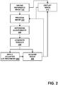
Fig. 2 is a flow chart illustrating a method for administering ultrasound therapy. - In accordance with the present invention and referring to
FIG. 1 , a system for quickly treating a patient P suffering from a stroke and/or an intracranial clot C includes a high-intensity focused-ultrasound phased-array transducer 105 (which is driven by one or more drivers 110), acontroller 120, and one ormore imagers 130 for monitoring clinical parameters related to the clot C. The parameters are detectable via theimaging apparatus 130 and can be used to monitor the successful liquefaction of the clot C. Examples of such parameters include blood flow in the vicinity of a blood vessel being treated and/or liquification of the clot C. The system includes aprocessor 140 and, optionally, adisplay 150. Optionally, the system may also include a user interface 160, such as a touch screen, a keyboard, and/or a mouse. Preferably, the system is configured for delivering ultrasonic energy between ten kilohertz (0.01 MHz) and ten Megahertz (10 MHz) to tissue within the skull or other anatomical regions. Such a system may be used to treat blood clots lodged within blood vessels of a patient or a clot outside blood vessels (generated by ruptured blood vessels) by delivering acoustic energy to the clot. In some cases, the clot may be partially or completely blocking a blood vessel in the patient's cranium, or applying intra cranial pressure in case of hemorrhagic clot and causing life-threatening conditions such as a stroke. During delivery of the ultrasonic energy, blood flow (as well as other clinical parameters) in and/or around the affected blood vessel is monitored, and adjustments made to one or more treatment parameters based thereon. The adjustments may be automatic or manual. For example, various images of the patient's anatomy surrounding the clot may be viewed on a display, and adjustments made based on automated analysis of the images. - The
transducer 105 may include "n" (where n > 1) transducer elements, thereby providing a multiple-element transducer array. Thetransducer 105 may include a flexible or semi-rigid base or panel conforming to the shape of patient's anatomy, such as the skull if being used to treat cranial blood clots. Thetransducer 105 may be pre-curved (e.g. biased to a spherical or other concave shape), such that the transducer may be placed on or near a portion of a skull. Alternatively, thetransducer 105 may be planar, parabolic, or any other suitable shape, e.g., circular, elliptical, and the like. - Suitable HIFU phased-array transducers are known in the art. See, e.g., copending, commonly-owned
U.S. Patent Application Serial No. 10/328,584 , entitled "Tissue Aberration Corrections in Ultrasound Therapy." - The system may obtain, process and present images of the interior anatomy of the patient P, including the area surrounding the clot C. For example, the images may be taken using a magnetic resonance imaging (MRI) device, a computer tomography (CT) device, or an ultrasound imaging device. The images processed by the
processor 140 and presented on thedisplay 150 indicate the state of blood flow and/or liquification of tissue about the clot C, either within the specific blood vessel in which the clot is lodged, or, in some cases, in blood vessels surrounding the clot or a general measure of perfusion surrounding the clot C or within the hemorrhagic clot. As ultrasound energy is applied to the clot, an operator views the images in real-time or pseudo-real time (e.g., a delay of less than five seconds), and thus is able to see the effects of the ultrasound on the clot and the resulting improvement of blood flow or perfusion. The effects of reflow or liquefaction maybe also assessed directly or by other bio-imaging parameters. - Based on the observed and/or detected blood flow or liquid content of the hemorrhage, a
processor 140 generate correction factors to be applied to the signals that drive thetransducers 105. Theprocessor 140 determines the correction factors by automatically analyzing images and estimating blood flow or liquid content (and, in some embodiments, identifying tissue characteristics of interest) from the images. Sufficient information is provided by the images to facilitate determination of the correction factors. The focal quality of the images is used to influence the correction factors. For example, if a particular image is know to be of poor quality, its contribution to the correction factor(s) may be down-weighted. - The
processor 140 automatically recognizes an increase in blood flow, perfusion (or lack thereof), and/or liquid content based, on the images. The correction factors are applied to treatment parameters that govern the application of HIFU energy, thereby influencing subsequent ultrasound applications. In some implementations, theprocessor 140 includes a number of amplifiers and/or phase shifters that are coupled in respective sets. The amplifiers provide amplified excitation signals to thetransducer elements 105, e.g., via coaxial cables or other connections, which may individually connect the amplifiers and respective transducer elements. - Among other effects, the correction factors allow the acoustic energy transmitted by the transducer elements to be steered such that the "focal zone" (the region in space toward which the acoustic energy is focused) can be moved, along the z axis (i.e., an axis extending orthogonally from the transmission surface of the transducer into the skull) and/or along the x or y axes. The component of each phase-shift factor associated with steering may be computed using known techniques, e.g., using the average speed of sound in the body (possibly adjusted for different tissue types) and the distance from each transducer element to a target site of interest (the intended focal zone within a tissue region).
- In addition, in cranial applications, the correction factors may also compensate for phase distortion of the ultrasonic energy transmitted by each transducer element that occurs when the acoustic energy passes through the skull. The component of each correction factor associated with phase distortion may compensate for perturbations and distortions introduced by the bone of the skull, the skin/skull interface, the dura matter/skull interface, by variations in skull thickness or density, and/or by structural considerations such as air-filled or liquid-filled pockets in the skull. The two components that make up the phase-shift factors, i.e., the steering components and the phase-distortion components, are summed to determine the composite phase-shift factors for the respective channels in order to focus ultrasonic energy at a desired location.
- In some cases, the
processor 140 or the operator may also construct an initial treatment regime describing various treatment parameters (e.g., pressure, temporal structure, frequency, energy, etc.) that creates a sharp focus (e.g., less than 5 mm) of the ultrasound energy, thereby facilitating clot lysis in a targeted manner. In such cases, the correction factors determined during treatment may be applied to the treatment regime in real-time. - The phase-shift factors may, in some cases, be determined by the
processor 140 and/ or the system image and display computer, which may be coupled to thecontroller 120 and to animager 130. Alternatively, thecontroller 120 itself may include all necessary hardware components and/or software modules to determine the correction factors instead of requiring a separate computer. The system may include one or more software modules, hardware components, firmware, hardwiring, or any combinations of these. For example, the processor may be a general or special purpose digital data processor programmed with software to generate the phase shift factors, which may be transferred to the controller for subsequent transfer to the phase shifters or directly to the phase shifters based upon images shown on the display. - In addition to providing a system for using high-intensity ultrasound to treat transcranial clots, variants of the disclosure provide methods for treating stroke victims using such a system as described below and illustrated in
FIG. 2 . - Upon initial examination, an imager is used to acquire one or more images (STEP 205) of a clot area within a patient's brain. As explained above, the imager may be any of a variety of imaging devices, such as an MRI device, a CT device, or an ultrasound device or a combination/fusion of images.
- Data representing the images are transferred to a processor (STEP 210), and presented on a display (STEP 215). In implementations in which the patient is being treated for a stroke, the transfer of image data may occur immediately upon acquiring the images from the patient in order to provide treatment quickly. The transfer may be completed automatically or may occur only upon instruction from a physician or other operator.
- Based on the image data, correction factors and other potential adjustments may be determined (STEP 220) for treatment of the clot. As described above, the correction factors may account for different speeds of sound that are encountered as the acoustic energy passes through different tissue types in respective segmented tissue regions. In addition or alternatively, the correction factors may account for aberrations generated by the skull or at boundaries of the segmented tissue regions, as explained further below.
- The resulting correction factors, e.g., phase-shift factors and/or amplitude factors, may be used to assist a particular course of treatment, preferably focusing acoustic energy at the clot. Once determined, the correction factors may be provided to the controller which, in turn, provides excitation signals (STEP 225) to the transducer array. The focused ultrasound system uses the correction factors to control a beam former or signal adjuster which delivers excitation or drive signals to the transducer based upon the correction factors for application of the ultrasound treatment (STEP 230). For example, one or more base signals may be supplied to a signal adjuster and split into a plurality of channels, preferably into individual channels corresponding to respective transducer elements or transducer element groupings of the transducer array. The phase of the signals for the respective channels may be adjusted by the signal adjuster according to phase correction factors received by the controller. For example, the phases may be adjusted to compensate for acoustic energy from respective transducer elements passing through different tissue types and/or encountering one or more tissue boundaries. In one particular case, the amplitude adjustments may be done to compensate for a known or calculated skull temperature or to generate desired skull temperature. Such an approach may be implemented a priori based, for example, on an acoustic-bio-heat simulation or based on actual skull temperature measurements. In some cases, the system may assume an even temperature distribution on the skull.
- This may be in addition to other phase adjustments that focus the acoustic energy at a particular location or in a particular shape or to compensate for transducer element variations, as is known to those skilled in the art. The phase-adjusted signals may be amplified based upon amplitude correction factors, e.g., by amplifiers, which may amplify the excitation signals. Alternatively, the signals for the respective channels may be amplified before they are phase-adjusted.
- The amplified and phase-adjusted excitation signals may be delivered to the transducer to drive the respective transducer elements. The transducer elements convert the excitation signals into acoustic energy that is transmitted from the respective transducer elements of the transducer into the blood vessel containing the clot or hemorrhage inside the brain (or its general area) through any intervening tissue and/or bone, such as the skull.
- During the treatment, additional images may be acquired (STEP 235) using the same or a different imager than used to acquire reference image data to monitor the progress of the treatment. For example, the images may be transferred to the processor and/or rendered on a display for real-time or nearly real-time monitoring. In some implementations, the images acquired during treatment may be compared with the previously acquired reference images to determine if, for example, the blood flow within the blood vessel has improved or the hemorrhage has been liquefied. This comparison may be made by the operator or in an automated fashion using image-analysis software configured to recognize fluid flow through or liquid content of a clot. If necessary, the treatment parameters may be adjusted, e.g., by providing further amplitude and/or phase correction factors, to modify the energy delivered to the tissue region and reflect events as they unfold, e.g., to increase monitored blood flow or liquid content. Treatment is thereby delivered in a controlled, closed-loop manner that considers how the treatment is affecting the physiological characteristics of the clot and/or the treatment zone surrounding the clot.
- In another mode of implementation the phase settings are adjusted based on the quality of the focus. The focal quality may be sensed using regular imaging or, in some cases, unique modes of imaging tailored to assess focus quality such as acoustic radiation force imaging. In these implementations, the phases are dithered to identify optimal focus that defines the phase setting.
Claims (3)
- A system for delivering acoustic energy to a clot, the system comprising:a. a focused-ultrasound phased-array transducer;b. an imager for capturing images;c. a processor adapted to receive the images transferred from the imager for monitoring clinical parameters related to the clot and wherein the processor is adapted to (i) automatically analyze images using image-analysis software configured to recognize fluid flow through or liquid content of the clot and estimate liquid content of the clot as ultrasound energy is directed at the clot, (ii) based on the image analysis, liquid content and focal quality of the images, generate correction factors for altering the intensity and/or pressure of the ultrasound energy and (iii) repeat steps (i) and (ii) until the clot is substantially liquefied; andd. a controller for operating the transducer in accordance with the correction factors.
- The system of claim 1 wherein the means for monitoring liquefaction comprise a visual display.
- The system of claim 1, wherein the correction factors alter the ultrasound energy to increase a monitored blood flow or the liquid content.
Applications Claiming Priority (2)
| Application Number | Priority Date | Filing Date | Title |
|---|---|---|---|
| US11611108P | 2008-11-19 | 2008-11-19 | |
| PCT/IB2009/007674 WO2010058292A2 (en) | 2008-11-19 | 2009-11-19 | Closed-loop clot lysis |
Publications (2)
| Publication Number | Publication Date |
|---|---|
| EP2358437A2 EP2358437A2 (en) | 2011-08-24 |
| EP2358437B1 true EP2358437B1 (en) | 2018-11-07 |
Family
ID=42172555
Family Applications (1)
| Application Number | Title | Priority Date | Filing Date |
|---|---|---|---|
| EP09801547.2A Active EP2358437B1 (en) | 2008-11-19 | 2009-11-19 | Closed-loop clot lysis |
Country Status (5)
| Country | Link |
|---|---|
| US (1) | US8425424B2 (en) |
| EP (1) | EP2358437B1 (en) |
| JP (1) | JP2012509126A (en) |
| CN (1) | CN102292123A (en) |
| WO (1) | WO2010058292A2 (en) |
Families Citing this family (51)
| Publication number | Priority date | Publication date | Assignee | Title |
|---|---|---|---|---|
| US8256430B2 (en) | 2001-06-15 | 2012-09-04 | Monteris Medical, Inc. | Hyperthermia treatment and probe therefor |
| AU2002359576A1 (en) | 2001-12-03 | 2003-06-17 | Ekos Corporation | Catheter with multiple ultrasound radiating members |
| US8409099B2 (en) * | 2004-08-26 | 2013-04-02 | Insightec Ltd. | Focused ultrasound system for surrounding a body tissue mass and treatment method |
| US10219815B2 (en) | 2005-09-22 | 2019-03-05 | The Regents Of The University Of Michigan | Histotripsy for thrombolysis |
| EP1960993B1 (en) | 2005-11-23 | 2016-11-02 | Insightec-Image Guided Treatment, Ltd. | Hierarchical switching in ultra-high density ultrasound array |
| US20100030076A1 (en) * | 2006-08-01 | 2010-02-04 | Kobi Vortman | Systems and Methods for Simultaneously Treating Multiple Target Sites |
| US10182833B2 (en) | 2007-01-08 | 2019-01-22 | Ekos Corporation | Power parameters for ultrasonic catheter |
| WO2009002881A1 (en) | 2007-06-22 | 2008-12-31 | Ekos Corporation | Method and apparatus for treatment of intracranial hemorrhages |
| US8617073B2 (en) | 2009-04-17 | 2013-12-31 | Insightec Ltd. | Focusing ultrasound into the brain through the skull by utilizing both longitudinal and shear waves |
| WO2010143072A1 (en) * | 2009-06-10 | 2010-12-16 | Insightec Ltd. | Acoustic-feedback power control during focused ultrasound delivery |
| US8979871B2 (en) | 2009-08-13 | 2015-03-17 | Monteris Medical Corporation | Image-guided therapy of a tissue |
| US9177543B2 (en) | 2009-08-26 | 2015-11-03 | Insightec Ltd. | Asymmetric ultrasound phased-array transducer for dynamic beam steering to ablate tissues in MRI |
| EP2489034B1 (en) | 2009-10-14 | 2016-11-30 | Insightec Ltd. | Mapping ultrasound transducers |
| US8932237B2 (en) | 2010-04-28 | 2015-01-13 | Insightec, Ltd. | Efficient ultrasound focusing |
| US9852727B2 (en) | 2010-04-28 | 2017-12-26 | Insightec, Ltd. | Multi-segment ultrasound transducers |
| EP2608730B1 (en) | 2010-08-27 | 2019-07-24 | Ekos Corporation | Apparatus for treatment of intracranial hemorrhages |
| AU2012364909B2 (en) | 2011-02-15 | 2016-11-24 | Hemosonics, Llc | Characterization of blood hemostasis and oxygen transport parameters |
| JP6285921B2 (en) * | 2012-06-13 | 2018-02-28 | ニューウェル, デイビッド ダブリュー.NEWELL, David, W. | Treatment of subarachnoid hematoma using ultrasonic thrombolysis and related devices, systems, and methods |
| US20140073907A1 (en) | 2012-09-12 | 2014-03-13 | Convergent Life Sciences, Inc. | System and method for image guided medical procedures |
| BR112015022067A2 (en) | 2013-03-14 | 2017-07-18 | Ekos Corp | method and apparatus for administering drug to a target site |
| WO2015003154A1 (en) | 2013-07-03 | 2015-01-08 | Histosonics, Inc. | Articulating arm limiter for cavitational ultrasound therapy system |
| WO2015000953A1 (en) * | 2013-07-03 | 2015-01-08 | Bracco Suisse S.A. | Devices and methods for the ultrasound treatment of ischemic stroke |
| WO2015027164A1 (en) | 2013-08-22 | 2015-02-26 | The Regents Of The University Of Michigan | Histotripsy using very short ultrasound pulses |
| US9433383B2 (en) | 2014-03-18 | 2016-09-06 | Monteris Medical Corporation | Image-guided therapy of a tissue |
| US20150265353A1 (en) | 2014-03-18 | 2015-09-24 | Monteris Medical Corporation | Image-guided therapy of a tissue |
| US10675113B2 (en) | 2014-03-18 | 2020-06-09 | Monteris Medical Corporation | Automated therapy of a three-dimensional tissue region |
| CN104587612A (en) * | 2015-01-10 | 2015-05-06 | 管勇 | Ultrasonic intracranial tumor treatment apparatus |
| US9726647B2 (en) | 2015-03-17 | 2017-08-08 | Hemosonics, Llc | Determining mechanical properties via ultrasound-induced resonance |
| US10327830B2 (en) | 2015-04-01 | 2019-06-25 | Monteris Medical Corporation | Cryotherapy, thermal therapy, temperature modulation therapy, and probe apparatus therefor |
| EP3307388B1 (en) | 2015-06-10 | 2022-06-22 | Ekos Corporation | Ultrasound catheter |
| WO2016210133A1 (en) | 2015-06-24 | 2016-12-29 | The Regents Of The Universtiy Of Michigan | Histotripsy therapy systems and methods for the treatment of brain tissue |
| EP3419548B1 (en) * | 2016-02-23 | 2024-07-17 | Sunnybrook Research Institute | Method of fabricating a transcranial headset for diagnostic or therapeutic procedures |
| CN106932415B (en) * | 2017-01-22 | 2019-10-11 | 南京航空航天大学 | A composite non-destructive testing device and method dedicated to laser welding joints |
| US11266311B2 (en) * | 2017-06-08 | 2022-03-08 | Arizona Board Of Regents On Behalf Of Arizona State University | Systems and methods for generating an elastogram of brain tissue using mesoscopic wavelength ultrasound |
| EP3681406B1 (en) | 2017-09-13 | 2025-04-23 | Orchard Ultrasound Innovation LLC | Medical device with cmut array and solid state cooling |
| CN111655337B (en) * | 2017-12-11 | 2023-06-27 | 医视特有限公司 | Controlled delivery of therapeutic agents during microbubble-enhanced ultrasound procedures |
| CA3120586A1 (en) | 2018-11-28 | 2020-06-04 | Histosonics, Inc. | Histotripsy systems and methods |
| US12318637B2 (en) * | 2019-05-31 | 2025-06-03 | Sunnybrook Research Institute | Systems and methods for reducing thermal skull-induced aberrations during transcranial ultrasound therapeutic procedures |
| CN110448354A (en) * | 2019-08-07 | 2019-11-15 | 西安市第四医院 | A kind of cerebral thrombosis supersonic therapeutic system |
| KR102247568B1 (en) * | 2019-10-11 | 2021-05-04 | 재단법인 대구경북첨단의료산업진흥재단 | Focused ultrasound irradiation system and the method for adjusting the degree of opening of the blood brain barrier using the same |
| WO2021087817A1 (en) * | 2019-11-06 | 2021-05-14 | 中国科学院深圳先进技术研究院 | Ultrasonic regulation and imaging apparatus and method, and server and storage medium |
| CN110882004B (en) * | 2019-11-06 | 2022-05-27 | 中国科学院深圳先进技术研究院 | Ultrasonic regulation and imaging device, method, server and storage medium |
| CN112774048A (en) * | 2019-11-08 | 2021-05-11 | 中国科学院深圳先进技术研究院 | Closed-loop ultrasonic nerve regulation and control system and method |
| JP2023513012A (en) | 2020-01-28 | 2023-03-30 | ザ リージェンツ オブ ザ ユニバーシティー オブ ミシガン | Systems and methods for histotrypsy immunization |
| BR112022025722A2 (en) | 2020-06-18 | 2023-03-07 | Histosonics Inc | ACOUSTIC COUPLING SYSTEMS AND METHODS AND PATIENT WITH HISTOTRIPSY |
| CN116782843A (en) | 2020-08-27 | 2023-09-19 | 密歇根大学董事会 | Ultrasound transducer with transmit-receive capability for tissue destruction |
| EP4346664A4 (en) * | 2021-06-01 | 2025-04-23 | Orchard Ultrasound Innovation LLC | Tissue treatment system |
| US12582848B2 (en) | 2021-06-07 | 2026-03-24 | The Regents Of The University Of Michigan | Minimally invasive histotripsy systems and methods |
| EP4351447A4 (en) | 2021-06-07 | 2025-07-16 | Univ Michigan Regents | ALL-IN-ONE ULTRASOUND SYSTEMS AND PROCEDURES WITH HISTOTRIPSY |
| WO2024092272A1 (en) | 2022-10-28 | 2024-05-02 | Histosonics, Inc. | Histotripsy systems and methods |
| KR20260003742A (en) | 2023-04-20 | 2026-01-07 | 히스토소닉스, 인크. | Histotripsy systems and associated methods including user interfaces and workflows for treatment planning and therapy |
Family Cites Families (251)
| Publication number | Priority date | Publication date | Assignee | Title |
|---|---|---|---|---|
| US2795709A (en) | 1953-12-21 | 1957-06-11 | Bendix Aviat Corp | Electroplated ceramic rings |
| US3142035A (en) | 1960-02-04 | 1964-07-21 | Harris Transducer Corp | Ring-shaped transducer |
| US4000493A (en) | 1971-04-12 | 1976-12-28 | Eastman Kodak Company | Acoustooptic scanner apparatus and method |
| US3974475A (en) | 1971-10-07 | 1976-08-10 | Hoffmann-La Roche Inc. | Method of and apparatus for focusing ultrasonic waves in a focal line |
| US3992693A (en) | 1972-12-04 | 1976-11-16 | The Bendix Corporation | Underwater transducer and projector therefor |
| CA1050654A (en) * | 1974-04-25 | 1979-03-13 | Varian Associates | Reconstruction system and method for ultrasonic imaging |
| US3942150A (en) * | 1974-08-12 | 1976-03-02 | The United States Of America As Represented By The Secretary Of The Navy | Correction of spatial non-uniformities in sonar, radar, and holographic acoustic imaging systems |
| US4206653A (en) | 1975-10-02 | 1980-06-10 | E M I Limited | Ultrasonic apparatus |
| US4211132A (en) | 1977-11-21 | 1980-07-08 | E. I. Du Pont De Nemours And Company | Apparatus for on-line defect zoning |
| CA1137210A (en) | 1979-04-26 | 1982-12-07 | Francis S. Foster | Ultrasonic imaging method and device using one transducer having a line focus aligned with another transducer |
| DE3119295A1 (en) | 1981-05-14 | 1982-12-16 | Siemens AG, 1000 Berlin und 8000 München | DEVICE FOR DESTROYING CONCRETE IN BODIES |
| US4454597A (en) | 1982-05-03 | 1984-06-12 | The United States Of America As Represented By The Secretary Of The Navy | Conformal array compensating beamformer |
| DE3216547C1 (en) * | 1982-05-04 | 1983-10-06 | Krautkraemer Gmbh | Method and circuit device for generating and changing a predetermined number of independent DC voltages |
| DE3224453A1 (en) | 1982-06-30 | 1984-01-05 | Siemens AG, 1000 Berlin und 8000 München | ULTRASONIC TOMOGRAPHER |
| US4554925A (en) | 1982-07-07 | 1985-11-26 | Picker International, Ltd. | Nuclear magnetic resonance imaging method |
| DE3319871A1 (en) | 1983-06-01 | 1984-12-06 | Richard Wolf Gmbh, 7134 Knittlingen | PIEZOELECTRIC CONVERTER FOR DESTROYING CONCRETE IN THE BODY |
| US4505156A (en) * | 1983-06-21 | 1985-03-19 | Sound Products Company L.P. | Method and apparatus for switching multi-element transducer arrays |
| US4537074A (en) | 1983-09-12 | 1985-08-27 | Technicare Corporation | Annular array ultrasonic transducers |
| US4549533A (en) | 1984-01-30 | 1985-10-29 | University Of Illinois | Apparatus and method for generating and directing ultrasound |
| US4662222A (en) * | 1984-12-21 | 1987-05-05 | Johnson Steven A | Apparatus and method for acoustic imaging using inverse scattering techniques |
| US4865042A (en) | 1985-08-16 | 1989-09-12 | Hitachi, Ltd. | Ultrasonic irradiation system |
| GB8529446D0 (en) | 1985-11-29 | 1986-01-08 | Univ Aberdeen | Divergent ultrasound arrays |
| DE8608538U1 (en) * | 1986-03-27 | 1988-02-18 | Messer Griesheim Gmbh, 6000 Frankfurt | Device for freezing food on a trolley |
| DE3788757D1 (en) * | 1986-08-20 | 1994-02-24 | Siemens Ag | Method and device for adaptive focusing in a medical ultrasound imaging device. |
| EP0272347B1 (en) | 1986-12-24 | 1989-06-07 | Hewlett-Packard GmbH | Method of and apparatus for adjusting the intensity profile of an ultrasound beam |
| DE3732131A1 (en) | 1987-09-24 | 1989-04-06 | Wolf Gmbh Richard | FOCUSING ULTRASONIC transducer |
| US4989143A (en) | 1987-12-11 | 1991-01-29 | General Electric Company | Adaptive coherent energy beam formation using iterative phase conjugation |
| US5209221A (en) * | 1988-03-01 | 1993-05-11 | Richard Wolf Gmbh | Ultrasonic treatment of pathological tissue |
| US4893284A (en) * | 1988-05-27 | 1990-01-09 | General Electric Company | Calibration of phased array ultrasound probe |
| US4893624A (en) * | 1988-06-21 | 1990-01-16 | Massachusetts Institute Of Technology | Diffuse focus ultrasound hyperthermia system |
| US5197475A (en) * | 1988-08-10 | 1993-03-30 | The Board Of Regents, The University Of Texas System | Method and apparatus for analyzing material properties using ultrasound |
| US5211160A (en) * | 1988-09-14 | 1993-05-18 | Interpore Orthopaedics, Inc. | Ultrasonic orthopedic treatment head and body-mounting means therefor |
| US5316000A (en) | 1991-03-05 | 1994-05-31 | Technomed International (Societe Anonyme) | Use of at least one composite piezoelectric transducer in the manufacture of an ultrasonic therapy apparatus for applying therapy, in a body zone, in particular to concretions, to tissue, or to bones, of a living being and method of ultrasonic therapy |
| US5435312A (en) | 1991-05-31 | 1995-07-25 | Spivey; Brett A. | Acoustic imaging device |
| JP3300419B2 (en) * | 1991-08-21 | 2002-07-08 | 株式会社東芝 | Thrombolysis treatment device |
| DE4227800C2 (en) * | 1991-08-21 | 1996-12-19 | Toshiba Kawasaki Kk | Thrombus-releasing treatment device |
| US5291890A (en) * | 1991-08-29 | 1994-03-08 | General Electric Company | Magnetic resonance surgery using heat waves produced with focussed ultrasound |
| JP3533217B2 (en) * | 1991-12-20 | 2004-05-31 | テクノメド メディカル システム | Ultrasound therapy device that outputs ultrasonic waves having thermal effect and cavitation effect |
| JP3325300B2 (en) | 1992-02-28 | 2002-09-17 | 株式会社東芝 | Ultrasound therapy equipment |
| JP3325534B2 (en) | 1992-02-28 | 2002-09-17 | 株式会社東芝 | Ultrasound therapy equipment |
| DE4207463C2 (en) | 1992-03-10 | 1996-03-28 | Siemens Ag | Arrangement for the therapy of tissue with ultrasound |
| US5247935A (en) | 1992-03-19 | 1993-09-28 | General Electric Company | Magnetic resonance guided focussed ultrasound surgery |
| US5318025A (en) | 1992-04-01 | 1994-06-07 | General Electric Company | Tracking system to monitor the position and orientation of a device using multiplexed magnetic resonance detection |
| US5271400A (en) | 1992-04-01 | 1993-12-21 | General Electric Company | Tracking system to monitor the position and orientation of a device using magnetic resonance detection of a sample contained within the device |
| DE4345308C2 (en) | 1992-07-15 | 2001-02-01 | Fukuda Denshi Kk | Medical ultrasonic diagnosis system |
| US5275165A (en) * | 1992-11-06 | 1994-01-04 | General Electric Company | Magnetic resonance guided ultrasound therapy system with inclined track to move transducers in a small vertical space |
| US5573497A (en) | 1994-11-30 | 1996-11-12 | Technomed Medical Systems And Institut National | High-intensity ultrasound therapy method and apparatus with controlled cavitation effect and reduced side lobes |
| DE4302537C1 (en) * | 1993-01-29 | 1994-04-28 | Siemens Ag | Ultrasound imaging and therapy device - generates imaging waves and focussed treatment waves having two differing frequencies for location and treatment of e.g tumours |
| JP3860227B2 (en) * | 1993-03-10 | 2006-12-20 | 株式会社東芝 | Ultrasonic therapy device used under MRI guide |
| EP0627206B1 (en) | 1993-03-12 | 2002-11-20 | Kabushiki Kaisha Toshiba | Apparatus for ultrasound medical treatment |
| US5307812A (en) | 1993-03-26 | 1994-05-03 | General Electric Company | Heat surgery system monitored by real-time magnetic resonance profiling |
| WO1995001751A1 (en) | 1993-07-01 | 1995-01-19 | Boston Scientific Corporation | Imaging, electrical potential sensing, and ablation catheters |
| US5379642A (en) * | 1993-07-19 | 1995-01-10 | Diasonics Ultrasound, Inc. | Method and apparatus for performing imaging |
| US5413550A (en) * | 1993-07-21 | 1995-05-09 | Pti, Inc. | Ultrasound therapy system with automatic dose control |
| US5368031A (en) | 1993-08-29 | 1994-11-29 | General Electric Company | Magnetic resonance surgery using heat waves produced with a laser fiber |
| US5329930A (en) | 1993-10-12 | 1994-07-19 | General Electric Company | Phased array sector scanner with multiplexed acoustic transducer elements |
| US5526814A (en) | 1993-11-09 | 1996-06-18 | General Electric Company | Automatically positioned focussed energy system guided by medical imaging |
| US5368032A (en) | 1993-11-09 | 1994-11-29 | General Electric Company | Manually positioned focussed energy system guided by medical imaging |
| JPH07184907A (en) | 1993-12-28 | 1995-07-25 | Toshiba Corp | Ultrasonic therapy equipment |
| FR2715313B1 (en) | 1994-01-27 | 1996-05-31 | Edap Int | Method for controlling a hyperthermia treatment device using ultrasound. |
| US5507790A (en) * | 1994-03-21 | 1996-04-16 | Weiss; William V. | Method of non-invasive reduction of human site-specific subcutaneous fat tissue deposits by accelerated lipolysis metabolism |
| GB9409133D0 (en) | 1994-05-09 | 1994-11-30 | Secr Defence | Sonar ring transducer |
| US5549638A (en) | 1994-05-17 | 1996-08-27 | Burdette; Everette C. | Ultrasound device for use in a thermotherapy apparatus |
| DE4421795C1 (en) | 1994-06-22 | 1996-01-04 | Siemens Ag | Implanted therapeutic acoustic wave source |
| ATE252349T1 (en) | 1994-09-15 | 2003-11-15 | Visualization Technology Inc | SYSTEM FOR POSITION DETECTION USING A REFERENCE UNIT ATTACHED TO A PATIENT'S HEAD FOR USE IN THE MEDICAL FIELD |
| US5694936A (en) | 1994-09-17 | 1997-12-09 | Kabushiki Kaisha Toshiba | Ultrasonic apparatus for thermotherapy with variable frequency for suppressing cavitation |
| US5443068A (en) | 1994-09-26 | 1995-08-22 | General Electric Company | Mechanical positioner for magnetic resonance guided ultrasound therapy |
| US5490840A (en) * | 1994-09-26 | 1996-02-13 | General Electric Company | Targeted thermal release of drug-polymer conjugates |
| US5520188A (en) * | 1994-11-02 | 1996-05-28 | Focus Surgery Inc. | Annular array transducer |
| NO310983B1 (en) | 1994-11-22 | 2001-09-24 | Baker Hughes Inc | Method and apparatus for drilling and supplementing wells |
| US5520612A (en) * | 1994-12-30 | 1996-05-28 | Exogen, Inc. | Acoustic system for bone-fracture therapy |
| US5617371A (en) * | 1995-02-08 | 1997-04-01 | Diagnostic/Retrieval Systems, Inc. | Method and apparatus for accurately determing the location of signal transducers in a passive sonar or other transducer array system |
| DE19507478C1 (en) | 1995-03-03 | 1996-05-15 | Siemens Ag | Therapy device for treatment with focused ultrasound |
| US6334846B1 (en) * | 1995-03-31 | 2002-01-01 | Kabushiki Kaisha Toshiba | Ultrasound therapeutic apparatus |
| US5984881A (en) | 1995-03-31 | 1999-11-16 | Kabushiki Kaisha Toshiba | Ultrasound therapeutic apparatus using a therapeutic ultrasonic wave source and an ultrasonic probe |
| US5617857A (en) * | 1995-06-06 | 1997-04-08 | Image Guided Technologies, Inc. | Imaging system having interactive medical instruments and methods |
| US5605154A (en) * | 1995-06-06 | 1997-02-25 | Duke University | Two-dimensional phase correction using a deformable ultrasonic transducer array |
| US5582578A (en) | 1995-08-01 | 1996-12-10 | Duke University | Method for the comminution of concretions |
| US5711300A (en) * | 1995-08-16 | 1998-01-27 | General Electric Company | Real time in vivo measurement of temperature changes with NMR imaging |
| US5590657A (en) * | 1995-11-06 | 1997-01-07 | The Regents Of The University Of Michigan | Phased array ultrasound system and method for cardiac ablation |
| US5606971A (en) * | 1995-11-13 | 1997-03-04 | Artann Corporation, A Nj Corp. | Method and device for shear wave elasticity imaging |
| US5810731A (en) | 1995-11-13 | 1998-09-22 | Artann Laboratories | Method and apparatus for elasticity imaging using remotely induced shear wave |
| US5728062A (en) * | 1995-11-30 | 1998-03-17 | Pharmasonics, Inc. | Apparatus and methods for vibratory intraluminal therapy employing magnetostrictive transducers |
| EP0811171B1 (en) | 1995-12-14 | 2005-07-27 | Koninklijke Philips Electronics N.V. | Method and device for heating by means of ultrasound guided by magnetic resonance imaging |
| US5762616A (en) | 1996-03-15 | 1998-06-09 | Exogen, Inc. | Apparatus for ultrasonic treatment of sites corresponding to the torso |
| US5752515A (en) * | 1996-08-21 | 1998-05-19 | Brigham & Women's Hospital | Methods and apparatus for image-guided ultrasound delivery of compounds through the blood-brain barrier |
| US5769790A (en) | 1996-10-25 | 1998-06-23 | General Electric Company | Focused ultrasound surgery system guided by ultrasound imaging |
| US5810008A (en) | 1996-12-03 | 1998-09-22 | Isg Technologies Inc. | Apparatus and method for visualizing ultrasonic images |
| AU726127B2 (en) | 1997-02-06 | 2000-11-02 | Exogen, Inc. | Method and apparatus for cartilage growth stimulation |
| US5904659A (en) * | 1997-02-14 | 1999-05-18 | Exogen, Inc. | Ultrasonic treatment for wounds |
| US5873845A (en) * | 1997-03-17 | 1999-02-23 | General Electric Company | Ultrasound transducer with focused ultrasound refraction plate |
| DE69821345T2 (en) | 1997-04-18 | 2004-09-30 | Exogen, Inc. | DEVICE FOR ULTRASONIC BONE TREATMENT |
| US6263230B1 (en) | 1997-05-08 | 2001-07-17 | Lucent Medical Systems, Inc. | System and method to determine the location and orientation of an indwelling medical device |
| ATE419789T1 (en) | 1997-05-23 | 2009-01-15 | Prorhythm Inc | HIGH INTENSITY DISPOSABLE FOCUSING ULTRASONIC APPLICATOR |
| DE19727081A1 (en) * | 1997-06-25 | 1999-01-07 | Siemens Ag | Object localisation method in MR examination |
| US6997925B2 (en) * | 1997-07-08 | 2006-02-14 | Atrionx, Inc. | Tissue ablation device assembly and method for electrically isolating a pulmonary vein ostium from an atrial wall |
| US6193659B1 (en) * | 1997-07-15 | 2001-02-27 | Acuson Corporation | Medical ultrasonic diagnostic imaging method and apparatus |
| US6128958A (en) | 1997-09-11 | 2000-10-10 | The Regents Of The University Of Michigan | Phased array system architecture |
| US6358246B1 (en) * | 1999-06-25 | 2002-03-19 | Radiotherapeutics Corporation | Method and system for heating solid tissue |
| US6071239A (en) | 1997-10-27 | 2000-06-06 | Cribbs; Robert W. | Method and apparatus for lipolytic therapy using ultrasound energy |
| US6113559A (en) | 1997-12-29 | 2000-09-05 | Klopotek; Peter J. | Method and apparatus for therapeutic treatment of skin with ultrasound |
| DE19800471A1 (en) * | 1998-01-09 | 1999-07-15 | Philips Patentverwaltung | MR method with micro-coils located in the examination area |
| US5947900A (en) | 1998-04-13 | 1999-09-07 | General Electric Company | Dynamic scan plane tracking using MR position monitoring |
| US6135960A (en) | 1998-08-31 | 2000-10-24 | Holmberg; Linda Jean | High-resolution, three-dimensional whole body ultrasound imaging system |
| US6042556A (en) * | 1998-09-04 | 2000-03-28 | University Of Washington | Method for determining phase advancement of transducer elements in high intensity focused ultrasound |
| US7722539B2 (en) * | 1998-09-18 | 2010-05-25 | University Of Washington | Treatment of unwanted tissue by the selective destruction of vasculature providing nutrients to the tissue |
| US6425867B1 (en) | 1998-09-18 | 2002-07-30 | University Of Washington | Noise-free real time ultrasonic imaging of a treatment site undergoing high intensity focused ultrasound therapy |
| JP4095729B2 (en) * | 1998-10-26 | 2008-06-04 | 株式会社日立製作所 | Therapeutic ultrasound system |
| US6701176B1 (en) | 1998-11-04 | 2004-03-02 | Johns Hopkins University School Of Medicine | Magnetic-resonance-guided imaging, electrophysiology, and ablation |
| WO2000030554A1 (en) | 1998-11-20 | 2000-06-02 | Jones Joie P | Methods for selectively dissolving and removing materials using ultra-high frequency ultrasound |
| US6246896B1 (en) | 1998-11-24 | 2001-06-12 | General Electric Company | MRI guided ablation system |
| US6289233B1 (en) | 1998-11-25 | 2001-09-11 | General Electric Company | High speed tracking of interventional devices using an MRI system |
| FR2786651B1 (en) | 1998-11-27 | 2002-10-25 | Commissariat Energie Atomique | ULTRASONIC CONTACT TRANSDUCER, WITH MULTIPLE ELEMENTS |
| US6309355B1 (en) | 1998-12-22 | 2001-10-30 | The Regents Of The University Of Michigan | Method and assembly for performing ultrasound surgery using cavitation |
| US6428532B1 (en) | 1998-12-30 | 2002-08-06 | The General Hospital Corporation | Selective tissue targeting by difference frequency of two wavelengths |
| EP1148817A4 (en) * | 1999-02-02 | 2004-10-13 | Transurgical Inc | INTRACORPOREAL HIFU APPLICATOR |
| IL129461A0 (en) | 1999-04-15 | 2000-02-29 | F R A Y Project Dev Ltd | 3-D ultrasound imaging system |
| US6217530B1 (en) * | 1999-05-14 | 2001-04-17 | University Of Washington | Ultrasonic applicator for medical applications |
| FR2794018B1 (en) * | 1999-05-26 | 2002-05-24 | Technomed Medical Systems | ULTRASONIC LOCATION AND TREATMENT APPARATUS |
| US6317619B1 (en) | 1999-07-29 | 2001-11-13 | U.S. Philips Corporation | Apparatus, methods, and devices for magnetic resonance imaging controlled by the position of a moveable RF coil |
| US6242915B1 (en) | 1999-08-27 | 2001-06-05 | General Electric Company | Field-frequency lock system for magnetic resonance system |
| JP4526648B2 (en) * | 1999-09-09 | 2010-08-18 | 株式会社日立メディコ | Magnetic resonance imaging system |
| JP3848572B2 (en) | 1999-09-10 | 2006-11-22 | プロリズム,インコーポレイテッド | Device for occluding anatomic tissue |
| US7510536B2 (en) * | 1999-09-17 | 2009-03-31 | University Of Washington | Ultrasound guided high intensity focused ultrasound treatment of nerves |
| US6524251B2 (en) * | 1999-10-05 | 2003-02-25 | Omnisonics Medical Technologies, Inc. | Ultrasonic device for tissue ablation and sheath for use therewith |
| US6626855B1 (en) | 1999-11-26 | 2003-09-30 | Therus Corpoation | Controlled high efficiency lesion formation using high intensity ultrasound |
| US6618608B1 (en) | 1999-11-30 | 2003-09-09 | Txsonics, Ltd. | Thermal imaging of fat and muscle using a simultaneous phase and magnitude double echo sequence |
| WO2001045550A2 (en) * | 1999-12-23 | 2001-06-28 | Therus Corporation | Ultrasound transducers for imaging and therapy |
| US8221402B2 (en) | 2000-01-19 | 2012-07-17 | Medtronic, Inc. | Method for guiding a medical device |
| US6635017B1 (en) * | 2000-02-09 | 2003-10-21 | Spentech, Inc. | Method and apparatus combining diagnostic ultrasound with therapeutic ultrasound to enhance thrombolysis |
| FR2806611B1 (en) | 2000-03-22 | 2004-10-15 | Hossein Kafai | ECHOGRAPHY APPARATUS FOR EXPLORATION OF TEMPORO-MANDIBULAR JOINT |
| US6419648B1 (en) | 2000-04-21 | 2002-07-16 | Insightec-Txsonics Ltd. | Systems and methods for reducing secondary hot spots in a phased array focused ultrasound system |
| US6613004B1 (en) | 2000-04-21 | 2003-09-02 | Insightec-Txsonics, Ltd. | Systems and methods for creating longer necrosed volumes using a phased array focused ultrasound system |
| US6392330B1 (en) * | 2000-06-05 | 2002-05-21 | Pegasus Technologies Ltd. | Cylindrical ultrasound receivers and transceivers formed from piezoelectric film |
| DE10028560C2 (en) * | 2000-06-09 | 2002-10-24 | Siemens Ag | Use of coefficients in a method for three-dimensional correction of distortions and magnetic resonance device for performing the method |
| US6761691B2 (en) | 2000-07-21 | 2004-07-13 | Fuji Photo Film Co., Ltd. | Image forming method used in ultrasonic diagnosis, ultrasonic diagnostic apparatus, signal processing apparatus, and recording medium for recording signal processing program |
| US6506171B1 (en) * | 2000-07-27 | 2003-01-14 | Insightec-Txsonics, Ltd | System and methods for controlling distribution of acoustic energy around a focal point using a focused ultrasound system |
| US6733450B1 (en) * | 2000-07-27 | 2004-05-11 | Texas Systems, Board Of Regents | Therapeutic methods and apparatus for use of sonication to enhance perfusion of tissue |
| US6582381B1 (en) | 2000-07-31 | 2003-06-24 | Txsonics Ltd. | Mechanical positioner for MRI guided ultrasound therapy system |
| US6612988B2 (en) | 2000-08-29 | 2003-09-02 | Brigham And Women's Hospital, Inc. | Ultrasound therapy |
| US6899680B2 (en) | 2000-10-19 | 2005-05-31 | Odetect As | Ultrasound measurement techniques for bone analysis |
| US6679855B2 (en) * | 2000-11-07 | 2004-01-20 | Gerald Horn | Method and apparatus for the correction of presbyopia using high intensity focused ultrasound |
| US6618620B1 (en) | 2000-11-28 | 2003-09-09 | Txsonics Ltd. | Apparatus for controlling thermal dosing in an thermal treatment system |
| US6506154B1 (en) * | 2000-11-28 | 2003-01-14 | Insightec-Txsonics, Ltd. | Systems and methods for controlling a phased array focused ultrasound system |
| CA2428872C (en) | 2000-11-28 | 2013-01-08 | Allez Physionix Limited | Systems and methods for making non-invasive physiological assessments |
| US6666833B1 (en) | 2000-11-28 | 2003-12-23 | Insightec-Txsonics Ltd | Systems and methods for focussing an acoustic energy beam transmitted through non-uniform tissue medium |
| US6613005B1 (en) | 2000-11-28 | 2003-09-02 | Insightec-Txsonics, Ltd. | Systems and methods for steering a focused ultrasound array |
| US6475150B2 (en) | 2000-12-01 | 2002-11-05 | The Regents Of The University Of California | System and method for ultrasonic tomography |
| US6770031B2 (en) | 2000-12-15 | 2004-08-03 | Brigham And Women's Hospital, Inc. | Ultrasound therapy |
| US6626854B2 (en) | 2000-12-27 | 2003-09-30 | Insightec - Txsonics Ltd. | Systems and methods for ultrasound assisted lipolysis |
| US6645162B2 (en) | 2000-12-27 | 2003-11-11 | Insightec - Txsonics Ltd. | Systems and methods for ultrasound assisted lipolysis |
| JP2002209905A (en) * | 2001-01-22 | 2002-07-30 | Hitachi Medical Corp | Ultrasonic therapy probe and ultrasonic therapy apparatus |
| US7094205B2 (en) | 2001-04-06 | 2006-08-22 | Alfred E. Mann Institute For Biomedical Engineering At The University Of Southern California | High-resolution 3D ultrasonic transmission imaging |
| US6478739B1 (en) | 2001-05-11 | 2002-11-12 | The Procter & Gamble Company | Ultrasonic breast examination system |
| US6559644B2 (en) * | 2001-05-30 | 2003-05-06 | Insightec - Txsonics Ltd. | MRI-based temperature mapping with error compensation |
| US6735461B2 (en) * | 2001-06-19 | 2004-05-11 | Insightec-Txsonics Ltd | Focused ultrasound system with MRI synchronization |
| US6523272B1 (en) * | 2001-08-03 | 2003-02-25 | George B. Morales | Measuring device and method of manufacture |
| US7429248B1 (en) | 2001-08-09 | 2008-09-30 | Exogen, Inc. | Method and apparatus for controlling acoustic modes in tissue healing applications |
| EP1423736A1 (en) | 2001-09-07 | 2004-06-02 | Shell Internationale Researchmaatschappij B.V. | Concentrating seismic energy in a selected target point in an underground formation |
| FR2830468B1 (en) | 2001-10-04 | 2004-02-20 | Inst Nat Sante Rech Med | DEVICE AND METHOD FOR PRODUCING HIGH PRESSURE ULTRASONIC PULSES |
| US6961606B2 (en) | 2001-10-19 | 2005-11-01 | Koninklijke Philips Electronics N.V. | Multimodality medical imaging system and method with separable detector devices |
| US7175596B2 (en) * | 2001-10-29 | 2007-02-13 | Insightec-Txsonics Ltd | System and method for sensing and locating disturbances in an energy path of a focused ultrasound system |
| US6770039B2 (en) | 2001-11-09 | 2004-08-03 | Duke University | Method to reduce tissue injury in shock wave lithotripsy |
| US6790180B2 (en) | 2001-12-03 | 2004-09-14 | Insightec-Txsonics Ltd. | Apparatus, systems, and methods for measuring power output of an ultrasound transducer |
| US6522142B1 (en) * | 2001-12-14 | 2003-02-18 | Insightec-Txsonics Ltd. | MRI-guided temperature mapping of tissue undergoing thermal treatment |
| US6824516B2 (en) | 2002-03-11 | 2004-11-30 | Medsci Technologies, Inc. | System for examining, mapping, diagnosing, and treating diseases of the prostate |
| US7128711B2 (en) | 2002-03-25 | 2006-10-31 | Insightec, Ltd. | Positioning systems and methods for guided ultrasound therapy systems |
| US20030187371A1 (en) | 2002-03-27 | 2003-10-02 | Insightec-Txsonics Ltd. | Systems and methods for enhanced focused ultrasound ablation using microbubbles |
| US6705993B2 (en) * | 2002-05-10 | 2004-03-16 | Regents Of The University Of Minnesota | Ultrasound imaging system and method using non-linear post-beamforming filter |
| US7264592B2 (en) | 2002-06-28 | 2007-09-04 | Alfred E. Mann Institute For Biomedical Engineering At The University Of Southern California | Scanning devices for three-dimensional ultrasound mammography |
| US6705994B2 (en) * | 2002-07-08 | 2004-03-16 | Insightec - Image Guided Treatment Ltd | Tissue inhomogeneity correction in ultrasound imaging |
| US7621877B2 (en) * | 2002-07-15 | 2009-11-24 | Itamar Medical Ltd. | Body surface probe, apparatus and method for non-invasively detecting medical conditions |
| US6676602B1 (en) * | 2002-07-25 | 2004-01-13 | Siemens Medical Solutions Usa, Inc. | Two dimensional array switching for beamforming in a volume |
| US20040059265A1 (en) * | 2002-09-12 | 2004-03-25 | The Regents Of The University Of California | Dynamic acoustic focusing utilizing time reversal |
| JP4386683B2 (en) | 2002-09-30 | 2009-12-16 | 富士フイルム株式会社 | Ultrasonic transmission / reception apparatus and ultrasonic transmission / reception method |
| US7077820B1 (en) | 2002-10-21 | 2006-07-18 | Advanced Medical Optics, Inc. | Enhanced microburst ultrasonic power delivery system and method |
| US7192400B2 (en) * | 2002-10-24 | 2007-03-20 | Synovis Life Technologies, Inc. | Device and method for vascular monitoring |
| US6860852B2 (en) | 2002-10-25 | 2005-03-01 | Compex Medical S.A. | Ultrasound therapeutic device |
| US6629929B1 (en) | 2002-11-08 | 2003-10-07 | Koninklijke Philips Electronics N.V. | Method and apparatus for automatically setting the transmit aperture and apodization of an ultrasound transducer array |
| ITFI20020228A1 (en) | 2002-11-22 | 2004-05-23 | Actis Active Sensors S R L | METHOD OF FOCUSING AN ECOGRAPHIC IMAGE AND RELATED ECOGRAPHIC SYSTEM |
| US7267650B2 (en) | 2002-12-16 | 2007-09-11 | Cardiac Pacemakers, Inc. | Ultrasound directed guiding catheter system and method |
| US8088067B2 (en) | 2002-12-23 | 2012-01-03 | Insightec Ltd. | Tissue aberration corrections in ultrasound therapy |
| IL154101A0 (en) * | 2003-01-23 | 2003-07-31 | Univ Ramot | Minimally invasive controlled surgical system with feedback |
| CN100356895C (en) * | 2003-01-31 | 2007-12-26 | 株式会社日立医药 | Ultrasonic probe and ultrasonic device |
| JP4543430B2 (en) * | 2003-01-31 | 2010-09-15 | 株式会社日立メディコ | Ultrasonic probe and ultrasonic device |
| US7344509B2 (en) * | 2003-04-17 | 2008-03-18 | Kullervo Hynynen | Shear mode therapeutic ultrasound |
| US7175599B2 (en) * | 2003-04-17 | 2007-02-13 | Brigham And Women's Hospital, Inc. | Shear mode diagnostic ultrasound |
| EP1471362A1 (en) * | 2003-04-24 | 2004-10-27 | Universiteit Utrecht Holding B.V. | Selective MR imaging of magnetic susceptibility deviations |
| AU2003902037A0 (en) * | 2003-04-24 | 2003-05-15 | Mclachlan, Craig | Method for tissue growth |
| US7699779B2 (en) * | 2003-05-19 | 2010-04-20 | Hitachi, Ltd. | Ultrasonic treatment equipment |
| US7611462B2 (en) | 2003-05-22 | 2009-11-03 | Insightec-Image Guided Treatment Ltd. | Acoustic beam forming in phased arrays including large numbers of transducer elements |
| US7377900B2 (en) | 2003-06-02 | 2008-05-27 | Insightec - Image Guided Treatment Ltd. | Endo-cavity focused ultrasound transducer |
| SE526718C2 (en) | 2003-06-04 | 2005-10-25 | Ultrazonix Dnt Ab | Ultrasonic probe with a central opening |
| DE602004002806T2 (en) | 2003-06-25 | 2007-08-23 | Aloka Co. Ltd., Mitaka | Diagnostic ultrasound imaging device with 2D transducer with variable subarray patterns |
| JP4639045B2 (en) * | 2003-07-11 | 2011-02-23 | 財団法人先端医療振興財団 | Non-invasive temperature distribution measuring method and apparatus for self-reference type and body movement tracking type by magnetic resonance tomography |
| JP4472395B2 (en) * | 2003-08-07 | 2010-06-02 | オリンパス株式会社 | Ultrasonic surgery system |
| JP2007513672A (en) | 2003-12-11 | 2007-05-31 | コーニンクレッカ フィリップス エレクトロニクス エヌ ヴィ | Stereoscopic ultrasound imaging system using a two-dimensional array transducer |
| US20050131301A1 (en) | 2003-12-12 | 2005-06-16 | Michael Peszynski | Ultrasound probe receptacle |
| US7069534B2 (en) | 2003-12-17 | 2006-06-27 | Sahouria Emile Y | Mask creation with hierarchy management using cover cells |
| DE102004004297B4 (en) | 2004-01-28 | 2013-06-27 | Siemens Aktiengesellschaft | Imaging Tomography Device |
| US7507213B2 (en) * | 2004-03-16 | 2009-03-24 | General Patent Llc | Pressure pulse/shock wave therapy methods for organs |
| JP2005283308A (en) | 2004-03-29 | 2005-10-13 | Lintec Corp | Manufacturing method of probe array |
| US9808221B2 (en) | 2004-04-02 | 2017-11-07 | Koninklijke Philips N.V. | Ultrasonic intracavity probe for 3D imaging |
| US7505808B2 (en) * | 2004-04-28 | 2009-03-17 | Sunnybrook Health Sciences Centre | Catheter tracking with phase information |
| EP1774920A4 (en) | 2004-06-21 | 2011-01-05 | Hiroshi Furuhata | Ultrasonic brain infarction treating device |
| US7699780B2 (en) * | 2004-08-11 | 2010-04-20 | Insightec—Image-Guided Treatment Ltd. | Focused ultrasound system with adaptive anatomical aperture shaping |
| US20060052706A1 (en) * | 2004-08-20 | 2006-03-09 | Kullervo Hynynen | Phased array ultrasound for cardiac ablation |
| US8409099B2 (en) * | 2004-08-26 | 2013-04-02 | Insightec Ltd. | Focused ultrasound system for surrounding a body tissue mass and treatment method |
| WO2006025001A1 (en) | 2004-09-01 | 2006-03-09 | Koninklijke Philips Electronics, N.V. | Magnetic resonance marker based position and orientation probe |
| US7452357B2 (en) | 2004-10-22 | 2008-11-18 | Ethicon Endo-Surgery, Inc. | System and method for planning treatment of tissue |
| US20060184034A1 (en) | 2005-01-27 | 2006-08-17 | Ronen Haim | Ultrasonic probe with an integrated display, tracking and pointing devices |
| US7553284B2 (en) | 2005-02-02 | 2009-06-30 | Vaitekunas Jeffrey J | Focused ultrasound for pain reduction |
| WO2006087649A1 (en) | 2005-02-17 | 2006-08-24 | Koninklijke Philips Electronics, N.V. | Method and apparatus for the visualization of the focus generated using focused ultrasound |
| US8801701B2 (en) | 2005-03-09 | 2014-08-12 | Sunnybrook Health Sciences Centre | Method and apparatus for obtaining quantitative temperature measurements in prostate and other tissue undergoing thermal therapy treatment |
| US7771418B2 (en) | 2005-03-09 | 2010-08-10 | Sunnybrook Health Sciences Centre | Treatment of diseased tissue using controlled ultrasonic heating |
| WO2006119572A1 (en) | 2005-05-12 | 2006-11-16 | Compumedics Medical Innovation Pty Ltd | Ultrasound diagnosis and treatment apparatus |
| US20070016039A1 (en) * | 2005-06-21 | 2007-01-18 | Insightec-Image Guided Treatment Ltd. | Controlled, non-linear focused ultrasound treatment |
| US20070194658A1 (en) * | 2005-07-13 | 2007-08-23 | Jimin Zhang | Systems and methods for performing acoustic hemostasis of deep bleeding trauma in limbs |
| US7804595B2 (en) | 2005-09-14 | 2010-09-28 | University Of Washington | Using optical scattering to measure properties of ultrasound contrast agent shells |
| US8057408B2 (en) * | 2005-09-22 | 2011-11-15 | The Regents Of The University Of Michigan | Pulsed cavitational ultrasound therapy |
| US20070167798A1 (en) * | 2005-11-23 | 2007-07-19 | Cai Anming H | Contrast agent augmented ultrasound therapy system with ultrasound imaging guidance for thrombus treatment |
| EP1960993B1 (en) | 2005-11-23 | 2016-11-02 | Insightec-Image Guided Treatment, Ltd. | Hierarchical switching in ultra-high density ultrasound array |
| JP2009525061A (en) | 2005-12-14 | 2009-07-09 | コーニンクレッカ フィリップス エレクトロニクス エヌ ヴィ | Method and apparatus for guidance and application of high intensity focused ultrasound for controlling bleeding due to amputated limbs |
| US20070149880A1 (en) | 2005-12-22 | 2007-06-28 | Boston Scientific Scimed, Inc. | Device and method for determining the location of a vascular opening prior to application of HIFU energy to seal the opening |
| US20090118619A1 (en) | 2006-02-23 | 2009-05-07 | Mitsuhiro Oshiki | Ultrasonic diagnostic apparatus and ultrasonic diagnostic method |
| US7942835B2 (en) | 2006-03-08 | 2011-05-17 | American Medical Innovations, L.L.C. | System and method for providing therapeutic treatment using a combination of ultrasound and vibrational stimulation |
| US8235901B2 (en) * | 2006-04-26 | 2012-08-07 | Insightec, Ltd. | Focused ultrasound system with far field tail suppression |
| JP4489048B2 (en) * | 2006-04-27 | 2010-06-23 | 株式会社日立メディコ | Ultrasonic therapy device |
| JP2010507548A (en) | 2006-06-21 | 2010-03-11 | マルテインスベルク・ゲゼルシヤフト・ミツト・ベシユレンクテル・ハフツング | Aluminum hydroxide particles produced from organic acid-containing aluminum hydroxide slurry |
| US7738951B2 (en) * | 2006-07-28 | 2010-06-15 | Medtronic, Inc. | Prioritized multicomplexor sensing circuit |
| US7652410B2 (en) * | 2006-08-01 | 2010-01-26 | Insightec Ltd | Ultrasound transducer with non-uniform elements |
| US7535794B2 (en) * | 2006-08-01 | 2009-05-19 | Insightec, Ltd. | Transducer surface mapping |
| US20080033278A1 (en) * | 2006-08-01 | 2008-02-07 | Insightec Ltd. | System and method for tracking medical device using magnetic resonance detection |
| US20100030076A1 (en) * | 2006-08-01 | 2010-02-04 | Kobi Vortman | Systems and Methods for Simultaneously Treating Multiple Target Sites |
| US20080125657A1 (en) | 2006-09-27 | 2008-05-29 | Chomas James E | Automated contrast agent augmented ultrasound therapy for thrombus treatment |
| EP1906383B1 (en) | 2006-09-29 | 2013-11-13 | Tung Thih Electronic Co., Ltd. | Ultrasound transducer apparatus |
| US20080183077A1 (en) | 2006-10-19 | 2008-07-31 | Siemens Corporate Research, Inc. | High intensity focused ultrasound path determination |
| CN101528307B (en) | 2006-10-23 | 2013-03-13 | 皇家飞利浦电子股份有限公司 | Symmetric and preferentially steered random arrays for ultrasound therapy |
| CN101190133B (en) | 2006-11-28 | 2011-05-18 | 深圳迈瑞生物医疗电子股份有限公司 | Method and device for transmitting wide wave beam in ultrasound wave diagnosis system |
| DE602006004230D1 (en) | 2006-12-18 | 2009-01-22 | Aloka Co Ltd | Diagnostic ultrasound device |
| US20100207631A1 (en) | 2007-03-27 | 2010-08-19 | Mcdowell Andrew F | System and Method for Detecting Labeled Entities Using Microcoil Magnetic MRI |
| US7511501B2 (en) * | 2007-05-11 | 2009-03-31 | General Electric Company | Systems and apparatus for monitoring internal temperature of a gradient coil |
| US8251908B2 (en) * | 2007-10-01 | 2012-08-28 | Insightec Ltd. | Motion compensated image-guided focused ultrasound therapy system |
| EP2208081A1 (en) | 2007-10-23 | 2010-07-21 | Abqmr, Inc. | Microcoil magnetic resonance detectors |
| EP2240081A1 (en) | 2007-12-21 | 2010-10-20 | Koninklijke Philips Electronics N.V. | Systems and methods for tracking and guiding high intensity focused ultrasound beams |
| WO2009094554A2 (en) * | 2008-01-25 | 2009-07-30 | The Regents Of The University Of Michigan | Histotripsy for thrombolysis |
| US20100179425A1 (en) | 2009-01-13 | 2010-07-15 | Eyal Zadicario | Systems and methods for controlling ultrasound energy transmitted through non-uniform tissue and cooling of same |
| US8617073B2 (en) | 2009-04-17 | 2013-12-31 | Insightec Ltd. | Focusing ultrasound into the brain through the skull by utilizing both longitudinal and shear waves |
| WO2010143072A1 (en) | 2009-06-10 | 2010-12-16 | Insightec Ltd. | Acoustic-feedback power control during focused ultrasound delivery |
| US9177543B2 (en) * | 2009-08-26 | 2015-11-03 | Insightec Ltd. | Asymmetric ultrasound phased-array transducer for dynamic beam steering to ablate tissues in MRI |
| EP2489034B1 (en) * | 2009-10-14 | 2016-11-30 | Insightec Ltd. | Mapping ultrasound transducers |
-
2009
- 2009-11-17 US US12/620,235 patent/US8425424B2/en active Active
- 2009-11-19 CN CN2009801539971A patent/CN102292123A/en active Pending
- 2009-11-19 WO PCT/IB2009/007674 patent/WO2010058292A2/en not_active Ceased
- 2009-11-19 EP EP09801547.2A patent/EP2358437B1/en active Active
- 2009-11-19 JP JP2011536968A patent/JP2012509126A/en active Pending
Non-Patent Citations (1)
| Title |
|---|
| None * |
Also Published As
| Publication number | Publication date |
|---|---|
| US20100125193A1 (en) | 2010-05-20 |
| WO2010058292A2 (en) | 2010-05-27 |
| CN102292123A (en) | 2011-12-21 |
| US8425424B2 (en) | 2013-04-23 |
| JP2012509126A (en) | 2012-04-19 |
| WO2010058292A3 (en) | 2010-10-28 |
| EP2358437A2 (en) | 2011-08-24 |
Similar Documents
| Publication | Publication Date | Title |
|---|---|---|
| US8425424B2 (en) | Closed-loop clot lysis | |
| CN113710163B (en) | Systems and methods for regulating microbubbles in ultrasound surgery | |
| EP3723855B1 (en) | Controlling delivery of therapeutic agent in microbubble-enhanced ultrasound procedures | |
| EP3723854B1 (en) | Ultrasound focusing in dynamically changing media | |
| EP3487583B1 (en) | Ultrasound autofocusing using reflections | |
| EP1381430B1 (en) | Method and system for calculating phase and amplitude corrections in ultrasound therapy | |
| JP7012726B2 (en) | Therapeutic ultrasound with reduced interference from microbubbles | |
| JP2022097547A (en) | Ultrasound-guided opening of the blood-brain barrier | |
| EP1796545B1 (en) | Focused ultrasound system for surrounding a body tissue mass | |
| US8012092B2 (en) | Method of using a combination imaging and therapy transducer to dissolve blood clots | |
| US7722539B2 (en) | Treatment of unwanted tissue by the selective destruction of vasculature providing nutrients to the tissue | |
| EP3417911A1 (en) | Ultrasound focusing using a cross-point switch matrix | |
| JP4686269B2 (en) | Ultrasonic therapy device | |
| CN115135381A (en) | Adaptive single bubble based autofocus and power adjustment in ultrasound procedures | |
| US20120209118A1 (en) | Method and apparatus for non-invasive treatment of hypertension through ultrasound renal denervation | |
| US20060058671A1 (en) | Focused ultrasound system with adaptive anatomical aperture shaping | |
| JP5429822B2 (en) | Non-invasive device for locating structures such as nerves | |
| US9198680B2 (en) | Combination imaging and therapy transducer with therapy transducer amplifier | |
| JP2012509126A5 (en) | ||
| JP2022551875A (en) | Pretreatment tissue sensitization for focused ultrasound procedures | |
| JPH0747079A (en) | Ultrasonic therapy equipment | |
| EP3426157B1 (en) | External ultrasound generating treating device for spinal cord and spinal nerves treatment | |
| US20250256132A1 (en) | Histotripsy therapy systems and methods for the treatment of brain tissue | |
| US20080319316A1 (en) | Combination Imaging and Therapy Transducer | |
| JP2007289541A (en) | Ultrasonic therapeutic apparatus |
Legal Events
| Date | Code | Title | Description |
|---|---|---|---|
| PUAI | Public reference made under article 153(3) epc to a published international application that has entered the european phase |
Free format text: ORIGINAL CODE: 0009012 |
|
| 17P | Request for examination filed |
Effective date: 20110617 |
|
| AK | Designated contracting states |
Kind code of ref document: A2 Designated state(s): AT BE BG CH CY CZ DE DK EE ES FI FR GB GR HR HU IE IS IT LI LT LU LV MC MK MT NL NO PL PT RO SE SI SK SM TR |
|
| DAX | Request for extension of the european patent (deleted) | ||
| 17Q | First examination report despatched |
Effective date: 20151009 |
|
| REG | Reference to a national code |
Ref country code: DE Ref legal event code: R079 Ref document number: 602009055523 Country of ref document: DE Free format text: PREVIOUS MAIN CLASS: A61N0007020000 Ipc: A61B0090000000 |
|
| GRAP | Despatch of communication of intention to grant a patent |
Free format text: ORIGINAL CODE: EPIDOSNIGR1 |
|
| RIC1 | Information provided on ipc code assigned before grant |
Ipc: A61N 7/02 20060101ALI20180712BHEP Ipc: A61B 90/00 20160101AFI20180712BHEP Ipc: A61B 5/026 20060101ALN20180712BHEP |
|
| RIC1 | Information provided on ipc code assigned before grant |
Ipc: A61B 90/00 20160101AFI20180725BHEP Ipc: A61N 7/02 20060101ALI20180725BHEP Ipc: A61B 5/026 20060101ALN20180725BHEP |
|
| INTG | Intention to grant announced |
Effective date: 20180815 |
|
| GRAS | Grant fee paid |
Free format text: ORIGINAL CODE: EPIDOSNIGR3 |
|
| GRAA | (expected) grant |
Free format text: ORIGINAL CODE: 0009210 |
|
| AK | Designated contracting states |
Kind code of ref document: B1 Designated state(s): AT BE BG CH CY CZ DE DK EE ES FI FR GB GR HR HU IE IS IT LI LT LU LV MC MK MT NL NO PL PT RO SE SI SK SM TR |
|
| REG | Reference to a national code |
Ref country code: GB Ref legal event code: FG4D |
|
| REG | Reference to a national code |
Ref country code: CH Ref legal event code: EP Ref country code: AT Ref legal event code: REF Ref document number: 1061177 Country of ref document: AT Kind code of ref document: T Effective date: 20181115 |
|
| REG | Reference to a national code |
Ref country code: IE Ref legal event code: FG4D |
|
| REG | Reference to a national code |
Ref country code: DE Ref legal event code: R096 Ref document number: 602009055523 Country of ref document: DE |
|
| REG | Reference to a national code |
Ref country code: NL Ref legal event code: MP Effective date: 20181107 |
|
| REG | Reference to a national code |
Ref country code: LT Ref legal event code: MG4D |
|
| REG | Reference to a national code |
Ref country code: AT Ref legal event code: MK05 Ref document number: 1061177 Country of ref document: AT Kind code of ref document: T Effective date: 20181107 |
|
| PG25 | Lapsed in a contracting state [announced via postgrant information from national office to epo] |
Ref country code: ES Free format text: LAPSE BECAUSE OF FAILURE TO SUBMIT A TRANSLATION OF THE DESCRIPTION OR TO PAY THE FEE WITHIN THE PRESCRIBED TIME-LIMIT Effective date: 20181107 Ref country code: AT Free format text: LAPSE BECAUSE OF FAILURE TO SUBMIT A TRANSLATION OF THE DESCRIPTION OR TO PAY THE FEE WITHIN THE PRESCRIBED TIME-LIMIT Effective date: 20181107 Ref country code: IS Free format text: LAPSE BECAUSE OF FAILURE TO SUBMIT A TRANSLATION OF THE DESCRIPTION OR TO PAY THE FEE WITHIN THE PRESCRIBED TIME-LIMIT Effective date: 20190307 Ref country code: NO Free format text: LAPSE BECAUSE OF FAILURE TO SUBMIT A TRANSLATION OF THE DESCRIPTION OR TO PAY THE FEE WITHIN THE PRESCRIBED TIME-LIMIT Effective date: 20190207 Ref country code: BG Free format text: LAPSE BECAUSE OF FAILURE TO SUBMIT A TRANSLATION OF THE DESCRIPTION OR TO PAY THE FEE WITHIN THE PRESCRIBED TIME-LIMIT Effective date: 20190207 Ref country code: FI Free format text: LAPSE BECAUSE OF FAILURE TO SUBMIT A TRANSLATION OF THE DESCRIPTION OR TO PAY THE FEE WITHIN THE PRESCRIBED TIME-LIMIT Effective date: 20181107 Ref country code: LT Free format text: LAPSE BECAUSE OF FAILURE TO SUBMIT A TRANSLATION OF THE DESCRIPTION OR TO PAY THE FEE WITHIN THE PRESCRIBED TIME-LIMIT Effective date: 20181107 Ref country code: HR Free format text: LAPSE BECAUSE OF FAILURE TO SUBMIT A TRANSLATION OF THE DESCRIPTION OR TO PAY THE FEE WITHIN THE PRESCRIBED TIME-LIMIT Effective date: 20181107 Ref country code: LV Free format text: LAPSE BECAUSE OF FAILURE TO SUBMIT A TRANSLATION OF THE DESCRIPTION OR TO PAY THE FEE WITHIN THE PRESCRIBED TIME-LIMIT Effective date: 20181107 |
|
| PG25 | Lapsed in a contracting state [announced via postgrant information from national office to epo] |
Ref country code: SE Free format text: LAPSE BECAUSE OF FAILURE TO SUBMIT A TRANSLATION OF THE DESCRIPTION OR TO PAY THE FEE WITHIN THE PRESCRIBED TIME-LIMIT Effective date: 20181107 Ref country code: NL Free format text: LAPSE BECAUSE OF FAILURE TO SUBMIT A TRANSLATION OF THE DESCRIPTION OR TO PAY THE FEE WITHIN THE PRESCRIBED TIME-LIMIT Effective date: 20181107 Ref country code: PT Free format text: LAPSE BECAUSE OF FAILURE TO SUBMIT A TRANSLATION OF THE DESCRIPTION OR TO PAY THE FEE WITHIN THE PRESCRIBED TIME-LIMIT Effective date: 20190307 Ref country code: GR Free format text: LAPSE BECAUSE OF FAILURE TO SUBMIT A TRANSLATION OF THE DESCRIPTION OR TO PAY THE FEE WITHIN THE PRESCRIBED TIME-LIMIT Effective date: 20190208 |
|
| REG | Reference to a national code |
Ref country code: CH Ref legal event code: PL |
|
| PG25 | Lapsed in a contracting state [announced via postgrant information from national office to epo] |
Ref country code: PL Free format text: LAPSE BECAUSE OF FAILURE TO SUBMIT A TRANSLATION OF THE DESCRIPTION OR TO PAY THE FEE WITHIN THE PRESCRIBED TIME-LIMIT Effective date: 20181107 Ref country code: LU Free format text: LAPSE BECAUSE OF NON-PAYMENT OF DUE FEES Effective date: 20181119 Ref country code: DK Free format text: LAPSE BECAUSE OF FAILURE TO SUBMIT A TRANSLATION OF THE DESCRIPTION OR TO PAY THE FEE WITHIN THE PRESCRIBED TIME-LIMIT Effective date: 20181107 Ref country code: CZ Free format text: LAPSE BECAUSE OF FAILURE TO SUBMIT A TRANSLATION OF THE DESCRIPTION OR TO PAY THE FEE WITHIN THE PRESCRIBED TIME-LIMIT Effective date: 20181107 |
|
| REG | Reference to a national code |
Ref country code: DE Ref legal event code: R097 Ref document number: 602009055523 Country of ref document: DE |
|
| REG | Reference to a national code |
Ref country code: BE Ref legal event code: MM Effective date: 20181130 |
|
| REG | Reference to a national code |
Ref country code: IE Ref legal event code: MM4A |
|
| PG25 | Lapsed in a contracting state [announced via postgrant information from national office to epo] |
Ref country code: SM Free format text: LAPSE BECAUSE OF FAILURE TO SUBMIT A TRANSLATION OF THE DESCRIPTION OR TO PAY THE FEE WITHIN THE PRESCRIBED TIME-LIMIT Effective date: 20181107 Ref country code: EE Free format text: LAPSE BECAUSE OF FAILURE TO SUBMIT A TRANSLATION OF THE DESCRIPTION OR TO PAY THE FEE WITHIN THE PRESCRIBED TIME-LIMIT Effective date: 20181107 Ref country code: CH Free format text: LAPSE BECAUSE OF NON-PAYMENT OF DUE FEES Effective date: 20181130 Ref country code: RO Free format text: LAPSE BECAUSE OF FAILURE TO SUBMIT A TRANSLATION OF THE DESCRIPTION OR TO PAY THE FEE WITHIN THE PRESCRIBED TIME-LIMIT Effective date: 20181107 Ref country code: LI Free format text: LAPSE BECAUSE OF NON-PAYMENT OF DUE FEES Effective date: 20181130 Ref country code: MC Free format text: LAPSE BECAUSE OF FAILURE TO SUBMIT A TRANSLATION OF THE DESCRIPTION OR TO PAY THE FEE WITHIN THE PRESCRIBED TIME-LIMIT Effective date: 20181107 Ref country code: SK Free format text: LAPSE BECAUSE OF FAILURE TO SUBMIT A TRANSLATION OF THE DESCRIPTION OR TO PAY THE FEE WITHIN THE PRESCRIBED TIME-LIMIT Effective date: 20181107 |
|
| PLBE | No opposition filed within time limit |
Free format text: ORIGINAL CODE: 0009261 |
|
| STAA | Information on the status of an ep patent application or granted ep patent |
Free format text: STATUS: NO OPPOSITION FILED WITHIN TIME LIMIT |
|
| 26N | No opposition filed |
Effective date: 20190808 |
|
| PG25 | Lapsed in a contracting state [announced via postgrant information from national office to epo] |
Ref country code: IE Free format text: LAPSE BECAUSE OF NON-PAYMENT OF DUE FEES Effective date: 20181119 Ref country code: SI Free format text: LAPSE BECAUSE OF FAILURE TO SUBMIT A TRANSLATION OF THE DESCRIPTION OR TO PAY THE FEE WITHIN THE PRESCRIBED TIME-LIMIT Effective date: 20181107 |
|
| PG25 | Lapsed in a contracting state [announced via postgrant information from national office to epo] |
Ref country code: BE Free format text: LAPSE BECAUSE OF NON-PAYMENT OF DUE FEES Effective date: 20181130 |
|
| PG25 | Lapsed in a contracting state [announced via postgrant information from national office to epo] |
Ref country code: MT Free format text: LAPSE BECAUSE OF NON-PAYMENT OF DUE FEES Effective date: 20181119 |
|
| PG25 | Lapsed in a contracting state [announced via postgrant information from national office to epo] |
Ref country code: TR Free format text: LAPSE BECAUSE OF FAILURE TO SUBMIT A TRANSLATION OF THE DESCRIPTION OR TO PAY THE FEE WITHIN THE PRESCRIBED TIME-LIMIT Effective date: 20181107 |
|
| PG25 | Lapsed in a contracting state [announced via postgrant information from national office to epo] |
Ref country code: HU Free format text: LAPSE BECAUSE OF FAILURE TO SUBMIT A TRANSLATION OF THE DESCRIPTION OR TO PAY THE FEE WITHIN THE PRESCRIBED TIME-LIMIT; INVALID AB INITIO Effective date: 20091119 Ref country code: MK Free format text: LAPSE BECAUSE OF NON-PAYMENT OF DUE FEES Effective date: 20181107 Ref country code: CY Free format text: LAPSE BECAUSE OF FAILURE TO SUBMIT A TRANSLATION OF THE DESCRIPTION OR TO PAY THE FEE WITHIN THE PRESCRIBED TIME-LIMIT Effective date: 20181107 |
|
| PGFP | Annual fee paid to national office [announced via postgrant information from national office to epo] |
Ref country code: DE Payment date: 20241121 Year of fee payment: 16 |
|
| PGFP | Annual fee paid to national office [announced via postgrant information from national office to epo] |
Ref country code: GB Payment date: 20241120 Year of fee payment: 16 |
|
| PGFP | Annual fee paid to national office [announced via postgrant information from national office to epo] |
Ref country code: FR Payment date: 20241128 Year of fee payment: 16 |
|
| PGFP | Annual fee paid to national office [announced via postgrant information from national office to epo] |
Ref country code: IT Payment date: 20241126 Year of fee payment: 16 |