EP2342566B1 - Procédé de diagnostic d'une sclérodermie systémique ou d'une hypertension artérielle pulmonaire - Google Patents
Procédé de diagnostic d'une sclérodermie systémique ou d'une hypertension artérielle pulmonaire Download PDFInfo
- Publication number
- EP2342566B1 EP2342566B1 EP09748405.9A EP09748405A EP2342566B1 EP 2342566 B1 EP2342566 B1 EP 2342566B1 EP 09748405 A EP09748405 A EP 09748405A EP 2342566 B1 EP2342566 B1 EP 2342566B1
- Authority
- EP
- European Patent Office
- Prior art keywords
- patients
- pah
- protein
- antibody
- scs
- Prior art date
- Legal status (The legal status is an assumption and is not a legal conclusion. Google has not performed a legal analysis and makes no representation as to the accuracy of the status listed.)
- Not-in-force
Links
- 206010064911 Pulmonary arterial hypertension Diseases 0.000 title claims description 144
- 206010042953 Systemic sclerosis Diseases 0.000 title claims description 99
- 201000009594 Systemic Scleroderma Diseases 0.000 title claims description 75
- 238000000034 method Methods 0.000 title claims description 37
- 238000003745 diagnosis Methods 0.000 title description 8
- 108090000623 proteins and genes Proteins 0.000 claims description 92
- 208000020875 Idiopathic pulmonary arterial hypertension Diseases 0.000 claims description 62
- 208000004248 Familial Primary Pulmonary Hypertension Diseases 0.000 claims description 55
- 210000002966 serum Anatomy 0.000 claims description 44
- 241000282414 Homo sapiens Species 0.000 claims description 37
- 239000002243 precursor Substances 0.000 claims description 28
- 206010039710 Scleroderma Diseases 0.000 claims description 25
- 230000008569 process Effects 0.000 claims description 17
- 239000012472 biological sample Substances 0.000 claims description 16
- 239000000523 sample Substances 0.000 claims description 9
- 238000003018 immunoassay Methods 0.000 claims description 5
- 238000000338 in vitro Methods 0.000 claims description 5
- 210000004369 blood Anatomy 0.000 claims description 4
- 239000008280 blood Substances 0.000 claims description 4
- 238000012286 ELISA Assay Methods 0.000 claims description 3
- 102100037241 Endoglin Human genes 0.000 claims description 3
- 108010036395 Endoglin Proteins 0.000 claims description 3
- 101000661803 Homo sapiens Putative protein FAM10A4 Proteins 0.000 claims description 3
- 101100490437 Mus musculus Acvrl1 gene Proteins 0.000 claims description 3
- 102100037947 Putative protein FAM10A4 Human genes 0.000 claims description 3
- 230000035772 mutation Effects 0.000 claims description 3
- 208000002815 pulmonary hypertension Diseases 0.000 claims description 3
- 208000002330 Congenital Heart Defects Diseases 0.000 claims description 2
- 206010062343 Congenital infection Diseases 0.000 claims description 2
- 208000028831 congenital heart disease Diseases 0.000 claims description 2
- 208000038014 SSc-Pulmonary arterial hypertension Diseases 0.000 claims 3
- 108700041152 Endoplasmic Reticulum Chaperone BiP Proteins 0.000 claims 2
- 102100021451 Endoplasmic reticulum chaperone BiP Human genes 0.000 claims 2
- 241000725303 Human immunodeficiency virus Species 0.000 claims 2
- 208000007232 portal hypertension Diseases 0.000 claims 1
- 102000004169 proteins and genes Human genes 0.000 description 89
- 235000018102 proteins Nutrition 0.000 description 88
- 230000009257 reactivity Effects 0.000 description 36
- 210000004509 vascular smooth muscle cell Anatomy 0.000 description 36
- 239000000499 gel Substances 0.000 description 32
- 239000000427 antigen Substances 0.000 description 25
- 102000036639 antigens Human genes 0.000 description 25
- 108091007433 antigens Proteins 0.000 description 25
- 239000012528 membrane Substances 0.000 description 22
- 210000004027 cell Anatomy 0.000 description 21
- 102000008186 Collagen Human genes 0.000 description 17
- 108010035532 Collagen Proteins 0.000 description 17
- 229920001436 collagen Polymers 0.000 description 17
- 230000008602 contraction Effects 0.000 description 17
- 238000001514 detection method Methods 0.000 description 14
- 238000003119 immunoblot Methods 0.000 description 14
- SQGYOTSLMSWVJD-UHFFFAOYSA-N silver(1+) nitrate Chemical compound [Ag+].[O-]N(=O)=O SQGYOTSLMSWVJD-UHFFFAOYSA-N 0.000 description 14
- 108091003079 Bovine Serum Albumin Proteins 0.000 description 12
- 239000000872 buffer Substances 0.000 description 12
- 238000012360 testing method Methods 0.000 description 12
- 239000012894 fetal calf serum Substances 0.000 description 11
- 238000011282 treatment Methods 0.000 description 11
- FAPWRFPIFSIZLT-UHFFFAOYSA-M Sodium chloride Chemical compound [Na+].[Cl-] FAPWRFPIFSIZLT-UHFFFAOYSA-M 0.000 description 10
- 239000007983 Tris buffer Substances 0.000 description 10
- 210000002950 fibroblast Anatomy 0.000 description 10
- LENZDBCJOHFCAS-UHFFFAOYSA-N tris Chemical compound OCC(N)(CO)CO LENZDBCJOHFCAS-UHFFFAOYSA-N 0.000 description 10
- HRPVXLWXLXDGHG-UHFFFAOYSA-N Acrylamide Chemical compound NC(=O)C=C HRPVXLWXLXDGHG-UHFFFAOYSA-N 0.000 description 9
- XSQUKJJJFZCRTK-UHFFFAOYSA-N Urea Chemical compound NC(N)=O XSQUKJJJFZCRTK-UHFFFAOYSA-N 0.000 description 9
- 208000020193 Pulmonary artery hypoplasia Diseases 0.000 description 8
- 238000002474 experimental method Methods 0.000 description 8
- 239000000284 extract Substances 0.000 description 8
- 239000011159 matrix material Substances 0.000 description 8
- 239000002033 PVDF binder Substances 0.000 description 7
- DBMJMQXJHONAFJ-UHFFFAOYSA-M Sodium laurylsulphate Chemical compound [Na+].CCCCCCCCCCCCOS([O-])(=O)=O DBMJMQXJHONAFJ-UHFFFAOYSA-M 0.000 description 7
- 230000000694 effects Effects 0.000 description 7
- 238000013508 migration Methods 0.000 description 7
- 230000005012 migration Effects 0.000 description 7
- 230000007170 pathology Effects 0.000 description 7
- 229920002981 polyvinylidene fluoride Polymers 0.000 description 7
- 229910001961 silver nitrate Inorganic materials 0.000 description 7
- 239000000243 solution Substances 0.000 description 7
- BQCIDUSAKPWEOX-UHFFFAOYSA-N 1,1-Difluoroethene Chemical compound FC(F)=C BQCIDUSAKPWEOX-UHFFFAOYSA-N 0.000 description 6
- QTBSBXVTEAMEQO-UHFFFAOYSA-N Acetic acid Chemical compound CC(O)=O QTBSBXVTEAMEQO-UHFFFAOYSA-N 0.000 description 6
- 102000002260 Alkaline Phosphatase Human genes 0.000 description 6
- 108020004774 Alkaline Phosphatase Proteins 0.000 description 6
- WQZGKKKJIJFFOK-GASJEMHNSA-N Glucose Natural products OC[C@H]1OC(O)[C@H](O)[C@@H](O)[C@@H]1O WQZGKKKJIJFFOK-GASJEMHNSA-N 0.000 description 6
- PEDCQBHIVMGVHV-UHFFFAOYSA-N Glycerine Chemical compound OCC(O)CO PEDCQBHIVMGVHV-UHFFFAOYSA-N 0.000 description 6
- 102000007982 Phosphoproteins Human genes 0.000 description 6
- 108010089430 Phosphoproteins Proteins 0.000 description 6
- 230000004913 activation Effects 0.000 description 6
- MYSWGUAQZAJSOK-UHFFFAOYSA-N ciprofloxacin Chemical compound C12=CC(N3CCNCC3)=C(F)C=C2C(=O)C(C(=O)O)=CN1C1CC1 MYSWGUAQZAJSOK-UHFFFAOYSA-N 0.000 description 6
- 239000008103 glucose Substances 0.000 description 6
- 229920000136 polysorbate Polymers 0.000 description 6
- 230000001105 regulatory effect Effects 0.000 description 6
- 238000000926 separation method Methods 0.000 description 6
- 239000011780 sodium chloride Substances 0.000 description 6
- UCSJYZPVAKXKNQ-HZYVHMACSA-N streptomycin Chemical compound CN[C@H]1[C@H](O)[C@@H](O)[C@H](CO)O[C@H]1O[C@@H]1[C@](C=O)(O)[C@H](C)O[C@H]1O[C@@H]1[C@@H](NC(N)=N)[C@H](O)[C@@H](NC(N)=N)[C@H](O)[C@H]1O UCSJYZPVAKXKNQ-HZYVHMACSA-N 0.000 description 6
- 238000002965 ELISA Methods 0.000 description 5
- 102000013446 GTP Phosphohydrolases Human genes 0.000 description 5
- 108091006109 GTPases Proteins 0.000 description 5
- 102000004316 Oxidoreductases Human genes 0.000 description 5
- 108090000854 Oxidoreductases Proteins 0.000 description 5
- 108010022181 Phosphopyruvate Hydratase Proteins 0.000 description 5
- 102000006010 Protein Disulfide-Isomerase Human genes 0.000 description 5
- 108010071390 Serum Albumin Proteins 0.000 description 5
- 102000007562 Serum Albumin Human genes 0.000 description 5
- 102000002933 Thioredoxin Human genes 0.000 description 5
- 108090000704 Tubulin Proteins 0.000 description 5
- 102000004243 Tubulin Human genes 0.000 description 5
- 238000004458 analytical method Methods 0.000 description 5
- 230000001419 dependent effect Effects 0.000 description 5
- 230000003287 optical effect Effects 0.000 description 5
- 150000002978 peroxides Chemical class 0.000 description 5
- 238000004393 prognosis Methods 0.000 description 5
- 108020003519 protein disulfide isomerase Proteins 0.000 description 5
- 108060008226 thioredoxin Proteins 0.000 description 5
- 229940094937 thioredoxin Drugs 0.000 description 5
- 238000012546 transfer Methods 0.000 description 5
- 108090000104 Actin-related protein 3 Proteins 0.000 description 4
- 102100038910 Alpha-enolase Human genes 0.000 description 4
- 102000014914 Carrier Proteins Human genes 0.000 description 4
- DHMQDGOQFOQNFH-UHFFFAOYSA-N Glycine Chemical compound NCC(O)=O DHMQDGOQFOQNFH-UHFFFAOYSA-N 0.000 description 4
- 102000004157 Hydrolases Human genes 0.000 description 4
- 108090000604 Hydrolases Proteins 0.000 description 4
- 102000004195 Isomerases Human genes 0.000 description 4
- 108090000769 Isomerases Proteins 0.000 description 4
- 241000829100 Macaca mulatta polyomavirus 1 Species 0.000 description 4
- 208000003250 Mixed connective tissue disease Diseases 0.000 description 4
- 201000003608 Saethre-Chotzen syndrome Diseases 0.000 description 4
- 102100025292 Stress-induced-phosphoprotein 1 Human genes 0.000 description 4
- 101710140918 Stress-induced-phosphoprotein 1 Proteins 0.000 description 4
- 102000044159 Ubiquitin Human genes 0.000 description 4
- 108090000848 Ubiquitin Proteins 0.000 description 4
- 229930185229 antidesmin Natural products 0.000 description 4
- 206010003246 arthritis Diseases 0.000 description 4
- 108091008324 binding proteins Proteins 0.000 description 4
- 239000007853 buffer solution Substances 0.000 description 4
- 230000001086 cytosolic effect Effects 0.000 description 4
- VHJLVAABSRFDPM-QWWZWVQMSA-N dithiothreitol Chemical compound SC[C@@H](O)[C@H](O)CS VHJLVAABSRFDPM-QWWZWVQMSA-N 0.000 description 4
- 238000001962 electrophoresis Methods 0.000 description 4
- 239000001963 growth medium Substances 0.000 description 4
- NOESYZHRGYRDHS-UHFFFAOYSA-N insulin Chemical compound N1C(=O)C(NC(=O)C(CCC(N)=O)NC(=O)C(CCC(O)=O)NC(=O)C(C(C)C)NC(=O)C(NC(=O)CN)C(C)CC)CSSCC(C(NC(CO)C(=O)NC(CC(C)C)C(=O)NC(CC=2C=CC(O)=CC=2)C(=O)NC(CCC(N)=O)C(=O)NC(CC(C)C)C(=O)NC(CCC(O)=O)C(=O)NC(CC(N)=O)C(=O)NC(CC=2C=CC(O)=CC=2)C(=O)NC(CSSCC(NC(=O)C(C(C)C)NC(=O)C(CC(C)C)NC(=O)C(CC=2C=CC(O)=CC=2)NC(=O)C(CC(C)C)NC(=O)C(C)NC(=O)C(CCC(O)=O)NC(=O)C(C(C)C)NC(=O)C(CC(C)C)NC(=O)C(CC=2NC=NC=2)NC(=O)C(CO)NC(=O)CNC2=O)C(=O)NCC(=O)NC(CCC(O)=O)C(=O)NC(CCCNC(N)=N)C(=O)NCC(=O)NC(CC=3C=CC=CC=3)C(=O)NC(CC=3C=CC=CC=3)C(=O)NC(CC=3C=CC(O)=CC=3)C(=O)NC(C(C)O)C(=O)N3C(CCC3)C(=O)NC(CCCCN)C(=O)NC(C)C(O)=O)C(=O)NC(CC(N)=O)C(O)=O)=O)NC(=O)C(C(C)CC)NC(=O)C(CO)NC(=O)C(C(C)O)NC(=O)C1CSSCC2NC(=O)C(CC(C)C)NC(=O)C(NC(=O)C(CCC(N)=O)NC(=O)C(CC(N)=O)NC(=O)C(NC(=O)C(N)CC=1C=CC=CC=1)C(C)C)CC1=CN=CN1 NOESYZHRGYRDHS-UHFFFAOYSA-N 0.000 description 4
- 238000002415 sodium dodecyl sulfate polyacrylamide gel electrophoresis Methods 0.000 description 4
- 239000007790 solid phase Substances 0.000 description 4
- UMGDCJDMYOKAJW-UHFFFAOYSA-N thiourea Chemical compound NC(N)=S UMGDCJDMYOKAJW-UHFFFAOYSA-N 0.000 description 4
- 239000001226 triphosphate Substances 0.000 description 4
- 238000011144 upstream manufacturing Methods 0.000 description 4
- 229940124549 vasodilator Drugs 0.000 description 4
- 239000003071 vasodilator agent Substances 0.000 description 4
- MZOFCQQQCNRIBI-VMXHOPILSA-N (3s)-4-[[(2s)-1-[[(2s)-1-[[(1s)-1-carboxy-2-hydroxyethyl]amino]-4-methyl-1-oxopentan-2-yl]amino]-5-(diaminomethylideneamino)-1-oxopentan-2-yl]amino]-3-[[2-[[(2s)-2,6-diaminohexanoyl]amino]acetyl]amino]-4-oxobutanoic acid Chemical compound OC[C@@H](C(O)=O)NC(=O)[C@H](CC(C)C)NC(=O)[C@H](CCCN=C(N)N)NC(=O)[C@H](CC(O)=O)NC(=O)CNC(=O)[C@@H](N)CCCCN MZOFCQQQCNRIBI-VMXHOPILSA-N 0.000 description 3
- UMCMPZBLKLEWAF-BCTGSCMUSA-N 3-[(3-cholamidopropyl)dimethylammonio]propane-1-sulfonate Chemical compound C([C@H]1C[C@H]2O)[C@H](O)CC[C@]1(C)[C@@H]1[C@@H]2[C@@H]2CC[C@H]([C@@H](CCC(=O)NCCC[N+](C)(C)CCCS([O-])(=O)=O)C)[C@@]2(C)[C@@H](O)C1 UMCMPZBLKLEWAF-BCTGSCMUSA-N 0.000 description 3
- 102100032230 Caveolae-associated protein 1 Human genes 0.000 description 3
- 108050005260 Caveolae-associated protein 1 Proteins 0.000 description 3
- 102000004190 Enzymes Human genes 0.000 description 3
- 108090000790 Enzymes Proteins 0.000 description 3
- 102000003974 Fibroblast growth factor 2 Human genes 0.000 description 3
- 108090000379 Fibroblast growth factor 2 Proteins 0.000 description 3
- WSFSSNUMVMOOMR-UHFFFAOYSA-N Formaldehyde Chemical compound O=C WSFSSNUMVMOOMR-UHFFFAOYSA-N 0.000 description 3
- 108010001498 Galectin 1 Proteins 0.000 description 3
- 102100021736 Galectin-1 Human genes 0.000 description 3
- 102100037907 High mobility group protein B1 Human genes 0.000 description 3
- 101710168537 High mobility group protein B1 Proteins 0.000 description 3
- 229930182555 Penicillin Natural products 0.000 description 3
- JGSARLDLIJGVTE-MBNYWOFBSA-N Penicillin G Chemical compound N([C@H]1[C@H]2SC([C@@H](N2C1=O)C(O)=O)(C)C)C(=O)CC1=CC=CC=C1 JGSARLDLIJGVTE-MBNYWOFBSA-N 0.000 description 3
- 102000004142 Trypsin Human genes 0.000 description 3
- 108090000631 Trypsin Proteins 0.000 description 3
- 108060008682 Tumor Necrosis Factor Proteins 0.000 description 3
- 102000000852 Tumor Necrosis Factor-alpha Human genes 0.000 description 3
- 230000000890 antigenic effect Effects 0.000 description 3
- 239000004202 carbamide Substances 0.000 description 3
- 229960003405 ciprofloxacin Drugs 0.000 description 3
- 201000010099 disease Diseases 0.000 description 3
- 208000037265 diseases, disorders, signs and symptoms Diseases 0.000 description 3
- KAKKHKRHCKCAGH-UHFFFAOYSA-L disodium;(4-nitrophenyl) phosphate;hexahydrate Chemical compound O.O.O.O.O.O.[Na+].[Na+].[O-][N+](=O)C1=CC=C(OP([O-])([O-])=O)C=C1 KAKKHKRHCKCAGH-UHFFFAOYSA-L 0.000 description 3
- 210000002889 endothelial cell Anatomy 0.000 description 3
- 230000000670 limiting effect Effects 0.000 description 3
- 238000004949 mass spectrometry Methods 0.000 description 3
- 230000002438 mitochondrial effect Effects 0.000 description 3
- 229940049954 penicillin Drugs 0.000 description 3
- YBYRMVIVWMBXKQ-UHFFFAOYSA-N phenylmethanesulfonyl fluoride Chemical compound FS(=O)(=O)CC1=CC=CC=C1 YBYRMVIVWMBXKQ-UHFFFAOYSA-N 0.000 description 3
- 238000011002 quantification Methods 0.000 description 3
- 210000000329 smooth muscle myocyte Anatomy 0.000 description 3
- 238000010186 staining Methods 0.000 description 3
- 229960005322 streptomycin Drugs 0.000 description 3
- 239000012588 trypsin Substances 0.000 description 3
- 238000005406 washing Methods 0.000 description 3
- IAKHMKGGTNLKSZ-INIZCTEOSA-N (S)-colchicine Chemical compound C1([C@@H](NC(C)=O)CC2)=CC(=O)C(OC)=CC=C1C1=C2C=C(OC)C(OC)=C1OC IAKHMKGGTNLKSZ-INIZCTEOSA-N 0.000 description 2
- 229920000936 Agarose Polymers 0.000 description 2
- 229940127291 Calcium channel antagonist Drugs 0.000 description 2
- 241000899864 Cherry mottle leaf virus Species 0.000 description 2
- 101710116895 DNA-binding protein H-NS Proteins 0.000 description 2
- IAZDPXIOMUYVGZ-UHFFFAOYSA-N Dimethylsulphoxide Chemical compound CS(C)=O IAZDPXIOMUYVGZ-UHFFFAOYSA-N 0.000 description 2
- KCXVZYZYPLLWCC-UHFFFAOYSA-N EDTA Chemical compound OC(=O)CN(CC(O)=O)CCN(CC(O)=O)CC(O)=O KCXVZYZYPLLWCC-UHFFFAOYSA-N 0.000 description 2
- 102100036123 Far upstream element-binding protein 2 Human genes 0.000 description 2
- 101710133942 Far upstream element-binding protein 2 Proteins 0.000 description 2
- 206010016654 Fibrosis Diseases 0.000 description 2
- 108010070675 Glutathione transferase Proteins 0.000 description 2
- 102000005720 Glutathione transferase Human genes 0.000 description 2
- 239000004471 Glycine Substances 0.000 description 2
- 108010025076 Holoenzymes Proteins 0.000 description 2
- 206010061598 Immunodeficiency Diseases 0.000 description 2
- 208000029462 Immunodeficiency disease Diseases 0.000 description 2
- 102000004877 Insulin Human genes 0.000 description 2
- 108090001061 Insulin Proteins 0.000 description 2
- 102000015271 Intercellular Adhesion Molecule-1 Human genes 0.000 description 2
- 108010064593 Intercellular Adhesion Molecule-1 Proteins 0.000 description 2
- 108010044467 Isoenzymes Proteins 0.000 description 2
- KWYHDKDOAIKMQN-UHFFFAOYSA-N N,N,N',N'-tetramethylethylenediamine Chemical compound CN(C)CCN(C)C KWYHDKDOAIKMQN-UHFFFAOYSA-N 0.000 description 2
- 241000283973 Oryctolagus cuniculus Species 0.000 description 2
- 241001505332 Polyomavirus sp. Species 0.000 description 2
- 239000004793 Polystyrene Substances 0.000 description 2
- 101710132617 Protein B1 Proteins 0.000 description 2
- 108010029485 Protein Isoforms Proteins 0.000 description 2
- 102000001708 Protein Isoforms Human genes 0.000 description 2
- 208000012322 Raynaud phenomenon Diseases 0.000 description 2
- 101710170630 Ribonucleoside-diphosphate reductase 1 subunit alpha Proteins 0.000 description 2
- CDBYLPFSWZWCQE-UHFFFAOYSA-L Sodium Carbonate Chemical compound [Na+].[Na+].[O-]C([O-])=O CDBYLPFSWZWCQE-UHFFFAOYSA-L 0.000 description 2
- 108010017842 Telomerase Proteins 0.000 description 2
- 102100032938 Telomerase reverse transcriptase Human genes 0.000 description 2
- 101710183280 Topoisomerase Proteins 0.000 description 2
- 102000013127 Vimentin Human genes 0.000 description 2
- 108010065472 Vimentin Proteins 0.000 description 2
- 241000700605 Viruses Species 0.000 description 2
- 238000002835 absorbance Methods 0.000 description 2
- 230000002482 anti-endothelial effect Effects 0.000 description 2
- 230000002529 anti-mitochondrial effect Effects 0.000 description 2
- 230000003460 anti-nuclear Effects 0.000 description 2
- 238000003556 assay Methods 0.000 description 2
- 239000011324 bead Substances 0.000 description 2
- UDSAIICHUKSCKT-UHFFFAOYSA-N bromophenol blue Chemical compound C1=C(Br)C(O)=C(Br)C=C1C1(C=2C=C(Br)C(O)=C(Br)C=2)C2=CC=CC=C2S(=O)(=O)O1 UDSAIICHUKSCKT-UHFFFAOYSA-N 0.000 description 2
- 239000000480 calcium channel blocker Substances 0.000 description 2
- 230000003197 catalytic effect Effects 0.000 description 2
- 230000010261 cell growth Effects 0.000 description 2
- 238000006243 chemical reaction Methods 0.000 description 2
- 239000003153 chemical reaction reagent Substances 0.000 description 2
- 230000001684 chronic effect Effects 0.000 description 2
- 230000008472 epithelial growth Effects 0.000 description 2
- 229960001123 epoprostenol Drugs 0.000 description 2
- KAQKFAOMNZTLHT-VVUHWYTRSA-N epoprostenol Chemical compound O1C(=CCCCC(O)=O)C[C@@H]2[C@@H](/C=C/[C@@H](O)CCCCC)[C@H](O)C[C@@H]21 KAQKFAOMNZTLHT-VVUHWYTRSA-N 0.000 description 2
- 239000006167 equilibration buffer Substances 0.000 description 2
- 238000011156 evaluation Methods 0.000 description 2
- 230000004761 fibrosis Effects 0.000 description 2
- 239000012634 fragment Substances 0.000 description 2
- 238000007710 freezing Methods 0.000 description 2
- 230000008014 freezing Effects 0.000 description 2
- PCHJSUWPFVWCPO-UHFFFAOYSA-N gold Chemical compound [Au] PCHJSUWPFVWCPO-UHFFFAOYSA-N 0.000 description 2
- 239000003102 growth factor Substances 0.000 description 2
- 230000036571 hydration Effects 0.000 description 2
- 238000006703 hydration reaction Methods 0.000 description 2
- 230000007813 immunodeficiency Effects 0.000 description 2
- 238000011534 incubation Methods 0.000 description 2
- 229940125396 insulin Drugs 0.000 description 2
- 238000011835 investigation Methods 0.000 description 2
- 238000002372 labelling Methods 0.000 description 2
- 230000003902 lesion Effects 0.000 description 2
- 210000004072 lung Anatomy 0.000 description 2
- 210000001349 mammary artery Anatomy 0.000 description 2
- 239000000463 material Substances 0.000 description 2
- 238000005259 measurement Methods 0.000 description 2
- 239000002609 medium Substances 0.000 description 2
- 239000000203 mixture Substances 0.000 description 2
- 238000012544 monitoring process Methods 0.000 description 2
- 239000008188 pellet Substances 0.000 description 2
- 239000000137 peptide hydrolase inhibitor Substances 0.000 description 2
- 239000008363 phosphate buffer Substances 0.000 description 2
- BASFCYQUMIYNBI-UHFFFAOYSA-N platinum Chemical compound [Pt] BASFCYQUMIYNBI-UHFFFAOYSA-N 0.000 description 2
- 229920002223 polystyrene Polymers 0.000 description 2
- 210000001147 pulmonary artery Anatomy 0.000 description 2
- 230000002685 pulmonary effect Effects 0.000 description 2
- 102000005912 ran GTP Binding Protein Human genes 0.000 description 2
- 238000011160 research Methods 0.000 description 2
- BNRNXUUZRGQAQC-UHFFFAOYSA-N sildenafil Chemical compound CCCC1=NN(C)C(C(N2)=O)=C1N=C2C(C(=CC=1)OCC)=CC=1S(=O)(=O)N1CCN(C)CC1 BNRNXUUZRGQAQC-UHFFFAOYSA-N 0.000 description 2
- 239000007787 solid Substances 0.000 description 2
- 238000000527 sonication Methods 0.000 description 2
- 238000002636 symptomatic treatment Methods 0.000 description 2
- 201000000596 systemic lupus erythematosus Diseases 0.000 description 2
- 238000002560 therapeutic procedure Methods 0.000 description 2
- 230000026683 transduction Effects 0.000 description 2
- 238000010361 transduction Methods 0.000 description 2
- 238000002604 ultrasonography Methods 0.000 description 2
- 210000005048 vimentin Anatomy 0.000 description 2
- DBGIVFWFUFKIQN-VIFPVBQESA-N (+)-Fenfluramine Chemical compound CCN[C@@H](C)CC1=CC=CC(C(F)(F)F)=C1 DBGIVFWFUFKIQN-VIFPVBQESA-N 0.000 description 1
- DBGIVFWFUFKIQN-UHFFFAOYSA-N (+-)-Fenfluramine Chemical compound CCNC(C)CC1=CC=CC(C(F)(F)F)=C1 DBGIVFWFUFKIQN-UHFFFAOYSA-N 0.000 description 1
- IFPMZBBHBZQTOV-UHFFFAOYSA-N 1,3,5-trinitro-2-(2,4,6-trinitrophenyl)-4-[2,4,6-trinitro-3-(2,4,6-trinitrophenyl)phenyl]benzene Chemical compound [O-][N+](=O)C1=CC([N+](=O)[O-])=CC([N+]([O-])=O)=C1C1=C([N+]([O-])=O)C=C([N+]([O-])=O)C(C=2C(=C(C=3C(=CC(=CC=3[N+]([O-])=O)[N+]([O-])=O)[N+]([O-])=O)C(=CC=2[N+]([O-])=O)[N+]([O-])=O)[N+]([O-])=O)=C1[N+]([O-])=O IFPMZBBHBZQTOV-UHFFFAOYSA-N 0.000 description 1
- PXFBZOLANLWPMH-UHFFFAOYSA-N 16-Epiaffinine Natural products C1C(C2=CC=CC=C2N2)=C2C(=O)CC2C(=CC)CN(C)C1C2CO PXFBZOLANLWPMH-UHFFFAOYSA-N 0.000 description 1
- XZKIHKMTEMTJQX-UHFFFAOYSA-N 4-Nitrophenyl Phosphate Chemical compound OP(O)(=O)OC1=CC=C([N+]([O-])=O)C=C1 XZKIHKMTEMTJQX-UHFFFAOYSA-N 0.000 description 1
- QFVHZQCOUORWEI-UHFFFAOYSA-N 4-[(4-anilino-5-sulfonaphthalen-1-yl)diazenyl]-5-hydroxynaphthalene-2,7-disulfonic acid Chemical compound C=12C(O)=CC(S(O)(=O)=O)=CC2=CC(S(O)(=O)=O)=CC=1N=NC(C1=CC=CC(=C11)S(O)(=O)=O)=CC=C1NC1=CC=CC=C1 QFVHZQCOUORWEI-UHFFFAOYSA-N 0.000 description 1
- FWMNVWWHGCHHJJ-SKKKGAJSSA-N 4-amino-1-[(2r)-6-amino-2-[[(2r)-2-[[(2r)-2-[[(2r)-2-amino-3-phenylpropanoyl]amino]-3-phenylpropanoyl]amino]-4-methylpentanoyl]amino]hexanoyl]piperidine-4-carboxylic acid Chemical compound C([C@H](C(=O)N[C@H](CC(C)C)C(=O)N[C@H](CCCCN)C(=O)N1CCC(N)(CC1)C(O)=O)NC(=O)[C@H](N)CC=1C=CC=CC=1)C1=CC=CC=C1 FWMNVWWHGCHHJJ-SKKKGAJSSA-N 0.000 description 1
- 102000007469 Actins Human genes 0.000 description 1
- 108010085238 Actins Proteins 0.000 description 1
- 102000009027 Albumins Human genes 0.000 description 1
- 108010088751 Albumins Proteins 0.000 description 1
- 241001523209 Antissa Species 0.000 description 1
- 108010039627 Aprotinin Proteins 0.000 description 1
- 208000023275 Autoimmune disease Diseases 0.000 description 1
- 101710197851 B1 protein Proteins 0.000 description 1
- 241000283690 Bos taurus Species 0.000 description 1
- 241000282472 Canis lupus familiaris Species 0.000 description 1
- 241000282693 Cercopithecidae Species 0.000 description 1
- 208000017667 Chronic Disease Diseases 0.000 description 1
- 208000027932 Collagen disease Diseases 0.000 description 1
- CMSMOCZEIVJLDB-UHFFFAOYSA-N Cyclophosphamide Chemical compound ClCCN(CCCl)P1(=O)NCCCO1 CMSMOCZEIVJLDB-UHFFFAOYSA-N 0.000 description 1
- 102100036912 Desmin Human genes 0.000 description 1
- 108010044052 Desmin Proteins 0.000 description 1
- 102000001477 Deubiquitinating Enzymes Human genes 0.000 description 1
- 108010093668 Deubiquitinating Enzymes Proteins 0.000 description 1
- 239000006144 Dulbecco’s modified Eagle's medium Substances 0.000 description 1
- 208000000059 Dyspnea Diseases 0.000 description 1
- 206010013975 Dyspnoeas Diseases 0.000 description 1
- 102000010180 Endothelin receptor Human genes 0.000 description 1
- 108050001739 Endothelin receptor Proteins 0.000 description 1
- 241000283086 Equidae Species 0.000 description 1
- 241000283074 Equus asinus Species 0.000 description 1
- LFQSCWFLJHTTHZ-UHFFFAOYSA-N Ethanol Chemical compound CCO LFQSCWFLJHTTHZ-UHFFFAOYSA-N 0.000 description 1
- 241000282326 Felis catus Species 0.000 description 1
- 102000007563 Galectins Human genes 0.000 description 1
- 108010046569 Galectins Proteins 0.000 description 1
- 206010019273 Heart disease congenital Diseases 0.000 description 1
- 206010019280 Heart failures Diseases 0.000 description 1
- 102100027738 Heterogeneous nuclear ribonucleoprotein H Human genes 0.000 description 1
- 101710185235 High mobility group protein 1 Proteins 0.000 description 1
- 101001081149 Homo sapiens Heterogeneous nuclear ribonucleoprotein H Proteins 0.000 description 1
- 108010002352 Interleukin-1 Proteins 0.000 description 1
- 108090001005 Interleukin-6 Proteins 0.000 description 1
- 208000012659 Joint disease Diseases 0.000 description 1
- GDBQQVLCIARPGH-UHFFFAOYSA-N Leupeptin Natural products CC(C)CC(NC(C)=O)C(=O)NC(CC(C)C)C(=O)NC(C=O)CCCN=C(N)N GDBQQVLCIARPGH-UHFFFAOYSA-N 0.000 description 1
- 208000019693 Lung disease Diseases 0.000 description 1
- 241000124008 Mammalia Species 0.000 description 1
- 241000699670 Mus sp. Species 0.000 description 1
- 239000000020 Nitrocellulose Substances 0.000 description 1
- 206010061876 Obstruction Diseases 0.000 description 1
- 108050000823 Peptidase C12, ubiquitin carboxyl-terminal hydrolases Proteins 0.000 description 1
- 102000008880 Peptidase C12, ubiquitin carboxyl-terminal hydrolases Human genes 0.000 description 1
- 102000004590 Peripherins Human genes 0.000 description 1
- 108010003081 Peripherins Proteins 0.000 description 1
- 102000003992 Peroxidases Human genes 0.000 description 1
- 108010085824 Peroxiredoxin VI Proteins 0.000 description 1
- 102000007514 Peroxiredoxin VI Human genes 0.000 description 1
- 102000012288 Phosphopyruvate Hydratase Human genes 0.000 description 1
- 239000004743 Polypropylene Substances 0.000 description 1
- 229920001213 Polysorbate 20 Polymers 0.000 description 1
- 229940124158 Protease/peptidase inhibitor Drugs 0.000 description 1
- 208000021066 Pulmonary arterial hypertension associated with connective tissue disease Diseases 0.000 description 1
- 208000021068 Pulmonary arterial hypertension associated with portal hypertension Diseases 0.000 description 1
- 208000011191 Pulmonary vascular disease Diseases 0.000 description 1
- 102100039790 Ran-specific GTPase-activating protein Human genes 0.000 description 1
- 101710179353 Ran-specific GTPase-activating protein Proteins 0.000 description 1
- 208000035977 Rare disease Diseases 0.000 description 1
- 102100025335 Reticulocalbin-1 Human genes 0.000 description 1
- 101710164380 Reticulocalbin-1 Proteins 0.000 description 1
- 241000283984 Rodentia Species 0.000 description 1
- 229920002684 Sepharose Polymers 0.000 description 1
- 101710178235 Septin and tuftelin-interacting protein 1 homolog Proteins 0.000 description 1
- 210000001744 T-lymphocyte Anatomy 0.000 description 1
- 101710120037 Toxin CcdB Proteins 0.000 description 1
- 102000005924 Triose-Phosphate Isomerase Human genes 0.000 description 1
- 108700015934 Triose-phosphate isomerases Proteins 0.000 description 1
- 229920004890 Triton X-100 Polymers 0.000 description 1
- 102100032856 Tuftelin-interacting protein 11 Human genes 0.000 description 1
- 101710201458 Tuftelin-interacting protein 11 Proteins 0.000 description 1
- 208000032594 Vascular Remodeling Diseases 0.000 description 1
- 102100023895 Zyxin Human genes 0.000 description 1
- 108010023249 Zyxin Proteins 0.000 description 1
- 239000002253 acid Substances 0.000 description 1
- 230000003213 activating effect Effects 0.000 description 1
- ROOXNKNUYICQNP-UHFFFAOYSA-N ammonium peroxydisulfate Substances [NH4+].[NH4+].[O-]S(=O)(=O)OOS([O-])(=O)=O ROOXNKNUYICQNP-UHFFFAOYSA-N 0.000 description 1
- VAZSKTXWXKYQJF-UHFFFAOYSA-N ammonium persulfate Chemical compound [NH4+].[NH4+].[O-]S(=O)OOS([O-])=O VAZSKTXWXKYQJF-UHFFFAOYSA-N 0.000 description 1
- 229910001870 ammonium persulfate Inorganic materials 0.000 description 1
- 230000001539 anorectic effect Effects 0.000 description 1
- 230000003429 anti-cardiolipin effect Effects 0.000 description 1
- 230000003476 anti-centromere Effects 0.000 description 1
- 239000003146 anticoagulant agent Substances 0.000 description 1
- 229940127219 anticoagulant drug Drugs 0.000 description 1
- 239000002830 appetite depressant Substances 0.000 description 1
- 229960004405 aprotinin Drugs 0.000 description 1
- 230000004872 arterial blood pressure Effects 0.000 description 1
- 210000001367 artery Anatomy 0.000 description 1
- 230000001363 autoimmune Effects 0.000 description 1
- 230000005784 autoimmunity Effects 0.000 description 1
- 210000003719 b-lymphocyte Anatomy 0.000 description 1
- 230000008901 benefit Effects 0.000 description 1
- 238000001574 biopsy Methods 0.000 description 1
- 230000015572 biosynthetic process Effects 0.000 description 1
- 229960002685 biotin Drugs 0.000 description 1
- 239000011616 biotin Substances 0.000 description 1
- 210000001124 body fluid Anatomy 0.000 description 1
- 239000010839 body fluid Substances 0.000 description 1
- GJPICJJJRGTNOD-UHFFFAOYSA-N bosentan Chemical compound COC1=CC=CC=C1OC(C(=NC(=N1)C=2N=CC=CN=2)OCCO)=C1NS(=O)(=O)C1=CC=C(C(C)(C)C)C=C1 GJPICJJJRGTNOD-UHFFFAOYSA-N 0.000 description 1
- 229960003065 bosentan Drugs 0.000 description 1
- 229940098773 bovine serum albumin Drugs 0.000 description 1
- 210000000481 breast Anatomy 0.000 description 1
- XJMXIWNOKIEIMX-UHFFFAOYSA-N bromo chloro 1h-indol-2-yl phosphate Chemical compound C1=CC=C2NC(OP(=O)(OBr)OCl)=CC2=C1 XJMXIWNOKIEIMX-UHFFFAOYSA-N 0.000 description 1
- 206010006451 bronchitis Diseases 0.000 description 1
- 239000011575 calcium Substances 0.000 description 1
- 102000028861 calmodulin binding Human genes 0.000 description 1
- 108091000084 calmodulin binding Proteins 0.000 description 1
- 230000000747 cardiac effect Effects 0.000 description 1
- 238000005266 casting Methods 0.000 description 1
- 230000001413 cellular effect Effects 0.000 description 1
- 238000005119 centrifugation Methods 0.000 description 1
- 238000012512 characterization method Methods 0.000 description 1
- 239000002738 chelating agent Substances 0.000 description 1
- 210000000038 chest Anatomy 0.000 description 1
- 238000003776 cleavage reaction Methods 0.000 description 1
- 229960001338 colchicine Drugs 0.000 description 1
- 230000002860 competitive effect Effects 0.000 description 1
- 150000001875 compounds Chemical class 0.000 description 1
- 230000037020 contractile activity Effects 0.000 description 1
- 210000004351 coronary vessel Anatomy 0.000 description 1
- 239000003246 corticosteroid Substances 0.000 description 1
- 229960001334 corticosteroids Drugs 0.000 description 1
- 229960004397 cyclophosphamide Drugs 0.000 description 1
- 235000018417 cysteine Nutrition 0.000 description 1
- 150000001945 cysteines Chemical class 0.000 description 1
- 230000034994 death Effects 0.000 description 1
- 206010061428 decreased appetite Diseases 0.000 description 1
- 210000005045 desmin Anatomy 0.000 description 1
- 229960004597 dexfenfluramine Drugs 0.000 description 1
- 238000010790 dilution Methods 0.000 description 1
- 239000012895 dilution Substances 0.000 description 1
- LOKCTEFSRHRXRJ-UHFFFAOYSA-I dipotassium trisodium dihydrogen phosphate hydrogen phosphate dichloride Chemical compound P(=O)(O)(O)[O-].[K+].P(=O)(O)([O-])[O-].[Na+].[Na+].[Cl-].[K+].[Cl-].[Na+] LOKCTEFSRHRXRJ-UHFFFAOYSA-I 0.000 description 1
- 229940042399 direct acting antivirals protease inhibitors Drugs 0.000 description 1
- 238000010494 dissociation reaction Methods 0.000 description 1
- 230000005593 dissociations Effects 0.000 description 1
- 239000012153 distilled water Substances 0.000 description 1
- 239000002934 diuretic Substances 0.000 description 1
- 229940030606 diuretics Drugs 0.000 description 1
- FGXWKSZFVQUSTL-UHFFFAOYSA-N domperidone Chemical compound C12=CC=CC=C2NC(=O)N1CCCN(CC1)CCC1N1C2=CC=C(Cl)C=C2NC1=O FGXWKSZFVQUSTL-UHFFFAOYSA-N 0.000 description 1
- 229960001253 domperidone Drugs 0.000 description 1
- 229940079593 drug Drugs 0.000 description 1
- 239000003814 drug Substances 0.000 description 1
- 230000004064 dysfunction Effects 0.000 description 1
- 238000013399 early diagnosis Methods 0.000 description 1
- 230000008030 elimination Effects 0.000 description 1
- 238000003379 elimination reaction Methods 0.000 description 1
- 230000004528 endothelial cell apoptotic process Effects 0.000 description 1
- 239000002532 enzyme inhibitor Substances 0.000 description 1
- 238000001704 evaporation Methods 0.000 description 1
- 230000008020 evaporation Effects 0.000 description 1
- 238000000605 extraction Methods 0.000 description 1
- 229960001582 fenfluramine Drugs 0.000 description 1
- 208000021302 gastroesophageal reflux disease Diseases 0.000 description 1
- 210000001035 gastrointestinal tract Anatomy 0.000 description 1
- 238000009499 grossing Methods 0.000 description 1
- 230000036541 health Effects 0.000 description 1
- 210000002287 horizontal cell Anatomy 0.000 description 1
- 230000035874 hyperreactivity Effects 0.000 description 1
- 229960002240 iloprost Drugs 0.000 description 1
- HIFJCPQKFCZDDL-ACWOEMLNSA-N iloprost Chemical compound C1\C(=C/CCCC(O)=O)C[C@@H]2[C@@H](/C=C/[C@@H](O)C(C)CC#CC)[C@H](O)C[C@@H]21 HIFJCPQKFCZDDL-ACWOEMLNSA-N 0.000 description 1
- 230000008938 immune dysregulation Effects 0.000 description 1
- 230000006872 improvement Effects 0.000 description 1
- 208000015181 infectious disease Diseases 0.000 description 1
- 230000002757 inflammatory effect Effects 0.000 description 1
- 238000001802 infusion Methods 0.000 description 1
- 239000003112 inhibitor Substances 0.000 description 1
- ZPNFWUPYTFPOJU-LPYSRVMUSA-N iniprol Chemical compound C([C@H]1C(=O)NCC(=O)NCC(=O)N[C@H]2CSSC[C@H]3C(=O)N[C@@H](CCCCN)C(=O)N[C@@H](C)C(=O)N[C@@H](CCCNC(N)=N)C(=O)N[C@H](C(N[C@H](C(=O)N[C@@H](CCCNC(N)=N)C(=O)N[C@@H](CC=4C=CC(O)=CC=4)C(=O)N[C@@H](CC=4C=CC=CC=4)C(=O)N[C@@H](CC=4C=CC(O)=CC=4)C(=O)N[C@@H](CC(N)=O)C(=O)N[C@@H](C)C(=O)N[C@@H](CCCCN)C(=O)N[C@@H](C)C(=O)NCC(=O)N[C@@H](CC(C)C)C(=O)N[C@@H](CSSC[C@H](NC(=O)[C@H](CC(O)=O)NC(=O)[C@H](CCC(O)=O)NC(=O)[C@H](C)NC(=O)[C@H](CO)NC(=O)[C@H](CCCCN)NC(=O)[C@H](CC=4C=CC=CC=4)NC(=O)[C@H](CC(N)=O)NC(=O)[C@H](CC(N)=O)NC(=O)[C@H](CCCNC(N)=N)NC(=O)[C@H](CCCCN)NC(=O)[C@H](C)NC(=O)[C@H](CCCNC(N)=N)NC2=O)C(=O)N[C@@H](CCSC)C(=O)N[C@@H](CCCNC(N)=N)C(=O)N[C@@H]([C@@H](C)O)C(=O)N[C@@H](CSSC[C@H](NC(=O)[C@H](CC=2C=CC=CC=2)NC(=O)[C@H](CC(O)=O)NC(=O)[C@H]2N(CCC2)C(=O)[C@@H](N)CCCNC(N)=N)C(=O)N[C@@H](CC(C)C)C(=O)N[C@@H](CCC(O)=O)C(=O)N2[C@@H](CCC2)C(=O)N2[C@@H](CCC2)C(=O)N[C@@H](CC=2C=CC(O)=CC=2)C(=O)N[C@@H]([C@@H](C)O)C(=O)NCC(=O)N2[C@@H](CCC2)C(=O)N3)C(=O)NCC(=O)NCC(=O)N[C@@H](C)C(O)=O)C(=O)N[C@@H](CCC(N)=O)C(=O)N[C@H](C(=O)N[C@@H](CC=2C=CC=CC=2)C(=O)N[C@H](C(=O)N1)C(C)C)[C@@H](C)O)[C@@H](C)CC)=O)[C@@H](C)CC)C1=CC=C(O)C=C1 ZPNFWUPYTFPOJU-LPYSRVMUSA-N 0.000 description 1
- 238000001990 intravenous administration Methods 0.000 description 1
- PGLTVOMIXTUURA-UHFFFAOYSA-N iodoacetamide Chemical compound NC(=O)CI PGLTVOMIXTUURA-UHFFFAOYSA-N 0.000 description 1
- 239000000644 isotonic solution Substances 0.000 description 1
- 108010052968 leupeptin Proteins 0.000 description 1
- GDBQQVLCIARPGH-ULQDDVLXSA-N leupeptin Chemical compound CC(C)C[C@H](NC(C)=O)C(=O)N[C@@H](CC(C)C)C(=O)N[C@H](C=O)CCCN=C(N)N GDBQQVLCIARPGH-ULQDDVLXSA-N 0.000 description 1
- 238000013507 mapping Methods 0.000 description 1
- 238000001840 matrix-assisted laser desorption--ionisation time-of-flight mass spectrometry Methods 0.000 description 1
- 238000002844 melting Methods 0.000 description 1
- 230000008018 melting Effects 0.000 description 1
- 108020004999 messenger RNA Proteins 0.000 description 1
- WSFSSNUMVMOOMR-NJFSPNSNSA-N methanone Chemical compound O=[14CH2] WSFSSNUMVMOOMR-NJFSPNSNSA-N 0.000 description 1
- 239000002480 mineral oil Substances 0.000 description 1
- 235000010446 mineral oil Nutrition 0.000 description 1
- 238000012986 modification Methods 0.000 description 1
- 230000004048 modification Effects 0.000 description 1
- 210000002464 muscle smooth vascular Anatomy 0.000 description 1
- 230000003387 muscular Effects 0.000 description 1
- ZIUHHBKFKCYYJD-UHFFFAOYSA-N n,n'-methylenebisacrylamide Chemical compound C=CC(=O)NCNC(=O)C=C ZIUHHBKFKCYYJD-UHFFFAOYSA-N 0.000 description 1
- 210000000822 natural killer cell Anatomy 0.000 description 1
- 239000013642 negative control Substances 0.000 description 1
- 238000003947 neutron activation analysis Methods 0.000 description 1
- JPXMTWWFLBLUCD-UHFFFAOYSA-N nitro blue tetrazolium(2+) Chemical compound COC1=CC(C=2C=C(OC)C(=CC=2)[N+]=2N(N=C(N=2)C=2C=CC=CC=2)C=2C=CC(=CC=2)[N+]([O-])=O)=CC=C1[N+]1=NC(C=2C=CC=CC=2)=NN1C1=CC=C([N+]([O-])=O)C=C1 JPXMTWWFLBLUCD-UHFFFAOYSA-N 0.000 description 1
- 229920001220 nitrocellulos Polymers 0.000 description 1
- 238000002640 oxygen therapy Methods 0.000 description 1
- 230000005298 paramagnetic effect Effects 0.000 description 1
- 239000002245 particle Substances 0.000 description 1
- 230000008506 pathogenesis Effects 0.000 description 1
- 230000001717 pathogenic effect Effects 0.000 description 1
- 108010091212 pepstatin Proteins 0.000 description 1
- 229950000964 pepstatin Drugs 0.000 description 1
- FAXGPCHRFPCXOO-LXTPJMTPSA-N pepstatin A Chemical compound OC(=O)C[C@H](O)[C@H](CC(C)C)NC(=O)[C@H](C)NC(=O)C[C@H](O)[C@H](CC(C)C)NC(=O)[C@H](C(C)C)NC(=O)[C@H](C(C)C)NC(=O)CC(C)C FAXGPCHRFPCXOO-LXTPJMTPSA-N 0.000 description 1
- 230000002093 peripheral effect Effects 0.000 description 1
- 210000005047 peripherin Anatomy 0.000 description 1
- 108040007629 peroxidase activity proteins Proteins 0.000 description 1
- 108030002458 peroxiredoxin Proteins 0.000 description 1
- 239000012071 phase Substances 0.000 description 1
- 239000002953 phosphate buffered saline Substances 0.000 description 1
- 239000002590 phosphodiesterase V inhibitor Substances 0.000 description 1
- 230000035479 physiological effects, processes and functions Effects 0.000 description 1
- 229910052697 platinum Inorganic materials 0.000 description 1
- 229920002401 polyacrylamide Polymers 0.000 description 1
- 238000006116 polymerization reaction Methods 0.000 description 1
- 239000000256 polyoxyethylene sorbitan monolaurate Substances 0.000 description 1
- 235000010486 polyoxyethylene sorbitan monolaurate Nutrition 0.000 description 1
- -1 polypropylene Polymers 0.000 description 1
- 229920001155 polypropylene Polymers 0.000 description 1
- 239000011148 porous material Substances 0.000 description 1
- 239000013641 positive control Substances 0.000 description 1
- 238000002360 preparation method Methods 0.000 description 1
- 239000003755 preservative agent Substances 0.000 description 1
- 230000002335 preservative effect Effects 0.000 description 1
- 230000000770 proinflammatory effect Effects 0.000 description 1
- 230000035755 proliferation Effects 0.000 description 1
- 150000003815 prostacyclins Chemical class 0.000 description 1
- 238000002731 protein assay Methods 0.000 description 1
- 238000000751 protein extraction Methods 0.000 description 1
- 229940126409 proton pump inhibitor Drugs 0.000 description 1
- 239000000612 proton pump inhibitor Substances 0.000 description 1
- 238000003127 radioimmunoassay Methods 0.000 description 1
- 238000007634 remodeling Methods 0.000 description 1
- 238000010079 rubber tapping Methods 0.000 description 1
- 210000003296 saliva Anatomy 0.000 description 1
- 150000003839 salts Chemical class 0.000 description 1
- 230000007017 scission Effects 0.000 description 1
- 238000012216 screening Methods 0.000 description 1
- 229960003310 sildenafil Drugs 0.000 description 1
- 210000003491 skin Anatomy 0.000 description 1
- 238000007390 skin biopsy Methods 0.000 description 1
- 239000011734 sodium Substances 0.000 description 1
- 229910000029 sodium carbonate Inorganic materials 0.000 description 1
- AKHNMLFCWUSKQB-UHFFFAOYSA-L sodium thiosulfate Chemical compound [Na+].[Na+].[O-]S([O-])(=O)=S AKHNMLFCWUSKQB-UHFFFAOYSA-L 0.000 description 1
- 235000019345 sodium thiosulphate Nutrition 0.000 description 1
- 238000002798 spectrophotometry method Methods 0.000 description 1
- 238000011301 standard therapy Methods 0.000 description 1
- DHCDFWKWKRSZHF-UHFFFAOYSA-N sulfurothioic S-acid Chemical compound OS(O)(=O)=S DHCDFWKWKRSZHF-UHFFFAOYSA-N 0.000 description 1
- 239000006228 supernatant Substances 0.000 description 1
- 230000004083 survival effect Effects 0.000 description 1
- 208000024891 symptom Diseases 0.000 description 1
- 238000003786 synthesis reaction Methods 0.000 description 1
- 230000001225 therapeutic effect Effects 0.000 description 1
- 125000003396 thiol group Chemical group [H]S* 0.000 description 1
- 229960002175 thyroglobulin Drugs 0.000 description 1
- 210000001519 tissue Anatomy 0.000 description 1
- 238000010967 transthoracic echocardiography Methods 0.000 description 1
- 238000000539 two dimensional gel electrophoresis Methods 0.000 description 1
- 238000005199 ultracentrifugation Methods 0.000 description 1
- 210000002700 urine Anatomy 0.000 description 1
- 230000002792 vascular Effects 0.000 description 1
- 230000006496 vascular abnormality Effects 0.000 description 1
- 210000003556 vascular endothelial cell Anatomy 0.000 description 1
- 210000001835 viscera Anatomy 0.000 description 1
- XLYOFNOQVPJJNP-UHFFFAOYSA-N water Chemical compound O XLYOFNOQVPJJNP-UHFFFAOYSA-N 0.000 description 1
- DGVVWUTYPXICAM-UHFFFAOYSA-N β‐Mercaptoethanol Chemical compound OCCS DGVVWUTYPXICAM-UHFFFAOYSA-N 0.000 description 1
Images





Classifications
-
- G—PHYSICS
- G01—MEASURING; TESTING
- G01N—INVESTIGATING OR ANALYSING MATERIALS BY DETERMINING THEIR CHEMICAL OR PHYSICAL PROPERTIES
- G01N33/00—Investigating or analysing materials by specific methods not covered by groups G01N1/00 - G01N31/00
- G01N33/48—Biological material, e.g. blood, urine; Haemocytometers
- G01N33/50—Chemical analysis of biological material, e.g. blood, urine; Testing involving biospecific ligand binding methods; Immunological testing
- G01N33/68—Chemical analysis of biological material, e.g. blood, urine; Testing involving biospecific ligand binding methods; Immunological testing involving proteins, peptides or amino acids
- G01N33/6893—Chemical analysis of biological material, e.g. blood, urine; Testing involving biospecific ligand binding methods; Immunological testing involving proteins, peptides or amino acids related to diseases not provided for elsewhere
-
- G—PHYSICS
- G01—MEASURING; TESTING
- G01N—INVESTIGATING OR ANALYSING MATERIALS BY DETERMINING THEIR CHEMICAL OR PHYSICAL PROPERTIES
- G01N33/00—Investigating or analysing materials by specific methods not covered by groups G01N1/00 - G01N31/00
- G01N33/48—Biological material, e.g. blood, urine; Haemocytometers
- G01N33/50—Chemical analysis of biological material, e.g. blood, urine; Testing involving biospecific ligand binding methods; Immunological testing
- G01N33/53—Immunoassay; Biospecific binding assay; Materials therefor
- G01N33/564—Immunoassay; Biospecific binding assay; Materials therefor for pre-existing immune complex or autoimmune disease, i.e. systemic lupus erythematosus, rheumatoid arthritis, multiple sclerosis, rheumatoid factors or complement components C1-C9
-
- G—PHYSICS
- G01—MEASURING; TESTING
- G01N—INVESTIGATING OR ANALYSING MATERIALS BY DETERMINING THEIR CHEMICAL OR PHYSICAL PROPERTIES
- G01N2333/00—Assays involving biological materials from specific organisms or of a specific nature
- G01N2333/435—Assays involving biological materials from specific organisms or of a specific nature from animals; from humans
- G01N2333/46—Assays involving biological materials from specific organisms or of a specific nature from animals; from humans from vertebrates
- G01N2333/47—Assays involving proteins of known structure or function as defined in the subgroups
-
- G—PHYSICS
- G01—MEASURING; TESTING
- G01N—INVESTIGATING OR ANALYSING MATERIALS BY DETERMINING THEIR CHEMICAL OR PHYSICAL PROPERTIES
- G01N2800/00—Detection or diagnosis of diseases
- G01N2800/10—Musculoskeletal or connective tissue disorders
-
- G—PHYSICS
- G01—MEASURING; TESTING
- G01N—INVESTIGATING OR ANALYSING MATERIALS BY DETERMINING THEIR CHEMICAL OR PHYSICAL PROPERTIES
- G01N2800/00—Detection or diagnosis of diseases
- G01N2800/32—Cardiovascular disorders
- G01N2800/321—Arterial hypertension
Definitions
- An in vitro method for detecting systemic scleroderma (ScS) or pulmonary arterial hypertension associated with scleroderma (HTAP-ScS) or idiopathic pulmonary arterial hypertension (HTAPi) in a subject comprising determining the presence and / or amount of antibodies in a biological sample from a patient as described in the claims.
- SCS Systemic scleroderma
- fibrosis lesions affecting the skin and certain viscera such as the lung, the digestive tract and the heart, as well as a vascular hyper-reactivity that causes a Raynaud's phenomenon and severe manifestations such as renal crisis and pulmonary arterial hypertension (PAH).
- PAH renal crisis and pulmonary arterial hypertension
- the physiology of SSc is complex and partially understood. The occurrence of SSc results from the dysfunction of three cell types, B and T lymphocytes responsible for immune dysregulation, endothelial cells responsible for vascular abnormalities and fibroblasts responsible for fibrosis lesions.
- ANAs antinuclear antibodies
- Some autoantibodies are very specific to ScS and mutually exclusive, such as anti-topoisomerase I (anti-SCI-70) antibodies (ATA) (Tamby et al., 2007), more commonly found in diffuse forms of the disease. , anti-centromere antibodies (ACA), associated in good with limited cutaneous forms (Moroi et al., 1980), or anti-RNA polymerase III antibodies associated with diffuse cutaneous forms and the occurrence of a renal crisis (Bunn et al., 1998). NAAs have no pathogenic role demonstrated during SSc, but their detection is an aid to early diagnosis of SSc.
- AECA Anti-endothelial cell antibodies
- topoisomerase 1 DNA (Garcia de la Pena-Lefebvre et al., 2004) and centromeric B protein (Servettaz et al., 2006) have been identified as AECA targets in scleroderma patients.
- Anti-fibroblast antibodies (AFA) have been identified in scleroderma patients. These antibodies are capable of activating fibroblasts, increasing the expression of adhesion molecules such as inter-cellular adhesion molecule-1 (ICAM) and pro-inflammatory (increased levels of mRNA, IL-1). 1 ⁇ , IL-1 ⁇ and IL-6) as well as collagen synthesis (Chizzolini et al., 2002).
- IAM inter-cellular adhesion molecule-1
- pro-inflammatory increased levels of mRNA, IL-1). 1 ⁇ , IL-1 ⁇ and IL-6) as well as collagen synthesis (Chizzolini et al., 2002).
- AFA can bind to topoisomerase 1 adsorbed on the surface of fibroblasts (Henault et al., 2006).
- ELISA Enzyme-linked Immunosorbent Assay
- Pulmonary Arterial Hypertension is a rare condition that causes right heart decompensation that can lead to death.
- the diagnosis of PAH is established by a right catheterization which allows the measurement of the average pulmonary arterial pressure greater than or equal to 25mmHg at rest in the absence of elevation of the capillary pulmonary pressure (Rubin, 1997).
- the occurrence of PAH is a consequence of chronic obstruction of small pulmonary arteries secondary to the proliferation of endothelial cells, vascular smooth muscle cells and fibroblasts (Dorfmuller et al, 2003).
- the neo-muscularization of small, normally non-muscular, peripheral pulmonary arteries is a feature common to all forms of remodeling associated with PAH.
- PAH can be idiopathic, ie sporadic, but also familial, associated with the taking of anorectics (dexfenfluramine), or associated with a certain number of pathologies including the infection by the virus of the human immunodeficiency ( HIV). PAH may also be associated with collagenosis such as SCS (Hachulla et al, 2005), Sharp syndrome (or mixed connective tissue disease), or more rarely systemic lupus erythematosus. Approximately 8 to 12% of patients scleroderma develop a PAH, causing high mortality.
- PAH is detected when the patient has stage III or IV dyspnea.
- PAH is detected by an annual cardiac ultrasound scan.
- a simple and reliable screening test for SCS and / or PAH is still lacking, and would be valuable for the earliest possible diagnosis, which would allow rapid implementation of therapeutic strategies to improve the patient's condition. and his chances of survival.
- the subject of the invention is such a test.
- the invention now provides an in vitro method for detecting systemic scleroderma (ScS) or pulmonary arterial hypertension associated with scleroderma (HTAP-ScS) or idiopathic pulmonary arterial hypertension (PATI) in a subject, comprising determining the presence and / or amount of at least one antibody selected from the group consisting of anti-Galectin-1, anti-FAM10A4, stress-induced anti-phosphoprotein 1, and anti-precursor antibodies.
- ScS systemic scleroderma
- HAP-ScS pulmonary arterial hypertension associated with scleroderma
- PATI idiopathic pulmonary arterial hypertension
- the 78 kDa protein regulated by glucose in a biological sample from a patient, the presence of a glucose-regulated 78 kDa anti-precursor antibody being indicative of either SCS or HTAPi; the presence of an anti-FAM10A4 protein antibody being indicative of an HTAP-ScS or a HTAPi; the presence of an anti-galectin-1 antibody being indicative of an SSc; or the presence in an amount greater than a control value of a stress-induced anti-phosphoprotein 1 antibody being indicative of a ScS, a PAH-ScS or a HTAPi.
- ScS Systemic scleroderma
- PAH pulmonary arterial hypertension
- AAc autoantibodies
- AAc vascular smooth muscle cells
- the inventors have proposed to identify the antibodies directed against VSMCs and to characterize the antigenic targets. To do this, the inventors used VSMCs of internal mammary arteries as a source of antigens and tested sera of patients of identical phenotype (patients having a SCS without or with PAH, patients having an idiopathic PAH, or HTAPi, and healthy subjects). Antibodies were screened using a one- and two-dimensional immunoblot technique followed by antigen identification by mass spectrometry (see “Examples” section below).
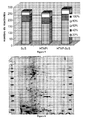
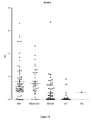
- the inventors have thus been able to demonstrate numerous IgG reactivities, some of which are very intense with all the sera of patients tested in one-dimensional immunoblotting, whereas the healthy subjects expressed almost no IgG reactivity.
- the inventors have characterized, in two-dimensional immunoblotting, several protein spots recognized by at least 80% of the IgGs of the pool of sera of a given group of patients, and not by the serum IgGs of a group of patients. pool of healthy subjects and other protein spots recognized by the vast majority of serum IgG pool of patients with greater intensity than that of serum IgG pool of healthy subjects.
- biological sample refers to any biological sample from a patient.
- Sample samples include body fluids and tissue biopsies. Fibroblasts of scleroderma patients cultured from skin biopsies are also an example of a biological sample.
- the sample may be blood, serum, saliva, urine, sperm. More preferably, the biological sample is a sample of blood or serum.
- the term "patient” refers to any subject that can be tested. Preferably it is a human, but the term includes any other mammal, such as dogs, cats, rodents, cattle, horses, monkeys etc.
- the patient can be tested regardless of gender or age.
- the patient may be at risk, asymptomatic, or have early or advanced signs of SSc and / or PAH.
- the patient may be a subject predisposed to developing SCS and / or PAH, particularly a subject carrying one or more mutations in the gene encoding BMPRII, endoglin or ALK1.
- diagnosis means the identification of the pathology or the evaluation of the state of severity of the pathology.
- prognosis means the assessment of the risk of aggravation and its consequences.
- control value refers to a baseline value corresponding to the average of values obtained with the biological sample of healthy subjects, unaffected by SSc or PAH or a disease likely to result in PAH. It can be a reference statistical value.
- the results of the second test are compared with the results of the first test, as well as often with the so-called "control" value.
- An amount of antibody "greater than the control value” generally means a statistically significant increase, for example, of at least two standard deviations above the average optical densities of the IgG reactivities of all healthy subjects.
- capture antigen an antigen, preferably fixed on a solid phase, which is capable of retaining said at least one antibody present in a biological sample by affine binding.
- the capture antigen can be labeled.
- labeled refers both to direct labeling (via enzymes, radioisotopes, fluorochromes, luminescent compounds, etc.) to indirect labeling (for example, by means of antibodies to them). They are directly labeled or with reagents of a labeled "affinity pair", such as, but not limited to, the labeled avidin-biotin pair, etc.).
- the inventors have identified several anti-VSMC antibodies in patients with SS with or without PAH, or with HTAPi.
- Detection and / or quantification of these antibodies can be implemented to detect an SCS and / or a PAH, to make the prognosis or the follow-up of these pathologies, or to evaluate the effectiveness of a treatment towards these pathologies.
- the antigens recognized by the identified antibodies are listed below.
- the names and access numbers for these antigens in the SWISSPROT Protein Sequence Database are given in Tables 1 and 2 of the "Examples" section.
- the inventors have characterized several reactivities against VSMCs in the sera of patients who are not found in the sera of healthy subjects.
- the antibodies identified are anti-precursor antibodies of the glucose-regulated 78 kDa protein, anti-caldesmone, anti-FAM10A4 protein, anti-zyxin, anti-galectin 1, anti-protein A3 precursor protein disulfide-isomerase, anti -desmin, anti-peripherin, heterogeneous nuclear anti-ribonucleoprotein H, anti-beta chain of the tubulin and anti-polymerase I and transcript release factor.
- the method described in this document comprises the determination of the presence of at least one antibody selected from the group consisting of anti-precursor antibodies of the 78 kDa protein regulated by glucose, anti-caldesmone, anti-FAM10A4 protein, anti-zyxin, anti-galectin 1, anti-precursor A3 of disulfide-isomerase protein, anti-desmin, anti-peripherin, heterogeneous nuclear anti-ribonucleoprotein H, anti-beta tubulin and anti- polymerase I and transcript release factor in a biological sample from a patient, the presence of said at least one antibody being indicative of systemic scleroderma and / or pulmonary arterial hypertension or a risk of developing systemic sclerosis and or pulmonary arterial hypertension.
- at least one antibody selected from the group consisting of anti-precursor antibodies of the 78 kDa protein regulated by glucose, anti-caldesmone, anti-FAM10A4 protein, anti-zyxin, anti-galect
- the inventors have also characterized several reactivities that have a significantly higher intensity in patients than in healthy subjects. These reactivities correspond to antibodies anti-vimentin, anti-phosphoprotein 1 induced by stress, anti- ⁇ -enolase, anti-triphosphate isomerase, anti-precursor of serum albumin, anti-Isozyme L1 carboxyl-terminal hydrolase ubiquitin, anti cytoplasmic actin 2, anti-peroxiredoxin-6, anti-Far-upstream element-binding protein 2 (or anti-protein 2) anti-precursor of reticulocabin-1, anti- ⁇ -enolase, anti-mitochondrial precursor of peroxide Thioredoxin-dependent reductase, anti-Ran-specific GTPase activation protein and anti-High mobility group protein B1.
- the antibodies corresponding to the reactivities which have a greater intensity in the patients than in the healthy subjects are used in a method described in this document comprising the comparison of the amount of at least one chosen antibody.
- control value being indicative of systemic scleroderma and / or pulmonary arterial hypertension or a risk of developing systemic scleroderma and / or pulmonary arterial hypertension.
- the invention therefore relates to the use of at least one antibody directed against VSMCs, in a method, preferably in vitro, for detecting an SCS and / or a PAH.
- the antibodies directed against the VSMCs used in the processes described are chosen from the group consisting of the anti-precursor antibodies of serum albumin, anti-zyxin, anti-Galectin-1, anti-Isozyme L1 carboxyl-terminal.
- ubiquitin hydrolase anti-caldesmone, anti-FAM10A4 protein, anti-Far-upstream element-binding protein 2, anti-cytoplasmic actin 2, anti- ⁇ -enolase, anti-precursor A3 of disulfide-isomerase protein, anti-desmin, anti-peripherin, heterogeneous nuclear anti-ribonucleoprotein H, stress-induced anti-phosphoprotein 1, anti-triphosphate isomerase, anti-peroxiredoxin-6, anti-reticulocabin-1, anti-peroxyredoxin-2, mitochondrial peroxide precursor Thioredoxin-dependent reductase, anti-Ran-specific GTPase activation protein, anti-High mobility group protein B1, anti-beta chain of tubulin and anti-polymerase I and transcript release factor.
- the antibodies directed against the CLMVs used in the processes described are chosen from the group consisting of anti-precursor antibodies to serum albumin, anti-zyxin, anti-Galectin-1, anti-Isozyme Ubiquitin carboxyl-terminal hydrolase, anti-caldesmone, anti-FAM10A4 protein, anti-Far-upstream element-binding protein 2, cytoplasmic anti-actin 2, anti- ⁇ -enolase, anti-A3 precursor of disulfide-isomerase protein, anti-desmin anti-peripherin, heterogeneous nuclear anti-ribonucleoprotein H, stress-induced anti-phosphoprotein 1, anti-triphosphate isomerase, anti-peroxiredoxin-6 and anti-reticulocabin-1.
- the antibodies identified by the inventors can be used in the processes described alone or in combination.
- the detection and / or the quantification can be carried out vis-à-vis only one of the identified antibodies, or may concern a plurality of antibodies.
- a solid support for example a microplate, on which are arranged in a defined and ordered manner the antigens corresponding to the plurality of antibodies to be detected and / or quantified.
- the described methods involve the detection of a stress-induced anti-galactin 1 or anti-phosphoprotein 1 antibody.
- the processes described implement the detection of an anti-galectin 1 antibody for the diagnosis, prognosis or monitoring of SSc.
- an anti-galectin 1 antibody for the diagnosis, prognosis or monitoring of SSc.
- the inventors have been able to show that this antibody is specific for the SCS, having regard to its presence in the sera of SCS patients with or without associated PAH, and its absence in the sera of patients suffering from PAH.
- the described methods involve the detection of an anti-precursor antibody of the glucose-regulated 78 kDa protein for the diagnosis, prognosis or follow-up of SSc or the iPAH.
- the methods described implement the detection of an anti-FAM10A4 protein antibody for the diagnosis, prognosis or monitoring of PAH, whether it is idiopathic or associated with a SSc.
- the biological sample is preferably a serum sample, preferably diluted to 1 / 100th, or more, for example to 1 / 200th or 1 / 400th.
- the amount of antibody can be determined by an immunoassay.
- the biological sample may be optionally treated in a previous step, or directly in the presence of at least one capture antigen.
- the process according to the invention can be carried out according to various formats well known to those skilled in the art: in the solid phase or in the homogeneous phase; in a time or in two stages; in competitive method, by way of non-limiting examples.
- the capture antigen is immobilized on a solid phase.
- solid phase microplates, in particular polystyrene microplates, such as those sold by the company Nunc, Denmark, may be used. It is also possible to use solid particles or beads, paramagnetic beads, such as those supplied by Dynal or Merck-Eurolab (France) (under the trademark EstaporTM), or also test tubes made of polystyrene or polypropylene, etc.
- ELISA assays can be used to reveal the presence of the antigen-antibody complexes formed.
- the capture antigen corresponds to an entire protein or to a fragment of said protein.
- the method of the invention comprises contacting a biological sample with an entire protein recognized by the antibody to be detected and / or quantified.
- the invention comprises bringing a sample of blood or serum into contact with galectin 1 or phosphoprotein 1 induced by the entire stress, for detection and / or the quantification of anti-galectin or anti-phosphoprotein 1 antibodies induced by stress in said sample.
- the capture antigen can be coupled to a glutathione S transferase (GST), before being deposited on a microplate.
- GST glutathione S transferase
- the serum samples to be tested for example diluted 1 / 100th, are incubated on the microplate. After washing, labeled anti-human Fc ⁇ antibodies (for example with alkaline phosphatase) are added, the complexes being revealed (for example by adding a phosphatase substrate whose cleavage can be detected by reading the absorbance) .
- labeled anti-human Fc ⁇ antibodies for example with alkaline phosphatase
- the complexes being revealed (for example by adding a phosphatase substrate whose cleavage can be detected by reading the absorbance) .
- the target patients are those likely to develop an SSc and / or a PAH. It may be a patient with PAH associated with connective tissue disease, such as systemic sclerosis, Sharp syndrome (which is a mixed connective tissue disease), or systemic lupus erythematosus. The patient may also have idiopathic or familial PAH. More generally, any patient suffering from pulmonary vascular disease may be advantageously subjected to the method of detecting a PAH as defined in the invention. In addition, the detected PAH may also be pulmonary hypertension (ie PAH associated with portal hypertension), or be associated with congenital heart disease, or infection with HPAI virus.
- connective tissue disease such as systemic sclerosis, Sharp syndrome (which is a mixed connective tissue disease), or systemic lupus erythematosus.
- the patient may also have idiopathic or familial PAH. More generally, any patient suffering from pulmonary vascular disease may be advantageously subjected to the method of detecting a PA
- HAV human immunodeficiency
- Other targeted patients are those exposed to certain appetite suppressants such as fenfluramine, the prescription of which may contribute to the occurrence of PAH.
- Other people likely to benefit from this type of test are those carrying a mutation in the gene coding for BMPRII, endoglin or ALK1, which possibly do not have detectable PAH on ultrasound, so as to detect individuals who may later develop PAH.
- Another object of this document is an in vitro method for evaluating the efficacy of a treatment with a ScS and / or a PAH, comprising the determination of the presence and / or the quantity of at least one antibody such as as defined above in a biological sample from a patient, at different times before, during or after treatment, the decrease in the amount of said at least one antibody over time being indicative of an improvement in the SSc or PAH.
- the current standard therapy for PAH combines symptomatic treatment with vasodilator therapy. Symptomatic treatment combines anti-coagulants, oxygen therapy and diuretics.
- the vasodilator treatment is based on the following molecules: calcium channel blockers, intravenous epoprostenol (prostacyclin) prescribed by continuous infusion, endothelin receptor inhibitors, selective or not, especially bosentan, sytaxentan and ambrysentan , phosphodiesterase type 5 inhibitors, in particular sildenafil, taladafil, all of these drugs being administered orally, and inhaled iloprost, a prostacyclin analogue administered by inhalation.
- prostacyclin intravenous epoprostenol
- endothelin receptor inhibitors selective or not, especially bosentan, sytaxentan and ambrysentan
- phosphodiesterase type 5 inhibitors in particular sildenafil, taladafil, all of these drugs being administered orally
- inhaled iloprost a prostacyclin analogue administered by inhalation.
- vasodilators primarily calcium channel blockers for the treatment of Raynaud's phenomenon, proton pump inhibitors and a prokinetics, domperidone, for the treatment of gastroesophageal reflux disease.
- the other treatments depend on the patient's symptoms: colchicine or low-dose corticosteroids in case of inflammatory joint disease, conversion enzyme inhibitors in case of renal crisis, cyclophosphamide in case of active diffuse infiltrative pneumopathy, vasodilator treatment pulmonary arterial hypertension.
- the inventors used the sera of patients with HTAPi or SCS with or without PAH. PAH was detected by transthoracic echocardiography and confirmed by right catheterization. Scleroderma patients met the criteria of the American Rheumathology Association (ARA) and / or the criteria of Leroy and Medsger. The sera were collected and stored at -80 ° C before use. All patients had signed informed consent in the framework of the study HTAP-Ig (Contract of Investigation and Clinical Research 2005 N ° CRC 05066, promoter Public Assistance-Hospitals of Paris).
- ARA American Rheumathology Association
- Human VSMCs obtained from breast arteries in coronary bypass patients were provided by Dr. Babett Weksler (Cochin Institute, Paris). These cells were immortalized following lentiviral sequential transduction of the catalytic subunit of the human holoenzyme Telomerase Reverse Transcriptase and the SV40 polyomavirus T antigen (Simian Virus 40) in a primary culture of adult VSMC (Weksler et al. 2005). These cells were cultured on 175 cm 2 flasks in Smooth Muscle Cell Growth Medium 2 culture medium (PromoCell, Heidelberg, Germany) supplemented with 5% fetal calf serum decomplemented (FCS), 0.5 ng / ml.
- FCS fetal calf serum decomplemented
- EGF Epithelial Growth Factor
- 2 ng / ml of basic Fibroblast Growth Factor 5 ⁇ g / ml of insulin, 1% of penicillin / streptomycin and 1% of ciprofloxacin.
- EGF Epithelial Growth Factor
- Human Aortic VSMCs (Cambrex) were used in experiments evaluating the effect of serum and purified IgG from patients with SCS, SCS-PAH, or HTAPi on contraction of a collagen matrix by VSMCs not activated or activated by TNF- ⁇
- the confluent VSMC were peeled off with trypsin, washed with phosphate buffered saline (PBS) and centrifuged at 1600 rpm at 20 ° C. The cell pellet was then recovered in a buffer containing 2% of sodium dodecyl sulfate (SDS), 62.5 mM Tris pH 6.8, 5% ⁇ -mercaptoethanol in the presence of protease inhibitors: 1 ⁇ g / ml of pepstatin, aprotinin, leupeptin and 1 mM phenylmethylsulphonyl fluoride (PMSF). The mixture was then sonicated 4 times dry in ice at 4 ° C at a power of 25 W and then heated for 10 min at 100 ° C. The protein extracts were then aliquoted and stored at -80 ° C until use.
- SDS sodium dodecyl sulfate
- PMSF phenylmethylsulphonyl fluoride
- the confluent VSMCs were washed 2 times in PBS free of Mg 2+ and Ca 2+ , and then peeled off and recovered in isotonic solution in the absence of enzyme containing chelating agents such as EDTA (Cell Dissociation Buffer). PBS-based free enzyme, Invitrogen, Carlsbad, CA, USA (US)). The cells were harvested and then centrifuged for 5 min at 1300 rpm at 20 ° C. After washing in isotonic NaCl solution, a second centrifugation was performed. After repeating this operation a second time, the cell pellet was frozen at -80 ° C in the presence of 1 mM PMSF and a protease inhibitor cocktail (Complete Mini, Roche Diagnostic, Meylan, France).
- the proteins were extracted after three sonications of 30 s each at 4 ° C in a buffer composed of 5M urea, 2M thiourea, 3 - [(3-cholamidopropyl) dimethylammonio] -1-propane sulfonate ( CHAPS) 2%, 40mM Tris and Bio-Lyte 3/10 0.2% ampholytes (ReadyPrep Sequential Extraction Reagent 3, Bio-Rad, Hercules, CA, USA). Two ultracentrifugations at 150,000 g for 25 min each were then performed at 4 ° C (Optima LE-80K, Beckman, Fullerton, CA, USA).
- the proteins in the sample were separated according to their molecular weight (MW) on denaturing polyacrylamide gels in the presence of 10% acrylamide (SDS-PAGE) SDS-PAGE (10% acrylamide, 0.27% bis acrylamide, 0.375 M Tris / HCl pH 8.8, 0.1% SDS, 0.1% Ammonium Persulfate, 0.04% Tetramethylethylenediamine (TEMED) (Biorad, Hercules, CA, USA)).
- SDS-PAGE 10% acrylamide
- SDS-PAGE SDS-PAGE (10% acrylamide, 0.27% bis acrylamide, 0.375 M Tris / HCl pH 8.8, 0.1% SDS, 0.1% Ammonium Persulfate, 0.04% Tetramethylethylenediamine (TEMED) (Biorad, Hercules, CA, USA)
- TEMED Tetramethylethylenediamine
- One hundred and twenty microliters of protein were deposited at the top of each gel and the migration was carried out in a migration buffer (
- the proteins thus separated were transferred from the gel to a nitrocellulose membrane (Immunetics Inc., Boston, Mass., USA) using a semi dry electrotransfer module (Semi Dry Electroblotter A ANCOS, Hoejby, Denmark) for 1 hour at 50 minutes. mA per transfer module.
- the membranes were then blocked for 1 hour in 0.2% PBS Tween 20 (Sigma) and incubated overnight in the presence of sera belonging to one of the following three groups: ScS, whether or not associated with a PAH, HTAPi.
- Sera from 12 healthy subjects were used as controls and PBS-0.2% Tween alone without Ac had been used as a negative control.
- Each patient serum was diluted to 1 ⁇ 2 in PBS-Tween 0.2% and sera from healthy subjects were diluted 1/100.
- IEF allows the separation of proteins according to their isoelectric pH (pHi).
- This step was carried out in immobilized pH gradient (IPG), that is to say that it had operated on an acrylamide gel cast on a rigid strip (strip) in which a pH gradient had been preformed here, a gradient of 3 to 10 (ReadyStrip 17cm, pH 3-10, Bio-Rad).
- the strips were placed in a Bio-Rad horizontal cell of Protean IEF cell type at room temperature. Each strip was placed in a groove in the presence of a mixture containing rehydration buffer and 100 .mu.g of CMLV protein extracts; the whole was covered with 2 ml of mineral oil to limit evaporation.
- the rehydration buffer consisted of 7M ultra-pure urea (VWR, Fontenay-Sous-Bois, France), 2M thiourea (Sigma), CHAPS 4% (Sigma), 0.002% triton X100 (Sigma), DTT (Sigma) bromophenol blue, and Pharmalytes 3-10 ampholytes (Amersham Biosciences, Uppsala, Sweden).
- the IEF included a passive hydration of 9 h followed by an active hydration of 12 h strips at a voltage of 50 V. Then, the IEF was performed as follows: 1 h at 200 V (elimination of excess salts) , then linear rise of the voltage for 1 h up to 1000 V, then for 6 h up to 10 000 V, then for 1 h up to 10 000 V.
- This second step allowed the separation of proteins according to their PM. Twelve 20x20x0.1 cm gels with an acrylamide gradient of 7 to 18.5% were simultaneously cast in a multi-gel chamber (Protean Plus Multi-Casting Chamber, Bio-Rad) ensuring optimal reproducibility and allowing the separation of proteins having a MW of between 10 and 250 kDa.
- the strips obtained at the end of the previous step were placed in the presence of 2 equilibration buffers in order to reduce and alkalinize the sulfhydryl groups of the cysteines.
- the first equilibration buffer consisted of 50 mM Tris, 6 mM urea, 40% glycerol, 52 mM SDS and 32.4 mM DTT.
- the second buffer consisted of 50mM Tris, 6mM urea, 40% glycerol, 52mM SDS and 86.5mM iodoacetamide. Then, the strips were kept in contact with the acrylamide gels in a 1% agarose solution (Agarose Low Melting Point Ultrapure, Gibco BRL Invitrogen) containing bromophenol blue to follow the migration front. PM markers had been placed on both sides of the strip. The migration lasted for approximately 30 h in a migration buffer (25 mM Tris, 192 mM glycine, 3.5 mM SDS, 1.25 mM sodium thiosulfate (Sigma)) maintained at 10 ° C.
- a migration buffer 25 mM Tris, 192 mM glycine, 3.5 mM SDS, 1.25 mM sodium thiosulfate (Sigma) maintained at 10 ° C.
- PVDF PolyVinylidene Fluoride
- the semi-dry transfer was carried out at 4 ° C for 1h30 at constant amperage (320mA).
- the membranes were immersed for 5 min in a PBS solution composed of 148 mM NaCl, 3.5 mM NaH 2 PO 4 , 2H 2 O, 17.6 mM Na 2 HPO 4 , 12H 2 O, and then dried.
- the untransferred gel (also referred to as reference gel) was stained with silver nitrate (Rabilloud et al., 1990) in 5 steps; fixation (30% absolute ethanol, 5% acid acetic acid), washing (11.8 mM silver nitrate (Sigma), 3.45 mM formaldehyde (Sigma), staining (0.02% silver nitrate) and finally revealing (37% formaldehyde, sodium carbonate, thiosulfate).
- the gel was then stored in preservative solution (dimethylsulfoxide 2% (Sigma), 10% acetic acid) and scanned with a densitometer (GS-800, Bio-Rad).
- the PVDF membranes initially blocked with 0.2% PBS-Tween were incubated overnight in the presence of sera from patients belonging to one of the three previously described groups: HTAPi, HTAP-ScS or ScS without PAH.
- HTAPi HTAPi
- HTAP-ScS HTAP-ScS
- ScS ScS without PAH.
- a pool of three sera belonging to the same group diluted 1 / 100th in a solution of PBS-Tween 0.2% was used.
- a membrane was incubated with a pool of 12 sera of healthy subjects diluted in the same 1 / 100th buffer.
- the computer analysis of the gels and membranes was carried out using software designed specifically for the analysis of two-dimensional gels (Image Master 2D® Platinum 6.0, Buckinghamshire, England).
- the first step consisted of an automatic detection of the protein spots according to the parameters we had chosen (number of smoothings carried out to eliminate the background noise, Laplacian threshold and minimal surface of the spots to be detected). Detected spots were visually controlled using 3-dimensional reconstruction methods to eliminate false positives and to see reactivity spots not detected by the software.
- each protein spot recognized by the IgGs of an individual was paired with the corresponding protein using densitometric photography of the same membrane made after colloidal gold staining. This step was performed for each of the 16 membranes.
- proteins transferred to the membranes were paired with the proteins of the gel chosen as reference gel. This made it possible to collect all the information on the reference gel and to then be able to compare the protein spots recognized by the healthy subjects and the patients within the different groups studied.
- Protein spots recognized as antigenic targets were extracted by coring a new acrylamide gel loaded with 400 ⁇ g of protein extracts and stained with Coomassie blue. Each core spot was placed in the well of a 96-well plate and digested in the presence of trypsin (Promega, France) overnight. The digested samples were then transferred to another 96-well plate subsequently stored at 4 ° C prior to the Matrix-Assisted Laser Desorption Ionization-Time-of-Flight (MALDI-TOF) mass spectrometry analysis ( PerSeptive Biosystems Framingham, MA, USA.
- MALDI-TOF Matrix-Assisted Laser Desorption Ionization-Time-of-Flight
- Stress-induced phosphoprotein 1 was obtained from Tebu-bio (Tebu-bio, Maryland, USA), diluted in bicarbonate buffer and deposited on 96-well plates (Maxisorb, NalgeNunc Int Rochester , NY, USA) at a final concentration of 3 ⁇ g / mL at 4 ° C.
- the reactivity of serum IgG obtained from 75 scleroderma patients without PAH, 74 with HTAPi, 37 scleroderma patients with PAH (ScS-PAH), and 70 healthy subjects (HC) were tested by ELISA against STIP-1.
- the plates were washed as mentioned above, and anti-human Fc ⁇ rabbit antibody conjugated to alkaline phosphatase (Tebu-bio, Maryland, USA; diluted 1/1000), with donkey anti-antibody rabbit IgG (Jackson ImmunoResearch, West Baltimore Pike, USA; diluted 1/10000 th) were incubated for one hour at room temperature.
- the reactivities were revealed by addition of p-nitrophenylphosphate (Sigma-Aldrich, St. Louis, USA) and the absorbance (OD) at 405 nm was measured.
- Optical density background noise (wells coated with bicarbonate buffer only) was removed of the value of OD obtained with the proteins. The samples were considered positive when the optical density was greater than or equal to the mean + 2 standard deviations of the control (2et).
- the inventors used the sera of 10 scleroderma patients without PAH (hereinafter referred to as SCS), 10 scleroderma patients with PAH (ScS-PAH), and 10 patients with PAHi. Ten healthy matched subjects for sex and age were also tested. Scleroderma patients meet the criteria of the American Rheumatology Association (ARA) and / or the criteria of Leroy and Medsger. The sera were stored at -80 ° C before use. All patients and healthy subjects have signed an informed consent in the framework of the study HTAP-Ig (Contract of Investigation and Clinical Research 2005 N ° CR C 05066, promoter Assistance Publique-Hôpitaux de Paris, coordinating investigator Luc Mouthon URC Cochin Management Center).
- ARA American Rheumatology Association
- IgG was purified from the serum of patients and healthy subjects on a protein G sepharose column. Purified IgG were quantified by spectrophotometry at 260 and 280 nm. The purity of the purified IgG preparations has been attested by SDS-PAGE.
- Human VSMCs obtained from mammary arteries in immortalized coronary artery bypass patients after lentiviral sequential transduction of the catalytic subunit of human holoenzyme Telomerase Reverse Transcriptase (hTERT) and polyomavirus T antigen SV40 (Simian Virus 40) in a primary culture of adult VSMC (Weksler et al., 2005) were provided by Dr. Babett Weksler (Cochin Institute, Paris).
- hTERT human holoenzyme Telomerase Reverse Transcriptase
- polyomavirus T antigen SV40 Seimian Virus 40
- DMEM culture medium Gibco BRL Invitrogen TM Cergy Pontoise, France
- FCS fetal calf serum
- Aortic human VSMCs (PromoCell, Heidelberg, Germany) were cultured in Smooth Muscle Cell Growth Medium 2 (PromoCell, Heidelberg, Germany) supplemented with 5% decomplemented FCS, 0.5 ng / ml Epithelial Growth Factor, (EGF), 2 ng / ml basic Fibroblast Growth Factor (bFGF), 5 ⁇ g / ml insulin, 1 penicillin / streptomycin and 1% ciprofloxacin.
- EGF Epithelial Growth Factor
- bFGF basic Fibroblast Growth Factor
- the VSMCs were harvested with trypsin 0.25% 1mM EDTA (Gibco BRL Invitrogen TM Cergy Pontoise, France) neutralized with 5% FCS.
- the collagen matrices were prepared in 35mm dishes with 1 ml of medium without FCS containing 500,000 VSMC, 1.65 ml of 1% serum medium (FCS, patient serum or healthy serum) or 128 ⁇ g / ml of purified IgG. and 1 ml of collagen 3.35 mg / ml (BD Biosciences, Franklin Lakes, USA).
- FCS 1% serum medium
- IgG purified IgG.
- ml of collagen 3.35 mg / ml BD Biosciences, Franklin Lakes, USA.
- the matrices were peeled by gently tapping on the edges of the box to initiate contraction.
- photographs were taken at D2 and D4 to measure the surface of the matrix.
- the results obtained with sera from healthy subjects and patients were compared. Twenty sera were tested in each experiment; the different contraction tests were calibrated using SVF and a reference serum used in duplicate. This control made it possible to verify the good reproducibility of the test. Measurements were made using Image J software (NIH National Institute of Health, USA).
- the inventors separately tested the IgG reactivities of the sera of 15 patients in each group and of 15 healthy subjects in 1D immunoblot at a dilution of 1/100. They demonstrated numerous reactivities with patient sera (ScS, HTAP-ScS, HTAPi), some of which were very intense compared with the sera of healthy subjects who showed virtually no IgG immunoreactivity band. The number of the reactivity bands was greater in the case of scleroderma patients with or without PAH than in patients with HTAPi. In addition, some bands of reactivities appeared to be specific for a given group of patients, particularly a band at about 90 kDa in some scleroderma patients with or without PAH ( figure 1 ). In order to identify the antigenic targets of the anti-CMVL IgG of the patients, the inventors then performed 2D immunoblots.
- the inventors After having migrated 100 ⁇ g of VSMC protein extracts prepared as described in the Materials and Methods section, the inventors were able to separate and then stain with silver nitrate 880 protein spots and obtain the gel represented in FIG. figure 2 .
- the inventors were able to estimate the PM and the pHi of each of these protein tasks after computer analysis according to their arrangement in the gel and using PM markers; they had a PM of between 10 and 125 kDa and a pHi between 3 and 8.
- Protein spots common to the 5 pools of PAH patients without PAH were also all recognized by some pools of patients with PAH and PAH-SS. Of the 18 protein spots recognized by 4/5 of the SCS patient pools, 9 were also recognized by at least 3/5 of the patients' pools. each of the other two groups of patients (3 of which were not recognized by healthy subjects), 5 were recognized by at least 3/5 of the pools of PAH-SCS patients (one of which was not recognized by healthy subjects), and 3 were recognized by at least 3/5 pools of HTAPi patients (of which 2 not recognized by healthy subjects).
- One stain (5325) was recognized by a single pool of HTAP-ScS patients, no pool of HTAPi patients, and no pool of healthy subjects.
- IgG from pools of 3 sera from patients with PAH-SSC recognized on average 145 ⁇ 48 protein spots ( figure 4 ).
- a total of 264 different protein spots were recognized by serum IgG in these patients, 77% of which were not recognized by IgG in healthy subjects.
- Protein spots common to the pools of HTAP-ScS patients were also predominantly recognized by patients with HTAPi and PAH-SS. Specifically, 16 spots were recognized by at least 3/5 of the pools of patients of the other two groups (of which only 1 not recognized by healthy subjects), 3 were recognized by at least 3/5 pools of patients HTAPi (including 1 alone not recognized by healthy subjects) and 1 stain was recognized by at least 3/5 of the SCS patient pools.
- the majority of the spots recognized by 4/5 of the pools of PAH-SCS patients were recognized by less than 40% of patients in the other two groups. Specifically, 12 spots were recognized by less than 40% of patients in the other two groups, including 5 spots not recognized by SCS patients (4658, 5206, 4831, 4707, 4659) and 3 spots not recognized by patients with HTAPi (4656 , 5190, 4707). 9 spots were recognized by at least 3/5 pools of patients with SCS and HTAPi (including 2 not recognized by healthy subjects), 5 were recognized by at least 3/5 of HTAPi patients (including 1 not recognized by healthy subjects) and 3 were recognized by at least 3/5 of SCS patients (all 3 spots were also recognized by healthy subjects).
- the IgGs of the pools of 3 sera of patients with HTAPi recognized on average 130 ⁇ 25 protein spots ( figure 3 ).
- a total of 356 protein spots were recognized by IgG in these patients, and 70% were not recognized by IgG in healthy subjects.
- 12 were common to the 5 pools of patients (but all were recognized by healthy subjects), 24 communes to 4/5 pools (7 of which were not recognized by healthy subjects) and 54 to 3/5 of the pools. pools (31 of which are not recognized by healthy subjects) ( figure 4 ).
- Protein spots common to 5 pools of HTAPi patients were also mostly recognized by pools of patients with HTAPi or HTAP-ScS. Specifically, 10 were also recognized by at least 3/5 pools of SCS patients and HTAP-SCS patients, one was also recognized by at least 3/5 pools of SCS patients and another by at least 3/5 of pools of PAH-SCS patients.
- Protein spots recognized by 4/5 pools of sera of patients with HTAPi were mostly shared with the 2 other groups of patients. Specifically, 15 spots were also recognized by at least 3/5 pools of SCS patients and 3/5 pools of PAH-SCS patients (including 4 not recognized by healthy subjects), 3 spots were also recognized by at least 3 Pools of SCS patients (one of which was not recognized by healthy subjects) and one was also recognized by at least 3/5 pools of PAH-SCS patients (and by healthy subjects). 5 spots were recognized by less than 40% of pools of ScS or PAH-SCS patients (including 4 not recognized by healthy subjects). Of these 4 spots, one was not recognized by SCS patients (4735).
- the inventors compared the IgG reactivity profiles of the serum pool of healthy subjects and the pools of patient sera against VSMC proteins. Regardless of the group of patients, most of the protein spots not recognized by IgG from healthy subjects were by a single pool of patients out of 5 ( figure 5 ). By selecting the protein spots recognized by at least 3/5 pools of sera from patients in a given group and not by the pool of healthy subjects, the inventors identified 21 protein spots of interest (Table 1). Although the result of all the digested protein spots has not yet been obtained, 13 interesting protein spots could be identified. The location of these spots Proteins on the reference gel is indicated in the figure 6 .
- Two spots (5190, 5325) appear to be specific for the SCS since they are recognized respectively by 3/5 and 4/5 of the pools of patients ScS and 4/5 and 1/5 of the pools of patients HTAP-ScS, but by none. pool of patients HTAPi.
- One of them (5325) has been identified as galectin.
- the inventors have identified the protein spots of CMLV recognized by IgG pools of 3 patient sera and IgG pool of healthy subjects, provided that these protein spots are recognized with high intensity by IgG from a large number of pools of 3 patient sera and with a higher intensity than healthy subjects. Twenty-seven protein spots met these criteria (Table 2).
- the serum IgG reactivities of the different groups of patients with regard to the protein spots 4576, 4570 and 4576 identified as isoforms of the stress-induced phosphoprotein and with respect to the spot 4738 identified as ⁇ - enolase are represented on the figure 7 .
- the selected region is represented on the figure 6 .
- the inventors have found that 56/75 (74.6%) scleroderma patients, 24/74 (32.4%) patients with HTAPi, 27/37 (73%) patients with PAH-SSc and 2/70 (2.8%) healthy subjects had anti-STIP1 antibodies. Nearly three-quarters of scleroderma patients, whether or not they had PAH and almost a third of patients with PAHi, had anti-STIP1 antibodies, whereas these antibodies were generally absent in healthy subjects.
- the inventors have determined whether serum and / or serum IgG from patients with SCS and / or PAH have an effect on the contraction of VSMCs and fibroblasts, a phenomenon involved in vascular remodeling and cell mobility.
- the cells were seeded in a collagen matrix, and incubated in the presence of 1% of FCS, sera, or purified IgG of patients having a SCS and / or a PAH versus those of healthy subjects.
- the quantifiable retraction of the collagen matrix reflects the contractile activity of the cells.
- the experiment was performed from healthy and unactivated cells.
- the contraction kinetics of the collagen matrices was monitored for 4 days for VSMCs and 7 days for fibroblasts.
- the results obtained with VSMCs are presented in the figure 9 .
- the average area of the matrices (in% of the initial surface) incubated with serum was 27.8% ⁇ 6.0 for SSc, 31.1% ⁇ 8.3 for HTAPi, 29, 4% ⁇ 4.7 for ScS-PAHs, and 34.3% ⁇ 7.1 for healthy subjects.
- the differences between the surfaces obtained in the presence of the other two groups of patient sera (HTAPi, ScS-PAH) and the serum group of healthy subjects were not significant.
- the average area of the matrices (in% of the initial surface) incubated with purified IgG was 53.9% ⁇ 8.2 for SSc, 48.0% ⁇ 3.2 for HTAPi, 55 , 8% ⁇ 8.9 for ScS-PAH, and 34.3% ⁇ 8.6 for healthy subjects.
- p 0.001
- the inventors submitted samples of patients with SSc, PAH-SCS or HTAPi, as well as samples from healthy subjects, to another analysis of their reactivities in order to refine the results obtained and identify other anti-VSMC antibodies.
- Example 1 the results presented in Example 1 could be refined.
- the inventors have thus been able to show that the anti-cytoplasmic actin antibodies 2 are present in the pools of patients and healthy subjects, but significantly higher in the patients. They also showed the existence of reactivity against galectin 1 and zyxin in IgG pools of patients, and not in IgG pool of healthy subjects.
Landscapes
- Health & Medical Sciences (AREA)
- Life Sciences & Earth Sciences (AREA)
- Immunology (AREA)
- Engineering & Computer Science (AREA)
- Hematology (AREA)
- Urology & Nephrology (AREA)
- Molecular Biology (AREA)
- Biomedical Technology (AREA)
- Chemical & Material Sciences (AREA)
- Food Science & Technology (AREA)
- Physics & Mathematics (AREA)
- Cell Biology (AREA)
- Pathology (AREA)
- Biotechnology (AREA)
- General Physics & Mathematics (AREA)
- Medicinal Chemistry (AREA)
- Microbiology (AREA)
- Analytical Chemistry (AREA)
- Biochemistry (AREA)
- General Health & Medical Sciences (AREA)
- Rheumatology (AREA)
- Rehabilitation Therapy (AREA)
- Proteomics, Peptides & Aminoacids (AREA)
- Peptides Or Proteins (AREA)
- Measuring Or Testing Involving Enzymes Or Micro-Organisms (AREA)
Description
- L'invention concerne un procédé in vitro de détection d'une sclérodermie systémique (ScS) ou d'une hypertension artérielle pulmonaire associée à une sclérodermie (HTAP-ScS) ou d'une hypertension artérielle pulmonaire idiopathique (HTAPi) chez un sujet, comprenant la détermination de la présence et/ou de la quantité d'anticorps dans un échantillon biologique provenant d'un patient comme décrit dans les revendications.
- La sclérodermie systémique (ScS) est une maladie rare qui se caractérise par la survenue de lésions de fibrose intéressant la peau et certains viscères comme le poumon, le tube digestif et le coeur, ainsi qu'une hyperréactivité vasculaire à l'origine d'un phénomène de Raynaud et de manifestations sévères comme la crise rénale et l'hypertension artérielle pulmonaire (HTAP). La physiologie de la ScS est complexe et partiellement comprise. La survenue d'une ScS résulte du dysfonctionnement de trois types cellulaires, les lymphocytes B et T responsables d'une dérégulation immunitaire, les cellules endothéliales responsables d'anomalies vasculaires et les fibroblastes responsables de lésions de fibrose.
- Quatre-vingt dix pour cent des patients sclérodermiques ont des anticorps antinucléaire (AAN) dans leur sérum. Certains auto-anticorps sont très spécifiques de la ScS et mutuellement exclusifs, comme les anticorps anti-topoisomérase I (anti-SCI-70) (ATA) (Tamby et al., 2007), plus fréquemment présents dans les formes diffuses de la maladie, les anticorps anti-centromère (ACA), associés en règle aux formes cutanées limitées (Moroi et al., 1980), ou les anticorps anti-ARN polymérase III associés aux formes cutanées diffuses et à la survenue d'une crise rénale (Bunn et al., 1998). Les AAN n'ont pas de rôle pathogène démontré au cours de la ScS mais leur détection constitue une aide au diagnostic précoce de la ScS. D'autres auto-anticorps non spécifiques de la ScS comme les anticorps anti-ribonucléoprotéine, anti-SSA et anti-SSB, les anticorps anti-cardiolipine ou le facteur rhumatoïde sont quelquefois trouvés au cours de la ScS. Des anticorps anti-cellules endothéliales (AECA) peuvent être détectés chez 28 à 54 % des patients sclérodermiques. Ces autoanticorps sont capables d'induire l'expression de molécules d'adhésion et de provoquer une apoptose des cellules endothéliales en présence de cellules tueuses naturelles (« natural killer ») (Bordron et al., 1998). Les cibles des AECA au cours de la ScS sont aujourd'hui mal connues et à ce jour il n'a pas été identifié d'antigène spécifique de la cellule endothéliale. En revanche, l'ADN topoisomérase 1 (Garcia de la Pena-Lefebvre etal., 2004) et la protéine centromérique B (Servettaz et al., 2006) ont été identifiées comme des cibles des AECA chez les malades sclérodermiques. Des anticorps anti-fibroblastes (AFA) ont été identifiés chez les patients sclérodermiques. Ces anticorps sont capables d'activer les fibroblastes, d'augmenter l'expression de molécules d'adhésion comme l'inter-cellular adhésion molecule-1 (ICAM) et pro-inflammatoires (augmentation des niveaux d'ARNm, d'IL-1α, d'IL-1β et d'IL-6) ainsi que la synthèse de collagène (Chizzolini et al., 2002). Il a récemment été mis en évidence que les AFA pouvaient se fixer sur la topoisomérase 1 adsorbée à la surface des fibroblastes (Henault et al., 2006). En ELISA (Enzyme-linked immunosorbent assay), des AFA ont été retrouvés chez 30 % des patients présentant une HTAP associée à une ScS (Tamby et al., 2006).
- L'hypertension artérielle pulmonaire (HTAP) est une pathologie rare responsable de la survenue d'une décompensation cardiaque droite pouvant aboutir au décès. Le diagnostic d'HTAP est établi par un cathétérisme droit qui permet la mesure de la pression artérielle pulmonaire moyenne supérieure ou égale à 25mmHg au repos en l'absence d'élévation de la pression capillaire pulmonaire (Rubin, 1997). La survenue d'une HTAP est la conséquence d'une obstruction chronique des petites artères pulmonaires secondaire à la prolifération de cellules endothéliales, de cellules musculaires lisses vasculaires et de fibroblastes (Dorfmuller et al, 2003). En particulier, la néo-muscularisation des petites artères pulmonaires périphériques normalement non musculaires est une caractéristique commune à toutes les formes de remodelage associées à l'HTAP.
- L'HTAP peut être idiopathique, c'est à dire sporadique, mais aussi familiale, associée à la prise d'anorexigènes (dexfenfluramine), ou associée à un certain nombre de pathologies comprenant l'infection par le virus de l'immunodéficience humaine (VIH). L'HTAP peut aussi être associée à des collagénoses comme la ScS (Hachulla et al, 2005), le syndrome de Sharp (ou connectivites mixte), ou plus rarement le lupus érythémateux systémique. Approximativement 8 à 12 % des patients sclérodermiques développent une HTAP, à l'origine d'une mortalité élevée. De plus, au cours de l'HTAP idiopathique, on trouve de temps à autres des stigmates d'auto-immunité, à savoir des anticorps anti-nucléaires ou des anticorps anti-thyroglobuline. La présence d'anticorps anti-cellules endothéliales (Tamby et al, 2005) et d'anticorps anti-fibroblastes (Tamby et al, 2006) a été rapportée au cours de l'HTAP idiopathique ou associée à la ScS. Cependant la valeur prédictive de ces anticorps dans la survenue de l'HTAP n'a pas été étudiée et le rôle potentiel des phénomènes autoimmuns dans la pathogénie de l'HTAP idiopathique reste incertain (Mouthon et al, 2005).
- Dans la plupart des cas, l'HTAP est dépistée lorsque le patient présente une dyspnée de stade III ou IV. Lorsque le patient est suivi pour une maladie chronique comme la ScS, l'HTAP est dépistée par une échographie cardiaque annuelle.
- La demande de brevet
US 2002/0009749 décrit la détection d'anticorps anti-HMG-1 (high mobility group protein 1) et anti-HMG-2 pour le diagnostic de maladies auto-immunes, entre autres la sclérodermie systémique. Terrier et al. ont identifié les antigènes d'anticorps anti-fibroblastes présents chez des patients atteints d'hypertension artérielle pulmonaire (Terrier et al., 2008). - Un test simple et fiable de dépistage d'une ScS et/ou d'une HTAP fait toujours défaut, et serait précieux pour un diagnostic le plus précoce possible, qui permettrait de mettre en place rapidement des stratégies thérapeutiques pour améliorer l'état du patient et ses chances de survie. L'invention a pour objet un tel test.
- L'invention fournit maintenant un procédé in vitro de détection d'une sclérodermie systémique (ScS) ou d'une hypertension artérielle pulmonaire associée à une sclérodermie (HTAP-ScS) ou d'une hypertension artérielle pulmonaire idiopathique (HTAPi) chez un sujet, comprenant la détermination de la présence et/ou de la quantité d'au moins un anticorps choisi dans le groupe constitué des anticorps anti-Galectine-1, anti-protéine FAM10A4, anti-phosphoprotéine 1 induite par le stress, et anti-précurseur de la protéine de 78 kDa régulée par le glucose, dans un échantillon biologique provenant d'un patient,
la présence d'un anticorps anti-précurseur de la protéine de 78 kDa régulée par le glucose étant indicative d'une ScS ou d'une HTAPi ;
la présence d'un anticorps anti-protéine FAM10A4 étant indicative d'une HTAP-ScS ou d'une HTAPi ;
la présence d'un anticorps anti-galectine-1 étant indicative d'une ScS;
ou la présence en une quantité supérieure à une valeur contrôle d'un anticorps anti-phosphoprotéine 1 induite par le stress étant indicative d'une ScS, d'une HTAP-ScS ou d'une HTAPi. - La sclérodermie systémique (ScS) et l'hypertension artérielle pulmonaire (HTAP) se caractérisent toutes deux par la présence d'autoanticorps (AAc) dans le sérum des patients, en particulier des AAc dirigés contre les cellules endothéliales et les fibroblastes. Avant les travaux présentés ci-dessous, aucun AAc dirigé contre les cellules musculaires lisses vasculaires (CMLV) n'avait été mis en évidence. La caractérisation par les inventeurs de tels anticorps dirigés contre les CMLV est d'une importance capitale, notamment eu égard au rôle clé de ces cellules dans la physiopathologie de la ScS, avec ou sans HTAP, et de l'HTAPi.
- Les inventeurs se sont proposés d'identifier les anticorps dirigés contre les CMLV et d'en caractériser les cibles antigéniques. Pour ce faire, les inventeurs ont mis en oeuvre des CMLV d'artères mammaires internes comme source d'antigènes et testé des sérums de patients de phénotype identique (patients ayant une ScS sans ou avec HTAP, patients ayant une HTAP idiopathique, ou HTAPi, et sujets sains). Les anticorps ont été recherchés à l'aide d'une technique d'immunoblot en une puis deux dimensions suivie d'une identification des antigènes par spectrométrie de masse (voir la partie « exemples » ci-dessous).
- Les inventeurs ont ainsi pu mettre en évidence des réactivités IgG nombreuses dont certaines très intenses avec l'ensemble des sérums de patients testés en immunoblot en une dimension, tandis que les sujets sains n'exprimaient quasiment aucune réactivité IgG. Les inventeurs ont caractérisé, en immunoblot en deux dimensions, plusieurs taches protéiques reconnues par au moins 80 % des IgG des pools de sérums d'un groupe de patients donnés, et non par les IgG sériques d'un pool de sujets sains et d'autres taches protéiques reconnues par la très grande majorité des IgG sériques des pools de malades avec une intensité plus forte que celle des IgG sériques du pool de sujets sains.
- Le terme « échantillon biologique » se réfère à tout échantillon biologique provenant d'un patient. Des exemples d'échantillons incluent des liquides biologiques et des biopsies tissulaires. Les fibroblastes de patients sclérodermiques cultivés à partir de biopsies de peau constituent également un exemple d'échantillon biologique. De manière préférentielle, l'échantillon peut être du sang, du sérum, de la salive, de l'urine, du sperme. De manière davantage préférée, l'échantillon biologique est un échantillon de sang ou de sérum.
- Le terme « patient » se réfère à tout sujet susceptible d'être testé. De préférence il s'agit d'un humain, mais le terme inclut tout autre mammifère, tel que des chiens, chats, rongeurs, bétail, chevaux, singes etc. Le patient peut être testé quel que soit son sexe ou son âge. Le patient peut être un sujet à risque, être asymptomatique ou présenter des signes précoces ou avancés d'une ScS et/ou d'une HTAP. Par exemple le patient peut être un sujet prédisposé à développer une ScS et/ou une HTAP, en particulier un sujet portant une ou plusieurs mutations dans le gène codant pour BMPRII, endogline ou ALK1.
- Le terme « diagnostic » signifie l'identification de la pathologie ou l'évaluation de l'état de sévérité de la pathologie.
- Le terme « pronostic » signifie l'évaluation du risque d'aggravation, et de ses conséquences.
- Le terme « valeur contrôle » se réfère à une valeur basale correspondant à la moyenne des valeurs obtenues avec l'échantillon biologique de sujets sains, non affectés par une ScS ou une HTAP ou une maladie susceptible d'entraîner une HTAP. Il peut s'agir d'une valeur statistique de référence.
- Pour évaluer l'évolution de la pathologie, il peut être utile de tester un patient et de contrôler l'effet d'un traitement ou l'évolution de la pathologie, en testant de nouveau le patient, par exemple à plusieurs mois d'intervalle. Dans ce cas, les résultats du second test sont comparés aux résultats du premier test, ainsi que souvent à la valeur dite « contrôle ».
- Une quantité d'anticorps « supérieure à la valeur contrôle » signifie généralement une augmentation statistiquement significative, par exemple d'au moins deux déviations standards au dessus de la moyenne des densités optiques des réactivités IgG de l'ensemble des sujets sains.
- Par « antigène de capture », on entend un antigène, de préférence fixé sur une phase solide, qui est capable de retenir ledit au moins un anticorps présent dans un échantillon biologique, par liaison affine. L'antigène de capture peut être marqué.
- Le terme « marqué » se réfère aussi bien à un marquage direct (par le biais d'enzymes, radioisotopes, fluorochromes, composés luminescents, etc...) qu'à un marquage indirect (par exemple, par le biais d'anticorps eux-mêmes marqués de manière directe ou à l'aide de réactifs d'une "paire d'affinité " marquée, telle que, mais non exclusivement, la paire avidine marquée-biotine, etc..).
- Comme indiqué dans la partie « exemples », les inventeurs ont identifié plusieurs anticorps anti-CMLV chez des malades ayant une ScS avec ou sans HTAP, ou ayant une HTAPi.
- La détection et/ou la quantification de ces anticorps peut être mise en oeuvre pour détecter une ScS et/ou une HTAP, pour réaliser le pronostic ou le suivi de ces pathologies, ou pour évaluer l'efficacité d'un traitement envers ces pathologies.
- Les antigènes reconnus par les anticorps identifiés sont listés ci-dessous. Le nom et les numéros d'accès correspondant à ces antigènes dans la base de données de séquences protéiques SWISSPROT sont donnés dans les tableaux 1 et 2 de la partie « Exemples ».
- Les inventeurs ont caractérisé plusieurs réactivités contre les CMLV dans les sérums de patients qui ne sont pas retrouvées dans les sérums de sujets sains. Les anticorps identifiés sont des anticorps anti-précurseur de la protéine de 78 kDa régulée par le glucose, anti-caldesmone, anti-protéine FAM10A4, anti-zyxine, anti-galectine 1, anti-précurseur A3 de la protéine disulfide-isomérase, anti-desmine, anti-périphérine, anti-ribonucléoprotéine H nucléaire hétérogène, anti-chaîne bêta de la tubuline et anti-polymerase I and transcript release factor. Dans un mode particulier de réalisation, le procédé décrit dans ce document comprend la détermination de la présence d'au moins un anticorps choisi dans le groupe constitué des anticorps anti-précurseur de la protéine de 78 kDa régulée par le glucose, anti-caldesmone, anti-protéine FAM10A4, anti-zyxine, anti-galectine 1, anti-précurseur A3 de la protéine disulfide-isomérase, anti-desmine, anti-périphérine, anti-ribonucléoprotéine H nucléaire hétérogène, anti-chaîne bêta de la tubuline et anti-polymerase I and transcript release factor dans un échantillon biologique provenant d'un patient, la présence dudit au moins un anticorps étant indicatrice d'une sclérodermie systémique et/ou d'une hypertension artérielle pulmonaire ou d'un risque de développer une sclérodermie systémique et/ou une hypertension artérielle pulmonaire.
- Les inventeurs ont également caractérisé plusieurs réactivités qui ont une intensité significativement plus forte chez les patients que chez les sujets sains. Ces réactivités correspondent à des anticorps anti-vimentine, anti-phosphoprotéine 1 induite par le stress, anti-α-énolase, anti-triosphosphate isomérase, anti-précurseur de l'albumine sérique, anti-Isozyme L1 carboxyl-terminal hydrolase ubiquitine, anti-actine cytoplasmique 2, anti-péroxirédoxine-6, anti-Far-upstream element-binding protein 2 (ou anti-protéine 2) anti-précurseur de la réticulocabine-1, anti-γ-énolase, anti-précurseur mitochondrial de la péroxyde réductase dépendant de la thiorédoxine, anti-protéine d'activation de GTPase spécifique de Ran et anti-High mobility group protein B1. Dans un mode particulier de réalisation, les anticorps correspondant aux réactivités qui ont une intensité plus forte chez les patients que chez les sujets sains sont mis en oeuvre dans un procédé décrit dans ce document comprenant la comparaison de la quantité d'au moins un anticorps choisi dans le groupe constitué des anticorps anti-vimentine, anti-phosphoprotéine 1 induite par le stress, anti-α-énolase, anti-triosphosphate isomérase, anti-précurseur de l'albumine sérique, anti-Isozyme L1 carboxyl-terminal hydrolase ubiquitine, anti-actine cytoplasmique 2, anti-péroxirédoxine-6, anti-Far-upstream element-binding protein 2 (ou anti-protéine 2) anti-précurseur de la réticulocabine-1, anti-γ-énolase, anti-précurseur mitochondrial de la péroxyde réductase dépendant de la thiorédoxine, anti-protéine d'activation de GTPase spécifique de Ran et anti-High mobility group protein B1 dans un échantillon biologique provenant d'un patient à une valeur contrôle, la présence dudit au moins un anticorps en une quantité supérieure à la
- valeur contrôle étant indicatrice d'une sclérodermie systémique et/ou d'une hypertension artérielle pulmonaire ou d'un risque de développer une sclérodermie systémique et/ou une hypertension artérielle pulmonaire.
- L'invention concerne donc la mise en oeuvre d'au moins un anticorps dirigé contre les CMLV, dans un procédé, de préférence in vitro, de détection d'une ScS et/ou d'une HTAP.
- De préférence, les anticorps dirigés contre les CMLV mis en oeuvre dans les procédés décrits sont choisis dans le groupe constitué des anticorps anti-précurseur de l'albumine sérique, anti-zyxine, anti-Galectine-1, anti-Isozyme L1 carboxyl-terminal hydrolase ubiquitine, anti-caldesmone, anti-protéine FAM10A4, anti-Far-upstream element-binding protein 2, anti-actine cytoplasmique 2, anti-γ-énolase, anti-précurseur A3 de la protéine disulfide-isomérase, anti-desmine, anti-périphérine, anti-ribonucléoprotéine H nucléaire hétérogène, anti-phosphoprotéine 1 induite par le stress, anti-triosphosphate isomérase, anti-péroxirédoxine-6, anti-réticulocabine-1, anti-péroxyrédoxine-2, anti-précurseur mitochondrial de la péroxyde réductase dépendant de la thiorédoxine, anti-protéine d'activation de GTPase spécifique de Ran, anti- High mobility group protein B1, anti-chaîne bêta de la tubuline et anti-polymerase I and transcript release factor.
- Dans un mode particulier de réalisation, les anticorps dirigés contre les CLMV mis en oeuvre dans les procédés décrits sont choisis dans le groupe constitué des anticorps anti-précurseur de l'albumine sérique, anti-zyxine, anti-Galectine-1, anti-Isozyme L1 carboxyl-terminal hydrolase ubiquitine, anti-caldesmone, anti-protéine FAM10A4, anti-Far-upstream element-binding protein 2, anti-actine cytoplasmique 2, anti-γ-énolase, anti-précurseur A3 de la protéine disulfide-isomérase, anti-desmine, anti-périphérine, anti-ribonucléoprotéine H nucléaire hétérogène, anti-phosphoprotéine 1 induite par le stress, anti-triosphosphate isomérase, anti-péroxirédoxine-6 et anti-réticulocabine-1.
- Les anticorps identifiés par les inventeurs peuvent être mis en oeuvre dans les procédés décrits seuls ou en combinaison. La détection et/ou la quantification peut être réalisée vis-à-vis d'un seul des anticorps identifiés, ou peut concerner une pluralité d'anticorps. On ainsi peut imaginer la réalisation du procédé sur un support solide, par exemple une microplaque, sur lequel sont disposés de manière définie et ordonnée les antigènes correspondant à la pluralité d'anticorps à détecter et/ou quantifier.
- Selon un mode de réalisation de l'invention, les procédés décrits mettent en oeuvre la détection d'un anticorps anti-galectine 1 ou anti-phosphoprotéine 1 induite par le stress.
- Selon un mode de réalisation de l'invention, les procédés décrits mettent en oeuvre la détection d'un anticorps anti-galectine 1 pour le diagnostic, le pronostic ou le suivi de la ScS. En effet, les inventeurs ont pu montrer que cet anticorps est spécifique de la ScS, eu égard à sa présence dans les sérums de patients ScS avec ou sans HTAP associée, et son absence dans les sérums de patients souffrant d'HTAPi.
- Selon un autre mode de réalisation de l'invention, les procédés décrits mettent en oeuvre la détection d'un anticorps anti-précurseur de la protéine de 78 kDa régulée par le glucose pour le diagnostic, le pronostic ou le suivi de la ScS ou de l'HTAPi.
- Selon un autre mode de réalisation de l'invention, les procédés décrits mettent en oeuvre la détection d'un anticorps anti-protéine FAM10A4 pour le diagnostic, le pronostic ou le suivi de l'HTAP, qu'elle soit idiopatyque ou associée à une ScS.
- L'échantillon biologique est de préférence un échantillon de sérum, de préférence dilué au 1/100ème, ou plus, par exemple au 1/200ème ou 1/400ème.
- De manière avantageuse, la quantité d'anticorps peut être déterminée par un immunoessai.
- L'échantillon biologique peut être éventuellement traité dans une étape préalable, ou mis directement en présence d'au moins un antigène de capture.
- Le procédé selon l'invention peut être réalisé selon divers formats bien connus de l'homme du métier: en phase solide ou en phase homogène; en un temps ou en deux temps; en méthode compétitive, à titre d'exemples non limitatifs.
- Selon un mode de réalisation préféré, l'antigène de capture est immobilisé sur une phase solide. On peut utiliser, à titre d'exemples non limitatifs de phase solide, des microplaques, en particulier des microplaques de polystyrène, telles que celles commercialisées par la société Nunc, Danemark. On peut également utiliser des particules ou des billes solides, des billes paramagnétiques, telles que celles fournies par Dynal ou Merck-Eurolab (France) (sous la marque EstaporTM), ou encore des tubes à essai en polystyrène ou polypropylène, etc.
- Un format d'immunoessai de détection des anticorps par compétition est également possible. D'autres modalités d'immunoessai sont encore envisageables et bien connues de l'homme du métier.
- Dosages ELISA, radioimmunoessais, ou toute autre technique de détection peuvent être mis en oeuvre pour révéler la présence des complexes antigènes-anticorps formés.
- Selon un mode particulier de réalisation préféré, l'antigène de capture correspond à une protéine entière ou à un fragment de ladite protéine. Par exemple, le procédé de l'invention comprend la mise en contact d'un échantillon biologique avec une protéine entière reconnue par l'anticorps à détecter et/ou quantifier. A titre illustratif, l'invention comprend la mise en contact d'un échantillon de sang ou de sérum avec la galectine 1 ou la phosphoprotéine 1 induite par le stress entière, pour la détection et/ou la quantification d'anticorps anti-galectine ou anti-phosphoprotéine 1 induite par le stress dans ledit échantillon.
- Dans un exemple particulier, l'antigène de capture peut être couplé à une glutathion S transférase (GST), avant d'être déposé sur une microplaque.
- A titre illustratif, les échantillons de sérum à tester, par exemple dilués au 1/100ème, sont mis à incuber sur la microplaque. Après lavage, des anticorps anti-Fcγ humain marqués (par exemple avec une phosphatase alcaline) sont ajoutés, les complexes étant révélés (par exemple par ajout d'un substrat de la phosphatase dont le clivage peut être détecté par lecture de l'absorbance).
- Les patients visés sont ceux susceptibles de développer une ScS et/ou une HTAP.
Il peut s'agir d'un patient qui souffre d'une HTAP associée à une connectivite, telle qu'une sclérodermie systémique, au syndrome de Sharp (qui est une connectivite mixte), ou à un lupus érythémateux systémique.
Le patient peut également souffrir d'une HTAP idiopathique ou familiale.
Plus généralement, tout patient atteint d'une maladie vasculaire pulmonaire peut être avantageusement soumis au procédé de détection d'une HTAP tel que défini dans l'invention.
Par ailleurs, l'HTAP détectée peut être aussi une hypertension porto-pulmonaire (c'est-à-dire une HTAP associée à une hypertension portale), ou être associée à une cardiopathie congénitale, ou à une infection par le virus de l'immunodéficience humaine (VIH), ou encore être une hypertension pulmonaire post-embolique, compliquant l'évolution d'une bronchite chronique obstructive ou d'une cardiopathie cyanogène.
D'autres patients visés sont ceux exposés à certains médicaments coupe-faim comme la fenfluramine dont la prescription peut contribuer à la survenue d'une HTAP.
D'autres personnes susceptibles de bénéficier de ce type de test sont celles porteuses d'une mutation dans le gène codant pour BMPRII, endogline ou ALK1, et qui éventuellement ne présentent pas d'HTAP détectable à l'échographie, de façon à dépister les individus susceptibles de développer ultérieurement une HTAP. - Un autre objet de ce document est un procédé in vitro d'évaluation de l'efficacité d'un traitement envers une ScS et/ou une HTAP, comprenant la détermination de la présence et/ou de la quantité d'au moins un anticorps tel que défini ci-dessus dans un échantillon biologique provenant d'un patient, à différents temps avant, au cours ou après le traitement, la diminution de la quantité dudit au moins un anticorps au cours du temps étant indicative d'une amélioration de la ScS ou de l'HTAP.
Le traitement classique actuel de l'HTAP associe un traitement symptomatique et un traitement vasodilatateur. Le traitement symptomatique associe des anti-coagulants, une oxygénothérapie et des diurétiques. Le traitement vasodilatateur repose sur les molécules suivantes : bloqueurs de canaux calcium, époprosténol (prostacycline) prescrit par voie intraveineuse en perfusion continue, les inhibiteurs des récepteurs de l'endothéline, sélectifs ou non, en particulier le bosentan, le sytaxentan et l'ambrysentan, les inhibiteurs des phosphodiestérases de type 5 en particulier le sildénafil, le taladafil, l'ensemble de ces médicaments étant administrés par voie orale, et l'iloprost inhalé, analogue de la prostacycline administré par voie inhalée. Ces traitements peuvent être éventuellement combinés. En cas d'échec de ces thérapeutiques, une transplantation pulmonaire ou coeur-poumons peut être proposée. Au cours de la ScS il est classique de prescrire des vasodilatateurs, en premier lieu des inhibiteurs calciques en traitement du phénomène de Raynaud, des inhibiteurs de la pompe à protons et un prokinétique, la domperidone, en traitement du reflux gastro-oesophagien. Les autres traitements dépendent des atteintes présentées par le patient : colchicine ou corticoides à faible dose en cas d'atteinte articulaire inflammatoire, inhibiteurs de l'enzyme de conversion en cas de crise rénale, cyclophosphamide en cas de pneumopathie infiltrante diffuse évolutive, traitement vasodilatateur d'une hypertension artérielle pulmonaire. - Les figures et exemples suivants illustrent l'invention sans en limiter la portée.
-
- La
figure 1 correspond à un immunoblot à une dimension montrant les réactivités des IgG sériques de patients sclérodermiques avec (n=3) ou sans (n=6) HTAP, de patients ayant une HTAP idiopathique (n=6) et de sujets sains (n=4) vis-à-vis des protéines de cellules musculaires lisses vasculaires. L'HTAP était documentée par cathétérisme droit chez l'ensemble des patients. ScS : sclérodermie systémique ; HTAP : hypertension artérielle pulmonaire ; HTAPi : hypertension artérielle pulmonaire idiopathique ; HTAP-ScS : hypertension artérielle pulmonaire associée à une sclérodermie ; C : contrôle interne (HTAP-ScS) ; PBS : Phosphate Buffer Saline. - La
figure 2 correspond à un gel de référence en deux dimensions d'un extrait protéique total de cellules musculaires lisses vasculaires, coloré au nitrate d'argent. Première dimension (axe horizontal): pH 3-10, seconde dimension (axe vertical): gradient d'acrylamide 7-18 %, permettant le dénombrement de 880 taches protéiques. - La
figure 3 est un graphique montrant le nombre de taches protéiques reconnues par les IgG de 15 pools de 3 sérums de patients phénotypiquement identiques ayant une sclérodermie systémique et/ou une HTAP et par un pool de 12 sujets sains après ajustement sur le gel de référence. L'échelle des ordonnées indique le nombre de réactivités IgG. - La
figure 4 montre la proportion des taches de réactivité reconnues par les IgG des sérums des pools de 3 patients au sein de chaque groupe (reconnues ou non par les sujets sains). 20 %, 40 %, 60%, 80%, 100% : nombre de taches protéiques reconnues respectivement par 1/5, 2/5, 3/5, 4/5, 5/5 des pools de patients dans un groupe donné. - La
figure 5 représente le nombre de taches reconnues par les IgG des sérums des pools de patients de chaque groupe et non reconnus par les sérums des sujets sains. 20 %, 40 %, 60%, 80%, 100% : nombre de taches protéiques reconnues respectivement par 1/5, 2/5, 3/5, 4/5, 5/5 des pools de patients dans un groupe donné. - La
figure 6 montre la localisation des taches protéiques candidates sur le gel d'électrophorèse en deux dimensions d'un extrait protéique total de cellules musculaires lisses vasculaires, coloré au nitrate d'argent. Première dimension (axe horizontal): pH 3-10, seconde dimension (axe vertical): gradient d'acrylamide 7-18 %. - La
figure 7 montre l'intensité des réactivités des IgG des 15 pools de 3 sérums de patients et du pool de sérums de sujets sains dirigées contre l'α-énolase et la phosphoprotéine 1 induite par le stress sur des membranes de PVDF des différents groupes de malades. La zone de membrane de PVDF figurée pour chacun des groupes correspond à un pHi compris entre 6.6 et 7.9 et des PM compris entre 51 et 70 kDa. - La
figure 8 est une représentation graphique de la détection d'anticorps anti-phosphoprotéine 1 induite par le stress (stress induced phosphoprotein 1 ou STIP1) par ELISA dans les sérums de patients atteints d'une ScS, d'une HTAPi, d'une HTAP associée à une ScS et dans les sérums de contrôles sujets sains (HC). Les données rapportées correspondent à la densité optique des protéines à 405 nm (DO405), le bruit de fond (DO405 du tampon bicarbonate) ayant été soustrait. Chaque point représente la réactivité d'un échantillon de sérum. Les barres horizontales uniques indiquent la moyenne et les doubles barres horizontales indiquent l'écart type. Les échantillons sont considérés positifs lorsque la densité optique est supérieure ou égale à la moyenne + 2 écarts-types du contrôle (2et). - La
figure 9 montre l'effet du sérum et des IgG purifiées de patients ayant une ScS, une ScS-HTAP ou une HTAPi sur la contraction d'une matrice de collagène par les CMLV d'aorte. La contraction de matrices de collagène ensemencées avec des CMLV a été suivie pendant 4 jours, en présence de SVF, de sérum, ou d'IgG purifiées de patients ayant une ScS, ScS-HTAP, ou une HTAPi. A, photographies de 4 matrices correspondant aux 4 conditions, incubées avec le sérum (A1) ou les IgG purifiées (A2), à J0 et J4. B, représentation graphique de la cinétique de contraction de matrices de collagène incubées avec le sérum (A1) ou les IgG purifiées (A2) de patients ScS, ScS-HTAP ou HTAPi, ou de sujets sains appariés. Pour chaque conditions, 10 sérums ou IgG purifiées ont été utilisés. *Sérums: Sains/ScS p = 0,012; IgG purifiées Sains/HTAPi p = 0,001. - La
figure 10 montre l'effet du sérum et des IgG purifiées de patients ayant une ScS, une ScS-HTAP, ou une HTAPi sur la contraction d'une matrice de collagène par des CMLV activées par du TNF-α. La contraction de matrices de collagène ensemencées avec des CMLV a été suivie pendant deux (sérums) ou trois jours (IgG purifiées), en présence de SVF, de sérum, ou d'IgG purifiées de patients ayant une ScS, une ScS-HTAP, ou une HTAPi. A: photographies de 4 matrices correspondant aux 4 conditions, incubées avec le sérum (A1) ou les IgG purifiées (A2), à J0 et J2 (sérum) ou J3 (IgG purifiées). B: représentation graphique de la cinétique de contraction de matrices de collagène incubées avec le sérum (B1) ou les IgG purifiées (B2) de patients ScS, ScS-HTAP ou HTAPi, ou de sujets sains appariés. Pour chaque condition, 10 sérums ou IgG purifiées ont été utilisés. *Sains/HTAPi p = 0,001; Sains/ScS-HTAP p = 0,029 - Les inventeurs ont utilisé les sérums de patients ayant une HTAPi ou une ScS avec ou sans HTAP. L'HTAP était dépistée par une échographie cardiaque trans-thoracique et confirmée par un cathétérisme droit. Les patients sclérodermiques répondaient aux critères de l'American Rheumathology Association (ARA) et/ou aux critères de Leroy et Medsger. Les sérums étaient recueillis et conservés à -80°C avant leur utilisation. Tous les patients avaient signé un consentement éclairé dans le cadre de l'étude HTAP-Ig (Contrat d'Investigation et de Recherche Clinique 2005 N° CRC 05066, promoteur Assistance Publique-Hôpitaux de Paris).
- Dans un premier temps les sérums de 15 patients ayant une HTAPi, 15 patients ayant une HTAP-ScS, 15 patients ayant une ScS sans HTAP et 12 sujets sains ont été testés dans des expériences d'immunoblot 1 D. Dans un deuxième temps, les mêmes sérums ont été testés sous forme de pools de 3 sérums de patients ayant un phénotype similaire. Un pool de 12 sérums de sujets sains, différents de ceux utilisés en 1 D a été utilisé comme témoin dans les expériences d'immunoblot en 2 D.
- Des CMLV humaines obtenues à partir d'artères mammaires chez des patients ayant subi un pontage aorto-coronaire nous ont été fournies par le Dr Babett Weksler (Institut Cochin, Paris). Ces cellules ont été immortalisées après transduction séquentielle lentivirale de la sous-unité catalytique de l'holoenzyme humaine Telomerase Reverse Transcriptase et de l'antigène T du polyomavirus SV40 (Simian Virus 40) dans une culture primaire de CMLV adultes (Weksler et al., 2005). Ces cellules ont été cultivées sur des flasques de 175 cm2 dans du milieu de culture Smooth Muscle Cell Growth Médium 2 (PromoCell, Heidelberg, Allemagne) supplémenté avec 5 % de sérum de veau foetal décomplémenté (SVF), 0,5 ng/ml d'Epithelial Growth Factor (EGF), 2 ng/ml de basic Fibroblast Growth Factor, 5 µg/ml d'insuline, 1% de pénicilline/streptomycine et 1 % de ciprofloxacine. Elles ont été utilisées pour les expériences d'immunoblot en 1 D et 2D.
Des CMLV d'aorte humaine (Cambrex) ont été utilisées dans les expériences évaluant l'effet du sérum et des IgG purifiées de patients ayant une ScS, une ScS-HTAP, ou une HTAPi sur la contraction d'une matrice de collagène par des CMLV non activées ou activées par du TNF-α - Les CMLV arrivées à confluence étaient décollées à la trypsine, lavées avec du phosphate buffer saline (PBS) puis centrifugées à 1600 tr/min à 20°C. Le culot cellulaire était ensuite récupéré dans un tampon contenant 2 % de sodium dodecyl sulfate (SDS), Tris 62.5 mM pH 6.8, β-mercaptoéthanol 5 % en présence d'inhibiteurs de protéases : 1 µg/ml de pepstatine, d'aprotinine, de leupeptine et 1 mM de phenylmethylsulphonyl fluoride (PMSF). Le mélange était ensuite soniqué à 4 reprises 30 sec dans la glace à 4 °C à une puissance de 25 W puis chauffé 10 min à 100 °C. Les extraits protéiques étaient ensuite aliquotés et conservés à -80 °C jusqu'à utilisation.
- Les CMLV arrivées à confluence étaient lavées 2 fois dans du PBS dépourvu de Mg2+ et de Ca2+, puis décollées et récupérées dans une solution isotonique en l'absence d'enzyme contenant des agents chélateurs comme l'EDTA (Cell Dissociation Buffer enzyme free PBS-based, Invitrogen, Carlsbad, CA, Etats-Unis (EU)). Les cellules étaient récoltées puis centrifugées 5 min à 1300 rpm à 20 °C. Après un lavage dans une solution de NaCl isotonique, une deuxième centrifugation était réalisée. Après avoir répété cette opération une deuxième fois, le culot cellulaire était congelé à -80 °C en présence d'1 mM de PMSF et d'un cocktail d'inhibiteur de protéases (Complete Mini, Roche Diagnostic, Meylan, France).
- Dans un second temps, les protéines étaient extraites après trois sonications de 30 s chacune à 4°C dans un tampon composé d'urée 5M, de thiourée 2M, de 3-[(3-cholamidopropyl) dimethylammonio]-1-propane sulfonate (CHAPS) 2 %, de Tris 40mM et d'ampholytes Bio-Lyte 3/10 0.2 % (ReadyPrep Sequential Extraction Reagent 3, Bio-Rad, Hercules, CA, EU). Deux ultracentrifugations à 150 000 g pendant 25 min chacune étaient ensuite réalisées à 4°C (Optima LE-80K, Beckman, Fullerton, CA, EU). Afin d'éviter la perturbation artéfactuelle de l'ADN libéré au cours de la sonication, une congélation à -80°C était réalisée afin de faire précipiter l'ADN. L'extrait était ensuite décongelé, centrifugé et le surnageant récupéré. Enfin, la concentration en protéines était mesurée par la méthode de Lowry (RC DC Protein Assay, Bio-Rad, Richmond, EU). Du dithiothreitol (DTT) était ajouté à l'extrait à une concentration finale de 64mM avant congélation à -80°C.
- Les protéines de l'échantillon étaient séparées selon leur poids moléculaire (PM) sur des gels de polyacrylamide dénaturants en présence de SDS (SDS-PAGE) à 10 % d'acrylamide (10 % acrylamide, 0.27 % bis acrylamide, 0.375 M Tris/HCl pH 8.8, 0.1 % SDS, 0.1 % Ammonium Persulfate, 0.04 % de Tetramethylethylenediamine (TEMED) (Biorad, Hercules, CA, EU)). Cent vingt microlitres de protéines étaient déposés en haut de chaque gel et la migration s'effectuait dans un tampon de migration (25 mM Tris/HCl, 192 mM glycine, 0.1 % SDS) à 25 mA par gel à ampérage constant avec un dispositif mini-PROTEAN III (Bio Rad) pendant environ 50 min.
- Les protéines ainsi séparées étaient transférées du gel sur une membrane de nitrocellulose (Immunetics Inc., Boston, Massachusetts, EU) grâce à un module d'électrotransfert semi-sec (Semi Dry Electroblotter A ANCOS, Hoejby, Danemark) pendant 1 h à 50 mA par module de transfert. Les membranes étaient ensuite bloquées 1h30 dans du PBS Tween 20 0.2 % (Sigma) et incubées toute la nuit en présence de sérums appartenant à l'un des trois groupes suivant : ScS associée ou non à une HTAP, HTAPi. Les sérums de 12 sujets sains étaient utilisés comme contrôles et du PBS-Tween 0.2 % seul sans Ac avait été utilisé comme témoin négatif. Chaque sérum de patients était dilué au ½ dans du PBS-Tween 0.2 % et les sérums de sujets sains étaient dilués au 1/100.
- Après 5 lavages courts de 20 s et 5 lavages longs de 5 min dans une solution de PBS-Tween 0.2 %, les membranes étaient incubées pendant 1h30 à 20 °C avec un Ac secondaire anti-IgG humaines spécifique du fragment Fcγ humain (Ac anti-Fcγ humain) conjugué à la phosphatase alcaline (Dako, Glostrup, Danemark). Après 5 lavages courts et 1 lavage long dans du PBS-Tween 0.2 %, les membranes ont été lavées dans une solution de tampon de Tris Buffer Saline (TBS : Tris 24mM, NaCl 136,9mM, KCI 18,6mM, pH 8) et les réactivités étaient révélées à l'aide du substrat de la phosphatase alcaline (bromo-chloro-indolyl phosphate et nitroblue tetrazolium (Sigma)) dans un tampon contenant du Tris 100 mM, NaCl 100 mM, MgCl2 5 mM (VWR international). La réaction était stoppée par des lavages à l'eau bi-distillée, et les membranes étaient séchées puis scannées à l'aide d'un scanner de haute résolution (Perfection 1200S, Seiko Epson Corporation, Hirooka, Japon).
- L'IEF permet la séparation des protéines selon leur pH isoélectrique (pHi). Cette étape était réalisée en gradient de pH immobilisé (IPG), c'est-à-dire qu'elle s'était opérée sur un gel d'acrylamide coulé sur une bandelette rigide (strip) dans lequel un gradient de pH avait été préformé, ici, un gradient de 3 à 10 (ReadyStrip 17cm, pH 3-10, Bio-Rad). Les bandelettes étaient placées dans une cuve horizontale Bio-Rad de type Protean IEF cell à température ambiante. Chaque strip était placé dans une rainure en présence d'un mélange contenant du tampon de réhydratation et 100µg d'extraits protéiques de CMLV; le tout était recouvert de 2 ml d'huile minérale afin de limiter l'évaporation. Le tampon de réhydratation était constitué d'urée ultra-pure 7M (VWR, Fontenay-Sous-Bois, France), thiourée 2M (Sigma), CHAPS 4 % (Sigma), triton X100 0.002 % (Sigma), DTT (Sigma), bleu de bromophénol, et ampholytes Pharmalytes 3-10 (Amersham Biosciences, Uppsala, Suède).
- L'IEF comprenait une hydratation passive de 9 h suivie d'une hydratation active de 12 h des strips sous une tension de 50 V. Ensuite, l'IEF était réalisée comme suit : 1 h à 200 V (élimination des excès de sels), puis montée linéaire de la tension pendant 1 h jusqu'à 1000 V, puis pendant 6 h jusqu'à 10 000 V, puis pendant 1 h jusqu'à 10 000 V.
- Cette deuxième étape permettait la séparation des protéines selon leur PM. Douze gels de 20x20x0,1 cm avec un gradient d'acrylamide de 7 à 18,5 % étaient coulés simultanément dans une cuve multi-gels (Protean Plus Multi-Casting Chamber, Bio-Rad) assurant une reproductibilité optimale et permettant la séparation de protéines ayant un PM compris entre 10 et 250 kDa. Avant de réaliser la deuxième dimension, les strips obtenus à la fin de l'étape précédente étaient mis en présence de 2 tampons d'équilibration afin de réduire et alcaliniser les groupements sulfhydryl des cystéines. Le premier tampon d'équilibration se composait de Tris 50 mM, urée 6 mM, glycérol 40 %, SDS 52 mM et DTT 32,4 mM. Le second tampon était composé de Tris 50mM, urée 6mM, glycérol 40 %, SDS 52 mM et iodoacétamide 86,5 mM. Ensuite, les strips étaient maintenus au contact des gels d'acrylamide dans une solution d'agarose 1 % (Agarose Low Melting Point Ultrapure, Gibco BRL Invitrogen) contenant du bleu de bromophénol pour suivre le front de migration. Des marqueurs de PM avaient été disposés de part et d'autre du strip. La migration durait environ 30 h dans un tampon de migration (Tris 25 mM, glycine 192 mM, SDS 3,5 mM, Thiosulfate de sodium 1,25 mM (Sigma)) maintenue à 10°C (Cuve Protean Plus Dodeca Cell Bio-Rad, Cryostat MultiTemp III Amersham Biosciences) à ampérage constant; 40 V pendant 1 h puis 80 V pendant 1 h et enfin 15mA/gel jusqu'à ce que le front de migration soit sorti des gels.
- A la fin de la migration dans la deuxième dimension, 11 gels étaient transférés sur des membranes de PolyVinyliDene Fluoride (PVDF) (Immobilon-P Transfer Membranes, pores de 0,45µm, Millipore, Bedford, MA, EU), tandis que le dernier gel était coloré au nitrate d'argent.
- Le transfert semi-sec s'effectuait à 4°C pendant 1h30 à ampérage constant (320mA). A la fin du transfert, les membranes étaient plongées 5 min dans une solution de PBS composé de NaCl 148 mM, de NaH2PO4,2H2O 3,5 mM, de Na2HPO4, 12H2O 17,6 mM, puis séchées.
- Le gel non transféré (appelé aussi gel de référence) était coloré au nitrate d'argent (Rabilloud et al., 1990) en 5 étapes; fixation (30 % éthanol absolu, 5 % acide acétique), lavage (nitrate d'argent 11,8 mM (Sigma), formaldéhyde 3,45mM (Sigma), coloration (nitrate d'argent 0.02 %) et enfin révélation (formaldéhyde 37 %, carbonate de sodium, thiosulfate). Le gel était ensuite conservé dans une solution de préservation (dimethylsulfoxide 2 % (Sigma), acide acétique 10 % avant d'être scanné à l'aide d'un densitomètre (GS-800, Bio-Rad).
- Les membranes de PVDF initialement bloquées avec du PBS-Tween 0.2 % étaient incubées toute la nuit en présence de sérums de patients appartenant à l'un des trois groupes décrits précédemment: HTAPi, HTAP-ScS ou ScS sans HTAP. Pour chaque membrane, un pool de trois sérums appartenant à un même groupe dilué au 1/100ème dans une solution de PBS-Tween 0.2 % était utilisé. Pour chaque expérience, une membrane était incubée avec un pool de 12 sérums de sujets sains dilué dans le même tampon au 1/100ème. La révélation des réactivités s'était effectuée comme précédemment dans le cas de l'immunoblot 1D (Ac anti-Fcγ humain conjugué à la phosphatase alcaline, révélation des réactivités à l'aide du substrat de la phosphatase alcaline) puis les membranes étaient séchées et photographiées à l'aide d'un densitomètre (GS-800, Bio-Rad). Les membranes étaient ensuite colorées à l'or colloïdal (Protogold®, BioCell, Cardiff, GB) afin de révéler l'ensemble des protéines transférées à la surface des membranes. Une nouvelle acquisition densitométrique était ensuite effectuée.
- L'analyse informatique des gels et membranes était réalisée à l'aide d'un logiciel conçu spécialement pour l'analyse des gels bidimensionnels (Image Master 2D® Platinum 6.0, Buckinghamshire, Angleterre). La première étape consistait en une détection automatique des taches protéiques en fonction des paramètres que nous avions choisi (nombre de lissages effectués pour éliminer le bruit de fond, seuil Laplacien et surface minimale des taches à détecter). Les taches détectées étaient contrôlées visuellement à l'aide de procédés de reconstruction en 3 dimensions, de façon à éliminer les faux positifs et voir les taches de réactivité non détectées par le logiciel. Ensuite, chaque tache protéique reconnue par les IgG d'un individu était appariée avec la protéine correspondante à l'aide de la photographie densitométrique de la même membrane réalisée après coloration à l'or colloïdal. Cette étape était réalisée pour chacune des 16 membranes. Enfin, les protéines transférées sur les membranes étaient appariées aux protéines du gel choisi comme gel de référence. Cela permettait de collecter toutes les informations sur le gel de référence et de pouvoir ensuite comparer les taches protéiques reconnues par les sujets sains et les patients au sein des différents groupes étudiés.
- Les taches protéiques reconnues comme cibles antigéniques était extraites par carottage d'un nouveau gel d'acrylamide chargé avec 400 µg d'extraits protéiques et coloré au bleu de Coomassie. Chaque spot carotté était placé dans le puit d'une plaque de 96 puits et digéré en présence de trypsine (Promega, France) pendant toute une nuit. Les échantillons digérés étaient ensuite transférés sur une autre plaque de 96 puits entreposée par la suite à 4° C avant l'analyse par spectrométrie de masse type 'Matrix-Assisted Laser Desorption Ionization-Time-of-Flight' (MALDI-TOF) (PerSeptive Biosystems Framingham, MA, EU.
- La phosphoprotéine 1 induite par le stress (STIP1) a été obtenue auprès de la société Tebu-bio (Tebu-bio, Maryland, USA), diluée dans un tampon de bicarbonate et déposée sur des plaques 96 puits (Maxisorb, NalgeNunc Int. Rochester, NY, USA) à une concentration finale de 3µg/mL à 4°C. La réactivité des IgG sériques obtenues à partir de 75 patients sclérodermiques sans HTAP, 74 atteints d'HTAPi, 37 patients sclérodermiques avec HTAP (ScS-HTAP), et 70 sujets sains (HC) ont été testés par ELISA contre STIP-1. Les puits ont été lavés cinq fois avec du tampon phosphate (PBS) et bloqués à l'aide d'une solution PBS-sérum albumine bovine 1 % pendant une heure à 37°C. Les sérums ont été dilués au 1/100ème dans du PBS, introduits en duplicate et incubés pendant une heure à température ambiante. Des anticorps polyclonaux de souris anti-STIP-1 (Tebu-bio, Maryland, USA) dilués au 1/500ème et utilisés comme contrôle positif. Les plaques ont été lavées comme mentionné ci-dessus, et des anticorps de lapin anti-Fcγ humain conjugués à la phosphatase alcaline (Tebu-bio, Maryland, USA; dilués au 1/1000ème), avec des anticorps d'âne anti-IgG de lapin (Jackson ImmunoResearch, West Baltimore Pike, USA; dilués au 1/10000ème) ont été incubés pendant une heure à température ambiante. Les réactivités ont été révélées par ajout de p-nitrophénylphosphate (Sigma-Aldrich, St.Louis, USA) et l'absorbance (DO) à 405 nm a été mesurée. Le bruit de fond de densité optique (puits recouverts de tampon bicarbonate uniquement) a été soustrait de la valeur de DO obtenue avec les protéines. Les échantillons ont été considérés positifs lorsque la densité optique était supérieure ou égale à la moyenne + 2 écarts-types du contrôle (2et).
- Les inventeurs ont utilisé les sérums de 10 patients sclérodermiques sans HTAP (appelés par la suite ScS), 10 patients sclérodermiques avec HTAP (ScS-HTAP), et 10 patients atteints d'HTAPi. Dix sujets sains appariés pour le sexe et l'âge ont également été testés. Les patients sclérodermiques répondent aux critères de l'American Rheumatology Association (ARA) et/ou aux critères de Leroy et Medsger. Les sérums étaient conservés à -80°C avant leur utilisation. Tous les patients et les sujets sains ont signé un consentement éclairé dans le cadre de l'étude HTAP-Ig (Contrat d'Investigation et de Recherche Clinique 2005 N° CR C 05066, promoteur Assistance Publique-Hôpitaux de Paris; investigateur coordonnateur Luc Mouthon; centre de gestion URC Cochin).
- Les IgG ont été purifiées du sérum des patients et des sujets sains sur une colonne de protéine G sépharose. Les IgG purifiées ont été quantifiées par spectrophotométrie à 260 et 280 nm. La pureté des préparations d'IgG purifiées a été attestée par SDS-PAGE.
- Des CMLV humaines obtenues à partir d'artères mammaires chez des patients ayant subi un pontage aorto-coronaire immortalisées après transduction séquentielle lentivirale de la sous-unité catalytique de l'holoenzyme humaine Telomerase Reverse Transcriptase (hTERT) et de l'antigène T du polyomavirus SV40 (Simian Virus 40) dans une culture primaire de CMLV adultes (Weksler et al. 2005) ont été fournies par le Dr Babett Weksler (Institut Cochin, Paris). Ces cellules ont été cultivées dans du milieu de culture DMEM (Gibco BRL Invitrogen™ Cergy Pontoise, France) supplémenté avec 10% de sérum de veau foetal (SVF) filtré et décomplémenté, avec 1% de pénicilline/streptomycine et 1% de ciprofloxacine.
- Des CMLV humaines d'aorte (PromoCell, Heidelberg, Allemagne) ont été cultivées dans du milieu de culture Smooth Muscle Cell Growth Medium 2 (PromoCell, Heidelberg, Allemagne) supplémenté avec 5 % de SVF décomplémenté, 0,5 ng/ml d'Epithelial Growth Factor, (EGF), 2 ng/ml de basic Fibroblast Growth Factor (bFGF), 5 µg/ml d'insuline, 1% de pénicilline/streptomycine et 1% de ciprofloxacine.
- Les CMLV ont été récoltées avec de la trypsine 0.25% EDTA 1mM (Gibco BRL Invitrogen™ Cergy Pontoise, France) neutralisée par 5% de SVF. Les matrices de collagène ont été préparées dans des boîtes 35mm avec 1 ml de milieu sans SVF contenant 500 000 CMLV, 1.65 ml de milieu 1% de sérum (SVF, sérum de patient ou sérum sain) ou 128 µg/ml d'IgG purifiés, et 1 ml de collagène 3.35 mg/ml (BD Biosciences, Franklin Lakes, EU). Pour le test de contraction avec activation des CMLV, 40 ng/ml de TNF-α (R&D systems, Abingdon, England) ont été ajoutés. Après une incubation 1h à 37°C permettant la polymérisation, les matrices ont été décollées en tapotant doucement sur les bords de la boîte, afin d'initier la contraction. Pour déterminer le degré de contraction du gel, des photographies ont été effectuées à J2 et J4, afin de mesurer la surface de la matrice. Les résultats obtenus avec les sérums de sujets sains et de patients ont été comparés. Vingt sérums ont été testés dans chaque expérience; les différents tests de contraction ont été étalonnés grâce au SVF et à un sérum de référence utilisé en duplicate. Ce contrôle permettait de vérifier la bonne reproductibilité du test. Les mesures ont été effectuées grâce au logiciel Image J (National Institute of Health NIH, EU).
- Dans un premier temps, les inventeurs ont testé séparément les réactivités IgG des sérums de 15 patients dans chaque groupe et de 15 sujets sains en immunoblot 1 D à une dilution de 1/100. Ils ont mis en évidence de nombreuses réactivités avec les sérums de patients (ScS, HTAP-ScS, HTAPi) dont certaines étaient très intenses en comparaison avec les sérums de sujets sains qui ne présentaient quasiment aucune bande d'immunoréactivité IgG. Le nombre des bandes de réactivité était plus important dans le cas des patients sclérodermiques avec ou sans HTAP que dans le cas des patients ayant une HTAPi. De plus, certaines bandes de réactivités semblaient spécifiques d'un groupe de malades donné, en particulier une bande à environ 90 kDa chez certains patients sclérodermiques avec ou sans HTAP (
Figure 1 ). Afin d'identifier les cibles antigéniques des IgG anti-CMVL des patients, les inventeurs ont ensuite effectué des immunoblots 2 D. - Après avoir fait migrer 100 µg d'extraits protéiques de CMLV préparés comme décrit dans la section Matériels et Méthodes, les inventeurs ont pu séparer puis colorer au nitrate d'argent 880 taches protéiques et obtenir le gel représenté en
figure 2 . Les inventeurs ont pu estimer le PM et le pHi de chacune de ces tâches protéiques après analyse informatique selon leur disposition dans le gel et à l'aide de marqueurs de PM; elles avaient majoritairement un PM compris entre 10 et 125 kDa et un pHi compris entre 3 et 8. - 635 réactivités ont été identifiées après appariement des réactivités présentes sur chacune des seize membranes de PVDF avec le gel de référence, en considérant les réactivités des IgG sériques des pools de trois patients et celles du pool de sujets sains additionnées.
- Les sérums de 12 sujets sains ont été mélangés et leurs réactivités ont été testées. Les IgG de ces sujets reconnaissaient 150 taches protéiques de CMVL (
Figure 3 ). Vingt et une taches protéiques étaient spécifiques des sujets sains (non reconnues par les patients). - Les 5 pools de 3 sérums de patients atteints de ScS sans HTAP reconnaissaient en moyenne 127±26 taches protéiques (
figure 3 ) et un total de 367 taches différentes. 71 % de ces taches n'étaient pas reconnues par les sujets sains. Parmi ces 367 taches, 13 étaient communes aux 5 pools de patients sclérodermiques (dont une seule non reconnue par les sujets sains), 18 communes à 4 pools sur 5 (dont 7 non reconnues par les sujets sains) et 39 à 3 pools sur 5 (dont 19 non reconnues par les sujets sains) (figure 4 ). - Les taches protéiques communes aux 5 pools de patients ScS sans HTAP étaient également toutes reconnues par certains pools de patients atteints d'HTAPi et d'HTAP-ScS. Sur les 18 taches protéiques reconnues par 4/5 des pools de patients ScS, 9 étaient également reconnues par au moins 3/5 des pools de patients de chacun des deux autres groupes de malades (dont 3 non reconnues par les sujets sains), 5 étaient reconnues par au moins 3/5 des pools des patients HTAP-ScS (dont une non reconnue par les sujets sains), et 3 étaient reconnues par au moins 3/5 des pools des patients HTAPi (dont 2 non reconnues par les sujets sains). Une tache (5325) était reconnue par un seul pool de patients HTAP-ScS, par aucun pool de patients HTAPi et par aucun pool de sujets sains.
- Les IgG des 5 pools de 3 sérums de patients atteints d'HTAP-ScS reconnaissaient en moyenne 145±48 taches protéiques (
figure 4 ). Au total, 264 taches protéiques différentes étaient reconnues par les IgG sériques de ces patients, dont 77 % non reconnues part les IgG des sujets sains. Parmi ces 264 taches protéiques, 19 étaient communes aux 5 pools de patients HTAP-ScS (dont 2 non reconnues par les sujets sains), 29 communes à 4/5 des pools (dont 9 non reconnues par les sujets sains) et 47 à 3/5 des pools (dont 30 non reconnues par les sujets sains) (figure 4 ). - Les taches protéiques communes aux 5 pools de patients HTAP-ScS étaient également majoritairement reconnues par les patients atteints d'HTAPi et d'HTAP-ScS. Plus précisément, 16 taches étaient reconnues par au moins 3/5 des pools de patients des deux autres groupes (dont 1 seule non reconnue par les sujets sains), 3 étaient reconnues par au moins 3/5 des pools de patients HTAPi (dont 1 seule non reconnue par les sujets sains) et 1 tache était reconnue par au moins 3/5 des pools de patients ScS.
- Contrairement aux patients atteints de ScS, la majorité des taches reconnues par 4/5 des pools de patients HTAP-ScS étaient reconnues par moins de 40 % des malades des deux autres groupes. Plus précisément, 12 taches étaient reconnues par moins de 40 % des malades des deux autres groupes, dont 5 taches non reconnues par les patients ScS (4658, 5206, 4831, 4707, 4659) et 3 taches non reconnues par les patients HTAPi (4656, 5190, 4707). 9 taches étaient reconnues par au moins 3/5 des pools des patients ScS et HTAPi (dont 2 non reconnues par les sujets sains), 5 étaient reconnues par au moins 3/5 des patients HTAPi (dont 1 non reconnue par les sujets sains) et 3 étaient reconnues par au moins 3/5 des patients ScS (ces 3 taches étaient également toutes reconnues par les sujets sains).
- Les IgG des 5 pools de 3 sérums de patients atteints d'HTAPi reconnaissaient en moyenne 130±25 taches protéiques (
figure 3 ). Au total, 356 taches protéiques différentes étaient reconnues par les IgG de ces patients, et 70 % n'étaient pas reconnues part les IgG des sujets sains. Parmi ces 356 taches protéiques, 12 étaient communes aux 5 pools de patients (mais toutes étaient reconnues par les sujets sains), 24 communes à 4/5 des pools (dont 7 non reconnues par les sujets sains) et 54 à 3/5 des pools (dont 31 non reconnues par les sujets sains) (figure 4 ). - Les taches protéiques communes aux 5 pools de patients HTAPi étaient également majoritairement reconnues par des pools de patients atteints d'HTAPi ou d'HTAP-ScS. Plus précisément, 10 étaient reconnues également par au moins 3/5 des pools de patients ScS et de patients HTAP-ScS, une était reconnue également par au moins 3/5 des pools des patients ScS et une autre par au moins 3/5 des pools des patients HTAP-ScS.
- Les taches protéiques reconnues par 4/5 des pools de sérums de patients HTAPi étaient majoritairement partagées avec les 2 autres groupes de malades. Plus précisément, 15 taches étaient reconnues également par au moins 3/5 des pools des patients ScS et 3/5 des pools des patients HTAP-ScS (dont 4 non reconnues par les sujets sains), 3 taches étaient également reconnues par au moins 3/5 des pools des patients ScS (dont une non reconnue par les sujets sains) et une était reconnue aussi par au moins 3/5 des pools des patients HTAP-ScS (et par les sujets sains). 5 taches étaient reconnues par moins de 40 % des pools des patients ScS ou HTAP-ScS (dont 4 non reconnues par les sujets sains). Parmi ces 4 taches, une n'était pas reconnue par les patients ScS (4735).
- Les inventeurs ont comparé les profils de réactivité des IgG du pool de sérums de sujets sains et des pools de sérums de patients vis-à-vis des protéines de CMLV. Quel que soit le groupe de patients, la plupart des taches protéiques non reconnues par les IgG de sujets sains l'étaient par un seul pool de patient sur 5 (
figure 5 ). En sélectionnant les taches protéiques reconnues par au moins 3/5 des pools de sérums de patients d'un groupe donné et non pas par le pool des sujets sains, les inventeurs ont identifié 21 taches protéiques d'intérêt (tableau 1). Même si le résultat de toutes les taches protéiques digérées n'a pas encore été obtenu, 13 taches protéiques intéressantes ont pu être identifiées. La localisation de ces taches protéiques sur le gel de référence est indiquée dans lafigure 6 . Deux taches (5190, 5325) semblent spécifiques de la ScS puisqu'elles sont reconnues respectivement par 3/5 et 4/5 des pools de patients ScS et 4/5 et 1/5 des pools de patients HTAP-ScS, mais par aucun pool de patients HTAPi. L'une d'entre elle (5325) a été identifiée comme étant la galectine.Tableau 1 : Identification des taches protéiques reconnues par les IgG d'au moins 4/5 des pools de patients d'un groupe donné et non par les IgG du pool de sujets sains. L'identification d'un même antigène candidat pour des spots différents correspond à la détection d'isoformes de la protéine. Spot pHi PM(kDa) antigène candidat nombre de pools de patients reconnaissant l'antigène Nom et numéro d'accès Swissprot des antigènes candidats ScS (n=5) HTAP-ScS (n=5) HTAPi (n=5) 4484 6.7 82 Précurseur de la protéine de 78 kDa régulée par le glucose 4 0 3 GRP78_HUMAN P11021 (SEQ ID NO:18) 4488 6.8 81 Caldesmone 2 2 4 CALD1_HUMAN Q05682 (SEQ ID NO:5) 4735 5.5 51 Protéine FAM10A4 0 2 4 F10A4_HUMAN Q8IZP2 (SEQ ID NO:6) 4787 5.7 46 Actine cytoplasmique 2 3 3 4 ACTG_HUMAN P63261 (SEQ ID NO:8) 4660 6.2 60 Précurseur A3 de la protéine disulfide-isomérase, Desmine, 1 4 1 PDIA3_HUMAN P30101 (SEQ ID NO:10) DESM_HUMAN P17661 (SEQ ID NO:11) Périphérine PERI_HUMAN P41219 (SEQ ID NO:12) 4691 6.4 56 Ribonucléoprotéine H nucléaire hétérogène 1 4 1 HNRH1_HUMAN P31943 (SEQ ID NO:13) - Dans un deuxième temps les inventeurs ont d'identifié les taches protéiques de CMLV reconnues par les IgG de pools de 3 sérums de patients et par les IgG du pool de sujets sains, à la condition que ces taches protéiques soient reconnues avec une forte intensité par les IgG d'un grand nombre de pools de 3 sérums de patients et avec une plus forte intensité que les sujets sains. Vingt sept taches protéiques ont répondu à ces critères (Tableau 2). Les réactivités des IgG sériques des différents groupes de malades vis-à-vis des taches protéiques 4576, 4570 et 4576 identifiées comme des isoformes de la phosphoprotéine induite par le stress et vis-à-vis de la tache 4738 identifiée comme l'α-énolase sont représentées sur la
figure 7 . La région sélectionnée est représentée sur lafigure 6 .Tableau 2 : Identification des taches protéiques reconnues avec une intensité significativement plus forte chez les patients que chez les sujets sains Spot pHi PM(kDa) antigène candidat nombre de pools de patients reconnaissant l'antigène Nom et numéro d'accès Swissprot des antigènes candidats ScS (n=5) HTAPi (n=5) HTAP-ScS (n=5) 4757 5.2 49 Vimentine 5 4 5 VIME_HUMAN P08670 (SEQ ID NO:19) 4576 6.9 70 Phosphoprotéine 1 induite par le stress 5 4 5 STIP1_HUMAN P31948 (SEQ ID NO:14) 4570 7.1 70 Phosphoprotéine 1 induite par le stress 5 5 5 STIP1_HUMAN P31948 (SEQ ID NO:14) 4575 6.8 70 Phosphoprotéine 1 induite par le stress 5 4 5 STIP1_HUMAN P31948 (SEQ ID NO:14) 4738 7.4 51 α-énolase 5 5 5 ENOA_HUMAN P06733 (SEQ ID NO:20) 5052 6.7 27 Triosephosphate isomérase 3 2 2 TPIS_HUMAN P60174 (SEQ ID NO:15) 4536 6.1 74 Précurseur de l'albumine sérique 4 4 4 ALBU_HUMAN P02768 (SEQ ID NO:1) 5063 5.8 26 Isozyme L1 carboxyl-terminal hydrolase ubiquitine 4 1 3 UCHL1_HUMAN P09936 (SEQ ID NO:4) 5064 5.9 26 Isozyme L1 carboxyl-terminal hydrolase ubiquitine 4 1 4 UCHL1_HUMAN P09936 (SEQ ID NO:4) 4463 6.7 85 Zyxine 4 3 4 ZYX_HUMAN Q15942 (SEQ ID NO:2) 4539 6.2 73 Précurseur de l'albumine sérique 4 4 4 ALBU_HUMAN P02768 (SEQ ID NO:1) 5325 5.4 15 Galectine-1 4 0 1 LEG1_HUMAN P09382 (SEQ ID NO:3) 4734 7.0 51 α-énolase 4 1 5 ENOA_HUMAN P06733 (SEQ ID NO:20) 5047 6.8 27 Peroxirédoxine-6 2 5 4 PRDX6_HUMAN P30041 (SEQ ID NO:16) 4441 7.4 89 Protéine 2 (Far upstream element-binding protein 2) 3 4 3 FUBP2_HUMAN Q92945 (SEQ ID NO:7) 4446 7.2 89 Protéine 2 (Far upstream element-binding protein 2) 3 4 2 FUBP2_HUMAN Q92945 (SEQ ID NO:7) 4833 4.9 43 Précurseur de la réticulocalbine-1 2 3 5 RCN1_HUMAN Q15293 (SEQ ID NO:17) 4747 5.2 50 γ-énolase, 1 3 5 ENOG_HUMAN P09104 (SEQ ID NO:9) Vimentine VIME_HUMAN P08670 (SEQ ID NO:19) - Les inventeurs ont mis en évidence que 56/75 (74.6%) patients sclérodermiques, 24/74 (32.4%) patients ayant une HTAPi, 27/37 (73%) patients ayant une HTAP-SSc et 2/70 (2.8%) sujets sains avaient des anticorps anti-STIP1. Ainsi près de trois quarts des patients sclérodermiques, qu'ils aient ou non une HTAP et près d'un tiers des patients atteints d'HTAPi avaient des Ac anti-STIP1, tandis que ces anticorps étaient en règle générale absents chez les sujets sains.
- Les inventeurs ont déterminé si les sérums et/ou les IgG sériques de patients ayant une ScS et/ou une HTAP avaient un effet sur la contraction des CMLV et des fibroblastes, phénomène impliqué dans le remodelage vasculaire et la mobilité des cellules. Pour cela, les cellules ont été ensemencées dans une matrice de collagène, et incubées en présence de 1% de SVF, de sérums, ou d'IgG purifiées de patients ayant une ScS et/ou une HTAP versus ceux de sujets sains. La rétraction quantifiable de la matrice de collagène reflète l'activité contractile des cellules. L'expérience a été réalisée à partir de cellules saines et non activées. La cinétique de contraction des matrices de collagène a été suivie pendant 4 jours pour les CMLV et 7 jours pour les fibroblastes. Les résultats obtenus avec les CMLV sont présentés dans la
figure 9 . - Pour ces cellules, les sérums de 15 patients de chaque condition pathologique (ScS, HTAPi, ScS-HTAP) et de 15 sujets sains ainsi que les IgG purifiées de 10 de ces 15 patients et de 10 de ces 15 sujets sains ont été testés. Le SVF utilisé en duplicate, a permis de pondérer les différents tests entre eux. La cinétique de contraction des matrices de collagène a été suivie pendant 4 jours, des expériences préliminaires ayant mis en évidence que les modifications observées au-delà étaient minimes (
Fig. 9B ) ; les surfaces des matrices de collagène ont été mesurées à J2 et J4 à l'aide du logiciel Image J et calculées en pourcentage de la surface de la matrice initiale. - A J4, la moyenne des surfaces des 15 matrices (en % de la surface initiale) incubées avec le sérum était de 27,8%±6,0 pour les ScS, 31,1%±8,3 pour les HTAPi, 29,4%±4,7 pour les ScS-HTAP, et 34,3%±7,1 pour les sujets sains. Les 15 surfaces des matrices de collagène incubées avec le sérum des patients ScS différaient significativement comparativement aux surfaces des 15 matrices incubées avec le sérum des sujets sains (p = 0,012). En revanche, les différences entre les surfaces obtenues en présence des deux autres groupes de sérums de patients (HTAPi, ScS-HTAP) et le groupe de sérums des sujets sains n'étaient pas significatives.
- A J4, la moyenne des surfaces des 10 matrices (en % de la surface initiale) incubées avec les IgG purifiées était de 53,9%±8,2 pour les ScS, 48,0%±3,2 pour les HTAPi, 55,8%±8,9 pour les ScS-HTAP, et 34,3%±8,6 pour les sujets sains. On note une différence significative entre les matrices incubées avec les IgG purifiées de patients HTAPi et celles incubées avec les IgG de sujets sains (p = 0,001).
- Si on compare les deux expériences entre elles (
Fig.9B1 vs9B2 ), on remarque que les matrices incubées avec les IgG purifiées se rétractaient moins que celles incubées avec les sérums. - Les inventeurs ont soumis les échantillons des patients atteints d'une ScS, d'une HTAP-ScS ou d'une HTAPi, ainsi que les échantillons de sujets sains, à une autre analyse de leurs réactivités afin d'affiner les résultats obtenus et identifier d'autres anticorps anti-CMLV.
- Cette étude a permis de mettre en évidence des réactivités contre la Péroxyrédoxine-2 (spot 5122; Swissprot: PRDX2_HUMAN, n°P32119; SEQ ID NO:21), le précurseur mitochondrial de la péroxyde réductase dépendant de la thiorédoxine (Thioredoxin-dependent peroxide reductase, mitochondrial precursor; spot 5096; Swissprot: PRDX3_HUMAN, n°P30048; SEQ ID NO:22), de la protéine d'activation de GTPase spécifique de Ran (Ran-specific GTPase-activating protein; spot 5024; Swissprot: RANG_HUMAN, n°P43487; SEQ ID NO:23) et de la High mobility group protein B1 (spot 5011; Swissprot: HMGB1_HUMAN, n°P09429; SEQ ID NO:24). Des réactivités contre la chaîne bêta de la tubuline et contre la polymerase I and transcript release factor dans le spot 4672. Parmi ces réactivités, celles dirigées contre la péroxyrédoxine-2, la chaîne bêta de la tubuline et la polymerase I and transcript release factor sont spécifiquement présentes dans les IgG des pools de patients, et non dans les IgG du pool de sujets sains. Les réactivités contre le précurseur mitochondrial de la péroxyde réductase dépendant de la thiorédoxine, la protéine d'activation de GTPase spécifique de Ran et la High mobility group protein B1 ont été identifiées dans les pools de patients et de sujets sains, mais de manière significativement plus élevée chez les patients.
- De plus, les résultats présentés dans l'exemple 1 ont pu être affinés. Les inventeurs ont ainsi pu montrer que les anticorps anti-actine cytoplasmique 2 sont présents dans les pools de patients et de sujets sains, mais de manière significativement plus élevées chez les patients. Ils ont également pu montrer l'existence de réactivité contre la galectine 1 et la zyxine dans les IgG des pools de patients, et non dans les IgG du pool de sujets sains.
-
- Bordron et al, 1998, Arthritis and rheumatism, 41(10):1738-47.
- Chizzolini et al, 2002, Arthritis and rheumatism, 46(6):1602-13.
- Dorfmuller et al, 2003, Eur Respir J, 22(2) :358-63
- Garcia de la Pena-Lefebvre et al, 2004, Clin Immunol, 111(3):241-51.
- Hachulla et al, 2005, Arthritis and rheumatism, 52(12):3792-800.
- Henault et al, 2006, Arthritis and rheumatism, 54(3):963-73.
- Moroi et al, 1980, Proceedings of the National Academy of Sciences of the United States of America, 77(3):1627-31.
- Mouthon et al, 2005, Eur Respir J, 26(6) :986-8
- Nicolls MR et al, 2005, Eur Respir J, 26(6):1110-8.
- Rabilloud et al, 1990, Electrophoresis, 11(10):785-94.
- Rubin, 1997, N Engl J Med, 336(2) :111-7.
- Servettaz et al, Clinical immunology, 120(2):212-9.
- Tamby et al, 2005, Thorax 60(9):765-72
- Tamby et al, 2006, Eur Respir J, 28(4):799-807.
- Tamby et al, 2007, Annals of the New York Academy of Sciences, 1109:221-8.
- Terrier et al, 2008, Am J Respir Crit Care Med, 177:1128-1134. 30 Weksler et al, 2005 Faseb J, 19(13):1872-4.
-
- <110> ASSISTANCE PUBLIQUE - HOPITAUX DE PARIS UNIVERSITE RENE DESCARTES - PARIS 5 UNIVERSITE PARIS-SUD 11
- <120> Procédé de diagnostic d'une sclérodermie systémique ou d'une hypertension artérielle pulmonaire
- <130> B762PC00
- <160> 24
- <170> PatentIn version 3.3
- <210> 1
<211> 609
<212> PRT
<213> Homo sapiens - <400> 1


- <210> 2
<211> 572
<212> PRT
<213> Homo sapiens - <400> 2


- <210> 3
<211> 135
<212> PRT
<213> Homo sapiens - <400> 3


- <210> 4
<211> 223
<212> PRT
<213> Homo sapiens - <400> 4


- <210> 5
<211> 793
<212> PRT
<213> Homo sapiens - <400> 5


- <210> 6
<211> 240
<212> PRT
<213> Homo sapiens - <400> 6
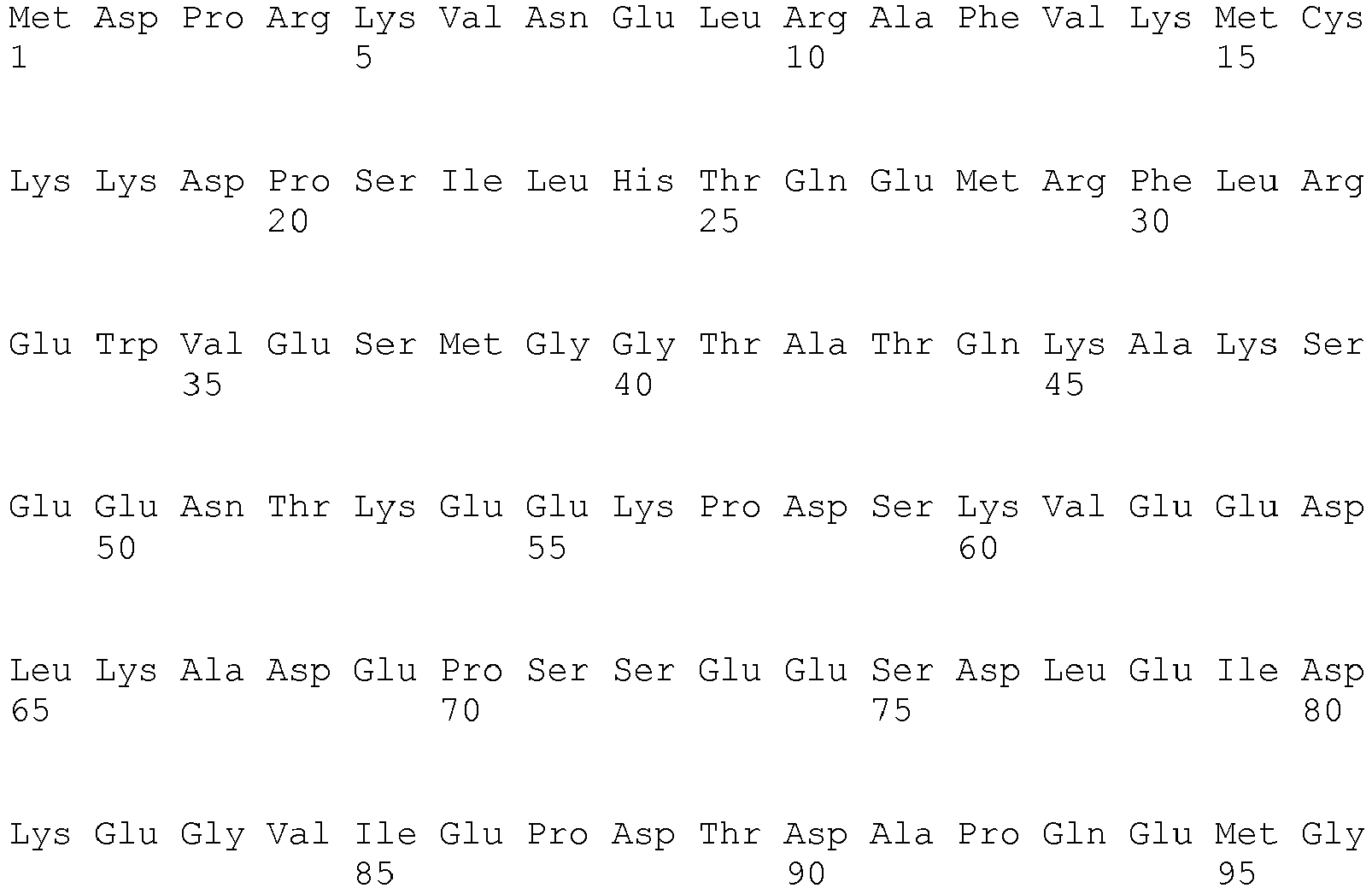

- <210> 7
<211> 710
<212> PRT
<213> Homo sapiens - <400> 7


- <210> 8
<211> 375
<212> PRT
<213> Homo sapiens - <400> 8


- <210> 9
<211> 434
<212> PRT
<213> Homo sapiens - <400> 9


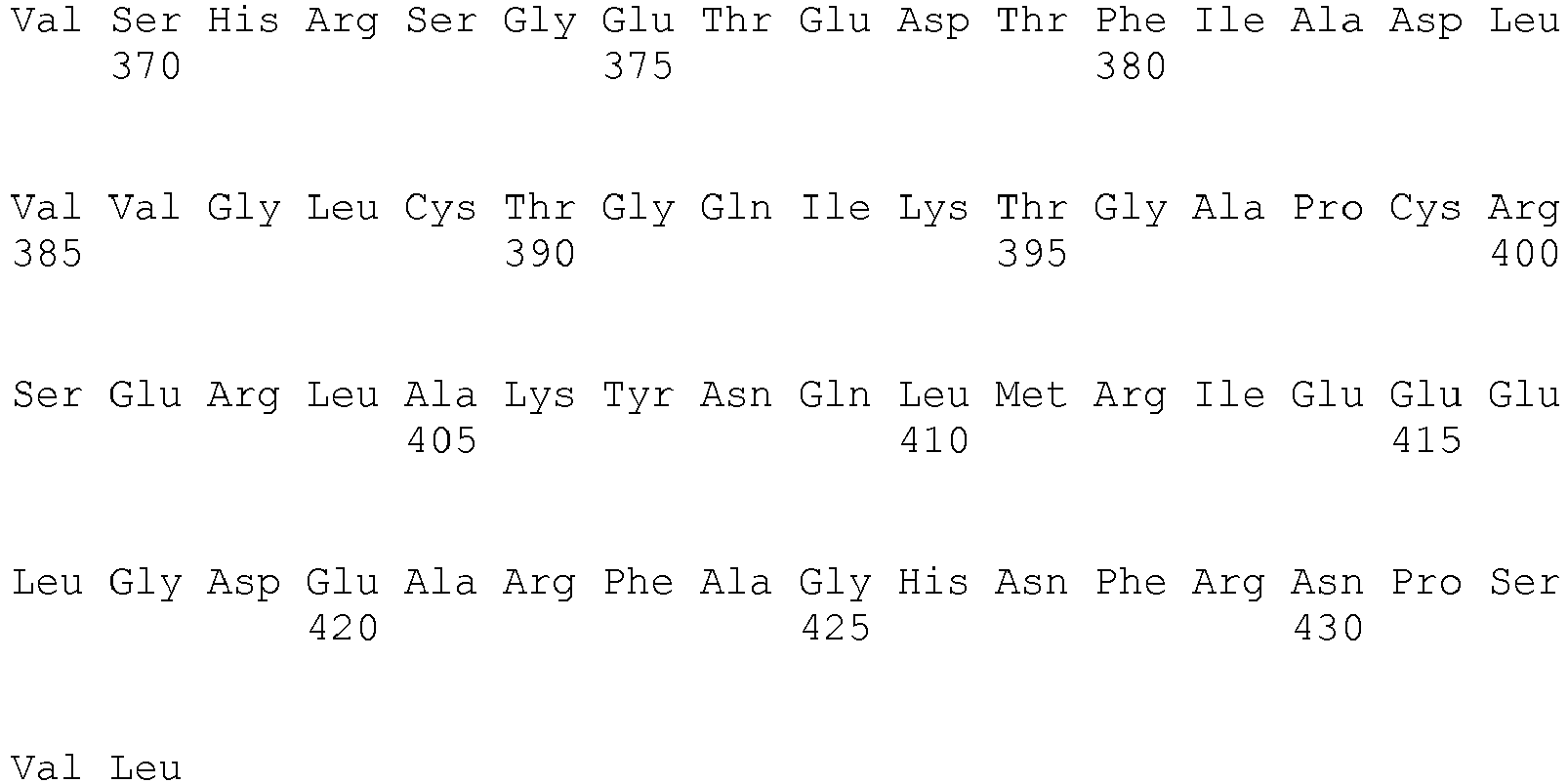
- <210> 10
<211> 505
<212> PRT
<213> Homo sapiens - <400> 10

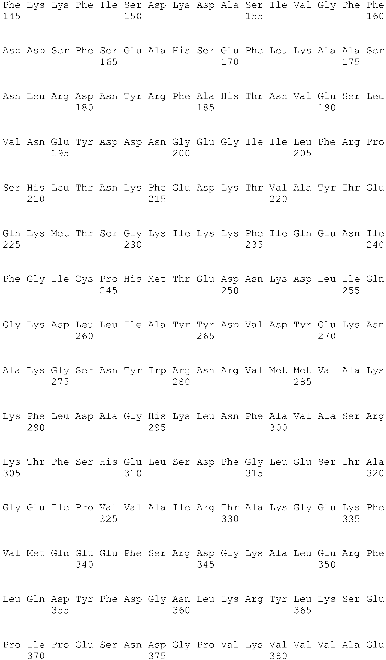
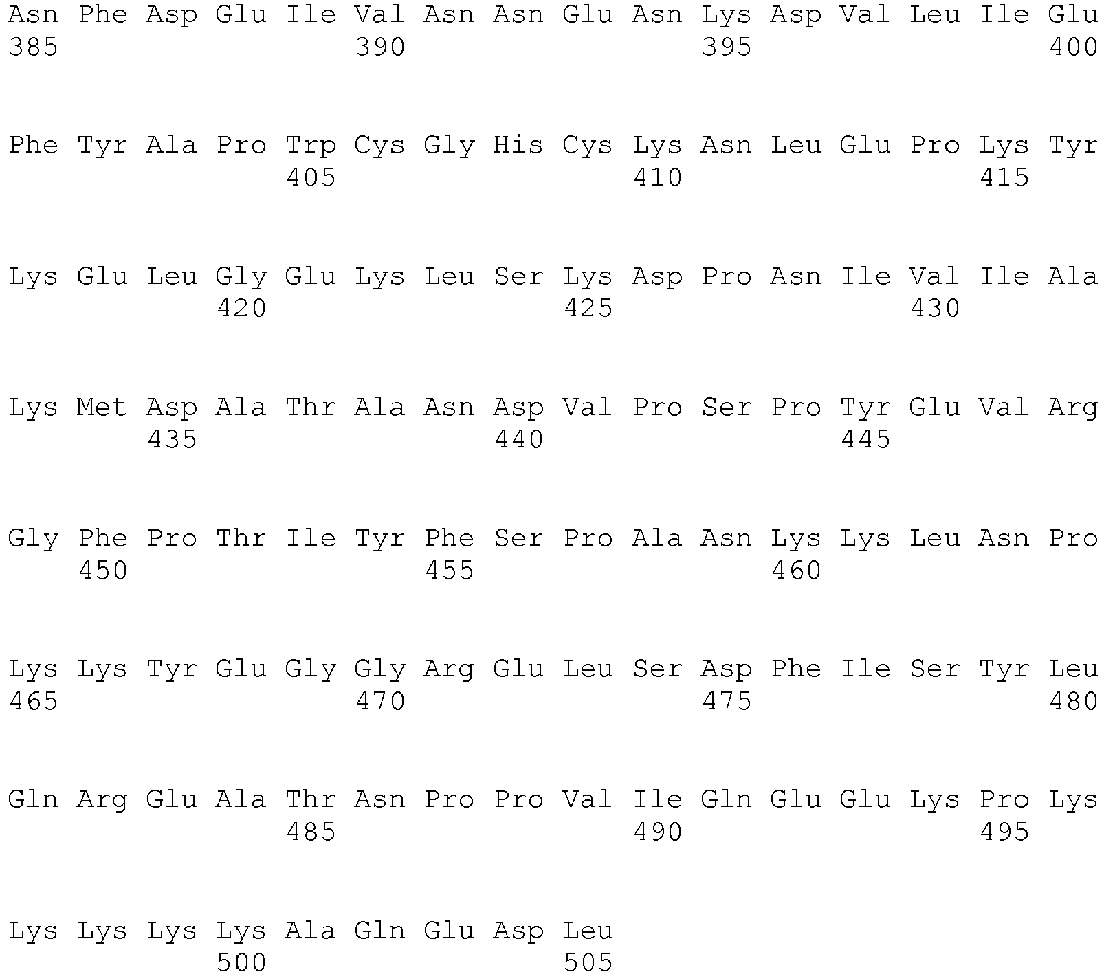
- <210> 11
<211> 470
<212> PRT
<213> Homo sapiens - <400> 11


- <210> 12
<211> 470
<212> PRT
<213> Homo sapiens - <400> 12


- <210> 13
<211> 449
<212> PRT
<213> Homo sapiens - <400> 13
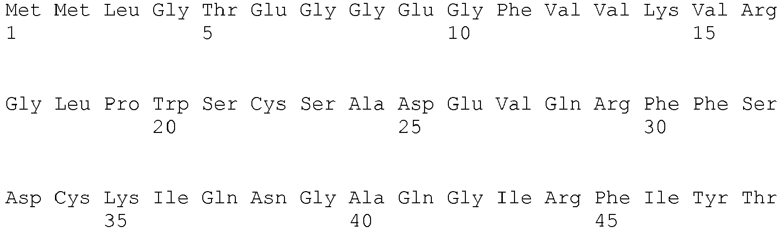


- <210> 14
<211> 543
<212> PRT
<213> Homo sapiens - <400> 14


- <210> 15
<211> 249
<212> PRT
<213> Homo sapiens - <400> 15
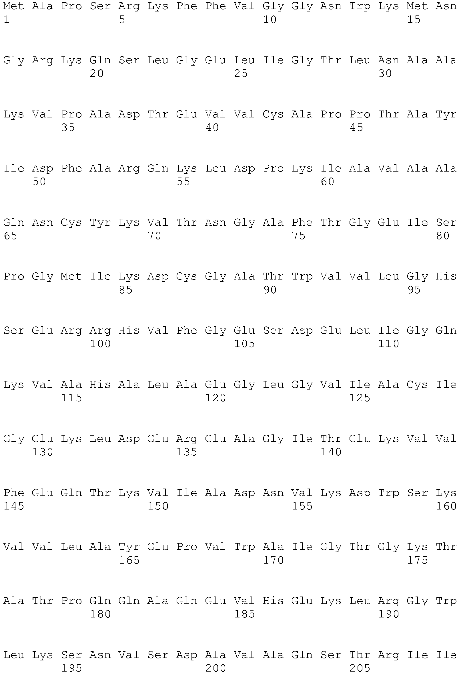

- <210> 16
<211> 224
<212> PRT
<213> Homo sapiens - <400> 16


- <210> 17
<211> 331
<212> PRT
<213> Homo sapiens - <400> 17


- <210> 18
<211> 654
<212> PRT
<213> Homo sapiens - <400> 18


- <210> 19
<211> 466
<212> PRT
<213> Homo sapiens - <400> 19


- <210> 20
<211> 434
<212> PRT
<213> Homo sapiens - <400> 20


- <210> 21
<211> 198
<212> PRT
<213> Homo sapiens - <400> 21


- <210> 22
<211> 256
<212> PRT
<213> Homo sapiens - <400> 22


- <210> 23
<211> 201
<212> PRT
<213> Homo sapiens - <400> 23


- <210> 24
<211> 215
<212> PRT
<213> Homo sapiens - <400> 24


Claims (12)
- Procédé in vitro de détection d'une sclérodermie systémique (ScS) ou d'une hypertension artérielle pulmonaire associée à une sclérodermie (HTAP-ScS) ou d'une hypertension artérielle pulmonaire idiopathique (HTAPi) chez un sujet, comprenant la détermination de la présence et/ou de la quantité d'au moins un anticorps choisi dans le groupe constitué des anticorps anti-Galectine-1, anti-protéine FAM10A4, anti-phosphoprotéine 1 induite par le stress, et anti-précurseur de la protéine de 78 kDa régulée par le glucose,
dans un échantillon biologique provenant d'un patient,
la présence d'un anticorps anti-précurseur de la protéine de 78 kDa régulée par le glucose étant indicative d'une ScS ou d'une HTAPi ;
la présence d'un anticorps anti-protéine FAM10A4 étant indicative d'une HTAP-ScS ou d'une HTAPi ;
la présence d'un anticorps anti-galectine-1 étant indicative d'une ScS ;
ou la présence en une quantité supérieure à une valeur contrôle d'un anticorps anti-phosphoprotéine 1 induite par le stress étant indicative d'une ScS, d'une HTAP-ScS ou d'une HTAPi. - Procédé selon la revendication 1, dans lequel l'échantillon biologique est un échantillon de sang ou de sérum.
- Procédé selon l'une des revendications 1 ou 2, dans lequel la quantité dudit au moins un anticorps est déterminée par un immunoessai.
- Procédé selon la revendication 3, dans lequel l'immunoessai est un dosage ELISA.
- Procédé selon l'une des revendications 1 à 4, dans lequel le patient est un humain.
- Procédé selon l'une des revendications 1 à 5, dans lequel le patient souffre d'une sclérodermie systémique, avec ou sans hypertension artérielle pulmonaire associée.
- Procédé selon l'une des revendications 1 à 5, dans lequel le patient souffre d'une hypertension artérielle pulmonaire idiopathique.
- Procédé selon l'une des revendications 1 à 6, dans lequel l'hypertension artérielle pulmonaire est associée à une hypertension portale, à une cardiopathie congénitale, ou à une infection par le virus de l'immunodéficience humaine (VIH), ou est une hypertension pulmonaire post-embolique.
- Procédé selon l'une des revendications 1 à 6, dans lequel le patient est un sujet prédisposé à développer une sclérodermie systémique ou une hypertension artérielle pulmonaire associée à une sclérodermie ou une hypertension artérielle pulmonaire idiopathique.
- Procédé selon la revendication 9, dans lequel le sujet est porteur d'une ou plusieurs mutation(s) dans le gène codant BMPRII, endoglin ou ALK1.
- Procédé selon l'une quelconque des revendications 1 à 10, ledit au moins un anticorps comprenant un anticorps anti-galectine 1.
- Procédé selon l'une quelconque des revendications 1 à 10, ledit au moins un anticorps comprenant un anticorps anti-phosphoprotéine 1 induite par le stress.
Applications Claiming Priority (2)
| Application Number | Priority Date | Filing Date | Title |
|---|---|---|---|
| FR0856255A FR2936060B1 (fr) | 2008-09-17 | 2008-09-17 | Procede de diagnostic d'une sclerodermie systemique ou d'une hypertension arterielle pulmonaire. |
| PCT/FR2009/051753 WO2010031968A1 (fr) | 2008-09-17 | 2009-09-17 | Procédé de diagnostic d'une sclérodermie systémique ou d'une hypertension artérielle pulmonaire |
Publications (2)
| Publication Number | Publication Date |
|---|---|
| EP2342566A1 EP2342566A1 (fr) | 2011-07-13 |
| EP2342566B1 true EP2342566B1 (fr) | 2015-01-28 |
Family
ID=40282523
Family Applications (1)
| Application Number | Title | Priority Date | Filing Date |
|---|---|---|---|
| EP09748405.9A Not-in-force EP2342566B1 (fr) | 2008-09-17 | 2009-09-17 | Procédé de diagnostic d'une sclérodermie systémique ou d'une hypertension artérielle pulmonaire |
Country Status (7)
| Country | Link |
|---|---|
| US (2) | US20110311995A1 (fr) |
| EP (1) | EP2342566B1 (fr) |
| CA (1) | CA2737535A1 (fr) |
| DK (1) | DK2342566T3 (fr) |
| ES (1) | ES2535719T3 (fr) |
| FR (1) | FR2936060B1 (fr) |
| WO (1) | WO2010031968A1 (fr) |
Families Citing this family (5)
| Publication number | Priority date | Publication date | Assignee | Title |
|---|---|---|---|---|
| PL2704711T3 (pl) * | 2011-05-02 | 2021-07-05 | The Board Of Trustees Of The Leland Stanford Junior University | Zastosowanie fk506 w leczeniu tętniczego nadciśnienia płucnego |
| WO2018138297A1 (fr) | 2017-01-27 | 2018-08-02 | Kymab Limited | Anticorps anti-opg |
| EP3624821A4 (fr) | 2017-05-19 | 2021-01-20 | The Board of Trustees of the Leland Stanford Junior University | Enzastaurine et agents augmentant l'activité du gène fhit (fragile histidine triad) pour le traitement de l'hypertension pulmonaire (pah) |
| CA3092861A1 (fr) * | 2018-03-07 | 2019-09-12 | Timber Pharmaceuticals, Inc. | Compositions et methodes destinees au traitement de la fibrose kystique |
| CN118767135A (zh) * | 2023-04-10 | 2024-10-15 | 复旦大学 | 治疗皮肤纤维化的药物及方法 |
Family Cites Families (4)
| Publication number | Priority date | Publication date | Assignee | Title |
|---|---|---|---|---|
| EP1577671A1 (fr) * | 1996-07-17 | 2005-09-21 | Kaneka Corporation | Medicaments pour le diagnostic de maladies auto-immunes |
| US20080254481A1 (en) * | 2006-11-13 | 2008-10-16 | Invitrogen Corporation | Methods and kits for detecting prostate cancer biomarkers |
| GB0721160D0 (en) * | 2007-10-29 | 2007-12-05 | Univ Leuven Kath | New method for diagnosing sjogren's syndrome |
| WO2009137832A2 (fr) * | 2008-05-09 | 2009-11-12 | Duke University | Auto-anticorps dans la détection et le traitement du cancer |
-
2008
- 2008-09-17 FR FR0856255A patent/FR2936060B1/fr not_active Expired - Fee Related
-
2009
- 2009-09-17 WO PCT/FR2009/051753 patent/WO2010031968A1/fr not_active Ceased
- 2009-09-17 ES ES09748405.9T patent/ES2535719T3/es active Active
- 2009-09-17 DK DK09748405T patent/DK2342566T3/da active
- 2009-09-17 CA CA2737535A patent/CA2737535A1/fr not_active Abandoned
- 2009-09-17 EP EP09748405.9A patent/EP2342566B1/fr not_active Not-in-force
- 2009-09-17 US US12/998,080 patent/US20110311995A1/en not_active Abandoned
-
2015
- 2015-04-14 US US14/686,190 patent/US20150323550A1/en not_active Abandoned
Non-Patent Citations (2)
| Title |
|---|
| FRITZLER ET AL: "Challenges to the use of autoantibodies as predictors of disease onset, diagnosis and outcomes", AUTOIMMUNITY REVIEWS, ELSEVIER, AMSTERDAM, NL, vol. 7, no. 8, 9 July 2008 (2008-07-09), pages 616 - 620, XP025408622, ISSN: 1568-9972, [retrieved on 20080709], DOI: 10.1016/J.AUTREV.2008.06.007 * |
| TERRIER BENJAMIN ET AL: "Anti-fibroblast antibodies from systemic sclerosis patients bind to alpha-enolase and are associated with interstitial lung disease", ARTHRITIS & RHEUMATISM, JOHN WILEY & SONS, INC, US, vol. 58, no. 9, 29 October 2008 (2008-10-29), pages S836, XP008101458, ISSN: 0004-3591, [retrieved on 20081029] * |
Also Published As
| Publication number | Publication date |
|---|---|
| FR2936060B1 (fr) | 2010-11-19 |
| WO2010031968A1 (fr) | 2010-03-25 |
| FR2936060A1 (fr) | 2010-03-19 |
| US20110311995A1 (en) | 2011-12-22 |
| CA2737535A1 (fr) | 2010-03-25 |
| DK2342566T3 (da) | 2015-05-04 |
| EP2342566A1 (fr) | 2011-07-13 |
| US20150323550A1 (en) | 2015-11-12 |
| ES2535719T3 (es) | 2015-05-14 |
Similar Documents
| Publication | Publication Date | Title |
|---|---|---|
| Leiherer et al. | Serum uromodulin is a predictive biomarker for cardiovascular events and overall mortality in coronary patients | |
| Borg et al. | Evaluation of the new ADA and WHO criteria for classification of diabetes mellitus in young adult people (15–34 yrs) in the Diabetes Incidence Study in Sweden (DISS) | |
| US20040265926A1 (en) | Bodily fluid markers of tissue hypoxia | |
| RU2733471C2 (ru) | Способ прогнозирования риска развития хронического заболевания почек | |
| HUE031613T2 (en) | Diagnosis and treatment of preeclampsia | |
| ES2948446T3 (es) | Adrenomedulina para la valoración de la congestión en un sujeto con insuficiencia cardiaca aguda | |
| EP2342566A1 (fr) | Procédé de diagnostic d'une sclérodermie systémique ou d'une hypertension artérielle pulmonaire | |
| US8425877B2 (en) | Method for diagnosis of systemic sclerosis involving an anti-AT1-receptor antibody | |
| Mozena et al. | Lack of a role for cross-reacting anti-thyroid antibodies in chronic idiopathic urticaria | |
| Husain-Krautter et al. | The osteopontin transgenic mouse is a new model for Sjögren's syndrome | |
| Vicco et al. | Assessment of cross-reactive host-pathogen antibodies in patients with different stages of chronic Chagas disease | |
| Berezin | Prognostication of clinical outcomes in diabetes mellitus: Emerging role of cardiac biomarkers | |
| EP2352032B1 (fr) | Procédé d'éliminer des anticorps d'endothéline dans un échantillon du sang | |
| Mandraffino et al. | Biglycan expression in current cigarette smokers: a possible link between active smoking and atherogenesis | |
| EP2274623B1 (fr) | Procede de diagnostic d'une hypertension arterielle pulmonaire | |
| CA2884356C (fr) | Procede pour le diagnostic de dystrophies musculaires | |
| Oh et al. | Lack of superiority for soluble ST2 over high sensitive C-reactive protein in predicting high risk coronary artery calcium score in a community cohort | |
| Jin et al. | Upregulation of gastric norepinephrine with β-adrenoceptors and gastric dysmotility in a rat model of functional dyspepsia | |
| JP7271442B2 (ja) | 腎機能を診断またはモニターする方法、または腎機能障害を診断することを補助する方法 | |
| Wiberg et al. | Cellular and humoral immune responses in atrophic lymphocytic pancreatitis in German shepherd dogs and rough-coated collies | |
| CA2711867C (fr) | Utilisation d'une proteine ou d'une sequence peptidique nitree pour la mise en oeuvre d'une methode de diagnostic | |
| Atteya et al. | Lisinopril has a cardio-protective effect on experimental acute autoimmune myocarditis in rats | |
| Liu et al. | WISP1 is increased in the maternal serum, adipose tissue, and placenta of women with gestational diabetes mellitus | |
| Singh et al. | Leishmania donovani infection-driven high levels of IL-10 causes hypoalbuminemia in human visceral leishmaniasis | |
| EP2526423B1 (fr) | Procédés et dispositifs de diagnostic de troubles cardiaques |
Legal Events
| Date | Code | Title | Description |
|---|---|---|---|
| PUAI | Public reference made under article 153(3) epc to a published international application that has entered the european phase |
Free format text: ORIGINAL CODE: 0009012 |
|
| 17P | Request for examination filed |
Effective date: 20110418 |
|
| AK | Designated contracting states |
Kind code of ref document: A1 Designated state(s): AT BE BG CH CY CZ DE DK EE ES FI FR GB GR HR HU IE IS IT LI LT LU LV MC MK MT NL NO PL PT RO SE SI SK SM TR |
|
| AX | Request for extension of the european patent |
Extension state: AL BA RS |
|
| DAX | Request for extension of the european patent (deleted) | ||
| 17Q | First examination report despatched |
Effective date: 20120516 |
|
| GRAP | Despatch of communication of intention to grant a patent |
Free format text: ORIGINAL CODE: EPIDOSNIGR1 |
|
| INTG | Intention to grant announced |
Effective date: 20140822 |
|
| GRAS | Grant fee paid |
Free format text: ORIGINAL CODE: EPIDOSNIGR3 |
|
| GRAA | (expected) grant |
Free format text: ORIGINAL CODE: 0009210 |
|
| AK | Designated contracting states |
Kind code of ref document: B1 Designated state(s): AT BE BG CH CY CZ DE DK EE ES FI FR GB GR HR HU IE IS IT LI LT LU LV MC MK MT NL NO PL PT RO SE SI SK SM TR |
|
| REG | Reference to a national code |
Ref country code: GB Ref legal event code: FG4D Free format text: NOT ENGLISH |
|
| REG | Reference to a national code |
Ref country code: CH Ref legal event code: EP |
|
| REG | Reference to a national code |
Ref country code: IE Ref legal event code: FG4D Free format text: LANGUAGE OF EP DOCUMENT: FRENCH |
|
| REG | Reference to a national code |
Ref country code: DE Ref legal event code: R096 Ref document number: 602009029239 Country of ref document: DE Effective date: 20150312 |
|
| REG | Reference to a national code |
Ref country code: AT Ref legal event code: REF Ref document number: 708453 Country of ref document: AT Kind code of ref document: T Effective date: 20150315 |
|
| REG | Reference to a national code |
Ref country code: DK Ref legal event code: T3 Effective date: 20150429 |
|
| REG | Reference to a national code |
Ref country code: ES Ref legal event code: FG2A Ref document number: 2535719 Country of ref document: ES Kind code of ref document: T3 Effective date: 20150514 |
|
| REG | Reference to a national code |
Ref country code: SE Ref legal event code: TRGR |
|
| REG | Reference to a national code |
Ref country code: NL Ref legal event code: T3 |
|
| REG | Reference to a national code |
Ref country code: AT Ref legal event code: MK05 Ref document number: 708453 Country of ref document: AT Kind code of ref document: T Effective date: 20150128 |
|
| REG | Reference to a national code |
Ref country code: LT Ref legal event code: MG4D |
|
| PG25 | Lapsed in a contracting state [announced via postgrant information from national office to epo] |
Ref country code: BG Free format text: LAPSE BECAUSE OF FAILURE TO SUBMIT A TRANSLATION OF THE DESCRIPTION OR TO PAY THE FEE WITHIN THE PRESCRIBED TIME-LIMIT Effective date: 20150428 Ref country code: HR Free format text: LAPSE BECAUSE OF FAILURE TO SUBMIT A TRANSLATION OF THE DESCRIPTION OR TO PAY THE FEE WITHIN THE PRESCRIBED TIME-LIMIT Effective date: 20150128 Ref country code: NO Free format text: LAPSE BECAUSE OF FAILURE TO SUBMIT A TRANSLATION OF THE DESCRIPTION OR TO PAY THE FEE WITHIN THE PRESCRIBED TIME-LIMIT Effective date: 20150428 Ref country code: FI Free format text: LAPSE BECAUSE OF FAILURE TO SUBMIT A TRANSLATION OF THE DESCRIPTION OR TO PAY THE FEE WITHIN THE PRESCRIBED TIME-LIMIT Effective date: 20150128 Ref country code: LT Free format text: LAPSE BECAUSE OF FAILURE TO SUBMIT A TRANSLATION OF THE DESCRIPTION OR TO PAY THE FEE WITHIN THE PRESCRIBED TIME-LIMIT Effective date: 20150128 |
|
| PG25 | Lapsed in a contracting state [announced via postgrant information from national office to epo] |
Ref country code: GR Free format text: LAPSE BECAUSE OF FAILURE TO SUBMIT A TRANSLATION OF THE DESCRIPTION OR TO PAY THE FEE WITHIN THE PRESCRIBED TIME-LIMIT Effective date: 20150429 Ref country code: PL Free format text: LAPSE BECAUSE OF FAILURE TO SUBMIT A TRANSLATION OF THE DESCRIPTION OR TO PAY THE FEE WITHIN THE PRESCRIBED TIME-LIMIT Effective date: 20150128 Ref country code: LV Free format text: LAPSE BECAUSE OF FAILURE TO SUBMIT A TRANSLATION OF THE DESCRIPTION OR TO PAY THE FEE WITHIN THE PRESCRIBED TIME-LIMIT Effective date: 20150128 Ref country code: AT Free format text: LAPSE BECAUSE OF FAILURE TO SUBMIT A TRANSLATION OF THE DESCRIPTION OR TO PAY THE FEE WITHIN THE PRESCRIBED TIME-LIMIT Effective date: 20150128 Ref country code: IS Free format text: LAPSE BECAUSE OF FAILURE TO SUBMIT A TRANSLATION OF THE DESCRIPTION OR TO PAY THE FEE WITHIN THE PRESCRIBED TIME-LIMIT Effective date: 20150528 |
|
| REG | Reference to a national code |
Ref country code: FR Ref legal event code: PLFP Year of fee payment: 7 |
|
| REG | Reference to a national code |
Ref country code: DE Ref legal event code: R097 Ref document number: 602009029239 Country of ref document: DE |
|
| PG25 | Lapsed in a contracting state [announced via postgrant information from national office to epo] |
Ref country code: SK Free format text: LAPSE BECAUSE OF FAILURE TO SUBMIT A TRANSLATION OF THE DESCRIPTION OR TO PAY THE FEE WITHIN THE PRESCRIBED TIME-LIMIT Effective date: 20150128 Ref country code: EE Free format text: LAPSE BECAUSE OF FAILURE TO SUBMIT A TRANSLATION OF THE DESCRIPTION OR TO PAY THE FEE WITHIN THE PRESCRIBED TIME-LIMIT Effective date: 20150128 Ref country code: CZ Free format text: LAPSE BECAUSE OF FAILURE TO SUBMIT A TRANSLATION OF THE DESCRIPTION OR TO PAY THE FEE WITHIN THE PRESCRIBED TIME-LIMIT Effective date: 20150128 Ref country code: RO Free format text: LAPSE BECAUSE OF FAILURE TO SUBMIT A TRANSLATION OF THE DESCRIPTION OR TO PAY THE FEE WITHIN THE PRESCRIBED TIME-LIMIT Effective date: 20150128 |
|
| PGFP | Annual fee paid to national office [announced via postgrant information from national office to epo] |
Ref country code: GB Payment date: 20150930 Year of fee payment: 7 Ref country code: CH Payment date: 20150930 Year of fee payment: 7 |
|
| PGFP | Annual fee paid to national office [announced via postgrant information from national office to epo] |
Ref country code: FR Payment date: 20150928 Year of fee payment: 7 Ref country code: SE Payment date: 20150929 Year of fee payment: 7 |
|
| PLBE | No opposition filed within time limit |
Free format text: ORIGINAL CODE: 0009261 |
|
| STAA | Information on the status of an ep patent application or granted ep patent |
Free format text: STATUS: NO OPPOSITION FILED WITHIN TIME LIMIT |
|
| PGFP | Annual fee paid to national office [announced via postgrant information from national office to epo] |
Ref country code: DK Payment date: 20150929 Year of fee payment: 7 |
|
| 26N | No opposition filed |
Effective date: 20151029 |
|
| PGFP | Annual fee paid to national office [announced via postgrant information from national office to epo] |
Ref country code: IT Payment date: 20150930 Year of fee payment: 7 Ref country code: DE Payment date: 20150928 Year of fee payment: 7 |
|
| PG25 | Lapsed in a contracting state [announced via postgrant information from national office to epo] |
Ref country code: SI Free format text: LAPSE BECAUSE OF FAILURE TO SUBMIT A TRANSLATION OF THE DESCRIPTION OR TO PAY THE FEE WITHIN THE PRESCRIBED TIME-LIMIT Effective date: 20150128 |
|
| PGFP | Annual fee paid to national office [announced via postgrant information from national office to epo] |
Ref country code: BE Payment date: 20150928 Year of fee payment: 7 Ref country code: NL Payment date: 20150929 Year of fee payment: 7 Ref country code: ES Payment date: 20151027 Year of fee payment: 7 |
|
| PG25 | Lapsed in a contracting state [announced via postgrant information from national office to epo] |
Ref country code: LU Free format text: LAPSE BECAUSE OF FAILURE TO SUBMIT A TRANSLATION OF THE DESCRIPTION OR TO PAY THE FEE WITHIN THE PRESCRIBED TIME-LIMIT Effective date: 20150917 Ref country code: MC Free format text: LAPSE BECAUSE OF FAILURE TO SUBMIT A TRANSLATION OF THE DESCRIPTION OR TO PAY THE FEE WITHIN THE PRESCRIBED TIME-LIMIT Effective date: 20150128 |
|
| REG | Reference to a national code |
Ref country code: IE Ref legal event code: MM4A |
|
| PG25 | Lapsed in a contracting state [announced via postgrant information from national office to epo] |
Ref country code: IE Free format text: LAPSE BECAUSE OF NON-PAYMENT OF DUE FEES Effective date: 20150917 |
|
| PG25 | Lapsed in a contracting state [announced via postgrant information from national office to epo] |
Ref country code: BE Free format text: LAPSE BECAUSE OF NON-PAYMENT OF DUE FEES Effective date: 20160930 |
|
| PG25 | Lapsed in a contracting state [announced via postgrant information from national office to epo] |
Ref country code: MT Free format text: LAPSE BECAUSE OF FAILURE TO SUBMIT A TRANSLATION OF THE DESCRIPTION OR TO PAY THE FEE WITHIN THE PRESCRIBED TIME-LIMIT Effective date: 20150128 |
|
| REG | Reference to a national code |
Ref country code: DE Ref legal event code: R119 Ref document number: 602009029239 Country of ref document: DE |
|
| REG | Reference to a national code |
Ref country code: DK Ref legal event code: EBP Effective date: 20160930 |
|
| PG25 | Lapsed in a contracting state [announced via postgrant information from national office to epo] |
Ref country code: SE Free format text: LAPSE BECAUSE OF NON-PAYMENT OF DUE FEES Effective date: 20160918 |
|
| REG | Reference to a national code |
Ref country code: CH Ref legal event code: PL |
|
| REG | Reference to a national code |
Ref country code: SE Ref legal event code: EUG |
|
| REG | Reference to a national code |
Ref country code: NL Ref legal event code: MM Effective date: 20161001 |
|
| GBPC | Gb: european patent ceased through non-payment of renewal fee |
Effective date: 20160917 |
|
| PG25 | Lapsed in a contracting state [announced via postgrant information from national office to epo] |
Ref country code: SM Free format text: LAPSE BECAUSE OF FAILURE TO SUBMIT A TRANSLATION OF THE DESCRIPTION OR TO PAY THE FEE WITHIN THE PRESCRIBED TIME-LIMIT Effective date: 20150128 Ref country code: HU Free format text: LAPSE BECAUSE OF FAILURE TO SUBMIT A TRANSLATION OF THE DESCRIPTION OR TO PAY THE FEE WITHIN THE PRESCRIBED TIME-LIMIT; INVALID AB INITIO Effective date: 20090917 |
|
| PG25 | Lapsed in a contracting state [announced via postgrant information from national office to epo] |
Ref country code: NL Free format text: LAPSE BECAUSE OF NON-PAYMENT OF DUE FEES Effective date: 20161001 Ref country code: CY Free format text: LAPSE BECAUSE OF FAILURE TO SUBMIT A TRANSLATION OF THE DESCRIPTION OR TO PAY THE FEE WITHIN THE PRESCRIBED TIME-LIMIT Effective date: 20150128 |
|
| REG | Reference to a national code |
Ref country code: FR Ref legal event code: ST Effective date: 20170531 |
|
| PG25 | Lapsed in a contracting state [announced via postgrant information from national office to epo] |
Ref country code: LI Free format text: LAPSE BECAUSE OF NON-PAYMENT OF DUE FEES Effective date: 20160930 Ref country code: FR Free format text: LAPSE BECAUSE OF NON-PAYMENT OF DUE FEES Effective date: 20160930 Ref country code: DE Free format text: LAPSE BECAUSE OF NON-PAYMENT OF DUE FEES Effective date: 20170401 Ref country code: CH Free format text: LAPSE BECAUSE OF NON-PAYMENT OF DUE FEES Effective date: 20160930 Ref country code: GB Free format text: LAPSE BECAUSE OF NON-PAYMENT OF DUE FEES Effective date: 20160917 |
|
| PG25 | Lapsed in a contracting state [announced via postgrant information from national office to epo] |
Ref country code: TR Free format text: LAPSE BECAUSE OF FAILURE TO SUBMIT A TRANSLATION OF THE DESCRIPTION OR TO PAY THE FEE WITHIN THE PRESCRIBED TIME-LIMIT Effective date: 20150128 Ref country code: IT Free format text: LAPSE BECAUSE OF NON-PAYMENT OF DUE FEES Effective date: 20160917 |
|
| REG | Reference to a national code |
Ref country code: BE Ref legal event code: MM Effective date: 20160930 |
|
| PG25 | Lapsed in a contracting state [announced via postgrant information from national office to epo] |
Ref country code: DK Free format text: LAPSE BECAUSE OF NON-PAYMENT OF DUE FEES Effective date: 20160930 |
|
| PG25 | Lapsed in a contracting state [announced via postgrant information from national office to epo] |
Ref country code: ES Free format text: LAPSE BECAUSE OF NON-PAYMENT OF DUE FEES Effective date: 20160918 |
|
| PG25 | Lapsed in a contracting state [announced via postgrant information from national office to epo] |
Ref country code: PT Free format text: LAPSE BECAUSE OF FAILURE TO SUBMIT A TRANSLATION OF THE DESCRIPTION OR TO PAY THE FEE WITHIN THE PRESCRIBED TIME-LIMIT Effective date: 20150128 Ref country code: MK Free format text: LAPSE BECAUSE OF FAILURE TO SUBMIT A TRANSLATION OF THE DESCRIPTION OR TO PAY THE FEE WITHIN THE PRESCRIBED TIME-LIMIT Effective date: 20150128 |
|
| REG | Reference to a national code |
Ref country code: ES Ref legal event code: FD2A Effective date: 20181121 |