EP2340123B1 - Reaction vessel capable of detecting agglutinated antibody loaded erythrocytes, method for detecting agglutinated antibody loaded erythrocytes, and kit of parts - Google Patents
Reaction vessel capable of detecting agglutinated antibody loaded erythrocytes, method for detecting agglutinated antibody loaded erythrocytes, and kit of parts Download PDFInfo
- Publication number
- EP2340123B1 EP2340123B1 EP08801905.4A EP08801905A EP2340123B1 EP 2340123 B1 EP2340123 B1 EP 2340123B1 EP 08801905 A EP08801905 A EP 08801905A EP 2340123 B1 EP2340123 B1 EP 2340123B1
- Authority
- EP
- European Patent Office
- Prior art keywords
- antibodies
- erythrocytes
- antibody
- compartment
- loaded
- Prior art date
- Legal status (The legal status is an assumption and is not a legal conclusion. Google has not performed a legal analysis and makes no representation as to the accuracy of the status listed.)
- Active
Links
- 210000003743 erythrocyte Anatomy 0.000 title claims description 95
- 238000000034 method Methods 0.000 title claims description 24
- 238000006243 chemical reaction Methods 0.000 title description 41
- 238000011534 incubation Methods 0.000 claims description 27
- 230000004520 agglutination Effects 0.000 claims description 24
- 239000007788 liquid Substances 0.000 claims description 17
- FAPWRFPIFSIZLT-UHFFFAOYSA-M Sodium chloride Chemical compound [Na+].[Cl-] FAPWRFPIFSIZLT-UHFFFAOYSA-M 0.000 claims description 16
- 238000005119 centrifugation Methods 0.000 claims description 14
- 239000011324 bead Substances 0.000 claims description 12
- 230000009918 complex formation Effects 0.000 claims description 12
- 239000011780 sodium chloride Substances 0.000 claims description 8
- 239000012530 fluid Substances 0.000 claims description 6
- 229920002307 Dextran Polymers 0.000 claims description 5
- KCXVZYZYPLLWCC-UHFFFAOYSA-N EDTA Chemical compound OC(=O)CN(CC(O)=O)CCN(CC(O)=O)CC(O)=O KCXVZYZYPLLWCC-UHFFFAOYSA-N 0.000 claims description 5
- WQZGKKKJIJFFOK-GASJEMHNSA-N Glucose Natural products OC[C@H]1OC(O)[C@H](O)[C@@H](O)[C@@H]1O WQZGKKKJIJFFOK-GASJEMHNSA-N 0.000 claims description 5
- 239000008103 glucose Substances 0.000 claims description 5
- 229910019142 PO4 Inorganic materials 0.000 claims description 4
- 229920005654 Sephadex Polymers 0.000 claims description 4
- 239000012507 Sephadex™ Substances 0.000 claims description 4
- 229920002684 Sepharose Polymers 0.000 claims description 4
- 102000007562 Serum Albumin Human genes 0.000 claims description 4
- 108010071390 Serum Albumin Proteins 0.000 claims description 4
- 239000008366 buffered solution Substances 0.000 claims description 4
- 238000004891 communication Methods 0.000 claims description 4
- 239000011521 glass Substances 0.000 claims description 4
- NBIIXXVUZAFLBC-UHFFFAOYSA-K phosphate Chemical compound [O-]P([O-])([O-])=O NBIIXXVUZAFLBC-UHFFFAOYSA-K 0.000 claims description 4
- 239000010452 phosphate Substances 0.000 claims description 4
- 239000012266 salt solution Substances 0.000 claims description 4
- 239000000969 carrier Substances 0.000 claims description 3
- 150000001875 compounds Chemical class 0.000 claims description 3
- 229920000936 Agarose Polymers 0.000 claims description 2
- -1 acryl Chemical group 0.000 claims description 2
- 230000004888 barrier function Effects 0.000 claims description 2
- 239000002738 chelating agent Substances 0.000 claims description 2
- 150000002016 disaccharides Chemical class 0.000 claims description 2
- 239000012634 fragment Substances 0.000 claims description 2
- 150000002772 monosaccharides Chemical class 0.000 claims description 2
- 210000004027 cell Anatomy 0.000 description 90
- 238000012360 testing method Methods 0.000 description 81
- 239000011159 matrix material Substances 0.000 description 46
- 239000008280 blood Substances 0.000 description 45
- 210000004369 blood Anatomy 0.000 description 42
- 210000002966 serum Anatomy 0.000 description 34
- 239000012491 analyte Substances 0.000 description 19
- 239000000427 antigen Substances 0.000 description 19
- 102000036639 antigens Human genes 0.000 description 19
- 108091007433 antigens Proteins 0.000 description 19
- 239000000725 suspension Substances 0.000 description 16
- 230000035945 sensitivity Effects 0.000 description 13
- 238000002474 experimental method Methods 0.000 description 12
- 230000000295 complement effect Effects 0.000 description 11
- 239000012071 phase Substances 0.000 description 11
- 238000003556 assay Methods 0.000 description 10
- 239000003153 chemical reaction reagent Substances 0.000 description 10
- 239000003446 ligand Substances 0.000 description 10
- 239000007863 gel particle Substances 0.000 description 9
- 230000015572 biosynthetic process Effects 0.000 description 8
- 230000001419 dependent effect Effects 0.000 description 8
- 239000002245 particle Substances 0.000 description 7
- 210000002381 plasma Anatomy 0.000 description 6
- 239000000523 sample Substances 0.000 description 6
- 239000007790 solid phase Substances 0.000 description 6
- 238000001514 detection method Methods 0.000 description 5
- 239000003814 drug Substances 0.000 description 5
- 239000000910 agglutinin Substances 0.000 description 4
- 229940079593 drug Drugs 0.000 description 4
- 239000000463 material Substances 0.000 description 4
- 239000000203 mixture Substances 0.000 description 4
- 102000004169 proteins and genes Human genes 0.000 description 4
- 108090000623 proteins and genes Proteins 0.000 description 4
- 238000012216 screening Methods 0.000 description 4
- 238000000926 separation method Methods 0.000 description 4
- NHZLNPMOSADWGC-UHFFFAOYSA-N 4-amino-N-(2-quinoxalinyl)benzenesulfonamide Chemical compound C1=CC(N)=CC=C1S(=O)(=O)NC1=CN=C(C=CC=C2)C2=N1 NHZLNPMOSADWGC-UHFFFAOYSA-N 0.000 description 3
- 108090000790 Enzymes Proteins 0.000 description 3
- 102000004190 Enzymes Human genes 0.000 description 3
- 108060003951 Immunoglobulin Proteins 0.000 description 3
- 101710120037 Toxin CcdB Proteins 0.000 description 3
- 208000003441 Transfusion reaction Diseases 0.000 description 3
- 239000000872 buffer Substances 0.000 description 3
- 239000006285 cell suspension Substances 0.000 description 3
- 108700021073 cold agglutinins Proteins 0.000 description 3
- 229940088598 enzyme Drugs 0.000 description 3
- 102000018358 immunoglobulin Human genes 0.000 description 3
- 230000007935 neutral effect Effects 0.000 description 3
- 239000013049 sediment Substances 0.000 description 3
- 238000009589 serological test Methods 0.000 description 3
- 239000007787 solid Substances 0.000 description 3
- 239000000243 solution Substances 0.000 description 3
- 238000005406 washing Methods 0.000 description 3
- 241000124008 Mammalia Species 0.000 description 2
- 239000002202 Polyethylene glycol Substances 0.000 description 2
- 239000004365 Protease Substances 0.000 description 2
- 206010070834 Sensitisation Diseases 0.000 description 2
- 239000003795 chemical substances by application Substances 0.000 description 2
- 230000000694 effects Effects 0.000 description 2
- 238000005516 engineering process Methods 0.000 description 2
- 238000001727 in vivo Methods 0.000 description 2
- 230000003993 interaction Effects 0.000 description 2
- 229920002521 macromolecule Polymers 0.000 description 2
- 239000011859 microparticle Substances 0.000 description 2
- 239000013610 patient sample Substances 0.000 description 2
- 229920001223 polyethylene glycol Polymers 0.000 description 2
- 229920000642 polymer Polymers 0.000 description 2
- 230000035935 pregnancy Effects 0.000 description 2
- 230000002028 premature Effects 0.000 description 2
- 230000000717 retained effect Effects 0.000 description 2
- 230000002441 reversible effect Effects 0.000 description 2
- 238000004062 sedimentation Methods 0.000 description 2
- 230000008313 sensitization Effects 0.000 description 2
- 102000009027 Albumins Human genes 0.000 description 1
- 108010088751 Albumins Proteins 0.000 description 1
- 102100027544 Blood group Rh(D) polypeptide Human genes 0.000 description 1
- 108010004032 Bromelains Proteins 0.000 description 1
- 241000283707 Capra Species 0.000 description 1
- 244000304337 Cuminum cyminum Species 0.000 description 1
- 108090000270 Ficain Proteins 0.000 description 1
- 229920000209 Hexadimethrine bromide Polymers 0.000 description 1
- 101000580024 Homo sapiens Blood group Rh(D) polypeptide Proteins 0.000 description 1
- 108090000526 Papain Proteins 0.000 description 1
- 108091005804 Peptidases Proteins 0.000 description 1
- 102000035195 Peptidases Human genes 0.000 description 1
- 206010049190 Red blood cell agglutination Diseases 0.000 description 1
- 102000004142 Trypsin Human genes 0.000 description 1
- 108090000631 Trypsin Proteins 0.000 description 1
- 238000001042 affinity chromatography Methods 0.000 description 1
- 230000004523 agglutinating effect Effects 0.000 description 1
- 230000002391 anti-complement effect Effects 0.000 description 1
- 108010008730 anticomplement Proteins 0.000 description 1
- 230000000890 antigenic effect Effects 0.000 description 1
- 238000013459 approach Methods 0.000 description 1
- 230000015556 catabolic process Effects 0.000 description 1
- 210000000170 cell membrane Anatomy 0.000 description 1
- 230000002498 deadly effect Effects 0.000 description 1
- 238000006073 displacement reaction Methods 0.000 description 1
- POTUGHMKJGOKRI-UHFFFAOYSA-N ficin Chemical compound FI=CI=N POTUGHMKJGOKRI-UHFFFAOYSA-N 0.000 description 1
- 235000019836 ficin Nutrition 0.000 description 1
- 230000006870 function Effects 0.000 description 1
- 230000014509 gene expression Effects 0.000 description 1
- 230000005484 gravity Effects 0.000 description 1
- 150000003278 haem Chemical class 0.000 description 1
- 229910052739 hydrogen Inorganic materials 0.000 description 1
- 239000001257 hydrogen Substances 0.000 description 1
- 230000002209 hydrophobic effect Effects 0.000 description 1
- 230000003053 immunization Effects 0.000 description 1
- 238000002649 immunization Methods 0.000 description 1
- 229940072221 immunoglobulins Drugs 0.000 description 1
- 230000006872 improvement Effects 0.000 description 1
- 238000000338 in vitro Methods 0.000 description 1
- 230000002779 inactivation Effects 0.000 description 1
- 230000001404 mediated effect Effects 0.000 description 1
- 229940055729 papain Drugs 0.000 description 1
- 235000019834 papain Nutrition 0.000 description 1
- 239000008363 phosphate buffer Substances 0.000 description 1
- 229920003023 plastic Polymers 0.000 description 1
- 239000004033 plastic Substances 0.000 description 1
- 238000002360 preparation method Methods 0.000 description 1
- 230000002035 prolonged effect Effects 0.000 description 1
- 229940024999 proteolytic enzymes for treatment of wounds and ulcers Drugs 0.000 description 1
- 238000000746 purification Methods 0.000 description 1
- 230000003014 reinforcing effect Effects 0.000 description 1
- 238000009877 rendering Methods 0.000 description 1
- 230000000405 serological effect Effects 0.000 description 1
- 230000002269 spontaneous effect Effects 0.000 description 1
- 238000003756 stirring Methods 0.000 description 1
- 239000000126 substance Substances 0.000 description 1
- 239000006228 supernatant Substances 0.000 description 1
- 239000012588 trypsin Substances 0.000 description 1
- XLYOFNOQVPJJNP-UHFFFAOYSA-N water Substances O XLYOFNOQVPJJNP-UHFFFAOYSA-N 0.000 description 1
Images


Classifications
-
- G—PHYSICS
- G01—MEASURING; TESTING
- G01N—INVESTIGATING OR ANALYSING MATERIALS BY DETERMINING THEIR CHEMICAL OR PHYSICAL PROPERTIES
- G01N33/00—Investigating or analysing materials by specific methods not covered by groups G01N1/00 - G01N31/00
- G01N33/48—Biological material, e.g. blood, urine; Haemocytometers
- G01N33/50—Chemical analysis of biological material, e.g. blood, urine; Testing involving biospecific ligand binding methods; Immunological testing
- G01N33/53—Immunoassay; Biospecific binding assay; Materials therefor
- G01N33/5302—Apparatus specially adapted for immunological test procedures
- G01N33/5304—Reaction vessels, e.g. agglutination plates
-
- B—PERFORMING OPERATIONS; TRANSPORTING
- B01—PHYSICAL OR CHEMICAL PROCESSES OR APPARATUS IN GENERAL
- B01L—CHEMICAL OR PHYSICAL LABORATORY APPARATUS FOR GENERAL USE
- B01L3/00—Containers or dishes for laboratory use, e.g. laboratory glassware; Droppers
- B01L3/50—Containers for the purpose of retaining a material to be analysed, e.g. test tubes
- B01L3/502—Containers for the purpose of retaining a material to be analysed, e.g. test tubes with fluid transport, e.g. in multi-compartment structures
- B01L3/5021—Test tubes specially adapted for centrifugation purposes
-
- B—PERFORMING OPERATIONS; TRANSPORTING
- B01—PHYSICAL OR CHEMICAL PROCESSES OR APPARATUS IN GENERAL
- B01L—CHEMICAL OR PHYSICAL LABORATORY APPARATUS FOR GENERAL USE
- B01L3/00—Containers or dishes for laboratory use, e.g. laboratory glassware; Droppers
- B01L3/50—Containers for the purpose of retaining a material to be analysed, e.g. test tubes
- B01L3/502—Containers for the purpose of retaining a material to be analysed, e.g. test tubes with fluid transport, e.g. in multi-compartment structures
- B01L3/5025—Containers for the purpose of retaining a material to be analysed, e.g. test tubes with fluid transport, e.g. in multi-compartment structures for parallel transport of multiple samples
-
- B—PERFORMING OPERATIONS; TRANSPORTING
- B01—PHYSICAL OR CHEMICAL PROCESSES OR APPARATUS IN GENERAL
- B01L—CHEMICAL OR PHYSICAL LABORATORY APPARATUS FOR GENERAL USE
- B01L3/00—Containers or dishes for laboratory use, e.g. laboratory glassware; Droppers
- B01L3/50—Containers for the purpose of retaining a material to be analysed, e.g. test tubes
- B01L3/508—Containers for the purpose of retaining a material to be analysed, e.g. test tubes rigid containers not provided for above
- B01L3/5082—Test tubes per se
-
- B—PERFORMING OPERATIONS; TRANSPORTING
- B01—PHYSICAL OR CHEMICAL PROCESSES OR APPARATUS IN GENERAL
- B01L—CHEMICAL OR PHYSICAL LABORATORY APPARATUS FOR GENERAL USE
- B01L2300/00—Additional constructional details
- B01L2300/08—Geometry, shape and general structure
- B01L2300/0809—Geometry, shape and general structure rectangular shaped
- B01L2300/0829—Multi-well plates; Microtitration plates
-
- B—PERFORMING OPERATIONS; TRANSPORTING
- B01—PHYSICAL OR CHEMICAL PROCESSES OR APPARATUS IN GENERAL
- B01L—CHEMICAL OR PHYSICAL LABORATORY APPARATUS FOR GENERAL USE
- B01L2400/00—Moving or stopping fluids
- B01L2400/04—Moving fluids with specific forces or mechanical means
- B01L2400/0403—Moving fluids with specific forces or mechanical means specific forces
- B01L2400/0409—Moving fluids with specific forces or mechanical means specific forces centrifugal forces
Definitions
- the present invention relates to a micro column capable of detecting carrier-bound analyte complexes, and, more particularly, erythrocyte-bound antibody complexes or agglutinates. Further, the present invention relates to a method for detecting carrier-bound analyte complexes, and, particularly, erythrocyte-bound antibody complexes, comprising the use of the present micro column. Furthermore, the present invention relates to a kit of parts comprising the present micro column for use in the present method. The present micro column, method, and kit of parts are preferably used in the field of blood group serology, and, especially, blood group assays and antibody detection.
- a major objective of blood group serology is to obtain compatible red blood cell or erythrocyte preparations from a donor for blood transfusion of an acceptor.
- assays are routinely performed such as blood group typing, antibody screening and identification, and (blood) compatibility assays by performing cross matches.
- the majority of these assays are based on the principle of agglutination, i.e., the formation of carrier-bound analyte complexes, such as erythrocyte-bound antibodies complexes.
- carrier-bound analyte complexes such as erythrocyte-bound antibodies complexes.
- Such complexes clearly microscopically visible or, in some cases, already visible by the naked eye, can comprise from about 50 up to thousands of carrier-bound analytes, such as erythrocyte-bound antibodies.
- specific antigenic determinants on the cell membrane of red blood cells, or erythrocytes are characterizing the so-called blood group of said mammal.
- the information for expressing blood group antigens is generally genetically determined at the genomic level.
- a well known, and generally used, blood group system is the so-called A, B, AB, and O system (AB0-system), discovered in 1900 by Karl Landsteiner.
- A, B, AB, and O Different blood groups in this system are designated A, B, AB, and O.
- red blood cell blood groups In addition to the ABO-system, more than 400 red blood cell blood groups are known, the majority of these being clustered in blood group systems with diverging clinical significance. Specific antibodies to these other blood group systems can be formed after immunization with the corresponding blood group antigen, for example during blood transfusion or pregnancy, and may cause problems during a blood transfusion or pregnancy thereafter.
- the ABO-system is the most important blood group system. This because every individual, characterized by a specific blood group, has antibodies in his serum against the blood group(s) that are not present. For example, an individual characterized as blood group A, will have anti-B antibodies in his serum, and vice versa.
- the IgM antibodies are so-called “naturally occurring” antibodies and are in general strong IgM type antibodies.
- the IgM antibodies are capable of causing a direct, i.e., without a bridging reagent, such as an anti-IgM antibody, complex formation or agglutination of, for example, erythrocytes exposing a blood group antigen against which the antibody is directed.
- hemolytic transfusion reactions Blood transfusion reactions in an individual (recipient) induced by allo-antibodies, raised against foreign erythrocytes or red blood cells are called hemolytic transfusion reactions. This because they are generally accompanied by a strongly accelerated, and often deadly, breakdown of erythrocytes. Therefore, it of major importance to prevent hemolytic transfusion reactions by careful serological examination before a blood transfusion is performed. For this, several types of tests are generally carried out.
- both donor and recipient are typed for the AB0-blood group system and Rhesus D antigen. These must be identical, or compatible, for both the donor and recipient.
- the AB0-blood group found on the red blood cells can, for example, be confirmed by performing a so-called reverse AB0 typing test on the antibodies present in the serum.
- a screening of the serum of the recipient is performed for the presence of red blood cell antibodies directed against all other blood groups beside the AB0-system. If an antibody is found, it must be identified in order to select donor blood which is negative for the corresponding blood group antigen.
- donor red blood cells and recipient serum can be performed to find out whether the donor and the recipient are in deed compatible.
- blood group antibodies are immunoglobulins of the IgG or IgM type.
- the antigen-antibody reaction such as the present carrier-analyte reaction forming carrier-bound analytes, is dependent, amongst others, on ionic binding, hydrogen bridges and hydrophobic effects (displacement of water).
- the strength of the binding between the binding pocket of an antibody and an epitope is designated as "affinity".
- Antibodies which are capable of agglutinating red blood cells under all conditions are designated agglutinins or complete antibodies (in general IgM antibodies).
- Antibodies which bind to (sensitize) red blood cells, but cause no direct agglutination, are called incomplete antibodies (in general IgG antibodies).
- Red blood cell antigens and their corresponding antibodies are often detected by means of agglutination reactions, which can take place in a physiological salt solution.
- agglutination tests can be rendered more sensitive by using, for example, a medium having a low ionic strength, proteolytic enzymes (e.g. bromelin, papain or ficin), polycations (e.g. polybrene), macromolecules (e.g. albumin), or polymers (e.g. polyethylene glycol (PEG) or dextrane).
- proteolytic enzymes e.g. bromelin, papain or ficin
- polycations e.g. polybrene
- macromolecules e.g. albumin
- polymers e.g. polyethylene glycol (PEG) or dextrane
- serological tests are the tube method, micro column tests, and tests in micro plates. These techniques can be further divided into techniques based on agglutination, i.e. complex formation, and techniques based on a solid-phase (affinity) principle.
- the tube method is a widely used test allowing prolonged incubations with antibodies.
- Red blood cells after a reaction with antibodies, can be sedimented, or centrifuged, to accelerate the agglutination reaction.
- the tube method test is the antiglobulin test or Coombs test, described by Moreschi in 1908 (Zbl. Bakt. 46: 49, 1908 ) and reintroduced in 1945 by Coombs et al. (Lancet 2: 15, 1945 and Brit. J. Exp. Path. 26: 255, 1945 ).
- the Coombs test is based on the principle that red blood cells loaded with, for example, incomplete antibodies of the IgG type can be agglutinated through the addition of antiglobulin serum. In the test, three phases can be distinguished.
- the first phase is the sensitization phase. During this phase, antibodies bind to the corresponding antigen structures on the red blood cells (sensitization of red blood cells). When binding, thereby forming carrier (erythrocyte)-bound analytes (antibodies), has occurred, a second phase is initiated, also designated as the wash phase. In this wash phase, substantially all non-bound antibodies are removed from the incubation mixture.
- carrier erythrocyte-bound analytes
- the third phase is designated as the antiglobulin phase, in which antiglobulin serum is added to the washed sensitized, i.e., antibody loaded, cells. This causes binding of sensitized cells to each other resulting in the formation of complexes or agglutinates comprised of clustered, i.e., from about 50 to thousands, red blood cells.
- red blood cells are separated from the serum by means of a centrifugation step.
- the separation is based on the observation that the specific gravity of serum is lower than that of red cells. If a mixture of cells and serum is applied on top of a layer of medium with a density between the density of the cells and that of the serum, the (sensitized) red blood cells will be separated in a sedimentation or centrifugation step from the serum (containing the non-bound antibodies). Thus, the red blood cells are sedimentated or centrifuged out of the incubation mixture. In this way, a triple or quadruple washing step, needed in the Coombs test, is reduced to a single centrifugation step. Thereafter, the sensitized cells are incubated with antiglobulin serum and the formation of complexes or agglutinates is detected.
- a further improvement of the test is provided by centrifuging red blood cells through a column of transparent, inert, solid particles (micro column test) such as a Sephadex® gel or glass beads (Dalton et al., 1970, (Beckton Dickinson & Co., U.S. Pat. No. 3,492,396 ). Because of their size, in theory, agglutinates are not capable to pass through the column in contrast to the non-agglutinated (individual) cells. As a consequence, agglutinates will be retained on top, in the upper part, or in the remainder, of the column depending on the column material used and/or the size of the agglutinates. This in contrast with non-agglutinated cells that readily pass through the column.
- micro column test such as a Sephadex® gel or glass beads
- DiaMed AG DiaMed-ID Microtyping System
- Grifols DianaGel, DG Gel® cards
- Bio-Rad/Sanofi ScanGel® cards
- a number of drawbacks are associated with the above micro column systems. For example, a special centrifuge is required for correct performance of the test. Also, special reading equipment is required for automatic reading of the test as well as special equipment for automation of the entire test.
- a general disadvantage of the above micro column agglutination tests is the occurrence of "smear formation", especially when weak reactions are tested, resulting in uncertainty when reading and interpreting the strength of the reaction results, both at reading by the naked eye and at automated reading of the results.
- a red smear of red blood cells mostly small red blood cell clusters comprised of 2 to less than 50 cells, also designated as rosettes
- These smears are, for example, caused when:
- Another general drawback of the above micro column agglutination tests is that problems often occur when demonstrating weak agglutinates, for instance in the case of weak AB0-incompatibilities or in the case of some other weak IgM antibodies reactive at 37 °C, like anti-P1 and anti-Lea ( Cummins & Downham, Lancet 343: 1649, 1994 ; Philips et al., Transfusion Medicine 7: 47-53, 1997 ; Cheng et al., Clin. Lab. Haem. 17: 81-84, 1995 ).
- the shear forces during centrifugation can cause weak agglutinates to disintegrate into several small red blood cell clusters or rosettes (or, in extreme cases, into individual (sensitized) red cells), that will not be detected since they are too small to be sieved by the gel particles and therefore will sediment, resulting in an increase of the number of false negative reactions.
- Microplates in combination with the solid-phase principle are used by, among others, Biotest AG (Erytype for typing of red cell antigens and Solidscreen II for antibody diagnostics), Immucor Inc. (Capture-R systems for antibody screening and identification) and Sanquin (Microtype for typing of red cell antigens) and have further been described by Llopis et al. (Vox Sanguinis 70: 152-156, 1996 and Vox Sanguinis 72: 26-30, 1997 ), Ohgama et al. (Transfusion Medicine 6: 351-359, 1996 ), Chateau et al. (La Gazette de la Transfusion 131: 38-41, 1997 and French Patent 94 05123 ) and Den Boer et al. (US Patent 6,303,390 ).
- the solid-phase affinity tests are not limited to microplates.
- Pernell Sudofi Pasteur/BioRad, European Patent 0 594 506 ) describes an affinity gel test in which an immunoglobulin-binding substance (such as protein A, protein G or anti-human IgG) is immobilized on the gel matrix. Incubation of red blood cells and antibodies occurs above the gel matrix. After this incubation phase, centrifugation of the gel column takes place. If antibodies are bound to the red blood cells, these sensitized red cells will bind to the immunoglobulin-binding gel matrix and, hence, will bind to the upper part of the gel column. Red blood cells to which no antibodies are bound will not be bound and end up at the bottom of the column.
- an immunoglobulin-binding substance such as protein A, protein G or anti-human IgG
- the test described by Sanofi Pasteur is a combination of this principle and the centrifugation step for separating red blood cells, whether sensitized or not, and non-bound antibodies as used, for example, in the micro column system of DiaMed.
- a comparable system has also been described by Gamma/Immucor ( WO 95/31731 and WO 98/16831 and US Patents 5,665,558 and 5,905,028 ) using micro tubes containing two layers of immunoreactive gel particles, a first layer having protein G coupled to the surface of the particles and a second layer having protein A coupled to the particles.
- Two types of assays can be carried out: 1. a direct assay, where antibodies specific for blood group antigens to be tested are coupled to the protein G and a sample of red cells being added to the reaction tube above the gel; 2. an indirect assay, where samples of red blood cells, serum or plasma are incubated with a known antibody or antigen reagent in the reaction tube above the gel. In both assay types the reaction tube is subsequently centrifuged to force to the bottom of the reaction tube red blood cells that do not attach to the immunoreactive gel particles.
- IgM type anti-Le or anti-P1 antibodies when detecting weak AB0 incompatibilities, or IgM type anti-Le or anti-P1 antibodies; 2. when typing weak expressions of blood group antigens or variants thereof, using typing reagents containing monoclonal IgM antibodies.
- An improved sensitivity is also observed for detection of complement dependent (IgG) antibodies, some of which have been described to be detectable only by means of anti-complement activity ( Mollison et al., Blood Transfusion in Clinical Medicine, Blackwell Scientific Publications, 1992 ) .
- Another disadvantage is that a-specific interactions can occur between (non sensitized) red blood cells and the gel matrix and/or the immobilized ligands, resulting in apparent binding of red blood cells to the gel matrix and thereby in an increase in the amount of false positive reactions.
- micro column capable of detecting carrier-bound-analyte complexes, comprising at least three fluid communicating elongated compartments, wherein said micro column comprises:
- carrier-bound analyte complexes and, particularly, agglutinated antibody loaded erythrocytes are defined as complexes comprising at least 50 carrier-bound analytes and agglutinated antibody loaded erythrocytes, respectively.
- the carrier such as red blood cells
- analyte such as antibodies
- carrier-bound analytes such as antibody sensitized red blood cells. It is of importance to note that substantially no complex formation, i.e., agglutination, occurs in the first compartment.
- IgG sensitized erythrocytes are inherently substantially not capable of forming complexes or agglutinates without the further addition of a bridging reagent, such as an anti-IgG antibody.
- the analyte such as antibodies
- the analyte are capable of binding the carriers, such as red blood cells, without being hindered by a premature contact with free antibodies present in the second compartment.
- the optional free antibodies present in the second compartment cannot be inactivated by a premature contact with the analyte, such as an antibody containing sample.
- the unbound analyte will be effectively separated from the carrier.
- Red blood cells sensitized with analyte such as complement factors and/or IgG antibodies and/or IgM antibodies and/or IgA antibodies, are allowed to react with the optional free antibodies present in the second compartment, thereby reinforcing sensitized red blood cells into a loosely associated 3-dimensional network of red blood cells, followed by subsequent formation of complexes or agglutinates of sensitized red blood cells upon landing on the top of the third compartment.
- the second compartment has a sufficient length in relation to the third compartment in combination with a sufficient density of the medium in the second compartment.
- the present inventors have surprisingly found that when the fluid density is between 1.01 to 1.09 g/ml and the ratio of the length of the elongated second compartment to the third elongated compartment is between 1.1 to 1.7, complex formation and agglutination is provided at the interface of the compartments, thereby providing the observed increased sensitivity of the present micro column over the prior art systems.
- micro column according to the present invention it was found possible to demonstrate, in addition to IgG antibodies, complement-dependent antibodies with a high sensitivity.
- IgM antibodies including weak IgM antibodies, reactive at 37 °C, with a high sensitivity.
- the second fluid density is between 1.02 and 1.06 g/ml, more preferably between 1.02 and 1.04 g/ml.
- the ratio of the length of the elongated second compartment to the third elongated compartment is between 1.3 to 1.5.
- the top-end diameter of the first elongated compartment is larger than the bottom-end diameter of the first elongated compartment for facilitating sample receiving, i.e., carrier and analyte.
- the diameter of the second and third elongated compartment is substantially equal to the diameter of the bottom-end diameter of the first elongated compartment.
- the present medium defining the second elongated compartment comprises 2% to 4% of dextran and 1% to 3% serum albumin with a pH of at least 6, and, more preferably, further comprises a physiological salt solution, a chelator and a mono- or disaccharide.
- the presence of macromolecules and polymers in the present medium defining the second compartment increases the interaction between the red blood cell antigens and antibodies and between (optionally) free anti-human antibodies and red blood cell antibodies present on the red blood cells. In this way association of sensitized red blood cells is promoted and, subsequently, complex formation or agglutination of red blood cells takes place upon sedimentation on top of the gel matrix in stead of inside the gel matrix (in the case of IgG-type antibodies and/or weak IgM-type antibodies).
- the present medium defining the second elongated compartment further comprises 15 to 25 mM EDTA, 60 to 80 mM NaCl, 20 to 40 mM glucose in a phosphate buffered solution (PBS).
- PBS phosphate buffered solution
- the present medium defining the second elongated compartment, comprises 2.5% dextran, 2% serum albumin, 20 mM EDTA, 70 mM NaCl, and 30 mM glucose in a phosphate buffered solution (PBS).
- PBS phosphate buffered solution
- the above medium, defining the second elongated compartment further comprises at least one compound selected from the group consisting of anti-IgG antibodies, anti-IgM antibodies, anti-C3d antibodies, anti-IgA antibodies, and anti-C3c antibodies, more preferably anti-IgG antibodies, anti-IgM antibodies, and anti-C3d antibodies.
- the above addition of antibodies strongly increases the sensitivity, and especially the sensitivity for weak IgM-type antibodies (reactive at 37°C), complement-dependent antibodies, and normal IgG-type antibodies.
- the presence of anti-human IgG antibodies, anti-human IgM antibodies and anti-human complement antibodies in the medium defining the second elongated compartment reinforces the network of sensitized red blood cells before the actual complex forming or agglutination reaction takes place.
- the inert beads comprised in the medium as defined above and defining the third elongated compartment, have an average diameter of between 10 to 150 ⁇ m, preferably between 20 to 50 ⁇ m.
- the inert beads comprised in the medium as defined above and defining the third elongated compartment, are selected from the group consisting of glass, sepharose, acryl, crosslinked agarose, and sephadex beads.
- the carrier and analyte are erythrocytes and antibody, respectively.
- the carrier-bound analytes and the carrier-bound analyte complexes are antibody loaded erythrocytes and agglutinated antibody loaded erythrocytes, respectively.
- the present micro column as defined above provides many advantages over the serology test systems of the prior art, especially with respect to an increased sensitivity and the absence of "smear" formation in the column compartment, not only allowing an increased sensitivity, but also reducing the number of false negative results, and a high automation potential of the test.
- the present invention also relates to a method for detecting agglutinated antibody-loaded-erythrocytes as defined in claim 10.
- the incubation comprises 1 second to 30 minutes at 15 to 37 °C.
- the centrifugation according to the present method preferably comprises 1 to 3 minutes at 50 to 100xg; 0.5 to 1.5 minutes at 150 to 250xg; and 5-10 minutes at 1500 to 2000xg, more preferably, 2 minutes at 75xg; 1 minute at 200xg; and 7 minutes at 1790xg.
- the analyte is antibody; the carrier is erythrocytes; the carrier-bound analyte is antibody loaded erythrocytes; and the carrier-bound analyte complex is agglutinated antibody loaded erythrocytes.
- the present invention also relates to a kit of parts for carrying out the present method comprising:
- Sepharose HP 10 ml Sepharose HP (GE Healthcare/Amersham, Uppsala, Sweden) was washed according to the instructions of the supplier and included in a 20 mM phosphate buffer, further containing 70 mM NaCl, 20 mM EDTA, 30 mM glucose, 2.5% dextran, 2.0% BSA, pH 8.8 (gel buffer).
- polyclonal goat serum directed to human IgG (diluted 100-fold in the final gel suspension)
- monoclonal mouse IgG directed to human IgM (final concentration of 1 pg/ml)
- monoclonal mouse IgG directed to human C3d (final concentration of 5 pg/ml).
- Red blood cells of blood group 0+ i.e. blood group 0, Rhesus D positive
- IgG type anti-D antibodies were sensitized with IgG type anti-D antibodies and then 3 times washed in a modified LISS (low ionic strength solution).
- LISS low ionic strength solution
- a 0.5% suspension was made in LISS.
- 1 drop of this suspension was introduced into the incubation compartment of the micro column.
- 1 drop of normal serum from a donor of blood group 0+ 1 drop of normal serum from a donor of blood group 0+.
- the micro column was centrifuged using the following protocol: 2 min at 75xg, followed by 1 min at 200xg, followed by 7 min at 1790xg.
- a micro column card as described in Example 1 was prepared. Of Duffy-a (Fya) positive red blood cells (phenotype Fya+b+), a 0.5% suspension in LISS was made. Then 1 drop of this suspension was introduced into the incubation compartment of the micro column. To this was added 1 drop of anti-Fya (polyclonal IgG antiserum, Sanquin Reagents, Amsterdam, Netherlands).
- the micro column was centrifuged according to the protocol of Example 1.
- a micro column card as described in Example 1 was prepared. Of red blood cells carrying the A antigen (A positive cells), a 0.5% suspension in LISS was made. Then 1 drop of this suspension was introduced into the incubation compartment of the micro column. To this was added 1 drop of anti-A (monoclonal IgM reagent, Sanquin Reagents, Amsterdam, Netherlands). Next, the micro column was centrifuged according to the protocol of Example 1.
- test systems were assayed:
- test system as described in Example 1 having free anti-human IgG, anti-human IgM and anti-human C3d in the high density medium.
- DiaMed ID LISS/Coombs micro column test card containing free anti-human IgG and anti-human C3d in the gel matrix.
- test systems were examined for their ability to demonstrate red cells loaded with complement-components in vitro (C3b cells, C3d cells) or in vivo (patient samples).
- C3b cells, C3d cells complement-components in vitro
- patient samples fresh citrate-blood and an MgCl 2 -solution were added to a cold sucrose-solution.
- the resulting C3b cells were washed 4 times with cold PBS. A part of the C3b cells was treated with trypsin after which the resulting C3d cells were washed 3 times with an excess of PBS.
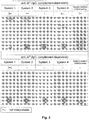
- systems 1 and 2 provide a clear (positive) read out shown at two levels. This in contrast with the system according to the prior art showing a diffuse smear over at least part of the column length (upper part of Figure 3 ), or a false negative reaction (lower part Figure 3 ).
- systems 1 and 2 according to the present invention provide a clear (positive) read out shown at two levels. This in contrast with the system according to the prior art showing a diffuse smear over at least part of the column length.
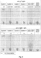
- systems 1 and 2 according to the present invention provide a clear (positive) read out shown at two levels. This in contrast with the systems according to the prior art maximally showing a diffuse smear over at least part of the column length or a false negative reaction.
- system 1 provides a clear (positive) read out shown at two levels. This in contrast with the systems according to the prior art maximally only showing false negative reactions.
Description
- The present invention relates to a micro column capable of detecting carrier-bound analyte complexes, and, more particularly, erythrocyte-bound antibody complexes or agglutinates. Further, the present invention relates to a method for detecting carrier-bound analyte complexes, and, particularly, erythrocyte-bound antibody complexes, comprising the use of the present micro column. Furthermore, the present invention relates to a kit of parts comprising the present micro column for use in the present method. The present micro column, method, and kit of parts are preferably used in the field of blood group serology, and, especially, blood group assays and antibody detection.
- In a clinical setting, a major objective of blood group serology, and, especially, blood group assays and antibody detection, is to obtain compatible red blood cell or erythrocyte preparations from a donor for blood transfusion of an acceptor. For this, assays are routinely performed such as blood group typing, antibody screening and identification, and (blood) compatibility assays by performing cross matches.
- In general, the majority of these assays are based on the principle of agglutination, i.e., the formation of carrier-bound analyte complexes, such as erythrocyte-bound antibodies complexes. Such complexes, clearly microscopically visible or, in some cases, already visible by the naked eye, can comprise from about 50 up to thousands of carrier-bound analytes, such as erythrocyte-bound antibodies.
- In a mammal, and particularly a human mammal, specific antigenic determinants on the cell membrane of red blood cells, or erythrocytes, are characterizing the so-called blood group of said mammal. The information for expressing blood group antigens is generally genetically determined at the genomic level.
- A well known, and generally used, blood group system is the so-called A, B, AB, and O system (AB0-system), discovered in 1900 by Karl Landsteiner. Different blood groups in this system are designated A, B, AB, and O.
- In addition to the ABO-system, more than 400 red blood cell blood groups are known, the majority of these being clustered in blood group systems with diverging clinical significance. Specific antibodies to these other blood group systems can be formed after immunization with the corresponding blood group antigen, for example during blood transfusion or pregnancy, and may cause problems during a blood transfusion or pregnancy thereafter.
- Presently, for transfusion practices, the ABO-system is the most important blood group system. This because every individual, characterized by a specific blood group, has antibodies in his serum against the blood group(s) that are not present. For example, an individual characterized as blood group A, will have anti-B antibodies in his serum, and vice versa.
- These antibodies are so-called "naturally occurring" antibodies and are in general strong IgM type antibodies. The IgM antibodies are capable of causing a direct, i.e., without a bridging reagent, such as an anti-IgM antibody, complex formation or agglutination of, for example, erythrocytes exposing a blood group antigen against which the antibody is directed.
- Blood transfusion reactions in an individual (recipient) induced by allo-antibodies, raised against foreign erythrocytes or red blood cells are called hemolytic transfusion reactions. This because they are generally accompanied by a strongly accelerated, and often deadly, breakdown of erythrocytes. Therefore, it of major importance to prevent hemolytic transfusion reactions by careful serological examination before a blood transfusion is performed. For this, several types of tests are generally carried out.
- In general, both donor and recipient are typed for the AB0-blood group system and Rhesus D antigen. These must be identical, or compatible, for both the donor and recipient. The AB0-blood group found on the red blood cells can, for example, be confirmed by performing a so-called reverse AB0 typing test on the antibodies present in the serum.
- Next, a screening of the serum of the recipient is performed for the presence of red blood cell antibodies directed against all other blood groups beside the AB0-system. If an antibody is found, it must be identified in order to select donor blood which is negative for the corresponding blood group antigen.
- Finally, a cross-match between donor red blood cells and recipient serum can be performed to find out whether the donor and the recipient are in deed compatible.
- In general, blood group antibodies are immunoglobulins of the IgG or IgM type. The antigen-antibody reaction, such as the present carrier-analyte reaction forming carrier-bound analytes, is dependent, amongst others, on ionic binding, hydrogen bridges and hydrophobic effects (displacement of water). The strength of the binding between the binding pocket of an antibody and an epitope is designated as "affinity".
- Antibodies which are capable of agglutinating red blood cells under all conditions are designated agglutinins or complete antibodies (in general IgM antibodies). Antibodies which bind to (sensitize) red blood cells, but cause no direct agglutination, are called incomplete antibodies (in general IgG antibodies).
- Red blood cell antigens and their corresponding antibodies are often detected by means of agglutination reactions, which can take place in a physiological salt solution. In practice, agglutination tests can be rendered more sensitive by using, for example, a medium having a low ionic strength, proteolytic enzymes (e.g. bromelin, papain or ficin), polycations (e.g. polybrene), macromolecules (e.g. albumin), or polymers (e.g. polyethylene glycol (PEG) or dextrane). A large number of serological tests is known.
- Presently, the most common, and generally used, serological tests are the tube method, micro column tests, and tests in micro plates. These techniques can be further divided into techniques based on agglutination, i.e. complex formation, and techniques based on a solid-phase (affinity) principle.
- Although serological test based on DNA techniques are available, for example, blood group typing, their application in the field of serology is still limited. In addition, techniques based on fluorescent labels or magnetic beads are available.
- When considering the present major clinical applications in the field of serology, the tube method, micro column tests, and tests in micro plates tests are widely used and will be further detailed below.
- The tube method is a widely used test allowing prolonged incubations with antibodies. Red blood cells, after a reaction with antibodies, can be sedimented, or centrifuged, to accelerate the agglutination reaction.
- An important, and widely applied, variant of the tube method test is the antiglobulin test or Coombs test, described by Moreschi in 1908 (Zbl. Bakt. 46: 49, 1908) and reintroduced in 1945 by Coombs et al. (Lancet 2: 15, 1945 and Brit. J. Exp. Path. 26: 255, 1945). The Coombs test is based on the principle that red blood cells loaded with, for example, incomplete antibodies of the IgG type can be agglutinated through the addition of antiglobulin serum. In the test, three phases can be distinguished.
- The first phase is the sensitization phase. During this phase, antibodies bind to the corresponding antigen structures on the red blood cells (sensitization of red blood cells). When binding, thereby forming carrier (erythrocyte)-bound analytes (antibodies), has occurred, a second phase is initiated, also designated as the wash phase. In this wash phase, substantially all non-bound antibodies are removed from the incubation mixture.
- The third phase is designated as the antiglobulin phase, in which antiglobulin serum is added to the washed sensitized, i.e., antibody loaded, cells. This causes binding of sensitized cells to each other resulting in the formation of complexes or agglutinates comprised of clustered, i.e., from about 50 to thousands, red blood cells.
- When performing the Coombs test, it is necessary, before adding the antiglobulin serum, to wash very thoroughly and frequently, and thus this step is very time consuming. Insufficient removal of non-bound antibodies can lead to inactivation of the antiglobulin serum. Other disadvantages of the test are the need for promptly reading the results by a trained professional, that the test results cannot be preserved, the test is less reproducible because of manual reading of the test results, and difficulties in automating the test.
- As indicated, the washing step in the Coombs test is very time consuming. Graham et al. (Transfusion 22: 408, 1982 and P.L. Mollison, Blood transfusion in clinical medicine, Blackwell, Oxford, p. 512, 1983) developed a technology rendering the wash step redundant. This technology was commercialized by Ortho Diagnostic Systems Inc. in a test system marketed as Simwash®.
- In this system, red blood cells are separated from the serum by means of a centrifugation step. The separation is based on the observation that the specific gravity of serum is lower than that of red cells. If a mixture of cells and serum is applied on top of a layer of medium with a density between the density of the cells and that of the serum, the (sensitized) red blood cells will be separated in a sedimentation or centrifugation step from the serum (containing the non-bound antibodies). Thus, the red blood cells are sedimentated or centrifuged out of the incubation mixture. In this way, a triple or quadruple washing step, needed in the Coombs test, is reduced to a single centrifugation step. Thereafter, the sensitized cells are incubated with antiglobulin serum and the formation of complexes or agglutinates is detected.
- A further improvement of the test is provided by centrifuging red blood cells through a column of transparent, inert, solid particles (micro column test) such as a Sephadex® gel or glass beads (Dalton et al., 1970, (Beckton Dickinson & Co.,
U.S. Pat. No. 3,492,396 ). Because of their size, in theory, agglutinates are not capable to pass through the column in contrast to the non-agglutinated (individual) cells. As a consequence, agglutinates will be retained on top, in the upper part, or in the remainder, of the column depending on the column material used and/or the size of the agglutinates. This in contrast with non-agglutinated cells that readily pass through the column. - The principle of using a column of inert and solid particles for retaining agglutinates was further refined by LaPierre et al. (Transfusion 30: 109-113, 1990 and European Patents
0 194 212 and0 305 337 ). This system combines the principles of Simwash® and the use of solid inert particles to discriminate between complexes or agglutinates and non-agglutinates. - In this test system, use is made of small columns filled with Sephadex® gel. Use can be made of columns comprising antibodies (e.g. for blood group typing) or no antibodies (e.g. for reverse ABO typing).
- In order to perform a Coombs test, use is made of gel columns containing antiglobulin serum. After incubation, the gel columns are centrifuged. In case of a negative reaction, i.e., no carrier-bound analytes complexes are formed, all (individual) red blood cells will end up at the bottom of the micro column; if the test is positive, the red blood cell complexes, or agglutinates, will be more or less retained by the column, i.e., will, after centrifugation, be visible on top or somewhere in the column. In case of weak reactions, red blood cells will sediment partly resulting in red blood cells in the column and at the bottom of the column. This test is marketed by DiaMed AG (DiaMed-ID Microtyping System), Grifols (DianaGel, DG Gel® cards) and Bio-Rad/Sanofi (ScanGel® cards) and has been used in the past by Diagast Laboratoires (Chromatest).
- A comparable micro column system has been described using non-compressible micro particles instead of gel material as inert material to retain the complexes or agglutinates formed (European Patents
0 485 228 ,0 725 276 ,0 755 719 andUS patents 5,552,064 and5,650,068 ). This test is marketed by Ortho (Biovue System), using glass beads as non-compressible micro particles. - A number of drawbacks are associated with the above micro column systems. For example, a special centrifuge is required for correct performance of the test. Also, special reading equipment is required for automatic reading of the test as well as special equipment for automation of the entire test.
- A general disadvantage of the above micro column agglutination tests is the occurrence of "smear formation", especially when weak reactions are tested, resulting in uncertainty when reading and interpreting the strength of the reaction results, both at reading by the naked eye and at automated reading of the results. A red smear of red blood cells (mostly small red blood cell clusters comprised of 2 to less than 50 cells, also designated as rosettes) can be seen over the entire micro column or part thereof. These smears are, for example, caused when:
- 1. small red blood cell clusters or rosettes, formed during incubation when testing weak IgM antibodies, sediment partly since they penetrate deeper into the gel of the micro column than large and strong agglutinates;
- 2. small clusters or rosettes are formed in the gel itself due to (optionally) present antiglobulin serum when testing weak IgG antibodies;
- 3. weak agglutinates already formed are broken down into small clusters or rosettes by "shear forces" during a centrifugation step caused by a centrifugal force exerted on the agglutinates and the hindering force exerted on the agglutinates by the presence of the gel particles (Philips et al., Transfusion Medicine 7: 47-53, 1997).
- Another general drawback of the above micro column agglutination tests is that problems often occur when demonstrating weak agglutinates, for instance in the case of weak AB0-incompatibilities or in the case of some other weak IgM antibodies reactive at 37 °C, like anti-P1 and anti-Lea (Cummins & Downham, Lancet 343: 1649, 1994; Philips et al., Transfusion Medicine 7: 47-53, 1997; Cheng et al., Clin. Lab. Haem. 17: 81-84, 1995).
- The shear forces during centrifugation can cause weak agglutinates to disintegrate into several small red blood cell clusters or rosettes (or, in extreme cases, into individual (sensitized) red cells), that will not be detected since they are too small to be sieved by the gel particles and therefore will sediment, resulting in an increase of the number of false negative reactions.
- Another approach in serology testing is based on the solid-phase (affinity) principle as an alternative for direct (complex formation or agglutination without a bridging reagent) and indirect (complex formation or agglutination with a bridging reagent) reactions for blood group typing, antibody screening, antibody identification and cross match. Applications and advantages of the use of affinity solid-phase techniques in serology have been described by Rosenfield (Abstracts, 15th Cong. Int. Soc. Blood Trans., Paris, pp. 27-33, 1976 and
US Patent 4,275,053 ). Here, amongst others, red blood cells were used which had been coupled to the surface of plastic tubes. - More recently, systems have been described by Plapp et al. (Am. J. Clin. Path. 82: 719-721, 1984),
Bayer et al. (US Patent 4,608,246 ), Rachel et al. (Transfusion 25: 24-26, 1985), Plapp et al. (The Lancet 1465-1466, 1986) andUthemann et al. (US Patent 4,925,786 , European Patent0 363 510 and Transfusion 30: 114-116, 1990). - Microplates in combination with the solid-phase principle are used by, among others, Biotest AG (Erytype for typing of red cell antigens and Solidscreen II for antibody diagnostics), Immucor Inc. (Capture-R systems for antibody screening and identification) and Sanquin (Microtype for typing of red cell antigens) and have further been described by Llopis et al. (Vox Sanguinis 70: 152-156, 1996 and Vox Sanguinis 72: 26-30, 1997), Ohgama et al. (Transfusion Medicine 6: 351-359, 1996), Chateau et al. (La Gazette de la Transfusion 131: 38-41, 1997 and French Patent
94 05123 Den Boer et al. (US Patent 6,303,390 ). - The solid-phase affinity tests are not limited to microplates. Pernell (Sanofi Pasteur/BioRad, European Patent
0 594 506 ) describes an affinity gel test in which an immunoglobulin-binding substance (such as protein A, protein G or anti-human IgG) is immobilized on the gel matrix. Incubation of red blood cells and antibodies occurs above the gel matrix. After this incubation phase, centrifugation of the gel column takes place. If antibodies are bound to the red blood cells, these sensitized red cells will bind to the immunoglobulin-binding gel matrix and, hence, will bind to the upper part of the gel column. Red blood cells to which no antibodies are bound will not be bound and end up at the bottom of the column. - This principle of binding of sensitized cells to a solid phase has previously been described by GE Healthcare / Amersham/Pharmacia (Cell Affinity Chromatography, Uppsala, Sweden, 1984). Therein, Pharmacia describes the purification of red blood cells based on the presence or absence of certain blood group antigens using protein A-Sepharose 6MB. Red blood cells with the A antigen on the surface (blood group A red blood cells) to which anti-A antibodies are bound, can be separated by binding to said gel material (via protein A) from red blood cells that do no possess the A antigen.
- Therefore, the test described by Sanofi Pasteur is a combination of this principle and the centrifugation step for separating red blood cells, whether sensitized or not, and non-bound antibodies as used, for example, in the micro column system of DiaMed.
- A comparable system has also been described by Gamma/Immucor (
WO 95/31731 WO 98/16831 US Patents 5,665,558 and5,905,028 ) using micro tubes containing two layers of immunoreactive gel particles, a first layer having protein G coupled to the surface of the particles and a second layer having protein A coupled to the particles. - Two types of assays can be carried out: 1. a direct assay, where antibodies specific for blood group antigens to be tested are coupled to the protein G and a sample of red cells being added to the reaction tube above the gel; 2. an indirect assay, where samples of red blood cells, serum or plasma are incubated with a known antibody or antigen reagent in the reaction tube above the gel. In both assay types the reaction tube is subsequently centrifuged to force to the bottom of the reaction tube red blood cells that do not attach to the immunoreactive gel particles.
- An alternative to these tests, thereby improving the results, has been described by Van der Donk et al. (Dutch Patent
1008738 andEuropean Patent 1 064 556 ). Apart from an optionally present affinity ligand for binding of red cells sensitized with IgG-antibodies, here two affinity ligands are immobilized on a gel matrix in micro columns, being an affinity ligand for binding of red blood cells sensitized with IgM-antibodies and an affinity ligand for binding of red blood cells loaded with complement components, caused by complement dependent antibodies. This causes an (unexpected) improved sensitivity for detection of weak reactions mediated by IgM-antibodies reactive at 37°C, for example: 1. when detecting weak AB0 incompatibilities, or IgM type anti-Le or anti-P1 antibodies; 2. when typing weak expressions of blood group antigens or variants thereof, using typing reagents containing monoclonal IgM antibodies. An improved sensitivity is also observed for detection of complement dependent (IgG) antibodies, some of which have been described to be detectable only by means of anti-complement activity (Mollison et al., Blood Transfusion in Clinical Medicine, Blackwell Scientific Publications, 1992) . - A major disadvantage of the above described affinity gel test systems is that a relatively high amount of costly ligand molecules is necessary, while only a fraction thereof is effectively utilized.
- Since use is being made of gel particles originally meant for chromatographic applications, immobilization of ligands on these gel particles occurs not only on the outer surface of the gel particles, where they can interact with the red blood cells, but also on the relatively large inner surface of the gel particles, where the red blood cells cannot penetrate.
- Another disadvantage is that a-specific interactions can occur between (non sensitized) red blood cells and the gel matrix and/or the immobilized ligands, resulting in apparent binding of red blood cells to the gel matrix and thereby in an increase in the amount of false positive reactions.
- Considering the above, it is an object of the present invention, amongst others, to obviate at least part of the above disadvantages associated with the known serology test systems, and especially the serology test systems based on (micro) columns.
- According to the present invention, this object is met by the micro column as defined in the appended claims.
- Especially, this object is met by providing a micro column, capable of detecting carrier-bound-analyte complexes, comprising at least three fluid communicating elongated compartments, wherein said micro column comprises:
- a) a first elongated compartment (3) capable of receiving erythrocytes and/or antibodies, thereby providing a sample with a first liquid density, for forming antibody-loaded-erythrocytes, but not agglutinated-antibody-loaded-erythrocytes, wherein said first compartment (3) is in liquid communication with;
- b) a second elongated compartment (2) of about 17 µl consisting of a medium with a second liquid density, wherein said second liquid density is higher than said first liquid density, wherein said medium comprises at least one compound selected from the group consisting of anti-IgG antibodies, anti-IgM antibodies, anti-C3d antibodies, anti-IgA antibodies, anti-C3c antibodies, and functional fragments thereof and wherein said second liquid density is between 1.01 to 1.09 g/ml, preferably between 1.02 and 1.06 g/ml, more preferably between 1.02 and 1.04 g/ml, for forming agglutinated-antibody-loaded-erythrocytes, and wherein said second compartment (2) is in liquid communication with;
- c) a third elongated compartment (1) of about 5 µl consisting of inert beads, comprised in the medium according to (b), capable of providing a barrier for said agglutinated-antibody-loaded erythrocytes and not for said antibody-loaded-erythrocytes, erythrocytes and/or antibodies;
- Within the context of the present invention, carrier-bound analyte complexes and, particularly, agglutinated antibody loaded erythrocytes are defined as complexes comprising at least 50 carrier-bound analytes and agglutinated antibody loaded erythrocytes, respectively.
- In the first compartment, incubation of the carrier, such as red blood cells, and analyte, such as antibodies, results in the formation of carrier-bound analytes, such as antibody sensitized red blood cells. It is of importance to note that substantially no complex formation, i.e., agglutination, occurs in the first compartment.
- However, this inherently implies that, in the case of IgM antibodies, the spontaneous formation of complexes or agglutinates in the first compartment must be substantially prevented. The skilled person, by following the Heidelberger curve, is readily capable to choose the concentrations of carrier and analyte in such a way that complex formation or agglutination is substantially prevented in the first compartment, for example, by using a high concentration of red blood cell antibodies in combination with a low concentration of red blood cell antigens.
- It is noted that in case of IgG antibodies, no additional measures are needed to prevent complex formation or agglutination. This because IgG sensitized erythrocytes are inherently substantially not capable of forming complexes or agglutinates without the further addition of a bridging reagent, such as an anti-IgG antibody.
- After the incubation phase in the first compartment, a centrifugation takes place, such that the carrier-bound analytes are centrifuged through the second compartment, while the analyte remains above the present second compartment. In this manner, the analyte, such as antibodies, present in the sample, are capable of binding the carriers, such as red blood cells, without being hindered by a premature contact with free antibodies present in the second compartment. Vice versa, the optional free antibodies present in the second compartment cannot be inactivated by a premature contact with the analyte, such as an antibody containing sample.
- Further, because of the density of the medium in the second compartment, the unbound analyte will be effectively separated from the carrier.
- Red blood cells sensitized with analyte, such as complement factors and/or IgG antibodies and/or IgM antibodies and/or IgA antibodies, are allowed to react with the optional free antibodies present in the second compartment, thereby reinforcing sensitized red blood cells into a loosely associated 3-dimensional network of red blood cells, followed by subsequent formation of complexes or agglutinates of sensitized red blood cells upon landing on the top of the third compartment.
- In order to obtain this effect, it of critical importance that the second compartment has a sufficient length in relation to the third compartment in combination with a sufficient density of the medium in the second compartment.
- The present inventors have surprisingly found that when the fluid density is between 1.01 to 1.09 g/ml and the ratio of the length of the elongated second compartment to the third elongated compartment is between 1.1 to 1.7, complex formation and agglutination is provided at the interface of the compartments, thereby providing the observed increased sensitivity of the present micro column over the prior art systems.
- Further, since complex formation or agglutination takes only place at the interface of the second and third compartment, shear forces occurring during passage of agglutinates through the column matrix in other column agglutination test systems will not occur according to the present invention because complexes or agglutinates do not pass through the column matrix.
- Using the micro column according to the present invention, it was found possible to demonstrate, in addition to IgG antibodies, complement-dependent antibodies with a high sensitivity.
- In addition, it also was possible to demonstrate IgM antibodies, including weak IgM antibodies, reactive at 37 °C, with a high sensitivity.
- Further, the reading of the test results was improved and more univocal because of detection of red blood cells at two levels, at the bottom (negative reaction) and at the interface of the third compartment (positive reaction).
- According to a preferred embodiment of the present micro column, the second fluid density is between 1.02 and 1.06 g/ml, more preferably between 1.02 and 1.04 g/ml.
- According to another preferred embodiment, the ratio of the length of the elongated second compartment to the third elongated compartment is between 1.3 to 1.5.
- According to yet another preferred embodiment of the present micro column, the top-end diameter of the first elongated compartment is larger than the bottom-end diameter of the first elongated compartment for facilitating sample receiving, i.e., carrier and analyte.
- Not according to the presently claimed micro column, the diameter of the second and third elongated compartment is substantially equal to the diameter of the bottom-end diameter of the first elongated compartment.
- Preferably, the present medium defining the second elongated compartment comprises 2% to 4% of dextran and 1% to 3% serum albumin with a pH of at least 6, and, more preferably, further comprises a physiological salt solution, a chelator and a mono- or disaccharide.
- The presence of macromolecules and polymers in the present medium defining the second compartment increases the interaction between the red blood cell antigens and antibodies and between (optionally) free anti-human antibodies and red blood cell antibodies present on the red blood cells. In this way association of sensitized red blood cells is promoted and, subsequently, complex formation or agglutination of red blood cells takes place upon sedimentation on top of the gel matrix in stead of inside the gel matrix (in the case of IgG-type antibodies and/or weak IgM-type antibodies).
- In a more preferred embodiment, the present medium defining the second elongated compartment further comprises 15 to 25 mM EDTA, 60 to 80 mM NaCl, 20 to 40 mM glucose in a phosphate buffered solution (PBS).
- In a most preferred embodiment, the present medium, defining the second elongated compartment, comprises 2.5% dextran, 2% serum albumin, 20 mM EDTA, 70 mM NaCl, and 30 mM glucose in a phosphate buffered solution (PBS).
- According to the invention, the above medium, defining the second elongated compartment, further comprises at least one compound selected from the group consisting of anti-IgG antibodies, anti-IgM antibodies, anti-C3d antibodies, anti-IgA antibodies, and anti-C3c antibodies, more preferably anti-IgG antibodies, anti-IgM antibodies, and anti-C3d antibodies.
- The above addition of antibodies strongly increases the sensitivity, and especially the sensitivity for weak IgM-type antibodies (reactive at 37°C), complement-dependent antibodies, and normal IgG-type antibodies. The presence of anti-human IgG antibodies, anti-human IgM antibodies and anti-human complement antibodies in the medium defining the second elongated compartment reinforces the network of sensitized red blood cells before the actual complex forming or agglutination reaction takes place.
- According to a preferred embodiment, the inert beads, comprised in the medium as defined above and defining the third elongated compartment, have an average diameter of between 10 to 150 µm, preferably between 20 to 50 µm.
- Further, according to a preferred embodiment, the inert beads, comprised in the medium as defined above and defining the third elongated compartment, are selected from the group consisting of glass, sepharose, acryl, crosslinked agarose, and sephadex beads.
- According to the present invention, the carrier and analyte are erythrocytes and antibody, respectively.
- According to the present invention, the carrier-bound analytes and the carrier-bound analyte complexes are antibody loaded erythrocytes and agglutinated antibody loaded erythrocytes, respectively.
- The present micro column as defined above provides many advantages over the serology test systems of the prior art, especially with respect to an increased sensitivity and the absence of "smear" formation in the column compartment, not only allowing an increased sensitivity, but also reducing the number of false negative results, and a high automation potential of the test.
- Considering the above advantageous properties of the present micro column, the present invention also relates to a method for detecting agglutinated antibody-loaded-erythrocytes as defined in
claim 10. - According to a preferred embodiment of the present method, the incubation comprises 1 second to 30 minutes at 15 to 37 °C.
- The centrifugation according to the present method preferably comprises 1 to 3 minutes at 50 to 100xg; 0.5 to 1.5 minutes at 150 to 250xg; and 5-10 minutes at 1500 to 2000xg, more preferably, 2 minutes at 75xg; 1 minute at 200xg; and 7 minutes at 1790xg.
- According to the present method the analyte is antibody; the carrier is erythrocytes; the carrier-bound analyte is antibody loaded erythrocytes; and the carrier-bound analyte complex is agglutinated antibody loaded erythrocytes.
- The present invention also relates to a kit of parts for carrying out the present method comprising:
- a) at least one micro column as defined above;
- b) instructions for use; and
- c) optionally, one or more control analytes and/ or carriers.
- The present invention will further be detailed in the following examples comprising, and describing, preferred embodiments of the present invention. In the examples, reference is made to the appended figures, wherein:
- Figure 1:
- shows a test card with six micro columns. In a micro column (7) closed at the bottom, a gel matrix of inert particles (1) is provided. On this gel matrix there is a layer of separation medium of high density (2). The incubation compartment (3) is formed by a widening at the top of the micro column. A micro column test system can be integrated into a base plate (6) and be placed in a centrifuge using supporting and guiding elements (9, 10, 11).
The reference numerals denote:- 1 Matrix of inert particles
- 2 Separation medium of high density
- 3 Incubation compartment
- 4 Micro column wall
- 5 Micro column bottom
- 6 Base plate
- 7 Micro column
- 8 Recess
- 9 Top surface
- 10 Slot
- 11 Support
- Figure 2:
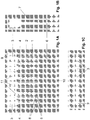
- shows the various possible reaction strengths. The strength indications denote:
4+ all agglutinated cells on top of the inert gel matrix, no cells at the bottom
3+ more agglutinated cells on top of the inert gel matrix than non-agglutinated cells at the bottom
2+ as many agglutinated cells on top of the inert gel matrix as non-agglutinated cells at the bottom
1+ fewer agglutinated cells on top of the inert gel matrix than non-agglutinated cells at the bottom
(+) very few agglutinated cells on top of the inert gel matrix, many non-agglutinated cells at the bottom
- no agglutinated cells on top of the inert gel matrix, all cells at the bottom - Figure 3:
- Test results of 5 micro column systems: System 1: The test system as described having free anti-human IgG, anti-human IgM and anti-human C3d in the high density medium; System 2: As
system 1, but with free anti-human IgG alone in the high density medium; System 3: Assystem 1, but without free antibodies in the high density medium; System 4: Assystem 1, but without the layer of high density medium above of the gel matrix; System DiaMed LISS/Coombs: DiaMed ID LISS/Coombs micro column test card, containing free anti-human IgG and anti-human C3d in the gel matrix. - Figure 4:
- Test results of 5 micro column systems: System 1: The test system as described having free anti-human IgG, anti-human IgM and anti-human C3d in the high density medium; System 2: As
system 1, but with free anti-human IgG alone in the high density medium; System 3: Assystem 1, but without free antibodies in the high density medium; System 4: Assystem 1, but without the layer of high density medium above of the gel matrix; System DiaMed LISS/Coombs: DiaMed ID LISS/Coombs micro column test card, containing free anti-human IgG and anti-human C3d in the gel matrix. - Figure 5:
- Test results of 6 micro column systems: System 1: The test system as described having free anti-human IgG, anti-human IgM and anti-human C3d in the high density medium; System 2: As
system 1, but with free anti-human IgG alone in the high density medium; System 3: Assystem 1, but without free antibodies in the high density medium; System 4: Assystem 1, but without the layer of high density medium above of the gel matrix; System DiaMed LISS/Coombs: DiaMed ID LISS/Coombs micro column test card, containing free anti-human IgG and anti-human C3d in the gel matrix; System DiaMed neutral: DiaMed ID NaCl, enzyme test and cold agglutinins micro column test card, containing no free antibodies in the gel matrix. - Figure 6:
- Test results of 6 micro column systems: System 1: The test system as described having free anti-human IgG, anti-human IgM and anti-human C3d in the high density medium; System 2: As
system 1, but with free anti-human IgG alone in the high density medium; System 3: Assystem 1, but without free antibodies in the high density medium; System 4: Assystem 1, but without the layer of high density medium above of the gel matrix; System DiaMed LISS/Coombs: DiaMed ID LISS/Coombs micro column test card, containing free anti-human IgG and anti-human C3d in the gel matrix; System DiaMed neutral: DiaMed ID NaCl, enzyme test and cold agglutinins micro column test card, containing no free antibodies in the gel matrix. - The invention will now be further explained in and by the following examples. It is of importance to see, however, that these examples are given for illustrative purposes and illuminate the enablement of the invention but do not in any way constitute the definitive conditions of the different assay methods nor limit the scope of the invention in any way.
- 10 ml Sepharose HP (GE Healthcare/Amersham, Uppsala, Sweden) was washed according to the instructions of the supplier and included in a 20 mM phosphate buffer, further containing 70 mM NaCl, 20 mM EDTA, 30 mM glucose, 2.5% dextran, 2.0% BSA, pH 8.8 (gel buffer).
- To this was added, polyclonal goat serum, directed to human IgG (diluted 100-fold in the final gel suspension), monoclonal mouse IgG, directed to human IgM (final concentration of 1 pg/ml) and monoclonal mouse IgG, directed to human C3d (final concentration of 5 pg/ml).
- Additional gel buffer was added until a final gel suspension of 23% was obtained. Per micro column of a test card according to
Figure 1 , 22 µl gel suspension was added. The gel sedimented within 15 min (yielding about 5 µl of packed gel, i.e., the present third elongated compartment). The supernatant gel buffer (about 17 µl) functions as a layer of high density separation medium, i.e., the present second elongated compartment. - Red blood cells of blood group 0+ (i.e. blood group 0, Rhesus D positive) were sensitized with IgG type anti-D antibodies and then 3 times washed in a modified LISS (low ionic strength solution). Of these red blood cells, a 0.5% suspension was made in LISS. Then, 1 drop of this suspension was introduced into the incubation compartment of the micro column. To this was added, 1 drop of normal serum from a donor of blood group 0+.
- After an incubation period of 15 min at 37°C, the micro column was centrifuged using the following protocol: 2 min at 75xg, followed by 1 min at 200xg, followed by 7 min at 1790xg.
- Then the result was assessed: all red blood cells were agglutinated on top of the inert gel matrix (
reaction strength 4+). As a control experiment, the procedure was repeated with non-sensitized red cells of blood group 0+. These cells were not agglutinated but collected at the bottom of the micro column. - A micro column card as described in Example 1 was prepared. Of Duffy-a (Fya) positive red blood cells (phenotype Fya+b+), a 0.5% suspension in LISS was made. Then 1 drop of this suspension was introduced into the incubation compartment of the micro column. To this was added 1 drop of anti-Fya (polyclonal IgG antiserum, Sanquin Reagents, Amsterdam, Netherlands).
- After an incubation period of 15 min at 37°C, the micro column was centrifuged according to the protocol of Example 1.
- After this, the result was assessed: all red cells were agglutinated on top of the inert gel matrix (
reaction strength 4+). As a control experiment, corresponding non-sensitized red cells were tested. These cells were not agglutinated but collected at the bottom of the micro column. - This experiment was repeated for the following antigens with the corresponding polyclonal IgG antisera: Fyb (with anti-Fyb serum and a Fya+b+ cell), Jka (with anti-Jka serum and a Jka+b+ cell), Jkb (with anti-Jkb serum and a Jka+b+ cell), Kell (with anti-K serum and a K+k+ cell), cellano (with anti-k serum and a K+k+ cell), Lua (with anti-Lua serum and a Lua+b+ cell), Lub (with anti-Lub serum and a Lua+b+ cell), S (with anti-S serum and a S+s+ cell) and s (with anti-s serum and a S+s+ cell). The results were similar to the results described for Fya.
- In all cases, complete agglutination of red cells on top of the inert gel matrix occurred (
reaction strength 4+), while non-sensitized red cells in the control experiments were not agglutinated but collected at the bottom of the micro column. - A micro column card as described in Example 1 was prepared. Of red blood cells carrying the A antigen (A positive cells), a 0.5% suspension in LISS was made. Then 1 drop of this suspension was introduced into the incubation compartment of the micro column. To this was added 1 drop of anti-A (monoclonal IgM reagent, Sanquin Reagents, Amsterdam, Netherlands). Next, the micro column was centrifuged according to the protocol of Example 1.
- After this, the result was assessed: all red cells were agglutinated on top of the inert gel matrix (
reaction strength 4+). As a control experiment, corresponding non-sensitized red cells were tested. These cells were not agglutinated but collected at the bottom of the micro column. - This experiment was repeated for the following antigens with the corresponding monoclonal IgM reagents: B (with anti-B and a B positive cell), D (with anti-D and a D positive cell), C (with anti-C and a C+c+ cell), c (with anti-c and a C+c+ cell), E (with anti-E and a E+e+ cell), e (with anti-e and a E+e+ cell) and Kell (with anti-K and a K+k+ cell). The results were in agreement with what has been described for the A positive cell. In all cases, complete agglutination of red cells on top of the inert gel matrix occurred, while non-sensitized red cells in the control experiments were not agglutinated but collected at the bottom of the micro column.
- In this example, the following test systems were assayed:
- The test system as described in Example 1 having free anti-human IgG, anti-human IgM and anti-human C3d in the high density medium.
- As
system 1, but with free anti-human IgG alone in the high density medium. - As
system 1, but without free antibodies in the high density medium. - As
system 1, but without the layer of high density medium above of the gel matrix. To that end the layer of high density medium was carefully removed with a capillary tip, leaving the gel particles settled and just covered with liquid medium, still containing the potentiators and free antibodies. - DiaMed ID LISS/Coombs micro column test card, containing free anti-human IgG and anti-human C3d in the gel matrix.
- DiaMed ID NaCl, enzyme test and cold agglutinins micro column test card, containing no free antibodies in the gel matrix.
- The last 2 systems were used according to the manufacturer's instructions.
- Some of the above-mentioned test systems were examined for their ability to demonstrate red cells loaded with complement-components in vitro (C3b cells, C3d cells) or in vivo (patient samples). For this purpose fresh citrate-blood and an MgCl2-solution were added to a cold sucrose-solution.
- After 45 min of slowly stirring the mixture, the resulting C3b cells were washed 4 times with cold PBS. A part of the C3b cells was treated with trypsin after which the resulting C3d cells were washed 3 times with an excess of PBS.
- In addition, two patient samples were used, which were known to be loaded in vivo with complement components only. Of these C3b, C3d and patient cells, respectively, a 0.5% suspension in LISS was prepared. Then 1 drop of this suspension was introduced into the incubation compartment of the micro column. Directly hereafter the micro columns were centrifuged according to the protocol of Example 1.
- After this, the result was assessed. The reaction strengths are summarized in Table 1. As a control, experiments were carried out with untreated cells instead of cells loaded with complement-components, yielding only negative reactions (results not shown).
Table 1 Sample System 11 System 2System 3C3b cells 4+ - - C3d cells 4+ - - Patient 12+ - - Patient 22+ - - 1) 4+: all agglutinated cells on top of the inert gel matrix, no cells at the bottom; 3+more agglutinated cells on top of the inert gel matrix than non-agglutinated cells at the bottom; 2+: as many agglutinated cells on top of the inert gel matrix as non-agglutinated cells at the bottom; 1+: fewer agglutinated cells on top of the inert gel matrix than non-agglutinated cells at the bottom; (+): very few agglutinated cells on top of the inert gel matrix, many non-agglutinated cells at the bottom; -: no agglutinated cells on top of the inert gel matrix, all cells at the bottom - Several of the above-mentioned test systems were examined for their ability to demonstrate complement-dependent antibodies. For this, two patient sera were tested which were known to contain complement-dependent IgG type anti-Jka and anti-Jkb antibodies, respectively. Of Jka+b-cells and Jka-b+ cells, respectively, a 0.5% suspension in LISS was prepared. Then 1 drop of this suspension was introduced into the incubation compartment of the micro column. To this was added 1 drop of patient serum.
- After an incubation period of 15 min at 37°C, the micro columns were centrifuged according to the protocol of Example 1. After this, the result was assessed. The reaction strengths are summarized in
Figure 3 . As a control, experiments were carried out with AB-serum instead of patient serum, yielding only negative reactions (results not shown) . - As can be seen in
Figure 3 ,systems Figure 3 ), or a false negative reaction (lower partFigure 3 ). - Several of the above-mentioned test systems were examined for their ability to demonstrate (weak) IgG type antibodies. For this purpose, two patient sera were tested which were known to contain (weak) IgG type antibodies (anti-K and anti-Fya). Of K+k+ cells and of Fya+b+ cells, respectively, a 0.5% suspension in LISS was prepared. Then 1 drop of red cell suspension was introduced into the incubation compartment of the micro column. To this was added 1 drop of patient serum.
- After an incubation period of 15 min at 37°C, the micro columns were centrifuged according to the protocol of Example 1. After this, the result was assessed. The reaction strengths are summarized in
Figure 4 . As a control, experiments were carried out with AB-serum instead of patient serum, yielding only negative reactions (results not shown) . - As can be seen in
Figure 4 ,systems - All of the above-mentioned test systems were examined for their ability to demonstrate weak IgM type antibodies. For this purpose, two patient plasma samples were tested which were known to contain weak IgM type antibodies (anti-Lea (IgM) and anti-s (IgM + IgG)). Of Lea+b+ cells and S+s+ cells, respectively, a 0.5% suspension in LISS was prepared. Then 1 drop of red cell suspension was introduced into the incubation compartment of the micro column. To this was added 1 drop of patient plasma. After an incubation period of 15 min at 37°C, the micro columns were centrifuged according to the protocol of Example 1.
- After this, the result was assessed. The reaction strengths are summarized in
Figure 5 . As a control, experiments were carried out with AB-plasma instead of patient serum, yielding only negative reactions (results not shown). - As can be seen in
Figure 5 ,systems - Similar to example 7, six weak AB0-incompatibilities were tested, viz. A2B red cell samples with blood group B plasma samples (containing IgM type anti-A antibodies), which in a normal tube agglutination test in a physiological salt solution still gave a 1+ reaction.
- Of the different A2B red cells, a 0.5% suspension in LISS was prepared. Then 1 drop of red cell suspension was introduced into the incubation compartment of the micro column. To this was added 1 drop of patient plasma. After an incubation period of 15 min at 37°C, the micro columns were centrifuged according to the protocol of Example 1.
- After this, the result was assessed. The reaction strengths are summarized in
Figure 6 . As a control, experiments were carried out with AB-plasma instead of group B plasma, yielding only negative reactions (results not shown) . - As can be seen in
Figure 6 ,system 1 according to the present invention provides a clear (positive) read out shown at two levels. This in contrast with the systems according to the prior art maximally only showing false negative reactions. - From
Figure 3 it is clear that complement dependent IgG antibodies are demonstrated with a higher sensitivity according to the present invention (systems 1 and 2). The addition of, next to free anti-human IgG, free anti-human C3d as a ligand in the high density medium of the test system (system 1) further increased the sensitivity. - From
Figure 5 andFigure 6 , it is clear that weak agglutinins reactive at 37 °C are demonstrated with a higher sensitivity according to the present invention. When, next to free anti-human IgG and free anti-human C3d, also free anti-human IgM is present as a ligand in the high density medium of the test system the sensitivity is further increased (system 1). Moreover, in the absence of free anti-human IgM some weak agglutinins reactive at 37 °C can no longer be demonstrated (system 2 and system 3). - From
Figures 3-6 , it is clear that the presence of the second compartment according to the present invention is essential for obtaining a correct test result. Without this (system 4) red blood cell agglutination still seems feasible to some extent, due to the presence of free ligands in the medium between the gel beads. - The results, however, show a (vague) smear of red cells instead of a sharp and distinct red line on top of the gel matrix, thereby excluding the possibility of reading the result on only two levels (underneath and on top of the gel matrix). As a consequence the reaction strength can no longer be interpreted.
- Moreover, some weak IgG type antibodies and weak agglutinins reactive at 37 °C could no longer be demonstrated when the high density medium above the gel matrix was removed (system 4).
Claims (13)
- Micro column (7) capable of detecting agglutinated-antibody-loaded-erythrocytes comprising at least three fluid communicating elongated compartments (1, 2 and 3), wherein said micro column (7) comprises:a) a first elongated compartment (3) capable of receiving erythrocytes and/or antibodies, thereby providing a sample with a first liquid density, for forming antibody-loaded-erythrocytes, but not agglutinated-antibody-loaded-erythrocytes, wherein said first compartment (3) is in liquid communication with;b) a second elongated compartment (2) of about 17 µl consisting of a medium with a second liquid density, wherein said second fluid density is higher than said first fluid density, wherein said medium comprises at least one compound selected from the group consisting of anti-IgG antibodies, anti-IgM antibodies, anti-C3d antibodies, anti-IgA antibodies, anti-C3c antibodies, and functional fragments thereof and wherein said second liquid density is between 1.01 to 1.09 g/ml, preferably between 1.02 and 1.06 g/ml, more preferably between 1.02 and 1.04 g/ml, for forming agglutinated-antibody-loaded-erythrocytes, and wherein said second compartment (2) is in liquid communication with;c) a third elongated compartment (1) of about 5 µl consisting of inert beads, comprised in the medium according to (b), capable of providing a barrier for said agglutinated-antibody-loaded erythrocytes and not for said antibody-loaded-erythrocytes, erythrocytes and/or antibodies;wherein the ratio of the length of said elongated second compartment (2) to said third elongated compartment (1) is between 1.1 to 1.7 thereby providing complex formation and agglutination of said antibody-loaded-erythrocytes at the interface of said second (2) and third (1) elongated compartment.
- Micro column (7) according to claim 1, wherein the top-end diameter of said first elongated compartment (3) is larger than the bottom-end diameter of said first elongated compartment (3).
- Micro column (7) according to any of the claims 1 to 2, wherein said medium with a second liquid density comprises 2% to 4% of dextran and 1% to 3% serum albumin with a pH of at least 6.
- Micro column (7) according to claim 3, wherein said medium with a second liquid density further comprises a physiological salt solution, a chelator and a mono- or disaccharide.
- Micro column (7) according to claim 3 or claim 4, wherein said medium with a second liquid density further comprises 15 to 25 mM EDTA, 60 to 80 mM NaCl, 20 to 40 mM glucose in a phosphate buffered solution (PBS).
- Micro column (7) according to any of the claims 3 to 5, wherein said medium with a second liquid density comprises 2.5% dextran, 2% serum albumin, 20 mM EDTA, 70 mM NaCl, and 30 mM glucose in a phosphate buffered solution (PBS).
- Micro column (7) according to claim 1 comprising anti-IgG antibodies, anti-IgM antibodies, anti-C3d antibodies.
- Micro column (7) according to any of the claims 1 to 7, wherein said inert beads, comprised in the medium according to (b), have an average diameter of between 10 to 150 µm, preferably between 20 to 50 µm.
- Micro column (7) according to any of the claims 1 to 8, wherein said inert beads, comprised in the medium according to (b), are selected from the group consisting of glass, sepharose, acryl, crosslinked agarose, and sephadex beads.
- Method for detecting agglutinated antibody-loaded-erythrocytes comprising:a) loading erythrocytes and antibodies into a first elongated compartment (3) of a micro column (7) as defined in any of the claims 1 to 9;b) incubating said antibodies and said erythrocytes in said first elongated first compartment (3) for forming antibody-loaded-erythrocytes;c) introducing, by centrifugation, said antibody-loaded-erythrocytes into a second elongated compartment (2) of a Micro column (7) as defined in any of the claims 1 to 9 for forming agglutinated-antibody-loaded-erythrocytes;d) separating, by centrifugation, said agglutinated-antibody-loaded-erythrocytes from said antibody-loaded-erythrocytes and said erythrocytes in a third elongated compartment (1) of a Micro column (7) as defined in any of the claims 1 to 9;e) detecting the presence of agglutinated-antibody-loaded-erythrocytes at the interface of said second (2) and third (1) elongated compartment of a micro column (7) as defined in any of the claims 1 to 9.
- Method according to claim 10, wherein the incubation comprises 1 second to 30 minutes at 15 to 37 °C.
- Method according to claim 10 or claim 11, wherein the centrifugation as defined in steps (c) and (d) comprises 1 to 3 minutes at 50 to 100xg; 0.5 to 1.5 minutes at 150 to 250xg; and 5-10 minutes at 1500 to 2000xg, preferably wherein the centrifugation as defined in steps (c) and (d) comprises 2 minutes at 75xg; 1 minute at 200xg; and 7 minutes at 1790xg.
- Kit of parts for carrying out a method as defined in any of the claims 10 to 12 comprising:a) at least one micro column (7) as defined in any of the claims 1 to 9;b) instructions for use; andc) optionally, one or more control analytes and/ or carriers.
Applications Claiming Priority (1)
| Application Number | Priority Date | Filing Date | Title |
|---|---|---|---|
| PCT/EP2008/007325 WO2010025756A1 (en) | 2008-09-03 | 2008-09-03 | Reaction vessel capable of detecting carrier-bound analyte complexes, method for detecting carrier-bound analyte complexes, and kit of parts |
Publications (2)
| Publication Number | Publication Date |
|---|---|
| EP2340123A1 EP2340123A1 (en) | 2011-07-06 |
| EP2340123B1 true EP2340123B1 (en) | 2019-06-26 |
Family
ID=40194551
Family Applications (1)
| Application Number | Title | Priority Date | Filing Date |
|---|---|---|---|
| EP08801905.4A Active EP2340123B1 (en) | 2008-09-03 | 2008-09-03 | Reaction vessel capable of detecting agglutinated antibody loaded erythrocytes, method for detecting agglutinated antibody loaded erythrocytes, and kit of parts |
Country Status (2)
| Country | Link |
|---|---|
| EP (1) | EP2340123B1 (en) |
| WO (1) | WO2010025756A1 (en) |
Families Citing this family (3)
| Publication number | Priority date | Publication date | Assignee | Title |
|---|---|---|---|---|
| CN111398578A (en) * | 2019-12-31 | 2020-07-10 | 珠海朗泰生物科技有限公司 | Brucella antibody gel detection kit |
| CN112379100B (en) * | 2020-10-13 | 2024-01-23 | 中国科学院苏州生物医学工程技术研究所 | Distribution detection method for immune-epidemic area of platelet anti-human globulin experiment |
| CN115267181A (en) * | 2022-07-26 | 2022-11-01 | 广州市第一人民医院(广州消化疾病中心、广州医科大学附属市一人民医院、华南理工大学附属第二医院) | Reagent for detecting conventional sample of condensed blood and detection method and application thereof |
Family Cites Families (5)
| Publication number | Priority date | Publication date | Assignee | Title |
|---|---|---|---|---|
| NL9400777A (en) * | 1994-05-10 | 1995-12-01 | Stichting Centraal Lab | Solid phase filtration method for antigen and antibody determinations in blood group serology, and test kit. |
| US5905028A (en) * | 1994-05-17 | 1999-05-18 | Gamma Biologicals, Inc. | Method and apparatus useful for detecting bloodgroup antigens and antibodies |
| ES2156610T3 (en) * | 1996-12-18 | 2001-07-01 | Stiftung Fur Diagnostische For | SYNTHETIC PARTICLES AS AGLUTINATION REAGENTS. |
| NL1008738C2 (en) * | 1998-03-27 | 1999-09-28 | Stichting Bloedvoorziening | Solid phase method for antigen and antibody determinations in blood group serology, and test kit. |
| US7449329B2 (en) * | 2006-03-31 | 2008-11-11 | Hale Anne S | Blood test kit |
-
2008
- 2008-09-03 WO PCT/EP2008/007325 patent/WO2010025756A1/en active Application Filing
- 2008-09-03 EP EP08801905.4A patent/EP2340123B1/en active Active
Non-Patent Citations (1)
| Title |
|---|
| None * |
Also Published As
| Publication number | Publication date |
|---|---|
| WO2010025756A1 (en) | 2010-03-11 |
| EP2340123A1 (en) | 2011-07-06 |
Similar Documents
| Publication | Publication Date | Title |
|---|---|---|
| JP2854058B2 (en) | Method and apparatus for performing an assay | |
| US7087203B2 (en) | Methods and apparatus for blood typing with optical bio-disc | |
| US7943368B2 (en) | Reducing time to result for blood bank diagnostic testing | |
| EP0755719B1 (en) | Column agglutination assay and device | |
| US5552064A (en) | Column agglutination assay and device using biphasic centrifugation | |
| US6303390B1 (en) | Method for antigen and antibody determination in bloodgroup serology | |
| EP3485973A1 (en) | Microfluidic reagent card and detection method and application thereof | |
| EA017186B1 (en) | Multiple analysis of blood samples | |
| EP1064556B1 (en) | Solid-phase method for antigen and antibody determinations in bloodgroup serology, and test kit | |
| US7824873B2 (en) | Blood test kit | |
| JP2001502795A (en) | Method and apparatus useful for detecting blood group antigens and antibodies | |
| EP2340123B1 (en) | Reaction vessel capable of detecting agglutinated antibody loaded erythrocytes, method for detecting agglutinated antibody loaded erythrocytes, and kit of parts | |
| Knight et al. | New technologies for red-cell serology | |
| EP0760103B1 (en) | Solid-phase filtration method for antigen and antibody assays in bloodgroup serology, and test kit | |
| Judd et al. | Reactivity of FDA-approved anti-D reagents with partial D red blood cells | |
| Sigdel et al. | Comparison between the manual method of indirect coombs via gel technology and solid phase red cell adherence | |
| Trudell | Detection and identification of antibodies | |
| US20220252622A1 (en) | Single tube preparation comprising a panel of differently labeled cells for serology | |
| Ajmani et al. | Blood Test in Immunohematology and Blood Banking | |
| CN111356921B (en) | In situ serial dilution method | |
| EP1410042A1 (en) | Methods and apparatus for blood typing with optical bio-discs | |
| Spada et al. | Comparison of cross-matching methodology for detection of DEA 7 blood incompatibility |
Legal Events
| Date | Code | Title | Description |
|---|---|---|---|
| PUAI | Public reference made under article 153(3) epc to a published international application that has entered the european phase |
Free format text: ORIGINAL CODE: 0009012 |
|
| 17P | Request for examination filed |
Effective date: 20110329 |
|
| AK | Designated contracting states |
Kind code of ref document: A1 Designated state(s): AT BE BG CH CY CZ DE DK EE ES FI FR GB GR HR HU IE IS IT LI LT LU LV MC MT NL NO PL PT RO SE SI SK TR |
|
| AX | Request for extension of the european patent |
Extension state: AL BA MK RS |
|
| DAX | Request for extension of the european patent (deleted) | ||
| 17Q | First examination report despatched |
Effective date: 20120905 |
|
| STAA | Information on the status of an ep patent application or granted ep patent |
Free format text: STATUS: EXAMINATION IS IN PROGRESS |
|
| GRAP | Despatch of communication of intention to grant a patent |
Free format text: ORIGINAL CODE: EPIDOSNIGR1 |
|
| STAA | Information on the status of an ep patent application or granted ep patent |
Free format text: STATUS: GRANT OF PATENT IS INTENDED |
|
| INTG | Intention to grant announced |
Effective date: 20181219 |
|
| GRAS | Grant fee paid |
Free format text: ORIGINAL CODE: EPIDOSNIGR3 |
|
| GRAA | (expected) grant |
Free format text: ORIGINAL CODE: 0009210 |
|
| STAA | Information on the status of an ep patent application or granted ep patent |
Free format text: STATUS: THE PATENT HAS BEEN GRANTED |
|
| RAP1 | Party data changed (applicant data changed or rights of an application transferred) |
Owner name: SANQUIN REAGENTS B.V. |
|
| AK | Designated contracting states |
Kind code of ref document: B1 Designated state(s): AT BE BG CH CY CZ DE DK EE ES FI FR GB GR HR HU IE IS IT LI LT LU LV MC MT NL NO PL PT RO SE SI SK TR |
|
| REG | Reference to a national code |
Ref country code: GB Ref legal event code: FG4D |
|
| REG | Reference to a national code |
Ref country code: CH Ref legal event code: EP |
|
| REG | Reference to a national code |
Ref country code: AT Ref legal event code: REF Ref document number: 1147684 Country of ref document: AT Kind code of ref document: T Effective date: 20190715 |
|
| REG | Reference to a national code |
Ref country code: DE Ref legal event code: R096 Ref document number: 602008060524 Country of ref document: DE |
|
| REG | Reference to a national code |
Ref country code: IE Ref legal event code: FG4D Ref country code: NL Ref legal event code: FP |
|
| PG25 | Lapsed in a contracting state [announced via postgrant information from national office to epo] |
Ref country code: NO Free format text: LAPSE BECAUSE OF FAILURE TO SUBMIT A TRANSLATION OF THE DESCRIPTION OR TO PAY THE FEE WITHIN THE PRESCRIBED TIME-LIMIT Effective date: 20190926 Ref country code: HR Free format text: LAPSE BECAUSE OF FAILURE TO SUBMIT A TRANSLATION OF THE DESCRIPTION OR TO PAY THE FEE WITHIN THE PRESCRIBED TIME-LIMIT Effective date: 20190626 Ref country code: SE Free format text: LAPSE BECAUSE OF FAILURE TO SUBMIT A TRANSLATION OF THE DESCRIPTION OR TO PAY THE FEE WITHIN THE PRESCRIBED TIME-LIMIT Effective date: 20190626 Ref country code: FI Free format text: LAPSE BECAUSE OF FAILURE TO SUBMIT A TRANSLATION OF THE DESCRIPTION OR TO PAY THE FEE WITHIN THE PRESCRIBED TIME-LIMIT Effective date: 20190626 Ref country code: LT Free format text: LAPSE BECAUSE OF FAILURE TO SUBMIT A TRANSLATION OF THE DESCRIPTION OR TO PAY THE FEE WITHIN THE PRESCRIBED TIME-LIMIT Effective date: 20190626 |
|
| REG | Reference to a national code |
Ref country code: LT Ref legal event code: MG4D |
|
| PG25 | Lapsed in a contracting state [announced via postgrant information from national office to epo] |
Ref country code: BG Free format text: LAPSE BECAUSE OF FAILURE TO SUBMIT A TRANSLATION OF THE DESCRIPTION OR TO PAY THE FEE WITHIN THE PRESCRIBED TIME-LIMIT Effective date: 20190926 Ref country code: LV Free format text: LAPSE BECAUSE OF FAILURE TO SUBMIT A TRANSLATION OF THE DESCRIPTION OR TO PAY THE FEE WITHIN THE PRESCRIBED TIME-LIMIT Effective date: 20190626 Ref country code: GR Free format text: LAPSE BECAUSE OF FAILURE TO SUBMIT A TRANSLATION OF THE DESCRIPTION OR TO PAY THE FEE WITHIN THE PRESCRIBED TIME-LIMIT Effective date: 20190927 |
|
| REG | Reference to a national code |
Ref country code: AT Ref legal event code: MK05 Ref document number: 1147684 Country of ref document: AT Kind code of ref document: T Effective date: 20190626 |
|
| PG25 | Lapsed in a contracting state [announced via postgrant information from national office to epo] |
Ref country code: RO Free format text: LAPSE BECAUSE OF FAILURE TO SUBMIT A TRANSLATION OF THE DESCRIPTION OR TO PAY THE FEE WITHIN THE PRESCRIBED TIME-LIMIT Effective date: 20190626 Ref country code: SK Free format text: LAPSE BECAUSE OF FAILURE TO SUBMIT A TRANSLATION OF THE DESCRIPTION OR TO PAY THE FEE WITHIN THE PRESCRIBED TIME-LIMIT Effective date: 20190626 Ref country code: EE Free format text: LAPSE BECAUSE OF FAILURE TO SUBMIT A TRANSLATION OF THE DESCRIPTION OR TO PAY THE FEE WITHIN THE PRESCRIBED TIME-LIMIT Effective date: 20190626 Ref country code: AT Free format text: LAPSE BECAUSE OF FAILURE TO SUBMIT A TRANSLATION OF THE DESCRIPTION OR TO PAY THE FEE WITHIN THE PRESCRIBED TIME-LIMIT Effective date: 20190626 Ref country code: CZ Free format text: LAPSE BECAUSE OF FAILURE TO SUBMIT A TRANSLATION OF THE DESCRIPTION OR TO PAY THE FEE WITHIN THE PRESCRIBED TIME-LIMIT Effective date: 20190626 Ref country code: PT Free format text: LAPSE BECAUSE OF FAILURE TO SUBMIT A TRANSLATION OF THE DESCRIPTION OR TO PAY THE FEE WITHIN THE PRESCRIBED TIME-LIMIT Effective date: 20191028 |
|
| PG25 | Lapsed in a contracting state [announced via postgrant information from national office to epo] |
Ref country code: IS Free format text: LAPSE BECAUSE OF FAILURE TO SUBMIT A TRANSLATION OF THE DESCRIPTION OR TO PAY THE FEE WITHIN THE PRESCRIBED TIME-LIMIT Effective date: 20191026 Ref country code: IT Free format text: LAPSE BECAUSE OF FAILURE TO SUBMIT A TRANSLATION OF THE DESCRIPTION OR TO PAY THE FEE WITHIN THE PRESCRIBED TIME-LIMIT Effective date: 20190626 Ref country code: ES Free format text: LAPSE BECAUSE OF FAILURE TO SUBMIT A TRANSLATION OF THE DESCRIPTION OR TO PAY THE FEE WITHIN THE PRESCRIBED TIME-LIMIT Effective date: 20190626 |
|
| PG25 | Lapsed in a contracting state [announced via postgrant information from national office to epo] |
Ref country code: TR Free format text: LAPSE BECAUSE OF FAILURE TO SUBMIT A TRANSLATION OF THE DESCRIPTION OR TO PAY THE FEE WITHIN THE PRESCRIBED TIME-LIMIT Effective date: 20190626 |
|
| PG25 | Lapsed in a contracting state [announced via postgrant information from national office to epo] |
Ref country code: PL Free format text: LAPSE BECAUSE OF FAILURE TO SUBMIT A TRANSLATION OF THE DESCRIPTION OR TO PAY THE FEE WITHIN THE PRESCRIBED TIME-LIMIT Effective date: 20190626 Ref country code: DK Free format text: LAPSE BECAUSE OF FAILURE TO SUBMIT A TRANSLATION OF THE DESCRIPTION OR TO PAY THE FEE WITHIN THE PRESCRIBED TIME-LIMIT Effective date: 20190626 |
|
| PG25 | Lapsed in a contracting state [announced via postgrant information from national office to epo] |
Ref country code: IS Free format text: LAPSE BECAUSE OF FAILURE TO SUBMIT A TRANSLATION OF THE DESCRIPTION OR TO PAY THE FEE WITHIN THE PRESCRIBED TIME-LIMIT Effective date: 20200224 Ref country code: MC Free format text: LAPSE BECAUSE OF FAILURE TO SUBMIT A TRANSLATION OF THE DESCRIPTION OR TO PAY THE FEE WITHIN THE PRESCRIBED TIME-LIMIT Effective date: 20190626 |
|
| REG | Reference to a national code |
Ref country code: CH Ref legal event code: PL |
|
| REG | Reference to a national code |
Ref country code: DE Ref legal event code: R097 Ref document number: 602008060524 Country of ref document: DE |
|
| PLBE | No opposition filed within time limit |
Free format text: ORIGINAL CODE: 0009261 |
|
| STAA | Information on the status of an ep patent application or granted ep patent |
Free format text: STATUS: NO OPPOSITION FILED WITHIN TIME LIMIT |
|
| PG2D | Information on lapse in contracting state deleted |
Ref country code: IS |
|
| PG25 | Lapsed in a contracting state [announced via postgrant information from national office to epo] |
Ref country code: LI Free format text: LAPSE BECAUSE OF NON-PAYMENT OF DUE FEES Effective date: 20190930 Ref country code: CH Free format text: LAPSE BECAUSE OF NON-PAYMENT OF DUE FEES Effective date: 20190930 Ref country code: LU Free format text: LAPSE BECAUSE OF NON-PAYMENT OF DUE FEES Effective date: 20190903 Ref country code: IE Free format text: LAPSE BECAUSE OF NON-PAYMENT OF DUE FEES Effective date: 20190903 |
|
| 26N | No opposition filed |
Effective date: 20200603 |
|
| PG25 | Lapsed in a contracting state [announced via postgrant information from national office to epo] |
Ref country code: SI Free format text: LAPSE BECAUSE OF FAILURE TO SUBMIT A TRANSLATION OF THE DESCRIPTION OR TO PAY THE FEE WITHIN THE PRESCRIBED TIME-LIMIT Effective date: 20190626 |
|
| GBPC | Gb: european patent ceased through non-payment of renewal fee |
Effective date: 20190926 |
|
| PG25 | Lapsed in a contracting state [announced via postgrant information from national office to epo] |
Ref country code: GB Free format text: LAPSE BECAUSE OF NON-PAYMENT OF DUE FEES Effective date: 20190926 |
|
| PG25 | Lapsed in a contracting state [announced via postgrant information from national office to epo] |
Ref country code: CY Free format text: LAPSE BECAUSE OF FAILURE TO SUBMIT A TRANSLATION OF THE DESCRIPTION OR TO PAY THE FEE WITHIN THE PRESCRIBED TIME-LIMIT Effective date: 20190626 |
|
| PG25 | Lapsed in a contracting state [announced via postgrant information from national office to epo] |
Ref country code: HU Free format text: LAPSE BECAUSE OF FAILURE TO SUBMIT A TRANSLATION OF THE DESCRIPTION OR TO PAY THE FEE WITHIN THE PRESCRIBED TIME-LIMIT; INVALID AB INITIO Effective date: 20080903 Ref country code: MT Free format text: LAPSE BECAUSE OF FAILURE TO SUBMIT A TRANSLATION OF THE DESCRIPTION OR TO PAY THE FEE WITHIN THE PRESCRIBED TIME-LIMIT Effective date: 20190626 |
|
| P01 | Opt-out of the competence of the unified patent court (upc) registered |
Effective date: 20230526 |
|
| PGFP | Annual fee paid to national office [announced via postgrant information from national office to epo] |
Ref country code: NL Payment date: 20230921 Year of fee payment: 16 |
|
| PGFP | Annual fee paid to national office [announced via postgrant information from national office to epo] |
Ref country code: FR Payment date: 20230929 Year of fee payment: 16 Ref country code: DE Payment date: 20230928 Year of fee payment: 16 Ref country code: BE Payment date: 20230928 Year of fee payment: 16 |