1. TECHNICAL FIELD
-
The present invention is directed generally to method and compositions related to molecular biology, virology, and oncology. In certain aspects it is directed to compositions comprising and methods of profiling and/or classifying a plurality of cells or cell lines based on peptide binding characteristics.
BRIEF SUMMARY OF THE INVENTION
-
An embodiment of the invention includes methods of profiling cell lines and/or identifying peptide sequences or structures that bind a target population or family of cells. The methods include providing a plurality of cell lines; contacting each cell line with a library of phage displaying random heterologous peptides on their surface; obtaining phage that bind each of the cell lines; identifying peptides that bind each cell line; and classifying each cell line based on the identified peptides. The method can further comprise classifying each identified peptide based on the cell lines that bind each identified peptide. In one aspect, the cell lines include cancer cell lines. Cancer cell lines may include, but are not limited to kidney, breast, colon, lung, prostate, brain, liver, pancreatic, uterine, neuronal, skin, head and neck, leukemic, lymphocytic, or ovarian cancer cell lines. In another aspect, the panel is cancer cell lines. In a particular aspect, the panel is a NCI 60 panel of cancer cell lines. The methods further include identifying a peptide that binds to a majority of the cancer cell lines or cancer cells of common origin. Furthermore, methods can also include analyzing the identified peptides to identify similarities with known receptor ligands.
-
In certain aspects, classifying the cell line is performed by clustering analysis. Clustering analysis can be used to construct a clustered image map (CIM). In a particular aspect, classifying the identified peptide is performed by clustering analysis. Clustering analysis can be used to construct a clustered image map. In another aspect, the methods may also include identifying receptors for at least one of the identified peptides comprising the steps of providing an identified peptide; labeling the identified peptide; contacting an appropriate cell line with the labeled peptide; isolating a receptor - peptide complex; and identifying the receptor bound to the labeled peptide.
-
In another embodiment, a group of peptides comprising five or more peptides can be classified or identified as selectively bind to a sub-population of cell lines, wherein the peptides include, but are not limited to those listed in Table 3 and described herein. In certain aspects, a sub sequence of the peptide may be identified as conferring to the peptide a certain binding characteristic.
-
In still further embodiments, methods of the invention can be used to classify a cell or cell line. Methods of classifying a cell line include, but are not limited to steps comprising: contacting a cell with a group of selected peptides or polypeptides that differentially bind cells of a known origin; detecting the peptides that bind the cell line; and assessing the classification of the cell line based on the peptide(s) that bind the cell line. Thus, in certain aspects, classifying a cell may comprise determining whether as cell expresses a certain receptor polypeptide, is susceptible to a particular therapy or determining the tissue of origin for the cell. In certain aspects of the invention, a group of selected peptide for use according to the invention are further defined as cyclic or partially peptides, such as peptides comprising a disulfide bond. In certain cases, a group of selected peptides or polypeptides may comprise at least 3, 4, 5, 6, 7, 8, 9, 10, 15, 20, 25, 30 or more distinct peptides or polypeptides.
-
Thus, in a further specific embodiment there is provided a method for classifying a cell comprising obtaining or having a sample comprising a cell; contacting the cell with a group of peptides or polypeptides that differentially bind cells of a known origin or type; detecting the peptides that bind to the cell and classifying the cell based on the peptide binding. As described supra, in certain aspects, a group of selected peptides or polypeptides comprise amino acid sequences selected from those provided in Table 3. Thus, in certain cases a group of selected peptides or polypeptides comprise 3, 4, 5, 6, 7, 8, 9, 10, 15, 20, 25, 30 or more members that comprising an amino acid sequence according to Table 3. The skilled artisan will recognize that selected peptide or polypeptides of the invention may in some aspects be labeled for example with an enzyme, a fluorophor or a radio isotope.
-
In some aspects, a selected peptide or polypeptide may be a cyclic or partially polypeptide such as a peptide or polypeptide comprising a disulfide bond. In some preffered aspects, the cyclic region of a peptide or polypeptide comprises 5, 6, 7, 8, 9, 10 or more amino acids. For example, in certain aspects, a selected peptide or polypeptide comprises an amino acid sequence provided in Table 3 wherein the given amino acid sequence is comprised in the cyclic region of the polypeptide. Thus, it is contemplated that a selected peptide or polypeptide may comprise an amino acid sequence of Table 3 wherein the sequence is flanked by cysteine residues such that the cysteine residues may be linked by a disulfide bond.
-
In some aspects of the invention a method for classifying a cell according to the invention may comprise comparing the binding profile of a group of selected peptides or polypeptides to a cell to a similar binding profile from a cell with a known classification. Such a comparison may be performed directly or may performed by consulting a chart or database of binding profiles. For example, a chart or database of binding profiles may comprise binding profiles from cells of 5, 10, 15, 20, 25 or more different classifications. In certain aspects, a chart or database of binding profiles may comprise clustering analysis of the binding of selected peptides or polypeptide to cells of different classification. Thus, in some cases a chart or database of binding profiles may comprise a clustered image map (CIM). Thus, classifying a cell may be performed by for example clustering analysis.
-
In still further aspects of the invention there is provided a method for treating a subject comprising obtaining or having a sample from the subject comprising a cell; classifying the cell (e.g., by the methods described supra); and treating the subject with a therapeutic based upon the classification of the cell. For example, in some cases a subject may be defined as a cancer patient. In this case a cancer cell from the subject may be classified. Classification of the cell may for example comprising determining the tissue of origin, receptor status or susceptibility of the cell to particular anticancer therapy. Thus, based upon the classification of the cell the subject may be treated with an appropriate anticancer therapy. For example, methods of the invention may be used to classify a cell as susceptible or resistant to radiation therapy, immunotherapy, surgical therapy or chemotherapy. Furthermore, methods of the invention may be used to classify the cell as susceptible or resistant to a particular chemotherapeutic agent or class of chemotherapeutic agents. Thus, methods of the invention may involve classifying a cancer cell from a subject as susceptible or resistant to an anticancer therapy and treating the subject with one or more anticancer therapies that the cell is susceptible to.
-
In certain aspects the invention concerns obtaining or having a sample such as a cell. It is contemplated that in cases where a sample is from a subject the sample may be directly obtained or may be obtained by a third party and subsequently subjected to methods described herein. Furthermore, in certain aspects it is contemplated that methods of the invention may be defined as a method for aiding in the therapy of a subject comprising classify a cell from the subject (e.g., as having certain protein receptor expression or being from a tissue of a particular origin) and providing the classification information to a third party such as a medical professional to aid in the therapy of the subject.
-
In yet another embodiment of the invention includes a method of classifying a peptide(s). Methods of peptide classification include, but are not limited to steps comprising: contacting a plurality of cell lines with a library of peptides that differentially bind the cells; detecting the peptides that bind the cell line; and classifying the peptides based on the cells that bind the peptide.
-
In certain aspects an EphA5 receptor can be targeted by using a composition comprising a peptide sequence of CSGIGSGGC (SEQ ID NO:2) or CRFESSGGC (SEQ ID NO:3). The skilled artisan will further recognize that in certain aspects a peptide targeting sequence of the invention is cyclic. Thus, there is provided EphA5 receptor targeting composition comprising a cyclic polypeptide wherein the cyclic polypeptide comprises the amino acid sequence SGIGSGG (SEQ ID NO:4) or RFESSGG (SEQ ID NO:5). As exemplified herein, in certain aspects an cyclic EphA5 targeting composition may comprise a peptide sequence according to SEQ ID NO:4 or SEQ ID NO:5 flanked by cysteine residues, thereby forming a cyclic targeting agent via disulfide bonds between the cysteine residues. As used herein the term "flanked" means that the indicated amino acid sequences are between two cysteine residues. However, it is contemplated that in some cases additional amino acids may also be comprised between the two cysteine residues.
-
A composition of the invention can be coupled (either non-covalently or covalently, or indirectly via an intermediate such as a liposome or directly) to a therapeutic or imaging agent. The therapeutic agent can include, but is not limited to a small molecule, a drug, or a therapeutic peptide. For example, in certain aspects, a therapeutic composition of the invention comprises a polypeptide. In these aspects the therapeutic Eph5A receptor targeting composition may comprise a fusion protein. Thus, in some very specific cases the therapeutic polypeptide may be a toxin or other cytotoxic molecule capable of inducing cell death in Eph5A receptor-expressing cells. Imaging agents for use in the invention include but are not limited to MRI contrast agents, radioisotopes, fluorophors and mass tags (e.g., for detection via mass spectrometry).
-
In certain aspects there is provided an EphA5 receptor agonist comprising the amino acid sequence SGIGSGG (SEQ ID NO:4) or RFESSGG (SEQ ID NO:5). As described above, in some cases the EphA5 receptor agonist is a cyclic peptide or polypeptide, wherein the cyclic region comprises the amino acid sequence of SEQ ID NO:4 or SEQ ID NO:5. Thus, in some case the agonist is a cyclic peptide or polypeptide comprising a disulfide bond such as a peptide or polypeptide, wherein the amino acid sequences of SEQ ID NO:4 or SEQ ID NO:5 are flanked by cysteine residues (e.g., as in SEQ ID NO:2 or SEQ ID NO:3).
-
Thus, in still further aspects of the invention there is provided a method for treating an Eph5A receptor-positive cell comprising administering to the cell an EphA5 receptor targeting therapeutic as described supra. Thus, in some aspects a method of the invention may be further defined as a methods for treating a subject comprising an EphA5 receptor-positive cell by administering an effective amount of an EphA5 receptor targeting therapeutic. For example, in certain cases a subject may be a cancer patient comprising an EphA5 receptor-positive cancer such as a lung cancer or neuronal cancer. In still further aspects there is provided a method for treating a subject with a an EphA5 receptor-positive cancer by administering an EphA5 receptor targeting therapeutic wherein the therapeutic comprises a cytotoxic agent or an anticancer agent.
-
The use of the word "a" or "an" when used in conjunction with the term "comprising" in the claims and/or the specification may mean "one," but it is also consistent with the meaning of "one or more," "at least one," and "one or more than one."
-
The use of the term "or" in the claims is used to mean "and/or" unless explicitly indicated to refer to alternatives only or the alternative are mutually exclusive, although the disclosure supports a definition that refers to only alternatives and "and/or."
-
Other objects, features and advantages of the present invention will become apparent from the following detailed description. It should be understood, however, that the detailed description and the specific examples, while indicating specific embodiments of the invention, are given by way of illustration only, since various changes and modifications within the spirit and scope of the invention will become apparent to those skilled in the art from this detailed description.
BRIEF DESCRIPTION OF THE DRAWINGS
-
For a more complete understanding of the present invention, reference is now made to the following descriptions taken in conjunction with the accompanying drawing, in which:
-
FIG. 1: Selectivity of broad-specificity tripeptides for clusters of NCI-60 cell lines. Two-dimensional hierarchical clustering was applied to the frequencies of 38 tripeptides (rows) encountered in CX7C peptides selected on NCI-60 cell lines (columns). Tripeptides selected on all but one cell line of common origin were clustered based on their correlations with cell lines; cell lines were clustered based on their correlations with the tripeptides. Tripeptide frequencies were mean subtracted and average linkage clustered with correlation metric. Amino acid color code: red, hydrophobic; green, neutral and polar; purple, basic. The color in each CIM segment ranges from blue (negative correlation) to red (positive correlation), as indicated by the scale bar. Cell lines are color-coded based on previously defined histologic tumor origin (Monks et al., 1991, Weinstein et al., 1997. Bars underneath dendrogram, clusters of cells of similar tumor tissue origin (one exception allowed). Boxed, cluster of lung cancer-derived cell lines and associated/dissociated tripeptides.
-
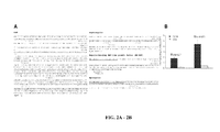
FIGS. 2A -B: Identification of peptides mimicking EGFR ligands. FIG. 2A , EGFR-binding peptide sequences isolated from the SKOV-3 selected phage pool were matched in each orientation to protein sequences of biological human EGFR ligands (leader peptide sequence underlined). Matches displayed are peptides with three or more amino acids being identical (red) and one or more being from the same class (green) as the correspondingly positioned protein amino acids. Tripeptides listed in Table 1 (yellow). FIG. 2B , isolation of peptides targeting EGFR. Binding of SKOV3-selected phage pool to immobilized EGFR compared with BSA in rounds 1 and 2 of biopanning of SKOV3-selected phage pool on immobilized human EGFR.
-
FIGs. 3A-3B : Phage selection on immobilized EphA5 receptor. FIG. 3A , Ephrin-mimic phage displaying the enriched motif GGS were selected on EphA5-coated microtiter wells. Phage showing specific binding to EphA5 was analyzed for its distinctive binding to EphA5 compared to EphA4 receptor ( FIG. 3B ). BSA and fd-tet insertless phage were used as negative controls.
-
FIG. 4 : EphA5 receptor expression in the NCI-60. From microarray analysis reported at dtp.nci.nih.gov/mtweb/servlet/moltidsearch?moltid=MT894.
-
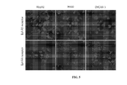
FIG. 5 : EphA5 and EphA4 receptor expression by the lung cancer cell lines Hop92 and H460. The OVCAR3 cell line was used as negative control. 10X magnification.
-
FIG. 6 : Specific binding of the CSGIGSGGC (SEQ ID NO:2) and CRFESSGGC (SEQ ID NO:3)- phage to lung cancer cells Hop92 and H460 but not to the ovarian cancer cell line OVCAR-3. Insertless phage (fd-tet) was used as negative control.
-
FIGS. 7A -B: A. Clustered image map relating all isolated NCI-60-binding tripeptides to NCI-60 cell lines. FIG. 7A , Two-dimensional hierarchical clustering was applied to the frequencies of 3,280 unique tripeptides (rows) found in cell-binding CX7C peptides selected on the NCI-60 cells (columns). Tripeptides were clustered based on their correlations with cell lines; cell lines were clustered based on their correlations with tripeptides. Tripeptide frequencies were mean-subtracted and average-linkage clustered with correlation metric (the data were transformed to the mean of 0; variance of 1). The color in each CIM segment ranges from blue (high negative correlation) to red (high positive correlation), as indicated by the scale bar. Cell lines are color-coded based on previously defined histological tumor origin. FIG. 7B , A control two-dimensional hierarchical clustering applied under the Poisson assumption to 3,280 randomly simulated tripeptide frequencies (rows) showed no obvious pattern, thus indicating that clusters in A were not generated at random.
-
FIG. 8 : Targeted peptides mediate ligand-receptor cell internalization. CSGIGSGGC (SEQ ID NO:2) and CRFESSGGC (SEQ ID NO:3)-phage were permeabilized into A549 cells. No internalization was observed when cells were incubated with insertless phage
-
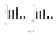
FIG. 9A -B: Biological effects of the peptides CSGIGSGGC (SEQ ID NO:2) and CRFESSGGC (SEQ ID NO:3 on lung cancer cells. Promotion of cell survivial and proliferative response of starved lung cancer cells to the ephrin mimic peptides, control peptide and complete culture medium (A549 ( FIG. 9A ), H460 cells ( FIG. 9B )). Concentrations of peptide were optimized. Values in the Y-axis correspond to the number of viable cells under each experimental condition evaluated after a 72h incubation period. Data bars represent the mean and corresponding standard error of the mean.
DETAILED DESCRIPTION OF THE INVENTION
-
A collection of 60 cell lines derived from human tumors (NCI-60) has been widely explored as a tool for anticancer drug discovery. In one aspect of the invention, the cell surface of the NCI-60 was profiled by high-throughput screening of a phage-displayed random peptide library and classified the cell lines according to the binding selectivity of 26,031 recovered tripeptide motifs. By analyzing selected cell-homing peptide motifs and their NCI-60 recognition patterns, the inventors established that some of these motifs (a) are similar to domains of human proteins known as ligands for tumor cell receptors and (b) segregate among the NCI-60 in a pattern correlating with expression profiles of the corresponding receptors. The inventors biochemically validated some of the motifs as mimic peptides of native ligands for the epidermal growth factor receptor. The results indicate that ligand-directed profiling of tumor cell lines can select functional peptides from combinatorial libraries based on the expression of tumor cell surface molecules, which in turn could be exploited as "druggable" receptors in specific types of cancer (Kolonin et al., 2006).
-
The National Cancer Institute panel of human cancer cell lines from different histologic origins and grades (NCI-60) has been extensively used to screen compounds for anticancer activity (Monks et al., 1991; Weinstein et al., 1997). The NCI-60 includes carcinomas of several origins (kidney, breast, colon, lung, prostate, and ovarian), tumors of the central nervous system, malignant melanomas, leukemias, and lymphomas. Gene expression determined by high-throughput microarrays has been used to survey the variation in abundance of thousands of distinct transcripts in the NCI-60; such data provided functional insights about the corresponding gene products in tumor cell transformation (Weinstein et al., 1997; Scherf et al., 2000; Nishizuka et al., 2003). This information-intensive genomic approach has yielded candidate diagnostic tumor markers to be validated at the protein level in prospective studies (Nishizuka et al., 2003). Moreover, systematic proteomic studies based on two-dimensional PAGE (Myers et al., 1997) and protein microarrays (Nishizuka et al., 2003) have also been implemented. Finally, in parallel with the NCI-60 transcriptome and proteome initiatives, pharmacologic sensitivity of the cells to >105 different chemical compounds has been registered (Monks et al., 1991; Weinstein et al., 1997). Indeed, for some genes, correlation of expression data to drug sensitivity profiles has uncovered the mechanistic basis for the drug activity (Scherf et al., 2000; Zaharevitz et al., 2002; Blower et al., 2002; Rabow et al., 2002; Wallqvist et al., 2002; Szakacs et al., 2004). Thus, conventional genomic and proteomic approaches have identified several potential tumor markers and drug targets. However, despite such advances, correlation between drug activity and gene expression profiles has not as yet been established for most of the compounds tested (Wallqvist et al., 2002; Brown, 1997; Walloyist et al., 2003). This suggests the likely existence of unknown factors and the need to develop alternative methodology to discover "druggable" molecular targets.
-
Over the past few years, it has been proposed that (a) characterization of molecular diversity at the tumor cell surface level (represented primarily by membrane-associated proteins that are often modified by lipids and carbohydrates) is required for the development of ligand-directed anticancer therapies, and that (Zaharevitz et al., 2002) peptides binding to surface receptors preferentially expressed on tumor cells may be used to ligand-direct therapeutics to sites of disease with potential for increased therapeutic windows (Arap et al., 1998; Kolonin et al., 2001). It has become increasingly clear that selective cell surface features can be mapped by screening libraries of peptides (Kolonin et al., 2001; Pasqualini and Ruoslahti, 1996; Giordano et al., 2001; Arap et al., 2002). In fact, combinatorial peptide libraries displayed from pIII protein of an M13-derived phage have now been successfully screened on intact cells and in vivo (Arap et al., 1998; Kolonin et al., 2001; Pasqualini and Ruoslahti, 1996). Peptide ligands selected from unbiased screens without any predetermined notions about the nature of the cellular receptor repertoire have been used for the subsequent identification of the corresponding target cell surface receptors (Giordano et al., 2001; Arap et al., 2002; Pasqualini et al., 2000; Kolonin et al., 2002; Kolonin et al., 2004; Pasqualini et al., 2001). In addition, novel techniques, such as the biopanning and rapid analysis of selective interactive ligands (BRASIL), have enabled high-throughput phage library screening on cells (Giordano et al., 2001). Here, the BRASIL method is used to systematically screen combinatorial libraries on tumor cells of the NCI-60 panel. Results of this feasibility study suggest that tumor cells can be grouped by profiles of their peptide ligands directed to differentially expressed cell surface receptors. The data support the notion that many tumor cell surface-exposed receptors are expressed irrespective of tumor origin, thus suggesting they could be developed as broad tumor targets. Integration of ligand-directed surface profiling with other approaches related to the NCI-60 may uncover functional ligand-receptor pairs for the targeted drug delivery.
I. CELL TARGETING MOLECULES
-
Modified cell targeting molecules of the present invention may be produced by chemical synthetic methods, by chemical linkage between the two moieties or in some cases by fusion of a second polypeptide coding sequence to the targeting moiety. It is contemplated that modified cell targeting molecules of the invention may be used as therapeutics and/or as imaging agents to target specific classes of cells.
-
As mentioned above, in certain aspects of the invention, a modified cell targeting moiety may comprise a second polypeptide wherein the two polypeptides together form a fusion protein. For example, in certain aspects the second polypeptide may be a therapeutic or cytotoxic (e.g., a toxin) polypeptide as exemplified below. A fusion of two polypeptide coding sequences can be achieved by methods well known in the art of molecular biology. It is preferred that a fusion polynucleotide contain only the AUG translation initiation codon at the 5' end of the first coding sequence without the initiation codon of the second coding sequence to avoid the production of two separate encoded products. In addition, a leader sequence may be placed at the 5' end of the polynucleotide in order to target the expressed product to a specific site or compartment within a host cell to facilitate secretion or subsequent purification after gene expression. The two coding sequences can be fused directly without any linker or by using a flexible polylinker.
A. Cell Targeting Moieties
-
Cell targeting moities as provided here may, in some aspects, comprise peptides or polypeptides that exhibit binding to a specific class of cells. For example, in some cases the cell targeting moiety is selected from one of the polypeptide sequences provided in Table 3. The skilled artisan will understand that such sequences may comprise additional amino acids or other covalent modifications. For instance, in preferred embodiments a polypeptide sequence from Table 3 is provided a cyclic polypeptide. Thus, in some specific examples, an amino acid sequence from Table 3 is flanked by cysteine residues that may form a disulfide bond thereby providing a cyclic polypeptide. Thus, in some aspects the invention provides compositions and methods for targeting any of the classes of cells that bind to the peptides and polypeptides provided herein (e.g., as indicated in Table 3) such as leukemia cells, lung cancer cells, colon cancer cells, CNS cancer cells, melanoma cells, ovarian cancer cells, prostate cancer cells, renal cancer cells or breast cancer cells.
B. Therapeutic Moieties
-
As mentioned above in certain aspects, a therapeutic moiety may be a toxin such as radioisotopes, holotoxins, modified toxins, catalytic subunits of toxins, cytotoxins (cytotoxic agents), or any molecules or enzymes not normally present in or on the surface of a cell that under defined conditions cause the cell's death. Toxins that may be used according to the methods of the invention include, but are not limited to, radioisotopes known in the art, compounds such as, for example, antibodies (or complement fixing containing portions thereof) that bind an inherent or induced endogenous cytotoxic effector system, thymidine kinase, endonuclease, RNAse, alpha toxin, ricin, abrin, Pseudomonas exotoxin A, diphtheria toxin, saporin, momordin, gelonin, pokeweed antiviral protein, alpha-sarcin and cholera toxin. "Toxin" also includes a cytostatic or cytocidal agent, a therapeutic agent or a radioactive metal ion, e.g., alpha-emitters such as, for example, 213Bi, or other radioisotopes such as, for example, 103Pd, 133Xe, 131I, 68Ge, 57Co, 65Zn, 85Sr , 32P, 35S, 90Y, 153Sm, 153Gd, 169Yb, 51Cr, 54Mn, 75Se, 113Sn, 117 Sn, 186Re, 166Ho, and 188Re; luminescent labels, such as luminol; and fluorescent labels, such as fluorescein and rhodamine, and biotin. Furthermore, a therapeutic moiety may be a pro-apoptotic protein such as a BCL2 family member, a caspase or a granzyme.
II. CANCER THERAPIES
-
A variety of conventional cancer therapies are currently used in the treatment of cancer. Thus, in some aspects of the invention there are provided methods for classifying cancer cells such as cells that are sensitive or resistant to an anticancer therapy. Some examples of conventional cancer therapies discussed below. It is contemplated that methods according to the invention may be used to identify cells that are sensitive or resistant to any particular cancer treatment. Furthermore, some aspects of the invention concern compositions and methods for cell targeted anticancer therapy. Thus, it is contemplated that any anticancer method known to those in the art (as exemplified below) may be used in combination or conjunction with compositions and methods provided herein.
A. Chemotherapy
-
Cancer therapies also include a variety of combination therapies with both chemical and radiation based treatments. Combination chemotherapies include, for example, cisplatin (CDDP), carboplatin, procarbazine, mechlorethamine, cyclophosphamide, camptothecin, ifosfamide, melphalan, chlorambucil, busulfan, nitrosurea, dactinomycin, daunorubicin, doxorubicin, bleomycin, plicomycin, mitomycin, etoposide (VP16), tamoxifen, raloxifene, estrogen receptor binding agents, taxol, gemcitabien, navelbine, farnesyl-protein tansferase inhibitors, transplatinum, 5-fluorouracil, vincristin, vinblastin and methotrexate, or any analog or derivative variant of the foregoing.
B. Radiotherapy
-
Other factors that cause DNA damage and have been used extensively include what are commonly known as γ-rays, X-rays, and/or the directed delivery of radioisotopes to tumor cells. Other forms of DNA damaging factors are also contemplated such as microwaves and UV-irradiation. It is most likely that all of these factors effect a broad range of damage on DNA, on the precursors of DNA, on the replication and repair of DNA, and on the assembly and maintenance of chromosomes. Dosage ranges for X-rays range from daily doses of 50 to 200 roentgens for prolonged periods of time (3 to 4 wk), to single doses of 2000 to 6000 roentgens. Dosage ranges for radioisotopes vary widely, and depend on the half-life of the isotope, the strength and type of radiation emitted, and the uptake by the neoplastic cells.
-
The terms "contacted" and "exposed," when applied to a cell, are used herein to describe the process by which a therapeutic construct and a chemotherapeutic or radiotherapeutic agent are delivered to a target cell or are placed in direct juxtaposition with the target cell. To achieve cell killing or stasis, both agents are delivered to a cell in a combined amount effective to kill the cell or prevent it from dividing.
C. Immunotherapy
-
Immunotherapeutics, generally, rely on the use of immune effector cells and molecules to target and destroy cancer cells. The immune effector may be, for example, an antibody specific for some marker on the surface of a tumor cell. The antibody alone may serve as an effector of therapy or it may recruit other cells to actually effect cell killing. The antibody also may be conjugated to a drug or toxin (chemotherapeutic, radionuclide, ricin A chain, cholera toxin, pertussis toxin, etc.) and serve merely as a targeting agent. Alternatively, the effector may be a lymphocyte carrying a surface molecule that interacts, either directly or indirectly, with a tumor cell target. Various effector cells include cytotoxic T cells and NK cells.
-
Immunotherapy, thus, could be used as part of a combined therapy, in conjunction with gene therapy. The general approach for combined therapy is discussed below. Generally, the tumor cell must bear some marker that is amenable to targeting, i.e., is not present on the majority of other cells. Many tumor markers exist and any of these may be suitable for targeting in the context of the present invention. Common tumor markers include carcinoembryonic antigen, prostate specific antigen, urinary tumor associated antigen, fetal antigen, tyrosinase (p97), gp68, TAG-72, HMFG, Sialyl Lewis Antigen, MucA, MucB, PLAP, estrogen receptor, laminin receptor, erb B and p155.
III. EXAMPLES
Example 1
Combinatorial library screening on cells
-
All the NCI-60 cell lines (1), except MDA-N (unavailable), were grown in RPMI 1640 supplemented with 5% fetal bovine serum (FBS) and 5 mmol/L L-glutamine. A phage display random peptide library based on the vector fUSE5 displaying the insert CX7C (SEQ ID NO:1) was screened by using BRASIL as described (Giordano et al., 2001). Exponentially growing cells were harvested with 0.5 mmol/L EDTA, 0.4 g/L KCl, 8 g/L NaCl, and 1 g/L dextrose, washed once with phosphate buffer saline (PBS), and resuspended in RPMI containing 1% bovine serum albumin (BSA) and 1 mmol/L HEPES. Cells (∼ 106) were incubated for 2 hours on ice with 109 transduction units (T.U.) of CX7C phage in 200-µL suspension, transferred to the top of a nonmiscible organic lower phase (dibutyl phtalate/cyclohexane, 9:1), and centrifuged at 10,000 x g for 10 minutes. The phage-bound cell pellet was incubated with 200 µL of K91 bacterial culture, and the bound phages were amplified and used in the following round. To prevent preferential isolation of peptides containing the RGD motif, which is selected on tissue-cultured cells due to expression of cell adhesion molecules binding to vitronectin, library screening was done in the presence of 1 mg/mL of the synthetic peptide RGD-4C (AnaSpec, San Diego, CA) in each round. After three rounds of selection, phage peptide-encoding inserts were sequenced as described (Pasqualini and Ruoslahti, 1996; Arap et al., 2002; Pasqualini et al., 2001).
Example 2
Hierarchical cluster analysis of peptide motif/cell line association
-
The inventors created an interactive sequence management database of all peptide sequences isolated in the screen. Calculation of tripeptide motif frequencies in CX7C peptides (in both directions) was done by using a character pattern recognition program based on SAS (version 8.1.2, SAS Institute, Cary, NC) and Perl (version 5.6.1) as described (Arap et al., 2002). To identify the most closely related tripeptides and cell lines, clustered image maps (CIM) were generated by using online software CIMminer available at discover.nci.nih.gov/tools.jsp. Data were centered (mean subtracted and divided by SD) on both cell lines and tripeptide motifs; correlation coefficient metric with average linkage algorithm was used as distance measurement. The tripeptide motif frequencies across the NCI-60 cell lines formed a two-dimensional data matrix that was used to correlate motif enrichment with groups of cell lines. To evaluate whether CIMMiner algorithm is appropriate for clustering analysis of peptide frequency data, a simulation test was devised assuming that the frequencies of tripeptide motifs in a given data set follow an independent Poisson distribution. The inventors simulated a random 3,280 x 59 data matrix of the dimension identical to that of tripeptide motif frequency data matrix (corresponding to the set of 3,280 tripeptides and 59 cell lines). These simulated data were centered the same way as the experimental data by transforming to mean of 0, variance of 1. For CIM in FIG. 1, tripeptides selected on all but one cell line of common origin (Arap et al., 2002) were used. Specificity of five tripeptides selectively overrepresented or underrepresented in lung tumor cell binding peptides for the 11 boxed cell lines (against the other 48 cell lines) was evaluated by using the R Package, version 2.0.0 (www.r-project.org) by performing two-sample t test (one tailed), as well as using Wilcoxon rank sum test (one tailed) and Fisher exact test (one tailed) as described (Arap et al., 2002).
Example 3
Identification of candidate targeted receptors
-
To identify lead receptors targeted by tripeptide motifs, the Molecular Target Database (www.dtp.nci.nih.gov) was screened to identify proteins, expression levels of which in individual cell lines of the NCI-60 correlated with frequencies of individual tripeptides from FIG. 1 in the corresponding cell lines. The inventors used the COMPARE software (dtp.nci.nih.gov/docs/compare/compare.html) to calculate pairwise Pearson correlations between tripeptide frequencies in cell lines and the protein expression patterns in the database. Minimum Pearson correlation coefficient of 0.2 served as cutoff for the selection of lead receptors, as it provided a reasonable number of candidate molecular targets for which NCI-60 expression profiles and tripeptide frequency distribution profiles correlated. To initially restrict the candidate targets analyzed to broad-specificity receptors, only putative cell surface molecules (Table 1) were included, expression of which in the NCI-60 was found to correlate with the frequency profile of at least 25% of the tripeptides.
Example 4
Protein database screening for peptide motif similarity
-
To identify natural prototype ligands of candidate receptors that are mimicked by selected peptides, the inventors screened all 7-mer peptides selected in the screen by using online ClustalW software (www.ebi.ac.uk/clustalw/) to identify extended (four or longer amino acids) motifs shared between multiple peptides containing the broad-specificity tripeptides (FIG. 1). Nonredundant databases of human proteins were searched by the BLAST software (www.ncbi.nlm.nih.gov/BLAST/) for proteins containing the cell-targeting 4-mers under the condition that at least the tripeptide part of the motif is identical to the part of the BLAST match.
Example 5
Validation of epidermal growth factor receptor as one of the peptide targets
-
To isolate peptides binding to epidermal growth factor receptor (EGFR), phage clones selected on SKOV3 in rounds 2 and 3 of the screening were individually amplified and pooled, and 109 transduction units of the mixed phage were incubated overnight at 4°C with 10 µg of purified human EGFR (Sigma, St. Louis, MO), or BSA control immobilized on plastic. Unbound phages were extensively washed off with PBS, and then the bound phages were recovered by infecting host K91 Escherichia coli directly on the plate, and tetracycline-resistant clones were selected, quantified, and sequenced. To identify EGFR ligand-matching motifs among phage-displayed SKOV3-binding peptides, custom-designed Perl 5.8.1- based software was used to run peptide sequences against biological EGFR ligand sequences. Each 7-mer peptide sequence was aligned in each orientation against the EGFR ligand sequences from the NH2 to COOH terminus in one-amino-acid shifts. The peptide/protein similarity scores for each residue were calculated based on a BLOSUM62 matrix modified to identify peptide matches of at least three amino acids in any position being identical and one being similar to the corresponding amino acid positions in the EGFR ligands (FIG. 2A).
Example 6
Isolation of peptides binding to surface of the NCI-60 cancer cells
-
As an initial attempt to profile cell surface of the tumor cell panel, a large (2 x 10⁸ unique sequences) cyclic random peptide library was screened with the basic structure CX7C (C, cysteine; X any residue) on every cell line of the NCI-60. Phage selection was done in the excess of a competing Arg-Gly-Asp (RGD) synthetic integrin-binding peptide (Arap et al., 1009) to minimize the recovery of RGD-containing peptides. This strategy was designed to facilitate the recovery of ligands binding to nonintegrin families of cell surface receptors because RGD tends to become dominant in the screening due to the high levels of integrin expression in adherent cells (unpublished observation). Preferential cell binding of specific cell-targeting peptides results in enrichment, defined by the increased recovery frequency of these peptide motifs in each subsequent round of the screen (Kolonin et al., 2001; Pasqualini et al., 2001). Thus, the inventors set out to profile the expression of nonintegrin cell surface molecules among the cell lines of the NCI-60 according to the differential selection of motifs enriched in the screen.
Example 7
Hierarchical cluster analysis of peptides binding to the NCI-60 cells
-
To analyze the spectrum of the peptides resulting from the screening and compare those among different cell lines of the panel, a combinatorial statistical approach was adopted based on the premise that three residue motifs (tripeptides) provide a sufficient structure for protein-peptide interactions in the context of phage display (Arap et al., 2002). For each NCI-60 cell line, CX7C peptide-encoding DNA inserts from 96 phage clones recovered after three rounds of selection were sequenced. A computer-assisted survey of all tripeptides within the library-derived sequences selected on each cell line by analyzing a database of 26,031 tripeptides contained within the 5,270 CX7C-encoded 7-mer peptides isolated (an average of eighty-nine 7-mer peptide sequences analyzed per each NCI-60 cell line) was performed. Thus, each cell line was assigned a unique set of tripeptides that was identified during the selection for cell surface binders, and the frequencies of each motif among all peptides for a given cell line were calculated.
-
To classify cell lines according to their association with particular motifs, which might provide inference on the targeted surface molecules, a hierarchical clustering analysis of the 3,280 nonredundant tripeptides was done based on the frequency of association with the NCI-60 cell lines. For the construction of a CIM, the inventors adapted a hierarchical clustering algorithm and a pseudo-color visualization matrix initially designed to address differential gene expression among the cells of the panel (Scherf et al., 2000; Zaharevitz et al., 2002; Blower et al., 2002; Rabow et al., 2002). CIMMiner (Weinstein et al., 1997) was used for inference of the variation in peptide binding specificity across the cell lines by comparing relative frequencies of tripeptides found in 7-mer peptides binding to each cell. Clustering of peptide motifs with similar cell selectivity revealed that the peptide distribution of the combinatorial library within the NCI-60 set was nonrandom. Computer simulations of the permutated data set show that the observed pattern could not be generated by random chance, thus indicating that the discontinuous tripeptide frequency data is applicable for cluster analysis.
-
The selective spectra of peptide motifs interacting with the clustered cell lines suggest the existence of shared targeted surface receptor(s) expressed in these lines. In this study, the inventors chose to focus on putative peptide-targeted receptors with broad cell line specificity, which would be more informative for an initial peptide binding/receptor expression correlation analysis the inventors therefore excluded from the data set motifs selected only on a single or few cell lines. Instead, the inventors focused on 38 tripeptides that showed a semiubiquitous distribution among the NCI-60 lines (FIG. 1). A CIM constructed according to the isolation frequency of these broader-specificity tripeptides from each cell line revealed several apparent clusters of cell lines that displayed distinct profiles of association with certain classes of peptide motifs. For example, the majority of lung cancer-derived cell lines segregated as a separate group, suggesting that some of the receptors targeted may be conserved among cell lines derived from a common origin (FIG. 1). Thus, although the analysis was severely restricted by limiting it to semiubiquitous tripeptides, clustering of some of them (predominantly with cell lines derived from the same tumor type) is consistent with their relative tissue specificity. To evaluate individual motifs for selectivity, a distinct cluster of five tripeptides associated with lung tumor-derived cell lines (FIG. 1, boxed) were identified The inventors compared tripeptide frequencies for the 11 cell lines within this cluster with their frequencies for the rest of NCI-60 lines by using statistical tests (Fisher exact, Wilcoxon rank-sum, and t test). Consistently, the GGS motif was isolated for the clustered lines significantly (P < 0.05) more frequently than for the other NCI-60 cell lines.
-
Notably, the distribution of cell lines in the dendrogram (FIG. 1) was partially consistent with the reported association of cells derived from tumors with common tissue origin (Scherf et al., 2000; Nishizuka et al., 2003). This suggests that some of the receptors, such as the one presumably recognized by the lung tumor-specific tripeptide GGS (FIG. 1), may be up-regulated only in certain cancer origins. However, the tumor cell phylogeny was recapitulated only to an extent; the majority of the observed clusters contained cell lines derived from unrelated tumor types (FIG. 1). The limited grouping of lines derived from tumors of common origin is perhaps not surprising: the relationship between different cell lines in the study is based on peptide binding to putative cell surface molecules, many of which may be tumor induced rather than characteristic of the tissue of origin. If so, the analysis of broad-specificity motif distribution may be well suitable for identification of specific surface molecules that are generally up-regulated by tumors and thus may constitute broad drug targets against cancer.
Example 8
Identification of candidate receptor targets for peptide motifs
-
The inventors proceeded to identify the targets for the 38 broad-specificity tripeptides, most of which presumably bind to receptors expressed by multiple NCI-60 cell lines. The NCI Molecular Targets Database that contains detailed information on the expression and activity of 1,218 human proteins measured by nonarray methods was used (Holbeck, 2004). By using the COMPARE algorithm (Zaharevitz et al., 2002), the inventors correlated the selectivity profiles of the 38 tripeptide motifs with the expression profiles of the characterized molecular targets. It was observed that several of the qualifying proteins, expression of which correlated with enrichment profiles of certain motifs, represented tyrosine kinase receptors, such as those for ligands belonging to families of EGFs, fibroblast growth factors (FGF), nerve growth factors (NGF), and ephrins (Table 1). When transferred to molecular target correlation data, the order of the 38-tripeptide motif set in the dendrogram (FIG. 1) revealed clusters of tripeptides for which cell line association profile correlated with expression profiles of EGF, FGF, NGF, or ephrin receptors (Table 1).
-
The peptide distribution-correlating tyrosine kinase receptors, belonging to EGFR, FGFR, NGFR, and ephrin receptor families (Table 1), are often up-regulated in many types of cancer (Vogelstein and Kinzler, 2004). To determine if the cell-binding peptides may target these tyrosine kinases, the inventors employed the notion that receptor-binding peptide motifs often mimic natural ligands for these receptors (Giordano et al., 2001; Arap et al., 2002; Kolonin et al., 2002). Thus, the selected motifs mimic ligands for the candidate tyrosine kinases were tested by determining whether tripeptides listed in Table 1 are embedded into longer peptides that may be responsible for cell surface binding. The inventors analyzed the CX7C (SEQ ID NO:1) phage inserts containing the 38 tripeptides by using the ClustalW software and compiled extended motifs containing the tripeptides shared among multiple peptides selected during the screen (data not shown). To identify candidate prototype human ligands, epitopes of which could be mimicked, each of the ClustalW-extended motifs were screened against the nonredundant database of human proteins by using the BLAST software (National Center for Biotechnology Information). As a result of this analysis, the inventors found the motifs containing 34 of 38 tripeptides (89%) to be identical or very similar to segments of proven or putative ligands for the tyrosine kinase receptors listed (Table 1).
Example 9
Validation of EGFR as a targeted receptor
-
To show that the approach taken can lead to actual targetable tumor cell surface proteins, the inventors chose to test if the EGFR is bound by any of the tripeptide motifs distributed in the panel in a profile correlating with EGFR expression. Consistently, 24 of 38 tripeptides surveyed displayed NCI-60 cell line association pattern consistent with that of EGFR expression (Table 1). Of these tripeptides, 22 were isolated in the screens on ovarian cancer cell lines SKOV3 and OVCAR4 (data not shown). Because EGFR is well known to be associated with ovarian cancer (Vogelstein and Kinzler, 2004), the inventors deemed these cell lines to be likely expressers of targetable EGFR, which would account for the selection of EGFR ligand-mimicking motifs. To validate EGFR binding by the selected motifs, the SKOV3-binding phage sublibrary (pooled clones recovered in rounds 2 and 3) were screened against immobilized human EGFR. After two rounds of selection, phage displaying the EGFR-binding peptides were analyzed: the majority were comprised by different 7-mer peptides (FIG. 2A) that contained 17 of 22 SKOV3-selected tripeptide motifs distributed in the panel in a profile correlating with EGFR expression (Table 1). Phage displaying these peptides had specific affinity to EGFR, as determined by subjecting the same sublibrary to immobilized BSA control binding (FIG. 2B). Remarkably, computer-assisted analysis of sequences (FIG. 2A) revealed that 12 of the 7-mer EGFR-binding peptides contained amino acid motifs similar to those present in some of the biological EGFR ligands (Vogelstein and Kinzler, 2004). These peptides, containing eight of the candidate tripeptides (RVS, AGS, AGL, GVR, GGR, GGL, GSV, and GVS), were found highly similar to fragments of EGF, amphiregulin, heparin-binding EGF-like growth factor, and epiregulin (FIG. 2A). Similarity search using the same algorithm on the same twelve 7-mers did not reveal any matches to two other EGFR ligands, transforming growth factor-α and β-cellulin, or randomly chosen control ligands of tyrosine kinase receptors from the three other candidate families listed in Table 1: ephrin A, NGF-β, and FGF6 (data not shown). Taken together, these data suggest that at least some of the peptides selected on the NCI-60 cells target EGFR, whereas others may bind to different tyrosine kinases, possibly including those from TRK, ephrin, or FGF receptor families.
-
Expression profiles of the candidate receptor targets for peptides identified in the screen illustrate the concept that in cancer, at least some tumor-associated cell surface molecules seem up-regulated regardless of cancer tissue origin. As such, this is the case for the EGFR and other tyrosine kinases possibly targeted by peptide ligands selected on the NCI-60 cell panel. This may also be the case for many other receptors with a role in tumorigenesis, expression profiles of which may not correlate with the overall proteomic profile of the original tumor tissue. In fact, these observations may account for the relatively limited success in correlating drug toxicity profiles with the genomic and/or proteomic profiles of the NCI-60 panel (Walloyist et al., 2003). On the other hand, some of the receptors, such as EphA5 presumably targeted by GGS tripeptide and its derivatives predominantly selective for lung tumor-derived cell lines (FIG. 1), seem to be at least partially specific for the progenitor cancer type.
-
The candidate ligand-receptor leads identified in this study can be characterized further for the development of targeted agents selective for tumors. Moreover, the peptides identified by the approach described here may map receptor interaction domains of biological (native) ligands. Similarity of peptides to the corresponding receptor-binding ligands has already been used for validation of the IL-11Rα receptor as a target of an interleukin-11 mimic peptide homing to blood vessels in the prostate (Arap
et al., 2002; Zurita
et al., 2004). The inventors and others have modeled the usage of peptides homing to receptors expressed by tumors (Pasqualini
et al., 2000) or non-malignant tissues (Kolonin
et al., 2002; Kolonin
et al., 2004) for directing the delivery of cytotoxics, proapoptotic peptides, metalloprotease inhibitors, cytokines, fluorophores, and genes (Arap
et al., 1998; Kolonin
et al., 2001). Thus, the approach provides a straightforward way to identify drug-accessible tumor cell surface receptors and to discover peptide ligands that can serve as mimetic prototype drugs. Unlike genomic or proteomic-based approaches that rely on differential expression levels of transcripts or protein products, this discovery platform directly addresses functional protein-protein interactions at the level of physical binding. In contrast to protein array systems, it is possible to select binding peptides even if the ligand-receptor interaction is mediated by conformational (rather than linear) epitopes. Ligand-directed screening of combinatorial libraries on tumor cell surfaces can lead to improved selection of functionally relevant peptides that can be developed for targeting "druggable" molecular targets.

| Table 1. Candidate ligand-receptor interactions mimicked* |
| RLS | ErbB2, | ErbB4 | FGF2,4 | | | EGF-TM7 |
| RGV | | | | | | |
| RGS | ErbB4 | | FGF2 | | EphA2,A3,A4,A8,B 1 | EGF-TM7, FGF-12b, FGF-5, NGF-beta |
| RAV | ErbB2 | | | | | MEGF7, NGF-beta. NTF 6 alpha |
| RAS | | | | TRKA | | FGF-20, NRG-3 |
| GAG | EGFR | | FGF1,2, 3 | | | MEGF4. FGF6, NGF-beta |
| AVS | EGFR, ErbB4 | ErbB2, | FGF1 | TRKB,C | EphA2,A3,A4,A7,B 1,B2,B3,B5 | TRK1 |
| LLS | | | | | | Amphlregulin |
| LLR | | | | TRKA | | EphA4 |
| LRV | EGFR, ErbB4 | ErbB2, | FGF3 | TRKA,B, C | EphA2,A3,A7 | FGF-12b, Eph-B3 |
| LRS | ErbB3 | | | | | MEGF4, MEGF5, MEGFS, NRG-3, NGF-beta |
| RVS | EGFR, ErbB4 | Erb B2, | FGF1,2 | TRKB | EphA7 | MEGF10, amphiregulin |
| RSS | | | FGF3 | TRKA | EphAS | EGF-TM7, FGF-S, NRG-3 |
| AGS | EGFR | | | TRKA | | MEGF6, brain NGF |
| AGR | | | | | | MEGF2, MEGF4, FGF6, NTF-5, NTF-6 |
| AGL | EGFR, ErbB3 | ErbB2, | FGF1,3 | | EphAS,A6,A8 | MEGF12 |
| AGG | | | | | EphA5 | HB-EGF, Ephr-B3 |
| GVR | EGFR, ErbB4 | ErbB2, | FGF1,2 | TRKB | EphA7 | MEGF4, MEGF6 MEGF8, FGF-5, bFGF, brain NGF |
| GVL | | | FGF1,2 | | EphA2,A3,A5,A6,B 3 | NGF2, Ephrin-B3, |
| GAV | | | | | | MEGFS, MEGF6, NGF-beta |
| GLV | ErbB4 | | FGF4 | | EphA5 | ESF-TM7, betaceilulin, NTF 3, Eph-B3, |
| GLR | ErbB4 | | | | | MEGF5, EGFL5, FGF-12b, FGF-16, NRG-3 |
| LVS | | | FGF1,4 | | EphA5,A6 | EGFL5. FGF23, GDNF, Eph-B3 |
| ARG | ErbB2 | | FGF2,4 | TRKA | EphAI | FGF-12b, FGF23, NGF-beta. GDNF. NTF 6 |
| ASL | | | FGF1,2 | TRKC | | EGF-TM7, FGFR1 |
| AAV | | | | TRKB | EphA2,A3,A4,A7,B 3,B5 | • |
| AAS | | | FGF1,2 | TRKC | | • |
| GGS | | | | | EphA5 | Eph-B3, Eph A4 |
| GGR | EGFR.ErbB2 | | FGF2 | | | EGF-TM7, HB-EGF, FGF23, |
| | | | | | | Ephrin-B3 |
| GLG | ErbB2, | ErbB3 | FGF2,3, 4 | | EphA1,A6 | heparin binding growth factor 8 |
| GGL | ErbB2 | | | | | HB-EGF, MEGF5, EGFL5, NRG-3 |
| GSS | EGFR, | ErbB2 | FGF3 | TRKA,C | EphA5 | MEGFS |
| GSG | EGFR | | | | EphA5 | |
| GSV | EGFR, ErbB4 | ErbB2, | FGF4 | TRKB | EphA7,B2 | MEGF5, NRG-3, Ephrin-B3 |
| GRV | EGFR | | | | | MEGFS, EGF-TM7, FGF23, NTF5 |
| GRL | EGFR.ErbB2 | | | | EphAS,B1,B2,B4 | betacellulin, EGFL5, NGF2, NTF5, |
| | | | | | | EphB3, EphA4 |
| GPS | EGFR, ERB4 | ErbB2, | FGF3 | TRKB | EpnA2,A3,A4,A7,B 2,B5 | MEGFS, EGFL5, EGF-like EMR3, SPGF |
| GVS | EGFR | | FGF4 | TRKA | | MEGF-1, MEGF5, NRG-3, NTF-6, NTF-5 |
-
* NOTE: Candidate peptide motif receptors are the human cell surface proteins (identified by COMPARE) expressed in profiles correlating with the selectivity of the corresponding tripeptides. Candidate peptide-mimicked receptor ligands are human proteins (identified by automated BLAST) that contained the corresponding tripeptides. Tripeptides in the column are ordered as in FIG. 1. Receptors of the same family and their corresponding candidate biological ligands identified based on tripeptide similarity are coded by the same color [EGFR, blue; FGFR, green; TRK receptor (NGFR), purple; ephrin receptor, red]. Tripeptides that both have a selectivity correlating with EGFR family receptor expression and are found within EGFR ligands (boldface). Tripeptides that were confirmed to reside within EGFR-binding SKOV3-slected peptides (FIG. 2; blue).
Example 10
Molecular Fingerprinting of Cancer Cell Lines
-
Proteomics can be defined as the systematic analysis of the proteins in biological samples that aims to document the overall distribution of proteins in tumor cells or tumor-associated cells, identify and characterize individual proteins of interest and to elucidate their relationships and functional roles. Ultimately, high-throughput profiling of protein expression will lead to the "proteome", a protein-based fingerprint, for each tissue in humans and other species. As technologies related to proteomics advance, new approaches for systematic molecular analysis of cancer at the protein level are surfacing. However, methods for systematic protein expression profiling may also easily overlook potential targets for intervention. These methods often do not take anatomical context into account. Therefore, for the generation of molecular map of accessible receptors that can be used for targeting therapeutics, information derived from conventional protein profiling approaches should be enhanced by integration with data from functional screenings ex vivo and in vivo. Studies by the inventors and others have advanced the concept of cancer proteomics: the molecular phenotyping of tumor cells and cells forming blood vessels at the protein-protein interaction level. Exploiting the molecular diversity of cell surface receptors expressed in cancer will eventually result in a ligand-receptor functional map for targeted delivery.
A major goal in drug development has long been to generate targeted therapies. This approach would improve drug therapeutic indexes by limiting the systemic exposure of other tissues to untoward or toxic effects. Thus, the promise for the identification of selectively expressed tumor-associated receptors and the ligands that home to these receptors is translation of this knowledge into the development of targeted therapeutics. Generally, coupling of homing peptides yields targeted compounds that are more effective and less toxic than the parental compound. So far, peptides selected by homing to tumor vasculature have been used as carriers to guide the delivery of cytotoxic drugs, pro-apoptotic peptides, metalloprotease inhibitors, cytokines, fluorofores, and genes in transgenic and xenograft mouse models of human disease.
-
Recognition of molecular diversity in human cancer is essential for the development of targeted therapies. The methods developed have two main applications. First, they may identify ligands targeting human cancer. Second, the determination of molecular profiles of biomarkers in specific types of tumors may enable identification of differentially expressed cancer markers. Thus, the approach may lead to construction of a molecular profile of human tumors. Early identification of targets, optimized regimens tailored to molecular profile of individual cancer patients, combined with the identification of new vascular addresses may result in revisiting or salvaging of drug candidates that are ineffective or too toxic. Ultimately, it may be possible to guide imaging or therapeutic compounds to tumor targets in cancer patients.
-
By fingerprinting lung cancer cells the inventors have confirmed the expression of a previously characterized molecular target, EGFR, in multiple cancer origins, which demonstrates the power of the approach. Recently, the inventors used this approach to identify a new cancer origin-selective molecular target, Ephrin A5 receptor, which the inventors have preliminary validated in the context of human lung cancer cell lines and tissues.
Example 11
Motifs targeting NCI-60 cells in correlation with EGFR expression pattern are found within peptides similar to domains of biological EGFR ligands and bind to EGFR
-
To show that the approach taken can lead to actual targetable tumor cell surface proteins, the inventors chose to test if the EGF receptor (EGFR) is bound by any of the tripeptide motifs distributed in the panel in a profile correlating with EGFR expression. Consistently, 24 out of 38 tripeptides surveyed displayed NCI-60 cell line association pattern consistent with that of EGFR expression (Kolonin et al., 2001). Of these, tripeptides, 22 were isolated in the screens on ovarian cancer cell lines SKOV3 and OVCAR4 (data not shown). Since EGFR is well known to be associated with ovarian cancer (Vogelstein, 2004; Maihle and Lafky, 2002), the inventors deemed these cell lines to be likely expressers of targetable EGFR, which would account for the selection of EGFR ligand-mimicking motifs. To validate EGFR binding by the selected motifs, the SKOV3-binding phage sub-library (pooled clones recovered in rounds 2 and 3) were screened against immobilized human EGFR. After 2 rounds of selection, phage displaying the EGFR-binding peptides were analyzed: the majority were comprised by different seven-mer peptides (FIG. 3A) that contained 17 out of 22 SKOV3-selected tripeptide motifs distributed in the panel in a profile correlating with EGFR expression.
-
Phage displaying these peptides had specific affinity to EGFR, as determined by subjecting the same sub-library to immobilized bovine serum albumin (BSA) control binding (FIG. 2B). Remarkably, computer-assisted analysis of sequences (FIG. 2A) revealed that 12 of the seven-mer EGFR-binding peptides contained amino acid motifs similar to those present in some of the biological EGFR ligands. These peptides, containing eight of the candidate tripeptides (RVS, AGS, AGL, GVR, GGR, GGL, GSV, and GVS) were found highly similar to fragments of EGF, Amphiregulin, heparin-binding EGF-like growth factor, and Epiregulin (FIG. 2A). Similarity search using the same algorithm on the same 12 seven-mers did not reveal any matches to two other EGFR ligands, TGF-α and betacellulin, or randomly chosen control ligands of tyrosine kinase receptors from the three other candidate families listed in Table 2 (Kolonin et al. 2001): Ephrin A, NGF-β, and FGF6. Taken together, these data suggest that at least some of the peptides selected on the NCI-60 cells target EGFR, while others may bind to different tyrosine kinases, possibly including those from TRK, Ephrin, or FGF receptor families.
-
A phage-displayed combinatorial library was systematically screened for peptides capable of targeting the cell lines in the NCI-60 panel. By statistical analysis of peptide motif sequences, each NCI-60 cell line was assigned a unique set of peptide motifs that were isolated during the selection for cell surface binders. It was shown that tumor cells can be grouped by profiles of their phage display-derived peptide ligands directed to differentially expressed cell surface receptors.
-
An approach for peptide-targeted receptor identification was designed. Profiles of peptide motif preference for specific lines of the NCI-60 were correlated with expression profiles of known breast cancer-related targets. Some of the peptide motifs were found within proteins known to bind the receptors that had NCI-60 expression profiles matching cell line recognition profiles of the peptides, and that are implicated in cancer.
-
Candidate targeted cell surface molecules were identified, which included a number of tyrosine kinase receptors. As a proof of principle, EGFR, a receptor known to be upregulated in various cancers, was validated as a target of tripeptides RVS, AGS, AGL, GVR, GGR, GGL, GSV, and GVS, which were The results described uncover a previously overlooked phenomenon. The data support the notion that many tumor cell surface-exposed receptors are expressed irrespective of tumor origin, thus suggesting they could be explored as broad tumor targets.
Example 12
Ephrin A5 receptor as a lung cancer cell surface marker
-
The peptide distribution-correlating tyrosine kinase receptors, belonging to EGFR, FGFR, NGFR and Ephrin receptor families are often up-regulated in many types of cancer. On the other hand, some of the receptors, such as EphA5 presumably targeted by GGS tripeptide and its derivatives predominantly selective for lung tumor-derived cell lines appear to be at least partially specific for the progenitor cancer type. Since this approach clearly allowed identification of cell surface receptors ubiquitously upregulated in various cancers, the inventors took a step further to attempt identification of cancer type-specific receptors.
Having chosen lung cancer for the initial procedure establishment, the inventors identified a distinct cluster of five tripeptides associated with lung tumor-derived cell lines. The inventors compared tripeptide frequencies for the 11 cell lines within this cluster with their frequencies for the rest of NCI-60 lines by using statistical tests (Fisher exact, Wilcoxon rank-sum, and t-test). Consistently, the inventors observed that motif GGS was isolated for the clustered lines significantly (P<0.05) more frequently than for the other NCI-60 cell lines (Table 2).
Table 2 Association of specific tripeptides with lung cancer-derived cell lines: | Motif | Mean motif count (±SEM) inside vs outside cluster | P value t-test, 1-sided | P value Wilcoxon ranksum test, 1-sided | P value Fisher exact test, 1-sided |
| GGS | 22(±05) vs 12(±02) | 0 0422 | 0 0407 | 0 0043 |
| GGR | 13(±03) vs 15(±02) | 0 6991 | 0 6466 | 0 6739 |
| GLG | 07(±04)vs 07(±02) | 0 5375 | 0 6888 | 05150 |
| GGL | 12(±02) vs 13(±02) | 0 6457 | 04174 | 0 5485 |
| GSS | 22 (± 0 4) vs 11 (± 0 2) | 0 0422 | 0 0026 | 0 0008 |
-
To determine statistical significance of association or dissociation between exemplary tripeptides and cell lines, normalized frequencies of five tripeptides predominantly associated (GGS, GGR, GLG, and GGL) or dissociated (GSS) with the cluster containing the majority of lung tumor-derived cell lines (FIG. 1, boxed) were compared for cell lines inside the cluster and outside the cluster. Selective association of tripeptide GGS with the clustered cell lines was found significant according to t-test, Fisher exact test and Wilcoxon rank-sum test (all tests one-tailed).
-
Based on the automated BLAST analysis (Table 2) the inventors identified proteins of the ephrin family candidate prototypes of the GGS-containing peptides: ephrins -B3 and A4 contain the GGS, consistent with a functional mimickry. Ephrins (A and B) and their receptors (EphA and EphB) represent a large class of cell-cell communication molecules with well-defined developmental functions. Their role in healthy adult tissues and in human disease is still largely unknown, although diverse roles in carcinogenesis have been postulated and a number of Eph receptors have been found overexpressed by various cancers (Hafner et al., 2004). Based on the COMPARE analysis of GGS distribution within NCI-60 (Kolonin et al., 2001, Table 2), the receptor expressed in the corresponding pattern is EphA5. The EphA5 expression (FIG. 4 has been explored using cDNA microarray analysis and is reported at the DTP server (dtp.nci.nih.gov/mtweb/servlet/moltidsearch?moltid=MT894), however, no studies of EphA5 function in cancer have been published. Intriguingly EphA5 is not expressed in normal lung and normally is only thought to have brain-specific functions.
Example 13
Validation of ephrin-mimic peptides in lung cancer
-
To validate phage containing the motif GGS as a ligand of Eph receptors, the inventors tested phage binding to the EphA5 immobilized receptor. The inventors started testing eight peptides (CAGLSGGTC, CSGIGSGGC, CSSGGVLGC, CSWGSGGSC, CTLVLGGSC, CRFESSGGC, CHVSGGSGC, CTGGSLGAC) containing the enriched motif GGS, all of them displayed by phage clones obtained from the screening on different cell lines known to express the EphA5 receptor (FIG. 3A). From this first round of selection, 5 clones (CAGLSGGTC, CSGIGSGGC, CSSGGVLGC, CRFESSGGC and CSWGSGGSC) showed good binding to the receptor relative to the control (BSA) and were further analyzed by their ability to specifically bind to EphA5 but not to the control EphA4 receptor (FIG. 3B). Phage displaying the peptide sequences CSGIGSGGC and CRFESSGGC showed binding specificity and were chosen for characterization.
The inventors investigated the binding of the selected phage to the lung cancer cells Hop92 and H460. These cells are known to express EphA5 receptor on its surface, as confirmed by immunofluorescence analysis (FIG. 5). The ovarian cancer cell line OVCAR-3, negative for EphA5 expression, was used as control.
-
Next, the inventors used the BRASIL method (biopanning and rapid analysis of selective interactive ligands) to analyze binding of selected phage to lung cancer cells. The inventors observed specific binding of phage displaying the sequences CSGIGSGGC and CRFESSGGC to Hop92 and H460, confirming the data obtained from the screening on the immobilized EphA5 receptor (FIG. 6).
-
Finally, by using banked sections or patient tissues from the MD Anderson Cancer Center, the inventors showed that EphA5 protein is overexpressed by human lung adenocarcinoma epithelium.
-
Immunohistochemistry (polyclonal anti-prohibitin antibody) on formalin-fixed paraffin sections of human non-small cell lung cancer (NSLC) or normal prostate with EphA5 or EphA4-specific antibodies. Immunostaining demonstrates selective EphA5 upregulation of EphA5 protein expression in NSLC lung adenocarcinoma epithelium, but not stroma, as compared with the control prostate tissue.
Taken together, these data suggest that the two selected phage displaying the motif GGS are ligands of EphA5 receptor. Upregulation of EphA5 in gliomas has been reported, without any functional connections, and, up to date, there has been no reports of investigation of this tyrosine kinase receptor in lung cancer. Therefore, EphA5 protein overexpression in lung cancer cells (FIG. 4) in light of candidate ephrin mimics (GGS peptides) targeting these cells provides an original evidence for EphA5 being a lung cancer marker and has potential functional implications.
-
It is contemplated by the inventors that the cancer-associated motifs identified here can be used for the development of approaches for targeted imaging or therapy of breast tumors in patients. Their receptors, including EGFR, EphA5, and other cell surface molecules, can be further explored for their oncogenic properties and the potential to serve as universal or origin/grade-selective targets of cancer.
Example 14
Cell internalization of ephrin-mimic peptides
-
The ability of ephrin-mimic peptides to mediate cell internaization was assessed. The A549 cell line was used as a representative human lung cancer-derived cells expressing the EphA5 receptor on the cell surface. Each phage clone or control insertless phage was incubated with cells for 4h at 37 °C. Both CSGIGSGGC (SEQ ID NO:2) and CRFESSGGC (SEQ ID NO:3) - phage were internalized into A549 cells while only background fluorescence was obtained when nontargeted control phage was used (see FIG. 8).
Example 15
Activation of cells by ephrin-mimic peptide
-
Activation of the EphA5 receptor by the peptides CSGIGSGGC (SEQ ID NO: 2) and CRFESSGGC (SEQ ID NO:3) lead to proliferation and/or survival of lung cancer cells. In the absence of sera, this peptides increased lung cancer cells proliferation by 4-fold (
FIG. 9A-B). This effect was confirmed in two different human cell lines, which express the EphA5 receptor.
-
The following are further preferred embodiments of the present application:
- 1. A method of profiling cell lines comprising the steps of: a) providing a plurality of cell lines; b) contacting each cell line with a library of phage displaying random heterologous peptides on their surface; c) obtaining phage that bind each of the cell lines; d) identifying peptides that bind each cell line; and e) classifying each cell line based on the identified peptides.
- 2. The method of embodiment 1, further comprising classifying each identified peptide based on the cell lines that bind each identified peptide.
- 3. The method of embodiment 1, wherein the panel includes cancer cell lines.
- 4. The method of embodiment 3, wherein the cancer cell lines include kidney, breast, colon, lung, prostate, brain, liver, pancreatic, uterine, neuronal, skin, head and neck, leukemic, lymphocytic, or ovarian cancer cell lines.
- 5. The method of embodiment 3, wherein the panel is cancer cell lines.
- 6. The method of embodiment 5, wherein the panel is a NCI 60 panel of cancer cell lines.
- 7. The method of embodiment 3, further comprising identifying a peptide that binds to a majority of the cancer cell lines or cancer cells of common origin.
- 8. The method of embodiment 1, further comprising analyzing the identified peptides to identify similarities with known receptor ligands.
- 9. The method of embodiment 1, wherein classifying the cell line is performed by clustering analysis.
- 10. The method of embodiment 9, wherein the clustering analysis is used to construct a clustered image map.
- 11. The method of embodiment 2, wherein classifying the identified peptide is performed by clustering analysis.
- 12. The method of embodiment 11, wherein the clustering analysis is used to construct a clustered image map.
- 13. The method of embodiment 1, further comprising identifying receptors for at least one of the identified peptides comprising the steps of: a) providing an identified peptide; b) labeling the identified peptide; c) contacting an appropriate cell line with the labeled peptide; d) isolating a receptor - peptide complex; and e) identifying the receptor bound to the labeled peptide.
- 14. A group of peptides comprising five or more peptides that selectively bind to a sub-population of cell lines, wherein the peptides comprise those listed in Table 3.
- 15. A method of classifying a cell or cell line comprising the steps of: a) contacting the cell with a group of selected peptides or polypeptides that differentially bind cells of a known origin; b) detecting the peptides or polypeptides that bind the cell line; and c) assessing the classification of the cell or cell line based on the peptide(s) that bind the cell.
- 16. The method of embodiment 15, wherein the selected peptides or polypeptides are phage particles.
- 17. The method of embodiment 15, wherein the selected peptides or polypeptides are labeled.
- 18. The method of embodiment 17, wherein the selected peptides or polypeptides are labeled with a fluorophor, an enzyme or a radioisotope.
- 19. The method of embodiment 15, wherein the group of selected peptides or polypeptides comprise at least 3 different peptides or polypeptides.
- 20. The method of embodiment 15, wherein the selected peptides or polypeptides comprise are cyclic.
- 21. The method of embodiment 15, wherein the selected peptides or polypeptides comprise at least 3 of the amino acid sequences from Table 3.
- 22. The method of embodiment 15, wherein the cell or cell line is a cancer cell (line).
- 23. The method of embodiment 15, wherein classifying a cell comprises determineing the tissue origin of the cell or whether the cell expresses a particaulr receptor.
- 24. The method of embodiment 15, wherein classifying a cell comprises determineing wether the cell is resistant to an anti cancer therapy.
- 25. The method of embodiment 15, wherein the selected peptides or polypeptides differentially bind to a panel of cells of a known origin.
- 26. A method of classifying a peptide comprising the steps of: a) contacting a plurality of cell lines with a library of peptides that differentially bind the cells; b) detecting the peptides that bind the cell line; and c) classifying the peptides based on the cells that bind the peptide.
- 27. An EphA5 receptor targeting peptide comprising a peptide sequence of CSGIGSGGC or CRFESSGGC.
- 28. The peptide of embodiment 27, wherein the peptide is cyclic.
- 29. The peptide of embodiment 27, further comprising a therapeutic.
- 30. The peptide of embodiment 29, wherein the therapeutic is a small molecule or a therapeutic polypeptide.
- 31. The peptide of embodiment 27, further comprising an imaging agent.
REFERENCES
-
- Arap et al., Nature Med. 8, 121-127, 2002.
- Arap et al., Science, 279:377-80, 1998.
- Blower et al., Pharmacogenomics J., 2:259-271, 2002.
- Brown, Oncol. Res., 1997;9:213-5, 1997.
- Giordano et al., Nat. Med., 7:1249-1253, 2001.
- Hafner et al., Clin. Chem., 50:490-499, 2004.
- Holbeck, Eur. J. Cancer, 40:785-793, 2004.
- Kolonin et al., Cancer Res., 66(1):1-7, 2006.
- Kolonin et al., Curr. Opin. Chem. Biol., 5:308-13, 2001.
- Kolonin et al., Nat. Med., 6:625-632, 2004.
- Kolonin et al., Proc. Natl. Acad. Sci. USA, 99:13055-13060, 2002.
- Maihle and Lafky, Trends Cell Biol., 12:160-161, 2002.
- Monks et al., J. Natl. Cancer Inst., 1991;83:757-66, 1991.
- Myers et al., Electrophoresis, 18:647-653, 1997.
- Nishizuka et al., Proc. Natl. Acad. Sci. USA, 100:14229-14234, 2003.
- Pasqualini and Ruoslahti, Nature, 380:364-366, 1996.
- Pasqualini et al., Cancer Res., 60:722-727, 2000.
- Pasqualini et al., In: Phage Display, A Laboratory Manual, Barbas et al. (Eds.), NY, Cold Spring Harbor Laboratory Press, 22:1-24, 2001.
- Rabow et al., J. Med. Chem., 45:818-40, 2002.
- Scherf et al., Nat. Genet., 24:236-244, 2000.
- Szakacs et al., Cancer Cell, 6:129-37, 2004.
- Vogelstein and Kinzler, Nat. Med., 10:789-799, 2004.
- Walloyist et al., Bioinformatics, 19:2212-24, 2003.
- Wallqvist et al., Mol. Cancer Ther., 1:311-20, 2002.
- Weinstein et al., Science, 275:343-349, 1997.
- Zaharevitz et al., J. Mol. Graph. Model, 30:297-303, 2002.
- Zurita et al., Cancer Res. , 2004:64:435-9, 2004.