EP2336358A1 - Simultaneous detection of multiple nucleic acid sequences in a reaction - Google Patents
Simultaneous detection of multiple nucleic acid sequences in a reaction Download PDFInfo
- Publication number
- EP2336358A1 EP2336358A1 EP20100189426 EP10189426A EP2336358A1 EP 2336358 A1 EP2336358 A1 EP 2336358A1 EP 20100189426 EP20100189426 EP 20100189426 EP 10189426 A EP10189426 A EP 10189426A EP 2336358 A1 EP2336358 A1 EP 2336358A1
- Authority
- EP
- European Patent Office
- Prior art keywords
- probes
- nucleic acid
- probe
- reaction
- label
- Prior art date
- Legal status (The legal status is an assumption and is not a legal conclusion. Google has not performed a legal analysis and makes no representation as to the accuracy of the status listed.)
- Ceased
Links
- 150000007523 nucleic acids Chemical group 0.000 title claims abstract description 96
- 238000006243 chemical reaction Methods 0.000 title claims abstract description 57
- 238000001514 detection method Methods 0.000 title description 14
- 239000000523 sample Substances 0.000 claims abstract description 236
- 102000039446 nucleic acids Human genes 0.000 claims abstract description 53
- 108020004707 nucleic acids Proteins 0.000 claims abstract description 53
- 238000002844 melting Methods 0.000 claims abstract description 52
- 230000008018 melting Effects 0.000 claims abstract description 52
- 238000000034 method Methods 0.000 claims abstract description 44
- 108091028043 Nucleic acid sequence Proteins 0.000 claims abstract description 32
- 230000003321 amplification Effects 0.000 claims abstract description 19
- 238000003199 nucleic acid amplification method Methods 0.000 claims abstract description 19
- 239000003153 chemical reaction reagent Substances 0.000 claims abstract description 10
- 238000010438 heat treatment Methods 0.000 claims abstract description 6
- 239000000975 dye Substances 0.000 claims description 20
- 239000012099 Alexa Fluor family Substances 0.000 claims description 10
- 230000009977 dual effect Effects 0.000 claims description 8
- GNBHRKFJIUUOQI-UHFFFAOYSA-N fluorescein Chemical compound O1C(=O)C2=CC=CC=C2C21C1=CC=C(O)C=C1OC1=CC(O)=CC=C21 GNBHRKFJIUUOQI-UHFFFAOYSA-N 0.000 claims description 6
- 239000007850 fluorescent dye Substances 0.000 claims description 6
- PCHJSUWPFVWCPO-UHFFFAOYSA-N gold Chemical compound [Au] PCHJSUWPFVWCPO-UHFFFAOYSA-N 0.000 claims description 5
- 239000010931 gold Substances 0.000 claims description 5
- 229910052737 gold Inorganic materials 0.000 claims description 5
- PYWVYCXTNDRMGF-UHFFFAOYSA-N rhodamine B Chemical compound [Cl-].C=12C=CC(=[N+](CC)CC)C=C2OC2=CC(N(CC)CC)=CC=C2C=1C1=CC=CC=C1C(O)=O PYWVYCXTNDRMGF-UHFFFAOYSA-N 0.000 claims description 4
- MPLHNVLQVRSVEE-UHFFFAOYSA-N texas red Chemical compound [O-]S(=O)(=O)C1=CC(S(Cl)(=O)=O)=CC=C1C(C1=CC=2CCCN3CCCC(C=23)=C1O1)=C2C1=C(CCC1)C3=[N+]1CCCC3=C2 MPLHNVLQVRSVEE-UHFFFAOYSA-N 0.000 claims description 4
- -1 Pico Green Chemical compound 0.000 claims description 3
- ZMMJGEGLRURXTF-UHFFFAOYSA-N ethidium bromide Chemical compound [Br-].C12=CC(N)=CC=C2C2=CC=C(N)C=C2[N+](CC)=C1C1=CC=CC=C1 ZMMJGEGLRURXTF-UHFFFAOYSA-N 0.000 claims description 3
- 229960005542 ethidium bromide Drugs 0.000 claims description 3
- INAAIJLSXJJHOZ-UHFFFAOYSA-N pibenzimol Chemical compound C1CN(C)CCN1C1=CC=C(N=C(N2)C=3C=C4NC(=NC4=CC=3)C=3C=CC(O)=CC=3)C2=C1 INAAIJLSXJJHOZ-UHFFFAOYSA-N 0.000 claims description 3
- VGIRNWJSIRVFRT-UHFFFAOYSA-N 2',7'-difluorofluorescein Chemical compound OC(=O)C1=CC=CC=C1C1=C2C=C(F)C(=O)C=C2OC2=CC(O)=C(F)C=C21 VGIRNWJSIRVFRT-UHFFFAOYSA-N 0.000 claims description 2
- ZLSOONVQLWLPMF-UHFFFAOYSA-M 3-(3,8-diamino-6-phenylphenanthridin-5-ium-5-yl)propyl-diethyl-methylazanium;dibromide Chemical compound [Br-].[Br-].C12=CC(N)=CC=C2C2=CC=C(N)C=C2[N+](CCC[N+](C)(CC)CC)=C1C1=CC=CC=C1 ZLSOONVQLWLPMF-UHFFFAOYSA-M 0.000 claims description 2
- BZTDTCNHAFUJOG-UHFFFAOYSA-N 6-carboxyfluorescein Chemical compound C12=CC=C(O)C=C2OC2=CC(O)=CC=C2C11OC(=O)C2=CC=C(C(=O)O)C=C21 BZTDTCNHAFUJOG-UHFFFAOYSA-N 0.000 claims description 2
- 239000012112 Alexa Fluor 633 Substances 0.000 claims description 2
- 238000012408 PCR amplification Methods 0.000 claims description 2
- MHMNJMPURVTYEJ-UHFFFAOYSA-N fluorescein-5-isothiocyanate Chemical compound O1C(=O)C2=CC(N=C=S)=CC=C2C21C1=CC=C(O)C=C1OC1=CC(O)=CC=C21 MHMNJMPURVTYEJ-UHFFFAOYSA-N 0.000 claims description 2
- ABZLKHKQJHEPAX-UHFFFAOYSA-N tetramethylrhodamine Chemical compound C=12C=CC(N(C)C)=CC2=[O+]C2=CC(N(C)C)=CC=C2C=1C1=CC=CC=C1C([O-])=O ABZLKHKQJHEPAX-UHFFFAOYSA-N 0.000 claims description 2
- 108020004414 DNA Proteins 0.000 description 30
- 238000009396 hybridization Methods 0.000 description 29
- 239000013615 primer Substances 0.000 description 28
- 238000003556 assay Methods 0.000 description 12
- 102000053602 DNA Human genes 0.000 description 10
- 230000000694 effects Effects 0.000 description 9
- 230000000295 complement effect Effects 0.000 description 8
- 241000239226 Scorpiones Species 0.000 description 7
- 238000004458 analytical method Methods 0.000 description 7
- 239000000203 mixture Substances 0.000 description 7
- 108091034117 Oligonucleotide Proteins 0.000 description 6
- 239000003795 chemical substances by application Substances 0.000 description 6
- 238000002866 fluorescence resonance energy transfer Methods 0.000 description 6
- 238000003753 real-time PCR Methods 0.000 description 6
- 108091093088 Amplicon Proteins 0.000 description 5
- 102000004190 Enzymes Human genes 0.000 description 5
- 108090000790 Enzymes Proteins 0.000 description 5
- 108060002716 Exonuclease Proteins 0.000 description 5
- ISAKRJDGNUQOIC-UHFFFAOYSA-N Uracil Chemical group O=C1C=CNC(=O)N1 ISAKRJDGNUQOIC-UHFFFAOYSA-N 0.000 description 5
- 102000006943 Uracil-DNA Glycosidase Human genes 0.000 description 5
- 108010072685 Uracil-DNA Glycosidase Proteins 0.000 description 5
- 102000013165 exonuclease Human genes 0.000 description 5
- 230000008569 process Effects 0.000 description 5
- 108010006785 Taq Polymerase Proteins 0.000 description 4
- 238000000137 annealing Methods 0.000 description 4
- 230000015572 biosynthetic process Effects 0.000 description 4
- 238000012544 monitoring process Methods 0.000 description 4
- 244000052769 pathogen Species 0.000 description 4
- 108090000623 proteins and genes Proteins 0.000 description 4
- 238000003786 synthesis reaction Methods 0.000 description 4
- 238000012360 testing method Methods 0.000 description 4
- 210000001519 tissue Anatomy 0.000 description 4
- 241000894006 Bacteria Species 0.000 description 3
- 230000004544 DNA amplification Effects 0.000 description 3
- 239000003155 DNA primer Substances 0.000 description 3
- 241001465754 Metazoa Species 0.000 description 3
- 108060004795 Methyltransferase Proteins 0.000 description 3
- 108091093105 Nuclear DNA Proteins 0.000 description 3
- JLCPHMBAVCMARE-UHFFFAOYSA-N [3-[[3-[[3-[[3-[[3-[[3-[[3-[[3-[[3-[[3-[[3-[[5-(2-amino-6-oxo-1H-purin-9-yl)-3-[[3-[[3-[[3-[[3-[[3-[[5-(2-amino-6-oxo-1H-purin-9-yl)-3-[[5-(2-amino-6-oxo-1H-purin-9-yl)-3-hydroxyoxolan-2-yl]methoxy-hydroxyphosphoryl]oxyoxolan-2-yl]methoxy-hydroxyphosphoryl]oxy-5-(5-methyl-2,4-dioxopyrimidin-1-yl)oxolan-2-yl]methoxy-hydroxyphosphoryl]oxy-5-(6-aminopurin-9-yl)oxolan-2-yl]methoxy-hydroxyphosphoryl]oxy-5-(6-aminopurin-9-yl)oxolan-2-yl]methoxy-hydroxyphosphoryl]oxy-5-(6-aminopurin-9-yl)oxolan-2-yl]methoxy-hydroxyphosphoryl]oxy-5-(6-aminopurin-9-yl)oxolan-2-yl]methoxy-hydroxyphosphoryl]oxyoxolan-2-yl]methoxy-hydroxyphosphoryl]oxy-5-(5-methyl-2,4-dioxopyrimidin-1-yl)oxolan-2-yl]methoxy-hydroxyphosphoryl]oxy-5-(4-amino-2-oxopyrimidin-1-yl)oxolan-2-yl]methoxy-hydroxyphosphoryl]oxy-5-(5-methyl-2,4-dioxopyrimidin-1-yl)oxolan-2-yl]methoxy-hydroxyphosphoryl]oxy-5-(5-methyl-2,4-dioxopyrimidin-1-yl)oxolan-2-yl]methoxy-hydroxyphosphoryl]oxy-5-(6-aminopurin-9-yl)oxolan-2-yl]methoxy-hydroxyphosphoryl]oxy-5-(6-aminopurin-9-yl)oxolan-2-yl]methoxy-hydroxyphosphoryl]oxy-5-(4-amino-2-oxopyrimidin-1-yl)oxolan-2-yl]methoxy-hydroxyphosphoryl]oxy-5-(4-amino-2-oxopyrimidin-1-yl)oxolan-2-yl]methoxy-hydroxyphosphoryl]oxy-5-(4-amino-2-oxopyrimidin-1-yl)oxolan-2-yl]methoxy-hydroxyphosphoryl]oxy-5-(6-aminopurin-9-yl)oxolan-2-yl]methoxy-hydroxyphosphoryl]oxy-5-(4-amino-2-oxopyrimidin-1-yl)oxolan-2-yl]methyl [5-(6-aminopurin-9-yl)-2-(hydroxymethyl)oxolan-3-yl] hydrogen phosphate Polymers Cc1cn(C2CC(OP(O)(=O)OCC3OC(CC3OP(O)(=O)OCC3OC(CC3O)n3cnc4c3nc(N)[nH]c4=O)n3cnc4c3nc(N)[nH]c4=O)C(COP(O)(=O)OC3CC(OC3COP(O)(=O)OC3CC(OC3COP(O)(=O)OC3CC(OC3COP(O)(=O)OC3CC(OC3COP(O)(=O)OC3CC(OC3COP(O)(=O)OC3CC(OC3COP(O)(=O)OC3CC(OC3COP(O)(=O)OC3CC(OC3COP(O)(=O)OC3CC(OC3COP(O)(=O)OC3CC(OC3COP(O)(=O)OC3CC(OC3COP(O)(=O)OC3CC(OC3COP(O)(=O)OC3CC(OC3COP(O)(=O)OC3CC(OC3COP(O)(=O)OC3CC(OC3COP(O)(=O)OC3CC(OC3COP(O)(=O)OC3CC(OC3CO)n3cnc4c(N)ncnc34)n3ccc(N)nc3=O)n3cnc4c(N)ncnc34)n3ccc(N)nc3=O)n3ccc(N)nc3=O)n3ccc(N)nc3=O)n3cnc4c(N)ncnc34)n3cnc4c(N)ncnc34)n3cc(C)c(=O)[nH]c3=O)n3cc(C)c(=O)[nH]c3=O)n3ccc(N)nc3=O)n3cc(C)c(=O)[nH]c3=O)n3cnc4c3nc(N)[nH]c4=O)n3cnc4c(N)ncnc34)n3cnc4c(N)ncnc34)n3cnc4c(N)ncnc34)n3cnc4c(N)ncnc34)O2)c(=O)[nH]c1=O JLCPHMBAVCMARE-UHFFFAOYSA-N 0.000 description 3
- 244000052616 bacterial pathogen Species 0.000 description 3
- 201000010099 disease Diseases 0.000 description 3
- 208000037265 diseases, disorders, signs and symptoms Diseases 0.000 description 3
- 238000006073 displacement reaction Methods 0.000 description 3
- 238000005516 engineering process Methods 0.000 description 3
- 238000007834 ligase chain reaction Methods 0.000 description 3
- 239000002773 nucleotide Substances 0.000 description 3
- 125000003729 nucleotide group Chemical group 0.000 description 3
- 230000003287 optical effect Effects 0.000 description 3
- 238000003752 polymerase chain reaction Methods 0.000 description 3
- 238000009738 saturating Methods 0.000 description 3
- 108091032973 (ribonucleotides)n+m Proteins 0.000 description 2
- 241000207208 Aquifex Species 0.000 description 2
- 238000000018 DNA microarray Methods 0.000 description 2
- 108010014303 DNA-directed DNA polymerase Proteins 0.000 description 2
- 102000016928 DNA-directed DNA polymerase Human genes 0.000 description 2
- 241000588724 Escherichia coli Species 0.000 description 2
- 108020005196 Mitochondrial DNA Proteins 0.000 description 2
- 238000011529 RT qPCR Methods 0.000 description 2
- 102000001218 Rec A Recombinases Human genes 0.000 description 2
- 108010055016 Rec A Recombinases Proteins 0.000 description 2
- 108020004682 Single-Stranded DNA Proteins 0.000 description 2
- 108091007415 Small Cajal body-specific RNA Proteins 0.000 description 2
- 102000039471 Small Nuclear RNA Human genes 0.000 description 2
- 108020003224 Small Nucleolar RNA Proteins 0.000 description 2
- 102000042773 Small Nucleolar RNA Human genes 0.000 description 2
- 108020004459 Small interfering RNA Proteins 0.000 description 2
- 241000589596 Thermus Species 0.000 description 2
- 238000003491 array Methods 0.000 description 2
- 239000003124 biologic agent Substances 0.000 description 2
- 210000004369 blood Anatomy 0.000 description 2
- 239000008280 blood Substances 0.000 description 2
- 210000000481 breast Anatomy 0.000 description 2
- 230000008859 change Effects 0.000 description 2
- 239000002299 complementary DNA Substances 0.000 description 2
- 238000004925 denaturation Methods 0.000 description 2
- 230000036425 denaturation Effects 0.000 description 2
- 230000005284 excitation Effects 0.000 description 2
- 239000012530 fluid Substances 0.000 description 2
- 238000011901 isothermal amplification Methods 0.000 description 2
- 108020004999 messenger RNA Proteins 0.000 description 2
- 230000001717 pathogenic effect Effects 0.000 description 2
- 239000013612 plasmid Substances 0.000 description 2
- 239000002987 primer (paints) Substances 0.000 description 2
- 238000010791 quenching Methods 0.000 description 2
- 238000005096 rolling process Methods 0.000 description 2
- 238000000926 separation method Methods 0.000 description 2
- 239000010865 sewage Substances 0.000 description 2
- 239000004055 small Interfering RNA Substances 0.000 description 2
- 108091029842 small nuclear ribonucleic acid Proteins 0.000 description 2
- 239000007787 solid Substances 0.000 description 2
- 239000000126 substance Substances 0.000 description 2
- XLYOFNOQVPJJNP-UHFFFAOYSA-N water Substances O XLYOFNOQVPJJNP-UHFFFAOYSA-N 0.000 description 2
- UDGUGZTYGWUUSG-UHFFFAOYSA-N 4-[4-[[2,5-dimethoxy-4-[(4-nitrophenyl)diazenyl]phenyl]diazenyl]-n-methylanilino]butanoic acid Chemical compound COC=1C=C(N=NC=2C=CC(=CC=2)N(C)CCCC(O)=O)C(OC)=CC=1N=NC1=CC=C([N+]([O-])=O)C=C1 UDGUGZTYGWUUSG-UHFFFAOYSA-N 0.000 description 1
- 208000035657 Abasia Diseases 0.000 description 1
- 241000893512 Aquifex aeolicus Species 0.000 description 1
- 208000035143 Bacterial infection Diseases 0.000 description 1
- 241000589876 Campylobacter Species 0.000 description 1
- 108090000994 Catalytic RNA Proteins 0.000 description 1
- 102000053642 Catalytic RNA Human genes 0.000 description 1
- 206010008631 Cholera Diseases 0.000 description 1
- 108020004635 Complementary DNA Proteins 0.000 description 1
- 108010017826 DNA Polymerase I Proteins 0.000 description 1
- 102000004594 DNA Polymerase I Human genes 0.000 description 1
- 230000006820 DNA synthesis Effects 0.000 description 1
- AHCYMLUZIRLXAA-SHYZEUOFSA-N Deoxyuridine 5'-triphosphate Chemical compound O1[C@H](COP(O)(=O)OP(O)(=O)OP(O)(O)=O)[C@@H](O)C[C@@H]1N1C(=O)NC(=O)C=C1 AHCYMLUZIRLXAA-SHYZEUOFSA-N 0.000 description 1
- 241000991587 Enterovirus C Species 0.000 description 1
- 208000019331 Foodborne disease Diseases 0.000 description 1
- 238000001327 Förster resonance energy transfer Methods 0.000 description 1
- 241000605233 Hydrogenobacter Species 0.000 description 1
- 206010024229 Leprosy Diseases 0.000 description 1
- 102000003960 Ligases Human genes 0.000 description 1
- 108090000364 Ligases Proteins 0.000 description 1
- 108700011259 MicroRNAs Proteins 0.000 description 1
- 241000187479 Mycobacterium tuberculosis Species 0.000 description 1
- 102000043141 Nuclear RNA Human genes 0.000 description 1
- 108020003217 Nuclear RNA Proteins 0.000 description 1
- 101710163270 Nuclease Proteins 0.000 description 1
- 206010035664 Pneumonia Diseases 0.000 description 1
- 208000000474 Poliomyelitis Diseases 0.000 description 1
- 241000589516 Pseudomonas Species 0.000 description 1
- 108020004422 Riboswitch Proteins 0.000 description 1
- 241000702670 Rotavirus Species 0.000 description 1
- 240000004808 Saccharomyces cerevisiae Species 0.000 description 1
- 241000607142 Salmonella Species 0.000 description 1
- 241000607768 Shigella Species 0.000 description 1
- 241000194017 Streptococcus Species 0.000 description 1
- 206010043376 Tetanus Diseases 0.000 description 1
- 241000186339 Thermoanaerobacter Species 0.000 description 1
- 241000204652 Thermotoga Species 0.000 description 1
- 241000204666 Thermotoga maritima Species 0.000 description 1
- 241000204664 Thermotoga neapolitana Species 0.000 description 1
- 241000589500 Thermus aquaticus Species 0.000 description 1
- 241001602803 Thermus eggertssonii Species 0.000 description 1
- 241000589499 Thermus thermophilus Species 0.000 description 1
- 108020004566 Transfer RNA Proteins 0.000 description 1
- 108700019146 Transgenes Proteins 0.000 description 1
- 208000037386 Typhoid Diseases 0.000 description 1
- 108020005202 Viral DNA Proteins 0.000 description 1
- 108020000999 Viral RNA Proteins 0.000 description 1
- 241000700605 Viruses Species 0.000 description 1
- 239000007864 aqueous solution Substances 0.000 description 1
- 208000022362 bacterial infectious disease Diseases 0.000 description 1
- 239000012472 biological sample Substances 0.000 description 1
- 230000005540 biological transmission Effects 0.000 description 1
- 210000000988 bone and bone Anatomy 0.000 description 1
- 210000001185 bone marrow Anatomy 0.000 description 1
- 210000004556 brain Anatomy 0.000 description 1
- 238000010804 cDNA synthesis Methods 0.000 description 1
- 210000004027 cell Anatomy 0.000 description 1
- 210000001175 cerebrospinal fluid Anatomy 0.000 description 1
- 210000003679 cervix uteri Anatomy 0.000 description 1
- 210000004252 chorionic villi Anatomy 0.000 description 1
- 239000013611 chromosomal DNA Substances 0.000 description 1
- 238000003776 cleavage reaction Methods 0.000 description 1
- 210000001072 colon Anatomy 0.000 description 1
- 230000000052 comparative effect Effects 0.000 description 1
- 238000012790 confirmation Methods 0.000 description 1
- 238000011109 contamination Methods 0.000 description 1
- 230000001351 cycling effect Effects 0.000 description 1
- SUYVUBYJARFZHO-RRKCRQDMSA-N dATP Chemical compound C1=NC=2C(N)=NC=NC=2N1[C@H]1C[C@H](O)[C@@H](COP(O)(=O)OP(O)(=O)OP(O)(O)=O)O1 SUYVUBYJARFZHO-RRKCRQDMSA-N 0.000 description 1
- SUYVUBYJARFZHO-UHFFFAOYSA-N dATP Natural products C1=NC=2C(N)=NC=NC=2N1C1CC(O)C(COP(O)(=O)OP(O)(=O)OP(O)(O)=O)O1 SUYVUBYJARFZHO-UHFFFAOYSA-N 0.000 description 1
- RGWHQCVHVJXOKC-SHYZEUOFSA-J dCTP(4-) Chemical compound O=C1N=C(N)C=CN1[C@@H]1O[C@H](COP([O-])(=O)OP([O-])(=O)OP([O-])([O-])=O)[C@@H](O)C1 RGWHQCVHVJXOKC-SHYZEUOFSA-J 0.000 description 1
- HAAZLUGHYHWQIW-KVQBGUIXSA-N dGTP Chemical compound C1=NC=2C(=O)NC(N)=NC=2N1[C@H]1C[C@H](O)[C@@H](COP(O)(=O)OP(O)(=O)OP(O)(O)=O)O1 HAAZLUGHYHWQIW-KVQBGUIXSA-N 0.000 description 1
- NHVNXKFIZYSCEB-XLPZGREQSA-N dTTP Chemical compound O=C1NC(=O)C(C)=CN1[C@@H]1O[C@H](COP(O)(=O)OP(O)(=O)OP(O)(O)=O)[C@@H](O)C1 NHVNXKFIZYSCEB-XLPZGREQSA-N 0.000 description 1
- 238000003745 diagnosis Methods 0.000 description 1
- 230000004069 differentiation Effects 0.000 description 1
- 206010013023 diphtheria Diseases 0.000 description 1
- 239000003651 drinking water Substances 0.000 description 1
- 235000020188 drinking water Nutrition 0.000 description 1
- 230000002255 enzymatic effect Effects 0.000 description 1
- 238000010195 expression analysis Methods 0.000 description 1
- 238000013467 fragmentation Methods 0.000 description 1
- 238000006062 fragmentation reaction Methods 0.000 description 1
- 238000001502 gel electrophoresis Methods 0.000 description 1
- 239000011521 glass Substances 0.000 description 1
- 210000003128 head Anatomy 0.000 description 1
- 238000003505 heat denaturation Methods 0.000 description 1
- 208000005252 hepatitis A Diseases 0.000 description 1
- 230000007062 hydrolysis Effects 0.000 description 1
- 238000006460 hydrolysis reaction Methods 0.000 description 1
- 230000003301 hydrolyzing effect Effects 0.000 description 1
- 125000002887 hydroxy group Chemical group [H]O* 0.000 description 1
- 238000000338 in vitro Methods 0.000 description 1
- 239000012678 infectious agent Substances 0.000 description 1
- 208000015181 infectious disease Diseases 0.000 description 1
- 230000001524 infective effect Effects 0.000 description 1
- 230000005764 inhibitory process Effects 0.000 description 1
- 238000009830 intercalation Methods 0.000 description 1
- 230000002452 interceptive effect Effects 0.000 description 1
- 210000003734 kidney Anatomy 0.000 description 1
- 210000004185 liver Anatomy 0.000 description 1
- 210000004072 lung Anatomy 0.000 description 1
- 238000007403 mPCR Methods 0.000 description 1
- 239000000463 material Substances 0.000 description 1
- 239000011159 matrix material Substances 0.000 description 1
- 238000005259 measurement Methods 0.000 description 1
- 230000001404 mediated effect Effects 0.000 description 1
- 239000002679 microRNA Substances 0.000 description 1
- 238000002493 microarray Methods 0.000 description 1
- 230000002438 mitochondrial effect Effects 0.000 description 1
- 238000010369 molecular cloning Methods 0.000 description 1
- 239000000178 monomer Substances 0.000 description 1
- 210000003739 neck Anatomy 0.000 description 1
- 230000001537 neural effect Effects 0.000 description 1
- 108091027963 non-coding RNA Proteins 0.000 description 1
- 102000042567 non-coding RNA Human genes 0.000 description 1
- 239000002777 nucleoside Substances 0.000 description 1
- 210000001672 ovary Anatomy 0.000 description 1
- 210000000496 pancreas Anatomy 0.000 description 1
- 230000037361 pathway Effects 0.000 description 1
- 210000003800 pharynx Anatomy 0.000 description 1
- 125000002467 phosphate group Chemical group [H]OP(=O)(O[H])O[*] 0.000 description 1
- 150000004713 phosphodiesters Chemical class 0.000 description 1
- 238000000053 physical method Methods 0.000 description 1
- 230000037452 priming Effects 0.000 description 1
- ZDWVWKDAWBGPDN-UHFFFAOYSA-O propidium Chemical compound C12=CC(N)=CC=C2C2=CC=C(N)C=C2[N+](CCC[N+](C)(CC)CC)=C1C1=CC=CC=C1 ZDWVWKDAWBGPDN-UHFFFAOYSA-O 0.000 description 1
- 210000002307 prostate Anatomy 0.000 description 1
- 238000011002 quantification Methods 0.000 description 1
- 230000006798 recombination Effects 0.000 description 1
- 238000005215 recombination Methods 0.000 description 1
- 230000008521 reorganization Effects 0.000 description 1
- 230000010076 replication Effects 0.000 description 1
- 238000011160 research Methods 0.000 description 1
- 108091008146 restriction endonucleases Proteins 0.000 description 1
- 108091092562 ribozyme Proteins 0.000 description 1
- 230000007017 scission Effects 0.000 description 1
- 238000011896 sensitive detection Methods 0.000 description 1
- 230000035945 sensitivity Effects 0.000 description 1
- 210000002966 serum Anatomy 0.000 description 1
- 210000003491 skin Anatomy 0.000 description 1
- 239000000243 solution Substances 0.000 description 1
- 125000006850 spacer group Chemical group 0.000 description 1
- 230000002269 spontaneous effect Effects 0.000 description 1
- 239000007858 starting material Substances 0.000 description 1
- 210000002784 stomach Anatomy 0.000 description 1
- 230000002194 synthesizing effect Effects 0.000 description 1
- 208000006379 syphilis Diseases 0.000 description 1
- 230000008685 targeting Effects 0.000 description 1
- 210000001550 testis Anatomy 0.000 description 1
- 210000001685 thyroid gland Anatomy 0.000 description 1
- 238000012546 transfer Methods 0.000 description 1
- 230000007704 transition Effects 0.000 description 1
- 239000001226 triphosphate Substances 0.000 description 1
- 235000011178 triphosphate Nutrition 0.000 description 1
- 201000008827 tuberculosis Diseases 0.000 description 1
- 201000008297 typhoid fever Diseases 0.000 description 1
- 101150032615 ung gene Proteins 0.000 description 1
- 229940035893 uracil Drugs 0.000 description 1
- 210000004291 uterus Anatomy 0.000 description 1
Images
Classifications
-
- C—CHEMISTRY; METALLURGY
- C12—BIOCHEMISTRY; BEER; SPIRITS; WINE; VINEGAR; MICROBIOLOGY; ENZYMOLOGY; MUTATION OR GENETIC ENGINEERING
- C12Q—MEASURING OR TESTING PROCESSES INVOLVING ENZYMES, NUCLEIC ACIDS OR MICROORGANISMS; COMPOSITIONS OR TEST PAPERS THEREFOR; PROCESSES OF PREPARING SUCH COMPOSITIONS; CONDITION-RESPONSIVE CONTROL IN MICROBIOLOGICAL OR ENZYMOLOGICAL PROCESSES
- C12Q1/00—Measuring or testing processes involving enzymes, nucleic acids or microorganisms; Compositions therefor; Processes of preparing such compositions
- C12Q1/68—Measuring or testing processes involving enzymes, nucleic acids or microorganisms; Compositions therefor; Processes of preparing such compositions involving nucleic acids
- C12Q1/6813—Hybridisation assays
- C12Q1/6816—Hybridisation assays characterised by the detection means
- C12Q1/6818—Hybridisation assays characterised by the detection means involving interaction of two or more labels, e.g. resonant energy transfer
-
- C—CHEMISTRY; METALLURGY
- C12—BIOCHEMISTRY; BEER; SPIRITS; WINE; VINEGAR; MICROBIOLOGY; ENZYMOLOGY; MUTATION OR GENETIC ENGINEERING
- C12Q—MEASURING OR TESTING PROCESSES INVOLVING ENZYMES, NUCLEIC ACIDS OR MICROORGANISMS; COMPOSITIONS OR TEST PAPERS THEREFOR; PROCESSES OF PREPARING SUCH COMPOSITIONS; CONDITION-RESPONSIVE CONTROL IN MICROBIOLOGICAL OR ENZYMOLOGICAL PROCESSES
- C12Q1/00—Measuring or testing processes involving enzymes, nucleic acids or microorganisms; Compositions therefor; Processes of preparing such compositions
- C12Q1/68—Measuring or testing processes involving enzymes, nucleic acids or microorganisms; Compositions therefor; Processes of preparing such compositions involving nucleic acids
- C12Q1/6844—Nucleic acid amplification reactions
-
- C—CHEMISTRY; METALLURGY
- C12—BIOCHEMISTRY; BEER; SPIRITS; WINE; VINEGAR; MICROBIOLOGY; ENZYMOLOGY; MUTATION OR GENETIC ENGINEERING
- C12Q—MEASURING OR TESTING PROCESSES INVOLVING ENZYMES, NUCLEIC ACIDS OR MICROORGANISMS; COMPOSITIONS OR TEST PAPERS THEREFOR; PROCESSES OF PREPARING SUCH COMPOSITIONS; CONDITION-RESPONSIVE CONTROL IN MICROBIOLOGICAL OR ENZYMOLOGICAL PROCESSES
- C12Q1/00—Measuring or testing processes involving enzymes, nucleic acids or microorganisms; Compositions therefor; Processes of preparing such compositions
- C12Q1/68—Measuring or testing processes involving enzymes, nucleic acids or microorganisms; Compositions therefor; Processes of preparing such compositions involving nucleic acids
- C12Q1/6869—Methods for sequencing
-
- C—CHEMISTRY; METALLURGY
- C12—BIOCHEMISTRY; BEER; SPIRITS; WINE; VINEGAR; MICROBIOLOGY; ENZYMOLOGY; MUTATION OR GENETIC ENGINEERING
- C12Q—MEASURING OR TESTING PROCESSES INVOLVING ENZYMES, NUCLEIC ACIDS OR MICROORGANISMS; COMPOSITIONS OR TEST PAPERS THEREFOR; PROCESSES OF PREPARING SUCH COMPOSITIONS; CONDITION-RESPONSIVE CONTROL IN MICROBIOLOGICAL OR ENZYMOLOGICAL PROCESSES
- C12Q1/00—Measuring or testing processes involving enzymes, nucleic acids or microorganisms; Compositions therefor; Processes of preparing such compositions
- C12Q1/68—Measuring or testing processes involving enzymes, nucleic acids or microorganisms; Compositions therefor; Processes of preparing such compositions involving nucleic acids
- C12Q1/6876—Nucleic acid products used in the analysis of nucleic acids, e.g. primers or probes
Definitions
- the present invention is in the field of biology and chemistry, more in particular in the field of molecular biology and human genetics.
- the invention relates to the field of identifying certain nucleic acid sequences in a sample.
- the invention is in the field of simultaneously amplifying and detecting nucleic acid sequences in a reaction.
- the invention relates to methods, kits, and systems for detection of nucleic acid sequences in a sample.
- Diagnostic assays that sensitively, specifically, and quickly detect biological agents, e.g., pathogens, in samples are becoming increasingly important for both disease and diagnostic bio agent monitoring. Few assays are able to accurately detect physiologically or clinically relevant organisms on an appropriate time scale for the early detection of the presence of an infective or otherwise harmful agent.
- a DNA microarray is a collection of microscopic DNA spots, commonly representing single genes, arrayed on a solid surface by covalent attachment to a chemical matrix.
- DNA arrays are different from other types of microarray only in that they either measure DNA or use DNA as part of its detection system.
- Qualitative or quantitative measurements with DNA microarrays utilize the selective nature of DNA-DNA or DNA-RNA hybridization under high-stringency conditions and fluorophore-based detection.
- DNA arrays are commonly used for expression profiling, i.e. , monitoring expression levels of thousands of genes simultaneously, or for comparative genomic hybridization. The drawback with this system is that multiple steps need to be performed prior to analysis. Also, the array is not sensitive.
- the most sensitive detection methods involve PCR. Determining the presence or absence of a plurality of biological agents in a single sample can be performed using multiplexed detection methods.
- Multiplex PCR uses multiple, unique primer sets within a single PCR reaction to produce amplicons of varying sizes specific to different DNA sequences, i.e. different transgenes. By targeting multiple genes at once, additional information may be gained from a single test run that otherwise would require several times the reagents and more time to perform. Annealing temperatures for each of the primer sets must be optimized to work correctly within a single reaction, and amplicon sizes, i.e. , their base pair length, should be different enough to form distinct bands when visualized by gel electrophoresis.
- Multiplexed real time PCR is one method that can be used for a diagnostic assay.
- Assays based on PCR can be limited by the complexity of optimizing the PCR reactions to test for multiple agents in a cost-effective number of reaction tubes. As a general rule, the number of probes needed to support a highly specific confirmation result range from two to as many as six sequences. As one of skill in the art will be aware, optimizing a PCR reaction with many different primer pairs and probes can be a daunting task that becomes increasingly unmanageable as the number of targets to be detected increases. Assays based on PCR can also be limited by the number of unique labels available for analysis of results. For example, real time PCR assays generally employ fluorescent labels.
- the number of labels that can be used in a single reaction is limited by the number of fluorescent color channels available in the optical detection system used.
- WO 2005/111243 A2 relates to a method of detecting agents in two containers.
- the drawback of the method disclosed in WO 2005/111243 A2 is the fact that two containers are necessary. Ideally, the reaction would require one container.
- the present invention relates to a method for simultaneously amplifying and detecting nucleic acid sequences in a reaction comprising the following steps, (i) providing a sample comprising at least one nucleic acid molecule, (ii) providing reagents for performing an amplification reaction, wherein the reagents comprise at least three probe sets, wherein (a) each probe set consists of at least three probes, (b) each of the probes is specific for a nucleic acid sequence, (c) each of the probes in a given probe set carries a different label, (d) all of the probes in a given probe set have a similar, preferably identical melting temperature (T m ) when they are dissociated from their target nucleic acid sequence by heating, (iii) amplifying the nucleic acid sequences in the reaction, (iv) detecting the amplified nucleic acids by determining whether the labeled probe has bound its nucleic acid sequence, and (v) detecting the temperature at which each given labeled probe
- nucleic acid sequence is, in the context of the present invention, a sequence on a nucleic acid.
- a nucleic acid may be, inter alia, RNA, DNA, cDNA (complementary DNA), LNA (locked nucleic acid), mRNA (messenger RNA), mtRNA (mitochondrial), rRNA (ribosomal RNA), tRNA (transfer RNA), nRNA (nuclear RNA), siRNA (short interfering RNA), snRNA (small nuclear RNA), snoRNA (small nucleolar RNA), scaRNA (Small Cajal Body specific RNA), microRNA, dsRNA (doubled-stranded RNA), ribozyme, riboswitch, viral RNA, dsDNA (double-stranded DNA), ssDNA (single-stranded DNA), plasmid DNA, cosmid DNA, chromosomal DNA, viral DNA, mt
- probe is a nucleic acid which is able to bind another nucleic acid.
- tissue refers to any tissue or fluid in a human, animal or plant including, but not limited to breast, prostate, blood, serum, cerebrospinal fluid, liver, kidney, breast, head and neck, pharynx, thyroid, pancreas, stomach, colon, colorectal, uterus, cervix, bone, bone marrow, testes, brain, neural tissue, ovary, skin, and lung.
- probe set is a set of three or more agents that may interact with a nucleic acid molecule at a specific position, i.e. sequence.
- label is a moiety that is bound covalently or non-covalently to a probe where it can give rise to signal which may be detected by optical or other physical means.
- the present invention relates to a method for simultaneously amplifying and detecting nucleic acid sequences in a reaction comprising the following steps, (i) providing a sample comprising at least one nucleic acid molecule, (ii) providing reagents for performing an amplification reaction, wherein the reagents comprise at least two probe sets, wherein (a) each probe set consists of at least three probes, (b) each of the probes is specific for a nucleic acid sequence, (c) each of the probes in a given probe set carries a different label, (d) all of the probes in a given probe set have a similar, preferably identical melting temperature (T m ) when they are dissociated from their target nucleic acid sequence by heating, (iii) amplifying the nucleic acid sequences in the reaction, (iv) detecting the amplified nucleic acids by determining whether the labeled probe has bound its nucleic acid sequence, and (v) detecting the temperature at which each given labeled probe
- the method comprises at least three probe sets, wherein each probe set consists of at least three probes.
- a probe set according to the invention comprises at least 3 probes.
- a probe set may seen as all those probes that share a common label but also as all those probes that share a common melting temperature (T m ).
- T m melting temperature
- the probes in a probe set that have the same label or labels that are not distinguishable from one another have different melting temperatures.
- the probes in a probe set that have identical or very similar melting temperatures should have different labels.
- the reagents will, ordinarily, comprise for example an enzyme for amplification, a buffer, nucleotides and the like. This of course depends on the type of amplification.
- the inventors have developed a method which makes it possible to perform a multiplex amplification reaction with, for example 20 templates.
- 5 different labels are used and all the probes that share a common label have a slightly varying melting temperature. All the probes that share a common melting temperature on the other hand have a different label.
- the inventors By detecting the label and the melting temperature either during or after amplification the inventors have for the first time provided for a means which makes it possible to analyze, e.g. said 20 templates in one tube.
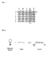
- Fig. 1 In order to further elucidate the general principle please see Fig. 1 .
- all the probes in row 1 share a common label. All the probes in row one differ with respect to their melting temperature. All the probes in column D share a given melting temperature. Given a scenario in which the probes in row one have a green label and the probes in column D, which all differ with respect to their label share 55°C as a common melting temperature, it would be possible to determine whether a template for probe "4" is present in the reaction because this is the case if a signal is detected from the green label at 55°C melting temperature.
- this "detecting the amplified nucleic acids by determining whether the labeled probe has bound its nucleic acid sequence", and "detecting the temperature at which each given labeled probe dissociates from the nucleic acid sequence to which it has bound” may be done at the end of given reaction or during the reaction.
- amplification methods may be applied, these are for example, rolling circle amplification (such as in Liu, et al., “Rolling circle DNA synthesis: Small circular oligonucleotides as efficient templates for DNA polymerases," J. Am. Chem. Soc. 118:1587-1594 (1996 ).), isothermal amplification (such as in Walker, et al., “Strand displacement amplification-an isothermal, in vitro DNA amplification technique", Nucleic Acids Res.
- rolling circle amplification such as in Liu, et al., “Rolling circle DNA synthesis: Small circular oligonucleotides as efficient templates for DNA polymerases," J. Am. Chem. Soc. 118:1587-1594 (1996 ).
- isothermal amplification such as in Walker, et al., “Strand displacement amplification-an isothermal, in vitro DNA amplification technique", Nucleic Acids Res.
- ligase chain reaction such as in Landegren, et al., "A Ligase-Mediated Gene Detection Technique," Science 241:1077-1080, 1988 , or, in Wiedmann, et al., “Ligase Chain Reaction (LCR)-Overview and Applications,” PCR Methods and Applications (Cold Spring Harbor Laboratory Press, Cold Spring Harbor Laboratory, NY, 1994) pp. S51-S64. ). Polymerase chain reaction amplification is, however, preferred.
- the reaction is a polymerase chain reaction
- the "detecting the amplified nucleic acids by determining whether the labeled probe has bound its nucleic acid sequence", and "detecting the temperature at which each given labeled probe dissociates from the nucleic acid sequence to which it has bound” may be done after each cycle, after one cycle, after more than one cycle, in intervals, or at the end of the complete PCR reaction.
- a PCR reaction may consist of 10 to 100 "cycles" of denaturation and synthesis of a DNA molecule.
- the temperature at which denaturation is done in a thermocycling amplification reaction is between about 90°C to greater than 95°C, more preferably between 92-94°C.
- Preferred thermo cycling amplification methods include polymerase chain reactions involving from about 10 to about 100 cycles, more preferably from about 25 to about 50 cycles, and peak temperatures of from about 90°C to greater than 95°C, more preferably 92-94°C.
- a PCR reaction is done using a DNA Polymerase I to produce, in exponential quantities relative to the number of reaction steps involved, at least one target nucleic acid sequence, given (a) that the ends of the target sequence are known in sufficient detail that oligonucleotide primers can be synthesized which will hybridize to them and (b) that a small amount of the target sequence is available to initiate the chain reaction.
- the product of the chain reaction will be a discrete nucleic acid duplex with termini corresponding to the ends of the specific primers employed. Any source of nucleic acid, in purified or non-purified form, can be utilized as the starting nucleic acid, if it contains or is thought to contain the target nucleic acid sequence desired.
- the process may employ, for example, DNA which may be single stranded or double stranded.
- DNA-RNA hybrid which contains one strand of each may be utilized.
- a mixture of any of these nucleic acids may also be employed, or the nucleic acids produced from a previous amplification reaction using the same or different primers may be so utilized.
- the nucleic acid amplified is preferably DNA,
- the target nucleic acid sequence to be amplified may be only a fraction of a larger molecule or can be present initially as a discrete molecule, so that the target sequence constitutes the entire nucleic acid.
- the target sequence to be amplified may be present initially in a pure form; it may be a minor fraction of a complex mixture or a portion of nucleic acid sequence due to a particular animal which organism might constitute only a very minor fraction of a particular biological sample.
- the starting nucleic acid may contain more than one desired target nucleic acid sequence which may be the same or different. Therefore, the method is useful for amplifying simultaneously multiple target nucleic acid sequences located on the same or different nucleic acid molecules.
- the nucleic acid(s) may be obtained from any source and include plasmids and cloned DNA, DNA from any source, including bacteria, yeast, viruses, and higher organisms such as plants or animals.
- DNA may be extracted from, for example, blood or other fluid, or tissue material such as chorionic villi or amniotic cells by a variety of techniques such as that described by Maniatis et al., Molecular Cloning: A Laboratory Manual, (New York: Cold Spring Harbor Laboratory) pp 280-281 (1982 ). Additionally the Templex technology may be applied which combines Genaco's Tem-PCR technology and Luminex's xMAP technology.
- Oligonucleotide primers may be prepared using any suitable method, such as, for example, the phosphotriester and phosphodiester methods or automated embodiments thereof. In one such automated embodiment diethylophosphoramidites are used as starting materials and may be synthesized as described by Beaucage et al., Tetrahedron Letters, 22:1859-1862 (1981 ), which is hereby incorporated by reference.
- diethylophosphoramidites are used as starting materials and may be synthesized as described by Beaucage et al., Tetrahedron Letters, 22:1859-1862 (1981 ), which is hereby incorporated by reference.
- One method for synthesizing oligonucleotides on a modified solid support is described in U.S. Pat. No. 4,458,006 , which is hereby incorporated by reference.
- primers which has been isolated from a biological source (such as a restriction endonuclease digest).
- Preferred primers have a length of from about 15-100, more preferably about 20-50, most preferably about 20-40 bases. It is essential that the primers of the method span the region comprising the target sequence.
- the target nucleic acid sequence is amplified by using the nucleic acid containing that sequence as a template. [f the nucleic acid contains two strands, it is necessary to separate the strands of the nucleic acid before it can be used as the template, either as a separate step or simultaneously with the synthesis of the primer extension products. This strand separation can be accomplished by any suitable denaturing method including physical, chemical, or enzymatic means.
- One physical method of separating the strands of the nucleic acid involves heating the nucleic acid until it is completely (>99%) denatured. Typical heat denaturation may involve temperatures ranging from about 80°C to 105°C, preferably about 90°C to about 98°C, still more preferably 93°C to 95°C, for times ranging from about 1 to 10 minutes.
- the strand separation may also be induced by an enzyme from the class of enzymes known as helicases or the enzyme RecA, which has helicase activity and is known to denature DNA.
- the reaction conditions suitable for separating the strands of nucleic acids with helicases are described by Cold Spring Harbor Symposia on Quantitative Biology, Vol.
- This synthesis can be performed using any suitable method. Generally, it occurs in a buffered aqueous solution. In some preferred embodiments, the buffer pH is about 7.5-8.9. Preferably, a molar excess (for cloned nucleic acid, usually about 1000:1 primer:template, and for genomic nucleic acid, usually about 106 :1 primer:template) of the oligonucleotide primers is added to the buffer containing the separated template strands. It is understood, however, that the amount of complementary strand may not be known if the process herein is used for some applications, so that the amount of primer relative to the amount of complementary strand cannot be determined with certainty.
- the amount of primer added will generally be in molar excess over the amount of complementary strand (template) when the sequence to be amplified is contained in a mixture of complicated long-chain nucleic acid strands.
- a large molar excess is preferred to improve the efficiency of the process.
- Nucleoside triphosphates preferably dATP, dCTP, dGTP, dTTP and/or dUTP are also added to the synthesis mixture in adequate amounts.
- the preferred molarity of nucleotides is as follows 0.025 mM to 1 mM, preferred 0,05 to 0.6 mM, most prefered 0.1 to 0.5 mM.
- the polymerase according to the invention is selected from the group of genera of Thermus, Aquifex, Thermotoga, Thermocridis, Hydrogenobacter, Thermosynchecoccus and Thermoanaerobacter.
- the polymerase according to the invention is selected from the group of organisms of Aquifex aeolicus, Aquifex pyogenes, Thermus thermophilus, Thermus aquaticus, Thermotoga neapolitana, Thermus pacificus, Thermus eggertssonii, and Thermotoga maritima.
- the polymerase is Taq polymerase. However, as will be outlined in more detail below, in some embodiments it is preferred that the polymerase carries a 5'-3' exonuclease activity. In other embodiments it is preferred that the polymerase lacks a 5'-3' exonuclease activity. In most embodiments it is preferred that the polymerase lacks a 3'-5' exonuclease activity.
- Uracil DNA glycosylase uracil-N-glycosylase
- Uracil DNA glycosylase is the product of the Escherichia coli ung-gene, and has been cloned, sequenced and expressed in E. coli.
- Uracil DNA glycosylase UDG removes these uracil residues from DNA (single- and double-stranded) without destroying the DNA sugar-phosphodiester backbone, thus preventing its use as a hybridization target or as a template for DNA polymerases.
- the resulting abasic sites are susceptible to hydrolytic cleavage at elevated temperatures.
- removal of uracil bases is usually accompanied by fragmentation of the DNA.
- both the enzyme as well as the uracil nucleotide may be in the kit according to the invention.
- the labels of the probes in the first and second or further probe set are fluorescent labels and have an emission wavelength that is very similar. Ideally, that means they may be detected without altering the wavelength adjustment that may be detected by the detection device. It is preferred that the labels of the probes in the first, second and third or further probe set are identical.
- the probes carrying the same label differ in melting temperature (T m ) in a way that they are distinguishable by melting point on a given instrument, typically harboring a difference in melting temperature of more than 0.1°C, 0.2°C, 0.3°C, 0.4°C, 0.5°C, 1°C, 1.5°C, 2°C, 2.5°C, 3°C, 4°C, 5°C, 6°C, 7°C, 8°C, 9°C, or 10°C. More than 1°C, 1.5°C, 2°C, 2.5°C, 3°C, 4°C, 5°C is preferred.
- the melting transitions of the double stranded segments can be determined by monitoring fluorescence intensity of double stranded nucleic acid-specific (dsNAS) dyes.
- the double stranded nucleic acid-specific dye is selected from the group consisting of SYBR® Green I, SYBR® Gold, ethidium bromide, propidium bomide, Pico Green, Hoechst 33258, YO-PRO-I and YO-YO-I, SYTO®9, LC Green®, LC Green® Plus+, EvaGreenTM. These saturation dyes are capable of existing at sufficiently saturating conditions with respect to the DNA during or after amplification, while minimizing the inhibition of PCR.
- the dsDNA binding dye has a percent saturation of at least 50%.
- the percent saturation is at least 80%.
- the percent saturation is at least 99%. It is understood that the percent saturation is the percent fluorescence compared to fluorescence of the same dye at saturating concentrations. Saturating concentration is the concentration that provides the highest fluorescence intensity possible in the presence of a predetermined amount of dsDNA. Because these dyes can be present at significantly higher concentrations without significantly interfering with certain nucleic acid reactions, these dyes may be particularly useful for monitoring the conformation of single-stranded nucleic acids and dsDNA.
- the preferred reaction is a polymerase chain reaction.
- the probes are selected from the group of TaqMan probe, molecular beacon probe, scorpion probe and light cycler probe. Detection of the amplification product per se may be accomplished by using one of the following probes, TaqMan probe, molecular beacon probe, scorpion probe,, light cycler probe, hybridization probe, displacement probe and other types of sequence specific probe formats.
- the TaqMan® Assay utilizes the 5' nuclease activity of Taq DNA polymerase to cleave a fluorescently labeled probe (FAM TM -Iabeled MGB).
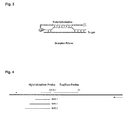
- Molecular beacons are single-stranded oligonucleotide hybridization probes that form a stem-and-loop structure ( FIG. 2 ).
- the loop contains a probe sequence that is complementary to a target sequence, and the stem is formed by the annealing of complementary arm sequences that are located on either side of the probe sequence.
- a fluorophore is covalently linked to the end of one arm and a quencher is covalently linked to the end of the other arm.
- Molecular beacons do not fluoresce when they are free in solution. However, when they hybridize to a nucleic acid strand containing a target sequence they undergo a conformational change that enables them to fluoresce brightly.
- the probe In the absence of targets, the probe is dark, because the stem places the fluorophore so close to the non-fluorescent quencher that they transiently share electrons, eliminating the ability of the fluorophore to fluoresce.
- the probe encounters a target molecule, it forms a probe-target hybrid that is longer and more stable than the stem hybrid.
- the rigidity and length of the probe-target hybrid precludes the simultaneous existence of the stem hybrid. Consequently, the molecular beacon undergoes a spontaneous conformational reorganization that forces the stem hybrid to dissociate and the fluorophore and the quencher to move away from each other, restoring fluorescence.
- Molecular beacons are added to the assay mixture before carrying out gene amplification and fluorescence is measured in real time. Molecular beacons can be synthesized that possess differently colored fluorophores, enabling the method according to the invention.
- the color of the resulting fluorescence if any, identifies the pathogenic agent in combination with the determination of the melting temperature.
- Scorpion primers are bi-functional molecules in which a primer is covalently linked to the probe.
- the molecules also contain a fluorophore and a quencher. In the absence of the target, the quencher nearly absorbs the fluorescence emitted by the fluorophore.
- the fluorophore and the quencher separate which leads to an increase in the fluorescence emitted. The fluorescence can be detected and measured in the reaction tube.
- a light cycler FRET probe system is a pair of single-stranded fluorescently-labeled oligonucleotides.
- Probe 1 (the donor probe) is labeled at its 3'end with a donor fluorophore (generally fluorescein) and Probe 2 (the acceptor probe) is labeled at its 5'end with one of four available fluorophores (red 610, 640, 670 or 705).
- the free 3' hydroxyl group of Probe 2 must be blocked with a phosphate group (P) to prevent Taq DNA polymerase extension.
- the PCR primers and the light cycler probes hybridize to their specific target regions causing the donor dye to come into close proximity to the acceptor dye.
- the donor dye is excited by light from the light cycler instrument (h ⁇ 1)
- energy is transferred by Fluorescence Resonance Energy Transfer (FRET) from the donor to the acceptor dye.
- FRET Fluorescence Resonance Energy Transfer
- the energy transfer causes the acceptor dye to emit light (h ⁇ 2) at a longer wavelength than the light emitted from the instrument (h ⁇ 1).
- the acceptor fluorophore's emission wavelength is detected by the instrument's optical unit.
- the increase in measured fluorescent signal is directly proportional to the amount of accumulating target DNA.
- probes are Eclipse Probes (Epoch, Nanogen), displacement probes ( Cheng et al., Nucleic Acids Research, 2004, Vol. 32, No. 7 ), pleiades probes ( NAR 2007 Vol 35 5 e30 ) and plexor systems (Promega). Of course other probe systems are likewise encompassed by the invention.
- the TaqMan probe is combined with a intercalating dye used for the melting point analysis.
- the TaqMan probe is combined with a hybridization probe used for the melting point analysis in a special embodiment the TaqMan probe is the hybridization probe. In another embodiment the TaqMan probe is not the hybridization probe but a separate oligonucleotide serves as hybridization probe.
- Figure 4 shows a very preferred embodiment.
- the reaction comprises both a hybridization probe as well as a TaqMan probe.
- each probe set would, e.g. consists of at least two probes, wherein the probes are the hybridization probes,
- each of the probes is specific for a nucleic acid sequence,
- each of the hybridization probes for example BHQ 1, BHQ 2, BHQ 3 in Fig. 4
- each of the hybridization probes in a given probe set carries a different label (see for example column A in Fig. 1 )
- all of the probes in a given probe set have a similar, preferably identical melting temperature (T m ) (see for example column A in Fig.
- the melting point of the hybridization probe is lower than the melting point of the primers used for amplification.
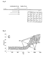
- Fig. 5 One example is given in Fig. 5 .
- the hybridization probe Q1 will dissociate for the DNA strand.
- the hybridization probe carries a quencher.
- the downstream TaqMan probe will give rise to a signal coming from the fluorescent label which is now no longer quenched.
- the melting point is known due to the signal (melting point mp1).
- the label is known (FL1).
- this hybridization probe was specific for, e.g. pathogen 1 (p1) which is thus known to be present in the reaction.
- the TaqMan probe allows on line quantification during the PCR reaction.
- the reaction additionally comprises a double strand nucleic acid specific dye.
- a double strand specific dye is used it is preferred that this dye is selected from the group of SYBR ® Green I, SYBR ® Gold, ethidium bromide, propidium bromide, Pico Green, Hoechst 33258, YO-PRO-I and YO-YO-I.
- SYBR ® Green I is very preferred.
- the double strand nucleic acid specific dye is spectrally distinguishable from the probe labels.
- the label is a fluorescent label and the label is selected from the group of FAM (5-or 6-carboxyfluorescein), VIC, NED, Fluorescein, FITC, IRD-700/800, CY3, CY5, CY3.5, CY5.5, HEX, TET, TAMRA, JOE, ROX, BODIPY TMR, Oregon Green, Rhodamine Green, Rhodamine Red, Texas Red, Yakima Yellow, Alexa Fluor PET, Biosearch BlueTM, Marina Blue ® , Bothell Blue ® , Alexa Fluor ® .
- the method is alternatively based on the basic principle that melting curve analyses is performed at the end of a PCR reaction with a single dual-labeled probe allows differentiation of targets.
- the melting point (T m ) of the probe was chosen to be 10 °C below the T m s of the PCR primers.
- the probe is allowed to hybridize and the mixture is subjected to stepwise increase in temperature, with fluorescence monitored continuously.
- FRET Förster resonance energy transfer
- asymmetric primer concentrations are used. This is done by altering the ratio of both primers for each target in a way that the primer generating the DNA strand binding the probe is added at higher concentration than the other primer.
- the labeled probe is a group consisting of a hybridization probe and TaqMan probe
- the TaqMan probe carries the said label
- the said label is a fluorescent label
- the TaqMan probe additionally comprises a quencher
- the hybridization probe carries an additional quencher that is able to quench the fluorescence of the label attached to the TaqMan probe
- said TaqMan probe and said hybridization probe are able to bind said nucleic acid sequence adjacently in such a way that when both probes are bound to their respective sequences, the quencher present on the hybridization probe, at least partially, quenches the fluorescence of the said label on said TaqMan probe.
- the hybridization probe carries an additional quencher that is able to quench the fluorescence of the label attached to the TaqMan probe.
- the probes with identical labels form a group with hybridization probes which differ in melting temperature (Tm) so that they may be distinguished, e.g. by at least 0.1 °C, 0.5°C, 1°C, 1.5°C, 2°C, 2.5°C, 3°C, 4°C, 5°C, 6°C, 7°C, 8°C, 9°C, or 10°C. More than 0.5°C, 1°C, 1.5°C, 2°C, 2.5°C, 3°C, 4°C, 5°C is preferred. Most preferred is about 5°C. In this embodiment it is preferred also that the melting temperature (T m ) of the hybridization probe is lower than the melting temperature (T m ) of the TaqMan probe.
- Tm melting temperature
- the hybridization probe upon annealing, the temperature is elevated, the hybridization probe will dissociate from its complementary strand, generating a first signal, when the T m is reached. The polymerase will then gain access to the TaqMan probe which will result in a second TaqMan signal.
- the hybridization probes have a melting temperature (T m ) that is below the temperature at which the polymerase exhibits its optimal activity. This provides for that the hybridization probes dissociate and gives the way free for the polymerase to the TaqMan probe. It is obvious that the method ideally makes use of a polymerase that exhibits a 5'-3' exonuclease activity.
- Real-time PCR requires an instrumentation platform that consists of a thermal cycler, a computer, optics for fluorescence excitation and emission collection, and data acquisition and analysis software.
- instrumentation platform that consists of a thermal cycler, a computer, optics for fluorescence excitation and emission collection, and data acquisition and analysis software.
- These machines available from several manufacturers, differ in sample capacity (some are 96-well or 384-well standard plate format, others process fewer samples or require specialized glass capillary tubes, some have block format, others a carousel), method of excitation (some use lasers, others broad spectrum light sources with tunable filters or one or more diodes), detection (some use a camera, others a photo multiplier tube, or types of light detection system) and overall sensitivity.
- method of excitation some use lasers, others broad spectrum light sources with tunable filters or one or more diodes
- detection some use a camera, others a photo multiplier tube, or types of light detection system
- overall sensitivity There are also platform
- the invention relates to a kit comprising at least 6 probes which are able to hybridize, under stringent conditions, to one or more nucleic acid molecules, wherein a) a first group of at least three probes carries a first label and all the probes in this group differ with respect to their melting temperature and b) a second group of at least three probes carries a second label and all the probes in this group differ with respect to their melting temperature
- the invention also relates to a kit comprising at least 9 probes which are able to hybridize, under stringent conditions, to one or more nucleic acid molecules, wherein (a) a first group of at least two probes carries a first label and all the probes in this group differ with respect to their melting temperature, (b) a second group of at least two probes carries a second label and (c) a third group of at least two probes carries a third label, and all the probes in this group differ with respect to their melting temperature and at least a third group of at least two probes carries a third label and all the probes in this group differ with respect to their melting temperature.
- the invention also relates to a kit with more probe sets and/or more probes.
- the probes that differ with respect to their melting temperature (T m ) differ by at least 0.5°C, 1°C, 1.5°C, 2°C, 2.5°C, 3°C, 4°C, 5°C, 6°C, 7°C, 8°C, 9°C, or 10°C. More than 1°C, 1.5°C, 2°C, 2.5°C, 3°C, 4°C, 5°C is preferred. Ideally the probes differ by about 5°C. Ideally, the probes that have the same label have fixed intervals of difference in melting temperature (T m ), selected in a way that they are reliably distinguishable by melting analysis, for example such as 5°C. In such an embodiment the probes that are, for example, fluorescein labeled would have a melting temperature (T m ) of, e.g. 40°C, 45°C, 50°C and 55°C.
- the kit according to the invention can additionally comprise a buffer, nucleotides and one or enzymes, such as a polymerase.
- the kit may be adapted as a premix, wherein the user only needs to add the probe.
- Primers and probes according to the invention may be specific for various targets, such as disease markers, pathogens, forensic markers or any other target that may be addressed by means of amplification.
- the invention is particularly suited for the analysis of pathogens.
- the most common bacterial disease is tuberculosis, caused by the bacterium Mycobacterium tuberculosis, which kills about 2 million people a year, mostly in sub-Saharan Africa.
- Pathogenic bacteria contribute to other globally important diseases, such as pneumonia, which can be caused by bacteria such as Streptococcus and Pseudomonas, and foodborne illnesses, which can be caused by bacteria such as Shigella, Campylobacter and Salmonella.
- Pathogenic bacteria also cause infections such as tetanus, typhoid fever, diphtheria, syphilis and leprosy.
- One of the primary pathways by which food or water become contaminated is from the release of untreated sewage into a drinking water supply or onto cropland, with the result that people who eat or drink contaminated sources become infected.
- sewage is discharged into the environment or on cropland. This is the typical mode of transmission for the infectious agents of cholera, hepatitis A, polio and rotavirus.
- the primers and probes are specific for one or more pathogenic bacteria.
- the kit may used for human or veterinary diagnosis, for testing food or water, for forensic applications or for scientific purposes.
Landscapes
- Chemical & Material Sciences (AREA)
- Life Sciences & Earth Sciences (AREA)
- Organic Chemistry (AREA)
- Proteomics, Peptides & Aminoacids (AREA)
- Zoology (AREA)
- Wood Science & Technology (AREA)
- Health & Medical Sciences (AREA)
- Engineering & Computer Science (AREA)
- Analytical Chemistry (AREA)
- Biophysics (AREA)
- Immunology (AREA)
- Microbiology (AREA)
- Molecular Biology (AREA)
- Biotechnology (AREA)
- Physics & Mathematics (AREA)
- Biochemistry (AREA)
- Bioinformatics & Cheminformatics (AREA)
- General Engineering & Computer Science (AREA)
- General Health & Medical Sciences (AREA)
- Genetics & Genomics (AREA)
- Chemical Kinetics & Catalysis (AREA)
- Measuring Or Testing Involving Enzymes Or Micro-Organisms (AREA)
Abstract
Description
- The present invention is in the field of biology and chemistry, more in particular in the field of molecular biology and human genetics. The invention relates to the field of identifying certain nucleic acid sequences in a sample. Particularly, the invention is in the field of simultaneously amplifying and detecting nucleic acid sequences in a reaction. The invention relates to methods, kits, and systems for detection of nucleic acid sequences in a sample.
- Diagnostic assays that sensitively, specifically, and quickly detect biological agents, e.g., pathogens, in samples are becoming increasingly important for both disease and diagnostic bio agent monitoring. Few assays are able to accurately detect physiologically or clinically relevant organisms on an appropriate time scale for the early detection of the presence of an infective or otherwise harmful agent.
- A DNA microarray is a collection of microscopic DNA spots, commonly representing single genes, arrayed on a solid surface by covalent attachment to a chemical matrix. DNA arrays are different from other types of microarray only in that they either measure DNA or use DNA as part of its detection system. Qualitative or quantitative measurements with DNA microarrays utilize the selective nature of DNA-DNA or DNA-RNA hybridization under high-stringency conditions and fluorophore-based detection. DNA arrays are commonly used for expression profiling, i.e., monitoring expression levels of thousands of genes simultaneously, or for comparative genomic hybridization. The drawback with this system is that multiple steps need to be performed prior to analysis. Also, the array is not sensitive.
- To date, the most sensitive detection methods involve PCR. Determining the presence or absence of a plurality of biological agents in a single sample can be performed using multiplexed detection methods.
- Multiplex PCR uses multiple, unique primer sets within a single PCR reaction to produce amplicons of varying sizes specific to different DNA sequences, i.e. different transgenes. By targeting multiple genes at once, additional information may be gained from a single test run that otherwise would require several times the reagents and more time to perform. Annealing temperatures for each of the primer sets must be optimized to work correctly within a single reaction, and amplicon sizes, i.e., their base pair length, should be different enough to form distinct bands when visualized by gel electrophoresis.
- Multiplexed real time PCR is one method that can be used for a diagnostic assay. Assays based on PCR can be limited by the complexity of optimizing the PCR reactions to test for multiple agents in a cost-effective number of reaction tubes. As a general rule, the number of probes needed to support a highly specific confirmation result range from two to as many as six sequences. As one of skill in the art will be aware, optimizing a PCR reaction with many different primer pairs and probes can be a formidable task that becomes increasingly unmanageable as the number of targets to be detected increases. Assays based on PCR can also be limited by the number of unique labels available for analysis of results. For example, real time PCR assays generally employ fluorescent labels.
- The number of labels that can be used in a single reaction is limited by the number of fluorescent color channels available in the optical detection system used.
- It would be advantageous to have a method for simultaneously amplifying and detecting multiple nucleic acid sequences in one container.
-
WO 2005/111243 A2 relates to a method of detecting agents in two containers. The drawback of the method disclosed inWO 2005/111243 A2 is the fact that two containers are necessary. Ideally, the reaction would require one container. - The present invention relates to a method for simultaneously amplifying and detecting nucleic acid sequences in a reaction comprising the following steps, (i) providing a sample comprising at least one nucleic acid molecule, (ii) providing reagents for performing an amplification reaction, wherein the reagents comprise at least three probe sets, wherein (a) each probe set consists of at least three probes, (b) each of the probes is specific for a nucleic acid sequence, (c) each of the probes in a given probe set carries a different label, (d) all of the probes in a given probe set have a similar, preferably identical melting temperature (Tm) when they are dissociated from their target nucleic acid sequence by heating, (iii) amplifying the nucleic acid sequences in the reaction, (iv) detecting the amplified nucleic acids by determining whether the labeled probe has bound its nucleic acid sequence, and (v) detecting the temperature at which each given labeled probe dissociates from the nucleic acid sequence to which it has bound.
- As used herein the term "nucleic acid sequence" is, in the context of the present invention, a sequence on a nucleic acid. A nucleic acid may be, inter alia, RNA, DNA, cDNA (complementary DNA), LNA (locked nucleic acid), mRNA (messenger RNA), mtRNA (mitochondrial), rRNA (ribosomal RNA), tRNA (transfer RNA), nRNA (nuclear RNA), siRNA (short interfering RNA), snRNA (small nuclear RNA), snoRNA (small nucleolar RNA), scaRNA (Small Cajal Body specific RNA), microRNA, dsRNA (doubled-stranded RNA), ribozyme, riboswitch, viral RNA, dsDNA (double-stranded DNA), ssDNA (single-stranded DNA), plasmid DNA, cosmid DNA, chromosomal DNA, viral DNA, mtDNA (mitochondrial DNA), nDNA (nuclear DNA), snDNA (small nuclear DNA) or the like or any other class or sub-class of nucleic acid which is distinguishable from the bulk nucleic acid in a sample.
- As used herein the term "probe" is a nucleic acid which is able to bind another nucleic acid.
- As used herein the term "tissue" refers to any tissue or fluid in a human, animal or plant including, but not limited to breast, prostate, blood, serum, cerebrospinal fluid, liver, kidney, breast, head and neck, pharynx, thyroid, pancreas, stomach, colon, colorectal, uterus, cervix, bone, bone marrow, testes, brain, neural tissue, ovary, skin, and lung.
- As used herein the term "probe set" is a set of three or more agents that may interact with a nucleic acid molecule at a specific position, i.e. sequence.
- Herein, a "label" is a moiety that is bound covalently or non-covalently to a probe where it can give rise to signal which may be detected by optical or other physical means.
- The present invention relates to a method for simultaneously amplifying and detecting nucleic acid sequences in a reaction comprising the following steps, (i) providing a sample comprising at least one nucleic acid molecule, (ii) providing reagents for performing an amplification reaction, wherein the reagents comprise at least two probe sets, wherein (a) each probe set consists of at least three probes, (b) each of the probes is specific for a nucleic acid sequence, (c) each of the probes in a given probe set carries a different label, (d) all of the probes in a given probe set have a similar, preferably identical melting temperature (Tm) when they are dissociated from their target nucleic acid sequence by heating, (iii) amplifying the nucleic acid sequences in the reaction, (iv) detecting the amplified nucleic acids by determining whether the labeled probe has bound its nucleic acid sequence, and (v) detecting the temperature at which each given labeled probe dissociates from the nucleic acid sequence to which it has bound.
- In further embodiment the method comprises at least three probe sets, wherein each probe set consists of at least three probes.
- In order to better elucidate the invention we point to
figure 1 . A probe set according to the invention comprises at least 3 probes. A probe set may seen as all those probes that share a common label but also as all those probes that share a common melting temperature (Tm). Ideally, the probes in a probe set that have the same label or labels that are not distinguishable from one another have different melting temperatures. The probes in a probe set that have identical or very similar melting temperatures should have different labels. - The person skilled in the art will know that the reagents will, ordinarily, comprise for example an enzyme for amplification, a buffer, nucleotides and the like. This of course depends on the type of amplification.
- The inventors have developed a method which makes it possible to perform a multiplex amplification reaction with, for example 20 templates. In one embodiment 5 different labels are used and all the probes that share a common label have a slightly varying melting temperature. All the probes that share a common melting temperature on the other hand have a different label. By detecting the label and the melting temperature either during or after amplification the inventors have for the first time provided for a means which makes it possible to analyze, e.g. said 20 templates in one tube.
- In order to further elucidate the general principle please see
Fig. 1 . Here, all the probes inrow 1 share a common label. All the probes in row one differ with respect to their melting temperature. All the probes in column D share a given melting temperature. Given a scenario in which the probes in row one have a green label and the probes in column D, which all differ with respect to their label share 55°C as a common melting temperature, it would be possible to determine whether a template for probe "4" is present in the reaction because this is the case if a signal is detected from the green label at 55°C melting temperature. - In principle this "detecting the amplified nucleic acids by determining whether the labeled probe has bound its nucleic acid sequence", and "detecting the temperature at which each given labeled probe dissociates from the nucleic acid sequence to which it has bound" may be done at the end of given reaction or during the reaction.
- Various amplification methods may be applied, these are for example, rolling circle amplification (such as in Liu, et al., "Rolling circle DNA synthesis: Small circular oligonucleotides as efficient templates for DNA polymerases," J. Am. Chem. Soc. 118:1587-1594 (1996).), isothermal amplification (such as in Walker, et al., "Strand displacement amplification-an isothermal, in vitro DNA amplification technique", Nucleic Acids Res. 20(7):1691-6 (1992)), ligase chain reaction (such as in Landegren, et al., "A Ligase-Mediated Gene Detection Technique," Science 241:1077-1080, 1988, or, in Wiedmann, et al., "Ligase Chain Reaction (LCR)-Overview and Applications," PCR Methods and Applications (Cold Spring Harbor Laboratory Press, Cold Spring Harbor Laboratory, NY, 1994) pp. S51-S64.). Polymerase chain reaction amplification is, however, preferred.
- If the reaction is a polymerase chain reaction the "detecting the amplified nucleic acids by determining whether the labeled probe has bound its nucleic acid sequence", and "detecting the temperature at which each given labeled probe dissociates from the nucleic acid sequence to which it has bound" may be done after each cycle, after one cycle, after more than one cycle, in intervals, or at the end of the complete PCR reaction.
- A PCR reaction may consist of 10 to 100 "cycles" of denaturation and synthesis of a DNA molecule. In a preferred embodiment, the temperature at which denaturation is done in a thermocycling amplification reaction is between about 90°C to greater than 95°C, more preferably between 92-94°C. Preferred thermo cycling amplification methods include polymerase chain reactions involving from about 10 to about 100 cycles, more preferably from about 25 to about 50 cycles, and peak temperatures of from about 90°C to greater than 95°C, more preferably 92-94°C. In a preferred embodiment, a PCR reaction is done using a DNA Polymerase I to produce, in exponential quantities relative to the number of reaction steps involved, at least one target nucleic acid sequence, given (a) that the ends of the target sequence are known in sufficient detail that oligonucleotide primers can be synthesized which will hybridize to them and (b) that a small amount of the target sequence is available to initiate the chain reaction. The product of the chain reaction will be a discrete nucleic acid duplex with termini corresponding to the ends of the specific primers employed. Any source of nucleic acid, in purified or non-purified form, can be utilized as the starting nucleic acid, if it contains or is thought to contain the target nucleic acid sequence desired. Thus, the process may employ, for example, DNA which may be single stranded or double stranded. In addition, a DNA-RNA hybrid which contains one strand of each may be utilized. A mixture of any of these nucleic acids may also be employed, or the nucleic acids produced from a previous amplification reaction using the same or different primers may be so utilized. The nucleic acid amplified is preferably DNA, The target nucleic acid sequence to be amplified may be only a fraction of a larger molecule or can be present initially as a discrete molecule, so that the target sequence constitutes the entire nucleic acid. It is not necessary that the target sequence to be amplified be present initially in a pure form; it may be a minor fraction of a complex mixture or a portion of nucleic acid sequence due to a particular animal which organism might constitute only a very minor fraction of a particular biological sample. The starting nucleic acid may contain more than one desired target nucleic acid sequence which may be the same or different. Therefore, the method is useful for amplifying simultaneously multiple target nucleic acid sequences located on the same or different nucleic acid molecules. The nucleic acid(s) may be obtained from any source and include plasmids and cloned DNA, DNA from any source, including bacteria, yeast, viruses, and higher organisms such as plants or animals. DNA may be extracted from, for example, blood or other fluid, or tissue material such as chorionic villi or amniotic cells by a variety of techniques such as that described by Maniatis et al., Molecular Cloning: A Laboratory Manual, (New York: Cold Spring Harbor Laboratory) pp 280-281 (1982). Additionally the Templex technology may be applied which combines Genaco's Tem-PCR technology and Luminex's xMAP technology.
- The assay makes use of locus-specific primers. Oligonucleotide primers may be prepared using any suitable method, such as, for example, the phosphotriester and phosphodiester methods or automated embodiments thereof. In one such automated embodiment diethylophosphoramidites are used as starting materials and may be synthesized as described by Beaucage et al., Tetrahedron Letters, 22:1859-1862 (1981), which is hereby incorporated by reference. One method for synthesizing oligonucleotides on a modified solid support is described in
U.S. Pat. No. 4,458,006 , which is hereby incorporated by reference. It is also possible to use a primer which has been isolated from a biological source (such as a restriction endonuclease digest). Preferred primers have a length of from about 15-100, more preferably about 20-50, most preferably about 20-40 bases. It is essential that the primers of the method span the region comprising the target sequence. The target nucleic acid sequence is amplified by using the nucleic acid containing that sequence as a template. [f the nucleic acid contains two strands, it is necessary to separate the strands of the nucleic acid before it can be used as the template, either as a separate step or simultaneously with the synthesis of the primer extension products. This strand separation can be accomplished by any suitable denaturing method including physical, chemical, or enzymatic means. One physical method of separating the strands of the nucleic acid involves heating the nucleic acid until it is completely (>99%) denatured. Typical heat denaturation may involve temperatures ranging from about 80°C to 105°C, preferably about 90°C to about 98°C, still more preferably 93°C to 95°C, for times ranging from about 1 to 10 minutes. In the case of isothermal amplification the strand separation may also be induced by an enzyme from the class of enzymes known as helicases or the enzyme RecA, which has helicase activity and is known to denature DNA. The reaction conditions suitable for separating the strands of nucleic acids with helicases are described by Cold Spring Harbor Symposia on Quantitative Biology, Vol. XLIII "DNA: Replication and Recombination" (New York: Cold Spring Harbor Laboratory, 1978), and techniques for using RecA are reviewed in C. Radding, Ann. Rev. Genetics, 16:405-37 (1982), which is hereby incorporated by reference. - This synthesis can be performed using any suitable method. Generally, it occurs in a buffered aqueous solution. In some preferred embodiments, the buffer pH is about 7.5-8.9. Preferably, a molar excess (for cloned nucleic acid, usually about 1000:1 primer:template, and for genomic nucleic acid, usually about 106 :1 primer:template) of the oligonucleotide primers is added to the buffer containing the separated template strands. It is understood, however, that the amount of complementary strand may not be known if the process herein is used for some applications, so that the amount of primer relative to the amount of complementary strand cannot be determined with certainty. As a practical matter, however, the amount of primer added will generally be in molar excess over the amount of complementary strand (template) when the sequence to be amplified is contained in a mixture of complicated long-chain nucleic acid strands. A large molar excess is preferred to improve the efficiency of the process.
- Nucleoside triphosphates, preferably dATP, dCTP, dGTP, dTTP and/or dUTP are also added to the synthesis mixture in adequate amounts. The preferred molarity of nucleotides is as follows 0.025 mM to 1 mM, preferred 0,05 to 0.6 mM, most prefered 0.1 to 0.5 mM.
- It is preferred that the polymerase according to the invention is selected from the group of genera of Thermus, Aquifex, Thermotoga, Thermocridis, Hydrogenobacter, Thermosynchecoccus and Thermoanaerobacter.
- It is preferred that the polymerase according to the invention is selected from the group of organisms of Aquifex aeolicus, Aquifex pyogenes, Thermus thermophilus, Thermus aquaticus, Thermotoga neapolitana, Thermus pacificus, Thermus eggertssonii, and Thermotoga maritima.
- It is most preferred that the polymerase is Taq polymerase. However, as will be outlined in more detail below, in some embodiments it is preferred that the polymerase carries a 5'-3' exonuclease activity. In other embodiments it is preferred that the polymerase lacks a 5'-3' exonuclease activity. In most embodiments it is preferred that the polymerase lacks a 3'-5' exonuclease activity.
- In one embodiment uracil residues are incorporated during the PCR reaction. Uracil DNA glycosylase (uracil-N-glycosylase) is the product of the Escherichia coli ung-gene, and has been cloned, sequenced and expressed in E. coli. Uracil DNA glycosylase (UDG) removes these uracil residues from DNA (single- and double-stranded) without destroying the DNA sugar-phosphodiester backbone, thus preventing its use as a hybridization target or as a template for DNA polymerases. The resulting abasic sites are susceptible to hydrolytic cleavage at elevated temperatures. Thus, removal of uracil bases is usually accompanied by fragmentation of the DNA. The person skilled in the art knows how to use the Uracil DNA glycosylase in order to avoid contamination. Likewise both the enzyme as well as the uracil nucleotide may be in the kit according to the invention.
- Ideally, the labels of the probes in the first and second or further probe set are fluorescent labels and have an emission wavelength that is very similar. Ideally, that means they may be detected without altering the wavelength adjustment that may be detected by the detection device. It is preferred that the labels of the probes in the first, second and third or further probe set are identical.
- It is preferred that the probes carrying the same label differ in melting temperature (Tm) in a way that they are distinguishable by melting point on a given instrument, typically harboring a difference in melting temperature of more than 0.1°C, 0.2°C, 0.3°C, 0.4°C, 0.5°C, 1°C, 1.5°C, 2°C, 2.5°C, 3°C, 4°C, 5°C, 6°C, 7°C, 8°C, 9°C, or 10°C. More than 1°C, 1.5°C, 2°C, 2.5°C, 3°C, 4°C, 5°C is preferred.
- In one embodiment the melting transitions of the double stranded segments can be determined by monitoring fluorescence intensity of double stranded nucleic acid-specific (dsNAS) dyes. In one embodiment, the double stranded nucleic acid-specific dye is selected from the group consisting of SYBR® Green I, SYBR® Gold, ethidium bromide, propidium bomide, Pico Green, Hoechst 33258, YO-PRO-I and YO-YO-I, SYTO®9, LC Green®, LC Green® Plus+, EvaGreen™. These saturation dyes are capable of existing at sufficiently saturating conditions with respect to the DNA during or after amplification, while minimizing the inhibition of PCR. For example, at maximum PCR-compatible concentrations, the dsDNA binding dye has a percent saturation of at least 50%. In other embodiments, the percent saturation is at least 80%. In yet other embodiments, the percent saturation is at least 99%. It is understood that the percent saturation is the percent fluorescence compared to fluorescence of the same dye at saturating concentrations. Saturating concentration is the concentration that provides the highest fluorescence intensity possible in the presence of a predetermined amount of dsDNA. Because these dyes can be present at significantly higher concentrations without significantly interfering with certain nucleic acid reactions, these dyes may be particularly useful for monitoring the conformation of single-stranded nucleic acids and dsDNA.
- The preferred reaction is a polymerase chain reaction.
- It is preferred that the probes are selected from the group of TaqMan probe, molecular beacon probe, scorpion probe and light cycler probe. Detection of the amplification product per se may be accomplished by using one of the following probes, TaqMan probe, molecular beacon probe, scorpion probe,, light cycler probe, hybridization probe, displacement probe and other types of sequence specific probe formats.
- The TaqMan® Assay utilizes the 5' nuclease activity of Taq DNA polymerase to cleave a fluorescently labeled probe (FAM™-Iabeled MGB).
- Molecular beacons are single-stranded oligonucleotide hybridization probes that form a stem-and-loop structure (
Fig. 2 ). The loop contains a probe sequence that is complementary to a target sequence, and the stem is formed by the annealing of complementary arm sequences that are located on either side of the probe sequence. A fluorophore is covalently linked to the end of one arm and a quencher is covalently linked to the end of the other arm. Molecular beacons do not fluoresce when they are free in solution. However, when they hybridize to a nucleic acid strand containing a target sequence they undergo a conformational change that enables them to fluoresce brightly. In the absence of targets, the probe is dark, because the stem places the fluorophore so close to the non-fluorescent quencher that they transiently share electrons, eliminating the ability of the fluorophore to fluoresce. When the probe encounters a target molecule, it forms a probe-target hybrid that is longer and more stable than the stem hybrid. The rigidity and length of the probe-target hybrid precludes the simultaneous existence of the stem hybrid. Consequently, the molecular beacon undergoes a spontaneous conformational reorganization that forces the stem hybrid to dissociate and the fluorophore and the quencher to move away from each other, restoring fluorescence. Molecular beacons are added to the assay mixture before carrying out gene amplification and fluorescence is measured in real time. Molecular beacons can be synthesized that possess differently colored fluorophores, enabling the method according to the invention. - The color of the resulting fluorescence, if any, identifies the pathogenic agent in combination with the determination of the melting temperature.
- Scorpion primers (
Fig: 3 ) are bi-functional molecules in which a primer is covalently linked to the probe. The molecules also contain a fluorophore and a quencher. In the absence of the target, the quencher nearly absorbs the fluorescence emitted by the fluorophore. During the Scorpion PCR reaction, in the presence of the target, the fluorophore and the quencher separate which leads to an increase in the fluorescence emitted. The fluorescence can be detected and measured in the reaction tube. - A light cycler FRET probe system is a pair of single-stranded fluorescently-labeled oligonucleotides. Probe 1 (the donor probe) is labeled at its 3'end with a donor fluorophore (generally fluorescein) and Probe 2 (the acceptor probe) is labeled at its 5'end with one of four available fluorophores (red 610, 640, 670 or 705). The free 3' hydroxyl group of
Probe 2 must be blocked with a phosphate group (P) to prevent Taq DNA polymerase extension. To avoid any steric problems between the donor and the acceptor fluorophores on both probes, there should be a spacer of 1 to 5 nt (4 to 25A distance) to separate the two probes from each other. Before any real-time quantitative PCR reaction takes place, fluorescence background may be observed inside the tube. - During the annealing step of real-time quantitative PCR, the PCR primers and the light cycler probes hybridize to their specific target regions causing the donor dye to come into close proximity to the acceptor dye. When the donor dye is excited by light from the light cycler instrument (hγ1), energy is transferred by Fluorescence Resonance Energy Transfer (FRET) from the donor to the acceptor dye. The energy transfer causes the acceptor dye to emit light (hγ2) at a longer wavelength than the light emitted from the instrument (hγ1). The acceptor fluorophore's emission wavelength is detected by the instrument's optical unit. The increase in measured fluorescent signal is directly proportional to the amount of accumulating target DNA.
- Other alternative probes are Eclipse Probes (Epoch, Nanogen), displacement probes (Cheng et al., Nucleic Acids Research, 2004, Vol. 32, No. 7), pleiades probes (NAR 2007 Vol 35 5 e30) and plexor systems (Promega). Of course other probe systems are likewise encompassed by the invention.
- In one embodiment of the invention the TaqMan probe is combined with a intercalating dye used for the melting point analysis.
- In another embodiment the TaqMan probe is combined with a hybridization probe used for the melting point analysis in a special embodiment the TaqMan probe is the hybridization probe. In another embodiment the TaqMan probe is not the hybridization probe but a separate oligonucleotide serves as hybridization probe.
-
Figure 4 shows a very preferred embodiment. Here, the reaction comprises both a hybridization probe as well as a TaqMan probe. Here, (a) each probe set would, e.g. consists of at least two probes, wherein the probes are the hybridization probes, (b) each of the probes is specific for a nucleic acid sequence, (c) each of the hybridization probes (forexample BHQ 1,BHQ 2,BHQ 3 inFig. 4 ) in a given probe set carries a different label (see for example column A inFig. 1 ), (d) all of the probes in a given probe set have a similar, preferably identical melting temperature (Tm) (see for example column A inFig. 1 ) when they are dissociated from their target nucleic acid sequence by heating. The amplification of the nucleic acid sequences in the reaction is performed, the amplified nucleic acids are detected and the temperature at which each given labeled probe dissociates from the nucleic acid sequence to which it has bound is determined. In this embodiment it is preferred, although not essentially required, that the melting point of the hybridization probe is lower than the melting point of the primers used for amplification. - One example is given in
Fig. 5 . During the PCR reaction at a given melting point (the melting point for the hybridization probe Q1) the hybridization probe Q1 will dissociate for the DNA strand. The hybridization probe carries a quencher. Once dissociated the downstream TaqMan probe will give rise to a signal coming from the fluorescent label which is now no longer quenched. The melting point is known due to the signal (melting point mp1). The label is known (FL1). Hence, it is possible to determine that this hybridization probe was specific for, e.g. pathogen 1 (p1) which is thus known to be present in the reaction. At the same time the TaqMan probe allows on line quantification during the PCR reaction. - It is preferred that the reaction additionally comprises a double strand nucleic acid specific dye. If a double strand specific dye is used it is preferred that this dye is selected from the group of SYBR® Green I, SYBR® Gold, ethidium bromide, propidium bromide, Pico Green, Hoechst 33258, YO-PRO-I and YO-YO-I. SYBR® Green I is very preferred.
- According to the invention ideally, the double strand nucleic acid specific dye is spectrally distinguishable from the probe labels.
- Ideally, the label is a fluorescent label and the label is selected from the group of FAM (5-or 6-carboxyfluorescein), VIC, NED, Fluorescein, FITC, IRD-700/800, CY3, CY5, CY3.5, CY5.5, HEX, TET, TAMRA, JOE, ROX, BODIPY TMR, Oregon Green, Rhodamine Green, Rhodamine Red, Texas Red, Yakima Yellow, Alexa Fluor PET, Biosearch Blue™, Marina Blue®, Bothell Blue®, Alexa Fluor®. 350 FAM™, SYBR® Green 1, Fluorescein, EvaGreen™, Alexa Fluor® 488 JOE™, VIC™, HEX™, TET™, CAL Fluor® Gold 540, Yakima Yellow®, ROX™, CAL Fluor® Red 610, Cy3.5™, Texas Red®, Alexa Fluor®, 568 Cy5™, Quasar™ 670, LightCycler Red640®, Alexa Fluor 633 Quasar™ 705, LightCycler Red705®, Alexa Fluor® 680, SYTO®9, LC Green®, LC Green® Plus+, EvaGreen™.
- The method is alternatively based on the basic principle that melting curve analyses is performed at the end of a PCR reaction with a single dual-labeled probe allows differentiation of targets. To avoid hydrolysis of this probe, as occurs classically during the elongation steps in TaqMan real-time PCR, the melting point (Tm) of the probe was chosen to be 10 °C below the Tms of the PCR primers. At the end of the PCR reaction, the probe is allowed to hybridize and the mixture is subjected to stepwise increase in temperature, with fluorescence monitored continuously. As in classic Taq-Man real-time PCR, generation of the fluorescence signal by the probe is based on the Förster resonance energy transfer (FRET) phenomenon.
- However, and contrary to what happens in TaqMan real-time PCR, no or only partial hydrolosis of the available probe molecules by Taq polymerase is involved in this embodiment. Rather, the procedure relies on the decrease in FRET observed when the probe detaches from its target to achieve a random single-stranded conformation. The mean distance between the reporter and the quencher molecules of the dual-labeled probe will become shorter when the probe is released from its hybrid with the target sequence. Because the FRET effect is inversely proportional to the sixth power of this distance, a difference in fluorescence emission will be readily detectable between hybridized and melted configurations of the probe. A general scheme is shown in
Fig. 6 . Here, the change in fluorescence is shown cycle by cycle for different reaction temperatures. In a preferred embodiment of this method the polymerase used lacks a 5'-3' exonuclease activity. - In a preferred embodiment, asymmetric primer concentrations are used. This is done by altering the ratio of both primers for each target in a way that the primer generating the DNA strand binding the probe is added at higher concentration than the other primer.
- In one alternative embodiment of the method according to the invention, (a) the labeled probe is a group consisting of a hybridization probe and TaqMan probe, (b) the TaqMan probe carries the said label, (c) the said label is a fluorescent label, (d) the TaqMan probe additionally comprises a quencher, (e) optionally the hybridization probe carries an additional quencher that is able to quench the fluorescence of the label attached to the TaqMan probe, (f) said TaqMan probe and said hybridization probe are able to bind said nucleic acid sequence adjacently in such a way that when both probes are bound to their respective sequences, the quencher present on the hybridization probe, at least partially, quenches the fluorescence of the said label on said TaqMan probe. It is however preferred that the hybridization probe carries an additional quencher that is able to quench the fluorescence of the label attached to the TaqMan probe.
- In this embodiment the probes with identical labels form a group with hybridization probes which differ in melting temperature (Tm) so that they may be distinguished, e.g. by at least 0.1 °C, 0.5°C, 1°C, 1.5°C, 2°C, 2.5°C, 3°C, 4°C, 5°C, 6°C, 7°C, 8°C, 9°C, or 10°C. More than 0.5°C, 1°C, 1.5°C, 2°C, 2.5°C, 3°C, 4°C, 5°C is preferred. Most preferred is about 5°C. In this embodiment it is preferred also that the melting temperature (Tm) of the hybridization probe is lower than the melting temperature (Tm) of the TaqMan probe. Hence, once in a reaction, upon annealing, the temperature is elevated, the hybridization probe will dissociate from its complementary strand, generating a first signal, when the Tm is reached. The polymerase will then gain access to the TaqMan probe which will result in a second TaqMan signal.
- It is also preferred in this embodiment that the hybridization probes have a melting temperature (Tm) that is below the temperature at which the polymerase exhibits its optimal activity. This provides for that the hybridization probes dissociate and gives the way free for the polymerase to the TaqMan probe. It is obvious that the method ideally makes use of a polymerase that exhibits a 5'-3' exonuclease activity.
- Real-time PCR requires an instrumentation platform that consists of a thermal cycler, a computer, optics for fluorescence excitation and emission collection, and data acquisition and analysis software. These machines, available from several manufacturers, differ in sample capacity (some are 96-well or 384-well standard plate format, others process fewer samples or require specialized glass capillary tubes, some have block format, others a carousel), method of excitation (some use lasers, others broad spectrum light sources with tunable filters or one or more diodes), detection (some use a camera, others a photo multiplier tube, or types of light detection system) and overall sensitivity. There are also platform-specific differences in how the software processes data. In principle the available machines harboring two or more detection channels are suited for the method according to the invention.
- The invention relates to a kit comprising at least 6 probes which are able to hybridize, under stringent conditions, to one or more nucleic acid molecules, wherein a) a first group of at least three probes carries a first label and all the probes in this group differ with respect to their melting temperature and b) a second group of at least three probes carries a second label and all the probes in this group differ with respect to their melting temperature
- The invention also relates to a kit comprising at least 9 probes which are able to hybridize, under stringent conditions, to one or more nucleic acid molecules, wherein (a) a first group of at least two probes carries a first label and all the probes in this group differ with respect to their melting temperature, (b) a second group of at least two probes carries a second label and (c) a third group of at least two probes carries a third label, and all the probes in this group differ with respect to their melting temperature and at least a third group of at least two probes carries a third label and all the probes in this group differ with respect to their melting temperature. Of course the invention also relates to a kit with more probe sets and/or more probes.
- The probes that differ with respect to their melting temperature (Tm) differ by at least 0.5°C, 1°C, 1.5°C, 2°C, 2.5°C, 3°C, 4°C, 5°C, 6°C, 7°C, 8°C, 9°C, or 10°C. More than 1°C, 1.5°C, 2°C, 2.5°C, 3°C, 4°C, 5°C is preferred. Ideally the probes differ by about 5°C. Ideally, the probes that have the same label have fixed intervals of difference in melting temperature (Tm), selected in a way that they are reliably distinguishable by melting analysis, for example such as 5°C. In such an embodiment the probes that are, for example, fluorescein labeled would have a melting temperature (Tm) of, e.g. 40°C, 45°C, 50°C and 55°C.
- The kit according to the invention can additionally comprise a buffer, nucleotides and one or enzymes, such as a polymerase.
- The kit may be adapted as a premix, wherein the user only needs to add the probe.
- Primers and probes according to the invention may be specific for various targets, such as disease markers, pathogens, forensic markers or any other target that may be addressed by means of amplification. The invention is particularly suited for the analysis of pathogens. The most common bacterial disease is tuberculosis, caused by the bacterium Mycobacterium tuberculosis, which kills about 2 million people a year, mostly in sub-Saharan Africa. Pathogenic bacteria contribute to other globally important diseases, such as pneumonia, which can be caused by bacteria such as Streptococcus and Pseudomonas, and foodborne illnesses, which can be caused by bacteria such as Shigella, Campylobacter and Salmonella. Pathogenic bacteria also cause infections such as tetanus, typhoid fever, diphtheria, syphilis and leprosy. One of the primary pathways by which food or water become contaminated is from the release of untreated sewage into a drinking water supply or onto cropland, with the result that people who eat or drink contaminated sources become infected. In developing countries most sewage is discharged into the environment or on cropland. This is the typical mode of transmission for the infectious agents of cholera, hepatitis A, polio and rotavirus. Thus, in embodiment the primers and probes are specific for one or more pathogenic bacteria.
- The kit may used for human or veterinary diagnosis, for testing food or water, for forensic applications or for scientific purposes.
-
-
Figure 1
Figure 1 shows the principle of the invention. The reactions performed with the probes in, e.g. row one all share a common label. However, the melting temperature of the probes differs. It is thus possible to identify each probe by means of the differing melting temperatures. The probes in column D for example all have the same melting temperature but a different label. It is thus possible to identify each probe by means of the different label. -
Figure 2
Figure 2 shows the principle of a molecular beacon probe. Molecular beacons can be used as amplicon detector probes in for example diagnostic assays. Because non-hybridized molecular beacons are dark, it is not necessary to isolate the probe-target hybrids to determine the number of amplicons synthesized during an assay. The are thus ideally suited for the present invention. Molecular beacons are added to the assay mixture before carrying out gene amplification and fluorescence is measured in real time. -
Figure 3
With Scorpion probes, sequence-specific priming and PCR product detection is achieved using a single oligonucleotide. The Scorpion probe maintains a stem-loop configuration in the unhybridized state. The fluorophore is attached to the 5' end and is quenched by a moiety coupled to the 3' end. The 3' portion of the stem also contains a sequence that is complementary to the extension product of the primer. This sequence is linked to the 5' end of a specific primer via a non-amplifiable monomer. After extension of the Scorpion primer, the specific probe sequence is able to bind to its complement within the extended amplicon thus opening up the hairpin loop. This prevents the fluorescence from being quenched and a signal is observed. -
Figure 4
Figure shows one preferred embodiment of the invention. Here two probes are present in the reaction for each target sequence. A first hybridization probe is present that carries a label, e.g. a fluorescent label that is quenched by an adjacent TaqMan probe. When the melting temperature is reached the hybridization probe dissociates for the strand to which it is bound. The fluorescent label is now no longer quenched and a signal is produced. -
Figure 5
Figure shows one preferred embodiment of the invention. Afluorescent signal FL 1 at a melting temperature mp1 is indicative of the presence of the target sequence p1 in the reaction. -
Figure 6
Figure 6 shows the intensity of a fluorescent signal with a single dual-labeled probe as it changes throughout a cycle as well as throughout the PCR. The signal is strong when it is bound to the target. It is weaker when dissociated. Also the signal gets stronger towards the end of the reaction due to the increase in template amount.
Claims (10)
- Method for simultaneously amplifying and detecting nucleic acid sequences in a reaction comprising the following steps:(i) providing a sample comprising at least one nucleic acid molecule;(ii) providing reagents for performing an amplification reaction, wherein the reagents comprise at least two probe sets, whereina. each probe set consists of at least three probes;b. each of the probes is specific for a nucleic acid sequence;c. each of the probes in a given probe set carries a different fluorescent reporter label;d. all of the probes in a given probe set have an identical melting temperature (Tm) when they are dissociated from their target nucleic acid sequence by heating,(iii) amplifying the nucleic acid sequences in the reaction;(iv) detecting the amplified nucleic acids by determining whether the labeled probe has bound its nucleic acid sequence;(v) detecting the temperature at which each given labeled probe dissociates from the nucleic acid sequence to which it has bound,
wherein the probes carrying the same label differ in melting temperature (Tm) in a way that they are distinguishable by melting point, and
wherein the probes are dual-labeled probes comprising said reporter label and a quencher molecule such that a difference in fluorescence emission is detectable between hybridized and melted configurations of said probes. - Method of claim 1, wherein the labels of the probes in the first and second probe set are fluorescent labels and have an emission wavelength that is very similar.
- Method of claim 1 or 2, wherein the amplification reaction is polymerase chain reaction amplification.
- Method of claim 1 to 3, wherein the reaction additionally comprises a double strand nucleic acid specific dye.
- Method of claim 4, wherein the double strand nucleic acid specific dye is spectrally distinguishable from the probe labels.
- Method of claim 1 to 5, wherein the double strand nucleic acid specific dye is selected from the group of SYBR® Green I, SYBR® Gold, ethidium bromide, propidium bromide, Pico Green, Hoechst 33258, YO-PRO-I and YO-YO-I, Boxto, Evagreen, LC Green, LC Green Plus and Syto 9.
- Method according to any of the above claims, wherein the label is a fluorescent label and the label is selected from the group of FAM (5-or 6-carboxyfluorescein), VIC, NED, Fluorescein, FITC, IRD-700/800, CY3, CY5, CY3.5, CY5.5, HEX, TET, TAMRA, JOE, ROX, BOD1PY TMR, Oregon Green, Rhodamine Green, Rhodamine Red, Texas Red, Yakima Yellow, Alexa Fluor PET, Biosearch Blue™, Marina Blue®, Bothell Blue®, Alexa Fluor®, 350 FAM™, SYBR® Green 1, Fluorescein, EvaGreen™, Alexa Fluor® 488 JOE™, VIC™, HEX™, TET™, CAL Fluor® Gold 540, Yakima Yellow®, ROX™, CAL Fluor®, Red 610, Cy3.5™, Texas Red®, Alexa Fluor®, 568 Cy5™, Quasar™ 670, LightCycler Red640®, Alexa Fluor 633 Quasar™ 705, LightCycler Red705®, Alexa Fluor® 680, SYTO®9, LC Green®, LC Green® Plus+, EvaGreen™.
- Method according to any of the above claims, wherein the dual-labeled probes are Molecular Beacon probes or TaqMan probes.
- Kit comprising at least 6 dual-labeled probes which are able to hybridize, under stringent conditions, to one or more nucleic acid molecules, whereina) a first group of at least three probes carries a first label and all the probes in this group differ with respect to their melting temperature andb) a second group of at least three probes carries a second label and all the probes in this group differ with respect to their melting temperature, wherein the dual-labeled probes comprise said reporter label and a quencher molecule such that a difference in fluorescence emission is detectable between hybridized and melted configurations of said probes.
- The kit according to claim 9, wherein the dual-labeled probes are Molecular Beacon probes or TaqMan probes.
Applications Claiming Priority (1)
| Application Number | Priority Date | Filing Date | Title |
|---|---|---|---|
| EP08155687A EP2116614A1 (en) | 2008-05-06 | 2008-05-06 | Simultaneous detection of multiple nucleic acid sequences in a reaction |
Related Parent Applications (1)
| Application Number | Title | Priority Date | Filing Date |
|---|---|---|---|
| EP08155687.0 Division | 2008-05-06 |
Publications (1)
| Publication Number | Publication Date |
|---|---|
| EP2336358A1 true EP2336358A1 (en) | 2011-06-22 |
Family
ID=39691298
Family Applications (3)
| Application Number | Title | Priority Date | Filing Date |
|---|---|---|---|
| EP08155687A Ceased EP2116614A1 (en) | 2008-05-06 | 2008-05-06 | Simultaneous detection of multiple nucleic acid sequences in a reaction |
| EP20100189426 Ceased EP2336358A1 (en) | 2008-05-06 | 2008-05-06 | Simultaneous detection of multiple nucleic acid sequences in a reaction |
| EP09742050.9A Not-in-force EP2285985B1 (en) | 2008-05-06 | 2009-05-05 | Simultaneous detection of multiple nucleic acid sequences in a reaction |
Family Applications Before (1)
| Application Number | Title | Priority Date | Filing Date |
|---|---|---|---|
| EP08155687A Ceased EP2116614A1 (en) | 2008-05-06 | 2008-05-06 | Simultaneous detection of multiple nucleic acid sequences in a reaction |
Family Applications After (1)
| Application Number | Title | Priority Date | Filing Date |
|---|---|---|---|
| EP09742050.9A Not-in-force EP2285985B1 (en) | 2008-05-06 | 2009-05-05 | Simultaneous detection of multiple nucleic acid sequences in a reaction |
Country Status (9)
| Country | Link |
|---|---|
| US (1) | US20110151459A1 (en) |
| EP (3) | EP2116614A1 (en) |
| JP (1) | JP5385973B2 (en) |
| KR (1) | KR20100134139A (en) |
| CN (1) | CN102016075A (en) |
| AU (1) | AU2009243574A1 (en) |
| CA (1) | CA2725212A1 (en) |
| RU (1) | RU2523589C2 (en) |
| WO (1) | WO2009135832A1 (en) |
Families Citing this family (41)
| Publication number | Priority date | Publication date | Assignee | Title |
|---|---|---|---|---|
| WO2008109823A2 (en) * | 2007-03-08 | 2008-09-12 | Idaho Technology | Primers for melting analysis |
| US8039215B2 (en) * | 2009-03-10 | 2011-10-18 | Roche Molecular Systems, Inc. | Multiplex quantitative nucleic acid amplification and melting assay |
| US10113206B2 (en) * | 2010-02-24 | 2018-10-30 | Grifols Therapeutics Inc. | Methods, compositions, and kits for determining human immunodeficiency virus (HIV) |
| US20110281266A1 (en) * | 2010-05-14 | 2011-11-17 | Life Technologies Corporation | Identification of Nucleic Acids |
| US9422593B2 (en) | 2010-05-19 | 2016-08-23 | Qiagen Gaithresburg, Inc | Methods and compositions for sequence-specific purification and multiplex analysis of nucleic acids |
| EP2638178B1 (en) * | 2010-11-10 | 2016-10-05 | Brandeis University | Methods, and kits for detecting and identifying mycobacteria |
| US9637790B2 (en) | 2010-12-03 | 2017-05-02 | Brandeis University | Detecting mutations in DNA |
| EP2689032B1 (en) | 2011-03-24 | 2018-05-30 | Qiagen GmbH | Modified hybridization probes |
| EP2700934B1 (en) * | 2011-04-20 | 2016-12-14 | Olympus Corporation | Method for detecting a nucleic acid molecule in a biosample |
| JP6059453B2 (en) * | 2011-05-31 | 2017-01-11 | アークレイ株式会社 | A method for detecting a plurality of gene polymorphisms at a single wavelength using a plurality of oligonucleotides modified with a fluorescent dye having the same or near detection wavelength |
| EP2774995A4 (en) * | 2011-10-31 | 2015-04-29 | Arkray Inc | Method for measuring abundance of genes |
| CN107326085B (en) | 2012-02-03 | 2022-05-03 | 加州理工学院 | Encoding and decoding of signals in multiplexed biochemical assays |
| EP2873731B1 (en) * | 2012-07-16 | 2018-08-29 | Kabushiki Kaisha Dnaform | Nucleic acid probe, method for designing nucleic acid probe, and method for detecting target sequence |
| IN2015DN01609A (en) | 2012-08-03 | 2015-07-03 | California Inst Of Techn | |
| EA032482B1 (en) | 2012-08-30 | 2019-06-28 | КАБУСИКИ КАЙСЯ ДиЭнЭйФОРМ | Method for analyzing target nucleic acid |
| WO2014043660A2 (en) * | 2012-09-17 | 2014-03-20 | Brandeis University | Combination of dsdna binding dye and probes for characterization of ssdna sequences |
| RU2015122794A (en) * | 2012-11-15 | 2017-01-10 | Молекуляр Детекшн Израиль Лтд. | REACTIONAL MIXTURES FOR PCR AND METHODS OF APPLICATION |
| CA2896616C (en) * | 2013-01-10 | 2018-03-13 | F. Hoffmann-La Roche Ag | Improved calibration of high resolution melting |
| US10407739B2 (en) | 2013-09-23 | 2019-09-10 | Quest Diagnostics Investments Incorporated | Detection of methicillin-resistant Staphylococcus aureus in biological samples |
| KR102266987B1 (en) * | 2014-12-24 | 2021-06-21 | 주식회사 시선바이오머티리얼스 | Method for Ensuring Quality of Synthetic Oligo Using Peptide Nucleic Acid Probe |
| CN107532213B (en) * | 2015-04-23 | 2021-05-11 | 病理取景器控股有限责任公司 | Method for simultaneous detection of multiple nucleic acid sequences in a sample |
| CN106191214B (en) * | 2015-04-30 | 2019-05-10 | 奥健生物科技(广州)有限公司 | PCR detection method for multicolor fluorescence melting curve |
| MX2017015673A (en) * | 2015-06-04 | 2018-06-19 | Mizuho Medy Co Ltd | Detection kit for multiple target nucleic acids and detection method using same. |
| WO2017044651A2 (en) * | 2015-09-10 | 2017-03-16 | Beckman Coulter, Inc. | Short oligonucleotide quenchers for reduction of taqman probe baseline fluorescence |
| RU2630648C2 (en) * | 2016-02-05 | 2017-09-11 | Общество С Ограниченной Ответственностью "Биомаркер-Ру" | Method for diagnosis of disease accompanied by increased cell death, and set for its implementation |
| EP4219519A3 (en) | 2016-04-01 | 2023-09-13 | Chromacode, Inc. | Competitive probes for engineering signal generation |
| US10066263B2 (en) | 2016-06-17 | 2018-09-04 | California Institute Of Technology | Nucleic acid reactions and related methods and compositions |
| CN107937485A (en) * | 2017-12-29 | 2018-04-20 | 澳门大学 | A kind of method detected and analyze DNA |
| US12291745B2 (en) * | 2018-01-30 | 2025-05-06 | Biofire Defense, Llc | Methods and systems for validation of a nucleic acid amplification assay |
| CN112424377A (en) | 2018-04-17 | 2021-02-26 | 克罗玛科德公司 | Method and system for multiplex analysis |
| US12203129B2 (en) | 2018-07-03 | 2025-01-21 | ChromaCode, Inc. | Formulations and signal encoding and decoding methods for massively multiplexed biochemical assays |
| US11674178B2 (en) * | 2018-12-14 | 2023-06-13 | Ultivue, Inc. | Methods and compositions for sequentially detecting targets |
| CN110068536B (en) * | 2019-03-11 | 2022-01-07 | 西北大学 | Optical system for detecting nucleic acid melting temperature in real time and implementation method |
| CN110452803A (en) * | 2019-08-27 | 2019-11-15 | 东南大学 | A kind of nucleic acid rapid amplification detection method and device |
| US20230220463A1 (en) * | 2020-06-30 | 2023-07-13 | Speedx Pty Ltd | Multiplex detection of nucleic acids using mixed reporters |
| CN112111566A (en) * | 2020-09-23 | 2020-12-22 | 迈克生物股份有限公司 | Multiplex nucleic acid detection method, combination and kit |
| CN116391039A (en) * | 2020-10-30 | 2023-07-04 | 东洋纺株式会社 | Method for Simultaneous Detection of Multiple Target Nucleic Acids |
| CN112980926B (en) * | 2021-03-02 | 2024-01-05 | 华中科技大学协和深圳医院 | Method for removing nucleic acid detection background and application thereof |
| FR3125824B1 (en) * | 2021-07-30 | 2025-07-11 | Bforcure | Device and method for multiplexed detection of nucleic acid sequences |
| WO2023023673A2 (en) * | 2021-08-20 | 2023-02-23 | Kasa Bio, L.L.C. | Compositions and methods for multiplex detection of mirna and other polynucelotides |
| KR20250086619A (en) * | 2022-10-14 | 2025-06-13 | 주식회사 씨젠 | Method for detecting target nucleic acid in a sample using two probes with different TM values |
Citations (3)
| Publication number | Priority date | Publication date | Assignee | Title |
|---|---|---|---|---|
| US4458006A (en) | 1982-06-21 | 1984-07-03 | Hoechst Aktiengesellschaft | Photopolymerizable mixture and photopolymerizable copying material prepared therewith |
| WO2004074447A2 (en) * | 2003-02-18 | 2004-09-02 | Applera Corporation | Compositions and methods for multiplex analysis of polynucleotides |
| WO2005111243A2 (en) | 2004-05-07 | 2005-11-24 | Cepheid | Multiplexed detection of biological agents |
Family Cites Families (8)
| Publication number | Priority date | Publication date | Assignee | Title |
|---|---|---|---|---|
| CA2318880C (en) * | 1998-02-04 | 2010-07-27 | Applied Biosystems, Llc | Determination of a genotype of an amplification product at multiple allelic sites |
| GB9803382D0 (en) * | 1998-02-19 | 1998-04-15 | Secr Defence | Detection system |
| US6472156B1 (en) * | 1999-08-30 | 2002-10-29 | The University Of Utah | Homogeneous multiplex hybridization analysis by color and Tm |
| US6977153B2 (en) * | 2002-12-31 | 2005-12-20 | Qiagen Gmbh | Rolling circle amplification of RNA |
| US20050053950A1 (en) * | 2003-09-08 | 2005-03-10 | Enrique Zudaire Ubani | Protocol and software for multiplex real-time PCR quantification based on the different melting temperatures of amplicons |
| US20060166238A1 (en) * | 2004-12-22 | 2006-07-27 | Ramsing Niels B | Probes, libraries and kits for analysis of mixtures of nucleic acids and methods for constructing the same |
| WO2006102695A1 (en) * | 2005-04-01 | 2006-10-05 | Medvet Science Pty Ltd | Identifying respiratory infectious organisms |
| US7919242B2 (en) * | 2005-06-30 | 2011-04-05 | Roche Molecular Systems, Inc. | Light emission modifiers and their uses in nucleic acid detection, amplification and analysis |
-
2008
- 2008-05-06 EP EP08155687A patent/EP2116614A1/en not_active Ceased
- 2008-05-06 EP EP20100189426 patent/EP2336358A1/en not_active Ceased
-
2009
- 2009-05-05 KR KR1020107027185A patent/KR20100134139A/en not_active Ceased
- 2009-05-05 CN CN2009801165009A patent/CN102016075A/en active Pending
- 2009-05-05 WO PCT/EP2009/055404 patent/WO2009135832A1/en not_active Ceased
- 2009-05-05 EP EP09742050.9A patent/EP2285985B1/en not_active Not-in-force
- 2009-05-05 CA CA2725212A patent/CA2725212A1/en not_active Abandoned
- 2009-05-05 RU RU2010149456/10A patent/RU2523589C2/en not_active IP Right Cessation
- 2009-05-05 US US12/991,071 patent/US20110151459A1/en not_active Abandoned
- 2009-05-05 JP JP2011507888A patent/JP5385973B2/en not_active Expired - Fee Related
- 2009-05-05 AU AU2009243574A patent/AU2009243574A1/en not_active Abandoned
Patent Citations (3)
| Publication number | Priority date | Publication date | Assignee | Title |
|---|---|---|---|---|
| US4458006A (en) | 1982-06-21 | 1984-07-03 | Hoechst Aktiengesellschaft | Photopolymerizable mixture and photopolymerizable copying material prepared therewith |
| WO2004074447A2 (en) * | 2003-02-18 | 2004-09-02 | Applera Corporation | Compositions and methods for multiplex analysis of polynucleotides |
| WO2005111243A2 (en) | 2004-05-07 | 2005-11-24 | Cepheid | Multiplexed detection of biological agents |
Non-Patent Citations (10)
| Title |
|---|
| "Cold Spring Harbor Symposia on Quantitative Biology", vol. XLIII, 1978, COLD SPRING HARBOR LABORATORY, article "DNA: Replication and Recombination" |
| BEAUCAGE ET AL., TETRAHEDRON LETTERS, vol. 22, 1981, pages 1859 - 1862 |
| C. RADDING, ANN. REV. GENETICS, vol. 16, 1982, pages 405 - 37 |
| CHENG ET AL., NUCLEIC ACIDS RESEARCH, vol. 32, 2004, pages 7 |
| LANDEGREN: "A Ligase-Mediated Gene Detection Technique", SCIENCE, vol. 241, 1988, pages 1077 - 1080 |
| LIU ET AL.: "Rolling circle DNA synthesis: Small circular oligonucleotides as efficient templates for DNA polymerases", J. AM. CHEM. SOC., vol. 118, 1996, pages 1587 - 1594 |
| MANIATIS ET AL.: "Molecular Cloning: A Laboratory Manual", 1982, COLD SPRING HARBOR LABORATORY, pages: 280 - 281 |
| NAR, vol. 35, 2007 |
| WALKER ET AL.: "Strand displacement amplification--an isothermal, in vitro DNA amplification technique", NUCLEIC ACIDS RES., vol. 20, no. 7, 1992, pages 1691 - 6 |
| WIEDMANN ET AL.: "PCR Methods and Applications", 1994, COLD SPRING HARBOR LABORATORY PRESS, article "Ligase Chain Reaction (LCR)--Overview and Applications", pages: 51 - 64 |
Also Published As
| Publication number | Publication date |
|---|---|
| CA2725212A1 (en) | 2009-11-12 |
| WO2009135832A1 (en) | 2009-11-12 |
| EP2285985B1 (en) | 2017-07-26 |
| JP2011519570A (en) | 2011-07-14 |
| US20110151459A1 (en) | 2011-06-23 |
| EP2116614A1 (en) | 2009-11-11 |
| KR20100134139A (en) | 2010-12-22 |
| AU2009243574A1 (en) | 2009-11-12 |
| CN102016075A (en) | 2011-04-13 |
| RU2010149456A (en) | 2012-06-20 |
| JP5385973B2 (en) | 2014-01-08 |
| RU2523589C2 (en) | 2014-07-20 |
| EP2285985A1 (en) | 2011-02-23 |
Similar Documents
| Publication | Publication Date | Title |
|---|---|---|
| EP2116614A1 (en) | Simultaneous detection of multiple nucleic acid sequences in a reaction | |
| US8852863B2 (en) | Detection of multiple nucleic acid sequences in a reaction cartridge | |
| AU2010290277B2 (en) | TD probe and its uses | |
| US20240344114A1 (en) | Dual quenching assay for multiplex detection of target nucleic acids | |
| US20160040219A1 (en) | Probes for improved melt discrimination and multiplexing in nucleic acid assays | |
| JPH08220092A (en) | Method for detecting changes in oligonucleotide length | |
| US20060188902A1 (en) | Primer for nucleic acid detection | |
| US10072288B2 (en) | Detecting single nucleotide polymorphism using overlapped primer and melting probe | |
| US20240279726A1 (en) | A method to amplify and detect target nucleic acids with very high specificity and sensitivity | |
| US20230265490A1 (en) | Compositions and methods for multiplex detection of mirna and other polynucleotides | |
| US6841346B1 (en) | Methods for detecting bacteriophage MS2 | |
| US9260746B2 (en) | Photoinduced electron transfer (PET) primer for nucleic acid amplification | |
| US20110136105A1 (en) | Methods of quantifying nucleic acids | |
| WO2005003373A2 (en) | Fluorogenic nucleic acid probes including lna for methods to detect and/or quantify nucleic acid analytes |
Legal Events
| Date | Code | Title | Description |
|---|---|---|---|
| PUAI | Public reference made under article 153(3) epc to a published international application that has entered the european phase |
Free format text: ORIGINAL CODE: 0009012 |
|
| AC | Divisional application: reference to earlier application |
Ref document number: 2116614 Country of ref document: EP Kind code of ref document: P |
|
| AK | Designated contracting states |
Kind code of ref document: A1 Designated state(s): AT BE BG CH CY CZ DE DK EE ES FI FR GB GR HR HU IE IS IT LI LT LU LV MC MT NL NO PL PT RO SE SI SK TR |
|
| 17P | Request for examination filed |
Effective date: 20110629 |
|
| 17Q | First examination report despatched |
Effective date: 20110824 |
|
| APBK | Appeal reference recorded |
Free format text: ORIGINAL CODE: EPIDOSNREFNE |
|
| APBN | Date of receipt of notice of appeal recorded |
Free format text: ORIGINAL CODE: EPIDOSNNOA2E |
|
| APBR | Date of receipt of statement of grounds of appeal recorded |
Free format text: ORIGINAL CODE: EPIDOSNNOA3E |
|
| APAF | Appeal reference modified |
Free format text: ORIGINAL CODE: EPIDOSCREFNE |
|
| RAP1 | Party data changed (applicant data changed or rights of an application transferred) |
Owner name: QIAGEN HAMBURG GMBH Owner name: QIAGEN GMBH |
|
| APBT | Appeal procedure closed |
Free format text: ORIGINAL CODE: EPIDOSNNOA9E |
|
| STAA | Information on the status of an ep patent application or granted ep patent |
Free format text: STATUS: THE APPLICATION HAS BEEN REFUSED |
|
| 18R | Application refused |
Effective date: 20180417 |