EP2324359B1 - Use of heat shock protein 90-beta as marker for the identification and/or enrichment of adult multipotential mesenchymal precursor cells - Google Patents
Use of heat shock protein 90-beta as marker for the identification and/or enrichment of adult multipotential mesenchymal precursor cells Download PDFInfo
- Publication number
- EP2324359B1 EP2324359B1 EP09807748.0A EP09807748A EP2324359B1 EP 2324359 B1 EP2324359 B1 EP 2324359B1 EP 09807748 A EP09807748 A EP 09807748A EP 2324359 B1 EP2324359 B1 EP 2324359B1
- Authority
- EP
- European Patent Office
- Prior art keywords
- cells
- hsp
- 90beta
- antibody
- stro
- Prior art date
- Legal status (The legal status is an assumption and is not a legal conclusion. Google has not performed a legal analysis and makes no representation as to the accuracy of the status listed.)
- Active
Links
- 210000002901 mesenchymal stem cell Anatomy 0.000 title claims description 40
- 239000003550 marker Substances 0.000 title claims description 26
- 102000002812 Heat-Shock Proteins Human genes 0.000 title 1
- 108010004889 Heat-Shock Proteins Proteins 0.000 title 1
- 210000004027 cell Anatomy 0.000 claims description 289
- 238000000034 method Methods 0.000 claims description 92
- 239000011230 binding agent Substances 0.000 claims description 56
- 210000001519 tissue Anatomy 0.000 claims description 42
- 239000012634 fragment Substances 0.000 claims description 33
- 239000000427 antigen Substances 0.000 claims description 30
- 108091007433 antigens Proteins 0.000 claims description 30
- 102000036639 antigens Human genes 0.000 claims description 30
- 101000935043 Homo sapiens Integrin beta-1 Proteins 0.000 claims description 20
- 102100025304 Integrin beta-1 Human genes 0.000 claims description 20
- 210000000988 bone and bone Anatomy 0.000 claims description 20
- 210000004408 hybridoma Anatomy 0.000 claims description 19
- 210000001185 bone marrow Anatomy 0.000 claims description 15
- 102100023543 Vascular cell adhesion protein 1 Human genes 0.000 claims description 13
- 238000000338 in vitro Methods 0.000 claims description 11
- 108010000134 Vascular Cell Adhesion Molecule-1 Proteins 0.000 claims description 7
- 102100023126 Cell surface glycoprotein MUC18 Human genes 0.000 claims description 6
- 101000623903 Homo sapiens Cell surface glycoprotein MUC18 Proteins 0.000 claims description 6
- 108010084313 CD58 Antigens Proteins 0.000 claims description 5
- 101001078158 Homo sapiens Integrin alpha-1 Proteins 0.000 claims description 5
- 101001078133 Homo sapiens Integrin alpha-2 Proteins 0.000 claims description 5
- 101000994378 Homo sapiens Integrin alpha-3 Proteins 0.000 claims description 5
- 101000994375 Homo sapiens Integrin alpha-4 Proteins 0.000 claims description 5
- 101000935040 Homo sapiens Integrin beta-2 Proteins 0.000 claims description 5
- 101001015004 Homo sapiens Integrin beta-3 Proteins 0.000 claims description 5
- 102100025323 Integrin alpha-1 Human genes 0.000 claims description 5
- 102100025305 Integrin alpha-2 Human genes 0.000 claims description 5
- 102100032819 Integrin alpha-3 Human genes 0.000 claims description 5
- 102100032818 Integrin alpha-4 Human genes 0.000 claims description 5
- 102100025390 Integrin beta-2 Human genes 0.000 claims description 5
- 102100032999 Integrin beta-3 Human genes 0.000 claims description 5
- 108010064593 Intercellular Adhesion Molecule-1 Proteins 0.000 claims description 5
- 102100037877 Intercellular adhesion molecule 1 Human genes 0.000 claims description 5
- 108010092694 L-Selectin Proteins 0.000 claims description 5
- 108010035766 P-Selectin Proteins 0.000 claims description 5
- 102100023472 P-selectin Human genes 0.000 claims description 5
- 108010069381 Platelet Endothelial Cell Adhesion Molecule-1 Proteins 0.000 claims description 5
- 102100024616 Platelet endothelial cell adhesion molecule Human genes 0.000 claims description 5
- 102000014128 RANK Ligand Human genes 0.000 claims description 5
- 108010025832 RANK Ligand Proteins 0.000 claims description 5
- 101150052863 THY1 gene Proteins 0.000 claims description 5
- 210000003041 ligament Anatomy 0.000 claims description 5
- 210000002435 tendon Anatomy 0.000 claims description 5
- 210000000577 adipose tissue Anatomy 0.000 claims description 4
- 210000004556 brain Anatomy 0.000 claims description 4
- 210000003734 kidney Anatomy 0.000 claims description 4
- 210000004185 liver Anatomy 0.000 claims description 4
- 210000004072 lung Anatomy 0.000 claims description 4
- 210000000496 pancreas Anatomy 0.000 claims description 4
- 210000002027 skeletal muscle Anatomy 0.000 claims description 4
- 210000003491 skin Anatomy 0.000 claims description 4
- 210000000952 spleen Anatomy 0.000 claims description 4
- 210000003074 dental pulp Anatomy 0.000 claims description 3
- 210000003780 hair follicle Anatomy 0.000 claims description 3
- 210000002216 heart Anatomy 0.000 claims description 3
- 210000000936 intestine Anatomy 0.000 claims description 3
- 210000001165 lymph node Anatomy 0.000 claims description 3
- 210000001672 ovary Anatomy 0.000 claims description 3
- 210000002826 placenta Anatomy 0.000 claims description 3
- 210000001525 retina Anatomy 0.000 claims description 3
- 210000001541 thymus gland Anatomy 0.000 claims description 3
- 210000000515 tooth Anatomy 0.000 claims description 3
- 102100033467 L-selectin Human genes 0.000 claims 1
- 108090000623 proteins and genes Proteins 0.000 description 40
- 239000013598 vector Substances 0.000 description 37
- 239000000203 mixture Substances 0.000 description 30
- 235000018102 proteins Nutrition 0.000 description 26
- 102000004169 proteins and genes Human genes 0.000 description 26
- 150000001413 amino acids Chemical class 0.000 description 24
- 150000001875 compounds Chemical class 0.000 description 24
- 235000001014 amino acid Nutrition 0.000 description 22
- 108010047041 Complementarity Determining Regions Proteins 0.000 description 21
- 238000000926 separation method Methods 0.000 description 21
- 239000000523 sample Substances 0.000 description 19
- 229940024606 amino acid Drugs 0.000 description 18
- 108090000765 processed proteins & peptides Proteins 0.000 description 18
- 230000004069 differentiation Effects 0.000 description 16
- 108010080379 Fibrin Tissue Adhesive Proteins 0.000 description 15
- 102000008946 Fibrinogen Human genes 0.000 description 15
- 108010049003 Fibrinogen Proteins 0.000 description 15
- 229940012952 fibrinogen Drugs 0.000 description 15
- BWGVNKXGVNDBDI-UHFFFAOYSA-N Fibrin monomer Chemical compound CNC(=O)CNC(=O)CN BWGVNKXGVNDBDI-UHFFFAOYSA-N 0.000 description 14
- 108010073385 Fibrin Proteins 0.000 description 13
- 102000009123 Fibrin Human genes 0.000 description 13
- 229950003499 fibrin Drugs 0.000 description 13
- 239000011159 matrix material Substances 0.000 description 13
- 230000001177 retroviral effect Effects 0.000 description 13
- 238000006467 substitution reaction Methods 0.000 description 13
- 241000700605 Viruses Species 0.000 description 11
- 239000011324 bead Substances 0.000 description 11
- 238000002360 preparation method Methods 0.000 description 11
- 239000006228 supernatant Substances 0.000 description 11
- 230000001413 cellular effect Effects 0.000 description 10
- 239000007787 solid Substances 0.000 description 10
- 210000000130 stem cell Anatomy 0.000 description 10
- 108020004414 DNA Proteins 0.000 description 9
- 239000012133 immunoprecipitate Substances 0.000 description 9
- 108091033319 polynucleotide Proteins 0.000 description 9
- 102000040430 polynucleotide Human genes 0.000 description 9
- 239000002157 polynucleotide Substances 0.000 description 9
- 239000002243 precursor Substances 0.000 description 9
- 102000004196 processed proteins & peptides Human genes 0.000 description 9
- 108060003951 Immunoglobulin Proteins 0.000 description 8
- 241000699666 Mus <mouse, genus> Species 0.000 description 8
- 238000004458 analytical method Methods 0.000 description 8
- 210000000845 cartilage Anatomy 0.000 description 8
- 239000003795 chemical substances by application Substances 0.000 description 8
- 102000018358 immunoglobulin Human genes 0.000 description 8
- 238000004519 manufacturing process Methods 0.000 description 8
- 101710113864 Heat shock protein 90 Proteins 0.000 description 7
- ROHFNLRQFUQHCH-YFKPBYRVSA-N L-leucine Chemical compound CC(C)C[C@H](N)C(O)=O ROHFNLRQFUQHCH-YFKPBYRVSA-N 0.000 description 7
- 241001529936 Murinae Species 0.000 description 7
- 108090000190 Thrombin Proteins 0.000 description 7
- 239000006285 cell suspension Substances 0.000 description 7
- 230000005291 magnetic effect Effects 0.000 description 7
- 238000004806 packaging method and process Methods 0.000 description 7
- 239000002245 particle Substances 0.000 description 7
- 229960004072 thrombin Drugs 0.000 description 7
- 102000004190 Enzymes Human genes 0.000 description 6
- 108090000790 Enzymes Proteins 0.000 description 6
- 101000622304 Homo sapiens Vascular cell adhesion protein 1 Proteins 0.000 description 6
- KZSNJWFQEVHDMF-UHFFFAOYSA-N Valine Chemical compound CC(C)C(N)C(O)=O KZSNJWFQEVHDMF-UHFFFAOYSA-N 0.000 description 6
- 210000004271 bone marrow stromal cell Anatomy 0.000 description 6
- 239000003153 chemical reaction reagent Substances 0.000 description 6
- 230000003021 clonogenic effect Effects 0.000 description 6
- 238000012258 culturing Methods 0.000 description 6
- 230000007547 defect Effects 0.000 description 6
- 229940088598 enzyme Drugs 0.000 description 6
- 230000001537 neural effect Effects 0.000 description 6
- 229920003023 plastic Polymers 0.000 description 6
- 239000004033 plastic Substances 0.000 description 6
- 238000002415 sodium dodecyl sulfate polyacrylamide gel electrophoresis Methods 0.000 description 6
- 239000007790 solid phase Substances 0.000 description 6
- 241000894007 species Species 0.000 description 6
- 241001430294 unidentified retrovirus Species 0.000 description 6
- 230000003612 virological effect Effects 0.000 description 6
- 108091032973 (ribonucleotides)n+m Proteins 0.000 description 5
- 108091003079 Bovine Serum Albumin Proteins 0.000 description 5
- 241000283707 Capra Species 0.000 description 5
- DHMQDGOQFOQNFH-UHFFFAOYSA-N Glycine Chemical compound NCC(O)=O DHMQDGOQFOQNFH-UHFFFAOYSA-N 0.000 description 5
- AGPKZVBTJJNPAG-WHFBIAKZSA-N L-isoleucine Chemical compound CC[C@H](C)[C@H](N)C(O)=O AGPKZVBTJJNPAG-WHFBIAKZSA-N 0.000 description 5
- 108091034117 Oligonucleotide Proteins 0.000 description 5
- 108010004729 Phycoerythrin Proteins 0.000 description 5
- 238000004617 QSAR study Methods 0.000 description 5
- 210000001789 adipocyte Anatomy 0.000 description 5
- 238000013459 approach Methods 0.000 description 5
- 210000004369 blood Anatomy 0.000 description 5
- 239000008280 blood Substances 0.000 description 5
- 239000003054 catalyst Substances 0.000 description 5
- 210000001612 chondrocyte Anatomy 0.000 description 5
- 230000001419 dependent effect Effects 0.000 description 5
- 208000037265 diseases, disorders, signs and symptoms Diseases 0.000 description 5
- 239000003814 drug Substances 0.000 description 5
- 230000006870 function Effects 0.000 description 5
- 238000001727 in vivo Methods 0.000 description 5
- 239000003112 inhibitor Substances 0.000 description 5
- 238000002826 magnetic-activated cell sorting Methods 0.000 description 5
- 239000000463 material Substances 0.000 description 5
- 238000010369 molecular cloning Methods 0.000 description 5
- 210000004165 myocardium Anatomy 0.000 description 5
- 229920000642 polymer Polymers 0.000 description 5
- 229920001184 polypeptide Polymers 0.000 description 5
- 230000009257 reactivity Effects 0.000 description 5
- 238000010186 staining Methods 0.000 description 5
- 230000001225 therapeutic effect Effects 0.000 description 5
- 238000012546 transfer Methods 0.000 description 5
- NFGXHKASABOEEW-UHFFFAOYSA-N 1-methylethyl 11-methoxy-3,7,11-trimethyl-2,4-dodecadienoate Chemical compound COC(C)(C)CCCC(C)CC=CC(C)=CC(=O)OC(C)C NFGXHKASABOEEW-UHFFFAOYSA-N 0.000 description 4
- 102000012422 Collagen Type I Human genes 0.000 description 4
- 108010022452 Collagen Type I Proteins 0.000 description 4
- 102100034051 Heat shock protein HSP 90-alpha Human genes 0.000 description 4
- 101001016856 Homo sapiens Heat shock protein HSP 90-beta Proteins 0.000 description 4
- DCXYFEDJOCDNAF-REOHCLBHSA-N L-asparagine Chemical compound OC(=O)[C@@H](N)CC(N)=O DCXYFEDJOCDNAF-REOHCLBHSA-N 0.000 description 4
- ZDXPYRJPNDTMRX-VKHMYHEASA-N L-glutamine Chemical compound OC(=O)[C@@H](N)CCC(N)=O ZDXPYRJPNDTMRX-VKHMYHEASA-N 0.000 description 4
- 102000016551 L-selectin Human genes 0.000 description 4
- 241001465754 Metazoa Species 0.000 description 4
- 241000713862 Moloney murine sarcoma virus Species 0.000 description 4
- 241001494479 Pecora Species 0.000 description 4
- 241000713880 Spleen focus-forming virus Species 0.000 description 4
- 125000000539 amino acid group Chemical group 0.000 description 4
- 238000003556 assay Methods 0.000 description 4
- 230000000975 bioactive effect Effects 0.000 description 4
- 230000015572 biosynthetic process Effects 0.000 description 4
- 229940098773 bovine serum albumin Drugs 0.000 description 4
- 238000004113 cell culture Methods 0.000 description 4
- 239000013592 cell lysate Substances 0.000 description 4
- 201000010099 disease Diseases 0.000 description 4
- 238000005516 engineering process Methods 0.000 description 4
- 238000000684 flow cytometry Methods 0.000 description 4
- 230000004927 fusion Effects 0.000 description 4
- 239000000499 gel Substances 0.000 description 4
- 239000003102 growth factor Substances 0.000 description 4
- 239000001963 growth medium Substances 0.000 description 4
- JYGXADMDTFJGBT-VWUMJDOOSA-N hydrocortisone Chemical compound O=C1CC[C@]2(C)[C@H]3[C@@H](O)C[C@](C)([C@@](CC4)(O)C(=O)CO)[C@@H]4[C@@H]3CCC2=C1 JYGXADMDTFJGBT-VWUMJDOOSA-N 0.000 description 4
- 239000006249 magnetic particle Substances 0.000 description 4
- 239000012528 membrane Substances 0.000 description 4
- 239000011859 microparticle Substances 0.000 description 4
- 238000000329 molecular dynamics simulation Methods 0.000 description 4
- 150000007523 nucleic acids Chemical class 0.000 description 4
- 210000000963 osteoblast Anatomy 0.000 description 4
- 230000003248 secreting effect Effects 0.000 description 4
- 210000002460 smooth muscle Anatomy 0.000 description 4
- 238000010361 transduction Methods 0.000 description 4
- 230000026683 transduction Effects 0.000 description 4
- 230000002792 vascular Effects 0.000 description 4
- 239000013603 viral vector Substances 0.000 description 4
- 239000012130 whole-cell lysate Substances 0.000 description 4
- 229920000936 Agarose Polymers 0.000 description 3
- 102100022749 Aminopeptidase N Human genes 0.000 description 3
- 108091023037 Aptamer Proteins 0.000 description 3
- 238000011537 Coomassie blue staining Methods 0.000 description 3
- 102000004127 Cytokines Human genes 0.000 description 3
- 108090000695 Cytokines Proteins 0.000 description 3
- PEDCQBHIVMGVHV-UHFFFAOYSA-N Glycerine Chemical compound OCC(O)CO PEDCQBHIVMGVHV-UHFFFAOYSA-N 0.000 description 3
- 239000012981 Hank's balanced salt solution Substances 0.000 description 3
- 102100032510 Heat shock protein HSP 90-beta Human genes 0.000 description 3
- 101000757160 Homo sapiens Aminopeptidase N Proteins 0.000 description 3
- 101001016865 Homo sapiens Heat shock protein HSP 90-alpha Proteins 0.000 description 3
- 102100020880 Kit ligand Human genes 0.000 description 3
- 101710177504 Kit ligand Proteins 0.000 description 3
- ONIBWKKTOPOVIA-BYPYZUCNSA-N L-Proline Chemical compound OC(=O)[C@@H]1CCCN1 ONIBWKKTOPOVIA-BYPYZUCNSA-N 0.000 description 3
- MIJPAVRNWPDMOR-ZAFYKAAXSA-N L-ascorbic acid 2-phosphate Chemical compound OC[C@H](O)[C@H]1OC(=O)C(OP(O)(O)=O)=C1O MIJPAVRNWPDMOR-ZAFYKAAXSA-N 0.000 description 3
- COLNVLDHVKWLRT-QMMMGPOBSA-N L-phenylalanine Chemical compound OC(=O)[C@@H](N)CC1=CC=CC=C1 COLNVLDHVKWLRT-QMMMGPOBSA-N 0.000 description 3
- 101000988090 Leishmania donovani Heat shock protein 83 Proteins 0.000 description 3
- 241000699670 Mus sp. Species 0.000 description 3
- 102000003729 Neprilysin Human genes 0.000 description 3
- 108090000028 Neprilysin Proteins 0.000 description 3
- 108090000573 Osteocalcin Proteins 0.000 description 3
- 102000004067 Osteocalcin Human genes 0.000 description 3
- 102000009890 Osteonectin Human genes 0.000 description 3
- 108010077077 Osteonectin Proteins 0.000 description 3
- 108091008606 PDGF receptors Proteins 0.000 description 3
- 206010035226 Plasma cell myeloma Diseases 0.000 description 3
- ONIBWKKTOPOVIA-UHFFFAOYSA-N Proline Natural products OC(=O)C1CCCN1 ONIBWKKTOPOVIA-UHFFFAOYSA-N 0.000 description 3
- FAPWRFPIFSIZLT-UHFFFAOYSA-M Sodium chloride Chemical compound [Na+].[Cl-] FAPWRFPIFSIZLT-UHFFFAOYSA-M 0.000 description 3
- 102100026966 Thrombomodulin Human genes 0.000 description 3
- 108010079274 Thrombomodulin Proteins 0.000 description 3
- 239000000872 buffer Substances 0.000 description 3
- 239000002771 cell marker Substances 0.000 description 3
- 210000000170 cell membrane Anatomy 0.000 description 3
- 238000012512 characterization method Methods 0.000 description 3
- 238000010367 cloning Methods 0.000 description 3
- 230000001332 colony forming effect Effects 0.000 description 3
- 210000002808 connective tissue Anatomy 0.000 description 3
- 238000012217 deletion Methods 0.000 description 3
- 230000037430 deletion Effects 0.000 description 3
- 230000029087 digestion Effects 0.000 description 3
- 239000003937 drug carrier Substances 0.000 description 3
- 239000012636 effector Substances 0.000 description 3
- 230000000694 effects Effects 0.000 description 3
- 230000003511 endothelial effect Effects 0.000 description 3
- 239000013604 expression vector Substances 0.000 description 3
- 239000007850 fluorescent dye Substances 0.000 description 3
- 238000012239 gene modification Methods 0.000 description 3
- 230000005017 genetic modification Effects 0.000 description 3
- 235000013617 genetically modified food Nutrition 0.000 description 3
- 230000012010 growth Effects 0.000 description 3
- 230000003394 haemopoietic effect Effects 0.000 description 3
- 210000003958 hematopoietic stem cell Anatomy 0.000 description 3
- 230000001900 immune effect Effects 0.000 description 3
- 230000002163 immunogen Effects 0.000 description 3
- 208000015181 infectious disease Diseases 0.000 description 3
- 230000002458 infectious effect Effects 0.000 description 3
- 208000014674 injury Diseases 0.000 description 3
- 238000003780 insertion Methods 0.000 description 3
- 230000037431 insertion Effects 0.000 description 3
- 102000006495 integrins Human genes 0.000 description 3
- 108010044426 integrins Proteins 0.000 description 3
- 239000004816 latex Substances 0.000 description 3
- 229920000126 latex Polymers 0.000 description 3
- 238000004949 mass spectrometry Methods 0.000 description 3
- 230000001404 mediated effect Effects 0.000 description 3
- 210000005087 mononuclear cell Anatomy 0.000 description 3
- 210000003205 muscle Anatomy 0.000 description 3
- 201000000050 myeloid neoplasm Diseases 0.000 description 3
- 108091008104 nucleic acid aptamers Proteins 0.000 description 3
- 102000039446 nucleic acids Human genes 0.000 description 3
- 108020004707 nucleic acids Proteins 0.000 description 3
- 210000004248 oligodendroglia Anatomy 0.000 description 3
- 230000002188 osteogenic effect Effects 0.000 description 3
- 238000012856 packing Methods 0.000 description 3
- 239000008194 pharmaceutical composition Substances 0.000 description 3
- 230000002285 radioactive effect Effects 0.000 description 3
- 102000005962 receptors Human genes 0.000 description 3
- 108020003175 receptors Proteins 0.000 description 3
- 230000006798 recombination Effects 0.000 description 3
- 238000005215 recombination Methods 0.000 description 3
- 230000009467 reduction Effects 0.000 description 3
- 230000004936 stimulating effect Effects 0.000 description 3
- 239000000126 substance Substances 0.000 description 3
- 239000000725 suspension Substances 0.000 description 3
- 229950003937 tolonium Drugs 0.000 description 3
- HNONEKILPDHFOL-UHFFFAOYSA-M tolonium chloride Chemical compound [Cl-].C1=C(C)C(N)=CC2=[S+]C3=CC(N(C)C)=CC=C3N=C21 HNONEKILPDHFOL-UHFFFAOYSA-M 0.000 description 3
- 238000002054 transplantation Methods 0.000 description 3
- 241000701161 unidentified adenovirus Species 0.000 description 3
- 239000003981 vehicle Substances 0.000 description 3
- 239000004475 Arginine Substances 0.000 description 2
- CIWBSHSKHKDKBQ-JLAZNSOCSA-N Ascorbic acid Chemical compound OC[C@H](O)[C@H]1OC(=O)C(O)=C1O CIWBSHSKHKDKBQ-JLAZNSOCSA-N 0.000 description 2
- DCXYFEDJOCDNAF-UHFFFAOYSA-N Asparagine Natural products OC(=O)C(N)CC(N)=O DCXYFEDJOCDNAF-UHFFFAOYSA-N 0.000 description 2
- IJGRMHOSHXDMSA-UHFFFAOYSA-N Atomic nitrogen Chemical compound N#N IJGRMHOSHXDMSA-UHFFFAOYSA-N 0.000 description 2
- 238000011725 BALB/c mouse Methods 0.000 description 2
- KCXVZYZYPLLWCC-UHFFFAOYSA-N EDTA Chemical compound OC(=O)CN(CC(O)=O)CCN(CC(O)=O)CC(O)=O KCXVZYZYPLLWCC-UHFFFAOYSA-N 0.000 description 2
- 241000160765 Erebia ligea Species 0.000 description 2
- 108010071289 Factor XIII Proteins 0.000 description 2
- 241000713813 Gibbon ape leukemia virus Species 0.000 description 2
- 102000006395 Globulins Human genes 0.000 description 2
- 108010044091 Globulins Proteins 0.000 description 2
- 239000004471 Glycine Substances 0.000 description 2
- 102000001398 Granzyme Human genes 0.000 description 2
- 108060005986 Granzyme Proteins 0.000 description 2
- HTTJABKRGRZYRN-UHFFFAOYSA-N Heparin Chemical compound OC1C(NC(=O)C)C(O)OC(COS(O)(=O)=O)C1OC1C(OS(O)(=O)=O)C(O)C(OC2C(C(OS(O)(=O)=O)C(OC3C(C(O)C(O)C(O3)C(O)=O)OS(O)(=O)=O)C(CO)O2)NS(O)(=O)=O)C(C(O)=O)O1 HTTJABKRGRZYRN-UHFFFAOYSA-N 0.000 description 2
- QNAYBMKLOCPYGJ-REOHCLBHSA-N L-alanine Chemical compound C[C@H](N)C(O)=O QNAYBMKLOCPYGJ-REOHCLBHSA-N 0.000 description 2
- CKLJMWTZIZZHCS-REOHCLBHSA-N L-aspartic acid Chemical compound OC(=O)[C@@H](N)CC(O)=O CKLJMWTZIZZHCS-REOHCLBHSA-N 0.000 description 2
- WHUUTDBJXJRKMK-VKHMYHEASA-N L-glutamic acid Chemical compound OC(=O)[C@@H](N)CCC(O)=O WHUUTDBJXJRKMK-VKHMYHEASA-N 0.000 description 2
- FFEARJCKVFRZRR-BYPYZUCNSA-N L-methionine Chemical compound CSCC[C@H](N)C(O)=O FFEARJCKVFRZRR-BYPYZUCNSA-N 0.000 description 2
- QIVBCDIJIAJPQS-VIFPVBQESA-N L-tryptophane Chemical compound C1=CC=C2C(C[C@H](N)C(O)=O)=CNC2=C1 QIVBCDIJIAJPQS-VIFPVBQESA-N 0.000 description 2
- OUYCCCASQSFEME-QMMMGPOBSA-N L-tyrosine Chemical compound OC(=O)[C@@H](N)CC1=CC=C(O)C=C1 OUYCCCASQSFEME-QMMMGPOBSA-N 0.000 description 2
- KZSNJWFQEVHDMF-BYPYZUCNSA-N L-valine Chemical compound CC(C)[C@H](N)C(O)=O KZSNJWFQEVHDMF-BYPYZUCNSA-N 0.000 description 2
- ROHFNLRQFUQHCH-UHFFFAOYSA-N Leucine Natural products CC(C)CC(N)C(O)=O ROHFNLRQFUQHCH-UHFFFAOYSA-N 0.000 description 2
- KDXKERNSBIXSRK-UHFFFAOYSA-N Lysine Natural products NCCCCC(N)C(O)=O KDXKERNSBIXSRK-UHFFFAOYSA-N 0.000 description 2
- 239000004472 Lysine Substances 0.000 description 2
- 241000713869 Moloney murine leukemia virus Species 0.000 description 2
- 206010028980 Neoplasm Diseases 0.000 description 2
- 108010025020 Nerve Growth Factor Proteins 0.000 description 2
- 102000015336 Nerve Growth Factor Human genes 0.000 description 2
- 108091028043 Nucleic acid sequence Proteins 0.000 description 2
- 241000283973 Oryctolagus cuniculus Species 0.000 description 2
- 239000002033 PVDF binder Substances 0.000 description 2
- 229930040373 Paraformaldehyde Natural products 0.000 description 2
- 102000057297 Pepsin A Human genes 0.000 description 2
- 108090000284 Pepsin A Proteins 0.000 description 2
- 108010079855 Peptide Aptamers Proteins 0.000 description 2
- 108010038512 Platelet-Derived Growth Factor Proteins 0.000 description 2
- 102000010780 Platelet-Derived Growth Factor Human genes 0.000 description 2
- 229920000954 Polyglycolide Polymers 0.000 description 2
- 229920001213 Polysorbate 20 Polymers 0.000 description 2
- 241000288906 Primates Species 0.000 description 2
- 102000016611 Proteoglycans Human genes 0.000 description 2
- 108010067787 Proteoglycans Proteins 0.000 description 2
- 108020004511 Recombinant DNA Proteins 0.000 description 2
- MTCFGRXMJLQNBG-UHFFFAOYSA-N Serine Natural products OCC(N)C(O)=O MTCFGRXMJLQNBG-UHFFFAOYSA-N 0.000 description 2
- 108010090804 Streptavidin Proteins 0.000 description 2
- AYFVYJQAPQTCCC-UHFFFAOYSA-N Threonine Natural products CC(O)C(N)C(O)=O AYFVYJQAPQTCCC-UHFFFAOYSA-N 0.000 description 2
- 239000004473 Threonine Substances 0.000 description 2
- QIVBCDIJIAJPQS-UHFFFAOYSA-N Tryptophan Natural products C1=CC=C2C(CC(N)C(O)=O)=CNC2=C1 QIVBCDIJIAJPQS-UHFFFAOYSA-N 0.000 description 2
- 230000001464 adherent effect Effects 0.000 description 2
- 239000002671 adjuvant Substances 0.000 description 2
- 235000004279 alanine Nutrition 0.000 description 2
- RGCKGOZRHPZPFP-UHFFFAOYSA-N alizarin Chemical compound C1=CC=C2C(=O)C3=C(O)C(O)=CC=C3C(=O)C2=C1 RGCKGOZRHPZPFP-UHFFFAOYSA-N 0.000 description 2
- 239000004019 antithrombin Substances 0.000 description 2
- ODKSFYDXXFIFQN-UHFFFAOYSA-N arginine Natural products OC(=O)C(N)CCCNC(N)=N ODKSFYDXXFIFQN-UHFFFAOYSA-N 0.000 description 2
- 229960001230 asparagine Drugs 0.000 description 2
- 235000009582 asparagine Nutrition 0.000 description 2
- 229940009098 aspartate Drugs 0.000 description 2
- 210000001130 astrocyte Anatomy 0.000 description 2
- 230000009286 beneficial effect Effects 0.000 description 2
- 230000004071 biological effect Effects 0.000 description 2
- 230000000903 blocking effect Effects 0.000 description 2
- 210000002798 bone marrow cell Anatomy 0.000 description 2
- 238000010322 bone marrow transplantation Methods 0.000 description 2
- 239000004068 calcium phosphate ceramic Substances 0.000 description 2
- 238000002512 chemotherapy Methods 0.000 description 2
- 230000002648 chondrogenic effect Effects 0.000 description 2
- 230000021615 conjugation Effects 0.000 description 2
- 238000004132 cross linking Methods 0.000 description 2
- XUJNEKJLAYXESH-UHFFFAOYSA-N cysteine Natural products SCC(N)C(O)=O XUJNEKJLAYXESH-UHFFFAOYSA-N 0.000 description 2
- 235000018417 cysteine Nutrition 0.000 description 2
- 239000000412 dendrimer Substances 0.000 description 2
- 229920000736 dendritic polymer Polymers 0.000 description 2
- 230000008021 deposition Effects 0.000 description 2
- 238000001514 detection method Methods 0.000 description 2
- 229960003957 dexamethasone Drugs 0.000 description 2
- UREBDLICKHMUKA-CXSFZGCWSA-N dexamethasone Chemical compound C1CC2=CC(=O)C=C[C@]2(C)[C@]2(F)[C@@H]1[C@@H]1C[C@@H](C)[C@@](C(=O)CO)(O)[C@@]1(C)C[C@@H]2O UREBDLICKHMUKA-CXSFZGCWSA-N 0.000 description 2
- 239000006185 dispersion Substances 0.000 description 2
- 238000001962 electrophoresis Methods 0.000 description 2
- 230000002708 enhancing effect Effects 0.000 description 2
- 230000002255 enzymatic effect Effects 0.000 description 2
- 229940012444 factor xiii Drugs 0.000 description 2
- 210000002950 fibroblast Anatomy 0.000 description 2
- 238000001415 gene therapy Methods 0.000 description 2
- 239000003292 glue Substances 0.000 description 2
- 229930195712 glutamate Natural products 0.000 description 2
- ZDXPYRJPNDTMRX-UHFFFAOYSA-N glutamine Natural products OC(=O)C(N)CCC(N)=O ZDXPYRJPNDTMRX-UHFFFAOYSA-N 0.000 description 2
- RWSXRVCMGQZWBV-WDSKDSINSA-N glutathione Chemical compound OC(=O)[C@@H](N)CCC(=O)N[C@@H](CS)C(=O)NCC(O)=O RWSXRVCMGQZWBV-WDSKDSINSA-N 0.000 description 2
- 210000004524 haematopoietic cell Anatomy 0.000 description 2
- 238000003306 harvesting Methods 0.000 description 2
- 210000003494 hepatocyte Anatomy 0.000 description 2
- HNDVDQJCIGZPNO-UHFFFAOYSA-N histidine Natural products OC(=O)C(N)CC1=CN=CN1 HNDVDQJCIGZPNO-UHFFFAOYSA-N 0.000 description 2
- 239000012510 hollow fiber Substances 0.000 description 2
- 229960000890 hydrocortisone Drugs 0.000 description 2
- 238000001114 immunoprecipitation Methods 0.000 description 2
- 238000002513 implantation Methods 0.000 description 2
- CGIGDMFJXJATDK-UHFFFAOYSA-N indomethacin Chemical compound CC1=C(CC(O)=O)C2=CC(OC)=CC=C2N1C(=O)C1=CC=C(Cl)C=C1 CGIGDMFJXJATDK-UHFFFAOYSA-N 0.000 description 2
- 230000006698 induction Effects 0.000 description 2
- 229910052816 inorganic phosphate Inorganic materials 0.000 description 2
- NOESYZHRGYRDHS-UHFFFAOYSA-N insulin Chemical compound N1C(=O)C(NC(=O)C(CCC(N)=O)NC(=O)C(CCC(O)=O)NC(=O)C(C(C)C)NC(=O)C(NC(=O)CN)C(C)CC)CSSCC(C(NC(CO)C(=O)NC(CC(C)C)C(=O)NC(CC=2C=CC(O)=CC=2)C(=O)NC(CCC(N)=O)C(=O)NC(CC(C)C)C(=O)NC(CCC(O)=O)C(=O)NC(CC(N)=O)C(=O)NC(CC=2C=CC(O)=CC=2)C(=O)NC(CSSCC(NC(=O)C(C(C)C)NC(=O)C(CC(C)C)NC(=O)C(CC=2C=CC(O)=CC=2)NC(=O)C(CC(C)C)NC(=O)C(C)NC(=O)C(CCC(O)=O)NC(=O)C(C(C)C)NC(=O)C(CC(C)C)NC(=O)C(CC=2NC=NC=2)NC(=O)C(CO)NC(=O)CNC2=O)C(=O)NCC(=O)NC(CCC(O)=O)C(=O)NC(CCCNC(N)=N)C(=O)NCC(=O)NC(CC=3C=CC=CC=3)C(=O)NC(CC=3C=CC=CC=3)C(=O)NC(CC=3C=CC(O)=CC=3)C(=O)NC(C(C)O)C(=O)N3C(CCC3)C(=O)NC(CCCCN)C(=O)NC(C)C(O)=O)C(=O)NC(CC(N)=O)C(O)=O)=O)NC(=O)C(C(C)CC)NC(=O)C(CO)NC(=O)C(C(C)O)NC(=O)C1CSSCC2NC(=O)C(CC(C)C)NC(=O)C(NC(=O)C(CCC(N)=O)NC(=O)C(CC(N)=O)NC(=O)C(NC(=O)C(N)CC=1C=CC=CC=1)C(C)C)CC1=CN=CN1 NOESYZHRGYRDHS-UHFFFAOYSA-N 0.000 description 2
- 230000003993 interaction Effects 0.000 description 2
- 238000002955 isolation Methods 0.000 description 2
- 229960000310 isoleucine Drugs 0.000 description 2
- AGPKZVBTJJNPAG-UHFFFAOYSA-N isoleucine Natural products CCC(C)C(N)C(O)=O AGPKZVBTJJNPAG-UHFFFAOYSA-N 0.000 description 2
- 108010045069 keyhole-limpet hemocyanin Proteins 0.000 description 2
- 150000002611 lead compounds Chemical class 0.000 description 2
- 239000002502 liposome Substances 0.000 description 2
- 239000007791 liquid phase Substances 0.000 description 2
- 239000006166 lysate Substances 0.000 description 2
- 238000000816 matrix-assisted laser desorption--ionisation Methods 0.000 description 2
- 239000002207 metabolite Substances 0.000 description 2
- 229930182817 methionine Natural products 0.000 description 2
- 244000005700 microbiome Species 0.000 description 2
- 230000004048 modification Effects 0.000 description 2
- 238000012986 modification Methods 0.000 description 2
- 239000000178 monomer Substances 0.000 description 2
- 230000035772 mutation Effects 0.000 description 2
- 239000013642 negative control Substances 0.000 description 2
- 210000005036 nerve Anatomy 0.000 description 2
- 229940053128 nerve growth factor Drugs 0.000 description 2
- 210000002569 neuron Anatomy 0.000 description 2
- 229940021182 non-steroidal anti-inflammatory drug Drugs 0.000 description 2
- 210000000535 oligodendrocyte precursor cell Anatomy 0.000 description 2
- 230000011164 ossification Effects 0.000 description 2
- 229920002866 paraformaldehyde Polymers 0.000 description 2
- 230000005298 paramagnetic effect Effects 0.000 description 2
- -1 petidomimetics Proteins 0.000 description 2
- 239000000546 pharmaceutical excipient Substances 0.000 description 2
- 239000012071 phase Substances 0.000 description 2
- COLNVLDHVKWLRT-UHFFFAOYSA-N phenylalanine Natural products OC(=O)C(N)CC1=CC=CC=C1 COLNVLDHVKWLRT-UHFFFAOYSA-N 0.000 description 2
- 230000003169 placental effect Effects 0.000 description 2
- 210000002381 plasma Anatomy 0.000 description 2
- 239000013612 plasmid Substances 0.000 description 2
- 238000007747 plating Methods 0.000 description 2
- 229920000747 poly(lactic acid) Polymers 0.000 description 2
- 239000004633 polyglycolic acid Substances 0.000 description 2
- 229950008885 polyglycolic acid Drugs 0.000 description 2
- 239000004626 polylactic acid Substances 0.000 description 2
- 239000000256 polyoxyethylene sorbitan monolaurate Substances 0.000 description 2
- 235000010486 polyoxyethylene sorbitan monolaurate Nutrition 0.000 description 2
- 229920002981 polyvinylidene fluoride Polymers 0.000 description 2
- 230000008569 process Effects 0.000 description 2
- 230000002062 proliferating effect Effects 0.000 description 2
- RXWNCPJZOCPEPQ-NVWDDTSBSA-N puromycin Chemical compound C1=CC(OC)=CC=C1C[C@H](N)C(=O)N[C@H]1[C@@H](O)[C@H](N2C3=NC=NC(=C3N=C2)N(C)C)O[C@@H]1CO RXWNCPJZOCPEPQ-NVWDDTSBSA-N 0.000 description 2
- 238000001959 radiotherapy Methods 0.000 description 2
- ZAHRKKWIAAJSAO-UHFFFAOYSA-N rapamycin Natural products COCC(O)C(=C/C(C)C(=O)CC(OC(=O)C1CCCCN1C(=O)C(=O)C2(O)OC(CC(OC)C(=CC=CC=CC(C)CC(C)C(=O)C)C)CCC2C)C(C)CC3CCC(O)C(C3)OC)C ZAHRKKWIAAJSAO-UHFFFAOYSA-N 0.000 description 2
- 230000008929 regeneration Effects 0.000 description 2
- 238000011069 regeneration method Methods 0.000 description 2
- 230000001105 regulatory effect Effects 0.000 description 2
- 230000004044 response Effects 0.000 description 2
- 239000000565 sealant Substances 0.000 description 2
- 230000035939 shock Effects 0.000 description 2
- 229960002930 sirolimus Drugs 0.000 description 2
- QFJCIRLUMZQUOT-HPLJOQBZSA-N sirolimus Chemical compound C1C[C@@H](O)[C@H](OC)C[C@@H]1C[C@@H](C)[C@H]1OC(=O)[C@@H]2CCCCN2C(=O)C(=O)[C@](O)(O2)[C@H](C)CC[C@H]2C[C@H](OC)/C(C)=C/C=C/C=C/[C@@H](C)C[C@@H](C)C(=O)[C@H](OC)[C@H](O)/C(C)=C/[C@@H](C)C(=O)C1 QFJCIRLUMZQUOT-HPLJOQBZSA-N 0.000 description 2
- 231100001055 skeletal defect Toxicity 0.000 description 2
- 235000020183 skimmed milk Nutrition 0.000 description 2
- 239000000243 solution Substances 0.000 description 2
- 125000006850 spacer group Chemical group 0.000 description 2
- 210000004988 splenocyte Anatomy 0.000 description 2
- UCSJYZPVAKXKNQ-HZYVHMACSA-N streptomycin Chemical compound CN[C@H]1[C@H](O)[C@@H](O)[C@H](CO)O[C@H]1O[C@@H]1[C@](C=O)(O)[C@H](C)O[C@H]1O[C@@H]1[C@@H](NC(N)=N)[C@H](O)[C@@H](NC(N)=N)[C@H](O)[C@H]1O UCSJYZPVAKXKNQ-HZYVHMACSA-N 0.000 description 2
- 238000003786 synthesis reaction Methods 0.000 description 2
- 230000017423 tissue regeneration Effects 0.000 description 2
- 230000000699 topical effect Effects 0.000 description 2
- 238000001890 transfection Methods 0.000 description 2
- 230000008733 trauma Effects 0.000 description 2
- OUYCCCASQSFEME-UHFFFAOYSA-N tyrosine Natural products OC(=O)C(N)CC1=CC=C(O)C=C1 OUYCCCASQSFEME-UHFFFAOYSA-N 0.000 description 2
- 239000004474 valine Substances 0.000 description 2
- 238000001262 western blot Methods 0.000 description 2
- 230000029663 wound healing Effects 0.000 description 2
- DGVVWUTYPXICAM-UHFFFAOYSA-N β‐Mercaptoethanol Chemical compound OCCS DGVVWUTYPXICAM-UHFFFAOYSA-N 0.000 description 2
- ALBODLTZUXKBGZ-JUUVMNCLSA-N (2s)-2-amino-3-phenylpropanoic acid;(2s)-2,6-diaminohexanoic acid Chemical compound NCCCC[C@H](N)C(O)=O.OC(=O)[C@@H](N)CC1=CC=CC=C1 ALBODLTZUXKBGZ-JUUVMNCLSA-N 0.000 description 1
- AUXMWYRZQPIXCC-KNIFDHDWSA-N (2s)-2-amino-4-methylpentanoic acid;(2s)-2-aminopropanoic acid Chemical compound C[C@H](N)C(O)=O.CC(C)C[C@H](N)C(O)=O AUXMWYRZQPIXCC-KNIFDHDWSA-N 0.000 description 1
- LOGFVTREOLYCPF-KXNHARMFSA-N (2s,3r)-2-[[(2r)-1-[(2s)-2,6-diaminohexanoyl]pyrrolidine-2-carbonyl]amino]-3-hydroxybutanoic acid Chemical compound C[C@@H](O)[C@@H](C(O)=O)NC(=O)[C@H]1CCCN1C(=O)[C@@H](N)CCCCN LOGFVTREOLYCPF-KXNHARMFSA-N 0.000 description 1
- 102000040650 (ribonucleotides)n+m Human genes 0.000 description 1
- FPIPGXGPPPQFEQ-UHFFFAOYSA-N 13-cis retinol Natural products OCC=C(C)C=CC=C(C)C=CC1=C(C)CCCC1(C)C FPIPGXGPPPQFEQ-UHFFFAOYSA-N 0.000 description 1
- VNDNKFJKUBLYQB-UHFFFAOYSA-N 2-(4-amino-6-chloro-5-oxohexyl)guanidine Chemical compound ClCC(=O)C(N)CCCN=C(N)N VNDNKFJKUBLYQB-UHFFFAOYSA-N 0.000 description 1
- QRBLKGHRWFGINE-UGWAGOLRSA-N 2-[2-[2-[[2-[[4-[[2-[[6-amino-2-[3-amino-1-[(2,3-diamino-3-oxopropyl)amino]-3-oxopropyl]-5-methylpyrimidine-4-carbonyl]amino]-3-[(2r,3s,4s,5s,6s)-3-[(2s,3r,4r,5s)-4-carbamoyl-3,4,5-trihydroxy-6-(hydroxymethyl)oxan-2-yl]oxy-4,5-dihydroxy-6-(hydroxymethyl)- Chemical compound N=1C(C=2SC=C(N=2)C(N)=O)CSC=1CCNC(=O)C(C(C)=O)NC(=O)C(C)C(O)C(C)NC(=O)C(C(O[C@H]1[C@@]([C@@H](O)[C@H](O)[C@H](CO)O1)(C)O[C@H]1[C@@H]([C@](O)([C@@H](O)C(CO)O1)C(N)=O)O)C=1NC=NC=1)NC(=O)C1=NC(C(CC(N)=O)NCC(N)C(N)=O)=NC(N)=C1C QRBLKGHRWFGINE-UGWAGOLRSA-N 0.000 description 1
- JKMHFZQWWAIEOD-UHFFFAOYSA-N 2-[4-(2-hydroxyethyl)piperazin-1-yl]ethanesulfonic acid Chemical compound OCC[NH+]1CCN(CCS([O-])(=O)=O)CC1 JKMHFZQWWAIEOD-UHFFFAOYSA-N 0.000 description 1
- PWKSKIMOESPYIA-UHFFFAOYSA-N 2-acetamido-3-sulfanylpropanoic acid Chemical compound CC(=O)NC(CS)C(O)=O PWKSKIMOESPYIA-UHFFFAOYSA-N 0.000 description 1
- QKNYBSVHEMOAJP-UHFFFAOYSA-N 2-amino-2-(hydroxymethyl)propane-1,3-diol;hydron;chloride Chemical compound Cl.OCC(N)(CO)CO QKNYBSVHEMOAJP-UHFFFAOYSA-N 0.000 description 1
- QFVHZQCOUORWEI-UHFFFAOYSA-N 4-[(4-anilino-5-sulfonaphthalen-1-yl)diazenyl]-5-hydroxynaphthalene-2,7-disulfonic acid Chemical compound C=12C(O)=CC(S(O)(=O)=O)=CC2=CC(S(O)(=O)=O)=CC=1N=NC(C1=CC=CC(=C11)S(O)(=O)=O)=CC=C1NC1=CC=CC=C1 QFVHZQCOUORWEI-UHFFFAOYSA-N 0.000 description 1
- GOZMBJCYMQQACI-UHFFFAOYSA-N 6,7-dimethyl-3-[[methyl-[2-[methyl-[[1-[3-(trifluoromethyl)phenyl]indol-3-yl]methyl]amino]ethyl]amino]methyl]chromen-4-one;dihydrochloride Chemical compound Cl.Cl.C=1OC2=CC(C)=C(C)C=C2C(=O)C=1CN(C)CCN(C)CC(C1=CC=CC=C11)=CN1C1=CC=CC(C(F)(F)F)=C1 GOZMBJCYMQQACI-UHFFFAOYSA-N 0.000 description 1
- 230000035495 ADMET Effects 0.000 description 1
- 108010042708 Acetylmuramyl-Alanyl-Isoglutamine Proteins 0.000 description 1
- 229920001817 Agar Polymers 0.000 description 1
- XZWXFWBHYRFLEF-FSPLSTOPSA-N Ala-His Chemical compound C[C@H](N)C(=O)N[C@H](C(O)=O)CC1=CN=CN1 XZWXFWBHYRFLEF-FSPLSTOPSA-N 0.000 description 1
- WPWUFUBLGADILS-WDSKDSINSA-N Ala-Pro Chemical compound C[C@H](N)C(=O)N1CCC[C@H]1C(O)=O WPWUFUBLGADILS-WDSKDSINSA-N 0.000 description 1
- 108010088751 Albumins Proteins 0.000 description 1
- 102000009027 Albumins Human genes 0.000 description 1
- 102000002260 Alkaline Phosphatase Human genes 0.000 description 1
- 108020004774 Alkaline Phosphatase Proteins 0.000 description 1
- 102100025677 Alkaline phosphatase, germ cell type Human genes 0.000 description 1
- ROWCTNFEMKOIFQ-YUMQZZPRSA-N Arg-Met Chemical compound CSCC[C@@H](C(O)=O)NC(=O)[C@@H](N)CCCNC(N)=N ROWCTNFEMKOIFQ-YUMQZZPRSA-N 0.000 description 1
- 206010003445 Ascites Diseases 0.000 description 1
- JHFNSBBHKSZXKB-VKHMYHEASA-N Asp-Gly Chemical compound OC(=O)C[C@H](N)C(=O)NCC(O)=O JHFNSBBHKSZXKB-VKHMYHEASA-N 0.000 description 1
- BSYNRYMUTXBXSQ-UHFFFAOYSA-N Aspirin Chemical compound CC(=O)OC1=CC=CC=C1C(O)=O BSYNRYMUTXBXSQ-UHFFFAOYSA-N 0.000 description 1
- 241000271566 Aves Species 0.000 description 1
- 210000002237 B-cell of pancreatic islet Anatomy 0.000 description 1
- 108700003860 Bacterial Genes Proteins 0.000 description 1
- 108010049931 Bone Morphogenetic Protein 2 Proteins 0.000 description 1
- 108010049951 Bone Morphogenetic Protein 3 Proteins 0.000 description 1
- 108010049955 Bone Morphogenetic Protein 4 Proteins 0.000 description 1
- 108010049974 Bone Morphogenetic Protein 6 Proteins 0.000 description 1
- 108010049870 Bone Morphogenetic Protein 7 Proteins 0.000 description 1
- 102100024506 Bone morphogenetic protein 2 Human genes 0.000 description 1
- 102100024504 Bone morphogenetic protein 3 Human genes 0.000 description 1
- 102100024505 Bone morphogenetic protein 4 Human genes 0.000 description 1
- 102100022525 Bone morphogenetic protein 6 Human genes 0.000 description 1
- 102100022544 Bone morphogenetic protein 7 Human genes 0.000 description 1
- 241000283690 Bos taurus Species 0.000 description 1
- 101100346189 Caenorhabditis elegans mpc-1 gene Proteins 0.000 description 1
- BHPQYMZQTOCNFJ-UHFFFAOYSA-N Calcium cation Chemical compound [Ca+2] BHPQYMZQTOCNFJ-UHFFFAOYSA-N 0.000 description 1
- UXVMQQNJUSDDNG-UHFFFAOYSA-L Calcium chloride Chemical compound [Cl-].[Cl-].[Ca+2] UXVMQQNJUSDDNG-UHFFFAOYSA-L 0.000 description 1
- 241000282465 Canis Species 0.000 description 1
- 208000020446 Cardiac disease Diseases 0.000 description 1
- 102000014914 Carrier Proteins Human genes 0.000 description 1
- 108010078791 Carrier Proteins Proteins 0.000 description 1
- 108010076667 Caspases Proteins 0.000 description 1
- 102000011727 Caspases Human genes 0.000 description 1
- 102000053642 Catalytic RNA Human genes 0.000 description 1
- 108090000994 Catalytic RNA Proteins 0.000 description 1
- 102000006573 Chemokine CXCL12 Human genes 0.000 description 1
- 108010008951 Chemokine CXCL12 Proteins 0.000 description 1
- 108010012236 Chemokines Proteins 0.000 description 1
- 241001227713 Chiron Species 0.000 description 1
- 108091026890 Coding region Proteins 0.000 description 1
- 108010058936 Cohn fraction V Proteins 0.000 description 1
- 102000008186 Collagen Human genes 0.000 description 1
- 108010035532 Collagen Proteins 0.000 description 1
- 235000005956 Cosmos caudatus Nutrition 0.000 description 1
- PMATZTZNYRCHOR-CGLBZJNRSA-N Cyclosporin A Chemical compound CC[C@@H]1NC(=O)[C@H]([C@H](O)[C@H](C)C\C=C\C)N(C)C(=O)[C@H](C(C)C)N(C)C(=O)[C@H](CC(C)C)N(C)C(=O)[C@H](CC(C)C)N(C)C(=O)[C@@H](C)NC(=O)[C@H](C)NC(=O)[C@H](CC(C)C)N(C)C(=O)[C@H](C(C)C)NC(=O)[C@H](CC(C)C)N(C)C(=O)CN(C)C1=O PMATZTZNYRCHOR-CGLBZJNRSA-N 0.000 description 1
- 108010036949 Cyclosporine Proteins 0.000 description 1
- 241000701022 Cytomegalovirus Species 0.000 description 1
- 108091027757 Deoxyribozyme Proteins 0.000 description 1
- 241000702421 Dependoparvovirus Species 0.000 description 1
- 102100024746 Dihydrofolate reductase Human genes 0.000 description 1
- 206010059866 Drug resistance Diseases 0.000 description 1
- YQYJSBFKSSDGFO-UHFFFAOYSA-N Epihygromycin Natural products OC1C(O)C(C(=O)C)OC1OC(C(=C1)O)=CC=C1C=C(C)C(=O)NC1C(O)C(O)C2OCOC2C1O YQYJSBFKSSDGFO-UHFFFAOYSA-N 0.000 description 1
- 241000283073 Equus caballus Species 0.000 description 1
- 241000701959 Escherichia virus Lambda Species 0.000 description 1
- HKVAMNSJSFKALM-GKUWKFKPSA-N Everolimus Chemical compound C1C[C@@H](OCCO)[C@H](OC)C[C@@H]1C[C@@H](C)[C@H]1OC(=O)[C@@H]2CCCCN2C(=O)C(=O)[C@](O)(O2)[C@H](C)CC[C@H]2C[C@H](OC)/C(C)=C/C=C/C=C/[C@@H](C)C[C@@H](C)C(=O)[C@H](OC)[C@H](O)/C(C)=C/[C@@H](C)C(=O)C1 HKVAMNSJSFKALM-GKUWKFKPSA-N 0.000 description 1
- 241000282324 Felis Species 0.000 description 1
- 238000012413 Fluorescence activated cell sorting analysis Methods 0.000 description 1
- 108700039691 Genetic Promoter Regions Proteins 0.000 description 1
- XITLYYAIPBBHPX-ZKWXMUAHSA-N Gln-Ile Chemical compound CC[C@H](C)[C@@H](C(O)=O)NC(=O)[C@@H](N)CCC(N)=O XITLYYAIPBBHPX-ZKWXMUAHSA-N 0.000 description 1
- PABVKUJVLNMOJP-WHFBIAKZSA-N Glu-Cys Chemical compound OC(=O)CC[C@H](N)C(=O)N[C@@H](CS)C(O)=O PABVKUJVLNMOJP-WHFBIAKZSA-N 0.000 description 1
- WQZGKKKJIJFFOK-GASJEMHNSA-N Glucose Natural products OC[C@H]1OC(O)[C@H](O)[C@@H](O)[C@@H]1O WQZGKKKJIJFFOK-GASJEMHNSA-N 0.000 description 1
- 108010024636 Glutathione Proteins 0.000 description 1
- JLXVRFDTDUGQEE-YFKPBYRVSA-N Gly-Arg Chemical compound NCC(=O)N[C@H](C(O)=O)CCCN=C(N)N JLXVRFDTDUGQEE-YFKPBYRVSA-N 0.000 description 1
- BCCRXDTUTZHDEU-VKHMYHEASA-N Gly-Ser Chemical compound NCC(=O)N[C@@H](CO)C(O)=O BCCRXDTUTZHDEU-VKHMYHEASA-N 0.000 description 1
- 229920002683 Glycosaminoglycan Polymers 0.000 description 1
- 108060003393 Granulin Proteins 0.000 description 1
- 208000031886 HIV Infections Diseases 0.000 description 1
- 229940121710 HMGCoA reductase inhibitor Drugs 0.000 description 1
- 102000007625 Hirudins Human genes 0.000 description 1
- 108010007267 Hirudins Proteins 0.000 description 1
- MDCTVRUPVLZSPG-BQBZGAKWSA-N His-Asp Chemical compound OC(=O)C[C@@H](C(O)=O)NC(=O)[C@@H](N)CC1=CNC=N1 MDCTVRUPVLZSPG-BQBZGAKWSA-N 0.000 description 1
- VHOLZZKNEBBHTH-YUMQZZPRSA-N His-Glu Chemical compound OC(=O)CC[C@@H](C(O)=O)NC(=O)[C@@H](N)CC1=CNC=N1 VHOLZZKNEBBHTH-YUMQZZPRSA-N 0.000 description 1
- 241000282412 Homo Species 0.000 description 1
- 101000574440 Homo sapiens Alkaline phosphatase, germ cell type Proteins 0.000 description 1
- 101000884271 Homo sapiens Signal transducer CD24 Proteins 0.000 description 1
- 108010001336 Horseradish Peroxidase Proteins 0.000 description 1
- 241000701024 Human betaherpesvirus 5 Species 0.000 description 1
- 241000725303 Human immunodeficiency virus Species 0.000 description 1
- 241000713772 Human immunodeficiency virus 1 Species 0.000 description 1
- 241000713340 Human immunodeficiency virus 2 Species 0.000 description 1
- 102100023915 Insulin Human genes 0.000 description 1
- 108090001061 Insulin Proteins 0.000 description 1
- 108090000723 Insulin-Like Growth Factor I Proteins 0.000 description 1
- 102000004218 Insulin-Like Growth Factor I Human genes 0.000 description 1
- 102100034353 Integrase Human genes 0.000 description 1
- 108010002352 Interleukin-1 Proteins 0.000 description 1
- 102000000589 Interleukin-1 Human genes 0.000 description 1
- 102000003777 Interleukin-1 beta Human genes 0.000 description 1
- 108090000193 Interleukin-1 beta Proteins 0.000 description 1
- 108090001005 Interleukin-6 Proteins 0.000 description 1
- PWKSKIMOESPYIA-BYPYZUCNSA-N L-N-acetyl-Cysteine Chemical compound CC(=O)N[C@@H](CS)C(O)=O PWKSKIMOESPYIA-BYPYZUCNSA-N 0.000 description 1
- FADYJNXDPBKVCA-UHFFFAOYSA-N L-Phenylalanyl-L-lysin Natural products NCCCCC(C(O)=O)NC(=O)C(N)CC1=CC=CC=C1 FADYJNXDPBKVCA-UHFFFAOYSA-N 0.000 description 1
- LEVWYRKDKASIDU-IMJSIDKUSA-N L-cystine Chemical compound [O-]C(=O)[C@@H]([NH3+])CSSC[C@H]([NH3+])C([O-])=O LEVWYRKDKASIDU-IMJSIDKUSA-N 0.000 description 1
- 229930182816 L-glutamine Natural products 0.000 description 1
- 241000713666 Lentivirus Species 0.000 description 1
- JPNRPAJITHRXRH-BQBZGAKWSA-N Lys-Asn Chemical compound NCCCC[C@H](N)C(=O)N[C@H](C(O)=O)CC(N)=O JPNRPAJITHRXRH-BQBZGAKWSA-N 0.000 description 1
- 241000124008 Mammalia Species 0.000 description 1
- 108010052285 Membrane Proteins Proteins 0.000 description 1
- 235000006679 Mentha X verticillata Nutrition 0.000 description 1
- 235000002899 Mentha suaveolens Nutrition 0.000 description 1
- 235000001636 Mentha x rotundifolia Nutrition 0.000 description 1
- 108010006519 Molecular Chaperones Proteins 0.000 description 1
- 102000005431 Molecular Chaperones Human genes 0.000 description 1
- 229930193140 Neomycin Natural products 0.000 description 1
- 239000000020 Nitrocellulose Substances 0.000 description 1
- NPGIHFRTRXVWOY-UHFFFAOYSA-N Oil red O Chemical compound Cc1ccc(C)c(c1)N=Nc1cc(C)c(cc1C)N=Nc1c(O)ccc2ccccc12 NPGIHFRTRXVWOY-UHFFFAOYSA-N 0.000 description 1
- 108090000526 Papain Proteins 0.000 description 1
- 229930182555 Penicillin Natural products 0.000 description 1
- JGSARLDLIJGVTE-MBNYWOFBSA-N Penicillin G Chemical compound N([C@H]1[C@H]2SC([C@@H](N2C1=O)C(O)=O)(C)C)C(=O)CC1=CC=CC=C1 JGSARLDLIJGVTE-MBNYWOFBSA-N 0.000 description 1
- 208000006735 Periostitis Diseases 0.000 description 1
- GKZIWHRNKRBEOH-HOTGVXAUSA-N Phe-Phe Chemical compound C([C@H]([NH3+])C(=O)N[C@@H](CC=1C=CC=CC=1)C([O-])=O)C1=CC=CC=C1 GKZIWHRNKRBEOH-HOTGVXAUSA-N 0.000 description 1
- IEHDJWSAXBGJIP-RYUDHWBXSA-N Phe-Val Chemical compound CC(C)[C@@H](C([O-])=O)NC(=O)[C@@H]([NH3+])CC1=CC=CC=C1 IEHDJWSAXBGJIP-RYUDHWBXSA-N 0.000 description 1
- LTQCLFMNABRKSH-UHFFFAOYSA-N Phleomycin Natural products N=1C(C=2SC=C(N=2)C(N)=O)CSC=1CCNC(=O)C(C(O)C)NC(=O)C(C)C(O)C(C)NC(=O)C(C(OC1C(C(O)C(O)C(CO)O1)OC1C(C(OC(N)=O)C(O)C(CO)O1)O)C=1NC=NC=1)NC(=O)C1=NC(C(CC(N)=O)NCC(N)C(N)=O)=NC(N)=C1C LTQCLFMNABRKSH-UHFFFAOYSA-N 0.000 description 1
- 108010035235 Phleomycins Proteins 0.000 description 1
- 102000004160 Phosphoric Monoester Hydrolases Human genes 0.000 description 1
- 108090000608 Phosphoric Monoester Hydrolases Proteins 0.000 description 1
- 239000002202 Polyethylene glycol Substances 0.000 description 1
- 239000004793 Polystyrene Substances 0.000 description 1
- 102000007327 Protamines Human genes 0.000 description 1
- 108010007568 Protamines Proteins 0.000 description 1
- 108091008103 RNA aptamers Proteins 0.000 description 1
- 102000007056 Recombinant Fusion Proteins Human genes 0.000 description 1
- 108010008281 Recombinant Fusion Proteins Proteins 0.000 description 1
- 241001068263 Replication competent viruses Species 0.000 description 1
- 229920002684 Sepharose Polymers 0.000 description 1
- UJTZHGHXJKIAOS-WHFBIAKZSA-N Ser-Gln Chemical compound OC[C@H](N)C(=O)N[C@H](C(O)=O)CCC(N)=O UJTZHGHXJKIAOS-WHFBIAKZSA-N 0.000 description 1
- LZLREEUGSYITMX-JQWIXIFHSA-N Ser-Trp Chemical compound C1=CC=C2C(C[C@H](NC(=O)[C@H](CO)N)C(O)=O)=CNC2=C1 LZLREEUGSYITMX-JQWIXIFHSA-N 0.000 description 1
- 102100038081 Signal transducer CD24 Human genes 0.000 description 1
- 241000700584 Simplexvirus Species 0.000 description 1
- 108010003723 Single-Domain Antibodies Proteins 0.000 description 1
- 206010061363 Skeletal injury Diseases 0.000 description 1
- 101710127774 Stress response protein Proteins 0.000 description 1
- 210000001744 T-lymphocyte Anatomy 0.000 description 1
- QJJXYPPXXYFBGM-LFZNUXCKSA-N Tacrolimus Chemical compound C1C[C@@H](O)[C@H](OC)C[C@@H]1\C=C(/C)[C@@H]1[C@H](C)[C@@H](O)CC(=O)[C@H](CC=C)/C=C(C)/C[C@H](C)C[C@H](OC)[C@H]([C@H](C[C@H]2C)OC)O[C@@]2(O)C(=O)C(=O)N2CCCC[C@H]2C(=O)O1 QJJXYPPXXYFBGM-LFZNUXCKSA-N 0.000 description 1
- DSGIVWSDDRDJIO-ZXXMMSQZSA-N Thr-Thr Chemical compound C[C@@H](O)[C@H](N)C(=O)N[C@@H]([C@@H](C)O)C(O)=O DSGIVWSDDRDJIO-ZXXMMSQZSA-N 0.000 description 1
- 108010022394 Threonine synthase Proteins 0.000 description 1
- 102000009843 Thyroglobulin Human genes 0.000 description 1
- 108010034949 Thyroglobulin Proteins 0.000 description 1
- 102000004887 Transforming Growth Factor beta Human genes 0.000 description 1
- 108090001012 Transforming Growth Factor beta Proteins 0.000 description 1
- 102000056172 Transforming growth factor beta-3 Human genes 0.000 description 1
- 108090000097 Transforming growth factor beta-3 Proteins 0.000 description 1
- 239000007983 Tris buffer Substances 0.000 description 1
- 108090000631 Trypsin Proteins 0.000 description 1
- 102000004142 Trypsin Human genes 0.000 description 1
- 108060008682 Tumor Necrosis Factor Proteins 0.000 description 1
- 102000000852 Tumor Necrosis Factor-alpha Human genes 0.000 description 1
- JAQGKXUEKGKTKX-HOTGVXAUSA-N Tyr-Tyr Chemical compound C([C@H](N)C(=O)N[C@@H](CC=1C=CC(O)=CC=1)C(O)=O)C1=CC=C(O)C=C1 JAQGKXUEKGKTKX-HOTGVXAUSA-N 0.000 description 1
- 102000003990 Urokinase-type plasminogen activator Human genes 0.000 description 1
- 108090000435 Urokinase-type plasminogen activator Proteins 0.000 description 1
- 108020005202 Viral DNA Proteins 0.000 description 1
- FPIPGXGPPPQFEQ-BOOMUCAASA-N Vitamin A Natural products OC/C=C(/C)\C=C\C=C(\C)/C=C/C1=C(C)CCCC1(C)C FPIPGXGPPPQFEQ-BOOMUCAASA-N 0.000 description 1
- 208000027418 Wounds and injury Diseases 0.000 description 1
- 102000007624 ZAP-70 Protein-Tyrosine Kinase Human genes 0.000 description 1
- 108010046882 ZAP-70 Protein-Tyrosine Kinase Proteins 0.000 description 1
- JLCPHMBAVCMARE-UHFFFAOYSA-N [3-[[3-[[3-[[3-[[3-[[3-[[3-[[3-[[3-[[3-[[3-[[5-(2-amino-6-oxo-1H-purin-9-yl)-3-[[3-[[3-[[3-[[3-[[3-[[5-(2-amino-6-oxo-1H-purin-9-yl)-3-[[5-(2-amino-6-oxo-1H-purin-9-yl)-3-hydroxyoxolan-2-yl]methoxy-hydroxyphosphoryl]oxyoxolan-2-yl]methoxy-hydroxyphosphoryl]oxy-5-(5-methyl-2,4-dioxopyrimidin-1-yl)oxolan-2-yl]methoxy-hydroxyphosphoryl]oxy-5-(6-aminopurin-9-yl)oxolan-2-yl]methoxy-hydroxyphosphoryl]oxy-5-(6-aminopurin-9-yl)oxolan-2-yl]methoxy-hydroxyphosphoryl]oxy-5-(6-aminopurin-9-yl)oxolan-2-yl]methoxy-hydroxyphosphoryl]oxy-5-(6-aminopurin-9-yl)oxolan-2-yl]methoxy-hydroxyphosphoryl]oxyoxolan-2-yl]methoxy-hydroxyphosphoryl]oxy-5-(5-methyl-2,4-dioxopyrimidin-1-yl)oxolan-2-yl]methoxy-hydroxyphosphoryl]oxy-5-(4-amino-2-oxopyrimidin-1-yl)oxolan-2-yl]methoxy-hydroxyphosphoryl]oxy-5-(5-methyl-2,4-dioxopyrimidin-1-yl)oxolan-2-yl]methoxy-hydroxyphosphoryl]oxy-5-(5-methyl-2,4-dioxopyrimidin-1-yl)oxolan-2-yl]methoxy-hydroxyphosphoryl]oxy-5-(6-aminopurin-9-yl)oxolan-2-yl]methoxy-hydroxyphosphoryl]oxy-5-(6-aminopurin-9-yl)oxolan-2-yl]methoxy-hydroxyphosphoryl]oxy-5-(4-amino-2-oxopyrimidin-1-yl)oxolan-2-yl]methoxy-hydroxyphosphoryl]oxy-5-(4-amino-2-oxopyrimidin-1-yl)oxolan-2-yl]methoxy-hydroxyphosphoryl]oxy-5-(4-amino-2-oxopyrimidin-1-yl)oxolan-2-yl]methoxy-hydroxyphosphoryl]oxy-5-(6-aminopurin-9-yl)oxolan-2-yl]methoxy-hydroxyphosphoryl]oxy-5-(4-amino-2-oxopyrimidin-1-yl)oxolan-2-yl]methyl [5-(6-aminopurin-9-yl)-2-(hydroxymethyl)oxolan-3-yl] hydrogen phosphate Polymers Cc1cn(C2CC(OP(O)(=O)OCC3OC(CC3OP(O)(=O)OCC3OC(CC3O)n3cnc4c3nc(N)[nH]c4=O)n3cnc4c3nc(N)[nH]c4=O)C(COP(O)(=O)OC3CC(OC3COP(O)(=O)OC3CC(OC3COP(O)(=O)OC3CC(OC3COP(O)(=O)OC3CC(OC3COP(O)(=O)OC3CC(OC3COP(O)(=O)OC3CC(OC3COP(O)(=O)OC3CC(OC3COP(O)(=O)OC3CC(OC3COP(O)(=O)OC3CC(OC3COP(O)(=O)OC3CC(OC3COP(O)(=O)OC3CC(OC3COP(O)(=O)OC3CC(OC3COP(O)(=O)OC3CC(OC3COP(O)(=O)OC3CC(OC3COP(O)(=O)OC3CC(OC3COP(O)(=O)OC3CC(OC3COP(O)(=O)OC3CC(OC3CO)n3cnc4c(N)ncnc34)n3ccc(N)nc3=O)n3cnc4c(N)ncnc34)n3ccc(N)nc3=O)n3ccc(N)nc3=O)n3ccc(N)nc3=O)n3cnc4c(N)ncnc34)n3cnc4c(N)ncnc34)n3cc(C)c(=O)[nH]c3=O)n3cc(C)c(=O)[nH]c3=O)n3ccc(N)nc3=O)n3cc(C)c(=O)[nH]c3=O)n3cnc4c3nc(N)[nH]c4=O)n3cnc4c(N)ncnc34)n3cnc4c(N)ncnc34)n3cnc4c(N)ncnc34)n3cnc4c(N)ncnc34)O2)c(=O)[nH]c1=O JLCPHMBAVCMARE-UHFFFAOYSA-N 0.000 description 1
- 239000003070 absorption delaying agent Substances 0.000 description 1
- 229960004308 acetylcysteine Drugs 0.000 description 1
- 229960001138 acetylsalicylic acid Drugs 0.000 description 1
- 229960004150 aciclovir Drugs 0.000 description 1
- MKUXAQIIEYXACX-UHFFFAOYSA-N aciclovir Chemical compound N1C(N)=NC(=O)C2=C1N(COCCO)C=N2 MKUXAQIIEYXACX-UHFFFAOYSA-N 0.000 description 1
- 230000004913 activation Effects 0.000 description 1
- 239000013543 active substance Substances 0.000 description 1
- 238000010535 acyclic diene metathesis reaction Methods 0.000 description 1
- 230000002293 adipogenic effect Effects 0.000 description 1
- 230000011759 adipose tissue development Effects 0.000 description 1
- 238000001042 affinity chromatography Methods 0.000 description 1
- 230000009824 affinity maturation Effects 0.000 description 1
- 239000008272 agar Substances 0.000 description 1
- 238000013019 agitation Methods 0.000 description 1
- 108010070944 alanylhistidine Proteins 0.000 description 1
- 108010087924 alanylproline Proteins 0.000 description 1
- FPIPGXGPPPQFEQ-OVSJKPMPSA-N all-trans-retinol Chemical compound OC\C=C(/C)\C=C\C=C(/C)\C=C\C1=C(C)CCCC1(C)C FPIPGXGPPPQFEQ-OVSJKPMPSA-N 0.000 description 1
- 238000003277 amino acid sequence analysis Methods 0.000 description 1
- 210000003484 anatomy Anatomy 0.000 description 1
- 238000004873 anchoring Methods 0.000 description 1
- 238000010171 animal model Methods 0.000 description 1
- 239000003242 anti bacterial agent Substances 0.000 description 1
- 230000002424 anti-apoptotic effect Effects 0.000 description 1
- 230000000844 anti-bacterial effect Effects 0.000 description 1
- 229940121363 anti-inflammatory agent Drugs 0.000 description 1
- 239000002260 anti-inflammatory agent Substances 0.000 description 1
- 230000000781 anti-lymphocytic effect Effects 0.000 description 1
- 230000000702 anti-platelet effect Effects 0.000 description 1
- 230000001028 anti-proliverative effect Effects 0.000 description 1
- 230000001494 anti-thymocyte effect Effects 0.000 description 1
- 239000003146 anticoagulant agent Substances 0.000 description 1
- 229940121375 antifungal agent Drugs 0.000 description 1
- 239000003429 antifungal agent Substances 0.000 description 1
- 239000003963 antioxidant agent Substances 0.000 description 1
- 235000006708 antioxidants Nutrition 0.000 description 1
- 239000000074 antisense oligonucleotide Substances 0.000 description 1
- 238000012230 antisense oligonucleotides Methods 0.000 description 1
- 239000007864 aqueous solution Substances 0.000 description 1
- 210000001188 articular cartilage Anatomy 0.000 description 1
- 229960005070 ascorbic acid Drugs 0.000 description 1
- 239000011668 ascorbic acid Substances 0.000 description 1
- 235000010323 ascorbic acid Nutrition 0.000 description 1
- 108010047857 aspartylglycine Proteins 0.000 description 1
- 239000012298 atmosphere Substances 0.000 description 1
- LMEKQMALGUDUQG-UHFFFAOYSA-N azathioprine Chemical compound CN1C=NC([N+]([O-])=O)=C1SC1=NC=NC2=C1NC=N2 LMEKQMALGUDUQG-UHFFFAOYSA-N 0.000 description 1
- 229960002170 azathioprine Drugs 0.000 description 1
- 210000003719 b-lymphocyte Anatomy 0.000 description 1
- 230000001580 bacterial effect Effects 0.000 description 1
- 229960004669 basiliximab Drugs 0.000 description 1
- 210000000013 bile duct Anatomy 0.000 description 1
- 239000003124 biologic agent Substances 0.000 description 1
- 239000012620 biological material Substances 0.000 description 1
- 230000033228 biological regulation Effects 0.000 description 1
- 210000000601 blood cell Anatomy 0.000 description 1
- 210000004204 blood vessel Anatomy 0.000 description 1
- 210000002805 bone matrix Anatomy 0.000 description 1
- 238000010804 cDNA synthesis Methods 0.000 description 1
- 229940046731 calcineurin inhibitors Drugs 0.000 description 1
- 239000001110 calcium chloride Substances 0.000 description 1
- 229910001628 calcium chloride Inorganic materials 0.000 description 1
- 229960002713 calcium chloride Drugs 0.000 description 1
- 229910001424 calcium ion Inorganic materials 0.000 description 1
- 239000001506 calcium phosphate Substances 0.000 description 1
- 229910000389 calcium phosphate Inorganic materials 0.000 description 1
- 235000011010 calcium phosphates Nutrition 0.000 description 1
- 230000000747 cardiac effect Effects 0.000 description 1
- 210000004413 cardiac myocyte Anatomy 0.000 description 1
- 239000000969 carrier Substances 0.000 description 1
- 230000003197 catalytic effect Effects 0.000 description 1
- 230000030833 cell death Effects 0.000 description 1
- 230000024245 cell differentiation Effects 0.000 description 1
- 230000010261 cell growth Effects 0.000 description 1
- 230000012292 cell migration Effects 0.000 description 1
- 230000004637 cellular stress Effects 0.000 description 1
- 238000005119 centrifugation Methods 0.000 description 1
- 230000008859 change Effects 0.000 description 1
- 238000006243 chemical reaction Methods 0.000 description 1
- 230000009816 chondrogenic differentiation Effects 0.000 description 1
- 229960001265 ciclosporin Drugs 0.000 description 1
- 230000035602 clotting Effects 0.000 description 1
- 238000003501 co-culture Methods 0.000 description 1
- 238000000975 co-precipitation Methods 0.000 description 1
- 238000000576 coating method Methods 0.000 description 1
- ACTIUHUUMQJHFO-UPTCCGCDSA-N coenzyme Q10 Chemical compound COC1=C(OC)C(=O)C(C\C=C(/C)CC\C=C(/C)CC\C=C(/C)CC\C=C(/C)CC\C=C(/C)CC\C=C(/C)CC\C=C(/C)CC\C=C(/C)CC\C=C(/C)CCC=C(C)C)=C(C)C1=O ACTIUHUUMQJHFO-UPTCCGCDSA-N 0.000 description 1
- 229920001436 collagen Polymers 0.000 description 1
- 239000002299 complementary DNA Substances 0.000 description 1
- 239000002131 composite material Substances 0.000 description 1
- 238000012790 confirmation Methods 0.000 description 1
- 230000008473 connective tissue growth Effects 0.000 description 1
- 238000010276 construction Methods 0.000 description 1
- 238000012937 correction Methods 0.000 description 1
- 239000003246 corticosteroid Substances 0.000 description 1
- 229960001334 corticosteroids Drugs 0.000 description 1
- 230000009260 cross reactivity Effects 0.000 description 1
- 239000012228 culture supernatant Substances 0.000 description 1
- 210000004748 cultured cell Anatomy 0.000 description 1
- 229930182912 cyclosporin Natural products 0.000 description 1
- 229960003067 cystine Drugs 0.000 description 1
- 210000000805 cytoplasm Anatomy 0.000 description 1
- 231100000135 cytotoxicity Toxicity 0.000 description 1
- 230000003013 cytotoxicity Effects 0.000 description 1
- GVJHHUAWPYXKBD-UHFFFAOYSA-N d-alpha-tocopherol Natural products OC1=C(C)C(C)=C2OC(CCCC(C)CCCC(C)CCCC(C)C)(C)CCC2=C1C GVJHHUAWPYXKBD-UHFFFAOYSA-N 0.000 description 1
- 229960002806 daclizumab Drugs 0.000 description 1
- 230000006378 damage Effects 0.000 description 1
- 230000007812 deficiency Effects 0.000 description 1
- 210000004443 dendritic cell Anatomy 0.000 description 1
- 210000004268 dentin Anatomy 0.000 description 1
- 210000004207 dermis Anatomy 0.000 description 1
- 238000013461 design Methods 0.000 description 1
- 238000011161 development Methods 0.000 description 1
- 230000018109 developmental process Effects 0.000 description 1
- 239000000539 dimer Substances 0.000 description 1
- 229960002768 dipyridamole Drugs 0.000 description 1
- IZEKFCXSFNUWAM-UHFFFAOYSA-N dipyridamole Chemical compound C=12N=C(N(CCO)CCO)N=C(N3CCCCC3)C2=NC(N(CCO)CCO)=NC=1N1CCCCC1 IZEKFCXSFNUWAM-UHFFFAOYSA-N 0.000 description 1
- 208000035475 disorder Diseases 0.000 description 1
- 239000002612 dispersion medium Substances 0.000 description 1
- 229940079593 drug Drugs 0.000 description 1
- 238000012377 drug delivery Methods 0.000 description 1
- 210000001755 duct epithelial cell Anatomy 0.000 description 1
- 239000000975 dye Substances 0.000 description 1
- 238000004520 electroporation Methods 0.000 description 1
- 238000010828 elution Methods 0.000 description 1
- 230000013020 embryo development Effects 0.000 description 1
- 210000001671 embryonic stem cell Anatomy 0.000 description 1
- 108010078428 env Gene Products Proteins 0.000 description 1
- 210000002615 epidermis Anatomy 0.000 description 1
- 210000002919 epithelial cell Anatomy 0.000 description 1
- 229960005167 everolimus Drugs 0.000 description 1
- 230000007717 exclusion Effects 0.000 description 1
- 230000001747 exhibiting effect Effects 0.000 description 1
- 238000002474 experimental method Methods 0.000 description 1
- 239000000284 extract Substances 0.000 description 1
- 238000000605 extraction Methods 0.000 description 1
- 239000012894 fetal calf serum Substances 0.000 description 1
- 230000001605 fetal effect Effects 0.000 description 1
- 108010073651 fibrinmonomer Proteins 0.000 description 1
- 239000003527 fibrinolytic agent Substances 0.000 description 1
- 230000003328 fibroblastic effect Effects 0.000 description 1
- 239000012530 fluid Substances 0.000 description 1
- MHMNJMPURVTYEJ-UHFFFAOYSA-N fluorescein-5-isothiocyanate Chemical compound O1C(=O)C2=CC(N=C=S)=CC=C2C21C1=CC=C(O)C=C1OC1=CC(O)=CC=C21 MHMNJMPURVTYEJ-UHFFFAOYSA-N 0.000 description 1
- 238000001943 fluorescence-activated cell sorting Methods 0.000 description 1
- 238000001215 fluorescent labelling Methods 0.000 description 1
- PGBHMTALBVVCIT-VCIWKGPPSA-N framycetin Chemical compound N[C@@H]1[C@@H](O)[C@H](O)[C@H](CN)O[C@@H]1O[C@H]1[C@@H](O)[C@H](O[C@H]2[C@@H]([C@@H](N)C[C@@H](N)[C@@H]2O)O[C@@H]2[C@@H]([C@@H](O)[C@H](O)[C@@H](CN)O2)N)O[C@@H]1CO PGBHMTALBVVCIT-VCIWKGPPSA-N 0.000 description 1
- 108020001507 fusion proteins Proteins 0.000 description 1
- 102000037865 fusion proteins Human genes 0.000 description 1
- 108010027225 gag-pol Fusion Proteins Proteins 0.000 description 1
- 229960002963 ganciclovir Drugs 0.000 description 1
- IRSCQMHQWWYFCW-UHFFFAOYSA-N ganciclovir Chemical compound O=C1NC(N)=NC2=C1N=CN2COC(CO)CO IRSCQMHQWWYFCW-UHFFFAOYSA-N 0.000 description 1
- 230000009368 gene silencing by RNA Effects 0.000 description 1
- 238000003144 genetic modification method Methods 0.000 description 1
- 239000011521 glass Substances 0.000 description 1
- 230000002518 glial effect Effects 0.000 description 1
- 239000003862 glucocorticoid Substances 0.000 description 1
- 239000008103 glucose Substances 0.000 description 1
- 229960003180 glutathione Drugs 0.000 description 1
- PCHJSUWPFVWCPO-UHFFFAOYSA-N gold Chemical compound [Au] PCHJSUWPFVWCPO-UHFFFAOYSA-N 0.000 description 1
- 239000010931 gold Substances 0.000 description 1
- 229910052737 gold Inorganic materials 0.000 description 1
- 210000000442 hair follicle cell Anatomy 0.000 description 1
- 230000036541 health Effects 0.000 description 1
- 208000019622 heart disease Diseases 0.000 description 1
- 230000011132 hemopoiesis Effects 0.000 description 1
- 229960002897 heparin Drugs 0.000 description 1
- 229920000669 heparin Polymers 0.000 description 1
- 239000002628 heparin derivative Substances 0.000 description 1
- 238000013537 high throughput screening Methods 0.000 description 1
- 229940006607 hirudin Drugs 0.000 description 1
- WQPDUTSPKFMPDP-OUMQNGNKSA-N hirudin Chemical compound C([C@@H](C(=O)N[C@@H](CCC(O)=O)C(=O)N[C@@H](CCC(O)=O)C(=O)N[C@@H]([C@@H](C)CC)C(=O)N1[C@@H](CCC1)C(=O)N[C@@H](CCC(O)=O)C(=O)N[C@@H](CCC(O)=O)C(=O)N[C@@H](CC=1C=CC(OS(O)(=O)=O)=CC=1)C(=O)N[C@@H](CC(C)C)C(=O)N[C@@H](CCC(N)=O)C(O)=O)NC(=O)[C@H](CC(O)=O)NC(=O)CNC(=O)[C@H](CC(O)=O)NC(=O)[C@H](CC(N)=O)NC(=O)[C@H](CC=1NC=NC=1)NC(=O)[C@H](CO)NC(=O)[C@H](CCC(N)=O)NC(=O)[C@H]1N(CCC1)C(=O)[C@H](CCCCN)NC(=O)[C@H]1N(CCC1)C(=O)[C@@H](NC(=O)CNC(=O)[C@H](CCC(O)=O)NC(=O)CNC(=O)[C@@H](NC(=O)[C@@H](NC(=O)[C@H]1NC(=O)[C@H](CCC(N)=O)NC(=O)[C@H](CC(N)=O)NC(=O)[C@H](CCCCN)NC(=O)[C@H](CCC(O)=O)NC(=O)CNC(=O)[C@H](CC(O)=O)NC(=O)[C@H](CO)NC(=O)CNC(=O)[C@H](CC(C)C)NC(=O)[C@H]([C@@H](C)CC)NC(=O)[C@@H]2CSSC[C@@H](C(=O)N[C@@H](CCC(O)=O)C(=O)NCC(=O)N[C@@H](CO)C(=O)N[C@@H](CC(N)=O)C(=O)N[C@H](C(=O)N[C@H](C(NCC(=O)N[C@@H](CCC(N)=O)C(=O)NCC(=O)N[C@@H](CC(N)=O)C(=O)N[C@@H](CCCCN)C(=O)N2)=O)CSSC1)C(C)C)NC(=O)[C@H](CC(C)C)NC(=O)[C@H]1NC(=O)[C@H](CC(C)C)NC(=O)[C@H](CC(N)=O)NC(=O)[C@H](CCC(N)=O)NC(=O)CNC(=O)[C@H](CO)NC(=O)[C@H](CCC(O)=O)NC(=O)[C@H]([C@@H](C)O)NC(=O)[C@@H](NC(=O)[C@H](CC(O)=O)NC(=O)[C@@H](NC(=O)[C@H](CC=2C=CC(O)=CC=2)NC(=O)[C@@H](NC(=O)[C@@H](N)C(C)C)C(C)C)[C@@H](C)O)CSSC1)C(C)C)[C@@H](C)O)[C@@H](C)O)C1=CC=CC=C1 WQPDUTSPKFMPDP-OUMQNGNKSA-N 0.000 description 1
- 101150113423 hisD gene Proteins 0.000 description 1
- 102000057331 human HSP90AB1 Human genes 0.000 description 1
- 230000002209 hydrophobic effect Effects 0.000 description 1
- 239000002471 hydroxymethylglutaryl coenzyme A reductase inhibitor Substances 0.000 description 1
- 238000003384 imaging method Methods 0.000 description 1
- 230000002519 immonomodulatory effect Effects 0.000 description 1
- 230000028993 immune response Effects 0.000 description 1
- 230000003053 immunization Effects 0.000 description 1
- 238000002649 immunization Methods 0.000 description 1
- 238000010166 immunofluorescence Methods 0.000 description 1
- 238000010185 immunofluorescence analysis Methods 0.000 description 1
- 230000005847 immunogenicity Effects 0.000 description 1
- 230000016784 immunoglobulin production Effects 0.000 description 1
- 229940072221 immunoglobulins Drugs 0.000 description 1
- 238000003364 immunohistochemistry Methods 0.000 description 1
- 230000001506 immunosuppresive effect Effects 0.000 description 1
- 230000001976 improved effect Effects 0.000 description 1
- 238000011065 in-situ storage Methods 0.000 description 1
- 238000010348 incorporation Methods 0.000 description 1
- 238000011534 incubation Methods 0.000 description 1
- 229960000905 indomethacin Drugs 0.000 description 1
- 230000001939 inductive effect Effects 0.000 description 1
- 230000002401 inhibitory effect Effects 0.000 description 1
- 229940125396 insulin Drugs 0.000 description 1
- 230000003834 intracellular effect Effects 0.000 description 1
- 238000007918 intramuscular administration Methods 0.000 description 1
- 238000007912 intraperitoneal administration Methods 0.000 description 1
- 238000001990 intravenous administration Methods 0.000 description 1
- 238000011835 investigation Methods 0.000 description 1
- 239000007951 isotonicity adjuster Substances 0.000 description 1
- 210000002510 keratinocyte Anatomy 0.000 description 1
- 210000003127 knee Anatomy 0.000 description 1
- 239000003446 ligand Substances 0.000 description 1
- 238000012417 linear regression Methods 0.000 description 1
- 150000002632 lipids Chemical class 0.000 description 1
- 239000007788 liquid Substances 0.000 description 1
- 239000003589 local anesthetic agent Substances 0.000 description 1
- 229960005015 local anesthetics Drugs 0.000 description 1
- 229940124302 mTOR inhibitor Drugs 0.000 description 1
- 238000007885 magnetic separation Methods 0.000 description 1
- 238000012423 maintenance Methods 0.000 description 1
- 230000014759 maintenance of location Effects 0.000 description 1
- 239000003628 mammalian target of rapamycin inhibitor Substances 0.000 description 1
- 239000002184 metal Substances 0.000 description 1
- 229910052751 metal Inorganic materials 0.000 description 1
- 238000002493 microarray Methods 0.000 description 1
- 239000011325 microbead Substances 0.000 description 1
- 238000012544 monitoring process Methods 0.000 description 1
- 230000000921 morphogenic effect Effects 0.000 description 1
- 229940126619 mouse monoclonal antibody Drugs 0.000 description 1
- 201000006417 multiple sclerosis Diseases 0.000 description 1
- BSOQXXWZTUDTEL-ZUYCGGNHSA-N muramyl dipeptide Chemical compound OC(=O)CC[C@H](C(N)=O)NC(=O)[C@H](C)NC(=O)[C@@H](C)O[C@H]1[C@H](O)[C@@H](CO)O[C@@H](O)[C@@H]1NC(C)=O BSOQXXWZTUDTEL-ZUYCGGNHSA-N 0.000 description 1
- 230000003387 muscular Effects 0.000 description 1
- RTGDFNSFWBGLEC-SYZQJQIISA-N mycophenolate mofetil Chemical compound COC1=C(C)C=2COC(=O)C=2C(O)=C1C\C=C(/C)CCC(=O)OCCN1CCOCC1 RTGDFNSFWBGLEC-SYZQJQIISA-N 0.000 description 1
- 229960004866 mycophenolate mofetil Drugs 0.000 description 1
- 229960004927 neomycin Drugs 0.000 description 1
- 210000000653 nervous system Anatomy 0.000 description 1
- 210000001178 neural stem cell Anatomy 0.000 description 1
- 210000004498 neuroglial cell Anatomy 0.000 description 1
- 229920001220 nitrocellulos Polymers 0.000 description 1
- 229910052757 nitrogen Inorganic materials 0.000 description 1
- 210000004416 odontoblast Anatomy 0.000 description 1
- 230000003287 optical effect Effects 0.000 description 1
- 210000000056 organ Anatomy 0.000 description 1
- 150000002894 organic compounds Chemical class 0.000 description 1
- 201000008482 osteoarthritis Diseases 0.000 description 1
- 210000002997 osteoclast Anatomy 0.000 description 1
- 210000004409 osteocyte Anatomy 0.000 description 1
- 210000005009 osteogenic cell Anatomy 0.000 description 1
- 230000002138 osteoinductive effect Effects 0.000 description 1
- 201000008968 osteosarcoma Diseases 0.000 description 1
- 102000002574 p38 Mitogen-Activated Protein Kinases Human genes 0.000 description 1
- 108010068338 p38 Mitogen-Activated Protein Kinases Proteins 0.000 description 1
- 238000004091 panning Methods 0.000 description 1
- 238000007911 parenteral administration Methods 0.000 description 1
- 230000037361 pathway Effects 0.000 description 1
- 229960004439 pemirolast Drugs 0.000 description 1
- HIANJWSAHKJQTH-UHFFFAOYSA-N pemirolast Chemical compound CC1=CC=CN(C2=O)C1=NC=C2C=1N=NNN=1 HIANJWSAHKJQTH-UHFFFAOYSA-N 0.000 description 1
- 229940049954 penicillin Drugs 0.000 description 1
- 229940111202 pepsin Drugs 0.000 description 1
- 239000000816 peptidomimetic Substances 0.000 description 1
- 230000009984 peri-natal effect Effects 0.000 description 1
- 210000003668 pericyte Anatomy 0.000 description 1
- 210000002379 periodontal ligament Anatomy 0.000 description 1
- 210000003460 periosteum Anatomy 0.000 description 1
- 210000005259 peripheral blood Anatomy 0.000 description 1
- 239000011886 peripheral blood Substances 0.000 description 1
- 239000000825 pharmaceutical preparation Substances 0.000 description 1
- 108010084572 phenylalanyl-valine Proteins 0.000 description 1
- 108010073025 phenylalanylphenylalanine Proteins 0.000 description 1
- 230000004962 physiological condition Effects 0.000 description 1
- 230000035479 physiological effects, processes and functions Effects 0.000 description 1
- 210000005059 placental tissue Anatomy 0.000 description 1
- 239000013600 plasmid vector Substances 0.000 description 1
- 239000000106 platelet aggregation inhibitor Substances 0.000 description 1
- 229920002401 polyacrylamide Polymers 0.000 description 1
- 229920001223 polyethylene glycol Polymers 0.000 description 1
- 229920002223 polystyrene Polymers 0.000 description 1
- 239000011148 porous material Substances 0.000 description 1
- 239000000843 powder Substances 0.000 description 1
- 229960005205 prednisolone Drugs 0.000 description 1
- OIGNJSKKLXVSLS-VWUMJDOOSA-N prednisolone Chemical compound O=C1C=C[C@]2(C)[C@H]3[C@@H](O)C[C@](C)([C@@](CC4)(O)C(=O)CO)[C@@H]4[C@@H]3CCC2=C1 OIGNJSKKLXVSLS-VWUMJDOOSA-N 0.000 description 1
- FYPMFJGVHOHGLL-UHFFFAOYSA-N probucol Chemical compound C=1C(C(C)(C)C)=C(O)C(C(C)(C)C)=CC=1SC(C)(C)SC1=CC(C(C)(C)C)=C(O)C(C(C)(C)C)=C1 FYPMFJGVHOHGLL-UHFFFAOYSA-N 0.000 description 1
- 229960003912 probucol Drugs 0.000 description 1
- 230000035755 proliferation Effects 0.000 description 1
- 239000002089 prostaglandin antagonist Substances 0.000 description 1
- 229940048914 protamine Drugs 0.000 description 1
- 238000000734 protein sequencing Methods 0.000 description 1
- 230000002797 proteolythic effect Effects 0.000 description 1
- 230000005180 public health Effects 0.000 description 1
- 238000000746 purification Methods 0.000 description 1
- 229950010131 puromycin Drugs 0.000 description 1
- 238000011002 quantification Methods 0.000 description 1
- 230000005855 radiation Effects 0.000 description 1
- 239000002464 receptor antagonist Substances 0.000 description 1
- 229940044551 receptor antagonist Drugs 0.000 description 1
- 238000010188 recombinant method Methods 0.000 description 1
- 230000001172 regenerating effect Effects 0.000 description 1
- 229940116176 remicade Drugs 0.000 description 1
- 230000008439 repair process Effects 0.000 description 1
- 210000000844 retinal pigment epithelial cell Anatomy 0.000 description 1
- 206010039073 rheumatoid arthritis Diseases 0.000 description 1
- 108091092562 ribozyme Proteins 0.000 description 1
- 239000012723 sample buffer Substances 0.000 description 1
- 231100000241 scar Toxicity 0.000 description 1
- 238000012216 screening Methods 0.000 description 1
- 210000002966 serum Anatomy 0.000 description 1
- 210000002363 skeletal muscle cell Anatomy 0.000 description 1
- 210000004927 skin cell Anatomy 0.000 description 1
- 210000000329 smooth muscle myocyte Anatomy 0.000 description 1
- 239000011780 sodium chloride Substances 0.000 description 1
- 239000002904 solvent Substances 0.000 description 1
- 238000001179 sorption measurement Methods 0.000 description 1
- 238000001228 spectrum Methods 0.000 description 1
- 210000004989 spleen cell Anatomy 0.000 description 1
- 238000010561 standard procedure Methods 0.000 description 1
- 230000000638 stimulation Effects 0.000 description 1
- 229960005322 streptomycin Drugs 0.000 description 1
- 239000000758 substrate Substances 0.000 description 1
- 230000008093 supporting effect Effects 0.000 description 1
- 230000003319 supportive effect Effects 0.000 description 1
- MDKGKXOCJGEUJW-UHFFFAOYSA-N suprofen Chemical compound C1=CC(C(C(O)=O)C)=CC=C1C(=O)C1=CC=CS1 MDKGKXOCJGEUJW-UHFFFAOYSA-N 0.000 description 1
- 229960004492 suprofen Drugs 0.000 description 1
- 239000003894 surgical glue Substances 0.000 description 1
- 230000002194 synthesizing effect Effects 0.000 description 1
- 229960001967 tacrolimus Drugs 0.000 description 1
- QJJXYPPXXYFBGM-SHYZHZOCSA-N tacrolimus Natural products CO[C@H]1C[C@H](CC[C@@H]1O)C=C(C)[C@H]2OC(=O)[C@H]3CCCCN3C(=O)C(=O)[C@@]4(O)O[C@@H]([C@H](C[C@H]4C)OC)[C@@H](C[C@H](C)CC(=C[C@@H](CC=C)C(=O)C[C@H](O)[C@H]2C)C)OC QJJXYPPXXYFBGM-SHYZHZOCSA-N 0.000 description 1
- XYKWNRUXCOIMFZ-UHFFFAOYSA-N tepoxalin Chemical compound C1=CC(OC)=CC=C1N1C(C=2C=CC(Cl)=CC=2)=CC(CCC(=O)N(C)O)=N1 XYKWNRUXCOIMFZ-UHFFFAOYSA-N 0.000 description 1
- 229950009638 tepoxalin Drugs 0.000 description 1
- 230000002381 testicular Effects 0.000 description 1
- 238000012360 testing method Methods 0.000 description 1
- 229960000814 tetanus toxoid Drugs 0.000 description 1
- ZRKFYGHZFMAOKI-QMGMOQQFSA-N tgfbeta Chemical compound C([C@H](NC(=O)[C@H](C(C)C)NC(=O)CNC(=O)[C@H](CCC(O)=O)NC(=O)[C@H](CCCNC(N)=N)NC(=O)[C@H](CC(N)=O)NC(=O)[C@H](CC(C)C)NC(=O)[C@H]([C@@H](C)O)NC(=O)[C@H](CCC(O)=O)NC(=O)[C@H]([C@@H](C)O)NC(=O)[C@H](CC(C)C)NC(=O)CNC(=O)[C@H](C)NC(=O)[C@H](CO)NC(=O)[C@H](CCC(N)=O)NC(=O)[C@@H](NC(=O)[C@H](C)NC(=O)[C@H](C)NC(=O)[C@@H](NC(=O)[C@H](CC(C)C)NC(=O)[C@@H](N)CCSC)C(C)C)[C@@H](C)CC)C(=O)N[C@@H]([C@@H](C)O)C(=O)N[C@@H](C(C)C)C(=O)N[C@@H](CC=1C=CC=CC=1)C(=O)N[C@@H](C)C(=O)N1[C@@H](CCC1)C(=O)N[C@@H]([C@@H](C)O)C(=O)N[C@@H](CC(N)=O)C(=O)N[C@@H](CCC(O)=O)C(=O)N[C@@H](C)C(=O)N[C@@H](CC=1C=CC=CC=1)C(=O)N[C@@H](CCCNC(N)=N)C(=O)N[C@@H](C)C(=O)N[C@@H](CC(C)C)C(=O)N1[C@@H](CCC1)C(=O)N1[C@@H](CCC1)C(=O)N[C@@H](CCCNC(N)=N)C(=O)N[C@@H](CCC(O)=O)C(=O)N[C@@H](CCCNC(N)=N)C(=O)N[C@@H](CO)C(=O)N[C@@H](CCCNC(N)=N)C(=O)N[C@@H](CC(C)C)C(=O)N[C@@H](CC(C)C)C(O)=O)C1=CC=C(O)C=C1 ZRKFYGHZFMAOKI-QMGMOQQFSA-N 0.000 description 1
- 229940124597 therapeutic agent Drugs 0.000 description 1
- 238000002560 therapeutic procedure Methods 0.000 description 1
- 229960002175 thyroglobulin Drugs 0.000 description 1
- 239000011732 tocopherol Substances 0.000 description 1
- 235000010384 tocopherol Nutrition 0.000 description 1
- 229960001295 tocopherol Drugs 0.000 description 1
- 229930003799 tocopherol Natural products 0.000 description 1
- 229960001017 tolmetin Drugs 0.000 description 1
- UPSPUYADGBWSHF-UHFFFAOYSA-N tolmetin Chemical compound C1=CC(C)=CC=C1C(=O)C1=CC=C(CC(O)=O)N1C UPSPUYADGBWSHF-UHFFFAOYSA-N 0.000 description 1
- 231100000167 toxic agent Toxicity 0.000 description 1
- 239000003440 toxic substance Substances 0.000 description 1
- 239000003053 toxin Substances 0.000 description 1
- 231100000765 toxin Toxicity 0.000 description 1
- 229960005342 tranilast Drugs 0.000 description 1
- NZHGWWWHIYHZNX-CSKARUKUSA-N tranilast Chemical compound C1=C(OC)C(OC)=CC=C1\C=C\C(=O)NC1=CC=CC=C1C(O)=O NZHGWWWHIYHZNX-CSKARUKUSA-N 0.000 description 1
- 238000013518 transcription Methods 0.000 description 1
- 230000035897 transcription Effects 0.000 description 1
- 230000009466 transformation Effects 0.000 description 1
- 238000003146 transient transfection Methods 0.000 description 1
- 238000013519 translation Methods 0.000 description 1
- QORWJWZARLRLPR-UHFFFAOYSA-H tricalcium bis(phosphate) Chemical compound [Ca+2].[Ca+2].[Ca+2].[O-]P([O-])([O-])=O.[O-]P([O-])([O-])=O QORWJWZARLRLPR-UHFFFAOYSA-H 0.000 description 1
- LENZDBCJOHFCAS-UHFFFAOYSA-N tris Chemical compound OCC(N)(CO)CO LENZDBCJOHFCAS-UHFFFAOYSA-N 0.000 description 1
- 230000001228 trophic effect Effects 0.000 description 1
- 239000012588 trypsin Substances 0.000 description 1
- 210000004881 tumor cell Anatomy 0.000 description 1
- 230000007306 turnover Effects 0.000 description 1
- 108010003137 tyrosyltyrosine Proteins 0.000 description 1
- 229960005356 urokinase Drugs 0.000 description 1
- 210000004291 uterus Anatomy 0.000 description 1
- 210000003556 vascular endothelial cell Anatomy 0.000 description 1
- 210000003462 vein Anatomy 0.000 description 1
- 230000035899 viability Effects 0.000 description 1
- 108700026220 vif Genes Proteins 0.000 description 1
- 235000019155 vitamin A Nutrition 0.000 description 1
- 239000011719 vitamin A Substances 0.000 description 1
- 229940045997 vitamin a Drugs 0.000 description 1
- AFVLVVWMAFSXCK-UHFFFAOYSA-N α-cyano-4-hydroxycinnamic acid Chemical compound OC(=O)C(C#N)=CC1=CC=C(O)C=C1 AFVLVVWMAFSXCK-UHFFFAOYSA-N 0.000 description 1
- GVJHHUAWPYXKBD-IEOSBIPESA-N α-tocopherol Chemical compound OC1=C(C)C(C)=C2O[C@@](CCC[C@H](C)CCC[C@H](C)CCCC(C)C)(C)CCC2=C1C GVJHHUAWPYXKBD-IEOSBIPESA-N 0.000 description 1
Images

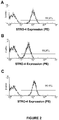

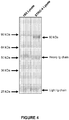

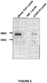
Classifications
-
- C—CHEMISTRY; METALLURGY
- C07—ORGANIC CHEMISTRY
- C07K—PEPTIDES
- C07K16/00—Immunoglobulins [IGs], e.g. monoclonal or polyclonal antibodies
- C07K16/18—Immunoglobulins [IGs], e.g. monoclonal or polyclonal antibodies against material from animals or humans
- C07K16/28—Immunoglobulins [IGs], e.g. monoclonal or polyclonal antibodies against material from animals or humans against receptors, cell surface antigens or cell surface determinants
-
- A—HUMAN NECESSITIES
- A61—MEDICAL OR VETERINARY SCIENCE; HYGIENE
- A61K—PREPARATIONS FOR MEDICAL, DENTAL OR TOILETRY PURPOSES
- A61K35/00—Medicinal preparations containing materials or reaction products thereof with undetermined constitution
- A61K35/12—Materials from mammals; Compositions comprising non-specified tissues or cells; Compositions comprising non-embryonic stem cells; Genetically modified cells
- A61K35/28—Bone marrow; Haematopoietic stem cells; Mesenchymal stem cells of any origin, e.g. adipose-derived stem cells
-
- C—CHEMISTRY; METALLURGY
- C07—ORGANIC CHEMISTRY
- C07K—PEPTIDES
- C07K16/00—Immunoglobulins [IGs], e.g. monoclonal or polyclonal antibodies
- C07K16/18—Immunoglobulins [IGs], e.g. monoclonal or polyclonal antibodies against material from animals or humans
-
- C—CHEMISTRY; METALLURGY
- C12—BIOCHEMISTRY; BEER; SPIRITS; WINE; VINEGAR; MICROBIOLOGY; ENZYMOLOGY; MUTATION OR GENETIC ENGINEERING
- C12N—MICROORGANISMS OR ENZYMES; COMPOSITIONS THEREOF; PROPAGATING, PRESERVING, OR MAINTAINING MICROORGANISMS; MUTATION OR GENETIC ENGINEERING; CULTURE MEDIA
- C12N5/00—Undifferentiated human, animal or plant cells, e.g. cell lines; Tissues; Cultivation or maintenance thereof; Culture media therefor
- C12N5/06—Animal cells or tissues; Human cells or tissues
- C12N5/0602—Vertebrate cells
- C12N5/0652—Cells of skeletal and connective tissues; Mesenchyme
- C12N5/0662—Stem cells
- C12N5/0663—Bone marrow mesenchymal stem cells (BM-MSC)
-
- G—PHYSICS
- G01—MEASURING; TESTING
- G01N—INVESTIGATING OR ANALYSING MATERIALS BY DETERMINING THEIR CHEMICAL OR PHYSICAL PROPERTIES
- G01N33/00—Investigating or analysing materials by specific methods not covered by groups G01N1/00 - G01N31/00
- G01N33/48—Biological material, e.g. blood, urine; Haemocytometers
- G01N33/50—Chemical analysis of biological material, e.g. blood, urine; Testing involving biospecific ligand binding methods; Immunological testing
- G01N33/5005—Chemical analysis of biological material, e.g. blood, urine; Testing involving biospecific ligand binding methods; Immunological testing involving human or animal cells
- G01N33/5008—Chemical analysis of biological material, e.g. blood, urine; Testing involving biospecific ligand binding methods; Immunological testing involving human or animal cells for testing or evaluating the effect of chemical or biological compounds, e.g. drugs, cosmetics
- G01N33/5044—Chemical analysis of biological material, e.g. blood, urine; Testing involving biospecific ligand binding methods; Immunological testing involving human or animal cells for testing or evaluating the effect of chemical or biological compounds, e.g. drugs, cosmetics involving specific cell types
-
- G—PHYSICS
- G01—MEASURING; TESTING
- G01N—INVESTIGATING OR ANALYSING MATERIALS BY DETERMINING THEIR CHEMICAL OR PHYSICAL PROPERTIES
- G01N33/00—Investigating or analysing materials by specific methods not covered by groups G01N1/00 - G01N31/00
- G01N33/48—Biological material, e.g. blood, urine; Haemocytometers
- G01N33/50—Chemical analysis of biological material, e.g. blood, urine; Testing involving biospecific ligand binding methods; Immunological testing
- G01N33/53—Immunoassay; Biospecific binding assay; Materials therefor
- G01N33/569—Immunoassay; Biospecific binding assay; Materials therefor for microorganisms, e.g. protozoa, bacteria, viruses
- G01N33/56966—Animal cells
-
- A—HUMAN NECESSITIES
- A61—MEDICAL OR VETERINARY SCIENCE; HYGIENE
- A61K—PREPARATIONS FOR MEDICAL, DENTAL OR TOILETRY PURPOSES
- A61K35/00—Medicinal preparations containing materials or reaction products thereof with undetermined constitution
- A61K35/12—Materials from mammals; Compositions comprising non-specified tissues or cells; Compositions comprising non-embryonic stem cells; Genetically modified cells
- A61K2035/124—Materials from mammals; Compositions comprising non-specified tissues or cells; Compositions comprising non-embryonic stem cells; Genetically modified cells the cells being hematopoietic, bone marrow derived or blood cells
-
- C—CHEMISTRY; METALLURGY
- C07—ORGANIC CHEMISTRY
- C07K—PEPTIDES
- C07K2317/00—Immunoglobulins specific features
- C07K2317/20—Immunoglobulins specific features characterized by taxonomic origin
- C07K2317/24—Immunoglobulins specific features characterized by taxonomic origin containing regions, domains or residues from different species, e.g. chimeric, humanized or veneered
-
- G—PHYSICS
- G01—MEASURING; TESTING
- G01N—INVESTIGATING OR ANALYSING MATERIALS BY DETERMINING THEIR CHEMICAL OR PHYSICAL PROPERTIES
- G01N2333/00—Assays involving biological materials from specific organisms or of a specific nature
- G01N2333/435—Assays involving biological materials from specific organisms or of a specific nature from animals; from humans
- G01N2333/46—Assays involving biological materials from specific organisms or of a specific nature from animals; from humans from vertebrates
- G01N2333/47—Assays involving proteins of known structure or function as defined in the subgroups
- G01N2333/4701—Details
- G01N2333/4703—Regulators; Modulating activity
Definitions
- the present invention relates to the use of heat shock protein-90beta (HSP-90beta) as a marker for identifying and/or isolating multipotential cells.
- HSP-90beta heat shock protein-90beta
- the cellular components that comprise haematopoietic supportive marrow stroma and associated skeletal tissues are derived from a population of multipotential cells including multipotential mesenchymal stem cells (MSC) and their precursors (mesenchymal precursor cells (MPCs)).
- MPC multipotential mesenchymal stem cells
- MPCs mesenchymal precursor cells
- MPC reside within the bone marrow spaces located primarily in perivascular niches which surround blood vessels (Gronthos S et al., 2003 and Shi S et al., 2003).
- studies have focused on the regenerative potential of different human stromal/mesenchymal cell populations to reconstitute fat, bone, cartilage and muscle (Gronthos S et al., 2003 and Simmons PJ et al., 1991).
- WO 2006/032092 A1 discloses multipotential expanded mesenchymal precursor cell progeny characterized by the early developmental markers STRO-1 bri and ALP-1, methods for producing the progeny and therapeutic uses thereof.
- European Patent Application Publication No. 1 457 499 A1 discloses inhibitors of extracellular Hsp90, the use of which leads to a reduction of the invasiveness of the tumour cells.
- WO 2006/050333 A2 discloses compositions and methods useful for treating and diagnosing cell proliferative disorders associated with Hsp90 and/or ZAP-70.
- the present disclosure describes the generation and characterisation of a novel monoclonal antibody (mAb), designated STRO-4 that identifies and isolates multipotential cells from unfractionated ovine and human bone marrow.
- mAb monoclonal antibody
- STRO-4 antibody is able to isolate essentially all of the clonogenic multipotential cells (CFU-F: colony forming unit fibroblastic) in both species.
- CFU-F colony forming unit fibroblastic
- HSP-90beta heat shock protein-90 beta
- the present invention is directed to a method of enriching for adult multipotential mesenchymal precursor cells (MPCs), the method comprising enriching a cell sample taken from a tissue source containing multipotential mesenchymal precursor cells that express the HSP-90beta marker, wherein enriching for cells that express the HSP-90beta marker comprises: contacting the cell sample with a HSP-90beta binding agent; and separating cells bound to the HSP-90beta binding agent from cells that do not bind the HSP-90beta binding agent; and wherein the HSP-90beta binding agent is an antibody or an antigen-binding fragment thereof that specifically binds to HSP-90beta.
- MPCs multipotential mesenchymal precursor cells
- the present invention is also directed to a method for identifying the presence of adult multipotential mesenchymal precursor cells (MPCs) in a cell sample taken from a tissue source containing multipotential mesenchymal precursor cells, the method comprising identifying cells in the cell sample that express the HSP-90beta marker, wherein the identification of cells in a cell sample that express the HSP-90beta marker comprises: contacting a cell sample obtained from a tissue source with a HSP-90beta binding agent under conditions suitable for binding of HSP-90beta to the HSP-90beta binding agent; and detecting the presence of the HSP-90beta binding agent bound to cells in the sample, wherein the presence of adult multipotential mesenchymal precursor cells (MPCs) is indicated by cells that bind to the HSP-90beta binding agent; wherein the HSP-90beta binding agent is an antibody or an antigen-binding fragment thereof that specifically binds to HSP-90beta.
- the HSP-90beta binding agent is an isolated antibody which binds to the same epitope on mesenchymal stem cells as the STRO-4 antibody produced by the hybridoma cell line deposited with ATCC on 10 July 2008 under the provisions of the Budapest Treaty under deposit accession number PTA-9362.
- the HSP-90beta binding agent is a STRO-4 antibody or an antigen-binding fragment of a STRO-4 antibody and wherein the STRO-4 antibody is produced by the hybridoma cell line deposited with ATCC on 10 July 2008 under the provisions of the Budapest Treaty under deposit accession number PTA-9362.
- the method also enriches for cells that are positive for one or more markers selected from the group consisting of STRO-1, STRO-3, CD146, LFA-3, THY-1, VCAM-1, ICAM-1, PECAM-1, P-selectin, L-selectin, CD49a/CD49b/CD29, CD49c/CD29, CD49d/CD29, CD29, CD18, CD61, NGF-R, FGF-R, Leptin-R, RANKL or any combination of these markers.
- markers selected from the group consisting of STRO-1, STRO-3, CD146, LFA-3, THY-1, VCAM-1, ICAM-1, PECAM-1, P-selectin, L-selectin, CD49a/CD49b/CD29, CD49c/CD29, CD49d/CD29, CD29, CD18, CD61, NGF-R, FGF-R, Leptin-R, RANKL or any combination of these markers.
- the tissue source is placenta, adipose tissue, teeth, dental pulp, skin, liver, kidney, heart, retina, brain, hair follicles, intestine, lung, spleen, lymph node, thymus, ovary, pancreas, bone, ligament, bone marrow, tendon or skeletal muscle.
- the present invention is also directed to use of HSP-90beta as a marker for the in vitro identification and/or enrichment of adult multipotential mesenchymal precursor cells (MPCs).
- MPCs multipotential mesenchymal precursor cells
- the enriched cell population of the invention has not been cultured in vitro.
- the enriched cell population of the invention is capable of giving rise to clonogenic CFU-F.
- the enriched cell population is homogenous for HSP-90beta + /STRO-4 + cells.
- the cell sample may be derived from any tissue source suspected of containing multipotential cells.
- the tissue source may be placenta, adipose tissue, teeth, dental pulp, skin, liver, kidney, heart, retina, brain, hair follicles, intestine, lung, spleen, lymph node, thymus, ovary, pancreas, bone, ligament, bone marrow, tendon or skeletal muscle.
- the tissue source is bone marrow.
- the source of cells from which the multipotential cells are enriched may be human or ovine origin. However, the invention is also applicable to cells derived from other animal species, including bovine, equine, murine, porcine, canine, or feline.
- the present disclosure also relates to progeny cells which are produced from the in vitro culture of multipotential cells of the disclosure.
- Expanded cells of the disclosure may have a wide variety of phenotypes depending on the culture conditions (including the number and/or type of stimulatory factors in the culture medium), the number of passages and the like.
- multipotential cells of the disclosure are not capable of giving rise, upon culturing, to hematopoietic cells.
- the methods according to the invention may also include the step of harvesting a source of multipotential cells before the first enrichment step. This may involve, for example, separating the cells obtained from a tissue source to form a single cell suspension. This separation may be achieved by physical or enzymatic means. Such means will be familiar to persons skilled in the art of the present invention. In one example of the invention, this step involves harvesting bone marrow cells using known techniques.
- the HSP-90beta binding agent used in the methods of the present disclosure can be any polypeptide or compound identified as having binding affinity to HSP-90beta.
- the HSP-90beta binding agent specifically binds to HSP-90beta.
- the HSP-90beta binding agent specifically binds to an epitope on HSP-90beta wherein HSP-90beta comprises a sequence at least about 75% identical to the sequence of SEQ ID NO:1 ( Figure 5 ), preferably at least about 78% identical, more preferably at least about 80%, still more preferably at least about 85%, still more preferably at least about 90%, even more preferably at least about 91%, 92%, 93%, 94%, 95%, 96%, 97%, 98%, 99% to SEQ ID NO:1.
- the HSP-90beta binding agent is selected from the group consisting of peptides, peptidomimetics, nucleic acid aptamers, peptide aptamers, dendrimers, and small organic molecules.
- the HSP-90beta binding agent is an oligonucleotide.
- the oligonucleotide is a labelled oligonucleotide.
- the HSP-90beta binding agent is a purified anti-HSP-90beta antibody or antigen binding fragment or derivative thereof.
- the anti-HSP-90beta antibody may be a monoclonal, recombinant, chimeric, or humanized antibody.
- the anti-HSP-90beta monoclonal antibody is the STRO-4 antibody produced by the hybridoma cell line deposited with ATCC on 10 July 2008 under the provisions of the Budapest Treaty under deposit accession number PTA-9362.
- Suitable anti-HSP-90beta monoclonal antibodies for use in the methods of the present invention include commercially available antibodies such as mouse anti-human Hsp90Beta mAb clone H9010 (Invitrogen), mouse anti-Hsp90b mAb clone EMD-5E12 (Calbiochem), mouse anti-human HSP90beta mAb clone K3725B (Cosmo Bio Co., Ltd), and mouse anti-human Hsp90 beta mAb clone K3705 (Assay Designs/Stressgen Bioreagents).
- Other commercially available antibodies not mentioned above are also considered to be within the scope of the present invention.
- the disclosure provides an HSP-90beta binding agent or antigen binding fragment or derivative thereof selected from the group consisting of:
- the humanized antibody comprises all six CDRs from STRO-4.
- the HSP-90beta binding agent is a recombinant antibody comprising a sequence substantially identical to that of the STRO-4 antibody produced by the hybridoma cell line deposited with ATCC on 10 July 2008 under the provisions of the Budapest Treaty under deposit accession number PTA-9362.
- the HSP-90beta binding agent is labelled.
- the label is a fluorescent label. In another example, the label is an enzymatic label.
- the separation of cells bound to the HSP-90beta binding agent is carried out by a mechanical cell sorter.
- the HSP-90beta binding agent is coupled to a fluorescent labelling compound.
- the separation of cells bound to the HSP-90beta binding agent is preferably carried out using a fluorescence-activated cell sorter (FACS).
- the HSP-90beta binding agent is linked to a solid particle.
- the solid particle is a magnetic particle.
- the separation of cells bound to the HSP-90beta binding agent is preferably carried out by separating the particulate phase from the liquid phase.
- the cell sample prior to the separation step is contacted with an antibody directed against the HSP-90beta binding agent linked to a solid particle, and wherein the separation of cells bound to the HSP-90beta binding agent is carried out by separating the particulate phase from the liquid phase.
- the cells of the cell sample are adherent cells cultivated on a solid support, and removal of unbound HSP-90beta binding agent is carried out by rinsing.
- the cells of the cell sample are cultivated in suspension, and removal of unbound HSP-90beta binding agent is carried out by centrifuging the cell sample and separating off the resulting supernatant.
- the cell sample is subjected to a further cell sorting procedure to enrich or diminish the cell population in cells expressing at least one further multipotential cell marker.
- the present disclosure also provides an enriched population of multipotential cells obtained by a method according to the present invention.
- the present disclosure also provides an enriched population of HSP-90beta + multipotential cells.
- At least 1%, 2%, 3%, 4%, 5%, 10%, 15%, 20%, 30%, 40%, 50%, 60%, 70%, 80%, 90% or 95% of the total enriched cell population are multipotential cells that have the phenotype HSP-90beta + .
- the enriched population of multipotential cells were obtained by a method according to the present disclosure.
- the present disclosure also provides an expanded cell population obtained by culturing a population of multipotential cells enriched according to the disclosure.
- the enriched cell population of the disclosure, or an expanded cell population of the disclosure comprises at least some cells which are genetically modified.
- the present disclosure also provides a method of generating a tissue specific committed cell population, the method comprising culturing a population of multipotential cells of the present disclosure in the presence of one or more stimulatory factors, and subjecting said cultured population to conditions biasing differentiation of the multipotential cells to a specific tissue type.
- the tissue type is selected from the group consisting of placental tissue, cardiac muscle, vascular tissue, bone tissue, cartilage tissue, fat tissue, neural tissue, smooth muscle and endothelial tissue.
- the present disclosure also provides a composition comprising enriched multipotential cells according to the present disclosure and/or an expanded cell population according to the disclosure.
- the composition further comprises a stimulatory factor.
- a stimulatory factor is likely to be beneficial therapeutically and thus will be prepared in a pharmaceutically acceptable form.
- composition further comprises a factor to bias differentiation of the multipotential cells of the present disclosure to one specific tissue type.
- tissue type is selected from, but not limited to, the group consisting of cardiac muscle, vascular tissue, bone tissue, cartilage tissue, fat tissue, neural tissue, placental, smooth muscle and endothelial tissue.
- composition of the disclosure further comprises a fibrin glue.
- the present disclosure also provides a method for generating or repairing tissue in a subject, the method comprising administering to the subject an enriched and/or expanded multipotential cell population of the present disclosure.
- the present disclosure also provides a method for generating or repairing tissue in a subject, the method comprising administering to the subject a composition of the present disclosure.
- the present disclosure also provides for the use of an enriched and/or expanded multipotential cell population of the present disclosure for the manufacture of a medicament for generating or repairing tissue in a subject.
- the tissue being generated or repaired according to the disclosure is selected from, but not limited to, the group consisting of cardiac muscle, vascular tissue, bone tissue, cartilage tissue, fat tissue, neural tissue, placental, smooth muscle and endothelial tissue.
- the multipotential cells according to the disclosure are of human origin.
- the present disclosure also provides an isolated cell which has been obtained by a method of the disclosure, or a progeny cell thereof, wherein the cell is genetically modified.
- the present disclosure also provides a STRO-4 hybridoma cell line deposited with ATCC on 10 July 2008 under the provisions of the Budapest Treaty under deposit accession number PTA-9362.
- the present disclosure also provides a STRO-4 antibody produced by the hybridoma cell line deposited with ATCC on 10 July 2008 under the provisions of the Budapest Treaty under deposit accession number PTA-9362.
- the present disclosure also provides an isolated antibody which binds to the same epitope on multipotential cells as the STRO-4 antibody produced by the hybridoma cell line deposited with ATCC on 10 July 2008 under the provisions of the Budapest Treaty under deposit accession number PTA-9362.
- the present disclosure also provides a composition
- a composition comprising a HSP-90beta binding agent or antigen binding fragment or derivative thereof.
- the composition further comprises one or more pharmaceutically acceptable carriers and/or suitable adjuvants or excipients.
- Single colour immunofluorescence and flow cytometry was performed on specimens of Ficoll-separated ovine and human BM MNC to examine the expression of STRO-4 antigen.
- Data are displayed as single-parameter fluorescence (phycoerythrin (PE)) histograms of 1 x 10 4 light-scatter gated events, collected as list mode data. IgG control (dotted line); mAb STRO-3 (solid line).
- a single cell suspension of unfractionated BM and MACS selected STRO-4 + and STRO-4 - ovine (C) and human (D) BMMNC were plated into regular growth medium to assess the incidence of adherent colony-forming cells in each cell fraction. Following 12 days of culture, colonies (aggregates of 50 cells or more) were stained and scored as described in Methods. The bar graph depicts the number of clonogenic colonies per 10 5 cells plated for each cell fraction averaged from 3 separate experiments. These data demonstrate that the majority of clonogenic MPC are exclusively restricted to the STRO-4 + fraction of BM.
- Toluidine blue positive staining for proteoglycans was present throughout the cellular mass surrounding chondrocyte-like cells (arrow) following 3 weeks of chondrogenic induction in the human MPC (E) and ovine MPC (F) cultures (arrow) (200x).
- the STRO-4 Antibody Identifies a 90 kDa Molecular Weight Protein.
- Plasma membrane lysates were prepared from single cell suspensions of cultured expanded STRO-4 + human MPC as described in the methods. The cell lysates were incubated with either STRO-4 supernatant or the isotype matched control antibody, 1B5, then immunoprecipitated by magnetic bead separation. The immunoprecipitates were analysed by 10% (w/v) SDS-polyacrylamide gel electrophoresis and visualised following Coomassie blue staining.
- the STRO-4 Antibody Identifies Heat Shock Protein-90 beta.
- Plasma membrane lysates were prepared from single cell suspensions of cultured expanded STRO-4 + human MG63 as described in the methods. The cell lysates were incubated with either STRO-4 supernatant then immunoprecipitated by magnetic bead separation. The immunoprecipitates were analysed by 10% (w/v) SDS-polyacrylamide gel electrophoresis and visualised following Coomassie blue staining. The 90 kDa bands were excised from the gels, subjected to tryptic digestion and analysed by mass spectrometry with an Applied Biosystems 4700 Proteomics Analyser with TOF/TOF optics in MS mode. Microsequencing peptide analysis demonstrated matched (bold) STRO-4 sequenced peptides to human heat shock protein-90 beta (NCBI, accession number, P08238).
- Cultured expanded STRO-4 + human MPC were prepared as whole cell lysates alone or used to generate STRO-4 or 1B5 immunoprecipitates as described in the methods.
- the whole cell lysates and STRO-4 or 1B5 immunoprecipitates were resolved by electrophoresis on a 10% (w/v) SDS-polyacrylamide gel. Proteins were transferred to PVDF membranes, then blocked with 5% skim milk powder-0.05% Tween-20, before the filters were incubated with a commercially available polyclonal antibody to HSP-90beta.
- the immunoreactive proteins were visualised using a FluorImager (Molecular Dynamics, Sunnyvale, CA) using ImageQuant software (Molecular Dynamics).
- the anti-HSP-90beta antibody detected a characteristic 90 kDa band in both the control whole cell lysate preparation and in the STRO-4 immunoprecipitate sample.
- the hybridoma which produces the monoclonal antibody designated STRO-4 was deposited on 10 July 2008 with American Type Culture Collection (ATCC) under accession number PTA-9362.
- Multipotential cells are cells which are capable of giving rise to any of several mature cell types.
- the multipotential cells are derived from adult tissue.
- the term encompasses, for example, mesenchymal precursor cells (MPCs), mesenchymal stem cells (MSC) and multipotential expanded MPC progeny (MEMPs).
- MPCs mesenchymal precursor cells
- MSC mesenchymal stem cells
- MEMPs multipotential expanded MPC progeny
- MPCs Mesenchymal precursor cells
- MPCs Mesenchymal precursor cells
- the specific lineage-commitment and differentiation pathway which these cells enter depends upon various influences from mechanical influences and/or endogenous bioactive factors, such as growth factors, cytokines, and/or local microenvironmental conditions established by host tissues.
- Mesenchymal precursor cells are defined as cells which are not terminally differentiated; which can divide without limit; and divide to yield daughter cells that are either stem cells or are progenitor cells which in time will irreversibly differentiate to yield a phenotypically and/or functionally distinct cell type.
- MPCs are non-hematopoietic progenitor cells that are capable of forming large number of multipotential cells.
- the terms 'enriched', 'enrichment' or variations thereof are used herein to describe a population of cells in which the proportion of one particular cell type or the proportion of a number of particular cell types is increased when compared with the untreated population.
- the method of enriching for multipotential cells according to the disclosure may be based on detecting the presence of HSP-90beta marker alone or in combination with one or more additional markers.
- Reference to a cell being "positive" for a given marker means it may be either a low (lo or dim) or a high (bright, bri) expresser of that marker depending on the degree to which the marker is present on the cell surface, where the terms relate to intensity of fluorescence or other colour used in the colour sorting process of the cells.
- lo or dim or dull
- bri will be understood in the context of the marker used on a particular cell population being sorted.
- “bright”, when used herein, refers to a marker on a cell surface that generates a relatively high signal when detectably labelled. Whilst not wishing to be limited by theory, it is proposed that "bright" cells express more of the target marker antigen (for example the antigen recognised by STRO-1) than other cells in the sample. For instance, STRO-1 bri cells produce a greater fluorescent signal, when labelled with a fluorescently-conjugated STRO-1 antibody as determined by FACS analysis, than non-bright cells (STRO-1 dull/dim ).
- STRO-1 bright cells have 2 log magnitude higher expression of STRO-1 surface expression. This is calculated relative to an isotype matched negative control.
- STRO-1 dim and/or STRO-1 intermediate cells have less than 2 log magnitude higher expression of STRO-1 surface expression, typically about 1 log or less expression over the isotype matched negative control.
- the method may include the step of making a first partially enriched pool of cells by enriching for the expression of a first multipotential cell specific marker, followed by a step of enriching for expression of HSP-90beta from the partially enriched pool of cells.
- the method may include an initial enrichment step based on selection of cells expressing HSP-90beta, followed by a step which involves enriching for a different multipotential cell marker.
- the method involves simultaneously selecting for cells that express HSP-90beta and one or more additional multipotential cell markers.
- a significant proportion of the multipotential cells are capable of differentiation into at least two committed cell types.
- the lineages to which the multipotential cells may differentiate into include, bone precursor cells; hepatocyte progenitors, which are pluripotent for bile duct epithelial cells and hepatocytes; neural restricted cells, which can generate glial cell precursors that progress to oligodendrocytes and astrocytes; neuronal precursors that progress to neurons; precursors for cardiac muscle and cardiomyocytes, glucose-responsive insulin secreting pancreatic beta cell lines.
- lineages include, but are not limited to, odontoblasts, dentin-producing cells and chondrocytes, and precursor cells of the following: retinal pigment epithelial cells, fibroblasts, skin cells such as keratinocytes, dendritic cells, hair follicle cells, renal duct epithelial cells, smooth and skeletal muscle cells, testicular progenitors, vascular endothelial cells, tendon, ligament, cartilage, adipocyte, fibroblast, marrow stroma, cardiac muscle, smooth muscle, skeletal muscle, pericyte, vascular, epithelial, glial, neuronal, astrocyte and oligodendrocyte cells.
- the multipotential cells are at least capable of being cultured, in vivo or in vitro, to produce adipocytes, osteoblast and/or chondrocytes.
- separation of cells expressing cell surface HSP-90beta can be effected by a number of different methods, however, all of these methods rely at some point upon binding of cells to the HSP-90beta binding agent followed by separation of those cells that exhibit binding, being either high level binding, or low level binding or no binding.
- HSP-90beta is normally present on the cytoplasm of cells where it acts as a molecular chaperone to facilitate the normal folding, intracellular deposition and proteolytic turnover of many key regulators of postnatal cell growth and differentiation. It is part of a highly conserved family of stress response proteins. HSPs are typically expressed at low levels under normal physiological conditions but show dramatically increased expression in response to cellular stress.
- HSP-90beta is not limited to the sequence of human or ovine origin but also includes homologous sequences obtained from any source, for example homologues, particularly orthologues (i.e. homologues obtained from species other than humans), allelic variants, as well as fragments and synthetic peptides or derivatives thereof.
- sequence identity of a polypeptide is determined by FASTA (Pearson and Lipman, (1988)) analysis (GCG program) using the default settings and a query sequence of at least 50 amino acids in length, and whereby the FASTA analysis aligns the two sequences over a region of at least 50 amino acids. More preferably, the FASTA analysis aligns the two sequences over a region of at least 100 amino acids. More preferably, the FASTA analysis aligns the two sequences over a region of at least 250 amino acids. Even more preferably, the FASTA analysis aligns the two sequences over a region of at least 350 amino acids.
- HSP-90beta also encompasses fragments of the above mentioned full-length polypeptides and variants thereof, including fragments of the HSP-90beta sequence.
- Preferred fragments include those that include an epitope.
- Suitable fragments will be at least about 6 or 7 amino acids in length, e.g. at least 10, 12, 15 or 20 amino acids in length. They may also be less than 200, 100 or 50 amino acids in length.
- HSP-90beta binding agent refers to a moiety that recognises and/or specifically binds to HSP-90beta.
- HSP-90beta binding agent is capable of binding to HSP-90beta with suitable affinity and/or avidity so as to provide a useful tool for selective enrichment of cells expressing HSP-90beta. That is, it associates more frequently, more rapidly, with greater duration and/or with greater affinity with HSP-90beta than it does with alternative targets (e.g. non-HSP-90beta).
- alternative targets e.g. non-HSP-90beta
- an HSP90beta binding agent that specifically binds to HSP90beta or an epitope or immunogenic fragment thereof binds with grater affinity, avidity, more readily, and/or with greater duration than it binds to other non-HSP90beta targets.
- HSP-90beta binding agents are polypeptides or compounds identified as having binding affinity to HSP-90beta.
- the HSP-90beta binding agent of the disclosure includes peptides, petidomimetics, nucleic acid aptamers, peptide aptamers, dendrimers and small organic molecules.
- a nucleic acid aptamer is a nucleic acid molecule that is capable of forming a secondary and/or tertiary structure that provides the ability to bind to a molecular target.
- An aptamer library is produced, for example, by cloning random oligonucleotides into a vector (or an expression vector in the case of an RNA aptamer), wherein the random sequence is flanked by known sequences that provide the site of binding for PCR primers.
- An aptamer with increased activity is selected, for example, using SELEX (Sytematic Evolution of Ligands by EXponential enrichment). Suitable methods for producing and/or screening an aptamer library are described, for example, in Elloington and Szostak, Nature 346:818-22, 1990 .
- informatics is used to select suitable chemical building blocks from known compounds, for producing a combinatorial library.
- QSAR Quantitative Structure Activity Relationship
- the software of the Chemical Computing Group, Inc. uses high-throughput screening experimental data on active as well as inactive compounds, to create a probabilistic QSAR model, which is subsequently used to select lead compounds.
- the Binary QSAR method is based upon three characteristic properties of compounds that form a "descriptor" of the likelihood that a particular compound will or will not perform a required function: partial charge, molar refractivity (bonding interactions), and logP (lipophilicity of molecule). Each atom has a surface area in the molecule and it has these three properties associated with it. All atoms of a compound having a partial charge in a certain range are determined and the surface areas (Van der Walls Surface Area descriptor) are summed.
- the binary QSAR models are then used to make activity models or ADMET models, which are used to build a combinatorial library. Accordingly, lead compounds identified in initial screens, can be used to expand the list of compounds being screened to thereby identify highly active compounds.
- HSP-90beta binding agents are anti- HSP-90beta antibodies or antigen binding fragments thereof or derivatives/variants thereof (naturally occurring or recombinant, from any source).
- the term "antibody” refers to an immunoglobulin molecule capable of binding to a target, such as HSP-90beta and/or an epitope thereof and/or an immunogenic fragment thereof and/or a modified form thereof (e.g., glycosylated, glycosylated, etc.) through at least one epitope recognition site, located in the variable region of the immunoglobulin molecule.
- a target such as HSP-90beta
- This term encompasses not only intact polyclonal or monoclonal antibodies, but also variants, fusion proteins comprising an antibody portion with an epitope recognition site of the required specificity, humanized antibodies, human antibodies, chimeric antibodies, and any other modified configuration of the immunoglobulin
- antibody binding fragment as used herein shall be taken to mean any fragment of an antibody that retains the ability to bind to HSP-90beta, preferably specifically to HSP-90beta. This includes:
- the term “monoclonal antibody” refers to a homogeneous antibody population capable of binding to the same antigen(s) and, preferably, to the same epitopic determinant within the antigen(s). This term is not intended to be limited as regards to the source of the antibody or the manner in which it is made.
- chimeric antibody refers to antibodies in which a portion of the heavy and/or light chain is identical with or homologous to corresponding sequences in antibodies derived from a particular species (e.g., murine, such as mouse) or belonging to a particular antibody class or subclass, while the remainder of the chain(s) is identical with or homologous to corresponding sequences in antibodies derived from another species (e.g., primate, such as human) or belonging to another antibody class or subclass, as well as fragments of such antibodies, so long as they exhibit the desired biological activity ( U.S. Patent No. 4,816,567 ; and Morrison et al. (1984) Proc. Natl Acad. Sci USA 81:6851-6855 ).
- a particular species e.g., murine, such as mouse
- primate such as human
- humanized antibody shall be understood to refer to a chimeric molecule, generally prepared using recombinant techniques, having an epitope binding site derived from an immunoglobulin from a non-human species and the remaining immunoglobulin structure of the molecule based upon the structure and/or sequence of a human immunoglobulin.
- the antigen- binding site preferably comprises the complementarity determining regions (CDRs) from the non-human antibody grafted onto appropriate framework regions in the variable domains of human antibodies and the remaining regions from a human antibody.
- CDRs complementarity determining regions
- Epitope binding sites may be wild type or modified by one or more amino acid substitutions.
- variable regions of both heavy and light chains contain three complementarity-determining regions (CDRs) which vary in response to the epitopes in question and determine binding capability, flanked by four framework regions (FRs) which are relatively conserved in a given species and which putatively provide a scaffolding for the CDRs.
- CDRs complementarity-determining regions
- FRs framework regions
- the variable regions can be "reshaped” or “humanized” by grafting CDRs derived from nonhuman antibody on the FRs present in the human antibody to be modified. Application of this approach is known in the art and/or described in more detail herein.
- constant region refers to the portion of the antibody molecule which confers effector functions.
- the constant regions of the subject humanized antibodies are derived from human immunoglobulins.
- the heavy chain constant region can be selected from any of the five isotypes: alpha, delta, epsilon, gamma or mu. Further, heavy chains of various subclasses (such as the IgG subclasses of heavy chains) are responsible for different effector functions and thus, by choosing the desired heavy chain constant region, antibodies with desired effector function can be produced.
- Preferred heavy chain constant regions are gamma 1 (IgG1), gamma 2 (IgG2), gamma 3 (IgG3) and gamma 4 (IgG4).
- Light chain constant regions can be of the kappa or lambda type, preferably of the kappa type.
- Framework regions are those variable domain residues other than the CDR residues.
- Each variable domain of a naturally-occurring antibody typically has four FRs identified as FR1, FR2, FR3 and FR4.
- the CDRs are defined according to Kabat, the light chain FR residues are positioned at about residues 1 - 23 (LCFR1), 35-49 or 40-54 (LCFR2), 57-88 or 62-93 (LCFR3), and 98-107 or 103-112 (LCFR4) and the heavy chain FR residues are positioned about at about residues 1-30 or 1-30 (HCFR1), 36-49 (HCFR2), 66-94 or 67-96 (HCFR3), and 103- 113 or 109-119 (HCFR4) in the heavy chain residues.
- the light chain FR residues are positioned about at about residues 1 -25 (LCFR1), 33-49 (LCFR2), 53-90 (LCFR3), and 97-107 (LCFR4) in the light chain and the heavy chain FR residues are positioned about at about residues 1-25 (HCFR1), 33-52 (HCFR2), 56-95 (HCFR3), and 102-113 (HCFR4) in the heavy chain residues.
- the CDR comprises amino acids from both a CDR as defined by Kabat and those of a hypervariable loop, the FR residues will be adjusted accordingly.
- complementarity determining regions refers to the amino acid residues of an antibody variable domain the presence of which are necessary for antigen binding.
- Each variable domain typically has three CDR regions identified as CDR1, CDR2 and CDR3.
- Each complementarity determining region may comprise amino acid residues from a "complementarity determining region" as defined by Kabat (i.e.
- a complementarity determining region can include amino acids from both a CDR region defined according to Kabat and a hypervariable loop.
- derivative as used herein in intended to refer to an anti-HSP-90beta antibody or antigen binding fragment thereof comprising one or more conservative amino acid substitutions.
- conservative substitution shall be taken to mean amino acid substitutions set forth in Table 1. Table 1.
- Exemplary Substitutions Original Residue Exemplary Substitutions Ala (A) val; leu; ile; gly Arg (R) lys Asn (N) gln; his Asp (D) glu Cys (C) ser Gln (Q) asn; his Glu (E) asp Gly (G) pro, ala His (H) asn; gln Ile (I) leu; val; ala Leu (L) ile; val; met; ala; phe Lys (K) arg Met (M) leu; phe Phe (F) leu; val; ala Pro (P) gly Ser (S) thr Thr (T) ser Trp (W) tyr Tyr (Y) trp; phe Val (V) ile; leu; met; phe; ala
- substitution also encompasses substitutions of amino acids having a similar hydropathic index or score.
- Each amino acid has been assigned a hydropathic index on the basis of their hydrophobicity and charge characteristics, as follows: isoleucine (+4.5); valine (+4.2); leucine (+3.8); phenylalanine (+2.8); cysteine/cystine (+2.5); methionine (+1.9); alanine (+1.8); glycine (-0.4); threonine (-0.7); serine (-0.8); tryptophan (-0.9); tyrosine (-1.3); proline (-1.6); histidine (-3.2); glutamate (-3.5); glutamine (-3.5); aspartate (- 3.5); asparagine (-3.5); lysine (-3.9); and arginine (-4.5).
- the substitution of amino acids whose hydropathic indices are within +/- 0.2 is preferred. More preferably, the substitution will involve amino acids having hydropathic indices within +/- 0.1, and more preferably within about +/- 0.05.
- conservative amino acid substitution also encompasses substitution of like amino acids made on the basis of hydrophilicity, particularly where the biological functional equivalent protein or peptide thereby created is intended for use in immunological embodiments, as in the present case.
- hydrophilicity particularly where the biological functional equivalent protein or peptide thereby created is intended for use in immunological embodiments, as in the present case.
- the greatest local average hydrophilicity of a protein, as governed by the hydrophilicity of its adjacent amino acids correlates with its immunogenicity and antigenicity.
- hydrophilicity values have been assigned to amino acid residues: arginine (+3.0); lysine (+3.0); aspartate (+3.0 +/-0.1); glutamate (+3.0 +/- 0.1); serine (+0.3); asparagine (+0.2); glutamine (+0.2); glycine (0); threonine (-0.4); proline (-0.5 +/- 0.1); alanine (-0.5); histidine (-0.5); cysteine (-1.0); methionine (-1.3); valine (-1.5); leucine (-1.8); isoleucine (-1.8); tyrosine (-2.3); phenylalanine (-2.5); tryptophan (-3.4).
- derivative in relation to the amino acid sequences of the present disclosure and/or for use in the present disclosure includes any substitution of, variation of, modification of, replacement of, deletion of or addition of one (or more) amino acids from or to the sequence providing the resultant amino acid sequence preferably has at least 50% of the biological activity as a naturally occurring HSP-90beta more preferably at least substantially the same activity.
- derivative also comprises an additional component directly or indirectly linked to the antibody or antigen-binding fragment.
- the derivative comprises a compound that enhances or increases the half life of the antibody in vivo, e.g., polyethylene glycol or albumin.
- the derivative additionally comprises a detectable compound, e.g., a fluorescent compound or a radioactive compound or an enzyme, e.g., to facilitate detection and/or imaging.
- the derivative additionally comprises a toxic compound, e.g., a radioactive compound or a cellular toxin.
- Antibodies of the present disclosure can be prepared using cells expressing full length HSP-90beta or fragments thereof as the immunizing antigen.
- a peptide used to immunize an animal can be derived from translated cDNA or chemical synthesis and is purified and conjugated to a carrier protein, if desired.
- carrier protein e.g., a carrier protein
- Such commonly used carriers which are chemically coupled to the peptide include keyhole limpet hemocyanin (KLH), thyroglobulin, bovine serum albumin (BSA), and tetanus toxoid.
- KLH keyhole limpet hemocyanin
- BSA bovine serum albumin
- the coupled peptide may then be used to immunize the animal (e.g., a mouse or a rabbit).
- polyclonal antibodies can be further purified, for example, by binding to and elution from a matrix to which the peptide to which the antibodies were raised is bound.
- a matrix to which the peptide to which the antibodies were raised is bound.
- Those of skill in the art will know of various techniques common in the immunology arts for purification and/or concentration of polyclonal antibodies, as well as monoclonal antibodies (See for example, Coligan, et al., Unit 9, Current Protocols in Immunology, Wiley Interscience, 1991 ).
- Monoclonal antibodies may be prepared using any technique which provides for the production of antibody molecules by continuous cell lines in culture, such as, for example, the hybridoma technique, the human B-cell hybridoma technique, and the EBV-hybridoma technique (Kohler et al., 1975; Kozbor et al., 1985; Cote et al., 1983; Cole et al., 1984).
- Antibodies with an epitopic specificity which is the same as or similar to that of mAb STRO-4 can be identified by their ability to compete with that particular mAb for binding to HSP-90beta (e.g. to cells bearing HSP-90beta, such as MPCs, or to isolated HSP-90 protein or fragments thereof). Using receptor chimeras (Rucker et al., 1996) or other techniques known to those skilled in the art, the binding site of STRO-4 mAb may be mapped.
- Still another way to determine whether a monoclonal antibody has the specificity of STRO-4 mAb is to pre-incubate the monoclonal antibody being tested with HSP-90beta, and then add STRO-4 mAb to determine if STRO-4 mAb is inhibited in its ability to bind to HSP-90beta. If the binding of STRO-4 mAb is inhibited then, in all likelihood, the monoclonal antibody being tested has the same, or functionally equivalent, epitopic specificity as STRO-4 mAb.
- Monoclonal antibodies useful in the present invention can be engineered so as to change the isotype of the antibody.
- an IgG2A isotype can be engineered as an IgG1, IgG2B, or other isotypes.
- a HSP-90beta binding agent such as an antibody of the disclosure may be conjugated to a compound that is useful, for example, in cell separation, therapeutic or diagnostic applications.
- an antibody of the disclosure is conjugated to a label.
- the label may be any entity the presence of which can be readily detected.
- the label may be a direct label, such as those described in detail in May et al., U.S. Pat. No. 5,656,503 .
- Direct labels are not limited to but include entities which, in their natural state, are readily visible either to the naked eye, or with the aid of an optical filter and/or applied stimulation, e.g. UV light to promote fluorescence.
- Examples include radioactive, chemiluminescent, electroactive (such as redox labels), and fluorescent compounds.
- Direct particulate labels such as dye sols, metallic sols (e.g. gold) and coloured latex particles, are also very suitable and are, along with fluorescent compounds, preferred. Of these options, coloured latex particles and fluorescent compounds are most preferred. Concentration of the label into a small zone or volume should give rise to a readily detectable signal, e.g. a strongly coloured area.
- Indirect labels such as enzymes, e.g. alkaline phosphatase and horseradish peroxidase, can also be used, although these usually require the addition of one or more developing reagents such as substrates before a visible signal can be detected.
- Conjugation of a label to a binding agent such as an antibody of the disclosure can be by covalent or non-covalent (including hydrophobic) bonding, or by adsorption. Techniques for such conjugation are commonplace in the art and may be readily adapted for the particular reagents employed.
- a binding agent for use in the methods of the invention may also be coated onto a solid support.
- the antibody can be coated on a synthetic plastics material, magnetic particle, microtitre assay plate, microarray chip, latex bead, filter comprising a cellulosic or synthetic polymeric material, glass or plastic slide, dipstick, capillary fill device and the like.
- the HSP-90beta binding agent may be attached to a solid support to allow for a crude separation.
- the separation techniques preferably maximise the retention of viability of the fraction to be collected.
- Various techniques of different efficacy may be employed to obtain relatively crude separations. The particular technique employed will depend upon efficiency of separation, associated cytotoxicity, ease and speed of performance, and necessity for sophisticated equipment and/or technical skill.
- Procedures for separation may include, but are not limited to, magnetic separation, using antibody-coated magnetic beads, affinity chromatography and "panning" with antibody attached to a solid matrix.
- Techniques providing accurate separation include but are not limited to MACS, Dynal magnetic bead selection and FACS.
- HSP-90beta binding agents such as anti-HSP-90beta antibodies
- HSP-90beta binding agents allows not only for the identification and quantification of these cells in tissue samples, but also for their separation and enrichment in suspension. This can be achieved by a number of cell-sorting techniques by which cells are physically separated by reference to a property associated with the cell-antibody complex, or a label attached to the antibody. This label may be a magnetic particle or a fluorescent molecule.
- the antibodies may be cross-linked such that they form aggregates of multiple cells, which are separable by their density. Alternatively the antibodies may be attached to a stationary matrix, to which the desired cells adhere.
- the antibody bound to the cell can be labelled and then the cells separated by a mechanical cell sorter that detects the presence of the label.
- Fluorescence-activated cell sorters are well known in the art.
- the anti-HSP-90beta antibody is attached to a solid support.
- solid supports are known to those of skill in the art, including, but not limited to, agarose beads, polystyrene beads, hollow fiber membranes, polymers, and plastic petri dishes. Cells that are bound by the antibody can be removed from the cell suspension by simply physically separating the solid support from the cell suspension.
- Super paramagnetic microparticles may be used for cell separations.
- the microparticles may be coated with anti-HSP-90beta antibodies.
- the antibody-tagged, super paramagnetic microparticles may then be incubated with a solution containing the cells of interest.
- the microparticles bind to the surfaces of the desired multipotential cells, and these cells can then be collected in a magnetic field.
- the cell sample is allowed to physically contact, for example, a solid phase-linked anti-HSP-90beta monoclonal antibody.
- the solid-phase linking can comprise, for instance, adsorbing the antibodies to a plastic, nitrocellulose, or other surface.
- the antibodies can also be adsorbed on to the walls of the large pores (sufficiently large to permit flow-through of cells) of a hollow fiber membrane.
- the antibodies can be covalently linked to a surface or bead, such as Pharmacia Sepharose 6 MB macrobeads.
- the exact conditions and duration of incubation for the solid phase-linked antibodies with the multipotential cell containing suspension will depend upon several factors specific to the system employed. The selection of appropriate conditions, however, is well within the skill of the art.
- the unbound cells are then eluted or washed away with physiologic buffer after allowing sufficient time for the multipotential cells to be bound.
- the unbound cells can be recovered and used for other purposes or discarded after appropriate testing has been done to ensure that the desired separation had been achieved.
- the bound cells are then separated from the solid phase by any appropriate method, depending mainly upon the nature of the solid phase and the antibody.
- bound cells can be eluted from a plastic petri dish by vigorous agitation.
- bound cells can be eluted by enzymatically "nicking" or digesting an enzyme-sensitive "spacer" sequence between the solid phase and the antibody. Spacers bound to agarose beads are commercially available from, for example, Pharmacia.
- the eluted, enriched fraction of cells may then be washed with a buffer by centrifugation and either said enriched fraction or the unbound fraction may be cryopreserved in a viable state for later use according to conventional technology or introduced into the transplant recipient.
- Conditions that bias differentiation of the multipotential cells of the present disclosure to bone precursor cells or bone may involve, for example, culturing in ⁇ MEM supplemented with 10% FCS, 100 ⁇ M L-ascorbate-2-phosphate, dexamethasone 10 -7 M and 3 mM inorganic phosphate. These conditions have been shown to induce human bone marrow stromal cells to develop a mineralized bone matrix in vitro (Gronthos et al., 1994).
- Suitable conditions for differentiating the multipotential cells of the present disclosure into osteoblasts may involve cultivating the cells in the presence of type I collagen, fibrinogen, fibrin, polyglycolic acid, polylactic acid, osteocalcin, or osteonectin.
- the cells are cultivated in the presence of type I collagen, fibrinogen, and fibrin.
- the cells are cultivated in the presence of type I collagen, fibrinogen, fibrin, osteocalcin, and osteonectin.
- type I collagen, fibrinogen, fibrin, polyglycolic acid, polylactic acid, osteocalcin, or osteonectin may be used alone or in the presence of a growth factor. It will be understood that any combination of the compounds listed above in this paragraph is contemplated by the present disclosure.
- the present disclosure relates to genetically modified cells, particularly genetically modified multipotential cells of the disclosure.
- the cells are genetically modified to produce a heterologous protein.
- the cells will be genetically modified such that the heterologous protein is secreted from the cells.
- the heterologous protein may be any protein of interest.
- the heterologous protein may be platelet derived growth factor (PDGF), tumor necrosis factor ⁇ (TNF- ⁇ ), interleukin -1 ⁇ (IL-1 ⁇ ) and stromal derived factor 1 ⁇ (SDF-1 ⁇ ).
- the heterologous protein is a bioactive factor which accelerates differentiation of the multipotential cells to specific tissue types.
- the bioactive factor may be, for example, a synthetic glucocorticoid, or a bone morphogenic protein, such as BMP-2, BMP-3, BMP-4, BMP-6 or BMP-7.
- the cells can be modified to express a functional non-protein encoding polynucleotide such as dsRNA (typically for RNA silencing), an antisense oligonucleotide or a catalytic nucleic acid (such as a ribozyme or DNAzyme).
- a functional non-protein encoding polynucleotide such as dsRNA (typically for RNA silencing), an antisense oligonucleotide or a catalytic nucleic acid (such as a ribozyme or DNAzyme).
- Genetically modified cells may be cultured in the presence of at least one cytokine in an amount sufficient to support growth of the modified cells.
- the genetically modified cells thus obtained may be used immediately (e.g., in transplant), cultured and expanded in vitro, or stored for later uses.
- the modified cells may be stored by methods well known in the art, e.g., frozen in liquid nitrogen.
- Genetic modification as used herein encompasses any genetic modification method which involves introduction of an exogenous or foreign polynucleotide into an multipotential cell or modification of an endogenous gene within multipotential cells. Genetic modification includes but is not limited to transduction (viral mediated transfer of host DNA from a host or donor to a recipient, either in vitro or in vivo ), transfection (transformation of cells with isolated viral DNA genomes), liposome mediated transfer, electroporation, calcium phosphate transfection or coprecipitation and others. Methods of transduction include direct co-culture of cells with producer cells (Bregni et al., 1992) or culturing with viral supernatant alone with or without appropriate growth factors and polycations (Xu et al., 1994).
- An exogenous polynucleotide is preferably introduced to a host cell in a vector.
- the vector preferably includes the necessary elements for the transcription and translation of the inserted coding sequence.
- Methods used to construct such vectors are well known in the art. For example, techniques for constructing suitable expression vectors are described in detail in Sambrook et al., Molecular Cloning: A Laboratory Manual, Cold Spring Harbor Press, N.Y. (3rd Ed., 2000 ); and Ausubel et al., Current Protocols in Molecular Biology, John Wiley & Sons, Inc., New York (1999 ).
- Vectors may include but are not limited to viral vectors, such as retroviruses, adenoviruses, adeno-associated viruses, and herpes simplex viruses; cosmids; plasmid vectors; synthetic vectors; and other recombination vehicles typically used in the art.
- Vectors containing both a promoter and a cloning site into which a polynucleotide can be operatively linked are well known in the art.
- Such vectors are capable of transcribing RNA in vitro or in vivo, and are commercially available from sources such as Stratagene (La Jolla, Calif.) and Promega Biotech (Madison, Wis.). Specific examples include, pSG, pSV2CAT, pXtl from Stratagene; and pMSG, pSVL, pBPV and pSVK3 from Pharmacia.
- Preferred vectors include retroviral vectors (see, Coffin et al., "Retroviruses", Chapter 9 pp; 437-473, Cold Springs Harbor Laboratory Press, 1997 ).
- Vectors useful in the disclosure can be produced recombinantly by procedures well known in the art.
- WO94/29438 , WO97/21824 and WO97/21825 describe the construction of retroviral packaging plasmids and packing cell lines.
- Exemplary vectors include the pCMV mammalian expression vectors, such as pCMV6b and pCMV6c (Chiron Corp.), pSFFV-Neo, and pBluescript-Sk+.
- Non-limiting examples of useful retroviral vectors are those derived from murine, avian or primate retroviruses.
- Common retroviral vectors include those based on the Moloney murine leukemia virus (MoMLV-vector).
- MoMLV derived vectors include, Lmily, LINGFER, MINGFR and MINT.
- Additional vectors include those based on Gibbon ape leukemia virus (GALV) and Moloney murine sarcoma virus (MOMSV) and spleen focus forming virus (SFFV).
- Vectors derived from the murine stem cell virus (MESV) include MESV-MiLy.
- Retroviral vectors also include vectors based on lentiviruses, and non-limiting examples include vectors based on human immunodeficiency virus (HIV-1 and HIV-2).
- the viral gag, pol and env sequences can be removed from the virus, creating room for insertion of foreign DNA sequences.
- Genes encoded by foreign DNA are usually expressed under the control a strong viral promoter in the long terminal repeat (LTR). Selection of appropriate control regulatory sequences is dependent on the host cell used and selection is within the skill of one in the art. Numerous promoters are known in addition to the promoter of the LTR.
- Non-limiting examples include the phage lambda PL promoter, the human cytomegalovirus (CMV) immediate early promoter; the U3 region promoter of the Moloney Murine Sarcoma Virus (MMSV), Rous Sacroma Virus (RSV), or Spleen Focus Forming Virus (SFFV); Granzyme A promoter; and the Granzyme B promoter. Additionally inducible or multiple control elements may be used. The selection of a suitable promoter will be apparent to those skilled in the art.
- Such a construct can be packed into viral particles efficiently if the gag, pol and env functions are provided in trans by a packing cell line. Therefore, when the vector construct is introduced into the packaging cell, the gag-pol and env proteins produced by the cell, assemble with the vector RNA to produce infectious virons that are secreted into the culture medium. The virus thus produced can infect and integrate into the DNA of the target cell, but does not produce infectious viral particles since it is lacking essential packaging sequences. Most of the packing cell lines currently in use have been transfected with separate plasmids, each containing one of the necessary coding sequences, so that multiple recombination events are necessary before a replication competent virus can be produced. Alternatively the packaging cell line harbours a provirus.
- the provirus has been crippled so that although it may produce all the proteins required to assemble infectious viruses, its own RNA cannot be packaged into virus. RNA produced from the recombinant virus is packaged instead. Therefore, the virus stock released from the packaging cells contains only recombinant virus.
- retroviral packaging lines include PA12, PA317, PE501, PG13, PSI.CRIP, RDI 14, GP7C-tTA-G10, ProPak-A (PPA-6), and PT67. Reference is made to Miller et al., 1986; Danos et al., 1988; Pear et al., 1993; and Finer et al., 1994.
- adenoviral vectors see, Frey et al., 1998; and WO 95/27071
- adeno-associated viral vectors include adenoviral vectors (see, Frey et al., 1998; and WO 95/27071 ) and adeno-associated viral vectors. These vectors are all well known in the art, e.g., as described in Chatterjee et al., 1996; and Stem Cell Biology and Gene Therapy, eds. Quesenberry et al., John Wiley & Sons, 1998 ; and U.S. Pat. Nos. 5,693,531 and 5,691,176 .
- the use of adenovirus-derived vectors may be advantageous under certain situation because they are not capable of infecting non-dividing cells. Unlike retroviral DNA, the adenoviral DNA is not integrated into the genome of the target cell.
- adenoviral vectors are another useful delivery system.
- the DNA of this virus may be integrated into non-dividing cells, and a number of polynucleotides have been successful introduced into different cell types using adeno-associated viral vectors.
- the construct or vector will include two or more heterologous polynucleotide sequences.
- the additional nucleic acid sequence is a polynucleotide which encodes a selective marker, a structural gene, a therapeutic gene, or a cytokine/chemokine gene.
- a selective marker may be included in the construct or vector for the purposes of monitoring successful genetic modification and for selection of cells into which DNA has been integrated.
- Non-limiting examples include drug resistance markers, such as G418 (neomycin sulphate) or hygromycin. Additionally negative selection may be used, for example wherein the marker is the HSV-tk gene. This gene will make the cells sensitive to agents such as acyclovir and gancyclovir.
- the NeoR (neomycin/G418 resistance) gene is commonly used but any convenient marker gene may be used whose gene sequences are not already present in the target cell can be used.
- NGFR low-affinity Nerve Growth Factor
- EFGP enhanced fluorescent green protein
- DHFR dihydrofolate reductase gene
- HSA murine CD24
- HSA murine CD8a(lyt)
- bacterial genes which confer resistance to puromycin or phleomycin and ⁇ -glactosidase.
- the additional polynucleotide sequence(s) may be introduced into the host cell on the same vector or may be introduced into the host cells on a second vector.
- a selective marker will be included on the same vector as the polynucleotide.
- the present disclosure also encompasses genetically modifying the promoter region of an endogenous gene such that expression of the endogenous gene is up-regulated resulting in the increased production of the encoded protein compared to wild type multipotential cells.
- Enrichment of multipotential cells such as MPCs is desirable for a variety of therapeutic purposes. These include regeneration of missing or damaged skeletal tissue, enhancing the implantation of various plastic or metal prosthetic devices through the attachment of the isolated or culturally expanded marrow derived mesenchymal cells onto the porous surfaces of the prosthetic devices, which upon activation and subsequent differentiation of marrow-derived mesenchymal cells produce natural osseous bridges.
- Composite grafts of cultured mesenchymal cells might be used to augment the rate of haematopoietic cell reserve during bone marrow transplantation.
- a class of defect that may be repaired by cultured marrow-derived mesenchymal cells expanded from the multipotential cells of the present disclosure is the class of large skeletal defects in bone caused by injury or produced by the removal of large sections of bone infected with tumour. Under normal circumstances this type of defect does not heal and creates non-union of the bone. This type of defect may be treated by implanting cultured mesenchymal cells contained in calcium phosphate ceramic vehicles into the defect site.
- Another class of defect that may be repaired by cultured marrow-derived mesenchymal cells expanded from the multipotential cells of the present disclosure is the damaged articular cartilage generated by trauma or by diseases such as osteoarthritis and rheumatoid arthritis.
- the enriched or expanded cell population of multipotential cells obtained according to the present disclosure may be used, for example, in the formation and repair of bones, and as such a combination of multipotential cells as well as a suitable support may be introduced into a site requiring bone formation.
- skeletal defects caused by bone injury or the removal of sections of bone infiltrated with tumour may be repaired by implanting cultured or expanded multipotential cells contained in calcium phosphate ceramic vehicles into the defect site.
- Caplan et al. in US patent 5,226,914 and US patent 5,837,539 , both of which use cruder preparations of stem cells when compared to the present disclosure.
- the enriched cell population or composition may be used to assist in anchoring prosthetic devices.
- the surface of a prosthetic device such as those used in hip, knee and shoulder replacement, may be coated with the enriched multipotential cells prior to implantation.
- the multipotential cells may then differentiate into osteogenic cells to thereby speed up the process of bony ingrowth and incorporation of the prosthetic device (see Caplan et al., in US patent 5,226,914 and US patent 5,837,539 ).
- the enriched cell population or composition might also be used in gene therapy so that, for example, an enriched population may have exogenous nucleic acid transformed into it and then such a population may be introduced into the body of the patient to treat a disease or condition. Alternatively it might be used for the release of therapeutics.
- an enriched population may have exogenous nucleic acid transformed into it and then such a population may be introduced into the body of the patient to treat a disease or condition.
- it might be used for the release of therapeutics.
- US patent 5,591,625 by Gerson et al. which uses cruder preparations of stem cells when compared to the present disclosure.
- the enriched population or composition may be used to augment bone marrow transplantation, wherein the composition containing enriched multipotential cells can be injected into a patient undergoing marrow transplantation prior to the introduction of the whole marrow.
- the rate of haemopoiesis may be increased, particularly following radiation or chemotherapy.
- the composition might also encompass a mixture of multipotential cells and haemopoietic cells which may be useful in radiotherapy or chemotherapy.
- compositions of the present disclosure are useful for the regeneration of tissue of various types, including bone, cartilage, tendon, ligament, muscle, skin, and other connective tissue, as well as nerve, cardiac, liver, lung, kidney, pancreas, brain, and other organ tissues.
- pharmaceutically acceptable carrier including, but not limited to any and all solvents, dispersion media, coatings, antibacterial and antifungal agents, isotonic and absorption delaying agents, and the like that are physiologically compatible.
- the carrier is suitable for parenteral administration.
- the carrier can be suitable for intravenous, intraperitoneal, intramuscular, sublingual or oral administration.
- Pharmaceutically acceptable carriers include sterile aqueous solutions or dispersions and sterile powders for the extemporaneous preparation of sterile injectable solutions or dispersion. The use of such media and agents for pharmaceutically active substances is well known in the art. Except insofar as any conventional media or agent is incompatible with the active compound, use thereof in the pharmaceutical compositions of the disclosure is contemplated. Supplementary active compounds can also be incorporated into the compositions.
- compositions of the present disclosure may be administered in combination with an appropriate matrix, for instance, for supporting the cells and providing a surface for bone, cartilage, muscle, nerve, epidermis and/or other connective tissue growth.
- the matrix may be in the form of traditional matrix biomaterials.
- the matrix may provide slow release of cells and/or the appropriate environment for presentation thereof.
- various collagenous and non-collagenous proteins are expected to be upregulated and secreted from the cells. This phenomenon accelerates tissue regeneration by enhancing matrix deposition.
- Matrix proteins can also be expressed in the genetically engineered cells and enhance the engraftment and attachment of transplanted cells into the transplant area.
- the cellular compositions of the disclosure may be administered alone or as admixtures with other cells.
- Cells that may be administered in conjunction with the compositions of the present disclosure include, but are not limited to, other multipotent or pluripotent cells or chondrocytes, chondroblasts, osteocytes, osteoblasts, osteoclasts, bone lining cells, stem cells, or bone marrow cells.
- the cells of different types may be admixed with a composition of the disclosure immediately or shortly prior to administration, or they may be co-cultured together for a period of time prior to administration.
- the cellular compositions of the disclosure may be administered with other beneficial drugs or biological molecules (growth factors, trophic factors).
- beneficial drugs or biological molecules growth factors, trophic factors
- they may be administered together in a single pharmaceutical composition, or in separate pharmaceutical compositions, simultaneously or sequentially with the other agents (either before or after administration of the other agents).
- Bioactive factors which may be co-administered include anti-apoptotic agents (e.g., EPO, EPO mimetibody, TPO, IGF-I and IGF-Il, HGF, caspase inhibitors); anti-inflammatory agents (e.g., p38 MAPK inhibitors, TGF-beta inhibitors, statins, IL-6 and IL-1 inhibitors, PEMIROLAST, TRANILAST, REMICADE, SIROLIMUS, and NSAIDs (non-steroidal anti-inflammatory drugs; e.g., TEPOXALIN, TOLMETIN, SUPROFEN); immunosupressive/immunomodulatory agents (e.g., calcineurin inhibitors, such as cyclosporine, tacrolimus; mTOR inhibitors (e.g., SIROLIMUS, EVEROLIMUS); anti-proliferatives (e.g., azathioprine, mycophenolate mofetil); cortico
- cellular compositions of the disclosure are administered as undifferentiated cells, i.e., as cultured in Growth Medium.
- the cellular compositions may be administered following exposure in culture to conditions that stimulate differentiation toward a desired phenotype, for example, an osteogenic phenotype.
- Fibrin glues are a class of surgical sealants which have been used in various clinical settings. As the skilled address would be aware, numerous sealants are useful in compositions of the disclosure. However, a preferred embodiment of the disclosure relates to the use of fibrin glues with cells of the disclosure.
- fibrin glue refers to the insoluble matrix formed by the cross-linking of fibrin polymers in the presence of calcium ions.
- the fibrin glue may be formed from fibrinogen, or a derivative or metabolite thereof, fibrin (soluble monomers or polymers) and/or complexes thereof derived from biological tissue or fluid which forms a fibrin matrix.
- the fibrin glue may be formed from fibrinogen, or a derivative or metabolite thereof, or fibrin, produced by recombinant DNA technology.
- the fibrin glue may also be formed by the interaction of fibrinogen and a catalyst of fibrin glue formation (such as thrombin and/or Factor XIII).
- a catalyst of fibrin glue formation such as thrombin and/or Factor XIII.
- fibrinogen is proteolytically cleaved in the presence of a catalyst (such as thrombin) and converted to a fibrin monomer.
- the fibrin monomers may then form polymers which may cross-link to form a fibrin glue matrix.
- the cross-linking of fibrin polymers may be enhanced by the presence of a catalyst such as Factor XIII.
- the catalyst of fibrin glue formation may be derived from blood plasma, cryoprecipitate or other plasma fractions containing fibrinogen or thrombin. Alternatively, the catalyst may be produced by recombinant DNA technology.
- the rate at which the clot forms is dependent upon the concentration of thrombin mixed with fibrinogen. Being an enzyme dependent reaction, the higher the temperature (up to 37°C) the faster the clot formation rate. The tensile strength of the clot is dependent upon the concentration of fibrinogen used.
- Hirsh et al. discloses the extraction of fibrinogen and thrombin components from a single donor, and the combination of only these components for use as a fibrin glue.
- Marx U.S. Pat. No. 5,651,982 , describes another preparation and method of use for fibrin glue.
- Marx provides a fibrin glue with liposomes for use as a topical sealant in mammals.
- the preparation and use of a topical fibrinogen complex (TFC) for wound healing is known in the field.
- PCT Application No. PCT/US95/15876 PCT Publication No. WO96/17633
- PCT Publication No. WO96/17633 discusses TFC preparations containing fibrinogen, thrombin, and calcium chloride, for example, at pages 16-18 of PCT Publication No. WO96/17633 .
- U.S. Patent 4,983,393 discloses a composition for use as an intra-vaginal insert comprising agarose, agar, saline solution glycosaminoglycans, collagen, fibrin and an enzyme.
- U.S. Patent 3,089,815 discloses an injectable pharmaceutical preparation composed of fibrinogen and thrombin
- U.S. Patent 6,468,527 discloses a fibrin glue which facilitates the delivery of various biological and non-biological agents to specific sites within the body.
- VCAM-1/CD106 positive immunoselected ovine MPC were harvested and resuspended in 300 ⁇ l PBS supplemented with 20 ⁇ g muramyl dipeptide (Sigma Chemical Company, St. Louis, MO) as adjuvant.
- BALB/c mice were immunised intraperitoneally with 5 x 10 6 BMSSC, and subsequently boosted a further 3 times at three-week intervals to ensure adequate affinity maturation of the immune response.
- 5 x 10 6 cells resuspended in 100 ⁇ l PBS were administered via the tail vein.
- mice were sacrificed and their spleens aseptically removed.
- Hybridomas secreting antibodies reactive with cultured BMSSC were made by fusing the NS1-Ag4-1 murine myeloma cell line and spleen cells derived from BALB/c mice immunised with trypsinised cultured ovine MPC. Fusion of splenocytes and myeloma cells was performed essentially as previously described (Filshie RJ et al., 1998 and Zannettino AC et al., 1996).
- Hybridomas were screened for their low reactivity with bone marrow mononuclear cells and reactivity with the primary immunogen (cultured ovine MPC) and human cultured MPC.
- the STRO-4 hybridoma was also selected for its reactivity and enrichment of ovine and human colony forming MPC (CFU-F).
- BMMNC human bone marrow mononuclear cells
- Colony efficiency assays were performed at plating densities ranging from 0.1 to 1 x 10 4 unfractionated or immunoselected BMMNC per cm 2 in triplicate 6-well plates over a 12 day period in triplicate wells as previously described.
- the cells were grown in ⁇ -MEM supplemented with 20% fetal calf serum, 2 mM L-glutamine and 100 ⁇ M L-ascorbate-2-phosphate 50 U/ml Penicillin, 50 ⁇ g/ml Streptomycin in 5% CO 2 , at 37°C humidified atmosphere, as previously described (Gronthos S et al., 2003).
- Colonies (cell clusters of >50 cells) were counted following fixation with 4% paraformaldehyde then stained with 0.1% toluidine blue.
- Primary BMSSC cultures were established by plating 1 to 5 x 10 4 unfractionated or STRO-4 + immunoselected BMMNC per cm 2 and then grown in ⁇ -MEM as described above.
- MPC were cultured in ⁇ -MEM supplemented with 10% FCS, 100 ⁇ M L-ascorbate-2-phosphate, dexamethasone 10 -7 M and 3 mM inorganic phosphate, where mineralized deposits were identified by Alizarin Red staining (Gronthos S et al., 2003).
- Adipogenesis was induced in the presence of 0.5 mM isomethylbutylmethylxanthine (IBMX), 0.5 ⁇ M hydrocortisone, and 60 ⁇ M indomethacin, where Oil Red O staining was used to identify lipid-laden fat cells as previously described (Gronthos S et al., 2003). Chondrogenic differentiation was assessed in aggregate cultures treated with 10 ng/ml TGF- ⁇ 3 and assessed by 0.1% Toluidine blue staining overnight to detect proteoglycan synthesis (Gronthos S et al., 2003).
- cultured cells Prior to immunolabelling, cultured cells (ovine MPC, human MPC, MG63, HOS, SAOS were incubated in blocking buffer (HBSS + 20mM Hepes, 1% normal human AB serum, 1% bovine serum albumin (BSA: Cohn fraction V, Sigma Aldrich Pty Ltd, NSW, Australia), and 5% FCS for 20 minutes on ice. Aliquots of 1 x 10 5 cells were resuspended in 100 ⁇ L of supernatant of either STRO-4 for 45 minutes on ice. The isotype-matched, non-binding control antibodies, antibodies, IgG 1 (1B5) (kindly provided by Professor L.K. Ashman, University of Newcastle, AUS) was used as culture supernatant under identical conditions.
- the cells were then washed in HBSS with 5% FCS and incubated with a goat anti-mouse IgG ( ⁇ -chain specific) phycoerythrin (PE) (1/50; Southern Biotechnology Associates, Birmingham AL) for 45 minutes on ice. Prior to analysis, cells were washed twice in HBSS with 5% FCS and resuspended in PBS/1% paraformaldehyde. Flow cytometric analysis was performed using a Coulter Excel flow cytometer (Coulter Corp., Hialeah, FL). Positivity for each antibody was defined as the level of fluorescence greater than 99% of what was observed when isotype matched, non-binding control antibodies were used. 20,000 events were collected per sample as list mode data and analysed using Coulter ELITE software.
- PE ⁇ -chain specific phycoerythrin
- NP40 cell lysate 1.0 ml aliquots of the appropriate NP40 cell lysate were added. The samples were incubated for 2 hours at 4°C with rotation. The immunoprecipitates were then washed twice in 1% (v/v) NP40-TSE, once in 0.1% (v/v) NP40-TSE, and once in TSE, pH 8.0. The supernatant was then removed and samples stored at -20°C or used immediately for electrophoresis. Each immunoprecipitate represented the material from 5 x 10 6 cell equivalents.
- Samples were boiled for 3 minutes in 25 ⁇ L reducing sample buffer (62.5 mM Tris, 3% (w/v) SDS, 10% (v/v) glycerol and 5% (v/v) 2-mercaptoethanol) and analysed by 10% (w/v) SDS-polyacrylamide gel electrophoresis using Coomassie blue as previously described (Zannettino AC et al., 1996).
- a Nd:YAG laser (355 nm) was used to irradiate the sample.
- the spectra were acquired in reflectron mode in the mass range 750 to 3500 Th.
- the data was exported in a format suitable for submission to the database search program Mascot (Matrix Science Ltd, London, UK).
- proteins on unstained gels were transferred to Polyvinyl-difluoroacetate (PVDF; MSI Membranes, Geneworks, Sydney, Australia) at 30 mA overnight in a wet blotting apparatus (Hoefer Scientific Instruments, San Francisco, CA, USA). After blocking for 2 hours with 5% skim milk powder-0.05% Tween-20 in PBS, the filters were incubated with a polyclonal antibody to HSP-90 (Santa Cruz Biotechnologies) for 1 hour at room temperature.
- PVDF Polyvinyl-difluoroacetate
- the primary antibody was subsequently detected with goat anti-mouse-alkaline phosphatase conjugate (Amersham Biosciences, Poole, UK) and immunoreactive proteins were visualised on a FluorImager (Molecular Dynamics, Sunnyvale, CA) using ImageQuant software (Molecular Dynamics) as recommended by the manufacturer (Zannettino AC et al., 1996 and Shi S et al., 2001).
- a panel of mouse monoclonal antibodies (mAb) reactive with ovine MPC were generated following the fusion of splenocytes derived from mice immnunized with cultured VCAM-1/CD106 positive derived ovine MPC with the myeloma cell line, NS1-Ag4-1, as described in the methods.
- Preliminary screens were designed to identify mAbs which reacted with both ovine and human clonogenic MPC (CFU-F).
- CFU-F myeloma cell line
- One such hybridoma, STRO-4 was selected for further analysis as it identified a minor subset of ovine and human bone marrow mononuclear cells ( Figure 1A and B ) and efficiently isolated ovine and human CFU-F ( Figure 1C and D ).
- Ex vivo expanded ovine and human MPC maintained high cell surface expression of the antigen identified by STRO-4 ( Figure 2A and B ) and exhibited the capacity for multi-lineage differentiation into osteoblasts, adipocytes and chondrocytes ( Figure 3 ).
- the immunoglobulin isotype of STRO-4 from tertiary hybridoma supernatants was determined to be IgG 1 using an isotype detection kit as described in the methods.
- Immunoprecipitation was used to purify the STRO-4 antigen from the immature human osteosarcoma cell line, MG63, which expressed high cell surface levels of the STRO-4 antigen ( Figure 2C ).
- Human MG63 plasma membrane proteins were extracted with 1% NP40 as described in the methods, and incubated with the STRO-4 antibody.
- the STRO-4 immunoprecipitated protein was recovered using sheep anti-mouse IgG antibodies conjugated to magnetic Dynal beads and resolved by SDS-polyacrylamide gel electrophoresis (SDS-PAGE) as described in the methods. Coomassie Blue staining of resolved immunoprecipitates revealed a specific peptide band with an apparent molecular mass of 90 kDa ( Figure 4 ).
- the 90 kDa protein fragment was excised from the gel and prepared for mass spectrometry and amino acid sequence analysis.
- the resultant STRO-4 peptide sequences were used to survey the Protein database at the National Center for Biotechnology Information and found to exhibit homology with the human heat shock protein-90 beta (accession number, P08238) ( Figure 5 ).
- antibodies to STRO-4 may be used as an effective means of isolating and enriching MPCs.
Description
- The present invention relates to the use of heat shock protein-90beta (HSP-90beta) as a marker for identifying and/or isolating multipotential cells.
- The cellular components that comprise haematopoietic supportive marrow stroma and associated skeletal tissues are derived from a population of multipotential cells including multipotential mesenchymal stem cells (MSC) and their precursors (mesenchymal precursor cells (MPCs)). MPC reside within the bone marrow spaces located primarily in perivascular niches which surround blood vessels (Gronthos S et al., 2003 and Shi S et al., 2003). Over the last two decades, studies have focused on the regenerative potential of different human stromal/mesenchymal cell populations to reconstitute fat, bone, cartilage and muscle (Gronthos S et al., 2003 and Simmons PJ et al., 1991). However, the successful application of these technologies is dependent on the ability to isolate purified populations of MPC and to subsequently manipulate their growth and differentiation ex vivo. Moreover, there is a need to overcome the various technical and safety concerns often associated with stem cell based therapies by assessing the safety and efficacy of MPC preparations in appropriate large pre-clinical animal models representative of human diseases. Ovine models of human orthopaedic and cardiac disease/trauma have been widely used, as sheep share similarities with human anatomy, physiology, immunology and embryonic development (Airey JA et al., 2004; Liechty KW et al., 2000 and Mackenzie TC et al., 2001).
- Whilst, several human multipotential cell specific markers such as STRO-1, CD106, CD146 have been described in the literature (Gronthos S et al., 2003 and Shi S et al., 2003), significant progress in examining the therapeutic potential of multipotential cells in ovine models of human disease has been limited due a lack of specific reagents which enable the isolation and characterisation of an equivalent multipotential cell population in ovine tissues. The development of biological reagents, such as monoclonal antibodies, reactive with both ovine and human multipotential cells would greatly facilitate the capacity to monitor and equate the functional properties of both ovine and human multipotential cell populations in pre-clinical and clinical trials, respectively.
- Cid et al., 2005, Journal of Neurochemistry 95:349-360, disclose that anti-heat shock protein 90β antibodies decrease pre-oligodendrocyte populations in perinatal and adult cell cultures, which has implications for remyelination in multiple sclerosis.
- International Patent Application Publication No.
WO 2006/032092 A1 discloses multipotential expanded mesenchymal precursor cell progeny characterized by the early developmental markers STRO-1bri and ALP-1, methods for producing the progeny and therapeutic uses thereof. - Seo et al., 2004, The Lancet 364:149-155 disclose an investigation of multipotent postnatal stem cells from human periodontal ligament.
- Chen et al., 2007, Nature Protocols 2:1044-1051 disclose the isolation and culture of rat and mouse oligodendrocyte precursor cells.
- Gronthos et al., 2008, Mesenchymal Stem Cells: Methods and Protocols, pages 45-57 disclose a method to isolate and purify human bone marrow stromal stem cells with a STRO-1 antibody.
- Cid et al., 2004, The FASEB Journal 18:40-411 disclose antibodies reactive to
heat shock protein 90 induce oligodendrocyte precursor cell death in culture. - Sidera et al., 2004, Journal of Biological Chemistry 279:45379-45388 disclose the involvement of cell surface HSP90 in cell migration which reveals a novel role in the developing nervous system.
- Akama et al., 2008, Biochimica et Biophysica Acta 1784:773-782 disclose proteomic identification of differentially expressed genes in mouse neural stem cells and neurons differentiated from embryonic stem cells in vitro.
- European Patent Application Publication No.
1 457 499 A1 discloses inhibitors of extracellular Hsp90, the use of which leads to a reduction of the invasiveness of the tumour cells. - International Patent Application Publication No.
WO 2006/050333 A2 discloses compositions and methods useful for treating and diagnosing cell proliferative disorders associated with Hsp90 and/or ZAP-70. - Any discussion of documents, acts, materials, devices, articles or the like which has been included in the present specification is solely for the purpose of providing a context for the present invention. It is not to be taken as an admission that any or all of these matters form part of the prior art base or were common general knowledge in the field relevant to the present invention as it existed before the priority date of each claim of this application.
- The present disclosure describes the generation and characterisation of a novel monoclonal antibody (mAb), designated STRO-4 that identifies and isolates multipotential cells from unfractionated ovine and human bone marrow. In particular, the STRO-4 antibody is able to isolate essentially all of the clonogenic multipotential cells (CFU-F: colony forming unit fibroblastic) in both species. The isolated multipotential cells exhibit high proliferative potential and multi-lineage differentiation.
- Surprisingly, the present inventors have found that the heat shock protein-90 beta (HSP-90beta) protein is expressed on the cell surface of multipotential cells in vivo and monoclonal antibody STRO-4 was found to identify a unique epitope expressed on HSP-90beta.
- Accordingly, the present invention is directed to a method of enriching for adult multipotential mesenchymal precursor cells (MPCs), the method comprising enriching a cell sample taken from a tissue source containing multipotential mesenchymal precursor cells that express the HSP-90beta marker, wherein enriching for cells that express the HSP-90beta marker comprises: contacting the cell sample with a HSP-90beta binding agent; and separating cells bound to the HSP-90beta binding agent from cells that do not bind the HSP-90beta binding agent; and wherein the HSP-90beta binding agent is an antibody or an antigen-binding fragment thereof that specifically binds to HSP-90beta.
- The present invention is also directed to a method for identifying the presence of adult multipotential mesenchymal precursor cells (MPCs) in a cell sample taken from a tissue source containing multipotential mesenchymal precursor cells, the method comprising identifying cells in the cell sample that express the HSP-90beta marker, wherein the identification of cells in a cell sample that express the HSP-90beta marker comprises: contacting a cell sample obtained from a tissue source with a HSP-90beta binding agent under conditions suitable for binding of HSP-90beta to the HSP-90beta binding agent; and detecting the presence of the HSP-90beta binding agent bound to cells in the sample, wherein the presence of adult multipotential mesenchymal precursor cells (MPCs) is indicated by cells that bind to the HSP-90beta binding agent; wherein the HSP-90beta binding agent is an antibody or an antigen-binding fragment thereof that specifically binds to HSP-90beta.
- In an embodiment of the present invention, the HSP-90beta binding agent is an isolated antibody which binds to the same epitope on mesenchymal stem cells as the STRO-4 antibody produced by the hybridoma cell line deposited with ATCC on 10 July 2008 under the provisions of the Budapest Treaty under deposit accession number PTA-9362. In an embodiment of the present invention, the HSP-90beta binding agent is a STRO-4 antibody or an antigen-binding fragment of a STRO-4 antibody and wherein the STRO-4 antibody is produced by the hybridoma cell line deposited with ATCC on 10 July 2008 under the provisions of the Budapest Treaty under deposit accession number PTA-9362.
- In an embodiment of the present invention, the method also enriches for cells that are positive for one or more markers selected from the group consisting of STRO-1, STRO-3, CD146, LFA-3, THY-1, VCAM-1, ICAM-1, PECAM-1, P-selectin, L-selectin, CD49a/CD49b/CD29, CD49c/CD29, CD49d/CD29, CD29, CD18, CD61, NGF-R, FGF-R, Leptin-R, RANKL or any combination of these markers.
- In an embodiment of the present invention, the tissue source is placenta, adipose tissue, teeth, dental pulp, skin, liver, kidney, heart, retina, brain, hair follicles, intestine, lung, spleen, lymph node, thymus, ovary, pancreas, bone, ligament, bone marrow, tendon or skeletal muscle.
- The present invention is also directed to use of HSP-90beta as a marker for the in vitro identification and/or enrichment of adult multipotential mesenchymal precursor cells (MPCs).
- In one embodiment, the enriched cell population of the invention has not been cultured in vitro.
- In another embodiment, the enriched cell population of the invention is capable of giving rise to clonogenic CFU-F.
- In another embodiment, the enriched cell population is homogenous for HSP-90beta+/STRO-4+ cells.
- The cell sample may be derived from any tissue source suspected of containing multipotential cells. For example, the tissue source may be placenta, adipose tissue, teeth, dental pulp, skin, liver, kidney, heart, retina, brain, hair follicles, intestine, lung, spleen, lymph node, thymus, ovary, pancreas, bone, ligament, bone marrow, tendon or skeletal muscle. In a preferred embodiment, the tissue source is bone marrow.
- The source of cells from which the multipotential cells are enriched may be human or ovine origin. However, the invention is also applicable to cells derived from other animal species, including bovine, equine, murine, porcine, canine, or feline.
- The present disclosure also relates to progeny cells which are produced from the in vitro culture of multipotential cells of the disclosure. Expanded cells of the disclosure may have a wide variety of phenotypes depending on the culture conditions (including the number and/or type of stimulatory factors in the culture medium), the number of passages and the like.
- In a further embodiment, culturing the enriched population of the disclosure results in multipotential cells that also express one or more markers selected from the group consisting of STRO-1, STRO-3 (TNSAP), LFA-3, THY-1, VCAM-1, ICAM-1, PECAM-1, P-selectin, L-selectin, CD49a/CD49b/CD29, CD49c/CD29, CD49d/CD29, CD29, CD18, CD61, integrin beta, 6-19, thrombomodulin, CD10, CD13, SCF, PDGF-R, EGF-R, IGF1-R, NGF-R, FGF-R, Leptin-R, (STRO-2 = Leptin-R), RANKL, STRO-1bright and CD146 or any combination of these markers. In another embodiment of the disclosure, multipotential cells of the disclosure are not capable of giving rise, upon culturing, to hematopoietic cells.
- The methods according to the invention may also include the step of harvesting a source of multipotential cells before the first enrichment step. This may involve, for example, separating the cells obtained from a tissue source to form a single cell suspension. This separation may be achieved by physical or enzymatic means. Such means will be familiar to persons skilled in the art of the present invention. In one example of the invention, this step involves harvesting bone marrow cells using known techniques.
- The HSP-90beta binding agent used in the methods of the present disclosure can be any polypeptide or compound identified as having binding affinity to HSP-90beta.
- Preferably, the HSP-90beta binding agent specifically binds to HSP-90beta.
- Preferably, the HSP-90beta binding agent specifically binds to an epitope on HSP-90beta wherein HSP-90beta comprises a sequence at least about 75% identical to the sequence of SEQ ID NO:1 (
Figure 5 ), preferably at least about 78% identical, more preferably at least about 80%, still more preferably at least about 85%, still more preferably at least about 90%, even more preferably at least about 91%, 92%, 93%, 94%, 95%, 96%, 97%, 98%, 99% to SEQ ID NO:1. - In one embodiment, the HSP-90beta binding agent is selected from the group consisting of peptides, peptidomimetics, nucleic acid aptamers, peptide aptamers, dendrimers, and small organic molecules.
- In one example, the HSP-90beta binding agent is an oligonucleotide. Preferably, the oligonucleotide is a labelled oligonucleotide.
- In another embodiment, the HSP-90beta binding agent is a purified anti-HSP-90beta antibody or antigen binding fragment or derivative thereof. The anti-HSP-90beta antibody may be a monoclonal, recombinant, chimeric, or humanized antibody.
- In one embodiment of the present invention, the anti-HSP-90beta monoclonal antibody is the STRO-4 antibody produced by the hybridoma cell line deposited with ATCC on 10 July 2008 under the provisions of the Budapest Treaty under deposit accession number PTA-9362.
- Other suitable anti-HSP-90beta monoclonal antibodies for use in the methods of the present invention include commercially available antibodies such as mouse anti-human Hsp90Beta mAb clone H9010 (Invitrogen), mouse anti-Hsp90b mAb clone EMD-5E12 (Calbiochem), mouse anti-human HSP90beta mAb clone K3725B (Cosmo Bio Co., Ltd), and mouse anti-human Hsp90 beta mAb clone K3705 (Assay Designs/Stressgen Bioreagents). Other commercially available antibodies not mentioned above are also considered to be within the scope of the present invention.
- In another embodiment, the disclosure provides an HSP-90beta binding agent or antigen binding fragment or derivative thereof selected from the group consisting of:
- (i) a STRO-4 monoclonal antibody;
- (ii) a chimeric antibody comprising heavy and light chain variable regions from STRO-4 and human heavy and light chain constant regions; and
- (iii) a humanized antibody comprising at least one complementarity determining region (CDR) from STRO-4 and human framework and constant region sequences.
- In another embodiment, the humanized antibody comprises all six CDRs from STRO-4.
- In another embodiment, the HSP-90beta binding agent is a recombinant antibody comprising a sequence substantially identical to that of the STRO-4 antibody produced by the hybridoma cell line deposited with ATCC on 10 July 2008 under the provisions of the Budapest Treaty under deposit accession number PTA-9362.
- In one embodiment of the present disclosure, the HSP-90beta binding agent is labelled.
- On one example, the label is a fluorescent label. In another example, the label is an enzymatic label.
- In another embodiment, the separation of cells bound to the HSP-90beta binding agent is carried out by a mechanical cell sorter.
- In a further embodiment, the HSP-90beta binding agent is coupled to a fluorescent labelling compound. In this case, the separation of cells bound to the HSP-90beta binding agent is preferably carried out using a fluorescence-activated cell sorter (FACS).
- In a further embodiment, the HSP-90beta binding agent is linked to a solid particle. Preferably, the solid particle is a magnetic particle. In this embodiment of the invention, the separation of cells bound to the HSP-90beta binding agent is preferably carried out by separating the particulate phase from the liquid phase.
- In a further preferred embodiment, prior to the separation step the cell sample is contacted with an antibody directed against the HSP-90beta binding agent linked to a solid particle, and wherein the separation of cells bound to the HSP-90beta binding agent is carried out by separating the particulate phase from the liquid phase.
- In a further embodiment, the cells of the cell sample are adherent cells cultivated on a solid support, and removal of unbound HSP-90beta binding agent is carried out by rinsing.
- In a further embodiment, the cells of the cell sample are cultivated in suspension, and removal of unbound HSP-90beta binding agent is carried out by centrifuging the cell sample and separating off the resulting supernatant.
- In a further embodiment, the cell sample is subjected to a further cell sorting procedure to enrich or diminish the cell population in cells expressing at least one further multipotential cell marker. The multipotential cell marker may one or more markers selected from the group consisting of STRO-1, STRO-3 (TNSAP), CD106, CD146, LFA-3, THY-1, VCAM-1, ICAM-1, PECAM-1, P-selectin, L-selectin, CD49a/CD49b/CD29, CD49c/CD29, CD49d/CD29, CD29, CD18, CD61, integrin beta, 6-19, thrombomodulin, CD10, CD13, SCF, PDGF-R, EGF-R, IGF1-R, NGF-R, FGF-R, Leptin-R, (STRO-2 = Leptin-R), RANKL, or any combination of these markers.
- The present disclosure also provides an enriched population of multipotential cells obtained by a method according to the present invention.
- The present disclosure also provides an enriched population of HSP-90beta+ multipotential cells.
- Preferably, at least 1%, 2%, 3%, 4%, 5%, 10%, 15%, 20%, 30%, 40%, 50%, 60%, 70%, 80%, 90% or 95% of the total enriched cell population are multipotential cells that have the phenotype HSP-90beta+.
- Preferably, the enriched population of multipotential cells were obtained by a method according to the present disclosure.
- The present disclosure also provides an expanded cell population obtained by culturing a population of multipotential cells enriched according to the disclosure.
- In one embodiment, the enriched cell population of the disclosure, or an expanded cell population of the disclosure, comprises at least some cells which are genetically modified.
- The present disclosure also provides a method of generating a tissue specific committed cell population, the method comprising
culturing a population of multipotential cells of the present disclosure in the presence of one or more stimulatory factors, and
subjecting said cultured population to conditions biasing differentiation of the multipotential cells to a specific tissue type. - In one embodiment of this method of the disclosure, the tissue type is selected from the group consisting of placental tissue, cardiac muscle, vascular tissue, bone tissue, cartilage tissue, fat tissue, neural tissue, smooth muscle and endothelial tissue.
- The present disclosure also provides a composition comprising enriched multipotential cells according to the present disclosure and/or an expanded cell population according to the disclosure.
- In a preferred embodiment, the composition further comprises a stimulatory factor. Such a composition is likely to be beneficial therapeutically and thus will be prepared in a pharmaceutically acceptable form.
- In another embodiment the composition further comprises a factor to bias differentiation of the multipotential cells of the present disclosure to one specific tissue type. Preferably, the tissue type is selected from, but not limited to, the group consisting of cardiac muscle, vascular tissue, bone tissue, cartilage tissue, fat tissue, neural tissue, placental, smooth muscle and endothelial tissue.
- In a further embodiment, the composition of the disclosure further comprises a fibrin glue.
- The present disclosure also provides a method for generating or repairing tissue in a subject, the method comprising administering to the subject an enriched and/or expanded multipotential cell population of the present disclosure.
- The present disclosure also provides a method for generating or repairing tissue in a subject, the method comprising administering to the subject a composition of the present disclosure.
- The present disclosure also provides for the use of an enriched and/or expanded multipotential cell population of the present disclosure for the manufacture of a medicament for generating or repairing tissue in a subject.
- Also provided is the use of a composition of the present disclosure for the manufacture of a medicament for generating or repairing tissue in a subject. Preferably, the tissue being generated or repaired according to the disclosure is selected from, but not limited to, the group consisting of cardiac muscle, vascular tissue, bone tissue, cartilage tissue, fat tissue, neural tissue, placental, smooth muscle and endothelial tissue.
- Preferably, the multipotential cells according to the disclosure are of human origin.
- The present disclosure also provides an isolated cell which has been obtained by a method of the disclosure, or a progeny cell thereof, wherein the cell is genetically modified.
- The present disclosure also provides a STRO-4 hybridoma cell line deposited with ATCC on 10 July 2008 under the provisions of the Budapest Treaty under deposit accession number PTA-9362.
- The present disclosure also provides a STRO-4 antibody produced by the hybridoma cell line deposited with ATCC on 10 July 2008 under the provisions of the Budapest Treaty under deposit accession number PTA-9362.
- The present disclosure also provides an isolated antibody which binds to the same epitope on multipotential cells as the STRO-4 antibody produced by the hybridoma cell line deposited with ATCC on 10 July 2008 under the provisions of the Budapest Treaty under deposit accession number PTA-9362.
- The present disclosure also provides a composition comprising a HSP-90beta binding agent or antigen binding fragment or derivative thereof. Preferably, the composition further comprises one or more pharmaceutically acceptable carriers and/or suitable adjuvants or excipients.
- Throughout this specification the word "comprise", or variations such as "comprises" or "comprising", will be understood to imply the inclusion of a stated element, integer or step, or group of elements, integers or steps, but not the exclusion of any other element, integer or step, or group of elements, integers or steps.
- Single colour immunofluorescence and flow cytometry was performed on specimens of Ficoll-separated ovine and human BM MNC to examine the expression of STRO-4 antigen. The mean incidence of STRO-4+ cells in the ovine (A) and human (B) BMMNC samples was 4.0% ± 1.2 and 3.0% ± 1.0 (n=3) for each population, respectively. Data are displayed as single-parameter fluorescence (phycoerythrin (PE)) histograms of 1 x 10⁴ light-scatter gated events, collected as list mode data. IgG control (dotted line); mAb STRO-3 (solid line). A single cell suspension of unfractionated BM and MACS selected STRO-4+ and STRO-4- ovine (C) and human (D) BMMNC were plated into regular growth medium to assess the incidence of adherent colony-forming cells in each cell fraction. Following 12 days of culture, colonies (aggregates of 50 cells or more) were stained and scored as described in Methods. The bar graph depicts the number of clonogenic colonies per 105 cells plated for each cell fraction averaged from 3 separate experiments. These data demonstrate that the majority of clonogenic MPC are exclusively restricted to the STRO-4+ fraction of BM.
- Single cell suspensions of passage two ovine MPC (A) and human MPC (B) and MG63 cells (C) were obtained by trypsin/EDTA digestion then processed for one-colour flow cytometry. The cells were sequentially incubated with STRO-4 supernatant and then indirectly labelled with a goat anti-murine IgG coupled to PE. The histogram represents 2 x 10⁴ events collected as listmode data. Positivity (as highlighted by the horizontal statistical marker) for STRO-4 was defined as the level of fluorescence greater than 99% of what was observed when isotype matched, non-binding control antibody (1B5; black line). The results demonstrated that the majority of ex vivo expanded ovine and human MPC expressed STRO-4 (bold line).
- Secondary cultures of cells derived from STRO-4 antigen selected ovine and human MPC were induced under adipocytic, osteogenic, or chondrogenic conditions. The presence of clusters of lipid containing adipocytes were detected by Oil red-O staining (arrow) within 2 weeks of adipogenic induction in the human MPC (A) and ovine MPC (B) cultures (200×). Mineralized deposits stained positively with the Alizarin Red reagent (arrow) formed within 4 weeks of culture under osteoinductive conditions in the human MPC (C) and ovine MPC (D) cultures (200×). In aggregate cultures, Toluidine blue positive staining for proteoglycans was present throughout the cellular mass surrounding chondrocyte-like cells (arrow) following 3 weeks of chondrogenic induction in the human MPC (E) and ovine MPC (F) cultures (arrow) (200x).
- Plasma membrane lysates were prepared from single cell suspensions of cultured expanded STRO-4+ human MPC as described in the methods. The cell lysates were incubated with either STRO-4 supernatant or the isotype matched control antibody, 1B5, then immunoprecipitated by magnetic bead separation. The immunoprecipitates were analysed by 10% (w/v) SDS-polyacrylamide gel electrophoresis and visualised following Coomassie blue staining.
- Plasma membrane lysates were prepared from single cell suspensions of cultured expanded STRO-4+ human MG63 as described in the methods. The cell lysates were incubated with either STRO-4 supernatant then immunoprecipitated by magnetic bead separation. The immunoprecipitates were analysed by 10% (w/v) SDS-polyacrylamide gel electrophoresis and visualised following Coomassie blue staining. The 90 kDa bands were excised from the gels, subjected to tryptic digestion and analysed by mass spectrometry with an Applied Biosystems 4700 Proteomics Analyser with TOF/TOF optics in MS mode. Microsequencing peptide analysis demonstrated matched (bold) STRO-4 sequenced peptides to human heat shock protein-90 beta (NCBI, accession number, P08238).
- Cultured expanded STRO-4+ human MPC were prepared as whole cell lysates alone or used to generate STRO-4 or 1B5 immunoprecipitates as described in the methods. The whole cell lysates and STRO-4 or 1B5 immunoprecipitates were resolved by electrophoresis on a 10% (w/v) SDS-polyacrylamide gel. Proteins were transferred to PVDF membranes, then blocked with 5% skim milk powder-0.05% Tween-20, before the filters were incubated with a commercially available polyclonal antibody to HSP-90beta. The immunoreactive proteins were visualised using a FluorImager (Molecular Dynamics, Sunnyvale, CA) using ImageQuant software (Molecular Dynamics). The anti-HSP-90beta antibody detected a characteristic 90 kDa band in both the control whole cell lysate preparation and in the STRO-4 immunoprecipitate sample.
- The term "and/or", e.g., "X and/or Y" shall be understood to mean either "X and Y" or "X or Y" and shall be taken to provide explicit support for both meanings or for either meaning.
- The hybridoma which produces the monoclonal antibody designated STRO-4 was deposited on 10 July 2008 with American Type Culture Collection (ATCC) under accession number PTA-9362.
- This deposit was made under the provisions of the Budapest Treaty on the International Recognition of the Deposit of Microorganisms for the Purpose of Patent Procedure and the Regulations thereunder. This assures maintenance of viable cultures for 30 years from the date of deposit. The organisms will be made available by ATCC under the terms of the Budapest Treaty which assures permanent and unrestricted availability of the progeny of the culture to the public upon issuance of the pertinent patent.
- The Assignee of the present application has agreed that if the culture deposit should die or be lost or destroyed when cultivated under suitable conditions, it will be promptly replaced on notification with a viable specimen of the same culture. Availability of a deposited strain is not to be construed as a license to practice the invention in contravention of the rights granted under the authority of any government in accordance with its patent laws.
- Unless specifically defined otherwise, all technical and scientific terms used herein shall be taken to have the same meaning as commonly understood by one of ordinary skill in the art (e.g., in cell culture, molecular genetics, immunology, immunohistochemistry, protein chemistry, and biochemistry).
- Unless otherwise indicated, the recombinant protein, cell culture, and immunological techniques utilized in the present invention are standard procedures, well known to those skilled in the art. Such techniques are described and explained throughout the literature in sources such as, J. Perbal, A Practical Guide to Molecular Cloning, John Wiley and Sons (1984), J. Sambrook et al., Molecular Cloning: A Laboratory Manual, Cold Spring Harbour Laboratory Press (1989), T.A. Brown (editor), Essential Molecular Biology: A Practical Approach, Volumes 1 and 2, IRL Press (1991), D.M. Glover and B.D. Hames (editors), DNA Cloning: A Practical Approach, Volumes 1-4, IRL Press (1995 and 1996), and F.M. Ausubel et al. (editors), Current Protocols in Molecular Biology, Greene Pub. Associates and Wiley-Interscience (1988, including all updates until present), Ed Harlow and David Lane (editors) Antibodies: A Laboratory Manual, Cold Spring Harbour Laboratory, (1988), and J.E. Coligan et al. (editors) Current Protocols in Immunology, John Wiley & Sons (including all updates until present).
- Multipotential cells are cells which are capable of giving rise to any of several mature cell types. In one embodiment the multipotential cells are derived from adult tissue. The term encompasses, for example, mesenchymal precursor cells (MPCs), mesenchymal stem cells (MSC) and multipotential expanded MPC progeny (MEMPs).
- Mesenchymal precursor cells (MPCs) are cells found in bone marrow, blood, dermis, and periosteum; and are capable of differentiating into specific types of mesenchymal or connective tissues including adipose, osseous, cartilaginous, elastic, muscular, and fibrous connective tissues. The specific lineage-commitment and differentiation pathway which these cells enter depends upon various influences from mechanical influences and/or endogenous bioactive factors, such as growth factors, cytokines, and/or local microenvironmental conditions established by host tissues. Mesenchymal precursor cells are defined as cells which are not terminally differentiated; which can divide without limit; and divide to yield daughter cells that are either stem cells or are progenitor cells which in time will irreversibly differentiate to yield a phenotypically and/or functionally distinct cell type. MPCs are non-hematopoietic progenitor cells that are capable of forming large number of multipotential cells.
- The terms 'enriched', 'enrichment' or variations thereof are used herein to describe a population of cells in which the proportion of one particular cell type or the proportion of a number of particular cell types is increased when compared with the untreated population.
- The method of enriching for multipotential cells according to the disclosure may be based on detecting the presence of HSP-90beta marker alone or in combination with one or more additional markers. For example, the method of enriching for multipotential cells may also be based on enriching for cells that are positive for one or more markers selected from the group consisting of STRO-1, STRO-3 (TNSAP), CD106, CD146, LFA-3, THY-1, VCAM-1, ICAM-1, PECAM-1, P-selectin, L-selectin, CD49a/CD49b/CD29, CD49c/CD29, CD49d/CD29, CD29, CD18, CD61, integrin beta, 6-19, thrombomodulin, CD10, CD13, SCF, PDGF-R, EGF-R, IGF1-R, NGF-R, FGF-R, Leptin-R, (STRO-2 = Leptin-R), RANKL, or any combination of these markers. Accordingly, an enriched population of multipotential cells of the present disclosure may also be positive for one or more of these markers or a combination thereof.
- Reference to a cell being "positive" for a given marker means it may be either a low (lo or dim) or a high (bright, bri) expresser of that marker depending on the degree to which the marker is present on the cell surface, where the terms relate to intensity of fluorescence or other colour used in the colour sorting process of the cells. The distinction of lo (or dim or dull) and bri will be understood in the context of the marker used on a particular cell population being sorted.
- The term "bright", when used herein, refers to a marker on a cell surface that generates a relatively high signal when detectably labelled. Whilst not wishing to be limited by theory, it is proposed that "bright" cells express more of the target marker antigen (for example the antigen recognised by STRO-1) than other cells in the sample. For instance, STRO-1bri cells produce a greater fluorescent signal, when labelled with a fluorescently-conjugated STRO-1 antibody as determined by FACS analysis, than non-bright cells (STRO-1dull/dim).
- In another example, STRO-1bright cells have 2 log magnitude higher expression of STRO-1 surface expression. This is calculated relative to an isotype matched negative control. By comparison, STRO-1dim and/or STRO-1intermediate cells have less than 2 log magnitude higher expression of STRO-1 surface expression, typically about 1 log or less expression over the isotype matched negative control.
- For example, the method may include the step of making a first partially enriched pool of cells by enriching for the expression of a first multipotential cell specific marker, followed by a step of enriching for expression of HSP-90beta from the partially enriched pool of cells. In another example, the method may include an initial enrichment step based on selection of cells expressing HSP-90beta, followed by a step which involves enriching for a different multipotential cell marker. In yet another example, the method involves simultaneously selecting for cells that express HSP-90beta and one or more additional multipotential cell markers.
- It is preferred that a significant proportion of the multipotential cells are capable of differentiation into at least two committed cell types. Non-limiting examples of the lineages to which the multipotential cells may differentiate into include, bone precursor cells; hepatocyte progenitors, which are pluripotent for bile duct epithelial cells and hepatocytes; neural restricted cells, which can generate glial cell precursors that progress to oligodendrocytes and astrocytes; neuronal precursors that progress to neurons; precursors for cardiac muscle and cardiomyocytes, glucose-responsive insulin secreting pancreatic beta cell lines. Other lineages include, but are not limited to, odontoblasts, dentin-producing cells and chondrocytes, and precursor cells of the following: retinal pigment epithelial cells, fibroblasts, skin cells such as keratinocytes, dendritic cells, hair follicle cells, renal duct epithelial cells, smooth and skeletal muscle cells, testicular progenitors, vascular endothelial cells, tendon, ligament, cartilage, adipocyte, fibroblast, marrow stroma, cardiac muscle, smooth muscle, skeletal muscle, pericyte, vascular, epithelial, glial, neuronal, astrocyte and oligodendrocyte cells. In a preferred embodiment, the multipotential cells are at least capable of being cultured, in vivo or in vitro, to produce adipocytes, osteoblast and/or chondrocytes.
- It will be understood that separation of cells expressing cell surface HSP-90beta can be effected by a number of different methods, however, all of these methods rely at some point upon binding of cells to the HSP-90beta binding agent followed by separation of those cells that exhibit binding, being either high level binding, or low level binding or no binding.
- HSP-90beta is normally present on the cytoplasm of cells where it acts as a molecular chaperone to facilitate the normal folding, intracellular deposition and proteolytic turnover of many key regulators of postnatal cell growth and differentiation. It is part of a highly conserved family of stress response proteins. HSPs are typically expressed at low levels under normal physiological conditions but show dramatically increased expression in response to cellular stress.
- It is to be understood that the term "HSP-90beta" is not limited to the sequence of human or ovine origin but also includes homologous sequences obtained from any source, for example homologues, particularly orthologues (i.e. homologues obtained from species other than humans), allelic variants, as well as fragments and synthetic peptides or derivatives thereof.
- The sequence identity (% identity) of a polypeptide is determined by FASTA (Pearson and Lipman, (1988)) analysis (GCG program) using the default settings and a query sequence of at least 50 amino acids in length, and whereby the FASTA analysis aligns the two sequences over a region of at least 50 amino acids. More preferably, the FASTA analysis aligns the two sequences over a region of at least 100 amino acids. More preferably, the FASTA analysis aligns the two sequences over a region of at least 250 amino acids. Even more preferably, the FASTA analysis aligns the two sequences over a region of at least 350 amino acids.
- The term "HSP-90beta" also encompasses fragments of the above mentioned full-length polypeptides and variants thereof, including fragments of the HSP-90beta sequence. Preferred fragments include those that include an epitope. Suitable fragments will be at least about 6 or 7 amino acids in length, e.g. at least 10, 12, 15 or 20 amino acids in length. They may also be less than 200, 100 or 50 amino acids in length.
- When used herein, the phrase "HSP-90beta binding agent" refers to a moiety that recognises and/or specifically binds to HSP-90beta.
- By "binds specifically to" it is meant that the HSP-90beta binding agent is capable of binding to HSP-90beta with suitable affinity and/or avidity so as to provide a useful tool for selective enrichment of cells expressing HSP-90beta. That is, it associates more frequently, more rapidly, with greater duration and/or with greater affinity with HSP-90beta than it does with alternative targets (e.g. non-HSP-90beta). .For example, an HSP90beta binding agent that specifically binds to HSP90beta or an epitope or immunogenic fragment thereof binds with grater affinity, avidity, more readily, and/or with greater duration than it binds to other non-HSP90beta targets.
- Preferred HSP-90beta binding agents are polypeptides or compounds identified as having binding affinity to HSP-90beta.
- The HSP-90beta binding agent of the disclosure includes peptides, petidomimetics, nucleic acid aptamers, peptide aptamers, dendrimers and small organic molecules.
- A nucleic acid aptamer (adaptable oligomer) is a nucleic acid molecule that is capable of forming a secondary and/or tertiary structure that provides the ability to bind to a molecular target. An aptamer library is produced, for example, by cloning random oligonucleotides into a vector (or an expression vector in the case of an RNA aptamer), wherein the random sequence is flanked by known sequences that provide the site of binding for PCR primers. An aptamer with increased activity is selected, for example, using SELEX (Sytematic Evolution of Ligands by EXponential enrichment). Suitable methods for producing and/or screening an aptamer library are described, for example, in Elloington and Szostak, Nature 346:818-22, 1990.
- Techniques for synthesizing small organic compounds will vary considerably depending upon the compound, however such methods will be well known to those skilled in the art. In one embodiment, informatics is used to select suitable chemical building blocks from known compounds, for producing a combinatorial library. For example, QSAR (Quantitative Structure Activity Relationship) modelling approach uses linear regressions or regression trees of compound structures to determine suitability. The software of the Chemical Computing Group, Inc. (Montreal, Canada) uses high-throughput screening experimental data on active as well as inactive compounds, to create a probabilistic QSAR model, which is subsequently used to select lead compounds. The Binary QSAR method is based upon three characteristic properties of compounds that form a "descriptor" of the likelihood that a particular compound will or will not perform a required function: partial charge, molar refractivity (bonding interactions), and logP (lipophilicity of molecule). Each atom has a surface area in the molecule and it has these three properties associated with it. All atoms of a compound having a partial charge in a certain range are determined and the surface areas (Van der Walls Surface Area descriptor) are summed. The binary QSAR models are then used to make activity models or ADMET models, which are used to build a combinatorial library. Accordingly, lead compounds identified in initial screens, can be used to expand the list of compounds being screened to thereby identify highly active compounds.
- Particularly preferred HSP-90beta binding agents are anti- HSP-90beta antibodies or antigen binding fragments thereof or derivatives/variants thereof (naturally occurring or recombinant, from any source).
- As used herein, the term "antibody" refers to an immunoglobulin molecule capable of binding to a target, such as HSP-90beta and/or an epitope thereof and/or an immunogenic fragment thereof and/or a modified form thereof (e.g., glycosylated, glycosylated, etc.) through at least one epitope recognition site, located in the variable region of the immunoglobulin molecule. This term encompasses not only intact polyclonal or monoclonal antibodies, but also variants, fusion proteins comprising an antibody portion with an epitope recognition site of the required specificity, humanized antibodies, human antibodies, chimeric antibodies, and any other modified configuration of the immunoglobulin molecule that comprises an epitope recognition site of the required specificity.
- The term "antibody binding fragment" as used herein shall be taken to mean any fragment of an antibody that retains the ability to bind to HSP-90beta, preferably specifically to HSP-90beta. This includes:
- (1) Fab, the fragment which contains a monovalent antigen-binding fragment of an antibody molecule can be produced by digestion of whole antibody with the enzyme papain to yield an intact light chain and a portion of one heavy chain;
- (2) Fab', the fragment of an antibody molecule can be obtained by treating whole antibody with pepsin, followed by reduction, to yield an intact light chain and a portion of the heavy chain; two Fab' fragments are obtained per antibody molecule;
- (3) (Fab')2, the fragment of the antibody that can be obtained by treating whole antibody with the enzyme pepsin without subsequent reduction; F(ab)2 is a dimer of two Fab' fragments held together by two disulfide bonds;
- (4) Fv, defined as a genetically engineered fragment containing the variable region of the light chain and the variable region of the heavy chain expressed as two chains; and
- (5) Single chain antibody ("SCA"), defined as a genetically engineered molecule containing the variable region of the light chain, the variable region of the heavy chain, linked by a suitable polypeptide linker as a genetically fused single chain molecule.
- (6) Single domain antibody, preferably a variable heavy domain devoid of light chain.
- Methods of making these fragments are known in the art. (See for example, Harlow and Lane, Antibodies: A Laboratory Manual, Cold Spring Harbour Laboratory, New York (1988)).
- The term "monoclonal antibody" refers to a homogeneous antibody population capable of binding to the same antigen(s) and, preferably, to the same epitopic determinant within the antigen(s). This term is not intended to be limited as regards to the source of the antibody or the manner in which it is made.
- The term "chimeric antibody" refers to antibodies in which a portion of the heavy and/or light chain is identical with or homologous to corresponding sequences in antibodies derived from a particular species (e.g., murine, such as mouse) or belonging to a particular antibody class or subclass, while the remainder of the chain(s) is identical with or homologous to corresponding sequences in antibodies derived from another species (e.g., primate, such as human) or belonging to another antibody class or subclass, as well as fragments of such antibodies, so long as they exhibit the desired biological activity (
U.S. Patent No. 4,816,567 ; and Morrison et al. (1984) Proc. Natl Acad. Sci USA 81:6851-6855). - The term "humanized antibody" shall be understood to refer to a chimeric molecule, generally prepared using recombinant techniques, having an epitope binding site derived from an immunoglobulin from a non-human species and the remaining immunoglobulin structure of the molecule based upon the structure and/or sequence of a human immunoglobulin. The antigen- binding site preferably comprises the complementarity determining regions (CDRs) from the non-human antibody grafted onto appropriate framework regions in the variable domains of human antibodies and the remaining regions from a human antibody. Epitope binding sites may be wild type or modified by one or more amino acid substitutions. It is known that the variable regions of both heavy and light chains contain three complementarity-determining regions (CDRs) which vary in response to the epitopes in question and determine binding capability, flanked by four framework regions (FRs) which are relatively conserved in a given species and which putatively provide a scaffolding for the CDRs. When nonhuman antibodies are prepared with respect to a particular epitope, the variable regions can be "reshaped" or "humanized" by grafting CDRs derived from nonhuman antibody on the FRs present in the human antibody to be modified. Application of this approach is known in the art and/or described in more detail herein.
- The term constant region (CR) as used herein, refers to the portion of the antibody molecule which confers effector functions. The constant regions of the subject humanized antibodies are derived from human immunoglobulins. The heavy chain constant region can be selected from any of the five isotypes: alpha, delta, epsilon, gamma or mu. Further, heavy chains of various subclasses (such as the IgG subclasses of heavy chains) are responsible for different effector functions and thus, by choosing the desired heavy chain constant region, antibodies with desired effector function can be produced. Preferred heavy chain constant regions are gamma 1 (IgG1), gamma 2 (IgG2), gamma 3 (IgG3) and gamma 4 (IgG4). Light chain constant regions can be of the kappa or lambda type, preferably of the kappa type.
- "Framework regions" are those variable domain residues other than the CDR residues. Each variable domain of a naturally-occurring antibody typically has four FRs identified as FR1, FR2, FR3 and FR4. If the CDRs are defined according to Kabat, the light chain FR residues are positioned at about residues 1 - 23 (LCFR1), 35-49 or 40-54 (LCFR2), 57-88 or 62-93 (LCFR3), and 98-107 or 103-112 (LCFR4) and the heavy chain FR residues are positioned about at about residues 1-30 or 1-30 (HCFR1), 36-49 (HCFR2), 66-94 or 67-96 (HCFR3), and 103- 113 or 109-119 (HCFR4) in the heavy chain residues. If the CDRs comprise amino acid residues from hypervariable loops, the light chain FR residues are positioned about at about residues 1 -25 (LCFR1), 33-49 (LCFR2), 53-90 (LCFR3), and 97-107 (LCFR4) in the light chain and the heavy chain FR residues are positioned about at about residues 1-25 (HCFR1), 33-52 (HCFR2), 56-95 (HCFR3), and 102-113 (HCFR4) in the heavy chain residues. In some instances, when the CDR comprises amino acids from both a CDR as defined by Kabat and those of a hypervariable loop, the FR residues will be adjusted accordingly. The skilled artisan will be aware of some variation in the positioning of the FRs, e.g., as a result of mutations (e.g., deletions and/or insertions), e.g., up to 5 residues variation, or 4 residues variation, or 2 residues variation, or 1 residue variation (e.g., as exemplified antibodies herein).
- As used herein, the term "complementarity determining regions" (syn. CDRs; i.e., CDR1, CDR2, and CDR3) refers to the amino acid residues of an antibody variable domain the presence of which are necessary for antigen binding. Each variable domain typically has three CDR regions identified as CDR1, CDR2 and CDR3. Each complementarity determining region may comprise amino acid residues from a "complementarity determining region" as defined by Kabat (i.e. about residues 24-34 or 24-39 (L1)), 50-56 or 55-61 (L2) and 89-97 or 93-102 (L3) in the light chain variable domain and 31-35 or 26-35 (H1), 50-65 or 50-66 (H2) and 95-102 or 97-108 (H3) in the heavy chain variable domain; Kabat et al., Sequences of Proteins of Immunological Interest, 5th Ed. Public Health Service, National Institutes of Health, Bethesda, MD. (1991)) and/or those residues from a "hypervariable loop" i.e. about residues 26-32 (L1), 50-52 (L2) and 91 -96 (L3) in the light chain variable domain and 26-32 (H1), 53-55 (H2) and 96-101 (H3) in the heavy chain variable domain; Chothia and Lesk (1987) J. Mol Biol. 196:901 -917). In some instances, a complementarity determining region can include amino acids from both a CDR region defined according to Kabat and a hypervariable loop. The skilled artisan will be aware of some variation in the positioning of the FRs, e.g., as a result of mutations (e.g., deletions and/or insertions), e.g., up to 5 residues variation, or 4 residues variation, or 2 residues variation, or 1 residue variation (e.g., as exemplified antibodies herein).
- The term "derivative" as used herein in intended to refer to an anti-HSP-90beta antibody or antigen binding fragment thereof comprising one or more conservative amino acid substitutions. The term "conservative substitution" shall be taken to mean amino acid substitutions set forth in Table 1.
Table 1. Exemplary Substitutions Original Residue Exemplary Substitutions Ala (A) val; leu; ile; gly Arg (R) lys Asn (N) gln; his Asp (D) glu Cys (C) ser Gln (Q) asn; his Glu (E) asp Gly (G) pro, ala His (H) asn; gln Ile (I) leu; val; ala Leu (L) ile; val; met; ala; phe Lys (K) arg Met (M) leu; phe Phe (F) leu; val; ala Pro (P) gly Ser (S) thr Thr (T) ser Trp (W) tyr Tyr (Y) trp; phe Val (V) ile; leu; met; phe; ala - The term "conservative substitution" also encompasses substitutions of amino acids having a similar hydropathic index or score. Each amino acid has been assigned a hydropathic index on the basis of their hydrophobicity and charge characteristics, as follows: isoleucine (+4.5); valine (+4.2); leucine (+3.8); phenylalanine (+2.8); cysteine/cystine (+2.5); methionine (+1.9); alanine (+1.8); glycine (-0.4); threonine (-0.7); serine (-0.8); tryptophan (-0.9); tyrosine (-1.3); proline (-1.6); histidine (-3.2); glutamate (-3.5); glutamine (-3.5); aspartate (- 3.5); asparagine (-3.5); lysine (-3.9); and arginine (-4.5). In making changes based upon the hydropathic index, the substitution of amino acids whose hydropathic indices are within +/- 0.2 is preferred. More preferably, the substitution will involve amino acids having hydropathic indices within +/- 0.1, and more preferably within about +/- 0.05.
- The term "conservative amino acid substitution" also encompasses substitution of like amino acids made on the basis of hydrophilicity, particularly where the biological functional equivalent protein or peptide thereby created is intended for use in immunological embodiments, as in the present case. For example, the greatest local average hydrophilicity of a protein, as governed by the hydrophilicity of its adjacent amino acids, correlates with its immunogenicity and antigenicity. As detailed in
US Patent No. 4,554,101 , the following hydrophilicity values have been assigned to amino acid residues: arginine (+3.0); lysine (+3.0); aspartate (+3.0 +/-0.1); glutamate (+3.0 +/- 0.1); serine (+0.3); asparagine (+0.2); glutamine (+0.2); glycine (0); threonine (-0.4); proline (-0.5 +/- 0.1); alanine (-0.5); histidine (-0.5); cysteine (-1.0); methionine (-1.3); valine (-1.5); leucine (-1.8); isoleucine (-1.8); tyrosine (-2.3); phenylalanine (-2.5); tryptophan (-3.4). In making changes based upon similar hydrophilicity values, it is preferred to substitute amino acids having hydrophilicity values within about +/- 0.2 of each other, more preferably within about +/- 0.1, and even more preferably within about +/- 0.05 - The term "derivative" in relation to the amino acid sequences of the present disclosure and/or for use in the present disclosure includes any substitution of, variation of, modification of, replacement of, deletion of or addition of one (or more) amino acids from or to the sequence providing the resultant amino acid sequence preferably has at least 50% of the biological activity as a naturally occurring HSP-90beta more preferably at least substantially the same activity.
- The term "derivative" as used herein also comprises an additional component directly or indirectly linked to the antibody or antigen-binding fragment. For example, the derivative comprises a compound that enhances or increases the half life of the antibody in vivo, e.g., polyethylene glycol or albumin. In another example, the derivative additionally comprises a detectable compound, e.g., a fluorescent compound or a radioactive compound or an enzyme, e.g., to facilitate detection and/or imaging. In a further example, the derivative additionally comprises a toxic compound, e.g., a radioactive compound or a cellular toxin.
- Methods of recombinantly producing antibodies will be familiar to persons skilled in the art of the present disclosure. Such techniques are described and explained throughout the literature in sources such as, J. Perbal, A Practical Guide to Molecular Cloning, John Wiley and Sons (1984), J. Sambrook et al., Molecular Cloning: A Laboratory Manual, Cold Spring Harbour Laboratory Press (1989), T.A. Brown (editor), Essential Molecular Biology: A Practical Approach, Volumes 1 and 2, IRL Press (1991).
- Antibodies of the present disclosure can be prepared using cells expressing full length HSP-90beta or fragments thereof as the immunizing antigen. A peptide used to immunize an animal can be derived from translated cDNA or chemical synthesis and is purified and conjugated to a carrier protein, if desired. Such commonly used carriers which are chemically coupled to the peptide include keyhole limpet hemocyanin (KLH), thyroglobulin, bovine serum albumin (BSA), and tetanus toxoid. The coupled peptide may then be used to immunize the animal (e.g., a mouse or a rabbit).
- If desired, polyclonal antibodies can be further purified, for example, by binding to and elution from a matrix to which the peptide to which the antibodies were raised is bound. Those of skill in the art will know of various techniques common in the immunology arts for purification and/or concentration of polyclonal antibodies, as well as monoclonal antibodies (See for example, Coligan, et al., Unit 9, Current Protocols in Immunology, Wiley Interscience, 1991).
- Monoclonal antibodies may be prepared using any technique which provides for the production of antibody molecules by continuous cell lines in culture, such as, for example, the hybridoma technique, the human B-cell hybridoma technique, and the EBV-hybridoma technique (Kohler et al., 1975; Kozbor et al., 1985; Cote et al., 1983; Cole et al., 1984).
- Methods known in the art also allow antibodies exhibiting binding for HSP-90beta to be identified and isolated from antibody expression libraries.
- Antibodies with an epitopic specificity which is the same as or similar to that of mAb STRO-4 can be identified by their ability to compete with that particular mAb for binding to HSP-90beta (e.g. to cells bearing HSP-90beta, such as MPCs, or to isolated HSP-90 protein or fragments thereof). Using receptor chimeras (Rucker et al., 1996) or other techniques known to those skilled in the art, the binding site of STRO-4 mAb may be mapped.
- It is also possible to determine, without undue experimentation, if a monoclonal antibody has the same specificity as STRO-4 mAb by ascertaining whether the former prevents the latter from binding to HSP-90beta. If the monoclonal antibody being tested competes with STRO-4 mAb, as shown by a decrease in binding by STRO-4 mAb, then the two monoclonal antibodies bind to the same, or a closely related, epitope.
- Still another way to determine whether a monoclonal antibody has the specificity of STRO-4 mAb is to pre-incubate the monoclonal antibody being tested with HSP-90beta, and then add STRO-4 mAb to determine if STRO-4 mAb is inhibited in its ability to bind to HSP-90beta. If the binding of STRO-4 mAb is inhibited then, in all likelihood, the monoclonal antibody being tested has the same, or functionally equivalent, epitopic specificity as STRO-4 mAb.
- Monoclonal antibodies useful in the present invention can be engineered so as to change the isotype of the antibody. For example, an IgG2A isotype can be engineered as an IgG1, IgG2B, or other isotypes.
- It will be appreciated that a HSP-90beta binding agent such as an antibody of the disclosure may be conjugated to a compound that is useful, for example, in cell separation, therapeutic or diagnostic applications. In one example, an antibody of the disclosure is conjugated to a label. The label may be any entity the presence of which can be readily detected. For example, the label may be a direct label, such as those described in detail in
May et al., U.S. Pat. No. 5,656,503 . Direct labels are not limited to but include entities which, in their natural state, are readily visible either to the naked eye, or with the aid of an optical filter and/or applied stimulation, e.g. UV light to promote fluorescence. Examples include radioactive, chemiluminescent, electroactive (such as redox labels), and fluorescent compounds. Direct particulate labels, such as dye sols, metallic sols (e.g. gold) and coloured latex particles, are also very suitable and are, along with fluorescent compounds, preferred. Of these options, coloured latex particles and fluorescent compounds are most preferred. Concentration of the label into a small zone or volume should give rise to a readily detectable signal, e.g. a strongly coloured area. Indirect labels, such as enzymes, e.g. alkaline phosphatase and horseradish peroxidase, can also be used, although these usually require the addition of one or more developing reagents such as substrates before a visible signal can be detected. - Conjugation of a label to a binding agent such as an antibody of the disclosure can be by covalent or non-covalent (including hydrophobic) bonding, or by adsorption. Techniques for such conjugation are commonplace in the art and may be readily adapted for the particular reagents employed.
- A binding agent for use in the methods of the invention, such as an antibody of the disclosure, may also be coated onto a solid support. For example, the antibody can be coated on a synthetic plastics material, magnetic particle, microtitre assay plate, microarray chip, latex bead, filter comprising a cellulosic or synthetic polymeric material, glass or plastic slide, dipstick, capillary fill device and the like.
- The HSP-90beta binding agent may be attached to a solid support to allow for a crude separation. The separation techniques preferably maximise the retention of viability of the fraction to be collected. Various techniques of different efficacy may be employed to obtain relatively crude separations. The particular technique employed will depend upon efficiency of separation, associated cytotoxicity, ease and speed of performance, and necessity for sophisticated equipment and/or technical skill. Procedures for separation may include, but are not limited to, magnetic separation, using antibody-coated magnetic beads, affinity chromatography and "panning" with antibody attached to a solid matrix. Techniques providing accurate separation include but are not limited to MACS, Dynal magnetic bead selection and FACS.
- The ability to recognise mesenchymal precursor cells with HSP-90beta binding agents, such as anti-HSP-90beta antibodies, allows not only for the identification and quantification of these cells in tissue samples, but also for their separation and enrichment in suspension. This can be achieved by a number of cell-sorting techniques by which cells are physically separated by reference to a property associated with the cell-antibody complex, or a label attached to the antibody. This label may be a magnetic particle or a fluorescent molecule. The antibodies may be cross-linked such that they form aggregates of multiple cells, which are separable by their density. Alternatively the antibodies may be attached to a stationary matrix, to which the desired cells adhere.
- Various methods of separating antibody-bound cells from unbound cells are known. For example, the antibody bound to the cell (or an anti-isotype antibody) can be labelled and then the cells separated by a mechanical cell sorter that detects the presence of the label. Fluorescence-activated cell sorters are well known in the art.
- In one embodiment, the anti-HSP-90beta antibody is attached to a solid support. Various solid supports are known to those of skill in the art, including, but not limited to, agarose beads, polystyrene beads, hollow fiber membranes, polymers, and plastic petri dishes. Cells that are bound by the antibody can be removed from the cell suspension by simply physically separating the solid support from the cell suspension.
- Super paramagnetic microparticles may be used for cell separations. For example, the microparticles may be coated with anti-HSP-90beta antibodies. The antibody-tagged, super paramagnetic microparticles may then be incubated with a solution containing the cells of interest. The microparticles bind to the surfaces of the desired multipotential cells, and these cells can then be collected in a magnetic field.
- In another example, the cell sample is allowed to physically contact, for example, a solid phase-linked anti-HSP-90beta monoclonal antibody. The solid-phase linking can comprise, for instance, adsorbing the antibodies to a plastic, nitrocellulose, or other surface. The antibodies can also be adsorbed on to the walls of the large pores (sufficiently large to permit flow-through of cells) of a hollow fiber membrane. Alternatively, the antibodies can be covalently linked to a surface or bead, such as Pharmacia Sepharose 6 MB macrobeads. The exact conditions and duration of incubation for the solid phase-linked antibodies with the multipotential cell containing suspension will depend upon several factors specific to the system employed. The selection of appropriate conditions, however, is well within the skill of the art.
- The unbound cells are then eluted or washed away with physiologic buffer after allowing sufficient time for the multipotential cells to be bound. The unbound cells can be recovered and used for other purposes or discarded after appropriate testing has been done to ensure that the desired separation had been achieved. The bound cells are then separated from the solid phase by any appropriate method, depending mainly upon the nature of the solid phase and the antibody. For example, bound cells can be eluted from a plastic petri dish by vigorous agitation. Alternatively, bound cells can be eluted by enzymatically "nicking" or digesting an enzyme-sensitive "spacer" sequence between the solid phase and the antibody. Spacers bound to agarose beads are commercially available from, for example, Pharmacia.
- The eluted, enriched fraction of cells may then be washed with a buffer by centrifugation and either said enriched fraction or the unbound fraction may be cryopreserved in a viable state for later use according to conventional technology or introduced into the transplant recipient.
- Conditions that bias differentiation of the multipotential cells of the present disclosure to bone precursor cells or bone may involve, for example, culturing in αMEM supplemented with 10% FCS, 100 µM L-ascorbate-2-phosphate,
dexamethasone 10-7 M and 3 mM inorganic phosphate. These conditions have been shown to induce human bone marrow stromal cells to develop a mineralized bone matrix in vitro (Gronthos et al., 1994). - Suitable conditions for differentiating the multipotential cells of the present disclosure into osteoblasts may involve cultivating the cells in the presence of type I collagen, fibrinogen, fibrin, polyglycolic acid, polylactic acid, osteocalcin, or osteonectin. In one particular example, the cells are cultivated in the presence of type I collagen, fibrinogen, and fibrin. In an alternative example, the cells are cultivated in the presence of type I collagen, fibrinogen, fibrin, osteocalcin, and osteonectin. In the context of this method, type I collagen, fibrinogen, fibrin, polyglycolic acid, polylactic acid, osteocalcin, or osteonectin may be used alone or in the presence of a growth factor. It will be understood that any combination of the compounds listed above in this paragraph is contemplated by the present disclosure.
- In one embodiment the present disclosure relates to genetically modified cells, particularly genetically modified multipotential cells of the disclosure. Preferably, the cells are genetically modified to produce a heterologous protein. Typically, the cells will be genetically modified such that the heterologous protein is secreted from the cells. The heterologous protein may be any protein of interest. For example, the heterologous protein may be platelet derived growth factor (PDGF), tumor necrosis factor α (TNF- α), interleukin -1β (IL-1β) and stromal derived factor 1α (SDF-1α).
- In another example, the heterologous protein is a bioactive factor which accelerates differentiation of the multipotential cells to specific tissue types. The bioactive factor may be, for example, a synthetic glucocorticoid, or a bone morphogenic protein, such as BMP-2, BMP-3, BMP-4, BMP-6 or BMP-7.
- In an embodiment the cells can be modified to express a functional non-protein encoding polynucleotide such as dsRNA (typically for RNA silencing), an antisense oligonucleotide or a catalytic nucleic acid (such as a ribozyme or DNAzyme).
- Genetically modified cells may be cultured in the presence of at least one cytokine in an amount sufficient to support growth of the modified cells. The genetically modified cells thus obtained may be used immediately (e.g., in transplant), cultured and expanded in vitro, or stored for later uses. The modified cells may be stored by methods well known in the art, e.g., frozen in liquid nitrogen.
- Genetic modification as used herein encompasses any genetic modification method which involves introduction of an exogenous or foreign polynucleotide into an multipotential cell or modification of an endogenous gene within multipotential cells. Genetic modification includes but is not limited to transduction (viral mediated transfer of host DNA from a host or donor to a recipient, either in vitro or in vivo), transfection (transformation of cells with isolated viral DNA genomes), liposome mediated transfer, electroporation, calcium phosphate transfection or coprecipitation and others. Methods of transduction include direct co-culture of cells with producer cells (Bregni et al., 1992) or culturing with viral supernatant alone with or without appropriate growth factors and polycations (Xu et al., 1994).
- An exogenous polynucleotide is preferably introduced to a host cell in a vector. The vector preferably includes the necessary elements for the transcription and translation of the inserted coding sequence. Methods used to construct such vectors are well known in the art. For example, techniques for constructing suitable expression vectors are described in detail in Sambrook et al., Molecular Cloning: A Laboratory Manual, Cold Spring Harbor Press, N.Y. (3rd Ed., 2000); and Ausubel et al., Current Protocols in Molecular Biology, John Wiley & Sons, Inc., New York (1999).
- Vectors may include but are not limited to viral vectors, such as retroviruses, adenoviruses, adeno-associated viruses, and herpes simplex viruses; cosmids; plasmid vectors; synthetic vectors; and other recombination vehicles typically used in the art. Vectors containing both a promoter and a cloning site into which a polynucleotide can be operatively linked are well known in the art. Such vectors are capable of transcribing RNA in vitro or in vivo, and are commercially available from sources such as Stratagene (La Jolla, Calif.) and Promega Biotech (Madison, Wis.). Specific examples include, pSG, pSV2CAT, pXtl from Stratagene; and pMSG, pSVL, pBPV and pSVK3 from Pharmacia.
- Preferred vectors include retroviral vectors (see, Coffin et al., "Retroviruses", Chapter 9 pp; 437-473, Cold Springs Harbor Laboratory Press, 1997). Vectors useful in the disclosure can be produced recombinantly by procedures well known in the art. For example,
WO94/29438 WO97/21824 WO97/21825 - In producing retroviral vector constructs, the viral gag, pol and env sequences can be removed from the virus, creating room for insertion of foreign DNA sequences. Genes encoded by foreign DNA are usually expressed under the control a strong viral promoter in the long terminal repeat (LTR). Selection of appropriate control regulatory sequences is dependent on the host cell used and selection is within the skill of one in the art. Numerous promoters are known in addition to the promoter of the LTR. Non-limiting examples include the phage lambda PL promoter, the human cytomegalovirus (CMV) immediate early promoter; the U3 region promoter of the Moloney Murine Sarcoma Virus (MMSV), Rous Sacroma Virus (RSV), or Spleen Focus Forming Virus (SFFV); Granzyme A promoter; and the Granzyme B promoter. Additionally inducible or multiple control elements may be used. The selection of a suitable promoter will be apparent to those skilled in the art.
- Such a construct can be packed into viral particles efficiently if the gag, pol and env functions are provided in trans by a packing cell line. Therefore, when the vector construct is introduced into the packaging cell, the gag-pol and env proteins produced by the cell, assemble with the vector RNA to produce infectious virons that are secreted into the culture medium. The virus thus produced can infect and integrate into the DNA of the target cell, but does not produce infectious viral particles since it is lacking essential packaging sequences. Most of the packing cell lines currently in use have been transfected with separate plasmids, each containing one of the necessary coding sequences, so that multiple recombination events are necessary before a replication competent virus can be produced. Alternatively the packaging cell line harbours a provirus. The provirus has been crippled so that although it may produce all the proteins required to assemble infectious viruses, its own RNA cannot be packaged into virus. RNA produced from the recombinant virus is packaged instead. Therefore, the virus stock released from the packaging cells contains only recombinant virus. Non-limiting examples of retroviral packaging lines include PA12, PA317, PE501, PG13, PSI.CRIP, RDI 14, GP7C-tTA-G10, ProPak-A (PPA-6), and PT67. Reference is made to Miller et al., 1986; Danos et al., 1988; Pear et al., 1993; and Finer et al., 1994.
- Other suitable vectors include adenoviral vectors (see, Frey et al., 1998; and
WO 95/27071 U.S. Pat. Nos. 5,693,531 and5,691,176 . The use of adenovirus-derived vectors may be advantageous under certain situation because they are not capable of infecting non-dividing cells. Unlike retroviral DNA, the adenoviral DNA is not integrated into the genome of the target cell. Further, the capacity to carry foreign DNA is much larger in adenoviral vectors than retroviral vectors. The adeno-associated viral vectors are another useful delivery system. The DNA of this virus may be integrated into non-dividing cells, and a number of polynucleotides have been successful introduced into different cell types using adeno-associated viral vectors. - In some embodiments, the construct or vector will include two or more heterologous polynucleotide sequences. Preferably the additional nucleic acid sequence is a polynucleotide which encodes a selective marker, a structural gene, a therapeutic gene, or a cytokine/chemokine gene.
- A selective marker may be included in the construct or vector for the purposes of monitoring successful genetic modification and for selection of cells into which DNA has been integrated. Non-limiting examples include drug resistance markers, such as G418 (neomycin sulphate) or hygromycin. Additionally negative selection may be used, for example wherein the marker is the HSV-tk gene. This gene will make the cells sensitive to agents such as acyclovir and gancyclovir. The NeoR (neomycin/G418 resistance) gene is commonly used but any convenient marker gene may be used whose gene sequences are not already present in the target cell can be used. Further non-limiting examples include low-affinity Nerve Growth Factor (NGFR), enhanced fluorescent green protein (EFGP), dihydrofolate reductase gene (DHFR) the bacterial hisD gene, murine CD24 (HSA), murine CD8a(lyt), bacterial genes which confer resistance to puromycin or phleomycin, and β-glactosidase.
- The additional polynucleotide sequence(s) may be introduced into the host cell on the same vector or may be introduced into the host cells on a second vector. In a preferred embodiment, a selective marker will be included on the same vector as the polynucleotide.
- The present disclosure also encompasses genetically modifying the promoter region of an endogenous gene such that expression of the endogenous gene is up-regulated resulting in the increased production of the encoded protein compared to wild type multipotential cells.
- Enrichment of multipotential cells such as MPCs is desirable for a variety of therapeutic purposes. These include regeneration of missing or damaged skeletal tissue, enhancing the implantation of various plastic or metal prosthetic devices through the attachment of the isolated or culturally expanded marrow derived mesenchymal cells onto the porous surfaces of the prosthetic devices, which upon activation and subsequent differentiation of marrow-derived mesenchymal cells produce natural osseous bridges.
- Composite grafts of cultured mesenchymal cells might be used to augment the rate of haematopoietic cell reserve during bone marrow transplantation.
- A class of defect that may be repaired by cultured marrow-derived mesenchymal cells expanded from the multipotential cells of the present disclosure is the class of large skeletal defects in bone caused by injury or produced by the removal of large sections of bone infected with tumour. Under normal circumstances this type of defect does not heal and creates non-union of the bone. This type of defect may be treated by implanting cultured mesenchymal cells contained in calcium phosphate ceramic vehicles into the defect site.
- Another class of defect that may be repaired by cultured marrow-derived mesenchymal cells expanded from the multipotential cells of the present disclosure is the damaged articular cartilage generated by trauma or by diseases such as osteoarthritis and rheumatoid arthritis.
- The enriched or expanded cell population of multipotential cells obtained according to the present disclosure may be used, for example, in the formation and repair of bones, and as such a combination of multipotential cells as well as a suitable support may be introduced into a site requiring bone formation. Thus, for example, skeletal defects caused by bone injury or the removal of sections of bone infiltrated with tumour may be repaired by implanting cultured or expanded multipotential cells contained in calcium phosphate ceramic vehicles into the defect site. For appropriate methods and techniques see
Caplan et al., in US patent 5,226,914 andUS patent 5,837,539 , both of which use cruder preparations of stem cells when compared to the present disclosure. - In addition, the enriched cell population or composition may be used to assist in anchoring prosthetic devices. Thus, the surface of a prosthetic device such as those used in hip, knee and shoulder replacement, may be coated with the enriched multipotential cells prior to implantation. The multipotential cells may then differentiate into osteogenic cells to thereby speed up the process of bony ingrowth and incorporation of the prosthetic device (see
Caplan et al., in US patent 5,226,914 andUS patent 5,837,539 ). - The enriched cell population or composition might also be used in gene therapy so that, for example, an enriched population may have exogenous nucleic acid transformed into it and then such a population may be introduced into the body of the patient to treat a disease or condition. Alternatively it might be used for the release of therapeutics. For appropriate techniques we refer to
US patent 5,591,625 by Gerson et al. which uses cruder preparations of stem cells when compared to the present disclosure. - Alternatively the enriched population or composition may be used to augment bone marrow transplantation, wherein the composition containing enriched multipotential cells can be injected into a patient undergoing marrow transplantation prior to the introduction of the whole marrow. In this way the rate of haemopoiesis may be increased, particularly following radiation or chemotherapy. The composition might also encompass a mixture of multipotential cells and haemopoietic cells which may be useful in radiotherapy or chemotherapy.
- The cellular compositions of the present disclosure, such as those comprising multipotential cells such as MPCs, are useful for the regeneration of tissue of various types, including bone, cartilage, tendon, ligament, muscle, skin, and other connective tissue, as well as nerve, cardiac, liver, lung, kidney, pancreas, brain, and other organ tissues.
- As used herein "pharmaceutically acceptable carrier" or "excipient" including, but not limited to any and all solvents, dispersion media, coatings, antibacterial and antifungal agents, isotonic and absorption delaying agents, and the like that are physiologically compatible. In one embodiment, the carrier is suitable for parenteral administration. Alternatively, the carrier can be suitable for intravenous, intraperitoneal, intramuscular, sublingual or oral administration. Pharmaceutically acceptable carriers include sterile aqueous solutions or dispersions and sterile powders for the extemporaneous preparation of sterile injectable solutions or dispersion. The use of such media and agents for pharmaceutically active substances is well known in the art. Except insofar as any conventional media or agent is incompatible with the active compound, use thereof in the pharmaceutical compositions of the disclosure is contemplated. Supplementary active compounds can also be incorporated into the compositions.
- In some embodiments, the compositions of the present disclosure may be administered in combination with an appropriate matrix, for instance, for supporting the cells and providing a surface for bone, cartilage, muscle, nerve, epidermis and/or other connective tissue growth. The matrix may be in the form of traditional matrix biomaterials. The matrix may provide slow release of cells and/or the appropriate environment for presentation thereof. In some embodiments, various collagenous and non-collagenous proteins are expected to be upregulated and secreted from the cells. This phenomenon accelerates tissue regeneration by enhancing matrix deposition. Matrix proteins can also be expressed in the genetically engineered cells and enhance the engraftment and attachment of transplanted cells into the transplant area.
- The cellular compositions of the disclosure may be administered alone or as admixtures with other cells. Cells that may be administered in conjunction with the compositions of the present disclosure include, but are not limited to, other multipotent or pluripotent cells or chondrocytes, chondroblasts, osteocytes, osteoblasts, osteoclasts, bone lining cells, stem cells, or bone marrow cells. The cells of different types may be admixed with a composition of the disclosure immediately or shortly prior to administration, or they may be co-cultured together for a period of time prior to administration.
- The cellular compositions of the disclosure may be administered with other beneficial drugs or biological molecules (growth factors, trophic factors). When the multipotential cells are administered with other agents, they may be administered together in a single pharmaceutical composition, or in separate pharmaceutical compositions, simultaneously or sequentially with the other agents (either before or after administration of the other agents). Bioactive factors which may be co-administered include anti-apoptotic agents (e.g., EPO, EPO mimetibody, TPO, IGF-I and IGF-Il, HGF, caspase inhibitors); anti-inflammatory agents (e.g., p38 MAPK inhibitors, TGF-beta inhibitors, statins, IL-6 and IL-1 inhibitors, PEMIROLAST, TRANILAST, REMICADE, SIROLIMUS, and NSAIDs (non-steroidal anti-inflammatory drugs; e.g., TEPOXALIN, TOLMETIN, SUPROFEN); immunosupressive/immunomodulatory agents (e.g., calcineurin inhibitors, such as cyclosporine, tacrolimus; mTOR inhibitors (e.g., SIROLIMUS, EVEROLIMUS); anti-proliferatives (e.g., azathioprine, mycophenolate mofetil); corticosteroids (e.g., prednisolone, hydrocortisone); antibodies such as monoclonal anti-IL-2Ralpha receptor antibodies (e.g., basiliximab, daclizumab), polyclonal anti-T-cell antibodies (e.g., anti-thymocyte globulin (ATG); anti-lymphocyte globulin (ALG); monoclonal anti-T cell antibody OKT3)); anti-thrombogenic agents (e.g., heparin, heparin derivatives, urokinase, PPack (dextrophenylalanine proline arginine chloromethylketone), antithrombin compounds, platelet receptor antagonists, antithrombin antibodies, anti-platelet receptor antibodies, aspirin, dipyridamole, protamine, hirudin, prostaglandin inhibitors, and platelet inhibitors); and antioxidants (e.g., probucol, vitamin A, ascorbic acid, tocopherol, coenzyme Q-10, glutathione, L-cysteine, N-acetylcysteine) as well as local anesthetics. As another example, the cells may be co-administered with scar inhibitory factor as described in
U.S. Pat. No. 5,827,735 . - In one embodiment, cellular compositions of the disclosure are administered as undifferentiated cells, i.e., as cultured in Growth Medium. Alternatively, the cellular compositions may be administered following exposure in culture to conditions that stimulate differentiation toward a desired phenotype, for example, an osteogenic phenotype.
- Fibrin glues are a class of surgical sealants which have been used in various clinical settings. As the skilled address would be aware, numerous sealants are useful in compositions of the disclosure. However, a preferred embodiment of the disclosure relates to the use of fibrin glues with cells of the disclosure.
- When used herein the term "fibrin glue" refers to the insoluble matrix formed by the cross-linking of fibrin polymers in the presence of calcium ions. The fibrin glue may be formed from fibrinogen, or a derivative or metabolite thereof, fibrin (soluble monomers or polymers) and/or complexes thereof derived from biological tissue or fluid which forms a fibrin matrix. Alternatively, the fibrin glue may be formed from fibrinogen, or a derivative or metabolite thereof, or fibrin, produced by recombinant DNA technology.
- The fibrin glue may also be formed by the interaction of fibrinogen and a catalyst of fibrin glue formation (such as thrombin and/or Factor XIII). As will be appreciated by those skilled in the art, fibrinogen is proteolytically cleaved in the presence of a catalyst (such as thrombin) and converted to a fibrin monomer. The fibrin monomers may then form polymers which may cross-link to form a fibrin glue matrix. The cross-linking of fibrin polymers may be enhanced by the presence of a catalyst such as Factor XIII. The catalyst of fibrin glue formation may be derived from blood plasma, cryoprecipitate or other plasma fractions containing fibrinogen or thrombin. Alternatively, the catalyst may be produced by recombinant DNA technology.
- The rate at which the clot forms is dependent upon the concentration of thrombin mixed with fibrinogen. Being an enzyme dependent reaction, the higher the temperature (up to 37°C) the faster the clot formation rate. The tensile strength of the clot is dependent upon the concentration of fibrinogen used.
- Use of fibrin glue and methods for its preparation and use are described by
Hirsh et al. in U.S. Pat. No. 5,643,192 . Hirsh discloses the extraction of fibrinogen and thrombin components from a single donor, and the combination of only these components for use as a fibrin glue.Marx, U.S. Pat. No. 5,651,982 , describes another preparation and method of use for fibrin glue. Marx provides a fibrin glue with liposomes for use as a topical sealant in mammals. The preparation and use of a topical fibrinogen complex (TFC) for wound healing is known in the field. PCT Application No.PCT/US95/15876 PCT Publication No. WO96/17633 PCT Publication No. WO96/17633 - Several publications describe the use of fibrin glue for the delivery of therapeutic agents. For example,
U.S. Patent 4,983,393 discloses a composition for use as an intra-vaginal insert comprising agarose, agar, saline solution glycosaminoglycans, collagen, fibrin and an enzyme. Further,U.S. Patent 3,089,815 discloses an injectable pharmaceutical preparation composed of fibrinogen and thrombin andU.S. Patent 6,468,527 discloses a fibrin glue which facilitates the delivery of various biological and non-biological agents to specific sites within the body. - The present disclosure will now be described in more detail with reference to the following non-limiting examples.
- VCAM-1/CD106 positive immunoselected ovine MPC were harvested and resuspended in 300 µl PBS supplemented with 20 µg muramyl dipeptide (Sigma Chemical Company, St. Louis, MO) as adjuvant. BALB/c mice were immunised intraperitoneally with 5 x 10⁶ BMSSC, and subsequently boosted a further 3 times at three-week intervals to ensure adequate affinity maturation of the immune response. Three days prior to fusion, 5 x 10⁶ cells resuspended in 100 µl PBS were administered via the tail vein. Immediately prior to fusion, mice were sacrificed and their spleens aseptically removed. Hybridomas secreting antibodies reactive with cultured BMSSC were made by fusing the NS1-Ag4-1 murine myeloma cell line and spleen cells derived from BALB/c mice immunised with trypsinised cultured ovine MPC. Fusion of splenocytes and myeloma cells was performed essentially as previously described (Filshie RJ et al., 1998 and Zannettino AC et al., 1996).
- Hybridomas were screened for their low reactivity with bone marrow mononuclear cells and reactivity with the primary immunogen (cultured ovine MPC) and human cultured MPC. The STRO-4 hybridoma was also selected for its reactivity and enrichment of ovine and human colony forming MPC (CFU-F).
- Normal human adult bone marrow aspirates were obtained under the approved guidelines of the Human Ethics Committee of the Royal Adelaide Hospital. Ovine bone marrow aspirates were obtained under the approved guidelines of the Animal ethic committee of the Institute of Medical and Veterinary Science. Ovine and human bone marrow mononuclear cells (BMMNC) were prepared by density gradient separation essentially as described previously (Gronthos S et al., 2003). The BMMNC preparations were used for immuno-magnetic or -fluorescence activated cell sorting as described below.
- Colony efficiency assays were performed at plating densities ranging from 0.1 to 1 x 10⁴ unfractionated or immunoselected BMMNC per cm2 in triplicate 6-well plates over a 12 day period in triplicate wells as previously described. The cells were grown in α-MEM supplemented with 20% fetal calf serum, 2 mM L-glutamine and 100 µM L-ascorbate-2-phosphate 50 U/ml Penicillin, 50 µg/ml Streptomycin in 5% CO2, at 37°C humidified atmosphere, as previously described (Gronthos S et al., 2003). Colonies (cell clusters of >50 cells) were counted following fixation with 4% paraformaldehyde then stained with 0.1% toluidine blue. Primary BMSSC cultures were established by plating 1 to 5 x 10⁴ unfractionated or STRO-4+ immunoselected BMMNC per cm2 and then grown in α-MEM as described above.
- To induce osteogenesis, MPC were cultured in α-MEM supplemented with 10% FCS, 100 µM L-ascorbate-2-phosphate,
dexamethasone 10-7 M and 3 mM inorganic phosphate, where mineralized deposits were identified by Alizarin Red staining (Gronthos S et al., 2003). Adipogenesis was induced in the presence of 0.5 mM isomethylbutylmethylxanthine (IBMX), 0.5 µM hydrocortisone, and 60 µM indomethacin, where Oil Red O staining was used to identify lipid-laden fat cells as previously described (Gronthos S et al., 2003). Chondrogenic differentiation was assessed in aggregate cultures treated with 10 ng/ml TGF-β3 and assessed by 0.1% Toluidine blue staining overnight to detect proteoglycan synthesis (Gronthos S et al., 2003). - This was performed as previously described (Gronthos S et al., 2003). In brief, approximately 1-3
x 108 normal ovine or human BMMNC were incubated with anti-VCAM-1/CD106 or STRO-4 neat supernatant, for one hour on ice. The cell preparations were then incubated with goat anti-mouse IgG streptavidin microbeads and finally streptavidin FITC (1/50; Caltag Laboratories, Burlingame, CA) for 30 minutes on ice before being separated on a Mini MACS magnetic column (Miltenyi Biotec Inc., Auburn, CA) according to the manufacturers instructions. - Prior to immunolabelling, cultured cells (ovine MPC, human MPC, MG63, HOS, SAOS were incubated in blocking buffer (HBSS + 20mM Hepes, 1% normal human AB serum, 1% bovine serum albumin (BSA: Cohn fraction V, Sigma Aldrich Pty Ltd, NSW, Australia), and 5% FCS for 20 minutes on ice. Aliquots of 1 x 10⁵ cells were resuspended in 100 µL of supernatant of either STRO-4 for 45 minutes on ice. The isotype-matched, non-binding control antibodies, antibodies, IgG1 (1B5) (kindly provided by Professor L.K. Ashman, University of Newcastle, AUS) was used as culture supernatant under identical conditions. The cells were then washed in HBSS with 5% FCS and incubated with a goat anti-mouse IgG (γ-chain specific) phycoerythrin (PE) (1/50; Southern Biotechnology Associates, Birmingham AL) for 45 minutes on ice. Prior to analysis, cells were washed twice in HBSS with 5% FCS and resuspended in PBS/1% paraformaldehyde. Flow cytometric analysis was performed using a Coulter Excel flow cytometer (Coulter Corp., Hialeah, FL). Positivity for each antibody was defined as the level of fluorescence greater than 99% of what was observed when isotype matched, non-binding control antibodies were used. 20,000 events were collected per sample as list mode data and analysed using Coulter ELITE software.
- Cell lysates were prepared as previously described. Goat anti-mouse Ig-coupled Dynabeads (Dynal, Oslo, Sweden), were washed twice in 1% (v/v) NP40-50 mM Tris-HCl, 150 mM NaCl, 1 mM EDTA [TSE] prior to the addition of neat STRO-4 supernatant and isotype matched non-binding control (1B5). This mixture was then incubated at 4°C for a minimum of 6 hours, with rotation. The resulting pre-armed Dynabeads were washed twice in 1% NP40 TSE, and the beads collected using a magnetic particle collector (MPC-1, Dynal). To these, 1.0 ml aliquots of the appropriate NP40 cell lysate were added. The samples were incubated for 2 hours at 4°C with rotation. The immunoprecipitates were then washed twice in 1% (v/v) NP40-TSE, once in 0.1% (v/v) NP40-TSE, and once in TSE, pH 8.0. The supernatant was then removed and samples stored at -20°C or used immediately for electrophoresis. Each immunoprecipitate represented the material from 5 x 10⁶ cell equivalents. Samples were boiled for 3 minutes in 25 µL reducing sample buffer (62.5 mM Tris, 3% (w/v) SDS, 10% (v/v) glycerol and 5% (v/v) 2-mercaptoethanol) and analysed by 10% (w/v) SDS-polyacrylamide gel electrophoresis using Coomassie blue as previously described (Zannettino AC et al., 1996).
- For mass spectromerty and protein sequencing, excised bands were subjected to 16 hours of tryptic digest at 37°C. The samples were then desalted and concentrated using a Millipore C18 ZipTip and a 1µL aliquot was spotted onto a sample plate with 1µL of matrix (α-cyano-4-hydroxycinnamic acid, 8mg/mL in 70% v/v AcN, 1% v/v TFA) and allowed to air dry. Matrix assisted laser desorption ionisation (MALDI) mass spectrometry was performed with an Applied Biosystems 4700 Proteomics Analyser with TOF/TOF optics in MS mode. A Nd:YAG laser (355 nm) was used to irradiate the sample. The spectra were acquired in reflectron mode in the mass range 750 to 3500 Th. The data was exported in a format suitable for submission to the database search program Mascot (Matrix Science Ltd, London, UK).
- For immunblots, proteins on unstained gels were transferred to Polyvinyl-difluoroacetate (PVDF; MSI Membranes, Geneworks, Adelaide, Australia) at 30 mA overnight in a wet blotting apparatus (Hoefer Scientific Instruments, San Francisco, CA, USA). After blocking for 2 hours with 5% skim milk powder-0.05% Tween-20 in PBS, the filters were incubated with a polyclonal antibody to HSP-90 (Santa Cruz Biotechnologies) for 1 hour at room temperature. The primary antibody was subsequently detected with goat anti-mouse-alkaline phosphatase conjugate (Amersham Biosciences, Poole, UK) and immunoreactive proteins were visualised on a FluorImager (Molecular Dynamics, Sunnyvale, CA) using ImageQuant software (Molecular Dynamics) as recommended by the manufacturer (Zannettino AC et al., 1996 and Shi S et al., 2001).
- A panel of mouse monoclonal antibodies (mAb) reactive with ovine MPC were generated following the fusion of splenocytes derived from mice immnunized with cultured VCAM-1/CD106 positive derived ovine MPC with the myeloma cell line, NS1-Ag4-1, as described in the methods. Preliminary screens were designed to identify mAbs which reacted with both ovine and human clonogenic MPC (CFU-F). One such hybridoma, STRO-4, was selected for further analysis as it identified a minor subset of ovine and human bone marrow mononuclear cells (
Figure 1A and B ) and efficiently isolated ovine and human CFU-F (Figure 1C and D ). Ex vivo expanded ovine and human MPC maintained high cell surface expression of the antigen identified by STRO-4 (Figure 2A and B ) and exhibited the capacity for multi-lineage differentiation into osteoblasts, adipocytes and chondrocytes (Figure 3 ). The immunoglobulin isotype of STRO-4 from tertiary hybridoma supernatants was determined to be IgG1 using an isotype detection kit as described in the methods. - Immunoprecipitation was used to purify the STRO-4 antigen from the immature human osteosarcoma cell line, MG63, which expressed high cell surface levels of the STRO-4 antigen (
Figure 2C ). Human MG63 plasma membrane proteins were extracted with 1% NP40 as described in the methods, and incubated with the STRO-4 antibody. The STRO-4 immunoprecipitated protein was recovered using sheep anti-mouse IgG antibodies conjugated to magnetic Dynal beads and resolved by SDS-polyacrylamide gel electrophoresis (SDS-PAGE) as described in the methods. Coomassie Blue staining of resolved immunoprecipitates revealed a specific peptide band with an apparent molecular mass of 90 kDa (Figure 4 ). The 90 kDa protein fragment was excised from the gel and prepared for mass spectrometry and amino acid sequence analysis. The resultant STRO-4 peptide sequences were used to survey the Protein database at the National Center for Biotechnology Information and found to exhibit homology with the human heat shock protein-90 beta (accession number, P08238) (Figure 5 ). - To confirm that the STRO-4 reactive 90 kDa peptide was HSP-90beta, whole cell lysates and STRO-4 immunoprecipitated extracts were resolved by SDS-PAGE. Western blot analysis was performed using a commercially available rabbit polyclonal antibody reactive with human HSP-90 beta as described in the methods. The STRO-4-immunoprecipitates exhibited specific reactivity with the HSP-90-specific antibody at the appropriate molecular weight of 90 kDa (
Figure 6 ). - In the present study, the ovine and human MPC populations selected on the basis of STRO-4 cell surface expression demonstrated extensive proliferation in vitro, while retaining their capacity for differentiation into bone, cartilage and adipose tissues. Thus far, there have been few antibody reagents which exhibit cross reactivity between different species.
- Thus antibodies to STRO-4 may be used as an effective means of isolating and enriching MPCs.
-
- Airey JA, Almeida-Porada G, Colletti EJ, Porada CD, Chamberlain J, Movsesian M, Sutko JL, Zanjani ED (2004) Human mesenchymal stem cells form Purkinje fibers in fetal sheep heart. Circulation 109(11):1401-7.
- Anseth KS et al (2002) In situ forming degradable networks and their application in tissue engineering and drug delivery 78(103):199-209.
- Bregni et al. (1992). Human peripheral blood hematopoietic progenitors are optimal targets of retroviral-mediated gene transfer. .
- Chen B, Piel WH, Gui L, Bruford E, Monteiro A (2005) The HSP90 family of genes in the human genome: insights into their divergence and evolution. Genomics 86(6):627-37.
- Cole et al. (1984) Human monoclonal antibodies. Mol. Cell Biochem. 62, 109-20.
- Cote et al. (1983) Generation of human monoclonal antibodies reactive with cellular antigens. Proc. Natl. .
- Danos et al. (1988) Safe and efficient generation of recombinant retroviruses with amphotropic and ecotropic host ranges. Proc. Natl. Acad. Sci. USA. 85, 6460-4
- Filshie RJ, Zannettino AC, Makrynikola V, Gronthos S, Henniker AJ, Bendall LJ, Gottlieb DJ, Simmons PJ, Bradstock KF (1998) MUC18, a member of the immunoglobulin superfamily, is expressed on bone marrow fibroblasts and a subset of hematological malignancies. Leukemia 12(3):414-21.
- Finer et al. (1994) kat: a high-efficiency retroviral transduction system for primary human T lymphocytes. Blood. 83, 43-50.
- Frey et al. (1998) High-efficiency gene transfer into ex vivo expanded human hematopoietic progenitors and precursor cells by adenovirus vectors. Blood 91, 2781-92.
- Gronthos et al. (1994). The STRO-1+ fraction of adult human bone marrow contains the osteogenic precursors. Blood 84, 4164-73.
- Gronthos S, Zannettino AC, Hay SJ, Shi S, Graves SE, Kortesidis A, Simmons PJ (2003) Molecular and cellular characterisation of highly purified stromal stem cells derived from human bone marrow. J Cell Sci 116(Pt 9):1827-35.
- Kohler et al. (1975). Continuous cultures of fused cells secreting antibody of predefined specificity. Nature 256, 495-7.
- Kozbor et al. (1985). Specific immunoglobulin production and enhanced tumorigenicity following ascites growth of human hybridomas. J. Immunol. .
- Liechty KW, MacKenzie TC, Shaaban AF, Radu A, Moseley AM, Deans R, Marshak DR, Flake AW (2000) Human mesenchymal stem cells engraft and demonstrate site-specific differentiation after in utero transplantation in sheep. Nat Med 6(11):1282-6.
- Mackenzie TC, Flake AW (2001) Human mesenchymal stem cells persist, demonstrate site-specific multipotential differentiation, and are present in sites of wound healing and tissue regeneration after transplantation into fetal sheep. Blood Cells Mol Dis 27(3):601-4.
- Miller et al. (1986) Redesign of retrovirus packaging cell lines to avoid recombination leading to helper virus production. Mol Cell Biol. 6, 2895-902.
Miller et al. (1989) Improved retroviral vectors for gene transfer and expression. Biotechniques. 7, 980-82, 984-86, 989-990. - Pear et al. (1993) Production of High-Titer Helper-Free Retroviruses by Transient Transfection. Proc Natl Acad Sci USA. 90, 8392-8396.
- Pearson WR and Lipman DJ (1988) Improved tools for biological sequence comparison. 85(8):2444-8.
- Rucker et al. (1996) Regions in beta-chemokine receptors CCR5 and CCR2b that determine HIV-1 cofactor specificity. Cell 87, 437-46 .
- Shi S, Robey PG, Gronthos S (2001) Comparison of human dental pulp and bone marrow stromal stem cells by cDNA microarray analysis. Bone 29(6):532-9.
- Shi S, Gronthos S (2003) Perivascular niche of postnatal mesenchymal stem cells in human bone marrow and dental pulp. J Bone Miner Res 18(4):696-704.
- Simmons PJ, Torok-Storb B (1991) Identification of stromal cell precursors in human bone marrow by a novel monoclonal antibody, STRO-1. Blood 78(1):55-62.
- Voss AK, Thomas T, Gruss P (2000) Mice lacking HSP90beta fail to develop a placental labyrinth. Development 127(1):1-11.
- Yamada T, Hashiguchi A, Fukushima S, Kakita Y, Umezawa A, Maruyama T, Hata J (2000) Function of 90-kDa heat shock protein in cellular differentiation of human embryonal carcinoma cells. In Vitro Cell Dev Biol Anim 36(2):139-46.
- Zannettino AC, Rayner JR, Ashman LK, Gonda TJ, Simmons PJ (1996) A powerful new technique for isolating genes encoding cell surface antigens using retroviral expression cloning. J Immunol 156(2):611-20.
- Xu et al. (1994). Correction of the enzyme deficiency in hematopoietic cells of Gaucher patients using a clinically acceptable retroviral supernatant transduction protocol. Exp. Hemat. 22, 223-30.
Claims (7)
- A method of enriching for adult multipotential mesenchymal precursor cells, the method comprising enriching a cell sample taken from a tissue source containing multipotential mesenchymal precursor cells that express the HSP-90beta marker,
wherein enriching for cells that express the HSP-90beta marker comprises:contacting the cell sample with a HSP-90beta binding agent; andseparating cells bound to the HSP-90beta binding agent from cells that do not bind the HSP-90beta binding agent; andwherein the HSP-90beta binding agent is an antibody or an antigen-binding fragment thereof that specifically binds to HSP-90beta. - A method for identifying the presence of adult multipotential mesenchymal precursor cells in a cell sample taken from a tissue source containing multipotential mesenchymal precursor cells, the method comprising identifying cells in the cell sample that express the HSP-90beta marker,
wherein the identification of cells in a cell sample that express the HSP-90beta marker comprises:contacting a cell sample obtained from a tissue source with a HSP-90beta binding agent under conditions suitable for binding of HSP-90beta to the HSP-90beta binding agent; anddetecting the presence of the HSP-90beta binding agent bound to cells in the sample, wherein the presence of adult multipotential mesenchymal precursor cells is indicated by cells that bind to the HSP-90beta binding agent;wherein the HSP-90beta binding agent is an antibody or an antigen-binding fragment thereof that specifically binds to HSP-90beta. - A method according to claim 1 or 2, wherein the HSP-90beta binding agent is an isolated antibody which binds to the same epitope on mesenchymal stem cells as the STRO-4 antibody produced by the hybridoma cell line deposited with ATCC on 10 July 2008 under the provisions of the Budapest Treaty under deposit accession number PTA-9362.
- A method according to claim 1 or 2, wherein the HSP-90beta binding agent is a STRO-4 antibody or an antigen-binding fragment of a STRO-4 antibody and wherein the STRO-4 antibody is produced by the hybridoma cell line deposited with ATCC on 10 July 2008 under the provisions of the Budapest Treaty under deposit accession number PTA-9362.
- A method according to any one of claims 1 to 4, wherein the method also enriches for cells that are positive for one or more markers selected from the group consisting of STRO-1, STRO-3, CD146, LFA-3, THY-1, VCAM-1, ICAM-1, PECAM-1, P-selectin, L-selectin, CD49a/CD49b/CD29, CD49c/CD29, CD49d/CD29, CD29, CD18, CD61, NGF-R, FGF-R, Leptin-R, RANKL or any combination of these markers.
- A method according to any one of claims 1 to 5, wherein the tissue source is placenta, adipose tissue, teeth, dental pulp, skin, liver, kidney, heart, retina, brain, hair follicles, intestine, lung, spleen, lymph node, thymus, ovary, pancreas, bone, ligament, bone marrow, tendon or skeletal muscle.
- Use of HSP-90beta as a marker for the in vitro identification and/or enrichment of adult multipotential mesenchymal precursor cells.
Priority Applications (1)
| Application Number | Priority Date | Filing Date | Title |
|---|---|---|---|
| EP16186983.9A EP3121597A1 (en) | 2008-08-18 | 2009-08-18 | Monoclonal antibody stro-4 |
Applications Claiming Priority (2)
| Application Number | Priority Date | Filing Date | Title |
|---|---|---|---|
| US18934908P | 2008-08-18 | 2008-08-18 | |
| PCT/AU2009/001060 WO2010019997A1 (en) | 2008-08-18 | 2009-08-18 | Monoclonal antibody stro-4 |
Related Child Applications (2)
| Application Number | Title | Priority Date | Filing Date |
|---|---|---|---|
| EP16186983.9A Division-Into EP3121597A1 (en) | 2008-08-18 | 2009-08-18 | Monoclonal antibody stro-4 |
| EP16186983.9A Division EP3121597A1 (en) | 2008-08-18 | 2009-08-18 | Monoclonal antibody stro-4 |
Publications (3)
| Publication Number | Publication Date |
|---|---|
| EP2324359A1 EP2324359A1 (en) | 2011-05-25 |
| EP2324359A4 EP2324359A4 (en) | 2013-04-17 |
| EP2324359B1 true EP2324359B1 (en) | 2018-10-31 |
Family
ID=41706760
Family Applications (2)
| Application Number | Title | Priority Date | Filing Date |
|---|---|---|---|
| EP09807748.0A Active EP2324359B1 (en) | 2008-08-18 | 2009-08-18 | Use of heat shock protein 90-beta as marker for the identification and/or enrichment of adult multipotential mesenchymal precursor cells |
| EP16186983.9A Withdrawn EP3121597A1 (en) | 2008-08-18 | 2009-08-18 | Monoclonal antibody stro-4 |
Family Applications After (1)
| Application Number | Title | Priority Date | Filing Date |
|---|---|---|---|
| EP16186983.9A Withdrawn EP3121597A1 (en) | 2008-08-18 | 2009-08-18 | Monoclonal antibody stro-4 |
Country Status (11)
| Country | Link |
|---|---|
| US (2) | US9090678B2 (en) |
| EP (2) | EP2324359B1 (en) |
| JP (1) | JP5826629B2 (en) |
| KR (1) | KR101713343B1 (en) |
| CN (3) | CN102257388B (en) |
| AU (2) | AU2009284692B2 (en) |
| CA (1) | CA2734210C (en) |
| ES (1) | ES2707274T3 (en) |
| SG (1) | SG10201601279SA (en) |
| TR (1) | TR201900975T4 (en) |
| WO (1) | WO2010019997A1 (en) |
Families Citing this family (25)
| Publication number | Priority date | Publication date | Assignee | Title |
|---|---|---|---|---|
| US20110280881A1 (en) * | 2010-04-13 | 2011-11-17 | Riken | Novel anti-hsp90 monoclonal antibody |
| US8883210B1 (en) | 2010-05-14 | 2014-11-11 | Musculoskeletal Transplant Foundation | Tissue-derived tissuegenic implants, and methods of fabricating and using same |
| US10130736B1 (en) | 2010-05-14 | 2018-11-20 | Musculoskeletal Transplant Foundation | Tissue-derived tissuegenic implants, and methods of fabricating and using same |
| US9352003B1 (en) | 2010-05-14 | 2016-05-31 | Musculoskeletal Transplant Foundation | Tissue-derived tissuegenic implants, and methods of fabricating and using same |
| GB201011173D0 (en) | 2010-07-02 | 2010-08-18 | Smith & Nephew | Provision of wound filler |
| US9725689B2 (en) | 2010-10-08 | 2017-08-08 | Terumo Bct, Inc. | Configurable methods and systems of growing and harvesting cells in a hollow fiber bioreactor system |
| MX337627B (en) | 2010-11-25 | 2016-03-10 | Smith & Nephew | Composition i-ii and products and uses thereof. |
| GB201020005D0 (en) | 2010-11-25 | 2011-01-12 | Smith & Nephew | Composition 1-1 |
| JP6184942B2 (en) * | 2011-06-03 | 2017-08-23 | メゾブラスト,インコーポレーテッド | Methods for treating or preventing neurological diseases |
| MY185016A (en) | 2011-07-06 | 2021-04-30 | Cell Therapy Ltd | Progenitor cells of mesodermal lineage |
| US20150159066A1 (en) | 2011-11-25 | 2015-06-11 | Smith & Nephew Plc | Composition, apparatus, kit and method and uses thereof |
| US20160120706A1 (en) | 2013-03-15 | 2016-05-05 | Smith & Nephew Plc | Wound dressing sealant and use thereof |
| EP3027235A1 (en) | 2013-07-30 | 2016-06-08 | Musculoskeletal Transplant Foundation | Acellular soft tissue-derived matrices and methods for preparing same |
| JP6633522B2 (en) | 2013-11-16 | 2020-01-22 | テルモ ビーシーティー、インコーポレーテッド | Cell growth in bioreactors |
| JP6783143B2 (en) | 2014-03-25 | 2020-11-11 | テルモ ビーシーティー、インコーポレーテッド | Passive replenishment of medium |
| CN106715676A (en) | 2014-09-26 | 2017-05-24 | 泰尔茂比司特公司 | Scheduled feed |
| US10531957B2 (en) | 2015-05-21 | 2020-01-14 | Musculoskeletal Transplant Foundation | Modified demineralized cortical bone fibers |
| WO2017004592A1 (en) | 2015-07-02 | 2017-01-05 | Terumo Bct, Inc. | Cell growth with mechanical stimuli |
| US10912864B2 (en) | 2015-07-24 | 2021-02-09 | Musculoskeletal Transplant Foundation | Acellular soft tissue-derived matrices and methods for preparing same |
| US11052175B2 (en) | 2015-08-19 | 2021-07-06 | Musculoskeletal Transplant Foundation | Cartilage-derived implants and methods of making and using same |
| US11685883B2 (en) | 2016-06-07 | 2023-06-27 | Terumo Bct, Inc. | Methods and systems for coating a cell growth surface |
| US11104874B2 (en) | 2016-06-07 | 2021-08-31 | Terumo Bct, Inc. | Coating a bioreactor |
| US11624046B2 (en) | 2017-03-31 | 2023-04-11 | Terumo Bct, Inc. | Cell expansion |
| EP3656841A1 (en) | 2017-03-31 | 2020-05-27 | Terumo BCT, Inc. | Cell expansion |
| CN110684105B (en) * | 2019-09-11 | 2021-01-29 | 江苏莱森生物科技研究院有限公司 | anti-HSP 90 monoclonal antibody and kit |
Family Cites Families (31)
| Publication number | Priority date | Publication date | Assignee | Title |
|---|---|---|---|---|
| US5622871A (en) | 1987-04-27 | 1997-04-22 | Unilever Patent Holdings B.V. | Capillary immunoassay and device therefor comprising mobilizable particulate labelled reagents |
| US1457499A (en) | 1921-03-12 | 1923-06-05 | Bethlehem Steel Corp | Drill shank |
| US3089815A (en) | 1951-10-11 | 1963-05-14 | Lieb Hans | Injectable pharmaceutical preparation, and a method of making same |
| US4554101A (en) | 1981-01-09 | 1985-11-19 | New York Blood Center, Inc. | Identification and preparation of epitopes on antigens and allergens on the basis of hydrophilicity |
| US4983393A (en) | 1987-07-21 | 1991-01-08 | Maximed Corporation | Intra-vaginal device and method for sustained drug release |
| US5173414A (en) | 1990-10-30 | 1992-12-22 | Applied Immune Sciences, Inc. | Production of recombinant adeno-associated virus vectors |
| US5837539A (en) | 1990-11-16 | 1998-11-17 | Osiris Therapeutics, Inc. | Monoclonal antibodies for human mesenchymal stem cells |
| US5226914A (en) | 1990-11-16 | 1993-07-13 | Caplan Arnold I | Method for treating connective tissue disorders |
| WO1994000484A1 (en) | 1992-06-22 | 1994-01-06 | Young Henry E | Scar inhibitory factor and use thereof |
| US5834256A (en) | 1993-06-11 | 1998-11-10 | Cell Genesys, Inc. | Method for production of high titer virus and high efficiency retroviral mediated transduction of mammalian cells |
| US5591625A (en) | 1993-11-24 | 1997-01-07 | Case Western Reserve University | Transduced mesenchymal stem cells |
| US5693531A (en) | 1993-11-24 | 1997-12-02 | The United States Of America As Represented By The Department Of Health And Human Services | Vector systems for the generation of adeno-associated virus particles |
| ATE241340T1 (en) | 1994-02-17 | 2003-06-15 | New York Blood Ct Inc | BIOLOGICAL BIOADHESIVE PREPARATIONS CONTAINING FIBRIN ADHESIVES AND LIPOSOMES, METHOD FOR THEIR PRODUCTION AND USE |
| US7252989B1 (en) | 1994-04-04 | 2007-08-07 | Board Of Regents, The University Of Texas System | Adenovirus supervector system |
| WO1996017633A1 (en) | 1994-12-07 | 1996-06-13 | The American National Red Cross | Supplemented and unsupplemented tissue sealants, methods of their production and use |
| US5643192A (en) | 1995-04-06 | 1997-07-01 | Hamilton Civic Hospitals Research Development, Inc. | Autologous fibrin glue and methods for its preparation and use |
| CA2238434A1 (en) | 1995-12-15 | 1997-06-19 | Systemix, Inc. | Method for obtaining retroviral vector supernatant having high transduction efficiency |
| US5910434A (en) | 1995-12-15 | 1999-06-08 | Systemix, Inc. | Method for obtaining retroviral packaging cell lines producing high transducing efficiency retroviral supernatant |
| WO2001097872A1 (en) | 2000-06-22 | 2001-12-27 | Austin Sam L | Bioadhesive compositions and methods of preparation and use |
| EP1457499A1 (en) * | 2003-03-12 | 2004-09-15 | Tufts University School Of Medicine | Inhibitors of extracellular Hsp90 |
| WO2005001621A2 (en) * | 2003-06-04 | 2005-01-06 | The Trustees Of The University Of Pennsylvania | Ndma scalable archive hardware/software architecture for load balancing, independent processing, and querying of records |
| WO2005012482A2 (en) * | 2003-06-30 | 2005-02-10 | Sloan-Kettering Institute For Cancer Research | Assay for identification of bioactive compounds that interact with heat shock protein 90 |
| CA2532035A1 (en) * | 2003-07-07 | 2005-02-03 | The General Hospital Corporation | Fugetactic proteins, compositions and methods of use |
| CN1640887A (en) * | 2004-01-08 | 2005-07-20 | 北京中天康泰生物科技有限公司 | 'Heat shock protein special extraction kit' for biological therapy |
| AU2005259002B2 (en) * | 2004-07-02 | 2011-01-27 | Neutec Pharma Limited | Treatment of cancer |
| ES2313805B1 (en) | 2004-10-04 | 2009-12-23 | Cellerix, S.L. | IDENTIFICATION AND ISOLATION OF MULTIPOTENT CELLS OF NON-OSTEOCONDRAL MESENQUIMAL FABRIC. |
| US20080260694A1 (en) | 2004-09-24 | 2008-10-23 | Angioblast Systems, Inc. | Multipotential Expanded Mesenchymal Precursor Cell Progeny (Memp) and Uses Thereof |
| US8741578B2 (en) * | 2004-11-02 | 2014-06-03 | The Regents Of The University Of California | Methods of detecting chronic lymphocytic leukemia with Hsp90 and ZAP-70 |
| JP4929447B2 (en) | 2005-05-16 | 2012-05-09 | 国立大学法人北海道大学 | Method for preparing chondrocytes from mesenchymal stem cells |
| JP5170443B2 (en) | 2006-02-16 | 2013-03-27 | ユニヴェルシテ リブル ドゥ ブリュッセル | Method for bone differentiation of bone marrow stem cells (BMSC) and use thereof |
| WO2014160954A1 (en) | 2013-03-28 | 2014-10-02 | Level 3 Communications, Llc | System and method for network provisioning |
-
2009
- 2009-08-18 CN CN200980141163.9A patent/CN102257388B/en active Active
- 2009-08-18 CN CN201510067688.XA patent/CN104678106B/en active Active
- 2009-08-18 SG SG10201601279SA patent/SG10201601279SA/en unknown
- 2009-08-18 TR TR2019/00975T patent/TR201900975T4/en unknown
- 2009-08-18 EP EP09807748.0A patent/EP2324359B1/en active Active
- 2009-08-18 US US13/059,448 patent/US9090678B2/en active Active
- 2009-08-18 KR KR1020117005455A patent/KR101713343B1/en active IP Right Grant
- 2009-08-18 ES ES09807748T patent/ES2707274T3/en active Active
- 2009-08-18 WO PCT/AU2009/001060 patent/WO2010019997A1/en active Application Filing
- 2009-08-18 CA CA2734210A patent/CA2734210C/en active Active
- 2009-08-18 JP JP2011523265A patent/JP5826629B2/en active Active
- 2009-08-18 EP EP16186983.9A patent/EP3121597A1/en not_active Withdrawn
- 2009-08-18 AU AU2009284692A patent/AU2009284692B2/en active Active
- 2009-08-18 CN CN201610940377.4A patent/CN106995797B/en active Active
-
2015
- 2015-07-09 US US14/795,449 patent/US9815896B2/en active Active
-
2016
- 2016-05-25 AU AU2016203436A patent/AU2016203436A1/en not_active Abandoned
Non-Patent Citations (1)
| Title |
|---|
| S. FITTER, S. GRONTHOS, S.S. OII, A.C.W.ZANNETTINO: "the mesenchymal precursor cell marker antibody stro-1 binds to cell surface heat shock cognate 70", STEM CELLS, vol. 35, 27 December 2016 (2016-12-27) - 27 December 2016 (2016-12-27), pages 940 - 951 * |
Also Published As
| Publication number | Publication date |
|---|---|
| CN106995797B (en) | 2021-04-30 |
| US20150307608A1 (en) | 2015-10-29 |
| SG10201601279SA (en) | 2016-03-30 |
| AU2009284692A1 (en) | 2010-02-25 |
| CN102257388A (en) | 2011-11-23 |
| CA2734210A1 (en) | 2010-02-25 |
| AU2016203436A1 (en) | 2016-06-16 |
| EP2324359A1 (en) | 2011-05-25 |
| CA2734210C (en) | 2018-03-27 |
| TR201900975T4 (en) | 2019-02-21 |
| CN104678106A (en) | 2015-06-03 |
| EP2324359A4 (en) | 2013-04-17 |
| CN102257388B (en) | 2015-03-18 |
| AU2009284692B2 (en) | 2016-02-25 |
| US9090678B2 (en) | 2015-07-28 |
| KR20110055605A (en) | 2011-05-25 |
| CN106995797A (en) | 2017-08-01 |
| EP3121597A1 (en) | 2017-01-25 |
| JP5826629B2 (en) | 2015-12-02 |
| US20110189696A1 (en) | 2011-08-04 |
| ES2707274T3 (en) | 2019-04-03 |
| KR101713343B1 (en) | 2017-03-22 |
| JP2012500010A (en) | 2012-01-05 |
| CN104678106B (en) | 2016-12-07 |
| WO2010019997A1 (en) | 2010-02-25 |
| US9815896B2 (en) | 2017-11-14 |
Similar Documents
| Publication | Publication Date | Title |
|---|---|---|
| US9815896B2 (en) | Monoclonal antibody STRO-4 | |
| KR101446634B1 (en) | Isolation of adult multipotential cells by tissue non-specific alkaline phosphatase | |
| JP4922548B2 (en) | Identification and isolation of somatic stem cells and uses thereof | |
| US20110117575A1 (en) | Isolation and/or identification of stem cells having adipocytic, chondrocytic and pancreatic differentiation potential | |
| RU2636551C2 (en) | Stromal stem cells | |
| AU2002355970A1 (en) | Identification and isolation of somatic stem cells and uses thereof | |
| Class et al. | Patent application title: MONOCLONAL ANTIBODY STRO-4 Inventors: Stan Gronthos (Adelaide, AU) Andrew Christopher William Zannettino (Highbury, AU) Assignees: MESOBLAST, INC. | |
| US9580686B2 (en) | MSC surface marker | |
| JP2010520761A (en) | Markers, antibodies and recombinant scFv for mesenchymal stem cell subpopulations and osteoclasts | |
| AU2016200809B2 (en) | Isolation of adult multipotential cells by tissue non-specific alkaline phosphatase | |
| KR20090055964A (en) | Methods for isolating mesenchymal stem cells from bone marrow |
Legal Events
| Date | Code | Title | Description |
|---|---|---|---|
| PUAI | Public reference made under article 153(3) epc to a published international application that has entered the european phase |
Free format text: ORIGINAL CODE: 0009012 |
|
| 17P | Request for examination filed |
Effective date: 20110308 |
|
| AK | Designated contracting states |
Kind code of ref document: A1 Designated state(s): AT BE BG CH CY CZ DE DK EE ES FI FR GB GR HR HU IE IS IT LI LT LU LV MC MK MT NL NO PL PT RO SE SI SK SM TR |
|
| AX | Request for extension of the european patent |
Extension state: AL BA RS |
|
| DAX | Request for extension of the european patent (deleted) | ||
| RAP1 | Party data changed (applicant data changed or rights of an application transferred) |
Owner name: MESOBLAST, INC. |
|
| A4 | Supplementary search report drawn up and despatched |
Effective date: 20130320 |
|
| RIC1 | Information provided on ipc code assigned before grant |
Ipc: G01N 33/50 20060101AFI20130314BHEP Ipc: C07K 16/18 20060101ALI20130314BHEP Ipc: C12N 5/0775 20100101ALI20130314BHEP |
|
| 17Q | First examination report despatched |
Effective date: 20151019 |
|
| STAA | Information on the status of an ep patent application or granted ep patent |
Free format text: STATUS: EXAMINATION IS IN PROGRESS |
|
| REG | Reference to a national code |
Ref country code: DE Ref legal event code: R079 Ref document number: 602009055403 Country of ref document: DE Free format text: PREVIOUS MAIN CLASS: G01N0033577000 Ipc: G01N0033500000 |
|
| RIC1 | Information provided on ipc code assigned before grant |
Ipc: C12N 5/0775 20100101ALN20180328BHEP Ipc: C07K 16/18 20060101ALN20180328BHEP Ipc: G01N 33/50 20060101AFI20180328BHEP |
|
| GRAP | Despatch of communication of intention to grant a patent |
Free format text: ORIGINAL CODE: EPIDOSNIGR1 |
|
| STAA | Information on the status of an ep patent application or granted ep patent |
Free format text: STATUS: GRANT OF PATENT IS INTENDED |
|
| RIC1 | Information provided on ipc code assigned before grant |
Ipc: C07K 16/18 20060101ALN20180418BHEP Ipc: C12N 5/0775 20100101ALN20180418BHEP Ipc: G01N 33/50 20060101AFI20180418BHEP |
|
| INTG | Intention to grant announced |
Effective date: 20180515 |
|
| GRAS | Grant fee paid |
Free format text: ORIGINAL CODE: EPIDOSNIGR3 |
|
| GRAA | (expected) grant |
Free format text: ORIGINAL CODE: 0009210 |
|
| STAA | Information on the status of an ep patent application or granted ep patent |
Free format text: STATUS: THE PATENT HAS BEEN GRANTED |
|
| AK | Designated contracting states |
Kind code of ref document: B1 Designated state(s): AT BE BG CH CY CZ DE DK EE ES FI FR GB GR HR HU IE IS IT LI LT LU LV MC MK MT NL NO PL PT RO SE SI SK SM TR |
|
| REG | Reference to a national code |
Ref country code: GB Ref legal event code: FG4D Ref country code: CH Ref legal event code: EP |
|
| REG | Reference to a national code |
Ref country code: AT Ref legal event code: REF Ref document number: 1060010 Country of ref document: AT Kind code of ref document: T Effective date: 20181115 |
|
| REG | Reference to a national code |
Ref country code: DE Ref legal event code: R096 Ref document number: 602009055403 Country of ref document: DE |
|
| REG | Reference to a national code |
Ref country code: IE Ref legal event code: FG4D |
|
| REG | Reference to a national code |
Ref country code: CH Ref legal event code: NV Representative=s name: STOLMAR AND PARTNER INTELLECTUAL PROPERTY S.A., CH |
|
| REG | Reference to a national code |
Ref country code: NL Ref legal event code: FP |
|
| REG | Reference to a national code |
Ref country code: LT Ref legal event code: MG4D |
|
| REG | Reference to a national code |
Ref country code: ES Ref legal event code: FG2A Ref document number: 2707274 Country of ref document: ES Kind code of ref document: T3 Effective date: 20190403 |
|
| REG | Reference to a national code |
Ref country code: AT Ref legal event code: MK05 Ref document number: 1060010 Country of ref document: AT Kind code of ref document: T Effective date: 20181031 |
|
| PG25 | Lapsed in a contracting state [announced via postgrant information from national office to epo] |
Ref country code: FI Free format text: LAPSE BECAUSE OF FAILURE TO SUBMIT A TRANSLATION OF THE DESCRIPTION OR TO PAY THE FEE WITHIN THE PRESCRIBED TIME-LIMIT Effective date: 20181031 Ref country code: LV Free format text: LAPSE BECAUSE OF FAILURE TO SUBMIT A TRANSLATION OF THE DESCRIPTION OR TO PAY THE FEE WITHIN THE PRESCRIBED TIME-LIMIT Effective date: 20181031 Ref country code: IS Free format text: LAPSE BECAUSE OF FAILURE TO SUBMIT A TRANSLATION OF THE DESCRIPTION OR TO PAY THE FEE WITHIN THE PRESCRIBED TIME-LIMIT Effective date: 20190228 Ref country code: PL Free format text: LAPSE BECAUSE OF FAILURE TO SUBMIT A TRANSLATION OF THE DESCRIPTION OR TO PAY THE FEE WITHIN THE PRESCRIBED TIME-LIMIT Effective date: 20181031 Ref country code: AT Free format text: LAPSE BECAUSE OF FAILURE TO SUBMIT A TRANSLATION OF THE DESCRIPTION OR TO PAY THE FEE WITHIN THE PRESCRIBED TIME-LIMIT Effective date: 20181031 Ref country code: HR Free format text: LAPSE BECAUSE OF FAILURE TO SUBMIT A TRANSLATION OF THE DESCRIPTION OR TO PAY THE FEE WITHIN THE PRESCRIBED TIME-LIMIT Effective date: 20181031 Ref country code: BG Free format text: LAPSE BECAUSE OF FAILURE TO SUBMIT A TRANSLATION OF THE DESCRIPTION OR TO PAY THE FEE WITHIN THE PRESCRIBED TIME-LIMIT Effective date: 20190131 Ref country code: LT Free format text: LAPSE BECAUSE OF FAILURE TO SUBMIT A TRANSLATION OF THE DESCRIPTION OR TO PAY THE FEE WITHIN THE PRESCRIBED TIME-LIMIT Effective date: 20181031 Ref country code: NO Free format text: LAPSE BECAUSE OF FAILURE TO SUBMIT A TRANSLATION OF THE DESCRIPTION OR TO PAY THE FEE WITHIN THE PRESCRIBED TIME-LIMIT Effective date: 20190131 |
|
| PG25 | Lapsed in a contracting state [announced via postgrant information from national office to epo] |
Ref country code: PT Free format text: LAPSE BECAUSE OF FAILURE TO SUBMIT A TRANSLATION OF THE DESCRIPTION OR TO PAY THE FEE WITHIN THE PRESCRIBED TIME-LIMIT Effective date: 20190301 Ref country code: SE Free format text: LAPSE BECAUSE OF FAILURE TO SUBMIT A TRANSLATION OF THE DESCRIPTION OR TO PAY THE FEE WITHIN THE PRESCRIBED TIME-LIMIT Effective date: 20181031 Ref country code: GR Free format text: LAPSE BECAUSE OF FAILURE TO SUBMIT A TRANSLATION OF THE DESCRIPTION OR TO PAY THE FEE WITHIN THE PRESCRIBED TIME-LIMIT Effective date: 20190201 |
|
| PG25 | Lapsed in a contracting state [announced via postgrant information from national office to epo] |
Ref country code: CZ Free format text: LAPSE BECAUSE OF FAILURE TO SUBMIT A TRANSLATION OF THE DESCRIPTION OR TO PAY THE FEE WITHIN THE PRESCRIBED TIME-LIMIT Effective date: 20181031 Ref country code: DK Free format text: LAPSE BECAUSE OF FAILURE TO SUBMIT A TRANSLATION OF THE DESCRIPTION OR TO PAY THE FEE WITHIN THE PRESCRIBED TIME-LIMIT Effective date: 20181031 |
|
| REG | Reference to a national code |
Ref country code: DE Ref legal event code: R097 Ref document number: 602009055403 Country of ref document: DE |
|
| PG25 | Lapsed in a contracting state [announced via postgrant information from national office to epo] |
Ref country code: SK Free format text: LAPSE BECAUSE OF FAILURE TO SUBMIT A TRANSLATION OF THE DESCRIPTION OR TO PAY THE FEE WITHIN THE PRESCRIBED TIME-LIMIT Effective date: 20181031 Ref country code: SM Free format text: LAPSE BECAUSE OF FAILURE TO SUBMIT A TRANSLATION OF THE DESCRIPTION OR TO PAY THE FEE WITHIN THE PRESCRIBED TIME-LIMIT Effective date: 20181031 Ref country code: EE Free format text: LAPSE BECAUSE OF FAILURE TO SUBMIT A TRANSLATION OF THE DESCRIPTION OR TO PAY THE FEE WITHIN THE PRESCRIBED TIME-LIMIT Effective date: 20181031 Ref country code: RO Free format text: LAPSE BECAUSE OF FAILURE TO SUBMIT A TRANSLATION OF THE DESCRIPTION OR TO PAY THE FEE WITHIN THE PRESCRIBED TIME-LIMIT Effective date: 20181031 |
|
| PLBE | No opposition filed within time limit |
Free format text: ORIGINAL CODE: 0009261 |
|
| STAA | Information on the status of an ep patent application or granted ep patent |
Free format text: STATUS: NO OPPOSITION FILED WITHIN TIME LIMIT |
|
| 26N | No opposition filed |
Effective date: 20190801 |
|
| PG25 | Lapsed in a contracting state [announced via postgrant information from national office to epo] |
Ref country code: SI Free format text: LAPSE BECAUSE OF FAILURE TO SUBMIT A TRANSLATION OF THE DESCRIPTION OR TO PAY THE FEE WITHIN THE PRESCRIBED TIME-LIMIT Effective date: 20181031 |
|
| PG25 | Lapsed in a contracting state [announced via postgrant information from national office to epo] |
Ref country code: MC Free format text: LAPSE BECAUSE OF FAILURE TO SUBMIT A TRANSLATION OF THE DESCRIPTION OR TO PAY THE FEE WITHIN THE PRESCRIBED TIME-LIMIT Effective date: 20181031 Ref country code: LU Free format text: LAPSE BECAUSE OF NON-PAYMENT OF DUE FEES Effective date: 20190818 |
|
| PG25 | Lapsed in a contracting state [announced via postgrant information from national office to epo] |
Ref country code: CY Free format text: LAPSE BECAUSE OF FAILURE TO SUBMIT A TRANSLATION OF THE DESCRIPTION OR TO PAY THE FEE WITHIN THE PRESCRIBED TIME-LIMIT Effective date: 20181031 |
|
| PG25 | Lapsed in a contracting state [announced via postgrant information from national office to epo] |
Ref country code: HU Free format text: LAPSE BECAUSE OF FAILURE TO SUBMIT A TRANSLATION OF THE DESCRIPTION OR TO PAY THE FEE WITHIN THE PRESCRIBED TIME-LIMIT; INVALID AB INITIO Effective date: 20090818 Ref country code: MT Free format text: LAPSE BECAUSE OF FAILURE TO SUBMIT A TRANSLATION OF THE DESCRIPTION OR TO PAY THE FEE WITHIN THE PRESCRIBED TIME-LIMIT Effective date: 20181031 |
|
| REG | Reference to a national code |
Ref country code: GB Ref legal event code: 732E Free format text: REGISTERED BETWEEN 20211125 AND 20211201 |
|
| PG25 | Lapsed in a contracting state [announced via postgrant information from national office to epo] |
Ref country code: MK Free format text: LAPSE BECAUSE OF FAILURE TO SUBMIT A TRANSLATION OF THE DESCRIPTION OR TO PAY THE FEE WITHIN THE PRESCRIBED TIME-LIMIT Effective date: 20181031 |
|
| P01 | Opt-out of the competence of the unified patent court (upc) registered |
Effective date: 20230517 |
|
| PGFP | Annual fee paid to national office [announced via postgrant information from national office to epo] |
Ref country code: IE Payment date: 20230623 Year of fee payment: 15 Ref country code: FR Payment date: 20230622 Year of fee payment: 15 |
|
| PGFP | Annual fee paid to national office [announced via postgrant information from national office to epo] |
Ref country code: NL Payment date: 20230718 Year of fee payment: 15 |
|
| PGFP | Annual fee paid to national office [announced via postgrant information from national office to epo] |
Ref country code: TR Payment date: 20230814 Year of fee payment: 15 Ref country code: IT Payment date: 20230711 Year of fee payment: 15 Ref country code: GB Payment date: 20230629 Year of fee payment: 15 Ref country code: ES Payment date: 20230905 Year of fee payment: 15 Ref country code: CH Payment date: 20230902 Year of fee payment: 15 |
|
| PGFP | Annual fee paid to national office [announced via postgrant information from national office to epo] |
Ref country code: DE Payment date: 20230620 Year of fee payment: 15 Ref country code: BE Payment date: 20230719 Year of fee payment: 15 |