EP2307557B1 - A chimeric bacteriophage lysin with activity against staphylococci bacteria - Google Patents
A chimeric bacteriophage lysin with activity against staphylococci bacteria Download PDFInfo
- Publication number
- EP2307557B1 EP2307557B1 EP09774409.8A EP09774409A EP2307557B1 EP 2307557 B1 EP2307557 B1 EP 2307557B1 EP 09774409 A EP09774409 A EP 09774409A EP 2307557 B1 EP2307557 B1 EP 2307557B1
- Authority
- EP
- European Patent Office
- Prior art keywords
- lysin
- chimeric
- clys
- domain
- aureus
- Prior art date
- Legal status (The legal status is an assumption and is not a legal conclusion. Google has not performed a legal analysis and makes no representation as to the accuracy of the status listed.)
- Active
Links
- KDXKERNSBIXSRK-YFKPBYRVSA-N L-lysine Chemical compound NCCCC[C@H](N)C(O)=O KDXKERNSBIXSRK-YFKPBYRVSA-N 0.000 title claims description 105
- KDXKERNSBIXSRK-UHFFFAOYSA-N Lysine Natural products NCCCCC(N)C(O)=O KDXKERNSBIXSRK-UHFFFAOYSA-N 0.000 title claims description 104
- 241001515965 unidentified phage Species 0.000 title claims description 26
- 241000295644 Staphylococcaceae Species 0.000 title claims description 15
- 230000000694 effects Effects 0.000 title description 45
- 108090000765 processed proteins & peptides Proteins 0.000 claims description 32
- 208000015181 infectious disease Diseases 0.000 claims description 30
- 238000011282 treatment Methods 0.000 claims description 27
- 230000027455 binding Effects 0.000 claims description 25
- 239000000203 mixture Substances 0.000 claims description 24
- 241000191967 Staphylococcus aureus Species 0.000 claims description 23
- 102000004196 processed proteins & peptides Human genes 0.000 claims description 23
- 241000543700 Staphylococcus virus Twort Species 0.000 claims description 21
- 229920001184 polypeptide Polymers 0.000 claims description 21
- 230000003197 catalytic effect Effects 0.000 claims description 20
- 210000002421 cell wall Anatomy 0.000 claims description 19
- 241000894006 Bacteria Species 0.000 claims description 18
- 108010059378 Endopeptidases Proteins 0.000 claims description 17
- 102000005593 Endopeptidases Human genes 0.000 claims description 17
- 229960001019 oxacillin Drugs 0.000 claims description 17
- UWYHMGVUTGAWSP-JKIFEVAISA-N oxacillin Chemical compound N([C@@H]1C(N2[C@H](C(C)(C)S[C@@H]21)C(O)=O)=O)C(=O)C1=C(C)ON=C1C1=CC=CC=C1 UWYHMGVUTGAWSP-JKIFEVAISA-N 0.000 claims description 17
- 241000191963 Staphylococcus epidermidis Species 0.000 claims description 16
- 241000191940 Staphylococcus Species 0.000 claims description 15
- LOKCTEFSRHRXRJ-UHFFFAOYSA-I dipotassium trisodium dihydrogen phosphate hydrogen phosphate dichloride Chemical compound P(=O)(O)(O)[O-].[K+].P(=O)(O)([O-])[O-].[Na+].[Na+].[Cl-].[K+].[Cl-].[Na+] LOKCTEFSRHRXRJ-UHFFFAOYSA-I 0.000 claims description 12
- 230000002147 killing effect Effects 0.000 claims description 12
- 238000000034 method Methods 0.000 claims description 12
- 239000002953 phosphate buffered saline Substances 0.000 claims description 12
- 108010059993 Vancomycin Proteins 0.000 claims description 11
- 229960003165 vancomycin Drugs 0.000 claims description 11
- MYPYJXKWCTUITO-LYRMYLQWSA-N vancomycin Chemical compound O([C@@H]1[C@@H](O)[C@H](O)[C@@H](CO)O[C@H]1OC1=C2C=C3C=C1OC1=CC=C(C=C1Cl)[C@@H](O)[C@H](C(N[C@@H](CC(N)=O)C(=O)N[C@H]3C(=O)N[C@H]1C(=O)N[C@H](C(N[C@@H](C3=CC(O)=CC(O)=C3C=3C(O)=CC=C1C=3)C(O)=O)=O)[C@H](O)C1=CC=C(C(=C1)Cl)O2)=O)NC(=O)[C@@H](CC(C)C)NC)[C@H]1C[C@](C)(N)[C@H](O)[C@H](C)O1 MYPYJXKWCTUITO-LYRMYLQWSA-N 0.000 claims description 11
- MYPYJXKWCTUITO-UHFFFAOYSA-N vancomycin Natural products O1C(C(=C2)Cl)=CC=C2C(O)C(C(NC(C2=CC(O)=CC(O)=C2C=2C(O)=CC=C3C=2)C(O)=O)=O)NC(=O)C3NC(=O)C2NC(=O)C(CC(N)=O)NC(=O)C(NC(=O)C(CC(C)C)NC)C(O)C(C=C3Cl)=CC=C3OC3=CC2=CC1=C3OC1OC(CO)C(O)C(O)C1OC1CC(C)(N)C(O)C(C)O1 MYPYJXKWCTUITO-UHFFFAOYSA-N 0.000 claims description 11
- RJQXTJLFIWVMTO-TYNCELHUSA-N Methicillin Chemical compound COC1=CC=CC(OC)=C1C(=O)N[C@@H]1C(=O)N2[C@@H](C(O)=O)C(C)(C)S[C@@H]21 RJQXTJLFIWVMTO-TYNCELHUSA-N 0.000 claims description 10
- 229960003085 meticillin Drugs 0.000 claims description 10
- 239000008194 pharmaceutical composition Substances 0.000 claims description 10
- 241000124008 Mammalia Species 0.000 claims description 7
- 230000003115 biocidal effect Effects 0.000 claims description 7
- 239000003937 drug carrier Substances 0.000 claims description 6
- 239000003242 anti bacterial agent Substances 0.000 claims description 5
- 239000004599 antimicrobial Substances 0.000 claims description 5
- 241000894007 species Species 0.000 claims description 5
- 230000008685 targeting Effects 0.000 claims description 5
- 230000002458 infectious effect Effects 0.000 claims description 4
- 150000001413 amino acids Chemical group 0.000 claims description 3
- 230000000845 anti-microbial effect Effects 0.000 claims description 3
- 230000002265 prevention Effects 0.000 claims description 3
- 238000011200 topical administration Methods 0.000 claims description 3
- 230000000699 topical effect Effects 0.000 claims description 3
- 230000002685 pulmonary effect Effects 0.000 claims description 2
- 238000011012 sanitization Methods 0.000 claims description 2
- FWMNVWWHGCHHJJ-SKKKGAJSSA-N 4-amino-1-[(2r)-6-amino-2-[[(2r)-2-[[(2r)-2-[[(2r)-2-amino-3-phenylpropanoyl]amino]-3-phenylpropanoyl]amino]-4-methylpentanoyl]amino]hexanoyl]piperidine-4-carboxylic acid Chemical compound C([C@H](C(=O)N[C@H](CC(C)C)C(=O)N[C@H](CCCCN)C(=O)N1CCC(N)(CC1)C(O)=O)NC(=O)[C@H](N)CC=1C=CC=CC=1)C1=CC=CC=C1 FWMNVWWHGCHHJJ-SKKKGAJSSA-N 0.000 claims 1
- 206010041925 Staphylococcal infections Diseases 0.000 claims 1
- 101710126949 Lysin Proteins 0.000 description 82
- 210000004027 cell Anatomy 0.000 description 27
- 108090000623 proteins and genes Proteins 0.000 description 27
- 102000004190 Enzymes Human genes 0.000 description 25
- 108090000790 Enzymes Proteins 0.000 description 25
- 229940088598 enzyme Drugs 0.000 description 25
- 230000002101 lytic effect Effects 0.000 description 23
- 235000018102 proteins Nutrition 0.000 description 22
- 102000004169 proteins and genes Human genes 0.000 description 22
- 241000699670 Mus sp. Species 0.000 description 19
- 230000001580 bacterial effect Effects 0.000 description 19
- 108090000988 Lysostaphin Proteins 0.000 description 18
- 230000014509 gene expression Effects 0.000 description 17
- 239000000872 buffer Substances 0.000 description 15
- 239000008363 phosphate buffer Substances 0.000 description 15
- FAPWRFPIFSIZLT-UHFFFAOYSA-M Sodium chloride Chemical compound [Na+].[Cl-] FAPWRFPIFSIZLT-UHFFFAOYSA-M 0.000 description 14
- 239000000499 gel Substances 0.000 description 14
- 108700023418 Amidases Proteins 0.000 description 13
- 102000005922 amidase Human genes 0.000 description 13
- 108020004414 DNA Proteins 0.000 description 12
- 101000925662 Enterobacteria phage PRD1 Endolysin Proteins 0.000 description 12
- 125000003275 alpha amino acid group Chemical group 0.000 description 12
- 238000002474 experimental method Methods 0.000 description 11
- 230000004083 survival effect Effects 0.000 description 11
- 241000193830 Bacillus <bacterium> Species 0.000 description 10
- VHJLVAABSRFDPM-QWWZWVQMSA-N dithiothreitol Chemical compound SC[C@@H](O)[C@H](O)CS VHJLVAABSRFDPM-QWWZWVQMSA-N 0.000 description 10
- 108010062010 N-Acetylmuramoyl-L-alanine Amidase Proteins 0.000 description 9
- 150000001875 compounds Chemical class 0.000 description 9
- 238000011144 upstream manufacturing Methods 0.000 description 9
- 230000035899 viability Effects 0.000 description 9
- 241000588724 Escherichia coli Species 0.000 description 8
- 241001465754 Metazoa Species 0.000 description 8
- 239000011780 sodium chloride Substances 0.000 description 8
- 208000037265 diseases, disorders, signs and symptoms Diseases 0.000 description 7
- 238000001727 in vivo Methods 0.000 description 7
- 239000000725 suspension Substances 0.000 description 7
- MSFSPUZXLOGKHJ-UHFFFAOYSA-N Muraminsaeure Natural products OC(=O)C(C)OC1C(N)C(O)OC(CO)C1O MSFSPUZXLOGKHJ-UHFFFAOYSA-N 0.000 description 6
- 241000699666 Mus <mouse, genus> Species 0.000 description 6
- 108010013639 Peptidoglycan Proteins 0.000 description 6
- 241000191978 Staphylococcus simulans Species 0.000 description 6
- 125000000539 amino acid group Chemical group 0.000 description 6
- 150000001720 carbohydrates Chemical class 0.000 description 6
- 235000014633 carbohydrates Nutrition 0.000 description 6
- 238000011161 development Methods 0.000 description 6
- 208000035475 disorder Diseases 0.000 description 6
- 239000003814 drug Substances 0.000 description 6
- 150000003839 salts Chemical class 0.000 description 6
- 230000009870 specific binding Effects 0.000 description 6
- 241000193755 Bacillus cereus Species 0.000 description 5
- 208000037942 Methicillin-resistant Staphylococcus aureus infection Diseases 0.000 description 5
- 241000554957 Staphylococcus phage phiNM3 Species 0.000 description 5
- 241001505901 Streptococcus sp. 'group A' Species 0.000 description 5
- 210000004899 c-terminal region Anatomy 0.000 description 5
- 108091006116 chimeric peptides Proteins 0.000 description 5
- 238000010586 diagram Methods 0.000 description 5
- 230000008030 elimination Effects 0.000 description 5
- 238000003379 elimination reaction Methods 0.000 description 5
- 239000013604 expression vector Substances 0.000 description 5
- MHMNJMPURVTYEJ-UHFFFAOYSA-N fluorescein-5-isothiocyanate Chemical compound O1C(=O)C2=CC(N=C=S)=CC=C2C21C1=CC=C(O)C=C1OC1=CC(O)=CC=C21 MHMNJMPURVTYEJ-UHFFFAOYSA-N 0.000 description 5
- 238000007918 intramuscular administration Methods 0.000 description 5
- 238000007912 intraperitoneal administration Methods 0.000 description 5
- 238000000746 purification Methods 0.000 description 5
- 230000009885 systemic effect Effects 0.000 description 5
- 230000001225 therapeutic effect Effects 0.000 description 5
- 241000282412 Homo Species 0.000 description 4
- 239000002202 Polyethylene glycol Substances 0.000 description 4
- 238000003556 assay Methods 0.000 description 4
- 239000000969 carrier Substances 0.000 description 4
- 210000000170 cell membrane Anatomy 0.000 description 4
- 238000006243 chemical reaction Methods 0.000 description 4
- 238000004587 chromatography analysis Methods 0.000 description 4
- 229940079593 drug Drugs 0.000 description 4
- 238000009472 formulation Methods 0.000 description 4
- 244000000059 gram-positive pathogen Species 0.000 description 4
- 238000000338 in vitro Methods 0.000 description 4
- 239000007924 injection Substances 0.000 description 4
- 238000002347 injection Methods 0.000 description 4
- 238000001990 intravenous administration Methods 0.000 description 4
- 239000000463 material Substances 0.000 description 4
- 229920001223 polyethylene glycol Polymers 0.000 description 4
- 230000000069 prophylactic effect Effects 0.000 description 4
- 238000002415 sodium dodecyl sulfate polyacrylamide gel electrophoresis Methods 0.000 description 4
- 238000001228 spectrum Methods 0.000 description 4
- 229940037648 staphylococcus simulans Drugs 0.000 description 4
- QFVHZQCOUORWEI-UHFFFAOYSA-N 4-[(4-anilino-5-sulfonaphthalen-1-yl)diazenyl]-5-hydroxynaphthalene-2,7-disulfonic acid Chemical compound C=12C(O)=CC(S(O)(=O)=O)=CC2=CC(S(O)(=O)=O)=CC=1N=NC(C1=CC=CC(=C11)S(O)(=O)=O)=CC=C1NC1=CC=CC=C1 QFVHZQCOUORWEI-UHFFFAOYSA-N 0.000 description 3
- 102000004092 Amidohydrolases Human genes 0.000 description 3
- 108090000531 Amidohydrolases Proteins 0.000 description 3
- 108010013198 Daptomycin Proteins 0.000 description 3
- 241000620209 Escherichia coli DH5[alpha] Species 0.000 description 3
- DNIAPMSPPWPWGF-UHFFFAOYSA-N Propylene glycol Chemical compound CC(O)CO DNIAPMSPPWPWGF-UHFFFAOYSA-N 0.000 description 3
- 235000001014 amino acid Nutrition 0.000 description 3
- 238000004458 analytical method Methods 0.000 description 3
- 230000000844 anti-bacterial effect Effects 0.000 description 3
- 229940088710 antibiotic agent Drugs 0.000 description 3
- 230000006037 cell lysis Effects 0.000 description 3
- 238000005119 centrifugation Methods 0.000 description 3
- 239000003795 chemical substances by application Substances 0.000 description 3
- 230000009089 cytolysis Effects 0.000 description 3
- 230000001086 cytosolic effect Effects 0.000 description 3
- DOAKLVKFURWEDJ-QCMAZARJSA-N daptomycin Chemical compound C([C@H]1C(=O)O[C@H](C)[C@@H](C(NCC(=O)N[C@@H](CCCN)C(=O)N[C@@H](CC(O)=O)C(=O)N[C@H](C)C(=O)N[C@@H](CC(O)=O)C(=O)NCC(=O)N[C@H](CO)C(=O)N[C@H](C(=O)N1)[C@H](C)CC(O)=O)=O)NC(=O)[C@H](CC(O)=O)NC(=O)[C@@H](CC(N)=O)NC(=O)[C@H](CC=1C2=CC=CC=C2NC=1)NC(=O)CCCCCCCCC)C(=O)C1=CC=CC=C1N DOAKLVKFURWEDJ-QCMAZARJSA-N 0.000 description 3
- 229960005484 daptomycin Drugs 0.000 description 3
- 238000001802 infusion Methods 0.000 description 3
- 230000003993 interaction Effects 0.000 description 3
- 239000002502 liposome Substances 0.000 description 3
- 210000004379 membrane Anatomy 0.000 description 3
- 239000012528 membrane Substances 0.000 description 3
- 230000004048 modification Effects 0.000 description 3
- 238000012986 modification Methods 0.000 description 3
- 239000002674 ointment Substances 0.000 description 3
- 239000013612 plasmid Substances 0.000 description 3
- 238000002360 preparation method Methods 0.000 description 3
- 239000000047 product Substances 0.000 description 3
- 238000011321 prophylaxis Methods 0.000 description 3
- 238000007920 subcutaneous administration Methods 0.000 description 3
- 229920001817 Agar Polymers 0.000 description 2
- 108091093088 Amplicon Proteins 0.000 description 2
- CIWBSHSKHKDKBQ-JLAZNSOCSA-N Ascorbic acid Chemical compound OC[C@H](O)[C@H]1OC(=O)C(O)=C1O CIWBSHSKHKDKBQ-JLAZNSOCSA-N 0.000 description 2
- 241000193738 Bacillus anthracis Species 0.000 description 2
- 208000035143 Bacterial infection Diseases 0.000 description 2
- 206010011409 Cross infection Diseases 0.000 description 2
- SXRSQZLOMIGNAQ-UHFFFAOYSA-N Glutaraldehyde Chemical compound O=CCCCC=O SXRSQZLOMIGNAQ-UHFFFAOYSA-N 0.000 description 2
- PEDCQBHIVMGVHV-UHFFFAOYSA-N Glycerine Chemical compound OCC(O)CO PEDCQBHIVMGVHV-UHFFFAOYSA-N 0.000 description 2
- DHMQDGOQFOQNFH-UHFFFAOYSA-N Glycine Chemical compound NCC(O)=O DHMQDGOQFOQNFH-UHFFFAOYSA-N 0.000 description 2
- 101710097941 N-acetylmuramoyl-L-alanine amidase CwlA Proteins 0.000 description 2
- 241000432376 Streptococcus sciuri Species 0.000 description 2
- 238000002835 absorbance Methods 0.000 description 2
- 239000000443 aerosol Substances 0.000 description 2
- 239000008272 agar Substances 0.000 description 2
- 229940024606 amino acid Drugs 0.000 description 2
- 238000005571 anion exchange chromatography Methods 0.000 description 2
- 229940065181 bacillus anthracis Drugs 0.000 description 2
- 208000022362 bacterial infectious disease Diseases 0.000 description 2
- 230000003385 bacteriostatic effect Effects 0.000 description 2
- 230000008901 benefit Effects 0.000 description 2
- 238000002815 broth microdilution Methods 0.000 description 2
- 239000007853 buffer solution Substances 0.000 description 2
- 230000015556 catabolic process Effects 0.000 description 2
- 108020001778 catalytic domains Proteins 0.000 description 2
- 239000006781 columbia blood agar Substances 0.000 description 2
- 238000004440 column chromatography Methods 0.000 description 2
- 238000010276 construction Methods 0.000 description 2
- 239000006071 cream Substances 0.000 description 2
- 230000034994 death Effects 0.000 description 2
- 238000005202 decontamination Methods 0.000 description 2
- 230000003588 decontaminative effect Effects 0.000 description 2
- 238000006731 degradation reaction Methods 0.000 description 2
- 238000013461 design Methods 0.000 description 2
- 239000010432 diamond Substances 0.000 description 2
- 238000001962 electrophoresis Methods 0.000 description 2
- 230000002255 enzymatic effect Effects 0.000 description 2
- 238000001125 extrusion Methods 0.000 description 2
- 108020001507 fusion proteins Proteins 0.000 description 2
- 102000037865 fusion proteins Human genes 0.000 description 2
- 238000010353 genetic engineering Methods 0.000 description 2
- 210000002216 heart Anatomy 0.000 description 2
- 229910052588 hydroxylapatite Inorganic materials 0.000 description 2
- 239000007943 implant Substances 0.000 description 2
- 238000001361 intraarterial administration Methods 0.000 description 2
- TYZROVQLWOKYKF-ZDUSSCGKSA-N linezolid Chemical compound O=C1O[C@@H](CNC(=O)C)CN1C(C=C1F)=CC=C1N1CCOCC1 TYZROVQLWOKYKF-ZDUSSCGKSA-N 0.000 description 2
- 229960003907 linezolid Drugs 0.000 description 2
- 239000007788 liquid Substances 0.000 description 2
- 239000006166 lysate Substances 0.000 description 2
- 239000004530 micro-emulsion Substances 0.000 description 2
- 238000001000 micrograph Methods 0.000 description 2
- 210000000214 mouth Anatomy 0.000 description 2
- 210000004877 mucosa Anatomy 0.000 description 2
- 210000003928 nasal cavity Anatomy 0.000 description 2
- 210000002850 nasal mucosa Anatomy 0.000 description 2
- 239000003921 oil Substances 0.000 description 2
- 230000003287 optical effect Effects 0.000 description 2
- 210000000056 organ Anatomy 0.000 description 2
- 230000000399 orthopedic effect Effects 0.000 description 2
- 238000007911 parenteral administration Methods 0.000 description 2
- XYJRXVWERLGGKC-UHFFFAOYSA-D pentacalcium;hydroxide;triphosphate Chemical compound [OH-].[Ca+2].[Ca+2].[Ca+2].[Ca+2].[Ca+2].[O-]P([O-])([O-])=O.[O-]P([O-])([O-])=O.[O-]P([O-])([O-])=O XYJRXVWERLGGKC-UHFFFAOYSA-D 0.000 description 2
- 235000019271 petrolatum Nutrition 0.000 description 2
- 238000001066 phage therapy Methods 0.000 description 2
- 239000000546 pharmaceutical excipient Substances 0.000 description 2
- 238000007747 plating Methods 0.000 description 2
- 239000000843 powder Substances 0.000 description 2
- 230000002829 reductive effect Effects 0.000 description 2
- 230000000241 respiratory effect Effects 0.000 description 2
- 239000000523 sample Substances 0.000 description 2
- 238000012163 sequencing technique Methods 0.000 description 2
- 238000013207 serial dilution Methods 0.000 description 2
- 239000000243 solution Substances 0.000 description 2
- 230000000087 stabilizing effect Effects 0.000 description 2
- 230000001783 staphylolytic effect Effects 0.000 description 2
- UCSJYZPVAKXKNQ-HZYVHMACSA-N streptomycin Chemical compound CN[C@H]1[C@H](O)[C@@H](O)[C@H](CO)O[C@H]1O[C@@H]1[C@](C=O)(O)[C@H](C)O[C@H]1O[C@@H]1[C@@H](NC(N)=N)[C@H](O)[C@@H](NC(N)=N)[C@H](O)[C@H]1O UCSJYZPVAKXKNQ-HZYVHMACSA-N 0.000 description 2
- 230000009044 synergistic interaction Effects 0.000 description 2
- 239000003826 tablet Substances 0.000 description 2
- 238000012360 testing method Methods 0.000 description 2
- 229940124597 therapeutic agent Drugs 0.000 description 2
- 210000001519 tissue Anatomy 0.000 description 2
- 231100000607 toxicokinetics Toxicity 0.000 description 2
- 239000001974 tryptic soy broth Substances 0.000 description 2
- 108010050327 trypticase-soy broth Proteins 0.000 description 2
- 231100000747 viability assay Toxicity 0.000 description 2
- 238000003026 viability measurement method Methods 0.000 description 2
- 230000001018 virulence Effects 0.000 description 2
- XLYOFNOQVPJJNP-UHFFFAOYSA-N water Chemical compound O XLYOFNOQVPJJNP-UHFFFAOYSA-N 0.000 description 2
- 239000004475 Arginine Substances 0.000 description 1
- DCXYFEDJOCDNAF-UHFFFAOYSA-N Asparagine Natural products OC(=O)C(N)CC(N)=O DCXYFEDJOCDNAF-UHFFFAOYSA-N 0.000 description 1
- 238000011725 BALB/c mouse Methods 0.000 description 1
- 208000034309 Bacterial disease carrier Diseases 0.000 description 1
- 241000283690 Bos taurus Species 0.000 description 1
- 206010006563 Bullous impetigo Diseases 0.000 description 1
- 238000011746 C57BL/6J (JAX™ mouse strain) Methods 0.000 description 1
- 241000282472 Canis lupus familiaris Species 0.000 description 1
- 241000283707 Capra Species 0.000 description 1
- KRKNYBCHXYNGOX-UHFFFAOYSA-K Citrate Chemical compound [O-]C(=O)CC(O)(CC([O-])=O)C([O-])=O KRKNYBCHXYNGOX-UHFFFAOYSA-K 0.000 description 1
- FBPFZTCFMRRESA-FSIIMWSLSA-N D-Glucitol Natural products OC[C@H](O)[C@H](O)[C@@H](O)[C@H](O)CO FBPFZTCFMRRESA-FSIIMWSLSA-N 0.000 description 1
- FBPFZTCFMRRESA-KVTDHHQDSA-N D-Mannitol Chemical compound OC[C@@H](O)[C@@H](O)[C@H](O)[C@H](O)CO FBPFZTCFMRRESA-KVTDHHQDSA-N 0.000 description 1
- FBPFZTCFMRRESA-JGWLITMVSA-N D-glucitol Chemical compound OC[C@H](O)[C@@H](O)[C@H](O)[C@H](O)CO FBPFZTCFMRRESA-JGWLITMVSA-N 0.000 description 1
- WQZGKKKJIJFFOK-QTVWNMPRSA-N D-mannopyranose Chemical compound OC[C@H]1OC(O)[C@@H](O)[C@@H](O)[C@@H]1O WQZGKKKJIJFFOK-QTVWNMPRSA-N 0.000 description 1
- 102000053602 DNA Human genes 0.000 description 1
- 239000004375 Dextrin Substances 0.000 description 1
- 229920001353 Dextrin Polymers 0.000 description 1
- KCXVZYZYPLLWCC-UHFFFAOYSA-N EDTA Chemical compound OC(=O)CN(CC(O)=O)CCN(CC(O)=O)CC(O)=O KCXVZYZYPLLWCC-UHFFFAOYSA-N 0.000 description 1
- 241000194032 Enterococcus faecalis Species 0.000 description 1
- 241000194031 Enterococcus faecium Species 0.000 description 1
- 101710198774 Envelope protein US9 Proteins 0.000 description 1
- 241000283086 Equidae Species 0.000 description 1
- LFQSCWFLJHTTHZ-UHFFFAOYSA-N Ethanol Chemical compound CCO LFQSCWFLJHTTHZ-UHFFFAOYSA-N 0.000 description 1
- 241000282326 Felis catus Species 0.000 description 1
- 208000019331 Foodborne disease Diseases 0.000 description 1
- 108010010803 Gelatin Proteins 0.000 description 1
- WQZGKKKJIJFFOK-GASJEMHNSA-N Glucose Natural products OC[C@H]1OC(O)[C@H](O)[C@@H](O)[C@@H]1O WQZGKKKJIJFFOK-GASJEMHNSA-N 0.000 description 1
- 239000004471 Glycine Substances 0.000 description 1
- 108010015899 Glycopeptides Proteins 0.000 description 1
- 102000002068 Glycopeptides Human genes 0.000 description 1
- 108010031186 Glycoside Hydrolases Proteins 0.000 description 1
- 102000005744 Glycoside Hydrolases Human genes 0.000 description 1
- DGAQECJNVWCQMB-PUAWFVPOSA-M Ilexoside XXIX Chemical compound C[C@@H]1CC[C@@]2(CC[C@@]3(C(=CC[C@H]4[C@]3(CC[C@@H]5[C@@]4(CC[C@@H](C5(C)C)OS(=O)(=O)[O-])C)C)[C@@H]2[C@]1(C)O)C)C(=O)O[C@H]6[C@@H]([C@H]([C@@H]([C@H](O6)CO)O)O)O.[Na+] DGAQECJNVWCQMB-PUAWFVPOSA-M 0.000 description 1
- 108060003951 Immunoglobulin Proteins 0.000 description 1
- 238000012404 In vitro experiment Methods 0.000 description 1
- YQEZLKZALYSWHR-UHFFFAOYSA-N Ketamine Chemical compound C=1C=CC=C(Cl)C=1C1(NC)CCCCC1=O YQEZLKZALYSWHR-UHFFFAOYSA-N 0.000 description 1
- ODKSFYDXXFIFQN-BYPYZUCNSA-P L-argininium(2+) Chemical compound NC(=[NH2+])NCCC[C@H]([NH3+])C(O)=O ODKSFYDXXFIFQN-BYPYZUCNSA-P 0.000 description 1
- DCXYFEDJOCDNAF-REOHCLBHSA-N L-asparagine Chemical compound OC(=O)[C@@H](N)CC(N)=O DCXYFEDJOCDNAF-REOHCLBHSA-N 0.000 description 1
- ZDXPYRJPNDTMRX-VKHMYHEASA-N L-glutamine Chemical compound OC(=O)[C@@H](N)CCC(N)=O ZDXPYRJPNDTMRX-VKHMYHEASA-N 0.000 description 1
- 241001492308 Lactococcus phage Tuc2009 Species 0.000 description 1
- GUBGYTABKSRVRQ-QKKXKWKRSA-N Lactose Natural products OC[C@H]1O[C@@H](O[C@H]2[C@H](O)[C@@H](O)C(O)O[C@@H]2CO)[C@H](O)[C@@H](O)[C@H]1O GUBGYTABKSRVRQ-QKKXKWKRSA-N 0.000 description 1
- 239000004472 Lysine Substances 0.000 description 1
- 229930195725 Mannitol Natural products 0.000 description 1
- 201000009906 Meningitis Diseases 0.000 description 1
- 102000005431 Molecular Chaperones Human genes 0.000 description 1
- 108010006519 Molecular Chaperones Proteins 0.000 description 1
- 108010014251 Muramidase Proteins 0.000 description 1
- 102000016943 Muramidase Human genes 0.000 description 1
- 241000283973 Oryctolagus cuniculus Species 0.000 description 1
- 206010031252 Osteomyelitis Diseases 0.000 description 1
- 229910019142 PO4 Inorganic materials 0.000 description 1
- 241001494479 Pecora Species 0.000 description 1
- 102000035195 Peptidases Human genes 0.000 description 1
- 108091005804 Peptidases Proteins 0.000 description 1
- 239000004264 Petrolatum Substances 0.000 description 1
- 206010035664 Pneumonia Diseases 0.000 description 1
- ONIBWKKTOPOVIA-UHFFFAOYSA-N Proline Natural products OC(=O)C1CCCN1 ONIBWKKTOPOVIA-UHFFFAOYSA-N 0.000 description 1
- 241000589517 Pseudomonas aeruginosa Species 0.000 description 1
- 208000021326 Ritter disease Diseases 0.000 description 1
- 240000004808 Saccharomyces cerevisiae Species 0.000 description 1
- 229920002684 Sepharose Polymers 0.000 description 1
- 206010040070 Septic Shock Diseases 0.000 description 1
- 102000007562 Serum Albumin Human genes 0.000 description 1
- 108010071390 Serum Albumin Proteins 0.000 description 1
- 206010062255 Soft tissue infection Diseases 0.000 description 1
- 206010051017 Staphylococcal bacteraemia Diseases 0.000 description 1
- 206010041929 Staphylococcal scalded skin syndrome Diseases 0.000 description 1
- 241000194026 Streptococcus gordonii Species 0.000 description 1
- 241000193998 Streptococcus pneumoniae Species 0.000 description 1
- 241000194024 Streptococcus salivarius Species 0.000 description 1
- 241000194054 Streptococcus uberis Species 0.000 description 1
- 241000282887 Suidae Species 0.000 description 1
- 238000003917 TEM image Methods 0.000 description 1
- 206010044248 Toxic shock syndrome Diseases 0.000 description 1
- 231100000650 Toxic shock syndrome Toxicity 0.000 description 1
- 206010066901 Treatment failure Diseases 0.000 description 1
- COQLPRJCUIATTQ-UHFFFAOYSA-N Uranyl acetate Chemical compound O.O.O=[U]=O.CC(O)=O.CC(O)=O COQLPRJCUIATTQ-UHFFFAOYSA-N 0.000 description 1
- 239000002253 acid Substances 0.000 description 1
- 150000007513 acids Chemical class 0.000 description 1
- 239000002671 adjuvant Substances 0.000 description 1
- 239000003708 ampul Substances 0.000 description 1
- 238000005349 anion exchange Methods 0.000 description 1
- 230000003466 anti-cipated effect Effects 0.000 description 1
- 239000003963 antioxidant agent Substances 0.000 description 1
- 235000006708 antioxidants Nutrition 0.000 description 1
- ODKSFYDXXFIFQN-UHFFFAOYSA-N arginine Natural products OC(=O)C(N)CCCNC(N)=N ODKSFYDXXFIFQN-UHFFFAOYSA-N 0.000 description 1
- 229960005070 ascorbic acid Drugs 0.000 description 1
- 235000010323 ascorbic acid Nutrition 0.000 description 1
- 239000011668 ascorbic acid Substances 0.000 description 1
- 229960001230 asparagine Drugs 0.000 description 1
- 235000009582 asparagine Nutrition 0.000 description 1
- 230000029586 bacterial cell surface binding Effects 0.000 description 1
- 201000005008 bacterial sepsis Diseases 0.000 description 1
- 239000003899 bactericide agent Substances 0.000 description 1
- 230000001420 bacteriolytic effect Effects 0.000 description 1
- 239000000022 bacteriostatic agent Substances 0.000 description 1
- WQZGKKKJIJFFOK-VFUOTHLCSA-N beta-D-glucose Chemical compound OC[C@H]1O[C@@H](O)[C@H](O)[C@@H](O)[C@@H]1O WQZGKKKJIJFFOK-VFUOTHLCSA-N 0.000 description 1
- 239000003833 bile salt Substances 0.000 description 1
- 229940093761 bile salts Drugs 0.000 description 1
- 102000023732 binding proteins Human genes 0.000 description 1
- 108091008324 binding proteins Proteins 0.000 description 1
- 239000012620 biological material Substances 0.000 description 1
- 230000005540 biological transmission Effects 0.000 description 1
- 230000015572 biosynthetic process Effects 0.000 description 1
- 239000008280 blood Substances 0.000 description 1
- 210000004369 blood Anatomy 0.000 description 1
- 230000037396 body weight Effects 0.000 description 1
- 210000004556 brain Anatomy 0.000 description 1
- 239000008366 buffered solution Substances 0.000 description 1
- PPKJUHVNTMYXOD-PZGPJMECSA-N c49ws9n75l Chemical compound O=C([C@@H]1N(C2=O)CC[C@H]1S(=O)(=O)CCN(CC)CC)O[C@H](C(C)C)[C@H](C)\C=C\C(=O)NC\C=C\C(\C)=C\[C@@H](O)CC(=O)CC1=NC2=CO1.N([C@@H]1C(=O)N[C@@H](C(N2CCC[C@H]2C(=O)N(C)[C@@H](CC=2C=CC(=CC=2)N(C)C)C(=O)N2C[C@@H](CS[C@H]3C4CCN(CC4)C3)C(=O)C[C@H]2C(=O)N[C@H](C(=O)O[C@@H]1C)C=1C=CC=CC=1)=O)CC)C(=O)C1=NC=CC=C1O PPKJUHVNTMYXOD-PZGPJMECSA-N 0.000 description 1
- 239000007978 cacodylate buffer Substances 0.000 description 1
- 229940041514 candida albicans extract Drugs 0.000 description 1
- 239000002775 capsule Substances 0.000 description 1
- 238000005277 cation exchange chromatography Methods 0.000 description 1
- VOAZJEPQLGBXGO-SDAWRPRTSA-N ceftobiprole Chemical compound S1C(N)=NC(C(=N\O)\C(=O)N[C@@H]2C(N3C(=C(\C=C/4C(N([C@H]5CNCC5)CC\4)=O)CS[C@@H]32)C(O)=O)=O)=N1 VOAZJEPQLGBXGO-SDAWRPRTSA-N 0.000 description 1
- 229950004259 ceftobiprole Drugs 0.000 description 1
- 230000030833 cell death Effects 0.000 description 1
- 230000003833 cell viability Effects 0.000 description 1
- 238000012512 characterization method Methods 0.000 description 1
- 239000002738 chelating agent Substances 0.000 description 1
- 238000010367 cloning Methods 0.000 description 1
- 230000001332 colony forming effect Effects 0.000 description 1
- 238000004132 cross linking Methods 0.000 description 1
- 230000003247 decreasing effect Effects 0.000 description 1
- 230000000593 degrading effect Effects 0.000 description 1
- 230000001419 dependent effect Effects 0.000 description 1
- 239000007933 dermal patch Substances 0.000 description 1
- 238000011033 desalting Methods 0.000 description 1
- 239000003599 detergent Substances 0.000 description 1
- 235000019425 dextrin Nutrition 0.000 description 1
- 238000007865 diluting Methods 0.000 description 1
- 239000003085 diluting agent Substances 0.000 description 1
- 238000010790 dilution Methods 0.000 description 1
- 239000012895 dilution Substances 0.000 description 1
- 150000002016 disaccharides Chemical class 0.000 description 1
- 201000010099 disease Diseases 0.000 description 1
- 231100000676 disease causative agent Toxicity 0.000 description 1
- 239000006185 dispersion Substances 0.000 description 1
- 239000002552 dosage form Substances 0.000 description 1
- 238000009509 drug development Methods 0.000 description 1
- 238000010410 dusting Methods 0.000 description 1
- 239000012149 elution buffer Substances 0.000 description 1
- 239000000839 emulsion Substances 0.000 description 1
- 206010014665 endocarditis Diseases 0.000 description 1
- 229940066758 endopeptidases Drugs 0.000 description 1
- 239000003623 enhancer Substances 0.000 description 1
- 229940032049 enterococcus faecalis Drugs 0.000 description 1
- VJYFKVYYMZPMAB-UHFFFAOYSA-N ethoprophos Chemical compound CCCSP(=O)(OCC)SCCC VJYFKVYYMZPMAB-UHFFFAOYSA-N 0.000 description 1
- 239000002095 exotoxin Substances 0.000 description 1
- 231100000776 exotoxin Toxicity 0.000 description 1
- 239000010408 film Substances 0.000 description 1
- 238000001914 filtration Methods 0.000 description 1
- 239000006260 foam Substances 0.000 description 1
- 239000012634 fragment Substances 0.000 description 1
- 238000004108 freeze drying Methods 0.000 description 1
- 230000004927 fusion Effects 0.000 description 1
- 239000008273 gelatin Substances 0.000 description 1
- 229920000159 gelatin Polymers 0.000 description 1
- 235000019322 gelatine Nutrition 0.000 description 1
- 235000011852 gelatine desserts Nutrition 0.000 description 1
- 238000002523 gelfiltration Methods 0.000 description 1
- 230000002068 genetic effect Effects 0.000 description 1
- 239000008103 glucose Substances 0.000 description 1
- ZDXPYRJPNDTMRX-UHFFFAOYSA-N glutamine Natural products OC(=O)C(N)CCC(N)=O ZDXPYRJPNDTMRX-UHFFFAOYSA-N 0.000 description 1
- 235000011187 glycerol Nutrition 0.000 description 1
- 150000002337 glycosamines Chemical group 0.000 description 1
- 230000013595 glycosylation Effects 0.000 description 1
- 238000006206 glycosylation reaction Methods 0.000 description 1
- 208000027136 gram-positive bacterial infections Diseases 0.000 description 1
- 239000008187 granular material Substances 0.000 description 1
- 229940093915 gynecological organic acid Drugs 0.000 description 1
- 230000036541 health Effects 0.000 description 1
- 238000004128 high performance liquid chromatography Methods 0.000 description 1
- HNDVDQJCIGZPNO-UHFFFAOYSA-N histidine Natural products OC(=O)C(N)CC1=CN=CN1 HNDVDQJCIGZPNO-UHFFFAOYSA-N 0.000 description 1
- 239000000017 hydrogel Substances 0.000 description 1
- 230000007062 hydrolysis Effects 0.000 description 1
- 238000006460 hydrolysis reaction Methods 0.000 description 1
- 229920001477 hydrophilic polymer Polymers 0.000 description 1
- 230000002209 hydrophobic effect Effects 0.000 description 1
- 102000018358 immunoglobulin Human genes 0.000 description 1
- 229940072221 immunoglobulins Drugs 0.000 description 1
- 238000002513 implantation Methods 0.000 description 1
- 239000012535 impurity Substances 0.000 description 1
- 238000011065 in-situ storage Methods 0.000 description 1
- 210000003000 inclusion body Anatomy 0.000 description 1
- 230000002401 inhibitory effect Effects 0.000 description 1
- 229940102223 injectable solution Drugs 0.000 description 1
- 238000007917 intracranial administration Methods 0.000 description 1
- 239000007928 intraperitoneal injection Substances 0.000 description 1
- 238000007913 intrathecal administration Methods 0.000 description 1
- 238000007915 intraurethral administration Methods 0.000 description 1
- 238000007914 intraventricular administration Methods 0.000 description 1
- 229960003299 ketamine Drugs 0.000 description 1
- 210000003734 kidney Anatomy 0.000 description 1
- 238000002372 labelling Methods 0.000 description 1
- 239000008101 lactose Substances 0.000 description 1
- 230000001665 lethal effect Effects 0.000 description 1
- 210000004185 liver Anatomy 0.000 description 1
- 230000007774 longterm Effects 0.000 description 1
- 239000006210 lotion Substances 0.000 description 1
- 235000010335 lysozyme Nutrition 0.000 description 1
- 238000012423 maintenance Methods 0.000 description 1
- 239000000594 mannitol Substances 0.000 description 1
- 235000010355 mannitol Nutrition 0.000 description 1
- 239000003550 marker Substances 0.000 description 1
- 230000007246 mechanism Effects 0.000 description 1
- 239000002609 medium Substances 0.000 description 1
- 239000000693 micelle Substances 0.000 description 1
- 238000000386 microscopy Methods 0.000 description 1
- 239000002480 mineral oil Substances 0.000 description 1
- 235000010446 mineral oil Nutrition 0.000 description 1
- 239000000178 monomer Substances 0.000 description 1
- 150000002772 monosaccharides Chemical class 0.000 description 1
- 210000004400 mucous membrane Anatomy 0.000 description 1
- 230000000096 muralytic effect Effects 0.000 description 1
- 210000003205 muscle Anatomy 0.000 description 1
- 239000002547 new drug Substances 0.000 description 1
- 239000002736 nonionic surfactant Substances 0.000 description 1
- 231100000252 nontoxic Toxicity 0.000 description 1
- 230000003000 nontoxic effect Effects 0.000 description 1
- 244000039328 opportunistic pathogen Species 0.000 description 1
- 150000007524 organic acids Chemical class 0.000 description 1
- 235000005985 organic acids Nutrition 0.000 description 1
- 210000003300 oropharynx Anatomy 0.000 description 1
- 229910000489 osmium tetroxide Inorganic materials 0.000 description 1
- 239000012285 osmium tetroxide Substances 0.000 description 1
- 230000002018 overexpression Effects 0.000 description 1
- 230000036961 partial effect Effects 0.000 description 1
- 239000006072 paste Substances 0.000 description 1
- 244000052769 pathogen Species 0.000 description 1
- 230000001575 pathological effect Effects 0.000 description 1
- 230000035515 penetration Effects 0.000 description 1
- MXHCPCSDRGLRER-UHFFFAOYSA-N pentaglycine Chemical compound NCC(=O)NCC(=O)NCC(=O)NCC(=O)NCC(O)=O MXHCPCSDRGLRER-UHFFFAOYSA-N 0.000 description 1
- 229940066842 petrolatum Drugs 0.000 description 1
- NBIIXXVUZAFLBC-UHFFFAOYSA-K phosphate Chemical compound [O-]P([O-])([O-])=O NBIIXXVUZAFLBC-UHFFFAOYSA-K 0.000 description 1
- 239000010452 phosphate Substances 0.000 description 1
- 229920001983 poloxamer Polymers 0.000 description 1
- 238000002264 polyacrylamide gel electrophoresis Methods 0.000 description 1
- 229920000136 polysorbate Polymers 0.000 description 1
- 239000001267 polyvinylpyrrolidone Substances 0.000 description 1
- 229920000036 polyvinylpyrrolidone Polymers 0.000 description 1
- 235000013855 polyvinylpyrrolidone Nutrition 0.000 description 1
- 238000001556 precipitation Methods 0.000 description 1
- 239000002243 precursor Substances 0.000 description 1
- 235000019833 protease Nutrition 0.000 description 1
- 239000012521 purified sample Substances 0.000 description 1
- 238000011002 quantification Methods 0.000 description 1
- 108010071077 quinupristin-dalfopristin Proteins 0.000 description 1
- 230000001105 regulatory effect Effects 0.000 description 1
- 108091008146 restriction endonucleases Proteins 0.000 description 1
- 230000000717 retained effect Effects 0.000 description 1
- 230000002441 reversible effect Effects 0.000 description 1
- 238000000926 separation method Methods 0.000 description 1
- 238000002741 site-directed mutagenesis Methods 0.000 description 1
- 206010040872 skin infection Diseases 0.000 description 1
- 239000011734 sodium Substances 0.000 description 1
- 229910052708 sodium Inorganic materials 0.000 description 1
- 239000007787 solid Substances 0.000 description 1
- 239000000600 sorbitol Substances 0.000 description 1
- 210000000952 spleen Anatomy 0.000 description 1
- 239000003381 stabilizer Substances 0.000 description 1
- 238000010561 standard procedure Methods 0.000 description 1
- 239000007858 starting material Substances 0.000 description 1
- 238000011146 sterile filtration Methods 0.000 description 1
- 229940031000 streptococcus pneumoniae Drugs 0.000 description 1
- 229960005322 streptomycin Drugs 0.000 description 1
- 239000000126 substance Substances 0.000 description 1
- 238000006467 substitution reaction Methods 0.000 description 1
- 150000005846 sugar alcohols Chemical class 0.000 description 1
- 239000006228 supernatant Substances 0.000 description 1
- 239000000829 suppository Substances 0.000 description 1
- 239000004094 surface-active agent Substances 0.000 description 1
- 238000013268 sustained release Methods 0.000 description 1
- 239000012730 sustained-release form Substances 0.000 description 1
- 208000011580 syndromic disease Diseases 0.000 description 1
- 239000006188 syrup Substances 0.000 description 1
- 235000020357 syrup Nutrition 0.000 description 1
- 239000004753 textile Substances 0.000 description 1
- 238000002560 therapeutic procedure Methods 0.000 description 1
- 238000004627 transmission electron microscopy Methods 0.000 description 1
- 210000001215 vagina Anatomy 0.000 description 1
- 239000013598 vector Substances 0.000 description 1
- 239000003981 vehicle Substances 0.000 description 1
- 210000002845 virion Anatomy 0.000 description 1
- 210000001835 viscera Anatomy 0.000 description 1
- 235000012431 wafers Nutrition 0.000 description 1
- 239000003871 white petrolatum Substances 0.000 description 1
- BPICBUSOMSTKRF-UHFFFAOYSA-N xylazine Chemical compound CC1=CC=CC(C)=C1NC1=NCCCS1 BPICBUSOMSTKRF-UHFFFAOYSA-N 0.000 description 1
- 229960001600 xylazine Drugs 0.000 description 1
- 239000012138 yeast extract Substances 0.000 description 1
Images


Classifications
-
- C—CHEMISTRY; METALLURGY
- C12—BIOCHEMISTRY; BEER; SPIRITS; WINE; VINEGAR; MICROBIOLOGY; ENZYMOLOGY; MUTATION OR GENETIC ENGINEERING
- C12N—MICROORGANISMS OR ENZYMES; COMPOSITIONS THEREOF; PROPAGATING, PRESERVING, OR MAINTAINING MICROORGANISMS; MUTATION OR GENETIC ENGINEERING; CULTURE MEDIA
- C12N9/00—Enzymes; Proenzymes; Compositions thereof; Processes for preparing, activating, inhibiting, separating or purifying enzymes
- C12N9/14—Hydrolases (3)
- C12N9/24—Hydrolases (3) acting on glycosyl compounds (3.2)
- C12N9/2402—Hydrolases (3) acting on glycosyl compounds (3.2) hydrolysing O- and S- glycosyl compounds (3.2.1)
- C12N9/2462—Lysozyme (3.2.1.17)
-
- A—HUMAN NECESSITIES
- A01—AGRICULTURE; FORESTRY; ANIMAL HUSBANDRY; HUNTING; TRAPPING; FISHING
- A01N—PRESERVATION OF BODIES OF HUMANS OR ANIMALS OR PLANTS OR PARTS THEREOF; BIOCIDES, e.g. AS DISINFECTANTS, AS PESTICIDES OR AS HERBICIDES; PEST REPELLANTS OR ATTRACTANTS; PLANT GROWTH REGULATORS
- A01N63/00—Biocides, pest repellants or attractants, or plant growth regulators containing microorganisms, viruses, microbial fungi, animals or substances produced by, or obtained from, microorganisms, viruses, microbial fungi or animals, e.g. enzymes or fermentates
- A01N63/50—Isolated enzymes; Isolated proteins
-
- A—HUMAN NECESSITIES
- A61—MEDICAL OR VETERINARY SCIENCE; HYGIENE
- A61P—SPECIFIC THERAPEUTIC ACTIVITY OF CHEMICAL COMPOUNDS OR MEDICINAL PREPARATIONS
- A61P31/00—Antiinfectives, i.e. antibiotics, antiseptics, chemotherapeutics
- A61P31/04—Antibacterial agents
-
- C—CHEMISTRY; METALLURGY
- C07—ORGANIC CHEMISTRY
- C07K—PEPTIDES
- C07K14/00—Peptides having more than 20 amino acids; Gastrins; Somatostatins; Melanotropins; Derivatives thereof
- C07K14/005—Peptides having more than 20 amino acids; Gastrins; Somatostatins; Melanotropins; Derivatives thereof from viruses
-
- A—HUMAN NECESSITIES
- A61—MEDICAL OR VETERINARY SCIENCE; HYGIENE
- A61K—PREPARATIONS FOR MEDICAL, DENTAL OR TOILETRY PURPOSES
- A61K38/00—Medicinal preparations containing peptides
-
- A—HUMAN NECESSITIES
- A61—MEDICAL OR VETERINARY SCIENCE; HYGIENE
- A61K—PREPARATIONS FOR MEDICAL, DENTAL OR TOILETRY PURPOSES
- A61K38/00—Medicinal preparations containing peptides
- A61K38/16—Peptides having more than 20 amino acids; Gastrins; Somatostatins; Melanotropins; Derivatives thereof
- A61K38/43—Enzymes; Proenzymes; Derivatives thereof
- A61K38/46—Hydrolases (3)
- A61K38/47—Hydrolases (3) acting on glycosyl compounds (3.2), e.g. cellulases, lactases
-
- C—CHEMISTRY; METALLURGY
- C07—ORGANIC CHEMISTRY
- C07K—PEPTIDES
- C07K2319/00—Fusion polypeptide
-
- C—CHEMISTRY; METALLURGY
- C12—BIOCHEMISTRY; BEER; SPIRITS; WINE; VINEGAR; MICROBIOLOGY; ENZYMOLOGY; MUTATION OR GENETIC ENGINEERING
- C12N—MICROORGANISMS OR ENZYMES; COMPOSITIONS THEREOF; PROPAGATING, PRESERVING, OR MAINTAINING MICROORGANISMS; MUTATION OR GENETIC ENGINEERING; CULTURE MEDIA
- C12N2795/00—Bacteriophages
- C12N2795/00011—Details
- C12N2795/10011—Details dsDNA Bacteriophages
- C12N2795/10311—Siphoviridae
- C12N2795/10322—New viral proteins or individual genes, new structural or functional aspects of known viral proteins or genes
-
- Y—GENERAL TAGGING OF NEW TECHNOLOGICAL DEVELOPMENTS; GENERAL TAGGING OF CROSS-SECTIONAL TECHNOLOGIES SPANNING OVER SEVERAL SECTIONS OF THE IPC; TECHNICAL SUBJECTS COVERED BY FORMER USPC CROSS-REFERENCE ART COLLECTIONS [XRACs] AND DIGESTS
- Y02—TECHNOLOGIES OR APPLICATIONS FOR MITIGATION OR ADAPTATION AGAINST CLIMATE CHANGE
- Y02A—TECHNOLOGIES FOR ADAPTATION TO CLIMATE CHANGE
- Y02A50/00—TECHNOLOGIES FOR ADAPTATION TO CLIMATE CHANGE in human health protection, e.g. against extreme weather
- Y02A50/30—Against vector-borne diseases, e.g. mosquito-borne, fly-borne, tick-borne or waterborne diseases whose impact is exacerbated by climate change
Definitions
- the present disclosure relates to the identification and use of chimeric lytic enzymes to rapidly and specifically detect and kill Staphylococci bacteria, including certain antibiotic-resistant Staphylococcus aureus bacterial strains.
- lysins' The modular architecture of lysins' is an important feature with respect to their development as antimicrobial agents. This enables creation of chimeras by swapping lysin domains and thereby altering binding specificity or enzymatic activity or both ( Sheehan MM, Garcia JL, Lopez R, Garcia P. 1996. Analysis of the catalytic domain of the lysin of the lactococcal bacteriophage Tuc2009 by chimeric gene assembling. FEMS Microbiol Lett 140(1):23-8 ; Lopez R GE, Garcia P, Garcia JL. 1997. The pneumococcal cell wall degrading enzymes: a modular design to create new lysins?
- lytic enzyme genetically coded for by a bacteriophage refers to a polypeptide having at least some lytic activity against the host bacteria.
- Chimeric bacteriophage lysins with killing activity against S. aureus are described herein.
- the chimeric bacteriophage lysin has killing activity against Staphylococcus aureus, such as methicillin-resistant and methicillin sensitive strains of S. aureus.
- the chimeric lysin has killing activity against multiple staphylococcal species, such as Staphylococcus aureus and Staphylococcus epidermidis. Lysins generally occur in a modular structure.
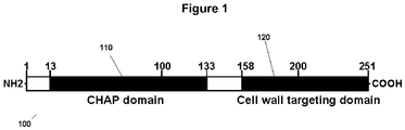
- FIG. 1 is a schematic diagram of phiNM3 lysin showing the putative CHAP domain 110 and the CWT domain 120. The numbers represent the amino acid positions and the domain limits.
- the chimeric lysins comprising an endopeptidase domain of a first lysin (e.g., Twort S. aureus lysin) bound to the CWT domain of SEQ ID NO:1 are surprisingly soluble in PBS (e.g., at least about 1 mg/ml, and typically about 3 mg/ml or greater).
- a lysin is provided in SEQ ID NO:2 (AD127), shown in FIG. 5A and consisting of the Twort lysin endopeptidase domain attached to the phiNM3 CWT domain (SEQ ID NO:1).
- ClyS Muralytic activity of ClyS was tested on a number of bacterial strains representing a variety of species which were divided into sets (Table 1 and Figure 10 ).
- Set I consisted of S. aureus strains including methicillin-sensitive S. aureus (MSSA) and MRSA.
- ClyS was active against MSSA and MRSA although differences were observed between S. aureus strains.
- Set II consisted of different species of staphylococci including S. epidermidis, S. simulans and S. sciuri.
- ClyS was active not only against S. epidermidis including the biofilm-forming strain RP62A but was also active against S. simulans and S. sciuri suggesting that ClyS recognizes an epitope in the cell wall that is present in all staphylococcal cells.
- a chimeric peptide comprises an isolated polypeptide comprising an endopeptidase domain of the S. aureus Twort lysin upstream of the lyphostaphin CWT domain.
- S. aureus Twort lysin upstream of the lyphostaphin CWT domain.
- One example of such a lysin is provided in SEQ ID NO:3 (AD119).
- the present disclosure pertains to lytic enzymes as a prophylactic treatment for preventing infection those who have possibly been exposed to S. aureus bacteria, or as a therapeutic treatment for those who have already become ill from the infection.
- the phage associated lytic enzymes described herein are specific for S. aureus bacteria and preferably effectively and efficiently break down the cell wall of the S. aureus bacteria.
- compositions which may be used for the prophylactic and therapeutic treatment of a S. aureus bacteria infection also includes the shuffled and/or chimeric enzyme and a means of application (such as a carrier system or an oral delivery mode) to the mucosal lining of the oral and nasal cavity, such that the enzyme is put in the carrier system or oral delivery mode to reach the mucosa lining.
- physiologically acceptable carriers include buffers such as phosphate, citrate, and other organic acids; antioxidants including ascorbic acid; low molecular weight (less than about 10 residues) polypeptides; proteins, such as serum albumin, gelatin, or immunoglobulins; hydrophilic polymers such as polyvinylpyrrolidone; amino acids such as glycine, glutamine, asparagine, arginine or lysine; monosaccharides, disaccharides, and other carbohydrates including glucose, mannose, or dextrins; chelating agents such as EDTA; sugar alcohols such as mannitol or sorbitol; salt-forming counterions such as sodium; and/or nonionic surfactants such as TWEEN.TM., polyethylene glycol (PEG), and PLURONICS.TM..
- buffers such as phosphate, citrate, and other organic acids
- antioxidants including ascorbic acid
- low molecular weight (less than about 10 residues) polypeptides such as
- Routes of administration include topical, ocular, nasal, pulmonary, buccal, parenteral (intravenous, subcutaneous, and intramuscular), oral, parenteral, vaginal and rectal. Also administration from implants is possible.
- the compounds of the invention may also be administered topically to the skin or mucosa, that is, dermally or transdermally.
- Typical formulations for this purpose include gels, hydrogels, lotions, solutions, creams, ointments, dusting powders, dressings, foams, films, skin patches, wafers, implants, sponges, fibres, bandages and microemulsions. Liposomes may also be used.
- Typical carriers include alcohol, water, mineral oil, liquid petrolatum, white petrolatum, glycerin, polyethylene glycol and propylene glycol.
- Penetration enhancers may be incorporated [see, for example, J Pharm Sci, 88 (10), 955-958 by Finnin and Morgan (October 1999 ).]
- the compounds of the invention may also be administered directly into the blood stream, into muscle, or into an internal organ.
- Suitable means for parenteral administration include intravenous, intraarterial, intraperitoneal, intrathecal, intraventricular, intraurethral, intrasternal, intracranial, intramuscular and subcutaneous.
- Suitable devices for parenteral administration include needle (including microneedle) injectors, needle-free injectors and infusion techniques.
- the compounds of the invention may also be administered intranasally or orally by inhalation, typically in the form of an aerosol.
- a pharmaceutical composition comprising a lysin of the invention in admixture with a pharmaceutically acceptable carrier is formulated for topical administration or for administration intranasally or orally by inhalation.
- Suitable antimicrobial preparation forms are, for example granules, powders, tablets, coated tablets, (micro) capsules, suppositories, syrups, emulsions, microemulsions, defined as optically isotropic thermodynamically stable systems consisting of water, oil and surfactant, liquid crystalline phases, defined as systems characterized by long-range order but short-range disorder (examples include lamellar, hexagonal and cubic phases, either water- or oil continuous), or their dispersed counterparts, gels, ointments, dispersions, suspensions, creams, aerosols, droplets or injectable solution in ampule form and also preparations with protracted release of active compounds, in whose preparation excipients, diluents, adjuvants or carriers are customarily used as described above.
- the pharmaceutical composition may also be provided in bandages or in sutures or the like.
- chimeric bacteriophage lysin described herein (e.g., SEQ ID NO:2) as a replacement for or for use in combination with prophylactic antibiotics in this situation.
- the chimeric bacteriophage lysin may be administered by injection with a suitable carrier directly to the site of the orthopedic device in situ to clear the infection, or on a surface of the device prior to implantation.
- Other injection routes such as subcutaneous, intramuscular, or intraperitoneal, can be used.
- a chimeric peptide described herein Prior to, or at the time the enzyme is put in the carrier system or oral delivery mode, it may be desirable for a chimeric peptide described herein to be administered or formulated in a stabilizing buffer environment, maintaining a pH range between about 5.0 and about 7.5.
- the enzyme Prior to, or at the time the chimeric peptide is put in the carrier system or oral delivery mode, the enzyme may be in a stabilizing buffer environment for maintaining a suitable pH range, such as between about 5.0 and about 8.0, including a pH of about 5.0, 6.0, 7.0, 8.0 or any pH interval of 0.05 therebetween, or any interval that is a multiple of 0.05 therebetween, including pH values of 5.2, 6.5, 7.4, 7.5 and 8.5.
- lysins peptidoglycan and associated carbohydrates, respectively
- lysin resistance will be rare.
- the route of administration is in accord with known methods, e.g. injection or infusion by intravenous, intraperitoneal, intracerebral, intramuscular, intraocular, intraarterial or intralesional routes, topical administration, or by sustained release systems.
- the lytic enzyme may be administered in any suitable fashion, including parenterally or through the oral or nasal cavity.
- Dosages and desired drug concentrations of pharmaceutical compositions of the present invention may vary depending on the particular use envisioned. The determination of the appropriate dosage or route of administration is well within the skill of an ordinary physician. Animal-experiments provide reliable guidance for the determination of effective doses for human therapy. Interspecies scaling of effective doses can be performed following the principles laid down by Mordenti, J. and Chappell, W. "The use of interspecies scaling in toxicokinetics" In Toxicokinetics and New Drug Development, Yacobi et al., Eds., Pergamon Press, New York 1989, pp. 42-96 .
- chimeic peptide lysin When in vivo administration of a chimeic peptide lysin is employed, normal dosage amounts may vary from about 10 ng/kg to up to 1000 mg/kg of mammal body weight or more per day, or about 1 ⁇ g/kg/day to 10000mg/kg/day, depending upon the route of administration. Guidance as to particular dosages and methods of delivery is also provided below, as well as in the literature. It is anticipated that different formulations will be effective for different treatment compounds and different disorders, that administration targeting one organ or tissue, for example, may necessitate delivery in a manner different from that to another organ or tissue.
- the concentration of the active units of a chimeric peptide believed to provide for an effective amount or dosage of enzyme may be in the range of about 10, 20, 30, 40, 50, 60, 70, 80, 90, or 100 units/ml up to about 10,000,000 units/ml of composition, in a range of about 1000 units/ml to about 10,000,000 units/ml, and from about 10,000 to 10,000,000 units/ml.
- the enzyme can be transported in a liposome, with the enzyme be "inserted" in the liposomes by known techniques. Similarly, the enzyme may be in a reverse micelle. The enzyme can also be pegylated, attaching the polyethylene glycol to the non-active part of the enzyme. Alternatively, hydrophobic molecules can be used to transport the enzyme across the cell membrane. Finally, the glycosylation of the enzyme can be used to target specific internalization receptors on the membrane of the cell.
- a Staphylococcus chimeric lysin such as a lysin of SEQ ID NO:2 (ClyS) may be combined with other bacteriostatic or bacteriocidal agents useful for decontamination of inanimate solid surfaces suspected of containing infectious bacteria, or for decontamination of porous surfaces.
- Bacterial strains (Table 1) were stored at -80°C routinely grown at 37°C. Staphylococcal strains used in this study were grown in Trypticase Soy Broth (TSB) media, streptococcal strains were grown in THY (Todd-Hewitt broth, 1% wt/vol yeast extract) media, B. cereus and P. aeruginosa were grown in BHI (Brain Heart Infusion) media while E. coli was cultivated in LB (Luria Bertani) media.
- TTB Trypticase Soy Broth
- streptococcal strains were grown in THY (Todd-Hewitt broth, 1% wt/vol yeast extract) media
- B. cereus and P. aeruginosa were grown in BHI (Brain Heart Infusion) media while E. coli was cultivated in LB (Luria Bertani) media.
- the two PCR amplicons were ligated using the Pstl restriction endonuclease site.
- the ligated product was cloned into pBAD24 vector using the NcoI-HindIII cloning sites to generate recombinant plasmid pAD127.
- the entire DNA fragment corresponding to clyS was PCR amplified from pAD124 using primers NM3-Lys-Xba-F: 5'-CTAGTCTAGAGGTGGAATAATGAAAACATACAGTGAAGCAAG-3' (SEQ ID NO:9) and primer NM3-CBD-Hind-R(SEQ ID NO:8).
- the PCR product was cloned into expression vector pJML6 to generate pAD138.
- the sequence of ClyS was confirmed by sequencing.
- the recombinant plasmid pAD138 was transformed into E. coli DH5 ⁇ cells.
- ClyS was induced overnight from E. coli DH5 ⁇ (pAD138) cells with lactose (10g/500ml final concentration) at 30°C.
- Cells were harvested by centrifugation, resuspended in buffer A (20 mM phosphate buffer (PB), 1 mM DTT (dithiothreitol)) and lysed by an EmulsiFlex-C5 high pressure homogenizer (Avestin) at 4ooC.
- the lysates were cleared by centrifugation (2x 50,000xg) for 30 minutes at 4°C and the supernatant applied to a CM-sepharose column (Amersham Pharmacia, Piscataway, N.J.).
- ClyS was eluted with buffer A + 1 M NaCl using a linear gradient of 0-50% B in 15 columns volumes. Fractions were analyzed for lytic activity as previously described (Daniel et al, 2001). Fractions displaying lytic activity were pooled and dialyzed overnight against buffer B (PB, 1 mM DTT, 50mM NaCl). The dialyzed sample was applied to a hydroxylapatite (MacroPrep Typell 40 ⁇ m, BioRad) column and eluted with elution buffer (500 mM PB + 50 mM NaCl+ 1 mM DTT) using a linear gradient of 0-100% B in 20 columns volumes. The fractions were analyzed by SDS-PAGE and for lytic activity. Active clean fractions of ClyS were pooled and dialyzed against buffer B. Protein concentration was determined with the BCA method (Sigma, St. Louis, MO).
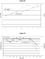
- ClyS activity was measured as previously described (Daniel et al, 2001), with some modifications. Briefly, S. aureus strain 8325-4 was grown to an OD 600 of 0.25-0.3, centrifuged, and resuspended in PB to a final OD 600 of 0.8-1.0. Two-fold serial dilutions of purified ClyS (100 ⁇ l) were added to 100 ⁇ l of bacterial suspension in 96-well plates (Costar) and the decrease in OD600 was monitored by a Spectramax Plus 384 spectrophotometer (Molecular Devices) over 30 min at 37°C. ClyS activity in units per milliliter was defined as the reciprocal of the highest dilution of lysin that decreased the absorbance by 50% in 15 minutes.
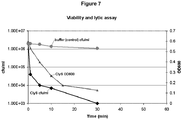
- ClyS The viability assay of ClyS was tested as previously described (Nelson et al, 2001). Briefly, logphase cultures of S. aureus strain 8325-4 were resuspended in PB to OD 600 of 0.8-1.0. 50U of ClyS or the corresponding volume of PB was added to bacterial cells and aliquots were removed, serially diluted, and plated at 1, 5, 10, 30, and 60 minutes to assess the viability of the treated and control cells. All experiments were performed in triplicate. The activity of ClyS on various bacterial strains was tested as described previously (Schuch et al, 2002). Briefly, logphase bacterial cells were treated with 50U of ClyS at 37°C for 15 minutes. The samples were serially diluted and plated. Control experiments with the addition of phosphate buffer (pH 7.0) were performed under the same conditions.
- phosphate buffer pH 7.0
- Example 6 Measuring ClyS activity as a function of pH and salt profile
- S. aureus strain 8325-4 was grown to log-phase, centrifuged and resuspended in PBS to an absorbance at 600nm of 1.0.
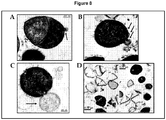
- the bacterial suspension was incubated with 50U of ClyS at room temperature.
- the lytic reaction was terminated after 1 minute and 5 minutes by adding glutaraldehyde (final concentration 2.5%).
- the suspension was pelleted by centrifugation and overlaid with 2.5% glutaraldehyde in 0.1 M cacodylate buffer (pH 7.4).
- the samples were then postfixed in 1 % osmium tetroxide, block stained with uranyl acetate and processed according to standard procedures by The Rockefeller University Electron Microscopy Service.
- the approximately 10-kDa phiNM3 CWT protein was expressed and the protein was purified in one step by cation-exhange chromatography.
- the purified protein (1mg/ml) was incubated with 10 ⁇ l of FITC (1mg/ml) for 1 hour. Excess FITC was removed on a desalting column.
- the labeled-protein (50 ⁇ g) was incubated with bacterial cells for 10 minutes, washed 3x with phosphate-buffered saline (pH 7.4) and observed under fluorescence microscope.
- Example 8 Measuring in vivo activity of ClyS
- MRSA strain would be grown to log-phase, centrifuged and resuspended to a predefined titer of about 1010 cfu/ml.
- the animals would be divided into 2 groups and administered various concentrations of ClyS or sterile saline intraperitoneally six hours after infection and every six hours thereafter for 3 days. The survival rate for each group would be observed up to 7 days post infection.
- Example 9 The linker region by itself does not confer solubility to a chimera.
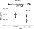
- Example 10 In vivo Nasal Decolonization of MRSA by ClyS .
- mice were intranasally inoculated with ⁇ 2x10 7 of a spontaneously streptomycin resistant strain of MRSA (191-SM R ). Twenty-four hours post-infection mice were administered three doses hourly of either phosphate buffered saline (control) or ClyS (960 ⁇ g) into the nasal passages. One hour after the last treatment, mice were sacrificed and bacteria colonies were enumerated on Spectra MRSA agar, (a selective chromogenic medium developed to diagnostically detect MRSA nasal colonization) and Columbia blood agar.
Landscapes
- Life Sciences & Earth Sciences (AREA)
- Health & Medical Sciences (AREA)
- Chemical & Material Sciences (AREA)
- General Health & Medical Sciences (AREA)
- Engineering & Computer Science (AREA)
- Zoology (AREA)
- Organic Chemistry (AREA)
- Wood Science & Technology (AREA)
- Genetics & Genomics (AREA)
- Biotechnology (AREA)
- Microbiology (AREA)
- Virology (AREA)
- Medicinal Chemistry (AREA)
- Bioinformatics & Cheminformatics (AREA)
- Biochemistry (AREA)
- Molecular Biology (AREA)
- Proteomics, Peptides & Aminoacids (AREA)
- Pest Control & Pesticides (AREA)
- Plant Pathology (AREA)
- Agronomy & Crop Science (AREA)
- Dentistry (AREA)
- Environmental Sciences (AREA)
- Biomedical Technology (AREA)
- General Engineering & Computer Science (AREA)
- Gastroenterology & Hepatology (AREA)
- Biophysics (AREA)
- Communicable Diseases (AREA)
- Oncology (AREA)
- Chemical Kinetics & Catalysis (AREA)
- General Chemical & Material Sciences (AREA)
- Nuclear Medicine, Radiotherapy & Molecular Imaging (AREA)
- Pharmacology & Pharmacy (AREA)
- Animal Behavior & Ethology (AREA)
- Public Health (AREA)
- Veterinary Medicine (AREA)
- Medicines That Contain Protein Lipid Enzymes And Other Medicines (AREA)
- Peptides Or Proteins (AREA)
- Micro-Organisms Or Cultivation Processes Thereof (AREA)
- Measuring Or Testing Involving Enzymes Or Micro-Organisms (AREA)
- Enzymes And Modification Thereof (AREA)
Description
- The present disclosure relates to the identification and use of chimeric lytic enzymes to rapidly and specifically detect and kill Staphylococci bacteria, including certain antibiotic-resistant Staphylococcus aureus bacterial strains.
- Staphylococcus aureus is an opportunistic pathogen inhabiting human skin and mucous membranes. S. aureus is the causative agent of variety of skin and soft tissue infections in humans and serious infections such as pneumonia, meningitis, endocarditis, and osteomyelitis. S. aureus exotoxins also cause disease syndromes such as bullous impetigo, scalded skin syndrome, and toxic shock syndrome. Additionally, staphylococci are also among the most common causes of food-borne illness in United States (Fischetti V A, Novick, R. P., Ferretti, J. J., Portnoy, D. A. and Rood, J. I., editor. 2006. Gram-positive pathogens. 2nd ed: ASM Press). S. aureus is also a major cause of community- and hospital-acquired (nosocomial) infections. Of the nearly 2 million cases of nosocomial infections in United States, approximately 230,000 cases are caused by S. aureus (NNIS. 2003. NNIS report, data summary from January 1992 through June 2003, issued August 2003. American Journal of Infection Control 31:481-498.).
- The global appearance of methicillin- and vancomycin-resistant clinical isolates of S. aureus has become a serious concern. Currently, 40-60% of nosocomial infections of S. aureus are resistant to oxacillin (Massey RC, Horsburgh MJ, Lina G, Hook M, Recker M. 2006. The evolution and maintenance of virulence in Staphylococcus aureus: a role for host-to-host transmission? Nat Rev Microbiol 4(12):953-8.) and greater than 60% of the isolates are resistant to methicillin (Gill SR, Fouts DE, Archer GL, Mongodin EF, Deboy RT, Ravel J, Paulsen IT, Kolonay JF, Brinkac L, Beanan M and others. 2005. Insights on evolution of virulence and resistance from the complete genome analysis of an early methicillin-resistant Staphylococcus aureus strain and a biofilm-producing methicillin-resistant Staphylococcus epidermidis strain. J Bacteriol 187(7):2426-38.). Treating infections caused by the drug-resistant S. aureus has become increasingly difficult and therefore is a major concern among healthcare professionals. To combat this challenge, development of new and effective antibiotics belonging to different classes are being aggressively pursued. A number of new antimicrobial agents such as linezolid, quinupristin-dalfopristin, daptomycin, tigecyline, new glycopeptides and ceftobiprole have been introduced or are under clinical development (Aksoy DY, Unal S. 2008. New antimicrobial agents for the treatment of Gram-positive bacterial infections. Clin Microbiol Infect 14(5):411-20.). However, clinical isolates of MRSA (methicillin-resistant Staphylococcus aureus) with resistance to these new classes of antibiotics have already been reported (Tsiodras S, Gold HS, Sakoulas G, Eliopoulos GM, Wennersten C, Venkataraman L, Moellering RC, Ferraro MJ. 2001. Linezolid resistance in a clinical isolate of Staphylococcus aureus. Lancet 358(9277):207-8; Mangili A, Bica I, Snydman DR, Hamer DH. 2005. Daptomycin-resistant, methicillin-resistant Staphylococcus aureus bacteremia. Clin Infect Dis 40(7):1058-60; Skiest DJ. 2006. Treatment failure resulting from resistance of Staphylococcus aureus to daptomycin. J Clin Microbiol 44(2):655-6). Consequently, there is an urgent need to develop novel therapeutic agents or antibiotic alternatives against MRSA.
- Bacteriophage endolysins (lysins) are one such class of novel antimicrobial agents that are emerging as novel agents for the prophylactic and therapeutic treatment of bacterial infections. Lysins are cell wall hydrolases that are produced during the infection cycle of double-stranded DNA bacteriophages (or phages) enabling release of progeny virions. Typically, lysins have two distinct functional domains consisting of a catalytic domain for peptidoglycan hydrolysis and a binding domain for recognition of surface moieties on the bacterial cell walls. The catalytic domains are relatively conserved among lysins. The activities of lysins can be classified into two groups based on bond specificity within the peptidoglycan: glycosidases that hydrolyze linkages within the aminosugar moieties and amidases that hydrolyze amide bonds of cross-linking stem peptides. The binding domains however are not conserved among lysins. Hence the binding domain imparts species- and strain-specificity because the binding targets, often carbohydrates associated with the peptidoglycan, display species- or strain-specific distribution (Fischetti VA, Nelson D, Schuch R. 2006. Reinventing phage therapy: are the parts greater than the sum? Nat Biotechnol 24(12):1508-11). The modular architecture of lysins' is an important feature with respect to their development as antimicrobial agents. This enables creation of chimeras by swapping lysin domains and thereby altering binding specificity or enzymatic activity or both (Sheehan MM, Garcia JL, Lopez R, Garcia P. 1996. Analysis of the catalytic domain of the lysin of the lactococcal bacteriophage Tuc2009 by chimeric gene assembling. FEMS Microbiol Lett 140(1):23-8; Lopez R GE, Garcia P, Garcia JL. 1997. The pneumococcal cell wall degrading enzymes: a modular design to create new lysins? Microb Drug Res 3:199-211; Croux C, Ronda C, Lopez R, Garcia JL. 1993. Interchange of functional domains switches enzyme specificity: construction of a chimeric pneumococcal-clostridial cell wall lytic enzyme. Mol Microbiol 9(5):1019-25; Donovan DM, Dong S, Garrett W, Rousseau GM, Moineau S, Pritchard DG. 2006. Peptidoglycan hydrolase fusions maintain their parental specificities. Appl Environ Microbiol 72(4):2988-96).
- When applied exogenously, native or recombinant lysins were able to degrade the cell wall of susceptible bacteria and cause rapid cell lysis (Nelson D, Loomis L, Fischetti VA. 2001. Prevention and elimination of upper respiratory colonization of mice by group A streptococci by using a bacteriophage lytic enzyme. Proc Natl Acad Sci U S A 98(7):4107-12). Lysins have been developed against a number of Gram-positive pathogens including Group A streptococci (Nelson D, Loomis L, Fischetti VA. 2001. Prevention and elimination of upper respiratory colonization of mice by group A streptococci by using a bacteriophage lytic enzyme. Proc Natl Acad Sci U S A 98(7):4107-12), S. pneumoniae (Loeffler JM, Nelson D, Fischetti VA. 2001. Rapid killing of Streptococcus pneumoniae with a bacteriophage cell wall hydrolase. Science 294(5549):2170-2), Bacillus anthracis (Schuch R, Nelson D, Fischetti VA. 2002. A bacteriolytic agent that detects and kills Bacillus anthracis. Nature 418(6900):884-9), enterococci (Yoong P, Schuch R, Nelson D, Fischetti VA. 2004. Identification of a broadly active phage lytic enzyme with lethal activity against antibiotic-resistant Enterococcus faecalis and Enterococcus faecium. J Bacteriol 186(14):4808-12), Group B streptococci (Cheng Q, Nelson D, Zhu S, Fischetti VA. 2005. Removal of group B streptococci colonizing the vagina and oropharynx of mice with a bacteriophage lytic enzyme. Antimicrob Agents Chemother 49(1):111-7), and Staphylococcus aureus (Rashel M, Uchiyama J, Ujihara T, Uehara Y, Kuramoto S, Sugihara S, Yagyu K, Muraoka A, Sugai M, Hiramatsu K and others. 2007. Efficient elimination of multidrug-resistant Staphylococcus aureus by cloned lysin derived from bacteriophage phi MR11. J Infect Dis 196(8):1237-47). The activities of most of these lysins have been demonstrated in vitro and in in vivo models. Several unique characteristics of lysin make them attractive antibacterial candidates against Gram-positive pathogens. These include i) rapid antibacterial activity both in vitro and in vivo; ii) very narrow lytic spectrum (species- and strain-specific); iii) very strong binding affinity, typically in the nanomolar range; iv) very low chances of developing resistance since the binding epitopes are essential for viability; v) safe; and vi) relative ease of modification by genetic engineering (Fischetti V A, Nelson D, Schuch R. 2006. Reinventing phage therapy: are the parts greater than the sum? Nat Biotechnol 24(12):1508-11).
- Although lysins have been developed against a number of Gram-positive pathogens, there remains a need for a S. aureus-specific lysin. Various labs have unsuccessfully attempted to obtain a staphylococcal lysin. The expression of more than twenty different staphylococcal lysins using a variety of techniques have been attempted without success. These include expression of lysin genes in E. coli using different expression vectors and conditions, expression in Bacillus, yeast and mammalian systems, expression in the presence of chaperones, expression of truncated versions etc. To our knowledge, there is only one report of the successful development of S. aureus-specific lysin called MV-L (Rashel M, Uchiyama J, Ujihara T, Uehara Y, Kuramoto S, Sugihara S, Yagyu K, Muraoka A, Sugai M, Hiramatsu K and others. 2007). Efficient elimination of multidrug-resistant Staphylococcus aureus by cloned lysin derived from bacteriophage phi MR11. J Infect Dis 196(8):1237-47). MV-L lysin is comprised of two catalytic domains (an endopeptidase and an amidase domain) linked to a single cell wall targeting (CWT) domain, a type of binding domain.
WO 2009/024327 (Profos AG) relates to a polypeptide termed ply-pitti26 and uses thereof in particular for the treatment or prophylaxis of a subject infected or exposed to Staphylococci.
Unless otherwise indicated, references herein to a "binding domain" herein include a CWT domain. The MV-L CWT domain, like the staphylolytic enzyme lysostaphin, displays homology to SH3b-like domains. The SH3b-like domains bind to the peptide cross-bridge (the penta Glycine) in the staphylococcal cell wall. There are reports of staphylococcal strains developing resistance at 10.sup.-6 frequencies to lysostaphin by altering their peptide cross-bridges. Therefore, we expect staphylococci to develop resistance at a higher frequency to lysins containing SH3b-like CWT domains including MV-L. There is a need for lytic enzymes capable of specific binding to Staphylococcal bacteria without undesirably high frequencies of lysostaphin resistance, such as S. aureus-specific lysins without SH3b-like CWT domains. - This disclosure describes novel staphylococcal lysins, as well as methods of making and using the lysin. In one example, the genetic engineering of a novel chimeric lysin called ClyS (for chimeric lysin for staphylococci) is described. ClyS is specifically active against susceptible and drug-resistant staphylococci, and was constructed by fusing the catalytic domain of a Staphylococcus-specific phage lysin with a unique binding domain from another Staphylococcus-specific phage lysin that has no known homologs. ClyS is a soluble Staphylococcal-specific lysin without a SH3b-like CWT domain, but does contain a CWT domain that is believed to recognize a staphylococci-specific surface carbohydrate. Consequently, the frequency by which staphylococcal strains will develop resistance to ClyS may be reduced. The invention concerns a chimeric bacteriophage lysin comprising a catalytic domain of a first Staphylococcus-specific phage lysin and a binding domain of a second Staphylococcus-specific phage lysin, wherein the binding domain is not an SH3b-like cell wall targeting domain. Additionally, biochemical characterization of ClyS revealed that the pH and salt spectrum of ClyS is very different from conventional lysins thereby providing unique properties to this chimeric lysin.
-
-
FIG. 1 is a schematic diagram of phiNM3 lysin showing the putative CHAP ("cysteine- and histidine-dependent amidohydrolase/peptidase") and CWT domains. The numbers represent the amino acid positions and the domain limits. The CWT domain of ClyS is indicated in the diagram -
FIG. 2A is a gel showing the purification of phiNM3 CWT. SDS-PAGE and coomassie blue stained gel of phiNM3 CWT purified by anion-exchange chromatography is depicted in the lane marked "CWT." Protein
molecular weight markers in kilodaltons (kDa) are shown in the lane marked "M." -
Figure 2B shows the amino acid sequence of the phiNM3 CWT protein (SEQ ID NO:1). -
Figure 3 shows a series of micrographs showing PhiNM3 CWT binding specifically to staphylococci. Purified phiNM3 CWT was labeled with FITC and exposed to 1) S. aureus; 2) B. cereus; 3) S. epidermidis; 4) E. coli; 5) Group A Streptococcus and 6) mixed suspension of S. aureus and B. cereus cells. "P" indicates phase-contrast image and "F" indicates fluorescent image. -
Figure 4 is a schematic diagram illustrating chimeric lysin development. In particular,Figure 4 provides schematic diagrams of various chimeric lysins showing their respective domains and the corresponding expression and solubility of the protein and activity against S. aureus cells. Similar domains are depicted in the same shading and labeled. PlyB-cat indicates catalytic domain of Bacillus-specific lysin PlyB (and is marked with a "4" in the figure); Sa-aa indicates 16 amino acid residues specific for staphylococcal lysins (and is marked with a "5" in the figure); PlyB-CWT indicates CWT domain of PlyB (and is marked with a "6" in the figure); Twort-CWT indicates CWT domain of S. aureus phage Twort lysin (and is marked with a "8" in the figure); Lysostaphin CWt indicates CWT domain of lysostaphin (and is marked with a "10" in the figure); and Se autolyin amidase indicates an amidase domain of S. epidermidis autolysin (and is marked with a "12" in the figure). -
Figure 5A shows the ClyS protein sequence. The predicted protein sequence of the chimeric protein ClyS showing the Twort endopeptidase catalytic and the phiNM3 CWT domains. -
Figure 5B shows the amino acid sequence for the AD127 chimeric molecule, described with respect toFigure 4 . -
Figure 5C shows the amino acid sequence for the native (unmodified) Twort lysin (SEQ ID NO: 12). -
Figure 6 is a gel showing the purification of ClyS. Cloys ways expressed in E. coli DH5α cells and purified by cation-exchange chromatography followed by hydroxyapatite chromatography. Purified sample (10 micrograms) was separated by SDS-PAGE and stained by Coomassie blue (right hand lane). Protein molecular weight markers in kilodaltons (kDa) are shown in the left hand lane. -
Figure 7 is a graph showing the activity of ClyS against S. aureus in vitro. S. aureus strain 8325-4 cells were resuspended in 20 mM phosphate buffer (pH 7.4), incubated with 50U of ClyS and OD600 (filled triangles) monitored by a spectrophotometer. Control experiments (filled squares) were performed under the same conditions with buffer alone. Viability (filled diamonds) of cells, shown as colony-forming units/ml, was determined by serially diluting and plating the cells. -
Figure 8 is a series of micrographs showing that ClyS causes cell wall disruption and ultimately lysis of 8325-4 cells.Figures 8A - 8C (A-C) are thin-section transmission electron micrographs (bars, 200 nm) ofS. aureus 3 minutes after exposure to 50 U of ClyS. The arrows indicate cytoplasmic membrane extrusions through holes generated in the cell wall by ClyS. Ultimate lysis results in "cell-ghosts" (D) after the loss of cytoplasmic contents (bar, 500 nm). -
Figures 9A and 9B are graphs showing the activity of ClyS in various pH and salt concentration conditions.Figure 9A is a graph of the activity of ClyS (50U) tested against S. aureus strain 8325-4 in buffers with pH values ranging from 4 and 10 in 15 minute assays. Optical density (filled squares) and viability (filled diamonds) was measured as described in legend ofFigure 6 . Fold killing in the viability assay was calculated by dividing the number of viable bacteria after buffer treatment at a particular pH by the number after exposure to ClyS enzyme at the same pH. Final pH readings for each reaction are recorded on the x axis.Figure 9B is a graph showing the activity of ClyS (50 U) tested against S. aureus strain 8325-4 in 20 mM phosphate buffer (pH 7.4) in the presence of different concentrations of NaCl. After 15 minutes samples were assayed for optical density and viability calculated as above. -
Figure 10 is a bar graph showing that ClyS exerts specific killing of staphylococci. Log-phase cultures of different bacteria were exposed to 50 U of ClyS for 15 minutes. Fold killing was calculated as described inFigure 8 legend. -
Figure 11 depicts a graph of the CFU of MRSA from individual MRSA infected mice after being administered phosphate buffered saline pH 7.3 (control) or ClyS (630 µg). -
Figure 12 depicts Kaplan Meier Survival Curves showing the effect of ClyS on preventing death in mice injected with MRSA compared with phosphate buffer control. -
Figure 13 depicts an isobologram for a checkerboard broth microdilution study of the effect of vancomycin on VISA (vancomycin-resistant Staphylococcus aureus) or oxacillin on MRSA with increasing amounts of ClyS. -
Figure 14 depicts Kaplan Meier Survival Curves showing the effect of oxacillin alone or in combination with ClyS. -
Figure 15 depicts a photograph of a Coomassie-blue stained SDS-PAGE gel of a 5-day time-course at 21°C of ClyS in the absence (top left gel) or presence of 5 mM DTT (top right gel), and pClyS in the absence (bottom left gel) or presence of 5 mM DTT (bottom right gel). About 20 micrograms of protein was loaded into each lane of the gel. The bottom right gel shows a much higher amount of intact pClyS in the presence of 5mM DTT after 5 days compared to intact ClyS in the presence of 5 mM DTT after 5 days. - Unless otherwise indicated, the certain terms used herein and their applicability to the present disclosure are defined below.
- The term "isolated" means at least partially purified from a starting material. The term "purified" means that the biological material has been measurably increased in concentration by any purification process, including but not limited to, column chromatography, HPLC, precipitation, electrophoresis, etc., thereby partially, substantially or completely removing impurities such as precursors or other chemicals involved in preparing the material. Hence, material that is homogenous or substantially homogenous (e.g., yields a single protein signal in a separation procedure such as electrophoresis or chromatography) is included within the meanings of isolated and purified. Skilled artisans will appreciate that the amount of purification necessary will depend upon the use of the material. For example, compositions intended for administration to humans ordinarily must be highly purified in accordance with regulatory standards.
- The term "lytic enzyme genetically coded for by a bacteriophage" refers to a polypeptide having at least some lytic activity against the host bacteria.
- Variants of "chimeric bacteriophage lysin" are included within the definition of chimeric bacteriophage lysins, and include a functionally active chimeric bacteriophage lysin with killing activity against Staphylococcus aureus having at least 50%, 55%, 60%, 65%, 70%, 75%, 80%, 85%, 90%, 95%, 97%, 98%, 99%, or even at least 99.5% amino acid sequence identity with a sequence described herein. For example, an aspect of the present invention includes chimerical bacteriophage lysins having at least 90%, 95%, 97%, 98%, 99%, or even at least 99.5% amino acid sequence identity with the polypeptide sequence of SEQ ID NO:2.
- "Percent (%) polypeptide sequence identity" with respect to the lytic enzyme polypeptide sequences identified here is defined as the percentage of amino acid residues in a candidate sequence that are identical with the amino acid residues in the specific lytic enzyme polypeptide sequence, after aligning the sequences and introducing gaps, if necessary, to achieve the maximum percent sequence identity, and not considering any conservative substitutions as part of the sequence identity. Methods for alignment for purposes of determining percent amino acid sequence identity are described below.
- Chimeric bacteriophage lysins with killing activity against S. aureus are described herein. In an aspect of the invention, the chimeric bacteriophage lysin has killing activity against Staphylococcus aureus, such as methicillin-resistant and methicillin sensitive strains of S. aureus. In a further aspect of the invention, the chimeric lysin has killing activity against multiple staphylococcal species, such as Staphylococcus aureus and Staphylococcus epidermidis. Lysins generally occur in a modular structure.
FIG. 1 is a schematic diagram of phiNM3 lysin showing theputative CHAP domain 110 and theCWT domain 120. The numbers represent the amino acid positions and the domain limits. The CWT domain of ClyS is shown as shadedbox 120. The N-terminal module consists of a catalytic domain believed to possess the ability to break down the bacterial cell wall of certain bacteria. Enzymatic activities often associated with the catalytic domain are amidases, endopeptidases, glucosamidases and muramidases. In an aspect of the invention, the catalytic domain is the Twort endopeptidase domain. The C-terminal module consists of a binding domain that is believed to have an affinity for a carbohydrate epitope on the target bacteria cell wall. The binding domain is believed to determine the specificity of the lysin. The peptide cross-bridge within the staphylococcal peptidoglycan is believed to function as the receptor for the CWT domain of lysostaphin, a staphylolytic enzyme produced by Staphylococcus simulans. The CWT domain of lysostaphin has homology to the SH3b domain suggesting that such lysins might also utilize the peptide cross-bridge as its receptor. - In one embodiment, Staphylococcus-specific binding molecules comprising a CWT domain within staphylococcal lysins are provided that have no known domain homologs. Preferably, such a CWT domain is provided to recognize a different epitope such as a cell wall-associated carbohydrate instead of the peptide cross-bridge in the staphylococcal cell wall.
- In a further embodiment, the ClyS lysin can be used to digest the cell wall of Staphylococcus aureus bacterial strains, which in turn would allow access to the genetic and cytoplasmic material, such as endogenous DNA and RNA, to further identify and sequence the Staphylococcus aureus bacterial strain. It will also release membrane-associated and wall-associated molecules for diagnostic purposes.
- Most preferably, the binding molecule is a soluble binding domain of a bacterial lysin comprising a polypeptide including an amino acid sequence providing specific binding to S. aureus, such as SEQ ID NO:1 (phiNM3 CWT domain). For example, the lysin preferably includes the polypeptide sequence of S. aureus phage phiNM3 lysin (SEQ ID NO:1) (protein accession number YP.sub.--908849). The phiNM3 lysin CWT domain (SEQ ID NO:1) corresponding to amino acid residues 158-251 was cloned and expressed. The approximately 10-kDa protein of SEQ ID NO:1 was highly soluble and was purified by one-step anion-exchange chromatography to homogeneity.
FIG. 2A is an anion exchange gel showing the protein of SEQ ID NO:1 in a second column next to a set of marker proteins in a first column.FIG. 2B shows the amino acid sequence of SEQ ID NO:1. To determine whether the peptide domain of SEQ ID NO:1 displayed Staphylococcus-specific binding, the purified protein was labeled with FITC and exposed to log-phase S. aureus, S. epidermidis and mixed population of S. aureus and Bacillus. Group A streptococci, E. coli and Bacillus cereus were used as controls. More preferably, the FITC-labeled phiNM3 CWT domain bound specifically to S. aureus (FIG. 3-1 ) and S. epidermidis (FIG. 3-3 ) cells when present in single or mixed populations (FIG. 3-6 ) while binding to streptococci (FIG. 3-5 ), Bacillus (FIG. 3-2 ) or E. coli (FIG. 3-4 ) was not observed. PhiNM3 lysin specifically bound to S. aureus (FIG. 3-1 ) and S. epidermidis (FIG. 3-2 ) cells when present in single or mixed populations (FIG. 3-3 ) while binding to streptococci (FIG. 3-4 ), Bacillus (FIG. 3-5 ) or E. coli (FIG. 3-6 ) was not observed. -
FIG. 5B provides the amino acid sequence of SEQ ID NO:3, the AD119 sample discussed with respect toFIG. 4 . AD119 (SEQ ID NO:3) comprises the Twort endopeptidase domain joined to the Lysostaphin CWT domain. In contrast to the chimeric compound of SEQ ID NO:2 (AD127), which shares the Twort endopeptidase domain but has the phiNM3 CWT domain (SEQ ID NO:1) in place of the Lysostaphin CWT domain, the AD127 compound was insoluble and exhibited little or no killing activity against S. aureus. - In another embodiment, the lysin is a chimeric protein that comprises an endopeptidase domain of the S. aureus Twort lysin upstream of the phiNM3 CWT domain (SEQ ID NO:1). The chimeric polypeptide is preferably sufficiently soluble in phosphate buffered saline (PBS). Preferred levels of solubility in PBS for the chimeric lysins is at least about 1 mg/ml and more preferably at least about 3 mg/mL in PBS. While native staphylococcal bacteriophage lysins are typically insoluble in PBS, the chimeric lysins comprising an endopeptidase domain of a first lysin (e.g., Twort S. aureus lysin) bound to the CWT domain of SEQ ID NO:1 are surprisingly soluble in PBS (e.g., at least about 1 mg/ml, and typically about 3 mg/ml or greater). One example of such a lysin is provided in SEQ ID NO:2 (AD127), shown in
FIG. 5A and consisting of the Twort lysin endopeptidase domain attached to the phiNM3 CWT domain (SEQ ID NO:1). The isolated polypeptide of SEQ ID NO:2 (AD 127) was constructed by engineering S. epidermidis autolysin amidase and Twort lysin endopeptidase domains upstream of phiNM3 CWT domain, respectively.Chimera AD 126 had no expression or activity butAD 127 was soluble and had very high activity but low expression. To overcome low expression ofAD 127 construct, the entire chimera gene was cloned into expression vector pJML6 to generate pAD 138. The expression, solubility and activity ofAD 127 from the pAD138 construct was very high. Therefore, this chimera was named 'ClyS' for Chimeric lysin for Staphylococcus (Figure 5A ). - ClyS (SEQ ID NO:2) contains 280 amino acid residues with a deduced molecular mass of 31956 Da and a theoretical isoelectric point of 9.17, and was purified by two-step column chromatography to >90% homogeneity. ClyS had a molecular mass of approximately 31 kDa by SDS/PAGE (
Figure 6 ) which was confirmed by gel filtrations chromatography, suggesting that the protein exists as a monomer and is not proteolytically processed (data not shown). - The unit activity of ClyS was defined by measuring the spectrophotometric loss of turbidity, indicative of cell lysis, of S. aureus 8325-4 cells upon adding serial dilutions of ClyS. In our assays, 5 micrograms of ClyS corresponded to 1 U of lytic activity. When 50 U of ClyS was added to exponentially growing 8325-4 cells the OD600 dropped to baseline within 5 min (
Figure 7 ). To confirm that the observed cell lysis corresponds to cell death, staphylococcal viability was determined by enumerating aliquots from the lytic reaction at various time points. A decrease in viability of approximately 3-logs was observed in 30 min (Figure 7 ). - The lytic effect on S. aureus 8325-4 cells exposed to 50U of ClyS for 1-3 min was visualized by transmission electron microscopy. Typical of lysin activity observed previously, localized degradation of the cell wall was observed at single (
Figure 8A ) or multiple sites (Figure 8B ). However, unlike other lysins, the sites of degradation on the cell was not restricted to the septal or polar positions but was randomly distributed. This resulted in extrusions and rupture of the cell membrane (Figure 8C ) and subsequent loss of cytoplasmic contents and formation of cell-ghosts (Figure 8D ). - The effect of pH on the activity of ClyS was determined by measuring the drop in OD600 or cell viability at different pH values. We observed that ClyS was active over a wide range of pH values but was most active between
pH Figure 9A ). Similarly, the effect of salt concentration on activity of ClyS was also determined. ClyS displayed activity in a wide range of salt concentrations (Figure 9B ). While its activity deteriorated above 400 mM NaCl, at physiological concentrations ClyS functioned well. - Muralytic activity of ClyS was tested on a number of bacterial strains representing a variety of species which were divided into sets (Table 1 and
Figure 10 ). Set I consisted of S. aureus strains including methicillin-sensitive S. aureus (MSSA) and MRSA. ClyS was active against MSSA and MRSA although differences were observed between S. aureus strains. Set II consisted of different species of staphylococci including S. epidermidis, S. simulans and S. sciuri. ClyS was active not only against S. epidermidis including the biofilm-forming strain RP62A but was also active against S. simulans and S. sciuri suggesting that ClyS recognizes an epitope in the cell wall that is present in all staphylococcal cells. Set III consisted of a mix of Gram-positive and Gram-negative bacteria including representatives of group A, B, C and E streptococci, oral streptococcal species including S. gordonii, and S. salivarius, as well as S. uberis, Bacillus cereus, Pseudomonas aeruginosa and E. coli. ClyS exhibited no activity against any of these organisms. - In another embodiment, a chimeric peptide comprises an isolated polypeptide comprising an endopeptidase domain of the S. aureus Twort lysin upstream of the lyphostaphin CWT domain. One example of such a lysin is provided in SEQ ID NO:3 (AD119).
- In another embodiment, lytic compositions may comprise a mixture of two or more lysins. The mixture may include a first polypeptide and a second peptide where one or both of the polypeptides may lack a desired level of lytic activity, but the mixture provides desirably specific and effective lytic activity against a bacteria of interest. For example, a composition may include an isolated first polypeptide comprising an endopeptidase domain of the S. aureus Twort lysin upstream of the lyphostaphin CWT domain combined with a second isolated polypeptide comprising an S. epidermidis autolysin amidase domain upstream of the lysostaphin CWT domain. One example of such a composition comprises a mixture of SEQ ID NO:3 (AD119) and SEQ ID NO:4 (AD112).
- In some examples, the present disclosure pertains to lytic enzymes as a prophylactic treatment for preventing infection those who have possibly been exposed to S. aureus bacteria, or as a therapeutic treatment for those who have already become ill from the infection. The phage associated lytic enzymes described herein are specific for S. aureus bacteria and preferably effectively and efficiently break down the cell wall of the S. aureus bacteria.
- The chimeric lytic enzyme polypeptides described herein may also be employed as a therapeutic agent. The lytic enzyme polypeptides of the present invention can be formulated according to known methods to prepare pharmaceutically useful compositions, whereby the lytic enzyme product hereof is combined in admixture with a pharmaceutically acceptable carrier vehicle. Compositions which may be used for the prophylactic and therapeutic treatment of a S. aureus bacteria infection also includes the shuffled and/or chimeric enzyme and a means of application (such as a carrier system or an oral delivery mode) to the mucosal lining of the oral and nasal cavity, such that the enzyme is put in the carrier system or oral delivery mode to reach the mucosa lining.
- In an aspect of the invention, the chimeric lysin is administered in combination with an antibiotic, such as vancomycin or oxacillin. A Staphylococcus chimeric lysin, such as a lysin of SEQ ID NO:2 (ClyS), is administered as an antibacterial composition in combination with a suitable pharmaceutical carrier. In an aspect of the invention, the chimeric lysin is administered in admixture with a pharmaceutically acceptable carrier as a pharmaceutical composition. In a further aspect of the invention, the pharmaceutical composition further comprises vancomycin or oxacillin. In certain embodiments, the amount of the chimeric bacteriophage lysin present is a therapeutically effective amount. "Carriers" as used herein include pharmaceutically acceptable carriers, excipients, or stabilizers which are nontoxic to the cell or mammal being exposed thereto at the dosages and concentrations employed. Often the physiologically acceptable carrier is an aqueous pH buffered solution. Examples of physiologically acceptable carriers include buffers such as phosphate, citrate, and other organic acids; antioxidants including ascorbic acid; low molecular weight (less than about 10 residues) polypeptides; proteins, such as serum albumin, gelatin, or immunoglobulins; hydrophilic polymers such as polyvinylpyrrolidone; amino acids such as glycine, glutamine, asparagine, arginine or lysine; monosaccharides, disaccharides, and other carbohydrates including glucose, mannose, or dextrins; chelating agents such as EDTA; sugar alcohols such as mannitol or sorbitol; salt-forming counterions such as sodium; and/or nonionic surfactants such as TWEEN.TM., polyethylene glycol (PEG), and PLURONICS.TM.. These antimicrobial/pharmaceutical compositions may be administered locally or systemically.
- Routes of administration include topical, ocular, nasal, pulmonary, buccal, parenteral (intravenous, subcutaneous, and intramuscular), oral, parenteral, vaginal and rectal. Also administration from implants is possible. The compounds of the invention may also be administered topically to the skin or mucosa, that is, dermally or transdermally. Typical formulations for this purpose include gels, hydrogels, lotions, solutions, creams, ointments, dusting powders, dressings, foams, films, skin patches, wafers, implants, sponges, fibres, bandages and microemulsions. Liposomes may also be used. Typical carriers include alcohol, water, mineral oil, liquid petrolatum, white petrolatum, glycerin, polyethylene glycol and propylene glycol. Penetration enhancers may be incorporated [see, for example, J Pharm Sci, 88 (10), 955-958 by Finnin and Morgan (October 1999).]
- The compounds of the invention may also be administered directly into the blood stream, into muscle, or into an internal organ. Suitable means for parenteral administration include intravenous, intraarterial, intraperitoneal, intrathecal, intraventricular, intraurethral, intrasternal, intracranial, intramuscular and subcutaneous. Suitable devices for parenteral administration include needle (including microneedle) injectors, needle-free injectors and infusion techniques. The compounds of the invention may also be administered intranasally or orally by inhalation, typically in the form of an aerosol. Thus in an aspect of the invention, a pharmaceutical composition comprising a lysin of the invention in admixture with a pharmaceutically acceptable carrier is formulated for topical administration or for administration intranasally or orally by inhalation.
- Suitable antimicrobial preparation forms are, for example granules, powders, tablets, coated tablets, (micro) capsules, suppositories, syrups, emulsions, microemulsions, defined as optically isotropic thermodynamically stable systems consisting of water, oil and surfactant, liquid crystalline phases, defined as systems characterized by long-range order but short-range disorder (examples include lamellar, hexagonal and cubic phases, either water- or oil continuous), or their dispersed counterparts, gels, ointments, dispersions, suspensions, creams, aerosols, droplets or injectable solution in ampule form and also preparations with protracted release of active compounds, in whose preparation excipients, diluents, adjuvants or carriers are customarily used as described above. The pharmaceutical composition may also be provided in bandages or in sutures or the like.
- Many orthopedic surgeons consider that humans with prosthetic joints should be considered for antibiotic prophylaxis. Late deep infection by S. aureus is a serious complication sometimes leading to loss of the prosthetic joint and is accompanied by significant morbidity and mortality. It may therefore be possible to extend the use of the chimeric bacteriophage lysin described herein (e.g., SEQ ID NO:2) as a replacement for or for use in combination with prophylactic antibiotics in this situation. The chimeric bacteriophage lysin may be administered by injection with a suitable carrier directly to the site of the orthopedic device in situ to clear the infection, or on a surface of the device prior to implantation. Other injection routes, such as subcutaneous, intramuscular, or intraperitoneal, can be used. Alternative means for administration include transmucosal and transdermal administration using penetrants such as bile salts or fusidic acids or other detergents. In addition, if a polypeptide or other compounds of the present invention can be formulated in an enteric or an encapsulated formulation, oral administration may also be possible. Administration of these compounds may also be topical and/or localized, in the form of salves, pastes, gels, and the like.
- Prior to, or at the time the enzyme is put in the carrier system or oral delivery mode, it may be desirable for a chimeric peptide described herein to be administered or formulated in a stabilizing buffer environment, maintaining a pH range between about 5.0 and about 7.5. Prior to, or at the time the chimeric peptide is put in the carrier system or oral delivery mode, the enzyme may be in a stabilizing buffer environment for maintaining a suitable pH range, such as between about 5.0 and about 8.0, including a pH of about 5.0, 6.0, 7.0, 8.0 or any pH interval of 0.05 therebetween, or any interval that is a multiple of 0.05 therebetween, including pH values of 5.2, 6.5, 7.4, 7.5 and 8.5.
- There are a number of advantages to using lytic enzymes to treat bacterial infections. The modular design of lysins, with their distinct catalytic and binding domains, makes them ideal for domain swapping experiments in which bacterial specificities and catalytic activities can be improved or adapted for use against alternate pathogens. Since the catalytic and binding targets of lysins (peptidoglycan and associated carbohydrates, respectively) are largely essential for viability, lysin resistance will be rare.
- "Treatment" refers to both therapeutic treatment and prophylactic or preventative measures, wherein the object is to prevent or slow down (lessen) the targeted pathologic condition or disorder. Those in need of treatment include those already with the disorder as well as those prone to have the disorder or those in whom the disorder is to be prevented.
- "Mammal" for purposes of treatment refers to any animal classified as a mammal, including humans, domestic and farm animals, and zoo, sports, or pet animals, such as dogs, cats, cattle, horses, sheep, pigs, goats, rabbits, etc. Preferably, the mammal is human.
- The formulations to be used for in vivo administration are preferably sterile. This is readily accomplished by filtration through sterile filtration membranes, prior to or following lyophilization and reconstitution. Therapeutic compositions herein generally are placed into a container having a sterile access port, for example, an intravenous solution bag or vial having a stopper pierceable by a hypodermic injection needle.
- The route of administration is in accord with known methods, e.g. injection or infusion by intravenous, intraperitoneal, intracerebral, intramuscular, intraocular, intraarterial or intralesional routes, topical administration, or by sustained release systems. When treating a bacterial exposure or infection, the lytic enzyme may be administered in any suitable fashion, including parenterally or through the oral or nasal cavity.
- Dosages and desired drug concentrations of pharmaceutical compositions of the present invention may vary depending on the particular use envisioned. The determination of the appropriate dosage or route of administration is well within the skill of an ordinary physician. Animal-experiments provide reliable guidance for the determination of effective doses for human therapy. Interspecies scaling of effective doses can be performed following the principles laid down by Mordenti, J. and Chappell, W. "The use of interspecies scaling in toxicokinetics" In Toxicokinetics and New Drug Development, Yacobi et al., Eds., Pergamon Press, New York 1989, pp. 42-96.
- When in vivo administration of a chimeic peptide lysin is employed, normal dosage amounts may vary from about 10 ng/kg to up to 1000 mg/kg of mammal body weight or more per day, or about 1 µg/kg/day to
10000mg/kg/day, depending upon the route of administration. Guidance as to particular dosages and methods of delivery is also provided below, as well as in the literature. It is anticipated that different formulations will be effective for different treatment compounds and different disorders, that administration targeting one organ or tissue, for example, may necessitate delivery in a manner different from that to another organ or tissue. - The effective dosage rates or amounts of the chimeric peptide to be administered parenterally, and the duration of treatment will depend in part on the seriousness of the infection, the weight of the patient, the duration of exposure of the recipient to the infectious bacteria, the seriousness of the infection, and a variety of a number of other variables. The composition may be applied anywhere from once to several times a day, and may be applied for a short or long term period. The usage may last for days or weeks. Any dosage form employed should provide for a minimum number of units for a minimum amount of time. The concentration of the active units of a chimeric peptide believed to provide for an effective amount or dosage of enzyme may be in the range of about 10, 20, 30, 40, 50, 60, 70, 80, 90, or 100 units/ml up to about 10,000,000 units/ml of composition, in a range of about 1000 units/ml to about 10,000,000 units/ml, and from about 10,000 to 10,000,000 units/ml.
- Additionally, a number of methods can be used to assist in transporting the enzyme across the cell membrane. The enzyme can be transported in a liposome, with the enzyme be "inserted" in the liposomes by known techniques. Similarly, the enzyme may be in a reverse micelle. The enzyme can also be pegylated, attaching the polyethylene glycol to the non-active part of the enzyme. Alternatively, hydrophobic molecules can be used to transport the enzyme across the cell membrane. Finally, the glycosylation of the enzyme can be used to target specific internalization receptors on the membrane of the cell.
- Another preferred embodiment provides for a composition comprising a Staphylococcus chimeric lysin bacterial binding protein such as a lysin of SEQ ID NO:2 (ClyS), with other lytic enzymes which are useful for sanitizing or decontaminating porous surfaces e.g. textiles, carpeting. Furthermore, the composition of lytic enzymes may be used to decontaminate veterinarian surgical or examination areas, where such areas may be thought to harbor infectious organisms susceptible to the bacteriostatic or bacteriocidal activity.
- In a further preferred embodiment, a Staphylococcus chimeric lysin such as a lysin of SEQ ID NO:2 (ClyS) may be combined with other bacteriostatic or bacteriocidal agents useful for decontamination of inanimate solid surfaces suspected of containing infectious bacteria, or for decontamination of porous surfaces.
- We conducted conserved domain searches of Staphylococcus-specific phage and prophage lysin protein sequences in the National Center for Biotechnology Information database. The lysins were classified based on homology to known domains in the database. We identified several lysins including the S. aureus phage phiNM3 lysin (protein accession number YP_908849), S. aureus prophage phi13 amidase (accession number NP_803402), S. aureus prophage MW2 amidase (accession number NP_646703.1), etc. that shared 100 % sequence identity with each other and had a conserved CHAP domain within their catalytic domain. However, the C-terminal domain of these lysins did not display homology to any known domains in the database (
Figure 1 ). - Since the attempts to express a native staphylococcal lysin were unsuccessful, we decided to develop chimeric lysins by taking advantage of the modular nature of lysins. Traditionally, Bacillus-specific lysins are expressed at high levels and are soluble in E. coli. Therefore, our first attempt was to engineer a 16-amino acid peptide (4) that is conserved in several S. aureus-specific lysins (Lu JZ, Fujiwara T, Komatsuzawa H, Sugai M, Sakon J. 2006. Cell wall-targeting domain of glycylglycine endopeptidase distinguishes among peptidoglycan cross-bridges. (Lue et al. (2006) J. Biol. Chem. 281(1):549-58) The catalytic domain of the Bacillus-specific lysin PlyB was used to generate chimera AD 103 (
Figure 4 ) (SEQ ID NO: 13). The chimeras were tested for expression, solubility and activity. Then the entire C-terminal CWT domain of PlyB (6) was replaced by the putative C-terminal domain of S. aureus phage Twort lysin (8) to obtain AD 105 (SEQ ID NO:14). This chimera was not active and so we engineered the lysostaphin CWT domain (10) downstream of PlyB-catalytic domain (2) to get AD 107 (SEQ ID NO:15). Although this chimera had expression, the solubility was poor and there was no activity. The next step was to engineer a S. epidermidis autolysin amidase domain (12) upstream of the lysostaphin CWT domain (10) which resulted in AD 112 (SEQ ID NO:4).AD 112 expressed very well and the protein was also very soluble but there was no lytic activity. However, we observed that the S. aureus cells clumped when exposed toAD 112. Since the lysostaphin catalytic domain (an amidase) (10) inAD 112 was of bacterial origin, we attempted to engineer a phage-derived catalytic domain upstream of the lysostaphin CWT. For this, the endopeptidase domain in Twort lysin (14) was used to construct chimera AD 119 (SEQ ID NO:3). We observed poor expression forAD 119 but the chimera was soluble. Although in ourlytic assays AD 119 alone did not show significant activity, when combined withchimera AD 112 the activity was significantly enhanced. Since we identified phiNM3 CWT domain from our conserved domains searches and observed that phiNM3 CWT exhibited Staphylococcus-specific binding, we constructed chimeras AD 126 (SEQ ID NO:16) and AD 127 (SEQ ID NO:2) by engineering S. epidermidis autolysin amidase (12) and Twort lysin endopeptidase (14) domains upstream of phiNM3 CWT domain (2) (SEQ ID NO:1), respectively.Chimera AD 126 had no expression or activity butAD 127 was soluble and had very high activity but low expression (Figure 4 ). To overcome low expression ofAD 127 construct, the entire chimera gene was cloned into expression vector pJML6 to generate pAD 138. The expression, solubility and activity ofAD 127 from the pAD138 construct was very high. Therefore, this chimera was named 'ClyS' for Chimeric lysin for Staphylococcus. The amino acid sequence for ClyS (i.e., SEQ ID NO:2) is provided inFigure 5A . - Bacterial strains (Table 1) were stored at -80°C routinely grown at 37°C. Staphylococcal strains used in this study were grown in Trypticase Soy Broth (TSB) media, streptococcal strains were grown in THY (Todd-Hewitt broth, 1% wt/vol yeast extract) media, B. cereus and P. aeruginosa were grown in BHI (Brain Heart Infusion) media while E. coli was cultivated in LB (Luria Bertani) media.
- The chimeric lysin was constructed by amplifying and ligating individual domains from respective genes. For this, the Twort endopeptidase domain was PCR amplified from plasmid pCR2.1plyTW which contains the entire lysin (plyTW) gene using primers TW-Endo-Ncol-F: 5'-CTAGCCATGGAAACCCTGAAACAAGCAG-3' (SEQ ID NO:5) and TW-Endo-Pstl-R: 5'-ACATGCTGCAGAACCATATTGTAATTAATATTAGTTCTATC-3'(SEQ ID NO:6). The cell wall targeting (CWT) domain was PCR amplified from S. aureus strain 8325 genomic DNA using primers NM3-CBD-Pstl-F: 5'-ACATGCTGCAGGGTAAATCTGCAAGTAAAATAACAG-3' (SEQ ID NO:7)
and NM3-CBD-Hind-R: 5'-CCCAAGCTTAAAACACTTCTTTCACAATCAATCTC-3'(SEQ ID NO:8). The two PCR amplicons were ligated using the Pstl restriction endonuclease site. The ligated product was cloned into pBAD24 vector using the NcoI-HindIII cloning sites to generate recombinant plasmid pAD127. In the second step, the entire DNA fragment corresponding to clyS was PCR amplified from pAD124 using primers NM3-Lys-Xba-F: 5'-CTAGTCTAGAGGTGGAATAATGAAAACATACAGTGAAGCAAG-3' (SEQ ID NO:9) and primer NM3-CBD-Hind-R(SEQ ID NO:8). The PCR product was cloned into expression vector pJML6 to generate pAD138. The sequence of ClyS was confirmed by sequencing. The recombinant plasmid pAD138 was transformed into E. coli DH5α cells. - ClyS was induced overnight from E. coli DH5α (pAD138) cells with lactose (10g/500ml final concentration) at 30°C. Cells were harvested by centrifugation, resuspended in buffer A (20 mM phosphate buffer (PB), 1 mM DTT (dithiothreitol)) and lysed by an EmulsiFlex-C5 high pressure homogenizer (Avestin) at 4ooC. The lysates were cleared by centrifugation (2x 50,000xg) for 30 minutes at 4°C and the supernatant applied to a CM-sepharose column (Amersham Pharmacia, Piscataway, N.J.). ClyS was eluted with buffer A + 1 M NaCl using a linear gradient of 0-50% B in 15 columns volumes. Fractions were analyzed for lytic activity as previously described (Daniel et al, 2001). Fractions displaying lytic activity were pooled and dialyzed overnight against buffer B (PB, 1 mM DTT, 50mM NaCl). The dialyzed sample was applied to a hydroxylapatite (MacroPrep Typell 40µm, BioRad) column and eluted with elution buffer (500 mM PB + 50
mM NaCl+ 1 mM DTT) using a linear gradient of 0-100% B in 20 columns volumes. The fractions were analyzed by SDS-PAGE and for lytic activity. Active clean fractions of ClyS were pooled and dialyzed against buffer B. Protein concentration was determined with the BCA method (Sigma, St. Louis, MO). - ClyS activity was measured as previously described (Daniel et al, 2001), with some modifications. Briefly, S. aureus strain 8325-4 was grown to an OD600 of 0.25-0.3, centrifuged, and resuspended in PB to a final OD600 of 0.8-1.0. Two-fold serial dilutions of purified ClyS (100µl) were added to 100µl of bacterial suspension in 96-well plates (Costar) and the decrease in OD600 was monitored by a Spectramax Plus 384 spectrophotometer (Molecular Devices) over 30 min at 37°C. ClyS activity in units per milliliter was defined as the reciprocal of the highest dilution of lysin that decreased the absorbance by 50% in 15 minutes.
- The viability assay of ClyS was tested as previously described (Nelson et al, 2001). Briefly, logphase cultures of S. aureus strain 8325-4 were resuspended in PB to OD600 of 0.8-1.0. 50U of ClyS or the corresponding volume of PB was added to bacterial cells and aliquots were removed, serially diluted, and plated at 1, 5, 10, 30, and 60 minutes to assess the viability of the treated and control cells. All experiments were performed in triplicate. The activity of ClyS on various bacterial strains was tested as described previously (Schuch et al, 2002). Briefly, logphase bacterial cells were treated with 50U of ClyS at 37°C for 15 minutes. The samples were serially diluted and plated. Control experiments with the addition of phosphate buffer (pH 7.0) were performed under the same conditions.
- The effect of pH on ClyS activity was determined as previously described using the universal buffer system pH 4-10 (Yoong et al). Briefly, logphase 8325-4 cells were resuspended in the universal buffer system and incubated with 50U of ClyS for 15 minutes. The final pH of each reaction was checked by pH paper. The samples were serially diluted and plated. In controls, PB replaced ClyS.
- Similarly the effect of salt concentration on the lytic activity of ClyS was determined by incubating 50U of ClyS with logphase 8325-4 cells in PB containing NaCl at a final concentration of 25-500 mM for 15 minutes. The samples were serially diluted and plated to determine the viability counts.
- S. aureus strain 8325-4 was grown to log-phase, centrifuged and resuspended in PBS to an absorbance at 600nm of 1.0. The bacterial suspension was incubated with 50U of ClyS at room temperature. The lytic reaction was terminated after 1 minute and 5 minutes by adding glutaraldehyde (final concentration 2.5%). The suspension was pelleted by centrifugation and overlaid with 2.5% glutaraldehyde in 0.1 M cacodylate buffer (pH 7.4). The samples were then postfixed in 1 % osmium tetroxide, block stained with uranyl acetate and processed according to standard procedures by The Rockefeller University Electron Microscopy Service.
- Flourescent labeling and binding analyses were performed on phiNM3 CWT. S. aureus strain 8325-4 genomic DNA was used to amplify the putative CWT of phiNM3 lysin using primers NM3-FWD 5'-CATGCCATGGGTAAATCTGCAAGTAAAATAACAG-3' (SEQ ID NO:10) and NM3-REV 5'-CCCAAGCTTAAAACACTTCTTTCACAATCAATCTC-3'(SEQ ID NO:11). The resulting amplicon was cloned into the arabinose-inducible expression vector pBAD24. Positive clones containing the insert were confirmed by sequencing. The approximately 10-kDa phiNM3 CWT protein was expressed and the protein was purified in one step by cation-exhange chromatography. The purified protein (1mg/ml) was incubated with 10 µl of FITC (1mg/ml) for 1 hour. Excess FITC was removed on a desalting column. The labeled-protein (50 µg) was incubated with bacterial cells for 10 minutes, washed 3x with phosphate-buffered saline (pH 7.4) and observed under fluorescence microscope.
- MRSA strain would be grown to log-phase, centrifuged and resuspended to a predefined titer of about 1010 cfu/ml. For intranasal infection, 6-wk-old female C57BL/6J, outbread Swiss or BALB/c mice (
weight range 22 to 24 g, Charles River Laboratories, Wilmington, MA) would be anesthetized with a mixture of ketamine (Fort Dodge Animal Health, Fort Dodge, IA, 1.2 mg/animal) and xylazine (Miles Inc., Shawnee Mission, KS, 0.25 mg/animal), and inoculated with 15 µl of the bacterial suspension per nostril (n = 10). The animals would be divided into 2 groups and administered various concentrations of ClyS or sterile saline intraperitoneally six hours after infection and every six hours thereafter for 3 days. The survival rate for each group would be observed up to 7 days post infection. For intraperitoneal infection, mice would be infected intraperitoneally with 100 µl of the bacterial suspension (n = 10). The animals would be divided into 2 groups and administered various concentrations of ClyS or sterile saline intraperitoneally six hours after infection and every six hours thereafter for 3 days. The survival rate for each group would be observed up to 7 days post infection. - Since the ClyS construct was the only chimera that was highly soluble and active against staphylococci, we hypothesized that the linker region comprising of amino acid residues 142 through 185 of ClyS may be crucial for solubility. We had previously cloned and expressed the native phiNM3 lysin and observed that the protein was insoluble. To test this hypothesis, we replaced the endopeptidase domain of ClyS with the amidase domain of phiNM3 lysin upstream of the linker region of ClyS (ami-link-ClyS) and expressed the chimera (data not shown). We observed that similar to the native phiNM3 lysin, the ami-link-ClyS chimera was insoluble and expressed as inclusion bodies. We also tested the lysates of ami-link-ClyS for activity against staphylococci and did not observe any lytic activity confirming that the protein was insoluble and therefore inactive. Thus, it is the unique combination of the N and C terminal domains that are the subject of this patent that allow for a soluble complex to occur and behave as described herein.
- Carriage of both MSSA and MRSA in the human anterior nares is the major reservoir for S. aureus infection. Studies have shown that roughly 80% of the population could be nasally colonized by S. aureus, and that colonization can be an increased risk factor for developing other more serious S. aureus infections (Kluytmans, J., A. van Belkum. 1997. Nasal carriage of Staphylococcus aureus: epidemiology, underlying mechanisms, and associated risks. Clin Microbiol Rev 10(3): 505-20.). Elimination of nasal carriage in the community or in the hospital setting thus could possibly reduce the risk of infection and slow the spread of drug resistant S. aureus (Kluytmans et al. (1997)). To study the potential of ClyS to reduce MRSA colonization of the nasal mucosa, C57BL/6J mice were intranasally inoculated with ∼2x10⁷ of a spontaneously streptomycin resistant strain of MRSA (191-SMR). Twenty-four hours post-infection mice were administered three doses hourly of either phosphate buffered saline (control) or ClyS (960 µg) into the nasal passages. One hour after the last treatment, mice were sacrificed and bacteria colonies were enumerated on Spectra MRSA agar, (a selective chromogenic medium developed to diagnostically detect MRSA nasal colonization) and Columbia blood agar. No significant differences in CFU were obtained between plating to Spectra MRSA agar or Columbia blood agar (Data not shown). Three independent experiments were performed to evaluate a total 20 mice for each treatment group (
Figure 11 ). Compared to the buffer alone control (Avg. 12,273 CFU/cavity), ClyS treatment (Avg. 1198 CFU/cavity) significantly (p<0.001) reduced the mean CFU on the nasal mucosa. - Example 11. ClyS Treatment of Systemic MRSA infections. In order to assess whether ClyS treatment could prevent death resulting from systemic MRSA infections, 4 week old FVB/NJ mice were intraperitonally injected with ∼5x10⁵ CFU of the community-acquired MRSA strain MW2 in 5% mucin. Preliminary experiments determined that 5x10⁵ CFU was 10X the LD100 dose for a twenty-four hour period. Furthermore, within 3 hours of IP injection the MRSA infection was systemic, i.e., MRSA were recovered in high numbers from heart, liver, spleen, and kidney (data not shown). Treatment occurred three hours post-infection, with either 20mM phosphate buffer or 1 mg of ClyS in 20mM phosphate buffer injected IP (intraperitoneally). Mice were then monitored for survival over ten days. The results from three independent experiments were combined (ClyS treatment, n =16; buffer treatment, n =14) and the mice survival data plotted with a Kaplan Meier Survival curve (
Figure 12 ). Within twenty-four hours all of the control mice died of bacterial sepsis, while only 2/16 of ClyS treated mice died at forty-eight hours, and the remaining mice (14/16, 88%) survived over the course of the experiments (Figure 12 ). - Example 12. ClyS showed synergistic interaction with Vancomycin and Oxacillin. We used the checkerboard broth-microdilution assay to test the interaction of ClyS with vancomycin and with oxacillin. The vancomycin MIC for VISA strain Mu50 was 8µg/ml and the oxacillin MIC for MRSA strain COL was 32 µg/ml, while the ClyS MIC was 6 and 8 U/ml for both strains tested (Mu50 and COL respectively). Isobolograms for ClyS with vancomycin and ClyS with oxacillin was plotted by transcribing the enzyme concentrations along the inhibitory line on the microtiter plate in an x/y plot. The shape of the curves for both interactions were characteristic of a synergistic interaction (
Fig. 13 ) and were further confirmed by calculating the ∑FICI for both interactions which was ≤0.5. - Example 13. In vivo Synergy of Oxacillin and ClyS in the Treatment of Systemic MRSA infections. In vitro experiments showed that ClyS acted synergistically with oxacillin (
Figure 14 ). To determine if this effect could be seen in our systemic MRSA infection model, FVB/NJ mice were intraperitonally injected with ∼5x10⁵ CFUs of the MRSA strain MW2 as above. Three hours post infection mice were treated in parallel, with a lower IP dose of 130 µg/mouse of ClyS combined with different concentrations of oxacillin (10-100 µg/mouse) or buffer alone controls. Preliminary experiments determined that an ED30 dose of ClyS (130 µg/mouse) had minimal efficacy to evaluate the effect of combinatorial treatment with oxacillin (data not shown). Mice were monitored for survival for 10 days and the results of 5 independent experiments were combined and plotted in a Kaplan Meier Survival curve (FIG. 14 ). While only 30% (6/20 alive) to 35% (8/23 alive) of mice survived with individual treatments of either 130 µg/mouse of ClyS or 100 µg/mouse of oxacillin, respectively, neither differed significantly from the survival rate of the buffer alone control, 13% (2/15 alive). Conversely, a single dose of the combined treatment of intraperitoneal injected ClyS (130 µg) with either 0 µg or 50 µg of intramuscular injected oxacillin significantly increased mouse survival (80%, 8/10 alive; 82%, 18/22 alive respectively) compared to the individual treatments and buffer alone (FIG. 14 ). - Example 16. Modification of ClyS. The G166 residue of ClyS (SEQ ID NO:2) was changed to a proline by site directed mutagenesis (creating pClyS). When the purified pClyS molecule (SEQ ID NO: 17) was subjected to stability studies at 21 °C. for 5 days, the pClyS was found to be significantly more stable in the presence of 5 mM DTT the native ClyS with or without DTT (
FIG. 15 ). -
- <110> The Rockefeller University Fischetti, Vincent A. Daniel, Anu Euler, Chad
- <120> A CHIMERIC BACTERIOPHAGE LYSIN WITH ACTIVITY AGAINST STAPHYLOCOCCI BACTERIA
- <130> 3268-1-007PCT
- <140>
PCT/US 09/049349
<141> 2009-07-01 - <150>
US 61/078,277
<151> 2008-07-03 - <160> 17
- <170> PatentIn version 3.5
- <210> 1
<211> 94
<212> PRT
<213> Staphylococcus aureus phage phiNM3 - <400> 1

- <210> 2
<211> 280
<212> PRT
<213> Artificial Sequence - <220>
<223> Chimeric of Staphylococcus aureus bacteriophage Twort domain and Staphylococcus aureus phage phiNM3 CWT domain - <400> 2


- <210> 3
<211> 283
<212> PRT
<213> Artificial Sequence - <220>
<223> Chimeric of Staphylococcus aureus bacteriophage Twort domain and Staphylococcus simulans metalloendopeptidase Lysostaphin CWT domain - <400> 3


- <210> 4
<211> 643
<212> PRT
<213> Artificial Sequence - <220>
<223> Chimeric of Staphylococcus epidermidis autolysin amidase domain and Staphylococcus simulans metalloendopeptidase Lysostaphin CWT domain - <400> 4


- <210> 5
<211> 28
<212> DNA
<213> Artificial Sequence - <220>
<223> TW-Endo-NcoI-F PCR Primer - <400> 5

- <210> 6
<211> 41
<212> DNA
<213> Artificial Sequence - <220>
<223> TW-Endo-PstI-R PCR Primer - <400> 6

- <210> 7
<211> 36
<212> DNA
<213> Artificial Sequence - <220>
<223> NM3-CBD-PstI-F PCR Primer - <400> 7

- <210> 8
<211> 35
<212> DNA
<213> Artificial Sequence - <220>
<223> NM3-CBD-Hind-R PCR Primer - <400> 8

- <210> 9
<211> 42
<212> DNA
<213> Artificial Sequence - <220>
<223> NM3-Lys-Xba-F PCR Primer - <400> 9

- <210> 10
<211> 34
<212> DNA
<213> Artificial Sequence - <220>
<223> NM3-FWD PCR Primer - <400> 10

- <210> 11
<211> 35
<212> DNA
<213> Artificial Sequence - <220>
<223> NM3-REV PCR Primer - <400> 11

- <210> 12
<211> 467
<212> PRT
<213> Staphylococus phage Twort - <400> 12


- <210> 13
<211> 283
<212> PRT
<213> Artificial Sequence - <220>
<223> Chimeric of Bacillus-specific phage lysin - <400> 13


- <210> 14
<211> 287
<212> PRT
<213> Artificial Sequence - <220>
<223> Cimeric of Bacillus-specific phage lysin PlyB catalytic domain and Staphylococcus aureus bacteriophage Twort domain - <400> 14


- <210> 15
<211> 292
<212> PRT
<213> Artificial Sequence - <220>
<223> Chimeric of Bacillus-specific phage lysin PlyB catalytic domain and Staphylococcus simulans metalloendopeptidase Lysostaphin CWT domain - <400> 15


- <210> 16
<211> 658
<212> PRT
<213> Artificial Sequence - <220>
<223> Chimeric of Staphylococcus epidermidis autolysin amidase domain and Staphylococcus aureus phage pniNM3 CWT domain - <400> 16


- <210> 17
<211> 280
<212> PRT
<213> Artificial Sequence - <220>
<223> Chmeric of Staphylococcus aureus bacteriophage Twort domain and Staphylococcus aureus phage phiNM3 CWT domain - <400> 17


Claims (20)
- A chimeric bacteriophage lysin comprising a catalytic domain of a first Staphylococcus-specific phage lysin and a binding domain of a second Staphylococcus-specific phage lysin, wherein the binding domain is not an SH3b-like cell wall targeting domain.
- The chimeric lysin of claim 1, where the chimeric bacteriophage lysin has killing activity against Staphylococcus aureus, such as methicillin-resistant and methicillin sensitive strains of S. aureus.
- The chimeric lysin of claim 1 or 2, where the binding domain comprises the polypeptide sequence of SEQ ID NO: 1 or a variant thereof having at least 90% amino acid sequence identity with SEQ ID NO: 1.
- The chimeric lysin of any one of claims 1-3, where the catalytic domain is the Twort endopeptidase domain.
- The chimeric lysin of any one of claims 1-4, where the bacteriophage lysin comprises the polypeptide sequence of SEQ ID NO:2 or a variant thereof having at least 90% amino acid sequence identity with SEQ ID NO: 2.
- The chimeric lysin of any one of claims 1-5 having killing activity against multiple staphylococcal species, such as Staphylococcus aureus and Staphylococcus epidermidis.
- The chimeric lysin of any one of claims 1-6 having killing activity against Staphylococcal bacteria over a pH range of about 4-10.
- The chimeric lysin of any one of claims 1-7, where the lysin has a solubility of at least about 3 mg/mL in phosphate buffered saline (PBS).
- A chimeric lysin of any one of claims 1-8 for use in the treatment of a mammal with a staphylococcal infection.
- A chimeric lysin of any one of claims 1-8 for use in the prevention of infection of a mammal colonized by staphylococci.
- A chimeric lysin of any one of claims 1 to 8 for use in the decolonization of S. aureus.
- A chimeric lysin for use according to claim 11 wherein the decolonization is nasal decolonization.
- A chimeric lysin for use according to any one of claims 9 to 11, wherein the lysin is administered in combination with an antibiotic.
- A chimeric lysin for use according to claim 13, wherein the antibiotic is vancomycin or oxacillin.
- A pharmaceutical composition comprising a chimeric lysin of any one of claims 1 to 8 in admixture with a pharmaceutically acceptable carrier.
- A pharmaceutical composition according to claim 15 further comprising vancomycin or oxacillin.
- A pharmaceutical composition according to claim 15 which is formulated for topical, ocular, nasal, pulmonary, buccal, parenteral, oral, vaginal or rectal administration.
- A pharmaceutical composition according to claim 17 which is formulated for topical administration or for administration intranasally or orally by inhalation.
- An anti-microbial composition for sanitizing or decontaminating porous or non-porous surfaces comprising a chimeric bacteriophage lysin of any one of claims 1-8.
- A method for decontaminating inanimate surfaces suspected of containing infectious bacteria comprising treatment of said surfaces with a bacteriocidal or bacteriostatically effective amount of the composition of claim 19.
Priority Applications (1)
| Application Number | Priority Date | Filing Date | Title |
|---|---|---|---|
| EP17158578.9A EP3263588A1 (en) | 2008-07-03 | 2009-07-01 | Isolated staphylococcus-specific soluble binding molecule |
Applications Claiming Priority (2)
| Application Number | Priority Date | Filing Date | Title |
|---|---|---|---|
| US7827708P | 2008-07-03 | 2008-07-03 | |
| PCT/US2009/049349 WO2010002959A2 (en) | 2008-07-03 | 2009-07-01 | A chimeric bacteriophage lysin with activity against staphylococci bacteria |
Related Child Applications (1)
| Application Number | Title | Priority Date | Filing Date |
|---|---|---|---|
| EP17158578.9A Division EP3263588A1 (en) | 2008-07-03 | 2009-07-01 | Isolated staphylococcus-specific soluble binding molecule |
Publications (3)
| Publication Number | Publication Date |
|---|---|
| EP2307557A2 EP2307557A2 (en) | 2011-04-13 |
| EP2307557A4 EP2307557A4 (en) | 2012-04-11 |
| EP2307557B1 true EP2307557B1 (en) | 2017-03-01 |
Family
ID=41466590
Family Applications (2)
| Application Number | Title | Priority Date | Filing Date |
|---|---|---|---|
| EP17158578.9A Pending EP3263588A1 (en) | 2008-07-03 | 2009-07-01 | Isolated staphylococcus-specific soluble binding molecule |
| EP09774409.8A Active EP2307557B1 (en) | 2008-07-03 | 2009-07-01 | A chimeric bacteriophage lysin with activity against staphylococci bacteria |
Family Applications Before (1)
| Application Number | Title | Priority Date | Filing Date |
|---|---|---|---|
| EP17158578.9A Pending EP3263588A1 (en) | 2008-07-03 | 2009-07-01 | Isolated staphylococcus-specific soluble binding molecule |
Country Status (8)
| Country | Link |
|---|---|
| US (4) | US8840900B2 (en) |
| EP (2) | EP3263588A1 (en) |
| JP (2) | JP5758291B2 (en) |
| AU (2) | AU2009266982B2 (en) |
| CA (2) | CA2729160C (en) |
| ES (1) | ES2627321T3 (en) |
| HK (1) | HK1248722A1 (en) |
| WO (1) | WO2010002959A2 (en) |
Families Citing this family (19)
| Publication number | Priority date | Publication date | Assignee | Title |
|---|---|---|---|---|
| WO2008018854A2 (en) | 2005-06-06 | 2008-02-14 | The Rockefeller University | Bactiophage lysins for bacillus anthracis |
| WO2007024628A2 (en) | 2005-08-24 | 2007-03-01 | The Rockefeller University | Ply-gbs mutant lysins |
| JP6139509B2 (en) | 2011-04-21 | 2017-05-31 | ザ ロックフェラー ユニバーシティ | Streptococcus bacteriophage lysine for detecting and treating gram positive bacteria |
| AU2011367262B2 (en) | 2011-05-04 | 2016-11-03 | Micreos Human Health B.V. | A polypeptide |
| EP3369742B1 (en) | 2011-10-05 | 2020-07-29 | The Rockefeller University | Dimeric bacteriophage lysins |
| CN111803620A (en) * | 2012-05-09 | 2020-10-23 | 康特拉费克特公司 | Biofilm prevention, disruption and treatment using bacteriophage lysin |
| KR20210121313A (en) | 2012-05-09 | 2021-10-07 | 콘트라펙트 코포레이션 | Bacteriophage lysin and antibiotic combinations against gram positive bacteria |
| CN103122347B (en) * | 2013-02-01 | 2015-04-22 | 中国科学院武汉病毒研究所 | Lyase for killing staphylococcus and application of lyase |
| ES2525954B1 (en) * | 2013-05-28 | 2015-10-23 | Consejo Superior De Investigaciones Científicas (Csic) | ENHANCED BACTERICID ENZIBITICS AGAINST PNEUMOCOCO AND OTHER BACTERIA |
| CN104762285B (en) * | 2015-04-23 | 2017-10-27 | 武汉菲吉乐科生物科技有限公司 | A kind of lyases and its application from interior cracking Escherichia coli |
| CN104805066B (en) * | 2015-05-18 | 2018-04-24 | 武汉菲吉乐科生物科技有限公司 | A kind of staphylolytic enzyme and its application |
| MX2018013372A (en) | 2016-05-12 | 2019-05-09 | Contrafect Corp | Broth microdilution method for evaluating and determining minimal inhibitory concentration of antibacterial polypeptides. |
| CN116850269A (en) * | 2018-06-22 | 2023-10-10 | 康特拉费克特公司 | Lysin and derivatives thereof to re-sensitize staphylococcus aureus and gram positive bacteria to antibiotics |
| BR112021002417A2 (en) * | 2018-08-10 | 2021-05-04 | Meiji Seika Pharma Co., Ltd. | bacteriophage preparation |
| CN109666667A (en) * | 2019-01-22 | 2019-04-23 | 上海交通大学 | The chimaeric enzyme antibiotic of killing staphylococcus aureus and its preparation and application |
| JP2023510074A (en) * | 2019-10-31 | 2023-03-13 | バクトクリア ホールディングス ピーティーイー エルティーディー. | Chimeric protein for selective lysis of bacteria |
| AU2021236351A1 (en) * | 2020-03-11 | 2022-10-06 | Telum Therapeutics S.L. | New recombinant lysin and its use in the treatment of gram-negative bacterial infections |
| CN111808837A (en) * | 2020-07-17 | 2020-10-23 | 青岛诺安百特生物技术有限公司 | Staphylococcus aureus bacteriophage lyase and preparation method and application thereof |
| CN115820616A (en) * | 2022-07-22 | 2023-03-21 | 昆明理工大学 | Bacteriophage lyase with fluorescent label and application thereof |
Family Cites Families (12)
| Publication number | Priority date | Publication date | Assignee | Title |
|---|---|---|---|---|
| WO1997008553A1 (en) * | 1995-08-22 | 1997-03-06 | The Regents Of The University Of California | Targeting of proteins to the cell wall of gram-positive bacteria |
| US6056954A (en) * | 1997-10-31 | 2000-05-02 | New Horizons Diagnostics Corp | Use of bacterial phage associated lysing enzymers for the prophylactic and therapeutic treatment of various illnesses |
| US6703025B1 (en) * | 1998-08-31 | 2004-03-09 | Inhibitex, Inc. | Multicomponent vaccines |
| US20020127215A1 (en) | 1999-09-14 | 2002-09-12 | Lawrence Loomis | Parenteral use of bacterial phage associated lysing enzymes for the therapeutic treatment of bacterial infections |
| GB0107661D0 (en) * | 2001-03-27 | 2001-05-16 | Chiron Spa | Staphylococcus aureus |
| US20040213765A1 (en) | 2001-07-13 | 2004-10-28 | Vincent Fischetti | Use of bacterial phage associated lytic enzymes to prevent food poisoning |
| JP2005520545A (en) * | 2002-03-25 | 2005-07-14 | ザ ユニバーシティ オブ ワーウィック | Antibacterial agent |
| AU2003261782A1 (en) * | 2002-08-28 | 2004-03-19 | Techno Network Shikoku Co., Ltd. | Bacteriophages and remedies for bacterial infection |
| WO2007024628A2 (en) * | 2005-08-24 | 2007-03-01 | The Rockefeller University | Ply-gbs mutant lysins |
| CA2696864A1 (en) * | 2007-08-22 | 2009-02-26 | Hyglos Invest Gmbh | New proteins for use in human and animal staphylococcus infections |
| JP5732397B2 (en) * | 2008-10-10 | 2015-06-10 | ルソメディカメンタ, エス.エー.Lusomedicamenta, S.A. | Antibacterial phage peptides and uses thereof |
| CA3019013C (en) * | 2009-02-06 | 2020-12-15 | Technophage, Investigacao E Desenvolvimento Em Biotecnologia, Sa | Antibacterial phage, phage peptides and methods of use thereof |
-
2009
- 2009-07-01 EP EP17158578.9A patent/EP3263588A1/en active Pending
- 2009-07-01 JP JP2011516843A patent/JP5758291B2/en active Active
- 2009-07-01 EP EP09774409.8A patent/EP2307557B1/en active Active
- 2009-07-01 US US13/502,912 patent/US8840900B2/en active Active
- 2009-07-01 CA CA2729160A patent/CA2729160C/en active Active
- 2009-07-01 WO PCT/US2009/049349 patent/WO2010002959A2/en active Application Filing
- 2009-07-01 ES ES09774409.8T patent/ES2627321T3/en active Active
- 2009-07-01 AU AU2009266982A patent/AU2009266982B2/en active Active
- 2009-07-01 CA CA2984933A patent/CA2984933C/en active Active
-
2014
- 2014-09-09 US US14/480,822 patent/US10053681B2/en active Active
-
2015
- 2015-02-24 JP JP2015033626A patent/JP6169632B2/en active Active
- 2015-09-25 AU AU2015230807A patent/AU2015230807B2/en active Active
-
2018
- 2018-06-27 HK HK18108284.8A patent/HK1248722A1/en unknown
- 2018-08-20 US US16/105,155 patent/US11015182B2/en active Active
-
2021
- 2021-05-24 US US17/328,299 patent/US11827911B2/en active Active
Non-Patent Citations (1)
| Title |
|---|
| None * |
Also Published As
| Publication number | Publication date |
|---|---|
| EP3263588A1 (en) | 2018-01-03 |
| US10053681B2 (en) | 2018-08-21 |
| AU2009266982B2 (en) | 2015-06-25 |
| WO2010002959A3 (en) | 2010-05-06 |
| AU2015230807B2 (en) | 2017-07-27 |
| US20190055533A1 (en) | 2019-02-21 |
| US11827911B2 (en) | 2023-11-28 |
| CA2729160C (en) | 2017-11-14 |
| JP5758291B2 (en) | 2015-08-05 |
| US8840900B2 (en) | 2014-09-23 |
| CA2984933A1 (en) | 2010-01-07 |
| CA2984933C (en) | 2020-07-07 |
| US20150064156A1 (en) | 2015-03-05 |
| US20120258088A1 (en) | 2012-10-11 |
| HK1248722A1 (en) | 2018-10-19 |
| CA2729160A1 (en) | 2010-01-07 |
| WO2010002959A2 (en) | 2010-01-07 |
| EP2307557A4 (en) | 2012-04-11 |
| JP6169632B2 (en) | 2017-07-26 |
| US11015182B2 (en) | 2021-05-25 |
| EP2307557A2 (en) | 2011-04-13 |
| JP2015126738A (en) | 2015-07-09 |
| JP2011526920A (en) | 2011-10-20 |
| AU2015230807A1 (en) | 2015-10-15 |
| AU2009266982A1 (en) | 2010-01-07 |
| ES2627321T3 (en) | 2017-07-27 |
| US20210348146A1 (en) | 2021-11-11 |
Similar Documents
| Publication | Publication Date | Title |
|---|---|---|
| US11827911B2 (en) | Chimeric bacteriophage lysin with activity against Staphylococci bacteria | |
| US20230167427A9 (en) | Streptococcus bacteriophage lysins for detection and treatment of gram positive bacteria | |
| JP2022058682A (en) | Bacteriophage lysin and antibiotic combinations against gram-positive bacteria | |
| Hermoso et al. | Taking aim on bacterial pathogens: from phage therapy to enzybiotics | |
| Donovan | Bacteriophage and peptidoglycan degrading enzymes with antimicrobial applications | |
| US20100004321A1 (en) | Recombinant Staphylococcal Phage Lysin as an Antibacterial Agent | |
| KR20200045468A (en) | Strengthening blood components of lytic protein antibacterial activity and methods and uses thereof | |
| US20180353575A1 (en) | Clostridium difficile bacteriophage lysins for detection and treatment of clostridium difficile bacteria infection | |
| AU2018203076B2 (en) | Dimeric bacteriophage lysins | |
| Borysowski et al. | Enzybiotics and their potential applications in medicine |
Legal Events
| Date | Code | Title | Description |
|---|---|---|---|
| PUAI | Public reference made under article 153(3) epc to a published international application that has entered the european phase |
Free format text: ORIGINAL CODE: 0009012 |
|
| 17P | Request for examination filed |
Effective date: 20110128 |
|
| AK | Designated contracting states |
Kind code of ref document: A2 Designated state(s): AT BE BG CH CY CZ DE DK EE ES FI FR GB GR HR HU IE IS IT LI LT LU LV MC MK MT NL NO PL PT RO SE SI SK SM TR |
|
| AX | Request for extension of the european patent |
Extension state: AL BA RS |
|
| DAX | Request for extension of the european patent (deleted) | ||
| RIC1 | Information provided on ipc code assigned before grant |
Ipc: C12P 13/08 20060101AFI20120222BHEP Ipc: C12P 13/04 20060101ALI20120222BHEP |
|
| RIC1 | Information provided on ipc code assigned before grant |
Ipc: C12P 13/04 20060101ALI20120227BHEP Ipc: C12P 13/08 20060101AFI20120227BHEP |
|
| A4 | Supplementary search report drawn up and despatched |
Effective date: 20120309 |
|
| 17Q | First examination report despatched |
Effective date: 20150907 |
|
| REG | Reference to a national code |
Ref country code: DE Ref legal event code: R079 Ref document number: 602009044504 Country of ref document: DE Free format text: PREVIOUS MAIN CLASS: C12P0013080000 Ipc: C07K0014005000 |
|
| GRAP | Despatch of communication of intention to grant a patent |
Free format text: ORIGINAL CODE: EPIDOSNIGR1 |
|
| RIC1 | Information provided on ipc code assigned before grant |
Ipc: A61K 38/48 20060101ALI20160722BHEP Ipc: C12N 9/48 20060101ALI20160722BHEP Ipc: C07K 14/005 20060101AFI20160722BHEP Ipc: A01N 63/00 20060101ALI20160722BHEP |
|
| INTG | Intention to grant announced |
Effective date: 20160816 |
|
| GRAS | Grant fee paid |
Free format text: ORIGINAL CODE: EPIDOSNIGR3 |
|
| STAA | Information on the status of an ep patent application or granted ep patent |
Free format text: STATUS: GRANT OF PATENT IS INTENDED |
|
| GRAA | (expected) grant |
Free format text: ORIGINAL CODE: 0009210 |
|
| STAA | Information on the status of an ep patent application or granted ep patent |
Free format text: STATUS: THE PATENT HAS BEEN GRANTED |
|
| AK | Designated contracting states |
Kind code of ref document: B1 Designated state(s): AT BE BG CH CY CZ DE DK EE ES FI FR GB GR HR HU IE IS IT LI LT LU LV MC MK MT NL NO PL PT RO SE SI SK SM TR |
|
| REG | Reference to a national code |
Ref country code: GB Ref legal event code: FG4D |
|
| REG | Reference to a national code |
Ref country code: CH Ref legal event code: EP Ref country code: AT Ref legal event code: REF Ref document number: 871210 Country of ref document: AT Kind code of ref document: T Effective date: 20170315 |
|
| REG | Reference to a national code |
Ref country code: IE Ref legal event code: FG4D |
|
| REG | Reference to a national code |
Ref country code: DE Ref legal event code: R096 Ref document number: 602009044504 Country of ref document: DE |
|
| REG | Reference to a national code |
Ref country code: NL Ref legal event code: FP |
|
| REG | Reference to a national code |
Ref country code: SE Ref legal event code: TRGR |
|
| REG | Reference to a national code |
Ref country code: CH Ref legal event code: NV Representative=s name: ISLER AND PEDRAZZINI AG, CH |
|
| REG | Reference to a national code |
Ref country code: LT Ref legal event code: MG4D |
|
| REG | Reference to a national code |
Ref country code: AT Ref legal event code: MK05 Ref document number: 871210 Country of ref document: AT Kind code of ref document: T Effective date: 20170301 |
|
| REG | Reference to a national code |
Ref country code: FR Ref legal event code: PLFP Year of fee payment: 9 |
|
| REG | Reference to a national code |
Ref country code: ES Ref legal event code: FG2A Ref document number: 2627321 Country of ref document: ES Kind code of ref document: T3 Effective date: 20170727 |
|
| PG25 | Lapsed in a contracting state [announced via postgrant information from national office to epo] |
Ref country code: LT Free format text: LAPSE BECAUSE OF FAILURE TO SUBMIT A TRANSLATION OF THE DESCRIPTION OR TO PAY THE FEE WITHIN THE PRESCRIBED TIME-LIMIT Effective date: 20170301 Ref country code: FI Free format text: LAPSE BECAUSE OF FAILURE TO SUBMIT A TRANSLATION OF THE DESCRIPTION OR TO PAY THE FEE WITHIN THE PRESCRIBED TIME-LIMIT Effective date: 20170301 Ref country code: NO Free format text: LAPSE BECAUSE OF FAILURE TO SUBMIT A TRANSLATION OF THE DESCRIPTION OR TO PAY THE FEE WITHIN THE PRESCRIBED TIME-LIMIT Effective date: 20170601 Ref country code: GR Free format text: LAPSE BECAUSE OF FAILURE TO SUBMIT A TRANSLATION OF THE DESCRIPTION OR TO PAY THE FEE WITHIN THE PRESCRIBED TIME-LIMIT Effective date: 20170602 Ref country code: HR Free format text: LAPSE BECAUSE OF FAILURE TO SUBMIT A TRANSLATION OF THE DESCRIPTION OR TO PAY THE FEE WITHIN THE PRESCRIBED TIME-LIMIT Effective date: 20170301 |
|
| PG25 | Lapsed in a contracting state [announced via postgrant information from national office to epo] |
Ref country code: LV Free format text: LAPSE BECAUSE OF FAILURE TO SUBMIT A TRANSLATION OF THE DESCRIPTION OR TO PAY THE FEE WITHIN THE PRESCRIBED TIME-LIMIT Effective date: 20170301 Ref country code: AT Free format text: LAPSE BECAUSE OF FAILURE TO SUBMIT A TRANSLATION OF THE DESCRIPTION OR TO PAY THE FEE WITHIN THE PRESCRIBED TIME-LIMIT Effective date: 20170301 Ref country code: BG Free format text: LAPSE BECAUSE OF FAILURE TO SUBMIT A TRANSLATION OF THE DESCRIPTION OR TO PAY THE FEE WITHIN THE PRESCRIBED TIME-LIMIT Effective date: 20170601 |
|
| PG25 | Lapsed in a contracting state [announced via postgrant information from national office to epo] |
Ref country code: EE Free format text: LAPSE BECAUSE OF FAILURE TO SUBMIT A TRANSLATION OF THE DESCRIPTION OR TO PAY THE FEE WITHIN THE PRESCRIBED TIME-LIMIT Effective date: 20170301 Ref country code: RO Free format text: LAPSE BECAUSE OF FAILURE TO SUBMIT A TRANSLATION OF THE DESCRIPTION OR TO PAY THE FEE WITHIN THE PRESCRIBED TIME-LIMIT Effective date: 20170301 Ref country code: SK Free format text: LAPSE BECAUSE OF FAILURE TO SUBMIT A TRANSLATION OF THE DESCRIPTION OR TO PAY THE FEE WITHIN THE PRESCRIBED TIME-LIMIT Effective date: 20170301 Ref country code: CZ Free format text: LAPSE BECAUSE OF FAILURE TO SUBMIT A TRANSLATION OF THE DESCRIPTION OR TO PAY THE FEE WITHIN THE PRESCRIBED TIME-LIMIT Effective date: 20170301 |
|
| PG25 | Lapsed in a contracting state [announced via postgrant information from national office to epo] |
Ref country code: PT Free format text: LAPSE BECAUSE OF FAILURE TO SUBMIT A TRANSLATION OF THE DESCRIPTION OR TO PAY THE FEE WITHIN THE PRESCRIBED TIME-LIMIT Effective date: 20170703 Ref country code: IS Free format text: LAPSE BECAUSE OF FAILURE TO SUBMIT A TRANSLATION OF THE DESCRIPTION OR TO PAY THE FEE WITHIN THE PRESCRIBED TIME-LIMIT Effective date: 20170701 Ref country code: SM Free format text: LAPSE BECAUSE OF FAILURE TO SUBMIT A TRANSLATION OF THE DESCRIPTION OR TO PAY THE FEE WITHIN THE PRESCRIBED TIME-LIMIT Effective date: 20170301 Ref country code: PL Free format text: LAPSE BECAUSE OF FAILURE TO SUBMIT A TRANSLATION OF THE DESCRIPTION OR TO PAY THE FEE WITHIN THE PRESCRIBED TIME-LIMIT Effective date: 20170301 |
|
| REG | Reference to a national code |
Ref country code: DE Ref legal event code: R097 Ref document number: 602009044504 Country of ref document: DE |
|
| PLBE | No opposition filed within time limit |
Free format text: ORIGINAL CODE: 0009261 |
|
| STAA | Information on the status of an ep patent application or granted ep patent |
Free format text: STATUS: NO OPPOSITION FILED WITHIN TIME LIMIT |
|
| PG25 | Lapsed in a contracting state [announced via postgrant information from national office to epo] |
Ref country code: DK Free format text: LAPSE BECAUSE OF FAILURE TO SUBMIT A TRANSLATION OF THE DESCRIPTION OR TO PAY THE FEE WITHIN THE PRESCRIBED TIME-LIMIT Effective date: 20170301 |
|
| 26N | No opposition filed |
Effective date: 20171204 |
|
| PG25 | Lapsed in a contracting state [announced via postgrant information from national office to epo] |
Ref country code: SI Free format text: LAPSE BECAUSE OF FAILURE TO SUBMIT A TRANSLATION OF THE DESCRIPTION OR TO PAY THE FEE WITHIN THE PRESCRIBED TIME-LIMIT Effective date: 20170301 |
|
| REG | Reference to a national code |
Ref country code: BE Ref legal event code: MM Effective date: 20170731 |
|
| PG25 | Lapsed in a contracting state [announced via postgrant information from national office to epo] |
Ref country code: LU Free format text: LAPSE BECAUSE OF NON-PAYMENT OF DUE FEES Effective date: 20170701 |
|
| REG | Reference to a national code |
Ref country code: FR Ref legal event code: PLFP Year of fee payment: 10 |
|
| PG25 | Lapsed in a contracting state [announced via postgrant information from national office to epo] |
Ref country code: BE Free format text: LAPSE BECAUSE OF NON-PAYMENT OF DUE FEES Effective date: 20170731 |
|
| PG25 | Lapsed in a contracting state [announced via postgrant information from national office to epo] |
Ref country code: MT Free format text: LAPSE BECAUSE OF NON-PAYMENT OF DUE FEES Effective date: 20170701 |
|
| PG25 | Lapsed in a contracting state [announced via postgrant information from national office to epo] |
Ref country code: HU Free format text: LAPSE BECAUSE OF FAILURE TO SUBMIT A TRANSLATION OF THE DESCRIPTION OR TO PAY THE FEE WITHIN THE PRESCRIBED TIME-LIMIT; INVALID AB INITIO Effective date: 20090701 Ref country code: MC Free format text: LAPSE BECAUSE OF FAILURE TO SUBMIT A TRANSLATION OF THE DESCRIPTION OR TO PAY THE FEE WITHIN THE PRESCRIBED TIME-LIMIT Effective date: 20170301 |
|
| PG25 | Lapsed in a contracting state [announced via postgrant information from national office to epo] |
Ref country code: CY Free format text: LAPSE BECAUSE OF NON-PAYMENT OF DUE FEES Effective date: 20170301 |
|
| PG25 | Lapsed in a contracting state [announced via postgrant information from national office to epo] |
Ref country code: MK Free format text: LAPSE BECAUSE OF FAILURE TO SUBMIT A TRANSLATION OF THE DESCRIPTION OR TO PAY THE FEE WITHIN THE PRESCRIBED TIME-LIMIT Effective date: 20170301 |
|
| PG25 | Lapsed in a contracting state [announced via postgrant information from national office to epo] |
Ref country code: TR Free format text: LAPSE BECAUSE OF FAILURE TO SUBMIT A TRANSLATION OF THE DESCRIPTION OR TO PAY THE FEE WITHIN THE PRESCRIBED TIME-LIMIT Effective date: 20170301 |
|
| P01 | Opt-out of the competence of the unified patent court (upc) registered |
Effective date: 20230526 |
|
| PGFP | Annual fee paid to national office [announced via postgrant information from national office to epo] |
Ref country code: NL Payment date: 20230726 Year of fee payment: 15 |
|
| PGFP | Annual fee paid to national office [announced via postgrant information from national office to epo] |
Ref country code: IT Payment date: 20230720 Year of fee payment: 15 Ref country code: IE Payment date: 20230727 Year of fee payment: 15 Ref country code: GB Payment date: 20230727 Year of fee payment: 15 Ref country code: ES Payment date: 20230804 Year of fee payment: 15 Ref country code: CH Payment date: 20230802 Year of fee payment: 15 |
|
| PGFP | Annual fee paid to national office [announced via postgrant information from national office to epo] |
Ref country code: SE Payment date: 20230727 Year of fee payment: 15 Ref country code: FR Payment date: 20230725 Year of fee payment: 15 Ref country code: DE Payment date: 20230727 Year of fee payment: 15 |