EP2302381A1 - Gene signature for replicative senescence in cell cultures - Google Patents
Gene signature for replicative senescence in cell cultures Download PDFInfo
- Publication number
- EP2302381A1 EP2302381A1 EP09164153A EP09164153A EP2302381A1 EP 2302381 A1 EP2302381 A1 EP 2302381A1 EP 09164153 A EP09164153 A EP 09164153A EP 09164153 A EP09164153 A EP 09164153A EP 2302381 A1 EP2302381 A1 EP 2302381A1
- Authority
- EP
- European Patent Office
- Prior art keywords
- cell
- amount
- replicative senescence
- sample
- determining
- Prior art date
- Legal status (The legal status is an assumption and is not a legal conclusion. Google has not performed a legal analysis and makes no representation as to the accuracy of the status listed.)
- Withdrawn
Links
- 230000008943 replicative senescence Effects 0.000 title claims abstract description 58
- 238000004113 cell culture Methods 0.000 title claims abstract description 32
- 230000004547 gene signature Effects 0.000 title 1
- 238000000034 method Methods 0.000 claims abstract description 45
- 230000001225 therapeutic effect Effects 0.000 claims abstract description 12
- 210000004027 cell Anatomy 0.000 claims description 107
- 108090000623 proteins and genes Proteins 0.000 claims description 58
- 101100242814 Caenorhabditis elegans parg-1 gene Proteins 0.000 claims description 21
- 238000011156 evaluation Methods 0.000 claims description 14
- 210000002536 stromal cell Anatomy 0.000 claims description 14
- 230000014509 gene expression Effects 0.000 claims description 13
- 238000004458 analytical method Methods 0.000 claims description 11
- 230000007774 longterm Effects 0.000 claims description 4
- 102000009512 Cyclin-Dependent Kinase Inhibitor p15 Human genes 0.000 claims 7
- 108010009356 Cyclin-Dependent Kinase Inhibitor p15 Proteins 0.000 claims 7
- 102100039606 DNA replication licensing factor MCM3 Human genes 0.000 claims 7
- 108090000368 Fibroblast growth factor 8 Proteins 0.000 claims 7
- 101000963174 Homo sapiens DNA replication licensing factor MCM3 Proteins 0.000 claims 7
- 102100039277 Pleiotrophin Human genes 0.000 claims 7
- 101150033839 4 gene Proteins 0.000 claims 2
- 108090000765 processed proteins & peptides Proteins 0.000 description 68
- 239000000523 sample Substances 0.000 description 43
- 102000004196 processed proteins & peptides Human genes 0.000 description 40
- 239000000047 product Substances 0.000 description 40
- 229920001184 polypeptide Polymers 0.000 description 37
- 239000003446 ligand Substances 0.000 description 29
- 210000004271 bone marrow stromal cell Anatomy 0.000 description 24
- 150000007523 nucleic acids Chemical group 0.000 description 20
- 102000040430 polynucleotide Human genes 0.000 description 18
- 108091033319 polynucleotide Proteins 0.000 description 18
- 239000002157 polynucleotide Substances 0.000 description 18
- 238000009396 hybridization Methods 0.000 description 14
- 150000001413 amino acids Chemical group 0.000 description 12
- 210000001519 tissue Anatomy 0.000 description 11
- 230000035755 proliferation Effects 0.000 description 10
- 108091032973 (ribonucleotides)n+m Proteins 0.000 description 9
- 238000011529 RT qPCR Methods 0.000 description 9
- 239000002299 complementary DNA Substances 0.000 description 9
- 241000282414 Homo sapiens Species 0.000 description 8
- 239000002609 medium Substances 0.000 description 8
- 108020004999 messenger RNA Proteins 0.000 description 8
- 230000001105 regulatory effect Effects 0.000 description 8
- 108020004414 DNA Proteins 0.000 description 7
- 238000000338 in vitro Methods 0.000 description 7
- 238000002493 microarray Methods 0.000 description 7
- 102000039446 nucleic acids Human genes 0.000 description 7
- 210000000056 organ Anatomy 0.000 description 7
- 239000000758 substrate Substances 0.000 description 7
- 108091034117 Oligonucleotide Proteins 0.000 description 6
- 230000036755 cellular response Effects 0.000 description 6
- 238000001514 detection method Methods 0.000 description 6
- 238000002372 labelling Methods 0.000 description 6
- 239000007758 minimum essential medium Substances 0.000 description 6
- 108020004707 nucleic acids Proteins 0.000 description 6
- 230000032683 aging Effects 0.000 description 5
- 230000000875 corresponding effect Effects 0.000 description 5
- 230000004069 differentiation Effects 0.000 description 5
- 238000002474 experimental method Methods 0.000 description 5
- 210000002901 mesenchymal stem cell Anatomy 0.000 description 5
- 230000009758 senescence Effects 0.000 description 5
- 238000012360 testing method Methods 0.000 description 5
- ZHNUHDYFZUAESO-UHFFFAOYSA-N Formamide Chemical compound NC=O ZHNUHDYFZUAESO-UHFFFAOYSA-N 0.000 description 4
- 108091028043 Nucleic acid sequence Proteins 0.000 description 4
- JLCPHMBAVCMARE-UHFFFAOYSA-N [3-[[3-[[3-[[3-[[3-[[3-[[3-[[3-[[3-[[3-[[3-[[5-(2-amino-6-oxo-1H-purin-9-yl)-3-[[3-[[3-[[3-[[3-[[3-[[5-(2-amino-6-oxo-1H-purin-9-yl)-3-[[5-(2-amino-6-oxo-1H-purin-9-yl)-3-hydroxyoxolan-2-yl]methoxy-hydroxyphosphoryl]oxyoxolan-2-yl]methoxy-hydroxyphosphoryl]oxy-5-(5-methyl-2,4-dioxopyrimidin-1-yl)oxolan-2-yl]methoxy-hydroxyphosphoryl]oxy-5-(6-aminopurin-9-yl)oxolan-2-yl]methoxy-hydroxyphosphoryl]oxy-5-(6-aminopurin-9-yl)oxolan-2-yl]methoxy-hydroxyphosphoryl]oxy-5-(6-aminopurin-9-yl)oxolan-2-yl]methoxy-hydroxyphosphoryl]oxy-5-(6-aminopurin-9-yl)oxolan-2-yl]methoxy-hydroxyphosphoryl]oxyoxolan-2-yl]methoxy-hydroxyphosphoryl]oxy-5-(5-methyl-2,4-dioxopyrimidin-1-yl)oxolan-2-yl]methoxy-hydroxyphosphoryl]oxy-5-(4-amino-2-oxopyrimidin-1-yl)oxolan-2-yl]methoxy-hydroxyphosphoryl]oxy-5-(5-methyl-2,4-dioxopyrimidin-1-yl)oxolan-2-yl]methoxy-hydroxyphosphoryl]oxy-5-(5-methyl-2,4-dioxopyrimidin-1-yl)oxolan-2-yl]methoxy-hydroxyphosphoryl]oxy-5-(6-aminopurin-9-yl)oxolan-2-yl]methoxy-hydroxyphosphoryl]oxy-5-(6-aminopurin-9-yl)oxolan-2-yl]methoxy-hydroxyphosphoryl]oxy-5-(4-amino-2-oxopyrimidin-1-yl)oxolan-2-yl]methoxy-hydroxyphosphoryl]oxy-5-(4-amino-2-oxopyrimidin-1-yl)oxolan-2-yl]methoxy-hydroxyphosphoryl]oxy-5-(4-amino-2-oxopyrimidin-1-yl)oxolan-2-yl]methoxy-hydroxyphosphoryl]oxy-5-(6-aminopurin-9-yl)oxolan-2-yl]methoxy-hydroxyphosphoryl]oxy-5-(4-amino-2-oxopyrimidin-1-yl)oxolan-2-yl]methyl [5-(6-aminopurin-9-yl)-2-(hydroxymethyl)oxolan-3-yl] hydrogen phosphate Polymers Cc1cn(C2CC(OP(O)(=O)OCC3OC(CC3OP(O)(=O)OCC3OC(CC3O)n3cnc4c3nc(N)[nH]c4=O)n3cnc4c3nc(N)[nH]c4=O)C(COP(O)(=O)OC3CC(OC3COP(O)(=O)OC3CC(OC3COP(O)(=O)OC3CC(OC3COP(O)(=O)OC3CC(OC3COP(O)(=O)OC3CC(OC3COP(O)(=O)OC3CC(OC3COP(O)(=O)OC3CC(OC3COP(O)(=O)OC3CC(OC3COP(O)(=O)OC3CC(OC3COP(O)(=O)OC3CC(OC3COP(O)(=O)OC3CC(OC3COP(O)(=O)OC3CC(OC3COP(O)(=O)OC3CC(OC3COP(O)(=O)OC3CC(OC3COP(O)(=O)OC3CC(OC3COP(O)(=O)OC3CC(OC3COP(O)(=O)OC3CC(OC3CO)n3cnc4c(N)ncnc34)n3ccc(N)nc3=O)n3cnc4c(N)ncnc34)n3ccc(N)nc3=O)n3ccc(N)nc3=O)n3ccc(N)nc3=O)n3cnc4c(N)ncnc34)n3cnc4c(N)ncnc34)n3cc(C)c(=O)[nH]c3=O)n3cc(C)c(=O)[nH]c3=O)n3ccc(N)nc3=O)n3cc(C)c(=O)[nH]c3=O)n3cnc4c3nc(N)[nH]c4=O)n3cnc4c(N)ncnc34)n3cnc4c(N)ncnc34)n3cnc4c(N)ncnc34)n3cnc4c(N)ncnc34)O2)c(=O)[nH]c1=O JLCPHMBAVCMARE-UHFFFAOYSA-N 0.000 description 4
- 230000003321 amplification Effects 0.000 description 4
- 238000003556 assay Methods 0.000 description 4
- 210000001185 bone marrow Anatomy 0.000 description 4
- 238000002955 isolation Methods 0.000 description 4
- 238000005259 measurement Methods 0.000 description 4
- 238000003199 nucleic acid amplification method Methods 0.000 description 4
- 239000000126 substance Substances 0.000 description 4
- 108700039887 Essential Genes Proteins 0.000 description 3
- 239000000872 buffer Substances 0.000 description 3
- 230000032677 cell aging Effects 0.000 description 3
- 238000012512 characterization method Methods 0.000 description 3
- 238000006243 chemical reaction Methods 0.000 description 3
- 239000007795 chemical reaction product Substances 0.000 description 3
- 238000004590 computer program Methods 0.000 description 3
- 230000001186 cumulative effect Effects 0.000 description 3
- 238000006911 enzymatic reaction Methods 0.000 description 3
- 239000012634 fragment Substances 0.000 description 3
- 108020004445 glyceraldehyde-3-phosphate dehydrogenase Proteins 0.000 description 3
- 230000012010 growth Effects 0.000 description 3
- 230000001900 immune effect Effects 0.000 description 3
- 238000001727 in vivo Methods 0.000 description 3
- 239000012528 membrane Substances 0.000 description 3
- 210000005087 mononuclear cell Anatomy 0.000 description 3
- 239000002773 nucleotide Substances 0.000 description 3
- 125000003729 nucleotide group Chemical group 0.000 description 3
- 230000008569 process Effects 0.000 description 3
- 102000004169 proteins and genes Human genes 0.000 description 3
- 230000010076 replication Effects 0.000 description 3
- 238000003196 serial analysis of gene expression Methods 0.000 description 3
- 210000000130 stem cell Anatomy 0.000 description 3
- YBJHBAHKTGYVGT-ZKWXMUAHSA-N (+)-Biotin Chemical compound N1C(=O)N[C@@H]2[C@H](CCCCC(=O)O)SC[C@@H]21 YBJHBAHKTGYVGT-ZKWXMUAHSA-N 0.000 description 2
- 108010085238 Actins Proteins 0.000 description 2
- 102000007469 Actins Human genes 0.000 description 2
- KCXVZYZYPLLWCC-UHFFFAOYSA-N EDTA Chemical compound OC(=O)CN(CC(O)=O)CCN(CC(O)=O)CC(O)=O KCXVZYZYPLLWCC-UHFFFAOYSA-N 0.000 description 2
- 102100031181 Glyceraldehyde-3-phosphate dehydrogenase Human genes 0.000 description 2
- 208000009329 Graft vs Host Disease Diseases 0.000 description 2
- HTTJABKRGRZYRN-UHFFFAOYSA-N Heparin Chemical compound OC1C(NC(=O)C)C(O)OC(COS(O)(=O)=O)C1OC1C(OS(O)(=O)=O)C(O)C(OC2C(C(OS(O)(=O)=O)C(OC3C(C(O)C(O)C(O3)C(O)=O)OS(O)(=O)=O)C(CO)O2)NS(O)(=O)=O)C(C(O)=O)O1 HTTJABKRGRZYRN-UHFFFAOYSA-N 0.000 description 2
- ZDXPYRJPNDTMRX-VKHMYHEASA-N L-glutamine Chemical compound OC(=O)[C@@H](N)CCC(N)=O ZDXPYRJPNDTMRX-VKHMYHEASA-N 0.000 description 2
- 229930182816 L-glutamine Natural products 0.000 description 2
- 238000000636 Northern blotting Methods 0.000 description 2
- 239000004677 Nylon Substances 0.000 description 2
- FAPWRFPIFSIZLT-UHFFFAOYSA-M Sodium chloride Chemical compound [Na+].[Cl-] FAPWRFPIFSIZLT-UHFFFAOYSA-M 0.000 description 2
- 102000004142 Trypsin Human genes 0.000 description 2
- 108090000631 Trypsin Proteins 0.000 description 2
- 210000001789 adipocyte Anatomy 0.000 description 2
- 238000013459 approach Methods 0.000 description 2
- 229960002685 biotin Drugs 0.000 description 2
- 239000011616 biotin Substances 0.000 description 2
- 210000000988 bone and bone Anatomy 0.000 description 2
- 238000002659 cell therapy Methods 0.000 description 2
- 230000008859 change Effects 0.000 description 2
- 239000003795 chemical substances by application Substances 0.000 description 2
- 210000001612 chondrocyte Anatomy 0.000 description 2
- 150000001875 compounds Chemical class 0.000 description 2
- 230000003247 decreasing effect Effects 0.000 description 2
- 238000012217 deletion Methods 0.000 description 2
- 230000037430 deletion Effects 0.000 description 2
- 230000002255 enzymatic effect Effects 0.000 description 2
- 238000010199 gene set enrichment analysis Methods 0.000 description 2
- 208000024908 graft versus host disease Diseases 0.000 description 2
- 239000001963 growth medium Substances 0.000 description 2
- 229960002897 heparin Drugs 0.000 description 2
- 229920000669 heparin Polymers 0.000 description 2
- 210000005260 human cell Anatomy 0.000 description 2
- 238000003018 immunoassay Methods 0.000 description 2
- 238000012606 in vitro cell culture Methods 0.000 description 2
- NOESYZHRGYRDHS-UHFFFAOYSA-N insulin Chemical compound N1C(=O)C(NC(=O)C(CCC(N)=O)NC(=O)C(CCC(O)=O)NC(=O)C(C(C)C)NC(=O)C(NC(=O)CN)C(C)CC)CSSCC(C(NC(CO)C(=O)NC(CC(C)C)C(=O)NC(CC=2C=CC(O)=CC=2)C(=O)NC(CCC(N)=O)C(=O)NC(CC(C)C)C(=O)NC(CCC(O)=O)C(=O)NC(CC(N)=O)C(=O)NC(CC=2C=CC(O)=CC=2)C(=O)NC(CSSCC(NC(=O)C(C(C)C)NC(=O)C(CC(C)C)NC(=O)C(CC=2C=CC(O)=CC=2)NC(=O)C(CC(C)C)NC(=O)C(C)NC(=O)C(CCC(O)=O)NC(=O)C(C(C)C)NC(=O)C(CC(C)C)NC(=O)C(CC=2NC=NC=2)NC(=O)C(CO)NC(=O)CNC2=O)C(=O)NCC(=O)NC(CCC(O)=O)C(=O)NC(CCCNC(N)=N)C(=O)NCC(=O)NC(CC=3C=CC=CC=3)C(=O)NC(CC=3C=CC=CC=3)C(=O)NC(CC=3C=CC(O)=CC=3)C(=O)NC(C(C)O)C(=O)N3C(CCC3)C(=O)NC(CCCCN)C(=O)NC(C)C(O)=O)C(=O)NC(CC(N)=O)C(O)=O)=O)NC(=O)C(C(C)CC)NC(=O)C(CO)NC(=O)C(C(C)O)NC(=O)C1CSSCC2NC(=O)C(CC(C)C)NC(=O)C(NC(=O)C(CCC(N)=O)NC(=O)C(CC(N)=O)NC(=O)C(NC(=O)C(N)CC=1C=CC=CC=1)C(C)C)CC1=CN=CN1 NOESYZHRGYRDHS-UHFFFAOYSA-N 0.000 description 2
- 238000012423 maintenance Methods 0.000 description 2
- 238000000655 nuclear magnetic resonance spectrum Methods 0.000 description 2
- 229920001778 nylon Polymers 0.000 description 2
- 230000003287 optical effect Effects 0.000 description 2
- 239000003960 organic solvent Substances 0.000 description 2
- 210000004409 osteocyte Anatomy 0.000 description 2
- 230000001766 physiological effect Effects 0.000 description 2
- 238000002360 preparation method Methods 0.000 description 2
- 108020003175 receptors Proteins 0.000 description 2
- 102000005962 receptors Human genes 0.000 description 2
- 230000002441 reversible effect Effects 0.000 description 2
- 238000012552 review Methods 0.000 description 2
- 238000002864 sequence alignment Methods 0.000 description 2
- 150000003384 small molecules Chemical class 0.000 description 2
- 239000007787 solid Substances 0.000 description 2
- UCSJYZPVAKXKNQ-HZYVHMACSA-N streptomycin Chemical compound CN[C@H]1[C@H](O)[C@@H](O)[C@H](CO)O[C@H]1O[C@@H]1[C@](C=O)(O)[C@H](C)O[C@H]1O[C@@H]1[C@@H](NC(N)=N)[C@H](O)[C@@H](NC(N)=N)[C@H](O)[C@H]1O UCSJYZPVAKXKNQ-HZYVHMACSA-N 0.000 description 2
- 238000006467 substitution reaction Methods 0.000 description 2
- 239000012588 trypsin Substances 0.000 description 2
- 238000005406 washing Methods 0.000 description 2
- 230000003442 weekly effect Effects 0.000 description 2
- 108020004463 18S ribosomal RNA Proteins 0.000 description 1
- JKMHFZQWWAIEOD-UHFFFAOYSA-N 2-[4-(2-hydroxyethyl)piperazin-1-yl]ethanesulfonic acid Chemical compound OCC[NH+]1CCN(CCS([O-])(=O)=O)CC1 JKMHFZQWWAIEOD-UHFFFAOYSA-N 0.000 description 1
- 102100022464 5'-nucleotidase Human genes 0.000 description 1
- 102100022749 Aminopeptidase N Human genes 0.000 description 1
- 108091093088 Amplicon Proteins 0.000 description 1
- 244000105975 Antidesma platyphyllum Species 0.000 description 1
- 108091023037 Aptamer Proteins 0.000 description 1
- 241000894006 Bacteria Species 0.000 description 1
- 108700003860 Bacterial Genes Proteins 0.000 description 1
- 108010081589 Becaplermin Proteins 0.000 description 1
- 108091003079 Bovine Serum Albumin Proteins 0.000 description 1
- 108091007914 CDKs Proteins 0.000 description 1
- 241000282693 Cercopithecidae Species 0.000 description 1
- 102000003903 Cyclin-dependent kinases Human genes 0.000 description 1
- 108090000266 Cyclin-dependent kinases Proteins 0.000 description 1
- 238000000018 DNA microarray Methods 0.000 description 1
- 238000002965 ELISA Methods 0.000 description 1
- 238000012286 ELISA Assay Methods 0.000 description 1
- 241000196324 Embryophyta Species 0.000 description 1
- 102000004190 Enzymes Human genes 0.000 description 1
- 108090000790 Enzymes Proteins 0.000 description 1
- 241000283073 Equus caballus Species 0.000 description 1
- 101100218845 Escherichia coli (strain K12) bioH gene Proteins 0.000 description 1
- 241000282326 Felis catus Species 0.000 description 1
- 102100037362 Fibronectin Human genes 0.000 description 1
- 108010067306 Fibronectins Proteins 0.000 description 1
- 241000233866 Fungi Species 0.000 description 1
- 230000010190 G1 phase Effects 0.000 description 1
- WQZGKKKJIJFFOK-GASJEMHNSA-N Glucose Natural products OC[C@H]1OC(O)[C@H](O)[C@@H](O)[C@@H]1O WQZGKKKJIJFFOK-GASJEMHNSA-N 0.000 description 1
- 239000007995 HEPES buffer Substances 0.000 description 1
- 101000678236 Homo sapiens 5'-nucleotidase Proteins 0.000 description 1
- 101000757160 Homo sapiens Aminopeptidase N Proteins 0.000 description 1
- 101000935043 Homo sapiens Integrin beta-1 Proteins 0.000 description 1
- 101001075558 Homo sapiens Rho GTPase-activating protein 29 Proteins 0.000 description 1
- 101000800116 Homo sapiens Thy-1 membrane glycoprotein Proteins 0.000 description 1
- 101001087422 Homo sapiens Tyrosine-protein phosphatase non-receptor type 13 Proteins 0.000 description 1
- 241000712431 Influenza A virus Species 0.000 description 1
- 102100023915 Insulin Human genes 0.000 description 1
- 108090001061 Insulin Proteins 0.000 description 1
- 102100025304 Integrin beta-1 Human genes 0.000 description 1
- MIJPAVRNWPDMOR-ZAFYKAAXSA-N L-ascorbic acid 2-phosphate Chemical compound OC[C@H](O)[C@H]1OC(=O)C(OP(O)(O)=O)=C1O MIJPAVRNWPDMOR-ZAFYKAAXSA-N 0.000 description 1
- OYHQOLUKZRVURQ-HZJYTTRNSA-N Linoleic acid Chemical compound CCCCC\C=C/C\C=C/CCCCCCCC(O)=O OYHQOLUKZRVURQ-HZJYTTRNSA-N 0.000 description 1
- 101710175625 Maltose/maltodextrin-binding periplasmic protein Proteins 0.000 description 1
- 244000141359 Malus pumila Species 0.000 description 1
- 241000124008 Mammalia Species 0.000 description 1
- 241001465754 Metazoa Species 0.000 description 1
- 206010028980 Neoplasm Diseases 0.000 description 1
- 108020005187 Oligonucleotide Probes Proteins 0.000 description 1
- 241001494479 Pecora Species 0.000 description 1
- 229930182555 Penicillin Natural products 0.000 description 1
- JGSARLDLIJGVTE-MBNYWOFBSA-N Penicillin G Chemical compound N([C@H]1[C@H]2SC([C@@H](N2C1=O)C(O)=O)(C)C)C(=O)CC1=CC=CC=C1 JGSARLDLIJGVTE-MBNYWOFBSA-N 0.000 description 1
- 108091005804 Peptidases Proteins 0.000 description 1
- 108010079855 Peptide Aptamers Proteins 0.000 description 1
- 241000009328 Perro Species 0.000 description 1
- 239000004365 Protease Substances 0.000 description 1
- 241000700159 Rattus Species 0.000 description 1
- 108700008625 Reporter Genes Proteins 0.000 description 1
- 102100037486 Reverse transcriptase/ribonuclease H Human genes 0.000 description 1
- 102100021433 Rho GTPase-activating protein 1 Human genes 0.000 description 1
- 101710116876 Rho GTPase-activating protein 1 Proteins 0.000 description 1
- 102100020899 Rho GTPase-activating protein 29 Human genes 0.000 description 1
- BUGBHKTXTAQXES-UHFFFAOYSA-N Selenium Chemical compound [Se] BUGBHKTXTAQXES-UHFFFAOYSA-N 0.000 description 1
- 238000000692 Student's t-test Methods 0.000 description 1
- 241000282898 Sus scrofa Species 0.000 description 1
- 102100033523 Thy-1 membrane glycoprotein Human genes 0.000 description 1
- 102000004338 Transferrin Human genes 0.000 description 1
- 108090000901 Transferrin Proteins 0.000 description 1
- 102000004887 Transforming Growth Factor beta Human genes 0.000 description 1
- 108090001012 Transforming Growth Factor beta Proteins 0.000 description 1
- 102100033014 Tyrosine-protein phosphatase non-receptor type 13 Human genes 0.000 description 1
- 230000004913 activation Effects 0.000 description 1
- 230000001464 adherent effect Effects 0.000 description 1
- 235000021016 apples Nutrition 0.000 description 1
- 239000012062 aqueous buffer Substances 0.000 description 1
- 238000003491 array Methods 0.000 description 1
- 238000002820 assay format Methods 0.000 description 1
- 102000005936 beta-Galactosidase Human genes 0.000 description 1
- 108010005774 beta-Galactosidase Proteins 0.000 description 1
- 101150029327 bioB gene Proteins 0.000 description 1
- 101150085692 bioC gene Proteins 0.000 description 1
- 101150023452 bioD gene Proteins 0.000 description 1
- 230000033228 biological regulation Effects 0.000 description 1
- 239000000090 biomarker Substances 0.000 description 1
- 238000001574 biopsy Methods 0.000 description 1
- 235000020958 biotin Nutrition 0.000 description 1
- 210000004369 blood Anatomy 0.000 description 1
- 239000008280 blood Substances 0.000 description 1
- 238000004820 blood count Methods 0.000 description 1
- 210000001124 body fluid Anatomy 0.000 description 1
- 239000010839 body fluid Substances 0.000 description 1
- 229940098773 bovine serum albumin Drugs 0.000 description 1
- 230000021164 cell adhesion Effects 0.000 description 1
- 230000032823 cell division Effects 0.000 description 1
- 230000010261 cell growth Effects 0.000 description 1
- 210000000170 cell membrane Anatomy 0.000 description 1
- 230000009087 cell motility Effects 0.000 description 1
- 238000005119 centrifugation Methods 0.000 description 1
- 239000003153 chemical reaction reagent Substances 0.000 description 1
- 238000004587 chromatography analysis Methods 0.000 description 1
- 230000002759 chromosomal effect Effects 0.000 description 1
- 238000003501 co-culture Methods 0.000 description 1
- 238000005345 coagulation Methods 0.000 description 1
- 230000015271 coagulation Effects 0.000 description 1
- 230000000295 complement effect Effects 0.000 description 1
- 230000002596 correlated effect Effects 0.000 description 1
- 230000008878 coupling Effects 0.000 description 1
- 238000010168 coupling process Methods 0.000 description 1
- 238000005859 coupling reaction Methods 0.000 description 1
- 210000004292 cytoskeleton Anatomy 0.000 description 1
- 230000007547 defect Effects 0.000 description 1
- 230000001419 dependent effect Effects 0.000 description 1
- UREBDLICKHMUKA-CXSFZGCWSA-N dexamethasone Chemical compound C1CC2=CC(=O)C=C[C@]2(C)[C@]2(F)[C@@H]1[C@@H]1C[C@@H](C)[C@@](C(=O)CO)(O)[C@@]1(C)C[C@@H]2O UREBDLICKHMUKA-CXSFZGCWSA-N 0.000 description 1
- 229960003957 dexamethasone Drugs 0.000 description 1
- 230000009274 differential gene expression Effects 0.000 description 1
- 230000000694 effects Effects 0.000 description 1
- 238000001962 electrophoresis Methods 0.000 description 1
- 210000001671 embryonic stem cell Anatomy 0.000 description 1
- 238000005516 engineering process Methods 0.000 description 1
- 210000003527 eukaryotic cell Anatomy 0.000 description 1
- 210000004700 fetal blood Anatomy 0.000 description 1
- 210000002950 fibroblast Anatomy 0.000 description 1
- 230000002068 genetic effect Effects 0.000 description 1
- 239000011521 glass Substances 0.000 description 1
- 239000008103 glucose Substances 0.000 description 1
- 235000009424 haa Nutrition 0.000 description 1
- 238000003306 harvesting Methods 0.000 description 1
- 210000003958 hematopoietic stem cell Anatomy 0.000 description 1
- 230000011132 hemopoiesis Effects 0.000 description 1
- 238000010842 high-capacity cDNA reverse transcription kit Methods 0.000 description 1
- 229940125396 insulin Drugs 0.000 description 1
- 230000010354 integration Effects 0.000 description 1
- 229940043355 kinase inhibitor Drugs 0.000 description 1
- 238000009533 lab test Methods 0.000 description 1
- OYHQOLUKZRVURQ-IXWMQOLASA-N linoleic acid Natural products CCCCC\C=C/C\C=C\CCCCCCCC(O)=O OYHQOLUKZRVURQ-IXWMQOLASA-N 0.000 description 1
- 235000020778 linoleic acid Nutrition 0.000 description 1
- 230000002132 lysosomal effect Effects 0.000 description 1
- 238000001819 mass spectrum Methods 0.000 description 1
- 238000010208 microarray analysis Methods 0.000 description 1
- 239000000203 mixture Substances 0.000 description 1
- 238000012986 modification Methods 0.000 description 1
- 230000004048 modification Effects 0.000 description 1
- 238000010369 molecular cloning Methods 0.000 description 1
- 239000003147 molecular marker Substances 0.000 description 1
- 210000002894 multi-fate stem cell Anatomy 0.000 description 1
- 238000010606 normalization Methods 0.000 description 1
- 238000010899 nucleation Methods 0.000 description 1
- 108091008104 nucleic acid aptamers Proteins 0.000 description 1
- 239000002751 oligonucleotide probe Substances 0.000 description 1
- 229940049954 penicillin Drugs 0.000 description 1
- JTJMJGYZQZDUJJ-UHFFFAOYSA-N phencyclidine Chemical compound C1CCCCN1C1(C=2C=CC=CC=2)CCCCC1 JTJMJGYZQZDUJJ-UHFFFAOYSA-N 0.000 description 1
- 239000003757 phosphotransferase inhibitor Substances 0.000 description 1
- 239000004033 plastic Substances 0.000 description 1
- 210000004623 platelet-rich plasma Anatomy 0.000 description 1
- 125000002924 primary amino group Chemical group [H]N([H])* 0.000 description 1
- 238000012545 processing Methods 0.000 description 1
- 230000000644 propagated effect Effects 0.000 description 1
- 238000011002 quantification Methods 0.000 description 1
- 230000002285 radioactive effect Effects 0.000 description 1
- 239000013074 reference sample Substances 0.000 description 1
- 238000011160 research Methods 0.000 description 1
- 238000010839 reverse transcription Methods 0.000 description 1
- 238000009738 saturating Methods 0.000 description 1
- 230000028327 secretion Effects 0.000 description 1
- 229910052711 selenium Inorganic materials 0.000 description 1
- 239000011669 selenium Substances 0.000 description 1
- 238000000926 separation method Methods 0.000 description 1
- 239000011780 sodium chloride Substances 0.000 description 1
- NLJMYIDDQXHKNR-UHFFFAOYSA-K sodium citrate Chemical compound O.O.[Na+].[Na+].[Na+].[O-]C(=O)CC(O)(CC([O-])=O)C([O-])=O NLJMYIDDQXHKNR-UHFFFAOYSA-K 0.000 description 1
- 239000001509 sodium citrate Substances 0.000 description 1
- 210000001082 somatic cell Anatomy 0.000 description 1
- 238000010186 staining Methods 0.000 description 1
- 238000010561 standard procedure Methods 0.000 description 1
- 238000000528 statistical test Methods 0.000 description 1
- 150000003431 steroids Chemical class 0.000 description 1
- 229960005322 streptomycin Drugs 0.000 description 1
- 230000009469 supplementation Effects 0.000 description 1
- 230000003319 supportive effect Effects 0.000 description 1
- 238000002198 surface plasmon resonance spectroscopy Methods 0.000 description 1
- 230000004083 survival effect Effects 0.000 description 1
- 108091035539 telomere Proteins 0.000 description 1
- 102000055501 telomere Human genes 0.000 description 1
- 210000003411 telomere Anatomy 0.000 description 1
- ZRKFYGHZFMAOKI-QMGMOQQFSA-N tgfbeta Chemical compound C([C@H](NC(=O)[C@H](C(C)C)NC(=O)CNC(=O)[C@H](CCC(O)=O)NC(=O)[C@H](CCCNC(N)=N)NC(=O)[C@H](CC(N)=O)NC(=O)[C@H](CC(C)C)NC(=O)[C@H]([C@@H](C)O)NC(=O)[C@H](CCC(O)=O)NC(=O)[C@H]([C@@H](C)O)NC(=O)[C@H](CC(C)C)NC(=O)CNC(=O)[C@H](C)NC(=O)[C@H](CO)NC(=O)[C@H](CCC(N)=O)NC(=O)[C@@H](NC(=O)[C@H](C)NC(=O)[C@H](C)NC(=O)[C@@H](NC(=O)[C@H](CC(C)C)NC(=O)[C@@H](N)CCSC)C(C)C)[C@@H](C)CC)C(=O)N[C@@H]([C@@H](C)O)C(=O)N[C@@H](C(C)C)C(=O)N[C@@H](CC=1C=CC=CC=1)C(=O)N[C@@H](C)C(=O)N1[C@@H](CCC1)C(=O)N[C@@H]([C@@H](C)O)C(=O)N[C@@H](CC(N)=O)C(=O)N[C@@H](CCC(O)=O)C(=O)N[C@@H](C)C(=O)N[C@@H](CC=1C=CC=CC=1)C(=O)N[C@@H](CCCNC(N)=N)C(=O)N[C@@H](C)C(=O)N[C@@H](CC(C)C)C(=O)N1[C@@H](CCC1)C(=O)N1[C@@H](CCC1)C(=O)N[C@@H](CCCNC(N)=N)C(=O)N[C@@H](CCC(O)=O)C(=O)N[C@@H](CCCNC(N)=N)C(=O)N[C@@H](CO)C(=O)N[C@@H](CCCNC(N)=N)C(=O)N[C@@H](CC(C)C)C(=O)N[C@@H](CC(C)C)C(O)=O)C1=CC=C(O)C=C1 ZRKFYGHZFMAOKI-QMGMOQQFSA-N 0.000 description 1
- 238000002560 therapeutic procedure Methods 0.000 description 1
- 238000012085 transcriptional profiling Methods 0.000 description 1
- 239000012581 transferrin Substances 0.000 description 1
- 230000005760 tumorsuppression Effects 0.000 description 1
- 238000001262 western blot Methods 0.000 description 1
Classifications
-
- G—PHYSICS
- G01—MEASURING; TESTING
- G01N—INVESTIGATING OR ANALYSING MATERIALS BY DETERMINING THEIR CHEMICAL OR PHYSICAL PROPERTIES
- G01N33/00—Investigating or analysing materials by specific methods not covered by groups G01N1/00 - G01N31/00
- G01N33/48—Biological material, e.g. blood, urine; Haemocytometers
- G01N33/50—Chemical analysis of biological material, e.g. blood, urine; Testing involving biospecific ligand binding methods; Immunological testing
- G01N33/5005—Chemical analysis of biological material, e.g. blood, urine; Testing involving biospecific ligand binding methods; Immunological testing involving human or animal cells
- G01N33/5091—Chemical analysis of biological material, e.g. blood, urine; Testing involving biospecific ligand binding methods; Immunological testing involving human or animal cells for testing the pathological state of an organism
-
- C—CHEMISTRY; METALLURGY
- C12—BIOCHEMISTRY; BEER; SPIRITS; WINE; VINEGAR; MICROBIOLOGY; ENZYMOLOGY; MUTATION OR GENETIC ENGINEERING
- C12Q—MEASURING OR TESTING PROCESSES INVOLVING ENZYMES, NUCLEIC ACIDS OR MICROORGANISMS; COMPOSITIONS OR TEST PAPERS THEREFOR; PROCESSES OF PREPARING SUCH COMPOSITIONS; CONDITION-RESPONSIVE CONTROL IN MICROBIOLOGICAL OR ENZYMOLOGICAL PROCESSES
- C12Q1/00—Measuring or testing processes involving enzymes, nucleic acids or microorganisms; Compositions therefor; Processes of preparing such compositions
- C12Q1/68—Measuring or testing processes involving enzymes, nucleic acids or microorganisms; Compositions therefor; Processes of preparing such compositions involving nucleic acids
- C12Q1/6876—Nucleic acid products used in the analysis of nucleic acids, e.g. primers or probes
- C12Q1/6881—Nucleic acid products used in the analysis of nucleic acids, e.g. primers or probes for tissue or cell typing, e.g. human leukocyte antigen [HLA] probes
-
- C—CHEMISTRY; METALLURGY
- C12—BIOCHEMISTRY; BEER; SPIRITS; WINE; VINEGAR; MICROBIOLOGY; ENZYMOLOGY; MUTATION OR GENETIC ENGINEERING
- C12Q—MEASURING OR TESTING PROCESSES INVOLVING ENZYMES, NUCLEIC ACIDS OR MICROORGANISMS; COMPOSITIONS OR TEST PAPERS THEREFOR; PROCESSES OF PREPARING SUCH COMPOSITIONS; CONDITION-RESPONSIVE CONTROL IN MICROBIOLOGICAL OR ENZYMOLOGICAL PROCESSES
- C12Q2600/00—Oligonucleotides characterized by their use
- C12Q2600/158—Expression markers
-
- G—PHYSICS
- G01—MEASURING; TESTING
- G01N—INVESTIGATING OR ANALYSING MATERIALS BY DETERMINING THEIR CHEMICAL OR PHYSICAL PROPERTIES
- G01N2333/00—Assays involving biological materials from specific organisms or of a specific nature
- G01N2333/435—Assays involving biological materials from specific organisms or of a specific nature from animals; from humans
- G01N2333/46—Assays involving biological materials from specific organisms or of a specific nature from animals; from humans from vertebrates
- G01N2333/47—Assays involving proteins of known structure or function as defined in the subgroups
- G01N2333/4701—Details
- G01N2333/4703—Regulators; Modulating activity
Definitions
- the present invention relates to the field of diagnostics. Specifically, it relates to means and methods for determining the replicative senescence status of a cell culture and for assessing the suitability of a cell culture for therapeutic uses.
- MSC Mesenchymal stem cells
- mesenchymal stromal cells comprise a multipotent cell population that can differentiate into mesodermal cell lineages including osteocytes, chondrocytes and adipocytes.
- MSCs raise high expectations regarding cellular therapy and tissue engineering such as treatment of steroid refractory graft versus host disease (GVHD) or orthopaedic applications (e.g. bone defects). They can be isolated from a variety of tissues, by diverse separation protocols, in different culture media and under different growth conditions ( Wagner and Ho, 2007, Mesenchymal stem cell preparations - comparing apples and oranges, Stem Cell Reviews and Reports, 3: 239-248 ).
- MSCs are separated by plastic adherence while non-adherent cells are removed by washing steps. All known isolation methods for MSCs result in heterogeneous cell populations. Reliable markers for the definition of multipotent subsets are still elusive ( Ho et al., 2008, Heterogeneity of mesenchymal stromal cell preparations, Cytotherapy, 10: 320-330 ).
- MSCs undergo changes in the course of in vitro culture expansion. All human somatic cells that can be propagated successfully in cell culture undergo replicative senescence ( Smith and Pereira-Smith, 1996, Replicative senescence: Implications for in vivo aging and tumor suppression, Science, 273: 63-67 ). Within 20 to 50 population doublings (PDs), cells enlarge and become more granular. Cellular aging and replicative senescence of MSCs are influenced by their culture conditions.
- Colter and co-workers reported that single cellderived MSC clones can be expanded for up to 50 PDs if they are maintained at low density by repeated passaging, whereas cells stopped growing after 15 doublings if cultured at high cell density ( Colter et al., 2001, Rapid expansion ofrecycling stem cells in cultures of plastic-adherent cells from human bone marrow, Proc. Natl. Acad. Sci. USA, 97: 3213-3218 ). Senescent cells remain metabolically active and can be maintained in this state for years.
- Replicative senescence does not only influence proliferation but also the differentiation potential of MSCs ( Bonab et al., 2006, Aging of mesenchymal cells in vitro, BMC Cell Biology, 7: 14 ; Stenderup et al., 2003, Aging is associated with decreased maximal lifespan and accelerated senescence of bone marrow stromal cells, Bone, 33: 919-926 ; Fehrer and Lepperdinger, 2005, Mesenchymal stem cell aging, Exp.
- MSCs cultured under the aegis of adult pHPL cease proliferation after less than 50 PDs as opposed to more than 60 PDs in FBS-driven cultures ( Reinisch et al., 2007, Humanized system to propagate cord blood-derived multipotent mesnchymal stromal cells for clinical application, Regen. Med., 2: 371-382 ).
- MSCs The heterogeneity of isolated MSCs as well as the physiological changes induced by cell culture affects the explanatory power of experiments performed with these cells. Moreover, the suitability of MSCs for medical uses depends on the proliferation and differentiation potential of the cells. Thus, there is a need for means and methods which allow for a reliable characterization of MSCs with respect to their replicative senescence status.
- the present invention relates to a method for determining the replicative senescence status of a cell comprising the steps of
- replicative senescence status refers to the potential of a cell to undergo further cell divisions and to differentiate into different cell types.
- the replicative senescence status of a cell depends on the duration of in vitro culture, on the culture conditions and on the age of the donor. A cell in a more advanced replicative senescence status has less differentiation potential compared to a cell in a less advanced replicative senescence status. Similarly, the cell in a more advanced replicative senescence status has a lower proliferation potential, i.e. it shows a decreased proliferation rate and the cell population derived from this cell will undergo less population doublings.
- gene products refers to polynucleotides or polypeptides of the any one of the following genes PARG-1, CDKN2B, PTN or MCM3.
- PARG1 PTPL1-associated RhoGTPase activating protein 1; alternatively named ARHGAP29
- ARHGAP29 mediates regulation of Rho-proteins. These proteins are involved in signal cascades from the cell membrane for control of cell adhesion, motility and cell morphology by modifications of the actin cytoskeleton.
- CDKN2B (cycline dependent kinase inhibitor 2B) inhibits activation of cyclin dependent kinases and TGF- ⁇ induced G1-phase. This protein is an important tumour suppressor and associated with cell growth.
- PTN Pheiotrophin
- MCM3 minichromosome maintenance complex 3
- Polynucleotides representing the aforementioned genes comprise, preferably, nucleic acid sequences as defined by SEQ ID NOs: 1 (PARG-1), 2 (CDKN2B), 3 (PTN) or 4 (MCM3).
- Polypeptides encoded by the genes preferably, have an amino acid sequences as defined by any one of SEQ ID NOs: 5 (PARG-1), 6 (CDKN2B), 7 (PTN) and 8 (MCM3).
- Gene products as used herein also encompass variants of the aforementioned specific polynucleotides and polypeptides. Said variants may represent orthologs, paralogs or other homologs of the polynucleotide of as described above.
- the polynucleotide variants preferably, comprise a nucleic acid sequence characterized in that the sequence can be derived from the aforementioned specific nucleic acid sequences shown in SEQ ID NO: 1 to SEQ ID NO: 4 by at least one nucleotide substitution, addition and/or deletion whereby the variant nucleic acid sequence shall still encode a polypeptide having the activity as specified above.
- Variants also encompass polynucleotides comprising a nucleic acid sequence which is capable of hybridizing to the aforementioned specific nucleic acid sequences, preferably, under stringent hybridization conditions.
- stringent conditions are known to the skilled worker and can be found in Current Protocols in Molecular Biology, John Wiley & Sons, N. Y. (1989), 6.3.1-6.3.6 .
- hybridization conditions differ depending on the type of nucleic acid and, for example when organic solvents are present, with regard to the temperature and concentration of the buffer.
- temperature differs depending on the type of nucleic acid between 42°C and 58°C in aqueous buffer with a concentration of 0.1 to 5 ⁇ SSC (pH 7.2). If organic solvent is present in the abovementioned buffer, for example 50% formamide, the temperature under standard conditions is approximately 42°C.
- the hybridization conditions for DNA:DNA hybrids are preferably for example 0.1 ⁇ SSC and 20°C to 45°C, preferably between 30°C and 45°C.
- the hybridization conditions for DNA:RNA hybrids are preferably, for example, 0.1 ⁇ SSC and 30°C to 55°C, preferably between 45°C and 55°C.
- polynucleotide variants are obtainable by PCR-based techniques such as mixed oligonucleotide primer-based amplification of DNA, i.e. using degenerated primers against conserved domains of the aforementioned polypeptides.
- Oligonucleotides suitable as PCR primers as well as suitable PCR conditions are described in the accompanying Examples.
- DNA or cDNA from bacteria, fungi, plants or animals may be used.
- variants include polynucleotides comprising nucleic acid sequences which are at least 70%, at least 75%, at least 80%, at least 85%, at least 90%, at least 95%, at least 98% or at least 99% identical to the nucleic acid sequences shown in SEQ ID NO: 1 to SEQ ID NO: 4.
- polynucleotides which comprise nucleic acid sequences encoding amino acid sequences which are at least 70%, at least 75%, at least 80%, at least 85%, at least 90%, at least 95%, at least 98% or at least 99% identical to the amino acid sequences shown in SEQ ID NO: 5 to SEQ ID NO: 8.
- the percent identity values are, preferably, calculated over the entire amino acid or nucleic acid sequence region.
- a series of programs based on a variety of algorithms is available to the skilled worker for comparing different sequences. In this context, the algorithms of Needleman and Wunsch or Smith and Waterman give particularly reliable results. To carry out the sequence alignments, the program PileUp ( J. Mol.
- sequence identity values recited above in percent (%) are to be determined, preferably, using the program GAP over the entire sequence region with the following settings: Gap Weight: 50, Length Weight: 3, Average Match: 10.000 and Average Mismatch: 0.000, which, unless otherwise specified, shall always be used as standard settings for sequence alignments.
- the polynucleotide is, preferably, DNA including cDNA or RNA. More preferably, it is cDNA or mRNA.
- the term encompasses single as well as double stranded polynucleotides.
- comprised are also chemically modified polynucleotides including naturally occurring modified polynucleotides such as glycosylated or methylated polynucleotides.
- Variant polypeptides have at least the same essential immunological properties as the specific polypeptides and offer the same diagnostic information. In particular, they share the same essential immunological properties, if they are detectable by the same specific assays referred to in this specification, e.g., by ELISA assays using polyclonal or monoclonal antibodies specifically recognizing the said polypeptides.
- a variant as referred to in accordance with the present invention shall have an amino acid sequence which differs due to at least one amino acid substitution, deletion and/or addition wherein the amino acid sequence of the variant is still, preferably, at least 50%, 60%, 70%, 80%, 85%, 90%, 92%, 95%, 97%, 98%, or 99% identical with the amino sequence of the specific polypeptides.
- the degree of identity between two amino acid sequences can be determined by algorithms as described above.
- a gene product comprising a fragment of any of the aforementioned nucleic acid sequences or amino acid sequences is also encompassed as a gene product as described above. Accordingly, the gene product may comprise or consist of the domains of the aforementioned conferring the same diagnostic information or - in the case of a polypeptide - also the same immunological properties.
- the term "amount” is, preferably the amount of a gene product.
- the amount is an absolute amount. More preferably, the amount is a relative amount.
- a relative amount of a gene product is, preferably, the amount of the gene product in proportion to a sample of a defined size. More preferably, a relative amount is the amount of one of the aforementioned gene products in proportion to the amount of the gene product of a house keeping gene.
- Preferred house keeping genes are the glyceraldehyde 3-phosphate dehydrogenase gene, 18s RNA and beta-actin.
- a “reference amount” as referred to in the present application is the amount of the at least one of the aforementioned gene products in a cell or cell culture in a known replicative senescence status.
- a known replicative senescence status is defined by the physiological properties of the cell which is used to determine the reference amount. Said physiological properties are, preferably, the differentiation and proliferation potential of the cell.
- the replicative senescence status of a cell is, preferably, determined by comparing the amount of the at least one gene product in the sample with the amount of the same gene product(s) in a reference cell, i.e. a cell in a known replicative senescence status.
- An amount higher or lower than the reference amount is indicative of a more advanced or less advanced replicative senescence status while an essentially identical amount is indicative for a cell having the same replicative senescence status.
- Higher or lower amounts as used herein are, preferably, amounts which are statistically significant higher or lower. Whether this is the case can be determined by the skilled artisan without further ado by applying standard techniques of statistics such as Student's t- test or similar statistical tests.
- a range of reference amounts is derived from cells in different replicative senescence statuses, thus, enabling the determination of the replicative senescence status with a higher resolution.
- PARG1 and CDKN2B are up-regulated upon replicative senescence.
- PTN and MCM3 are down-regulated upon replicative senescence.
- a higher amount of the gene products of PARG-1 and CDNK2B is, preferably, indicative of a more advanced replicative senescence status.
- a lower amount of the gene products of PTN and MCM3 is, preferably, indicative of a more advanced replicative senescence status.
- the differences of the amounts of the gene products of the amounts of the gene products are, preferably, statistically significant. Whether a difference is statistically significant can be determined by the statistical techniques described above.
- comparing encompasses comparing the amount of the at least one gene product comprised by the sample to be analyzed with an amount of a suitable reference source specified elsewhere in this description. It is to be understood that comparing as used herein refers to a comparison of corresponding parameters or values, e.g., an absolute amount is compared to an absolute reference amount while a concentration is compared to a reference concentration or an intensity signal obtained from a test sample is compared to the same type of intensity signal of a reference sample.
- the comparison referred to in step b) of the method of the present invention may be carried out manually or computer assisted. For a computer assisted comparison, the value of the determined amount may be compared to values corresponding to suitable references which are stored in a database by a computer program.
- the computer program may further evaluate the result of the comparison, i.e. automatically provide the desired assessment in a suitable output format. Based on the comparison of the amount determined in step a) and the reference amount, it is possible to determine the replicative senescence status of a cell. Therefore, the reference amount is to be chosen so that either a difference or a similarity in the compared amounts allows determining the replicative senescence status of a cell or a cell culture.
- Determining the amount of any peptide or polypeptide referred to in this specification relates to measuring the amount or concentration, preferably semi-quantitatively or quantitatively. Measuring can be done directly or indirectly. Direct measuring relates to measuring the amount or concentration of the peptide or polypeptide based on a signal which is obtained from the peptide or polypeptide itself and the intensity of which directly correlates with the number of molecules of the peptide present in the sample. Such a signal
- determining the amount of a peptide or polypeptide can be achieved by all known means for determining the amount of a peptide in a sample.
- Said means comprise immunoassay devices and methods which may utilize labelled molecules in various sandwich, competition, or other assay formats. Said assays will develop a signal which is indicative for the presence or absence of the peptide or polypeptide.
- the signal strength can, preferably, be correlated directly or indirectly (e.g. reverse- proportional) to the amount of polypeptide present in a sample.
- Further suitable methods comprise measuring a physical or chemical property specific for the peptide or polypeptide such as its precise molecular mass or NMR spectrum.
- Said methods comprise, preferably, biosensors, optical devices coupled to immunoassays, biochips, analytical devices such as mass- spectrometers, NMR- analyzers, or chromatography devices. Further, methods include micro-plate ELISA-based methods.
- determining the amount of a peptide or polypeptide comprises the steps of (a) contacting a cell capable of eliciting a cellular response the intensity of which is indicative of the amount of the peptide or polypeptide with the said peptide or polypeptide for an adequate period of time, (b) measuring the cellular response.
- the sample or processed sample is, preferably, added to a cell culture and an internal or external cellular response is measured.
- the cellular response may include the measurable expression of a reporter gene or the secretion of a substance, e.g. a peptide, polypeptide, or a small molecule.
- the expression or substance shall generate an intensity signal which correlates to the amount of the peptide or polypeptide.
- determining the amount of a peptide or polypeptide comprises the step of measuring a specific intensity signal obtainable from the peptide or polypeptide in the sample.
- a specific intensity signal may be the signal intensity observed at an m/z variable specific for the peptide or polypeptide observed in mass spectra or a NMR spectrum specific for the peptide or polypeptide.
- Determining the amount of a peptide or polypeptide preferably, comprises the steps of (a) contacting the peptide with a specific ligand, (b) (optionally) removing non-bound ligand, (c) measuring the amount of bound ligand.
- the bound ligand will generate an intensity signal.
- Binding according to the present invention includes both covalent and non-covalent binding.
- a ligand according to the present invention can be any compound, e.g., a peptide, polypeptide, nucleic acid, or small molecule, binding to the peptide or polypeptide described herein.
- Preferred ligands include antibodies, nucleic acids, peptides or polypeptides such as receptors or binding partners for the peptide or polypeptide and fragments thereof comprising the binding domains for the peptides, and aptamers, e.g. nucleic acid or peptide aptamers.
- binding of a ligand may be measured directly, e.g. by NMR or surface plasmon resonance.
- an enzymatic reaction product may be measured (e.g. the amount of a protease can be measured by measuring the amount of cleaved substrate, e.g. on a Western Blot).
- the ligand may exhibit enzymatic properties itself and the "ligand/peptide or polypeptide" complex or the ligand which was bound by the peptide or polypeptide, respectively, may be contacted with a suitable substrate allowing detection by the generation of an intensity signal.
- the amount of substrate is saturating.
- the substrate may also be labelled with a detectable label prior to the reaction.
- the sample is contacted with the substrate for an adequate period of time.
- An adequate period of time refers to the time necessary for an detectable, preferably measurable, amount of product to be produced. Instead of measuring the amount of product, the time necessary for appearance of a given (e.g. detectable) amount of product can be measured.
- the ligand may be coupled covalently or non-covalently to a label allowing detection and measurement of the ligand.
- Labelling may be done by direct or indirect methods.
- Direct labeling involves coupling of the label directly (covalently or non-covalently) to the ligand.
- Indirect labeling involves binding (covalently or non-covalently) of a secondary ligand to the first ligand.
- the secondary ligand should specifically bind to the first ligand.
- Said secondary ligand may be coupled with a suitable label and/or be the target (receptor) of tertiary ligand binding to the secondary ligand.
- the use of secondary, tertiary or even higher order ligands is often used to increase the signal.
- Suitable secondary and higher order ligands may include antibodies, secondary antibodies, and the well-known streptavidin-biotin system (Vector Laboratories, Inc.).
- the ligand or substrate may also be "tagged" with one or more tags as known in the art. Such tags may then be targets for higher order ligands.
- Suitable tags include biotin, digoxygenin, His-Tag, Glutathion-S-Transferase, FLAG, GFP, myc-tag, influenza A virus haemagglutinin (HA), maltose binding protein, and the like.
- the tag is preferably at the N-terminus and/or C-terminus.
- Suitable labels are any labels detectable by an appropriate detection method.
- Hybridization based methods are, preferably, northern blot, an RNAse protection assay, serial analysis of gene expression (SAGE), and microarrays.
- a preferred amplification based method is quantitative real time PCR (q-RT-PCR).
- the mRNA of the cells is separated by electrophoresis. Then the separated nucleic acid sequences are transferred to a suitable membrane, typically nylon. An oligonucleotide complementary to a sequence comprised by one of the above described gene products (the probe) is then added to the membrane in a suitable buffer. The probe is labelled, preferably with radioactive isotopes or a chemiluminescent label. The intensity of the signal generated by the probe is a measure for the amount of the gene product. In an RNAse protection assay an oligonucleotide probe is hybridized to the mRNA of the cell. After that the single-stranded RNA sequences, i.e.
- RNA molecules which do not match the probe, are degraded enzymatically.
- SAGE is based on the fact that short stretches of a polynucleotide are sufficient for its unambiguous identification.
- oligonucleotides are cleaved. These oligonucleotides are then linked to form a concatamer, cloned into a vector and sequenced. The frequency of a specific tag indicates the strength of the expression of the respective gene.
- Microarrays comprise large numbers of oligonucleotides immobilized on a solid support, preferably a glass slide or a nylon membrane.
- probes have, preferably, a length of 5 to 50, more preferably 20 to 30 nucleotides.
- the probes correspond to genomic DNA or to cDNA.
- a suitable analyzer such as an automatic reader device
- the amount of mRNA bound to the spots on the microarray is precisely measured, generating a profile of gene expression in the cell.
- the mRNA is transcribed into cDNA before hybridization to the microarray.
- q-RT-PCR exploits the ability of suitable primers to specifically amplify a polynucleotide comprising binding sites for both primers.
- the gene product is transcribed into cDNA in a first step.
- the cDNA is then used as template for the amplification with specific primers in q-RT-PCR.
- the amount of PCR products after each cycle of replication the amount of template, and thus the amount of the mRNA of interest, in the sample can be determined.
- cDNA microarrays as described in the Examples.
- cell refers to an eukaryotic cell, more preferably to the cell of a mammal, even more preferably to the cell of a rat, cat, dog, monkey, pig, sheep, horse or human. Most preferably it relates to a human cell. It is to be understood that the term “cell” includes single cells as well as larger numbers of cells.
- the cell is a mesenchymal stromal cell (in the art also known as mesenchymal stem cell). These cells are multipotent, i.e. they can differentiate into cells of diverse types, preferably chondrocytes, adipocytes and osteocytes.
- a mesenchymal stromal cell has, preferably, the immunophenotypic profile (HLA-AB + , CD13 + , CD29 + , CD73 + , CD90 + , CD105 + , CD146 + and NLA-DR - , CD5 - , CD10 - , CD14 - , CD31 - , CD34 - , CD45 - , CD56 - ).
- Mesenchymal stromal cells are, preferably, isolated from bone marrow.
- sample refers to a sample of separated cells or to a sample from a tissue or an organ.
- Tissue or organ samples may be obtained from any tissue or organ by, e.g., biopsy.
- Separated cells may be obtained from the body fluids or the tissues or organs by separating techniques such as centrifugation or cell sorting.
- the cell, tissue or organ derived sample comprises the cells which shall be subjected to the method of the present invention, i.e. the cells the replicative senescence status of which shall be determined.
- cell-, tissue- or organ samples are obtained from those cells, tissues or organs which express or produce the peptides referred to herein.
- separated cells are obtained from in vitro cell cultures.
- the sample comprises mesenchymal stromal cells as defined above.
- the method of the present invention allows a more detailed characterization of a cell or cell culture regarding its potential for further proliferation and/or differentiation.
- it is very difficult to generate homogenous cultures of mesenchymal stromal cells.
- the properties of the cultures depend on the applied protocols for isolation and in vitro cell culture as well as on the duration of in vitro cultivation. Therefore, it is difficult to compare experimental results obtained with different cell cultures.
- the method of the present invention allows for the comparison of the replicative senescence status of different cells or cell cultures, thus enabling the standardization of experimental procedures. By comparing the amounts of at least one of the above mentioned gene products in at least two cells or cell cultures it is, furthermore, possible to identify to cell cultures in the same status of replicative senescence.
- the cell has been freshly isolated from a donor.
- a freshly isolated cell has been in vitro cultured for less than 5 population doublings. More preferably, a freshly isolated cell (i.e. a cell without intermittent cell culture) or a cell of a cell culture of about 5 PDs (population doublings) is analyzed.
- the cell has been long term cultured.
- a long term cultivated cell has been cultured in vitro for at least 13, 18 or 23 population doublings. There is evidence that under certain culture conditions (e.g. continuous growth in very low densities) retards the senescence process to a higher number of population doublings.
- the amount of the gene products of at least 2, 3 or 4 genes is determined. In an especially preferred embodiment of the present invention the amount of the gene products of all 4 genes is determined.
- the present invention also provides for a method for determining whether cells in a first and a second sample have the same replicative senescence status comprising the steps of
- Such a method can be used to compare the replicative senescence status of, e.g., cells of two or even more cell cultures prior to an experiment in order to avoid senescence caused influences on the experiment. It will be understood that the method referred to before can also be applied for comparing the replicative senescence status among more than two samples, i.e. a first, second, third, fourth, etc. sample.
- the present invention relates to a method for identifying a cell culture which is suitable for therapeutic uses comprising the steps of
- a "cell culture suitable for therapeutic uses” is a cell culture comprising cells which can differentiate into the cell type that is required for the therapeutic use in question. Moreover, the proliferation potential of the cells must allow for the expansion of the cell line in vitro and for proliferation of the administered cells in vivo.
- a reference amount for a cell culture suitable for therapeutic uses is, preferably, derived from a cell culture which has been applied successfully for the therapeutic use in question.
- the present invention relates to the use of at least one gene selected from the group consisting of PARG-1, CDKN2B, PTN and MCM3 for the determination of the replicative senescence status of a cell comprised by a sample.
- the present invention relates to a device for determining the replicative senescence status of a cell comprising
- the term "device” as used herein relates to a system of means comprising at least the aforementioned means operatively linked to each other as to allow the determination of the replicative senescence status of a cell or cell culture.
- Preferred means for determining the amounts of the gene products of the present invention, and means for carrying out the comparison are disclosed above in connection with the method of the invention. How to link the means in an operating manner will depend on the type of means included into the device. For example, where an analysis unit for automatically determining the amount of the gene products of the present invention is applied, the data obtained by said automatically operating analysis unit can be processed by, e.g., a computer program in order to obtain the desired results.
- the means are comprised by a single device in such a case.
- Said device preferably, includes an analyzing unit for the measurement of the amount of the gene products of the present invention in an applied sample and an evaluation unit for processing the resulting data.
- the evaluation unit comprises a database with the stored reference amounts.
- the evaluation unit may comprise control stripes or tables allocating the determined amount to a reference amount.
- the test stripes are, preferably, coupled to a ligand which specifically binds to the gene products referred to herein.
- the strip or device preferably, comprises means for detection of the binding of said gene products to the said ligand. Preferred means for detection are disclosed in connection with embodiments relating to the method of the invention above.
- the analysis unit and the evaluation unit are operatively linked in that the user of the system brings together the result of the determination of the amount and the diagnostic or prognostic value thereof due to the instructions and interpretations given in a manual.
- the analysis unit and the evaluation unit may appear as separate devices in such an embodiment and are, preferably, packaged together as a kit.
- the person skilled in the art will realize how to link the means without further ado.
- Preferred devices are those which can be applied without the particular knowledge of a specialized clinician, e.g., test stripes or electronic devices which merely require loading with a sample.
- the results may be given as output of raw data which need interpretation by the clinician.
- the output of the device is, however, processed, i.e.
- Further preferred devices comprise the analyzing units/devices (e.g., biosensors, arrays, solid supports coupled to ligands specifically recognizing the gene product, Plasmon surface resonance devices, NMR spectrometers, mass-spectrometers etc.) or evaluation units/devices referred to above in accordance with the method of the invention.
- analyzing units/devices e.g., biosensors, arrays, solid supports coupled to ligands specifically recognizing the gene product, Plasmon surface resonance devices, NMR spectrometers, mass-spectrometers etc.
- the present invention relates to a kit for determining the replicative senescence status of a cell comprising
- kit refers to a collection of the aforementioned compounds, means or reagents of the present invention which may or may not be packaged together.
- the components of the kit may be comprised by separate vials (i.e. as a kit of separate parts) or provided in a single vial.
- the kit of the present invention is to be used for practising the methods referred to herein above. It is, preferably, envisaged that all components are provided in a ready-to-use manner for practising the methods referred to above.
- the kit preferably contains instructions for carrying out the said methods.
- the instructions can be provided by a user's manual in paper- or electronic form.
- the manual may comprise instructions for interpreting the results obtained when carrying out the aforementioned methods using the kit of the present invention.
- the present invention relates to a device for identifying a cell culture which is suitable for therapeutic uses comprising
- the present invention relates to a kit for identifying a cell culture which is suitable for therapeutic uses comprising
- BM samples were diluted in medium without further manipulation.
- Cells were cultured in alpha-modified minimum essential medium ( ⁇ -MEM, M4526; Sigma-Aldrich; St.
- pHPL was prepared from buffy coat-derived platelet rich plasma from at least 40 whole blood donations. Preservative-free Heparin (2 U/mL; Biochrom AG, Berlin, Germany) was added to the medium before pHPL supplementation to avoid coagulation.
- BM mononuclear cell (MNC) count was analyzed by automated blood count (Coulter Onyx; Beckman Coulter, Fullerton, CA) and cells were seeded at a density corresponding to 0.6 - 1.0 x 10 4 MNC/cm 2 in 3 - 5 tissue culture flasks (Coming Inc., Acton, MA) and cultured at 5% CO 2 , 37°C, 95% air humidity.
- Non-adherent cells were removed by a complete change of medium after 2-3 3 days. Twice weekly, 30% of the medium was replaced by fresh supplemented medium and cells were harvested before reaching confluence (between day 11 and 16) with 0.05% trypsin/0.7mM EDTA (1-5 min, 37°C; Sigma-Aldrich). Numbers of nucleated cells were determined using a hemocytometer as the mean of four measurements.
- MSCs derived from primary culture were seeded in ⁇ -MEM/10% pHPL and ⁇ -MEM/10% FBS with a seeding density of 30/cm 2 on 1.0 to 2.5 m 2 culture area in four to ten 4-layered cell factories (CF-4; Nalge Nunc International, Naperville, IL) and cultured to reach confluence.
- Donor A MSCs were primarily cultured only in FBS-medium but were re-seeded in FBS and pHPL for P1. Partial (30%) medium change was performed twice weekly and MSCs were harvested at days 12 to 13 by trypsinization (passage 1; P1).
- MSCs derived from P1 were immediately re-seeded in ⁇ -MEM/10% pHPL and ⁇ -MEM/10% FBS in a density of 10-30 cells per cm 2 in 3 to 4 tissue culture flasks (Coming Inc., Acton, MA) corresponding to a culture area of 675 - 900 cm 2 and cultured at 5% CO 2 , 37°C, 95% air humidity for 12 to 14 days until reaching confluence (passage 2; P2).
- MSCs were harvested with 0.05% trypsin/0.7mM EDTA (1 - 5 min, 37°C; Sigma-Aldrich) and counted. Cumulative PDs were calculated in relation to the initial CFU-F frequency.
- MSCs that had been isolated in culture medium M1.
- Cells were isolated from bone marrow aspirates from healthy donors after written informed consent according to guidelines approved by the Ethic Committee on the Use of Human Subjects at the University of Heidelberg.
- the medium M1 consisted of 58% Dulbecco's Modified Eagles Medium-Low Glucose (DMEM-LG, Cambrex, Apen, Germany) and 40% MCDB201 (Sigma, Deisenhofen, Germany), 2% FBS (HyClone, Bonn, Germany), supplemented with 2mM L-Glutamine, 100 U/mL Pen/Strep (Cambrex), 1% insulin transferrin selenium, 1% linoleic acid bovine serum albumin, 10nM dexamethasone, 0.1mM L-ascorbic-acid-2-phosphate (Sigma, Hamburg, Germany), PDGF-BB and EGF (10ng/mL each, R&D Systems, Wiesbaden, Germany).
- Tissue culture flasks were coated with 10ng/mL fibronectin (Sigma) before use.
- MSC-M1 were harvested upon sub-confluent growth at a density of 70% and always re-plated at 10 4 cells per cm 2 . Thereby the cell density remained constant at a relatively high level throughout the culture.
- RNA from at least 3 x 10 6 MSCs was extracted using TRIZOL (Invitrogen, Carlsbad, CA, US) followed by RNeasy clean-up (QIAGEN). RNA quality was controlled using the RNA 6000 Pico LabChip kit (Agilent, Waldbronn, Germany) and quantified with a NanoDrop ND-1000 Spectrophotometer (Nanodrop Technologies, Wilmington, USA). Reverse transcription (oligo dT-T7 promoter primed) and labeling of cDNA was carried out using the RT labeling kit (Applied Biosystems, Foster City, CA, US) according to the manufacturer's instructions.
- RNA per sample Forty micrograms of total RNA per sample was used as a starting amount.
- Chemiluminescence RT Labeling kit (Applied Biosystems, Foster City, CA, US; Cat.Nr. 4340415) spike in controls of three bacterial genes (bioB, bioC, bioD) were used.
- Global gene expression profiles were analyzed using the ABI 1700 Genetic Analyzer Platform and the Human Genome Survey Microarray version 2.0 (Applied Biosystems, Foster City, CA, US).
- the chemiluminescence (CL) signal specific for the probe was normalized by the fluorescence (FL) signal divided by the feature integration aperture.
- Raw data and a combined transformed data are stored at the ArrayExpress database at the European Bioinformatics Institute; experiment name: Transcriptional profiling of mesenchymal stromal cells to reveal senescence-associated gene expression changes; ArrayExpress accession: E-MEXP-1997; user name: Reviewer_E-MEXP-1997; password: 1233614296357).
- Example 5 Analysis of senescence-associated gene expression in FBS-MSC and pHPL-MSC
- Primers were obtained from Biospring (Frankfurt, Germany) Table 1: Primers used for q-RT-PCR Gene Forward primer Reverse primer Length of amplicon [nt] GAPDH SEQ ID NO: 9 SEQ ID NO: 10 142 PARG-1 SEQ ID NO: 11 SEQ ID NO: 12 309 CDKN2B SEQ ID NO: 13 SEQ ID NO: 14 274 PTN SEQ ID NO: 15 SEQ ID NO: 16 299 MCM3 SEQ ID NO: 17 SEQ ID NO: 18 300
- q-RT-PCR reactions were performed with the power SYBR® green PCR master mix in a MicroAmp optical 96-well reaction plate with a ABI PRISM® 7700HT sequence detector (Applied Biosystems) according to the manufacturer's instructions. Gene expression levels were normalized to GAPDH expression, which was used as a housekeeping gene.
Landscapes
- Life Sciences & Earth Sciences (AREA)
- Health & Medical Sciences (AREA)
- Chemical & Material Sciences (AREA)
- Engineering & Computer Science (AREA)
- Immunology (AREA)
- Analytical Chemistry (AREA)
- Proteomics, Peptides & Aminoacids (AREA)
- Organic Chemistry (AREA)
- Molecular Biology (AREA)
- Wood Science & Technology (AREA)
- Urology & Nephrology (AREA)
- Microbiology (AREA)
- Biotechnology (AREA)
- Physics & Mathematics (AREA)
- Cell Biology (AREA)
- Zoology (AREA)
- Biochemistry (AREA)
- Hematology (AREA)
- Biomedical Technology (AREA)
- General Health & Medical Sciences (AREA)
- Genetics & Genomics (AREA)
- Physiology (AREA)
- Tropical Medicine & Parasitology (AREA)
- General Engineering & Computer Science (AREA)
- Bioinformatics & Cheminformatics (AREA)
- Biophysics (AREA)
- Food Science & Technology (AREA)
- Medicinal Chemistry (AREA)
- General Physics & Mathematics (AREA)
- Pathology (AREA)
- Measuring Or Testing Involving Enzymes Or Micro-Organisms (AREA)
Abstract
The present invention relates to the field of diagnostics. Specifically, it relates to means and methods for determining the replicative senescence status of a cell culture and for assessing the suitability of a cell culture for therapeutic uses.
Description
- The present invention relates to the field of diagnostics. Specifically, it relates to means and methods for determining the replicative senescence status of a cell culture and for assessing the suitability of a cell culture for therapeutic uses.
- Mesenchymal stem cells (MSC, also known as mesenchymal stromal cells) comprise a multipotent cell population that can differentiate into mesodermal cell lineages including osteocytes, chondrocytes and adipocytes. MSCs raise high expectations regarding cellular therapy and tissue engineering such as treatment of steroid refractory graft versus host disease (GVHD) or orthopaedic applications (e.g. bone defects). They can be isolated from a variety of tissues, by diverse separation protocols, in different culture media and under different growth conditions (Wagner and Ho, 2007, Mesenchymal stem cell preparations - comparing apples and oranges, Stem Cell Reviews and Reports, 3: 239-248). Usually, MSCs are separated by plastic adherence while non-adherent cells are removed by washing steps. All known isolation methods for MSCs result in heterogeneous cell populations. Reliable markers for the definition of multipotent subsets are still elusive (Ho et al., 2008, Heterogeneity of mesenchymal stromal cell preparations, Cytotherapy, 10: 320-330).
- Molecular characterization of MSCs is further complicated by the fact that MSCs undergo changes in the course of in vitro culture expansion. All human somatic cells that can be propagated successfully in cell culture undergo replicative senescence (Smith and Pereira-Smith, 1996, Replicative senescence: Implications for in vivo aging and tumor suppression, Science, 273: 63-67). Within 20 to 50 population doublings (PDs), cells enlarge and become more granular. Cellular aging and replicative senescence of MSCs are influenced by their culture conditions. Colter and co-workers reported that single cellderived MSC clones can be expanded for up to 50 PDs if they are maintained at low density by repeated passaging, whereas cells stopped growing after 15 doublings if cultured at high cell density (Colter et al., 2001, Rapid expansion ofrecycling stem cells in cultures of plastic-adherent cells from human bone marrow, Proc. Natl. Acad. Sci. USA, 97: 3213-3218). Senescent cells remain metabolically active and can be maintained in this state for years. Replicative senescence does not only influence proliferation but also the differentiation potential of MSCs (Bonab et al., 2006, Aging of mesenchymal cells in vitro, BMC Cell Biology, 7: 14; Stenderup et al., 2003, Aging is associated with decreased maximal lifespan and accelerated senescence of bone marrow stromal cells, Bone, 33: 919-926; Fehrer and Lepperdinger, 2005, Mesenchymal stem cell aging, Exp. Gerontol., 40: 926-930; Baxter et al., 2004, Study of telomere length reveals rapid aging of human marrow stromal cells following invitro expansion, Stem Cells, 22: 675-682; Wagner et al., 2008, Replicative senescence of mesenchymal stem cells - a continuous and organized process, PLoS ONE, 5: e2213). Furthermore, replicative senescence affects the hematopoiesis supportive function of MSCs (Walenda et al., 2009, Co-culture with mesenchymal stromal cells increases proliferation and maintenance of hematopoietic progenitor cells, J Cell Mol Med, Epub ahead of print). MSCs cultured under the aegis of adult pHPL cease proliferation after less than 50 PDs as opposed to more than 60 PDs in FBS-driven cultures (Reinisch et al., 2007, Humanized system to propagate cord blood-derived multipotent mesnchymal stromal cells for clinical application, Regen. Med., 2: 371-382).
- So far, there is no specific molecular marker for replicative senescence upon long-term culture. The enzyme lysosomal pH6 β-galactosidase (SA-β-gal) has been shown to be active in senescent human fibroblasts, but not in quiescent, pre-senescent or differentiated cells (Dimri GP et al.,, 1995, A biomarker that identifies senescent human cells in culture and in aging skin in vivo, Proc Natl Acad Sci U S A, 92: 9363-9367.) However, the staining procedure is not very standardized and can hardly be quantified to monitor the process of cellular aging.
- The heterogeneity of isolated MSCs as well as the physiological changes induced by cell culture affects the explanatory power of experiments performed with these cells. Moreover, the suitability of MSCs for medical uses depends on the proliferation and differentiation potential of the cells. Thus, there is a need for means and methods which allow for a reliable characterization of MSCs with respect to their replicative senescence status.
- Therefore, the present invention relates to a method for determining the replicative senescence status of a cell comprising the steps of
- a) determining the amount of at least one of the gene products selected from the group consisting of PARG-1, CDKN2B, PTN and MCM3 in a sample comprising said cell; and
- b) comparing each amount measured in step a) to a reference amount whereby the replicative senescence status is determined.
- The term "replicative senescence status" refers to the potential of a cell to undergo further cell divisions and to differentiate into different cell types. The replicative senescence status of a cell depends on the duration of in vitro culture, on the culture conditions and on the age of the donor. A cell in a more advanced replicative senescence status has less differentiation potential compared to a cell in a less advanced replicative senescence status. Similarly, the cell in a more advanced replicative senescence status has a lower proliferation potential, i.e. it shows a decreased proliferation rate and the cell population derived from this cell will undergo less population doublings.
- The term "gene products" refers to polynucleotides or polypeptides of the any one of the following genes PARG-1, CDKN2B, PTN or MCM3.
- PARG1 (PTPL1-associated RhoGTPase activating protein 1; alternatively named ARHGAP29) mediates regulation of Rho-proteins. These proteins are involved in signal cascades from the cell membrane for control of cell adhesion, motility and cell morphology by modifications of the actin cytoskeleton.
- CDKN2B (cycline dependent kinase inhibitor 2B) inhibits activation of cyclin dependent kinases and TGF-β induced G1-phase. This protein is an important tumour suppressor and associated with cell growth.
- PTN (Pheiotrophin) plays an important role for survival and large scale expansion of human embryonic stem cells.
- MCM3 (minichromosome maintenance complex 3) is an important factor for genomic replication and part of the pre-replication complex.
- Polynucleotides representing the aforementioned genes comprise, preferably, nucleic acid sequences as defined by SEQ ID NOs: 1 (PARG-1), 2 (CDKN2B), 3 (PTN) or 4 (MCM3). Polypeptides encoded by the genes, preferably, have an amino acid sequences as defined by any one of SEQ ID NOs: 5 (PARG-1), 6 (CDKN2B), 7 (PTN) and 8 (MCM3).
- Gene products as used herein also encompass variants of the aforementioned specific polynucleotides and polypeptides. Said variants may represent orthologs, paralogs or other homologs of the polynucleotide of as described above. The polynucleotide variants, preferably, comprise a nucleic acid sequence characterized in that the sequence can be derived from the aforementioned specific nucleic acid sequences shown in SEQ ID NO: 1 to SEQ ID NO: 4 by at least one nucleotide substitution, addition and/or deletion whereby the variant nucleic acid sequence shall still encode a polypeptide having the activity as specified above. Variants also encompass polynucleotides comprising a nucleic acid sequence which is capable of hybridizing to the aforementioned specific nucleic acid sequences, preferably, under stringent hybridization conditions. These stringent conditions are known to the skilled worker and can be found in Current Protocols in Molecular Biology, John Wiley & Sons, N. Y. (1989), 6.3.1-6.3.6. A preferred example for stringent hybridization conditions are hybridization conditions in 6 × sodium chloride/sodium citrate (= SSC) at approximately 45°C, followed by one or more wash steps in 0.2 × SSC, 0.1% SDS at 50 to 65°C. The skilled worker knows that these hybridization conditions differ depending on the type of nucleic acid and, for example when organic solvents are present, with regard to the temperature and concentration of the buffer. For example, under "standard hybridization conditions" the temperature differs depending on the type of nucleic acid between 42°C and 58°C in aqueous buffer with a concentration of 0.1 to 5 × SSC (pH 7.2). If organic solvent is present in the abovementioned buffer, for example 50% formamide, the temperature under standard conditions is approximately 42°C. The hybridization conditions for DNA:DNA hybrids are preferably for example 0.1 × SSC and 20°C to 45°C, preferably between 30°C and 45°C. The hybridization conditions for DNA:RNA hybrids are preferably, for example, 0.1 × SSC and 30°C to 55°C, preferably between 45°C and 55°C. The abovementioned hybridization temperatures are determined for example for a nucleic acid with approximately 100 bp (= base pairs) in length and a G + C content of 50% in the absence of formamide. The skilled worker knows how to determine the hybridization conditions required by referring to textbooks such as the textbook mentioned above, or the following textbooks: Sambrook et al., "Molecular Cloning", Cold Spring Harbor Laboratory, 1989; Hames and Higgins (Ed.) 1985, "Nucleic Acids Hybridization: A Practical Approach", IRL Press at Oxford University Press, Oxford; Brown (Ed.) 1991, "Essential Molecular Biology: A Practical Approach", IRL Press at Oxford University Press, Oxford. Alternatively, polynucleotide variants are obtainable by PCR-based techniques such as mixed oligonucleotide primer-based amplification of DNA, i.e. using degenerated primers against conserved domains of the aforementioned polypeptides. Oligonucleotides suitable as PCR primers as well as suitable PCR conditions are described in the accompanying Examples. As a template, DNA or cDNA from bacteria, fungi, plants or animals may be used. Further, variants include polynucleotides comprising nucleic acid sequences which are at least 70%, at least 75%, at least 80%, at least 85%, at least 90%, at least 95%, at least 98% or at least 99% identical to the nucleic acid sequences shown in SEQ ID NO: 1 to SEQ ID NO: 4. Moreover, also encompassed are polynucleotides which comprise nucleic acid sequences encoding amino acid sequences which are at least 70%, at least 75%, at least 80%, at least 85%, at least 90%, at least 95%, at least 98% or at least 99% identical to the amino acid sequences shown in SEQ ID NO: 5 to SEQ ID NO: 8. The percent identity values are, preferably, calculated over the entire amino acid or nucleic acid sequence region. A series of programs based on a variety of algorithms is available to the skilled worker for comparing different sequences. In this context, the algorithms of Needleman and Wunsch or Smith and Waterman give particularly reliable results. To carry out the sequence alignments, the program PileUp (J. Mol. Evolution., 25, 351-360, 1987, Higgins et al., CABIOS, 5 1989: 151-153) or the programs Gap and BestFit [Needleman and Wunsch (J. Mol. Biol. 48; 443-453 (1970)) and Smith and Waterman (Adv. Appl. Math. 2; 482-489 (1981))], which are part of the GCG software packet [Genetics Computer Group, 575 Science Drive, Madison, Wisconsin, USA 53711 (1991)], are to be used. The sequence identity values recited above in percent (%) are to be determined, preferably, using the program GAP over the entire sequence region with the following settings: Gap Weight: 50, Length Weight: 3, Average Match: 10.000 and Average Mismatch: 0.000, which, unless otherwise specified, shall always be used as standard settings for sequence alignments.
- The polynucleotide is, preferably, DNA including cDNA or RNA. More preferably, it is cDNA or mRNA. The term encompasses single as well as double stranded polynucleotides. Moreover, comprised are also chemically modified polynucleotides including naturally occurring modified polynucleotides such as glycosylated or methylated polynucleotides.
- Variant polypeptides have at least the same essential immunological properties as the specific polypeptides and offer the same diagnostic information. In particular, they share the same essential immunological properties, if they are detectable by the same specific assays referred to in this specification, e.g., by ELISA assays using polyclonal or monoclonal antibodies specifically recognizing the said polypeptides. Moreover, it is to be understood that a variant as referred to in accordance with the present invention shall have an amino acid sequence which differs due to at least one amino acid substitution, deletion and/or addition wherein the amino acid sequence of the variant is still, preferably, at least 50%, 60%, 70%, 80%, 85%, 90%, 92%, 95%, 97%, 98%, or 99% identical with the amino sequence of the specific polypeptides. The degree of identity between two amino acid sequences can be determined by algorithms as described above.
- A gene product comprising a fragment of any of the aforementioned nucleic acid sequences or amino acid sequences is also encompassed as a gene product as described above. Accordingly, the gene product may comprise or consist of the domains of the aforementioned conferring the same diagnostic information or - in the case of a polypeptide - also the same immunological properties. A fragment as meant herein, preferably, comprises at least 50, at least 100, at least 250 or at least 500 consecutive nucleotides of any one of the aforementioned nucleic acid sequences or encodes an amino acid sequence comprising at least 20, at least 30, at least 50, at least 80, at least 100 or at least 150 consecutive amino acids of any one of the aforementioned amino acid sequences.
- The term "amount" is, preferably the amount of a gene product. Preferably the amount is an absolute amount. More preferably, the amount is a relative amount. A relative amount of a gene product is, preferably, the amount of the gene product in proportion to a sample of a defined size. More preferably, a relative amount is the amount of one of the aforementioned gene products in proportion to the amount of the gene product of a house keeping gene. Preferred house keeping genes are the glyceraldehyde 3-phosphate dehydrogenase gene, 18s RNA and beta-actin.
- A "reference amount" as referred to in the present application is the amount of the at least one of the aforementioned gene products in a cell or cell culture in a known replicative senescence status. Preferably, a known replicative senescence status is defined by the physiological properties of the cell which is used to determine the reference amount. Said physiological properties are, preferably, the differentiation and proliferation potential of the cell.
- The replicative senescence status of a cell is, preferably, determined by comparing the amount of the at least one gene product in the sample with the amount of the same gene product(s) in a reference cell, i.e. a cell in a known replicative senescence status. An amount higher or lower than the reference amount is indicative of a more advanced or less advanced replicative senescence status while an essentially identical amount is indicative for a cell having the same replicative senescence status. Higher or lower amounts as used herein are, preferably, amounts which are statistically significant higher or lower. Whether this is the case can be determined by the skilled artisan without further ado by applying standard techniques of statistics such as Student's t- test or similar statistical tests.
- More preferably, a range of reference amounts is derived from cells in different replicative senescence statuses, thus, enabling the determination of the replicative senescence status with a higher resolution. PARG1 and CDKN2B are up-regulated upon replicative senescence. PTN and MCM3 are down-regulated upon replicative senescence. Thus, a higher amount of the gene products of PARG-1 and CDNK2B is, preferably, indicative of a more advanced replicative senescence status. Conversely, a lower amount of the gene products of PTN and MCM3 is, preferably, indicative of a more advanced replicative senescence status. The differences of the amounts of the gene products of the amounts of the gene products are, preferably, statistically significant. Whether a difference is statistically significant can be determined by the statistical techniques described above.
- The term "comparing" as used herein encompasses comparing the amount of the at least one gene product comprised by the sample to be analyzed with an amount of a suitable reference source specified elsewhere in this description. It is to be understood that comparing as used herein refers to a comparison of corresponding parameters or values, e.g., an absolute amount is compared to an absolute reference amount while a concentration is compared to a reference concentration or an intensity signal obtained from a test sample is compared to the same type of intensity signal of a reference sample. The comparison referred to in step b) of the method of the present invention may be carried out manually or computer assisted. For a computer assisted comparison, the value of the determined amount may be compared to values corresponding to suitable references which are stored in a database by a computer program. The computer program may further evaluate the result of the comparison, i.e. automatically provide the desired assessment in a suitable output format. Based on the comparison of the amount determined in step a) and the reference amount, it is possible to determine the replicative senescence status of a cell. Therefore, the reference amount is to be chosen so that either a difference or a similarity in the compared amounts allows determining the replicative senescence status of a cell or a cell culture.
- Determining the amount of any peptide or polypeptide referred to in this specification relates to measuring the amount or concentration, preferably semi-quantitatively or quantitatively. Measuring can be done directly or indirectly. Direct measuring relates to measuring the amount or concentration of the peptide or polypeptide based on a signal which is obtained from the peptide or polypeptide itself and the intensity of which directly correlates with the number of molecules of the peptide present in the sample. Such a signal
- sometimes referred to herein as intensity signal - may be obtained, e.g., by measuring an intensity value of a specific physical or chemical property of the peptide or polypeptide. Indirect measuring includes measuring of a signal obtained from a secondary component (i.e. a component not being the peptide or polypeptide itself) or a biological read out system, e.g., measurable cellular responses, ligands, labels, or enzymatic reaction products.
- In accordance with the present invention, determining the amount of a peptide or polypeptide can be achieved by all known means for determining the amount of a peptide in a sample. Said means comprise immunoassay devices and methods which may utilize labelled molecules in various sandwich, competition, or other assay formats. Said assays will develop a signal which is indicative for the presence or absence of the peptide or polypeptide. Moreover, the signal strength can, preferably, be correlated directly or indirectly (e.g. reverse- proportional) to the amount of polypeptide present in a sample. Further suitable methods comprise measuring a physical or chemical property specific for the peptide or polypeptide such as its precise molecular mass or NMR spectrum. Said methods comprise, preferably, biosensors, optical devices coupled to immunoassays, biochips, analytical devices such as mass- spectrometers, NMR- analyzers, or chromatography devices. Further, methods include micro-plate ELISA-based methods.
- Preferably, determining the amount of a peptide or polypeptide comprises the steps of (a) contacting a cell capable of eliciting a cellular response the intensity of which is indicative of the amount of the peptide or polypeptide with the said peptide or polypeptide for an adequate period of time, (b) measuring the cellular response. For measuring cellular responses, the sample or processed sample is, preferably, added to a cell culture and an internal or external cellular response is measured. The cellular response may include the measurable expression of a reporter gene or the secretion of a substance, e.g. a peptide, polypeptide, or a small molecule. The expression or substance shall generate an intensity signal which correlates to the amount of the peptide or polypeptide.
- Also preferably, determining the amount of a peptide or polypeptide comprises the step of measuring a specific intensity signal obtainable from the peptide or polypeptide in the sample. As described above, such a signal may be the signal intensity observed at an m/z variable specific for the peptide or polypeptide observed in mass spectra or a NMR spectrum specific for the peptide or polypeptide.
- Determining the amount of a peptide or polypeptide, preferably, comprises the steps of (a) contacting the peptide with a specific ligand, (b) (optionally) removing non-bound ligand, (c) measuring the amount of bound ligand. The bound ligand will generate an intensity signal. Binding according to the present invention includes both covalent and non-covalent binding. A ligand according to the present invention can be any compound, e.g., a peptide, polypeptide, nucleic acid, or small molecule, binding to the peptide or polypeptide described herein. Preferred ligands include antibodies, nucleic acids, peptides or polypeptides such as receptors or binding partners for the peptide or polypeptide and fragments thereof comprising the binding domains for the peptides, and aptamers, e.g. nucleic acid or peptide aptamers.
- First, binding of a ligand may be measured directly, e.g. by NMR or surface plasmon resonance.
- Second, if the ligand also serves as a substrate of an enzymatic activity of the peptide or polypeptide of interest, an enzymatic reaction product may be measured (e.g. the amount of a protease can be measured by measuring the amount of cleaved substrate, e.g. on a Western Blot). Alternatively, the ligand may exhibit enzymatic properties itself and the "ligand/peptide or polypeptide" complex or the ligand which was bound by the peptide or polypeptide, respectively, may be contacted with a suitable substrate allowing detection by the generation of an intensity signal. For measurement of enzymatic reaction products, preferably the amount of substrate is saturating. The substrate may also be labelled with a detectable label prior to the reaction. Preferably, the sample is contacted with the substrate for an adequate period of time. An adequate period of time refers to the time necessary for an detectable, preferably measurable, amount of product to be produced. Instead of measuring the amount of product, the time necessary for appearance of a given (e.g. detectable) amount of product can be measured.
- Third, the ligand may be coupled covalently or non-covalently to a label allowing detection and measurement of the ligand. Labelling may be done by direct or indirect methods. Direct labeling involves coupling of the label directly (covalently or non-covalently) to the ligand. Indirect labeling involves binding (covalently or non-covalently) of a secondary ligand to the first ligand. The secondary ligand should specifically bind to the first ligand. Said secondary ligand may be coupled with a suitable label and/or be the target (receptor) of tertiary ligand binding to the secondary ligand. The use of secondary, tertiary or even higher order ligands is often used to increase the signal. Suitable secondary and higher order ligands may include antibodies, secondary antibodies, and the well-known streptavidin-biotin system (Vector Laboratories, Inc.). The ligand or substrate may also be "tagged" with one or more tags as known in the art. Such tags may then be targets for higher order ligands. Suitable tags include biotin, digoxygenin, His-Tag, Glutathion-S-Transferase, FLAG, GFP, myc-tag, influenza A virus haemagglutinin (HA), maltose binding protein, and the like. In the case of a peptide or polypeptide, the tag is preferably at the N-terminus and/or C-terminus. Suitable labels are any labels detectable by an appropriate detection method.
- Methods for determining the amount of a polynucleotide are well known to the person skilled in the art. Preferred methods are based on specific hybridization or specific amplification of the above described polynucleotides. Hybridization based methods are, preferably, northern blot, an RNAse protection assay, serial analysis of gene expression (SAGE), and microarrays. A preferred amplification based method is quantitative real time PCR (q-RT-PCR).
- For northern blotting the mRNA of the cells is separated by electrophoresis. Then the separated nucleic acid sequences are transferred to a suitable membrane, typically nylon. An oligonucleotide complementary to a sequence comprised by one of the above described gene products (the probe) is then added to the membrane in a suitable buffer. The probe is labelled, preferably with radioactive isotopes or a chemiluminescent label. The intensity of the signal generated by the probe is a measure for the amount of the gene product. In an RNAse protection assay an oligonucleotide probe is hybridized to the mRNA of the cell. After that the single-stranded RNA sequences, i.e. those stretches of RNA which do not match the probe, are degraded enzymatically. SAGE is based on the fact that short stretches of a polynucleotide are sufficient for its unambiguous identification. From defined positions of the mRNA molecules oligonucleotides are cleaved. These oligonucleotides are then linked to form a concatamer, cloned into a vector and sequenced. The frequency of a specific tag indicates the strength of the expression of the respective gene. Microarrays comprise large numbers of oligonucleotides immobilized on a solid support, preferably a glass slide or a nylon membrane. These probes have, preferably, a length of 5 to 50, more preferably 20 to 30 nucleotides. Preferably, the probes correspond to genomic DNA or to cDNA. With the aid of a suitable analyzer, such as an automatic reader device, the amount of mRNA bound to the spots on the microarray is precisely measured, generating a profile of gene expression in the cell. Preferably, the mRNA is transcribed into cDNA before hybridization to the microarray. q-RT-PCR exploits the ability of suitable primers to specifically amplify a polynucleotide comprising binding sites for both primers. Preferably, the gene product is transcribed into cDNA in a first step. The cDNA is then used as template for the amplification with specific primers in q-RT-PCR. By determining the amount of PCR products after each cycle of replication the amount of template, and thus the amount of the mRNA of interest, in the sample can be determined.
- Especially preferred is the use of cDNA microarrays as described in the Examples.
- The term "cell" refers to an eukaryotic cell, more preferably to the cell of a mammal, even more preferably to the cell of a rat, cat, dog, monkey, pig, sheep, horse or human. Most preferably it relates to a human cell. It is to be understood that the term "cell" includes single cells as well as larger numbers of cells.
- In a preferred embodiment of the present invention the cell is a mesenchymal stromal cell (in the art also known as mesenchymal stem cell). These cells are multipotent, i.e. they can differentiate into cells of diverse types, preferably chondrocytes, adipocytes and osteocytes. A mesenchymal stromal cell has, preferably, the immunophenotypic profile (HLA-AB+, CD13+, CD29+, CD73+, CD90+, CD105+, CD146+ and NLA-DR-, CD5-, CD10-, CD14-, CD31-, CD34-, CD45-, CD56-). Mesenchymal stromal cells are, preferably, isolated from bone marrow.
- The term "sample" refers to a sample of separated cells or to a sample from a tissue or an organ. Tissue or organ samples may be obtained from any tissue or organ by, e.g., biopsy. Separated cells may be obtained from the body fluids or the tissues or organs by separating techniques such as centrifugation or cell sorting. It will e understood that the cell, tissue or organ derived sample comprises the cells which shall be subjected to the method of the present invention, i.e. the cells the replicative senescence status of which shall be determined. Preferably, cell-, tissue- or organ samples are obtained from those cells, tissues or organs which express or produce the peptides referred to herein. Also preferably, separated cells are obtained from in vitro cell cultures. Preferably, the sample comprises mesenchymal stromal cells as defined above.
- Advantageously, the method of the present invention allows a more detailed characterization of a cell or cell culture regarding its potential for further proliferation and/or differentiation. As set forth above, it is very difficult to generate homogenous cultures of mesenchymal stromal cells. Furthermore, the properties of the cultures depend on the applied protocols for isolation and in vitro cell culture as well as on the duration of in vitro cultivation. Therefore, it is difficult to compare experimental results obtained with different cell cultures. The method of the present invention allows for the comparison of the replicative senescence status of different cells or cell cultures, thus enabling the standardization of experimental procedures. By comparing the amounts of at least one of the above mentioned gene products in at least two cells or cell cultures it is, furthermore, possible to identify to cell cultures in the same status of replicative senescence. Thus, cell cultures with comparable or even identical properties can be selected for the application in question. A standardization is especially desirable because mesenchymal stromal cells are not only used in laboratory experiments but also for medical applications. To ensure the safety and efficacy of treatment protocols the cells used in these protocols must have defined properties. The method of the present invention, thus, gives the scientist as well as the physician a measure for the quality of mesenchymal stromal cells used in research and therapy.
- In a preferred embodiment of the present invention the cell has been freshly isolated from a donor. Preferably, a freshly isolated cell has been in vitro cultured for less than 5 population doublings. More preferably, a freshly isolated cell (i.e. a cell without intermittent cell culture) or a cell of a cell culture of about 5 PDs (population doublings) is analyzed.
- In another preferred embodiment of the present invention the cell has been long term cultured. Preferably, a long term cultivated cell has been cultured in vitro for at least 13, 18 or 23 population doublings. There is evidence that under certain culture conditions (e.g. continuous growth in very low densities) retards the senescence process to a higher number of population doublings.
- In yet another preferred embodiment of the present invention the amount of the gene products of at least 2, 3 or 4 genes is determined. In an especially preferred embodiment of the present invention the amount of the gene products of all 4 genes is determined.
- It will be understood from the above that the present invention also provides for a method for determining whether cells in a first and a second sample have the same replicative senescence status comprising the steps of
- a) determining the amount of at least one of the gene products selected from the group consisting of PARG-1, CDKN2B, PTN and MCM3 in the said first and second sample; and
- b) comparing the amount measured in step a) in the first sample to the amount measured in step a) in the second sample whereby an essentially identical amount indicates that the cells comprised by the first and second sample have the same replicative senescence status.
- Such a method can be used to compare the replicative senescence status of, e.g., cells of two or even more cell cultures prior to an experiment in order to avoid senescence caused influences on the experiment. It will be understood that the method referred to before can also be applied for comparing the replicative senescence status among more than two samples, i.e. a first, second, third, fourth, etc. sample.
- Moreover, the present invention relates to a method for identifying a cell culture which is suitable for therapeutic uses comprising the steps of
- a) determining the amount of at least one of the gene products selected from the group consisting of PARG-1, CDKN2B, PTN and MCM3 in a sample comprising said cell; and
- b) comparing each amount measured in step a) to a reference amount whereby the replicative senescence status is determined.
- A "cell culture suitable for therapeutic uses" is a cell culture comprising cells which can differentiate into the cell type that is required for the therapeutic use in question. Moreover, the proliferation potential of the cells must allow for the expansion of the cell line in vitro and for proliferation of the administered cells in vivo.
- A reference amount for a cell culture suitable for therapeutic uses is, preferably, derived from a cell culture which has been applied successfully for the therapeutic use in question.
- Moreover, the present invention relates to the use of at least one gene selected from the group consisting of PARG-1, CDKN2B, PTN and MCM3 for the determination of the replicative senescence status of a cell comprised by a sample.
- Furthermore, the present invention relates to a device for determining the replicative senescence status of a cell comprising
- a) an analysis unit for determining the amount of at least one of the gene products selected from the group consisting of PARG-1, CDKN2B, PTN and MCM3 in a sample comprising said cell; and
- b) an evaluation unit for comparing each amount measured in step a) to a reference amount whereby the replicative senescence status is determined.
- The term "device" as used herein relates to a system of means comprising at least the aforementioned means operatively linked to each other as to allow the determination of the replicative senescence status of a cell or cell culture. Preferred means for determining the amounts of the gene products of the present invention, and means for carrying out the comparison are disclosed above in connection with the method of the invention. How to link the means in an operating manner will depend on the type of means included into the device. For example, where an analysis unit for automatically determining the amount of the gene products of the present invention is applied, the data obtained by said automatically operating analysis unit can be processed by, e.g., a computer program in order to obtain the desired results. Preferably, the means are comprised by a single device in such a case.
- Said device, preferably, includes an analyzing unit for the measurement of the amount of the gene products of the present invention in an applied sample and an evaluation unit for processing the resulting data. Preferably, the evaluation unit comprises a database with the stored reference amounts. Alternatively, where means such as test stripes are used for determining the amount of the gene products, the evaluation unit may comprise control stripes or tables allocating the determined amount to a reference amount. The test stripes are, preferably, coupled to a ligand which specifically binds to the gene products referred to herein. The strip or device, preferably, comprises means for detection of the binding of said gene products to the said ligand. Preferred means for detection are disclosed in connection with embodiments relating to the method of the invention above. In such a case, the analysis unit and the evaluation unit are operatively linked in that the user of the system brings together the result of the determination of the amount and the diagnostic or prognostic value thereof due to the instructions and interpretations given in a manual. The analysis unit and the evaluation unit may appear as separate devices in such an embodiment and are, preferably, packaged together as a kit. The person skilled in the art will realize how to link the means without further ado. Preferred devices are those which can be applied without the particular knowledge of a specialized clinician, e.g., test stripes or electronic devices which merely require loading with a sample. The results may be given as output of raw data which need interpretation by the clinician. Preferably, the output of the device is, however, processed, i.e. evaluated, raw data the interpretation of which does not require a clinician. Further preferred devices comprise the analyzing units/devices (e.g., biosensors, arrays, solid supports coupled to ligands specifically recognizing the gene product, Plasmon surface resonance devices, NMR spectrometers, mass-spectrometers etc.) or evaluation units/devices referred to above in accordance with the method of the invention.
- Moreover, the present invention relates to a kit for determining the replicative senescence status of a cell comprising
- a) an analysing unit for determining the amount of at least one of the gene products selected from the group consisting of PARG-1, CDKN2B, PTN and MCM3 in a sample comprising said cell; and
- b) an evaluation unit for comparing each amount measured in step a) to a reference amount whereby the replicative senescence status is determined.
- The term "kit" as used herein refers to a collection of the aforementioned compounds, means or reagents of the present invention which may or may not be packaged together. The components of the kit may be comprised by separate vials (i.e. as a kit of separate parts) or provided in a single vial. Moreover, it is to be understood that the kit of the present invention is to be used for practising the methods referred to herein above. It is, preferably, envisaged that all components are provided in a ready-to-use manner for practising the methods referred to above. Further, the kit preferably contains instructions for carrying out the said methods. The instructions can be provided by a user's manual in paper- or electronic form. For example, the manual may comprise instructions for interpreting the results obtained when carrying out the aforementioned methods using the kit of the present invention.
- Furthermore the present invention relates to a device for identifying a cell culture which is suitable for therapeutic uses comprising
- a) an analysing unit for determining the amount of at least one of the gene products selected from the group consisting of PARG-1, CDKN2B, PTN and MCM3 in a sample comprising said cell; and
- b) an evaluation unit for comparing each amount measured in step a) to a reference amount whereby the replicative senescence status is determined.
- Finally, the present invention relates to a kit for identifying a cell culture which is suitable for therapeutic uses comprising
- a) an analysing unit for determining the expression level of at least one of the genes selected from the group consisting of PARG-1, CDKN2B, PTN and MCM3 in a sample comprising said cell; and
- b) an evaluation unit for comparing each amount measured in step a) to a reference amount whereby the replicative senescence status is determined.
- The following examples are only intended to illustrate the present invention. They shall not limit the scope of the claims in any way.
- Bone marrow aspirates (BM; maximum 13.5mL) were obtained in 5mL syringes preloaded with 500 IU of preservative-free Heparin (Biochrom AG, Berlin, Germany) in 2.5mL aliquots from healthy donors (n=3; age, 9, 27 and 36 years; female : male, 1:2) after written informed consent according to protocols approved by an institutional review board. After harvest, BM samples were diluted in medium without further manipulation. Cells were cultured in alpha-modified minimum essential medium (α-MEM, M4526; Sigma-Aldrich; St. Louis; MO) supplemented with 25mM HEPES (Sigma), 2mM L-Glutamine (Sigma), 100 U/mL Penicillin and 100µg/mL Streptomycin (Gibco Cell Culture, Invitrogen Corporation, Grand Island, NY), and either 10% FBS (Hyclone, Logan, UT) or 10% pHPL. pHPL was prepared from buffy coat-derived platelet rich plasma from at least 40 whole blood donations. Preservative-free Heparin (2 U/mL; Biochrom AG, Berlin, Germany) was added to the medium before pHPL supplementation to avoid coagulation. BM mononuclear cell (MNC) count was analyzed by automated blood count (Coulter Onyx; Beckman Coulter, Fullerton, CA) and cells were seeded at a density corresponding to 0.6 - 1.0 x 10⁴ MNC/cm2 in 3 - 5 tissue culture flasks (Coming Inc., Acton, MA) and cultured at 5% CO2, 37°C, 95% air humidity. Non-adherent cells were removed by a complete change of medium after 2-3 3 days. Twice weekly, 30% of the medium was replaced by fresh supplemented medium and cells were harvested before reaching confluence (between day 11 and 16) with 0.05% trypsin/0.7mM EDTA (1-5 min, 37°C; Sigma-Aldrich). Numbers of nucleated cells were determined using a hemocytometer as the mean of four measurements.
- MSCs derived from primary culture (passage zero; P0) were seeded in α-MEM/10% pHPL and α-MEM/10% FBS with a seeding density of 30/cm2 on 1.0 to 2.5 m2 culture area in four to ten 4-layered cell factories (CF-4; Nalge Nunc International, Naperville, IL) and cultured to reach confluence. Donor A MSCs were primarily cultured only in FBS-medium but were re-seeded in FBS and pHPL for P1. Partial (30%) medium change was performed twice weekly and MSCs were harvested at days 12 to 13 by trypsinization (passage 1; P1). MSCs derived from P1 were immediately re-seeded in α-MEM/10% pHPL and α-MEM/10% FBS in a density of 10-30 cells per cm2 in 3 to 4 tissue culture flasks (Coming Inc., Acton, MA) corresponding to a culture area of 675 - 900 cm2 and cultured at 5% CO2, 37°C, 95% air humidity for 12 to 14 days until reaching confluence (passage 2; P2). MSCs were harvested with 0.05% trypsin/0.7mM EDTA (1 - 5 min, 37°C; Sigma-Aldrich) and counted. Cumulative PDs were calculated in relation to the initial CFU-F frequency.
- For comparison we used MSCs that had been isolated in culture medium M1. Cells were isolated from bone marrow aspirates from healthy donors after written informed consent according to guidelines approved by the Ethic Committee on the Use of Human Subjects at the University of Heidelberg. The medium M1 consisted of 58% Dulbecco's Modified Eagles Medium-Low Glucose (DMEM-LG, Cambrex, Apen, Germany) and 40% MCDB201 (Sigma, Deisenhofen, Germany), 2% FBS (HyClone, Bonn, Germany), supplemented with 2mM L-Glutamine, 100 U/mL Pen/Strep (Cambrex), 1% insulin transferrin selenium, 1% linoleic acid bovine serum albumin, 10nM dexamethasone, 0.1mM L-ascorbic-acid-2-phosphate (Sigma, Hamburg, Germany), PDGF-BB and EGF (10ng/mL each, R&D Systems, Wiesbaden, Germany). Tissue culture flasks were coated with 10ng/mL fibronectin (Sigma) before use. MSC-M1 were harvested upon sub-confluent growth at a density of 70% and always re-plated at 104 cells per cm2. Thereby the cell density remained constant at a relatively high level throughout the culture.
- FBS-MSC and pHPL-MSC were harvested by trypsinization after each passage. RNA from at least 3 x 10⁶ MSCs was extracted using TRIZOL (Invitrogen, Carlsbad, CA, US) followed by RNeasy clean-up (QIAGEN). RNA quality was controlled using the RNA 6000 Pico LabChip kit (Agilent, Waldbronn, Germany) and quantified with a NanoDrop ND-1000 Spectrophotometer (Nanodrop Technologies, Wilmington, USA). Reverse transcription (oligo dT-T7 promoter primed) and labeling of cDNA was carried out using the RT labeling kit (Applied Biosystems, Foster City, CA, US) according to the manufacturer's instructions. Forty micrograms of total RNA per sample was used as a starting amount. For external control the Chemiluminescence RT Labeling kit (Applied Biosystems, Foster City, CA, US; Cat.Nr. 4340415) spike in controls of three bacterial genes (bioB, bioC, bioD) were used. Global gene expression profiles were analyzed using the ABI 1700 Genetic Analyzer Platform and the Human Genome Survey Microarray version 2.0 (Applied Biosystems, Foster City, CA, US). For intra-chip normalization the fully corrected signal associated with the probe was used. The chemiluminescence (CL) signal specific for the probe was normalized by the fluorescence (FL) signal divided by the feature integration aperture. Raw data and a combined transformed data are stored at the ArrayExpress database at the European Bioinformatics Institute; experiment name: Transcriptional profiling of mesenchymal stromal cells to reveal senescence-associated gene expression changes; ArrayExpress accession: E-MEXP-1997; user name: Reviewer_E-MEXP-1997; password: 1233614296357).
- Raw data of all independently processed samples were quantile normalized. Subsequently, ratios of differential gene expression were calculated in comparison to the corresponding sample in passage 0 (P1/P0 and P2/P0) and log2 transformed. For subsequent analysis we only considered probe sets that were detected as a high quality signal (flag 0) in all 12 hybridizations. Lists of regulated genes were assembled using Significant Analysis of Microarrays (SAM) using the MultiExperiment Viewer (MeV, TM4) with a false discovery rate (FDR) < 5. Differentially expressed genes were further classified by GeneOnthology analysis using GoMiner software available at the National Center for Biotechnology Information. Representation in functional categories was analyzed by Fisher's Exact p-value test. Chromosomal distribution of differentially regulated genes was analyzed by Gene set enrichment analysis (GSEA).
- Quantification of mRNA expression for candidate genes was performed by quantitative real-time PCR (qRT-PCR) using the ABI PRISM® 7700HT Sequence Detection System Instrument (Applied Biosystems, Applera Deutschland GmbH, Darmstadt, Germany). Total RNA was reverse transcribed by using the high capacity cDNA reverse transcription kit (Applied Biosystems). Primers were obtained from Biospring (Frankfurt, Germany)
Table 1: Primers used for q-RT-PCR Gene Forward primer Reverse primer Length of amplicon [nt] GAPDH SEQ ID NO: 9 SEQ ID NO: 10 142 PARG-1 SEQ ID NO: 11 SEQ ID NO: 12 309 CDKN2B SEQ ID NO: 13 SEQ ID NO: 14 274 PTN SEQ ID NO: 15 SEQ ID NO: 16 299 MCM3 SEQ ID NO: 17 SEQ ID NO: 18 300 - q-RT-PCR reactions were performed with the power SYBR® green PCR master mix in a MicroAmp optical 96-well reaction plate with a ABI PRISM® 7700HT sequence detector (Applied Biosystems) according to the manufacturer's instructions. Gene expression levels were normalized to GAPDH expression, which was used as a housekeeping gene.
- The following mean gene expression changes were observed between the 2nd (7 to 12 cumulative population doubling) and senescent passages (20 to 30 cumulative population doublings:
PARG-1 : about 7-fold up-regulated CDKN2B : about 6-fold up-regulated PTN : about 13-fold down-regulated MCM3: about 4-fold down-regulated 

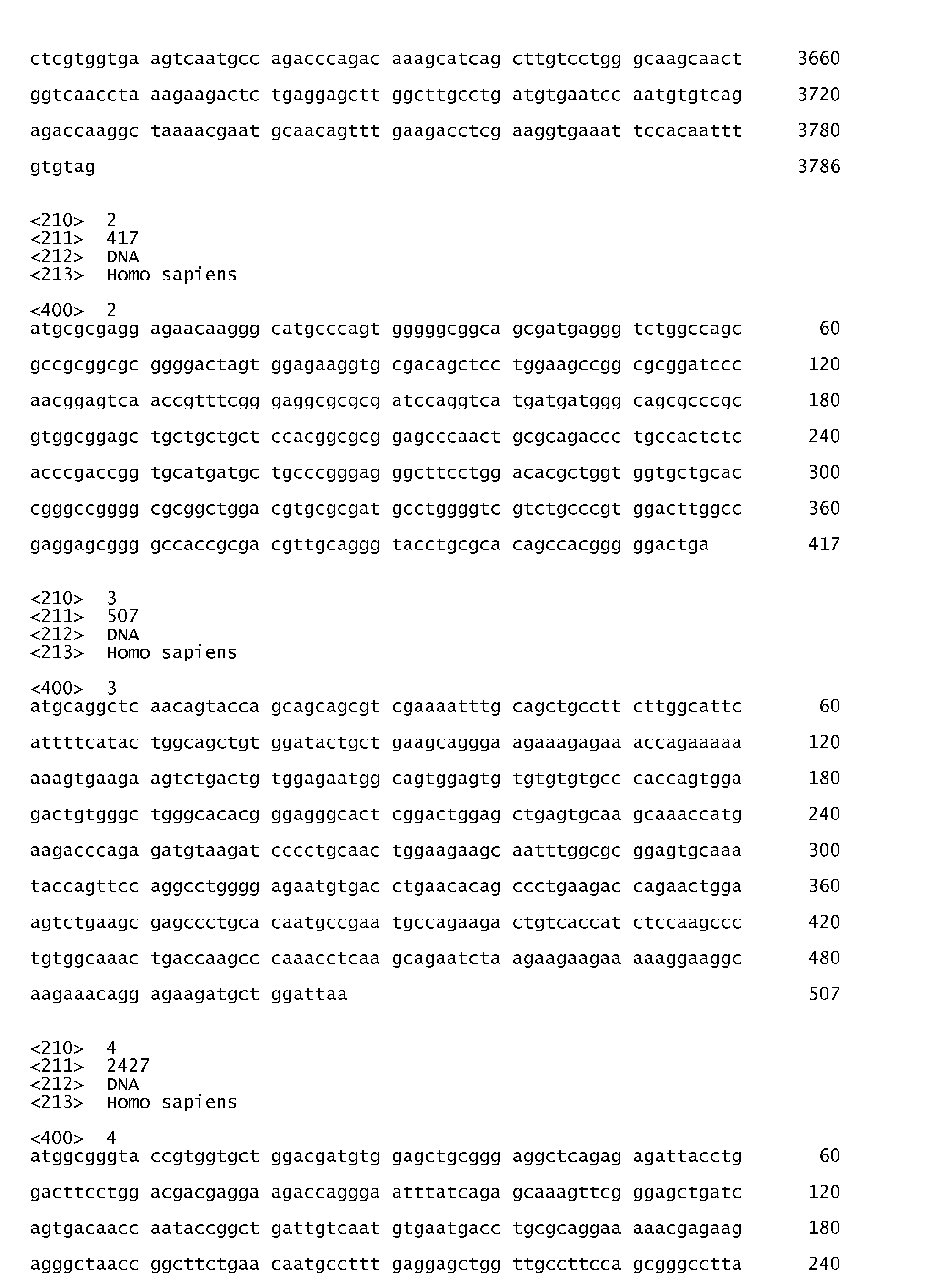


Claims (13)
- A method for determining the replicative senescence status of a cell comprising the steps ofa) determining the amount of at least one of the gene products selected from the group consisting of PARG-1, CDKN2B, PTN and MCM3 in a sample comprising said cell; andb) comparing each amount measured in step a) to a reference amount whereby the replicative senescence status is determined.
- The method of claim 1, wherein the cell has been freshly isolated from a donor.
- The method of claim 1, wherein the cell has been long term cultured.
- The method of any of claims 1 to 3, wherein the cell is a mesenchymal stromal cell.
- The method of any one of claims 1 to 4, wherein the amount of all 4 gene products is determined.
- A method for identifying a cell culture which is suitable for therapeutic uses comprising the steps ofa) determining the amount of at least one of the gene products selected from the group consisting of PARG-1, CDKN2B, PTN and MCM3 in a sample comprising said cell; andb) comparing each amount measured in step a) to a reference amount whereby the replicative senescence status is determined.
- The method of claim 6, wherein the cell culture comprises mesenchymal stromal cells.
- The method of claim 6 or 7, wherein the amounts of all 4 gene products are determined.
- Use of at least one gene selected from the group consisting of PARG-1, CDKN2B, PTN and MCM3 for the determination of the replicative senescence status of a cell comprised by a sample.
- A device for determining the replicative senescence status of a cell comprisinga) an analysis unit for determining the amount of at least one of the gene products selected from the group consisting of PARG-1, CDKN2B, PTN and MCM3 in a sample comprising said cell; andb) an evaluation unit for comparing each amount measured in step a) to a reference amount whereby the replicative senescence status is determined.
- A kit for determining the replicative senescence status of a cell comprisinga) an analysing unit for determining the amount of at least one of the gene products selected from the group consisting of PARG-1, CDKN2B, PTN and MCM3 in a sample comprising said cell; andb) an evaluation unit for comparing each amount measured in step a) to a reference amount whereby the replicative senescence status is determined.
- A device for identifying a cell culture which is suitable for therapeutic uses comprisinga) an analysing unit for determining the amount of at least one of the gene products selected from the group consisting of PARG-1, CDKN2B, PTN and MCM3 in a sample comprising said cell; andb) an evaluation unit for comparing each amount measured in step a) to a reference amount whereby the replicative senescence status is determined.
- A kit for identifying a cell culture which is suitable for therapeutic uses comprisinga) an analysing unit for determining the expression level of at least one of the genes selected from the group consisting of PARG-1, CDKN2B, PTN and MCM3 in a sample comprising said cell; andb) an evaluation unit for comparing each amount measured in step a) to a reference amount whereby the replicative senescence status is determined.
Priority Applications (2)
| Application Number | Priority Date | Filing Date | Title |
|---|---|---|---|
| EP09164153A EP2302381A1 (en) | 2009-06-30 | 2009-06-30 | Gene signature for replicative senescence in cell cultures |
| PCT/EP2010/059212 WO2011000833A1 (en) | 2009-06-30 | 2010-06-29 | Gene signature for replicative senescence in cell cultures |
Applications Claiming Priority (1)
| Application Number | Priority Date | Filing Date | Title |
|---|---|---|---|
| EP09164153A EP2302381A1 (en) | 2009-06-30 | 2009-06-30 | Gene signature for replicative senescence in cell cultures |
Publications (1)
| Publication Number | Publication Date |
|---|---|
| EP2302381A1 true EP2302381A1 (en) | 2011-03-30 |
Family
ID=40941545
Family Applications (1)
| Application Number | Title | Priority Date | Filing Date |
|---|---|---|---|
| EP09164153A Withdrawn EP2302381A1 (en) | 2009-06-30 | 2009-06-30 | Gene signature for replicative senescence in cell cultures |
Country Status (2)
| Country | Link |
|---|---|
| EP (1) | EP2302381A1 (en) |
| WO (1) | WO2011000833A1 (en) |
Citations (1)
| Publication number | Priority date | Publication date | Assignee | Title |
|---|---|---|---|---|
| WO2007023808A1 (en) * | 2005-08-23 | 2007-03-01 | Fancl Corporation | Skin aging marker and technique for use thereof |
-
2009
- 2009-06-30 EP EP09164153A patent/EP2302381A1/en not_active Withdrawn
-
2010
- 2010-06-29 WO PCT/EP2010/059212 patent/WO2011000833A1/en not_active Ceased
Patent Citations (1)
| Publication number | Priority date | Publication date | Assignee | Title |
|---|---|---|---|---|
| WO2007023808A1 (en) * | 2005-08-23 | 2007-03-01 | Fancl Corporation | Skin aging marker and technique for use thereof |
Non-Patent Citations (23)
| Title |
|---|
| "Current Protocols in Molecular Biology", 1989, JOHN WILEY & SONS, pages: 6.3.1 - 6.3.6 |
| "Essential Molecular Biology: A Practical Approach", 1991, IRL PRESS AT OXFORD UNIVERSITY PRESS |
| "Nucleic Acids Hybridization: A Practical Approach", 1985, IRL PRESS AT OXFORD UNIVERSITY PRESS |
| BAXTER ET AL.: "Study of telomere length reveals rapid aging of human marrow stromal cells following invitro expansion", STEM CELLS, vol. 22, 2004, pages 675 - 682 |
| BONAB ET AL.: "Aging of mesenchymal cells in vitro", BMC CELL BIOLOGY, vol. 7, 2006, pages 14 |
| CAMPISI J: "Replicative senescence: an old lives' tale?", CELL 23 FEB 1996, vol. 84, no. 4, 23 February 1996 (1996-02-23), pages 497 - 500, XP002541826, ISSN: 0092-8674 * |
| COLTER ET AL.: "Rapid expansion of recycling stem cells in cultures of plastic-adherent cells from human bone marrow", PROC. NATL. ACAD. SCI. USA, vol. 97, 2001, pages 3213 - 3218 |
| DIMRI G P ET AL: "A BIOMARKER THAT IDENTIFIES SENESCENT HUMAN CELLS IN CULTURE AND IN AGING SKIN IN VIVO", PROCEEDINGS OF THE NATIONAL ACADEMY OF SCIENCES OF USA, NATIONAL ACADEMY OF SCIENCE, WASHINGTON, DC, US, vol. 92, 1 September 1995 (1995-09-01), pages 9363 - 9367, XP009065201, ISSN: 0027-8424 * |
| DIMRI GP ET AL.: "A biomarker that identifies senescent human cells in culture and in aging skin in vivo", PROC NATL ACAD SCI U S A, vol. 92, 1995, pages 9363 - 9367 |
| FEHRER; LEPPERDINGER: "Mesenchymal stem cell aging", EXP. GERONTOL., vol. 40, 2005, pages 926 - 930 |
| HIGGINS ET AL., CABIOS, vol. 5, 1989, pages 151 - 153 |
| HO ET AL.: "Heterogeneity of mesenchymal stromal cell preparations", CYTOTHERAPY, vol. 10, 2008, pages 320 - 330 |
| J. MOL. BIOL., vol. 48, 1970, pages 443 - 453 |
| J. MOL. EVOLUTION., vol. 25, 1987, pages 351 - 360 |
| REINISCH ET AL.: "Humanized system to propagate cord blood- derived multipotent mesnchymal stromal cells for clinical application", REGEN. MED., vol. 2, 2007, pages 371 - 382 |
| RIPPERGER TIM ET AL: "Promoter methylation of PARG1, a novel candidate tumor suppressor gene in mantle-cell lymphomas.", HAEMATOLOGICA APR 2007, vol. 92, no. 4, April 2007 (2007-04-01), pages 460 - 468, XP002541825, ISSN: 1592-8721 * |
| SAMBROOK ET AL.: "Molecular Cloning", 1989, COLD SPRING HARBOR LABORATORY |
| SMITH; PEREIRA-SMITH: "Replicative senescence: Implications for in vivo aging and tumor suppression", SCIENCE, vol. 273, 1996, pages 63 - 67 |
| SMITH; WATERMAN, ADV. APPL. MATH., vol. 2, 1981, pages 482 - 489 |
| STENDERUP ET AL.: "Aging is associated with decreased maximal lifespan and accelerated senescence of bone marrow stromal cells", BONE, vol. 33, 2003, pages 919 - 926 |
| WAGNER ET AL.: "Replicative senescence of mesenchymal stem cells - a continuous and organized process", PLOS ONE, vol. 5, 2008, pages E2213 |
| WAGNER; HO: "Mesenchymal stem cell preparations - comparing apples and oranges", STEM CELL REVIEWS AND REPORTS, vol. 3, 2007, pages 239 - 248 |
| WALENDA ET AL.: "Co-culture with mesenchymal stromal cells increases proliferation and maintenance of hematopoietic progenitor cells", J CELL MOL MED, 2009 |
Also Published As
| Publication number | Publication date |
|---|---|
| WO2011000833A1 (en) | 2011-01-06 |
Similar Documents
| Publication | Publication Date | Title |
|---|---|---|
| Mitsui et al. | Combined use of laser capture microdissection and cDNA microarray analysis identifies locally expressed disease-related genes in focal regions of psoriasis vulgaris skin lesions | |
| Clark et al. | Concise review: MicroRNA function in multipotent mesenchymal stromal cells | |
| Orciani et al. | T helper (Th) 1, Th17 and Th2 imbalance in mesenchymal stem cells of adult patients with atopic dermatitis: at the origin of the problem | |
| Mohr et al. | Microdissection, mRNA amplification and microarray: a study of pleural mesothelial and malignant mesothelioma cells | |
| Sathiyanathan et al. | A genomic biomarker that identifies human bone marrow-derived mesenchymal stem cells with high scalability | |
| JPWO2014003053A1 (en) | Pancreatic cancer detection method and detection kit | |
| Yoo et al. | Expression profiles of subtracted mRNAs during cellular senescence in human mesenchymal stem cells derived from bone marrow | |
| JP2013500474A (en) | Methods of detecting / enriching hairy dermal cells, cells in methods of treating hair loss and uses thereof | |
| Jarczak et al. | scRNA-seq revealed transcriptional signatures of human umbilical cord primitive stem cells and their germ lineage origin regulated by imprinted genes | |
| US20100291580A1 (en) | Biomarkers for trichogenicity | |
| US10294527B2 (en) | Epigenetic marker for the identification of natural killer cells | |
| EP2460876A2 (en) | Method for identifying senescent mesenchymal stem cells | |
| Yang et al. | Plasminogen activator system in osteoclasts | |
| Khadka et al. | Mediator kinase inhibition drives myometrial stem cell differentiation and the uterine fibroid phenotype through super-enhancer reprogramming | |
| de Souza et al. | Investigation of IGF2, Hedgehog and fusion gene expression profiles in pediatric sarcomas | |
| WO2011000833A1 (en) | Gene signature for replicative senescence in cell cultures | |
| CN105264089B (en) | For detecting proliferation and the marker for the treatment of ability of medicine and application thereof that are incubated at the fat-derived stem cells of the culture medium containing EGF or BFGF | |
| KR102293079B1 (en) | A use of WNT16 marker for predicting osteogenic differentiation of Tonsil-derived mesenchymal stem cells | |
| US20160145686A1 (en) | Biomarker for senescence and use thereof | |
| WO2011101550A1 (en) | Method of detecting the differentiation status of a stem cell population | |
| Thum et al. | Microarray-based gene expression profiling to elucidate cellular responses to nitric oxide—A review from an analytical and biomedical point of view | |
| US11497817B2 (en) | Senile dementia treatment formulation and application thereof | |
| US20240247320A1 (en) | Methods for identifying critically short telomeres | |
| Kumar et al. | OCT4A transcript level correlates with proliferation potential of human mesenchymal stem cells | |
| Pearce | Investigation of fibulin-2 in pulmonary fibrosis |
Legal Events
| Date | Code | Title | Description |
|---|---|---|---|
| PUAI | Public reference made under article 153(3) epc to a published international application that has entered the european phase |
Free format text: ORIGINAL CODE: 0009012 |
|
| AK | Designated contracting states |
Kind code of ref document: A1 Designated state(s): AT BE BG CH CY CZ DE DK EE ES FI FR GB GR HR HU IE IS IT LI LT LU LV MC MK MT NL NO PL PT RO SE SI SK TR |
|
| STAA | Information on the status of an ep patent application or granted ep patent |
Free format text: STATUS: THE APPLICATION IS DEEMED TO BE WITHDRAWN |
|
| 18D | Application deemed to be withdrawn |
Effective date: 20111001 |