EP2294086B1 - Anti-inflammatory agents - Google Patents
Anti-inflammatory agents Download PDFInfo
- Publication number
- EP2294086B1 EP2294086B1 EP09757582.3A EP09757582A EP2294086B1 EP 2294086 B1 EP2294086 B1 EP 2294086B1 EP 09757582 A EP09757582 A EP 09757582A EP 2294086 B1 EP2294086 B1 EP 2294086B1
- Authority
- EP
- European Patent Office
- Prior art keywords
- antibody
- seq
- antibodies
- deposited
- accession number
- Prior art date
- Legal status (The legal status is an assumption and is not a legal conclusion. Google has not performed a legal analysis and makes no representation as to the accuracy of the status listed.)
- Active
Links
Images


Classifications
-
- C—CHEMISTRY; METALLURGY
- C07—ORGANIC CHEMISTRY
- C07K—PEPTIDES
- C07K16/00—Immunoglobulins [IGs], e.g. monoclonal or polyclonal antibodies
- C07K16/40—Immunoglobulins [IGs], e.g. monoclonal or polyclonal antibodies against enzymes
-
- A—HUMAN NECESSITIES
- A61—MEDICAL OR VETERINARY SCIENCE; HYGIENE
- A61K—PREPARATIONS FOR MEDICAL, DENTAL OR TOILETRY PURPOSES
- A61K39/00—Medicinal preparations containing antigens or antibodies
- A61K39/395—Antibodies; Immunoglobulins; Immune serum, e.g. antilymphocytic serum
- A61K39/39533—Antibodies; Immunoglobulins; Immune serum, e.g. antilymphocytic serum against materials from animals
- A61K39/3955—Antibodies; Immunoglobulins; Immune serum, e.g. antilymphocytic serum against materials from animals against proteinaceous materials, e.g. enzymes, hormones, lymphokines
-
- A—HUMAN NECESSITIES
- A61—MEDICAL OR VETERINARY SCIENCE; HYGIENE
- A61K—PREPARATIONS FOR MEDICAL, DENTAL OR TOILETRY PURPOSES
- A61K45/00—Medicinal preparations containing active ingredients not provided for in groups A61K31/00 - A61K41/00
- A61K45/06—Mixtures of active ingredients without chemical characterisation, e.g. antiphlogistics and cardiaca
-
- A—HUMAN NECESSITIES
- A61—MEDICAL OR VETERINARY SCIENCE; HYGIENE
- A61P—SPECIFIC THERAPEUTIC ACTIVITY OF CHEMICAL COMPOUNDS OR MEDICINAL PREPARATIONS
- A61P1/00—Drugs for disorders of the alimentary tract or the digestive system
- A61P1/04—Drugs for disorders of the alimentary tract or the digestive system for ulcers, gastritis or reflux esophagitis, e.g. antacids, inhibitors of acid secretion, mucosal protectants
-
- A—HUMAN NECESSITIES
- A61—MEDICAL OR VETERINARY SCIENCE; HYGIENE
- A61P—SPECIFIC THERAPEUTIC ACTIVITY OF CHEMICAL COMPOUNDS OR MEDICINAL PREPARATIONS
- A61P1/00—Drugs for disorders of the alimentary tract or the digestive system
- A61P1/16—Drugs for disorders of the alimentary tract or the digestive system for liver or gallbladder disorders, e.g. hepatoprotective agents, cholagogues, litholytics
-
- A—HUMAN NECESSITIES
- A61—MEDICAL OR VETERINARY SCIENCE; HYGIENE
- A61P—SPECIFIC THERAPEUTIC ACTIVITY OF CHEMICAL COMPOUNDS OR MEDICINAL PREPARATIONS
- A61P17/00—Drugs for dermatological disorders
- A61P17/06—Antipsoriatics
-
- A—HUMAN NECESSITIES
- A61—MEDICAL OR VETERINARY SCIENCE; HYGIENE
- A61P—SPECIFIC THERAPEUTIC ACTIVITY OF CHEMICAL COMPOUNDS OR MEDICINAL PREPARATIONS
- A61P19/00—Drugs for skeletal disorders
- A61P19/02—Drugs for skeletal disorders for joint disorders, e.g. arthritis, arthrosis
-
- A—HUMAN NECESSITIES
- A61—MEDICAL OR VETERINARY SCIENCE; HYGIENE
- A61P—SPECIFIC THERAPEUTIC ACTIVITY OF CHEMICAL COMPOUNDS OR MEDICINAL PREPARATIONS
- A61P19/00—Drugs for skeletal disorders
- A61P19/08—Drugs for skeletal disorders for bone diseases, e.g. rachitism, Paget's disease
-
- A—HUMAN NECESSITIES
- A61—MEDICAL OR VETERINARY SCIENCE; HYGIENE
- A61P—SPECIFIC THERAPEUTIC ACTIVITY OF CHEMICAL COMPOUNDS OR MEDICINAL PREPARATIONS
- A61P25/00—Drugs for disorders of the nervous system
-
- A—HUMAN NECESSITIES
- A61—MEDICAL OR VETERINARY SCIENCE; HYGIENE
- A61P—SPECIFIC THERAPEUTIC ACTIVITY OF CHEMICAL COMPOUNDS OR MEDICINAL PREPARATIONS
- A61P25/00—Drugs for disorders of the nervous system
- A61P25/14—Drugs for disorders of the nervous system for treating abnormal movements, e.g. chorea, dyskinesia
- A61P25/16—Anti-Parkinson drugs
-
- A—HUMAN NECESSITIES
- A61—MEDICAL OR VETERINARY SCIENCE; HYGIENE
- A61P—SPECIFIC THERAPEUTIC ACTIVITY OF CHEMICAL COMPOUNDS OR MEDICINAL PREPARATIONS
- A61P25/00—Drugs for disorders of the nervous system
- A61P25/28—Drugs for disorders of the nervous system for treating neurodegenerative disorders of the central nervous system, e.g. nootropic agents, cognition enhancers, drugs for treating Alzheimer's disease or other forms of dementia
-
- A—HUMAN NECESSITIES
- A61—MEDICAL OR VETERINARY SCIENCE; HYGIENE
- A61P—SPECIFIC THERAPEUTIC ACTIVITY OF CHEMICAL COMPOUNDS OR MEDICINAL PREPARATIONS
- A61P29/00—Non-central analgesic, antipyretic or antiinflammatory agents, e.g. antirheumatic agents; Non-steroidal antiinflammatory drugs [NSAID]
- A61P29/02—Non-central analgesic, antipyretic or antiinflammatory agents, e.g. antirheumatic agents; Non-steroidal antiinflammatory drugs [NSAID] without antiinflammatory effect
-
- A—HUMAN NECESSITIES
- A61—MEDICAL OR VETERINARY SCIENCE; HYGIENE
- A61P—SPECIFIC THERAPEUTIC ACTIVITY OF CHEMICAL COMPOUNDS OR MEDICINAL PREPARATIONS
- A61P37/00—Drugs for immunological or allergic disorders
- A61P37/02—Immunomodulators
- A61P37/06—Immunosuppressants, e.g. drugs for graft rejection
-
- C—CHEMISTRY; METALLURGY
- C07—ORGANIC CHEMISTRY
- C07K—PEPTIDES
- C07K14/00—Peptides having more than 20 amino acids; Gastrins; Somatostatins; Melanotropins; Derivatives thereof
- C07K14/435—Peptides having more than 20 amino acids; Gastrins; Somatostatins; Melanotropins; Derivatives thereof from animals; from humans
- C07K14/46—Peptides having more than 20 amino acids; Gastrins; Somatostatins; Melanotropins; Derivatives thereof from animals; from humans from vertebrates
- C07K14/47—Peptides having more than 20 amino acids; Gastrins; Somatostatins; Melanotropins; Derivatives thereof from animals; from humans from vertebrates from mammals
- C07K14/4701—Peptides having more than 20 amino acids; Gastrins; Somatostatins; Melanotropins; Derivatives thereof from animals; from humans from vertebrates from mammals not used
- C07K14/4713—Autoimmune diseases, e.g. Insulin-dependent diabetes mellitus, multiple sclerosis, rheumathoid arthritis, systemic lupus erythematosus; Autoantigens
-
- C—CHEMISTRY; METALLURGY
- C07—ORGANIC CHEMISTRY
- C07K—PEPTIDES
- C07K16/00—Immunoglobulins [IGs], e.g. monoclonal or polyclonal antibodies
- C07K16/18—Immunoglobulins [IGs], e.g. monoclonal or polyclonal antibodies against material from animals or humans
-
- C—CHEMISTRY; METALLURGY
- C07—ORGANIC CHEMISTRY
- C07K—PEPTIDES
- C07K16/00—Immunoglobulins [IGs], e.g. monoclonal or polyclonal antibodies
- C07K16/44—Immunoglobulins [IGs], e.g. monoclonal or polyclonal antibodies against material not provided for elsewhere, e.g. haptens, metals, DNA, RNA, amino acids
-
- A—HUMAN NECESSITIES
- A61—MEDICAL OR VETERINARY SCIENCE; HYGIENE
- A61K—PREPARATIONS FOR MEDICAL, DENTAL OR TOILETRY PURPOSES
- A61K39/00—Medicinal preparations containing antigens or antibodies
- A61K2039/505—Medicinal preparations containing antigens or antibodies comprising antibodies
-
- C—CHEMISTRY; METALLURGY
- C07—ORGANIC CHEMISTRY
- C07K—PEPTIDES
- C07K2317/00—Immunoglobulins specific features
- C07K2317/20—Immunoglobulins specific features characterized by taxonomic origin
- C07K2317/21—Immunoglobulins specific features characterized by taxonomic origin from primates, e.g. man
-
- C—CHEMISTRY; METALLURGY
- C07—ORGANIC CHEMISTRY
- C07K—PEPTIDES
- C07K2317/00—Immunoglobulins specific features
- C07K2317/30—Immunoglobulins specific features characterized by aspects of specificity or valency
- C07K2317/34—Identification of a linear epitope shorter than 20 amino acid residues or of a conformational epitope defined by amino acid residues
-
- C—CHEMISTRY; METALLURGY
- C07—ORGANIC CHEMISTRY
- C07K—PEPTIDES
- C07K2317/00—Immunoglobulins specific features
- C07K2317/50—Immunoglobulins specific features characterized by immunoglobulin fragments
- C07K2317/56—Immunoglobulins specific features characterized by immunoglobulin fragments variable (Fv) region, i.e. VH and/or VL
-
- C—CHEMISTRY; METALLURGY
- C07—ORGANIC CHEMISTRY
- C07K—PEPTIDES
- C07K2317/00—Immunoglobulins specific features
- C07K2317/60—Immunoglobulins specific features characterized by non-natural combinations of immunoglobulin fragments
- C07K2317/62—Immunoglobulins specific features characterized by non-natural combinations of immunoglobulin fragments comprising only variable region components
- C07K2317/622—Single chain antibody (scFv)
-
- C—CHEMISTRY; METALLURGY
- C07—ORGANIC CHEMISTRY
- C07K—PEPTIDES
- C07K2317/00—Immunoglobulins specific features
- C07K2317/90—Immunoglobulins specific features characterized by (pharmaco)kinetic aspects or by stability of the immunoglobulin
- C07K2317/92—Affinity (KD), association rate (Ka), dissociation rate (Kd) or EC50 value
Definitions
- This disclosure is in the field of treating or preventing inflammation in humans and animals and relates to pharmaceutical compositions and methods for treating or preventing various inflammatory conditions.
- the disclosure relates to compositions and methods for preventing or treating inflammatory conditions such as citrulline related diseases, preferably inflammatory diseases.
- the disclosure provides specific binding molecules directed against citrulline-containing epitopes for use in the therapy and prevention of inflammatory conditions.
- Inflammatory conditions whether of a chronic or acute nature, represent a substantial problem in the healthcare industry.
- chronic inflammation is considered to be inflammation of a prolonged duration (weeks or months) in which active inflammation, tissue destruction and attempts at healing are proceeding simultaneously ( Robbins Pathological Basis of Disease by R. S. Cotran, V. Kumar, and S. L. Robbins, W. B. Saunders Co., p. 75, 1989 ).
- chronic inflammation can follow an acute inflammatory episode, it can also begin as an insidious process that progresses with time, for example, as a result of a persistent infection (e.g., tuberculosis, syphilis, fungal infection) that causes a delayed hypersensitivity reaction, prolonged exposure to endogenous (e.g., elevated plasma lipids) or exogenous (e.g., silica, asbestos, cigarette tar, surgical sutures) toxins, or autoimmune reactions against the body's own tissues (e.g., rheumatoid arthritis, systemic lupus erythematosus, multiple sclerosis, psoriasis).
- a persistent infection e.g., tuberculosis, syphilis, fungal infection
- endogenous e.g., elevated plasma lipids
- exogenous e.g., silica, asbestos, cigarette tar, surgical sutures
- autoimmune reactions against the body's own tissues
- rheumatoid arthritis is a multisystem chronic, relapsing, inflammatory disease affecting 1 to 2% of the world's population.
- RA is basically a severe form of chronic synovitis that sometimes leads to destruction and ankylosis of affected joints ( Robbins Pathological Basis of Disease, by R. S. Cotran, V. Kumar, and S. L. Robbins, W.B. Saunders Co., 1989 ).
- the disease is characterized by a marked thickening of the synovial membrane which forms villous projections that extend into the joint space, multilayering of the synoviocyte lining (synoviocyte proliferation), infiltration of the synovial membrane with white blood cells (macrophages, lymphocytes, plasma cells, and lymphoid follicles; called an "inflammatory synovitis"), and deposition of fibrin with cellular necrosis within the synovium.
- the tissue formed as a result of this process is called pannus and eventually the pannus grows to fill the joint space.
- the pannus develops an extensive network of new blood vessels through the process of angiogenesis, which is essential to the evolution of the synovitis.
- pannus tissue Release of digestive enzymes (matrix metalloproteinases (e.g., collagenase, stromelysin)), and other mediators of the inflammatory process (e.g., hydrogen peroxide, superoxides, lysosomal enzymes, and products of arachadonic acid metabolism), from the cells of the pannus tissue leads to the progressive destruction of the cartilage tissue.
- the pannus invades the articular cartilage leading to erosions and fragmentation of the cartilage tissue. Eventually there is erosion of the subchondral bone with fibrous ankylosis, and ultimately bony ankylosis, of the involved joint.
- RA is an autoimmune disease and that many different arthrogenic stimuli activate the immune response in an immunogenetically susceptible host.
- exogenous infectious agents Epstein-Barr virus, rubella virus, cytomegalovirus, herpes virus, human T-cell lymphotropic virus, Mycoplasma, and others
- endogenous proteins such as collagen, proteoglycans, altered immunoglobulins and post-translationally modified proteins like citrullinated proteins have been implicated as a causative agent that triggers an inappropriate host immune response.
- autoimmunity plays a role in the progression of the disease.
- the relevant antigen is ingested by antigen-presenting cells (macrophages or dendritic cells in the synovial membrane), processed, and presented to T lymphocytes.
- the T cells initiate a cellular immune response and stimulate the proliferation and differentiation of B lymphocytes into plasma cells.
- the end result is the production of an excessive inappropriate immune response directed against the host tissues (e.g., antibodies directed against type II collagen, antibodies directed against the Fc portion of autologous IgG (called "Rheumatoid Factor”)), and antibodies directed against different citrullinated epitopes (anti-CCP).
- This further amplifies the immune response and hastens the destruction of the cartilage tissue. Once this cascade is initiated numerous mediators of cartilage destruction are responsible for the progression of rheumatoid arthritis.
- anti-CCP antibodies have been demonstrated to be highly specific for RA. Recent evidence shows that each individual that is seropositive for these antibodies either already has RA or will develop this disease in the future. The presence of anti-CCP antibodies (especially when high titers are present) is predictive of erosive disease outcome ( Nijenhuis et al., Clin. Chim. Acta, vol 350, 17-34, 2004 ). Furthermore, it has been demonstrated that anti-CCP antibodies are produced locally at the site of inflammation.
- anti-CCP producing plasma cells in the synovium is indicative of an antigen-driven maturation of CCP-specific B cells at the site of inflammation.
- anti-CCP antibodies Once anti-CCP antibodies are produced, the formation of immune complexes with citrullinated proteins in the synovia may trigger the progression of the inflammatory process.
- Patent application WO 2004/078098 discloses antibodies specific for citrullinated peptide/MHC class II complexes to inhibit T cell activation, the concept being that the antibody does not bind to the separate peptide of MHC class II molecule, but only to the complex of peptide and MHC class II molecule.
- the disclosure provides a binding molecule specifically reactive with a citrullinated epitope on p15 and/or p17 for use in the treatment or prevention of inflammatory diseases.
- the disclosure also provides a method for treating or preventing an inflammatory disease, comprising the step of administering to a patient in need thereof a therapeutically effective amount of an anti-inflammatory composition comprising a binding molecule specifically reactive with a citrulline epitope on p15 and/or p17.
- compositions and methods of the present disclosure include pharmaceutically acceptable formulations of specific binding molecules reactive with citrulline residues.
- the binding molecules are specifically reactive with citrullinated epitopes on two polypeptides as identified herein, termed p 15 and p17.
- the disclosure provides a binding molecule specifically reactive with a citrullinated epitope on p15 and/or p17 for use in the treatment or prevention of inflammatory diseases.
- binding molecule is used herein to indicate a molecule, preferably a small molecule, capable of specific binding. Specific binding in this respect is intended to mean that the molecule is capable of binding to a selected target molecule whereas it will not bind to another non-related target molecule under the same conditions. For instance, a binding molecule is said to specifically bind to serum albumin when it binds to serum albumin and less or not at all to another or preferably any other protein found in serum.
- peptide or peptide-like molecule should be interpreted as structures that are capable of presenting the citrulline residue in the correct context for immunoreactivity with the specific binding molecules as described herein, preferably in the same context as it appears in the human or animal body, preferably in the context of a native polypeptide.
- the "specific binding molecule” may be a molecule, preferably a small molecule composed of DNA, RNA, peptide, protein domain, whole proteins, or combinations thereof or parts thereof, that are capable of specifically binding to a target compound.
- Preferred examples of specific binding molecules are peptides or antibodies or parts thereof, such as Single Chain Variable Fragments (scFvs), Fragment antigen binding regions (Fabs), single domains antibodies (sdabs), also known as VHH antibodies, nanobodies (Camelids derived single domain antibodies), or shark IgNAR derived single domain antibody fragments called VNAR, or other active components thereof, Anticalins, or aptamers (DNA or RNA).
- a specific binding molecule is a fusion protein comprising the antigen-binding domain of an antibody or an aptamer, such as an aptamer in the form of DNA or RNA.
- the specific binding molecule comprises antibodies, or derivatives thereof, such as antibody fragments, nanobodies, single domain antibodies, or active parts thereof. The disclosure therefore in particular relates to specific binding molecules as described above which are peptides or antibodies.
- Antibodies refers to a protein or polypeptide capable of specific binding to a target molecule often referred to as "antigen”.
- Antibodies also known as immunoglobulins
- immunoglobulins are gamma globulin proteins that are found in blood or other bodily fluids of vertebrates, and are used by the immune system to identify and neutralize foreign objects, such as bacteria and viruses.
- Antibodies are typically made of basic structural units - each with two large heavy chains and two small light chains - to form, for example, monomers with one unit, dimers with two units or pentamers with five units. Antibodies are produced by a kind of white blood cell called a B cell. There are several different types of antibody heavy chain, and several different kinds of antibodies, which are grouped into different isotypes based on which heavy chain they possess. Five different antibody isotypes are known in mammals which perform different roles, and help direct the appropriate immune response for each different type of foreign object they encounter. Some animal species such as Camelids (e.g. llamas) and sharks may have aberrant antibody structures.
- Camelids e.g. llamas
- sharks may have aberrant antibody structures.
- the large and diverse population of antibodies is generated by random combinations of a set of gene segments that encode different antigen binding sites (or paratopes), followed by random mutations in this area of the antibody gene, which create further diversity.
- Antibody genes also re-organize in a process called class switching that changes the base of the heavy chain to another, creating a different isotype of the antibody that retains the antigen specific variable region. This allows a single antibody to be used in several different isotypes by several different parts of the immune system.
- Antibody as used herein includes single chain antibodies, fragment antigen binding regions, recombinantly produced antibodies, monoclonal antibodies, single domain antibodies, and the like.
- an antibody or other specific binding molecule in the context of an antibody or other specific binding molecule is meant to refer to the part of the antibody or specific binding molecule that makes up the specific binding site of the antibody or specific binding molecule and may be interpreted as the part of an antibody or specific binding molecule that is still capable to react with the same epitope as the entire antibody or specific binding molecule.
- specific binding molecules and derivatives thereof such as antibodies, fusion proteins comprising a specific binding domain of an antibody, aptamers, antibody fragments, single domain antibody fragments, other proteinacous binding domains such as anticalins, and small molecules that specifically bind citrullinated epitopes are disclosed herein.
- Human antibodies or fragments thereof are preferred for the use as disclosed herein.
- IgG1 e.g., IgG1 ⁇
- antibodies having an IgG1 heavy chain and a lambda light chain are used.
- antibodies are also disclosed herein, including IgG2, IgG3, IgG4, IgM, IgA1, IgA2, IgAsec, IgD and IgE in combination with a kappa or lambda light chain.
- all animal-derived antibodies of various isotypes can be used in the methods as disclosed herein.
- the antibodies can be full-size antibodies or antigen-binding fragments of antibodies, including Fab, F(ab')2, single chain Fv fragments, or single domain VHH, VH or VL single domains.
- Specific binding molecules reactive with a citrullinated epitope are to be interpreted as specific binding molecules that specifically react with a citrulline residue in the context of a larger structure such as a peptide or a peptide nucleic acid or an aptamer or a peptide mimicking structure.
- Citrulline is an amino acid that is not incorporated into proteins during translation, however, it can be generated by post-translational modification of an arginine residue by peptidylarginine deiminase (PAD).
- PAD peptidylarginine deiminase
- Citrullination is the posttranslational conversion of arginine residues to citrulline residues, which is catalyzed by peptidylarginine deiminase (PAD).
- Peptidylarginine deiminase (PAD; EC 3.5.3.15) enzymes catalyse the conversion of arginine residues to citrulline residues in proteins.
- PAD1 - PAD6 PAD1 - PAD6
- 'PAD4' and 'PAD5' PAD isotypes
- All these enzymes rely strongly on the presence of Ca2+ for activity and are unable to convert free L-arginine into free L-citrulline.
- Free L-arginine can be converted to free L-citrulline by nitric oxide synthase (EC 1.14.13.39) in eukaryotes or by arginine deiminase (EC 3.5.3.6) in bacteria.
- nitric oxide synthase EC 1.14.13.39
- arginine deiminase EC 3.5.3.6
- PAD1 (synonyms: PAD I, PAD type I) is involved in the citrullination of keratin filaments during the final stages of keratinocyte differentiation, which is important for the reorganization of the cornified envelope.
- PAD3 sekunder-derived protein
- THH trichohyalin
- PAD6 The most recently identified PAD isotype, PAD6 (synonym: ePAD), was found in cytoplasmic sheets of mouse oocytes, which play an important role in early embryogenesis. The expression of its human orthologue was found to be restricted to ovary, testis and peripheral blood leukocytes ( Chavanas et al., Gene vol 330; 19-27, 2004 ). Originally, this PAD isotype was designated ePAD, but based upon the systematic numbering of other PADs, this isotype was renamed PAD6 ( Vossenaar et al., Bioessays vol 25 1106-1118, 2003 ).
- PAD2 The most widely expressed isotype, PAD2 (synonyms PAD II, PAD type II, PAD-H19), is present in many different tissues, like skeletal muscle, brain, spleen, secretory glands and macrophages. Despite this broad expression pattern, only myelin basic protein (MBP) and vimentin have been identified as natural substrates. In multiple sclerosis (MS) patients develop an autoimmune response against MBP. MBP is an abundant protein of the myelin sheath, and its citrullination occurs during development of the central nervous system.
- MBP myelin basic protein
- MS multiple sclerosis
- Substrates of PAD4 in the nucleus are histone core proteins (H2A, H3 and H4) and nucleophosmin/B23, a nucleolar protein that functions in ribosome assembly, nucleocytoplasmic transport and centrosome duplication.
- Such specific binding molecules were found to be particularly suited for the treatment or prevention of inflammatory diseases.
- Inflammatory Conditions or Inflammatory diseases as used herein refers to any of a number of conditions or diseases which are characterized by vascular changes: edema and infiltration of neutrophils (e.g., acute inflammatory reactions); infiltration of tissues by mononuclear cells; tissue destruction by inflammatory cells, connective tissue cells and their cellular products; and attempts at repair by connective tissue replacement (e.g., chronic inflammatory reactions).
- neutrophils e.g., acute inflammatory reactions
- infiltration of tissues by mononuclear cells tissue destruction by inflammatory cells, connective tissue cells and their cellular products
- attempts at repair by connective tissue replacement e.g., chronic inflammatory reactions.
- Citrulline related inflammatory diseases are herein defined as those diseases wherein citrullination plays a role in the pathogenesis of the disease. Whether or not citrullination plays a role in the pathogenesis of the disease, may be easily determined by a skilled person using routine tests available in the art. For example, these diseases may be characterized by the presence of an abnormal level of citrullinated proteins in affected or disease related tissue. Such may be accomplished by an immunological test such as a western blot or an ELISA wherein the affected tissue is used as an antigen and citrullination of that antigen may be detected with the aid of an anti-citrullin antibody as described herein.
- Proteomics applications such as mass spec. analysis to compare the level and type of citrullinaton in a diseased versus healthy tissue from affected patients.
- the disease may also be characterized by the presence of an immune response against citrulline containing peptides or proteins.
- This may be a humoral or a cellular immune response, such as a response mediated by T-cells or B-cells.
- Tests for detecting anti-citrulline antibodies have been described in the art and are commercially available.
- the disclosure provides a specific binding molecule for use in treating or preventing citrulline related inflammatory diseases.
- Such diseases are for instance inflammatory arthritis, including rheumatoid arthritis and osteoarthritis, multiple sclerosis, psoriatic arthritis, psoriasis, Alzheimer's disease, autoimmune hepatitis, juvenile idiopathic arthritis, spondyloarthropathy, Down's syndrome, multiple system atrophy, Parkinson's disease and Lewy body dementia.
- the disclosure in particular provides specific binding molecules for the treatment or prevention of autoimmune diseases, more in particular rheumatoid arthritis or osteoarthritis.
- MS Multiple sclerosis
- MBP is a highly cationic protein, capable of forming strong interactions with negatively charged phospholipids such as phosphatidylserine.
- arginines are citrullinated ( Wood et al., J Biol Chem, vol264, 5121-5127, 1989 , Wood et al., Ann Neurol, vol40, 18-24, 1996 ).
- the remaining MBP molecules do not contain citrulline.
- the proportion of MBP-cit6 is increased to 45% of total MBP.
- the decreased net positive charge of MBP-cit6 causes partial unfolding of MBP molecules and weakens their interaction with the phospholipids ( Boggs et al., J Neurosci Res, vol57, 529-535, 1999 , Pritzker et al., Biochemistry, vol39, 5374-5381, 2000 ).
- MBP-cit6 is capable of forming lipid complexes more rapidly than non-citrullinated MBP, the complexes that are formed are not as densely packed as those formed with non-citrullinated MBP ( Boggs et al, J Neurosci Res, vol57, 529-535, 1999 , Beniac et al, J Struct Biol, vol129, 80-95, 2000 ). MBP-cit6 is degraded 4 times more rapidly by cathepsin D than non-citrullinated MBP ( Cao et al., Biochemistry, vol38, 6157-6163, 1999 ).
- MBPcit18 In a rare case of acute fulminating MS (Marburg type), 80% of the MBP molecules are heavily citrullinated (MBPcit18) ( Wood et al., Ann Neurol, vol40, 18-24, 1996 ). The severely unfolded MBP-cit18 is degraded 45 times more rapidly by cathepsin D than normal MBP ( Cao et al., Biochemistry, vol38, 6157-6163, 1999 ). Clinical trials with paclitaxel, the active component of the anti-cancer drug taxol, are in progress ( O'Connor et al., Ann Neurol, vol46, 470, 1999 ).
- paclitaxel can inhibit citrullination of MBP by PAD2 in vitro ( Pritzker et al., Biochim Biophys Acta, vol1388, 154-160, 1998 ). Treatment with paclitaxel attenuates clinical symptoms and induces remyelination of damaged sheaths ( Moscarello et al., Mult Scler, vol8, 130138, 2002 ), underlining the possible importance of PAD as a candidate factor in demyelinating disease ( Moscarello et al., J Neurochem, vol81, 335-343, 2002 ).
- the composition as disclosed herein is in a form selected from the group consisting of an aqueous solution, a gel, a hydrogel, a film, a paste, a cream, a spray, an ointment, or a wrap.
- the above methods are used to administer the compositions described herein by a route selected from intra-articular, intraperitoneal, topical, rectal, intravenous, oral, ocular, or to the resection margin of tumors.
- a pharmaceutically acceptable carrier comprises at least one carrier selected from the group consisting of a co-solvent solution, liposomes, micelles, liquid crystals, nanocrystals, nanoparticles, emulsions, microparticles, microspheres, nanospheres, nanocapsules, polymers or polymeric carriers, surfactants, suspending agents, complexing agents such as cyclodextrins or adsorbing molecules such as albumin, surface active particles, and chelating agents.
- a polysaccharide comprises hyaluronic acid and derivatives thereof, dextran and derivatives thereof, cellulose and derivatives thereof (e.g., methylcellulose, hydroxy-propylcellulose, hydroxy-propylmethylcellulose, carboxymethylcellulose, cellulose acetate phthalate, cellulose acetate succinate, cellulose acetate butyrate, hydroxypropylmethyl-cellulose phthalate), chitosan and derivative thereof, [beta]-glucan, arabinoxylans, carrageenans, pectin, glycogen, fucoidan, chondrotin, dermatan, heparan, heparin, pentosan, keratan, alginate, cyclodextrins, and salts and derivatives, including esters and sulfates, thereof.
- cellulose and derivatives thereof e.g., methylcellulose, hydroxy-propylcellulose, hydroxy-propylmethylcellulose, carboxymethylcellulose, cellulose acetate
- the method as disclosed herein comprises delivering a composition as disclosed herein to a target site, most notably a synovial joint.
- the specific binding molecule competes with monoclonal antibodies RhmAb2.102, RmmAb1.102, RhmAb2.103, RmmAb1.103, RhmAb2.104, RmmAb1.104, RhmAb2.105 and RhmAb2.107 for binding to p15 and/or p17.
- the primary mRNA sequences of the variable regions of monoclonal antibodies RhmAb2.101, RhmAb2.103, and RhmAb2.104, RmmAb1.101, RmmAb1.103 and RmmAb1.104 have been published and were deposited in the EMBL database under accession numbers as shown in table 1.
- the primary sequence of the variable regions of monoclonal antibodies RhmAb2.102 , RmmAb1.102, RhmAb2.105 and RhmAb2.107 are disclosed herein in SEQ ID NO: 13, SEQ ID NO: 15, SEQ ID NO: 17, SEQ ID NO: 19, SEQ ID NO: 39, SEQ ID NO: 40, SEQ ID NO: 41 and SEQ ID NO: 42.
- the disclosure therefore provides a polypeptide comprising a variable heavy or light chain according to SEQ ID NO: 13, SEQ ID NO: 15, SEQ ID NO: 17, SEQ ID NO: 19, SEQ ID NO: 39, SEQ ID NO: 40, SEQ ID NO: 41 and SEQ ID NO: 42.
- the disclosure also provides a nucleic acid encoding a polypeptide according to SEQ ID NO: 13, SEQ ID NO: 15, SEQ ID NO: 17, SEQ ID NO: 19, SEQ ID NO: 39, SEQ ID NO: 40, SEQ ID NO: 41 and SEQ ID NO: 42.
- the specific binding molecule is an antibody selected from the group consisting of monoclonal antibodies RhmAb2.102, RmmAb1.102, RhmAb2.103, RmmAb1.103, RhmAb2.104, RmmAb1.104, RhmAb2.105 and RhmAb2.107.
- the specific binding molecule comprises VH and /or VL domains derived from an antibody selected from the group consisting of monoclonal antibodies RhmAb2.102, RmmAb1.102, RhmAb2.103, RmmAb1.103, RhmAb2.104, RmmAb1.104, RhmAb2.105 and RhmAb2.107.
- Specific binding molecules as disclosed herein may be generated essentially in two ways. First, they may be derived from the antibodies and its sequences as presented herein. Reactivity of the antibodies may even be improved by side-directed mutagenesis, chain shuffling, sexual PCR, or by other means for antibody derivation and optimisation known to the person skilled in the art. Alternatively, specific binding molecules, in particular antibodies may be obtained by panning with any of the specifically reactive epitopes as described herein, in particular PAD4 treated Histon 2A, peptide 1 (SEQ ID NO: 21) and other particularly reactive peptides.
- the term "derived" in this respect means that the essential residues responsible for the specific binding properties of the VH and /or VL domains in a particular antibody are identified and that these essential residues are then transferred into the context of another peptide.
- a person skilled in the art may use the sequences described herein to clone or generate cDNA or genomic sequences for instance such as described in the below examples. Cloning of these sequences in an appropriate eukaryotic expression vector, like pcDNA3 (In Vitrogen), or derivates thereof, and subsequent double transfection of mammalian cells (like CHO cells) with combinations of the appropriate light chain and heavy chain containing vectors will result in the expression and secretion of the listed antibodies RhmAb2.101, 2.102, 2.103, 2.104, 2.105 and/or 2.107, and RmmAb1.101, 1.102, 1.103, 1.104.
- Recombinant Human and Mouse monoclonal anti-citrulline antibodies were obtained as described in Examples 1 and 15. Monoclonal antibodies were obtained with a human IgG1 Fc region (RhmAb2.101, RhmAb2.102, RhmAb2.103, RhmAb2.104, RhmAb2.105 and RhmAb2.107) and a mouse IgG2a Fc region (RmmAb1.101, RmmAb1.102, RmmAb1.103 and RmmAb1.104).
- Mouse monoclonal anti-citrullin-peptide antibodies RmmAb13.101, RmmAb13.102 and RmmAb13.103 were obtained from a commercial source (ModiQuest Research BV Nijmegen, The Netherlands; Cat no, MQ13.101, MQ13.102and MQ13.103).
- Anti-citrullin antibodies were tested in an experimental model wherein inflammation is induced by injecting anti-collagen antibodies into a mouse. This model is known as collagen antibody induced arthritis (CAIA) ( Nandakumar and Holmdahl, J Immunol Methods, vol304, 126-136, 2005 ). Anti collagen antibodies were obtained from a commercial source (ModiQuest Research BV Nijmegen, The Netherlands; Cat no, MQ18.101).
- CAIA collagen antibody induced arthritis
- RhmAb2.104 and RhmAb2.105 reduced the clinical signs of arthritis in the experimental CAIA model, whereas RhmAb2.103, RhmAb2.102 and RhmAb2.107 even abolished the clinical signs of arthritis in the experimental CAIA model.
- RhmAb2.103 and RhmAb2.102 performed identical, only the results obtained with RhmAb2.102 are shown in Figures 1c and 1d . Results obtained with RhmAb2.105 and RhmAb2.107 are shown in Figure 10 .
- the human monoclonal antibody RhmAb2.101 had no effect at all on the clinical signs of arthritis at the dose applied.
- the commercially available antibody RhmAb2.201 is used as an irrelevant antibody control in this experiment (ModiQuest Research B.V., cat no: MQR2.201). This antibody does not recognize citrulinated epitopes.
- Figure 1e and 1f show an independent CAIA experiment in which the clinical dose for RhmAb2.102 has been evaluated.
- the lowest dose that gave maximum inhibition was 0,5 mg Ab/mouse which corresponds to 28 mg/kg at IP injection.
- RhmAb2.102 RhmAb2.103, RhmAb2.104, RmmAb1.102, RmmAb1.103, RmmAb1.104, RhmAb2.105 and RhmAb2.107 play an important role in the treatment or prevention of inflammatory diseases.
- WO 2004/078098 discloses antibodies specific for citrullinated peptide/MHC class II complexes to inhibit T cell activation. These antibodies do not bind to the separate peptide or MHC class II molecule but only to the complex of the peptide and the MHC class II molecule.
- the antibodies disclosed herein are different from the antibodies disclosed in WO 2004/078098 since they recognize the individual peptides and proteins as disclosed herein.
- the antibodies recognize a polypeptide in a western blot that could not be a complex between a peptide and an MHC class II molecule, since the complex between an MHC molecule and a citrullinated peptide would never survive the reducing conditions of an SDS gel used in the immunoblot procedure.
- the epitopes recognized by the binding molecules as disclosed herein are therefore different from the antibodies disclosed in WO 2004/078098 .
- the antibodies as disclosed herein are not specifically reactive with a complex of a peptide and an MHC class II molecule.
- RhmAb2.102, RmmAb1.102, RhmAb2.103 and RmmAb1.103 on both human PAD2 and PAD4 deiminated COS-1 lysates revealed prominent p15 and p17 protein bands. These bands were somewhat less prominent when immuno-precipitations were performed with RhmAb2.104 and RmmAb1.104.
- RhmAb2.102, RhmAb2.103 or RhmAb2.104 can be performed using either Western blots containing deiminated COS-1 lysates as described in example 6 or purified deiminated p15 and/or p17 proteins in Western blot or ELISA.
- Proteins p15 and p17 were further characterized by Matrix-assisted laser desorption/ionisation-time of flight mass spectrometry (MALDI-TOF MS) as detailed in example 7. Since the genome of the African Green Monkey is not completely sequenced we screened all other mammal genome databases for homology with the peptides found with MALDI-TOF MS. Proteins found with a high degree of homology turned out to be histones This is shown in Table 3 (Example 7).
- the disclosure therefore provides a binding molecule specifically reactive with a citrullinated epitope on histones for use in the treatment or prevention of inflammatory diseases.
- citrullinated histones may very well be produced in vitro. These citrullinated histones may then be used as a substrate in an enzymatic binding assay to screen and select for other specific binding molecules such as peptides and antibodies reactive with epitopes on citrullinated p15 and p17, i.e. histones.
- specific binding molecules are selected that compete with antibodies RhmAb2.102, RmmAb1.102, RhmAb2.103, RmmAb1.103, RhmAb2.104, RmmAb1.104 and RhmAb2.105 and RhmAb2.107 for binding to p15 and/or p17.
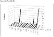
- RhmAb2.102 and RhmAb2.104 In order to further analyze which deiminated histone or histones are involved in the therapeutic action of RhmAb2.102 and RhmAb2.104, commercial available histones (H1, H2A, H2B, H3 and H4) were deiminated with human peptidylarginine deiminase (PAD, EC 3.5.3.15) enzymes (huPAD2 or huPAD4). Deiminated as well as non-deiminated histones were coated on 96-well ELISA plates and incubated with serial dilutions of RhmAb2.101, RhmAb2.102 and RhmAb2.104. The results are shown in table 6 and Figure 2 .
- PAD human peptidylarginine deiminase
- huPAD2 or huPAD4 enzymes
- RhmAb2.102 has higher affinity for H2A/p4 if compared to RhmAb2.104 ( Figure 2b and 2c ).
- RhmAb2.102 has higher affinity for H2A/p4 if compared to RhmAb2.104 ( Figure 2b and 2c ).
- a mimic is for instance a molecule with an acceptable level of equivalent activity, which, in this case, would include as being recognized with higher affinity by RhmAb2.102 than RhmAb2.104 and RhmAb2.101.)
- the disclosure therefore provides a specific binding molecule as described above, reactive with a citrullinated epitope on human PAD4 deiminated human histone 2A or histone 4, or on human PAD2 deiminated human histone H4 or histone H3.
- RhmAb2.102 was recognized by the therapeutic antibodies RhmAb2.102 and RhmAb2.104, but not by RhmAb2.101 (Table 4 and Figure 3a , 3b and 3c ). Again RhmAb2.102 showed higher affinity if compared to RhmAb2.104 ( Figure 3b and 3c ). The same holds true for the deiminated epitopes on peptides 4 and 6 (Table 4) since RhmAb2.102 shows higher affinity for these peptides than RhmAb2.104 and RhmAb2.101 ( Figures 2a , 2b and 2c ).
- Biotin labeled and citrullin containing fibrinogen and vimentin peptides were also tested for reactivity with the therapeutic antibodies.
- Peptides were coated on 96-well neutravidin-ELISA plates. Subsequently serial dilutions of RhmAb2.101, RhmAb2.102 and RhmAb2.104 were applied to the coated plates. The results are shown in Table 8 and Figure 4 .
- RhmAb2.101 RhmAb2.102
- RhmAb2.104 RhmAb2.101
- RhmAb2.102 showed higher affinity if compared to RhmAb2.104
- RhmAb2.104 performed slightly better than RhmAb2.101
- This antibody recognition pattern is similar to the pattern observed on Western blots loaded with huPAD2 and HuPAD4 deiminated human fibrinogen.
- RhmAb2.102 recognized the mouse vimentine peptide (example 10).
- the disclosure therefore provides a specific binding molecule as described above which is specifically reactive with an epitope on peptides msFib ⁇ or msVim (SEQ ID NO: 37 or SEQ ID NO: 38) and their use.
- citrullinated epitopes appear de novo in inflammated tissue.
- human monoclonal antibody 102 RhmAb2.102
- mice 3 mice per group
- mice Three days later mice received another i.p. injection containing 25ug LPS. Scoring has been performed as described above.
- a group of mice has been sacrificed, and paws were analyzed for citrulline presence by Western Blot analysis and Immunohistochemical techniques.
- IP Immuniprecipitations
- citrulline residues present on blot have been chemically modified according to Senshu et al. ( Senshu et al, Anal Biochem, vol 203, 94-100, 1992 ).
- the chemical modification can then be visualized using an antibody that recognizes the chemical modification of citrulline residues ( Senshu et al, Anal Biochem, vol 203, 94-100, 1992 ).
- Deiminated fibrinogen was used as a positive control in this experiment.
- An immunoprecipitation without extracts was used as a negative control in these experiments.
- mice subjected to CAIA have detectable citrulline levels in their inflamed joints.
- anti-citrulline antibodies were injected on day 3 after anti-collagen antibody injection, when inflammation in the paws of mice was still absent or very low. This prevented the occurrence of clinical symptoms and is therefore useful as a treatment of inflation, in particular a prophylactic treatment.
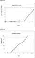
- RhmAb2.102 could also cure clinical symptoms once they had occurred. This was done by treating animals on day 7 after anti-collagen injection when mean arthritis scores of all 4 paws of all mice reached the arbitrary score of approximately 4. As is shown in figure 6A and 6B , RhmAb2.102 does not abolish the swelling observed, but rather stabilized the present inflammation/swelling. Animals were followed for 35 days after which inflammatory scores among placebo and RhmAb2.102 treated mice were equal ( Figure 6B and example 12). Figure 6A shows the Mean arthritis score of all paws of each group, while Figure 6B shows the mean arthritis score of the right hind paws of the animals that have been used for histological analysis at day 35.
- FIG. 7A shows that macroscopical inflammation in the right hind paws between experimental groups on day 35 of the experiment were similar. Most surprisingly however, all known parameters for joint erosion were decreased.
- D Inflammatory cell influx
- B Cartilage erosion
- E Cartilage PG depletion
- F Chondrocyte death
- C Bone erosion
- RhmAb2.102 has been injected i.v. in order to deliver the antibody rapidly to sites of inflammation.
- prophylactic treatment at day 3 and a non treated control group have been included.
- Experimental procedures have been performed as in Example 12 with the only difference of injections with 1 mg RhmAb2.102 per mouse on day 3, 5 and 6.
- RhmAb2.102 at day 3 inhibited the inflammatory response.
- Treating mice with i.v. injections of RhmAb2.102 on day 5, 6 or 7 stabilized the inflammation ( Figure 8 ) as also seen in Figure 6 . It is noteworthy that the signs of inflammation were not reduced whereas all parameters for joint erosion were decreased. This shows that joint erosion and inflammation are two separate entities that may be treated separately.
- Dexamethason is a general inflammatory inhibitor which needs to be administered on a daily basis. Once treatment is interrupted, the inflammation reoccurs.
- Experimental procedures have been performed as described in Example 12 with the difference that 1 mg RhmAb2.102 has been injected i.v. on day 5 ( Figure 9A ), day 6 ( Figure 9B ) and day 7 ( Figure 9C ) after anti-collagen antibody injection, simultaneously with i.p injections of Dexamethason (2mg/kg). Dexamethason was administered sequentially for 2 or 3 days until swelling in the paws disappeared. Additional groups of animals received i.p. injections of Dexamethason only. As shown in Figure 9 , inflammation reappeared in mice that did not receive RhmAb2.102.
- RhmAb2.105, RhmAb2.107 novel anti-citrulline antibodies that have shown cross-reactivity with RhmAb2.102 on its differentiating antigens from RhmAb2.101, have been tested for their anti inflammatory effect.
- RhmAb2.105, RhmAb2.107 and RhmAb2.102 positive control
- Figure 10 Experimental procedures have been performed as described in Example 12.
- Figure 10 shows the Mean arthritis score of all paws of each group.
- RhmAb2.102 showed the highest anti inflammatory effect. RhmAb2.107 performed almost as well as RhmAb2.102, and RhmAb2.105 showed an intermediate effect similar as previously observed for RhmAb2.104 ( Figure 1C) .
- RhmAb2.102 Additional deiminated proteins that preferentially bind to RhmAb2.102 have been identified by mass spectrometry analysis. Furthermore, deiminated proteins that preferentially bind to RhmAb2.102 and not, or with to a lesser extent to RhmAb2.101 have also been identified by additional mass spectrometry analysis.
- Human PAD4 deiminated Human Embryonic Kidney cell (HEK293) lysates have been immunoprecipitated with RhmAb2.101 or RhmAb2.102 (Example 13) and subjected to a high throughput nano-LC system coupled to an advanced, high-performance LTQ Fourier Transform Ion Cyclotron Resonance Mass spectrometer (nLC LTQ FTMS ULTRA) (Example 14).
- the disclosure also provides a binding molecule specifically reactive with any of the proteins or polypeptides as shown in table 7 for use in the prevention or treatment of an inflammatory disease.
- a binding molecule specifically reactive with an epitope on a molecule selected from the group consisting of p15, p17, more in particular a citrullinated epitope on human PAD4 deiminated human histone 2A, a citrullinated epitope on human PAD4 deiminated human histone 4, human PAD2 deiminated human histone H4, human PAD2 deiminated human histone H3, or a protein selected from the group consisting of the proteins of table 7 and even more in particular a peptide according to SEQ ID NO: 21, SEQ ID NO: 24, SEQ ID NO 26, SEQ ID NO: 37 and SEQ ID NO: 38 may be used in the treatment or prevention of inflammatory diseases as specified herein.
- Whether a given binding molecule is specifically reactive with the above mentioned molecules may easily be determined by analysis of the ability of the binding molecule to compete with an antibody selected from the group consisting of RhmAb2.102, RmmAb1.102, RhmAb2.103, RmmAb1.103, RhmAb2.104, RmmAb1.104, RhmAb2.105 and RhmAb2.107 for binding to an epitope on p15 or p17 or any of the citrullinated epitopes mentioned above.
- the disclosure also provides a method for the prevention or treatment of inflammatory diseases by eliciting an immune response in vivo wherein specific binding molecules are generated reactive with an epitope selected from the group consisting of a citrullinated epitope on p15, p17, a citrullinated epitope on human PAD4 deiminated human histone 2A, human PAD4 deiminated human histone 4, human PAD2 deiminated human histone H4, human PAD2 deiminated human histone H3, and a peptide according to SEQ ID NO: 21, SEQ ID NO: 24, SEQ ID NO 26, SEQ ID NO: 37 and SEQ ID NO: 38
- Vaccines or therapeutics as described herein may effectively comprise a citrullinated epitope specifically reactive with a binding molecule as described herein.
- the citrullinated epitope may be a citrullinated epitope on human PAD4 deiminated human histone 2A or histone 4, or on human PAD2 deiminated human histone H4, human histone H3, or a peptide selected from the group consisting of SEQ ID NO: 21, SEQ ID NO: 24, SEQ ID NO 26, SEQ ID NO: 37 and SEQ ID NO: 38.
- the disclosure also provides a method as described above wherein the inflammatory disease is selected from the group consisting of autoimmune diseases, arthritis, rheumatoid arthritis, osteoarthritis, multiple sclerosis, psoriatic arthritis, psoriasis, Alzheimer's disease, autoimmune hepatitis, juvenile idiopathic arthritis, spondyloarthropathy, Down's syndrome, multiple system atrophy, Parkinson's disease and Lewy body dementia.
- the inflammatory disease is selected from the group consisting of autoimmune diseases, arthritis, rheumatoid arthritis, osteoarthritis, multiple sclerosis, psoriatic arthritis, psoriasis, Alzheimer's disease, autoimmune hepatitis, juvenile idiopathic arthritis, spondyloarthropathy, Down's syndrome, multiple system atrophy, Parkinson's disease and Lewy body dementia.
- autoimmune diseases such as rheumatoid arthritis.
- a preferred specific binding molecule is an antibody.
- Example 1 Recombinant human and mouse monoclonal antibodies.
- Monoclonal antibodies against citrullinated antigens of patients with RA were initially selected by means of phage display, as described ( Raats et al., J Reumatology, vol30, 1696-711, 2003 ). Briefly, the autoantibody repertoires of three patients with RA were isolated from their B-cell repertoire, and used to generate antibody fragment libraries. These libraries were subjected to four rounds of affinity selection against citrullinated cyclic peptide CFC1-cyc as described in WO98/22503 . Antibody clones were selected based on their strong reactivity with CFC1-cyc and lack of reactivity with the non-citrullinated CFC0-cyc, ( WO98/22503 ).
- Antibody coding sequences described by Raats et al., (J Reumatology, vol30, 1696-711, 2003 ) were synthesized according to Stemmer et al (Gene, vol164, 49-53, 1995 ), and subsequently cloned into mammalian expression vectors coding for human and mouse antibody isotypes.
- Human antibodies were of the isotype IgG1 lambda and were named RhmAb2.101, RhmAb2.102, RhmAb2.103, and RhmAb2.104.
- Mouse antibodies were of the isotype IgG2a kappa and were named RmmAb1.101, RmmAb1.102, RmmAb1.103, and RmmAb1.104.
- RhmAb2.101 was synthesized according to the protocol of Stemmer et al., (Gene, vol164, 49-53, 1995 ) based on the sequence of clone Ra3 ( Raats et al., J Reumatology, vol30, 1696-711, 2003 ) and consists of a VH derived from germline family 3-21, combined with a VL derived from germline family ⁇ 1b.
- RhmAb2.103 is synthesized according to Stemmer et al (Gene, vol164, 49-53, 1995 ) based on the sequence of clone A2-2 ( Raats et al., J Reumatology, vol30, 1696-711, 2003 ), and consists of a VH derived from germline family 3-23, combined with a VL derived from germline family ⁇ 1a.
- RhmAb2.104 is synthesized according to Stemmer et al (Gene, vol164, 49-53, 1995 ), and consists of a VH derived from germline family 4-b, combined with a VL derived from germline family ⁇ 1c.
- the immunoglobulin light chain encoded by SEQ ID NO: 9 comprises a mouse leader globulin according to SEQ ID NO: 12, followed by the variable antibody light chain according to SEQ ID NO: 15 followed by the immunoglobulin human lambda constant domain according to SEQ ID NO: 16.
- RmmAb1.102 was synthesized according to Stemmer et al (Gene, vol164, 49-53, 1995 ) and comprises an immunoglobulin heavy chain encoded by SEQ ID NO: 10, combined with an immunoglobulin light chain encoded by SEQ ID NO: 11.
- the immunoglobulin heavy chain encoded by SEQ ID NO: 10 comprises a mouse leader globulin according to SEQ ID NO: 12, followed by the variable antibody heavy chain according to SEQ ID NO: 19, followed by the immunoglobulin constant domain mouse IgG2a according to SEQ ID NO: 20.
- the immunoglobulin light chain encoded by SEQ ID NO: 11 comprises a mouse leader globulin according to SEQ ID NO: 12, followed by the variable antibody light chain according to SEQ ID NO: 17 followed by the immunoglobulin mouse kappa constant domain according to SEQ ID NO: 18.
- variable domains VH and VL
- RhmAb2.101, RhmAb2.103, and RhmAb2.104, RmmAb1.101, RmmAb1.103 and RmmAb1.104 have been published and were deposited in the EMBL database under accession numbers as shown in table 1.
- Full size human and mouse antibody sequences were generated using identical leader and constant human or mouse domains as described for antibody RhmAb2.102 and RmmAb1.102.
- Control antibodies RmmAb13.101, RmmAb13.102 and RmmAb13,103 against citrullinated fibrinogen, and RhmAb2.201 against the apoptotic 40 kD cleavage product of the Human U1-70k protein, were commercially obtained from Modiquest Research BV, Schoutstraat 58, 6525 XV Nijmegen, The Netherlands (Cat no, MQ13.101, MQ13.102, MQ13.103,and MQR2.201).
- mice The commercially available collagen antibody induced arthritis (CAIA) mouse model from ModiQuest Research B.V. (cat no: MQ18.101) has been used according to manufacturers specifications to induce arthritis in mice (http://www.modiquestresearch.nl/shop/files/18.101-50MG%20_2007.08.22.pdf).
- CAIA collagen antibody induced arthritis
- mice On day 0 male DBA/J1 mice (5-6 mice /group) of the age of 8 weeks have been injected i.p. with a mix of 8 anti-collagen antibodies.
- mice used in figure 1a and 1b received 1,6mg anti-collagen antibody mix, whereas mice used in figure 1c-f received 2,4mg).
- mice received another i.p.
- RhmAb2.102, RhmAb2.103 and RhmAb2.104 Human monoclonal antibodies RhmAb2.102, RhmAb2.103 and RhmAb2.104, however, surprisingly reduced or even abolished the clinical signs of arthritis in the experimental CAIA model ( Figure 1c and 1d ).
- RhmAb2.102 and RhmAb2.103 reduced the signs of arthritis best, whereas RhmAb2.104 reduced the inflammation by approximately 50%.
- RhmAb2.101 had no effect at all at the dose tested.
- Example 3 Preparation of deiminated cell extract, SDS-page electrophoresis and western blotting.
- COS-1 cells (8 ⁇ 10 5 ) were transiently transfected with 2 ⁇ g huPAD2 or huPAD4 expression vector using the AMAXA nucleofection device (program D-005) together with the V-kit, and cells were seeded in 20ml medium in a T75.
- the cells were washed twice with PBS, trypsinized, spun down and resuspended in 15 ⁇ l ice cold lysis buffer (20mM Tris pH7.4, 10mM ⁇ -mercaptoethanol, 100mM NaCl, 10% glycerol, protease inhibitors).
- the cell samples were sonified 4 times for 15 seconds on ice.
- the lysate was centrifuged at 3.000 rpm for 5 minutes and the supernatant transferred to a clean tube.
- the cell lysate was deiminated for 30 minutes to 2 hours at 37°C by adding CaCl 2 and DTE at a final concentration of 10 and 5mM respectively. Deiminated cell lysates were stored at -20°C.
- Example 4 Therapeutic anti-citrulline antibodies recognize p15 and p17
- Blots as prepared in example 3 were cut in strips and blocked for 2 hours at RT with 5% (w/v) low fat dry milk in PBS-Tween (wash buffer) to block all non-specific sites. Blots were then washed 5 times 5 minutes with wash buffer and strips were incubated for an additional 1 hour at RT with 4 ml wash buffer containing 20ug anti-citrulline antibody. Thereafter, the strips were washed 5 times for 10 min with wash buffer, and incubated with a peroxydase-conjugated rabbit anti-human IgG (Dako) (1 hour at RT) in wash buffer (1:2000). Strips where then washed 3 times for 10min with wash buffer followed by a 2 times wash with PBS to wash away all unbound antibody.
- wash buffer 5% (w/v) low fat dry milk in PBS-Tween
- Immunoreactive bands were visualized using chemiluminescent substrate (PIERCE), and exposed to Kodak BioMax XAR autoradiography films (Eastman Kodak Company, Rochester, NY, USA).
- RhmAb2.102 RhmAb2.103
- RhmAb2.104 showed reactivity with a doublet of proteins with a molecular weight of approximately 15 and 17 kiloDalton.
- Example 6 Antibody competition assay for p15 and p17.
- Example 7 Mass-spectrometry analysis of p15 and p17.
- the bands at p15 and p17 of the SDS-page gels of example 3 were excised from the gel and analyzed by MALDI-TOF MS. Briefly, excised gel pieces were washed 2 times with 50 ⁇ l of 25 mM ammonium bicarbonate, and incubated 30 min for each washing step. A 15 min wash was repeated as above with the addition of 30% v/v acetonitrile. All liquid was removed and 25 ⁇ l of 25 mM ammonium bicarbonate + 25 ⁇ l of acetonitrile added and Incubated for 15 min. Again all liquid was removed and gels were incubated 30 min with 50 ⁇ l of acetonitrile.
- Peptides were extracted by incubating with 4 ⁇ l 50% acetonitrile/0.5% trifluoroacetic acid (TFA)/5 mM n-octyl-ß-D-glucopyranoside for 1 h at RT. Samples were sonicated for 2 min in a sonication water bath, the liquid transferred in a new tube and the extraction step was repeated. The sample was dried in a vacuum centrifuge and subjected to MALDI-TOF MS.
- TFA trifluoroacetic acid
- Example 8 Therapeutic anti-citrulline antibodies recognize H2A/p4.
- Human recombinant histones H1, H2A, H2B, H3 and H4 (100 ⁇ g) were incubated 3 hours with or without 53,4 mU huPAD2 or huPAD4 at 37°C. Deiminated as well as non-deiminated histones were coated on 96-well ELISA plates (0,3 ⁇ g /well) by overnight incubation at 4°C. Wells were washed 5 times with PBS-Tween20 (PBS-T) and blocked by a 1 hour incubation with PBS-T + 1% Bovine serum albumin (BSA) at room temperature (RT).
- PBS-T PBS-Tween20
- BSA Bovine serum albumin
- RhmAb2.101, RhmAb2.102 or RhmAb2.104 were incubated for 1 hour at RT with serial dilutions of RhmAb2.101, RhmAb2.102 or RhmAb2.104 in PBS-T + 1% BSA starting at a concentration of 10 ⁇ g/well.
- Wells were washed 5 times with PBS-T and incubated with rabbit-anti-human-HRP (1:2000) for 1 hour at RT followed by 5 washes with PBS-T and 3 wash steps with PBS.
- Wells incubated with RhmAb2.101 and RhmAb 2.104 were incubated 15min and wells incubated with RhmAb2.102 were incubated 10min with TMB substrate before stopping the reaction with 2M H 2 SO 4 .
- Optical density was measured by 450nm and is a measure for the affinity of the antibodies used.
- 96-well ELISA plates were coated with neutravidin (0,1 ⁇ g /well) by overnight incubation at 4°C. Wells were washed 5 times with PBS-Tween20 (PBS-T) and blocked by a 1 hour incubation with PBS-T + 1% Bovine serum albumin (BSA) at room temperature (RT). After 5 more washes with PBS-T, wells were incubated for 1 hour at RT with histone derived citrulline and biotin containing peptides (0,3 ⁇ g /well).
- PBS-T PBS-Tween20
- BSA Bovine serum albumin
- 100ug human plasma fibrinogen was disolved in 100 ⁇ l deimination buffer (PBS pH7.6, 10mM CaCl2, 5mM Dithiothreitol), and deiminated for 3 hours at 37 ⁇ C with 53.4 mU huPAD2 or huPAD4.
- 10x sample buffer (0.25M Tris pH6.8, 8% SDS, 35% glycerol, 2.5% ⁇ -mercaptoethanol, bromphenolblue
- was added and 7.5 ⁇ g deiminated or non-deiminated fibrinogen loaded in each lane of a SDS-PADE (12.5%) and separated , followed by electroblotting to Hybond C extra nitrocellulose membranes (Amersham Biosciences). Blotting and loading were checked by Ponceau S staining.
- Blots were blocked for 2 hours at RT with 5% (w/v) low fat dry milk in PBS-Tween (wash buffer) to block all non-specific sites. Blots were then washed 5 times 5 minutes with wash buffer and strips were incubated for an additional 1 hour at RT with 4 ml wash buffer containing 20ug anti-citrulline antibody. Thereafter, the strips were washed 5 times for 10 min with wash buffer, and incubated with a peroxydase-conjugated rabbit anti-human IgG (Dako) (1 hour at RT) in wash buffer (1:2000). Strips where then washed 3 times for 10min with wash buffer followed by a 2 washes with PBS to wash away all unbound antibody.
- wash buffer containing 20ug anti-citrulline antibody
- RhmAb2.102 showed higher reactivity with deiminated human plasma fibrinogen than RhmAb2.101. Again RhmAb2.102 showed higher affinity if compared to RhmAb2.104
- 96-well ELISA plates were coated with neutravidin (0,1 ⁇ g /well) by overnight incubation at 4 degrees C. Wells were washed 5 times with PBS-Tween20 (PBS-T) and blocked by a 1 hour incubation with PBS-T + 1% Bovine serum albumin (BSA) at room temperature (RT). After 5 more washes with PBS-T, wells were incubated for 1 hour at RT with fibrinogen and vimentin derived citrulline and biotin containing peptides (0,3 ⁇ g /well).
- PBS-T PBS-Tween20
- BSA Bovine serum albumin
- mice The commercially available collagen antibody induced arthritis (CAIA) mouse model from ModiQuest Research B.V. (cat no: MQ18.101) has been used according to manufacturers specifications to induce arthritis in mice (http://www.modiquestresearch.nl/shop/files/18.101-50MG%20_2007.08.22.pdf).
- CAIA collagen antibody induced arthritis
- mice On day 0 male DBA/J1 mice (5 mice/group) of the age of 8 weeks have been injected i.p. with a mix of 8 anti-collagen antibodies (2,8mg/mouse).
- mice received another i.p. injection containing 25ug LPS. LPS triggers the inflammation.
- the mean arthritis score was around 4 ( Figure 6A ) one group received an i.v. injection containing 1 mg RhmAb2.102, whether the other group received an i.v. injection containing placebo.
- PG proteoglycan
- Figure 7E To study proteoglycan (PG) depletion from the cartilage matrix ( Figure 7E ), sections were stained with safranin O (SO) followed by counterstaining with fast green. Depletion of PG was determined using an arbitrary scale of 0-3, ranging from normal, fully stained cartilage to destained cartilage, fully depleted of PGs. Chondrocyte death ( Figure 7F ) was scored on a scale of 0 - 3ranging from no loss of chondrocyte nuclei to complete empty cartilage surface. Cartilage and bone erosion ( Figure 7B & C ) were graded on a scale 0 - 3, ranging from no damage to complete loss of the cartilage or bone structure. Histopathological changes in the joint were scored on five semiserial sections of joint spaced 70 ⁇ m apart. Scoring was performed blind, without previous knowledge of the experimental conditions.
- RhmAb2.102 Although macroscopical inflammation in the right hind paws among groups was identical on day 35 ( Figure 6A and 7A ), a dramatic decrease is observed in the experimental group receiving RhmAb2.102 compared to the control group when looking at any of the following parameters for joint erosion: Inflammatory cell influx ( Figure 7D ), Cartilage erosion ( Figure 7B ), Cartilage PG depletion ( Figure 7E ), Chondrocyte death ( Figure 7F ) and Bone erosion ( Figure 7C ). This result strongly supports the therapeutic potential of RhmAb2.102.
- Example 13 Preparation of huPAD4 deiminated HEK293 extract and immunoprecipitation with RhmAb2.101 or RhmAb2.102
- HEK293 cells were harvested, washed once with PBS, spun down, and 5.105 cells cells resuspended in 15 ⁇ l ice cold lysis buffer (20mM Tris pH7.4, 10mM ⁇ -mercaptoethanol, 100mM NaCl, 10% glycerol, protease inhibitors).
- the cell samples were sonified 4 times for 15 seconds on ice.
- the lysate was centrifuged at 3.000 rpm for 5 minutes and the supernatant transferred to a clean tube.
- the cell lysate was deiminated for 2 hours at 37°C by adding 1U human PAD4 per 2mg of protein (ModiQuest Research B.V.; cat no: MQ16.203), 10mM CaCl2 and 5mM DTT.
- IP immunoprecipitations
- Beads were subjected to 3 washes with 1 ml IPP500, one wash with 1 ml IPP150 (10mM Tris/HCl pH8,0, 150mM NaCl, 0,1% NP40 and 0,1% Tween-20), and subsequently incubated at room temperature with 300 ⁇ l deiminated HEK293 lysate for 2 hours under constant rotation. Beads were washed 3 times with 1 ml of IPP150 after which a small part has been used for SDS-PAGE electeforesis to determine if the IP procedure with the HEK293 cells was successful.
- Example 14 Mass-spectrometry analysis of RhmAb2.101 and RhmAb2.102 immunoprecipitated huPAD4 deiminated HEK293 proteins
- Protein identification validation was performed by an in-house developed script. Briefly, the software classifies protein identifications based on the number of uniquely identified peptide sequences, clusters proteins sharing the same set of peptides and validates the proteins with the following criteria:
- peptides have been identified in all 3 samples (sample 1: HEK293 precipitate with RhmAb2.101; sample 2: HEK293 precipitate with Rhm2.102; sample 3: HEK293 precipitate with empty beads).
- emPAI Extraly Modified Protein Abundance Index
- Example 15 Generation/selection of a family of anti-inflammatory antibodies
- Human-derived scFv libraries were panned against PAD2-, or PAD4-deiminated forms of human Histon-2A Histon-4, peptide 1 (AAASGXGKQGGK, SEQ ID NO: 21) and against CFC-1 peptide in a similar method as decribed in Raats et al., 2003 ( Raats, J.M.H., Wijnen, E.W, Pruijn, G.J.M., Van den Hoogen, F.H.M., and W.J. van Venrooij. 2003. J. Rheum. 30, 1696-1711 ).
- Antibodies that immunoprecipitated bands p15 and/or p17, and/or antibodies with ELISA reactivity profiles against citrullinated epitopes (PAD2 and PAD4 deiminated human Histon isoforms, and/or CFC-1 and/ or peptide 1 (AAASGXGKQGGK, SEQ ID 21, and/or citrullinated epitopes derived form proteins listed in table 7) comparable with RhmAb2.102, were subsequently cloned into human IgG1 format. Full size human IgG antibodies were tested for their prophylactic and/or therapeutic anti-inflammatory potential in a CAIA mouse model, as described herein.
- This screening procedure yielded antibodies with prophylactic and or therapeutic anti inflammatory potential in the CAIA mouse model with high frequency.
- RhmAb2.105 SEQ ID 39 and 40
- RhmAb2.107 SEQ ID NOs 41 and 42
- Nucleotide sequences encoding these antibodies are listed in SEQ ID NOs 43 to 46.
Description
- This disclosure is in the field of treating or preventing inflammation in humans and animals and relates to pharmaceutical compositions and methods for treating or preventing various inflammatory conditions. In particular, the disclosure relates to compositions and methods for preventing or treating inflammatory conditions such as citrulline related diseases, preferably inflammatory diseases. The disclosure provides specific binding molecules directed against citrulline-containing epitopes for use in the therapy and prevention of inflammatory conditions.
- Inflammatory conditions, whether of a chronic or acute nature, represent a substantial problem in the healthcare industry. Briefly, chronic inflammation is considered to be inflammation of a prolonged duration (weeks or months) in which active inflammation, tissue destruction and attempts at healing are proceeding simultaneously (Robbins Pathological Basis of Disease by R. S. Cotran, V. Kumar, and S. L. Robbins, W. B. Saunders Co., p. 75, 1989). Although chronic inflammation can follow an acute inflammatory episode, it can also begin as an insidious process that progresses with time, for example, as a result of a persistent infection (e.g., tuberculosis, syphilis, fungal infection) that causes a delayed hypersensitivity reaction, prolonged exposure to endogenous (e.g., elevated plasma lipids) or exogenous (e.g., silica, asbestos, cigarette tar, surgical sutures) toxins, or autoimmune reactions against the body's own tissues (e.g., rheumatoid arthritis, systemic lupus erythematosus, multiple sclerosis, psoriasis).
- Inflammatory arthritis is a serious health problem in developed countries, particularly given the increasing number of aged individuals. For example, one form of inflammatory arthritis, rheumatoid arthritis (RA) is a multisystem chronic, relapsing, inflammatory disease affecting 1 to 2% of the world's population.
- Although many organs can be affected, RA is basically a severe form of chronic synovitis that sometimes leads to destruction and ankylosis of affected joints (Robbins Pathological Basis of Disease, by R. S. Cotran, V. Kumar, and S. L. Robbins, W.B. Saunders Co., 1989). Pathologically the disease is characterized by a marked thickening of the synovial membrane which forms villous projections that extend into the joint space, multilayering of the synoviocyte lining (synoviocyte proliferation), infiltration of the synovial membrane with white blood cells (macrophages, lymphocytes, plasma cells, and lymphoid follicles; called an "inflammatory synovitis"), and deposition of fibrin with cellular necrosis within the synovium. The tissue formed as a result of this process is called pannus and eventually the pannus grows to fill the joint space. The pannus develops an extensive network of new blood vessels through the process of angiogenesis, which is essential to the evolution of the synovitis. Release of digestive enzymes (matrix metalloproteinases (e.g., collagenase, stromelysin)), and other mediators of the inflammatory process (e.g., hydrogen peroxide, superoxides, lysosomal enzymes, and products of arachadonic acid metabolism), from the cells of the pannus tissue leads to the progressive destruction of the cartilage tissue. The pannus invades the articular cartilage leading to erosions and fragmentation of the cartilage tissue. Eventually there is erosion of the subchondral bone with fibrous ankylosis, and ultimately bony ankylosis, of the involved joint.
- It is generally believed that RA is an autoimmune disease and that many different arthrogenic stimuli activate the immune response in an immunogenetically susceptible host. Both exogenous infectious agents (Epstein-Barr virus, rubella virus, cytomegalovirus, herpes virus, human T-cell lymphotropic virus, Mycoplasma, and others) and endogenous proteins such as collagen, proteoglycans, altered immunoglobulins and post-translationally modified proteins like citrullinated proteins have been implicated as a causative agent that triggers an inappropriate host immune response. Regardless of the inciting agent, autoimmunity plays a role in the progression of the disease. In particular, the relevant antigen is ingested by antigen-presenting cells (macrophages or dendritic cells in the synovial membrane), processed, and presented to T lymphocytes. The T cells initiate a cellular immune response and stimulate the proliferation and differentiation of B lymphocytes into plasma cells. The end result is the production of an excessive inappropriate immune response directed against the host tissues (e.g., antibodies directed against type II collagen, antibodies directed against the Fc portion of autologous IgG (called "Rheumatoid Factor")), and antibodies directed against different citrullinated epitopes (anti-CCP). This further amplifies the immune response and hastens the destruction of the cartilage tissue. Once this cascade is initiated numerous mediators of cartilage destruction are responsible for the progression of rheumatoid arthritis.
- The above mentioned anti-CCP antibodies have been demonstrated to be highly specific for RA. Recent evidence shows that each individual that is seropositive for these antibodies either already has RA or will develop this disease in the future. The presence of anti-CCP antibodies (especially when high titers are present) is predictive of erosive disease outcome (Nijenhuis et al., Clin. Chim. Acta, vol 350, 17-34, 2004). Furthermore, it has been demonstrated that anti-CCP antibodies are produced locally at the site of inflammation. The proportion of anti-CCP antibodies with respect to total IgG found in synovial material from RA patients appeared to be significantly higher than that in serum of the same patients (Masson-Bessiere et al, Clin Exp Immunol, vol 119, 544-552, 2000) (Reparon-Schuijt et al, Arthritis Rheum, vol 44, 41-47, 2001).
- The presence of anti-CCP producing plasma cells in the synovium is indicative of an antigen-driven maturation of CCP-specific B cells at the site of inflammation. Once anti-CCP antibodies are produced, the formation of immune complexes with citrullinated proteins in the synovia may trigger the progression of the inflammatory process. These and other data supported the hypothesis that anti-CCP antibodies actually caused at least part of the disease symptoms of RA. A role for the anti-CCP antibodies in the pathogenesis of RA is supported by the results of B lymphocyte depletion experiments in patients with RA (Cambridge et al., Arthritis Rheum, vol48, 2146-2154,2003).
- People with advanced rheumatoid arthritis have a mortality rate greater than some forms of cancer and because of this, treatment regimes have shifted towards aggressive early drug therapy designed to reduce the probability of irreversible joint damage. Recent recommendations of the American College of Rheumatology (Arthritis and Rheumatism 39(5):713-722, 1996) include early initiation of disease-modifying anti-rheumatic drug (DMARD) therapy for any patient with an established diagnosis and ongoing symptoms. Anticancer drugs have become the first line therapy for the vast majority of patients, with the chemotherapeutic drug methotrexate being the drug of choice for 60 to 70% of rheumatologists. The severity of the disease often warrants indefinite weekly treatment with this drug, and in those patients whose disease progresses despite methotrexate therapy (over 50% of patients), second line chemotherapeutic drugs such as cyclosporin and azathioprine (alone or in combination) are frequently employed.
- Patent application
WO 2004/078098 discloses antibodies specific for citrullinated peptide/MHC class II complexes to inhibit T cell activation, the concept being that the antibody does not bind to the separate peptide of MHC class II molecule, but only to the complex of peptide and MHC class II molecule. - There remains a need for compounds for the treatment or prevention of inflammatory diseases that are capable of inhibiting the pathogenesis of inflammatory diseases, in particular diseases wherein the synovium is involved and citrulline related inflammatory diseases.
- The disclosure provides a binding molecule specifically reactive with a citrullinated epitope on p15 and/or p17 for use in the treatment or prevention of inflammatory diseases.
- The disclosure also provides a method for treating or preventing an inflammatory disease, comprising the step of administering to a patient in need thereof a therapeutically effective amount of an anti-inflammatory composition comprising a binding molecule specifically reactive with a citrulline epitope on p15 and/or p17.
- The compositions and methods of the present disclosure include pharmaceutically acceptable formulations of specific binding molecules reactive with citrulline residues. In particular, the binding molecules are specifically reactive with citrullinated epitopes on two polypeptides as identified herein, termed
p 15 and p17. - These and other aspects of the present disclosure will become evident upon reference to the following detailed description, figures and examples. In addition, various references are set forth herein which describe in more detail certain procedures, devices, or compositions.
- The disclosure provides a binding molecule specifically reactive with a citrullinated epitope on p15 and/or p17 for use in the treatment or prevention of inflammatory diseases.
- The term "specific binding molecule" is used herein to indicate a molecule, preferably a small molecule, capable of specific binding. Specific binding in this respect is intended to mean that the molecule is capable of binding to a selected target molecule whereas it will not bind to another non-related target molecule under the same conditions. For instance, a binding molecule is said to specifically bind to serum albumin when it binds to serum albumin and less or not at all to another or preferably any other protein found in serum.
- The term: "specifically reacts with citrulline" or "reactive with a citrullinated epitope" or "reactive with a citrulline epitope" in this context means that the antibody reacts with a structure such as a peptide or peptide-like molecule containing a citrulline residue whereas the antibody reacts less or preferably not at all with the same structure containing an arginine residue instead of the citrulline residue. The term peptide or peptide-like molecule should be interpreted as structures that are capable of presenting the citrulline residue in the correct context for immunoreactivity with the specific binding molecules as described herein, preferably in the same context as it appears in the human or animal body, preferably in the context of a native polypeptide.
- The "specific binding molecule" may be a molecule, preferably a small molecule composed of DNA, RNA, peptide, protein domain, whole proteins, or combinations thereof or parts thereof, that are capable of specifically binding to a target compound. Preferred examples of specific binding molecules are peptides or antibodies or parts thereof, such as Single Chain Variable Fragments (scFvs), Fragment antigen binding regions (Fabs), single domains antibodies (sdabs), also known as VHH antibodies, nanobodies (Camelids derived single domain antibodies), or shark IgNAR derived single domain antibody fragments called VNAR, or other active components thereof, Anticalins, or aptamers (DNA or RNA). In a preferred embodiment, a specific binding molecule is a fusion protein comprising the antigen-binding domain of an antibody or an aptamer, such as an aptamer in the form of DNA or RNA. In an even more preferred embodiment, the specific binding molecule comprises antibodies, or derivatives thereof, such as antibody fragments, nanobodies, single domain antibodies, or active parts thereof. The disclosure therefore in particular relates to specific binding molecules as described above which are peptides or antibodies.
- The term "Antibodies" or "antibody" refers to a protein or polypeptide capable of specific binding to a target molecule often referred to as "antigen". Antibodies (also known as immunoglobulins) are gamma globulin proteins that are found in blood or other bodily fluids of vertebrates, and are used by the immune system to identify and neutralize foreign objects, such as bacteria and viruses.
- Antibodies are typically made of basic structural units - each with two large heavy chains and two small light chains - to form, for example, monomers with one unit, dimers with two units or pentamers with five units. Antibodies are produced by a kind of white blood cell called a B cell. There are several different types of antibody heavy chain, and several different kinds of antibodies, which are grouped into different isotypes based on which heavy chain they possess. Five different antibody isotypes are known in mammals which perform different roles, and help direct the appropriate immune response for each different type of foreign object they encounter. Some animal species such as Camelids (e.g. llamas) and sharks may have aberrant antibody structures.
- Although the general structure of all antibodies is very similar a small region at the tip of the protein is extremely variable, allowing millions of antibodies with slightly different tip structures to exist. This region is known as the hypervariable region. Each of these variants can bind to a different target, known as an antigen. This huge diversity of antibodies allows the immune system to recognize an equally wide diversity of antigens. The unique part of the antigen recognized by an antibody is called an epitope. These epitopes bind with their antibody in a highly specific interaction that allows antibodies to identify and bind only their unique antigen in the midst of the millions of different molecules that make up an organism. Recognition of an antigen by an antibody tags it for attack by other parts of the immune system. Antibodies can also neutralize targets directly, for example, by binding to a part of a pathogen that it needs to cause an infection.
- The large and diverse population of antibodies is generated by random combinations of a set of gene segments that encode different antigen binding sites (or paratopes), followed by random mutations in this area of the antibody gene, which create further diversity. Antibody genes also re-organize in a process called class switching that changes the base of the heavy chain to another, creating a different isotype of the antibody that retains the antigen specific variable region. This allows a single antibody to be used in several different isotypes by several different parts of the immune system.
- The term "Antibody" as used herein includes single chain antibodies, fragment antigen binding regions, recombinantly produced antibodies, monoclonal antibodies, single domain antibodies, and the like.
- The term "or part thereof" in the context of an antibody or other specific binding molecule is meant to refer to the part of the antibody or specific binding molecule that makes up the specific binding site of the antibody or specific binding molecule and may be interpreted as the part of an antibody or specific binding molecule that is still capable to react with the same epitope as the entire antibody or specific binding molecule.
- All kind of specific binding molecules, and derivatives thereof such as antibodies, fusion proteins comprising a specific binding domain of an antibody, aptamers, antibody fragments, single domain antibody fragments, other proteinacous binding domains such as anticalins, and small molecules that specifically bind citrullinated epitopes are disclosed herein. Human antibodies or fragments thereof are preferred for the use as disclosed herein. Preferably IgG1 (e.g., IgG1λ) antibodies having an IgG1 heavy chain and a lambda light chain are used. However, other human antibody isotypes are also disclosed herein, including IgG2, IgG3, IgG4, IgM, IgA1, IgA2, IgAsec, IgD and IgE in combination with a kappa or lambda light chain. Also all animal-derived antibodies of various isotypes can be used in the methods as disclosed herein. The antibodies can be full-size antibodies or antigen-binding fragments of antibodies, including Fab, F(ab')2, single chain Fv fragments, or single domain VHH, VH or VL single domains.
- Specific binding molecules reactive with a citrullinated epitope" are to be interpreted as specific binding molecules that specifically react with a citrulline residue in the context of a larger structure such as a peptide or a peptide nucleic acid or an aptamer or a peptide mimicking structure.
- Citrulline is an amino acid that is not incorporated into proteins during translation, however, it can be generated by post-translational modification of an arginine residue by peptidylarginine deiminase (PAD).
- Citrullination is the posttranslational conversion of arginine residues to citrulline residues, which is catalyzed by peptidylarginine deiminase (PAD). Peptidylarginine deiminase (PAD; EC 3.5.3.15) enzymes catalyse the conversion of arginine residues to citrulline residues in proteins. No tRNA exists for citrulline, the presence of citrulline residues in proteins is exclusively the result of post-translational modification. In mammals (humans, mice and rats) five PAD isotypes (PAD1 - PAD6; 'PAD4' and 'PAD5' are used for the same isotype), each encoded by a distinct gene, have been identified (Vossenaar et al, ). All these enzymes rely strongly on the presence of Ca2+ for activity and are unable to convert free L-arginine into free L-citrulline. Free L-arginine can be converted to free L-citrulline by nitric oxide synthase (EC 1.14.13.39) in eukaryotes or by arginine deiminase (EC 3.5.3.6) in bacteria. These enzymes are not Ca2+ dependent.
- The most pronounced difference between the highly homologous PAD enzymes is their tissue-specific expression. In epidermis PAD1 (synonyms: PAD I, PAD type I) is involved in the citrullination of keratin filaments during the final stages of keratinocyte differentiation, which is important for the reorganization of the cornified envelope. Another site of citrullination in the epidermis is the hair follicle, which contains PAD3 (synonyms PAD III, PAD type III) and its natural substrate trichohyalin (THH). THH is a major structural protein of the inner root sheath cells and the medulla layer of the hair follicle and, to a lesser extent, of other specialized epithelia. The most recently identified PAD isotype, PAD6 (synonym: ePAD), was found in cytoplasmic sheets of mouse oocytes, which play an important role in early embryogenesis. The expression of its human orthologue was found to be restricted to ovary, testis and peripheral blood leukocytes (Chavanas et al., Gene vol 330; 19-27, 2004). Originally, this PAD isotype was designated ePAD, but based upon the systematic numbering of other PADs, this isotype was renamed PAD6 (Vossenaar et al., ). The most widely expressed isotype, PAD2 (synonyms PAD II, PAD type II, PAD-H19), is present in many different tissues, like skeletal muscle, brain, spleen, secretory glands and macrophages. Despite this broad expression pattern, only myelin basic protein (MBP) and vimentin have been identified as natural substrates. In multiple sclerosis (MS) patients develop an autoimmune response against MBP. MBP is an abundant protein of the myelin sheath, and its citrullination occurs during development of the central nervous system. Citrullination of vimentin was observed during calcium-ionophore induced apoptosis of human and mouse macrophages and, as described above, citrullinated vimentin was shown to be the target of the RA-specific anti-Sa autoantibodies. In contrast to the PADs discussed above, which are all mainly localized in the cytoplasm of cells, the PAD4 isotype (synonyms: PAD IV, PAD type IV, HL-60 PAD, PAD V, PAD type V, PADI4) is localized in the nucleus. The nuclear localization signal of PAD4 was found in the N-terminal region of the protein. PAD4 is mainly expressed in peripheral blood granulocytes and monocytes. Substrates of PAD4 in the nucleus are histone core proteins (H2A, H3 and H4) and nucleophosmin/B23, a nucleolar protein that functions in ribosome assembly, nucleocytoplasmic transport and centrosome duplication.
- Specific binding molecules as disclosed herein are directed against a citrullinated epitope on p15 and/or p17, two polypeptides characterized by their molecular weights of 15 kDa and 17 kDa, respectively.
- Such specific binding molecules were found to be particularly suited for the treatment or prevention of inflammatory diseases.
- "Inflammatory Conditions" or Inflammatory diseases" as used herein refers to any of a number of conditions or diseases which are characterized by vascular changes: edema and infiltration of neutrophils (e.g., acute inflammatory reactions); infiltration of tissues by mononuclear cells; tissue destruction by inflammatory cells, connective tissue cells and their cellular products; and attempts at repair by connective tissue replacement (e.g., chronic inflammatory reactions).
- Representative examples of such conditions include citrulline related inflammatory diseases and autoimmune diseases. Citrulline related inflammatory diseases are herein defined as those diseases wherein citrullination plays a role in the pathogenesis of the disease. Whether or not citrullination plays a role in the pathogenesis of the disease, may be easily determined by a skilled person using routine tests available in the art. For example, these diseases may be characterized by the presence of an abnormal level of citrullinated proteins in affected or disease related tissue. Such may be accomplished by an immunological test such as a western blot or an ELISA wherein the affected tissue is used as an antigen and citrullination of that antigen may be detected with the aid of an anti-citrullin antibody as described herein.
- Alternatively, a person skilled in the art can use Proteomics applications such as mass spec. analysis to compare the level and type of citrullinaton in a diseased versus healthy tissue from affected patients.
- The disease may also be characterized by the presence of an immune response against citrulline containing peptides or proteins. This may be a humoral or a cellular immune response, such as a response mediated by T-cells or B-cells. Tests for detecting anti-citrulline antibodies have been described in the art and are commercially available.
- The disclosure provides a specific binding molecule for use in treating or preventing citrulline related inflammatory diseases.
- Such diseases are for instance inflammatory arthritis, including rheumatoid arthritis and osteoarthritis, multiple sclerosis, psoriatic arthritis, psoriasis, Alzheimer's disease, autoimmune hepatitis, juvenile idiopathic arthritis, spondyloarthropathy, Down's syndrome, multiple system atrophy, Parkinson's disease and Lewy body dementia. The disclosure provides a specific binding molecule for use in treating or preventing diseases selected from the group consisting of arthritis, rheumatoid arthritis, osteoarthritis, multiple sclerosis, psoriatic arthritis, psoriasis, Alzheimer's disease, autoimmune hepatitis, juvenile idiopathic arthritis, spondyloarthropathy, Down's syndrome, multiple system atrophy, Parkinson's disease and Lewy body dementia.
- The disclosure in particular provides specific binding molecules for the treatment or prevention of autoimmune diseases, more in particular rheumatoid arthritis or osteoarthritis.
- Multiple sclerosis or MS is a chronic inflammatory disorder of the CNS, characterized by autoimmunity mediated destruction of the myelin sheath. The cells of the myelin sheath form a multibilayer structure around the axons consisting of lipid-protein complexes in a ratio of about 3: 1. Two major proteins, MBP and proteolipid protein, account for 85% of the protein fraction. MBP is a highly cationic protein, capable of forming strong interactions with negatively charged phospholipids such as phosphatidylserine. In approximately 18% of the MBP molecules of healthy adult humans 6 (out of 19) arginines are citrullinated (Wood et al., J Biol Chem, vol264, 5121-5127, 1989, Wood et al., Ann Neurol, vol40, 18-24, 1996). The remaining MBP molecules do not contain citrulline. In MS patients the proportion of MBP-cit6 is increased to 45% of total MBP. The decreased net positive charge of MBP-cit6 causes partial unfolding of MBP molecules and weakens their interaction with the phospholipids (Boggs et al., J Neurosci Res, vol57, 529-535, 1999, Pritzker et al., Biochemistry, vol39, 5374-5381, 2000). Although MBP-cit6 is capable of forming lipid complexes more rapidly than non-citrullinated MBP, the complexes that are formed are not as densely packed as those formed with non-citrullinated MBP (Boggs et al, J Neurosci Res, vol57, 529-535, 1999, Beniac et al, J Struct Biol, vol129, 80-95, 2000). MBP-cit6 is degraded 4 times more rapidly by cathepsin D than non-citrullinated MBP (Cao et al., Biochemistry, vol38, 6157-6163, 1999). In a rare case of acute fulminating MS (Marburg type), 80% of the MBP molecules are heavily citrullinated (MBPcit18) (Wood et al., Ann Neurol, vol40, 18-24, 1996). The severely unfolded MBP-cit18 is degraded 45 times more rapidly by cathepsin D than normal MBP (Cao et al., Biochemistry, vol38, 6157-6163, 1999). Clinical trials with paclitaxel, the active component of the anti-cancer drug taxol, are in progress (O'Connor et al., Ann Neurol, vol46, 470, 1999). Low doses of paclitaxel can inhibit citrullination of MBP by PAD2 in vitro (Pritzker et al., Biochim Biophys Acta, vol1388, 154-160, 1998). Treatment with paclitaxel attenuates clinical symptoms and induces remyelination of damaged sheaths (Moscarello et al., Mult Scler, vol8, 130138, 2002), underlining the possible importance of PAD as a candidate factor in demyelinating disease (Moscarello et al., J Neurochem, vol81, 335-343, 2002).
- In psoriasis, keratinocytes proliferate very rapidly and travel from the basal layer to the surface in only about four days. The skin can not shed these cells quickly enough so they accumulate in thick, dry patches, or plaques. In normal keratinocytes, keratin K1 is citrullinated by PAD1 during terminal differentiation. This process causes the keratin filaments to become more compact, which is essential for the normal cornification process of the epidermis. The keratinocytes in the psoriatic hyperproliferative plaques do not contain citrullinated keratin K1 (Ishida-Yamamoto et al., J Invest Dermatol, vol114, 701-705, 2000). It is not clear whether the increased cell proliferation prevents adequate citrullination by PAD or that inactivity of PAD allows hyperproliferation and accumulation of keratinocytes. Although the mechanism is unknown, aberrant citrullination in psoriatic epidermis obviously is related to PAD1.
- In a preferred embodiment, the composition as disclosed herein is in a form selected from the group consisting of an aqueous solution, a gel, a hydrogel, a film, a paste, a cream, a spray, an ointment, or a wrap. In further embodiments, the above methods are used to administer the compositions described herein by a route selected from intra-articular, intraperitoneal, topical, rectal, intravenous, oral, ocular, or to the resection margin of tumors.
- In certain embodiments, a pharmaceutically acceptable carrier comprises at least one carrier selected from the group consisting of a co-solvent solution, liposomes, micelles, liquid crystals, nanocrystals, nanoparticles, emulsions, microparticles, microspheres, nanospheres, nanocapsules, polymers or polymeric carriers, surfactants, suspending agents, complexing agents such as cyclodextrins or adsorbing molecules such as albumin, surface active particles, and chelating agents. In further embodiments, a polysaccharide comprises hyaluronic acid and derivatives thereof, dextran and derivatives thereof, cellulose and derivatives thereof (e.g., methylcellulose, hydroxy-propylcellulose, hydroxy-propylmethylcellulose, carboxymethylcellulose, cellulose acetate phthalate, cellulose acetate succinate, cellulose acetate butyrate, hydroxypropylmethyl-cellulose phthalate), chitosan and derivative thereof, [beta]-glucan, arabinoxylans, carrageenans, pectin, glycogen, fucoidan, chondrotin, dermatan, heparan, heparin, pentosan, keratan, alginate, cyclodextrins, and salts and derivatives, including esters and sulfates, thereof.
- The method as disclosed herein comprises delivering a composition as disclosed herein to a target site, most notably a synovial joint.
- In one specific disclosure as provided herein, the specific binding molecule competes with monoclonal antibodies RhmAb2.102, RmmAb1.102, RhmAb2.103, RmmAb1.103, RhmAb2.104, RmmAb1.104, RhmAb2.105 and RhmAb2.107 for binding to p15 and/or p17.
- The primary mRNA sequences of the variable regions of monoclonal antibodies RhmAb2.101, RhmAb2.103, and RhmAb2.104, RmmAb1.101, RmmAb1.103 and RmmAb1.104 have been published and were deposited in the EMBL database under accession numbers as shown in table 1. The primary sequence of the variable regions of monoclonal antibodies RhmAb2.102 , RmmAb1.102, RhmAb2.105 and RhmAb2.107 are disclosed herein in SEQ ID NO: 13, SEQ ID NO: 15, SEQ ID NO: 17, SEQ ID NO: 19, SEQ ID NO: 39, SEQ ID NO: 40, SEQ ID NO: 41 and SEQ ID NO: 42.
- The disclosure therefore provides a polypeptide comprising a variable heavy or light chain according to SEQ ID NO: 13, SEQ ID NO: 15, SEQ ID NO: 17, SEQ ID NO: 19, SEQ ID NO: 39, SEQ ID NO: 40, SEQ ID NO: 41 and SEQ ID NO: 42. The disclosure also provides a nucleic acid encoding a polypeptide according to SEQ ID NO: 13, SEQ ID NO: 15, SEQ ID NO: 17, SEQ ID NO: 19, SEQ ID NO: 39, SEQ ID NO: 40, SEQ ID NO: 41 and SEQ ID NO: 42.
- In another preferred embodiment, the specific binding molecule is an antibody selected from the group consisting of monoclonal antibodies RhmAb2.102, RmmAb1.102, RhmAb2.103, RmmAb1.103, RhmAb2.104, RmmAb1.104, RhmAb2.105 and RhmAb2.107.
- In another preferred embodiment, the specific binding molecule comprises VH and /or VL domains derived from an antibody selected from the group consisting of monoclonal antibodies RhmAb2.102, RmmAb1.102, RhmAb2.103, RmmAb1.103, RhmAb2.104, RmmAb1.104, RhmAb2.105 and RhmAb2.107.
- Specific binding molecules as disclosed herein may be generated essentially in two ways. First, they may be derived from the antibodies and its sequences as presented herein. Reactivity of the antibodies may even be improved by side-directed mutagenesis, chain shuffling, sexual PCR, or by other means for antibody derivation and optimisation known to the person skilled in the art. Alternatively, specific binding molecules, in particular antibodies may be obtained by panning with any of the specifically reactive epitopes as described herein, in particular PAD4 treated Histon 2A, peptide 1 (SEQ ID NO: 21) and other particularly reactive peptides.
- The term "derived" in this respect means that the essential residues responsible for the specific binding properties of the VH and /or VL domains in a particular antibody are identified and that these essential residues are then transferred into the context of another peptide.
- A person skilled in the art may use the sequences described herein to clone or generate cDNA or genomic sequences for instance such as described in the below examples. Cloning of these sequences in an appropriate eukaryotic expression vector, like pcDNA3 (In Vitrogen), or derivates thereof, and subsequent double transfection of mammalian cells (like CHO cells) with combinations of the appropriate light chain and heavy chain containing vectors will result in the expression and secretion of the listed antibodies RhmAb2.101, 2.102, 2.103, 2.104, 2.105 and/or 2.107, and RmmAb1.101, 1.102, 1.103, 1.104.
- He may also make analogues of the specific binding molecules as described herein by using the specific binding domains of the antibody sequences and express them in a different context such as a polypeptide such as a fusion protein. This is well known in the art.
- Recombinant Human and Mouse monoclonal anti-citrulline antibodies were obtained as described in Examples 1 and 15. Monoclonal antibodies were obtained with a human IgG1 Fc region (RhmAb2.101, RhmAb2.102, RhmAb2.103, RhmAb2.104, RhmAb2.105 and RhmAb2.107) and a mouse IgG2a Fc region (RmmAb1.101, RmmAb1.102, RmmAb1.103 and RmmAb1.104). The human and mouse recombinant antibody pairs (RhmAb2.101 and RmmAb1.102, RhmAb2.102 and RmmAb1.102, RhmAb2.103 and RmmAb1.103, and RhmAb2.104 and RmmAb1.104) contain identical VH and VL domains but contain human IgG1 (SEQ ID NO: 14) or mouse IgG2a Fc domains (SEQ ID NO: 20) respectively. The three mouse and human monoclonal antibody pairs were analysed on western blots and each pair was found to have the same specificity for their respective antigens.
- Mouse monoclonal anti-citrullin-peptide antibodies RmmAb13.101, RmmAb13.102 and RmmAb13.103 were obtained from a commercial source (ModiQuest Research BV Nijmegen, The Netherlands; Cat no, MQ13.101, MQ13.102and MQ13.103).
- Anti-citrullin antibodies were tested in an experimental model wherein inflammation is induced by injecting anti-collagen antibodies into a mouse. This model is known as collagen antibody induced arthritis (CAIA) (Nandakumar and Holmdahl, J Immunol Methods, vol304, 126-136, 2005). Anti collagen antibodies were obtained from a commercial source (ModiQuest Research BV Nijmegen, The Netherlands; Cat no, MQ18.101).
- Mouse monoclonal anti-citrulline antibodies RmmAb13.101, RmmAb13.102 and RmmAb13.103 were confirmed to enhance the severity of the collagen antibody induced arthritis, as has been described also by Kuhn et al. (J. Clin. Invest, vol116, 961-871, 2006); and Hill et al. (J Exp Med, vol 205, 967-979, 2008). This is shown in
figures 1 a and 1 b. - Furthermore, several studies in human patients indicate that antibodies against citrullinated epitopes add to the pathogenesis of RA (Masson-Bessière et al, J. Immunol, vol166, 4177-4184, 2001; Vossenaar and van Venrooij, Arthritis Res Ther, vol6, 107-111, 2004). This is shown in
Figure 1 a and b, which shows the "mean arthritis score" and "arthritis incidence" respectively of the same experiment. - Surprisingly, however, human monoclonal antibodies RhmAb2.104 and RhmAb2.105 reduced the clinical signs of arthritis in the experimental CAIA model, whereas RhmAb2.103, RhmAb2.102 and RhmAb2.107 even abolished the clinical signs of arthritis in the experimental CAIA model.
- RhmAb2.103 and RhmAb2.102 performed identical, only the results obtained with RhmAb2.102 are shown in
Figures 1c and 1d . Results obtained with RhmAb2.105 and RhmAb2.107 are shown inFigure 10 . - The human monoclonal antibody RhmAb2.101 had no effect at all on the clinical signs of arthritis at the dose applied. The commercially available antibody RhmAb2.201 is used as an irrelevant antibody control in this experiment (ModiQuest Research B.V., cat no: MQR2.201). This antibody does not recognize citrulinated epitopes.
- The same experiments were also performed with the equivalent mouse Fc IgG2a monoclonal antibodies RmmAb1.101, RmmAb1.102, RmmAb1.103 and RmmAb1.104 which contain identical VH and VL domains compared to their human counterparts and also recognize the same epitopes as their human counterparts. Identical results were obtained as with their human counterparts. RmmAb1.102, RmmAb1.103 and RmmAb1.104 abolished (RmmAb1.102, RmmAb1.103) or reduced (RmmAb1.104) the clinical signs of arthritis whereas RmmAb1.101 had no effect at all.
-
Figure 1e and 1f show an independent CAIA experiment in which the clinical dose for RhmAb2.102 has been evaluated. The lowest dose that gave maximum inhibition was 0,5 mg Ab/mouse which corresponds to 28 mg/kg at IP injection. - From these experiments it is concluded that the specific epitopes recognized by monoclonal antibodies selected from the group consisting of RhmAb2.102, RhmAb2.103, RhmAb2.104, RmmAb1.102, RmmAb1.103, RmmAb1.104, RhmAb2.105 and RhmAb2.107 play an important role in the treatment or prevention of inflammatory diseases.
- In order to further analyze the antigen or antigens recognized by these monoclonal antibodies, they were tested for their reactivity towards cell extracts that were deiminated using Peptidylarginine deiminase (PAD enzyme) as described in Example 3. Western blots containing hPAD2 or hPAD4 transfected COS-1 lysates that were post-lytically deiminated were incubated with the monoclonal antibodies RhmAb2.101, RhmAb2.102, RhmAb2.103 and RhmAb2.104. It was observed, that only strips incubated with RhmAb2.102, RhmAb2.103 and RhmAb2.104 showed reactivity with a doublet of proteins with a molecular weight of approximately 15 and 17 kiloDalton.
-
WO 2004/078098 discloses antibodies specific for citrullinated peptide/MHC class II complexes to inhibit T cell activation. These antibodies do not bind to the separate peptide or MHC class II molecule but only to the complex of the peptide and the MHC class II molecule. The antibodies disclosed herein are different from the antibodies disclosed inWO 2004/078098 since they recognize the individual peptides and proteins as disclosed herein. Moreover, the antibodies recognize a polypeptide in a western blot that could not be a complex between a peptide and an MHC class II molecule, since the complex between an MHC molecule and a citrullinated peptide would never survive the reducing conditions of an SDS gel used in the immunoblot procedure. The epitopes recognized by the binding molecules as disclosed herein are therefore different from the antibodies disclosed inWO 2004/078098 . Moreover, the antibodies as disclosed herein are not specifically reactive with a complex of a peptide and an MHC class II molecule. - The above described experiments and considerations led us to conclude that there is a clear correlation between the ability to prevent clinical signs of inflammatory diseases and reactivity with citrullinated epitopes on p15 and p17.
- Similar data were obtained when human monoclonal antibodies RhmAb2.101, RhmAb2.102, RhmAb2.103 and RhmAb2.104 and mouse monoclonal antibodies RmmAb1.101, RmmAb1.102, RmmAb1.103 and RmmAb1.104 were used in immunoprecipitation experiments as detailed in Example 5.
- Immunoprecipitations with RhmAb2.102, RmmAb1.102, RhmAb2.103 and RmmAb1.103 on both human PAD2 and PAD4 deiminated COS-1 lysates revealed prominent p15 and p17 protein bands. These bands were somewhat less prominent when immuno-precipitations were performed with RhmAb2.104 and RmmAb1.104.
- The intensity of recognition of p15 and p17 proteins therefore seems to correlate well with the therapeutic properties of these antibodies (
Figures 1a-d ). - Whether or not an antibody is reactive with p15 or p17 may easily be established by performing immunoprecipitation or western blot analysis as detailed in Examples 4 and 5. Alternatively, competition experiments with RhmAb2.102, RhmAb2.103 or RhmAb2.104 can be performed using either Western blots containing deiminated COS-1 lysates as described in example 6 or purified deiminated p15 and/or p17 proteins in Western blot or ELISA.
- Proteins p15 and p17 were further characterized by Matrix-assisted laser desorption/ionisation-time of flight mass spectrometry (MALDI-TOF MS) as detailed in example 7. Since the genome of the African Green Monkey is not completely sequenced we screened all other mammal genome databases for homology with the peptides found with MALDI-TOF MS. Proteins found with a high degree of homology turned out to be histones This is shown in Table 3 (Example 7).
- The disclosure therefore provides a binding molecule specifically reactive with a citrullinated epitope on histones for use in the treatment or prevention of inflammatory diseases.
- The citrullination of histones by enzymatic action of PAD is well documented and therefore citrullinated histones may very well be produced in vitro. These citrullinated histones may then be used as a substrate in an enzymatic binding assay to screen and select for other specific binding molecules such as peptides and antibodies reactive with epitopes on citrullinated p15 and p17, i.e. histones. Preferably, specific binding molecules are selected that compete with antibodies RhmAb2.102, RmmAb1.102, RhmAb2.103, RmmAb1.103, RhmAb2.104, RmmAb1.104 and RhmAb2.105 and RhmAb2.107 for binding to p15 and/or p17.
- In this document and in its claims, the verb "to comprise" and its conjugations is used in its non-limiting sense to mean that items following the word are included, but items not specifically mentioned are not excluded. In addition, reference to an element by the indefinite article "a" or "an" does not exclude the possibility that more than one of the element is present, unless the context clearly requires that there be one and only one of the elements. The indefinite article "a" or "an" thus usually means "at least one".
- In order to further analyze which deiminated histone or histones are involved in the therapeutic action of RhmAb2.102 and RhmAb2.104, commercial available histones (H1, H2A, H2B, H3 and H4) were deiminated with human peptidylarginine deiminase (PAD, EC 3.5.3.15) enzymes (huPAD2 or huPAD4). Deiminated as well as non-deiminated histones were coated on 96-well ELISA plates and incubated with serial dilutions of RhmAb2.101, RhmAb2.102 and RhmAb2.104. The results are shown in table 6 and
Figure 2 . - It is evident from the results shown in
figure 2 that huPAD4 deiminated histone 2A (H2A/p4) is best recognized by the therapeutic antibodies RhmAb2.102 and RhmAb2.104, but not by RhmAb2.101 (Figure 2a ,2b and2c ). Furthermore RhmAb2.102 has higher affinity for H2A/p4 if compared to RhmAb2.104 (Figure 2b and2c ). These data correlate well with the effect of these antibodies on the clinical signs of arthritis in the experimental CAIA model, in which RhmAb2.102 abolish, RhmAb2.104 reduce and RhmAb2.101 has no effect on the clinical signs of arthritis (Figure 1c and 1d ). - We have therefore shown that a deiminated epitope on H2A/p4 or its structural mimics play a crucial role in the RA inflammatory cascade. The same is true for deiminated epitopes on H3/p2, H4/p2 and H4/p4 since RhmAb2.102 shows higher affinity for these histones than RhmAb2.104 and RhmAb2.101 (
Figure 2a ,2b and2c ). - A mimic is for instance a molecule with an acceptable level of equivalent activity, which, in this case, would include as being recognized with higher affinity by RhmAb2.102 than RhmAb2.104 and RhmAb2.101.)
- The disclosure therefore provides a specific binding molecule as described above, reactive with a citrullinated epitope on human PAD4 deiminated human histone 2A or
histone 4, or on human PAD2 deiminated human histone H4 or histone H3. - To further pinpoint the exact citrullinated epitope on H2A which is recognized by RhmAb2.102 and RhmAb2.104, biotin labeled peptides were synthesized containing all 13 potential deimination sites of histone 2A (Table 4). These peptides were coated on 96-well neutravidin-ELISA plates and incubated with serial dilutions of RhmAb2.101, RhmAb2.102 and RhmAb2.104. The results are shown in
Figure 3 .Table 6A Reactivity of deiminated histones with RhmAb2.101, shown in figure 2A 2.101 H1 H1/p2 H1/p4 H2A H2A/p2 H2A/p4 H2B H2B/p2 H2B/p4 10 0,141 0,151 0,126 0,14 3,141 0,522 0,105 0,216 0,114 2 0,072 0,09 0,084 0,089 1,473 0,159 0,085 0,12 0,087 0,4 0,067 0,08 0,083 0,085 0,426 0,11 0,069 0,077 0,069 0,08 0,064 0,072 0,072 0,076 0,128 0,073 0,067 0,067 0,064 0,016 0,061 0,064 0,072 0,073 0,076 0,073 0,065 0,062 0,064 0,0032 0,061 0,066 0,069 0,072 0,063 0,065 0,062 0,064 0,061 0,00064 0,06 0,067 0,069 0,071 0,059 0,064 0,059 0,06 0,061 0,000128 0,064 0,063 0,071 0,066 0,058 0,063 0,058 0,065 0,062 H3 H3/p2 H3/p4 H4 H4/p2 H4/p4 CFC-0 CFC-1 No coating 0,115 0,217 0,383 0,111 1,341 0,116 0,303 3,587 0,069 0,075 0,087 0,146 0,093 0,412 0,073 0,103 3,26 0,055 0,065 0,073 0,076 0,089 0,154 0,077 0,084 2,13 0,058 0,074 0,067 0,069 0,066 0,084 0,065 0,066 0,807 0,067 0,071 0,069 0,079 0,067 0,06 0,063 0,056 0,249 0,053 0,072 0,079 0,076 0,072 0,067 0,066 0,056 0,097 0,057 0,074 0,077 0,074 0,07 0,062 0,063 0,057 0,072 0,052 0,079 0,104 0,104 0,073 0,08 0,063 0,056 0,065 0,051 Table 6B Reactivity of deiminated histones with RhmAb2.102, shown in figure 2B 2.102 H1 H1/p2 H1/p4 H2A H2A/p2 H2A/p4 H2B H2B/p2 H2B/p4 10 0,9 1,214 1,045 0,428 3,411 3,425 0,247 0,31 0,229 2 0,178 0,304 0,27 0,115 3,179 3,134 0,076 0,086 0,069 0,4 0,089 0,119 0,103 0,071 3,085 2,722 0,056 0,06 0,054 0,08 0,059 0,069 0,065 0,06 1,963 1,747 0,054 0,053 0,052 0,016 0,054 0,058 0,059 0,057 0,628 0,426 0,065 0,052 0,052 0,0032 0,055 0,058 0,057 0,056 0,161 0,135 0,05 0,052 0,052 0,00064 0,102 0,058 0,058 0,057 0,077 0,075 0,052 0,052 0,055 0,000128 0,053 0,057 0,057 0,058 0,063 0,062 0,052 0,051 0,053 H3 H3/p2 H3/p4 H4 H4/p2 H4/p4 CFC-0 CFC-1 No coating 0,549 2,442 1,311 0,825 2,979 1,776 0,26 3,478 0,08 0,275 1,935 0,439 0,208 2,735 1,556 0,086 3,377 0,053 0,08 1,177 0,166 0,091 2,218 0,986 0,06 3,115 0,05 0,062 0,493 0,093 0,067 1,343 0,432 0,05 2,145 0,046 0,058 0,155 0,076 0,061 0,491 0,167 0,05 0,702 0,047 0,058 0,08 0,065 0,06 0,151 0,077 0,049 0,178 0,047 0,056 0,062 0,062 0,06 0,073 0,058 0,048 0,077 0,045 0,058 0,066 0,06 0,06 0,073 0,055 0,047 0,058 0,046 Table 6C Reactivity of deiminated histones with RhmAb2.104, shown in figure 2C 2.104 H1 H1/p2 H1/p4 H2A H2A/p2 H2A/p4 H2B H2B/p2 H2B/p4 10 0,082 0,096 0,09 0,095 2,688 3,13 0,101 0,099 0,09 2 0,07 0,08 0,077 0,077 2,034 2,224 0,083 0,085 0,078 0,4 0,07 0,078 0,076 0,084 0,923 0,834 0,077 0,085 0,073 0,08 0,067 0,073 0,075 0,07 0,396 0,23 0,077 0,081 0,074 0,016 0,071 0,074 0,074 0,07 0,124 0,105 0,076 0,079 0,075 0,0032 0,069 0,08 0,074 0,071 0,086 0,082 0,075 0,086 0,077 0,00064 0,069 0,069 0,071 0,075 0,078 0,078 0,079 0,081 0,074 0,000128 0,068 0,072 0,072 0,068 0,077 0,078 0,075 0,077 0,072 H3 H3/p2 H3/p4 H4 H4/p2 H4/p4 CFC-0 CFC-1 No coating 0,087 0,145 0,14 0,104 1,243 0,144 0,085 3,901 0,064 0,078 0,103 0,112 0,094 0,553 0,075 0,065 4,041 0,062 0,073 0,077 0,09 0,09 0,227 0,069 0,057 4,003 0,057 0,07 0,081 0,075 0,08 0,344 0,066 0,056 3,942 0,052 0,074 0,074 0,087 0,209 0,243 0,068 0,057 3,895 0,05 0,072 0,075 0,072 0,071 0,069 0,065 0,056 2,27 0,053 0,07 0,077 0,075 0,069 0,067 0,068 0,055 0,536 0,051 0,068 0,082 0,089 0,068 0,068 0,069 0,053 0,205 0,051 Table 7 Reactivity of selected peptides with mAbs RhmAb2.102, RhmAb2.104 and RhmAb2.101 as indicated 2.101 peptide 1 2 3 4 5 6 7 8 9 10 11 12 CFC-0 CFC-1 No coating 10 ng/well 0,266 0,457 0,393 0,095 0,083 0,750 1,178 0,090 0,087 0,073 0,148 0,072 0,095 2,841 0,076 2 0,102 0,136 0,121 0,048 0,051 0,218 0,459 0,053 0,053 0,069 0,064 0,053 0,071 2,717 0,055 0,4 0,086 0,071 0,068 0,051 0,064 0,090 0,174 0,050 0,056 0,061 0,058 0,050 0,068 1,827 0,050 0,08 0,062 0,054 0,053 0,056 0,051 0,062 0,080 0,051 0,052 0,052 0,051 0,050 0,065 0,951 0,051 0,016 0,057 0,049 0,049 0,051 0,054 0,058 0,055 0,050 0,049 0,048 0,050 0,050 0,055 0,492 0,050 0,0023 0,061 0,052 0,049 0,052 0,054 0,051 0,050 0,050 0,050 0,055 0,050 0,051 0,063 0,583 0,051 0,00064 0,049 0,038 0,050 0,040 0,053 0,052 0,052 0,050 0,048 0,066 0,047 0,045 0,064 0,548 0,050 0,000128 0,060 0,052 0,045 0,049 0,047 0,046 0,047 0,048 0,049 0,051 0,047 0,052 0,059 0,537 0,051 2.102 1 2 3 4 5 6 7 8 9 10 11 12 CFC-0 CFC-1 No coating 10 3,112 0,552 0,619 2,056 0,239 1,410 0,080 0,082 0,090 0,091 0,088 0,083 0,870 3,271 0,074 2 3,048 0,270 0,286 1,300 0,111 0,752 0,059 0,060 0,063 0,070 0,067 0,067 0,242 3,206 0,053 0,4 2,804 0,136 0,154 0,564 0,082 0,333 0,064 0,061 0,057 0,051 0,064 0,061 0,115 3,060 0,051 0,08 2,039 0,086 0,091 0,192 0,066 0,123 0,062 0,060 0,060 0,058 0,064 0,060 0,088 2,656 0,050 0,016 0,843 0,065 0,070 0,084 0,065 0,075 0,061 0,063 0,064 0,066 0,069 0,057 0,071 1,460 0,045 0,0023 0,300 0,062 0,062 0,078 0,063 0,058 0,064 0,060 0,062 0,068 0,057 0,059 0,067 0,916 0,046 0,00064 0,160 0,055 0,058 0,063 0,067 0,058 0,057 0,057 0,059 0,056 0,060 0,056 0,066 0,621 0,050 0,000128 0,128 0,075 0,063 0,058 0,059 0,054 0,056 0,055 0,055 0,057 0,059 0,056 0,063 0,749 0,047 2.104 1 2 3 4 5 6 7 8 9 10 11 12 CFC-0 CFC-1 No coating 10 1,828 0,087 0,066 0,078 0,062 0,056 0,064 0,061 0,067 0,067 0,069 0,066 0,084 3,231 0,055 2 1,630 0,080 0,058 0,059 0,053 0,053 0,052 0,050 0,055 0,061 0,059 0,057 0,069 3,218 0,054 0,4 0,959 0,064 0,054 0,055 0,055 0,053 0,055 0,052 0,053 0,060 0,067 0,054 0,065 3,239 0,051 0,08 0,374 0,053 0,057 0,055 0,054 0,056 0,054 0,059 0,061 0,062 0,058 0,060 0,066 3,259 0,052 0,016 0,165 0,055 0,052 0,055 0,048 0,057 0,055 0,058 0,055 0,055 0,055 0,059 0,063 2,975 0,050 0,0023 0,125 0,052 0,055 0,059 0,057 0,052 0,053 0,052 0,054 0,051 0,070 0,056 0,061 1,993 0,050 0,00064 0,111 0,052 0,049 0,055 0,056 0,053 0,052 0,053 0,056 0,057 0,056 0,056 0,064 0,968 0,050 0,000128 0,105 0,050 0,054 0,053 0,051 0,052 0,050 0,050 0,053 0,050 0,055 0,061 0,061 0,627 0,050 Table 8 Reactivity of selected peptides with mAbs RhmAb2.102, RhmAb2.104 and RhmAb2.101 as indicated. 2.101 msFib α msFib α huFib α huFib α msFib β msVim (ug/well) XH XG XH XG XG XS/XL cfc1 XG cf0 Neutra blanc 10 0,120 3,876 0,177 3,778 2,538 0,282 3,780 0,154 0,088 0,069 2 0,081 3,730 0,124 3,601 1,260 0,144 3,612 0,115 0,120 0,066 0,4 0,074 2,616 0,107 2,497 0,457 0,123 2,581 0,109 0,098 0,061 0,08 0,073 0,893 0,100 0,798 0,203 0,119 1,070 0,115 0,099 0,061 0,016 0,087 0,267 0,112 0,249 0,132 0,129 0,459 0,126 0,135 0,064 0,0023 0,102 0,143 0,118 0,151 0,119 0,128 0,325 0,123 0,137 0,069 0,00064 0,130 0,130 0,121 0,254 0,123 0,134 0,322 0,123 0,124 0,062 0,000128 0,114 0,144 0,139 0,146 0,119 0,147 0,292 0,136 0,113 0,059 2.102 msFib α msFib α huFib α huFib α msFib β msVim (ug/well) XH XG XH XG XG XS/XL cfc1 XG cf0 Neutra blanc 10 0,154 3,028 0,179 2,727 3,802 3,694 3,892 0,334 0,088 0,066 2 0,091 1,902 0,116 1,511 3,154 2,767 3,968 0,138 0,080 0,062 0,4 0,076 0,773 0,090 0,521 1,670 1,448 3,794 0,111 0,075 0,060 0,08 0,076 0,237 0,080 0,186 0,515 0,515 3,026 0,094 0,073 0,061 0,016 0,081 0,107 0,080 0,103 0,174 0,201 1,223 0,102 0,089 0,061 0,0023 0,085 0,125 0,123 0,125 0,120 0,142 0,506 0,124 0,103 0,060 0,00064 0,088 0,116 0,124 0,125 0,133 0,154 0,345 0,152 0,134 0,060 0,000128 0,089 0,119 0,120 0,115 0,118 0,133 0,288 0,139 0,119 0,059 2.104 msFib α msFib α huFib α huFib α msFib β msVim (ug/well) XH XG XH XG XG XS/XL cfc1 XG cf0 Neutra blanc 10 0,075 0,071 0,076 0,077 2,427 0,142 3,678 0,089 0,065 0,058 2 0,081 0,086 0,086 0,085 1,723 0,113 3,780 0,083 0,064 0,064 0,4 0,089 0,093 0,092 0,091 0,722 0,080 3,768 0,075 0,062 0,057 0,08 0,071 0,086 0,087 0,085 0,255 0,096 3,782 0,089 0,070 0,056 0,016 0,070 0,072 0,078 0,078 0,122 0,098 3,585 0,105 0,100 0,061 0,0023 0,058 0,063 0,065 0,063 0,069 0,070 2,108 0,070 0,064 0,057 0,00064 0,064 0,069 0,071 0,067 0,064 0,076 0,664 0,079 0,069 0,069 0,000128 0,078 0,075 0,073 0,070 0,058 0,074 0,236 0,068 0,070 0,062 - It was observed that peptide 1 (AAASGXGKQGGK) was recognized by the therapeutic antibodies RhmAb2.102 and RhmAb2.104, but not by RhmAb2.101 (Table 4 and
Figure 3a ,3b and3c ). Again RhmAb2.102 showed higher affinity if compared to RhmAb2.104 (Figure 3b and3c ). The same holds true for the deiminated epitopes onpeptides 4 and 6 (Table 4) since RhmAb2.102 shows higher affinity for these peptides than RhmAb2.104 and RhmAb2.101 (Figures 2a ,2b and2c ). We have therewith shown that the deiminated epitope or the structural equivalents or mimics thereof onpeptides peptides Table 4: Histone 2A citrulline containing peptides Peptide Number Sequence ID NO: Amino- acid sequence 1 Sequence ID NO: 21 A A A S G X G K Q G G K 2 Sequence ID NO: 22 A K A K S X S S R A G L 3 Sequence ID NO: 23 K S R S S X A G L Q F P 4 Sequence ID NO: 24 Q F P V G X V H R L L R 5 Sequence ID NO: 25 V G R V H X L L R K G N 6 Sequence ID NO: 26 V H R L L X K G N Y S E 7 Sequence ID NO: 27 G N Y S E X V G A G A P 8 Sequence ID NO: 28 A G N A A X D N K K T R 9 Sequence ID NO: 29 D N K K T X I I P R H L 10 Sequence ID NO: 30 T R I I P X H L Q L A I 11 Sequence ID NO: 31 L Q L A I X N D E E L N 12 Sequence ID NO: 32 N K L L G X V T I A Q G X denote a citrulline residue - Biotin labeled and citrullin containing fibrinogen and vimentin peptides (Table 5) were also tested for reactivity with the therapeutic antibodies. Peptides were coated on 96-well neutravidin-ELISA plates. Subsequently serial dilutions of RhmAb2.101, RhmAb2.102 and RhmAb2.104 were applied to the coated plates. The results are shown in Table 8 and
Figure 4 .Table 5: Fibrinogen and vimentin citrulline containing peptides Peptide Name SEQ ID NO: Amino-acid sequence msFibα XH SEQ ID NO: 33 L S E G G G V R G P R V V E X H Q S Q C K D msFibα XG SEQ ID NO: 34 L S E G G G V X G P R V V E R H Q S Q C K D huFibα XH SEQ ID NO: 35 L A E G G G V R G P R V V E X H Q S A C K D huFibα XG SEQ ID NO: 36 L A E G G G V X G P R V V E R H Q S A C K D msFibβ XG SEQ ID NO: 37 E P T D S L D A X G H R P V D R R msVim XS/XL SEQ ID NO: 38 Y V T X S S A V X L X S S V P X = citrulline - It was observed that the mouse fibrinogen β peptide (SEQ ID NO: 37) is recognized by RhmAb2.101, RhmAb2.102 and RhmAb2.104 (
Figure 4a ,4b and4c ). Again RhmAb2.102 showed higher affinity if compared to RhmAb2.104, and RhmAb2.104 performed slightly better than RhmAb2.101 (Figure 4a ,4b and4c ). This antibody recognition pattern is similar to the pattern observed on Western blots loaded with huPAD2 and HuPAD4 deiminated human fibrinogen. Furthermore only RhmAb2.102 recognized the mouse vimentine peptide (example 10). It is very likely that besides the above mentioned peptides, also the deiminated epitopes on peptide msFibβ (SEQ ID NO: 37) and msVim (SEQ ID NO: 38) play a crucial role in the RA inflammatory cascade. However it is therewith not excluded also other epitopes on fibrinogen and vimentin play a role in the anti-inflammatory effects of our therapeutic antibodies. - The disclosure therefore provides a specific binding molecule as described above which is specifically reactive with an epitope on peptides msFibβ or msVim (SEQ ID NO: 37 or SEQ ID NO: 38) and their use.
- In addition we have shown that citrullinated epitopes appear de novo in inflammated tissue. In an experimental mouse model for rheumatoid arthritis we were able to show that citrullinated peptides were immunoprecipitable from the inflammated forepaws of affected mice using human monoclonal antibody 102 (RhmAb2.102).
- A typical CAIA experiment was therefore performed in which mice (3 mice per group) have been injected i.p. with a mix of 8 anti-collagen antibodies (2.8mg/mouse) on
day 0. Three days later mice received another i.p. injection containing 25ug LPS. Scoring has been performed as described above. During this experiment each day a group of mice has been sacrificed, and paws were analyzed for citrulline presence by Western Blot analysis and Immunohistochemical techniques. - For each group of mice, forepaws were pooled and extracts made. Immuniprecipitations (IP) have been performed on these extracts using 20 microgram RhmAb2.102 per IP. Precipitates have been subjected to SDS-page electrophoreses and transferred to a nitrocellulose membrane by Western Blot techniques. The blot was first stained with Ponceau S for total protein detection. Ponceau S staining is performed to verify that for each IP the same amount of antibody has been used. Pronounced antibody heavy and light chains could be observed in the same amounts.
- Subsequently the citrulline residues present on blot have been chemically modified according to Senshu et al. (Senshu et al, Anal Biochem, vol 203, 94-100, 1992). The chemical modification can then be visualized using an antibody that recognizes the chemical modification of citrulline residues (Senshu et al, Anal Biochem, vol 203, 94-100, 1992). Deiminated fibrinogen was used as a positive control in this experiment. An immunoprecipitation without extracts was used as a negative control in these experiments.
- As from
day 4, pronounced bands appeared on the blots at positions corresponding to proteins with molecular weights of 50, 15 and 17 kiloDaltons. These bands became more pronounced inday 5 and were most intense atday 6. - The arthritis incidence of the experiment was 100%, with mice having regular arthritis scores, reaching 5+ at day 6 (
Fig. 5A and 5B ). The amount of precipitated protein increases in time, which is visible fromday 4 to 6. Based on the citrulline specificity of RhmAb2.102 and the presence of the signals on blot obtained with the anti-chemically modified citrulline antibody, we can conclude that mice subjected to CAIA have detectable citrulline levels in their inflamed joints. - Immunohistochemical analysis was also performed on the hindpaws of the same mice. Slides have been incubated with RhAb2.104. Results complied with the Western Blot analysis. Modified citrullines could be detected on proteins with apparent molecular weight of approximately 50, 15 and 17 kiloDaltons in the samples from
days 4 to 6 which allowed us to conclude that citrullinated epitopes reactive and immunoprecipitable with with RhmAb2.102 appeared de novo in inflamed joints, in this case in the hindpaws of experimentally induced arthritis mice. - In the CAIA experiments described above, anti-citrulline antibodies were injected on
day 3 after anti-collagen antibody injection, when inflammation in the paws of mice was still absent or very low. This prevented the occurrence of clinical symptoms and is therefore useful as a treatment of inflation, in particular a prophylactic treatment. - We therefore wanted to study if RhmAb2.102 could also cure clinical symptoms once they had occurred. This was done by treating animals on
day 7 after anti-collagen injection when mean arthritis scores of all 4 paws of all mice reached the arbitrary score of approximately 4. As is shown infigure 6A and 6B , RhmAb2.102 does not abolish the swelling observed, but rather stabilized the present inflammation/swelling. Animals were followed for 35 days after which inflammatory scores among placebo and RhmAb2.102 treated mice were equal (Figure 6B and example 12).Figure 6A shows the Mean arthritis score of all paws of each group, whileFigure 6B shows the mean arthritis score of the right hind paws of the animals that have been used for histological analysis atday 35. - Histology on right hind paws of all animals has been performed in order to investigate whether RhmAb2.102 treatment on
day 7 could protect the mice from permanent joint damage (Figure 7). Figure 7A shows that macroscopical inflammation in the right hind paws between experimental groups onday 35 of the experiment were similar. Most surprisingly however, all known parameters for joint erosion were decreased. When scoring Inflammatory cell influx (D), Cartilage erosion (B), Cartilage PG depletion (E), Chondrocyte death (F) and Bone erosion (C) a dramatic decrease is observed in the experimental group that has been treated onday 7 with RhmAb2.102, Indicating that RhmAb2.102 has a strong therapeutic potential in regard to preventing joint damage during inflammation (example 12). The disclosure therefore provides a method for preventing or treating joint damage by administering a binding molecule as described herein to a patient in need of such a treatment. - Further CAIA experiments have been performed to investigate the therapeutic effect of RhmAb2.102 treatment on
day Figure 8 ). In this experiment RhmAb2.102 has been injected i.v. in order to deliver the antibody rapidly to sites of inflammation. In this experiment prophylactic treatment atday 3, and a non treated control group have been included. Experimental procedures have been performed as in Example 12 with the only difference of injections with 1 mg RhmAb2.102 per mouse onday day 3 inhibited the inflammatory response. Treating mice with i.v. injections of RhmAb2.102 onday Figure 8 ) as also seen inFigure 6 . It is noteworthy that the signs of inflammation were not reduced whereas all parameters for joint erosion were decreased. This shows that joint erosion and inflammation are two separate entities that may be treated separately. - In the next series of CAIA experiments we investigated the possibility to reduce the inflammation levels with Dexamethason and preventing the reoccurrence of inflammation after Dexamethason treatment was stopped by simultaneous injection of RhmAb2.102 on
day Figure 9 ) with dexamethason. - Dexamethason is a general inflammatory inhibitor which needs to be administered on a daily basis. Once treatment is interrupted, the inflammation reoccurs. Experimental procedures have been performed as described in Example 12 with the difference that 1 mg RhmAb2.102 has been injected i.v. on day 5 (
Figure 9A ), day 6 (Figure 9B ) and day 7 (Figure 9C ) after anti-collagen antibody injection, simultaneously with i.p injections of Dexamethason (2mg/kg). Dexamethason was administered sequentially for 2 or 3 days until swelling in the paws disappeared. Additional groups of animals received i.p. injections of Dexamethason only. As shown inFigure 9 , inflammation reappeared in mice that did not receive RhmAb2.102. However, in strong contrast, when Dexamethason was combined with RhmAb2.102, inflammatory relapse was much milder and occurred later compared to Dexamethason only treated mice. This was most evident when starting combined RhmAb2.102/Dexamethason treatment onday 6 or 7 (Figure 9B and C ). The experiments shown inFigure 9 demonstrate a new treatment method for inflammatory diseases in which an inhibitor of inflammation such as Dexamethason can be used to treat flares of inflammation, and RhmAb2.102 can be used to prevent inflammation relapse and more importantly prevent tissue/joint damage to occur. The disclosure therefore provides a method of treating inflammation and joint damage by simultaneous administration of an inhibitor of inflammation together with a binding molecule as described herein - In another CAIA experiment, 2 novel anti-citrulline antibodies (RhmAb2.105, and RhmAb2.107) that have shown cross-reactivity with RhmAb2.102 on its differentiating antigens from RhmAb2.101, have been tested for their anti inflammatory effect. RhmAb2.105, RhmAb2.107 and RhmAb2.102 (positive control) have been injected i.v. on day 3 (1 mg/mouse) after anti-collagen antibody injection in separate experimental groups (
Figure 10 ). Experimental procedures have been performed as described in Example 12.Figure 10 shows the Mean arthritis score of all paws of each group. - It appeared that RhmAb2.102 showed the highest anti inflammatory effect. RhmAb2.107 performed almost as well as RhmAb2.102, and RhmAb2.105 showed an intermediate effect similar as previously observed for RhmAb2.104 (
Figure 1C) . - Additional deiminated proteins that preferentially bind to RhmAb2.102 have been identified by mass spectrometry analysis. Furthermore, deiminated proteins that preferentially bind to RhmAb2.102 and not, or with to a lesser extent to RhmAb2.101 have also been identified by additional mass spectrometry analysis. Human PAD4 deiminated Human Embryonic Kidney cell (HEK293) lysates have been immunoprecipitated with RhmAb2.101 or RhmAb2.102 (Example 13) and subjected to a high throughput nano-LC system coupled to an advanced, high-performance LTQ Fourier Transform Ion Cyclotron Resonance Mass spectrometer (nLC LTQ FTMS ULTRA) (Example 14). Its ultra-high mass resolution, mass accuracy and sensitivity in combination with Exponentially Modified Protein Abundace Index (emPAI) calculations enabled us to identify deiminated proteins that (preferentially) bind to RhmAb2.102. This is shown in Table 7 (Example 13 and 14).
- Hence, the disclosure also provides a binding molecule specifically reactive with any of the proteins or polypeptides as shown in table 7 for use in the prevention or treatment of an inflammatory disease.
- In summary, we have shown herein that a binding molecule specifically reactive with an epitope on a molecule selected from the group consisting of p15, p17, more in particular a citrullinated epitope on human PAD4 deiminated human histone 2A, a citrullinated epitope on human PAD4 deiminated
human histone 4, human PAD2 deiminated human histone H4, human PAD2 deiminated human histone H3, or a protein selected from the group consisting of the proteins of table 7 and even more in particular a peptide according to SEQ ID NO: 21, SEQ ID NO: 24,SEQ ID NO 26, SEQ ID NO: 37 and SEQ ID NO: 38 may be used in the treatment or prevention of inflammatory diseases as specified herein. Whether a given binding molecule is specifically reactive with the above mentioned molecules, may easily be determined by analysis of the ability of the binding molecule to compete with an antibody selected from the group consisting of RhmAb2.102, RmmAb1.102, RhmAb2.103, RmmAb1.103, RhmAb2.104, RmmAb1.104, RhmAb2.105 and RhmAb2.107 for binding to an epitope on p15 or p17 or any of the citrullinated epitopes mentioned above. - Having shown the efficacy of the binding composition as described herein, it will now be evident for the skilled person that inflammatory diseases may also be treated or prevented by eliciting an immune response wherein specific binding molecules as disclosed herein are generated in the patient's own body (in vivo). Such an immune response may be generated to prevent inflammatory disease from occurring (prophylaxis, prophylactic vaccines) or to ameliorate or decrease the consequences of an inflammatory disease, i.e. therapy.
- Hence, the disclosure also provides a method for the prevention or treatment of inflammatory diseases by eliciting an immune response in vivo wherein specific binding molecules are generated reactive with an epitope selected from the group consisting of a citrullinated epitope on p15, p17, a citrullinated epitope on human PAD4 deiminated human histone 2A, human PAD4 deiminated
human histone 4, human PAD2 deiminated human histone H4, human PAD2 deiminated human histone H3, and a peptide according to SEQ ID NO: 21, SEQ ID NO: 24,SEQ ID NO 26, SEQ ID NO: 37 and SEQ ID NO: 38 - Vaccines or therapeutics as described herein may effectively comprise a citrullinated epitope specifically reactive with a binding molecule as described herein. More in particular, the citrullinated epitope may be a citrullinated epitope on human PAD4 deiminated human histone 2A or
histone 4, or on human PAD2 deiminated human histone H4, human histone H3, or a peptide selected from the group consisting of SEQ ID NO: 21, SEQ ID NO: 24,SEQ ID NO 26, SEQ ID NO: 37 and SEQ ID NO: 38. - Accordingly, a number of citrulline related inflammatory diseases may be treated or prevented. Hence, the disclosure also provides a method as described above wherein the inflammatory disease is selected from the group consisting of autoimmune diseases, arthritis, rheumatoid arthritis, osteoarthritis, multiple sclerosis, psoriatic arthritis, psoriasis, Alzheimer's disease, autoimmune hepatitis, juvenile idiopathic arthritis, spondyloarthropathy, Down's syndrome, multiple system atrophy, Parkinson's disease and Lewy body dementia. Particlularly preferred is the prevention or treatment of autoimmune diseases such as rheumatoid arthritis.
- Since this disclosure relates to an in vivo immune response, a preferred specific binding molecule is an antibody.
-
-
Figure 1 : A Collagen antibody induced arthritis (CAIA) model was used to test the effect of eight monoclonal antibodies on the severity of symptoms of arthritis. Mean arthritis score (Figures 1a ,1c and1e ) and arthritis incidence (figures 1b ,1d and1f ) are indicated. Groups of 5-6 mice were treated atday 0 through i.p. injection with anti-collagen antibodies. Mice used in the experiments shown infigure 1 a and 1b received 1,6 mg anti-collagen antibody mix, whereas mice used infigure 1c-f received 2,4 mg. LPS (25 µg/mouse) together with anti-citrulline or a control antibody (RhmAb2.201) were administered onday 3 through i.p. injection. All antibodies were administered at 1 mg/mouse unless otherwise stated in the graph. Animals have been scored daily untilday 13. Antibodies RhmAb2.102 and RhmAb2.103 performed equally well, only RhmAb2.102 is shown. The same is true for antibodies RmmAb1.102 and RmmAb1.103; they performed equally well, only RmmAb1.102 is shown. -
Figure 2 : An enzyme linked immunosorbend assay (ELISA) was used to test the affinity of a) RhmAb2.101, b) RhmAb2.102 and c) RhmAb2.104 for human recombinant histones (H1, H2A, H2B, H3 and H4) deiminated with huPAD2 or huPAD4. Deiminated as well as non-deiminated histones were immobilized on 96-well ELISA plates (0,3µg/well). CFC-1 and CFC-0 were coated at the same concentration and served as positive and negative controls respectively for specific anti-citrulline reactivity and as coating controls. Non coated wells were used to test for aspecific binding of the antibodies. Coated wells were incubated with antibody dilution series ranging from 10ug/well down to 0,000128ug/well for 1h at RT (z-axis). Detection of bound anti-citrulline antibodies was performed by incubating the wells with rabbit-anti-human-HRP (1:2000) for 1 hour at RT followed by incubation with TMB substrate. The resulting OD (y-axis) is a measure for antibody binding. H1=recombinant Histon 1; H1/p2= huPAD2recombinant Histon 1; H1/p4= huPAD4recombinant Histon 1 and so forth (x-axis). -
Figure 3 : An enzyme linked immunosorbend assay (ELISA) was used to test the affinity of a) RhmAb2.101, b) RhmAb2.102 and c) RhmAb2.104 for citrulline containing peptides derived from human histones H2A. Biotin and citrulline containing peptides derived from histone 2A were immobilized on neutravidin coated 96-well ELISA plates (0,3µg/well). CFC-1 and CFC-0 were coated at the same concentration and served as positive and negative controls respectively for specific anti-citrulline reactivity and as coating controls. Non coated wells were used to test for aspecific binding of the antibodies. Coated wells were incubated with antibody dilution series ranging from 10ug/well down to 0,000128ug/well for 1h at RT (z-axis). Detection of bound anti-citrulline antibodies was performed by incubating the wells with rabbit-anti-human-HRP (1:2000) for 1 hour at RT followed by incubation with TMB substrate. The resulting OD (y-axis) is a measure for antibody binding. -
Figure 4 : An enzyme linked immunosorbend assay (ELISA) was used to test the affinity of a) RhmAb2.101, b) RhmAb2.102 and c) RhmAb2.104 for citrulline containing peptides derived from fibrinogen and vimentin. Biotin and citrulline containing peptides derived from fibrinogen and vimentin were immobilized on neutravidin coated 96-well ELISA plates (0,3µg/well). CFC-1 and CFC-0 were coated at the same concentration and served as positive and negative controls respectively for specific anti-citrulline reactivity and as coating controls. Non coated wells were used to test for aspecific binding of the antibodies. Coated wells were incubated with antibody dilution series ranging from 10ug/well down to 0,000128ug/well for 1h at RT (z-axis). Detection of bound anti-citrulline antibodies was performed by incubating the wells with rabbit-anti-human-HRP (1:2000) for 1 hour at RT followed by incubation with TMB substrate. The resulting OD (y-axis) is a measure for antibody binding. -
Figure 5 : A Collagen antibody induced arthritis (CAIA) model was used to investigate citrulline appearance in the paws. Groups of 3 mice were treated atday 0 with 2.8mg anti-collagen antibodies through i.p. injection, followed by an additional i.p. injection with LPS (25 µg/mouse) onday 3. Mean arthritis score and arthritis incidence are shown inFigure 5A and 5B respectively. -
Figure 6 : A Collagen antibody induced arthritis (CAIA) model was used to test the therapeutic effect of RhmAb2.102 when given onday 7 after anti-collagen antibody injection. Mean arthritis score of all paws (Figure 6A ) and Mean arthritis score of the right hind paws (Figure 6B ) are indicated. Groups of 5 mice were treated atday 0 through i.p. injection with 2,8mg anti-collagen antibodies. LPS (25 µg/mouse) was administered onday 3 through i.p. injection, and RhmAb2.102 (1 mg/mouse) or placebo were injected via the same route atday 7. Animals have been scored daily untilday 35. It was observed that RhmAb2.102 at least stabilized the present inflammation. -
Figure 7 : Histological analysis has been performed on Haematoxylin/eosin and safranin O stained tissue slides of right hind paws of all CAIA animals that have been treated onday 7 with RhmAb2.102 or placebo (Figure 7 ). The following parameters have been scored (arbitrary scale of 0-3) on the stained tissue slides: Cartilage erosion (B), Bone erosion (C), Inflammatory cell influx (D), Cartilage PG depletion (E), and Chondrocyte death (F).Figure 7A shows the macroscopical inflammation in the right hind paws between experimental groups on the last day of the experiment (day 35). Each dot depicts a single animal. The horizontal lines indicate the mean score within an experimental group. It may be concluded that RhmAb2.102 injection protects the mice from permanent joint damage. -
Figure 8 : A Collagen antibody induced arthritis (CAIA) model was used to test the therapeutic effect of RhmAb2.102 when given onday day 0 through i.p. injection with 2,8mg anti-collagen antibodies. LPS (25 µg/mouse) was administered onday 3 through i.p. injection. RhmAb2.102 (1 mg/mouse) was injected i.v. atday day 19. The graph depicts Mean arthritis score for each experimental group. It may again be concluded that RhmAb2.102 at least stabilized the inflammation at a level comparable to the level at the start of the therapy. Diamonds: control, Cirkel:Day 7, Open Cirkel:Day 6, Square:Day 5 and Triangle:Day 3 -
Figure 9 : A Collagen antibody induced arthritis (CAIA) model was used to test the therapeutic effect of RhmAb2.102 when given onday day 0 through i.p. injection with 2,8mg anti-collagen antibodies. LPS (25 µg/mouse) was administered onday 3 through i.p. injection. RhmAb2.102 (1 mg/mouse) was injected i.v. atday
Animals have been scored daily untilday 21. The graph depicts Mean arthritis score for each experimental group.
It was found that RhmAb2.102 treatment in combination with Dexamethason resulted a dramatic decrease in swelling, and only slow and mild re-appearance of inflammation compared to mice that did not receive RhmAb2.102. In strong contrast, when only Dexamethason was administered to the animals, inflammatory relapse was much stronger and faster compared to Dexamethason/RhmAb2.102 combination treated mice.
Diamonds: Control, Triangles: Dexamethason only, daily fromday 5, Squares: Dexamethason daily fromday 5 plus RhmAb2.102. -
Figure 10 : The Collagen antibody induced arthritis (CAIA) model was used to test the anti inflammatory effect of RhmAb2.102, RhmAb2.105 and RhmAb2.107 when given onday 3 after anti-collagen antibody injection. Mean arthritis score of all paws (Figure 10A ) and Mean arthritis score of hind paws only (Figure 10B ) are indicated. Groups of 5 mice were treated atday 0 with i.p. injection of 2,8mg anti-collagen antibodies. LPS (25 µg/mouse) was administered atday 3 via i.p. injection, and RhmAb2.102, RhmAb2.105 and RhmAb2.107 (1 mg/mouse) or placebo were injected via i.v. injection on the same day. Animals have been scored daily untilday 14.
RhmAb2.102 resulted in highest anti-inflammatory effect. When examining the mean arthritis score of hind paws only, RhmAb2.102, RhmAb2.105 and RhmAb2.107 all performed similar in respect to anti- inflammatory effect.
Diamonds: control, Triangles: RhmAb2.102, Squares: RhmAb2.105 and Cirkels: RhmAb2.107 - Monoclonal antibodies against citrullinated antigens of patients with RA were initially selected by means of phage display, as described (Raats et al., J Reumatology, vol30, 1696-711, 2003). Briefly, the autoantibody repertoires of three patients with RA were isolated from their B-cell repertoire, and used to generate antibody fragment libraries. These libraries were subjected to four rounds of affinity selection against citrullinated cyclic peptide CFC1-cyc as described in
WO98/22503 WO98/22503 - Antibody coding sequences described by Raats et al., (J Reumatology, vol30, 1696-711, 2003) were synthesized according to Stemmer et al (Gene, vol164, 49-53, 1995), and subsequently cloned into mammalian expression vectors coding for human and mouse antibody isotypes. Human antibodies were of the isotype IgG1 lambda and were named RhmAb2.101, RhmAb2.102, RhmAb2.103, and RhmAb2.104. Mouse antibodies were of the isotype IgG2a kappa and were named RmmAb1.101, RmmAb1.102, RmmAb1.103, and RmmAb1.104.
- RhmAb2.101 was synthesized according to the protocol of Stemmer et al., (Gene, vol164, 49-53, 1995) based on the sequence of clone Ra3 (Raats et al., J Reumatology, vol30, 1696-711, 2003) and consists of a VH derived from germline family 3-21, combined with a VL derived from germline family λ1b. RhmAb2.103 is synthesized according to Stemmer et al (Gene, vol164, 49-53, 1995) based on the sequence of clone A2-2 (Raats et al., J Reumatology, vol30, 1696-711, 2003), and consists of a VH derived from germline family 3-23, combined with a VL derived from germline family λ1a. RhmAb2.104 is synthesized according to Stemmer et al (Gene, vol164, 49-53, 1995), and consists of a VH derived from germline family 4-b, combined with a VL derived from germline family λ1c.
- RhmAb2.102 was synthesized according to Stemmer et al (Gene, vol164, 49-53, 1995) and comprises an immunoglobulin heavy chain encoded by SEQ ID NO: 8, combined with an immunoglobulin light chain encoded by SEQ ID NO: 9. The immunoglobulin heavy chain encoded by SEQ ID NO: 8 comprises a mouse leader globulin according to SEQ ID NO: 12, followed by the variable antibody heavy chain according to SEQ ID NO: 13, followed by the immunoglobulin constant domain human IgG1 according to SEQ ID NO: 14. The immunoglobulin light chain encoded by SEQ ID NO: 9, comprises a mouse leader globulin according to SEQ ID NO: 12, followed by the variable antibody light chain according to SEQ ID NO: 15 followed by the immunoglobulin human lambda constant domain according to SEQ ID NO: 16.
- RmmAb1.102 was synthesized according to Stemmer et al (Gene, vol164, 49-53, 1995) and comprises an immunoglobulin heavy chain encoded by SEQ ID NO: 10, combined with an immunoglobulin light chain encoded by SEQ ID NO: 11. The immunoglobulin heavy chain encoded by SEQ ID NO: 10 comprises a mouse leader globulin according to SEQ ID NO: 12, followed by the variable antibody heavy chain according to SEQ ID NO: 19, followed by the immunoglobulin constant domain mouse IgG2a according to SEQ ID NO: 20. The immunoglobulin light chain encoded by SEQ ID NO: 11, comprises a mouse leader globulin according to SEQ ID NO: 12, followed by the variable antibody light chain according to SEQ ID NO: 17 followed by the immunoglobulin mouse kappa constant domain according to SEQ ID NO: 18.
- The primary mRNA sequences of the variable domains (VH and VL) of monoclonal antibodies RhmAb2.101, RhmAb2.103, and RhmAb2.104, RmmAb1.101, RmmAb1.103 and RmmAb1.104 have been published and were deposited in the EMBL database under accession numbers as shown in table 1. Full size human and mouse antibody sequences were generated using identical leader and constant human or mouse domains as described for antibody RhmAb2.102 and RmmAb1.102.
Table 1 mAb Database reference Accession number Description RhmAb2.101 & RmmAb1.101 heavy chain EMBL:AJ430751 AJ430751 Homo sapiens partial mRNA for immunoglobulin heavy chain variable region (IGVH gene), clone Ra3 RhmAb2.101 & RmmAb1.101 light chain EMBL:AJ430766 AJ430766 Homo sapiens partial mRNA for immunoglobulin light chain variable region (IGVL gene), clone Ra3 RhmAb2.103 & RmmAb1.103 heavy chain EMBL:AJ430749 AJ430749 Homo sapiens partial mRNA for immunoglobulin heavy chain variable region (IGVH gene), clone A2-2 RhmAb2.103 & RmmAb1.103 light chain EMBL:AJ430773 AJ430773 Homo sapiens partial mRNA for immunoglobulin light chain variable region (IGVL gene), clone A2-2 RhmAb2.104 & RmmAb1.104 heavy chain EMBL:AJ430732 AJ430732 Homo sapiens partial mRNA for immunoglobulin heavy chain variable region (IGHV gene), clone B8-6 RhmAb2.104 & RmmAb1.104 light chain EMBL:AJ430753 AJ430753 Homo sapiens partial mRNA for immunoglobulin light chain variable region (IGLV gene), clone B8-6 - Control antibodies RmmAb13.101, RmmAb13.102 and RmmAb13,103 against citrullinated fibrinogen, and RhmAb2.201 against the apoptotic 40 kD cleavage product of the Human U1-70k protein, were commercially obtained from Modiquest Research BV,
Schoutstraat 58, 6525 XV Nijmegen, The Netherlands (Cat no, MQ13.101, MQ13.102, MQ13.103,and MQR2.201). - The commercially available collagen antibody induced arthritis (CAIA) mouse model from ModiQuest Research B.V. (cat no: MQ18.101) has been used according to manufacturers specifications to induce arthritis in mice (http://www.modiquestresearch.nl/shop/files/18.101-50MG%20_2007.08.22.pdf). For that purpose, on
day 0 male DBA/J1 mice (5-6 mice /group) of the age of 8 weeks have been injected i.p. with a mix of 8 anti-collagen antibodies. (Mice used infigure 1a and 1b received 1,6mg anti-collagen antibody mix, whereas mice used infigure 1c-f received 2,4mg). Onday 3, mice received another i.p. injection containing 25ug LPS mixed with 1 mg anti-citrulline antibodies (unless stated otherwise). LPS triggers the inflammation. Untilday 13 of the experiment animals where scored daily for signs of inflammation in their paws. Scoring has been performed according to the table 2. The maximum arthritis score per animal is 8. - Mouse monoclonal anti-citrulline antibodies RmmAb13.101, RmmAb13.102 and RmmAb 13.103 were confirmed to be able to enhance the severity of the collagen antibody induced arthritis. A mixture of these antibodies had even a more pronounced response. This essentially confirms earlier results that anti-citrullin antibodies are capable of enhancing/inducing arthritis (Kuhn et al., J. Clin. Invest, vol116, 961-871, 2006; Hill et al., J Exp Med, vol205, 967-979, 2008). These results are shown in
Figure 1 a and b, which shows the "mean arthritis score" and "arthritis incidence" respectively of the same experiment. - Human monoclonal antibodies RhmAb2.102, RhmAb2.103 and RhmAb2.104, however, surprisingly reduced or even abolished the clinical signs of arthritis in the experimental CAIA model (
Figure 1c and 1d ). RhmAb2.102 and RhmAb2.103 reduced the signs of arthritis best, whereas RhmAb2.104 reduced the inflammation by approximately 50%. RhmAb2.101 had no effect at all at the dose tested.Table 2 1-2 Swollen Toes 0.25 3-4 Swollen toes 0.50 Slightly Swollen footpad or ankle 0.50-0.75 Swollen Footpad or Ankle +/- toes 1.00 Swollen Toes + slightly swollen footpad 1.25 Swollen Toes + swollen footpad 1.5 Swollen Footpad + Swollen Ankle 2.00 day 3 after anti-collagen antibody injection was based on the data of the experiment described herein above which show that citrullinated epitopes appeared in the paws of mice with experimentally induced arthritis approximately atday 4. - COS-1 cells (8·105) were transiently transfected with 2µg huPAD2 or huPAD4 expression vector using the AMAXA nucleofection device (program D-005) together with the V-kit, and cells were seeded in 20ml medium in a T75.
- 72 hours later the cells were washed twice with PBS, trypsinized, spun down and resuspended in 15µl ice cold lysis buffer (20mM Tris pH7.4, 10mM β-mercaptoethanol, 100mM NaCl, 10% glycerol, protease inhibitors).
- The cell samples were sonified 4 times for 15 seconds on ice. The lysate was centrifuged at 3.000 rpm for 5 minutes and the supernatant transferred to a clean tube. The cell lysate was deiminated for 30 minutes to 2 hours at 37°C by adding CaCl2 and DTE at a final concentration of 10 and 5mM respectively. Deiminated cell lysates were stored at -20°C.
- 10x sample buffer (0.25M Tris pH6.8, 8% SDS, 35% glycerol, 2.5% β-mercaptoethanol, bromphenolblue) was added to the deiminated cell lysates and boiled for 5 minutes. Lysate corresponding to approximately 5·105 cells was loaded in each lane of a SDS-PAGE (15% gels) and separated, followed by electroblotting to Hybond C extra nitrocellulose membranes (Amersham Biosciences). Blotting and loading were checked by Ponceau S staining.
- Blots as prepared in example 3 were cut in strips and blocked for 2 hours at RT with 5% (w/v) low fat dry milk in PBS-Tween (wash buffer) to block all non-specific sites. Blots were then washed 5
times 5 minutes with wash buffer and strips were incubated for an additional 1 hour at RT with 4 ml wash buffer containing 20ug anti-citrulline antibody. Thereafter, the strips were washed 5 times for 10 min with wash buffer, and incubated with a peroxydase-conjugated rabbit anti-human IgG (Dako) (1 hour at RT) in wash buffer (1:2000). Strips where then washed 3 times for 10min with wash buffer followed by a 2 times wash with PBS to wash away all unbound antibody. - Immunoreactive bands were visualized using chemiluminescent substrate (PIERCE), and exposed to Kodak BioMax XAR autoradiography films (Eastman Kodak Company, Rochester, NY, USA).
- It was observed, that strips incubated with RhmAb2.102, RhmAb2.103 and RhmAb2.104 showed reactivity with a doublet of proteins with a molecular weight of approximately 15 and 17 kiloDalton.
- For immunoprecipitation purposes, 20µg anti-citrulline antibodies together with 30µL of protein A-Sepharose fast flow (Amersham Biosciences, Uppsala, Sweden) was added to 330 µL cell lysate and incubated 2 hours at 4°C while rotating. The Sepharose beads with immunobound proteins were subsequently washed four times in IPP150 (10 mM Tris/Hcl pH8, 150mM NaCl, 0.1% NP40, 0.1% Tween-20). 2 × sample buffer (100 mm Tris-HCl, pH 6.8, 200 mm dithiothreitol, 4% SDS, 0.2% bromophenol blue, 20% glycerol) was added to the beads, and proteins were subjected to 15% SDS-PAGE. The gel was stained overnight at RT in staining solution (10% w/v ammonium sulfate, 2% w/v phosphoric acid (85%), 0.1% w/v CBB G-250, 20% v/v methanol) while gently rocking. All staining trays were sealed with parafilm to prevent methanol evaporation. The next day background de-staining was performed by incubating the gels in milli-Q H2O until desired staining is visible. The de-staining solution (milli-Q H2O) was replaced 2-3 times, where after images of the gel where taken.
- Immunoprecipitations with RhmAb2.102, RhmAb2.103, RmmAb1.102 and RmmAb1.103 on both human PAD2 and PAD4 deiminated COS-1 lysates revealed prominent p15 and p17 protein bands. These bands were somewhat less prominent when immuno-precipitations were performed with RhmAb2.104 and RmmAb1.104. The rate of recognition of p15 and p17 proteins therefore correlates well with the therapeutic properties of these antibodies (
Figure 1a-d ). - Competition assays for binding to p15 and p17 were performed on the immunoblots as described in Example 3. Mouse monoclonal antibodies RmmAb1.102 and RmmAb1.103 were allowed to bind to immunoblot strips comprising p15 and p17 in the presence and absence of RhmAb2.102 and RhmAb2.103 respectively. Binding was detected using anti-mouse conjugate. Appropriate control experiments were performed to ensure that the conjugate did not react with human antibody. It appeared that binding of RmmAb1.102 and RmmAb1.103 to p15 and p17 could be diminished when RhmAb2.102 and RhmAb2.103 respectively were used as a competing antibody. Control antibodies RmmAb13.101, RmmAb13.102 and RmmAb13.103 did not compete for binding to p15 or p17 with RmmAb1.102 or RmmAb1.103.
- These findings make this assay an excellent test for the selection of antibodies that can inhibit the clinical signs of inflammatory diseases.
- The bands at p15 and p17 of the SDS-page gels of example 3 were excised from the gel and analyzed by MALDI-TOF MS. Briefly, excised gel pieces were washed 2 times with 50 µl of 25 mM ammonium bicarbonate, and incubated 30 min for each washing step. A 15 min wash was repeated as above with the addition of 30% v/v acetonitrile. All liquid was removed and 25 µl of 25 mM ammonium bicarbonate + 25 µl of acetonitrile added and Incubated for 15 min. Again all liquid was removed and gels were incubated 30 min with 50 µl of acetonitrile. All liquid was removed and the pieces were dehydrated by incubating for 2 h at 37°C. After the dehydration, the gel pieces were allowed to swell again by adding 5 µl of trypsin solution (∼ 15 ng trypsin/µl in 25 mM ammonium bicarbonate/5 mM n-octyl-ß-D-glucopyranoside) and incubated on ice for 1 hour. Excess trypsin solution was removed and gel pieces were incubate for 14 h at 37°C with 5
µl 25 mM ammonium bicarbonate/5 mM n-octyl-ß-D-glucopyranoside. Peptides were extracted by incubating with 4 µl 50% acetonitrile/0.5% trifluoroacetic acid (TFA)/5 mM n-octyl-ß-D-glucopyranoside for 1 h at RT. Samples were sonicated for 2 min in a sonication water bath, the liquid transferred in a new tube and the extraction step was repeated. The sample was dried in a vacuum centrifuge and subjected to MALDI-TOF MS. - All fragments identified in MALDI-TOF MS analysis were attributable to histone proteins (Table 3).
Table 3 MALDI-TOF data Description Peptide Seq ID NO: histone cluster 3, H2bb [Mus musculus]KAMGIMNSFVNDIFERI Seq ID NO: 1 histone cluster 3, H2bb [Mus musculus]RKESYSIYVYKV Seq ID NO: 2 similar to histone H2B [Bos taurus] KAMGIMNSFVNDIFKRI Seq ID NO: 3 histone cluster 1, H2bn [Bos taurus]KAMGNMNSFVNDIFERI Seq ID NO: 4 histone cluster 2, H4 [Rattus norvegicus]RKTVTAMDVVYALKR Seq ID NO: 5 histone cluster 2, H4 [Rattus norvegicus]RDAVTYTEHAKR Seq ID NO: 6 histone cluster 2, H4 [Rattus norvegicus]RISGLIYEETRG Seq ID NO: 7 - Human recombinant histones H1, H2A, H2B, H3 and H4 (100µg) were incubated 3 hours with or without 53,4 mU huPAD2 or huPAD4 at 37°C. Deiminated as well as non-deiminated histones were coated on 96-well ELISA plates (0,3µg /well) by overnight incubation at 4°C. Wells were washed 5 times with PBS-Tween20 (PBS-T) and blocked by a 1 hour incubation with PBS-T + 1% Bovine serum albumin (BSA) at room temperature (RT). After 5 more washes with PBS-T, wells were incubated for 1 hour at RT with serial dilutions of RhmAb2.101, RhmAb2.102 or RhmAb2.104 in PBS-T + 1% BSA starting at a concentration of 10µg/well. Wells were washed 5 times with PBS-T and incubated with rabbit-anti-human-HRP (1:2000) for 1 hour at RT followed by 5 washes with PBS-T and 3 wash steps with PBS. Wells incubated with RhmAb2.101 and RhmAb 2.104 were incubated 15min and wells incubated with RhmAb2.102 were incubated 10min with TMB substrate before stopping the reaction with 2M H2SO4. Optical density was measured by 450nm and is a measure for the affinity of the antibodies used.
- 96-well ELISA plates were coated with neutravidin (0,1 µg /well) by overnight incubation at 4°C. Wells were washed 5 times with PBS-Tween20 (PBS-T) and blocked by a 1 hour incubation with PBS-T + 1% Bovine serum albumin (BSA) at room temperature (RT). After 5 more washes with PBS-T, wells were incubated for 1 hour at RT with histone derived citrulline and biotin containing peptides (0,3µg /well). After another 5 more washes with PBS-T, wells were incubated for 1 hour at RT with serial dilutions of RhmAb2.101, RhmAb2.102 or RhmAb2.104 in PBS-T + 1% BSA starting at a concentration of 10µg/well. Wells were washed 5 times with PBS-T and incubated with rabbit-anti-human-HRP (1:2000) for 1 hour at RT followed by 5 washes with PBS-T and 3 wash steps with PBS. Wells were incubated 5min with TMB substrate before stopping the reaction with 2M H2SO4. Optical density was measured by 450nm and is a measure for the affinity of the antibodies used.
- 100ug human plasma fibrinogen was disolved in 100µl deimination buffer (PBS pH7.6, 10mM CaCl2, 5mM Dithiothreitol), and deiminated for 3 hours at 37 □C with 53.4 mU huPAD2 or huPAD4. 10x sample buffer (0.25M Tris pH6.8, 8% SDS, 35% glycerol, 2.5% β-mercaptoethanol, bromphenolblue) was added, and 7.5µg deiminated or non-deiminated fibrinogen loaded in each lane of a SDS-PADE (12.5%) and separated , followed by electroblotting to Hybond C extra nitrocellulose membranes (Amersham Biosciences). Blotting and loading were checked by Ponceau S staining.
- Blots were blocked for 2 hours at RT with 5% (w/v) low fat dry milk in PBS-Tween (wash buffer) to block all non-specific sites. Blots were then washed 5
times 5 minutes with wash buffer and strips were incubated for an additional 1 hour at RT with 4 ml wash buffer containing 20ug anti-citrulline antibody. Thereafter, the strips were washed 5 times for 10 min with wash buffer, and incubated with a peroxydase-conjugated rabbit anti-human IgG (Dako) (1 hour at RT) in wash buffer (1:2000). Strips where then washed 3 times for 10min with wash buffer followed by a 2 washes with PBS to wash away all unbound antibody. - Immunoreactive bands were visualized using chemiluminescent substrate (PIERCE), and exposed to Kodak BioMax XAR autoradiography films (Eastman Kodak Company, Rochester, NY, USA).
- It was observed, that blots incubated with RhmAb2.102 and RhmAb2.104 showed higher reactivity with deiminated human plasma fibrinogen than RhmAb2.101. Again RhmAb2.102 showed higher affinity if compared to RhmAb2.104
- 96-well ELISA plates were coated with neutravidin (0,1µg /well) by overnight incubation at 4 degrees C. Wells were washed 5 times with PBS-Tween20 (PBS-T) and blocked by a 1 hour incubation with PBS-T + 1% Bovine serum albumin (BSA) at room temperature (RT). After 5 more washes with PBS-T, wells were incubated for 1 hour at RT with fibrinogen and vimentin derived citrulline and biotin containing peptides (0,3µg /well). After another 5 more washes with PBS-T, wells were incubated for 1 hour at RT with serial dilutions of RhmAb2.101, RhmAb2.102 or RhmAb2.104 in PBS-T + 1% BSA starting at a concentration of 10 µg/well. Wells were washed 5 times with PBS-T and incubated with rabbit-anti-human-HRP (1:2000) for 1 hour at RT followed by 5 washes with PBS-T and 3 wash steps with PBS. Wells were incubated 5min with TMB substrate before stopping the reaction with 2M H2SO4. Optical density was measured by 450nm and is a measure for the affinity of the antibodies used.
- The commercially available collagen antibody induced arthritis (CAIA) mouse model from ModiQuest Research B.V. (cat no: MQ18.101) has been used according to manufacturers specifications to induce arthritis in mice (http://www.modiquestresearch.nl/shop/files/18.101-50MG%20_2007.08.22.pdf). For that purpose, on
day 0 male DBA/J1 mice (5 mice/group) of the age of 8 weeks have been injected i.p. with a mix of 8 anti-collagen antibodies (2,8mg/mouse). Onday 3, mice received another i.p. injection containing 25ug LPS. LPS triggers the inflammation. Onday 7 when the mean arthritis score was around 4 (Figure 6A ) one group received an i.v. injection containing 1 mg RhmAb2.102, whether the other group received an i.v. injection containing placebo. - Animals where scored daily for signs of inflammation in their paws. Scoring has been performed according to table 2. The maximum arthritis score per animal is 8. RhmAb2.102 stabilized the inflammation (
Figure 6A ). - All right hind paws have been used for histological analysis. Tissue was fixed for 4 days in 4% formaldehyde, decalcified in 5% formic acid, and subsequently dehydrated and embedded in paraffin. Standard frontal sections of 7µm were mounted on SuperFrost slides (Menzel-Gläser, Braunschweig, Germany). Haematoxylin and eosin (H&E) staining was performed to study joint inflammation (cell influx,
Figure 7D ). The severity of inflammation in the joints was scored on a scale of 0-3 (0 = no cells, 1 = mild cellularity, 2 = moderate cellularity, and 3 = maximal cellularity).Figure 7A shows the macroscopical inflammation onday 35. To study proteoglycan (PG) depletion from the cartilage matrix (Figure 7E ), sections were stained with safranin O (SO) followed by counterstaining with fast green. Depletion of PG was determined using an arbitrary scale of 0-3, ranging from normal, fully stained cartilage to destained cartilage, fully depleted of PGs. Chondrocyte death (Figure 7F ) was scored on a scale of 0 - 3ranging from no loss of chondrocyte nuclei to complete empty cartilage surface. Cartilage and bone erosion (Figure 7B & C ) were graded on a scale 0 - 3, ranging from no damage to complete loss of the cartilage or bone structure. Histopathological changes in the joint were scored on five semiserial sections of joint spaced 70µm apart. Scoring was performed blind, without previous knowledge of the experimental conditions. - Although macroscopical inflammation in the right hind paws among groups was identical on day 35 (
Figure 6A and7A ), a dramatic decrease is observed in the experimental group receiving RhmAb2.102 compared to the control group when looking at any of the following parameters for joint erosion: Inflammatory cell influx (Figure 7D ), Cartilage erosion (Figure 7B ), Cartilage PG depletion (Figure 7E ), Chondrocyte death (Figure 7F ) and Bone erosion (Figure 7C ). This result strongly supports the therapeutic potential of RhmAb2.102. - HEK293 cells were harvested, washed once with PBS, spun down, and 5.105 cells cells resuspended in 15µl ice cold lysis buffer (20mM Tris pH7.4, 10mM β-mercaptoethanol, 100mM NaCl, 10% glycerol, protease inhibitors).
- The cell samples were sonified 4 times for 15 seconds on ice. The lysate was centrifuged at 3.000 rpm for 5 minutes and the supernatant transferred to a clean tube. The cell lysate was deiminated for 2 hours at 37°C by adding 1U human PAD4 per 2mg of protein (ModiQuest Research B.V.; cat no: MQ16.203), 10mM CaCl2 and 5mM DTT.
- Deimination of lysates was verified by subjecting the deiminated HEK293 lysates to SDS-Page (12,5% gels) electrophoresis followed by Western blotting. Western blots have been immunostained with antibodies RhmAb2.101 or RhmAb2.102 and found positive. Blots treated with an irrelevant antibody did not show any staining.
- Subsequently, immunoprecipitations (IP) have been performed on deiminated HEK293 lysates with antibodies RhmAb2.101 or RhmAb2.102. Briefly, 30µl Protein A Sepharose Fast Flow were washed 5 times with 1 ml IPP500 (10mM Tris/HCl pH8,0, 500mM NaCl, 0,1% NP40 and 0,1% Tween-20), and coupled to 20µg RhmAb2.101, 20µg RhmAb2.102 or not coupled (negative control). Protein A Sepharose Beads / antibody mixtures have been incubated 1h at room temperature under constant rotation. Beads were subjected to 3 washes with 1 ml IPP500, one wash with 1 ml IPP150 (10mM Tris/HCl pH8,0, 150mM NaCl, 0,1% NP40 and 0,1% Tween-20), and subsequently incubated at room temperature with 300µl deiminated HEK293 lysate for 2 hours under constant rotation. Beads were washed 3 times with 1 ml of IPP150 after which a small part has been used for SDS-PAGE electeforesis to determine if the IP procedure with the HEK293 cells was successful. Immunoprecipitated proteins on RhmAb2.101, RhmAb2.102 and control beads have been eluted with 50µl elution buffer (100mM Na citrate pH3.0), neutralized with 10µl 1M Tris/HCl pH9,04 and stored at -20°C until nLC LTQ FTMS ULTRA mass spectrometry (Example 14).
- To remove PEG's from the immunoprecipitated proteins, they were loaded on a 15% SDS-PAGE gel and run shortly. The proteins were cut out of the gel and in-gel digested with trypsin as described in example 7. Samples where diluted 50 fold before subjecting them to nLC LTQ FTMS ULTRA analysis.
- Peptide and protein identifications were extracted from the data by means of the search program Mascot, using the NCBInr_20081022 database with Homo sapiens taxonomy. The following modifications were allowed in the search: carbamidomethylation of cysteines (C) (fixed), oxidation of methionine (M) (variable) and deamidation of asparagine (N), arginine (R) and glutamine (Q) (variable). Deimination could not be used as a search tool. This problem could be eliminated since deamidation and deimination result both in 1 dalton mass difference if compared to non modified arginines.
- Protein identification validation was performed by an in-house developed script. Briefly, the software classifies protein identifications based on the number of uniquely identified peptide sequences, clusters proteins sharing the same set of peptides and validates the proteins with the following criteria:
- Proteins with 1 peptide must have a peptide score: >49
- Proteins with more than 1 peptide must have a peptide score: >29
- With the validation criteria used, peptides have been identified in all 3 samples (sample 1: HEK293 precipitate with RhmAb2.101; sample 2: HEK293 precipitate with Rhm2.102; sample 3: HEK293 precipitate with empty beads).
- emPAI (Exponentially Modified Protein Abundance Index) was calculated for all validated proteins. emPAI provides approximate, label-free, relative quantitation of the proteins in a mixture based on protein coverage by the peptide matches in a database search result. This technique enabled us to identify deiminated proteins that (preferentially) bind to RhmAb2.102. This is shown in Table 7.
Table 7 nLC LTQ FTMS ULTRA data Protein ID Protein Ratio 102/101 gi|4503841|ref|NP_001460.1| ATP-dependent DNA helicase II, 70 kDa subunit [Homo sapiens] ∼ gi|4504279|ref|NP_002098.1| H3 histone, family 3A [Homo sapiens] ∼ gi|4504263|ref|NP_003512.1| H2B histone family, member E [Homo sapiens] ∼ gi|16306566|ref|NP_003518.2| histone H2B [Homo sapiens] ∼ gi|10800130|ref|NP_066409.1| histone 1, H2ad [Homo sapiens] ∼ gi|4501955|ref|NP_001609.1| poly (ADP-ribose) polymerase family, member 1 [Homo sapiens] ∼ gi|60097902|ref|NP_002007.1| filaggrin [Homo sapiens] ∼ gi|13399298|ref|NP_064455.1| immunoglobulin lambda-like polypeptide 1 isoform a precursor [Homo sapiens] ∼ gi|113414893|ref|XP_1127175.1| PREDICTED: similar to lactotransferrin [Homo sapiens] ∼ gi|62122917|ref|NP_001014364.1| filaggrin 2 [Homo sapiens] ∼ gi|4557581|ref|NP_001435.1| fatty acid binding protein 5 (psoriasis-associated) [Homo sapiens] ∼ gi|13775212|ref|NP_112583.1| polyamine modulated factor 1 binding protein 1 [Homo sapiens] ∼ gi|21614544|ref|NP_002955.2| S100 calcium-binding protein A8 [Homo sapiens] ∼ gi|4758170|ref|NP_004397.1| deleted in malignant brain tumors 1 isoform a precursor [Homo sapiens] ∼ gi|4503143|ref|NP_001900.1| cathepsin D preproprotein [Homo sapiens] ∼ gi|77539758|ref|NP_001029249.1| histone cluster 2, H4b [Homo sapiens] 30,2 gi|4501883|ref|NP_001604.1| alpha 2 actin [Homo sapiens] 3,2 gi|12056468|ref|NP_068831.1| junction plakoglobin [Homo sapiens] 2,8 gi|4501885|ref|NP_001092.1| beta actin [Homo sapiens] 2,7 gi|58530840|ref|NP_004406.2| desmoplakin isoform I [Homo sapiens] 2,2 gi|57864582|ref|NP_001009931.1| hornerin [Homo sapiens] 1,7 gi|74136883|ref|NP_114032.2| heterogeneous nuclear ribonucleoprotein U isoform a [Homo sapiens] 1,0 gi|34419635|ref|NP_002146.2| heat shock 70kDa protein 6 (HSP70B') [Homo sapiens] 1,0 gi|50845388|ref|NP_001002858.1| annexin A2 isoform 1 [Homo sapiens] 1,0 gi|113425263|ref|XP_001133831.1| PREDICTED: similar to 60S ribosomal protein L29 (Cell surface heparin-binding protein HIP) [Homo sapiens] 1,0 gi|4885431|ref|NP_005337.1| heat shock 70kDa protein 1B [Homo sapiens] 0,8 gi|117190254|ref|NP_001070911.1| heterogeneous nuclear ribonucleoprotein C isoform b [Homo sapiens] 0,7 gi|32483416|refNP_066554.2| neurofilament, heavy polypeptide 200kDa [Homo sapiens] 0,7 gi|4506629|ref|NP_000983.1| ribosomal protein L29 [Homo sapiens] 0,5 gi|5729877|ref|NP_006588.1| heat shock 70kDa protein 8 isoform 1 [Homo sapiens] 0,5 gi|4503471|ref|NP_001393.1| eukaryotic translation elongation factor 1 alpha 1 [Homo sapiens] 0,5 gi|16751921|ref|NP_444513.1| dermcidin preproprotein [Homo sapiens] 0,4 gi|4502027|ref|NP_000468.1| albumin precursor [Homo sapiens] 0,4 gi|34098946|ref|NP_004550.2| nuclease sensitive element binding protein 1 [Homo sapiens] 0,0 - Human-derived scFv libraries were panned against PAD2-, or PAD4-deiminated forms of human Histon-2A Histon-4, peptide 1 (AAASGXGKQGGK, SEQ ID NO: 21) and against CFC-1 peptide in a similar method as decribed in Raats et al., 2003 (Raats, J.M.H., Wijnen, E.W, Pruijn, G.J.M., Van den Hoogen, F.H.M., and W.J. van Venrooij. 2003. J. Rheum. 30, 1696-1711).
- Selected antibodies that showed citrulline dependent reactivity with CFC-1 and/ or peptide 1 (AAASGXGKQGGK, SEQ ID 21) and/or PAD-deiminated Histon 2a and/or
Histon 4, were screened for reactivity against an array of citrullinated proteins and/or peptides derived thereof (Example 14, table 7), against PAD2 and PAD4 deiminated human Histon isoforms, and against deiminated human Histon-derived peptides. Concomitantly, immunoprecipitation was performed on PAD2 and PAD4 deiminated human cell extracts and sinovial fluid from RA patients. - Antibodies that immunoprecipitated bands p15 and/or p17, and/or antibodies with ELISA reactivity profiles against citrullinated epitopes (PAD2 and PAD4 deiminated human Histon isoforms, and/or CFC-1 and/ or peptide 1 (AAASGXGKQGGK,
SEQ ID 21, and/or citrullinated epitopes derived form proteins listed in table 7) comparable with RhmAb2.102, were subsequently cloned into human IgG1 format. Full size human IgG antibodies were tested for their prophylactic and/or therapeutic anti-inflammatory potential in a CAIA mouse model, as described herein. - This screening procedure yielded antibodies with prophylactic and or therapeutic anti inflammatory potential in the CAIA mouse model with high frequency.
- Examples of novel antibodies selected according to the above method are RhmAb2.105 (SEQ ID 39 and 40) and RhmAb2.107 (SEQ ID NOs 41 and 42). Nucleotide sequences encoding these antibodies are listed in SEQ ID NOs 43 to 46.
-
- <110> ModiQuest BV
<120> Anti-Inflammatory Agents
<130> 037WO
<150>EP 09155484 <151> 2009-03-18
<150>EP 08171920 <151> 2008-12-17
<150>EP 08157547 <151> 2008-06-04 - <160> 46
<170> PatentIn version 3.5 - <210> 1
<211> 17
<212> PRT
<213> Mus musculus
<400> 1
- <210> 2
<211> 12
<212> PRT
<213> Mus musculus
<400> 2
- <210> 3
<211> 17
<212> PRT
<213> Bos Taurus
<400> 3
- <210> 4
<211> 17
<212> PRT
<213> H2bn Bos Taurus
<400> 4
- <210> 5
<211> 15
<212> PRT
<213> Rattus norvegicus
<400> 5
- <210> 6
<211> 12
<212> PRT
<213> Rattus norvegicus
<400> 6
- <210> 7
<211> 12
<212> PRT
<213> Rattus norvegicus
<400> 7
- <210> 8
<211> 1431
<212> DNA
<213> human
<400> 8
- <210> 9
<211> 705
<212> DNA
<213> human
<400> 9

- <210> 10
<211> 1431
<212> DNA
<213> mus musculus
<400> 10
- <210> 11
<211> 711
<212> DNA
<213> mus musculus
<400> 11

- <210> 12
<211> 19
<212> PRT
<213> Mus musculus
<400> 12
- <210> 13
<211> 127
<212> PRT
<213> human
<400> 13
- <210> 14
<211> 330
<212> PRT
<213> human
<400> 14

- <210> 15
<211> 110
<212> PRT
<213> human
<400> 15
- <210> 16
<211> 106
<212> PRT
<213> human
<400> 16

- <210> 17
<211> 110
<212> PRT
<213> human
<400> 17
- <210> 18
<211> 107
<212> PRT
<213> mus musculus
<400> 18

- <210> 19
<211> 127
<212> PRT
<213> human
<400> 19
- <210> 20
<211> 330
<212> PRT
<213> mus musculus
<400> 20

- <210> 21
<211> 12
<212> PRT
<213> human
<220>
<221> MISC_FEATURE
<222> (6)..(6)
<223> X = citrulline
<400> 21
- <210> 22
<211> 12
<212> PRT
<213> human
<220>
<221> MISC_FEATURE
<222> (6)..(6)
<223> X = citrulline
<400> 22
- <210> 23
<211> 12
<212> PRT
<213> human
<220>
<221> MISC_FEATURE
<222> (6)..(6)
<223> X = citrulline
<400> 23
- <210> 24
<211> 12
<212> PRT
<213> human
<220>
<221> MISC_FEATURE
<222> (6)..(6)
<223> X = citrulline
<400> 24
- <210> 25
<211> 12
<212> PRT
<213> human <220>
<221> MISC_FEATURE <222> (6)..(6)
<223> X = citrulline <400> 25
- <210> 26
<211> 12
<212> PRT
<213> human
<220>
<221> MISC_FEATURE
<222> (6)..(6)
<223> X = citrulline
<400> 26
- <210> 27
<211> 12
<212> PRT
<213> human
<220>
<221> MISC_FEATURE
<222> (6)..(6)
<223> X = citrulline
<400> 27
- <210> 28
<211> 12
<212> PRT
<213> human
<220>
<221> MISC_FEATURE
<222> (6)..(6)
<223> X = citrulline
<400> 28
- <210> 29
<211> 12
<212> PRT
<213> human
<220>
<221> MISC_FEATURE
<222> (6)..(6)
<223> X = citrulline
<400> 29
- <210> 30
<211> 12
<212> PRT
<213> human
<220>
<221> MISC_FEATURE
<222> (6)..(6)
<223> X = citrulline
<400> 30
- <210> 31
<211> 12
<212> PRT
<213> human
<220>
<221> MISC_FEATURE <222> (6)..(6)
<223> X = citrulline
<400> 31

- <210> 32
<211> 12
<212> PRT
<213> human
<220>
<221> MISC_FEATURE
<222> (6)..(6)
<223> X = citrulline
<400> 32
- <210> 33
<211> 22
<212> PRT
<213> human
<220>
<221> MISC_FEATURE
<222> (15)..(17)
<223> X = citrulline
<400> 33
- <210> 34
<211> 22
<212> PRT
<213> human
<220>
<221> MISC_FEATURE
<222> (8)..(8)
<223> X = citrulline
<400> 34
- <210> 35
<211> 22
<212> PRT
<213> human <220>
<221> MISC_FEATURE <222> (15)..(15)
<223> X = citrulline <400> 35
- <210> 36
<211> 22
<212> PRT
<213> human
<220>
<221> MISC_FEATURE
<222> (8)..(8)
<223> X = citrulline
<400> 36
- <210> 37
<211> 17
<212> PRT
<213> human
<220>
<221> MISC_FEATURE
<222> (9)..(9)
<223> X = citrulline
<400> 37
- <210> 38
<211> 15
<212> PRT
<213> human
<220>
<221> MISC_FEATURE
<222> (4)..(11)
<223> X = citrulline
<400> 38
- <210> 39
<211> 123
<212> PRT
<213> Homo sapiens
<400> 39
- <210> 40
<211> 110
<212> PRT
<213> homo sapiens
<400> 40
- <210> 41
<211> 123
<212> PRT
<213> homo sapiens
<400> 41
- <210> 42
<211> 110
<212> PRT
<213> homo sapiens - <400> 42

- <210> 43
<211> 369
<212> DNA
<213> homo sapiens
<400> 43
- <210> 44
<211> 330
<212> DNA
<213> homo sapiens
<400> 44
- <210> 45
<211> 369
<212> DNA
<213> homo sapiens
<400> 45
- <210> 46
<211> 330
<212> DNA
<213> homo sapiens
<400> 46
Claims (7)
- Antibody specifically reactive with a citrullinated epitope on a peptide with an amino acid sequence according to SEQ ID NO: 21 for use in the prevention or treatment of arthritis.
- Antibody for use according to claim 1 wherein arthritis is chosen from the group consisting rheumatoid arthritis, osteoarthritis, psoriatic arthritis and juvenile idiopathic arthritis.
- Antibody for use according to claims 1 or 2 which is a monoclonal antibody.
- Antibody for use according to claim 3 selected from the group consisting of a recombinant human antibody comprising a heavy chain according to SEQ ID NO: 13 and a light chain according to SEQ ID NO: 15, a recombinant human antibody comprising a light chain according to SEQ ID NO: 17 and a heavy chain according to SEQ ID NO: 19, a recombinant human antibody comprising a heavy chain as deposited at the EMBL database with accession number: AJ430749 and a light chain as deposited at the EMBL database with accession number: AJ430773, a recombinant mouse antibody comprising a heavy chain as deposited at the EMBL database with accession number: AJ430749 and a light chain as deposited at the EMBL database with accession number: AJ430773, a recombinant human antibody comprising a heavy chain as deposited at the EMBL database with accession number: AJ430732 and a light chain as deposited at the EMBL database with accession number: AJ430753, a recombinant mouse antibody comprising a heavy chain as deposited at the EMBL database with accession number: AJ430732 and a light chain as deposited at the EMBL database with accession number: AJ430753, a recombinant antibody comprising a heavy chain according to SEQ ID NO: 39 and a light chain according to SEQ ID NO: 40,and a recombinant human antibody comprising a heavy chain according to SEQ ID NO: 41 and a light chain according to SEQ ID NO: 42.
- Antibody for use according to claims 1 - 4 comprising a specific binding domain derived from an antibody selected from the group consisting of recombinant human antibody comprising a heavy chain according to SEQ ID NO: 13 and a light chain according to SEQ ID NO: 15, a recombinant human antibody comprising a light chain according to SEQ ID NO: 17 and a heavy chain according to SEQ ID NO: 19, a recombinant human antibody comprising a heavy chain as deposited at the EMBL database with accession number: AJ430749 and a light chain as deposited at the EMBL database with accession number: AJ430773, a recombinant mouse antibody comprising a heavy chain as deposited at the EMBL database with accession number: AJ430749 and a light chain as deposited at the EMBL database with accession number: AJ430773, a recombinant human antibody comprising a heavy chain as deposited at the EMBL database with accession number: AJ430732 and a light chain as deposited at the EMBL database with accession number: AJ430753, a recombinant mouse antibody comprising a heavy chain as deposited at the EMBL database with accession number: AJ430732 and a light chain as deposited at the EMBL database with accession number: AJ430753, a recombinant antibody comprising a heavy chain according to SEQ ID NO: 39 and a light chain according to SEQ ID NO: 40,and a recombinant human antibody comprising a heavy chain according to SEQ ID NO: 41 and a light chain according to SEQ ID NO: 42
- Antibody for use according to claim 5 which is a recombinant antibody or single chain antibody.
- Antibody for use according to claims 4 - 6 comprising Single Chain Variable Fragments (scFvs), fragment antigen binding regions (Fabs), single domain antibodies (sdabs), or VHH antibodies or nanobodies such as camelids derived single domain antibodies, or shark IgNAR derived single domain antibody fragments (VNAR).
Priority Applications (3)
| Application Number | Priority Date | Filing Date | Title |
|---|---|---|---|
| EP09757582.3A EP2294086B1 (en) | 2008-06-04 | 2009-06-04 | Anti-inflammatory agents |
| PL09757582T PL2294086T3 (en) | 2008-06-04 | 2009-06-04 | Anti-inflammatory agents |
| NO09757582A NO2294086T3 (en) | 2008-06-04 | 2009-06-04 |
Applications Claiming Priority (5)
| Application Number | Priority Date | Filing Date | Title |
|---|---|---|---|
| EP08157547 | 2008-06-04 | ||
| EP08171920 | 2008-12-17 | ||
| EP09155484 | 2009-03-18 | ||
| PCT/EP2009/056862 WO2009147201A2 (en) | 2008-06-04 | 2009-06-04 | Anti-inflammatory agents |
| EP09757582.3A EP2294086B1 (en) | 2008-06-04 | 2009-06-04 | Anti-inflammatory agents |
Publications (2)
| Publication Number | Publication Date |
|---|---|
| EP2294086A2 EP2294086A2 (en) | 2011-03-16 |
| EP2294086B1 true EP2294086B1 (en) | 2017-09-13 |
Family
ID=40943595
Family Applications (1)
| Application Number | Title | Priority Date | Filing Date |
|---|---|---|---|
| EP09757582.3A Active EP2294086B1 (en) | 2008-06-04 | 2009-06-04 | Anti-inflammatory agents |
Country Status (16)
| Country | Link |
|---|---|
| US (2) | US9718877B2 (en) |
| EP (1) | EP2294086B1 (en) |
| JP (1) | JP5528436B2 (en) |
| KR (1) | KR101752499B1 (en) |
| CN (1) | CN102056944A (en) |
| AU (1) | AU2009253923B2 (en) |
| BR (1) | BRPI0913945A2 (en) |
| CA (1) | CA2726511C (en) |
| DK (1) | DK2294086T3 (en) |
| EA (1) | EA023302B9 (en) |
| ES (1) | ES2652127T3 (en) |
| IL (1) | IL209741A0 (en) |
| NO (1) | NO2294086T3 (en) |
| PL (1) | PL2294086T3 (en) |
| WO (1) | WO2009147201A2 (en) |
| ZA (1) | ZA201008727B (en) |
Families Citing this family (20)
| Publication number | Priority date | Publication date | Assignee | Title |
|---|---|---|---|---|
| ES2652127T3 (en) | 2008-06-04 | 2018-01-31 | Modiquest B.V. | Anti-inflammatory agents |
| EP2332987A1 (en) * | 2009-12-10 | 2011-06-15 | ModiQuest B.V. | Anti-inflammatory agents directed against citrullinated epitopes |
| US20110262505A1 (en) | 2010-04-22 | 2011-10-27 | Gina Athwal | Seaweed-derived cosmetic compositions |
| EP2402368A1 (en) | 2010-07-02 | 2012-01-04 | Toscana Biomarkers S.r.l. | Histone citrullinated peptides and uses thereof |
| EP2527841A1 (en) * | 2011-05-25 | 2012-11-28 | Toscana Biomarkers S.r.l. | Methods for the diagnosis of rheumatoid arthritis |
| WO2013105721A1 (en) * | 2012-01-13 | 2013-07-18 | 가톨릭대학교 산학협력단 | Rheumatoid arthritis diagnosis kit |
| KR101458100B1 (en) | 2012-01-13 | 2014-11-05 | 가톨릭대학교 산학협력단 | Diagnostic kit for rheumatoid arthritis |
| WO2013109185A1 (en) * | 2012-01-19 | 2013-07-25 | Vilara Ab | Novel antibodies |
| FR3007411B1 (en) * | 2013-06-21 | 2015-07-03 | Agronomique Inst Nat Rech | MONOCATENARY ANTIBODY TO CAMPHIDE HEAVY CHAIN AGAINST CHROMATIN AND USES THEREOF |
| US20160159922A1 (en) * | 2013-07-24 | 2016-06-09 | Vilara Ab | Novel Antibodies for the Diagnosis and Treatment of Rheumatoid Arthritis |
| US10684283B2 (en) * | 2014-05-05 | 2020-06-16 | Novio Th B.V. | Method for the serological diagnosis of rheumatoid arthritis |
| ES2724364T3 (en) | 2014-12-11 | 2019-09-10 | Modiquest B V | Idiopathic pulmonary fibrosis treatment procedure |
| WO2016179326A1 (en) | 2015-05-05 | 2016-11-10 | The Regents Of The University Of California | H3.3 ctl peptides and uses thereof |
| US10441644B2 (en) | 2015-05-05 | 2019-10-15 | The Regents Of The University Of California | H3.3 CTL peptides and uses thereof |
| EP3713966A1 (en) | 2017-11-22 | 2020-09-30 | Vacara AB | Antibodies to citrullinated proteins |
| CU24508B1 (en) * | 2017-12-29 | 2021-04-07 | Centro De Ingenieria Genetica Y Biotecnologia Biocubafarma | PHARMACEUTICAL COMPOSITION INCLUDING APL TYPE PEPTIDE |
| CA3102360A1 (en) * | 2018-06-20 | 2019-12-26 | Pharma Foods International Co., Ltd. | Novel anti-pad2 antibody |
| AU2019326740A1 (en) | 2018-08-21 | 2021-03-18 | Citryll B.V. | Antibodies binding to citrullinated histone 2A and/or 4 |
| WO2022233931A1 (en) | 2021-05-04 | 2022-11-10 | Citryll B.V. | Inhibition of eosinophilic traps |
| EP4085973A1 (en) | 2021-05-04 | 2022-11-09 | Citryll B.V. | Inhibition of eosinophil extracellular traps |
Family Cites Families (7)
| Publication number | Priority date | Publication date | Assignee | Title |
|---|---|---|---|---|
| US5605793A (en) * | 1994-02-17 | 1997-02-25 | Affymax Technologies N.V. | Methods for in vitro recombination |
| NL1004539C2 (en) * | 1996-11-15 | 1998-05-20 | Stichting Tech Wetenschapp | Peptide derived from an antigen recognized by autoantibodies of patients with rheumatoid arthritis, antibody to it and method for detecting autoimmune antibodies. |
| HUP0004338A2 (en) | 1997-11-28 | 2001-02-28 | Innogenetics N.V. | Synthetic peptides containing citrulline recognized by rheumatoid arthritis sera as tools for diagnosis and treatment |
| AU2012301A (en) | 1999-12-21 | 2001-07-03 | Innogenetics N.V. | Peptides designed for the diagnosis and treatment of rheumatoid arthritis |
| CA2518187A1 (en) * | 2003-03-07 | 2004-09-16 | London Health Sciences Centre Reseach, Inc. | Peptides associated with hla-dr mhc class ii molecules involved in autoimmune diseases |
| GB0721605D0 (en) | 2007-11-02 | 2007-12-12 | Cancer Rec Tech Ltd | Prostrate cancer susceptibility screening |
| ES2652127T3 (en) | 2008-06-04 | 2018-01-31 | Modiquest B.V. | Anti-inflammatory agents |
-
2009
- 2009-06-04 ES ES09757582.3T patent/ES2652127T3/en active Active
- 2009-06-04 EA EA201001865A patent/EA023302B9/en not_active IP Right Cessation
- 2009-06-04 BR BRPI0913945A patent/BRPI0913945A2/en not_active Application Discontinuation
- 2009-06-04 CN CN2009801210911A patent/CN102056944A/en active Pending
- 2009-06-04 WO PCT/EP2009/056862 patent/WO2009147201A2/en active Application Filing
- 2009-06-04 PL PL09757582T patent/PL2294086T3/en unknown
- 2009-06-04 KR KR1020107029828A patent/KR101752499B1/en active IP Right Grant
- 2009-06-04 DK DK09757582.3T patent/DK2294086T3/en active
- 2009-06-04 JP JP2011512123A patent/JP5528436B2/en active Active
- 2009-06-04 EP EP09757582.3A patent/EP2294086B1/en active Active
- 2009-06-04 US US12/737,075 patent/US9718877B2/en active Active
- 2009-06-04 NO NO09757582A patent/NO2294086T3/no unknown
- 2009-06-04 CA CA2726511A patent/CA2726511C/en active Active
- 2009-06-04 AU AU2009253923A patent/AU2009253923B2/en active Active
-
2010
- 2010-12-02 IL IL209741A patent/IL209741A0/en unknown
- 2010-12-03 ZA ZA2010/08727A patent/ZA201008727B/en unknown
-
2017
- 2017-06-22 US US15/630,020 patent/US10703822B2/en active Active
Non-Patent Citations (1)
| Title |
|---|
| None * |
Also Published As
| Publication number | Publication date |
|---|---|
| CN102056944A (en) | 2011-05-11 |
| EA023302B1 (en) | 2016-05-31 |
| NO2294086T3 (en) | 2018-02-10 |
| ZA201008727B (en) | 2012-01-25 |
| DK2294086T3 (en) | 2018-01-02 |
| WO2009147201A2 (en) | 2009-12-10 |
| ES2652127T3 (en) | 2018-01-31 |
| US20170298146A1 (en) | 2017-10-19 |
| EA201001865A1 (en) | 2011-06-30 |
| AU2009253923B2 (en) | 2014-02-13 |
| PL2294086T3 (en) | 2018-02-28 |
| BRPI0913945A2 (en) | 2016-09-27 |
| KR20110015035A (en) | 2011-02-14 |
| CA2726511C (en) | 2018-07-10 |
| EP2294086A2 (en) | 2011-03-16 |
| WO2009147201A3 (en) | 2010-01-28 |
| AU2009253923A1 (en) | 2009-12-10 |
| EA023302B9 (en) | 2016-10-31 |
| US20110243945A1 (en) | 2011-10-06 |
| KR101752499B1 (en) | 2017-06-29 |
| US10703822B2 (en) | 2020-07-07 |
| IL209741A0 (en) | 2011-02-28 |
| JP5528436B2 (en) | 2014-06-25 |
| US9718877B2 (en) | 2017-08-01 |
| CA2726511A1 (en) | 2009-12-10 |
| JP2011523555A (en) | 2011-08-18 |
Similar Documents
| Publication | Publication Date | Title |
|---|---|---|
| US10703822B2 (en) | Anti-inflammatory agents | |
| US20210087260A1 (en) | Method of Treating or Preventing Rheumatoid Arthritis | |
| US11345750B2 (en) | Antibodies binding to citrullinated histone 2A and/or 4 | |
| EP3229829B1 (en) | Method for the treatment of idiopathic pulmonary fibrosis | |
| Chirivi et al. | Anti-citrullinated protein antibodies as novel therapeutic drugs in rheumatoid arthritis |
Legal Events
| Date | Code | Title | Description |
|---|---|---|---|
| PUAI | Public reference made under article 153(3) epc to a published international application that has entered the european phase |
Free format text: ORIGINAL CODE: 0009012 |
|
| 17P | Request for examination filed |
Effective date: 20101123 |
|
| AK | Designated contracting states |
Kind code of ref document: A2 Designated state(s): AT BE BG CH CY CZ DE DK EE ES FI FR GB GR HR HU IE IS IT LI LT LU LV MC MK MT NL NO PL PT RO SE SI SK TR |
|
| AX | Request for extension of the european patent |
Extension state: AL BA RS |
|
| DAX | Request for extension of the european patent (deleted) | ||
| 17Q | First examination report despatched |
Effective date: 20111012 |
|
| RAP1 | Party data changed (applicant data changed or rights of an application transferred) |
Owner name: MODIQUEST B.V. |
|
| RAP1 | Party data changed (applicant data changed or rights of an application transferred) |
Owner name: MODIQUEST B.V. |
|
| RIC1 | Information provided on ipc code assigned before grant |
Ipc: C07K 14/47 20060101ALI20170213BHEP Ipc: A61K 39/00 20060101ALI20170213BHEP Ipc: C07K 16/18 20060101AFI20170213BHEP Ipc: C07K 16/44 20060101ALI20170213BHEP |
|
| GRAP | Despatch of communication of intention to grant a patent |
Free format text: ORIGINAL CODE: EPIDOSNIGR1 |
|
| INTG | Intention to grant announced |
Effective date: 20170331 |
|
| GRAS | Grant fee paid |
Free format text: ORIGINAL CODE: EPIDOSNIGR3 |
|
| GRAA | (expected) grant |
Free format text: ORIGINAL CODE: 0009210 |
|
| AK | Designated contracting states |
Kind code of ref document: B1 Designated state(s): AT BE BG CH CY CZ DE DK EE ES FI FR GB GR HR HU IE IS IT LI LT LU LV MC MK MT NL NO PL PT RO SE SI SK TR |
|
| REG | Reference to a national code |
Ref country code: GB Ref legal event code: FG4D |
|
| REG | Reference to a national code |
Ref country code: CH Ref legal event code: EP |
|
| REG | Reference to a national code |
Ref country code: IE Ref legal event code: FG4D |
|
| REG | Reference to a national code |
Ref country code: AT Ref legal event code: REF Ref document number: 928038 Country of ref document: AT Kind code of ref document: T Effective date: 20171015 |
|
| REG | Reference to a national code |
Ref country code: DE Ref legal event code: R096 Ref document number: 602009048330 Country of ref document: DE |
|
| REG | Reference to a national code |
Ref country code: SE Ref legal event code: TRGR Ref country code: DK Ref legal event code: T3 Effective date: 20171219 |
|
| REG | Reference to a national code |
Ref country code: NL Ref legal event code: FP |
|
| REG | Reference to a national code |
Ref country code: LT Ref legal event code: MG4D |
|
| PG25 | Lapsed in a contracting state [announced via postgrant information from national office to epo] |
Ref country code: HR Free format text: LAPSE BECAUSE OF FAILURE TO SUBMIT A TRANSLATION OF THE DESCRIPTION OR TO PAY THE FEE WITHIN THE PRESCRIBED TIME-LIMIT Effective date: 20170913 Ref country code: LT Free format text: LAPSE BECAUSE OF FAILURE TO SUBMIT A TRANSLATION OF THE DESCRIPTION OR TO PAY THE FEE WITHIN THE PRESCRIBED TIME-LIMIT Effective date: 20170913 |
|
| REG | Reference to a national code |
Ref country code: ES Ref legal event code: FG2A Ref document number: 2652127 Country of ref document: ES Kind code of ref document: T3 Effective date: 20180131 |
|
| REG | Reference to a national code |
Ref country code: AT Ref legal event code: MK05 Ref document number: 928038 Country of ref document: AT Kind code of ref document: T Effective date: 20170913 |
|
| REG | Reference to a national code |
Ref country code: NO Ref legal event code: T2 Effective date: 20170913 |
|
| PG25 | Lapsed in a contracting state [announced via postgrant information from national office to epo] |
Ref country code: GR Free format text: LAPSE BECAUSE OF FAILURE TO SUBMIT A TRANSLATION OF THE DESCRIPTION OR TO PAY THE FEE WITHIN THE PRESCRIBED TIME-LIMIT Effective date: 20171214 Ref country code: LV Free format text: LAPSE BECAUSE OF FAILURE TO SUBMIT A TRANSLATION OF THE DESCRIPTION OR TO PAY THE FEE WITHIN THE PRESCRIBED TIME-LIMIT Effective date: 20170913 Ref country code: BG Free format text: LAPSE BECAUSE OF FAILURE TO SUBMIT A TRANSLATION OF THE DESCRIPTION OR TO PAY THE FEE WITHIN THE PRESCRIBED TIME-LIMIT Effective date: 20171213 |
|
| PG25 | Lapsed in a contracting state [announced via postgrant information from national office to epo] |
Ref country code: RO Free format text: LAPSE BECAUSE OF FAILURE TO SUBMIT A TRANSLATION OF THE DESCRIPTION OR TO PAY THE FEE WITHIN THE PRESCRIBED TIME-LIMIT Effective date: 20170913 Ref country code: CZ Free format text: LAPSE BECAUSE OF FAILURE TO SUBMIT A TRANSLATION OF THE DESCRIPTION OR TO PAY THE FEE WITHIN THE PRESCRIBED TIME-LIMIT Effective date: 20170913 |
|
| PG25 | Lapsed in a contracting state [announced via postgrant information from national office to epo] |
Ref country code: EE Free format text: LAPSE BECAUSE OF FAILURE TO SUBMIT A TRANSLATION OF THE DESCRIPTION OR TO PAY THE FEE WITHIN THE PRESCRIBED TIME-LIMIT Effective date: 20170913 Ref country code: SK Free format text: LAPSE BECAUSE OF FAILURE TO SUBMIT A TRANSLATION OF THE DESCRIPTION OR TO PAY THE FEE WITHIN THE PRESCRIBED TIME-LIMIT Effective date: 20170913 Ref country code: IS Free format text: LAPSE BECAUSE OF FAILURE TO SUBMIT A TRANSLATION OF THE DESCRIPTION OR TO PAY THE FEE WITHIN THE PRESCRIBED TIME-LIMIT Effective date: 20180113 Ref country code: AT Free format text: LAPSE BECAUSE OF FAILURE TO SUBMIT A TRANSLATION OF THE DESCRIPTION OR TO PAY THE FEE WITHIN THE PRESCRIBED TIME-LIMIT Effective date: 20170913 |
|
| REG | Reference to a national code |
Ref country code: DE Ref legal event code: R097 Ref document number: 602009048330 Country of ref document: DE |
|
| REG | Reference to a national code |
Ref country code: FR Ref legal event code: PLFP Year of fee payment: 10 |
|
| PLBE | No opposition filed within time limit |
Free format text: ORIGINAL CODE: 0009261 |
|
| STAA | Information on the status of an ep patent application or granted ep patent |
Free format text: STATUS: NO OPPOSITION FILED WITHIN TIME LIMIT |
|
| 26N | No opposition filed |
Effective date: 20180614 |
|
| PG25 | Lapsed in a contracting state [announced via postgrant information from national office to epo] |
Ref country code: SI Free format text: LAPSE BECAUSE OF FAILURE TO SUBMIT A TRANSLATION OF THE DESCRIPTION OR TO PAY THE FEE WITHIN THE PRESCRIBED TIME-LIMIT Effective date: 20170913 |
|
| PG25 | Lapsed in a contracting state [announced via postgrant information from national office to epo] |
Ref country code: LU Free format text: LAPSE BECAUSE OF NON-PAYMENT OF DUE FEES Effective date: 20180604 Ref country code: MC Free format text: LAPSE BECAUSE OF FAILURE TO SUBMIT A TRANSLATION OF THE DESCRIPTION OR TO PAY THE FEE WITHIN THE PRESCRIBED TIME-LIMIT Effective date: 20170913 |
|
| PG25 | Lapsed in a contracting state [announced via postgrant information from national office to epo] |
Ref country code: MT Free format text: LAPSE BECAUSE OF NON-PAYMENT OF DUE FEES Effective date: 20180604 |
|
| PG25 | Lapsed in a contracting state [announced via postgrant information from national office to epo] |
Ref country code: TR Free format text: LAPSE BECAUSE OF FAILURE TO SUBMIT A TRANSLATION OF THE DESCRIPTION OR TO PAY THE FEE WITHIN THE PRESCRIBED TIME-LIMIT Effective date: 20170913 |
|
| PG25 | Lapsed in a contracting state [announced via postgrant information from national office to epo] |
Ref country code: PT Free format text: LAPSE BECAUSE OF FAILURE TO SUBMIT A TRANSLATION OF THE DESCRIPTION OR TO PAY THE FEE WITHIN THE PRESCRIBED TIME-LIMIT Effective date: 20170913 Ref country code: HU Free format text: LAPSE BECAUSE OF FAILURE TO SUBMIT A TRANSLATION OF THE DESCRIPTION OR TO PAY THE FEE WITHIN THE PRESCRIBED TIME-LIMIT; INVALID AB INITIO Effective date: 20090604 |
|
| PG25 | Lapsed in a contracting state [announced via postgrant information from national office to epo] |
Ref country code: MK Free format text: LAPSE BECAUSE OF NON-PAYMENT OF DUE FEES Effective date: 20170913 Ref country code: CY Free format text: LAPSE BECAUSE OF FAILURE TO SUBMIT A TRANSLATION OF THE DESCRIPTION OR TO PAY THE FEE WITHIN THE PRESCRIBED TIME-LIMIT Effective date: 20170913 |
|
| REG | Reference to a national code |
Ref country code: GB Ref legal event code: 732E Free format text: REGISTERED BETWEEN 20210325 AND 20210331 |
|
| REG | Reference to a national code |
Ref country code: CH Ref legal event code: PFUS Owner name: CITRYLL B.V., NL Free format text: FORMER OWNER: MODIQUEST B.V., NL |
|
| REG | Reference to a national code |
Ref country code: DE Ref legal event code: R081 Ref document number: 602009048330 Country of ref document: DE Owner name: CITRYLL B.V., NL Free format text: FORMER OWNER: MODIQUEST B.V., OSS, NL |
|
| REG | Reference to a national code |
Ref country code: ES Ref legal event code: PC2A Owner name: CITRYLL B.V. Effective date: 20210618 |
|
| REG | Reference to a national code |
Ref country code: NL Ref legal event code: PD Owner name: CITRYLL B.V.; NL Free format text: DETAILS ASSIGNMENT: CHANGE OF OWNER(S), DEMERGER; FORMER OWNER NAME: MODIQUEST B.V. Effective date: 20210629 |
|
| REG | Reference to a national code |
Ref country code: FI Ref legal event code: PCE Owner name: CITRYLL BV |
|
| REG | Reference to a national code |
Ref country code: NO Ref legal event code: CHAD Owner name: CITRYLL BV, NL |
|
| REG | Reference to a national code |
Ref country code: BE Ref legal event code: PD Owner name: CITRYLL IP B.V.; NL Free format text: DETAILS ASSIGNMENT: CHANGE OF OWNER(S), MERGE; FORMER OWNER NAME: CITRYLL IP B.V. Effective date: 20210709 |
|
| REG | Reference to a national code |
Ref country code: FR Ref legal event code: PLFP Year of fee payment: 15 |
|
| PGFP | Annual fee paid to national office [announced via postgrant information from national office to epo] |
Ref country code: SE Payment date: 20230314 Year of fee payment: 15 Ref country code: PL Payment date: 20230314 Year of fee payment: 15 |
|
| P01 | Opt-out of the competence of the unified patent court (upc) registered |
Effective date: 20230516 |
|
| PGFP | Annual fee paid to national office [announced via postgrant information from national office to epo] |
Ref country code: NL Payment date: 20230417 Year of fee payment: 15 |
|
| PGFP | Annual fee paid to national office [announced via postgrant information from national office to epo] |
Ref country code: NO Payment date: 20230608 Year of fee payment: 15 Ref country code: IT Payment date: 20230510 Year of fee payment: 15 Ref country code: IE Payment date: 20230412 Year of fee payment: 15 Ref country code: FR Payment date: 20230411 Year of fee payment: 15 Ref country code: DK Payment date: 20230613 Year of fee payment: 15 Ref country code: DE Payment date: 20230412 Year of fee payment: 15 |
|
| PGFP | Annual fee paid to national office [announced via postgrant information from national office to epo] |
Ref country code: FI Payment date: 20230615 Year of fee payment: 15 |
|
| PGFP | Annual fee paid to national office [announced via postgrant information from national office to epo] |
Ref country code: BE Payment date: 20230517 Year of fee payment: 15 |
|
| PGFP | Annual fee paid to national office [announced via postgrant information from national office to epo] |
Ref country code: GB Payment date: 20230413 Year of fee payment: 15 Ref country code: ES Payment date: 20230707 Year of fee payment: 15 Ref country code: CH Payment date: 20230702 Year of fee payment: 15 |