EP2283042B1 - Isolated monoclonal antibody or fragment thereof binding prostate specific membrane antigen, conjugates and uses thereof - Google Patents
Isolated monoclonal antibody or fragment thereof binding prostate specific membrane antigen, conjugates and uses thereof Download PDFInfo
- Publication number
- EP2283042B1 EP2283042B1 EP09735635A EP09735635A EP2283042B1 EP 2283042 B1 EP2283042 B1 EP 2283042B1 EP 09735635 A EP09735635 A EP 09735635A EP 09735635 A EP09735635 A EP 09735635A EP 2283042 B1 EP2283042 B1 EP 2283042B1
- Authority
- EP
- European Patent Office
- Prior art keywords
- antigen
- antibody
- psma
- binding
- monoclonal antibody
- Prior art date
- Legal status (The legal status is an assumption and is not a legal conclusion. Google has not performed a legal analysis and makes no representation as to the accuracy of the status listed.)
- Active
Links
Images


Classifications
-
- C—CHEMISTRY; METALLURGY
- C07—ORGANIC CHEMISTRY
- C07K—PEPTIDES
- C07K16/00—Immunoglobulins [IG], e.g. monoclonal or polyclonal antibodies
- C07K16/18—Immunoglobulins [IG], e.g. monoclonal or polyclonal antibodies against material from animals or humans
- C07K16/28—Immunoglobulins [IG], e.g. monoclonal or polyclonal antibodies against material from animals or humans against receptors, cell surface antigens or cell surface determinants
- C07K16/30—Immunoglobulins [IG], e.g. monoclonal or polyclonal antibodies against material from animals or humans against receptors, cell surface antigens or cell surface determinants from tumour cells
- C07K16/3069—Reproductive system, e.g. ovaria, uterus, testes, prostate
-
- A—HUMAN NECESSITIES
- A61—MEDICAL OR VETERINARY SCIENCE; HYGIENE
- A61K—PREPARATIONS FOR MEDICAL, DENTAL OR TOILETRY PURPOSES
- A61K47/00—Medicinal preparations characterised by the non-active ingredients used, e.g. carriers or inert additives; Targeting or modifying agents chemically bound to the active ingredient
- A61K47/50—Medicinal preparations characterised by the non-active ingredients used, e.g. carriers or inert additives; Targeting or modifying agents chemically bound to the active ingredient the non-active ingredient being chemically bound to the active ingredient, e.g. polymer-drug conjugates
- A61K47/51—Medicinal preparations characterised by the non-active ingredients used, e.g. carriers or inert additives; Targeting or modifying agents chemically bound to the active ingredient the non-active ingredient being chemically bound to the active ingredient, e.g. polymer-drug conjugates the non-active ingredient being a modifying agent
- A61K47/68—Medicinal preparations characterised by the non-active ingredients used, e.g. carriers or inert additives; Targeting or modifying agents chemically bound to the active ingredient the non-active ingredient being chemically bound to the active ingredient, e.g. polymer-drug conjugates the non-active ingredient being a modifying agent the modifying agent being an antibody, an immunoglobulin or a fragment thereof, e.g. an Fc-fragment
- A61K47/6801—Drug-antibody or immunoglobulin conjugates defined by the pharmacologically or therapeutically active agent
- A61K47/6803—Drugs conjugated to an antibody or immunoglobulin, e.g. cisplatin-antibody conjugates
- A61K47/6811—Drugs conjugated to an antibody or immunoglobulin, e.g. cisplatin-antibody conjugates the drug being a protein or peptide, e.g. transferrin or bleomycin
- A61K47/6817—Toxins
- A61K47/6819—Plant toxins
- A61K47/6825—Ribosomal inhibitory proteins, i.e. RIP-I or RIP-II, e.g. Pap, gelonin or dianthin
- A61K47/6827—Ricin A
-
- A—HUMAN NECESSITIES
- A61—MEDICAL OR VETERINARY SCIENCE; HYGIENE
- A61K—PREPARATIONS FOR MEDICAL, DENTAL OR TOILETRY PURPOSES
- A61K47/00—Medicinal preparations characterised by the non-active ingredients used, e.g. carriers or inert additives; Targeting or modifying agents chemically bound to the active ingredient
- A61K47/50—Medicinal preparations characterised by the non-active ingredients used, e.g. carriers or inert additives; Targeting or modifying agents chemically bound to the active ingredient the non-active ingredient being chemically bound to the active ingredient, e.g. polymer-drug conjugates
- A61K47/51—Medicinal preparations characterised by the non-active ingredients used, e.g. carriers or inert additives; Targeting or modifying agents chemically bound to the active ingredient the non-active ingredient being chemically bound to the active ingredient, e.g. polymer-drug conjugates the non-active ingredient being a modifying agent
- A61K47/68—Medicinal preparations characterised by the non-active ingredients used, e.g. carriers or inert additives; Targeting or modifying agents chemically bound to the active ingredient the non-active ingredient being chemically bound to the active ingredient, e.g. polymer-drug conjugates the non-active ingredient being a modifying agent the modifying agent being an antibody, an immunoglobulin or a fragment thereof, e.g. an Fc-fragment
- A61K47/6835—Medicinal preparations characterised by the non-active ingredients used, e.g. carriers or inert additives; Targeting or modifying agents chemically bound to the active ingredient the non-active ingredient being chemically bound to the active ingredient, e.g. polymer-drug conjugates the non-active ingredient being a modifying agent the modifying agent being an antibody, an immunoglobulin or a fragment thereof, e.g. an Fc-fragment the modifying agent being an antibody or an immunoglobulin bearing at least one antigen-binding site
- A61K47/6851—Medicinal preparations characterised by the non-active ingredients used, e.g. carriers or inert additives; Targeting or modifying agents chemically bound to the active ingredient the non-active ingredient being chemically bound to the active ingredient, e.g. polymer-drug conjugates the non-active ingredient being a modifying agent the modifying agent being an antibody, an immunoglobulin or a fragment thereof, e.g. an Fc-fragment the modifying agent being an antibody or an immunoglobulin bearing at least one antigen-binding site the antibody targeting a determinant of a tumour cell
- A61K47/6869—Medicinal preparations characterised by the non-active ingredients used, e.g. carriers or inert additives; Targeting or modifying agents chemically bound to the active ingredient the non-active ingredient being chemically bound to the active ingredient, e.g. polymer-drug conjugates the non-active ingredient being a modifying agent the modifying agent being an antibody, an immunoglobulin or a fragment thereof, e.g. an Fc-fragment the modifying agent being an antibody or an immunoglobulin bearing at least one antigen-binding site the antibody targeting a determinant of a tumour cell the tumour determinant being from a cell of the reproductive system: ovaria, uterus, testes, prostate
-
- A—HUMAN NECESSITIES
- A61—MEDICAL OR VETERINARY SCIENCE; HYGIENE
- A61P—SPECIFIC THERAPEUTIC ACTIVITY OF CHEMICAL COMPOUNDS OR MEDICINAL PREPARATIONS
- A61P35/00—Antineoplastic agents
-
- C—CHEMISTRY; METALLURGY
- C07—ORGANIC CHEMISTRY
- C07K—PEPTIDES
- C07K2317/00—Immunoglobulins specific features
- C07K2317/60—Immunoglobulins specific features characterized by non-natural combinations of immunoglobulin fragments
- C07K2317/62—Immunoglobulins specific features characterized by non-natural combinations of immunoglobulin fragments comprising only variable region components
- C07K2317/622—Single chain antibody (scFv)
-
- C—CHEMISTRY; METALLURGY
- C07—ORGANIC CHEMISTRY
- C07K—PEPTIDES
- C07K2319/00—Fusion polypeptide
Definitions
- the present invention relates to monoclonal antibodies and antigen-binding fragments thereof defined as scFV hereinafter, as reagents for the diagnosis and treatment of prostate tumours and the eradication/detection of cells expressing high levels of prostate specific membrane antigen, referred to as PSMA hereinafter.
- Prostate cancer is the most frequent form of cancer in males (followed by lung cancer) and represents the second cause of death due to tumour.
- prostate cancer Upon onset, prostate cancer is an organ-confined tumour; the surgical removal of the prostate and seminal vesicles and radiotherapy are effective in the treatment of this form of tumour.
- Tumour associated antigens i.e. molecules which are overexpressed by the neoplastic tissue and may potentially be used as targets for immunological therapy, need to be identified to apply this therapy.
- PSMA prostate specific membrane antigen
- PSMA is a glycoprotein of about 100 kDa with a short intracellular domain (amino acids 1-18), a transmembrane domain (amino acids 19-43) and an extracellular domain (amino acids 44-750). PSMA is expressed in both normal and cancerous epithelial prostate cells, although with an expression level which is considerably increased in prostate cancer; this level tends to increase with the severeness and progression of the disease.
- PSMA has the advantage of being mainly expressed in the prostate and being a transmembrane protein present on the cell surface and internalised from the cell membrane through endocytic vesicles coated with clathrin. These features allow to use PSMA for the therapy with immunotoxins.
- the binding of antibodies or antibody fragments to the extracellular domain promotes this endocytosis process.
- 7E11 which has been approved by the FDA for the study of metastases expressing PSMA, is a monoclonal antibody that binds the intracellular portion of PSMA. Unfortunately, however, 7E11 does not bind viable cells, but only necrotic or apoptotic cells within a tumour mass.
- Monoclonal antibodies such as J591, J415, J533 and E99 have subsequently been produced. These antibodies recognise epitopes on the extracellular domain of PSMA and therefore bind viable cells.
- J591 has in particular been used for several in vitro studies and in animal models both diagnostically and therapeutically and is still under clinical experimental study.
- EP1726650 discloses another monoclonal antibody and an scFv fragment thereof which binds to PSMA.
- fragment of the antibody binding the antigen indicates one or more fragments of this antibody which maintains/maintain the ability to specifically bind PSMA.
- this object is achieved by an isolated monoclonal antibody or an antigen-binding fragment thereof according to claims 1 or 2, a conjugate thereof according to claim 5, a pharmaceutical composition comprising the same according to claim 811.
- the use of the antibody and of the fragment thereof, of the conjugate and of the pharmaceutical composition is also provided according to claims 12 and 14.
- PSMA On prostate cells PSMA is expressed with a specific tertiary and quaternary structure and the antibodies obtained with an isolated and denatured PSMA do not effectively recognise the PSMA expressed on tumour cells.
- the antibodies and fragments thereof according to the present invention advantageously bind PSMA in its native form with a high affinity and may therefore be effectively used both in therapy and diagnostics.
- the monoclonal antibodies and the fragments thereof according to the invention selectively bind PSMA therefore reducing the side effects of a non-selective therapy.
- the used antibody fragments are single chain antibody fragments (scFv) which have a smaller size and a lower immunogenicity with respect to the corresponding antibody.
- scFv single chain antibody fragments
- the monoclonal antibodies or the fragments thereof comprise a peculiar nucleotide and amino acid sequence at the variable regions VH and VK which determine the specificity and affinity thereof.
- the nucleotide sequences of the moAb D2/B and of the scFv fragment thereof are indicated as SEQ ID NO: 1, 2, 3, 4.
- the specified sequence encodes an antibody fragment that has a good binding ability to the PSMA target antigen.
- the monoclonal antibody or fragment thereof as disclosed above may be conjugated with an active ingredient to obtain compounds which may be used for radioimmunotherapy or imaging or also recombinant immunotoxins.
- the active ingredient is preferably a labelling agent, more preferably a radionuclide selected among 188 Re, 131 I, 125 I, 123 I, 187 Re, 111 In, 90 Y, 99m Tc, 177 Lu, or fluorescent nanoparticles.
- a labelling agent more preferably a radionuclide selected among 188 Re, 131 I, 125 I, 123 I, 187 Re, 111 In, 90 Y, 99m Tc, 177 Lu, or fluorescent nanoparticles.
- the monoclonal antibody or fragment thereof may be conjugated with a cytotoxic agent, preferably selected among PE40, ricin A chain, diantin, saporin.
- a cytotoxic agent preferably selected among PE40, ricin A chain, diantin, saporin.
- the monoclonal antibody or fragment thereof may be conjugated with biotin to exploit the high affinity of the avidin-biotin interaction or with other molecular pairs that allow multiple-step therapeutic protocols.
- the conjugate with biotin may be applied in targeting techniques in the case of treatments which include the use of radioisotopes. Namely, various preclinical studies have highlighted the superiority of pretargeting with respect to direct targeting; for instance Goldenberg D.M. et al. (J. Clin. Oncol., 24:823-834, 2006 ).
- the monoclonal antibody, the fragments and conjugates thereof may be formulated in pharmaceutical compositions with appropriate excipients and may be used for the preparation of a medicament for the treatment or diagnosis of tumour tissues overexpressing the PSMA antigen, a tumour, preferably prostate tumour.
- the monoclonal antibodies and the scFv of the invention have been characterised by flow cytofluorimetry and Western blot on the following cell lines of human PCa, provided by the ATCC (American Type Colture Collection, Rockville, MD): lymphonodal metastases of PCa (LNCaP), brain metastases of PCa (DU145), bone metastases of PCa (PC-3). These cell lines have been maintained in RPMI 1640 enriched with 10% foetal bovine serum (FBS), 2 mM glutamine, 100 U/ml penicillin and streptomycin. The incubation temperature was 37°C and the CO 2 concentration was 5%. The cells were split 2-3 times per week, detaching them with a 0.02% EDTA/PBS or 0.02% EDTA and 0.05% trypsin / PBS solution.
- the antibody was obtained following immunization of Balb/c mice with plasma membrane fractions of cells expressing the PSMA antigen and with the recombinant form of PSMA, following established protocols exploiting the hybridoma technology. Spleen cell populations containing B lymphocytes of immunized animals were fused with myeloma cells. The obtained hybridomas, cloned by limiting dilution and characterized to evaluate specificity and affinity, were grown in an appropriate serum free medium. The antibodies are purified from the culture supernatant by affinity chromatography.
- the Enzyme-linked Immunosorbent Assay is a technique utilized to detect the presence of a molecule by its reactivity with specific antibodies.
- This assay was used to determine the isotype of the anti-PSMA moAbs previously obtained.
- the kit used (Hybridoma Subisotyping Kit, Calbiochem) has allowed the identification of the antibody isotype among the IgG1, IgG2, IgG2b, IgG3, IgM and IgA subclasses; the procedure of the assay is described in the technical information sheet attached to the ELISA kit used.
- RNA of hybridoma D2/B was extracted using the Superscript cDNA Synthesis kit (Invitrogen).
- the optimal combinations were identified and the VH and Vk regions were cloned into a plasmidic vector appending a 6-His tag to the protein N-terminus. Sequencing of the vector has allowed identification of VH and Vk sequences (SEQ ID 1, 2).
- an SDS-PAGE (10% polyacrylamide) of PSMA+ LNCaP cells and of PSMA- PC-3 cells was carried out.
- Cell lysis was performed by sonication of cells resuspended in a solution of 20 mM Tris, 150 mM NaCl pH 8.0 containing 1% Triton X-100 and protease inhibitors (Complete Protease Inhibitor Cocktail, Roche). Slots were loaded with 20 ⁇ l lysate corresponding to approx. 100 ⁇ g total proteins; the SDS-PAGE was carried out according to the protocol described by Laemmli. Subsequently the proteins separated by gel electrophoresis were transferred to a nitrocellulose membrane by applying an electric field (established procedure, Western blot).
- the nitrocellulose membrane was incubated o.n. at 4°C with gentle agitation in the presence of the primary antibody D2/B or of the control antibody J591, both used at the concentration of 10-20 ⁇ g/ml in a 5% solution of defatted milk. Excess antibody was eliminated by 4 washings using TBST buffer.
- a secondary anti-mouse antibody conjugated to HRP and diluted 1:1000 in 5% defatted milk in TBST was then used; incubation lasted 1 h at r.t. with gentle agitation.
- Figure 1 shows a Western blotting carried out with a lysate of LNCaP (PSMA+) and PC-3 (PSMA-) cells, using as a postive control the antibody J591, already described in the literature; as shown in the Figure the antibody D2/B is able to recognize the antigen present in the cell lysate as well as the antibody J591 (lanes 1 and 2, respectively). It can also be appreciated that no signal can be observed using a lysate of antigen negative cells (PC-3) (lanes 3 and 4 for D2/B and J591 antibody, respectively).
- PC-3 antigen negative cells
- the recognition ability of the antibody scFv D2/B was evaluated in Western blot using the same cell lysates as above but revealing the binding with a rabbit anti-myc tag antibody (Sigma) followed by an anti-rabbit HRP-conjugated antibody (Sigma).
- the scFv D2/B detects PSMA in the LNCaP cell lysates but no signal is observed with PC-3 cell lysates, lacking the antigenic target PSMA ( Figure 2 ).
- LNCaP cells (PSMA+) were grown on polylysine-coated coverslips in a 24-well microtiter plate. Before carrying out the immunofluorescence assay the cell culture medium was discarded and the cells washed twice with PBS.
- the cells were then incubated at r.t. for 1 h in the presence of the primary anti-PSMA antibody at the concentration of 10 ⁇ g/ml.
- the following antibodies were used: D2/B, 7E11 (used as a well known reference antibody to an intracellular epitope of PSMA), an anti-MHC class-I antibody (as a positive control for cellular staining) and an isotypic control (negative control).
- the sample was then observed by a fluorescence microscope Axioskop 20 (Carl Zeiss, Germany).
- the D2/B monoclonal can specifically recognize LNCaP cells (PSMA+) but cannot detect cells lacking the PSMA antigen (i.e. DU145 cells).
- Sections of human prostate carcinoma embedded in paraffin were used to evaluate the recognition ability of human tumor tissues by the antibody D2/B in comparison to the already known and described antibody J591.
- Paraffin embedded tissues were placed in an oven at 60°C (30 to 60 min), treated 5 times with xylol for 4 min each time, sequentially rehydrated twice in 100%, 95%, 75% ethanol for 3 min each and immersed in H 2 O for 5 min; to unmask the PSMA antigen the samples were then placed in a steam bath in 10 mM citrate buffer pH 6.0 for 15 min at 95°C.
- Flow cytometry allows the evaluation of important physico-chemical parameters of particles in suspension.
- the system records the fluorescent light emitted by a fluorochrome (organic compound emitting fluorescent light when appropriately excited) linked to a monoclonal antibody specific for molecules expressed at the surface and/or in the cytoplasm of a cell under investigation.
- the signal emitted is then converted in analog/digital signals.
- LNCaP and PC-3 cells cultured at 37°C were detached from plastic surfaces with Trypsin/EDTA for 4 min at 37°C, neutralized with complete medium, washed twice with 10 ml of a cold solution of PBS/0.2% BSA by centrifugation at 1000 rpm for 5 min at 4°C and finally 2-3x10⁵ cells/0.5 ml were incubated with D2/B, J591 or an isotype control for 1 h at 4°C.
- the cells resuspended in 100 ⁇ l PBS/0.2% BSA were incubated for 30 min at 4°C in the presence of a saturating amount of a secondary antibody (approx. 1 ⁇ g/100 ⁇ l), FITC (fluorescein)-labelled goat IgG anti-mouse IgG.
- a secondary antibody approximately 1 ⁇ g/100 ⁇ l
- FITC fluorescein
- MFI mean fluorescence intensity
- LNCaP cells were incubated with increasing amounts of anti-PSMA D2/B or scFvD2B antibodies or with the antibody J591 of which the affinity of binding is known. With MFI values obtained a binding curve was plotted where the value yielding 100% saturation of the antigen sites corresponds to the antibody concentration where the highest fluorescence value was recorded.
- the concentration of J591, D2/B and scFv D2/B saturating the antigen sites (PSMA) expressed at the surface of LNCaP cells is shown in Figure 5 .
- the Immunotoxin synthesis has proceeded as described below.
- the antibody D2/B was loaded onto an AffiBlue gel column to eliminate the fraction of antibodies that could bind to the matrix and which could contaminate the Immunotoxin preparations and reduce its cytotoxic efficacy.
- the eluted D2/B was dialyzed in PBS and derivatized with SMPT (4-succinimidyloxycarbonyl-alpha- methyl-alpha(2-pyridyldithio)toluene) dissolved at 10 mM concentration in anhydrous dimethylformamide, with a ratio IgG:cross-linker of approx. 1:2.5.
- the number of SH groups introduced corresponding to the number of possible bonds linking the toxin to the antibody, was determined based on the fact that DTT, by reducing thiol groups, releases the group piridyne-2-thione which can be quantified spectrophotometrically at 343 nm. This value was comprised between 1.2 and 1.5.
- Recombinant RTA produced and purified according to a protocol described by Chignola et al. (J.Biol.Chem., 270:23345-51, 1995 ) was treated with 25 mM DTT (dithiothreitol) at r.t. for 2 h to reduce cysteines and make them available to conjugation.
- DTT dithiothreitol
- DTT was subsequently eliminated by gel filtration on PD-10 columns and the toxin, reduced and purified, was concentrated by Centricon-10 (Amicon, Beverly, MA).
- Conjugation reaction to link the derivatized D2/B antibody and RTA was carried out at r.t. for 48 h.
- the resulting immunotoxin was purified by gel filtration on a TSK3000SW column (Beckman, San Ramon, CA), with a 0.5 ml/min flow and PBS as eluting buffer.
- Peaks corresponding to molecular species of 150-180 kDa (D2/B+1 RTA) and 210-240 kDa (Ab+2/3 RTA) were further purified onto AffiBlue gel, with a 150 nM-1 M salt gradient in phosphate buffer 5 mM.
- the toxin alone has a m.w. of approx. 30 kDa whereas the antibody has a m.w. of approx. 150 kDa.
- a column of AffiBlue gel was then utilized because its matrix can bind enzymes which exploit NAD as a coenzyme or which have nucleotide sequences as substrate, like RTA; for these reasons this gel can specifically bind RTA (and antibody-RTA conjugates) while allowing the free antibody to pass through.
- the fractions containing the protein of interest were pooled, concentrated with Centricon-10, dialyzed against PBS. The protein concentration was evaluated spectrophotometrically and the different pools sterilized by filtration.
- the sequence of scFv D2/B was cloned in a pET11d vector between Nco I and Hind III restriction sites; downstream of Hind III was inserted the nuclotide sequence coding for the PE40 toxin.
- the fusion protein is produced in E.Coli strain BL21(DE3)pLysS previously transformed with the plasmidic vector coding for the Immunotoxin; the bacteria grown at 37°C with agitation (225 rpm) in LB medium additioned with 0.5% glucose, 0.05% MgS04, ampicillin 100 ⁇ g/ml, chloramphenicol 34 ⁇ g/ml, were induced at an absorbance of 0.8 at 600 nm with 1 mM IPTG final.
- the purity of the protein was evaluated in SDS-PAGE and identified by Western blot.
- the cytotoxic effect of the immunoconjugate was evaluated in PSMA-positive and PSMA- negative cells by measuring the inhibition of cell proliferation, as compared to the toxin alone.
- LNCaP or PC-3 cells were seeded in 96-well microtiter plates in complete medium in amounts adequate to reach sub-confluence in control untreated wells in the 32 h of the assay.
- the cells were then harvested on fiber filters by a cell harvester and the radioactivity incorporated was measured by liquid scintillation in a ß-counter.
- the cytocidal effect is expressed as a function of the incorporation of tritiated thymidine as compared to a control represented by untreated cells; results are expressed as a percent of the proliferation inhibition.
- the IC50 was then calculated, i.e. the concentration of the drug which is able to inhibit 50% of the cell proliferation; this value can be easily compared with the IC50 measured using different molecules in the cytotoxicity assay.
- Table 2 LNCap PSMA+ ng/ml PC-3 PSMA-ng/ml J591-smpt-RTA 7,4 >1.260
- D2/B-smpt-RTA 21 >16.800 scFv D2/B-PE40 1,3 >6.500 moAb
- D2/B >6.000 >6.000 scFv D2/B > 3.300 > 3.300 PE toxin 2,4 3,4 PE40 toxin 1.950 RTA toxin 12.000 9.000
- Monobiotinylated scFv D2/B were obtained by cloning the scFv molecule between NcoI and NotI restriction sites; in the cloning vector in 3' with respect to the NotI site were cloned successively: a sequence coding for a domain derived from the hinge region of IgA, a sequence coding for a peptidic stretch named BAD (Biotin Acceptor Domain), and a sequence coding for a cleavable fragment recognized by the proteases Enterokinase and Factor X.
- the hinge region of IgA is a spacer between scFv and BAD domain, which is monobiotinylated in a reaction catalyzed by the enzyme BirA.
- biotinylated protein was carried out in bacteria transformed with the plasmid of interest and are able to biotinylate the BAD site; by IPTG induction and addition of free biotin to the culture medium the protein under investigation is obtained; this includes the scFv domain endowed with the desired specificity and one single biotin molecule.
- the protein scFv-biotin was purified on a NiNTA gel under partially denaturing conditions to facilitate the exposure of the N-terminal 5-His tag and the binding to the column.
- the purity of the protein was analyzed by SDS-PAGE whereas its identity was confirmed by Western blotting using HRP-labelled secondary antibodies to the scFv and to the His tag.
- Th MFI in LNCaP cells is 161, 728 and 314 for the negative control (avidin-FITC alone), for chemically biotinylated D2/B antibody and the enzymatically labelled scFv-D2/B, respectively, whereas the MFI values obtained with PC-3 cells are 132, 151 and 180, respectively.
- the choice of the chelating agent used to derivatize an antibody is dependent upon the choice of radionuclides that need to be conjugated to the antibody itself; additionally, the chelating substance must be endowed with an available functional group exploitable for linking to the antibody.
- the reaction mixture is cooled on ice for 1 h before being added to the antibody to be derivatized.
- the resulting DOTA-antibody conjugate is separated from excess DOTA and other reactants by gel filtration (PD-10 columns) or by centrifugation (Amicon concentrators) using a 0.3 M ammonium acetate solution.
- Radiolabeling of DOTA with 111In is achieved by adding 111InC13 in 0.01 M HCl to the 0.3 M acetate solution of DOTA-Ab.
- reaction mixture is separated by Biogl-P6 columns equilibrated in with 1%HSA-PBS. The amount of free radioisotope is evaluated by TLC chromatography.
Landscapes
- Health & Medical Sciences (AREA)
- Life Sciences & Earth Sciences (AREA)
- Chemical & Material Sciences (AREA)
- Bioinformatics & Cheminformatics (AREA)
- Engineering & Computer Science (AREA)
- Immunology (AREA)
- General Health & Medical Sciences (AREA)
- Medicinal Chemistry (AREA)
- Cell Biology (AREA)
- Organic Chemistry (AREA)
- Animal Behavior & Ethology (AREA)
- Public Health (AREA)
- Veterinary Medicine (AREA)
- Pharmacology & Pharmacy (AREA)
- Reproductive Health (AREA)
- Epidemiology (AREA)
- Toxicology (AREA)
- Molecular Biology (AREA)
- Gynecology & Obstetrics (AREA)
- Pregnancy & Childbirth (AREA)
- Biochemistry (AREA)
- Biophysics (AREA)
- Genetics & Genomics (AREA)
- Proteomics, Peptides & Aminoacids (AREA)
- Botany (AREA)
- Nuclear Medicine, Radiotherapy & Molecular Imaging (AREA)
- Chemical Kinetics & Catalysis (AREA)
- General Chemical & Material Sciences (AREA)
- Peptides Or Proteins (AREA)
- Preparation Of Compounds By Using Micro-Organisms (AREA)
- Medicines Containing Antibodies Or Antigens For Use As Internal Diagnostic Agents (AREA)
Description
- The present invention relates to monoclonal antibodies and antigen-binding fragments thereof defined as scFV hereinafter, as reagents for the diagnosis and treatment of prostate tumours and the eradication/detection of cells expressing high levels of prostate specific membrane antigen, referred to as PSMA hereinafter.
- Prostate cancer is the most frequent form of cancer in males (followed by lung cancer) and represents the second cause of death due to tumour.
- Upon onset, prostate cancer is an organ-confined tumour; the surgical removal of the prostate and seminal vesicles and radiotherapy are effective in the treatment of this form of tumour.
- However, when the tumour is diagnosed in an advanced stage and in cases in which the disease evolves into more aggressive metastatic and androgen-independent forms, the use of different pharmacological approaches is ineffective.
- In these cases, new therapeutic strategies based on passive immunotherapy and specific monoclonal antibodies (moAbs) may therefore result useful to treat these advanced and metastatic prostate cancer forms.
- Tumour associated antigens (TAAs), i.e. molecules which are overexpressed by the neoplastic tissue and may potentially be used as targets for immunological therapy, need to be identified to apply this therapy.
- Recent studies suggest that prostate specific membrane antigen (PSMA) may represent the ideal antigen target in prostate cancer due to its features.
- PSMA is a glycoprotein of about 100 kDa with a short intracellular domain (amino acids 1-18), a transmembrane domain (amino acids 19-43) and an extracellular domain (amino acids 44-750). PSMA is expressed in both normal and cancerous epithelial prostate cells, although with an expression level which is considerably increased in prostate cancer; this level tends to increase with the severeness and progression of the disease.
- On the other side, normal extraprostatic tissues, such as for instance the proximal tubules of the kidney, the duodenum and the colon have a limited expression of the PSMA antigen.
- As a target of antitumoral immunotherapies, PSMA has the advantage of being mainly expressed in the prostate and being a transmembrane protein present on the cell surface and internalised from the cell membrane through endocytic vesicles coated with clathrin. These features allow to use PSMA for the therapy with immunotoxins. Advantageously, the binding of antibodies or antibody fragments to the extracellular domain promotes this endocytosis process.
- Many PSMA-binding monoclonal antibodies are known. For instance, 7E11, which has been approved by the FDA for the study of metastases expressing PSMA, is a monoclonal antibody that binds the intracellular portion of PSMA. Unfortunately, however, 7E11 does not bind viable cells, but only necrotic or apoptotic cells within a tumour mass.
- Monoclonal antibodies such as J591, J415, J533 and E99 have subsequently been produced. These antibodies recognise epitopes on the extracellular domain of PSMA and therefore bind viable cells.
- J591 has in particular been used for several in vitro studies and in animal models both diagnostically and therapeutically and is still under clinical experimental study.
- Finally,
EP1726650 discloses another monoclonal antibody and an scFv fragment thereof which binds to PSMA. - Hereinafter, the term "fragment of the antibody binding the antigen" indicates one or more fragments of this antibody which maintains/maintain the ability to specifically bind PSMA.
- For a better understanding of the present invention, it is now also described with reference to the accompanying figures, in which:
-
Figure 1 shows a Western blot performed on a lysate of LNCaP (PSMA+) and PC-3 (PSMA-) cells, using moAb D2/B and antibody J591 (already widely disclosed in the literature) as a control; -
figure 2 shows a Western blot performed on a lysate of LNCaP (PSMA+) cells and PC-3 cells, which do not express the PSMA antigenic target, using scFv D2/B; -
figure 3a-d show cytofluorimetric analyses of D2/B and scFv D2/B on PSMA+ cells; -
figure 4a-d show cytofluorimetric analyses of D2/B and scFv D2/B on PSMA- cells; -
figure 5 shows a comparison of the binding ability to LNCaP cells (PSMA+) of Ab J591, Ab D2/B, scFv D2/B; -
figure 6 shows an in vivo imaging with Ab D2/B conjugated with fluorophore Cy5.5. - It is an object of the present invention to find new monoclonal antibodies for the treatment of prostate cancer, which are selective for tumour cells, allow an earlier and more accurate diagnosis and represent therapeutic options which truly allow to affect the biology of the disease in an advanced and metastatic stage so as to more effectively cope with an ever increasing social and medical problem that has uncertain solution perspectives with current therapeutical measures.
- According to the present invention, this object is achieved by an isolated monoclonal antibody or an antigen-binding fragment thereof according to
claims - On prostate cells PSMA is expressed with a specific tertiary and quaternary structure and the antibodies obtained with an isolated and denatured PSMA do not effectively recognise the PSMA expressed on tumour cells. The antibodies and fragments thereof according to the present invention advantageously bind PSMA in its native form with a high affinity and may therefore be effectively used both in therapy and diagnostics. In particular, the monoclonal antibodies and the fragments thereof according to the invention selectively bind PSMA therefore reducing the side effects of a non-selective therapy.
- The used antibody fragments are single chain antibody fragments (scFv) which have a smaller size and a lower immunogenicity with respect to the corresponding antibody. However, in virtue of the small size and the lower affinity for the antigen, they have shown an increase of the permeability in the microcirculation and a better interaction with the antigen in the binding site of the antibody.
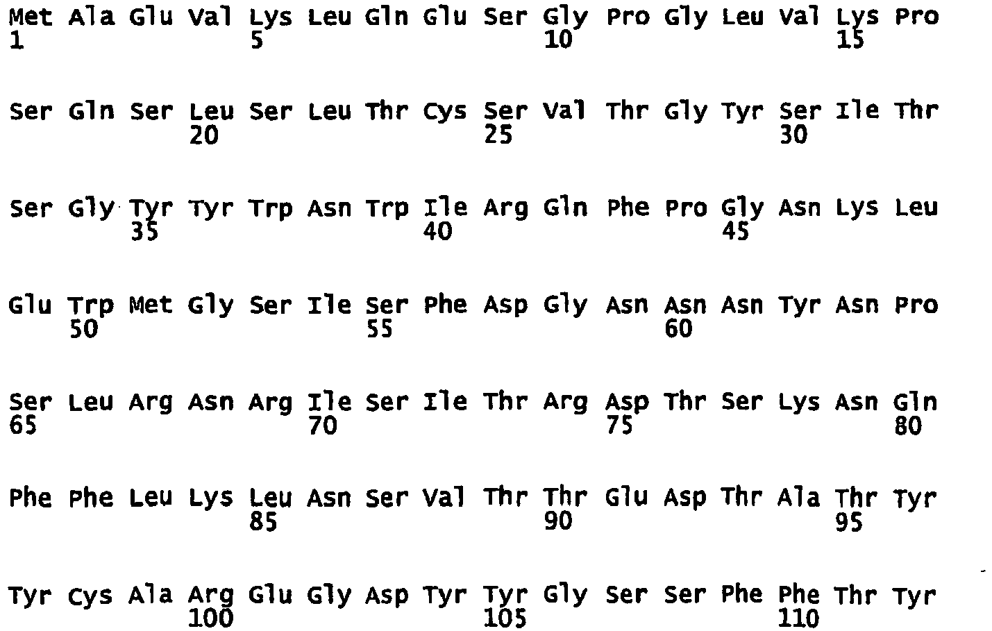
- The monoclonal antibodies or the fragments thereof comprise a peculiar nucleotide and amino acid sequence at the variable regions VH and VK which determine the specificity and affinity thereof. The nucleotide sequences of the moAb D2/B and of the scFv fragment thereof are indicated as SEQ ID NO: 1, 2, 3, 4. The specified sequence encodes an antibody fragment that has a good binding ability to the PSMA target antigen.
- The monoclonal antibody or fragment thereof as disclosed above may be conjugated with an active ingredient to obtain compounds which may be used for radioimmunotherapy or imaging or also recombinant immunotoxins.
- The active ingredient is preferably a labelling agent, more preferably a radionuclide selected among 188Re, 131I, 125I, 123I, 187Re, 111In, 90Y, 99mTc, 177Lu, or fluorescent nanoparticles.
- As an alternative, the monoclonal antibody or fragment thereof may be conjugated with a cytotoxic agent, preferably selected among PE40, ricin A chain, diantin, saporin.
- Finally, the monoclonal antibody or fragment thereof may be conjugated with biotin to exploit the high affinity of the avidin-biotin interaction or with other molecular pairs that allow multiple-step therapeutic protocols. In particular, the conjugate with biotin may be applied in targeting techniques in the case of treatments which include the use of radioisotopes. Namely, various preclinical studies have highlighted the superiority of pretargeting with respect to direct targeting; for instance Goldenberg D.M. et al. (J. Clin. Oncol., 24:823-834, 2006).
- It has been shown that the administration of a conjugate with the antibody according to the invention or of a fragment thereof with biotin, before the administration of a radiopharmaceutical (streptavidin and then radiolabelled biotin) allows a better distribution of the radiopharmaceutical at the tumour site thus reducing the side effects.
- The monoclonal antibody, the fragments and conjugates thereof may be formulated in pharmaceutical compositions with appropriate excipients and may be used for the preparation of a medicament for the treatment or diagnosis of tumour tissues overexpressing the PSMA antigen, a tumour, preferably prostate tumour.
- The procedure to obtain monoclonal antibodies is as follows:
- Balb/c mice are immunised, the cells from the animal's spleen are fused with hybridoma cells, the resulting cells are selected in HAT medium, polyclonal hybridomas recognising the target antigen are identified, cloned and newly characterised to asses specificity/affinity of the now monoclonal hybridomas, which are finally adapted to' grow in HAT-free medium. The antibodies are then purified from the culture medium of the hybridoma by means of standard biochemical techniques (affinity chromatography).
- Further features of the present invention will result from the following disclosure of some embodiments given by mere way of non-limitative illustration.
- The monoclonal antibodies and the scFv of the invention have been characterised by flow cytofluorimetry and Western blot on the following cell lines of human PCa, provided by the ATCC (American Type Colture Collection, Rockville, MD): lymphonodal metastases of PCa (LNCaP), brain metastases of PCa (DU145), bone metastases of PCa (PC-3). These cell lines have been maintained in RPMI 1640 enriched with 10% foetal bovine serum (FBS), 2 mM glutamine, 100 U/ml penicillin and streptomycin. The incubation temperature was 37°C and the CO2 concentration was 5%. The cells were split 2-3 times per week, detaching them with a 0.02% EDTA/PBS or 0.02% EDTA and 0.05% trypsin / PBS solution.
- Hereinafter reference will be made to antibodies according to the invention as "D2/B" and "scFv D2/B".
- The antibody was obtained following immunization of Balb/c mice with plasma membrane fractions of cells expressing the PSMA antigen and with the recombinant form of PSMA, following established protocols exploiting the hybridoma technology. Spleen cell populations containing B lymphocytes of immunized animals were fused with myeloma cells. The obtained hybridomas, cloned by limiting dilution and characterized to evaluate specificity and affinity, were grown in an appropriate serum free medium. The antibodies are purified from the culture supernatant by affinity chromatography.
- The Enzyme-linked Immunosorbent Assay (ELISA) is a technique utilized to detect the presence of a molecule by its reactivity with specific antibodies.
- This assay was used to determine the isotype of the anti-PSMA moAbs previously obtained. The kit used (Hybridoma Subisotyping Kit, Calbiochem) has allowed the identification of the antibody isotype among the IgG1, IgG2, IgG2b, IgG3, IgM and IgA subclasses; the procedure of the assay is described in the technical information sheet attached to the ELISA kit used.
- The results, reported in Tab. 1 show that antibody D2/B is an IgG1;
row number 2 of the table (IgG1) shows an absorbance value of 4.0, as opposed to a negative control (culture medium only) of 0.8. The absorbance values of D2/B for the other IgG subclasses correspond to the negative control value. As a positive control in the assay we used murine serum which, as evidenced from data reported incolumn number 3, contains all immunoglobulin isotypes.Table 1 D2/B Negative control Mouse serum IgM 0,56 0,54 2,36 IgG1 4,00 0,81 1,75 IgG2a 0,41 0,32 2,42 IgG2b 0,30 0,25 1,84 IgG3 0,30 0,28 1,24 IgA 0,25 0,23 0,18 - After extraction of the total RNA of hybridoma D2/B with TRIzol (Gibco), cDNA was synthesized using the Superscript cDNA Synthesis kit (Invitrogen). Using appropriate pairs of reverse and forward primers and PCR we have identified the primer combinations able to amplify the variable VH and Vk regions of the D2/B moAb, the optimal combinations were identified and the VH and Vk regions were cloned into a plasmidic vector appending a 6-His tag to the protein N-terminus. Sequencing of the vector has allowed identification of VH and Vk sequences (
SEQ ID 1, 2). - Production of scFv D2/B takes place in E.Coli cells (strain HB2151) grown at 37°C with stirring (225 rpm) in medium 2xTY additioned with
ampicillin 100 µg/ml, glucose 0.1% and the bacteria were induced at an O.D. of 0.8 (600 nm) with a final concentration of 1 mM IPTG for 3 h at 37°C. The cells were harvested by centrifugation at 5000 rpm at 4°C for 10 min and exposed for 1 h to Tris-HCl 30 mM pH = 8.0 +EDTA 1 mM +sucrose 20%; the supernatant (containing the periplasmic fraction) is collected and the scFv D2/B is purified by chromatography using a NiNTA (Novagen) matrix eluting withTris 20 mM pH = 8.0 + 250 mM Imidazole buffer. - Purity of the protein was evaluated by SDS-PAGE.
- To verify the recognition potential of D2/B antibody in Western blots an SDS-PAGE (10% polyacrylamide) of PSMA+ LNCaP cells and of PSMA- PC-3 cells was carried out. Cell lysis was performed by sonication of cells resuspended in a solution of 20 mM Tris, 150 mM NaCl pH 8.0 containing 1% Triton X-100 and protease inhibitors (Complete Protease Inhibitor Cocktail, Roche). Slots were loaded with 20 µl lysate corresponding to approx. 100 µg total proteins; the SDS-PAGE was carried out according to the protocol described by Laemmli. Subsequently the proteins separated by gel electrophoresis were transferred to a nitrocellulose membrane by applying an electric field (established procedure, Western blot).
- To verify PSMA antigen recognition by the D2/B antibody the nitrocellulose membrane was incubated o.n. at 4°C with gentle agitation in the presence of the primary antibody D2/B or of the control antibody J591, both used at the concentration of 10-20 µg/ml in a 5% solution of defatted milk. Excess antibody was eliminated by 4 washings using TBST buffer.
- A secondary anti-mouse antibody conjugated to HRP and diluted 1:1000 in 5% defatted milk in TBST was then used; incubation lasted 1 h at r.t. with gentle agitation.
- After 3 washings in TBST and 1 washing in PBS, the signal was detected by a chemiluminescent reaction (ECL Amersham Biosciences, UK).
-
Figure 1 shows a Western blotting carried out with a lysate of LNCaP (PSMA+) and PC-3 (PSMA-) cells, using as a postive control the antibody J591, already described in the literature; as shown in the Figure the antibody D2/B is able to recognize the antigen present in the cell lysate as well as the antibody J591 (lanes lanes - Likewise, the recognition ability of the antibody scFv D2/B was evaluated in Western blot using the same cell lysates as above but revealing the binding with a rabbit anti-myc tag antibody (Sigma) followed by an anti-rabbit HRP-conjugated antibody (Sigma). The scFv D2/B detects PSMA in the LNCaP cell lysates but no signal is observed with PC-3 cell lysates, lacking the antigenic target PSMA (
Figure 2 ). - LNCaP cells (PSMA+) were grown on polylysine-coated coverslips in a 24-well microtiter plate. Before carrying out the immunofluorescence assay the cell culture medium was discarded and the cells washed twice with PBS.
- Cells were then fixed with 2% paraformaldehyde for 10 min at r.t. and the cells further washed with PBS for three times to get rid of the excess fixative.
- The cells were then incubated at r.t. for 1 h in the presence of the primary anti-PSMA antibody at the concentration of 10 µg/ml. The following antibodies were used: D2/B, 7E11 (used as a well known reference antibody to an intracellular epitope of PSMA), an anti-MHC class-I antibody (as a positive control for cellular staining) and an isotypic control (negative control).
- Excess antibody was eliminated by 4 washings with a 0.2% BSA solution in PBS.
- Incubation with a secondary anti-mouse FITC(fluorescein)-labelled antibody (Beckman Coulter, USA) diluted 1:200 was carried out for 1 h at r.t.
- After 2 further washings with
PBS 2 drops of PBS/glycerol were added and the coverslip mounted on a glass slide and sealed. - The sample was then observed by a fluorescence microscope Axioskop 20 (Carl Zeiss, Germany).
- The D2/B monoclonal can specifically recognize LNCaP cells (PSMA+) but cannot detect cells lacking the PSMA antigen (i.e. DU145 cells).
- Sections of human prostate carcinoma embedded in paraffin were used to evaluate the recognition ability of human tumor tissues by the antibody D2/B in comparison to the already known and described antibody J591. Paraffin embedded tissues were placed in an oven at 60°C (30 to 60 min), treated 5 times with xylol for 4 min each time, sequentially rehydrated twice in 100%, 95%, 75% ethanol for 3 min each and immersed in H2O for 5 min; to unmask the PSMA antigen the samples were then placed in a steam bath in 10 mM citrate buffer pH 6.0 for 15 min at 95°C. After 3 washings in PBS for 5 min each, incubation in PBS + 3% H2O2 for 6 min and 3 further washings in PBS for 5 min each, the samples were treated with PBS+0.1% BSA for 30-40 min to saturate nonspecific binding sites; incubation with the primary antibody (D2/B or J591, both at 2 µg/ml) was carried out o.n. at 4°C; after 3 washings with PBS for 5 min each the kit EnVision Detection System HRP/DAB (Dako) was used, following the manufacturer's instructions.
- Counterstaining was carried out with Gill hematoxylin for 5 sec, followed by washings with abundant H20, and subsequently with sequential 75%, 80%, 95%, 100% solutions of ethanol and then with xylol for 4 times, 5 min each time. The slides were then mounted with resin and observed by a light standard microscope.
- Flow cytometry allows the evaluation of important physico-chemical parameters of particles in suspension. Using a laser emission and optical detectors the system records the fluorescent light emitted by a fluorochrome (organic compound emitting fluorescent light when appropriately excited) linked to a monoclonal antibody specific for molecules expressed at the surface and/or in the cytoplasm of a cell under investigation. The signal emitted is then converted in analog/digital signals.
- This technique was used to evaluate important parameters, such as:
- the specificity, i.e. the ability of the antibodies obtained (D2/B and scFv D2B) to selectively bind the relevant antigen on the surface of antigen-positive cells without aspecifically binding antigen-negative cells;
- the concentration of the antibody saturating the antigen sites expressed at the surface of LNCaP cells, as compared to the control antibody J591.
- LNCaP and PC-3 cells cultured at 37°C were detached from plastic surfaces with Trypsin/EDTA for 4 min at 37°C, neutralized with complete medium, washed twice with 10 ml of a cold solution of PBS/0.2% BSA by centrifugation at 1000 rpm for 5 min at 4°C and finally 2-3x10⁵ cells/0.5 ml were incubated with D2/B, J591 or an isotype control for 1 h at 4°C. Following two washings with 3 ml PBS/0.2% BSA at 4°C to eliminate the excess of primary antibody, the cells resuspended in 100 µl PBS/0.2% BSA were incubated for 30 min at 4°C in the presence of a saturating amount of a secondary antibody (approx. 1 µg/100 µl), FITC (fluorescein)-labelled goat IgG anti-mouse IgG.
- At the end of the incubation time, following a further washing and resupension of the cells in 500 µl PBS/0.2% BSA, flow cytometry analysis was carried out using a cytofluorimeter (BD FACSCanto). To detect binding of scFv D2/B a mouse anti-myc tag secondary antibody was used followed by a goat anti-mouse FITC-labelled antibody.
- The parameters that were considered in the analysis were: mean fluorescence intensity (MFI) of the cells investigated and the percentage of cells found in a window comprised between channel 500 and 256.000; cells within this range were considered positive.
- To verify the binding ability of the antibodies, LNCaP cells were incubated with increasing amounts of anti-PSMA D2/B or scFvD2B antibodies or with the antibody J591 of which the affinity of binding is known. With MFI values obtained a binding curve was plotted where the value yielding 100% saturation of the antigen sites corresponds to the antibody concentration where the highest fluorescence value was recorded.
- As illustrated in
Figures 3.A and 3.B the D2/B and scFv D2/B antibodies recognize the antigen expressed on the plasma membrane at a value of MFI of approx. 8000 and 1500, respectively. InFigures 3.C and 3.D are reported the negative controls. Using the same technique we also evaluated the specificity of the moAb by analyzing the non-specific binding to the antigen-negative cells, PC-3 (bone metastasis of prostate carcinoma); the results, summarized inFigures 4.A and 4.B , show the lack of binding of the moAb D2/B and scFv D2/B, respectively.Figures 4.C and 4.D show the respective negative controls. - The concentration of J591, D2/B and scFv D2/B saturating the antigen sites (PSMA) expressed at the surface of LNCaP cells is shown in
Figure 5 . - The Immunotoxin synthesis has proceeded as described below.
- Before conjugation the antibody D2/B was loaded onto an AffiBlue gel column to eliminate the fraction of antibodies that could bind to the matrix and which could contaminate the Immunotoxin preparations and reduce its cytotoxic efficacy. The eluted D2/B was dialyzed in PBS and derivatized with SMPT (4-succinimidyloxycarbonyl-alpha- methyl-alpha(2-pyridyldithio)toluene) dissolved at 10 mM concentration in anhydrous dimethylformamide, with a ratio IgG:cross-linker of approx. 1:2.5.
- Excess cross-linker was eliminated by gel filtration on PD-10 columns (Pharmacia, Uppsala, Sweden).
- The number of SH groups introduced, corresponding to the number of possible bonds linking the toxin to the antibody, was determined based on the fact that DTT, by reducing thiol groups, releases the group piridyne-2-thione which can be quantified spectrophotometrically at 343 nm. This value was comprised between 1.2 and 1.5.
- Recombinant RTA, produced and purified according to a protocol described by Chignola et al. (J.Biol.Chem., 270:23345-51, 1995) was treated with 25 mM DTT (dithiothreitol) at r.t. for 2 h to reduce cysteines and make them available to conjugation.
- DTT was subsequently eliminated by gel filtration on PD-10 columns and the toxin, reduced and purified, was concentrated by Centricon-10 (Amicon, Beverly, MA).
- Conjugation reaction to link the derivatized D2/B antibody and RTA was carried out at r.t. for 48 h.
- The resulting immunotoxin was purified by gel filtration on a TSK3000SW column (Beckman, San Ramon, CA), with a 0.5 ml/min flow and PBS as eluting buffer.
- Peaks corresponding to molecular species of 150-180 kDa (D2/B+1 RTA) and 210-240 kDa (Ab+2/3 RTA) were further purified onto AffiBlue gel, with a 150 nM-1 M salt gradient in phosphate buffer 5 mM. The toxin alone has a m.w. of approx. 30 kDa whereas the antibody has a m.w. of approx. 150 kDa.
- A column of AffiBlue gel was then utilized because its matrix can bind enzymes which exploit NAD as a coenzyme or which have nucleotide sequences as substrate, like RTA; for these reasons this gel can specifically bind RTA (and antibody-RTA conjugates) while allowing the free antibody to pass through.
- The fractions containing the protein of interest were pooled, concentrated with Centricon-10, dialyzed against PBS. The protein concentration was evaluated spectrophotometrically and the different pools sterilized by filtration.
- The sequence of scFv D2/B was cloned in a pET11d vector between Nco I and Hind III restriction sites; downstream of Hind III was inserted the nuclotide sequence coding for the PE40 toxin.
- The fusion protein is produced in E.Coli strain BL21(DE3)pLysS previously transformed with the plasmidic vector coding for the Immunotoxin; the bacteria grown at 37°C with agitation (225 rpm) in LB medium additioned with 0.5% glucose, 0.05% MgS04,
ampicillin 100 µg/ml, chloramphenicol 34 µg/ml, were induced at an absorbance of 0.8 at 600 nm with 1 mM IPTG final. After 3 h incubation the bacteria harvested by centrifugation at 5000 rpm are lysed by incubation and sonication in lysis buffer (Tris 20 mM pH = 7.5+EDTA 10 mM+Triton-X100 1%) additioned with 0.1 mg/ml lysozyme, 0.1 mM PMSF, protease inhibitors (Roche). Inclusion bodies (IB) are harvested by centrifugation at 10.000 rpm at 4°C, these are washed twice in lysis buffer and solubilized at 20 mg/ml concentration inCAPS buffer 50 mM pH=11 additioned with N-laurylsarcosine 0.8-1.0% final andDTT 1 mM. Insoluble material was removed by centrifugation and the protein was refolded by successive dialyses. Dialysis I and II were carried out for 12 h at 4°C in Tris 20mM pH=8,5+DTT 0,1mM+PMSF PMSF Tris 20 mM pH=7.4+PMSF 0.1 mM. - The protein was then purified by 2 runs through QAE column (Bio-Rad) with a 200 ml linear 0-0.5 M NaCl gradient in
Tris 20 mM pH=7.4, volume of fractions recovered was 2.5 ml. - The purity of the protein was evaluated in SDS-PAGE and identified by Western blot.
- The cytotoxic effect of the immunoconjugate was evaluated in PSMA-positive and PSMA- negative cells by measuring the inhibition of cell proliferation, as compared to the toxin alone.
- LNCaP or PC-3 cells were seeded in 96-well microtiter plates in complete medium in amounts adequate to reach sub-confluence in control untreated wells in the 32 h of the assay.
- Some wells were treated with increasing amounts of the immunotoxins D2/B-RTA (obtained by chemical conjugation using the SMPT cross-linker) or scFv D2/B-PE40 (obtained by gene fusion) or with the control immunotoxin J591-RTA (obtained by chemical conjugation with the SMPT cross-linker), other wells were treated with RTA, PE or PE40 alone, the moAb D2/B or scFv D2/B alone, for 22 h at 37 °C.
- The cells were then harvested on fiber filters by a cell harvester and the radioactivity incorporated was measured by liquid scintillation in a ß-counter.
- The cytocidal effect is expressed as a function of the incorporation of tritiated thymidine as compared to a control represented by untreated cells; results are expressed as a percent of the proliferation inhibition. The smaller the radioactive emission, the smaller the incorporation of tritiated thymidine, the bigger was therefore considered the inhibition of cell growth.
- The IC50 was then calculated, i.e. the concentration of the drug which is able to inhibit 50% of the cell proliferation; this value can be easily compared with the IC50 measured using different molecules in the cytotoxicity assay.
Table 2 LNCap PSMA+ ng/ml PC-3 PSMA-ng/ml J591-smpt- RTA 7,4 >1.260 D2/B-smpt-RTA 21 >16.800 scFv D2/B- PE40 1,3 >6.500 moAb D2/B >6.000 >6.000 scFv D2/B > 3.300 > 3.300 PE toxin 2,4 3,4 PE40 toxin 1.950 RTA toxin 12.000 9.000 - Monobiotinylated scFv D2/B were obtained by cloning the scFv molecule between NcoI and NotI restriction sites; in the cloning vector in 3' with respect to the NotI site were cloned successively: a sequence coding for a domain derived from the hinge region of IgA, a sequence coding for a peptidic stretch named BAD (Biotin Acceptor Domain), and a sequence coding for a cleavable fragment recognized by the proteases Enterokinase and Factor X. The hinge region of IgA is a spacer between scFv and BAD domain, which is monobiotinylated in a reaction catalyzed by the enzyme BirA. The production of the biotinylated protein was carried out in bacteria transformed with the plasmid of interest and are able to biotinylate the BAD site; by IPTG induction and addition of free biotin to the culture medium the protein under investigation is obtained; this includes the scFv domain endowed with the desired specificity and one single biotin molecule.
- The protein scFv-biotin was purified on a NiNTA gel under partially denaturing conditions to facilitate the exposure of the N-terminal 5-His tag and the binding to the column. The purity of the protein was analyzed by SDS-PAGE whereas its identity was confirmed by Western blotting using HRP-labelled secondary antibodies to the scFv and to the His tag.
- Functional properties of the biotinylated scFv was then assayed by FACS analysis carried out with PSMA+ (LNCaP) and PSMA- (PC-3) cells.
- Th MFI in LNCaP cells is 161, 728 and 314 for the negative control (avidin-FITC alone), for chemically biotinylated D2/B antibody and the enzymatically labelled scFv-D2/B, respectively, whereas the MFI values obtained with PC-3 cells are 132, 151 and 180, respectively.
- To address the possibility of applying in vivo imaging techniques based on the use of D2/B or scFv D2/B antibodies labelled with fluorescent tracers, immunodeficient SCID mice were inoculated with LNCaP cells pre-treated in vitro with D2/B antibody linked to the fluorochrome Cy5.5, according to the manufacturer's instructions. This assay has allowed to trace a s.c. implant of 7.5x10⁶ PSMA+ cells, but we have good indications that lower numbers of cells can be identified (i.e. down to 106 cells).
Figure 6 . - The choice of the chelating agent used to derivatize an antibody is dependent upon the choice of radionuclides that need to be conjugated to the antibody itself; additionally, the chelating substance must be endowed with an available functional group exploitable for linking to the antibody. For instance the compound DOTA can be directly cross-linked to the antibody via one of the 4 carboxyl groups available; the antibody is concentrated, washed with 1% DTPA (pH=5.0), dialyzed in 0.1 M phosphate buffer (pH=7.0) and reacted with active esters of DOTA. Active esters of DOTA are created by dissolving 0.361 mmol (146 mg) and 0.313 mmol (36 mg) of N-hydroxysuccinimide in 2 ml of H20 and adjusting the pH to pH=7.3 with NaOH prior to the addition of 10 mg of 1-ethyl-3-(3-dimethylaminopropyl)carbodiimide. The reaction mixture is cooled on ice for 1 h before being added to the antibody to be derivatized. The resulting DOTA-antibody conjugate is separated from excess DOTA and other reactants by gel filtration (PD-10 columns) or by centrifugation (Amicon concentrators) using a 0.3 M ammonium acetate solution. Radiolabeling of DOTA with 111In is achieved by adding 111InC13 in 0.01 M HCl to the 0.3 M acetate solution of DOTA-Ab.
- After 20 min the reaction mixture is separated by Biogl-P6 columns equilibrated in with 1%HSA-PBS. The amount of free radioisotope is evaluated by TLC chromatography.
-
- <110> università di Verona
- <120> ISOLATED MONOCLONAL ANTIBODY OR FRAGMENT THEREOF BINDING PROSTATE SPECIFIC MEMBRANE ANTIGEN, CONJUGATES AND USES THEREOF
- <130> 840-07
- <160> 6
- <170> PatentIn version 3.5
- <210> 1
<211> 366
<212> DNA
<213> Mouse - <220>
<221> v_region
<222> (1)..(366)
<223> Ab D2/B variable heavy chain - <220>
<221> misc_feature
<222> (97)..(114)
<223> CDRH1 - <220>
<221> misc_feature
<222> (157)..(204)
<223> CDRH2 - <220>
<221> miss_feature
<222> (301)..(336)
<223> CDRH3 - <400> 1

- <210> 2
<211> 327
<212> DNA
<213> Mouse - <220>
<221> v_region
<222> (1)..(327)
<223> Ab D2/B variable light chain - <220>
<221> misc_feature
<222> (76)..(108)
<223> CDRL1 - <220>
<221> misc_feature
<222> (154)..(174)
<223> CDRL2 - <220>
<221> misc_feature
<222> (271)..(294)
<223> CDRL3 - <400> 2

- <210> 3
<211> 822
<212> DNA
<213> Mouse - <220>
<221> CDS
<222> (1)..(822)
<223> scFv D2/B - <220>
<221> v_region
<222> (1)..(366)
<223> scFv D2/B variable heavy chain - <220>
<221> v_region
<222> (415)..(741)
<223> scFv D2/B variable light chain - <400> 3


- <210> 4
<211> 273
<212> PRT
<213> Mouse - <400> 4


- <210> 5
<211> 1830
<212> DNA
<213> Mouse and Bacteria - <220>
<221> CDS
<222> (1)..(1830)
<223> scFv D2/B-PE40 - <220>
<221> v_region
<222> (1)..(738)
<223> scFv D2/B variable heavy and light chain region - <220>
<221> misc_feature
<222> (739)..(1830)
<223> PE40 Toxin sequence - <400> 5


- <210> 6
<211> 609
<212> PRT
<213> Mouse and Bacteria - <400> 6


Claims (14)
- An isolated D2/B monoclonal antibody binding prostate specific membrane antigen (PSMA) characterised by comprising a protein encoded by the nucleotide sequence of the variable regions VH and VK of SEQ ID NO: 1 and 2.
- An antigen-binding D2/B scFv antibody fragment, characterised by comprising a protein encoded by a sequence of at least 741 consecutive nucleotides characterising the antigen-binding variable regions VH and VL of SEQ ID NO:3.
- The isolated monoclonal antibody or antigen-binding fragment thereof according to any of the preceding claims, characterised by binding PSMA in its native form on the surface of tumour cells.
- The isolated monoclonal antibody or antigen-binding fragment thereof according to any of the preceding claims, characterised by selectively binding PSMA.
- A conjugate between active ingredient and antibody comprising an isolated monoclonal antibody or antigen-binding fragment thereof according to any of the preceding claims conjugated with an active ingredient selected from the group consisting of a labelling agent, a cytotoxic agent or a carrier.
- The conjugate according to claim 5, characterised in that said labelling agent is a radionuclide or fluorescent nanoparticles.
- The conjugate according to claim 5 or 6, characterised in that said labelling agent is selected from the group consisting of 188Re, 131I, 125I, 123I, 187Re, 111In, 90Y, 99mTc, 177Lu.
- The conjugate according to claim 5, characterised in that said cytotoxic agent is PE40, the ricin A chain, saporin and diantin.
- scFv D2/B-PE40 conjugate of SEQ ID NO:5.
- The conjugate according to claim 6, characterised in that said carrier is biotin or nanoparticles.
- A pharmaceutical composition comprising an isolated monoclonal antibody or antigen-binding fragment thereof according to any of claims 1 to 4, or a conjugate according to any of claims 5 to 10.
- Use of an isolated monoclonal antibody or antigen-binding fragment thereof according to any of claims 1 to 4, or of a conjugate according to any of claims 5 to 10, or of a pharmaceutical composition according to claim 11 for the preparation of a medicament for the treatment or the diagnosis of a tumour.
- Use according to claim 12, characterised in that said tumour is a prostate tumour.
- Isolated monoclonal antibody or antigen-binding fragment thereof according to any of claims 1 to 4, or a conjugate according to any of claims 5 to 10, or a pharmaceutical composition according to claim 11 for the use in PSMA+ angiogenesis of tumours of any hystotype.
Applications Claiming Priority (2)
| Application Number | Priority Date | Filing Date | Title |
|---|---|---|---|
| IT000313A ITTO20080313A1 (en) | 2008-04-22 | 2008-04-22 | ISOLATED MONOCLONAL ANTIBODY OR ITS FRAGMENT BINDING THE PROSTATE'S SPECIFIC MEMBRANE ANTIGEN, ITS CONJUGATES AND ITS USES |
| PCT/IB2009/005326 WO2009130575A2 (en) | 2008-04-22 | 2009-04-22 | Isolated monoclonal antibody or fragment thereof binding prostate specific membrane antigen, conjugates and uses thereof |
Publications (2)
| Publication Number | Publication Date |
|---|---|
| EP2283042A2 EP2283042A2 (en) | 2011-02-16 |
| EP2283042B1 true EP2283042B1 (en) | 2012-11-14 |
Family
ID=40297325
Family Applications (1)
| Application Number | Title | Priority Date | Filing Date |
|---|---|---|---|
| EP09735635A Active EP2283042B1 (en) | 2008-04-22 | 2009-04-22 | Isolated monoclonal antibody or fragment thereof binding prostate specific membrane antigen, conjugates and uses thereof |
Country Status (5)
| Country | Link |
|---|---|
| US (1) | US8703918B2 (en) |
| EP (1) | EP2283042B1 (en) |
| ES (1) | ES2400082T3 (en) |
| IT (1) | ITTO20080313A1 (en) |
| WO (1) | WO2009130575A2 (en) |
Families Citing this family (40)
| Publication number | Priority date | Publication date | Assignee | Title |
|---|---|---|---|---|
| US9193763B2 (en) | 2007-08-17 | 2015-11-24 | Purdue Research Foundation | PSMA binding ligand-linker conjugates and methods for using |
| EP3243527B1 (en) | 2009-02-13 | 2019-06-05 | Immunomedics, Inc. | Immunoconjugates with an intracellularly-cleavable linkage |
| MX337600B (en) | 2009-12-02 | 2016-03-11 | Imaginab Inc | J591 minibodies and cys-diabodies for targeting human prostate specific membrane antigen (psma) and methods for their use. |
| US9951324B2 (en) | 2010-02-25 | 2018-04-24 | Purdue Research Foundation | PSMA binding ligand-linker conjugates and methods for using |
| SG194510A1 (en) | 2011-04-22 | 2013-12-30 | Emergent Product Dev Seattle | Prostate-specific membrane antigen binding proteins and related compositionsand methods |
| AU2012250924B2 (en) | 2011-05-02 | 2017-05-25 | Immunomedics, Inc. | Ultrafiltration concentration of allotype selected antibodies for small-volume administration |
| WO2013052484A1 (en) | 2011-10-03 | 2013-04-11 | Immunomedics, Inc. | Dye conjugated peptides for fluorescent imaging |
| WO2014028560A2 (en) | 2012-08-14 | 2014-02-20 | Ibc Pharmaceuticals, Inc. | T-cell redirecting bispecific antibodies for treatment of disease |
| AU2013344778C1 (en) | 2012-11-15 | 2024-06-27 | Endocyte, Inc. | Conjugates for treating diseases caused by PSMA expressing cells |
| WO2017004144A1 (en) | 2015-07-01 | 2017-01-05 | Immunomedics, Inc. | Antibody-sn-38 immunoconjugates with a cl2a linker |
| AU2013360335B2 (en) | 2012-12-13 | 2017-12-07 | Immunomedics, Inc. | Dosages of immunoconjugates of antibodies and SN-38 for improved efficacy and decreased toxicity |
| EP3456700A1 (en) | 2013-10-18 | 2019-03-20 | Deutsches Krebsforschungszentrum | Labeled inhibitors of prostate specific membrane antigen (psma), their use as imaging agents and pharmaceutical agents for the treatment of prostate cancer |
| CN105849568B (en) | 2013-11-14 | 2018-05-04 | 恩多塞特公司 | Compounds for Positron Emission Tomography |
| JP6447933B2 (en) | 2014-02-21 | 2019-01-09 | アイビーシー ファーマスーティカルズ,インコーポレイテッド | Disease treatment by inducing an immune response against Trop-2 expressing cells |
| CA2935748A1 (en) | 2014-02-25 | 2015-09-03 | Immunomedics, Inc. | Humanized rfb4 anti-cd22 antibody |
| IL292311A (en) | 2014-04-03 | 2022-06-01 | Igm Biosciences Inc | Modified j-chain |
| SI3204018T1 (en) | 2014-10-07 | 2021-11-30 | Immunomedics, Inc. | Neoadjuvant use of antibody-drug conjugates |
| US10188759B2 (en) | 2015-01-07 | 2019-01-29 | Endocyte, Inc. | Conjugates for imaging |
| US9797907B2 (en) | 2015-04-22 | 2017-10-24 | Immunomedics, Inc. | Isolation, detection, diagnosis and/or characterization of circulating Trop-2-positive cancer cells |
| AU2016281622C1 (en) | 2015-06-25 | 2021-11-18 | Immunomedics, Inc. | Combining anti-HLA-DR or anti-Trop-2 antibodies with microtubule inhibitors, PARP inhibitors, Bruton kinase inhibitors or phosphoinositide 3-kinase inhibitors significantly improves therapeutic outcome in cancer |
| KR20250008141A (en) | 2015-08-07 | 2025-01-14 | 이미지냅 인코포레이티드 | Antigen binding constructs to target molecules |
| KR102784293B1 (en) | 2015-09-21 | 2025-03-20 | 압테보 리서치 앤드 디벨롭먼트 엘엘씨 | CD3 binding polypeptide |
| CA2999284C (en) | 2015-09-30 | 2023-06-13 | Igm Biosciences A/S | Binding molecules with modified j-chain |
| US11639389B2 (en) | 2015-09-30 | 2023-05-02 | Igm Biosciences, Inc. | Binding molecules with modified J-chain |
| WO2018147960A1 (en) | 2017-02-08 | 2018-08-16 | Imaginab, Inc. | Extension sequences for diabodies |
| KR20210003135A (en) | 2018-04-17 | 2021-01-11 | 엔도사이트, 인코포레이티드 | Cancer treatment method |
| CN110407939B (en) * | 2019-03-12 | 2023-09-29 | 广东医科大学附属第二医院 | A humanized anti-PSMA single chain antibody and its application |
| JP7604397B2 (en) | 2019-05-20 | 2024-12-23 | エンドサイト・インコーポレイテッド | Methods for preparing PSMA conjugates |
| WO2021202376A1 (en) | 2020-03-30 | 2021-10-07 | The United States Of America, As Represented By The Secretary, Department Of Health And Human Services | Method for blocking uptake of prostate-specific membrane antigen (psma)-targeted radionuclides by exocrine organs |
| US20230256114A1 (en) | 2020-07-07 | 2023-08-17 | Bionecure Therapeutics, Inc. | Novel maytansinoids as adc payloads and their use for the treatment of cancer |
| IL299748A (en) | 2020-07-08 | 2023-03-01 | Lava Therapeutics N V | Antibodies that bind psma and gamma-delta t cell receptors |
| CN115803010A (en) | 2020-07-09 | 2023-03-14 | 豪夫迈·罗氏有限公司 | Concentrated protein compositions, their preparation and use |
| EP4342497A1 (en) | 2021-05-10 | 2024-03-27 | Kawasaki Institute of Industrial Promotion | Antibody having reduced binding affinity for antigen |
| CN118043080A (en) | 2021-08-13 | 2024-05-14 | 昆山新蕴达生物科技有限公司 | An antibody-drug conjugate based on microtubule inhibitors |
| CN117881431A (en) | 2021-08-24 | 2024-04-12 | 昆山新蕴达生物科技有限公司 | An antibody-drug conjugate coupled by a cleavable linker |
| WO2023103854A1 (en) | 2021-12-09 | 2023-06-15 | 昆山新蕴达生物科技有限公司 | Antibody-drug conjugate having improved affinity, and preparation method therefor and application thereof |
| CN119698305A (en) | 2022-08-10 | 2025-03-25 | 兴和株式会社 | New drug complex |
| EP4414386A1 (en) * | 2023-02-09 | 2024-08-14 | Mariangela Figini | Method of chemical synthesis of single-chain antibody fragments and products thereby obtained |
| JP2026507228A (en) | 2023-03-03 | 2026-02-27 | アーセナル バイオサイエンシズ インコーポレイテッド | PSMA and CA9 Targeting Systems |
| CN118652355B (en) * | 2024-08-20 | 2025-04-22 | 南京大学 | PSMA-targeted chimeric antigen receptors and cells modified therefrom and their applications |
Family Cites Families (1)
| Publication number | Priority date | Publication date | Assignee | Title |
|---|---|---|---|---|
| EP1726650A1 (en) * | 2005-05-27 | 2006-11-29 | Universitätsklinikum Freiburg | Monoclonal antibodies and single chain antibody fragments against cell-surface prostate specific membrane antigen |
-
2008
- 2008-04-22 IT IT000313A patent/ITTO20080313A1/en unknown
-
2009
- 2009-04-22 WO PCT/IB2009/005326 patent/WO2009130575A2/en not_active Ceased
- 2009-04-22 ES ES09735635T patent/ES2400082T3/en active Active
- 2009-04-22 US US12/989,316 patent/US8703918B2/en active Active
- 2009-04-22 EP EP09735635A patent/EP2283042B1/en active Active
Also Published As
| Publication number | Publication date |
|---|---|
| EP2283042A2 (en) | 2011-02-16 |
| WO2009130575A3 (en) | 2010-01-14 |
| US20120034168A1 (en) | 2012-02-09 |
| ITTO20080313A1 (en) | 2009-10-23 |
| WO2009130575A2 (en) | 2009-10-29 |
| US8703918B2 (en) | 2014-04-22 |
| ES2400082T3 (en) | 2013-04-05 |
Similar Documents
| Publication | Publication Date | Title |
|---|---|---|
| US8703918B2 (en) | Isolated monoclonal antibody or fragment thereof binding prostate specific membrane antigen, conjugates and uses thereof | |
| CN116133694B (en) | Anti-HER3 antibodies and anti-HER3 antibody-drug conjugates and their pharmaceutical uses | |
| KR102169224B1 (en) | Antibody specifically binding to MUC1 and Use thereof | |
| RU2458073C2 (en) | Monoclonal antibodies and one-chain fragments of cell-surface prostate-specific membrane antigen antibodies | |
| RU2711935C2 (en) | Site-specific antibody-drug conjugation by means of glycoengineering | |
| JP6326137B2 (en) | Anti-HER2 antibody and conjugate thereof | |
| JP7063623B2 (en) | Therapeutic antibodies and their use | |
| TWI872044B (en) | Anti-cd228 antibodies and antibody-drug conjugates | |
| WO2008105560A1 (en) | Pharmaceutical composition comprising anti-grp78 antibody as active ingredient | |
| Nováková et al. | Novel monoclonal antibodies recognizing human prostate‐specific membrane antigen (PSMA) as research and theranostic tools | |
| CN101970497A (en) | Anti-epcam antibody and uses thereof | |
| Cao et al. | Construction and characterization of novel, recombinant immunotoxins targeting the Her2/neu oncogene product: in vitro and in vivo studies | |
| US20210230295A1 (en) | Antibody binding to axl protein and application thereof | |
| US20230365676A1 (en) | Cd33 antibodies | |
| US12534541B2 (en) | Anti-PTK7 antibody and uses thereof | |
| CA3104800A1 (en) | Humanized antibodies against psma | |
| JP2020532523A (en) | Anti-EGFR antibody drug conjugate (ADC) and its use | |
| CN114650843A (en) | Antibody compositions comprising FC mutation and site-specific conjugation properties | |
| US20260022193A1 (en) | Methods of treatment using a bispecific antigen-binding construct targeting her2 | |
| KR20230078152A (en) | Antibody Specifically Binding to PSMA and Uses thereof | |
| WO2023222068A1 (en) | Anti-cd200r1 antibodies | |
| WO2025131054A1 (en) | Antibody drug conjugates targeting to b7-h3 and egfr and the use thereof | |
| US20250019430A1 (en) | Resistin-specific antibody and use thereof |
Legal Events
| Date | Code | Title | Description |
|---|---|---|---|
| PUAI | Public reference made under article 153(3) epc to a published international application that has entered the european phase |
Free format text: ORIGINAL CODE: 0009012 |
|
| 17P | Request for examination filed |
Effective date: 20101030 |
|
| AK | Designated contracting states |
Kind code of ref document: A2 Designated state(s): AT BE BG CH CY CZ DE DK EE ES FI FR GB GR HR HU IE IS IT LI LT LU LV MC MK MT NL NO PL PT RO SE SI SK TR |
|
| AX | Request for extension of the european patent |
Extension state: AL BA RS |
|
| 17Q | First examination report despatched |
Effective date: 20110328 |
|
| DAX | Request for extension of the european patent (deleted) | ||
| GRAP | Despatch of communication of intention to grant a patent |
Free format text: ORIGINAL CODE: EPIDOSNIGR1 |
|
| GRAS | Grant fee paid |
Free format text: ORIGINAL CODE: EPIDOSNIGR3 |
|
| GRAA | (expected) grant |
Free format text: ORIGINAL CODE: 0009210 |
|
| AK | Designated contracting states |
Kind code of ref document: B1 Designated state(s): AT BE BG CH CY CZ DE DK EE ES FI FR GB GR HR HU IE IS IT LI LT LU LV MC MK MT NL NO PL PT RO SE SI SK TR |
|
| RAP1 | Party data changed (applicant data changed or rights of an application transferred) |
Owner name: FONDAZIONE IRCCS ISTITUTO NAZIONALE DEI TUMORI Owner name: COLOMBATTI, MARCO |
|
| REG | Reference to a national code |
Ref country code: GB Ref legal event code: FG4D |
|
| REG | Reference to a national code |
Ref country code: AT Ref legal event code: REF Ref document number: 583964 Country of ref document: AT Kind code of ref document: T Effective date: 20121115 Ref country code: CH Ref legal event code: EP |
|
| REG | Reference to a national code |
Ref country code: IE Ref legal event code: FG4D |
|
| REG | Reference to a national code |
Ref country code: DE Ref legal event code: R096 Ref document number: 602009011200 Country of ref document: DE Effective date: 20130110 |
|
| REG | Reference to a national code |
Ref country code: CH Ref legal event code: NV Representative=s name: MOINAS AND SAVOYE SA, CH |
|
| REG | Reference to a national code |
Ref country code: ES Ref legal event code: FG2A Ref document number: 2400082 Country of ref document: ES Kind code of ref document: T3 Effective date: 20130405 |
|
| REG | Reference to a national code |
Ref country code: NL Ref legal event code: VDEP Effective date: 20121114 |
|
| REG | Reference to a national code |
Ref country code: AT Ref legal event code: MK05 Ref document number: 583964 Country of ref document: AT Kind code of ref document: T Effective date: 20121114 |
|
| REG | Reference to a national code |
Ref country code: LT Ref legal event code: MG4D |
|
| PG25 | Lapsed in a contracting state [announced via postgrant information from national office to epo] |
Ref country code: SE Free format text: LAPSE BECAUSE OF FAILURE TO SUBMIT A TRANSLATION OF THE DESCRIPTION OR TO PAY THE FEE WITHIN THE PRESCRIBED TIME-LIMIT Effective date: 20121114 Ref country code: FI Free format text: LAPSE BECAUSE OF FAILURE TO SUBMIT A TRANSLATION OF THE DESCRIPTION OR TO PAY THE FEE WITHIN THE PRESCRIBED TIME-LIMIT Effective date: 20121114 Ref country code: HR Free format text: LAPSE BECAUSE OF FAILURE TO SUBMIT A TRANSLATION OF THE DESCRIPTION OR TO PAY THE FEE WITHIN THE PRESCRIBED TIME-LIMIT Effective date: 20121114 Ref country code: LT Free format text: LAPSE BECAUSE OF FAILURE TO SUBMIT A TRANSLATION OF THE DESCRIPTION OR TO PAY THE FEE WITHIN THE PRESCRIBED TIME-LIMIT Effective date: 20121114 Ref country code: NO Free format text: LAPSE BECAUSE OF FAILURE TO SUBMIT A TRANSLATION OF THE DESCRIPTION OR TO PAY THE FEE WITHIN THE PRESCRIBED TIME-LIMIT Effective date: 20130214 |
|
| PG25 | Lapsed in a contracting state [announced via postgrant information from national office to epo] |
Ref country code: GR Free format text: LAPSE BECAUSE OF FAILURE TO SUBMIT A TRANSLATION OF THE DESCRIPTION OR TO PAY THE FEE WITHIN THE PRESCRIBED TIME-LIMIT Effective date: 20130215 Ref country code: BE Free format text: LAPSE BECAUSE OF FAILURE TO SUBMIT A TRANSLATION OF THE DESCRIPTION OR TO PAY THE FEE WITHIN THE PRESCRIBED TIME-LIMIT Effective date: 20121114 Ref country code: LV Free format text: LAPSE BECAUSE OF FAILURE TO SUBMIT A TRANSLATION OF THE DESCRIPTION OR TO PAY THE FEE WITHIN THE PRESCRIBED TIME-LIMIT Effective date: 20121114 Ref country code: PT Free format text: LAPSE BECAUSE OF FAILURE TO SUBMIT A TRANSLATION OF THE DESCRIPTION OR TO PAY THE FEE WITHIN THE PRESCRIBED TIME-LIMIT Effective date: 20130314 Ref country code: SI Free format text: LAPSE BECAUSE OF FAILURE TO SUBMIT A TRANSLATION OF THE DESCRIPTION OR TO PAY THE FEE WITHIN THE PRESCRIBED TIME-LIMIT Effective date: 20121114 Ref country code: PL Free format text: LAPSE BECAUSE OF FAILURE TO SUBMIT A TRANSLATION OF THE DESCRIPTION OR TO PAY THE FEE WITHIN THE PRESCRIBED TIME-LIMIT Effective date: 20121114 |
|
| PG25 | Lapsed in a contracting state [announced via postgrant information from national office to epo] |
Ref country code: AT Free format text: LAPSE BECAUSE OF FAILURE TO SUBMIT A TRANSLATION OF THE DESCRIPTION OR TO PAY THE FEE WITHIN THE PRESCRIBED TIME-LIMIT Effective date: 20121114 |
|
| PG25 | Lapsed in a contracting state [announced via postgrant information from national office to epo] |
Ref country code: EE Free format text: LAPSE BECAUSE OF FAILURE TO SUBMIT A TRANSLATION OF THE DESCRIPTION OR TO PAY THE FEE WITHIN THE PRESCRIBED TIME-LIMIT Effective date: 20121114 Ref country code: SK Free format text: LAPSE BECAUSE OF FAILURE TO SUBMIT A TRANSLATION OF THE DESCRIPTION OR TO PAY THE FEE WITHIN THE PRESCRIBED TIME-LIMIT Effective date: 20121114 Ref country code: BG Free format text: LAPSE BECAUSE OF FAILURE TO SUBMIT A TRANSLATION OF THE DESCRIPTION OR TO PAY THE FEE WITHIN THE PRESCRIBED TIME-LIMIT Effective date: 20130214 Ref country code: DK Free format text: LAPSE BECAUSE OF FAILURE TO SUBMIT A TRANSLATION OF THE DESCRIPTION OR TO PAY THE FEE WITHIN THE PRESCRIBED TIME-LIMIT Effective date: 20121114 Ref country code: CZ Free format text: LAPSE BECAUSE OF FAILURE TO SUBMIT A TRANSLATION OF THE DESCRIPTION OR TO PAY THE FEE WITHIN THE PRESCRIBED TIME-LIMIT Effective date: 20121114 |
|
| PG25 | Lapsed in a contracting state [announced via postgrant information from national office to epo] |
Ref country code: RO Free format text: LAPSE BECAUSE OF FAILURE TO SUBMIT A TRANSLATION OF THE DESCRIPTION OR TO PAY THE FEE WITHIN THE PRESCRIBED TIME-LIMIT Effective date: 20121114 Ref country code: NL Free format text: LAPSE BECAUSE OF FAILURE TO SUBMIT A TRANSLATION OF THE DESCRIPTION OR TO PAY THE FEE WITHIN THE PRESCRIBED TIME-LIMIT Effective date: 20121114 |
|
| PLBE | No opposition filed within time limit |
Free format text: ORIGINAL CODE: 0009261 |
|
| STAA | Information on the status of an ep patent application or granted ep patent |
Free format text: STATUS: NO OPPOSITION FILED WITHIN TIME LIMIT |
|
| 26N | No opposition filed |
Effective date: 20130815 |
|
| PG25 | Lapsed in a contracting state [announced via postgrant information from national office to epo] |
Ref country code: MC Free format text: LAPSE BECAUSE OF FAILURE TO SUBMIT A TRANSLATION OF THE DESCRIPTION OR TO PAY THE FEE WITHIN THE PRESCRIBED TIME-LIMIT Effective date: 20121114 Ref country code: CY Free format text: LAPSE BECAUSE OF FAILURE TO SUBMIT A TRANSLATION OF THE DESCRIPTION OR TO PAY THE FEE WITHIN THE PRESCRIBED TIME-LIMIT Effective date: 20121114 |
|
| REG | Reference to a national code |
Ref country code: DE Ref legal event code: R097 Ref document number: 602009011200 Country of ref document: DE Effective date: 20130815 |
|
| REG | Reference to a national code |
Ref country code: IE Ref legal event code: MM4A |
|
| PG25 | Lapsed in a contracting state [announced via postgrant information from national office to epo] |
Ref country code: IE Free format text: LAPSE BECAUSE OF NON-PAYMENT OF DUE FEES Effective date: 20130422 |
|
| PG25 | Lapsed in a contracting state [announced via postgrant information from national office to epo] |
Ref country code: MT Free format text: LAPSE BECAUSE OF FAILURE TO SUBMIT A TRANSLATION OF THE DESCRIPTION OR TO PAY THE FEE WITHIN THE PRESCRIBED TIME-LIMIT Effective date: 20121114 |
|
| REG | Reference to a national code |
Ref country code: FR Ref legal event code: PLFP Year of fee payment: 7 |
|
| PG25 | Lapsed in a contracting state [announced via postgrant information from national office to epo] |
Ref country code: TR Free format text: LAPSE BECAUSE OF FAILURE TO SUBMIT A TRANSLATION OF THE DESCRIPTION OR TO PAY THE FEE WITHIN THE PRESCRIBED TIME-LIMIT Effective date: 20121114 |
|
| PG25 | Lapsed in a contracting state [announced via postgrant information from national office to epo] |
Ref country code: LU Free format text: LAPSE BECAUSE OF NON-PAYMENT OF DUE FEES Effective date: 20130422 Ref country code: MK Free format text: LAPSE BECAUSE OF FAILURE TO SUBMIT A TRANSLATION OF THE DESCRIPTION OR TO PAY THE FEE WITHIN THE PRESCRIBED TIME-LIMIT Effective date: 20121114 Ref country code: HU Free format text: LAPSE BECAUSE OF FAILURE TO SUBMIT A TRANSLATION OF THE DESCRIPTION OR TO PAY THE FEE WITHIN THE PRESCRIBED TIME-LIMIT; INVALID AB INITIO Effective date: 20090422 |
|
| PG25 | Lapsed in a contracting state [announced via postgrant information from national office to epo] |
Ref country code: IS Free format text: LAPSE BECAUSE OF FAILURE TO SUBMIT A TRANSLATION OF THE DESCRIPTION OR TO PAY THE FEE WITHIN THE PRESCRIBED TIME-LIMIT Effective date: 20121114 |
|
| REG | Reference to a national code |
Ref country code: FR Ref legal event code: PLFP Year of fee payment: 8 |
|
| REG | Reference to a national code |
Ref country code: CH Ref legal event code: PFA Owner name: FONDAZIONE IRCCS ISTITUTO NAZIONALE DEI TUMORI, IT Free format text: FORMER OWNER: FONDAZIONE IRCCS ISTITUTO NAZIONALE DEI TUMORI, IT |
|
| REG | Reference to a national code |
Ref country code: FR Ref legal event code: PLFP Year of fee payment: 9 |
|
| REG | Reference to a national code |
Ref country code: FR Ref legal event code: PLFP Year of fee payment: 10 |
|
| REG | Reference to a national code |
Ref country code: DE Ref legal event code: R082 Ref document number: 602009011200 Country of ref document: DE Representative=s name: MOSER GOETZE & PARTNER PATENTANWAELTE MBB, DE Ref country code: DE Ref legal event code: R081 Ref document number: 602009011200 Country of ref document: DE Owner name: INTERCEPTIN S.R.L., IT Free format text: FORMER OWNERS: COLOMBATTI, MARCO, SANT'ANNA D'ALFAEDO, IT; FONDAZIONE IRCCS ISTITUTO NAZIONALE DEI TUMORI, MILAN, IT Ref country code: DE Ref legal event code: R081 Ref document number: 602009011200 Country of ref document: DE Owner name: FONDAZIONE IRCCS ISTITUTO NAZIONALE DEI TUMORI, IT Free format text: FORMER OWNERS: COLOMBATTI, MARCO, SANT'ANNA D'ALFAEDO, IT; FONDAZIONE IRCCS ISTITUTO NAZIONALE DEI TUMORI, MILAN, IT |
|
| REG | Reference to a national code |
Ref country code: ES Ref legal event code: PC2A Owner name: INTERCEPTIN S.R.L. Effective date: 20200428 |
|
| REG | Reference to a national code |
Ref country code: GB Ref legal event code: 732E Free format text: REGISTERED BETWEEN 20200507 AND 20200513 |
|
| PGFP | Annual fee paid to national office [announced via postgrant information from national office to epo] |
Ref country code: CH Payment date: 20220322 Year of fee payment: 14 |
|
| PGFP | Annual fee paid to national office [announced via postgrant information from national office to epo] |
Ref country code: ES Payment date: 20220502 Year of fee payment: 14 |
|
| REG | Reference to a national code |
Ref country code: CH Ref legal event code: PL |
|
| PG25 | Lapsed in a contracting state [announced via postgrant information from national office to epo] |
Ref country code: LI Free format text: LAPSE BECAUSE OF NON-PAYMENT OF DUE FEES Effective date: 20230430 Ref country code: CH Free format text: LAPSE BECAUSE OF NON-PAYMENT OF DUE FEES Effective date: 20230430 |
|
| REG | Reference to a national code |
Ref country code: ES Ref legal event code: FD2A Effective date: 20240531 |
|
| PG25 | Lapsed in a contracting state [announced via postgrant information from national office to epo] |
Ref country code: ES Free format text: LAPSE BECAUSE OF NON-PAYMENT OF DUE FEES Effective date: 20230423 |
|
| PG25 | Lapsed in a contracting state [announced via postgrant information from national office to epo] |
Ref country code: ES Free format text: LAPSE BECAUSE OF NON-PAYMENT OF DUE FEES Effective date: 20230423 |
|
| PGFP | Annual fee paid to national office [announced via postgrant information from national office to epo] |
Ref country code: DE Payment date: 20250319 Year of fee payment: 17 |
|
| PGFP | Annual fee paid to national office [announced via postgrant information from national office to epo] |
Ref country code: IT Payment date: 20250418 Year of fee payment: 17 |
|
| PGFP | Annual fee paid to national office [announced via postgrant information from national office to epo] |
Ref country code: GB Payment date: 20260320 Year of fee payment: 18 |
|
| PGFP | Annual fee paid to national office [announced via postgrant information from national office to epo] |
Ref country code: FR Payment date: 20260319 Year of fee payment: 18 |