EP2282208B1 - Methods and compounds for the diagnosis and treatment of cancer - Google Patents
Methods and compounds for the diagnosis and treatment of cancer Download PDFInfo
- Publication number
- EP2282208B1 EP2282208B1 EP09167060A EP09167060A EP2282208B1 EP 2282208 B1 EP2282208 B1 EP 2282208B1 EP 09167060 A EP09167060 A EP 09167060A EP 09167060 A EP09167060 A EP 09167060A EP 2282208 B1 EP2282208 B1 EP 2282208B1
- Authority
- EP
- European Patent Office
- Prior art keywords
- cancer
- histone
- h3t6ph
- pkcβi
- sample
- Prior art date
- Legal status (The legal status is an assumption and is not a legal conclusion. Google has not performed a legal analysis and makes no representation as to the accuracy of the status listed.)
- Not-in-force
Links
Images


Classifications
-
- G—PHYSICS
- G01—MEASURING; TESTING
- G01N—INVESTIGATING OR ANALYSING MATERIALS BY DETERMINING THEIR CHEMICAL OR PHYSICAL PROPERTIES
- G01N33/00—Investigating or analysing materials by specific methods not covered by groups G01N1/00 - G01N31/00
- G01N33/48—Biological material, e.g. blood, urine; Haemocytometers
- G01N33/50—Chemical analysis of biological material, e.g. blood, urine; Testing involving biospecific ligand binding methods; Immunological testing
- G01N33/53—Immunoassay; Biospecific binding assay; Materials therefor
- G01N33/574—Immunoassay; Biospecific binding assay; Materials therefor for cancer
- G01N33/57484—Immunoassay; Biospecific binding assay; Materials therefor for cancer involving compounds serving as markers for tumor, cancer, neoplasia, e.g. cellular determinants, receptors, heat shock/stress proteins, A-protein, oligosaccharides, metabolites
-
- A—HUMAN NECESSITIES
- A61—MEDICAL OR VETERINARY SCIENCE; HYGIENE
- A61P—SPECIFIC THERAPEUTIC ACTIVITY OF CHEMICAL COMPOUNDS OR MEDICINAL PREPARATIONS
- A61P35/00—Antineoplastic agents
-
- C—CHEMISTRY; METALLURGY
- C12—BIOCHEMISTRY; BEER; SPIRITS; WINE; VINEGAR; MICROBIOLOGY; ENZYMOLOGY; MUTATION OR GENETIC ENGINEERING
- C12N—MICROORGANISMS OR ENZYMES; COMPOSITIONS THEREOF; PROPAGATING, PRESERVING, OR MAINTAINING MICROORGANISMS; MUTATION OR GENETIC ENGINEERING; CULTURE MEDIA
- C12N9/00—Enzymes; Proenzymes; Compositions thereof; Processes for preparing, activating, inhibiting, separating or purifying enzymes
- C12N9/10—Transferases (2.)
- C12N9/12—Transferases (2.) transferring phosphorus containing groups, e.g. kinases (2.7)
- C12N9/1205—Phosphotransferases with an alcohol group as acceptor (2.7.1), e.g. protein kinases
-
- C—CHEMISTRY; METALLURGY
- C12—BIOCHEMISTRY; BEER; SPIRITS; WINE; VINEGAR; MICROBIOLOGY; ENZYMOLOGY; MUTATION OR GENETIC ENGINEERING
- C12Y—ENZYMES
- C12Y207/00—Transferases transferring phosphorus-containing groups (2.7)
- C12Y207/11—Protein-serine/threonine kinases (2.7.11)
- C12Y207/11013—Protein kinase C (2.7.11.13)
-
- G—PHYSICS
- G01—MEASURING; TESTING
- G01N—INVESTIGATING OR ANALYSING MATERIALS BY DETERMINING THEIR CHEMICAL OR PHYSICAL PROPERTIES
- G01N33/00—Investigating or analysing materials by specific methods not covered by groups G01N1/00 - G01N31/00
- G01N33/48—Biological material, e.g. blood, urine; Haemocytometers
- G01N33/50—Chemical analysis of biological material, e.g. blood, urine; Testing involving biospecific ligand binding methods; Immunological testing
- G01N33/53—Immunoassay; Biospecific binding assay; Materials therefor
- G01N33/574—Immunoassay; Biospecific binding assay; Materials therefor for cancer
- G01N33/57407—Specifically defined cancers
- G01N33/57434—Specifically defined cancers of prostate
Definitions
- the present invention provides in vitro methods for detecting, grading or prognosticating cancer, in particular prostate cancer.
- Prostate cancer represents one of the most common types of cancer in men in the developed world and is the second leading cause of male cancer death. Prostate cancer is most often discovered by prostate-specific antigen (PSA) screening and less commonly by physical examination or by symptoms. However, there is some current concern about the accuracy of the PSA test and its usefulness, as said test has been found to often result in false positive or negative results.
- PSA prostate-specific antigen
- test does not allow the differentiation of benign prostate hyperplasia, non-aggressive prostate cancer and aggressive prostate cancer.
- novel prostate cancer biomarkers are needed in order to improve the early detection and accuracy of diagnosis, to determine the aggressiveness of prostate cancer and to monitor the efficacy of treatment.
- prostate cancer is stimulated by androgens (such as e.g. testosterone) and androgen receptor-regulated genes are pivotal to the control of tumour cell proliferation in prostate cancer.
- Antiandrogens can block the action of androgens and are therefore often employed in hormone therapy during treatment of prostate cancer.
- Other common treatment options include surgery and radiation.
- all of these treatment methods are associated with a high risk of side effects, including e.g. loss of bladder control, rectal problems, impotence and, in the case of hormone therapy, increased risk of heart attack, weight gain and liver damage.
- the development of alternative pharmacological treatments for prostate cancer therefore remains an essential task.
- PKC Protein kinase C
- DAG diacylglycerol
- PKC isozymes are serine/threonine kinases that act by catalyzing the transfer of a phosphate group from ATP to serine and threonine residues of their substrate proteins. They phosphorylate a wide variety of intracellular target proteins and have multiple functions in signal transduction-mediated cellular regulation. Activation of one or more PKC isoforms leads to a variety of biological responses, including changes in cell proliferation and differentiation, transmembrane ion transport, glucose and lipid metabolism, smooth muscle contraction, and gene expression. PKC isozymes negatively or positively regulate critical cell cycle transitions, including cell cycle entry and exit and the G 1 and G 2 checkpoints.
- PKCs represent promising targets for the treatment of cancer.
- EP 2 017 621 in the name of the Vietnamesesklinikum Freiburg discloses a novel epigenetic mark for transcriptional regulation, namely histone H3 phosphorylated at threonine 11. Furthermore, a process for controlling at least one androgen receptor- (AR-) regulated mechanism in mammalian cells under histone-phosphorylating conditions is disclosed therein.
- AR- androgen receptor-
- the present invention provides a method for detecting, grading and/or prognosticating cancer comprising the step of determining in a sample from a subject the amount of histone H3 phosphorylated at threonine 6 (H3T6ph).
- the present invention further provides a diagnostic kit for detecting, grading, and/or prognosticating cancer comprising a detecting agent specific for histone H3 phosphorylated at threonine 6 (H3T6ph) , wherein the detecting agent is an antibody, an aptamer or an oligonucleotide probe.
- a diagnostic kit for detecting, grading, and/or prognosticating cancer comprising a detecting agent specific for histone H3 phosphorylated at threonine 6 (H3T6ph) , wherein the detecting agent is an antibody, an aptamer or an oligonucleotide probe.
- the present invention relates to the use of a detecting agent specific for histone H3 phosphorylated at threonine 6 (H3T6ph) for detecting, grading and/or prognosticating cancer in a sample from a subject, wherein the detecting agent is an antibody, an aptamer or an oligonucleotide probe.
- H3T6ph histone H3 phosphorylated at threonine 6
- the determination of percent identity between two sequences is preferably accomplished using the mathematical algorithm of Karlin and Altschul (1993) Proc. Natl. Acad. Sci USA 90: 5873-5877 . Such an algorithm is e.g. incorporated into the BLASTn and BLASTp programs of Altschul et al. (1990) J. MoI. Biol. 215: 403-410 available at NCBI (http://www.ncbi.nlm.nih.gov/blast/Blast.cge ).
- the determination of percent identity is preferably performed with the standard parameters of the BLASTn and BLASTp programs.
- BLAST polynucleotide searches are preferably performed with the BLASTn program.
- the "Max Target Sequences” box may be set to 100, the “Short queries” box may be ticked, the “Expect threshold” box may be set to 10 and the “Word Size” box may be set to 28.
- the scoring parameters the "Match/mismatch Scores” may be set to 1,-2 and the "Gap Costs” box may be set to linear.
- the Filters and Masking parameters the "Low complexity regions” box may not be ticked, the "Species-specific repeats” box may not be ticked, the "Mask for lookup table only” box may be ticked, the "Mask lower case letters” box may not be ticked.
- BLAST protein searches are preferably performed with the BLASTp program.
- the "Max Target Sequences” box may be set to 100, the “Short queries” box may be ticked, the “Expect threshold” box may be set to 10 and the “Word Size” box may be set to "3”.
- the scoring parameters the "Matrix” box may be set to "BLOSUM62”
- the "Gap Costs” Box may be set to “Existence: 11 Extension:1”
- the “Compositional adjustments” box may be set to "Conditional compositional score matrix adjustment”.
- the "Low complexity regions” box may not be ticked
- the "Mask for lookup table only” box may not be ticked and the "Mask lower case letters” box may not be ticked.
- RNA interference refers to an RNA induced block of gene expression in a specific and post-transcriptional manner by degradation of a specific target mRNA.
- Gleason score refers to a means of grading prostate cancer. Cancers with a higher Gleason score are more aggressive and have a worse prognosis.
- the Gleason score may e.g. be determined based upon the microscopic appearance of a tumor sample. The Gleason score may further be determined by assigning a grade to the most common tumor pattern and a second grade to the next most common tumor pattern in a sample from the tumor and adding the two grades to get the Gleason score.
- the Gleason grade typically ranges from 1 to 5 with 5 having the worst prognosis, while the Gleason score typically ranges from 2 to 10 with 10 having the worst prognosis.
- cancer or tumor as used herein are meant to include different types of cancers such as e.g. prostate cancer, breast cancer, colorectal cancer, lung cancer, blood cancer and cancers of the brain.
- Other cancers or tumors may include e.g. non-Hodgkin lymphoma, head and neck cancer, non-small cell lung cancer, ovarian cancer or urinary bladder cancer.
- prostate cancer e.g. prostate cancer.
- breast cancer e.g., breast cancer, colorectal cancer, lung cancer, blood cancer and cancers of the brain.
- Other cancers or tumors may include e.g. non-Hodgkin lymphoma, head and neck cancer, non-small cell lung cancer, ovarian cancer or urinary bladder cancer.
- prostate cancer e.g., breast cancer.
- isolated in the context of the present invention indicates that a polynucleotide has been removed from its natural environment and/or is presented in a form in which it is not found in nature.
- subject as used herein preferably refers to a human. However, veterinary applications are also in the scope of the present invention.
- subject can therefore also refer to an animal, preferably a mammal such as e.g. non-human primates, mice, rats, rabbits, guinea pigs, dogs, cats, cattle, horses, sheep, pigs, goats and the like.
- animal preferably a mammal such as e.g. non-human primates, mice, rats, rabbits, guinea pigs, dogs, cats, cattle, horses, sheep, pigs, goats and the like.
- aptamer refers to a polynucleotide that has a specific binding affinity for a target compound or molecule of interest, e.g. a protein.
- Aptamers may e.g. be RNA, single stranded DNA, modified RNA or modified DNA molecules.
- the preparation of aptamers is well known in the art and may involve, inter alia, the use of combinatorial RNA libraries to identify binding sites (reference may e.g. be made to Gold (1995), Ann. Rev. Biochem 64, 763-797 ).
- Histones are the main protein components of chromatin. They form the unit around which DNA is coiled in the nucleosomes of eukaryotic chromosomes. Histone H3 is one of the five main histone proteins involved in the structure of chromatin in eukaryotic cells. Histones may be modified by posttranslational modifications such as e.g. lysine acetylation, lysine methylation, lysine ubiquitylation, arginine methylation or serine, threonine or tyrosine phosphorylation.
- posttranslational modifications such as e.g. lysine acetylation, lysine methylation, lysine ubiquitylation, arginine methylation or serine, threonine or tyrosine phosphorylation.
- histone H3 or "H3” as used herein may refer to any known histone H3 or variants thereof.
- said histone H3 is mammalian histone H3, most preferably human histone H3.
- histone H3 may for example have an amino acid sequence selected from any of SEQ ID NO: 7, 8 or 9 (corresponding to accession number AAA63185.1, accession number NP_066403.2 and accession number CAA88778.1 respectively) or variants thereof. Preferably, said variants are naturally occuring variants. If histone H3 has an amino acid sequence selected from any of SEQ ID NO: 7, 8 or 9, histone H3 may lack the N-terminal methionine residue of said sequence (e.g. due to a post-translational loss of said residue).
- histone H3 has an amino acid sequence selected from any of SEQ ID NO: 7, 8 or 9, wherein said sequence lacks the N-terminal methionine residue.
- Histone H3 consists of an amino acid sequence that is at least 70%, 75%, 80%, 85%, 86%, 87%, 88%, 89%, 90%, 91%, 92%, 93%, 94%, 95%, 96%, 97%, 98% or 99% identical to the amino acid sequence according to any of SEQ ID NO: 7, 8 or 9.
- threonine 6 T6
- K4 lysine 4
- the amino acid residues corresponding to threonine 6 and/or lysine 4 (or any other residue of interest) of SEQ ID NO: 7, 8 or 9 may be shifted to another position within said amino acid sequence (e.g. due to amino acid insertions or deletions).
- the amino acids corresponding to threonine 6 and/or lysine 4 (or any other residue of interest) of SEQ ID NO: 7, 8 or 9 may e.g.
- threonine 6 and lysine 4 of histone H3 as used in the methods of the invention are comprised in the following consensus sequence of histone H3:
- protein kinase C ß1 (herein also referred to as PKCßI) phosphorylates histone H3 at threonine 6 and that said phosphorylation blocks demethylation of methylated lysine 4 of histone H3 during androgen receptor (also referred to herein as "AR") induced gene expression.
- PKCßI protein kinase C ß1
- the inventors of the present invention have also surprisingly found that elevated levels of protein kinase C ß1 (PKCßI), histone H3 phosphorylated at threonine 6 (herein also referred to as H3T6ph), histone H3 monomethylated at lysine 4 (herein also referred to as H3K4me1), histone H3 dimethylated at lysine 4 (herein also referred to as H3K4me2) and histone H3 trimethylated at lysine 4 (herein also referred to as H3K4me3) correlate with high Gleason scores in human prostate carcinoma.
- PDCßI protein kinase C ß1
- H3T6ph histone H3 phosphorylated at threonine 6
- H3K4me1 histone H3 monomethylated at lysine 4
- H3K4me2 histone H3 dimethylated at lysine 4
- the present disclosure thus discloses protein kinase C ⁇ I (PKCßI), histone H3 phosphorylated at threonine 6 (H3T6ph), histone H3 monomethylated at lysine 4 (H3K4me1), histone H3 dimethylated at lysine 4 (H3K4me2) and histone H3 trimethylated at lysine 4 (H3K4me3) as novel diagnostic markers for cancer, in particular prostate cancer, which provide the advantage that they can also be used to determine the grade and thus the aggressiveness of a given cancer and to monitor the efficacy of treatment of cancer. Furthermore, several of all of the diagnostic biomarkers according to the disclosure may be analyzed in parallel within the same sample from a subject thus improving accuracy of detecting cancer in a sample from a subject.
- PDCßI protein kinase C ⁇ I
- H3T6ph histone H3 phosphorylated at threonine 6
- H3K4me1 histone H3 monomethylated
- the present invention in a first aspect relates to a method for detecting, grading and/or prognosticating cancer comprising the step of determining in a sample from a subject the amount of histone H3 phosphorylated at threonine 6 (H3T6ph).
- the methods according to the invention are performed in vitro.
- determining the amount of H3T6ph may e.g. be achieved by contacting the sample from the subject with a detecting agent specific for H3T6ph, i.e. the amount of H3T6ph may be detected by contacting the sample from the subject with a detecting agent specific for H3T6ph.
- a detecting agent is specific for a given target, if it binds said target with a higher affinity than any other compound in a sample (i.e. a non-target).
- an antibody is specific for H3T6ph if it binds to H3T6ph with an affinity higher than the affinity with which it binds to H3K4me1, H3K4me2, H3K4me3 or any other compound present in the sample.
- a detecting agent specific for a given target binds to said target only and does not bind at all to a non-target.
- a suitable detecting agent may preferably be an antibody or an aptamer.
- the antibody is a monoclonal or polyclonal antibody.
- the detecting agent may also be selected from antibody variants or fragments such as e.g. single chain antibodies, diabodies, minibodies, single chain Fv fragments (sc(Fv)), sc(Fv) 2 antibodies, Fab fragments or a F(ab') 2 fragments.
- sc(Fv) single chain Fv fragments
- sc(Fv) 2 antibodies e.g. single chain antibodies, diabodies, minibodies, single chain Fv fragments (sc(Fv)), sc(Fv) 2 antibodies, Fab fragments or a F(ab') 2 fragments.
- commercially available antibodies specific for H3T6ph may be used. Examples of preferred commercially available antibodies are described herein below in the example section.
- Antibodies may be produced according to any suitable method known to the person skilled in the art. Polyclonal antibodies may e.g. be produced by immunization of animals with the antigen of choice, whereas monoclonal antibodies of defined specificity may e.g. be produced using the hybridoma technology developed by Köhler and Milstein ( Köhler and Milstein, 1976, Eur. J. Immunol., 6:511-519 ).
- a detecting agent as described herein above may comprise a detectable label. Any suitable label, which can be attached to the detecting agent may be used. In one preferred embodiment the detectable label is covalently or non-covalently attached to the detecting agent.
- labels that may be attached to the detecting agent include e.g. fluorescent dyes such as e.g. Cyanine dyes, e.g. Cyanine 3, Cyanine 5 or Cyanine 7, Alexa Fluor dyes, e.g. Alexa 594, Alexa 488 or Alexa 532, fluorescein family dyes, R-Phycoerythrin, Texas Red and rhodamine. Detecting agents may also be labeled with enzymes such as e.g.
- the detecting agent may also be detected by a secondary detecting agent comprising a label as described above.
- a secondary detecting agent is capable of specifically binding to the above described detecting agent.
- a secondary detecting agent is an antibody.
- the amount of H3T6ph may e.g. be detected in methods involving histological or cell-biological procedures.
- visual techniques such as light microscopy or immunofluoresence microscopy, or flow cytometry or luminometry may be used.
- H3T6ph is detected by immunohistochemistry.
- determining the amount of H3T6ph in the sample from the subject may be performed alongside measuring or determining the amount of other compounds or factors, such as e.g. determining the level of prostate-specific antigen (PSA) or Insulin-like Growth Factor-1 (IGF-1) in the same sample or in a different sample from the same subject.
- PSA prostate-specific antigen
- IGF-1 Insulin-like Growth Factor-1
- detecting cancer means that the presence of a cancerous disease or disorder may be identified in a subject or in a sample from a subject. Preferably, said subject is previously not known to suffer from cancer. In one preferred embodiment the subject is suspected to suffer from cancer.
- detecting cancer as used herein is thus meant to encompass “diagnosing cancer”.
- the terms “detecting cancer” and “diagnosing cancer” may be used interchangeably.
- the determination or identification of a cancerous disease or disorder may e.g. be accomplished by comparing the amount of H3T6ph determined in the sample from the subject to the amount of the respective compound being present in a control as herein described below.
- grade cancer refers to classifying the cancer by determining certain features of the cancer, such as e.g. its aggressiveness and its prognosis.
- grade cancer preferably prostate cancer
- in the context of the present invention may be performed by correlating the amount of H3T6ph determined in the sample from the subject to cancer grade, preferably prostate cancer grade.
- Correlating the amount of H3T6ph determined in the sample from the subject to cancer grade may for example be achieved by comparing the amount of H3T6ph determined in the sample to previously determined values for the amount of H3T6ph which was determined in one or more control sample(s) from subjects known to suffer from cancer and which were assigned to different tumor grades, preferably to different Gleason scores.
- prognosticating cancer refers to the prediction of the course or outcome of a diagnosed or detected cancerous disease, e.g. during a certain period of time, e.g. during treatment or after treatment.
- the term may also refer to a determination of chance of survival or recovery from the cancerous disease, as well as to a prediction of the expected survival time of a subject.
- the amount of H3T6ph may be determined in a sample from a subject at a given point of time and compared to the respective amount of H3T6ph determined in a sample from the same subject at a later point of time, wherein an increase or decrease in the amount of H3T6ph indicates cancer cell proliferation or cancer regression respectively.
- Such an approach may e.g. be used during cancer treatment, e.g. during application of anti-cancer medication to a subject suffering from cancer.
- the methods according to the invention may be used to monitor the efficacy of cancer treatment in vitro.
- the efficacy of cancer treatment may e.g. be monitored by detecting the amount of H3T6ph in different samples from a subject that were provided over a given period of time while the subject from which the samples were derived was subjected to anti-cancer treatment.
- An increase or decrease in the amount of H3T6ph in samples provided from the subject over a given period of time may then indicate the efficacy of anti-cancer treatment.
- the above method further comprises the step of comparing the amount of histone H3 phosphorylated at threonine 6 (H3T6ph) determined in the sample from the subject to the amount of the respective compound determined in a control.
- the term “respective compound” means that the amount of each compound in the sample is compared to the amount of the respective compound in the control, i.e. e.g. the amount of histone H3 phosphorylated at threonine 6 (H3T6ph) in the sample is compared to the amount of histone H3 phosphorylated at threonine 6 (H3T6ph) in the control.
- control is a sample from a healthy subject.
- a higher amount of H3T6ph determined in the sample from the subject in comparison to the control indicates the presence of cancer in the subject.
- the term "higher amount” means that the amount of H3T6ph determined in the sample from the subject is preferably at least 2 fold, at least 3 fold, at least 4 fold, at least 5 fold, at least 10 fold, at least 20 fold, at least 30 fold, at least 40 fold, at least 50 fold, at least 60 fold, at least 70 fold, at least 80 fold, at least 90 fold, at least 100 fold, at least 500 fold, at least 1000 fold or at least 10000 fold higher than in the control.
- the control may also be a sample derived from a subject known to suffer from cancer, i.e. a subject that has been independently diagnosed with cancer, preferably with prostate cancer or breast cancer.
- a control derived from a subject known to suffer from cancer has previously been subjected to cancer grading.
- the amount of histone H3 phosphorylated at threonine 6 (H3T6ph) determined in the sample from the subject in comparison to the control may indicate the grade of the cancer present in the subject.
- the amount of H3T6ph in the control may be determined in parallel to the amount of H3T6ph in the sample from the subject.
- control may be a predetermined value.
- a predetermined value may e.g. be based on the results of previous experiments determining the amount of H3T6ph, in one or more samples from a healthy subject or a subject known to suffer from cancer, preferably prostate cancer or breast cancer.
- a predetermined value may be derivable from a database.
- the amount of H3T6ph may be compared to more than one control, e.g. to 2, 3, 4, 5, 6, 7, 8, 9, 10, 20, 30, 40, 50, 60, 70, 80, 90, 100 or more than 100 controls.
- determining the amount of protein kinase beta 1 and/or 2 protein in the sample from the subject is performed alongside determining the amount of histone H3 phosphorylated at threonine 6 (H3T6ph), the amount of histone H3 monomethylated at lysine 4 (H3K4me1), the amount of histone H3 dimethylated at lysine4 (H3K4me2) and the amount of histone H3 trimethylated at lysine 4 (H3K4me3) in the same sample or in a further sample from the same subject.
- H3T6ph histone H3 phosphorylated at threonine 6
- H3K4me1 the amount of histone H3 monomethylated at lysine 4
- H3K4me2 the amount of histone H3 dimethylated at lysine4
- H3K4me3 trimethylated at lysine 4 H3K4me3
- the sample from the subject used in the methods according to the invention is separated from the body of the subject.
- the sample may be solid or liquid.
- the sample from the subject is a body fluid or tissue sample, preferably a prostate or breast tissue sample.
- the tissue sample is derived from a cancer tissue, most preferably a prostate or breast cancer tissue.
- Tissue samples may e.g. be fresh or frozen tissue samples or fixed paraffin embedded samples.
- the sample may be a biopsy or resection sample.
- the body fluid is blood, plasma, urine, saliva, serum, semen, prostate fluid or seminal fluid.
- a liquid sample may be enriched for cells of interest, e.g. prostate cells.
- Enrichment may be performed by any method known to the skilled person. Enrichment may e.g. be achieved by using a solid support, e.g. a column, coated with a specific antibody, such as e.g. a prostate specific antibody. Alternatively, enrichment may e.g. also be achieved by using filtration methods or by immobilizing specific aptamers on a microfluidic channel and pumping the liquid sample through the device.
- the present invention also relates to a diagnostic kit for detecting, grading, and/or prognosticating cancer comprising a detecting agent specific for histone H3 phosphorylated at threonine 6 (H3T6ph), wherein the detecting agent is an antibody, an aptamer or an oligonucleotide probe.
- a diagnostic kit for detecting, grading, and/or prognosticating cancer comprising a detecting agent specific for histone H3 phosphorylated at threonine 6 (H3T6ph), wherein the detecting agent is an antibody, an aptamer or an oligonucleotide probe.
- the diagnostic kit according to the invention contains one or more detecting agents specific for histone H3 phosphorylated at threonine 6 (H3T6ph). If the diagnostic kit according to the invention comprises more than one detecting agent, said detecting agents are preferably each specific for histone H3 phosphorylated at threonine 6 (H3T6ph). Suitable detecting agents have been described herein above.
- the diagnostic kit may further comprise additional components or reagents that may be suitable for performing the methods according to the invention, such as e.g. buffers or controls.
- the components contained in the diagnostic kit may be comprised in one or more containers.
- the diagnostic kit according to the present invention may also comprise an instruction leaflet, which indicates how to use the diagnostic kit and its components.
- the present invention in a further aspect also relates to the use of a detecting agent specific for histone H3 phosphorylated at threonine 6 (H3T6ph), detecting, grading and/or prognosticating cancer in a sample from a subject, wherein the detecting agent is an antibody, an aptamer or an oligonucleotide probe.
- H3T6ph histone H3 phosphorylated at threonine 6
- the detecting agent is an antibody, an aptamer or an oligonucleotide probe.
- said use in vitro.
- Suitable detecting agents are described herein above.
- the present invention relates to the diagnostic kit according to the invention or the use of a detecting agent specific for histone H3 phosphorylated at threonine 6 (H3T6ph) for detecting, grading and/or prognosticating cancer in a sample from a subject, wherein the detecting agent is an antibody, an aptamer or an oligonucleotide probe.
- H3T6ph histone H3 phosphorylated at threonine 6
- the present invention relates to the methods according to the invention, the diagnostic kit according to the invention, and/or the use according to the invention, wherein the cancer is selected from the group consisting of prostate cancer, breast cancer, lung cancer, pancreatic cancer, neuroblastoma, melanoma, brain cancer, kidney cancer, bladder cancer, ovarian cancer, blood cancer and colon cancer.
- the cancer is prostate cancer.
- GST-H3 1-135 T6A was provided by J.M.G. Higgins. To construct GST H3 1-15 T6A the corresponding cDNA fragment was cloned into pGEX-4T1.
- pLenti6-miRNA-PKC ⁇ I the DNA corresponding to miRNA-PKC ⁇ I (5'-TGCTGTTACGTAGGGATCTGACAGGCGTTTTGG CCACTGACTGACGCCTGTCATCCCTACGTAA-3' (SEQ IDNO:6) and 5'-CCTGTTACGTAGGGATGACAGGCGTCAGTCAGTGGCCAAAACGCCTGTCA GATCCCTACGTAAC-3') (SEQ ID NO:5) was cloned into pLenti6/V5-DEST according to the manufacturer's instructions (Invitrogen).
- ChIP and Re-ChIP experiments were performed as described 3 .
- LNCaP cells were cultured for 15min (ChIP) or 210 min (Re-ChIP) in the presence or absence of 1 x 10 -8 M R1881 as indicated.
- siRNA control for PKC ⁇ I: 5'-CAUUCCUAUGCGAGACGUAUGUAAA-3'(SEQ IDNO 3); PKC ⁇ I: 5'-CAUUGUCCUCGUAAGAGAUGCUAAA-3' (SEQ ID NO 4) ( Fig.
- Immunoprecipitation was performed with specific antibodies ( ⁇ -AR # 06-680 lot 33529 (Upstate Biotechnology), ⁇ -H3K4me1 # ab8895 lot 535654, ⁇ -H3T6ph # ab14102 lot 481643, ⁇ -H3 # ab1791 lot 172452, ⁇ -PKC ⁇ I S660ph # ab23513 lot 596904 (Abcam), ⁇ -PKC ⁇ I (C-16) # sc-209 lot 481643, (Santa Cruz), ⁇ -H3K4me2 # cs-035-100 lot A391001 (Diagenode), ⁇ -PRK1 3 , and ⁇ -LSD1 2 ) on protein A-Sepharose 4B (GE-Healthcare).
- PCR primers for FKBP5 (+351 / +443), KLK2 (-343/- 90), SCN1A, SCN2A, GAPDH, and U6 were described previously 1,2,6,11 .
- kinase assays were performed as described 3 .
- Ten ⁇ g of either GST-H3 mutants ( Fig. 1b), or 1 ⁇ g of nucleosomes ( Fig. 1a ) purified from HeLa cells 16 were incubated with the amount corresponding to 12 pmol/min -1 purified recombinant PKC (ProQinase) at 30°C in kinase buffer containing 60 mM Hepes-NaOH pH 7.5, 3 mM MgCl 2 , 3 mM MnCl 2 and either 10 mM ATP ( Fig. 1a ) or 5 ⁇ Ci [ ⁇ - 32 P]ATP ( Fig. 1b ).
- the reaction mixture was analysed by Western blotting using antibodies as indicated ( Fig. 1a ) or SDS-PAGE followed by autoradiography ( Fig. 1b ).
- Stainings were performed using a protocol 17 for antigen retrieval and indirect immunoperoxidase.
- ⁇ -AR # sc-7305 lot E171; Santa Cruz
- ⁇ -PKC ⁇ I # sc-209 lot 481643; Santa Cruz
- ⁇ -H3T6ph # ab14102 lot 481643; Abcam
- ⁇ -H3K4me2 # ab7766-100, lot 56290; Abcam
- Gs Gleason score
- the demethylation assays were performed essentially as described 1, 20 .
- One ⁇ g peptides corresponding to the H3 tail residues 1-20 carrying either one, two, or three methyl groups at K4 in the absence or presence of a phosphorylated T6 were incubated with 5 ⁇ g bacterially expressed and purified His-LSD1, or 5 ⁇ g baculovirus expressed and purified GST-JARID1B and GST as indicated.
- Peptides were incubated for 5 hours at 37°C in demethylation buffer containing 50 mM Tris HCl pH 7.5, 50 mM KCl, 5 mM MgCl 2 or 50 mM HEPES-KOH pH 8.0, 2 mM ascorbate, 100 ⁇ M Fe(NH 4 ) 2 (SO 4 ) 2 , 1 mM ⁇ -ketoglutarate 1, 20 .
- the crude demethylase reactions were diluted 1:10 in a saturated CHCA solution (50% acetonitril, 0.1%TFA) and spotted onto a MALDI-target plate. For each reaction 2000 spectra were recorded and analysed using the Data Explorer SoftwareTM.
- Example 1 - H3T6 is phosphorylated by PKCßI
- H3T6 kinases To identify putative H3T6 kinases, in vitro kinase screens were performed by incubating nucleosomes purified from HeLa cells with 97 recombinantly expressed, purified kinases ( Fig. 5 ).
- Western blot analysis performed with antibody specific to phosphorylated H3T6 demonstrates that only PKC ⁇ , ⁇ 1 and ⁇ 2 phosphorylate nucleosomes at H3T6 ( Fig. 1a ).
- PKC ⁇ , ⁇ 1 and ⁇ 2 specifically target H3T6 but not threonine 3, serine 10, or threonine 11 of histone H3 (H3T3, H3S10, H3T11, respectively) ( Fig. 1a ).
- Example 2- phosphorylation of H3T6 blocks the demethylation of H3K4 methyl marks
- Demethylation assays were performed with di-, and monomethyl H3K4 peptides either unmodified at T6 (H3K4me2 and H3K4me1) or phosphorylated at T6 (H3K4me2T6ph and H3K4me1T6ph) and recombinant, purified LSD1 ( Fig. 2 a, b and Fig. 8 ).
- the peptides were analysed by mass spectrometry.
- the robust demethylation of H3K4me2 observed in the presence of LSD1 ( Fig. 2 a , compare panels 1 and 2) is completely blocked when the peptides are phosphorylated at T6 ( Fig.
- PKC ⁇ I Protein-kinase-C-related kinase 1 (PRK1) and AR bind chromatin only in a ligand-dependent manner.
- Example 4- H3T6ph is a novel chromatin mark for transcriptional activation
- LNCaP cells cultured in the presence or absence of R1881, were transfected with either an unrelated control siRNA or a siRNA directed against PKC ⁇ I, and then subjected to ChIP with ⁇ -H3T6ph antibody.
- addition of ligand results in phosphorylation of H3T6 at the AREs of the KLK2 and FKBP5 promoters ( Fig. 3b, c ).
- Androgen-induced phosphorylation at H3T6 is PKC ⁇ I-dependent since it is blocked by knockdown of PKC ⁇ I.
- PKC ⁇ I depletion is specific and does not affect the levels of endogenous AR ( FIG. 3c , right panel). PKC ⁇ I depletion does neither affect ligand-dependent recruitment of AR nor the presence of LSD1 on AR-regulated genes ( Fig. 3b, c ). Since PKC ⁇ I is present on the promoter of AR target genes irrespective of the presence or absence of ligand, whereas H3T6 phosphorylation is only observed in the presence of R1881, it was investigated whether PKC ⁇ I is phosphorylated and thereby activated in an androgen-dependent manner. Active PKC ⁇ I is characterised by phosphorylated serine 660 12 (S660ph).
- ChIP assays using ⁇ -PKC ⁇ I S660ph antibody show androgen-induced phosphorylation at S660 of PKC ⁇ I at the KLK2 and FKBP5 promoters ( Fig. 3b, c ). Taken together, these data demonstrate androgen-dependent activation of PKC ⁇ I and phosphorylation of H3T6 by PKC ⁇ I in vivo.
- PRK1 is a gatekeeper for regulation of AR target gene expression 3 .
- PRK1 controls multiple processes during AR-dependent transcription, such as transition from the RNA polymerase II pre-initiation to the initiation complex and demethylation at lysine 9 of histone H3 (H3K9) 3 . Therefore, it was tested whether in the presence of ligand PKC ⁇ I and PRK1 are present in the same complex at promoters of AR target genes. Re-ChIP analysis first with ⁇ -PKC ⁇ I and then with ⁇ -PRK1 antibody demonstrates that both proteins form a complex on the KLK2 and FKBP5 promoters upon addition of ligand ( Fig. 3a , lower panel).
- PRK1 regulates PKC ⁇ I activity LNCaP cells, grown in the presence or absence of R1881, were transfected with either an unrelated control siRNA or a siRNA directed against PRK1 and then subjected to ChIP. Ligand-dependent activation of PKC ⁇ I is blocked by knockdown of PRK1 ( Fig. 3d, e ). Consequently, ligand-dependent phosphorylation of H3T6 at the AREs of the KLK2 and FKBP5 promoters is abolished ( Fig. 3d, e ). PRK1 depletion is specific and does neither affect the levels of endogenous AR ( Fig. 3e , right panel) nor the presence of PKC ⁇ I on AR target promoters ( Fig. 3d, e ). In summary, these data demonstrate that PKC ⁇ I phosphorylates H3T6 on chromatinized promoters of AR target genes in an androgen- and PRK1-dependent manner.
- LNCaP cells were cultivated in the presence or absence of R1881, transfected with either an unrelated control siRNA or a siRNA directed against PKC ⁇ I and subjected to ChIP. As shown in Fig. 3f and 3g , levels of H3K4me2 and H3K4me1 observed at the KLK2 and FKBP5 promoters in the absence of ligand are increased upon the addition of R1881, which is in agreement with previous data 13 .
- H3K4me2 and H3K4me1 correlates with R1881-induced phosphorylation of H3T6 by PKC ⁇ I.
- Knockdown of PKC ⁇ I blunts ligand-induced phosphorylation of H3T6 and reduces H3K4me2 and H3K4me1 levels to those observed in the absence of R1881.
- Antibody recognition of H3K4 methyl marks is not altered by phosphorylation at H3T6 ( Fig. 11 ).
- said observations show that phosphorylation of H3T6 by PKC ⁇ I interferes with the removal of H3K4 methyl marks by LSD1 in vivo.
- H3K4me2 and H3K4me1 are observed upon PKC ⁇ I knockdown in the absence of R1881 indicating androgen-independent functions of PKC ⁇ I beyond phosphorylation of H3T6 ( Fig. 3f, g ).
- PKC ⁇ I signalling also occurs on genes such as SCN1A or SCN2A where LSD1 removes active H3K4 methyl marks to silence gene expression 1 .
- ChIP assays performed in LNCaP and HeLa cells show that opposite to AR-activated genes, PKC ⁇ I does not associate with LSD1-silenced genes and consequently H3T6 is not phosphorylated ( Fig. 3h, i and Fig. 12a, b ).
- Example 7- PKC ⁇ I plays an important role in the control of AR-dependent tumour cell growth
Abstract
Description
- The present invention provides in vitro methods for detecting, grading or prognosticating cancer, in particular prostate cancer.
- Prostate cancer represents one of the most common types of cancer in men in the developed world and is the second leading cause of male cancer death. Prostate cancer is most often discovered by prostate-specific antigen (PSA) screening and less commonly by physical examination or by symptoms. However, there is some current concern about the accuracy of the PSA test and its usefulness, as said test has been found to often result in false positive or negative results.
- Furthermore, the test does not allow the differentiation of benign prostate hyperplasia, non-aggressive prostate cancer and aggressive prostate cancer.
- Therefore novel prostate cancer biomarkers are needed in order to improve the early detection and accuracy of diagnosis, to determine the aggressiveness of prostate cancer and to monitor the efficacy of treatment.
- The growth and development of prostate cancer is stimulated by androgens (such as e.g. testosterone) and androgen receptor-regulated genes are pivotal to the control of tumour cell proliferation in prostate cancer. Antiandrogens can block the action of androgens and are therefore often employed in hormone therapy during treatment of prostate cancer. Other common treatment options include surgery and radiation. However, all of these treatment methods are associated with a high risk of side effects, including e.g. loss of bladder control, rectal problems, impotence and, in the case of hormone therapy, increased risk of heart attack, weight gain and liver damage. In addition, many patients ultimately become unresponsive to hormonal medications. The development of alternative pharmacological treatments for prostate cancer therefore remains an essential task.
- Protein kinase C (PKC) comprises a superfamily of at least 12 isozymes that are activated in response to various stimuli. PKC isozymes are classified as conventional (α, β1, β2, γ), novel (δ, ε, η, θ, µ), and atypical (ζ, λ) isozymes. Conventional PKC isozymes are Ca2+-dependent, while novel and atypical isozymes do not require Ca2+ for their activation. All PKC isozymes, with the exception of ζ, λ, are activated by diacylglycerol (DAG).
- PKC isozymes are serine/threonine kinases that act by catalyzing the transfer of a phosphate group from ATP to serine and threonine residues of their substrate proteins. They phosphorylate a wide variety of intracellular target proteins and have multiple functions in signal transduction-mediated cellular regulation. Activation of one or more PKC isoforms leads to a variety of biological responses, including changes in cell proliferation and differentiation, transmembrane ion transport, glucose and lipid metabolism, smooth muscle contraction, and gene expression. PKC isozymes negatively or positively regulate critical cell cycle transitions, including cell cycle entry and exit and the G1 and G2 checkpoints.
- Thus, PKCs represent promising targets for the treatment of cancer.
- However, given the complexity of PKC signaling, general inhibition of all PKC isoforms would result in severe side effects that might even endanger the survival of a patient. Therefore there is an ongoing need for isoform specific therapeutic PKC inhibitors.
- The review article by Cooper and Foster entitled "Concepts of epigenetics in prostate cancer development", British Journal of Cancer, Vol. 100, No. 2, January 2009, pages 240-245 discloses inter alia histone-patterns which appear to be linked to prostate cancer and prostate cancer prognosis. Further biomarkers such as EZH2, BMII and RING1 are also described therein.
- The article by Kim j et al., Cancer Research, Vol. 68, No. 16, August 2008, pages 6831-6839 entitled "Centrosomal PKCβII and Pericentrin are critical for human prostate cancer growth and angiogenesis" suggests that PKCβII inhibitors may be used to reduce prostate cancer growth by targeting both angiogenesis and tumor cell growth.
- The review article by Sledge and Gökmen-Polar entitled "protein kinase C-β as a therapeutic target in breast cancer", Seminars in oncology, Vol. 33, No. suppl. 9, June 2006, pages 15-18 focuses on the rationale for using PKC-β as a therapeutic target at both the preclinical and clinical levels in breast cancer.
-
EP 2 017 621threonine 11. Furthermore, a process for controlling at least one androgen receptor- (AR-) regulated mechanism in mammalian cells under histone-phosphorylating conditions is disclosed therein. - It is one object of the present invention to provide methods that can be used for detecting, grading and/or prognosticating cancer, in particular prostate cancer, in a sample from a subject.
- This and other objectives as they will become apparent from the ensuing description and claims are attained by the subject matter of the independent claims. Some of the preferred embodiments are defined by the dependent claims.
- In a first aspect the present invention provides a method for detecting, grading and/or prognosticating cancer comprising the step of determining in a sample from a subject the amount of histone H3 phosphorylated at threonine 6 (H3T6ph).
- In another aspect the present invention further provides a diagnostic kit for detecting, grading, and/or prognosticating cancer comprising a detecting agent specific for histone H3 phosphorylated at threonine 6 (H3T6ph) , wherein the detecting agent is an antibody, an aptamer or an oligonucleotide probe.
- In yet another aspect the present invention relates to the use of a detecting agent specific for histone H3 phosphorylated at threonine 6 (H3T6ph) for detecting, grading and/or prognosticating cancer in a sample from a subject, wherein the detecting agent is an antibody, an aptamer or an oligonucleotide probe.
-
-
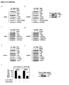
Figure 1 PKCα, βI and βII specifically phosphorylate histone H3 at threonine 6 (H3T6). Nucleosomes from HeLa cells (a) or bacterially expressed GST and GST-H3 proteins (b) were incubated with active PKCα, βI, βII,δ,µ,ζ or τ, as indicated. Coomassie blue staining (b, bottom panel) shows the amounts of GST fusion proteins used. Western blots were decorated with the indicated antibodies (a). -
Figure 2 Phosphorylation of H3T6 blocks demethylation atlysine 4 of histone H3 (H3K4) by LSD1. H3K4me2 or H3K4me2T6ph (a) and H3K4me1 or H3K4me1 phosphorylated at threonine 6 (H3K4me1T6ph) (b) peptides corresponding to the H3 tail residues 1-20 were incubated in the presence (a, b;panel 2 and 4) or in the absence of LSD1 (a, b;panel 1 and 3) and analysed by mass spectrometry. A shift in mass equivalent to one methyl group is indicated as "me". -
Figure 3 PKCβI phosphorylates H3T6 and controls demethylation of H3K4 during AR-dependent gene expression. For ChIP (a-i), *LNCaP cells were cultivated in the presence or absence of the AR agonist R1881 and transfected with siRNA (b-i). ChIP analyses were performed with the indicated antibodies. The precipitated chromatin was amplified by PCR using primers flanking androgen response elements (AREs) in the promoter region of the KLK2 and FKBP5 genes or primers in the promoter region of the GAPDH, U6, SCN1A, and SCN2A genes. (j), miRNA-mediated PKCβI knockdown reduces expression of the androgen-regulated KLK2 and FKBP5 genes in LNCaP cells. Bars represent mean +SD (n≥3). Western blots (c, e, j, right panels) were decorated with the indicated antibodies. -
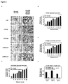
Figure 4 PKCβI, H3T6ph, H3K4me2 and H3K4me3 levels positively correlate with the malignancy of prostate cancer and control androgen-dependent tumour cell proliferation. (a), immunohistochemical staining of AR, PKCβI, H3T6ph, H3K4me2 and H3K4me3 in human normal and tumour prostate. AR (A, B), PKCβI (C, D), H3T6ph (E, F), H3K4me2 (G, H), and H3K4me3 (I, J) immunoreactivity is detected in the secretory epithelium of normal prostate (A, C, E, G, I, arrows) and prostate carcinoma cells (B, D, F, H, J, arrows). All sections were taken from the same radical prostatectomy specimen (magnification: x250). The correlation of elevated PKCβI, H3T6ph, H3K4me2 and H3K4me3 levels with high Gleason score in a panel of 154 human prostate carcinomas is highly significant: r=0.5292, p<0.0001 (b); r=0.5386, p<0.0001 (c); r=0.5373, p<0.0001 (d); r=0.5395, p<0.0001 (f). Normal prostate specimens are included as a control. (e), miRNA-mediated PKCβI knockdown severely reduces R1881-induced cell proliferation in LNCaP cells. Bars represent mean +SD (n≥6). -
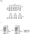
Figure 5 Alphabetical list of the 97 kinases (ProQinase GmbH) used to screen for H3T6 specific kinases. -
Figure 6 The α-H3T3ph21, α-H3T6ph (# ab14102 lot 481643; Abcam), α-H3S10ph (# 06-570 lot 32219; Upstate), and α-H3T11ph3 antibodies used for Western blot analysis or ChIP assays specifically recognize histone H3 phosphorylated at threonine 3 (H3T3ph), histone H3 phosphorylated at threonine 6 (H3T6ph), histone H3 phosphorylated at serine 10 (H3S10ph), and histone H3 phosphorylated at threonine 11 (H3T11ph), respectively. The indicated amounts of peptides were spotted onto nitrocellulose (Protran BA 79, Schleicher & Schuell). H3T3ph, H3T6ph, H3S10ph, and H3T11ph peptides were obtained from Abcam. The unmodified H3 peptide was obtained from Peptides & Elephants. Western blots were decorated as indicated. Control shows the amounts of Ponceau red stained peptides (bottom panel). -
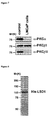
Figure 7 PKCβI but not PKCα or PKCβII is expressed in LNCaP cells. Western blots were decorated with the indicated antibodies. -
Figure 8 Coomassie blue staining shows the bacterially expressed and purified His-LSD1 protein used in the demethylation assay. -
Figure 9 Coomassie blue staining shows the purified GST and GST-JARID1B protein used in the demethylation assay. -
Figure 10 Phosphorylation of H3T6 blocks demethylation at H3K4 by JARID1B. H3K4me3 or H3K4me3T6ph (a) and H3K4me2 or H3K4me2T6ph (b) peptides corresponding to the H3 tail residues 1-20 were incubated with GST (a, b;panel 1 and 3) or GST-JARID1B (a, b;panel 2 and 4) and analysed by mass spectrometry. A shift in mass equivalent to one methyl group is indicated as "me". -
Figure 11 The specificity of the α-H3K4me2 and α-H3K4me1 antibodies is not altered by phosphorylation of H3T6. Equal amounts of either untreated nucleosomes (-) or nucleosomes phosphorylated at H3T6 in vitro by PKC (+) were used for Western blot analysis. Western blots were decorated with the indicated antibodies. -
Figure 12 PKCβI is expressed in HeLa cells (a), but does not interact with the chromatinized SCN1A promoter (b). ChIP analyses were performed with the indicated antibodies. The precipitated chromatin was amplified by PCR using primers in the promoter region of SCN1A and GAPDH. Western blots (a) were decorated with the indicated antibodies. - Unless defined otherwise, all technical and scientific terms used herein have the same meanings as commonly understood by one of ordinary skill in the art.
- The following definitions are introduced:
- It is to be understood that the term "comprise", and variations such as "comprises" and "comprising" is not limiting. For the purpose of the present invention the term "consisting of" is considered to be a preferred embodiment of the term "comprising". If hereinafter a group is defined to comprise at least a certain number of embodiments, this is meant to also encompass a group which preferably consists of these embodiments only.
- The terms "about" and "approximately" in the context of the present invention denote an interval of accuracy that a person skilled in the art will understand to still ensure the technical effect of the feature in question. The term typically encompasses a deviation from the indicated numerical value of ±10 % and preferably of ±5 %.
- The determination of percent identity between two sequences is preferably accomplished using the mathematical algorithm of Karlin and Altschul (1993) Proc. Natl. Acad. Sci USA 90: 5873-5877. Such an algorithm is e.g. incorporated into the BLASTn and BLASTp programs of Altschul et al. (1990) J. MoI. Biol. 215: 403-410 available at NCBI (http://www.ncbi.nlm.nih.gov/blast/Blast.cge).
- The determination of percent identity is preferably performed with the standard parameters of the BLASTn and BLASTp programs.
- BLAST polynucleotide searches are preferably performed with the BLASTn program.
- For the general parameters, the "Max Target Sequences" box may be set to 100, the "Short queries" box may be ticked, the "Expect threshold" box may be set to 10 and the "Word Size" box may be set to 28. For the scoring parameters the "Match/mismatch Scores" may be set to 1,-2 and the "Gap Costs" box may be set to linear. For the Filters and Masking parameters, the "Low complexity regions" box may not be ticked, the "Species-specific repeats" box may not be ticked, the "Mask for lookup table only" box may be ticked, the "Mask lower case letters" box may not be ticked.
- BLAST protein searches are preferably performed with the BLASTp program.
- For the general parameters, the "Max Target Sequences" box may be set to 100, the "Short queries" box may be ticked, the "Expect threshold" box may be set to 10 and the "Word Size" box may be set to "3". For the scoring parameters the "Matrix" box may be set to "BLOSUM62", the "Gap Costs" Box may be set to "Existence: 11 Extension:1", the "Compositional adjustments" box may be set to "Conditional compositional score matrix adjustment". For the Filters and Masking parameters the "Low complexity regions" box may not be ticked, the "Mask for lookup table only" box may not be ticked and the "Mask lower case letters" box may not be ticked.
- One letter amino acid abbreviations used herein correspond to IUPAC nomenclature (see e.g. European Journal of Biochemistry, 138:9-37, 1984).
- The term "RNA interference" or "RNAi" as used herein refers to an RNA induced block of gene expression in a specific and post-transcriptional manner by degradation of a specific target mRNA.
- The term "Gleason score" as used herein refers to a means of grading prostate cancer. Cancers with a higher Gleason score are more aggressive and have a worse prognosis. The Gleason score may e.g. be determined based upon the microscopic appearance of a tumor sample. The Gleason score may further be determined by assigning a grade to the most common tumor pattern and a second grade to the next most common tumor pattern in a sample from the tumor and adding the two grades to get the Gleason score. The Gleason grade typically ranges from 1 to 5 with 5 having the worst prognosis, while the Gleason score typically ranges from 2 to 10 with 10 having the worst prognosis.
- The terms "cancer" or "tumor" as used herein are meant to include different types of cancers such as e.g. prostate cancer, breast cancer, colorectal cancer, lung cancer, blood cancer and cancers of the brain. Other cancers or tumors may include e.g. non-Hodgkin lymphoma, head and neck cancer, non-small cell lung cancer, ovarian cancer or urinary bladder cancer. One preferred type of cancer in the context of the present invention is prostate cancer. Another preferred type of cancer in the context of the present invention is breast cancer.
- The term "isolated" in the context of the present invention indicates that a polynucleotide has been removed from its natural environment and/or is presented in a form in which it is not found in nature.
- The term "subject" as used herein preferably refers to a human. However, veterinary applications are also in the scope of the present invention.
- The term "subject" can therefore also refer to an animal, preferably a mammal such as e.g. non-human primates, mice, rats, rabbits, guinea pigs, dogs, cats, cattle, horses, sheep, pigs, goats and the like.
- The term "aptamer" as used herein refers to a polynucleotide that has a specific binding affinity for a target compound or molecule of interest, e.g. a protein. Aptamers may e.g. be RNA, single stranded DNA, modified RNA or modified DNA molecules. The preparation of aptamers is well known in the art and may involve, inter alia, the use of combinatorial RNA libraries to identify binding sites (reference may e.g. be made to Gold (1995), Ann. ).
- Histones are the main protein components of chromatin. They form the unit around which DNA is coiled in the nucleosomes of eukaryotic chromosomes. Histone H3 is one of the five main histone proteins involved in the structure of chromatin in eukaryotic cells. Histones may be modified by posttranslational modifications such as e.g. lysine acetylation, lysine methylation, lysine ubiquitylation, arginine methylation or serine, threonine or tyrosine phosphorylation.
- The terms "histone H3" or "H3" as used herein may refer to any known histone H3 or variants thereof. Preferably, said histone H3 is mammalian histone H3, most preferably human histone H3.
- In some embodiments histone H3 may for example have an amino acid sequence selected from any of SEQ ID NO: 7, 8 or 9 (corresponding to accession number AAA63185.1, accession number NP_066403.2 and accession number CAA88778.1 respectively) or variants thereof. Preferably, said variants are naturally occuring variants. If histone H3 has an amino acid sequence selected from any of SEQ ID NO: 7, 8 or 9, histone H3 may lack the N-terminal methionine residue of said sequence (e.g. due to a post-translational loss of said residue).
- Thus, in one preferred embodiment histone H3 has an amino acid sequence selected from any of SEQ ID NO: 7, 8 or 9, wherein said sequence lacks the N-terminal methionine residue.
- In some preferred embodiments Histone H3 consists of an amino acid sequence that is at least 70%, 75%, 80%, 85%, 86%, 87%, 88%, 89%, 90%, 91%, 92%, 93%, 94%, 95%, 96%, 97%, 98% or 99% identical to the amino acid sequence according to any of SEQ ID NO: 7, 8 or 9.
- The position of amino acid residues within the histone H3 sequence, e.g. threonine 6 (T6) or lysine 4 (K4), is indicated herein with respect to the position of residues in the sequence according to any of SEQ ID NO: 7, 8 or 9, starting from the N-terminal end and sequentially numbering the amino acid residues wherein the number '0' is assigned to the N-terminal methionine residue, which is typically removed from the mature H3 protein in vivo by post-translational modification. In some embodiments where histone H3 has an amino acid sequence different from SEQ ID NO: 7, 8 or 9, the amino acid residues corresponding to
threonine 6 and/or lysine 4 (or any other residue of interest) of SEQ ID NO: 7, 8 or 9 may be shifted to another position within said amino acid sequence (e.g. due to amino acid insertions or deletions). In such a case, the amino acids corresponding tothreonine 6 and/or lysine 4 (or any other residue of interest) of SEQ ID NO: 7, 8 or 9 may e.g. be determined by aligning the sequence of said histone H3 to any or all of SEQ ID NO: 7, 8 or 9 and detecting the amino acid residues in said sequence which align tothreonine 6 and/or lysine 4 (or any other residue of interest) of SEQ ID NO: 7, 8 and/or 9. The skilled person knows how to perform such sequence alignments. For example, blast programs publicly available at NCBI may be used. Preferably, said programs are used with standard parameters. - In one embodiment,
threonine 6 andlysine 4 of histone H3 as used in the methods of the invention are comprised in the following consensus sequence of histone H3: - ARXKXTAR;
- The inventors of the present invention have surprisingly found that protein kinase C ß1 (herein also referred to as PKCßI) phosphorylates histone H3 at
threonine 6 and that said phosphorylation blocks demethylation of methylatedlysine 4 of histone H3 during androgen receptor (also referred to herein as "AR") induced gene expression. The inventors of the present invention have also surprisingly found that elevated levels of protein kinase C ß1 (PKCßI), histone H3 phosphorylated at threonine 6 (herein also referred to as H3T6ph), histone H3 monomethylated at lysine 4 (herein also referred to as H3K4me1), histone H3 dimethylated at lysine 4 (herein also referred to as H3K4me2) and histone H3 trimethylated at lysine 4 (herein also referred to as H3K4me3) correlate with high Gleason scores in human prostate carcinoma. The present disclosure thus discloses protein kinase C βI (PKCßI), histone H3 phosphorylated at threonine 6 (H3T6ph), histone H3 monomethylated at lysine 4 (H3K4me1), histone H3 dimethylated at lysine 4 (H3K4me2) and histone H3 trimethylated at lysine 4 (H3K4me3) as novel diagnostic markers for cancer, in particular prostate cancer, which provide the advantage that they can also be used to determine the grade and thus the aggressiveness of a given cancer and to monitor the efficacy of treatment of cancer. Furthermore, several of all of the diagnostic biomarkers according to the disclosure may be analyzed in parallel within the same sample from a subject thus improving accuracy of detecting cancer in a sample from a subject. - The present invention in a first aspect relates to a method for detecting, grading and/or prognosticating cancer comprising the step of determining in a sample from a subject the amount of histone H3 phosphorylated at threonine 6 (H3T6ph).
- The methods according to the invention are performed in vitro.
- In one embodiment, determining the amount of H3T6ph may e.g. be achieved by contacting the sample from the subject with a detecting agent specific for H3T6ph, i.e. the amount of H3T6ph may be detected by contacting the sample from the subject with a detecting agent specific for H3T6ph. A detecting agent is specific for a given target, if it binds said target with a higher affinity than any other compound in a sample (i.e. a non-target). For example, an antibody is specific for H3T6ph if it binds to H3T6ph with an affinity higher than the affinity with which it binds to H3K4me1, H3K4me2, H3K4me3 or any other compound present in the sample. Preferably, a detecting agent specific for a given target binds to said target only and does not bind at all to a non-target.
- A suitable detecting agent may preferably be an antibody or an aptamer. Preferably, the antibody is a monoclonal or polyclonal antibody. In some embodiments the detecting agent may also be selected from antibody variants or fragments such as e.g. single chain antibodies, diabodies, minibodies, single chain Fv fragments (sc(Fv)), sc(Fv)2 antibodies, Fab fragments or a F(ab')2 fragments. In one preferred embodiment commercially available antibodies specific for H3T6ph may be used. Examples of preferred commercially available antibodies are described herein below in the example section.
- Antibodies may be produced according to any suitable method known to the person skilled in the art. Polyclonal antibodies may e.g. be produced by immunization of animals with the antigen of choice, whereas monoclonal antibodies of defined specificity may e.g. be produced using the hybridoma technology developed by Köhler and Milstein (Köhler and Milstein, 1976, Eur. J. Immunol., 6:511-519).
- In a preferred embodiment, a detecting agent as described herein above may comprise a detectable label. Any suitable label, which can be attached to the detecting agent may be used. In one preferred embodiment the detectable label is covalently or non-covalently attached to the detecting agent. Examples of labels that may be attached to the detecting agent include e.g. fluorescent dyes such as e.g. Cyanine dyes,
e.g. Cyanine 3,Cyanine 5 orCyanine 7, Alexa Fluor dyes, e.g. Alexa 594, Alexa 488 or Alexa 532, fluorescein family dyes, R-Phycoerythrin, Texas Red and rhodamine. Detecting agents may also be labeled with enzymes such as e.g. horseradish peroxidase, alkaline phosphatase or beta-lactamase, radioisotopes such as e.g. 3H, 14C, 32P, 33P, 35S or 125I or metal such as e.g. gold. In another preferred embodiment the detecting agent may also be detected by a secondary detecting agent comprising a label as described above. Preferably a secondary detecting agent is capable of specifically binding to the above described detecting agent. In a particularly preferred embodiment a secondary detecting agent is an antibody. - In some embodiments the amount of H3T6ph may e.g. be detected in methods involving histological or cell-biological procedures. In some embodiments, visual techniques, such as light microscopy or immunofluoresence microscopy, or flow cytometry or luminometry may be used. In a preferred embodiment H3T6ph is detected by immunohistochemistry.
- In further preferred embodiments determining the amount of H3T6ph in the sample from the subject may be performed alongside measuring or determining the amount of other compounds or factors, such as e.g. determining the level of prostate-specific antigen (PSA) or Insulin-like Growth Factor-1 (IGF-1) in the same sample or in a different sample from the same subject.
- The term "detecting cancer" as used herein means that the presence of a cancerous disease or disorder may be identified in a subject or in a sample from a subject. Preferably, said subject is previously not known to suffer from cancer. In one preferred embodiment the subject is suspected to suffer from cancer. The term "detecting cancer" as used herein is thus meant to encompass "diagnosing cancer". The terms "detecting cancer" and "diagnosing cancer" may be used interchangeably.
- The determination or identification of a cancerous disease or disorder may e.g. be accomplished by comparing the amount of H3T6ph determined in the sample from the subject to the amount of the respective compound being present in a control as herein described below.
- The term "grading cancer" as used herein refers to classifying the cancer by determining certain features of the cancer, such as e.g. its aggressiveness and its prognosis.
- The inventors of the present invention have surprisingly found that elevated levels of protein kinase C ß1, H3T6ph, H3K4me1, H3K4me2 and H3K4me3 correlate with high Gleason scores in human prostate cancer. Therefore, in one preferred embodiment "grading cancer", preferably prostate cancer, in the context of the present invention may be performed by correlating the amount of H3T6ph determined in the sample from the subject to cancer grade, preferably prostate cancer grade.
- Correlating the amount of H3T6ph determined in the sample from the subject to cancer grade may for example be achieved by comparing the amount of H3T6ph determined in the sample to previously determined values for the amount of H3T6ph which was determined in one or more control sample(s) from subjects known to suffer from cancer and which were assigned to different tumor grades, preferably to different Gleason scores.
- The term "prognosticating cancer" as used herein refers to the prediction of the course or outcome of a diagnosed or detected cancerous disease, e.g. during a certain period of time, e.g. during treatment or after treatment. The term may also refer to a determination of chance of survival or recovery from the cancerous disease, as well as to a prediction of the expected survival time of a subject.
- For example, the amount of H3T6ph may be determined in a sample from a subject at a given point of time and compared to the respective amount of H3T6ph determined in a sample from the same subject at a later point of time, wherein an increase or decrease in the amount of H3T6ph indicates cancer cell proliferation or cancer regression respectively. Such an approach may e.g. be used during cancer treatment, e.g. during application of anti-cancer medication to a subject suffering from cancer.
- Thus in one preferred embodiment the methods according to the invention may be used to monitor the efficacy of cancer treatment in vitro. The efficacy of cancer treatment may e.g. be monitored by detecting the amount of H3T6ph in different samples from a subject that were provided over a given period of time while the subject from which the samples were derived was subjected to anti-cancer treatment. An increase or decrease in the amount of H3T6ph in samples provided from the subject over a given period of time may then indicate the efficacy of anti-cancer treatment.
- In a preferred embodiment the above method further comprises the step of comparing the amount of histone H3 phosphorylated at threonine 6 (H3T6ph) determined in the sample from the subject to the amount of the respective compound determined in a control. The term "respective compound " means that the amount of each compound in the sample is compared to the amount of the respective compound in the control, i.e. e.g. the amount of histone H3 phosphorylated at threonine 6 (H3T6ph) in the sample is compared to the amount of histone H3 phosphorylated at threonine 6 (H3T6ph) in the control.
- In one preferred embodiment the control is a sample from a healthy subject. In a preferred embodiment a higher amount of H3T6ph determined in the sample from the subject in comparison to the control indicates the presence of cancer in the subject. The term "higher amount" means that the amount of H3T6ph determined in the sample from the subject is preferably at least 2 fold, at least 3 fold, at least 4 fold, at least 5 fold, at least 10 fold, at least 20 fold, at least 30 fold, at least 40 fold, at least 50 fold, at least 60 fold, at least 70 fold, at least 80 fold, at least 90 fold, at least 100 fold, at least 500 fold, at least 1000 fold or at least 10000 fold higher than in the control.
- In some other preferred embodiments, preferably in cases where grading of the cancer is desired, the control may also be a sample derived from a subject known to suffer from cancer, i.e. a subject that has been independently diagnosed with cancer, preferably with prostate cancer or breast cancer. Preferably a control derived from a subject known to suffer from cancer has previously been subjected to cancer grading. Thus, in one preferred embodiment the amount of histone H3 phosphorylated at threonine 6 (H3T6ph) determined in the sample from the subject in comparison to the control may indicate the grade of the cancer present in the subject.
- In one preferred embodiment the amount of H3T6ph in the control may be determined in parallel to the amount of H3T6ph in the sample from the subject.
- In another preferred embodiment the control may be a predetermined value. Such a value may e.g. be based on the results of previous experiments determining the amount of H3T6ph, in one or more samples from a healthy subject or a subject known to suffer from cancer, preferably prostate cancer or breast cancer. In some embodiments a predetermined value may be derivable from a database.
- In one preferred embodiment the amount of H3T6ph may be compared to more than one control, e.g. to 2, 3, 4, 5, 6, 7, 8, 9, 10, 20, 30, 40, 50, 60, 70, 80, 90, 100 or more than 100 controls.
- . In a preferred embodiment determining the amount of
protein kinase beta 1 and/or 2 protein in the sample from the subject is performed alongside determining the amount of histone H3 phosphorylated at threonine 6 (H3T6ph), the amount of histone H3 monomethylated at lysine 4 (H3K4me1), the amount of histone H3 dimethylated at lysine4 (H3K4me2) and the amount of histone H3 trimethylated at lysine 4 (H3K4me3) in the same sample or in a further sample from the same subject. - The sample from the subject used in the methods according to the invention is separated from the body of the subject. The sample may be solid or liquid. In a preferred embodiment the sample from the subject is a body fluid or tissue sample, preferably a prostate or breast tissue sample. Preferably, the tissue sample is derived from a cancer tissue, most preferably a prostate or breast cancer tissue. Tissue samples may e.g. be fresh or frozen tissue samples or fixed paraffin embedded samples. In one preferred embodiment the sample may be a biopsy or resection sample.
- Preferably, the body fluid is blood, plasma, urine, saliva, serum, semen, prostate fluid or seminal fluid.
- In some embodiments, a liquid sample may be enriched for cells of interest, e.g. prostate cells. Enrichment may be performed by any method known to the skilled person. Enrichment may e.g. be achieved by using a solid support, e.g. a column, coated with a specific antibody, such as e.g. a prostate specific antibody. Alternatively, enrichment may e.g. also be achieved by using filtration methods or by immobilizing specific aptamers on a microfluidic channel and pumping the liquid sample through the device.
- In a further aspect the present invention also relates to a diagnostic kit for detecting, grading, and/or prognosticating cancer comprising a detecting agent specific for histone H3 phosphorylated at threonine 6 (H3T6ph), wherein the detecting agent is an antibody, an aptamer or an oligonucleotide probe.
- Preferably, the diagnostic kit according to the invention contains one or more detecting agents specific for histone H3 phosphorylated at threonine 6 (H3T6ph). If the diagnostic kit according to the invention comprises more than one detecting agent, said detecting agents are preferably each specific for histone H3 phosphorylated at threonine 6 (H3T6ph). Suitable detecting agents have been described herein above. The diagnostic kit may further comprise additional components or reagents that may be suitable for performing the methods according to the invention, such as e.g. buffers or controls. The components contained in the diagnostic kit may be comprised in one or more containers. The diagnostic kit according to the present invention may also comprise an instruction leaflet, which indicates how to use the diagnostic kit and its components.
- The present invention in a further aspect also relates to the use of a detecting agent specific for histone H3 phosphorylated at threonine 6 (H3T6ph), detecting, grading and/or prognosticating cancer in a sample from a subject, wherein the detecting agent is an antibody, an aptamer or an oligonucleotide probe. Preferably, said use in vitro.
- Suitable detecting agents are described herein above.
- In another aspect the present invention relates to the diagnostic kit according to the invention or the use of a detecting agent specific for histone H3 phosphorylated at threonine 6 (H3T6ph) for detecting, grading and/or prognosticating cancer in a sample from a subject, wherein the detecting agent is an antibody, an aptamer or an oligonucleotide probe.
- Examples of suitable antibodies, aptamers and oligonucleotide probes have been described herein above.
- In a further aspect the present invention relates to the methods according to the invention, the diagnostic kit according to the invention, and/or the use according to the invention, wherein the cancer is selected from the group consisting of prostate cancer, breast cancer, lung cancer, pancreatic cancer, neuroblastoma, melanoma, brain cancer, kidney cancer, bladder cancer, ovarian cancer, blood cancer and colon cancer. In a particularly preferred embodiment the cancer is prostate cancer.
- The present invention will now be described with respect to some of its specific examples.
- The following plasmids were described previously: pGEX-4T1-H3 1-15, pGEX-4T1-H3 16-30, pGEX-4T1-H3 29-44, pET-GEX H3 1-135, and pLenti6-miRNA-control3. GST-H3 1-135 T6A was provided by J.M.G. Higgins. To construct GST H3 1-15 T6A the corresponding cDNA fragment was cloned into pGEX-4T1. To construct pLenti6-miRNA-PKCβI, the DNA corresponding to miRNA-PKCβI (5'-TGCTGTTACGTAGGGATCTGACAGGCGTTTTGG CCACTGACTGACGCCTGTCATCCCTACGTAA-3' (SEQ IDNO:6) and 5'-CCTGTTACGTAGGGATGACAGGCGTCAGTCAGTGGCCAAAACGCCTGTCA GATCCCTACGTAAC-3') (SEQ ID NO:5) was cloned into pLenti6/V5-DEST according to the manufacturer's instructions (Invitrogen).
- ChIP and Re-ChIP experiments were performed as described3. LNCaP cells were cultured for 15min (ChIP) or 210 min (Re-ChIP) in the presence or absence of 1 x 10⁻⁸ M R1881 as indicated. Three days before harvesting, cells were transfected with siRNA (control for PKCβI: 5'-CAUUCCUAUGCGAGACGUAUGUAAA-3'(SEQ IDNO 3); PKCβI: 5'-CAUUGUCCUCGUAAGAGAUGCUAAA-3' (SEQ ID NO 4) (
Fig. 3 b, c, f-i ); control for PRK1: 5'-GAAAGUCCUAGAUCCACACGCAAAU-3' (SEQ ID NO 14); PRK1: 5'-GAACAUGAUCCAGACCUACAGCAAU-3' (SEQ ID NO: 15) (Fig. 3 d, e ); Invitrogen) using RNAifect (Qiagen) following the manufacturer's instructions. Immunoprecipitation was performed with specific antibodies (α-AR # 06-680 lot 33529 (Upstate Biotechnology), α-H3K4me1 # ab8895 lot 535654, α-H3T6ph # ab14102 lot 481643, α-H3 # ab1791 lot 172452, α-PKCβI S660ph # ab23513 lot 596904 (Abcam), α-PKCβI (C-16) # sc-209 lot 481643, (Santa Cruz), α-H3K4me2 # cs-035-100 lot A391001 (Diagenode), α-PRK13, and α-LSD12) on protein A-Sepharose 4B (GE-Healthcare). For PCR, 2 µl out of 50 µl DNA extract was used. PCR primers for FKBP5 (+351/+443), KLK2 (-343/- 90), SCN1A, SCN2A, GAPDH, and U6 were described previously 1,2,6,11. - Experiments were performed as described2. Western blots were decorated as indicated.
- Experiments were performed as described2. pLenti6-miRNA-control and pLenti6-miRNA-PKCβI were used to produce recombinant lentiviruses to infect LNCaP cells as described15. The infected cells were cultured for 72 hours in medium containing 10% double-stripped FCS. 1x10⁴ cells were plated in a 96-well plate in the presence or absence of 1 x 10⁻⁹ M R1881. The cell proliferation Elisa BrdU Colorimetric Assay (Roche) was performed according to the manufacturer's instructions. The figure shows the increase of proliferation in the presence versus absence of R1881. The experiments were performed in hexaplicate.
- Quantitative RT-PCR and statistical analysis were performed as described2. The primers for KLK2 and FKBP5 were described previously6,11.
- The kinase assays were performed as described3. Ten µg of either GST-H3 mutants (
Fig. 1b), or 1 µg of nucleosomes (Fig. 1a ) purified from HeLa cells16 were incubated with the amount corresponding to 12 pmol/min-1 purified recombinant PKC (ProQinase) at 30°C in kinase buffer containing 60 mM Hepes-NaOH pH 7.5, 3 mM MgCl2, 3 mM MnCl2 and either 10 mM ATP (Fig. 1a ) or5 µCi [γ-32P]ATP (Fig. 1b ). The reaction mixture was analysed by Western blotting using antibodies as indicated (Fig. 1a ) or SDS-PAGE followed by autoradiography (Fig. 1b ). - Stainings were performed using a protocol17 for antigen retrieval and indirect immunoperoxidase. α-AR (# sc-7305 lot E171; Santa Cruz), α-PKCβI (# sc-209 lot 481643; Santa Cruz), α-H3T6ph (# ab14102 lot 481643; Abcam), and α-H3K4me2 (# ab7766-100, lot 56290; Abcam) antibodies were used at a dilution of 1:75, 1:1000, 1:200, and 1:1000, respectively. Immunoreactions were performed with the universal vectastain ABC kit according to the manufacturer's instructions (Vector Laboratories).
- Clinical data of patients and procedures for generating the tissue microarrays were described previously17. Tissue microarrays were prepared from formalin-fixed, paraffin-embedded tissue specimens of 154 prostates selected from the archival files of the Institute of Pathology, University of Bonn Medical School. All tumour samples were surgically obtained from patients who had undergone radical retropubic prostatectomy in two surgical centres between 1995 and 2002 for clinically organ confined prostate cancer (preoperative staging </= cT2, cNo, cMo). Patients who had received prior hormonal therapy, chemotherapy, or radiation therapy were excluded from the study. All cases were re-evaluated by a panel of experienced pathologists for histopathological staging according to the UICC TNM-system18, rescored according to the Gleason scoring system19 and subsequently followed-up between 21 to 128 months (median 40.24 months). Three different tissue cores representing the lowest and highest Gleason grades within a single tumour were arrayed from formalin-fixed, paraffin-embedded tissue blocks using a manual device (Beecher Instruments). 4 µm paraffin sections were cut from every TMA and used for subsequent immunohistochemical analyses within one week. Statistical analysis was performed with the Mann-Whitney U-Test using the SPSS 12.0 program (SPSS Inc.) and by calculating the two-tailed Spearman Rank correlation coefficient. The number of cases (n) analysed per Gleason score (Gs) were: Gs 3 (n=5); Gs 4 (n=12); Gs 5 (n=14); Gs 6 (n=38); Gs 7 (n=24); Gs 8 (n=27); Gs 9 (n=20); Gs 10 (n=14). Normal prostate specimens (n=25).
- The demethylation assays were performed essentially as described1, 20. One µg peptides corresponding to the H3 tail residues 1-20 carrying either one, two, or three methyl groups at K4 in the absence or presence of a phosphorylated T6 were incubated with 5 µg bacterially expressed and purified His-LSD1, or 5 µg baculovirus expressed and purified GST-JARID1B and GST as indicated. Peptides were incubated for 5 hours at 37°C in demethylation buffer containing 50 mM Tris HCl pH 7.5, 50 mM KCl, 5 mM MgCl2 or 50 mM HEPES-KOH pH 8.0, 2 mM ascorbate, 100 µM Fe(NH4)2(SO4)2, 1 mM α-ketoglutarate1, 20. The crude demethylase reactions were diluted 1:10 in a saturated CHCA solution (50% acetonitril, 0.1%TFA) and spotted onto a MALDI-target plate. For each reaction 2000 spectra were recorded and analysed using the Data Explorer Software™.
- To identify putative H3T6 kinases, in vitro kinase screens were performed by incubating nucleosomes purified from HeLa cells with 97 recombinantly expressed, purified kinases (
Fig. 5 ). Western blot analysis performed with antibody specific to phosphorylated H3T6 (aH3T6ph) demonstrates that only PKCα, β1 and β2 phosphorylate nucleosomes at H3T6 (Fig. 1a ). PKCα,β1 and β2 specifically target H3T6 but notthreonine 3,serine 10, orthreonine 11 of histone H3 (H3T3, H3S10, H3T11, respectively) (Fig. 1a ). Specificity of the antibodies was verified (Fig. 6 ). Western blot analysis performed with α-PKCα, β1 or β2 antibodies shows that in LNCaP human prostate cancer cells only PKCβI is expressed (Fig.7 ). Therefore, all further studies were continued with PKCβI. To corroborate in an independent approach that PKCβI exclusively phosphorylates H3T6, PKCβI was incubated with recombinant, purified GST-H3 or GST control proteins in the presence of [γ-32P]ATP. In these experiments only full-length histone H3 (H3 1-135) and a H3 fragment spanningamino acid residues 1 to 15 (H3 1-15), but not H3 16-30 or H3 29-44, are phosphorylated by PKCβI (Fig. 1b ). Furthermore, mutation ofthreonine 6 to alanine in either H3 1-15 (H3 1-15 T6A) or in full length H3 (H3 1-135 T6A) abolishes phosphorylation, demonstrating that PKCβI targets H3T6 only (Fig. 1b ). Taken together, said data identify PKCβI as the first H3T6 kinase. - Demethylation assays were performed with di-, and monomethyl H3K4 peptides either unmodified at T6 (H3K4me2 and H3K4me1) or phosphorylated at T6 (H3K4me2T6ph and H3K4me1T6ph) and recombinant, purified LSD1 (
Fig. 2 a, b andFig. 8 ). Following the demethylation assay, the peptides were analysed by mass spectrometry. The robust demethylation of H3K4me2 observed in the presence of LSD1 (Fig. 2 a , comparepanels 1 and 2) is completely blocked when the peptides are phosphorylated at T6 (Fig. 2 a , comparepanels 3 and 4). Likewise, demethylation of H3K4me1 by LSD1 is completely abrogated in the presence of phosphorylated T6 (Fig. 2 b ,panels 3 and 4). Since LSD1 only removes mono- and dimethyl marks1, 2, it was tested whether phosphorylation at H3T6 also blocks demethylation of tri-, and dimethyl H3K4 (H3K4me3 and H3K4me2) by members of the KDM5 family of JMJC domain-containing demethylases such as JARID1B/KDM5B9. Recombinant, purified JARID1B (Fig. 9 ) efficiently demethylates H3K4me3 and H3K4me2 peptides (Fig. 10 a, b , comparepanels 1 and 2). In contrast, demethylation of tri- and dimethyl H3K4 peptides phosphorylated at T6 (H3K4me3T6ph and H3K4me2T6ph) is severely impaired (Fig. 10 a, b , comparepanels 3 and 4). These results demonstrate that phosphorylation of H3T6 blocks demethylation of H3K4 methyl marks in vitro. - Western blot analysis performed with anti-PKC α, β1 or β2 antibodies indicates that only PKCβI is expressed in LNCaP cells (
Fig. 7 ). Therefore, it was investigated whether PKCβI is present at AR target genes and phosphorylates H3T6 in vivo. To analyse first whether PKCβI binds to promoters of AR target genes, LNCaP cells were subjected to chromatin immunoprecipitation (ChIP) in the presence or absence of the AR agonist R1881. PKCβI specifically associates with the androgen response elements (AREs) located in the promoters of the KLK210 and FKBP511 genes (Fig. 3a , upper panel). Association of PKCβI with AR target promoters is specific since DNA from neither GAPDH nor U6 promoters is enriched (Fig. 3a , upper panel). PKCβI binds to chromatin in a ligand-independent manner similar to LSD1 (ref2 and seeFig. 3a , upper panel). In comparison, Protein-kinase-C-related kinase 1 (PRK1) and AR bind chromatin only in a ligand-dependent manner. To investigate whether PKCβI and AR are present in the same complex on the KLK2 and FKBP5 promoters, R1881-treated LNCaP cells were subjected to sequential chromatin immunoprecipitation (Re-ChIP), first with α-AR and then with α-PKCβI antibody. PKCβI and AR form a complex on chromatin upon addition of ligand (Fig. 3a , lower panel). Similarly, it was investigated whether PKCβI and LSD1 are present in the same complex on the KLK2 and FKBP5 promoters. Re-ChIP first with α-PKCβI and then with α-LSD1 antibody demonstrates that PKCβI and LSD1 form a complex on chromatin in a ligand-independent manner (Fig. 3a , lower panel). - Next, it was invetigated whether PKCβI phosphorylates H3T6 at promoters of AR-regulated genes in vivo. LNCaP cells, cultured in the presence or absence of R1881, were transfected with either an unrelated control siRNA or a siRNA directed against PKCβI, and then subjected to ChIP with α-H3T6ph antibody. Importantly, addition of ligand results in phosphorylation of H3T6 at the AREs of the KLK2 and FKBP5 promoters (
Fig. 3b, c ). Androgen-induced phosphorylation at H3T6 is PKCβI-dependent since it is blocked by knockdown of PKCβI. PKCβI depletion is specific and does not affect the levels of endogenous AR (Fig. 3c , right panel). PKCβI depletion does neither affect ligand-dependent recruitment of AR nor the presence of LSD1 on AR-regulated genes (Fig. 3b, c ). Since PKCβI is present on the promoter of AR target genes irrespective of the presence or absence of ligand, whereas H3T6 phosphorylation is only observed in the presence of R1881, it was investigated whether PKCβI is phosphorylated and thereby activated in an androgen-dependent manner. Active PKCβI is characterised by phosphorylated serine 66012 (S660ph). ChIP assays using α-PKCβI S660ph antibody show androgen-induced phosphorylation at S660 of PKCβI at the KLK2 and FKBP5 promoters (Fig. 3b, c ). Taken together, these data demonstrate androgen-dependent activation of PKCβI and phosphorylation of H3T6 by PKCβI in vivo. - It was recently shown by the inventors that PRK1 is a gatekeeper for regulation of AR target gene expression3. PRK1 controls multiple processes during AR-dependent transcription, such as transition from the RNA polymerase II pre-initiation to the initiation complex and demethylation at
lysine 9 of histone H3 (H3K9)3. Therefore, it was tested whether in the presence of ligand PKCβI and PRK1 are present in the same complex at promoters of AR target genes. Re-ChIP analysis first with α-PKCβI and then with α-PRK1 antibody demonstrates that both proteins form a complex on the KLK2 and FKBP5 promoters upon addition of ligand (Fig. 3a , lower panel). It was next investigated whether PRK1 regulates PKCβI activity. LNCaP cells, grown in the presence or absence of R1881, were transfected with either an unrelated control siRNA or a siRNA directed against PRK1 and then subjected to ChIP. Ligand-dependent activation of PKCβI is blocked by knockdown of PRK1 (Fig. 3d, e ). Consequently, ligand-dependent phosphorylation of H3T6 at the AREs of the KLK2 and FKBP5 promoters is abolished (Fig. 3d, e ). PRK1 depletion is specific and does neither affect the levels of endogenous AR (Fig. 3e , right panel) nor the presence of PKCβI on AR target promoters (Fig. 3d, e ). In summary, these data demonstrate that PKCβI phosphorylates H3T6 on chromatinized promoters of AR target genes in an androgen- and PRK1-dependent manner. - It was analysed whether PKCβI protects demethylation of H3K4 at androgen regulated promoters in vivo. LNCaP cells were cultivated in the presence or absence of R1881, transfected with either an unrelated control siRNA or a siRNA directed against PKCβI and subjected to ChIP. As shown in
Fig. 3f and 3g , levels of H3K4me2 and H3K4me1 observed at the KLK2 and FKBP5 promoters in the absence of ligand are increased upon the addition of R1881, which is in agreement with previous data13. Furthermore, the ligand-dependent increase in H3K4me2 and H3K4me1 correlates with R1881-induced phosphorylation of H3T6 by PKCβI. Knockdown of PKCβI blunts ligand-induced phosphorylation of H3T6 and reduces H3K4me2 and H3K4me1 levels to those observed in the absence of R1881. Antibody recognition of H3K4 methyl marks is not altered by phosphorylation at H3T6 (Fig. 11 ). Together, said observations show that phosphorylation of H3T6 by PKCβI interferes with the removal of H3K4 methyl marks by LSD1 in vivo. Elevated levels of H3K4me2 and H3K4me1 are observed upon PKCβI knockdown in the absence of R1881 indicating androgen-independent functions of PKCβI beyond phosphorylation of H3T6 (Fig. 3f, g ). Next, it was tested whether PKCβI signalling also occurs on genes such as SCN1A or SCN2A where LSD1 removes active H3K4 methyl marks to silence gene expression1. ChIP assays performed in LNCaP and HeLa cells show that opposite to AR-activated genes, PKCβI does not associate with LSD1-silenced genes and consequently H3T6 is not phosphorylated (Fig. 3h, i andFig. 12a, b ). - To assess the physiological role of PKCβI, the levels of PKCβI, H3T6ph and H3K4me2 were analysed by immunostaining a panel of 25 normal human prostates and 154 prostate carcinomas on tissue microarrays (
Fig. 4 a-d and f ). Quantification of immunoreactivity by scoring staining intensity and percentage of positive carcinoma cells14 reveals that high PKCβI, H3T6ph, and H3K4me2 levels significantly correlate with high Gleason scores and indicate aggressive biology of the tumours (Fig. 4b-d and f ). To examine whether PKCβI regulates tumour cell proliferation, androgen-dependent proliferation of LNCaP cells was quantified upon PKCβI knockdown by RNAi. Androgen-induced proliferation of LNCaP cells is severely reduced by PKCβI knockdown when compared to control cells (Fig. 4e ), emphasising the important role of PKCβI in the control of AR-dependent tumor growth. -
- 1. Shi, Y. et al. Histone demethylation mediated by the nuclear amine oxidase homolog LSD1. Cell 119, 941-953. (2004).
- 2. Metzger, E. et al. LSD1 demethylates repressive histone marks to promote androgen-receptor-dependent transcription. Nature 437, 436-439. (2005).
- 3. Metzger, E. et al. Phosphorylation of histone H3 at ).
- 4. Strahl, B. D. & Allis, C. D. The language of covalent histone modifications. Nature 403, 41-45. (2000).
- 5. Klose, R.J., Kallin, E.M. & Zhang, Y. JmjC-domain-containing proteins and histone demethylation. Nat. Rev. Genet. 7, 715-727. (2006).
- 6. Wissmann, M. et al. Cooperative demethylation by JMJD2C and LSD1 promotes androgen receptor-dependent gene expression. Nat. Cell Biol. 9, 347-353. (2007).
- 7. Yang, M. et al. Structural basis of histone demethylation by LSD1 revealed by suicide inactivation. Nat. Struct. Mol. Biol. 14, 535-539. (2007).
- 8. Forneris, F., Binda. C., Adamo, A., Battaglioli, E. & Mattevi, A. Structural basis of LSD1-CoREST selectivity in histone H3 recognition. J. Biol. Chem. 282, 20070-20074. (2007).
- 9. Metzger, E. & Schüle R. The expanding world of histone lysine demethylases. Nat. Struct. Mol. Biol. 14, 252-254. (2007).
- 10. Kang, Z., Pirskanen, A., Janne, O. A. & Palvimo, J. J. Involvement of proteasome in the dynamic assembly of the androgen receptor transcription complex. J Biol. Chem. 277, 48366-48371. (2002).
- 11. Magee, J.A. et al. Direct, androgen receptor mediated regulation of the FKBP5 gene via a distal enhancer element. Endocrinology 147, 590-598. (2005).
- 12. Keranen L.M., Dutil E.M. & Newton A.C. Protein kinase C is regulated in vivo by three functionally distinct phosphorylations. Curr. Biol. 5, 1394-1403. (1995).
- 13. Kang, Z., Jänne O.A. & Palvimo J.J. Coregulator recruitment and histone modifications in transcriptional regulation by the androgen receptor. Mol. Endocrinol. 18, 2633-2648. (2004).
- 14. Kononen J. et al. Tissue microarrays for high-throughput molecular profiling of tumour specimens. Nat. Med. 4, 844-847. (1998).
- 15. Wiznerowicz, M. & Trono, D. Conditional suppression of cellular genes: lentivirus vector-mediated drug-inducible RNA interference. J. Virol. 77, 8957-8961. (2003).
- 16. O'Neill, T. E., Roberge, M. & Bradbury, E. M. Nucleosome arrays inhibit both initiation and elongation of transcripts by bacteriophage T7 RNA polymerase. J. Mol. Biol. 223, 67-78. (1992).
- 17. Kahl, P. et al. Androgen receptor coactivators lysine-).
- 18. Schroder, F.H. et al. The TNM classification of prostate cancer. Prostate Suppl. 4, 129-138. (1992).
- 19. Hsing, A.W., Tsao L. & Devesa S.S. International trends and patterns of prostate cancer incidence and mortality. ).
- 20. Tsukada, Y. I. et al. Histone demethylation by a family of JmjC domain-containing proteins. Nature 435, 811-816. (2005).
- 21. Dai, J. et al. The kinase haspin is required for mitotic ).
-
- <110> UNIVERSITATSKLINIKUM FREIBURG
- <120> Methods and compounds for the diagnosis and treatment of cancer
- <130> U7125/DB
- <160> 19
- <170> PatentIn version 3.3
- <210> 1
<211> 25
<212> RNA
<213> Artificial - <220>
<223> synthetic oligonucleotide; RNA or DNA - <400> 1
uuuagcaucu cuuacgagga caaug 25 - <210> 2
<211> 21
<212> RNA
<213> Artificial - <220>
<223> synthetic oligonucleotide; RNA or DNA - <400> 2
uuacguaggg aucugacaggc 21 - <210> 3
<211> 25
<212> RNA
<213> Artificial - <220>
<223> synthetic oligonucleotide - <400> 3
cauuccuaug cgagacguau guaaa 25 - <210> 4
<211> 25
<212> RNA
<213> Artificial - <220>
<223> synthetic oligonucleotide - <400> 4
cauuguccuc guaagagaug cuaaa 25 - <210> 5
<211> 64
<212> DNA
<213> Artificial - <220>
<223> synthetic oligonucleotide - <400> 5
cctgttacgt agggatgaca ggcgtcagtc agtggccaaa acgcctgtca gatccctacg 60taac 64 - <210> 6
<211> 64
<212> DNA
<213> Artificial - <220>
<223> synthetic oligonucleotide - <400> 6
tgctgttacg tagggatctg acaggcgttt tggccactga ctgacgcctg tcatccctac 60gtaa 64 - <210> 7
<211> 136
<212> PRT
<213> Homo sapiens - <400> 7

- <210> 8
<211> 136
<212> PRT
<213> Homo sapiens - <400> 8

- <210> 9
<211> 136
<212> PRT
<213> Homo sapiens - <400> 9


- <210> 10
<211> 671
<212> PRT
<213> Homo sapiens - <400> 10


- <210> 11
<211> 2711
<212> DNA
<213> Homo sapiens - <400> 11


- <210> 12
<211> 673
<212> PRT
<213> Homo sapiens - <400> 12


- <210> 13
<211> 8014
<212> DNA
<213> Homo sapiens - <400> 13


- <210> 14
<211> 25
<212> RNA
<213> Artificial - <220>
<223> synthetic oligonucleotide - <400> 14
gaaaguccua gauccacacg caaau 25 - <210> 15
<211> 25
<212> RNA
<213> Artificial - <220>
<223> synthetic oligonucleotide - <400> 15
gaacaugauc cagaccuaca gcaau 25 - <210> 16
<211> 25
<212> DNA
<213> Artificial - <220>
<223> synthetic oligonucleotide - <400> 16
tttagcatct cttacgagga caatg 25 - <210> 17
<211> 19
<212> DNA
<213> Artificial - <220>
<223> synthetic oligonucleotide - <400> 17
gcctgtcatc cctacgtaa 19 - <210> 18
<211> 21
<212> DNA
<213> Artificial - <220>
<223> synthetic oligonucleotide - <400> 18
ttacgtaggg atctgacaggc 21 - <210> 19
<211> 25
<212> DNA
<213> Artificial - <220> .
<223> synthetic oligonucleotide - <400> 19
cattgtcctc gtaagagatg ctaaa 25
Claims (9)
- A method for detecting, grading and/or prognosticating cancer comprising the step of determining in a sample from a subject the amount of histone H3 phosphorylated at threonine 6 (H3T6ph), wherein said method is performed in vitro.
- The method according to claim 1, further comprising the step of comparing the amount of histone H3 phosphorylated at threonine 6 (H3T6ph) determined in the sample from the subject to the amount of histone H3 phosphorylated at threonine 6 (H3T6ph) determined in a control.
- The method according to claim 1 or 2, wherein the sample from the subject is a body fluid or tissue sample, preferably a prostate or breast tissue sample.
- The method according to claim 3, wherein the body fluid is selected from the group of blood, plasma, urine, saliva, serum, semen, prostate fluid or seminal fluid.
- The method according to any of claims 1 to 4, wherein the cancer is selected from the group consisting of prostate cancer, breast cancer, lung cancer, pancreatic cancer, neuroblastoma, melanoma, brain cancer, kidney cancer, bladder cancer, ovarian cancer, blood cancer and colon cancer.
- A diagnostic kit for detecting, grading and/or prognosticating cancer comprising a detecting agent specific for histone H3 phosphorylated at threonine 6 (H3T6ph), wherein the detecting agent is an antibody, an aptamer or an oligonucleotide probe.
- The diagnostic kit according to claim 6, wherein the cancer is selected from the group consisting of prostate cancer, breast cancer, lung cancer, pancreatic cancer, neuroblastoma, melanoma, brain cancer, kidney cancer, bladder cancer, ovarian cancer, blood cancer and colon cancer.
- Use of a detecting agent specific for histone H3 phosphorylated at threonine 6 (H3T6ph) for detecting, grading and/or prognosticating cancer in a sample from a subject, wherein the detecting agent is an antibody, an aptamer or an oligonucleotide probe.
- The use according to claim 8, wherein the cancer is selected from the group consisting of prostate cancer, breast cancer, lung cancer, pancreatic cancer, neuroblastoma, melanoma, brain cancer, kidney cancer, bladder cancer, ovarian cancer, blood cancer and colon cancer.
Priority Applications (4)
| Application Number | Priority Date | Filing Date | Title |
|---|---|---|---|
| AT09167060T ATE534908T1 (en) | 2009-08-03 | 2009-08-03 | METHODS AND COMPOUNDS FOR THE DIAGNOSIS AND TREATMENT OF CANCER |
| EP09167060A EP2282208B1 (en) | 2009-08-03 | 2009-08-03 | Methods and compounds for the diagnosis and treatment of cancer |
| PCT/EP2010/061317 WO2011015593A1 (en) | 2009-08-03 | 2010-08-03 | Methods and compounds for the diagnosis and treatment of cancer |
| US13/388,940 US20120329854A1 (en) | 2009-08-03 | 2010-08-03 | Methods and Compounds for the Diagnosis and Treatment for Cancer |
Applications Claiming Priority (1)
| Application Number | Priority Date | Filing Date | Title |
|---|---|---|---|
| EP09167060A EP2282208B1 (en) | 2009-08-03 | 2009-08-03 | Methods and compounds for the diagnosis and treatment of cancer |
Publications (2)
| Publication Number | Publication Date |
|---|---|
| EP2282208A1 EP2282208A1 (en) | 2011-02-09 |
| EP2282208B1 true EP2282208B1 (en) | 2011-11-23 |
Family
ID=41459776
Family Applications (1)
| Application Number | Title | Priority Date | Filing Date |
|---|---|---|---|
| EP09167060A Not-in-force EP2282208B1 (en) | 2009-08-03 | 2009-08-03 | Methods and compounds for the diagnosis and treatment of cancer |
Country Status (4)
| Country | Link |
|---|---|
| US (1) | US20120329854A1 (en) |
| EP (1) | EP2282208B1 (en) |
| AT (1) | ATE534908T1 (en) |
| WO (1) | WO2011015593A1 (en) |
Families Citing this family (3)
| Publication number | Priority date | Publication date | Assignee | Title |
|---|---|---|---|---|
| WO2020041691A1 (en) * | 2018-08-24 | 2020-02-27 | City Of Hope | Oligonucleotide inhibitors of nuclear factor kappa-light-chain-enhancer |
| BR112022010774A2 (en) * | 2019-12-02 | 2022-08-23 | Belgian Volition Srl | USE OF CELL-FREE NUCLEOSOMES AS BIOMARKERS |
| JP7454756B2 (en) * | 2019-12-05 | 2024-03-25 | 国立大学法人九州大学 | Methods for diagnosing cancer, compositions for cancer diagnosis, kits for cancer diagnosis, methods for evaluating cancer status, and methods for screening cancer preventive and/or therapeutic drugs |
Family Cites Families (2)
| Publication number | Priority date | Publication date | Assignee | Title |
|---|---|---|---|---|
| US8008071B2 (en) * | 1999-11-08 | 2011-08-30 | University Of South Florida | Compositions and methods for detecting intracellular glucose and analogs thereof |
| EP2017621B1 (en) * | 2007-07-16 | 2011-06-22 | Universitätsklinikum Freiburg | Method of predicting the malignancy of prostate cancer by detecting the expression of the Protein Kinase C Related Kinase, PRK1 |
-
2009
- 2009-08-03 EP EP09167060A patent/EP2282208B1/en not_active Not-in-force
- 2009-08-03 AT AT09167060T patent/ATE534908T1/en active
-
2010
- 2010-08-03 WO PCT/EP2010/061317 patent/WO2011015593A1/en active Application Filing
- 2010-08-03 US US13/388,940 patent/US20120329854A1/en not_active Abandoned
Also Published As
| Publication number | Publication date |
|---|---|
| US20120329854A1 (en) | 2012-12-27 |
| EP2282208A1 (en) | 2011-02-09 |
| WO2011015593A1 (en) | 2011-02-10 |
| ATE534908T1 (en) | 2011-12-15 |
Similar Documents
| Publication | Publication Date | Title |
|---|---|---|
| Metzger et al. | Phosphorylation of histone H3 at threonine 11 establishes a novel chromatin mark for transcriptional regulation | |
| Reincke et al. | Mutations in the deubiquitinase gene USP8 cause Cushing's disease | |
| EP1704859B1 (en) | Androgen receptor-dependent gene expression control by inhibiting the amine oxidase activity of lysine-specific demethylase (LSD1) | |
| Lin et al. | Involvement of maternal embryonic leucine zipper kinase (MELK) in mammary carcinogenesis through interaction with Bcl-G, a pro-apoptotic member of the Bcl-2 family | |
| EP2062597A1 (en) | Preventive or remedy for er-negative and her2-negative breast cancer and method of screening the same | |
| US20120052071A1 (en) | Tumor markers and methods of use thereof | |
| JP5139332B2 (en) | Use of growth stimulating proteins | |
| US20200246313A1 (en) | Methods for diagnosing and treating prostate cancer | |
| US7858395B2 (en) | Antibodies for use in identifying and/or scoring prostate cancer and androgen receptor-dependent gene expression control | |
| EP2449383B1 (en) | Diagnostic method for predicting the risk of cancer recurrence based on histone macroh2a isoforms | |
| US8853183B2 (en) | Prognosis and treatment of breast cancer | |
| EP2282208B1 (en) | Methods and compounds for the diagnosis and treatment of cancer | |
| EP2383578A1 (en) | Tumor marker and methods of use thereof | |
| US9182404B2 (en) | Tumor markers and methods of use thereof | |
| EP1693383B1 (en) | Antibodies for use in identifying prostate cancer | |
| US8076094B2 (en) | Phosphorylation of histone H3 at threonine 11—a novel epigenetic mark for transcriptional regulation | |
| US20120100561A1 (en) | A pkn3/rhoc macromolecular complex and methods of use therefor |
Legal Events
| Date | Code | Title | Description |
|---|---|---|---|
| PUAI | Public reference made under article 153(3) epc to a published international application that has entered the european phase |
Free format text: ORIGINAL CODE: 0009012 |
|
| 17P | Request for examination filed |
Effective date: 20100923 |
|
| AK | Designated contracting states |
Kind code of ref document: A1 Designated state(s): AT BE BG CH CY CZ DE DK EE ES FI FR GB GR HR HU IE IS IT LI LT LU LV MC MK MT NL NO PL PT RO SE SI SK SM TR |
|
| AX | Request for extension of the european patent |
Extension state: AL BA RS |
|
| RIC1 | Information provided on ipc code assigned before grant |
Ipc: C12N 15/00 20060101ALI20110302BHEP Ipc: A61K 48/00 20060101ALI20110302BHEP Ipc: G01N 33/574 20060101AFI20110302BHEP |
|
| GRAP | Despatch of communication of intention to grant a patent |
Free format text: ORIGINAL CODE: EPIDOSNIGR1 |
|
| GRAC | Information related to communication of intention to grant a patent modified |
Free format text: ORIGINAL CODE: EPIDOSCIGR1 |
|
| GRAS | Grant fee paid |
Free format text: ORIGINAL CODE: EPIDOSNIGR3 |
|
| GRAA | (expected) grant |
Free format text: ORIGINAL CODE: 0009210 |
|
| AK | Designated contracting states |
Kind code of ref document: B1 Designated state(s): AT BE BG CH CY CZ DE DK EE ES FI FR GB GR HR HU IE IS IT LI LT LU LV MC MK MT NL NO PL PT RO SE SI SK SM TR |
|
| REG | Reference to a national code |
Ref country code: GB Ref legal event code: FG4D |
|
| REG | Reference to a national code |
Ref country code: CH Ref legal event code: EP |
|
| REG | Reference to a national code |
Ref country code: IE Ref legal event code: FG4D |
|
| REG | Reference to a national code |
Ref country code: DE Ref legal event code: R096 Ref document number: 602009003809 Country of ref document: DE Effective date: 20120119 |
|
| REG | Reference to a national code |
Ref country code: CH Ref legal event code: NV Representative=s name: BOVARD AG |
|
| REG | Reference to a national code |
Ref country code: NL Ref legal event code: VDEP Effective date: 20111123 |
|
| LTIE | Lt: invalidation of european patent or patent extension |
Effective date: 20111123 |
|
| PG25 | Lapsed in a contracting state [announced via postgrant information from national office to epo] |
Ref country code: LT Free format text: LAPSE BECAUSE OF FAILURE TO SUBMIT A TRANSLATION OF THE DESCRIPTION OR TO PAY THE FEE WITHIN THE PRESCRIBED TIME-LIMIT Effective date: 20111123 Ref country code: IS Free format text: LAPSE BECAUSE OF FAILURE TO SUBMIT A TRANSLATION OF THE DESCRIPTION OR TO PAY THE FEE WITHIN THE PRESCRIBED TIME-LIMIT Effective date: 20120323 Ref country code: NO Free format text: LAPSE BECAUSE OF FAILURE TO SUBMIT A TRANSLATION OF THE DESCRIPTION OR TO PAY THE FEE WITHIN THE PRESCRIBED TIME-LIMIT Effective date: 20120223 |
|
| PG25 | Lapsed in a contracting state [announced via postgrant information from national office to epo] |
Ref country code: PT Free format text: LAPSE BECAUSE OF FAILURE TO SUBMIT A TRANSLATION OF THE DESCRIPTION OR TO PAY THE FEE WITHIN THE PRESCRIBED TIME-LIMIT Effective date: 20120323 Ref country code: SI Free format text: LAPSE BECAUSE OF FAILURE TO SUBMIT A TRANSLATION OF THE DESCRIPTION OR TO PAY THE FEE WITHIN THE PRESCRIBED TIME-LIMIT Effective date: 20111123 Ref country code: SE Free format text: LAPSE BECAUSE OF FAILURE TO SUBMIT A TRANSLATION OF THE DESCRIPTION OR TO PAY THE FEE WITHIN THE PRESCRIBED TIME-LIMIT Effective date: 20111123 Ref country code: GR Free format text: LAPSE BECAUSE OF FAILURE TO SUBMIT A TRANSLATION OF THE DESCRIPTION OR TO PAY THE FEE WITHIN THE PRESCRIBED TIME-LIMIT Effective date: 20120224 Ref country code: BE Free format text: LAPSE BECAUSE OF FAILURE TO SUBMIT A TRANSLATION OF THE DESCRIPTION OR TO PAY THE FEE WITHIN THE PRESCRIBED TIME-LIMIT Effective date: 20111123 Ref country code: LV Free format text: LAPSE BECAUSE OF FAILURE TO SUBMIT A TRANSLATION OF THE DESCRIPTION OR TO PAY THE FEE WITHIN THE PRESCRIBED TIME-LIMIT Effective date: 20111123 Ref country code: HR Free format text: LAPSE BECAUSE OF FAILURE TO SUBMIT A TRANSLATION OF THE DESCRIPTION OR TO PAY THE FEE WITHIN THE PRESCRIBED TIME-LIMIT Effective date: 20111123 Ref country code: NL Free format text: LAPSE BECAUSE OF FAILURE TO SUBMIT A TRANSLATION OF THE DESCRIPTION OR TO PAY THE FEE WITHIN THE PRESCRIBED TIME-LIMIT Effective date: 20111123 |
|
| PG25 | Lapsed in a contracting state [announced via postgrant information from national office to epo] |
Ref country code: CY Free format text: LAPSE BECAUSE OF FAILURE TO SUBMIT A TRANSLATION OF THE DESCRIPTION OR TO PAY THE FEE WITHIN THE PRESCRIBED TIME-LIMIT Effective date: 20111123 |
|
| PG25 | Lapsed in a contracting state [announced via postgrant information from national office to epo] |
Ref country code: DK Free format text: LAPSE BECAUSE OF FAILURE TO SUBMIT A TRANSLATION OF THE DESCRIPTION OR TO PAY THE FEE WITHIN THE PRESCRIBED TIME-LIMIT Effective date: 20111123 Ref country code: SK Free format text: LAPSE BECAUSE OF FAILURE TO SUBMIT A TRANSLATION OF THE DESCRIPTION OR TO PAY THE FEE WITHIN THE PRESCRIBED TIME-LIMIT Effective date: 20111123 Ref country code: BG Free format text: LAPSE BECAUSE OF FAILURE TO SUBMIT A TRANSLATION OF THE DESCRIPTION OR TO PAY THE FEE WITHIN THE PRESCRIBED TIME-LIMIT Effective date: 20120223 Ref country code: EE Free format text: LAPSE BECAUSE OF FAILURE TO SUBMIT A TRANSLATION OF THE DESCRIPTION OR TO PAY THE FEE WITHIN THE PRESCRIBED TIME-LIMIT Effective date: 20111123 Ref country code: CZ Free format text: LAPSE BECAUSE OF FAILURE TO SUBMIT A TRANSLATION OF THE DESCRIPTION OR TO PAY THE FEE WITHIN THE PRESCRIBED TIME-LIMIT Effective date: 20111123 |
|
| PG25 | Lapsed in a contracting state [announced via postgrant information from national office to epo] |
Ref country code: IT Free format text: LAPSE BECAUSE OF FAILURE TO SUBMIT A TRANSLATION OF THE DESCRIPTION OR TO PAY THE FEE WITHIN THE PRESCRIBED TIME-LIMIT Effective date: 20111123 Ref country code: PL Free format text: LAPSE BECAUSE OF FAILURE TO SUBMIT A TRANSLATION OF THE DESCRIPTION OR TO PAY THE FEE WITHIN THE PRESCRIBED TIME-LIMIT Effective date: 20111123 Ref country code: RO Free format text: LAPSE BECAUSE OF FAILURE TO SUBMIT A TRANSLATION OF THE DESCRIPTION OR TO PAY THE FEE WITHIN THE PRESCRIBED TIME-LIMIT Effective date: 20111123 |
|
| REG | Reference to a national code |
Ref country code: AT Ref legal event code: MK05 Ref document number: 534908 Country of ref document: AT Kind code of ref document: T Effective date: 20111123 |
|
| PLBE | No opposition filed within time limit |
Free format text: ORIGINAL CODE: 0009261 |
|
| STAA | Information on the status of an ep patent application or granted ep patent |
Free format text: STATUS: NO OPPOSITION FILED WITHIN TIME LIMIT |
|
| 26N | No opposition filed |
Effective date: 20120824 |
|
| REG | Reference to a national code |
Ref country code: DE Ref legal event code: R097 Ref document number: 602009003809 Country of ref document: DE Effective date: 20120824 |
|
| PG25 | Lapsed in a contracting state [announced via postgrant information from national office to epo] |
Ref country code: AT Free format text: LAPSE BECAUSE OF FAILURE TO SUBMIT A TRANSLATION OF THE DESCRIPTION OR TO PAY THE FEE WITHIN THE PRESCRIBED TIME-LIMIT Effective date: 20111123 |
|
| PG25 | Lapsed in a contracting state [announced via postgrant information from national office to epo] |
Ref country code: MC Free format text: LAPSE BECAUSE OF NON-PAYMENT OF DUE FEES Effective date: 20120831 |
|
| PG25 | Lapsed in a contracting state [announced via postgrant information from national office to epo] |
Ref country code: ES Free format text: LAPSE BECAUSE OF FAILURE TO SUBMIT A TRANSLATION OF THE DESCRIPTION OR TO PAY THE FEE WITHIN THE PRESCRIBED TIME-LIMIT Effective date: 20120305 |
|
| REG | Reference to a national code |
Ref country code: IE Ref legal event code: MM4A |
|
| PG25 | Lapsed in a contracting state [announced via postgrant information from national office to epo] |
Ref country code: FI Free format text: LAPSE BECAUSE OF FAILURE TO SUBMIT A TRANSLATION OF THE DESCRIPTION OR TO PAY THE FEE WITHIN THE PRESCRIBED TIME-LIMIT Effective date: 20111123 |
|
| PG25 | Lapsed in a contracting state [announced via postgrant information from national office to epo] |
Ref country code: IE Free format text: LAPSE BECAUSE OF NON-PAYMENT OF DUE FEES Effective date: 20120803 |
|
| PG25 | Lapsed in a contracting state [announced via postgrant information from national office to epo] |
Ref country code: MT Free format text: LAPSE BECAUSE OF FAILURE TO SUBMIT A TRANSLATION OF THE DESCRIPTION OR TO PAY THE FEE WITHIN THE PRESCRIBED TIME-LIMIT Effective date: 20111123 |
|
| PGFP | Annual fee paid to national office [announced via postgrant information from national office to epo] |
Ref country code: GB Payment date: 20130823 Year of fee payment: 5 Ref country code: FR Payment date: 20130820 Year of fee payment: 5 |
|
| PG25 | Lapsed in a contracting state [announced via postgrant information from national office to epo] |
Ref country code: TR Free format text: LAPSE BECAUSE OF FAILURE TO SUBMIT A TRANSLATION OF THE DESCRIPTION OR TO PAY THE FEE WITHIN THE PRESCRIBED TIME-LIMIT Effective date: 20111123 |
|
| PG25 | Lapsed in a contracting state [announced via postgrant information from national office to epo] |
Ref country code: SM Free format text: LAPSE BECAUSE OF FAILURE TO SUBMIT A TRANSLATION OF THE DESCRIPTION OR TO PAY THE FEE WITHIN THE PRESCRIBED TIME-LIMIT Effective date: 20111123 Ref country code: LU Free format text: LAPSE BECAUSE OF NON-PAYMENT OF DUE FEES Effective date: 20120803 |
|
| PG25 | Lapsed in a contracting state [announced via postgrant information from national office to epo] |
Ref country code: HU Free format text: LAPSE BECAUSE OF FAILURE TO SUBMIT A TRANSLATION OF THE DESCRIPTION OR TO PAY THE FEE WITHIN THE PRESCRIBED TIME-LIMIT Effective date: 20090803 |
|
| PGFP | Annual fee paid to national office [announced via postgrant information from national office to epo] |
Ref country code: DE Payment date: 20140821 Year of fee payment: 6 Ref country code: CH Payment date: 20140821 Year of fee payment: 6 |
|
| GBPC | Gb: european patent ceased through non-payment of renewal fee |
Effective date: 20140803 |
|
| REG | Reference to a national code |
Ref country code: FR Ref legal event code: ST Effective date: 20150430 |
|
| PG25 | Lapsed in a contracting state [announced via postgrant information from national office to epo] |
Ref country code: MK Free format text: LAPSE BECAUSE OF FAILURE TO SUBMIT A TRANSLATION OF THE DESCRIPTION OR TO PAY THE FEE WITHIN THE PRESCRIBED TIME-LIMIT Effective date: 20111123 Ref country code: GB Free format text: LAPSE BECAUSE OF NON-PAYMENT OF DUE FEES Effective date: 20140803 |
|
| PG25 | Lapsed in a contracting state [announced via postgrant information from national office to epo] |
Ref country code: FR Free format text: LAPSE BECAUSE OF NON-PAYMENT OF DUE FEES Effective date: 20140901 |
|
| REG | Reference to a national code |
Ref country code: DE Ref legal event code: R119 Ref document number: 602009003809 Country of ref document: DE |
|
| REG | Reference to a national code |
Ref country code: CH Ref legal event code: PL |
|
| PG25 | Lapsed in a contracting state [announced via postgrant information from national office to epo] |
Ref country code: LI Free format text: LAPSE BECAUSE OF NON-PAYMENT OF DUE FEES Effective date: 20150831 Ref country code: CH Free format text: LAPSE BECAUSE OF NON-PAYMENT OF DUE FEES Effective date: 20150831 |
|
| PG25 | Lapsed in a contracting state [announced via postgrant information from national office to epo] |
Ref country code: DE Free format text: LAPSE BECAUSE OF NON-PAYMENT OF DUE FEES Effective date: 20160301 |