EP2279205B1 - Modified Stefin A scaffold proteins - Google Patents
Modified Stefin A scaffold proteins Download PDFInfo
- Publication number
- EP2279205B1 EP2279205B1 EP09742378.4A EP09742378A EP2279205B1 EP 2279205 B1 EP2279205 B1 EP 2279205B1 EP 09742378 A EP09742378 A EP 09742378A EP 2279205 B1 EP2279205 B1 EP 2279205B1
- Authority
- EP
- European Patent Office
- Prior art keywords
- protein
- stefin
- scaffold
- peptide
- polypeptide
- Prior art date
- Legal status (The legal status is an assumption and is not a legal conclusion. Google has not performed a legal analysis and makes no representation as to the accuracy of the status listed.)
- Active
Links
- 101710167800 Capsid assembly scaffolding protein Proteins 0.000 title claims description 134
- 101710130420 Probable capsid assembly scaffolding protein Proteins 0.000 title claims description 134
- 101710204410 Scaffold protein Proteins 0.000 title claims description 134
- 108090000765 processed proteins & peptides Proteins 0.000 claims description 255
- 108090000623 proteins and genes Proteins 0.000 claims description 159
- 102000004169 proteins and genes Human genes 0.000 claims description 158
- 102000004196 processed proteins & peptides Human genes 0.000 claims description 107
- 150000001413 amino acids Chemical group 0.000 claims description 67
- 230000037431 insertion Effects 0.000 claims description 66
- 238000003780 insertion Methods 0.000 claims description 66
- 230000035772 mutation Effects 0.000 claims description 54
- 229920001184 polypeptide Polymers 0.000 claims description 48
- DHMQDGOQFOQNFH-UHFFFAOYSA-N Glycine Chemical compound NCC(O)=O DHMQDGOQFOQNFH-UHFFFAOYSA-N 0.000 claims description 27
- 230000003993 interaction Effects 0.000 claims description 26
- 108010061641 Cystatin A Proteins 0.000 claims description 24
- 102000012193 Cystatin A Human genes 0.000 claims description 24
- 238000002493 microarray Methods 0.000 claims description 15
- 239000004471 Glycine Substances 0.000 claims description 14
- 238000000034 method Methods 0.000 claims description 12
- 239000004475 Arginine Substances 0.000 claims description 11
- ODKSFYDXXFIFQN-UHFFFAOYSA-N arginine Natural products OC(=O)C(N)CCCNC(N)=N ODKSFYDXXFIFQN-UHFFFAOYSA-N 0.000 claims description 11
- 238000012360 testing method Methods 0.000 claims description 9
- 108020004707 nucleic acids Proteins 0.000 claims description 8
- 102000039446 nucleic acids Human genes 0.000 claims description 8
- 150000007523 nucleic acids Chemical class 0.000 claims description 8
- 239000002773 nucleotide Substances 0.000 claims description 8
- 125000003729 nucleotide group Chemical group 0.000 claims description 8
- 239000000090 biomarker Substances 0.000 claims description 6
- 238000012544 monitoring process Methods 0.000 claims description 5
- 239000003795 chemical substances by application Substances 0.000 claims description 4
- 235000018102 proteins Nutrition 0.000 description 129
- 108010079855 Peptide Aptamers Proteins 0.000 description 70
- 235000001014 amino acid Nutrition 0.000 description 61
- 230000008859 change Effects 0.000 description 45
- 108020004705 Codon Proteins 0.000 description 39
- 238000002983 circular dichroism Methods 0.000 description 25
- 230000000869 mutational effect Effects 0.000 description 24
- 230000000694 effects Effects 0.000 description 21
- 108091034117 Oligonucleotide Proteins 0.000 description 19
- 238000004458 analytical method Methods 0.000 description 19
- 125000003275 alpha amino acid group Chemical group 0.000 description 16
- 125000002924 primary amino group Chemical group [H]N([H])* 0.000 description 16
- 238000007792 addition Methods 0.000 description 15
- 210000004027 cell Anatomy 0.000 description 13
- 238000002474 experimental method Methods 0.000 description 13
- 230000006870 function Effects 0.000 description 13
- 230000008901 benefit Effects 0.000 description 12
- 238000001727 in vivo Methods 0.000 description 12
- 230000004048 modification Effects 0.000 description 12
- 238000012986 modification Methods 0.000 description 12
- 239000002904 solvent Substances 0.000 description 11
- 108700026244 Open Reading Frames Proteins 0.000 description 10
- QIVBCDIJIAJPQS-UHFFFAOYSA-N Tryptophan Natural products C1=CC=C2C(CC(N)C(O)=O)=CNC2=C1 QIVBCDIJIAJPQS-UHFFFAOYSA-N 0.000 description 9
- 230000004075 alteration Effects 0.000 description 9
- 125000000151 cysteine group Chemical group N[C@@H](CS)C(=O)* 0.000 description 9
- 238000012217 deletion Methods 0.000 description 9
- 230000037430 deletion Effects 0.000 description 9
- 239000006166 lysate Substances 0.000 description 8
- 230000004850 protein–protein interaction Effects 0.000 description 8
- 239000000243 solution Substances 0.000 description 8
- 125000000539 amino acid group Chemical group 0.000 description 7
- 230000001580 bacterial effect Effects 0.000 description 7
- 239000011347 resin Substances 0.000 description 7
- 229920005989 resin Polymers 0.000 description 7
- YBJHBAHKTGYVGT-ZKWXMUAHSA-N (+)-Biotin Chemical compound N1C(=O)N[C@@H]2[C@H](CCCCC(=O)O)SC[C@@H]21 YBJHBAHKTGYVGT-ZKWXMUAHSA-N 0.000 description 6
- 108091023037 Aptamer Proteins 0.000 description 6
- 101100189913 Caenorhabditis elegans pept-1 gene Proteins 0.000 description 6
- 102000015833 Cystatin Human genes 0.000 description 6
- QIVBCDIJIAJPQS-VIFPVBQESA-N L-tryptophane Chemical compound C1=CC=C2C(C[C@H](N)C(O)=O)=CNC2=C1 QIVBCDIJIAJPQS-VIFPVBQESA-N 0.000 description 6
- 238000001142 circular dichroism spectrum Methods 0.000 description 6
- 108050004038 cystatin Proteins 0.000 description 6
- 239000003814 drug Substances 0.000 description 6
- RAXXELZNTBOGNW-UHFFFAOYSA-N imidazole Natural products C1=CNC=N1 RAXXELZNTBOGNW-UHFFFAOYSA-N 0.000 description 6
- 238000000338 in vitro Methods 0.000 description 6
- 238000004519 manufacturing process Methods 0.000 description 6
- 230000007935 neutral effect Effects 0.000 description 6
- 230000026731 phosphorylation Effects 0.000 description 6
- 238000006366 phosphorylation reaction Methods 0.000 description 6
- 230000004481 post-translational protein modification Effects 0.000 description 6
- 230000000717 retained effect Effects 0.000 description 6
- 239000006228 supernatant Substances 0.000 description 6
- 238000002198 surface plasmon resonance spectroscopy Methods 0.000 description 6
- 102000015792 Cyclin-Dependent Kinase 2 Human genes 0.000 description 5
- 108010024986 Cyclin-Dependent Kinase 2 Proteins 0.000 description 5
- 108010043121 Green Fluorescent Proteins Proteins 0.000 description 5
- 102000004144 Green Fluorescent Proteins Human genes 0.000 description 5
- 101000921786 Homo sapiens Cystatin-A Proteins 0.000 description 5
- JLCPHMBAVCMARE-UHFFFAOYSA-N [3-[[3-[[3-[[3-[[3-[[3-[[3-[[3-[[3-[[3-[[3-[[5-(2-amino-6-oxo-1H-purin-9-yl)-3-[[3-[[3-[[3-[[3-[[3-[[5-(2-amino-6-oxo-1H-purin-9-yl)-3-[[5-(2-amino-6-oxo-1H-purin-9-yl)-3-hydroxyoxolan-2-yl]methoxy-hydroxyphosphoryl]oxyoxolan-2-yl]methoxy-hydroxyphosphoryl]oxy-5-(5-methyl-2,4-dioxopyrimidin-1-yl)oxolan-2-yl]methoxy-hydroxyphosphoryl]oxy-5-(6-aminopurin-9-yl)oxolan-2-yl]methoxy-hydroxyphosphoryl]oxy-5-(6-aminopurin-9-yl)oxolan-2-yl]methoxy-hydroxyphosphoryl]oxy-5-(6-aminopurin-9-yl)oxolan-2-yl]methoxy-hydroxyphosphoryl]oxy-5-(6-aminopurin-9-yl)oxolan-2-yl]methoxy-hydroxyphosphoryl]oxyoxolan-2-yl]methoxy-hydroxyphosphoryl]oxy-5-(5-methyl-2,4-dioxopyrimidin-1-yl)oxolan-2-yl]methoxy-hydroxyphosphoryl]oxy-5-(4-amino-2-oxopyrimidin-1-yl)oxolan-2-yl]methoxy-hydroxyphosphoryl]oxy-5-(5-methyl-2,4-dioxopyrimidin-1-yl)oxolan-2-yl]methoxy-hydroxyphosphoryl]oxy-5-(5-methyl-2,4-dioxopyrimidin-1-yl)oxolan-2-yl]methoxy-hydroxyphosphoryl]oxy-5-(6-aminopurin-9-yl)oxolan-2-yl]methoxy-hydroxyphosphoryl]oxy-5-(6-aminopurin-9-yl)oxolan-2-yl]methoxy-hydroxyphosphoryl]oxy-5-(4-amino-2-oxopyrimidin-1-yl)oxolan-2-yl]methoxy-hydroxyphosphoryl]oxy-5-(4-amino-2-oxopyrimidin-1-yl)oxolan-2-yl]methoxy-hydroxyphosphoryl]oxy-5-(4-amino-2-oxopyrimidin-1-yl)oxolan-2-yl]methoxy-hydroxyphosphoryl]oxy-5-(6-aminopurin-9-yl)oxolan-2-yl]methoxy-hydroxyphosphoryl]oxy-5-(4-amino-2-oxopyrimidin-1-yl)oxolan-2-yl]methyl [5-(6-aminopurin-9-yl)-2-(hydroxymethyl)oxolan-3-yl] hydrogen phosphate Polymers Cc1cn(C2CC(OP(O)(=O)OCC3OC(CC3OP(O)(=O)OCC3OC(CC3O)n3cnc4c3nc(N)[nH]c4=O)n3cnc4c3nc(N)[nH]c4=O)C(COP(O)(=O)OC3CC(OC3COP(O)(=O)OC3CC(OC3COP(O)(=O)OC3CC(OC3COP(O)(=O)OC3CC(OC3COP(O)(=O)OC3CC(OC3COP(O)(=O)OC3CC(OC3COP(O)(=O)OC3CC(OC3COP(O)(=O)OC3CC(OC3COP(O)(=O)OC3CC(OC3COP(O)(=O)OC3CC(OC3COP(O)(=O)OC3CC(OC3COP(O)(=O)OC3CC(OC3COP(O)(=O)OC3CC(OC3COP(O)(=O)OC3CC(OC3COP(O)(=O)OC3CC(OC3COP(O)(=O)OC3CC(OC3COP(O)(=O)OC3CC(OC3CO)n3cnc4c(N)ncnc34)n3ccc(N)nc3=O)n3cnc4c(N)ncnc34)n3ccc(N)nc3=O)n3ccc(N)nc3=O)n3ccc(N)nc3=O)n3cnc4c(N)ncnc34)n3cnc4c(N)ncnc34)n3cc(C)c(=O)[nH]c3=O)n3cc(C)c(=O)[nH]c3=O)n3ccc(N)nc3=O)n3cc(C)c(=O)[nH]c3=O)n3cnc4c3nc(N)[nH]c4=O)n3cnc4c(N)ncnc34)n3cnc4c(N)ncnc34)n3cnc4c(N)ncnc34)n3cnc4c(N)ncnc34)O2)c(=O)[nH]c1=O JLCPHMBAVCMARE-UHFFFAOYSA-N 0.000 description 5
- 230000002411 adverse Effects 0.000 description 5
- 230000003247 decreasing effect Effects 0.000 description 5
- 238000013461 design Methods 0.000 description 5
- 239000005090 green fluorescent protein Substances 0.000 description 5
- 102000045247 human CSTA Human genes 0.000 description 5
- 238000001114 immunoprecipitation Methods 0.000 description 5
- 238000002360 preparation method Methods 0.000 description 5
- 238000004611 spectroscopical analysis Methods 0.000 description 5
- 108060008226 thioredoxin Proteins 0.000 description 5
- 108091028043 Nucleic acid sequence Proteins 0.000 description 4
- FAPWRFPIFSIZLT-UHFFFAOYSA-M Sodium chloride Chemical compound [Na+].[Cl-] FAPWRFPIFSIZLT-UHFFFAOYSA-M 0.000 description 4
- 102000002933 Thioredoxin Human genes 0.000 description 4
- 230000004071 biological effect Effects 0.000 description 4
- 238000005119 centrifugation Methods 0.000 description 4
- 238000010276 construction Methods 0.000 description 4
- 238000005516 engineering process Methods 0.000 description 4
- 108020001507 fusion proteins Proteins 0.000 description 4
- 102000037865 fusion proteins Human genes 0.000 description 4
- 230000003834 intracellular effect Effects 0.000 description 4
- 239000008363 phosphate buffer Substances 0.000 description 4
- 108091008146 restriction endonucleases Proteins 0.000 description 4
- 238000001228 spectrum Methods 0.000 description 4
- 238000003860 storage Methods 0.000 description 4
- 229940094937 thioredoxin Drugs 0.000 description 4
- CQTGBCFGAAYOCY-ZCRNMIQFSA-N (3S)-3-[[(2S)-2-[[(2S)-2-[[(2S)-2-[[(2S,3R)-2-[[(2S)-2-[[(2S)-2-[[(2S)-2-acetamido-3-methylbutanoyl]amino]-3-methylbutanoyl]amino]-3-phenylpropanoyl]amino]-3-hydroxybutanoyl]amino]-3-hydroxypropanoyl]amino]-3-(1H-indol-3-yl)propanoyl]amino]-5-amino-5-oxopentanoyl]amino]-4-[[(2S)-1-[[(2S)-1-[[(2S)-1-[[(2S)-1-[[(2S)-1-amino-3-methyl-1-oxobutan-2-yl]amino]-3-(1H-indol-3-yl)-1-oxopropan-2-yl]amino]-1-oxo-3-phenylpropan-2-yl]amino]-4-methyl-1-oxopentan-2-yl]amino]-3-(4-hydroxyphenyl)-1-oxopropan-2-yl]amino]-4-oxobutanoic acid Chemical compound CC(C)C[C@H](NC(=O)[C@H](Cc1ccc(O)cc1)NC(=O)[C@H](CC(O)=O)NC(=O)[C@H](CCC(N)=O)NC(=O)[C@H](Cc1c[nH]c2ccccc12)NC(=O)[C@H](CO)NC(=O)[C@@H](NC(=O)[C@H](Cc1ccccc1)NC(=O)[C@@H](NC(=O)[C@@H](NC(C)=O)C(C)C)C(C)C)[C@@H](C)O)C(=O)N[C@@H](Cc1ccccc1)C(=O)N[C@@H](Cc1c[nH]c2ccccc12)C(=O)N[C@@H](C(C)C)C(N)=O CQTGBCFGAAYOCY-ZCRNMIQFSA-N 0.000 description 3
- 229920000936 Agarose Polymers 0.000 description 3
- 102000014914 Carrier Proteins Human genes 0.000 description 3
- 108010078791 Carrier Proteins Proteins 0.000 description 3
- 102000005600 Cathepsins Human genes 0.000 description 3
- 108010084457 Cathepsins Proteins 0.000 description 3
- 108020004414 DNA Proteins 0.000 description 3
- 241000588724 Escherichia coli Species 0.000 description 3
- HVLSXIKZNLPZJJ-TXZCQADKSA-N HA peptide Chemical compound C([C@@H](C(=O)N[C@@H](CC(O)=O)C(=O)N[C@@H](C(C)C)C(=O)N1[C@@H](CCC1)C(=O)N[C@@H](CC(O)=O)C(=O)N[C@@H](CC=1C=CC(O)=CC=1)C(=O)N[C@@H](C)C(O)=O)NC(=O)[C@H]1N(CCC1)C(=O)[C@@H](N)CC=1C=CC(O)=CC=1)C1=CC=C(O)C=C1 HVLSXIKZNLPZJJ-TXZCQADKSA-N 0.000 description 3
- 238000001042 affinity chromatography Methods 0.000 description 3
- 230000008827 biological function Effects 0.000 description 3
- 229960002685 biotin Drugs 0.000 description 3
- 235000020958 biotin Nutrition 0.000 description 3
- 239000011616 biotin Substances 0.000 description 3
- 201000010099 disease Diseases 0.000 description 3
- 208000037265 diseases, disorders, signs and symptoms Diseases 0.000 description 3
- 239000003596 drug target Substances 0.000 description 3
- 230000004927 fusion Effects 0.000 description 3
- 239000011521 glass Substances 0.000 description 3
- 239000000203 mixture Substances 0.000 description 3
- 238000002823 phage display Methods 0.000 description 3
- 230000004853 protein function Effects 0.000 description 3
- 230000006916 protein interaction Effects 0.000 description 3
- 230000004044 response Effects 0.000 description 3
- 239000000523 sample Substances 0.000 description 3
- 239000007790 solid phase Substances 0.000 description 3
- 241000894007 species Species 0.000 description 3
- 125000000430 tryptophan group Chemical group [H]N([H])C(C(=O)O*)C([H])([H])C1=C([H])N([H])C2=C([H])C([H])=C([H])C([H])=C12 0.000 description 3
- GOJUJUVQIVIZAV-UHFFFAOYSA-N 2-amino-4,6-dichloropyrimidine-5-carbaldehyde Chemical group NC1=NC(Cl)=C(C=O)C(Cl)=N1 GOJUJUVQIVIZAV-UHFFFAOYSA-N 0.000 description 2
- 101710189812 Bilin-binding protein Proteins 0.000 description 2
- 108091026890 Coding region Proteins 0.000 description 2
- 102000004190 Enzymes Human genes 0.000 description 2
- 108090000790 Enzymes Proteins 0.000 description 2
- 108010001336 Horseradish Peroxidase Proteins 0.000 description 2
- ROHFNLRQFUQHCH-YFKPBYRVSA-N L-leucine Chemical group CC(C)C[C@H](N)C(O)=O ROHFNLRQFUQHCH-YFKPBYRVSA-N 0.000 description 2
- HSQGMTRYSIHDAC-BQBZGAKWSA-N Leu-Ala Chemical compound CC(C)C[C@H](N)C(=O)N[C@@H](C)C(O)=O HSQGMTRYSIHDAC-BQBZGAKWSA-N 0.000 description 2
- ROHFNLRQFUQHCH-UHFFFAOYSA-N Leucine Natural products CC(C)CC(N)C(O)=O ROHFNLRQFUQHCH-UHFFFAOYSA-N 0.000 description 2
- 241001465754 Metazoa Species 0.000 description 2
- PXHVJJICTQNCMI-UHFFFAOYSA-N Nickel Chemical compound [Ni] PXHVJJICTQNCMI-UHFFFAOYSA-N 0.000 description 2
- MTCFGRXMJLQNBG-UHFFFAOYSA-N Serine Chemical group OCC(N)C(O)=O MTCFGRXMJLQNBG-UHFFFAOYSA-N 0.000 description 2
- 238000010521 absorption reaction Methods 0.000 description 2
- 150000001412 amines Chemical class 0.000 description 2
- 238000000137 annealing Methods 0.000 description 2
- 125000000637 arginyl group Chemical group N[C@@H](CCCNC(N)=N)C(=O)* 0.000 description 2
- 238000003556 assay Methods 0.000 description 2
- 239000000872 buffer Substances 0.000 description 2
- 239000013522 chelant Substances 0.000 description 2
- 239000003153 chemical reaction reagent Substances 0.000 description 2
- 239000011248 coating agent Substances 0.000 description 2
- 238000000576 coating method Methods 0.000 description 2
- 235000018417 cysteine Nutrition 0.000 description 2
- XUJNEKJLAYXESH-UHFFFAOYSA-N cysteine Natural products SCC(N)C(O)=O XUJNEKJLAYXESH-UHFFFAOYSA-N 0.000 description 2
- 230000007423 decrease Effects 0.000 description 2
- 230000001419 dependent effect Effects 0.000 description 2
- 238000011161 development Methods 0.000 description 2
- 229940079593 drug Drugs 0.000 description 2
- 229940088598 enzyme Drugs 0.000 description 2
- PCHJSUWPFVWCPO-UHFFFAOYSA-N gold Chemical compound [Au] PCHJSUWPFVWCPO-UHFFFAOYSA-N 0.000 description 2
- 239000010931 gold Substances 0.000 description 2
- 229910052737 gold Inorganic materials 0.000 description 2
- 210000005260 human cell Anatomy 0.000 description 2
- 238000003119 immunoblot Methods 0.000 description 2
- 239000012133 immunoprecipitate Substances 0.000 description 2
- 108010071185 leucyl-alanine Proteins 0.000 description 2
- 238000005259 measurement Methods 0.000 description 2
- 239000003068 molecular probe Substances 0.000 description 2
- 239000013642 negative control Substances 0.000 description 2
- 239000013612 plasmid Substances 0.000 description 2
- 238000009877 rendering Methods 0.000 description 2
- 238000011160 research Methods 0.000 description 2
- 230000002441 reversible effect Effects 0.000 description 2
- 230000019491 signal transduction Effects 0.000 description 2
- 239000011780 sodium chloride Substances 0.000 description 2
- AJPJDKMHJJGVTQ-UHFFFAOYSA-M sodium dihydrogen phosphate Chemical compound [Na+].OP(O)([O-])=O AJPJDKMHJJGVTQ-UHFFFAOYSA-M 0.000 description 2
- 229910000162 sodium phosphate Inorganic materials 0.000 description 2
- 239000000126 substance Chemical group 0.000 description 2
- 238000010200 validation analysis Methods 0.000 description 2
- 239000011534 wash buffer Substances 0.000 description 2
- 108010011170 Ala-Trp-Arg-His-Pro-Gln-Phe-Gly-Gly Proteins 0.000 description 1
- 102000008102 Ankyrins Human genes 0.000 description 1
- 108010049777 Ankyrins Proteins 0.000 description 1
- 102100031491 Arylsulfatase B Human genes 0.000 description 1
- 102100021631 B-cell lymphoma 6 protein Human genes 0.000 description 1
- 102220591835 Charged multivesicular body protein 3_V48D_mutation Human genes 0.000 description 1
- 102100031673 Corneodesmosin Human genes 0.000 description 1
- 101710139375 Corneodesmosin Proteins 0.000 description 1
- 238000000018 DNA microarray Methods 0.000 description 1
- 102220476904 Dynein regulatory complex protein 8_E78A_mutation Human genes 0.000 description 1
- 102000001039 Dystrophin Human genes 0.000 description 1
- 108010069091 Dystrophin Proteins 0.000 description 1
- 241000196324 Embryophyta Species 0.000 description 1
- 108010074124 Escherichia coli Proteins Proteins 0.000 description 1
- 108050001049 Extracellular proteins Proteins 0.000 description 1
- 102000002090 Fibronectin type III Human genes 0.000 description 1
- 108050009401 Fibronectin type III Proteins 0.000 description 1
- 108010093488 His-His-His-His-His-His Proteins 0.000 description 1
- 101000971234 Homo sapiens B-cell lymphoma 6 protein Proteins 0.000 description 1
- 108090000144 Human Proteins Proteins 0.000 description 1
- 102000003839 Human Proteins Human genes 0.000 description 1
- ONIBWKKTOPOVIA-BYPYZUCNSA-N L-Proline Chemical compound OC(=O)[C@@H]1CCCN1 ONIBWKKTOPOVIA-BYPYZUCNSA-N 0.000 description 1
- QNAYBMKLOCPYGJ-REOHCLBHSA-N L-alanine Chemical group C[C@H](N)C(O)=O QNAYBMKLOCPYGJ-REOHCLBHSA-N 0.000 description 1
- 108050006654 Lipocalin Proteins 0.000 description 1
- 102000019298 Lipocalin Human genes 0.000 description 1
- 108010059724 Micrococcal Nuclease Proteins 0.000 description 1
- 101000921783 Mus musculus Cystatin-A Proteins 0.000 description 1
- 229910019142 PO4 Inorganic materials 0.000 description 1
- 239000002033 PVDF binder Substances 0.000 description 1
- 108090000526 Papain Proteins 0.000 description 1
- 108091005804 Peptidases Proteins 0.000 description 1
- 102000035195 Peptidases Human genes 0.000 description 1
- 108091000080 Phosphotransferase Proteins 0.000 description 1
- ONIBWKKTOPOVIA-UHFFFAOYSA-N Proline Natural products OC(=O)C1CCCN1 ONIBWKKTOPOVIA-UHFFFAOYSA-N 0.000 description 1
- 239000004365 Protease Substances 0.000 description 1
- 229940124158 Protease/peptidase inhibitor Drugs 0.000 description 1
- 229940096437 Protein S Drugs 0.000 description 1
- 102000007056 Recombinant Fusion Proteins Human genes 0.000 description 1
- 108010008281 Recombinant Fusion Proteins Proteins 0.000 description 1
- 102000014400 SH2 domains Human genes 0.000 description 1
- 108050003452 SH2 domains Proteins 0.000 description 1
- 102000000395 SH3 domains Human genes 0.000 description 1
- 108050008861 SH3 domains Proteins 0.000 description 1
- 240000004808 Saccharomyces cerevisiae Species 0.000 description 1
- 108010088160 Staphylococcal Protein A Proteins 0.000 description 1
- 102100036407 Thioredoxin Human genes 0.000 description 1
- AYFVYJQAPQTCCC-UHFFFAOYSA-N Threonine Natural products CC(O)C(N)C(O)=O AYFVYJQAPQTCCC-UHFFFAOYSA-N 0.000 description 1
- 239000004473 Threonine Substances 0.000 description 1
- QHNORJFCVHUPNH-UHFFFAOYSA-L To-Pro-3 Chemical group [I-].[I-].S1C2=CC=CC=C2[N+](C)=C1C=CC=C1C2=CC=CC=C2N(CCC[N+](C)(C)C)C=C1 QHNORJFCVHUPNH-UHFFFAOYSA-L 0.000 description 1
- 102100035140 Vitronectin Human genes 0.000 description 1
- 108010031318 Vitronectin Proteins 0.000 description 1
- 239000000654 additive Substances 0.000 description 1
- 235000004279 alanine Nutrition 0.000 description 1
- 238000012801 analytical assay Methods 0.000 description 1
- 238000013459 approach Methods 0.000 description 1
- 239000011324 bead Substances 0.000 description 1
- 230000009286 beneficial effect Effects 0.000 description 1
- 230000009141 biological interaction Effects 0.000 description 1
- 230000008236 biological pathway Effects 0.000 description 1
- 230000031018 biological processes and functions Effects 0.000 description 1
- 210000004899 c-terminal region Anatomy 0.000 description 1
- 101150073031 cdk2 gene Proteins 0.000 description 1
- 238000000423 cell based assay Methods 0.000 description 1
- 239000013592 cell lysate Substances 0.000 description 1
- 230000001413 cellular effect Effects 0.000 description 1
- 238000003776 cleavage reaction Methods 0.000 description 1
- 150000001875 compounds Chemical group 0.000 description 1
- 230000001010 compromised effect Effects 0.000 description 1
- 238000004132 cross linking Methods 0.000 description 1
- 239000013078 crystal Substances 0.000 description 1
- 230000030609 dephosphorylation Effects 0.000 description 1
- 238000006209 dephosphorylation reaction Methods 0.000 description 1
- 238000009795 derivation Methods 0.000 description 1
- 238000001514 detection method Methods 0.000 description 1
- 230000001627 detrimental effect Effects 0.000 description 1
- 230000029087 digestion Effects 0.000 description 1
- 238000010790 dilution Methods 0.000 description 1
- 239000012895 dilution Substances 0.000 description 1
- 239000000539 dimer Substances 0.000 description 1
- 238000009510 drug design Methods 0.000 description 1
- 238000007876 drug discovery Methods 0.000 description 1
- 238000010828 elution Methods 0.000 description 1
- 239000012149 elution buffer Substances 0.000 description 1
- 238000011067 equilibration Methods 0.000 description 1
- 239000006167 equilibration buffer Substances 0.000 description 1
- 239000013613 expression plasmid Substances 0.000 description 1
- 239000013604 expression vector Substances 0.000 description 1
- 239000012737 fresh medium Substances 0.000 description 1
- 239000000499 gel Substances 0.000 description 1
- 238000001415 gene therapy Methods 0.000 description 1
- 230000013595 glycosylation Effects 0.000 description 1
- 238000006206 glycosylation reaction Methods 0.000 description 1
- 125000003630 glycyl group Chemical group [H]N([H])C([H])([H])C(*)=O 0.000 description 1
- 230000012010 growth Effects 0.000 description 1
- 230000036541 health Effects 0.000 description 1
- 230000006872 improvement Effects 0.000 description 1
- 239000012535 impurity Substances 0.000 description 1
- 238000010348 incorporation Methods 0.000 description 1
- 230000006698 induction Effects 0.000 description 1
- 230000005764 inhibitory process Effects 0.000 description 1
- 238000002347 injection Methods 0.000 description 1
- 239000007924 injection Substances 0.000 description 1
- 230000002452 interceptive effect Effects 0.000 description 1
- 230000037041 intracellular level Effects 0.000 description 1
- 238000011835 investigation Methods 0.000 description 1
- 229930027917 kanamycin Natural products 0.000 description 1
- SBUJHOSQTJFQJX-NOAMYHISSA-N kanamycin Chemical compound O[C@@H]1[C@@H](O)[C@H](O)[C@@H](CN)O[C@@H]1O[C@H]1[C@H](O)[C@@H](O[C@@H]2[C@@H]([C@@H](N)[C@H](O)[C@@H](CO)O2)O)[C@H](N)C[C@@H]1N SBUJHOSQTJFQJX-NOAMYHISSA-N 0.000 description 1
- 229960000318 kanamycin Drugs 0.000 description 1
- 229930182823 kanamycin A Natural products 0.000 description 1
- 150000002611 lead compounds Chemical class 0.000 description 1
- 230000002132 lysosomal effect Effects 0.000 description 1
- 210000004962 mammalian cell Anatomy 0.000 description 1
- 230000001404 mediated effect Effects 0.000 description 1
- 239000012528 membrane Substances 0.000 description 1
- 239000002184 metal Substances 0.000 description 1
- 229910052751 metal Inorganic materials 0.000 description 1
- 238000012775 microarray technology Methods 0.000 description 1
- 238000013508 migration Methods 0.000 description 1
- 230000005012 migration Effects 0.000 description 1
- 238000010172 mouse model Methods 0.000 description 1
- 229910052759 nickel Inorganic materials 0.000 description 1
- 230000003647 oxidation Effects 0.000 description 1
- 238000007254 oxidation reaction Methods 0.000 description 1
- 229940055729 papain Drugs 0.000 description 1
- 235000019834 papain Nutrition 0.000 description 1
- 230000037361 pathway Effects 0.000 description 1
- 239000000137 peptide hydrolase inhibitor Substances 0.000 description 1
- 238000010647 peptide synthesis reaction Methods 0.000 description 1
- 239000012071 phase Substances 0.000 description 1
- NBIIXXVUZAFLBC-UHFFFAOYSA-K phosphate Chemical compound [O-]P([O-])([O-])=O NBIIXXVUZAFLBC-UHFFFAOYSA-K 0.000 description 1
- 239000010452 phosphate Substances 0.000 description 1
- 102000020233 phosphotransferase Human genes 0.000 description 1
- 229920000729 poly(L-lysine) polymer Polymers 0.000 description 1
- 229920002401 polyacrylamide Polymers 0.000 description 1
- 238000002264 polyacrylamide gel electrophoresis Methods 0.000 description 1
- 229920002981 polyvinylidene fluoride Polymers 0.000 description 1
- 239000013641 positive control Substances 0.000 description 1
- 102000035123 post-translationally modified proteins Human genes 0.000 description 1
- 108091005626 post-translationally modified proteins Proteins 0.000 description 1
- 230000001323 posttranslational effect Effects 0.000 description 1
- 230000008569 process Effects 0.000 description 1
- 239000000047 product Substances 0.000 description 1
- 230000002035 prolonged effect Effects 0.000 description 1
- 235000019833 protease Nutrition 0.000 description 1
- 230000004952 protein activity Effects 0.000 description 1
- 229940121649 protein inhibitor Drugs 0.000 description 1
- 239000012268 protein inhibitor Substances 0.000 description 1
- 238000000746 purification Methods 0.000 description 1
- 238000009790 rate-determining step (RDS) Methods 0.000 description 1
- 230000006798 recombination Effects 0.000 description 1
- 238000005215 recombination Methods 0.000 description 1
- 230000002829 reductive effect Effects 0.000 description 1
- 102220247477 rs1024182354 Human genes 0.000 description 1
- 102220242995 rs1555187010 Human genes 0.000 description 1
- 102220275989 rs1556435940 Human genes 0.000 description 1
- 102200041246 rs749465732 Human genes 0.000 description 1
- 230000007017 scission Effects 0.000 description 1
- 125000003607 serino group Chemical group [H]N([H])[C@]([H])(C(=O)[*])C(O[H])([H])[H] 0.000 description 1
- 230000011664 signaling Effects 0.000 description 1
- 239000002356 single layer Substances 0.000 description 1
- 150000003384 small molecules Chemical class 0.000 description 1
- 239000007787 solid Substances 0.000 description 1
- 238000000527 sonication Methods 0.000 description 1
- 238000001179 sorption measurement Methods 0.000 description 1
- 238000010186 staining Methods 0.000 description 1
- 238000006467 substitution reaction Methods 0.000 description 1
- 239000000758 substrate Substances 0.000 description 1
- 239000000725 suspension Substances 0.000 description 1
- 229940124597 therapeutic agent Drugs 0.000 description 1
- 239000010409 thin film Substances 0.000 description 1
- 230000007704 transition Effects 0.000 description 1
- 238000005406 washing Methods 0.000 description 1
- 238000001262 western blot Methods 0.000 description 1
Images


Classifications
-
- C—CHEMISTRY; METALLURGY
- C07—ORGANIC CHEMISTRY
- C07K—PEPTIDES
- C07K14/00—Peptides having more than 20 amino acids; Gastrins; Somatostatins; Melanotropins; Derivatives thereof
- C07K14/81—Protease inhibitors
- C07K14/8107—Endopeptidase (E.C. 3.4.21-99) inhibitors
- C07K14/8139—Cysteine protease (E.C. 3.4.22) inhibitors, e.g. cystatin
Definitions
- the present invention relates to novel scaffold proteins for the display of peptides, such as peptide aptamers.
- the invention relates to the use of modified Stefin A polypeptides and modified artificial proteins based on Stefin A, all for use as scaffold proteins and as display systems.
- Peptide aptamers in particular have emerged as important molecular tools that are useful for both basic and applied aspects of molecular medicine. Due to their ability to specifically bind to, and inactivate, a given target protein at the intracellular level, they provide an experimental strategy for functional protein analyses, both in vitro and in vivo. They may also be used against extracellular proteins. As well as applications in studying protein function, these tools may therefore be useful for molecular detection, diagnostics and/or as therapeutic agents. Peptides and peptide aptamers may be used free in solution. However, small peptides when unconstrained will tend to form structures which present a limited interaction surface.
- peptides of interest may be bound to physical supports, or displayed in the context of a larger polypeptide.
- the former cannot readily be applied to in vivo studies.
- peptides are genetically inserted into the primary sequence of a simple, stable scaffold protein.
- the folding of the scaffold conformationally constrains the peptide, so peptide aptamers bind partners with high specificity and affinity. It is display in the context of a polypeptide which is important in the present invention. Such display is often brought about using scaffold proteins.
- Prior art scaffolds have included inactivated staphylococcal nuclease, green fluorescent protein (GFP) and thioredoxin A (TrxA), as well as isolated protein folds such as the Z domain of staphylococcal protein A, "affibodies”, anticalins, and ankyrin repeats.
- GFP green fluorescent protein
- TrxA thioredoxin A
- scaffold proteins include the fibronectin type III domain ('Fn3'), lipocalin family proteins from which anticalins are derived, bilin binding protein (BBP), and others.
- the modified Stefin A scaffold comprises mutations at the following three sites Lys71-Leu73, V48D and G4W and is referred to as STM ( S tefin A T riple M utant). It was shown that the combination of these three mutations generated a protein that had minimal interactions with proteins in human cells, and in particular had lost all detectable interaction with its known natural partners. However, we found that insertion of peptides into the protein at position 71-73 led to a strong selection pressure for truncations of the protein at the end of the inserted peptide.
- novel mutations made to Stefin A and to modified artificial proteins based on Stefin A such as STM ( S tefin A Triple M utant) as disclosed in the present invention provide alternative improved and more stable scaffold proteins and also provide display systems that are more versatile than those of the prior art. Moreover, these new protein scaffolds/display systems are also quite unpredictable as efficient and robust display entities.
- the new mutations described hereinafter have been made at specified diverse areas of the Stefin A/STM proteins and surprisingly have been found not to affect Stefin A/STM protein configuration or their potential function as scaffold proteins. Furthermore, with the improved scaffolds of the present invention by virtue of further engineering it is possible to provide modifications wherein the scaffolds have multiple insertions something that was not hitherto possible in the prior art scsffolds.
- a Stefin A polypeptide comprising amino acid sequence having at least 80% identity to SEQ ID NO:1; said polypeptide comprising a mutation at position 4 wherein the Glycine of Stefin A is replaced by Arginine; characterised in that the polypeptide further comprises a heterologous peptide insertion, wherein
- Stefin A polypeptide comprises a further heterologous peptide insertion such that at least one heterologous peptide is inserted into the protein at at least two positions selected from (a), (b) and (c).
- Stefin A polypeptide comprises a further heterologous peptide insertion such that at least one heterologous peptide is inserted into the protein at at least three positions selected from (a), (b) and (c).
- the invention relates to a Stefin A polypeptide comprising amino acid sequence having at least 80% identity to SEQ ID NO:1; said polypeptide comprising a mutation at position 4 wherein the Glycine of Stefin A is replaced by Arginine; characterised in that the polypeptide further comprises a heterologous peptide insertion, wherein
- Stefin A polypeptide comprises a further heterologous peptide insertion such that at least one heterologous peptide is inserted into the protein at at least two positions selected from (i) and (ii).
- the invention relates to a polypeptide comprising the amino acid sequence as set forth in any one of SEQ ID NOs: 10, 13, 16, 17, 22, 23, 24 and 25.
- heterologous peptide of (a) is inserted proximal to the G4 site of Stefin A.
- the invention relates to use of a polypeptide as described above as a scaffold protein.
- the invention relates to a method for identifying a target peptide capable of binding a structure of interest comprising:
- the invention relates to use of a polypeptide as described above to display peptide(s) on microarray(s).
- the invention relates to use of a polypeptide as described above as an agent to bind and specifically detect a biomarker.
- the invention relates to use of a polypeptide as described above in an interaction test, wherein said polypeptide is immobilised.
- the invention in another aspect relates to a method for detecting a structure of interest comprising providing a stefin A scaffold protein as described above comprising a target peptide capable of binding said structure of interest; contacting said scaffold protein with said structure of interest; and monitoring the association between the scaffold and the structure of interest, wherein association of the scaffold protein with the structure of interest detects said structure.
- a structure of interest comprises a biomarker.
- fusion protein a protein that includes the scaffold protein of the invention joined to one or more different (i.e., “heterologous”) peptides or proteins. The insertion of heterologous peptides or proteins enables the fusion protein to bind to a desired target.
- the present invention is based upon the novel modifications including insertions of the wild type Stefin A protein itself, preferably the Stefin A is a human Stefin A, or to its triple mutant version, STM, rendering them into forms suitable for use as stable scaffold proteins whilst concomitantly advantageously rendering them biologically neutral by ablating biologically significant interactions and activities by mutation of residues that are required for natural interactions with either cathepsins or other unknown proteins.
- the selected mutation or insertion site(s) are able to accept and constrain inserted peptides to produce for example peptide aptamers.
- studies in human may require a human scaffold
- the use of for example mouse Stefin A may be advantageous for studies of mouse model biology and/or disease, similarly Stefin A derived from other species or plants may also be of utility in that specific species.
- the scaffolds and presentation systems of the present invention are intended to be useful for any selected species and the derivation of the Stefin A is dependent on a user's requirements.
- the changes in DNA sequences encoding the amino acid at codon 4 of either Stefin A or its STM form, or the changes in codons 46 to 54 inclusive, that encode amino acids comprising or constraining loop 1 of either Stefn A or its STM form, or the changes in codons 67 to 84 inclusive, that encode amino acids comprising or constraining loop 2 of either Stefin A or its STM form can be independent of one another. That is to say the modifications to Stefin A protein may be at one of three different discrete areas or regions i.e. at position 4 or in constraining loop 1 or loop 2. Similarly, the modifications to the triple mutant form STM may also be at any one of the three specified independent discrete sites i.e. at position 4 or in constraining loop 1 or loop 2. The rest of the sequence of Stefin A or STM will be unaltered and comprise the sequences as set forth below.
- SEQ ID NO:1 The sequence of wild type human Stefin A is shown below as SEQ ID NO:1:
- STM triple mutant STM
- SEQ ID NO:2 The sequence of the triple mutant STM is shown below as SEQ ID NO:2, the mutation sites and thus where STM varies from wild-type Stefin A are marked in bold and underlined:
- mutational change conveys that there is a permanent change in the genetic material the mutational change may be by addition(s) or deletions or insertion(s) or replacement(s) to the amino acid residue(s)
- the single mutational change is Glycine at codon 4 of Stefin A
- its replacement is selected from the group comprising G4V, G4I, G4L, IG4M, G4F, G4P, G4N, G4V, G4Q, G4S, G4T, G4W, G4Y, G4R, G4H, G4K, G4D and G4E.
- the change is G4R, that is to say the Glycine is replaced by Arginine at codon 4.
- the single mutational change is Tryptophan at codon 4 of STM
- its replacement is selected from the group comprising W4V, W4I, W4L, W4M, W4F, W4P, W4N, W4V, W4Q, W4S, W4T, W4G, W4Y, W4R, W4H, W4K, W4D and W4E.
- the change is W4R, that is to say the Tryptophan is replaced by Arginine at position 4.
- the mutational change is any change in codons 46 to 54 inclusive that encode amino acids comprising or constraining loop 1 of Stefin A (SEQ ID NO:3 QWAGTNYY) or STM (SEQ ID NO 4: QVDAGTNYY)
- the change comprises for example QVLASTNYY (SEQ ID NO: 5). It has been surprisingly demonstrated that introducing a sequence of amino acids such as, and without limitation, Leucine, Alanine, Serine at positions 48, 49 and 50, leads to a protein with the same biophysical characteristics as STM, and is thus likely to be an efficient scaffold.
- the mutational change in Stefin A is at 48-VAG-50 and in STM is at 48-LAS-50 such that the result is 48-LXS-50, wherein X is any amino acid.
- the mutational change is any change in codons 67 to 84 inclusive that encode amino acids comprising or constraining loop 2 of Stefin A (SEQ ID NO 6: LKVFKSLPGQNEDLVLTG) or STM (SEQ ID NO 7: LKVFNGPPGQNED LVLTG)
- the change comprises for example SEQ ID NO:8 LKVFNGPPGQNEDLVRSG.
- the mutational change in Stefin A is at 71-KSL-73 and 82-LT-83 and in STM is at 71-NPG-73 and 82-LT-83 such that the result is 71-NxP-73 and 82-RS-83, wherein X is any amino acid.
- a further mutational change in Stefin A and STM which is at 82-LT-83 such that the result is 82-XX-83, wherein X is any amino acid and in a particularly preferred embodiment it is 82-RS-83.
- the mutational change may be at either 82 or 83 or at both positions.
- the mutational change may be any combination of those herein before mentioned with for example and without limitation, 82-XX-83 and a particular preferred variant has mutational changes at least at positions 71-73 and/or 82-83.
- modified Stefin A polypeptide or modified STM protein comprising two mutational changes or a heterologous oligonucleotide encoding a peptide insertions at sites selected from the group comprising:
- the modified Stefin A may comprise two mutations, it may comprise for example a mutation at position 4 and a change in any of codons 46 to 54 having loop 1 function or it may comprise a mutation at position 4 and a change in any of codons 67 to 84 having loop 2 function or it may comprise a change in any of codons 46 to 54 having loop 1 function and change in any of codons 67 to 84 having loop 2 function.
- the STM may comprise a mutation at position 4 and a change in any of codons 46 to 54 having loop 1 function or it may comprise a mutation at position 4 and a change in any of codons 67 to 84 having loop 2 function or it may comprise a change in any of codons 46 to 54 having loop 1 function and change in any of codons 67 to 84 having loop 2 function or it may comprise a change in any of codons 67 to 84 having loop 2 function.
- modified Stefin A polypeptide or modified STM protein wherein the modification mutational changes or a heterologous oligonucleotide encoding a peptide inserted at three sites:
- modified Stefin A and STM comprises all three mutational changes as hereinbefore described.
- modified Stefin A or STM scaffold proteins comprises three specific mutations at position 4 and a change in both loop 1 and 2.
- a modified Stefin A polypeptide or modified STM protein comprising any single or combination of the sequences listed above, but terminating at either residue 73 of Stefin A or STM, or residue 84 of Stefin A or STM, and either with or without the insertion of a new amino acid sequence at these positions.
- the present disclosure therefore also includes truncated or shortened modified Stefin A and STM scaffold proteins ideally shortened by 15 or 25 residues at the C-terminus end and thus terminating at either residues 73 or 84 of either Stefn A or STM. Also disclosed are truncated or shortened modified Stefin A or STM that are shortened by any integer between 15 and 25 and thus terminate a residues between 73 to 84 of Stefin A or STM.
- the present disclosure preferably includes all of the variants described, as each one allows the introduction of a heterologous peptide at one or more sites of the Stefin A or STM variant, by insertion of an oligonucleotide into an engineered restriction site in the open reading frames we have created.
- the present disclosure provides several new scaffolds based on:
- a particular advantage of the scaffold proteins and the new mutations is that they enable the use of the whole of loop 1 or loop 2, or both loop 1 and loop 2 as well as the amino terminus. Together, these mutations will allow the presentation of surfaces at least as large as those used by antibodies. In addition, each can be used singly, or they can be used pair-wise or in multiple combinations with other mutations.
- the differing positions of the interaction surfaces, combined with the differing interactions between peptides inserted at the different sites, is likely to provide novel uses of the scaffold, such as where peptides that could not be presented for useful interaction at one site may now be presented by another, or where combinations of peptides at different sites allow a given peptide to switch from a non-interacting to an interacting conformation.
- any of these new mutations may be used in the context of full length Stefin A, full length STM, the full length proteins disclosed herein, or in the mutant versions of any of these proteins where the last residue derived from either Stefin A or STM is Leu73 of SteA or its new variants described here, or Pro73 of STM or its new variants, or the last residue of an inserted heterologous peptide where the last 15 or 25 amino acids of Stefin A or STM have been truncated.
- isolated nucleic acids comprising nucleotide sequences encoding the amino acid sequences of a scaffold protein or polypeptides as hereinbefore described above.
- the scaffold protein is selected from the group comprising:
- scaffold proteins of present invention as an agent selected from the group comprising diagnostics, therapeutics, biomarkers, agents to bind to and specifically detect biomarkers, rationalized drug design templates, targets or reagents for drug discovery, antibody substitutes and research tools.
- scaffold proteins of present invention as a fusion protein.
- a “deletion” refers to a change in an amino acid or nucleotide sequence due to the absence of one or more amino acid residues or nucleotides.
- the terms “insertion” or “addition” refer to changes in an amino acid or nucleotide sequence resulting in the addition of one or more amino acid residues or nucleotides, respectively, to a molecule or representation thereof, as compared to a reference sequence, for example, the sequence found in the naturally occurring molecule.
- substitution refers to the replacement of one or more amino acids or nucleotides by different amino acids or nucleotides, respectively.
- heterologous peptides In order to improve upon Stefin A or STM as a scaffold, it is desirable to be able to insert heterologous peptides at alternative sites, and/or at multiple sites. To do this required altering the open reading frame that codes for either Stefin A or for STM, so as to introduce restriction endonuclease recognition sites into which oligonucleotides encoding heterologous peptides could be inserted. Alteration of the open reading frame almost inevitably leads to an alteration of the amino acid sequence that comprises the expressed protein. Given that proteins have evolved to an optimum combination of function and stability, the most likely (and most frequently observed) outcome of a change to the amino acid sequence of a protein is a loss of secondary structure and hence of stability. In the present invention the new scaffold proteins retained stability (See Examples and Figures).
- SUN and SQM Two variants (SUN and SQM) were noted that appeared to show increased structure compared to the others. This may be attributable to the acquisition of secondary structure in the amino terminal tail that is present in all these proteins, and would be driven by the replacement of Glycine (Stefin A) or Tryptophan (STM) at position 4 by Arginine, as this is the only change that is common to SUN and SQM, and these are the only variants to possess this alteration.
- Glycine Glycine
- STM Tryptophan
- the term 'scaffold' refers to a protein which can present target peptides to solvent without its own structure being deformed by the target peptide.
- this can be tested using immunoprecipitation experiments. For example, an indication that a peptide is being presented to solvent may be obtained by its availability to an antibody capable of recognising it.
- the scaffold comprising the peptide would be expressed and an antibody recognising the peptide would be used to try to immunoprecipitate the scaffold-peptide fusion.
- this protein can be immunoprecipitated or captured on the antibody, this shows that the peptide was presented to solvent as is required by a scaffold protein.
- Another, or an alternative, indication that a peptide is being presented to solvent may be obtained by phosphorylation studies. By incorporating a phosphate acceptor site into the target peptide, and then contacting the scaffold-peptide fusion with the cognate kinase in conditions permissive of phosphorylation, then the presentation of the peptide to solvent can be verified. Phosphorylation of the peptide indicates correct presentation to solvent. Concerning a scaffold protein's resistance to being deformed by the target peptide which it bears, this can be tested using techniques such as circular dichroism or thermal stability.
- a circular dichroism analysis of a scaffold protein without target peptide inserted into it should be substantially the same as the circular dichroism characteristics of the same scaffold protein when bearing a target peptide. This provides a demonstration that the presence of the target peptide in the scaffold protein has not compromised or deformed the structure of the scaffold protein bearing it. Another way to test this resistance to deformation by the target peptide is by studying the thermal stability of the scaffold protein with and without target peptide inserted.
- a scaffold protein must be able to accept a peptide insert.
- the peptide insert is 36 amino acids or less, preferably 20 amino acids or less.
- the target peptide insert is 12 amino acids or less.
- a scaffold protein must be of known structure.
- known structure it is meant that the crystal structure or a solution structure (NMR structure) must be known.
- a scaffold protein constrains the target peptide.
- the presence of a constraint effect in a scaffold protein can be demonstrated by comparing the affinity of an entity binding the target peptide when the target peptide is in the scaffold protein with the affinity when the peptide is not in the scaffold protein. A difference in these two affinities indicates that the scaffold protein is constraining the peptide to assume a particular three dimensional conformation.
- a scaffold protein constrains a peptide so that it demonstrates an increased binding affinity when present in the context of the scaffold protein. In other words, preferably the scaffold protein decreases the entropic cost of binding and so increases the measured affinity when compared with binding of a free peptide.
- constraint may be provided by a single N-terminal or C- terminal fusion to the target peptide.
- a scaffold protein provides the target peptide with an increased stability in vivo. This effect may be demonstrated by comparison of expression of the target peptide in the context of the scaffold protein with expression of the target peptide on its own.
- the target peptide shows increased stability in the context of the scaffold protein.
- a scaffold protein is preferably biologically neutral.
- biologically neutral it is meant that interactions with other known proteins have been abolished.
- any signalling abilities possessed by the protein are preferably removed.
- a preferred scaffold protein according to the present invention is the STM scaffold protein.
- Biological neutrality is an advantage of the present invention since it does not exist in the majority of prior art scaffold proteins.
- Thioredoxin A acts as a dominant negative of the natural redox pathways in cells.
- P53 and is known to inhibit BCL6 signalling pathways.
- the scaffold proteins of the present invention do not interfere with naturally occurring signalling pathways.
- a scaffold protein should be small. By 'small' is meant less than 25kDa, preferably less than 13kDa. Most preferably a scaffold protein should be less than 110 aa (excluding target peptide insert).
- a scaffold protein according to the present invention will be conformationally stable. By 'conformationally stable' it is meant that no conformational changes should take place.
- a scaffold protein has no hinge region.
- a scaffold protein has no PH domain.
- a scaffold protein has no SH3 domain.
- a scaffold protein has no SH2 domain.
- a scaffold protein has no 'WW domain.
- a scaffold protein has no 'WD' domain.
- a scaffold protein. has no HEAT repeats.
- a scaffold protein has no Proline rich domain.
- a scaffold protein has no post- translational modification in cells.
- a scaffold protein has no other domain known to facilitate conformational changes.
- a scaffold protein according to the present invention preferably has no protein-protein interaction domains.
- a protein will be considered to have no protein-protein interaction domains if these have been mutated so as to render them non-functional.
- a scaffold protein according to the present invention has no post translational modifications.
- a scaffold protein according to the present invention has no glycosylation site. This is an advantage over prior art scaffold proteins such as dystrophin because post translational modifications can interfere with interactions or create spurious interactions themselves.
- scaffold proteins should not be deformed by the peptide insert.
- green fluorescent protein would not be considered a scaffold protein because at least one third of inserted target peptides abolish the fluorescence of green fluorescent protein.
- TrxA Thioredoxin A
- TrxA is a prior art scaffold protein. TrxA is small and is stable. However, the insertion of target peptides into TrxA takes place between two cysteine residues. Scaffold proteins according to the present invention advantageously avoid this arrangement because the cysteine residues in TrxA can undergo reversible disulphide bonding which can alter the conformation of the scaffold protein and can affect the conformation of the presented target peptide. Thus, preferably the insertion site for target peptide is not between two cysteine residues on the scaffold protein.
- Scaffold proteins preferably have one or more of the following features:
- the present invention provides a scaffold suited to the requirements of peptide aptamer technology.
- the scaffold proteins of the present invention preferably possesses all of the criteria defined above: the structure of parental Stefin A is known; the engineered scaffold is stable and tolerates the insertion of at least one peptide without losing its biophysical stability; it is able to present a broad range of peptides for functional interaction; and not only have all known biological interactions been engineered away.
- peptide aptamers in microarrays are particularly advantageous when those peptide aptamers are presented in the scaffold protein according to the present invention.
- Prior art microarray technology relies heavily on antibodies. However, antibodies can lose specificity when they are bound to the array.
- recombinant proteins used in microarrays can provide information that proteins are present, but cannot provide information about what is binding them.
- using peptide aptamers displayed in scaffold proteins according to the present invention can advantageously provide a lot more information when an array is interrogated. For example, upon observation of a binding partner, contextual information is advantageously derived when using a scaffold protein to display the aptamer. This advantage is characterised as the difference between a na ⁇ ve and an informed library.
- the invention relates to the use of these new scaffold proteins to display peptides on microarrays.
- the scaffold protein according to the present invention is based on the sequence of Stefin A.
- the scaffold protein should possess at least 30 of the 98 amino acid residues of Stefin A, preferably 25% of the amino acid sequence of Stefin A, preferably 30%, 40%, 50%, 60% or 70% of the amino acid sequence of Stefin A, preferably 80%, preferably 85%, preferably 90%, preferably 95% or even more of the sequence of Stefin A.
- the scaffold protein will have the sequence of Stefin A or STM or one of the new variants disclosed herein and comprises one or more of the mutational changes hereinbefore described.
- peptide aptamers to disrupt protein-protein interactions in vivo may allow the rapid identification of novel drug leads. Furthermore, the use of small, candidate drug molecule(s) to disrupt protein-protein interaction is advantageously facilitated by the present invention.
- peptide inserts comprising post-translational modifcation sites such as phosphorylation site(s) may be advantageously employed. This is beneficial in dissecting interactions which are varied according to the phosphorylation state of the target peptide. Furthermore, it allows the identification of candidate peptide aptamers which bind in a phosphorylation dependent manner.
- the family II cystatins use a di-sulphide bond to form elements of secondary structure that correspond to one preferred region of insertion.
- this can be achieved for example by the addition of a single cysteine at the C-terminus of the scaffold polypeptide, or within the target peptide such as at the C-terminal end of the target peptide, and addition of a second cysteine residue inserted at a second location such as in the N-terminus of the scaffold or at the N-terminal end of the target peptide, thus allowing cross-linking between the two.
- a single cysteine at the C-terminus of the scaffold polypeptide, or within the target peptide such as at the C-terminal end of the target peptide
- a second cysteine residue inserted at a second location such as in the N-terminus of the scaffold or at the N-terminal end of the target peptide
- scaffolds may force a bias on the peptides they present, so that study of target peptides may advantageously involve peptides and/or libraries presented in more than one scaffold, so as to maximize the likelihood of success.
- Scaffolds of the invention allow investigators to extend in vitro observations to the intracellular environment and vice versa, as well as allowing the in vitro identification or creation of tools that may be used inside cells without concerns about folding patterns or the oxidation state of disulphide bonds.
- Peptide aptamers based on scaffolds of the present invention are tools that can be used to validate drug targets that can be used as components of diagnostic or prognostic tests or even form the basis for lead compounds for the treatment of human disease.
- the scaffolds of the invention advantageously based on a full-length human protein, may be useful as biological therapeutics and/or in gene therapy.
- the term 'target peptide' as used herein refers to a peptide of interest.
- the target peptide is preferably a heterologous peptide.
- heterologous is meant a peptide which is removed from its usual context, preferably a peptide having a sequence not usually found in the sequence of the scaffold protein bearing, carrying or displaying it. If the peptide does have a sequence which occurs elsewhere in the sequence of the scaffold protein, then for it to be 'heterologous' that sequence will be out of context i.e. not occupying its naturally occurring position (address) within the scaffold protein polypeptide.
- 'position' and means position within the linear amino acid chain rather than position in three dimensional space relative to other amino acid residues.
- the target peptide may be artificial for example generated by the construction of a library of peptides for incorporation into the scaffold protein. In these embodiments, the artificial peptide(s) are considered to be 'heterologous' for the purposes of the invention
- Peptide aptamers are peptides constrained and presented by a scaffold protein that are used to study protein function in cells. Some are able to disrupt protein-protein interactions and some are able to constitute recognition modules that allow the creation of a molecular toolkit for the intracellular analysis of protein function.
- Peptide aptamers are more easily delivered and more stable in cells than free peptides and their constrained folding results in a lower entropic cost of binding and hence increased affinity for target proteins. Protein engineering of peptide aptamers allows them to provide the recognition functionality in the design of a molecular toolkit although this potential has yet to be fully realized.
- the affinity of peptide aptamers for their targets ranges from 10" 6 to 5 x 10" 9 M compared to IQ 10' 7 to 10' 11 M for antibody/target interactions. By using multiple insertions to increase the surface area of interaction, peptide aptamers are expected to be able to match or possibly advantageously exceed the binding affinities of antibodies.
- peptide aptamers are clearly able to disrupt protein-protein interactions in vivo.
- Peptide aptamer screens are performed In yeast or in mammalian cells, which distinguishes them from phage display screens of peptide or antibody libraries performed against potentially misfolded prokaryotically expressed protein.
- TrxA Escherichia coli protein thioredoxin
- Stefin A is the founder member of the cystatin family of protein inhibitors of cysteine cathepsins, which are lysosomal peptidases of the papain family.
- the stefin sub-group of the cystatin family is relatively small (around 100 amino acids) single domain proteins. They receive no known post-translational modification, and lack disulphide bonds, suggesting that they will be able to fold identically in a wide range of extra- and intracellular environments.
- SteA itself is a monomeric, single chain, single domain protein of 98 amino acids. The structure of SteA has been solved, facilitating the rational mutation of SteA into the STM scaffold.
- cystatins The only known biological activity of cystatins is the inhibition of cathepsin activity, which allowed us to exhaustively test for residual biological activity of our engineered proteins.
- protein engineering of native SteA can produce variants that are useful as peptide aptamer scaffolds.
- the peptide aptamer prior art has been hampered by difficulties in identifying biological activity in cell-based assays, caused at least in part by sub-optimal performance of the various existing scaffolds.
- the present invention provides a useful scaffold that will be of great benefit to those seeking to study protein- protein interactions in vitro and in vivo.
- a scaffold 'based on' stefin A has a sequence which is derived from stefin A.
- the sequence derived from stefin A comprises the stefin A wild type sequence, preferably comprising one or more of the modifications (mutations) described herein. It will be apparent to a person skilled in the art that minor modifications may be made to the scaffold sequence without departing from the invention.
- the invention relates to amino acid sequences and/or nucleotide sequences which have at least 25%, 35%, 45%, 55% or 60% identity to the corresponding sequences shown herein, preferably at least 70%, preferably at least 80%, preferably at least 85%, preferably at least 90%, preferably at least 92%, preferably at least 94%, preferably at least 95%, preferably at least 96%, preferably at least 97%, preferably at least 98%, preferably at least 99% identity, or even more, however in each case, sequence variations are considered 'minor' if they do not adversely affect the ability of the scaffold to present the target peptide to solvent, and do not restore or generate biological functions such as those which are possessed by wild type stefin A but which are abolished in mutational changes of the present invention.
- minor modifications may also include small deletions or additions to the stefin A or stefin A derived sequences disclosed herein, such as addition or deletion of 10 amino acids or fewer to the stefin A derived polypeptide.
- the invention relates to amino acid sequences having a total addition or deletion with respect to the stefin A or STM sequences disclosed herein of 40 amino acids or fewer, preferably 30 amino acids or fewer, preferably 20 amino acids or fewer, preferably 15 amino acids or fewer, more preferably 10 amino acids or fewer, preferably 9 amino acids or fewer, preferably 8 amino acids or fewer, preferably 7 amino acids or fewer, preferably 6 amino acids or fewer, preferably 5 amino acids or fewer, preferably 4 amino acids or fewer, preferably 3 amino acids or fewer, preferably 2 amino acids or fewer, preferably 1 amino acid.
- the total addition or deletion is the important factor, so that a difference of 9 or fewer may mean a deletion of 9 amino acids, or three deletions each of three amino acids, two additions of three amino acids and one deletion of three amino acids and so on.
- the invention also relates to the corresponding nucleic acid variants. In each case, sequence variations are considered 'minor' modifications if they do not adversely affect the ability of the scaffold to present the target peptide to solvent, and do not restore or generate biological functions such as those which are possessed by wild type stefin A.
- 'close to' means within 7 amino acids, preferably within 5 amino acids, preferably within 3 amino acids, preferably within 2 amino acids, preferably at the nominated amino acid or one of the two neighboring amino acids.
- nucleic acid level restriction site(s) preferably unique restriction site(s) are introduced to facilitate future insertions.
- 'unique' is meant unique in the coding sequence of the scaffold protein. Non-unique sites may be used, but unique sites are preferred for ease of insertion and manipulation of the constructs. Where two or more sites are used for example to facilitate removal and replacement of the sequence of any of codons 67-84 of loop 1 of SteA, preferably each of the two or more sites is unique.
- two or more sites are identical it may advantageously simplify the removal and replacement operations, for example by involving only a single restriction enzyme treatment. These choices are well within the ability of the skilled person working the invention.
- two identical sites are introduced for removal and replacement of the loop.
- restriction sites used at the sequences coding for the mutational changes are different so that insertions or modifications at each of these four locations in the coding sequence can be made using a different restriction enzyme for ease of manipulation.
- mutations refers to addition(s) or insertion(s) or replacement(s) to the amino terminus amino acid residue(s) of SteA or STM.
- mutations are proximal to Pro3, preferably proximal to G4 (Stefin A) or W4 (STM).
- mutations are close to, or preferably at, the Pro3 of human Stefin A or STM. Most preferred is replacement of residue 4 with R.
- the position 4 site is used as a primary, secondary, or tertiary insertion site in addition to the other mutational changes to loop 1 and/or 2 as herein before described.
- the presence of R rather than G increases the accessibility of the recognition (target binding) surface since R is a positively charged amino acid and thus prevents an alpha helical loop covering the recognition site.
- the change destabilized the aptamer when alone but is stabilized once the aptamer binds to a target.
- codons 46-54' The term 'mutation in any of codons 46-54' is used herein to describe mutation around, preferably close to or preferably at, the VAG site of SteA or DAG site of STM.
- the VAG site is residues 48-50 of the QWAG site which is at residues 46-50 of human SteA.
- the DAG site is residues 48-50 of the QVDAG site which is at residues 46-50 of STM.
- this refers to addition(s) or insertion(s) or replacement(s) around, preferably close to or preferably at the VAG/DAG sites. Preferably this refers to additions to or insertions into the VAG/DAG sites.
- the 46-54 site is used as a primary, secondary or tertiary insertion site in combination with the mutational changes herein before described.
- the mutation at the VAG/DAG sites is LAS.
- codons 67-84' are used herein to describe mutation around or preferably close to or preferably at the L73-L80 loop of human Stefin A or the P73-L80 loop of STM.
- the term may refer to addition(s) to or insertion(s) at, or replacement at this site.
- the mutation may comprise replacement of the whole loop between L73 and L80 or P73 and L80 with any peptide sequence, preferably with a range of different target peptide sequences (preferably only ane per stefin scaffold molecule) i.e. a library.
- preferred mutations are those which result in a restriction site for insertion in the loop, and more preferably two restriction sites for replacement of the sequence encoding this loop.
- Particularly preferred are restriction sites are Rsrll restriction sites.
- loop 2 site is used as a primary, secondary or tertiary insertion site in combination with the mutational changes herein before described.
- Peptides inserted into these aptamers are available to solvents as shown by antibody binding experiments and advantageously these proteins retain their binding and function when attached to a solid surface. Furthermore, they have an increased surface area due to the positions for three inserts and therefore give higher affinity binding and experiments have shown that SQM folds correctly with a specific set of peptide inserts and does not form dimers which can mask binding sites, this is in contrast to STM and thus provides significant advantages over the prior art.
- peptide aptamer libraries can be made using for example SQM and thus aptamers can been identified that have the potential to interact with targets in human tissue due to the multiple binding surfaces.
- inserts are close to or preferably at the L73-L80 loop of human Stefn A or the P73-L80 loop of STM and more preferably with two residues LeuAla encoded by the annealing sequence and thus the scaffold protein is two residues longer than the original Stefn A.
- a scaffold protein is based on Stefin A or STM and comprises at least one of the mutations described above.
- the scaffold protein comprises at least two or all three of the mutations as described above.
- a scaffold protein possesses all three mutations described above, with the rest of the protein resembling either Stefin A or STM or one of the other variants.
- the terminal mutation is at position 72/73 or 82/83 it is followed by a stop codon that removes or replaces the last 25 or the last 15 amino acids of either Stefin A or STM.
- Target peptides may advantageously be inserted at any of the three preferred mutation sites.
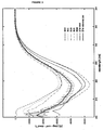
- the Stefin A/STM based scaffold proteins allow the use of three surfaces in total. These are the surfaces defined by position 4, loop 1 and loop2 ( Figure 2 ).
- the scaffold proteins of the invention are preferably engineered to facilitate its association or attachment to the solid phase substrate for the assay. Preferably this is by sticking to a gold coating, or by association with biotin. In order to engineer the scaffold for sticking to gold coating, preferably one or more Cys residues is introduced at the C or N terminus of the scaffold protein. In order to engineer the scaffold for immobilisation by attachment to biotin, preferably one or more copies of an eight amino acid biotin binding domain ('streptag') is introduced into said scaffold. Immobilisation may be by one or more of these or any other suitable means. Preferably the scaffold protein of the invention is immobilised. Preferably the scaffold proteins of the invention are engineered for immobilisation. Preferably interaction tests according to the present invention are carried out using immobilised scaffold proteins.
- Scaffold proteins based on Stefin A are superior to using peptides because they can be used in vivo. Furthermore, employing recombinant systems they are cheaper than working with synthetic peptides. Furthermore, construction of libraries is cheaper than using synthetic libraries for the same reason, and also because they can be rationally designed using nucleic acid manipulation. This reduces the reliance on complicated chemistry for peptide synthesis.
- Scaffold proteins based on Stefin A are superior to prior art such as phage display since they are internal to the cell, whereas phage display relies on extracellular interaction.
- scaffold proteins of the present invention can be used to work on native targets rather than recombinant targets. This has a further advantage of allowing examination of post translationally modified proteins which will be correctly phosphorylated or glycosylated or otherwise post-translationally modified in vivo but which would probably not be correctly formed if produced in vitro .
- scaffold proteins according to the present invention allow interrogation of the naturally occurring spectrum of splice variants and post translational modification variants which are produced in vivo without having to individually manufacture each of them and array them or otherwise compartmentalise them for analysis.
- a further application of the invention is in the use of microcantilevers as a read out for interaction with Stefin A based scaffold proteins.
- the scaffold proteins of the present invention are particularly suitable for use with electrochemical and/or thin film transistor type readouts.
- a yet further advantage of the scaffold of the present invention is that the peptide aptamers of the present invention can substitute for antibodies and results have shown that they may even perform better as, for example, CDK2 was detected more rapidly using peptide aptamers than antibodies. Accordingly, use of peptide aptamers rather than antibodies means that fewer animals will need to be used in the production of molecular probes which offers significant advantages to scientific research.
- Stefin A and three sites in Stefin A which are mutated in order to generate the new scaffold proteins of the present invention. These sites are: at position G4 of Stefin A or W4 of STM; any of codons 46 to 54 inclusive of constraining loop 1 and particularly at codon positions 48-50; any of codons and; any of codons 67 to 84 inclusive of constraining loop 2 and particularly mutations of 70-73.
- Modified Stefin A or STM polypeptides for use as scaffold proteins are produced by mutating the sequence of Stefin A as described. The resulting proteins based on Stefin A but possessing the specified mutational changes are given in the sequences herein before disclosed.
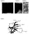
- Figure1 shows the expression of STM and exemplary variants in E coli.
- the open reading frames for STM and the variants disclosed herein were cloned into a version of the E coli expression vector pET30a+ which has been engineered to include additional functionalities in the amino terminal tail, such as a cysteine residue (present in all variants shown) or a Strepll tag (in STM only).
- additional 8 amino acids of the inserted Strepll tag account for the slight difference in migration of the STM protein compared to the other variants.
- E coli cells carrying these expression constructs were grown in the absence (-) or the presence (+) of isoproptl- ⁇ -d-thiogalactopyranoside (IPTG), which induces the expression of STM and the variant proteins (highlighted by *).
- IPTG isoproptl- ⁇ -d-thiogalactopyranoside
- T total cell lysate
- S soluble portion recovered after a 10 minute centrifugation at 16,000 x g were loaded onto 15% polyacrylamide gels. Proteins were visualised by Coomassie staining. As far as can be determined by this method, 100% of each protein shown (SUN, SUM, STM, SUC, SDM and SQM) could be recovered in the soluble fraction, indicating that the variant proteins remain able to fold in E coli.
- new scaffold proteins based on Stefin A and/or STM a rational approach to the design of a new peptide has been employed. It is desirable for the new scaffold proteins of the present invention to possess qualities that an ideal scaffold would need to possess to be broadly useful for in vitro and in vivo studies and apply these criteria to the design of a new scaffold.
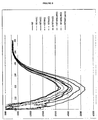
- Stefin A is a monomeric, single domain protein of 98 amino acids that receives no known post-translational modification and lacks disulphide bonds. SteA shows remarkable thermostability with a reversible transition observed at 90.8°C and folding enthalpy of 490 kJ/mol, all important features of a SteA-based scaffold.
- STM variant expression plasmids (all using pET30a+) were transformed into E coli. Single colonies were inoculated into overnight cultures for growth at 37 C with shaking (250 rpm on an orbital shaker). The following morning, 0.5 mL of each overnight culture was inoculated into 500 mL fresh medium, supplemented with kanamycin to maintain selection for the pET30 plasmids. Variant protein expression was induced once the cultures reached mid log phase (OD600 ⁇ 0.6-0.8). The cultures were grown for a further 3 hours, still at 37 C with shaking. E coli cells were harvested by centrifugation and lysed using a French Press.
- the lysates were clarified by centrifugation and STM variant proteins were purified from the resulting supernatants using Ni-chelate affinity chromatography. For this, 0.5mL of Ni-NTA agarose (QIAgen) was used per 20mL lysate. The resin was centrifuged in 50mL Falcon tubes at 700g for 2min and the supernatant was discarded. The resin was washed three times with 2.5mL of 1x Equilibration/Wash buffer, by re-suspending the resin in buffer and then centrifuging at 700g for 2min at 4°C and removing the supernatant. The lysate was combined with the washed metal affinity resin and incubated on rollers for 2h at 4°C.
- Ni-NTA agarose QIAgen
- the resin was incubated with 1 mL of Elution Buffer (50mM NaH 2 PO 4 , 300mM NaCl, 150mM Imidazole, pH7.4) for 10min at room temperature.
- Elution Buffer 50mM NaH 2 PO 4 , 300mM NaCl, 150mM Imidazole, pH7.4
- the suspension was centrifuged at 700g for 5min and the supernatant retained. This step was repeated twice more to yield two further elution fractions.
- the fractions with the highest protein concentrations were retained and stored at 4 C.
- the samples were buffer-exchanged into 50 mM phosphate buffer pH 7.4.
- the samples were diluted to approximately 0.3 mg/ml on the day of analysis, and accurate protein measurements were taken using a NanoDrop spectrophotometer. Where indicated in the figures, 1 mM DTT was added immediately prior to analysis.
- CD spectra were collected from 200 to 260 nm using a Jasco J715 spectropolarimeter. The spectra were normalized to molar protein concentration and residual molar ellipticities were plotted so as to minimise artefacts between samples.
- proteins were purified to near-homogeneity by affinity-chromatography using Ni-agarose, and diluted the purified preparations to 0.3 mg/ml immediately prior to circular dichroism analysis.
- circular dichroism analysis involves scanning the protein across a range of near-UV wavelengths, such that the ellipticity of the light is affected by secondary structure elements (alpha-helix or beta-strands) of the protein.
- Figure 4 shows that, after storage at 4 C for two weeks in a phosphate buffer at pH7.4 of the concentrated stocks of all the scaffold protein variants tested, the proportion of secondary structure is retained when fresh dilutions of the same samples are analysed (comparison to Figure 5 ).
- This step i.e. addition of a carrier protein is undesirable, as the aim is to use highly purified preparations of peptide aptamers so as to minimize non-specific signals in diagnostic and analytical assays that would result from the presence of irrelevant proteins such as the carrier protein.
- the new scaffold protein Stefin A variants may be stored in a simple phosphate buffer, with no apparent adverse effects. This observation means that it is likely to aid in the industrial application of the new scaffold protein Stefin A variants.
- Circular dichroism was also performed on variants STM, SDM, SQM, SUM, SUN, SUC plus three peptide aptamers pep6M, pep9M and pep10M ( Figure 6 ).
- DTT was added to prevent the cysteine residues present in the amino-terminal tails of the expressed variant proteins from forming inter-molecular disulphide bonds that could affect the experiment.
- the presence or absence of DTT did not change the observed secondary structure, although the spectra obtained in its absence were easier to interpret, as DTT itself contributes to the signal in the near-UV range.
- the SQM variant (SEQ ID NO:10) which comprises a mutational change at position 4 in addition to changes in both loop 1 and 2 i.e. multiple insertion sites in a single scaffold, was assessed for its ability as a scaffold protein display system.
- the peptides (HA, AU1 or MYC) were inserted at positions 4, 48 and 72/82 respectively and circular dichroism data was generated ( Figures 7 ).
- the circular dichroism analysis data indicates that the new insertion sites are not only capable of presenting peptides for interaction but they do so without significant loss of structure.
- the first method is crudely to determine the relative expression levels of the engineered proteins in E coli, with the rationale that most amino acid changes are likely to destabilise the protein.
- Table 1 below gives the expression yields of various scaffold variants from bacterial culture, yield is given as mg purified protein per litre of culture.
- the second method used was to directly assess the proportion of secondary structure elements in each protein by circular dichroism (CD).
- CD circular dichroism
- CD spectra for the different STM variants were obtained between 200 nm and 260 nm. A change in the shape of the CD spectra plot is considered to reflect changes in the ⁇ -helical and ⁇ -sheet content of the folded protein. All STM variants showed similar CD spectra with an inflexion point at about 218 nm indicating structural similarity without disrupting effects by the introduced modifications ( Figure 3 ). However, observed differences in the amplitude of the CD spectra were also observed with SDM, SUC, and SUM showing a flatter curve, and SQM and SUN showing a deeper curve compared to STM ( Figure 3 ). A possible explanation might be an enhanced stability of the folding, i.e.
- Peptide aptamers (Pep2, pep6, pep9 or pep10m) were immobilized in loop2 of SQM or SQT on a glass microscope slide using amine chemistry and probed with soluble, unlabelled, commercially sourced active CDK2 (New England Biolabs). After extensive washing, CDK2 bound to immobilised peptide aptamers was detected using an anti-CDK2 antibody and a labelled secondary antibody, which was imaged using a standard DNA microarray scanner. The data show that SQM can present pep6 better than any other scaffold, whereas SQT can present pep10m better than any scaffold variant and in clinically relevant range.
- the empty SQT scaffold gives a low signal which we set as the background.
- Peptide aptamers with different epitopes at different positions purified from E coli were printed onto glass slides coated with either a nickel chelate (which should capture the hexahistidine tag of each peptide aptamer so as to allow for controlled orientation or simply with poly-L-lysine. All prints started and finished with print buffer spots and free SQM spots which always gave no signal and acted as negative controls. In all cases antibody absorption was specific. These results have been confirmed with SPR ( Figure 10 ).
- the antibodies have the highest affinity for the SQM target in the following order: Anti-Myc, Anti-Ha and Anti-Au1, with changes in surface concentration of 0.4, 0.13 and 0.02 pM cm -2 observed upon the injection of 33 nM solutions (see Table 2 below). These values indicate the relative qualitative amplitudes of the equilibrium constants K.
- the K value which has been calculated for the anti-Myc antibody to be in the order of -50 nM, is in agreement with that for similar studies done with anti-cystatin.
- K values for Anti-Ha and Anti-Au1 have not been accurately determined due to their non-ideal behaviour in the SPR assay. Anti-Ha shows an immediate and intense association spike, followed by a secondary slower association component, with similar behaviour seen in the disassociation.
- the expression profiles of 864 random peptides inserted at each of the three sites were analysed.
- small scale cultures were grown of the random peptide aptamers in 96 well plates, purified the peptide aptamers in high throughput (i.e. without optimising expression or purification protocols for each well) and spotted an equal volume of each peptide aptamer into a glass microscope slide, creating a small microarray.
- the microarray was then probed with either of two antibodies that recognise the scaffold protein. The signal intensity obtained with the antibody would then be proportional to the amount of peptide aptamer at each feature of the array.
- Results showed that insertions at loop 1 and loop 2 were generally well expressed, while almost 50% of the amino-terminal inserts were significantly less well expressed ( Figure 11 ).
- Results showed that 68% of the 384 peptide aptamers we tested with 6 amino acid-long random inserts in loop 1 of SQM were well expressed, and 16% could be expressed to a lower level, while the expression of 15% could not be detected. 76% of the 384 peptide aptamers we tested with 12 amino acid-long random inserts in loop 2 of SQM were well expressed, while 14% could be expressed at a lower level and 10% could not be detected at all. Finally, of 192 peptide aptamers with random inserts in the amino terminus, only 35% were well expressed, while 32% were expressed at a lower level and 32% could not be detected
- the scaffolds and novel sites of the present invention may be used to produce peptide aptamers that interact with the target protein using a greater surface area and thus with greater affinity and specificity.
- the new scaffold was designated as SQT (SEQ ID NO:24). It was found that protein expression yields in E coli for SQT were decreased compared to those for SQM (Table 3). However, the yields for versions of SQT with inserts at loop 2 were consistently improved compared to insertions of the same sequences into loop 2 of SQM (Table 3). These differences were most pronounced with AU1 in loop 2, where the expression level was less than 1 mg peptide aptamer from 1 L of bacterial culture when using SQM, but was 45 mg/L of culture with SQT. Table 3 shows the yield of peptide of peptide aptamer expresses as mg of purified protein obtained under standard conditions from 1 liter of bacterial culture.
- Peptide aptamers with double insertions in loop 1 and loop 2 of SQT were generally well-expressed, and showed improved yields over the same combinations in SQM, although we obtained our highest yield of a peptide aptamer to date when inserting the AU1 tag into loop 1 and the HA tag into loop 2 of SQM (Table 3).
- SQT was found to be able to present the model CDK-binding peptides (pep2, 6, 9 and 10m) or the Myc epitope. Generally, SQT was better able to accept these peptides than SQM. However, surprisingly, SQT appeared to possess little (pep2, pep9) or no (pep6, pep10m) advantage over STM.
- the final sequence of the modified scaffold in this region, including the new linker is: DNA: 5'-AAGTGCTAGCAGGCCTTGATCACCATGGAC TAGCAAGCACAAATTA-3' (SEQ ID NO 27). Protein: (N-terminus) 44-KTQVLAgldhhgla STNYYIKVRAG- (C terminus) with the amino acids encoded by the inserted oligonucleotide given in lower case.
- the two peptide aptamers showing the highest apparent affinity for CDK2 from the microarray experiment were used in an "immuno-blotting" protocol (also called a western blot see Figure 12 ), except that the peptide aptamers (SQM-pep6 and SQT-pep10M) were used instead of antibodies.
- Antibodies were similarly omitted from the development step of the "immuno-blot" as the peptide aptamers possess an S-tag which can be detected using S-protein fused to horse radish peroxidase (HRP).
- HRP horse radish peroxidase
- parallel blots were probed with the empty scaffold (SQT is shown) and developed using the S protein protocol.
- FIG. 12 shows two lysates of human tissue culture cells that were subject to denaturing polyacrylamide gel electrophoresis prior to being transferred to nitrocellularose or PVDF membranes and probed with scaffold, peptide aptamers or scaffold as described above.
- the data show that peptide aptamers can not only substitute for antibodies in this protocol, but actually perform better as CDK2 was detected more rapidly using peptide aptamers than antibodies.
- use of peptide aptamers rather than antibodies means that fewer animals will need to be used in the production of molecular probes.
Description
- The present invention relates to novel scaffold proteins for the display of peptides, such as peptide aptamers. In particular, the invention relates to the use of modified Stefin A polypeptides and modified artificial proteins based on Stefin A, all for use as scaffold proteins and as display systems.
- Study of protein interactions is vital to an understanding of many biological processes, such as the roles of gene products in vivo both in health and disease. Peptide aptamers in particular have emerged as important molecular tools that are useful for both basic and applied aspects of molecular medicine. Due to their ability to specifically bind to, and inactivate, a given target protein at the intracellular level, they provide an experimental strategy for functional protein analyses, both in vitro and in vivo. They may also be used against extracellular proteins. As well as applications in studying protein function, these tools may therefore be useful for molecular detection, diagnostics and/or as therapeutic agents. Peptides and peptide aptamers may be used free in solution. However, small peptides when unconstrained will tend to form structures which present a limited interaction surface. Furthermore, they will often lose conformational entropy upon association with target molecules, reducing free energy of binding and consequently free peptides will often not form tight non-covalent complexes, which is a problem. In addition, within cells peptides are rapidly degraded, which limits their effectiveness for the study of protein interactions in vivo, which is also a problem.
- Rather than being used in free solutions, peptides of interest may be bound to physical supports, or displayed in the context of a larger polypeptide. The former cannot readily be applied to in vivo studies. In the latter, peptides are genetically inserted into the primary sequence of a simple, stable scaffold protein. The folding of the scaffold conformationally constrains the peptide, so peptide aptamers bind partners with high specificity and affinity. It is display in the context of a polypeptide which is important in the present invention. Such display is often brought about using scaffold proteins.
- Prior art scaffolds have included inactivated staphylococcal nuclease, green fluorescent protein (GFP) and thioredoxin A (TrxA), as well as isolated protein folds such as the Z domain of staphylococcal protein A, "affibodies", anticalins, and ankyrin repeats.
- Further prior art scaffold proteins include the fibronectin type III domain ('Fn3'), lipocalin family proteins from which anticalins are derived, bilin binding protein (BBP), and others.
- More recently (
WO 2006/131749 ) describes several rational mutations made in Stefin A to improve it as a scaffold. The modified Stefin A scaffold comprises mutations at the following three sites Lys71-Leu73, V48D and G4W and is referred to as STM (Stefin A Triple Mutant). It was shown that the combination of these three mutations generated a protein that had minimal interactions with proteins in human cells, and in particular had lost all detectable interaction with its known natural partners. However, we found that insertion of peptides into the protein at position 71-73 led to a strong selection pressure for truncations of the protein at the end of the inserted peptide. Although such truncated proteins could display biological efficacy, this observation leads to concerns that a subset of peptides that are simply inserted at position 71-73 without truncation may not be freely available for interaction with a target protein, which is a problem. Furthermore, insertion of peptides at a single site inevitably limits the total surface area used for a protein interaction, which in turn limits binding affinity and potentially specificity. - The novel mutations made to Stefin A and to modified artificial proteins based on Stefin A such as STM (Stefin A Triple Mutant) as disclosed in the present invention provide alternative improved and more stable scaffold proteins and also provide display systems that are more versatile than those of the prior art. Moreover, these new protein scaffolds/display systems are also quite unpredictable as efficient and robust display entities. The new mutations described hereinafter have been made at specified diverse areas of the Stefin A/STM proteins and surprisingly have been found not to affect Stefin A/STM protein configuration or their potential function as scaffold proteins. Furthermore, with the improved scaffolds of the present invention by virtue of further engineering it is possible to provide modifications wherein the scaffolds have multiple insertions something that was not hitherto possible in the prior art scsffolds.
- According to a first aspect of the invention there is provided a Stefin A polypeptide comprising amino acid sequence having at least 80% identity to SEQ ID NO:1; said polypeptide comprising a mutation at
position 4 wherein the Glycine of Stefin A is replaced by Arginine;
characterised in that the polypeptide further comprises a heterologous peptide insertion, wherein - a) said heterologous peptide is inserted into the protein at the G4 site of Stefin A; or
- b) said heterologous peptide is inserted into the protein at position 46-54; or
- c) said heterologous peptide is inserted into the protein at position 67-84; or
- d) said heterologous peptide is inserted into the protein at position 82/83.
- Suitably said Stefin A polypeptide comprises a further heterologous peptide insertion such that at least one heterologous peptide is inserted into the protein at at least two positions selected from (a), (b) and (c).
- Suitably said Stefin A polypeptide comprises a further heterologous peptide insertion such that at least one heterologous peptide is inserted into the protein at at least three positions selected from (a), (b) and (c).
- In another aspect the invention relates to a Stefin A polypeptide comprising amino acid sequence having at least 80% identity to SEQ ID NO:1; said polypeptide comprising a mutation at
position 4 wherein the Glycine of Stefin A is replaced by Arginine; characterised in that the polypeptide further comprises a heterologous peptide insertion, wherein - i) said heterologous peptide is inserted into the protein at position 46-54; or
- ii) said heterologous peptide is inserted into the protein at position 67-84; or
- iii) said heterologous peptide is inserted into the protein at position 82/83.
- Suitably said Stefin A polypeptide comprises a further heterologous peptide insertion such that at least one heterologous peptide is inserted into the protein at at least two positions selected from (i) and (ii).
- In another aspect the invention relates to a polypeptide comprising the amino acid sequence as set forth in any one of SEQ ID NOs: 10, 13, 16, 17, 22, 23, 24 and 25.
- Suitably said heterologous peptide of (a) is inserted proximal to the G4 site of Stefin A.
- In another aspect the invention relates to use of a polypeptide as described above as a scaffold protein.
- In another aspect the invention relates to a method for identifying a target peptide capable of binding a structure of interest comprising:
- (i) providing a stefin A scaffold protein as described above comprising a target peptide;
- (ii) contacting said scaffold protein with said structure of interest; and
- (iii) monitoring association between the scaffold and the structure of interest, wherein association of the scaffold protein with the structure of interest identifies the target peptide as a candidate target peptide capable of binding said structure.
- In another aspect the invention relates to use of a polypeptide as described above to display peptide(s) on microarray(s).
- In another aspect the invention relates to use of a polypeptide as described above as an agent to bind and specifically detect a biomarker.
- In another aspect the invention relates to use of a polypeptide as described above in an interaction test, wherein said polypeptide is immobilised.
- In another aspect the invention relates to a method for detecting a structure of interest comprising
providing a stefin A scaffold protein as described above comprising a target peptide capable of binding said structure of interest;
contacting said scaffold protein with said structure of interest; and
monitoring the association between the scaffold and the structure of interest, wherein association of the scaffold protein with the structure of interest detects said structure. Preferably said structure of interest comprises a biomarker. - Throughout the description and claims of this specification, the words "comprise" and "contain" and variations of the words, for example "comprising" and "comprises", means "including but not limited to", and is not intended to (and does not) exclude other moieties, additives, components, integers or steps.
- Throughout the description and claims of this specification, the singular encompasses the plural unless the context otherwise requires. In particular, where the indefinite article is used, the specification is to be understood as contemplating plurality as well as singularity, unless the context requires otherwise.
- Features, integers, characteristics, compounds, chemical moieties or groups described in conjunction with a particular aspect, embodiment or example of the invention are to be understood to be applicable to any other aspect, embodiment or example described herein unless incompatible therewith.
- Reference herein to a scaffold protein is to sequences fused together into one protein, the term is also synonymous with fusion protein. By a "fusion protein" is meant a protein that includes the scaffold protein of the invention joined to one or more different (i.e., "heterologous") peptides or proteins. The insertion of heterologous peptides or proteins enables the fusion protein to bind to a desired target.
- The present invention is based upon the novel modifications including insertions of the wild type Stefin A protein itself, preferably the Stefin A is a human Stefin A, or to its triple mutant version, STM, rendering them into forms suitable for use as stable scaffold proteins whilst concomitantly advantageously rendering them biologically neutral by ablating biologically significant interactions and activities by mutation of residues that are required for natural interactions with either cathepsins or other unknown proteins.
- Furthermore, it is envisaged that the selected mutation or insertion site(s) are able to accept and constrain inserted peptides to produce for example peptide aptamers. Whereas studies in human may require a human scaffold, the use of for example mouse Stefin A, may be advantageous for studies of mouse model biology and/or disease, similarly Stefin A derived from other species or plants may also be of utility in that specific species. Accordingly, the scaffolds and presentation systems of the present invention are intended to be useful for any selected species and the derivation of the Stefin A is dependent on a user's requirements.
- It will be appreciated that the changes in DNA sequences encoding the amino acid at
codon 4 of either Stefin A or its STM form, or the changes in codons 46 to 54 inclusive, that encode amino acids comprising or constrainingloop 1 of either Stefn A or its STM form, or the changes in codons 67 to 84 inclusive, that encode amino acids comprising or constrainingloop 2 of either Stefin A or its STM form, can be independent of one another. That is to say the modifications to Stefin A protein may be at one of three different discrete areas or regions i.e. atposition 4 or in constrainingloop 1 orloop 2. Similarly, the modifications to the triple mutant form STM may also be at any one of the three specified independent discrete sites i.e. atposition 4 or in constrainingloop 1 orloop 2. The rest of the sequence of Stefin A or STM will be unaltered and comprise the sequences as set forth below. - The sequence of wild type human Stefin A is shown below as SEQ ID NO:1:

- The sequence of the triple mutant STM is shown below as SEQ ID NO:2, the mutation sites and thus where STM varies from wild-type Stefin A are marked in bold and underlined:

- Reference herein to a "mutational change" conveys that there is a permanent change in the genetic material the mutational change may be by addition(s) or deletions or insertion(s) or replacement(s) to the amino acid residue(s)
- Preferably, in the embodiment where the single mutational change is Glycine at
codon 4 of Stefin A, its replacement is selected from the group comprising G4V, G4I, G4L, IG4M, G4F, G4P, G4N, G4V, G4Q, G4S, G4T, G4W, G4Y, G4R, G4H, G4K, G4D and G4E. More preferably the change is G4R, that is to say the Glycine is replaced by Arginine atcodon 4. - Preferably, in the embodiment where the single mutational change is Tryptophan at
codon 4 of STM, its replacement is selected from the group comprising W4V, W4I, W4L, W4M, W4F, W4P, W4N, W4V, W4Q, W4S, W4T, W4G, W4Y, W4R, W4H, W4K, W4D and W4E. More preferably the change is W4R, that is to say the Tryptophan is replaced by Arginine atposition 4. - It has been found that a change in the 5' region of the Stefin A open reading frame encoding the amino terminal 8 amino acids of Stefin A or STM allows the introduction of a cleavage site for a restriction endonuclease or a targeted recombination site. For example, it is demonstrated herein that after changing the DNA sequence to encode for example an Arginine at position 4 (replacing the wild type Glycine, or the Tryptophan in STM) allows the production of a surprisingly stable protein that possess the same biophysical characteristics as STM, but where the open reading frame now possess a unique restriction site for an enzyme such as and without limitation the enzyme Avrll, and thus serves an alternative and efficient scaffold protein.
- Preferably, in the embodiment where the mutational change is any change in codons 46 to 54 inclusive that encode amino acids comprising or constraining
loop 1 of Stefin A (SEQ ID NO:3 QWAGTNYY) or STM (SEQ ID NO 4: QVDAGTNYY) the change comprises for example QVLASTNYY (SEQ ID NO: 5). It has been surprisingly demonstrated that introducing a sequence of amino acids such as, and without limitation, Leucine, Alanine, Serine atpositions 48, 49 and 50, leads to a protein with the same biophysical characteristics as STM, and is thus likely to be an efficient scaffold. - Preferably, the mutational change in Stefin A is at 48-VAG-50 and in STM is at 48-LAS-50 such that the result is 48-LXS-50, wherein X is any amino acid.
- Preferably, in the embodiment where the mutational change is any change in codons 67 to 84 inclusive that encode amino acids comprising or constraining
loop 2 of Stefin A (SEQ ID NO 6: LKVFKSLPGQNEDLVLTG) or STM (SEQ ID NO 7: LKVFNGPPGQNED LVLTG) the change comprises for example SEQ ID NO:8 LKVFNGPPGQNEDLVRSG. It has been surprisingly demonstrated that a sequence of amino acids such as Arginine followed by a Serine (to replace Leucine 82 and threonine 83 of Stefin A or STM) leads to the production of a stable protein that, like STM, may serve as a good scaffold for the presentation of peptide aptamers. - Preferably, the mutational change in Stefin A is at 71-KSL-73 and 82-LT-83 and in STM is at 71-NPG-73 and 82-LT-83 such that the result is 71-NxP-73 and 82-RS-83, wherein X is any amino acid.
- Preferably, there is a further mutational change in Stefin A and STM which is at 82-LT-83 such that the result is 82-XX-83, wherein X is any amino acid and in a particularly preferred embodiment it is 82-RS-83. The mutational change may be at either 82 or 83 or at both positions.
- Preferably, the mutational change may be any combination of those herein before mentioned with for example and without limitation, 82-XX-83 and a particular preferred variant has mutational changes at least at positions 71-73 and/or 82-83.
- Disclosed herein is a modified Stefin A polypeptide or modified STM protein comprising two mutational changes or a heterologous oligonucleotide encoding a peptide insertions at sites selected from the group comprising:
- (i) a mutation at
codon 4 wherein the Glycine of Stefin A or the Tryptophan of STM is replaced by another amino acid that is not Tryptophan for Stefin A or Glycine for STM or by a peptide encoded by the heterologous oligonucleotide; and/or - (ii) any change or heterologous oligonucleotide encoding a peptide insertion in codons 46 to 54 inclusive, that encode amino acids comprising or constraining
loop 1;and/or - (iii) any change or heterologous oligonucleotide encoding a peptide insertion in codons 67 to 84 inclusive, that encode amino acids comprising or constraining
loop 2. - It will be appreciated that in this aspect, where the modified Stefin A may comprise two mutations, it may comprise for example a mutation at
position 4 and a change in any of codons 46 to 54 havingloop 1 function or it may comprise a mutation atposition 4 and a change in any of codons 67 to 84 havingloop 2 function or it may comprise a change in any of codons 46 to 54 havingloop 1 function and change in any of codons 67 to 84 havingloop 2 function. - Similarly, the STM may comprise a mutation at
position 4 and a change in any of codons 46 to 54 havingloop 1 function or it may comprise a mutation atposition 4 and a change in any of codons 67 to 84 havingloop 2 function or it may comprise a change in any of codons 46 to 54 havingloop 1 function and change in any of codons 67 to 84 havingloop 2 function or it may comprise a change in any of codons 67 to 84 havingloop 2 function. - Also disclosed herein is a modified Stefin A polypeptide or modified STM protein wherein the modification mutational changes or a heterologous oligonucleotide encoding a peptide inserted at three sites:
- (i) a mutation at
codon 4 wherein the Glycine of Stefin A or the Tryptophan of STM is replaced by another amino acid that is not Tryptophan for Stefin A or Glycine for STM or by a peptide encoded by the heterologous oligonucleotide; and - (ii) any change or heterologous oligonucleotide encoding a peptide insertion in codons 46 to 54 inclusive, that encode amino acids comprising or constraining
loop 1;and - (iii) any change or heterologous oligonucleotide encoding a peptide insertion in codons 67 to 84 inclusive, that encode amino acids comprising or constraining
loop 2. - Thus in this aspect modified Stefin A and STM comprises all three mutational changes as hereinbefore described. Thus the modified Stefin A or STM scaffold proteins comprises three specific mutations at
position 4 and a change in bothloop - Also disclosed herein is a modified Stefin A polypeptide or modified STM protein comprising any single or combination of the sequences listed above, but terminating at either residue 73 of Stefin A or STM, or residue 84 of Stefin A or STM, and either with or without the insertion of a new amino acid sequence at these positions. We have found that there is a strong selection pressure for a stop codon following insertion of amino acid sequences after the NGP of STM, but that surprisingly such truncated proteins are both stable and capable of interfering with the biological functions of a target protein.
- The present disclosure therefore also includes truncated or shortened modified Stefin A and STM scaffold proteins ideally shortened by 15 or 25 residues at the C-terminus end and thus terminating at either residues 73 or 84 of either Stefn A or STM. Also disclosed are truncated or shortened modified Stefin A or STM that are shortened by any integer between 15 and 25 and thus terminate a residues between 73 to 84 of Stefin A or STM.
- The present disclosure preferably includes all of the variants described, as each one allows the introduction of a heterologous peptide at one or more sites of the Stefin A or STM variant, by insertion of an oligonucleotide into an engineered restriction site in the open reading frames we have created. Thus the present disclosure provides several new scaffolds based on:
- A unique heterologous peptide inserted into the protein at
position 4, with the rest of the protein resembling either Stefin A or STM or one of the other variants described herein. - A unique heterologous peptide inserted into the protein at position 46-54 and especially at positions 48/49/50, with the rest of the protein resembling either Stefin A or STM or one of the other variants described herein.
- A unique heterologous peptide inserted into the protein at position 67-84 and especially at position 71/72/73 with the rest of the protein resembling either Stefin A or STM or one of the other variants described herein.
- A unique heterologous peptide inserted into the protein at positions 67-84 and especially at position 82/83 with the rest of the protein resembling either Stefin A or STM or one of the other variants described herein.
- Any combination of multiple peptides inserted into
positions 4 and/or 48/49/50 and/or 71/72/73 and/or 82/83. - Any combination of single or multiple peptides inserted into
positions 4 and/or 48/49/50 and/or 71/72/73 and/or 82/83 that is followed by a stop codon that removes or replaces the last 25 or the last 15 amino acid residues of either Stefin A or STM. - A particular advantage of the scaffold proteins and the new mutations is that they enable the use of the whole of
loop 1 orloop 2, or bothloop 1 andloop 2 as well as the amino terminus. Together, these mutations will allow the presentation of surfaces at least as large as those used by antibodies. In addition, each can be used singly, or they can be used pair-wise or in multiple combinations with other mutations. The differing positions of the interaction surfaces, combined with the differing interactions between peptides inserted at the different sites, is likely to provide novel uses of the scaffold, such as where peptides that could not be presented for useful interaction at one site may now be presented by another, or where combinations of peptides at different sites allow a given peptide to switch from a non-interacting to an interacting conformation. In addition, any of these new mutations may be used in the context of full length Stefin A, full length STM, the full length proteins disclosed herein, or in the mutant versions of any of these proteins where the last residue derived from either Stefin A or STM is Leu73 of SteA or its new variants described here, or Pro73 of STM or its new variants, or the last residue of an inserted heterologous peptide where the last 15 or 25 amino acids of Stefin A or STM have been truncated. - Also disclosed herein are isolated nucleic acids comprising nucleotide sequences encoding the amino acid sequences of a scaffold protein or polypeptides as hereinbefore described above.
- Also disclosed herein is a method for identifying a target peptide capable of binding a structure of interest comprising providing a modified stefin A or STM protein scaffold protein as herein before described comprising a target peptide; contacting said scaffold protein with said structure of interest; and monitoring the association between the scaffold and the structure of interest, wherein association of the scaffold protein with the structure of interest identifies the target peptide as a candidate target peptide capable of binding said structure.
- In a further aspect the scaffold protein is selected from the group comprising:


- In a further aspect there is provided use of the scaffold proteins of present invention as an agent selected from the group comprising diagnostics, therapeutics, biomarkers, agents to bind to and specifically detect biomarkers, rationalized drug design templates, targets or reagents for drug discovery, antibody substitutes and research tools.
- In a yet further aspect there is provided use of the scaffold proteins of present invention as a fusion protein.
- Preferred features herein before described apply mutatis mutandis to all and each aspects.
- Taken together the results described hereinafter and above demonstrate that, the scaffolds disclosed herein are amenable to engineering in multiple locations, with each change alone or in combination being surprisingly well tolerated and that any destabilising effects of the mutations are apparently magnified by insertions in the amino terminus and in
loop 2. These sites cannot therefore be used routinely, but surprisingly some inserts are tolerated which will allow us to use them to improve the binding affinity and specificity of peptide aptamers in for example the SQM-loop1. In addition,loop 1 appears able to present a range of peptides with very little problem. This is very surprising, as this is the shortest loop. - A "deletion" refers to a change in an amino acid or nucleotide sequence due to the absence of one or more amino acid residues or nucleotides. The terms "insertion" or "addition" refer to changes in an amino acid or nucleotide sequence resulting in the addition of one or more amino acid residues or nucleotides, respectively, to a molecule or representation thereof, as compared to a reference sequence, for example, the sequence found in the naturally occurring molecule. A "substitution" refers to the replacement of one or more amino acids or nucleotides by different amino acids or nucleotides, respectively.
- In order to improve upon Stefin A or STM as a scaffold, it is desirable to be able to insert heterologous peptides at alternative sites, and/or at multiple sites. To do this required altering the open reading frame that codes for either Stefin A or for STM, so as to introduce restriction endonuclease recognition sites into which oligonucleotides encoding heterologous peptides could be inserted. Alteration of the open reading frame almost inevitably leads to an alteration of the amino acid sequence that comprises the expressed protein. Given that proteins have evolved to an optimum combination of function and stability, the most likely (and most frequently observed) outcome of a change to the amino acid sequence of a protein is a loss of secondary structure and hence of stability. In the present invention the new scaffold proteins retained stability (See Examples and Figures).
- In order to ascertain whether the alterations made at the DNA (open reading frame) level that also alter the amino acid sequence of the new Stefin A/STM variants lead to decreased stability of the protein, all of the variants described here were expressed in E.coli and circular dichroism was used to compare their secondary structure composition to that of Stefin A. All the proteins were found to be equally well expressed in E coli, typically to approximately 28 mg variant protein/ml of bacterial culture (
Figure 1 ). Proteins were purified to near-homogeneity by affinity-chromatography using Ni-agarose, and the purified preparations diluted to 0.3 mg protein/mt. Each resulting sample was subjected to analysis by circular dichroism. This involves scanning the protein across a range of near-UV wavelengths, such that the ellipticity of the light is affected by secondary structure elements (alpha-helix or beta-strands) of the protein. The greater the proportion of secondary structure, the greater is the effect on the ellipticity. Because the effect is affected by protein concentration, proteins were diluted to 0.3 mg/ml immediately before the analysis. Because the effect is proportional to the number of peptide bonds in the protein being analysed, the molar ellipticity is shown, which takes this effect into account. The data are shown inFigure 3 . This data shows that the proportion of secondary structure is preserved between STM and the new variants, and that the presence of inserts in STM does not adversely affect its structure. Two variants (SUN and SQM) were noted that appeared to show increased structure compared to the others. This may be attributable to the acquisition of secondary structure in the amino terminal tail that is present in all these proteins, and would be driven by the replacement of Glycine (Stefin A) or Tryptophan (STM) atposition 4 by Arginine, as this is the only change that is common to SUN and SQM, and these are the only variants to possess this alteration. - As is well known in the art, the term 'scaffold' refers to a protein which can present target peptides to solvent without its own structure being deformed by the target peptide. Regarding the presentation of peptide to solvent, this can be tested using immunoprecipitation experiments. For example, an indication that a peptide is being presented to solvent may be obtained by its availability to an antibody capable of recognising it. Thus, in order to test the ability of a scaffold protein to present a peptide to solvent, the scaffold comprising the peptide would be expressed and an antibody recognising the peptide would be used to try to immunoprecipitate the scaffold-peptide fusion. If this protein can be immunoprecipitated or captured on the antibody, this shows that the peptide was presented to solvent as is required by a scaffold protein. Another, or an alternative, indication that a peptide is being presented to solvent may be obtained by phosphorylation studies. By incorporating a phosphate acceptor site into the target peptide, and then contacting the scaffold-peptide fusion with the cognate kinase in conditions permissive of phosphorylation, then the presentation of the peptide to solvent can be verified. Phosphorylation of the peptide indicates correct presentation to solvent. Concerning a scaffold protein's resistance to being deformed by the target peptide which it bears, this can be tested using techniques such as circular dichroism or thermal stability. Specifically, a circular dichroism analysis of a scaffold protein without target peptide inserted into it should be substantially the same as the circular dichroism characteristics of the same scaffold protein when bearing a target peptide. This provides a demonstration that the presence of the target peptide in the scaffold protein has not compromised or deformed the structure of the scaffold protein bearing it. Another way to test this resistance to deformation by the target peptide is by studying the thermal stability of the scaffold protein with and without target peptide inserted.
- A scaffold protein must be able to accept a peptide insert. Preferably the peptide insert is 36 amino acids or less, preferably 20 amino acids or less. Preferably the target peptide insert is 12 amino acids or less.
- A scaffold protein must be of known structure. By 'known structure' it is meant that the crystal structure or a solution structure (NMR structure) must be known.
- Preferably a scaffold protein constrains the target peptide. The presence of a constraint effect in a scaffold protein can be demonstrated by comparing the affinity of an entity binding the target peptide when the target peptide is in the scaffold protein with the affinity when the peptide is not in the scaffold protein. A difference in these two affinities indicates that the scaffold protein is constraining the peptide to assume a particular three dimensional conformation. Preferably a scaffold protein constrains a peptide so that it demonstrates an increased binding affinity when present in the context of the scaffold protein. In other words, preferably the scaffold protein decreases the entropic cost of binding and so increases the measured affinity when compared with binding of a free peptide.
- In some embodiments, constraint may be provided by a single N-terminal or C- terminal fusion to the target peptide.
- Preferably a scaffold protein provides the target peptide with an increased stability in vivo. This effect may be demonstrated by comparison of expression of the target peptide in the context of the scaffold protein with expression of the target peptide on its own. Preferably, the target peptide shows increased stability in the context of the scaffold protein.
- A scaffold protein is preferably biologically neutral. By 'biologically neutral' it is meant that interactions with other known proteins have been abolished. Furthermore, any signalling abilities possessed by the protein are preferably removed. Thus, a preferred scaffold protein according to the present invention is the STM scaffold protein.
- Biological neutrality is an advantage of the present invention since it does not exist in the majority of prior art scaffold proteins. For example, Thioredoxin A acts as a dominant negative of the natural redox pathways in cells. Furthermore, it is known to inhibit P53 and is known to inhibit BCL6 signalling pathways. Advantageously, the scaffold proteins of the present invention do not interfere with naturally occurring signalling pathways.
- A scaffold protein should be small. By 'small' is meant less than 25kDa, preferably less than 13kDa. Most preferably a scaffold protein should be less than 110 aa (excluding target peptide insert).
- Preferably a scaffold protein according to the present invention will be conformationally stable. By 'conformationally stable' it is meant that no conformational changes should take place. Preferably a scaffold protein has no hinge region. Preferably a scaffold protein has no PH domain. Preferably a scaffold protein has no SH3 domain. Preferably a scaffold protein has no SH2 domain. Preferably a scaffold protein has no 'WW domain. Preferably a scaffold protein has no 'WD' domain. Preferably a scaffold protein. has no HEAT repeats. Preferably a scaffold protein has no Proline rich domain. Preferably a scaffold protein has no post- translational modification in cells. Preferably a scaffold protein has no other domain known to facilitate conformational changes.
- A scaffold protein according to the present invention preferably has no protein-protein interaction domains. A protein will be considered to have no protein-protein interaction domains if these have been mutated so as to render them non-functional.
- Preferably a scaffold protein according to the present invention has no post translational modifications. Thus, preferably a scaffold protein according to the present invention has no glycosylation site. This is an advantage over prior art scaffold proteins such as dystrophin because post translational modifications can interfere with interactions or create spurious interactions themselves.
- As noted above, scaffold proteins should not be deformed by the peptide insert. On this criterion, green fluorescent protein would not be considered a scaffold protein because at least one third of inserted target peptides abolish the fluorescence of green fluorescent protein. This is a demonstration that the target peptide insert is deforming the structure of the protein. Therefore, it is not a scaffold protein according to the present invention since a scaffold protein should preferably not be deformed by the target peptide insert.
- Thioredoxin A (TrxA) is a prior art scaffold protein. TrxA is small and is stable. However, the insertion of target peptides into TrxA takes place between two cysteine residues. Scaffold proteins according to the present invention advantageously avoid this arrangement because the cysteine residues in TrxA can undergo reversible disulphide bonding which can alter the conformation of the scaffold protein and can affect the conformation of the presented target peptide. Thus, preferably the insertion site for target peptide is not between two cysteine residues on the scaffold protein.
- Scaffold proteins preferably have one or more of the following features:
- 1) be of known structure, thereby allowing an informed choice of the site for peptide insertion or replacement;
- 2) be stable enough to constrain the folding of a broad range of peptides;
- 3) be biologically neutral, i.e. lack interactions with cellular proteins that could contribute a phenotype; and
- 4) be able to fold similarly, preferably identically in both prokaryotic and eukaryotic environments, so that data obtained in one system can inform experiments performed in the other.
- The present invention provides a scaffold suited to the requirements of peptide aptamer technology. The scaffold proteins of the present invention preferably possesses all of the criteria defined above: the structure of parental Stefin A is known; the engineered scaffold is stable and tolerates the insertion of at least one peptide without losing its biophysical stability; it is able to present a broad range of peptides for functional interaction; and not only have all known biological interactions been engineered away.
- It will be appreciated by the skilled reader that the use of peptide aptamers in microarrays is particularly advantageous when those peptide aptamers are presented in the scaffold protein according to the present invention. Prior art microarray technology relies heavily on antibodies. However, antibodies can lose specificity when they are bound to the array. Furthermore, recombinant proteins used in microarrays can provide information that proteins are present, but cannot provide information about what is binding them. By contrast, using peptide aptamers displayed in scaffold proteins according to the present invention can advantageously provide a lot more information when an array is interrogated. For example, upon observation of a binding partner, contextual information is advantageously derived when using a scaffold protein to display the aptamer. This advantage is characterised as the difference between a naïve and an informed library. Thus, in another aspect the invention relates to the use of these new scaffold proteins to display peptides on microarrays.
- The scaffold protein according to the present invention is based on the sequence of Stefin A. By 'based on the sequence of Stefin A' it is meant that the scaffold protein should possess at least 30 of the 98 amino acid residues of Stefin A, preferably 25% of the amino acid sequence of Stefin A, preferably 30%, 40%, 50%, 60% or 70% of the amino acid sequence of Stefin A, preferably 80%, preferably 85%, preferably 90%, preferably 95% or even more of the sequence of Stefin A. Most preferably the scaffold protein will have the sequence of Stefin A or STM or one of the new variants disclosed herein and comprises one or more of the mutational changes hereinbefore described.
- The ability of peptide aptamers to disrupt protein-protein interactions in vivo may allow the rapid identification of novel drug leads. Furthermore, the use of small, candidate drug molecule(s) to disrupt protein-protein interaction is advantageously facilitated by the present invention.
- Use of peptide inserts comprising post-translational modifcation sites such as phosphorylation site(s) may be advantageously employed. This is beneficial in dissecting interactions which are varied according to the phosphorylation state of the target peptide. Furthermore, it allows the identification of candidate peptide aptamers which bind in a phosphorylation dependent manner.
- In some embodiments, it may be desired to introduce disulphide bonds either side of the target peptide insert, for example by engineering a cysteine residue each side of the target peptide insert. This may be useful if the scaffold is being used exclusively in one setting. In this regard, it is to be noted that the family II cystatins use a di-sulphide bond to form elements of secondary structure that correspond to one preferred region of insertion. In the context of the present invention this can be achieved for example by the addition of a single cysteine at the C-terminus of the scaffold polypeptide, or within the target peptide such as at the C-terminal end of the target peptide, and addition of a second cysteine residue inserted at a second location such as in the N-terminus of the scaffold or at the N-terminal end of the target peptide, thus allowing cross-linking between the two. However, it is preferred to avoid the covalent constraint of peptides in this manner. Thus, in preferred scaffolds of the present invention, preferably the target peptide is not flanked by cysteine residues.
- Overall it will be appreciated that different scaffolds may force a bias on the peptides they present, so that study of target peptides may advantageously involve peptides and/or libraries presented in more than one scaffold, so as to maximize the likelihood of success.
- Scaffolds of the invention allow investigators to extend in vitro observations to the intracellular environment and vice versa, as well as allowing the in vitro identification or creation of tools that may be used inside cells without concerns about folding patterns or the oxidation state of disulphide bonds.
- Peptide aptamers based on scaffolds of the present invention are tools that can be used to validate drug targets that can be used as components of diagnostic or prognostic tests or even form the basis for lead compounds for the treatment of human disease. The scaffolds of the invention, advantageously based on a full-length human protein, may be useful as biological therapeutics and/or in gene therapy.
- The term 'target peptide' as used herein refers to a peptide of interest. The target peptide is preferably a heterologous peptide. By heterologous is meant a peptide which is removed from its usual context, preferably a peptide having a sequence not usually found in the sequence of the scaffold protein bearing, carrying or displaying it. If the peptide does have a sequence which occurs elsewhere in the sequence of the scaffold protein, then for it to be 'heterologous' that sequence will be out of context i.e. not occupying its naturally occurring position (address) within the scaffold protein polypeptide. In this context, 'position' and means position within the linear amino acid chain rather than position in three dimensional space relative to other amino acid residues. The target peptide may be artificial for example generated by the construction of a library of peptides for incorporation into the scaffold protein. In these embodiments, the artificial peptide(s) are considered to be 'heterologous' for the purposes of the invention.
- Peptide aptamers are peptides constrained and presented by a scaffold protein that are used to study protein function in cells. Some are able to disrupt protein-protein interactions and some are able to constitute recognition modules that allow the creation of a molecular toolkit for the intracellular analysis of protein function.
- The ability to design or identify small molecules that can bind specifically and with high affinity to a given protein is a rate-limiting step in many experiments, including the development of protein microarrays, the analysis of proteins in the context of living cells and the validation of candidate drug targets. In nature, protein-protein interactions can be mediated by small surfaces of folded proteins. This has led to the use of small peptide surfaces presented within the context of a stable protein, called the scaffold, as protein recognition modules. Such reagents, called here peptide aptamers, have been used to disrupt biological protein activity in a range of systems.
- Peptide aptamers are more easily delivered and more stable in cells than free peptides and their constrained folding results in a lower entropic cost of binding and hence increased affinity for target proteins. Protein engineering of peptide aptamers allows them to provide the recognition functionality in the design of a molecular toolkit although this potential has yet to be fully realized. The affinity of peptide aptamers for their targets ranges from 10"6to 5 x 10"9 M compared to IQ 10'7 to 10'11 M for antibody/target interactions. By using multiple insertions to increase the surface area of interaction, peptide aptamers are expected to be able to match or possibly advantageously exceed the binding affinities of antibodies. Nonetheless, peptide aptamers are clearly able to disrupt protein-protein interactions in vivo. Peptide aptamer screens are performed In yeast or in mammalian cells, which distinguishes them from phage display screens of peptide or antibody libraries performed against potentially misfolded prokaryotically expressed protein.
- While the most extensively used scaffold is the Escherichia coli protein thioredoxin (TrxA), a number of other proteins have been used. The success of this technology hinges upon the robustness of the scaffold, yet one third of peptides may destabilize GFP, while many TrxA based peptide aptamers are not stably expressed in cultured human cells, suggesting that this scaffold also may not be rigid enough to present peptides without becoming itself partially unfolded. Peptides taken out of the context of one scaffold and placed in another frequently lose the ability to interact with their target proteins, raising the possibility that screens for constrained interactors with a given target may fail unless an appropriate scaffold is used. Finally, the biological activities of scaffolds used to present peptides have not been rigorously characterized in the prior art, leading to concerns that any phenotype observed when a peptide aptamer is expressed could, at least in part, be due to an effect of the scaffold and not the inserted peptide. We have therefore produced a robust, versatile, biologically neutral scaffold for the presentation of constrained peptides. We sought a protein that could be stably expressed in a range of experimental systems while presenting peptides that are able to interact functionally with a wide range of targets. Such a scaffold substantially improves peptide aptamer technology by increasing its robustness. In addition, by expanding the repertoire of available scaffolds, the present invention advantageously. increases the likelihood that hits will be obtained in screens against a greater number of target proteins by using libraries in multiple scaffolds in simultaneous screens against each target.
- Stefin A is the founder member of the cystatin family of protein inhibitors of cysteine cathepsins, which are lysosomal peptidases of the papain family. The stefin sub-group of the cystatin family is relatively small (around 100 amino acids) single domain proteins. They receive no known post-translational modification, and lack disulphide bonds, suggesting that they will be able to fold identically in a wide range of extra- and intracellular environments. SteA itself is a monomeric, single chain, single domain protein of 98 amino acids. The structure of SteA has been solved, facilitating the rational mutation of SteA into the STM scaffold. The only known biological activity of cystatins is the inhibition of cathepsin activity, which allowed us to exhaustively test for residual biological activity of our engineered proteins. Thus, we disclose that protein engineering of native SteA can produce variants that are useful as peptide aptamer scaffolds. The peptide aptamer prior art has been hampered by difficulties in identifying biological activity in cell-based assays, caused at least in part by sub-optimal performance of the various existing scaffolds. The present invention provides a useful scaffold that will be of great benefit to those seeking to study protein- protein interactions in vitro and in vivo.
- A scaffold 'based on' stefin A has a sequence which is derived from stefin A. Preferably the sequence derived from stefin A comprises the stefin A wild type sequence, preferably comprising one or more of the modifications (mutations) described herein. It will be apparent to a person skilled in the art that minor modifications may be made to the scaffold sequence without departing from the invention. In particular, the invention relates to amino acid sequences and/or nucleotide sequences which have at least 25%, 35%, 45%, 55% or 60% identity to the corresponding sequences shown herein, preferably at least 70%, preferably at least 80%, preferably at least 85%, preferably at least 90%, preferably at least 92%, preferably at least 94%, preferably at least 95%, preferably at least 96%, preferably at least 97%, preferably at least 98%, preferably at least 99% identity, or even more, however in each case, sequence variations are considered 'minor' if they do not adversely affect the ability of the scaffold to present the target peptide to solvent, and do not restore or generate biological functions such as those which are possessed by wild type stefin A but which are abolished in mutational changes of the present invention.
- Furthermore, minor modifications may also include small deletions or additions to the stefin A or stefin A derived sequences disclosed herein, such as addition or deletion of 10 amino acids or fewer to the stefin A derived polypeptide. Thus the invention relates to amino acid sequences having a total addition or deletion with respect to the stefin A or STM sequences disclosed herein of 40 amino acids or fewer, preferably 30 amino acids or fewer, preferably 20 amino acids or fewer, preferably 15 amino acids or fewer, more preferably 10 amino acids or fewer, preferably 9 amino acids or fewer, preferably 8 amino acids or fewer, preferably 7 amino acids or fewer, preferably 6 amino acids or fewer, preferably 5 amino acids or fewer, preferably 4 amino acids or fewer, preferably 3 amino acids or fewer, preferably 2 amino acids or fewer, preferably 1 amino acid. The total addition or deletion is the important factor, so that a difference of 9 or fewer may mean a deletion of 9 amino acids, or three deletions each of three amino acids, two additions of three amino acids and one deletion of three amino acids and so on. The invention also relates to the corresponding nucleic acid variants. In each case, sequence variations are considered 'minor' modifications if they do not adversely affect the ability of the scaffold to present the target peptide to solvent, and do not restore or generate biological functions such as those which are possessed by wild type stefin A.
- In the context of discussing mutation sites, 'close to' means within 7 amino acids, preferably within 5 amino acids, preferably within 3 amino acids, preferably within 2 amino acids, preferably at the nominated amino acid or one of the two neighboring amino acids.
- In the context of insertions, it is preferred that at the nucleic acid level restriction site(s), preferably unique restriction site(s), are introduced to facilitate future insertions. These teachings and common general knowledge in the art of recombinant nucleic acid technology enable the skilled worker to introduce the relevant restriction site(s) whilst preserving the key features of the scaffold. By 'unique' is meant unique in the coding sequence of the scaffold protein. Non-unique sites may be used, but unique sites are preferred for ease of insertion and manipulation of the constructs. Where two or more sites are used for example to facilitate removal and replacement of the sequence of any of codons 67-84 of
loop 1 of SteA, preferably each of the two or more sites is unique. However, if the two or more sites are identical it may advantageously simplify the removal and replacement operations, for example by involving only a single restriction enzyme treatment. These choices are well within the ability of the skilled person working the invention. In a preferred embodiment, two identical sites are introduced for removal and replacement of the loop. Preferably restriction sites used at the sequences coding for the mutational changes are different so that insertions or modifications at each of these four locations in the coding sequence can be made using a different restriction enzyme for ease of manipulation. - The term '
position 4 mutation' is used herein to describe mutation around, preferably close to or preferably at, the G4 site of stefin A, or W4 site of STM, mutation refers to addition(s) or insertion(s) or replacement(s) to the amino terminus amino acid residue(s) of SteA or STM. Preferably such mutations, are proximal to Pro3, preferably proximal to G4 (Stefin A) or W4 (STM). Preferably such mutations are close to, or preferably at, the Pro3 of human Stefin A or STM. Most preferred is replacement ofresidue 4 with R. - In a preferred embodiment, the
position 4 site is used as a primary, secondary, or tertiary insertion site in addition to the other mutational changes toloop 1 and/or 2 as herein before described. The presence of R rather than G increases the accessibility of the recognition (target binding) surface since R is a positively charged amino acid and thus prevents an alpha helical loop covering the recognition site. Furthermore, the change destabilized the aptamer when alone but is stabilized once the aptamer binds to a target. - The term 'mutation in any of codons 46-54' is used herein to describe mutation around, preferably close to or preferably at, the VAG site of SteA or DAG site of STM. The VAG site is residues 48-50 of the QWAG site which is at residues 46-50 of human SteA. The DAG site is residues 48-50 of the QVDAG site which is at residues 46-50 of STM.
- Preferably this refers to addition(s) or insertion(s) or replacement(s) around, preferably close to or preferably at the VAG/DAG sites. Preferably this refers to additions to or insertions into the VAG/DAG sites.
- In a preferred embodiment, the 46-54 site is used as a primary, secondary or tertiary insertion site in combination with the mutational changes herein before described.
- In one preferred embodiment the mutation at the VAG/DAG sites is LAS.
- Experiments have shown that the modifications D48L and G50S lead to increased expression in a bacterial system.
- The term 'mutation in any of codons 67-84' is used herein to describe mutation around or preferably close to or preferably at the L73-L80 loop of human Stefin A or the P73-L80 loop of STM.
- The term may refer to addition(s) to or insertion(s) at, or replacement at this site.
- In one embodiment, the mutation may comprise replacement of the whole loop between L73 and L80 or P73 and L80 with any peptide sequence, preferably with a range of different target peptide sequences (preferably only ane per stefin scaffold molecule) i.e. a library.
- At a nucleic acid level, preferred mutations are those which result in a restriction site for insertion in the loop, and more preferably two restriction sites for replacement of the sequence encoding this loop. Particularly preferred are restriction sites are Rsrll restriction sites.
- In a preferred embodiment,
loop 2 site is used as a primary, secondary or tertiary insertion site in combination with the mutational changes herein before described. - Two new scaffolds according to the present invention where mutational changes have been engineered at the NGP (SQM has L82R and T83S and SQT has E78A and L80R) each exhibit high expression in E.coli, which is most unexpected as it is a significant difference from the parent protein. Both SQM and SQT have a stable structure as shown by circular dichroism, which is unexpected as it is a significant difference from the parent protein.
- Peptides inserted into these aptamers are available to solvents as shown by antibody binding experiments and advantageously these proteins retain their binding and function when attached to a solid surface. Furthermore, they have an increased surface area due to the positions for three inserts and therefore give higher affinity binding and experiments have shown that SQM folds correctly with a specific set of peptide inserts and does not form dimers which can mask binding sites, this is in contrast to STM and thus provides significant advantages over the prior art. Moreover with the scaffolds of the present invention peptide aptamer libraries can be made using for example SQM and thus aptamers can been identified that have the potential to interact with targets in human tissue due to the multiple binding surfaces.
- Preferably, inserts are close to or preferably at the L73-L80 loop of human Stefn A or the P73-L80 loop of STM and more preferably with two residues LeuAla encoded by the annealing sequence and thus the scaffold protein is two residues longer than the original Stefn A.
- Preferably a scaffold protein is based on Stefin A or STM and comprises at least one of the mutations described above. Preferably the scaffold protein comprises at least two or all three of the mutations as described above. Preferably a scaffold protein possesses all three mutations described above, with the rest of the protein resembling either Stefin A or STM or one of the other variants. Additionally or alternatively where the terminal mutation is at position 72/73 or 82/83 it is followed by a stop codon that removes or replaces the last 25 or the last 15 amino acids of either Stefin A or STM. Target peptides may advantageously be inserted at any of the three preferred mutation sites. In the highly preferred embodiment the Stefin A/STM based scaffold proteins allow the use of three surfaces in total. These are the surfaces defined by
position 4,loop 1 and loop2 (Figure 2 ). - As noted above, the invention finds application in microarrays. In solid phase embodiments such as microarray embodiments, the scaffold proteins of the invention are preferably engineered to facilitate its association or attachment to the solid phase substrate for the assay. Preferably this is by sticking to a gold coating, or by association with biotin. In order to engineer the scaffold for sticking to gold coating, preferably one or more Cys residues is introduced at the C or N terminus of the scaffold protein. In order to engineer the scaffold for immobilisation by attachment to biotin, preferably one or more copies of an eight amino acid biotin binding domain ('streptag') is introduced into said scaffold. Immobilisation may be by one or more of these or any other suitable means. Preferably the scaffold protein of the invention is immobilised. Preferably the scaffold proteins of the invention are engineered for immobilisation. Preferably interaction tests according to the present invention are carried out using immobilised scaffold proteins.
- Scaffold proteins based on Stefin A are superior to using peptides because they can be used in vivo. Furthermore, employing recombinant systems they are cheaper than working with synthetic peptides. Furthermore, construction of libraries is cheaper than using synthetic libraries for the same reason, and also because they can be rationally designed using nucleic acid manipulation. This reduces the reliance on complicated chemistry for peptide synthesis.
- Scaffold proteins based on Stefin A are superior to prior art such as phage display since they are internal to the cell, whereas phage display relies on extracellular interaction. Furthermore, scaffold proteins of the present invention can be used to work on native targets rather than recombinant targets. This has a further advantage of allowing examination of post translationally modified proteins which will be correctly phosphorylated or glycosylated or otherwise post-translationally modified in vivo but which would probably not be correctly formed if produced in vitro.
- A further advantage of scaffold proteins according to the present invention is that they allow interrogation of the naturally occurring spectrum of splice variants and post translational modification variants which are produced in vivo without having to individually manufacture each of them and array them or otherwise compartmentalise them for analysis.
- A further application of the invention is in the use of microcantilevers as a read out for interaction with Stefin A based scaffold proteins. Furthermore, the scaffold proteins of the present invention are particularly suitable for use with electrochemical and/or thin film transistor type readouts.
- A yet further advantage of the scaffold of the present invention is that the peptide aptamers of the present invention can substitute for antibodies and results have shown that they may even perform better as, for example, CDK2 was detected more rapidly using peptide aptamers than antibodies. Accordingly, use of peptide aptamers rather than antibodies means that fewer animals will need to be used in the production of molecular probes which offers significant advantages to scientific research.
- The present invention will now be described by way of example, in which reference will be made to the following figures.
-
-
Figure 1 shows expression and solubility of STM and variants in E coli;Figure 1A shows the SUN, SUM and STM variants,Figure 1B shows the SUC and SDM variants andFigure 1C shows the SQM variant. -
Figure 2 shows a representation of the NMR solution structure of STM variants atcodon position 4, codon positions 48-50 inloop 1, codons position 67-84 inloop 2 and codons at positions 91-92 created using Cn3D software and PDB coordinates IDVD (Martin et al. 1995 'The three-dimensional solution structure of human stefin A.' J Mol Biol, ). The regions that were mutated to produce the modified Stefin A proteins are indicated. -
Figure 3 shows circular dichroism (CD) spectroscopy analysis for SDM, SUC, AUM, SUN, SQM, STM W4R and a STM reference plot which measures differences in the absorption of left-handed polarized light versus right-handed polarized light which arise due to structural asymmetry so as to illustrate the proportion of secondary structure preserved between STM and the new variants. -
Figure 4 shows circular dichroism (CD) spectroscopy analysis for STM, SDM, SQM, SUM, SUN, SUC, pep6M, pep9M and pep10M following prolonged storage. -
Figure 5 shows circular dichroism (CD) spectroscopy analysis for independent preparations of STM, SDM, SQM, SUM, SUN, SUC, pep6M, pep9M and pep10M. -
Figure 6 shows circular dichroism (CD) spectroscopy analysis of SQT and epitope tagged variants thereof:- SQT-AUI(1), SQT-AUI(2), SQT-HA(2), SQT- myc(1), SQT-myc(2), SQT-AU)(1),AU)(2), SQT AUI(1),HA(2) and SQT-AUI(1),myc(2). -
Figure 7 shows circular dichroism (CD) spectroscopy analysis of the epitope tagged SQM variants:- SQM-myc(1), SQM-AUI(2), SQM-myc(n)AUI(1), SQM-AUI(1),HA(2), SQM-myc(n),AU)(1),HA(2), SQM-HA(n-term), SQM-myc(2), SQM(21random-loop1), SQM-AUI(1), SQM(AUIx2, loop1), SCQM-HA(n);AUI(1), SQM-HA(n),myc(2), SQM-AUI(1),myc(2), SQM-HAx2(n),AUI(1),myc(2), SQM-HA(n),AUI(1),myc(2) and a peptide aptamer designated pep22(Trx). -
Figure 8A shows immunoprecipitation of AUI peptide by anti-AUI antibody,Figure 8B shows SQM-myc (loop1) immunoprecipitation andFigure 8C shows SQT-HA (loop2) immunoprecipitation. -
Figure 9 shows the results of a microarray experiment with peptide aptamers with different epitopes at different positions. -
Figure 10 shows surface plasmon resonance (SPR) measurements of antibody/epitope interactions in the context of the SQM scaffold.Figure 10A shows a comparison of 33 nM solutions of Anti-Myc (red), Anti-Ha (blue) and Anti-Au1 (green) in pH 7.3, 10 mM phosphate buffer being immobilised on a chemisorbed SQM (Nt Ha, L1 Au1, L2 Myc) monolayer (black).Figure 10B shows SPR response at variable concentration of Anti-Myc fitted to the saturation kinetics function M=(CxBmax)/(C+K), giving rise to an equilibrium constant of 50x10-9 M. -
Figure 11 the results of a microarray comprising 864 random peptide aptamers inserted into loop 1 (top) or loop 2 (centre) or the amino acid terminus(bottom). -
Figure 12 shows the use of peptide aptamers in SQM (SQM-pep6) and SQT (SQT-pep10m) to detect endogenous cdk2 expression in duplicate lysates of human (HeLa) cells. - With reference to
Figure 2 there is shown a three dimensional structure of Stefin A and three sites in Stefin A which are mutated in order to generate the new scaffold proteins of the present invention. These sites are: at position G4 of Stefin A or W4 of STM; any of codons 46 to 54 inclusive of constrainingloop 1 and particularly at codon positions 48-50; any of codons and; any of codons 67 to 84 inclusive of constrainingloop 2 and particularly mutations of 70-73. Modified Stefin A or STM polypeptides for use as scaffold proteins are produced by mutating the sequence of Stefin A as described. The resulting proteins based on Stefin A but possessing the specified mutational changes are given in the sequences herein before disclosed. -
Figure1 shows the expression of STM and exemplary variants in E coli. The open reading frames for STM and the variants disclosed herein were cloned into a version of the E coli expression vector pET30a+ which has been engineered to include additional functionalities in the amino terminal tail, such as a cysteine residue (present in all variants shown) or a Strepll tag (in STM only). The additional 8 amino acids of the inserted Strepll tag account for the slight difference in migration of the STM protein compared to the other variants. E coli cells carrying these expression constructs were grown in the absence (-) or the presence (+) of isoproptl-β-d-thiogalactopyranoside (IPTG), which induces the expression of STM and the variant proteins (highlighted by *). After 3 hours of induction at 37 C, cells were lysed by sonication, and the total cell lysate (T) or the soluble (S) portion recovered after a 10 minute centrifugation at 16,000 x g were loaded onto 15% polyacrylamide gels. Proteins were visualised by Coomassie staining. As far as can be determined by this method, 100% of each protein shown (SUN, SUM, STM, SUC, SDM and SQM) could be recovered in the soluble fraction, indicating that the variant proteins remain able to fold in E coli. - In the production of new scaffold proteins based on Stefin A and/or STM a rational approach to the design of a new peptide has been employed. It is desirable for the new scaffold proteins of the present invention to possess qualities that an ideal scaffold would need to possess to be broadly useful for in vitro and in vivo studies and apply these criteria to the design of a new scaffold.
- Starting from the small, stable intracellular protease inhibitor Stefin A or STM, we have engineered a number of biologically neutral scaffolds that retain the stable conformation of the parent protein. We expect that modified new scaffold proteins are able to present peptides that bind to targets of interest, both in the context of known interactors and in a library screen. Molecular tools based on the scaffolds find application in a wide range of studies of biological pathways, and in the validation of drug targets. SteA is a monomeric, single domain protein of 98 amino acids that receives no known post-translational modification and lacks disulphide bonds. SteA shows remarkable thermostability with a reversible transition observed at 90.8°C and folding enthalpy of 490 kJ/mol, all important features of a SteA-based scaffold.
- STM variant expression plasmids (all using pET30a+) were transformed into E coli. Single colonies were inoculated into overnight cultures for growth at 37 C with shaking (250 rpm on an orbital shaker). The following morning, 0.5 mL of each overnight culture was inoculated into 500 mL fresh medium, supplemented with kanamycin to maintain selection for the pET30 plasmids. Variant protein expression was induced once the cultures reached mid log phase (OD600 ∼ 0.6-0.8). The cultures were grown for a further 3 hours, still at 37 C with shaking. E coli cells were harvested by centrifugation and lysed using a French Press. The lysates were clarified by centrifugation and STM variant proteins were purified from the resulting supernatants using Ni-chelate affinity chromatography. For this, 0.5mL of Ni-NTA agarose (QIAgen) was used per 20mL lysate. The resin was centrifuged in 50mL Falcon tubes at 700g for 2min and the supernatant was discarded. The resin was washed three times with 2.5mL of 1x Equilibration/Wash buffer, by re-suspending the resin in buffer and then centrifuging at 700g for 2min at 4°C and removing the supernatant. The lysate was combined with the washed metal affinity resin and incubated on rollers for 2h at 4°C. An aliquot of the lysate was retained for subsequent analysis. The resin was separated from the lysate by centrifugation at 700g for 5min at 4°C and removing the supernatant. A further aliquot of the lysate was retained for subsequent analysis of binding efficiency. The resin was washed six times by re-suspending the beads in 10mL Wash Buffer (50mM NaH2PO4, 300mM NaCl, 20mM Imidazole, pH 7.4), then centrifuging at 700g for 2min at 4°C and removing the supernatant. The resin was incubated with 1 mL of Elution Buffer (50mM NaH2PO4, 300mM NaCl, 150mM Imidazole, pH7.4) for 10min at room temperature. The suspension was centrifuged at 700g for 5min and the supernatant retained. This step was repeated twice more to yield two further elution fractions. The fractions with the highest protein concentrations were retained and stored at 4 C. For circular dichroism, the samples were buffer-exchanged into 50 mM phosphate buffer pH 7.4. The samples were diluted to approximately 0.3 mg/ml on the day of analysis, and accurate protein measurements were taken using a NanoDrop spectrophotometer. Where indicated in the
figures, 1 mM DTT was added immediately prior to analysis. CD spectra were collected from 200 to 260 nm using a Jasco J715 spectropolarimeter. The spectra were normalized to molar protein concentration and residual molar ellipticities were plotted so as to minimise artefacts between samples. - In order to ascertain whether the new scaffold proteins of the present invention based on Stefin A and STM were conformationaly stable and whether the alterations made at the DNA (open reading frame) level that altered the amino acid sequence of the new Stefin A/ STM variants lead to decreased stability of the protein, all variants described herein were expressed and subjected to circular dichroism to compare their secondary structure composition to that of Stefin A. Referring to
Figure 1 it can be seen that all the proteins were found to be equally well expressed in E.coli, typically to approximately 28 mg variant protein/ml of bacterial culture. Subsequently, proteins were purified to near-homogeneity by affinity-chromatography using Ni-agarose, and diluted the purified preparations to 0.3 mg/ml immediately prior to circular dichroism analysis. As mentioned before, circular dichroism analysis involves scanning the protein across a range of near-UV wavelengths, such that the ellipticity of the light is affected by secondary structure elements (alpha-helix or beta-strands) of the protein. - Referring to
Figure 3 it can be seen that the proportion of secondary structure is preserved between STM and the new variants of the present invention conveniently referred to as SDM, SQM, SUM, SUN and SUC (given herein before as SEQ ID NOs: 9-13) and that the presence of inserts in STM does not adversely affect its structure. Of particular note is that two variants (SUN and SQM) appeared to show increased structure compared to other test scaffold proteins. This may be attributable to the acquisition of secondary structure in the amino terminal tail that is present in all these proteins, and would be driven by the replacement of Glycine (Stefin A) or Tryptophan (STM) atposition 4 by Arginine, as this is the only change that is common to SUN and SQM, and these are the only variants to possess this alteration. When considering the minimal changes in SUN, SUM, SUC and SDM, results showed that the location of the major inflexion point at 218 nm is largely unaffected by each change (Figure 3 ), indicating that, in contrast to the expectation that amino acid alterations are generally expected to destabilise proteins, the proportion of secondary structure in Stefin A derivatives is unchanged by the amino acid alterations. In contrast, the depth of the inflexion alters significantly between the variants (Figure 3 ). - The effect of storage was investigated.
Figure 4 shows that, after storage at 4 C for two weeks in a phosphate buffer at pH7.4 of the concentrated stocks of all the scaffold protein variants tested, the proportion of secondary structure is retained when fresh dilutions of the same samples are analysed (comparison toFigure 5 ). This is highly unexpected as most proteins are completely denatured, or have been lost due to adsorption to the storage vessel, by this time unless a large amount of carrier protein is added. This step i.e. addition of a carrier protein is undesirable, as the aim is to use highly purified preparations of peptide aptamers so as to minimize non-specific signals in diagnostic and analytical assays that would result from the presence of irrelevant proteins such as the carrier protein. In conclusion the new scaffold protein Stefin A variants may be stored in a simple phosphate buffer, with no apparent adverse effects. This observation means that it is likely to aid in the industrial application of the new scaffold protein Stefin A variants. - Circular dichroism was also performed on variants STM, SDM, SQM, SUM, SUN, SUC plus three peptide aptamers pep6M, pep9M and pep10M (
Figure 6 ). DTT was added to prevent the cysteine residues present in the amino-terminal tails of the expressed variant proteins from forming inter-molecular disulphide bonds that could affect the experiment. The presence or absence of DTT did not change the observed secondary structure, although the spectra obtained in its absence were easier to interpret, as DTT itself contributes to the signal in the near-UV range. These results show that DTT does not affect secondary structure of the scaffold proteins of the present invention. - The SQM variant (SEQ ID NO:10) which comprises a mutational change at
position 4 in addition to changes in bothloop positions 4, 48 and 72/82 respectively and circular dichroism data was generated (Figures 7 ). The circular dichroism analysis data indicates that the new insertion sites are not only capable of presenting peptides for interaction but they do so without significant loss of structure. - Two methods were used to determine the effect of the amino acid changes on the scaffold structure. The first method is crudely to determine the relative expression levels of the engineered proteins in E coli, with the rationale that most amino acid changes are likely to destabilise the protein. Table 1 below gives the expression yields of various scaffold variants from bacterial culture, yield is given as mg purified protein per litre of culture.
Table 1 SCAFFOLD YIELD SCAFFOLD YIELD INSERTION STM 59 SQM --myc 11 L2 SUN 63 SQM-AU1- 103 L1 SUM 48 SQM -AUI HA 206 L1 + L2 SUC 71 SQM -AU1 myc 14 L1 + L2 SDM 57 SQM HA 19 N SQM 58 SQM HA-myc 4 N + L2 STM Pep2 19 SQM HA AU1- 2 N + L1 SQM Pep 2 9 SQM HA AU1 myc 2 N + L1 +L2 STM Pep6 36 SQM myc AU1 - 11 N + L1 SQM Pep6 2 SQM myc AU1 HA 7 N + L1 +L2 STM Pep9 117, 61 SQM Pep9 21 STM Pep10m 76, 40 SQM Pep10m 2 STMA7 23 SQMA7 41 STMA48 52,43 SQMA48 42 STMA58 62, 61 SQMA58 70 SQT 45 - The second method used was to directly assess the proportion of secondary structure elements in each protein by circular dichroism (CD). When comparing protein yields from E coli, it was found that each individual change from Stefin A in SUN, SUM and SUC had little effect, or led to increased yield compared to STM (Table I). This was true also when two mutations were combined (SDM) or all three changes together in one protein, SQM. Indeed, the average yield of protein from 4 preparations of SQM was 58+/- 29 mg of purified scaffold/litre bacterial culture, while the yield from three cultures of STM was 59 +/- 41 mg scaffold/litre of culture.
- CD spectra for the different STM variants were obtained between 200 nm and 260 nm. A change in the shape of the CD spectra plot is considered to reflect changes in the α-helical and β-sheet content of the folded protein. All STM variants showed similar CD spectra with an inflexion point at about 218 nm indicating structural similarity without disrupting effects by the introduced modifications (
Figure 3 ). However, observed differences in the amplitude of the CD spectra were also observed with SDM, SUC, and SUM showing a flatter curve, and SQM and SUN showing a deeper curve compared to STM (Figure 3 ). A possible explanation might be an enhanced stability of the folding, i.e. the averaged over-time content of correctly folded protein is higher in solutions of SQM and SUN compared to STM resulting in a higher ellipticity read out. Based on the CD spectra in combination with determined protein yields (Table 1) it is concluded that the SQM variant is likely to be a suitable scaffold protein. - Investigations were conducted to assess whether SQM would be able to present peptides for interaction by testing whether simple epitope tags would be able to be recognised by their cognate antibodies when presented in the new sites created in SQM. Three peptide epitopes were selected (AU1, HA and MYC tags) that differ in both length and physico-chemical characteristics. These peptides were inserted singly or in various combinations into the available positions (the N terminus,
loop 1 or loop2) in the scaffold. Initially, the HA tag was inserted into the amino terminal site, the AU1 tag (the shortest peptide) into loop1, and the Myc tag into loop2. Surprisingly, insertions of the HA tag into the amino terminus were only poorly tolerated, with yields of protein decreased approximately 2.5-fold compared to the empty scaffold. Similarly, Myc insertion intoloop 2 resulted in >5-fold decrease in protein yield in E coli. In contrast, insertion of the AU1 tag into loop1 did not destabilise SQM, and may in fact increase yields (Table 1). - Taken together, the protein expression data indicate that SQM is able to present peptides from three sites- the amino terminus,
loop 1 and loop2. Of these, the new loop1 site appears to be most broadly useful. - Experiments were conducted to assess the effects of inserting short peptides derived form existing peptide aptamers into
loop 2 of the previously described STM scaffold. These peptides were of various lengths, being 10 residues (A48, A52 and A58), 17 residues (A7) and 22 residues (A52 tandem) long. Of these, only A7 affected the secondary strucure of the STM protein (data not shown). Surprisingly, when the same peptides were inserted intoloop 2 of SQM, the proportion of secondary structure in each resulting peptide aptamer was unchanged (data not shown). This indicates that SQM is better able to tolerate peptide insertions than STM. In addition, experiments were conducted to assess the effects of insertion of the epitope tags into the three sites as described above (Table I) on the proportion of secondary structure in the resulting peptide aptamers. Results showed unexpectedly that even those insertions that decreased protein yields did not appreciably disrupt the secondary structure of the resulting peptide aptamers (Figure 7 ). However, it was noted that the presence of a peptide at the N terminal site changes the shape of the curve, pushing the inflexion point from 218 nm towards 209 nm (Figure 7 , SQM-Ha). In order to ascertain whether this may reflect a general effect of insertions at this site on the structure of the scaffold, the spectra for a range of SQM-derived peptide aptamers with inserts inloop 1 and/or loop2, all with an insert at the amino terminus was analysed. It was consistently found that these proteins possessed less secondary structure than the corresponding proteins lacking an insert at the N-terminal site (Figure 7 ). - Having determined that it is possible, within limits, to insert model peptides into each of the three positions of the present invention, it was found that the inserted peptide aptamer could be immuno-precipitated. With reference to
Figure 8A , immuno-precipitation of the AU1 peptide inserted intoloop 1 is shown indicating that the AU1 peptide inserted intoloop 1 could be bound by an anti-AU1 antibody sufficiently tightly that the peptide aptamer could be immuno-precipitated.Figures 8B and8C also show that epitopes inloop 1 of SQM (AU1 and MYC) and inloop 2 of SQT (HA) can be recognised by the cognate antibody, which can immunoprecipitate the epitope tagged scaffold variant. This was confirmed with other epitope tags being equally well recognised using a microarray format (Figure 9 ). Peptide aptamers (Pep2, pep6, pep9 or pep10m) were immobilized in loop2 of SQM or SQT on a glass microscope slide using amine chemistry and probed with soluble, unlabelled, commercially sourced active CDK2 (New England Biolabs). After extensive washing, CDK2 bound to immobilised peptide aptamers was detected using an anti-CDK2 antibody and a labelled secondary antibody, which was imaged using a standard DNA microarray scanner. The data show that SQM can present pep6 better than any other scaffold, whereas SQT can present pep10m better than any scaffold variant and in clinically relevant range. The empty SQT scaffold gives a low signal which we set as the background. Peptide aptamers with different epitopes at different positions purified from E coli were printed onto glass slides coated with either a nickel chelate (which should capture the hexahistidine tag of each peptide aptamer so as to allow for controlled orientation or simply with poly-L-lysine. All prints started and finished with print buffer spots and free SQM spots which always gave no signal and acted as negative controls. In all cases antibody absorption was specific. These results have been confirmed with SPR (Figure 10 ). The antibodies have the highest affinity for the SQM target in the following order: Anti-Myc, Anti-Ha and Anti-Au1, with changes in surface concentration of 0.4, 0.13 and 0.02 pM cm-2 observed upon the injection of 33 nM solutions (see Table 2 below). These values indicate the relative qualitative amplitudes of the equilibrium constants K. The K value which has been calculated for the anti-Myc antibody to be in the order of -50 nM, is in agreement with that for similar studies done with anti-cystatin. K values for Anti-Ha and Anti-Au1 have not been accurately determined due to their non-ideal behaviour in the SPR assay. Anti-Ha shows an immediate and intense association spike, followed by a secondary slower association component, with similar behaviour seen in the disassociation. Such behaviour indicates multiple processes occurring and may result from binding to multiple alternative conformations of the scaffold amino terminus, or even from impurities in the commercial sample. Alternatively it may be the result of very fast kinetic association and disassociation constants. AU1 behaves similarly to Myc with a well resolved association curve, but the absolute magnitude of immobilised antibody is seen to be drastically reduced byca 2 orders of magnitude. In the cases of Ha and Au1 the K values are clearly less than 50x10-9 M.Table 2 shows relative responses of the antibodies to a covalently bound SQM aptamers containing all three epitope tags. SPR Response K / Mcfeg / ng mm-2 / pmol cm-2 / M SQM 135 1.1 5 Myc 33nM 79 0.64 0.4 50x10-9 Ha 33nM 25 0.20 0.13 <50x10-9 Au1 33nM 3 0.03 0.02 <50x10-9 - Sample results are qualitatively identical between the two surfaces and no fundamental differences have been observed, though signal to noise and reproducibility is better on the amine surfaces. The similarity between surfaces indicates that in both random orientations and in controlled orientation scenarios, all three loops are open to the surroundings and are addressable. There is no evidence for identical epitopes placed in the N term,
Loops - The expression profiles of 864 random peptides inserted at each of the three sites were analysed. For this experiment, small scale cultures were grown of the random peptide aptamers in 96 well plates, purified the peptide aptamers in high throughput (i.e. without optimising expression or purification protocols for each well) and spotted an equal volume of each peptide aptamer into a glass microscope slide, creating a small microarray. The microarray was then probed with either of two antibodies that recognise the scaffold protein. The signal intensity obtained with the antibody would then be proportional to the amount of peptide aptamer at each feature of the array. Results showed that insertions at
loop 1 andloop 2 were generally well expressed, while almost 50% of the amino-terminal inserts were significantly less well expressed (Figure 11 ). Results showed that 68% of the 384 peptide aptamers we tested with 6 amino acid-long random inserts inloop 1 of SQM were well expressed, and 16% could be expressed to a lower level, while the expression of 15% could not be detected. 76% of the 384 peptide aptamers we tested with 12 amino acid-long random inserts inloop 2 of SQM were well expressed, while 14% could be expressed at a lower level and 10% could not be detected at all. Finally, of 192 peptide aptamers with random inserts in the amino terminus, only 35% were well expressed, while 32% were expressed at a lower level and 32% could not be detected - In conclusion, although many different peptides can be presented by insertions at the amino-terminal site, these can also be frequently detrimental to the stability of the scaffold. Accordingly, it is proposed that the scaffolds and novel sites of the present invention may be used to produce peptide aptamers that interact with the target protein using a greater surface area and thus with greater affinity and specificity.
- Further engineering of
loop 2 was undertaken in an attempt to rescue the destabilising effect of the new mutation at position 82-83 compared to STM. Accordingly, a new scaffold was engineered that possesses the same changes as SQM at the amino terminus andloop 1, also has 71-NGP-73 and but now replaces the wild type (Stefin A) sequence 78-EDL-80 with either 78-ADR-80, or 77-SDRL-80, or to 78-NTD-80. Each of these changes was designed to allow us to use two Rsrll sites to introduce oligonucleotides encoding peptides intoloop 2. Of these, the version with 78-ADR-80 proved to be the most flexible. The new scaffold was designated as SQT (SEQ ID NO:24). It was found that protein expression yields in E coli for SQT were decreased compared to those for SQM (Table 3). However, the yields for versions of SQT with inserts atloop 2 were consistently improved compared to insertions of the same sequences intoloop 2 of SQM (Table 3). These differences were most pronounced with AU1 inloop 2, where the expression level was less than 1 mg peptide aptamer from 1 L of bacterial culture when using SQM, but was 45 mg/L of culture with SQT. Table 3 shows the yield of peptide of peptide aptamer expresses as mg of purified protein obtained under standard conditions from 1 liter of bacterial culture.Table 3 INSERT SQM SQT NONE 19 15 Pep2 9 16 Pep6 2 3 Pep9 21 105 Pep10m 2 11 myc 11 65 AUI (loop 1) 103 25 AUI (loop 2) 1 36 AUI, AUI nd 49 HA (loop 2) nd 32 AUI, AHA 205 36 myc (loop 1) 7 34 Myc (loop 2) 11 49 AUI, myc 14 52 - One surprising observation was that expression yields for inserts into
loop 1 alone could be greater for SQM than for SQT. For example, 25 mg of peptide aptamers per litre of culture was obtained for SQT-AU1-loop1, but 103 mg per litre for SQM-AU1-loop 1, although this not universal, as the effect was reversed for peptide aptamers with the Myc epitope in loop 1 (Table 3). Peptide aptamers with double insertions inloop 1 andloop 2 of SQT were generally well-expressed, and showed improved yields over the same combinations in SQM, although we obtained our highest yield of a peptide aptamer to date when inserting the AU1 tag intoloop 1 and the HA tag intoloop 2 of SQM (Table 3). - When considering the effects of alternative peptide insertions, SQT was found to be able to present the model CDK-binding peptides (pep2, 6, 9 and 10m) or the Myc epitope. Generally, SQT was better able to accept these peptides than SQM. However, surprisingly, SQT appeared to possess little (pep2, pep9) or no (pep6, pep10m) advantage over STM. These data suggest that minimal engineering of the scaffold and peptide insertion into a loop is more readily accommodated than more complex alterations, even if these are designed to minimise disruption of the secondary structure.
- In order to improve the efficiency of library
construction using loop 1 of SQT, we inserted 5'-AGGCCTTGATCACCATGGACTAGCA-3' (SEQ ID NO:26) into the NheI site an oligonucleotide of sequence, using an NheI site at the 5' end and an Spel site at the 3' end, the latter is lost when it anneals to the cut NheI site and this also results in the loss of the 3' NheI site in the STM open reading frame. The inserted oligonucleotide carries 3 new restriction sites (StuI, BclI and NcoI) which may be used for the insertion of peptide-encoding oligonulceotides. The final sequence of the modified scaffold in this region, including the new linker is: DNA: 5'-AAGTGCTAGCAGGCCTTGATCACCATGGAC TAGCAAGCACAAATTA-3' (SEQ ID NO 27). Protein: (N-terminus) 44-KTQVLAgldhhgla STNYYIKVRAG- (C terminus) with the amino acids encoded by the inserted oligonucleotide given in lower case. Note that using the NheI and NcoI sites of the linker to insert oligonucleotides will result in inserts terminating with two residues (LeuAla) encoded by the SpeI/NheI annealing sequence, and the SQT scaffold protein is thus two residues longer than the original Stefin A. We call this elongated scaffold SQL. In order to ask whether this strategy improves library construction, we inserted a random oligonucleotide flanked by 5'-NheI and 3'-NcoI sites into the corresponding sites in SQL. All 20 of the transformants that we sequenced showed correct insertion of a single oligonucleotide in each clone in contrast, when we used the NheI site alone and cloned and sequenced 24 of the transformants, only 8 were as required (single inserts, correct orientation). Nine other clones were empty plasmid (indicating inefficient digestion and dephosphorylation steps), 3 were concatamers (where oligonucleotides ligate to each other prior to ligating into the scaffold ORF) and 4 were in the wrong orientation. These numbers show a vast improvement in using orientated insertion over those obtained when randon oligonucleotides flanked at both ends with an NheI site are inserted into the NheI sote of SQT. Accordingly, we have implemented a similar strategy withloop 2, by changing the nuceotide sequence of the open reading frame to create an Xmal site instead of the Rsrll site of SQT. This does not change the amino acid sequence of SQT. - The two peptide aptamers showing the highest apparent affinity for CDK2 from the microarray experiment were used in an "immuno-blotting" protocol (also called a western blot see
Figure 12 ), except that the peptide aptamers (SQM-pep6 and SQT-pep10M) were used instead of antibodies. Antibodies were similarly omitted from the development step of the "immuno-blot" as the peptide aptamers possess an S-tag which can be detected using S-protein fused to horse radish peroxidase (HRP). As a negative control, parallel blots were probed with the empty scaffold (SQT is shown) and developed using the S protein protocol. As a positive control, parallel blots were probed with an anti-cdk2 antibody, which was able to detect a protein of the same relative molecular weight, although longer exposure times were required when the antibody was used at the same concentration as the peptide aptamers.Figure 12 shows two lysates of human tissue culture cells that were subject to denaturing polyacrylamide gel electrophoresis prior to being transferred to nitrocelulose or PVDF membranes and probed with scaffold, peptide aptamers or scaffold as described above. The data show that peptide aptamers can not only substitute for antibodies in this protocol, but actually perform better as CDK2 was detected more rapidly using peptide aptamers than antibodies. Thus advantageously, use of peptide aptamers rather than antibodies means that fewer animals will need to be used in the production of molecular probes.
Claims (18)
- A Stefin A polypeptide comprising amino acid sequence having at least 80% identity to SEQ ID NO: 1; said polypeptide comprising a mutation at position 4 wherein the Glycine of Stefin A is replaced by Arginine (G4R);
characterised in that the polypeptide further comprises a heterologous peptide insertion, whereina) said heterologous peptide is inserted into the protein at the G4R site of Stefin A; orb) said heterologous peptide is inserted into the protein at position 46-54; orc) said heterologous peptide is inserted into the protein at position 67-84. - A Stefin A polypeptide according to claim 1 comprising a further heterologous peptide insertion such that at least one heterologous peptide is inserted into the protein at at least two positions selected from (a), (b) and (c).
- A Stefin A polypeptide according to claim 2 comprising a further heterologous peptide insertion such that at least one heterologous peptide is inserted into the protein at all three positions (a), (b) and (c).
- A Stefin A polypeptide according to any of claims 1 to 3 wherein when a heterologous peptide is inserted into the protein at position 67-84, said heterologous peptide is inserted into the protein at position 82/83.
- A Stefin A polypeptide comprising amino acid sequence having at least 80% identity to SEQ ID NO: 1; said polypeptide comprising a mutation at position 4 wherein the Glycine of Stefin A is replaced by Arginine;
characterised in that the polypeptide further comprises a heterologous peptide insertion, whereini) said heterologous peptide is inserted into the protein at position 46-54; orii) said heterologous peptide is inserted into the protein at position 67-84. - A Stefin A polypeptide according to claim 5 comprising a further heterologous peptide insertion such that at least one heterologous peptide is inserted into the protein at each of positions (i) and (ii).
- A Stefin A polypeptide according to claim 6 wherein when a heterologous peptide is inserted into the protein at position 67-84, said heterologous peptide is inserted into the protein at position 82/83.
- A polypeptide comprising the amino acid sequence as set forth in any one of SEQ ID NOs: 10, 13, 16, 17, 22, 23, 24 and 25.
- A Stefin A polypeptide comprising amino acid sequence having at least 80% identity to SEQ ID NO: 1; said polypeptide comprising a mutation at position 4 wherein the Glycine of Stefin A is replaced by Arginine (G4R);
characterised in that the polypeptide further comprises a heterologous peptide insertion, whereina) said heterologous peptide is inserted into the protein proximal to the G4R site of Stefin A; orb) said heterologous peptide is inserted into the protein at position 46-54; orc) said heterologous peptide is inserted into the protein at position 67-84. - Use of a polypeptide according to any preceding claim as a scaffold protein.
- A method for identifying a target peptide capable of binding a structure of interest comprising:(i) providing a stefin A scaffold protein as defined in any one of claims 1 to 9 comprising a target peptide;(ii) contacting said scaffold protein with said structure of interest; and(iii) monitoring association between the scaffold and the structure of interest, wherein association of the scaffold protein with the structure of interest identifies the target peptide as a candidate target peptide capable of binding said structure.
- Use of a polypeptide as defined in any one of claims 1 to 9 to display peptide(s) on microarray(s).
- Use of a polypeptide as defined in any one of claims 1 to 9 as an agent to bind and specifically detect a biomarker.
- Use of a polypeptide as defined in any one of claims 1 to 9 in an interaction test, wherein said polypeptide is immobilised.
- A method for detecting a structure of interest comprising
providing a stefin A scaffold protein as defined in any one of claims 1 to 9 comprising a target peptide capable of binding said structure of interest;
contacting said scaffold protein with said structure of interest; and
monitoring the association between the scaffold and the structure of interest, wherein association of the scaffold protein with the structure of interest detects said structure. - A method according to claim 15 wherein said structure of interest comprises a biomarker.
- An isolated nucleic acid comprising nucleotide sequence encoding the amino acid sequence of a polypeptide according to any of claims 1 to 7, or claim 9.
- An isolated nucleic acid comprising nucleotide sequence encoding a polypeptide. comprising the amino acid sequence as set forth in any one of SEQ ID NOs: 10, 16, 17, 22, 23, 24 and 25.
Priority Applications (1)
| Application Number | Priority Date | Filing Date | Title |
|---|---|---|---|
| PL09742378T PL2279205T3 (en) | 2008-04-18 | 2009-04-16 | Modified Stefin A scaffold proteins |
Applications Claiming Priority (2)
| Application Number | Priority Date | Filing Date | Title |
|---|---|---|---|
| GBGB0807065.8A GB0807065D0 (en) | 2008-04-18 | 2008-04-18 | Novel scaffolds |
| PCT/GB2009/050380 WO2009136182A1 (en) | 2008-04-18 | 2009-04-16 | Modified stefin a scaffold proteins |
Publications (2)
| Publication Number | Publication Date |
|---|---|
| EP2279205A1 EP2279205A1 (en) | 2011-02-02 |
| EP2279205B1 true EP2279205B1 (en) | 2016-08-17 |
Family
ID=39472316
Family Applications (1)
| Application Number | Title | Priority Date | Filing Date |
|---|---|---|---|
| EP09742378.4A Active EP2279205B1 (en) | 2008-04-18 | 2009-04-16 | Modified Stefin A scaffold proteins |
Country Status (12)
| Country | Link |
|---|---|
| US (2) | US8853131B2 (en) |
| EP (1) | EP2279205B1 (en) |
| JP (1) | JP5688362B2 (en) |
| CN (1) | CN102056942B (en) |
| CA (1) | CA2757513C (en) |
| DK (1) | DK2279205T3 (en) |
| ES (1) | ES2602602T3 (en) |
| GB (1) | GB0807065D0 (en) |
| HU (1) | HUE031800T2 (en) |
| PL (1) | PL2279205T3 (en) |
| PT (1) | PT2279205T (en) |
| WO (1) | WO2009136182A1 (en) |
Families Citing this family (18)
| Publication number | Priority date | Publication date | Assignee | Title |
|---|---|---|---|---|
| GB0807065D0 (en) * | 2008-04-18 | 2008-05-21 | Univ Leeds | Novel scaffolds |
| CA3067107C (en) * | 2010-06-03 | 2022-07-12 | Idexx Laboratories, Inc. | Markers for renal disease |
| GB201302597D0 (en) | 2013-02-14 | 2013-04-03 | Univ Leeds | Novel Synthetic Proteins |
| US20190119357A1 (en) * | 2015-07-23 | 2019-04-25 | Modernatx, Inc. | Messenger ribonucleic acids for the production of intracellular binding polypeptides and methods of use thereof |
| EP3405579A1 (en) | 2016-01-22 | 2018-11-28 | Modernatx, Inc. | Messenger ribonucleic acids for the production of intracellular binding polypeptides and methods of use thereof |
| WO2018098352A2 (en) | 2016-11-22 | 2018-05-31 | Jun Oishi | Targeting kras induced immune checkpoint expression |
| ES2924223T3 (en) | 2017-01-04 | 2022-10-05 | Mgi Tech Co Ltd | Nucleic acid sequencing using affinity reagents |
| JP7070914B2 (en) * | 2017-01-20 | 2022-05-18 | 国立大学法人埼玉大学 | Antibody-polysaccharide conjugate and high-sensitivity immunoassay method using it |
| EP3574326A1 (en) | 2017-05-19 | 2019-12-04 | Philip Morris Products S.a.s. | Diagnostic test for distinguishing the smoking status of a subject |
| GB201710973D0 (en) * | 2017-07-07 | 2017-08-23 | Avacta Life Sciences Ltd | Scaffold proteins |
| EP3801634A4 (en) * | 2018-06-04 | 2022-11-30 | Trustees of Tufts College | Tumor microenvironment-activated drug-binder conjugates, and uses related thereto |
| CN108959846B (en) * | 2018-07-03 | 2021-09-14 | 南昌立德生物技术有限公司 | Affinity free energy decomposition algorithm for computer-aided pilot medicament optimization design |
| WO2020175502A1 (en) | 2019-02-27 | 2020-09-03 | 国立大学法人東京医科歯科大学 | Fusion protein of antigen-binding protein and fluorescent protein or fluorescence-labeled tag protein |
| KR20220110181A (en) * | 2019-10-09 | 2022-08-05 | 에드워드 프리츠 | Multi-Domain Protein Vaccines |
| CA3155082A1 (en) * | 2019-10-16 | 2021-04-22 | Lg Chem, Ltd. | Neonatal fc receptor binding affimers |
| GB202101299D0 (en) | 2020-06-09 | 2021-03-17 | Avacta Life Sciences Ltd | Diagnostic polypetides and methods |
| TW202221030A (en) * | 2020-07-30 | 2022-06-01 | 英商阿法克塔生命科學有限公司 | Serum albumin-binding polypeptides |
| KR20230121575A (en) * | 2022-02-10 | 2023-08-18 | 주식회사 아피셀테라퓨틱스 | Stefin A protein variants specifically binding to CD40L, and uses thereof |
Family Cites Families (2)
| Publication number | Priority date | Publication date | Assignee | Title |
|---|---|---|---|---|
| AU2006256530B2 (en) | 2005-06-10 | 2012-12-20 | United Kingdom Research And Innovation | Scaffold |
| GB0807065D0 (en) * | 2008-04-18 | 2008-05-21 | Univ Leeds | Novel scaffolds |
-
2008
- 2008-04-18 GB GBGB0807065.8A patent/GB0807065D0/en not_active Ceased
-
2009
- 2009-04-16 ES ES09742378.4T patent/ES2602602T3/en active Active
- 2009-04-16 JP JP2011504541A patent/JP5688362B2/en active Active
- 2009-04-16 CN CN200980122473.6A patent/CN102056942B/en active Active
- 2009-04-16 DK DK09742378.4T patent/DK2279205T3/en active
- 2009-04-16 EP EP09742378.4A patent/EP2279205B1/en active Active
- 2009-04-16 PT PT97423784T patent/PT2279205T/en unknown
- 2009-04-16 US US12/988,106 patent/US8853131B2/en active Active
- 2009-04-16 WO PCT/GB2009/050380 patent/WO2009136182A1/en active Application Filing
- 2009-04-16 HU HUE09742378A patent/HUE031800T2/en unknown
- 2009-04-16 CA CA2757513A patent/CA2757513C/en active Active
- 2009-04-16 PL PL09742378T patent/PL2279205T3/en unknown
-
2014
- 2014-09-05 US US14/478,910 patent/US9447170B2/en active Active
Also Published As
| Publication number | Publication date |
|---|---|
| JP5688362B2 (en) | 2015-03-25 |
| ES2602602T3 (en) | 2017-02-21 |
| HUE031800T2 (en) | 2017-08-28 |
| US20110053796A1 (en) | 2011-03-03 |
| DK2279205T3 (en) | 2016-12-05 |
| JP2011520786A (en) | 2011-07-21 |
| CN102056942A (en) | 2011-05-11 |
| CA2757513C (en) | 2022-11-15 |
| EP2279205A1 (en) | 2011-02-02 |
| CN102056942B (en) | 2017-05-31 |
| US8853131B2 (en) | 2014-10-07 |
| US9447170B2 (en) | 2016-09-20 |
| PL2279205T3 (en) | 2017-04-28 |
| WO2009136182A1 (en) | 2009-11-12 |
| PT2279205T (en) | 2016-11-17 |
| CA2757513A1 (en) | 2009-11-12 |
| US20160024184A1 (en) | 2016-01-28 |
| GB0807065D0 (en) | 2008-05-21 |
Similar Documents
| Publication | Publication Date | Title |
|---|---|---|
| EP2279205B1 (en) | Modified Stefin A scaffold proteins | |
| JP6883529B2 (en) | Methods and products for synthesizing fusion proteins | |
| US8063019B2 (en) | Scaffold polypeptides for heterologous peptide display | |
| US20220267750A1 (en) | Soluble Intein Fusion Proteins And Methods For Purifying Biomolecules | |
| JP2010539915A (en) | Engineered armadillo repeat protein | |
| Eldridge et al. | Hydrazide reactive peptide tags for site-specific protein labeling | |
| Huang et al. | Isolation of monobodies that bind specifically to the SH3 domain of the Fyn tyrosine protein kinase | |
| CN113195521A (en) | Mtu Delta I-CM intein variants and uses thereof | |
| JP4263598B2 (en) | Tyrosyl tRNA synthetase mutant | |
| CN114127281A (en) | Proximity interaction analysis | |
| US20060105407A1 (en) | Protein chip for analyzing interaction between protein and substrate peptide thereof | |
| Scott | Identifying lead peptides from epitope libraries | |
| WO2010057259A1 (en) | Protein stability assay using a fluorescent reporter of protein folding | |
| Blanes-Mira et al. | Identification of SNARE complex modulators that inhibit exocytosis from an α-helix-constrained combinatorial library | |
| EP1198586B1 (en) | An in vivo library-versus-library selection of optimized protein-protein interactions | |
| EP4172172A1 (en) | Cross-linking methods | |
| CN109917120B (en) | Kit for detecting interaction between post-translationally modified protein and its ligand | |
| Akel et al. | HinZip, a designed frankenprotein that combines Hin recombinase and FosW, mimics the structure and DNA-binding function of the HD-Zip plant transcription factor family | |
| ES2350455T3 (en) | USE OF ESTEFINA A AS ARMAZÓN PROTEIN. | |
| Sen | Combinatorial library approach to decipher the stability effects of loop mutagenesis in model protein scaffold via high throughput methods |
Legal Events
| Date | Code | Title | Description |
|---|---|---|---|
| PUAI | Public reference made under article 153(3) epc to a published international application that has entered the european phase |
Free format text: ORIGINAL CODE: 0009012 |
|
| 17P | Request for examination filed |
Effective date: 20101111 |
|
| AK | Designated contracting states |
Kind code of ref document: A1 Designated state(s): AT BE BG CH CY CZ DE DK EE ES FI FR GB GR HR HU IE IS IT LI LT LU LV MC MK MT NL NO PL PT RO SE SI SK TR |
|
| AX | Request for extension of the european patent |
Extension state: AL BA RS |
|
| DAX | Request for extension of the european patent (deleted) | ||
| 17Q | First examination report despatched |
Effective date: 20111103 |
|
| RAP1 | Party data changed (applicant data changed or rights of an application transferred) |
Owner name: APTUSCAN LIMITED |
|
| RAP1 | Party data changed (applicant data changed or rights of an application transferred) |
Owner name: AVACTA LIFE SCIENCES LIMITED |
|
| GRAP | Despatch of communication of intention to grant a patent |
Free format text: ORIGINAL CODE: EPIDOSNIGR1 |
|
| INTG | Intention to grant announced |
Effective date: 20150625 |
|
| GRAS | Grant fee paid |
Free format text: ORIGINAL CODE: EPIDOSNIGR3 |
|
| GRAA | (expected) grant |
Free format text: ORIGINAL CODE: 0009210 |
|
| AK | Designated contracting states |
Kind code of ref document: B1 Designated state(s): AT BE BG CH CY CZ DE DK EE ES FI FR GB GR HR HU IE IS IT LI LT LU LV MC MK MT NL NO PL PT RO SE SI SK TR |
|
| REG | Reference to a national code |
Ref country code: GB Ref legal event code: FG4D |
|
| REG | Reference to a national code |
Ref country code: CH Ref legal event code: EP |
|
| REG | Reference to a national code |
Ref country code: IE Ref legal event code: FG4D |
|
| REG | Reference to a national code |
Ref country code: AT Ref legal event code: REF Ref document number: 820999 Country of ref document: AT Kind code of ref document: T Effective date: 20160915 |
|
| REG | Reference to a national code |
Ref country code: DE Ref legal event code: R096 Ref document number: 602009040441 Country of ref document: DE |
|
| REG | Reference to a national code |
Ref country code: NL Ref legal event code: FP |
|
| REG | Reference to a national code |
Ref country code: PT Ref legal event code: SC4A Ref document number: 2279205 Country of ref document: PT Date of ref document: 20161117 Kind code of ref document: T Free format text: AVAILABILITY OF NATIONAL TRANSLATION Effective date: 20161110 |
|
| REG | Reference to a national code |
Ref country code: DK Ref legal event code: T3 Effective date: 20161128 |
|
| REG | Reference to a national code |
Ref country code: SE Ref legal event code: TRGR |
|
| REG | Reference to a national code |
Ref country code: NO Ref legal event code: T2 Effective date: 20160817 |
|
| REG | Reference to a national code |
Ref country code: LT Ref legal event code: MG4D |
|
| PG25 | Lapsed in a contracting state [announced via postgrant information from national office to epo] |
Ref country code: LT Free format text: LAPSE BECAUSE OF FAILURE TO SUBMIT A TRANSLATION OF THE DESCRIPTION OR TO PAY THE FEE WITHIN THE PRESCRIBED TIME-LIMIT Effective date: 20160817 Ref country code: HR Free format text: LAPSE BECAUSE OF FAILURE TO SUBMIT A TRANSLATION OF THE DESCRIPTION OR TO PAY THE FEE WITHIN THE PRESCRIBED TIME-LIMIT Effective date: 20160817 |
|
| REG | Reference to a national code |
Ref country code: ES Ref legal event code: FG2A Ref document number: 2602602 Country of ref document: ES Kind code of ref document: T3 Effective date: 20170221 |
|
| PG25 | Lapsed in a contracting state [announced via postgrant information from national office to epo] |
Ref country code: LV Free format text: LAPSE BECAUSE OF FAILURE TO SUBMIT A TRANSLATION OF THE DESCRIPTION OR TO PAY THE FEE WITHIN THE PRESCRIBED TIME-LIMIT Effective date: 20160817 |
|
| REG | Reference to a national code |
Ref country code: GR Ref legal event code: EP Ref document number: 20160402728 Country of ref document: GR Effective date: 20170130 |
|
| REG | Reference to a national code |
Ref country code: FR Ref legal event code: PLFP Year of fee payment: 9 |
|
| PG25 | Lapsed in a contracting state [announced via postgrant information from national office to epo] |
Ref country code: RO Free format text: LAPSE BECAUSE OF FAILURE TO SUBMIT A TRANSLATION OF THE DESCRIPTION OR TO PAY THE FEE WITHIN THE PRESCRIBED TIME-LIMIT Effective date: 20160817 Ref country code: EE Free format text: LAPSE BECAUSE OF FAILURE TO SUBMIT A TRANSLATION OF THE DESCRIPTION OR TO PAY THE FEE WITHIN THE PRESCRIBED TIME-LIMIT Effective date: 20160817 |
|
| REG | Reference to a national code |
Ref country code: DE Ref legal event code: R097 Ref document number: 602009040441 Country of ref document: DE |
|
| PG25 | Lapsed in a contracting state [announced via postgrant information from national office to epo] |
Ref country code: BG Free format text: LAPSE BECAUSE OF FAILURE TO SUBMIT A TRANSLATION OF THE DESCRIPTION OR TO PAY THE FEE WITHIN THE PRESCRIBED TIME-LIMIT Effective date: 20161117 Ref country code: SK Free format text: LAPSE BECAUSE OF FAILURE TO SUBMIT A TRANSLATION OF THE DESCRIPTION OR TO PAY THE FEE WITHIN THE PRESCRIBED TIME-LIMIT Effective date: 20160817 Ref country code: CZ Free format text: LAPSE BECAUSE OF FAILURE TO SUBMIT A TRANSLATION OF THE DESCRIPTION OR TO PAY THE FEE WITHIN THE PRESCRIBED TIME-LIMIT Effective date: 20160817 |
|
| REG | Reference to a national code |
Ref country code: NO Ref legal event code: CREP Representative=s name: ZACCO NORWAY AS, POSTBOKS 2003 VIKA, 0125 OSLO |
|
| PLBE | No opposition filed within time limit |
Free format text: ORIGINAL CODE: 0009261 |
|
| STAA | Information on the status of an ep patent application or granted ep patent |
Free format text: STATUS: NO OPPOSITION FILED WITHIN TIME LIMIT |
|
| 26N | No opposition filed |
Effective date: 20170518 |
|
| REG | Reference to a national code |
Ref country code: HU Ref legal event code: AG4A Ref document number: E031800 Country of ref document: HU |
|
| PG25 | Lapsed in a contracting state [announced via postgrant information from national office to epo] |
Ref country code: SI Free format text: LAPSE BECAUSE OF FAILURE TO SUBMIT A TRANSLATION OF THE DESCRIPTION OR TO PAY THE FEE WITHIN THE PRESCRIBED TIME-LIMIT Effective date: 20160817 |
|
| PG25 | Lapsed in a contracting state [announced via postgrant information from national office to epo] |
Ref country code: MC Free format text: LAPSE BECAUSE OF FAILURE TO SUBMIT A TRANSLATION OF THE DESCRIPTION OR TO PAY THE FEE WITHIN THE PRESCRIBED TIME-LIMIT Effective date: 20160817 |
|
| PG25 | Lapsed in a contracting state [announced via postgrant information from national office to epo] |
Ref country code: LU Free format text: LAPSE BECAUSE OF NON-PAYMENT OF DUE FEES Effective date: 20170416 |
|
| REG | Reference to a national code |
Ref country code: FR Ref legal event code: PLFP Year of fee payment: 10 |
|
| PG25 | Lapsed in a contracting state [announced via postgrant information from national office to epo] |
Ref country code: MT Free format text: LAPSE BECAUSE OF NON-PAYMENT OF DUE FEES Effective date: 20170416 |
|
| REG | Reference to a national code |
Ref country code: AT Ref legal event code: UEP Ref document number: 820999 Country of ref document: AT Kind code of ref document: T Effective date: 20160817 |
|
| PG25 | Lapsed in a contracting state [announced via postgrant information from national office to epo] |
Ref country code: CY Free format text: LAPSE BECAUSE OF NON-PAYMENT OF DUE FEES Effective date: 20160817 |
|
| PG25 | Lapsed in a contracting state [announced via postgrant information from national office to epo] |
Ref country code: MK Free format text: LAPSE BECAUSE OF FAILURE TO SUBMIT A TRANSLATION OF THE DESCRIPTION OR TO PAY THE FEE WITHIN THE PRESCRIBED TIME-LIMIT Effective date: 20160817 |
|
| PG25 | Lapsed in a contracting state [announced via postgrant information from national office to epo] |
Ref country code: TR Free format text: LAPSE BECAUSE OF FAILURE TO SUBMIT A TRANSLATION OF THE DESCRIPTION OR TO PAY THE FEE WITHIN THE PRESCRIBED TIME-LIMIT Effective date: 20160817 |
|
| PG25 | Lapsed in a contracting state [announced via postgrant information from national office to epo] |
Ref country code: IS Free format text: LAPSE BECAUSE OF FAILURE TO SUBMIT A TRANSLATION OF THE DESCRIPTION OR TO PAY THE FEE WITHIN THE PRESCRIBED TIME-LIMIT Effective date: 20161217 |
|
| PGFP | Annual fee paid to national office [announced via postgrant information from national office to epo] |
Ref country code: IE Payment date: 20230310 Year of fee payment: 15 Ref country code: FR Payment date: 20230328 Year of fee payment: 15 |
|
| PGFP | Annual fee paid to national office [announced via postgrant information from national office to epo] |
Ref country code: SE Payment date: 20230310 Year of fee payment: 15 Ref country code: PL Payment date: 20230315 Year of fee payment: 15 Ref country code: IT Payment date: 20230310 Year of fee payment: 15 Ref country code: GR Payment date: 20230315 Year of fee payment: 15 Ref country code: GB Payment date: 20230302 Year of fee payment: 15 Ref country code: BE Payment date: 20230315 Year of fee payment: 15 |
|
| P01 | Opt-out of the competence of the unified patent court (upc) registered |
Effective date: 20230506 |
|
| PGFP | Annual fee paid to national office [announced via postgrant information from national office to epo] |
Ref country code: NL Payment date: 20230314 Year of fee payment: 15 |
|
| PGFP | Annual fee paid to national office [announced via postgrant information from national office to epo] |
Ref country code: PT Payment date: 20230417 Year of fee payment: 15 Ref country code: NO Payment date: 20230412 Year of fee payment: 15 Ref country code: ES Payment date: 20230512 Year of fee payment: 15 Ref country code: DK Payment date: 20230414 Year of fee payment: 15 Ref country code: DE Payment date: 20230307 Year of fee payment: 15 Ref country code: CH Payment date: 20230502 Year of fee payment: 15 |
|
| PGFP | Annual fee paid to national office [announced via postgrant information from national office to epo] |
Ref country code: HU Payment date: 20230320 Year of fee payment: 15 Ref country code: FI Payment date: 20230411 Year of fee payment: 15 Ref country code: AT Payment date: 20230327 Year of fee payment: 15 |
|
| PGFP | Annual fee paid to national office [announced via postgrant information from national office to epo] |
Ref country code: GR Payment date: 20240312 Year of fee payment: 16 |