EP2276859B1 - Detection of pcr products in gel electrophoresis - Google Patents
Detection of pcr products in gel electrophoresis Download PDFInfo
- Publication number
- EP2276859B1 EP2276859B1 EP09730059A EP09730059A EP2276859B1 EP 2276859 B1 EP2276859 B1 EP 2276859B1 EP 09730059 A EP09730059 A EP 09730059A EP 09730059 A EP09730059 A EP 09730059A EP 2276859 B1 EP2276859 B1 EP 2276859B1
- Authority
- EP
- European Patent Office
- Prior art keywords
- pcr
- dna binding
- gel
- sample
- nucleic acid
- Prior art date
- Legal status (The legal status is an assumption and is not a legal conclusion. Google has not performed a legal analysis and makes no representation as to the accuracy of the status listed.)
- Not-in-force
Links
Images


Classifications
-
- C—CHEMISTRY; METALLURGY
- C12—BIOCHEMISTRY; BEER; SPIRITS; WINE; VINEGAR; MICROBIOLOGY; ENZYMOLOGY; MUTATION OR GENETIC ENGINEERING
- C12Q—MEASURING OR TESTING PROCESSES INVOLVING ENZYMES, NUCLEIC ACIDS OR MICROORGANISMS; COMPOSITIONS OR TEST PAPERS THEREFOR; PROCESSES OF PREPARING SUCH COMPOSITIONS; CONDITION-RESPONSIVE CONTROL IN MICROBIOLOGICAL OR ENZYMOLOGICAL PROCESSES
- C12Q1/00—Measuring or testing processes involving enzymes, nucleic acids or microorganisms; Compositions therefor; Processes of preparing such compositions
- C12Q1/68—Measuring or testing processes involving enzymes, nucleic acids or microorganisms; Compositions therefor; Processes of preparing such compositions involving nucleic acids
- C12Q1/6844—Nucleic acid amplification reactions
- C12Q1/686—Polymerase chain reaction [PCR]
Definitions
- An electrophoretic separation of nucleic acids in gel matrices usually takes place either in agarose gels or in polyacrylamide gels.
- the resolving power is determined by the content of agarose in the gel in the case of agarose gels.
- the resolving power of polyacrylamide gels is adjusted by the mixing ratio of acrylamide to bisacrylamide.
- the detection of nucleic acids in nucleic acid analytics with the aid of gel electrophoresis is based on the fact that fluorescent DNA binding dyes permanently bind non-covalently to nucleic acids and in their bound form enable nucleic acids to be located in the gel matrix after excitation with light of a suitable wavelength.
- these dyes can also be used to detect PCR amplificates in real time PCR or qPCR since they do not substantially inhibit the PCR reaction ( US 6,569,627 ).
- dyes apart from SybrGreen and SybrGold which can also be used in real time PCR ( Gudnason, H., et al., Nucleic Acids Research 35(19) (2007) e127 ).
- certain dyes exist for real time PCR applications which are especially suitable for thermal melting curve analyses such as the LightCycler480 RO27 dye ( Roche, Nature Methods, Vol. 5, No. 3, p. I-II, 2008 ). They are grouped together as HRM (high resolution melting) fluorescent dyes ( WO 2006/121423 ) and are characterized in that they can be used in higher concentrations in the PCR without inhibiting the PCR reaction.
- HRM high resolution melting
- the dyes used for electrophoresis are either admixed with the gel preparation before polymerization in the case of ethidium bromide, or the gel is stained after completion of the gel electrophoresis with the aid of an aqueous dye solution containing ethidium bromide or another dye.
- SybrGreen I can be added to the sample containing the nucleic acid before loading the gel, provided the nucleic acid is incubated with the dye for a further 15 minutes before being applied to the gel ( Karlsen, F., et al., Journal of Virol. Methods 55 (1995) 153-156 , and Jin, X., et al., FASEB J. 10 A1128 (1996 ) abstract # 751, Larionov et al., BMC Bioinformatics, Vol. 6, No. 1, p. 62-77 (2005 )).
- the method according to the present invention is particularily characterized in that the gel matrix is not pre-stained prior to the separation of the nucleic acid molecules and further in that the gel matrix is not stained after the separation of the nucleic acid molecules.
- the amplification is measured in real time during the PCR with the aid of the DNA binding dye RO27.
- the gel matrix preferably consists either of agarose or of polyacrylamide. If it is polyacrylamide, the gel matrix can also be in a capillary in order to carry out a capillary gel electrophoresis.
- the DNA binding dye RO27 is usually added to the sample at a concentration of 1 - 10 ⁇ M and preferably 2-4 ⁇ M.
- the origin of the present invention was the surprising finding that it is possible to add a real time PCR mixture containing a DNA binding dye directly to the PCR and namely at a concentration which is sufficient to allow omission of a subsequent staining of the PCR products for example with ethidium bromide or with SybrGreen I or SybrGreen II after the gel electrophoresis.
- These DNA binding dyes only emit a corresponding fluorescence signal after excitation with light of a suitable wavelength when they have bound to double-stranded nucleic acid.
- the detectability requires a strong, non-covalent binding of the fluorescent dye to the DNA.
- DNA binding dyes migrate in an unbound form towards the cathode during gel electrophoresis.
- the charge of the nucleic acids is, however, not sufficient for the nucleic acid / binding dye complex to migrate towards the anode during a gel electrophoresis.
- the present invention concerns a method for analysing nucleic acids in a sample comprising the following steps
- the method according to the present invention is particularily characterized in that the gel matrix is not pre-stained prior to the separation of the nucleic acid molecules and further in that the gel matrix is not stained after the separation of the nucleic acid molecules.
- the sample to be analysed is usually a mixture of nucleic acids which have been obtained from biological material such as for example cellular lysates. These lysates can be prepared by any standard methods. As a rule the nucleic acids present in the lysates are at least partially purified by methods known from the prior art before a PCR can be carried out by standard methods ( US 4,683,202 , US 4,683,195 , US 4,965,188 ). The nucleic acids present in the sample can either be DNA or RNA.
- the PCR is carried out in the form of a one-step RT-PCR in which the RNA is firstly reversely transcribed into a single-stranded cDNA with the aid of suitable enzymes and subsequently this cDNA is amplified to form a double-stranded end product ( US 5,407,800 , US 5,322,770 , US 5,310,652 ).
- the progress of the PCR reaction can be measured in real time during the amplification with the aid of the DNA binding dye ( US 6,569,627 ).
- This requires thermocyclers with optical modules which excite the DNA binding dye contained in the PCR reaction vessel during the PCR reaction and can subsequently detect the fluorescence signal emitted by this dye (e.g. US 6,814,934 , US 6,106,777 ).
- the DNA binding dye is usually added to the sample at a concentration of 1 - 10 ⁇ M and preferably 2-4 ⁇ M. These concentrations do not usually lead to a measurable or significant inhibition of the polymerase catalysed PCR reaction in the case of the dyes that are to be used according to the invention.
- the DNA binding dye which can be by far be used particularly well within the scope of the present invention is the LC480 Resolight Dye (Roche Diagnostics Catalogue No: 04 909 640 001). This dye can be used in high concentrations in the PCR and has the following chemical structure:
- RO27 is a fluorescent dye from the group of HRM (high resolution melting) fluorescent dyes which can be used for so-called high resolution melting analyses after a real time PCR. They have in common a low toxicity during the PCR amplification so that they can be used in higher concentrations without inhibiting the PCR.
- HRM high resolution melting
- the gel matrix can consist of 0.02 to 6 % w/v and preferably 0.5 to 4 % agarose in a TBE buffer system.
- the electrophoretic separation at 10 - 150 V then preferably takes place in a horizontal direction for 10 to 180 minutes.
- the gel matrix can consist of a mixture of 4 % to 25 % w/v and preferably of 6 - 20 % acrylamide / bisacrylamide in a TAE buffer system.
- a separation time of 10 to 30 minutes is preferred.
- the migration properties are determined in this case by the proportion of acrylamide as well as by the degree of cross-linking. This depends on the mixing ratio between acrylamide and bisacrylamide.
- the electrophoretic separation is carried out at 20 to 250 V for 15 to 180 minutes. A separation time of 15 to 45 minutes is preferred.
- the acrylamide matrix is in a capillary for carrying out a capillary gel electrophoresis ( Schwartz, H.E., et al., J. Chromatogr. 559 (1991) 267- 283 ; Guttman, A., Methods in Molecular Biology 52 (1996) 157-169 : Capillary Gel Electrophoresis). It is a major advantage to be able to add these fluorescent dyes before the PCR reaction especially for the highly sensitive, highly resolving and rapid analysis of DNA fragments in the combination of PCR / capillary gel electrophoresis with LIF detection.
- the nucleic acids are detected for example directly by means of a UV transilluminator or with the aid of imaging instruments such as the Lumilmager (Roche Applied Science Cat. No. 2c 012 847).
- the gel matrix is shrink-wrapped in a foil to protect it against drying out.
- the kit can contain an appropriate gel electrophoresis run buffer. It does not contain any staining dye.
- the reagents for carrying out the polymerase chain reaction are usually a thermostable DNA polymerase such as for example Taq polymerase or a thermostable polymerase with additional reverse transcriptase activity, deoxynucleoside triphosphates, as well as suitable buffers and PCR additives such as for example magnesium chloride.
- the reagents can be present individually in the kit.
- One or more components including the DNA binding dye can also be present in the kit as a mixture in so-called master mixes.
- such a kit can contain components for DNA sample preparation.
- PCR reaction was carried out under standard conditions in which the GAPDH housekeeping gene was amplified as a target using primers known from the publication Eldering, J.A., et al., Biologicals 32 (2004) 183 - 193 . 3.2 ⁇ M RO 27 was added to the mixture before starting the PCR.
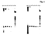
- Fig. 1 shows the visual result without (left side) and with restaining (right side). It can be seen that the presence of RO 27 alone in the PCR mixture is sufficient to detect the amplicon with adequate sensitivity. In contrast the restaining of the gel with SybrGreen I does not increase the sensitivity.
- Example 2 was repeated with samples containing in each case 3.2 ⁇ M RO 27 but different amounts of length standards(1-fold to 5-fold) on differently cross-linked gels (6 % and 20 %).
- Fig. 3 clearly shows that at the same molecular weight standard concentration (x 5) and at the same RO 27 concentration (3.2 ⁇ M) the detectability of the 50 - 150 bp standards is only slightly better in the case of the 6 % polyacrylamide gels.
- the method according to the invention can be used independently of the degree of cross-linking of the respective gel system.
- LightCycler programming denaturation: 95°C 10 min 20°C/min amplification 1: 95°C 10 sec 20°C/min 60°C 15 sec 20°C/min ⁇ 10 x 72°C 20 sec 20°C/min single amplification 2: 95°C 10 sec 20°C/min 60°C 15 sec 20°C/min ⁇ 35 x 72°C 20 sec 20°C/min single melting curve: 95°C 60 sec 20°C/min 40°C 60 sec 20°C/min 95°C 0 sec 0.1°C/min cont cooling: 40°C 30 sec 20°C/min
Abstract
Description
- The present invention concerns a simplified method in the field of nucleic acid analytics. The method according to the invention enables a gel electrophoretic nucleic acid determination to be carried out without gel staining because the dye is added to the sample containing nucleic acids.
- An electrophoretic separation of nucleic acids in gel matrices usually takes place either in agarose gels or in polyacrylamide gels. The resolving power is determined by the content of agarose in the gel in the case of agarose gels. In contrast, the resolving power of polyacrylamide gels is adjusted by the mixing ratio of acrylamide to bisacrylamide. The detection of nucleic acids in nucleic acid analytics with the aid of gel electrophoresis is based on the fact that fluorescent DNA binding dyes permanently bind non-covalently to nucleic acids and in their bound form enable nucleic acids to be located in the gel matrix after excitation with light of a suitable wavelength.
- In classical gel electrophoresis the intercalating dye ethidium bromide was used for this purpose. However, due to the toxicicity of ethidium bromide, alternative DNA binding dyes such as for example SybrGreen or SybrGold have been used for several years, the binding properties of which are not or not exclusively based on the principle of DNA intercalation.
- In contrast to ethidium bromide, these dyes can also be used to detect PCR amplificates in real time PCR or qPCR since they do not substantially inhibit the PCR reaction (
US 6,569,627 ). There are other dyes apart from SybrGreen and SybrGold which can also be used in real time PCR (Gudnason, H., et al., Nucleic Acids Research 35(19) (2007) e127). - Furthermore, certain dyes exist for real time PCR applications which are especially suitable for thermal melting curve analyses such as the LightCycler480 RO27 dye (Roche, Nature Methods, Vol. 5, No. 3, p. I-II, 2008). They are grouped together as HRM (high resolution melting) fluorescent dyes (
WO 2006/121423 ) and are characterized in that they can be used in higher concentrations in the PCR without inhibiting the PCR reaction. - Capillary gel electrophoresis is also an important method for analysing PCR products, for analysing fragments from the restriction digestion, for mutation analytics and DNA sequencing due to its speed, the ability to automate and its high resolving power. LIF (laser induced fluorescence) detection plays an important role in this connection in order to make it possible to detect and quantify DNA in a highly sensitive manner and in a high dilution. In addition to the covalent coupling of fluorescent dyes to the DNA (by means of fluorescent labelled primers during DNA sequencing), the use of dsDNA intercalators plays a major role as a non-covalent method which enables DNA to be detected fluorimetrically and simply by means of stable dye-DNA complexes. The same intercalators are used for this as for the detection of DNA in gel electrophoresis (Sang, F., et al., J. Sep. Sci. 29 (2006) 1275-1280).
- The dyes used for electrophoresis are either admixed with the gel preparation before polymerization in the case of ethidium bromide, or the gel is stained after completion of the gel electrophoresis with the aid of an aqueous dye solution containing ethidium bromide or another dye. Furthermore, it has also been described that SybrGreen I can be added to the sample containing the nucleic acid before loading the gel, provided the nucleic acid is incubated with the dye for a further 15 minutes before being applied to the gel (Karlsen, F., et al., Journal of Virol. Methods 55 (1995) 153-156, and Jin, X., et al., FASEB J. 10 A1128 (1996) abstract # 751, Larionov et al., BMC Bioinformatics, Vol. 6, No. 1, p. 62-77 (2005)).
- The staining methods for the gel electrophoretic analysis of nucleic acids known from the prior art thus have the disadvantage that the addition before polymerization only takes place with the mutagenic substance ethidium bromide, staining in a dye solution is time-consuming and laborious and requires large amounts of dye, or the addition of the dye immediately before loading the gel requires a longer incubation period.
- The present invention therefore concerns a method for analysing nucleic acids in a sample comprising the following steps
- adding the DNA binding dye RO27 to the sample to be analysed
- carrying out a polymerase chain reaction
- applying the sample to a gel matrix
- separating the nucleic acid molecules according to their size by applying a voltage
- excitation with light of a suitable wavelength for the optical visualization of the nucleic acid / DNA binding dye complexes.
- The method according to the present invention is particularily characterized in that the gel matrix is not pre-stained prior to the separation of the nucleic acid molecules and further in that the gel matrix is not stained after the separation of the nucleic acid molecules.
- In a special embodiment the amplification is measured in real time during the PCR with the aid of the DNA binding dye RO27.
- The gel matrix preferably consists either of agarose or of polyacrylamide. If it is polyacrylamide, the gel matrix can also be in a capillary in order to carry out a capillary gel electrophoresis.
- The DNA binding dye RO27 is usually added to the sample at a concentration of 1 - 10 µM and preferably 2-4 µM.
-
- Figure 1:
- Gel electrophoresis with subsequent detection of the amplificate by a Lumilmager without (left) and with (right) restaining.
- Figure 2:
- Dilution series of a molecular weight marker in which 1.6 µm dye was added before the electrophoresis (EtBr = ethidium bromide, SG1 = SybrGreen I, SG2 = SybrGreen 2, R27 = RO 27).
- Figure 3:
- Multiplication series of the molecular weight marker with constant concentrations of 3.2 µM RO 27 on a 20 % polyacrylamide gel (left) and on a 6 % polyacrylamide gel (right).
- Figure 4a:
- Polyacrylamide gel electrophoresis of PCR mixtures containing 0.8 µM RO27 (left) or SybrGreen I (right) without restaining with SybrGreen II.
- Figure 4b:
- Polyacrylamide gel electrophoresis of PCR mixtures containing 0.8 µM RO27 (left) or SybrGreen I (right) with restaining with SybrGreen II.
- Figure 4c:
- Polyacrylamide gel electrophoresis of PCR mixtures containing 4.0 µM RO27 (left) or SybrGreen I (right) without restaining with SybrGreen II.
- Figure 4d:
- Polyacrylamide gel electrophoresis of PCR mixtures containing 4.0 µM RO27 (left) or SybrGreen I (right) with restaining with SybrGreen II.
- The origin of the present invention was the surprising finding that it is possible to add a real time PCR mixture containing a DNA binding dye directly to the PCR and namely at a concentration which is sufficient to allow omission of a subsequent staining of the PCR products for example with ethidium bromide or with SybrGreen I or SybrGreen II after the gel electrophoresis. These DNA binding dyes only emit a corresponding fluorescence signal after excitation with light of a suitable wavelength when they have bound to double-stranded nucleic acid. The detectability requires a strong, non-covalent binding of the fluorescent dye to the DNA.
- Due to their cationic properties DNA binding dyes migrate in an unbound form towards the cathode during gel electrophoresis. The charge of the nucleic acids is, however, not sufficient for the nucleic acid / binding dye complex to migrate towards the anode during a gel electrophoresis.
- Hence, the present invention concerns a method for analysing nucleic acids in a sample comprising the following steps
- adding the DNA binding dye RO27 to the sample to be analysed
- carrying out a polymerase chain reaction (PCR)
- applying the sample to a gel matrix
- separating the nucleic acid molecules according to their size by applying a voltage
- excitation with light of a suitable wavelength for the optical visualization of the nucleic acid / DNA binding dye complexes.
- The method according to the present invention is particularily characterized in that the gel matrix is not pre-stained prior to the separation of the nucleic acid molecules and further in that the gel matrix is not stained after the separation of the nucleic acid molecules.
- The sample to be analysed is usually a mixture of nucleic acids which have been obtained from biological material such as for example cellular lysates. These lysates can be prepared by any standard methods. As a rule the nucleic acids present in the lysates are at least partially purified by methods known from the prior art before a PCR can be carried out by standard methods (
US 4,683,202 ,US 4,683,195 ,US 4,965,188 ). The nucleic acids present in the sample can either be DNA or RNA. In the case of RNA, the PCR is carried out in the form of a one-step RT-PCR in which the RNA is firstly reversely transcribed into a single-stranded cDNA with the aid of suitable enzymes and subsequently this cDNA is amplified to form a double-stranded end product (US 5,407,800 ,US 5,322,770 ,US 5,310,652 ). - In a special embodiment the progress of the PCR reaction can be measured in real time during the amplification with the aid of the DNA binding dye (
US 6,569,627 ). This requires thermocyclers with optical modules which excite the DNA binding dye contained in the PCR reaction vessel during the PCR reaction and can subsequently detect the fluorescence signal emitted by this dye (e.g.US 6,814,934 ,US 6,106,777 ). - The DNA binding dye is usually added to the sample at a concentration of 1 - 10 µM and preferably 2-4 µM. These concentrations do not usually lead to a measurable or significant inhibition of the polymerase catalysed PCR reaction in the case of the dyes that are to be used according to the invention.
- The DNA binding dye which can be by far be used particularly well within the scope of the present invention is the LC480 Resolight Dye (Roche Diagnostics Catalogue No: 04 909 640 001). This dye can be used in high concentrations in the PCR and has the following chemical structure:

- Details of the synthesis of this dye class are described in
WO 2008 052742 . - RO27 is a fluorescent dye from the group of HRM (high resolution melting) fluorescent dyes which can be used for so-called high resolution melting analyses after a real time PCR. They have in common a low toxicity during the PCR amplification so that they can be used in higher concentrations without inhibiting the PCR.
- Before application to the gel an application buffer such as for example TBE or TAE is added to the PCR mixture containing the DNA binding dye according to the methods known from the prior art. In a suitable embodiment the gel matrix can consist of 0.02 to 6 % w/v and preferably 0.5 to 4 % agarose in a TBE buffer system. The electrophoretic separation at 10 - 150 V then preferably takes place in a horizontal direction for 10 to 180 minutes. In one embodiment the gel matrix can consist of a mixture of 4 % to 25 % w/v and preferably of 6 - 20 % acrylamide / bisacrylamide in a TAE buffer system. A separation time of 10 to 30 minutes is preferred. The migration properties are determined in this case by the proportion of acrylamide as well as by the degree of cross-linking. This depends on the mixing ratio between acrylamide and bisacrylamide.
- The electrophoretic separation is carried out at 20 to 250 V for 15 to 180 minutes. A separation time of 15 to 45 minutes is preferred. In a special embodiment the acrylamide matrix is in a capillary for carrying out a capillary gel electrophoresis (Schwartz, H.E., et al., J. Chromatogr. 559 (1991) 267- 283; Guttman, A., Methods in Molecular Biology 52 (1996) 157-169: Capillary Gel Electrophoresis). It is a major advantage to be able to add these fluorescent dyes before the PCR reaction especially for the highly sensitive, highly resolving and rapid analysis of DNA fragments in the combination of PCR / capillary gel electrophoresis with LIF detection. Previously only the addition of these fluorescent dyes before the capillary gel electrophoresis was known (Garcia-Canas, V., J. Agric. Food Chem. 50 (2002) 4497-4502; Sang, F., et al., J. Sep. Sci. 29 (2006) 1275-1280).
- After the electrophoretic separation of the nucleic acids by the classical gel electrophoresis, the nucleic acids are detected for example directly by means of a UV transilluminator or with the aid of imaging instruments such as the Lumilmager (Roche Applied Science Cat. No. 2c 012 847).
- During an examination of the dependencies of the direct detection of dsDNA on the size and the dependencies with regard to separation times (i.e. also the dependency on the voltage) it turned out that the detection can be dependent on the separation time in the case of small DNA fragments of 80 - 150 bp. Thus, in 6 % polyacrylamide gels it is still possible to detect even small DNA fragments of 80 - 150 bp with the aid of RO 27 at 200 V voltage and 30 minutes separation time. It was also possible to directly detect 80 bp PCR products without restaining in 20 % gels with a separation time of 2 hours. In general it can be determined that shorter run times have a positive effect on the sensitivity of the nucleic acid detection within the scope of the method according to the invention.
- The gel matrix is shrink-wrapped in a foil to protect it against drying out. In addition the kit can contain an appropriate gel electrophoresis run buffer. It does not contain any staining dye.
- The reagents for carrying out the polymerase chain reaction are usually a thermostable DNA polymerase such as for example Taq polymerase or a thermostable polymerase with additional reverse transcriptase activity, deoxynucleoside triphosphates, as well as suitable buffers and PCR additives such as for example magnesium chloride. The reagents can be present individually in the kit. One or more components including the DNA binding dye can also be present in the kit as a mixture in so-called master mixes. In addition such a kit can contain components for DNA sample preparation.
- The invention is further elucidated by the following examples, publications and figures, the protective scope of which results from the patent claims. The described methods are to be understood as examples which still describe the subject matter of the invention even after modifications.
- Starting with genomic DNA isolated from CHO cells K1 (5 x 10⁶ cells/ml) a PCR reaction was carried out under standard conditions in which the GAPDH housekeeping gene was amplified as a target using primers known from the publication Eldering, J.A., et al., Biologicals 32 (2004) 183 - 193. 3.2 µM RO 27 was added to the mixture before starting the PCR.
- After the amplification 1:10 dilutions of the PCR mixture were prepared. 3 µl TBE
sample buffer Anamed 5 x was added to 12 µl of each dilution. 10 µl of each dilution was applied to a 6 % polyacrylamide TBE gel. The electrophoresis was carried out for 30 minutes at 200 V. The subsequent detection was carried out after excitation for 2 seconds with a 520 nm light source in a Lumilmager system. - Subsequently the gel was restained for 30 minutes with SybrGreen I according to the manufacturer's instructions (Invitrogen, Cat. No.: S7563).
-
Fig. 1 shows the visual result without (left side) and with restaining (right side). It can be seen that the presence of RO 27 alone in the PCR mixture is sufficient to detect the amplicon with adequate sensitivity. In contrast the restaining of the gel with SybrGreen I does not increase the sensitivity. - In order to compare the detection limit of dsDNA with and without restaining using a molecular weight standard (length standard XIII, Roche Applied Science Cat. No.: 11 721 925 001) as an example and when using different DNA binding dyes as a function of the concentration of the molecular weight standard, various dilutions of the length standard were admixed in each case with 1.6 µM ethidium bromide, SybrGreen I, SybrGreen II or RO 27 before application to a gel system according to example 1. The result is shown in
fig. 2 . It can be seen that in particular SybrGreen I and RO 27 are suitable for a sensitive detection. No DNA is detectable when ethidium bromide is added to the length standard before application to the gel. - Example 2 was repeated with samples containing in each case 3.2 µM RO 27 but different amounts of length standards(1-fold to 5-fold) on differently cross-linked gels (6 % and 20 %).
Fig. 3 clearly shows that at the same molecular weight standard concentration (x 5) and at the same RO 27 concentration (3.2 µM) the detectability of the 50 - 150 bp standards is only slightly better in the case of the 6 % polyacrylamide gels. Thus, the method according to the invention can be used independently of the degree of cross-linking of the respective gel system. - The aim of the experiments was to see whether real time PCR experiments with RO 27 as a universal detection dye with subsequent characterization of the PCR amplificates by gel electrophoresis can also be used for small PCR amplificates without any restaining. It is known from the prior art that DNA binding dyes containing a benzothiazolium or benzoxazolium group do not inhibit the PCR to any significant extent and can thus be used for real time PCR applications. Therefore, a PCR was carried out in which in each case 0.8 µM or 4 µM RO 27 or SybrGreen I was added to the PCR mixture before starting the amplification reaction.
- The details of the amplification reactions were as follows:
Instrument: LightCycler LC 2.0 with firmware version > 4.02.0 LightCycler TM: 20 µl capillaries Dyes: 0.8 µM or 4 µM RO 27 or SybrGreen I Template: plasmid DNA containing 18s RNA gene sequence in different copy numbers (104, 103, 102, 101) Amplicon: 91 bp - LightCycler programming:
denaturation: 95° C 10 min 20°C/minamplification 1: 95° C 10 sec 20°C/min 60°C 15 sec 20°C/min } 10 x 72° C 20 sec 20°C/min single amplification 2: 95° C 10 sec 20°C/min 60°C 15 sec 20°C/min } 35 x 72° C 20 sec 20°C/min single melting curve: 95°C 60 sec 20°C/min 40°C 60 sec 20°C/min 95°C 0 sec 0.1°C/min cont cooling: 40°C 30 sec 20°C/min - Subsequently 10 µl of each of the mixtures was separated by gel electrophoresis similarly to example 1.
- The detection before and after additional restaining of the gel with SybrGreen I was also carried out according to example 1.
- The results are shown in
fig. 4 a - d. They show that even at a low RO 27 concentration (0.8 µM), the 80 bp bands are visible without restaining and all expected bands (except for primer dimer artefacts and the primers themselves) are detected. Restaining the gels with SybrGreen I does not result in a significant increase in the sensitivity of the detection. In this connection the advantage of RO 27 is that it can be used in even higher concentrations (4.0 µM) because the PCR reaction is not inhibited under these conditions in contrast to SybrGreen I. The detectability of weak DNA bands in the gel electrophoresis can thus be improved. - Similarly to example 1 and 4 various dyes were added to a PCR reaction mixture before the amplification reaction. After the PCR was completed, the products were analysed by gel electrophoresis without any further staining measures in a 6 % polyacrylamide gel as well as in a 2 % agarose gel. The result is shown in the following table.
fluorescent dye 6 % polyacrylamide gel 2 % agarose gel type of dye RO 27 + + benzothiazolium SybrGreen I + + benzothiazolium SybrGreen II + not tested benzothiazolium Syto 9 not tested + benzothiazolium LC Green not tested + benzothiazolium
Claims (5)
- Method for analysing nucleic acids in a sample comprising the following steps- adding the DNA binding dye having the structure
 - carrying out a polymerase chain reaction- applying the sample to a gel matrix- separating the nucleic acid molecules according to their size by applying a voltage- excitation with light of a suitable wavelength for the optical visualization of the nucleic acid / DNA binding dye complexes,wherein the gel matrix is not pre-stained prior to separation and not stained after separation of the nucleic acid molecules.
 - Method according to claim 1, characterized in that the amplification is measured in real time during the PCR with the aid of the DNA binding dye.
- Method according to claim 1, characterized in that the gel matrix consists either of polyacrylamide or of agarose.
- Method according to claim 3, characterized in that the gel matrix is polyacrylamide and is present in a capillary.
- Method according to claim 1 to 4, characterized in that the DNA binding dye is added to the sample at a concentration of 1 - 10 µM and particularly preferably 2-4 µM.
Priority Applications (1)
| Application Number | Priority Date | Filing Date | Title |
|---|---|---|---|
| EP09730059A EP2276859B1 (en) | 2008-04-08 | 2009-03-27 | Detection of pcr products in gel electrophoresis |
Applications Claiming Priority (3)
| Application Number | Priority Date | Filing Date | Title |
|---|---|---|---|
| EP08006994 | 2008-04-08 | ||
| PCT/EP2009/002264 WO2009124665A1 (en) | 2008-04-08 | 2009-03-27 | Detection of pcr products in gel electrophoresis |
| EP09730059A EP2276859B1 (en) | 2008-04-08 | 2009-03-27 | Detection of pcr products in gel electrophoresis |
Publications (2)
| Publication Number | Publication Date |
|---|---|
| EP2276859A1 EP2276859A1 (en) | 2011-01-26 |
| EP2276859B1 true EP2276859B1 (en) | 2011-12-14 |
Family
ID=39714018
Family Applications (1)
| Application Number | Title | Priority Date | Filing Date |
|---|---|---|---|
| EP09730059A Not-in-force EP2276859B1 (en) | 2008-04-08 | 2009-03-27 | Detection of pcr products in gel electrophoresis |
Country Status (4)
| Country | Link |
|---|---|
| US (1) | US8053213B2 (en) |
| EP (1) | EP2276859B1 (en) |
| AT (1) | ATE537272T1 (en) |
| WO (1) | WO2009124665A1 (en) |
Families Citing this family (1)
| Publication number | Priority date | Publication date | Assignee | Title |
|---|---|---|---|---|
| EP3655779A1 (en) * | 2017-07-20 | 2020-05-27 | CytomX Therapeutics, Inc. | Methods of qualitatively and/or quantitatively analyzing properties of activatable antibodies and uses thereof |
Family Cites Families (12)
| Publication number | Priority date | Publication date | Assignee | Title |
|---|---|---|---|---|
| US4683195A (en) | 1986-01-30 | 1987-07-28 | Cetus Corporation | Process for amplifying, detecting, and/or-cloning nucleic acid sequences |
| US4965188A (en) | 1986-08-22 | 1990-10-23 | Cetus Corporation | Process for amplifying, detecting, and/or cloning nucleic acid sequences using a thermostable enzyme |
| US4683202A (en) * | 1985-03-28 | 1987-07-28 | Cetus Corporation | Process for amplifying nucleic acid sequences |
| US5310652A (en) | 1986-08-22 | 1994-05-10 | Hoffman-La Roche Inc. | Reverse transcription with thermostable DNA polymerase-high temperature reverse transcription |
| US5322770A (en) | 1989-12-22 | 1994-06-21 | Hoffman-Laroche Inc. | Reverse transcription with thermostable DNA polymerases - high temperature reverse transcription |
| US5407800A (en) | 1986-08-22 | 1995-04-18 | Hoffmann-La Roche Inc. | Reverse transcription with Thermus thermophilus polymerase |
| US5994056A (en) | 1991-05-02 | 1999-11-30 | Roche Molecular Systems, Inc. | Homogeneous methods for nucleic acid amplification and detection |
| US5658751A (en) * | 1993-04-13 | 1997-08-19 | Molecular Probes, Inc. | Substituted unsymmetrical cyanine dyes with selected permeability |
| DE69527125T2 (en) | 1994-11-09 | 2003-02-27 | Hitachi Ltd | Method and device for DNA analysis |
| EP0912766B2 (en) | 1996-06-04 | 2011-12-14 | University of Utah Research Foundation | Monitoring hybridization during pcr |
| US7387887B2 (en) | 2004-04-20 | 2008-06-17 | University Of Utah Research Foundation | Nucleic acid melting analysis with saturation dyes |
| EP2087043B1 (en) | 2006-11-02 | 2010-08-04 | Roche Diagnostics GmbH | New ds dna binding fluorescent dyes |
-
2009
- 2009-03-27 EP EP09730059A patent/EP2276859B1/en not_active Not-in-force
- 2009-03-27 WO PCT/EP2009/002264 patent/WO2009124665A1/en active Application Filing
- 2009-03-27 AT AT09730059T patent/ATE537272T1/en active
-
2010
- 2010-09-07 US US12/876,320 patent/US8053213B2/en not_active Expired - Fee Related
Also Published As
| Publication number | Publication date |
|---|---|
| ATE537272T1 (en) | 2011-12-15 |
| US8053213B2 (en) | 2011-11-08 |
| EP2276859A1 (en) | 2011-01-26 |
| US20100330579A1 (en) | 2010-12-30 |
| WO2009124665A1 (en) | 2009-10-15 |
Similar Documents
| Publication | Publication Date | Title |
|---|---|---|
| US20240026340A1 (en) | Blood collection device for improved nucleic acid regulation | |
| Thompson et al. | An overview of DNA typing methods for human identification: past, present, and future | |
| Hsieh et al. | Optimization of a relative telomere length assay by monochromatic multiplex real-time quantitative PCR on the LightCycler 480: sources of variability and quality control considerations | |
| US20060057595A1 (en) | Compositions, methods, and kits for identifying and quantitating small RNA molecules | |
| Yoo et al. | International comparison of enumeration-based quantification of DNA copy-concentration using flow cytometric counting and digital polymerase chain reaction | |
| Sieber et al. | Substantial performance discrepancies among commercially available kits for reverse transcription quantitative polymerase chain reaction: a systematic comparative investigator-driven approach | |
| Venegas et al. | Quantification of mtDNA mutation heteroplasmy (ARMS qPCR) | |
| WO2014115779A1 (en) | QUANTIFICATION METHOD FOR EXPRESSION LEVEL OF WT1 mRNA | |
| Hancock et al. | A Standard Reference Material to determine the sensitivity of techniques for detecting low-frequency mutations, SNPs, and heteroplasmies in mitochondrial DNA | |
| JoonáLee | Label/quencher-free detection of single-nucleotide changes in DNA using isothermal amplification and G-quadruplexes | |
| EP2569443A1 (en) | Tissue typing assays and kits | |
| EP2276859B1 (en) | Detection of pcr products in gel electrophoresis | |
| Xu et al. | Determination of murine fetal Cyp1a1 and 1b1 expression by real-time fluorescence reverse transcription-polymerase chain reaction | |
| WO2008103770A2 (en) | Multiplex compositions and methods for quantification of human nuclear dna and human male dna and detection of pcr inhibitors | |
| Ekstrøm et al. | Analysis of mutational spectra by denaturing capillary electrophoresis | |
| Wang et al. | Quantification of SMN1 and SMN2 genes by capillary electrophoresis for diagnosis of spinal muscular atrophy | |
| EP1798291B1 (en) | Methods and compositions for assaying mutations and/or large scale alterations in nucleic acids and their uses in diagnosis of genetic diseases and cancers | |
| US9297038B2 (en) | Method and kit for the quantification of nucleic acids | |
| Kahyo et al. | Application of digital PCR with chip-in-a-tube format to analyze Adenomatous polyposis coli (APC) somatic mosaicism | |
| Kim et al. | Development and forensic validation of human genomic DNA quantification kit | |
| EP4029938A1 (en) | Nucleic acid detection method by real-time pcr | |
| Volkov et al. | Genetic Analyzer Nanofor 05 as a Measuring Instrument for DNA Sequencing | |
| Jiang et al. | Genotyping genetic markers from LCN and degraded DNA by HRM and their application in hair shaft | |
| Elek et al. | Multicapillary gel electrophoresis based analysis of genetic variants in the WFS1 gene | |
| Turrina et al. | STR typing of archival Bouin’s fluid-fixed paraffin-embedded tissue using new sensitive redesigned primers for three STR loci (CSF1P0, D8S1179 and D13S317) |
Legal Events
| Date | Code | Title | Description |
|---|---|---|---|
| PUAI | Public reference made under article 153(3) epc to a published international application that has entered the european phase |
Free format text: ORIGINAL CODE: 0009012 |
|
| 17P | Request for examination filed |
Effective date: 20101108 |
|
| AK | Designated contracting states |
Kind code of ref document: A1 Designated state(s): AT BE BG CH CY CZ DE DK EE ES FI FR GB GR HR HU IE IS IT LI LT LU LV MC MK MT NL NO PL PT RO SE SI SK TR |
|
| AX | Request for extension of the european patent |
Extension state: AL BA RS |
|
| 17Q | First examination report despatched |
Effective date: 20110314 |
|
| GRAP | Despatch of communication of intention to grant a patent |
Free format text: ORIGINAL CODE: EPIDOSNIGR1 |
|
| DAX | Request for extension of the european patent (deleted) | ||
| GRAS | Grant fee paid |
Free format text: ORIGINAL CODE: EPIDOSNIGR3 |
|
| GRAA | (expected) grant |
Free format text: ORIGINAL CODE: 0009210 |
|
| AK | Designated contracting states |
Kind code of ref document: B1 Designated state(s): AT BE BG CH CY CZ DE DK EE ES FI FR GB GR HR HU IE IS IT LI LT LU LV MC MK MT NL NO PL PT RO SE SI SK TR |
|
| REG | Reference to a national code |
Ref country code: GB Ref legal event code: FG4D |
|
| REG | Reference to a national code |
Ref country code: CH Ref legal event code: EP |
|
| REG | Reference to a national code |
Ref country code: IE Ref legal event code: FG4D |
|
| REG | Reference to a national code |
Ref country code: DE Ref legal event code: R096 Ref document number: 602009004208 Country of ref document: DE Effective date: 20120223 |
|
| REG | Reference to a national code |
Ref country code: NL Ref legal event code: VDEP Effective date: 20111214 |
|
| PG25 | Lapsed in a contracting state [announced via postgrant information from national office to epo] |
Ref country code: NO Free format text: LAPSE BECAUSE OF FAILURE TO SUBMIT A TRANSLATION OF THE DESCRIPTION OR TO PAY THE FEE WITHIN THE PRESCRIBED TIME-LIMIT Effective date: 20120314 Ref country code: LT Free format text: LAPSE BECAUSE OF FAILURE TO SUBMIT A TRANSLATION OF THE DESCRIPTION OR TO PAY THE FEE WITHIN THE PRESCRIBED TIME-LIMIT Effective date: 20111214 |
|
| LTIE | Lt: invalidation of european patent or patent extension |
Effective date: 20111214 |
|
| PG25 | Lapsed in a contracting state [announced via postgrant information from national office to epo] |
Ref country code: HR Free format text: LAPSE BECAUSE OF FAILURE TO SUBMIT A TRANSLATION OF THE DESCRIPTION OR TO PAY THE FEE WITHIN THE PRESCRIBED TIME-LIMIT Effective date: 20111214 Ref country code: NL Free format text: LAPSE BECAUSE OF FAILURE TO SUBMIT A TRANSLATION OF THE DESCRIPTION OR TO PAY THE FEE WITHIN THE PRESCRIBED TIME-LIMIT Effective date: 20111214 Ref country code: SE Free format text: LAPSE BECAUSE OF FAILURE TO SUBMIT A TRANSLATION OF THE DESCRIPTION OR TO PAY THE FEE WITHIN THE PRESCRIBED TIME-LIMIT Effective date: 20111214 Ref country code: GR Free format text: LAPSE BECAUSE OF FAILURE TO SUBMIT A TRANSLATION OF THE DESCRIPTION OR TO PAY THE FEE WITHIN THE PRESCRIBED TIME-LIMIT Effective date: 20120315 Ref country code: LV Free format text: LAPSE BECAUSE OF FAILURE TO SUBMIT A TRANSLATION OF THE DESCRIPTION OR TO PAY THE FEE WITHIN THE PRESCRIBED TIME-LIMIT Effective date: 20111214 Ref country code: SI Free format text: LAPSE BECAUSE OF FAILURE TO SUBMIT A TRANSLATION OF THE DESCRIPTION OR TO PAY THE FEE WITHIN THE PRESCRIBED TIME-LIMIT Effective date: 20111214 |
|
| PG25 | Lapsed in a contracting state [announced via postgrant information from national office to epo] |
Ref country code: BE Free format text: LAPSE BECAUSE OF FAILURE TO SUBMIT A TRANSLATION OF THE DESCRIPTION OR TO PAY THE FEE WITHIN THE PRESCRIBED TIME-LIMIT Effective date: 20111214 Ref country code: CY Free format text: LAPSE BECAUSE OF FAILURE TO SUBMIT A TRANSLATION OF THE DESCRIPTION OR TO PAY THE FEE WITHIN THE PRESCRIBED TIME-LIMIT Effective date: 20111214 |
|
| PG25 | Lapsed in a contracting state [announced via postgrant information from national office to epo] |
Ref country code: EE Free format text: LAPSE BECAUSE OF FAILURE TO SUBMIT A TRANSLATION OF THE DESCRIPTION OR TO PAY THE FEE WITHIN THE PRESCRIBED TIME-LIMIT Effective date: 20111214 Ref country code: CZ Free format text: LAPSE BECAUSE OF FAILURE TO SUBMIT A TRANSLATION OF THE DESCRIPTION OR TO PAY THE FEE WITHIN THE PRESCRIBED TIME-LIMIT Effective date: 20111214 Ref country code: SK Free format text: LAPSE BECAUSE OF FAILURE TO SUBMIT A TRANSLATION OF THE DESCRIPTION OR TO PAY THE FEE WITHIN THE PRESCRIBED TIME-LIMIT Effective date: 20111214 Ref country code: BG Free format text: LAPSE BECAUSE OF FAILURE TO SUBMIT A TRANSLATION OF THE DESCRIPTION OR TO PAY THE FEE WITHIN THE PRESCRIBED TIME-LIMIT Effective date: 20120314 Ref country code: IS Free format text: LAPSE BECAUSE OF FAILURE TO SUBMIT A TRANSLATION OF THE DESCRIPTION OR TO PAY THE FEE WITHIN THE PRESCRIBED TIME-LIMIT Effective date: 20120414 |
|
| PG25 | Lapsed in a contracting state [announced via postgrant information from national office to epo] |
Ref country code: PT Free format text: LAPSE BECAUSE OF FAILURE TO SUBMIT A TRANSLATION OF THE DESCRIPTION OR TO PAY THE FEE WITHIN THE PRESCRIBED TIME-LIMIT Effective date: 20120416 Ref country code: RO Free format text: LAPSE BECAUSE OF FAILURE TO SUBMIT A TRANSLATION OF THE DESCRIPTION OR TO PAY THE FEE WITHIN THE PRESCRIBED TIME-LIMIT Effective date: 20111214 Ref country code: PL Free format text: LAPSE BECAUSE OF FAILURE TO SUBMIT A TRANSLATION OF THE DESCRIPTION OR TO PAY THE FEE WITHIN THE PRESCRIBED TIME-LIMIT Effective date: 20111214 |
|
| REG | Reference to a national code |
Ref country code: AT Ref legal event code: MK05 Ref document number: 537272 Country of ref document: AT Kind code of ref document: T Effective date: 20111214 |
|
| PLBE | No opposition filed within time limit |
Free format text: ORIGINAL CODE: 0009261 |
|
| STAA | Information on the status of an ep patent application or granted ep patent |
Free format text: STATUS: NO OPPOSITION FILED WITHIN TIME LIMIT |
|
| PG25 | Lapsed in a contracting state [announced via postgrant information from national office to epo] |
Ref country code: MC Free format text: LAPSE BECAUSE OF NON-PAYMENT OF DUE FEES Effective date: 20120331 Ref country code: DK Free format text: LAPSE BECAUSE OF FAILURE TO SUBMIT A TRANSLATION OF THE DESCRIPTION OR TO PAY THE FEE WITHIN THE PRESCRIBED TIME-LIMIT Effective date: 20111214 |
|
| 26N | No opposition filed |
Effective date: 20120917 |
|
| PG25 | Lapsed in a contracting state [announced via postgrant information from national office to epo] |
Ref country code: IT Free format text: LAPSE BECAUSE OF FAILURE TO SUBMIT A TRANSLATION OF THE DESCRIPTION OR TO PAY THE FEE WITHIN THE PRESCRIBED TIME-LIMIT Effective date: 20111214 |
|
| REG | Reference to a national code |
Ref country code: IE Ref legal event code: MM4A |
|
| REG | Reference to a national code |
Ref country code: DE Ref legal event code: R097 Ref document number: 602009004208 Country of ref document: DE Effective date: 20120917 |
|
| PG25 | Lapsed in a contracting state [announced via postgrant information from national office to epo] |
Ref country code: IE Free format text: LAPSE BECAUSE OF NON-PAYMENT OF DUE FEES Effective date: 20120327 Ref country code: AT Free format text: LAPSE BECAUSE OF FAILURE TO SUBMIT A TRANSLATION OF THE DESCRIPTION OR TO PAY THE FEE WITHIN THE PRESCRIBED TIME-LIMIT Effective date: 20111214 |
|
| PG25 | Lapsed in a contracting state [announced via postgrant information from national office to epo] |
Ref country code: MK Free format text: LAPSE BECAUSE OF FAILURE TO SUBMIT A TRANSLATION OF THE DESCRIPTION OR TO PAY THE FEE WITHIN THE PRESCRIBED TIME-LIMIT Effective date: 20111214 |
|
| PG25 | Lapsed in a contracting state [announced via postgrant information from national office to epo] |
Ref country code: ES Free format text: LAPSE BECAUSE OF FAILURE TO SUBMIT A TRANSLATION OF THE DESCRIPTION OR TO PAY THE FEE WITHIN THE PRESCRIBED TIME-LIMIT Effective date: 20120325 |
|
| PG25 | Lapsed in a contracting state [announced via postgrant information from national office to epo] |
Ref country code: FI Free format text: LAPSE BECAUSE OF FAILURE TO SUBMIT A TRANSLATION OF THE DESCRIPTION OR TO PAY THE FEE WITHIN THE PRESCRIBED TIME-LIMIT Effective date: 20111214 |
|
| PG25 | Lapsed in a contracting state [announced via postgrant information from national office to epo] |
Ref country code: MT Free format text: LAPSE BECAUSE OF FAILURE TO SUBMIT A TRANSLATION OF THE DESCRIPTION OR TO PAY THE FEE WITHIN THE PRESCRIBED TIME-LIMIT Effective date: 20111214 |
|
| REG | Reference to a national code |
Ref country code: CH Ref legal event code: PL |
|
| PG25 | Lapsed in a contracting state [announced via postgrant information from national office to epo] |
Ref country code: LI Free format text: LAPSE BECAUSE OF NON-PAYMENT OF DUE FEES Effective date: 20130331 Ref country code: CH Free format text: LAPSE BECAUSE OF NON-PAYMENT OF DUE FEES Effective date: 20130331 |
|
| PG25 | Lapsed in a contracting state [announced via postgrant information from national office to epo] |
Ref country code: TR Free format text: LAPSE BECAUSE OF FAILURE TO SUBMIT A TRANSLATION OF THE DESCRIPTION OR TO PAY THE FEE WITHIN THE PRESCRIBED TIME-LIMIT Effective date: 20111214 |
|
| PGFP | Annual fee paid to national office [announced via postgrant information from national office to epo] |
Ref country code: DE Payment date: 20140331 Year of fee payment: 6 |
|
| PG25 | Lapsed in a contracting state [announced via postgrant information from national office to epo] |
Ref country code: LU Free format text: LAPSE BECAUSE OF NON-PAYMENT OF DUE FEES Effective date: 20120327 |
|
| PGFP | Annual fee paid to national office [announced via postgrant information from national office to epo] |
Ref country code: FR Payment date: 20140225 Year of fee payment: 6 |
|
| PGFP | Annual fee paid to national office [announced via postgrant information from national office to epo] |
Ref country code: GB Payment date: 20140225 Year of fee payment: 6 |
|
| PG25 | Lapsed in a contracting state [announced via postgrant information from national office to epo] |
Ref country code: HU Free format text: LAPSE BECAUSE OF FAILURE TO SUBMIT A TRANSLATION OF THE DESCRIPTION OR TO PAY THE FEE WITHIN THE PRESCRIBED TIME-LIMIT Effective date: 20090327 |
|
| REG | Reference to a national code |
Ref country code: DE Ref legal event code: R119 Ref document number: 602009004208 Country of ref document: DE |
|
| GBPC | Gb: european patent ceased through non-payment of renewal fee |
Effective date: 20150327 |
|
| REG | Reference to a national code |
Ref country code: FR Ref legal event code: ST Effective date: 20151130 |
|
| PG25 | Lapsed in a contracting state [announced via postgrant information from national office to epo] |
Ref country code: DE Free format text: LAPSE BECAUSE OF NON-PAYMENT OF DUE FEES Effective date: 20151001 Ref country code: GB Free format text: LAPSE BECAUSE OF NON-PAYMENT OF DUE FEES Effective date: 20150327 |
|
| PG25 | Lapsed in a contracting state [announced via postgrant information from national office to epo] |
Ref country code: FR Free format text: LAPSE BECAUSE OF NON-PAYMENT OF DUE FEES Effective date: 20150331 |