EP2272947B1 - Peptide conjugated anti-cancer prodrugs - Google Patents
Peptide conjugated anti-cancer prodrugs Download PDFInfo
- Publication number
- EP2272947B1 EP2272947B1 EP10156533.1A EP10156533A EP2272947B1 EP 2272947 B1 EP2272947 B1 EP 2272947B1 EP 10156533 A EP10156533 A EP 10156533A EP 2272947 B1 EP2272947 B1 EP 2272947B1
- Authority
- EP
- European Patent Office
- Prior art keywords
- peptide
- prodrug
- drug
- cells
- resin
- Prior art date
- Legal status (The legal status is an assumption and is not a legal conclusion. Google has not performed a legal analysis and makes no representation as to the accuracy of the status listed.)
- Expired - Lifetime
Links
- 0 C1C*2C=CC2C1 Chemical compound C1C*2C=CC2C1 0.000 description 2
Images


Classifications
-
- A—HUMAN NECESSITIES
- A61—MEDICAL OR VETERINARY SCIENCE; HYGIENE
- A61K—PREPARATIONS FOR MEDICAL, DENTAL OR TOILETRY PURPOSES
- A61K38/00—Medicinal preparations containing peptides
- A61K38/04—Peptides having up to 20 amino acids in a fully defined sequence; Derivatives thereof
- A61K38/10—Peptides having 12 to 20 amino acids
-
- A—HUMAN NECESSITIES
- A61—MEDICAL OR VETERINARY SCIENCE; HYGIENE
- A61K—PREPARATIONS FOR MEDICAL, DENTAL OR TOILETRY PURPOSES
- A61K38/00—Medicinal preparations containing peptides
- A61K38/04—Peptides having up to 20 amino acids in a fully defined sequence; Derivatives thereof
- A61K38/06—Tripeptides
-
- A—HUMAN NECESSITIES
- A61—MEDICAL OR VETERINARY SCIENCE; HYGIENE
- A61K—PREPARATIONS FOR MEDICAL, DENTAL OR TOILETRY PURPOSES
- A61K38/00—Medicinal preparations containing peptides
- A61K38/04—Peptides having up to 20 amino acids in a fully defined sequence; Derivatives thereof
- A61K38/08—Peptides having 5 to 11 amino acids
-
- A—HUMAN NECESSITIES
- A61—MEDICAL OR VETERINARY SCIENCE; HYGIENE
- A61K—PREPARATIONS FOR MEDICAL, DENTAL OR TOILETRY PURPOSES
- A61K38/00—Medicinal preparations containing peptides
- A61K38/16—Peptides having more than 20 amino acids; Gastrins; Somatostatins; Melanotropins; Derivatives thereof
- A61K38/17—Peptides having more than 20 amino acids; Gastrins; Somatostatins; Melanotropins; Derivatives thereof from animals; from humans
- A61K38/1703—Peptides having more than 20 amino acids; Gastrins; Somatostatins; Melanotropins; Derivatives thereof from animals; from humans from vertebrates
- A61K38/1709—Peptides having more than 20 amino acids; Gastrins; Somatostatins; Melanotropins; Derivatives thereof from animals; from humans from vertebrates from mammals
-
- A—HUMAN NECESSITIES
- A61—MEDICAL OR VETERINARY SCIENCE; HYGIENE
- A61K—PREPARATIONS FOR MEDICAL, DENTAL OR TOILETRY PURPOSES
- A61K47/00—Medicinal preparations characterised by the non-active ingredients used, e.g. carriers or inert additives; Targeting or modifying agents chemically bound to the active ingredient
- A61K47/50—Medicinal preparations characterised by the non-active ingredients used, e.g. carriers or inert additives; Targeting or modifying agents chemically bound to the active ingredient the non-active ingredient being chemically bound to the active ingredient, e.g. polymer-drug conjugates
- A61K47/51—Medicinal preparations characterised by the non-active ingredients used, e.g. carriers or inert additives; Targeting or modifying agents chemically bound to the active ingredient the non-active ingredient being chemically bound to the active ingredient, e.g. polymer-drug conjugates the non-active ingredient being a modifying agent
- A61K47/62—Medicinal preparations characterised by the non-active ingredients used, e.g. carriers or inert additives; Targeting or modifying agents chemically bound to the active ingredient the non-active ingredient being chemically bound to the active ingredient, e.g. polymer-drug conjugates the non-active ingredient being a modifying agent the modifying agent being a protein, peptide or polyamino acid
- A61K47/65—Peptidic linkers, binders or spacers, e.g. peptidic enzyme-labile linkers
Definitions
- the present invention relates to prodrug molecules comprising conjugates of an antiproliferative drug, a protease specific cleavable peptide, and, optionally, a targeting peptide, said prodrugs being substantially inactive prior to degradation of the cleavable sequence by proteolytic enzymes abundant within or in close proximity to the target cancer cell, to pharmaceutical compositions comprising the conjugates and to the use of these compositions in the treatment of cancer.
- Anti-proliferative drugs also known as anti-metabolites, act by inhibiting crucial metabolic processes, and are commonly used in the treatment of diseases involving abnormal cell proliferation, such as tumors.
- diseases involving abnormal cell proliferation such as tumors.
- the utility of these drugs is severely hampered by their excessive toxicity and adverse side effects on healthy cells of the treated patient. Therefore, it would be advantageous to be able to reduce these adverse effects by the use of a prodrug having decreased toxicity.
- prodrugs to impart desired characteristics such as increased bioavailability or increased site-specificity of known drugs is a recognized concept in the state of the art of pharmaceutical development.
- various blocking groups which must be removed in order to release the active drug is also known in the background art.
- one or more blocking groups may be attached via an available amine, hydroxyl group or other functional reactive group on the drug to yield an amide or an ester.
- This type of prodrug may be cleaved by non-specific esterases to release the active principle in a sustained-release fashion over a prolonged period of time compared to the native drug species.
- Methotrexate for example, is an effective anti-proliferative drug commonly used in cancer therapy. It is an analogue of dihydrofolate that inhibits the enzyme dihydrofolate reductase (DHFR), thus depleting intracellular tetrahydrofolate (FH 4 ), which is an essential co-factor required for the de novo synthesis of purine nucleotides.
- DHFR dihydrofolate reductase
- FH 4 tetrahydrofolate
- MTX, Mephalan and Chlorambucil are valuable drugs in the treatment of many rapidly growing tumors, however, their use is limited by the frequency and severity of side effects. Unwanted side effects include toxicity to all rapidly dividing normal cells, such as stem cells in the bone marrow, epithelial cells of the intestinal tract, hair follicle cells, etc.
- Another major problem in chemotherapy which is particularly relevant in the case of anti-metabolites, is inherent or acquired resistance of tumors to cytotoxic drugs.
- development of resistance to MTX frequently follows prolonged exposure to this drug. Resistance may be due to new mutations induced by the clinical treatment, or to positive selection, by the treatment regimen, of pre-existing resistant mutant cell.
- Known mechanisms for development of resistance involve impaired transport of MTX into cells, e.g. by mutations in the Reduced Folate Carrier (RFC), over expression of the target enzyme DHFR, or mutations in the enzyme responsible for polyglutamination of reduced folates (FPGS).
- MDR multi-drug resistance
- cytotoxic drugs it has been suggested that the therapeutic index of such drugs might be increased if the drug is covalently bound to a peptide that would be cleaved in the vicinity of the tumor cells by the action of certain proteases.
- This approach has been suggested for peptide conjugated Methotrexate (Kuefner et al., 1989) and for Arabinofuranosyl cytosine (ara-C) lipid-peptide-drug conjugates (Menger et al., 1994).
- cell surface polypeptides or glycoproteins have been utilized for targeting drugs to malignant cells, with various degrees of success.
- cell surface complex sugars as cell surface markers is much less well developed. In part this is due to the fact that the expression of these structures cannot be followed in terms of gene transcription. In other words, the complex sugars are the product of varying expression of the glycosylation enzymes, and cannot be traced directly as gene products.
- Proteoglycans are composed of long, unbranched sugar polymers, called glycosaminoglycans (GAGs), which are covalently linked to a core protein.
- the proteoglycans constitute the extracellular matrix, such as the cartilage, the basement membranes, and the connective tissue. They are also found on the cell surface (Bernfield, M. et al. 1992).
- Virtually all epithelia express cell-surface proteoglycans, represented principally by glypicans and syndecans.
- Glypicans are glycosyl phosphatidyl inositol (GPI)-linked molecules, and bear glycosaminoglycans exclusively of the heparan sulfate type.
- Syndecans are transmembrane proteins, and are decorated with chondroitin sulfate and with heparan sulfate polymers. Syndecans exhibit a complex pattern of cell and development specific expression, however, the molecular mechanisms responsible for this expression have not been fully explored. It was shown, for example that during wound healing the expression of syndecan-1 and -4 is induced. In the case of glypicans, it was shown that glypican-1 is strongly expressed in human pancreatic cancer, whereas its expression is low in normal pancreas.
- HSPGs heparan sulphate proteoglycans
- FGFs fibroblast growth factors
- HB-EGF heparin binding EGF-like growth factor
- HGF hepatocyte growth factor
- VEGF vascular endothelial growth factor
- VEGFs Vascular endothelial growth factors
- VEGF-165 contains the peptide encoded by exon-7 of the VEGF gene, confers on VEGF-165 the ability to bind heparan-sulfate molecules.
- VEGF-145 contains the peptide encoded by exon-6a of the VEGF gene, enabling VEGF-145 to bind ECM (Poltorak et al., 1997).
- VEGF tyrosine-kinase receptor types have been characterized, these receptors mediates the mitogenic activity and induced cell migration of VEGF.
- Other VEGF receptors, neuropilin-1 and neuropilin-2 (Gitay-Goren, H., et al., 1992) bind only to the GAG binding forms of VEGF (VEGF-165, VEGF-145) that have GAG binding peptides (axons 6a or 7) of the VEGF gene.
- VEGF-165, VEGF-1405 that have GAG binding peptides (axons 6a or 7) of the VEGF gene.
- These receptors are highly expressed in cancer cells such as human melanoma and carcinoma, but not expressed in normal melanocytes.
- VEGFs play a critical role in the process of tumor angiogenesis. This process is essential for tumor progression and for the subsequent process of tumor metastasis.
- VEGF soluble receptors have been suggested as an inhibitor of endothelial cell induced proliferation and angiogenesis (Kendall et al. US patent: 5712380 ).
- PF4 Platelet factor 4
- CXCR CXC receptor
- Kaposi's sarcoma cancer is indicated by uncontrolled angiogenesis that is associated with KSHV (Kaposi's sarcoma associated herpes virus) that produces the CXCR-2 receptor homolog.
- PF4 can bind cell surface proteoglycans, and can be accumulated in the intracellular compartments (Neufeld at al., personal communication). Peptide from its GAG binding domain inhibited melanoma tumor growth in mice xenograft, though it had no effect on cancer cells in-vitro. CXC chemokines have been suggested as therapeutic molecules in modulating the angiogenic and angiostatic responses ( US Patent 5871723 ).
- Cancer invasion involves a proteolytic degradation of extracellular matrix in the surrounding normal tissue. Excess matrix degradation is one of the hallmarks of cancer, and is an important component of the process of tumor progression (Fidler, I.J., 1997). In order for invasion and metastasis to occur, the tumor cell must bypass the basement membrane by degrading the components of the ECM.
- Plasmin is formed from the inactive zymogen plasminogen by the plasminogen-activators. Plasminogen is produced in the liver and is present extracellulalrly throughout the body.
- uPA urokinase plasminogen activator
- uPAR uPA receptor
- the MMPs comprise of a large family of over 20 proteins that can degrade all the known components of the extracellular matrix (Massova, I. et al. 1998). MMPs were identified in the tissues surrounding invasive cancers, and show over expression in malignant tissues.
- the human aspartic proteinases include cathepsin D, cathepsin E, pepsinogen A, pepsinogen C, and rennin (Taggart, R.T., 1992). Cathepsins D and E are significantly elevated in various cancers and metastases, hence applied as tumor cell markers of epithelial cancers (Matsue, K. et al., 1996).
- EP 0 859 058 relates to a nucleic acid construct for expressing an active compound in diseased cells.
- the construct comprises a promoter and DNA sequences encoding an active compound, a cleavable amino acid sequence and a blocking peptide which is bound to the active compound and inhibits its activity.
- the construct may further comprise a nucleic acid sequence encoding a ligand which binds to a cell membrane receptor, a cell membrane antigen, a cell membrane adhesion molecule or to the extracellular matrix.
- the construct must be transferred into cells to enable expression of the active compound.
- various proteases are disclosed for cleaving the peptide such as a prostate specific antigen, a plasminogen activator, a cathepsin or a matrix metalloproteinase.
- prodrugs comprising a conjugate of at least one antiproliferative drug molecule covalently coupled to a protease specific cleavable sequence, said protease specific cleavable sequence is specifically cleavable by legumain abundant in or secreted by malignant cells, thereby releasing the antiproliferative drug molecule within or at the malignant cells by the action of the legumain, wherein the antiproliferative drug is a chemotherapeutic drug selected from the group consisting of alkylating agents, antimetabolites, antitumor antibiotics, vinca alkaloids, epipodophyllotoxins, nitrosoureas, and antihormonal agents, and wherein the protease specific cleavable sequence consists of 2 to 14 amino acids.
- prodrugs are specifically released near or in the malignant cells by the action of said protease.
- the conjugate is substantially pharmacologically inactive.
- these prodrugs may further comprise a targetor peptide, the targetor peptide binding to a glycosaminoglycan.
- compositions comprising the prodrugs may be represented schematically as follows:
- compositions according to the present invention may consist of at least one drug molecule conjugated to the protease cleavable sequence and to the targetor peptide.
- compositions according to the present invention may consist of a plurality of drug molecules conjugated to at least one protease cleavable sequence and targetor peptide.
- the plurality of drug molecules may be the same or different at each occurrence
- the present invention further provides pharmaceutical compositions comprising as an active ingredient a prodrug according to the present invention.
- Such pharmaceutical compositions may be administered by any suitable route of administration.
- the technique for activation of the prodrug comprises the following steps:
- chemotherapeutic drug is selected from a group consisting of Melphalan, Methotraxate, and Chlorambucil.
- a prodrug wherein the targetor is selected from a group consisting of GAG binding domain of PF4, and GAG.
- prodrug conjugates comprise at least one anti-proliferative drug covalently coupled to a peptide sequence comprising a peptide bond specifically cleavable by legumain.
- the peptide sequence may further comprise a targeting sequence, designed to increase the localization of the conjugate to the vicinity of the malignant cells.
- prodrugs may further comprise linker moieties between the drug and the peptide, and may still further comprise protecting groups or blocking groups attached to the peptide.
- drug denotes a chemotherapeutic drug capable of arresting cell growth, or killing the cell in which it is present and includes known cytotoxic, cytostatic or antiproliferative drugs such as are known in the art, exemplified by such compounds as:
- protease specific sequence denotes any peptide sequence comprising a sequence cleavable by legumain, and includes peptides of from two to fourteen amino acids comprising at least one site that is cleaved by legumain. More preferred are peptide sequences comprising from three to twelve amino acids, as exemplified hereinbelow.
- linker denotes any chemical compound, which may be present between the drug moiety and the peptide moiety of the prodrug. This linker may be removed from the drug by chemical means, by enzymatic means, or spontaneously. The linker may be pharmacologically inert or may itself provide added beneficial pharmacological activity.
- spacer may also be used interchangeably as a synonym for linker.
- protection group denotes any appropriate blocking group on the N-, or -C terminal part, or on the side chain of the peptide sequence, which is capable of blocking the action of exopeptidases or endopeptidases, such as are well known in the art.
- the protection group may itself be pharmacologically inert or may provide added pharmacologically beneficial attributes to the conjugate. Most advantageously the protecting group will be lipophilic, thereby improving the ability of the conjugate to penetrate into cells.
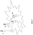
- the prodrug includes several moieties: an optional targeting moiety, consisting of a targetor peptide that recognizes cancer cells or malignant tissues, a protease specific cleavable moiety, consisting of a cleavable sequence recognized by legumain abundant within or in proximity to the malignant cells, and a chemotherapeutic drug ( Fig. 1 ).
- an optional targeting moiety consisting of a targetor peptide that recognizes cancer cells or malignant tissues
- a protease specific cleavable moiety consisting of a cleavable sequence recognized by legumain abundant within or in proximity to the malignant cells
- a chemotherapeutic drug Fig. 1
- the prodrug may act on cancer cells and tissues by several concurrent mechanisms as follows: (i) The extracellular prodrug activation - the prodrug is targeted to a cancer cell, which displays a cancer specific marker recognized by the tageting moiety ( Fig. 2A ), the targeting moiety binds to the marker ( Fig. 2B ), the chemotherapeutic drug is released from the carrier by a specific extracellular degrading enzyme ( Fig. 2C ), and the chemotherapeutic drug attacks the cancer cell ( Fig. 2D ); (ii) The intracellular prodrug activation - the prodrug is targeted to a cancer cell, which displays a cancer specific marker recognized by the tageting moiety ( Fig.
- the targeting moiety binds to the marker ( Fig. 3B ), the prodrug-marker complex are internalized into the cell ( Fig. 3C ), the chemotherapeutic drug is released from the carrier by a specific intracellular degrading enzyme, and the chemotherapeutic drug attacks the cancer cell ( Fig. 3D ).
- the drug could be placed at either the N-terminal or C-terminal side of the peptide.
- the skilled artisan will be able to optimize the appropriate linkage and position of the drug moiety within the prodrug.
- Various concerns should be taken into consideration to guide the artisan in this decision, such as selection of the peptide sequence, selection of the linker, selection of the position of attachment to the drug species, and requirements concerning host intracellular enzymes for drug activation.
- Chemotherapeutic drugs have different ways in which they inhibit cancer. Chemotherapeutic drugs can damage the DNA template by alkylation, by cross-linking, or by double-strand cleavage of DNA. Other cancer drugs can block RNA synthesis by intercalation. Some agents are spindle poisons, such as vinca alkaloids, or anti-metabolites that inhibit enzyme activity, or hormonal and anti-hormonal agents. Chemotherapeutic drugs for targeting may be selected from various groups of agents, including but not limited to alkylating agents, antimetabolites, antitumor antibiotics, vinca alkaloids, epipodophyllotoxins, nitrosoureas, hormonal and antihormonal agents, and toxins.
- alkylating agents may be exemplified by cyclophosphamide, chlorambucil, busulfan, Melphalan, Thiotepa, ifosphamide, Nitrogen mustard.
- Currently more preferred antimetabolites may be exemplified by methotrexate, 5-Fluorouracil, cytosine arabinoside, 6-thioguanine, 6-mercaptopurin.
- doxorubicin doxorubicin
- daunorubicin idorubicin
- nimitoxantron dactinomycin
- bleomycin bleomycin
- mitomycin plicamycin
- vinca alkaloids and epipodophyllotoxins may be exemplified by vincristin, vinblastin, vindestin, Etoposide, Teniposide.
- nitrosoureas may be exemplified by carmustin, lomustin, semustin, streptozocin.
- hormonal and antihormonal agents may be exemplified by adrenocorticorticoids, estrogens, antiestrogens, progestins, aromatas inhibitors, androgens, antiandrogens.
- the targeting sequences for the chemotherapeutic drug may be a glycosaminoglycan binding domain.
- the selection of a specific glycosaminoglycan binding domain as a targeting sequence was made by structure analysis of specific glycosaminoglycan chains present on the cancer cells or tissues. Since different cell types have been shown to synthesize proteoglycans with different glycosaminoglycan structures and functions, such differences may be utilized for the selection of targetor peptide.
- the selection of specific glycosaminoglycan binding domains for targeting can be carried out by different means, for example, by screening for native glycosaminoglycan binding domains capable of interacting with the specific glycosaminoglycans found on cancer cells or tissues. Alternatively, this selection can be carried out by screening for specific peptides (in peptide libraries) that interact with the specific glycosaminoglycans found on cancerous cells or tissues.
- Native specific glycosaminoglycan binding domains for targeting can be selected from glycosaminoglycan binding proteins, exemplified but not limited to growth factors including but not limited to fibroblast growth factors (1-23), epidermal growth factors, platelet derived growth factors, vascular endothelial growth factors, cytokines and chemokines including but not limited to interleukins, PF4, GRO-alpha, GRO-beta, GRO-gamma, extracellular matrix and cell adhesion proteins including but not limited to fibronectin, collagen, laminin, thrombospondin, integrins, N-CAM, PECAM, CD44, lipid binding proteins including but not limited to lipoprotein lipase, apolipoprotein B and E, LDL, enzymes including but not limited to acetylcholinesterase, GAG degrading enzymes, blood coagulation factors including but not limited to antithrombin III tissue factor, and other proteins including but not limited to influenza virus, Dipht
- Cancer invasion involves a proteolytic degradation of the extracellular matrix of the surrounding normal tissue. Excess matrix degradation is one of the hallmarks of cancer, and is an important component of the process of tumor progression (Fidler, I.J., 1997).
- malignant When a tumor cell acquires the ability to invade and destroy a normal tissue, it is termed malignant.
- the ability to form tumor metastases is characteristic of highly malignant cancers with poor clinical outcome.
- the tumor cell In order to invade and metastasize, the tumor cell must bypass the basement membrane by degrading the components of the extracellular matrix. During the invasion, the tumor cell penetrates the basement membrane underlying the tumor. It then moves through more extracellular matrix to reach the circulation either through the lymphatic- or through the blood vessels. The process of entering the blood stream is termed intravasation.
- the cancer cell In order to establish itself at a distant site, the cancer cell repeats the entire process in reverse. During this process of extravasation the tumor cell leaves the blood circulation, and penetrates the host tissue, again crossing through a basement membrane. If the tumor cells are capable of growing in this unfamiliar environment, clinically significant metastases are formed and can pose a threat to the life of the host.
- proteases have been implicated in tumor invasion.
- the serine protease plasmin, the MMPs, and the aspartic proteinases have been shown to be involved in this process.
- the MMPs comprise of a large family of over 20 proteins that can degrade all the known components of the extracellular matrix (Massova I. et al. 1998). These proteinases demonstrate some selectivity such that an individual MMP has the ability to degrade a particular subset of matrix proteins. MMPs were identified in the tissues surrounding invasive cancers, and show over expression in malignant tissues.
- the human aspartic proteinases include cathepsin D, cathepsin E, pepsinogen A, pepsinogen C, and renin.
- Cathepsins D and E are significly elevated in various cancers and metastases, hence applied as tumor cell markers of epithelial cancers.
- protease cleavable sequences were selected by structure analysis of specific biodegradable sequences that are degraded by proteases that are more abundant within or in proximity to the malignant cells.
- the proteases are grouped as follows:
- Aspartic proteases may be exemplified but not limited to cathepsin D, cathepsin E, pepsinogen A, pepsinogen C, renin.
- Serine proteases may be exemplified but not limited to plasmin, tissue-type plasminogen activator (tPA), urokinase-type plasminogen activator (uPA).
- tPA tissue-type plasminogen activator
- uPA urokinase-type plasminogen activator
- Cysteine proteases may be exemplified but not limited to cathepsin B, cathepsin L, cathepsin S.
- Asparaginyl proteases may be exemplified but not limited to legumain.
- the protease specific cleavable sequence is specifically cleavable by legumain.
- protease specific cleavable sequences can be selected by screening for native degradation substrates of proteases, which are more abundant within or in proximity to malignant cells.
- the protease specific cleavable sequences can be selected by screening for specific peptides (in peptide libraries) that can be susceptible to a proteolytic degradation by proteases, which are more abundant within or near malignant cells.
- Native specific biodegradable sequences can be selected from the groups of native substrates as listed below:
- Aspartic proteases substrates exemplified but not limited to Bioactive peptides, Beta-amyloid precursor.
- Serine proteases substrates exemplified but not limited to plasminogen, fibrin, PAR1 thrombin receptor, uPAR-1 (uPA receptor)
- Cysteine proteases substrates exemplified but not limited to collagens.
- Asparaginyl proteases substrates exemplified but not limited to antigenic proteins for MHC class II, proenzymes.
- chemotherapeutic drug is selected from a group consisting of Melphalan, Methotraxate, and Chlorambucil.
- a prodrug wherein the targetor is selected from a group consisting of GAG binding domain of PF4, and GAG.
- the molecular weight and chemical structure of the product were analyzed by Mass spectrometry on matrix assisted laser desorption ionization (MALDI) or electrospray ionization (ESI), interfaced to qudrupole ion trap and TOF (time of flight) mass spectrometer.
- MALDI matrix assisted laser desorption ionization
- ESI electrospray ionization
- Methotrexate N-[4-[[(2,4-diamino-6-pteridin-yl)-methylamino]benzoyl]-L-glutamic acid
- triethylamine 1.5 mmol
- the solution was mixed and 1.1 mmol of S-Boc-2-mercapto-4,6-dimethyl-pirimidine dissolved in 5 ml of dioxane was added.
- the reaction mixture was mixed for 18 hours.
- the product Di-t-Butoxycarbonyl-N-Methotrexate was extracted by Ethylacetate and saturated with citric acid solution.
- the peptide carrier for the chemotherapeutic drug was synthesized by using combinatorial chemistry and solid phase peptide synthesis. Fmoc/Boc protected amino acids were used for the synthesis.
- the first amino acid was bound to activated solid support such as polystyrene beads onto which hydroxybenzyl alcohol linker has been attached (Wang, 1973).
- the remaining hydroxyl groups of the resin were benzoylated with Benzoyl chloride (0,3 ml) and Pyridine (0.3 ml) in 8 ml DCE for 2 hours at room temperature.
- the resin was washed on a filter glass with DMF and with methanol, and dried in vacuum over silica gel.
- the Fmoc-Ser(But)-Resin was deprotected by piperidine method: 1 g resin with 0.5 mmol Fmoc-amino acid was deprotected 4 times with 10 ml of 20%-50% Piperidine in NMP or DMF. Deprotection was monitored by spectrophotometer measuring the level of the free Fmoc residue at 290 nm of each deprotection step. At the end of deprotection the resin was washed 10 times with DMF and methanol, and a sample of the resin beads was analyzed by Kaiser test (Kaiser et al., 1970).
- next amino acids were bound to the peptide-resin by subsequent steps of deprotection and coupling made for the extension of the peptide on the resin using DIC/HOBt method: Fmoc-amino acid was dissolved in NMP or DMF for 3 minutes and reactivated with DIC/HOBt (molar ratio 1:1:1 to amino acid) for 20 minutes. Coupling of amino acid to peptide-resin was made for 60 minutes at room temperature with mechanical mixing and nitrogen bubbling.
- the peptide was analyzed using reversed-phase HPLC, mass spectrometry, and gel electrophoresis separation.
- HPLC was made on LiChroCART 250-4 HPLC cartridge LiChrospher WP 300 RP-18 (5 micrometer) (purchased from Merck).
- the prodrug was seperated on HPLC using a gradient of 20%B to 100%B at flow rate of 0.5 ml/min, and the product was detected at 214 nm (Fluent A: water + 0.1% TFA, Fluent B: Acetonitrile + 10% water + 0.1% TFA).
- Mass spectrometry was made by matrix assisted lazerdesorption ionization (MALDI) or electrospray ionization (ESI), interfaced to qudrupole ion trap and TOF (time of flight) mass spectrometer.
- MALDI matrix assisted lazerdesorption ionization
- ESI electrospray ionization
- T Targeting peptide sequence (SEQ ID NO 4) is:
- the first amino acid Fmoc-Val-OH was coupled to Wang resin (Wang, 1973) by using Dichlorobenzoylchloride method as described in example 4.
- next amino acids were bound to the peptide-resin by subsequent steps of deprotection and coupling made for the extension of the peptide on the resin using DIC/HOBt method as described in Example 4 or HBTU/HOBt method: Fmoc-amino acid was dissolved in NMP or DMF for 3 minutes, reactivated with HOBt (molar ratio 1:1 to amino acid) for 3 minutes and transferred to the peptide-resin. HBTU (molar ratio 1:1 to amino acid) was added for 5 minutes and DIPEA (molar ratio 2:1 to amino acid) was added. Coupling of amino acid to peptide-resin was made for 120 minutes at room temperature with mechanical mixing and nitrogen bubbling.
- the peptide was analyzed using reversed-phase HPLC, mass spectrometry, and gel electrophoresis separation as described in example 4.
- T Targeting peptide sequence (SEQ ID NO 4) is:
- the first amino acid Fmoc-Val-OH was coupled to Rink resin.
- Rink resin One gram of Rink resin was swollen in 10 ml of DMF for 1 hour. The resin was washed 5 times with DMF. Resin was deprotected 4 times with 20% Piperidine in NMP. Deprotection was monitored by spectrophotometer measuring the level of the free Fmoc residue at 290 nm. At the end of deprotection the resin was washed 10 times with NMP, methanol, NMP, and a sample of the resin beads was analyzed by Kaiser test (Kaiser et al 1970).
- Amino acids were bound to the peptide-resin by subsequent steps of deprotection and coupling made for the extension of the peptide on the resin using HBTU/HOBt method or TBTU/HOBt method: Fmoc-amino acid was dissolved in NMP or DMF for 3 minutes, reactivated with HOBt (molar ratio 1:1 to amino acid) for 3 minutes and transferred to the peptide-resin. TBTU (molar ratio 1:1 to amino acid) was added for 5 minutes and DIPEA (molar ratio 3:1 to amino acid) was added. Coupling of amino acid to peptide-resin was made for 120 minutes at room temperature with mechanical mixing and nitrogen bubbling.
- cleavage of the peptide from the resin was made using 2 step cleavage method: 20%-50% TFA in DCM and scavengers (10-20 ml for 1g peptide-resin) for 15-30 minutes mixing at room temperature.
- the elution was filtered from the resin, the resin was washed 2 times with 20% TFA/DCM, and filtrates were combined. Most of the liquid was evaporated from the elution, and 95% TFA and 5% scavengers were added to the elute for 1-4 hours of deprotection.
- Peptide was precipitated from TFA using ice-cold t-butyl-methyl-ether.
- the peptide precipitate was washed 3 times with ice-cold t-butyl-methyl-ether, and then evaporated.
- the peptide was dissolved in water or buffer and lyophilized.
- the peptide was analyzed using reversed-phase HPLC, mass spectrometry, and gel electrophoresis separation as described in Example 4.
- Peptide I (SEQ ID NO 3): Tyr-Gly-Leu-Leu-Gly-Ile-Ala-Gly-Pro-Pro-Gly-Pro -Pro-Leu-Tyr-Lys-Lys-Ile-Ile-Lys-Lys-Leu-Leu-Gln-Ser was synthesized as described in Example 4 and then coupled to the drug.
- t-Butoxycarbonyl-N-Melphalan (molar ratio 1: 2 to peptide) was dissolved in DMF, and mixed for 5 minutes with the peptide-resin. TBTU (molar rate 1:1) was added for 5 minutes and DIPEA (molar ratio 2:1) was added. Coupling of t-Butoxycarbonyl-N-Melphalan to peptide-resin was made for 2-24 hours at room temperature with mechanical mixing and nitrogen bubbling. The coupling was monitored by spectroscopic measuring at 254 nm of the free t-Butoxycarbonyl-N-Melphalan in the reaction mixture.
- the peptide-Melphalan was analyzed using reversed-phase HPLC, mass spectrometry, and gel electrophoresis separation as described in Example 4.
- Peptide I (SEQ ID NO 3): Tyr-Gly-Leu-Leu-Gly-Ile-Ala-Gly-Pro-Pro-Gly-Pro -Pro-Leu-Tyr-Lys-Lys-Ile-Ile-Lys-Lys-Leu-Leu-Gln-Ser was synthesized as described in Example 4 and then coupled to the drug.
- Di-t-Butoxycarbonyl-N-Methotrexate (molar ratio 1:1.2 to peptide) was dissolved in NMP, and mixed for 5 minutes with the peptide-resin.
- HBTU (molar ratio 1:1 to methotrexate) was added for 5 minutes and DIPEA (molar ratio 2:1 to methotrexate) was added.
- Coupling of methotrexate to peptide-resin was made for 2-24 hours at room temperature with mechanical mixing and nitrogen bubbling. The coupling was monitored by spectroscopic measuring at 302 nm of the free methotrexate in the reaction mixture.
- the peptide-Methotrexate was analyzed using reversed-phase HPLC, mass spectrometry, and gel electrophoresis separation as described in Example 4.
- Peptide I (SEQ ID NO 3): Tyr-Gly-Leu-Leu-Gly-Ile-Ala-Gly-Pro-Pro-Gly-Pro -Pro-Leu-Tyr-Lys-Lys-Ile-Ile-Lys-Lys-Leu-Leu-Gln-Ser was synthesized as described in Example 4 and then coupled to the drug.
- Chlorambucil (molar ratio 1: 2 to peptide) was dissolved in NMP, and mixed for 5 minutes with the peptide-resin.
- HBTU (molar ratio 1:1 to chlorambucil) was added for 5 minutes and DIPEA (molar rate 2:1 to chlorambucil) was added.
- Coupling of chlorambucil to peptide-resin was made for 2-24 hours at room temperature with mechanical mixing and nitrogen bubbling. The coupling was monitored by spectroscopic measuring at 263 nm of the free chlorambucil in the reaction mixture.
- the resin was washed for 5 times with NMP, and 10 times with DCM on cinder glass.
- Cleavage of the drug from the resin was carried out by 90% TFA containing 5% H2O and 5% triisopropylsilan for 3 hours under mixing at room temperature. The drug was worked-up using process 4.
- the peptide-Chlorambucil was analyzed using reversed-phase HPLC, mass spectrometry, and gel electrophoresis separation as described in Example 4.
- Peptide I (SEQ ID NO 3) was synthesized as described in Example 4 and then coupled to folic acid. Folic acid was dissolved in DMF for 3 minutes, reactivated with PyBOP (molar ratio 1:1.3 to acid) for 3 minutes. DIPEA (molar rate 2:1 to acid) was added and the mixture was transferred to the peptide-resin. Coupling of folic acid to peptide-resin was made for 120 minutes at room temperature with mechanical mixing and nitrogen bubbling.
- Peptide I (SEQ ID NO 3) was synthesized as described in Example 4 and then coupled to N-N'-Diethylaminobenzoic acid.
- N-N'-Diethylaminobenzoic acid (molar ratio 1: 2 to peptide) was dissolved in NMP, and mixed for 5 minutes with the peptide-resin.
- HBTU molar ratio 1:1 was added for 5 minutes and DIPEA (molar rate 2:1 was added.
- Coupling of N-N'-Diethylaminobenzoic acid to peptide-resin was made for 2-24 hours at room temperature with mechanical mixing and nitrogen bubbling. The coupling was monitored by spectroscopic measuring at 263 nm of the free N-N'-Diethylaminobenzoic in the reaction mixture.
- Human umbilical vein-derived endothelial cells were prepared from umbilical veins and cultured as described previously. The HUVE cells were grown in M199 medium supplemented with 20% fetal calf serum, vitamins, 1 ng/ml hbFGF, and antibiotics.
- Human melanoma cells (WW-94) were grown in 50% DMEM/50% F-12 medium supplemented with 10% fetal calf serum and antibiotics.
- MCF-7 Human Brest cancer cells
- HUVEC normal cells
- melanoma cancer cells
- P-CAC prodrug 3 - Peptide I-Chlorambucil
- Cholorambucil or the peptide carrier was added under the same experimental conditions.
- the cells were washed with phosphate buffer saline (PBS), suspended with 0.5% EDTA/ PBS, and counted using cell coulter (Electronics ZM). Cell viability was verified by trypan-blue staining and hemocytometer analysis.
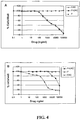
- IC50 indicates the concentration of the drug measured at 50% of the cell death.
- Fig. 4 shows that the peptide carrier is not toxic either to the HUVEC or to melanoma cells. Chlorambucil is very toxic to cancer and normal cells (IC50 is 2 mM). The prodrug Peptide I-Chlorambucil is efficient as a chemotherapeutic agent in killing cancer cells with an IC50 of 2 mM ( Fig. 4A ). However, this prodrug is much less toxic to HUVE cells (IC50 of 60 mM)( Fig. 4B ), i.e., 30 times less toxic to normal cells.
- HUVEC or melanoma cells were seeded in 96-well ELISA dish at a concentration of 10,000-20,000 cells/well, and various concentrations of the prodrug 1 were added to the wells as described in Example 14.
- Melphalan was added under the same experimental conditions. After 72 hours the medium was aspirated, and DMEM containing 5% FCS and 0.5 mg/ml MTT (an indicator of cell viability) was added. The cells were incubated for 2-4 hours at 37°C and 5% CO2. At the end of the incubation the cells were washed with phosphate buffer saline (PBS), and dissolved with DMSO. The results were analysed using Techan ELISA reader equipped with a 570 nm filter.
- Fig. 5 shows that Melphalan ( ⁇ ) kills efficiently both cancer and normal cells. It also shows that the prodrug Peptide I-Melphalan ( ⁇ ) is as efficient as the Melphalan itself in killing cancer cells ( Figure 5B ), but it is less toxic to normal cells ( Figure 5A ).
- the IC50 for Melphalan in HUVE cells and in melanoma cells is 0.1 mM, and 0.07 mM, respectively.
- the IC50 for the prodrug 1 in HUVE cells and in melanoma cells is 0.5 mM and 0.07 mM, respectively.
- breast cancer cells or melanoma cells were grown to sub-confluence in a 10 cm dish. The cells were washed with PBS, and further incubated with serum free medium. HUVE cells were grown to confluence at 10 cm dish. The cells were washed with M-199 medium, and conditioned with M-199 with 1% heat inactivated serum. The conditioned medium was collected after 24 hours.
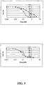
- reaction mixture was incubated with heparin-sepharose (30 minutes at 0°C). The supernatant was collected, and the release of free chlorambucil was monitored by spectrophotometer at a wavelengh of 305 nm.
- Fig. 6 shows that the chemotherapeutic agent can be released from the peptide carrier either by purified MMPs, or by MMPs secreted by cancer cells.
- heparin Addition of heparin to the reaction mixture inhibited the release of the chemotherapeutic agent from the peptide carrier, suggesting that GAGs can modulate the drug activity.
- native proteoglycans such as Serglycin can be used as a carrier for the drug, protecting it from degradation.
- a "pharmaceutical composition” refers to a preparation of one or more of the prodrugs described herein, or physiologically acceptable salts thereof, with other chemical components such as physiologically suitable carriers and excipients.
- the purpose of a pharmaceutical composition is to facilitate administration of a compound to an organism.
- excipient refers to an inert substance added to a pharmaceutical composition to further facilitate administration of a compound.
- excipients include calcium carbonate, calcium phosphate, various sugars and types of starch, cellulose derivatives, gelatin, vegetable oils and polyethylene glycols.
- compositions may also include one or more additional active ingredients, such as, but not limited to, conventional anti-migraine agents.
- compositions of the present invention may be manufactured by processes well known in the art, e.g., by means of conventional mixing, dissolving, granulating, grinding, pulverizing, dragee-making, levigating, emulsifying, encapsulating, entrapping or lyophilizing processes.
- compositions for use in accordance with the present invention thus may be formulated in conventional manner using one or more physiologically acceptable carriers comprising excipients and auxiliaries, which facilitate processing of the active compounds into preparations which, can be used pharmaceutically. Proper formulation is dependent upon the route of administration chosen.
- the compounds of the invention may be formulated in aqueous solutions, preferably in physiologically compatible buffers such as Hank's solution, Ringer's solution, or physiological saline buffer.
- physiologically compatible buffers such as Hank's solution, Ringer's solution, or physiological saline buffer.
- penetrants appropriate to the barrier to be permeated are used in the formulation.
- penetrants for example DMSO, or polyethylene glycol are generally known in the art.
- the compounds can be formulated readily by combining the active compounds with pharmaceutically acceptable carriers well known in the art.
- Such carriers enable the compounds of the invention to be formulated as tablets, pills, dragees, capsules, liquids, gels, syrups, slurries, suspensions, and the like, for oral ingestion by a patient.
- Pharmacological preparations for oral use can be made using a solid excipient, optionally grinding the resulting mixture, and processing the mixture of granules, after adding suitable auxiliaries if desired, to obtain tablets or dragee cores.
- Suitable excipients are, in particular, fillers such as sugars, including lactose, sucrose, mannitol, or sorbitol; cellulose preparations such as, for example, maize starch, wheat starch, rice starch, potato starch, gelatin, gum tragacanth, methyl cellulose, hydroxypropylmethyl-cellulose, sodium carbomethylcellulose; and/or physiologically acceptable polymers such as polyvinylpyrrolidone (PVP).
- disintegrating agents may be added, such as cross-linked polyvinyl pyrrolidone, agar, or alginic acid or a salt thereof such as sodium alginate.
- enterocoating are useful as it is desirable to prevent exposure of the peptides of the invention to the gastric environment.
- Dragee cores are provided with suitable coatings.
- suitable coatings For this purpose, concentrated sugar solutions may be used which may optionally contain gum arabic, talc, polyvinyl pyrrolidone, carbopol gel, polyethylene glycol, titanium dioxide, lacquer solutions and suitable organic solvents or solvent mixtures.
- Dyestuffs or pigments may be added to the tablets or dragee coatings for identification or to characterize different combinations of active compound doses.
- compositions which can be used orally, include push-fit capsules made of gelatin as well as soft, sealed capsules made of gelatin and a plasticizer, such as glycerol or sorbitol.
- the push-fit capsules may contain the active ingredients in admixture with filler such as lactose, binders such as starches, lubricants such as talc or magnesium stearate and, optionally, stabilizers.
- the active compounds may be dissolved or suspended in suitable liquids, such as fatty oils, liquid paraffin, or liquid polyethylene glycols.
- stabilizers may be added. All formulations for oral administration should be in dosages suitable for the chosen route of administration.
- compositions may take the form of tablets or lozenges formulated in conventional manner.
- the peptides for use according to the present invention are conveniently delivered in the form of an aerosol spray presentation from a pressurized pack or a nebulizer with the use of a suitable propellant, e.g., dichlorodifluoromethane, trichlorofluoromethane, dichloro-tetrafluoroethane or carbon dioxide.
- a suitable propellant e.g., dichlorodifluoromethane, trichlorofluoromethane, dichloro-tetrafluoroethane or carbon dioxide.
- the dosage unit may be determined by providing a valve to deliver a metered amount.
- Capsules and cartridges of, e.g., gelatin for use in an inhaler or insufflator may be formulated containing a powder mix of the peptide and a suitable powder base such as lactose or starch.
- compositions for parenteral administration include aqueous solutions of the active ingredients in water-soluble form. Additionally, suspensions of the active compounds may be prepared as appropriate oily injection suspensions. Suitable lipophilic solvents or vehicles include fatty oils such as sesame oil, or synthetic fatty acids esters such as ethyl oleate, triglycerides or liposomes. Aqueous injection suspensions may contain substances, which increase the viscosity of the suspension, such as sodium carboxymethyl cellulose, sorbitol or dextran. Optionally, the suspension may also contain suitable stabilizers or agents, which increase the solubility of the compounds, to allow for the preparation of highly concentrated solutions.
- the active ingredient may be in powder form for reconstitution with a suitable vehicle, e.g., sterile, pyrogen-free water, before use.
- a suitable vehicle e.g., sterile, pyrogen-free water
- the compounds of the present invention may also be formulated in rectal compositions such as suppositories or retention enemas, using, e.g., conventional suppository bases such as cocoa butter or other glycerides.
- compositions herein described may also comprise suitable solid of gel phase carriers or excipients.
- suitable solid of gel phase carriers or excipients include, but are not limited to, calcium carbonate, calcium phosphate, various sugars, starches, cellulose derivatives, gelatin and polymers such as polyethylene glycols.
- compositions suitable for use in context of the present invention include compositions wherein the active ingredients are contained in an amount effective to achieve the intended purpose. More specifically, a therapeutically effective amount means an amount of a compound effective to prevent, alleviate or ameliorate symptoms of a disease of the subject being treated.
- Toxicity and therapeutic efficacy of the peptides described herein can be determined by standard pharmaceutical procedures in cell cultures or experimental animals, e.g., by determining the IC 50 (the concentration which provides 50% inhibition) and the LD 50 (lethal dose causing death in 50 % of the tested animals) for a subject compound.
- the data obtained from these cell culture assays and animal studies can be used in formulating a range of dosage for use in human.
- the dosage may vary depending upon the dosage form employed and the route of administration utilized. The exact formulation, route of administration and dosage can be chosen by the individual physician in view of the patient's condition. (See e.g., Fung1, et al., 1975, in "The Pharmacological Basis of Therapeutics", Ch. 1 p.1).
- dosing can also be a single administration of a slow release composition, with course of treatment lasting from several days to several weeks or until cure is effected or diminution of the disease state is achieved.
- compositions to be administered will, of course, be dependent on the subject being treated, the severity of the affliction, the manner of administration, the judgment of the prescribing physician, and all other relevant factors.
- the following example is an illustration only of peptide according to the invention for use in treating a subject afflicted with cancer, in order to treat a pathological condition associated with a solid tumor or a related condition, and is not intended to be limiting.
- the medicament can be administered according to an effective dosing methodology, preferably until a predefined endpoint is reached, such as a reduction or amelioration of the pathological condition in the subject.
Landscapes
- Health & Medical Sciences (AREA)
- Life Sciences & Earth Sciences (AREA)
- Pharmacology & Pharmacy (AREA)
- Proteomics, Peptides & Aminoacids (AREA)
- Veterinary Medicine (AREA)
- Chemical & Material Sciences (AREA)
- Engineering & Computer Science (AREA)
- Bioinformatics & Cheminformatics (AREA)
- Public Health (AREA)
- Medicinal Chemistry (AREA)
- General Health & Medical Sciences (AREA)
- Animal Behavior & Ethology (AREA)
- Epidemiology (AREA)
- Immunology (AREA)
- Gastroenterology & Hepatology (AREA)
- Zoology (AREA)
- Marine Sciences & Fisheries (AREA)
- Medicines That Contain Protein Lipid Enzymes And Other Medicines (AREA)
- Medicinal Preparation (AREA)
- Peptides Or Proteins (AREA)
- Pharmaceuticals Containing Other Organic And Inorganic Compounds (AREA)
Abstract
Description
- The present invention relates to prodrug molecules comprising conjugates of an antiproliferative drug, a protease specific cleavable peptide, and, optionally, a targeting peptide, said prodrugs being substantially inactive prior to degradation of the cleavable sequence by proteolytic enzymes abundant within or in close proximity to the target cancer cell, to pharmaceutical compositions comprising the conjugates and to the use of these compositions in the treatment of cancer.
- Anti-proliferative drugs, also known as anti-metabolites, act by inhibiting crucial metabolic processes, and are commonly used in the treatment of diseases involving abnormal cell proliferation, such as tumors. However, the utility of these drugs is severely hampered by their excessive toxicity and adverse side effects on healthy cells of the treated patient. Therefore, it would be advantageous to be able to reduce these adverse effects by the use of a prodrug having decreased toxicity.
- The use of prodrugs to impart desired characteristics such as increased bioavailability or increased site-specificity of known drugs is a recognized concept in the state of the art of pharmaceutical development. The use of various blocking groups, which must be removed in order to release the active drug is also known in the background art. Commonly, one or more blocking groups may be attached via an available amine, hydroxyl group or other functional reactive group on the drug to yield an amide or an ester. This type of prodrug may be cleaved by non-specific esterases to release the active principle in a sustained-release fashion over a prolonged period of time compared to the native drug species.
- Methotrexate (MTX), for example, is an effective anti-proliferative drug commonly used in cancer therapy. It is an analogue of dihydrofolate that inhibits the enzyme dihydrofolate reductase (DHFR), thus depleting intracellular tetrahydrofolate (FH4), which is an essential co-factor required for the de novo synthesis of purine nucleotides.
- MTX, Mephalan and Chlorambucil are valuable drugs in the treatment of many rapidly growing tumors, however, their use is limited by the frequency and severity of side effects. Unwanted side effects include toxicity to all rapidly dividing normal cells, such as stem cells in the bone marrow, epithelial cells of the intestinal tract, hair follicle cells, etc.
- Another major problem in chemotherapy, which is particularly relevant in the case of anti-metabolites, is inherent or acquired resistance of tumors to cytotoxic drugs. For example, development of resistance to MTX frequently follows prolonged exposure to this drug. Resistance may be due to new mutations induced by the clinical treatment, or to positive selection, by the treatment regimen, of pre-existing resistant mutant cell. Known mechanisms for development of resistance involve impaired transport of MTX into cells, e.g. by mutations in the Reduced Folate Carrier (RFC), over expression of the target enzyme DHFR, or mutations in the enzyme responsible for polyglutamination of reduced folates (FPGS).
- A more severe problem in the clinic is the phenomenon of multi-drug resistance (MDR), which is a resistance to a broad spectrum of structurally unrelated cytotoxic drugs. MDR is mediated by transmembrane "pumps", which actively expel chemotherapeutic drugs from the tumor cells. MDR significantly limits the efficacy of many cancer chemotherapy regimens and is a major factor in the failure of cancer chemotherapy.
- It would, therefore, be most advantageous to have drug derivatives that are specifically targeted to or selectively active in the diseased cells rather than in the healthy cells, thus reducing undesirable side effects. It would also be desirable to generate new anti-proliferative agents that overcome drug-resistance, as well as agents that are active as cytotoxic drugs but lack or have a reduced ability to provoke MDR phenotype.
- For specific cytotoxic drugs it has been suggested that the therapeutic index of such drugs might be increased if the drug is covalently bound to a peptide that would be cleaved in the vicinity of the tumor cells by the action of certain proteases. This approach has been suggested for peptide conjugated Methotrexate (Kuefner et al., 1989) and for Arabinofuranosyl cytosine (ara-C) lipid-peptide-drug conjugates (Menger et al., 1994).
- Many different types of cell surface polypeptides or glycoproteins have been utilized for targeting drugs to malignant cells, with various degrees of success.
- The use of specific cell surface complex sugars as cell surface markers is much less well developed. In part this is due to the fact that the expression of these structures cannot be followed in terms of gene transcription. In other words, the complex sugars are the product of varying expression of the glycosylation enzymes, and cannot be traced directly as gene products.
- Proteoglycans are composed of long, unbranched sugar polymers, called glycosaminoglycans (GAGs), which are covalently linked to a core protein. The proteoglycans constitute the extracellular matrix, such as the cartilage, the basement membranes, and the connective tissue. They are also found on the cell surface (Bernfield, M. et al. 1992). Virtually all epithelia express cell-surface proteoglycans, represented principally by glypicans and syndecans. Glypicans are glycosyl phosphatidyl inositol (GPI)-linked molecules, and bear glycosaminoglycans exclusively of the heparan sulfate type. Syndecans are transmembrane proteins, and are decorated with chondroitin sulfate and with heparan sulfate polymers. Syndecans exhibit a complex pattern of cell and development specific expression, however, the molecular mechanisms responsible for this expression have not been fully explored. It was shown, for example that during wound healing the expression of syndecan-1 and -4 is induced. In the case of glypicans, it was shown that glypican-1 is strongly expressed in human pancreatic cancer, whereas its expression is low in normal pancreas.
- A variety of regulatory proteins bind tightly to GAGs, including growth factors, cytokines, chemokines, extracellular matrix proteins, cell adhesion molecules, lipid binding proteins, enzymes, and blood coagulation factors. The role of heparan sulphate proteoglycans (HSPGs) in growth factor signaling has been best characterized with respect to fibroblast growth factors (FGFs), which require the presence of heparan sulfate for high affinity binding to their tyrosine kinase receptors (Yayon, A., et al. 1991). Several other growth factors have been found to exhibit a strong requirement for a HSPG co-receptor in their signaling. These include heparin binding EGF-like growth factor (HB-EGF), hepatocyte growth factor (HGF), vascular endothelial growth factor (VEGF) (Yamada, Y. et al., 1997), PDGF, TGF-beta, and other types of growth factors.
- Vascular endothelial growth factors (VEGFs) are mitogens for endothelial cells and are potent angiogenic factors in vivo. VEGF-165 contains the peptide encoded by exon-7 of the VEGF gene, confers on VEGF-165 the ability to bind heparan-sulfate molecules. VEGF-145 contains the peptide encoded by exon-6a of the VEGF gene, enabling VEGF-145 to bind ECM (Poltorak et al., 1997).
- Several VEGF tyrosine-kinase receptor types have been characterized, these receptors mediates the mitogenic activity and induced cell migration of VEGF. Other VEGF receptors, neuropilin-1 and neuropilin-2 (Gitay-Goren, H., et al., 1992) bind only to the GAG binding forms of VEGF (VEGF-165, VEGF-145) that have GAG binding peptides (axons 6a or 7) of the VEGF gene. These receptors are highly expressed in cancer cells such as human melanoma and carcinoma, but not expressed in normal melanocytes.
- VEGFs play a critical role in the process of tumor angiogenesis. This process is essential for tumor progression and for the subsequent process of tumor metastasis.
- VEGF soluble receptors have been suggested as an inhibitor of endothelial cell induced proliferation and angiogenesis (Kendall et al.
US patent: 5712380 ). - Among the chemokines that are known to bind to heparin the better characterized are Platelet factor 4 (PF4)(Morgan et al., 1977). PF4 is an anti-angiogenic factor that belongs to the CXC Chemokines family. PF4 binds to several receptors that belong to the CXC receptor (CXCR) family involved in angiogenesis. Kaposi's sarcoma cancer is indicated by uncontrolled angiogenesis that is associated with KSHV (Kaposi's sarcoma associated herpes virus) that produces the CXCR-2 receptor homolog.
- Injection of fluorescent PF4 to hamsters showed concentration of PF4 at capillary endothelial cells at sites of active angiogenesis. PF4 is accumulated at high concentrations in extra cellular matrix and basement membrane due to its GAG binding ability.
- PF4 can bind cell surface proteoglycans, and can be accumulated in the intracellular compartments (Neufeld at al., personal communication). Peptide from its GAG binding domain inhibited melanoma tumor growth in mice xenograft, though it had no effect on cancer cells in-vitro. CXC chemokines have been suggested as therapeutic molecules in modulating the angiogenic and angiostatic responses (
US Patent 5871723 ). - Cancer invasion involves a proteolytic degradation of extracellular matrix in the surrounding normal tissue. Excess matrix degradation is one of the hallmarks of cancer, and is an important component of the process of tumor progression (Fidler, I.J., 1997). In order for invasion and metastasis to occur, the tumor cell must bypass the basement membrane by degrading the components of the ECM.
- Various proteases, in particular the serine protease plasmin, and a variety of matrix metalloproteinases (MMPs), have been implicated in tumor invasion. Plasmin is formed from the inactive zymogen plasminogen by the plasminogen-activators. Plasminogen is produced in the liver and is present extracellulalrly throughout the body. One of the plasminogen-activators, the urokinase plasminogen activator (uPA), is synthesized as a pro-uPA that binds with high affinity to a cell-surface-bound receptor, the uPA receptor (uPAR). Receptor binding of pro-uPA strongly enhances the overall reaction leading to plasmin formation (Dano, K. et al., 1994). Clinical findings have shown that there are elevated tumor antigen levels of Plasminogen Activator (uPA, tPA) and its receptor uPAR in caner cells and tumors and it plays a role in tumor invasion and metastasis (Koopman et al., 1998; Schmidt et al., 1997).
- The MMPs comprise of a large family of over 20 proteins that can degrade all the known components of the extracellular matrix (Massova, I. et al. 1998). MMPs were identified in the tissues surrounding invasive cancers, and show over expression in malignant tissues.
- The human aspartic proteinases include cathepsin D, cathepsin E, pepsinogen A, pepsinogen C, and rennin (Taggart, R.T., 1992). Cathepsins D and E are significantly elevated in various cancers and metastases, hence applied as tumor cell markers of epithelial cancers (Matsue, K. et al., 1996).
-
EP 0 859 058 - Manfredini et al. ("Peptide T-araC conjugates: solid-phase synthesis and biological activity of N4-(acylpeptidyl)-araC"; Bioorganic & Medicinal Chemistry, March 2000, Vol. 8, No. 3, p. 539-547) report a conjugate of araC-paptide T. By synthesizing the conjugate, the authors aimed at selectively delivering the antiproliferative drug araC to Cud4+ cells. It is described that the conjugate can be used for reducing or preventing araC degradation by serum catabolic enzymes. The results indicate that while the conjugate showed binding to CD4 receptors, the antiproliferative activity was not specific to CD4+ cells and was lower than that of the free araC, indicating that the low intracellular hydrolysis of the conjugate is responsible for the lower potency of the conjugate.
- Nowhere in the background art is it taught or suggested that it is possible to use peptides as drug carriers useful to target prodrugs to tumors.
- It is an object of this invention to target a drug to malignant cells. It is a further object of the present invention to provide prodrugs that are selectively activated in or near malignant cells. It is still a further object of the present invention to provide a technique for treating a malignant tumor or metastatic cancer by selective activation of such prodrugs in or near malignant cells.
- These and yet other objectives are accomplished by the prodrugs according to the present invention, comprising a conjugate of at least one antiproliferative drug molecule covalently coupled to a protease specific cleavable sequence, said protease specific cleavable sequence is specifically cleavable by legumain abundant in or secreted by malignant cells, thereby releasing the antiproliferative drug molecule within or at the malignant cells by the action of the legumain, wherein the antiproliferative drug is a chemotherapeutic drug selected from the group consisting of alkylating agents, antimetabolites, antitumor antibiotics, vinca alkaloids, epipodophyllotoxins, nitrosoureas, and antihormonal agents, and wherein the protease specific cleavable sequence consists of 2 to 14 amino acids. These prodrugs are specifically released near or in the malignant cells by the action of said protease.
- According to currently preferred embodiments the conjugate is substantially pharmacologically inactive.
- Advantageously, these prodrugs may further comprise a targetor peptide, the targetor peptide binding to a glycosaminoglycan.
- Compositions comprising the prodrugs may be represented schematically as follows:
- Protecting group-Targetor peptide-Protease specific cleavable sequence-Linker-Drug (Formula I)
- The prodrugs according to the present invention may consist of at least one drug molecule conjugated to the protease cleavable sequence and to the targetor peptide. In another embodiment, compositions according to the present invention may consist of a plurality of drug molecules conjugated to at least one protease cleavable sequence and targetor peptide. The plurality of drug molecules may be the same or different at each occurrence
- The present invention further provides pharmaceutical compositions comprising as an active ingredient a prodrug according to the present invention. Such pharmaceutical compositions may be administered by any suitable route of administration.
- The technique for activation of the prodrug comprises the following steps:
- a) specifically cleaving a peptide bond within the peptide moiety of the prodrug by a protease;
- b) digesting the remaining peptide moiety of the prodrug by means of any nonspecific exopeptidase activity; and in cases where the linker moiety is present,
- c) releasing the active drug from the linker moiety by decomposition of the drug-linker bond.
- Currently more preferred embodiments of the invention comprise a prodrug, wherein the chemotherapeutic drug is selected from a group consisting of Melphalan, Methotraxate, and Chlorambucil.
- Currently most preferred embodiments according to the invention comprise a prodrug selected from the group consisting of
- Prodrug 1: GAG binding domain of PF4 - Protease cleavage site - Melphalan;
- Prodrug 2: GAG binding domain of PF4 - Protease cleavage site -Methotraxate;
- Prodrug 3: GAG binding domain of PF4 - Protease cleavage site - Chlorambucil;
- Prodrug 6: GAG and extracellular binding domain of VEGF - Protease Cleavage site - Chlorambucil;
- Prodrug 7: GAG and extracellular binding domain of VEGF - Protease cleavage site - Melphalan.
- Currently more preferred embodiments of the invention comprise a prodrug, wherein the targetor is selected from a group consisting of GAG binding domain of PF4, and GAG.
-
-
Fig. 1 shows a prodrug model that includes a targeting moiety (T), a protease cleavable moiety (R), and a chemotherapeutic drug (D). -
Fig. 2 shows a model of an extracellular prodrug activation that includes the targeting of a prodrug to a marker present on a cancer cell, and the release of the chemotherapeutic drug from its carrier by a specific extracellular degrading enzyme. -
Fig. 3 depicts a model of an intracellular prodrug activation that includes the targeting of a prodrug to a marker present on a cancer cell, the internalization of the prodrug-marker complex into the cell, and the release of the chemotherapeutic drug from its carrier by a specific intracellular degrading enzyme. -
Fig. 4A-B shows a comparison between cancer and normal cell death at increasing concentrations of the prodrug 3. -
Fig. 5A-B depicts a comparison between normal and cancer cells death at increasing concentrations of theprodrug 1. -
Fig. 6 shows an in-vitro release of Chlorambucil from its peptide carrier (prodrug 3) by purified MMPs, or by the cancer cell conditioned medium. - According to the present invention prodrug conjugates are provided which comprise at least one anti-proliferative drug covalently coupled to a peptide sequence comprising a peptide bond specifically cleavable by legumain. The peptide sequence may further comprise a targeting sequence, designed to increase the localization of the conjugate to the vicinity of the malignant cells. These prodrugs may further comprise linker moieties between the drug and the peptide, and may still further comprise protecting groups or blocking groups attached to the peptide.
- In the specification and in the claims the term "drug" denotes a chemotherapeutic drug capable of arresting cell growth, or killing the cell in which it is present and includes known cytotoxic, cytostatic or antiproliferative drugs such as are known in the art, exemplified by such compounds as:
- Alkaloids: Docetaxel, Etoposide, Irinotecan, Paclitaxel, Teniposide, Topotecan, Vinblastine, Vincristine, Vindesine.
- Alkylating agents: Busulfan, Improsulfan, Piposulfan,Benzodepa, Carboquone, Meturedepa, Uredepa, Altretamine, triethylenemelamine, Triethylenephosphoramide, Triethylenethiophosphoramide, Chlorambucil, Chloranaphazine, Cyclophosphamide, Estramustine, Ifosfamide, Mechlorethamine, Mechlorethamine Oxide Hcl, Melphalan, Novemebichin, Perfosfamide Phenesterine, Prednimustine, Trofosfamide, Uracil Mustard, Carmustine, Chlorozotocin, Fotemustine, Lomustine, Nimustine, Semustine Ranimustine, Dacarbazine, Mannomustine, Mitobronitol, Mitolactol, Pipobroman, Temozolomide.
- Antibiotics and analogs: Aclacinomycins, Actinomycins, Anthramycin, Azaserine, Bleomycins, Cactinomycin, Carubicin, Carzinophilin, Cromomycins, Dactinomycins, Daunorubicin, 6-Diazo-5-oxo-L-norleucine, Doxorubicin, Epirubicin, Idarubicin, Menogaril, Mitomycins, Mycophenolic Acid, Nogalamycine, Olivomycins, Peplomycin, Pirarubicin, Plicamycin, Porfiromycin, Puromycine, Streptonigrin, Streptozocin, Tubercidin, Zinostatin, Zorubicin.
- Antimetabolites: Denopterin, Edatrexate, Methotrexate, Piritrexim, Pteropterin, Tomudex, Trimetrexate, Cladridine, Fludarabine, 6-Mercaptopurine, Pentostatine Thiamiprine, Thioguanine, Ancitabine, Azacitidine, 6-Azauridine, Carmofur, Cytarabine, Doxifluridine, Emitefur, Floxuridine, Fluorouracil, Gemcitabine, Tegafur;
- Platinum complexes: Caroplatin, Cisplatin, Miboplatin, Oxaliplatin;
- Others: Aceglatone, Amsacrine, Bisantrene, Defosfamide, Demecolcine, Diaziquone, Eflornithine, Elliptinium Acetate, Etoglucid, Etopside, Fenretinide, Gallium Nitrate, Hdroxyurea, Lonidamine, Miltefosine, Mitoguazone, Mitoxantrone, Mopidamol, Nitracrine, Pentostatin, Phenamet, Podophillinic acid 2-Ethyl-Hydrazide, Procarbazine, Razoxane, Sobuzoxane, Spirogermanium,Teniposide Tenuazonic Acid, Triaziquone, 2,2',2"-Trichlorotriethylamine, Urethan.
- In the specification and in the claims the term "protease specific sequence" denotes any peptide sequence comprising a sequence cleavable by legumain, and includes peptides of from two to fourteen amino acids comprising at least one site that is cleaved by legumain. More preferred are peptide sequences comprising from three to twelve amino acids, as exemplified hereinbelow.
- In the specification and in the claims the term "linker" denotes any chemical compound, which may be present between the drug moiety and the peptide moiety of the prodrug. This linker may be removed from the drug by chemical means, by enzymatic means, or spontaneously. The linker may be pharmacologically inert or may itself provide added beneficial pharmacological activity. The term "spacer" may also be used interchangeably as a synonym for linker.
- In the specification and in the claims the term "protection group" denotes any appropriate blocking group on the N-, or -C terminal part, or on the side chain of the peptide sequence, which is capable of blocking the action of exopeptidases or endopeptidases, such as are well known in the art.
- The protection group may itself be pharmacologically inert or may provide added pharmacologically beneficial attributes to the conjugate. Most advantageously the protecting group will be lipophilic, thereby improving the ability of the conjugate to penetrate into cells.
- The prodrug includes several moieties: an optional targeting moiety, consisting of a targetor peptide that recognizes cancer cells or malignant tissues, a protease specific cleavable moiety, consisting of a cleavable sequence recognized by legumain abundant within or in proximity to the malignant cells, and a chemotherapeutic drug (
Fig. 1 ). - The prodrug may act on cancer cells and tissues by several concurrent mechanisms as follows: (i) The extracellular prodrug activation - the prodrug is targeted to a cancer cell, which displays a cancer specific marker recognized by the tageting moiety (
Fig. 2A ), the targeting moiety binds to the marker (Fig. 2B ), the chemotherapeutic drug is released from the carrier by a specific extracellular degrading enzyme (Fig. 2C ), and the chemotherapeutic drug attacks the cancer cell (Fig. 2D ); (ii) The intracellular prodrug activation - the prodrug is targeted to a cancer cell, which displays a cancer specific marker recognized by the tageting moiety (Fig. 3A ), the targeting moiety binds to the marker (Fig. 3B ), the prodrug-marker complex are internalized into the cell (Fig. 3C ), the chemotherapeutic drug is released from the carrier by a specific intracellular degrading enzyme, and the chemotherapeutic drug attacks the cancer cell (Fig. 3D ). - In prodrugs according to the invention, the drug could be placed at either the N-terminal or C-terminal side of the peptide. The skilled artisan will be able to optimize the appropriate linkage and position of the drug moiety within the prodrug. Various concerns should be taken into consideration to guide the artisan in this decision, such as selection of the peptide sequence, selection of the linker, selection of the position of attachment to the drug species, and requirements concerning host intracellular enzymes for drug activation.
- The principles that apply to the selection of peptide, linker, attachment site, etc., will be detailed herein for exemplary compounds. The principles may be generalized as follows:
- a) Selection of the peptide sequence: any peptide sequence of 2 to 14 amino acids that is cleavable by legumain that is more abundant within or in proximity to cancer cells may be suitable.
- b) Selection of the linker: any chemical moiety that can serve as a linker between the peptide and the drug. The linker may be cleaved by chemical reaction, enzymatic reaction, or spontaneously. The linker may also serve for optimizing the specificity of the peptide-protease interaction.
- c) Selection of the position of attachment to the drug species: the drug may be attached to either one or to both sides of the peptides, according to the peptidase activities that exists in the targeted cells.
- d) Selection of the protecting group: The protecting group may be any chemical moiety that reduces the non-specific prodrug degradation (to active or inactive compounds).
Advantageously, the protecting group can be a compound that increases the selectivity of the prodrug towards the cancer cells or tissues, or a compound that increases the permeability of the prodrug towards the cancer cells or tissues. The protecting group can itself be replaced by a second drug or a second molecule of the same drug. - e) Selection of the drug: the drug can be any anti-proliferative agent. It may be released in protected or unprotected form, i.e., it can itself be a prodrug. For instance, the targetor moiety may be cleaved extracellularly and the resultant drug- linker conjugate may still be a prodrug that releases the active drug species intracellularly for effective treatment of oncogenesis.
- Chemotherapeutic drugs have different ways in which they inhibit cancer. Chemotherapeutic drugs can damage the DNA template by alkylation, by cross-linking, or by double-strand cleavage of DNA. Other cancer drugs can block RNA synthesis by intercalation. Some agents are spindle poisons, such as vinca alkaloids, or anti-metabolites that inhibit enzyme activity, or hormonal and anti-hormonal agents. Chemotherapeutic drugs for targeting may be selected from various groups of agents, including but not limited to alkylating agents, antimetabolites, antitumor antibiotics, vinca alkaloids, epipodophyllotoxins, nitrosoureas, hormonal and antihormonal agents, and toxins.
- Currently more preferred alkylating agents may be exemplified by cyclophosphamide, chlorambucil, busulfan, Melphalan, Thiotepa, ifosphamide, Nitrogen mustard.
- Currently more preferred antimetabolites may be exemplified by methotrexate, 5-Fluorouracil, cytosine arabinoside, 6-thioguanine, 6-mercaptopurin.
- Currently more preferred antitumor antibiotics may be exemplified by doxorubicin, daunorubicin, idorubicin, nimitoxantron, dactinomycin, bleomycin, mitomycin, plicamycin.
- Currently more preferred vinca alkaloids and epipodophyllotoxins may be exemplified by vincristin, vinblastin, vindestin, Etoposide, Teniposide.
- Currently more preferred nitrosoureas may be exemplified by carmustin, lomustin, semustin, streptozocin.
- Currently more preferred hormonal and antihormonal agents may be exemplified by adrenocorticorticoids, estrogens, antiestrogens, progestins, aromatas inhibitors, androgens, antiandrogens.
- The targeting sequences for the chemotherapeutic drug may be a glycosaminoglycan binding domain.
- In this invention the selection of a specific glycosaminoglycan binding domain as a targeting sequence was made by structure analysis of specific glycosaminoglycan chains present on the cancer cells or tissues. Since different cell types have been shown to synthesize proteoglycans with different glycosaminoglycan structures and functions, such differences may be utilized for the selection of targetor peptide.
- The selection of specific glycosaminoglycan binding domains for targeting can be carried out by different means, for example, by screening for native glycosaminoglycan binding domains capable of interacting with the specific glycosaminoglycans found on cancer cells or tissues. Alternatively, this selection can be carried out by screening for specific peptides (in peptide libraries) that interact with the specific glycosaminoglycans found on cancerous cells or tissues.
- Native specific glycosaminoglycan binding domains for targeting can be selected from glycosaminoglycan binding proteins, exemplified but not limited to growth factors including but not limited to fibroblast growth factors (1-23), epidermal growth factors, platelet derived growth factors, vascular endothelial growth factors, cytokines and chemokines including but not limited to interleukins, PF4, GRO-alpha, GRO-beta, GRO-gamma, extracellular matrix and cell adhesion proteins including but not limited to fibronectin, collagen, laminin, thrombospondin, integrins, N-CAM, PECAM, CD44, lipid binding proteins including but not limited to lipoprotein lipase, apolipoprotein B and E, LDL, enzymes including but not limited to acetylcholinesterase, GAG degrading enzymes, blood coagulation factors including but not limited to antithrombin III tissue factor, and other proteins including but not limited to influenza virus, Diphteria toxin, prion proteins, some of them involve in various malignancies.
- Cancer invasion involves a proteolytic degradation of the extracellular matrix of the surrounding normal tissue. Excess matrix degradation is one of the hallmarks of cancer, and is an important component of the process of tumor progression (Fidler, I.J., 1997). When a tumor cell acquires the ability to invade and destroy a normal tissue, it is termed malignant. The ability to form tumor metastases is characteristic of highly malignant cancers with poor clinical outcome. In order to invade and metastasize, the tumor cell must bypass the basement membrane by degrading the components of the extracellular matrix. During the invasion, the tumor cell penetrates the basement membrane underlying the tumor. It then moves through more extracellular matrix to reach the circulation either through the lymphatic- or through the blood vessels. The process of entering the blood stream is termed intravasation. In order to establish itself at a distant site, the cancer cell repeats the entire process in reverse. During this process of extravasation the tumor cell leaves the blood circulation, and penetrates the host tissue, again crossing through a basement membrane. If the tumor cells are capable of growing in this unfamiliar environment, clinically significant metastases are formed and can pose a threat to the life of the host.
- Various proteases have been implicated in tumor invasion. In particular, the serine protease plasmin, the MMPs, and the aspartic proteinases have been shown to be involved in this process.
- The MMPs comprise of a large family of over 20 proteins that can degrade all the known components of the extracellular matrix (Massova I. et al. 1998). These proteinases demonstrate some selectivity such that an individual MMP has the ability to degrade a particular subset of matrix proteins. MMPs were identified in the tissues surrounding invasive cancers, and show over expression in malignant tissues.
- The human aspartic proteinases include cathepsin D, cathepsin E, pepsinogen A, pepsinogen C, and renin. Cathepsins D and E are significly elevated in various cancers and metastases, hence applied as tumor cell markers of epithelial cancers.
- In the present invention specific protease cleavable sequences were selected by structure analysis of specific biodegradable sequences that are degraded by proteases that are more abundant within or in proximity to the malignant cells. The proteases are grouped as follows:
- Matrix metalloproteinases may be exemplified but not limited to collagenases, gelatinases, stromelysins.
- Aspartic proteases may be exemplified but not limited to cathepsin D, cathepsin E, pepsinogen A, pepsinogen C, renin.
- Serine proteases may be exemplified but not limited to plasmin, tissue-type plasminogen activator (tPA), urokinase-type plasminogen activator (uPA).
- Cysteine proteases may be exemplified but not limited to cathepsin B, cathepsin L, cathepsin S.
- Asparaginyl proteases may be exemplified but not limited to legumain. In the present invention, the protease specific cleavable sequence is specifically cleavable by legumain.
- The protease specific cleavable sequences can be selected by screening for native degradation substrates of proteases, which are more abundant within or in proximity to malignant cells. Alternatively, the protease specific cleavable sequences can be selected by screening for specific peptides (in peptide libraries) that can be susceptible to a proteolytic degradation by proteases, which are more abundant within or near malignant cells.
- Native specific biodegradable sequences can be selected from the groups of native substrates as listed below:
- Matrix metalloproteinases substrates exemplified but not limited to collagens, gelatins, fibronectin, elastin, laminin, proteoglycans, serpin, uPA.
- Aspartic proteases substrates exemplified but not limited to Bioactive peptides, Beta-amyloid precursor.
- Serine proteases substrates exemplified but not limited to plasminogen, fibrin, PAR1 thrombin receptor, uPAR-1 (uPA receptor)
- Cysteine proteases substrates exemplified but not limited to collagens.
- Asparaginyl proteases substrates exemplified but not limited to antigenic proteins for MHC class II, proenzymes.
- Currently most preferred embodiments of the invention comprise a conjugate, wherein the chemotherapeutic drug is selected from a group consisting of Melphalan, Methotraxate, and Chlorambucil.
- Currently most preferred embodiments encompass the following conjugates:
- Prodrug 1: GAG binding domain of PF4 - Protease cleavage site - Melphalan
- Prodrug 2: GAG binding domain of PF4 - Protease cleavage site -Methotraxate.
- Prodrug 3: GAG binding domain of PF4 - Protease cleavage site - Chlorambucil.
- Currently most preferred embodiments of the invention comprise a prodrug, wherein the targetor is selected from a group consisting of GAG binding domain of PF4, and GAG.
- Although the invention will now be described in conjunction with specific embodiments thereof, it is evident that many alternatives, modifications and variations will be apparent to those skilled in the art. In addition, citation or identification of any reference in this application shall not be construed as an admission that such reference is available as prior art to the present invention.
- 100 mg Melphalan were dissolved in 0.56 ml water solution of NaOH (0.44 g in 11 ml HPLC water). The solution was mixed and 0.25 ml of t-butyl alcohol was added. 0.08 ml of di-tert-butylcarbonate was added to the mixture and mixed for 1.5 hours.
- The product t-Butoxycarbonyl-Melphalan was extracted by Hexane and precipitated by 0.1N HCl. The precipitate was washed with HPLC water and dissolved in Ethanol. Purified t-Butoxycarbonyl-N-Melphalan was crystallized from Ethanol.
- Purity of t-Butoxycarbonyl-N-Melphalan was analyzed using reversed-phase high performance liquid chromatography (HPLC) on LiChroCART 250-4 HPLC cartridge Purospher RP-18 (purchased from Merck).
- The molecular weight and chemical structure of the product were analyzed by Mass spectrometry on matrix assisted laser desorption ionization (MALDI) or electrospray ionization (ESI), interfaced to qudrupole ion trap and TOF (time of flight) mass spectrometer.
- 0.5 mmol Melphalan and 0.7 mmol 9-fluorenylmethel-succinimidyl-carbonate were dissolved in 5 ml of acetonitrile/water solution (2/1). The solution was mixed and 0.1 ml of diisopropylethylamine was added. The mixture was stirred for 20 hours.
- Acetonitrile was evaporated. The product 9-fluorenylmethelcarbonyl-Melphalan was extracted by Ethylacetate/5% citric acid. The organic layer was washed with 5% citric acid and then evaporated to dryness. The product was dissolved in Ethanol. Purified 9-fluorenylmethelcarbonyl-N-Melphalan was crystallized from Ethanol.
- Purity, molecular weight and chemical structure of 9-fluorenylmethelcarbonyl -N-Melphalan were analyzed using reversed-phase HPLC, and Mass spectrometry as described in Example 1.
- 0.5 mmol Methotrexate (N-[4-[[(2,4-diamino-6-pteridin-yl)-methylamino]benzoyl]-L-glutamic acid) and 1.5 mmol of triethylamine were dissolved in 5 ml HPLC water. The solution was mixed and 1.1 mmol of S-Boc-2-mercapto-4,6-dimethyl-pirimidine dissolved in 5 ml of dioxane was added. The reaction mixture was mixed for 18 hours. The product Di-t-Butoxycarbonyl-N-Methotrexate was extracted by Ethylacetate and saturated with citric acid solution. The organic layer was washed with saturated citric acid solution and saturated NaCl solution and then evaporated to dryness. The product was dissolved in Ethanol. Purified Di-t-Butoxycarbonyl-N-Methotrexate was crystallized from Ethanol/ice cold water solution.
- Purity, molecular weight and chemical structure of Di-t-Butoxycarbonyl-N-Methotrexate were analyzed using reversed-phase HPLC, and Mass spectrometry as described in Example 1.
- The peptide carrier for the chemotherapeutic drug was synthesized by using combinatorial chemistry and solid phase peptide synthesis. Fmoc/Boc protected amino acids were used for the synthesis.
- The first amino acid was bound to activated solid support such as polystyrene beads onto which hydroxybenzyl alcohol linker has been attached (Wang, 1973).
- Synthesis of the peptide carrier I (SEQ ID NO 3) containing targeting (SEQ ID NO 1) and cleavable (SEQ ID NO 2) domains (T-R) was as follows:
- Targeting (T) peptide sequence (SEQ ID NO 1) is:
- Pro-Leu-Tyr-Lys-Lys-Ile-Ile-Lys-Lys-Leu-Leu-Gln-Ser
- Cleavable (R) peptide sequence (SEQ ID NO 2) is:
- Tyr-Gly-Leu-Leu-Gly-Ile-Ala-Gly-Pro-Pro-Gly-Pro
- The first amino acid Fmoc-Ser(But)-OH was coupled to Wang resin (Wang, 1973)
- By using Dichlorobenzoylchloride method: 1 g of Wang resin was swollen in 8 ml of dimethylformamid (DMF) for 30 minutes. The resin was washed 5 times with DMF. Two equivalents of Fmoc-Ser(But)-OH were dissolved in 8 ml DMF, and added to the resin. The resin was shaked for 30 minutes at room temperature. 3.3 equivalents of Pyridine and 2 equivalents of 2,6-Dichlorobenzoylchloride were added to the resin and the resin was shaked for 20-24 hours. The resin was washed on a filter glass with DMF and 1,2-Dichloroethane (DCE). The remaining hydroxyl groups of the resin were benzoylated with Benzoyl chloride (0,3 ml) and Pyridine (0.3 ml) in 8 ml DCE for 2 hours at room temperature. The resin was washed on a filter glass with DMF and with methanol, and dried in vacuum over silica gel.
- At the end of the coupling the Fmoc-Ser(But)-Resin was deprotected by piperidine method: 1 g resin with 0.5 mmol Fmoc-amino acid was deprotected 4 times with 10 ml of 20%-50% Piperidine in NMP or DMF. Deprotection was monitored by spectrophotometer measuring the level of the free Fmoc residue at 290 nm of each deprotection step. At the end of deprotection the resin was washed 10 times with DMF and methanol, and a sample of the resin beads was analyzed by Kaiser test (Kaiser et al., 1970).
- The next amino acids were bound to the peptide-resin by subsequent steps of deprotection and coupling made for the extension of the peptide on the resin using DIC/HOBt method: Fmoc-amino acid was dissolved in NMP or DMF for 3 minutes and reactivated with DIC/HOBt (molar ratio 1:1:1 to amino acid) for 20 minutes. Coupling of amino acid to peptide-resin was made for 60 minutes at room temperature with mechanical mixing and nitrogen bubbling.
- At the end of peptide synthesis cleavage of the peptide from the resin was made using TFA and scavengers (10-20 ml for 1g peptide-resin) for 1-4 hours mixing at room temperature. The eluate was filtered from the resin, the resin was washed 2 times with TFA, and the filtrates were combined. Most of the TFA was evaporated from the elution, and peptide was precipitated from TFA using ice-cold diisopropyl-ether. The peptide precipitate was washed 3 times with ice-cold diisopropyl ether, and then evaporated. The peptide was dissolved in water or buffer and lyophilized.
- The peptide was analyzed using reversed-phase HPLC, mass spectrometry, and gel electrophoresis separation.
- HPLC was made on LiChroCART 250-4 HPLC cartridge LiChrospher WP 300 RP-18 (5 micrometer) (purchased from Merck). The prodrug was seperated on HPLC using a gradient of 20%B to 100%B at flow rate of 0.5 ml/min, and the product was detected at 214 nm (Fluent A: water + 0.1% TFA, Fluent B: Acetonitrile + 10% water + 0.1% TFA).
- Mass spectrometry was made by matrix assisted lazerdesorption ionization (MALDI) or electrospray ionization (ESI), interfaced to qudrupole ion trap and TOF (time of flight) mass spectrometer.
- Gel electrophoresis was made on Tris-Tricin/
SDS 10%- 20% gradient gels (purchased from Bio-Rad). - Targeting (T) peptide sequence (SEQ ID NO 4) is:
- Lys-Gly-Lys-Gly-Gln-Lys-Arg-Lys-Arg-Lys-Lys-Ser-Arg-Tyr-Lys-Ser-Trp-Ser-Val
- Cleavable (R) peptide sequence (SEQ ID NO 5) is:
- Ser-Pro-Gly-Arg-Val-Val-Arg-Gly
- Cleavable (R) peptide sequence (SEQ ID NO 5) is:
- The first amino acid Fmoc-Val-OH was coupled to Wang resin (Wang, 1973) by using Dichlorobenzoylchloride method as described in example 4.
- At the end of the coupling the Fmoc-Val-Resin was deprotected by DBU method: 1 g resin with 0.5 mmol Fmoc-amino acid was deprotected 4 times with 10 ml of 2% 1,8-diazabicyco[5.4.0]undec-7-ene (DBU) and 2% Piperidine in NMP or DMF. At the end of deprotection a sample of the resin beads was analyzed by Kaiser test.
- The next amino acids were bound to the peptide-resin by subsequent steps of deprotection and coupling made for the extension of the peptide on the resin using DIC/HOBt method as described in Example 4 or HBTU/HOBt method: Fmoc-amino acid was dissolved in NMP or DMF for 3 minutes, reactivated with HOBt (molar ratio 1:1 to amino acid) for 3 minutes and transferred to the peptide-resin. HBTU (molar ratio 1:1 to amino acid) was added for 5 minutes and DIPEA (molar ratio 2:1 to amino acid) was added. Coupling of amino acid to peptide-resin was made for 120 minutes at room temperature with mechanical mixing and nitrogen bubbling.
- Peptide cleavage and side chain deprotection was made as described in example 4.
- The peptide was analyzed using reversed-phase HPLC, mass spectrometry, and gel electrophoresis separation as described in example 4.
- Targeting (T) peptide sequence (SEQ ID NO 4) is:
- Lys-Gly-Lys-Gly-Gln-Lys-Arg-Lys-Arg-Lys-Lys-Ser-Arg-Tyr-Lys-Ser-Trp-Ser-Val
Cleavable (R) peptide sequence (SEQ ID NO 7) is: Val-Arg-Gly - The first amino acid Fmoc-Val-OH was coupled to Rink resin. One gram of Rink resin was swollen in 10 ml of DMF for 1 hour. The resin was washed 5 times with DMF. Resin was deprotected 4 times with 20% Piperidine in NMP. Deprotection was monitored by spectrophotometer measuring the level of the free Fmoc residue at 290 nm. At the end of deprotection the resin was washed 10 times with NMP, methanol, NMP, and a sample of the resin beads was analyzed by Kaiser test (Kaiser et al 1970).
- Amino acids were bound to the peptide-resin by subsequent steps of deprotection and coupling made for the extension of the peptide on the resin using HBTU/HOBt method or TBTU/HOBt method: Fmoc-amino acid was dissolved in NMP or DMF for 3 minutes, reactivated with HOBt (molar ratio 1:1 to amino acid) for 3 minutes and transferred to the peptide-resin. TBTU (molar ratio 1:1 to amino acid) was added for 5 minutes and DIPEA (molar ratio 3:1 to amino acid) was added. Coupling of amino acid to peptide-resin was made for 120 minutes at room temperature with mechanical mixing and nitrogen bubbling.
- At the end of peptide synthesis cleavage of the peptide from the resin was made using 2 step cleavage method: 20%-50% TFA in DCM and scavengers (10-20 ml for 1g peptide-resin) for 15-30 minutes mixing at room temperature. The elution was filtered from the resin, the resin was washed 2 times with 20% TFA/DCM, and filtrates were combined. Most of the liquid was evaporated from the elution, and 95% TFA and 5% scavengers were added to the elute for 1-4 hours of deprotection.
- Peptide was precipitated from TFA using ice-cold t-butyl-methyl-ether. The peptide precipitate was washed 3 times with ice-cold t-butyl-methyl-ether, and then evaporated. The peptide was dissolved in water or buffer and lyophilized.
- The peptide was analyzed using reversed-phase HPLC, mass spectrometry, and gel electrophoresis separation as described in Example 4.
- Peptide I (SEQ ID NO 3): Tyr-Gly-Leu-Leu-Gly-Ile-Ala-Gly-Pro-Pro-Gly-Pro -Pro-Leu-Tyr-Lys-Lys-Ile-Ile-Lys-Lys-Leu-Leu-Gln-Ser was synthesized as described in Example 4 and then coupled to the drug.
- t-Butoxycarbonyl-N-Melphalan (molar ratio 1: 2 to peptide) was dissolved in DMF, and mixed for 5 minutes with the peptide-resin. TBTU (molar rate 1:1) was added for 5 minutes and DIPEA (molar ratio 2:1) was added. Coupling of t-Butoxycarbonyl-N-Melphalan to peptide-resin was made for 2-24 hours at room temperature with mechanical mixing and nitrogen bubbling. The coupling was monitored by spectroscopic measuring at 254 nm of the free t-Butoxycarbonyl-N-Melphalan in the reaction mixture.
- At the end of the
prodrug 1 synthesis cleavage and side chain deprotection of peptide-Melphalan was made as described in Example 4. - The peptide-Melphalan was analyzed using reversed-phase HPLC, mass spectrometry, and gel electrophoresis separation as described in Example 4.
- Peptide I (SEQ ID NO 3): Tyr-Gly-Leu-Leu-Gly-Ile-Ala-Gly-Pro-Pro-Gly-Pro -Pro-Leu-Tyr-Lys-Lys-Ile-Ile-Lys-Lys-Leu-Leu-Gln-Ser was synthesized as described in Example 4 and then coupled to the drug.
- Di-t-Butoxycarbonyl-N-Methotrexate (molar ratio 1:1.2 to peptide) was dissolved in NMP, and mixed for 5 minutes with the peptide-resin. HBTU (molar ratio 1:1 to methotrexate) was added for 5 minutes and DIPEA (molar ratio 2:1 to methotrexate) was added. Coupling of methotrexate to peptide-resin was made for 2-24 hours at room temperature with mechanical mixing and nitrogen bubbling. The coupling was monitored by spectroscopic measuring at 302 nm of the free methotrexate in the reaction mixture.
- At the end of the prodrug 2 synthesis, cleavage, and side chain deprotection of peptide-Methotrexate were carried out as described in Example 4.
- The peptide-Methotrexate was analyzed using reversed-phase HPLC, mass spectrometry, and gel electrophoresis separation as described in Example 4.
- Peptide I (SEQ ID NO 3): Tyr-Gly-Leu-Leu-Gly-Ile-Ala-Gly-Pro-Pro-Gly-Pro -Pro-Leu-Tyr-Lys-Lys-Ile-Ile-Lys-Lys-Leu-Leu-Gln-Ser was synthesized as described in Example 4 and then coupled to the drug.
- Chlorambucil (molar ratio 1: 2 to peptide) was dissolved in NMP, and mixed for 5 minutes with the peptide-resin. HBTU (molar ratio 1:1 to chlorambucil) was added for 5 minutes and DIPEA (molar rate 2:1 to chlorambucil) was added. Coupling of chlorambucil to peptide-resin was made for 2-24 hours at room temperature with mechanical mixing and nitrogen bubbling. The coupling was monitored by spectroscopic measuring at 263 nm of the free chlorambucil in the reaction mixture. At the end of the peptide-drug synthesis the resin was washed for 5 times with NMP, and 10 times with DCM on cinder glass. Cleavage of the drug from the resin was carried out by 90% TFA containing 5% H2O and 5% triisopropylsilan for 3 hours under mixing at room temperature. The drug was worked-up using process 4.
- The peptide-Chlorambucil was analyzed using reversed-phase HPLC, mass spectrometry, and gel electrophoresis separation as described in Example 4.
- Peptide I (SEQ ID NO 3) was synthesized as described in Example 4 and then coupled to folic acid. Folic acid was dissolved in DMF for 3 minutes, reactivated with PyBOP (molar ratio 1:1.3 to acid) for 3 minutes. DIPEA (molar rate 2:1 to acid) was added and the mixture was transferred to the peptide-resin. Coupling of folic acid to peptide-resin was made for 120 minutes at room temperature with mechanical mixing and nitrogen bubbling.
- Cleavage and analysis of peptide-folic acid was carried our as described in example 8.
- Peptide I (SEQ ID NO 3) was synthesized as described in Example 4 and then coupled to N-N'-Diethylaminobenzoic acid.
- N-N'-Diethylaminobenzoic acid (molar ratio 1: 2 to peptide) was dissolved in NMP, and mixed for 5 minutes with the peptide-resin. HBTU (molar ratio 1:1 was added for 5 minutes and DIPEA (molar rate 2:1 was added. Coupling of N-N'-Diethylaminobenzoic acid to peptide-resin was made for 2-24 hours at room temperature with mechanical mixing and nitrogen bubbling. The coupling was monitored by spectroscopic measuring at 263 nm of the free N-N'-Diethylaminobenzoic in the reaction mixture.
- Cleavage and analysis of peptide- N-N'-Diethylaminobenzoic was carried out as described in example 9.
- Synthesis of the peptide carrier II (SEQ ID NO 6):
- Ser-Pro-Gly-Arg-Val-Val-Arg-Gly-Lys-Gly-Lys-Gly-Gln-Lys-Arg-Lys-Arg-Lys-Lys-Ser-Arg-Tyr-Lys-Ser-Trp-Ser-Val was synthesized as described in Example 5 and then coupled to Chlorambucil as described in example 9.
- Cleavage and analysis of peptide-chlorambucil was carried out as described in example 9.
- Synthesis of the peptide carrier II (SEQ ID NO 8):
- Val-Arg-Gly-Lys-Gly-Lys-Gly-Gln-Lys-Arg-Lys-Arg-Lys-Lys-Ser-Arg-Tyr-Lys-Ser-Trp-Ser-Val was synthesized as described in Example 6 and then coupled to Melphalan as described in example 7.
- Cleavage and analysis of peptide- Melphalan was carried out as described in example 6.
- Human umbilical vein-derived endothelial cells (HUVEC) were prepared from umbilical veins and cultured as described previously. The HUVE cells were grown in M199 medium supplemented with 20% fetal calf serum, vitamins, 1 ng/ml hbFGF, and antibiotics.
- Human melanoma cells (WW-94) were grown in 50% DMEM/50% F-12 medium supplemented with 10% fetal calf serum and antibiotics.
- Human Brest cancer cells (MCF-7) were grown in DMEM medium supplemented with 10% fetal calf serum and antibiotics.
- To determine the biological activity of the drugs, HUVEC (normal cells) or melanoma (cancer) cells were seeded in 24-well dish at a concentration of 10,000-20,000 cells/well, and increasing concentrations of the prodrug 3 - Peptide I-Chlorambucil (P-CAC) were added to the wells. As controls, either Cholorambucil or the peptide carrier was added under the same experimental conditions. After 72 hours the cells were washed with phosphate buffer saline (PBS), suspended with 0.5% EDTA/ PBS, and counted using cell coulter (Electronics ZM). Cell viability was verified by trypan-blue staining and hemocytometer analysis. IC50 indicates the concentration of the drug measured at 50% of the cell death.
-
Fig. 4 shows that the peptide carrier is not toxic either to the HUVEC or to melanoma cells. Chlorambucil is very toxic to cancer and normal cells (IC50 is 2 mM). The prodrug Peptide I-Chlorambucil is efficient as a chemotherapeutic agent in killing cancer cells with an IC50 of 2 mM (Fig. 4A ). However, this prodrug is much less toxic to HUVE cells (IC50 of 60 mM)(Fig. 4B ), i.e., 30 times less toxic to normal cells. - HUVEC or melanoma cells were seeded in 96-well ELISA dish at a concentration of 10,000-20,000 cells/well, and various concentrations of the
prodrug 1 were added to the wells as described in Example 14. As a control, Melphalan was added under the same experimental conditions. After 72 hours the medium was aspirated, and DMEM containing 5% FCS and 0.5 mg/ml MTT (an indicator of cell viability) was added. The cells were incubated for 2-4 hours at 37°C and 5% CO2. At the end of the incubation the cells were washed with phosphate buffer saline (PBS), and dissolved with DMSO. The results were analysed using Techan ELISA reader equipped with a 570 nm filter. -
Fig. 5 shows that Melphalan (◆) kills efficiently both cancer and normal cells. It also shows that the prodrug Peptide I-Melphalan (■) is as efficient as the Melphalan itself in killing cancer cells (Figure 5B ), but it is less toxic to normal cells (Figure 5A ). The IC50 for Melphalan in HUVE cells and in melanoma cells is 0.1 mM, and 0.07 mM, respectively. The IC50 for theprodrug 1 in HUVE cells and in melanoma cells is 0.5 mM and 0.07 mM, respectively. These results show that this prodrug of Melphalan is 7 times less toxic to normal cells. - To find out whether proteases are secreted to the conditioned medium of cancer cells, breast cancer cells or melanoma cells were grown to sub-confluence in a 10 cm dish. The cells were washed with PBS, and further incubated with serum free medium. HUVE cells were grown to confluence at 10 cm dish. The cells were washed with M-199 medium, and conditioned with M-199 with 1% heat inactivated serum. The conditioned medium was collected after 24 hours.
- In-vitro release of Methotrexate from the peptide I-Methotrexate was carried out in a liquid phase. The prodrug was dissolved in PBS, and incubated for up to 4 hours at 37°C in the absence or presence of the cancer cells or HUVE cells (normal cells) conditioned medium. The release of the Methotrexate from the peptide I-Methotrexate was monitored by spectroscopic measuring of the free Methotrexate at 302 nm.
- In-vitro release of Melphalan from the peptide III-Melphalan was carried out in a liquid phase as described in Example 16. The drug was dissolved in PBS and incubated for 3 hours at 37°C in the absence or presence of cancer cells or normal cells conditioned medium. The release of the Melphalan from the peptide III-Melphalan was monitored by spectroscopic measuring of the free Melphalan at 254.
- In-vitro release of Chlorambucil from the peptide carrier was carried out in a liquid phase as described in Example 16. Peptide I-Chlorambucil was dissolved in PBS, and incubated for 2 hours at 37°C in the absence or presence of purified MMPs, or in the presence of conditioned medium of either human melanoma cells (HMCM) or human breast cancer cells (BCCM). To show that the GAGs can modulate the release of the chemotherapeutic agent from the peptide carrier the 2 hours incubation mentioned above was carried out in the absence or presence of 10 µg/ml heparin.
- At the end of the incubation the reaction mixture was incubated with heparin-sepharose (30 minutes at 0°C). The supernatant was collected, and the release of free chlorambucil was monitored by spectrophotometer at a wavelengh of 305 nm.
-
Fig. 6 shows that the chemotherapeutic agent can be released from the peptide carrier either by purified MMPs, or by MMPs secreted by cancer cells. - Addition of heparin to the reaction mixture inhibited the release of the chemotherapeutic agent from the peptide carrier, suggesting that GAGs can modulate the drug activity. For example, native proteoglycans such as Serglycin can be used as a carrier for the drug, protecting it from degradation.
- In adult nude mice the following procedures are carried out under general anesthesia (Ketamin 1mg/10g b.w. + 0.01mg/10g b.w.):
- A. A chronic I.V. access is opened by inserting a catheter to the juglar vein.
- B. S.C. Injection of 100 µl volume of human cancer cell suspension in normal saline containing 1x10⁶ cells. Tumor xenografts proliferate within 2-6 weeks in order to reach predetermined average size. The drugs are daily delivered by i.v. injections.
- As used herein a "pharmaceutical composition" refers to a preparation of one or more of the prodrugs described herein, or physiologically acceptable salts thereof, with other chemical components such as physiologically suitable carriers and excipients. The purpose of a pharmaceutical composition is to facilitate administration of a compound to an organism.
- Herein the term "excipient" refers to an inert substance added to a pharmaceutical composition to further facilitate administration of a compound. Examples, without limitation, of excipients include calcium carbonate, calcium phosphate, various sugars and types of starch, cellulose derivatives, gelatin, vegetable oils and polyethylene glycols.
- Pharmaceutical compositions may also include one or more additional active ingredients, such as, but not limited to, conventional anti-migraine agents.
- Pharmaceutical compositions of the present invention may be manufactured by processes well known in the art, e.g., by means of conventional mixing, dissolving, granulating, grinding, pulverizing, dragee-making, levigating, emulsifying, encapsulating, entrapping or lyophilizing processes.
- Pharmaceutical compositions for use in accordance with the present invention thus may be formulated in conventional manner using one or more physiologically acceptable carriers comprising excipients and auxiliaries, which facilitate processing of the active compounds into preparations which, can be used pharmaceutically. Proper formulation is dependent upon the route of administration chosen.
- For injection, the compounds of the invention may be formulated in aqueous solutions, preferably in physiologically compatible buffers such as Hank's solution, Ringer's solution, or physiological saline buffer. For transmucosal administration, penetrants appropriate to the barrier to be permeated are used in the formulation. Such penetrants for example DMSO, or polyethylene glycol are generally known in the art.
- For oral administration, the compounds can be formulated readily by combining the active compounds with pharmaceutically acceptable carriers well known in the art. Such carriers enable the compounds of the invention to be formulated as tablets, pills, dragees, capsules, liquids, gels, syrups, slurries, suspensions, and the like, for oral ingestion by a patient. Pharmacological preparations for oral use can be made using a solid excipient, optionally grinding the resulting mixture, and processing the mixture of granules, after adding suitable auxiliaries if desired, to obtain tablets or dragee cores. Suitable excipients are, in particular, fillers such as sugars, including lactose, sucrose, mannitol, or sorbitol; cellulose preparations such as, for example, maize starch, wheat starch, rice starch, potato starch, gelatin, gum tragacanth, methyl cellulose, hydroxypropylmethyl-cellulose, sodium carbomethylcellulose; and/or physiologically acceptable polymers such as polyvinylpyrrolidone (PVP). If desired, disintegrating agents may be added, such as cross-linked polyvinyl pyrrolidone, agar, or alginic acid or a salt thereof such as sodium alginate. In addition enterocoating are useful as it is desirable to prevent exposure of the peptides of the invention to the gastric environment.
- Dragee cores are provided with suitable coatings. For this purpose, concentrated sugar solutions may be used which may optionally contain gum arabic, talc, polyvinyl pyrrolidone, carbopol gel, polyethylene glycol, titanium dioxide, lacquer solutions and suitable organic solvents or solvent mixtures. Dyestuffs or pigments may be added to the tablets or dragee coatings for identification or to characterize different combinations of active compound doses.
- Pharmaceutical compositions, which can be used orally, include push-fit capsules made of gelatin as well as soft, sealed capsules made of gelatin and a plasticizer, such as glycerol or sorbitol. The push-fit capsules may contain the active ingredients in admixture with filler such as lactose, binders such as starches, lubricants such as talc or magnesium stearate and, optionally, stabilizers. In soft capsules, the active compounds may be dissolved or suspended in suitable liquids, such as fatty oils, liquid paraffin, or liquid polyethylene glycols. In addition, stabilizers may be added. All formulations for oral administration should be in dosages suitable for the chosen route of administration.
- For buccal administration, the compositions may take the form of tablets or lozenges formulated in conventional manner.
- For administration by inhalation, the peptides for use according to the present invention are conveniently delivered in the form of an aerosol spray presentation from a pressurized pack or a nebulizer with the use of a suitable propellant, e.g., dichlorodifluoromethane, trichlorofluoromethane, dichloro-tetrafluoroethane or carbon dioxide. In the case of a pressurized aerosol, the dosage unit may be determined by providing a valve to deliver a metered amount. Capsules and cartridges of, e.g., gelatin for use in an inhaler or insufflator may be formulated containing a powder mix of the peptide and a suitable powder base such as lactose or starch.
- Pharmaceutical compositions for parenteral administration include aqueous solutions of the active ingredients in water-soluble form. Additionally, suspensions of the active compounds may be prepared as appropriate oily injection suspensions. Suitable lipophilic solvents or vehicles include fatty oils such as sesame oil, or synthetic fatty acids esters such as ethyl oleate, triglycerides or liposomes. Aqueous injection suspensions may contain substances, which increase the viscosity of the suspension, such as sodium carboxymethyl cellulose, sorbitol or dextran. Optionally, the suspension may also contain suitable stabilizers or agents, which increase the solubility of the compounds, to allow for the preparation of highly concentrated solutions.
- Alternatively, the active ingredient may be in powder form for reconstitution with a suitable vehicle, e.g., sterile, pyrogen-free water, before use.
- The compounds of the present invention may also be formulated in rectal compositions such as suppositories or retention enemas, using, e.g., conventional suppository bases such as cocoa butter or other glycerides.
- The pharmaceutical compositions herein described may also comprise suitable solid of gel phase carriers or excipients. Examples of such carriers or excipients include, but are not limited to, calcium carbonate, calcium phosphate, various sugars, starches, cellulose derivatives, gelatin and polymers such as polyethylene glycols.
- Pharmaceutical compositions suitable for use in context of the present invention include compositions wherein the active ingredients are contained in an amount effective to achieve the intended purpose. More specifically, a therapeutically effective amount means an amount of a compound effective to prevent, alleviate or ameliorate symptoms of a disease of the subject being treated.
- Determination of a therapeutically effective amount is well within the capability of those skilled in the art, especially in light of the detailed disclosure provided herein.
- Toxicity and therapeutic efficacy of the peptides described herein can be determined by standard pharmaceutical procedures in cell cultures or experimental animals, e.g., by determining the IC50 (the concentration which provides 50% inhibition) and the LD50 (lethal dose causing death in 50 % of the tested animals) for a subject compound. The data obtained from these cell culture assays and animal studies can be used in formulating a range of dosage for use in human. The dosage may vary depending upon the dosage form employed and the route of administration utilized. The exact formulation, route of administration and dosage can be chosen by the individual physician in view of the patient's condition. (See e.g., Fung1, et al., 1975, in "The Pharmacological Basis of Therapeutics", Ch. 1 p.1).
- Depending on the severity and responsiveness of the condition to be treated, dosing can also be a single administration of a slow release composition, with course of treatment lasting from several days to several weeks or until cure is effected or diminution of the disease state is achieved.
- The amount of a composition to be administered will, of course, be dependent on the subject being treated, the severity of the affliction, the manner of administration, the judgment of the prescribing physician, and all other relevant factors.
- The following example is an illustration only of peptide according to the invention for use in treating a subject afflicted with cancer, in order to treat a pathological condition associated with a solid tumor or a related condition, and is not intended to be limiting.
- The medicament can be administered according to an effective dosing methodology, preferably until a predefined endpoint is reached, such as a reduction or amelioration of the pathological condition in the subject.
-
- Bernfield, M., Kokenyesi, R., Kato, M., Hinkes, M. T., Spring, J., Gallo, R. L. and Lose, E. J. (1992) Annu. Rev. Cell Biol. 8, 365-393.
- Dano K, Behrendt N, Brünner N, Ellis V, Ploug M, Pyke C: Fibrinolysis 1994, 8: 189-203.
- Fidler I.J. (1997) In: Devita V.T., et al. (Eds.) Cancer: Principles and Practice of Oncology (5th edn). (pp. 135-152) : Lippincott-Raven.
- Gitay-Goren, H., Soker, S., Vlodavsky, I. and Neufeld, G. (1992) J. Biol. Chem. 267, 6093-6098.
- Kuefner, U., Lohrman, U., Montejano, Y.D., Vitols, K.S., and Hunnekens, F.M. (1989) Biochemistry 28: 2288-97.
- Massova I. et al. (1998) FASEB J., 12, 1075-1095.
- Menger et al. (1994) Bioconjugate Chem. 5, 162-166.
- Poltorak, Z., Cohen, T., Sivan, R., Kandelis, Y., Spira, G., Vlodavsky, I., Keshet, E., and Neufeld, G. (1997) J. Biol. Chem. 272, 7151-7158.
- Yamada, K. M. and Geiger, B. (1997) Curr. Opin. Cell Biol. 9, 76-85.
- Yayon, A., M. Klagsbrun, J.D. Esko, P. Leder, and D.M. Ornitz (1991). Cell. 64: 841-848.
-
- <110> BioSight Ltd
- <120> PEPTIDE CONJUGATED ANTI-CANCER PRODRUGS
- <130> BIOST/001 PCT
- <150>
US Provisional 60/229733
<151> 2000-09-05 - <160> 8
- <170> PatentIn version 3.5
- <210> 1
<211> 13
<212> PRT
<213> Artificial sequence - <220>
<223> SYNTHETIC PEPTIDE - <400> 1

- <210> 2
<211> 12
<212> PRT
<213> Artificial sequence - <220>
<223> SYNTHETIC PEPTIDE - <400> 2

- <210> 3
<211> 25
<212> PRT
<213> Artificial Sequence - <220>
<223> SYNTHETIC PEPTIDE - <400> 3

- <210> 4
<211> 19
<212> PRT
<213> Artificial Sequence - <220>
<223> SYNTHETIC PEPTIDE - <400> 4

- <210> 5
<211> 8
<212> PRT
<213> Artificial sequence - <220>
<223> SYNTHETIC PEPTIDE - <400> 5

- <210> 6
<211> 27
<212> PRT
<213> Artificial Sequence - <220>
<223> SYNTHETIC PEPTIDE - <400> 6

- <210> 7
<211> 3
<212> PRT
<213> Artificial Sequence - <220>
<223> SYNTHETIC PEPTIDE - <400> 7

- <210> 8
<211> 22
<212> PRT
<213> Artificial Sequence - <220>
<223> SYNTHETIC PEPTIDE - <400> 8


Claims (8)
- A prodrug comprising a conjugate of at least one antiproliferative drug molecule covalently coupled to a protease specific cleavable sequence, said protease specific cleavable sequence is specifically cleavable by legumain abundant in or secreted by malignant cells, thereby releasing the antiproliferative drug molecule within or at the malignant cells by the action of the legumain, wherein the antiproliferative drug is a chemotherapeutic drug selected from the group consisting of alkylating agents, antimetabolites, antitumor antibiotics, vinca alkaloids, epipodophyllotoxins, nitrosoureas, and antihormonal agents, and wherein the protease specific cleavable sequence consists of 2 to 14 amino acids.
- The prodrug of claim 1, wherein the conjugate is substantially pharmacologically inactive.
- The prodrug of claim 1, wherein the chemotherapeutic drug is selected from the group consisting of: cyclophosphamide, chlorambucil, busulfan, Melphalan, Thiotepa, ifosphamide, Nitrogen mustard, methotrexate, 5-Fluorouracil, cytosine arabinoside, 6-thioguanine, 6-mercaptopurine, doxorubicin, daunorubicin, idorubicin, nimitoxantron, dactinomycin, bleomycin, mitomycin, plicamycin, epipodophyllotoxins vincristin, vinblastin, vindestin, Etoposide, Teniposide, carmustin, lomustin, semustin, streptozocin, adrenocorticorticoids, estrogens, antiestrogens, progestins, aromatase inhibitors, androgens, antiandrogens, dacarbazin, hexamethylmelamine, hydroxyurea, mitotane, procarbazide, cisplastin, and carboplatin.
- The prodrug of claims 1 and 2, further comprising a targetor peptide, the targetor peptide binding to a glycosaminoglycan.
- The prodrug of claim 1,
further comprising at least one protecting group capable of preventing digestion of the peptide moiety by nonspecific proteases; or
comprising a plurality of antiproliferative drug molecules. - A pharmaceutical composition comprising as an active ingredient a prodrug according to any of claims 1 to 5 together with a pharmaceutically acceptable excipient or diluent.
- Use of a prodrug according to any one of claims 1 to 5 for the manufacture of a medicament for treating a malignancy.
- A method for producing a prodrug, the method comprising the step of linking at least one antiproliferative drug molecule and a protease specific cleavable sequence of 2 to 14 amino acids, the protease specific cleavable sequence being specifically cleavable by legumain, wherein the antiproliferative drug is a chemotherapeutic drug selected from the group consisting of alkylating agents, antimetabolites, antitumor antibiotics, vinca alkaloids, epipodophyllotoxins, nitrosoureas, and antihormonal agents.
Applications Claiming Priority (2)
| Application Number | Priority Date | Filing Date | Title |
|---|---|---|---|
| US22973300P | 2000-09-05 | 2000-09-05 | |
| EP01967657A EP1581615B1 (en) | 2000-09-05 | 2001-09-05 | Peptide conjugated anti-cancer prodrugs |
Related Parent Applications (4)
| Application Number | Title | Priority Date | Filing Date |
|---|---|---|---|
| PCT/IL2001/000839 Previously-Filed-Application WO2002020715A2 (en) | 2000-09-05 | 2001-09-05 | Peptide conjugated anti-cancer prodrugs |
| EP01967657A Division EP1581615B1 (en) | 2000-09-05 | 2001-09-05 | Peptide conjugated anti-cancer prodrugs |
| EP01967657.6 Division | 2001-09-05 | ||
| WOPCT/IL01/00839 Previously-Filed-Application | 2001-09-05 |
Publications (2)
| Publication Number | Publication Date |
|---|---|
| EP2272947A1 EP2272947A1 (en) | 2011-01-12 |
| EP2272947B1 true EP2272947B1 (en) | 2016-08-10 |
Family
ID=22862481
Family Applications (2)
| Application Number | Title | Priority Date | Filing Date |
|---|---|---|---|
| EP01967657A Expired - Lifetime EP1581615B1 (en) | 2000-09-05 | 2001-09-05 | Peptide conjugated anti-cancer prodrugs |
| EP10156533.1A Expired - Lifetime EP2272947B1 (en) | 2000-09-05 | 2001-09-05 | Peptide conjugated anti-cancer prodrugs |
Family Applications Before (1)
| Application Number | Title | Priority Date | Filing Date |
|---|---|---|---|
| EP01967657A Expired - Lifetime EP1581615B1 (en) | 2000-09-05 | 2001-09-05 | Peptide conjugated anti-cancer prodrugs |
Country Status (7)
| Country | Link |
|---|---|
| US (2) | US7135547B2 (en) |
| EP (2) | EP1581615B1 (en) |
| AT (1) | ATE466069T1 (en) |
| AU (1) | AU2001288025A1 (en) |
| DE (1) | DE60141992D1 (en) |
| IL (1) | IL154696A (en) |
| WO (1) | WO2002020715A2 (en) |
Families Citing this family (33)
| Publication number | Priority date | Publication date | Assignee | Title |
|---|---|---|---|---|
| SE0201863D0 (en) * | 2002-06-18 | 2002-06-18 | Cepep Ab | Cell penetrating peptides |
| US7230101B1 (en) | 2002-08-28 | 2007-06-12 | Gpc Biotech, Inc. | Synthesis of methotrexate-containing heterodimeric molecules |
| WO2004093807A2 (en) * | 2003-04-22 | 2004-11-04 | Societe De Conseils De Recherches Et D'applications Scientifiques S.A.S. | Somatostatin vectors |
| CA2536357A1 (en) | 2003-05-29 | 2004-12-23 | The Scripps Research Institute | Targeted delivery to legumain-expressing cells |
| AT412785B (en) * | 2003-12-04 | 2005-07-25 | Kungl Andreas J Dr | GAG BINDING PROTEINS |
| WO2005097089A2 (en) * | 2004-04-02 | 2005-10-20 | Wisconsin Alumni Research Foundation | Derivatives of leinamycin for use as anticancer agents |
| EP1934261B1 (en) * | 2005-09-26 | 2014-10-29 | Medarex, L.L.C. | Human monoclonal antibodies to cd70 |
| CN101374856A (en) * | 2005-11-29 | 2009-02-25 | 斯克里普斯研究学院 | Inhibiting tumor cell invasion, metastasis and angiogenesis |
| WO2008074004A2 (en) * | 2006-12-14 | 2008-06-19 | Medarex, Inc. | Human antibodies that bind cd70 and uses thereof |
| WO2008139457A2 (en) * | 2007-05-10 | 2008-11-20 | Clear Direction Ltd. | Methods and compositions for drug targeting |
| KR101032860B1 (en) * | 2008-10-22 | 2011-05-06 | 한국식품연구원 | Pancreas specific gene MMP1 |
| CN101747411B (en) * | 2008-11-28 | 2012-07-25 | 北京大学 | Aminoacyl tryptophyl tryptophan tripeptide benzyl ester as well as preparation method and application thereof |
| EP2488874A4 (en) * | 2009-10-13 | 2013-08-14 | Univ Johns Hopkins | Biomarker for identification of melanoma tumor cells |
| AR077384A1 (en) | 2010-07-05 | 2011-08-24 | Eriochem Sa | AN INJECTABLE PHARMACEUTICAL FORMULATION OF MELFALANO. |
| SI2646464T1 (en) * | 2010-12-03 | 2015-07-31 | Adamed Sp. Z.O.O. | Anticancer fusion protein |
| PL394618A1 (en) * | 2011-04-19 | 2012-10-22 | Adamed Spólka Z Ograniczona Odpowiedzialnoscia | Anticancer fusion protein |
| US9081001B2 (en) | 2012-05-15 | 2015-07-14 | Wellstat Diagnostics, Llc | Diagnostic systems and instruments |
| US9213043B2 (en) | 2012-05-15 | 2015-12-15 | Wellstat Diagnostics, Llc | Clinical diagnostic system including instrument and cartridge |
| US9625465B2 (en) | 2012-05-15 | 2017-04-18 | Defined Diagnostics, Llc | Clinical diagnostic systems |
| US8946163B2 (en) * | 2012-11-19 | 2015-02-03 | Mimedx Group, Inc. | Cross-linked collagen comprising metallic anticancer agents |
| US8940684B2 (en) | 2012-11-19 | 2015-01-27 | Mimedx Group, Inc. | Cross-linked collagen comprising an antifungal agent |
| US9155799B2 (en) | 2012-11-19 | 2015-10-13 | Mimedx Group, Inc. | Cross-linked collagen with at least one bound antimicrobial agent for in vivo release of the agent |
| US10029030B2 (en) | 2013-03-15 | 2018-07-24 | Mimedx Group, Inc. | Molded placental tissue compositions and methods of making and using the same |
| US10335433B2 (en) | 2013-04-10 | 2019-07-02 | Mimedx Group, Inc. | NDGA polymers and metal complexes thereof |
| US9446142B2 (en) | 2013-05-28 | 2016-09-20 | Mimedx Group, Inc. | Polymer chelator conjugates |
| US10449220B2 (en) | 2013-08-30 | 2019-10-22 | Mimedx Group, Inc. | Micronized placental compositions comprising a chelator |
| KR102272213B1 (en) * | 2014-07-08 | 2021-07-01 | 삼성전자주식회사 | Fusion protein comprising targeting moiety, cleavage site, and cell membrane penetrating domain, and use thereof |
| IL259569B2 (en) | 2015-12-03 | 2024-03-01 | Biosight Ltd | Salts of cytarabine-amino acids conjugates |
| US11939609B2 (en) | 2017-07-24 | 2024-03-26 | The Board Of Trustees Of The Leland Stanford Junior University | Rewiring aberrant cancer signaling to a therapeutic effector response with a synthetic two-component system |
| AU2019205279A1 (en) | 2018-01-08 | 2020-07-30 | Susavion Biosciences, Inc. | Compositions and methods of treating cancer with glycomimetic peptides |
| KR102544277B1 (en) * | 2020-01-15 | 2023-06-16 | 재단법인대구경북과학기술원 | Extracellular matrix and anticancer drug conjugates and medical use thereof |
| US20210402001A1 (en) * | 2020-06-30 | 2021-12-30 | David I. Cohen | Zinc Porters, and their Monoclonal Antibody Conjugates, for the Prevention and Treatment of COVID-19 (SARS-CoV-2), Other Infections, and Cancers |
| WO2023044321A1 (en) * | 2021-09-14 | 2023-03-23 | Xilio Development, Inc. | Cleavable linkers |
Family Cites Families (9)
| Publication number | Priority date | Publication date | Assignee | Title |
|---|---|---|---|---|
| AU8762982A (en) * | 1981-07-02 | 1983-02-02 | Walton, A.G. | Glycosaminoglycan drug complexes |
| GB8907617D0 (en) * | 1989-04-05 | 1989-05-17 | Celltech Ltd | Drug delivery system |
| US5795860A (en) * | 1993-03-01 | 1998-08-18 | Repligen Corporation | Analogs for specific oligosaccharide-protein interactions and uses therefor |
| CA2158745C (en) | 1993-03-25 | 2007-06-19 | Richard L. Kendall | Inhibitor of vascular endothelial cell growth factor |
| US5612474A (en) * | 1994-06-30 | 1997-03-18 | Eli Lilly And Company | Acid labile immunoconjugate intermediates |
| US5871723A (en) | 1995-06-06 | 1999-02-16 | The Regent Of The University Of Michigan | CXC chemokines as regulators of angiogenesis |
| DE19701141C1 (en) * | 1997-01-16 | 1998-04-09 | Hoechst Ag | Gene construct for treatment of disease involving protease, particularly tumours and inflammation |
| WO2000064946A2 (en) * | 1999-04-28 | 2000-11-02 | Board Of Regents, The University Of Texas System | Compositions and methods for cancer treatment by selectively inhibiting vegf |
| CN101374856A (en) * | 2005-11-29 | 2009-02-25 | 斯克里普斯研究学院 | Inhibiting tumor cell invasion, metastasis and angiogenesis |
-
2001
- 2001-09-05 WO PCT/IL2001/000839 patent/WO2002020715A2/en active Application Filing
- 2001-09-05 DE DE60141992T patent/DE60141992D1/de not_active Expired - Lifetime
- 2001-09-05 EP EP01967657A patent/EP1581615B1/en not_active Expired - Lifetime
- 2001-09-05 AU AU2001288025A patent/AU2001288025A1/en not_active Abandoned
- 2001-09-05 IL IL154696A patent/IL154696A/en active IP Right Grant
- 2001-09-05 EP EP10156533.1A patent/EP2272947B1/en not_active Expired - Lifetime
- 2001-09-05 AT AT01967657T patent/ATE466069T1/en not_active IP Right Cessation
-
2003
- 2003-03-05 US US10/382,240 patent/US7135547B2/en not_active Expired - Lifetime
-
2006
- 2006-11-02 US US11/555,939 patent/US7638127B2/en not_active Expired - Lifetime
Also Published As
| Publication number | Publication date |
|---|---|
| AU2001288025A1 (en) | 2002-03-22 |
| IL154696A0 (en) | 2003-09-17 |
| AU2001288025A8 (en) | 2006-11-02 |
| US20070160573A1 (en) | 2007-07-12 |
| ATE466069T1 (en) | 2010-05-15 |
| EP1581615B1 (en) | 2010-04-28 |
| US7638127B2 (en) | 2009-12-29 |
| WO2002020715A3 (en) | 2006-01-12 |
| US20030216298A1 (en) | 2003-11-20 |
| DE60141992D1 (en) | 2010-06-10 |
| WO2002020715A2 (en) | 2002-03-14 |
| EP1581615A2 (en) | 2005-10-05 |
| US7135547B2 (en) | 2006-11-14 |
| EP2272947A1 (en) | 2011-01-12 |
| IL154696A (en) | 2008-11-03 |
| EP1581615A4 (en) | 2005-10-05 |
Similar Documents
| Publication | Publication Date | Title |
|---|---|---|
| EP2272947B1 (en) | Peptide conjugated anti-cancer prodrugs | |
| US8314060B2 (en) | Peptide conjugated anti-cancer prodrugs | |
| ES2364452T3 (en) | METHOD FOR DESIGNING PROFESSIONAL COMPOUNDS ACTIVATED BY CD10. | |
| Tai et al. | Development of a peptide–drug conjugate for prostate cancer therapy | |
| KR101759261B1 (en) | Multifunctional anticancer prodrugs activated by the induced phenotype, their preparation methods and applications | |
| JP2003526683A (en) | Peptidase-cleavable targeted antineoplastic drugs and their therapeutic use | |
| Ngambenjawong et al. | Synthesis and evaluation of multivalent M2pep peptides for targeting alternatively activated M2 macrophages | |
| US10610602B2 (en) | Tumor cell-specific responsive self-assembling drug nanoconjugate | |
| US8258256B2 (en) | Compositions and methods for the treatment of cancer | |
| US20070078092A1 (en) | Cell permeable conjugates of peptides for inhibition of protein kinases | |
| RU2722449C2 (en) | Poly-ligand medicinal conjugates and use thereof | |
| WO2000069472A2 (en) | Enzyme-activated anti-tumor prodrug compounds | |
| US20080070843A1 (en) | Protein kinase inhibitors comprising atp mimetics conjugated to peptides or peptidomimetics | |
| US7374898B2 (en) | Peptide inhibitors against seprase | |
| AU2001271301B2 (en) | Prodrug compounds cleavable by thimet oligopeptidase | |
| Chen et al. | Tissue selective affinity targeting using the avidin–biotin system | |
| ES2343247T3 (en) | SAPOSINA C AND RECEPTORS AS TARGETS FOR THE TREATMENT OF BENIGN AND MALIGNAL DISORDERS. | |
| KR102436012B1 (en) | Novel use of chemotherapeutic prodrug conjugate | |
| US20210371478A1 (en) | Generation of brain and spinal cord neurons, cardiac myocytes, and hepatocytes using reg peptides, peptidomimetics, small molecules and stimulatory antibodies to reg receptor | |
| Agustí | Novel Cryptophycin Analogues and Conjugates for Tumor Targeted Therapy | |
| US20020173451A1 (en) | Method of treating cancer | |
| WO2020234239A1 (en) | A pharmaceutically active substance |
Legal Events
| Date | Code | Title | Description |
|---|---|---|---|
| PUAI | Public reference made under article 153(3) epc to a published international application that has entered the european phase |
Free format text: ORIGINAL CODE: 0009012 |
|
| AC | Divisional application: reference to earlier application |
Ref document number: 1581615 Country of ref document: EP Kind code of ref document: P |
|
| AK | Designated contracting states |
Kind code of ref document: A1 Designated state(s): AT BE CH CY DE DK ES FI FR GB GR IE IT LI LU MC NL PT SE TR |
|
| 17P | Request for examination filed |
Effective date: 20110711 |
|
| 17Q | First examination report despatched |
Effective date: 20121015 |
|
| GRAP | Despatch of communication of intention to grant a patent |
Free format text: ORIGINAL CODE: EPIDOSNIGR1 |
|
| INTG | Intention to grant announced |
Effective date: 20160309 |
|
| GRAS | Grant fee paid |
Free format text: ORIGINAL CODE: EPIDOSNIGR3 |
|
| GRAA | (expected) grant |
Free format text: ORIGINAL CODE: 0009210 |
|
| AC | Divisional application: reference to earlier application |
Ref document number: 1581615 Country of ref document: EP Kind code of ref document: P |
|
| AK | Designated contracting states |
Kind code of ref document: B1 Designated state(s): AT BE CH CY DE DK ES FI FR GB GR IE IT LI LU MC NL PT SE TR |
|
| REG | Reference to a national code |
Ref country code: GB Ref legal event code: FG4D |
|
| REG | Reference to a national code |
Ref country code: AT Ref legal event code: REF Ref document number: 819070 Country of ref document: AT Kind code of ref document: T Effective date: 20160815 Ref country code: CH Ref legal event code: EP |
|
| REG | Reference to a national code |
Ref country code: IE Ref legal event code: FG4D |
|
| REG | Reference to a national code |
Ref country code: FR Ref legal event code: PLFP Year of fee payment: 16 |
|
| REG | Reference to a national code |
Ref country code: DE Ref legal event code: R096 Ref document number: 60150053 Country of ref document: DE |
|
| REG | Reference to a national code |
Ref country code: CH Ref legal event code: NV Representative=s name: ROMAN VUILLE, CH |
|
| REG | Reference to a national code |
Ref country code: NL Ref legal event code: FP |
|
| REG | Reference to a national code |
Ref country code: AT Ref legal event code: MK05 Ref document number: 819070 Country of ref document: AT Kind code of ref document: T Effective date: 20160810 |
|
| RAP2 | Party data changed (patent owner data changed or rights of a patent transferred) |
Owner name: BIOSIGHT LTD. |
|
| PG25 | Lapsed in a contracting state [announced via postgrant information from national office to epo] |
Ref country code: FI Free format text: LAPSE BECAUSE OF FAILURE TO SUBMIT A TRANSLATION OF THE DESCRIPTION OR TO PAY THE FEE WITHIN THE PRESCRIBED TIME-LIMIT Effective date: 20160810 |
|
| PG25 | Lapsed in a contracting state [announced via postgrant information from national office to epo] |
Ref country code: AT Free format text: LAPSE BECAUSE OF FAILURE TO SUBMIT A TRANSLATION OF THE DESCRIPTION OR TO PAY THE FEE WITHIN THE PRESCRIBED TIME-LIMIT Effective date: 20160810 Ref country code: BE Free format text: LAPSE BECAUSE OF NON-PAYMENT OF DUE FEES Effective date: 20160930 Ref country code: GR Free format text: LAPSE BECAUSE OF FAILURE TO SUBMIT A TRANSLATION OF THE DESCRIPTION OR TO PAY THE FEE WITHIN THE PRESCRIBED TIME-LIMIT Effective date: 20161111 Ref country code: ES Free format text: LAPSE BECAUSE OF FAILURE TO SUBMIT A TRANSLATION OF THE DESCRIPTION OR TO PAY THE FEE WITHIN THE PRESCRIBED TIME-LIMIT Effective date: 20160810 Ref country code: SE Free format text: LAPSE BECAUSE OF FAILURE TO SUBMIT A TRANSLATION OF THE DESCRIPTION OR TO PAY THE FEE WITHIN THE PRESCRIBED TIME-LIMIT Effective date: 20160810 Ref country code: PT Free format text: LAPSE BECAUSE OF FAILURE TO SUBMIT A TRANSLATION OF THE DESCRIPTION OR TO PAY THE FEE WITHIN THE PRESCRIBED TIME-LIMIT Effective date: 20161212 |
|
| REG | Reference to a national code |
Ref country code: DE Ref legal event code: R097 Ref document number: 60150053 Country of ref document: DE |
|
| PG25 | Lapsed in a contracting state [announced via postgrant information from national office to epo] |
Ref country code: DK Free format text: LAPSE BECAUSE OF FAILURE TO SUBMIT A TRANSLATION OF THE DESCRIPTION OR TO PAY THE FEE WITHIN THE PRESCRIBED TIME-LIMIT Effective date: 20160810 Ref country code: BE Free format text: LAPSE BECAUSE OF FAILURE TO SUBMIT A TRANSLATION OF THE DESCRIPTION OR TO PAY THE FEE WITHIN THE PRESCRIBED TIME-LIMIT Effective date: 20160810 |
|
| PLBE | No opposition filed within time limit |
Free format text: ORIGINAL CODE: 0009261 |
|
| STAA | Information on the status of an ep patent application or granted ep patent |
Free format text: STATUS: NO OPPOSITION FILED WITHIN TIME LIMIT |
|
| REG | Reference to a national code |
Ref country code: IE Ref legal event code: MM4A |
|
| PG25 | Lapsed in a contracting state [announced via postgrant information from national office to epo] |
Ref country code: MC Free format text: LAPSE BECAUSE OF FAILURE TO SUBMIT A TRANSLATION OF THE DESCRIPTION OR TO PAY THE FEE WITHIN THE PRESCRIBED TIME-LIMIT Effective date: 20160810 |
|
| 26N | No opposition filed |
Effective date: 20170511 |
|
| PG25 | Lapsed in a contracting state [announced via postgrant information from national office to epo] |
Ref country code: IE Free format text: LAPSE BECAUSE OF NON-PAYMENT OF DUE FEES Effective date: 20160905 |
|
| PG25 | Lapsed in a contracting state [announced via postgrant information from national office to epo] |
Ref country code: LU Free format text: LAPSE BECAUSE OF NON-PAYMENT OF DUE FEES Effective date: 20160905 |
|
| REG | Reference to a national code |
Ref country code: FR Ref legal event code: PLFP Year of fee payment: 17 |
|
| PG25 | Lapsed in a contracting state [announced via postgrant information from national office to epo] |
Ref country code: CY Free format text: LAPSE BECAUSE OF FAILURE TO SUBMIT A TRANSLATION OF THE DESCRIPTION OR TO PAY THE FEE WITHIN THE PRESCRIBED TIME-LIMIT Effective date: 20160810 |
|
| PG25 | Lapsed in a contracting state [announced via postgrant information from national office to epo] |
Ref country code: TR Free format text: LAPSE BECAUSE OF FAILURE TO SUBMIT A TRANSLATION OF THE DESCRIPTION OR TO PAY THE FEE WITHIN THE PRESCRIBED TIME-LIMIT Effective date: 20160810 |
|
| REG | Reference to a national code |
Ref country code: FR Ref legal event code: PLFP Year of fee payment: 18 |
|
| REG | Reference to a national code |
Ref country code: DE Ref legal event code: R082 Ref document number: 60150053 Country of ref document: DE Representative=s name: 2K PATENT- UND RECHTSANWAELTE PARTNERSCHAFT MB, DE |
|
| PGFP | Annual fee paid to national office [announced via postgrant information from national office to epo] |
Ref country code: DE Payment date: 20200826 Year of fee payment: 20 Ref country code: FR Payment date: 20200812 Year of fee payment: 20 Ref country code: NL Payment date: 20200915 Year of fee payment: 20 Ref country code: GB Payment date: 20200826 Year of fee payment: 20 |
|
| PGFP | Annual fee paid to national office [announced via postgrant information from national office to epo] |
Ref country code: IT Payment date: 20200812 Year of fee payment: 20 Ref country code: CH Payment date: 20200916 Year of fee payment: 20 |
|
| REG | Reference to a national code |
Ref country code: DE Ref legal event code: R071 Ref document number: 60150053 Country of ref document: DE |
|
| REG | Reference to a national code |
Ref country code: NL Ref legal event code: MK Effective date: 20210904 |
|
| REG | Reference to a national code |
Ref country code: CH Ref legal event code: PL |
|
| REG | Reference to a national code |
Ref country code: GB Ref legal event code: PE20 Expiry date: 20210904 |
|
| PG25 | Lapsed in a contracting state [announced via postgrant information from national office to epo] |
Ref country code: GB Free format text: LAPSE BECAUSE OF EXPIRATION OF PROTECTION Effective date: 20210904 |