EP2268795B1 - Method for generating primate cardiovascular progenitor cells for clinical use from primate embryonic stem cells or embryonic-like state cells, and their applications - Google Patents
Method for generating primate cardiovascular progenitor cells for clinical use from primate embryonic stem cells or embryonic-like state cells, and their applications Download PDFInfo
- Publication number
- EP2268795B1 EP2268795B1 EP09719996.2A EP09719996A EP2268795B1 EP 2268795 B1 EP2268795 B1 EP 2268795B1 EP 09719996 A EP09719996 A EP 09719996A EP 2268795 B1 EP2268795 B1 EP 2268795B1
- Authority
- EP
- European Patent Office
- Prior art keywords
- cells
- primate
- cell
- embryonic
- antibodies
- Prior art date
- Legal status (The legal status is an assumption and is not a legal conclusion. Google has not performed a legal analysis and makes no representation as to the accuracy of the status listed.)
- Active
Links
Images







Classifications
-
- C—CHEMISTRY; METALLURGY
- C12—BIOCHEMISTRY; BEER; SPIRITS; WINE; VINEGAR; MICROBIOLOGY; ENZYMOLOGY; MUTATION OR GENETIC ENGINEERING
- C12N—MICROORGANISMS OR ENZYMES; COMPOSITIONS THEREOF; PROPAGATING, PRESERVING, OR MAINTAINING MICROORGANISMS; MUTATION OR GENETIC ENGINEERING; CULTURE MEDIA
- C12N5/00—Undifferentiated human, animal or plant cells, e.g. cell lines; Tissues; Cultivation or maintenance thereof; Culture media therefor
- C12N5/06—Animal cells or tissues; Human cells or tissues
- C12N5/0602—Vertebrate cells
- C12N5/0652—Cells of skeletal and connective tissues; Mesenchyme
- C12N5/0657—Cardiomyocytes; Heart cells
-
- A—HUMAN NECESSITIES
- A61—MEDICAL OR VETERINARY SCIENCE; HYGIENE
- A61P—SPECIFIC THERAPEUTIC ACTIVITY OF CHEMICAL COMPOUNDS OR MEDICINAL PREPARATIONS
- A61P43/00—Drugs for specific purposes, not provided for in groups A61P1/00-A61P41/00
-
- A—HUMAN NECESSITIES
- A61—MEDICAL OR VETERINARY SCIENCE; HYGIENE
- A61P—SPECIFIC THERAPEUTIC ACTIVITY OF CHEMICAL COMPOUNDS OR MEDICINAL PREPARATIONS
- A61P9/00—Drugs for disorders of the cardiovascular system
- A61P9/04—Inotropic agents, i.e. stimulants of cardiac contraction; Drugs for heart failure
-
- C—CHEMISTRY; METALLURGY
- C12—BIOCHEMISTRY; BEER; SPIRITS; WINE; VINEGAR; MICROBIOLOGY; ENZYMOLOGY; MUTATION OR GENETIC ENGINEERING
- C12N—MICROORGANISMS OR ENZYMES; COMPOSITIONS THEREOF; PROPAGATING, PRESERVING, OR MAINTAINING MICROORGANISMS; MUTATION OR GENETIC ENGINEERING; CULTURE MEDIA
- C12N2501/00—Active agents used in cell culture processes, e.g. differentation
- C12N2501/10—Growth factors
- C12N2501/155—Bone morphogenic proteins [BMP]; Osteogenins; Osteogenic factor; Bone inducing factor
-
- C—CHEMISTRY; METALLURGY
- C12—BIOCHEMISTRY; BEER; SPIRITS; WINE; VINEGAR; MICROBIOLOGY; ENZYMOLOGY; MUTATION OR GENETIC ENGINEERING
- C12N—MICROORGANISMS OR ENZYMES; COMPOSITIONS THEREOF; PROPAGATING, PRESERVING, OR MAINTAINING MICROORGANISMS; MUTATION OR GENETIC ENGINEERING; CULTURE MEDIA
- C12N2501/00—Active agents used in cell culture processes, e.g. differentation
- C12N2501/70—Enzymes
-
- C—CHEMISTRY; METALLURGY
- C12—BIOCHEMISTRY; BEER; SPIRITS; WINE; VINEGAR; MICROBIOLOGY; ENZYMOLOGY; MUTATION OR GENETIC ENGINEERING
- C12N—MICROORGANISMS OR ENZYMES; COMPOSITIONS THEREOF; PROPAGATING, PRESERVING, OR MAINTAINING MICROORGANISMS; MUTATION OR GENETIC ENGINEERING; CULTURE MEDIA
- C12N2506/00—Differentiation of animal cells from one lineage to another; Differentiation of pluripotent cells
- C12N2506/02—Differentiation of animal cells from one lineage to another; Differentiation of pluripotent cells from embryonic cells
-
- C—CHEMISTRY; METALLURGY
- C12—BIOCHEMISTRY; BEER; SPIRITS; WINE; VINEGAR; MICROBIOLOGY; ENZYMOLOGY; MUTATION OR GENETIC ENGINEERING
- C12N—MICROORGANISMS OR ENZYMES; COMPOSITIONS THEREOF; PROPAGATING, PRESERVING, OR MAINTAINING MICROORGANISMS; MUTATION OR GENETIC ENGINEERING; CULTURE MEDIA
- C12N2506/00—Differentiation of animal cells from one lineage to another; Differentiation of pluripotent cells
- C12N2506/45—Differentiation of animal cells from one lineage to another; Differentiation of pluripotent cells from artificially induced pluripotent stem cells
Definitions
- the present description discloses a method for the in vitro preparation of cardiovascular progenitors cells from mammalian embryonic stem cells (ES cells) or mammalian embryonic-like state cells, preferably from primate, wherein said method comprises the use of the CD15 (SSEAI) marker as a positive cardiovascular progenitors differentiation marker.
- the present description discloses the use of a receptor tyrosine kinase inhibitor, particularly the SU5402 or SU11248 in association with the BMP2 for improving the efficiency of the desired differentiation.
- the present description discloses the use of platelet lysate as foetal animal serum substitute in a culture medium intended to the proliferation or propagation of primate ES cells maintaining their pluripotency feature. Derived compositions or kits in relation with the claimed methods or product obtainable by the described methods form also part of the present description.
- Heart failure is becoming a predominant disease and a leading cause of death in most of developed countries. Regardless of the origin of myocardial failure (i.e, ischemic or genetic), the clinical symptoms result mainly from the death of cardiomyocytes replaced by a fibrotic and non contractile tissue. Pharmacological approaches to cure or relieve heart failure have been facing limitations. Because of a limited regeneration capability of the heart [1] and a shortage in donors for heart transplantation, an external source of cells has been envisioned as a therapeutic solution to bring a gain in function to diseased myocardium. For the last few years, hematopoietic stem cells had raised many hopes as a potential autologous cell source to repair diseased myocardium.
- ES cells embryonic stem (ES) cells [10-12] or ES cell-derived cardiomyocytes [13-14] have turned out to be the most promising for replacing scar fibrosis by new contractile elements.
- the number of cells required to regenerate a post-infarcted human myocardium i.e., several hundreds of million
- proliferative mouse ES cells engrafted in a diseased myocardium further differentiate into functional cardiomyocytes following in-vitro commitment using the cardiogenic morphogen BMP2 [10-12].
- Cardiac-specified cells then complete their differentiation in response to the local cues present in the scar and do not generate any kind of tumors.
- Li Zongjin et al discloses the treatment of heart failure with hES-cell-derived progenitors
- Embryonic stem (ES) cells feature the capability to selfrenew and to differentiate in any cell lineage of the three embryonic layers namely the ectoderm, endoderm and mesoderm [35].
- spontaneous differentiation of ES cells toward a specific cell lineage is poorly efficient, specifically for primate ES cells.
- Human ES cells do not share with mouse ES cells the same molecular mechanisms of self-renewal or capabilities of spontaneous differentiation [15].
- laboratories have developed protocols to direct mouse and human ES (HES) cells toward their favourite cell type.
- primate ES cells such as embryonic-like state cells
- SSEA1 positive CD15
- the cells do differentiate into cardiomyocytes following engraftment into the myocardial scar without any sign of hyperproliferation.
- the present invention relates to methods for obtaining substantially pure populations of primate cardiovascular progenitors.
- Compositions such as therapeutical composition, containing these cell populations and method of using these cell populations are also described.
- the description discloses an in vitro method for the preparation of cardiovascular progenitors cells from mammalian ES cells or from mammalian embryonic-like state cells, preferably for the preparation of a substantially purified population of cardiovascular progenitors, wherein said method comprises the following step of:
- said embryonic-like state cells are induced pluripotent stem cells, commonly abbreviated as iPS cells, preferably from adult somatic cells, particularly from adult fibroblast.
- said iPS cells are obtained using human dermal fibroblasts infected by lentivirus harbouring the cDNAs encoding Oct4, Sox2, Lin 28, Klf4 and Nanog, preferably under ES cells culture conditions [49].
- pluripotency it is intended to designate herein pluripotent ES cells-derived cells that are the descendants of totipotent embryonic stem cells and can differentiate into cells derived from any of the three germ layers ectoderm, endoderm and mesoderm.
- iPS cells By iPS cells, it is intended to designate pluripotent stem cell artificially derived from a non-pluripotent cell, typically an adult somatic cell, by inducing expression of certain genes.
- iPS cells from mammalian cells, particularly from mouse cells is well known from the skilled person. [46-49].
- the description discloses an in vitro method wherein said mammalian cells are primate, mouse or rat cells, preferably primate cells.
- Primate embryonic stem cells can be isolated from blastocysts of members of the primate species (see for example U.S. Pat. No. 5,843,780 ; Thomson et al., Proc. Natl. Acad. Sci. USA 92:7844, 1995 ).
- said BMP2 protein (also named BMP-2) is the human BMP2, more preferably a recombinant hBMP2, more preferably the human BMP2 protein having the amino acids sequence depicted in GenBank Accession Number AAF21646.
- the CD-15 marker is the human CD-15 (also named “SSEA1” marker, “SSEA1” for "Stage Specific Embryonic Antigen 1").
- Angiogenic growth factors such as fibroblast growth factors (FGFs) and vascular endothelial growth factors (VEGFs) are currently targets of intense efforts to inhibit deregulated blood vessel formation in diseases such as cancer.
- FGFs and VEGFs exert their effects via specific binding to cell surface-expressed receptors equipped with tyrosine kinase activity. Activation of the receptor kinase activity allows coupling to downstream signal transduction pathways that regulate proliferation, migration and differentiation of endothelial cells.
- Inhibitors of tyrosine kinase activity such as the tyrosine kinase activity of FGF and/or VGR receptor are currently in clinical trials.
- RTK receptor tyrosine kinase
- FGFR inhibitor or multitargeted tyrosine kinase receptor inhibitors, particularly resulting to the inhibition of the tyrosine kinase activity of the VEGF receptor (R-1 and/or R-2), and/or fetal liver tyrosine kinase receptor 3 (FLT3), and/or KIT (stem-cell factor [SCF] receptor), and/or PDGFR ⁇ , and/or PDGFR ⁇ , can be used in step b) to improve, likely by a synergic effect with BMP2, the differentiation of primate ES cells or embryonic-like state cells in cardiovascular progenitors cells.
- RTK receptor tyrosine kinase
- FGFR inhibitor or multitargeted tyrosine kinase receptor inhibitors, particularly resulting to the inhibition of the tyrosine kinase activity of the VEGF receptor (R-1 and/or R-2), and/or feta
- the RTK inhibitor is selected from the group consisting of SU5402 and SU11248.
- the SU5402 compound has the following formula: 3-[3-(2-Carboxyethyl)-4-methylpyrrol-2-methylidenyl]-2-indolinone.
- FGFR1 fibroblast growth factor receptor 1
- SU5402 is considered as only a weak inhibitor of tyrosine phosphorylation of the PDGF receptor and does not inhibit phosphorylation of the insulin receptor. It does not inhibit the kinase activity of the EGF receptor ( Mohammadi M. et al
- the SU11248 compound (also referenced as "Sunitinib malate” or Sutent” has the following formula: N-[2-(diethylamino)ethyl]-5-[(Z)-(5-fluoro-1,2-dihydro-2-oxo-3H-indol-3-ylidine)methyl]-2,4-dimethyl-1H-pyrrole-3-carboxamide.
- Sunitinib (sunitinib malate; SU11248; SUTENT; Pfizer Inc, New York, NY) is a multitargeted RTK inhibitor with antitumor and antiangiogenic activities. Sunitinib has been identified as a potent inhibitor of VEGFR-1, VEGFR-2, fetal liver tyrosine kinase receptor 3 (FLT3), KIT (stem-cell factor [SCF] receptor), PDGFR, and PDGFR ⁇ in both biochemical and cellular assays ( Faivre et al., Journal of Clinical Oncology, Vol. 24, N° 1, 2006: pp. 25-35 ).
- the primate ES cells or embryonic-like state cellsor human embryonic-like state cells were treated in step b) for respectively 48 hrs and 6 days with a BMP2 concentration of 10 ng/ml medium ( ⁇ 5 ng/ml) in the presence of 1 ⁇ M SU5402 ( ⁇ 0.5 ⁇ M).
- the embryonic-like state cells iPS cells can be trypsinised.
- step b) After demonstrating the synergic effect of FGF inhibitor in step b) for differentiating the primate ES cells or primate embryonic-like state cells in cardiac progenitor cells (or early cardiovascular progenitors cells having the capability to proliferate and repopulate the postinfarction scar after administration to the patient), the inventors have demonstrated surprisingly that it will be possible to use a multitargeted RTK inhibitor available as clinical grade, such as the SU11248, as synergic compound in step b) in place of the FGF inhibitor.
- a multitargeted RTK inhibitor available as clinical grade, such as the SU11248, as synergic compound in step b) in place of the FGF inhibitor.
- a multitargeted RTK inhibitor such as SU11248, is used in place of a FGF inhibitor
- the primate ES cells or primate embryonic-like state cells were treated with about 5 times more quantity of such in step b) for 48 hrs with a BMP2 concentration of 10 ng/ml medium ( ⁇ 5 ng/ml) in the presence of 5 ⁇ M SU ( ⁇ 2 ⁇ M).
- the description discloses a method for preparing a substantially pure population of progenitor cells which is at least about 80 %, preferably 85 %, 90 %, 95 %, 97 %, 98 % and 99 % pure cardiac progenitor cells which display at their surface the marker CD-15 and, preferably, which retain their capability to proliferate and repopulate the postinfarction scar after administration to a patient.
- step a) of the method medium for culturing primate ES cells or primate embryonic-like state cells allowing their proliferation and maintaining their pluripotenty are well known by the skilled person.
- primate ES cells required feeder cells for their culture, such as mouse embryonic fibroblasts, and FGF2 (bFGF) to maintain their pluripotency.
- feeder cells such as mouse embryonic fibroblasts, and FGF2 (bFGF) to maintain their pluripotency.
- “Feeder cells” it is intended to designate cells that are co-cultured with the primate ES cells. They provide an environment in which the primate ES cells type can grow.
- the culture of the primate ES cells can be supported by primary mouse embryonic fibroblasts (MEF) or primary human foreskin fibroblasts as exemplified. Immortalized mouse embryonic fibroblasts or human foreskin fibroblasts cells can also be used as feeder cells.
- KOTM-DMEM medium from Invitrogen (or from Gibco, Rockville, MD, USA) can be cited.
- FGF2 fibroblast growth factor 2, also named bFGF
- bFGF fibroblast growth factor 2
- the culture medium is changed every day.
- the cell colonies are dissociated into single cells or small cell clusters every 4-5 days using trypsin or collagenase depending on the primate ES cells or primate embryonic-like state cells source (monkey source).
- the medium for culturing primate ES or primate embryonic-like state cells comprised a basic medium, such as the KOTM-DMEM medium, supplemented with platelet lysate obtained from primate blood, preferably from human blood.
- the platelet lysate is a total human freeze-thawed platelet lysate, more preferably at a concentration range of 5 % to 15 % (V/V), the most preferred at a concentration of 7.5 % (V/V) ⁇ 2.5 % in the basic medium.
- the inventors have demonstrated that the use of platelet lysate in place of the FBS (foetal bovine serum) content usually present in the basic medium at this stage of a culture of primate ES cells or primate embryonic-like state cells allows to obtain the proliferation of non-differentiated primate ES cells or primate embryonic-like state cells, the use of such platelet lysate allowing the maintain of the pluripotency of the primate ES cells or primate embryonic-like state cells obtained after this step of proliferation or propagation.
- FBS familial bovine serum
- platelet lysate used in place of FBS is to eliminate the problem of possible contamination of FBS with infectious microorganism, virus or prion and thus to produce therapeutical cells composition with a better safety.
- the blood platelet lysate can wholly or partly replace foetal bovine serum in cell culture.
- the whole blood is for example separated into red blood corpuscles and platelet-rich plasma by centrifugation suitably at about + 4°C.
- the platelet-rich plasma can be concentrated by ultrafiltration, after which the concentrated platelet-rich plasma is optionally frozen for storage and/or analysis and further lysis of the platelets included.
- the frozen plasma is thawed, preferably at a temperature below 37°C, more preferred below 20°C.
- water can be added for accelerate the total lysis of the platelets included if necessary.
- the PRP may use the patient's own plasma and/or platelets who will receive the therapeutical cells.
- the platelets may be present in the plasma or PRP at a range of from about 200,000 to 2,000, 000 platelets per cubic centimetre, or more.
- the PRP may be obtained using autologous but also allogenic, or pooled sources of platelets and/or plasma from a variety of primate sources, including human sources.
- the basic medium for differentiating primate ES cells or primate embryonic-like state cells can be selected for example in the group consisting of RPMI or DMEM medium.
- the primate stem cells or embryonic-like state cells can be treated for respectively 96 H and 6 days with 10 ng/ml BMP2 in RPMI supplemented with 2 % B27, and: 1 ⁇ M SU5402 (research grade) or 5 ⁇ M SU11548, SUTENT (clinical grade).
- the cells do not reach confluency at the end of BMP2 treatment. It is also preferred that BMP2 is added as soon as small colonies of ES cells appear.
- the description discloses a substantially purified population of cardiac progenitor cells susceptible to be obtained by the method herein disclosed, wherein the cardiac progenitor cells display at their membrane surface the CD15 marker and, preferably retain their capability to proliferate and repopulate the postinfarction scar when administrate to a patient in need thereof.
- the cells of the cardiac progenitor cells population herein disclosed express the early mesodermal Brachyury and Tbx6 markers, the cardiac Tbx20 and Mef2c markers, the GATA4, Nkx2.5, Isl1, Tbx18 markers and the Oct-4A marker.
- substantially purified population is meant that greater than about 80 % of the cells are cardiac progenitor cells, preferably greater than about 90 %, more preferably greater than about 95 %, more preferably yet greater than about 98 % and most preferably greater than about 99 %.
- the substantially purified population of cardiac progenitor cells herein discribed is useful for many clinical applications, preferably as a therapeutical composition or a medicament.
- the description discloses the use of the purified population of primate cardiac progenitor cells of this invention for preparing a therapeutic composition for replacing or regenerating cells or tissues in a primate, particularly cardiac cells.
- the invention further comprises a method for selectively separating cardiac progenitor cells from a primate cells population containing non-differentiated (pluripotency) ES cells or (non-differentiated) embryonic-like state cells and cardiac progenitor cells, or a method for enriching a primate cells population cell in cardiac progenitor cells wherein said method comprises the following steps of:
- the anti-CD 15 antibodies are anti-human CD15 antibodies, most preferably, monoclonal antibodies.
- the anti-CD 15 antibodies are labelled, more preferably with a marker which can be used to select and to separate the cells displaying the CD15 marker from a cells population, preferably from a cells population obtained or susceptible to be obtained by the steps a) and b) of the method for the preparation of a population of cardiovascular progenitors cells from primate ES cells or embryonic-like state cells according to the present invention.
- said antibody marker is a fluorescent marker such as FITC.
- the anti-CD 15 antibodies are bound at the surface of magnetic beads or articles or coupled to magnetic compounds.
- Antibodies specific for a particular cell of interest are covalently bond to magnetic particles. They then incubate a mixture of cells in a solution with the magnetic antibodies. The entire reaction mixture is exposed to a magnetic field, which retains the cells of interest. Some of these particles are even composed of materials that naturally degrade without adversely affecting cell function.
- Such a system to separate or enrich population in specific cell is well known from the skilled man (see BD Biosciences system, (San Jose, CA USA); positive cells selection with the MidiMACSTM separation system from Miltenyi Biotech (Bergisch Gladbach, Germany), Polysciences or Dynal Biotech)).
- FACS fluorescence-activated cells sorting
- the description discloses a kit useful for enriching for cardiac progenitor cells from a cells population containing non-differentiated primate ES cells or (non-differentiated) embryonic-like state cells, and cardiac progenitor cells.
- the kit includes anti-CD 15 antibodies, preferably anti-human CD15 antibodies.
- monoclonal antibodies are used.
- the anti-CD 15 antibodies are labelled, more preferably with a marker which can be used to select and to separate cells displaying the CD15 marker from a cells population, preferably from a cells population obtained or susceptible to be obtained by the steps a) and b) of the method according to the present invention.
- said antibody marker is a fluorescent marker such as FITC.
- the ant-CD15 antibodies are bound at the surface of magnetic beads or coupled to magnetic compounds.
- instructions are provided which have information to the user regarding the use of the antibodies for enriching for cardiac progenitor cells from the cells population.
- the kit also contains magnetic beads, e.g., superparamagnetic microparticles.
- the magnetic beads can be complexed to the anti-CD15 antibodies, or they can be separate.
- the kit also can include an electromagnet for use in generating a magnetic field.
- the description discloses a method for proliferating or propagating primate ES cells or embryonic-like state cells maintaining their pluripotency feature during this step of proliferation or propagation, said primate ES cells or embryonic-like state cells being usually cultivated in a medium supplemented with foetal mammal serum, such as FBS, characterized in that said foetal serum is replaced by a platelet lysate from a primate, preferably a human platelet lysate, preferably at a concentration of 7.5 % (V/V) ⁇ 2.5 %.
- foetal mammal serum such as FBS
- a composition or a culture medium used for culturing cells preferably primate ES cells, comprising BMP2 and a RTK inhibitor.
- the RTK inhibitor is a FGF inhibitor or a multitargeted RTK inhibitor capable to inhibit VEGFR, FLT3, KIT and/or PDGFR.
- the RTK inhibitor is selected from the group consisting of SU5402 and SU11248.
- the composition or the medium contains 10 ng/ml BMP2 ( ⁇ 5 ng/ml) for 1 ⁇ M SU5402 ( ⁇ 0.2 ⁇ M).
- the composition or the medium contains 10 ng/ml BMP2 ( ⁇ 5 ng/ml) for 5 ⁇ M SU11248 ( ⁇ 2 ⁇ M).
- Knockout TM-DMEM Invitrogen cat n° 10829018
- KnockoutTM SR serum replacement cat n° 10828028 Invitrogen
- ES cell lines require feeder cells (Mouse Embryonic Fibroblasts (MEF) prepared from E14 mouse embryos) and FGF2 to maintain their pluripotency.
- feeder cells Mae Embryonic Fibroblasts (MEF) prepared from E14 mouse embryos
- FGF2 FGF2
- Reference Example 2 Protocol to direct the fate and to differentiate primate ES cells toward a cardiac lineage
- CD15-positive cells When cultured on MEF, CD15-positive cells retained the phenotype of cardiovascular progenitors as monitored by immunstaining cardiovascular markers; when treated with VEGF (10 or 50 ng/ml) or PDGF (10 or 50 ng/ml), CD15+ become endothelial and smooth muscle cells respectively. When cultured on human cardiac fibroblasts, CD15+ cells differentiate into actinin- positive cardiomyocytes.
- Reference Example 3 Culture of primate ES cells in a clinical grade medium
- Embryonic stem cells are cultured on irradiated adult human skin fibroblasts (FSBT from Dr O. Damour, Hôpital E Herriot, Lyon) in KOTM-DMEM supplemented with:
- FSBT are cultured in DMEM glutamine supplemented with 10 % foetal calf serum (batch approved by AFSSAPS).
- Table 1 Propagation and differentiation medium HES cells KnockOutTM-DMEM 400 ml or KOTM-DMEM 320 ml + 80 ml F12 (ORMES) Glutamine 200 mM 5 ml Non essential amino acid 100 mM 5 ml MercaptoEthanol 1x10 -4 M 500 ⁇ l (Use the stock 10 -4 M solution for one month only) Serum replacement 15 % (5 % for cell differentiation) Medium is filtered on 0.2 ⁇ m Nalgene Filter unit FGF2 (for ESC propagation only) 5 or 10 ng/ml Human PCR primer sequences (See Tables 2 and 3, Example 4)
- ES cells are very sensitive to change in temperature and should remain as long as possible at 37°C.
- ORMES splitting replace the culture medium by KOTM-DMEM with 1 mg/ml collagenase (500 ⁇ l or 2.5 ml is enough for one well of a 6wells dish or for a B10 plate). Incubate the plate at 37°C for 5 min. Add 1 or 5 ml propagation medium without FGF2 and scrape the cells with a 5 ml pipette. Transfer the cells to a 15 ml falcon tube and spin them down at 800 rpm for 4 min at RT.
- I6 cell line splitting use the same protocol as above but incubate the cells in the presence of collagenase for 45 to 60 min.
- HUES cells splitting Wash the cells once with warm PBS and add trypsin (500 ⁇ l or 2.5 ml is enough for one well of a 6wells dish or a B10 plate). Incubate the cells under the hood for exactly 3 min. Wash out trypsin and resuspend the cells with propagation medium slightly by flushing out the feeder cell layer. Plate the cells at 1/6.
- BOX 2 RT-PCR and real time quantitative PCR
- Total RNA is prepared after cell lysis using a kit (Zymo research). After reverse transcription using the reverse transcriptase Superscript 2 (Invitrogen) according to the manufacturer instructions, Real time PCR is carried out using a set of gene specific primers (see Tables 2 and 3). 2-6 ng cDNA is used for real time quantitative PCR, performed with a lightcycler1.5 and the SYBR Green fast start kit (Roche, Germany).
- the 12- ⁇ l reaction mix contained 1 ⁇ l of Master SYBR Green I mix, including Taq DNA polymerase, buffer, deoxynucleoside trisphosphate mix, SYBR Green I dye, 3 mM MgCl 2 and 0.5 ⁇ M of each primer.
- Amplification includes an initial denaturation at 95°C for 8 min, and 45 cycles of denaturation at 95°C for 3 s, annealing at 65°C for 8-10 s, and extension at 72°C for 7-10 s.
- the temperature transition rate is 20°C/s. Fluorescence is measured at the end of each extension step.
- a melting curve is acquired by heating the product at 20°C/s to 95°C, then cooling it at 20°C/s to 70°C. The reaction was maintained at 70°C for 20 s followed by slow heating at 0.3°C/s to 95°C. Melting curves are used to determine the specificity of PCR products, and they are further confirmed by gel electrophoresis.
- RNA was extracted from HES cells or slices of rat myocardium using a Quiagen kit. One ⁇ g of RNA was reverse-transcribed using the Mu-MLV reverse transcriptase (Invitrogen, Cergy, France) and oligo(16)dT.
- Real-time quantitative PCR was performed using a Light Cycler (Roche Diagnostic) or a Chromo4 thermal cycler (Biorad). Amplification was carried out as recommended by the manufacturers. Twelve or Twenty two ⁇ l reaction mixture contained 10 or 20 ⁇ l of Roche or Abgene SYBR Green I mix respectively (including Taq DNA polymerase, reaction buffer, deoxynucleoside trisphosphate mix, and SYBR Green I dye, 3 mM MgCl2), 0.25 ⁇ M concentration of appropriate primer and 2 ⁇ l of cDNA.
- the amplification programme included the initial denaturation step at 95°C for 15 or 8 min, and 40 cycles of denaturation at 95°C for 10 s, annealing at 65°C for 8 s (Light cycler) or 20 s (Chromo4), and extension at 72°C for 8 or 30 s.
- the temperature transition rate was 20 (Light Cycler) or 4 (Biorad)°C/s. Fluorescence was measured at the end of each extension step.
- a melting curve was acquired by heating the product at 20 or 4°C/s to 95°C, cooling it at 20 or 4°C/s to 70°C, keeping it at 70°C for 20 s, and then slowly heating it at 20 or 4°C/s to 95°C.
- Table 2 PCR primer sequences Table 2: PCR primer sequences Genes Forward (SEQ ID NO:) Reverse (SEQ ID NO:) ⁇ -tubulin CCGGACAGTGTGGCAACCAGATCGG (1) TGGCCAAAAGGACCTGAGCGAACGG (2) Nkx2.5 CATTTACCCGGGAGCCTACG (3) GCTTTCCGTCGCCGCCGTGCGCGTG (4) Mef2c AGATACCCACAACACACCACGCGCC (5) ATCCTTCAGAGAGTCGCATGC (6) SRF CTCCGCCCCGCTCAGACCCCACCACAGA (7) AGGTAGTTGGTGATGGGGAAGGA (8) ⁇ -actin CTATGTCGCCCTGGATTTTGAGAA (9) TGAGGGAAGGTGGTTTGGAAGAC (10) Oct-4 ACGACCATCTGCCGCTTTGAG (11) GCCTCTCACTCGGTTCTGAT (12) Tbx6 AGGCCCGCTACTTGTTTCTTCTGG (13) TGGCTGCATAGTTGGGTGGCTCTCTC (14) Is
- HUES-1 and I6 cell lines were cultured on Mouse Embryonic Fibroblasts (MEF) prepared from E14 mouse embryos using KOTM-DMEM medium supplemented with mercaptoethanol, glutamine, non essential amino acids, 15 % KOTMSR and 10 or 5 ng/ml FGF2 respectively. Medium was changed every day. Cell colonies were dissociated into single cells or cell clusters every 4-5 days using trypsin (HUES-1) or collagenase (16), respectively. A similar enzymatic digestion was used prior to cell transplantation in infarcted rats.
- MEF Mouse Embryonic Fibroblasts
- HES cells were treated for 48 hrs with 10 ng/ml BMP2 in the presence or absence of 1 ⁇ M SU5402, a FGF receptor inhibitor, in low KOTMSR (5 %) containing KOTM-DMEM.
- Embryo ⁇ d bodies were generated after trypsinisation (HUES-1) or collagenase (16) dissociation of HES cell colonies and cell aggregation in low attachment dishes (Nunc) in DMEM, 10 % foetal calf serum.
- Myocardial infarction was induced in female Wistar (mean weight of 250 g) by ligation of the left coronary artery. Rats were operated on under general anaesthesia with isoflurane (Baxter), 3 % at induction and 2 % for maintenance. After tracheal intubation, mechanical ventilation (Alphalab, Minerve) was set at a rate of 70/min and with an 0.2 ml average insufflate volume. Analgesia was performed with a 10 mg/kg subcutaneous injection of ketoprofen (Merial).
- the heart was exposed through a left thoracotomy and the left coronary artery was permanently snared between the pulmonary artery trunk and the left atrial appendage.
- Immunosuppressive therapy consisting in one daily 10 mg/kg subcutaneous shot of cyclosporine A, was started on the same day and continued until sacrifice.
- Myocardial sections were stained with eosin and hematoxylin using a standard protocol.
- mice Two months after myocardial injection, rats were euthanized after general anaesthesia. Transverse-cut rat hearts were immediately fixed in OTC (Tissutec) and frozen at -180 °C nitrogen. Eight ⁇ m sections were cut on an ultramicrotome (LM 1850, Leica).
- Immunofluorescence of myocardial cryosections were performed after paraformaldhehyde fixation and permeabilisation using Triton X-100 with an anti-human ventricular ⁇ -myosin heavy chain (MHC) (Chemicon), anti-human lamin A/C (Novacastra) anti-atrial natriuretic peptide (ANP, Abgent) and anti-Connexin 43 (Cx43) (SIGMA) antibodies.
- MHC anti-human ventricular ⁇ -myosin heavy chain
- Novacastra anti-human lamin A/C
- NBP anti-atrial natriuretic peptide
- SIGMA anti-Connexin 43
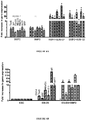
- FIG. 6A shows that both mesodermal (i.e., SRF, Tbx6, FoxH1, Isl1) and cardiac (Mef2c, Nkx2.5, ⁇ -actin) genes were induced by the morphogen in HUES-1 cells. This effect was further enhanced by 3-10 folds when BMP2 was added in the presence of the FGF-R inhibitor SU5402. No significant difference was observed in the extent of the BMP2 cardiogenic response between both cell lines ( Fig. 6A ) although the total number of copies of each gene expressed following BMP2 induction was much higher (i.e. 10-15 fold) in I6 than in HUES-1 cell line (data not shown).
- Immunostaining with an anti-ANP and anti-human lamin antibodies revealed the presence of lamin-positive human ES cell derived-cardiomyocytes ( Fig. 8A ).
- ES cell-derived cardiomyocytes sections were immunostained with an anti- human ⁇ -MHC antibody. These experiments revealed the presence of differentiated cardiomyocytes ( Fig. 8B ) in 40 % and 71 % of cryosections examined from HUES-1- and I6-engrafted hearts, respectively and in 85 % of sections examined from rat grafted with HUES-1 cells treated with both BMP2 and SU5402. The cell engraftment was however limited.
- Eosin-hematoxylin stained sections did not show any sign of inflammation or cell hyperproliferation two months posttransplantation ( Fig. 9 ). Likewise, whole-body autopsies failed to disclose any tumor in peripheral organs.
- Example 5 Generation of CD15+ cardiovascular progenitors fom human iPS cell line
- Human iPS cell line 111 were generated using human dermal fibroblasts infected by lentivirus harbouring the cDNAs encoding Oct4, Sox2, Lin 28, Klf4 and Nanog.
- iPS cells were trypsinised and incubated for 30 min with anti-CD 15 conjugated magnetic beads (Miletenyi) and sorted out using Miltenyi columns.
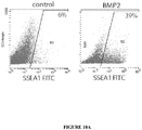
- a fraction of BMP2-treated cells were used prior to sorting for FACS analysis using the anti-CD 15 FITC and anti-cardiac markers antibodies ( Figures 10A and 10B ). 38 % of cells turned out to be CD15 +. This percentage was used to normalise the percentage of cells positive for the cardiac markers (numbers in green).
- CD15+ cardiovascular progenitors could also be derived from iPS cells.
- HES cells or embryonic-like state cells are capable to differentiate into cardiomyocytes without formation of teratomas after commitment toward a cardiac lineage using the cardiogenic factor BMP2. While BMP2 was shown to improve late cardiac differentiation of already differentiating cells [17], our study reports the strong instructive action of the morphogen on undifferentiated HES cells.
- BMP2 is a potent mesodermal and cardiogenic instructor when used at low concentration. Its cardiogenic potential is a well conserved property throughout the evolution. Dpp, the drosophila homolog of BMP2, favours formation of the mesoderm including the heart [18]. Similar effects have been observed in zebrafish [19], Xenopus [20-21] and chicken [22]. Our data obtained in two separate cell lines uncovered that BMP2 function is conserved in human species. While I6 cells were more prone to give rise to a mesodermal lineage ( Fig. 5 ), maximal BMP2 response was not significantly different from the one observed with HUES-1 cells although maximal extent of gene expression was higher in I6 than in HUES-1 cells.
- BMP2 effect was weak while its instructive action was dramatically enhanced by addition of the FGF receptor inhibitor, SU5402.
- FGF receptor inhibitor SU5402.
- Human ES cells are grown on feeder cells which secrete many factors including FGF2 which is known to antagonize the BMP2 signaling pathway.
- SU5402 could act through at least two mechanisms to unmask the BMP2 transcriptional effect. First, FGF2 phosphorylates smad2/3, thereby preventing the BMP2 signaling co-factor from translocating into the nucleus and thus to exert its transcriptional action [23].
- FGF2 is also known to act as a paracrine factor on both MEF and HES cells to regulate expression of Cerberus, a nodal and BMP antagonist enriched in HES cells [24].
- SU5402 blocks self-renewal of cells and favours non-specific differentiation which is further directed to the mesoderm by BMP2. By blocking all or either one of these pathways, the FGF inhibitor is required to unravel the BMP2 transcriptional response of HES cells.
- HES cell-derived cardiomyocytes in situ was rather close to a foetal one. Indeed the cells still expressed ⁇ -MHC and ANP, two known markers of early stage of cardiac differentiation. The short length of the sarcomere is still characteristic of a foetal myocyte. Several reasons could account for this immature phenotype. HES cells may require a longer time (more than two months) to fully differentiate. Alternatively, the paracrine environment of the infarction scar may not provide the factors (some FGFs, Neuregulin, retinoic acid, BMP10, ...) [30] or signals taking place in embryogenesis to ensure a full differentiation process.
- I6 cells gave rise to larger engraftment areas than HUES-1 cells. Although both cell lines respond with the same efficiency to BMP2, I6 cells feature a higher basal expression of mesodermal cardiac genes ( Fig. 5 ). This indicates that the cell line is already committed to the mesoderm, which is likely to account for the better cardiogenic potential in vivo.
- HUES-1 cells pretreated with BMP2 together with SU5402 also featured a better engraftment than HUES-1 challenged by BMP2 alone. This further emphasizes that the stage of specification is crucial to ensure a proper differentiation of ES cells in situ.
- HES-cell derived differentiated cardiomyocytes did not either express Cx43 when transplanted in injured left ventricle while they did express it when co-cultured with neonatal rat cardiomyocytes [32].
- Cx43 remains, however, critical to establish unequivocally as a true cardiac regeneration implies that the donor-derived cardiomyocytes can establish gap junction-supported electromechanical connections with those of the host.
- the formation of such a syncytium allowing graft-host synchronized beats which is critical for enhancement of contractility has not yet been achieved with adult cells, whether myogenic [33]or bone marrow-derived [34].
- the demonstration that HES cells could fill this unmet need would likely be a major step for rationalizing their use in situations where patient outcomes are critically dependent on the replenishment of a new pool of contractile cells.
Landscapes
- Health & Medical Sciences (AREA)
- Engineering & Computer Science (AREA)
- Life Sciences & Earth Sciences (AREA)
- Bioinformatics & Cheminformatics (AREA)
- Biomedical Technology (AREA)
- Organic Chemistry (AREA)
- Chemical & Material Sciences (AREA)
- Wood Science & Technology (AREA)
- Genetics & Genomics (AREA)
- Biotechnology (AREA)
- Zoology (AREA)
- Cardiology (AREA)
- General Health & Medical Sciences (AREA)
- Veterinary Medicine (AREA)
- Medicinal Chemistry (AREA)
- Animal Behavior & Ethology (AREA)
- General Engineering & Computer Science (AREA)
- Biochemistry (AREA)
- Rheumatology (AREA)
- Pharmacology & Pharmacy (AREA)
- Nuclear Medicine, Radiotherapy & Molecular Imaging (AREA)
- Cell Biology (AREA)
- Public Health (AREA)
- Microbiology (AREA)
- General Chemical & Material Sciences (AREA)
- Chemical Kinetics & Catalysis (AREA)
- Heart & Thoracic Surgery (AREA)
- Hospice & Palliative Care (AREA)
- Micro-Organisms Or Cultivation Processes Thereof (AREA)
- Medicines Containing Material From Animals Or Micro-Organisms (AREA)
- Materials For Medical Uses (AREA)
Description
- The present description discloses a method for the in vitro preparation of cardiovascular progenitors cells from mammalian embryonic stem cells (ES cells) or mammalian embryonic-like state cells, preferably from primate, wherein said method comprises the use of the CD15 (SSEAI) marker as a positive cardiovascular progenitors differentiation marker. The present description discloses the use of a receptor tyrosine kinase inhibitor, particularly the SU5402 or SU11248 in association with the BMP2 for improving the efficiency of the desired differentiation. The present description discloses the use of platelet lysate as foetal animal serum substitute in a culture medium intended to the proliferation or propagation of primate ES cells maintaining their pluripotency feature. Derived compositions or kits in relation with the claimed methods or product obtainable by the described methods form also part of the present description.
- Heart failure is becoming a predominant disease and a leading cause of death in most of developed countries. Regardless of the origin of myocardial failure (i.e, ischemic or genetic), the clinical symptoms result mainly from the death of cardiomyocytes replaced by a fibrotic and non contractile tissue. Pharmacological approaches to cure or relieve heart failure have been facing limitations. Because of a limited regeneration capability of the heart [1] and a shortage in donors for heart transplantation, an external source of cells has been envisioned as a therapeutic solution to bring a gain in function to diseased myocardium. For the last few years, hematopoietic stem cells had raised many hopes as a potential autologous cell source to repair diseased myocardium. However, the enthusiasm generated by the early non-controlled phase I studies has been dampened by the more recent recognition that out of four randomised controlled trials entailing intracoronary infusions of bone marrow-derived cells shortly after myocardial infarction, three failed to meet their primary end point, i.e., an improvement in left ventricular ejection fraction [2-5]. Combined with basic studies disproving the cardiogenic potential of these cells [6-7], the outcome of these trials demonstrates that these cells do not really regenerate the diseased myocardium and it is unlikely that their paracrine effects may be sufficient for restoring function of extensively scarred myocardium [8]. The same limitations apply to skeletal myoblasts [9]. Thus, these findings call for another stem cell source to achieve myocardial regeneration. Among various other cell types, embryonic stem (ES) cells [10-12] or ES cell-derived cardiomyocytes [13-14] have turned out to be the most promising for replacing scar fibrosis by new contractile elements. However, the number of cells required to regenerate a post-infarcted human myocardium (i.e., several hundreds of million) is too high to be reasonably achieved by in vitro engineering of ES cell-derived cardiomyocytes. For the last few years, we and others have shown that proliferative mouse ES cells engrafted in a diseased myocardium further differentiate into functional cardiomyocytes following in-vitro commitment using the cardiogenic morphogen BMP2 [10-12]. Cardiac-specified cells then complete their differentiation in response to the local cues present in the scar and do not generate any kind of tumors.
- Leschik et al (Nature Protocols , vol 3 N°9, 2008, pp 1381-1387) discloses methods of cardiovascular progenitor cells preparation and/or separation.
- Li Zongjin et al (Journal of Cellular Biochemistry, vol 106, N°2, 2009, pp 194-199) discloses the treatment of heart failure with hES-cell-derived progenitors
- Primate Embryonic stem (ES) cells feature the capability to selfrenew and to differentiate in any cell lineage of the three embryonic layers namely the ectoderm, endoderm and mesoderm [35]. However, spontaneous differentiation of ES cells toward a specific cell lineage is poorly efficient, specifically for primate ES cells. Human ES cells do not share with mouse ES cells the same molecular mechanisms of self-renewal or capabilities of spontaneous differentiation [15]. For the last decade, laboratories have developed protocols to direct mouse and human ES (HES) cells toward their favourite cell type. These protocols are mandatory to: (i) better investigate and thus comprehend the genetic and epigenetic mechanisms underlying ES cell differentiation, (ii) use ES cells-derived differentiated cells as a toxicology model (iii) perform HTS (High Throughput Screening) aiming at discovering new cardiogenic molecules, markers of cardiac genetic diseases or new therapeutic drugs [36-37] (iv) design protocols of cell therapy of heart failure [38].
- There is a clear need, therefore, to provide clinical grade cells which can subsequently differentiate into cardiomyocytes in situ following their transplantation in infarcted myocardium without any sign of hyperproliferation, by a method able to specifically differentiate in vitro a high number of primate ES cells or embryonic-like state cells toward a cardiovascular lineage.
- In this context, it would be worthwhile to have further at one's disposal a simple and reproducible protocol to commit primate ES cells or embryonic-like state cells toward a cardiovascular lineage allowing to sort out the population of early cardiovascular progenitors which retain the capability to proliferate and repopulate the postinfarction scar.
- This is the object of the present invention.
- Herein, the inventors bring the proof of concept that primate ES cells such as embryonic-like state cells, can also be directed toward a cardiogenic and vascular fate using the morphogen BMP2 in association with a receptor tyrosine kinase inhibitor and to selectively collect these cardiovascular progenitors thus obtained by using the positive CD15 (SSEA1) biomarker. Furthermore, the cells do differentiate into cardiomyocytes following engraftment into the myocardial scar without any sign of hyperproliferation. These data open the path for the use of early cardiovascular progenitors, which retain the capability to proliferate and repopulate the postinfarction scar.
- Thus, the present invention relates to methods for obtaining substantially pure populations of primate cardiovascular progenitors. Compositions such as therapeutical composition, containing these cell populations and method of using these cell populations are also described.
- In a first aspect, the description discloses an in vitro method for the preparation of cardiovascular progenitors cells from mammalian ES cells or from mammalian embryonic-like state cells, preferably for the preparation of a substantially purified population of cardiovascular progenitors, wherein said method comprises the following step of:
- a) culturing of mammalian ES cells or embryonic-like state cells in a medium containing suitable agents allowing their proliferation and maintaining their pluripotency;
- b) differentiating the mammalian pluripotent ES cells or embryonic-like state cells obtained in step a) toward cardiovascular progenitors cells by suspending said pluripotent ES cells or embryonic-like state cells in a medium containing BMP2 (Bone Morphogenetic Protein 2); and
- c) selecting and collecting the differentiated mammalian ES cells or mammalian embryonic-like state cells obtained in step b) which display the CD15 marker at their membrane surface, the mammalian ES cells or embryonic-like state cells displaying said CD15 marker being selecting and collecting as cardiovascular progenitors cells.
- In a preferred embodiment, said embryonic-like state cells are induced pluripotent stem cells, commonly abbreviated as iPS cells, preferably from adult somatic cells, particularly from adult fibroblast.
- In a more preferred embodiment, said iPS cells are obtained using human dermal fibroblasts infected by lentivirus harbouring the cDNAs encoding Oct4, Sox2, Lin 28, Klf4 and Nanog, preferably under ES cells culture conditions [49].
- By "pluripotency", it is intended to designate herein pluripotent ES cells-derived cells that are the descendants of totipotent embryonic stem cells and can differentiate into cells derived from any of the three germ layers ectoderm, endoderm and mesoderm.
- By iPS cells, it is intended to designate pluripotent stem cell artificially derived from a non-pluripotent cell, typically an adult somatic cell, by inducing expression of certain genes.
- The preparation of iPS cells from mammalian cells, particularly from mouse cells is well known from the skilled person. [46-49].
- In a preferred embodiment, the description discloses an in vitro method wherein said mammalian cells are primate, mouse or rat cells, preferably primate cells.
- Primate embryonic stem cells can be isolated from blastocysts of members of the primate species (see for example
U.S. Pat. No. 5,843,780 ; Thomson et al., Proc. Natl. Acad. Sci. USA 92:7844, 1995). - Cell culture methods are described generally in the current edition of Culture of Animal Cells: A Manual of Basic Technique (R. I. Freshney ed., Wiley & Sons); General Techniques of Cell Culture (M. A. Harrison & I. F. Rae, Cambridge Univ. Press), and Embryonic Stem Cells: Methods and Protocols (K. Turksen ed., Humana Press). Tissue culture supplies and reagents are available from commercial vendors such as Gibco/BRL, Nalgene-Nunc International, Sigma Chemical Co., and ICN Biomedicals.
- In a preferred embodiment, said BMP2 protein (also named BMP-2) is the human BMP2, more preferably a recombinant hBMP2, more preferably the human BMP2 protein having the amino acids sequence depicted in GenBank Accession Number AAF21646.
- In a preferred embodiment, the CD-15 marker is the human CD-15 (also named "SSEA1" marker, "SSEA1" for "Stage Specific
Embryonic Antigen 1"). - Angiogenic growth factors such as fibroblast growth factors (FGFs) and vascular endothelial growth factors (VEGFs) are currently targets of intense efforts to inhibit deregulated blood vessel formation in diseases such as cancer. FGFs and VEGFs exert their effects via specific binding to cell surface-expressed receptors equipped with tyrosine kinase activity. Activation of the receptor kinase activity allows coupling to downstream signal transduction pathways that regulate proliferation, migration and differentiation of endothelial cells. Inhibitors of tyrosine kinase activity such as the tyrosine kinase activity of FGF and/or VGR receptor are currently in clinical trials.
- The inventors have surprisingly demonstrated that such receptor tyrosine kinase (RTK) inhibitor, particularly FGFR inhibitor or multitargeted tyrosine kinase receptor inhibitors, particularly resulting to the inhibition of the tyrosine kinase activity of the VEGF receptor (R-1 and/or R-2), and/or fetal liver tyrosine kinase receptor 3 (FLT3), and/or KIT (stem-cell factor [SCF] receptor), and/or PDGFRα, and/or PDGFRβ, can be used in step b) to improve, likely by a synergic effect with BMP2, the differentiation of primate ES cells or embryonic-like state cells in cardiovascular progenitors cells.
- In a preferred embodiment, the RTK inhibitor is selected from the group consisting of SU5402 and SU11248.
- The SU5402 compound has the following formula:
3-[3-(2-Carboxyethyl)-4-methylpyrrol-2-methylidenyl]-2-indolinone. - SU5402 inhibits the tyrosine kinase activity of fibroblast growth factor receptor 1 (FGFR1); (IC50 = 10 - 20 µM in the presence of 1 mM ATP). It also inhibits aFGF-induced tyrosine phosphorylation of ERK1 and ERK2 (IC50 = 10-20 µM). SU5402 is considered as only a weak inhibitor of tyrosine phosphorylation of the PDGF receptor and does not inhibit phosphorylation of the insulin receptor. It does not inhibit the kinase activity of the EGF receptor (Mohammadi M. et al., 1997, Science 276, 955).
- The SU11248 compound (also referenced as "Sunitinib malate" or Sutent" has the following formula:
N-[2-(diethylamino)ethyl]-5-[(Z)-(5-fluoro-1,2-dihydro-2-oxo-3H-indol-3-ylidine)methyl]-2,4-dimethyl-1H-pyrrole-3-carboxamide. - Sunitinib (sunitinib malate; SU11248; SUTENT; Pfizer Inc, New York, NY) is a multitargeted RTK inhibitor with antitumor and antiangiogenic activities. Sunitinib has been identified as a potent inhibitor of VEGFR-1, VEGFR-2, fetal liver tyrosine kinase receptor 3 (FLT3), KIT (stem-cell factor [SCF] receptor), PDGFR, and PDGFRβ in both biochemical and cellular assays (Faivre et al., Journal of Clinical Oncology, Vol. 24, N° 1, 2006: pp. 25-35).
- When the SU5402 compound is used as FGF inhibitor in step b), it is preferred that the primate ES cells or embryonic-like state cellsor human embryonic-like state cells were treated in step b) for respectively 48 hrs and 6 days with a BMP2 concentration of 10 ng/ml medium (± 5 ng/ml) in the presence of 1 µM SU5402 (± 0.5 µM). Following the 6 days treatment with 10 ng/ml BMP2 and 1 uM SU5402, the embryonic-like state cells (iPS cells) can be trypsinised.
- One of the problems which have to be solved in this context, when production of therapeutical cells is wished to commit primate ES cells or primate embryonic-like state cells toward a cardiac lineage, is to have further at one's disposal compounds used in the production method, such as RTK inhibitor, which have been already used as therapeutical compound. Indeed, these compounds are known not to have relevant side-effects and generally, available as clinical grade compound.
- After demonstrating the synergic effect of FGF inhibitor in step b) for differentiating the primate ES cells or primate embryonic-like state cells in cardiac progenitor cells (or early cardiovascular progenitors cells having the capability to proliferate and repopulate the postinfarction scar after administration to the patient), the inventors have demonstrated surprisingly that it will be possible to use a multitargeted RTK inhibitor available as clinical grade, such as the SU11248, as synergic compound in step b) in place of the FGF inhibitor.
- Preferably, when a multitargeted RTK inhibitor, such as SU11248, is used in place of a FGF inhibitor, it is preferred that the primate ES cells or primate embryonic-like state cells were treated with about 5 times more quantity of such in step b) for 48 hrs with a BMP2 concentration of 10 ng/ml medium (± 5 ng/ml) in the presence of 5 µM SU (± 2 µM).
- In a preferred embodiment the description discloses a method for preparing a substantially pure population of progenitor cells which is at least about 80 %, preferably 85 %, 90 %, 95 %, 97 %, 98 % and 99 % pure cardiac progenitor cells which display at their surface the marker CD-15 and, preferably, which retain their capability to proliferate and repopulate the postinfarction scar after administration to a patient.
- In step a) of the method medium for culturing primate ES cells or primate embryonic-like state cells allowing their proliferation and maintaining their pluripotenty are well known by the skilled person.
- Cell culture methods are described generally in the current edition of Culture of Animal Cells: A Manual of Basic Technique (R. I. Freshney ed., Wiley & Sons); General Techniques of Cell Culture (M. A. Harrison & I. F. Rae, Cambridge Univ. Press), and Embryonic Stem Cells: Methods and Protocols (K. Turksen ed., Humana Press). Tissue culture supplies and reagents are available from commercial vendors such as Gibco/BRL, Nalgene-Nunc International, Sigma Chemical Co., and ICN Biomedicals.
- For example, it is known that primate ES cells required feeder cells for their culture, such as mouse embryonic fibroblasts, and FGF2 (bFGF) to maintain their pluripotency.
- By "Feeder cells" it is intended to designate cells that are co-cultured with the primate ES cells. They provide an environment in which the primate ES cells type can grow. The culture of the primate ES cells can be supported by primary mouse embryonic fibroblasts (MEF) or primary human foreskin fibroblasts as exemplified. Immortalized mouse embryonic fibroblasts or human foreskin fibroblasts cells can also be used as feeder cells.
- For example of basic medium which can be used for culturing primate ES cells or primate embryonic-like state cells, the KO™-DMEM medium from Invitrogen (or from Gibco, Rockville, MD, USA) can be cited.
- It is preferred that FGF2 (
fibroblast growth factor 2, also named bFGF) is added to the basic medium after a period of at least an overnight with ES cell or embryonic-like state cells basic medium without FGF2 on feeder cell layer. - It is preferred that the culture medium is changed every day.
- Generally, the cell colonies are dissociated into single cells or small cell clusters every 4-5 days using trypsin or collagenase depending on the primate ES cells or primate embryonic-like state cells source (monkey source).
- It is preferred to never let the ES cells or primate embryonic-like state cells colonies reaching confluency in the dish.
- In step a) of the method the medium for culturing primate ES or primate embryonic-like state cells comprised a basic medium, such as the KO™-DMEM medium, supplemented with platelet lysate obtained from primate blood, preferably from human blood.
- Preferably, the platelet lysate is a total human freeze-thawed platelet lysate, more preferably at a concentration range of 5 % to 15 % (V/V), the most preferred at a concentration of 7.5 % (V/V) ± 2.5 % in the basic medium.
- The inventors have demonstrated that the use of platelet lysate in place of the FBS (foetal bovine serum) content usually present in the basic medium at this stage of a culture of primate ES cells or primate embryonic-like state cells allows to obtain the proliferation of non-differentiated primate ES cells or primate embryonic-like state cells, the use of such platelet lysate allowing the maintain of the pluripotency of the primate ES cells or primate embryonic-like state cells obtained after this step of proliferation or propagation.
- One of the benefit of platelet lysate used in place of FBS is to eliminate the problem of possible contamination of FBS with infectious microorganism, virus or prion and thus to produce therapeutical cells composition with a better safety.
- The blood platelet lysate can wholly or partly replace foetal bovine serum in cell culture.
- To obtain such a platelet lysate, the whole blood is for example separated into red blood corpuscles and platelet-rich plasma by centrifugation suitably at about + 4°C.
- Subsequently the platelet-rich plasma can be concentrated by ultrafiltration, after which the concentrated platelet-rich plasma is optionally frozen for storage and/or analysis and further lysis of the platelets included. After optional freezing, the frozen plasma is thawed, preferably at a temperature below 37°C, more preferred below 20°C.
- After or before thawing of the concentrated platelet-rich plasma, water can be added for accelerate the total lysis of the platelets included if necessary.
- The PRP may use the patient's own plasma and/or platelets who will receive the therapeutical cells. The platelets may be present in the plasma or PRP at a range of from about 200,000 to 2,000, 000 platelets per cubic centimetre, or more. The PRP may be obtained using autologous but also allogenic, or pooled sources of platelets and/or plasma from a variety of primate sources, including human sources.
- In step b) of the method, the basic medium for differentiating primate ES cells or primate embryonic-like state cells can be selected for example in the group consisting of RPMI or DMEM medium.
- In a preferred embodiment, the primate stem cells or embryonic-like state cells, can be treated for respectively 96 H and 6 days with 10 ng/ml BMP2 in RPMI supplemented with 2 % B27, and: 1 µM SU5402 (research grade) or 5 µM SU11548, SUTENT (clinical grade).
- It is preferred that the cells do not reach confluency at the end of BMP2 treatment. It is also preferred that BMP2 is added as soon as small colonies of ES cells appear.
- The description discloses a substantially purified population of cardiac progenitor cells susceptible to be obtained by the method herein disclosed, wherein the cardiac progenitor cells display at their membrane surface the CD15 marker and, preferably retain their capability to proliferate and repopulate the postinfarction scar when administrate to a patient in need thereof.
- In certain embodiment, the cells of the cardiac progenitor cells population herein disclosed express the early mesodermal Brachyury and Tbx6 markers, the cardiac Tbx20 and Mef2c markers, the GATA4, Nkx2.5, Isl1, Tbx18 markers and the Oct-4A marker.
- Preferably, by substantially purified population is meant that greater than about 80 % of the cells are cardiac progenitor cells, preferably greater than about 90 %, more preferably greater than about 95 %, more preferably yet greater than about 98 % and most preferably greater than about 99 %.
- The substantially purified population of cardiac progenitor cells herein discribed is useful for many clinical applications, preferably as a therapeutical composition or a medicament.
- The description discloses the use of the purified population of primate cardiac progenitor cells of this invention for preparing a therapeutic composition for replacing or regenerating cells or tissues in a primate, particularly cardiac cells.
- The invention further comprises a method for selectively separating cardiac progenitor cells from a primate cells population containing non-differentiated (pluripotency) ES cells or (non-differentiated) embryonic-like state cells and cardiac progenitor cells, or a method for enriching a primate cells population cell in cardiac progenitor cells wherein said method comprises the following steps of:
- A) contacting the cells population with anti-CD 15 antibodies; and
- B) selecting the cells that bind specifically to the CD15 antibodies or eliminating the cells which are not bound the anti-CD 15 antibodies, said cells having the capability to specifically recognize and to bind the CD15 antibodies being the primate cardiac progenitor cells which are desired to be kept in the cells population.
- Preferably, in step A), the anti-CD 15 antibodies are anti-human CD15 antibodies, most preferably, monoclonal antibodies.
- In a preferred embodiment, the anti-CD 15 antibodies are labelled, more preferably with a marker which can be used to select and to separate the cells displaying the CD15 marker from a cells population, preferably from a cells population obtained or susceptible to be obtained by the steps a) and b) of the method for the preparation of a population of cardiovascular progenitors cells from primate ES cells or embryonic-like state cells according to the present invention.
- More preferably, said antibody marker is a fluorescent marker such as FITC.
- In another preferred embodiment, the anti-CD 15 antibodies are bound at the surface of magnetic beads or articles or coupled to magnetic compounds.
- Method using magnetic beads or particles is usually implemented for sorting cells. Antibodies specific for a particular cell of interest are covalently bond to magnetic particles. They then incubate a mixture of cells in a solution with the magnetic antibodies. The entire reaction mixture is exposed to a magnetic field, which retains the cells of interest. Some of these particles are even composed of materials that naturally degrade without adversely affecting cell function. Such a system to separate or enrich population in specific cell is well known from the skilled man (see BD Biosciences system, (San Jose, CA USA); positive cells selection with the MidiMACS™ separation system from Miltenyi Biotech (Bergisch Gladbach, Germany), Polysciences or Dynal Biotech)).
- As alternatives in cell separation, the well established fluorescence-activated cells sorting (FACS) technique can be also used (Flow cytometers can measure and separate up to 500,000 cells per minute).
- The description discloses a kit useful for enriching for cardiac progenitor cells from a cells population containing non-differentiated primate ES cells or (non-differentiated) embryonic-like state cells, and cardiac progenitor cells. The kit includes anti-CD 15 antibodies, preferably anti-human CD15 antibodies.
- Preferably, monoclonal antibodies are used.
- Preferably, the anti-CD 15 antibodies are labelled, more preferably with a marker which can be used to select and to separate cells displaying the CD15 marker from a cells population, preferably from a cells population obtained or susceptible to be obtained by the steps a) and b) of the method according to the present invention.
- More preferably, said antibody marker is a fluorescent marker such as FITC.
- In another preferred embodiment, the ant-CD15 antibodies are bound at the surface of magnetic beads or coupled to magnetic compounds.
- Preferably, instructions are provided which have information to the user regarding the use of the antibodies for enriching for cardiac progenitor cells from the cells population.
- In certain embodiments, the kit also contains magnetic beads, e.g., superparamagnetic microparticles. The magnetic beads can be complexed to the anti-CD15 antibodies, or they can be separate. The kit also can include an electromagnet for use in generating a magnetic field.
- In another aspect, the description discloses a method for proliferating or propagating primate ES cells or embryonic-like state cells maintaining their pluripotency feature during this step of proliferation or propagation, said primate ES cells or embryonic-like state cells being usually cultivated in a medium supplemented with foetal mammal serum, such as FBS, characterized in that said foetal serum is replaced by a platelet lysate from a primate, preferably a human platelet lysate, preferably at a concentration of 7.5 % (V/V) ± 2.5 %.
- A composition or a culture medium used for culturing cells, preferably primate ES cells, comprising BMP2 and a RTK inhibitor.
- Preferably the RTK inhibitor is a FGF inhibitor or a multitargeted RTK inhibitor capable to inhibit VEGFR, FLT3, KIT and/or PDGFR.
- More preferably, the RTK inhibitor is selected from the group consisting of SU5402 and SU11248.
- Preferably, the composition or the medium contains 10 ng/ml BMP2 (± 5 ng/ml) for 1 µM SU5402 (± 0.2 µM).
- Preferably, the composition or the medium contains 10 ng/ml BMP2 (± 5 ng/ml) for 5 µM SU11248 (± 2 µM).
-
-
Figure 1 : Beating activity of I6 HES cells-derived EBS atday 30. Cells are treated for 48 H with BMP2 prior to the generation of EBs. AN EB is scored as positive if it features at least three beating areas. Data are expressed as mean (blue bars) ± SEM (pink bars) (n = 3 experiments). -
Figure 2 : FACS analysis of CD15 positive cells. Cells are treated for 4 days with BMP2 and SU5402 in RPMI/B27 medium. Cells are then washed once with D-PBS, incubated for 3 min with trypsin. Cells are spun down and resuspended in D-PBS supplemented with 3 % Foetal calf serum containing the anti-CD15-FITC antibody used at a dilution of 1/100. Cells are incubated for 30 min at 4°C before FACS monitoring. The figure is representative of 3 experiments using HUES-24 or ORMES cell lines. Data are expressed as mean ± SEM. -
Figures 3A ,3B ,3C and3D : Gene expression profile of CD15-positive cardiovascular progenitors.
After sorting out the CD15-positive primate ES cells using the MACS system, cells are lysed and RNA extracted. Gene expression is monitored by real time quantitative PCR. Thefigure 3A is representative of 3 experiments using HUES-24 or ORMES cell lines. Data are expressed as mean ± SEM and normalised to the CD15- cell population.
After sorting out the CD15-positive primate ES cells using the MACS system following checking expression of CD15 by FACS (Fig. 3B ), cells are lysed and RNA extracted. Gene expression is monitored by real time quantitative PCR. Thefigures 3C and3D are representative of 5 experiments using HUES-24, 16, HUES-9 or ORMES cell lines. Data are expressed as mean ± SEM and normalised to the CD15- cell population. -
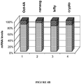
Figures 4A and4B :Figure 4A : Immunofluorescence of HUES cells cultured for 10 passages in clinical grade medium. The antibodies used are: anti Oct-4, TRA-1-60 and SSEA-4.Figure 4B : Real time PCR monitoring of pluripotency genes expression in HUES cells cultured for 10 passages in clinical grade medium. -
Figure 5 : Comparison of mesodermal and cardiac gene expression in HUES-1 and I6 cells. RNA was extracted and reverse transcribed from undifferentiated HUES-1 (passages 22-25) and I6 cell lines (passages 27-32) cultured for at least 5 passages after thaw-out on MEF prior to stimulation with BMP2. Differentiated colonies were cut out the plate before RNA extraction. Gene expression was estimated by real-time PCR and expressed as a ratio between expression in I6 and expression in HUES-1. Data are normalised to β-tubulin expression and expressed as means ±SEM (n=3). * Statistically significant (p<0.01). -
Figures 6A and 6B: Figure 6A : HUES-1 and I6 cells were treated for 48 hrs with 10 ng/ml BMP2 in the presence or absence or FGF inhibitor SU5402 and RNA extracted and reverse transcribed. Gene expression was monitored by real-time quantitative PCR. Results are expressed as fold stimulation in gene expression when compared to untreated ES cells.Figure 6B : HUES-1 cells treated or not with BMP2 and SU5402 were allowed to aggregate to form embryoid bodies (EB). EBs were kept in suspension for 5 days before RNA extraction and real time PCR. Data are normalised to β-tubulin expression and expressed as means ±SEM (n=3-5). * Statistically significant (p≤0.01). -
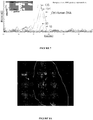
Figure 7 : BMP2-treated HES cells were engrafted into postmyocardial infarcted rats and their fate was examined two months later by real time PCR of α-actin mRNA following reverse transcription of mRNA extracted from myocardial sections. The figure shows both the profile of the melting curves of amplicons and the amplicons on gel. Human RNA was used as a positive control. * Statistically significant (p≤0.01). -
Figures 8A ,8B ,8C and8D : Immunostaining of cryosections from HES cell-engrafted myocardium using an antihuman lamin antibody (Fig. 8A ) (note that the anti-lamin antibody stained HES-derived cells within the scar area but not the surrounding endogenous rat cells) and an anti-human β-myosin antibody (Fig. 8B ). The antibody did not recognize the adult rat endogenous β-MHC (left panel B) while it bound human β-MHC. Images were acquired in confocal microscopy. (Fig. 8C ) quantification (in %) of the human β-MHC positive regions in the scars of myocardium engrafted with BMP2- or BMP2 with SU5402-treated HUES-1 or BMP2-treated I6 cell lines: * Statistically significant (p≤0.025). The area of β-MHC positive area within the scar was calculated using the threshold function of Metamorph software (fig. 8D ) a transversal section stained by the anti-β myosin antibody revealed some sarcomeric structures. The size was calculated using the scaling system of the ZEISS software driving the confocal microscope. -
Figure 9 : Eosin-Hematoxylin stained section from HES cell-engrafted myocardium. The scar area does not show any sign of cell infiltration of cell proliferation. -
Figures 10A and10B : IPS cells give rise to CD15+ cardiovascular progenitors, CD15+ iPS cells were generated and sorted following 6 days BMP2 treatment-
Figure 10A : FACS analysis of cardiac markers in CD15+ cells. -
Figure 10B : Immunofluorescence of CD15+ cells cultured for 5 days on MEF. The scale bar indicates 10 µm.
-
-
Figures 11A ,11B and11C : CD15+ cells express cardiac, endothelial and vascular proteins markers; CD15+ sorted cells were cultured for 5 days on MEF and immunostained with the indicated markers; some cells were treated with VEGF or PDGF and immunostained with anti smooth muscle actin and myosin or CD31. Some others were cultured with human fibroblast isolated from the myocardium and stained with an anti-actinin antibody. - Knockout ™-DMEM (Invitrogen cat n° 10829018)
Knockout™ SR serum replacement (cat n° 10828028 Invitrogen) test batch before using: - Trypsin 0.05 %/0.5 mM EDTA4Na IX (Invitrogen cat n° 25300054)
- Glutamax 200 mM (Invitrogen cat n° 35050038)
- MEM Non essential Amino Acids solution (200 mM) (Invitrogen cat n° 11140035)
- Mercaptoethanol (Invitrogen cat n° 31350010) (make solution at 10-7 Min PBS and use for one month)
- RPMI 1640 with glutamax (Invitrogen cat n° 61870-010)
- F12 nutritient supplement with glutamine (Invitrogen cat n° 21765-029)
- Dulbecco Phosphate Buffered Saline (D-PBS) Ca- and Mg-free (Invitrogen cat n° 14200067)
- B27 50X supplement without vitamin A (Invitrogen cat n° 12587010)
-
Gelatin 2 % solution from bovine skin cell (Invitrogen cat n° G1393-100ml) -
Mitomycin 2 mg Sigma cat n° M4287) - FGF2 (Human recombinant, preprotech cat n° 100-18B)
- BMP2 (Human recombinant R&D system)
- dispase (Invitrogen cat n° 17105041)
- collagenase CLS2 (Invitrogen cat n° 17104-019)
- SU5402 (calbiochem cat n° 572630)
- CD15 microbeads (MACS sorter Miltenyi cat n° 130-046-601)
- Anti-SSEA4 and anti-TRA 1-60 (Chemicon cat n° 90231 and 90232, respectively)
- Anti-Oct-4 (Santa Cruz, cat n° sc-9081)
- anti-CD 15-FITC antibody from BD Bioscience (IgM clone MMA cat n° 340703)
- RNA extraction kit (Mini RNA Isolation II Kit™ Zymo Research, cat n° R1033)
- Real time PCR reagent: LightCycler® FastStart DNA MasterPLUS SYBR Green I (Roche cat n° 3515885)
- Low attachment dishes (NUNC™ Low Cell Binding Plates cat n° 1453)
Sterile cell culture dishes and pipettes
500 ml Filtration unit 0.2 µm Nalgene cat n° 162-0020
A real time QPCR thermocycler (Roche LightCycler 1.5) - Primate ES cell lines require feeder cells (Mouse Embryonic Fibroblasts (MEF) prepared from E14 mouse embryos) and FGF2 to maintain their pluripotency.
- 1- MEF are cultured for 3 or 4 passages and treated for 2 hours with 10 µg/ml mitomycin; cells are washed twice with PBS, trypsinised for 5 min and plated in gelatin (0.1 %)-coated plates at a density of 40,000 cells/cm2.
- 2- Primate embryonic stem cells are thawed out quickly in warm propagation medium, spun down at 800 rpm for 4 min, and resuspended to be plated on feeder cells cultured overnight with ES cell medium without FGF2. Add FGF2 to the plate.
- 3- Rhesus ES cells (ORMES-2 given by Dr S Mitalipov, Dr Wolf's laboratory) and Human ES cell lines HES cell lines from D Melton's laboratory [39] and I6 cell lines from the Technion Institute [40] (NIH approved) are then cultured on MEF using KO™-DMEM medium supplemented with mercaptoethanol, glutamine, non essential amino acids, 15 % KOSR and 10 or 5 ng/ml FGF2 respectively (see Table 1). For Rhesus ES cells [41], 20 % of F12 medium supplements the KO™ -DMEM.
- 4- Medium is changed every day.
- 5- Cell colonies are dissociated into single cells or small cell clusters every 4-5 days using trypsin (HUES) or collagenase (16, ORMES-2, 6, 9, 18), respectively (see paragraph "BOX1").
- It is preferred to never let the ES cells colonies reaching confluency in the dish. Split them at about 70 % confluency.
-
- 1- Primate ES cells are treated one or two days following the passaging for 48 H with 10 ng/ml BMP2 in the presence of 1 µM SU5402 a FGF receptor inhibitor, in KO™-SR DMEM. It is critical that the cells do not reach too much confluency at the end of the treatment.
It is preferred to resuspend BMP2 at 1 mg/ml in aqueous buffer containing at least 0.1 % BSA to prevent adsorption to the vial atpH 5 or lower (due to its low solubility at neutral pH) and stored as aliquot at -20°C. Frozen stocks at 10 µg/ml can be further made in PBS. Do not freeze/thaw. - 2- Embryoid bodies are generated after dispase used at 1 mg/ml for 15 min at 37°C (HUES1, 9, 24, 26) or collagenase CLS2 (16) dissociation of HUES cell colonies and cell aggregation in low attachment dishes in KO™-DMEM, 5 % SR. 6.106 to 107 cells are required to fill a low attachment B6 plate to allow for formation of EBs.
It is preferred to use mechanical cell dissociation after dispase or collagenase step with a 5 ml pipette to generate small (20-50 cells) cell clusters. Spin down the cells and resuspend them in 5 % SR-KO™-DMEM (Differentiation medium). - 3- The functional index of cardiac differentiation in vitro is the score of beating EBs, observed after 3-4 weeks (
Figure 1 ). At earlier days, the kinetic of HES cell differentiation is monitored by real time PCR of cardiac transcription factors [42]. In vivo experiments (i.e., cell xenograft in immuno-compromised postmyocardial infarcted rats) also serves as a functional index of cardiomyocytes derived from this protocol [42]. -
- 1- Primate stem cells are treated for 96hrs with 10 ng/ml BMP2 in RPMI supplemented with 2 % B27, and (1 µM SU5402 (research grade) or 5 µM SU11548, SUTENT (clinical grade)).
It is preferred that the cells do not reach confluency at the end of BMP2 treatment. It is also preferred that BMP2 is added as soon as small colonies of ES cells appear. - 2- Use FACS analysis using an anti-CD15-FITC antibody to visualize SSEA-1 (CD15) positive cells (
Figure 2 ). Cells can thus be sorted out using CD15-coated Miltenyi beads (Kit) as follows. - 3- Cells are trypsinized and filtered on a 70 µm mesh nylon filter.
- 4- Cells are incubated for 30 min at 4°C with gentle occasional agitation with anti-CD15 antibody-coated Miltenyi beads (100 µl/ 5.106 cells) in PBS supplemented with 0.5 % BSA and 2 mM EDTA.
- 5- Cells are transferred to a L50 Miltenyi cartridge set on the magnet.
- 6- Cells are washed three times with 3 ml D-PBS- BSA/EDTA and allowed to be eluted from the column removed from the magnet using 3 ml of D-PBS/BSA/EDTA. The cells which go through are recycled to the cartridge on the magnet and eluted once more. This procedure is repeated three times. Counting the cells revealed 50-60 % of CD15 positive cells.
- 7- Gene expression profile is performed by RT-real time Q-PCR (see paragraph "BOX2"). A phenotypic analysis of the CD15-positive cells revealed that they express early mesodermal (Brachyury, Tbx6) and cardiac and cardiovascular (Tbx20, Mef2c, GATA4, Isl1, Tbx18, Flk1) markers (
Figures 3A-3D ) Nkx2.5 was absent in the CD15 negative while detected in the CD15 positive cell population. The CD15 positive cell population looses expression of pluripotency gene (Lefty, Nanog, Crypto). However, this cell population still over-expresses Oct-4A [43, 44]. - When cultured on MEF, CD15-positive cells retained the phenotype of cardiovascular progenitors as monitored by immunstaining cardiovascular markers; when treated with VEGF (10 or 50 ng/ml) or PDGF (10 or 50 ng/ml), CD15+ become endothelial and smooth muscle cells respectively. When cultured on human cardiac fibroblasts, CD15+ cells differentiate into actinin- positive cardiomyocytes.
- Embryonic stem cells are cultured on irradiated adult human skin fibroblasts (FSBT from Dr O. Damour, Hôpital E Herriot, Lyon) in KO™-DMEM supplemented with:
- 7.5 % of total human freeze-thawed platelet lysate,
- heparin (1000 UI/ml)
- 10 ng/ml FGF2 and insulin
- 10 passages in such a medium do not affect expression of markers of pluripotency (
Figures 4A-4B ). - FSBT are cultured in DMEM glutamine supplemented with 10 % foetal calf serum (batch approved by AFSSAPS).
Table 1: Propagation and differentiation medium HES cells KnockOut™- DMEM 400 ml or KO™-DMEM 320 ml + 80 ml F12 (ORMES) Glutamine 200 mM 5 ml Non essential amino acid 100mM 5 ml MercaptoEthanol 1x10⁻⁴ M 500 µl (Use the stock 10-4 M solution for one month only) Serum replacement 15 % (5 % for cell differentiation) Medium is filtered on 0.2 µm Nalgene Filter unit FGF2 (for ESC propagation only) 5 or 10 ng/ml - It is preferred that all media, enzymes, PBS should be warmed up at 37°C. Primate ES cells are very sensitive to change in temperature and should remain as long as possible at 37°C.
ORMES splitting: replace the culture medium by KO™-DMEM with 1 mg/ml collagenase (500 µl or 2.5 ml is enough for one well of a 6wells dish or for a B10 plate). Incubate the plate at 37°C for 5 min. Add 1 or 5 ml propagation medium without FGF2 and scrape the cells with a 5 ml pipette. Transfer the cells to a 15 ml falcon tube and spin them down at 800 rpm for 4 min at RT. Aspirate the supernatant and resuspend the cells with propagation medium with FGF2. Break down the ES cells colonies by pipetting them forth and back 3 to 5 times until getting a homogenous cell suspension. Plate thecells 1/6.
I6 cell line splitting: use the same protocol as above but incubate the cells in the presence of collagenase for 45 to 60 min.
HUES cells splitting: Wash the cells once with warm PBS and add trypsin (500 µl or 2.5 ml is enough for one well of a 6wells dish or a B10 plate). Incubate the cells under the hood for exactly 3 min. Wash out trypsin and resuspend the cells with propagation medium slightly by flushing out the feeder cell layer. Plate the cells at 1/6. - Total RNA is prepared after cell lysis using a kit (Zymo research). After reverse transcription using the reverse transcriptase Superscript 2 (Invitrogen) according to the manufacturer instructions, Real time PCR is carried out using a set of gene specific primers (see Tables 2 and 3). 2-6 ng cDNA is used for real time quantitative PCR, performed with a lightcycler1.5 and the SYBR Green fast start kit (Roche, Germany). The 12-µl reaction mix contained 1 µl of Master SYBR Green I mix, including Taq DNA polymerase, buffer, deoxynucleoside trisphosphate mix, SYBR Green I dye, 3 mM MgCl2 and 0.5 µM of each primer. 2 µl of 10-fold diluted cDNA is added to the mixture. Data are normalised using RT-PCR of the GAPDH mRNA as an index of human cDNA content after reverse transcription. Amplification includes an initial denaturation at 95°C for 8 min, and 45 cycles of denaturation at 95°C for 3 s, annealing at 65°C for 8-10 s, and extension at 72°C for 7-10 s. The temperature transition rate is 20°C/s. Fluorescence is measured at the end of each extension step. After amplification, a melting curve is acquired by heating the product at 20°C/s to 95°C, then cooling it at 20°C/s to 70°C. The reaction was maintained at 70°C for 20 s followed by slow heating at 0.3°C/s to 95°C. Melting curves are used to determine the specificity of PCR products, and they are further confirmed by gel electrophoresis.
- RNA was extracted from HES cells or slices of rat myocardium using a Quiagen kit. One µg of RNA was reverse-transcribed using the Mu-MLV reverse transcriptase (Invitrogen, Cergy, France) and oligo(16)dT.
- Real-time quantitative PCR was performed using a Light Cycler (Roche Diagnostic) or a Chromo4 thermal cycler (Biorad). Amplification was carried out as recommended by the manufacturers. Twelve or Twenty two µl reaction mixture contained 10 or 20 µl of Roche or Abgene SYBR Green I mix respectively (including Taq DNA polymerase, reaction buffer, deoxynucleoside trisphosphate mix, and SYBR Green I dye, 3 mM MgCl2), 0.25 µM concentration of appropriate primer and 2 µl of cDNA. The amplification programme included the initial denaturation step at 95°C for 15 or 8 min, and 40 cycles of denaturation at 95°C for 10 s, annealing at 65°C for 8 s (Light cycler) or 20 s (Chromo4), and extension at 72°C for 8 or 30 s. The temperature transition rate was 20 (Light Cycler) or 4 (Biorad)°C/s. Fluorescence was measured at the end of each extension step. After amplification, a melting curve was acquired by heating the product at 20 or 4°C/s to 95°C, cooling it at 20 or 4°C/s to 70°C, keeping it at 70°C for 20 s, and then slowly heating it at 20 or 4°C/s to 95°C. Fluorescence was measured through the slow heating phase. Melting curves were used to determine the specificity of PCR products, which were confirmed using conventional gel electrophoresis. Data were analysed according to Pfafll et al. [16]. Primers specific for human genes are described in Tables 2 and 3.
Table 2: PCR primer sequences Table 2: PCR primer sequences Genes Forward (SEQ ID NO:) Reverse (SEQ ID NO:) β-tubulin CCGGACAGTGTGGCAACCAGATCGG (1) TGGCCAAAAGGACCTGAGCGAACGG (2) Nkx2.5 CATTTACCCGGGAGCCTACG (3) GCTTTCCGTCGCCGCCGTGCGCGTG (4) Mef2c AGATACCCACAACACACCACGCGCC (5) ATCCTTCAGAGAGTCGCATGC (6) SRF CTCCGCCCCGCTCAGACCCCACCACAGA (7) AGGTAGTTGGTGATGGGGAAGGA (8) α-actin CTATGTCGCCCTGGATTTTGAGAA (9) TGAGGGAAGGTGGTTTGGAAGAC (10) Oct-4 ACGACCATCTGCCGCTTTGAG (11) GCCTCTCACTCGGTTCTGAT (12) Tbx6 AGGCCCGCTACTTGTTTCTTCTGG (13) TGGCTGCATAGTTGGGTGGCTCTC (14) Isl1 CATCGAGTGTTTCCGCTGTGTAG (15) GTGGTCTTCTCCGGCTGCTTGTGG (16) FoxH1 GCCCCTGCCCACGCTGTCTA (17) GGTACCTCTTCTTCCTCCTCTT (18) Brachyury CGGAACAATTCTCCAACCTATT (19) GTACTGGCTGTCCACGATGTCT (20) Mesp1 CTCGTCTCGTCCCCAGACT (21) AGCGTGCGCATGCGCAGTT (22) Tbx20 CTGAGCCACTGATCCCCACCAC (23) CTCAGGATCCACCCCCGAAAAG (24) Gata4 GGTTCCCAGGCCTCTTGCAATGCGG (25) AGTGGCATTGCTGGAGTTACCGCTG (26) Pax6 GCCAGCAACACACCTAGTCA (27) TGTGAGGGCTGTGTCTGTTC (28) α-FP ACTGCAATTGAGAAACCCACTGGAGATG (29) CGATGCTGGAGTGGGCTTTTTGTGT (30) Cx43 TACCATGCGACCAGTGGTGCGC (31) GAATTCTGGTTATCATCGGGGAA (32) GAPDH ATGGGCAAGGTGAAGGTCGGAG (33) TCGCCCGACTTGATTTTGCAGG (34) Tbx5 TACCACCACACCCATCAAC (35) ACACCAAGACAGGGACAGAC (36) LeftyA GGGAATTGGGATACCTGGATTC (37) TAAATATGCACGGGCAAGGCTC (38) TDGF1 ACAGAACCTGCTGCCTGAAT (39) ATCACAGCCGGGTAGAAATG (40) Nanog CAAAGGCAAACAACCCACTT(41) TCTGCTGGAGGCTGAGGTAT (42) Table 3: PCR primer sequences: Melting T° (°C) and Genbank Reference sequence Genes Melting T° (°C) Refseq ID GAPDH 87 AF261085; NM_002046 Nkx2.5 88 AB021133 Mef2c 86 NM_002397; AL833268 Oct-4A 90 NM_002701 Tbx6 88 AJ007989; NM_004608, NM_080758 Brachyury 92 AJ001699; NM_003181 Tbx20 88 AJ237589; NM_020417 Tbx5 85 U89353; NM_080717 Lefty A 89 U81523; NM_003240 TDGF1 90 M96955; NM_003212 Nanog 84 AB093576; NM_024865 Pax6 85 AY707088; NM_001604 α-FP 88 V01514 - HUES-1 and I6 cell lines were cultured on Mouse Embryonic Fibroblasts (MEF) prepared from E14 mouse embryos using KO™-DMEM medium supplemented with mercaptoethanol, glutamine, non essential amino acids, 15 % KO™SR and 10 or 5 ng/ml FGF2 respectively. Medium was changed every day. Cell colonies were dissociated into single cells or cell clusters every 4-5 days using trypsin (HUES-1) or collagenase (16), respectively. A similar enzymatic digestion was used prior to cell transplantation in infarcted rats.
- HES cells were treated for 48 hrs with 10 ng/ml BMP2 in the presence or absence of 1 µM SU5402, a FGF receptor inhibitor, in low KO™SR (5 %) containing KO™-DMEM. Embryoïd bodies were generated after trypsinisation (HUES-1) or collagenase (16) dissociation of HES cell colonies and cell aggregation in low attachment dishes (Nunc) in DMEM, 10 % foetal calf serum.
- Myocardial infarction was induced in female Wistar (mean weight of 250 g) by ligation of the left coronary artery. Rats were operated on under general anaesthesia with isoflurane (Baxter), 3 % at induction and 2 % for maintenance. After tracheal intubation, mechanical ventilation (Alphalab, Minerve) was set at a rate of 70/min and with an 0.2 ml average insufflate volume. Analgesia was performed with a 10 mg/kg subcutaneous injection of ketoprofen (Merial).
- The heart was exposed through a left thoracotomy and the left coronary artery was permanently snared between the pulmonary artery trunk and the left atrial appendage.
- On the 15th day following infarction, the rats were reoperated on by median sternotomy and randomized to receive injections of BMP2-treated HUES-1 cells (3x10⁶ HUES-1 cells, n = 11 rats) in suspension of single cells, BMP2-treated I6 ES cells (3 106 I6 cells, n = 11 rats) in suspension of small cell clusters or control medium (n = 9 rats). Additional animals (n = 5 rats) received in-scar injections of 3 106 HUES-1 cells that had been exposed to both BMP2 and SU5402. We selected HUES-1 cell line for the latter experimental situation since this is the one which is not already committed to the mesoderm. One rat of each group (HUES-1 cell- and I6 cell- transplanted) died within 48 hrs after cell injection.
- Immunosuppressive therapy, consisting in one daily 10 mg/kg subcutaneous shot of cyclosporine A, was started on the same day and continued until sacrifice.
- Myocardial sections were stained with eosin and hematoxylin using a standard protocol.
- Two months after myocardial injection, rats were euthanized after general anaesthesia. Transverse-cut rat hearts were immediately fixed in OTC (Tissutec) and frozen at -180 °C nitrogen. Eight µm sections were cut on an ultramicrotome (LM 1850, Leica).
- Potential tumor growth was assessed with 8 µm standardized sections stained with hematoxylin and eosin.
- Immunofluorescence of myocardial cryosections were performed after paraformaldhehyde fixation and permeabilisation using Triton X-100 with an anti-human ventricular □β-myosin heavy chain (MHC) (Chemicon), anti-human lamin A/C (Novacastra) anti-atrial natriuretic peptide (ANP, Abgent) and anti-Connexin 43 (Cx43) (SIGMA) antibodies. The proteins were revealed using alexa-conjugated antibodies. Sections were observed in confocal microscopy (ZEISS LSM-510 meta).
- In addition, a whole-body autopsy of each transplanted rat, including brain, lungs, liver, spleen, pancreas, kidneys, periaortic lymph nodes, thymus, spine and ovaries, was systematically performed for the detection of a tumor.
- We used both HUES-1 and I6 HES cell lines to test their cardiogenic potential in vitro and in vivo. Indeed, a real-time PCR amplification of a few mesodermal and cardiac genes in both cell lines showed that the I6 cell line featured a higher basal expression of both mesodermal (Tbx6, SRF, Mesp1, brachyury) and early cardiac (Isl1, Mef2c,Tbx20) genes. GATA4 was weakly expressed in I6 but not in HUES-1 cells. Nkx2.5 was barely detected in either I6 or HUES-1 cell lines. Oct-4 level was not significantly different between both cell lines (
Fig. 5 ). - Both I6 and HUES-1 Human ES cells were treated for 48hrs with 10 ng/ml human recombinant BMP2. Gene induction was tested using real time Q-PCR.
Figures 6A and 6B shows that both mesodermal (i.e., SRF, Tbx6, FoxH1, Isl1) and cardiac (Mef2c, Nkx2.5, α-actin) genes were induced by the morphogen in HUES-1 cells. This effect was further enhanced by 3-10 folds when BMP2 was added in the presence of the FGF-R inhibitor SU5402. No significant difference was observed in the extent of the BMP2 cardiogenic response between both cell lines (Fig. 6A ) although the total number of copies of each gene expressed following BMP2 induction was much higher (i.e. 10-15 fold) in I6 than in HUES-1 cell line (data not shown). - To test whether BMP2-induced HES cell commitment was translated into a process of cardiac differentiation and to envision the differentiation scenario that might take place in vivo, control or BMP2-challenged HUES-1 cells were allowed to aggregate to form embryoid bodies (EBs). Gene expression was then monitored in
day 2 andday 5 EBs. BMP2 effect was observed at day 2 (i.e. two fold induction in gene expression) and became prominent at day 5 (Fig. 6B ). At that stage of development, expression of both mesodermal and cardiac genes was dramatically increased by 3 to 10 folds (Fig. 6 ). In contrast, Oct-4 was downregulated and almost absent in EBs generated from BMP2-treated ES cells. Similar results were obtained using I6 cell line. - Two months after coronary artery ligation, Human α-actin mRNAs were identified in transplanted hearts but not in those injected with the control medium (
Fig. 7 ). In contrast, we could not detect any mRNA encoding Oct-4, Pax6 (an early ectodermal marker) or α -foeto protein, an early endodermal marker (data not shown). - Immunostaining with an anti-ANP and anti-human lamin antibodies revealed the presence of lamin-positive human ES cell derived-cardiomyocytes (
Fig. 8A ). - To further define the phenotype of ES cell-derived cardiomyocytes, sections were immunostained with an anti- human β-MHC antibody. These experiments revealed the presence of differentiated cardiomyocytes (
Fig. 8B ) in 40 % and 71 % of cryosections examined from HUES-1- and I6-engrafted hearts, respectively and in 85 % of sections examined from rat grafted with HUES-1 cells treated with both BMP2 and SU5402. The cell engraftment was however limited. BMP2 treated HUES-1, I6 and BMP2/SU5402 HUES-1 treated ES cell-derived cardiomyocytes colonized 2.4 ± 0.3, 3.1 ± 0.4 and 3.6 ± 0.3 % of the scar (n = 10) (Fig. 8C ), respectively. Careful examination of these sections further indicated that these cardiomyocytes were still at a foetal stage demonstrated by a shorter sarcomeric length (1.6 ± 0.1µm) compared to 2 ± 0.1 µm in adult rat (Fig. 8D ). - Eosin-hematoxylin stained sections did not show any sign of inflammation or cell hyperproliferation two months posttransplantation (
Fig. 9 ). Likewise, whole-body autopsies failed to disclose any tumor in peripheral organs. - Human iPS cell line 111, were generated using human dermal fibroblasts infected by lentivirus harbouring the cDNAs encoding Oct4, Sox2, Lin 28, Klf4 and Nanog.
- Following 6 days treatment with 10 ng/ml BMP2 and 1 uM SU5402, iPS cells were trypsinised and incubated for 30 min with anti-CD 15 conjugated magnetic beads (Miletenyi) and sorted out using Miltenyi columns. A fraction of BMP2-treated cells were used prior to sorting for FACS analysis using the anti-CD 15 FITC and anti-cardiac markers antibodies (
Figures 10A and10B ). 38 % of cells turned out to be CD15 +. This percentage was used to normalise the percentage of cells positive for the cardiac markers (numbers in green). Cells were then plated on mouse embryonic fibroblasts in KODMEM added with KOSR (invitrogen), glutamine, non essential amino acids and mercaptoethanol (HES propagation medium) and stained one week later with anti-isll, -Nkx2.5 and -Mef2c. A similar pattern of expression of cardiac proteins (i.e., Nkx2.5, Mef2c, Tbx5 and Isl1) as well as endothelial marker CD31 were found in CD15+ cells derived cells as assessed by FACS analysis (Figure1A ) and immunofluorescence (Figure 10B ). - Here the inventors also demonstrate that CD15+ cardiovascular progenitors could also be derived from iPS cells.
- This study reveals that HES cells or embryonic-like state cells (iPS cells) are capable to differentiate into cardiomyocytes without formation of teratomas after commitment toward a cardiac lineage using the cardiogenic factor BMP2. While BMP2 was shown to improve late cardiac differentiation of already differentiating cells [17], our study reports the strong instructive action of the morphogen on undifferentiated HES cells.
- BMP2 is a potent mesodermal and cardiogenic instructor when used at low concentration. Its cardiogenic potential is a well conserved property throughout the evolution. Dpp, the drosophila homolog of BMP2, favours formation of the mesoderm including the heart [18]. Similar effects have been observed in zebrafish [19], Xenopus [20-21] and chicken [22]. Our data obtained in two separate cell lines uncovered that BMP2 function is conserved in human species. While I6 cells were more prone to give rise to a mesodermal lineage (
Fig. 5 ), maximal BMP2 response was not significantly different from the one observed with HUES-1 cells although maximal extent of gene expression was higher in I6 than in HUES-1 cells. Used alone, in a defined (KOSR) medium, BMP2 effect was weak while its instructive action was dramatically enhanced by addition of the FGF receptor inhibitor, SU5402. Indeed, Human ES cells are grown on feeder cells which secrete many factors including FGF2 which is known to antagonize the BMP2 signaling pathway. SU5402 could act through at least two mechanisms to unmask the BMP2 transcriptional effect. First, FGF2 phosphorylates smad2/3, thereby preventing the BMP2 signaling co-factor from translocating into the nucleus and thus to exert its transcriptional action [23]. Second, FGF2 is also known to act as a paracrine factor on both MEF and HES cells to regulate expression of Cerberus, a nodal and BMP antagonist enriched in HES cells [24]. Finally, it might be that SU5402 blocks self-renewal of cells and favours non-specific differentiation which is further directed to the mesoderm by BMP2. By blocking all or either one of these pathways, the FGF inhibitor is required to unravel the BMP2 transcriptional response of HES cells. - In keeping with previous observations made in hearts transplanted with mouse [10, 11, 25], and human [26] ES cells, no hyperproliferation (teratoma) was observed in any of the rats injected with cardiac-committed HES cells. As intramyocardial injections in a beating heart are also known to cause leakage of a substantial proportion of cells [27], it is also noteworthy that we failed to document any extra-cardiac tumor. In fact, it was known for a long time that grafts of embryonic tissue also loose the capacity to form tumors very early after differentiation [28] when they acquire control of their proliferation by extracellular signal regulated kinases. It is thus not surprising that a similar scenario takes place after cardiac commitment of HES cells. As such, our findings are not in contradiction with the previous observation [11] that injection of HES cells into a normal immunocompetent myocardium results in teratoma formation since the latter results primarily suggest that such an environment is unlikely to provide enough cardiogenic factors required for differentiation of ES cells. Of note, the rather reassuring safety data yielded by our experiments were obtained despite the lack of pretransplantation sorting targeted at eliminating non specified cells. This suggests that the environment of the diseased myocardium (i.e., scar) enriched in growth factors is sufficient to drive primed ES cells toward a cardiac fate [25]. In a clinical perspective, however, such a selection step remains a major goal.
- So far, two studies have assessed the effects of intramyocardial transplantation of HES cells. Both have entailed the use of embryoid body-derived cardiomyocytes into either normal myocardium [26] or acutely infarcted myocardium [29]. To make the protocol more clinically relevant, we selected a delayed timing of in-scar transplantation that tends to mimick the clinical scenario of heart failure and injected cardiac-specified but not yet fully differentiated monolayer-cultured cardiovascular progenitors. Altogether, the engraftment patterns seen after 2 months support the advantage of this cardiac commitment process before transplantation into the target scar where local signals are then expected to drive the fate of the graft further down the cardiomyocytic differentiation pathway.
- We should however point out that the phenotype of HES cell-derived cardiomyocytes in situ was rather close to a foetal one. Indeed the cells still expressed β-MHC and ANP, two known markers of early stage of cardiac differentiation. The short length of the sarcomere is still characteristic of a foetal myocyte. Several reasons could account for this immature phenotype. HES cells may require a longer time (more than two months) to fully differentiate. Alternatively, the paracrine environment of the infarction scar may not provide the factors (some FGFs, Neuregulin, retinoic acid, BMP10, ...) [30] or signals taking place in embryogenesis to ensure a full differentiation process.
- Another interesting observation is that I6 cells gave rise to larger engraftment areas than HUES-1 cells. Although both cell lines respond with the same efficiency to BMP2, I6 cells feature a higher basal expression of mesodermal cardiac genes (
Fig. 5 ). This indicates that the cell line is already committed to the mesoderm, which is likely to account for the better cardiogenic potential in vivo. Of note, HUES-1 cells pretreated with BMP2 together with SU5402 also featured a better engraftment than HUES-1 challenged by BMP2 alone. This further emphasizes that the stage of specification is crucial to ensure a proper differentiation of ES cells in situ. The finding that the rates of scar repopulation by the grafted cells was overall low probably reflects a combination of initially insufficient cell dosing, extracardiac cell leakage at the time of injections and possible death of retained cells. Clearly, optimisation of the functional benefits of ES cells transplantation will require that each of these issues be thoughtfully addressed. - Finally, and in contrast to what has been reported in our previous studies using mouse ES cells [10, 31], we could not detect Cx43 mRNA or protein in HES cell-derived cardiomyocytes. HES-cell derived differentiated cardiomyocytes [13] did not either express Cx43 when transplanted in injured left ventricle while they did express it when co-cultured with neonatal rat cardiomyocytes [32]. The reason for this discrepancy with mouse ES cells or the ex-vivo situation is still unclear and might involve line-specific differences in the cardiogenic potential, a still early stage of cell development, a level of expression below the threshold of detection by immunostaining, a mistargeting of the protein or inhibitory signals coming from the fibrotic scar of infarcted rat myocardium to which HES cells might be highly sensitive. Finally, Human ESC were transplanted into rat hearts and that some of the cues required for the full differentiation of the cardiac-specified cells into Cx43-expressing cardiomyocytes may have been missing. This issue is under investigation in the laboratory.
- Expression of Cx43 remains, however, critical to establish unequivocally as a true cardiac regeneration implies that the donor-derived cardiomyocytes can establish gap junction-supported electromechanical connections with those of the host. The formation of such a syncytium allowing graft-host synchronized beats which is critical for enhancement of contractility has not yet been achieved with adult cells, whether myogenic [33]or bone marrow-derived [34]. The demonstration that HES cells could fill this unmet need would likely be a major step for rationalizing their use in situations where patient outcomes are critically dependent on the replenishment of a new pool of contractile cells.
-
- [1]. Beltrami AP, Urbanek K, Kajstura J, Yan SM, Finato N, Bussani R, Nadal-Ginard B, Silvestri F, Leri A, Beltrami CA, Anversa P. Evidence that human cardiac myocytes divide after myocardial infarction. N Engl J Med. 2001, 344:1750-7.
- [2]. Janssens S, Dubois C, Bogaert J, Theunissen K, Deroose C, Desmet W, Kalantzi M, Herbots L, Sinnaeve P, Dens J, Maertens J, Rademakers F, Dymarkowski S, Gheysens O, Van Cleemput J, Bormans G, Nuyts J, Belmans A, Mortelmans L, Boogaerts M, Van de Werf F. Autologous bone marrow-derived stem-cell transfer in patients with ST-segment elevation myocardial infarction: double-blind, randomised controlled trial. Lancet, 2006, 367:113-21.
- [3]. Meyer GP, Wollert KC, Lotz J, Steffens J, Lippolt P, Fichtner S, Hecker H, Schaefer A, Arseniev L, Hertenstein B, Ganser A, Drexler H. Intracoronary bone marrow cell transfer after myocardial infarction: eighteen months' follow-up data from the randomized, controlled BOOST (BOne marrOw transfer to enhance ST-elevation infarct regeneration) trial. Circulation, 2006, 113:1287-94.
- [4]. Lunde K, Solheim S, Aakhus S, Arnesen H, Abdelnoor M, Egeland T, Endresen K, Ilebekk A, Mangschau A, Fjeld JG, Smith HJ, Taraldsrud E, Grogaard HK, Bjornerheim R, Brekke M, Muller C, Hopp E, Ragnarsson A, Brinchmann JE, Forfang K. Intracoronary injection of mononuclear bone marrow cells in acute myocardial infarction. N Engl J Med., 2006, 355:1199-209.
- [5]. Schachinger V, Erbs S, Elsasser A, Haberbosch W, Hambrecht R, Holschermann H, Yu J, Corti R, Mathey DG, Hamm CW, Suselbeck T, Assmus B, Tonn T, Dimmeler S, Zeiher AM. Intracoronary bone marrow-derived progenitor cells in acute myocardial infarction. N Engl J Med.n 2006, 355:1210-21.
- [6]. Balsam LB, Wagers AJ, Christensen JL, Kofidis T, Weissman IL, Robbins RC. Haematopoietic stem cells adopt mature haematopoietic fates in ischaemic myocardium. Nature, 2004, 428:668-73.
- [7]. Murry CE, Soonpaa MH, Reinecke H, Nakajima H, Nakajima HO, Rubart M, Pasumarthi KB, Virag JI, Bartelmez SH, Poppa V, Bradford G, Dowell JD, Williams DA, Field LJ. Haematopoietic stem cells do not transdifferentiate into cardiac myocytes in myocardial infarcts. Nature, 2004, 428:664-8.
- [8]. Rosenzweig A. Cardiac cell therapy--mixed results from mixed cells. N Engl J Med., 2006, 355:1274-7.
- [9]. Reinecke H, Poppa V, Murry CE. Skeletal muscle stem cells do not transdifferentiate into cardiomyocytes after cardiac grafting. J Mol Cell Cardiol., 2002, 34:241-9.
- [10]. Behfar A, Zingman L, Hodgson D, Rauzier J, Kane G, Terzic A, Pucéat M. Stem cell differentiation requires a paracrine pathway in the heart. FASEB J., 2002, 16:1558-1566.
- [11]. Ménard C, Hagège A, Agbulut O, Barro M, Morichetti C, Brasselet C, Bel A, Messas E, Bissery A, Bruneval P, Desnos M, Pucéat M, Menasché P. Xenograft of cardiac-committed embryonic stem cells to infarcted sheep myocardium improves left ventricular function: A preclinical study. The Lancet, 2005, 17-23; 366(9490): 1005-12.
- [12]. Singla DK, Hacker TA, Ma L, Douglas PS, Sullivan R, Lyons GE, Kamp TJ. Transplantation of embryonic stem cells into the infarcted mouse heart: formation of multiple cell types. J Mol Cell Cardiol., 2006, 40:195-200. Epub 2005 Nov 8.
- [13]. Laflamme MA, Gold J, Xu C, Hassanipour M, Rosler E, Police S, Muskheli V, Murry CE. Formation of human myocardium in the rat heart from human embryonic stem cells. Am J Pathol., 2005, 167:663-71.
- [14]. Kolossov E, Bostani T, Roell W, Breitbach M, Pillekamp F, Nygren JM, Sasse P, Rubenchik O, Fries JW, Wenzel D, Geisen C, Xia Y, Lu Z, Duan Y, Kettenhofen R, Jovinge S, Bloch W, Bohlen H, Welz A, Hescheler J, Jacobsen SE, Fleischmann BK. Engraftment of engineered ES cell-derived cardiomyocytes but not BM cells restores contractile function to the infarcted myocardium. J Exp Med., 2006, 203:2315-27.
- [15]. Pera MF, Trounson AO. Human embryonic stem cells: prospects for development. Development, 2004, 131:5515-25.
- [16]. Pfaffl MW. A new mathematical model for relative quantification in real-time RT-PCR. Nucleic Acids Res., 2001, 29:e45.
- [17]. Pal, R., and Khanna, A. (2007) Differentiation. 75, 112-122.
- [18]. Frasch M. Intersecting signalling and transcriptional pathways in Drosophila heart specification. Semin Cell Dev Biol., 1999, 10:61-71.
- [19]. Reiter JF, Verkade H, Stainier DY. Bmp2b and Oep promote early myocardial differentiation through their regulation of gata5. Dev Biol., 2001, 234:330-8.
- [20]. Breckenridge RA, Mohun TJ, Amaya E. A role for BMP signalling in heart looping morphogenesis in Xenopus. Dev Biol., 2001, 232:191-203.
- [21]. Shi Y, Katsev S, Cai C, Evans S. BMP signaling is required for heart formation in vertebrates. Dev Biol., 2000, 224:226-37.
- [22]. Andree B, Duprez D, Vorbusch B, Arnold HH, Brand T. BMP-2 induces ectopic expression of cardiac lineage markers and interferes with somite formation in chicken embryos. Mech Dev., 1998, 70:119-31.
- [23]. Massague J. Integration of Smad and MAPK pathways: a link and a linker revisited. Genes Dev., 2003, 17:2993-7.
- [24]. Greber B, Lehrach H, Adjaye J. FGF2 Modulates TGF {beta} Signaling in MEFs and Human ES cells to Support hESC Self-renewal. Stem Cells, 2006, 12:12.
- [25]. Behfar A, Perez-Terzic C, Faustino RS, Arrell DK, Hodgson DM, Yamada S, Puceat M, Niederlander N, Alekseev AE, Zingman LV, Terzic A. Cardiopoietic programming of embryonic stem cells for tumor-free heart repair. J Exp Med., 2007, 5:5.
- [26]. Laflamme MA, Murry CE. Regenerating the heart. Nat Biotechnol., 2005, 23:845-56.
- [27]. Teng CJ, Luo J, Chiu RC, Shum-Tim D. Massive mechanical loss of microspheres with direct intramyocardial injection in the beating heart: implications for cellular cardiomyoplasty. J Thorac Cardiovasc Surg., 2006, 132:628-32.
- [28]. Damajanov I, Solter D, Skreb N. Teratocarcinogenesis as related to the age of embryos grafted under the kidney capsule. Roux arch. Dev. Biol., 1971, 173:228-234.
- [29]. Kofidis T, Lebl DR, Swijnenburg RJ, Greeve JM, Klima U, Robbins RC. Allopurinol/uricase and ibuprofen enhance engraftment of cardiomyocyte-enriched human embryonic stem cells and improve cardiac function following myocardial injury. Eur J Cardiothorac Surg, 2006, 29:50-5. Epub 2005
Dec 6. - [30]. Teichmann U, Kessel M. Highly restricted BMP10 expression in the trabeculating myocardium of the chick embryo. Dev Genes Evol., 2004, 214:96-8.
- [31]. Menard C, Hagege AA, Agbulut O, Barro M, Morichetti MC, Brasselet C, Bel A, Messas E, Bissery A, Bruneval P, Desnos M, Puceat M, Menasche P. Transplantation of cardiac-committed mouse embryonic stem cells to infarcted sheep myocardium: a preclinical study. Lancet, 2005, 366:1005-12.
- [32]. Kehat, I., Khimovich, L., Caspi, O., Gepstein, A., Shofti, R., Arbel, G., Huber, I., Satin, J., Itskovitz-Eldor, J., and Gepstein, L. (2004) Nat Biotechnol, 22, 1282-1289.
- [33]. Leobon B, Garcin I, Menasche P, Vilquin JT, Audinat E, Charpak S. Myoblasts transplanted into rat infarcted myocardium are functionally isolated from their host. Proc Natl Acad Sci U S A, 2003, 12:12.
- [34]. Lagostena L, Avitabile D, De Falco E, Orlandi A, Grassi F, Iachininoto MG, Ragone G, Fucile S, Pompilio G, Eusebi F, Pesce M, Capogrossi MC. Electrophysiological properties of mouse bone marrow c-kit+ cells co-cultured onto neonatal cardiac myocytes. Cardiovasc Res., 2005, 66:482-92.
- [35]. Smith, A.G. Embryo-derived stem cells: of mice and men. Annu Rev Cell Dev Biol 17, 435-462 (2001).
- [36]. Cezar, G.G. Can human embryonic stem cells contribute to the discovery of safer and more effective drugs? Curr Opin Chem Biol. 11, 405-409 (2007).
- [37]. Bushway, P.J. & Mercola, M. High-throughput screening for modulators of stem cell differentiation. Methods Enzymol. 414, 300-316 (2006).
- [38]. Puceat, M. & Ballis, A. Embryonic stem cells: from bench to bedside. Clin Pharmacol Ther. 82, 337-339 (2007).
- [39]. Cowan, C.A. et al. Derivation of embryonic stem-cell lines from human blastocysts. N Engl J Med. 350, 1353-1356 (2004).
- [40]. Amit, M. & Itskovitz-Eldor, J. Derivation and maintenance of human embryonic stem cells. Methods Mol Biol. 331, 43-53 (2006).
- [41]. Mitalipov, S. et al. Isolation and characterization of novel rhesus monkey embryonic stem cell lines. Stem Cells. 24, 2177-2186 (2006).
- [42]. Tomescot, A. et al. Differentiation in vivo of cardiac committed Human embryonic stem cells in post-myocardial infarcted rats. Stem Cells. 25, 2200-2205 (2007).
- [43]. Zeineddine, D. et al. Oct-3/4 dose dependently regulates specification of embryonic stem cells toward a cardiac lineage and early heart development. Dev Cell. 11, 535-546 (2006).
- [44]. Stefanovic, S. & Puceat, M. Oct-3/4: not just a gatekeeper of pluripotency for embryonic stem cell, a cell fate instructor through a gene dosage effect. Cell Cycle. 6, 8-10 (2007).
- [45].Yao, S. et al. Long-term self-renewal and directed differentiation of human embryonic stem cells in chemically defined conditions. Proc Natl Acad Sci U S A. 103, 6907-6912 (2006).
- [46]. Takahashi K, et al., Induction of Pluripotent Stem Cells from Adult Human Fibroblasts by Defined Factors; Cell. 2007 .
- [47]. Nakagawa et al., Generation of induced pluripotent stem cells without Myc from mouse and human fibroblasts; Nat Biotechnol. 2008; 26(1):101-6).
- [48]. Wernig et al., In vitro reprogramming of fibroblasts into a pluripotent ES-cell-like state. Nature, 2007, 448(7151):318-24.
- [49]. Maherali N et al. High-efficiency system for the generation and study of human induced pluripotent stem ).
-
- <110> ASSISTANCE PUBLIQUE - HÔPITAUX DE PARIS
INSTITUT NATIONAL DE LA SANTE ET DE LA RECHERCHE MEDICALE (INSERM)
UNIVERSITE PARIS DESCARTES
UNIVERSITEPARIS DIDEROT PARIS 7
UNIVERSITE MONTPELLIER 2 SCIENCES ET TECHNIQUES - <120> Method for generating primate cardiovascular progenitor cells for clinical use from primate embryonic stem cells or embryonic-like state cells, and their applications
- <130> D26412
- <150>
EP 08300138.8
<151> 2008-03-10 - <150>
US 61/035,146
<151> 2008-03-10 - <160> 42
- <170> PatentIn version 3.3
- <210> 1
<211> 25
<212> DNA
<213> artificial sequence - <220>
<223> PCR forward primer used for β-tubulin marker - <400> 1
ccggacagtg tggcaaccag atcgg 25 - <210> 2
<211> 25
<212> DNA
<213> artificial sequence - <220>
<223> PCR reverse primer used for β-tubulin marker - <400> 2
tggccaaaag gacctgagcg aacgg 25 - <210> 3
<211> 20
<212> DNA
<213> artificial sequence - <220>
<223> PCR forward primer used for Nkx2.5 marker - <400> 3
catttacccg ggagcctacg 20 - <210> 4
<211> 25
<212> DNA
<213> artificial sequence - <220>
<223> PCR reverse primer used for Nkx2.5 marker - <400> 4
gctttccgtc gccgccgtgc gcgtg 25 - <210> 5
<211> 25
<212> DNA
<213> artificial sequence - <220>
<223> PCR forward primer used for Mef2c marker - <400> 5
agatacccac aacacaccac gcgcc 25 - <210> 6
<211> 21
<212> DNA
<213> artificial sequence - <220>
<223> PCR reverse primer used for Mef2c marker - <400> 6
atccttcaga gagtcgcatgc 21 - <210> 7
<211> 28
<212> DNA
<213> artificial sequence - <220>
<223> PCR forward primer used for SRF marker - <400> 7
ctccgccccg ctcagacccc accacaga 28 - <210> 8
<211> 23
<212> DNA
<213> artificial sequence - <220>
<223> PCR reverse primer used for SRF marker - <400> 8
aggtagttgg tgatggggaa gga 23 - <210> 9
<211> 24
<212> DNA
<213> artificial sequence - <220>
<223> PCR forward primer used for α-actin marker - <400> 9
ctatgtcgcc ctggattttg agaa 24 - <210> 10
<211> 23
<212> DNA
<213> artificial sequence - <220>
<223> PCR reverse primer used for α-actin marker - <400> 10
tgagggaagg tggtttggaa gac 23 - <210> 11
<211> 21
<212> DNA
<213> artificial sequence - <220>
<223> PCR forward primer used for Oct-4 marker - <400> 11
acgaccatct gccgctttgag 21 - <210> 12
<211> 20
<212> DNA
<213> artificial sequence - <220>
<223> PCR reverse primer used for Oct-4 marker - <400> 12
gcctctcact cggttctgat 20 - <210> 13
<211> 24
<212> DNA
<213> artificial sequence - <220>
<223> PCR forward primer used for Tbx6 marker - <400> 13
aggcccgcta cttgtttctt ctgg 24 - <210> 14
<211> 24
<212> DNA
<213> artificial sequence - <220>
<223> PCR reverse primer used for Tbx6 marker - <400> 14
tggctgcata gttgggtggc tctc 24 - <210> 15
<211> 23
<212> DNA
<213> artificial sequence - <220>
<223> PCR forward primer used for Isl1 marker - <400> 15
catcgagtgt ttccgctgtg tag 23 - <210> 16
<211> 24
<212> DNA
<213> artificial sequence - <220>
<223> PCR reverse primer used for Isl1 marker - <400> 16
gtggtcttct ccggctgctt gtgg 24 - <210> 17
<211> 20
<212> DNA
<213> artificial sequence - <220>
<223> PCR forward primer used for FoxH1 marker - <400> 17
gcccctgccc acgctgtcta 20 - <210> 18
<211> 22
<212> DNA
<213> artificial sequence - <220>
<223> PCR reverse primer used for FoxH1 marker - <400> 18
ggtacctctt cttcctcctc tt 22 - <210> 19
<211> 22
<212> DNA
<213> artificial sequence - <220>
<223> PCR forward primer used for Brachyury marker - <400> 19
cggaacaatt ctccaaccta tt 22 - <210> 20
<211> 22
<212> DNA
<213> artificial sequence - <220>
<223> PCR reverse primer used for Brachyury marker - <400> 20
gtactggctg tccacgatgt ct 22 - <210> 21
<211> 19
<212> DNA
<213> artificial sequence - <220>
<223> PCR forward primer used for Mesp1 marker - <400> 21
ctcgtctcgt ccccagact 19 - <210> 22
<211> 19
<212> DNA
<213> artificial sequence - <220>
<223> PCR reverse primer used for Mesp1 marker - <400> 22
agcgtgcgca tgcgcagtt 19 - <210> 23
<211> 22
<212> DNA
<213> artificial sequence - <220>
<223> PCR forward primer used for Tbx20 marker - <400> 23
ctgagccact gatccccacc ac 22 - <210> 24
<211> 22
<212> DNA
<213> artificial sequence - <220>
<223> PCR reverse primer used for Tbx20 marker - <400> 24
ctcaggatcc acccccgaaa ag 22 - <210> 25
<211> 25
<212> DNA
<213> artificial sequence - <220>
<223> PCR forward primer used for Gata4 marker - <400> 25
ggttcccagg cctcttgcaa tgcgg 25 - <210> 26
<211> 25
<212> DNA
<213> artificial sequence - <220>
<223> PCR reverse primer used for Gata4 marker - <400> 26
agtggcattg ctggagttac cgctg 25 - <210> 27
<211> 20
<212> DNA
<213> artificial sequence - <220>
<223> PCR forward primer used for Pax6 marker - <400> 27
gccagcaaca cacctagtca 20 - <210> 28
<211> 20
<212> DNA
<213> artificial sequence - <220>
<223> PCR reverse primer used for Pax6 marker - <400> 28
tgtgagggct gtgtctgttc 20 - <210> 29
<211> 28
<212> DNA
<213> artificial sequence - <220>
<223> PCR forward primer used for α-FP marker - <400> 29
actgcaattg agaaacccac tggagatg 28 - <210> 30
<211> 25
<212> DNA
<213> artificial sequence - <220>
<223> PCR reverse primer used for α-FP marker - <400> 30
cgatgctgga gtgggctttt tgtgt 25 - <210> 31
<211> 22
<212> DNA
<213> artificial sequence - <220>
<223> PCR forward primer used for Cx43 marker - <400> 31
taccatgcga ccagtggtgc gc 22 - <210> 32
<211> 23
<212> DNA
<213> artificial sequence - <220>
<223> PCR reverse primer used for Cx43 marker - <400> 32
gaattctggt tatcatcggg gaa 23 - <210> 33
<211> 22
<212> DNA
<213> artificial sequence - <220>
<223> PCR forward primer used for GAPDH marker - <400> 33
atgggcaagg tgaaggtcgg ag 22 - <210> 34
<211> 22
<212> DNA
<213> artificial sequence - <220>
<223> PCR reverse primer used for GAPDH marker - <400> 34
tcgcccgact tgattttgca gg 22 - <210> 35
<211> 19
<212> DNA
<213> artificial sequence - <220>
<223> PCR forward primer used for Tbx5 marker - <400> 35
taccaccaca cccatcaac 19 - <210> 36
<211> 20
<212> DNA
<213> artificial sequence - <220>
<223> PCR reverse primer used for Tbx5 marker - <400> 36
acaccaagac agggacagac 20 - <210> 37
<211> 22
<212> DNA
<213> artificial sequence - <220>
<223> PCR forward primer used for LeftyA marker - <400> 37
gggaattggg atacctggat tc 22 - <210> 38
<211> 22
<212> DNA
<213> artificial sequence - <220>
<223> PCR reverse primer used for LeftyA marker - <400> 38
taaatatgca cgggcaaggc tc 22 - <210> 39
<211> 20
<212> DNA
<213> artificial sequence - <220>
<223> PCR forward primer used for TDGF1 marker - <400> 39
acagaacctg ctgcctgaat 20 - <210> 40
<211> 20
<212> DNA
<213> artificial sequence - <220>
<223> PCR reverse primer used for TDGF1 marker - <400> 40
atcacagccg ggtagaaatg 20 - <210> 41
<211> 20
<212> DNA
<213> artificial sequence - <220>
<223> PCR forward primer used for Nanog marker - <400> 41
caaaggcaaa caacccactt 20 - <210> 42
<211> 20
<212> DNA
<213> artificial sequence - <220>
<223> PCR reverse primer used for Nanog marker - <400> 42
tctgctggag gctgaggtat 20
Claims (6)
- A method for selectively separating cardiovascular progenitor cells from a primate cells population containing non-differentiated ES cells and cardiovascular progenitor cells comprising the following steps of:A) contacting the cells population with anti-CD 15 antibodies; andB) selecting and collecting the cells that bind specifically to the CD 15 antibodies or eliminating the cells which are not bound to the anti-CD 15 antibodies.
- A method for enriching a primate cell population containing non-differentiated ES cells and cardiovascular progenitor cells in cardiac progenitor cells, comprising the following steps of:A) differentiating the pluripotent ES toward cardiovascular progenitors cells by suspending said pluripotent ES cells in a medium containing BMP2 (Bone Morphogenetic Protein 2)B) contacting the cells population with anti-CD 15 antibodies; andC) selecting and collecting the cells that bind specifically to the CD 15 antibodies or eliminating the cells which are not bound to the anti-CD 15 antibodies.
- A method according to claim 1 or 2, wherein said primate cells population is a human cells population.
- A method according to claim 1 to 3, wherein said anti-CD15 antibodies are anti-human antibodies.
- A method according to any one of claims 1 to 4, wherein the anti-CD 15 antibodies are bound at the surface of magnetic beads or particles or coupled to magnetic compounds.
- A method according to any one of claims 1 to 5, wherein the fluorescence-activated cells sorting (FACS) technique is used.
Priority Applications (1)
| Application Number | Priority Date | Filing Date | Title |
|---|---|---|---|
| EP09719996.2A EP2268795B1 (en) | 2008-03-10 | 2009-03-10 | Method for generating primate cardiovascular progenitor cells for clinical use from primate embryonic stem cells or embryonic-like state cells, and their applications |
Applications Claiming Priority (4)
| Application Number | Priority Date | Filing Date | Title |
|---|---|---|---|
| US3514608P | 2008-03-10 | 2008-03-10 | |
| EP08300138A EP2100954A1 (en) | 2008-03-10 | 2008-03-10 | Method for generating primate cardiac progenitor cells for clinical use from primate embryonic stem cells, and their applications |
| PCT/EP2009/052797 WO2009112496A1 (en) | 2008-03-10 | 2009-03-10 | Method for generating primate cardiovascular progenitor cells for clinical use from primate embryonic stem cells or embryonic-like state cells, and their applications |
| EP09719996.2A EP2268795B1 (en) | 2008-03-10 | 2009-03-10 | Method for generating primate cardiovascular progenitor cells for clinical use from primate embryonic stem cells or embryonic-like state cells, and their applications |
Publications (2)
| Publication Number | Publication Date |
|---|---|
| EP2268795A1 EP2268795A1 (en) | 2011-01-05 |
| EP2268795B1 true EP2268795B1 (en) | 2019-09-18 |
Family
ID=39772951
Family Applications (2)
| Application Number | Title | Priority Date | Filing Date |
|---|---|---|---|
| EP08300138A Withdrawn EP2100954A1 (en) | 2008-03-10 | 2008-03-10 | Method for generating primate cardiac progenitor cells for clinical use from primate embryonic stem cells, and their applications |
| EP09719996.2A Active EP2268795B1 (en) | 2008-03-10 | 2009-03-10 | Method for generating primate cardiovascular progenitor cells for clinical use from primate embryonic stem cells or embryonic-like state cells, and their applications |
Family Applications Before (1)
| Application Number | Title | Priority Date | Filing Date |
|---|---|---|---|
| EP08300138A Withdrawn EP2100954A1 (en) | 2008-03-10 | 2008-03-10 | Method for generating primate cardiac progenitor cells for clinical use from primate embryonic stem cells, and their applications |
Country Status (8)
| Country | Link |
|---|---|
| US (1) | US8361796B2 (en) |
| EP (2) | EP2100954A1 (en) |
| JP (1) | JP5754026B2 (en) |
| AU (1) | AU2009224684B2 (en) |
| CA (1) | CA2718032C (en) |
| DK (1) | DK2268795T3 (en) |
| IL (1) | IL208082A (en) |
| WO (1) | WO2009112496A1 (en) |
Families Citing this family (19)
| Publication number | Priority date | Publication date | Assignee | Title |
|---|---|---|---|---|
| ES2982461T3 (en) * | 2008-09-16 | 2024-10-16 | Mayo Found Medical Education & Res | Compositions containing platelet content |
| SG175880A1 (en) * | 2009-05-20 | 2011-12-29 | Cardio3 Biosciences Sa | Parmaceutical composition for the treatment of heart diseases. |
| BRPI1010684A2 (en) * | 2009-05-20 | 2017-07-18 | Cardio3 Biosciences Sa | method to determine the cardioregenerative potential of mammalian cells |
| WO2011080261A1 (en) | 2009-12-28 | 2011-07-07 | INSERM (Institut National de la Santé et de la Recherche Médicale) | Method for improved cardiomyogenic differentiation of pluripotent cells |
| WO2011091945A1 (en) * | 2010-01-26 | 2011-08-04 | Université Libre de Bruxelles | Tools for isolating and following cardiovascular progenitor cells |
| US9308277B2 (en) | 2010-02-25 | 2016-04-12 | Mesoblast International Sàrl | Protease-resistant mutants of stromal cell derived factor-1 in the repair of tissue damage |
| CN103747795A (en) | 2011-06-07 | 2014-04-23 | 美索布拉斯特国际有限公司 | Methods for repairing tissue damage using protease-resistant mutants of stromal cell derived factor-1 |
| ITRM20110500A1 (en) * | 2011-09-23 | 2013-03-24 | Futura Stem Cells Sa | LYSATE PIASTRINICO, USE OF IT AND METHOD FOR ITS PREPARATION |
| EP2594635A1 (en) | 2011-11-18 | 2013-05-22 | Univercell Biosolutions | Method for generating primate cardiovascular progenitor cells for clinical and drug cells testing use from primate embryonic stem cells or embryonic-like state cells, and their applications |
| EP3019830B1 (en) | 2013-07-11 | 2019-03-27 | MPS Tech Switzerland Sàrl | Angular orientation sensor and corresponding methods and devices |
| ES2615396T3 (en) | 2013-08-22 | 2017-06-07 | Georg-August-Universität Göttingen Stiftung Öffentlichen Rechts Universitätsmedizin | Method to produce cardiac muscle by engineering (EHM) |
| JP6681825B2 (en) * | 2013-09-20 | 2020-04-15 | レパイロン・ゲーエムベーハーRepairon Gmbh | Direct differentiation of pluripotent stem cells into functional myocardium |
| US11891620B2 (en) | 2014-05-16 | 2024-02-06 | Mayo Foundation For Medical Education And Research | Cell culture media compositions for primary cells |
| WO2016201657A1 (en) * | 2015-06-17 | 2016-12-22 | 深圳市达科为生物工程有限公司 | Serum replacement for culturing lymphocytes, and preparation method therefor |
| US20190017029A1 (en) * | 2016-02-25 | 2019-01-17 | The J. David Gladstone Institutes, a testament trust established under the Will of J. David | Generation of Expandable Cardiovascular Progenitor Cells |
| JP6853538B2 (en) * | 2016-03-15 | 2021-03-31 | 国立大学法人 筑波大学 | Direct production of cardiac progenitor cells and cardiomyocytes from fibroblasts |
| CN108779441A (en) * | 2016-03-15 | 2018-11-09 | 学校法人庆应义塾 | The manufacturing method of cardiac progenitor cell and cardiac muscle cell is manufactured by multipotential stem cell |
| CN112980770B (en) * | 2021-03-03 | 2022-08-02 | 华中科技大学同济医学院附属协和医院 | Induced Human Pluripotent Stem Cell Directed Endothelial Differentiation Method |
| CN118086183B (en) * | 2024-04-17 | 2024-09-03 | 广州华越肾科再生医学科技有限公司 | Method for differentiating kidney progenitor cells into podocytes |
Citations (1)
| Publication number | Priority date | Publication date | Assignee | Title |
|---|---|---|---|---|
| WO2003046141A2 (en) * | 2001-11-26 | 2003-06-05 | Advanced Cell Technology, Inc. | Methods for making and using reprogrammed human somatic cell nuclei and autologous and isogenic human stem cells |
Family Cites Families (9)
| Publication number | Priority date | Publication date | Assignee | Title |
|---|---|---|---|---|
| US5843780A (en) | 1995-01-20 | 1998-12-01 | Wisconsin Alumni Research Foundation | Primate embryonic stem cells |
| GB2393734B (en) * | 2001-07-12 | 2005-07-27 | Geron Corp | Cells of the cardiomyocyte lineage produced from human pluripotent stem cells |
| JP3968383B2 (en) * | 2004-09-01 | 2007-08-29 | 株式会社日本メディカル総研 | Novel classification method of blood cells and tailor-made treatment and prevention using it |
| EP2960328A1 (en) * | 2004-11-04 | 2015-12-30 | Ocata Therapeutics, Inc. | Derivation of embryonic stem cells |
| JP2006238835A (en) * | 2005-03-04 | 2006-09-14 | Reprocell Inc | Markers for adult hematopoietic stem cells |
| US20070054397A1 (en) * | 2005-08-26 | 2007-03-08 | Harald Ott | Adult cardiac uncommitted progenitor cells |
| CN101864392B (en) * | 2005-12-13 | 2016-03-23 | 国立大学法人京都大学 | Nuclear reprogramming factor |
| AU2006326853B2 (en) * | 2005-12-22 | 2012-02-16 | Es Cell International Pte Ltd | Direct differentiation of cardiomyocytes from human embryonic stem cells |
| CN101506354B (en) * | 2006-08-09 | 2014-05-07 | 瓦内瓦公司 | Method of production of transgenic avian using embryonic stem cells |
-
2008
- 2008-03-10 EP EP08300138A patent/EP2100954A1/en not_active Withdrawn
-
2009
- 2009-03-10 JP JP2010550178A patent/JP5754026B2/en not_active Expired - Fee Related
- 2009-03-10 CA CA2718032A patent/CA2718032C/en active Active
- 2009-03-10 DK DK09719996T patent/DK2268795T3/en active
- 2009-03-10 WO PCT/EP2009/052797 patent/WO2009112496A1/en not_active Ceased
- 2009-03-10 EP EP09719996.2A patent/EP2268795B1/en active Active
- 2009-03-10 AU AU2009224684A patent/AU2009224684B2/en not_active Ceased
- 2009-03-10 US US12/921,908 patent/US8361796B2/en not_active Expired - Fee Related
-
2010
- 2010-09-12 IL IL208082A patent/IL208082A/en active IP Right Grant
Patent Citations (1)
| Publication number | Priority date | Publication date | Assignee | Title |
|---|---|---|---|---|
| WO2003046141A2 (en) * | 2001-11-26 | 2003-06-05 | Advanced Cell Technology, Inc. | Methods for making and using reprogrammed human somatic cell nuclei and autologous and isogenic human stem cells |
Non-Patent Citations (1)
| Title |
|---|
| CHUNG YOUNG ET AL: "Human embryonic stem cell lines generated without embryo destruction", CELL STEM CELL, ELSEVIER, CELL PRESS, AMSTERDAM, NL, vol. 2, no. 2, 7 February 2008 (2008-02-07), pages 113 - 117, XP002604696, ISSN: 1934-5909, DOI: 10.1016/J.STEM.2007.12.013 * |
Also Published As
| Publication number | Publication date |
|---|---|
| CA2718032A1 (en) | 2009-09-17 |
| IL208082A (en) | 2015-08-31 |
| IL208082A0 (en) | 2011-02-28 |
| US8361796B2 (en) | 2013-01-29 |
| US20110014691A1 (en) | 2011-01-20 |
| AU2009224684A1 (en) | 2009-09-17 |
| EP2268795A1 (en) | 2011-01-05 |
| CA2718032C (en) | 2019-08-06 |
| JP5754026B2 (en) | 2015-07-22 |
| WO2009112496A1 (en) | 2009-09-17 |
| AU2009224684B2 (en) | 2015-04-09 |
| EP2100954A1 (en) | 2009-09-16 |
| JP2011512855A (en) | 2011-04-28 |
| DK2268795T3 (en) | 2019-12-09 |
Similar Documents
| Publication | Publication Date | Title |
|---|---|---|
| US8361796B2 (en) | Method for generating primate cardiovascular progenitor cells for clinical use from primate embryonic stem cells or embryonic-like state cells, and their applications | |
| Tomescot et al. | Differentiation in vivo of cardiac committed human embryonic stem cells in postmyocardial infarcted rats | |
| US10947506B2 (en) | Human cardiovascular progenitor cells | |
| AU2009274517B2 (en) | Compositions for Mesoderm derived ISL1+ Multipotent cells (IMPs), epicardial progenitor cells (EPCs) and multipotent CXCR4+CD56+ cells (C56Cs) and methods of use | |
| US7732199B2 (en) | Process for making transplantable cardiomyocytes from human embryonic stem cells | |
| JP4971131B2 (en) | Method for making high-purity cardiomyocyte preparations suitable for regenerative medicine | |
| WO2003006950A2 (en) | Cells of the cardiomyocyte lineage produced from human pluripotent stem cells | |
| AU2002313670A1 (en) | Cells of the cardiomyocyte lineage produced from human pluripotent stem cells | |
| US20050214938A1 (en) | Cardiac bodies: clusters of spontaneously contracting cells for regenerating cardiac function | |
| WO2012033763A2 (en) | Tissue-specific differentiation matrices and uses thereof | |
| H Parsons et al. | Patents on technologies of human tissue and organ regeneration from pluripotent human embryonic stem cells | |
| CN116802276A (en) | Medium for expanding and culturing nephron progenitor cells, methods for expanding and culturing nephron progenitor cells, and methods for manufacturing kidney organoids | |
| IL178006A (en) | Method for making high purity cardiomyocyte preparations suitable for regenerative medicine | |
| Aghami et al. | ESC cardiac differentiation and applications | |
| Bauwens | A scalable bioprocess for generating embyronic stem cell derived cardiomyocytes | |
| Dawson | Cardiac Tissue Engineering | |
| Chaddah | Clonal derivation of neural stem cells from human embryonic stem cells | |
| Ban et al. | 13 Pluripotent Stem Cell Therapy | |
| Buikema | Molecular control of ventricular growth and cardiomyocyte proliferation | |
| HK1156966B (en) | Compositions for mesoderm derived isl1+ multipotent cells (imps), epicardial progenitor cells (epcs) and multipotent cxcr4+cd56+ cells (c56cs) and methods of use | |
| AU2007201665A1 (en) | Cells of the cardiomyocyte lineage produced from human pluripotent stem cells |
Legal Events
| Date | Code | Title | Description |
|---|---|---|---|
| PUAI | Public reference made under article 153(3) epc to a published international application that has entered the european phase |
Free format text: ORIGINAL CODE: 0009012 |
|
| 17P | Request for examination filed |
Effective date: 20101008 |
|
| AK | Designated contracting states |
Kind code of ref document: A1 Designated state(s): AT BE BG CH CY CZ DE DK EE ES FI FR GB GR HR HU IE IS IT LI LT LU LV MC MK MT NL NO PL PT RO SE SI SK TR |
|
| AX | Request for extension of the european patent |
Extension state: AL BA RS |
|
| RIC1 | Information provided on ipc code assigned before grant |
Ipc: C12N 5/071 20100101AFI20101209BHEP |
|
| RAP1 | Party data changed (applicant data changed or rights of an application transferred) |
Owner name: UNIVERSITE PARIS DIDEROT - PARIS 7 Owner name: ASSISTANCE PUBLIQUE - HOPITAUX DE PARIS Owner name: INSERM - INSTITUT NATIONAL DE LA SANTE ET DE LA RE Owner name: UNIVERSITE PARIS DESCARTES |
|
| 17Q | First examination report despatched |
Effective date: 20110302 |
|
| DAX | Request for extension of the european patent (deleted) | ||
| STAA | Information on the status of an ep patent application or granted ep patent |
Free format text: STATUS: EXAMINATION IS IN PROGRESS |
|
| RIC1 | Information provided on ipc code assigned before grant |
Ipc: C12N 5/077 20100101ALI20190311BHEP Ipc: C12N 5/071 20100101AFI20190311BHEP |
|
| GRAP | Despatch of communication of intention to grant a patent |
Free format text: ORIGINAL CODE: EPIDOSNIGR1 |
|
| STAA | Information on the status of an ep patent application or granted ep patent |
Free format text: STATUS: GRANT OF PATENT IS INTENDED |
|
| INTG | Intention to grant announced |
Effective date: 20190423 |
|
| GRAS | Grant fee paid |
Free format text: ORIGINAL CODE: EPIDOSNIGR3 |
|
| GRAA | (expected) grant |
Free format text: ORIGINAL CODE: 0009210 |
|
| STAA | Information on the status of an ep patent application or granted ep patent |
Free format text: STATUS: THE PATENT HAS BEEN GRANTED |
|
| AK | Designated contracting states |
Kind code of ref document: B1 Designated state(s): AT BE BG CH CY CZ DE DK EE ES FI FR GB GR HR HU IE IS IT LI LT LU LV MC MK MT NL NO PL PT RO SE SI SK TR |
|
| REG | Reference to a national code |
Ref country code: GB Ref legal event code: FG4D |
|
| REG | Reference to a national code |
Ref country code: CH Ref legal event code: EP |
|
| REG | Reference to a national code |
Ref country code: DE Ref legal event code: R096 Ref document number: 602009059875 Country of ref document: DE |
|
| REG | Reference to a national code |
Ref country code: AT Ref legal event code: REF Ref document number: 1181333 Country of ref document: AT Kind code of ref document: T Effective date: 20191015 |
|
| REG | Reference to a national code |
Ref country code: IE Ref legal event code: FG4D |
|
| REG | Reference to a national code |
Ref country code: CH Ref legal event code: NV Representative=s name: MICHELI AND CIE SA, CH |
|
| REG | Reference to a national code |
Ref country code: DK Ref legal event code: T3 Effective date: 20191203 |
|
| REG | Reference to a national code |
Ref country code: SE Ref legal event code: TRGR |
|
| REG | Reference to a national code |
Ref country code: NL Ref legal event code: FP |
|
| PG25 | Lapsed in a contracting state [announced via postgrant information from national office to epo] |
Ref country code: BG Free format text: LAPSE BECAUSE OF FAILURE TO SUBMIT A TRANSLATION OF THE DESCRIPTION OR TO PAY THE FEE WITHIN THE PRESCRIBED TIME-LIMIT Effective date: 20191218 Ref country code: NO Free format text: LAPSE BECAUSE OF FAILURE TO SUBMIT A TRANSLATION OF THE DESCRIPTION OR TO PAY THE FEE WITHIN THE PRESCRIBED TIME-LIMIT Effective date: 20191218 Ref country code: HR Free format text: LAPSE BECAUSE OF FAILURE TO SUBMIT A TRANSLATION OF THE DESCRIPTION OR TO PAY THE FEE WITHIN THE PRESCRIBED TIME-LIMIT Effective date: 20190918 Ref country code: LT Free format text: LAPSE BECAUSE OF FAILURE TO SUBMIT A TRANSLATION OF THE DESCRIPTION OR TO PAY THE FEE WITHIN THE PRESCRIBED TIME-LIMIT Effective date: 20190918 Ref country code: FI Free format text: LAPSE BECAUSE OF FAILURE TO SUBMIT A TRANSLATION OF THE DESCRIPTION OR TO PAY THE FEE WITHIN THE PRESCRIBED TIME-LIMIT Effective date: 20190918 |
|
| REG | Reference to a national code |
Ref country code: LT Ref legal event code: MG4D |
|
| PG25 | Lapsed in a contracting state [announced via postgrant information from national office to epo] |
Ref country code: LV Free format text: LAPSE BECAUSE OF FAILURE TO SUBMIT A TRANSLATION OF THE DESCRIPTION OR TO PAY THE FEE WITHIN THE PRESCRIBED TIME-LIMIT Effective date: 20190918 Ref country code: GR Free format text: LAPSE BECAUSE OF FAILURE TO SUBMIT A TRANSLATION OF THE DESCRIPTION OR TO PAY THE FEE WITHIN THE PRESCRIBED TIME-LIMIT Effective date: 20191219 |
|
| REG | Reference to a national code |
Ref country code: AT Ref legal event code: MK05 Ref document number: 1181333 Country of ref document: AT Kind code of ref document: T Effective date: 20190918 |
|
| PG25 | Lapsed in a contracting state [announced via postgrant information from national office to epo] |
Ref country code: ES Free format text: LAPSE BECAUSE OF FAILURE TO SUBMIT A TRANSLATION OF THE DESCRIPTION OR TO PAY THE FEE WITHIN THE PRESCRIBED TIME-LIMIT Effective date: 20190918 Ref country code: PT Free format text: LAPSE BECAUSE OF FAILURE TO SUBMIT A TRANSLATION OF THE DESCRIPTION OR TO PAY THE FEE WITHIN THE PRESCRIBED TIME-LIMIT Effective date: 20200120 Ref country code: IT Free format text: LAPSE BECAUSE OF FAILURE TO SUBMIT A TRANSLATION OF THE DESCRIPTION OR TO PAY THE FEE WITHIN THE PRESCRIBED TIME-LIMIT Effective date: 20190918 Ref country code: AT Free format text: LAPSE BECAUSE OF FAILURE TO SUBMIT A TRANSLATION OF THE DESCRIPTION OR TO PAY THE FEE WITHIN THE PRESCRIBED TIME-LIMIT Effective date: 20190918 Ref country code: EE Free format text: LAPSE BECAUSE OF FAILURE TO SUBMIT A TRANSLATION OF THE DESCRIPTION OR TO PAY THE FEE WITHIN THE PRESCRIBED TIME-LIMIT Effective date: 20190918 Ref country code: RO Free format text: LAPSE BECAUSE OF FAILURE TO SUBMIT A TRANSLATION OF THE DESCRIPTION OR TO PAY THE FEE WITHIN THE PRESCRIBED TIME-LIMIT Effective date: 20190918 Ref country code: PL Free format text: LAPSE BECAUSE OF FAILURE TO SUBMIT A TRANSLATION OF THE DESCRIPTION OR TO PAY THE FEE WITHIN THE PRESCRIBED TIME-LIMIT Effective date: 20190918 |
|
| PG25 | Lapsed in a contracting state [announced via postgrant information from national office to epo] |
Ref country code: IS Free format text: LAPSE BECAUSE OF FAILURE TO SUBMIT A TRANSLATION OF THE DESCRIPTION OR TO PAY THE FEE WITHIN THE PRESCRIBED TIME-LIMIT Effective date: 20200224 Ref country code: SK Free format text: LAPSE BECAUSE OF FAILURE TO SUBMIT A TRANSLATION OF THE DESCRIPTION OR TO PAY THE FEE WITHIN THE PRESCRIBED TIME-LIMIT Effective date: 20190918 Ref country code: CZ Free format text: LAPSE BECAUSE OF FAILURE TO SUBMIT A TRANSLATION OF THE DESCRIPTION OR TO PAY THE FEE WITHIN THE PRESCRIBED TIME-LIMIT Effective date: 20190918 |
|
| REG | Reference to a national code |
Ref country code: DE Ref legal event code: R097 Ref document number: 602009059875 Country of ref document: DE |
|
| PLBE | No opposition filed within time limit |
Free format text: ORIGINAL CODE: 0009261 |
|
| STAA | Information on the status of an ep patent application or granted ep patent |
Free format text: STATUS: NO OPPOSITION FILED WITHIN TIME LIMIT |
|
| PG2D | Information on lapse in contracting state deleted |
Ref country code: IS |
|
| PG25 | Lapsed in a contracting state [announced via postgrant information from national office to epo] |
Ref country code: IS Free format text: LAPSE BECAUSE OF FAILURE TO SUBMIT A TRANSLATION OF THE DESCRIPTION OR TO PAY THE FEE WITHIN THE PRESCRIBED TIME-LIMIT Effective date: 20200119 |
|
| 26N | No opposition filed |
Effective date: 20200619 |
|
| PG25 | Lapsed in a contracting state [announced via postgrant information from national office to epo] |
Ref country code: MC Free format text: LAPSE BECAUSE OF FAILURE TO SUBMIT A TRANSLATION OF THE DESCRIPTION OR TO PAY THE FEE WITHIN THE PRESCRIBED TIME-LIMIT Effective date: 20190918 |
|
| REG | Reference to a national code |
Ref country code: DK Ref legal event code: EBP Effective date: 20200331 |
|
| REG | Reference to a national code |
Ref country code: NL Ref legal event code: MM Effective date: 20200401 |
|
| PG25 | Lapsed in a contracting state [announced via postgrant information from national office to epo] |
Ref country code: NL Free format text: LAPSE BECAUSE OF NON-PAYMENT OF DUE FEES Effective date: 20200401 Ref country code: LU Free format text: LAPSE BECAUSE OF NON-PAYMENT OF DUE FEES Effective date: 20200310 |
|
| PG25 | Lapsed in a contracting state [announced via postgrant information from national office to epo] |
Ref country code: IE Free format text: LAPSE BECAUSE OF NON-PAYMENT OF DUE FEES Effective date: 20200310 |
|
| PG25 | Lapsed in a contracting state [announced via postgrant information from national office to epo] |
Ref country code: DK Free format text: LAPSE BECAUSE OF NON-PAYMENT OF DUE FEES Effective date: 20200331 |
|
| REG | Reference to a national code |
Ref country code: CH Ref legal event code: PK Free format text: BERICHTIGUNGEN |
|
| REG | Reference to a national code |
Ref country code: GB Ref legal event code: 732E Free format text: REGISTERED BETWEEN 20210930 AND 20211006 |
|
| REG | Reference to a national code |
Ref country code: BE Ref legal event code: PD Owner name: UNIVERSITE DE PARIS; FR Free format text: DETAILS ASSIGNMENT: CHANGE OF OWNER(S), MERGE; FORMER OWNER NAME: UNIVERSITE PARIS DIDEROT - PARIS 7 Effective date: 20211013 |
|
| REG | Reference to a national code |
Ref country code: DE Ref legal event code: R081 Ref document number: 602009059875 Country of ref document: DE Owner name: UNIVERSITE PARIS CITE, FR Free format text: FORMER OWNERS: ASSISTANCE PUBLIQUE-HOPITAUX DE PARIS, PARIS, FR; INSERM - INSTITUT NATIONAL DE LA SANTE ET DE LA RECHERCHE MEDICALE, PARIS, FR; UNIVERSITE PARIS DESCARTES, PARIS, FR; UNIVERSITE PARIS DIDEROT - PARIS 7, PARIS, FR Ref country code: DE Ref legal event code: R081 Ref document number: 602009059875 Country of ref document: DE Owner name: UNIVERSITE DE PARIS, FR Free format text: FORMER OWNERS: ASSISTANCE PUBLIQUE-HOPITAUX DE PARIS, PARIS, FR; INSERM - INSTITUT NATIONAL DE LA SANTE ET DE LA RECHERCHE MEDICALE, PARIS, FR; UNIVERSITE PARIS DESCARTES, PARIS, FR; UNIVERSITE PARIS DIDEROT - PARIS 7, PARIS, FR Ref country code: DE Ref legal event code: R081 Ref document number: 602009059875 Country of ref document: DE Owner name: INSERM - INSTITUT NATIONAL DE LA SANTE ET DE L, FR Free format text: FORMER OWNERS: ASSISTANCE PUBLIQUE-HOPITAUX DE PARIS, PARIS, FR; INSERM - INSTITUT NATIONAL DE LA SANTE ET DE LA RECHERCHE MEDICALE, PARIS, FR; UNIVERSITE PARIS DESCARTES, PARIS, FR; UNIVERSITE PARIS DIDEROT - PARIS 7, PARIS, FR Ref country code: DE Ref legal event code: R081 Ref document number: 602009059875 Country of ref document: DE Owner name: ASSISTANCE PUBLIQUE-HOPITAUX DE PARIS, FR Free format text: FORMER OWNERS: ASSISTANCE PUBLIQUE-HOPITAUX DE PARIS, PARIS, FR; INSERM - INSTITUT NATIONAL DE LA SANTE ET DE LA RECHERCHE MEDICALE, PARIS, FR; UNIVERSITE PARIS DESCARTES, PARIS, FR; UNIVERSITE PARIS DIDEROT - PARIS 7, PARIS, FR |
|
| PG25 | Lapsed in a contracting state [announced via postgrant information from national office to epo] |
Ref country code: TR Free format text: LAPSE BECAUSE OF FAILURE TO SUBMIT A TRANSLATION OF THE DESCRIPTION OR TO PAY THE FEE WITHIN THE PRESCRIBED TIME-LIMIT Effective date: 20190918 Ref country code: MT Free format text: LAPSE BECAUSE OF FAILURE TO SUBMIT A TRANSLATION OF THE DESCRIPTION OR TO PAY THE FEE WITHIN THE PRESCRIBED TIME-LIMIT Effective date: 20190918 Ref country code: CY Free format text: LAPSE BECAUSE OF FAILURE TO SUBMIT A TRANSLATION OF THE DESCRIPTION OR TO PAY THE FEE WITHIN THE PRESCRIBED TIME-LIMIT Effective date: 20190918 |
|
| PG25 | Lapsed in a contracting state [announced via postgrant information from national office to epo] |
Ref country code: MK Free format text: LAPSE BECAUSE OF FAILURE TO SUBMIT A TRANSLATION OF THE DESCRIPTION OR TO PAY THE FEE WITHIN THE PRESCRIBED TIME-LIMIT Effective date: 20190918 |
|
| REG | Reference to a national code |
Ref country code: DE Ref legal event code: R081 Ref document number: 602009059875 Country of ref document: DE Owner name: UNIVERSITE PARIS CITE, FR Free format text: FORMER OWNERS: ASSISTANCE PUBLIQUE-HOPITAUX DE PARIS, PARIS, FR; INSERM - INSTITUT NATIONAL DE LA SANTE ET DE LA RECHERCHE MEDICALE, PARIS, FR; UNIVERSITE DE PARIS, PARIS, FR Ref country code: DE Ref legal event code: R081 Ref document number: 602009059875 Country of ref document: DE Owner name: INSERM - INSTITUT NATIONAL DE LA SANTE ET DE L, FR Free format text: FORMER OWNERS: ASSISTANCE PUBLIQUE-HOPITAUX DE PARIS, PARIS, FR; INSERM - INSTITUT NATIONAL DE LA SANTE ET DE LA RECHERCHE MEDICALE, PARIS, FR; UNIVERSITE DE PARIS, PARIS, FR Ref country code: DE Ref legal event code: R081 Ref document number: 602009059875 Country of ref document: DE Owner name: ASSISTANCE PUBLIQUE-HOPITAUX DE PARIS, FR Free format text: FORMER OWNERS: ASSISTANCE PUBLIQUE-HOPITAUX DE PARIS, PARIS, FR; INSERM - INSTITUT NATIONAL DE LA SANTE ET DE LA RECHERCHE MEDICALE, PARIS, FR; UNIVERSITE DE PARIS, PARIS, FR |
|
| REG | Reference to a national code |
Ref country code: BE Ref legal event code: HC Owner name: INSERM - INSTITUT NATIONAL DE LA SANTE ET DE LA RECHERCHE MEDICALE; FR Free format text: DETAILS ASSIGNMENT: CHANGE OF OWNER(S), CHANGE OF OWNER(S) NAME; FORMER OWNER NAME: UNIVERSITE DE PARIS Effective date: 20220812 |
|
| PG25 | Lapsed in a contracting state [announced via postgrant information from national office to epo] |
Ref country code: SI Free format text: LAPSE BECAUSE OF FAILURE TO SUBMIT A TRANSLATION OF THE DESCRIPTION OR TO PAY THE FEE WITHIN THE PRESCRIBED TIME-LIMIT Effective date: 20190918 |
|
| PGFP | Annual fee paid to national office [announced via postgrant information from national office to epo] |
Ref country code: DE Payment date: 20240307 Year of fee payment: 16 Ref country code: GB Payment date: 20240325 Year of fee payment: 16 |
|
| PGFP | Annual fee paid to national office [announced via postgrant information from national office to epo] |
Ref country code: SE Payment date: 20240319 Year of fee payment: 16 Ref country code: FR Payment date: 20240215 Year of fee payment: 16 Ref country code: BE Payment date: 20240326 Year of fee payment: 16 |
|
| PGFP | Annual fee paid to national office [announced via postgrant information from national office to epo] |
Ref country code: CH Payment date: 20240402 Year of fee payment: 16 |
|
| REG | Reference to a national code |
Ref country code: DE Ref legal event code: R119 Ref document number: 602009059875 Country of ref document: DE |
|
| REG | Reference to a national code |
Ref country code: CH Ref legal event code: H13 Free format text: ST27 STATUS EVENT CODE: U-0-0-H10-H13 (AS PROVIDED BY THE NATIONAL OFFICE) Effective date: 20251023 |
|
| REG | Reference to a national code |
Ref country code: SE Ref legal event code: EUG |
|
| GBPC | Gb: european patent ceased through non-payment of renewal fee |
Effective date: 20250310 |
|
| REG | Reference to a national code |
Ref country code: BE Ref legal event code: MM Effective date: 20250331 |
|
| PG25 | Lapsed in a contracting state [announced via postgrant information from national office to epo] |
Ref country code: DE Free format text: LAPSE BECAUSE OF NON-PAYMENT OF DUE FEES Effective date: 20251001 |
|
| PG25 | Lapsed in a contracting state [announced via postgrant information from national office to epo] |
Ref country code: GB Free format text: LAPSE BECAUSE OF NON-PAYMENT OF DUE FEES Effective date: 20250310 |
|
| PG25 | Lapsed in a contracting state [announced via postgrant information from national office to epo] |
Ref country code: FR Free format text: LAPSE BECAUSE OF NON-PAYMENT OF DUE FEES Effective date: 20250331 |
|
| PG25 | Lapsed in a contracting state [announced via postgrant information from national office to epo] |
Ref country code: BE Free format text: LAPSE BECAUSE OF NON-PAYMENT OF DUE FEES Effective date: 20250331 |
|
| PG25 | Lapsed in a contracting state [announced via postgrant information from national office to epo] |
Ref country code: CH Free format text: LAPSE BECAUSE OF NON-PAYMENT OF DUE FEES Effective date: 20250331 |
|
| PG25 | Lapsed in a contracting state [announced via postgrant information from national office to epo] |
Ref country code: SE Free format text: LAPSE BECAUSE OF NON-PAYMENT OF DUE FEES Effective date: 20250311 |