EP2258271A1 - Comparison of ultrasound data before and after medical treatment - Google Patents
Comparison of ultrasound data before and after medical treatment Download PDFInfo
- Publication number
- EP2258271A1 EP2258271A1 EP10151371A EP10151371A EP2258271A1 EP 2258271 A1 EP2258271 A1 EP 2258271A1 EP 10151371 A EP10151371 A EP 10151371A EP 10151371 A EP10151371 A EP 10151371A EP 2258271 A1 EP2258271 A1 EP 2258271A1
- Authority
- EP
- European Patent Office
- Prior art keywords
- ultrasound data
- ultrasound
- interest
- medical treatment
- target body
- Prior art date
- Legal status (The legal status is an assumption and is not a legal conclusion. Google has not performed a legal analysis and makes no representation as to the accuracy of the status listed.)
- Withdrawn
Links
Images
Classifications
-
- A—HUMAN NECESSITIES
- A61—MEDICAL OR VETERINARY SCIENCE; HYGIENE
- A61B—DIAGNOSIS; SURGERY; IDENTIFICATION
- A61B8/00—Diagnosis using ultrasonic, sonic or infrasonic waves
-
- A—HUMAN NECESSITIES
- A61—MEDICAL OR VETERINARY SCIENCE; HYGIENE
- A61B—DIAGNOSIS; SURGERY; IDENTIFICATION
- A61B8/00—Diagnosis using ultrasonic, sonic or infrasonic waves
- A61B8/13—Tomography
- A61B8/14—Echo-tomography
-
- A—HUMAN NECESSITIES
- A61—MEDICAL OR VETERINARY SCIENCE; HYGIENE
- A61B—DIAGNOSIS; SURGERY; IDENTIFICATION
- A61B8/00—Diagnosis using ultrasonic, sonic or infrasonic waves
- A61B8/08—Clinical applications
-
- A—HUMAN NECESSITIES
- A61—MEDICAL OR VETERINARY SCIENCE; HYGIENE
- A61B—DIAGNOSIS; SURGERY; IDENTIFICATION
- A61B8/00—Diagnosis using ultrasonic, sonic or infrasonic waves
- A61B8/08—Clinical applications
- A61B8/0833—Clinical applications involving detecting or locating foreign bodies or organic structures
-
- A—HUMAN NECESSITIES
- A61—MEDICAL OR VETERINARY SCIENCE; HYGIENE
- A61B—DIAGNOSIS; SURGERY; IDENTIFICATION
- A61B90/00—Instruments, implements or accessories specially adapted for surgery or diagnosis and not covered by any of the groups A61B1/00 - A61B50/00, e.g. for luxation treatment or for protecting wound edges
- A61B90/36—Image-producing devices or illumination devices not otherwise provided for
- A61B2090/364—Correlation of different images or relation of image positions in respect to the body
-
- A—HUMAN NECESSITIES
- A61—MEDICAL OR VETERINARY SCIENCE; HYGIENE
- A61B—DIAGNOSIS; SURGERY; IDENTIFICATION
- A61B90/00—Instruments, implements or accessories specially adapted for surgery or diagnosis and not covered by any of the groups A61B1/00 - A61B50/00, e.g. for luxation treatment or for protecting wound edges
- A61B90/36—Image-producing devices or illumination devices not otherwise provided for
- A61B90/37—Surgical systems with images on a monitor during operation
- A61B2090/378—Surgical systems with images on a monitor during operation using ultrasound
-
- A—HUMAN NECESSITIES
- A61—MEDICAL OR VETERINARY SCIENCE; HYGIENE
- A61B—DIAGNOSIS; SURGERY; IDENTIFICATION
- A61B8/00—Diagnosis using ultrasonic, sonic or infrasonic waves
- A61B8/54—Control of the diagnostic device
- A61B8/543—Control of the diagnostic device involving acquisition triggered by a physiological signal
Definitions
- the present invention generally relates to an ultrasound system for comparing ultrasound data for a patient before and after a medical treatment. More particularly, the present invention relates to an ultrasound system capable of comparing ultrasound data for a patient, which are acquired at different statuses of the patient.
- An ultrasound system has been extensively used for acquiring information inside a target body due to its non-invasive and non-destructive nature.
- the ultrasound system has become an important tool in the medical field since it can provide doctors with high-resolution images of internal features of humans in real time without any surgical medical treatment.
- the ultrasound system may acquire ultrasound data associated with an object of interest by transmitting ultrasound signals to the target body and receiving ultrasound echo signals reflected from the target body.
- the ultrasound system may provide an ultrasound image and/or a variety of information by using the acquired ultrasound data. For example, if the object of interest is a heart (or heart muscle), then the ultrasound system may provide dyssynchronous segment information for the analysis of strain, velocity, segment volume and the like by using the acquired ultrasound data.
- ultrasound data associated with an object of interest obtained without medical treatment or ultrasound data associated with the object of interest obtained with medical treatment are used for providing dyssynchronous segment information.
- it is difficult to compare the degree of medical improvement before and after the medical treatment.
- an ultrasound system and a method of comparing ultrasound data for a patient before and after medical treatment are provided.
- an ultrasound system comprises: an ultrasound data acquisition unit configured to acquire a plurality of first ultrasound data for an object of interest in a target body when the object of interest is in a first state and acquire a plurality of second ultrasound data for the object of interest when the object of interest is in a second state; a storage unit for storing the plurality of first ultrasound data and the plurality of second ultrasound data; an input unit configured to receive a user instruction for selecting at least one of the plurality of first ultrasound data and at least one of the plurality of second ultrasound data; and a processing unit configured to extract the at least one of the plurality of first ultrasound data and the at least one of the plurality of second ultrasound data from the storage unit and compare the extracted first ultrasound data with the extracted second ultrasound data to form comparison information.
- a method of providing comparison information which comprises: a) acquiring a plurality of first ultrasound data for an object of interest in a target body when the object of interest is in a first state and a plurality of second ultrasound data for the object of interest in the target body when the object of interest is in a second state; b) storing in a storage unit the plurality of first ultrasound data and the plurality of second ultrasound data; c) receiving a user instruction for selecting at least one of the plurality of first ultrasound data and at least one of the plurality of second ultrasound data; d) extracting the at least one of the plurality of first ultrasound data and the at least one of the plurality of second ultrasound data from the storage unit; and e) comparing the extracted first ultrasound data with the extracted second ultrasound data to form comparison information.
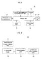
- Fig 1 is a diagram illustrating an arrangement of an ultrasound system 100 according to one embodiment of the present invention.
- the ultrasound system 100 may include an electrocardiogram (ECG) signal providing unit 110, a control unit 120, an ultrasound data acquiring unit 130, a storage unit 140, an input unit 150, a processing unit 160 and a display unit 170.
- ECG electrocardiogram
- the ECG signal providing unit 110 may acquire minute electric change resulting from contraction and relaxation of a moving interest object in a target body (e.g., heart, heart muscle, etc.). It may then convert the minute electric change into ECG signals to provide the formed ECG signals.
- a target body e.g., heart, heart muscle, etc.
- the control unit 120 may control the synchronization of ultrasound signals by using the ECG signals provided from the ECG signal providing unit 110.
- the control unit 120 may synchronize ultrasound signals with a certain period of the ECG signals (e.g., contraction period (R peak)).
- the control unit 120 may control transmitting and receiving the synchronized ultrasound signals with the ECG signals.
- the control unit 120 may control acquiring and storing ultrasound data.
- the control unit 120 may control storing the acquiring time of the ultrasound data along with the ultrasound data. Further, the control unit 120 may control forming and displaying comparison information. The comparison information will be described below.
- the ultrasound data acquiring unit 130 may transmit to the target body the ultrasound signals synchronized with the ECG signals under the control of the control unit 120 and receive from the target body the reflected ultrasound signals (i.e., ultrasound echo signals) to acquire ultrasound data.
- Fig. 2 is a block diagram illustrating an arrangement of the ultrasound data acquiring unit 130 according to one embodiment of the present invention.
- the ultrasound data acquiring unit 130 may include a transmission signal forming unit 131, an ultrasound probe 132 having at least one transducer element, a beam forming unit 133 and an ultrasound data forming unit 134.
- the transmission signal forming unit 131 may form transmission signals to be applied to the transducer elements of the ultrasound probe 132 in consideration of the focusing points and the position of the transducer elements.
- the transmission signals may include first transmission signals for acquiring ultrasound data for an object of interest in a target body when the object of interest has not been given medical treatment (hereinafter "first ultrasound data") and second transmission signals for acquiring ultrasound data for the object of interest when the object of interest has been given medical treatment (hereinafter "a second ultrasound data").
- the ultrasound probe 132 may transduce the first transmission signals into ultrasound signals when provided with the first transmission signals from the transmission signal forming unit 131.
- the ultrasound probe 132 may transmit to the target body the ultrasound signals synchronized with ECG signals under the control of the control unit 120 and receive from the target body the reflected ultrasound signals. This is to form first reception signals.
- the ultrasound probe 132 may transduce the second transmission signals into ultrasound signals when provided with the second transmission signals from transmission signal forming unit 131.
- the ultrasound probe 132 may transmit to the target body the ultrasound signals synchronized with ECG signals under the control of the control unit 120 and receive from the target body the reflected ultrasound signals. This is to form second reception signals.
- the beam forming unit 133 may digitalize the first reception signals to form first digital signals when provided with the first reception signals from the ultrasound probe 132.
- the beam forming unit 133 may focus the first digital signals to form first focusing reception signals in consideration of the focusing points and the position of the transducer elements of the ultrasound probe 132.
- the beam forming unit 133 may digitalize the second reception signals to form second digital signals when provided with the second reception signals from the ultrasound probe 132.
- the beam forming unit 133 may focus the second digital signals to form second focusing reception signals in consideration of the focusing points and the position of the transducer elements of the ultrasound probe 132.
- the ultrasound data forming unit 134 may use the first focusing reception signals to form first ultrasound data when provided with the first focusing reception signals from the beam forming unit 133.
- the ultrasound data forming unit 134 may use the second focusing reception signals to form second ultrasound data when provided with the second focusing reception signals from the beam forming unit 133. Further, the ultrasound data forming unit 134 may perform a variety of signal processes necessary to form ultrasound data (e.g., gain control, filtering control, etc.) from the first and second focusing reception signals.
- the storage unit 140 may store the first and second ultrasound data provided from the ultrasound data acquiring unit 130.
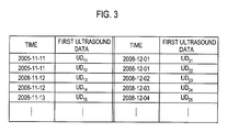
- the storage unit 140 may store the first ultrasound data and the second ultrasound data by order of the acquiring time of the ultrasound data under the control of the control unit 120, as illustrated in Fig. 3 .
- the input unit 150 may be a control panel, a mouse, a keyboard and the like, which may receive instructions from a user.
- the instructions may include the instructions for selecting at least one of a plurality of the first ultrasound data stored in the storage unit 140 and at least one of a plurality of the second ultrasound data stored in the storage unit 140.
- the processing unit 160 may look up the storage unit 140 and extract from the storage unit 140 the first ultrasound data and the second ultrasound data when provided with the instructions from the input unit 150.
- the processing unit 160 may compare the extracted first ultrasound data and the extracted second ultrasound data to form comparison information. For example, when provided with the instructions for selecting first ultrasound data UD 14 and second ultrasound data UD23 from the input unit 150, the processing unit 160 may look up the storage unit 140 and extract the first ultrasound data UD14 and the second ultrasound data UD23. Then, the processing unit 160 may compare the extracted first ultrasound data UD14 and the extracted second ultrasound data UD23 to form comparison information. For the sake of the brevity, detailed explanations on the conventional techniques of forming comparison information between ultrasound data are not provided herein. Further, the processing unit 160 may form a list of the first and second ultrasound data, which are stored in the storage unit 140.
- the display unit 170 may display the comparison information provided from the processing unit 160. Further, the display unit 170 may display a list of the first and second ultrasound data, which is provided from the processing unit 160.
- the present invention it is easier and more convenient to identify how much an object of interest is improved through a medical treatment by comparing ultrasound data associated with the object of interest before and after the medical treatment and providing comparison information.
Landscapes
- Life Sciences & Earth Sciences (AREA)
- Health & Medical Sciences (AREA)
- Biomedical Technology (AREA)
- Biophysics (AREA)
- Nuclear Medicine, Radiotherapy & Molecular Imaging (AREA)
- Pathology (AREA)
- Radiology & Medical Imaging (AREA)
- Engineering & Computer Science (AREA)
- Physics & Mathematics (AREA)
- Heart & Thoracic Surgery (AREA)
- Medical Informatics (AREA)
- Molecular Biology (AREA)
- Surgery (AREA)
- Animal Behavior & Ethology (AREA)
- General Health & Medical Sciences (AREA)
- Public Health (AREA)
- Veterinary Medicine (AREA)
- Ultra Sonic Daignosis Equipment (AREA)
Abstract
A system and a method of comparing ultrasound data associated with an object of interest before and after the medical treatment and providing the comparison information are disclosed. A plurality of first ultrasound data associated with an object of interest are stored before the medical treatment on the object of interest in the target body and a plurality of second ultrasound data associated with an object of interest are stored after the medical treatment on the object of interest in the target body in a storage unit. Instructions are input from a user for selecting any pair of first ultrasound data and second ultrasound data among a plurality of the first ultrasound data and a plurality of the second ultrasound data, which are stored in the storage unit. From the storage unit, the first ultrasound data and the second ultrasound data are extracted, and the extracted first ultrasound data and the extracted second ultrasound data are compared to form comparison information.
Description
- The present application claims priority from Korean Patent Application No.
10-2009-0048200 filed on June 1, 2009 - The present invention generally relates to an ultrasound system for comparing ultrasound data for a patient before and after a medical treatment. More particularly, the present invention relates to an ultrasound system capable of comparing ultrasound data for a patient, which are acquired at different statuses of the patient.
- An ultrasound system has been extensively used for acquiring information inside a target body due to its non-invasive and non-destructive nature. The ultrasound system has become an important tool in the medical field since it can provide doctors with high-resolution images of internal features of humans in real time without any surgical medical treatment.
- Generally, the ultrasound system may acquire ultrasound data associated with an object of interest by transmitting ultrasound signals to the target body and receiving ultrasound echo signals reflected from the target body. The ultrasound system may provide an ultrasound image and/or a variety of information by using the acquired ultrasound data. For example, if the object of interest is a heart (or heart muscle), then the ultrasound system may provide dyssynchronous segment information for the analysis of strain, velocity, segment volume and the like by using the acquired ultrasound data.
- Conventionally, ultrasound data associated with an object of interest obtained without medical treatment or ultrasound data associated with the object of interest obtained with medical treatment are used for providing dyssynchronous segment information. Thus, it is difficult to compare the degree of medical improvement before and after the medical treatment.
- There are provided an ultrasound system and a method of comparing ultrasound data for a patient before and after medical treatment.
- According to one aspect of the present invention, an ultrasound system comprises: an ultrasound data acquisition unit configured to acquire a plurality of first ultrasound data for an object of interest in a target body when the object of interest is in a first state and acquire a plurality of second ultrasound data for the object of interest when the object of interest is in a second state; a storage unit for storing the plurality of first ultrasound data and the plurality of second ultrasound data; an input unit configured to receive a user instruction for selecting at least one of the plurality of first ultrasound data and at least one of the plurality of second ultrasound data; and a processing unit configured to extract the at least one of the plurality of first ultrasound data and the at least one of the plurality of second ultrasound data from the storage unit and compare the extracted first ultrasound data with the extracted second ultrasound data to form comparison information.
- According to one aspect of the present invention, there is disclosed a method of providing comparison information, which comprises: a) acquiring a plurality of first ultrasound data for an object of interest in a target body when the object of interest is in a first state and a plurality of second ultrasound data for the object of interest in the target body when the object of interest is in a second state; b) storing in a storage unit the plurality of first ultrasound data and the plurality of second ultrasound data; c) receiving a user instruction for selecting at least one of the plurality of first ultrasound data and at least one of the plurality of second ultrasound data; d) extracting the at least one of the plurality of first ultrasound data and the at least one of the plurality of second ultrasound data from the storage unit; and e) comparing the extracted first ultrasound data with the extracted second ultrasound data to form comparison information.
- The summary is provided to introduce a selection of concepts in a simplified form that are further described below in the Detailed Description. This Summary is not intended to identify key or essential features of the claimed subject matter, nor is it intended to be used in determining the scope of the claimed subject matter.
-
- Fig. 1
- is a block diagram illustrating an arrangement of an ultrasound system according to one embodiment of the present invention.
- Fig. 2
- is a block diagram illustrating an arrangement of an ultrasound data acquiring unit according to one embodiment of the present invention.
- Fig. 3
- illustrates exemplary ultrasound data registered according to one embodiment of the present invention.
- A detailed description may be provided with reference to the accompanying drawings. One of ordinary skill in the art may realize that the following description is illustrative only and is not in any way limiting. Other embodiments of the present invention may readily suggest themselves to such skilled persons having the benefit of this disclosure.
-
Fig 1 is a diagram illustrating an arrangement of anultrasound system 100 according to one embodiment of the present invention. Theultrasound system 100 may include an electrocardiogram (ECG)signal providing unit 110, acontrol unit 120, an ultrasounddata acquiring unit 130, astorage unit 140, aninput unit 150, aprocessing unit 160 and adisplay unit 170. - The ECG
signal providing unit 110 may acquire minute electric change resulting from contraction and relaxation of a moving interest object in a target body (e.g., heart, heart muscle, etc.). It may then convert the minute electric change into ECG signals to provide the formed ECG signals. - The
control unit 120 may control the synchronization of ultrasound signals by using the ECG signals provided from the ECGsignal providing unit 110. In the present embodiment, thecontrol unit 120 may synchronize ultrasound signals with a certain period of the ECG signals (e.g., contraction period (R peak)). Thecontrol unit 120 may control transmitting and receiving the synchronized ultrasound signals with the ECG signals. Thecontrol unit 120 may control acquiring and storing ultrasound data. Thecontrol unit 120 may control storing the acquiring time of the ultrasound data along with the ultrasound data. Further, thecontrol unit 120 may control forming and displaying comparison information. The comparison information will be described below. - The ultrasound
data acquiring unit 130 may transmit to the target body the ultrasound signals synchronized with the ECG signals under the control of thecontrol unit 120 and receive from the target body the reflected ultrasound signals (i.e., ultrasound echo signals) to acquire ultrasound data. -
Fig. 2 is a block diagram illustrating an arrangement of the ultrasounddata acquiring unit 130 according to one embodiment of the present invention. The ultrasounddata acquiring unit 130 may include a transmissionsignal forming unit 131, anultrasound probe 132 having at least one transducer element, abeam forming unit 133 and an ultrasounddata forming unit 134. - The transmission
signal forming unit 131 may form transmission signals to be applied to the transducer elements of theultrasound probe 132 in consideration of the focusing points and the position of the transducer elements. In the present embodiment, the transmission signals may include first transmission signals for acquiring ultrasound data for an object of interest in a target body when the object of interest has not been given medical treatment (hereinafter "first ultrasound data") and second transmission signals for acquiring ultrasound data for the object of interest when the object of interest has been given medical treatment (hereinafter "a second ultrasound data"). - The
ultrasound probe 132 may transduce the first transmission signals into ultrasound signals when provided with the first transmission signals from the transmissionsignal forming unit 131. Theultrasound probe 132 may transmit to the target body the ultrasound signals synchronized with ECG signals under the control of thecontrol unit 120 and receive from the target body the reflected ultrasound signals. This is to form first reception signals. Theultrasound probe 132 may transduce the second transmission signals into ultrasound signals when provided with the second transmission signals from transmissionsignal forming unit 131. Theultrasound probe 132 may transmit to the target body the ultrasound signals synchronized with ECG signals under the control of thecontrol unit 120 and receive from the target body the reflected ultrasound signals. This is to form second reception signals. - The
beam forming unit 133 may digitalize the first reception signals to form first digital signals when provided with the first reception signals from theultrasound probe 132. Thebeam forming unit 133 may focus the first digital signals to form first focusing reception signals in consideration of the focusing points and the position of the transducer elements of theultrasound probe 132. Further, thebeam forming unit 133 may digitalize the second reception signals to form second digital signals when provided with the second reception signals from theultrasound probe 132. Thebeam forming unit 133 may focus the second digital signals to form second focusing reception signals in consideration of the focusing points and the position of the transducer elements of theultrasound probe 132. - The ultrasound
data forming unit 134 may use the first focusing reception signals to form first ultrasound data when provided with the first focusing reception signals from thebeam forming unit 133. The ultrasounddata forming unit 134 may use the second focusing reception signals to form second ultrasound data when provided with the second focusing reception signals from thebeam forming unit 133. Further, the ultrasounddata forming unit 134 may perform a variety of signal processes necessary to form ultrasound data (e.g., gain control, filtering control, etc.) from the first and second focusing reception signals. - Referring back to
Fig. 1 , thestorage unit 140 may store the first and second ultrasound data provided from the ultrasounddata acquiring unit 130. For example, thestorage unit 140 may store the first ultrasound data and the second ultrasound data by order of the acquiring time of the ultrasound data under the control of thecontrol unit 120, as illustrated inFig. 3 . - The
input unit 150 may be a control panel, a mouse, a keyboard and the like, which may receive instructions from a user. In the present embodiment, the instructions may include the instructions for selecting at least one of a plurality of the first ultrasound data stored in thestorage unit 140 and at least one of a plurality of the second ultrasound data stored in thestorage unit 140. - The
processing unit 160 may look up thestorage unit 140 and extract from thestorage unit 140 the first ultrasound data and the second ultrasound data when provided with the instructions from theinput unit 150. Theprocessing unit 160 may compare the extracted first ultrasound data and the extracted second ultrasound data to form comparison information. For example, when provided with the instructions for selecting first ultrasound data UD 14 and second ultrasound data UD23 from theinput unit 150, theprocessing unit 160 may look up thestorage unit 140 and extract the first ultrasound data UD14 and the second ultrasound data UD23. Then, theprocessing unit 160 may compare the extracted first ultrasound data UD14 and the extracted second ultrasound data UD23 to form comparison information. For the sake of the brevity, detailed explanations on the conventional techniques of forming comparison information between ultrasound data are not provided herein. Further, theprocessing unit 160 may form a list of the first and second ultrasound data, which are stored in thestorage unit 140. - The
display unit 170 may display the comparison information provided from theprocessing unit 160. Further, thedisplay unit 170 may display a list of the first and second ultrasound data, which is provided from theprocessing unit 160. - According to the present invention, it is easier and more convenient to identify how much an object of interest is improved through a medical treatment by comparing ultrasound data associated with the object of interest before and after the medical treatment and providing comparison information.
- Although embodiments have been described with reference to a number of illustrative embodiments thereof, it should be understood that numerous other modifications and embodiments can be devised by those skilled in the art that will fall within the spirit and scope of the principles of this disclosure. More particularly, numerous variations and modifications are possible in the component parts and/or arrangements of the subject combination arrangement within the scope of the disclosure, the drawings and the appended claims. In addition to variations and modifications in the component parts and/or arrangements, alternative uses will also be apparent to those skilled in the art. The invention is not to be restricted except in the spirit of the appended claims.
Claims (11)
- An ultrasound system, comprising:an ultrasound data acquisition unit configured to acquire a plurality of first ultrasound data for an object of interest in a target body when the object of interest is in a first state and acquire a plurality of second ultrasound data for the object of interest when the object of interest is in a second state;a storage unit for storing the plurality of first ultrasound data and the plurality of second ultrasound data;an input unit configured to receive a user instruction for selecting at least one of the plurality of first ultrasound data and at least one of the plurality of second ultrasound data; anda processing unit configured to extract the at least one of the plurality of first ultrasound data and the at least one of the plurality of second ultrasound data from the storage unit and compare the extracted first ultrasound data with the extracted second ultrasound data to form comparison information.
- The system of Claim 1, wherein the object of interest has not been given medical treatment in the first state and the object of interest has been given medical treatment in the second state.
- The system of Claim 1, wherein the ultrasound data acquisition unit is configured to transmit an ultrasound signal to the target body and receive the ultrasound signal reflected from the target body to acquire the first and second ultrasound data.
- The system of Claim 3, further comprising:an ECG signal providing unit configured to provide an electrocardiogram (ECG) signal of the object of interest; anda control unit configured to synchronize the ultrasound signal with the ECG signal.
- The system of Claim 1, further comprising:a display unit configured to display the comparison information.
- An ultrasound system, comprising:an ultrasound data acquisition unit configured to acquire first ultrasound data for an object of interest in a target body without medical treatment given andacquire second ultrasound data for the object of interest with medical treatment given; anda processing unit configured to compare the first ultrasound data with the second ultrasound data to form comparison information.
- A method of providing comparison information, comprising:a) acquiring a plurality of first ultrasound data for an object of interest in a target body when the object of interest is in a first state and a plurality of second ultrasound data for the object of interest in the target body when the object of interest is in a second state;b) storing in a storage unit the plurality of first ultrasound data and the plurality of second ultrasound data;c) receiving a user instruction for selecting at least one of the plurality of first ultrasound data and at least one of the plurality of second ultrasound data;d) extracting the at least one of the plurality of first ultrasound data and the at least one of the plurality of second ultrasound data from the storage unit; ande) comparing the extracted first ultrasound data with the extracted second ultrasound data to form comparison information.
- The method of Claim 7, wherein the object of interest has not been given medical treatment in the first state and the object of interest has been given medical treatment in the second state.
- The method of Claim 7, wherein a) comprises:transmitting an ultrasound signal to the target body and receiving the ultrasound signal reflected from the target body to acquire the first and second ultrasound data.
- The method of Claim 9, further comprising:providing an electrocardiogram (ECG) signal of the object of interest; andsynchronizing the ultrasound signal with the ECG signal.
- The method of Claim 7, further comprising:displaying the comparison information.
Applications Claiming Priority (1)
| Application Number | Priority Date | Filing Date | Title |
|---|---|---|---|
| KR1020090048200A KR101117868B1 (en) | 2009-06-01 | 2009-06-01 | Ultrasound system and method for providing comparision information of interest object |
Publications (1)
| Publication Number | Publication Date |
|---|---|
| EP2258271A1 true EP2258271A1 (en) | 2010-12-08 |
Family
ID=41697770
Family Applications (1)
| Application Number | Title | Priority Date | Filing Date |
|---|---|---|---|
| EP10151371A Withdrawn EP2258271A1 (en) | 2009-06-01 | 2010-01-22 | Comparison of ultrasound data before and after medical treatment |
Country Status (4)
| Country | Link |
|---|---|
| US (1) | US20100174193A1 (en) |
| EP (1) | EP2258271A1 (en) |
| JP (1) | JP2010274104A (en) |
| KR (1) | KR101117868B1 (en) |
Families Citing this family (2)
| Publication number | Priority date | Publication date | Assignee | Title |
|---|---|---|---|---|
| KR101323332B1 (en) * | 2012-01-06 | 2013-10-29 | 삼성메디슨 주식회사 | Apparatus and method for realizing synchronization image |
| KR20150069920A (en) * | 2013-12-16 | 2015-06-24 | 삼성메디슨 주식회사 | Ultrasonic diagnostic apparatus and operating method for the same |
Citations (4)
| Publication number | Priority date | Publication date | Assignee | Title |
|---|---|---|---|---|
| US20050101863A1 (en) | 2003-09-05 | 2005-05-12 | Kabushiki Kaisha Toshiba | Ultrasonic diagnostic equipment and imaging processing apparatus |
| US20060064014A1 (en) * | 2004-09-20 | 2006-03-23 | Tony Falco | Radiotherapy treatment monitoring using ultrasound |
| JP2007014541A (en) * | 2005-07-07 | 2007-01-25 | Toshiba Corp | Ultrasonic diagnostic equipment |
| KR20090048200A (en) | 2007-11-09 | 2009-05-13 | (주) 금영테크 | Electric resistance welding method of aluminum rod |
-
2009
- 2009-06-01 KR KR1020090048200A patent/KR101117868B1/en active Active
-
2010
- 2010-01-22 EP EP10151371A patent/EP2258271A1/en not_active Withdrawn
- 2010-01-26 US US12/694,207 patent/US20100174193A1/en not_active Abandoned
- 2010-01-28 JP JP2010017304A patent/JP2010274104A/en active Pending
Patent Citations (4)
| Publication number | Priority date | Publication date | Assignee | Title |
|---|---|---|---|---|
| US20050101863A1 (en) | 2003-09-05 | 2005-05-12 | Kabushiki Kaisha Toshiba | Ultrasonic diagnostic equipment and imaging processing apparatus |
| US20060064014A1 (en) * | 2004-09-20 | 2006-03-23 | Tony Falco | Radiotherapy treatment monitoring using ultrasound |
| JP2007014541A (en) * | 2005-07-07 | 2007-01-25 | Toshiba Corp | Ultrasonic diagnostic equipment |
| KR20090048200A (en) | 2007-11-09 | 2009-05-13 | (주) 금영테크 | Electric resistance welding method of aluminum rod |
Also Published As
| Publication number | Publication date |
|---|---|
| KR20100129569A (en) | 2010-12-09 |
| JP2010274104A (en) | 2010-12-09 |
| US20100174193A1 (en) | 2010-07-08 |
| KR101117868B1 (en) | 2012-03-07 |
Similar Documents
| Publication | Publication Date | Title |
|---|---|---|
| EP3092951B1 (en) | Method and apparatus for synthesizing medical images | |
| EP2823766A1 (en) | Ultrasound system and method for providing object information | |
| EP2989987B1 (en) | Ultrasound diagnosis apparatus and method and computer readable storage medium | |
| US20160004330A1 (en) | Handheld medical imaging apparatus with cursor pointer control | |
| US10163228B2 (en) | Medical imaging apparatus and method of operating same | |
| KR20150069920A (en) | Ultrasonic diagnostic apparatus and operating method for the same | |
| JP2024048667A (en) | ULTRASONIC DIAGNOSTIC APPARATUS AND METHOD FOR CONTROLLING ULTRASONIC DIAGNOSTIC APPARATUS | |
| JP2010269018A (en) | Ultrasonic diagnostic equipment | |
| EP1625829B1 (en) | Ultrasonogram generation method | |
| US20120108962A1 (en) | Providing a body mark in an ultrasound system | |
| US20150206323A1 (en) | Method and apparatus for displaying medical image | |
| KR20150004490A (en) | Ultrasonic diagnostic apparatus and operating method for the same | |
| US20100174193A1 (en) | Comparison Of Ultrasound Data Before And After Medical Treatment | |
| KR20170054982A (en) | Ultrasound image apparatus and method for operating the same | |
| US20120108964A1 (en) | Performing gain control process based on application in ultrasound system | |
| EP3037041B1 (en) | Method and apparatus for generating body marker | |
| KR101415021B1 (en) | Ultrasound system and method for providing panoramic image | |
| JPH09140711A (en) | Automatic time phase discriminating method and ultrasonic diagnostic device | |
| EP2107389A2 (en) | Formation of an elastic image in an ultrasound system | |
| EP2336804B1 (en) | Ultrasound system and method for providing doppler sound | |
| US8727985B2 (en) | Medical system for providing external trigger signal | |
| JP2020048968A (en) | Ultrasonic diagnostic apparatus and control method for ultrasonic diagnostic apparatus | |
| JP2007167351A (en) | Ultrasonographic instrument | |
| KR101060386B1 (en) | Ultrasound system and method for forming elastic images |
Legal Events
| Date | Code | Title | Description |
|---|---|---|---|
| PUAI | Public reference made under article 153(3) epc to a published international application that has entered the european phase |
Free format text: ORIGINAL CODE: 0009012 |
|
| AK | Designated contracting states |
Kind code of ref document: A1 Designated state(s): AT BE BG CH CY CZ DE DK EE ES FI FR GB GR HR HU IE IS IT LI LT LU LV MC MK MT NL NO PL PT RO SE SI SK SM TR |
|
| AX | Request for extension of the european patent |
Extension state: AL BA RS |
|
| 17P | Request for examination filed |
Effective date: 20110321 |
|
| 17Q | First examination report despatched |
Effective date: 20110413 |
|
| STAA | Information on the status of an ep patent application or granted ep patent |
Free format text: STATUS: THE APPLICATION IS DEEMED TO BE WITHDRAWN |
|
| 18D | Application deemed to be withdrawn |
Effective date: 20130821 |