EP2247752B1 - Vorhersage und diagnose von degenerativer myelopathie bei hunden - Google Patents
Vorhersage und diagnose von degenerativer myelopathie bei hunden Download PDFInfo
- Publication number
- EP2247752B1 EP2247752B1 EP09708938.7A EP09708938A EP2247752B1 EP 2247752 B1 EP2247752 B1 EP 2247752B1 EP 09708938 A EP09708938 A EP 09708938A EP 2247752 B1 EP2247752 B1 EP 2247752B1
- Authority
- EP
- European Patent Office
- Prior art keywords
- sod1
- nucleic acid
- dog
- dna
- antibody
- Prior art date
- Legal status (The legal status is an assumption and is not a legal conclusion. Google has not performed a legal analysis and makes no representation as to the accuracy of the status listed.)
- Active
Links
Images




Classifications
-
- C—CHEMISTRY; METALLURGY
- C12—BIOCHEMISTRY; BEER; SPIRITS; WINE; VINEGAR; MICROBIOLOGY; ENZYMOLOGY; MUTATION OR GENETIC ENGINEERING
- C12Q—MEASURING OR TESTING PROCESSES INVOLVING ENZYMES, NUCLEIC ACIDS OR MICROORGANISMS; COMPOSITIONS OR TEST PAPERS THEREFOR; PROCESSES OF PREPARING SUCH COMPOSITIONS; CONDITION-RESPONSIVE CONTROL IN MICROBIOLOGICAL OR ENZYMOLOGICAL PROCESSES
- C12Q1/00—Measuring or testing processes involving enzymes, nucleic acids or microorganisms; Compositions therefor; Processes of preparing such compositions
- C12Q1/68—Measuring or testing processes involving enzymes, nucleic acids or microorganisms; Compositions therefor; Processes of preparing such compositions involving nucleic acids
- C12Q1/6876—Nucleic acid products used in the analysis of nucleic acids, e.g. primers or probes
- C12Q1/6883—Nucleic acid products used in the analysis of nucleic acids, e.g. primers or probes for diseases caused by alterations of genetic material
-
- C—CHEMISTRY; METALLURGY
- C12—BIOCHEMISTRY; BEER; SPIRITS; WINE; VINEGAR; MICROBIOLOGY; ENZYMOLOGY; MUTATION OR GENETIC ENGINEERING
- C12Q—MEASURING OR TESTING PROCESSES INVOLVING ENZYMES, NUCLEIC ACIDS OR MICROORGANISMS; COMPOSITIONS OR TEST PAPERS THEREFOR; PROCESSES OF PREPARING SUCH COMPOSITIONS; CONDITION-RESPONSIVE CONTROL IN MICROBIOLOGICAL OR ENZYMOLOGICAL PROCESSES
- C12Q2600/00—Oligonucleotides characterized by their use
- C12Q2600/106—Pharmacogenomics, i.e. genetic variability in individual responses to drugs and drug metabolism
-
- C—CHEMISTRY; METALLURGY
- C12—BIOCHEMISTRY; BEER; SPIRITS; WINE; VINEGAR; MICROBIOLOGY; ENZYMOLOGY; MUTATION OR GENETIC ENGINEERING
- C12Q—MEASURING OR TESTING PROCESSES INVOLVING ENZYMES, NUCLEIC ACIDS OR MICROORGANISMS; COMPOSITIONS OR TEST PAPERS THEREFOR; PROCESSES OF PREPARING SUCH COMPOSITIONS; CONDITION-RESPONSIVE CONTROL IN MICROBIOLOGICAL OR ENZYMOLOGICAL PROCESSES
- C12Q2600/00—Oligonucleotides characterized by their use
- C12Q2600/158—Expression markers
-
- C—CHEMISTRY; METALLURGY
- C12—BIOCHEMISTRY; BEER; SPIRITS; WINE; VINEGAR; MICROBIOLOGY; ENZYMOLOGY; MUTATION OR GENETIC ENGINEERING
- C12Q—MEASURING OR TESTING PROCESSES INVOLVING ENZYMES, NUCLEIC ACIDS OR MICROORGANISMS; COMPOSITIONS OR TEST PAPERS THEREFOR; PROCESSES OF PREPARING SUCH COMPOSITIONS; CONDITION-RESPONSIVE CONTROL IN MICROBIOLOGICAL OR ENZYMOLOGICAL PROCESSES
- C12Q2600/00—Oligonucleotides characterized by their use
- C12Q2600/172—Haplotypes
Definitions
- the present invention relates generally to the fields of molecular genetics, veterinary medicine, veterinary neurology, human neurology and human medicine. More particularly, it concerns the veterinary disease canine degenerative myelopathy and the human disease amyotrophic lateral sclerosis. Specifically, the invention relates to the use of genetic markers to identify the presence of a major genetic risk factor for canine degenerative myelopathy which is a fatal veterinary disease and a model for amyotrophic lateral sclerosis (ALS).
- ALS amyotrophic lateral sclerosis
- Canine degenerative myelopathy (CDM; also known as chronic degenerative radiculomyelopathy) is a neurological disease common in German Shepherds, Welsh Corgis, and several other breeds. The disease is chronic and progressive, and can result in lameness in the animal and eventually may lead to extensive paralysis of the back legs. The animal could be crippled within a few months, or may survive up to 3 years. Under any scenario, the disease is debilating for the animal, severely hampers the quality of life, culminates in euthanasia, and is devastating for pet owners and breeders alike.
- CDM is a chronic condition that cannot be cured, though it may be possible to maintain the dog's quality of life for a short time through a proper program of exercise and nutrition. Exercise has been recommended to maintain the dog's ability to walk, and physiotherapy may prolong the length of time that the dog remains mobile and increase survival time, but the ultimate outcome is not favorable. There are still no proven effective therapies to halt or slow progression of CDM. Although not approved by the U.S. Food and Drug Administration (FDA), aminocaproic acid (EACA) and n-acetylcysteine (NAC) may slow the progression, but this treatment is still experimental and controversial.
- FDA U.S. Food and Drug Administration
- EACA aminocaproic acid
- NAC n-acetylcysteine
- a method of identifying a dog as having or at risk of developing degenerative myelopathy , or producing offspring having or at risk of developing degenerative myelopathy comprising (a) providing a nucleic acid-containing sample from said dog; (b) assessing the structure of an SOD1 gene or transcript corresponding to amino acid residue 40 of the corresponding SOD1 polypeptide in said nucleic acid-containing sample from said dog; and (c) identifying said dog as (i) having or at risk of developing degenerative myelopathy when a polymorphism in the SOD1 coding region corresponds to a homozygous E ⁇ K substitution at amino acid residue 40 of the corresponding SOD1 polypeptide, as compared to a reference wild-type SOD1 gene or transcript is observed, or (ii) producing offspring having or at risk of developing degenerative myelopathy when a polymorphism in the SOD1 coding region corresponds to a heterozygous E ⁇ K substitution
- the dog may be a boxer, a Welsh corgi, a Chesapeake Bay retriever, a Rhodesian Ridgeback, a German shepherd, a Kerry blue terrier, a Irish setter, a old English sheepdog, a collie, a Standard poodle, a wire Fox terrier, another breed, or a cross-breed thereof.
- Step (b) may comprise pyrosequencing, competitive probe hybridization, chain-terminating sequencing, restriction digestion, allele-specific polymerase reaction, single-stranded conformational polymorphism analysis, genetic bit analysis, TaqMan® allelic discrimination assay, temperature gradient gel electrophoresis, ligase chain reaction, melting curve profiles, or microarray hybridization, microarray analysis in combination other nucleic acid marker assays, or may comprise assessing the structure of a genetic locus that is in linkage disequilibrium with said genetic marker.
- the reference wild-type SOD1 exon 2 sequence may comprise the following sequence:
- the present application also describes a method of assessing a treatment for canine degenerative myelopathy or amyotrophic lateral sclerosis comprising assessing a candidate therapy for efficacy against said degenerative myelopathy or amyotrophic lateral sclerosis a dog having a mutation in an SOD1 gene as compared to a reference wild-type SOD1 gene or transcript, wherein the dog has been administered the candidate therapy.
- the dog may be a boxer, a Welsh corgi , a Chesapeake Bay retriever, a Rhodesian Ridgeback, a German shepherd, a Kerry blue terrier, a Irish setter, a old English sheepdog, a Collie, a standard poodle, a wire Fox terrier, or another breed, or a cross-breed thereof.
- the mutation may be a polymorphism in the SOD1 coding region corresponding to an E ⁇ K substitution at residue 40 of the SOD1 polypeptide.
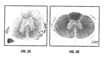
- Assessing may comprise neurologic examination, electrodiagnostic testing, CT/myelography, MRI, or functional imaging, or assessing may comprise spinal chord immunohistopathology.
- Canine degenerative myelopathy is a crippling and fatal neurological disease common in a variety of canine breeds.
- the disease is chronic and progressive, and the only "treatments" - physical therapy and nutrition - have limited ability to slow the onset of the disease.
- a dog's back legs become useless, at which point euthanasia may be the only option.
- the etiology of this disease is unknown, although it is considered that the basis may be genetic.
- a dog's genotype with respect to a genetic risk factor can help confirm or refute a diagnosis of CDM in a dog showing clinical signs of the disease.
- the inventors have mapped the risk locus for CDM to region of CFA31 in Pembroke Welsh corgis. Within this chromosomal region, they discovered a missense mutation (E40K) in the canine superoxide dismutase 1 (SOD1) gene, and observed that the K allele is common in several other breeds. Finally, the inventors have demonstrated that homozygosity for the K allele is a major genetic risk factor for CDM. It is of great importance to breeders and owners to be able to identify "at risk" animals, as well as animals that could pass the disease on to future generations. Genetic markers for the high-risk allele will facilitate CDM diagnosis. These markers will also enable breeders to select matings that will produce puppies with greatly reduced risk of developing CDM.
- ALS amyotrophic lateral sclerosis
- DMA markers can help identify dogs that are homozygous for the A allele and can thus be used as an animal model to investigate the underlying disease menchanism or to evaluate potential treatments for ALS or CDM.
- CDM Canine Degenerative Myelopathy
- Canine degenerative myelopathy also known as chronic degenerative radiculomyelopathy
- chronic degenerative radiculomyelopathy is a neurological disease prevalent in German shepherds, boxers, Rhodesian Ridgeback, Chesapeake Bay retriever, Pemroke and Cardigan Welsh Corgis, and other pure- and cross-bred dogs.
- the disease is chronic and progressive, and can result in lameness in the animal, and eventually death.
- the disease usually manifests between the ages of seven and fourteen and initially affects the back legs and causes muscle weakness and loss, and lack of coordination. These cause a staggering effect that may appear to be arthritis.
- the dog may scuff its one or both rear paws when it walks. This scuffing can cause the nails of one foot to be worn down. Eventually the condition may lead to extensive paralysis of the back legs. As the disease progresses the animal starts having considerable difficulties walking, and eventually the back legs become useless, at which point euthanasia may be the only option. If euthanasia is delayed, the clinical signs will progress to affect the front legs. Progression of the disease is generally slow.. The animal could be crippled within a few months, or members of smaller breeds may survive up to 3 years as owners may delay euthanasia because they can easily carry smaller dogs.
- cervical disc disease protrusion or rupture
- Canine degenerative myelopathy can only be diagnosed by histopathology of the spinal cord. Lesions demonstrate axonal and myelin loss replaced by gliosis in all funciuli of the spinal cord most severe in the dorsal portion of the lateral funiculus of the mid to caudal thoracic spinal cord.
- the enzyme superoxide dismutase catalyzes the dismutation of superoxide into oxygen and hydrogen peroxide. As such, it is an important antioxidant defense in nearly all cells exposed to oxygen.
- the SOD-catalysed dismutation of superoxide may be written with the following half-reactions: M (n+1)+ - SOD + O 2 - ⁇ M n+ - SOD + O 2 M n+ - SOD + O 2 + 2H + ⁇ M (+1)+ - SOD + H 2 O 2 .
- the oxidation state of the metal cation oscillates between n and n+1.
- SODs were prior were known as several metalloproteins with unknown function (for example, CuZnSOD was known as erythrocuprein).
- CuZnSOD was known as erythrocuprein.
- Mn-SOD manganese
- the ligands of the manganese ions are 3 histidine side chains, an aspartate side chain and a water molecule or hydroxy ligand depending on the Mn oxidation state (respectively II and III).
- E. coli and many other bacteria also contain a form of the enzyme with iron (Fe-SOD); some bacteria contain Fe-SOD, others Mn-SOD, and some contain both.
- Fe-SOD iron
- Some bacteria contain Fe-SOD, others Mn-SOD, and some contain both.
- the active sites of Mn and Fe superoxide dismutases contain the same type of amino acids side chains. In humans, three forms of superoxide dismutase are present.
- SOD1 is located in the cytoplasm, SOD2 in the mitochondria and SOD3 is extracellular.
- the first is a dimer (consists of two units), while the others are tetramers (four subunits).
- SOD1 and SOD3 contain copper and zinc, while SOD2 has manganese in its reactive centre.
- the genes are located on human chromosomes 21, 6 and 4, respectively (21q22.1, 6q25.3 and 4p15.3-p15.1). A microtiter plate assay for SOD is available.
- the cytosols of virtually all eukaryotic cells contain an SOD enzyme with copper and zinc (Cu-Zn-SOD).
- the Cu-Zn enzyme is a homodimer of molecular weight 32,500. The two subunits are joined primarily by hydrophobic and electrostatic interactions.
- the ligands of copper and zinc are histidine side chains.
- the half-life of superoxide although very short at high concentrations (e.g ., 0.05 seconds at 0.1 mM) is actually quite long at low concentrations ( e.g ., 14 hours at 0.1 nM).
- the reaction of superoxide with SOD is first order with respect to superoxide concentration.
- superoxide has the fastest turnover number (reaction rate with its substrate) of any known enzyme ( ⁇ 10 9 M -1 s -1 ), this reaction being only limited by the frequency of collision between itself and superoxide. That is, the reaction rate is "diffusion limited,"
- SOD1 familial amyotrophic lateral sclerosis
- ALS a form of motor neuron disease
- mice inactivation of SOD2 causes perinatal lethality and inactivation of SOD1 causes hepatocellular carcinoma.
- Mutations in SOD1 can cause familial ALS by a mechanism that is presently not understood, but not due to loss of enzymatic activity.
- Overexpression of SOD1 has been linked to Down's syndrome. Prior to this work, canine SOD1 has not been linked to any particular disease state.
- Wild-type canine cDNA coding sequence
- Variant canine cDNA coding sequence
- the presence of the E40K polymorphism can be determined from either the sequence of the SOD1 protein, the sequence of the SOD1 nucleic acid or a genetic marker that is in linkage disequilibrium with the E40K polymorphism. As a result, a variety of different methodologies can be employed.
- nucleic acids include the use of amplification primers, oligonucleotide probes, and other nucleic acid elements involved in the analysis of genomic DNA, cDNA or mRNA transcripts.
- the term "nucleic acid” is well known in the art.
- a “nucleic acid” as used herein will generally refer to a molecule ( i.e., a strand) of DNA or RNA comprised in part by a nucleotide bases.
- Nucleobases include, for example, naturally-occurring purine or pyrimidine bases found in DNA (e.g., an adenine "A,” a guanine “G,” a thymine “T” or a cytosine “C”) or RNA (e.g., an A, a G, an uracil “U” or a C).
- DNA e.g., an adenine "A,” a guanine “G,” a thymine “T” or a cytosine "C”
- RNA e.g., an A, a G, an uracil "U” or a C.
- nucleic acid encompass the terms “oligonucleotide” and “polynucleotide,” each as a subgenus of the term “nucleic acid.”
- oligonucleotide refers to a molecule of between about 3 and about 100 nucleobases in length.
- polynucleotide refers to at least one molecule of greater than about 100 nucleobases in length.
- a “gene” refers to coding sequence of a gene product, as well as introns and the promoter of the gene product.
- nucleic acids of the disclosure comprise or are complementary to all or 5, 6, 7, 8, 9, 10, 11, 12, 13, 14, 15, 16, 17, 18, 19, 20, 21, 22, 23, 24, 25, 26, 27, 28, 29, 30, 31, 32, 33, 34, 35, 36, 37, 38, 39, 40, 41, 42, 43, 44, 45, 46, 47, 48, 49, 50, 51, 52, 53, 54, 55, 56, 57, 58, 59, 60, 61, 62, 63, 64, 65, 66, 67, 68, 69, 70, 71, 72, 73, 74, 75, 76, 77, 78, 79, 80, 81, 82, 83, 84, 85, 86, 87, 88, 89, 90, 91, 92, 93, 94, 95, 96, 97, 98, 99, 100, 110, 120, 130, 140, 150, 160, 170, 180, 190, 200, 210, 220, 230, 240, 250, 260,
- a nucleic acid may encompass a double-stranded molecule or a triple-stranded molecule that comprises one or more complementary strand(s) or "complement(s)" of a particular sequence comprising a molecule.
- a single-stranded nucleic acid may be denoted by the prefix "ss,” a double-stranded nucleic acid by the prefix "ds,” and a triple-stranded nucleic acid by the prefix "ts.”
- a nucleic acid encodes a protein, polypeptide, or peptide.
- the present disclosure concerns novel compositions comprising at least one proteinaceous molecule.
- a "proteinaceous molecule,” “proteinaceous composition,” “proteinaceous compound,” “proteinaceous chain,” or “proteinaceous material” generally refers, but is not limited to, a protein of greater than about 200 amino acids or the full length endogenous sequence translated from a gene; a polypeptide of greater than about 100 amino acids; and/or a peptide of from about 3 to about 100 amino acids. All the "proteinaceous” terms described above may be used interchangeably herein.
- a nucleic acid may be made by any technique known to one of ordinary skill in the art, such as for example, chemical synthesis, enzymatic production or biological production.
- a synthetic nucleic acid e.g., a synthetic oligonucleotide
- Non-limiting examples of a synthetic nucleic acid include a nucleic acid made by in vitro chemical synthesis using phosphotriester, phosphite or phosphoramidite chemistry and solid phase techniques such as described in European Patent 266,032 , or via deoxynucleoside H-phosphonate intermediates as described by Froehler et al., 1986 and U.S. Patent 5,705,629 , each incorporated herein by reference.
- oligonucleotide may be used.
- Various different mechanisms of oligonucleotide synthesis have been disclosed in for example, U.S. Patents 4,659,774 , 4,816,571 , 5,141,813 , 5,264,566 , 4,959,463 , 5,428,148 , 5,554,744 , 5,574,146 , 5,602,244 .
- a non-limiting example of an enzymatically produced nucleic acid include one produced by enzymes in amplification reactions such as PCRTM (see for example, U.S. Patent 4,683,202 and U.S. Patent 4,682,195 or the synthesis of an oligonucleotide described in U.S. Patent 5,645,897 .
- a non-limiting example of a biologically produced nucleic acid includes a recombinant nucleic acid produced ( i.e ., replicated) in a living cell, such as a recombinant DNA vector replicated in bacteria (see for example, Sambrook et al. 2001)
- a nucleic acid may be purified on polyacrylamide gels, cesium chloride centrifugation gradients, chromatography columns or by any other means known to one of ordinary skill in the art (see for example, Sambrook et al., 2001) .
- a nucleic acid is a pharmacologically acceptable nucleic acid.
- Pharmacologically acceptable compositions are known to those of skill in the art, and are described herein.
- the present disclosure concerns a nucleic acid that is an isolated nucleic acid.
- isolated nucleic acid refers to a nucleic acid molecule (e.g ., an RNA or DNA molecule) that has been isolated free of, or is otherwise free of, the bulk of the total genomic and transcribed nucleic acids of one or more cells.
- isolated nucleic acid refers to a nucleic acid that has been isolated free of, or is otherwise free of, bulk of cellular components or in vitro reaction components such as for example, macromolecules such as lipids or proteins, small biological molecules, and the like.
- the nucleic acid is a nucleic acid segment.
- nucleic acid segment are fragments of a nucleic acid, such as, for a non-limiting example, those that encode only part of a gene locus or a gene sequence.
- a “nucleic acid segment” may comprise any part of a gene sequence, including from about 2 nucleotides to the full length gene including promoter regions to the polyadenylation signal and any length that includes all the coding region.
- nucleic acid segments may be designed based on a particular nucleic acid sequence, and may be of any length.
- an algorithm defining all nucleic acid segments can be created: n to n + y where n is an integer from 1 to the last number of the sequence and y is the length of the nucleic acid segment minus one, where n + y does not exceed the last number of the sequence.
- the nucleic acid segments correspond to bases 1 to 10, 2 to 11, 3 to 12 ... and so on.
- nucleic acid segments correspond to bases 1 to 15, 2 to 16, 3 to 17 ... and so on.
- the nucleic segments correspond to bases 1 to 20, 2 to 21, 3 to 22 ... and so on.
- the nucleic acid segment may be a probe or primer.
- a probe generally refers to a nucleic acid used in a detection method or composition.
- a primer generally refers to a nucleic acid used in an extension or amplification method or composition.
- the present disclosure also encompasses a nucleic acid that is complementary to a nucleic acid.
- a nucleic acid is “complement(s)” or is “complementary” to another nucleic acid when it is capable of base-pairing with another nucleic acid according to the standard Watson-Crick, Hoogsteen or reverse Hoogsteen binding complementarity rules.
- another nucleic acid may refer to a separate molecule or a spatial separated sequence of the same molecule.
- a complement is a hybridization probe or amplification primer for the detection of a nucleic acid polymorphism.
- complementary also refers to a nucleic acid comprising a sequence of consecutive nucleobases or semiconsecutive nucleobases (e.g., one or more nucleobase moieties are not present in the molecule) capable of hybridizing to another nucleic acid strand or duplex even if less than all the nucleobases do not base pair with a counterpart nucleobase.
- semiconsecutive nucleobases e.g., one or more nucleobase moieties are not present in the molecule
- completely complementary nucleic acids are preferred.
- nucleic acid molecules may be double-stranded molecules and that reference to a particular site on one strand refers, as well, to the corresponding site on a complementary strand.
- reference to an adenine, a thymine (uridine), a cytosine, or a guanine at a particular site on the plus (sense or coding) strand of a nucleic acid molecule is also intended to include the thymine (uridine), adenine, guanine, or cytosine (respectively) at the corresponding site on a minus (antisense or noncoding) strand of a complementary strand of a nucleic acid molecule.
- the nucleic acid mixture is isolated from a biological sample taken from the individual, such as a blood sample or tissue sample using standard techniques such as disclosed in Jones (1963). Suitable tissue samples include whole blood, semen saliva, tears, urine, fecal material, sweat, buccal mucosa, skin and hair.
- the nucleic acid mixture may be comprised of genomic DNA, mRNA, or cDNA and, in the latter two cases, the biological sample must be obtained from an organ in which the SOD1 gene is expressed.
- mRNA or cDNA preparations would not be used to detect polymorphisms located in introns or in 5' and 3' nontranscribed regions. If a SOD1 gene fragment is isolated, it must contain the polymorphic site(s) to be genotyped.
- the identity of a nucleotide (or nucleotide pair) at a polymorphic site may be determined by amplifying a target region(s) containing the polymorphic site(s) directly from one or both copies of the SOD1 gene present in the individual and the sequence of the amplified region(s) determined by conventional methods. It will be readily appreciated by the skilled artisan that only one nucleotide will be detected at a polymorphic site in individuals who are homozygous at that site, while two different nucleotides will be detected if the individual is heterozygous for that site.
- the polymorphism may be identified directly, known as positive-type identification, or by inference, referred to as negative-type identification.
- a site may be positively determined to be either guanine or cytosine for an individual homozygous at that site, or both guanine and cytosine, if the individual is heterozygous at that site.
- the site may be negatively determined to be not guanine (and thus cytosine/cytosine) or not cytosine (and thus guanine/guanine).
- the target region(s) may be amplified using any oligonucleotide-directed amplification method, including but not limited to polymerase chain reaction (PCR) ( U.S. Patent 4,965,188 ), ligase chain reaction (LCR) (Barany et al., 1991; WO90/01069 ), and oligonucleotide ligation assay (OLA) (Landegren et al., 1988). Oligonucleotides useful as primers or probes in such methods should specifically hybridize to a region of the nucleic acid that contains or is adjacent to the polymorphic site.
- PCR polymerase chain reaction
- LCR ligase chain reaction
- OLA oligonucleotide ligation assay
- the oligonucleotides are between 10 and 35 nucleotides in length and preferably, between 15 and 30 nucleotides in length. Most preferably, the oligonucleotides are 20 to 25 nucleotides long. The exact length of the oligonucleotide will depend on many factors that are routinely considered and practiced by the skilled artisan.
- nucleic acid amplification procedures may be used to amplify the target region including transcription-based amplification systems ( U.S. Patent 5,130,238 ; EP 329,822 ; U.S. Patents 5,169,766 , WO89/06700 ) and isothermal methods (Walker et al., 1992).
- a polymorphism in the target region may also be assayed before or after amplification using one of several hybridization-based methods known in the art.
- allele-specific oligonucleotides are utilized in performing such methods.
- the allele-specific oligonucleotides may be used as differently labeled probe pairs, with one member of the pair showing a perfect match to one variant of a target sequence and the other member showing a perfect match to a different variant.
- more than one polymorphic site may be detected at once using a set of allele-specific oligonucleotides or oligonucleotide pairs.
- Hybridization of an allele-specific oligonucleotide to a target polynucleotide may be performed with both entities in solution, or such hybridization may be performed when either the oligonucleotide or the target polynucleotide is covalently or noncovalently affixed to a solid support. Attachment may be mediated, for example, by antibody-antigen interactions, poly-L-Lys, streptavidin or avidin-biotin, salt bridges, hydrophobic interactions, chemical linkages, UV cross-linking baking, etc. Allele-specific oligonucleotides may be synthesized directly on the solid support or attached to the solid support subsequent to synthesis.
- Solid-supports suitable for use in detection methods of the invention include substrates made of silicon, glass, plastic, paper and the like, which may be formed, for example, into wells (as in 96-well plates), slides, sheets, membranes, fibers, chips, dishes, and beads.
- the solid support may be treated, coated or derivatized to facilitate the immobilization of the allele-specific oligonucleotide or target nucleic acid.
- the genotype for one or more polymorphic sites in the gene of an individual may also be determined by hybridization of one or both copies of the gene, or a fragment thereof, to nucleic acid arrays and subarrays such as described in WO 95/11995 .
- the arrays would contain a battery of allele-specific oligonucleotides representing each of the polymorphic sites to be included in the genotype or haplotype.
- polymorphisms may also be determined using a mismatch detection technique, including but not limited to the RNase protection method using riboprobes (Winter et al., 1985; Meyers et al., 1985) and proteins which recognize nucleotide mismatches, such as the E. coli mutS protein (Modrich, 1991).
- variant alleles can be identified by single strand conformation polymorphism (SSCP) analysis (Orita et al., 1989; Humphries , et al., 1996) or denaturing gradient gel electrophoresis (DGGE) (Wartell et al., 1990; Sheffield et al., 1989).
- SSCP single strand conformation polymorphism
- DGGE denaturing gradient gel electrophoresis
- a polymerase-mediated primer extension method may also be used to identify the polymorphism(s).
- Extended primers containing a polymorphism may be detected by mass spectrometry as described in U.S. Patent 5,605,798 .
- Another primer extension method is allele-specific PCR (Ruano et al., 1989; Ruano et al., 1991; WO 93/22456 ; Turki et al., 1995).
- a probe or primer of between 7, 8, 9, 10, 11, 12, 13, 14, 15, 16, 17, 18, 19, 20, 21, 22, 23, 24, 25, 50, 60, 70, 80, 90, or 100 nucleotides, preferably between 17 and 100 nucleotides in length, or in some aspects of the disclosure up to 1-2 kilobases or more in length, allows the formation of a duplex molecule that is both stable and selective. Molecules having complementary sequences over contiguous stretches greater than 20 bases in length are generally preferred, to increase stability and/or selectivity of the hybrid molecules obtained.
- Such fragments may be readily prepared, for example, by directly synthesizing the fragment by chemical means or by introducing selected sequences into recombinant vectors for recombinant production.
- nucleotide sequences of the disclosure may be used for their ability to selectively form duplex molecules with complementary stretches of DNAs and/or RNAs or to provide primers for amplification of DNA or RNA from samples.
- relatively high stringency conditions For applications requiring high selectivity, one will typically desire to employ relatively high stringency conditions to form the hybrids.
- relatively low salt and/or high temperature conditions such as provided by about 0.02 M to about 0.10 M NaCl at temperatures of about 50°C to about 70°C.
- Such high stringency conditions tolerate little, if any, mismatch between the probe or primers and the template or target strand and would be particularly suitable for isolating specific genes or for detecting a specific polymorphism. It is generally appreciated that conditions can be rendered more stringent by the addition of increasing amounts of formamide.
- hybridization to filter-bound DNA may be carried out in 0.5 M NaHPO 4 , 7% sodium dodecyl sulfate (SDS), 1 mM EDTA at 65°C, and washing in 0.1 x SSC/0.1% SDS at 68°C (Ausubel et al., 1989).
- Conditions may be rendered less stringent by increasing salt concentration and/or decreasing temperature.
- a medium stringency condition could be provided by about 0.1 to 0.25 M NaCl at temperatures of about 37°C to about 55°C
- a low stringency condition could be provided by about 0.15 M to about 0.9 M salt, at temperatures ranging from about 20°C to about 55°C.
- the washing may be carried out for example in 0.2 x SSC/0.1% SDS at 42°C (Ausubel et al., 1989).
- Hybridization conditions can be readily manipulated depending on the desired results.
- hybridization may be achieved under conditions of, for example, 50 mM Tris-HCl (pH 8.3), 75 mM KCl, 3 mM MgCl 2 , 1.0 mM dithiothreitol, at temperatures between approximately 20°C to about 37°C.
- Other hybridization conditions utilized could include approximately 10 mM Tris-HCl (pH 8.3), 50 mM KC1, 1.5 mM MgCl 2 , at temperatures ranging from approximately 40°C to about 72°C.
- nucleic acids of defined sequences of the present invention in combination with an appropriate means, such as a label, for determining hybridization.
- appropriate indicator means include fluorescent, radioactive, enzymatic or other ligands, such as avidin/biotin, which are capable of being detected.
- colorimetric indicator substrates are known that can be employed to provide a detection means that is visibly or spectrophotometrically detectable, to identify specific hybridization with complementary nucleic acid containing samples.
- a particular nuclease cleavage site may be present and detection of a particular nucleotide sequence can be determined by the presence or absence of nucleic acid cleavage.
- the probes or primers described herein will be useful as reagents in solution hybridization, as in PCR, for detection of expression or genotype of corresponding genes, as well as in embodiments employing a solid phase.
- the test DNA or RNA
- the test DNA is adsorbed or otherwise affixed to a selected matrix or surface.
- This fixed, single-stranded nucleic acid is then subjected to hybridization with selected probes under desired conditions.
- the conditions selected will depend on the particular circumstances (depending, for example, on the G+C content, type of target nucleic acid, source of nucleic acid, size of hybridization probe, etc .).
- hybridization After washing of the hybridized molecules to remove non-specifically bound probe molecules, hybridization is detected, and/or quantified, by determining the amount of bound label.
- Representative solid phase hybridization methods are disclosed in U.S. Patents 5,843,663 , 5,900,481 and 5,919,626 .
- Other methods of hybridization that may be used in the practice of the present invention are disclosed in U.S. Patents 5,849,481 , 5,849,486 and 5,851,772 . The relevant portions of these and other references identified in this section of the Specification.
- Nucleic acids used as a template for amplification may be isolated from cells, tissues or other samples according to standard methodologies (Sambrook et al., 2001). In certain embodiments, analysis is performed on whole cell or tissue homogenates or biological fluid samples with or without substantial purification of the template nucleic acid.
- the nucleic acid may be genomic DNA or fractionated or whole cell RNA. Where RNA is used, it may be desired to first convert the RNA to a complementary DNA.
- primer is meant to encompass any nucleic acid that is capable of priming the synthesis of a nascent nucleic acid in a template-dependent process.
- primers are oligonucleotides from ten to twenty and/or thirty base pairs in length, but longer sequences can be employed.
- Primers may be provided in double-stranded and/or single-stranded form, although the single-stranded form is preferred.
- Pairs of primers designed to selectively hybridize to nucleic acids corresponding to the SOD1 gene locus, variants and fragments thereof are contacted with the template nucleic acid under conditions that permit selective hybridization.
- high stringency hybridization conditions may be selected that will only allow hybridization to sequences that are completely complementary to the primers.
- hybridization may occur under reduced stringency to allow for amplification of nucleic acids that contain one or more mismatches with the primer sequences.
- the template-primer complex is contacted with one or more enzymes that facilitate template-dependent nucleic acid synthesis. Multiple rounds of amplification, also referred to as "cycles," are conducted until a sufficient amount of amplification product is produced.
- the amplification product may be detected, analyzed or quantified.
- the detection may be performed by visual means.
- the detection may involve indirect identification of the product via chemiluminescence, radioactive scintigraphy of incorporated radiolabel or fluorescent label or even via a system using electrical and/or thermal impulse signals (Affymax technology; Bellus, 1994).
- PCRTM polymerase chain reaction
- LCR ligase chain reaction
- European Application No. 320 308 European Application No. 320 308
- U.S. Patent 4,883,750 describes a method similar to LCR for binding probe pairs to a target sequence.
- a method based on PCRTM and oligonucleotide ligase assay (OLA) (described in further detail below), disclosed in U.S. Patent 5,912,148 , may also be used.
- An isothermal amplification method in which restriction endonucleases and ligases are used to achieve the amplification of target molecules that contain nucleotide 5'-[alpha-thio]-triphosphates in one strand of a restriction site may also be useful in the amplification of nucleic acids in the present disclosure (Walker et al., 1992).
- Strand Displacement Amplification (SDA) disclosed in U.S. Patent 5,916,779 , is another method of carrying out isothermal amplification of nucleic acids which involves multiple rounds of strand displacement and synthesis, i.e., nick translation
- nucleic acid amplification procedures include transcription-based amplification systems (TAS), including nucleic acid sequence based amplification (NASBA) and 3SR (Kwoh et al., 1989; PCT Application WO 88/10315 , incorporated herein by reference in their entirety).
- TAS transcription-based amplification systems
- NASBA nucleic acid sequence based amplification
- 3SR 3SR
- European Application 329 822 disclose a nucleic acid amplification process involving cyclically synthesizing single-stranded RNA ("ssRNA”), ssDNA, and double-stranded DNA (dsDNA), which may be used in accordance with the present disclosure.
- ssRNA single-stranded RNA
- dsDNA double-stranded DNA
- PCT Application WO 89/06700 disclose a nucleic acid sequence amplification scheme based on the hybridization of a promoter region/primer sequence to a target single-stranded DNA ("ssDNA”) followed by transcription of many RNA copies of the sequence. This scheme is not cyclic, i.e., new templates are not produced from the resultant RNA transcripts.
- Other amplification methods include "RACE” and “one-sided PCR” (Frohman, 1990; Ohara et al., 1989).
- amplification products are separated by agarose, agarose-acrylamide or polyacrylamide gel electrophoresis using standard methods (Sambrook et al., 2001). Separated amplification products may be cut out and eluted from the gel for further manipulation. Using low melting point agarose gels, the separated band may be removed by heating the gel, followed by extraction of the nucleic acid.
- Separation of nucleic acids may also be effected by spin columns and/or chromatographic techniques known in art.
- chromatographic techniques There are many kinds of chromatography which may be used in the practice of the present invention, including adsorption, partition, ion-exchange, hydroxylapatite, molecular sieve, reverse-phase, column, paper, thin-layer, and gas chromatography as well as HPLC.
- the amplification products are visualized, with or without separation.
- a typical visualization method involves staining of a gel with ethidium bromide and visualization of bands under UV light.
- the amplification products are integrally labeled with radio- or fluorometrically-labeled nucleotides, the separated amplification products can be exposed to x-ray film or visualized under the appropriate excitatory spectra.
- a labeled nucleic acid probe is brought into contact with the amplified marker sequence.
- the probe preferably is conjugated to a chromophore but may be radiolabeled.
- the probe is conjugated to a binding partner, such as an antibody or biotin, or another binding partner carrying a detectable moiety.
- detection is by Southern blotting and hybridization with a labeled probe.
- the techniques involved in Southern blotting are well known to those of skill in the art (see Sambrook et al. , 2001).
- One example of the foregoing is described in U.S. Patent 5,279,721 , incorporated by reference herein, which discloses an apparatus and method for the automated electrophoresis and transfer of nucleic acids.
- the apparatus permits electrophoresis and blotting without external manipulation of the gel and is ideally suited to carrying out methods according to the present disclosure
- DGGE denaturing gradient gel electrophoresis
- RFLP restriction fragment length polymorphism analysis
- SSCP single-strand conformation polymorphism analysis
- mismatch is defined as a region of one or more unpaired or mispaired nucleotides in a double-stranded RNA/RNA, RNA/DNA or DNA/DNA molecule. This definition thus includes mismatches due to insertion/deletion mutations, as well as single or multiple base point mutations.
- U.S. Patent 4,946,773 describes an RNase A mismatch cleavage assay that involves annealing single-stranded DNA or RNA test samples to an RNA probe, and subsequent treatment of the nucleic acid duplexes with RNase A. For the detection of mismatches, the single-stranded products of the RNase A treatment, electrophoretically separated according to size, are compared to similarly treated control duplexes. Samples containing smaller fragments (cleavage products) not seen in the control duplex are scored as positive.
- RNase I in mismatch assays.
- the use of RNase I for mismatch detection is described in literature from Promega Biotech. Promega markets a kit containing RNase I that is reported to cleave three out of four known mismatches. Others have described using the MutS protein or other DNA-repair enzymes for detection of single-base mismatches.
- VNTRs variable number tandem repeat polymorphisms
- RFLPs restriction fragment length polymorphisms
- SNPs single nucleotide polymorphisms
- SNPs single nucleotide polymorphisms
- SNPs are the most common genetic variations and occur once every 100 to 300 bases and several SNP mutations have been found that affect a single nucleotide in a protein-encoding gene in a manner sufficient to actually cause a genetic disease.
- SNP diseases are exemplified by hemophilia, sickle-cell anemia, hereditary hemochromatosis, late-onset alzheimer disease etc.
- SNPs relating to SOD1 can be characterized by the use of any of these methods or suitable modification thereof.
- Such methods include the direct or indirect sequencing of the site, the use of restriction enzymes where the respective alleles of the site create or destroy a restriction site, the use of allele-specific hybridization probes, the use of antibodies that are specific for the proteins encoded by the different alleles of the polymorphism, or any other biochemical interpretation.
- the most commonly used method of characterizing a polymorphism is direct DNA sequencing of the genetic locus that flanks and includes the polymorphism. Such analysis can be accomplished using either the "dideoxy-mediated chain termination method,” also known as the “Sanger Method” (Sanger et al., 1975) or the “chemical degradation method,” also known as the “Maxam-Gilbert method” (Maxam et al., 1977). Sequencing in combination with genomic sequence-specific amplification technologies, such as the polymerase chain reaction may be utilized to facilitate the recovery of the desired genes (Mullis et al., 1986; European Patent Application 50,424 ; European Patent Application. 84,796 , European Patent Application 258,017 , European Patent Application. 237,362 ; European Patent Application. 201,184 ; U.S. Patents 4,683,202 ; 4,582,788 ; and 4.683,194 ).
- French Patent 2,650,840 and PCT Application WO91/02087 discuss a solution-based method for determining the identity of the nucleotide of a polymorphic site. According to these methods, a primer complementary to allelic sequences immediately 3'-to a polymorphic site is used. The identity of the nucleotide of that site is determined using labeled dideoxynucleotide derivatives which are incorporated at the end of the primer if complementary to the nucleotide of the polymorphic site.
- PCT Application WO92/15712 describes a method that uses mixtures of labeled terminators and a primer that is complementary to the sequence 3' to a polymorphic site.
- the labeled terminator that is incorporated is complementary to the nucleotide present in the polymorphic site of the target molecule being evaluated and is thus identified.
- the primer or the target molecule is immobilized to a solid phase.
- OLA Oligonucleotide Ligation Assay
- oligonucleotides capable of hybridizing to abutting sequences of a single strand of a target DNA are used.
- One of these oligonucleotides is biotinylated while the other is detectably labeled. If the precise complementary sequence is found in a target molecule, the oligonucleotides will hybridize such that their termini abut, and create a ligation substrate. Ligation permits the recovery of the labeled oligonucleotide by using avidin.
- Other nucleic acid detection assays, based on this method, combined with PCR have also been described (Nickerson et al., 1990). Here, PCR is used to achieve the exponential amplification of target DNA, which is then detected using the OLA.
- U.S. Patent 5,952,174 describes a method that also involves two primers capable of hybridizing to abutting sequences of a target molecule.
- the hybridized product is formed on a solid support to which the target is immobilized.
- the hybridization occurs such that the primers are separated from one another by a space of a single nucleotide.
- Incubating this hybridized product in the presence of a polymerase, a ligase, and a nucleoside triphosphate mixture containing at least one deoxynucleoside triphosphate allows the ligation of any pair of abutting hybridized oligonucleotides. Addition of a ligase results in two events required to generate a signal, extension and ligation.
- this method enhances the specificity of the polymerase step by combining it with a second hybridization and a ligation step for a signal to be attached to the solid phase.
- Invasive cleavage reactions can be used to evaluate cellular DNA for a particular polymorphism.
- a technology called INVADER® employs such reactions (e.g., de Arruda et al., 2002; Stevens et al., 2003).
- upstream oligo an oligonucleotide upstream of the target site
- probe a probe oligonucleotide covering the target site
- target a single-stranded DNA with the the target site
- the upstream oligo and probe do not overlap but they contain contiguous sequences.
- the probe contains a donor fluorophore, such as fluoroscein, and an acceptor dye, such as Dabcyl.
- a donor fluorophore such as fluoroscein
- an acceptor dye such as Dabcyl
- the nucleotide at the 3' terminal end of the upstream oligo overlaps ("invades") the first base pair of a probe-target duplex. Then the probe is cleaved by a structure-specific 5' nuclease causing separation of the fluorophore/quencher pair, which increases the amount of fluorescence that can be detected. See Lu et al. (2004).
- the assay is conducted on a solid-surface or in an array format.
- extended haplotypes may be determined at any given locus in a population, which allows one to identify exactly which SNPs will be redundant and which will be essential in association studies.
- the latter is referred to as 'haplotype tag SNPs (htSNPs)', markers that capture the haplotypes of a gene or a region of linkage disequilibrium.
- htSNPs 'haplotype tag SNPs
- the VDA-assay utilizes PCR amplification of genomic segments by long PCR methods using TaKaRa LA Taq reagents and other standard reaction conditions.
- the long amplification can amplify DNA sizes of about 2,000-12,000 bp.
- Hybridization of products to variant detector array (VDA) can be performed by a Affymetrix High Throughput Screening Center and analyzed with computerized software.
- Chip Assay uses PCR amplification of genomic segments by standard or long PCR protocols. Hybridization products are analyzed by VDA, Halushka et al. (1999), incorporated herein by reference. SNPs are generally classified as “certain” or “likely” based on computer analysis of hybridization patterns. By comparison to alternative detection methods such as nucleotide sequencing, “certain” SNPs have been confirmed 100% of the time; and “likely” SNPs have been confirmed 73% of the time by this method.
- individual exons or overlapping fragments of large exons are PCR-amplified.
- Primers are designed from published or database sequences and PCR-amplification of genomic DNA is performed using the following conditions: 200 ng DNA template, 0.5 ⁇ M each primer, 80 ⁇ M each of dCTP, dATP, dTTP and dGTP, 5% formamide, 1.5 mM MgCl 2 , 0.5 U of Taq polymerase and 0.1 volume of the Taq buffer.
- PCR-SSCP PCR-single strand conformation polymorphism
- DEMIGLACE In a method called CGAP-GAI (DEMIGLACE), sequence and alignment data (from a PHRAP.ace file), quality scores for the sequence base calls (from PHRED quality files), distance information (from PHYLIP dnadist and neighbour programs) and base-calling data (from PHRED '-d' switch) are loaded into memory. Sequences are aligned and examined for each vertical chunk ('slice') of the resulting assembly for disagreement. Any such slice is considered a candidate SNP (DEMIGLACE). A number of filters are used by DEMIGLACE to eliminate slices that are not likely to represent true polymorphisms.
- filters that: (i) exclude sequences in any given slice from SNP consideration where neighboring sequence quality scores drop 40% or more; (ii) exclude calls in which peak amplitude is below the fifteenth percentile of all base calls for that nucleotide type; (iii) disqualify regions of a sequence having a high number of disagreements with the consensus from participating in SNP calculations; (iv) removed from consideration any base call with an alternative call in which the peak takes up 25% or more of the area of the called peak; (v) exclude variations that occur in only one read direction.
- PHRED quality scores were converted into probability-of-error values for each nucleotide in the slice. Standard Baysian methods are used to calculate the posterior probability that there is evidence of nucleotide heterogeneity at a given location.
- PCR amplification is performed from DNA isolated from blood using specific primers for each SNP, and after typical cleanup protocols to remove unused primers and free nucleotides, direct sequencing using the same or nested primers.
- DEBNICK a comparative analysis of clustered EST sequences is performed and confirmed by fluorescent-based DNA sequencing.
- ERO ERO
- new primers sets are designed for electronically published STSs and used to amplify DNA from 10 different mouse strains.
- the amplification product from each strain is then gel purified and sequenced using a standard dideoxy, cycle sequencing technique with 33 P-labeled terminators. All the ddATP terminated reactions are then loaded in adjacent lanes of a sequencing gel followed by all of the ddGTP reactions and so on. SNPs are identified by visually scanning the radiographs.
- ERO RESEQ-HT
- new primers sets are designed for electronically published murine DNA sequences and used to amplify DNA from 10 different mouse strains.
- the amplification product from each strain is prepared for sequencing by treating with Exonuclease I and Shrimp Alkaline Phosphatase. Sequencing is performed using ABI Prism Big Dye Terminator Ready Reaction Kit (Perkin-Elmer) and sequence samples are run on the 3700 DNA Analyzer (96 Capillary Sequencer).
- FGU-CBT (SCA2-SNP) identifies a method where the region containing the SNP was PCR amplified using the primers SCA2-FP3 and SCA2-RP3. Approximately 100 ng of genomic DNA is amplified in a 50 ml reaction volume containing a final concentration of 5 mM Tris, 25 mM KC1, 0.75 mM MgCl 2 , 0.05% gelatin, 20 pmol of each primer and 0.5U of Taq DNA polymerase.
- Samples are denatured, annealed and extended and the PCR product is purified from a band cut out of the agarose gel using, for example, the QIAquick gel extraction kit (Qiagen) and is sequenced using dye terminator chemistry on an ABI Prism 377 automated DNA sequencer with the PCR primers.
- QIAquick gel extraction kit Qiagen
- JBLACK SEQ/RESTRICT
- two independent PCR reactions are performed with genomic DNA. Products from the first reaction are analyzed by sequencing, indicating a unique FspI restriction site. The mutation is confirmed in the product of the second PCR reaction by digesting with Fsp I.
- SNPs are identified by comparing high quality genomic sequence data from four randomly chosen individuals by direct DNA sequencing of PCR products with dye-terminator chemistry (see Kwok et al., 1996).
- SNPs are identified by comparing high quality genomic sequence data from overlapping large-insert clones such as bacterial artificial chromosomes (BACs) or P1-based artificial chromosomes (PACs).
- BACs bacterial artificial chromosomes
- PACs P1-based artificial chromosomes
- SNPs are identified by comparing high quality genomic sequence data from overlapping large-insert clones BACs or PACs.
- the SNPs found by this approach represent DNA sequence variations between the two donor chromosomes but the allele frequencies in the general population have not yet been determined.
- SNPs are identified by comparing high quality genomic sequence data from a homozygous DNA sample and one or more pooled DNA samples by direct DNA sequencing of PCR products with dye-terminator chemistry.
- the STSs used are developed from sequence data found in publicly available databases.
- these STSs are amplified by PCR against a complete hydatidiform mole (CHM) that has been shown to be homozygous at all loci and a pool of DNA samples from 80 CEPH parents (see Kwok et al., 1994).
- CHM complete hydatidiform mole
- KWOK OverlapSnpDetectionWithPolyBayes
- SNPs are discovered by automated computer analysis of overlapping regions of large-insert human genomic clone sequences.
- clone sequences are obtained directly from large-scale sequencing centers. This is necessary because base quality sequences are not present/available through GenBank.
- Raw data processing involves analyzed of clone sequences and accompanying base quality information for consistency. Finished ('base perfect', error rate lower than 1 in 10,000 bp) sequences with no associated base quality sequences are assigned a uniform base quality value of 40 (1 in 10,000 bp error rate). Draft sequences without base quality values are rejected. Processed sequences are entered into a local database.
- a version of each sequence with known human repeats masked is also stored. Repeat masking is performed with the program "MASKERAID.” Overlap detection: Putative overlaps are detected with the program "WUBLAST.” Several filtering steps followed in order to eliminate false overlap detection results, i.e. similarities between a pair of clone sequences that arise due to sequence duplication as opposed to true overlap. Total length of overlap, overall percent similarity, number of sequence differences between nucleotides with high base quality value "high-quality mismatches.” Results are also compared to results of restriction fragment mapping of genomic clones at Washington University Genome Sequencing Center, finisher's reports on overlaps, and results of the sequence contig building effort at the NCBI.
- SNP detection Overlapping pairs of clone sequence are analyzed for candidate SNP sites with the 'POLYBAYES' SNP detection software. Sequence differences between the pair of sequences are scored for the probability of representing true sequence variation as opposed to sequencing error. This process requires the presence of base quality values for both sequences. High-scoring candidates are extracted. The search is restricted to substitution-type single base pair variations. Confidence score of candidate SNP is computed by the POLYBAYES software.
- TaqMan assay In method identified by KWOK (TaqMan assay), the TaqMan assay is used to determine genotypes for 90 random individuals.
- PCR products are post-labeled with fluorescent dyes and analyzed by an automated capillary electrophoresis system under SSCP conditions (PLACE-SSCP).
- PLACE-SSCP automated capillary electrophoresis system under SSCP conditions
- PCR primers are tagged to have 5'-ATT or 5'-GTT at their ends for post-labeling of both strands.
- Samples of DNA (10 ng/ul) are amplified in reaction mixtures containing the buffer (10 mM Tris-HC1, pH 8.3 or 9.3, 50 mM KC1, 2.0 mM MgCl 2 ), 0.25 ⁇ M of each primer, 200 ⁇ M of each dNTP, and 0.025 units/ ⁇ l of Taq DNA polymerase premixed with anti-Taq antibody.
- the two strands of PCR products are differentially labeled with nucleotides modified with R110 and R6G by an exchange reaction of Klenow fragment of DNA polymerase I.
- the reaction is stopped by adding EDTA, and unincorporated nucleotides are dephosphorylated by adding calf intestinal alkaline phosphatase.
- SSCP For the SSCP: an aliquot of fluorescently labeled PCR products and TAMRA-labeled internal markers are added to deionized formamide, and denatured. Electrophoresis is performed in a capillary using an ABI Prism 310 Genetic Analyzer. Genescan softwares (P-E Biosystems) are used for data collection and data processing. DNA of individuals (two to eleven) including those who showed different genotypes on SSCP are subjected for direct sequencing using big-dye terminator chemistry, on ABI Prism 310 sequencers. Multiple sequence trace files obtained from ABI Prism 310 are processed and aligned by Phred/Phrap and viewed using Consed viewer. SNPs are identified by PolyPhred software and visual inspection.
- KYUGEN In yet another method identified as KYUGEN (Method2), individuals with different genotypes are searched by denaturing HPLC (DHPLC) or PLACE-SSCP (Inazuka et al., 1997) and their sequences are determined to identify SNPs. PCR is performed with primers tagged with 5'-ATT or 5'-GTT at their ends for post-labeling of both strands. DHPLC analysis is carried out using the WAVE DNA fragment analysis system (Transgenomic). PCR products are injected into DNASep column, and separated under the conditions determined using WAVEMaker program (Transgenomic).
- the reaction is stopped by adding EDTA, and unincorporated nucleotides are dephosphorylated by adding calf intestinal alkaline phosphatase.
- SSCP followed by electrophoresis is performed in a capillary using an ABI Prism 310 Genetic Analyzer. Genescan softwares (P-E Biosystems). DNA of individuals including those who showed different genotypes on DHPLC or SSCP are subjected for direct sequencing using big-dye terminator chemistry, on ABI Prism 310 sequencer.
- SNPs are identified by PolyPhred software and visual inspection. Trace chromatogram data of EST sequences in Unigene are processed with PHRED. To identify likely SNPs, single base mismatches are reported from multiple sequence alignments produced by the programs PHRAP, BRO and POA for each Unigene cluster. BRO corrected possible misreported EST orientations, while POA identified and analyzed non-linear alignment structures indicative of gene mixing/chimeras that might produce spurious SNPs.
- Bayesian inference is used to weigh evidence for true polymorphism versus sequencing error, misalignment or ambiguity, misclustering or chimeric EST sequences, assessing data such as raw chromatogram height, sharpness, overlap and spacing; sequencing error rates; context-sensitivity; cDNA library origin, etc.
- MARSHFIELD Method-B
- overlapping human DNA sequences which contained putative insertion/deletion polymorphisms are identified through searches of public databases.
- PCR primers which flanked each polymorphic site are selected from the consensus sequences.
- Primers are used to amplify individual or pooled human genomic DNA. Resulting PCR products are resolved on a denaturing polyacrylamide gel and a PhosphorImager is used to estimate allele frequencies from DNA pools.
- Linkage disequilibrium (“LD” as used herein, though also referred to as “LED” in the art) refers to a situation where a particular combination of alleles ( i.e., a variant form of a given gene) or polymorphisms at two loci appears more frequently than would be expected by chance.
- LD linkage disequilibrium
- Significant as used in respect to linkage disequilibrium, as determined by one of skill in the art, is contemplated to be a statistical p or ⁇ value that may be 0.25 or 0.1 and may be 0.1, 0.05.
- the polymorphism at position 40 SOD1 protein may be determined by evaluating the nucleic acid sequence of a polymorphism in linkage disequilibrium with the residue 40 polymorphism.
- the invention may be implemented in this manner with respect to one or more polymorphisms so as to allow haplotype analysis.
- Haplotype is used according to its plain and ordinary meaning to one skilled in the art. It refers to a collective genotype of two or more alleles or polymorphisms along one of the homologous chromosomes.
- polymorphic variation can be determined by any method that detects an amino acid variation.
- the invention should not be limited by any particular method for achieving this.
- a sample of fluid or tissue may be obtained from an individual and the amino acid at position 40 of the SOD1 protein is determined.
- detection can be by various methods including antibody based assays, (Western blots, ELISA) or amino acid analysis (high pressure liquid chromatography or mass spectroscopy) could be used that would detect whether the protein has Arg or Gly.
- the present invention concerns compositions comprising at least one proteinaceous molecule, such as a SOD1 protein or an protein that binds SOD1 protein, such as an antibody.
- a proteinaceous molecule such as a SOD1 protein or an protein that binds SOD1 protein, such as an antibody.
- a "proteinaceous molecule,” “proteinaceous composition,” “proteinaceous compound,” “proteinaceous chain” or “proteinaceous material” generally refers, but is not limited to, a protein of greater than about 200 amino acids or the full length endogenous sequence translated from a gene; a polypeptide of greater than about 100 amino acids; and/or a peptide of from about 3 to about 100 amino acids. All the "proteinaceous” terms described above may be used interchangeably herein.
- Proteinaceous compositions may be made by any technique known to those of skill in the art, including the expression of proteins, polypeptides or peptides through standard molecular biological techniques, the isolation of proteinaceous compounds from natural sources, or the chemical synthesis of proteinaceous materials.
- the nucleotide and protein, polypeptide and peptide sequences for various genes have been previously disclosed, and may be found at computerized databases known to those of ordinary skill in the art.
- One such database is the National Center for Biotechnology Information's Genbank and GenPept databases (the world wide web at ncbi.nlm.nih.gov/).
- Genbank and GenPept databases the world wide web at ncbi.nlm.nih.gov/.
- the coding regions for these known genes may be amplified and/or expressed using the techniques disclosed herein or as would be know to those of ordinary skill in the art.
- various commercial preparations of proteins, polypeptides and peptides are known to those of skill in the art.
- Protein purification techniques are well known to those of skill in the art. These techniques involve, at one level, the crude fractionation of the cellular milieu to polypeptide and non-polypeptide fractions. Having separated the polypeptide from other proteins, the polypeptide of interest may be further purified using chromatographic and electrophoretic techniques to achieve partial or complete purification (or purification to homogeneity).
- Analytical methods particularly suited to the preparation of a pure peptide are ion-exchange chromatography, exclusion chromatography; polyacrylamide gel electrophoresis; isoelectric focusing.
- a particularly efficient method of purifying peptides is fast protein liquid chromatography or even HPLC.
- Certain aspects of the present invention may concern the purification, and in particular embodiments, the substantial purification, of an encoded protein or peptide.
- the term "purified protein or peptide" as used herein, is intended to refer to a composition, isolatable from other components, wherein the protein or peptide is purified to any degree relative to its naturally-obtainable state.
- a purified protein or peptide therefore also refers to a protein or peptide, free from the environment in which it may naturally occur.
- purified will refer to a protein or peptide composition that has been subjected to fractionation to remove various other components, and which composition substantially retains its expressed biological activity. Where the term “substantially purified” is used, this designation will refer to a composition in which the protein or peptide forms the major component of the composition, such as constituting about 50%, about 60%, about 70%, about 80%, about 90%, about 95% or more of the proteins in the composition.
- Various methods for quantifying the degree of purification of the protein or peptide will be known to those of skill in the art in light of the present disclosure. These include, for example, determining the specific activity of an active fraction, or assessing the amount of polypeptides within a fraction by SDS/PAGE analysis.
- a preferred method for assessing the purity of a fraction is to calculate the specific activity of the fraction, to compare it to the specific activity of the initial extract, and to thus calculate the degree of purity, herein assessed by a "-fold purification number.”
- the actual units used to represent the amount of activity will, of course, be dependent upon the particular assay technique chosen to follow the purification and whether or not the expressed protein or peptide exhibits a detectable activity.
- a variety of techniques suitable for use in protein purification will be well known to those of skill in the art. These include, for example, precipitation with ammonium sulfate, PEG, antibodies and the like or by heat denaturation, followed by centrifugation; chromatography steps such as ion exchange, gel filtration, reverse phase, hydroxylapatite and affinity chromatography; isoelectric focusing; gel electrophoresis; and combinations of such and other techniques. As is generally known in the art, it is believed that the order of conducting the various purification steps may be changed, or that certain steps may be omitted, and still result in a suitable method for the preparation of a substantially purified protein or peptide.
- Partial purification may be accomplished by using fewer purification steps in combination, or by utilizing different forms of the same general purification scheme. For example, it is appreciated that a cation-exchange column chromatography performed utilizing an HPLC apparatus will generally result in a greater "-fold" purification than the same technique utilizing a low pressure chromatography system. Methods exhibiting a lower degree of relative purification may have advantages in total recovery of protein product, or in maintaining the activity of an expressed protein.
- High Performance Liquid Chromatography is characterized by a very rapid separation with extraordinary resolution of peaks. This is achieved by the use of very fine particles and high pressure to maintain an adequate flow rate. Separation can be accomplished in a matter of minutes, or at most an hour. Moreover, only a very small volume of the sample is needed because the particles are so small and close-packed that the void volume is a very small fraction of the bed volume. Also, the concentration of the sample need not be very great because the bands are so narrow that there is very little dilution of the sample.
- Gel chromatography is a special type of partition chromatography that is based on molecular size.
- the theory behind gel chromatography is that the column, which is prepared with tiny particles of an inert substance that contain small pores, separates larger molecules from smaller molecules as they pass through or around the pores, depending on their size.
- the sole factor determining rate of flow is the size.
- molecules are eluted from the column in decreasing size, so long as the shape is relatively constant.
- Gel chromatography is unsurpassed for separating molecules of different size because separation is independent of all other factors such as pH, ionic strength, temperature, etc. There also is virtually no adsorption, less zone spreading and the elution volume is related in a simple matter to molecular weight.
- Affinity Chromatography is a chromatographic procedure that relies on the specific affinity between a substance to be isolated and a molecule that it can specifically bind to. This is a receptor-ligand type interaction.
- the column material is synthesized by covalently coupling one of the binding partners to an insoluble matrix. The column material is then able to specifically adsorb the substance from the solution. Elution occurs by changing the conditions to those in which binding will not occur (e.g ., alter pH, ionic strength, and temperature).
- Lectins are a class of substances that bind to a variety of polysaccharides and glycoproteins. Lectins are usually coupled to agarose by cyanogen bromide. Conconavalin A coupled to Sepharose was the first material of this sort to be used and has been widely used in the isolation of polysaccharides and glycoproteins other lectins that have been include lentil lectin, wheat germ agglutinin which has been useful in the purification of N-acetyl glucosaminyl residues and Helix pomatia lectin.
- Lectins themselves are purified using affinity chromatography with carbohydrate ligands. Lactose has been used to purify lectins from castor bean and peanuts; maltose has been useful in extracting lectins from lentils and jack bean; N-acetyl-D galactosamine is used for purifying lectins from soybean; N-acetyl glucosaminyl binds to lectins from wheat germ; D-galactosamine has been used in obtaining lectins from clams and L-fucose will bind to lectins from lotus.
- the matrix should be a substance that itself does not adsorb molecules to any significant extent and that has a broad range of chemical, physical and thermal stability.
- the ligand should be coupled in such a way as to not affect its binding properties.
- the ligand also should provide relatively tight binding. And it should be possible to elute the substance without destroying the sample or the ligand.
- affinity chromatography One of the most common forms of affinity chromatography is immunoaffinity chromatography. The generation of antibodies that would be suitable for use in accord with the present invention is discussed below.
- antibodies in some cases, a human monoclonal antibody immunoreactive with the polypeptide sequence of canine SOD1. It is understood that antibodies can be used for detecting SOD1, particularly a SOD1 that is the result of a particular polymorphism. It is contemplated that antibodies particularly useful in the context of the present invention are those that differentially bind a SOD1 protein with either an E or K residue and position 40 so as to distinguish between the two populations.
- antibody is intended to refer broadly to any immunologic binding agent such as IgG, IgM, IgA, IgD and IgE.
- IgG and/or IgM are preferred because they are the most common antibodies in the physiological situation and because they are most easily made in a laboratory setting.
- antibody is used to refer to any antibody-like molecule that has an antigen binding region, and includes antibody fragments such as Fab', Fab, F(ab') 2 , single domain antibodies (DABs), Fv, scFv (single chain Fv), and the like.
- DABs single domain antibodies
- Fv single chain Fv
- scFv single chain Fv
- the present disclosure involves antibodies.
- all or part of a monoclonal may be used in determining the amino acid at position 389.
- antibodies in addition to antibodies generated against full length proteins, antibodies also may be generated in response to smaller constructs comprising epitopic core regions, including wild-type and mutant epitopes.
- the techniques for preparing and using various antibody-based constructs and fragments are well known in the art. Means for preparing and characterizing antibodies are also well known in the art (see, e.g., Harlow and Lane, 1988).
- Monoclonal antibodies are recognized to have certain advantages, e.g ., reproducibility and large-scale production, and their use is generally preferred.
- the disclosure thus provides monoclonal antibodies of the human, murine, monkey, rat, hamster, rabbit and even chicken origin.
- a polyclonal antibody may be prepared by immunizing an animal with an immunogenic polypeptide composition in accordance with the present disclosure and collecting antisera from that immunized animal.
- serum is collected from persons who may have been exposed to a particular antigen. Exposure to a particular antigen may occur a work environment, such that those persons have been occupationally exposed to a particular antigen and have developed polyclonal antibodies to a peptide, polypeptide, or protein.
- polyclonal serum from occupationally exposed persons is used to identify antigenic regions in the gelonin toxin through the use of immunodetection methods.
- a wide range of animal species can be used for the production of antisera.
- the animal used for production of antisera is a rabbit, a mouse, a rat, a hamster, a guinea pig or a goat. Because of the relatively large blood volume of rabbits, a rabbit is a preferred choice for production of polyclonal antibodies.
- a given composition may vary in its immunogenicity. It is often necessary therefore to boost the host immune system, as may be achieved by coupling a peptide or polypeptide immunogen to a carrier.
- exemplary and preferred carriers are keyhole limpet hemocyanin (KLH) and bovine serum albumin (BSA). Other albumins such as ovalbumin, mouse serum albumin or rabbit serum albumin also can be used as carriers.
- KLH keyhole limpet hemocyanin
- BSA bovine serum albumin
- Other albumins such as ovalbumin, mouse serum albumin or rabbit serum albumin also can be used as carriers.
- Means for conjugating a polypeptide to a carrier protein are well known in the art and include glutaraldehyde, m-maleimidobenzoyl-N-hydroxysuccinimide ester, carbodiimide and bis-biazotized benzidine.
- the immunogenicity of a particular immunogen composition can be enhanced by the use of non-specific stimulators of the immune response, known as adjuvants.
- Suitable molecule adjuvants include all acceptable immunostimulatory compounds, such as cytokines, toxins or synthetic compositions.
- Adjuvants that may be used include IL-1, IL-2, IL-4, IL-7, IL-12, ⁇ -interferon, GMCSP, BCG, aluminum hydroxide, MDP compounds, such as thur-MDP and nor-MDP, CGP (MTP-PE), lipid A, and monophosphoryl lipid A (MPL).
- MDP compounds such as thur-MDP and nor-MDP, CGP (MTP-PE), lipid A, and monophosphoryl lipid A (MPL).
- MDP compounds such as thur-MDP and nor-MDP
- CGP CGP
- MTP-PE CGP
- MTP-PE CGP
- MTP-PE CGP
- MTP-PE CGP
- MTP-PE CGP
- MTP-PE CGP
- MPL monophosphoryl lipid A
- MPL monophosphoryl lipid A
- RIBI which contains three components extracted from bacteria, MPL, trehalose dimycolate (TDM) and cell wall
- BRM biologic response modifiers
- CCM Cimetidine
- CYP Cyclophosphamide
- cytokines such as ⁇ -interferon, IL-2, or IL-12 or genes encoding proteins involved in immune helper functions, such as B-7.
- the amount of immunogen composition used in the production of polyclonal antibodies varies upon the nature of the immunogen as well as the animal used for immunization. A variety of routes can be used to administer the immunogen (subcutaneous, intramuscular, intradermal, intravenous and intraperitoneal). The production of polyclonal antibodies may be monitored by sampling blood of the immunized animal at various points following immunization.
- a second, booster injection also may be given.
- the process of boosting and titering is repeated until a suitable titer is achieved.
- the immunized animal can be bled and the serum isolated and stored, and/or the animal can be used to generate mAbs.
- mAbs may be readily prepared through use of well-known techniques, such as those exemplified in U.S. Patent 4,196,265 .
- this technique involves immunizing a suitable animal with a selected immunogen composition, e.g ., a purified or partially purified polypeptide, peptide or domain, be it a wild-type or mutant composition.
- the immunizing composition is administered in a manner effective to stimulate antibody producing cells.
- mAbs may be further purified, if desired, using filtration, centrifugation and various chromatographic methods such as HPLC or affinity chromatography. Fragments of the monoclonal antibodies of the invention can be obtained from the monoclonal antibodies so produced by methods which include digestion with enzymes, such as pepsin or papain, and/or by cleavage of disulfide bonds by chemical reduction. Alternatively, monoclonal antibody fragments encompassed by the present invention can be synthesized using an automated peptide synthesizer.
- a molecular cloning approach may be used to generate mAbs.
- combinatorial immunoglobulin phagemid libraries are prepared from RNA isolated from the spleen of the immunized animal, and phagemids expressing appropriate antibodies are selected by panning using cells expressing the antigen and control cells.
- the advantages of this approach over conventional hybridoma techniques are that approximately 10 4 times as many antibodies can be produced and screened in a single round, and that new specificities are generated by H and L chain combination which further increases the chance of finding appropriate antibodies.
- the present disclosure concerns immunodetection methods for binding, purifying, removing, determining, and/or otherwise detecting biological components such as antigenic regions on polypeptides and peptides.
- the immunodetection methods of the present disclosure can be used to identify antigenic regions of a peptide, polypeptide, or protein that has therapeutic implications, particularly in reducing the immunogenicity or antigenicity of the peptide, polypeptide, or protein in a target subject.
- Immunodetection methods include enzyme linked immunosorbent assay (ELISA), radioimmunoassay (RIA), immunoradiometric assay, fluoroimmunoassay, chemiluminescent assay, bioluminescent assay, and Western blot, though several others are well known to those of ordinary skill.
- ELISA enzyme linked immunosorbent assay
- RIA radioimmunoassay
- immunoradiometric assay fluoroimmunoassay
- fluoroimmunoassay chemiluminescent assay
- bioluminescent assay bioluminescent assay
- Western blot though several others are well known to those of ordinary skill.
- the steps of various useful immunodetection methods have been described in the scientific literature, such as, e.g., Doolittle et al., 1999; Gulbis et al., 1993; De Jager et al., 1993; and Nakamura et al., 1987.
- the immunobinding methods include obtaining a sample suspected of containing a protein, polypeptide and/or peptide, and contacting the sample with a first antibody, monoclonal or polyclonal, in accordance with the present invention, as the case may be, under conditions effective to allow the formation of immunocomplexes.
- these methods include methods for purifying a protein, polypeptide and/or peptide from organelle, cell, tissue or organism's samples.
- the antibody removes the antigenic protein, polypeptide and/or peptide component from a sample.
- the antibody will preferably be linked to a solid support, such as in the form of a column matrix, and the sample suspected of containing the protein, polypeptide and/or peptide antigenic component will be applied to the immobilized antibody. The unwanted components will be washed from the column, leaving the antigen immunocomplexed to the immobilized antibody to be eluted.
- the immunobinding methods also include methods for detecting and quantifying the amount of an antigen component in a sample and the detection and quantification of any immune complexes formed during the binding process.
- detecting and quantifying the amount of an antigen component in a sample and the detection and quantification of any immune complexes formed during the binding process.
- one would obtain a sample suspected of containing an antigen or antigenic domain and contact the sample with an antibody against the antigen or antigenic domain, and then detect and quantify the amount of immune complexes formed under the specific conditions.
- the biological sample analyzed may be any sample that is suspected of containing an antigen or antigenic domain, such as, for example, a tissue section or specimen, a homogenized tissue extract, a cell, an organelle, separated and/or purified forms of any of the above antigen-containing compositions, or even any biological fluid that comes into contact with the cell or tissue, including blood and/or serum.
- an antigen or antigenic domain such as, for example, a tissue section or specimen, a homogenized tissue extract, a cell, an organelle, separated and/or purified forms of any of the above antigen-containing compositions, or even any biological fluid that comes into contact with the cell or tissue, including blood and/or serum.
- the chosen biological sample with the antibody under effective conditions and for a period of time sufficient to allow the formation of immune complexes is generally a matter of simply adding the antibody composition to the sample and incubating the mixture for a period of time long enough for the antibodies to form immune complexes with, i.e., to bind to, any antigens present.
- the sample-antibody composition such as a tissue section, ELISA plate, dot blot or western blot, will generally be washed to remove any non-specifically bound antibody species, allowing only those antibodies specifically bound within the primary immune complexes to be detected.
- the antibody employed in the detection may itself be linked to a detectable label, wherein one would then simply detect this label, thereby allowing the amount of the primary immune complexes in the composition to be determined.
- the first antibody that becomes bound within the primary immune complexes may be detected by means of a second binding ligand that has binding affinity for the antibody.
- the second binding ligand may be linked to a detectable label.
- the second binding ligand is itself often an antibody, which may thus be termed a "secondary" antibody.
- the primary immune complexes are contacted with the labeled, secondary binding ligand, or antibody, under effective conditions and for a period of time sufficient to allow the formation of secondary immune complexes.
- the secondary immune complexes are then generally washed to remove any non-specifically bound labeled secondary antibodies or ligands, and the remaining label in the secondary immune complexes is then detected.
- Further methods include the detection of primary immune complexes by a two step approach.
- a second binding ligand such as an antibody, that has binding affinity for the antibody is used to form secondary immune complexes, as described above.
- the secondary immune complexes are contacted with a third binding ligand or antibody that has binding affinity for the second antibody, again under effective conditions and for a period of time sufficient to allow the formation of immune complexes (tertiary immune complexes).
- the third ligand or antibody is linked to a detectable label, allowing detection of the tertiary immune complexes thus formed. This system may provide for signal amplification if this is desired.
- One method of immunodetection designed by Charles Cantor uses two different antibodies.
- a first step biotinylated, monoclonal or polyclonal antibody is used to detect the target antigen(s), and a second step antibody is then used to detect the biotin attached to the complexed biotin.
- the sample to be tested is first incubated in a solution containing the first step antibody. If the target antigen is present, some of the antibody binds to the antigen to form a biotinylated antibody/antigen complex.
- the antibody/antigen complex is then amplified by incubation in successive solutions of streptavidin (or avidin), biotinylated DNA, and/or complementary biotinylated DNA, with each step adding additional biotin sites to the antibody/antigen complex.
- streptavidin or avidin
- biotinylated DNA and/or complementary biotinylated DNA
- the amplification steps are repeated until a suitable level of amplification is achieved, at which point the sample is incubated in a solution containing the second step antibody against biotin.
- This second step antibody is labeled, as for example with an enzyme that can be used to detect the presence of the antibody/antigen complex by histoenzymology using a chromogen substrate.
- a conjugate can be produced which is macroscopically visible.
- PCR Polymerase Chain Reaction
- the PCR method is similar to the Cantor method up to the incubation with biotinylated DNA, however, instead of using multiple rounds of streptavidin and biotinylated DNA incubation, the DNA/biotin/streptavidin/antibody complex is washed out with a low pH or high salt buffer that releases the antibody. The resulting wash solution is then used to carry out a PCR reaction with suitable primers with appropriate controls.
- the enormous amplification capability and specificity of PCR can be utilized to detect a single antigen molecule.
- immunoassays in their most simple and/or direct sense, are binding assays.
- Certain preferred immunoassays are the various types of enzyme linked immunosorbent assays (ELISAs) and/or radioimmunoassays (RIA) known in the art.
- ELISAs enzyme linked immunosorbent assays
- RIA radioimmunoassays
- Immunohistochemical detection using tissue sections is also particularly useful. However, it will be readily appreciated that detection is not limited to such techniques, and/or western blotting, dot blotting, FACS analyses, and/or the like may also be used.
- antibodies are immobilized onto a selected surface exhibiting protein affinity, such as a well in a polystyrene microtiter plate. Then, a test composition suspected of containing the antigen, such as a clinical sample, is added to the wells. After binding and/or washing to remove non-specifically bound immune complexes, the bound antigen may be detected. Detection is generally achieved by the addition of another antibody that is linked to a detectable label. This type of ELISA is a simple "sandwich ELISA.” Detection may also be achieved by the addition of a second antibody, followed by the addition of a third antibody that has binding affinity for the second antibody, with the third antibody being linked to a detectable label. The ELISA may be based on differential binding of an antibody to a protein with Arg389 versus Gly389.
- the samples suspected of containing the antigen are immobilized onto the well surface and/or then contacted with antibodies. After binding and/or washing to remove non-specifically bound immune complexes, the bound anti-antibodies are detected. Where the initial antibodies are linked to a detectable label, the immune complexes may be detected directly. Again, the immune complexes may be detected using a second antibody that has binding affinity for the first antibody, with the second antibody being linked to a detectable label.
- Another ELISA in which the antigens are immobilized involves the use of antibody competition in the detection.
- labeled antibodies against an antigen are added to the wells, allowed to bind, and/or detected by means of their label.
- the amount of an antigen in an unknown sample is then determined by mixing the sample with the labeled antibodies against the antigen during incubation with coated wells.
- the presence of an antigen in the sample acts to reduce the amount of antibody against the antigen available for binding to the well and thus reduces the ultimate signal.
- This is also appropriate for detecting antibodies against an antigen in an unknown sample, where the unlabeled antibodies bind to the antigen-coated wells and also reduces the amount of antigen available to bind the labeled antibodies.
- ELISAs have certain features in common, such as coating, incubating and binding, washing to remove non-specifically bound species, and detecting the bound immune complexes. These are described below.
- a plate with either antigen or antibody In coating a plate with either antigen or antibody, one will generally incubate the wells of the plate with a solution of the antigen or antibody, either overnight or for a specified period of hours. The wells of the plate will then be washed to remove incompletely adsorbed material. Any remaining available surfaces of the wells are then "coated" with a nonspecific protein that is antigenically neutral with regard to the test antisera. These include bovine serum albumin (BSA), casein or solutions of milk powder.
- BSA bovine serum albumin
- the coating allows for blocking of nonspecific adsorption sites on the immobilizing surface and thus reduces the background caused by nonspecific binding of antisera onto the surface.
- a secondary or tertiary detection means rather than a direct procedure.
- the immobilizing surface is contacted with the biological sample to be tested under conditions effective to allow immune complex (antigen/antibody) formation. Detection of the immune complex then requires a labeled secondary binding ligand or antibody, and a secondary binding ligand or antibody in conjunction with a labeled tertiary antibody or a third binding ligand.
- Under conditions effective to allow immune complex (antigen/antibody) formation means that the conditions preferably include diluting the antigens and/or antibodies with solutions such as BSA, bovine gamma globulin (BGG) or phosphate buffered saline (PBS)/Tween. These added agents also tend to assist in the reduction of nonspecific background.
- suitable conditions also mean that the incubation is at a temperature or for a period of time sufficient to allow effective binding. Incubation steps are typically from about 1 to 2 to 4 hours or so, at temperatures preferably on the order of 25°C to 27°C, or may be overnight at about 4°C or so.
- the contacted surface is washed so as to remove non-complexed material.
- An example of a washing procedure includes washing with a solution such as PBS/Tween, or borate buffer. Following the formation of specific immune complexes between the test sample and the originally bound material, and subsequent washing, the occurrence of even minute amounts of immune complexes may be determined.
- the second or third antibody will have an associated label to allow detection.
- This may be an enzyme that will generate color development upon incubating with an appropriate chromogenic substrate.
- a urease glucose oxidase, alkaline phosphatase or hydrogen peroxidase-conjugated antibody for a period of time and under conditions that favor the development of further immune complex formation (e.g ., incubation for 2 hours at room temperature in a PBS-containing solution such as PBS-Tween).
- the amount of label is quantified, e.g ., by incubation with a chromogenic substrate such as urea, or bromocresol purple, or 2,2'-azino-di-(3-ethyl-benzthiazoline-6-sulfonic acid (ABTS), or H 2 O 2 , in the case of peroxidase as the enzyme label. Quantification is then achieved by measuring the degree of color generated, e.g ., using a visible spectra spectrophotometer.
- a chromogenic substrate such as urea, or bromocresol purple, or 2,2'-azino-di-(3-ethyl-benzthiazoline-6-sulfonic acid (ABTS), or H 2 O 2 , in the case of peroxidase as the enzyme label.
- Quantification is then achieved by measuring the degree of color generated, e.g ., using a visible spectra spectrophotometer.
- the antibodies of the present disclosure may also be used in conjunction with both fresh-frozen and/or formalin-fixed, paraffin-embedded tissue blocks prepared for study by immunohistochemistry (IHC).
- immunohistochemistry may be utilized to characterize Fortilin or to evaluate the amount Fortilin in a cell.
- the method of preparing tissue blocks from these particulate specimens has been successfully used in previous IHC studies of various prognostic factors, and/or is well known to those of skill in the art (Brown et al., 1990; Abbondanzo et al., 1990; Allred et al., 1990).
- frozen-sections may be prepared by rehydrating 50 mg of frozen "pulverized” tissue at room temperature in phosphate buffered saline (PBS) in small plastic capsules; pelleting the particles by centrifugation; resuspending them in a viscous embedding medium (OCT); inverting the capsule and/or pelleting again by centrifugation; snap-freezing in -70°C isopentane; cutting the plastic capsule and/or removing the frozen cylinder of tissue; securing the tissue cylinder on a cryostat microtome chuck; and/or cutting 25-50 serial sections.
- PBS phosphate buffered saline
- OCT viscous embedding medium
- Permanent-sections may be prepared by a similar method involving rehydration of the 50 mg sample in a plastic microfuge tube; pelleting; resuspending in 10% formalin for 4 hours fixation; washing/pelleting; resuspending in warm 2.5% agar; pelleting; cooling in ice water to harden the agar; removing the tissue/agar block from the tube; infiltrating and/or embedding the block in paraffin; and/or cutting up to 50 serial permanent sections.
- Canine DNA Samples Individual DNA samples from normal and DM-affected dogs were obtained from the Canine Health Information Center (CHIC), DNA Repository (the world wide web at caninehealthinfo.org/), and from DNA collections at the University of Missouri, the Broad Institute of Harvard and Massachusetts Institute of Technology, and the University of Pennsylvania.
- the other-breeds control group consisted of 2 randomly selected and unrelated individuals from 60 dog breeds, in which DM is rarely, if ever, reported.
- Diagnosis of DM The DM cases involved privately owned dogs that were referred to one of the participating Colleges of Veterinary Medicine. Depending on the availability of tissues and clinical information, diagnoses of DM were made on the basis of 4 sets of criteria of varying stringency. The inventors considered confirmation of DM by histopathology to be the most stringent criterion for DM diagnosis (stringency level 1); however, spinal cords were not available from all dogs in the study.
- the diagnoses of DM at stringency levels 2 and 3 were based on the presence of typical clinical signs and the absence of a compressive lesion detectable by MRI (level 2) or myelography (level 3).
- the least stringent diagnoses (level 4) were based solely on suggestive clinical signs, which included progressive upper motor neuron paresis and general proprioceptive ataxia.
- Case-control GWA mapping was evaluated by using PLINK (Purcell et al., 2007), followed by the identification of a region of homozygosity in affected individuals based on SNP genotypes. Fine mapping was performed by using MassARRAY (Sequenom) assays for 63 SNPs in 207 samples from 5 breeds as previously described (Karlsson et al., 2007). Haplotype analysis was performed with Haploview (Barrett et al., 2005). The sources of samples used for fine mapping are identified in FIG. 1C .
- Exons 2 to 5 of canine SOD1 were resequenced after PCR amplification of genomic DNA from DM-affected and normal dogs. Oligonucleotide primers were designed from sequences flanking these exons from build 2.1 of the canine genome reference sequence (world-wide-web at ncbi.nlm.nih.gov/projects/mapview/map_search.cgi?taxid_9615).
- exon 1 of SOD1 is not represented in build 2.1
- the inventors used RT-PCR to amplify exon 1-containing RNA segments in total RNA extracted from blood from DM-affected and normal dogs with thePAXgeneBloodRNAKit (Qiagen).
- the RT-PCR primers were designed from the consensus sequence produced from an alignment of all canine exon 1-containing expressed sequence tags in GenBank. Purified PCR and RT-PCR amplicons were sequenced with an Applied Biosystems 3730x1 DNA analyzer. Some of the canine DNA samples were genotyped at the SOD1:c.118G>A locus by pyrosequencing with a PSQ 96 Pyrosequencer.
- the PCR primers were 5'-biotinyl-AGTGGGCCTGTTGTGGTATCA with CTCCAAACTGATGGACGTGGAAT, and AATCCATGCTCGCCTT was used for the sequencing primer.
- Geneotype distributions for affected and control samples were compared by using 2 x 2 contingency tables, in which A/G and G/G genotypes were pooled under the assumption of autosomal recessive inheritance.
- a TaqMan® allelic discrimination assay was used to genotype other canine DNA samples.
- the PCR primer sequences for this assay were GTGGGCCTGTTGTGGTATCA with CAAACTGATGGACGTGGAATCC and the competing fluorescent labeled probe sequences were VIC- CTCGCCTTTAGTCAGC (mutant) and FAM- CGCCTTCAGTCAGC. Fisher's exact 1-tailed test was used to test for the independence of the genotype classes between the case and control samples.
- the inventors fine mapped 90 SNPs across a 1.9-Mb region from 29.04 to 30.97 Mb in 5 breeds, which segregate for DM ( FIG. 1B ).
- Affected dogs from all 5 breeds share a 5-SNP haplotype (maximum 195 kB in size), which contains the E40K mutation ( FIG. 1C ).
- This haplotype is also present in dogs that do not have the mutation. No other SNP or haplotype in the region is both shared across all breeds and concordant with recessive inheritance.
- DM Exhibit Symptoms and Histopathologic and Immunohistopathologic Lesions Similar to Those in ALS Patients.
- the diagnosis of DM was confirmed by histopathologic examination of spinal cord sections in 46 dogs. Affected dogs had lesions in the posterior and lateral columns ( FIGS. 2A-B ).
- Surviving spinal cord neurons from 7 DM-affected dogs and 10 similarly aged asymptomatic control dogs were examined by immunohistopathology. All 7 of the DM-affected dogs were A/A homozygotes, and all contained cytoplasmic inclusions, which, when stained with anti-SOD1 antibodies, appeared as well-defined dark clumps.
- EMG revealed multifocal spontaneous activity in the distal appendicular musculature. Fibrillation potentials and sharp waves were the most common waveforms recorded. Compared with canine-specific reference ranges (Walker et al ., 1979), compound muscle action potentials (M waves) recorded in the tibial and ulnar nerves showed temporal dispersion and decreases in amplitudes, and motor nerve conduction velocities were decreased (data not shown). Muscle specimens from dogs with advanced DM showed excessive variability in myofiber size with large and small groups of atrophic fibers typical of denervation ( FIG. 4A ). Peripheral nerve specimens from these dogs showed nerve fiber loss as indicated by axonal degeneration, endoneurial fibrosis, numerous inappropriately thinly myelinated fibers, and secondary demyelination ( FIG. 4C ).
- SOD1 functions as a homodimer which converts superoxide radicals to hydrogen peroxide and molecular oxygen. Active sites in each subunit contain one copper ion and one zinc ion within an eight-stranded anti-parallel ⁇ -barrel (Tainer et al., 1982). Amino acid position 40 lies within a connecting loop between ⁇ -strands 3 and 4 (Green et al., 2002) which serves as the short Greek key connection between the two sets of ⁇ -strands that comprise the ⁇ -barrel (strands 1, 2, 3 and 6 and stands 4, 5, 7 and 8) (Boissinot et al., 1997).
- Amino acid position 40 of human SOD1 lies within a region that contains a cluster of missense mutations that are associated with human ALS (Deng et al., 1993; Valentine et al., 2005; Sandelin et al., 2007), including E40G at a position orthologous to the canine E40K mutation (Sandelin et al., 2007). Adjacent G41S and G41D mutations are believed to cause ALS by destabilizing both the SOD1 monomer and the dimerization interface (Lindberg et al., 2005).
- the human E40G mutation and many other ALS-associated SOD1 mutations reduce the net negative charge of the predicted protein product (Sandelin et al., 2007; Shaw et al., 2007). These SOD1 isoforms may be prone to the formation of toxic SOD1 aggregates because of reduced repulsive Coulombic forces, or because of increased interaction with anionic membrane surfaces (Sandelin et al., 2007; Shaw et al., 2007). Thus, the reduced net negative charge for canine SOD1 caused by the E40K mutation may be significant.
- a glutamate at a position corresponding to amino acid position 40 in canine SOD1 is conserved in 19 of 20 mammals identified in a Blastp query of the non-redundant protein sequences in the NCBI database (Table 3).
- the exception is equine SOD1 which, like the mutant canine allele, has a lysine at amino acid position 40.
- the inventors resequenced SOD1 exon 2 from five unrelated horses and confirmed that horses commonly have a lysine at this position (data not shown). This unusual lysine may be tolerated because of ameliorating amino acid substitutions elsewhere in equine SOD1.
- the thoracic spinal cord in all DM affected dogs had bilateral regions of white matter sclerosis most prominent in the dorsal portion of the lateral funiculus lateral to the dorsal root entry zone. Few axons remained in the affected white matter. The sclerotic areas more darkly stained for GFAP than the surrounding white matter. There was a mild increase in the number of microglia in the affected tissue. Longitudinal sections of nerves had reduced axonal diameter and many axon sheaths appeared collapsed. Examined muscle specimens consisted mostly of fat and remaining muscle fibers were atrophic and somewhat angular. No inclusions were observed on light microscopy.
- DM appears to be an incompletely penetrant autosomal recessive disease; whereas, most human SOD1 mutations cause dominant forms of ALS. Nonetheless, the N90A SOD1 variant is associated with a recessively inherited form of ALS in some families but a dominantly inherited form of ALS in others (Andersen et al., 1996). Among the families segregating for the dominant form of ALS, patients with two copies of the SOD1 mutant generally have a much earlier age at disease onset than patients inheriting only a single copy (Hayward et al., 1998; Marucci et al., 2007).
- h SOD1 m mice with higher transgene copy numbers exhibit earlier onset disease; and, the disease occurs much earlier in homozygous than heterozygous h SOD1 m mice (Jonsson et al., 2006).
- the onset of clinical signs was inversely related to the copy number of the mutant SOD1 .
- the age at onset for SOD1:c.118G>A heterozygotes may exceed the normal canine life expectancy. In this case, the mode of inheritance would appear to be recessive even if the pathogenesis involves a gain of function.
- compositions and methods disclosed and/or claimed herein can be made and executed without undue experimentation in light of the present disclosure. While the compositions and methods disclosed have been described in terms of preferred embodiments, it will be apparent to those of skill in the art that variations may be applied to the compositions and methods, and in the steps or in the sequence of steps of the methods described herein. More specifically, it will be apparent that certain agents which are both chemically and physiologically related may be substituted for the agents described herein while the same or similar results would be achieved.
Landscapes
- Chemical & Material Sciences (AREA)
- Life Sciences & Earth Sciences (AREA)
- Proteomics, Peptides & Aminoacids (AREA)
- Health & Medical Sciences (AREA)
- Organic Chemistry (AREA)
- Wood Science & Technology (AREA)
- Analytical Chemistry (AREA)
- Zoology (AREA)
- Genetics & Genomics (AREA)
- Engineering & Computer Science (AREA)
- Pathology (AREA)
- Immunology (AREA)
- Microbiology (AREA)
- Molecular Biology (AREA)
- Biotechnology (AREA)
- Biophysics (AREA)
- Physics & Mathematics (AREA)
- Biochemistry (AREA)
- Bioinformatics & Cheminformatics (AREA)
- General Engineering & Computer Science (AREA)
- General Health & Medical Sciences (AREA)
- Measuring Or Testing Involving Enzymes Or Micro-Organisms (AREA)
Claims (5)
- Verfahren zum Identifizieren eines Hundes als degenerative Myelopathie habend oder ein Risiko habend, degenerative Myelopathie zu entwickeln, oder einen Nachkommen erzeugend, der degenerative Myelopathie hat oder ein Risiko hat, degenerative Myelopathie zu entwickeln, aufweisend:(a) Bereitstellen einer Nukleinsäure enthaltenden Probe von dem Hund;(b) Beurteilen der Struktur eines SOD1-Genes oder -Transkriptes, das mit dem Aminosäurerest 40 des korrespondierenden SOD1-Polypeptides korrespondiert, in der Nukleinsäure enthaltenden Probe von dem Hund; und(c) Identifizieren des Hundes als (i) degenerative Myelopathie habend oder ein Risiko habend, degenerative Myelopathie zu entwickeln, wenn ein Polymorphismus in der SOD1-Kodierungsregion, der mit einer homozygoten E->K-Substitution bei dem Aminosäurerest 40 des korrespondierenden SOD1-Polypeptides korrespondiert, im Vergleich zu einem Wildtyp-Referenz-SOD1-Gen oder -Transkript beobachtet wird oder (ii) einen Nachkommen erzeugend, der degenerative Myelopathie hat oder ein Risiko hat, degenerative Myelopathie zu entwickeln, wenn ein Polymorphismus in der SOD1-Kodierungsregion, der mit einer heterozygoten E->K-Substitution bei dem Aminosäurerest 40 des korrespondierenden SOD1-Polypeptides korrespondiert, im Vergleich zu einem Wildtyp-Referenz-SOD1-Gen oder -Transkript beobachtet wird.
- Verfahren nach Anspruch 1, wobei der Hund ein Boxer, ein Welsh Corgi, ein Chesapeake Bay Retriever, ein Rhodesian Ridgeback, ein deutscher Schäferhund, ein Kerry Blue Terrier, ein Irish Setter, ein alter englischer Schäferhund, ein Collie, ein Standard-Pudel, ein Rauhaar-Foxterrier oder eine andere Rasse oder eine Kreuzung davon ist.
- Verfahren nach Anspruch 1, wobei Schritt (b) Pyrosequenzierung, Kettenabbruchsequenzierung, Restriktionsabbau, Allel-spezifische Polymerasereaktion, Einzelstrangkonformationspolymorphismus-Analyse, genetische Bit-Analyse, Temperaturgradienten-Gelelektrophorese, Ligase-Kettenreaktion, Schmelzkurvenprofile, TaqMan®-Allelunterscheidungsassay oder Mikroarrayhybridisierung aufweist.
- Verfahren nach Anspruch 1, wobei die Nukleinsäure enthaltende Probe Blut, bukkales Gewebe, Haut, Sperma oder Haarfollikel von dem Hund aufweist.
- Verfahren nach Anspruch 1, wobei das Wildtyp-Referenz-SOD1-Gen SEQ ID-Nr. 1 aufweist.
Priority Applications (2)
| Application Number | Priority Date | Filing Date | Title |
|---|---|---|---|
| SI200931122T SI2247752T1 (sl) | 2008-02-04 | 2009-02-04 | Napoved in diagnoza degenerativne mielopatije pri psih |
| PL09708938T PL2247752T3 (pl) | 2008-02-04 | 2009-02-04 | Przewidywanie i diagnozowanie mielopatii zwyrodnieniowej psów |
Applications Claiming Priority (2)
| Application Number | Priority Date | Filing Date | Title |
|---|---|---|---|
| US2594908P | 2008-02-04 | 2008-02-04 | |
| PCT/US2009/033091 WO2009100141A1 (en) | 2008-02-04 | 2009-02-04 | Prediction and diagnosis of canine degenerative myelopathy |
Publications (2)
| Publication Number | Publication Date |
|---|---|
| EP2247752A1 EP2247752A1 (de) | 2010-11-10 |
| EP2247752B1 true EP2247752B1 (de) | 2014-11-05 |
Family
ID=40585517
Family Applications (1)
| Application Number | Title | Priority Date | Filing Date |
|---|---|---|---|
| EP09708938.7A Active EP2247752B1 (de) | 2008-02-04 | 2009-02-04 | Vorhersage und diagnose von degenerativer myelopathie bei hunden |
Country Status (11)
| Country | Link |
|---|---|
| US (1) | US20090239225A1 (de) |
| EP (1) | EP2247752B1 (de) |
| JP (1) | JP5584916B2 (de) |
| AU (1) | AU2009212473B2 (de) |
| CA (1) | CA2714393A1 (de) |
| DK (1) | DK2247752T3 (de) |
| ES (1) | ES2529465T3 (de) |
| MX (1) | MX2010008585A (de) |
| PL (1) | PL2247752T3 (de) |
| SI (1) | SI2247752T1 (de) |
| WO (1) | WO2009100141A1 (de) |
Families Citing this family (5)
| Publication number | Priority date | Publication date | Assignee | Title |
|---|---|---|---|---|
| JP5828521B2 (ja) * | 2010-05-13 | 2015-12-09 | 国立大学法人 東京大学 | 筋萎縮性側索硬化症(als)の診断のための抗体 |
| KR101576992B1 (ko) | 2013-11-14 | 2015-12-14 | 대한민국 | 개의 꼬리뼈 형질 진단을 위한 유전자 마커 및 그 진단방법 |
| KR101766273B1 (ko) | 2015-10-22 | 2017-08-10 | 대한민국 | 미토콘드리아 dna의 snp 마커를 포함하는 개 품종 식별용 조성물 및 이를 이용한 개 품종 식별방법 |
| CA3134683A1 (en) * | 2019-03-26 | 2020-10-01 | Rutgers, The State University Of New Jersey | Compositions and methods for treating neurodegenerative disorders |
| CN116004717A (zh) * | 2022-11-11 | 2023-04-25 | 北京希诺因生物科技有限公司 | Als疾病模型犬的建立方法 |
-
2009
- 2009-02-04 CA CA2714393A patent/CA2714393A1/en not_active Abandoned
- 2009-02-04 DK DK09708938.7T patent/DK2247752T3/en active
- 2009-02-04 MX MX2010008585A patent/MX2010008585A/es active IP Right Grant
- 2009-02-04 JP JP2010545276A patent/JP5584916B2/ja active Active
- 2009-02-04 EP EP09708938.7A patent/EP2247752B1/de active Active
- 2009-02-04 AU AU2009212473A patent/AU2009212473B2/en not_active Ceased
- 2009-02-04 PL PL09708938T patent/PL2247752T3/pl unknown
- 2009-02-04 SI SI200931122T patent/SI2247752T1/sl unknown
- 2009-02-04 WO PCT/US2009/033091 patent/WO2009100141A1/en not_active Ceased
- 2009-02-04 ES ES09708938.7T patent/ES2529465T3/es active Active
- 2009-02-04 US US12/365,578 patent/US20090239225A1/en not_active Abandoned
Also Published As
| Publication number | Publication date |
|---|---|
| CA2714393A1 (en) | 2009-08-13 |
| PL2247752T3 (pl) | 2015-07-31 |
| AU2009212473A1 (en) | 2009-08-13 |
| US20090239225A1 (en) | 2009-09-24 |
| JP2011510666A (ja) | 2011-04-07 |
| MX2010008585A (es) | 2011-02-24 |
| EP2247752A1 (de) | 2010-11-10 |
| ES2529465T3 (es) | 2015-02-20 |
| DK2247752T3 (en) | 2015-02-02 |
| SI2247752T1 (sl) | 2015-04-30 |
| AU2009212473B2 (en) | 2014-04-24 |
| JP5584916B2 (ja) | 2014-09-10 |
| WO2009100141A1 (en) | 2009-08-13 |
Similar Documents
| Publication | Publication Date | Title |
|---|---|---|
| US20100144538A1 (en) | Genemap of the human genes associated with schizophrenia | |
| US20090098557A1 (en) | Identification of genetic markers associated with parkinson disease | |
| JP2007507460A (ja) | 炎症性疾患の治療効力と関連している遺伝子多型の使用 | |
| US20040248092A1 (en) | Methods of screening for parkinsons's disease | |
| US8206911B2 (en) | Identification of the gene and mutation responsible for progressive rod-cone degeneration in dog and a method for testing same | |
| EP2247752B1 (de) | Vorhersage und diagnose von degenerativer myelopathie bei hunden | |
| US20110117557A1 (en) | Methods and Compositions For Diagnosis of Age-Related Macular Degeneration | |
| US20040203034A1 (en) | Optimization of cancer treatment with irinotecan | |
| US10258606B2 (en) | Endothelin single nucleotide polymorphisms and methods of predicting β-adrenergic receptor targeting agent efficacy | |
| CN104937113A (zh) | 用于预测由基于抗精神病药物的治疗诱导的锥体外系症状(eps)的发病的方法 | |
| US20100184798A1 (en) | Methods to Predict Cholesterol Elevations during Immunosuppressant Therapy | |
| WO2011146788A2 (en) | Methods of assessing a risk of developing necrotizing meningoencephalitis | |
| CA2547033A1 (en) | Ntrk1 genetic markers associated with progression of alzheimer's disease | |
| US20090305246A1 (en) | Schizophrenia associated genes and markers | |
| WO2008093343A2 (en) | Schizophrenia associated genes and markers | |
| WO2018218049A1 (en) | Methods of identifying equine immune-mediated myositis | |
| JP2002218997A (ja) | 気管支喘息発症危険因子の検出方法 | |
| WO2006084133A2 (en) | Methods for dosing l-thyroxine |
Legal Events
| Date | Code | Title | Description |
|---|---|---|---|
| PUAI | Public reference made under article 153(3) epc to a published international application that has entered the european phase |
Free format text: ORIGINAL CODE: 0009012 |
|
| 17P | Request for examination filed |
Effective date: 20100903 |
|
| AK | Designated contracting states |
Kind code of ref document: A1 Designated state(s): AT BE BG CH CY CZ DE DK EE ES FI FR GB GR HR HU IE IS IT LI LT LU LV MC MK MT NL NO PL PT RO SE SI SK TR |
|
| AX | Request for extension of the european patent |
Extension state: AL BA RS |
|
| 17Q | First examination report despatched |
Effective date: 20110211 |
|
| DAX | Request for extension of the european patent (deleted) | ||
| GRAP | Despatch of communication of intention to grant a patent |
Free format text: ORIGINAL CODE: EPIDOSNIGR1 |
|
| INTG | Intention to grant announced |
Effective date: 20140527 |
|
| RAP1 | Party data changed (applicant data changed or rights of an application transferred) |
Owner name: MASSACHUSETTS INSTITUTE OF TECHNOLOGY Owner name: THE CURATORS OF THE UNIVERSITY OF MISSOURI Owner name: MASSACHUSETTS GENERAL HOSPITAL |
|
| GRAS | Grant fee paid |
Free format text: ORIGINAL CODE: EPIDOSNIGR3 |
|
| GRAA | (expected) grant |
Free format text: ORIGINAL CODE: 0009210 |
|
| AK | Designated contracting states |
Kind code of ref document: B1 Designated state(s): AT BE BG CH CY CZ DE DK EE ES FI FR GB GR HR HU IE IS IT LI LT LU LV MC MK MT NL NO PL PT RO SE SI SK TR |
|
| REG | Reference to a national code |
Ref country code: GB Ref legal event code: FG4D |
|
| REG | Reference to a national code |
Ref country code: CH Ref legal event code: EP |
|
| REG | Reference to a national code |
Ref country code: AT Ref legal event code: REF Ref document number: 694695 Country of ref document: AT Kind code of ref document: T Effective date: 20141115 |
|
| REG | Reference to a national code |
Ref country code: IE Ref legal event code: FG4D |
|
| REG | Reference to a national code |
Ref country code: DE Ref legal event code: R096 Ref document number: 602009027552 Country of ref document: DE Effective date: 20141218 |
|
| REG | Reference to a national code |
Ref country code: DK Ref legal event code: T3 Effective date: 20150127 |
|
| REG | Reference to a national code |
Ref country code: CH Ref legal event code: NV Representative=s name: ISLER AND PEDRAZZINI AG, CH |
|
| REG | Reference to a national code |
Ref country code: SE Ref legal event code: TRGR |
|
| REG | Reference to a national code |
Ref country code: NL Ref legal event code: T3 |
|
| REG | Reference to a national code |
Ref country code: ES Ref legal event code: FG2A Ref document number: 2529465 Country of ref document: ES Kind code of ref document: T3 Effective date: 20150220 |
|
| REG | Reference to a national code |
Ref country code: LT Ref legal event code: MG4D |
|
| PG25 | Lapsed in a contracting state [announced via postgrant information from national office to epo] |
Ref country code: IS Free format text: LAPSE BECAUSE OF FAILURE TO SUBMIT A TRANSLATION OF THE DESCRIPTION OR TO PAY THE FEE WITHIN THE PRESCRIBED TIME-LIMIT Effective date: 20150305 Ref country code: NO Free format text: LAPSE BECAUSE OF FAILURE TO SUBMIT A TRANSLATION OF THE DESCRIPTION OR TO PAY THE FEE WITHIN THE PRESCRIBED TIME-LIMIT Effective date: 20150205 Ref country code: LT Free format text: LAPSE BECAUSE OF FAILURE TO SUBMIT A TRANSLATION OF THE DESCRIPTION OR TO PAY THE FEE WITHIN THE PRESCRIBED TIME-LIMIT Effective date: 20141105 Ref country code: PT Free format text: LAPSE BECAUSE OF FAILURE TO SUBMIT A TRANSLATION OF THE DESCRIPTION OR TO PAY THE FEE WITHIN THE PRESCRIBED TIME-LIMIT Effective date: 20150305 |
|
| PG25 | Lapsed in a contracting state [announced via postgrant information from national office to epo] |
Ref country code: HR Free format text: LAPSE BECAUSE OF FAILURE TO SUBMIT A TRANSLATION OF THE DESCRIPTION OR TO PAY THE FEE WITHIN THE PRESCRIBED TIME-LIMIT Effective date: 20141105 Ref country code: CY Free format text: LAPSE BECAUSE OF FAILURE TO SUBMIT A TRANSLATION OF THE DESCRIPTION OR TO PAY THE FEE WITHIN THE PRESCRIBED TIME-LIMIT Effective date: 20141105 Ref country code: GR Free format text: LAPSE BECAUSE OF FAILURE TO SUBMIT A TRANSLATION OF THE DESCRIPTION OR TO PAY THE FEE WITHIN THE PRESCRIBED TIME-LIMIT Effective date: 20150206 Ref country code: LV Free format text: LAPSE BECAUSE OF FAILURE TO SUBMIT A TRANSLATION OF THE DESCRIPTION OR TO PAY THE FEE WITHIN THE PRESCRIBED TIME-LIMIT Effective date: 20141105 |
|
| PG25 | Lapsed in a contracting state [announced via postgrant information from national office to epo] |
Ref country code: RO Free format text: LAPSE BECAUSE OF FAILURE TO SUBMIT A TRANSLATION OF THE DESCRIPTION OR TO PAY THE FEE WITHIN THE PRESCRIBED TIME-LIMIT Effective date: 20141105 Ref country code: EE Free format text: LAPSE BECAUSE OF FAILURE TO SUBMIT A TRANSLATION OF THE DESCRIPTION OR TO PAY THE FEE WITHIN THE PRESCRIBED TIME-LIMIT Effective date: 20141105 |
|
| REG | Reference to a national code |
Ref country code: PL Ref legal event code: T3 |
|
| REG | Reference to a national code |
Ref country code: DE Ref legal event code: R026 Ref document number: 602009027552 Country of ref document: DE |
|
| PLBI | Opposition filed |
Free format text: ORIGINAL CODE: 0009260 |
|
| PLBI | Opposition filed |
Free format text: ORIGINAL CODE: 0009260 |
|
| 26 | Opposition filed |
Opponent name: KRAUSS, JAN Effective date: 20150804 |
|
| PLAX | Notice of opposition and request to file observation + time limit sent |
Free format text: ORIGINAL CODE: EPIDOSNOBS2 |
|
| 26 | Opposition filed |
Opponent name: GENEFAST S.R.L. Effective date: 20150805 |
|
| PG25 | Lapsed in a contracting state [announced via postgrant information from national office to epo] |
Ref country code: LU Free format text: LAPSE BECAUSE OF FAILURE TO SUBMIT A TRANSLATION OF THE DESCRIPTION OR TO PAY THE FEE WITHIN THE PRESCRIBED TIME-LIMIT Effective date: 20150204 |
|
| REG | Reference to a national code |
Ref country code: SK Ref legal event code: T3 Ref document number: E 18857 Country of ref document: SK |
|
| PG25 | Lapsed in a contracting state [announced via postgrant information from national office to epo] |
Ref country code: MC Free format text: LAPSE BECAUSE OF FAILURE TO SUBMIT A TRANSLATION OF THE DESCRIPTION OR TO PAY THE FEE WITHIN THE PRESCRIBED TIME-LIMIT Effective date: 20141105 |
|
| REG | Reference to a national code |
Ref country code: IE Ref legal event code: MM4A |
|
| REG | Reference to a national code |
Ref country code: FR Ref legal event code: PLFP Year of fee payment: 8 |
|
| PLAF | Information modified related to communication of a notice of opposition and request to file observations + time limit |
Free format text: ORIGINAL CODE: EPIDOSCOBS2 |
|
| PG25 | Lapsed in a contracting state [announced via postgrant information from national office to epo] |
Ref country code: IE Free format text: LAPSE BECAUSE OF NON-PAYMENT OF DUE FEES Effective date: 20150204 |
|
| PLAF | Information modified related to communication of a notice of opposition and request to file observations + time limit |
Free format text: ORIGINAL CODE: EPIDOSCOBS2 |
|
| PLBB | Reply of patent proprietor to notice(s) of opposition received |
Free format text: ORIGINAL CODE: EPIDOSNOBS3 |
|
| REG | Reference to a national code |
Ref country code: AT Ref legal event code: UEP Ref document number: 694695 Country of ref document: AT Kind code of ref document: T Effective date: 20141105 |
|
| PG25 | Lapsed in a contracting state [announced via postgrant information from national office to epo] |
Ref country code: MT Free format text: LAPSE BECAUSE OF FAILURE TO SUBMIT A TRANSLATION OF THE DESCRIPTION OR TO PAY THE FEE WITHIN THE PRESCRIBED TIME-LIMIT Effective date: 20141105 |
|
| REG | Reference to a national code |
Ref country code: DE Ref legal event code: R100 Ref document number: 602009027552 Country of ref document: DE |
|
| PLCK | Communication despatched that opposition was rejected |
Free format text: ORIGINAL CODE: EPIDOSNREJ1 |
|
| STAA | Information on the status of an ep patent application or granted ep patent |
Free format text: STATUS: THE PATENT HAS BEEN GRANTED |
|
| REG | Reference to a national code |
Ref country code: FR Ref legal event code: PLFP Year of fee payment: 9 |
|
| PG25 | Lapsed in a contracting state [announced via postgrant information from national office to epo] |
Ref country code: BG Free format text: LAPSE BECAUSE OF FAILURE TO SUBMIT A TRANSLATION OF THE DESCRIPTION OR TO PAY THE FEE WITHIN THE PRESCRIBED TIME-LIMIT Effective date: 20141105 Ref country code: HU Free format text: LAPSE BECAUSE OF FAILURE TO SUBMIT A TRANSLATION OF THE DESCRIPTION OR TO PAY THE FEE WITHIN THE PRESCRIBED TIME-LIMIT; INVALID AB INITIO Effective date: 20090204 |
|
| PLBN | Opposition rejected |
Free format text: ORIGINAL CODE: 0009273 |
|
| STAA | Information on the status of an ep patent application or granted ep patent |
Free format text: STATUS: OPPOSITION REJECTED |
|
| 27O | Opposition rejected |
Effective date: 20170214 |
|
| PG25 | Lapsed in a contracting state [announced via postgrant information from national office to epo] |
Ref country code: TR Free format text: LAPSE BECAUSE OF FAILURE TO SUBMIT A TRANSLATION OF THE DESCRIPTION OR TO PAY THE FEE WITHIN THE PRESCRIBED TIME-LIMIT Effective date: 20141105 |
|
| REG | Reference to a national code |
Ref country code: BE Ref legal event code: FP Effective date: 20150205 |
|
| REG | Reference to a national code |
Ref country code: FR Ref legal event code: PLFP Year of fee payment: 10 |
|
| PG25 | Lapsed in a contracting state [announced via postgrant information from national office to epo] |
Ref country code: MK Free format text: LAPSE BECAUSE OF FAILURE TO SUBMIT A TRANSLATION OF THE DESCRIPTION OR TO PAY THE FEE WITHIN THE PRESCRIBED TIME-LIMIT Effective date: 20141105 |
|
| PGFP | Annual fee paid to national office [announced via postgrant information from national office to epo] |
Ref country code: SI Payment date: 20250114 Year of fee payment: 17 |
|
| PGFP | Annual fee paid to national office [announced via postgrant information from national office to epo] |
Ref country code: NL Payment date: 20260116 Year of fee payment: 18 |
|
| REG | Reference to a national code |
Ref country code: CH Ref legal event code: U11 Free format text: ST27 STATUS EVENT CODE: U-0-0-U10-U11 (AS PROVIDED BY THE NATIONAL OFFICE) Effective date: 20260301 |
|
| PGFP | Annual fee paid to national office [announced via postgrant information from national office to epo] |
Ref country code: SE Payment date: 20260112 Year of fee payment: 18 |
|
| PGFP | Annual fee paid to national office [announced via postgrant information from national office to epo] |
Ref country code: GB Payment date: 20260112 Year of fee payment: 18 |
|
| PGFP | Annual fee paid to national office [announced via postgrant information from national office to epo] |
Ref country code: ES Payment date: 20260305 Year of fee payment: 18 |
|
| PGFP | Annual fee paid to national office [announced via postgrant information from national office to epo] |
Ref country code: DE Payment date: 20260114 Year of fee payment: 18 Ref country code: DK Payment date: 20260213 Year of fee payment: 18 |
|
| PGFP | Annual fee paid to national office [announced via postgrant information from national office to epo] |
Ref country code: AT Payment date: 20260127 Year of fee payment: 18 |
|
| PGFP | Annual fee paid to national office [announced via postgrant information from national office to epo] |
Ref country code: FI Payment date: 20260213 Year of fee payment: 18 Ref country code: BE Payment date: 20260113 Year of fee payment: 18 Ref country code: IT Payment date: 20260122 Year of fee payment: 18 |
|
| PGFP | Annual fee paid to national office [announced via postgrant information from national office to epo] |
Ref country code: FR Payment date: 20260108 Year of fee payment: 18 |
|
| PGFP | Annual fee paid to national office [announced via postgrant information from national office to epo] |
Ref country code: CZ Payment date: 20260119 Year of fee payment: 18 Ref country code: CH Payment date: 20260301 Year of fee payment: 18 |
|
| PGFP | Annual fee paid to national office [announced via postgrant information from national office to epo] |
Ref country code: PL Payment date: 20260113 Year of fee payment: 18 |
|
| PGFP | Annual fee paid to national office [announced via postgrant information from national office to epo] |
Ref country code: SK Payment date: 20260113 Year of fee payment: 18 |