EP2236614A2 - Brain endothelial cell expression patterns - Google Patents
Brain endothelial cell expression patterns Download PDFInfo
- Publication number
- EP2236614A2 EP2236614A2 EP20100168014 EP10168014A EP2236614A2 EP 2236614 A2 EP2236614 A2 EP 2236614A2 EP 20100168014 EP20100168014 EP 20100168014 EP 10168014 A EP10168014 A EP 10168014A EP 2236614 A2 EP2236614 A2 EP 2236614A2
- Authority
- EP
- European Patent Office
- Prior art keywords
- protein
- gene
- homo sapiens
- factor
- alpha
- Prior art date
- Legal status (The legal status is an assumption and is not a legal conclusion. Google has not performed a legal analysis and makes no representation as to the accuracy of the status listed.)
- Ceased
Links
Classifications
-
- G—PHYSICS
- G01—MEASURING; TESTING
- G01N—INVESTIGATING OR ANALYSING MATERIALS BY DETERMINING THEIR CHEMICAL OR PHYSICAL PROPERTIES
- G01N33/00—Investigating or analysing materials by specific methods not covered by groups G01N1/00 - G01N31/00
- G01N33/48—Biological material, e.g. blood, urine; Haemocytometers
- G01N33/50—Chemical analysis of biological material, e.g. blood, urine; Testing involving biospecific ligand binding methods; Immunological testing
- G01N33/53—Immunoassay; Biospecific binding assay; Materials therefor
- G01N33/575—Immunoassay; Biospecific binding assay; Materials therefor for cancer
- G01N33/57557—Immunoassay; Biospecific binding assay; Materials therefor for cancer of other specific parts of the body, e.g. brain
-
- A—HUMAN NECESSITIES
- A61—MEDICAL OR VETERINARY SCIENCE; HYGIENE
- A61P—SPECIFIC THERAPEUTIC ACTIVITY OF CHEMICAL COMPOUNDS OR MEDICINAL PREPARATIONS
- A61P25/00—Drugs for disorders of the nervous system
-
- A—HUMAN NECESSITIES
- A61—MEDICAL OR VETERINARY SCIENCE; HYGIENE
- A61P—SPECIFIC THERAPEUTIC ACTIVITY OF CHEMICAL COMPOUNDS OR MEDICINAL PREPARATIONS
- A61P35/00—Antineoplastic agents
-
- A—HUMAN NECESSITIES
- A61—MEDICAL OR VETERINARY SCIENCE; HYGIENE
- A61P—SPECIFIC THERAPEUTIC ACTIVITY OF CHEMICAL COMPOUNDS OR MEDICINAL PREPARATIONS
- A61P37/00—Drugs for immunological or allergic disorders
- A61P37/02—Immunomodulators
- A61P37/04—Immunostimulants
-
- C—CHEMISTRY; METALLURGY
- C12—BIOCHEMISTRY; BEER; SPIRITS; WINE; VINEGAR; MICROBIOLOGY; ENZYMOLOGY; MUTATION OR GENETIC ENGINEERING
- C12N—MICROORGANISMS OR ENZYMES; COMPOSITIONS THEREOF; PROPAGATING, PRESERVING, OR MAINTAINING MICROORGANISMS; MUTATION OR GENETIC ENGINEERING; CULTURE MEDIA
- C12N5/00—Undifferentiated human, animal or plant cells, e.g. cell lines; Tissues; Cultivation or maintenance thereof; Culture media therefor
- C12N5/06—Animal cells or tissues; Human cells or tissues
- C12N5/0602—Vertebrate cells
- C12N5/0693—Tumour cells; Cancer cells
-
- C—CHEMISTRY; METALLURGY
- C12—BIOCHEMISTRY; BEER; SPIRITS; WINE; VINEGAR; MICROBIOLOGY; ENZYMOLOGY; MUTATION OR GENETIC ENGINEERING
- C12Q—MEASURING OR TESTING PROCESSES INVOLVING ENZYMES, NUCLEIC ACIDS OR MICROORGANISMS; COMPOSITIONS OR TEST PAPERS THEREFOR; PROCESSES OF PREPARING SUCH COMPOSITIONS; CONDITION-RESPONSIVE CONTROL IN MICROBIOLOGICAL OR ENZYMOLOGICAL PROCESSES
- C12Q1/00—Measuring or testing processes involving enzymes, nucleic acids or microorganisms; Compositions therefor; Processes of preparing such compositions
- C12Q1/68—Measuring or testing processes involving enzymes, nucleic acids or microorganisms; Compositions therefor; Processes of preparing such compositions involving nucleic acids
- C12Q1/6876—Nucleic acid products used in the analysis of nucleic acids, e.g. primers or probes
- C12Q1/6883—Nucleic acid products used in the analysis of nucleic acids, e.g. primers or probes for diseases caused by alterations of genetic material
- C12Q1/6886—Nucleic acid products used in the analysis of nucleic acids, e.g. primers or probes for diseases caused by alterations of genetic material for cancer
-
- G—PHYSICS
- G01—MEASURING; TESTING
- G01N—INVESTIGATING OR ANALYSING MATERIALS BY DETERMINING THEIR CHEMICAL OR PHYSICAL PROPERTIES
- G01N33/00—Investigating or analysing materials by specific methods not covered by groups G01N1/00 - G01N31/00
- G01N33/48—Biological material, e.g. blood, urine; Haemocytometers
- G01N33/50—Chemical analysis of biological material, e.g. blood, urine; Testing involving biospecific ligand binding methods; Immunological testing
- G01N33/53—Immunoassay; Biospecific binding assay; Materials therefor
- G01N33/575—Immunoassay; Biospecific binding assay; Materials therefor for cancer
- G01N33/5758—Immunoassay; Biospecific binding assay; Materials therefor for cancer involving compounds serving as markers for tumours, cancers or neoplasias, e.g. cellular determinants, receptors, heat shock/stress proteins, A-protein, oligosaccharides or metabolites
-
- C—CHEMISTRY; METALLURGY
- C12—BIOCHEMISTRY; BEER; SPIRITS; WINE; VINEGAR; MICROBIOLOGY; ENZYMOLOGY; MUTATION OR GENETIC ENGINEERING
- C12Q—MEASURING OR TESTING PROCESSES INVOLVING ENZYMES, NUCLEIC ACIDS OR MICROORGANISMS; COMPOSITIONS OR TEST PAPERS THEREFOR; PROCESSES OF PREPARING SUCH COMPOSITIONS; CONDITION-RESPONSIVE CONTROL IN MICROBIOLOGICAL OR ENZYMOLOGICAL PROCESSES
- C12Q2600/00—Oligonucleotides characterized by their use
- C12Q2600/112—Disease subtyping, staging or classification
-
- C—CHEMISTRY; METALLURGY
- C12—BIOCHEMISTRY; BEER; SPIRITS; WINE; VINEGAR; MICROBIOLOGY; ENZYMOLOGY; MUTATION OR GENETIC ENGINEERING
- C12Q—MEASURING OR TESTING PROCESSES INVOLVING ENZYMES, NUCLEIC ACIDS OR MICROORGANISMS; COMPOSITIONS OR TEST PAPERS THEREFOR; PROCESSES OF PREPARING SUCH COMPOSITIONS; CONDITION-RESPONSIVE CONTROL IN MICROBIOLOGICAL OR ENZYMOLOGICAL PROCESSES
- C12Q2600/00—Oligonucleotides characterized by their use
- C12Q2600/136—Screening for pharmacological compounds
-
- G—PHYSICS
- G01—MEASURING; TESTING
- G01N—INVESTIGATING OR ANALYSING MATERIALS BY DETERMINING THEIR CHEMICAL OR PHYSICAL PROPERTIES
- G01N2500/00—Screening for compounds of potential therapeutic value
Definitions
- This invention is related to the area of angiogenesis and anti-angiogenesis. In particular, it relates to genes which are characteristically expressed in brain glioma endothelial cells.
- Gliomas represent the most common brain neoplasms with highly vascular and invasive characteristics defining gliomas as one of the most aggressive tumors known. Classifications of gliomas derive from both the cellular origin and staged aggressiveness. Derived from either astrocytes or oligodendrocytes, astrocytomas and oligodendrogliomas constitute the most common types of gliomas.
- glioma increases in aggressiveness from the first to third stages of disease with stage IV, gliobastoma multiforme, being the most aggressive.
- glioblastoma tumors constitute one of the most vascular tumors known.
- Vascular permeability within the brain is limited in comparison to other organs. Similarily, the accessibility of brain malignancies to immune surveillance was thought to be restricted as well although more recent evidence suggests the brain is not wholly immunologically privileged.
- This so called "blood-brain barrier” is thought to derive primarily from a combination of brain-specific capillary transport systems and astrocyte-regulated cross-talk with the endothelial cell-based vasculature (for reviews, see Bart, J., Groen, H. J., Hendrikse, N. H., van der Graaf, W. T., Vaalburg, W., and de Vries, E. G. (2000).
- the blood-brain barrier and oncology new insights into function and modulation.
- vascular microenvironment within gliomas has been studied primarily through morphological, circulatory and perfusion based experiments (for review see Vajkoczy, P., and Menger, M. D. (2000). Vascular microenvironment in gliomas. J Neurooncol 50, 99-108 ; and Bart, J., Groen, H. J., Hendrikse, N. H., van der Graaf, W. T., Vaalburg, W., and de Vries, E. G. (2000). The blood-brain barrier and oncology: new insights into function and modulation. Cancer Treat Rev 26, 449-62 .) These studies demonstrate profound changes in vascualture architecture associated with tumor progression.
- Increased fenestrations, malperfusion, hyperpermeability, and reduced leukocyte-EC interaction are all phenotypic observations linked to glioma microvasculature Bernsen, H. J., Rijken, P. F., Oostendorp, T., and van der Kogel, A. J. (1995). Vascularity and perfusion of human gliomas xenografted in the athymic nude mouse. Br J Cancer 71, 721-6 ; Vick, N. A., and Bigner, D. D. (1972). Microvascular abnormalities in virally-induced canine brain tumors. Structural bases for altered blood-brain barrier function. J Neurol Sci 17, 29-39 ; and Hobbs, S.
- a method is provided to aid in diagnosing glioma.
- An expression product of at least one gene in a first brain tissue sample suspected of being neoplastic is detected.
- the at least one gene is selected from the group consisting of signal sequence receptor, delta (translocon-associated protein delta); DC2 protein; KIAA0404 protein; symplekin; Huntingtin interacting protein I; plasmalemma vesicle associated protein; KIAA0726 gene product; latexin protein; transforming growth factor, beta 1; hypothetical protein FLJ22215; Rag C protein; hypothetical protein FLJ23471; N-myristoyltransferase 1; hypothetical protein dJ1181N3.1; ribosomal protein L27; secreted protein, acidic, cysteine-rich (osteonectin); Hs 111988; Hs 112238; laminin, alpha 5; protective protein for beta-galactosidase (galactosialidosis); Melanoma associated gene;
- NADH dehydrogenase ubiquinone
- Fe-S protein 7 (20kD) NADH-coenzyme Q reductase
- DNA segment on chromosome X and Y DNA segment on chromosome X and Y (unique) 155 expressed sequence
- annexin A2 Homo sapiens clone 24670 mRNA sequence; hypothetical protein; matrix metalloproteinase 10 (stromelysin 2); KIAA1049 protein; G protein-coupled receptor; hypothetical protein FLJ20401; matrix metalloproteinase 14 (membrane-inserted); KIAA0470 gene product; solute carrier family 29 (nucleoside transporters), member 1; stanniocalcin 1; stanniocalcin 1; stanniocalcin 1; tumor suppressor deleted in oral cancer-related 1; tumor suppressor deleted in oral cancer-related 1; apolipoprotein C-I; glutathione peroxidase 4 (phospholipid hydroperoxid
- Expression of the at least one gene in the first brain tissue sample is compared to expression of the at least one gene in a second brain tissue sample which is normal. Increased expression of the at least one gene in the first brain tissue sample relative to the second tissue sample identifies the first brain tissue sample as likely to be neoplastic.
- a method is provided of treating a glioma. Cells of the glioma are contacted with an antibody.
- the antibody specifically binds to an extracellular epitope of a protein selected from the group consisting of plasmalemma associated protein; KIAA0726 gene product; osteonectin: laminin, alpha 5; collagen, type IV, alpha 1; insulin-like growth factor binding protein 7; Thy-1 cell surface antigen; dysferlin, limb girdle muscular dystrophy 2B; integrin, alpha 5; matrix metalloproteinase 9; Lutjheran blood group, integrink, alpha 10, collagen, type VI, alpha 2; glioma endothelial marker 1 precursor; translocase of inner mitochondrial membrane 17 homolog A; heparan sulfate proteoglycan 2; annexin A2; matrix metalloproteinase 10; G protein-coupled receptor; matrix metalloproteinase 14; solute carrier family 29, member 1; CD59 antigen p18-20; KIAA 1870 protein; plexin B2; lectin, glactoside
- a method for identifying a test compound as a potential anti-cancer or anti-glioma drug.
- a test compound is contacted with a cell which expresses at least one gene selected from the group consisting of: signal sequence receptor, delta (translocon-associated protein delta); DC2 protein; KIAA0404 protein; symplekin; Huntingtin interacting protein I; plasmalemma vesicle associated protein; KIAA0726 gene product; latexin protein; transforming growth factor, beta 1; hypothetical protein FLJ22215; Rag C protein; hypothetical protein FLJ23471; N-myristoyltransferase 1; hypothetical protein dJ1181N3.1; ribosomal protein L27; secreted protein, acidic, cysteine-rich (osteonectin); Hs 111988; Hs 112238; laminin, alpha 5; protective protein for beta-galactosidase (galactosialidosis); Melanoma associated gene
- NADH dehydrogenase ubiquinone
- Fe-S protein 7 (20kD) NADH-coenzyme Q reductase
- DNA segment on chromosome X and Y DNA segment on chromosome X and Y (unique) 155 expressed sequence
- annexin A2 Homo sapiens clone 24670 mRNA sequence; hypothetical protein; matrix metalloproteinase 10 (stromelysin 2); KIAA1049 protein; G protein-coupled receptor; hypothetical protein FLJ20401; matrix metalloproteinase 14 (membrane-inserted); KIAA0470 gene product; solute carrier family 29 (nucleoside transporters), member 1; stanniocalcin 1; stanniocalcin 1; stanniocalcin 1; tumor suppressor deleted in oral cancer-related 1; tumor suppressor deleted in oral cancer-related 1; apolipoprotein C-I; glutathione peroxidase 4 (phospholipid hydroperoxid
- a method is provided to aid in diagnosing glioma.
- An mRNA of at least one gene in a first brain tissue sample suspected of being neoplastic is detected.
- the at least one gene is identified by a tag selected from the group consisting of SEQ ID NO: 1-32.
- Expression of the at least one gene in the first brain tissue sample is compared to expression of the at least one gene in a second brain tissue sample which is normal. If increased expression of the at least one gene in the first brain tissue sample relative to the second tissue sample if found, the first brain tissue sample is identified as likely to be neoplastic.
- Another embodiment of the invention is a method of identifying a test compound as a potential anti-cancer or anti-glioma drug.
- a test compound is contacted with a cell.
- the cell expresses an mRNA of at least one gene identified by a tag selected from the group consisting of SEQ ID NO: 1-32.
- An mRNA of the at least one gene is monitored.
- the test compound is identified as a potential anti-cancer drug if it decreases the expression of at least one gene.
- Still another embodiment of the invention is a method to induce an immune response to glioma.
- a protein or nucleic acid encoding a protein is administered to a mammal, preferably a human.
- the protein is selected from the group consisting of: signal sequence receptor, delta (translocon-associated protein delta); DC2 protein; KIAA0404 protein; symplekin; Huntingtin interacting protein I; plasmalemma vesicle associated protein; KIAA0726 gene product; latexin protein; transforming growth factor, beta 1; hypothetical protein FLJ22215; Rag C protein; hypothetical protein FLJ23471; N-myristoyltransferase 1; hypothetical protein dJ1181N3.1; ribosomal protein L27; secreted protein, acidic, cysteine-rich (osteonectin); Hs 111988; Hs 112238; laminin, alpha 5; protective protein for beta-galactosidase (galactosialidosis); Melanom
- NADH dehydrogenase ubiquinone
- Fe-S protein 7 (20kD) NADH-coenzyme Q reductase
- DNA segment on chromosome X and Y DNA segment on chromosome X and Y (unique) 155 expressed sequence
- annexin A2 Homo sapiens clone 24670 mRNA sequence; hypothetical protein; matrix metalloproteinase 10 (stromelysin 2); KIAA1049 protein; G protein-coupled receptor; hypothetical protein FLJ20401; matrix metalloproteinase 14 (membrane-inserted); KIAA0470 gene product; solute carrier family 29 (nucleoside transporters), member 1; stanniocalcin 1; stanniocalcin 1; stanniocalcin 1; tumor suppressor deleted in oral cancer-related 1; tumor suppressor deleted in oral cancer-related 1; apolipoprotein C-I; glutathione peroxidase 4 (phospholipid hydroperoxid
- the present invention thus provides the art with methods of diagnosing and treating gliomas and other brain tumors.
- ECs represent only a minor fraction of the total cells within normal or tumor tissues, and only those EC transcripts expressed at the highest levels would be expected to be represented in libraries constructed from unfractionated tissues.
- the genes described in the current study should therefore provide a valuable resource for basic and clinical studies of human brain angiogenesis in the future.
- Genes which have been identified as expressed more in glioma endothelial cells than in normal brain endothelial cells (GEMS) include those which correspond to tags shown in SEQ ID NOS: 1-32.
- the tags correspond to the segment of the cDNA that is 3' of the 3' most restriction endonuclease site for the restriction enzyme NlaIII which was used as the anchoring enzyme.
- the tag shown is the same strand as the mRNA.
- Table 1 StdTag SEQ LongTag SEQ ID Function AAACCATTCT 1 AAACCATTCTCCTCCGC 256 AAGGCAGGGA 2 AAGGCAGGGAGGGAGG 257 ACACAGCAAG 3 ACACAGCAAGACGAGAA 258 AGCTGGAGTC 4 AGCTGGAGTCCTAGGCA 259 AGCTGGCACC 5 AGCTGGCACCAGAGCCC 260 ATAAATGAGG 6 ATAAATGAGGTAAGGTC 261 CAAGCACCCC 7 CAAGCACCCCCGTTCCA 262 CACTACCCAC 8 CACTACCCACCAGACGC 263 CACTACTCAC 9 CACTACTCACCAGGCGC 264 CCCACCTCCA 10 CCCACCTCCAGTCCAGC 265 CCCGCCTCTT 11 CCCGCCTCTTCACGGGC 266 CCTCAGA TGT 12 CCTCAGATGTTTGAAAA 267 CGCTACTCAC 13 CGCTACTCACCAGACGC 268 CTAAGACCTC 14 CTAAGACC
- Isolated and purified nucleic acids are those which are not linked to those genes to which they are linked in the human genome. Moreover, they are not present in a mixture such as a library containing a multitude of distinct sequences from distinct genes. They may be, however, linked to other genes such as vector sequences or sequences of other genes to which they are not naturally adjacent.
- Tags disclosed herein because of the way that they were made, represent sequences which are 3' of the 3' most restriction enzyme recognition site for the tagging enzyme used to generate the SAGE tags. In this case, the tags are 3' of the most 3' most NlaIII site in the cDNA molecules corresponding to mRNA.
- Nucleic acids corresponding to tags may be RNA, cDNA, or genomic DNA, for example. Such corresponding nucleic acids can be determined by comparison to sequence databases to determine sequence identities. Sequence comparisons can be done using any available technique, such as BLAST, available from the National Library of Medicine, National Center for Biotechnology Information. Tags can also be used as hybridization probes to libraries of genomic or cDNA to identify the genes from which they derive. Thus, using sequence comparisons or cloning, or combinations of these methods, one skilled in the art can obtain full-length nucleic acid sequences.
- Genes corresponding to tags will contain the sequence of the tag at the 3' end of the coding sequence or of the 3' untranslated region (UTR), 3' of the 3' most recognition site in the cDNA for the restriction endonuclease which was used to make the tags.
- the nucleic acids may represent either the sense or the anti-sense strand.
- Nucleic acids and proteins although disclosed herein with sequence particularity, may be derived from a single individual. Allelic variants which occur in the population of humans are included within the scope of such nucleic acids and proteins. Those of skill in the art are well able to identify allelic variants as being the same gene or protein.
- Griven a nucleic acid one of ordinary skill in the art can readily determine an open reading frame present, and consequently the sequence of a polypeptide encoded by the open reading frame and, using techniques well known in the art, express such protein in a suitable host.
- Proteins comprising such polypeptides can be the naturally occurring proteins, fusion proteins comprising exogenous sequences from other genes from humans or other species, epitope tagged polypeptides, etc. Isolated and purified proteins are not in a cell, and are separated from the normal cellular constituents, such as nucleic acids, lipids, etc.
- the protein is purified to such an extent that it comprises the predominant species of protein in the composition, such as greater than 50, 60 70, 80, 90, or even 95% of the proteins present.
- antibodies which specifically bind to the proteins.
- Such antibodies can be monoclonal or polyclonal. They can be chimeric, humanized, or totally human. Any functional fragment or derivative of an antibody can be used including Fab, Fab', Fab2, Fab'2, and single chain variable regions. So long as the fragment or derivative retains specificity of binding for the endothelial marker protein it can be used.
- Antibodies can be tested for specificity of binding by comparing binding to appropriate antigen to binding to irrelevant antigen or antigen mixture under a given set of conditions. If the antibody binds to the appropriate antigen at least 2, 5, 7, and preferably 10 times more than to irrelevant antigen or antigen mixture then it is considered to be specific.
- fully human antibody sequences are made in a transgenic mouse which has been engineered to express human heavy and light chain antibody genes. Multiple strains of such transgenic mice have been made which can produce different classes of antibodies. B cells from transgenic mice which are producing a desirable antibody can be fused to make hybridoma cell lines for continuous production of the desired antibody. See for example, Nina D. Russel, Jose R. F. Corvalan, Michael L. Gallo, C. Geigery Davis, Liise-Anne Pirofski.
- Antibody engineering via genetic engineering of the mouse XenoMouse strains are a vehicle for the facile generation of therapeutic human monoclonal antibodies Journal of Immunological Methods 231 11-23, 1999 ; Yang X-D, Corvalan JRF, Wang P, Roy CM-N and Davis CG. Fully Human Anti-interleukin-8 Monoclonal Antibodies: Potential Therapeutics for the Treatment of Inflammatory Disease States. Journal of Leukocyte Biology Vol. 66, pp401-410 (1999 ); Yang X-D, Jia X-C, Corvalan JRF, Wang P, CG Davis and Jakobovits A.
- Jakobovits A Production and selection of antigen-specific fully human monoclonal antibodies from mice engineered with human Ig loci. Advanced Drug Delivery Reviews Vol. 31, pp: 33-42 (1998 ); Green L and Jakobovits A. Regulation of B cell development by variable gene complexity in mice reconstituted with human immunoglobulin yeast artificial chromosomes. J. Exp. Med. Vol. 188, Number 3, pp: 483-495 (1998 ); Jakobovits A.
- Monoclonal Antibodies The Evolution from '80s Magic Bullets To Mature, Mainstream Applications as Clinical Therapeutics. Genetic Engineering News Vol. 17, Number 14 (March 1997 ); Mendez M, Green L, Corvalan J, Jia X-C, Maynard-Currie C, Yang X-d, Gallo M, Louie D, Lee D, Erickson K, Luna J, Roy C, Abderrahim H, Kirschenbaum F, Noguchi M, Smith D, Fukushima A, Hales J, Finer M, Davis C, Zsebo K, Jakobovits A. Functional transplant of megabase human immunoglobulin loci recapitulates human antibody response in mice. Nature Genetics Vol.
- Jakobovits A Mice engineered with human immunoglobulin YACs: A new technology for production of fully human antibodies for autoimmunity therapy. Weir's Handbook of Experimental Immunology, The Integrated Immune System Vol. IV, pp: 194.1-194.7 (1996 ) ; Jakobovits A. Production of fully human antibodies by transgenic mice. Current Opinion in Biotechnology Vol. 6, No.
- Antibodies can also be made using phage display techniques. Such techniques can be used to isolate an initial antibody or to generate variants with altered specificity or avidity characteristics. Single chain Fv can also be used as is convenient. They can be made from vaccinated transgenic mice, if desired. Antibodies can be produced in cell culture, in phage, or in various animals, including but not limited to cows, rabbits, goats, mice, rats, hamsters, guinea pigs, sheep, dogs, cats, monkeys, chimpanzees, apes.
- Antibodies can be labeled with a detectable moiety such as a radioactive atom, a chromophore, a fluorophore, or the like. Such labeled antibodies can be used for diagnostic techniques, either in vivo, or in an isolated test sample. Antibodies can also be conjugated, for example, to a pharmaceutical agent, such as chemotherapeutic drug or a toxin. They can be linked to a cytokine, to a ligand, to another antibody.
- a detectable moiety such as a radioactive atom, a chromophore, a fluorophore, or the like.
- Such labeled antibodies can be used for diagnostic techniques, either in vivo, or in an isolated test sample.
- Antibodies can also be conjugated, for example, to a pharmaceutical agent, such as chemotherapeutic drug or a toxin. They can be linked to a cytokine, to a ligand, to another antibody.
- Suitable agents for coupling to antibodies to achieve an anti-tumor effect include cytokines, such as interleukin 2 (IL-2) and Tumor Necrosis Factor (TNF); photosensitizers, for use in photodynamic therapy, including aluminum (III) phthalocyanine tetrasulfonate, hematoporphyrin, and phthalocyanine; radionuclides, such as iodine-131 ( 131 I), yttrium-90 ( 90 Y), bismuth-212 ( 212 Bi), bismuth-213 ( 213 Bi), technetium-99m ( 99m Tc), rhenium-186 ( 186 Re), and rhenium-188 ( 188 Re); antibiotics, such as doxorubicin, adriamycin, daunorubicin, methotrexate, daunomycin, neocarzinostatin, and carboplatin; bacterial, plant, and other toxins, such as diphtheria
- the antibodies may be cytotoxic on their own, or they may be used to deliver cytotoxic agents to particular locations in the body.
- the antibodies can be administered to individuals in need thereof as a form of passive immunization.
- Characterization of extracellular regions for the cell surface and secreted proteins from the protein sequence is based on the prediction of signal sequence, transmembrane domains and functional domains.
- Antibodies are preferably specifically immunoreactive with membrane associated proteins, particularly to extracellular domains of such proteins or to secreted proteins. Such targets are readily accessible to antibodies, which typically do not have access to the interior of cells or nuclei. However, in some applications, antibodies directed to intracellular proteins may be useful as well. Moreover, for diagnostic purposes, an intracellular protein may be an equally good target since cell lysates may be used rather than a whole cell assay.
- Computer programs can be used to identify extracellular domains of proteins whose sequences are known. Such programs include SMART software ( Schultz et al., Proc. Natl. Acad. Sci. USA 95: 5857-5864, 1998 ) and Pfam software ( BaGEMan et al., Nucleic acids Res. 28: 263-266, 2000 ) as well as PSORTII. Typically such programs identify transmembrane domains; the extracellular domains are identified as immediately adjacent to the transmembrane domains. Prediction of extracellular regions and the signal cleavage sites are only approximate. It may have a margin of error + or - 5 residues.
- TM domains can be identified by multiple prediction methods.
- Putative functions or functional domains of novel proteins can be inferred from homologous regions in the database identified by BLAST searches ( Altschul et. al. Nucleic Acid Res. 25: 3389-3402, 1997 ) and/or from a conserved domain database such as Pfam ( BaGEMan et.al, Nucleic Acids Res. 27:260-262 1999 ) BLOCKS ( Henikoff, et. al, Nucl. Acids Res. 28:228-230, 2000 ) and SMART ( Ponting, et. al, Nucleic Acid Res. 27,229-232, 1999 ).
- Extracellular domains include regions adjacent to a transmembrane domain in a single transmembrane domain protein (out-in or type I class).
- the extracellular domain also includes those regions between two adjacent transmembrane domains (in-out and out-in).
- regions following the transmembrane domain is generally extracellular.
- Secreted proteins on the other hand do not have a transmembrane domain and hence the whole protein is considered as extracellular.
- Membrane associated proteins can be engineered to delete the transmembrane domains, thus leaving the extracellular portions which can bind to ligands.
- Such soluble forms of transmembrane receptor proteins can be used to compete with natural forms for binding to ligand. Thus such soluble forms act as inhibitors. and can be used therapeutically as anti-angiogenic agents, as diagnostic tools for the quantification of natural ligands, and in assays for the identification of small molecules which modulate or mimic the activity of a GEM:ligand complex.
- the endothelial markers themselves can be used as vaccines to raise an immune response in the vaccinated animal or human.
- a protein, or immunogenic fragment of such protein corresponding to the intracellular, extracellular or secreted GEM of interest is administered to a subject.
- the immogenic agent may be provided as a purified preparation or in an appropriately expressing cell.
- the administration may be direct, by the delivery of the immunogenic agent to the subject, or indirect, through the delivery of a nucleic acid encoding the immunogenic agent under conditions resulting in the expression of the immunogenic agent of interest in the subject.
- the GEM of interest may be delivered in an expressing cell, such as a purified population of glioma endothelial cells or a populations of fused glioma endothelial and dendritic cells.
- Nucleic acids encoding the GEM of interest may be delivered in a viral or non-viral delivery vector or vehicle.
- Non-human sequences encoding the human GEM of interest or other mammalian homolog can be used to induce the desired immunologic response in a human subject.
- mouse, rat or other ortholog sequences are described herein or can be obtained from the literature or using techniques well within the skill of the art.
- Endothelial cells can be identified using the markers which are disclosed herein as being endothelial cell specific. These include the human markers identified by SEQ ID NOS: 1-510. Antibodies specific for such markers can be used to identify such cells, by contacting the antibodies with a population of cells containing some endothelial cells. The presence of cross-reactive material with the antibodies identifies particular cells as endothelial. Similarly, lysates of cells can be tested for the presence of cross-reactive material. Any known format or technique for detecting cross-reactive material can be used including, immunoblots, radioimmunoassay, ELISA, immunoprecipitation, and immunohistochemistry. In addition, nucleic acid probes for these markers can also be used to identify endothelial cells. Any hybridization technique known in the art including Northern blotting, RT-PCR, microarray hybridization, and in situ hybridization can be used.
- Intracellular and/or membrane associated GEMs may be present in bodily fluids as the result of high levels of expression of these factors and/or through lysis of cells expressing the GEMs.
- Endothelial cells can also be made using the antibodies to endothelial markers of the invention.
- the antibodies can be used to purify cell populations according to any technique known in the art, including but not limited to fluorescence activated cell sorting. Such techniques permit the isolation of populations which are at least 50, 60, 70, 80, 90, 92, 94, 95, 96, 97, 98, and even 99 % the type of endothelial cell desired, whether normal, tumor, or pan-endothelial.
- Antibodies can be used to both positively select and negatively select such populations. Preferably at least 1, 5, 10, 15, 20, or 25 of the appropriate markers are expressed by the endothelial cell population.
- Populations of endothelial cells made as described herein, can be used for screening drugs to identify those suitable for inhibiting the growth of tumors by virtue of inhibiting the growth of the tumor vasculature.
- Endothelial cells made as described herein can be used for screening candidate drugs to identify those suitable for modulating angiogenesis, such as for inhibiting the growth of tumors by virtue of inhibiting the growth of endothelial cells, such as inhibiting the growth of the tumor or other undesired vasculature, or alternatively, to promote the growth of endothelial cells and thus stimulate the growth of new or additional large vessel or microvasculature.
- Inhibiting the growth of endothelial cells means either regression of vasculature which is already present, or the slowing or the absence of the development of new vascularization in a treated system as compared with a control system.
- By stimulating the growth of endothelial cells one can influence development of new (neovascularization) or additional vasculature development (revascularization).
- a variety of model screening systems are available in which to test the angiogenic and/or anti-angiogenic properties of a given candidate drug. Typical tests involve assays measuring the endothelial cell response, such as proliferation, migration, differentiation and/or intracellular interaction of a given candidate drug. By such tests, one can study the signals and effects of the test stimuli.
- Some common screens involve measurement of the inhibition of heparanase, endothelial tube formation on Matrigel, scratch induced motility of endothelial cells, platelet-derived growth factor driven proliferation of vascular smooth muscle cells, and the rat aortic ring assay (which provides an advantage of capillary formation rather than just one cell type).
- Drugs can be screened for the ability to mimic or modulate, inhibit or stimulate, growth of tumor endothelium cells and/or normal endothelial cells. Drugs can be screened for the ability to inhibit tumor endothelium growth but not normal endothelium growth or survival.
- human cell populations such as normal endothelium populations or glioma endothelial cell populations, can be contacted with test substances and the expression of glioma endothelial markers and/or normal endothelial markers determined.
- Test substances which decrease the expression of glioma endothelial markers are candidates for inhibiting angiogenesis and the growth of tumors. In cases where the activity of a GEM is known, agents can be screened for their ability to decrease or increase the activity.
- glioma endothelial markers identified as containing transmembrane regions it is desirable to identify drug candidates capable of binding to the GEM receptors found at the cell surface. For some applications, the identification of drug candidates capable of blocking the GEM receptor from its native ligand will be desired. For some applications, the identification of a drug candidate capable of binding to the GEM receptor may be used as a means to deliver a therapeutic or diagnostic agent. For other applications, the identification of drug candidates capable of mimicking the activity of the native ligand will be desired. Thus, by manipulating the binding of a transmembrane GEM receptor:ligand complex, one may be able to promote or inhibit further development of endothelial cells and hence, vascularization.
- glioma endothelial markers identified as being secreted proteins it is desirable to identify drug candidates capable of binding to the secreted GEM protein.
- the identification of drug candidates capable of interfering with the binding of the secreted GEM it is native receptor.
- the identification of drug candidates capable of mimicking the activity of the native receptor will be desired.
- Expression can be monitored according to any convenient method. Protein or mRNA can be monitored. Any technique known in the art for monitoring specific genes' expression can be used, including but not limited to ELISAs, SAGE, microarray hybridization, Western blots. Changes in expression of a single marker may be used as a criterion for significant effect as a potential pro-angiogenic, anti-angiogenic or anti-tumor agent. However, it also may be desirable to screen for test substances which are able to modulate the expression of at least 5, 10, 15, or 20 of the relevant markers, such as the tumor or normal endothelial markers. Inhibition of GEM protein activity can also be used as a drug screen. Human and mouse GEMS can be used for this purpose.
- Test substances for screening can come from any source. They can be libraries of natural products, combinatorial chemical libraries, biological products made by recombinant libraries, etc.
- the source of the test substances is not critical to the invention.
- the present invention provides means for screening compounds and compositions which may previously have been overlooked in other screening schemes.
- Nucleic acids and the corresponding encoded proteins of the markers of the present invention can be used therapeutically in a variety of modes. GEMs can be used to stimulate the growth of vasculature, such as for wound healing or to circumvent a blocked vessel.
- the nucleic acids and encoded proteins can be administered by any means known in the art. Such methods include, using liposomes, nanospheres, viral vectors, non-viral vectors comprising polycations, etc.
- Suitable viral vectors include adenovirus, retroviruses, and Sindbis virus.
- Administration modes can be any known in the art, including parenteral, intravenous, intramuscular, intraperitoneal, topical, intranasal, intrarectal, intrabronchial, etc.
- GEMs Specific biological antagonists of GEMs can also be used to therapeutic benefit.
- antibodies, T cells specific for a GEM, antisense to a GEM, and ribozymes specific for a GEM can be used to restrict, inhibit, reduce, and/or diminish tumor or other abnormal or undesirable vasculature growth.
- Such antagonists can be administered as is known in the art for these classes of antagonists generally.
- Anti-angiogenic drugs and agents can be used to inhibit tumor growth, as well as to treat diabetic retinopathy, rheumatoid arthritis, psoriasis, polycystic kidney disease (PKD), and other diseases requiring angiogenesis for their pathologies.
- Mouse counterparts to human GEMS can be used in mouse cancer models or in cell lines or in vitro to evaluate potential anti-angiogenic or anti-tumor compounds or therapies. Their expression can be monitored as an indication of effect.
- Mouse GEMs can be used as antigens for raising antibodies which can be tested in mouse tumor models.
- Mouse GEMs with transmembrane domains are particularly preferred for this purpose.
- Mouse GEMs can also be ued as vaccines to raise an immunological response in a human to the human ortholog.
- samples were surgically excised and submerged in DMEM.
- the samples were minced into 2 centimeter cubes and subjected to tissue digestion with a collagenase cocktail. Samples were mixed at 37°C until dissolved. Cells were spun down and washed two times with PBSBSA and filtered through successive nylon mesh filters of 250, 100 and 40 microns. Samples were resuspended in PBS/BSA and applied to a 30% Percoll gradient centrifuging for 15 minutes at 800g. 5 ml off the top of the percoll gradient was diluted in 50 ml DMEM and cells pelleted, washed with PBS and resuspended in 3 ml PBSBSA.
- Cells were pelleted and washed 3 times in PBSBSA and resuspended in 500 microliters PBS/BSA. Prewashed goat anti-mouse M450 dynabeads were added to each tube and allowed to mix for 15 minutes at 4°C. Bead-bound cells were washed 8 times with PBS/BSA and resuspended in a final volume of 500 microliters PBS. Cells were counted and frozen at-70°C prior to RNA extraction.
- RNA isolation and SAGE library generation RNA was isolated from the selected cells and initially subjected to RT-PCR analysis to determine the relative abundance of specific, known endothelial cell markers.
- the microSAGE protocol St Croix, B., Rago, C., Velculescu, V., Traverso, G., Romans, K. E., Montgomery, E., Lal, A., Riggins, G. J., Lengauer, C., Vogelstein, B., and Kinzler, K. W. (2000). Genes expressed in human tumor endothelium.
- the following provides a detailed protocol useful for isolating brain endothelial cells. All steps were done at 4° C in cold room and in centrifuge except digestion.
- This example describes the preparation of SAGE tags from mRNA extracted from brain endothelial cells. The preparation is described with reference to standard SAGE tag preparation procedures as are known in the art.
- Custom 50 nucleotide oligomer arrays were constructed containing 606 unique gene elements.
- the 606 genes were derived from tumor and normal induced genes from both colon and brain data (328 genes), as well as 278 genes from both literature reviews and housekeeping genes.
- Arrays were interrogated with Cy3 and Cy5 dye-swapped labelled aRNA samples comparing HMVECs grown on plastic, collagen, fibrin, or Matrigel.
- In situ Hybridizations and Imunohistochemistry were carried out as desribed previously (10).
- Co-staining of PV1 and CD31 was carried out as follows: Four 500 nucleotide riboprobe fragments specific for PV1 were transcribed and used to probe formalin fixed 5 micron tissue sections. Final detection of the bound riboprobes were delayed until after the CD31 IHC staining. After PV1 hybridization and washing, tissue sections were fixed for 20 minutes in 4% formaldehyde. After a brief rinse in TBS, antigen retrieval was carried out using DAKO target retrieval solution (DAKO, Cat#S 1699) according to manufacturer's instructions.
- DAKO target retrieval solution DAKO, Cat#S 1699
- slides were digested with Proteinase K at 20 ng/ml in TBS for 20 minutes at 37T, then blocked for 20 minutes at room temperature in block (10% Goat serum/0.5% Casein/0.05% Tween-20/PBS). Slides were incubated with DAKO CD31 (Cat#M0823) at a final concentration of 1 microgram/slide in block solution, for 60 minutes at room temperature.
- DAKO CD31 Cat#M0823
- PV1 riboprobe and CD31 antibody were detected with Streptavidin-Cy2 (Jackson ImmunoResearch, Cat#016-220-084) at 5 micrograms/slide for the PV1 riboprobe, and goat anti-mouse-Cy3 (Jackson ImmunoResearch, Cat# 115-165 -146) at 2.5 micrograms/slide for CD3 1, for 60 minutes at room temperature.
- Streptavidin-Cy2 Jackson ImmunoResearch, Cat#016-220-084
- goat anti-mouse-Cy3 Jackson ImmunoResearch, Cat# 115-165 -146
- Capillary-like tubule formation assay The formation of capillary-like tubular structures was assessed in Matrigel-coated multiwell plates essentially as described previously (12). Briefly, 300 microliters of Matrigel (BD, Bedford, MA) was added to each well of a 24well plate and allowed to polymerize at 37 'C for 30 minutes. HMVECs (BioWhittaker) were infected with adenovius harboring Tem.1 or GFP gene or empty vector (EV) for 67 hours at 300 MOI (Multiplicity Of Infection).
- HMVEC proliferation was assessed by the Cell Titer-Glo Luminescent Cell Viability Assay (Promega, Madison, WI) in 96-well cell culture plates. HMVECs were seeded at 2,000 cells per well in 100 microliters medium and plates were incubated at 37 'C for 48 hours. Reagent was added to each well according to manufacture's instruction, and fluorescence was measured using the Millipore CytoFluor2350.
- apolipoprotein D shows higher expression in the stage III glioma than at least one of the stage IV tumors. This suggests that many of the highly induced glioma endothelial genes revealed in this analysis may be involved in later stages of angiogenesis where the initiation of vascular sprouting has already occurred or are glioma type specific showing representation in the astrocytoma and not oligodendroglioma-derived ECs.. Less highly induced genes, or genes primarily induced in the less aggressive tumor stage, may be more reflective of angiogenesis initiation. Several genes regulating extracellular matrix architecture are revealed as highly induced in this study.
- HSPG2 perlecan
- MMP14 matrix metalloprotease 14
- MG50 Melanoma associated antigen
- endothelin receptor the G-protein coupled receptor RDC-1
- integrin ⁇ V integrin ⁇ V
- MG50 was previously shown to be selectively associated with several types of tumor cells with a function yet to be defined.
- BAI-1 brain-specific angiogenesis inhibitor
- BAI-1 brain-specific angiogenesis inhibitor
- the loss of expression of BAI-1 in the later tumor stages reflects the need to more aggressively advance vascular development.
- no other colon endothelial markers were observed to be preferentially expressed in the grade III tumor.
- 12 are either present on the cell surface or secreted. The localization of the remaining two gene products has yet to be determined as these genes remain uncharacterized.
- only a select few genes show significant (>2 tags) expression in a fetal brain library where angiogenesis is expected to be robust.
- genes that are induced in the normal endothelial cells relative to glioma endothelial cells show a radically different cellular distribution. Twenty-one genes are induced 4 fold or greater in the normal endothelial cells. Filtering for genes with a 50% or greater chance of having greater than 2 fold difference in transcript abundance reduces this list to 14 genes (Table 6). Protein products predicted for these 14 genes show a range of cellular localizations with 4 gene products being intracellular, 5 being integral membrane proteins, 3 extracellular, and one each either secreted, on the cell surface or a nuclear membrane receptor.
- EGR1 and KLF4 encode transcription factors suggesting that some part of the anti-angiogenic pathway revealed here may be initiated by these gene products.
- MT1A none of the above genes show differential expression in colon tumor ECs and may therefore be glioma-specific EC markers.
- TEM1 tumor endothalial marker 1
- THY1 tumor endothalial marker 1
- RDC-1 tumor endothalial marker 1
- Non-endothelial cell SAGE database currently contains 76 libraries encoding 255,000 unique SAGE transcripts.
- the epithelial cell lines derive from lung, ovary, kidney, prostate, breast, colon, pancreas. Additional non-epithelial sources include cardiomyocytes, melanocytes, glioblastoma and monocytes. Genes which show induction in glioma ECs and demonstrate a restricted expression in non-EC cells may be ideal targets for anti-angiogenic therapies.
- Plexins share homology with the scatter factor/hepatocyte growth factor (SF/HGF) family of receptors encoded by the MET gene family [ Tamagnone, L., Artigiani, S., Chen, H., He, Z., Ming, G. I., Song, H., Chedotal, A., Winberg, M. L., Goodman, C. S., Poo, M., Tessier-Lavigne, M., and Comoglio, P. M. (1999).
- SF/HGF scatter factor/hepatocyte growth factor
- Plexins are a large family of receptors for transmembrane, secreted, and GPI-anchored semaphorins in vertebrates. Cell 99, 71-80 .] Earlier results have demonstrated a link between SF/HGF expression and increase tumorigencity [ Bowers, D. C., Fan, S., Walter, K. A., Abounader, R., Williams, J. A., Rosen, E. M., and Laterra, J. (2000). Scatter factor/hepatocyte growth factor protects against cytotoxic death in human glioblastoma via phosphatidylinositol 3-kinase- and AKT-dependent pathways.
- SF/HGF promotes this increased tumorigencity with concordant stimulation in angiogenesis
- In vivo targeting of SF/HGF was demonstrated to inhibit glioma growth and angiogenesis [ Abounader, R., Lal, B., Luddy, C., Koe, G., Davidson, B., Rosen, E. M., and Laterra, J. (2002).
- Plexins are known to function as coreceptors with neuropilin 1 functioning as a receptor for semaphorin and, in turn, regulating neuronal guidance and cell association [Tamagnone, 1999, supra ] .
- Plexin A2 shows very low level expression in colon ECs and is not differentially induced in colon tumor ECs. It is noteoworthy that another plexin, plexin B2 (PLXNB2), also showed a five fold increase in glioma EC expression but did not make the statistical threshold demanded for Table 8. Plexin B2 was previously shown to be differentially induced in brain tumors [ Shinoura, N., Shamraj, O.
- PV-1 (also called PLVAP for p lasmallema v esicle a ssociated p rotein), is a recently discovered type II integral membrane glycoprotein shown to colocalize with caveolin-1.
- Genomics 72, 304-13 Interestingly, this protein was the first to be shown to localize to the stomatal diaphragms and transendothelial channels within caveolae. The specific function of PV-1 remains unknown.
- PV-1 is expressed at substantial levels in colon ECs but is not expressed differentially between normal and tumor colon ECs.
- the upregulation of this caveolae-associated protein in gliomas may provide a means for specifically targeting glioma-associated endothelial cells as well as potentially providing a therapeutic delivery mechanism to the underlying tumorigenic cells ( Marx, J. (2001). Caveolae: a once-elusive structure gets some respect. Science 294, 1862-5 .))
- the blood brain barrier within brain capillary endothelial cells results in a restricted diffusion of both small and large molecules as compared to non-brain EC junction complexes.
- brain capillary ECs facilitate molecular exchange via a tightly regulated, or catalyzed transport system. Any differential expression of catalyzed membrane transporters between normal and tumor tissue may provide a means to selectively deliver therapies to tumor cells.
- the insulin receptor (IR) has been known for some time to be a marker for brain capillary ECs and to facilitate delivery of drugs.
- IR insulin receptor
- IR transcripts in gliomas were not previously recognized and may provide a selective delivery mechanism to cancer cells as these receptors are also proposed to reside within caveolae structures [ Smith, R. M., Jarret, L. (1988). Lab. Invest. 58, 613-629 .] Overall, very few transporters showed a differential induction in glioma-associated ECs as compared to their normal counterpart (Table 9). This is counter to previous suggestions linking altered expression of transporters with histologic grade of CNS tumors [ Guerin, C., Wolff, J. E., Laterra, J., Drewes, L. R., Brem, H., and Goldstein, G. W. (1992).
- Neovasculature induced by vascular endothelial growth factor is fenestrated. Cancer Res 57, 765-72 .] Only one membrane transporter, Na+/K+ transporting ATP1A2 ATPase, was reciprocally repressed in glioma-derived ECs. It remains possible that certain transporters were missed in this analysis due to incorrect functional assignment. Nonetheless, the low number of differentially regulated transport facilitators suggests a small number of these genes need to be transcriptionally activated to accommodate any necessary increase in protein abundance required for tumor growth.
Landscapes
- Health & Medical Sciences (AREA)
- Life Sciences & Earth Sciences (AREA)
- Engineering & Computer Science (AREA)
- Chemical & Material Sciences (AREA)
- Immunology (AREA)
- Biomedical Technology (AREA)
- Organic Chemistry (AREA)
- General Health & Medical Sciences (AREA)
- Molecular Biology (AREA)
- Biotechnology (AREA)
- Pathology (AREA)
- Microbiology (AREA)
- Biochemistry (AREA)
- Analytical Chemistry (AREA)
- Zoology (AREA)
- Bioinformatics & Cheminformatics (AREA)
- Urology & Nephrology (AREA)
- Genetics & Genomics (AREA)
- Wood Science & Technology (AREA)
- Hematology (AREA)
- Medicinal Chemistry (AREA)
- Cell Biology (AREA)
- Proteomics, Peptides & Aminoacids (AREA)
- Physics & Mathematics (AREA)
- General Engineering & Computer Science (AREA)
- Food Science & Technology (AREA)
- Oncology (AREA)
- General Physics & Mathematics (AREA)
- Public Health (AREA)
- General Chemical & Material Sciences (AREA)
- Veterinary Medicine (AREA)
- Animal Behavior & Ethology (AREA)
- Pharmacology & Pharmacy (AREA)
- Chemical Kinetics & Catalysis (AREA)
- Nuclear Medicine, Radiotherapy & Molecular Imaging (AREA)
- Hospice & Palliative Care (AREA)
- Biophysics (AREA)
- Neurology (AREA)
- Neurosurgery (AREA)
- Measuring Or Testing Involving Enzymes Or Micro-Organisms (AREA)
Abstract
Description
- This application claims the benefit of provisional applications serial numbers
60/403,390 filed August 15, 2002 60/458,978 filed April 1, 2003 - This invention is related to the area of angiogenesis and anti-angiogenesis. In particular, it relates to genes which are characteristically expressed in brain glioma endothelial cells.
- Brain cancers represent an infrequent but deadly form of cancer that has seen little improvement in survivability over the last 30 years. Tumor excision followed by therapies relying on outdated cytotoxins and radiation inevitably results in a diminished quality of life. Gliomas represent the most common brain neoplasms with highly vascular and invasive characteristics defining gliomas as one of the most aggressive tumors known. Classifications of gliomas derive from both the cellular origin and staged aggressiveness. Derived from either astrocytes or oligodendrocytes, astrocytomas and oligodendrogliomas constitute the most common types of gliomas. As is common to other tumor type classifications, glioma increases in aggressiveness from the first to third stages of disease with stage IV, gliobastoma multiforme, being the most aggressive. Moreover, glioblastoma tumors constitute one of the most vascular tumors known.
- Vascular permeability within the brain is limited in comparison to other organs. Similarily, the accessibility of brain malignancies to immune surveillance was thought to be restricted as well although more recent evidence suggests the brain is not wholly immunologically privileged. This so called "blood-brain barrier" is thought to derive primarily from a combination of brain-specific capillary transport systems and astrocyte-regulated cross-talk with the endothelial cell-based vasculature (for reviews, see Bart, J., Groen, H. J., Hendrikse, N. H., van der Graaf, W. T., Vaalburg, W., and de Vries, E. G. (2000). The blood-brain barrier and oncology: new insights into function and modulation. Cancer Treat Rev 26, 449-62.) The presence of tight junctions and an observed high electrical resistance both contribute to restricted transvascular molecular exchange. The existence of a therapeutically impermeable vasculature has resulted in a comparatively limited amount of work aimed at intervening in brain malignancies and other CNS diseases. Defining proteins preferentially expressed on either normal or diseased brain endothelial cells holds promise for expanding CNS therapeutic regimens.
- The vascular microenvironment within gliomas has been studied primarily through morphological, circulatory and perfusion based experiments (for review see Vajkoczy, P., and Menger, M. D. (2000). Vascular microenvironment in gliomas. J Neurooncol 50, 99-108; and Bart, J., Groen, H. J., Hendrikse, N. H., van der Graaf, W. T., Vaalburg, W., and de Vries, E. G. (2000). The blood-brain barrier and oncology: new insights into function and modulation. Cancer Treat Rev 26, 449-62.) These studies demonstrate profound changes in vascualture architecture associated with tumor progression. Increased fenestrations, malperfusion, hyperpermeability, and reduced leukocyte-EC interaction are all phenotypic observations linked to glioma microvasculature Bernsen, H. J., Rijken, P. F., Oostendorp, T., and van der Kogel, A. J. (1995). Vascularity and perfusion of human gliomas xenografted in the athymic nude mouse. Br J Cancer 71, 721-6; Vick, N. A., and Bigner, D. D. (1972). Microvascular abnormalities in virally-induced canine brain tumors. Structural bases for altered blood-brain barrier function. J Neurol Sci 17, 29-39; and Hobbs, S. K., Monsky, W. L., Yuan, F., Roberts, W. G., Griffith, L., Torchilin, V. P., and Jain, R. K. (1998). Regulation of transport pathways in tumor vessels: role of tumor type and microenvironment. Proc Natl Acad Sci U S A 95, 4607-12. It is also suggested that higher grade gliomas utilize intussuceptive capillary growth to a much larger degree than earlier staged gliomas that primarily utilize both sprouting an cooption to advance vessel growth. Vajkoczy, P., Schilling, L., Ullrich, A., Schmiedek, P., and Menger, M. D. (1998). Characterization of angiogenesis and microcirculation of high-grade glioma: an intravital multifluorescence microscopic approach in the athymic nude mouse. J Cereb Blood Flow Metab 18, 510-20. The molecular characterization of glioma ECs has thus far been limited to the evaluation of common growth factors or previously defined brain EC transporters. Holash, J., Maisonpierre, P. C., Compton, D., Boland, P., Alexander, C. R., Zagzag, D., Yancopoulos, G. D., and Wiegand, S. J. (1999). Vessel cooption, regression, and growth in tumors mediated by angiopoietins and VEGF. Science 284, 1994-8; Guerin, C., Wolff, J. E., Laterra, J., Drewes, L. R., Brem, H., and Goldstein, G. W. (1992). Vascular differentiation and glucose transporter expression in rat gliomas: effects of steroids. Ann Neurol 31, 481-7.
- To date, global gene expression profiles from endothelial cell-specific populations is limited to normal and tumorigenic colon tissue. St Croix, B., Rago, C., Velculescu, V., Traverso, G., Romans, K. E., Montgomery, E., Lal, A., Riggins, G. J., Lengauer, C., Vogelstein, B., and Kinzler, K. W. (2000). Genes expressed in human tumor endothelium. Science 289, 1197-202.
There is a need in the art for analysis of endothelial cells from other tissue, so that diagnostic and therapeutic for non-colonic tumors can be developed. - According to one embodiment of the invention a method is provided to aid in diagnosing glioma. An expression product of at least one gene in a first brain tissue sample suspected of being neoplastic is detected. The at least one gene is selected from the group consisting of signal sequence receptor, delta (translocon-associated protein delta); DC2 protein; KIAA0404 protein; symplekin; Huntingtin interacting protein I; plasmalemma vesicle associated protein; KIAA0726 gene product; latexin protein; transforming growth factor, beta 1; hypothetical protein FLJ22215; Rag C protein; hypothetical protein FLJ23471; N-myristoyltransferase 1; hypothetical protein dJ1181N3.1; ribosomal protein L27; secreted protein, acidic, cysteine-rich (osteonectin); Hs 111988; Hs 112238; laminin, alpha 5; protective protein for beta-galactosidase (galactosialidosis); Melanoma associated gene; Melanoma associated gene; E3 ubiquitin ligase SMURF1; collagen, type IV, alpha 1; collagen, type IV, alpha 1; collagen, type IV, alpha 1; insulin-like growth factor binding protein 7; gene predicted from cDNA with a complete coding sequence; Thy-1 cell surface antigen; Hs 127824; GTP binding protein 2; Homo sapiens mRNA; cDNA DKFZp586D0918 (from clone DKFZp586D0918); cutaneous T-cell lymphoma-associated tumor antigen se20-4; differentially expressed nucleolar TGF-betal target protein (DENTT); dysferlin, limb girdle muscular dystrophy 2B (autosomal recessive); smoothelin; integrin, alpha 5 (fibronectin receptor, alpha polypeptide); putative translation initiation factor; retinoic acid induced 14; matrix metalloproteinase 9 (gelatinase B, 92kD gelatinase, 92kD type IV collagenase); Lutheran blood group (Auberger b antigen included); stanniocalcin 2; nuclear factor (erythroid-derived 2)-like 2; protein tyrosine phosphatase, non-receptor type 1; integrin, alpha 10; collagen, type VI, alpha 2; chromosome 21 open reading frame 25; CDC37 (cell division cycle 37, S. cerevisiae, homolog); Hs 16450; Rho guanine nucleotide exchange factor (GEF) 7; creatine kinase, brain; hypothetical protein FLJ10297; hypothetical protein FLJ10350; TNF-induced protein; tumor necrosis factor receptor superfamily, member 12 (translocating chain-association membrane protein); cofilin 1 (non-muscle); splicing factor proline/glutamine rich (polypyrimidine tract-binding protein-associated); splicing factor proline/glutamine rich (polypyrimidine tract-binding protein-associated); v-ets avian erythroblastosis virus E26 oncogene homolog 1; protease, cysteine, 1 (legumain); ribosomal protein L13; chromosome 22 open reading frame 5; zinc finger protein 144 (Mel-18); degenerative spermatocyte (homolog Drosophila; lipid desaturase); eukaryotic translation initiation factor 2C, 2; mitochondrial ribosomal protein L45; prostate tumor over expressed gene 1; NADH dehydrogenase (ubiquinone) 1 alpha subcomplex, 7 (14.5kD, B14.5a); glioma endothelial marker 1 precursor; NS1-binding protein; ribosomal protein L38; tuftelin-interacting protein; HLA class II region expressed gene KE2; translocase of inner mitochondrial membrane 17 homolog A (yeast); sudD (suppressor of bimD6, Aspergillus nidulans) homolog; heparan sulfate proteoglycan 2 (perlecan); SEC24 (S. cerevisiae) related gene family, member A; NADH dehydrogenase (ubiquinone) Fe-S protein 7 (20kD) (NADH-coenzyme Q reductase); DNA segment on chromosome X and Y (unique) 155 expressed sequence; annexin A2; Homo sapiens clone 24670 mRNA sequence; hypothetical protein; matrix metalloproteinase 10 (stromelysin 2); KIAA1049 protein; G protein-coupled receptor; hypothetical protein FLJ20401; matrix metalloproteinase 14 (membrane-inserted); KIAA0470 gene product; solute carrier family 29 (nucleoside transporters), member 1; stanniocalcin 1; stanniocalcin 1; stanniocalcin 1; tumor suppressor deleted in oral cancer-related 1; tumor suppressor deleted in oral cancer-related 1; apolipoprotein C-I; glutathione peroxidase 4 (phospholipid hydroperoxidase); Hs 272106; transcription factor binding to IGHM enhancer 3; hypothetical protein DKFZp762A227; hypothetical protein FLJ22362; CD59 antigen p18-20 (antigen identified by monoclonal antibodies 16.3A5, EJ16, EJ30, EL32 and G344); PRO0628 protein; melanoma-associated antigen recognised by cytotoxic T lymphocytes; LOC88745; Homo sapiens beta-1,3-galactosyltransferase-6 (B3GALT6) mRNA, complete cds; sprouty (Drosophila) homolog 4; sprouty (Drosophila) homolog 4; Homo sapiens mRNA; cDNA DKFZp434E1515 (from clone DKFZp434E1515); coactosin-like protein; hypothetical protein FLJ21865; Hs296234; KIAA0685 gene product; hypothetical protein FLJ10980; ribosomal protein L10; ribosomal protein S19; Hs 299251; Huntingtin interacting protein K; Homo sapiens mRNA full length insert cDNA clone EUROIMAGE 50374; Hs 311780; Hs 212191; v-akt murine thymoma viral oncogene homolog 2; Hs 328774; transducin-like enhancer of split 2, homolog of Drosophila E(sp1); KIAA1870 protein; ribosomal protein L10a; peptidylprolyl isomerase A (cyclophilin A); Hs 344224; hypothetical protein FLJ23239; hypothetical protein DKFZp761H221; KIAA1887 protein; Homo sapiens mRNA full length insert cDNA clone EUROIMAGE 701679; Homo sapiens cDNA FLJ30634 fis, clone CTONG2002453; Homo sapiens cDNA FLJ32203 fis, clone PLACE6003038, weakly similar to ZINC FINGER PROTEIN 84; Homo sapiens mRNA full length insert cDNA clone EUROIMAGE 1035904; hypothetical protein LOC57333; myosin ID; plexin B2; lectin, galactoside-binding, soluble, 8 (galectin 8); double ring-finger protein, Dorfin; DKFZP434B168 protein; LIM domain binding 2; integrin beta 4 binding protein; synaptopodin; Hs 54828; insulin induced gene 1; acetyl LDL receptor; SREC; excision repair cross-complementing rodent repair deficiency, complementation group 1 (includes overlapping antisense sequence); hypothetical protein FLJ22329; schwannomin-interacting protein 1; PTEN induced putative kinase 1; myosin X; Homo sapiens cDNA FLJ32424 fis, clone SKMUS2000954, moderately similar to Homo sapiens F-box protein Fbx25 (FBX25) 97; golgi phosphoprotein 1; splicing factor, arginine/serine-rich 6; laminin, gamma 3; cysteine-rich protein 2; U6 snRNA-associated Sm-like protein LSm7; hypothetical protein FLJ10707; Homo sapiens, Similar to RIKEN cDNA 2310012N15 gene, clone IMAGE:3342825, mRNA, partial cds; macrophage migration inhibitory factor (glycosylation-inhibiting factor); ubiquinol-cytochrome c reductase hinge protein; gap junction protein, alpha 1, 43kD (connexin 43); dihydropyrimidinase-like 3; aquaporin 1 (channel-forming integral protein, 28kD); protein expressed in thyroid; macrophage myristoylated alanine-rich C kinase substrate; procollagen-lysine, 2-oxoglutarate 5-dioxygenase (lysine hydroxylase, Ehlers-Danlos syndrome type VI); protease, serine, 11 (IGF binding); 24-dehydrocholesterol reductase; collagen, type IV, alpha 2; profilin 1; apolipoprotein D; hyaluronoglucosaminidase 2; hypothetical protein FLJ22678; quiescin Q6; ras homolog gene family, member A; ras homolog gene family, member A; plasminogen activator, urokinase; insulin-like growth factor binding protein 3; uridine phosphorylase; KIAA0638 protein; B7 homolog 3; lamin A/C; lamin A/C; lamin A/C; regulator of G-protein signalling 12; proteasome (prosome, macropain) 26S subunit, non-ATPase, 8; Homo sapiens, Similar to RIKEN cDNA 5730528L13 gene, clone MGC:17337 IMAGE:4213591, mRNA, complete cds; prosaposin (variant Gaucher disease and variant metachromatic leukodystrophy); laminin, alpha 4; transcription elongation factor A (SII), 1; lectin, galactoside-binding, soluble, 3 binding protein; ribosomal protein 516; glycophorin C (Gerbich blood group); endothelin receptor type B; serine (or cysteine) proteinase inhibitor, clade E (nexin, plasminogen activator inhibitor type 1), member 1; biglycan; small nuclear ribonucleoprotein polypeptide B"; transmembrane 4 superfamily member 2; TAF11 RNA polymerase II, TATA box binding protein (TBP)-associated factor, 28 kD; lysyl oxidase-like 2; SRY (sex determining region Y)-box 4; SOX4 SRY (sex determining region Y)-box 4; SRY (sex determining region Y)-box 4; actin related protein 2/3 complex, subunit 2 (34 kD); Homo sapiens cDNA: FLJ23507 fis, clone LNG03128; hypothetical protein FLJ12442; Fas (TNFRSF6)-associated via death domain; mitogen-activated protein kinase kinase kinase 11; TEK tyrosine kinase, endothelial (venous malformations, multiple cutaneous and mucosal); insulin receptor; cell membrane glycoprotein, 110000M(r) (surface antigen); Homo sapiens cDNA FLJ11863 fis, clone HEMBA1006926; jagged 1 (Alagille syndrome); KIAA0304 gene product; pre-B-cell leukemia transcription factor 2; Homo sapiens cDNA FLJ31238 fis, clone KIDNE2004864; p53-induced protein; complement component 1, q subcomponent, receptor 1; complement component 1, q subcomponent, receptor 1; apolipoprotein E; chemokine (C-C motif) ligand 3; coagulation factor II (thrombin) receptor-like 3; coagulation factor III (thromboplastin, tissue factor); collagen, type I, alpha 1; collagen, type III, alpha 1 (Ehlers-Danlos syndrome type IV, autosomal dominant); C-type (calcium dependent, carbohydrate-recognition domain) lectin, superfamily member 9; cystatin C (amyloid angiopathy and cerebral hemorrhage); endoplasmic reticulum associated protein 140 kDa; ESTs; ESTs; ESTs, Highly similar to hypothetical protein FLJ10350 [Homo sapiens] [H.sapiens]; ESTs, Highly similar to ITB1_HUMAN Integrin beta-1 precursor (Fibronectin receptor beta subunit) (CD29) (Integrin VLA-4 beta subunit) [H.sapiens]; ESTs, Weakly similar to hypothetical protein FLJ20489 [Homo sapiens] [H.sapiens]; ESTs, Weakly similar to T17346 hypothetical protein DKFZp586O1624.1 - human (fragment) [H.sapiens]; ESTs, Weakly similar to T21371 hypothetical protein F25H8.3 - Caenorhabditis elegans [C.elegans]; eukaryotic translation initiation factor 4A, isoform 1; heme oxygenase (decycling) 1; Hermansky-Pudlak syndrome 4; Homo sapiens cDNA FLJ34888 fis, clone NT2NE2017332; Homo sapiens cDNA FLJ39848 fis, clone SPLEN2014669; Homo sapiens mRNA full length insert cDNA clone EUROIMAGE 1977059; Homo sapiens, clone IMAGE:4845226, mRNA; hypothetical protein FLJ22329; hypothetical protein FLJ32205; hypothetical protein MGC4677; inhibin, beta B (activin AB beta polypeptide); insulin-like growth factor binding protein 5; junction plakoglobin; KIAA0620 protein; KIAA0943 protein; likely ortholog of rat vacuole membrane protein 1; Lysosomal-associated multispanning membrane protein-5; major histocompatibility complex, class I, B; major histocompatibility complex, class I, C; matrix Gla protein; matrix metalloproteinase 1 (interstitial collagenase); microtubule-associated protein 1 light chain 3 beta; nerve growth factor receptor (TNFR superfamily, member 16); ribosomal protein S9; ring finger protein 40; 5100 calcium binding protein, beta (neural); sema domain, transmembrane domain (TM), and cytoplasmic domain, (semaphorin) 6B; SPARC-like 1 (mast9, hevin); tumor necrosis factor, alpha-induced protein 3; UDP-Gal:betaGlcNAc beta 1,4- galactosyltransferase, polypeptide 3; UDP-GlcNAc:betaGal beta-1,3-N-acetylglucosaminyltransferase 5; von Willebrand factor; v-akt murine thymoma vial oncogene homolog 2; cyclin-dependent kinase (cdc2-like) 10; ortholog mouse myocytic induction/differentiation originator; brain-specific angiogenesis inhibitor 1 ; EGF-TM7 latrophilin-related protein; sema domain ; integrin, alpha 5 ; likely ortholog of mouse fibronectin type III; Lutheran blood group (Auberger b antigen included); SSR4, TRAPD; nerve growth factor receptor (TNFR superfamily, member 16); insulin-like growth factor binding protein; leukemia inhibitory factor; protein tyrosine phosphatase, nonreceptor type I;
and Homo sapiens, clone IMAGE:3908182, mRNA, partial cds. Expression of the at least one gene in the first brain tissue sample is compared to expression of the at least one gene in a second brain tissue sample which is normal. Increased expression of the at least one gene in the first brain tissue sample relative to the second tissue sample identifies the first brain tissue sample as likely to be neoplastic.
According to another embodiment of the invention a method is provided of treating a glioma. Cells of the glioma are contacted with an antibody. The antibody specifically binds to an extracellular epitope of a protein selected from the group consisting of plasmalemma associated protein; KIAA0726 gene product; osteonectin: laminin, alpha 5; collagen, type IV, alpha 1; insulin-like growth factor binding protein 7; Thy-1 cell surface antigen; dysferlin, limb girdle muscular dystrophy 2B; integrin, alpha 5; matrix metalloproteinase 9; Lutjheran blood group, integrink, alpha 10, collagen, type VI, alpha 2; glioma endothelial marker 1 precursor; translocase of inner mitochondrial membrane 17 homolog A; heparan sulfate proteoglycan 2; annexin A2; matrix metalloproteinase 10; G protein-coupled receptor; matrix metalloproteinase 14; solute carrier family 29, member 1; CD59 antigen p18-20; KIAA 1870 protein; plexin B2; lectin, glactoside-binding, soluble, 8; integrin beta 4 binding protein; acetyl LDL receptor; laminin, gamma 3; macrophage migration inhibitory factor; gap junction p roein, alpha 1, 43 kD; aquaporin 1; protease, serine, 11; collagen, type IV, alpha 2; apolipoprotein D; plasminogen activator, urokinase; insulin-like growth factor binding protein 3; regulator of G-protein signaling 12; prosaposin; laminin, alpha 4; lectin, galactoside-binding, soluble, 3 binding protein; glycophorin C; endothelin receptor type B; biglycan; transmembrane 4 superfamilyh member 2; lysyl osidase-like 2; TEK tyrosine kinase, endothelial; insulin receptore; cell membrane glycoprotein, 110000M( r ); jagged 1; plasmalemma vesicle associated protein; TEM13, Thy-1 cell surface antigen; coagulation factor II (thrombin) receptor-like 3; dysferlin, limb girdle muscular dystrophy 2B (autosomal recessive); sema domain, transmembrane domain (TM), and cytoplasmic domain, (semaphorin) 6B; integrin, alpha 5 (fibronectin receptor, alpha polypeptide); likely ortholog of rat vacuole membrane protein 1; nerve growth factor receptor (TNFR superfamily, member 16); degenerative spermatocyte homolog, lipid desaturase (Drosophila); TEM1, endosialin; heme oxygenase (decycling) 1; G protein-coupled receptor; C-type (calcium dependent, carbohydrate-recognition domain) lectin, superfamily member 9; matrix metalloproteinase 14 (membrane-inserted); solute carrier family 29 (nucleoside transporters), member 1; likely ortholog of mouse embryonic epithelial gene 1; major histocompatibility complex, class I, C; likely ortholog of mouse fibronectin type III repeat containing protein 1; sprouty homolog 4 (Drosophila); KIAA0620 protein; coagulation factor III (thromboplastin, tissue factor); aquaporin 1 (channel-forming integral protein, 28kDa); major histocompatibility complex, class I, B; Lysosomal-associated multispanning membrane protein-5; endothelin receptor type B; insulin receptor; complement component 1, q subcomponent, receptor 1; brain-specific angiogenesis inhibitor 1 ; EGF-TM7 latrophilin-related protein; sema domain ; integrin, alpha 5 ; likely ortholog of mouse fibronectin type III; Lutheran blood group (Auberger b antigen included); SSR4, TRAPD; nerve growth factor receptor (TNFR superfamily, member 16)and complement component 1, q subcomponent, receptor 1. Immune destruction of cells of the glioma is thereby triggered. - According to still another embodiment of the invention a method is provided for identifying a test compound as a potential anti-cancer or anti-glioma drug. A test compound is contacted with a cell which expresses at least one gene selected from the group consisting of: signal sequence receptor, delta (translocon-associated protein delta); DC2 protein; KIAA0404 protein; symplekin; Huntingtin interacting protein I; plasmalemma vesicle associated protein; KIAA0726 gene product; latexin protein; transforming growth factor, beta 1; hypothetical protein FLJ22215; Rag C protein; hypothetical protein FLJ23471; N-myristoyltransferase 1; hypothetical protein dJ1181N3.1; ribosomal protein L27; secreted protein, acidic, cysteine-rich (osteonectin); Hs 111988; Hs 112238; laminin, alpha 5; protective protein for beta-galactosidase (galactosialidosis); Melanoma associated gene; Melanoma associated gene; E3 ubiquitin ligase SMURF1; collagen, type IV, alpha 1; collagen, type IV, alpha 1; collagen, type IV, alpha 1; insulin-like growth factor binding protein 7; gene predicted from cDNA with a complete coding sequence; Thy-1 cell surface antigen; Hs 127824; GTP binding protein 2; Homo sapiens mRNA; cDNA DKFZp586D0918 (from clone DKFZp586D0918); cutaneous T-cell lymphoma-associated tumor antigen se20-4; differentially expressed nucleolar TGF-betal target protein (DENTT); dysferlin, limb girdle muscular dystrophy 2B (autosomal recessive); smoothelin; integrin, alpha 5 (fibronectin receptor, alpha polypeptide); putative translation initiation factor; retinoic acid induced 14; matrix metalloproteinase 9 (gelatinase B, 92kD gelatinase, 92kD type IV collagenase); Lutheran blood group (Auberger b antigen included); stanniocalcin 2; nuclear factor (erythroid-derived 2)-like 2; protein tyrosine phosphatase, non-receptor type 1; integrin, alpha 10; collagen, type VI, alpha 2; chromosome 21 open reading frame 25; CDC37 (cell division cycle 37, S. cerevisiae, homolog); Hs 16450; Rho guanine nucleotide exchange factor (GEF) 7; creatine kinase, brain; hypothetical protein FLJ10297; hypothetical protein FLJ10350; TNF-induced protein; tumor necrosis factor receptor superfamily, member 12 (translocating chain-association membrane protein); cofilin 1 (non-muscle); splicing factor proline/glutamine rich (polypyrimidine tract-binding protein-associated); splicing factor proline/glutamine rich (polypyrimidine tract-binding protein-associated); v-ets avian erythroblastosis virus E26 oncogene homolog 1; protease, cysteine, 1 (legumain); ribosomal protein L13; chromosome 22 open reading frame 5; zinc finger protein 144 (Mel-18); degenerative spermatocyte (homolog Drosophila; lipid desaturase); eukaryotic translation initiation factor 2C, 2; mitochondrial ribosomal protein L45; prostate tumor over expressed gene 1; NADH dehydrogenase (ubiquinone) 1 alpha subcomplex, 7 (14.5kD, B14.5a); glioma endothelial marker 1 precursor; NS1-binding protein; ribosomal protein L38; tuftelin-interacting protein; HLA class II region expressed gene KE2; translocase of inner mitochondrial membrane 17 homolog A (yeast); sudD (suppressor of bimD6, Aspergillus nidulans) homolog; heparan sulfate proteoglycan 2 (perlecan); SEC24 (S. cerevisiae) related gene family, member A; NADH dehydrogenase (ubiquinone) Fe-S protein 7 (20kD) (NADH-coenzyme Q reductase); DNA segment on chromosome X and Y (unique) 155 expressed sequence; annexin A2; Homo sapiens clone 24670 mRNA sequence; hypothetical protein; matrix metalloproteinase 10 (stromelysin 2); KIAA1049 protein; G protein-coupled receptor; hypothetical protein FLJ20401; matrix metalloproteinase 14 (membrane-inserted); KIAA0470 gene product; solute carrier family 29 (nucleoside transporters), member 1; stanniocalcin 1; stanniocalcin 1; stanniocalcin 1; tumor suppressor deleted in oral cancer-related 1; tumor suppressor deleted in oral cancer-related 1; apolipoprotein C-I; glutathione peroxidase 4 (phospholipid hydroperoxidase); Hs 272106; transcription factor binding to IGHM enhancer 3; hypothetical protein DKFZp762A227; hypothetical protein FLJ22362; CD59 antigen p18-20 (antigen identified by monoclonal antibodies 16.3A5, EJ16, EJ30, EL32 and G344); PRO0628 protein; melanoma-associated antigen recognised by cytotoxic T lymphocytes; LOC88745; Homo sapiens beta-1,3-galactosyltransferase-6 (B3GALT6) mRNA, complete cds; sprouty (Drosophila) homolog 4; sprouty (Drosophila) homolog 4; Homo sapiens mRNA; cDNA DKFZp434E1515 (from clone DKFZp434E1515); coactosin-like protein; hypothetical protein FLJ21865; Hs296234; KIAA0685 gene product; hypothetical protein FLJ10980; ribosomal protein L10; ribosomal protein S19; Hs 299251; Huntingtin interacting protein K; Homo sapiens mRNA full length insert cDNA clone EUROIMAGE 50374; Hs 311780; Hs 212191; v-akt murine thymoma viral oncogene homolog 2; Hs 328774; transducin-like enhancer of split 2, homolog of Drosophila E(spl); KIAA1870 protein; ribosomal protein L10a; peptidylprolyl isomerase A (cyclophilin A); Hs 344224; hypothetical protein FLJ23239; hypothetical protein DKFZp761H221; KIAA1887 protein; Homo sapiens mRNA full length insert cDNA clone EUROIMAGE 701679; Homo sapiens cDNA FLJ30634 fis, clone CTONG2002453; Homo sapiens cDNA FLJ32203 fis, clone PLACE6003038, weakly similar to ZINC FINGER PROTEIN 84; Homo sapiens mRNA full length insert cDNA clone EUROIMAGE 1035904; hypothetical protein LOC57333; myosin ID; plexin B2; lectin, galactoside-binding, soluble, 8 (galectin 8); double ring-finger protein, Dorfin; DKFZP434B168 protein; LIM domain binding 2; integrin beta 4 binding protein; synaptopodin; Hs 54828; insulin induced gene 1; acetyl LDL receptor; SREC; excision repair cross-complementing rodent repair deficiency, complementation group 1 (includes overlapping antisense sequence); hypothetical protein FLJ22329; schwannomin-interacting protein 1; PTEN induced putative kinase 1; myosin X; Homo sapiens cDNA FLJ32424 fis, clone SKMUS2000954, moderately similar to Homo sapiens F-box protein Fbx25 (FBX25) 97; golgi phosphoprotein 1; splicing factor, arginine/serine-rich 6; laminin, gamma 3; cysteine-rich protein 2; U6 snRNA-associated Sm-like protein LSm7; hypothetical protein FLJ10707; Homo sapiens, Similar to RIKEN cDNA 2310012N15 gene, clone IMAGE:3342825, mRNA, partial cds; macrophage migration inhibitory factor (glycosylation-inhibiting factor); ubiquinol-cytochrome c reductase hinge protein; gap junction protein, alpha 1, 43kD (connexin 43); dihydropyrimidinase-like 3; aquaporin 1 (channel-forming integral protein, 28kD); protein expressed in thyroid; macrophage myristoylated alanine-rich C kinase substrate; procollagen-lysine, 2-oxoglutarate 5-dioxygenase (lysine hydroxylase, Ehlers-Danlos syndrome type VI); protease, serine, 11 (IGF binding); 24-dehydrocholesterol reductase; collagen, type IV, alpha 2; profilin 1; apolipoprotein D; hyaluronoglucosaminidase 2; hypothetical protein FLJ22678; quiescin Q6; ras homolog gene family, member A; ras homolog gene family, member A; plasminogen activator, urokinase; insulin-like growth factor binding protein 3; uridine phosphorylase; KIAA0638 protein; B7 homolog 3; lamin A/C; lamin A/C; lamin A/C; regulator of G-protein signalling 12; proteasome (prosome, macropain) 26S subunit, non-ATPase, 8; Homo sapiens, Similar to RIKEN cDNA 5730528L13 gene, clone MGC:17337 IMAGE:4213591, mRNA, complete cds; prosaposin (variant Gaucher disease and variant metachromatic leukodystrophy); laminin, alpha 4; transcription elongation factor A (SII), 1; lectin, galactoside-binding, soluble, 3 binding protein; ribosomal protein S16; glycophorin C (Gerbich blood group); endothelin receptor type B; serine (or cysteine) proteinase inhibitor, clade E (nexin, plasminogen activator inhibitor type 1), member 1; biglycan; small nuclear ribonucleoprotein polypeptide B"; transmembrane 4 superfamily member 2; TAF11 RNA polymerase II, TATA box binding protein (TBP)-associated factor, 28 kD; lysyl oxidase-like 2; SRY (sex determining region Y)-box 4; SOX4 SRY (sex determining region Y)-box 4; SRY (sex determining region Y)-box 4; actin related protein 2/3 complex, subunit 2 (34 kD); Homo sapiens cDNA: FLJ23507 fis, clone LNG03128; hypothetical protein FLJ12442; Fas (TNFRSF6)-associated via death domain; mitogen-activated protein kinase kinase kinase 11; TEK tyrosine kinase, endothelial (venous malformations, multiple cutaneous and mucosal); insulin receptor; cell membrane glycoprotein, 110000M(r) (surface antigen); Homo sapiens cDNA FLJ11863 fis, clone HEMBA1006926; jagged 1 (Alagille syndrome); KIAA0304 gene product; pre-B-cell leukemia transcription factor 2; Homo sapiens cDNA FLJ31238 fis, clone KIDNE2004864; p53-induced protein; complement component 1, q subcomponent, receptor 1; complement component 1, q subcomponent, receptor 1; apolipoprotein E; chemokine (C-C motif) ligand 3; coagulation factor II (thrombin) receptor-like 3; coagulation factor III (thromboplastin, tissue factor); collagen, type I, alpha 1; collagen, type III, alpha 1 (Ehlers-Danlos syndrome type IV, autosomal dominant); C-type (calcium dependent, carbohydrate-recognition domain) lectin, superfamily member 9; cystatin C (amyloid angiopathy and cerebral hemorrhage); endoplasmic reticulum associated protein 140 kDa; ESTs; ESTs; ESTs, Highly similar to hypothetical protein FLJ10350 [Homo sapiens] [H.sapiens]; ESTs, Highly similar to ITB1_HUMAN Integrin beta-1 precursor (Fibronectin receptor beta subunit) (CD29) (Integrin VLA-4 beta subunit) [H.sapiens]; ESTs, Weakly similar to hypothetical protein FLJ20489 [Homo sapiens] [H.sapiens]; ESTs, Weakly similar to T17346 hypothetical protein DKFZp586O1624.1 - human (fragment) [H.sapiens]; ESTs, Weakly similar to T21371 hypothetical protein F25H8.3 - Caenorhabditis elegans [C.elegans]; eukaryotic translation initiation factor 4A, isoform 1; heme oxygenase (decycling) 1; Hermansky-Pudlak syndrome 4; Homo sapiens cDNA FLJ34888 fis, clone NT2NE2017332; Homo sapiens cDNA FLJ39848 fis, clone SPLEN2014669; Homo sapiens mRNA full length insert cDNA clone EUROIMAGE 1977059; Homo sapiens, clone IMAGE:4845226, mRNA; hypothetical protein FLJ22329; hypothetical protein FLJ32205; hypothetical protein MGC4677; inhibin, beta B (activin AB beta polypeptide); insulin-like growth factor binding protein 5; junction plakoglobin; KIAA0620 protein; KIAA0943 protein; likely ortholog of rat vacuole membrane protein 1; Lysosomal-associated multispanning membrane protein-5; major histocompatibility complex, class I, B; major histocompatibility complex, class I, C; matrix Gla protein; matrix metalloproteinase 1 (interstitial collagenase); microtubule-associated protein 1 light chain 3 beta; nerve growth factor receptor (TNFR superfamily, member 16); ribosomal protein S9; ring finger protein 40; S100 calcium binding protein, beta (neural); sema domain, transmembrane domain (TM), and cytoplasmic domain, (semaphorin) 6B; SPARC-like 1 (mast9, hevin); tumor necrosis factor, alpha-induced protein 3; UDP-Gal:betaGlcNAc beta 1,4- galactosyltransferase, polypeptide 3; UDP-GlcNAc:betaGal beta-1,3-N-acetylglucosaminyltransferase 5; von Willebrand factor; v-akt murine thymoma vial oncogene homolog 2; cyclin-dependent kinase (cdc2-like) 10; ortholog mouse myocytic induction/differentiation originator; brain-specific angiogenesis inhibitor 1 ; EGF-TM7 latrophilin-related protein; sema domain ; integrin, alpha 5 ; likely ortholog of mouse fibronectin type III; Lutheran blood group (Auberger b antigen included); SSR4, TRAPD; nerve growth factor receptor (TNFR superfamily, member 16); insulin-like growth factor binding protein; leukemia inhibitory factor; protein tyrosine phosphatase, nonreceptor type I; and Homo sapiens, clone IMAGE:3908182, mRNA, partial cds. An expression product of the at least one gene is monitored. The test compound is identified as a potential anti-cancer drug if it decreases the expression of the at least one gene.
- According to yet another embodiment of the invention a method is provided to aid in diagnosing glioma. An mRNA of at least one gene in a first brain tissue sample suspected of being neoplastic is detected. The at least one gene is identified by a tag selected from the group consisting of SEQ ID NO: 1-32. Expression of the at least one gene in the first brain tissue sample is compared to expression of the at least one gene in a second brain tissue sample which is normal. If increased expression of the at least one gene in the first brain tissue sample relative to the second tissue sample if found, the first brain tissue sample is identified as likely to be neoplastic.
- Another embodiment of the invention is a method of identifying a test compound as a potential anti-cancer or anti-glioma drug. A test compound is contacted with a cell. The cell expresses an mRNA of at least one gene identified by a tag selected from the group consisting of SEQ ID NO: 1-32. An mRNA of the at least one gene is monitored. The test compound is identified as a potential anti-cancer drug if it decreases the expression of at least one gene.
- Still another embodiment of the invention is a method to induce an immune response to glioma. A protein or nucleic acid encoding a protein is administered to a mammal, preferably a human. The protein is selected from the group consisting of: signal sequence receptor, delta (translocon-associated protein delta); DC2 protein; KIAA0404 protein; symplekin; Huntingtin interacting protein I; plasmalemma vesicle associated protein; KIAA0726 gene product; latexin protein; transforming growth factor, beta 1; hypothetical protein FLJ22215; Rag C protein; hypothetical protein FLJ23471; N-myristoyltransferase 1; hypothetical protein dJ1181N3.1; ribosomal protein L27; secreted protein, acidic, cysteine-rich (osteonectin); Hs 111988; Hs 112238; laminin, alpha 5; protective protein for beta-galactosidase (galactosialidosis); Melanoma associated gene; Melanoma associated gene; E3 ubiquitin ligase SMURF1; collagen, type IV, alpha 1; collagen, type IV, alpha 1; collagen, type IV, alpha 1; insulin-like growth factor binding protein 7; gene predicted from cDNA with a complete coding sequence; Thy-1 cell surface antigen; Hs 127824; GTP binding protein 2; Homo sapiens mRNA; cDNA DKFZp586D0918 (from clone DKFZp586D0918); cutaneous T-cell lymphoma-associated tumor antigen se20-4; differentially expressed nucleolar TGF-betal target protein (DENTT); dysferlin, limb girdle muscular dystrophy 2B (autosomal recessive); smoothelin; integrin, alpha 5 (fibronectin receptor, alpha polypeptide); putative translation initiation factor; retinoic acid induced 14; matrix metalloproteinase 9 (gelatinase B, 92kD gelatinase, 92kD type IV collagenase); Lutheran blood group (Auberger b antigen included); stanniocalcin 2; nuclear factor (erythroid-derived 2)-like 2; protein tyrosine phosphatase, non-receptor type 1; integrin, alpha 10; collagen, type VI, alpha 2; chromosome 21 open reading frame 25; CDC37 (cell division cycle 37, S. cerevisiae, homolog); Hs 16450; Rho guanine nucleotide exchange factor (GEF) 7; creatine kinase, brain; hypothetical protein FLJ10297; hypothetical protein FLJ10350; TNF-induced protein; tumor necrosis factor receptor superfamily, member 12 (translocating chain-association membrane protein); cofilin 1 (non-muscle); splicing factor proline/glutamine rich (polypyrimidine tract-binding protein-associated); splicing factor proline/glutamine rich (polypyrimidine tract-binding protein-associated); v-ets avian erythroblastosis virus E26 oncogene homolog 1; protease, cysteine, 1 (legumain); ribosomal protein L13; chromosome 22 open reading frame 5; zinc finger protein 144 (Mel-18); degenerative spermatocyte (homolog Drosophila; lipid desaturase); eukaryotic translation initiation factor 2C, 2; mitochondrial ribosomal protein L45; prostate tumor over expressed gene 1; NADH dehydrogenase (ubiquinone) 1 alpha subcomplex, 7 (14.5kD, B14.5a); glioma endothelial marker 1 precursor; NS1-binding protein; ribosomal protein L38; tuftelin-interacting protein; HLA class II region expressed gene KE2; translocase of inner mitochondrial membrane 17 homolog A (yeast); sudD (suppressor of bimD6, Aspergillus nidulans) homolog; heparan sulfate proteoglycan 2 (perlecan); SEC24 (S. cerevisiae) related gene family, member A; NADH dehydrogenase (ubiquinone) Fe-S protein 7 (20kD) (NADH-coenzyme Q reductase); DNA segment on chromosome X and Y (unique) 155 expressed sequence; annexin A2; Homo sapiens clone 24670 mRNA sequence; hypothetical protein; matrix metalloproteinase 10 (stromelysin 2); KIAA1049 protein; G protein-coupled receptor; hypothetical protein FLJ20401; matrix metalloproteinase 14 (membrane-inserted); KIAA0470 gene product; solute carrier family 29 (nucleoside transporters), member 1; stanniocalcin 1; stanniocalcin 1; stanniocalcin 1; tumor suppressor deleted in oral cancer-related 1; tumor suppressor deleted in oral cancer-related 1; apolipoprotein C-I; glutathione peroxidase 4 (phospholipid hydroperoxidase); Hs 272106; transcription factor binding to IGHM enhancer 3; hypothetical protein DKFZp762A227; hypothetical protein FLJ22362; CD59 antigen p18-20 (antigen identified by monoclonal antibodies 16.3A5, EJ16, EJ30, EL32 and G344); PRO0628 protein; melanoma-associated antigen recognised by cytotoxic T lymphocytes; LOC88745; Homo sapiens beta-1,3-galactosyltransferase-6 (B3GALT6) mRNA, complete cds; sprouty (Drosophila) homolog 4; sprouty (Drosophila) homolog 4; Homo sapiens mRNA; cDNA DKFZp434E1515 (from clone DKFZp434E1515); coactosin-like protein; hypothetical protein FLJ21865; Hs296234; KIAA0685 gene product; hypothetical protein FLJ10980; ribosomal protein L10; ribosomal protein S19; Hs 299251; Huntingtin interacting protein K; Homo sapiens mRNA full length insert cDNA clone EUROIMAGE 50374; Hs 311780; Hs 212191; v-akt murine thymoma viral oncogene homolog 2; Hs 328774; transducin-like enhancer of split 2, homolog of Drosophila E(sp1); KIAA1870 protein; ribosomal protein L10a; peptidylprolyl isomerase A (cyclophilin A); Hs 344224; hypothetical protein FLJ23239; hypothetical protein DKFZp761H221; KIAA1887 protein; Homo sapiens mRNA full length insert cDNA clone EUROIMAGE 701679; Homo sapiens cDNA FLJ30634 fis, clone CTONG2002453; Homo sapiens cDNA FLJ32203 fis, clone PLACE6003038, weakly similar to ZINC FINGER PROTEIN 84; Homo sapiens mRNA full length insert cDNA clone EUROIMAGE 1035904; hypothetical protein LOC57333; myosin ID; plexin B2; lectin, galactoside-binding, soluble, 8 (galectin 8); double ring-finger protein, Dorfin; DKFZP434B168 protein; LIM domain binding 2; integrin beta 4 binding protein; synaptopodin; Hs 54828; insulin induced gene 1; acetyl LDL receptor; SREC; excision repair cross-complementing rodent repair deficiency, complementation group 1 (includes overlapping antisense sequence); hypothetical protein FLJ22329; schwannomin-interacting protein 1; PTEN induced putative kinase 1; myosin X; Homo sapiens cDNA FLJ32424 fis, clone SKMUS2000954, moderately similar to Homo sapiens F-box protein Fbx25 (FBX25) 97; golgi phosphoprotein 1; splicing factor, arginine/serine-rich 6; laminin, gamma 3; cysteine-rich protein 2; U6 snRNA-associated Sm-like protein LSm7 hypothetical protein FLJ10707; Homo sapiens, Similar to RIKEN cDNA 2310012N15 gene, clone IMAGE:3342825, mRNA, partial cds; macrophage migration inhibitory factor (glycosylation-inhibiting factor); ubiquinol-cytochrome c reductase hinge protein; gap junction protein, alpha 1, 43kD (connexin 43); dihydropyrimidinase-like 3; aquaporin 1 (channel-forming integral protein, 28kD); protein expressed in thyroid; macrophage myristoylated alanine-rich C kinase substrate; procollagen-lysine, 2-oxoglutarate 5-dioxygenase (lysine hydroxylase, Ehlers-Danlos syndrome type VI); protease, serine, 11 (IGF binding); 24-dehydrocholesterol reductase; collagen, type IV, alpha 2; profilin 1; apolipoprotein D; hyaluronoglucosaminidase 2; hypothetical protein FLJ22678; quiescin Q6; ras homolog gene family, member A; ras homolog gene family, member A; plasminogen activator, urokinase; insulin-like growth factor binding protein 3; uridine phosphorylase; KIAA0638 protein; B7 homolog 3; lamin A/C; lamin A/C; lamin A/C; regulator of G-protein signalling 12; proteasome (prosome, macropain) 26S subunit, non-ATPase, 8; Homo sapiens, Similar to RIKEN cDNA 5730528L13 gene, clone MGC:17337 IMAGE:4213591, mRNA, complete cds; prosaposin (variant Gaucher disease and variant metachromatic leukodystrophy); laminin, alpha 4; transcription elongation factor A (SII), 1; lectin, galactoside-binding, soluble, 3 binding protein; ribosomal protein S16; glycophorin C (Gerbich blood group); endothelin receptor type B; serine (or cysteine) proteinase inhibitor, clade E (nexin, plasminogen activator inhibitor type 1), member 1; biglycan; small nuclear ribonucleoprotein polypeptide B"; transmembrane 4 superfamily member 2; TAF11 RNA polymerase II, TATA box binding protein (TBP)-associated factor, 28 kD; lysyl oxidase-like 2; SRY (sex determining region Y)-box 4; SOX4 SRY (sex determining region Y)-box 4; SRY (sex determining region Y)-box 4; actin related protein 2/3 complex, subunit 2 (34 kD); Homo sapiens cDNA: FLJ23507 fis, clone LNG03128; hypothetical protein FLJ12442; Fas (TNFRSF6)-associated via death domain; mitogen-activated protein kinase kinase kinase 11; TEK tyrosine kinase, endothelial (venous malformations, multiple cutaneous and mucosal); insulin receptor; cell membrane glycoprotein, 110000M(r) (surface antigen); Homo sapiens cDNA FLJ11863 fis, clone HEMBA1006926; jagged 1 (Alagille syndrome); KIAA0304 gene product; pre-B-cell leukemia transcription factor 2; Homo sapiens cDNA FLJ31238 fis, clone KIDNE2004864; p53-induced protein; complement component 1, q subcomponent, receptor 1; complement component 1, q subcomponent, receptor 1; apolipoprotein E; chemokine (C-C motif) ligand 3; coagulation factor II (thrombin) receptor-like 3; coagulation factor III (thromboplastin, tissue factor); collagen, type I, alpha 1; collagen, type III, alpha 1 (Ehlers-Danlos syndrome type IV, autosomal dominant); C-type (calcium dependent, carbohydrate-recognition domain) lectin, superfamily member 9; cystatin C (amyloid angiopathy and cerebral hemorrhage); endoplasmic reticulum associated protein 140 kDa; ESTs; ESTs; ESTs, Highly similar to hypothetical protein FLJ10350 [Homo sapiens] [H.sapiens]; ESTs, Highly similar to ITB1_HUMAN Integrin beta-1 precursor (Fibronectin receptor beta subunit) (CD29) (Integrin VLA-4 beta subunit) [H.sapiens]; ESTs, Weakly similar to hypothetical protein FLJ20489 [Homo sapiens] [H.sapiens]; ESTs, Weakly similar to T17346 hypothetical protein DKFZp586O1624.1 - human (fragment) [H.sapiens]; ESTs, Weakly similar to T21371 hypothetical protein F25H8.3 - Caenorhabditis elegans [C.elegans]; eukaryotic translation initiation factor 4A, isoform 1; heme oxygenase (decycling) 1; Hermansky-Pudlak syndrome 4; Homo sapiens cDNA FLJ34888 fis, clone NT2NE2017332; Homo sapiens cDNA FLJ39848 fis, clone SPLEN2014669; Homo sapiens mRNA full length insert cDNA clone EUROIMAGE 1977059; Homo sapiens, clone IMAGE:4845226, mRNA; hypothetical protein FLJ22329; hypothetical protein FLJ32205; hypothetical protein MGC4677; inhibin, beta B (activin AB beta polypeptide); insulin-like growth factor binding protein 5; junction plakoglobin; KIAA0620 protein; KIAA0943 protein; likely ortholog of rat vacuole membrane protein 1; Lysosomal-associated multispanning membrane protein-5; major histocompatibility complex, class I, B; major histocompatibility complex, class I, C; matrix Gla protein; matrix metalloproteinase 1 (interstitial collagenase); microtubule-associated protein 1 light chain 3 beta; nerve growth factor receptor (TNFR superfamily, member 16); ribosomal protein S9; ring finger protein 40; S100 calcium binding protein, beta (neural); sema domain, transmembrane domain (TM), and cytoplasmic domain, (semaphorin) 6B; SPARC-like 1 (mast9, hevin); tumor necrosis factor, alpha-induced protein 3; UDP-Gal:betaGlcNAc beta 1,4- galactosyltransferase, polypeptide 3; UDP-GlcNAc:betaGal beta-1,3-N-acetylglucosaminyltransferase 5; von Willebrand factor; v-akt murine thymoma vial oncogene homolog 2; cyclin-dependent kinase (cdc2-like) 10; ortholog mouse myocytic induction/differentiation originator; brain-specific angiogenesis inhibitor 1 ; EGF-TM7 latrophilin-related protein; sema domain ; integrin, alpha 5 ; likely ortholog of mouse fibronectin type III; Lutheran blood group (Auberger b antigen included); SSR4, TRAPD; nerve growth factor receptor (TNFR superfamily, member 16); insulin-like growth factor binding protein; leukemia inhibitory factor; protein tyrosine phosphatase, nonreceptor type I; and Homo sapiens, clone IMAGE:3908182, mRNA, partial cds. An immune response to the protein is thereby induced.
- The present invention thus provides the art with methods of diagnosing and treating gliomas and other brain tumors.
- Using SAGE (Serial Analysis of Gene Expression) profiling, this study was able to identify previously unrecognized, angiogenesis-specific markers that discriminate between non-proliferative and pathologic endothelial cells. We identified 255 human genes that were expressed at significantly higher levels in brain tumor endothelium than in normal brain endothelium. See Table 1. We have named these markers GEMs (glioma e ndothelial m arkers). Any of the GEMs disclosed in any of the tables can be used in the methods of the present invention, according to the discretion of the skilled artisan.
- ECs represent only a minor fraction of the total cells within normal or tumor tissues, and only those EC transcripts expressed at the highest levels would be expected to be represented in libraries constructed from unfractionated tissues. The genes described in the current study should therefore provide a valuable resource for basic and clinical studies of human brain angiogenesis in the future. Genes which have been identified as expressed more in glioma endothelial cells than in normal brain endothelial cells (GEMS) include those which correspond to tags shown in SEQ ID NOS: 1-32. The tags correspond to the segment of the cDNA that is 3' of the 3' most restriction endonuclease site for the restriction enzyme NlaIII which was used as the anchoring enzyme. The tag shown is the same strand as the mRNA. Other such genes are listed in Tables 1 and 2.
Table 1 StdTag SEQ LongTag SEQ ID Function AAACCATTCT 1 AAACCATTCTCCTCCGC 256 AAGGCAGGGA 2 AAGGCAGGGAGGGAGG 257 ACACAGCAAG 3 ACACAGCAAGACGAGAA 258 AGCTGGAGTC 4 AGCTGGAGTCCTAGGCA 259 AGCTGGCACC 5 AGCTGGCACCAGAGCCC 260 ATAAATGAGG 6 ATAAATGAGGTAAGGTC 261 CAAGCACCCC 7 CAAGCACCCCCGTTCCA 262 CACTACCCAC 8 CACTACCCACCAGACGC 263 CACTACTCAC 9 CACTACTCACCAGGCGC 264 CCCACCTCCA 10 CCCACCTCCAGTCCAGC 265 CCCGCCTCTT 11 CCCGCCTCTTCACGGGC 266 CCTCAGA TGT 12 CCTCAGATGTTTGAAAA 267 CGCTACTCAC 13 CGCTACTCACCAGACGC 268 CTAAGACCTC 14 CTAAGACCTCACCAGTC 269 CTAAGACTTC 15 CTAAGACTTCACCGGTC 270 GAGTGGGTGC 16 GAGTGGGTGCAGCCTCC 271 GGGACAGCTG 17 GGGACAGCTGTCTGTGG 272 GGGTTGGCTT 18 GGGTTGGCTTGAAACCA 273 GTAAGTGTAC 19 GTAAGTGTACTGGAAGT 274 GTAAGTGTAC 20 GTAAGTGTACTGGTAAG 275 GTAGGGGTAA 21 GTAGGGGTAAAAGGAGG 276 TAACCACTGC 22 TAACCACTGCACTTTCC 277 TACTGCTCGG 23 TACTGCTCGGAGGTCGG 278 TCAGGCTGAA 24 TCAGGCTGAAGTCAGGC 279 TCCATACACC 25 TCCATACACCTATCCCC 280 TCCTTTTAAA 26 TCTTTTAAAACAAAAC 281 TGATTAAGGT 27 TGATTAAGGTCGGCGCT 282 TGGTATCACA 28 TGGTATCACACAAGGGG 283 TGGTGTATGC 29 TGGTGTATGCATCGGGG 284 TGTCACTGGG 30 TGTCACTGGGCAGGCGG 285 TGTGGGAGGC 31 TGTGGGAGGCTGATGGG 286 TTTAACGGCC 32 TTTAACGGCCGCGGTAC 287 GCTCTCTATG 33 GCTCTCTATGCTGACGT 288 signal sequence receptor, delta (translocon-associated protein delta) AGAATGAAAC 34 AGAATGAAACTGCCGGG 289 DC2 protein AAGTGGAATAH 35 AGTGGAATAAACTGCC 290 KIAA0404 protein GATGACGACT 36 GATGACGACTCGGGGCT 291 symplekin; Huntingtin interacting protein I CCCTTTCACA 37 CCCTTTCACACACACTT 292 plasmalemma vesicle associated protein TCCTGGGGCA 38 TCCTGGGGCAGGGGCG 293 KIAA0726 gene product TCTATTGATG 39 TCTATTGATGTGTATGC 294 latexin protein GGGGCTGTAT 40 GGGGCTGTATTTAAGGA 295 transforming growth factor, beta 1 CCCAGGACAC 41 CCCAGGACACCAGCTGG: 296 hypothetical protein FLJ22215 GGAGCTGCTG 42 GGAGCTGCTGCTTGTGG 297 Rag C protein TGGACAGCAG 43 TGGACAGCAGGGACCTG 298 hypothetical protein FLJ23471 TCTGGGAACAH 44 TCTGGGAACAGGGACGG 299 N-myristoltransferase 1 CCTGTGTATG 45 CCTGTGTATGTGTGTAA 300 hypothetical protein dJ1181N3.1 GGCAAGAAGA 46 GGCAAGAAGAAGATCGC 301 ribosomal protein L27 AAATGCTTGG 47 AAATGCTTGGAGGTGAA 302 secreted protein, acidic, cysteine-rich (osteonectin) CTAAAAACCT 48 CTAAAAACCTTATGACA 303 secreted protein, acidic, cysteine-rich (osteonectin) GAGCATTGCA 49 GAGCATTGCACCACCCG 304 secreted protein, acidic, cysteine-rich (osteonectin) GGTGGACACG 50 GGTGGACACGGATCTGC 305 secreted protein, acidic, cysteine-rich (osteonectin) GCTCCTGAGC 51 GCTCCTGAGCCCCGGCC 306 ESTs, Weakly similar to 165992 gene MLL protein [H.sapiens] AAGAAGTGGAHH 52 AAGAAGTGGAGATTGTC 307 ESTs TGGGAAGTGG 53 TGGGAAGTGGGCTCCTT 308 maternally expressed 3 ACTCGCTCTG 54 ACTCGCTCTGTGGAGGT 309 laminin, alpha 5 TTTCAGGGGA 55 TTTCAGGGGAGGGGGAA 310 protective protein for beta-galactosidase (galactosialidosis) ACAACGTCCA 56 ACAACGTCCAGCTGGTG 311 Melanoma associated gene GTCTCAGTGC 57 GTCTCAGTGCTGAGGCG 312 Melanoma associated gene CCCCCTGCCC 58 CCCCCTGCCCCTCTGCC 313 E3 ubiquitin ligase SMURF1 AGAAACCACG 59 AGAAACCACGGAAATGG 314 collagen, type IV, alpha 1 GACCGCAGGA 60 GACCGCAGGAGGGCAGA 315 collage type IV, alpha 1 GTGCTACTTC 61 GTGCTACTTCTTCTTCT 316 collagen, type IV, alpha 1 GATAACTACA 62 GATAACTACATTACCTG 317 Nnsulin-iike growth factor binding protein TGGCTGTGAC 63 TGGCTGTGACTGTGACT 318 gene predicted from cDNA with a complete coding sequence GAGTGAGACC 64 GAGTGAGACCCAGGAGC 319 Tthy-1 cell surface antigen GAGTGGCTAC 65 GAGTGGCTACCCGCCGC 320 ESTs, Weakly similar to T28770 hypothetical protein W03D2.1-Caenorhabditis elegans GACTCAGGGA 66 GACTCAGGGATTTGTTG 321 GTP binding protein 2 GTTATATGCC 67 GTTATATGCCCGGGAGA 322 Homo sapiens mRNA; cDNA DKFZp586D0918 (from clone DKFZp586D0918) GAGGCGCTGC 68 GAGGCGCTGCTGCCACC 323 cutaneous T-cell lymphoma-associated tumor antigen se20-4; differentially expressed nucleolar TGF-beta1 target protein (DENTT) GAGCTCTGAG 69 GAGCTCTGAGATCACCC 324 dysferlin, limb girdle muscular dystrophy 2B (autosomal recessive) GCCAGCCAGT 70 GCCAGCCAGTGGCAAGC 325 Smoothelin ATGGCAACAG 71 ATGGCAACAGATCTGGA 326 integrin, alpha 5 (fibronectin receptor, alpha polypeptide) AAGGAGTTAC 72 AAGGAGTTACACTAGTC 327 putative translation initiation factor TCCCACAAGG 73 TCCCACAAGGCTGCTTG 328 retinoic acid induced 14 TAAATCCCCA 74 TAAATCCCCACTGGGAC 329 matrix metalloproteinase 9 (gelatinase B, 92kD gelatinase, 92kD type IV collagenase) CCCGCCCCCG 75 CCCGCCCCCGCCTTCCC 330 Lutheran blood group (Auberger b antigen included) CCCGAGGCAG 76 CCCGAGGCAGAGTCGGG 331 stanniocalcin 2 CTACGTGATG 77 CTACGTGATGAAGATGG 332 nuclear factor (erythroid-derived 2)-like 2 ATGGGTTTGC 78 ATGGGTTTGCATTTTAG 333 protein tyrosine phosphatase, non-receptor type 1 GGCATTGTCT 79 GGCATTGTCTCTGTTTC 334 integrin, alpha 10 GTGCTAAGCG 80 GTGCTAAGCGGGCCCGG 335 Collagen, type alpha 2 ACCGTTTGCA 81 ACCGTTTGCATTCGAAA 336 chromosome 21 open reading frame 25 CAGCGCTGCA 82 CAGCGCTGCATTGACTC 337 CDC37 (cell division cycle 37, S. cerevisiae, homolog) GAAGACACTT 83 GAAGACACTTGGTTTGA 338 ESTs CGCTGGGCGT 84 CGCTGGGCGTCTGGGAC 339 Rho guanine nucleotide exchange factor (GEF) 7 CACCCCTGAT 85 CACCCCTGATGTTCGCC 340 creatine kinase, brain GCCCCCCTGC 86 GCCCCCCTGCCCCGTGC 341 hypothetical protein FLJ10297 CCCCCTGCCC 87 CCCCCTGCCCTCGCCTG 342 hypothetical protein FLJ10350 AGCATAAAAA 88 AGCATAAAAATGCGTGC 343 TNF-induced protein GGGCTGGACG 89 GGGCTGGACGGCTGCGT 344 tumor necrosis factor receptor superfamily, member 12 (translocating chain-association membrane protein) CTGCCAACTT 90 CTGCCAACTTCTAACCG 345 cofilin 1 (non-muscle) AAGTGGATAG 91 AAGTGGATAGATACTTC 346 splicing factor proline/glutamine rich (polypyrimidine tract-binding protein-associated) CGTACTGAGC 92 CGTACTGAGCGCTTTGG 347 splicing factor proline/glutamine rich (polypyrimidine tract-binding protein-associated) CCGCTTACTC 93 CCGCTTACTCTGTTGGG 348 v-ets avian erythroblastosis virus E26 oncogene homolog 1 GGGGCTTCTG 94 GGGGCTTCTGTAGCCCC 349 protease, cysteine, 1 (legumain) CCCGTCCGGA 95 CCCGTCCGGAACGTCTA 350 ribosomal protein L13 AGTTCCACCA 96 AGTTCCACCAGAAAGCC 351 chromosome 22 open reading frame 5 GGCCTCCAGC 97 GGCCTCCAGCCACCCAC 352 zinc finger protein 144 (Mel-18) GGAGGCTGAG 98 GGAGGCTGAGGTGGGA 353 degenerative spermatocyte (homolog Drosophila; lipid desaturase) CAGAGGCGTC 99 CAGAGGCGTCCGCAGGT 354 eukaryotic translation initiation factor 2C, 2 GACCAGCCTT 100 GACCAGCCTTCAGATGG 355 mitochondrial ribosomal protein L45 GAGGATGGTG 101 GAGGATGGTGTCCTGAG 356 prostate tumor over expressed gene 1 TCGTCGCAGA 102 TCGTCGCAGAAGGCGCT 357 NADH dehydrogenase (ubiquinone) 1 alpha subcomplex, 7 (14.5kD, B14.5a) GGGGCTGCCC 103 GGGGCTGCCCAGCTGGA 358 tumor endothelial marker 1 precursor CTGTACATAC 104 CTGTACATACTTTTTGG 359 NS1-binding protein GCGACGAGGC 105 GCGACGAGGCGCGCTG 360 ribosomal protein L38 GCCAAGTGAA 106 GCCAAGTGAACTGTGGC 361 protein AAGATAAACT 107 AAGATAAACTCTGGGCC 362 HLA class II region expressed gene KE2 GAGAGTGTAC 108 GAGAGTGTACTGGCACT 363 of inner mitochondrial membrane 17 homolog A (yeast) CCACTGCACT 109 CCACTGCACTCCGGCCT 364 sudD (suppressor of bimD6, Aspergillus nidulans) homolog CCACCCTCAC 110 CCACCCTCACACACACA 365 heparan sulfate proteoglycan 2 (perlecan) CAGACCATTG 111 CAGACCATTGTTTGATC 366 SEC24 (S. cerevisiae) related gene family, member A GGGAGCTGCG 112 GGGAGCTGCGCCAACGG 367 NADH dehydrogenase (ubiquinone) Fe-S protein 7 (20kD) (NADH-coenzyme Q reductase) GGGATTTCTG 113 GGGATTTCTGTGTCTGC 368 DNA segment on chromosome X and Y (unique) 155 expressed sequence CTTCCAGCTA 114 CTTCCAGCTAACAGGTC 369 annexin A2 CAGAAACAGA 115 CAGAAACAGACTGGGGG 370 Homo sapiens clone 24670 mRNA sequence TCTGTGCTCA 116 TCTGTGCTCAGGAAGAG 371 hypothetical protein TGCAATAGGT 117 TGCAATAGGTGAGAGAA 372 matrix metalloproteinase 10 (stromelysin 2) ATGGCCAACT 118 ATGGCCAACTTCCACCT 373 KIAA 1049 protein TCACACAGTG 119 TCACACAGTGCCTGTCG 374 G protein-coupled receptor GGCTTAGGAT 120 GGCTTAGGATGTGAATG 375 hypothetical protein FLJ20401 GGGAGGGGTG 121 GGGAGGGGTGGGGGTG 376 matrix metalloproteinase 14 (membrane-inserted) GAAGTAGAAG 122 GAAGTAGAAGGTAAGGA 377 KIAA0470 gene product CACCCTGTAC 123 CACCCTGTACAGTTGCC 378 solute carrier family 29 (nucleoside transporters), member 1 ATGTTTACAA 124 ATGTTTACAAGATGGCG 379 stanniocalcin 1 CAAACTGGTC 125 CAAACTGGTCTAGGTCA 380 stanniocalcin 1 GTAATGACAG 126 GTAATGACAGATGCAAG 381 stanniocalcin 1 ACCTGCCGAC 127 ACCTGCCGACAGTGTTG 382 tumor suppressor deleted in oral cancer-related 1 TGATGCGCGC 128 TGATGCGCGCTTTGTTG 383 tumor suppressor deleted in oral cancer-related 1 TGGCCCCAGG 129 TGGCCCCAGGTGCCACC 384 apolipoprotein C-1 GCCTGCTGGG 130 GCCTGCTGGGCTTGGCT 385 glutathione peroxidase 4 (phospholipid hydroperoxidase) TGCCTGTGGT 131 TGCCTGTGGTCCCAGCT 386 ESTs GAGGGTATAC 132 GAGGGTATACTGAGGGG 387 transcription factor binding to IGHM enhancer 3 GGAGCCAGCT 133 GGAGCCAGCTGACCTGC 388 hypothetical protein DKFZp762A227 GAGCCTCAGG 134 GAGCCTCAGGTGCTCCC 389 hypothetical protein FLJ22362 TACTTCACAT 135 TACTTCACATACAGTGC 390 CD59 antigen p18-20 (antigen identified by monoclonal antibodies 16.3A5, EJ16, EJ30, EL32 and G344) TAATCCCAGC 136 TAATCCCAGCACTTTGG 391 PRO0628 protein CACCTTCCAG 137 CACCTTCCAGCCCGGGG 392 melanoma-associated antigen recognised by cytotoxic T lymphocytes GAGTCTGTTC 138 GAGTCTGTTCGTGACTC 393 LOC88745 GGATTTTGGT 139 GGATTTTGGTCTCTGTC 394 Homo sapiens beta-1,3-galactosyltransferase-6 (B3GALT6) mRNA, TGCCTGTAGT 140 TGCCTGTAGTCCTAGTT 395 sprouty (Drosophila) homolog 4 TTACAAACAG 141 TTACAAACAGAAAAGCT 396 sprouty (Drosophila) homolog 4 TCTTCTTTCA 142 TCTTCTTTCAGAATGGG 397 Homo sapiens mRNA; cDNA DKFZp434E1515 (from clone AGCACATTTG 143 AGCACATTTGATATAGC 398 coactosin-like protein CAGGGCTCGC 144 CAGGGCTCGCGTGCGG 399 hypothetical protein FLJ21865 GCTGGTCCCA 145 GCTGGTCCCAGGGCCAG 400 ESTs, Weakly similar to T31613 hypothetical protein Y50E8A.i-Caenorhabditis elegans [C.elegans] TCCACGCCCT 146 TCCACGCCCTTCCTGGC 401 KIAA0685 gene product TTGCAATAGC 147 GCAATAGCAAAACCC 402 hypothetical protein FLJ10980 AGGGCTTCCA 148 AGGGCTTCCAATGTGCT 403 ribosomal protein L10 CTGGGTTAAT 149 CTGGGTTAATAAATTGC 404 ribosomal in S19 AACCTGGGAG 150 AACCTGGGAGGTGGAGG 405 ESTs GGCAACGTGG 151 GGCAACGTGGTAGAGGC 406 Huntingtin interacting protein K GGATGCGCAG 152 GGATGCGCAGGGGAGG 407 Homo sapiens mRNA full length insert cDNA clone EUROIMAGE 50374 CACCTGTAGT 153 CACCTGTAGTCCTAGCT 408 EST GTGGTGGGCG 154 GTGGTGGGCGCCTGTAG 409 EST GCAGGGTGGG 155 GCAGGGTGGGGAGGGG 410 v-akt murine thymoma viral oncogene homolog 2 CAAGCATCCC 156 CAAGCATCCCCGTTCCA 411 EST TGGGGGCCGA 157 TGGGGGCCGATGGGCA 412 transducin-like enhancer of split 2, homolog of Drosophila E(sp1) TCAGTGTATT 158 TCAGTGTATTAAAACCC 413 KIAA1870 protein GGCAAGCCCC 159 GGCAAGCCCCAGCGCCT 414 ribosomal protein L10a CCTAGCTGGA 160 CCTAGCTGGATTGCAGA 415 peptidylprolyl isomerase A (cyclophilin A) GCAAAACCCT 161 GCAAAACCCTGCTCTCC 416 ESTs, Weakly similar to ubiquitous TPR motif, Y isoform [H.sapiens] GCTGGTTCCT 162 GCTGGTTCCTGAGTGGC 417 hypothetical protein FLJ23239 GCACCTCAGC 163 GCACCTCAGCCAGGGGT 418 hypothetical protein DKFZp761H221 ACCAGCTGTC 164 ACCAGCTGTCCAGGGGC 419 KIAA1887 protein TTTGAATCAG 165 TTTGAATCAGTGCTAGA 420 Homo sapiens mRNA full length insert cDNA clone EUROIMAGE 701679 AGACTAGGGG 166 AGACTAGGGGCCGGAGC 421 Homo sapiens cDNA FLJ30634 fis, clone CTONG2002453 AGCTCAGTGA 167 AGCTCAGTGAGAAGGGC 422 Homo sapiens cDNA FLJ32203 fis, clone PLACE6003038, weakly similar to ZINC FINGER PROTEIN 84 GGCCAACATT 168 GGCCAACATTTGGTCCA 423 Homo sapiens mRNA full length insert cDNA clone EUROIMAGE 1035904 TTTGTGGGCA 169 TTTGTGGGCAGTCAGGC 424 hypothetical protein LOC57333 ATTGTAGACA 170 ATTGTAGACAATGAGGG 425 myosin ID CCCTAGGTTG 171 CCCTAGGTTGGGCCCCT 426 plexin B2 AAATCACCAA 172 AAATCACCAATCAAGGC 427 lectin, galactoside-binding, soluble, 8 (galectin 8) GGCTGCAGTC 173 GGCTGCAGTCTTCTTCC 428 double ing-finger protein, Dorfin GTGGCAGDCG 174 GTGGCAGGCGCCTGTAG 429 DKFZP434B168 protein TAAAGGCACA 175 TAAAGGCACAGTGGCTC 430 LIM domain binding 2 GGCTCCTGGC 176 GGCTCCTGGCTCTGGAC 431 integrin beta 4 binding protein ATATTAGGAA 177 ATATTAGGAAGTCGGGG 432 Synaptopodin GCTTCAGTGG 178 GCTTCAGTGGGGGAGAG 433 ESTs TGATTAAAAC 179 TGATTAAAACAAGTTGC 434 insulin induced gene ASGCCACCACG 180 AGCCACCACGCCTGGTC 435 acetyl LDL receptor; SREC GGCGGCTGCA 181 GGCGGCTGCAGAGCCTG 436 excision repair cross-complementing rodent repair deficiency, complementation group 1 (includes overlapping antisense sequence) TGTTTGGGGG 182 TGTTTGGGGGCTTTTAG 437 hypothetical protein FLJ22329 CCTGCCTCGT 183 CCTGCCTCGTAGTGAAG 438 schwannomin-interacting protein 1 AGGCCTGGGC 184 AGGCCTGGGCCTCTGCG 439 PTEN induced putative kinase 1 CAAAACTGTT 185 CAAAACTGTTTGTTGGC 440 myosin X GAGAGGACAT 186 GAGAGGACATTGGAGGG 441 Homo sapiens cDNA FLJ32424 fis, clone SKMUS2000954, moderately similar to Homo sapiens F-box protein Fbx25 (FBX25) 97 GAGTTAGGCA 187 GAGTTAGGCACTTCCTG 442 golgi phosphoprotein 1 CCGTAGTGCC 188 CCGTAGTGCCTTTATGG 443 splicing factor, arginine/serine-rich 6 CATAAACGGG 189 CATAAACGGGCACACCC 444 laminin, gamma 3 TCCCTGGCAG 190 TCCCTGGCAGAGGGCTT 445 cysteine-rich proteins 2 GAGGCCATCC 191 GAGGCCATCCCCAACCC 446 U6 snRNA-associated Sm-like protein LSm7 TTGCCTGGGA 192 TTGCCTGGGATGCTGGT 447 hypothetical protein FLJ10707 CTGTCAGCGG 193 CTGTCAGCGGCTGCCCC 448 Homo sapiens, Similar to RIKEN cDNA 2310012N15 gene, clone IMAGE:3342825, mRNA, partial cds AACGCGGCCA 194 AACGCGGCCAATGTGGG 449 macrophage migration inhibitory factor (glycosylation-inhibiting factor) GGTTTGGCTT 195 GGTTTGGCTTAGGCTGG 450 ubiquinol-cytochrome c reductase hinge protein GATTTTTGTG 196 ATTTTTGTGGTGTGGG 451 gap junction protein, alpha 1, 43kD (connexin 43) GGCTGCCCTG 197 GGCTGCCCTGGGCAGCC 452 dihydropyrimidinase-like 3 ATGGCAACAG 198 ATGGCAACAGAAACCAA 453 aquaporin 1 (channel-forming integral protein, 28kD) CGCTGTGGGG 199 CGCTGTGGGGTGCAGAC 454 protein expressed in thyroid GGCAGCCAGA 200 GGCAGCCAGAGCTCCAA 455 macrophage myristoylated alanine-rich C kinase substrate AGAGCAAACC 201 AGAGCAAACCGTAGTCC 456 procollagen-lysine, 2-oxoglutarate 5-dioxygenase (lysine hydroxylase, Ehlers-Danlos syndrome type VI) TTTCCCTCAA 202 TTTCCCTCAAAGACTCT 457 protease, serine, 11 (IGF binding) TCCCCGTGGC 203 TCCCCGTGGCTGTGGGG 458 24-dehydrocholesterol reductase TTCTCCCAAA 204 TTCTCCCAAATACCGTT 459 collagen, type IV, alpha 2 GGCTGGGGGC 205 GGCTGGGGGCCAGGGC 460 profilin 1 CCCTACCCTG 206 CCCTACCCTGTTACCTT 461 apolipoprotein D TAGGACCCTG 207 TAGGACCCTGCAGGGGG 462 hyaluronoglucosaminidase 2 GTTTTTGCTT 208 GTTTTTGCTTCAGCGGC 463 hypothetical protein FLJ22678 CTTGATTCCC 209 CTTGATTCCCACGCTAC 464 quiescin Q6 GCTTGGCTCC 210 GCTTGGCTCCCAAAGGG 465 ras homolog gene family, member A GGTGGCACTC 211 GGTGGCACTCAGTCTCT 466 ras homolog gene family, member A ACCTGTGACC 212 ACCTGTGACCAGCACTG 467 plasminogen activator, urokinase ACTGAGGAAA 213 ACTGAGGAAAGGAGCTC 468 insulin-like growth factor binding protein 3 TGCAGCGCCT 214 TGCAGCGCCTGCGGCCT 469 uridine phosphorylase CTGGGGGGAA 215 CTGGGGGGAAGGGACTG 470 KIAA0638 protein GTGCTATTCT 216 GTGCTATTCTGGGGCTG 471 B7 homolog 3 GGAGGGGGCT 217 GGAGGGGGCTTGAAGCC 472 lamin A/C GTGCCTGAGA 218 GTGCCTGAGAGGCAGGC 473 lamin A/C TCACAGGGTC 219 TCACAGGGTCCCCGGGG 474 lamin A/C GGGCTCCCTG 220 GGGCTCCCTGGCCCTGG 475 regulator of G-protein signalling 12 GCCCCAGGTA 221 GCCCCAGGTAGGGGGAC 476 proteasome (prosome, macropain) 26S subunit, non-ATPase, 8 GAAAGTGGCT 222 GAAAGTGGCTGTCCTGG 477 Homo sapiens, Similar to RIKEN cDNA 5730528L 13 gene, clone MGC:17337 IMAGE:4213591, mRNA, complete cds TCCCTGGCTG 223 TCCCTGGCTGTTGAGGC 478 prosaposin (variant Gaucher disease and variant metachromatic ACAGAGCACA 224 ACAGAGCACAGCTGCCC 479 laminin, alpha 4 CTTTGCACTC 225 CTTTGCACTCTCCTTTG 480 transcription elongation factor A (SII), 1 ATGCTCCCTG 226 ATGCTCCCTGAGGAGCT 481 lectin, galactoside-binding, soluble, 3 binding protein CCGTCCAAGG 227 CCGTCCAAGGGTCCGCT 482 ribosomal protein S16 GGGCCCCCTG 228 GGGCCCCCTGGGCAGTG 483 glycophorin C (Gerbich blood group) CTTATGCTGC 229 CTTATGCTGCTGGTGCC 484 endothelin receptor type B GGTTATTTTG 230 GGTTATTTTGGAGTGTA 485 serine (or cysteine) proteinase inhibitor, clade E (nexin, plasminogen activator inhibitor type 1), member 1 GCCTGTCCCT 231 GCCTGTCCCTCCAAGAC 486 Biglycan AAGATGAGGG 232 AAGATGAGGGGGCAGGC 487 small nuclear ribonucleoprotein polypeptide B" CCAACAAGAA 233 CCAACAAGAATGCATTG 488 transmembrane 4 superfamily member 2 AAGGATGCGG 234 AAGGATGCGGTGATGGC 489 TAF11 RNA polymerase II, TATA box binding protein (TBP)-associated factor, 28 kD TGTCATCACA 235 TGTCATCACAGACACTT 490 lysyl oxidase-like 2 CAGGCTTTTT 236 CAGGCTTTTTGGCTTCC 491 SRY (sex determining region Y)-box 4 TCAAGTTCAC 237 TCAAGTTCACTGCCTGT 492 SOX4 SPY (sex determining region Y)-box 4 TCCCTGGGCA 238 TCCCTGGGCAGCTTCAG 493 SRY (sex determining region Y)-box 4 CAGGAGTTCA 239 CAGGAGTTCAAAGAAGG 494 actin related protein 2/3 complex, subunit 2 (34 kD) CAGGTGGTTC 240 CAGGTGGTTCTGCCATC 495 Homo sapiens cDNA: FLJ23507 fis, clone LNG03128 GCCCACATCC 241 GCCCACATCCGCTGAGG 496 hypothetical protein FLJ12442 GCTGGGGTGG 242 GCTGGGGTGGGGGTGG 497 Fas (TNFRSF6)-associated via death domain GACCTCCTGC 243 GACCTCCTGCCCTGGGG 498 mitogen-activated protein kinase kinase kinase 11 AGTGAATAAA 244 AGTGAATAAATGTCTTG 499 TEK tyrosine kinase, endothelial (venous malformations, multiple cutaneous and mucosal) AAGGTTCTTC 245 AAGGTTCTTCTCAAGGG 500 insulin receptor AGCCTGGACT 246 AGCCTGGACTGAGCCAC 501 cell membrane glycoprotein, 110000M(r) (surface antigen) CAACCCAGAT 247 CAACCCAGATTGGGGTG 502 Homo sapiens cDNA FLJ11863 fis, clone HEMBA1006926 TGCTTCTGCC 248 TGCTTCTGCCACCCTGC 503 jagged 1 (Alagille syndrome) CAGGTGACAA 249 CAGGTGACAAGGGCCCT 504 KIAA0304 gene product GGCCGGGGGC 250 GGCCGGGGGCAGTTCTC 505 pre-B-cell leukemia transcription factor 2 GTGCGCTAGG 251 GTGCGCTAGGGCCCCGG 506 Homo sapiens cDNA FLJ31238 fis, clone KIDNE2004864 AGGCTGTCCA 252 AGGCTGTCCAGGCTCTG 507 p53-induced protein TGTTATGTCC 253 TGTTATGTCCATTTTGC 508 complement component 1, q subcomponent, receptor 1 TTTCCCAAAC 254 TTTCCCAAACTGTGAGG 509 complement component 1, q subcomponent, receptor 1 GGGGATGGGG 255 GGGGATGGGGTACTGCC 510 Homo sapiens, clone IMAGE:3908182, mRNA, partial cds Table 2. SEQ ID NO: Unigene ID OMIMID gene symbol locuslink id Cellular Component 33 Hs.102135 300090 SS 6748 endoplasmic reticulum, membrane 34 Hs.103180 35 Hs.105850 KIAA0404 23130 36 Hs.107019 602388 SPK 8189 cytoplasm, nucleoplasm 37 Hs.107125 membrane 38 Hs.107809 KIAA0726 9746 membrane 39 Hs.109276 40 Hs.1103 190180 TGFB1 7040 41 Hs.110443 42 Hs.110950 43 Hs.110964 44 Hs.111039 160993 NMT1 4836 45 Hs.11114 DJ1181N3 58476 46 Hs.111611 RPL27 6155 intracellular, ribosome 47 Hs.111779 182120 SPARC 6678 basement membrane 48 Hs.111779 182120 SPARC 6678 basement membrane 49 Hs.111779 182120 SPARC 6678 basement membrane 50 Hs.111779 182120 SPARC 6678 basement membrane 51 Hs.111988 52 Hs.112238 53 Hs.112844 54 Hs.11669 601033 LAMA5 3911 basement lamina 55 Hs.118126 256540 PPGB 5476 endoplasmic reticulum, lysosome 56 Hs.118893 600134 D2S448 7837 cellular_component unknown 57 Hs.118893 600134 D2S44 7837 cellular_component unknown 58 Hs.119120 605568 SMURF1 57154 intracellular 59 Hs.119129 120130 COL4A1 1282 collagen 60 Hs.119129 120130 COL4A1 1282 collagen 61 Hs.119129 120130 COL4A1 1282 collagen 62 Hs.119206 602867 IGFBP7 3490 extracellular 63 Hs.124 64 Hs.125359 188230 THY1 7070 integral plasma membrane protein 65 Hs.127824 66 Hs.13011 GTPBP2 54676 67 Hs.13350 68 Hs.136164 69 Hs.143897 603009 DYSF 8291 plasma membrane 70 Hs.149098 602127 SMTN 6525 actin cytoskeleton 71 Hs.149609 135620 ITGA5 3678 cytoskeleton, extracellular matrix, 72 Hs.150580 SUI1 10209 cellular_component unknown 73 Hs.15165 74 Hs.151738 120361 MMP9 4318 extracellular matrix, extracellular 75 Hs.155048 111200 LU 4059 integral plasma membrane protein, 76 Hs.155223 603665 STC2 8614 77 Hs.155396 600492 NFE2L2 4780 nucleus 78 Hs.155894 176885 PTPN1 5770 cytoplasm, soluble fraction 79 Hs.158237 604042 ITGA10 8515 cytoskeleton, extracellular matrix, 80 Hs.159263 120240 COL6A2 1292 extracellular matrix 81 Hs.16007 82 Hs.160958 605065 CDC37 11140 83 Hs.16450 84 Hs.172813 605477 P85SPR 8874 85 Hs.173724 123280 CKB 1152 cytoplasm 86 Hs.173739 87 Hs.177596 88 Hs.17839 GG2 25816 89 Hs.180338 603366 TNFRSF12 8718 integral plasma membrane protein, 90 Hs.180370 601442 CFL1 1072 cytoskeleton, nucleus 91 Hs.180610 605199 SFPQ 6421 nucleus 92 Hs.180610 605199 SFPQ 6421 nucleus 93 Hs.18063 164720 ETS1 2113 nucleus 94 Hs.18069 602620 PRSC1 5641 95 Hs.180842 113703 RPL13 6137 cytosolic ribosome, intracellular 96 Hs.182626 97 Hs.184669 600346 ZNF144 7703 nucleus 98 Hs.185973 DEGS 8560 endoplasmic reticulum, integral 99 Hs.193053 606229 EIF2C2 27161 cellular_component unknown 100 Hs.19347 MRPL45 84311 mitochondrion 101 Hs.19555 102 Hs.19561 602139 NDUFA7 4701 membrane fraction mitochondrion, 103 Hs.195727 606064 TEM1 57124 extracellular matrix 104 Hs.197298 NS1 10625 spliceosome, transcription factor 105 Hs.2017 604182 RPL38 6169 60S ribosomal subunit, intracellular, 106 Hs.20225 107 Hs.205736 605660 HKE2 10471 prefoldin 108 Hs.20716 605057 TIM17 10440 integral plasma membrane protein, 109 Hs.209061 603579 SUDD 8780 110 Hs.211573 14246 HSPG2 3339 basement membrane, extracellular 111 Hs.211612 SEC24A 10802 COPII vesicle coat, endoplasmic 112 Hs.211914 601825 NDUFS7 4727 mitochondrion, NADH dehydrogenase 113 Hs.21595 312095 DXYS155E 8227 cellular_component unknown 114 Hs.217493 151740 ANXA2 302 plasma membrane, soluble fraction 115 Hs.21906 116 Hs.22129 117 Hs.2258 185260 MMP10 4319 extracellular matrix, extracellular 118 Hs.227835 119 Hs.23016 RDC1 57007 integral membrane protein, membrane 120 Hs.233955 121 Hs.2399 600754 MMP14 4323 extracellular matrix, integral plasma 122 Hs.25132 123 Hs.25450 602193 SLC29A1 2030 integral plasma membrane protein, 124 Hs.25590 601185 STC1 6781 125 Hs.25590 601185 STC1 6781 126 Hs.25590 601185 STC1 6781 127 Hs.25664 DOC 10263 128 Hs.25664 DOC 10263 129 Hs.268571 107710 APOC1 341 130 Hs.2706 138322 GPX4 2879 mitochondrion 131 Hs.272106 132 Hs.274184 314310 TFE3 7030 nucleus 133 Hs.274453 134 Hs.27836 135 Hs.278573 107271 CD59 966 membrane fraction, plasma membrane 136 Hs.278941 137 Hs.279869 604853 MAAT1 10573 138 Hs.283636 139 Hs.284284 140 Hs.285814 141 Hs.285814 142 Hs.287830 143 Hs.289092 CLP 23406 intracellular 144 Hs.29288 145 Hs.296234 146 Hs.296406 147 Hs.29716 148 Hs.29797 312173 RPL10 6134 60S ribosomal subunit, intracellular, 149 Hs.298262 603474 RPS19 6223 40S ribosomal subunit, intracellular, ribosome 150 Hs.299257 151 Hs.300954 152 Hs.302741 153 Hs.311780 154 Hs.312191 155 Hs.326445 164731 AKT2 208 156 Hs.327884 157 Hs.332173 601041 TLE2 7089 nucleus 158 Hs.334604 KIAA1870 85301 collagen 159 Hs.334895 RPL10A 4736 60S ribosomal subunit, intracellular, 160 Hs.342389 123840 PPIA 5478 cytoplasm 161 Hs.344224 162 Hs.34516 163 Hs.347297 164 Hs.348428 165 Hs.348967 166 Hs.350065 167 Hs.351706 168 Hs.36353 169 Hs.39619 LOC57333 57333 170 Hs.39871 606539 MYO1D 4642 myosin 171 Hs.3989 604293 PLXNB2 23654 membrane 172 Hs.4082 606099 LGALS8 3964 extracellular space 173 Hs.48320 DORFIN 25897 centrosome 174 Hs.48604 175 Hs.4980 603450 LDB2 9079 nucleus 176 Hs.5215 602912 ITGB4BP 3692 extrinsic membrane protein, 177 Hs.5307 178 Hs.54828 179 Hs.56205 602055 INSIG1 3638 180 Hs.57735 SREC 8578 membrane 181 Hs.59544 126380 ERCC1 2067 nucleus 182 Hs.61478 183 Hs.61490 184 Hs.6163 PINK1 65018 185 Hs.61638 186 Hs.61661 187 Hs.6831 188 Hs.6891 601944 SFRS6 6431 nucleus 189 Hs.69954 604349 LAMC3 10319 extracellular matrix, membrane 190 HS.70327 601183 CRIP2 1397 191 Hs.70830 LOC51690 51690 nucleus, small nucleolar 192 Hs.7187 193 Hs.7247 194 Hs.73798 153620 MIF 4282 extracellular space 195 Hs.73818 UQCRH 7388 mitochondrial electron transport chain 196 Hs.74471 121014 GJA1 2697 connexon, integral plasma membrane 197 Hs.74566 601168 DPYSL3 1809 198 Hs.74602 107776 AQP1 358 integral plasma membrane protein, 199 Hs.7486 200 Hs.75061 MLP 65108 201 Hs.75093 153454 PLOD 5351 endoplasmic reticulum 202 Hs.75111 602194 PRSS11 5654 extracellular space 203 Hs.75616 204 Hs.75617 120090 COL4A2 1284 collagen, collagen type IV 205 Hs.75721 176610 PFN1 5216 actin cytoskeleton 206 Hs.75736 107740 APOD 347 extracellular space 207 Hs.76873 603551 HYAL2 8692 lysosome 208 Hs.7718 209 Hs.77266 603120 QSCN6 5768 210 Hs.77273 165390 ARHA 387 cytoskeleton 211 Hs.77273 165390 ARHA 387 cytoskeleton 212 Hs.77274 191840 PLAU 5328 extracellular space 213 Hs.77326 146732 IGFBP3 3486 extracellular space 214 Hs.77573 191730 UP 7378 215 Hs.77864 216 Hs.77873 605715 B7 80381 cellular_component unknown 217 Hs.77886 150330 LMNA 4000 lamin , nuclear lamina, nucleus 218 Hs.77886 150330 LMNA 4000 lamin, nuclear lamina, nucleus 219 Hs.77886 150330 LMNA 4000 lamin, nuclear lamina, nucleus 220 Hs.78281 602512 RGS12 6002 extrinsic plasma membrane protein, 221 Hs.78466 PSMD8 5714 19S proteasome regulatory particle 222 Hs.78531 223 Hs.78575 176801 PSAP 5660 extracellular space, integral membrane 224 Hs.78672 600133 LAMA4 3910 basement lamina 225 Hs.78869 601425 TCEA1 6917 nucleus 226 Hs.79339 600626 LGALS3BP 3959 extracellular space, membrane 227 Hs.80617 603675 RPS16 6217 40S ribosomal subunit, intracellular, 228 Hs.81994 110750 GYPC 2995 integral plasma membrane protein, 229 Hs.82002 131244 EDNRB 1910 integral plasma membrane protein, 230 Hs.82085 173360 SERPINE1 5054 231 Hs.821 301870 BGN 633 extracellular matrix 232 Hs.82575 603520 SNRPB2 6629 nucleus, snRNP U2e 233 Hs.82749 300096 TM4SF2 7102 integral plasma membrane protein, 234 Hs.83126 600772 TAF2I 6882 nucleus, TFIID complex 235 Hs.83354 LOXL2 4017 extracellular space, membrane 236 Hs.83484 184430 SOX4 6659 nucleus 237 Hs.83484 238 Hs.83484 184430 SOX4 6659 nucleus 239 Hs.83583 604224 ARPC2 10109 actin cytoskeleton, Arp2/3 protein 240 Hs.84063 241 Hs.84753 242 Hs.86131 602457 FADD 8772 cytoplasm 243 Hs.89449 600050 MAP3K11 4296 244 Hs.89640 600221 TEK 7010 integral plasma membrane protein, 245 Hs.89695 147670 INSR 3643 integral plasma membrane protein, 246 Hs.90107 GP110 11047 integral plasma membrane protein, 247 Hs.9096 248 Hs.91143 601920 JAG1 182 membrane 249 Hs.92236 KIAA0304 9757 nucleus 250 Hs.93728 176311 PBX2 5089 nucleus 251 Hs.9408 252 Hs.96908 PIG11 9537 253 Hs.97199 120577 C1QR 22918 integral plasma membrane protein, 254 Hs.97199 120577 C1QR 22918 integral plasma membrane protein, 255 Hs.99093 - Isolated and purified nucleic acids, according to the present invention are those which are not linked to those genes to which they are linked in the human genome. Moreover, they are not present in a mixture such as a library containing a multitude of distinct sequences from distinct genes. They may be, however, linked to other genes such as vector sequences or sequences of other genes to which they are not naturally adjacent. Tags disclosed herein, because of the way that they were made, represent sequences which are 3' of the 3' most restriction enzyme recognition site for the tagging enzyme used to generate the SAGE tags. In this case, the tags are 3' of the most 3' most NlaIII site in the cDNA molecules corresponding to mRNA. Nucleic acids corresponding to tags may be RNA, cDNA, or genomic DNA, for example. Such corresponding nucleic acids can be determined by comparison to sequence databases to determine sequence identities. Sequence comparisons can be done using any available technique, such as BLAST, available from the National Library of Medicine, National Center for Biotechnology Information. Tags can also be used as hybridization probes to libraries of genomic or cDNA to identify the genes from which they derive. Thus, using sequence comparisons or cloning, or combinations of these methods, one skilled in the art can obtain full-length nucleic acid sequences. Genes corresponding to tags will contain the sequence of the tag at the 3' end of the coding sequence or of the 3' untranslated region (UTR), 3' of the 3' most recognition site in the cDNA for the restriction endonuclease which was used to make the tags. The nucleic acids may represent either the sense or the anti-sense strand. Nucleic acids and proteins although disclosed herein with sequence particularity, may be derived from a single individual. Allelic variants which occur in the population of humans are included within the scope of such nucleic acids and proteins. Those of skill in the art are well able to identify allelic variants as being the same gene or protein. Griven a nucleic acid, one of ordinary skill in the art can readily determine an open reading frame present, and consequently the sequence of a polypeptide encoded by the open reading frame and, using techniques well known in the art, express such protein in a suitable host. Proteins comprising such polypeptides can be the naturally occurring proteins, fusion proteins comprising exogenous sequences from other genes from humans or other species, epitope tagged polypeptides, etc. Isolated and purified proteins are not in a cell, and are separated from the normal cellular constituents, such as nucleic acids, lipids, etc. Typically the protein is purified to such an extent that it comprises the predominant species of protein in the composition, such as greater than 50, 60 70, 80, 90, or even 95% of the proteins present.
- Using the proteins according to the invention, one of ordinary skill in the art can readily generate antibodies which specifically bind to the proteins. Such antibodies can be monoclonal or polyclonal. They can be chimeric, humanized, or totally human. Any functional fragment or derivative of an antibody can be used including Fab, Fab', Fab2, Fab'2, and single chain variable regions. So long as the fragment or derivative retains specificity of binding for the endothelial marker protein it can be used. Antibodies can be tested for specificity of binding by comparing binding to appropriate antigen to binding to irrelevant antigen or antigen mixture under a given set of conditions. If the antibody binds to the appropriate antigen at least 2, 5, 7, and preferably 10 times more than to irrelevant antigen or antigen mixture then it is considered to be specific.
- Techniques for making such partially to fully human antibodies are known in the art and any such techniques can be used. According to one particularly preferred embodiment, fully human antibody sequences are made in a transgenic mouse which has been engineered to express human heavy and light chain antibody genes. Multiple strains of such transgenic mice have been made which can produce different classes of antibodies. B cells from transgenic mice which are producing a desirable antibody can be fused to make hybridoma cell lines for continuous production of the desired antibody. See for example, Nina D. Russel, Jose R. F. Corvalan, Michael L. Gallo, C. Geoffrey Davis, Liise-Anne Pirofski. Production of Protective Human Antipneumococcal Antibodies by Transgenic Mice with Human Immunoglobulin Loci Infection and Immunity April 2000, p. 1820-1826; Michael L. Gallo, Vladimir E. Ivanov, Aya Jakobovits, and C. Geoffrey Davis. The human immunoglobulin loci introduced into mice: V (D) and J gene segment usage similar to that of adult humans European Journal of Immunology 30: 534-540, 2000; Larry L. Green. Antibody engineering via genetic engineering of the mouse: XenoMouse strains are a vehicle for the facile generation of therapeutic human monoclonal antibodies Journal of Immunological Methods 231 11-23, 1999; Yang X-D, Corvalan JRF, Wang P, Roy CM-N and Davis CG. Fully Human Anti-interleukin-8 Monoclonal Antibodies: Potential Therapeutics for the Treatment of Inflammatory Disease States. Journal of Leukocyte Biology Vol. 66, pp401-410 (1999); Yang X-D, Jia X-C, Corvalan JRF, Wang P, CG Davis and Jakobovits A. Eradication of Established Tumors by a Fully Human Monoclonal Antibody to the Epidermal Growth Factor Receptor without Concomitant Chemotherapy. Cancer Research Vol. 59, Number 6, pp1236-1243 (1999) ; Jakobovits A. Production and selection of antigen-specific fully human monoclonal antibodies from mice engineered with human Ig loci. Advanced Drug Delivery Reviews Vol. 31, pp: 33-42 (1998); Green L and Jakobovits A. Regulation of B cell development by variable gene complexity in mice reconstituted with human immunoglobulin yeast artificial chromosomes. J. Exp. Med. Vol. 188, Number 3, pp: 483-495 (1998); Jakobovits A. The long-awaited magic bullets: therapeutic human monoclonal antibodies from transgenic mice. Exp. Opin. Invest. Drugs Vol. 7(4), pp : 607-614 (1998) ; Tsuda H, Maynard-Currie K, Reid L, Yoshida T, Edamura K, Maeda N, Smithies O, Jakobovits A. Inactivation of Mouse HPRT locus by a 203-bp retrotransposon insertion and a 55-kb gene-targeted deletion: establishment of new HPRT-Deficient mouse embryonic sGEM cell lines. Genomics Vol. 42, pp: 413-421 (1997) ; Sherman-Gold, R. Monoclonal Antibodies: The Evolution from '80s Magic Bullets To Mature, Mainstream Applications as Clinical Therapeutics. Genetic Engineering News Vol. 17, Number 14 (August 1997); Mendez M, Green L, Corvalan J, Jia X-C, Maynard-Currie C, Yang X-d, Gallo M, Louie D, Lee D, Erickson K, Luna J, Roy C, Abderrahim H, Kirschenbaum F, Noguchi M, Smith D, Fukushima A, Hales J, Finer M, Davis C, Zsebo K, Jakobovits A. Functional transplant of megabase human immunoglobulin loci recapitulates human antibody response in mice. Nature Genetics Vol. 15, pp: 146-156 (1997); Jakobovits A. Mice engineered with human immunoglobulin YACs: A new technology for production of fully human antibodies for autoimmunity therapy. Weir's Handbook of Experimental Immunology, The Integrated Immune System Vol. IV, pp: 194.1-194.7 (1996) ; Jakobovits A. Production of fully human antibodies by transgenic mice. Current Opinion in Biotechnology Vol. 6, No. 5, pp: 561-566 (1995) ; Mendez M, Abderrahim H, Noguchi M, David N, Hardy M, Green L, Tsuda H, Yoast S, Maynard-Currie C, Garza D, Gemmill R, Jakobovits A, Klapholz S. Analysis of the structural integrity of YACs comprising human immunoglobulin genes in yeast and in embryonic sGEM cells. Genomics Vol. 26, pp: 294-307 (1995); Jakobovits A. YAC Vectors: Humanizing the mouse genome. Current Biology Vol. 4, No. 8, pp: 761-763 (1994); Arbones M, Ord D, Ley K, Ratech H, Maynard-Curry K, Otten G, Capon D, Tedder T. Lymphocyte homing and leukocyte rolling and migration are impaired in L-selectin-deficient mice. Immunity Vol. 1, No. 4, pp: 247-260 (1994); Green L, Hardy M, Maynard-Curry K, Tsuda H, Louie D, Mendez M, Abderrahim H, Noguchi M, Smith D, Zeng Y, et. al. Antigen-specific human monoclonal antibodies from mice engineered with human Ig heavy and light chain YACs. Nature Genetics Vol. 7, No. 1, pp: 13-21 (1994); Jakobovits A, Moore A, Green L, Vergara G, Maynard-Curry K, Austin H, Klapholz S. Germ-line transmission and expression of a human-derived yeast artificial chromosome. Nature Vol. 362, No. 6417, pp: 255-258 (1993) ; Jakobovits A, Vergara G, Kennedy J, Hales J, McGuinness R, Casentini-Borocz D, Brenner D, Otten G. Analysis of homozygous mutant chimeric mice: deletion of the immunoglobulin heavy-chain joining region blocks B-cell development and antibody production. Proceedings of the National Academy of Sciences USA Vol. 90, No. 6, pp: 2551-2555 (1993);
Kucherlapati et al., U.S. 6,1075,181 - Antibodies can also be made using phage display techniques. Such techniques can be used to isolate an initial antibody or to generate variants with altered specificity or avidity characteristics. Single chain Fv can also be used as is convenient. They can be made from vaccinated transgenic mice, if desired. Antibodies can be produced in cell culture, in phage, or in various animals, including but not limited to cows, rabbits, goats, mice, rats, hamsters, guinea pigs, sheep, dogs, cats, monkeys, chimpanzees, apes.
- Antibodies can be labeled with a detectable moiety such as a radioactive atom, a chromophore, a fluorophore, or the like. Such labeled antibodies can be used for diagnostic techniques, either in vivo, or in an isolated test sample. Antibodies can also be conjugated, for example, to a pharmaceutical agent, such as chemotherapeutic drug or a toxin. They can be linked to a cytokine, to a ligand, to another antibody. Suitable agents for coupling to antibodies to achieve an anti-tumor effect include cytokines, such as interleukin 2 (IL-2) and Tumor Necrosis Factor (TNF); photosensitizers, for use in photodynamic therapy, including aluminum (III) phthalocyanine tetrasulfonate, hematoporphyrin, and phthalocyanine; radionuclides, such as iodine-131 (131I), yttrium-90 (90Y), bismuth-212 (212Bi), bismuth-213 (213Bi), technetium-99m (99mTc), rhenium-186 (186Re), and rhenium-188 (188Re); antibiotics, such as doxorubicin, adriamycin, daunorubicin, methotrexate, daunomycin, neocarzinostatin, and carboplatin; bacterial, plant, and other toxins, such as diphtheria toxin, pseudomonas exotoxin A, staphylococcal enterotoxin A, abrin-A toxin, ricin A (deglycosylated ricin A and native ricin A), TGF-alpha toxin, cytotoxin from chinese cobra (naja naja atra), and gelonin (a plant toxin); ribosome inactivating proteins from plants, bacteria and fungi, such as restrictocin (a ribosome inactivating protein produced by Aspergillus restrictus), saporin (a ribosome inactivating protein from Saponaria officinalis), and RNase; tyrosine kinase inhibitors; ly207702 (a difluorinated purine nucleoside); liposomes containing antitumor agents (e.g., antisense oligonucleotides, plasmids which encode for toxins, methotrexate, etc.); and other antibodies or antibody fragments, such as F(ab).
- Those of skill in the art will readily understand and be able to make such antibody derivatives, as they are well known in the art. The antibodies may be cytotoxic on their own, or they may be used to deliver cytotoxic agents to particular locations in the body. The antibodies can be administered to individuals in need thereof as a form of passive immunization.
- Characterization of extracellular regions for the cell surface and secreted proteins from the protein sequence is based on the prediction of signal sequence, transmembrane domains and functional domains. Antibodies are preferably specifically immunoreactive with membrane associated proteins, particularly to extracellular domains of such proteins or to secreted proteins. Such targets are readily accessible to antibodies, which typically do not have access to the interior of cells or nuclei. However, in some applications, antibodies directed to intracellular proteins may be useful as well. Moreover, for diagnostic purposes, an intracellular protein may be an equally good target since cell lysates may be used rather than a whole cell assay.
- Computer programs can be used to identify extracellular domains of proteins whose sequences are known. Such programs include SMART software (Schultz et al., Proc. Natl. Acad. Sci. USA 95: 5857-5864, 1998) and Pfam software (BaGEMan et al., Nucleic acids Res. 28: 263-266, 2000) as well as PSORTII. Typically such programs identify transmembrane domains; the extracellular domains are identified as immediately adjacent to the transmembrane domains. Prediction of extracellular regions and the signal cleavage sites are only approximate. It may have a margin of error + or - 5 residues. Signal sequence can be predicted using three different methods (Nielsen et al, Protein Engineering 10: 1-6 ,1997, Jagla et. al, Bioinformatics 16: 245-250 , 2000, Nakai, K and Horton, P. Trends in Biochem. Sci. 24:34-35, 1999) for greater accuracy. Similarly transmembrane (TM) domains can be identified by multiple prediction methods. (Pasquier, et. al, Protein Eng. 12:381-385, 1999, Sonnhammer et al., In Proc. of Sixth Int. Conf. on Intelligent Systems for Molecular Biology, p. 175-182 , Ed J. Glasgow, T. Littlejohn, F. Major, R. Lathrop, D. Sankoff, and C. Sensen Menlo Park, CA: AAAI Press, 1998 , Klein, et.al, Biochim. Biophys. Acta, 815:468, 1985, Nakai and Kanehisa Genomics, 14: 897-911 , 1992). In ambiguous cases, locations of functional domains in well characterized proteins are used as a guide to assign a cellular localization.
- Putative functions or functional domains of novel proteins can be inferred from homologous regions in the database identified by BLAST searches (Altschul et. al. Nucleic Acid Res. 25: 3389-3402, 1997) and/or from a conserved domain database such as Pfam (BaGEMan et.al, Nucleic Acids Res. 27:260-262 1999) BLOCKS (Henikoff, et. al, Nucl. Acids Res. 28:228-230, 2000) and SMART (Ponting, et. al, Nucleic Acid Res. 27,229-232, 1999). Extracellular domains include regions adjacent to a transmembrane domain in a single transmembrane domain protein (out-in or type I class). For multiple transmembrane domains proteins, the extracellular domain also includes those regions between two adjacent transmembrane domains (in-out and out-in). For type II transmembrane domain proteins, for which the N-terminal region is cytoplasmic, regions following the transmembrane domain is generally extracellular. Secreted proteins on the other hand do not have a transmembrane domain and hence the whole protein is considered as extracellular.
- Membrane associated proteins can be engineered to delete the transmembrane domains, thus leaving the extracellular portions which can bind to ligands. Such soluble forms of transmembrane receptor proteins can be used to compete with natural forms for binding to ligand. Thus such soluble forms act as inhibitors. and can be used therapeutically as anti-angiogenic agents, as diagnostic tools for the quantification of natural ligands, and in assays for the identification of small molecules which modulate or mimic the activity of a GEM:ligand complex.
- Alternatively, the endothelial markers themselves can be used as vaccines to raise an immune response in the vaccinated animal or human. For such uses, a protein, or immunogenic fragment of such protein, corresponding to the intracellular, extracellular or secreted GEM of interest is administered to a subject. The immogenic agent may be provided as a purified preparation or in an appropriately expressing cell. The administration may be direct, by the delivery of the immunogenic agent to the subject, or indirect, through the delivery of a nucleic acid encoding the immunogenic agent under conditions resulting in the expression of the immunogenic agent of interest in the subject. The GEM of interest may be delivered in an expressing cell, such as a purified population of glioma endothelial cells or a populations of fused glioma endothelial and dendritic cells. Nucleic acids encoding the GEM of interest may be delivered in a viral or non-viral delivery vector or vehicle. Non-human sequences encoding the human GEM of interest or other mammalian homolog can be used to induce the desired immunologic response in a human subject. For several of the GEMs of the present invention, mouse, rat or other ortholog sequences are described herein or can be obtained from the literature or using techniques well within the skill of the art.
- Endothelial cells can be identified using the markers which are disclosed herein as being endothelial cell specific. These include the human markers identified by SEQ ID NOS: 1-510. Antibodies specific for such markers can be used to identify such cells, by contacting the antibodies with a population of cells containing some endothelial cells. The presence of cross-reactive material with the antibodies identifies particular cells as endothelial. Similarly, lysates of cells can be tested for the presence of cross-reactive material. Any known format or technique for detecting cross-reactive material can be used including, immunoblots, radioimmunoassay, ELISA, immunoprecipitation, and immunohistochemistry. In addition, nucleic acid probes for these markers can also be used to identify endothelial cells. Any hybridization technique known in the art including Northern blotting, RT-PCR, microarray hybridization, and in situ hybridization can be used.
- One can identify glioma endothelial cells for diagnostic purposes, testing cells suspected of containing one or more GEMs. One can test both tissues and bodily fluids of a subject. For example, one can test a patient's blood for evidence of intracellular and membrane associated GEMS, as well as for secreted GEMS. Intracellular and/or membrane associated GEMs may be present in bodily fluids as the result of high levels of expression of these factors and/or through lysis of cells expressing the GEMs.
- Populations of various types of endothelial cells can also be made using the antibodies to endothelial markers of the invention. The antibodies can be used to purify cell populations according to any technique known in the art, including but not limited to fluorescence activated cell sorting. Such techniques permit the isolation of populations which are at least 50, 60, 70, 80, 90, 92, 94, 95, 96, 97, 98, and even 99 % the type of endothelial cell desired, whether normal, tumor, or pan-endothelial. Antibodies can be used to both positively select and negatively select such populations. Preferably at least 1, 5, 10, 15, 20, or 25 of the appropriate markers are expressed by the endothelial cell population.
- Populations of endothelial cells made as described herein, can be used for screening drugs to identify those suitable for inhibiting the growth of tumors by virtue of inhibiting the growth of the tumor vasculature.
- Populations of endothelial cells made as described herein, can be used for screening candidate drugs to identify those suitable for modulating angiogenesis, such as for inhibiting the growth of tumors by virtue of inhibiting the growth of endothelial cells, such as inhibiting the growth of the tumor or other undesired vasculature, or alternatively, to promote the growth of endothelial cells and thus stimulate the growth of new or additional large vessel or microvasculature.
- Inhibiting the growth of endothelial cells means either regression of vasculature which is already present, or the slowing or the absence of the development of new vascularization in a treated system as compared with a control system. By stimulating the growth of endothelial cells, one can influence development of new (neovascularization) or additional vasculature development (revascularization). A variety of model screening systems are available in which to test the angiogenic and/or anti-angiogenic properties of a given candidate drug. Typical tests involve assays measuring the endothelial cell response, such as proliferation, migration, differentiation and/or intracellular interaction of a given candidate drug. By such tests, one can study the signals and effects of the test stimuli. Some common screens involve measurement of the inhibition of heparanase, endothelial tube formation on Matrigel, scratch induced motility of endothelial cells, platelet-derived growth factor driven proliferation of vascular smooth muscle cells, and the rat aortic ring assay (which provides an advantage of capillary formation rather than just one cell type).
- Drugs can be screened for the ability to mimic or modulate, inhibit or stimulate, growth of tumor endothelium cells and/or normal endothelial cells. Drugs can be screened for the ability to inhibit tumor endothelium growth but not normal endothelium growth or survival. Similarly, human cell populations, such as normal endothelium populations or glioma endothelial cell populations, can be contacted with test substances and the expression of glioma endothelial markers and/or normal endothelial markers determined. Test substances which decrease the expression of glioma endothelial markers (GEMs) are candidates for inhibiting angiogenesis and the growth of tumors. In cases where the activity of a GEM is known, agents can be screened for their ability to decrease or increase the activity.
- For those glioma endothelial markers identified as containing transmembrane regions, it is desirable to identify drug candidates capable of binding to the GEM receptors found at the cell surface. For some applications, the identification of drug candidates capable of blocking the GEM receptor from its native ligand will be desired. For some applications, the identification of a drug candidate capable of binding to the GEM receptor may be used as a means to deliver a therapeutic or diagnostic agent. For other applications, the identification of drug candidates capable of mimicking the activity of the native ligand will be desired. Thus, by manipulating the binding of a transmembrane GEM receptor:ligand complex, one may be able to promote or inhibit further development of endothelial cells and hence, vascularization.
- For those glioma endothelial markers identified as being secreted proteins, it is desirable to identify drug candidates capable of binding to the secreted GEM protein. For some applications, the identification of drug candidates capable of interfering with the binding of the secreted GEM it is native receptor. For other applications, the identification of drug candidates capable of mimicking the activity of the native receptor will be desired. Thus, by manipulating the binding of the secreted GEM:receptor complex, one may be able to promote or inhibit futher development of endothelial cells, and hence, vascularization.
- Expression can be monitored according to any convenient method. Protein or mRNA can be monitored. Any technique known in the art for monitoring specific genes' expression can be used, including but not limited to ELISAs, SAGE, microarray hybridization, Western blots. Changes in expression of a single marker may be used as a criterion for significant effect as a potential pro-angiogenic, anti-angiogenic or anti-tumor agent. However, it also may be desirable to screen for test substances which are able to modulate the expression of at least 5, 10, 15, or 20 of the relevant markers, such as the tumor or normal endothelial markers. Inhibition of GEM protein activity can also be used as a drug screen. Human and mouse GEMS can be used for this purpose.
- Test substances for screening can come from any source. They can be libraries of natural products, combinatorial chemical libraries, biological products made by recombinant libraries, etc. The source of the test substances is not critical to the invention. The present invention provides means for screening compounds and compositions which may previously have been overlooked in other screening schemes. Nucleic acids and the corresponding encoded proteins of the markers of the present invention can be used therapeutically in a variety of modes. GEMs can be used to stimulate the growth of vasculature, such as for wound healing or to circumvent a blocked vessel. The nucleic acids and encoded proteins can be administered by any means known in the art. Such methods include, using liposomes, nanospheres, viral vectors, non-viral vectors comprising polycations, etc. Suitable viral vectors include adenovirus, retroviruses, and sindbis virus. Administration modes can be any known in the art, including parenteral, intravenous, intramuscular, intraperitoneal, topical, intranasal, intrarectal, intrabronchial, etc.
- Specific biological antagonists of GEMs can also be used to therapeutic benefit. For example, antibodies, T cells specific for a GEM, antisense to a GEM, and ribozymes specific for a GEM can be used to restrict, inhibit, reduce, and/or diminish tumor or other abnormal or undesirable vasculature growth. Such antagonists can be administered as is known in the art for these classes of antagonists generally. Anti-angiogenic drugs and agents can be used to inhibit tumor growth, as well as to treat diabetic retinopathy, rheumatoid arthritis, psoriasis, polycystic kidney disease (PKD), and other diseases requiring angiogenesis for their pathologies.
- Mouse counterparts to human GEMS can be used in mouse cancer models or in cell lines or in vitro to evaluate potential anti-angiogenic or anti-tumor compounds or therapies. Their expression can be monitored as an indication of effect. Mouse GEMs can be used as antigens for raising antibodies which can be tested in mouse tumor models. Mouse GEMs with transmembrane domains are particularly preferred for this purpose. Mouse GEMs can also be ued as vaccines to raise an immunological response in a human to the human ortholog.
- The above disclosure generally describes the present invention. All references disclosed herein are expressly incorporated by reference. A more complete understanding can be obtained by reference to the following specific examples which are provided herein for purposes of illustration only, and are not intended to limit the scope of the invention.
- In this study we employ SAGE transcript profiling to derive the transcriptomes from normal and neoplastic brain tissue. Moreover, we have employed a new version of SAGE, long SAGE, allowing for the derivation of 21 bp SAGE tags. These longer tags allow for the direct interrogation of genomic DNA, identifying unique locations of cell-specific transcription. Endothelial cells from normal brain and different stages of gliomas were expression profiled and compared to each other and to the colon endothelial cell data. Distinct sets of genes define global tumor and normal endothelial cell markers as well as defining glioma-specific endothelial markers. This expanded tumor endothelial cell database will likely provide further insights into the complex regulatory mechanisms governing tumor angiogenesis.
- Tissue procurement and endothelial cell isolation. Five separate brain tissue samples (Table 1) were resected and immediately subjected to endothelial cell isolation with slight modifications to the protocol described previously. St Croix, B., Rago, C., Velculescu, V., Traverso, G., Romans, K. E., Montgomery, E., Lal, A., Riggins, G. J., Lengauer, C., Vogelstein, B., and Kinzler, K. W. (2000). Genes expressed in human tumor endothelium. Science 289, 1197-202.
- Briefly, samples were surgically excised and submerged in DMEM. The samples were minced into 2 centimeter cubes and subjected to tissue digestion with a collagenase cocktail. Samples were mixed at 37°C until dissolved. Cells were spun down and washed two times with PBSBSA and filtered through successive nylon mesh filters of 250, 100 and 40 microns. Samples were resuspended in PBS/BSA and applied to a 30% Percoll gradient centrifuging for 15 minutes at 800g. 5 ml off the top of the percoll gradient was diluted in 50 ml DMEM and cells pelleted, washed with PBS and resuspended in 3 ml PBSBSA. Cells were filtered through falcon blue top filter tubes, spun down and resuspended in 1 ml PBS/BSA. 100 microliters of prewashed ant-CD45 magnetic beads (Dynal) were added and the solution allowed to gently mix for ten minutes. Bead-bound cells were discarded and the supernatant transferred to a fresh microcentrifuge tube. 10 microliters of P1H12 mAB (1:100) (Brain N1, T1, and T2 samples) or UEA-I lectin (Brain N2 and T3 samples) was added and the samples were mixed gently at 4°C for 45 minutes. Cells were pelleted and washed 3 times in PBSBSA and resuspended in 500 microliters PBS/BSA. Prewashed goat anti-mouse M450 dynabeads were added to each tube and allowed to mix for 15 minutes at 4°C. Bead-bound cells were washed 8 times with PBS/BSA and resuspended in a final volume of 500 microliters PBS. Cells were counted and frozen at-70°C prior to RNA extraction.
- RNA isolation and SAGE library generation. RNA was isolated from the selected cells and initially subjected to RT-PCR analysis to determine the relative abundance of specific, known endothelial cell markers. The microSAGE protocol St Croix, B., Rago, C., Velculescu, V., Traverso, G., Romans, K. E., Montgomery, E., Lal, A., Riggins, G. J., Lengauer, C., Vogelstein, B., and Kinzler, K. W. (2000). Genes expressed in human tumor endothelium. Science 289, 1197-202 ( server www, domain name sagenet.org, directory sage_protocol) was used to generate high-quality longSAGE libraries employing the tagging enzyme MmeI instead of BsmFI. 21 base tags were defined by capillary sequencing using a combination of an ABI 3700 and ABI 3100. The sample descriptions and sequencing depth are shown in Table 3.
- Data analysis. Long SAGE tags derived from the brain endothelial samples were reduced to short tags to allow for the integration of colon endothelial SAGE data. Aggregate short tags were derived from the long tags. Any short tag counts that had more than one corresponding long tag representative were summed and the counts represented as one short tag. Both sequencing errors and legitimate long tag derivatives contribute to the generation of multiple long tags. For transcript and genome mapping, differential long tags were employed. Differential gene expression was evaluated as follows: For the two normal brain samples, either the maximum or minimum value was used for determining tumor/normal and normal/tumor ratios, respectively. For the three brain tumor samples, the median value was used for the tumor/normal whereas the maximum value was used for the normal/tumor ratios. A two parameter family of beta distributions was used to assess the probability of observing two fold differences in the observed SAGE tag abundances. Chen, H., Centola, M., Altschul, S. F., and Metzger, H. (1998). Characterization of gene expression in resting and activated mast cells. J Exp Med 188, 1657-68.
- The following provides a detailed protocol useful for isolating brain endothelial cells. All steps were done at 4° C in cold room and in centrifuge except digestion.
- 1) Take sample from operating room and submerge in known volume of DMEM+ in 50 ml conical tube to measure tumor volume by displacement. Cut off 2 small pieces of tumor on dry ice and store at -70° C for mRNA extraction/immunohistochemistry/in situ analysis.
- 2) Take sample from conical and place in small amount of DMEM+ in 10 cm Tissue Culture dish in hood. Mince specimen into 2 mm cubes with sterile scalpel.
- 3) Transfer minced specimen to small autoclaved erlenmeyer flask and add 5X volume of digestion cocktail. Sample volumes > 5 ml should be split into multiple flasks.
- 4) Mix in bacterial shaker or in 37° C room on rotating shaker for 45 minutes or until sample is dissolved. Titrate with 10 ml piper every 15 minutes. Once a good cell suspension is obtained, remove and transfer to 50 ml conical.
- 5) Spin down at 1500 RPM (600xg) at 4° C for 5 minutes.
- 6) Wash 2X with PBS/BSA and spin down again. Pool samples.
- 7) Filter through Nylon Mesh (250, 100, 40 micron).
- 8) Spin down.
- 9) Resuspend n PBSBSA at ½ the original tumor volume.
- 10) Aply sample in 500 ul aliquots to preformed 30% Percoll gradient (Gradients needed = volume of original sample).
- 11) Spin at 1750 RPM (800g) for 15 minutes.
- 12) Remove top 5 ml Percoll from each tube and dilute with DMEM to 50 ml volume.
- 13) Pellet cells in centrifuge at 1500 RPM. Pool pelleted cells.
- 14) Wash 2X with PBSBSA and resuspend in 3 ml PBS/BSA.
- 15) Filter through Falcon Blue Top Filter tube.
- 16) Spin down and resuspend in 1 ml PBS/BSA in a 1.5 ml microcentrigufe tube.
- 17) Add 100 µl of prewashed anti-CD45 beads (hematopoietic depletion) to solution and rotate end over end in cold room for ten minutes. [For brain tissue isolation, an additional negative selection with BerEP4 epithelial depletion is not needed]
- 18) Remove bead-bound cells and transfer supernatant toa fresh microcentrifuge tube. Save bead-bound sample by freezing at -70° C. Repeat extraction to ensure complete removal of all beads.
- 19) Add 10 ul of P1H12 mAb (1:100) to cells and mix in cold room with end-over-end roataion for 45 minutes.
[As an alternative, selection using UEA1 lectin also provides quality endothelial cell selection.] 20) Pellet cells and wash 3X with PBS/BSA. - 21) Resuspend cells in 500 ul of PBS/BSA.
- 22) Divide sample into four 1.5 ml microcentrifuge tubes (125 ul per tube) and bring volume up to 800 ul. Add 50 ul of prewashed goat anti-mouse M450 dynabeads to each tube.
- 23) Rotate tubes in cold room for 15 minutes.
- 24) Separate with magnet and save supernatant as staining control, tumor/brain fraction.
- 25) Rinse 8X with PBS/BSA.
- 26) Pool beads into single microcentrifuge tube.
- 27) Resuspend final cells in 500 ul plain PBS.
- 28) Take 5 ul of solution and combine with 5 ul of Magic DAPI and count on hemacytometer.
- 29) Remove 10k cells for staining for quality control based on hemacytometer results
- 30) Separate beads again and freeze remainder at-70° C for mRNA extraction.
- This example describes the preparation of SAGE tags from mRNA extracted from brain endothelial cells. The preparation is described with reference to standard SAGE tag preparation procedures as are known in the art.
- All of the template was used in the PCR SAGE ditag step. Usually we take only a small portion of our template, dilute it and perform ∼300 PCR
- reactions. For these libraries we used all of our material, diluted it and
- performed ∼ 1200 PCR reactions.
- * During the post-amplified PCR product purification step we normally
- do a standard large volume phenol/chloroform extraction and remove the
- aqueous layer which contains the product of interest. For these libraries
- we used Eppendorf's Phase Lock product which creates a physical barrier
- between the aqueous and organic layers thereby decreasing the amount of
- product you leave behind. This product was used for all P/C extractions in
- the second half of the protocol.
- * Digesting the amplified PCR products with NlaIII to release the
- ditag of interest is usually done in one reaction. For these libraries I
- divided the material into thirds and performed 3 NlaIII reactions in the
- hopes of yielding more released ditag.
- * Due to the low amount of material, upon entering the concatemer and
- digested pZERO ligation reaction, I modified the recipe for this reaction to
- accommodate this. Standard reaction calls for 6ul of concatemers, 2 ul of
- 5x ligase buffer, 1ul digested pZERO vector, and 1 ul of high concentrate
- ligase. I modified it to 6ul of concatemers, 2ul of 5x ligase buffer,
- 0.3-0.5ul of digested pZERO vector, 1 ul high concentrate ligase and filled
- the missing volume with water. My intention was to favor the concatemer to
- pZERO ligation reaction relative to the competing pZERO to
- pZERO ligation reaction.
- Most gels during the procedure showed weak amounts of product for
- visualization and the concatemer gels showed no visible product via the
- naked eye (we cut out certain fractions regardless).
- Microarray Analysis. Custom 50 nucleotide oligomer arrays were constructed containing 606 unique gene elements. The 606 genes were derived from tumor and normal induced genes from both colon and brain data (328 genes), as well as 278 genes from both literature reviews and housekeeping genes. Arrays were interrogated with Cy3 and Cy5 dye-swapped labelled aRNA samples comparing HMVECs grown on plastic, collagen, fibrin, or Matrigel.
- In situ Hybridizations and Imunohistochemistry. In situ hybridizations for PV I, VEGFR2 and vWF were carried out as desribed previously (10). Co-staining of PV1 and CD31 was carried out as follows: Four 500 nucleotide riboprobe fragments specific for PV1 were transcribed and used to probe formalin fixed 5 micron tissue sections. Final detection of the bound riboprobes were delayed until after the CD31 IHC staining. After PV1 hybridization and washing, tissue sections were fixed for 20 minutes in 4% formaldehyde. After a brief rinse in TBS, antigen retrieval was carried out using DAKO target retrieval solution (DAKO, Cat#S 1699) according to manufacturer's instructions. After a five minute wash in TBS, slides were digested with Proteinase K at 20 ng/ml in TBS for 20 minutes at 37T, then blocked for 20 minutes at room temperature in block (10% Goat serum/0.5% Casein/0.05% Tween-20/PBS). Slides were incubated with DAKO CD31 (Cat#M0823) at a final concentration of 1 microgram/slide in block solution, for 60 minutes at room temperature. After two 5 minute TBST (DAKO, Cat#S3306) washes at room temperature, PV1 riboprobe and CD31 antibody were detected with Streptavidin-Cy2 (Jackson ImmunoResearch, Cat#016-220-084) at 5 micrograms/slide for the PV1 riboprobe, and goat anti-mouse-Cy3 (Jackson ImmunoResearch, Cat# 115-165 -146) at 2.5 micrograms/slide for CD3 1, for 60 minutes at room temperature. After three Ywashes in TBST, the slides were mounted with antifade medium containing DAPI nuclear counter-stain, cover-slipped and stored at -20'C until viewing. Single images of DAPI, Cy2 and Cy3 images were acquired separately on a Zeiss Axioplan at 40x with a Hammamatsu camera, then merged together to form a composite image using universal imaging metamorph software, and stored at -20C until viewing.
- Capillary-like tubule formation assay. The formation of capillary-like tubular structures was assessed in Matrigel-coated multiwell plates essentially as described previously (12). Briefly, 300 microliters of Matrigel (BD, Bedford, MA) was added to each well of a 24well plate and allowed to polymerize at 37 'C for 30 minutes. HMVECs (BioWhittaker) were infected with adenovius harboring Tem.1 or GFP gene or empty vector (EV) for 67 hours at 300 MOI (Multiplicity Of Infection). Cells were then seeded at a density of 30 x 103 cells/well in 500 microliters EGM-2 medium with supplements (BioWhittaker) in Matrigel-coated plates and incubated at 37 'C for 24 hours and viewed using a Nikon Eclipse TE200 microscope under a phase contrast and photographed. Images were analyzed using software Scion Image (Scion Corporation, Frederick, MD) under the mode of integrated density.
- Cell Proliferation Assay. HMVEC proliferation was assessed by the Cell Titer-Glo Luminescent Cell Viability Assay (Promega, Madison, WI) in 96-well cell culture plates. HMVECs were seeded at 2,000 cells per well in 100 microliters medium and plates were incubated at 37 'C for 48 hours. Reagent was added to each well according to manufacture's instruction, and fluorescence was measured using the Millipore CytoFluor2350.
- Five independent endothelial cell populations were purified from glioma tumor tissue and normal brain tissue. In this study, the tissue defined as normal is derived from patients with epilepsy who have undergone a temporal lobectomy. The samples are summarized in Table 3. Samples N1, T1 and T2 were ultimately P1H12-selected and samples N2 and T3 were UEA-I selected. Prior to SAGE analysis, each sample was assessed for the relative mRNA abundance for vWF, Glial fibrillary acidic protein (GFAP) and EF1 by RT-PCR. Abundant levels of vWF and the control housekeeper EF1, and low levels of the glial cell-specific gene GFAP suggested the cell population was primarily endothelial (data not shown). SAGE analysis was performed to a depth of approximately 50,000 tags (Table 3). For data analysis, each SAGE project was normalized to exactly 50,000 tags. Pairwise comparisons between expression data derived from tumor samples selected with P1H12 or UEA-I showed correlation coefficients around 80%, slightly higher than a comparison between two tumor samples both selected with P1H12. This suggests that selecting endothelial cells with either P1H12 or UEA-I results in highly similar cell populations. Moreover, nearly half of the tumor specific markers revealed in this study are induced 4 fold in each of the normal samples used, suggesting the normal samples are similar populations as well. With this in mind, we felt that combining data for the two normal samples and for the three tumor samples was appropriate.
Table 3. Samples used in this study. Sample Description Tags Generated EC Selection Brain N1 Normal temporal lobectomy ECs 43,000 P1H12 Brain N2 Normal temporal lobectomy ECs 49,000 UEA-I Brain T1 Grade IV Glioma Ecs 46,000 P1H12 Brain T2 Grade III Glioma Ecs 50,000 P1H12 Brain T3 Grade IV Glioma Ecs 58,000 UEA-I Colon N* Normal colon Ecs 96,000 P1H12 Colon T* Tumor colon Ecs 96,000 P1H12 Fetal Brain Normal bulk 204,000 - Fetal Kidney Normal bulk 50,000+ - - Genes specific for endothelial cells showed expression levels consistent with the previously examined colon endothelial SAGE data (Table 4). Additionally, markers specific for epithelial, hematopoeitic or glial cells showed limited or no expression in the brain endothelial libraries suggesting little contamination from non-endothelial cell populations (Table 4). Finally, the data generated here allow for the derivation of a12 gene EC prediction class of which 6 have been previously described as EC-specific (Huminiecki, L., and Bicknell, R. (2000). In silico cloning of novel endothelial-specific genes. Genome Res 10, 1796-806.) (data not shown). This provides further evidence of pure EC populations used for this study.
Table 4. Cell specificity markers. Gene Specificity Colon N Colon T Brain N1 Brain N2 Brain T1 Brain T2 Brain T3 Hevin EC 161 69 51 99 223 121 48 VWF EC 35 33 12 53 37 51 110 Tie2/Tek EC 4 2 2 4 1 4 3 CD34 EC 5 2 3 10 12 4 11 CD14 Hematopoeitic 1 1 1 2 0 0 1 CK8 Epithelial 1 2 0 0 2 1 1 GLUT1 Brain EC 0 1 8 37 2 25 8 GFAP Glial 0 0 0 0 0 0 0 - Genes expressed preferentially in glioma derived endothelial cells as opposed to normal endothelial cells are potentially involved in regulating angiogenesis-dependent tumor growth. Specific parameters for the sorting of SAGE data and the layering of additional statistical filters allowed for a conservative estimate of legitimate differentially expressed genes (see Methods). Excluding mitochondrial genes, 131 genes were observed to be induced in the glioma endothelial cells based on a four fold induction ratio. Only 14 genes can be entertained as glioma-specific when additional statistical filters are applied (Table 5). In this case, a two fold parameter family of distributions was used to estabish a 90% probability of observing at least a 2 fold difference in values. Only one of these twelve genes, apolipoprotein D, shows higher expression in the stage III glioma than at least one of the stage IV tumors. This suggests that many of the highly induced glioma endothelial genes revealed in this analysis may be involved in later stages of angiogenesis where the initiation of vascular sprouting has already occurred or are glioma type specific showing representation in the astrocytoma and not oligodendroglioma-derived ECs.. Less highly induced genes, or genes primarily induced in the less aggressive tumor stage, may be more reflective of angiogenesis initiation. Several genes regulating extracellular matrix architecture are revealed as highly induced in this study. HSPG2 (perlecan), several type IV collagen transcript variants, and matrix metalloprotease 14 (MMP14) have all been shown to play a role in remodeling the extracelullar matrix. Interestingly, other genes that play roles in either cellular signaling or cell-cell communication are also highly expressed exclusively in glioma-associated endothelial cells. Melanoma associated antigen (MG50), endothelin receptor, the G-protein coupled receptor RDC-1, and integrin αV are all cell surface proteins previously demonstrated to play a role in signaling cascades. Although the endothelin receptor, RDC-1 and integrin αV have previously been shown to regulate angiogenesis, MG50 does not have an association with angiogenesis. Moreover, MG50 was previously shown to be selectively associated with several types of tumor cells with a function yet to be defined. It is noteworthy that the p53-induced, brain-specific angiogenesis inhibitor (BAI-1) was expressed to significant levels but restricted to the earlier stage tumor present in this study (data not shown). It is possible that the loss of expression of BAI-1 in the later tumor stages reflects the need to more aggressively advance vascular development. Other than the detection of a differential HEYL SAGE tag, no other colon endothelial markers were observed to be preferentially expressed in the grade III tumor. In total, of the 14 tumor induced genes listed, 12 are either present on the cell surface or secreted. The localization of the remaining two gene products has yet to be determined as these genes remain uncharacterized. Finally, it is noteworthy that only a select few genes show significant (>2 tags) expression in a fetal brain library where angiogenesis is expected to be robust.
- In contrast to the highly biased localization of glioma-induced endothelial cell gene expression defined above, genes that are induced in the normal endothelial cells relative to glioma endothelial cells show a radically different cellular distribution. Twenty-one genes are induced 4 fold or greater in the normal endothelial cells. Filtering for genes with a 50% or greater chance of having greater than 2 fold difference in transcript abundance reduces this list to 14 genes (Table 6). Protein products predicted for these 14 genes show a range of cellular localizations with 4 gene products being intracellular, 5 being integral membrane proteins, 3 extracellular, and one each either secreted, on the cell surface or a nuclear membrane receptor. Several of these genes have functions consistent with either tumor suppressor or anti-angiogenic functions. These anti-proliferative functions have been ascribed to the early growth response gene 1 (EGR1), BTG2, Fruppel-like factor 4 (KLF4), and the serine protease inhibitor SPINT2 although associations with angiogenesis are limited to SPINT2. The down-regulation of these genes in each of the three glioma tumors suggests that these genes may function to encode proteins with anti-angiogenic properties. Both SPINT2 and BTG2 are secreted and may act via paracrine mechanisms. Also noteworthy is the preferential expression of the secreted protein MT1A as this metalothionein may serve as an antioxidant potentially attenuating DNA damage within adjacent cells. Interestingly, EGR1 and KLF4 encode transcription factors suggesting that some part of the anti-angiogenic pathway revealed here may be initiated by these gene products. With the exception of MT1A, none of the above genes show differential expression in colon tumor ECs and may therefore be glioma-specific EC markers.
- The specificity of gene expression for tumor EC subtypes is important to define and can be addressed with the glioma EC data integrated with data obtained previously for colon EC populations. A limited number of genes are preferentially expressed in both brain and colon normal EC populations. In contrast, 16 genes were induced at least 4 fold in both colon and brain tumor EC fractions. 12 of these genes also met the criteria of having a greater than 50% chance of being at least 2 fold differential (Table 7). The majority of these genes (7) are collagen transcripts. However, tumor endothalial marker 1 (TEM1), THY1, and RDC-1 also show consistent induction in the different tumor EC cells. This limited conservation of tumor-induced EC expression suggests highly specific EC expression profiles dependent on the tissue source. TEM1 expression has been validated on tissue arrays harboring tissue slices from astrocytomas (data not shown).
- Defining the specificity of gene expression to particular cell types can assist in determining function and designing therapeutics. Our non-endothelial cell SAGE database currently contains 76 libraries encoding 255,000 unique SAGE transcripts. The epithelial cell lines derive from lung, ovary, kidney, prostate, breast, colon, pancreas. Additional non-epithelial sources include cardiomyocytes, melanocytes, glioblastoma and monocytes. Genes which show induction in glioma ECs and demonstrate a restricted expression in non-EC cells may be ideal targets for anti-angiogenic therapies. Allowing for 1 or fewer tags in any non-EC library and at least a four-fold induction in glioma ECs yielded only 5 genes (Table 8). Some of these genes are likely not EC-specific due to the relatively limited number of cell types included within the non-EC database. However, both PV-1 and Plexin A2 (PLXNA2) are interesting genes with potential functional relevance to angiogenesis regulation.
- The SAGE tag that defines PLXNA2 falls outside of the current mRNA boundaries residing 3' of the ultimate exon. RT-PCR results, however, have confirmed transcription of mRNA containing this tag in the tumor samples used to derive the SAGE data. Plexins share homology with the scatter factor/hepatocyte growth factor (SF/HGF) family of receptors encoded by the MET gene family [Tamagnone, L., Artigiani, S., Chen, H., He, Z., Ming, G. I., Song, H., Chedotal, A., Winberg, M. L., Goodman, C. S., Poo, M., Tessier-Lavigne, M., and Comoglio, P. M. (1999). Plexins are a large family of receptors for transmembrane, secreted, and GPI-anchored semaphorins in vertebrates. Cell 99, 71-80.] Earlier results have demonstrated a link between SF/HGF expression and increase tumorigencity [Bowers, D. C., Fan, S., Walter, K. A., Abounader, R., Williams, J. A., Rosen, E. M., and Laterra, J. (2000). Scatter factor/hepatocyte growth factor protects against cytotoxic death in human glioblastoma via phosphatidylinositol 3-kinase- and AKT-dependent pathways. Cancer Res 60, 4277-83.] Moreover, SF/HGF promotes this increased tumorigencity with concordant stimulation in angiogenesis [Lamszus, K., Laterra, J., Westphal, M., and Rosen, E. M. (1999). Scatter factor/hepatocyte growth factor (SF/HGF) content and function in human gliomas. Int J Dev Neurosci 17, 517-30.] In vivo targeting of SF/HGF was demonstrated to inhibit glioma growth and angiogenesis [Abounader, R., Lal, B., Luddy, C., Koe, G., Davidson, B., Rosen, E. M., and Laterra, J. (2002). In vivo targeting of SF/HGF and c-met expression via UlsnRNA/ribozymes inhibits glioma growth and angiogenesis and promotes apoptosis. Faseb J 16, 108-10.]. Plexins are known to function as coreceptors with neuropilin 1 functioning as a receptor for semaphorin and, in turn, regulating neuronal guidance and cell association [Tamagnone, 1999, supra]. As neuropilin-1 and Plexin association can serve to receive signals from semaphorins to guide neuronal growth, it is conceivable that a Plexin-neuropilin association may regulate angiogenic growth in a manner analogous to KDR-neuropilin complexes signaling VEGF responses. Plexin A2 shows very low level expression in colon ECs and is not differentially induced in colon tumor ECs. It is noteoworthy that another plexin, plexin B2 (PLXNB2), also showed a five fold increase in glioma EC expression but did not make the statistical threshold demanded for Table 8. Plexin B2 was previously shown to be differentially induced in brain tumors [Shinoura, N., Shamraj, O. I., Hugenholz, H., Zhu, J. G., McBlack, P., Warnick, R., Tew, J. J., Wani, M. A., and Menon, A. G. (1995). Identification and partial sequence of a cDNA that is differentially expressed in human brain tumors. Cancer Lett 89, 215-21.] The upregulation of plexins in glioma ECs allows for a hypothesis whereby SF/HGF directly stimulates EC migration and proliferation. The novel discovery of a consistently upregulated level of Plexin A2 in gliomas requires further evidence for a functional link between tumor levels of plexin A2 and angiogenesis regulation, particularly in the brain.
- PV-1 (also called PLVAP for plasmallema vesicle associated protein), is a recently discovered type II integral membrane glycoprotein shown to colocalize with caveolin-1. Stan, R. V., Arden, K. C., and Palade, G. E. (2001). cDNA and protein sequence, genomic organization, and analysis of cis regulatory elements of mouse and human PLVAP genes. Genomics 72, 304-13. Interestingly, this protein was the first to be shown to localize to the stomatal diaphragms and transendothelial channels within caveolae. The specific function of PV-1 remains unknown. PV-1 is expressed at substantial levels in colon ECs but is not expressed differentially between normal and tumor colon ECs. The upregulation of this caveolae-associated protein in gliomas may provide a means for specifically targeting glioma-associated endothelial cells as well as potentially providing a therapeutic delivery mechanism to the underlying tumorigenic cells (Marx, J. (2001). Caveolae: a once-elusive structure gets some respect. Science 294, 1862-5.))
- From this study there is also the potential to define brain EC specific genes irrespective of function or differential expression in normal or tumor tissue. Applying the same criteria as that applied for defining EC restricted glioma induced genes, only two genes, TNFα-induced protein 3 and JCTNB, show consistent expression in the brain EC samples but severely limited expression in non-EC databases.
- The blood brain barrier within brain capillary endothelial cells results in a restricted diffusion of both small and large molecules as compared to non-brain EC junction complexes. As a result of this, brain capillary ECs facilitate molecular exchange via a tightly regulated, or catalyzed transport system. Any differential expression of catalyzed membrane transporters between normal and tumor tissue may provide a means to selectively deliver therapies to tumor cells. The insulin receptor (IR) has been known for some time to be a marker for brain capillary ECs and to facilitate delivery of drugs. One of the most highly induced, glioma-specific genes in this study is the IR (Table 8). The high induction of IR transcripts in gliomas was not previously recognized and may provide a selective delivery mechanism to cancer cells as these receptors are also proposed to reside within caveolae structures [Smith, R. M., Jarret, L. (1988). Lab. Invest. 58, 613-629.] Overall, very few transporters showed a differential induction in glioma-associated ECs as compared to their normal counterpart (Table 9). This is counter to previous suggestions linking altered expression of transporters with histologic grade of CNS tumors [Guerin, C., Wolff, J. E., Laterra, J., Drewes, L. R., Brem, H., and Goldstein, G. W. (1992). Vascular differentiation and glucose transporter expression in rat gliomas: effects of steroids. Ann Neurol 31, 481-7.] Only one other gene, SLC1A5 Solute carrier family 1 member 5 (neutral amino acid transporter), showed a greater than 4 fold induction in glioma-derived ECs. It should be stated, however, that the standard SAGE tag for integrin αV is shared with aquaporin. Long tag derivations of these two genes revealed that both integrin αV and aquaporin are induced in glioma ECs. Aquaporin may play a role in caveolae swelling that accompanies VEGF stimulated EC growth [Roberts, W. G., and Palade, G. E. (1997). Neovasculature induced by vascular endothelial growth factor is fenestrated. Cancer Res 57, 765-72.] Only one membrane transporter, Na+/K+ transporting ATP1A2 ATPase, was reciprocally repressed in glioma-derived ECs. It remains possible that certain transporters were missed in this analysis due to incorrect functional assignment. Nonetheless, the low number of differentially regulated transport facilitators suggests a small number of these genes need to be transcriptionally activated to accommodate any necessary increase in protein abundance required for tumor growth.
- Table 10 shows genes induced in glioma endothelial cells but not in colon tumor or breast tumor endothelial cells.
- Table 11 shows genes which encode transporters which are repressed in glioma endothelial cells.
- Table 12 shows genes which encode proteins which are localized to the nucleus of both brain and colon tumor endothelial cells.
- Table 13 shows genes which encode proteins which are localized to the cytoplasm of both brain and colon tumor endothelial cells.
- Table 14 shows genes which encode proteins which are extracellular from both brain and colon tumor endothelial cells.
- Table 15 shows genes which encode proteins which are localized to the membrane of both brain and colon tumor endothelial cells.
- Table 16 shows genes which encode proteins which are induced in both brain and colon tumor endothelial cells.
- Table 17 shows additional tumor endothelial markers in brain.
- Table 18 shows tumor endothelial markers in the brain which are cytoplasmic.
- Table 19 shows tumor endothelial markers in the brain which are nuclear.
- Table 20 shows tumor endothelial markers in the brain which are membrane associated.
- Table 21 shows tumor endothelial markers in the brain which are extracellular.
- Table 22 shows tumor endothelial markers in the brain which are unsorted with respect to cellular localization.
-
- Abounader, R., Lal, B., Luddy, C., Koe, G., Davidson, B., Rosen, E. M., and Laterra, J. (2002). In vivo targeting of SF/HGF and c-met expression via U1snRNA/ribozymes inhibits glioma growth and angiogenesis and promotes apoptosis. Faseb J 16, 108-10.
- Bart, J., Groen, H. J., Hendrikse, N. H., van der Graaf, W. T., Vaalburg, W., and de Vries, E. G. (2000). The blood-brain barrier and oncology: new insights into function and modulation. Cancer Treat Rev 26, 449-62.
- Bernsen, H. J., Rijken, P. F., Oostendorp, T., and van der Kogel, A. J. (1995). Vascularity and perfusion of human gliomas xenografted in the athymic nude mouse. Br J Cancer 71, 721-6.
- Bowers, D. C., Fan, S., Walter, K. A., Abounader, R., Williams, J. A., Rosen, E. M., and Laterra, J. (2000). Scatter factor/hepatocyte growth factor protects against cytotoxic death in human glioblastoma via phosphatidylinositol 3-kinase- and AKT- dependent pathways. Cancer Res 60, 4277-83.
- Chen, H., Centola, M., Altschul, S. F., and Metzger, H. (1998). Characterization of gene expression in resting and activated mast cells. J Exp Med 188, 1657-68.
- Guerin, C., Wolff, J. E., Laterra, J., Drewes, L. R., Brem, H., and Goldstein, G. W. (1992). Vascular differentiation and glucose transporter expression in rat gliomas: effects of steroids. Ann Neurol 31, 481-7.
- Hobbs, S. K., Monsky, W. L., Yuan, F., Roberts, W. G., Griffith, L., Torchilin, V. P., and Jain, R. K. (1998). Regulation of transport pathways in tumor vessels: role of tumor type and microenvironment. Proc Natl Acad Sci U S A 95, 4607-12.
- Holash, J., Maisonpierre, P. C., Compton, D., Boland, P., Alexander, C. R., Zagzag, D., Yancopoulos, G. D., and Wiegand, S. J. (1999). Vessel cooption, regression, and growth in tumors mediated by angiopoietins and VEGF. Science 284, 1994-8.
- Huminiecki, L., and Bicknell, R. (2000). In silico cloning of novel endothelial-specific genes. Genome Res 10, 1796-806.
- Lamszus, K., Laterra, J., Westphal, M., and Rosen, E. M. (1999). Scatter factor/hepatocyte growth factor (SF/HGF) content and function in human gliomas. Int J Dev Neurosci 17, 517-30.
- Marx, J. (2001). Caveolae: a once-elusive structure gets some respect. Science 294, 1862-5.
- Roberts, W. G., and Palade, G. E. (1997). Neovasculature induced by vascular endothelial growth factor is fenestrated. Cancer Res 57, 765-72.
- Shinoura, N., Shamraj, O. I., Hugenholz, H., Zhu, J. G., McBlack, P., Warnick, R., Tew, J. J., Wani, M. A., and Menon, A. G. (1995). Identification and partial sequence of a cDNA that is differentially expressed in human brain tumors. Cancer Lett 89, 215-21.
- Smith, R. M., Jarret, L. (1988). Lab. Invest. 58, 613-629.
- St Croix, B., Rago, C., Velculescu, V., Traverso, G., Romans, K. E., Montgomery, E., Lal, A., Riggins, G. J., Lengauer, C., Vogelstein, B., and Kinzler, K. W. (2000). Genes expressed in human tumor endothelium. Science 289, 1197-202.
- Stan, R. V., Arden, K. C., and Palade, G. E. (2001). cDNA and protein sequence, genomic organization, and analysis of cis regulatory elements of mouse and human PLVAP genes. Genomics 72, 304-13.
- Tamagnone, L., Artigiani, S., Chen, H., He, Z., Ming, G. I., Song, H., Chedotal, A., Winberg, M. L., Goodman, C. S., Poo, M., Tessier-Lavigne, M., and Comoglio, P. M. (1999). Plexins are a large family of receptors for transmembrane, secreted, and GPI-anchored semaphorins in vertebrates. Cell 99, 71-80.
- Vajkoczy, P., and Menger, M. D. (2000). Vascular microenvironment in gliomas. J Neurooncol 50, 99-108.
- Vajkoczy, P., Schilling, L., Ullrich, A., Schmiedek, P., and Menger, M. D. (1998). Characterization of angiogenesis and microcirculation of high-grade glioma: an intravital multifluorescence microscopic approach in the athymic nude mouse. J Cereb Blood Flow Metab 18, 510-20.
- Vick, N. A., and Bigner, D. D. (1972). Microvascular abnormalities in virally-induced canine brain tumors. Structural bases for altered blood-brain barrier function. J Neurol Sci 17, 29-39.
- Clauses:
- 1. A method to aid in diagnosing glioma, comprising the steps of:
- detecting an expression product of at least one gene in a first brain tissue sample suspected of being neoplastic wherein said at least one gene is selected from the group consisting of signal sequence receptor, delta (translocon-associated protein delta); DC2 protein; KIAA0404 protein; symplekin; Huntingtin interacting protein I; plasmalemma vesicle associated protein; KIAA0726 gene product; latexin protein; transforming growth factor, beta 1; hypothetical protein FLJ22215; Rag C protein; hypothetical protein FLJ23471; N-myristoyltransferase 1; hypothetical protein dJ1181N3.1; ribosomal protein L27; secreted protein, acidic, cysteine-rich (osteonectin); Hs 111988; Hs 112238; laminin, alpha 5; protective protein for beta-galactosidase (galactosialidosis); Melanoma associated gene; Melanoma associated gene; E3 ubiquitin ligase SMURF1; collagen, type IV, alpha 1; collagen, type IV, alpha 1; collagen, type IV, alpha 1; insulin-like growth factor binding protein 7; gene predicted from cDNA with a complete coding sequence; Thy-1 cell surface antigen; Hs 127824; GTP binding protein 2; Homo sapiens mRNA; cDNA DKFZp586D0918 (from clone DKFZp586D0918); cutaneous T-cell lymphoma-associated tumor antigen se20-4; differentially expressed nucleolar TGF-betal target protein (DENTT); dysferlin, limb girdle muscular dystrophy 2B (autosomal recessive); smoothelin; integrin, alpha 5 (fibronectin receptor, alpha polypeptide); putative translation initiation factor; retinoic acid induced 14; matrix metalloproteinase 9 (gelatinase B, 92kD gelatinase, 92kD type IV collagenase); Lutheran blood group (Auberger b antigen included); stanniocalcin 2; nuclear factor (erythroid-derived 2)-like 2; protein tyrosine phosphatase, non-receptor type 1; integrin, alpha 10; collagen, type VI, alpha 2; chromosome 21 open reading frame 25; CDC37 (cell division cycle 37, S. cerevisiae, homolog); Hs 16450; Rho guanine nucleotide exchange factor (GEF) 7; creatine kinase, brain; hypothetical protein FLJ10297; hypothetical protein FLJ10350; TNF-induced protein; tumor necrosis factor receptor superfamily, member 12 (translocating chain-association membrane protein); cofilin 1 (non-muscle); splicing factor proline/glutamine rich (polypyrimidine tract-binding protein-associated); splicing factor proline/glutamine rich (polypyrimidine tract-binding protein-associated); v-ets avian erythroblastosis virus E26 oncogene homolog 1; protease, cysteine, 1 (legumain); ribosomal protein L13; chromosome 22 open reading frame 5; zinc finger protein 144 (Mel-18); degenerative spermatocyte (homolog Drosophila; lipid desaturase); eukaryotic translation initiation factor 2C, 2; mitochondrial ribosomal protein L45; prostate tumor over expressed gene 1; NADH dehydrogenase (ubiquinone) 1 alpha subcomplex, 7 (14.5kD, B14.5a); glioma endothelial marker 1 precursor; NS1-binding protein; ribosomal protein L38; tuftelin-interacting protein; HLA class II region expressed gene KE2; translocase of inner mitochondrial membrane 17 homolog A (yeast); sudD (suppressor of bimD6, Aspergillus nidulans) homolog; heparan sulfate proteoglycan 2 (perlecan); SEC24 (S. cerevisiae) related gene family, member A; NADH dehydrogenase (ubiquinone) Fe-S protein 7 (20kD) (NADH-coenzyme Q reductase); DNA segment on chromosome X and Y (unique) 155 expressed sequence; annexin A2; Homo sapiens clone 24670 mRNA sequence; hypothetical protein; matrix metalloproteinase 10 (stromelysin 2); KIAA1049 protein; G protein-coupled receptor; hypothetical protein FLJ20401; matrix metalloproteinase 14 (membrane-inserted); KIAA0470 gene product; solute carrier family 29 (nucleoside transporters), member 1; stanniocalcin 1; stanniocalcin 1; stanniocalcin 1; tumor suppressor deleted in oral cancer-related 1; tumor suppressor deleted in oral cancer-related 1; apolipoprotein C-I; glutathione peroxidase 4 (phospholipid hydroperoxidase); Hs 272106; transcription factor binding to IGHM enhancer 3; hypothetical protein DKFZp762A227; hypothetical protein FLJ22362; CD59 antigen p18-20 (antigen identified by monoclonal antibodies 16.3A5, EJ16, EJ30, EL32 and G344); PR00628 protein; melanoma-associated antigen recognised by cytotoxic T lymphocytes; LOC88745; Homo sapiens beta-1,3-galactosyltransferase-6 (B3GALT6) mRNA, complete cds; sprouty (Drosophila) homolog 4; sprouty (Drosophila) homolog 4; Homo sapiens mRNA; cDNA DKFZp434E1515 (from clone DKFZp434E1515); coactosin-like protein; hypothetical protein FLJ21865; Hs296234; KIAA0685 gene product; hypothetical protein FLJ10980; ribosomal protein L10; ribosomal protein S19; Hs 299251; Huntingtin interacting protein K; Homo sapiens mRNA full length insert cDNA clone EUROIMAGE 50374; Hs 311780; Hs 212191; v-akt murine thymoma viral oncogene homolog 2; Hs 328774; transducin-like enhancer of split 2, homolog of Drosophila E(spl); KIAA1870 protein; ribosomal protein L10a; peptidylprolyl isomerase A (cyclophilin A); Hs 344224; hypothetical protein FLJ23239; hypothetical protein DKFZp761H221; KIAA1887 protein; Homo sapiens mRNA full length insert cDNA clone EUROIMAGE 701679; Homo sapiens cDNA FLJ30634 fis, clone CTONG2002453; Homo sapiens cDNA FLJ32203 fis, clone PLACE6003038, weakly similar to ZINC FINGER PROTEIN 84; Homo sapiens mRNA full length insert cDNA clone EUROIMAGE 1035904; hypothetical protein LOC57333; myosin ID; plexin B2; lectin, galactoside-binding, soluble, 8 (galectin 8); double ring-finger protein, Dorfin; DKFZP434B168 protein; LIM domain binding 2; integrin beta 4 binding protein; synaptopodin; Hs 54828; insulin induced gene 1; acetyl LDL receptor; SREC; excision repair cross-complementing rodent repair deficiency, complementation group 1 (includes overlapping antisense sequence); hypothetical protein FLJ22329; schwannomin-interacting protein 1; PTEN induced putative kinase 1; myosin X; Homo sapiens cDNA FLJ32424 fis, clone SKMUS2000954, moderately similar to Homo sapiens F-box protein Fbx25 (FBX25) 97; golgi phosphoprotein 1; splicing factor, arginine/serine-rich 6; laminin, gamma 3; cysteine-rich protein 2; U6 snRNA-associated Sm-like protein LSm7; hypothetical protein FLJ10707; Homo sapiens, Similar to RIKEN cDNA 2310012N15 gene, clone IMAGE:3342825, mRNA, partial cds; macrophage migration inhibitory factor (glycosylation-inhibiting factor); ubiquinol-cytochrome c reductase hinge protein; gap junction protein, alpha 1, 43kD (connexin 43); dihydropyrimidinase-like 3; aquaporin 1 (channel-forming integral protein, 28kD); protein expressed in thyroid; macrophage myristoylated alanine-rich C kinase substrate; procollagen-lysine, 2-oxoglutarate 5-dioxygenase (lysine hydroxylase, Ehlers-Danlos syndrome type VI); protease, serine, 11 (IGF binding); 24-dehydrocholesterol reductase; collagen, type IV, alpha 2; profilin 1; apolipoprotein D; hyaluronoglucosaminidase 2; hypothetical protein FLJ22678; quiescin Q6; ras homolog gene family, member A; ras homolog gene family, member A; plasminogen activator, urokinase; insulin-like growth factor binding protein 3; uridine phosphorylase; KIAA0638 protein; B7 homolog 3; lamin A/C; lamin A/C; lamin A/C; regulator of G-protein signalling 12; proteasome (prosome, macropain) 26S subunit, non-ATPase, 8; Homo sapiens, Similar to RIKEN cDNA 5730528L13 gene, clone MGC:17337 IMAGE:4213591, mRNA, complete cds; prosaposin (variant Gaucher disease and variant metachromatic leukodystrophy); laminin, alpha 4; transcription elongation factor A (SII), 1; lectin, galactoside-binding, soluble, 3 binding protein; ribosomal protein S16; glycophorin C (Gerbich blood group); endothelin receptor type B; serine (or cysteine) proteinase inhibitor, clade E (nexin, plasminogen activator inhibitor type 1), member 1; biglycan; small nuclear ribonucleoprotein polypeptide B"; transmembrane 4 superfamily member 2; TAF11 RNA polymerase II, TATA box binding protein (TBP)-associated factor, 28 kD; lysyl oxidase-like 2; SRY (sex determining region Y)-box 4; SOX4 SRY (sex determining region Y)-box 4; SRY (sex determining region Y)-box 4; actin related protein 2/3 complex, subunit 2 (34 kD); Homo sapiens cDNA: FLJ23507 fis, clone LNG03128; hypothetical protein FLJ12442; Fas (TNFRSF6)-associated via death domain; mitogen-activated protein kinase kinase kinase 11; TEK tyrosine kinase, endothelial (venous malformations, multiple cutaneous and mucosal); insulin receptor; cell membrane glycoprotein, 110000M(r) (surface antigen); Homo sapiens cDNA FLJ11863 fis, clone HEMBA1006926; jagged 1 (Alagille syndrome); KIAA0304 gene product; pre-B-cell leukemia transcription factor 2; Homo sapiens cDNA FLJ31238 fis, clone KIDNE2004864; p53-induced protein; complement component 1, q subcomponent, receptor 1; complement component 1, q subcomponent, receptor 1; apolipoprotein E; chemokine (C-C motif) ligand 3; coagulation factor II (thrombin) receptor-like 3; coagulation factor III (thromboplastin, tissue factor); collagen, type I, alpha 1; collagen, type III, alpha 1 (Ehlers-Danlos syndrome type IV, autosomal dominant); C-type (calcium dependent, carbohydrate-recognition domain) lectin, superfamily member 9; cystatin C (amyloid angiopathy and cerebral hemorrhage); endoplasmic reticulum associated protein 140 kDa; ESTs; ESTs; ESTs, Highly similar to hypothetical protein FLJ10350 [Homo sapiens] [H.sapiens]; ESTs, Highly similar to ITB1_HUMAN Integrin beta-1 precursor (Fibronectin receptor beta subunit) (CD29) (Integrin VLA-4 beta subunit) [H.sapiens]; ESTs, Weakly similar to hypothetical protein FLJ20489 [Homo sapiens] [H.sapiens]; ESTs, Weakly similar to T17346 hypothetical protein DKFZp586O1624.1 - human (fragment) [H.sapiens]; ESTs, Weakly similar to T21371 hypothetical protein F25H8.3 - Caenorhabditis elegans [C.elegans]; eukaryotic translation initiation factor 4A, isoform 1; heme oxygenase (decycling) 1; Hermansky-Pudlak syndrome 4; Homo sapiens cDNA FLJ34888 fis, clone NT2NE2017332; Homo sapiens cDNA FLJ39848 fis, clone SPLEN2014669; Homo sapiens mRNA full length insert cDNA clone EUROIMAGE 1977059; Homo sapiens, clone IMAGE:4845226, mRNA; hypothetical protein FLJ22329; hypothetical protein FLJ32205; hypothetical protein MGC4677; inhibin, beta B (activin AB beta polypeptide); insulin-like growth factor binding protein 5; junction plakoglobin; KIAA0620 protein; KIAA0943 protein; likely ortholog of rat vacuole membrane protein 1; Lysosomal-associated multispanning membrane protein-5; major histocompatibility complex, class I, B; major histocompatibility complex, class I, C; matrix Gla protein; matrix metalloproteinase 1 (interstitial collagenase); microtubule-associated protein 1 light chain 3 beta; nerve growth factor receptor (TNFR superfamily, member 16); ribosomal protein S9; ring finger protein 40; S100 calcium binding protein, beta (neural); sema domain, transmembrane domain (TM), and cytoplasmic domain, (semaphorin) 6B; SPARC-like 1 (mast9, hevin); tumor necrosis factor, alpha-induced protein 3; UDP-Gal:betaGlcNAc beta 1,4- galactosyltransferase, polypeptide 3; UDP-GlcNAc:betaGal beta-1,3-N-acetylglucosaminyltransferase 5; von Willebrand factor; v-akt murine thymoma vial oncogene homolog 2; cyclin-dependent kinase (cdc2-like) 10; ortholog mouse myocytic induction/differentiation originator; brain-specific angiogenesis inhibitor 1 ; EGF-TM7 latrophilin-related protein; sema domain ; integrin, alpha 5 ; likely ortholog of mouse fibronectin type III; Lutheran blood group (Auberger b antigen included); SSR4, TRAPD; nerve growth factor receptor (TNFR superfamily, member 16); insulin-like growth factor binding protein; leukemia inhibitory factor; protein tyrosine phosphatase, nonreceptor type I; and Homo sapiens, clone IMAGE:3908182, mRNA, partial cds;
and - comparing expression of the at least one gene in the first brain tissue sample with expression of the at least one gene in a second brain tissue sample which is normal, wherein increased expression of the at least one gene in the first brain tissue sample relative to the second tissue sample identifies the first brain tissue sample as likely to be neoplastic.
- detecting an expression product of at least one gene in a first brain tissue sample suspected of being neoplastic wherein said at least one gene is selected from the group consisting of signal sequence receptor, delta (translocon-associated protein delta); DC2 protein; KIAA0404 protein; symplekin; Huntingtin interacting protein I; plasmalemma vesicle associated protein; KIAA0726 gene product; latexin protein; transforming growth factor, beta 1; hypothetical protein FLJ22215; Rag C protein; hypothetical protein FLJ23471; N-myristoyltransferase 1; hypothetical protein dJ1181N3.1; ribosomal protein L27; secreted protein, acidic, cysteine-rich (osteonectin); Hs 111988; Hs 112238; laminin, alpha 5; protective protein for beta-galactosidase (galactosialidosis); Melanoma associated gene; Melanoma associated gene; E3 ubiquitin ligase SMURF1; collagen, type IV, alpha 1; collagen, type IV, alpha 1; collagen, type IV, alpha 1; insulin-like growth factor binding protein 7; gene predicted from cDNA with a complete coding sequence; Thy-1 cell surface antigen; Hs 127824; GTP binding protein 2; Homo sapiens mRNA; cDNA DKFZp586D0918 (from clone DKFZp586D0918); cutaneous T-cell lymphoma-associated tumor antigen se20-4; differentially expressed nucleolar TGF-betal target protein (DENTT); dysferlin, limb girdle muscular dystrophy 2B (autosomal recessive); smoothelin; integrin, alpha 5 (fibronectin receptor, alpha polypeptide); putative translation initiation factor; retinoic acid induced 14; matrix metalloproteinase 9 (gelatinase B, 92kD gelatinase, 92kD type IV collagenase); Lutheran blood group (Auberger b antigen included); stanniocalcin 2; nuclear factor (erythroid-derived 2)-like 2; protein tyrosine phosphatase, non-receptor type 1; integrin, alpha 10; collagen, type VI, alpha 2; chromosome 21 open reading frame 25; CDC37 (cell division cycle 37, S. cerevisiae, homolog); Hs 16450; Rho guanine nucleotide exchange factor (GEF) 7; creatine kinase, brain; hypothetical protein FLJ10297; hypothetical protein FLJ10350; TNF-induced protein; tumor necrosis factor receptor superfamily, member 12 (translocating chain-association membrane protein); cofilin 1 (non-muscle); splicing factor proline/glutamine rich (polypyrimidine tract-binding protein-associated); splicing factor proline/glutamine rich (polypyrimidine tract-binding protein-associated); v-ets avian erythroblastosis virus E26 oncogene homolog 1; protease, cysteine, 1 (legumain); ribosomal protein L13; chromosome 22 open reading frame 5; zinc finger protein 144 (Mel-18); degenerative spermatocyte (homolog Drosophila; lipid desaturase); eukaryotic translation initiation factor 2C, 2; mitochondrial ribosomal protein L45; prostate tumor over expressed gene 1; NADH dehydrogenase (ubiquinone) 1 alpha subcomplex, 7 (14.5kD, B14.5a); glioma endothelial marker 1 precursor; NS1-binding protein; ribosomal protein L38; tuftelin-interacting protein; HLA class II region expressed gene KE2; translocase of inner mitochondrial membrane 17 homolog A (yeast); sudD (suppressor of bimD6, Aspergillus nidulans) homolog; heparan sulfate proteoglycan 2 (perlecan); SEC24 (S. cerevisiae) related gene family, member A; NADH dehydrogenase (ubiquinone) Fe-S protein 7 (20kD) (NADH-coenzyme Q reductase); DNA segment on chromosome X and Y (unique) 155 expressed sequence; annexin A2; Homo sapiens clone 24670 mRNA sequence; hypothetical protein; matrix metalloproteinase 10 (stromelysin 2); KIAA1049 protein; G protein-coupled receptor; hypothetical protein FLJ20401; matrix metalloproteinase 14 (membrane-inserted); KIAA0470 gene product; solute carrier family 29 (nucleoside transporters), member 1; stanniocalcin 1; stanniocalcin 1; stanniocalcin 1; tumor suppressor deleted in oral cancer-related 1; tumor suppressor deleted in oral cancer-related 1; apolipoprotein C-I; glutathione peroxidase 4 (phospholipid hydroperoxidase); Hs 272106; transcription factor binding to IGHM enhancer 3; hypothetical protein DKFZp762A227; hypothetical protein FLJ22362; CD59 antigen p18-20 (antigen identified by monoclonal antibodies 16.3A5, EJ16, EJ30, EL32 and G344); PR00628 protein; melanoma-associated antigen recognised by cytotoxic T lymphocytes; LOC88745; Homo sapiens beta-1,3-galactosyltransferase-6 (B3GALT6) mRNA, complete cds; sprouty (Drosophila) homolog 4; sprouty (Drosophila) homolog 4; Homo sapiens mRNA; cDNA DKFZp434E1515 (from clone DKFZp434E1515); coactosin-like protein; hypothetical protein FLJ21865; Hs296234; KIAA0685 gene product; hypothetical protein FLJ10980; ribosomal protein L10; ribosomal protein S19; Hs 299251; Huntingtin interacting protein K; Homo sapiens mRNA full length insert cDNA clone EUROIMAGE 50374; Hs 311780; Hs 212191; v-akt murine thymoma viral oncogene homolog 2; Hs 328774; transducin-like enhancer of split 2, homolog of Drosophila E(spl); KIAA1870 protein; ribosomal protein L10a; peptidylprolyl isomerase A (cyclophilin A); Hs 344224; hypothetical protein FLJ23239; hypothetical protein DKFZp761H221; KIAA1887 protein; Homo sapiens mRNA full length insert cDNA clone EUROIMAGE 701679; Homo sapiens cDNA FLJ30634 fis, clone CTONG2002453; Homo sapiens cDNA FLJ32203 fis, clone PLACE6003038, weakly similar to ZINC FINGER PROTEIN 84; Homo sapiens mRNA full length insert cDNA clone EUROIMAGE 1035904; hypothetical protein LOC57333; myosin ID; plexin B2; lectin, galactoside-binding, soluble, 8 (galectin 8); double ring-finger protein, Dorfin; DKFZP434B168 protein; LIM domain binding 2; integrin beta 4 binding protein; synaptopodin; Hs 54828; insulin induced gene 1; acetyl LDL receptor; SREC; excision repair cross-complementing rodent repair deficiency, complementation group 1 (includes overlapping antisense sequence); hypothetical protein FLJ22329; schwannomin-interacting protein 1; PTEN induced putative kinase 1; myosin X; Homo sapiens cDNA FLJ32424 fis, clone SKMUS2000954, moderately similar to Homo sapiens F-box protein Fbx25 (FBX25) 97; golgi phosphoprotein 1; splicing factor, arginine/serine-rich 6; laminin, gamma 3; cysteine-rich protein 2; U6 snRNA-associated Sm-like protein LSm7; hypothetical protein FLJ10707; Homo sapiens, Similar to RIKEN cDNA 2310012N15 gene, clone IMAGE:3342825, mRNA, partial cds; macrophage migration inhibitory factor (glycosylation-inhibiting factor); ubiquinol-cytochrome c reductase hinge protein; gap junction protein, alpha 1, 43kD (connexin 43); dihydropyrimidinase-like 3; aquaporin 1 (channel-forming integral protein, 28kD); protein expressed in thyroid; macrophage myristoylated alanine-rich C kinase substrate; procollagen-lysine, 2-oxoglutarate 5-dioxygenase (lysine hydroxylase, Ehlers-Danlos syndrome type VI); protease, serine, 11 (IGF binding); 24-dehydrocholesterol reductase; collagen, type IV, alpha 2; profilin 1; apolipoprotein D; hyaluronoglucosaminidase 2; hypothetical protein FLJ22678; quiescin Q6; ras homolog gene family, member A; ras homolog gene family, member A; plasminogen activator, urokinase; insulin-like growth factor binding protein 3; uridine phosphorylase; KIAA0638 protein; B7 homolog 3; lamin A/C; lamin A/C; lamin A/C; regulator of G-protein signalling 12; proteasome (prosome, macropain) 26S subunit, non-ATPase, 8; Homo sapiens, Similar to RIKEN cDNA 5730528L13 gene, clone MGC:17337 IMAGE:4213591, mRNA, complete cds; prosaposin (variant Gaucher disease and variant metachromatic leukodystrophy); laminin, alpha 4; transcription elongation factor A (SII), 1; lectin, galactoside-binding, soluble, 3 binding protein; ribosomal protein S16; glycophorin C (Gerbich blood group); endothelin receptor type B; serine (or cysteine) proteinase inhibitor, clade E (nexin, plasminogen activator inhibitor type 1), member 1; biglycan; small nuclear ribonucleoprotein polypeptide B"; transmembrane 4 superfamily member 2; TAF11 RNA polymerase II, TATA box binding protein (TBP)-associated factor, 28 kD; lysyl oxidase-like 2; SRY (sex determining region Y)-box 4; SOX4 SRY (sex determining region Y)-box 4; SRY (sex determining region Y)-box 4; actin related protein 2/3 complex, subunit 2 (34 kD); Homo sapiens cDNA: FLJ23507 fis, clone LNG03128; hypothetical protein FLJ12442; Fas (TNFRSF6)-associated via death domain; mitogen-activated protein kinase kinase kinase 11; TEK tyrosine kinase, endothelial (venous malformations, multiple cutaneous and mucosal); insulin receptor; cell membrane glycoprotein, 110000M(r) (surface antigen); Homo sapiens cDNA FLJ11863 fis, clone HEMBA1006926; jagged 1 (Alagille syndrome); KIAA0304 gene product; pre-B-cell leukemia transcription factor 2; Homo sapiens cDNA FLJ31238 fis, clone KIDNE2004864; p53-induced protein; complement component 1, q subcomponent, receptor 1; complement component 1, q subcomponent, receptor 1; apolipoprotein E; chemokine (C-C motif) ligand 3; coagulation factor II (thrombin) receptor-like 3; coagulation factor III (thromboplastin, tissue factor); collagen, type I, alpha 1; collagen, type III, alpha 1 (Ehlers-Danlos syndrome type IV, autosomal dominant); C-type (calcium dependent, carbohydrate-recognition domain) lectin, superfamily member 9; cystatin C (amyloid angiopathy and cerebral hemorrhage); endoplasmic reticulum associated protein 140 kDa; ESTs; ESTs; ESTs, Highly similar to hypothetical protein FLJ10350 [Homo sapiens] [H.sapiens]; ESTs, Highly similar to ITB1_HUMAN Integrin beta-1 precursor (Fibronectin receptor beta subunit) (CD29) (Integrin VLA-4 beta subunit) [H.sapiens]; ESTs, Weakly similar to hypothetical protein FLJ20489 [Homo sapiens] [H.sapiens]; ESTs, Weakly similar to T17346 hypothetical protein DKFZp586O1624.1 - human (fragment) [H.sapiens]; ESTs, Weakly similar to T21371 hypothetical protein F25H8.3 - Caenorhabditis elegans [C.elegans]; eukaryotic translation initiation factor 4A, isoform 1; heme oxygenase (decycling) 1; Hermansky-Pudlak syndrome 4; Homo sapiens cDNA FLJ34888 fis, clone NT2NE2017332; Homo sapiens cDNA FLJ39848 fis, clone SPLEN2014669; Homo sapiens mRNA full length insert cDNA clone EUROIMAGE 1977059; Homo sapiens, clone IMAGE:4845226, mRNA; hypothetical protein FLJ22329; hypothetical protein FLJ32205; hypothetical protein MGC4677; inhibin, beta B (activin AB beta polypeptide); insulin-like growth factor binding protein 5; junction plakoglobin; KIAA0620 protein; KIAA0943 protein; likely ortholog of rat vacuole membrane protein 1; Lysosomal-associated multispanning membrane protein-5; major histocompatibility complex, class I, B; major histocompatibility complex, class I, C; matrix Gla protein; matrix metalloproteinase 1 (interstitial collagenase); microtubule-associated protein 1 light chain 3 beta; nerve growth factor receptor (TNFR superfamily, member 16); ribosomal protein S9; ring finger protein 40; S100 calcium binding protein, beta (neural); sema domain, transmembrane domain (TM), and cytoplasmic domain, (semaphorin) 6B; SPARC-like 1 (mast9, hevin); tumor necrosis factor, alpha-induced protein 3; UDP-Gal:betaGlcNAc beta 1,4- galactosyltransferase, polypeptide 3; UDP-GlcNAc:betaGal beta-1,3-N-acetylglucosaminyltransferase 5; von Willebrand factor; v-akt murine thymoma vial oncogene homolog 2; cyclin-dependent kinase (cdc2-like) 10; ortholog mouse myocytic induction/differentiation originator; brain-specific angiogenesis inhibitor 1 ; EGF-TM7 latrophilin-related protein; sema domain ; integrin, alpha 5 ; likely ortholog of mouse fibronectin type III; Lutheran blood group (Auberger b antigen included); SSR4, TRAPD; nerve growth factor receptor (TNFR superfamily, member 16); insulin-like growth factor binding protein; leukemia inhibitory factor; protein tyrosine phosphatase, nonreceptor type I; and Homo sapiens, clone IMAGE:3908182, mRNA, partial cds;
- 2. The method of clause 1 wherein the increased expression of the at least one gene in the first brain tissue sample relative to the second tissue sample is at least two-fold higher.
- 3. The method of clause 1 wherein the increased expression of the at least one gene in the first brain tissue sample relative to the second tissue sample is at least five-fold higher.
- 4. The method of clause 1 wherein the increased expression of the at least one gene in the first brain tissue sample relative to the second tissue sample is at least ten-fold higher.
- 5. The method of clause 1 wherein the expression product is RNA.
- 6. The method of clause 1 wherein the expression product is protein.
- 7. The method of clause 1 wherein the first and second tissue samples are from a human.
- 8. The method of clause 1 wherein the first and second tissue samples are from the same human.
- 9. The method of clause 6 wherein the step of detecting is performed using a Western blot.
- 10. The method of clause 6 wherein the step of detecting is performed using an immunoassay.
- 11. The method of clause 6 wherein the step of detecting is performed using an immunohistochemical assay.
- 12. The method of clause 5 wherein the step of detecting is performed using SAGE.
- 13. The method of clause 5 wherein the step of detecting is performed using hybridization to a microarray.
- 14. A method of treating a glioma, comprising the step of:
- contacting cells of the glioma with an antibody, wherein the antibody specifically binds to an extracellular epitope of a protein selected from the group consisting of plasmalemma vesicle associated protein; KIAA0726 gene product; osteonectin: laminin, alpha 5; collagen, type IV, alpha 1; insulin-like growth factor binding protein 7; Thy-1 I cell surface antigen; dysferlin, limb girdle muscular dystrophy 2B; integrin, alpha 5; matrix metalloproteinase 9; Lutjheran blood group, integrink, alpha 10, collagen, type VI, alpha 2; glioma endothelial marker 1 precursor; translocase of inner mitochondrial membrane 17 homolog A; heparan sulfate proteoglycan 2; annexin A2; matrix metalloproteinase 10; G protein-coupled receptor; matrix metalloproteinase 14; solute carrier family 29, member 1; CD59 antigen p18-20; KIAA 1870 protein; plexin B2; lectin, glactoside-binding, soluble, 8; integrin beta 4 binding protein; acetyl LDL receptor; laminin, gamma 3; macrophage migration inhibitory factor; gap junction p roein, alpha 1, 43 kD; aquaporin 1; protease, serine, 11; collagen, type IV, alpha 2; apolipoprotein D; plasminogen activator, urokinase; insulin-like growth factor binding protein 3; regulator of G-protein signaling 12; prosaposin; laminin, alpha 4; lectin, galactoside-binding, soluble, 3 binding protein; glycophorin C; endothelin receptor type B; biglycan; transmembrane 4 superfamilyh member 2; lysyl osidase-like 2; TEK tyrosine kinase, endothelial; insulin receptore; cell membrane glycoprotein, 110000M( r ); jagged 1; plasmalemma vesicle associated protein; TEM13, Thy-1 cell surface antigen; coagulation factor II (thrombin) receptor-like 3; dysferlin, limb girdle muscular dystrophy 2B (autosomal recessive); sema domain, transmembrane domain (TM), and cytoplasmic domain, (semaphorin) 6B; integrin, alpha 5 (fibronectin receptor, alpha polypeptide); likely ortholog of rat vacuole membrane protein 1; nerve growth factor receptor (TNFR superfamily, member 16); degenerative spermatocyte homolog, lipid desaturase (Drosophila); TEM1, endosialin; heme oxygenase (decycling) 1; G protein-coupled receptor; C-type (calcium dependent, carbohydrate-recognition domain) lectin, superfamily member 9; matrix metalloproteinase 14 (membrane-inserted); solute carrier family 29 (nucleoside transporters), member 1; likely ortholog of mouse embryonic epithelial gene 1; major histocompatibility complex, class I, C; likely ortholog of mouse fibronectin type III repeat containing protein 1; sprouty homolog 4 (Drosophila); KIAA0620 protein; coagulation factor III (thromboplastin, tissue factor); aquaporin 1 (channel-forming integral protein, 28kDa); major histocompatibility complex, class I, B; Lysosomal-associated multispanning membrane protein-5; endothelin receptor type B; insulin receptor; complement component 1, q subcomponent, receptor 1; brain-specific angiogenesis inhibitor 1 ; EGF-TM7 latrophilin-related protein; sema domain ; integrin, alpha 5 ; likely ortholog of mouse fibronectin type III; Lutheran blood group (Auberger b antigen included); SSR4, TRAPD; nerve growth factor receptor (TNFR superfamily, member 16) and complement component 1, q subcomponent, receptor 1; whereby immune destruction of cells of the glioma is triggered.
- 15. The method of clause 14 wherein the antibody is conjugated to a diagnostic or therapeutic reagent.
- 16. The method of clause 14 wherein the glioma is multidrug-sensitive.
- 17. The method of clause 15 wherein the reagent is a chemotherapeutic agent.
- 18. The method of clause 15 wherein the reagent is a cytotoxin.
- 19. The method of clause 15 wherein the reagent is a non-radioactive label.
- 20. The method of clause 15 wherein the reagent is a radioactive compound.
- 21. The method of clause 14 wherein the glioma is in a human.
- 22. A method of identifying a test compound as a potential anti-cancer or anti-glioma drug, comprising the step of:
- contacting a test compound with a cell which expresses at least one gene selected from the group consisting of signal sequence receptor, delta (translocon-associated protein delta); DC2 protein; KIAA0404 protein; symplekin; Huntingtin interacting protein I; plasmalemma vesicle associated protein; KIAA0726 gene product; latexin protein; transforming growth factor, beta 1; hypothetical protein FLJ22215; Rag C protein; hypothetical protein FLJ23471; N-myristoyltransferase 1; hypothetical protein dJ1181N3.1; ribosomal protein L27; secreted protein, acidic, cysteine-rich (osteonectin); Hs 111988; Hs 112238; laminin, alpha 5; protective protein for beta-galactosidase (galactosialidosis); Melanoma associated gene; Melanoma associated gene; E3 ubiquitin ligase SMURF1; collagen, type IV, alpha 1; collagen, type IV, alpha 1; collagen, type IV, alpha 1; insulin-like growth factor binding protein 7; gene predicted from cDNA with a complete coding sequence; Thy-1 I cell surface antigen; Hs 127824; GTP binding protein 2; Homo sapiens mRNA; cDNA DKFZp586D0918 (from clone DKFZp586D0918); cutaneous T-cell lymphoma-associated tumor antigen se20-4; differentially expressed nucleolar TGF-betal target protein (DENTT); dysferlin, limb girdle muscular dystrophy 2B (autosomal recessive); smoothelin; integrin, alpha 5 (fibronectin receptor, alpha polypeptide); putative translation initiation factor; retinoic acid induced 14; matrix metalloproteinase 9 (gelatinase B, 92kD gelatinase, 92kD type IV collagenase); Lutheran blood group (Auberger b antigen included); stanniocalcin 2; nuclear factor (erythroid-derived 2)-like 2; protein tyrosine phosphatase, non-receptor type 1; integrin, alpha 10; collagen, type VI, alpha 2; chromosome 21 open reading frame 25; CDC37 (cell division cycle 37, S. cerevisiae, homolog); Hs 16450; Rho guanine nucleotide exchange factor (GEF) 7; creatine kinase, brain; hypothetical protein FLJ10297; hypothetical protein FLJ10350; TNF-induced protein; tumor necrosis factor receptor superfamily, member 12 (translocating chain-association membrane protein); cofilin 1 (non-muscle); splicing factor proline/glutamine rich (polypyrimidine tract-binding protein-associated); splicing factor proline/glutamine rich (polypyrimidine tract-binding protein-associated); v-ets avian erythroblastosis virus E26 oncogene homolog 1; protease, cysteine, 1 (legumain); ribosomal protein L13; chromosome 22 open reading frame 5; zinc finger protein 144 (Mel-18); degenerative spermatocyte (homolog Drosophila; lipid desaturase); eukaryotic translation initiation factor 2C, 2; mitochondrial ribosomal protein L45; prostate tumor over expressed gene 1; NADH dehydrogenase (ubiquinone) 1 alpha subcomplex, 7 (14.5kD, B14.5a); glioma endothelial marker 1 precursor; NS1-binding protein; ribosomal protein L38; tuftelin-interacting protein; HLA class II region expressed gene KE2; translocase of inner mitochondrial membrane 17 homolog A (yeast); sudD (suppressor of bimD6, Aspergillus nidulans) homolog; heparan sulfate proteoglycan 2 (perlecan); SEC24 (S. cerevisiae) related gene family, member A; NADH dehydrogenase (ubiquinone) Fe-S protein 7 (20kD) (NADH-coenzyme Q reductase); DNA segment on chromosome X and Y (unique) 155 expressed sequence; annexin A2; Homo sapiens clone 24670 mRNA sequence; hypothetical protein; matrix metalloproteinase 10 (stromelysin 2); KIAA1049 protein; G protein-coupled receptor; hypothetical protein FLJ20401; matrix metalloproteinase 14 (membrane-inserted); KIAA0470 gene product; solute carrier family 29 (nucleoside transporters), member 1; stanniocalcin 1; stanniocalcin 1; stanniocalcin 1; tumor suppressor deleted in oral cancer-related 1; tumor suppressor deleted in oral cancer-related 1; apolipoprotein C-I; glutathione peroxidase 4 (phospholipid hydroperoxidase); Hs 272106; transcription factor binding to IGHM enhancer 3; hypothetical protein DKFZp762A227; hypothetical protein FLJ22362; CD59 antigen p18-20 (antigen identified by monoclonal antibodies 16.3A5, EJ16, EJ30, EL32 and G344); PRO0628 protein; melanoma-associated antigen recognised by cytotoxic T lymphocytes; LOC88745; Homo sapiens beta-1,3-galactosyltransferase-6 (B3GALT6) mRNA, complete cds; sprouty (Drosophila) homolog 4; sprouty (Drosophila) homolog 4; Homo sapiens mRNA; cDNA DKFZp434E1515 (from clone DKFZp434E1515); coactosin-like protein; hypothetical protein FLJ21865; Hs296234; KIAA0685 gene product; hypothetical protein FLJ10980; ribosomal protein L10; ribosomal protein S19; Hs 299251; Huntingtin interacting protein K; Homo sapiens mRNA full length insert cDNA clone EUROIMAGE 50374; Hs 311780; Hs 212191; v-akt murine thymoma viral oncogene homolog 2; Hs 328774; transducin-like enhancer of split 2, homolog of Drosophila E(sp1); KIAA1870 protein; ribosomal protein L10a; peptidylprolyl isomerase A (cyclophilin A); Hs 344224; hypothetical protein FLJ23239; hypothetical protein DKFZp761H221; KIAA1887 protein; Homo sapiens mRNA full length insert cDNA clone EUROIMAGE 701679; Homo sapiens cDNA FLJ30634 fis, clone CTONG2002453; Homo sapiens cDNA FLJ32203 fis, clone PLACE6003038, weakly similar to ZINC FINGER PROTEIN 84; Homo sapiens mRNA full length insert cDNA clone EUROIMAGE 1035904; hypothetical protein LOC57333; myosin ID; plexin B2; lectin, galactoside-binding, soluble, 8 (galectin 8); double ring-finger protein, Dorfin; DKFZP434B168 protein; LIM domain binding 2; integrin beta 4 binding protein; synaptopodin; Hs 54828; insulin induced gene 1; acetyl LDL receptor; SREC; excision repair cross-complementing rodent repair deficiency, complementation group 1 (includes overlapping antisense sequence); hypothetical protein FLJ22329; schwannomin-interacting protein 1; PTEN induced putative kinase 1; myosin X; Homo sapiens cDNA FLJ32424 fis, clone SKMUS2000954, moderately similar to Homo sapiens F-box protein Fbx25 (FBX25) 97; golgi phosphoprotein 1; splicing factor, arginine/serine-rich 6; laminin, gamma 3; cysteine-rich protein 2; U6 snRNA-associated Sm-like protein LSm7; hypothetical protein FLJ10707; Homo sapiens, Similar to RIKEN cDNA 2310012N15 gene, clone IMAGE:3342825, mRNA, partial cds; macrophage migration inhibitory factor (glycosylation-inhibiting factor); ubiquinol-cytochrome c reductase hinge protein; gap junction protein, alpha 1, 43kD (connexin 43); dihydropyrimidinase-like 3; aquaporin 1 (channel-forming integral protein, 28kD); protein expressed in thyroid; macrophage myristoylated alanine-rich C kinase substrate; procollagen-lysine, 2-oxoglutarate 5-dioxygenase (lysine hydroxylase, Ehlers-Danlos syndrome type VI); protease, serine, 11 (IGF binding); 24-dehydrocholesterol reductase; collagen, type IV, alpha 2; profilin 1; apolipoprotein D; hyaluronoglucosaminidase 2; hypothetical protein FLJ22678; quiescin Q6; ras homolog gene family, member A; ras homolog gene family, member A; plasminogen activator, urokinase; insulin-like growth factor binding protein 3; uridine phosphorylase; KIAA0638 protein; B7 homolog 3; lamin A/C; lamin A/C; lamin A/C; regulator of G-protein signalling 12; proteasome (prosome, macropain) 26S subunit, non-ATPase, 8; Homo sapiens, Similar to RIKEN cDNA 5730528L13 gene, clone MGC:17337 IMAGE:4213591, mRNA, complete cds; prosaposin (variant Gaucher disease and variant metachromatic leukodystrophy); laminin, alpha 4; transcription elongation factor A (SII), 1; lectin, galactoside-binding, soluble, 3 binding protein; ribosomal protein S16; glycophorin C (Gerbich blood group); endothelin receptor type B; serine (or cysteine) proteinase inhibitor, clade E (nexin, plasminogen activator inhibitor type 1), member 1; biglycan; small nuclear ribonucleoprotein polypeptide B"; transmembrane 4 superfamily member 2; TAF11 RNA polymerase II, TATA box binding protein (TBP)-associated factor, 28 kD; lysyl oxidase-like 2; SRY (sex determining region Y)-box 4; SOX4 SRY (sex determining region Y)-box 4; SRY (sex determining region Y)-box 4; actin related protein 2/3 complex, subunit 2 (34 kD); Homo sapiens cDNA: FLJ23507 fis, clone LNG03128; hypothetical protein FLJ12442; Fas (TNFRSF6)-associated via death domain; mitogen-activated protein kinase kinase kinase 11; TEK tyrosine kinase, endothelial (venous malformations, multiple cutaneous and mucosal); insulin receptor; cell membrane glycoprotein, 110000M(r) (surface antigen); Homo sapiens cDNA FLJ11863 fis, clone HEMBA1006926; jagged 1 (Alagille syndrome); KIAA0304 gene product; pre-B-cell leukemia transcription factor 2; Homo sapiens cDNA FLJ31238 fis, clone KIDNE2004864; p53-induced protein; complement component 1, q subcomponent, receptor 1; complement component 1, q subcomponent, receptor 1; apolipoprotein E; chemokine (C-C motif) ligand 3; coagulation factor II (thrombin) receptor-like 3; coagulation factor III (thromboplastin, tissue factor); collagen, type I, alpha 1; collagen, type III, alpha 1 (Ehlers-Danlos syndrome type IV, autosomal dominant); C-type (calcium dependent, carbohydrate-recognition domain) lectin, superfamily member 9; cystatin C (amyloid angiopathy and cerebral hemorrhage); endoplasmic reticulum associated protein 140 kDa; ESTs; ESTs; ESTs, Highly similar to hypothetical protein FLJ10350 [Homo sapiens] [H.sapiens]; ESTs, Highly similar to ITB1_HUMAN Integrin beta-1 precursor (Fibronectin receptor beta subunit) (CD29) (Integrin VLA-4 beta subunit) [H.sapiens]; ESTs, Weakly similar to hypothetical protein FLJ20489 [Homo sapiens] [H.sapiens]; ESTs, Weakly similar to T17346 hypothetical protein DKFZp586O1624.1 - human (fragment) [H.sapiens]; ESTs, Weakly similar to T21371 hypothetical protein F25H8.3 - Caenorhabditis elegans [C.elegans]; eukaryotic translation initiation factor 4A, isoform 1; heme oxygenase (decycling) 1; Hermansky-Pudlak syndrome 4; Homo sapiens cDNA FLJ34888 fis, clone NT2NE2017332; Homo sapiens cDNA FLJ39848 fis, clone SPLEN2014669; Homo sapiens mRNA full length insert cDNA clone EUROIMAGE 1977059; Homo sapiens, clone IMAGE:4845226, mRNA; hypothetical protein FLJ22329; hypothetical protein FLJ32205; hypothetical protein MGC4677; inhibin, beta B (activin AB beta polypeptide); insulin-like growth factor binding protein 5; junction plakoglobin; KIAA0620 protein; KIAA0943 protein; likely ortholog of rat vacuole membrane protein 1; Lysosomal-associated multispanning membrane protein-5; major histocompatibility complex, class I, B; major histocompatibility complex, class I, C; matrix Gla protein; matrix metalloproteinase 1 (interstitial collagenase); microtubule-associated protein 1 light chain 3 beta; nerve growth factor receptor (TNFR superfamily, member 16); ribosomal protein S9; ring finger protein 40; S100 calcium binding protein, beta (neural); sema domain, transmembrane domain (TM), and cytoplasmic domain, (semaphorin) 6B; SPARC-like 1 (mast9, hevin); tumor necrosis factor, alpha-induced protein 3; UDP-Gal:betaGlcNAc beta 1,4- galactosyltransferase, polypeptide 3; UDP-GlcNAc:betaGal beta-1,3-N-acetylglucosaminyltransferase 5; von Willebrand factor; v-akt murine thymoma vial oncogene homolog 2; cyclin-dependent kinase (cdc2-like) 10; ortholog mouse myocytic induction/differentiation originator; brain-specific angiogenesis inhibitor 1 ; EGF-TM7 latrophilin-related protein; sema domain ; integrin, alpha 5 ; likely ortholog of mouse fibronectin type III; Lutheran blood group (Auberger b antigen included); SSR4, TRAPD; nerve growth factor receptor (TNFR superfamily, member 16); insulin-like growth factor binding protein; leukemia inhibitory factor; protein tyrosine phosphatase, nonreceptor type I; and Homo sapiens, clone IMAGE:3908182, mRNA, partial cds;
- monitoring an expression product of the at least one gene; and
- identifying the test compound as a potential anti-cancer drug if it decreases the expression of the at least one gene.
- 23. The method of clause 22 wherein the cell is a human cell.
- 24. The method of clause 22 wherein the cell is a glioma cell.
- 25. The method of clause 22 wherein the cell is a human glioma cell.
- 26. The method of clause 22 wherein the expression product is RNA.
- 27. The method of clause 22 wherein the expression product is protein.
- 28. The method of clause 22 wherein the cell overexpresses the at least one gene relative to a normal cell of the same tissue.
- 29. The method of clause 22 wherein expression of at least two of said genes is monitored.
- 30. The method of clause 22 wherein expression of at least three of said genes is monitored.
- 31. The method of clause 22 wherein expression of at least four of said genes is monitored.
- 32. The method of clause 22 wherein the test compound is identified if the decrease in expression is at least 50 %.
- 33. The method of clause 22 wherein the test compound is identified if the decrease in expression is at least 80 %.
- 34. The method of clause 22 wherein the decrease in expression is at least 90 %.
- 35. The method of clause 22 wherein the test compound is identified as an anti-glioma drug.
- 36. A method to aid in diagnosing glioma, comprising the steps of:
- detecting an mRNA of at least one gene in a first brain tissue sample suspected of being neoplastic wherein said at least one gene is identified by a tag selected from the group consisting of SEQ ID NO: 1-32; and
- comparing expression of the at least one gene in the first brain tissue sample with expression of the at least one gene in a second brain tissue sample which is normal, wherein increased expression of the at least one gene in the first brain tissue sample relative to the second tissue sample identifies the first brain tissue sample as likely to be neoplastic.
- 37. The method of clause 36 wherein the increased expression of the at least one gene in the first brain tissue sample relative to the second tissue sample is at least two-fold higher.
- 38. The method of clause 36 wherein the increased expression of the at least one gene in the first brain tissue sample relative to the second tissue sample is at least five-fold higher.
- 39. The method of clause 36 wherein the increased expression of the at least one gene in the first brain tissue sample relative to the second tissue sample is at least ten-fold higher.
- 40. The method of clause 36 wherein the first and second tissue samples are from a human.
- 41. The method of clause 36 wherein the first and second tissue samples are from the same human.
- 42. The method of clause 36 wherein the step of detecting is performed using a Western blot.
- 43. The method of clause 36 wherein the step of detecting is performed using an immunoassay.
- 44. The method of clause 36 wherein the step of detecting is performed using an immunohistochemical assay.
- 45. The method of clause 36 wherein the step of detecting is performed using SAGE.
- 46. The method of clause 36 wherein the step of detecting is performed using hybridization to a microarray.
- 47. A method of identifying a test compound as a potential anti-cancer or anti-glioma drug, comprising the step of:
- contacting a test compound with a cell which expresses an mRNA of at least one gene identified by a tag selected from the group consisting of SEQ ID NO: 1-32;
- monitoring an mRNA of the at least one gene; and
- identifying the test compound as a potential anti-cancer drug if it decreases the expression of the at least one gene.
- 48. The method of clause 47 wherein the cell is a human cell.
- 49. The method of clause 47 wherein the cell is a glioma cell.
- 50. The method of clause 47 wherein the cell is a human glioma cell.
- 51. The method of clause 47 wherein the expression product is RNA.
- 52. The method of clause 47 wherein the expression product is protein.
- 53. The method of clause 47 wherein the cell overexpresses the at least one gene relative to a normal cell of the same tissue.
- 54. The method of clause 47 wherein expression of at least two of said genes is monitored.
- 55. The method of clause 47 wherein expression of at least three of said genes is monitored.
- 56. The method of clause 47 wherein expression of at least four of said genes is monitored.
- 57. The method of clause 47 wherein the test compound is identified if the decrease in expression is at least 50 %.
- 58. The method of clause 47 wherein the test compound is identified if the decrease in expression is at least 80 %.
- 59. The method of clause 47 wherein the decrease in expression is at least 90 %.
- 60. The method of clause 47 wherein the test compound is identified as an anti-glioma drug.
- 61. A method to induce an immune response to glioma, comprising:
- administering to a mammal a protein or nucleic acid encoding a protein selected from the group consisting of:
- signal sequence receptor, delta (translocon-associated protein delta); DC2 protein; KIAA0404 protein; symplekin; Huntingtin interacting protein I; plasmalemma vesicle associated protein; KIAA0726 gene product; latexin protein; transforming growth factor, beta 1; hypothetical protein FLJ22215; Rag C protein; hypothetical protein FLJ23471; N-myristoyltransferase 1; hypothetical protein dJ1181N3.1; ribosomal protein L27; secreted protein, acidic, cysteine-rich (osteonectin); Hs 111988; Hs 112238; laminin, alpha 5; protective protein for beta-galactosidase (galactosialidosis); Melanoma associated gene; Melanoma associated gene; E3 ubiquitin ligase SMURF1; collagen, type IV, alpha 1; collagen, type IV, alpha 1; collagen, type IV, alpha 1; insulin-like growth factor binding protein 7; gene predicted from cDNA with a complete coding sequence; Thy-1 cell surface antigen; Hs 127824; GTP binding protein 2; Homo sapiens mRNA; cDNA DKFZp586D0918 (from clone DKFZp586D0918); cutaneous T-cell lymphoma-associated tumor antigen se20-4; differentially expressed nucleolar TGF-betal target protein (DENTT); dysferlin, limb girdle muscular dystrophy 2B (autosomal recessive); smoothelin; integrin, alpha 5 (fibronectin receptor, alpha polypeptide); putative translation initiation factor; retinoic acid induced 14; matrix metalloproteinase 9 (gelatinase B, 92kD gelatinase, 92kD type IV collagenase); Lutheran blood group (Auberger b antigen included); stanniocalcin 2; nuclear factor (erythroid-derived 2)-like 2; protein tyrosine phosphatase, non-receptor type 1; integrin, alpha 10; collagen, type VI, alpha 2; chromosome 21 open reading frame 25; CDC37 (cell division cycle 37, S. cerevisiae, homolog); Hs 16450; Rho guanine nucleotide exchange factor (GEF) 7; creatine kinase, brain; hypothetical protein FLJ10297; hypothetical protein FLJ10350; TNF-induced protein; tumor necrosis factor receptor superfamily, member 12 (translocating chain-association membrane protein); cofilin 1 (non-muscle); splicing factor proline/glutamine rich (polypyrimidine tract-binding protein-associated); splicing factor proline/glutamine rich (polypyrimidine tract-binding protein-associated); v-ets avian erythroblastosis virus E26 oncogene homolog 1; protease, cysteine, 1 (legumain); ribosomal protein L13; chromosome 22 open reading frame 5; zinc finger protein 144 (Mel-18); degenerative spermatocyte (homolog Drosophila; lipid desaturase); eukaryotic translation initiation factor 2C, 2; mitochondrial ribosomal protein L45; prostate tumor over expressed gene 1; NADH dehydrogenase (ubiquinone) 1 alpha subcomplex, 7 (14.5kD, B14.5a); glioma endothelial marker 1 precursor; NS1-binding protein; ribosomal protein L38; tuftelin-interacting protein; HLA class II region expressed gene KE2; translocase of inner mitochondrial membrane 17 homolog A (yeast); sudD (suppressor of bimD6, Aspergillus nidulans) homolog; heparan sulfate proteoglycan 2 (perlecan); SEC24 (S. cerevisiae) related gene family, member A; NADH dehydrogenase (ubiquinone) Fe-S protein 7 (20kD) (NADH-coenzyme Q reductase); DNA segment on chromosome X and Y (unique) 155 expressed sequence; annexin A2; Homo sapiens clone 24670 mRNA sequence; hypothetical protein; matrix metalloproteinase 10 (stromelysin 2); KIAA1049 protein; G protein-coupled receptor; hypothetical protein FLJ20401; matrix metalloproteinase 14 (membrane-inserted); KIAA0470 gene product; solute carrier family 29 (nucleoside transporters), member 1; stanniocalcin 1; stanniocalcin 1; stanniocalcin 1; tumor suppressor deleted in oral cancer-related 1; tumor suppressor deleted in oral cancer-related 1; apolipoprotein C-I; glutathione peroxidase 4 (phospholipid hydroperoxidase); Hs 272106; transcription factor binding to IGHM enhancer 3; hypothetical protein DKFZp762A227; hypothetical protein FLJ22362; CD59 antigen p18-20 (antigen identified by monoclonal antibodies 16.3A5, EJ16, EJ30, EL32 and G344); PRO0628 protein; melanoma-associated antigen recognised by cytotoxic T lymphocytes; LOC88745; Homo sapiens beta-1,3-galactosyltransferase-6 (B3GALT6) mRNA, complete cds; sprouty (Drosophila) homolog 4; sprouty (Drosophila) homolog 4; Homo sapiens mRNA; cDNA DKFZp434E1515 (from clone DKFZp434E1515); coactosin-like protein; hypothetical protein FLJ21865; Hs296234; KIAA0685 gene product; hypothetical protein FLJ10980; ribosomal protein L10; ribosomal protein S19; Hs 299251; Huntingtin interacting protein K; Homo sapiens mRNA full length insert cDNA clone EUROIMAGE 50374; Hs 311780; Hs 212191; v-akt murine thymoma viral oncogene homolog 2; Hs 328774; transducin-like enhancer of split 2, homolog of Drosophila E(sp1); KIAA1870 protein; ribosomal protein L10a; peptidylprolyl isomerase A (cyclophilin A); Hs 344224; hypothetical protein FLJ23239; hypothetical protein DKFZp761H221; KIAA1887 protein; Homo sapiens mRNA full length insert cDNA clone EUROIMAGE 701679; Homo sapiens cDNA FLJ30634 fis, clone CTONG2002453; Homo sapiens cDNA FLJ32203 fis, clone PLACE6003038, weakly similar to ZINC FINGER PROTEIN 84; Homo sapiens mRNA full length insert cDNA clone EUROIMAGE 1035904; hypothetical protein LOC57333; myosin ID; plexin B2; lectin, galactoside-binding, soluble, 8 (galectin 8); double ring-finger protein, Dorfin; DKFZP434B168 protein; LIM domain binding 2; integrin beta 4 binding protein; synaptopodin; Hs 54828; insulin induced gene 1; acetyl LDL receptor; SREC; excision repair cross-complementing rodent repair deficiency, complementation group 1 (includes overlapping antisense sequence); hypothetical protein FLJ22329; schwannomin-interacting protein 1; PTEN induced putative kinase 1; myosin X; Homo sapiens cDNA FLJ32424 fis, clone SKMUS2000954, moderately similar to Homo sapiens F-box protein Fbx25 (FBX25) 97; golgi phosphoprotein 1; splicing factor, arginine/serine-rich 6; laminin, gamma 3; cysteine-rich protein 2; U6 snRNA-associated Sm-like protein LSm7; hypothetical protein FLJ10707; Homo sapiens, Similar to RIKEN cDNA 2310012N15 gene, clone IMAGE:3342825, mRNA, partial cds; macrophage migration inhibitory factor (glycosylation-inhibiting factor); ubiquinol-cytochrome c reductase hinge protein; gap junction protein, alpha 1, 43kD (connexin 43); dihydropyrimidinase-like 3; aquaporin 1 (channel-forming integral protein, 28kD); protein expressed in thyroid; macrophage myristoylated alanine-rich C kinase substrate; procollagen-lysine, 2-oxoglutarate 5-dioxygenase (lysine hydroxylase, Ehlers-Danlos syndrome type VI); protease, serine, 11 (IGF binding); 24-dehydrocholesterol reductase; collagen, type IV, alpha 2; profilin 1; apolipoprotein D; hyaluronoglucosaminidase 2; hypothetical protein FLJ22678; quiescin Q6; ras homolog gene family, member A; ras homolog gene family, member A; plasminogen activator, urokinase; insulin-like growth factor binding protein 3; uridine phosphorylase; KIAA0638 protein; B7 homolog 3; lamin A/C; lamin A/C; lamin A/C; regulator of G-protein signalling 12; proteasome (prosome, macropain) 26S subunit, non-ATPase, 8; Homo sapiens, Similar to RIKEN cDNA 5730528L13 gene, clone MGC:17337 IMAGE:4213591, mRNA, complete cds; prosaposin (variant Gaucher disease and variant metachromatic leukodystrophy); laminin, alpha 4; transcription elongation factor A (SII), 1; lectin, galactoside-binding, soluble, 3 binding protein; ribosomal protein S16; glycophorin C (Gerbich blood group); endothelin receptor type B; serine (or cysteine) proteinase inhibitor, clade E (nexin, plasminogen activator inhibitor type 1), member 1; biglycan; small nuclear ribonucleoprotein polypeptide B"; transmembrane 4 superfamily member 2; TAF11 RNA polymerase II, TATA box binding protein (TBP)-associated factor, 28 kD; lysyl oxidase-like 2; SRY (sex determining region Y)-box 4; SOX4 SRY (sex determining region Y)-box 4; SRY (sex determining region Y)-box 4; actin related protein 2/3 complex, subunit 2 (34 kD); Homo sapiens cDNA: FLJ23507 fis, clone LNG03128; hypothetical protein FLJ12442; Fas (TNFRSF6)-associated via death domain; mitogen-activated protein kinase kinase kinase 11; TEK tyrosine kinase, endothelial (venous malformations, multiple cutaneous and mucosal); insulin receptor; cell membrane glycoprotein, 110000M(r) (surface antigen); Homo sapiens cDNA FLJ11863 fis, clone HEMBA1006926; jagged 1 (Alagille syndrome); KIAA0304 gene product; pre-B-cell leukemia transcription factor 2; Homo sapiens cDNA FLJ31238 fis, clone KIDNE2004864; p53-induced protein; complement component 1, q subcomponent, receptor 1; complement component 1, q subcomponent, receptor 1; apolipoprotein E; chemokine (C-C motif) ligand 3; coagulation factor II (thrombin) receptor-like 3; coagulation factor III (thromboplastin, tissue factor); collagen, type I, alpha 1; collagen, type III, alpha 1 (Ehlers-Danlos syndrome type IV, autosomal dominant); C-type (calcium dependent, carbohydrate-recognition domain) lectin, superfamily member 9; cystatin C (amyloid angiopathy and cerebral hemorrhage); endoplasmic reticulum associated protein 140 kDa; ESTs; ESTs; ESTs, Highly similar to hypothetical protein FLJ10350 [Homo sapiens] [H.sapiens]; ESTs, Highly similar to ITB1_HUMAN Integrin beta-1 precursor (Fibronectin receptor beta subunit) (CD29) (Integrin VLA-4 beta subunit) [H.sapiens]; ESTs, Weakly similar to hypothetical protein FLJ20489 [Homo sapiens] [H.sapiens]; ESTs, Weakly similar to T17346 hypothetical protein DKFZp586O1624.1 - human (fragment) [H.sapiens]; ESTs, Weakly similar to T21371 hypothetical protein F25H8.3 - Caenorhabditis elegans [C.elegans]; eukaryotic translation initiation factor 4A, isoform 1; heme oxygenase (decycling) 1; Hermansky-Pudlak syndrome 4; Homo sapiens cDNA FLJ34888 fis, clone NT2NE2017332; Homo sapiens cDNA FLJ39848 fis, clone SPLEN2014669; Homo sapiens mRNA full length insert cDNA clone EUROIMAGE 1977059; Homo sapiens, clone IMAGE:4845226, mRNA; hypothetical protein FLJ22329; hypothetical protein FLJ32205; hypothetical protein MGC4677; inhibin, beta B (activin AB beta polypeptide); insulin-like growth factor binding protein 5; junction plakoglobin; KIAA0620 protein; KIAA0943 protein; likely ortholog of rat vacuole membrane protein 1; Lysosomal-associated multispanning membrane protein-5; major histocompatibility complex, class I, B; major histocompatibility complex, class I, C; matrix Gla protein; matrix metalloproteinase 1 (interstitial collagenase); microtubule-associated protein 1 light chain 3 beta; nerve growth factor receptor (TNFR superfamily, member 16); ribosomal protein S9; ring finger protein 40; 5100 calcium binding protein, beta (neural); sema domain, transmembrane domain (TM), and cytoplasmic domain, (semaphorin) 6B; SPARC-like 1 (mast9, hevin); tumor necrosis factor, alpha-induced protein 3; UDP-Gal:betaGlcNAc beta 1,4- galactosyltransferase, polypeptide 3; UDP-GlcNAc:betaGal beta-1,3-N-acetylglucosaminyltransferase 5; von Willebrand factor; v-akt murine thymoma vial oncogene homolog 2; cyclin-dependent kinase (cdc2-like) 10; ortholog mouse myocytic induction/differentiation originator; brain-specific angiogenesis inhibitor 1 ; EGF-TM7 latrophilin-related protein; sema domain ; integrin, alpha 5 ; likely ortholog of mouse fibronectin type III; Lutheran blood group (Auberger b antigen included); SSR4, TRAPD; nerve growth factor receptor (TNFR superfamily, member 16); insulin-like growth factor binding protein; leukemia inhibitory factor; protein tyrosine phosphatase, nonreceptor type I; and Homo sapiens, clone IMAGE:3908182, mRNA, partial cds, whereby an immune response to the protein is induced.
- administering to a mammal a protein or nucleic acid encoding a protein selected from the group consisting of:
- 62. The method of clause 61 wherein a protein is administered.
- 63. The method of clause 61 wherein a nucleic acid is administered.
- 64. The method of clause 63 wherein the nucleic acid is administered intramuscularly.
- 65. The method of clause 62 further comprising administering an immune adjuvant to the mammal.
- 66. The method of clause 61 wherein the mammal has a glioma.
- 67. The method of clause 61 wherein the mammal has had a glioma surgically removed.


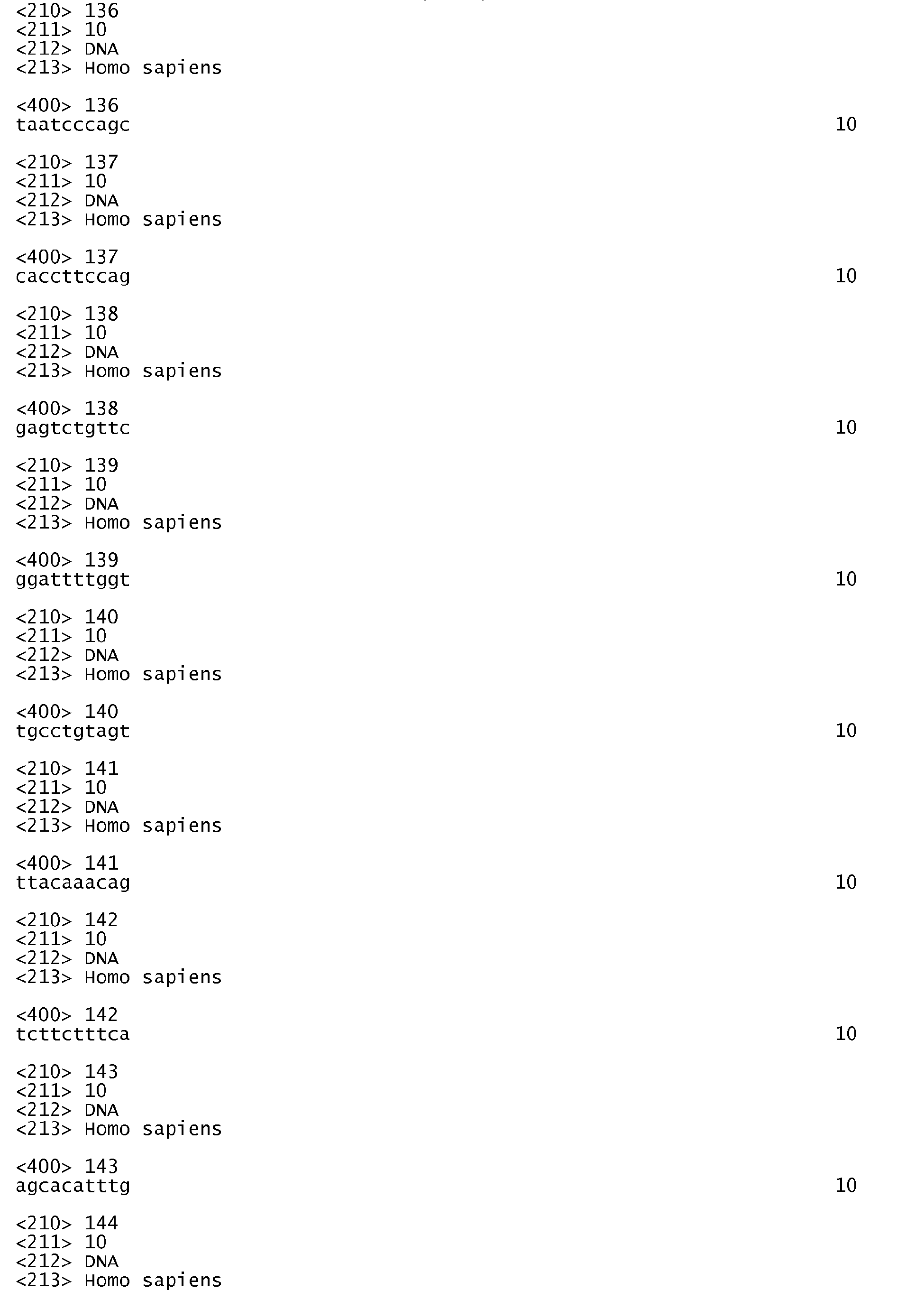


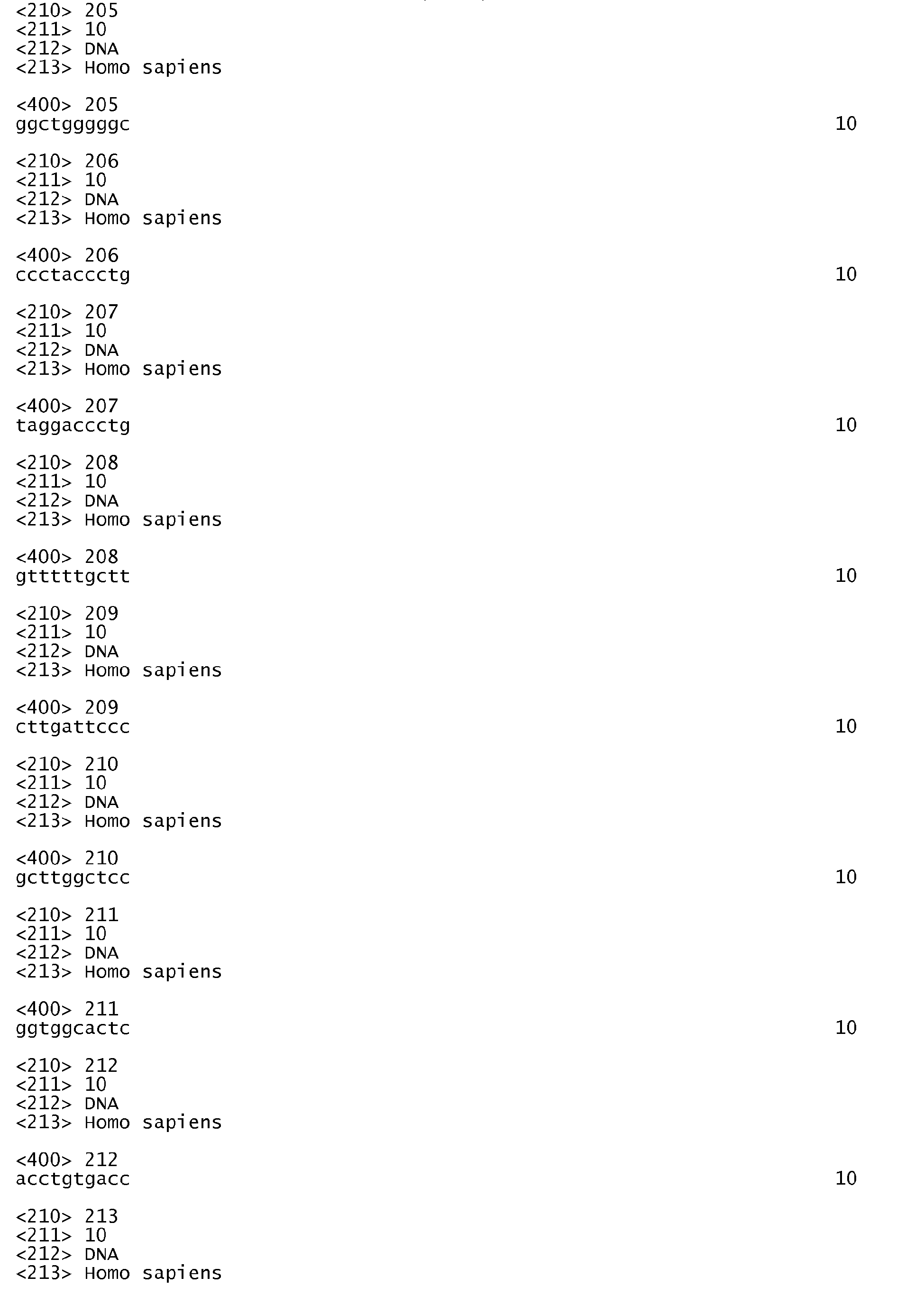


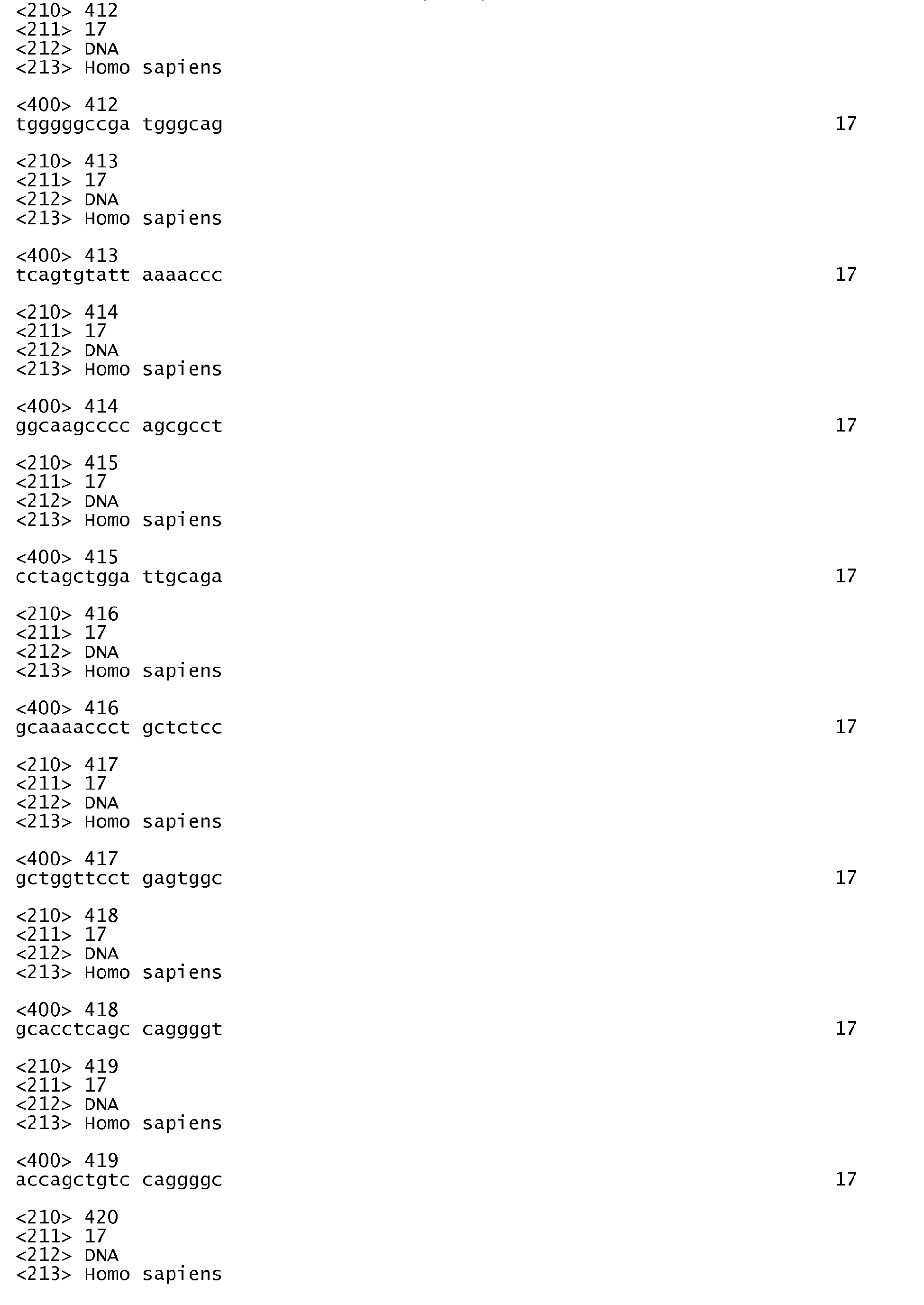

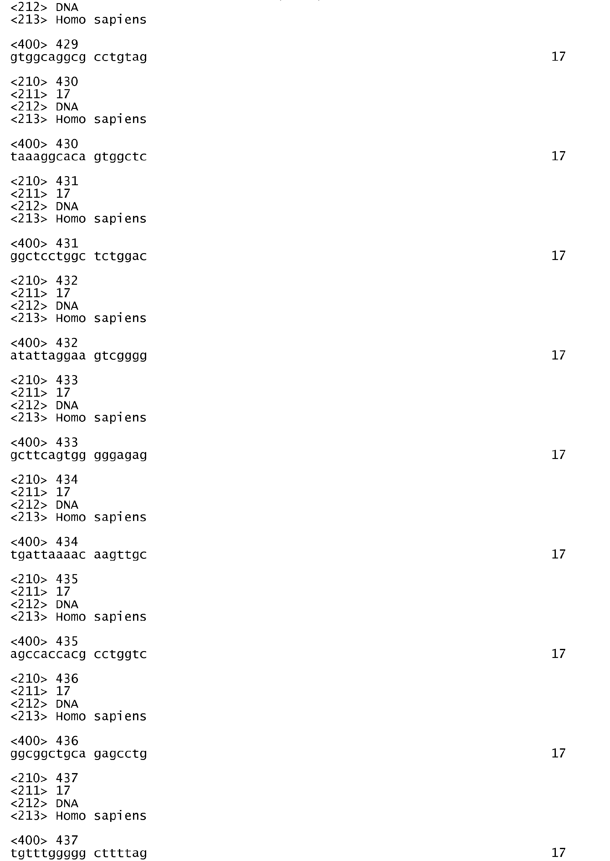


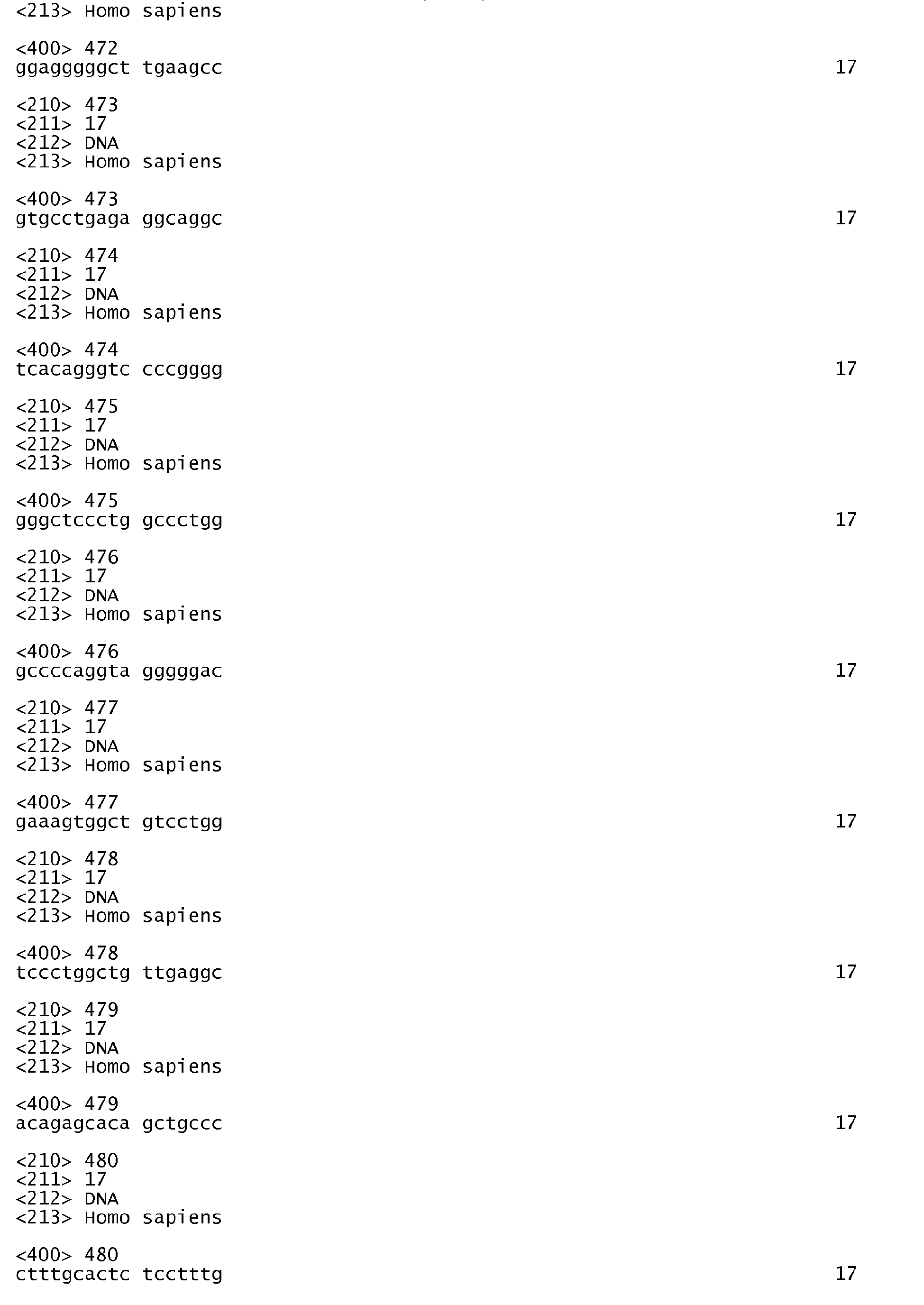


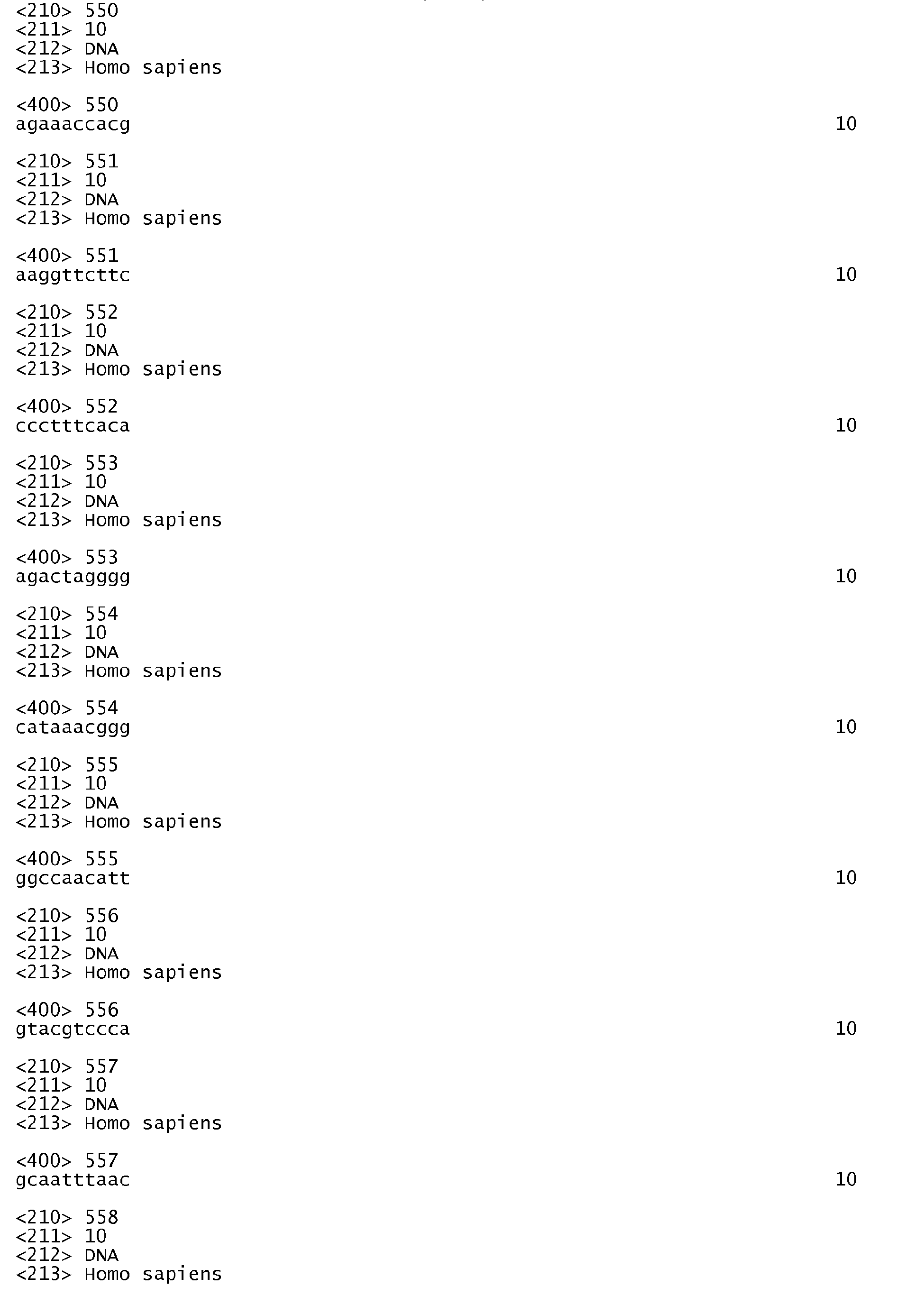


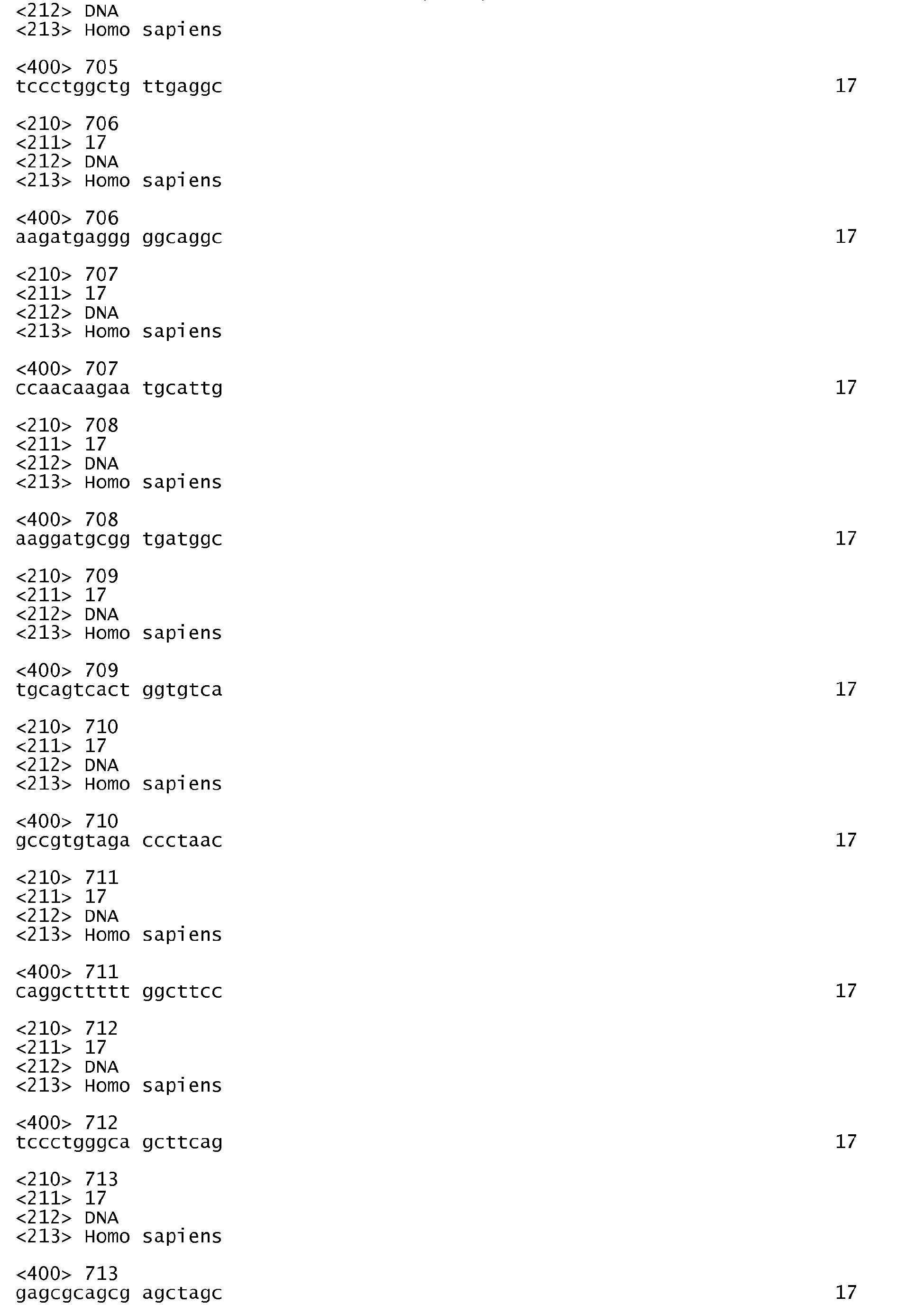


Claims (20)
- A method to aid in diagnosing glioma, comprising the steps of:detecting an expression product of at least one gene in a first brain tissue sample suspected of being neoplastic wherein said at least one gene is plasmalemma vesicle associated protein
and
comparing expression of the at least one gene in the first brain tissue sample with expression of the at least one gene in a second brain tissue sample which is normal, wherein increased expression of the at least one gene in the first brain tissue sample relative to the second tissue sample identifies the first brain tissue sample as likely to be neoplastic. - An antibody, optionally conjugated to a diagnostic or therapeutic reagent, which specifically binds to an extracellular epitope of plasmalemma vesicle associated protein for use in treating a glioma, especially whereby immune destruction of cells of the glioma is triggered.
- A method of identifying a test compound as a potential anti-cancer or anti-glioma drug, comprising the step of:contacting a test compound with a cell which expresses plasmalemma vesicle associated protein monitoring an expression product of the plasmalemma vesicle associated protein; andidentifying the test compound as a potential anti-cancer drug if it decreases the expression of the plasmalemma vesicle associated protein.
- A method to aid in diagnosing glioma, comprising the steps of:detecting an mRNA of at least one gene in a first brain tissue sample suspected of being neoplastic wherein said at least one gene is identified by a tag selected from the group consisting of SEQ ID NO: 1-32; andcomparing expression of the at least one gene in the first brain tissue sample with expression of the at least one gene in a second brain tissue sample which is normal, wherein increased expression of the at least one gene in the first brain tissue sample relative to the second tissue sample identifies the first brain tissue sample as likely to be neoplastic.
- A method of identifying a test compound as a potential anti-cancer or anti-glioma drug, comprising the step of:contacting a test compound with a cell which expresses an mRNA of at least one gene identified by a tag selected from the group consisting of SEQ ID NO: 1-32;monitoring an mRNA of the at least one gene; andidentifying the test compound as a potential anti-cancer drug if it decreases the expression of the at least one gene.
- Plasmalemma vesicle associated protein or a nucleic acid encoding plasmalemma vesicle associated protein for use in inducing an immune response to a glioma.
- The method of claim 1 or 4 wherein the increased expression of the at least one gene in the first brain tissue sample relative to the second tissue sample is at least two-fold higher; preferably at least five-fold higher; more preferably at least ten-fold higher.
- The method of claim 1, 2 or 5 wherein the expression product is RNA or protein.
- The method of claim 1 or 3 wherein the first and second tissue samples are from a human, especially from the same human.
- The antibody of claim 2 wherein the antibody is conjugated to a diagnostic or therapeutic reagent.
- The antibody of claim 2 wherein the glioma is multidrug-sensitive, and/or wherein the glioma is in a human.
- The antibody of claim 2 wherein the reagent is a chemotherapeutic agent; or a cytotoxin; or a non-radioactive label; or a radioactive compound.
- The method of claim 2 or 5 wherein the cell is a human cell and/or a glioma cell,
- The method of claim 2 or 5 wherein the cell overexpresses the at least one gene relative to a normal cell of the same tissue.
- The method of claim 2 or 5 wherein expression of at least one, preferably at least two, more preferably three additional genes selected from the group consisting of signal sequence receptor, delta (translocon-associated protein delta); DC2 protein; KIAA0404 protein; symplekin; Huntingtin interacting protein I; plasmalemma vesicle associated protein; KIAA0726 gene product; latexin protein; transforming growth factor, beta 1; hypothetical protein FLJ22215; Rag C protein; hypothetical protein FLJ23471; N-myristoyltransferase 1; hypothetical protein dJ1181N3.1; ribosomal protein L27; secreted protein, acidic, cysteine-rich (osteonectin); Hs 111988; Hs 112238; laminin, alpha 5; protective protein for beta-galactosidase (galactosialidosis); Melanoma associated gene; Melanoma associated gene; E3 ubiquitin ligase SMURF1; collagen, type IV, alpha 1; collagen, type IV, alpha 1; collagen, type IV, alpha 1; insulin-like growth factor binding protein 7; gene predicted from cDNA with a complete coding sequence; Thy-1 cell surface antigen; Hs 127824; GTP binding protein 2; Homo sapiens mRNA; cDNA DKFZp586D0918 (from clone DKFZp586D0918); cutaneous T-cell lymphoma-associated tumor antigen se20-4; differentially expressed nucleolar TGF-betal target protein (DENTT); dysferlin, limb girdle muscular dystrophy 2B (autosomal recessive); smoothelin; integrin, alpha 5 (fibronectin receptor, alpha polypeptide); putative translation initiation factor; retinoic acid induced 14; matrix metalloproteinase 9 (gelatinase B, 92kD gelatinase, 92kD type IV collagenase); Lutheran blood group (Auberger b antigen included); stanniocalcin 2; nuclear factor (erythroid-derived 2)-like 2; protein tyrosine phosphatase, non-receptor type 1; integrin, alpha 10; collagen, type VI, alpha 2; chromosome 21 open reading frame 25; CDC37 (cell division cycle 37, S. cerevisiae, homolog); Hs 16450; Rho guanine nucleotide exchange factor (GEF) 7; creatine kinase, brain; hypothetical protein FLJ10297; hypothetical protein FLJ10350; TNF-induced protein; tumor necrosis factor receptor superfamily, member 12 (translocating chain-association membrane protein); cofilin 1 (non-muscle); splicing factor proline/glutamine rich (polypyrimidine tract-binding protein-associated); splicing factor proline/glutamine rich (polypyrimidine tract-binding protein-associated); v-ets avian erythroblastosis virus E26 oncogene homolog 1; protease, cysteine, 1 (legumain); ribosomal protein L13; chromosome 22 open reading frame 5; zinc finger protein 144 (Mel-18); degenerative spermatocyte (homolog Drosophila; lipid desaturase); eukaryotic translation initiation factor 2C, 2; mitochondrial ribosomal protein L45; prostate tumor over expressed gene 1; NADH dehydrogenase (ubiquinone) 1 alpha subcomplex, 7 (14.5kD, B14.5a); glioma endothelial marker 1 precursor; NS1-binding protein; ribosomal protein L38; tuftelin-interacting protein; HLA class II region expressed gene KE2; translocase of inner mitochondrial membrane 17 homolog A (yeast); sudD (suppressor of bimD6, Aspergillus nidulans) homolog; heparan sulfate proteoglycan 2 (perlecan); SEC24 (S. cerevisiae) related gene family, member A; NADH dehydrogenase (ubiquinone) Fe-S protein 7 (20kD) (NADH-coenzyme Q reductase); DNA segment on chromosome X and Y (unique) 155 expressed sequence; annexin A2; Homo sapiens clone 24670 mRNA sequence; hypothetical protein; matrix metalloproteinase 10 (stromelysin 2); KIAA1049 protein; G protein-coupled receptor; hypothetical protein FLJ20401; matrix metalloproteinase 14 (membrane-inserted); KIAA0470 gene product; solute carrier family 29 (nucleoside transporters), member 1; stanniocalcin 1; stanniocalcin 1; stanniocalcin 1; tumor suppressor deleted in oral cancer-related 1; tumor suppressor deleted in oral cancer-related 1; apolipoprotein C-I; glutathione peroxidase 4 (phospholipid hydroperoxidase); Hs 272106; transcription factor binding to IGHM enhancer 3; hypothetical protein DKFZp762A227; hypothetical protein FLJ22362; CD59 antigen p18-20 (antigen identified by monoclonal antibodies 16.3A5, EJ16, EJ30, EL32 and G344); PRO0628 protein; melanoma-associated antigen recognised by cytotoxic T lymphocytes; LOC88745; Homo sapiens beta-1,3-galactosyltransferase-6 (B3GALT6) mRNA, complete cds; sprouty (Drosophila) homolog 4; sprouty (Drosophila) homolog 4; Homo sapiens mRNA; cDNA DKFZp434E1515 (from clone DKFZp434E1515); coactosin-like protein; hypothetical protein FLJ21865; Hs296234; KIAA0685 gene product; hypothetical protein FLJ10980; ribosomal protein L10; ribosomal protein S19; Hs 299251; Huntingtin interacting protein K; Homo sapiens mRNA full length insert cDNA clone EUROIMAGE 50374; Hs 311780; Hs 212191; v-akt murine thymoma viral oncogene homolog 2; Hs 328774; transducin-like enhancer of split 2, homolog of Drosophila E(sp1); KIAA1870 protein; ribosomal protein L10a; peptidylprolyl isomerase A (cyclophilin A); Hs 344224; hypothetical protein FLJ23239; hypothetical protein DKFZp761H221; KIAA1887 protein; Homo sapiens mRNA full length insert cDNA clone EUROIMAGE 701679; Homo sapiens cDNA FLJ30634 fis, clone CTONG2002453; Homo sapiens cDNA FLJ32203 fis, clone PLACE6003038, weakly similar to ZINC FINGER PROTEIN 84; Homo sapiens mRNA full length insert cDNA clone EUROIMAGE 1035904; hypothetical protein LOC57333; myosin ID; plexin B2; lectin, galactoside-binding, soluble, 8 (galectin 8); double ring-finger protein, Dorfin; DKFZP434B168 protein; LIM domain binding 2; integrin beta 4 binding protein; synaptopodin; Hs 54828; insulin induced gene 1; acetyl LDL receptor; SREC; excision repair cross-complementing rodent repair deficiency, complementation group 1 (includes overlapping antisense sequence); hypothetical protein FLJ22329; schwannomin-interacting protein 1; PTEN induced putative kinase 1; myosin X; Homo sapiens cDNA FLJ32424 fis, clone SKMUS2000954, moderately similar to Homo sapiens F-box protein Fbx25 (FBX25) 97; golgi phosphoprotein 1; splicing factor, arginine/serine-rich 6; laminin, gamma 3; cysteine-rich protein 2; U6 snRNA-associated Sm-like protein LSm7; hypothetical protein FLJ10707; Homo sapiens, Similar to RIKEN cDNA 2310012N15 gene, clone IMAGE:3342825, mRNA, partial cds; macrophage migration inhibitory factor (glycosylation-inhibiting factor); ubiquinol-cytochrome c reductase hinge protein; gap junction protein, alpha 1, 43kD (connexin 43); dihydropyrimidinase-like 3; aquaporin 1 (channel-forming integral protein, 28kD); protein expressed in thyroid; macrophage myristoylated alanine-rich C kinase substrate; procollagen-lysine, 2-oxoglutarate 5-dioxygenase (lysine hydroxylase, Ehlers-Danlos syndrome type VI); protease, serine, 11 (IGF binding); 24-dehydrocholesterol reductase; collagen, type IV, alpha 2; profilin 1; apolipoprotein D; hyaluronoglucosaminidase 2; hypothetical protein FLJ22678; quiescin Q6; ras homolog gene family, member A; ras homolog gene family, member A; plasminogen activator, urokinase; insulin-like growth factor binding protein 3; uridine phosphorylase; KIAA0638 protein; B7 homolog 3; lamin A/C; lamin A/C; lamin A/C; regulator of G-protein signalling 12; proteasome (prosome, macropain) 26S subunit, non-ATPase, 8; Homo sapiens, Similar to RIKEN cDNA 5730528L13 gene, clone MGC: 17337 IMAGE:4213591, mRNA, complete cds; prosaposin (variant Gaucher disease and variant metachromatic leukodystrophy); laminin, alpha 4; transcription elongation factor A (SII), 1; lectin, galactoside-binding, soluble, 3 binding protein; ribosomal protein S16; glycophorin C (Gerbich blood group); endothelin receptor type B; serine (or cysteine) proteinase inhibitor, clade E (nexin, plasminogen activator inhibitor type 1), member 1; biglycan; small nuclear ribonucleoprotein polypeptide B"; transmembrane 4 superfamily member 2; TAF11 RNA polymerase II, TATA box binding protein (TBP)-associated factor, 28 kD; lysyl oxidase-like 2; SRY (sex determining region Y)-box 4; SOX4 SRY (sex determining region Y)-box 4; SRY (sex determining region Y)-box 4; actin related protein 2/3 complex, subunit 2 (34 kD); Homo sapiens cDNA: FLJ23507 fis, clone LNG03128; hypothetical protein FLJ12442; Fas (TNFRSF6)-associated via death domain; mitogen-activated protein kinase kinase kinase 11; TEK tyrosine kinase, endothelial (venous malformations, multiple cutaneous and mucosal); insulin receptor; cell membrane glycoprotein, 110000M(r) (surface antigen); Homo sapiens cDNA FLJ11863 fis, clone HEMBA1006926; jagged 1 (Alagille syndrome); KIAA0304 gene product; pre-B-cell leukemia transcription factor 2; Homo sapiens cDNA FLJ31238 fis, clone KIDNE2004864; p53-induced protein; complement component 1, q subcomponent, receptor 1; complement component 1, q subcomponent, receptor 1; apolipoprotein E; chemokine (C-C motif) ligand 3; coagulation factor II (thrombin) receptor-like 3; coagulation factor III (thromboplastin, tissue factor); collagen, type I, alpha 1; collagen, type III, alpha 1 (Ehlers-Danlos syndrome type IV, autosomal dominant); C-type (calcium dependent, carbohydrate-recognition domain) lectin, superfamily member 9; cystatin C (amyloid angiopathy and cerebral hemorrhage); endoplasmic reticulum associated protein 140 kDa; ESTs; ESTs; ESTs, Highly similar to hypothetical protein FLJ10350 [Homo sapiens] [H.sapiens]; ESTs, Highly similar to ITB1_HUMAN Integrin beta-1 precursor (Fibronectin receptor beta subunit) (CD29) (Integrin VLA-4 beta subunit) [H.sapiens]; ESTs, Weakly similar to hypothetical protein FLJ20489 [Homo sapiens] [H.sapiens]; ESTs, Weakly similar to T17346 hypothetical protein DKFZp586O1624.1 - human (fragment) [H.sapiens]; ESTs, Weakly similar to T21371 hypothetical protein F25H8.3 - Caenorhabditis elegans [C.elegans]; eukaryotic translation initiation factor 4A, isoform 1; heme oxygenase (decycling) 1; Hermansky-Pudlak syndrome 4; Homo sapiens cDNA FLJ34888 fis, clone NT2NE2017332; Homo sapiens cDNA FLJ39848 fis, clone SPLEN2014669; Homo sapiens mRNA full length insert cDNA clone EUROIMAGE 1977059; Homo sapiens, clone IMAGE:4845226, mRNA; hypothetical protein FLJ22329; hypothetical protein FLJ32205; hypothetical protein MGC4677; inhibin, beta B (activin AB beta polypeptide); insulin-like growth factor binding protein 5; junction plakoglobin; KIAA0620 protein; KIAA0943 protein; likely ortholog of rat vacuole membrane protein 1; Lysosomal-associated multispanning membrane protein-5; major histocompatibility complex, class I, B; major histocompatibility complex, class I, C; matrix Gla protein; matrix metalloproteinase 1 (interstitial collagenase); microtubule-associated protein 1 light chain 3 beta; nerve growth factor receptor (TNFR superfamily, member 16); ribosomal protein S9; ring finger protein 40; S100 calcium binding protein, beta (neural); sema domain, transmembrane domain (TM), and cytoplasmic domain, (semaphorin) 6B; SPARC-like 1 (mast9, hevin); tumor necrosis factor, alpha-induced protein 3; UDP-Gal:betaGlcNAc beta 1,4- galactosyltransferase, polypeptide 3; UDP-GlcNAc:betaGal beta-1,3-N-acetylglucosaminyltransferase 5; von Willebrand factor; v-akt murine thymoma vial oncogene homolog 2; cyclin-dependent kinase (cdc2-like) 10; ortholog mouse myocytic induction/differentiation originator; brain-specific angiogenesis inhibitor 1 ; EGF-TM7 latrophilin-related protein; sema domain ; integrin, alpha 5 ; likely ortholog of mouse fibronectin type III; Lutheran blood group (Auberger b antigen included); SSR4, TRAPD; nerve growth factor receptor (TNFR superfamily, member 16); insulin-like growth factor binding protein; leukemia inhibitory factor; protein tyrosine phosphatase, nonreceptor type I; and Homo sapiens, clone IMAGE:3908182, mRNA, partial cds; is monitored.
- The method of claim 2 or 5 wherein the test compound is identified if the decrease in expression is at least 50%, preferably at least 80%, more preferably at least 90%.
- The method of claim 2 or 5 wherein the test compound is identified as an anti-glioma drug.
- A nucleic acid encoding plasmalemma vesicle associated protein, according to claim 6, wherein the nucleic acid is for intramuscular administration.
- Plasmalemma vesicle associated protein or a nucleic acid encoding plasmalemma vesicle associated protein, according to claim 6, wherein the plasmalemma vesicle associated protein or nucleic acid encoding the plasmalemma vesicle associated protein is for administration with an immune adjuvant.
- Plasmalemma vesicle associated protein or a nucleic acid encoding plasmalemma vesicle associated protein, according to claim 6, wherein the plasmalemma vesicle associated protein or nucleic acid encoding the plasmalemma vesicle associated protein is for administration to a mammal, or a mammal from which a glioma has been surgically removed.
Applications Claiming Priority (3)
| Application Number | Priority Date | Filing Date | Title |
|---|---|---|---|
| US40339002P | 2002-08-15 | 2002-08-15 | |
| US45897803P | 2003-04-01 | 2003-04-01 | |
| EP03788531A EP1576131A4 (en) | 2002-08-15 | 2003-08-15 | REASONS FOR EXPRESSION OF BRAIN ENDOTHELIAL CELLS |
Related Parent Applications (1)
| Application Number | Title | Priority Date | Filing Date |
|---|---|---|---|
| EP03788531.6 Division | 2003-08-15 |
Publications (2)
| Publication Number | Publication Date |
|---|---|
| EP2236614A2 true EP2236614A2 (en) | 2010-10-06 |
| EP2236614A3 EP2236614A3 (en) | 2011-01-26 |
Family
ID=31891381
Family Applications (2)
| Application Number | Title | Priority Date | Filing Date |
|---|---|---|---|
| EP03788531A Withdrawn EP1576131A4 (en) | 2002-08-15 | 2003-08-15 | REASONS FOR EXPRESSION OF BRAIN ENDOTHELIAL CELLS |
| EP20100168014 Ceased EP2236614A3 (en) | 2002-08-15 | 2003-08-15 | Brain endothelial cell expression patterns |
Family Applications Before (1)
| Application Number | Title | Priority Date | Filing Date |
|---|---|---|---|
| EP03788531A Withdrawn EP1576131A4 (en) | 2002-08-15 | 2003-08-15 | REASONS FOR EXPRESSION OF BRAIN ENDOTHELIAL CELLS |
Country Status (5)
| Country | Link |
|---|---|
| US (3) | US20060127902A1 (en) |
| EP (2) | EP1576131A4 (en) |
| JP (2) | JP2006512924A (en) |
| AU (1) | AU2003262717A1 (en) |
| WO (1) | WO2004016758A2 (en) |
Families Citing this family (50)
| Publication number | Priority date | Publication date | Assignee | Title |
|---|---|---|---|---|
| US7569342B2 (en) | 1997-12-10 | 2009-08-04 | Sierra Molecular Corp. | Removal of molecular assay interferences |
| US20030114410A1 (en) | 2000-08-08 | 2003-06-19 | Technion Research And Development Foundation Ltd. | Pharmaceutical compositions and methods useful for modulating angiogenesis and inhibiting metastasis and tumor fibrosis |
| WO2004064785A2 (en) | 2003-01-14 | 2004-08-05 | Dana Farber Cancer Institute | Cancer therapy sensitizer |
| AU2004298604B2 (en) * | 2003-12-15 | 2010-09-23 | The Regents Of The University Of California | Molecular signature of the PTEN tumor suppressor |
| EP1709193B1 (en) * | 2003-12-19 | 2011-03-16 | The Regents of The University of California | Methods and materials for assessing prostate cancer therapies |
| WO2005099693A2 (en) * | 2004-02-24 | 2005-10-27 | The Regents Of The University Of California | Methods and materials for assessing prostate cancer therapies and compounds |
| US20060099144A1 (en) * | 2004-11-08 | 2006-05-11 | Schering Corporation | Tumor association of MDL-1 and methods |
| AU2006241235B2 (en) * | 2005-04-22 | 2012-04-05 | Eisai, Inc. | Antibodies with immune effector activity and that internalize in endosialin-positive cells |
| US7709517B2 (en) | 2005-05-13 | 2010-05-04 | The Regents Of The University Of California | Diarylhydantoin compounds |
| WO2006128063A2 (en) * | 2005-05-25 | 2006-11-30 | Irm Llc | Methods and compositions for inhibiting glioma growth |
| US20070021365A1 (en) * | 2005-06-21 | 2007-01-25 | The Board Of Trustees Of The Leland Stanford Junior University | Inhibition of Lysyl oxidase for treating tumor growth and diagnostics relating thereto |
| US20070225242A1 (en) * | 2005-06-21 | 2007-09-27 | The Board Of Trustees Of The Leland Stanford Junior University | Method and composition for treating and preventing tumor metastasis in vivo |
| DK1907420T3 (en) | 2005-07-21 | 2017-07-17 | Modiquest B V | PLEXIN D1 TARGETED FOR TUMOR DIAGNOSTICATION AND TUMOR THERAPY |
| CA2623653C (en) | 2005-09-27 | 2012-08-28 | University Of Saskatchewan | Use of n-myristoyltransferase on non-tumor tissue for cancer diagnosis |
| WO2007107774A2 (en) * | 2006-03-22 | 2007-09-27 | Cartela R & D Ab | Integrin alpha-10 subunit as therapeutic target and diagnostic/prognostic marker for cancer |
| ES2689292T3 (en) | 2006-03-27 | 2018-11-13 | The Regents Of The University Of California | Androgen receptor modulator for the treatment of prostate cancer and diseases associated with the androgen receptor |
| NZ572374A (en) | 2006-03-29 | 2011-12-22 | Univ California | Diarylthiohydantoin compounds |
| CA2669260C (en) | 2006-11-09 | 2019-01-15 | The Government Of The United States Of America As Represented By The Secretary Of The Department Of Health And Human Services | Differential gene expression in physiological and pathological angiogenesis |
| WO2008091781A1 (en) * | 2007-01-22 | 2008-07-31 | Mayo Foundation For Medical Education And Research | Reducing tumor growth |
| EP2620451B1 (en) | 2007-04-05 | 2016-09-14 | Morphotek, Inc. | Methods for inhibiting the binding of endosialin to ligands |
| EA009327B1 (en) | 2007-04-27 | 2007-12-28 | Петр Генриевич ЛОХОВ | Method for producing antineoplastic vaccine based on surface antigens of endothelial cells |
| US12180549B2 (en) | 2007-07-23 | 2024-12-31 | The Chinese University Of Hong Kong | Diagnosing fetal chromosomal aneuploidy using genomic sequencing |
| HUE061020T2 (en) | 2007-07-23 | 2023-05-28 | Univ Hong Kong Chinese | Determining a nucleic acid sequence imbalance |
| ES2700141T3 (en) | 2007-08-02 | 2019-02-14 | Gilead Biologics Inc | LOXL2 inhibitory antibodies and their uses |
| EP2220050A2 (en) | 2007-10-26 | 2010-08-25 | The Regents Of The University Of California | Diarylhydantoin compounds as androgen receptor modulators |
| DE102008011850A1 (en) | 2008-02-29 | 2009-09-03 | Michael Grzendowski | Biomarker for the diagnosis of brain tumor |
| EP3115063A3 (en) | 2008-12-03 | 2017-04-19 | The John Hopkins University | Galectin-3 and annexin-a2 as immunological target |
| US9107935B2 (en) * | 2009-01-06 | 2015-08-18 | Gilead Biologics, Inc. | Chemotherapeutic methods and compositions |
| KR101054952B1 (en) * | 2009-01-22 | 2011-08-05 | 재단법인 한국원자력의학원 | UCCR, a marker for diagnosing liver cancer and predicting patient survival, a kit including the same, and prediction of liver cancer patient survival using the marker |
| ES2363358B1 (en) * | 2009-04-03 | 2012-06-21 | FUNDACIÓ INSTITUT DE RECERCA HOSPITAL UNIVERSITARI VALL D'HEBRON (Titular al | THERAPEUTIC AGENTS FOR THE TREATMENT OF DISEASES ASSOCIATED WITH AN INDESEABLE CELLULAR PROLIFERATION. |
| WO2010118203A2 (en) * | 2009-04-09 | 2010-10-14 | Morphotek, Inc. | Endosialin binding molecules |
| RU2012110585A (en) | 2009-08-21 | 2013-09-27 | Джилид Байолоджикс, Инк. | CATALYTIC DOMAINS OF LYSYLOXIDASE AND LOXL2 |
| EP2467162A4 (en) * | 2009-08-21 | 2013-10-02 | Gilead Biologics Inc | Therapeutic methods and compositions |
| CN102713601A (en) * | 2009-08-21 | 2012-10-03 | 吉联亚生物科技有限公司 | In vitro screening assays |
| US20110044907A1 (en) * | 2009-08-21 | 2011-02-24 | Derek Marshall | In vivo screening assays |
| KR20120054077A (en) * | 2009-08-21 | 2012-05-29 | 길리아드 바이오로직스, 인크. | Methods and compositions for treatment of pulmonary fibrotic disorders |
| US8680246B2 (en) | 2010-02-04 | 2014-03-25 | Gilead Biologics, Inc. | Antibodies that bind to lysyl oxidase-like 2 (LOXL2) |
| SG10201907588XA (en) | 2010-02-16 | 2019-09-27 | Aragon Pharmaceuticals Inc | Androgen Receptor Modulators And Uses Thereof |
| EP2371860A1 (en) * | 2010-04-05 | 2011-10-05 | Fundació Privada Institut d'Investigació Oncològica de Vall d'Hebron | Antibody recognising human leukemia inhibitory factor (LIF) and use of anti-LIF antibodies in the treatment of diseases associated with unwanted cell proliferation |
| US20130330349A1 (en) | 2011-02-23 | 2013-12-12 | Rappaport Family Institute For Research In The Medical Sciences | High affinity molecules capable of binding a type a plexin receptor and uses of same |
| US9892230B2 (en) | 2012-03-08 | 2018-02-13 | The Chinese University Of Hong Kong | Size-based analysis of fetal or tumor DNA fraction in plasma |
| CN115737643A (en) | 2012-09-26 | 2023-03-07 | 阿拉贡药品公司 | Antiandrogens for the treatment of non-metastatic castration resistant prostate cancer |
| DK2945628T3 (en) | 2013-01-15 | 2020-05-04 | Aragon Pharmaceuticals Inc | ANDROGEN RECEPTOR MODULATOR IN COMBINATION WITH ABIRATERON ACETATE AND PREDNISON FOR TREATMENT OF PROSTATACANCES |
| KR101727026B1 (en) * | 2014-03-06 | 2017-04-17 | (주)노바셀테크놀로지 | Novel bio-maker of neuroglioma and use thereof |
| KR101644599B1 (en) | 2014-10-14 | 2016-08-16 | 연세대학교 산학협력단 | Construction of mitochondrial UQCRB mutant expressing cells and utilization of the cells for UQCRB assay system thereof |
| US10364467B2 (en) | 2015-01-13 | 2019-07-30 | The Chinese University Of Hong Kong | Using size and number aberrations in plasma DNA for detecting cancer |
| CA2976203A1 (en) * | 2015-02-16 | 2016-08-25 | Xintela Ab | Detection and treatment of malignant tumours in the cns |
| TWI726969B (en) | 2016-01-11 | 2021-05-11 | 比利時商健生藥品公司 | Substituted thiohydantoin derivatives as androgen receptor antagonists |
| MX2020003845A (en) | 2017-10-16 | 2020-11-06 | Aragon Pharmaceuticals Inc | ANTIANDROGENS FOR THE TREATMENT OF NON-METASTATIC PROSTATE CANCER RESISTANT TO CASTRATION. |
| CN110687236B (en) * | 2019-10-30 | 2022-05-17 | 陕西师范大学 | Method for evaluating freezing and thawing degree of meat based on iTRAQ marker protein |
Citations (1)
| Publication number | Priority date | Publication date | Assignee | Title |
|---|---|---|---|---|
| US6075181A (en) | 1990-01-12 | 2000-06-13 | Abgenix, Inc. | Human antibodies derived from immunized xenomice |
Family Cites Families (7)
| Publication number | Priority date | Publication date | Assignee | Title |
|---|---|---|---|---|
| AU1724000A (en) * | 1998-11-12 | 2000-05-29 | Nyxis, Inc. | Diagnostic assay for cancer |
| US20020015950A1 (en) * | 1999-07-07 | 2002-02-07 | Karen Anne Jones | Atherosclerosis-associated genes |
| AU3316600A (en) * | 1999-02-22 | 2000-09-21 | Torben F. Orntoft | Gene expression in bladder tumors |
| EP1307557A2 (en) * | 2000-08-02 | 2003-05-07 | The Johns Hopkins University | Endothelial cell expression patterns |
| US20020142287A1 (en) * | 2000-12-14 | 2002-10-03 | Hirotaka Yamamoto | High throughput assay to detect inhibitors of the map kinase pathway |
| TW200413725A (en) * | 2002-09-30 | 2004-08-01 | Oncotherapy Science Inc | Method for diagnosing non-small cell lung cancers |
| JP4698228B2 (en) * | 2003-04-08 | 2011-06-08 | エフ.ホフマン−ラ ロシュ アーゲー | How to define the stage of tumor differentiation |
-
2003
- 2003-08-15 EP EP03788531A patent/EP1576131A4/en not_active Withdrawn
- 2003-08-15 AU AU2003262717A patent/AU2003262717A1/en not_active Abandoned
- 2003-08-15 US US10/524,432 patent/US20060127902A1/en not_active Abandoned
- 2003-08-15 EP EP20100168014 patent/EP2236614A3/en not_active Ceased
- 2003-08-15 WO PCT/US2003/025614 patent/WO2004016758A2/en not_active Ceased
- 2003-08-15 JP JP2005502059A patent/JP2006512924A/en not_active Withdrawn
-
2009
- 2009-11-11 US US12/616,600 patent/US20100062002A1/en not_active Abandoned
-
2010
- 2010-06-03 JP JP2010127374A patent/JP2010284161A/en active Pending
-
2012
- 2012-07-09 US US13/544,631 patent/US20120308479A1/en not_active Abandoned
Patent Citations (1)
| Publication number | Priority date | Publication date | Assignee | Title |
|---|---|---|---|---|
| US6075181A (en) | 1990-01-12 | 2000-06-13 | Abgenix, Inc. | Human antibodies derived from immunized xenomice |
Non-Patent Citations (63)
| Title |
|---|
| ABOUNADER, R.; LAL, B.; LUDDY, C.; KOE, G., DAVIDSON, B.; ROSEN, E. M.; LATERRA, J.: "In vivo targeting of SF/HGF and c-met expression via UlsnRNA/ribozymes inhibits glioma growth and angiogenesis and promotes apoptosis", FASEB J, vol. 16, 2002, pages 108 - 10 |
| ABOUNADER, R.; LAL, B.; LUDDY, C.; KOE, G.; DAVIDSON, B.; ROSEN, E. M.; LATERRA, J.: "In vivo targeting of SF/HGF and c-met expression via UlsnRNA/ribozymes inhibits glioma growth and angiogenesis and promotes apoptosis", FASEB J, vol. 16, 2002, pages 108 - 10 |
| ALTSCHUL, NUCLEIC ACID RES., vol. 25, 1997, pages 3389 - 3402 |
| ARBONES M; ORD D; LEY K; RATECH H; MAYNARD-CURRY K; OTTEN G; CAPON D; TEDDER T: "Lymphocyte homing and leukocyte rolling and migration are impaired in L-selectin-deficient mice", IMMUNITY, vol. 1, no. 4, 1994, pages 247 - 260 |
| BAGEMAN ET AL., NUCLEIC ACIDS RES., vol. 28, 2000, pages 263 - 266 |
| BAGEMAN, NUCLEIC ACIDS RES., vol. 27, 1999, pages 260 - 262 |
| BART, J.; GROEN, H. J.; HENDRIKSE, N. H.; VAN DER GRAAF, W. T.; VAALBURG, W.; DE VRIES, E. G.: "The blood-brain barrier and oncology: new insights into function and modulation", CANCER TREAT REV, vol. 26, 2000, pages 449 - 62 |
| BART, J.; GROEN, H. J.; HENDRIKSE, N. H.; VAN DER GRAAF, W. T.; VAALBURG, W.; VRIES, E. G.: "The blood-brain barrier and oncology: new insights into function and modulation", CANCER TREAT REV, vol. 26, 2000, pages 449 - 62 |
| BERNSEN, H. J.; RIJKEN, P. F.; OOSTENDORP, T.; VAN DER KOGEL, A. J.: "Vascularity and perfusion of human gliomas xenografted in the athymic nude mouse", BR J CANCER, vol. 71, 1995, pages 721 - 6 |
| BOWERS, D. C.; FAN, S.; WALTER, K. A.; ABOUNADER, R.; WILLIAMS, J. A.; ROSEN, E. M.; LATERRA, J.: "Scatter factor/hepatocyte growth factor protects against cytotoxic death in human glioblastoma via phosphatidylinositol 3- kinase- and AKT- dependent pathways", CANCER RES, vol. 60, 2000, pages 4277 - 83 |
| BOWERS, D. C.; FAN, S.; WALTER, K. A.; ABOUNADER, R.; WILLIAMS, J. A.; ROSEN, E. M.; LATERRA, J.: "Scatter factor/hepatocyte growth factor protects against cytotoxic death in human glioblastoma via phosphatidylinositol 3-kinase- and AKT- dependent pathways", CANCER RES, vol. 60, 2000, pages 4277 - 83 |
| CHEN, H.; CENTOLA, M.; ALTSCHUL, S. F.; METZGER, H.: "Characterization of gene expression in resting and activated mast cells", J EXP MED, vol. 188, 1998, pages 1657 - 68 |
| CROIX, B.; RAGO, C.; VELCULESCU, V.; TRAVERSO, G.; ROMANS, K. E.; MONTGOMERY, E.; LAL, A.; RIGGINS, G. J.; LENGAUER, C.; VOGELSTEI: "Genes expressed in human tumor endothelium", SCIENCE, vol. 289, 2000, pages 1197 - 202 |
| ELEANOR B CARSON-WALTER ET AL: "Plasmalemmal vesicle associated protein-1 is a novel marker implicated in brain tumor angiogenesis", CLINICAL CANCER RESEARCH, THE AMERICAN ASSOCIATION FOR CANCER RESEARCH, US, vol. 11, no. 21, 1 November 2005 (2005-11-01), pages 7643 - 7650, XP008110957, ISSN: 1078-0432, DOI: 10.1158/1078-0432.CCR-05-1099 * |
| EVELINE H SHUE ET AL: "Plasmalemmal Vesicle Associated Protein-1 (PV-1) is a marker of blood-brain barrier disruption in rodent models", BMC NEUROSCIENCE, vol. 9, no. 1, 1 January 2008 (2008-01-01), pages 29, XP055119414, ISSN: 1471-2202, DOI: 10.1186/1471-2202-9-29 * |
| GREEN L; HARDY M; MAYNARD-CURRY K; TSUDA H; LOUIE D; MENDEZ M; ABDERRAHIM H; NOGUCHI M; SMITH D; ZENG Y: "Antigen-specific human monoclonal antibodies from mice engineered with human Ig heavy and light chain YACs", NATURE GENETICS, vol. 7, no. 1, 1994, pages 13 - 21 |
| GREEN L; JAKOBOVITS A: "Regulation of B cell development by variable gene complexity in mice reconstituted with human immunoglobulin yeast artificial chromosomes", J EXP. MED., vol. 188, no. 3, 1998, pages 483 - 495 |
| GUERIN, C.; WOLFF, J. E.; LATERRA, J.; DREWES, L. R.; BREM, H.; GOLDSTEIN, G. W.: "Vascular differentiation and glucose transporter expression in rat gliomas: effects of steroids", ANN NEUROL, vol. 31, 1992, pages 481 - 7 |
| GUERIN, C.; WOLFF, J. E.; LATERRA, J; DREWES, L. R.; BREM, H.; GOLDSTEIN, G. W.: "Vascular differentiation and glucose transporter expression in rat gliomas: effects of steroids", ANN NEUROL, vol. 31, 1992, pages 481 - 7 |
| HENIKOFF, NUCL. ACIDS RES., vol. 28, 2000, pages 228 - 230 |
| HOBBS, S. K.; MONSKY, W. L.; YUAN, F.; ROBERTS, W. G.; GRIFFITH, L.; TORCHILIN, V. P.; JAIN, R. K.: "Regulation of transport pathways in tumor vessels: role of tumor type and microenvironment", PROC NATL ACAD SCI U S A, vol. 95, 1998, pages 4607 - 12 |
| HOBBS, S. K.; MONSKY, W. L.; YUAN, F.; ROBERTS, W. G.; GRIFFITH, L.; TORCHILIN, V. P.; JAIN, R. K.: "Regulation of transport pathways in tumor vessels: role of tumor type and microenvironment", PROC NATL ACAD SCI USA, vol. 95, 1998, pages 4607 - 12 |
| HOLASH, J.; MAISONPIERRE, P. C.; COMPTON, D.; BOLAND, P.; ALEXANDER, C. R.; ZAGZAG, D.; YANCOPOULOS, G. D.; WIEGAND, S. J.: "Vessel cooption, regression, and growth in tumors mediated by angiopoietins and VEGF", SCIENCE, vol. 284, 1999, pages 1994 - 8 |
| HOLASH, J.; MAISONPIERRE, P. C.; COMPTON, D.; BOLAND, P.; ALEXANDER, C. R.; ZAGZAG, D; YANCOPOULOS, G. D.; WIEGAND, S. J.: "Vessel cooption, regression, and growth in tumors mediated by angiopoietins and VEGF", SCIENCE, vol. 284, 1999, pages 1994 - 8 |
| HUMINIECKI, L.; BICKNELL, R.: "In silico cloning of novel endothelial-specific genes", GENOME RES, vol. 10, 2000, pages 1796 - 806 |
| JAGLA, BIOINFORMATICS, vol. 16, 2000, pages 245 - 250 |
| JAKOBOVITS A: "Production and selection of antigen-specific fully human monoclonal antibodies from mice engineered with human Ig loci", ADVANCED DRUG DELIVERY REVIEWS, vol. 31, 1998, pages 33 - 42 |
| JAKOBOVITS A: "Production of fully human antibodies by transgenic mice", CURRENT OPINION IN BIOTECHNOLOGY, vol. 6, no. 5, 1995, pages 561 - 566 |
| JAKOBOVITS A: "The long-awaited magic bullets: therapeutic human monoclonal antibodies from transgenic mice", EXP. OPIN. INVEST. DRUGS, vol. 7, no. 4, 1998, pages 607 - 614 |
| JAKOBOVITS A: "YAC Vectors: Humanizing the mouse genome", CURRENT BIOLOGY, vol. 4, no. 8, 1994, pages 761 - 763 |
| JAKOBOVITS A; MOORE A; GREEN L; VERGARA G; MAYNARD-CURRY K; AUSTIN H; KLAPHOLZ S: "Germ-line transmission and expression of a human- derived yeast artificial chromosome", NATURE, vol. 362, no. 6417, 1993, pages 255 - 258 |
| JAKOBOVITS A; VERGARA G; KENNEDY J; HALES J; MCGUINNESS R; CASENTINI-BOROCZ D; BRENNER D; OTTEN G: "Analysis of homozygous mutant chimeric mice: deletion of the immunoglobulin heavy-chain joining region blocks B-cell development and antibody production", PROCEEDINGS OF THE NATIONAL ACADEMY OF SCIENCES USA, vol. 90, no. 6, 1993, pages 2551 - 2555 |
| KLEIN, BIOCHIM. BIOPHYS. ACTA, vol. 815, 1985, pages 468 |
| L. A. SANDERS (STRICKLAND) ET AL: "Comments on Plasmalemmal Vesicle Associated Protein-1 as a Novel Marker Implicated in Brain Tumor Angiogenesis", CLINICAL CANCER RESEARCH, vol. 12, no. 8, 15 April 2006 (2006-04-15), pages 2649 - 2649, XP055119421, ISSN: 1078-0432, DOI: 10.1158/1078-0432.CCR-05-2624 * |
| LAMSZUS, K.; LATERRA, J.; WESTPHAL, M.; ROSEN, E. M.: "Scatter factor/hepatocyte growth factor (SF/HGF) content and function in human gliomas", INT J DEV NEUROSCI, vol. 17, 1999, pages 517 - 30 |
| LAMSZUS, K.; LATERRA, J; WESTPHAL, M.; ROSEN, E. M.: "Scatter factor/hepatocyte growth factor (SF/HGF) content and function in human gliomas", INT J DEV NEUROSCI, vol. 17, 1999, pages 517 - 30 |
| LARRY L. GREEN: "Antibody engineering via genetic engineering of the mouse: XenoMouse strains are a vehicle for the facile generation of therapeutic human monoclonal antibodies", JOURNAL OF IMMUNOLOGICAL METHODS, vol. 231, 1999, pages 11 - 23 |
| MARX, J.: "Caveolae: a once-elusive structure gets some respect", SCIENCE, vol. 294, 2001, pages 1862 - 5 |
| MENDEZ M; ABDERRAHIM H; NOGUCHI M; DAVID N; HARDY M; GREEN L; TSUDA H; YOAST S; MAYNARD-CURRIE C; GARZA D: "Analysis of the structural integrity of YACs comprising human immunoglobulin genes in yeast and in embryonic sGEM cells", GENOMICS, vol. 26, 1995, pages 294 - 307 |
| MENDEZ M; GREEN L; CORVALAN J; JIA X-C; MAYNARD-CURRIE C; YANG X-D; GALLO M; LOUIE D; LEE D; ERICKSON K: "Functional transplant of megabase human immunoglobulin loci recapitulates human antibody response in mice", NATURE GENETICS, vol. 15, 1997, pages 146 - 156 |
| MICHAEL L. GALLO; VLADIMIR E. IVANOV; AYA JAKOBOVITS; C. GEOFFREY DAVIS: "The human immunoglobulin loci introduced into mice: V (D) and J gene segment usage similar to that of adult humans", EUROPEAN JOURNAL OF IMMUNOLOGY, vol. 30, 2000, pages 534 - 540 |
| NAKAI, K; HORTON, P., TRENDS IN BIOCHEM. SCI., vol. 24, 1999, pages 34 - 35 |
| NAKAI; KANEHISA, GENOMICS, vol. 14, 1992, pages 897 - 911 |
| NIELSEN ET AL., PROTEIN ENGINEERING, vol. 10, 1997, pages 1 - 6 |
| NINA D. RUSSEL; JOSE R. F. CORVALAN; MICHAEL L. GALLO; C. GEOFFREY DAVIS; LIISE-ANNE PIROFSKI: "Production of Protective Human Antipneumococcal Antibodies by Transgenic Mice with Human Immunoglobulin Loci", INFECTION AND IMMUNITY, April 2000 (2000-04-01), pages 1820 - 1826 |
| PASQUIER, PROTEIN ENG., vol. 12, 1999, pages 381 - 385 |
| PONTING, NUCLEIC ACID RES., vol. 27, 1999, pages 229 - 232 |
| ROBERTS, W. G.; PALADE, G. E.: "Neovasculature induced by vascular endothelial growth factor is fenestrated", CANCER RES, vol. 57, 1997, pages 765 - 72 |
| SCHULTZ ET AL., PROC. NATL. ACAD. SCI. USA, vol. 95, 1998, pages 5857 - 5864 |
| SHERMAN-GOLD, R.: "Monoclonal Antibodies: The Evolution from '80s Magic Bullets To Mature, Mainstream Applications as Clinical Therapeutics", GENETIC ENGINEERING NEWS, vol. 17, no. 14, August 1997 (1997-08-01) |
| SHINOURA, N.; SHAMRAJ, O. I.; HUGENHOLZ, H.; ZHU, J. G.; MCBLACK, P.; WARNICK, R.; TEW, J. J.; WANI, M. A.; MENON, A. G.: "Identification and partial sequence of a cDNA that is differentially expressed in human brain tumors", CANCER LETT, vol. 89, 1995, pages 215 - 21 |
| SHINOURA, N.; SHAMRAJ, O. I.; HUGENHOLZ, H.; ZHU, J. G; MCBLACK, P.; WARNICK, R.; TEW, J. J.; WANI, M. A.; MENON, A. G.: "Identification and partial sequence of a cDNA that is differentially expressed in human brain tumors", CANCER LETT, vol. 89, 1995, pages 215 - 21 |
| SMITH, R. M.; JARRET, L., LAB. INVEST., vol. 58, 1988, pages 613 - 629 |
| ST CROIX, B.; RAGO, C.; VELCULESCU, V.; TRAVERSO, G.; ROMANS, K. E.; MONTGOMERY, E.; LAL, A.; RIGGINS, G. J.; LENGAUER, C.; VOGELS: "Genes expressed in human tumor endothelium", SCIENCE, vol. 289, 2000, pages 1197 - 202 |
| STAN, R. V.; ARDEN, K. C.; PALADE, G. E.: "cDNA and protein sequence, genomic organization, and analysis of cis regulatory elements of mouse and human PLVAP genes", GENOMICS, vol. 72, 2001, pages 304 - 13 |
| TAMAGNONE, L.; ARTIGIANI, S.; CHEN, H.; HE, Z.; MING, G. I.; SONG, H.; CHEDOTAL, A.; WINBERG, M. L.; GOODMAN, C. S.; POO, M.: "Plexins are a large family of receptors for transmembrane, secreted, and GPI-anchored semaphorins in vertebrates", CELL, vol. 99, 1999, pages 71 - 80 |
| TSUDA H; MAYNARD-CURRIE K; REID L; YOSHIDA T; EDAMURA K; MAEDA N; SMITHIES 0; JAKOBOVITS A: "Inactivation of Mouse HPRT locus by a 203- bp retrotransposon insertion and a 55-kb gene-targeted deletion: establishment of new HPRT-Deficient mouse embryonic sGEM cell lines", GENOMICS, vol. 42, 1997, pages 413 - 421 |
| VAJKOCZY, P.; MENGER, M. D.: "Vascular microenvironment in gliomas", J NEUROONCOL, vol. 50, 2000, pages 99 - 108 |
| VAJKOCZY, P.; SCHILLING, L.; ULLRICH, A.; SCHMIEDEK, P.; MENGER, M. D.: "Characterization of angiogenesis and microcirculation of high-grade glioma: an intravital multifluorescence microscopic approach in the athymic nude mouse", J CEREB BLOOD FLOW METAB, vol. 18, 1998, pages 510 - 20 |
| VAJKOCZY, P.; SCHILLING, L.; ULLRICH, A; SCHMIEDEK, P.; MENGER, M. D.: "Characterization of angiogenesis and microcirculation of high-grade glioma: an intravital multifluorescence microscopic approach in the athymic nude mouse", J CEREB BLOOD FLOW METAB, vol. 18, 1998, pages 510 - 20 |
| VICK, N. A.; BIGNER, D. D.: "Microvascular abnormalities in virally-induced canine brain tumors. Structural bases for altered blood-brain barrier function", J NEUROL SCI, vol. 17, 1972, pages 29 - 39 |
| YANG X-D; CORVALAN JRF; WANG P; ROY CM-N; DAVIS CG: "Fully Human Anti-interleukin-8 Monoclonal Antibodies: Potential Therapeutics for the Treatment of Inflammatory Disease States", JOURNAL OF LEUKOCYTE BIOLOGY, vol. 66, 1999, pages 401 - 410 |
| YANG X-D; JIA X-C; CORVALAN JRF; WANG P; CG DAVIS; JAKOBOVITS A: "Eradication of Established Tumors by a Fully Human Monoclonal Antibody to the Epidermal Growth Factor Receptor without Concomitant Chemotherapy", CANCER RESEARCH, vol. 59, no. 6, 1999, pages 1236 - 1243 |
Also Published As
| Publication number | Publication date |
|---|---|
| WO2004016758A2 (en) | 2004-02-26 |
| US20060127902A1 (en) | 2006-06-15 |
| WO2004016758A3 (en) | 2006-03-02 |
| AU2003262717A8 (en) | 2004-03-03 |
| EP1576131A4 (en) | 2008-08-13 |
| JP2006512924A (en) | 2006-04-20 |
| AU2003262717A1 (en) | 2004-03-03 |
| EP1576131A2 (en) | 2005-09-21 |
| US20120308479A1 (en) | 2012-12-06 |
| EP2236614A3 (en) | 2011-01-26 |
| JP2010284161A (en) | 2010-12-24 |
| US20100062002A1 (en) | 2010-03-11 |
Similar Documents
| Publication | Publication Date | Title |
|---|---|---|
| US20120308479A1 (en) | Brain Endothelial Cell Expression Patterns | |
| US8568985B2 (en) | Breast endothelial cell expression patterns | |
| US8062892B2 (en) | Genes, compositions, kits, and methods for identification, assessment, prevention and therapy of cervical cancer | |
| RU2144675C1 (en) | Method for diagnosing, treating and predicting solid nonlymphoid tumors and their metastases | |
| US20050214831A1 (en) | Nucleic acid molecules and proteins for the identification, assessment, prevention, and therapy of ovarian cancer | |
| US7799518B2 (en) | Nucleic acid molecules and proteins for the identification, assessment, prevention, and therapy of ovarian cancer | |
| EP2003196A2 (en) | Compositions and methods for treating and diagnosing cancer | |
| JP2005530856A (en) | Membrane-related tumor endothelial marker | |
| JP2002512533A (en) | Heparanase-specific molecular probes and their research and medical applications | |
| CA2814269A1 (en) | Identification of tumour-associated cell surface antigens for diagnosis and therapy | |
| WO2004005883A2 (en) | Secreted and cytoplasmic tumor endothelial markers | |
| JP2004533825A (en) | Evaluation method of endothelial cell expression pattern | |
| Koenig et al. | Urinary markers of malignancy | |
| US20060210975A1 (en) | Secreted and cytoplasmic tumor endothelial markers | |
| US7776519B2 (en) | Use of the slug gene, or of the transcription or expression products thereof in the detection and/or treatment of cancerous cells | |
| US20190242900A1 (en) | A novel invadopodia-specific marker of invasive cancer stem cells and the use thereof | |
| WO2004098647A2 (en) | Claudins’ underexpression as markers of tumor metastasis | |
| CN111518900A (en) | Application of miR-1246 as marker for diagnosing and treating acute myeloid leukemia | |
| JP2007530921A (en) | ADAM8 as a tumor marker and therapeutic target for non-small cell lung cancer | |
| WO2008054822A2 (en) | Compositions comprising alternatively spliced tissue factor (as tf) and methods for use | |
| JP2013002831A (en) | Method for detecting brain tumor stem cell |
Legal Events
| Date | Code | Title | Description |
|---|---|---|---|
| PUAI | Public reference made under article 153(3) epc to a published international application that has entered the european phase |
Free format text: ORIGINAL CODE: 0009012 |
|
| AC | Divisional application: reference to earlier application |
Ref document number: 1576131 Country of ref document: EP Kind code of ref document: P |
|
| AK | Designated contracting states |
Kind code of ref document: A2 Designated state(s): AT BE BG CH CY CZ DE DK EE ES FI FR GB GR HU IE IT LI LU MC NL PT RO SE SI SK TR |
|
| RIN1 | Information on inventor provided before grant (corrected) |
Inventor name: MADDEN, STEPHEN Inventor name: WANG, CLARENCE Inventor name: LATTERA, JOHN Inventor name: WALTER, KEVIN Inventor name: COOK, BRIAN |
|
| PUAL | Search report despatched |
Free format text: ORIGINAL CODE: 0009013 |
|
| AK | Designated contracting states |
Kind code of ref document: A3 Designated state(s): AT BE BG CH CY CZ DE DK EE ES FI FR GB GR HU IE IT LI LU MC NL PT RO SE SI SK TR |
|
| 17P | Request for examination filed |
Effective date: 20111122 |
|
| 17Q | First examination report despatched |
Effective date: 20120612 |
|
| STAA | Information on the status of an ep patent application or granted ep patent |
Free format text: STATUS: THE APPLICATION HAS BEEN REFUSED |
|
| 18R | Application refused |
Effective date: 20150324 |