EP2233501B1 - Humanized anti-cd34 monoclonal antibody, the preparation and uses thereof - Google Patents
Humanized anti-cd34 monoclonal antibody, the preparation and uses thereof Download PDFInfo
- Publication number
- EP2233501B1 EP2233501B1 EP08865354A EP08865354A EP2233501B1 EP 2233501 B1 EP2233501 B1 EP 2233501B1 EP 08865354 A EP08865354 A EP 08865354A EP 08865354 A EP08865354 A EP 08865354A EP 2233501 B1 EP2233501 B1 EP 2233501B1
- Authority
- EP
- European Patent Office
- Prior art keywords
- antibody
- human
- cells
- seq
- variable region
- Prior art date
- Legal status (The legal status is an assumption and is not a legal conclusion. Google has not performed a legal analysis and makes no representation as to the accuracy of the status listed.)
- Not-in-force
Links
Images

Classifications
-
- G—PHYSICS
- G01—MEASURING; TESTING
- G01N—INVESTIGATING OR ANALYSING MATERIALS BY DETERMINING THEIR CHEMICAL OR PHYSICAL PROPERTIES
- G01N33/00—Investigating or analysing materials by specific methods not covered by groups G01N1/00 - G01N31/00
- G01N33/48—Biological material, e.g. blood, urine; Haemocytometers
- G01N33/50—Chemical analysis of biological material, e.g. blood, urine; Testing involving biospecific ligand binding methods; Immunological testing
- G01N33/53—Immunoassay; Biospecific binding assay; Materials therefor
- G01N33/569—Immunoassay; Biospecific binding assay; Materials therefor for microorganisms, e.g. protozoa, bacteria, viruses
- G01N33/56966—Animal cells
- G01N33/56972—White blood cells
-
- A—HUMAN NECESSITIES
- A61—MEDICAL OR VETERINARY SCIENCE; HYGIENE
- A61P—SPECIFIC THERAPEUTIC ACTIVITY OF CHEMICAL COMPOUNDS OR MEDICINAL PREPARATIONS
- A61P35/00—Antineoplastic agents
-
- A—HUMAN NECESSITIES
- A61—MEDICAL OR VETERINARY SCIENCE; HYGIENE
- A61P—SPECIFIC THERAPEUTIC ACTIVITY OF CHEMICAL COMPOUNDS OR MEDICINAL PREPARATIONS
- A61P7/00—Drugs for disorders of the blood or the extracellular fluid
- A61P7/06—Antianaemics
-
- C—CHEMISTRY; METALLURGY
- C07—ORGANIC CHEMISTRY
- C07K—PEPTIDES
- C07K16/00—Immunoglobulins [IG], e.g. monoclonal or polyclonal antibodies
- C07K16/18—Immunoglobulins [IG], e.g. monoclonal or polyclonal antibodies against material from animals or humans
- C07K16/28—Immunoglobulins [IG], e.g. monoclonal or polyclonal antibodies against material from animals or humans against receptors, cell surface antigens or cell surface determinants
- C07K16/2896—Immunoglobulins [IG], e.g. monoclonal or polyclonal antibodies against material from animals or humans against receptors, cell surface antigens or cell surface determinants against molecules with a "CD"-designation, not provided for elsewhere
-
- G—PHYSICS
- G01—MEASURING; TESTING
- G01N—INVESTIGATING OR ANALYSING MATERIALS BY DETERMINING THEIR CHEMICAL OR PHYSICAL PROPERTIES
- G01N33/00—Investigating or analysing materials by specific methods not covered by groups G01N1/00 - G01N31/00
- G01N33/48—Biological material, e.g. blood, urine; Haemocytometers
- G01N33/50—Chemical analysis of biological material, e.g. blood, urine; Testing involving biospecific ligand binding methods; Immunological testing
- G01N33/53—Immunoassay; Biospecific binding assay; Materials therefor
- G01N33/543—Immunoassay; Biospecific binding assay; Materials therefor with an insoluble carrier for immobilising immunochemicals
- G01N33/54313—Immunoassay; Biospecific binding assay; Materials therefor with an insoluble carrier for immobilising immunochemicals the carrier being characterised by its particulate form
- G01N33/54326—Magnetic particles
-
- G—PHYSICS
- G01—MEASURING; TESTING
- G01N—INVESTIGATING OR ANALYSING MATERIALS BY DETERMINING THEIR CHEMICAL OR PHYSICAL PROPERTIES
- G01N33/00—Investigating or analysing materials by specific methods not covered by groups G01N1/00 - G01N31/00
- G01N33/48—Biological material, e.g. blood, urine; Haemocytometers
- G01N33/50—Chemical analysis of biological material, e.g. blood, urine; Testing involving biospecific ligand binding methods; Immunological testing
- G01N33/53—Immunoassay; Biospecific binding assay; Materials therefor
- G01N33/575—Immunoassay; Biospecific binding assay; Materials therefor for cancer
- G01N33/57505—Immunoassay; Biospecific binding assay; Materials therefor for cancer of the blood, e.g. leukaemia
-
- C—CHEMISTRY; METALLURGY
- C07—ORGANIC CHEMISTRY
- C07K—PEPTIDES
- C07K2317/00—Immunoglobulins specific features
- C07K2317/20—Immunoglobulins specific features characterized by taxonomic origin
- C07K2317/24—Immunoglobulins specific features characterized by taxonomic origin containing regions, domains or residues from different species, e.g. chimeric, humanized or veneered
-
- C—CHEMISTRY; METALLURGY
- C07—ORGANIC CHEMISTRY
- C07K—PEPTIDES
- C07K2317/00—Immunoglobulins specific features
- C07K2317/50—Immunoglobulins specific features characterized by immunoglobulin fragments
- C07K2317/56—Immunoglobulins specific features characterized by immunoglobulin fragments variable (Fv) region, i.e. VH and/or VL
-
- C—CHEMISTRY; METALLURGY
- C07—ORGANIC CHEMISTRY
- C07K—PEPTIDES
- C07K2317/00—Immunoglobulins specific features
- C07K2317/50—Immunoglobulins specific features characterized by immunoglobulin fragments
- C07K2317/56—Immunoglobulins specific features characterized by immunoglobulin fragments variable (Fv) region, i.e. VH and/or VL
- C07K2317/565—Complementarity determining region [CDR]
-
- C—CHEMISTRY; METALLURGY
- C07—ORGANIC CHEMISTRY
- C07K—PEPTIDES
- C07K2317/00—Immunoglobulins specific features
- C07K2317/70—Immunoglobulins specific features characterized by effect upon binding to a cell or to an antigen
- C07K2317/76—Antagonist effect on antigen, e.g. neutralization or inhibition of binding
-
- C—CHEMISTRY; METALLURGY
- C07—ORGANIC CHEMISTRY
- C07K—PEPTIDES
- C07K2317/00—Immunoglobulins specific features
- C07K2317/90—Immunoglobulins specific features characterized by (pharmaco)kinetic aspects or by stability of the immunoglobulin
- C07K2317/92—Affinity (KD), association rate (Ka), dissociation rate (Kd) or EC50 value
-
- G—PHYSICS
- G01—MEASURING; TESTING
- G01N—INVESTIGATING OR ANALYSING MATERIALS BY DETERMINING THEIR CHEMICAL OR PHYSICAL PROPERTIES
- G01N2333/00—Assays involving biological materials from specific organisms or of a specific nature
- G01N2333/435—Assays involving biological materials from specific organisms or of a specific nature from animals; from humans
- G01N2333/705—Assays involving receptors, cell surface antigens or cell surface determinants
- G01N2333/70596—Molecules with a "CD"-designation not provided for elsewhere in G01N2333/705
-
- G—PHYSICS
- G01—MEASURING; TESTING
- G01N—INVESTIGATING OR ANALYSING MATERIALS BY DETERMINING THEIR CHEMICAL OR PHYSICAL PROPERTIES
- G01N2800/00—Detection or diagnosis of diseases
- G01N2800/24—Immunology or allergic disorders
- G01N2800/245—Transplantation related diseases, e.g. graft versus host disease
Definitions
- the present invention relates to biotechnology, and in particular to an antibody, the preparation method and uses thereof.
- HSCs hematopoietic stem/progenitor cells
- GVHD graft versus-host disease
- HSCs have no definite morphological features, but mainly perform as lymphocyte-like mononuclear blast cells, they can only be indentified by some proteins on the cell surface.
- CD34 a transmembrane glycoprotein of 110 kDa, is the broadest and earliest antigen so far recognized as expressed on human HSC, and is a good marker to evaluate the quality and quantity of HSC.
- CD34 antibody-based positive selection technology CD34+ hematopoietic stem cells exhibit to reconstitute of the hematopoietic function on experiment animals and human beings.
- Immunomagnetic cell sorting technology is a recently developed new cell separation technology, which has advantages of simple and easy to operate, high separation purity, and retaining the cell activity, etc. and can be used in separating and examining various marrow and blood cells, tumor cells, bacteria and other microorganisms, etc. Thus, it is favored by researchers of biomedical field.
- immunomegatic beads are developed gradually towards submicron size, which allows the magnetic beads to separate cells easily and rapidly at low gradient magnetic field, and to perform subsequent analysis and application directly without dissociation of the magnetic beads from cell surface, and the beads have no effect on the cell activities after separation.
- the principle of separation is: using magnetic nano particles as vector to conjugate the antibody on their surfaces, due to the specific affinity of antibody, the antibody on the magnetic nano particles binds the specific surface antigen on the cells to form cell-antibody-magnetic nano particles complex, under the oriented control of applied magnetic field, target cells can be isolated directly from original cell mixture in one step by the operation of affinity adsorption, washing and desorption, etc.
- Nano magnetic cells affinity separation has dual advantages, easily and conveniently magnetic separation and high selectivity of affinity separation.
- EP1083226 discloses chimeric mouse-human mAb anti-CD34, based on murine mAb My10, which can be attached to magnetic Dyna-beads for isolation of CD34-positive cells.
- immunomagnetic beads which is prepared by the conjugation of CD34 monoclonal antibody to magnetic nano microspheres for sorting marrow hematopoietic stem cells has been applied to clinical hematopoietic stem cells transplantation.
- magnetic separation has advantages of easy and convenient operation, rapid and complete separation, and high cell purity, etc when sorting cells
- HSC transplantation has become the first choice for certain malignant hematological diseases, severe immunodeficiency and low hematopoietic function resulted from radiotherapy and chemotherapy for tumors.
- hematopoietic stem cell transplantation these antibodies that bind to the magnetic beads will inevitably enter into human body along hematopoietic stem cells.
- Murine-derived monoclonal antibody produced by hybridoma technology may cause human anti-mouse antibody response (HAMA) after entering human body [ Winter G, Harris WJ. Humanized antibodies. Immunol Today. 1993 Jun; 14(6): 243-6 ].
- HAMA human anti-mouse antibody response
- Humanized antibody is a new genetic engineering antibody developed to overcome the deficiency of murine derived monoclonal antibody.
- the first generation of humanized antibody is a chimeric antibody composed of a variable region of murine monoclonal antibody and a constant region of human antibody. Since the affinity between antibody and antigen is determined by its variable region, chimeric antibody keeps good affinity, meanwhile, the immunogenicity is reduced to a certain extent.
- the variable regions of antibody are composed of hypervariable regions (CDRs) and frame regions (FRs), wherein CDRs are highly variable regions which mediate directly the binding of antibody to antigen, FR regions are relative conserved, and are used as support to maintain the spatial position of CDR regions which are the main regions to produce immunogenicity in the variable regions.
- chimeric antibody retains the variable region of murine derived antibody, it may still cause strong HAMA reaction in clinical application. Therefore, in order to reduce the immunogenicity of chimeric antibody to the greatest extent, people consider grafting directly murine CDR region to FR region of human derived antibody variable region, yielding CDR graft antibody, namely humanized antibody.
- CDR graft antibody namely humanized antibody.
- the transfer of murine CDRs alone usually results in a significant loss of antigen binding, because certain framework residues are critical for preserving the CDR conformations or are directly involved in antigen binding.
- the successful design of humanized antibodies requires that these key murine residues be reintroduced into the human framework to restore affinity. Hence, it is critical for antibody humanization of how to determine the important residues that affect the antibody activity in FR.
- An object of the present invention is to provide a humanized anti-human CD34 antibody, an antibody keeping good affinity obtained by grafting a murine CDR region into a humanized antibody framework region, and at the same time reshaping the amino acids of the humanized antibody framework region.
- the present invention provides a humanized anti-human CD34 antibody, said antibody comprises at least one antigen binding site, said antigen binding site at least comprises:
- first variable region and second variable region correspond to heavy chain variable region and light chain variable region of immunoglobulin, respectively, wherein, a hypervariable regions (CDR) of the first variable region sequentially comprises CDR1, CDR2 and CDR3, of which the amino acid sequences are CDR1: GYTFTNYGMN (SEQ ID NO:14); CDR2: WINTNTGEPKYAEEFKG (SEQ ID NO:15) and CDR3: GYGNYARGAWLAY (SEQ ID NO:16), respectively.
- the first variable region further comprises a framework region (FR) derived from human immunoglobulin heavy chain variable region.
- a hypervariable regions (CDR) of the second variable region sequentially comprises CDR1', CDR2' and CDR3', of which the amino acid sequences are CDR1': RSSQTIVHSNGNTYLE (SEQ ID NO:17); CDR2': QVSNRFS (SEQ ID NO:18) and CDR3': FQGSHVPRT (SEQ ID NO:19), respectively.
- the second variable region further comprises a framework region (FR) derived from the human immunoglobulin light chain variable region.
- FR framework region
- the amino acids of the framework region have been partially reshaped in the present invention, thereby obtaining the second variable region having the amino acid sequence of SEQ ID NO: 8, with SEQ ID NO: 7 being the corresponding nucleotide sequence encoding the same.
- said antibody comprises:
- the human immunoglobulin constant region can be selected from various types of immunoglobulin, preferably, the human immunoglobulin heavy chain constant region or fragment thereof is type ⁇ (IgG1) having the amino acid sequence as shown in SEQ ID NO: 2; the human light chain constant region or fragment thereof is type ⁇ or type ⁇ , a preferable amino acid sequence of the light chain constant region is shown in SEQ ID NO: 4.
- the human immunoglobulin heavy chain constant region or fragment thereof is type ⁇ (IgG1) having the amino acid sequence as shown in SEQ ID NO: 2
- the human light chain constant region or fragment thereof is type ⁇ or type ⁇
- a preferable amino acid sequence of the light chain constant region is shown in SEQ ID NO: 4.
- Another object of the present invention is to provide a nucleotide molecule encoding the above-mentioned humanized human CD34 antibody, a host transformed by the nucleotide molecule is also included in the present invention.
- the present invention further provides a preparation method for the above-mentioned humanized anti-CD34 antibody, comprising designing an amino acid sequence of a humanized antibody h4C8 based on computer-assisted molecular modeling, performing whole gene synthesis of the heavy chain and light chain variable region genes and splicing the same with human antibody heavy and light chain constant regions, respectively though genetic recombination, cloning into eukaryotic expression vectors to construct expression vectors for humanized antibody heavy and light chain, then co-transfecting the expression vectors of light and heavy chain into CHO cells by liposome method, and then screening, culturing and purifying to obtain the product.
- the antibody can be conjugated with nano magnetic materials to prepare immunomagnetic beads to screen marrow hemopoietic stem cells. It may effectively reduce the incidence rate of HAMA and improve the security of clinical transplantation of hematopoietic stem cells and can be used in the treatment of some malignant hematologic diseases and solid tumors.
- a method of carbodiimide/N-Hydroxysuccinimide is used to activate the carboxyl group on the surface of the magnetic beads, the activated carboxyl group then reacts with the amino group on the antibody, conjugates CD34 antibody, and prepares CD34+ immunomagnetic beads that can effectively isolate cells.
- a sorting model is prepared using CD34+ KG-1a cells and CD34- Raji cells, and CD34 monoclonal immunomangetic beads are used to sort CD34+ cells.
- CD34+ immunomagnetic nano particles are used to sort umbilical cord blood stem cell, and the purity and yield of the cells obtained by sorting are analyzed.
- Anti-CD34 monoclonal antibody binding to magnetic nano particles is used as antibody vector to binds CD34+ hematopoietic stem/progenitor cells and thus forms immunocomplex. Under the effect of applied magnetic field, the complex moves dynamically and isolates the hematopoietic stem/progenitor cells that specifically bind the antibody on the magnetic nano particles from other cells (tumor cells that do not express CD34 antigen). The purified hematopoietic stem cells are transfused back to patients to reconstruct the hematopoietic and immune systems and effectively reduce the recurrence of tumors.
- KG-1a human leukaemia cell, ATCC, CCL-246.1
- 4C8 refers to that obtained by the method disclosed in the invention published as Chinese invention patent application No. 200710094456.9 on August 20, 2008 , entitled anti-human CD34 antibody, the preparation method and uses thereof, based on the application filed on December 13, 2007 by the present applicant, the method of which will be described in details below.
- Healthy human lymphoma cells were isolated from lymphoma cells separation solution (Dingguo Biotechnology Development Company, CHINA), total RNA was extracted using Trizol reagent (Invitrogen), the genes encoding antibody heavy chain and light chain constant region were amplified by RT-PCR reaction, with the primers designed according to the sequences reported in the reference ( Cell, 1980, 22: 197-207 ) and reference ( Nucleic Acids Research, 1982, 10: 4071-4079 ), respectively. The PCR products were purified by agarose gel electrophoresis and recovered and cloned into pGEM-T vector, correct clones were obtained by sequencing verification.
- SEQ ID NO:1 and SEQ ID NO:2 show the nucleotide sequence and amino acid sequence of the heavy chain constant regions (CH), respectively.
- SEQ ID NO:3 and SEQ ID NO:4 show the nucleotide sequence and amino acid sequence of the light chain constant regions (CL), respectively.
- the correct clones were marked as pGEM-T/CH and pGEM-T/CL.
- BALB/c mice obtained from SHANGHAI LABORATORY ANIMAL CENTER were immunized with KG-1a cells which highly express CD34 (KG-1a cell ATCC CCL-246.1), allowing the B lymphocytes cells in the spleen to produce anti-human CD34 antibody, the splenocytes from immunized mice were fused with NS-1 (BALB/c mice myeloma cells), selectively cultured by HAT, after culturing, the anti-human CD34 positive clones were selected and after cloning, the sub-clones were selected to ensure the antibody is produced by single cloning cell, then culture supernatants from each cloning cell were collected , and the anti-human CD34 monoclonal antibody 4C8 was obtained by Protein G column purification.
- NS-1 BALB/c mice myeloma cells
- Three gene specific primers GSP1, GSP2, GSP3 were designed respectively by selecting the proper positions of antibody (IgG1, ⁇ ) heavy chain and light chain constant regions, wherein, GSP1 is apart furthest from the variable region gene, and used for reverse transcription reaction, GSP2 is used for first run PCR amplification, GSP3 is used for nest amplification.
- the primers were synthesized by SHANGHAI SANGON BIOLOGICAL TECHNOLOGY & SERVICES CO., LTD, the sequences of which were as follows: GSP1-H, 5'-GTA GAG GTC AGA CTG CAG GAC-3' (SEQ ID NO:20); GSP2-H, 5'-CTC AGG GAA ATA GCC CTT GAC-3' (SEQ ID NO:21); GSP3-H, 5'-AGA TCC AGG GGC CAG TGG ATA GAC-3' (SEQ ID NO:22).
- GSP1-L 5'-TTG CTG TCC TGA TCA GTC CAA CT-3' (SEQ ID NO:23); GSP2-L, 5'-TGT CGT TCA CTG CCA TCA ATC TT-3' (SEQ ID NO:24); GSP3-L, 5'-TTG TTC AAG AAG CAC ACG ACT GA-3' (SEQ ID NO:25).
- the products of nest PCR were separated by 1% agarose gel electrophoresis, then the fragment of interest was recovered and purified, and cloned into pGEM-T easy vector, the positive clones were selected and sequenced, and analysis was carried out to the sequencing results.
- VH chain variable region genes were amplified using Onestep RT-PCR reaction to make its 5' end contain restriction enzyme site HindIII, and 3' end contain restriction enzyme site Nhe I, the reaction conditions were: 50°C for 30 min; 95°C for 15 min; 94°C for 50 sec, 58°C for 50 sec, 72°C for 50 sec, for 30 cycles; 72°C for 10 min.
- PCR products were purified by agarose gel electrophoresis and recovered and cloned into pGEM-T vector (Promega), the positive clones were selected and verified by sequencing, the result demonstrated that the sequence coincides completely with the sequence of 5'RACE.
- SEQ ID NO:9 and SEQ ID NO:10 show the nucleotide sequence and amino acid sequence of 4C8 heavy chain variable region, respectively. In this example, the correct clones were designated as pGEM-T/VH.
- VL gene was amplified using Onestep RT-PCR reaction to make its 5' end contain restriction enzyme site HindIII, and 3' end contain the complementary sequence of 5' end of human antibody light chain constant region, the reaction conditions were: 94°C for 5 min; 94°C for 50 sec, 58°C for 50 sec, 72°C for 1 min, for 30 cycles; 72°C for 10min.
- PCR products were purified by agarose gel electrophoresis and recovered and cloned into pGEM-T vector (Promega), the positive clones were selected and verified by sequencing, the result demonstrated that the sequence coincides completely with the sequence of 5'RACE.
- SEQ ID NO:11 and SEQ ID NO:12 show the nucleotide sequence and amino acid sequence of 4C8 light chain variable region, respectively. In this example, the correct clones were designated as pGEM-T/VL.
- VLCL eukaryotic expression vector pcDNA3.1/ZEO(+)
- the serum containing medium in the culture dishes was replaced by 3 ml serum-free DMEM medium, then the formed DNA-liposome complex was added to a plate and cultured for 4 hours (h) in CO 2 incubator, then 2ml DMEM complete medium containing 10% serum was supplemented, and cultured in CO 2 incubator continuously. 24h after transfection, the cells were transferred to selection medium containing 600 ⁇ g/ml G418 and 250 ⁇ g/ml Zeocin to select resistant clones.
- Cell culture supernatants were taken to select highly expressed clones by ELISA: the goat anti-human IgG (Fc) were coated on ELISA plate, overnight at 4°C, and blocked at 37°C for 2h using 2% BSA-PBS, the resistant clone culture supernatants to be tested or standard sample (Human myeloma IgG1, ⁇ ) were added and warm incubated at 37°C for 2h, HRP-goat anti-human IgG( ⁇ ) were added for combining reaction, warm incubated at 37°C for 1h and TMB was added and effected at 37°C for 5 min, and finally H 2 SO 4 was used to stop the reaction, A 450 value was measured.
- Fc goat anti-human IgG
- the highly expressed clones obtained by selection were enlarged cultured using serum-free medium, the chimeric antibody c4C8 was isolated and purified by Protein A affinity column (GE company). The purified antibody was dialyzed using PBS, and ultraviolet absorption method was used for quantification.
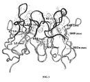
- the three dimensional structure of murine-derived 4C8 monoclonal antibody variable region was built by Insight II software package from Accelrys company. Firstly, the template proteins for 4C8 heavy chain and light chain variable regions proteins were searched in Protein Data Bank (PDB) by BLAST program, respectively. An antibody 1A4J which has the highest homology is selected as modeling template for 4C8, for modeling the three dimensional structure of 4C8 using Insight II program, as shown in Figure 1 .
- PDB Protein Data Bank
- Genbank database was searched for human-derived templates that are most similar to 4C8 light chain and heavy chain variable regions using BLAST program.
- the human-derived antibody having the highest homology with 4C8 heavy chain variable region is human antibody AAC 18206 (GenBank No. AAC18206), with the similarity of 68%
- the human-derived antibody having the highest homology with 4C8 light chain variable region is BAC01734 (GenBank No. BAC01734), with the similarity of 80%. Accordingly, AAC 18206 and BAC01734 were used as the humanized templates for 4C8 heavy chain and light chain, respectively.
- Humanized antibody heavy chain and light chain variable genes (h4C8VHa and h4C8VLa) were synthesized by total gene synthesis, then h4C8VHa gene and pGEM-T/CH vector were used as template to synthesize humanized antibody heavy chain gene by overlapping PCR, the reaction conditions were: 95°C for 15 min; 94°C for 50 sec, 58°C for 50 sec, 72°C for 50 sec, for 30 cycles; 72°C for 10 min. Besides, this humanized heavy chain gene was made restriction enzyme site HindIII and signal peptide gene sequence at 5' end and translation stop code TAA and restriction enzyme site EcoRI at 3' end.
- sequence of signal peptide is shown in SEQ ID NO:13 (ATGGATTTTCAGGTGCAGATTTTCAGCTTCCTGCTAATCAGTGCCTCAGTCATAATATC CAGAGGA).
- the clone with correct sequence was selected and digested by Hind III and EcoRI, after agarose gel electrophoresis, the humanized antibody heavy chain fragment H4C8VHaCH was recovered and ligated with plasmid pcDNA3.1(+)(Invitrogen, USA) digested by HindIII and EcoRI, to construct humanized heavy chain eukaryotic expression vector pcDNA3.1 (+) (h4C8VHaCH).
- Humanized antibody light chain gene was synthesized by overlapping PCR using h4C8VLa gene and pGEM-T/CL vector as template, the reaction conditions were: 95°C for 15 min; 94°C for 50 sec, 58°C for 50 sec, 72°C for 50 sec, for 30 cycles; 72°C for 10 min.
- the PCR product thus obtained is h4C8VLaCL, the 5' end of which contains restriction enzyme site HindIII and signal peptide gene sequence and 3' end of which contains translation stop code TAA and restriction enzyme site EcoRI.
- the sequence of signal peptide is shown in SEQ ID NO:13 (ATGGATTTTCAGGTGCAGATTTTCAGCTTCCTGCTAATCAGTGCCTCAGTCATAATATC CAGAGGA).
- the clone with correct sequence was selected and digested by Hind III and EcoRI, after agarose gel electrophoresis, the humanized antibody light chain fragment H4C8VLaCL was recovered and ligated with plasmid pcDNA3.1(+) vector (Invitrogen, USA) digested also by HindIII and EcoRI, to construct humanized light chain eukaryotic expression vector pcDNA3.1/ZEO(+)(h4C8VLaCL).
- COS-1 cells (ATCC CRL 1650) at 0.8 ⁇ 10 5 /well were inoculated to 24-well tissue culture plate, and cultured to 90-95% fusion using 10% FCS containing RPMI1640/DMEM mixed medium (16/DM medium) to perform transfection: 1 ⁇ g plasmid (0.6 ⁇ g light chain expression vector; 0.4 ⁇ g heavy chain expression vector) and 2 ⁇ l Lipofectamine2000 Reagent were dissolved in 50 ⁇ l serum-free 16/DM medium, respectively, laid still at room temperature for 5 min, the above-mentioned 2 liquids were mixed and incubated at room temperature for 20min to form DNA-liposome complex.
- 1 ⁇ g plasmid 0.6 ⁇ g light chain expression vector; 0.4 ⁇ g heavy chain expression vector
- 2 ⁇ l Lipofectamine2000 Reagent were dissolved in 50 ⁇ l serum-free 16/DM medium, respectively, laid still at room temperature for 5 min, the above-mentioned 2 liquids were mixed and incubated at room temperature for 20min to form DNA-
- the serum containing medium in the 24-well culture plate was replaced by 0.5 ml serum-free 16/DM medium, then the formed DNA-liposome complex was added to the well and cultured in CO 2 incubator for 4 hours (h), then 0.5 ml 16/DM medium containing 20% FCS was supplemented, and cultured in CO 2 incubator continuously.
- the culture supernatants were taken for analysis, the content of antibody in the culture supernatants was determined by ELISA: the goat anti-human IgG (Fc) were coated in ELISA plate, overnight at 4°C, and blocked at 37°C for 2h using 2% BSA-PBS, the culture supernatants to be tested and standard sample (Human myeloma IgG1, ⁇ ) were added and incubated at 37°C for 2h, HRP-goat anti-human ⁇ were added for combining reaction, incubated at 37°C for 1h and TMB was added and effected at 37°C for 5 min, and finally H 2 SO 4 was used to stop the reaction, the OD 450 value was measured.
- Fc goat anti-human IgG
- standard sample Human myeloma IgG1, ⁇
- Human KG-1a cells were re-suspended in 2%FCS-PBS to 1 ⁇ 10 6 cells/ml, adding COS cells culture supernatants transfecting humanized antibody with different dilution degree, respectively, they were incubated at 4°C for 60min, then cells were washed twice with 2% FCS-PBS, and then FITC-goat anti-human IgG (H+L) was added to the cells and incubated at 4°C for 60min, after washing, cells were analyzed by Flow Cytometry (FCM) and calculated for fluorescence intensity.
- FCM Flow Cytometry
- SEQ ID NO:5 and SEQ ID NO:6 show the nucleotide sequence and amino acid sequence of h4C8Hb, respectively.
- SEQ ID NO:7 and SEQ ID NO:8 show the nucleotide sequence and amino acid sequence of h4C8Lb, respectively.
- Three CDR regions amino acid sequences of h4C8Hb are (see, H4C8VHb of Figure.
- HCDR1 GYTFTNYGMN, SEQ ID NO:14
- HCDR2 WINTNTGEPKYAEEFKG, SEQ ID NO:15
- HCDR3 GYGNYARGAWLAY, SEQ ID NO:16
- Three CDR regions amino acid sequences of h4C8Lb are (see, H4C8VLb of Figure. 2-2 ): LCDR1 (RSSQTIVHSNGNTYLE, SEQ ID NO:17), LCDR2 (QVSNRFS, SEQ ID NO:18), LCDR3 (FQGSHVPRT, SEQ ID NO:19), respectively.
- Humanized antibody heavy and light chain variable regions gene (h4C8VHb/h4C8VLb) were synthesized using overlapping PCR method, respectively, and the same method as to humanized antibody (h4C8Ha/h4C8La) were used to construct light chain expression vector pcDNA3.1/ZEO(+) (h4C8VLbCL) and heavy chain expression vector pcDNA3.1 (+) (h4C8VHbCH). Then light and heavy expression vectors were cotransfected into COS-1 cells.
- Flow cytometry assay was performed to determine the antigen-binding activities of the antibody, showing its binding ability to KG-1a is similar to 4C8 chimeric antibody, and thus this humanized antibody (h4C8Hb/h4C8Lb) is designated as h4C8.
- the serum containing medium in the culture dishes was replaced by 3 ml serum-free DMEM medium, then the formed DNA-liposome complex was added to a plate and cultured for 4 hours (h) in CO 2 incubator, then 2 ml DMEM complete medium containing 10% serum was supplemented, and cultured in CO 2 incubator continuously. 24h after transfection, the cell were transferred to selection medium containing 600 ⁇ g/ml G418 and 250 ⁇ g/ml Zeocin to select resistant clones.
- the highly expressed clones obtained by selection was enlarged cultured using serum-free medium, the humanized antibody h4C8 was isolated and purified by Protein A affinity column (GE company). The purified antibody was dialyzed using PBS, and ultraviolet absorption method was used for quantification.
- Activation 0.01mol/L NaH 2 PO 4 Tween (0.05% Tween-20) solution, pH6.0 was taken as activated buffer solution, 2 mg carboxyl-beads (Bangs laboratories, BioMag Carboxyl (BM570), USA) was put into 2ml centrifuge tube, and 500 ⁇ l activated buffer solution was added to mix homogeneously on the vortex mixer, then the centrifuge tube was laid on a Magnetic Separation Rack, after the magnetic beads were completely adsorbed, the supernatant was extracted by mini vacuum pump; 500 ⁇ l activated buffer solution was added to rewash the magnetic beads twice, then 485 ⁇ l activated buffer solution was added to the magnetic beads, and 2.5mg carbodiimide (EDC, original concemation of 0.5g/ml) and N-Hydroxysuccinimide (NHS, original concernation of 0.25g/ml) were added respectively and mixed homogeneously on the vortex mixer to activate the 2 mg carboxyl group on the surface of magnetic beads for
- conjugated buffer 500 ⁇ l, pH7.4, 0.01mol/L phosphate Tween (PBST, 0.05% Tween-20) solution was used as conjugated buffer, 500 ⁇ l activated buffer solution was added to rewash the activated magnetic beads 3 times, then 500 ⁇ l conjugated buffer was added to wash the magnetic beads twice; 475 ⁇ l conjugated buffer was added to re-suspend the washed magnetic beads, and 25 ⁇ l 6mg/ml anti-CD34 antibody was added, allowing the activated carboxyl group on the magnetic beads to react with the amino group of the anti-CD34 antibody at room temperature for 3h, then the antibody was conjugated to the surface of the magnetic beads, yielding the immunomagnetic beads.
- PBST 0.05% Tween-20
- the conjugated magnetic beads were washed by 500 ⁇ l conjugated buffer twice, and 500 ⁇ l conjugated buffer containing 1% bovine serum albumin (BSA) was added to block the magnetic beads for 30 min.
- BSA bovine serum albumin
- the conjugated magnetic beads were washed by 500 ⁇ l conjugated buffer twice, and the magnetic beads were re-suspended in 500 ⁇ l pH7.4, 0.01mol/L phosphate Tween (0.05% Tween-20) solution containing 0.02% NaN 3 , 0.1% BSA, and then stored in refrigerator at 4°C for use.
- CD34+ KG-1a human leukaemia cell
- CD34- Raji human B lymphoma cell
- Preparing 0.1M PBS solution NaCl 8g, KCl 0.2g, Na 2 HPO 4 -12H 2 O 3.488g, KH 2 PO 4 0.2g, adding Milli-Q water to 1000ml, adjusting to pH 7.4, sterilizing using 0.22 ⁇ m filter, storing at 4°C.
- Preparing immunomagnetic beads sorting buffer 0.1M PBS solution containing 2 mM EDTA and 0.5% BSA.
- CD34 immunomagnetic beads into the above-mentioned cell suspension in the ratio of 10 ⁇ l /10 7 cells, mixing and incubating at 4-8°C for 15 min;
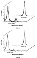
- the cells were stained with CD34-FITC (8G12, BD Bioscience, 340668, having no competitive effect with sorting antibody), the purity of the cells obtained by sorting were identified as more than 95%; the sorting cells were counted, with the yeild of sorting CD34+ cells of more than 90%; the result is shown in Figure 5 .
- CD34-FITC 8G12, BD Bioscience, 340668, having no competitive effect with sorting antibody
- CD34+ blood stem cells were sorted from human umbilical cord blood.
- Preparing 0.1M PBS solution NaCl 8g, KCl 0.2g, Na 2 HPO 4 -12H 2 O 3.488g, KH 2 PO 4 0.2g, adding Milli-Q water to 1000ml, adjusting to pH 7.4, sterilizing using 0.22 ⁇ m filter, storing at 4°C.
- Preparing immunomagnetic beads sorting buffer 0.1M PBS solution containing 2 mM EDTA and 0.5% BSA.
- CD34 immunomagnetic beads into the above-mentioned cell suspension in the ratio of 10 ⁇ l /10 7 cells, mixing and incubating at 4-8°C for 15 min;
- the cells were stained by CD34-FITC (8G12, BD Bioscience, 340668, having no competitive effect with sorting antibody), the purity of the cells obtained by sorting were identified as more than 95%; the sorting cells were counted, with the yeild of sorting CD34+ cells of more than 90%; the result is shown in Figure 6 .
- results indicate the purity and yield obtained by sorting the umbilical cord blood stem cells using the CD34 immunomagnetic beads we prepared has meet the requirement of clinical application, and is expected to be used in clinical transplantation of marrow blood stem cells.
- FITC-CD34 (My10) (BD, 348053) and serial dilution of unlabeled purified antibodies h4C8 and unlabeled My10 (mouse anti-human CD34 hybridoma cells (ATCC, HB8483, anti-My10 clone), were cultured by serum-free medium Hybridoma-SFM (GIBCO, 12045). After purification by rProteinA affinity chromatography (GE), the culture supernatants were mixed respectively and then added into target cells KG-1a (1 ⁇ 10 6 /ml), and incubated at 4°C for 60 min, the cells were washed twice with 1% FCS-PBS and examined by FCM using Cellquest software for analysis.
- GE rProteinA affinity chromatography
- Humanized anti-CD3 monoclonal antibody hu12F6 (preparation see to Li BH, Wang H, Dai JX, Ji JJ, Qian WZ, Zhang DP, Hou S, Guo YJ. Construction and characterization of a humanized anti-human CD3 monoclonal antibody 12F6 with effective immunoregulation functions. Immunology. 2005, 116(4): 487-98 ) was used as control. Triplicate tubes were set for each concentration of the competitive antibody. The IC 50 values were calculated, with the maximum fluorescence intensity indicating the average fluorescence intensity obtained in the absence of competitive antibody. Repeating this test for 3 times, and gaining statistical the result, see to Table 2.
- Example 10 Comparative test for sorting umbilical cord blood stem cells after the conjugation to magnetic beads by humanized anti-CD34 antibody and murine-derived antibody My10
- the mouse anti-human CD34 hybridoma cells (ATCC, HB8483, anti-My10 clone) were cultured using serum-free medium Hybridoma-SFM (GIBCO, 12045). The culture supernatants were purified by rProteinA affinity chromatography (GE), yielding anti-CD34 monoclonal antibody (My10) that meets the requirement of magnetic beads preparing conditions.
- GE rProteinA affinity chromatography
- My10 and h4C8 were conjugated with magnetic beads according to the method of Example 6, to prepare CD34 nano immunomagnetic beads.
- Human umbilical cord blood stem cells were sorted by My10 and h4C8 nano immunomagnetic beads according to the method of Example 8.
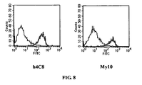
- the positive cells obtained by sorting were counted by blood counting plate, and the yield of the cells obtained by sorting by the two magnetic beads were calculated; the positive cells obtained by sorting were stained by FITC-anti-CD34 (BD, 348053, 8G12), and the purity of the cells obtained by sorting by the two magnetic beads were identified, with the stained results shown in Figure 8 (peak at left side is the test result before sorting, and peak at right side is the test result after sorting).
- the present humanized anti-CD34 antibody retains the affinity and specificity of original murine-derived antibody.
- the antibody can be conjugated with nano magnetic materials to prepare immunomagnetic beads to sort marrow hemopoietic stem cells. It can effectively reduce the incidence rate of HAMA and improve the security of clinical transplantation of hemopoietic stem cells and can be used in treatment of some malignant hematologic diseases and solid tumors.
- the present humanized anti-CD34 antibody is obtained by genetic engineering technology.
Landscapes
- Health & Medical Sciences (AREA)
- Life Sciences & Earth Sciences (AREA)
- Immunology (AREA)
- Chemical & Material Sciences (AREA)
- Engineering & Computer Science (AREA)
- Hematology (AREA)
- Molecular Biology (AREA)
- Urology & Nephrology (AREA)
- Biomedical Technology (AREA)
- Medicinal Chemistry (AREA)
- Cell Biology (AREA)
- General Health & Medical Sciences (AREA)
- Biochemistry (AREA)
- Organic Chemistry (AREA)
- Pathology (AREA)
- General Physics & Mathematics (AREA)
- Analytical Chemistry (AREA)
- Physics & Mathematics (AREA)
- Food Science & Technology (AREA)
- Microbiology (AREA)
- Biotechnology (AREA)
- Tropical Medicine & Parasitology (AREA)
- Chemical Kinetics & Catalysis (AREA)
- Virology (AREA)
- Veterinary Medicine (AREA)
- Nuclear Medicine, Radiotherapy & Molecular Imaging (AREA)
- General Chemical & Material Sciences (AREA)
- Zoology (AREA)
- Animal Behavior & Ethology (AREA)
- Public Health (AREA)
- Pharmacology & Pharmacy (AREA)
- Proteomics, Peptides & Aminoacids (AREA)
- Genetics & Genomics (AREA)
- Biophysics (AREA)
- Bioinformatics & Cheminformatics (AREA)
- Diabetes (AREA)
- Peptides Or Proteins (AREA)
- Micro-Organisms Or Cultivation Processes Thereof (AREA)
- Preparation Of Compounds By Using Micro-Organisms (AREA)
- Measuring Or Testing Involving Enzymes Or Micro-Organisms (AREA)
Abstract
Description
- The present invention relates to biotechnology, and in particular to an antibody, the preparation method and uses thereof.
- Tranplantation of hematopoietic stem/progenitor cells (HSCs) has currently become well-recongnized as the only effective treatment method for certain malignant blood diseases and solid tumors. However, because of numerous T lymphocyte contained in the grafts, acute, severe graft versus-host disease (GVHD) occurs after grafting, which is one of the major cause that affect whether the heterologous genetic hematopoietic stem cells grafting can survival long or not. The primary problem of tranplantation of hematopoietic stem/progenitor cells is how to obtain great amount of purified hematopoietic stem cells to remove the interference of the impurity cells in grafts. Hence, to purge or enrich specific cells in the grafts is a problem concerned in present grafting field. Since HSCs have no definite morphological features, but mainly perform as lymphocyte-like mononuclear blast cells, they can only be indentified by some proteins on the cell surface. CD34, a transmembrane glycoprotein of 110 kDa, is the broadest and earliest antigen so far recognized as expressed on human HSC, and is a good marker to evaluate the quality and quantity of HSC. With the development of CD34 antibody-based positive selection technology, CD34+ hematopoietic stem cells exhibit to reconstitute of the hematopoietic function on experiment animals and human beings. [ Andrews RG, Bryant EM, Bartelmez SH, Muirhead DT, Knitter GH, Bensinger W, Strong DM, Bernstien ID(1992)CD34+ marrow cells, devoid of T and B Lymphocytes, reconstitute stable lymphopoiesis and myelopoiesis in lethally irradiated baboons. Blood 80:1693 ] [ Shpall EJ, Jones RB, Franklin W, Bearman S, Stemmer S, Hami L, Petsche D, Taffs S, Myers S, Purdy M, Heimfeld S, Halligan J, Berenson RJ(1994)Transplantation of autologous CD34+ hematopoietic progenitor cells into breast cancer patient following high-dose chemotherapy. J Clin Oncol 12:28 ], hence, they have become an optimal target cell for hematopoietic stem cells grafting due to their unique biological characteristics and function.
- Currently, the mostly used methods for cell separation and purification included adhesion, differential centrifugation, discontinuous density gradient method and the like. Although these technologies have solved many practical problems, they hinder the progress of cell biology and the development and application of cell biotechnology product due to long operation period, low cell recovery rate, and high cost for separation and purification. Immunomagnetic cell sorting technology is a recently developed new cell separation technology, which has advantages of simple and easy to operate, high separation purity, and retaining the cell activity, etc. and can be used in separating and examining various marrow and blood cells, tumor cells, bacteria and other microorganisms, etc. Thus, it is favored by researchers of biomedical field. Recently, immunomegatic beads are developed gradually towards submicron size, which allows the magnetic beads to separate cells easily and rapidly at low gradient magnetic field, and to perform subsequent analysis and application directly without dissociation of the magnetic beads from cell surface, and the beads have no effect on the cell activities after separation. The principle of separation is: using magnetic nano particles as vector to conjugate the antibody on their surfaces, due to the specific affinity of antibody, the antibody on the magnetic nano particles binds the specific surface antigen on the cells to form cell-antibody-magnetic nano particles complex, under the oriented control of applied magnetic field, target cells can be isolated directly from original cell mixture in one step by the operation of affinity adsorption, washing and desorption, etc. Nano magnetic cells affinity separation has dual advantages, easily and conveniently magnetic separation and high selectivity of affinity separation.
-
EP1083226 discloses chimeric mouse-human mAb anti-CD34, based on murine mAb My10, which can be attached to magnetic Dyna-beads for isolation of CD34-positive cells. - With the coming out of immunomagnetic beads, use of immunomagnetic beads, which is prepared by the conjugation of CD34 monoclonal antibody to magnetic nano microspheres for sorting marrow hematopoietic stem cells has been applied to clinical hematopoietic stem cells transplantation. Owing to the fact that magnetic separation has advantages of easy and convenient operation, rapid and complete separation, and high cell purity, etc when sorting cells, HSC transplantation has become the first choice for certain malignant hematological diseases, severe immunodeficiency and low hematopoietic function resulted from radiotherapy and chemotherapy for tumors. During the course of hematopoietic stem cell transplantation, these antibodies that bind to the magnetic beads will inevitably enter into human body along hematopoietic stem cells. Murine-derived monoclonal antibody produced by hybridoma technology may cause human anti-mouse antibody response (HAMA) after entering human body [ Winter G, Harris WJ. Humanized antibodies. Immunol Today. 1993 Jun; 14(6): 243-6 ]. Hence, it is an urgent problem for those skilled in the art to construct a low immunogenicity antibody which can reduce the murine component, and effectively reduce the incidence rate of HAMA by genetic engineering technology.
- Humanized antibody is a new genetic engineering antibody developed to overcome the deficiency of murine derived monoclonal antibody. The first generation of humanized antibody is a chimeric antibody composed of a variable region of murine monoclonal antibody and a constant region of human antibody. Since the affinity between antibody and antigen is determined by its variable region, chimeric antibody keeps good affinity, meanwhile, the immunogenicity is reduced to a certain extent. The variable regions of antibody are composed of hypervariable regions (CDRs) and frame regions (FRs), wherein CDRs are highly variable regions which mediate directly the binding of antibody to antigen, FR regions are relative conserved, and are used as support to maintain the spatial position of CDR regions which are the main regions to produce immunogenicity in the variable regions. Since chimeric antibody retains the variable region of murine derived antibody, it may still cause strong HAMA reaction in clinical application. Therefore, in order to reduce the immunogenicity of chimeric antibody to the greatest extent, people consider grafting directly murine CDR region to FR region of human derived antibody variable region, yielding CDR graft antibody, namely humanized antibody. However, the transfer of murine CDRs alone usually results in a significant loss of antigen binding, because certain framework residues are critical for preserving the CDR conformations or are directly involved in antigen binding. In most cases, the successful design of humanized antibodies requires that these key murine residues be reintroduced into the human framework to restore affinity. Hence, it is critical for antibody humanization of how to determine the important residues that affect the antibody activity in FR.
- An object of the present invention is to provide a humanized anti-human CD34 antibody, an antibody keeping good affinity obtained by grafting a murine CDR region into a humanized antibody framework region, and at the same time reshaping the amino acids of the humanized antibody framework region.
- Accordingly, the present invention provides a humanized anti-human CD34 antibody, said antibody comprises at least one antigen binding site, said antigen binding site at least comprises:
- A first variable region having an amino acid sequence as shown in amino acids 1-122 of SEQ ID NO: 6; and
- A second variable region having an amino acid sequence as shown in amino acids 1-113 of SEQ ID NO: 8.
- The above first variable region and second variable region correspond to heavy chain variable region and light chain variable region of immunoglobulin, respectively, wherein, a hypervariable regions (CDR) of the first variable region sequentially comprises CDR1, CDR2 and CDR3, of which the amino acid sequences are CDR1: GYTFTNYGMN (SEQ ID NO:14); CDR2: WINTNTGEPKYAEEFKG (SEQ ID NO:15) and CDR3: GYGNYARGAWLAY (SEQ ID NO:16), respectively. Besides the above CDR regions, the first variable region further comprises a framework region (FR) derived from human immunoglobulin heavy chain variable region. In order to improve the affinity of the antibody, the amino acids of the framework region have been partially reshaped in the present invention, thereby obtaining the first variable region having the amino acid sequence of SEQ ID NO: 6, with SEQ ID NO: 5 being the corresponding nucleotide sequence encoding the same. Similarly, a hypervariable regions (CDR) of the second variable region sequentially comprises CDR1', CDR2' and CDR3', of which the amino acid sequences are CDR1': RSSQTIVHSNGNTYLE (SEQ ID NO:17); CDR2': QVSNRFS (SEQ ID NO:18) and CDR3': FQGSHVPRT (SEQ ID NO:19), respectively. Besides the above CDR regions, the second variable region further comprises a framework region (FR) derived from the human immunoglobulin light chain variable region. In order to improve the affinity of the antibody, the amino acids of the framework region have been partially reshaped in the present invention, thereby obtaining the second variable region having the amino acid sequence of SEQ ID NO: 8, with SEQ ID NO: 7 being the corresponding nucleotide sequence encoding the same.
- Further, in the humanized anti-human CD34 antibody of the present invention, said antibody comprises:
- 1) an immunoglobulin heavy chain or fragment thereof, comprising said first variable region and human immunoglobulin heavy chain constant region or fragment thereof; and
- 2) an immunoglobulin light chain or fragment thereof, comprising said second variable region and human immunoglobulin light chain constant region or fragment thereof.
- The human immunoglobulin constant region can be selected from various types of immunoglobulin, preferably, the human immunoglobulin heavy chain constant region or fragment thereof is type γ (IgG1) having the amino acid sequence as shown in SEQ ID NO: 2; the human light chain constant region or fragment thereof is type κ or type λ, a preferable amino acid sequence of the light chain constant region is shown in SEQ ID NO: 4.
- Another object of the present invention is to provide a nucleotide molecule encoding the above-mentioned humanized human CD34 antibody, a host transformed by the nucleotide molecule is also included in the present invention.
- The present invention further provides a preparation method for the above-mentioned humanized anti-CD34 antibody, comprising designing an amino acid sequence of a humanized antibody h4C8 based on computer-assisted molecular modeling, performing whole gene synthesis of the heavy chain and light chain variable region genes and splicing the same with human antibody heavy and light chain constant regions, respectively though genetic recombination, cloning into eukaryotic expression vectors to construct expression vectors for humanized antibody heavy and light chain, then co-transfecting the expression vectors of light and heavy chain into CHO cells by liposome method, and then screening, culturing and purifying to obtain the product.
- A serial of experiments have been carried out in the present invention using the resultant antibody, the result of antigen-binding activities assay in vitro indicates that h4C8 can bind specifically to human myeloid leukemia cell KG-1a which highly expressed antigen CD34. The result of competitive inhibition experiment indicates that h4C8 retains the affinity and specificity of original murine-derived antibody. The antibody can be conjugated with nano magnetic materials to prepare immunomagnetic beads to screen marrow hemopoietic stem cells. It may effectively reduce the incidence rate of HAMA and improve the security of clinical transplantation of hematopoietic stem cells and can be used in the treatment of some malignant hematologic diseases and solid tumors.
- A method of carbodiimide/N-Hydroxysuccinimide (EDC/NHS) is used to activate the carboxyl group on the surface of the magnetic beads, the activated carboxyl group then reacts with the amino group on the antibody, conjugates CD34 antibody, and prepares CD34+ immunomagnetic beads that can effectively isolate cells. A sorting model is prepared using CD34+ KG-1a cells and CD34- Raji cells, and CD34 monoclonal immunomangetic beads are used to sort CD34+ cells. Also, CD34+ immunomagnetic nano particles are used to sort umbilical cord blood stem cell, and the purity and yield of the cells obtained by sorting are analyzed.
- Anti-CD34 monoclonal antibody binding to magnetic nano particles is used as antibody vector to binds CD34+ hematopoietic stem/progenitor cells and thus forms immunocomplex. Under the effect of applied magnetic field, the complex moves dynamically and isolates the hematopoietic stem/progenitor cells that specifically bind the antibody on the magnetic nano particles from other cells (tumor cells that do not express CD34 antigen). The purified hematopoietic stem cells are transfused back to patients to reconstruct the hematopoietic and immune systems and effectively reduce the recurrence of tumors.
-
-
Figure 1 is a schematic view of molecular simulation structure of 4C8 monoclonal antibody: the FR region residues are represented by light grey ribbon, CDR region residues are represented by dark grey ribbon, nine FR region residues within 5 angstrom (Å) around CDR regions are represented by light grey ball-and-stick; -
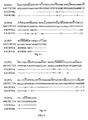
Figure 2 is an alignment view of amino acid sequences of heavy chain (Fig. 2-1 ) and light chain (Fig. 2-2 ) of a humanized antibody h4C8 with relevant sequences, wherein 4C8VH and 4C8VL represent the heavy chain and light chain variable regions of murine-derived monoclonal antibody 4C8, respectively; the heavy chain variable region of human antibody AAC18206 and the light chain variable region of human antibody BAC01734 are selected as the framework regions of the heavy chain and light chain of humanized antibody hu4C8, respectively; hu4C8VHa and hu4C8VHb represent heavy chain variable regions of different humanized antibody, h4C8VLa and h4C8VLb represent light chain variable regions of different humanized antibody, respectively; a dash represents an amino acid which is identical to the corresponding residue of human antibody AAC18206 or BAC01734, CDR regions are represented in the bracket; the amino acids are numbered according to the Kabat numbering manner [ E.A. Kabat, T.T. Wu, H.M. Perry, K.S. Gottesman, C. Foeller, Sequences of Proteins of Immunological Interest, Fifth ed, United States Department of Health and Human Services, Bethesda, MD, 1991 .]; -
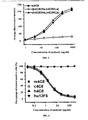
Figure 3 is the result of antigen-binding activities assay of humanized 4C8 antibody; -
Figure 4 is the result of competitive inhibition experiment; -
Figure 5 is the result of CD34+ cell lines sorting experiment using CD34 immunomagnetic beads: front side is the mixed cells before sorting, peak at the left side is CD34- Raji cells, peak at the right side is CD34+ KG-1a cells; back side is positive cell populations after sorting, wherein CD34- Raji cells have been removed to effectively enrich the CD34+ cells; -
Figure 6 is the result of human umbilical cord blood stem cell sorting experiment using CD34 immunomagnetic beads: front side is the umbilical cord blood mononuclear cell before sorting, peak at the left side is CD34- cell populations, peak at the right side is CD34+ cell populations; back side is positive cell populations after sorting, wherein CD34- cells have been removed to effectively enrich the CD34+ stem cells; -
Figure 7 is the result of competitive inhibition experiment of humanized anti-CD34 antibody with murine-derived antibody My 10; -
Figure 8 is the comparative experimental result of umbilical cord blood stem cells sorting after humanized anti-CD34 antibody and murine-derived antibody My10 have conjugated with beads (peak at left side is test result before sorting, and peak at right side is test result after sorting). - The following examples are just further description of the present invention and should not be understood as the limitation to the present invention.
- KG-1a (human leukaemia cell, ATCC, CCL-246.1)
- Raji (human B lymphoma cell, ATCC, CCL-86)
- In the present invention, 4C8 refers to that obtained by the method disclosed in the invention published as Chinese invention patent application No.
200710094456.9 on August 20, 2008 - Healthy human lymphoma cells were isolated from lymphoma cells separation solution (Dingguo Biotechnology Development Company, CHINA), total RNA was extracted using Trizol reagent (Invitrogen), the genes encoding antibody heavy chain and light chain constant region were amplified by RT-PCR reaction, with the primers designed according to the sequences reported in the reference (Cell, 1980, 22: 197-207) and reference (Nucleic Acids Research, 1982, 10: 4071-4079), respectively. The PCR products were purified by agarose gel electrophoresis and recovered and cloned into pGEM-T vector, correct clones were obtained by sequencing verification. SEQ ID NO:1 and SEQ ID NO:2 show the nucleotide sequence and amino acid sequence of the heavy chain constant regions (CH), respectively. SEQ ID NO:3 and SEQ ID NO:4 show the nucleotide sequence and amino acid sequence of the light chain constant regions (CL), respectively. In this example, the correct clones were marked as pGEM-T/CH and pGEM-T/CL.
- BALB/c mice (obtained from SHANGHAI LABORATORY ANIMAL CENTER) were immunized with KG-1a cells which highly express CD34 (KG-1a cell ATCC CCL-246.1), allowing the B lymphocytes cells in the spleen to produce anti-human CD34 antibody, the splenocytes from immunized mice were fused with NS-1 (BALB/c mice myeloma cells), selectively cultured by HAT, after culturing, the anti-human CD34 positive clones were selected and after cloning, the sub-clones were selected to ensure the antibody is produced by single cloning cell, then culture supernatants from each cloning cell were collected , and the anti-human CD34 monoclonal antibody 4C8 was obtained by Protein G column purification.
- Total RNA was extracted from 2× 10⁶ hybridoma cell 4C8 which secrets anti-human CD34 with "Trizol Reagent" reagent kit (Gibco BRL, USA) according to its instructions. Three gene specific primers GSP1, GSP2, GSP3 were designed respectively by selecting the proper positions of antibody (IgG1,κ) heavy chain and light chain constant regions, wherein, GSP1 is apart furthest from the variable region gene, and used for reverse transcription reaction, GSP2 is used for first run PCR amplification, GSP3 is used for nest amplification. The primers were synthesized by SHANGHAI SANGON BIOLOGICAL TECHNOLOGY & SERVICES CO., LTD, the sequences of which were as follows: GSP1-H, 5'-GTA GAG GTC AGA CTG CAG GAC-3' (SEQ ID NO:20); GSP2-H, 5'-CTC AGG GAA ATA GCC CTT GAC-3' (SEQ ID NO:21); GSP3-H, 5'-AGA TCC AGG GGC CAG TGG ATA GAC-3' (SEQ ID NO:22). GSP1-L, 5'-TTG CTG TCC TGA TCA GTC CAA CT-3' (SEQ ID NO:23); GSP2-L, 5'-TGT CGT TCA CTG CCA TCA ATC TT-3' (SEQ ID NO:24); GSP3-L, 5'-TTG TTC AAG AAG CAC ACG ACT GA-3' (SEQ ID NO:25).
- Total RNA was reverse transcribed into cDNA using GSP1 as primer according to the instruction of 5'RACE reagent kit (Gibco BRL, US), then poly(C) tail was added to the 3' end of first chain cDNA, after tailing, PCR amplification was carried out using GSP2 and AAP as primers, the amplification product was diluted 100 times and nest PCR amplification was carried out using AUAP and GSP3 as primer. Both PCR reactions were hot-start PCR, the reaction conditions were: 94°C for 5 minutes (min); 94°C for 45 seconds (sec), 60°C for 45 seconds, 72°C for 70 seconds, for 30 cycles; 72°C for 7 minutes. The products of nest PCR were separated by 1% agarose gel electrophoresis, then the fragment of interest was recovered and purified, and cloned into pGEM-T easy vector, the positive clones were selected and sequenced, and analysis was carried out to the sequencing results. Then the correct pGEM-T/VH confirmed by sequencing was taken as template to design the primers H sense AAG CTT GCC GCC ACC ATG GAT TGG CTG TGG AAC TTG (SEQ ID NO:26) and H antisense GCT AGC TGC AGA GAC AGT GAC CAG (SEQ ID NO:27), VH chain variable region genes were amplified using Onestep RT-PCR reaction to make its 5' end contain restriction enzyme site HindIII, and 3' end contain restriction enzyme site Nhe I, the reaction conditions were: 50°C for 30 min; 95°C for 15 min; 94°C for 50 sec, 58°C for 50 sec, 72°C for 50 sec, for 30 cycles; 72°C for 10 min. The PCR products were purified by agarose gel electrophoresis and recovered and cloned into pGEM-T vector (Promega), the positive clones were selected and verified by sequencing, the result demonstrated that the sequence coincides completely with the sequence of 5'RACE. SEQ ID NO:9 and SEQ ID NO:10 show the nucleotide sequence and amino acid sequence of 4C8 heavy chain variable region, respectively. In this example, the correct clones were designated as pGEM-T/VH.
- Taking the correct pGEM-T/VL verified by sequencing as template to design the primers H sense AAG CTT GCC GCC ACC ATG AAG TTG CCT GTT AGG CTG (SEQ ID NO:28) and L antisense GAC AGA TGG TGC AGC CAC AGT CCG TTT GAT TTC CAG CTT G (SEQ ID NO:29), VL gene was amplified using Onestep RT-PCR reaction to make its 5' end contain restriction enzyme site HindIII, and 3' end contain the complementary sequence of 5' end of human antibody light chain constant region, the reaction conditions were: 94°C for 5 min; 94°C for 50 sec, 58°C for 50 sec, 72°C for 1 min, for 30 cycles; 72°C for 10min. The PCR products were purified by agarose gel electrophoresis and recovered and cloned into pGEM-T vector (Promega), the positive clones were selected and verified by sequencing, the result demonstrated that the sequence coincides completely with the sequence of 5'RACE. SEQ ID NO:11 and SEQ ID NO:12 show the nucleotide sequence and amino acid sequence of 4C8 light chain variable region, respectively. In this example, the correct clones were designated as pGEM-T/VL.
- The above plasmid pGEM-T/VH which was sequenced as correct after Onestep RT-PCR was digested by HindIII and NheI, 430bp digested fragment which was obtained after agarose gel electrophoresis purification and recovery was ligated to plasmid pGEM-T/ CH which was also digested by the same enzymes, the correct clones were selected and digested by HindIII and EcoR I, after agarose gel electrophoresis purification, the fragment of interest was recovered and ligated to pcDNA3.1 (+) (Invitrogen, US) which was digested by the same enzyme using T4 DNA ligase (Invitrogen, US), to construct eukaryotic expression vector pcDNA3.1 (+) (VHCH).
- The above plasmid pGEM-T/VL which was sequenced as correct after Onestep RT-PCR was fused directly to the light chain constant region of correct clone pGEM-T/VL using Overlapping PCR, the reaction conditions were: 50°C for 30 min; 95°C for 15 min; 94°C for 50 sec, 58°C for 50 sec, 72°C for 50 sec, for 30 cycles; 72°C for 10 min, yielding a PCR product VLCL, the 5' end of which contains restriction enzyme site HindIII, and the 3' end contains restriction enzyme site EcoR I. After agarose gel electrophoresis purification, the PCR product was recovered and cloned into pGEM-T vector (Promega), the positive clones were selected and sequenced. The VLCL gene which was sequenced as correct was cleaved from pGEM-T vector by HindIII and EcoR I and cloned into pcDNA3.1/ZEO(+) vector (Invitrogen, US) to construct eukaryotic expression vector pcDNA3.1/ZEO(+) (VLCL).
- 3 × 10⁵ CHO-K1 cells were inoculated to 3.5 cm tissue culture dishes, and transfected when the cells were cultured to 90-95% fusion: 10µg plasmids (4µg plasmid cDNA3.1(+)(VHCH), 6µg plasmid pcDNA3.1/ZEO(+) (VLCL)) and 20µl Lipofectamine2000 Reagent (Invitrogen) were dissolved in 500µl serum-free DMEM medium, respectively, laid still at room temperature for 5 min, the above 2 liquids were mixed and incubated at room temperature for 20min to form DNA-liposome complex. During this period, the serum containing medium in the culture dishes was replaced by 3 ml serum-free DMEM medium, then the formed DNA-liposome complex was added to a plate and cultured for 4 hours (h) in CO2 incubator, then 2ml DMEM complete medium containing 10% serum was supplemented, and cultured in CO2 incubator continuously. 24h after transfection, the cells were transferred to selection medium containing 600µg/ml G418 and 250µg/ml Zeocin to select resistant clones. Cell culture supernatants were taken to select highly expressed clones by ELISA: the goat anti-human IgG (Fc) were coated on ELISA plate, overnight at 4°C, and blocked at 37°C for 2h using 2% BSA-PBS, the resistant clone culture supernatants to be tested or standard sample (Human myeloma IgG1, κ) were added and warm incubated at 37°C for 2h, HRP-goat anti-human IgG(κ) were added for combining reaction, warm incubated at 37°C for 1h and TMB was added and effected at 37°C for 5 min, and finally H2SO4 was used to stop the reaction, A450 value was measured. The highly expressed clones obtained by selection were enlarged cultured using serum-free medium, the chimeric antibody c4C8 was isolated and purified by Protein A affinity column (GE company). The purified antibody was dialyzed using PBS, and ultraviolet absorption method was used for quantification.
- The three dimensional structure of murine-derived 4C8 monoclonal antibody variable region was built by Insight II software package from Accelrys company. Firstly, the template proteins for 4C8 heavy chain and light chain variable regions proteins were searched in Protein Data Bank (PDB) by BLAST program, respectively. An antibody 1A4J which has the highest homology is selected as modeling template for 4C8, for modeling the three dimensional structure of 4C8 using Insight II program, as shown in
Figure 1 . - Genbank database was searched for human-derived templates that are most similar to 4C8 light chain and heavy chain variable regions using BLAST program. The human-derived antibody having the highest homology with 4C8 heavy chain variable region is human antibody AAC 18206 (GenBank No. AAC18206), with the similarity of 68%, and the human-derived antibody having the highest homology with 4C8 light chain variable region is BAC01734 (GenBank No. BAC01734), with the similarity of 80%. Accordingly, AAC 18206 and BAC01734 were used as the humanized templates for 4C8 heavy chain and light chain, respectively. Firstly, 4C8 heavy chain and light chain CDR regions were grafted into humanized templates AAC 18206 and BAC01734 respectively to form CDR grafted antibodies, h4C8Ha and h4C8La for heavy chain and light chain, respectively. The amino acid sequences for h4C8Ha and h4C8La variable regions are shown in
Figure 2 . Humanized antibody heavy chain and light chain variable genes (h4C8VHa and h4C8VLa) were synthesized by total gene synthesis, then h4C8VHa gene and pGEM-T/CH vector were used as template to synthesize humanized antibody heavy chain gene by overlapping PCR, the reaction conditions were: 95°C for 15 min; 94°C for 50 sec, 58°C for 50 sec, 72°C for 50 sec, for 30 cycles; 72°C for 10 min. Besides, this humanized heavy chain gene was made restriction enzyme site HindIII and signal peptide gene sequence at 5' end and translation stop code TAA and restriction enzyme site EcoRI at 3' end. The sequence of signal peptide is shown in SEQ ID NO:13
(ATGGATTTTCAGGTGCAGATTTTCAGCTTCCTGCTAATCAGTGCCTCAGTCATAATATC CAGAGGA). Finally, the PCR amplification products were separated by agarose gel electrophoresis and the band of interest was recovered and cloned into pGEMT vector to select positive clone and sequenced. The clone with correct sequence was selected and digested by Hind III and EcoRI, after agarose gel electrophoresis, the humanized antibody heavy chain fragment H4C8VHaCH was recovered and ligated with plasmid pcDNA3.1(+)(Invitrogen, USA) digested by HindIII and EcoRI, to construct humanized heavy chain eukaryotic expression vector pcDNA3.1 (+) (h4C8VHaCH). - Humanized antibody light chain gene was synthesized by overlapping PCR using h4C8VLa gene and pGEM-T/CL vector as template, the reaction conditions were: 95°C for 15 min; 94°C for 50 sec, 58°C for 50 sec, 72°C for 50 sec, for 30 cycles; 72°C for 10 min. The PCR product thus obtained is h4C8VLaCL, the 5' end of which contains restriction enzyme site HindIII and signal peptide gene sequence and 3' end of which contains translation stop code TAA and restriction enzyme site EcoRI. The sequence of signal peptide is shown in SEQ ID NO:13
(ATGGATTTTCAGGTGCAGATTTTCAGCTTCCTGCTAATCAGTGCCTCAGTCATAATATC CAGAGGA). The clone with correct sequence was selected and digested by Hind III and EcoRI, after agarose gel electrophoresis, the humanized antibody light chain fragment H4C8VLaCL was recovered and ligated with plasmid pcDNA3.1(+) vector (Invitrogen, USA) digested also by HindIII and EcoRI, to construct humanized light chain eukaryotic expression vector pcDNA3.1/ZEO(+)(h4C8VLaCL). - COS-1 cells (ATCC CRL 1650) at 0.8 × 10⁵/well were inoculated to 24-well tissue culture plate, and cultured to 90-95% fusion using 10% FCS containing RPMI1640/DMEM mixed medium (16/DM medium) to perform transfection: 1µg plasmid (0.6µg light chain expression vector; 0.4µg heavy chain expression vector) and 2µl Lipofectamine2000 Reagent were dissolved in 50µl serum-free 16/DM medium, respectively, laid still at room temperature for 5 min, the above-mentioned 2 liquids were mixed and incubated at room temperature for 20min to form DNA-liposome complex. During this period, the serum containing medium in the 24-well culture plate was replaced by 0.5 ml serum-free 16/DM medium, then the formed DNA-liposome complex was added to the well and cultured in CO2 incubator for 4 hours (h), then 0.5 ml 16/DM medium containing 20% FCS was supplemented, and cultured in CO2 incubator continuously. After 72h, the culture supernatants were taken for analysis, the content of antibody in the culture supernatants was determined by ELISA: the goat anti-human IgG (Fc) were coated in ELISA plate, overnight at 4°C, and blocked at 37°C for 2h using 2% BSA-PBS, the culture supernatants to be tested and standard sample (Human myeloma IgG1,κ) were added and incubated at 37°C for 2h, HRP-goat anti-humanκ were added for combining reaction, incubated at 37°C for 1h and TMB was added and effected at 37°C for 5 min, and finally H2SO4 was used to stop the reaction, the OD450 value was measured.
- Human KG-1a cells were re-suspended in 2%FCS-PBS to 1×10⁶ cells/ml, adding COS cells culture supernatants transfecting humanized antibody with different dilution degree, respectively, they were incubated at 4°C for 60min, then cells were washed twice with 2% FCS-PBS, and then FITC-goat anti-human IgG (H+L) was added to the cells and incubated at 4°C for 60min, after washing, cells were analyzed by Flow Cytometry (FCM) and calculated for fluorescence intensity. The result shows that to compared with c4C8 chimeric antibody, the humanized antibody composed of h4C8Ha and h4C8La (h4C8Ha/h4C8La) almost loses its activity completely (
Figure. 3 ). Hence, to obtain humanized antibody with high affinity, we need further to perform analysis and back-mutation on the murine-derived residues in FR region which may possibly affect the 4C8 antibody binding activity. - By analyzing the three-dimensional structure of the modeled 4C8 monoclonal antibody variable region, we have discovered nine framework residues, L3Leu, L4Leu, L46Leu, H2Ile, H46Lys, H68Ala, H69Leu, H82aAsn, and H91Phe within 5A° around the CDR regions might affect the CDR conformation of the original antibody while differ from the corresponding position of humanized template. Remaining these murine-derived amino acid residues in the constructed CDR grafted antibody can yield humanized antibody (h4C8Hb/h4C8Lb). The amino acid sequences for the variable regions of h4C8Hb and h4C8Lb are shown in
Figure 2 . SEQ ID NO:5 and SEQ ID NO:6 show the nucleotide sequence and amino acid sequence of h4C8Hb, respectively. SEQ ID NO:7 and SEQ ID NO:8 show the nucleotide sequence and amino acid sequence of h4C8Lb, respectively. Three CDR regions amino acid sequences of h4C8Hb are (see, H4C8VHb ofFigure. 2-1 ): HCDR1 (GYTFTNYGMN, SEQ ID NO:14), HCDR2 (WINTNTGEPKYAEEFKG, SEQ ID NO:15), HCDR3 (GYGNYARGAWLAY, SEQ ID NO:16), respectively; Three CDR regions amino acid sequences of h4C8Lb are (see, H4C8VLb ofFigure. 2-2 ): LCDR1 (RSSQTIVHSNGNTYLE, SEQ ID NO:17), LCDR2 (QVSNRFS, SEQ ID NO:18), LCDR3 (FQGSHVPRT, SEQ ID NO:19), respectively. Humanized antibody heavy and light chain variable regions gene (h4C8VHb/h4C8VLb) were synthesized using overlapping PCR method, respectively, and the same method as to humanized antibody (h4C8Ha/h4C8La) were used to construct light chain expression vector pcDNA3.1/ZEO(+) (h4C8VLbCL) and heavy chain expression vector pcDNA3.1 (+) (h4C8VHbCH). Then light and heavy expression vectors were cotransfected into COS-1 cells. Flow cytometry assay was performed to determine the antigen-binding activities of the antibody, showing its binding ability to KG-1a is similar to 4C8 chimeric antibody, and thus this humanized antibody (h4C8Hb/h4C8Lb) is designated as h4C8. - 3 × 10⁵ CHO-K1 cells (ATCC CRL-9618) were inoculated to 3.5cm tissue culture dishes, and transfected when the cells were cultured to 90-95% fusion: 10µg plasmids (4µg plasmid pcDNA3.1(+)(Vh4C8HbCH), 6µg plasmid pcDNA3.1/ZEO(+) (Vh4C8LbCL)) and 20µl Lipofectamine2000 Reagent (Invitrogen) were dissolved in 500µl serum- free DMEM medium, respectively, laid still at room temperature for 5 min, the above-mentioned two liquids were mixed and incubated at room temperature for 20min to form DNA-liposome complex. During this period, the serum containing medium in the culture dishes was replaced by 3 ml serum-free DMEM medium, then the formed DNA-liposome complex was added to a plate and cultured for 4 hours (h) in CO2 incubator, then 2 ml DMEM complete medium containing 10% serum was supplemented, and cultured in CO2 incubator continuously. 24h after transfection, the cell were transferred to selection medium containing 600µg/ml G418 and 250µg/ml Zeocin to select resistant clones. Cell culture supernatants were taken to select highly expressed clones: the goat anti-human IgG (Fc) were coated on ELISA plate, overnight at 4°C, and blocked at 37°C for 2h using 2% BSA-PBS, the culture supernatant of resistant clones to be tested or standard sample (Human myeloma IgG1, κ) were added and warm incubated at 37°C for 2h, HRP-goat anti-human IgG(κ) were added for combining reaction, warm-incubated at 37°C for 1h and TMB was added and effected at 37°C for 5 min, and finally H2SO4 was used to stop the reaction, A450 value was measured. The highly expressed clones obtained by selection was enlarged cultured using serum-free medium, the humanized antibody h4C8 was isolated and purified by Protein A affinity column (GE company). The purified antibody was dialyzed using PBS, and ultraviolet absorption method was used for quantification.
- A fixed subsaturating concentration of fluorescently labeled antibody FITC-4C8 and serial dilution of unlabeled purified antibodies were mixed respectively and then added into target KG-1a cells (1×10⁶ /ml), and incubated at 4°C for 60 min, the cells were washed twice with 1% FCS-PBS and examined by FCM using Cellquest software for analysis. Humanized anti-CD3 monoclonal antibody hu12F6 (preparation see to Li BH, Wang H, Dai JX, Ji JJ, Qian WZ, Zhang DP, Hou S, Guo YJ. Construction and characterization of a humanized anti-human CD3 monoclonal antibody 12F6 with effective immunoregulation functions. Immunology. 2005, 116(4): 487-98 ) was used as control. Triplicate tubes were set for each concentration of the competitive antibody. The IC50 values were calculated, with the maximum fluorescence intensity indicating the average fluorescence intensity obtained in the absence of competitive antibody.
- The experimental result is shown in
Figure 4 , antibody h4C8 can completely block the binding of fluorescently labeled antibody FITC-4C8 to KG-1a, their IC50 values were about the same, suggesting that this humanized antibody possessed specificity and affinity similar to that of the original murine- derived antibody (Table 1).Table 1. Result of competitive inhibition analysis Type of antibodies IC50 (µg/ml)a SD n 4C8 1.723 0.145 3 c4C8 1.692 0.137 3 h4C8 1.764 0.158 3 - Activation: 0.01mol/L NaH2PO4 Tween (0.05% Tween-20) solution, pH6.0 was taken as activated buffer solution, 2 mg carboxyl-beads (Bangs laboratories, BioMag Carboxyl (BM570), USA) was put into 2ml centrifuge tube, and 500µl activated buffer solution was added to mix homogeneously on the vortex mixer, then the centrifuge tube was laid on a Magnetic Separation Rack, after the magnetic beads were completely adsorbed, the supernatant was extracted by mini vacuum pump; 500µl activated buffer solution was added to rewash the magnetic beads twice, then 485µl activated buffer solution was added to the magnetic beads, and 2.5mg carbodiimide (EDC, original concemation of 0.5g/ml) and N-Hydroxysuccinimide (NHS, original concernation of 0.25g/ml) were added respectively and mixed homogeneously on the vortex mixer to activate the 2 mg carboxyl group on the surface of magnetic beads for 30 min at room temperature.
- Conjugation: 500µl, pH7.4, 0.01mol/L phosphate Tween (PBST, 0.05% Tween-20) solution was used as conjugated buffer, 500µl activated buffer solution was added to rewash the activated magnetic beads 3 times, then 500µl conjugated buffer was added to wash the magnetic beads twice; 475µl conjugated buffer was added to re-suspend the washed magnetic beads, and 25µl 6mg/ml anti-CD34 antibody was added, allowing the activated carboxyl group on the magnetic beads to react with the amino group of the anti-CD34 antibody at room temperature for 3h, then the antibody was conjugated to the surface of the magnetic beads, yielding the immunomagnetic beads.
- Block: the conjugated magnetic beads were washed by 500µl conjugated buffer twice, and 500µl conjugated buffer containing 1% bovine serum albumin (BSA) was added to block the magnetic beads for 30 min.
- Storage: the conjugated magnetic beads were washed by 500µl conjugated buffer twice, and the magnetic beads were re-suspended in 500µl pH7.4, 0.01mol/L phosphate Tween (0.05% Tween-20) solution containing 0.02% NaN3, 0.1% BSA, and then stored in refrigerator at 4°C for use.
- Mixing CD34+ KG-1a (human leukaemia cell) and CD34- Raji (human B lymphoma cell) in vitro, in the ratio of 1:10, to imitate human umbilical cord blood mononuclear cell (or mobilized peripheral blood mononuclear cell).
- Preparing 0.1M PBS solution: NaCl 8g, KCl 0.2g, Na2HPO4-12H2O 3.488g, KH2PO4 0.2g, adding Milli-Q water to 1000ml, adjusting to pH 7.4, sterilizing using 0.22µm filter, storing at 4°C.
- Preparing immunomagnetic beads sorting buffer: 0.1M PBS solution containing 2 mM EDTA and 0.5% BSA.
- Adding sorting buffer in the ratio of 90µl /107 cells to re-suspend the above-mentioned mixed cells;
- Adding CD34 immunomagnetic beads into the above-mentioned cell suspension in the ratio of 10µl /107 cells, mixing and incubating at 4-8°C for 15 min;
- Adding the sorting buffer in the ratio of 1000µl/107 cells (1000µl is also applicable for cells less than 107) to re-suspend the cells; laying the centrifuge tube in the magnetic field, and removing the supernatants;
- Repeating the above-mentioned steps twice;
- Re-suspending the cell precipitates using 500µl sorting buffer, yielding CD34+ KG-1a cells.
- The cells were stained with CD34-FITC (8G12, BD Bioscience, 340668, having no competitive effect with sorting antibody), the purity of the cells obtained by sorting were identified as more than 95%; the sorting cells were counted, with the yeild of sorting CD34+ cells of more than 90%; the result is shown in
Figure 5 . - CD34+ blood stem cells were sorted from human umbilical cord blood.
- Preparing 0.1M PBS solution: NaCl 8g, KCl 0.2g, Na2HPO4-12H2O 3.488g, KH2PO4 0.2g, adding Milli-Q water to 1000ml, adjusting to pH 7.4, sterilizing using 0.22µm filter, storing at 4°C.
- Preparing immunomagnetic beads sorting buffer: 0.1M PBS solution containing 2 mM EDTA and 0.5% BSA.
- Isolating mononuclear cell from healthy maternal umbilical cord blood using Ficoll.
- Adding sorting buffer in the ratio of 90µl /107 cells to re-suspend the mononuclear cell;
- Adding CD34 immunomagnetic beads into the above-mentioned cell suspension in the ratio of 10µl /107 cells, mixing and incubating at 4-8°C for 15 min;
- Adding the sorting buffer in the ratio of 1000µl/107 cells (1000µl is also applicable for cells less then 107 ) to re-suspend cells; laying the centrifuge tube in the magnetic field, and removing the supernatants;
- Repeating the above-mentioned steps twice;
- Re-suspending the cell precipitates using 500µl sorting buffer, yielding CD34+ blood stem cells.
- The cells were stained by CD34-FITC (8G12, BD Bioscience, 340668, having no competitive effect with sorting antibody), the purity of the cells obtained by sorting were identified as more than 95%; the sorting cells were counted, with the yeild of sorting CD34+ cells of more than 90%; the result is shown in
Figure 6 . - The results indicate the purity and yield obtained by sorting the umbilical cord blood stem cells using the CD34 immunomagnetic beads we prepared has meet the requirement of clinical application, and is expected to be used in clinical transplantation of marrow blood stem cells.
- A subsaturating concentration of FITC-CD34 (My10) (BD, 348053) and serial dilution of unlabeled purified antibodies h4C8 and unlabeled My10 (mouse anti-human CD34 hybridoma cells (ATCC, HB8483, anti-My10 clone), were cultured by serum-free medium Hybridoma-SFM (GIBCO, 12045). After purification by rProteinA affinity chromatography (GE), the culture supernatants were mixed respectively and then added into target cells KG-1a (1×10⁶/ml), and incubated at 4°C for 60 min, the cells were washed twice with 1% FCS-PBS and examined by FCM using Cellquest software for analysis. Humanized anti-CD3 monoclonal antibody hu12F6 (preparation see to Li BH, Wang H, Dai JX, Ji JJ, Qian WZ, Zhang DP, Hou S, Guo YJ. Construction and characterization of a humanized anti-human CD3 monoclonal antibody 12F6 with effective immunoregulation functions. Immunology. 2005, 116(4): 487-98 ) was used as control. Triplicate tubes were set for each concentration of the competitive antibody. The IC50 values were calculated, with the maximum fluorescence intensity indicating the average fluorescence intensity obtained in the absence of competitive antibody. Repeating this test for 3 times, and gaining statistical the result, see to Table 2.
- The test result is shown in
Figure 7 , the IC50 of the binding of fluorescently labeled antibody FITC-My10 to KG-1a cells blocked by antibody h4C8 was smaller than by murine-derived monoclonal antibody My10 (p < 0.05, t-test), hence the affinity of h4C8 is better than murine-derived monoclonal antibody My10.Table 2. Result of competitive inhibition analvsis Type of antibodies IC50(µg/ml) X ± SDp My10 8.67 8.69±0.53 1.54×10⁻⁵ 9.23 8.18 h4C8 1.11 1.09±0.06 1.14 1.02 - The mouse anti-human CD34 hybridoma cells (ATCC, HB8483, anti-My10 clone) were cultured using serum-free medium Hybridoma-SFM (GIBCO, 12045). The culture supernatants were purified by rProteinA affinity chromatography (GE), yielding anti-CD34 monoclonal antibody (My10) that meets the requirement of magnetic beads preparing conditions.
- My10 and h4C8 were conjugated with magnetic beads according to the method of Example 6, to prepare CD34 nano immunomagnetic beads.
- Human umbilical cord blood stem cells were sorted by My10 and h4C8 nano immunomagnetic beads according to the method of Example 8. The positive cells obtained by sorting were counted by blood counting plate, and the yield of the cells obtained by sorting by the two magnetic beads were calculated; the positive cells obtained by sorting were stained by FITC-anti-CD34 (BD, 348053, 8G12), and the purity of the cells obtained by sorting by the two magnetic beads were identified, with the stained results shown in
Figure 8 (peak at left side is the test result before sorting, and peak at right side is the test result after sorting). - The result of comparative test was shown in Table 3.
Table 3. My10 magnetic beads h4C8 magnetic beads Yield (%) 78% 91% Purity (%) 91% 95% - The present humanized anti-CD34 antibody retains the affinity and specificity of original murine-derived antibody. The antibody can be conjugated with nano magnetic materials to prepare immunomagnetic beads to sort marrow hemopoietic stem cells. It can effectively reduce the incidence rate of HAMA and improve the security of clinical transplantation of hemopoietic stem cells and can be used in treatment of some malignant hematologic diseases and solid tumors. The present humanized anti-CD34 antibody is obtained by genetic engineering technology.
-
- <110> Shanghai Guojian Bio-Tech Institute
- <120> Humanized anti-human CD34 antibody, the preparation method and uses thereof
- <130> P2404EP00
- <140> 08865354.8
<141> 2010-06-30 - <150>
CN200710094456.9
<151> 2007-12-13 - <150>
CN200810043016.5
<151> 2008-01-08 - <160> 29
- <170> PatentIn version 3.5
- <210> 1
<211> 990
<212> DNA
<213> Artificial Sequence - <220>
<223> Nucleotide sequence coding the human immunoglobulin heavy chain constant region - <400> 1


- <210> 2
<211> 330
<212> PRT
<213> Artificial Sequence - <220>
<223> The human immunoglobulin heavy chain constant region - <400> 2


- <210> 3
<211> 318
<212> DNA
<213> Artificial Sequence - <220>
<223> Nucleotide sequence coding the human immunoglobulin light chain constant region - <400> 3


- <210> 4
<211> 106
<212> PRT
<213> Artificial Sequence - <220>
<223> The human immunoglobulin light chain constant region - <400> 4

- <210> 5
<211> 1356
<212> DNA
<213> Artificial Sequence - <220>
<223> Nucleotide sequence coding humanized h4C8 antibody heavy chain - <400> 5


- <210> 6
<211> 452
<212> PRT
<213> Artificial Sequence - <220>
<223> humanized h4C8 antibody heavy chain - <400> 6


- <210> 7
<211> 657
<212> DNA
<213> Artificial Sequence - <220>
<223> Nucleotide sequence coding humanized h4C8 antibody light chain - <400> 7

- <210> 8
<211> 219
<212> PRT
<213> Artificial Sequence - <220>
<223> Humanized h4C8 antibody light chain - <400> 8
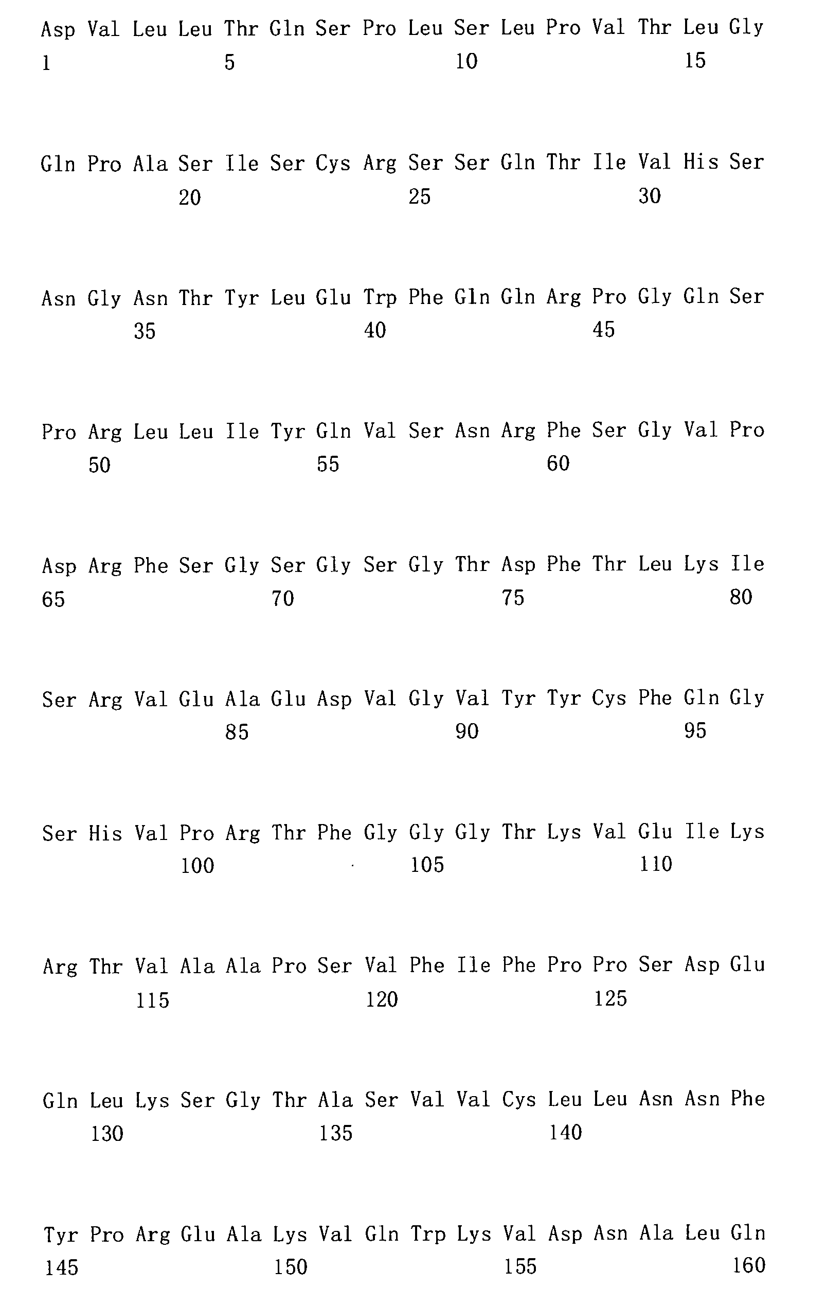
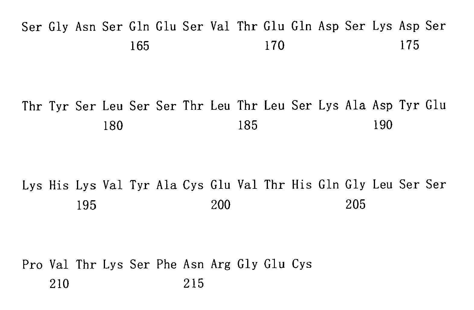
- <210> 9
<211> 366
<212> DNA
<213> Artificial Sequence - <220>
<223> Nucleotide sequence coding 4C8 heavy chain variable region - <400> 9

- <210> 10
<211> 122
<212> PRT
<213> Artificial Sequence - <220>
<223> 4C8 heavy chain variable region - <400> 10

- <210> 11
<211> 339
<212> DNA
<213> Artificial Sequence - <220>
<223> Nucleotide sequence coding 4C8 light chain variable region - <400> 11

- <210> 12
<211> 113
<212> PRT
<213> Artificial Sequence - <220>
<223> 4C8 light chain variable region - <400> 12


- <210> 13
<211> 66
<212> DNA
<213> Artificial Sequence - <220>
<223> Nucleotide sequence coding a signal peptide - <400> 13

- <210> 14
<211> 10
<212> PRT
<213> Artificial Sequence - <220>
<223> Amino acid sequence of CDR1 in the first variable reagion of humanized h4C8 antibody heavy chain - <400> 14

- <210> 15
<211> 17
<212> PRT
<213> Artificial Sequence - <220>
<223> Amino acid sequence of CDR2 in the first variable reagion of humanized h4C8 antibody heavy chain - <400> 15

- <210> 16
<211> 13
<212> PRT
<213> Artificial Sequence - <220>
<223> Amino acid sequence of CDR3 in the first variable reagion of humanized h4C8 antibody heavy chain - <400> 16

- <210> 17
<211> 16
<212> PRT
<213> Artificial Sequence - <220>
<223> Amino acid sequence of CDR1 in the second variable reagion of humanized h4C8 antibody light chain - <400> 17

- <210> 18
<211> 7
<212> PRT
<213> Artificial Sequence - <220>
<223> Amino acid sequence of CDR2 in the second variable reagion of humanized h4C8 antibody light chain - <400> 18

- <210> 19
<211> 9
<212> PRT
<213> Artificial Sequence - <220>
<223> Amino acid sequence of CDR3 in the second variable reagion of humanized h4C8 antibody light chain - <400> 19

- <210> 20
<211> 21
<212> DNA
<213> Artificial Sequence - <220>
<223> Synthetic primer - <400> 20 21
gtagaggtca gactgcagga c 21 - <210> 21
<211> 21
<212> DNA
<213> Artificial Sequence - <220>
<223> Synthetic primer - <400> 21 21
ctcagggaaa tagcccttga c 21 - <210> 22
<211> 24
<212> DNA
<213> Artificial Sequence - <220>
<223> Synthetic primer - <400> 22 24
agatccaggg gccagtggat agac 24 - <210> 23
<211> 23
<212> DNA
<213> Artificial Sequence - <220>
<223> Synthetic primer - <400> 23 23
ttgctgtcct gatcagtcca act 23 - <210> 24
<211> 23
<212> DNA
<213> Artificial Sequence - <220>
<223> Synthetic primer - <400> 24
tgtcgttcac tgccatcaat ctt 23 - <210> 25
<211> 23
<212> DNA
<213> Artificial Sequence - <220>
<223> Synthetic primer - <400> 25
ttgttcaaga agcacacgac tga 23 - <210> 26
<211> 36
<212> DNA
<213> Artificial Sequence - <220>
<223> Synthetic primer - <400> 26
aagcttgccg ccaccatgga ttggctgtgg aacttg 36 - <210> 27
<211> 24
<212> DNA
<213> Artificial Sequence - <220>
<223> Synthetic primer - <400> 27
gctagctgca gagacagtga ccag 24 - <210> 28
<211> 36
<212> DNA
<213> Artificial Sequence - <220>
<223> Synthetic primer - <400> 28
aagcttgccg ccaccatgaa gttgcctgtt aggctg 36 - <210> 29
<211> 40
<212> DNA
<213> Artificial Sequence - <220>
<223> Synthetic primer - <400> 29
gacagatggt gcagccacag tccgtttgat ttccagcttg 40
Claims (10)
- A humanized anti-human CD34 antibody, said antibody comprises at least one antigen binding site, said antigen binding site at least comprising:• a first variable region having an amino acid sequence as shown in amino acids 1-122 of SEQ ID NO: 6; and• a second variable region having an amino acid sequence as shown in amino acids 1-113 of SEQ ID NO: 8.
- The humanized anti-human CD34 antibody of claim 1, wherein said antibody comprises:1) an immunoglobulin heavy chain or fragment thereof, comprising said first variable region and human immunoglobulin heavy chain constant region or fragment thereof; and2) an immunoglobulin light chain or fragment thereof, comprising said second variable region and human immunoglobulin light chain constant region or fragment thereof.
- The humanized anti-human CD34 antibody of any of claims 1-2, wherein said human immunoglobulin heavy chain constant region or fragment thereof is type γ; said human immunoglobulin light chain constant region or fragment thereof is type κ or type λ.
- A nucleotide molecule encoding the humanized anti-human CD34 antibody of any of claims 1-3, said nucleotide molecule at least comprising a nucleotide sequence encoding the first variable region according to claim 1 as shown in nucleotides 1-366 of SEQ ID NO: 5; and a nucleotide sequence encoding the second variable region according to claim 1 as shown in nucleotides 1- 339 of SEQ ID NO: 7.
- The nucleotide molecule of claim 4, comprising the following sequences:a) a nucleotide sequence encoding immunoglobulin heavy chain or fragment thereof, comprising a nucleotide sequence encoding said first variable region and a nucleotide sequence encoding human immunoglobulin heavy chain constant region or fragment thereof; andb) a nucleotide sequence encoding immunoglobulin light chain or fragment thereof, comprising a nucleotide sequence encoding said second variable region and a nucleotide sequence encoding human immunoglobulin light chain constant region or fragment thereof.
- The nucleotide molecule of claim 5, wherein said nucleotide sequence of heavy chain constant region is shown in SEQ ID NO: 1; said nucleotide sequence of light chain constant region is shown in SEQ ID NO: 3.
- A bead comprising the humanized anti-human CD34 antibody of any of claims 1-3.
- The bead of claim 7, wherein said bead is an immunomagnetic bead.
- A use of the humanized anti-human CD34 antibody of any of claims 1-3 in sorting marrow hemopoietic stem/progenitor cells.
- A preparation method of the humanized anti-human CD34 antibody of any of claims 1-3, comprising the following steps:1) transforming an expression vector using the nucleotide molecule of claims 4-6, and transforming a host cell using the obtained expression vector;2) culturing said host cell;3) collecting, separating and purifying the humanized anti-human CD34 antibody expressed by said host cell.
Applications Claiming Priority (3)
| Application Number | Priority Date | Filing Date | Title |
|---|---|---|---|
| CN 200710094456 CN101245108B (en) | 2007-12-13 | 2007-12-13 | Antihuman CD34 antibody, producing process and uses thereof |
| CN200810043016 | 2008-01-08 | ||
| PCT/CN2008/001963 WO2009079922A1 (en) | 2007-12-13 | 2008-12-03 | Humanized anti-cd34 monoclonal antibody, the preparation and uses thereof |
Publications (3)
| Publication Number | Publication Date |
|---|---|
| EP2233501A1 EP2233501A1 (en) | 2010-09-29 |
| EP2233501A4 EP2233501A4 (en) | 2010-12-01 |
| EP2233501B1 true EP2233501B1 (en) | 2012-07-11 |
Family
ID=40800709
Family Applications (1)
| Application Number | Title | Priority Date | Filing Date |
|---|---|---|---|
| EP08865354A Not-in-force EP2233501B1 (en) | 2007-12-13 | 2008-12-03 | Humanized anti-cd34 monoclonal antibody, the preparation and uses thereof |
Country Status (6)
| Country | Link |
|---|---|
| US (1) | US20100311955A1 (en) |
| EP (1) | EP2233501B1 (en) |
| JP (1) | JP2011505810A (en) |
| BR (1) | BRPI0821211A2 (en) |
| CA (1) | CA2708407A1 (en) |
| WO (1) | WO2009079922A1 (en) |
Families Citing this family (19)
| Publication number | Priority date | Publication date | Assignee | Title |
|---|---|---|---|---|
| EP4086338A1 (en) * | 2011-03-17 | 2022-11-09 | Minerva Biotechnologies Corporation | Method for making pluripotent stem cells |
| US8785603B2 (en) | 2011-05-20 | 2014-07-22 | Siemens Healthcare Diagnostics Inc. | Antibodies to 25-hydroxyvitamin D2 and D3 and uses thereof |
| TWI443107B (en) | 2011-12-29 | 2014-07-01 | Ind Tech Res Inst | Humanized anti-human cell surface antigen CD34 monoclonal antibody and application thereof |
| EP3105252B1 (en) | 2014-02-12 | 2019-07-24 | Michael Uhlin | Bispecific antibodies for use in stem cell transplantation |
| US10604586B2 (en) * | 2016-05-09 | 2020-03-31 | Industrial Technology Research Institute | Humanized monoclonal antibody and uses thereof |
| CN109265547B (en) * | 2018-07-23 | 2022-07-26 | 中国科学院微生物研究所 | anti-CD47 antibody and application thereof |
| CN111879923A (en) * | 2020-08-06 | 2020-11-03 | 深圳科隆生物新材料有限公司 | Kit capable of eliminating HAMA effect |
| US20250082777A1 (en) | 2022-01-10 | 2025-03-13 | Sana Biotechnology, Inc. | Methods of ex vivo dosing and administration of lipid particles or viral vectors and related systems and uses |
| EP4466019A4 (en) * | 2022-01-21 | 2026-01-07 | Marengo Therapeutics Inc | Multifunctional molecules with G6B binder and/or CD34 binder and uses thereof |
| WO2024040195A1 (en) | 2022-08-17 | 2024-02-22 | Capstan Therapeutics, Inc. | Conditioning for in vivo immune cell engineering |
| EP4602174A1 (en) | 2022-10-13 | 2025-08-20 | Sana Biotechnology, Inc. | Viral particles targeting hematopoietic stem cells |
| WO2024119157A1 (en) | 2022-12-02 | 2024-06-06 | Sana Biotechnology, Inc. | Lipid particles with cofusogens and methods of producing and using the same |
| AU2024274792A1 (en) | 2023-05-23 | 2025-10-23 | Sana Biotechnology, Inc. | Tandem fusogens and related lipid particles |
| WO2025006799A1 (en) | 2023-06-27 | 2025-01-02 | Capstan Therapeutics, Inc. | Extracorporeal and ex vivo engineering of select cell populations from peripheral blood |
| CN116970650B (en) * | 2023-09-22 | 2023-12-08 | 深圳华大生命科学研究院 | Envelope protein combination, targeting virus vector containing envelope protein combination and preparation method of targeting virus vector |
| WO2025184529A1 (en) | 2024-03-01 | 2025-09-04 | Sana Biotechnology, Inc. | Viral particles with fusogen display and related compositions and methods |
| CN118501458B (en) * | 2024-05-08 | 2025-09-30 | 广东省第二人民医院(广东省卫生应急医院) | A kit and method for the combined detection of multiple immune markers on the surface of peripheral blood extracellular vesicles |
| WO2026156792A1 (en) * | 2025-01-26 | 2026-07-30 | 深圳华大生命科学研究院 | Cd34 antibody and use thereof |
| CN120098129B (en) * | 2025-03-03 | 2026-04-03 | 安徽医科大学第一附属医院 | A CD34 biofusion enzyme antibody with a signal protein tag and its application |
Family Cites Families (7)
| Publication number | Priority date | Publication date | Assignee | Title |
|---|---|---|---|---|
| US5530101A (en) * | 1988-12-28 | 1996-06-25 | Protein Design Labs, Inc. | Humanized immunoglobulins |
| JPH11332578A (en) * | 1998-05-25 | 1999-12-07 | Asahi Chem Ind Co Ltd | Cell separation device and separation method |
| AU3733499A (en) * | 1998-05-25 | 1999-12-13 | Asahi Kasei Kogyo Kabushiki Kaisha | Cell separation device and separation method |
| JPH11332594A (en) * | 1998-05-26 | 1999-12-07 | Asahi Chem Ind Co Ltd | Apparatus and method for separating CD4-positive cells |
| ATE382633T1 (en) * | 2000-05-23 | 2008-01-15 | Baxter Healthcare Sa | CELL SELECTION REAGENTS AND METHOD OF USE |
| US8728525B2 (en) * | 2004-05-12 | 2014-05-20 | Baxter International Inc. | Protein microspheres retaining pharmacokinetic and pharmacodynamic properties |
| CN101245108B (en) * | 2007-12-13 | 2013-04-10 | 上海抗体药物国家工程研究中心有限公司 | Antihuman CD34 antibody, producing process and uses thereof |
-
2008
- 2008-12-03 WO PCT/CN2008/001963 patent/WO2009079922A1/en not_active Ceased
- 2008-12-03 JP JP2010537235A patent/JP2011505810A/en not_active Ceased
- 2008-12-03 EP EP08865354A patent/EP2233501B1/en not_active Not-in-force
- 2008-12-03 BR BRPI0821211-2A patent/BRPI0821211A2/en not_active IP Right Cessation
- 2008-12-03 CA CA2708407A patent/CA2708407A1/en not_active Abandoned
- 2008-12-03 US US12/746,990 patent/US20100311955A1/en not_active Abandoned
Also Published As
| Publication number | Publication date |
|---|---|
| US20100311955A1 (en) | 2010-12-09 |
| EP2233501A1 (en) | 2010-09-29 |
| WO2009079922A1 (en) | 2009-07-02 |
| JP2011505810A (en) | 2011-03-03 |
| EP2233501A4 (en) | 2010-12-01 |
| BRPI0821211A2 (en) | 2015-06-16 |
| CA2708407A1 (en) | 2009-07-02 |
Similar Documents
| Publication | Publication Date | Title |
|---|---|---|
| EP2233501A1 (en) | Humanized anti-cd34 monoclonal antibody, the preparation and uses thereof | |
| CA2375115C (en) | Method for separating cells using immunorosettes | |
| CN107955071B (en) | Human anti-human CD47 antibody and coding gene and application thereof | |
| JP6987640B2 (en) | CD3 specific humanized antibody or fragment thereof | |
| CN106922147B (en) | CD 19-specific antibodies and chimeric antigen receptors | |
| US9624290B2 (en) | Lowered affinity antibodies and methods of making the same | |
| CN107880128B (en) | Fully human antibody or antibody fragment for resisting CD19, and method and application thereof | |
| EP3882276A1 (en) | Bispecific antibody, preparation method therefor and application thereof | |
| CN111349163B (en) | Monoclonal antibodies against CD123 | |
| US20220177579A1 (en) | Cd19 antibodies and methods of using the same | |
| CN110088133A (en) | Anti-idiotype antibodies and related methods | |
| JP2023509821A (en) | Anti-CLL1 antibodies and uses thereof | |
| CN115916817A (en) | Anti-idiotypic antibodies directed against BCMA targeting binding domains and related compositions and methods | |
| JP2022546572A (en) | ANTI-STEAP1 ANTIBODY AND USES THEREOF | |
| US20260015435A1 (en) | Anti-polysialic acid antibodies and uses thereof | |
| CN115335399A (en) | Anti-idiotype antibodies directed against GPRC5D target binding domains and related compositions and methods | |
| CA3149683A1 (en) | Anti-cd33 antibodies for treating cancer | |
| KR102960935B1 (en) | Anti-PD-L1 and PD-L2 antibodies and their derivatives and uses | |
| EP4458853A1 (en) | Development of novel upar single-domain antibody | |
| CN113383017A (en) | Novel bispecific antibody molecules and bispecific antibodies that simultaneously bind PD-L1 and LAG-3 | |
| CN116848147A (en) | CD19 humanized antibodies and their applications | |
| CN101245108B (en) | Antihuman CD34 antibody, producing process and uses thereof | |
| CN117209608A (en) | An antibody targeting CD47 and its application | |
| CN110615841B (en) | Anti-human CD47 monoclonal antibody and application thereof | |
| CN101481417A (en) | Antihuman CD34 humanized antibody, preparation and use thereof |
Legal Events
| Date | Code | Title | Description |
|---|---|---|---|
| PUAI | Public reference made under article 153(3) epc to a published international application that has entered the european phase |
Free format text: ORIGINAL CODE: 0009012 |
|
| 17P | Request for examination filed |
Effective date: 20100713 |
|
| AK | Designated contracting states |
Kind code of ref document: A1 Designated state(s): AT BE BG CH CY CZ DE DK EE ES FI FR GB GR HR HU IE IS IT LI LT LU LV MC MT NL NO PL PT RO SE SI SK TR |
|
| AX | Request for extension of the european patent |
Extension state: AL BA MK RS |
|
| A4 | Supplementary search report drawn up and despatched |
Effective date: 20101103 |
|
| RIC1 | Information provided on ipc code assigned before grant |
Ipc: C12N 15/13 20060101ALI20101022BHEP Ipc: A61P 7/06 20060101ALI20101022BHEP Ipc: A61K 39/44 20060101ALI20101022BHEP Ipc: C12P 21/08 20060101ALI20101022BHEP Ipc: A61P 35/00 20060101ALI20101022BHEP Ipc: C07K 16/28 20060101AFI20090717BHEP Ipc: G01N 33/543 20060101ALI20101022BHEP |
|
| DAX | Request for extension of the european patent (deleted) | ||
| 17Q | First examination report despatched |
Effective date: 20110713 |
|
| GRAP | Despatch of communication of intention to grant a patent |
Free format text: ORIGINAL CODE: EPIDOSNIGR1 |
|
| RIC1 | Information provided on ipc code assigned before grant |
Ipc: G01N 33/569 20060101ALI20111201BHEP Ipc: A61P 35/00 20060101ALI20111201BHEP Ipc: C07K 16/28 20060101AFI20111201BHEP Ipc: A61P 7/06 20060101ALI20111201BHEP Ipc: G01N 33/574 20060101ALI20111201BHEP Ipc: G01N 33/543 20060101ALI20111201BHEP Ipc: A61K 39/44 20060101ALI20111201BHEP Ipc: C12P 21/08 20060101ALI20111201BHEP Ipc: C12N 15/13 20060101ALI20111201BHEP |
|
| GRAS | Grant fee paid |
Free format text: ORIGINAL CODE: EPIDOSNIGR3 |
|
| GRAA | (expected) grant |
Free format text: ORIGINAL CODE: 0009210 |
|
| AK | Designated contracting states |
Kind code of ref document: B1 Designated state(s): AT BE BG CH CY CZ DE DK EE ES FI FR GB GR HR HU IE IS IT LI LT LU LV MC MT NL NO PL PT RO SE SI SK TR |
|
| REG | Reference to a national code |
Ref country code: GB Ref legal event code: FG4D |
|
| REG | Reference to a national code |
Ref country code: CH Ref legal event code: EP |
|
| REG | Reference to a national code |
Ref country code: AT Ref legal event code: REF Ref document number: 566110 Country of ref document: AT Kind code of ref document: T Effective date: 20120715 |
|
| REG | Reference to a national code |
Ref country code: IE Ref legal event code: FG4D |
|
| REG | Reference to a national code |
Ref country code: DE Ref legal event code: R096 Ref document number: 602008017205 Country of ref document: DE Effective date: 20120906 |
|
| REG | Reference to a national code |
Ref country code: NL Ref legal event code: VDEP Effective date: 20120711 |
|
| REG | Reference to a national code |
Ref country code: AT Ref legal event code: MK05 Ref document number: 566110 Country of ref document: AT Kind code of ref document: T Effective date: 20120711 |
|
| REG | Reference to a national code |
Ref country code: LT Ref legal event code: MG4D Effective date: 20120711 |
|
| PG25 | Lapsed in a contracting state [announced via postgrant information from national office to epo] |
Ref country code: IS Free format text: LAPSE BECAUSE OF FAILURE TO SUBMIT A TRANSLATION OF THE DESCRIPTION OR TO PAY THE FEE WITHIN THE PRESCRIBED TIME-LIMIT Effective date: 20121111 Ref country code: NO Free format text: LAPSE BECAUSE OF FAILURE TO SUBMIT A TRANSLATION OF THE DESCRIPTION OR TO PAY THE FEE WITHIN THE PRESCRIBED TIME-LIMIT Effective date: 20121011 Ref country code: BE Free format text: LAPSE BECAUSE OF FAILURE TO SUBMIT A TRANSLATION OF THE DESCRIPTION OR TO PAY THE FEE WITHIN THE PRESCRIBED TIME-LIMIT Effective date: 20120711 Ref country code: LT Free format text: LAPSE BECAUSE OF FAILURE TO SUBMIT A TRANSLATION OF THE DESCRIPTION OR TO PAY THE FEE WITHIN THE PRESCRIBED TIME-LIMIT Effective date: 20120711 Ref country code: FI Free format text: LAPSE BECAUSE OF FAILURE TO SUBMIT A TRANSLATION OF THE DESCRIPTION OR TO PAY THE FEE WITHIN THE PRESCRIBED TIME-LIMIT Effective date: 20120711 Ref country code: AT Free format text: LAPSE BECAUSE OF FAILURE TO SUBMIT A TRANSLATION OF THE DESCRIPTION OR TO PAY THE FEE WITHIN THE PRESCRIBED TIME-LIMIT Effective date: 20120711 Ref country code: CY Free format text: LAPSE BECAUSE OF FAILURE TO SUBMIT A TRANSLATION OF THE DESCRIPTION OR TO PAY THE FEE WITHIN THE PRESCRIBED TIME-LIMIT Effective date: 20120711 Ref country code: HR Free format text: LAPSE BECAUSE OF FAILURE TO SUBMIT A TRANSLATION OF THE DESCRIPTION OR TO PAY THE FEE WITHIN THE PRESCRIBED TIME-LIMIT Effective date: 20120711 |
|
| PG25 | Lapsed in a contracting state [announced via postgrant information from national office to epo] |
Ref country code: LV Free format text: LAPSE BECAUSE OF FAILURE TO SUBMIT A TRANSLATION OF THE DESCRIPTION OR TO PAY THE FEE WITHIN THE PRESCRIBED TIME-LIMIT Effective date: 20120711 Ref country code: PL Free format text: LAPSE BECAUSE OF FAILURE TO SUBMIT A TRANSLATION OF THE DESCRIPTION OR TO PAY THE FEE WITHIN THE PRESCRIBED TIME-LIMIT Effective date: 20120711 Ref country code: SI Free format text: LAPSE BECAUSE OF FAILURE TO SUBMIT A TRANSLATION OF THE DESCRIPTION OR TO PAY THE FEE WITHIN THE PRESCRIBED TIME-LIMIT Effective date: 20120711 Ref country code: GR Free format text: LAPSE BECAUSE OF FAILURE TO SUBMIT A TRANSLATION OF THE DESCRIPTION OR TO PAY THE FEE WITHIN THE PRESCRIBED TIME-LIMIT Effective date: 20121012 Ref country code: PT Free format text: LAPSE BECAUSE OF FAILURE TO SUBMIT A TRANSLATION OF THE DESCRIPTION OR TO PAY THE FEE WITHIN THE PRESCRIBED TIME-LIMIT Effective date: 20121112 Ref country code: SE Free format text: LAPSE BECAUSE OF FAILURE TO SUBMIT A TRANSLATION OF THE DESCRIPTION OR TO PAY THE FEE WITHIN THE PRESCRIBED TIME-LIMIT Effective date: 20120711 |
|
| PG25 | Lapsed in a contracting state [announced via postgrant information from national office to epo] |
Ref country code: NL Free format text: LAPSE BECAUSE OF FAILURE TO SUBMIT A TRANSLATION OF THE DESCRIPTION OR TO PAY THE FEE WITHIN THE PRESCRIBED TIME-LIMIT Effective date: 20120711 |
|
| PG25 | Lapsed in a contracting state [announced via postgrant information from national office to epo] |
Ref country code: EE Free format text: LAPSE BECAUSE OF FAILURE TO SUBMIT A TRANSLATION OF THE DESCRIPTION OR TO PAY THE FEE WITHIN THE PRESCRIBED TIME-LIMIT Effective date: 20120711 Ref country code: CZ Free format text: LAPSE BECAUSE OF FAILURE TO SUBMIT A TRANSLATION OF THE DESCRIPTION OR TO PAY THE FEE WITHIN THE PRESCRIBED TIME-LIMIT Effective date: 20120711 Ref country code: RO Free format text: LAPSE BECAUSE OF FAILURE TO SUBMIT A TRANSLATION OF THE DESCRIPTION OR TO PAY THE FEE WITHIN THE PRESCRIBED TIME-LIMIT Effective date: 20120711 Ref country code: DK Free format text: LAPSE BECAUSE OF FAILURE TO SUBMIT A TRANSLATION OF THE DESCRIPTION OR TO PAY THE FEE WITHIN THE PRESCRIBED TIME-LIMIT Effective date: 20120711 |
|
| PLBE | No opposition filed within time limit |
Free format text: ORIGINAL CODE: 0009261 |
|
| STAA | Information on the status of an ep patent application or granted ep patent |
Free format text: STATUS: NO OPPOSITION FILED WITHIN TIME LIMIT |
|
| PG25 | Lapsed in a contracting state [announced via postgrant information from national office to epo] |
Ref country code: IT Free format text: LAPSE BECAUSE OF FAILURE TO SUBMIT A TRANSLATION OF THE DESCRIPTION OR TO PAY THE FEE WITHIN THE PRESCRIBED TIME-LIMIT Effective date: 20120711 Ref country code: SK Free format text: LAPSE BECAUSE OF FAILURE TO SUBMIT A TRANSLATION OF THE DESCRIPTION OR TO PAY THE FEE WITHIN THE PRESCRIBED TIME-LIMIT Effective date: 20120711 |
|
| 26N | No opposition filed |
Effective date: 20130412 |
|
| PG25 | Lapsed in a contracting state [announced via postgrant information from national office to epo] |
Ref country code: BG Free format text: LAPSE BECAUSE OF FAILURE TO SUBMIT A TRANSLATION OF THE DESCRIPTION OR TO PAY THE FEE WITHIN THE PRESCRIBED TIME-LIMIT Effective date: 20121011 Ref country code: MC Free format text: LAPSE BECAUSE OF NON-PAYMENT OF DUE FEES Effective date: 20121231 |
|
| REG | Reference to a national code |
Ref country code: CH Ref legal event code: PL |
|
| REG | Reference to a national code |
Ref country code: DE Ref legal event code: R097 Ref document number: 602008017205 Country of ref document: DE Effective date: 20130412 |
|
| GBPC | Gb: european patent ceased through non-payment of renewal fee |
Effective date: 20121203 |
|
| REG | Reference to a national code |
Ref country code: IE Ref legal event code: MM4A |
|
| REG | Reference to a national code |
Ref country code: FR Ref legal event code: ST Effective date: 20130830 |
|
| PG25 | Lapsed in a contracting state [announced via postgrant information from national office to epo] |
Ref country code: ES Free format text: LAPSE BECAUSE OF FAILURE TO SUBMIT A TRANSLATION OF THE DESCRIPTION OR TO PAY THE FEE WITHIN THE PRESCRIBED TIME-LIMIT Effective date: 20121022 Ref country code: DE Free format text: LAPSE BECAUSE OF NON-PAYMENT OF DUE FEES Effective date: 20130702 Ref country code: LI Free format text: LAPSE BECAUSE OF NON-PAYMENT OF DUE FEES Effective date: 20121231 Ref country code: CH Free format text: LAPSE BECAUSE OF NON-PAYMENT OF DUE FEES Effective date: 20121231 Ref country code: IE Free format text: LAPSE BECAUSE OF NON-PAYMENT OF DUE FEES Effective date: 20121203 |
|
| REG | Reference to a national code |
Ref country code: DE Ref legal event code: R119 Ref document number: 602008017205 Country of ref document: DE Effective date: 20130702 |
|
| PG25 | Lapsed in a contracting state [announced via postgrant information from national office to epo] |
Ref country code: FR Free format text: LAPSE BECAUSE OF NON-PAYMENT OF DUE FEES Effective date: 20130102 Ref country code: MT Free format text: LAPSE BECAUSE OF FAILURE TO SUBMIT A TRANSLATION OF THE DESCRIPTION OR TO PAY THE FEE WITHIN THE PRESCRIBED TIME-LIMIT Effective date: 20120711 Ref country code: GB Free format text: LAPSE BECAUSE OF NON-PAYMENT OF DUE FEES Effective date: 20121203 |
|
| PG25 | Lapsed in a contracting state [announced via postgrant information from national office to epo] |
Ref country code: TR Free format text: LAPSE BECAUSE OF FAILURE TO SUBMIT A TRANSLATION OF THE DESCRIPTION OR TO PAY THE FEE WITHIN THE PRESCRIBED TIME-LIMIT Effective date: 20120711 |
|
| PG25 | Lapsed in a contracting state [announced via postgrant information from national office to epo] |
Ref country code: LU Free format text: LAPSE BECAUSE OF NON-PAYMENT OF DUE FEES Effective date: 20121203 |
|
| PG25 | Lapsed in a contracting state [announced via postgrant information from national office to epo] |
Ref country code: HU Free format text: LAPSE BECAUSE OF FAILURE TO SUBMIT A TRANSLATION OF THE DESCRIPTION OR TO PAY THE FEE WITHIN THE PRESCRIBED TIME-LIMIT Effective date: 20081203 |