EP2222875B1 - Methods and compositions for assessing responsiveness of b-cell lymphoma to treatment with anti-cd40 antibodies - Google Patents
Methods and compositions for assessing responsiveness of b-cell lymphoma to treatment with anti-cd40 antibodies Download PDFInfo
- Publication number
- EP2222875B1 EP2222875B1 EP08848388.8A EP08848388A EP2222875B1 EP 2222875 B1 EP2222875 B1 EP 2222875B1 EP 08848388 A EP08848388 A EP 08848388A EP 2222875 B1 EP2222875 B1 EP 2222875B1
- Authority
- EP
- European Patent Office
- Prior art keywords
- seq
- nos
- antibody
- lymphoma
- sample
- Prior art date
- Legal status (The legal status is an assumption and is not a legal conclusion. Google has not performed a legal analysis and makes no representation as to the accuracy of the status listed.)
- Active
Links
- 238000000034 method Methods 0.000 title claims description 123
- 238000011282 treatment Methods 0.000 title claims description 108
- 208000003950 B-cell lymphoma Diseases 0.000 title claims description 64
- 230000004043 responsiveness Effects 0.000 title claims description 34
- 239000000203 mixture Substances 0.000 title description 14
- 108090000623 proteins and genes Proteins 0.000 claims description 307
- 239000000523 sample Substances 0.000 claims description 232
- 230000014509 gene expression Effects 0.000 claims description 201
- 210000004027 cell Anatomy 0.000 claims description 164
- 239000003550 marker Substances 0.000 claims description 106
- 101150013553 CD40 gene Proteins 0.000 claims description 70
- 102100040245 Tumor necrosis factor receptor superfamily member 5 Human genes 0.000 claims description 70
- 230000035945 sensitivity Effects 0.000 claims description 69
- 206010025323 Lymphomas Diseases 0.000 claims description 54
- 102100038080 B-cell receptor CD22 Human genes 0.000 claims description 52
- 101000884305 Homo sapiens B-cell receptor CD22 Proteins 0.000 claims description 52
- 206010028980 Neoplasm Diseases 0.000 claims description 52
- 101001012021 Homo sapiens Mammalian ependymin-related protein 1 Proteins 0.000 claims description 51
- 102100030031 Mammalian ependymin-related protein 1 Human genes 0.000 claims description 50
- 101000854774 Homo sapiens Pantetheine hydrolase VNN2 Proteins 0.000 claims description 47
- 102100020748 Pantetheine hydrolase VNN2 Human genes 0.000 claims description 47
- 101710148333 Regulator of G-protein signaling 13 Proteins 0.000 claims description 46
- 208000031671 Large B-Cell Diffuse Lymphoma Diseases 0.000 claims description 45
- 206010012818 diffuse large B-cell lymphoma Diseases 0.000 claims description 45
- 208000015914 Non-Hodgkin lymphomas Diseases 0.000 claims description 39
- 101000933604 Homo sapiens Protein BTG2 Proteins 0.000 claims description 37
- 102100026034 Protein BTG2 Human genes 0.000 claims description 37
- 101001034844 Homo sapiens Interferon-induced transmembrane protein 1 Proteins 0.000 claims description 35
- 102100040021 Interferon-induced transmembrane protein 1 Human genes 0.000 claims description 35
- 102100039688 Insulin-like growth factor 1 receptor Human genes 0.000 claims description 34
- 102100021631 B-cell lymphoma 6 protein Human genes 0.000 claims description 33
- 101001034652 Homo sapiens Insulin-like growth factor 1 receptor Proteins 0.000 claims description 33
- 102100032912 CD44 antigen Human genes 0.000 claims description 32
- 101000868273 Homo sapiens CD44 antigen Proteins 0.000 claims description 32
- 101000662009 Homo sapiens UDP-N-acetylglucosamine pyrophosphorylase Proteins 0.000 claims description 32
- 102100037921 UDP-N-acetylglucosamine pyrophosphorylase Human genes 0.000 claims description 32
- 102100027203 B-cell antigen receptor complex-associated protein beta chain Human genes 0.000 claims description 31
- 101000914491 Homo sapiens B-cell antigen receptor complex-associated protein beta chain Proteins 0.000 claims description 31
- 101000971234 Homo sapiens B-cell lymphoma 6 protein Proteins 0.000 claims description 31
- 101001111742 Homo sapiens Rhombotin-2 Proteins 0.000 claims description 31
- 102100023876 Rhombotin-2 Human genes 0.000 claims description 31
- 102100029921 Dipeptidyl peptidase 1 Human genes 0.000 claims description 28
- 101000793922 Homo sapiens Dipeptidyl peptidase 1 Proteins 0.000 claims description 28
- 101000590549 Homo sapiens Pseudouridylate synthase 7 homolog Proteins 0.000 claims description 26
- 102100032494 Pseudouridylate synthase 7 homolog Human genes 0.000 claims description 26
- 239000000556 agonist Substances 0.000 claims description 25
- 108010029697 CD40 Ligand Proteins 0.000 claims description 22
- 229910052796 boron Inorganic materials 0.000 claims description 22
- 102100032937 CD40 ligand Human genes 0.000 claims description 20
- 102000004169 proteins and genes Human genes 0.000 claims description 18
- 208000025205 Mantle-Cell Lymphoma Diseases 0.000 claims description 14
- 239000003153 chemical reaction reagent Substances 0.000 claims description 14
- 230000003247 decreasing effect Effects 0.000 claims description 14
- 125000003275 alpha amino acid group Chemical group 0.000 claims description 12
- 239000012188 paraffin wax Substances 0.000 claims description 11
- 208000010839 B-cell chronic lymphocytic leukemia Diseases 0.000 claims description 9
- 208000031422 Lymphocytic Chronic B-Cell Leukemia Diseases 0.000 claims description 9
- 201000003444 follicular lymphoma Diseases 0.000 claims description 9
- WSFSSNUMVMOOMR-UHFFFAOYSA-N Formaldehyde Chemical compound O=C WSFSSNUMVMOOMR-UHFFFAOYSA-N 0.000 claims description 8
- 238000004364 calculation method Methods 0.000 claims description 8
- 239000013610 patient sample Substances 0.000 claims description 8
- 238000001574 biopsy Methods 0.000 claims description 7
- 230000003993 interaction Effects 0.000 claims description 7
- 208000021937 marginal zone lymphoma Diseases 0.000 claims description 7
- 238000011529 RT qPCR Methods 0.000 claims description 5
- 201000007924 marginal zone B-cell lymphoma Diseases 0.000 claims description 5
- 102100021035 Regulator of G-protein signaling 18 Human genes 0.000 claims 9
- 210000001519 tissue Anatomy 0.000 description 68
- 238000003556 assay Methods 0.000 description 51
- 102100035776 Regulator of G-protein signaling 13 Human genes 0.000 description 40
- 208000037265 diseases, disorders, signs and symptoms Diseases 0.000 description 32
- 108020004999 messenger RNA Proteins 0.000 description 30
- 201000010099 disease Diseases 0.000 description 29
- 230000027455 binding Effects 0.000 description 28
- 239000000090 biomarker Substances 0.000 description 28
- 230000008859 change Effects 0.000 description 25
- 102000040430 polynucleotide Human genes 0.000 description 24
- 108091033319 polynucleotide Proteins 0.000 description 24
- 239000002157 polynucleotide Substances 0.000 description 24
- -1 ELTI1902 Proteins 0.000 description 23
- 239000000758 substrate Substances 0.000 description 23
- 108091034117 Oligonucleotide Proteins 0.000 description 22
- 102000055120 MEF2 Transcription Factors Human genes 0.000 description 20
- 108010018650 MEF2 Transcription Factors Proteins 0.000 description 20
- 230000000875 corresponding effect Effects 0.000 description 20
- 239000000427 antigen Substances 0.000 description 19
- 150000007523 nucleic acids Chemical group 0.000 description 19
- 238000003762 quantitative reverse transcription PCR Methods 0.000 description 19
- 238000004458 analytical method Methods 0.000 description 18
- 108091007433 antigens Proteins 0.000 description 18
- 102000036639 antigens Human genes 0.000 description 18
- 230000004044 response Effects 0.000 description 18
- 102000004190 Enzymes Human genes 0.000 description 17
- 108090000790 Enzymes Proteins 0.000 description 17
- 229940088598 enzyme Drugs 0.000 description 17
- 102000039446 nucleic acids Human genes 0.000 description 16
- 108020004707 nucleic acids Proteins 0.000 description 16
- 238000002493 microarray Methods 0.000 description 15
- 125000003729 nucleotide group Chemical group 0.000 description 15
- 239000007787 solid Substances 0.000 description 15
- 238000003364 immunohistochemistry Methods 0.000 description 14
- 230000009467 reduction Effects 0.000 description 14
- 101001045695 Homo sapiens Phosphoribosyl pyrophosphate synthase-associated protein 2 Proteins 0.000 description 12
- 108060003951 Immunoglobulin Proteins 0.000 description 12
- 102100022060 Phosphoribosyl pyrophosphate synthase-associated protein 2 Human genes 0.000 description 12
- 210000003719 b-lymphocyte Anatomy 0.000 description 12
- 238000009396 hybridization Methods 0.000 description 12
- 102000018358 immunoglobulin Human genes 0.000 description 12
- 238000005259 measurement Methods 0.000 description 12
- 101001026232 Homo sapiens Small conductance calcium-activated potassium channel protein 3 Proteins 0.000 description 11
- 102100037442 Small conductance calcium-activated potassium channel protein 3 Human genes 0.000 description 11
- 230000010056 antibody-dependent cellular cytotoxicity Effects 0.000 description 11
- 239000002773 nucleotide Substances 0.000 description 11
- 238000002360 preparation method Methods 0.000 description 11
- 238000010186 staining Methods 0.000 description 11
- 102100030374 Actin, cytoplasmic 2 Human genes 0.000 description 10
- 101000773237 Homo sapiens Actin, cytoplasmic 2 Proteins 0.000 description 10
- 101000789800 Homo sapiens Protein YIPF3 Proteins 0.000 description 10
- 101000617738 Homo sapiens Survival motor neuron protein Proteins 0.000 description 10
- 102100028077 Protein YIPF3 Human genes 0.000 description 10
- 102100021947 Survival motor neuron protein Human genes 0.000 description 10
- 230000003321 amplification Effects 0.000 description 10
- 239000003814 drug Substances 0.000 description 10
- 239000012634 fragment Substances 0.000 description 10
- 238000003199 nucleic acid amplification method Methods 0.000 description 10
- 230000007170 pathology Effects 0.000 description 10
- 230000015572 biosynthetic process Effects 0.000 description 9
- 201000011510 cancer Diseases 0.000 description 9
- 238000001514 detection method Methods 0.000 description 9
- 230000000694 effects Effects 0.000 description 9
- 230000002255 enzymatic effect Effects 0.000 description 9
- 239000011521 glass Substances 0.000 description 9
- 229920001184 polypeptide Polymers 0.000 description 9
- 102000004196 processed proteins & peptides Human genes 0.000 description 9
- 108090000765 processed proteins & peptides Proteins 0.000 description 9
- 238000003757 reverse transcription PCR Methods 0.000 description 9
- 108091032973 (ribonucleotides)n+m Proteins 0.000 description 8
- 101001051777 Homo sapiens Protein kinase C alpha type Proteins 0.000 description 8
- 101000965721 Homo sapiens Volume-regulated anion channel subunit LRRC8A Proteins 0.000 description 8
- 102100026894 Lymphotoxin-beta Human genes 0.000 description 8
- 102100024924 Protein kinase C alpha type Human genes 0.000 description 8
- 102100040985 Volume-regulated anion channel subunit LRRC8A Human genes 0.000 description 8
- JLCPHMBAVCMARE-UHFFFAOYSA-N [3-[[3-[[3-[[3-[[3-[[3-[[3-[[3-[[3-[[3-[[3-[[5-(2-amino-6-oxo-1H-purin-9-yl)-3-[[3-[[3-[[3-[[3-[[3-[[5-(2-amino-6-oxo-1H-purin-9-yl)-3-[[5-(2-amino-6-oxo-1H-purin-9-yl)-3-hydroxyoxolan-2-yl]methoxy-hydroxyphosphoryl]oxyoxolan-2-yl]methoxy-hydroxyphosphoryl]oxy-5-(5-methyl-2,4-dioxopyrimidin-1-yl)oxolan-2-yl]methoxy-hydroxyphosphoryl]oxy-5-(6-aminopurin-9-yl)oxolan-2-yl]methoxy-hydroxyphosphoryl]oxy-5-(6-aminopurin-9-yl)oxolan-2-yl]methoxy-hydroxyphosphoryl]oxy-5-(6-aminopurin-9-yl)oxolan-2-yl]methoxy-hydroxyphosphoryl]oxy-5-(6-aminopurin-9-yl)oxolan-2-yl]methoxy-hydroxyphosphoryl]oxyoxolan-2-yl]methoxy-hydroxyphosphoryl]oxy-5-(5-methyl-2,4-dioxopyrimidin-1-yl)oxolan-2-yl]methoxy-hydroxyphosphoryl]oxy-5-(4-amino-2-oxopyrimidin-1-yl)oxolan-2-yl]methoxy-hydroxyphosphoryl]oxy-5-(5-methyl-2,4-dioxopyrimidin-1-yl)oxolan-2-yl]methoxy-hydroxyphosphoryl]oxy-5-(5-methyl-2,4-dioxopyrimidin-1-yl)oxolan-2-yl]methoxy-hydroxyphosphoryl]oxy-5-(6-aminopurin-9-yl)oxolan-2-yl]methoxy-hydroxyphosphoryl]oxy-5-(6-aminopurin-9-yl)oxolan-2-yl]methoxy-hydroxyphosphoryl]oxy-5-(4-amino-2-oxopyrimidin-1-yl)oxolan-2-yl]methoxy-hydroxyphosphoryl]oxy-5-(4-amino-2-oxopyrimidin-1-yl)oxolan-2-yl]methoxy-hydroxyphosphoryl]oxy-5-(4-amino-2-oxopyrimidin-1-yl)oxolan-2-yl]methoxy-hydroxyphosphoryl]oxy-5-(6-aminopurin-9-yl)oxolan-2-yl]methoxy-hydroxyphosphoryl]oxy-5-(4-amino-2-oxopyrimidin-1-yl)oxolan-2-yl]methyl [5-(6-aminopurin-9-yl)-2-(hydroxymethyl)oxolan-3-yl] hydrogen phosphate Polymers Cc1cn(C2CC(OP(O)(=O)OCC3OC(CC3OP(O)(=O)OCC3OC(CC3O)n3cnc4c3nc(N)[nH]c4=O)n3cnc4c3nc(N)[nH]c4=O)C(COP(O)(=O)OC3CC(OC3COP(O)(=O)OC3CC(OC3COP(O)(=O)OC3CC(OC3COP(O)(=O)OC3CC(OC3COP(O)(=O)OC3CC(OC3COP(O)(=O)OC3CC(OC3COP(O)(=O)OC3CC(OC3COP(O)(=O)OC3CC(OC3COP(O)(=O)OC3CC(OC3COP(O)(=O)OC3CC(OC3COP(O)(=O)OC3CC(OC3COP(O)(=O)OC3CC(OC3COP(O)(=O)OC3CC(OC3COP(O)(=O)OC3CC(OC3COP(O)(=O)OC3CC(OC3COP(O)(=O)OC3CC(OC3COP(O)(=O)OC3CC(OC3CO)n3cnc4c(N)ncnc34)n3ccc(N)nc3=O)n3cnc4c(N)ncnc34)n3ccc(N)nc3=O)n3ccc(N)nc3=O)n3ccc(N)nc3=O)n3cnc4c(N)ncnc34)n3cnc4c(N)ncnc34)n3cc(C)c(=O)[nH]c3=O)n3cc(C)c(=O)[nH]c3=O)n3ccc(N)nc3=O)n3cc(C)c(=O)[nH]c3=O)n3cnc4c3nc(N)[nH]c4=O)n3cnc4c(N)ncnc34)n3cnc4c(N)ncnc34)n3cnc4c(N)ncnc34)n3cnc4c(N)ncnc34)O2)c(=O)[nH]c1=O JLCPHMBAVCMARE-UHFFFAOYSA-N 0.000 description 8
- 238000003491 array Methods 0.000 description 8
- 230000003833 cell viability Effects 0.000 description 8
- 238000003753 real-time PCR Methods 0.000 description 8
- 238000012360 testing method Methods 0.000 description 8
- 102100039061 Cytokine receptor common subunit beta Human genes 0.000 description 7
- 101001033280 Homo sapiens Cytokine receptor common subunit beta Proteins 0.000 description 7
- 101000840258 Homo sapiens Immunoglobulin J chain Proteins 0.000 description 7
- 101001112229 Homo sapiens Neutrophil cytosol factor 1 Proteins 0.000 description 7
- 102100029571 Immunoglobulin J chain Human genes 0.000 description 7
- 241000124008 Mammalia Species 0.000 description 7
- 102100023620 Neutrophil cytosol factor 1 Human genes 0.000 description 7
- 102100031292 Prepronociceptin Human genes 0.000 description 7
- 230000004075 alteration Effects 0.000 description 7
- 239000002299 complementary DNA Substances 0.000 description 7
- 230000002596 correlated effect Effects 0.000 description 7
- 230000007423 decrease Effects 0.000 description 7
- 238000003745 diagnosis Methods 0.000 description 7
- 230000005764 inhibitory process Effects 0.000 description 7
- 239000012071 phase Substances 0.000 description 7
- 230000008569 process Effects 0.000 description 7
- 239000013074 reference sample Substances 0.000 description 7
- 229960004641 rituximab Drugs 0.000 description 7
- 235000000346 sugar Nutrition 0.000 description 7
- 238000002560 therapeutic procedure Methods 0.000 description 7
- 102100024419 28S ribosomal protein S31, mitochondrial Human genes 0.000 description 6
- 102100021763 Alpha-mannosidase 2x Human genes 0.000 description 6
- 102100034270 Ankyrin repeat domain-containing protein 13A Human genes 0.000 description 6
- 102100033890 Arylsulfatase G Human genes 0.000 description 6
- 102100024220 CD180 antigen Human genes 0.000 description 6
- 102100040755 CREB-regulated transcription coactivator 3 Human genes 0.000 description 6
- 102100023241 Calcium-activated potassium channel subunit beta-4 Human genes 0.000 description 6
- 102100032644 Copine-2 Human genes 0.000 description 6
- 108020004414 DNA Proteins 0.000 description 6
- 102100029652 EH domain-binding protein 1 Human genes 0.000 description 6
- 102100030801 Elongation factor 1-alpha 1 Human genes 0.000 description 6
- 102100039328 Endoplasmin Human genes 0.000 description 6
- 102100021259 Frizzled-1 Human genes 0.000 description 6
- 102100039996 Histone deacetylase 1 Human genes 0.000 description 6
- 101000689847 Homo sapiens 28S ribosomal protein S31, mitochondrial Proteins 0.000 description 6
- 101000615966 Homo sapiens Alpha-mannosidase 2x Proteins 0.000 description 6
- 101000780149 Homo sapiens Ankyrin repeat domain-containing protein 13A Proteins 0.000 description 6
- 101000980829 Homo sapiens CD180 antigen Proteins 0.000 description 6
- 101001049842 Homo sapiens Calcium-activated potassium channel subunit beta-4 Proteins 0.000 description 6
- 101000920078 Homo sapiens Elongation factor 1-alpha 1 Proteins 0.000 description 6
- 101000812663 Homo sapiens Endoplasmin Proteins 0.000 description 6
- 101000819438 Homo sapiens Frizzled-1 Proteins 0.000 description 6
- 101001006875 Homo sapiens Kelch-like protein 23 Proteins 0.000 description 6
- 101001003687 Homo sapiens Lipoma-preferred partner Proteins 0.000 description 6
- 101001055087 Homo sapiens MAP3K7 C-terminal-like protein Proteins 0.000 description 6
- 101000579857 Homo sapiens PC-esterase domain-containing protein 1B Proteins 0.000 description 6
- 101000730605 Homo sapiens Pleckstrin homology domain-containing family F member 2 Proteins 0.000 description 6
- 101001002235 Homo sapiens Polypeptide N-acetylgalactosaminyltransferase 2 Proteins 0.000 description 6
- 101001129365 Homo sapiens Prepronociceptin Proteins 0.000 description 6
- 101001138006 Homo sapiens Purine nucleoside phosphorylase LACC1 Proteins 0.000 description 6
- 101000701876 Homo sapiens Serpin A9 Proteins 0.000 description 6
- 101001047681 Homo sapiens Tyrosine-protein kinase Lck Proteins 0.000 description 6
- 108010001336 Horseradish Peroxidase Proteins 0.000 description 6
- 102100027795 Kelch-like protein 23 Human genes 0.000 description 6
- 102100026358 Lipoma-preferred partner Human genes 0.000 description 6
- 102100026906 MAP3K7 C-terminal-like protein Human genes 0.000 description 6
- 102100027501 PC-esterase domain-containing protein 1B Human genes 0.000 description 6
- 108060006456 POU2AF1 Proteins 0.000 description 6
- 102000036938 POU2AF1 Human genes 0.000 description 6
- 206010035226 Plasma cell myeloma Diseases 0.000 description 6
- 102100032593 Pleckstrin homology domain-containing family F member 2 Human genes 0.000 description 6
- 102100020950 Polypeptide N-acetylgalactosaminyltransferase 2 Human genes 0.000 description 6
- 102100020863 Purine nucleoside phosphorylase LACC1 Human genes 0.000 description 6
- 102100029197 SLAM family member 6 Human genes 0.000 description 6
- 102100030420 Serpin A9 Human genes 0.000 description 6
- 102100021250 Stromal membrane-associated protein 2 Human genes 0.000 description 6
- 102100030113 Sulfate transporter Human genes 0.000 description 6
- 102100030633 TATA box-binding protein-like 1 Human genes 0.000 description 6
- 102100035303 Transmembrane protein 87A Human genes 0.000 description 6
- 102100024036 Tyrosine-protein kinase Lck Human genes 0.000 description 6
- 102100034993 Zinc transporter 1 Human genes 0.000 description 6
- 210000004369 blood Anatomy 0.000 description 6
- 239000008280 blood Substances 0.000 description 6
- 239000003593 chromogenic compound Substances 0.000 description 6
- 150000001875 compounds Chemical class 0.000 description 6
- 230000006870 function Effects 0.000 description 6
- 238000010199 gene set enrichment analysis Methods 0.000 description 6
- 238000004519 manufacturing process Methods 0.000 description 6
- 238000010606 normalization Methods 0.000 description 6
- 230000037361 pathway Effects 0.000 description 6
- 238000013518 transcription Methods 0.000 description 6
- 230000035897 transcription Effects 0.000 description 6
- 102100024442 60S ribosomal protein L13 Human genes 0.000 description 5
- 102000002260 Alkaline Phosphatase Human genes 0.000 description 5
- 108020004774 Alkaline Phosphatase Proteins 0.000 description 5
- 101000691550 Homo sapiens 39S ribosomal protein L13, mitochondrial Proteins 0.000 description 5
- 101001118201 Homo sapiens 60S ribosomal protein L13 Proteins 0.000 description 5
- 101000764294 Homo sapiens Lymphotoxin-beta Proteins 0.000 description 5
- 101001109719 Homo sapiens Nucleophosmin Proteins 0.000 description 5
- 101000764357 Homo sapiens Protein Tob1 Proteins 0.000 description 5
- 101001106821 Homo sapiens Rab11 family-interacting protein 1 Proteins 0.000 description 5
- 102000008394 Immunoglobulin Fragments Human genes 0.000 description 5
- 108010021625 Immunoglobulin Fragments Proteins 0.000 description 5
- 102100022678 Nucleophosmin Human genes 0.000 description 5
- 102100025909 Opsin-3 Human genes 0.000 description 5
- 102100025003 Ras-related protein R-Ras2 Human genes 0.000 description 5
- 238000000692 Student's t-test Methods 0.000 description 5
- 230000030833 cell death Effects 0.000 description 5
- 238000006243 chemical reaction Methods 0.000 description 5
- 229940079593 drug Drugs 0.000 description 5
- 238000005516 engineering process Methods 0.000 description 5
- 238000002474 experimental method Methods 0.000 description 5
- 238000000338 in vitro Methods 0.000 description 5
- 238000001727 in vivo Methods 0.000 description 5
- 239000000463 material Substances 0.000 description 5
- 229920000642 polymer Polymers 0.000 description 5
- 230000019491 signal transduction Effects 0.000 description 5
- 239000000126 substance Substances 0.000 description 5
- 208000024891 symptom Diseases 0.000 description 5
- 238000012353 t test Methods 0.000 description 5
- 229940124597 therapeutic agent Drugs 0.000 description 5
- 230000001225 therapeutic effect Effects 0.000 description 5
- YBJHBAHKTGYVGT-ZKWXMUAHSA-N (+)-Biotin Chemical compound N1C(=O)N[C@@H]2[C@H](CCCCC(=O)O)SC[C@@H]21 YBJHBAHKTGYVGT-ZKWXMUAHSA-N 0.000 description 4
- 102100038697 24-hydroxycholesterol 7-alpha-hydroxylase Human genes 0.000 description 4
- 102100027824 3'(2'),5'-bisphosphate nucleotidase 1 Human genes 0.000 description 4
- 102100031571 40S ribosomal protein S16 Human genes 0.000 description 4
- 102100033051 40S ribosomal protein S19 Human genes 0.000 description 4
- 102100037685 60S ribosomal protein L22 Human genes 0.000 description 4
- 102100022936 ATPase inhibitor, mitochondrial Human genes 0.000 description 4
- 102100032872 Adenosine 3'-phospho 5'-phosphosulfate transporter 1 Human genes 0.000 description 4
- 206010073478 Anaplastic large-cell lymphoma Diseases 0.000 description 4
- 102100028225 Arf-GAP with coiled-coil, ANK repeat and PH domain-containing protein 2 Human genes 0.000 description 4
- 108091008875 B cell receptors Proteins 0.000 description 4
- 102100031042 CCR4-NOT transcription complex subunit 6-like Human genes 0.000 description 4
- 102100029226 Cancer-related nucleoside-triphosphatase Human genes 0.000 description 4
- 102100028003 Catenin alpha-1 Human genes 0.000 description 4
- 102000004360 Cofilin 1 Human genes 0.000 description 4
- 108090000996 Cofilin 1 Proteins 0.000 description 4
- 102100023774 Cold-inducible RNA-binding protein Human genes 0.000 description 4
- 102100023263 Cyclin-dependent kinase 10 Human genes 0.000 description 4
- 102100029588 Deoxycytidine kinase Human genes 0.000 description 4
- 102100028555 Disheveled-associated activator of morphogenesis 1 Human genes 0.000 description 4
- 102100023149 E3 ubiquitin-protein ligase MARCHF6 Human genes 0.000 description 4
- 102100040573 ELL-associated factor 2 Human genes 0.000 description 4
- 102100037250 EP300-interacting inhibitor of differentiation 1 Human genes 0.000 description 4
- 102100036437 Ectonucleoside triphosphate diphosphohydrolase 6 Human genes 0.000 description 4
- 102100023362 Elongation factor 1-gamma Human genes 0.000 description 4
- 102100021579 Enhancer of filamentation 1 Human genes 0.000 description 4
- 102100030861 Eyes absent homolog 3 Human genes 0.000 description 4
- 102100031512 Fc receptor-like protein 3 Human genes 0.000 description 4
- 102100023940 G-protein-signaling modulator 1 Human genes 0.000 description 4
- 102100026973 Heat shock protein 75 kDa, mitochondrial Human genes 0.000 description 4
- 101000957683 Homo sapiens 24-hydroxycholesterol 7-alpha-hydroxylase Proteins 0.000 description 4
- 101000706746 Homo sapiens 40S ribosomal protein S16 Proteins 0.000 description 4
- 101000733040 Homo sapiens 40S ribosomal protein S19 Proteins 0.000 description 4
- 101001097555 Homo sapiens 60S ribosomal protein L22 Proteins 0.000 description 4
- 101000724279 Homo sapiens Arf-GAP with coiled-coil, ANK repeat and PH domain-containing protein 2 Proteins 0.000 description 4
- 101000919666 Homo sapiens CCR4-NOT transcription complex subunit 6-like Proteins 0.000 description 4
- 101001124534 Homo sapiens Cancer-related nucleoside-triphosphatase Proteins 0.000 description 4
- 101000908138 Homo sapiens Cyclin-dependent kinase 10 Proteins 0.000 description 4
- 101000915413 Homo sapiens Disheveled-associated activator of morphogenesis 1 Proteins 0.000 description 4
- 101000978676 Homo sapiens E3 ubiquitin-protein ligase MARCHF6 Proteins 0.000 description 4
- 101000881670 Homo sapiens EP300-interacting inhibitor of differentiation 1 Proteins 0.000 description 4
- 101001050451 Homo sapiens Elongation factor 1-gamma Proteins 0.000 description 4
- 101000898310 Homo sapiens Enhancer of filamentation 1 Proteins 0.000 description 4
- 101000938441 Homo sapiens Eyes absent homolog 3 Proteins 0.000 description 4
- 101000846910 Homo sapiens Fc receptor-like protein 3 Proteins 0.000 description 4
- 101000904748 Homo sapiens G-protein-signaling modulator 1 Proteins 0.000 description 4
- 101001021858 Homo sapiens Kynureninase Proteins 0.000 description 4
- 101001065853 Homo sapiens Leucine repeat adapter protein 25 Proteins 0.000 description 4
- 101001013272 Homo sapiens Mediator of RNA polymerase II transcription subunit 29 Proteins 0.000 description 4
- 101001014567 Homo sapiens Membrane-spanning 4-domains subfamily A member 7 Proteins 0.000 description 4
- 101000873719 Homo sapiens Phakinin Proteins 0.000 description 4
- 101000728117 Homo sapiens Plasma membrane calcium-transporting ATPase 4 Proteins 0.000 description 4
- 101001122811 Homo sapiens Pre-mRNA-splicing factor ATP-dependent RNA helicase PRP16 Proteins 0.000 description 4
- 101001069601 Homo sapiens Probable G-protein coupled receptor 82 Proteins 0.000 description 4
- 101000873612 Homo sapiens Protein bicaudal D homolog 1 Proteins 0.000 description 4
- 101001072202 Homo sapiens Protein disulfide-isomerase Proteins 0.000 description 4
- 101000861587 Homo sapiens Protein farnesyltransferase subunit beta Proteins 0.000 description 4
- 101000640056 Homo sapiens Protein strawberry notch homolog 2 Proteins 0.000 description 4
- 101001123986 Homo sapiens Protein-serine O-palmitoleoyltransferase porcupine Proteins 0.000 description 4
- 101001094531 Homo sapiens Reticulon-4-interacting protein 1, mitochondrial Proteins 0.000 description 4
- 101000602187 Homo sapiens Retinal rod rhodopsin-sensitive cGMP 3',5'-cyclic phosphodiesterase subunit delta Proteins 0.000 description 4
- 101000650547 Homo sapiens Ribosome production factor 1 Proteins 0.000 description 4
- 101000650814 Homo sapiens Semaphorin-4C Proteins 0.000 description 4
- 101000989953 Homo sapiens Serine/threonine-protein kinase haspin Proteins 0.000 description 4
- 101000878981 Homo sapiens Squalene synthase Proteins 0.000 description 4
- 101000845194 Homo sapiens Tetratricopeptide repeat protein 9A Proteins 0.000 description 4
- 101001102797 Homo sapiens Transmembrane protein PVRIG Proteins 0.000 description 4
- 101000727826 Homo sapiens Tyrosine-protein kinase RYK Proteins 0.000 description 4
- 101000956262 Homo sapiens UPF0692 protein C19orf54 Proteins 0.000 description 4
- 101000608019 Homo sapiens USP6 N-terminal-like protein Proteins 0.000 description 4
- 101000887051 Homo sapiens Ubiquitin-like-conjugating enzyme ATG3 Proteins 0.000 description 4
- 101000854918 Homo sapiens WD repeat-containing protein 6 Proteins 0.000 description 4
- 101000649171 Homo sapiens XK-related protein 6 Proteins 0.000 description 4
- 101000788842 Homo sapiens Zinc finger and BTB domain-containing protein 9 Proteins 0.000 description 4
- 102100036343 Inositol 1,4,5-triphosphate receptor associated 2 Human genes 0.000 description 4
- 102100036091 Kynureninase Human genes 0.000 description 4
- 208000032004 Large-Cell Anaplastic Lymphoma Diseases 0.000 description 4
- 102100032097 Leucine repeat adapter protein 25 Human genes 0.000 description 4
- 102000003959 Lymphotoxin-beta Human genes 0.000 description 4
- 108090000362 Lymphotoxin-beta Proteins 0.000 description 4
- 102100029668 Mediator of RNA polymerase II transcription subunit 29 Human genes 0.000 description 4
- 102100032512 Membrane-spanning 4-domains subfamily A member 7 Human genes 0.000 description 4
- 102100029684 Methylenetetrahydrofolate reductase Human genes 0.000 description 4
- 108091007491 NSP3 Papain-like protease domains Proteins 0.000 description 4
- 102100022883 Nuclear receptor coactivator 3 Human genes 0.000 description 4
- 102100035457 Paraneoplastic antigen Ma1 Human genes 0.000 description 4
- 102000003992 Peroxidases Human genes 0.000 description 4
- 102100035832 Phakinin Human genes 0.000 description 4
- 102100029743 Plasma membrane calcium-transporting ATPase 4 Human genes 0.000 description 4
- 102100028729 Pre-mRNA-splicing factor ATP-dependent RNA helicase PRP16 Human genes 0.000 description 4
- 102100033866 Probable G-protein coupled receptor 82 Human genes 0.000 description 4
- 102100035898 Protein bicaudal D homolog 1 Human genes 0.000 description 4
- 102100036352 Protein disulfide-isomerase Human genes 0.000 description 4
- 102100027569 Protein farnesyltransferase subunit beta Human genes 0.000 description 4
- 102100033980 Protein strawberry notch homolog 2 Human genes 0.000 description 4
- 102100028119 Protein-serine O-palmitoleoyltransferase porcupine Human genes 0.000 description 4
- 102100021315 Rab11 family-interacting protein 1 Human genes 0.000 description 4
- 102100035121 Reticulon-4-interacting protein 1, mitochondrial Human genes 0.000 description 4
- 102100037593 Retinal rod rhodopsin-sensitive cGMP 3',5'-cyclic phosphodiesterase subunit delta Human genes 0.000 description 4
- 102100039313 Rho-associated protein kinase 1 Human genes 0.000 description 4
- 102100027482 Ribosome production factor 1 Human genes 0.000 description 4
- 102100027745 Semaphorin-4C Human genes 0.000 description 4
- 102100034447 Serine/threonine-protein kinase NLK Human genes 0.000 description 4
- 102100029332 Serine/threonine-protein kinase haspin Human genes 0.000 description 4
- 102100021488 Solute carrier family 15 member 2 Human genes 0.000 description 4
- 102100037997 Squalene synthase Human genes 0.000 description 4
- 102100031286 Tetratricopeptide repeat protein 9A Human genes 0.000 description 4
- 102100028196 Threonine-tRNA ligase 2, cytoplasmic Human genes 0.000 description 4
- 102100035317 Transmembrane protein 17 Human genes 0.000 description 4
- 102100036756 Transmembrane protein 64 Human genes 0.000 description 4
- 102100039630 Transmembrane protein PVRIG Human genes 0.000 description 4
- 102100040418 Tumor protein D52 Human genes 0.000 description 4
- 102100029759 Tyrosine-protein kinase RYK Human genes 0.000 description 4
- 102100038574 UPF0692 protein C19orf54 Human genes 0.000 description 4
- 102100039854 USP6 N-terminal-like protein Human genes 0.000 description 4
- 102100039930 Ubiquitin-like-conjugating enzyme ATG3 Human genes 0.000 description 4
- 102100023048 Very long-chain acyl-CoA synthetase Human genes 0.000 description 4
- 102100027538 WAS/WASL-interacting protein family member 1 Human genes 0.000 description 4
- 102100020706 WD repeat-containing protein 6 Human genes 0.000 description 4
- 102100027970 XK-related protein 6 Human genes 0.000 description 4
- 102100025397 Zinc finger and BTB domain-containing protein 9 Human genes 0.000 description 4
- 102100028366 Zinc finger protein 322 Human genes 0.000 description 4
- 230000000259 anti-tumor effect Effects 0.000 description 4
- 230000021615 conjugation Effects 0.000 description 4
- 239000012636 effector Substances 0.000 description 4
- 239000003623 enhancer Substances 0.000 description 4
- 239000007850 fluorescent dye Substances 0.000 description 4
- 238000003018 immunoassay Methods 0.000 description 4
- 238000007901 in situ hybridization Methods 0.000 description 4
- 229910052751 metal Inorganic materials 0.000 description 4
- 239000002184 metal Substances 0.000 description 4
- 230000004048 modification Effects 0.000 description 4
- 238000012986 modification Methods 0.000 description 4
- 239000003147 molecular marker Substances 0.000 description 4
- 108040007629 peroxidase activity proteins Proteins 0.000 description 4
- 238000007447 staining method Methods 0.000 description 4
- 230000004083 survival effect Effects 0.000 description 4
- 102100026744 40S ribosomal protein S10 Human genes 0.000 description 3
- 102100023779 40S ribosomal protein S5 Human genes 0.000 description 3
- 102100021660 60S ribosomal protein L28 Human genes 0.000 description 3
- 102100041029 60S ribosomal protein L9 Human genes 0.000 description 3
- 102100036611 AN1-type zinc finger protein 4 Human genes 0.000 description 3
- 102100038079 AP2-associated protein kinase 1 Human genes 0.000 description 3
- 102100027691 ATP synthase membrane subunit K, mitochondrial Human genes 0.000 description 3
- 102100021626 Ankyrin repeat and SOCS box protein 2 Human genes 0.000 description 3
- 101710115232 Arylsulfatase G Proteins 0.000 description 3
- 102100034691 Astrocytic phosphoprotein PEA-15 Human genes 0.000 description 3
- 102000002785 Ataxin-10 Human genes 0.000 description 3
- 108010043914 Ataxin-10 Proteins 0.000 description 3
- 108090001008 Avidin Proteins 0.000 description 3
- 101710094471 CREB-regulated transcription coactivator 3 Proteins 0.000 description 3
- 102100021216 Cleft lip and palate transmembrane protein 1 Human genes 0.000 description 3
- 102100025411 Coiled-coil domain-containing protein 144B Human genes 0.000 description 3
- 108020004635 Complementary DNA Proteins 0.000 description 3
- 101710125101 Copine-2 Proteins 0.000 description 3
- 102100032643 Copine-5 Human genes 0.000 description 3
- 102100038417 Cytoplasmic FMR1-interacting protein 1 Human genes 0.000 description 3
- 102100039315 Cytoplasmic polyadenylation element-binding protein 4 Human genes 0.000 description 3
- 108010009540 DNA (Cytosine-5-)-Methyltransferase 1 Proteins 0.000 description 3
- 102100036279 DNA (cytosine-5)-methyltransferase 1 Human genes 0.000 description 3
- 102100022334 Dihydropyrimidine dehydrogenase [NADP(+)] Human genes 0.000 description 3
- 206010061818 Disease progression Diseases 0.000 description 3
- 101710165824 EH domain-binding protein 1 Proteins 0.000 description 3
- 101150073167 Eif1 gene Proteins 0.000 description 3
- 102100028778 Endonuclease 8-like 1 Human genes 0.000 description 3
- 108700039887 Essential Genes Proteins 0.000 description 3
- 102100029775 Eukaryotic translation initiation factor 1 Human genes 0.000 description 3
- 102100031381 Fc receptor-like A Human genes 0.000 description 3
- 108010087819 Fc receptors Proteins 0.000 description 3
- 102000009109 Fc receptors Human genes 0.000 description 3
- 102100030831 Fibrocystin-L Human genes 0.000 description 3
- 102100037813 Focal adhesion kinase 1 Human genes 0.000 description 3
- 102100038651 Four and a half LIM domains protein 1 Human genes 0.000 description 3
- 102100021262 Frizzled-3 Human genes 0.000 description 3
- 102100040287 GTP cyclohydrolase 1 feedback regulatory protein Human genes 0.000 description 3
- 102100021184 Golgi membrane protein 1 Human genes 0.000 description 3
- 102100033067 Growth factor receptor-bound protein 2 Human genes 0.000 description 3
- 108010024124 Histone Deacetylase 1 Proteins 0.000 description 3
- 208000017604 Hodgkin disease Diseases 0.000 description 3
- 208000021519 Hodgkin lymphoma Diseases 0.000 description 3
- 208000010747 Hodgkins lymphoma Diseases 0.000 description 3
- 102100032827 Homeodomain-interacting protein kinase 2 Human genes 0.000 description 3
- 101001119189 Homo sapiens 40S ribosomal protein S10 Proteins 0.000 description 3
- 101000622644 Homo sapiens 40S ribosomal protein S5 Proteins 0.000 description 3
- 101000676271 Homo sapiens 60S ribosomal protein L28 Proteins 0.000 description 3
- 101000672886 Homo sapiens 60S ribosomal protein L9 Proteins 0.000 description 3
- 101000782079 Homo sapiens AN1-type zinc finger protein 4 Proteins 0.000 description 3
- 101000742699 Homo sapiens AP2-associated protein kinase 1 Proteins 0.000 description 3
- 101000937382 Homo sapiens ATP synthase membrane subunit K, mitochondrial Proteins 0.000 description 3
- 101000754299 Homo sapiens Ankyrin repeat and SOCS box protein 2 Proteins 0.000 description 3
- 101000925538 Homo sapiens Arylsulfatase G Proteins 0.000 description 3
- 101000984916 Homo sapiens Butyrophilin subfamily 3 member A3 Proteins 0.000 description 3
- 101000891906 Homo sapiens CREB-regulated transcription coactivator 3 Proteins 0.000 description 3
- 101000750204 Homo sapiens Cleft lip and palate transmembrane protein 1 Proteins 0.000 description 3
- 101000934952 Homo sapiens Coiled-coil domain-containing protein 144B Proteins 0.000 description 3
- 101000941777 Homo sapiens Copine-2 Proteins 0.000 description 3
- 101001012951 Homo sapiens EH domain-binding protein 1 Proteins 0.000 description 3
- 101001123824 Homo sapiens Endonuclease 8-like 1 Proteins 0.000 description 3
- 101000846860 Homo sapiens Fc receptor-like A Proteins 0.000 description 3
- 101000878536 Homo sapiens Focal adhesion kinase 1 Proteins 0.000 description 3
- 101001031607 Homo sapiens Four and a half LIM domains protein 1 Proteins 0.000 description 3
- 101000819458 Homo sapiens Frizzled-3 Proteins 0.000 description 3
- 101001038346 Homo sapiens GTP cyclohydrolase 1 feedback regulatory protein Proteins 0.000 description 3
- 101001035024 Homo sapiens Histone deacetylase 1 Proteins 0.000 description 3
- 101001008854 Homo sapiens Kelch-like protein 6 Proteins 0.000 description 3
- 101001008949 Homo sapiens Kinesin-like protein KIF14 Proteins 0.000 description 3
- 101000971697 Homo sapiens Kinesin-like protein KIF1B Proteins 0.000 description 3
- 101001043352 Homo sapiens Lysyl oxidase homolog 2 Proteins 0.000 description 3
- 101000575066 Homo sapiens Mediator of RNA polymerase II transcription subunit 17 Proteins 0.000 description 3
- 101000624956 Homo sapiens Nesprin-2 Proteins 0.000 description 3
- 101000701367 Homo sapiens Phospholipid-transporting ATPase IA Proteins 0.000 description 3
- 101001133656 Homo sapiens Plasminogen activator inhibitor 1 RNA-binding protein Proteins 0.000 description 3
- 101000685718 Homo sapiens Protein S100-Z Proteins 0.000 description 3
- 101000685918 Homo sapiens Protein transport protein Sec23A Proteins 0.000 description 3
- 101000620788 Homo sapiens Rab proteins geranylgeranyltransferase component A 2 Proteins 0.000 description 3
- 101000870945 Homo sapiens Ras guanyl-releasing protein 3 Proteins 0.000 description 3
- 101000686227 Homo sapiens Ras-related protein R-Ras2 Proteins 0.000 description 3
- 101001092147 Homo sapiens Regulator of G-protein signaling 13 Proteins 0.000 description 3
- 101000633786 Homo sapiens SLAM family member 6 Proteins 0.000 description 3
- 101000648042 Homo sapiens Signal-transducing adaptor protein 1 Proteins 0.000 description 3
- 101000615384 Homo sapiens Stromal membrane-associated protein 2 Proteins 0.000 description 3
- 101000673946 Homo sapiens Synaptotagmin-like protein 1 Proteins 0.000 description 3
- 101000653503 Homo sapiens TATA box-binding protein-like 1 Proteins 0.000 description 3
- 101000658157 Homo sapiens Thymosin beta-4 Proteins 0.000 description 3
- 101000933542 Homo sapiens Transcription factor BTF3 Proteins 0.000 description 3
- 101000801038 Homo sapiens Translation machinery-associated protein 7 Proteins 0.000 description 3
- 101000597830 Homo sapiens Transmembrane protein 87A Proteins 0.000 description 3
- 101000850794 Homo sapiens Tropomyosin alpha-3 chain Proteins 0.000 description 3
- 101000638903 Homo sapiens U3 small nucleolar RNA-associated protein 25 homolog Proteins 0.000 description 3
- 101000777156 Homo sapiens UBX domain-containing protein 4 Proteins 0.000 description 3
- 101000944530 Homo sapiens Uncharacterized protein C6orf62 Proteins 0.000 description 3
- 101000806601 Homo sapiens V-type proton ATPase catalytic subunit A Proteins 0.000 description 3
- 101000802371 Homo sapiens Zinc transporter 1 Proteins 0.000 description 3
- 102100027791 Kelch-like protein 6 Human genes 0.000 description 3
- 102100027631 Kinesin-like protein KIF14 Human genes 0.000 description 3
- 102100021524 Kinesin-like protein KIF1B Human genes 0.000 description 3
- 102100026033 LIM domain-containing protein 1 Human genes 0.000 description 3
- 102100021948 Lysyl oxidase homolog 2 Human genes 0.000 description 3
- 102100025530 Mediator of RNA polymerase II transcription subunit 17 Human genes 0.000 description 3
- 241001465754 Metazoa Species 0.000 description 3
- 208000034578 Multiple myelomas Diseases 0.000 description 3
- 102100023305 Nesprin-2 Human genes 0.000 description 3
- 238000000636 Northern blotting Methods 0.000 description 3
- 108091028043 Nucleic acid sequence Proteins 0.000 description 3
- 101710130961 Opsin-3 Proteins 0.000 description 3
- 102100030622 Phospholipid-transporting ATPase IA Human genes 0.000 description 3
- 102100034055 Plasminogen activator inhibitor 1 RNA-binding protein Human genes 0.000 description 3
- 102100040682 Platelet-derived growth factor D Human genes 0.000 description 3
- 206010036790 Productive cough Diseases 0.000 description 3
- 102100023107 Protein S100-Z Human genes 0.000 description 3
- 102100023365 Protein transport protein Sec23A Human genes 0.000 description 3
- 102100025165 Rab GTPase-activating protein 1-like, isoform 10 Human genes 0.000 description 3
- 102100022880 Rab proteins geranylgeranyltransferase component A 2 Human genes 0.000 description 3
- 102100033450 Ras guanyl-releasing protein 3 Human genes 0.000 description 3
- 102100030706 Ras-related protein Rap-1A Human genes 0.000 description 3
- 101710083278 SLAM family member 6 Proteins 0.000 description 3
- 108091006505 SLC26A2 Proteins 0.000 description 3
- 108091006549 SLC30A1 Proteins 0.000 description 3
- 102100022824 Serum paraoxonase/arylesterase 2 Human genes 0.000 description 3
- 102100025263 Signal-transducing adaptor protein 1 Human genes 0.000 description 3
- 101710094691 Stromal membrane-associated protein 2 Proteins 0.000 description 3
- 102100040541 Synaptotagmin-like protein 1 Human genes 0.000 description 3
- 101710172903 TATA box-binding protein-like 1 Proteins 0.000 description 3
- 102100035000 Thymosin beta-4 Human genes 0.000 description 3
- 102000019347 Tob1 Human genes 0.000 description 3
- 102100026043 Transcription factor BTF3 Human genes 0.000 description 3
- 102100033696 Translation machinery-associated protein 7 Human genes 0.000 description 3
- 101710190947 Transmembrane protein 87A Proteins 0.000 description 3
- 102100033080 Tropomyosin alpha-3 chain Human genes 0.000 description 3
- 102100031542 U3 small nucleolar RNA-associated protein 25 homolog Human genes 0.000 description 3
- 102100031308 UBX domain-containing protein 4 Human genes 0.000 description 3
- 102100033655 Uncharacterized protein C6orf62 Human genes 0.000 description 3
- 102100037466 V-type proton ATPase catalytic subunit A Human genes 0.000 description 3
- 102100024703 Zinc finger protein 32 Human genes 0.000 description 3
- 238000009175 antibody therapy Methods 0.000 description 3
- 230000005888 antibody-dependent cellular phagocytosis Effects 0.000 description 3
- 230000006907 apoptotic process Effects 0.000 description 3
- 230000004071 biological effect Effects 0.000 description 3
- 230000001413 cellular effect Effects 0.000 description 3
- 208000032852 chronic lymphocytic leukemia Diseases 0.000 description 3
- 230000000295 complement effect Effects 0.000 description 3
- 231100000433 cytotoxic Toxicity 0.000 description 3
- 230000001472 cytotoxic effect Effects 0.000 description 3
- 238000011161 development Methods 0.000 description 3
- 230000018109 developmental process Effects 0.000 description 3
- 230000005750 disease progression Effects 0.000 description 3
- 208000035475 disorder Diseases 0.000 description 3
- 238000009826 distribution Methods 0.000 description 3
- 238000010195 expression analysis Methods 0.000 description 3
- 239000000834 fixative Substances 0.000 description 3
- 210000001280 germinal center Anatomy 0.000 description 3
- 210000004408 hybridoma Anatomy 0.000 description 3
- 230000006872 improvement Effects 0.000 description 3
- 238000011534 incubation Methods 0.000 description 3
- 238000011068 loading method Methods 0.000 description 3
- 230000010534 mechanism of action Effects 0.000 description 3
- 150000002739 metals Chemical class 0.000 description 3
- 238000012544 monitoring process Methods 0.000 description 3
- 230000035772 mutation Effects 0.000 description 3
- 201000000050 myeloid neoplasm Diseases 0.000 description 3
- 210000000056 organ Anatomy 0.000 description 3
- 230000036961 partial effect Effects 0.000 description 3
- 230000000069 prophylactic effect Effects 0.000 description 3
- 230000002441 reversible effect Effects 0.000 description 3
- 210000002966 serum Anatomy 0.000 description 3
- 230000011664 signaling Effects 0.000 description 3
- 239000007790 solid phase Substances 0.000 description 3
- 210000003802 sputum Anatomy 0.000 description 3
- 208000024794 sputum Diseases 0.000 description 3
- 230000004936 stimulating effect Effects 0.000 description 3
- 238000006467 substitution reaction Methods 0.000 description 3
- 150000008163 sugars Chemical class 0.000 description 3
- 238000011285 therapeutic regimen Methods 0.000 description 3
- 238000005406 washing Methods 0.000 description 3
- GEYOCULIXLDCMW-UHFFFAOYSA-N 1,2-phenylenediamine Chemical compound NC1=CC=CC=C1N GEYOCULIXLDCMW-UHFFFAOYSA-N 0.000 description 2
- RTQWWZBSTRGEAV-PKHIMPSTSA-N 2-[[(2s)-2-[bis(carboxymethyl)amino]-3-[4-(methylcarbamoylamino)phenyl]propyl]-[2-[bis(carboxymethyl)amino]propyl]amino]acetic acid Chemical compound CNC(=O)NC1=CC=C(C[C@@H](CN(CC(C)N(CC(O)=O)CC(O)=O)CC(O)=O)N(CC(O)=O)CC(O)=O)C=C1 RTQWWZBSTRGEAV-PKHIMPSTSA-N 0.000 description 2
- 101710097446 3'(2'),5'-bisphosphate nucleotidase 1 Proteins 0.000 description 2
- 101710085568 ATPase inhibitor, mitochondrial Proteins 0.000 description 2
- 102100027166 Activating molecule in BECN1-regulated autophagy protein 1 Human genes 0.000 description 2
- 102100027647 Activin receptor type-2B Human genes 0.000 description 2
- 101100339431 Arabidopsis thaliana HMGB2 gene Proteins 0.000 description 2
- 101000878595 Arabidopsis thaliana Squalene synthase 1 Proteins 0.000 description 2
- 239000010755 BS 2869 Class G Substances 0.000 description 2
- 102100040904 Beta-parvin Human genes 0.000 description 2
- 241000283707 Capra Species 0.000 description 2
- 101710106619 Catenin alpha-3 Proteins 0.000 description 2
- 102000016362 Catenins Human genes 0.000 description 2
- 108010067316 Catenins Proteins 0.000 description 2
- 102000003902 Cathepsin C Human genes 0.000 description 2
- 108090000267 Cathepsin C Proteins 0.000 description 2
- 101710164904 Cold-inducible RNA-binding protein Proteins 0.000 description 2
- 230000004544 DNA amplification Effects 0.000 description 2
- 238000000018 DNA microarray Methods 0.000 description 2
- 108010033174 Deoxycytidine kinase Proteins 0.000 description 2
- RTZKZFJDLAIYFH-UHFFFAOYSA-N Diethyl ether Chemical compound CCOCC RTZKZFJDLAIYFH-UHFFFAOYSA-N 0.000 description 2
- 102100031480 Dual specificity mitogen-activated protein kinase kinase 1 Human genes 0.000 description 2
- 238000002965 ELISA Methods 0.000 description 2
- 101710194881 ELL-associated factor 2 Proteins 0.000 description 2
- 101710116079 Ectonucleoside triphosphate diphosphohydrolase 6 Proteins 0.000 description 2
- 102100021598 Endoplasmic reticulum aminopeptidase 1 Human genes 0.000 description 2
- 101710168245 Endoplasmic reticulum aminopeptidase 1 Proteins 0.000 description 2
- 102100039036 Feline leukemia virus subgroup C receptor-related protein 1 Human genes 0.000 description 2
- 102100033837 G-protein coupled receptor 135 Human genes 0.000 description 2
- 101710104768 G-protein coupled receptor 135 Proteins 0.000 description 2
- 102100035695 Gamma-aminobutyric acid receptor-associated protein Human genes 0.000 description 2
- 108010015776 Glucose oxidase Proteins 0.000 description 2
- 239000004366 Glucose oxidase Substances 0.000 description 2
- 108700010013 HMGB1 Proteins 0.000 description 2
- 101150021904 HMGB1 gene Proteins 0.000 description 2
- 208000002250 Hematologic Neoplasms Diseases 0.000 description 2
- 102100037848 Heterochromatin protein 1-binding protein 3 Human genes 0.000 description 2
- 102100037907 High mobility group protein B1 Human genes 0.000 description 2
- 102100039121 Histone-lysine N-methyltransferase MECOM Human genes 0.000 description 2
- 241000282412 Homo Species 0.000 description 2
- 101000935623 Homo sapiens 3'(2'),5'-bisphosphate nucleotidase 1 Proteins 0.000 description 2
- 101000902767 Homo sapiens ATPase inhibitor, mitochondrial Proteins 0.000 description 2
- 101000836967 Homo sapiens Activating molecule in BECN1-regulated autophagy protein 1 Proteins 0.000 description 2
- 101000937269 Homo sapiens Activin receptor type-2B Proteins 0.000 description 2
- 101000655484 Homo sapiens Adenosine 3'-phospho 5'-phosphosulfate transporter 1 Proteins 0.000 description 2
- 101000734668 Homo sapiens Astrocytic phosphoprotein PEA-15 Proteins 0.000 description 2
- 101000613557 Homo sapiens Beta-parvin Proteins 0.000 description 2
- 101000859063 Homo sapiens Catenin alpha-1 Proteins 0.000 description 2
- 101000906744 Homo sapiens Cold-inducible RNA-binding protein Proteins 0.000 description 2
- 101000941772 Homo sapiens Copine-5 Proteins 0.000 description 2
- 101000956872 Homo sapiens Cytoplasmic FMR1-interacting protein 1 Proteins 0.000 description 2
- 101000745636 Homo sapiens Cytoplasmic polyadenylation element-binding protein 4 Proteins 0.000 description 2
- 101000930289 Homo sapiens DNA methyltransferase 1-associated protein 1 Proteins 0.000 description 2
- 101000902632 Homo sapiens Dihydropyrimidine dehydrogenase [NADP(+)] Proteins 0.000 description 2
- 101000966913 Homo sapiens ELL-associated factor 2 Proteins 0.000 description 2
- 101000851972 Homo sapiens Ectonucleoside triphosphate diphosphohydrolase 6 Proteins 0.000 description 2
- 101001029786 Homo sapiens Feline leukemia virus subgroup C receptor-related protein 1 Proteins 0.000 description 2
- 101000583237 Homo sapiens Fibrocystin-L Proteins 0.000 description 2
- 101001061405 Homo sapiens Frizzled-9 Proteins 0.000 description 2
- 101001001372 Homo sapiens Gamma-aminobutyric acid receptor-associated protein Proteins 0.000 description 2
- 101001040742 Homo sapiens Golgi membrane protein 1 Proteins 0.000 description 2
- 101000871017 Homo sapiens Growth factor receptor-bound protein 2 Proteins 0.000 description 2
- 101001025546 Homo sapiens Heterochromatin protein 1-binding protein 3 Proteins 0.000 description 2
- 101001033728 Homo sapiens Histone-lysine N-methyltransferase MECOM Proteins 0.000 description 2
- 101001066401 Homo sapiens Homeodomain-interacting protein kinase 2 Proteins 0.000 description 2
- 101000852486 Homo sapiens Inositol 1,4,5-triphosphate receptor associated 2 Proteins 0.000 description 2
- 101001008857 Homo sapiens Kelch-like protein 7 Proteins 0.000 description 2
- 101001051207 Homo sapiens L-lactate dehydrogenase B chain Proteins 0.000 description 2
- 101001005101 Homo sapiens LIM domain-containing protein 1 Proteins 0.000 description 2
- 101000577105 Homo sapiens Mannosyl-oligosaccharide glucosidase Proteins 0.000 description 2
- 101000956317 Homo sapiens Membrane-spanning 4-domains subfamily A member 4A Proteins 0.000 description 2
- 101000587058 Homo sapiens Methylenetetrahydrofolate reductase Proteins 0.000 description 2
- 101000593398 Homo sapiens Myb-related protein A Proteins 0.000 description 2
- 101000730680 Homo sapiens N-acetylglucosaminyl-phosphatidylinositol de-N-acetylase Proteins 0.000 description 2
- 101000974356 Homo sapiens Nuclear receptor coactivator 3 Proteins 0.000 description 2
- 101000995932 Homo sapiens Nucleolar protein 58 Proteins 0.000 description 2
- 101000720966 Homo sapiens Opsin-3 Proteins 0.000 description 2
- 101000740126 Homo sapiens Orphan sodium- and chloride-dependent neurotransmitter transporter NTT5 Proteins 0.000 description 2
- 101001094802 Homo sapiens Paraneoplastic antigen Ma1 Proteins 0.000 description 2
- 101001090047 Homo sapiens Peroxiredoxin-4 Proteins 0.000 description 2
- 101001074628 Homo sapiens Phosphatidylinositol-glycan biosynthesis class W protein Proteins 0.000 description 2
- 101000611892 Homo sapiens Platelet-derived growth factor D Proteins 0.000 description 2
- 101000887210 Homo sapiens Probable cation-transporting ATPase 13A5 Proteins 0.000 description 2
- 101000822032 Homo sapiens Putative sodium-coupled neutral amino acid transporter 7 Proteins 0.000 description 2
- 101000738771 Homo sapiens Receptor-type tyrosine-protein phosphatase C Proteins 0.000 description 2
- 101000669917 Homo sapiens Rho-associated protein kinase 1 Proteins 0.000 description 2
- 101000974043 Homo sapiens Ribosome biogenesis protein NOP53 Proteins 0.000 description 2
- 101000650820 Homo sapiens Semaphorin-4A Proteins 0.000 description 2
- 101000632054 Homo sapiens Septin-8 Proteins 0.000 description 2
- 101000621061 Homo sapiens Serum paraoxonase/arylesterase 2 Proteins 0.000 description 2
- 101000821889 Homo sapiens Solute carrier family 15 member 2 Proteins 0.000 description 2
- 101000727802 Homo sapiens Sulfate transporter Proteins 0.000 description 2
- 101000839330 Homo sapiens Threonine-tRNA ligase 2, cytoplasmic Proteins 0.000 description 2
- 101000687727 Homo sapiens Transcriptional regulator PINT87aa Proteins 0.000 description 2
- 101000597750 Homo sapiens Transmembrane protein 17 Proteins 0.000 description 2
- 101000851550 Homo sapiens Transmembrane protein 64 Proteins 0.000 description 2
- 101000610980 Homo sapiens Tumor protein D52 Proteins 0.000 description 2
- 101000697888 Homo sapiens UDP-GalNAc:beta-1,3-N-acetylgalactosaminyltransferase 2 Proteins 0.000 description 2
- 101000939529 Homo sapiens UDP-glucose 6-dehydrogenase Proteins 0.000 description 2
- 101000685652 Homo sapiens Very long-chain acyl-CoA synthetase Proteins 0.000 description 2
- 101000650141 Homo sapiens WAS/WASL-interacting protein family member 1 Proteins 0.000 description 2
- 101000760284 Homo sapiens Zinc finger protein 32 Proteins 0.000 description 2
- 101000723710 Homo sapiens Zinc finger protein 322 Proteins 0.000 description 2
- 108010073807 IgG Receptors Proteins 0.000 description 2
- 108010019476 Immunoglobulin Heavy Chains Proteins 0.000 description 2
- 102000006496 Immunoglobulin Heavy Chains Human genes 0.000 description 2
- 101710135845 Inositol 1,4,5-triphosphate receptor associated 2 Proteins 0.000 description 2
- 102100024580 L-lactate dehydrogenase B chain Human genes 0.000 description 2
- 108010013709 Leukocyte Common Antigens Proteins 0.000 description 2
- 102000017095 Leukocyte Common Antigens Human genes 0.000 description 2
- 102100029185 Low affinity immunoglobulin gamma Fc region receptor III-B Human genes 0.000 description 2
- 108091007767 MALAT1 Proteins 0.000 description 2
- 108010068342 MAP Kinase Kinase 1 Proteins 0.000 description 2
- 102100025315 Mannosyl-oligosaccharide glucosidase Human genes 0.000 description 2
- 108010030837 Methylenetetrahydrofolate Reductase (NADPH2) Proteins 0.000 description 2
- 241001529936 Murinae Species 0.000 description 2
- 102100034711 Myb-related protein A Human genes 0.000 description 2
- 102100032979 N-acetylglucosaminyl-phosphatidylinositol de-N-acetylase Human genes 0.000 description 2
- 108090001145 Nuclear Receptor Coactivator 3 Proteins 0.000 description 2
- 102100034532 Nucleolar protein 58 Human genes 0.000 description 2
- 108020005187 Oligonucleotide Probes Proteins 0.000 description 2
- 108700020796 Oncogene Proteins 0.000 description 2
- 102100037193 Orphan sodium- and chloride-dependent neurotransmitter transporter NTT5 Human genes 0.000 description 2
- 241000283973 Oryctolagus cuniculus Species 0.000 description 2
- 102000004316 Oxidoreductases Human genes 0.000 description 2
- 108090000854 Oxidoreductases Proteins 0.000 description 2
- 238000010222 PCR analysis Methods 0.000 description 2
- 108050008040 Paraneoplastic antigen Ma1 Proteins 0.000 description 2
- 102100034768 Peroxiredoxin-4 Human genes 0.000 description 2
- 102100036253 Phosphatidylinositol-glycan biosynthesis class W protein Human genes 0.000 description 2
- 241000288906 Primates Species 0.000 description 2
- 102100039912 Probable cation-transporting ATPase 13A5 Human genes 0.000 description 2
- 102100026881 Protein Tob1 Human genes 0.000 description 2
- 102100021471 Putative sodium-coupled neutral amino acid transporter 7 Human genes 0.000 description 2
- 101710183847 Rab GTPase-activating protein 1-like Proteins 0.000 description 2
- 102000000113 Ral guanine nucleotide dissociation stimulator Human genes 0.000 description 2
- 102100023320 Ral guanine nucleotide dissociation stimulator Human genes 0.000 description 2
- 108050008437 Ral guanine nucleotide dissociation stimulator Proteins 0.000 description 2
- 101150015043 Ralgds gene Proteins 0.000 description 2
- 101710154822 Ras-related protein R-Ras2 Proteins 0.000 description 2
- 102100037422 Receptor-type tyrosine-protein phosphatase C Human genes 0.000 description 2
- 108020004511 Recombinant DNA Proteins 0.000 description 2
- 101710088411 Rho-associated protein kinase 1 Proteins 0.000 description 2
- 102100022399 Ribosome biogenesis protein NOP53 Human genes 0.000 description 2
- 108091006593 SLC15A2 Proteins 0.000 description 2
- 108091006525 SLC27A2 Proteins 0.000 description 2
- 108091006950 SLC35B2 Proteins 0.000 description 2
- 102000005034 SLC6A16 Human genes 0.000 description 2
- 108060007755 SLC6A16 Proteins 0.000 description 2
- 102100027718 Semaphorin-4A Human genes 0.000 description 2
- 102000044463 Septin 8 Human genes 0.000 description 2
- 108700036698 Septin 8 Proteins 0.000 description 2
- 102100028025 Septin-8 Human genes 0.000 description 2
- 101710116197 Serine/threonine kinase NLK Proteins 0.000 description 2
- 101710175706 Serine/threonine-protein kinase NLK Proteins 0.000 description 2
- 238000002105 Southern blotting Methods 0.000 description 2
- 210000001744 T-lymphocyte Anatomy 0.000 description 2
- 108090000920 TNF receptor-associated factor 1 Proteins 0.000 description 2
- 101710109859 Threonine-tRNA ligase 2, cytoplasmic Proteins 0.000 description 2
- 102100024797 Transcriptional regulator PINT87aa Human genes 0.000 description 2
- 101710204707 Transforming growth factor-beta receptor-associated protein 1 Proteins 0.000 description 2
- 101710103132 Transmembrane protein 17 Proteins 0.000 description 2
- 101710106923 Transmembrane protein 64 Proteins 0.000 description 2
- 101710190247 Tumor protein D52 Proteins 0.000 description 2
- 102100027958 UDP-GalNAc:beta-1,3-N-acetylgalactosaminyltransferase 2 Human genes 0.000 description 2
- 102100029640 UDP-glucose 6-dehydrogenase Human genes 0.000 description 2
- 101710175177 Very-long-chain 3-oxoacyl-CoA reductase Proteins 0.000 description 2
- 102100037438 Very-long-chain 3-oxoacyl-CoA reductase Human genes 0.000 description 2
- 101710187138 Very-long-chain 3-oxoacyl-CoA reductase-A Proteins 0.000 description 2
- 101710187143 Very-long-chain 3-oxoacyl-CoA reductase-B Proteins 0.000 description 2
- 101710148954 WAS/WASL-interacting protein family member 1 Proteins 0.000 description 2
- 101710146937 Zinc finger protein 322 Proteins 0.000 description 2
- DZBUGLKDJFMEHC-UHFFFAOYSA-N acridine Chemical compound C1=CC=CC2=CC3=CC=CC=C3N=C21 DZBUGLKDJFMEHC-UHFFFAOYSA-N 0.000 description 2
- 150000001298 alcohols Chemical class 0.000 description 2
- 125000000539 amino acid group Chemical group 0.000 description 2
- 125000003277 amino group Chemical group 0.000 description 2
- 239000005557 antagonist Substances 0.000 description 2
- 238000002820 assay format Methods 0.000 description 2
- 230000008901 benefit Effects 0.000 description 2
- 108010005774 beta-Galactosidase Proteins 0.000 description 2
- 230000008827 biological function Effects 0.000 description 2
- 239000012472 biological sample Substances 0.000 description 2
- 229960002685 biotin Drugs 0.000 description 2
- 235000020958 biotin Nutrition 0.000 description 2
- 239000011616 biotin Substances 0.000 description 2
- 239000000872 buffer Substances 0.000 description 2
- 238000010804 cDNA synthesis Methods 0.000 description 2
- 238000004422 calculation algorithm Methods 0.000 description 2
- 150000001720 carbohydrates Chemical group 0.000 description 2
- 238000004113 cell culture Methods 0.000 description 2
- 239000002738 chelating agent Substances 0.000 description 2
- 239000003795 chemical substances by application Substances 0.000 description 2
- 238000003776 cleavage reaction Methods 0.000 description 2
- 230000000052 comparative effect Effects 0.000 description 2
- 238000012790 confirmation Methods 0.000 description 2
- 238000004132 cross linking Methods 0.000 description 2
- 230000001186 cumulative effect Effects 0.000 description 2
- GNBHRKFJIUUOQI-UHFFFAOYSA-N fluorescein Chemical compound O1C(=O)C2=CC=CC=C2C21C1=CC=C(O)C=C1OC1=CC(O)=CC=C21 GNBHRKFJIUUOQI-UHFFFAOYSA-N 0.000 description 2
- SDUQYLNIPVEERB-QPPQHZFASA-N gemcitabine Chemical compound O=C1N=C(N)C=CN1[C@H]1C(F)(F)[C@H](O)[C@@H](CO)O1 SDUQYLNIPVEERB-QPPQHZFASA-N 0.000 description 2
- 229940020967 gemzar Drugs 0.000 description 2
- 238000012224 gene deletion Methods 0.000 description 2
- 238000011223 gene expression profiling Methods 0.000 description 2
- 229940116332 glucose oxidase Drugs 0.000 description 2
- 235000019420 glucose oxidase Nutrition 0.000 description 2
- 230000009036 growth inhibition Effects 0.000 description 2
- 238000007490 hematoxylin and eosin (H&E) staining Methods 0.000 description 2
- 229910052739 hydrogen Inorganic materials 0.000 description 2
- 239000001257 hydrogen Substances 0.000 description 2
- 125000004435 hydrogen atom Chemical class [H]* 0.000 description 2
- 125000002887 hydroxy group Chemical group [H]O* 0.000 description 2
- 229960001001 ibritumomab tiuxetan Drugs 0.000 description 2
- 239000012642 immune effector Substances 0.000 description 2
- 230000005847 immunogenicity Effects 0.000 description 2
- 229940072221 immunoglobulins Drugs 0.000 description 2
- 229940121354 immunomodulator Drugs 0.000 description 2
- 238000011065 in-situ storage Methods 0.000 description 2
- 238000001802 infusion Methods 0.000 description 2
- 238000002955 isolation Methods 0.000 description 2
- 230000002147 killing effect Effects 0.000 description 2
- 238000002372 labelling Methods 0.000 description 2
- 125000005647 linker group Chemical group 0.000 description 2
- 230000007774 longterm Effects 0.000 description 2
- 201000007919 lymphoplasmacytic lymphoma Diseases 0.000 description 2
- 230000036210 malignancy Effects 0.000 description 2
- 230000007246 mechanism Effects 0.000 description 2
- 238000012775 microarray technology Methods 0.000 description 2
- 210000001616 monocyte Anatomy 0.000 description 2
- 230000000877 morphologic effect Effects 0.000 description 2
- 201000005962 mycosis fungoides Diseases 0.000 description 2
- UPSFMJHZUCSEHU-JYGUBCOQSA-N n-[(2s,3r,4r,5s,6r)-2-[(2r,3s,4r,5r,6s)-5-acetamido-4-hydroxy-2-(hydroxymethyl)-6-(4-methyl-2-oxochromen-7-yl)oxyoxan-3-yl]oxy-4,5-dihydroxy-6-(hydroxymethyl)oxan-3-yl]acetamide Chemical compound CC(=O)N[C@@H]1[C@@H](O)[C@H](O)[C@@H](CO)O[C@H]1O[C@H]1[C@H](O)[C@@H](NC(C)=O)[C@H](OC=2C=C3OC(=O)C=C(C)C3=CC=2)O[C@@H]1CO UPSFMJHZUCSEHU-JYGUBCOQSA-N 0.000 description 2
- 238000007899 nucleic acid hybridization Methods 0.000 description 2
- 239000002751 oligonucleotide probe Substances 0.000 description 2
- 230000002018 overexpression Effects 0.000 description 2
- 230000001575 pathological effect Effects 0.000 description 2
- 210000003819 peripheral blood mononuclear cell Anatomy 0.000 description 2
- 238000009520 phase I clinical trial Methods 0.000 description 2
- 229920000729 poly(L-lysine) polymer Polymers 0.000 description 2
- 229920002791 poly-4-hydroxybutyrate Polymers 0.000 description 2
- 238000003752 polymerase chain reaction Methods 0.000 description 2
- 238000006116 polymerization reaction Methods 0.000 description 2
- 238000012545 processing Methods 0.000 description 2
- 230000035755 proliferation Effects 0.000 description 2
- 125000006239 protecting group Chemical group 0.000 description 2
- ZCCUUQDIBDJBTK-UHFFFAOYSA-N psoralen Chemical compound C1=C2OC(=O)C=CC2=CC2=C1OC=C2 ZCCUUQDIBDJBTK-UHFFFAOYSA-N 0.000 description 2
- 108010036805 rap1 GTP-Binding Proteins Proteins 0.000 description 2
- 102000005962 receptors Human genes 0.000 description 2
- 108020003175 receptors Proteins 0.000 description 2
- 238000010839 reverse transcription Methods 0.000 description 2
- 238000012552 review Methods 0.000 description 2
- PYWVYCXTNDRMGF-UHFFFAOYSA-N rhodamine B Chemical compound [Cl-].C=12C=CC(=[N+](CC)CC)C=C2OC2=CC(N(CC)CC)=CC=C2C=1C1=CC=CC=C1C(O)=O PYWVYCXTNDRMGF-UHFFFAOYSA-N 0.000 description 2
- 230000007017 scission Effects 0.000 description 2
- 241000894007 species Species 0.000 description 2
- 238000001228 spectrum Methods 0.000 description 2
- 208000019814 splenic diffuse large B-cell lymphoma Diseases 0.000 description 2
- 206010062113 splenic marginal zone lymphoma Diseases 0.000 description 2
- 238000010561 standard procedure Methods 0.000 description 2
- 238000003786 synthesis reaction Methods 0.000 description 2
- 229960005267 tositumomab Drugs 0.000 description 2
- 230000009466 transformation Effects 0.000 description 2
- 238000011269 treatment regimen Methods 0.000 description 2
- 210000004881 tumor cell Anatomy 0.000 description 2
- 230000004614 tumor growth Effects 0.000 description 2
- 230000003612 virological effect Effects 0.000 description 2
- 238000001262 western blot Methods 0.000 description 2
- 102100027832 14-3-3 protein gamma Human genes 0.000 description 1
- VGONTNSXDCQUGY-RRKCRQDMSA-N 2'-deoxyinosine Chemical compound C1[C@H](O)[C@@H](CO)O[C@H]1N1C(N=CNC2=O)=C2N=C1 VGONTNSXDCQUGY-RRKCRQDMSA-N 0.000 description 1
- KGLPWQKSKUVKMJ-UHFFFAOYSA-N 2,3-dihydrophthalazine-1,4-dione Chemical class C1=CC=C2C(=O)NNC(=O)C2=C1 KGLPWQKSKUVKMJ-UHFFFAOYSA-N 0.000 description 1
- AQQSXKSWTNWXKR-UHFFFAOYSA-N 2-(2-phenylphenanthro[9,10-d]imidazol-3-yl)acetic acid Chemical compound C1(=CC=CC=C1)C1=NC2=C(N1CC(=O)O)C1=CC=CC=C1C=1C=CC=CC=12 AQQSXKSWTNWXKR-UHFFFAOYSA-N 0.000 description 1
- 108020005345 3' Untranslated Regions Proteins 0.000 description 1
- ZOOGRGPOEVQQDX-UUOKFMHZSA-N 3',5'-cyclic GMP Chemical compound C([C@H]1O2)OP(O)(=O)O[C@H]1[C@@H](O)[C@@H]2N1C(N=C(NC2=O)N)=C2N=C1 ZOOGRGPOEVQQDX-UUOKFMHZSA-N 0.000 description 1
- HSTOKWSFWGCZMH-UHFFFAOYSA-N 3,3'-diaminobenzidine Chemical compound C1=C(N)C(N)=CC=C1C1=CC=C(N)C(N)=C1 HSTOKWSFWGCZMH-UHFFFAOYSA-N 0.000 description 1
- HVCOBJNICQPDBP-UHFFFAOYSA-N 3-[3-[3,5-dihydroxy-6-methyl-4-(3,4,5-trihydroxy-6-methyloxan-2-yl)oxyoxan-2-yl]oxydecanoyloxy]decanoic acid;hydrate Chemical compound O.OC1C(OC(CC(=O)OC(CCCCCCC)CC(O)=O)CCCCCCC)OC(C)C(O)C1OC1C(O)C(O)C(O)C(C)O1 HVCOBJNICQPDBP-UHFFFAOYSA-N 0.000 description 1
- VXGRJERITKFWPL-UHFFFAOYSA-N 4',5'-Dihydropsoralen Natural products C1=C2OC(=O)C=CC2=CC2=C1OCC2 VXGRJERITKFWPL-UHFFFAOYSA-N 0.000 description 1
- OBWSOTREAMFOCQ-UHFFFAOYSA-N 4-(4-amino-3,5-dimethylphenyl)-2,6-dimethylaniline;hydrochloride Chemical compound Cl.CC1=C(N)C(C)=CC(C=2C=C(C)C(N)=C(C)C=2)=C1 OBWSOTREAMFOCQ-UHFFFAOYSA-N 0.000 description 1
- XZKIHKMTEMTJQX-UHFFFAOYSA-N 4-Nitrophenyl Phosphate Chemical compound OP(O)(=O)OC1=CC=C([N+]([O-])=O)C=C1 XZKIHKMTEMTJQX-UHFFFAOYSA-N 0.000 description 1
- 102100023216 40S ribosomal protein S15 Human genes 0.000 description 1
- 102100037563 40S ribosomal protein S2 Human genes 0.000 description 1
- 102100022600 40S ribosomal protein S3a Human genes 0.000 description 1
- 102100027271 40S ribosomal protein SA Human genes 0.000 description 1
- 102100031126 6-phosphogluconolactonase Human genes 0.000 description 1
- 108010029731 6-phosphogluconolactonase Proteins 0.000 description 1
- 102100040881 60S acidic ribosomal protein P0 Human genes 0.000 description 1
- 102100022406 60S ribosomal protein L10a Human genes 0.000 description 1
- 102100022289 60S ribosomal protein L13a Human genes 0.000 description 1
- 102100026926 60S ribosomal protein L4 Human genes 0.000 description 1
- CJIJXIFQYOPWTF-UHFFFAOYSA-N 7-hydroxycoumarin Natural products O1C(=O)C=CC2=CC(O)=CC=C21 CJIJXIFQYOPWTF-UHFFFAOYSA-N 0.000 description 1
- 208000035657 Abasia Diseases 0.000 description 1
- 102100033404 Acidic leucine-rich nuclear phosphoprotein 32 family member E Human genes 0.000 description 1
- 102000042089 Actin family Human genes 0.000 description 1
- 108091080272 Actin family Proteins 0.000 description 1
- 102100022734 Acyl carrier protein, mitochondrial Human genes 0.000 description 1
- 206010001497 Agitation Diseases 0.000 description 1
- 108091093088 Amplicon Proteins 0.000 description 1
- 101000787278 Arabidopsis thaliana Valine-tRNA ligase, chloroplastic/mitochondrial 2 Proteins 0.000 description 1
- 101000787296 Arabidopsis thaliana Valine-tRNA ligase, mitochondrial 1 Proteins 0.000 description 1
- 108010008184 Aryldialkylphosphatase Proteins 0.000 description 1
- 101710148554 Astrocytic phosphoprotein PEA-15 Proteins 0.000 description 1
- 241000271566 Aves Species 0.000 description 1
- 101710111825 B-cell lymphoma 6 protein Proteins 0.000 description 1
- 230000003844 B-cell-activation Effects 0.000 description 1
- 108090000363 Bacterial Luciferases Proteins 0.000 description 1
- 101150007337 Bcl6 gene Proteins 0.000 description 1
- 102100026189 Beta-galactosidase Human genes 0.000 description 1
- 102100026031 Beta-glucuronidase Human genes 0.000 description 1
- ZOXJGFHDIHLPTG-UHFFFAOYSA-N Boron Chemical compound [B] ZOXJGFHDIHLPTG-UHFFFAOYSA-N 0.000 description 1
- 208000011691 Burkitt lymphomas Diseases 0.000 description 1
- 108010014064 CCCTC-Binding Factor Proteins 0.000 description 1
- QCMYYKRYFNMIEC-UHFFFAOYSA-N COP(O)=O Chemical class COP(O)=O QCMYYKRYFNMIEC-UHFFFAOYSA-N 0.000 description 1
- 101100532685 Caenorhabditis elegans scc-3 gene Proteins 0.000 description 1
- OYPRJOBELJOOCE-UHFFFAOYSA-N Calcium Chemical compound [Ca] OYPRJOBELJOOCE-UHFFFAOYSA-N 0.000 description 1
- 241000282472 Canis lupus familiaris Species 0.000 description 1
- 102000000844 Cell Surface Receptors Human genes 0.000 description 1
- 108010001857 Cell Surface Receptors Proteins 0.000 description 1
- 102100036569 Cell division cycle and apoptosis regulator protein 1 Human genes 0.000 description 1
- 238000003734 CellTiter-Glo Luminescent Cell Viability Assay Methods 0.000 description 1
- 108091006146 Channels Proteins 0.000 description 1
- 101710125103 Copine-5 Proteins 0.000 description 1
- 241000699802 Cricetulus griseus Species 0.000 description 1
- 101710181791 Cytoplasmic FMR1-interacting protein 1 Proteins 0.000 description 1
- 101710143199 Cytoplasmic polyadenylation element-binding protein 4 Proteins 0.000 description 1
- 102100037579 D-3-phosphoglycerate dehydrogenase Human genes 0.000 description 1
- IGXWBGJHJZYPQS-SSDOTTSWSA-N D-Luciferin Chemical compound OC(=O)[C@H]1CSC(C=2SC3=CC=C(O)C=C3N=2)=N1 IGXWBGJHJZYPQS-SSDOTTSWSA-N 0.000 description 1
- HMFHBZSHGGEWLO-SOOFDHNKSA-N D-ribofuranose Chemical compound OC[C@H]1OC(O)[C@H](O)[C@@H]1O HMFHBZSHGGEWLO-SOOFDHNKSA-N 0.000 description 1
- 108010014303 DNA-directed DNA polymerase Proteins 0.000 description 1
- 102000016928 DNA-directed DNA polymerase Human genes 0.000 description 1
- 102100039303 DNA-directed RNA polymerase II subunit RPB2 Human genes 0.000 description 1
- 108090000626 DNA-directed RNA polymerases Proteins 0.000 description 1
- 102000004163 DNA-directed RNA polymerases Human genes 0.000 description 1
- CYCGRDQQIOGCKX-UHFFFAOYSA-N Dehydro-luciferin Natural products OC(=O)C1=CSC(C=2SC3=CC(O)=CC=C3N=2)=N1 CYCGRDQQIOGCKX-UHFFFAOYSA-N 0.000 description 1
- 102100022692 Density-regulated protein Human genes 0.000 description 1
- 101000787280 Dictyostelium discoideum Probable valine-tRNA ligase, mitochondrial Proteins 0.000 description 1
- 108010066455 Dihydrouracil Dehydrogenase (NADP) Proteins 0.000 description 1
- 206010061819 Disease recurrence Diseases 0.000 description 1
- 102100040015 Eukaryotic translation initiation factor 2 subunit 3 Human genes 0.000 description 1
- 229910052693 Europium Inorganic materials 0.000 description 1
- 108060002716 Exonuclease Proteins 0.000 description 1
- 102100029956 F-actin-capping protein subunit beta Human genes 0.000 description 1
- 241000282326 Felis catus Species 0.000 description 1
- 101710089769 Fibrocystin-L Proteins 0.000 description 1
- 108090000331 Firefly luciferases Proteins 0.000 description 1
- BJGNCJDXODQBOB-UHFFFAOYSA-N Fivefly Luciferin Natural products OC(=O)C1CSC(C=2SC3=CC(O)=CC=C3N=2)=N1 BJGNCJDXODQBOB-UHFFFAOYSA-N 0.000 description 1
- 102100022277 Fructose-bisphosphate aldolase A Human genes 0.000 description 1
- 108010036781 Fumarate Hydratase Proteins 0.000 description 1
- 102100036160 Fumarate hydratase, mitochondrial Human genes 0.000 description 1
- 108010015133 Galactose oxidase Proteins 0.000 description 1
- 102100022510 Gamma-aminobutyric acid receptor-associated protein-like 2 Human genes 0.000 description 1
- 101710106568 Gamma-aminobutyric acid receptor-associated protein-like 2 Proteins 0.000 description 1
- 108010010803 Gelatin Proteins 0.000 description 1
- 102100031103 Geranylgeranyl transferase type-2 subunit beta Human genes 0.000 description 1
- 108010073178 Glucan 1,4-alpha-Glucosidase Proteins 0.000 description 1
- 102100022624 Glucoamylase Human genes 0.000 description 1
- 108010018962 Glucosephosphate Dehydrogenase Proteins 0.000 description 1
- SXRSQZLOMIGNAQ-UHFFFAOYSA-N Glutaraldehyde Chemical compound O=CCCCC=O SXRSQZLOMIGNAQ-UHFFFAOYSA-N 0.000 description 1
- 102100031181 Glyceraldehyde-3-phosphate dehydrogenase Human genes 0.000 description 1
- 229930186217 Glycolipid Natural products 0.000 description 1
- 108050005430 Golgi membrane protein 1 Proteins 0.000 description 1
- 108091009389 Growth factor receptor-bound protein 2 Proteins 0.000 description 1
- 101100356020 Haemophilus influenzae (strain ATCC 51907 / DSM 11121 / KW20 / Rd) recA gene Proteins 0.000 description 1
- 102100035617 Heterogeneous nuclear ribonucleoprotein A/B Human genes 0.000 description 1
- 102100027706 Heterogeneous nuclear ribonucleoprotein D-like Human genes 0.000 description 1
- 102100033994 Heterogeneous nuclear ribonucleoproteins C1/C2 Human genes 0.000 description 1
- 101710110793 Homeodomain-interacting protein kinase 2 Proteins 0.000 description 1
- 101000723517 Homo sapiens 14-3-3 protein gamma Proteins 0.000 description 1
- 101000623543 Homo sapiens 40S ribosomal protein S15 Proteins 0.000 description 1
- 101001098029 Homo sapiens 40S ribosomal protein S2 Proteins 0.000 description 1
- 101000679249 Homo sapiens 40S ribosomal protein S3a Proteins 0.000 description 1
- 101000694288 Homo sapiens 40S ribosomal protein SA Proteins 0.000 description 1
- 101000673456 Homo sapiens 60S acidic ribosomal protein P0 Proteins 0.000 description 1
- 101000755323 Homo sapiens 60S ribosomal protein L10a Proteins 0.000 description 1
- 101000681240 Homo sapiens 60S ribosomal protein L13a Proteins 0.000 description 1
- 101000691203 Homo sapiens 60S ribosomal protein L4 Proteins 0.000 description 1
- 101000732665 Homo sapiens Acidic leucine-rich nuclear phosphoprotein 32 family member E Proteins 0.000 description 1
- 101000678845 Homo sapiens Acyl carrier protein, mitochondrial Proteins 0.000 description 1
- 101000933465 Homo sapiens Beta-glucuronidase Proteins 0.000 description 1
- 101100099884 Homo sapiens CD40 gene Proteins 0.000 description 1
- 101000715197 Homo sapiens Cell division cycle and apoptosis regulator protein 1 Proteins 0.000 description 1
- 101000669831 Homo sapiens DNA-directed RNA polymerase II subunit RPB2 Proteins 0.000 description 1
- 101001044612 Homo sapiens Density-regulated protein Proteins 0.000 description 1
- 101000959829 Homo sapiens Eukaryotic translation initiation factor 2 subunit 3 Proteins 0.000 description 1
- 101000896557 Homo sapiens Eukaryotic translation initiation factor 3 subunit B Proteins 0.000 description 1
- 101000793778 Homo sapiens F-actin-capping protein subunit beta Proteins 0.000 description 1
- 101000755879 Homo sapiens Fructose-bisphosphate aldolase A Proteins 0.000 description 1
- 101001129567 Homo sapiens Geranylgeranyl transferase type-2 subunit beta Proteins 0.000 description 1
- 101000854036 Homo sapiens Heterogeneous nuclear ribonucleoprotein A/B Proteins 0.000 description 1
- 101001081145 Homo sapiens Heterogeneous nuclear ribonucleoprotein D-like Proteins 0.000 description 1
- 101001017574 Homo sapiens Heterogeneous nuclear ribonucleoproteins C1/C2 Proteins 0.000 description 1
- 101000988834 Homo sapiens Hypoxanthine-guanine phosphoribosyltransferase Proteins 0.000 description 1
- 101001047746 Homo sapiens Lamina-associated polypeptide 2, isoform alpha Proteins 0.000 description 1
- 101001047731 Homo sapiens Lamina-associated polypeptide 2, isoforms beta/gamma Proteins 0.000 description 1
- 101001120854 Homo sapiens MKI67 FHA domain-interacting nucleolar phosphoprotein Proteins 0.000 description 1
- 101000962648 Homo sapiens Mitochondrial assembly of ribosomal large subunit protein 1 Proteins 0.000 description 1
- 101000969546 Homo sapiens Mortality factor 4-like protein 1 Proteins 0.000 description 1
- 101001137535 Homo sapiens Nuclear ubiquitous casein and cyclin-dependent kinase substrate 1 Proteins 0.000 description 1
- 101000912678 Homo sapiens Nucleolar RNA helicase 2 Proteins 0.000 description 1
- 101001067833 Homo sapiens Peptidyl-prolyl cis-trans isomerase A Proteins 0.000 description 1
- 101000579123 Homo sapiens Phosphoglycerate kinase 1 Proteins 0.000 description 1
- 101001105486 Homo sapiens Proteasome subunit alpha type-7 Proteins 0.000 description 1
- 101000794518 Homo sapiens Protein C1orf43 Proteins 0.000 description 1
- 101001019136 Homo sapiens Putative methyltransferase-like protein 7A Proteins 0.000 description 1
- 101001132733 Homo sapiens Rab GTPase-activating protein 1 Proteins 0.000 description 1
- 101001077015 Homo sapiens Rab GTPase-activating protein 1-like Proteins 0.000 description 1
- 101001077011 Homo sapiens Rab GTPase-activating protein 1-like, isoform 10 Proteins 0.000 description 1
- 101000743845 Homo sapiens Ras-related protein Rab-10 Proteins 0.000 description 1
- 101000584595 Homo sapiens Ras-related protein Rap-1A Proteins 0.000 description 1
- 101001012157 Homo sapiens Receptor tyrosine-protein kinase erbB-2 Proteins 0.000 description 1
- 101000580351 Homo sapiens Respirasome Complex Assembly Factor 1 Proteins 0.000 description 1
- 101000665150 Homo sapiens Small nuclear ribonucleoprotein Sm D1 Proteins 0.000 description 1
- 101000665250 Homo sapiens Small nuclear ribonucleoprotein Sm D2 Proteins 0.000 description 1
- 101000832685 Homo sapiens Small ubiquitin-related modifier 2 Proteins 0.000 description 1
- 101000713305 Homo sapiens Sodium-coupled neutral amino acid transporter 1 Proteins 0.000 description 1
- 101000707567 Homo sapiens Splicing factor 3B subunit 1 Proteins 0.000 description 1
- 101000946850 Homo sapiens T-lymphocyte activation antigen CD86 Proteins 0.000 description 1
- 101000796121 Homo sapiens Thioredoxin-like protein 1 Proteins 0.000 description 1
- 101000835093 Homo sapiens Transferrin receptor protein 1 Proteins 0.000 description 1
- 101000679343 Homo sapiens Transformer-2 protein homolog beta Proteins 0.000 description 1
- 101000655141 Homo sapiens Transmembrane protein 14C Proteins 0.000 description 1
- 101000801742 Homo sapiens Triosephosphate isomerase Proteins 0.000 description 1
- 101001070756 Homo sapiens Vasculin Proteins 0.000 description 1
- 101000915623 Homo sapiens Zinc finger protein 664 Proteins 0.000 description 1
- 102100029098 Hypoxanthine-guanine phosphoribosyltransferase Human genes 0.000 description 1
- 102000009786 Immunoglobulin Constant Regions Human genes 0.000 description 1
- 108010009817 Immunoglobulin Constant Regions Proteins 0.000 description 1
- 102000017727 Immunoglobulin Variable Region Human genes 0.000 description 1
- 108010067060 Immunoglobulin Variable Region Proteins 0.000 description 1
- 101710184277 Insulin-like growth factor 1 receptor Proteins 0.000 description 1
- 108090001028 Iron regulatory protein 2 Proteins 0.000 description 1
- 102000004902 Iron regulatory protein 2 Human genes 0.000 description 1
- 101710109594 LIM domain-containing protein 1 Proteins 0.000 description 1
- 102100038609 Lactoperoxidase Human genes 0.000 description 1
- 108010023244 Lactoperoxidase Proteins 0.000 description 1
- 102100023981 Lamina-associated polypeptide 2, isoform alpha Human genes 0.000 description 1
- 108060001084 Luciferase Proteins 0.000 description 1
- DDWFXDSYGUXRAY-UHFFFAOYSA-N Luciferin Natural products CCc1c(C)c(CC2NC(=O)C(=C2C=C)C)[nH]c1Cc3[nH]c4C(=C5/NC(CC(=O)O)C(C)C5CC(=O)O)CC(=O)c4c3C DDWFXDSYGUXRAY-UHFFFAOYSA-N 0.000 description 1
- 206010058467 Lung neoplasm malignant Diseases 0.000 description 1
- 206010052178 Lymphocytic lymphoma Diseases 0.000 description 1
- 102100026050 MKI67 FHA domain-interacting nucleolar phosphoprotein Human genes 0.000 description 1
- 108010026217 Malate Dehydrogenase Proteins 0.000 description 1
- 101710085938 Matrix protein Proteins 0.000 description 1
- 101710127721 Membrane protein Proteins 0.000 description 1
- 206010027476 Metastases Diseases 0.000 description 1
- 102100039539 Mitochondrial assembly of ribosomal large subunit protein 1 Human genes 0.000 description 1
- 102100024193 Mitogen-activated protein kinase 1 Human genes 0.000 description 1
- 108700015928 Mitogen-activated protein kinase 13 Proteins 0.000 description 1
- 102100021395 Mortality factor 4-like protein 1 Human genes 0.000 description 1
- 102000016943 Muramidase Human genes 0.000 description 1
- 108010014251 Muramidase Proteins 0.000 description 1
- 241000699666 Mus <mouse, genus> Species 0.000 description 1
- 101100122044 Mus musculus Glmn gene Proteins 0.000 description 1
- 101100042680 Mus musculus Slc7a1 gene Proteins 0.000 description 1
- 241000699670 Mus sp. Species 0.000 description 1
- 102100032965 Myomesin-2 Human genes 0.000 description 1
- 108010062010 N-Acetylmuramoyl-L-alanine Amidase Proteins 0.000 description 1
- 108010057466 NF-kappa B Proteins 0.000 description 1
- 102000003945 NF-kappa B Human genes 0.000 description 1
- 239000000020 Nitrocellulose Substances 0.000 description 1
- 102100021007 Nuclear ubiquitous casein and cyclin-dependent kinase substrate 1 Human genes 0.000 description 1
- 101710163270 Nuclease Proteins 0.000 description 1
- 108020004711 Nucleic Acid Probes Proteins 0.000 description 1
- 239000004677 Nylon Substances 0.000 description 1
- 108010054076 Oncogene Proteins v-myb Proteins 0.000 description 1
- 206010033128 Ovarian cancer Diseases 0.000 description 1
- 206010061535 Ovarian neoplasm Diseases 0.000 description 1
- 238000002944 PCR assay Methods 0.000 description 1
- KJWZYMMLVHIVSU-IYCNHOCDSA-N PGK1 Chemical compound CCCCC[C@H](O)\C=C\[C@@H]1[C@@H](CCCCCCC(O)=O)C(=O)CC1=O KJWZYMMLVHIVSU-IYCNHOCDSA-N 0.000 description 1
- 229910019142 PO4 Inorganic materials 0.000 description 1
- 108090000526 Papain Proteins 0.000 description 1
- 229930040373 Paraformaldehyde Natural products 0.000 description 1
- 102100034539 Peptidyl-prolyl cis-trans isomerase A Human genes 0.000 description 1
- 102000007456 Peroxiredoxin Human genes 0.000 description 1
- 206010057249 Phagocytosis Diseases 0.000 description 1
- 102100028251 Phosphoglycerate kinase 1 Human genes 0.000 description 1
- ABLZXFCXXLZCGV-UHFFFAOYSA-N Phosphorous acid Chemical group OP(O)=O ABLZXFCXXLZCGV-UHFFFAOYSA-N 0.000 description 1
- 108010053210 Phycocyanin Proteins 0.000 description 1
- 101710170209 Platelet-derived growth factor D Proteins 0.000 description 1
- 239000004743 Polypropylene Substances 0.000 description 1
- 239000004793 Polystyrene Substances 0.000 description 1
- ZLMJMSJWJFRBEC-UHFFFAOYSA-N Potassium Chemical compound [K] ZLMJMSJWJFRBEC-UHFFFAOYSA-N 0.000 description 1
- 102100037427 Probable ATP-dependent RNA helicase DDX56 Human genes 0.000 description 1
- 239000004365 Protease Substances 0.000 description 1
- 102100021201 Proteasome subunit alpha type-7 Human genes 0.000 description 1
- 102100030064 Protein C1orf43 Human genes 0.000 description 1
- 108010076504 Protein Sorting Signals Proteins 0.000 description 1
- 108010090920 Proto-Oncogene Proteins c-bcl-6 Proteins 0.000 description 1
- 108010039518 Proton-Translocating ATPases Proteins 0.000 description 1
- 102000015176 Proton-Translocating ATPases Human genes 0.000 description 1
- 102100034758 Putative methyltransferase-like protein 7A Human genes 0.000 description 1
- 238000002123 RNA extraction Methods 0.000 description 1
- 239000012980 RPMI-1640 medium Substances 0.000 description 1
- 102100033883 Rab GTPase-activating protein 1 Human genes 0.000 description 1
- 102100039103 Ras-related protein Rab-10 Human genes 0.000 description 1
- 241000700159 Rattus Species 0.000 description 1
- 102100030086 Receptor tyrosine-protein kinase erbB-2 Human genes 0.000 description 1
- 208000006265 Renal cell carcinoma Diseases 0.000 description 1
- 102100027558 Respirasome Complex Assembly Factor 1 Human genes 0.000 description 1
- 108091028664 Ribonucleotide Proteins 0.000 description 1
- PYMYPHUHKUWMLA-LMVFSUKVSA-N Ribose Natural products OC[C@@H](O)[C@@H](O)[C@@H](O)C=O PYMYPHUHKUWMLA-LMVFSUKVSA-N 0.000 description 1
- 241000283984 Rodentia Species 0.000 description 1
- 108091006207 SLC-Transporter Proteins 0.000 description 1
- 102000037054 SLC-Transporter Human genes 0.000 description 1
- 102000009203 Sema domains Human genes 0.000 description 1
- 108050000099 Sema domains Proteins 0.000 description 1
- 102000014105 Semaphorin Human genes 0.000 description 1
- 108050003978 Semaphorin Proteins 0.000 description 1
- BLRPTPMANUNPDV-UHFFFAOYSA-N Silane Chemical compound [SiH4] BLRPTPMANUNPDV-UHFFFAOYSA-N 0.000 description 1
- 102100038685 Small nuclear ribonucleoprotein Sm D2 Human genes 0.000 description 1
- 102100024542 Small ubiquitin-related modifier 2 Human genes 0.000 description 1
- 102100036916 Sodium-coupled neutral amino acid transporter 1 Human genes 0.000 description 1
- 102100031711 Splicing factor 3B subunit 1 Human genes 0.000 description 1
- 108010090804 Streptavidin Proteins 0.000 description 1
- 108010091582 Sulfate Transporters Proteins 0.000 description 1
- 102100034924 T-lymphocyte activation antigen CD86 Human genes 0.000 description 1
- 108010006785 Taq Polymerase Proteins 0.000 description 1
- RYYWUUFWQRZTIU-UHFFFAOYSA-N Thiophosphoric acid Chemical class OP(O)(S)=O RYYWUUFWQRZTIU-UHFFFAOYSA-N 0.000 description 1
- 102100031373 Thioredoxin-like protein 1 Human genes 0.000 description 1
- 102100027671 Transcriptional repressor CTCF Human genes 0.000 description 1
- 102100026144 Transferrin receptor protein 1 Human genes 0.000 description 1
- 102100022572 Transformer-2 protein homolog beta Human genes 0.000 description 1
- 102100033022 Transmembrane protein 14C Human genes 0.000 description 1
- 102100033598 Triosephosphate isomerase Human genes 0.000 description 1
- 206010054094 Tumour necrosis Diseases 0.000 description 1
- 108091005906 Type I transmembrane proteins Proteins 0.000 description 1
- 108010092464 Urate Oxidase Proteins 0.000 description 1
- 108010046334 Urease Proteins 0.000 description 1
- 108010054269 Uridine Diphosphate Glucose Dehydrogenase Proteins 0.000 description 1
- 102000057144 Uridine Diphosphate Glucose Dehydrogenase Human genes 0.000 description 1
- 208000007097 Urinary Bladder Neoplasms Diseases 0.000 description 1
- 102000013625 Valine-tRNA Ligase Human genes 0.000 description 1
- 102100034166 Vasculin Human genes 0.000 description 1
- 102100033220 Xanthine oxidase Human genes 0.000 description 1
- 108010093894 Xanthine oxidase Proteins 0.000 description 1
- 101710160518 Zinc finger protein 32 Proteins 0.000 description 1
- 102100028934 Zinc finger protein 664 Human genes 0.000 description 1
- 230000003213 activating effect Effects 0.000 description 1
- 230000004913 activation Effects 0.000 description 1
- 125000002015 acyclic group Chemical group 0.000 description 1
- 239000000853 adhesive Substances 0.000 description 1
- 230000001070 adhesive effect Effects 0.000 description 1
- 230000009824 affinity maturation Effects 0.000 description 1
- 150000001299 aldehydes Chemical class 0.000 description 1
- 229960000548 alemtuzumab Drugs 0.000 description 1
- 125000003342 alkenyl group Chemical group 0.000 description 1
- 125000000217 alkyl group Chemical group 0.000 description 1
- 239000002168 alkylating agent Substances 0.000 description 1
- HMFHBZSHGGEWLO-UHFFFAOYSA-N alpha-D-Furanose-Ribose Natural products OCC1OC(O)C(O)C1O HMFHBZSHGGEWLO-UHFFFAOYSA-N 0.000 description 1
- 229940059260 amidate Drugs 0.000 description 1
- 150000001412 amines Chemical group 0.000 description 1
- 210000004381 amniotic fluid Anatomy 0.000 description 1
- 210000004102 animal cell Anatomy 0.000 description 1
- 238000010171 animal model Methods 0.000 description 1
- 239000003242 anti bacterial agent Substances 0.000 description 1
- 230000000692 anti-sense effect Effects 0.000 description 1
- 229940088710 antibiotic agent Drugs 0.000 description 1
- 230000009833 antibody interaction Effects 0.000 description 1
- 239000003146 anticoagulant agent Substances 0.000 description 1
- 229940127219 anticoagulant drug Drugs 0.000 description 1
- 210000000612 antigen-presenting cell Anatomy 0.000 description 1
- PYMYPHUHKUWMLA-WDCZJNDASA-N arabinose Chemical compound OC[C@@H](O)[C@@H](O)[C@H](O)C=O PYMYPHUHKUWMLA-WDCZJNDASA-N 0.000 description 1
- PYMYPHUHKUWMLA-UHFFFAOYSA-N arabinose Natural products OCC(O)C(O)C(O)C=O PYMYPHUHKUWMLA-UHFFFAOYSA-N 0.000 description 1
- 125000003118 aryl group Chemical group 0.000 description 1
- 230000001174 ascending effect Effects 0.000 description 1
- 210000003567 ascitic fluid Anatomy 0.000 description 1
- 230000037429 base substitution Effects 0.000 description 1
- 239000011324 bead Substances 0.000 description 1
- 230000009286 beneficial effect Effects 0.000 description 1
- SRBFZHDQGSBBOR-UHFFFAOYSA-N beta-D-Pyranose-Lyxose Natural products OC1COC(O)C(O)C1O SRBFZHDQGSBBOR-UHFFFAOYSA-N 0.000 description 1
- WQZGKKKJIJFFOK-FPRJBGLDSA-N beta-D-galactose Chemical compound OC[C@H]1O[C@@H](O)[C@H](O)[C@@H](O)[C@H]1O WQZGKKKJIJFFOK-FPRJBGLDSA-N 0.000 description 1
- 102000005936 beta-Galactosidase Human genes 0.000 description 1
- 108700021042 biotin binding protein Proteins 0.000 description 1
- 102000043871 biotin binding protein Human genes 0.000 description 1
- 230000000903 blocking effect Effects 0.000 description 1
- 210000001124 body fluid Anatomy 0.000 description 1
- 238000013276 bronchoscopy Methods 0.000 description 1
- 230000001680 brushing effect Effects 0.000 description 1
- 210000004899 c-terminal region Anatomy 0.000 description 1
- 238000010805 cDNA synthesis kit Methods 0.000 description 1
- 239000011575 calcium Substances 0.000 description 1
- 229910052791 calcium Inorganic materials 0.000 description 1
- 125000002837 carbocyclic group Chemical group 0.000 description 1
- 125000004432 carbon atom Chemical group C* 0.000 description 1
- 190000008236 carboplatin Chemical compound 0.000 description 1
- 229960004562 carboplatin Drugs 0.000 description 1
- 230000032823 cell division Effects 0.000 description 1
- 230000003915 cell function Effects 0.000 description 1
- 238000011965 cell line development Methods 0.000 description 1
- 238000003570 cell viability assay Methods 0.000 description 1
- 229920002678 cellulose Polymers 0.000 description 1
- 239000001913 cellulose Substances 0.000 description 1
- 210000001175 cerebrospinal fluid Anatomy 0.000 description 1
- 238000002512 chemotherapy Methods 0.000 description 1
- 230000001684 chronic effect Effects 0.000 description 1
- 239000007979 citrate buffer Substances 0.000 description 1
- 238000009096 combination chemotherapy Methods 0.000 description 1
- 238000002648 combination therapy Methods 0.000 description 1
- 238000012875 competitive assay Methods 0.000 description 1
- 230000004540 complement-dependent cytotoxicity Effects 0.000 description 1
- 230000001268 conjugating effect Effects 0.000 description 1
- 239000000470 constituent Substances 0.000 description 1
- 238000011109 contamination Methods 0.000 description 1
- 239000013068 control sample Substances 0.000 description 1
- 238000007796 conventional method Methods 0.000 description 1
- 210000004748 cultured cell Anatomy 0.000 description 1
- 125000000392 cycloalkenyl group Chemical group 0.000 description 1
- 125000000753 cycloalkyl group Chemical group 0.000 description 1
- 230000002559 cytogenic effect Effects 0.000 description 1
- 230000001086 cytosolic effect Effects 0.000 description 1
- 231100000599 cytotoxic agent Toxicity 0.000 description 1
- 230000003013 cytotoxicity Effects 0.000 description 1
- 231100000135 cytotoxicity Toxicity 0.000 description 1
- 239000002619 cytotoxin Substances 0.000 description 1
- 125000001295 dansyl group Chemical group [H]C1=C([H])C(N(C([H])([H])[H])C([H])([H])[H])=C2C([H])=C([H])C([H])=C(C2=C1[H])S(*)(=O)=O 0.000 description 1
- 238000007405 data analysis Methods 0.000 description 1
- 238000012217 deletion Methods 0.000 description 1
- 230000037430 deletion Effects 0.000 description 1
- 210000004443 dendritic cell Anatomy 0.000 description 1
- 239000005547 deoxyribonucleotide Substances 0.000 description 1
- 125000002637 deoxyribonucleotide group Chemical group 0.000 description 1
- VGONTNSXDCQUGY-UHFFFAOYSA-N desoxyinosine Natural products C1C(O)C(CO)OC1N1C(NC=NC2=O)=C2N=C1 VGONTNSXDCQUGY-UHFFFAOYSA-N 0.000 description 1
- 230000001627 detrimental effect Effects 0.000 description 1
- 230000009274 differential gene expression Effects 0.000 description 1
- 230000004069 differentiation Effects 0.000 description 1
- 230000029087 digestion Effects 0.000 description 1
- 239000003085 diluting agent Substances 0.000 description 1
- 239000000539 dimer Substances 0.000 description 1
- 230000008034 disappearance Effects 0.000 description 1
- BFMYDTVEBKDAKJ-UHFFFAOYSA-L disodium;(2',7'-dibromo-3',6'-dioxido-3-oxospiro[2-benzofuran-1,9'-xanthene]-4'-yl)mercury;hydrate Chemical compound O.[Na+].[Na+].O1C(=O)C2=CC=CC=C2C21C1=CC(Br)=C([O-])C([Hg])=C1OC1=C2C=C(Br)C([O-])=C1 BFMYDTVEBKDAKJ-UHFFFAOYSA-L 0.000 description 1
- NAGJZTKCGNOGPW-UHFFFAOYSA-N dithiophosphoric acid Chemical class OP(O)(S)=S NAGJZTKCGNOGPW-UHFFFAOYSA-N 0.000 description 1
- 230000003828 downregulation Effects 0.000 description 1
- 230000009977 dual effect Effects 0.000 description 1
- 239000000975 dye Substances 0.000 description 1
- 238000013399 early diagnosis Methods 0.000 description 1
- 210000002889 endothelial cell Anatomy 0.000 description 1
- 238000007824 enzymatic assay Methods 0.000 description 1
- 210000002919 epithelial cell Anatomy 0.000 description 1
- 229950009760 epratuzumab Drugs 0.000 description 1
- NPUKDXXFDDZOKR-LLVKDONJSA-N etomidate Chemical compound CCOC(=O)C1=CN=CN1[C@H](C)C1=CC=CC=C1 NPUKDXXFDDZOKR-LLVKDONJSA-N 0.000 description 1
- VJJPUSNTGOMMGY-MRVIYFEKSA-N etoposide Chemical compound COC1=C(O)C(OC)=CC([C@@H]2C3=CC=4OCOC=4C=C3[C@@H](O[C@H]3[C@@H]([C@@H](O)[C@@H]4O[C@H](C)OC[C@H]4O3)O)[C@@H]3[C@@H]2C(OC3)=O)=C1 VJJPUSNTGOMMGY-MRVIYFEKSA-N 0.000 description 1
- 229960005420 etoposide Drugs 0.000 description 1
- OGPBJKLSAFTDLK-UHFFFAOYSA-N europium atom Chemical compound [Eu] OGPBJKLSAFTDLK-UHFFFAOYSA-N 0.000 description 1
- 238000011156 evaluation Methods 0.000 description 1
- 102000013165 exonuclease Human genes 0.000 description 1
- 210000003722 extracellular fluid Anatomy 0.000 description 1
- 238000000684 flow cytometry Methods 0.000 description 1
- 239000012530 fluid Substances 0.000 description 1
- 238000002866 fluorescence resonance energy transfer Methods 0.000 description 1
- 230000003325 follicular Effects 0.000 description 1
- 238000009472 formulation Methods 0.000 description 1
- 229920000159 gelatin Polymers 0.000 description 1
- 239000008273 gelatin Substances 0.000 description 1
- 235000019322 gelatine Nutrition 0.000 description 1
- 235000011852 gelatine desserts Nutrition 0.000 description 1
- 229960003297 gemtuzumab ozogamicin Drugs 0.000 description 1
- 238000003500 gene array Methods 0.000 description 1
- 238000010209 gene set analysis Methods 0.000 description 1
- 230000004547 gene signature Effects 0.000 description 1
- 238000007429 general method Methods 0.000 description 1
- 230000002068 genetic effect Effects 0.000 description 1
- 108020004445 glyceraldehyde-3-phosphate dehydrogenase Proteins 0.000 description 1
- PCHJSUWPFVWCPO-UHFFFAOYSA-N gold Chemical compound [Au] PCHJSUWPFVWCPO-UHFFFAOYSA-N 0.000 description 1
- 230000003394 haemopoietic effect Effects 0.000 description 1
- 201000010536 head and neck cancer Diseases 0.000 description 1
- 208000014829 head and neck neoplasm Diseases 0.000 description 1
- 238000010438 heat treatment Methods 0.000 description 1
- 201000005787 hematologic cancer Diseases 0.000 description 1
- 210000003958 hematopoietic stem cell Anatomy 0.000 description 1
- 125000000623 heterocyclic group Chemical group 0.000 description 1
- 230000002962 histologic effect Effects 0.000 description 1
- 230000007062 hydrolysis Effects 0.000 description 1
- 238000006460 hydrolysis reaction Methods 0.000 description 1
- HOMGKSMUEGBAAB-UHFFFAOYSA-N ifosfamide Chemical compound ClCCNP1(=O)OCCCN1CCCl HOMGKSMUEGBAAB-UHFFFAOYSA-N 0.000 description 1
- 229960001101 ifosfamide Drugs 0.000 description 1
- 238000005286 illumination Methods 0.000 description 1
- 230000003100 immobilizing effect Effects 0.000 description 1
- 238000003119 immunoblot Methods 0.000 description 1
- 229940127121 immunoconjugate Drugs 0.000 description 1
- 238000010166 immunofluorescence Methods 0.000 description 1
- 238000002991 immunohistochemical analysis Methods 0.000 description 1
- 230000002055 immunohistochemical effect Effects 0.000 description 1
- 238000011532 immunohistochemical staining Methods 0.000 description 1
- 238000013388 immunohistochemistry analysis Methods 0.000 description 1
- 230000001939 inductive effect Effects 0.000 description 1
- 230000008595 infiltration Effects 0.000 description 1
- 238000001764 infiltration Methods 0.000 description 1
- 238000007689 inspection Methods 0.000 description 1
- 238000001990 intravenous administration Methods 0.000 description 1
- 238000011835 investigation Methods 0.000 description 1
- 238000011005 laboratory method Methods 0.000 description 1
- 229940057428 lactoperoxidase Drugs 0.000 description 1
- 238000000370 laser capture micro-dissection Methods 0.000 description 1
- 230000003902 lesion Effects 0.000 description 1
- 230000000670 limiting effect Effects 0.000 description 1
- 229950004563 lucatumumab Drugs 0.000 description 1
- 210000004072 lung Anatomy 0.000 description 1
- 201000005202 lung cancer Diseases 0.000 description 1
- 208000020816 lung neoplasm Diseases 0.000 description 1
- 210000004880 lymph fluid Anatomy 0.000 description 1
- 210000004324 lymphatic system Anatomy 0.000 description 1
- 230000000527 lymphocytic effect Effects 0.000 description 1
- 230000002132 lysosomal effect Effects 0.000 description 1
- 229960000274 lysozyme Drugs 0.000 description 1
- 239000004325 lysozyme Substances 0.000 description 1
- 235000010335 lysozyme Nutrition 0.000 description 1
- 150000002671 lyxoses Chemical class 0.000 description 1
- 210000002540 macrophage Anatomy 0.000 description 1
- 238000012423 maintenance Methods 0.000 description 1
- 210000004962 mammalian cell Anatomy 0.000 description 1
- 238000013507 mapping Methods 0.000 description 1
- 239000011159 matrix material Substances 0.000 description 1
- 230000001404 mediated effect Effects 0.000 description 1
- 239000002609 medium Substances 0.000 description 1
- 239000012528 membrane Substances 0.000 description 1
- 210000001806 memory b lymphocyte Anatomy 0.000 description 1
- 230000009401 metastasis Effects 0.000 description 1
- 125000002496 methyl group Chemical group [H]C([H])([H])* 0.000 description 1
- 108091070501 miRNA Proteins 0.000 description 1
- 239000002679 microRNA Substances 0.000 description 1
- 108010029942 microperoxidase Proteins 0.000 description 1
- 239000003607 modifier Substances 0.000 description 1
- 238000010369 molecular cloning Methods 0.000 description 1
- 238000004802 monitoring treatment efficacy Methods 0.000 description 1
- 210000004160 naive b lymphocyte Anatomy 0.000 description 1
- 210000000822 natural killer cell Anatomy 0.000 description 1
- 230000007935 neutral effect Effects 0.000 description 1
- 210000000440 neutrophil Anatomy 0.000 description 1
- 229920001220 nitrocellulos Polymers 0.000 description 1
- 230000036963 noncompetitive effect Effects 0.000 description 1
- 239000002853 nucleic acid probe Substances 0.000 description 1
- 239000002777 nucleoside Substances 0.000 description 1
- 150000003833 nucleoside derivatives Chemical class 0.000 description 1
- 235000015097 nutrients Nutrition 0.000 description 1
- 229920001778 nylon Polymers 0.000 description 1
- 229950005751 ocrelizumab Drugs 0.000 description 1
- 238000002515 oligonucleotide synthesis Methods 0.000 description 1
- 238000011275 oncology therapy Methods 0.000 description 1
- 229940043515 other immunoglobulins in atc Drugs 0.000 description 1
- 230000002611 ovarian Effects 0.000 description 1
- 210000001672 ovary Anatomy 0.000 description 1
- 230000001590 oxidative effect Effects 0.000 description 1
- 229950007318 ozogamicin Drugs 0.000 description 1
- 229940055729 papain Drugs 0.000 description 1
- 235000019834 papain Nutrition 0.000 description 1
- 229920002866 paraformaldehyde Polymers 0.000 description 1
- 239000002245 particle Substances 0.000 description 1
- KHIWWQKSHDUIBK-UHFFFAOYSA-N periodic acid Chemical compound OI(=O)(=O)=O KHIWWQKSHDUIBK-UHFFFAOYSA-N 0.000 description 1
- 230000000737 periodic effect Effects 0.000 description 1
- 210000005259 peripheral blood Anatomy 0.000 description 1
- 239000011886 peripheral blood Substances 0.000 description 1
- 230000002093 peripheral effect Effects 0.000 description 1
- 108030002458 peroxiredoxin Proteins 0.000 description 1
- 238000002823 phage display Methods 0.000 description 1
- 230000008782 phagocytosis Effects 0.000 description 1
- 238000009521 phase II clinical trial Methods 0.000 description 1
- NBIIXXVUZAFLBC-UHFFFAOYSA-K phosphate Chemical compound [O-]P([O-])([O-])=O NBIIXXVUZAFLBC-UHFFFAOYSA-K 0.000 description 1
- 239000010452 phosphate Substances 0.000 description 1
- 125000002467 phosphate group Chemical group [H]OP(=O)(O[H])O[*] 0.000 description 1
- 150000004713 phosphodiesters Chemical class 0.000 description 1
- 238000000206 photolithography Methods 0.000 description 1
- 210000004910 pleural fluid Anatomy 0.000 description 1
- 229920002401 polyacrylamide Polymers 0.000 description 1
- 229920001155 polypropylene Polymers 0.000 description 1
- 229920002223 polystyrene Polymers 0.000 description 1
- 229920000915 polyvinyl chloride Polymers 0.000 description 1
- 239000004800 polyvinyl chloride Substances 0.000 description 1
- 230000029279 positive regulation of transcription, DNA-dependent Effects 0.000 description 1
- 229910052700 potassium Inorganic materials 0.000 description 1
- 239000011591 potassium Substances 0.000 description 1
- 239000002243 precursor Substances 0.000 description 1
- 230000035935 pregnancy Effects 0.000 description 1
- 108010055438 prepronociceptin Proteins 0.000 description 1
- 239000003755 preservative agent Substances 0.000 description 1
- 125000002924 primary amino group Chemical group [H]N([H])* 0.000 description 1
- 238000004393 prognosis Methods 0.000 description 1
- 230000002062 proliferating effect Effects 0.000 description 1
- 238000004451 qualitative analysis Methods 0.000 description 1
- 230000002285 radioactive effect Effects 0.000 description 1
- 238000002708 random mutagenesis Methods 0.000 description 1
- 229910052761 rare earth metal Inorganic materials 0.000 description 1
- 150000002910 rare earth metals Chemical class 0.000 description 1
- 102000016914 ras Proteins Human genes 0.000 description 1
- 238000010188 recombinant method Methods 0.000 description 1
- 230000002829 reductive effect Effects 0.000 description 1
- 210000001350 reed-sternberg cell Anatomy 0.000 description 1
- 230000001105 regulatory effect Effects 0.000 description 1
- 238000002271 resection Methods 0.000 description 1
- 239000002336 ribonucleotide Substances 0.000 description 1
- 125000002652 ribonucleotide group Chemical group 0.000 description 1
- HNMATTJJEPZZMM-BPKVFSPJSA-N s-[(2r,3s,4s,6s)-6-[[(2r,3s,4s,5r,6r)-5-[(2s,4s,5s)-5-[acetyl(ethyl)amino]-4-methoxyoxan-2-yl]oxy-6-[[(2s,5z,9r,13e)-13-[2-[[4-[(2e)-2-[1-[4-(4-amino-4-oxobutoxy)phenyl]ethylidene]hydrazinyl]-2-methyl-4-oxobutan-2-yl]disulfanyl]ethylidene]-9-hydroxy-12-(m Chemical compound C1[C@H](OC)[C@@H](N(CC)C(C)=O)CO[C@H]1O[C@H]1[C@H](O[C@@H]2C\3=C(NC(=O)OC)C(=O)C[C@@](C/3=C/CSSC(C)(C)CC(=O)N\N=C(/C)C=3C=CC(OCCCC(N)=O)=CC=3)(O)C#C\C=C/C#C2)O[C@H](C)[C@@H](NO[C@@H]2O[C@H](C)[C@@H](SC(=O)C=3C(=C(OC)C(O[C@H]4[C@@H]([C@H](OC)[C@@H](O)[C@H](C)O4)O)=C(I)C=3C)OC)[C@@H](O)C2)[C@@H]1O HNMATTJJEPZZMM-BPKVFSPJSA-N 0.000 description 1
- 238000003345 scintillation counting Methods 0.000 description 1
- 238000012216 screening Methods 0.000 description 1
- 150000003341 sedoheptuloses Chemical class 0.000 description 1
- 239000004065 semiconductor Substances 0.000 description 1
- 229910000077 silane Inorganic materials 0.000 description 1
- NTHWMYGWWRZVTN-UHFFFAOYSA-N sodium silicate Chemical compound [Na+].[Na+].[O-][Si]([O-])=O NTHWMYGWWRZVTN-UHFFFAOYSA-N 0.000 description 1
- 239000000243 solution Substances 0.000 description 1
- 238000007619 statistical method Methods 0.000 description 1
- 238000011476 stem cell transplantation Methods 0.000 description 1
- 150000003431 steroids Chemical class 0.000 description 1
- 238000011477 surgical intervention Methods 0.000 description 1
- 230000008685 targeting Effects 0.000 description 1
- MPLHNVLQVRSVEE-UHFFFAOYSA-N texas red Chemical compound [O-]S(=O)(=O)C1=CC(S(Cl)(=O)=O)=CC=C1C(C1=CC=2CCCN3CCCC(C=23)=C1O1)=C2C1=C(CCC1)C3=[N+]1CCCC3=C2 MPLHNVLQVRSVEE-UHFFFAOYSA-N 0.000 description 1
- 238000004448 titration Methods 0.000 description 1
- 231100000331 toxic Toxicity 0.000 description 1
- 230000002588 toxic effect Effects 0.000 description 1
- 231100000440 toxicity profile Toxicity 0.000 description 1
- 239000003053 toxin Substances 0.000 description 1
- 231100000765 toxin Toxicity 0.000 description 1
- 108700012359 toxins Proteins 0.000 description 1
- 238000011277 treatment modality Methods 0.000 description 1
- WVLBCYQITXONBZ-UHFFFAOYSA-N trimethyl phosphate Chemical compound COP(=O)(OC)OC WVLBCYQITXONBZ-UHFFFAOYSA-N 0.000 description 1
- ORHBXUUXSCNDEV-UHFFFAOYSA-N umbelliferone Chemical compound C1=CC(=O)OC2=CC(O)=CC=C21 ORHBXUUXSCNDEV-UHFFFAOYSA-N 0.000 description 1
- HFTAFOQKODTIJY-UHFFFAOYSA-N umbelliferone Natural products Cc1cc2C=CC(=O)Oc2cc1OCC=CC(C)(C)O HFTAFOQKODTIJY-UHFFFAOYSA-N 0.000 description 1
- 238000011144 upstream manufacturing Methods 0.000 description 1
- 210000002700 urine Anatomy 0.000 description 1
- 238000010200 validation analysis Methods 0.000 description 1
- 230000035899 viability Effects 0.000 description 1
- 230000000007 visual effect Effects 0.000 description 1
- 238000012800 visualization Methods 0.000 description 1
- XLYOFNOQVPJJNP-UHFFFAOYSA-N water Substances O XLYOFNOQVPJJNP-UHFFFAOYSA-N 0.000 description 1
- 230000003442 weekly effect Effects 0.000 description 1
- 239000008096 xylene Substances 0.000 description 1
- 150000003738 xylenes Chemical class 0.000 description 1
- 150000003742 xyloses Chemical class 0.000 description 1
Images
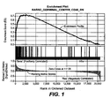
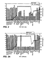
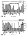
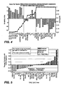


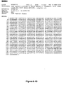
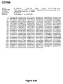
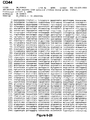


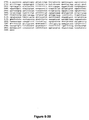


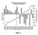
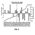
Classifications
-
- C—CHEMISTRY; METALLURGY
- C12—BIOCHEMISTRY; BEER; SPIRITS; WINE; VINEGAR; MICROBIOLOGY; ENZYMOLOGY; MUTATION OR GENETIC ENGINEERING
- C12Q—MEASURING OR TESTING PROCESSES INVOLVING ENZYMES, NUCLEIC ACIDS OR MICROORGANISMS; COMPOSITIONS OR TEST PAPERS THEREFOR; PROCESSES OF PREPARING SUCH COMPOSITIONS; CONDITION-RESPONSIVE CONTROL IN MICROBIOLOGICAL OR ENZYMOLOGICAL PROCESSES
- C12Q1/00—Measuring or testing processes involving enzymes, nucleic acids or microorganisms; Compositions therefor; Processes of preparing such compositions
- C12Q1/68—Measuring or testing processes involving enzymes, nucleic acids or microorganisms; Compositions therefor; Processes of preparing such compositions involving nucleic acids
- C12Q1/6876—Nucleic acid products used in the analysis of nucleic acids, e.g. primers or probes
- C12Q1/6883—Nucleic acid products used in the analysis of nucleic acids, e.g. primers or probes for diseases caused by alterations of genetic material
- C12Q1/6886—Nucleic acid products used in the analysis of nucleic acids, e.g. primers or probes for diseases caused by alterations of genetic material for cancer
-
- C—CHEMISTRY; METALLURGY
- C12—BIOCHEMISTRY; BEER; SPIRITS; WINE; VINEGAR; MICROBIOLOGY; ENZYMOLOGY; MUTATION OR GENETIC ENGINEERING
- C12Q—MEASURING OR TESTING PROCESSES INVOLVING ENZYMES, NUCLEIC ACIDS OR MICROORGANISMS; COMPOSITIONS OR TEST PAPERS THEREFOR; PROCESSES OF PREPARING SUCH COMPOSITIONS; CONDITION-RESPONSIVE CONTROL IN MICROBIOLOGICAL OR ENZYMOLOGICAL PROCESSES
- C12Q2600/00—Oligonucleotides characterized by their use
- C12Q2600/106—Pharmacogenomics, i.e. genetic variability in individual responses to drugs and drug metabolism
-
- C—CHEMISTRY; METALLURGY
- C12—BIOCHEMISTRY; BEER; SPIRITS; WINE; VINEGAR; MICROBIOLOGY; ENZYMOLOGY; MUTATION OR GENETIC ENGINEERING
- C12Q—MEASURING OR TESTING PROCESSES INVOLVING ENZYMES, NUCLEIC ACIDS OR MICROORGANISMS; COMPOSITIONS OR TEST PAPERS THEREFOR; PROCESSES OF PREPARING SUCH COMPOSITIONS; CONDITION-RESPONSIVE CONTROL IN MICROBIOLOGICAL OR ENZYMOLOGICAL PROCESSES
- C12Q2600/00—Oligonucleotides characterized by their use
- C12Q2600/118—Prognosis of disease development
-
- C—CHEMISTRY; METALLURGY
- C12—BIOCHEMISTRY; BEER; SPIRITS; WINE; VINEGAR; MICROBIOLOGY; ENZYMOLOGY; MUTATION OR GENETIC ENGINEERING
- C12Q—MEASURING OR TESTING PROCESSES INVOLVING ENZYMES, NUCLEIC ACIDS OR MICROORGANISMS; COMPOSITIONS OR TEST PAPERS THEREFOR; PROCESSES OF PREPARING SUCH COMPOSITIONS; CONDITION-RESPONSIVE CONTROL IN MICROBIOLOGICAL OR ENZYMOLOGICAL PROCESSES
- C12Q2600/00—Oligonucleotides characterized by their use
- C12Q2600/158—Expression markers
Definitions
- the present invention relates generally to the fields of predicting, assessing, aiding assessment of responsiveness of B-cell lymphoma to treatment with anti-CD40 antibodies.
- CD40 is a type I transmembrane protein of the tumor necrosis receptor superfamily. CD40 is an important molecule involved in B-cell proliferation and differentiation, immunoglobulin isotype switching, and cell viability. Receptor signaling is initiated by the binding of CD40 to the CD40 ligand (CD40L or CD 154), which is primarily expressed on activated CD4+ T cells.
- CD40L or CD 154 CD40 ligand
- CD40 On normal cells, CD40 is expressed on cells with high proliferative potential, including hematopoietic progenitors, epithelial and endothelial cells, and all antigen-presenting cells (dendritic cells, activated B lymphocytes, and activated monocytes). CD40 is highly expressed on several types of B-cell hematologic malignancies including multiple myeloma, non-Hodgkin's lymphoma (NHL), and chronic lymphocytic leukemia (CLL). The high prevalence of CD40 expression on B-cell malignancies makes it an attractive potential tumor target for antibody-based cancer therapy. CD40 is also expressed on a majority of bladder cancers and a significant percentage of other solid tumors, including head and neck cancers, renal cell carcinomas, ovarian and lung cancer.
- Anti-CD40 antibodies and their uses for treating B cell hematologic malignancies have been described. See, e.g., US Pat. 6,946,129 ; 6,843,989 ; 6,838,261 ; WO 2000/075348 ; US-2002-0197256 ; WO 2006/128103 ; and WO 2007/075326 . It has been shown that a humanized anti-CD40 antibody induces growth inhibition and apoptosis of CD40-positive cells in a subset of hematologic tumor cell lines through direct signal transduction. WO 2006/128103 ; WO 2007/075326 . Furthermore, the humanized anti-CD40 antibody kills tumor cells via immune effector functions, including antibody-dependent cellular cytotoxicity
- ADCC antibody-dependent cellular phagocytosis
- MM multiple myeloma
- NHL non-Hodgkin's lymphoma
- SCID severe combined immunodeficient mice
- anti-CD40 antibodies can induce growth inhibition and apoptosis of CD40-positive cells and may have anti-tumor activity in various types of B cell lymphoma patients, not all B lymphoma cells are sensitive to anti-CD40 antibody mediated cell death. There remains a need to identify one or more predictive markers for the responsiveness of B-cell lymphoma patients to anti-CD40 antibody therapy.
- the invention provides methods and compositions for predicting, assessing or aiding assessment of responsiveness of a subject having a type of B-cell lymphoma to treatment with an anti-CD40 antibody.
- the invention provides methods for predicting, assessing or aiding assessment of responsiveness of a subject having a B-cell lymphoma to an anti-CD40 antibody treatment, comprising the steps of: (a) measuring expression level of one or more marker genes in a sample comprising B lymphoma cells obtained from said subject, wherein said one or more marker genes are selected from the group consisting of IFITM1, CD40, RGS13, VNN2, LMO2, CD79B, CD22, BTG2, IGF1R, CD44, CTSC, EPDR1, UAP1, and PUS7; (b) predicting whether the subject is likely to respond to the anti-CD40 antibody treatment based on the measured expression level of said one or more marker genes from step (a).
- expression levels of at least two, at least three, at least four, at least five, at least six, at least seven, at least eight, at least nine, at least ten, at least eleven, at least twelve, at least thirteen, or fourteen maker genes from the group are measured and used for the prediction, assessment, or aiding assessment.
- the prediction, assessment, or aiding assessment is determined by comparing the measured expression level of one or more marker genes to a reference level.
- a reference level is a value or a range determined based on the measured expression level of the corresponding marker gene in samples comprising the B lymphoma cells from subjects having tumor volume increased or decreased after the anti-CD40 antibody treatment.
- the invention provides methods preparing a personalized genomics profile for a subject having B-cell lymphoma comprising the steps of: (a) determining expression level of one or more marker genes selected from the group consisting of IFITM1, CD40, RGS13, VNN2, LMO2, CD79B, CD22, BTG2, IGF1R, CD44, CTSC, EPDR1, UAP1, PUS7, and BCL6 in a sample comprising B lymphoma cells obtained from the subject; and (b) generating a report summarizing the expression level of one or more marker genes obtained in step (a).
- expression levels of at least two, at least three, at least four, at least five, at least six, at least seven, at least eight, at least nine, at least ten, at least eleven, at least twelve, at least thirteen, at least fourteen, or fifteen maker genes from the group are measured and used for the generating the report for the personalized genomics profile.
- the report includes a recommendation for an anti-CD40 antibody treatment for the subject
- the recommendation is determined by comparing the measured expression level of the marker genes to a reference level.
- a reference level is a value or a range determined based on the measured expression level of the corresponding marker gene in samples comprising the B lymphomas cells from subjects having tumor volume increased or decreased after the anti-CD40 antibody treatment.
- a value equals or greater than zero for the sensitivity index indicates that the subject is likely to respond the anti-CD40 antibody treatment, or wherein a value less than zero for the sensitivity index indicates that the subject is less likely to respond the anti-CD40 antibody treatment.
- the expression level of at least three, at least four, at least five, at least six, at least seven, at least eight, at least nine, at least ten, at least eleven, at least twelve, at least thirteen, or fourteen marker genes are measured and used for the sensitivity index calculation.
- the expression level of IFITM1, RGS13, CD79B, CD22, BTG2, CD44, EPDR1, and UAP1 are measured and used for the sensitivity index calculation.
- the invention provides an anti-CD40 antibody for use in a method of treatment of B cell lymphoma, the method comprising: predicting the responsiveness of a patient to the antibody by the methods of the invention; and administering the antibody to the patient if the patient is predicted to be responsive.
- the reference level is a measured expression level of the marker gene in a different B-cell lymphoma sample.
- the different B cell lymphoma sample comprises B lymphoma cells that are resistant to an anti-CD40 antibody induced cell death.
- the measured expression level of the marker gene and/or the reference level are normalized.
- the expression level is measured by detecting mRNA expression (e.g., real time quantitative reverse transcription PCR (qRT-PCR)) and/or by detecting protein expression (e.g., immunohistochemistry (IHC)).
- mRNA expression e.g., real time quantitative reverse transcription PCR (qRT-PCR)
- detecting protein expression e.g., immunohistochemistry (IHC)
- expression levels of at least one, at least two, at least three, at least four, at least five, at least six, at least seven, at least eight, at least nine, at least ten, at least eleven, at least twelve, at least thirteen, or fourteen genes selected from the group consisting of VNN2, MEF2C, LTB, KCNN3, NCF1, BCL6, IGJ, ELTI1902, PNOC, CSF2RB, POU2AF1, CD22, RGS13, and MEF2B in the B-cell lymphoma sample from the subject are compared to one or more reference levels.
- expression levels of one or more gene pairs selected from the group consisting of VNN2 and EPDR1, RGS13 and EPDR1, CD22 and EPDR1, LRRC8A and PRPSAP2, CD40 and IGF1R, IFITM1 and BTG2, SMN1 and LMO2, PRKCA and YIPF3 in a the B-cell lymphoma sample are compared.
- expression levels are compared between one or more gene pairs VNN2 and EPDR1, RGS13 and EPDR1, CD22 and EPDR1, LRRC8A and PRPSAP2, CD40 and IGF1R, IFITM1 and BTG2, SMN1 and LMO2, PRKCA and YIPF3 in the B-cell lymphoma sample, and sensitivity index calculated as the sum of signed t-scores for log2-scale expression of the gene pairs is used to assess responsiveness of the B-cell lymphoma to an anti-CD40 antibody treatment.
- the B-cell lymphoma is non-Hodgkin's lymphoma (NHL), including, but is not limited to, follicular lymphoma, relapsed follicular lymphoma, small lymphocytic lymphoma, mantle cell lymphoma, marginal zone lymphoma, lymphoplasmacytic lymphoma, mycosis fungoides/Sezary syndrome, splenic marginal zone lymphoma, and diffuse large B-cell lymphoma.
- the B-cell lymphoma is selected from the group consisting of indolent lymphoma, aggressive lymphoma, and highly aggressive lymphoma.
- the present invention is based on the discovery that certain genes (e.g., genes shown in Tables 2-4, 6, 7 and 13) are differentially expressed between B lymphoma cells that are sensitive to anti-CD40 antibody induced cell death and B lymphoma cells that are resistant to anti-CD40 induced cell death. Data from clinical trials described in Example 2 indicate that the expression level of the fourteen genes shown in Table 13 is highly associated with responsiveness to anti-CD40 Ab.1 treatment. Some of the differentially expressed genes between sensitive B lymphoma cells and resistant B lymphoma cells are the CD40 ligand downregulated pathway genes; and some are in the B-cell receptor signaling pathway.
- expression levels of one or more of these differentially expressed genes can be used for assessing or aiding assessment of responsiveness of a subject having a B-cell lymphoma to treatment with anti-CD40 antibodies, predicting responsiveness of the subject to treatment with anti-CD40 antibodies, and monitoring treatment/responsiveness in the subject.
- Primers, oligonucleotides and polynucleotides employed in the present invention can be generated using standard techniques known in the art.
- a subject having a B-cell lymphoma and "B-cell lymphoma patient” refer to a subject who has been diagnosed with a type of B-cell lymphoma or has been given a probable diagnosis of a type of B-cell lymphoma.
- biomarker refers generally to a molecule, including a gene, protein, carbohydrate structure, or glycolipid, the expression of which in or on a mammalian tissue or cell or secreted can be detected by known methods (or methods disclosed herein) and is predictive or can be used to predict (or aid prediction) for a mammalian cell's or tissue's sensitivity to, and in some embodiments, to predict (or aid prediction) an individual's responsiveness to treatment regimes based on anti-CD40 antibodies.
- sample refers to a composition that is obtained or derived from a subject of interest that contains a cellular and/or other molecular entity that is to be characterized and/or identified, for example based on physical, biochemical, chemical and/or physiological characteristics.
- disease sample and variations thereof refers to any sample obtained from a subject of interest that would be expected or is known to contain the cellular and/or molecular entity that is to be characterized.
- tissue or cell sample is meant a collection of similar cells obtained from a tissue of a subject or patient.
- the source of the tissue or cell sample may be solid tissue as from a fresh, frozen and/or preserved organ or tissue sample or biopsy or aspirate; blood or any blood constituents; bodily fluids such as cerebral spinal fluid, amniotic fluid, peritoneal fluid, or interstitial fluid; cells from any time in gestation or development of the subject.
- the tissue sample may also be primary or cultured cells or cell lines.
- the tissue or cell sample is obtained from a disease tissue/organ.
- the tissue sample may contain compounds which are not naturally intermixed with the tissue in nature such as preservatives, anticoagulants, buffers, fixatives, nutrients, antibiotics, or the like.
- a "section" of a tissue sample is meant a single part or piece of a tissue sample, e.g. a thin slice of tissue or cells cut from a tissue sample. It is understood that multiple sections of tissue samples may be taken and subjected to analysis according to the present invention, provided that it is understood that the present invention comprises a method whereby the same section of tissue sample is analyzed at both morphological and molecular levels, or is analyzed with respect to both protein and nucleic acid.
- a "B-cell lymphoma sample” or a “sample comprising B lymphoma cells” is a tissue or cell sample containing B lymphoma cells from a subject or a patient that have been diagnosed with a type of B-cell lymphoma.
- method for "aiding assessment” refers to methods that assist in making a clinical determination (e.g., responsiveness of a B-cell lymphoma to treatment with anti-CD40 antibodies), and may or may not be conclusive with respect to the definitive assessment.
- a "subject” or an “individual” is a mammal, more preferably a human. Mammals include, but are not limited to, humans, primates, farm animal, sport animals, rodents, and pets (e.g., dogs and cats).
- a "reference value” can be an absolute value; a relative value; a value that has an upper and/or lower limit; a range of values; an average value; a median value; a mean value; or a value as compared to a particular control or baseline value.
- array refers to an ordered arrangement of hybridizable array elements, such as polynucleotide probes (e.g., oligonucleotides) and antibodies, on a substrate.
- the substrate can be a solid substrate, such as a glass slide, or a semi-solid substrate, such as nitrocellulose membrane.
- the nucleotide sequences can be DNA, RNA, or any permutations thereof.
- Amplification generally refers to the process of producing multiple copies of a desired sequence.
- Multiple copies means at least 2 copies.
- a “copy” does not necessarily mean perfect sequence complementarity or identity to the template sequence.
- copies can include nucleotide analogs such as deoxyinosine, intentional sequence alterations (such as sequence alterations introduced through a primer comprising a sequence that is hybridizable, but not complementary, to the template), and/or sequence errors that occur during amplification.
- Expression/amount of a gene or biomarker in a first sample is at a level "greater than" the level in a second sample if the expression level/amount of the gene or biomarker in the first sample is at least about 1.5X, 1.75X, 2X, 3X, 4X, 5X, 6X, 7X, 8X, 9X or 10X the expression level/amount of the gene or biomarker in the second sample.
- Expression levels/amounts can be determined based on any suitable criterion known in the art, including but not limited to mRNA, cDNA, proteins, protein fragments and/or gene copy. Expression levels/amounts can be determined qualitatively and/or quantitatively.
- Polynucleotide or “nucleic acid,” as used interchangeably herein, refer to polymers of nucleotides of any length, and include DNA and RNA.
- the nucleotides can be deoxyribonucleotides, ribonucleotides, modified nucleotides or bases, and/or their analogs, or any substrate that can be incorporated into a polymer by DNA or RNA polymerase.
- a polynucleotide may comprise modified nucleotides, such as methylated nucleotides and their analogs. If present, modification to the nucleotide structure may be imparted before or after assembly of the polymer.
- the sequence of nucleotides may be interrupted by non-nucleotide components.
- a polynucleotide may be further modified after polymerization, such as by conjugation with a labeling component.
- modifications include, for example, "caps", substitution of one or more of the naturally occurring nucleotides with an analog, internucleotide modifications such as, for example, those with uncharged linkages (e.g., methyl phosphonates, phosphotriesters, phosphoamidates, cabamates, etc.) and with charged linkages (e.g., phosphorothioates, phosphorodithioates, etc.), those containing pendant moieties, such as, for example, proteins (e.g., nucleases, toxins, antibodies, signal peptides, ply-L-lysine, etc.
- proteins e.g., nucleases, toxins, antibodies, signal peptides, ply-L-lysine, etc.
- any of the hydroxyl groups ordinarily present in the sugars may be replaced, for example, by phosphonate groups, phosphate groups, protected by standard protecting groups, or activated to prepare additional linkages to additional nucleotides, or may be conjugated to solid supports.
- the 5' and 3' terminal OH can be phosphorylated or substituted with amines or organic capping groups moieties of from 1 to 20 carbon atoms. Other hydroxyls may also be derivatized to standard protecting groups.
- Polynucleotides can also contain analogous forms of ribose or deoxyribose sugars that are generally known in the art, including, for example, 2'-O-methyl-2'-O- allyl, 2'-fluoro- or 2'-azido-ribose, carbocyclic sugar analogs, ⁇ - anomeric sugars, epimeric sugars such as arabinose, xyloses or lyxoses, pyranose sugars, furanose sugars, sedoheptuloses, acyclic analogs and abasic nucleoside analogs such as methyl riboside.
- One or more phosphodiester linkages may be replaced by alternative linking groups.
- These alternative linking groups include, but are not limited to, embodiments wherein phosphate is replaced by P(O)S("thioate"), P(S)S ("dithioate"), "(O)NR 2 ("amidate"), P(O)R, P(O)OR', CO or CH 2 ("formacetal"), in which each R or R' is independently H or substituted or unsubstituted alkyl (1-20 C) optionally containing an ether (--O--) linkage, aryl, alkenyl, cycloalkyl, cycloalkenyl or araldyl. Not all linkages in a polynucleotide need be identical. The preceding description applies to all polynucleotides referred to herein, including RNA and DNA.
- Oligonucleotide generally refers to short, generally single stranded, generally synthetic polynucleotides that are generally, but not necessarily, less than about 200 nucleotides in length.
- oligonucleotide and “polynucleotide” are not mutually exclusive. The description above for polynucleotides is equally and fully applicable to oligonucleotides.
- a “primer” is generally a short single stranded polynucleotide, generally with a free 3'-OH group, that binds to a target potentially present in a sample of interest by hybridizing with a target sequence, and thereafter promotes polymerization of a polynucleotide complementary to the target.
- a “pair of primers” refer to a 5' primer and a 3' primer that can be used to amplify a portion of a specific target gene.
- the term "3"' generally refers to a region or position in a polynucleotide or oligonucleotide 3' (downstream) from another region or position in the same polynucleotide or oligonucleotide.
- the term "5"' generally refers to a region or position in a polynucleotide or oligonucleotide 5' (upstream) from another region or position in the same polynucleotide or oligonucleotide.
- gene amplification refers to a process by which multiple copies of a gene or gene fragment are formed in a particular cell or cell line.
- the duplicated region (a stretch of amplified DNA) is often referred to as "amplicon.”
- amplicon a stretch of amplified DNA
- the amount of the messenger RNA (mRNA) produced i.e., the level of gene expression, also increases in the proportion of the number of copies made of the particular gene expressed.
- Detection includes any means of detecting, including direct and indirect detection.
- prediction is used herein to refer to the likelihood that a patient will respond either favorably or unfavorably to a drug or set of drugs. In one embodiment, the prediction relates to the extent of those responses. In one embodiment, the prediction relates to whether and/or the probability that a patient will survive or improve following treatment, for example treatment with a particular therapeutic agent, and for a certain period of time without disease recurrence.
- the predictive methods of the invention can be used clinically to make treatment decisions by choosing the most appropriate treatment modalities for any particular patient.
- the predictive methods of the present invention are valuable tools in predicting if a patient is likely to respond favorably to a treatment regimen, such as a given therapeutic regimen, including for example, administration of a given therapeutic agent or combination, surgical intervention, steroid treatment, etc., or whether long-term survival of the patient, following a therapeutic regimen is likely.
- a treatment regimen such as a given therapeutic regimen, including for example, administration of a given therapeutic agent or combination, surgical intervention, steroid treatment, etc., or whether long-term survival of the patient, following a therapeutic regimen is likely.
- long-term survival is used herein to refer to survival for at least 1 year, 5 years, 8 years, or 10 years following therapeutic treatment.
- Patient response can be assessed using any endpoint indicating a benefit to the patient, including, without limitation, (1) inhibition, to some extent, of disease progression, including slowing down and complete arrest; (2) reduction in the number of disease episodes and/or symptoms; (3) reduction in lesional size; (4) inhibition (i.e., reduction, slowing down or complete stopping) of disease cell infiltration into adjacent peripheral organs and/or tissues; (5) inhibition (i.e. reduction, slowing down or complete stopping) of disease spread; (6) relief, to some extent, of one or more symptoms associated with the disorder; (7) increase in the length of disease-free presentation following treatment; and/or (8) decreased mortality at a given point of time following treatment.
- antibody is used in the broadest sense and specifically covers monoclonal antibodies (including full length monoclonal antibodies), multispecific antibodies (e.g., bispecific antibodies), and antibody fragments so long as they exhibit the desired biological activity or function.
- Antibody fragments comprise a portion of a full length antibody, generally the antigen binding or variable region thereof.
- Examples of antibody fragments include Fab, Fab', F(ab') 2 , and Fv fragments; diabodies; linear antibodies; single-chain antibody molecules; and multispecific antibodies formed from antibody fragments.
- Fv is the minimum antibody fragment which contains a complete antigen-recognition and -binding site. This fragment consists of a dimer of one heavy- and one light-chain variable region domain in tight, non-covalent association. From the folding of these two domains emanate six hypervariable loops (3 loops each from the H and L chain) that contribute the amino acid residues for antigen binding and confer antigen binding specificity to the antibody. However, even a single variable domain (or half of an Fv comprising only three CDRs specific for an antigen) has the ability to recognize and bind antigen, although at a lower affinity than the entire binding site.
- monoclonal antibody refers to an antibody from a population of substantially homogeneous antibodies, i.e ., the individual antibodies comprising the population are identical and/or bind the same epitope(s), except for possible variants that may arise during production of the monoclonal antibody, such variants generally being present in minor amounts.
- Such monoclonal antibody typically includes an antibody comprising a polypeptide sequence that binds a target, wherein the target-binding polypeptide sequence was obtained by a process that includes the selection of a single target binding polypeptide sequence from a plurality of polypeptide sequences.
- the selection process can be the selection of a unique clone from a plurality of clones, such as a pool of hybridoma clones, phage clones or recombinant DNA clones.
- the selected target binding sequence can be further altered, for example, to improve affinity for the target, to humanize the target binding sequence, to improve its production in cell culture, to reduce its immunogenicity in vivo, to create a multispecific antibody, etc., and that an antibody comprising the altered target binding sequence is also a monoclonal antibody of this invention.
- each monoclonal antibody of a monoclonal antibody preparation is directed against a single determinant on an antigen.
- the monoclonal antibody preparations are advantageous in that they are typically uncontaminated by other immunoglobulins.
- the modifier "monoclonal" indicates the character of the antibody as being obtained from a substantially homogeneous population of antibodies, and is not to be construed as requiring production of the antibody by any particular method.
- the monoclonal antibodies to be used in accordance with the present invention may be made by a variety of techniques, including, for example, the hybridoma method (e.g., Kohler et al., Nature, 256:495 (1975 ); Harlow et al., Antibodies: A Laboratory Manual, (Cold Spring Harbor Laboratory Press, 2nd ed. 1988 ); Hammerling et al., in: Monoclonal Antibodies and T-Cell Hybridomas 563-681, (Elsevier, N.Y., 1981 )), recombinant DNA methods (see, e.g., U.S. Patent No.
- phage display technologies see, e.g., Clackson et al., Nature, 352:624-628 (1991 ); Marks et al., J. Mol. Biol., 222:581-597 (1991 ); Sidhu et al., J. Mol. Biol. 338(2):299-310 (2004 ); Lee et al., J.Mol.Biol.340(5):1073-1093 (2004 ); Fellouse, Proc. Nat. Acad Sci. USA 101(34):12467-12472 (2004 ); and Lee et al. J. Immunol.
- Methods 284(1-2):119-132 (2004 ), and technologies for producing human or human-like antibodies in animals that have parts or all of the human immunoglobulin loci or genes encoding human immunoglobulin sequences see, e.g., WO 1998/24893 ; WO 1996/34096 ; WO 1996/33735 ; WO 1991/10741 ; Jakobovits et al., Proc. Natl. Acad Sci. USA, 90:2551 (1993 ); Jakobovits et al., Nature, 362:255-258 (1993 ); Bruggemann et al., Year in Immuno., 7:33 (1993 ); U.S. Patent Nos.
- the monoclonal antibodies herein specifically include “chimeric” antibodies.
- “Chimeric” antibodies immunoglobulins have a portion of the heavy and/or light chain identical with or homologous to corresponding sequences in antibodies derived from a particular species or belonging to a particular antibody class or subclass, while the remainder of the chain(s) is identical with or homologous to corresponding sequences in antibodies derived from another species or belonging to another antibody class or subclass, as well as fragments of such antibodies, so long as they exhibit the desired biological activity ( U.S. Patent No. 4,816,567 ; and Morrison et al., Proc. Natl. Acad. Sci. USA 81:6851-6855 (1984 )).
- Humanized antibody as used herein is a subset of chimeric antibodies.
- Humanized forms of non-human (e.g., murine) antibodies are chimeric antibodies which contain minimal sequence derived from non-human immunoglobulin.
- humanized antibodies are human immunoglobulins (recipient or acceptor antibody) in which hypervariable region residues of the recipient are replaced by hypervariable region residues from a non-human species (donor antibody) such as mouse, rat, rabbit or nonhuman primate having the desired specificity, affinity, and capacity.
- donor antibody such as mouse, rat, rabbit or nonhuman primate having the desired specificity, affinity, and capacity.
- Fv framework region (FR) residues of the human immunoglobulin are replaced by corresponding non-human residues.
- humanized antibodies may comprise residues which are not found in the recipient antibody or in the donor antibody. These modifications are made to further refine antibody performance such as binding affinity.
- the humanized antibody will comprise substantially all of at least one, and typically two, variable domains, in which all or substantially all of the hypervariable loops correspond to those of a non-human immunoglobulin and all or substantially all of the FR regions are those of a human immunoglobulin sequence although the FR regions may include one or more amino acid substitutions that improve binding affinity.
- the number of these amino acid substitutions in the FR are typically no more than 6 in the H chain, and in the L chain, no more than 3.
- the humanized antibody optionally also will comprise at least a portion of an immunoglobulin constant region (Fc), typically that of a human immunoglobulin.
- Fc immunoglobulin constant region
- a "human antibody” is one which possesses an amino acid sequence which corresponds to that of an antibody produced by a human and/or has been made using any of the known techniques for making human antibodies. This definition of a human antibody specifically excludes a humanized antibody comprising non-human antigen-binding residues.
- affinity matured antibody is one with one or more alterations in one or more CDRs/HVRs thereof which result in an improvement in the affinity of the antibody for antigen, compared to a parent antibody which does not possess those alteration(s).
- Preferred affinity matured antibodies will have nanomolar or even picomolar affinities for the target antigen.
- Affinity matured antibodies are produced by procedures known in the art. Marks et al. Bio/Technology 10:779-783 (1992 ) describes affinity maturation by VH and VL domain shuffling. Random mutagenesis of CDR/HVR and/or framework residues is described by: Barbas et al. Proc Nat. Acad.
- Fc region is used to define the C-terminal region of an immunoglobulin heavy chain which may be generated by papain digestion of an intact antibody.
- the Fc region may be a native sequence Fc region or a variant Fc region.
- the human IgG heavy chain Fc region is usually defined to stretch from an amino acid residue at about position Cys226, or from about position Pro230, to the carboxyl-terminus of the Fc region.
- the Fc region of an immunoglobulin generally comprises two constant domains, a CH2 domain and a CH3 domain, and optionally comprises a CH4 domain.
- Fc region chain herein is meant one of the two polypeptide chains of an Fc region.
- Antibody effector functions refer to those biological activities attributable to the Fc region (a native sequence Fc region or amino acid sequence variant Fc region) of an antibody, and vary with the antibody isotype. Examples of antibody effector functions include: C1q binding and complement dependent cytotoxicity; Fc receptor binding; antibody-dependent cell-mediated cytotoxicity (ADCC); phagocytosis; down regulation of cell surface receptors (e.g. B cell receptor); and B cell activation.
- ADCC antibody-dependent cell-mediated cytotoxicity
- FcRs Fc receptors
- cytotoxic cells e.g. Natural Killer (NK) cells, neutrophils, and macrophages
- NK cells Natural Killer cells
- neutrophils neutrophils
- macrophages e.g., neutrophils, and macrophages
- the antibodies “arm” the cytotoxic cells and are absolutely required for such killing.
- the primary cells for mediating ADCC, NK cells express Fc ⁇ RIII only, whereas monocytes express FcyRI, Fc ⁇ RII and Fc ⁇ RIII.
- FcR expression on hematopoietic cells is summarized in Table 3 on page 464 of Ravetch and Kinet, Annu. Rev. Immunol 9:457-92 (1991 ).
- an in vitro ADCC assay such as that described in US Patent No. 5,500,362 or 5,821,337 or Presta U.S. Patent No. 6,737,056 may be performed.
- Useful effector cells for such assays include peripheral blood mononuclear cells (PBMC) and Natural Killer (NK) cells.
- PBMC peripheral blood mononuclear cells
- NK Natural Killer
- ADCC activity of the molecule of interest may be assessed in vivo, e.g., in a animal model such as that disclosed in Clynes et al. PNAS (USA) 95:652-656 (1998 ).
- Treating” or “treatment” or “alleviation” refers to therapeutic treatment wherein the object is to slow down (lessen) if not cure the targeted pathologic condition or disorder or prevent recurrence of the condition.
- a subject is successfully “treated” for the B cell malignancy if, after receiving a therapeutic amount of a CD40 binding antibody, the subject shows observable and/or measurable reduction in or absence of one or more signs and symptoms of the particular disease.
- the antibodies of the invention achieve > 95% peripheral blood B cell depletion and the B cells return to 25% of baseline.
- treatment with the anti-CD40 antibodies is effective to result in the cancer patients being progression-free in the cancer 3 months after treatment, preferably 6 months, more preferably one year, even more preferably 2 or more years post treatment.
- non-Hodgkin's lymphoma refers to a cancer of the lymphatic system other than Hodgkin's lymphomas.
- Hodgkin's lymphomas can generally be distinguished from non-Hodgkin's lymphomas by the presence of Reed-Sternberg cells in Hodgkin's lymphomas and the absence of said cells in non-Hodgkin's lymphomas.
- an “effective amount” refers to an amount effective, at dosages and for periods of time necessary, to achieve the desired therapeutic or prophylactic result.
- a “therapeutically effective amount” of a therapeutic agent may vary according to factors such as the disease state, age, sex, and weight of the individual, and the ability of the antibody to elicit a desired response in the individual.
- a therapeutically effective amount is also one in which any toxic or detrimental effects of the therapeutic agent are outweighed by the therapeutically beneficial effects.
- a “prophylactically effective amount” refers to an amount effective, at dosages and for periods of time necessary, to achieve the desired prophylactic result. Typically but not necessarily, since a prophylactic dose is used in subjects prior to or at an earlier stage of disease, the prophylactically effective amount will be less than the therapeutically effective amount.
- housekeeping gene refers to a group of genes that codes for proteins whose activities are essential for the maintenance of cell function. These genes are typically similarly expressed in all cell types.
- correlate or “correlating” is meant comparing, in any way, the performance and/or results of a first analysis or protocol with the performance and/or results of a second analysis or protocol. For example, one may use the results of a first analysis or protocol in carrying out a second protocols and/or one may use the results of a first analysis or protocol to determine whether a second analysis or protocol should be performed. With respect to the embodiment of gene expression analysis or protocol, one may use the results of the gene expression analysis or protocol to determine whether a specific therapeutic regimen should be performed.
- label when used herein refers to a compound or composition which is conjugated or fused directly or indirectly to a reagent such as a nucleic acid probe or an antibody and facilitates detection of the reagent to which it is conjugated or fused.
- the label may itself be detectable (e.g., radioisotope labels or fluorescent labels) or, in the case of an enzymatic label, may catalyze chemical alteration of a substrate compound or composition which is detectable.
- the invention provides methods for assessing or aiding assessment of responsiveness of a subject having a B-cell lymphoma to treatment with an anti-CD40 antibody.
- the invention also provides methods for predicting responsiveness or monitoring treatment/responsiveness to an anti-CD40 antibody treatment in a subject having a B-cell lymphoma.
- the invention provides methods for selecting a subject having a B-cell lymphoma suitable for treatment with an anti-CD40 antibody and following up with an anti-CD40 antibody treatment.
- the methods comprise measuring expression level of one or more marker genes in any of Tables 2-4, 6, 7, and 13 in a sample comprising B lymphoma cells obtained from the subject; and predicting, assessing, or aiding assessment of responsiveness of the subject to an anti-CD40 antibody treatment based on the measure expression level of said one or more marker genes.
- the methods comprise comparing a measured expression level of at least one marker gene in any of Tables 2-4, 6, 7, and 13 in a B-cell lymphoma sample from the subject to a reference level for the respective marker gene.
- the methods of the present invention are useful for clinicians to identify patients with B-cell lymphoma for treatment with an anti-CD40 antibody, aiding in patient selection during the course of development of anti-CD40 antibody therapy, prediction of likelihood of success when treating an individual patient with a particular treatment regimen, in assessing and monitoring disease progression, in monitoring treatment efficacy, and in determining prognosis for individual patients. Any of these embodiments are included in this invention.
- the B-cell lymphoma is non-Hodgkin's lymphoma (NHL), including, but is not limited to, follicular lymphoma, relapsed follicular lymphoma, small lymphocytic lymphoma, mantle cell lymphoma, marginal zone lymphoma, lymphoplasmacytic lymphoma, mycosis fungoides/Sezary syndrome, splenic marginal zone lymphoma, and diffuse large B-cell lymphoma.
- NHL Hodgkin's lymphoma
- the B-cell lymphoma is indolent. In some embodiments, the B-cell lymphoma is aggressive. In some embodiments, the B-cell lymphoma is highly aggressive. In some embodiments, the indolent B-cell lymphoma is follicular lymphoma, marginal zone lymphoma, or small lymphocytic lymphoma. In some embodiments, the indolent B-cell lymphoma is follicular lymphoma.
- the expression level of one or more of the marker genes in a B-cell lymphoma sample relative a reference level may be used in the methods of the invention, such as to predict, assess or aid assessment of responsiveness of the B-cell lymphoma to treatment with an anti-CD40 antibody.
- Anti-CD40 antibody sensitive cells are cells having an IC25 value less than 0.4 ⁇ g/ml in reduction of cell viability by an anti-CD40 antibody tested as described in Example 1.
- Anti-CD40 resistant cells are cells having an IC25 value greater than 1 ⁇ g/ml in reduction in cell viability as tested in Example 1.
- genes in Tables 2-4, 6 and 7 are in the CD40 ligand downregulated pathway (for example, VNN2, MEF2C, LTB, KCNN3, NCF1, BCL6, IGJ, ELTI1902, PNOC, CSF2RB, and POU2AF1); and some of the genes in the tables are in the B-cell receptor signaling pathway (for example, CD22, RGS13, and MEF2B). Further, association of the expression level of IFITM1, CD40, RGS13, VNN2, LMO2, CD79B, CD22, BTG2, IGF1R, CD44, CTSC, EPDR1, UAP1, and PUS7 (Table 13) has been confirmed by clinical trials described in Example 2.
- CD40 ligand downregulated pathway for example, VNN2, MEF2C, LTB, KCNN3, NCF1, BCL6, IGJ, ELTI1902, PNOC, CSF2RB, and POU2AF1
- B-cell receptor signaling pathway for example, CD22, RGS13, and
- Expression levels of one or more of these genes are used in the methods of the invention. In some embodiments, expression levels of at least two, at least three, at least four, at least five, at least six, at least seven, at least eight, at least nine, at least ten, at least eleven, at least twelve, at least thirteen, at least fourteen, at least fifteen, at least twenty, at least twenty five, or at least thirty genes are used in the methods of the invention.
- expression levels of one or more of genes selected from the group consisting of VNN2, MEF2C, LTB, KCNN3, NCF1, BCL6, IGJ, ELTI1902, PNOC, CSF2RB, POU2AF1, CD22, RGS13, and MEF2B are measured and/or used.
- expression levels of one or more of genes selected from the group consisting of IFITM1, CD40, RGS13, VNN2, LMO2, CD79B, CD22, BTG2, IGF1R, CD44, CTSC, EPDR1, UAP1, and PUS7 are measured and/or used.
- expression levels of at least two, at least three, at least four, at least five, at least six, at least seven, at least eight, at least nine, at least ten, at least eleven, at least twelve, at least thirteen, or fourteen of these genes are measured and/or used.
- expression levels of CD22, CD40, and BCL6 are measured and/or used.
- expression levels of CD40, RGS13, CD22, BTG2, IGF1R, and CD44 are measured and/or used.
- expression levels of IFITM1, CD40, RGS13, VNN2, LMO2, CD79B, CD22, BTG2, IGF1R, CD44, CTSC, EPDR1, UAP1, and PUS7 are measured and/or used.
- expression levels of at least one, at least two, at least three, at least four, at least five, at least six, at least seven, at least eight, at least nine, at least ten, at least eleven, at least twelve, at least thirteen, at least fourteen, or fifteen of genes in Table 7 or Table 13 are measured and/or used.
- Genes (including sequences) identified in Tables 2-4, 6, 7 and 13 are known in the art.
- the examples of Gene Bank accession numbers for human genes are VNN2 (NM_004665; NM_078488; AJ132100; D89974; BC064641; CR609799; BC126145; BC126147; and AB026705); RGS13 (NM_002927; NM_144766; BT006929; BC056866; AY562947; CR536532; CR610389; CR599001; BC016667; AF493935; BC036950; and AF030107); CD22 (NM_001771; AK026467; BC109306; BC109307; AK225694; AK225625; X52785; and X59350); LRRC8A (AY143166; BC051322; AK123611; AY358286; NM_019594; XM_0269
- the measured expression level of one or more marker genes in a B-cell lymphoma sample is compared to a reference level.
- the reference level is the expression level of a gene the expression level of which does not change (does not change significantly) among different type of B-cell lymphomas, for example, between B-cell lymphoma sensitive to anti-CD40 antibody and B-cell lymphoma resistant to anti-CD40 antibody.
- expression levels of one or more housekeeping genes shown in Table 8 are used as reference levels.
- expression levels of one or more housekeeping genes shown in Table 9 are used as reference levels.
- the measured expression level of the marker gene is normalized using the reference level. In some embodiments, the normalized expression level of the marker gene is calculated as a ratio of or difference between the marker gene and reference expression levels, on the original or on a log scale, respectively.
- the reference genes in Table 8 and Table 9 were selected as specific normalizing counterparts to the marker genes in Table 4. Reference genes were selected for high mean expression and low variance in B cell lymphoma samples. In addition, reference genes were selected to have similar variance between replicated expression measurements of individual cell lines relative to variance between expression measurements of biologically distinct cell lines. In addition, reference genes were selected to have low statistical association with one or more markers in Table 4.
- the reference level is a measured expression level of the marker gene in a different B-cell lymphoma sample.
- the different B cell lymphoma sample comprises B lymphoma cells that are resistant to an anti-CD40 antibody induced cell death.
- the reference level is determined based on the expression level of the corresponding marker gene in samples comprising B lymphoma cells from subjects having tumor volume increased after the anti-CD40 antibody treatment and/or having tumor volume decreased after the anti-CD40 antibody treatment.
- the samples from subjects for reference level determination comprise the same type of B lymphoma cells as the sample from the subject whose responsiveness to the anti-CD40 antibody treatment is predicted or assessed.
- the same method e.g., qRT-PCR
- reagents e.g., primers and probes
- the methods disclosed herein provide methods to examine expression level of one or more of these marker genes in a lymphoma sample (e.g ., B-cell lymphoma sample) relative a reference level.
- the methods and assays include those which examine expression of marker genes such as one or more of those listed in any of Tables 2-4, 6, 7 and 13. Expression levels may be measured at mRNA level and/or protein level.
- the invention provides methods for measuring levels of expression from a mammalian tissue or cells sample (such as cells and/or tissues associated with B-cell lymphoma). For example, for obtaining patient samples, H&E staining is carried out and used as a guide for tissue macrodissection to enrich for tumor content
- the sample can be obtained by a variety of procedures known in the art including, but is not limited to surgical excision, aspiration or biopsy.
- the sample may be fresh or frozen.
- the sample is fixed and embedded in paraffin or the like.
- a mammalian tissue or cell sample is obtained and examined for expression of one or more biomarkers.
- the methods may be conducted in a variety of assay formats, including assays detecting mRNA expression, enzymatic assays detecting presence of enzymatic activity, and immunohistochemistry assays. Determination of expression of such biomarkers in said tissues or cells will be predictive that such tissues or cells will be sensitive/responsive to treatment with an anti-CD40 antibody.
- expression of various biomarkers in a sample can be analyzed by a number of methodologies, many of which are known in the art and understood by the skilled artisan, including but not limited to, microarray (gene and/or tissue array analysis), in situ hybridization, Northern analysis, PCR analysis of mRNAs, immunohistochemical and/or Western analysis, quantitative blood based assays (as for example Serum ELISA) (to examine, for example, levels of protein expression), and/or biochemical enzymatic activity assays.
- Typical protocols for evaluating the status of genes and gene products are found, for example in Ausubel et al.
- the methods of the invention further include protocols which examine the presence and/or expression of mRNAs, such as mRNAs of genes listed in any of Tables 2-4,6, 7 and 13, in a tissue or cell sample.
- expression of various biomarkers in a sample may be analyzed by microarray technologies, which examine or detect mRNAs, such as mRNAs in any of Tables 2-4, 6, 7 and 13, in a tissue or cell sample.
- test and control mRNA samples from test and control tissue samples are reverse transcribed and labeled to generate cDNA probes. The probes are then hybridized to an array of nucleic acids immobilized on a solid support.
- the array is configured such that the sequence and position of each member of the array is known. For example, a selection of genes that have potential to be expressed in certain disease states may be arrayed on a solid support. Hybridization of a labeled probe with a particular array member indicates that the sample from which the probe was derived expresses that gene. Differential gene expression analysis of disease tissue can provide valuable information. Microarray technology utilizes nucleic acid hybridization techniques and computing technology to evaluate the mRNA expression profile of thousands of genes within a single experiment. (See, e.g., WO 01/75166 published October 11, 2001 ; see also, for example, U.S. 5,700,637 , U.S. Patent 5,445,934 , and U.S.
- DNA microarrays are miniature arrays containing gene fragments that are either synthesized directly onto or spotted onto glass or other substrates. Thousands of genes are usually represented in a single array.
- a typical microarray experiment involves the following steps: 1) preparation of fluorescently labeled target from RNA isolated from the sample, 2) hybridization of the labeled target to the microarray, 3) washing, staining, and scanning of the array, 4) analysis of the scanned image and 5) generation of gene expression profiles.
- oligonucleotide usually 25 to 70 mers
- gene expression arrays containing PCR products prepared from cDNAs can be either prefabricated and spotted to the surface or directly synthesized on to the surface (in situ).
- the Affymetrix GeneChip® system is a commercially available microarray system which comprises arrays fabricated by direct synthesis of oligonucleotides on a glass surface.
- Probe/Gene Arrays Oligonucleotides, usually 25 mers, are directly synthesized onto a glass wafer by a combination of semiconductor-based photolithography and solid phase chemical synthesis technologies. Each array contains up to 400,000 different oligos and each oligo is present in millions of copies. Since oligonucleotide probes are synthesized in known locations on the array, the hybridization patterns and signal intensities can be interpreted in terms of gene identity and relative expression levels by the Affymetrix Microarray Suite software.
- Each gene is represented on the array by a series of different oligonucleotide probes.
- Each probe pair consists of a perfect match oligonucleotide and a mismatch oligonucleotide.
- the perfect match probe has a sequence exactly complimentary to the particular gene and thus measures the expression of the gene.
- the mismatch probe differs from the perfect match probe by a single base substitution at the center base position, disturbing the binding of the target gene transcript. This helps to determine the background and nonspecific hybridization that contributes to the signal measured for the perfect match oligo.
- the Microarray Suite software subtracts the hybridization intensities of the mismatch probes from those of the perfect match probes to determine the absolute or specific intensity value for each probe set.
- Probes are chosen based on current information from GenBank and other nucleotide repositories. The sequences are believed to recognize unique regions of the 3' end of the gene.
- a GeneChip Hybridization Oven (“rotisserie” oven) is used to carry out the hybridization of up to 64 arrays at one time.
- the fluidics station performs washing and staining of the probe arrays. It is completely automated and contains four modules, with each module holding one probe array. Each module is controlled independently through Microarray Suite software using preprogrammed fluidics protocols.
- the scanner is a confocal laser fluorescence scanner which measures fluorescence intensity emitted by the labeled cRNA bound to the probe arrays.
- the computer workstation with Microarray Suite software controls the fluidics station and the scanner.
- Microarray Suite software can control up to eight fluidics stations using preprogrammed hybridization, wash, and stain protocols for the probe array.
- the software also acquires and converts hybridization intensity data into a presence/absence call for each gene using appropriate algorithms.
- the software detects changes in gene expression between experiments by comparison analysis and formats the output into .txt files, which can be used with other software programs for further data analysis.
- expression of various biomarkers in a sample may also be assessed by examining gene deletion or gene amplification.
- Gene deletion or amplification may be measured by any one of a wide variety of protocols known in the art, for example, by conventional Southern blotting, Northern blotting to quantitate the transcription of mRNA ( Thomas, Proc. Natl. Acad. Sci. USA, 77:5201-5205 (1980 )), dot blotting (DNA analysis), or in situ hybridization (e.g., FISH), using an appropriately labeled probe, cytogenetic methods or comparative genomic hybridization (CGH) using an appropriately labeled probe.
- these methods may be employed to detect deletion or amplification of genes listed in any of Tables 2-4, 6, 7 and 13.
- expression of various biomarkers in a sample may be assessed by hybridization assays using complementary DNA probes (such as in situ hybridization using labeled riboprobes, Northern blot and related techniques) and various nucleic acid amplification assays (such as RT-PCR using complementary primers, such as primers specific for one or more genes listed in any of Tables 2-4, 6, 7 and 13, and other amplification type detection methods, such as, for example, branched DNA, SISBA, TMA and the like).
- complementary DNA probes such as in situ hybridization using labeled riboprobes, Northern blot and related techniques
- nucleic acid amplification assays such as RT-PCR using complementary primers, such as primers specific for one or more genes listed in any of Tables 2-4, 6, 7 and 13, and other amplification type detection methods, such as, for example, branched DNA, SISBA, TMA and the like.
- Tissue or cell samples from mammals can be conveniently assayed for, e.g., mRNAs of genes listed in any of Tables 2-4, 6, 7 and 13, using Northern, dot blot or PCR analysis.
- expression of one or more biomarkers may be assayed by RT-PCR.
- the RT-PCR may be quantitative RT-PCR (qRT-PCR).
- the RT-PCR is real-time RT-PCR.
- the RT-PCR is quantitative real-time RT-PCR.
- RT-PCR assays such as quantitative PCR assays are well known in the art.
- a method for detecting a mRNA in a biological sample comprises producing cDNA from the sample by reverse transcription using at least one primer; amplifying the cDNA so produced using a polynucleotide as sense and antisense primers to amplify cDNAs therein; and detecting the presence of the amplified cDNA of interest.
- the real-time RT-PCR may be quantitative RT-PCR.
- the real-time RT-PCR may be performed using TaqMan® chemistry (Applied Biosystems).
- the real-time RT-PCR may be performed using TaqMan® chemistry (Applied Biosystems) and the ABI Prism® 7700 Sequence Detection System (Applied Biosystems).
- the real-time RT-PCR combines the principles that Taq polymerase has a 5'-3; exonuclease activity and dual-labeled fluorogenic oligonucleotide problems have been created which emit a fluorescent signal only upon cleavage, based on the principle of fluorescence resonance energy transfer. See, e.g., Overbergh, L. et al., J. Biomolecular Techniques 14(1): 33-43 (2003 ).
- such methods can include one or more steps that allow one to determine the levels of mRNA, such as a mRNA of genes listed in any of Tables 2-4, 6, 7 and 13, in a biological sample (e.g., by simultaneously examining the levels a comparative control mRNA sequence of a "housekeeping" gene such as an actin family member and/or one or more genes listed in Table 8 or Table 9).
- mRNA such as a mRNA of genes listed in any of Tables 2-4, 6, 7 and 13, in a biological sample
- a comparative control mRNA sequence of a "housekeeping" gene such as an actin family member and/or one or more genes listed in Table 8 or Table 9.
- the expression of proteins encoded by the genes listed in any of Tables 2-4,6, 7 and 13 in a sample is examined using immunohistochemistry and staining protocols.
- Immunohistochemical staining of tissue sections has been shown to be a reliable method of assessing or detecting presence of proteins in a sample.
- Immunohistochemistry (“IHC") techniques utilize an antibody to probe and visualize cellular antigens in situ, generally by chromogenic or fluorescent methods.
- tissue or cell sample from a mammal may be used.
- samples include, but are not limited to, tissue biopsy, blood, lung aspirate, sputum, lymph fluid, etc.
- the sample can be obtained by a variety of procedures known in the art including, but not limited to surgical excision, aspiration or biopsy.
- the tissue may be fresh or frozen.
- the sample is fixed and embedded in paraffin or the like.
- the tissue sample may be fixed ( i.e. preserved) by conventional methodology (See e.g ., " Manual of Histological Staining Method of the Armed Forces Institute of Pathology," 3rd edition (1960) Lee G. Luna, HT (ASCP) Editor, The Blakston Division McGraw-Hill Book Company, New York ; The Armed Forces Institute of Pathology Advanced Laboratory Methods in Histology and Pathology (1994) Ulreka V. Mikel, Editor, Armed Forces Institute of Pathology, American Registry of Pathology, Washington, D.C .).
- a fixative is determined by the purpose for which the sample is to be histologically stained or otherwise analyzed.
- the length of fixation depends upon the size of the tissue sample and the fixative used.
- neutral buffered formalin, Bouin's or paraformaldehyde may be used to fix a sample.
- the sample is first fixed and is then dehydrated through an ascending series of alcohols, infiltrated and embedded with paraffin or other sectioning media so that the tissue sample may be sectioned. Alternatively, one may section the tissue and fix the sections obtained.
- the tissue sample may be embedded and processed in paraffin by conventional methodology (See e.g., "Manual of Histological Staining Method of the Armed Forces Institute of Pathology", supra).
- paraffin that may be used include, but are not limited to, Paraplast, Broloid, and Tissuemay.
- the sample may be sectioned by a microtome or the like ( See e.g., "Manual of Histological Staining Method of the Armed Forces Institute of Pathology", supra ). By way of example for this procedure, sections may range from about three microns to about five microns in thickness.
- the sections may be attached to slides by several standard methods. Examples of slide adhesives include, but are not limited to, silane, gelatin, poly-L-lysine and the like.
- the paraffin embedded sections may be attached to positively charged slides and/or slides coated with poly-L-lysine.
- the tissue sections are generally deparaffinized and rehydrated to water.
- the tissue sections may be deparaffinized by several conventional standard methodologies. For example, xylenes and a gradually descending series of alcohols may be used ( See e.g., "Manual of Histological Staining Method of the Armed Forces Institute of Pathology", supra ).
- commercially available deparaffinizing non-organic agents such as Hemo-De7 (CMS, Houston, Texas) may be used.
- a tissue section may be analyzed using IHC.
- IHC may be performed in combination with additional techniques such as morphological staining and/or fluorescence in-situ hybridization.
- Two general methods of IHC are available; direct and indirect assays. According to the first assay, binding of antibody to the target antigen (e.g., a protein or fragment thereof encoded by one or more genes listed in Tables 1-4,6 and 7) is determined directly.
- This direct assay uses a labeled reagent, such as a fluorescent tag or an enzyme-labeled primary antibody, which can be visualized without further antibody interaction.
- unconjugated primary antibody binds to the antigen and then a labeled secondary antibody binds to the primary antibody.
- a labeled secondary antibody binds to the primary antibody.
- a chromogenic or fluorogenic substrate is added to provide visualization of the antigen. Signal amplification occurs because several secondary antibodies may react with different epitopes on the primary antibody.
- the primary and/or secondary antibody used for immunohistochemistry typically will be labeled with a detectable moiety.
- Numerous labels are available which can be generally grouped into the following categories:
- enzyme-substrate combinations include, for example:
- the label is indirectly conjugated with the antibody.
- the antibody can be conjugated with biotin and any of the four broad categories of labels mentioned above can be conjugated with avidin, or vice versa. Biotin binds selectively to avidin and thus, the label can be conjugated with the antibody in this indirect manner.
- the antibody is conjugated with a small hapten and one of the different types of labels mentioned above is conjugated with an anti-hapten antibody.
- indirect conjugation of the label with the antibody can be achieved.
- tissue section prior to, during or following IHC may be desired.
- epitope retrieval methods such as heating the tissue sample in citrate buffer may be carried out (see, e.g., Leong et al. Appl. Immunohistochem. 4(3):201 (1996 )).
- the tissue section is exposed to primary antibody for a sufficient period of time and under suitable conditions such that the primary antibody binds to the target protein antigen in the tissue sample.
- Appropriate conditions for achieving this can be determined by routine experimentation.
- the extent of binding of antibody to the sample is determined by using any one of the detectable labels discussed above.
- the label is an enzymatic label (e.g . HRPO) which catalyzes a chemical alteration of the chromogenic substrate such as 3,3'-diaminobenzidine chromogen.
- the enzymatic label is conjugated to antibody which binds specifically to the primary antibody (e.g. the primary antibody is rabbit polyclonal antibody and secondary antibody is goat anti-rabbit antibody).
- the antibodies employed in the IHC analysis to detect expression of one or more biomarkers are antibodies generated to bind primarily to the one or more biomarkers of interest, such as one or more proteins encoded by genes listed in any of Tables 2-4,6 and 7.
- the antibody is a monoclonal antibody. Antibodies are readily available in the art, including from various commercial sources, and can also be generated using routine skills known in the art.
- staining intensity criteria may be evaluated as follows: TABLE A Staining Pattern Score No staining is observed in cells. 0 Faint/barely perceptible staining is detected in more than 10% of the cells. 1+ Weak to moderate staining is observed in more than 10% of the cells. 2+ Moderate to strong staining is observed in more than 10% of the cells. 3+
- the sample may be contacted with an antibody specific for said biomarker under conditions sufficient for an antibody-biomarker complex to form, and then detecting said complex.
- the presence of the biomarker may be detected in a number of ways, such as by Western blotting and ELISA procedures for assaying a wide variety of tissues and samples, including plasma or serum.
- a wide range of immunoassay techniques using such an assay format are available, see, e.g., U.S. Pat. Nos. 4,016,043 , 4,424,279 and 4,018,653 . These include both single-site and two-site or "sandwich" assays of the non-competitive types, as well as in the traditional competitive binding assays.
- These assays also include direct binding of a labeled antibody to a target biomarker.
- Sandwich assays are among the most useful and commonly used assays. A number of variations of the sandwich assay technique exist, and all are intended to be encompassed by the present invention. Briefly, in a typical forward assay, an unlabelled antibody is immobilized on a solid substrate, and the sample to be tested brought into contact with the bound molecule. After a suitable period of incubation, for a period of time sufficient to allow formation of an antibody-antigen complex, a second antibody specific to the antigen, labeled with a reporter molecule capable of producing a detectable signal is then added and incubated, allowing time sufficient for the formation of another complex of antibody-antigen-labeled antibody.
- any unreacted material is washed away, and the presence of the antigen is determined by observation of a signal produced by the reporter molecule.
- the results may either be qualitative, by simple observation of the visible signal, or may be quantitated by comparing with a control sample containing known amounts of biomarker.
- a simultaneous assay in which both sample and labeled antibody are added simultaneously to the bound antibody.
- a first antibody having specificity for the biomarker is either covalently or passively bound to a solid surface.
- the solid surface is typically glass or a polymer, the most commonly used polymers being cellulose, polyacrylamide, nylon, polystyrene, polyvinyl chloride or polypropylene.
- the solid supports may be in the form of tubes, beads, discs of microplates, or any other surface suitable for conducting an immunoassay.
- the binding processes are well-known in the art and generally consist of cross-linking covalently binding or physically adsorbing, the polymer-antibody complex is washed in preparation for the test sample. An aliquot of the sample to be tested is then added to the solid phase complex and incubated for a period of time sufficient (e.g ., 2-40 minutes or overnight if more convenient) and under suitable conditions (e.g., from room temperature to 40°C such as between 25° C and 32° C inclusive) to allow binding of any subunit present in the antibody. Following the incubation period, the antibody subunit solid phase is washed and dried and incubated with a second antibody specific for a portion of the biomarker. The second antibody is linked to a reporter molecule which is used to indicate the binding of the second antibody to the molecular marker.
- the methods involves immobilizing the target biomarkers in the sample and then exposing the immobilized target to specific antibody which may or may not be labeled with a reporter molecule.
- a bound target may be detectable by direct labeling with the antibody.
- a second labeled antibody, specific to the first antibody is exposed to the target-first antibody complex to form a target-first antibody-second antibody tertiary complex.
- the complex is detected by the signal emitted by the reporter molecule.
- reporter molecule is meant a molecule which, by its chemical nature, provides an analytically identifiable signal which allows the detection of antigen-bound antibody.
- the most commonly used reporter molecules in this type of assay are either enzymes, fluorophores or radionuclide containing molecules (i.e. radioisotopes) and chemiluminescent molecules.
- an enzyme is conjugated to the second antibody, generally by means of glutaraldehyde or periodate.
- glutaraldehyde or periodate As will be readily recognized, however, a wide variety of different conjugation techniques exist, which are readily available to the skilled artisan.
- Commonly used enzymes include horseradish peroxidase, glucose oxidase, -galactosidase and alkaline phosphatase, amongst others.
- the substrates to be used with the specific enzymes are generally chosen for the production, upon hydrolysis by the corresponding enzyme, of a detectable color change. Examples of suitable enzymes include alkaline phosphatase and peroxidase.
- fluorogenic substrates which yield a fluorescent product rather than the chromogenic substrates noted above.
- the enzyme-labeled antibody is added to the first antibody-molecular marker complex, allowed to bind, and then the excess reagent is washed away. A solution containing the appropriate substrate is then added to the complex of antibody-antigen-antibody. The substrate will react with the enzyme linked to the second antibody, giving a qualitative visual signal, which may be further quantitated, usually spectrophotometrically, to give an indication of the amount of biomarker which was present in the sample.
- fluorescent compounds such as fluorescein and rhodamine, may be chemically coupled to antibodies without altering their binding capacity.
- the fluorochrome-labeled antibody When activated by illumination with light of a particular wavelength, the fluorochrome-labeled antibody adsorbs the light energy, inducing a state to excitability in the molecule, followed by emission of the light at a characteristic color visually detectable with a light microscope.
- the fluorescent labeled antibody is allowed to bind to the first antibody-molecular marker complex. After washing off the unbound reagent, the remaining tertiary complex is then exposed to the light of the appropriate wavelength, the fluorescence observed indicates the presence of the molecular marker of interest.
- Immunofluorescence and EIA techniques are both very well established in the art. However, other reporter molecules, such as radioisotope, chemiluminescent or bioluminescent molecules, may also be employed.
- expression of a selected biomarker in a tissue or cell sample may be examined by way of functional or activity-based assays.
- the biomarker is an enzyme
- a sample comprising a target molecule can be obtained by methods well known in the art, and that are appropriate for the particular type and location of the disease of interest. Tissue biopsy is often used to obtain a representative piece of disease tissue. Alternatively, cells can be obtained indirectly in the form of tissues/fluids that are known or thought to contain the disease cells of interest. For instance, samples of disease lesions may be obtained by resection, bronchoscopy, fine needle aspiration, bronchial brushings, or from sputum, pleural fluid or blood. Genes or gene products can be detected from disease tissue or from other body samples such as urine, sputum or serum.
- tissue preparation for disease cells Means for enriching a tissue preparation for disease cells are known in the art.
- the tissue may be isolated from paraffin or cryostat sections.
- Cells of interest may also be separated from normal cells by flow cytometry or laser capture microdissection. These, as well as other techniques for separating disease from normal cells, are well known in the art. If the disease tissue is highly contaminated with normal cells, detection of signature gene expression profile may be more difficult, although techniques for minimizing contamination and/or false positive/negative results are known, some of which are described herein below.
- a sample may also be assessed for the presence of a biomarker (including a mutation) known to be associated with a disease cell of interest but not a corresponding normal cell, or vice versa.
- an effective amount of the anti-CD40 antibody may be administered to the mammal, such as a human to treat a disorder, such as a B-cell lymphoma which is afflicting the mammal.
- a disorder such as a B-cell lymphoma which is afflicting the mammal.
- Diagnosis in mammals, such as humans, of the various pathological conditions described herein can be made by the skilled practitioner.
- the methods described herein comprise a process of comparing a measured expression level of a marker gene and a reference level.
- the reference level may be a measured expression level of a reference gene different from the marker gene or a measured expression level of the same marker gene in a different sample.
- a measured expression level of a marker gene in a B cell lymphoma sample from a subject is compared to a measured expression level of a reference gene in the sample.
- the expression level of the reference gene does not substantially change among various types of B lymphoma cells, including anti-CD40 antibody sensitive and resistant cells (e.g. , genes in Table 8 or Table 9).
- the ratio of the measured expression level of the marker gene to the measured expression level of the reference is calculated, and the ratio may be used for assessing or aiding assessment of responsiveness of the B cell lymphoma to an anti-CD antibody treatment.
- a measured expression level of a marker gene in a B cell lymphoma sample from a subject is compared to a measured expression level of the marker gene in a reference sample.
- the reference sample comprises B lymphoma cells that are resistant or not responsive to an anti-CD40 antibody.
- the comparison is performed to determine the magnitude of the difference between the measured expression levels of the marker gene in the sample from the subject and in the reference sample ( e.g ., comparing the fold or percentage difference between the expression levels of the marker gene in the sample from the subject and the reference sample).
- An increase or decreased expression of a marker gene in the sample from the subject as compared to the expression of the marker gene in the reference sample comprising B lymphoma cells that are resistant or not responsive to an anti-CD40 antibody suggests or indicates responsiveness of the B-cell lymphoma to treatment with an anti-CD40 antibody.
- VNN2, MEF2C, LTB, KCNN3, NCF1, BCL6, IGJ, ELTI1902, PNOC, CSF2RB, POU2AF1, CD22, RGS13, and MEF2B are generally overexpressed in anti-CD40 antibody sensitive cells as compared to resistant cells.
- a fold of increase in the expression level of the sample from the subject can be at least about any of 1.5X, 1.75X, 2X, 3X, 4X, 5X, 6X, 7X, 8X, 9X, or 10X the expression level of the reference sample.
- a fold of decrease in the expression level of the sample from the subject can be less than about any of 0.01, 0.05, 0.1, 0.2,0.3, 0.4, 0.5, 0.6, 0.7, 0.8 of the expression level of the reference sample.
- expression level of one or more marker genes selected from the group consisting of IFITM1, CD40, RGS13, VNN2, LMO2, CD79B, CD22, BTG2, IGF1R, CD44, CTSC, EPDR1, UAP1, and PUS7 are compared to a reference level.
- an increased expression level of one or more of IFITM1, CD79B, IGF1R, CD44, CTSC, EPDR1, and PUS7 as compared to a reference level indicates that said subject is less likely to respond to an agonist anti-CD40 antibody treatment.
- the reference level is a value or a range determined by expression levels of the corresponding marker gene in samples comprising B lymphoma cells from subjects having tumor volume increased after an agonist anti-CD40 antibody treatment.
- an increased expression of one or more of CD40, RGS13, VNN2, LMO2, CD22, BTG2, and UAP1 as compared to a reference level indicates that said subject is likely to respond to the agonist anti-CD40 antibody treatment
- the reference level is a value or a range determined by expression levels of the corresponding marker gene in samples comprising B lymphoma cells from subjects having tumor volume decreased after an agonist anti-CD40 antibody treatment.
- the expression level BCL6 is measured and compared to a reference level.
- the expression level of BCL6 is used for predicting, assessing, or aiding assessment of responsiveness of the subject to an anti-CD40 antibody treatment. As shown in Example 2, BCL6 expression trends lower in those subjects with tumor increases after an agonist anti-CD40 antibody treatment. In some embodiments, an increased expression of BCL6 as compared to a reference level determined by expression level of BCL6 in samples from subjects having tumor volume decreased after an agonist anti-CD40 antibody treatment may indicate the subject is likely to respond to the agonist anti-CD40 antibody treatment.
- the expression levels of marker genes in Table 7) are measured, and a sensitivity index calculated as the sum of signed t-scores for log2-scale expression of genes pairs 1-8 in Table 7 is determined, wherein a sensitivity index greater than -4 suggests or indicates the B-cell lymphoma is responsive to an anti-CD40 antibody treatment.
- the sensitivity index is greater than -3, greater than -2, greater than -1, or greater than 0.
- the sensitivity index is between -4 and 20. In some embodiments, the sensitivity index is between 0 and 20.
- the expression levels of one or more of IFITM1, CD40, RGS13, VNN2, LMO2, CD79B, CD22, BTG2, IGF1R, CD44, CTSC, EPDR1, UAP1, and PUS7 are measured, and a sensitivity index is calculated based on the measured expression level of the marker genes.
- a value equals or greater than zero for the sensitivity index indicates that the subject is likely to respond the anti-CD40 antibody treatment, or wherein a value less than zero for the sensitivity index indicates that the subject is less likely to respond the anti-CD40 antibody treatment.
- Example 2 described in detail how to analyze and determine parameters for reference samples and new samples.
- the expression level of IFITM1, RGS13, CD79B, CD22, BTG2, CD44, EPDR1, and UAP1 are measured and used for the sensitivity index calculation.
- equal number of positive correlated marker genes and negative correlated marker genes are measured and used for the sensitivity index calculation.
- the comparison can be carried out in any convenient manner appropriate to the type of measured value and reference value for the gene markers at issue.
- the process of comparing may be manual or it may be automatic.
- measured expression levels are normalized values.
- the expression level may be normalized based on the equation under Transformed, Normalized Assay Values described in Example 2.
- replicate measurements may be taken for the expression levels of marker genes and/or reference genes.
- replicate measurements are taking into account for the measured values.
- the replicate measurements may be taken into account by using either the mean or median of the measured values as the "measured value”. Statistical analysis known in the art may be used to verify the significance of the difference between the two values compared.
- the marker genes identified in the invention may be used for predicting, assessing, or aiding assessment of responsiveness of B-cell lymphoma to treatment with one or more anti-CD40 antibodies.
- the anti-CD40 antibodies may be one or more agonist antibodies (i.e., bind and stimulate CD40).
- Stimulatory antibodies can be of different types, such as: (1) those that deliver a stimulatory signal through CD40 but do not increase the interaction between CD40 and CD40L (e.g., antibody G28-5 and antibodies derived from G28-5 described in U.S. Pat. No.
- CD40 and CD40L e.g., antibodies HuCD40-M2 and HuCD40-M3 and humanized antibodies described in U.S. Pat. No. 5,674,492 ; and (2) those that deliver a stimulatory signal through CD40 and can increase the interaction between CD40 and CD40L, e.g., S2C6 ( Francisco et al., 2000, Cancer Res. 60:3225-31 ) and antibodies derived from S2C6.
- S2C6 Francisco et al., 2000, Cancer Res. 60:3225-31
- Agonists antibodies are also described in U.S. Pat. No. 7,288,251 .
- the anti-CD40 antibodies may be one or more antagonist antibodies (i.e., bind CD40 and inhibit activities induced by CD40L).
- antagonist anti-CD40 antibodies include human antibody CHIR-12.12 described in U.S. Pub. No. 2007/0110754 , and anti-CD40 antibodies described in WO 97/31025 .
- the methods of the invention may further comprise administering an effective amount of an anti-CD40 antibody to a subject having a B-cell lymphoma after the subject has been identified as a candidate for treatment based on the assays/methods described herein.
- One or more anti-CD40 antibodies may be administered.
- the anti-CD40 antibody is administered in conjunction with one or more of the following therapeutic agents: rituximab, gemzar, and ICE.
- an anti-CD40 antibody can be administered to the patient in conjunction with rituximab therapy; with rituximal plus gemzar; with rituximal plus ICE (ifosfamide, carboplatin, etoposide) (R-ICE); or with rituximab plus chemotherapy.
- rituximab therapy with rituximal plus gemzar; with rituximal plus ICE (ifosfamide, carboplatin, etoposide) (R-ICE); or with rituximab plus chemotherapy.
- administration includes simultaneous administration and/or administration at different times.
- Administration in conjunction also encompasses administration as a co-formulation (i.e., different drugs are present in the same composition) or administration as separate compositions, administration at different dosing frequencies or intervals, and administration using the same route or different routes.
- the anti-CD40 antibodies or functional fragments can be used for the treatment of patients with NHL that are nonresponsive or have an inadequate response to treatment with any one of the following drugs: rituximab (Genentech); ocrelizumab (Genentech, Inc.); ibritumomab tiuxetan (ZevalinTM, Biogen Idec); tositumomab (BexxarTM, GlaxoSmithKline); HuMAX-CD20TM (GenMab); IMMU-106 (which is a humanized anti-CD20 a.k.a.
- hA20 or 90Y-hLL2, Immunomedics AME-133 (Applied Molecular Evolution/Eli Lilly); gentuzumab ozogamicin (MylotargTM, a humanized anti-CD33 antibody, Wyeth/PDL); alemtuzumab (CampathTM, an anti-CD52 antibody, Schering Plough/Genzyme); epratuzumab (IMMU-103TM, a humanized anti-CD22 antibody, Immunomedics), or have relapsed after treatment with these drugs.
- MylotargTM a humanized anti-CD33 antibody
- Wyeth/PDL Wyeth/PDL
- alemtuzumab CampathTM, an anti-CD52 antibody, Schering Plough/Genzyme
- epratuzumab IMMU-103TM, a humanized anti-CD22 antibody, Immunomedics
- lymphomas and CLL their diagnoses, treatment and standard medical procedures for measuring treatment efficacy.
- Anti-CD40 antibodies for use in the treatment include chimeric, humanized and human antibodies. Any agonist or antagonist antibodies described herein or known in the art may be used in the treatment. For example, humanized anti-CD40 antibodies described in WO 2006/128103 may be used for the anti-CD40 antibody treatment, and these antibodies and their amino acid sequences are incorporated herein by reference.
- the anti-CD40 antibody for used in the treatment described herein binds to CD40 (such as human CD40) expressed on B lymphoma cells and induces apoptosis of the B lymphoma cells.
- the anti-CD40 antibody may also have the characteristics of killing B lymphoma cells in vivo via immune effector functions, such as ADCC, CDC, and/or ADCP.
- the anti-CD40 antibody binds to CD40 with a K d value of no higher than about 1x10 -8 or no higher than 1x10 -9 . In some embodiments, the anti-CD40 antibody binds to CD40 and stimulates CD40 (i.e., an agonist antibody). In some embodiments, the anti-CD40 antibody increases the binding of CD40 ligand to CD40, for example, by at least 45%, by at least 50%, by at least 60%, or by at least 75%. A method of determining increases in binding of CD40 ligand to CD40 are disclosed in U.S. Pat. No. 6,838,261 (the disclosure of which is incorporated by reference herein).
- the anti-CD40 is a humanized antibody derived from murine monoclonal antibody S2C6 described in WO 00/75348 (including antibodies provided in Tables 3 and 4 of WO 00/75348 ).
- the anti-CD40 antibody comprises the heavy chain amino acid sequence shown in SEQ ED NO:1 and the light chain amino acid sequence shown in SEQ ID NO:2, for example anti-CD40 Ab.1.
- kits or articles of manufacture are also provided by the invention.
- Such kits may comprise at least one reagent specific for detecting expression level of a marker gene described herein, and may further include instructions for carrying out a method described herein.
- the invention provides compositions and kits comprising primers and primer pairs, which allow the specific amplification of the polynucleotides of the invention or of any specific parts thereof, and probes that selectively or specifically hybridize to nucleic acid molecules of the invention or to any part thereof.
- Probes may be labeled with a detectable marker, such as, for example, a radioisotope, fluorescent compound, bioluminescent compound, a chemiluminescent compound, metal chelator or enzyme.
- Such probes and primers can be used to detect the presence of polynucleotides, such as the polynucleotides corresponding to genes listed in Table 1-4, 6, 7 and 13, in a sample and as a means for detecting a cell expressing proteins encoded by the polynucleotides corresponding to genes listed in Table 1-4, 6, 7 and 13.
- polynucleotides such as the polynucleotides corresponding to genes listed in Table 1-4, 6, 7 and 13
- a great many different primers and probes may be prepared based on the sequences provided in herein and used effectively to amplify, clone and/or determine the presence and/or levels of mRNAs.
- kits comprise reagents for detecting expression levels of at least two, at least three, at least five, at least ten, at least fifteen, at least twenty marker genes. Kits may also comprise reference samples that are useful as generating reference values.
- the marker genes include, but are not limited to VNN2, MEF2C, LTB, KCNN3, NCF1, BCL6, IGJ, ELTI1902, PNOC, CSF2RB, POU2AF1, CD22, RGS13, MEF2B, LRRC8A, CD40, IFITM1, SMN1, PRRCA, EPDR1, PRPSAP2, IGF1R, BTG2, LMO2, YIPF3, CD79B, CD44, CTSC, UAP1, and PUS7.
- the reagents for detecting mRNA expression level of a marker gene may comprise at least one pair of primers specific for amplifying the mRNA products of one marker gene.
- the pair of primers may target the 3'end of the mRNA sequence (e.g ., targeting mRNA at the 3' UTR which is usually shared in common with all transcript variants).
- the kits may further comprise a surface or substrate (such as a microarray) for capture probes for detecting of amplified nucleic acids.
- kits comprises at least one pair of primers and a probe specific for detecting one marker gene expression level using qRT-PCR.
- sets of primers and probes that can be used in qRT-PCR are shown in Table 10.
- primer and probe sets shown in SEQ ID NOS:27,28 and 29, SEQ ID NOS:60, 61, and 62, and SEQ ID NOS:93, 94, and 95 may be used.
- primer and probe sets shown in SEQ ID NOS:24, 25, and 26, SEQ ID NOS:57, 58, and 59, SEQ ID NOS:90, 91 and 92 may be used.
- primer and probe sets shown in SEQ ID NOS:114, 115, and 116, and SEQ ID NOS:126, 127, and 128 may be used.
- VNN2 primer and probe sets shown in SEQ ID NOS:30, 31, and 32, SEQ ID NOS:63, 64, and 65, and SEQ ID NOS:96, 97, and 98.
- LMO2 primer and probe sets shown in SEQ ID NOS: 12, 13, and 14, SEQ ID NOS:45, 46, and 47, and SEQ ID NOS:78, 79, and 80.
- primer and probe sets shown in SEQ ID NOS:6, 7, and 8, SEQ ID NOS:39, 40, and 41, and SEQ ID NOS:72, 73, and 74 For detecting CD44, primer and probe sets shown in SEQ ID NOS:174, 175, and 176, SEQ ID NOS:180, 181, and 182, and SEQ ID NOS:186, 187, and 188.
- the reagents for detecting protein expression level of a marker gene may comprise an antibody that specifically binds to the protein encoded by the marker gene.
- kits may further comprise a carrier means being compartmentalized to receive in close confinement one or more container means such as vials, tubes, and the like, each of the container means comprising one of the separate elements to be used in the method.
- container means such as vials, tubes, and the like
- each of the container means comprising one of the separate elements to be used in the method.
- one of the container means may comprise a probe that is or can be detectably labeled.
- probe may be an antibody or polynucleotide specific for a marker gene.
- the kit may also have containers containing nucleotide(s) for amplification of the target nucleic acid sequence and/or a container comprising a reporter-means, such as a biotin-binding protein, such as avidin or streptavidin, bound to a reporter molecule, such as an enzymatic, florescent, or radioisotope label.
- a reporter-means such as a biotin-binding protein, such as avidin or streptavidin
- the kit of the invention will typically comprise the container described above and one or more other containers comprising materials desirable from a commercial and user standpoint, including buffers, diluents, filters, needles, syringes, and package inserts with instructions for use.
- a label may be present on the container to indicate that the composition is used for a specific therapy or non-therapeutic application, and may also indicate directions for either in vivo or in vitro use, such as those described above.
- the kit can further comprise a set of instructions and materials for preparing a tissue or cell sample and preparing nucleic acid (such as mRNA) from the sample.
- nucleic acid such as mRNA
- an array of the invention comprises individual or collections of nucleic acid molecules useful for detecting mutations of the invention.
- an array of the invention may comprises a series of discretely placed individual nucleic acid oligonucleotides or sets of nucleic acid oligonucleotide combinations that are hybridizable to a sample comprising target nucleic acids, whereby such hybridization is indicative of presence or absence of a mutation of the invention.
- nucleic acids attaching nucleic acids to a solid substrate such as a glass slide.
- One method is to incorporate modified bases or analogs that contain a moiety that is capable of attachment to a solid substrate, such as an amine group, a derivative of an amine group or another group with a positive charge, into nucleic acid molecules that are synthesized.
- the synthesized product is then contacted with a solid substrate, such as a glass slide, which is coated with an aldehyde or another reactive group which will form a covalent link with the reactive group that is on the amplified product and become covalently attached to the glass slide.
- Other methods such as those using amino propryl silican surface chemistry are also known in the art, as disclosed at world wide web at cmt.corning.com and cmgm.stanford.edu/pbrownl.
- Attachment of groups to oligonucleotides which could be later converted to reactive groups is also possible using methods known in the art. Any attachment to nucleotides of oligonucleotides will become part of oligonucleotide, which could then be attached to the solid surface of the microarray. Amplified nucleic acids can be further modified, such as through cleavage into fragments or by attachment of detectable labels, prior to or following attachment to the solid substrate, as required and/or permitted by the techniques used.
- NHL Cells were seeded in 384 well plates at 1500-5000 cells/well in 50ul RPMI 1640 supplemented with 2% FBS and treated with serial concentrations of crosslinked anti-CD40 Ab.1 or control antibody (anti-gD 5B6).
- anti-CD40 Ab.1 or anti-gD was incubated with F(ab')2 fragments of a goat anti human IgG Fc ⁇ fragment-specific antibody (Jackson ImmunoResearch, West Grove, PA) in a 1:4 ratio in medium for 30 minutes at room temperature before adding to cells. After 96 hours of incubation, cell viability was evaluated using CellTiter-Glo Luminescent Cell Viability Assay (Promega, Madison, WI) according to the manufacturer's instructions. Each data point was performed in quadruplicate.
- Sensitivity to anti-CD40 Ab.1 was binned into three categories: Sensitive, Intermediate, and Resistant based on IC25 and IC50 values.
- anti-CD40 Ab.1 is a humanized IgG1 mAb against CD40. It is produced in and secreted by a genetically engineered Chinese Hamster Ovary (CHO) cell line.
- the anti-CD40 Ab.1 used in the examples and referred to as anti-CD40 Ab.1 has the following amino acid sequence:
- Probes were mapped to each gene and a 1:1 probe to gene mapping was selected for downstream analysis using the probe most strongly associated with the measure of sensitivity.
- quantitative stepwise linear modeling was combined with qualitative analysis of target pathways to identify a parsimonious set of genes to inclusion in the assay. Further details and results are provided in the Results (Table 7).
- Gene set enrichment analysis was determined by utilizing the GSEA module within Gene Pattern (www.genepattern.org).
- the enrichment score awards pre-specified classes of genes when their members are significantly differentially expressed in a concordant manner across phenotypes.
- the normalized enrichment score is calculated by taking the enrichment score and adjusting for the number of genes within a gene set.
- the nominal p-value is determined by permutating the sensitive and resistant labels and recomputing the normalized enrichment score to give a null distribution.
- Each Target Gene is shown with its corresponding inversely correlated (anti-correlated) Pair Gene (Table 7), in order of the step at which the Target Gene was chosen for inclusion in the Index.
- the first 3 Main Genes (VNN2, RGS 13, CD22 in Table 7) were selected from Tables 2-4 (Step 1) to model the dominant component of differential overexpression in Sensitive and Intermediate cell lines.
- the expression of these 3 genes is highly correlated, with correlation coefficients of +0.77 or higher.
- a single pair gene EPDR1 was selected from Tables 2-4 to measure contrasting overexpression in Resistant cell lines. Including such anti-correlated Pair Genes in the assay provides auto-normalization in that both Sensitivity and Resistance are associated with high expression of one arm of the pair.
- the assay does not depend upon low overall mRNA assay levels to define any class, but rather describes each by a pattern of relative expression of the Main Genes to their anticorrelated Pairs (i.e. a sum of signed t-scores on the log 2 scale, with signs corresponding to the Fold Change Estimate).
- additional pairs of genes were chosen based upon mechanism of action from a new list of those with significant associations to IC25 after adjustment for the cumulative sum of signed t-scores for genes identified in previous Steps. This stepwise procedure requires each new pair of genes to add additional predictive power to the Sensitivity Index. After Step 5, no more gene pairs were needed for IC25 prediction.
- Step 6 a single additional pair was added for its ability to predict cell viability at maximum inhibition after adjusting for the cumulative Index based upon the previous 7 pairs of genes.
- BCL6 was added as a singleton without a corresponding pair based upon a mechanism of action rationale: it is not currently incorporated in the final Sensitivity Index, which is given by the sum of signed t-scores for log2-scale expression of Gene Pairs 1-8. It may be incorporated explicitly into the index based upon clinical experience. For classification into Sensitive or Intermediate versus Resistant groups, a preliminary cutoff was chosen for the Sensitivity Index so as to maximize the overall correct classification rate. Alternate classification rules based upon the selected probes may be optimized later for clinical application.
- Table 1 provides anti-CD40 Ab.1 IC25 sensitivity data across NHL cell lines in vitro. Specific lymphoma subtypes of each cell line, IC25 values and classifier data are given for each cell line.
- DLBCL Diffuse Large B-cell Lymphoma
- FL Follicular Lymphoma
- MCL Mantle Cell Lymphoma
- ALCL Anaplastic Large Cell Lymphoma
- RNA was prepared from the cell lines at the log stage of cell division and subjected to gene expression profiling using the Affymetrix HGU133P2 microarray. Differentially expressed genes between Sensitive and Resistant cell lines were determined by a moderated t-test and significance was determined using an adjusted P-value cutoff of ⁇ 0.05 (Table 2). In Table 2, gene list filtered to an adjusted p-value ⁇ 0.05 (5% FDR) resulting in 110 unique genes. Probe ID, gene symbol and description are indicated.
- Probe IDs, gene symbol and description are also indicated. Table 2.
- Gene Symbol Probe Description adj.P.Val RGS13 210258_at regulator of G-protein signalling 13 2.57E-05 MGC2463 219812_at 0.00015799 VNN2 205922_at vanin 2 0.000247994 EPDR1 223253_at ependymin related protein 1 (zebrafish) 0.000434413 MEF2B 205124_at MADS box transcription enhancer factor 2, polypeptide B (myocyte enhancer factor 2B) 0.001352572 SLAMF6 1552497_a_at SLAM family member 6 0.00263509 LCK 204891_s_at lymphocyte-specific protein tyrosine kinase 0.00263509 LPP 202822_at LIM domain containing preferred translocation partner in lipoma 0.005668066 SLC30A1 212907_at solute carrier family 30 (zinc transporter), member 1 0.00783662 LTB 207339_s_
- the genes that are highly expressed in Table 4 may be co-regulated genes that may not be related to the biology of anti-CD40 activity. Therefore to comprehend the biological function of the genes that are differentially expressed between sensitive and resistant cells, we carried out Gene Set Enrichment Analysis (GSEA).
- GSEA Gene Set Enrichment Analysis
- Table 5 provides gene set enrichment analysis of anti-CD40 Ab.1 Sensitive vs.
- Ranked genes are derived from the moderated t-test (Table 2). 70 genes in total were part of this gene set with the top 11 being displayed in this table. Genes shown in table 6 were overexpressed in anti-CD40 Ab.1 sensitive cell lines. The partial overlap of genes with the BCR and CD40L genes is expected since the two signal transduction pathways converge at the axis of NF- ⁇ B transcription and both pathways can synergize to activate B-cells. We next ascertained if any of the CD40L-induced genes are capable of discriminating between sensitive and resistant NHL cell lines to anti-CD40 Ab.1. Of the CD40L genes within the differentially expressed gene list on Tables 2 and 3, VNN2 gave the most accurate discrimination for sensitive and resistant cell lines ( Figure 2 ). Table 6.
- CD40L plays a critical role in activating B-cells and results in the expansion and proliferation of B-cells as well as Ig class switching and the CD40L signaling pathway is also active within pre- and post-GCB-cells including naive and memory B-cells. Therefore, it is striking to note that NHL cells that are displaying sensitivity to anti-CD40 Ab.1 are similar to GCB-cells in origin by gene expression profiling and have CD40L downregulated genes highly expressed, in contrast to resistant cells, indicative of a relationship between GCB and CD40 pathway activation status determining sensitivity to anti-CD40 Ab.1.
- xenograft models are used to explore in therapy (such as combination therapy).
- Real time quantitative RT-PCR (qRT-PCR) is used for measuring gene expression levels.
- immunohistochemistry (IHC) assays are developed for a small group of markers selected (e.g., VNN2 and RGS13). Selected marker genes are further tested in clinical trial samples.
- qRT-PCR and IHC are performed to measure expression levels of selected marker genes in clinical trial samples. Expression levels in samples from patients having relapsed diffuse large B-cell lymphoma that are responsive to the anti-CD40 treatment are compared the expression levels in samples from patients that are not responsive to the treatment.
- Tumor tissues were taken from patients before they received treatment with anti-CD40 Ab.1. For example, samples were taken as part of routine lymphoma diagnosis.
- Multi-institutional, multi-dose phase I study was conducted to test the safety, pharmacokinetic properties, immunogenicity, and antitumor activity of intravenous anti-CD40 Ab.1 in patients with relapsed NHL.
- Patients with multiple histologic subtypes of NHL were enrolled on this study, including diffuse large B-cell (DLBCL; 14), follicular (FCL; 9), mantle cell (MCL; 9), marginal zone (MZL; 2) and small lymphocytic (SLL; 1).
- Patients were treated with a dose-loading schedule: 1 mg/kg of anti-CD40 Ab.1 on day 1 and day 4 and subsequent intra-patient dose-escalation during weeks 2-5 to a maximum dose of 3, 4, 6, or 8 mg/kg over four cohorts.
- Tumor tissues were taken from patients before they received treatment with anti-Cd40 Ab.1. For example, samples were taken as part of routine lymphoma diagnosis.
- Formalin Fixed Paraffin Embedded (FFPE) archival tumor tissue from the Phase I and Phase II clinical trials described above was obtained from the clinical investigation sites with appropriate IRB approval and patient consent.
- FFPE Formalin Fixed Paraffin Embedded
- 4-6 micron sections derived from the tumor tissue were mounted on glass slides and one slide for each case was subject to H&E staining using standard pathology laboratory protocol.
- a board certified Pathologist marked the H&E slide for tumor content and was used as a guide to macrodissect the remaining tumor-containing region for RNA extraction using the Ambion RecoverAllTM Total Nucleic Acid Isolation Kit for FFPE Tissues (Cat. No. AM1975; Applied Biosystems/Ambion, Austin, TX).
- RNA per sample was reverse transcribed in a total reaction volume of 20 uL using Applied Biosystems' High Capacity Reverse Transcription cDNA Synthesis kit (Cat. No. 4368814; Applied Biosystems, Foster City, CA). Manufacturer's recommendations were followed with the exception of a shortened 60min RT reaction at 37 degrees. 5 ng total RNA equivalent cDNA (assuming 100% cDNA synthesis efficiency) product was mixed with Applied Biosystems' 2X Universal Master Mix (no UNG) in a volume of 15 uL for each PCR assay well. All amplifications were performed in triplicate in 384-well plates using a 2-step (95 degrees 15 sec, 60 degrees 1 min) PCR amplification procedure.
- Table 11 provides a sample accounting of assayed specimens and clinical samples from Clinical Trial 001.
- Twenty nine archival FFPE tumor specimens from 24 patients with DLBCL were submitted for qRT-PCR processing. Three patients had multiple specimens and all 24 patients had usable qRT-PCR results for at least one specimen. Of these 24,21 had tumor sum of the product of diameters (SPD) measurements reported both at baseline and at least one post-baseline visit.
- SPD product of diameters
- Table 11 Clinical Trial 001 Sample Accounting Diagnostic Assay Clinical Database Archival FFPE specimens 29 Analysis sample size (both qRT-PCR and SPD available) # of patients (3 with multiple specimens) 24 Specimens qRT-PCR Reported 27 Usable qRT-PCR results (1 insufficient) 26 46 Patients in clinical database qRT-PCR for unique patients (2 patient specimen pairs averaged together) 24 21 39 SPD Change from Baseline Reported
- Narrower ranges will become available as the sample size increases. Since no model-building or checking was required to produce these results, they comprise a robust trend, which confirms that these qRT-PCR probe measurements are associated, overall, with reduction in tumor SPD in patients treated with anti-CD40 Ab.1.
- the multivariate sensitivity index is a weighted average of the probes in Tables 12 and 13. Since weights in cell lines were not expected to reflect optimal weights in patient tumor specimens, the weights in cell lines were restricted to 1 and -1, corresponding to the signed, equal-weighted average, where the signs matched the association between each probe and resistance to anti-CD40 Ab.1 by IC25 in the cell lines. For clinical populations, new weights are required. As a preliminary analysis based upon 21 samples only, we chose to use a penalized, multivariate regression procedure to select and estimate weights for the best 8 of the 14 probes. Those weights (coefficient) are shown in Table 14, and the association between the resulting Sensitivity Index and SPD change from baseline is depicted in Figure 7 .
- IFITM1 -0.08 (-11.7, 3.7) BTG2 -0.62 (-11.6,0.0) CD40 0 (-9.5, 8.2) IGF1R 0 (-9.0, 5.6) RGS13 +1.13 (-1.9, 8.0) CD44 -3.39 (-11.9,0.0) VNN2 0 (-4.1, 4.1) CTSC 0 (-8.8, 2.1) LMO2 0 (-8.5, 2.1) EPDR1 -0.74 (-4.7, 3.6) CD79B +0.04 (-3.2, 9.0) UAP1 -2.45 (-15.1,0.0) CD22 +0.63 (-0.0, 12.7) PUS7 0 (-7.7, 7.3)
- Table 15 Using 26 samples from Clinical Trail 001, ranges for ⁇ j and ⁇ j values obtained are as shown in Table 15. Table 15: ⁇ j and ⁇ j ranges based on data from Clinical Trail 001 ⁇ j IFITM1 LMO2 CD40 VNN2 IGF1R BTG2 CD22 BCL6 lower -4.89 -5.09 -5.09 -5.10 -5.12 -5.02 -5.03 -5.07 upper -4.79 -5.00 -5.02 -5.02 -5.06 -4.92 -4.93 -4.99 ⁇ j RGS13 EPDR1 CD79B UAP1 CTSC CD44 PUS7 lower -5.14 -5.19 -5.10 -5.26 -5.04 -4.97 -5.24 upper -5.00 -5.12 -5.04 -5.18 -4.95 -4.87 -5.16 ⁇ j IFITM1 LMO2 CD40 VNN2 IGF1R BTG2 CD22 BCL
- BCL6 BCL6 .
Landscapes
- Chemical & Material Sciences (AREA)
- Life Sciences & Earth Sciences (AREA)
- Health & Medical Sciences (AREA)
- Organic Chemistry (AREA)
- Proteomics, Peptides & Aminoacids (AREA)
- Engineering & Computer Science (AREA)
- Immunology (AREA)
- Pathology (AREA)
- Analytical Chemistry (AREA)
- Zoology (AREA)
- Wood Science & Technology (AREA)
- Genetics & Genomics (AREA)
- Hospice & Palliative Care (AREA)
- Biochemistry (AREA)
- Microbiology (AREA)
- Molecular Biology (AREA)
- Biophysics (AREA)
- Physics & Mathematics (AREA)
- Oncology (AREA)
- Biotechnology (AREA)
- Bioinformatics & Cheminformatics (AREA)
- General Engineering & Computer Science (AREA)
- General Health & Medical Sciences (AREA)
- Measuring Or Testing Involving Enzymes Or Micro-Organisms (AREA)
- Peptides Or Proteins (AREA)
- Medicines Containing Antibodies Or Antigens For Use As Internal Diagnostic Agents (AREA)
Description
- This application claims benefit of provisional application serial number
60/986,277, filed on November 7, 2007 - The present invention relates generally to the fields of predicting, assessing, aiding assessment of responsiveness of B-cell lymphoma to treatment with anti-CD40 antibodies.
- CD40 is a type I transmembrane protein of the tumor necrosis receptor superfamily. CD40 is an important molecule involved in B-cell proliferation and differentiation, immunoglobulin isotype switching, and cell viability. Receptor signaling is initiated by the binding of CD40 to the CD40 ligand (CD40L or CD 154), which is primarily expressed on activated CD4+ T cells.
- On normal cells, CD40 is expressed on cells with high proliferative potential, including hematopoietic progenitors, epithelial and endothelial cells, and all antigen-presenting cells (dendritic cells, activated B lymphocytes, and activated monocytes). CD40 is highly expressed on several types of B-cell hematologic malignancies including multiple myeloma, non-Hodgkin's lymphoma (NHL), and chronic lymphocytic leukemia (CLL). The high prevalence of CD40 expression on B-cell malignancies makes it an attractive potential tumor target for antibody-based cancer therapy. CD40 is also expressed on a majority of bladder cancers and a significant percentage of other solid tumors, including head and neck cancers, renal cell carcinomas, ovarian and lung cancer.
- Anti-CD40 antibodies and their uses for treating B cell hematologic malignancies have been described. See, e.g.,
US Pat. 6,946,129 ;6,843,989 ;6,838,261 ;WO 2000/075348 ;US-2002-0197256 ;WO 2006/128103WO 2007/075326WO 2006/128103WO 2007/075326 - (ADCC) and antibody-dependent cellular phagocytosis (ADCP). In vivo, using xenograft models of multiple myeloma (MM) and non-Hodgkin's lymphoma (NHL), the anti-CD40 antibody suppresses tumor growth and improves survival in severe combined immunodeficient (SCID) mice. Comparison of the anti-CD40 antibody to rituximab (Genentech, Inc.) in several models revealed anti-tumor activity of the anti-CD40 antibody was at least as effective as rituximab.
- Seattle Genetics initiated Phase I clinical trials in 2004 with the humanized anti-CD40 antibody in a single agent multi-dose trial in patients with relapsed and refractory multiple myeloma (MM). Subsequently, Phase I trials were initiated in patients with relapsed non-Hodgkin's lymphoma (NHL) and chronic lymphocytic lymphoma (CLL). The results from these Phase I trials showed evidence for anti-tumor activity in myeloma patients with stable disease and decreased M-protein, NHL patients with partial and complete responses, and CLL patients with stable disease. A phase II trial of the anti-CD40 antibody in relapsed diffuse large B cell lymphoma (DLBCL) was initiated in December 2006.
- Although it has been shown anti-CD40 antibodies can induce growth inhibition and apoptosis of CD40-positive cells and may have anti-tumor activity in various types of B cell lymphoma patients, not all B lymphoma cells are sensitive to anti-CD40 antibody mediated cell death. There remains a need to identify one or more predictive markers for the responsiveness of B-cell lymphoma patients to anti-CD40 antibody therapy.
- All references cited herein, including patent applications and publications, are incorporated by reference in their entirety.
- The invention provides methods and compositions for predicting, assessing or aiding assessment of responsiveness of a subject having a type of B-cell lymphoma to treatment with an anti-CD40 antibody.
- In another aspect, the invention provides methods for predicting, assessing or aiding assessment of responsiveness of a subject having a B-cell lymphoma to an anti-CD40 antibody treatment, comprising the steps of: (a) measuring expression level of one or more marker genes in a sample comprising B lymphoma cells obtained from said subject, wherein said one or more marker genes are selected from the group consisting of IFITM1, CD40, RGS13, VNN2, LMO2, CD79B, CD22, BTG2, IGF1R, CD44, CTSC, EPDR1, UAP1, and PUS7; (b) predicting whether the subject is likely to respond to the anti-CD40 antibody treatment based on the measured expression level of said one or more marker genes from step (a). In some embodiments, expression levels of at least two, at least three, at least four, at least five, at least six, at least seven, at least eight, at least nine, at least ten, at least eleven, at least twelve, at least thirteen, or fourteen maker genes from the group are measured and used for the prediction, assessment, or aiding assessment. In some embodiments, the prediction, assessment, or aiding assessment is determined by comparing the measured expression level of one or more marker genes to a reference level. In some embodiments, a reference level is a value or a range determined based on the measured expression level of the corresponding marker gene in samples comprising the B lymphoma cells from subjects having tumor volume increased or decreased after the anti-CD40 antibody treatment.
- In another aspect, the invention provides methods preparing a personalized genomics profile for a subject having B-cell lymphoma comprising the steps of: (a) determining expression level of one or more marker genes selected from the group consisting of IFITM1, CD40, RGS13, VNN2, LMO2, CD79B, CD22, BTG2, IGF1R, CD44, CTSC, EPDR1, UAP1, PUS7, and BCL6 in a sample comprising B lymphoma cells obtained from the subject; and (b) generating a report summarizing the expression level of one or more marker genes obtained in step (a). In some embodiments, expression levels of at least two, at least three, at least four, at least five, at least six, at least seven, at least eight, at least nine, at least ten, at least eleven, at least twelve, at least thirteen, at least fourteen, or fifteen maker genes from the group are measured and used for the generating the report for the personalized genomics profile. In some embodiments, the report includes a recommendation for an anti-CD40 antibody treatment for the subject In some embodiments, the recommendation is determined by comparing the measured expression level of the marker genes to a reference level. In some embodiments, a reference level is a value or a range determined based on the measured expression level of the corresponding marker gene in samples comprising the B lymphomas cells from subjects having tumor volume increased or decreased after the anti-CD40 antibody treatment.
- In another aspect, the invention provides methods for predicting, assessing or aiding assessment of responsiveness of a subject having a B-cell lymphoma to an anti-CD40 antibody treatment, comprising the steps of: (a) measuring expression level at least two marker genes selected from the group consisting of IFITM1, CD40, RGS13, VNN2, LMO2, CD79B, CD22, BTG2, IGF1R, CD44, CTSC, EPDR1, UAP1, and PUS7 in a sample comprising B lymphoma cells from the subject; (b) calculating sensitivity index value (SI) based on the measured expression level of the marker genes in step (a) by the following equation:

wherein (i) βj is the coefficient value for each marker genes measured; (ii) p is the number of marker genes measured; (iii) xi is transformed, normalized expression level for the sample from the subject for expression level of each marker measured; and (iv) µj and σj are means and standard deviations for each marker gene measured; wherein βj, µj and σj are determined from patient samples comprising the B lympphoma cells. In some embodiments, a value equals or greater than zero for the sensitivity index indicates that the subject is likely to respond the anti-CD40 antibody treatment, or wherein a value less than zero for the sensitivity index indicates that the subject is less likely to respond the anti-CD40 antibody treatment. In some embodiments, the expression level of at least three, at least four, at least five, at least six, at least seven, at least eight, at least nine, at least ten, at least eleven, at least twelve, at least thirteen, or fourteen marker genes are measured and used for the sensitivity index calculation. In some embodiments, the expression level of IFITM1, RGS13, CD79B, CD22, BTG2, CD44, EPDR1, and UAP1 are measured and used for the sensitivity index calculation. - In another aspect, as defined in the claims, the invention provides an anti-CD40 antibody for use in a method of treatment of B cell lymphoma, the method comprising: predicting the responsiveness of a patient to the antibody by the methods of the invention; and administering the antibody to the patient if the patient is predicted to be responsive.
- In some embodiments, the reference level is a measured expression level of the marker gene in a different B-cell lymphoma sample. In some embodiments, the different B cell lymphoma sample comprises B lymphoma cells that are resistant to an anti-CD40 antibody induced cell death.
- In some embodiments, the measured expression level of the marker gene and/or the reference level are normalized.
- In some embodiments, the expression level is measured by detecting mRNA expression (e.g., real time quantitative reverse transcription PCR (qRT-PCR)) and/or by detecting protein expression (e.g., immunohistochemistry (IHC)).
- In some embodiments, expression levels of at least one, at least two, at least three, at least four, at least five, at least six, at least seven, at least eight, at least nine, at least ten, at least eleven, at least twelve, at least thirteen, or fourteen genes selected from the group consisting of VNN2, MEF2C, LTB, KCNN3, NCF1, BCL6, IGJ, ELTI1902, PNOC, CSF2RB, POU2AF1, CD22, RGS13, and MEF2B in the B-cell lymphoma sample from the subject are compared to one or more reference levels.
- In some embodiments, expression levels of one or more gene pairs selected from the group consisting of VNN2 and EPDR1, RGS13 and EPDR1, CD22 and EPDR1, LRRC8A and PRPSAP2, CD40 and IGF1R, IFITM1 and BTG2, SMN1 and LMO2, PRKCA and YIPF3 in a the B-cell lymphoma sample are compared. In some embodiments, expression levels are compared between one or more gene pairs VNN2 and EPDR1, RGS13 and EPDR1, CD22 and EPDR1, LRRC8A and PRPSAP2, CD40 and IGF1R, IFITM1 and BTG2, SMN1 and LMO2, PRKCA and YIPF3 in the B-cell lymphoma sample, and sensitivity index calculated as the sum of signed t-scores for log2-scale expression of the gene pairs is used to assess responsiveness of the B-cell lymphoma to an anti-CD40 antibody treatment.
- In some embodiments, the B-cell lymphoma is non-Hodgkin's lymphoma (NHL), including, but is not limited to, follicular lymphoma, relapsed follicular lymphoma, small lymphocytic lymphoma, mantle cell lymphoma, marginal zone lymphoma, lymphoplasmacytic lymphoma, mycosis fungoides/Sezary syndrome, splenic marginal zone lymphoma, and diffuse large B-cell lymphoma. In some embodiments, the B-cell lymphoma is selected from the group consisting of indolent lymphoma, aggressive lymphoma, and highly aggressive lymphoma.
- It is to be understood that one, some, or all of the properties of the various embodiments described herein may be combined to form other embodiments of the present invention. These and other aspects of the invention will become apparent to one of skill in the art.
-
-
Figure 1 . Enrichment plot of genes within BASSO_GERMINAL_CENTER_CD40_DN gene set. The upper plot represents the enrichment score distribution across the ranked genes from the moderated t-test (Table 2). The lower plot displays the distribution of the enrichment with respect to a ranked list metric known as signal2noice. Overall, these plots clearly show that the gene set is strongly enriched within anti-CD40 Ab.1 sensitive cells. -
Figure 2 . VNN2, a CD40L-downregulated gene, is overexpressed in sensitive NHL cells to anti-CD40 Ab.1 and discriminates between the two classes of sensitive and resistant. The bar graph represents the mRNA expression level and the line graph represents the IC25 values. -
Figure 3A-3C .RGS 13, CD22, and MEF2B germinal center B markers, are overexpressed in sensitive and intermediate NHL cells to anti-CD40 Ab.1 and can discriminate with reasonable accuracy between the two classes of sensitive and resistant. The bar graph represents the mRNA expression level and the line graph represents the IC25 values. -
Figure 4 . Anti-CD40Ab.1 Sensitivity Index Scoring Across NHL Cell Lines. Stepwise Linear Modeling and gene-pair scoring was applied to each cell line based on mRNA expression data. The primary y-axis displays the anti-CD40 Ab.1 Sensitivity Index and the secondary y-axis displays the anti-CD40 Ab.1 IC25 values plotted against the NHL cell lines on the x-axis. A high anti-CD40 Ab.1 Sensitivity Index (> -4) represents an increased probability of a cell line being sensitive. -
Figure 5 . Correlation of CD40 signature genes with anti-CD40.Ab.1 sensitivity. -
Figure 6-1 to 6-35 . Gene bank sequences for genes listed in Table 7 and Table 10. Nucleic acid sequences encoding mRNA of VNN2 (Figure 6-1 : SEQ ID NO:258), RGS13 (Figure 6-2 : SEQ ID NO:259), CD22 (Figure 6-3 and6-4 : SEQ ID NO:260), LRRC8A (Figure 6-5 : SEQ ID NO:261), CD40 (Figure 6-6 : SEQ ID NO:262), IFITM1 (Figure 6-7 : SEQ ID NO:263), PRKCA (Figure 6-8 to 6-10 : SEQ ID NO:264), BCL6 (Figure 6-11 and6-12 : SEQ ID NO:265), EPDR1 (Figure 6-13 : SEQ ID NO:266), PRPSAP2 (Figure 6-14 : SEQ ID NO:267), IGF1R (Figure 6-15 to 6-18 : SEQ ID NO:268), BTG2 (Figure 6-19 and6-20 : SEQ ID NO:269), LMO2 (Figure 6-21 : SEQ ID NO:270), YIPF3 (Figure 6-22 : SEQ ID NO:271), SMN1 (Figure 6-23 : SEQ ID NO:272), CD79B (Figure 6-24 : SEQ ID NO:273), CD44 (Figure 6-25 and6-26 : SEQ ID NO:274), CTSC (Figure 6-27 : SEQ ID NO:275), UAP1 (Figure 6-28 : SEQ ID NO:276), PUS7 (Figure 6-29 and6-30 : SEQ ID NO:277), RGS13 (Figure 6-31 : SEQ ID NO:278), CD22 (Figure 6-32 and6-33 : SEQ ID NO:279), SMN1 (Figure 6-34 : SEQ ID NO:280), and YIPF3 (Figure 6-35 : SEQ ID NO:281). -
Figure 7 . Association of multivariate sensitivity index and percent change in tumor sum of the product of diameters (SPD) measurements for 21 patients in Clinical Trial 001. SPD percent change is determined by comparing the smallest post-baseline SPD to baseline SPD. Positive change indicates tumor volume increases, and negative change indicates tumor volume decreases. Weights (coefficients) used for the sensitivity index calculation are shown in Table 14. Larger multivariate sensitivity index values are associated with SPD decreases post-baseline (Sperman's Rho = -0.58; P=0.006). -
Figure 8 . Association of BCL6 expression and percent change in SPD measurements for 26 patients with DLBCL. SPD percent change is determined by comparing the smallest post-baseline SPD to baseline SPD. Positive change indicates tumor volume increases, and negative change indicates tumor volume decreases. - The present invention is based on the discovery that certain genes (e.g., genes shown in Tables 2-4, 6, 7 and 13) are differentially expressed between B lymphoma cells that are sensitive to anti-CD40 antibody induced cell death and B lymphoma cells that are resistant to anti-CD40 induced cell death. Data from clinical trials described in Example 2 indicate that the expression level of the fourteen genes shown in Table 13 is highly associated with responsiveness to anti-CD40 Ab.1 treatment. Some of the differentially expressed genes between sensitive B lymphoma cells and resistant B lymphoma cells are the CD40 ligand downregulated pathway genes; and some are in the B-cell receptor signaling pathway. Accordingly, expression levels of one or more of these differentially expressed genes can be used for assessing or aiding assessment of responsiveness of a subject having a B-cell lymphoma to treatment with anti-CD40 antibodies, predicting responsiveness of the subject to treatment with anti-CD40 antibodies, and monitoring treatment/responsiveness in the subject.
- The practice of the present invention will employ, unless otherwise indicated, conventional techniques of molecular biology (including recombinant techniques), microbiology, cell biology, biochemistry, and immunology, which are within the skill of the art. Such techniques are explained fully in the literature, such as, "Molecular Cloning: A Laboratory Manual", second edition (Sambrook et al., 1989); "Oligonucleotide Synthesis" (M. J. Gait, ed., 1984); "Animal Cell Culture" (R. I. Freshney, ed., 1987); "Methods in Enzymology" (Academic Press, Inc.); "Current Protocols in Molecular Biology" (F. M. Ausubel et al., eds., 1987, and periodic updates); "PCR: The Polymerase Chain Reaction", (Mullis et al., eds., 1994).
- Primers, oligonucleotides and polynucleotides employed in the present invention can be generated using standard techniques known in the art.
- Unless defined otherwise, technical and scientific terms used herein have the same meaning as commonly understood by one of ordinary skill in the art to which this invention belongs. Singleton et al., Dictionary of Microbiology and Molecular Biology 2nd ed., J. Wiley & Sons (New York, N.Y. 1994), and March, Advanced Organic Chemistry Reactions, Mechanisms and Structure 4th ed., John Wiley & Sons (New York, N.Y. 1992), provide one skilled in the art with a general guide to many of the terms used in the present application.
- As used herein, the terms "a subject having a B-cell lymphoma" and "B-cell lymphoma patient" refer to a subject who has been diagnosed with a type of B-cell lymphoma or has been given a probable diagnosis of a type of B-cell lymphoma.
- The term "biomarker" or "marker" as used herein refers generally to a molecule, including a gene, protein, carbohydrate structure, or glycolipid, the expression of which in or on a mammalian tissue or cell or secreted can be detected by known methods (or methods disclosed herein) and is predictive or can be used to predict (or aid prediction) for a mammalian cell's or tissue's sensitivity to, and in some embodiments, to predict (or aid prediction) an individual's responsiveness to treatment regimes based on anti-CD40 antibodies.
- The term "sample", as used herein, refers to a composition that is obtained or derived from a subject of interest that contains a cellular and/or other molecular entity that is to be characterized and/or identified, for example based on physical, biochemical, chemical and/or physiological characteristics. For example, the phrase "disease sample" and variations thereof refers to any sample obtained from a subject of interest that would be expected or is known to contain the cellular and/or molecular entity that is to be characterized.
- By "tissue or cell sample" is meant a collection of similar cells obtained from a tissue of a subject or patient. The source of the tissue or cell sample may be solid tissue as from a fresh, frozen and/or preserved organ or tissue sample or biopsy or aspirate; blood or any blood constituents; bodily fluids such as cerebral spinal fluid, amniotic fluid, peritoneal fluid, or interstitial fluid; cells from any time in gestation or development of the subject. The tissue sample may also be primary or cultured cells or cell lines. Optionally, the tissue or cell sample is obtained from a disease tissue/organ. The tissue sample may contain compounds which are not naturally intermixed with the tissue in nature such as preservatives, anticoagulants, buffers, fixatives, nutrients, antibiotics, or the like.
- For the purposes herein a "section" of a tissue sample is meant a single part or piece of a tissue sample, e.g. a thin slice of tissue or cells cut from a tissue sample. It is understood that multiple sections of tissue samples may be taken and subjected to analysis according to the present invention, provided that it is understood that the present invention comprises a method whereby the same section of tissue sample is analyzed at both morphological and molecular levels, or is analyzed with respect to both protein and nucleic acid.
- As used herein, a "B-cell lymphoma sample" or a "sample comprising B lymphoma cells" is a tissue or cell sample containing B lymphoma cells from a subject or a patient that have been diagnosed with a type of B-cell lymphoma.
- As used herein, method for "aiding assessment" refers to methods that assist in making a clinical determination (e.g., responsiveness of a B-cell lymphoma to treatment with anti-CD40 antibodies), and may or may not be conclusive with respect to the definitive assessment.
- A "subject" or an "individual" is a mammal, more preferably a human. Mammals include, but are not limited to, humans, primates, farm animal, sport animals, rodents, and pets (e.g., dogs and cats).
- As used herein, a "reference value" can be an absolute value; a relative value; a value that has an upper and/or lower limit; a range of values; an average value; a median value; a mean value; or a value as compared to a particular control or baseline value.
- The term "array" or "microarray", as used herein refers to an ordered arrangement of hybridizable array elements, such as polynucleotide probes (e.g., oligonucleotides) and antibodies, on a substrate. The substrate can be a solid substrate, such as a glass slide, or a semi-solid substrate, such as nitrocellulose membrane. The nucleotide sequences can be DNA, RNA, or any permutations thereof.
- "Amplification," as used herein, generally refers to the process of producing multiple copies of a desired sequence. "Multiple copies" means at least 2 copies. A "copy" does not necessarily mean perfect sequence complementarity or identity to the template sequence. For example, copies can include nucleotide analogs such as deoxyinosine, intentional sequence alterations (such as sequence alterations introduced through a primer comprising a sequence that is hybridizable, but not complementary, to the template), and/or sequence errors that occur during amplification.
- Expression/amount of a gene or biomarker in a first sample is at a level "greater than" the level in a second sample if the expression level/amount of the gene or biomarker in the first sample is at least about 1.5X, 1.75X, 2X, 3X, 4X, 5X, 6X, 7X, 8X, 9X or 10X the expression level/amount of the gene or biomarker in the second sample. Expression levels/amounts can be determined based on any suitable criterion known in the art, including but not limited to mRNA, cDNA, proteins, protein fragments and/or gene copy. Expression levels/amounts can be determined qualitatively and/or quantitatively.
- "Polynucleotide," or "nucleic acid," as used interchangeably herein, refer to polymers of nucleotides of any length, and include DNA and RNA. The nucleotides can be deoxyribonucleotides, ribonucleotides, modified nucleotides or bases, and/or their analogs, or any substrate that can be incorporated into a polymer by DNA or RNA polymerase. A polynucleotide may comprise modified nucleotides, such as methylated nucleotides and their analogs. If present, modification to the nucleotide structure may be imparted before or after assembly of the polymer. The sequence of nucleotides may be interrupted by non-nucleotide components. A polynucleotide may be further modified after polymerization, such as by conjugation with a labeling component. Other types of modifications include, for example, "caps", substitution of one or more of the naturally occurring nucleotides with an analog, internucleotide modifications such as, for example, those with uncharged linkages (e.g., methyl phosphonates, phosphotriesters, phosphoamidates, cabamates, etc.) and with charged linkages (e.g., phosphorothioates, phosphorodithioates, etc.), those containing pendant moieties, such as, for example, proteins (e.g., nucleases, toxins, antibodies, signal peptides, ply-L-lysine, etc. ), those with intercalators (e.g., acridine, psoralen, etc.), those containing chelators (e.g., metals, radioactive metals, boron, oxidative metals, etc.), those containing alkylators, those with modified linkages (e.g., alpha anomeric nucleic acids, etc.), as well as unmodified forms of the polynucleotide(s). Further, any of the hydroxyl groups ordinarily present in the sugars may be replaced, for example, by phosphonate groups, phosphate groups, protected by standard protecting groups, or activated to prepare additional linkages to additional nucleotides, or may be conjugated to solid supports. The 5' and 3' terminal OH can be phosphorylated or substituted with amines or organic capping groups moieties of from 1 to 20 carbon atoms. Other hydroxyls may also be derivatized to standard protecting groups. Polynucleotides can also contain analogous forms of ribose or deoxyribose sugars that are generally known in the art, including, for example, 2'-O-methyl-2'-O- allyl, 2'-fluoro- or 2'-azido-ribose, carbocyclic sugar analogs, α- anomeric sugars, epimeric sugars such as arabinose, xyloses or lyxoses, pyranose sugars, furanose sugars, sedoheptuloses, acyclic analogs and abasic nucleoside analogs such as methyl riboside. One or more phosphodiester linkages may be replaced by alternative linking groups. These alternative linking groups include, but are not limited to, embodiments wherein phosphate is replaced by P(O)S("thioate"), P(S)S ("dithioate"), "(O)NR 2 ("amidate"), P(O)R, P(O)OR', CO or CH 2 ("formacetal"), in which each R or R' is independently H or substituted or unsubstituted alkyl (1-20 C) optionally containing an ether (--O--) linkage, aryl, alkenyl, cycloalkyl, cycloalkenyl or araldyl. Not all linkages in a polynucleotide need be identical. The preceding description applies to all polynucleotides referred to herein, including RNA and DNA.
- "Oligonucleotide," as used herein, generally refers to short, generally single stranded, generally synthetic polynucleotides that are generally, but not necessarily, less than about 200 nucleotides in length. The terms "oligonucleotide" and "polynucleotide" are not mutually exclusive. The description above for polynucleotides is equally and fully applicable to oligonucleotides.
- A "primer" is generally a short single stranded polynucleotide, generally with a free 3'-OH group, that binds to a target potentially present in a sample of interest by hybridizing with a target sequence, and thereafter promotes polymerization of a polynucleotide complementary to the target. A "pair of primers" refer to a 5' primer and a 3' primer that can be used to amplify a portion of a specific target gene.
- The term "3"' generally refers to a region or position in a polynucleotide or oligonucleotide 3' (downstream) from another region or position in the same polynucleotide or oligonucleotide. The term "5"' generally refers to a region or position in a polynucleotide or oligonucleotide 5' (upstream) from another region or position in the same polynucleotide or oligonucleotide.
- The phrase "gene amplification" refers to a process by which multiple copies of a gene or gene fragment are formed in a particular cell or cell line. The duplicated region (a stretch of amplified DNA) is often referred to as "amplicon." Usually, the amount of the messenger RNA (mRNA) produced, i.e., the level of gene expression, also increases in the proportion of the number of copies made of the particular gene expressed.
- "Detection" includes any means of detecting, including direct and indirect detection.
- The term "prediction" is used herein to refer to the likelihood that a patient will respond either favorably or unfavorably to a drug or set of drugs. In one embodiment, the prediction relates to the extent of those responses. In one embodiment, the prediction relates to whether and/or the probability that a patient will survive or improve following treatment, for example treatment with a particular therapeutic agent, and for a certain period of time without disease recurrence. The predictive methods of the invention can be used clinically to make treatment decisions by choosing the most appropriate treatment modalities for any particular patient. The predictive methods of the present invention are valuable tools in predicting if a patient is likely to respond favorably to a treatment regimen, such as a given therapeutic regimen, including for example, administration of a given therapeutic agent or combination, surgical intervention, steroid treatment, etc., or whether long-term survival of the patient, following a therapeutic regimen is likely.
- The term "long-term" survival is used herein to refer to survival for at least 1 year, 5 years, 8 years, or 10 years following therapeutic treatment.
- "Patient response" can be assessed using any endpoint indicating a benefit to the patient, including, without limitation, (1) inhibition, to some extent, of disease progression, including slowing down and complete arrest; (2) reduction in the number of disease episodes and/or symptoms; (3) reduction in lesional size; (4) inhibition (i.e., reduction, slowing down or complete stopping) of disease cell infiltration into adjacent peripheral organs and/or tissues; (5) inhibition (i.e. reduction, slowing down or complete stopping) of disease spread; (6) relief, to some extent, of one or more symptoms associated with the disorder; (7) increase in the length of disease-free presentation following treatment; and/or (8) decreased mortality at a given point of time following treatment.
- The term "antibody" is used in the broadest sense and specifically covers monoclonal antibodies (including full length monoclonal antibodies), multispecific antibodies (e.g., bispecific antibodies), and antibody fragments so long as they exhibit the desired biological activity or function.
- "Antibody fragments" comprise a portion of a full length antibody, generally the antigen binding or variable region thereof. Examples of antibody fragments include Fab, Fab', F(ab')2, and Fv fragments; diabodies; linear antibodies; single-chain antibody molecules; and multispecific antibodies formed from antibody fragments.
- "Fv" is the minimum antibody fragment which contains a complete antigen-recognition and -binding site. This fragment consists of a dimer of one heavy- and one light-chain variable region domain in tight, non-covalent association. From the folding of these two domains emanate six hypervariable loops (3 loops each from the H and L chain) that contribute the amino acid residues for antigen binding and confer antigen binding specificity to the antibody. However, even a single variable domain (or half of an Fv comprising only three CDRs specific for an antigen) has the ability to recognize and bind antigen, although at a lower affinity than the entire binding site.
- The term "monoclonal antibody" as used herein refers to an antibody from a population of substantially homogeneous antibodies, i.e., the individual antibodies comprising the population are identical and/or bind the same epitope(s), except for possible variants that may arise during production of the monoclonal antibody, such variants generally being present in minor amounts. Such monoclonal antibody typically includes an antibody comprising a polypeptide sequence that binds a target, wherein the target-binding polypeptide sequence was obtained by a process that includes the selection of a single target binding polypeptide sequence from a plurality of polypeptide sequences. For example, the selection process can be the selection of a unique clone from a plurality of clones, such as a pool of hybridoma clones, phage clones or recombinant DNA clones. It should be understood that the selected target binding sequence can be further altered, for example, to improve affinity for the target, to humanize the target binding sequence, to improve its production in cell culture, to reduce its immunogenicity in vivo, to create a multispecific antibody, etc., and that an antibody comprising the altered target binding sequence is also a monoclonal antibody of this invention. In contrast to polyclonal antibody preparations which typically include different antibodies directed against different determinants (epitopes), each monoclonal antibody of a monoclonal antibody preparation is directed against a single determinant on an antigen. In addition to their specificity, the monoclonal antibody preparations are advantageous in that they are typically uncontaminated by other immunoglobulins. The modifier "monoclonal" indicates the character of the antibody as being obtained from a substantially homogeneous population of antibodies, and is not to be construed as requiring production of the antibody by any particular method. For example, the monoclonal antibodies to be used in accordance with the present invention may be made by a variety of techniques, including, for example, the hybridoma method (e.g., Kohler et al., Nature, 256:495 (1975); Harlow et al., Antibodies: A Laboratory Manual, (Cold Spring Harbor Laboratory Press, 2nd ed. 1988); Hammerling et al., in: Monoclonal Antibodies and T-Cell Hybridomas 563-681, (Elsevier, N.Y., 1981)), recombinant DNA methods (see, e.g.,
U.S. Patent No. 4,816,567 ), phage display technologies (see, e.g., Clackson et al., Nature, 352:624-628 (1991); Marks et al., J. Mol. Biol., 222:581-597 (1991); Sidhu et al., J. Mol. Biol. 338(2):299-310 (2004); Lee et al., J.Mol.Biol.340(5):1073-1093 (2004); Fellouse, Proc. Nat. Acad Sci. USA 101(34):12467-12472 (2004); and Lee et al. J. Immunol. Methods 284(1-2):119-132 (2004), and technologies for producing human or human-like antibodies in animals that have parts or all of the human immunoglobulin loci or genes encoding human immunoglobulin sequences (see, e.g.,WO 1998/24893 WO 1996/34096 WO 1996/33735 WO 1991/10741 U.S. Patent Nos. 5,545,806 ;5,569,825 ;5,591,669 (all of GenPharm);5,545,807 ;WO 1997/17852 U.S. Patent Nos. 5,545,807 ;5,545,806 ;5,569,825 ;5,625,126 ;5,633,425 ; and5,661,016 ; Marks et al., BiolTechnology, 10: 779-783 (1992); Lonberg et al., Nature, 368: 856-859 (1994); Morrison, Nature, 368: 812-813 (1994); Fishwild et al., Nature Biotechnology, 14: 845-851 (1996); Neuberger, Nature Biotechnology, 14: 826 (1996); and Lonberg and Huszar, Intern. Rev. Immunol., 13: 65-93 (1995). - The monoclonal antibodies herein specifically include "chimeric" antibodies. "Chimeric" antibodies (immunoglobulins) have a portion of the heavy and/or light chain identical with or homologous to corresponding sequences in antibodies derived from a particular species or belonging to a particular antibody class or subclass, while the remainder of the chain(s) is identical with or homologous to corresponding sequences in antibodies derived from another species or belonging to another antibody class or subclass, as well as fragments of such antibodies, so long as they exhibit the desired biological activity (
U.S. Patent No. 4,816,567 ; and Morrison et al., Proc. Natl. Acad. Sci. USA 81:6851-6855 (1984)). Humanized antibody as used herein is a subset of chimeric antibodies. - "Humanized" forms of non-human (e.g., murine) antibodies are chimeric antibodies which contain minimal sequence derived from non-human immunoglobulin. For the most part, humanized antibodies are human immunoglobulins (recipient or acceptor antibody) in which hypervariable region residues of the recipient are replaced by hypervariable region residues from a non-human species (donor antibody) such as mouse, rat, rabbit or nonhuman primate having the desired specificity, affinity, and capacity. In some instances, Fv framework region (FR) residues of the human immunoglobulin are replaced by corresponding non-human residues. Furthermore, humanized antibodies may comprise residues which are not found in the recipient antibody or in the donor antibody. These modifications are made to further refine antibody performance such as binding affinity. Generally, the humanized antibody will comprise substantially all of at least one, and typically two, variable domains, in which all or substantially all of the hypervariable loops correspond to those of a non-human immunoglobulin and all or substantially all of the FR regions are those of a human immunoglobulin sequence although the FR regions may include one or more amino acid substitutions that improve binding affinity. The number of these amino acid substitutions in the FR are typically no more than 6 in the H chain, and in the L chain, no more than 3. The humanized antibody optionally also will comprise at least a portion of an immunoglobulin constant region (Fc), typically that of a human immunoglobulin. For further details, see Jones et al., Nature 321:522-525 (1986); Reichmann et al., Nature 332:323-329 (1988); and Presta, Curr. Op. Struct. Biol. 2:593-596 (1992).
- A "human antibody" is one which possesses an amino acid sequence which corresponds to that of an antibody produced by a human and/or has been made using any of the known techniques for making human antibodies. This definition of a human antibody specifically excludes a humanized antibody comprising non-human antigen-binding residues.
- An "affinity matured" antibody is one with one or more alterations in one or more CDRs/HVRs thereof which result in an improvement in the affinity of the antibody for antigen, compared to a parent antibody which does not possess those alteration(s). Preferred affinity matured antibodies will have nanomolar or even picomolar affinities for the target antigen. Affinity matured antibodies are produced by procedures known in the art. Marks et al. Bio/Technology 10:779-783 (1992) describes affinity maturation by VH and VL domain shuffling. Random mutagenesis of CDR/HVR and/or framework residues is described by: Barbas et al. Proc Nat. Acad. Sci, USA 91:3809-3813 (1994); Schier et al. Gene 169:147-155 (1995); Yelton et al. J. Immunol. 155:1994-2004 (1995); Jackson et al., J. Immunol. 154(7):3310-9 (1995); and Hawkins et al, J. Mol. Biol. 226:889-896 (1992).
- The term "Fc region" is used to define the C-terminal region of an immunoglobulin heavy chain which may be generated by papain digestion of an intact antibody. The Fc region may be a native sequence Fc region or a variant Fc region. Although the boundaries of the Fc region of an immunoglobulin heavy chain might vary, the human IgG heavy chain Fc region is usually defined to stretch from an amino acid residue at about position Cys226, or from about position Pro230, to the carboxyl-terminus of the Fc region. The Fc region of an immunoglobulin generally comprises two constant domains, a CH2 domain and a CH3 domain, and optionally comprises a CH4 domain. By "Fc region chain" herein is meant one of the two polypeptide chains of an Fc region.
- Antibody "effector functions" refer to those biological activities attributable to the Fc region (a native sequence Fc region or amino acid sequence variant Fc region) of an antibody, and vary with the antibody isotype. Examples of antibody effector functions include: C1q binding and complement dependent cytotoxicity; Fc receptor binding; antibody-dependent cell-mediated cytotoxicity (ADCC); phagocytosis; down regulation of cell surface receptors (e.g. B cell receptor); and B cell activation.
- "Antibody-dependent cell-mediated cytotoxicity" or "ADCC" refers to a form of cytotoxicity in which secreted Ig bound onto Fc receptors (FcRs) present on certain cytotoxic cells (e.g. Natural Killer (NK) cells, neutrophils, and macrophages) enable these cytotoxic effector cells to bind specifically to an antigen-bearing target cell and subsequently kill the target cell with cytotoxins. The antibodies "arm" the cytotoxic cells and are absolutely required for such killing. The primary cells for mediating ADCC, NK cells, express FcγRIII only, whereas monocytes express FcyRI, FcγRII and FcγRIII. FcR expression on hematopoietic cells is summarized in Table 3 on page 464 of Ravetch and Kinet, Annu. Rev. Immunol 9:457-92 (1991). To assess ADCC activity of a molecule of interest, an in vitro ADCC assay, such as that described in
US Patent No. 5,500,362 or5,821,337 orPresta U.S. Patent No. 6,737,056 may be performed. Useful effector cells for such assays include peripheral blood mononuclear cells (PBMC) and Natural Killer (NK) cells. Alternatively, or additionally, ADCC activity of the molecule of interest may be assessed in vivo, e.g., in a animal model such as that disclosed in Clynes et al. PNAS (USA) 95:652-656 (1998). - "Treating" or "treatment" or "alleviation" refers to therapeutic treatment wherein the object is to slow down (lessen) if not cure the targeted pathologic condition or disorder or prevent recurrence of the condition. A subject is successfully "treated" for the B cell malignancy if, after receiving a therapeutic amount of a CD40 binding antibody, the subject shows observable and/or measurable reduction in or absence of one or more signs and symptoms of the particular disease. For example, significant reduction in the number of cancer cells or absence of the cancer cells; reduction in the tumor size; inhibition (i.e., slow to some extent and preferably stop) of tumor metastasis; inhibition, to some extent, of tumor growth; increase in length of remission, and/or relief to some extent, one or more of the symptoms associated with the specific cancer; reduced morbidity and mortality, and improvement in quality of life issues. Reduction of the signs or symptoms of a disease may also be felt by the patient. Treatment can achieve a complete response, defined as disappearance of all signs of cancer, or a partial response, wherein the size of the tumor is decreased, preferably by more than 50 percent, more preferably by 75%. A patient is also considered treated if the patient experiences stable disease. In one criterion, the antibodies of the invention achieve > 95% peripheral blood B cell depletion and the B cells return to 25% of baseline. In some embodiments, treatment with the anti-CD40 antibodies is effective to result in the cancer patients being progression-free in the
cancer 3 months after treatment, preferably 6 months, more preferably one year, even more preferably 2 or more years post treatment. These parameters for assessing successful treatment and improvement in the disease are readily measurable by routine procedures familiar to a physician of appropriate skill in the art. - The term "non-Hodgkin's lymphoma" or "NHL", as used herein, refers to a cancer of the lymphatic system other than Hodgkin's lymphomas. Hodgkin's lymphomas can generally be distinguished from non-Hodgkin's lymphomas by the presence of Reed-Sternberg cells in Hodgkin's lymphomas and the absence of said cells in non-Hodgkin's lymphomas.
- An "effective amount" refers to an amount effective, at dosages and for periods of time necessary, to achieve the desired therapeutic or prophylactic result. A "therapeutically effective amount" of a therapeutic agent may vary according to factors such as the disease state, age, sex, and weight of the individual, and the ability of the antibody to elicit a desired response in the individual. A therapeutically effective amount is also one in which any toxic or detrimental effects of the therapeutic agent are outweighed by the therapeutically beneficial effects. A "prophylactically effective amount" refers to an amount effective, at dosages and for periods of time necessary, to achieve the desired prophylactic result. Typically but not necessarily, since a prophylactic dose is used in subjects prior to or at an earlier stage of disease, the prophylactically effective amount will be less than the therapeutically effective amount.
- The term "housekeeping gene" refers to a group of genes that codes for proteins whose activities are essential for the maintenance of cell function. These genes are typically similarly expressed in all cell types.
- By "correlate" or "correlating" is meant comparing, in any way, the performance and/or results of a first analysis or protocol with the performance and/or results of a second analysis or protocol. For example, one may use the results of a first analysis or protocol in carrying out a second protocols and/or one may use the results of a first analysis or protocol to determine whether a second analysis or protocol should be performed. With respect to the embodiment of gene expression analysis or protocol, one may use the results of the gene expression analysis or protocol to determine whether a specific therapeutic regimen should be performed.
- The word "label" when used herein refers to a compound or composition which is conjugated or fused directly or indirectly to a reagent such as a nucleic acid probe or an antibody and facilitates detection of the reagent to which it is conjugated or fused. The label may itself be detectable (e.g., radioisotope labels or fluorescent labels) or, in the case of an enzymatic label, may catalyze chemical alteration of a substrate compound or composition which is detectable.
- As used herein, "a", "an", and "the" can mean singular or plural (i.e., can mean one or more) unless indicated otherwise.
- The invention provides methods for assessing or aiding assessment of responsiveness of a subject having a B-cell lymphoma to treatment with an anti-CD40 antibody. The invention also provides methods for predicting responsiveness or monitoring treatment/responsiveness to an anti-CD40 antibody treatment in a subject having a B-cell lymphoma. The invention provides methods for selecting a subject having a B-cell lymphoma suitable for treatment with an anti-CD40 antibody and following up with an anti-CD40 antibody treatment. In some embodiments, the methods comprise measuring expression level of one or more marker genes in any of Tables 2-4, 6, 7, and 13 in a sample comprising B lymphoma cells obtained from the subject; and predicting, assessing, or aiding assessment of responsiveness of the subject to an anti-CD40 antibody treatment based on the measure expression level of said one or more marker genes. In some embodiments, the methods comprise comparing a measured expression level of at least one marker gene in any of Tables 2-4, 6, 7, and 13 in a B-cell lymphoma sample from the subject to a reference level for the respective marker gene.
- The methods of the present invention are useful for clinicians to identify patients with B-cell lymphoma for treatment with an anti-CD40 antibody, aiding in patient selection during the course of development of anti-CD40 antibody therapy, prediction of likelihood of success when treating an individual patient with a particular treatment regimen, in assessing and monitoring disease progression, in monitoring treatment efficacy, and in determining prognosis for individual patients. Any of these embodiments are included in this invention.
- In some embodiments, the B-cell lymphoma is non-Hodgkin's lymphoma (NHL), including, but is not limited to, follicular lymphoma, relapsed follicular lymphoma, small lymphocytic lymphoma, mantle cell lymphoma, marginal zone lymphoma, lymphoplasmacytic lymphoma, mycosis fungoides/Sezary syndrome, splenic marginal zone lymphoma, and diffuse large B-cell lymphoma.
- In some embodiments, the B-cell lymphoma is indolent. In some embodiments, the B-cell lymphoma is aggressive. In some embodiments, the B-cell lymphoma is highly aggressive. In some embodiments, the indolent B-cell lymphoma is follicular lymphoma, marginal zone lymphoma, or small lymphocytic lymphoma. In some embodiments, the indolent B-cell lymphoma is follicular lymphoma.
- The expression level of one or more of the marker genes in a B-cell lymphoma sample relative a reference level may be used in the methods of the invention, such as to predict, assess or aid assessment of responsiveness of the B-cell lymphoma to treatment with an anti-CD40 antibody.
- Genes that are differentially expressed (statistically significantly increased or decreased) in anti-CD40 antibody sensitive NHL cell lines as compared to resistant NHL cell lines are shown in Tables 2-4,6 and 7. "Anti-CD40 antibody sensitive cells" are cells having an IC25 value less than 0.4 µg/ml in reduction of cell viability by an anti-CD40 antibody tested as described in Example 1. "Anti-CD40 resistant cells" are cells having an IC25 value greater than 1 µg/ml in reduction in cell viability as tested in Example 1. Some of the genes in Tables 2-4, 6 and 7 are in the CD40 ligand downregulated pathway (for example, VNN2, MEF2C, LTB, KCNN3, NCF1, BCL6, IGJ, ELTI1902, PNOC, CSF2RB, and POU2AF1); and some of the genes in the tables are in the B-cell receptor signaling pathway (for example, CD22, RGS13, and MEF2B). Further, association of the expression level of IFITM1, CD40, RGS13, VNN2, LMO2, CD79B, CD22, BTG2, IGF1R, CD44, CTSC, EPDR1, UAP1, and PUS7 (Table 13) has been confirmed by clinical trials described in Example 2. Expression levels of one or more of these genes are used in the methods of the invention. In some embodiments, expression levels of at least two, at least three, at least four, at least five, at least six, at least seven, at least eight, at least nine, at least ten, at least eleven, at least twelve, at least thirteen, at least fourteen, at least fifteen, at least twenty, at least twenty five, or at least thirty genes are used in the methods of the invention.
- In some embodiments, expression levels of one or more of genes selected from the group consisting of VNN2, MEF2C, LTB, KCNN3, NCF1, BCL6, IGJ, ELTI1902, PNOC, CSF2RB, POU2AF1, CD22, RGS13, and MEF2B are measured and/or used. In some embodiments, expression levels of one or more of genes selected from the group consisting of IFITM1, CD40, RGS13, VNN2, LMO2, CD79B, CD22, BTG2, IGF1R, CD44, CTSC, EPDR1, UAP1, and PUS7 are measured and/or used. In some embodiments, expression levels of at least two, at least three, at least four, at least five, at least six, at least seven, at least eight, at least nine, at least ten, at least eleven, at least twelve, at least thirteen, or fourteen of these genes are measured and/or used. In some embodiments, expression levels of CD22, CD40, and BCL6 are measured and/or used. In some embodiments, expression levels of CD40, RGS13, CD22, BTG2, IGF1R, and CD44 are measured and/or used. In some embodiments, expression levels of IFITM1, CD40, RGS13, VNN2, LMO2, CD79B, CD22, BTG2, IGF1R, CD44, CTSC, EPDR1, UAP1, and PUS7 are measured and/or used. In some embodiments, expression levels of at least one, at least two, at least three, at least four, at least five, at least six, at least seven, at least eight, at least nine, at least ten, at least eleven, at least twelve, at least thirteen, at least fourteen, or fifteen of genes in Table 7 or Table 13 are measured and/or used.
- Genes (including sequences) identified in Tables 2-4, 6, 7 and 13 are known in the art. For example, the examples of Gene Bank accession numbers for human genes are VNN2 (NM_004665; NM_078488; AJ132100; D89974; BC064641; CR609799; BC126145; BC126147; and AB026705); RGS13 (NM_002927; NM_144766; BT006929; BC056866; AY562947; CR536532; CR610389; CR599001; BC016667; AF493935; BC036950; and AF030107); CD22 (NM_001771; AK026467; BC109306; BC109307; AK225694; AK225625; X52785; and X59350); LRRC8A (AY143166; BC051322; AK123611; AY358286; NM_019594; XM_026998; AK001199; AB037858; CR619692; CR619448; AK024649; BC000775; AK027495; and AK074723); CD40 (NM_001250; NM_152854; BC064518; AY225405; CR619622; CR608994; CR605787; AB209660; AK222896; AJ300189; BT019901; and BC012419); IFITM1 (NM_003641; BC000897; BT007173; BT009859; CR456894; CR541874; CR604902; X57351; X84958; NM_006435; BC009696; X02490; and J04164); SMN1 (NM_000344; BC062723; CR611445; CR593735; BC000908; NM_022874; BC015308; and U18423); PRKCA (NM_002737; AB209475; BC109274; BC109273; AF035594; BC053321; BX648954; AK125425; BC062759; BC071767; BC103691; BC101403; BC107592; AY633609; BC122530; BC015855; AF086287; AF035595; M22199; and X52479); EPDR1 (DQ914439; AY027862; NM_017549; AJ250475; AF202051; CR624676; CR596656; NM_016616; BC000686; BC018299; AF305596; and BC036816); PRPSAP2 (NM_002767; AB007851; BX648850; AK126398; CR457082; BC101672; BC101670; and BC106050); IGF1R (NM_000875; NM_015883; AY429545; CR624013; BC078157; BC088377; BC107089; BC111046; BC113610; BC113612; BC010607; X04434 M24599; and U09023); BTG2 (NM_006763; CR606002; CR604962; CR595352; CR591042; BC105948; BC105949; U72649; and Y09943); LMO2 (BC042426; NM_005574; BC073973; AK127915; CR625714; CR614368; CR604507; AF257211; BC034041; BC035607; and X61118); YIPF3 (AL050274; AK000946; CR533541; CR623137; CR622890; CR622532; CR621993; CR619816; CR619437; CR619054; CR618212; CR616987; CR616384; CR615623; CR615153; CR615118; CR612415; CR611748; CR611260; CR610983; CR610470; CR607768; CR606024; CR603408; CR603202; CR602267; CR601987; CR599615; CR598162; CR597677; CR596581; CR596249; CR595236; CR592266; CR590752; CR590349; NM_015388; AK021433; AK021655; AK022757; BC019297; and AF162672); and BCL6 (NM_001706; NM_138931; BX649185; U00115; BC142705; BC146796; BC150184; AL713713; AK090890; AL832990; and Z21943).
- The nucleic acid sequence of some of the genes referenced in Tables 2-4, 6, 7 and 13 are shown in
Figure 6 (6-1 to 6-35). - The measured expression level of one or more marker genes in a B-cell lymphoma sample is compared to a reference level. In some embodiments, the reference level is the expression level of a gene the expression level of which does not change (does not change significantly) among different type of B-cell lymphomas, for example, between B-cell lymphoma sensitive to anti-CD40 antibody and B-cell lymphoma resistant to anti-CD40 antibody. In some embodiments, expression levels of one or more housekeeping genes shown in Table 8 are used as reference levels. In some embodiments, expression levels of one or more housekeeping genes shown in Table 9 are used as reference levels.
- In some embodiments, the measured expression level of the marker gene is normalized using the reference level. In some embodiments, the normalized expression level of the marker gene is calculated as a ratio of or difference between the marker gene and reference expression levels, on the original or on a log scale, respectively.
- The reference genes in Table 8 and Table 9 were selected as specific normalizing counterparts to the marker genes in Table 4. Reference genes were selected for high mean expression and low variance in B cell lymphoma samples. In addition, reference genes were selected to have similar variance between replicated expression measurements of individual cell lines relative to variance between expression measurements of biologically distinct cell lines. In addition, reference genes were selected to have low statistical association with one or more markers in Table 4.
- In some embodiments, the reference level is a measured expression level of the marker gene in a different B-cell lymphoma sample. In some embodiments, the different B cell lymphoma sample comprises B lymphoma cells that are resistant to an anti-CD40 antibody induced cell death.
- In some embodiments, the reference level is determined based on the expression level of the corresponding marker gene in samples comprising B lymphoma cells from subjects having tumor volume increased after the anti-CD40 antibody treatment and/or having tumor volume decreased after the anti-CD40 antibody treatment. In some embodiments, the samples from subjects for reference level determination comprise the same type of B lymphoma cells as the sample from the subject whose responsiveness to the anti-CD40 antibody treatment is predicted or assessed. In some embodiments, the same method (e.g., qRT-PCR) and/or reagents (e.g., primers and probes) are used for measuring expression level of the marker genes in the sample and measuring expression level of the corresponding marker genes in the reference samples.
Table 8. Probe symb VarW. VarB mean var vscr vscr. rank P.Min SCR. anti-CD40. Ab.1 IC25. anti-CD40. Ab.1 GCB. anti-CD40. Ab.1 SCR EXT. anti-CD40. Ab.1 202521_at CTCF 0.02 10.61 0.19 -3.81 5079 0.020543 0.896744 0.758931 0.927787 0.285815 201949_x_at CAPZB 0.04 11.78 0.33 7.92 300 0.363476 0.5627 0.9554 0.3785 0.3635 201588_at TXNL1 0.01 13.00 0.29 -2.39 3182 2.49E-09 0.2422 0.5231 0.2540 0.1104 201070_x_at SF3B1 0.20 9.46 0.23 -3.78 5023 0.089689 0.1715 0.1517 0.2230 0.5294 209180_at RABGGTB 0.23 10.80 0.40 -2.89 3693 0.001233 0.9074 0.9214 0.7339 0.1495 AFFX-HSAC07/ X00351_5_at ACTB 0.03 14.02 0.53 0.48 2039 0.144577 0.6074 0.9584 0.2415 0.4461 201891_s_at B2M 0.13 14.67 0.22 1.98 1919 0.010118 0.2646 0.1011 0.4501 0.0392 FFX-HUMGAPDH/ M33197_5_at GAPDH 0.59 14.78 0.04 2.95 1850 0.000944 0.7089 0.7244 0.9014 0.3096 202605_at GUSB 0.05 10.52 0.65 -3.44 4415 6.73E-05 0.0096 0.0104 0.0053 0.0885 202854_at HPRT1 0.03 12.92 0.30 -1.90 2773 2.64E-05 0.1297 0.2069 0.0532 0.5541 200737_at PGK1 0.02 12.20 0.46 -2.75 3533 0.000307 0.0777 0.3535 0.0719 0.6473 201293_x_at PPIA 0.60 14.99 0.02 3.98 1731 0.065694 0.1406 0.3579 0.1735 0.6190 201033_x_at RPLP0 0.62 15.20 0.01 4.16 1709 0.066741 0.0667 0.1150 0.1081 0.7451 203135_at TBP 0.06 8.29 0.19 - 16.66 21417 0.001289 0.6978 0.7904 0.8630 0.2849 207332_s_at TFRC 0.06 12.50 1.16 -0.82 2311 5.66E-06 0.1391 0.0963 0.1051 0.1710 226131_s_at RPS16 0.68 15.60 0.01 15.24 1 0.4182 0.6946 0.6783 0.9425 0.4182 1553567_s_at ATP13A5 0.53 15.77 0.04 15.10 2 0.2744 0.3205 0.5881 0.2744 0.8039 213477_x_at EEF1A1 0.80 15.71 0.02 14.94 3 0.2716 0.3490 0.5611 0.2716 0.9425 229563_s_at RPL10A 0.65 15.08 0.02 14.64 4 0.2266 0.3258 0.2266 0.6668 0.7720 203107_x_at RPS2 0.75 15.37 0.01 14.55 5 0.2635 0.4033 0.5834 0.2635 0.6664 213614_x_at EEF1A1 0.51 16.11 0.02 14.47 6 0.2273 0.4168 0.5721 0.2273 0.6765 204892_x_at EEF1A1 0.55 15.29 0.02 14.46 7 0.3353 0.7883 0.7755 0.5296 0.7598 212391_x_at RPS3A 0.78 15.00 0.01 14.34 8 0.2519 0.3159 0.6319 0.2519 0.3350 211542_x_at RPS10 0.59 15.11 0.02 14.31 9 0.2000 0.8313 0.9604 0.7117 0.7029 213583_x_at EEF1A1 0.66 15.26 0.04 14.29 10 0.2172 0.4132 0.7604 0.2172 0.8064 200819_s_at RPS15 0.54 15.00 0.05 13.99 11 0.3700 0.6401 0.7339 0.8220 0.7939 200095_x_at FLJ20294 0.60 15.29 0.02 13.98 12 0.3400 0.7334 0.5003 0.4045 0.4757 224585_x_at ACTG1 0.49 14.73 0.06 13.96 13 0.4788 0.9612 0.7590 0.4788 0.5097 213414_s_at RPS19 0.49 15.19 0.02 13.95 14 0.3134 0.6110 0.5909 0.3134 0.9180 1553538_s_at NA 0.33 15.24 0.24 13.94 15 0.5473 0.6181 0.5473 0.9966 0.9360 200032_s_at RPL9 0.61 15.30 0.01 13.80 16 0.2652 0.7969 0.6658 0.8910 0.9033 200063_s_at NPM1 0.68 15.34 0.02 13.78 17 0.2634 0.6557 0.7122 0.2634 0.9201 213890_x_at RPS16 0.42 15.02 0.01 13.68 18 0.2333 0.2936 0.2333 0.3297 0.2718 212734_x_at RPL13 0.46 14.92 0.03 13.66 19 0.2300 0.8232 0.6720 0.4503 0.7004 211983_x_at ACTG1 0.40 14.83 0.06 13.54 20 0.4100 0.9680 0.7211 0.4205 0.7919 213801_x_at RPSA 0.61 15.01 0.05 13.53 21 0.2661 0.4603 0.7140 0.2661 0.4003 202649_x_at RPS19 0.33 15.03 0:03 13.44 22 0.3172 0.5861 0.5086 0.3172 0.9400 221607_x_at ACTG1 0.41 14.73 0.06 13.38 23 0.2715 0.9680 0.6637 0.3927 0.6126 212988_x_at ACTG1 0.45 14.53 0.06 13.31 24 0.3553 0.9075 0.6394 0.3553 0.7217 208929_x_at RPL13 0.40 14.75 0.02 13.25 25 0.3500 0.3583 0.7912 0.9760 0.6997 200689_x_at EEF1G 0.64 14.25 0.03 13.21 26 0.2100 0.9324 0.8163 0.8508 0.3667 211345_x_at EEF1G 0.54 14.23 0.03 13.21 27 0.2200 0.9444 0.8022 0.7118 0.3901 211970_x_at ACTG1 0.46 14.51 0.09 13.18 28 0.3072 0.7347 0.8427 0.7238 0.5534 211995_x_at ACTG1 0.35 14.61 0.10 13.14 29 0.3981 0.5436 0.9959 0.9161 0.3981 200089_s_at RPL4 0.29 15.28 0.04 13.09 30 0.2068 0.4500 0.6581 0.5132 0.5295 200024_at RPS5 0.57 14.61 0.03 13.09 31 0.2000 0.7753 0.8060 0.5846 0.9469 201550_x_at ACTG1 0.33 14.50 0.10 13.04 32 0.3356 0.5966 0.9624 0.8531 0.4378 AFFX-r2-P1-cre-3 at NA 0.28 15.23 0.12 13.00 33 0.4500 0.9518 0.5889 0.7244 0.8836 200003_s_at RPL28 0.14 15.12 0.04 12.93 34 0.5687 0.9905 0.6582 0.9539 0.5687 212363_x_at ACTG1 0.33 14.18 0.12 12.81 35 0.4219 0.6539 0.8152 0.8079 0.4254 221775_x_at EVI1 0.28 14.55 0.05 12.78 36 0.2391 0.9589 0.8109 0.4979 0.8711 208768_x_at RPL22 0.30 14.56 0.05 12.78 37 0.2858 0.9577 0.8867 0.4568 0.9964 212191_x_at LOC388344 0.18 14.84 0.05 12.77 38 0.2500 0.9777 0.8542 0.3553 0.9844 200021_at CFL1 0.50 13.77 0.02 12.77 39 0.2775 0.8529 0.8339 0.5283 0.2775 208517_x_at BTF3 0.33 14.54 0.02 12.56 40 0.2513 0.7046 0.7417 0.9434 0.2954 211956_s_at EIF1 0.16 15.12 0.08 12.50 41 0.2756 0.2756 0.3283 0.6567 0.4596 214351_x_at RPL13 0.44 14.01 0.03 12.36 42 0.2703 0.4829 0.9230 0.9173 0.4119 224731_at HMGB1 0.11 14.37 0.17 12.35 43 0.3496 0.4679 0.9363 0.4219 0.3496 234512_x_at LOC388474 0.25 13.55 0.04 12.35 44 0.5910 0.9435 0.5910 0.9578 0.6021 220960_x_at RPL22 0.28 14.20 0.02 12.28 45 0.5585 0.7556 0.8571 0.7995 0.9640 221791_s_at CCDC72 0.45 14.33 0.03 12.22 46 0.2692 0.5460 0.8746 0.4059 0.2692 216438_s_at TMSB4X 0.04 15.34 1.15 12.02 47 0.2086 0.3130 0.2155 0.2086 0.8821 201030_x_at LDHB 0.22 14.68 0.05 11.91 48 0.3032 0.4740 0.8098 0.5684 0.3032 AFFX-CreX-3 at NA 0.27 14.50 0.19 11.83 49 0.4700 0.9276 0.5873 0.7234 0.9267 200715_x_at RPL13A 0.26 13.87 0.12 11.70 50 0.3000 0.8556 0.3818 0.6143 0.4458 AFFX-CreX-5 at NA 0.15 14.64 0.27 11.59 51 0.3900 0.9872 0.6546 0.5814 0.7754 222976_s_at TPM3 0.04 14.13 0.09 11.54 52 0.3646 0.3786 0.7883 0.3646 0.5240 210466_s_at SERBP1 0.52 13.90 0.07 11.51 53 0.2326 0.3230 0.2326 0.2545 0.8323 225413_at USMG5 0.07 13.78 0.15 11.49 54 0.3239 0.9696 0.5515 0.8338 0.3239 221691_x_at NPM1 0.10 15.00 0.07 11.44 55 0.5097 0.8965 0.7627 0.5097 0.7686 229353_s_at NUCKS1 0.07 13.62 0.21 11.21 56 0.6703 0.7457 0.6703 0.7602 0.8020 1555730_a_at CFL1 0.04 14.01 0.30 11.17 57 0.4996 0.9337 0.7560 0.4996 0.5768 200966_x_at ALDOA 0.09 14.02 0.11 11.09 58 0.2409 0.2409 0.5526 0.4352 0.8701 224654_at DDX21 0.06 13.50 0.13 11.07 59 0.6759 0.8439 0.8720 0.7694 0.6759 224944_at TMPO 0.05 13.48 0.14 10.98 60 0.2455 0.3257 0.4478 0.3876 0.2455 222985_at YWHAG 0.04 13.53 0.15 10.86 61 0.3506 0.7800 0.3506 0.9581 0.9505 1555837_s_at POLR2B 0.07 13.18 0.16 10.85 62 0.3371 0.8399 0.3612 0.6857 0.3371 209026_x_at TUBB 0.07 13.60 0.24 10.73 63 0.2100 0.6957 0.4642 0.7072 0.3910 238199_x_at LOC440552 0.56 11.52 0.17 10.69 64 0.2720 0.3736 0.9156 0.2720 0.6534 217807_s_at GLTSCR2 0.07 13.62 0.19 10.61 65 0.5757 0.8314 0.7256 0.7767 0.5757 242131_at LOC440552 0.53 11.20 0.10 10.42 66 0.5733 0.7746 0.5733 0.7302 0.9388 222980_at RAB10 0.12 12.27 0.13 10.40 67 0.2461 0.7382 0.9132 0.2877 0.2461 234339_s_at GLTSCR2 0.58 11.32 0.29 10.39 68 0.6277 0.7136 0.9772 0.8445 0.6277 1554678_s_at HNRPDL 0.04 13.21 0.22 10.39 69 0.3095 0.3381 0.3095 0.6767 0.5390 200893_at SFRS10 0.12 13.68 0.06 10.38 70 0.3885 0.5944 0.8186 0.6001 0.3885 223105_s_at TMEM14C 0.02 13.54 0.16 10.35 71 0.6699 0.6699 0.8055 0.8667 0.9663 224579_at SLC38A1 0.02 13.49 0.20 10.21 72 0.2496 0.7799 0.3541 0.3506 0.2496 1558678_s_at MALAT1 0.16 12.46 0.89 10.21 73 0.4393 0.9362 0.8914 0.4393 0.9347 223096_at NOP5/NOP58 0.03 13.04 0.13 10.13 74 0.6162 0.6964 0.8240 0.6162 0.6685 224567_x_at MALAT1 0.11 12.50 0.69 10.10 75 0.4566 0.9218 0.9662 0.4566 0.7071 226385_s_at C7orf30 0.03 12.99 0.26 10.02 76 0.6285 0.6285 0.9109 0.7478 0.8336 213011_s_at TPI1 0.04 13.56 0.18 9.96 77 0.2442 0.3471 0.6334 0.5709 0.4333 225892_at IREB2 0.10 12.08 0.21 9.94 78 0.4034 0.8084 0.9066 0.6860 0.4034 231896_s_at DENR 0.03 12.80 0.14 9.93 79 0.2977 0.6041 0.7701 0.2977 0.4713 201114_x_at PSMA7 0.12 12.78 0.15 9.87 80 0.4093 0.5862 0.5983 0.8588 0.4093 208738_x_at SUMO2 0.17 14.07 0.02 9.87 81 0.2055 0.4579 0.4606 0.3408 0.2055 224592_x_at HP1BP3 0.13 11.74 0.15 9.86 82 0.6319 0.6899 0.6319 0.8361 0.8069 224935_at EIF2S3 0.03 13.01 0.35 9.86 83 0.2694 0.3291 0.6816 0.2694 0.3207 224736_at CCAR1 0.10 11.79 0.09 9.86 84 0.5647 0.8733 0.9743 0.7364 0.5647 224593_at ZNF664 0.20 11.63 0.37 9.85 85 0.4300 0.5453 0.8490 0.4300 0.9881 224714_at MKI67IP 0.07 12.26 0.23 9.83 86 0.3898 0.8170 0.7194 0.3898 0.5026 223705_s_at GPBP1 0.05 12.26 0.12 9.79 87 0.6059 0.9591 0.9834 0.6059 0.9781 1553575_at NA 0.04 12.71 0.40 9.76 88 0.2247 0.3970 0.3953 0.2247 0.4525 224591_at HP1BP3 0.04 12.57 0.24 9.72 89 0.6293 0.6293 0.6998 0.7775 0.9947 202690_s_at SNRPD1 0.07 13.90 0.13 9.70 90 0.5018 0.7715 0.5905 0.5018 0.5865 223034_s_at C1orf43 0.02 13.12 0.16 9.70 91 0.4517 0.6653 0.4517 0.7044 0.9095 224376_s_at C20orf24 0.06 12.21 0.23 9.69 92 0.5630 0.9644 0.9137 0.8592 0.5630 AFFX-r2-Ec-bioD-3_at NA 0.14 14.01 0.48 9.67 93 0.3700 0.8588 0.8057 0.4071 0.4774 201277_s_at HNRPAB 0.04 13.17 0.18 9.66 94 0.3203 0.8462 0.3900 0.3789 0.3203 228273_at NA 0.03 12.65 0.19 9.66 95 0.5447 0.5447 0.6935 0.9994 0.8749 202077_at NDUFAB1 0.06 13.06 0.08 9.65 96 0.2839 0.9323 0.6388 0.6981 0.2839 224561_s_at MORF4L1 0.04 12.46 0.18 9.64 97 0.6517 0.9637 0.6517 0.8271 0.7722 211623_s_at FBL 0.04 13.89 0.16 9.63 98 0.4800 0.5149 0.9574 0.8996 0.9424 212626_x_at HNRPC 0.08 13.05 0.14 9.62 99 0.2260 0.5906 0.5146 0.3161 0.4689 229128_s_at ANP32E 0.03 12.72 0.39 9.61 100 0.4422 0.6538 0.8542 0.4422 0.9196 Table 9. Probe symb.gse VarW. VarB mean var vscr vscr. rank P.Min SCR. anti-CD40 IC25. anti-CD40 GCB. anti-CD40 SCR EXT. anti-CD40 226131_s_at RPS16 0.68 15.60 0.01 15.24 1 0.4182 0.6946 0.6783 0.9425 0.4182 1553567_s_at ATP13A5 0.53 15.77 0.04 15.10 2 0.2744 0.3205 0.5881 0.2744 0.8039 213477_x_at EEF1A1 0.80 15.71 0.02 14.94 3 0.2716 0.3490 0.5611 0.2716 0.9425 211542_x_at RPS10 0.59 15.11 0.02 14.31 9 0.2000 0.8313 0.9604 0.7117 0.7029 200095_x_at FLJ20294 0.60 15.29 0.02 13.98 12 0.3400 0.7334 0.5003 0.4045 0.4757 224585_x_at ACTG1 0.49 14.73 0.06 13.96 13 0.4788 0.9612 0.7590 0.4788 0.5097 213414_s_at RPS19 0.49 15.19 0.02 13.95 14 0.3134 0.6110 0.5909 0.3134 0.9180 200032_s_at RPL9 0.61 15.30 0.01 13.80 16 0.2652 0.7969 0.6658 0.8910 0.9033 200063_s_at NPM1 0.68 15.34 0.02 13.78 17 0.2634 0.6557 0.7122 0.2634 0.9201 212734_x_at RPL13 0.46 14.92 0.03 13.66 19 0.2300 0.8232 0.6720 0.4503 0.7004 200689_x_at EEF1G 0.64 14.25 0.03 13.21 26 0.2100 0.9324 0.8163 0.8508 0.3667 200024_at RPS5 0.57 14.61 0.03 13.09 31 0.2000 0.7753 0.8060 0.5846 0.9469 200003_s_at RPL28 0.14 15.12 0.04 12.93 34 0.5687 0.9905 0.6582 0.9539 0.5687 221775_x_at EVI1 0.28 14.55 0.05 12.78 36 0.2391 0.9589 0.8109 0.4979 0.8711 208768_x_at RPL22 0.30 14.56 0.05 12.78 37 0.2858 0.9577 0.8867 0.4568 0.9964 212191_x_at LOC388344 0.18 14.84 0.05 12.77 38 0.2500 0.9777 0.8542 0.3553 0.9844 200021_at CFL1 0.50 13.77 0.02 12.77 39 0.2775 0.8529 0.8339 0.5283 0.2775 208517_x_at BTF3 0.33 14.54 0.02 12.56 40 0.2513 0.7046 0.7417 0.9434 0.2954 211956_s_at EIF1 0.16 15.12 0.08 12.50 41 0.2756 0.2756 0.3283 0.6567 0.4596 224731_at HMGB1 0.11 14.37 0.17 12.35 43 0.3496 0.4679 0.9363 0.4219 0.3496 234512_x_at LOC388474 0.25 13.55 0.04 12.35 44 0.5910 0.9435 0.5910 0.9578 0.6021 221791_s_at CCDC72 0.45 14.33 0.03 12.22 46 0.2692 0.5460 0.8746 0.4059 0.2692 216438_s_at TMSB4X 0.04 15.34 1.15 12.02 47 0.2086 0.3130 0.2155 0.2086 0.8821 201030_x_at LDHB 0.22 14.68 0.05 11.91 48 0.3032 0.4740 0.8098 0.5684 0.3032 222976_s_at TPM3 0.04 14.13 0.09 11.54 52 0.3646 0.3786 0.7883 0.3646 0.5240 Probe symb.bse VarW. VarB mean var vscr vscr. rank P.Min SCR. anti-CD40 IC25. anti-CD40 GCB. anti-CD40 SCR EXT. anti-CD40 210466_s_at SERBP1 0.52 13.90 0.07 11.51 53 0.2326 0.3230 0.2326 0.2545 0.8323 225413_at USMG5 0.07 13.78 0.15 11.49 54 0.3239 0.9696 0.5515 0.8338 0.3239 221691_x_at NPM1 0.10 15.00 0.07 11.44 55 0.5097 0.8965 0.7627 0.5097 0.7686 - The methods disclosed herein provide methods to examine expression level of one or more of these marker genes in a lymphoma sample (e.g., B-cell lymphoma sample) relative a reference level. The methods and assays include those which examine expression of marker genes such as one or more of those listed in any of Tables 2-4, 6, 7 and 13. Expression levels may be measured at mRNA level and/or protein level.
- The invention provides methods for measuring levels of expression from a mammalian tissue or cells sample (such as cells and/or tissues associated with B-cell lymphoma). For example, for obtaining patient samples, H&E staining is carried out and used as a guide for tissue macrodissection to enrich for tumor content The sample can be obtained by a variety of procedures known in the art including, but is not limited to surgical excision, aspiration or biopsy. The sample may be fresh or frozen. In some embodiments, the sample is fixed and embedded in paraffin or the like. In the methods, a mammalian tissue or cell sample is obtained and examined for expression of one or more biomarkers. The methods may be conducted in a variety of assay formats, including assays detecting mRNA expression, enzymatic assays detecting presence of enzymatic activity, and immunohistochemistry assays. Determination of expression of such biomarkers in said tissues or cells will be predictive that such tissues or cells will be sensitive/responsive to treatment with an anti-CD40 antibody.
- As discussed below, expression of various biomarkers in a sample can be analyzed by a number of methodologies, many of which are known in the art and understood by the skilled artisan, including but not limited to, microarray (gene and/or tissue array analysis), in situ hybridization, Northern analysis, PCR analysis of mRNAs, immunohistochemical and/or Western analysis, quantitative blood based assays (as for example Serum ELISA) (to examine, for example, levels of protein expression), and/or biochemical enzymatic activity assays. Typical protocols for evaluating the status of genes and gene products are found, for example in Ausubel et al. eds., 1995, Current Protocols In Molecular Biology, Units 2 (Northern Blotting), 4 (Southern Blotting), 15 (Immunoblotting) and 18 (PCR Analysis). The protocols below relating to detection of particular biomarkers, such as those listed in Tables 2-4, 6, 7 and 13, in a sample are provided for illustrative purposes.
- In some embodiments, the methods of the invention further include protocols which examine the presence and/or expression of mRNAs, such as mRNAs of genes listed in any of Tables 2-4,6, 7 and 13, in a tissue or cell sample. In some embodiments, expression of various biomarkers in a sample may be analyzed by microarray technologies, which examine or detect mRNAs, such as mRNAs in any of Tables 2-4, 6, 7 and 13, in a tissue or cell sample. Using nucleic acid microarrays, test and control mRNA samples from test and control tissue samples are reverse transcribed and labeled to generate cDNA probes. The probes are then hybridized to an array of nucleic acids immobilized on a solid support. The array is configured such that the sequence and position of each member of the array is known. For example, a selection of genes that have potential to be expressed in certain disease states may be arrayed on a solid support. Hybridization of a labeled probe with a particular array member indicates that the sample from which the probe was derived expresses that gene. Differential gene expression analysis of disease tissue can provide valuable information. Microarray technology utilizes nucleic acid hybridization techniques and computing technology to evaluate the mRNA expression profile of thousands of genes within a single experiment. (See, e.g.,
WO 01/75166 published October 11, 2001 U.S. 5,700,637 ,U.S. Patent 5,445,934 , andU.S. Patent 5,807,522 , Lockart, Nature Biotechnology, 14:1675-1680 (1996); Cheung, V.G. et al., Nature Genetics 21(Suppl):15-19 (1999) for a discussion of array fabrication). DNA microarrays are miniature arrays containing gene fragments that are either synthesized directly onto or spotted onto glass or other substrates. Thousands of genes are usually represented in a single array. A typical microarray experiment involves the following steps: 1) preparation of fluorescently labeled target from RNA isolated from the sample, 2) hybridization of the labeled target to the microarray, 3) washing, staining, and scanning of the array, 4) analysis of the scanned image and 5) generation of gene expression profiles. Currently two main types of DNA microarrays are being used: oligonucleotide (usually 25 to 70 mers) arrays and gene expression arrays containing PCR products prepared from cDNAs. In forming an array, oligonucleotides can be either prefabricated and spotted to the surface or directly synthesized on to the surface (in situ). - The Affymetrix GeneChip® system is a commercially available microarray system which comprises arrays fabricated by direct synthesis of oligonucleotides on a glass surface. Probe/Gene Arrays: Oligonucleotides, usually 25 mers, are directly synthesized onto a glass wafer by a combination of semiconductor-based photolithography and solid phase chemical synthesis technologies. Each array contains up to 400,000 different oligos and each oligo is present in millions of copies. Since oligonucleotide probes are synthesized in known locations on the array, the hybridization patterns and signal intensities can be interpreted in terms of gene identity and relative expression levels by the Affymetrix Microarray Suite software. Each gene is represented on the array by a series of different oligonucleotide probes. Each probe pair consists of a perfect match oligonucleotide and a mismatch oligonucleotide. The perfect match probe has a sequence exactly complimentary to the particular gene and thus measures the expression of the gene. The mismatch probe differs from the perfect match probe by a single base substitution at the center base position, disturbing the binding of the target gene transcript. This helps to determine the background and nonspecific hybridization that contributes to the signal measured for the perfect match oligo. The Microarray Suite software subtracts the hybridization intensities of the mismatch probes from those of the perfect match probes to determine the absolute or specific intensity value for each probe set. Probes are chosen based on current information from GenBank and other nucleotide repositories. The sequences are believed to recognize unique regions of the 3' end of the gene. A GeneChip Hybridization Oven ("rotisserie" oven) is used to carry out the hybridization of up to 64 arrays at one time. The fluidics station performs washing and staining of the probe arrays. It is completely automated and contains four modules, with each module holding one probe array. Each module is controlled independently through Microarray Suite software using preprogrammed fluidics protocols. The scanner is a confocal laser fluorescence scanner which measures fluorescence intensity emitted by the labeled cRNA bound to the probe arrays. The computer workstation with Microarray Suite software controls the fluidics station and the scanner. Microarray Suite software can control up to eight fluidics stations using preprogrammed hybridization, wash, and stain protocols for the probe array. The software also acquires and converts hybridization intensity data into a presence/absence call for each gene using appropriate algorithms. Finally, the software detects changes in gene expression between experiments by comparison analysis and formats the output into .txt files, which can be used with other software programs for further data analysis.
- In some embodiments, expression of various biomarkers in a sample may also be assessed by examining gene deletion or gene amplification. Gene deletion or amplification may be measured by any one of a wide variety of protocols known in the art, for example, by conventional Southern blotting, Northern blotting to quantitate the transcription of mRNA (Thomas, Proc. Natl. Acad. Sci. USA, 77:5201-5205 (1980)), dot blotting (DNA analysis), or in situ hybridization (e.g., FISH), using an appropriately labeled probe, cytogenetic methods or comparative genomic hybridization (CGH) using an appropriately labeled probe. By way of example, these methods may be employed to detect deletion or amplification of genes listed in any of Tables 2-4, 6, 7 and 13.
- In some embodiments, expression of various biomarkers in a sample may be assessed by hybridization assays using complementary DNA probes (such as in situ hybridization using labeled riboprobes, Northern blot and related techniques) and various nucleic acid amplification assays (such as RT-PCR using complementary primers, such as primers specific for one or more genes listed in any of Tables 2-4, 6, 7 and 13, and other amplification type detection methods, such as, for example, branched DNA, SISBA, TMA and the like).
- Tissue or cell samples from mammals can be conveniently assayed for, e.g., mRNAs of genes listed in any of Tables 2-4, 6, 7 and 13, using Northern, dot blot or PCR analysis. In some embodiments, expression of one or more biomarkers may be assayed by RT-PCR. In some embodiments, the RT-PCR may be quantitative RT-PCR (qRT-PCR). In some embodiments, the RT-PCR is real-time RT-PCR. In some embodiments, the RT-PCR is quantitative real-time RT-PCR. RT-PCR assays such as quantitative PCR assays are well known in the art. In an illustrative embodiment of the invention, a method for detecting a mRNA in a biological sample comprises producing cDNA from the sample by reverse transcription using at least one primer; amplifying the cDNA so produced using a polynucleotide as sense and antisense primers to amplify cDNAs therein; and detecting the presence of the amplified cDNA of interest. In some embodiments, the real-time RT-PCR may be quantitative RT-PCR. In some embodiments, the real-time RT-PCR may be performed using TaqMan® chemistry (Applied Biosystems). In some embodiments, the real-time RT-PCR may be performed using TaqMan® chemistry (Applied Biosystems) and the ABI Prism® 7700 Sequence Detection System (Applied Biosystems). The real-time RT-PCR combines the principles that Taq polymerase has a 5'-3; exonuclease activity and dual-labeled fluorogenic oligonucleotide problems have been created which emit a fluorescent signal only upon cleavage, based on the principle of fluorescence resonance energy transfer. See, e.g., Overbergh, L. et al., J. Biomolecular Techniques 14(1): 33-43 (2003). In addition, such methods can include one or more steps that allow one to determine the levels of mRNA, such as a mRNA of genes listed in any of Tables 2-4, 6, 7 and 13, in a biological sample (e.g., by simultaneously examining the levels a comparative control mRNA sequence of a "housekeeping" gene such as an actin family member and/or one or more genes listed in Table 8 or Table 9). Examples of primers and probes that may be used for conducting qRT-PCR are provided in Table 10.
- In some embodiments, the expression of proteins encoded by the genes listed in any of Tables 2-4,6, 7 and 13 in a sample is examined using immunohistochemistry and staining protocols. Immunohistochemical staining of tissue sections has been shown to be a reliable method of assessing or detecting presence of proteins in a sample. Immunohistochemistry ("IHC") techniques utilize an antibody to probe and visualize cellular antigens in situ, generally by chromogenic or fluorescent methods.
- For sample preparation, a tissue or cell sample from a mammal (typically a human patient) may be used. Examples of samples include, but are not limited to, tissue biopsy, blood, lung aspirate, sputum, lymph fluid, etc. The sample can be obtained by a variety of procedures known in the art including, but not limited to surgical excision, aspiration or biopsy. The tissue may be fresh or frozen. In some embodiments, the sample is fixed and embedded in paraffin or the like.
- The tissue sample may be fixed (i.e. preserved) by conventional methodology (See e.g., "Manual of Histological Staining Method of the Armed Forces Institute of Pathology," 3rd edition (1960) Lee G. Luna, HT (ASCP) Editor, The Blakston Division McGraw-Hill Book Company, New York; The Armed Forces Institute of Pathology Advanced Laboratory Methods in Histology and Pathology (1994) Ulreka V. Mikel, Editor, Armed Forces Institute of Pathology, American Registry of Pathology, Washington, D.C.). One of skill in the art will appreciate that the choice of a fixative is determined by the purpose for which the sample is to be histologically stained or otherwise analyzed. One of skill in the art will also appreciate that the length of fixation depends upon the size of the tissue sample and the fixative used. By way of example, neutral buffered formalin, Bouin's or paraformaldehyde, may be used to fix a sample.
- Generally, the sample is first fixed and is then dehydrated through an ascending series of alcohols, infiltrated and embedded with paraffin or other sectioning media so that the tissue sample may be sectioned. Alternatively, one may section the tissue and fix the sections obtained. By way of example, the tissue sample may be embedded and processed in paraffin by conventional methodology (See e.g., "Manual of Histological Staining Method of the Armed Forces Institute of Pathology", supra). Examples of paraffin that may be used include, but are not limited to, Paraplast, Broloid, and Tissuemay. Once the tissue sample is embedded, the sample may be sectioned by a microtome or the like (See e.g., "Manual of Histological Staining Method of the Armed Forces Institute of Pathology", supra). By way of example for this procedure, sections may range from about three microns to about five microns in thickness. Once sectioned, the sections may be attached to slides by several standard methods. Examples of slide adhesives include, but are not limited to, silane, gelatin, poly-L-lysine and the like. By way of example, the paraffin embedded sections may be attached to positively charged slides and/or slides coated with poly-L-lysine.
- If paraffin has been used as the embedding material, the tissue sections are generally deparaffinized and rehydrated to water. The tissue sections may be deparaffinized by several conventional standard methodologies. For example, xylenes and a gradually descending series of alcohols may be used (See e.g., "Manual of Histological Staining Method of the Armed Forces Institute of Pathology", supra). Alternatively, commercially available deparaffinizing non-organic agents such as Hemo-De7 (CMS, Houston, Texas) may be used.
- In some embodiments, subsequent to the sample preparation, a tissue section may be analyzed using IHC. IHC may be performed in combination with additional techniques such as morphological staining and/or fluorescence in-situ hybridization. Two general methods of IHC are available; direct and indirect assays. According to the first assay, binding of antibody to the target antigen (e.g., a protein or fragment thereof encoded by one or more genes listed in Tables 1-4,6 and 7) is determined directly. This direct assay uses a labeled reagent, such as a fluorescent tag or an enzyme-labeled primary antibody, which can be visualized without further antibody interaction. In a typical indirect assay, unconjugated primary antibody binds to the antigen and then a labeled secondary antibody binds to the primary antibody. Where the secondary antibody is conjugated to an enzymatic label, a chromogenic or fluorogenic substrate is added to provide visualization of the antigen. Signal amplification occurs because several secondary antibodies may react with different epitopes on the primary antibody.
- The primary and/or secondary antibody used for immunohistochemistry typically will be labeled with a detectable moiety. Numerous labels are available which can be generally grouped into the following categories:
- (a) Radioisotopes, such as 35S, 14C, 125I, 3H, and 131I. The antibody can be labeled with the radioisotope using the techniques described in Current Protocols in Immunology, ) for example and radioactivity can be measured using scintillation counting.
- (b) Colloidal gold particles.
- (c) Fluorescent labels including, but are not limited to, rare earth chelates (europium chelates), Texas Red, rhodamine, fluorescein, dansyl, Lissamine, umbelliferone, phycocrytherin, phycocyanin, or commercially available fluorophores such SPECTRUM ORANGE7 and SPECTRUM GREEN7 and/or derivatives of any one or more of the above. The fluorescent labels can be conjugated to the antibody using the techniques disclosed in Current Protocols in Immunology, supra, for example. Fluorescence can be quantified using a fluorimeter.
- (d) Various enzyme-substrate labels are available and
U.S. Patent No. 4,275,149 provides a review of some of these. The enzyme generally catalyzes a chemical alteration of the chromogenic substrate that can be measured using various techniques. For example, the enzyme may catalyze a color change in a substrate, which can be measured spectrophotometrically. Alternatively, the enzyme may alter the fluorescence or chemiluminescence of the substrate. Techniques for quantifying a change in fluorescence are described above. The chemiluminescent substrate becomes electronically excited by a chemical reaction and may then emit light which can be measured (using a chemiluminometer, for example) or donates energy to a fluorescent acceptor. Examples of enzymatic labels include luciferases (e.g., firefly luciferase and bacterial luciferase;U.S. Patent No. 4,737,456 ), luciferin, 2,3-dihydrophthalazinediones, malate dehydrogenase, urease, peroxidase such as horseradish peroxidase (HRPO), alkaline phosphatase, β-galactosidase, glucoamylase, lysozyme, saccharide oxidases (e.g., glucose oxidase, galactose oxidase, and glucose-6-phosphate dehydrogenase), heterocyclic oxidases (such as uricase and xanthine oxidase), lactoperoxidase, microperoxidase, and the like. Techniques for conjugating enzymes to antibodies are described in O'Sullivan et al., Methods for the Preparation of Enzyme-Antibody Conjugates for use in Enzyme Immunoassay, in Methods in Enzym. (ed. J. Langone & H. Van Vunakis), Academic press, New York, 73:147-166 (1981). - Examples of enzyme-substrate combinations include, for example:
- (i) Horseradish peroxidase (HRPO) with hydrogen peroxidase as a substrate,
wherein the hydrogen peroxidase oxidizes a dye precursor (e.g., orthophenylene diamine (OPD) or 3,3',5,5'-tetramethyl benzidine hydrochloride (TMB)); - (ii) alkaline phosphatase (AP) with para-Nitrophenyl phosphate as chromogenic substrate; and
- (iii) β-D-galactosidase (β-D-Gal) with a chromogenic substrate (e.g., p-nitrophenyl-β-D-galactosidase) or fluorogenic substrate (e.g., 4-methylumbelliferyl-β-D-galactosidase).
- Numerous other enzyme-substrate combinations are available to those skilled in the art. For a general review of these, see
U.S. Patent Nos. 4,275,149 and4,318,980 . Sometimes, the label is indirectly conjugated with the antibody. The skilled artisan will be aware of various techniques for achieving this. For example, the antibody can be conjugated with biotin and any of the four broad categories of labels mentioned above can be conjugated with avidin, or vice versa. Biotin binds selectively to avidin and thus, the label can be conjugated with the antibody in this indirect manner. Alternatively, to achieve indirect conjugation of the label with the antibody, the antibody is conjugated with a small hapten and one of the different types of labels mentioned above is conjugated with an anti-hapten antibody. Thus, indirect conjugation of the label with the antibody can be achieved. - Aside from the sample preparation procedures discussed above, further treatment of the tissue section prior to, during or following IHC may be desired. For example, epitope retrieval methods, such as heating the tissue sample in citrate buffer may be carried out (see, e.g., Leong et al. Appl. Immunohistochem. 4(3):201 (1996)).
- Following an optional blocking step, the tissue section is exposed to primary antibody for a sufficient period of time and under suitable conditions such that the primary antibody binds to the target protein antigen in the tissue sample. Appropriate conditions for achieving this can be determined by routine experimentation. The extent of binding of antibody to the sample is determined by using any one of the detectable labels discussed above. Preferably, the label is an enzymatic label (e.g. HRPO) which catalyzes a chemical alteration of the chromogenic substrate such as 3,3'-diaminobenzidine chromogen. Preferably the enzymatic label is conjugated to antibody which binds specifically to the primary antibody (e.g. the primary antibody is rabbit polyclonal antibody and secondary antibody is goat anti-rabbit antibody).
- In some embodiments, the antibodies employed in the IHC analysis to detect expression of one or more biomarkers are antibodies generated to bind primarily to the one or more biomarkers of interest, such as one or more proteins encoded by genes listed in any of Tables 2-4,6 and 7. In some embodiments, the antibody is a monoclonal antibody. Antibodies are readily available in the art, including from various commercial sources, and can also be generated using routine skills known in the art.
- Specimens thus prepared may be mounted and coverslipped. Slide evaluation is then determined, e.g. using a microscope, and staining intensity criteria, routinely used in the art, may be employed. As one example, staining intensity criteria may be evaluated as follows:
TABLE A Staining Pattern Score No staining is observed in cells. 0 Faint/barely perceptible staining is detected in more than 10% of the cells. 1+ Weak to moderate staining is observed in more than 10% of the cells. 2+ Moderate to strong staining is observed in more than 10% of the cells. 3+ - In alternative methods, the sample may be contacted with an antibody specific for said biomarker under conditions sufficient for an antibody-biomarker complex to form, and then detecting said complex. The presence of the biomarker may be detected in a number of ways, such as by Western blotting and ELISA procedures for assaying a wide variety of tissues and samples, including plasma or serum. A wide range of immunoassay techniques using such an assay format are available, see, e.g.,
U.S. Pat. Nos. 4,016,043 ,4,424,279 and4,018,653 . These include both single-site and two-site or "sandwich" assays of the non-competitive types, as well as in the traditional competitive binding assays. These assays also include direct binding of a labeled antibody to a target biomarker. - Sandwich assays are among the most useful and commonly used assays. A number of variations of the sandwich assay technique exist, and all are intended to be encompassed by the present invention. Briefly, in a typical forward assay, an unlabelled antibody is immobilized on a solid substrate, and the sample to be tested brought into contact with the bound molecule. After a suitable period of incubation, for a period of time sufficient to allow formation of an antibody-antigen complex, a second antibody specific to the antigen, labeled with a reporter molecule capable of producing a detectable signal is then added and incubated, allowing time sufficient for the formation of another complex of antibody-antigen-labeled antibody. Any unreacted material is washed away, and the presence of the antigen is determined by observation of a signal produced by the reporter molecule. The results may either be qualitative, by simple observation of the visible signal, or may be quantitated by comparing with a control sample containing known amounts of biomarker.
- Variations on the forward assay include a simultaneous assay, in which both sample and labeled antibody are added simultaneously to the bound antibody. These techniques are well known to those skilled in the art, including any minor variations as will be readily apparent. In a typical forward sandwich assay, a first antibody having specificity for the biomarker is either covalently or passively bound to a solid surface. The solid surface is typically glass or a polymer, the most commonly used polymers being cellulose, polyacrylamide, nylon, polystyrene, polyvinyl chloride or polypropylene. The solid supports may be in the form of tubes, beads, discs of microplates, or any other surface suitable for conducting an immunoassay. The binding processes are well-known in the art and generally consist of cross-linking covalently binding or physically adsorbing, the polymer-antibody complex is washed in preparation for the test sample. An aliquot of the sample to be tested is then added to the solid phase complex and incubated for a period of time sufficient (e.g., 2-40 minutes or overnight if more convenient) and under suitable conditions (e.g., from room temperature to 40°C such as between 25° C and 32° C inclusive) to allow binding of any subunit present in the antibody. Following the incubation period, the antibody subunit solid phase is washed and dried and incubated with a second antibody specific for a portion of the biomarker. The second antibody is linked to a reporter molecule which is used to indicate the binding of the second antibody to the molecular marker.
- In some embodiments, the methods involves immobilizing the target biomarkers in the sample and then exposing the immobilized target to specific antibody which may or may not be labeled with a reporter molecule. Depending on the amount of target and the strength of the reporter molecule signal, a bound target may be detectable by direct labeling with the antibody. Alternatively, a second labeled antibody, specific to the first antibody is exposed to the target-first antibody complex to form a target-first antibody-second antibody tertiary complex. The complex is detected by the signal emitted by the reporter molecule. By "reporter molecule", as used in the present specification, is meant a molecule which, by its chemical nature, provides an analytically identifiable signal which allows the detection of antigen-bound antibody. The most commonly used reporter molecules in this type of assay are either enzymes, fluorophores or radionuclide containing molecules (i.e. radioisotopes) and chemiluminescent molecules.
- In the case of an enzyme immunoassay, an enzyme is conjugated to the second antibody, generally by means of glutaraldehyde or periodate. As will be readily recognized, however, a wide variety of different conjugation techniques exist, which are readily available to the skilled artisan. Commonly used enzymes include horseradish peroxidase, glucose oxidase, -galactosidase and alkaline phosphatase, amongst others. The substrates to be used with the specific enzymes are generally chosen for the production, upon hydrolysis by the corresponding enzyme, of a detectable color change. Examples of suitable enzymes include alkaline phosphatase and peroxidase. It is also possible to employ fluorogenic substrates, which yield a fluorescent product rather than the chromogenic substrates noted above. In all cases, the enzyme-labeled antibody is added to the first antibody-molecular marker complex, allowed to bind, and then the excess reagent is washed away. A solution containing the appropriate substrate is then added to the complex of antibody-antigen-antibody. The substrate will react with the enzyme linked to the second antibody, giving a qualitative visual signal, which may be further quantitated, usually spectrophotometrically, to give an indication of the amount of biomarker which was present in the sample. Alternately, fluorescent compounds, such as fluorescein and rhodamine, may be chemically coupled to antibodies without altering their binding capacity. When activated by illumination with light of a particular wavelength, the fluorochrome-labeled antibody adsorbs the light energy, inducing a state to excitability in the molecule, followed by emission of the light at a characteristic color visually detectable with a light microscope. As in the EIA, the fluorescent labeled antibody is allowed to bind to the first antibody-molecular marker complex. After washing off the unbound reagent, the remaining tertiary complex is then exposed to the light of the appropriate wavelength, the fluorescence observed indicates the presence of the molecular marker of interest. Immunofluorescence and EIA techniques are both very well established in the art. However, other reporter molecules, such as radioisotope, chemiluminescent or bioluminescent molecules, may also be employed.
- In some embodiments, expression of a selected biomarker in a tissue or cell sample may be examined by way of functional or activity-based assays. For instance, if the biomarker is an enzyme, one may conduct assays known in the art to determine or detect the presence of the given enzymatic activity in the tissue or cell sample.
- In any of the above methods of assessing level of expression of one or more biomarkers, a sample comprising a target molecule can be obtained by methods well known in the art, and that are appropriate for the particular type and location of the disease of interest. Tissue biopsy is often used to obtain a representative piece of disease tissue. Alternatively, cells can be obtained indirectly in the form of tissues/fluids that are known or thought to contain the disease cells of interest. For instance, samples of disease lesions may be obtained by resection, bronchoscopy, fine needle aspiration, bronchial brushings, or from sputum, pleural fluid or blood. Genes or gene products can be detected from disease tissue or from other body samples such as urine, sputum or serum. The same techniques discussed above for detection of target genes or gene products in disease samples can be applied to other body samples. By screening such body samples, a simple early diagnosis can be achieved for these diseases. In addition, the progress of therapy can be monitored more easily by testing such body samples for target genes or gene products.
- Means for enriching a tissue preparation for disease cells are known in the art. For example, the tissue may be isolated from paraffin or cryostat sections. Cells of interest may also be separated from normal cells by flow cytometry or laser capture microdissection. These, as well as other techniques for separating disease from normal cells, are well known in the art. If the disease tissue is highly contaminated with normal cells, detection of signature gene expression profile may be more difficult, although techniques for minimizing contamination and/or false positive/negative results are known, some of which are described herein below. For example, a sample may also be assessed for the presence of a biomarker (including a mutation) known to be associated with a disease cell of interest but not a corresponding normal cell, or vice versa.
- Subsequent to the determination that the tissue or cell sample expresses one or more of the biomarkers indicating the tissue or cell sample will be sensitive to treatment with anti-CD40 antibodies, it is contemplated that an effective amount of the anti-CD40 antibody may be administered to the mammal, such as a human to treat a disorder, such as a B-cell lymphoma which is afflicting the mammal. Diagnosis in mammals, such as humans, of the various pathological conditions described herein can be made by the skilled practitioner.
- The methods described herein comprise a process of comparing a measured expression level of a marker gene and a reference level. The reference level may be a measured expression level of a reference gene different from the marker gene or a measured expression level of the same marker gene in a different sample.
- In some embodiments, a measured expression level of a marker gene in a B cell lymphoma sample from a subject is compared to a measured expression level of a reference gene in the sample. In some embodiments, the expression level of the reference gene does not substantially change among various types of B lymphoma cells, including anti-CD40 antibody sensitive and resistant cells (e.g., genes in Table 8 or Table 9). In some embodiments, the ratio of the measured expression level of the marker gene to the measured expression level of the reference is calculated, and the ratio may be used for assessing or aiding assessment of responsiveness of the B cell lymphoma to an anti-CD antibody treatment.
- In some embodiments, a measured expression level of a marker gene in a B cell lymphoma sample from a subject is compared to a measured expression level of the marker gene in a reference sample. In some embodiments, the reference sample comprises B lymphoma cells that are resistant or not responsive to an anti-CD40 antibody. For example, the comparison is performed to determine the magnitude of the difference between the measured expression levels of the marker gene in the sample from the subject and in the reference sample (e.g., comparing the fold or percentage difference between the expression levels of the marker gene in the sample from the subject and the reference sample). An increase or decreased expression of a marker gene in the sample from the subject as compared to the expression of the marker gene in the reference sample comprising B lymphoma cells that are resistant or not responsive to an anti-CD40 antibody suggests or indicates responsiveness of the B-cell lymphoma to treatment with an anti-CD40 antibody. See Table 4 for marker genes having increased and decreased expression in anti-CD40 antibody sensitive cells as compared to resistant cells. For examples, VNN2, MEF2C, LTB, KCNN3, NCF1, BCL6, IGJ, ELTI1902, PNOC, CSF2RB, POU2AF1, CD22, RGS13, and MEF2B are generally overexpressed in anti-CD40 antibody sensitive cells as compared to resistant cells. In some embodiments, a fold of increase in the expression level of the sample from the subject can be at least about any of 1.5X, 1.75X, 2X, 3X, 4X, 5X, 6X, 7X, 8X, 9X, or 10X the expression level of the reference sample. In some embodiments, a fold of decrease in the expression level of the sample from the subject can be less than about any of 0.01, 0.05, 0.1, 0.2,0.3, 0.4, 0.5, 0.6, 0.7, 0.8 of the expression level of the reference sample.
- In some embodiments, expression level of one or more marker genes selected from the group consisting of IFITM1, CD40, RGS13, VNN2, LMO2, CD79B, CD22, BTG2, IGF1R, CD44, CTSC, EPDR1, UAP1, and PUS7 are compared to a reference level.
- In some embodiments, an increased expression level of one or more of IFITM1, CD79B, IGF1R, CD44, CTSC, EPDR1, and PUS7 as compared to a reference level indicates that said subject is less likely to respond to an agonist anti-CD40 antibody treatment. In some embodiments, the reference level is a value or a range determined by expression levels of the corresponding marker gene in samples comprising B lymphoma cells from subjects having tumor volume increased after an agonist anti-CD40 antibody treatment.
- In some embodiments, an increased expression of one or more of CD40, RGS13, VNN2, LMO2, CD22, BTG2, and UAP1 as compared to a reference level indicates that said subject is likely to respond to the agonist anti-CD40 antibody treatment In some embodiments, the reference level is a value or a range determined by expression levels of the corresponding marker gene in samples comprising B lymphoma cells from subjects having tumor volume decreased after an agonist anti-CD40 antibody treatment.
- In some embodiments, the expression level BCL6 is measured and compared to a reference level. The expression level of BCL6 is used for predicting, assessing, or aiding assessment of responsiveness of the subject to an anti-CD40 antibody treatment. As shown in Example 2, BCL6 expression trends lower in those subjects with tumor increases after an agonist anti-CD40 antibody treatment. In some embodiments, an increased expression of BCL6 as compared to a reference level determined by expression level of BCL6 in samples from subjects having tumor volume decreased after an agonist anti-CD40 antibody treatment may indicate the subject is likely to respond to the agonist anti-CD40 antibody treatment.
- In some embodiments, the expression levels of marker genes in Table 7) are measured, and a sensitivity index calculated as the sum of signed t-scores for log2-scale expression of genes pairs 1-8 in Table 7 is determined, wherein a sensitivity index greater than -4 suggests or indicates the B-cell lymphoma is responsive to an anti-CD40 antibody treatment. In some embodiments, the sensitivity index is greater than -3, greater than -2, greater than -1, or greater than 0. In some embodiments, the sensitivity index is between -4 and 20. In some embodiments, the sensitivity index is between 0 and 20.
- In some embodiments, the expression levels of one or more of IFITM1, CD40, RGS13, VNN2, LMO2, CD79B, CD22, BTG2, IGF1R, CD44, CTSC, EPDR1, UAP1, and PUS7 are measured, and a sensitivity index is calculated based on the measured expression level of the marker genes. For example, the following equation may be used for determining sensitivity index (SI):

- Methods for determining sensitivity index are known in the art. See Zhou H. and Hastie T. (2005) Regularization and variable selection via the elastic net; J. R. Statist. Soc. B. 67(2). pp. 301-320; Friedman J., Hastie T. and Tibshirani R. 2008. Regularization Paths for Generalized Linear Models via Coordinate Descent. Technical Report, Department of Statistics, Stanford University (World Wide Web-stat.stanford.edu/~hastie/Papers/glmnet.pdf) R package glmnet; R Development Core Team (2008). R: A language and environment for statistical computing. R Foundation for Statistical Computing, Vienna, Austria. ISBN 3-900051-07-0, URL World Wide Web at R-project.org.
- The comparison can be carried out in any convenient manner appropriate to the type of measured value and reference value for the gene markers at issue. The process of comparing may be manual or it may be automatic. In some embodiments, measured expression levels are normalized values. For example, the expression level may be normalized based on the equation under Transformed, Normalized Assay Values described in Example 2. As will be apparent to those of skill in the art, replicate measurements may be taken for the expression levels of marker genes and/or reference genes. In some embodiments, replicate measurements are taking into account for the measured values. The replicate measurements may be taken into account by using either the mean or median of the measured values as the "measured value". Statistical analysis known in the art may be used to verify the significance of the difference between the two values compared.
- The marker genes identified in the invention may be used for predicting, assessing, or aiding assessment of responsiveness of B-cell lymphoma to treatment with one or more anti-CD40 antibodies. The anti-CD40 antibodies may be one or more agonist antibodies (i.e., bind and stimulate CD40). Stimulatory antibodies can be of different types, such as: (1) those that deliver a stimulatory signal through CD40 but do not increase the interaction between CD40 and CD40L (e.g., antibody G28-5 and antibodies derived from G28-5 described in
U.S. Pat. No. 5,182,368 ; andPCT WO 96/18413 U.S. Pat. No. 5,674,492 ; and (2) those that deliver a stimulatory signal through CD40 and can increase the interaction between CD40 and CD40L, e.g., S2C6 (Francisco et al., 2000, Cancer Res. 60:3225-31) and antibodies derived from S2C6. Agonists antibodies are also described inU.S. Pat. No. 7,288,251 . The anti-CD40 antibodies may be one or more antagonist antibodies (i.e., bind CD40 and inhibit activities induced by CD40L). Examples of antagonist anti-CD40 antibodies include human antibody CHIR-12.12 described inU.S. Pub. No. 2007/0110754 , and anti-CD40 antibodies described inWO 97/31025 - The methods of the invention may further comprise administering an effective amount of an anti-CD40 antibody to a subject having a B-cell lymphoma after the subject has been identified as a candidate for treatment based on the assays/methods described herein. One or more anti-CD40 antibodies may be administered. In some embodiments, the anti-CD40 antibody is administered in conjunction with one or more of the following therapeutic agents: rituximab, gemzar, and ICE. For example, an anti-CD40 antibody can be administered to the patient in conjunction with rituximab therapy; with rituximal plus gemzar; with rituximal plus ICE (ifosfamide, carboplatin, etoposide) (R-ICE); or with rituximab plus chemotherapy.
- As used herein, administration "in conjunction" includes simultaneous administration and/or administration at different times. Administration in conjunction also encompasses administration as a co-formulation (i.e., different drugs are present in the same composition) or administration as separate compositions, administration at different dosing frequencies or intervals, and administration using the same route or different routes.
- The anti-CD40 antibodies or functional fragments can be used for the treatment of patients with NHL that are nonresponsive or have an inadequate response to treatment with any one of the following drugs: rituximab (Genentech); ocrelizumab (Genentech, Inc.); ibritumomab tiuxetan (Zevalin™, Biogen Idec); tositumomab (Bexxar™, GlaxoSmithKline); HuMAX-CD20™ (GenMab); IMMU-106 (which is a humanized anti-CD20 a.k.a. hA20 or 90Y-hLL2, Immunomedics); AME-133 (Applied Molecular Evolution/Eli Lilly); gentuzumab ozogamicin (Mylotarg™, a humanized anti-CD33 antibody, Wyeth/PDL); alemtuzumab (Campath™, an anti-CD52 antibody, Schering Plough/Genzyme); epratuzumab (IMMU-103™, a humanized anti-CD22 antibody, Immunomedics), or have relapsed after treatment with these drugs.
- The following references describe lymphomas and CLL, their diagnoses, treatment and standard medical procedures for measuring treatment efficacy. Canellos GP, Lister, TA, Sklar JL: The Lymphomas. W.B.Saunders Company, Philadelphia, 1998; van Besien K and Cabanillas, F: Clinical Manifestations, Staging and Treatment of Non-Hodgkin's Lymphoma, Chap. 70, pp 1293-1338, in: Hematology , Basic Principles and Practice, 3rd ed. Hoffman et al. (editors). Churchill Livingstone, Philadelphia, 2000; and Rai, K and Patel, D:Chronic Lymphocytic Leukemia, Chap. 72, pp 1350-1362, in: Hematology, Basic Principles and Practice, 3rd ed. Hoffman et al. (editors). Churchill Livingstone, Philadelphia, 2000.
- Anti-CD40 antibodies for use in the treatment include chimeric, humanized and human antibodies. Any agonist or antagonist antibodies described herein or known in the art may be used in the treatment. For example, humanized anti-CD40 antibodies described in
WO 2006/128103 may be used for the anti-CD40 antibody treatment, and these antibodies and their amino acid sequences are incorporated herein by reference. In some embodiments, the anti-CD40 antibody for used in the treatment described herein binds to CD40 (such as human CD40) expressed on B lymphoma cells and induces apoptosis of the B lymphoma cells. The anti-CD40 antibody may also have the characteristics of killing B lymphoma cells in vivo via immune effector functions, such as ADCC, CDC, and/or ADCP. In some embodiments, the anti-CD40 antibody binds to CD40 with a Kd value of no higher than about 1x10⁻⁸ or no higher than 1x10⁻⁹. In some embodiments, the anti-CD40 antibody binds to CD40 and stimulates CD40 (i.e., an agonist antibody). In some embodiments, the anti-CD40 antibody increases the binding of CD40 ligand to CD40, for example, by at least 45%, by at least 50%, by at least 60%, or by at least 75%. A method of determining increases in binding of CD40 ligand to CD40 are disclosed inU.S. Pat. No. 6,838,261 (the disclosure of which is incorporated by reference herein). In some embodiments, the anti-CD40 is a humanized antibody derived from murine monoclonal antibody S2C6 described inWO 00/75348 WO 00/75348 - For use in the applications described or suggested above, kits or articles of manufacture are also provided by the invention. Such kits may comprise at least one reagent specific for detecting expression level of a marker gene described herein, and may further include instructions for carrying out a method described herein.
- In some embodiments, the invention provides compositions and kits comprising primers and primer pairs, which allow the specific amplification of the polynucleotides of the invention or of any specific parts thereof, and probes that selectively or specifically hybridize to nucleic acid molecules of the invention or to any part thereof. Probes may be labeled with a detectable marker, such as, for example, a radioisotope, fluorescent compound, bioluminescent compound, a chemiluminescent compound, metal chelator or enzyme. Such probes and primers can be used to detect the presence of polynucleotides, such as the polynucleotides corresponding to genes listed in Table 1-4, 6, 7 and 13, in a sample and as a means for detecting a cell expressing proteins encoded by the polynucleotides corresponding to genes listed in Table 1-4, 6, 7 and 13. As will be understood by the skilled artisan, a great many different primers and probes may be prepared based on the sequences provided in herein and used effectively to amplify, clone and/or determine the presence and/or levels of mRNAs.
- In some embodiments, the kits comprise reagents for detecting expression levels of at least two, at least three, at least five, at least ten, at least fifteen, at least twenty marker genes. Kits may also comprise reference samples that are useful as generating reference values. The marker genes include, but are not limited to VNN2, MEF2C, LTB, KCNN3, NCF1, BCL6, IGJ, ELTI1902, PNOC, CSF2RB, POU2AF1, CD22, RGS13, MEF2B, LRRC8A, CD40, IFITM1, SMN1, PRRCA, EPDR1, PRPSAP2, IGF1R, BTG2, LMO2, YIPF3, CD79B, CD44, CTSC, UAP1, and PUS7. The reagents for detecting mRNA expression level of a marker gene may comprise at least one pair of primers specific for amplifying the mRNA products of one marker gene. In some embodiments, the pair of primers may target the 3'end of the mRNA sequence (e.g., targeting mRNA at the 3' UTR which is usually shared in common with all transcript variants). In some embodiments, the kits may further comprise a surface or substrate (such as a microarray) for capture probes for detecting of amplified nucleic acids.
- In some embodiments, the kits comprises at least one pair of primers and a probe specific for detecting one marker gene expression level using qRT-PCR. Examples of sets of primers and probes that can be used in qRT-PCR are shown in Table 10. For detecting IFITM1, primer and probe sets shown in SEQ ID NOS:27,28 and 29, SEQ ID NOS:60, 61, and 62, and SEQ ID NOS:93, 94, and 95 may be used. For detecting CD40, primer and probe sets shown in SEQ ID NOS:24, 25, and 26, SEQ ID NOS:57, 58, and 59, SEQ ID NOS:90, 91 and 92 may be used. For detecting RGS13, primer and probe sets shown in SEQ ID NOS:114, 115, and 116, and SEQ ID NOS:126, 127, and 128 may be used. For detecting VNN2, primer and probe sets shown in SEQ ID NOS:30, 31, and 32, SEQ ID NOS:63, 64, and 65, and SEQ ID NOS:96, 97, and 98. For detecting LMO2, primer and probe sets shown in SEQ ID NOS: 12, 13, and 14, SEQ ID NOS:45, 46, and 47, and SEQ ID NOS:78, 79, and 80. For detecting CD79B, primer and probe sets shown in SEQ ID NOS:141, 142, and 143, SEQ ID NOS:150, 151, and 152, and SEQ ID NOS:159, 160, and 161. For detecting CD22, primer and probe sets shown in SEQ ID NOS:15, 16, and 17, SEQ ID NOS:48, 49, and 50, and SEQ ID NOS:81, 82, and 83. For detecting BTG2, primer and probe sets shown in SEQ ID NOS:9, 10, and 11, SEQ ID NOS:42, 43, and 44, and SEQ ID NOS:75, 76, and 77. For detecting IGF1R, primer and probe sets shown in SEQ ID NOS:6, 7, and 8, SEQ ID NOS:39, 40, and 41, and SEQ ID NOS:72, 73, and 74. For detecting CD44, primer and probe sets shown in SEQ ID NOS:174, 175, and 176, SEQ ID NOS:180, 181, and 182, and SEQ ID NOS:186, 187, and 188. For detecting CTSC, primer and probe sets shown in SEQ ID NOS:165, 166, and 167, SEQ ID NOS:168, 169, and 170, and SEQ ID NOS:171, 172, and 173. For detecting EPDR1, primer and probe sets shown in SEQ ID NOS:21, 22, and 23, SEQ ID NOS:54, 55, and 56, SEQ ID NOS:87,88, and 89, SEQ ID NOS:129, 130, and 131, SEQ ID NOS: 132, 133, and 134, SEQ ID NOS:135,136, and 137. For detecting UAP1, primer and probe sets shown in SEQ ID NOS:138, 139, and 140, SEQ ID NOS:147,148, and 149, and SEQ ID NOS:156, 157, and 158. For detecting PUS7, primer and probe sets shown in SEQ ID NOS: 177, 178, and 179, SEQ ID NOS:183, 184, and 185, and SEQ ID NOS:189, 190, and 191. For detecting BCL6, primer and probe sets shown in SEQ ID NOS:102, 103, and 104, and SEQ ID NOS:108, 109, and 110.
- The reagents for detecting protein expression level of a marker gene may comprise an antibody that specifically binds to the protein encoded by the marker gene.
- The kits may further comprise a carrier means being compartmentalized to receive in close confinement one or more container means such as vials, tubes, and the like, each of the container means comprising one of the separate elements to be used in the method. For example, one of the container means may comprise a probe that is or can be detectably labeled. Such probe may be an antibody or polynucleotide specific for a marker gene. Where the kit utilizes nucleic acid hybridization to detect the target nucleic acid, the kit may also have containers containing nucleotide(s) for amplification of the target nucleic acid sequence and/or a container comprising a reporter-means, such as a biotin-binding protein, such as avidin or streptavidin, bound to a reporter molecule, such as an enzymatic, florescent, or radioisotope label.
- The kit of the invention will typically comprise the container described above and one or more other containers comprising materials desirable from a commercial and user standpoint, including buffers, diluents, filters, needles, syringes, and package inserts with instructions for use. A label may be present on the container to indicate that the composition is used for a specific therapy or non-therapeutic application, and may also indicate directions for either in vivo or in vitro use, such as those described above.
- The kit can further comprise a set of instructions and materials for preparing a tissue or cell sample and preparing nucleic acid (such as mRNA) from the sample.
- The invention provides a variety of compositions suitable for use in performing methods of the invention, which may be used in kits. For example, the invention provides surfaces, such as arrays that can be used in such methods. In some embodiments, an array of the invention comprises individual or collections of nucleic acid molecules useful for detecting mutations of the invention. For instance, an array of the invention may comprises a series of discretely placed individual nucleic acid oligonucleotides or sets of nucleic acid oligonucleotide combinations that are hybridizable to a sample comprising target nucleic acids, whereby such hybridization is indicative of presence or absence of a mutation of the invention.
- Several techniques are well-known in the art for attaching nucleic acids to a solid substrate such as a glass slide. One method is to incorporate modified bases or analogs that contain a moiety that is capable of attachment to a solid substrate, such as an amine group, a derivative of an amine group or another group with a positive charge, into nucleic acid molecules that are synthesized. The synthesized product is then contacted with a solid substrate, such as a glass slide, which is coated with an aldehyde or another reactive group which will form a covalent link with the reactive group that is on the amplified product and become covalently attached to the glass slide. Other methods, such as those using amino propryl silican surface chemistry are also known in the art, as disclosed at world wide web at cmt.corning.com and cmgm.stanford.edu/pbrownl.
- Attachment of groups to oligonucleotides which could be later converted to reactive groups is also possible using methods known in the art. Any attachment to nucleotides of oligonucleotides will become part of oligonucleotide, which could then be attached to the solid surface of the microarray. Amplified nucleic acids can be further modified, such as through cleavage into fragments or by attachment of detectable labels, prior to or following attachment to the solid substrate, as required and/or permitted by the techniques used.
- The following are examples of the methods and compositions of the invention. It is understood that various other embodiments may be practiced, given the general description provided above.
- NHL Cells were seeded in 384 well plates at 1500-5000 cells/well in 50ul RPMI 1640 supplemented with 2% FBS and treated with serial concentrations of crosslinked anti-CD40 Ab.1 or control antibody (anti-gD 5B6). For crosslinking, anti-CD40 Ab.1 or anti-gD was incubated with F(ab')2 fragments of a goat anti human IgG Fcγ fragment-specific antibody (Jackson ImmunoResearch, West Grove, PA) in a 1:4 ratio in medium for 30 minutes at room temperature before adding to cells. After 96 hours of incubation, cell viability was evaluated using CellTiter-Glo Luminescent Cell Viability Assay (Promega, Madison, WI) according to the manufacturer's instructions. Each data point was performed in quadruplicate.
- XLfit was used to calculate IC50, IC25 and maximum inhibition. Data are expressed as average of three independent experiments. Sensitivity to anti-CD40 Ab.1 was binned into three categories: Sensitive, Intermediate, and Resistant based on IC25 and IC50 values.
- anti-CD40 Ab.1 is a humanized IgG1 mAb against CD40. It is produced in and secreted by a genetically engineered Chinese Hamster Ovary (CHO) cell line. The anti-CD40 Ab.1 used in the examples and referred to as anti-CD40 Ab.1 has the following amino acid sequence:
- Heavy Chain (SEQ ID NO:1). The italicized underlined ASN 294 residue identifies the location of the carbohydrate moiety.

- Light Chain (SEQ ID NO:2).

- Total RNA was extracted with the mirVana™ miRNA Isolation Kit (Ambion, Austin, TX) and was assayed using Affymetrix HGU133P2 whole genome expression microarrays. Raw data was extracted using an Affymetrix scanner and the resulting CEL files were processed using gcRMA with defaults in R Bioconductor Package (world wide web at bioconductor.org). Significantly differentially expressed genes were identified using a moderated t-test for differences across anti-CD40 Ab.1 sensitivity and viability classes. Further parameters were assessed using the LIMA package and t-statistics, p-values, adjusted p-values, and B-statistics were calculated for each gene. Probes were mapped to each gene and a 1:1 probe to gene mapping was selected for downstream analysis using the probe most strongly associated with the measure of sensitivity. For classification into Sensitive or Intermediate versus Resistant groups, quantitative stepwise linear modeling was combined with qualitative analysis of target pathways to identify a parsimonious set of genes to inclusion in the assay. Further details and results are provided in the Results (Table 7).
- Gene set enrichment analysis was determined by utilizing the GSEA module within Gene Pattern (www.genepattern.org). The enrichment score awards pre-specified classes of genes when their members are significantly differentially expressed in a concordant manner across phenotypes. The normalized enrichment score is calculated by taking the enrichment score and adjusting for the number of genes within a gene set. The nominal p-value is determined by permutating the sensitive and resistant labels and recomputing the normalized enrichment score to give a null distribution.
- Each Target Gene is shown with its corresponding inversely correlated (anti-correlated) Pair Gene (Table 7), in order of the step at which the Target Gene was chosen for inclusion in the Index. The first 3 Main Genes (VNN2,
RGS 13, CD22 in Table 7) were selected from Tables 2-4 (Step 1) to model the dominant component of differential overexpression in Sensitive and Intermediate cell lines. The expression of these 3 genes is highly correlated, with correlation coefficients of +0.77 or higher. Due to their similarity, a single pair gene EPDR1 was selected from Tables 2-4 to measure contrasting overexpression in Resistant cell lines. Including such anti-correlated Pair Genes in the assay provides auto-normalization in that both Sensitivity and Resistance are associated with high expression of one arm of the pair. By this mechanism, the assay does not depend upon low overall mRNA assay levels to define any class, but rather describes each by a pattern of relative expression of the Main Genes to their anticorrelated Pairs (i.e. a sum of signed t-scores on the log2 scale, with signs corresponding to the Fold Change Estimate). In Steps 2-5, additional pairs of genes were chosen based upon mechanism of action from a new list of those with significant associations to IC25 after adjustment for the cumulative sum of signed t-scores for genes identified in previous Steps. This stepwise procedure requires each new pair of genes to add additional predictive power to the Sensitivity Index. AfterStep 5, no more gene pairs were needed for IC25 prediction. InStep 6, a single additional pair was added for its ability to predict cell viability at maximum inhibition after adjusting for the cumulative Index based upon the previous 7 pairs of genes. BCL6 was added as a singleton without a corresponding pair based upon a mechanism of action rationale: it is not currently incorporated in the final Sensitivity Index, which is given by the sum of signed t-scores for log2-scale expression of Gene Pairs 1-8. It may be incorporated explicitly into the index based upon clinical experience. For classification into Sensitive or Intermediate versus Resistant groups, a preliminary cutoff was chosen for the Sensitivity Index so as to maximize the overall correct classification rate. Alternate classification rules based upon the selected probes may be optimized later for clinical application. - To gain an understanding of the mechanism of action of anti-CD40 antibody, and to identify one or more predictive markers for the responsiveness of NHL patients to anti-CD40 antibody therapy, we tested the activity of anti-CD40 Ab.1 across a panel of 31 NHL cell lines and assessed cell viability in response to a titration of anti-CD40 antibody. The IC25 values highlighted in Table 1 from this experiment reveal that anti-CD40 antibody sensitized 10 cell lines with a reduction in cell viability at a concentration of <0.4 µg/ml, hereon defined as 'sensitive' cell lines, and 13 cell lines that did not achieve a reduction in cell viability even up to concentrations of 1 µg/ml, hereon defined as 'resistant' cell lines. 8 cell lines had an IC25 between >0.4 and <0.8, and will hereon be defined as 'intermediate' cell lines.
- Table 1 provides anti-CD40 Ab.1 IC25 sensitivity data across NHL cell lines in vitro. Specific lymphoma subtypes of each cell line, IC25 values and classifier data are given for each cell line. DLBCL (Diffuse Large B-cell Lymphoma), FL (Follicular Lymphoma, MCL (Mantle Cell Lymphoma), ALCL (Anaplastic Large Cell Lymphoma).
Table 1. Cell line Anti-CD40 Antibody Sensitivity IC25 Classifier Anti-CD40 Antibody IC25 (µg/ml) Lymphoma Subtype SU-DHL-16 Sensitive 0.009817124 DLBCL SU-DHL-10 Sensitive 0.01 DLBCL SU-DHL-8 Sensitive 0.011140955 DLBCL SU-DHL-5 Sensitive 0.015309599 DLBCL SU-DHL-4 Sensitive 0.03 DLBCL MC116 Sensitive 0.03217012 UBCL HT Sensitive 0.123333333 DLBCL KARPAS-1106P Sensitive 0.196666667 DLBCL BJAB Sensitive 0.240995143 Burkitt's Lymphoma WSU-NHL Sensitive 0.348838607 FL REC-1 Intermediate 0.42 MCL WSU-FSCCL Intermediate 0.49 FL A3/Kawakami Intermediate 0.668463355 DLBCL DB Intermediate 0.676933804 DLBCL Ri-1 Intermediate 0.696666667 DLBCL RL Intermediate 0.698508885 DLBCL Sc-1 Intermediate 0.709276746 FL Farage Intermediate 0.796666667 DLBCL A4/Fukada Resistant 1 DLBCL GRANTA-519 Resistant 1 MCL JeKo-1 Resistant 1 MCL Karpas-422 Resistant 1 DLBCL NU-DHL-1 Resistant 1 DLBCL OCI-Ly19 Resistant 1 DLBCL Pfeiffer Resistant 1 DLBCL RC-K8 Resistant 1 DLBCL SCC-3 Resistant 1 DLBCL SR-786 Resistant 1 ALCL SU-DHL-1 Resistant 1 ALCL TK Resistant 1 DLBCL Toledo Resistant 1 DLBCL - To identify genes that are predictive of anti-CD40 Ab.1 activity in vitro, RNA was prepared from the cell lines at the log stage of cell division and subjected to gene expression profiling using the Affymetrix HGU133P2 microarray. Differentially expressed genes between Sensitive and Resistant cell lines were determined by a moderated t-test and significance was determined using an adjusted P-value cutoff of ≤0.05 (Table 2). In Table 2, gene list filtered to an adjusted p-value <0.05 (5% FDR) resulting in 110 unique genes. Probe ID, gene symbol and description are indicated. In addition, significant genes that correlated with the IC25 values across all NHL cell lines were determined by the Spearman's Rank Correlation and genes were filtered using a rho value of ≤-0.57 or ≥0.57 (Table 3). In Table 3, gene list filtered with a rho value of ≤-0.57 or ≥0.57 resulting in 130 unique genes. Probe ID, gene symbol and description are also indicated. A combined table of unique genes identified by each or both methodologies is displayed in Table 4. In Table 4, the Log(2) fold change is indicated where a positive fold change represents increased expression in the sensitive class and a negative fold change represents increased expression in the resistant class of NHL cell lines with respect to anti-CD40 Ab.1 sensitivity. Gene represents 195 unique genes. Probe IDs, gene symbol and description are also indicated.
Table 2. Gene Symbol Probe Description adj.P.Val RGS13 210258_at regulator of G-protein signalling 13 2.57E-05 MGC2463 219812_at 0.00015799 VNN2 205922_at vanin 2 0.000247994 EPDR1 223253_at ependymin related protein 1 (zebrafish) 0.000434413 MEF2B 205124_at MADS box transcription enhancer factor 2, polypeptide B (myocyte enhancer factor 2B) 0.001352572 SLAMF6 1552497_a_at SLAM family member 6 0.00263509 LCK 204891_s_at lymphocyte-specific protein tyrosine kinase 0.00263509 LPP 202822_at LIM domain containing preferred translocation partner in lipoma 0.005668066 SLC30A1 212907_at solute carrier family 30 (zinc transporter), member 1 0.00783662 LTB 207339_s_at lymphotoxin beta (TNF superfamily, member 3) 0.008947887 FAM113B 228298_at family with sequence similarity 113, member B 0.008947887 BRDG1 220059_at 0.011013653 PRPSAP2 203537_at phosphoribosyl pyrophosphate synthetase-associated protein 2 0.011342898 244040 at 244040_at 0.011342898 SEMA4A 219259_at sema domain, immunoglobulin domain transmembrane domain (TM) and short cytoplasmic domain, (semaphorin) 4A 0.012794771 CD86 210895_s_at CD86 molecule 0.013430782 CD22 217422_s_at CD22 molecule 0.01483858 LIMD1 222762_x_at LIM domains containing 1 0.01483858 236126 at 236126_at 0.01483858 RUNDC2B 1554413_s_at RUN domain containing 2B 0.01483858 LOXL2 202998_s_at lysyl oxidase-like 2 0.015908888 GOLPH2 217771_at golgi phosphoprotein 2 0.015908888 RASGRP3 205801_s_at RAS guanyl releasing protein 3 (calcium and DAG-regulated) 0.015908888 C21orf7 221211_s_at chromosome 21 open reading frame 7 0.016054465 RAP1A 202362_at RAP1A, member of RAS oncogene family 0.016642805 ANKRD13 A 224810_s_at ankyrin repeat domain 13A 0.016798331 ZNF32 209538_at zinc finger protein 32 0.017041183 DAAM1 216060_s_at dishevelled associated activator of morphogenesis 1 0.017041183 CRTC3 218648_at CREB regulated transcription coactivator 3 0.017041183 C13orf31 228937_at chromosome 13 open reading frame 31 0.017041183 SMAP1L 225282_at stromal membrane-associated protein 1-like 0.017041183 224811 at 224811_at 0.017041183 KCNN3 205903_s_at potassium intermediate/small conductance calcium-activated channel, subfamily N, member 3 0.017041183 S100Z 1554876_a_at S100 calcium binding protein, zeta 0.017041183 FZD1 204451_at frizzled homolog 1 (Drosophila) 0.017041183 FLVCR 222906_at 0.017041183 MYBL1 213906_at v-myb myeloblastosis viral oncogene homolog (avian)-like 1 0.017041183 EHBP1 212653_s_at EH domain binding protein 1 0.017041183 SYNE2 242774_at spectrin repeat containing, nuclear envelope 2 0.018508325 FLJ36492 1557366_at 0.018508325 MAP2K1 202670_at mitogen-activated protein kinase 1 0.018508325 NEIL1 219396_s_at nei endonuclease VIII-like 1 (E. coli) 0.018534278 228191_at 228191_at 0.018813942 LOC389203 225014_at 0.02072242 OPN3 219032_x_at opsin 3 (encephalopsin, panopsin) 0.021965295 227539 at 227539_at 0.022123902 GCHFR 204867_at GTP cyclohydrolase I feedback regulator 0.024418721 239287 at 239287_at 0.024681541 B3GALNT2 226233_at beta-1,3-N-acetylgalactosaminyltransferase 2 0.024681541 ANUBL1 223624_at AN1, ubiquitin-like, homolog (Xenopus laevis) 0.024681541 241879_at 241879_at 0.026428191 HDAC1 201209_at histone deacetylase 1 0.027641246 FHL1 201540_at four and a half LIM domains 1 0.027802063 PON2 201876_at paraoxonase 2 0.028969668 DNMT1 227684_at DNA (cytosine-5-)-methyltransferase 1 0.030015625 GABARAP L2 209046_s_at GABA(A) receptor-associated protein-like 2 0.031517586 HSP90B1 216449_x_at heat shock protein 90kDa beta (Grp94), member 1 0.031894346 RRAS2 212590_at related RAS viral (r-ras) oncogene homolog 2 0.032663885 ARSG 230748_at arylsulfatase G 0.03380232 UGDH 203343_at UDP-glucose dehydrogenase 0.03380232 KCNMB4 222857_s_at potassium large conductance calcium-activated channel, subfamily M, beta member 4 0.03380232 SYTL1 227134_at synaptotagmin-like 1 0.034025836 CYFIP1 208923_at cytoplasmic FMR1 interacting protein 1 0.035718667 HIPK2 225368_at homeodomain interacting protein kinase 2 0.035718667 MAN2A2 202032_s_at mannosidase, alpha, class 2A, member 2 0.035718667 AAK1 225522_at AP2 associated kinase 1 0.035782217 TBPL1 208398_s_at TBP-like 1 0.036337106 1553979_at 1553979_at 0.037283374 CHML 226350_at choroideremia-like (Rab escort protein 2) 0.037979419 VARS 201796_s_at valyl-tRNA synthetase 0.037979419 PTK2 208820_at PTK2 protein tyrosine kinase 2 0.037979419 IGF1R 203627_at insulin-like growth factor 1 receptor 0.037979419 GRB2 215075_s_at growth factor receptor-bound protein 2 0.039960264 ATP8A1 213106_at ATPase, aminophospholipid transporter (APLT), Class I, type 8A, member 1 0.039960264 FZD3 219683_at frizzled homolog 3 (Drosophila) 0.041405941 KIF1B 225878_at kinesin family member 1B 0.041405941 UBXD2 212008_at UBX domain containing 2 0.041405941 TMEM87A 212202_s_at transmembrane protein 87A 0.041888206 PARVB 37965_at parvin, beta 0.042377536 SLC26A2 205097_at solute carrier family 26 (sulfate transporter), member 2 0.042377536 FCRLM1 235400_at Fc receptor-like and mucin-like 1 0.042377536 PDGFD 219304_s_at platelet derived growth factor D 0.043219716 PRDX4 201923_at peroxiredoxin 4 0.043219716 SERPINA9 1553499_s_at serpin peptidase inhibitor, clade A (alpha-1 antiproteinase, antitrypsin), member 9 0.043248911 C6orf62 222309_at chromosome 6 open reading frame 62 0.043554388 226525 at 226525_at 0.043554388 TOB1 228834_at transducer of ERBB2, 1 0.043554388 228242 at 228242_at 0.043742426 PKHD1L1 230673_at polycystic kidney and hepatic disease 1 (autosomal recessive)-like 1 0.04395172 KLHL6 1560396_at kelch-like 6 (Drosophila) 0.04395172 ASB2 227915_at ankyrin repeat and SOCS box-containing 2 0.044799524 PLEKHF2 222699_s_at family pleckstrin homology domain containing, family F (with FYVE domain) member 2 0.046489788 KLHL23 213610_s_at kelch-like 23 (Drosophila) 0.046489788 CPNE2 225129_at copine II 0.046489788 LOC642236 215160_x_at 0.047687714 GALNT2 217787_s_at UDP-N-acetyl-alpha-D-galactosamine:polypeptide N-acetylgalactosaminyltransferase 2 (GalNAc-T2) 0.047687714 CD180 206206_at CD180 molecule 0.047687714 CPNE5 227189_at copine V 0.047687714 FH 203032_s_at fumarate hydratase 0.047687714 KIF14 206364_at kinesin family member 14 0.047687714 PEA15 200787_s_at phosphoprotein enriched in astrocytes 15 0.047687714 TOX 204529_s_at 0.047687714 MRPS31 212604_at mitochondrial ribosomal protein S31 0.047687714 SEC23A 204344_s_at Sec23 homolog A (S. cerevisiae) 0.047687714 DPYD 204646_at dihydropyrimidine dehydrogenase 0.047864579 227107 at 227107_at 0.047864579 RAB11FIP1 219681_s_at RAB11 family interacting protein 1 (class I) 0.047864579 C1orf107 214193_s_at chromosome 1 open reading frame 107 0.047864579 ATXN10 208833_s_at ataxin 10 0.048252462 CPEB4 224831_at cytoplasmic polyadenylation element binding protein 4 0.048504075 Table 3. Symbol Probe Description rho SLC30A1 228181_at solute carrier family 30 (zinc transporter), member 1 0.754838311 EPDR1 223253_at ependymin related protein 1 (zebrafish) 0.733893852 FZD1 204451_at frizzled homolog 1 (Drosophila) 0.732218295 MAN2A2 202032_s_at mannosidase, alpha, class 2A, member 2 0.721327176 PVRIG 219812_at -0.715881617 EHBP1 212653_s_at EH domain binding protein 1 0.706666055 DAAM1 226666_at G protein-coupled receptor 135 -0.705409387 SMAP1L 225282_at stromal membrane-associated protein 1-like -0.704990498 PRPSAP2 203537_at phosphoribosyl pyrophosphate synthetase-associated protein 2 -0.702896052 HSP90B1 216449_x_at heat shock protein 90kDa beta (Grp94), member 1 0.691586044 ZNF322A 219376_at zinc finger protein 322A 0.690748265 TMEM87A 212202_s_at transmembrane protein 87A 0.68823493 RABGAP1L 213982_s_at RAB GTPase activating protein 1-like -0.681951593 EAF2 219551_at ELL associated factor 2 -0.681532703 KCNMB4 234034_at potassium large conductance calcium-activated channel, subfamily M, beta member 4 -0.673992698 LCK 204891_s_at lymphocyte-specific protein tyrosine kinase -0.668547139 RGS13 1568752_s_at regulator of G-protein signalling 13 -0.666452693 TOB1 228834_at transducer of ERBB2, 1 -0.663520468 PLEKHF2 218640_s_at pleckstrin homology domain containing, family F (with FYVE domain) member 2 -0.66268269 TBPL1 208398_s_at TBP-like 1 -0.658912687 KLHL23 230434_at kelch-like 23 (Drosophila) 0.658493798 SEMA4C 46665_at sema domain, immunoglobulin domain (Ig), transmembrane domain (TM) and short cytoplasmic domain, (semaphorin) 4C 0.658074909 CRTC3 218648_at CREB regulated transcription coactivator 3 0.657237131 237075 at 23707_at -0.657237131 GCS1 210627_s_at 0.650534904 CPNE2 225129_at copine II 0.642576009 PIGL 205873_at phosphatidylinositol glycan anchor biosynthesis, class L -0.64215712 MTHFR 239035_at 5,10-methylenetetrahydrofolate reductase (NADPH) -0.64215712 ENTPD6 201704_at ectonucleoside triphosphate diphosphohydrolase 6 (putative function) 0.641319342 CD22 204581_at CD22 molecule -0.640062674 TPD52 201691_s_at tumor protein D52 -0.637549339 GPSM1 226043_at G-protein signalling modulator 1 (AGS3-like, C. elegans) 0.633360447 239467 at 239467_at -0.632941558 ROCK1 213044_at Rho-associated, coiled-coil containing protein kinase 1 -0.632522669 CENTB2 212476_at centaurin, beta 2 -0.630847112 WIPF1 231182_at Wiskott-Aldrich syndrome protein interacting protein -0.629590445 RAB11FIP1 219681_s_at RAB 11 family interacting protein 1 (class I) -0.628333777 LPP 202822_at LIM domain containing preferred translocation partner in lipoma -0.627077109 FLJ22814 220674_at -0.62665822 TRAP1 228929_at TNF receptor-associated protein 1 -0.62665822 MRPS31 212603_at mitochondrial ribosomal protein S31 -0.625401553 ANKRD13A 224810_s_at ankyrin repeat domain 13A -0.625401553 GALNT2 217788_s_at UDP-N-acetyl-alpha-D-galactosamine:polypeptide N-acetylgalactosaminyltransferase 2 (GalNAc-T2) 0.624982664 ACVR2B 236126_at 0.623160484 CD180 206206_at CD180 molecule -0.62163155 IXL 225708_at intersex-like (Drosophila) 0.62163155 FAM113B 228298_at family with sequence similarity 113, member B -0.621212661 MEF2B 205124_at MADS box transcription enhancer factor 2, polypeptide B (myocyte enhancer factor 2B) -0.620793772 224811 at 224811_at -0.620374882 ATP6V1A 201972_at ATPase, H+ transporting, lysosomal 70kDa, V1 subunit A -0.619955993 SLC15A2 205316_at solute carrier family 15 (H+/peptide transporter), member 2 -0.618280437 RTN4IP1 224509_s_at reticulon 4 interacting protein 1 -0.618280437 TTC9 213174_at tetratricopeptide repeat domain 9 -0.615767101 PTPRC 212587_s_at protein tyrosine phosphatase, receptor type, C -0.615348212 FLJ43663 228702_at -0.615348212 MARCH6 201736_s_at membrane-associated ring finger (C3HC4) 6 0.615348212 C13orf31 228937_at chromosome 13 open reading frame 31 -0.614929323 CNOT6L 226153_s_at CCR4-NOT transcription complex, subunit 6-like -0.614091545 PIGW 1558292_s_at phosphatidylinositol glycan anchor biosynthesis, class W 0.61115932 ARTS-1 210385_s_at 0.610740431 RYK 216976_s_at RYK receptor-like tyrosine kinase 0.609483764 VNN2 205922_at vanin 2 -0.609483764 FNTB 204764_at farnesyltransferase, CAAX box, beta 0.608645985 BICD1 242052_at bicaudal D homolog 1 (Drosophila) -0.607808207 SEPT8 209000_s_at septin 8 0.606970429 WDR6 233573_s_at WD repeat domain 6 0.606551539 HDAC1 201209_at histone deacetylase 1 -0.604038204 ATP2B4 212135_s_at ATPase, Ca++ transporting, plasma membrane 4 0.604038204 BRDG1 220059_at -0.602781537 SERPINA9 1553499_s_at serpin peptidase inhibitor, clade A (alpha-1 antiproteinase, antitrypsin), member 9 -0.602362648 CRSP6 221517_s_at cofactor required for Sp1 transcriptional activation, subunit 6, 77kDa 0.602362648 TMEM17 1557137_at transmembrane protein 17 0.602362648 BPNT1 232103_at 3'(2'), 5'-bisphosphate nucleotidase 1 -0.601943758 242826 at 242826_at -0.601524869 NCOA3 207700_s_at nuclear receptor coactivator 3 -0.598592645 LRMP 35974_at lymphoid-restricted membrane protein -0.598592645 PTK2 208820_at PTK2 protein tyrosine kinase 2 -0.598173756 C21orf7 221211_s_at chromosome 21 open reading frame 7 -0.598173756 FCRL3 231093_at Fc receptor-like 3 -0.598173756 FDFT1 208647_at farnesyl-diphosphate farnesyltransferase 1 -0.597335977 DHX38 209178_at DEAH (Asp-Glu-Ala-His) box polypeptide 38 0.596917088 C1orf57 223272_s_at chromosome 1 open reading frame 57 0.596917088 ARSG 230748_at arylsulfatase G -0.595660421 MS4A7 223343_at membrane-spanning 4-domains, subfamily A, member 7 -0.595241531 CYP39A1 244407_at cytochrome P450, family 39, subfamily A, polypeptide 1 -0.594403753 DCK 203302_at deoxycytidine kinase -0.593565975 CTNNA1 1558214_s_at catenin (cadherin-associated protein), alpha 1, 102kDa 0.593565975 SLC27A2 205769_at solute carrier family 27 (fatty acid transporter), member 2 0.592728196 SLC35B2 224716_at solute carrier family 35, member B2 0.592309307 243185 at 243185_at -0.592309307 FAM89B 32209_at family with sequence similarity 89, member B 0.591890418 GSG2 223759_s_at germ cell associated 2 (haspin) -0.591471529 USP6NL 204761_at USP6 N-terminal like -0.59105264 ATPIF1 218671_s_at ATPase inhibitory factor 1 -0.590214861 SLAMF6 1552497_a_at SLAM family member 6 -0.590214861 TARSL2 227611_at threonyl-tRNA synthetase-like 2 0.590214861 XKR6 236047_at XK, Kell blood group complex subunit-related family, member 6 -0.589377083 228242 at 228242_at 0.588958194 EYA3 1552314_a_at eyes absent homolog 3 (Drosophila) -0.586863748 RUNDC2B 1554413_s_at RUN domain containing 2B -0.584350413 BXDC5 218462_at brix domain containing 5 -0.583512634 SLC26A2 205097_at solute carrier family 26 (sulfate transporter), member 2 0.583512634 PNMA1 218224_at paraneoplastic antigen MA1 0.583512634 LOC401504 226635_at -0.583093745 GPR82 1553316_at G protein-coupled receptor 82 -0.582674856 ZBTB9 226163_at zinc finger and BTB domain containing 9 0.582255967 BFSP2 207399_at beaded filament structural protein 2, phakinin -0.580999299 SLC6A16 219820_at solute carrier family 6, member 16 -0.580999299 SBNO2 204166_at KIAA0963 0.580161521 CTSC 201487_at cathepsin C 0.579323742 EID1 208669_s_at CREBBP/EP300 inhibitor 1 0.579323742 RRAS2 212589_at related RAS viral (r-ras) oncogene homolog 2 -0.578904853 NLK 238624_at nemo-like kinase -0.578904853 FLJ36492 1557366_at -0.578904853 RALGDS 209051_s_at ral guanine nucleotide dissociation stimulator 0.578485964 CIRBP 225191_at cold inducible RNA binding protein 0.578067075 P4HB 1564494_s_at procollagen-proline, 2-oxoglutarate 4-dioxygenase (proline 4-hydroxylase), beta polypeptide 0.578067075 ATG3 221492_s_at ATG3 autophagy related 3 homolog (S. cerevisiae) -0.578067075 227539 at 227539_at -0.577648186 FLJ10815 56821_at 0.577648186 C19orf54 222052_at chromosome 19 open reading frame 54 -0.577229296 PORCN 219483_s_at porcupine homolog (Drosophila) 0.576810407 PDE6D 204091_at phosphodiesterase 6D, cGMP-specific, rod, delta -0.576391518 LOC389203 225014_at -0.576391518 235018 at 235018_at -0.575134851 CDK10 210622_x_at cyclin-dependent kinase (CDC2-like) 10 0.575134851 KYNU 210662_at kynureninase (L-kynurenine hydrolase) -0.573878183 PIGG 218652_s_at phosphatidylinositol glycan anchor biosynthesis, class G 0.573878183 TMEM64 225972_at transmembrane protein 64 -0.573878183 NEDD9 240019_at neural precursor cell expressed, developmentally down-regulated 9 -0.573878183 Table 4. Symbol Probes Description logFC Adj.P.value rho EPDR1 223253_at ependymin related protein 1 (zebrafish) -6.71079565 4.3441E-04 0.734 HIPK2 225368_at NA 5.568390135 3.5719E-02 NA CYFIP1 208923_at NA 5.507430049 3.5719E-02 NA GOLPH2 217771_at NA 5.149533123 1.5909E-02 NA PON2 201876_at NA -5.02937768 2.8970E-02 NA OPN3 219032_x at NA 4.868576042 2.1965E-02 NA FHL1 201540_at NA 4.849936383 2.7802E-02 NA DPYD 204646_at NA 4.601899147 4.7865E-02 NA CRTC3 218648_at CREB regulated transcription coactivator 3 - 4.447380308 1.7041E-02 0.657 LIMD1 222762_x_at NA 4.385468009 1.4839E-02 NA IGF1R 203627_at NA 3.780119703 3.7979E-02 NA PARVB 37965_at NA 3.705700946 4.2378E-02 NA 236126_at 236126_at NA 3.694482091 1.4839E-02 NA CHML 226350_at NA 3.643899135 3.7979E-02 NA FZD1 204451_at frizzled homolog 1 (Drosophila) - 3.531407505 1.7041E-02 0.732 AAK1 225522_at NA 3.502784982 3.5782E-02 NA CPNE2 225129_at copine II 3.432724459 4.6490E-02 0.643 KLHL23 213610_s_at,2 30434_at kelch-like 23 (Drosophila) - 3.407601857 4.6490E-02 0.658 ZNF32 209538_at NA -3.37444837 1.7041E-02 NA GALNT2 217787_s_at,2 17788_s_at UDP-N-acetyl-alpha-D-galactosamine:polypeptide N-acetylgalactosaminyltransfer ase 2 (GalNAc-T2) - 3.068993195 4.7688E-02 0.625 SLC30A1 212907_at,228 181_at solute carrier family 30 (zinc transporter), member 1 - 2.897034114 7.8366E-03 0.755 KIF1B 225878_at NA 2.893360476 4.1406E-02 NA FZD3 219683_at NA 2.888266087 4.1406E-02 NA SLC26A2 205097_at solute carrier family 26 (sulfate transporter), member 2 - 2.592191782 4.2378E-02 0.584 VARS 201796_s_at NA 2.146698292 3.7979E-02 NA MAN2A2 202032_s_at mannosidase, alpha, class 2A, member 2 -2.05163539 3.5719E-02 0.721 C6orf62 222309_at NA 1.970715812 4.3554E-02 NA UGDH 203343_at NA 1.915040205 3.3802E-02 NA HSP90B1 216449_x_at heat shock protein 90kDa beta (Grp94), member 1 - 1.779135947 3.1894E-02 0.692 B3GALNT 2 226233_at NA - 1.591532059 2.4682E-02 NA FLVCR 222906_at NA 1.528203803 1.7041E-02 NA 227107 at 227107_at NA 1.436834856 4.7865E-02 NA SEC23A 204344_s_at NA 1.377564142 4.7688E-02 NA 228242 at 228242_at NA 1.314847732 2.4682E-02 0.589 TMEM87A 212202_s_at transmembrane protein 87A 1.267840163 4.1888E-02 0.688 228191 at 228191_at NA 1.196685963 1.8814E-02 NA KIF14 206364_at NA 1.150921894 4.7688E-02 NA EHBP1 212653_s_at EH domain binding protein 1 - 1.110923792 1.7041E-02 0.707 C1orf107 214193_s_at NA 1.102968299 4.7865E-02 NA UBXD2 212008_at NA 1.062833934 4.1406E-02 NA FH 203032_s_at NA 1.047497846 4.7688E-02 NA PRDX4 201923_at NA 0.976330782 4.3220E-02 NA 1553979 at 1553979_at NA -0.95937263 3.7283E-02 NA ATXN10 208833_s_at NA 0.717153159 4.8252E-02 NA GABARAP L2 209046_s_at NA 0.928831609 3.1518E-02 NA MAP2K1 202670_at NA 1.062284638 1.8508E-02 NA LOC64223 6 215160_x_at NA 1.091751999 4.7688E-02 NA MRPS31 212604_at,212 603_at mitochondrial ribosomal protein S31 1.140136013 4.7688E-02 -0.625 HDAC1 201209_at histone deacetylase 1 1.189759283 2.7641E-02 -0.604 RAP1A 202362_at NA 1.235628621 1.6643E-02 NA 226525 at 226525_at NA 1.46297442 4.3554E-02 NA TBPL1 208398_s_at TBP-like 1 1.50757518 3.6337E-02 -0.659 TOB1 228834_at transducer of ERBB2, 1 1.580519874 4.3554E-02 -0.664 SMAP1L 225282_at stromal membrane-associated protein 1-like 1.582665273 1.7041E-02 -0.705 PEA15 200787_s_at NA 1.636511829 4.7688E-02 NA LOC38920 3 225014_at NA 1.653219861 2.0722E-02 -0.576 227539_at 227539_at NA 1.706768556 2.2124E-02 -0.578 GRB2 215075_s_at NA 1.719009368 3.9960E-02 NA PRPSAP2 203537_at phosphoribosyl pyrophosphate synthetase-associated protein 2 1.937200364 1.1343E-02 -0.703 ANKRD13 A 224810_s_at ankyrin repeat domain 13A 2.096260555 1.6798E-02 -0.625 DAAM1 216060_s_at,2 26666_at G protein-coupled receptor 135 2.205266761 1.7041E-02 -0.705 SYNE2 242774_at NA 2.326279517 1.8508E-02 NA ATP8A1 213106_at NA 2.351268406 3.9960E-02 NA PLEKHF2 222699_s_at,2 18640_s_at pleckstrin homology domain containing, family F (with FYVE domain) member 2 3.004500438 4.6490E-02 -0.663 S100Z 1554876_a_at NA 3.144995156 1.7041E-02 NA FLJ36492 1557366_at NA 3.222537979 1.8508E-02 -0.579 SLAMF6 1552497_a_at SLAM family member 6 3.363017096 2.6351E-03 -0.590 CPEB4 224831_at NA 3.444268629 4.8504E-02 NA NEIL1 219396_s_at NA 3.470614786 1.8534E-02 NA KLHL6 1560396_at NA 3.592234269 4.3952E-02 NA ANUBL1 223624_at NA 3.597608491 2.4682E-02 NA SYTL1 227134_at NA 3.601625514 3.4026E-02 NA LPP 202822_at LIM domain containing preferred translocation partner in lipoma 3.65635503 5.6681E-03 -0.627 ARSG 230748_at arylsulfatase G 3.772680821 3.3802E-02 -0.596 DNMT1 227684_at NA 3.787896364 3.0016E-02 NA RAB11FIP 1 219681_s_at RAB11 family interacting protein 1 (class I) 3.877841023 4.7865E-02 -0.628 224811_at 224811_at NA 3.884011816 1.7041E-02 -0.620 241879_at 241879_at NA 3.897073844 2.6428E-02 NA MYBL1 213906_at NA 3.964686033 1.7041E-02 NA KCNN3 244040_at potassium large conductance calcium-activated channel, subfamily M, beta member 3 4.14713855 1.1343E-02 NA RUNDC2B 1554413_s_at RUN domain containing 2B 4.249552511 1.4839E-02 -0.584 GCHFR 204867_at NA 4.314659424 2.4419E-02 NA C13orf31 228937_at chromosome 13 open reading frame 31 4.342634637 1.7041E-02 -0.615 KCNN3 205903_s_at NA 4.348558398 1.7041E-02 NA SERPINA9 1553499_s_at serpin peptidase inhibitor, clade A (alpha-1 antiproteinase, antitrypsin), member 9 4.362716185 4.3249E-02 -0.602 ASB2 227915_at NA 4.393168852 4.4800E-02 NA CD180 206206_at CD 180 molecule 4.400474176 4.7688E-02 -0.622 SEMA4A 219259_at NA 4.461977712 1.2795E-02 NA PKHD1L1 230673_at NA 4.462674523 3.4026E-02 NA FAM113B 228298_at family with sequence similarity 113, member B 4.725746806 8.9479E-03 -0.621 MGC2463 219812_at NA 4.747120819 1.5799E-04 NA PTK2 208820_at PTK2 protein tyrosine kinase 2 4.830737904 3.7979E-02 -0.598 LTB 207339_s_at NA 4.861032521 8.9479E-03 NA LOXL2 202998_s_at NA 4.936851624 1.5909E-02 NA KCNMB4 222857_s_at,2 34034_at potassium large conductance calcium-activated channel, subfamily M, beta member 4 5.103201059 3.3802E-02 -0.674 PDGFD 219304_s_at NA 5.13661915 3.4026E-02 NA CD22 217422_s_at,2 04581_at CD22 molecule 5.283886004 1.4839E-02 -0.640 CPNE5 227189_at NA 5.346723772 4.7688E-02 NA C21orf7 221211_s_at chromosome 21 open reading frame 7 5.407994478 1.6054E-02 -0.598 CD86 210895_s_at NA 5.574519784 1.3431E-02 NA VNN2 205922_at vanin 2 5.634272247 2.4799E-04 -0.609 TOX 204529_s_at NA 5.647082288 4.7688E-02 NA RASGRP3 205801_s_at NA 5.676809838 1.5909E-02 NA RRAS2 212590_at,212 589_at related RAS viral (r-ras) oncogene homolog 2 5.694136051 3.2664E-02 -0.579 239287_at 239287_at NA 5.91276116 2.4682E-02 NA MEF2B 205124_at MADS box transcription enhancer factor 2, polypeptide B (myocyte enhancer factor 2B) 6.009095593 1.3526E-03 -0.621 BRDG1 220059_at NA 6.358345958 1.1014E-02 -0.603 FCRLM1 235400_at NA 6.390558096 4.2378E-02 NA LCK 204891_s_at lymphocyte-specific protein tyrosine kinase 7.315280882 2.6351E-03 -0.669 RGS13 210258_as,156 8752_s_at regulator of G-protein signalling 13 10.29738517 2.5700E-05 -0.666 PVRIG 219812_at NA NA NA -0.716 RABGAP1 L 213982_s_at RAB GTPase activating protein 1-like NA NA -0.682 EAF2 219551_at ELL associated factor 2 NA NA -0.682 237075 at 237075_at NA NA NA -0.657 MTHFR 239035_at 5,10-methylenetetrahydrofolate reductase (NADPH) NA NA -0.642 PIGL 205873_at phosphatidylinositol glycan anchor biosynthesis, class L NA NA -0.642 TPD52 201691_s_at tumor protein D52 NA NA -0.638 239467_at 239467_at NA NA NA -0.633 ROCK1 213044_at Rho-associated, coiled-coil containing protein kinase 1 NA NA -0.633 CENTB2 212476_at centaurin, beta 2 NA NA -0.631 WIPF1 231182_at Wiskott-Aldrich syndrome protein interacting protein NA NA -0.630 FLJ22814 220674_at NA NA NA -0.627 TRAP1 228929_at TNF receptor-associated protein 1 NA NA -0.627 ATP6V1A 201972_at ATPase, H+ transporting, lysosomal 70kDa, V1 subunit A NA NA -0.620 RTN4IP1 224509_s_at reticulon 4 interacting protein 1 NA NA -0.618 SLC15A2 205316_at solute carrier family 15 (H+/peptide transporter), member 2 NA NA -0.618 TTC9 213174_at tetratricopeptide repeat domain 9 NA NA -0.616 FLJ43663 228702_at NA NA NA -0.615 PTPRC 212587_s_at protein tyrosine phosphatase, receptor type, C NA NA -0.615 CNOT6L 226153_s_at CCR4-NOT transcription complex, subunit 6-like NA NA -0.614 BICD1 242052_at bicaudal D homolog 1 (Drosophila) NA NA -0.608 BPNT1 232103_at 3'(2'), 5'-bisphosphate nucleotidase 1 NA NA -0.602 KAR 242826_at 3-ketoacyl-CoA reductase NA NA -0.602 LRMP 35974_at lymphoid-restricted membrane protein NA NA -0.599 NCOA3 207700_s_at nuclear receptor coactivator 3 NA NA -0.599 FCRL3 231093_at Fc receptor-like 3 NA NA -0.598 FDFT1 208647_at farnesyl-diphosphate farnesyltransferase 1 NA NA -0.597 MS4A7 223343_at membrane-spanning 4-domains, subfamily A, member 7 NA NA -0.595 CYP39A1 244407_at cytochrome P450, family 39, subfamily A, polypeptide 1 NA NA -0.594 DCK 203302_at deoxycytidine kinase NA NA -0.594 243185_at 243185_at NA NA NA -0.592 GSG2 223759_s_at germ cell associated 2 (haspin) NA NA -0.591 USP6NL 204761_at USP6 N-terminal like NA NA -0.591 ATPIF1 218671_s_at ATPase inhibitory factor 1 NA NA -0.590 XKR6 236047_at XK, Kell blood group complex subunit-related family, member 6 NA NA -0.589 EYA3 1552314_a_at eyes absent homolog 3 (Drosophila) NA NA -0.587 BXDC5 218462_at brix domain containing 5 NA NA -0.584 LOC40150 4 226635_at NA NA NA -0.583 GPR82 1553316_at G protein-coupled receptor 82 NA NA -0.583 BFSP2 207399_at beaded filament structural protein 2, phakinin NA NA -0.581 SLC6A16 219820_at solute carrier family 6, member 16 NA NA -0.581 NLK 238624_at nemo-like kinase NA NA -0.579 ATG3 221492_s_at ATG3 autophagy related 3 homolog (S. cerevisiae) NA NA -0.578 C19orf54 222052_at chromosome 19 open reading frame 54 NA NA -0.577 PDE6D 204091_at phosphodiesterase 6D, cGMP-specific, rod, delta NA NA -0.576 235018 at 235018_at NA NA NA -0.575 KYNU 210662_at kynureninase (L-kynurenine hydrolase) NA NA -0.574 NEDD9 240019_at neural precursor cell expressed, developmentally down-regulated 9 NA NA -0.574 TMEM64 225972_at transmembrane protein 64 NA NA -0.574 PIGG 218652_s_at phosphatidylinositol glycan anchor biosynthesis, class G NA NA 0.574 CDK10 210622_x_at cyclin-dependent kinase (CDC2-like) 10 NA NA 0.575 PORCN 219483_s_at porcupine homolog (Drosophila) NA NA 0.577 FLJ10815 56821_at NA NA NA 0.578 CIRBP 225191_at cold inducible RNA binding protein NA NA 0.578 P4HB 1564494_s_at procollagen-proline, 2-oxoglutarate 4-dioxygenase (proline 4-hydroxylase), beta polypeptide NA NA 0.578 RALGDS 209051_s_at ral guanine nucleotide dissociation stimulator NA NA 0.578 CTSC 201487_at cathepsin C NA NA 0.579 EID1 208669_s_at CREBBP/EP300 inhibitor 1 NA NA 0.579 SBNO2 204166_at KIAA0963 NA NA 0.580 ZBTB9 226163_at zinc finger and BTB domain containing 9 NA NA 0.582 PNMA1 218224_at paraneoplastic antigen MA1 NA NA 0.584 TARSL2 227611_at threonyl-tRNA synthetase-like 2 NA NA 0.590 FAM89B 32209_at family with sequence similarity 89, member B NA NA 0.592 SLC35B2 224716_at solute carrier family 35, member B2 NA NA 0.592 SLC27A2 205769_at solute carrier family 27 (fatty acid transporter), member 2 NA NA 0.593 CTNNA1 1558214_s_at catenin (cadherin-associated protein), alpha 1, 102kDa NA NA 0.594 C1orf57 223272_s_at chromosome 1 open reading frame 57 NA NA 0.597 DHX38 209178_at DEAH (Asp-Glu-Ala-His) box polypeptide 38 NA NA 0.597 CRSP6 221517_s_at cofactor required for Sp11 transcriptional activation, subunit 6, 77kDa NA NA 0.602 TMEM17 1557137_at transmembrane protein 17 NA NA 0.602 ATP2B4 212135_s_at ATPase, Ca++ transporting, plasma membrane 4 NA NA 0.604 WDR6 233573_s_at WD repeat domain 6 NA NA 0.607 SEPT8 209000_s_at septin 8 NA NA 0.607 FNTB 204764_at farnesyltransferase, CAAX box, beta NA NA 0.609 RYK 216976_s_at RYK receptor-like tyrosine kinase NA NA 0.609 ARTS-1 210385_s_at NA NA NA 0.611 PIGW 1558292_s_at phosphatidylinositol glycan anchor biosynthesis, class W NA NA 0.611 MARCH6 201736_s_at membrane-associated ring finger (C3HC4) 6 NA NA 0.615 IXL 225708_at intersex-like (Drosophila) NA NA 0.622 ACVR2B 236126_at NA NA NA 0.623 GPSM1 226043_at G-protein signalling modulator 1 (AGS3-like, C. elegans) NA NA 0.633 ENTPD6 201704_at ectonucleoside triphosphate diphosphohydrolase 6 (putative function) NA NA 0.641 GCS1 210627_s_at NA NA NA 0.651 SEMA4C 46665_at sema domain, immunoglobulin domain (Ig), transmembrane domain (TM) and short cytoplasmic domain, (semaphorin) 4C NA NA 0.658 ZNF322A 219376_at zinc finger protein 322A NA NA 0.691 - The genes that are highly expressed in Table 4 may be co-regulated genes that may not be related to the biology of anti-CD40 activity. Therefore to comprehend the biological function of the genes that are differentially expressed between sensitive and resistant cells, we carried out Gene Set Enrichment Analysis (GSEA). In this analysis, we address the question by calculating the mean t-statistic for genes in the set, and then comparing that mean t-statistic to the mean statistics calculated for random sets of genes of the same size. A low p-value may indicate that there is some correlation between the set of genes and the sample classification used to generate the statistics. Gene Set Analysis can thus be interpreted as a summary of the properties of the genes that are highly differentially expressed. Table 5 provides gene set enrichment analysis of anti-CD40 Ab.1 Sensitive vs. Resistant NHL cell lines. Enriched gene sets, number of genes per gene set, normalized enrichment score (NES), and nominal p-value (NOM p-val) are displayed. The higher the NES and the lower the NOM p-val, the more likely the findings are significant.
Table 5. Gene Set Name Number of Genes NES NOM p-val BCRPATHWAY 35 1.5387669 0.018181818 BASSO_GERMINAL_CENTER-CD40 DN 70 1.5124674 0.016949153 - Of the GSEA identified gene sets that were biologically relevant, gene sets involved in B-cell Receptor Signaling (BCR) and genes that are of germinal center origin (Table 5) were enriched. Of primary interest is the observation of genes involved in CD40 signaling as determined by the BASSO_GERMINAL_CENTER_CD40_DN gene set (
Figure 1 ). Basso et al., Blood 104:4088-96, 2004. This gene set refers to genes that have been reported to be repressed by CD40L in a Ramos cell line. The rank and adjusted p-value from the differentially expressed gene list is displayed in Table 6 with respect to this gene set. In Table 6, differentially expressed genes between sensitive and resistant cell lines are enriched for genes that are known to be CD40L downregulated. Ranked genes are derived from the moderated t-test (Table 2). 70 genes in total were part of this gene set with the top 11 being displayed in this table. Genes shown in table 6 were overexpressed in anti-CD40 Ab.1 sensitive cell lines. The partial overlap of genes with the BCR and CD40L genes is expected since the two signal transduction pathways converge at the axis of NF-κB transcription and both pathways can synergize to activate B-cells. We next ascertained if any of the CD40L-induced genes are capable of discriminating between sensitive and resistant NHL cell lines to anti-CD40 Ab.1. Of the CD40L genes within the differentially expressed gene list on Tables 2 and 3, VNN2 gave the most accurate discrimination for sensitive and resistant cell lines (Figure 2 ).Table 6. Rank Gene Symbol ProbeID Description t-statistic pvalue adj.P.Val. 3 VNN2 205922_at vanin 2 7.2679 0 0.000248 5 MEF2C 205124_at MADS box transcription enhancer factor 2, polypeptide B (myocyte enhancer factor 2B)6.7125 0 0.001353 10 LTB 207339_s_at lymphotoxin beta (TNF superfamily, member 3) 4.8723 1.00E-04 0.008948 14 KCNN3 244040_at potassium intermediate/small conductance calcium-activated channel, subfamily N, member 35.4914 0 0.011343 252 NCF1 204961_s_at NCF1 4.0453 6.00E-04 0.094030 278 BCL6 203140_at B-cell CLL/lymphoma 6 (zinc finger protein 51) 4.3355 3.00E-04 0.098016 349 IGJ 212592_at immunoglobulin J polypeptide, linker protein for immunoglobulin alpha and mu polypeptides 3.6952 0.0013 0.109865 475 ELTI1902 207761_s_at methyltransferase like 7A 3.3433 0.0031 0.130104 498 PNOC 205901_at prepronociceptin 3.7812 0.0011 0.134773 548 CSF2RB 205159_at colony stimulating factor 2 receptor, beta, low-affinity (granulocyte-macrophage)3.3371 0.0031 0.146260 707 POU2AF1 205267_at POU domain, class 2, associatingfactor 13.3788 0.0028 0.171312 - Further inspection of the differentially expressed genes list also revealed genes such as CD22,
RGS 13, and MEF2B (Table 2 andFigures 3 ,4 ,6 ), that were indicative of germinal center B (GCB) cells were overexpressed in anti-CD40 Ab.1 sensitive cell lines. CD40 signature genes correlated with anti-CD40.Ab.1 sensitivity as shown inFigure 5 . Notably, RGS13 was one of the highest-ranking genes by moderated t-test (Table 2) and Spearman's rank correlation (Table 3) across the cell lines and as a single gene can discriminate between sensitive and resistant as well intermediate and resistant classes with high accuracy: 96% accuracy for sensitive vs. resistant. 81 % for intermediate vs. resistant, and 87% for sensitive/intermediate vs. resistant - To gain optimal classification accuracy it will likely require a gene signature, or metagene, classifier. Therefore, to identify genes that may contribute to the most accurate classifier we generated an algorithm to identify pairs of genes that when combined would give the best possible classification across the cell lines with respect to anti-CD40 Ab.1 sensitivity. We therefore carried out a Stepwise Linear Modeling to achieve this aim and the final gene selection is shown in Table 7. In Table 7, each target gene is shown with its corresponding inversely correlated (anti-correlated) Pair Gene, in order of the step at which the Target Gene was chosen for inclusion in the Index, as described earlier. This selection of gene pairs revealed a robust classification of Sensitive, Intermediate and Resistant classes to anti-CD40 Ab.1 (
Figure 4 ) when a Sensitivity Index was calculated, which is essentially the sum of signed t-scores for log2-scale expression of Gene Pairs 1-8.Table 7. Anti-CD40 Ab.1 Sensitivity Index Identified Using Stepwise Linear Modeling. Gene Pair # Step # Main Gene Symbol Main Gene Probe Fold Change Estimate Pair Gene Symbol Pair Gene Probe Correlation with Main Gene 1 1 VNN2 205922_at +2.63 EPDR1 223253_at -0.72 2 1 RGS13 210258_at +5.18 EPDR1 223253_at -0.88 3 1 CD22 204581_at +2.70 EPDR1 223253_at -0.68 4 2 LRRC8A 233487_s_at -0.50 PRPSAP2 203537_at -0.61 5 3 CD40 205153_s_at +1.47 IGF1R 203627_at -0.76 6 4 IFITM1 214022_s_at -2.01 BTG2 201236_s_at -0.56 7 5 SMN1 203852_s_at +0.36 LMO2 204249_s_at -0.49 8 6 PRKCA 213093_at -1.34 YIPF3 216338_s_at -0.72 9 7 BCL6 203140_at NA NA NA NA - Overall, CD40L plays a critical role in activating B-cells and results in the expansion and proliferation of B-cells as well as Ig class switching and the CD40L signaling pathway is also active within pre- and post-GCB-cells including naive and memory B-cells. Therefore, it is striking to note that NHL cells that are displaying sensitivity to anti-CD40 Ab.1 are similar to GCB-cells in origin by gene expression profiling and have CD40L downregulated genes highly expressed, in contrast to resistant cells, indicative of a relationship between GCB and CD40 pathway activation status determining sensitivity to anti-CD40 Ab.1.
- To further confirm predictive classifier, xenograft models are used to explore in therapy (such as combination therapy). Real time quantitative RT-PCR (qRT-PCR) is used for measuring gene expression levels. After confirming the predictive classifier, immunohistochemistry (IHC) assays are developed for a small group of markers selected (e.g., VNN2 and RGS13). Selected marker genes are further tested in clinical trial samples.
- qRT-PCR and IHC are performed to measure expression levels of selected marker genes in clinical trial samples. Expression levels in samples from patients having relapsed diffuse large B-cell lymphoma that are responsive to the anti-CD40 treatment are compared the expression levels in samples from patients that are not responsive to the treatment.
- A multicenter, phase II, open-label study to determine the overall response rate and toxicity profile of anti-CD40 Ab.1 in patients with relapsed DLBCL. Tumor samples were assessed by a central lab for pathology confirmation and CD40 expression. Eligible patients had de novo or a transformed DLBCL at diagnosis and were excluded if there was a prior history of indolent lymphoma. Required prior therapy consisted of combination chemotherapy with rituximab and, if eligible, autologous stem cell transplantation. Patients received 6 IV infusions of anti-CD40 Ab.1 over 5 weeks (Cycle 1) with intra-patient dose loading (1 mg/kg on
Day 1; 2 mg/kg onDay 4; 4 mg/kg on Day 8) and 8 mg/kg/wk thereafter. Responding patients and those with SD (stable disease) were eligible to continue therapy until disease progression or up to a maximum of 12 cycles. Tumor tissues were taken from patients before they received treatment with anti-CD40 Ab.1. For example, samples were taken as part of routine lymphoma diagnosis. - Multi-institutional, multi-dose phase I study was conducted to test the safety, pharmacokinetic properties, immunogenicity, and antitumor activity of intravenous anti-CD40 Ab.1 in patients with relapsed NHL. Patients with multiple histologic subtypes of NHL were enrolled on this study, including diffuse large B-cell (DLBCL; 14), follicular (FCL; 9), mantle cell (MCL; 9), marginal zone (MZL; 2) and small lymphocytic (SLL; 1). Patients were treated with a dose-loading schedule: 1 mg/kg of anti-CD40 Ab.1 on
day 1 andday 4 and subsequent intra-patient dose-escalation during weeks 2-5 to a maximum dose of 3, 4, 6, or 8 mg/kg over four cohorts. Subsequently, a rapid dose-loading schedule was tested in one cohort (40% increase in total anti-CD40 Ab.1 administered during cycle 1). Responding patients or those with stable disease were eligible for a second cycle, consisting of four consecutive weekly infusions at the cohort-specific maximum dose of anti-CD40 Ab.1. Eight patients with DLBCL completedcycle 1 and received a maximum dose of at least 3 mg/kg anti-CD40 Ab.1 with an objective response rate of 37.5% (i.e. 1 CR and 2 PR) and 2 SD. Additional objective responses were seen in one patient with MCL (CR) and one patient with MZL (PR). The median duration of response for these 5 patients has not yet been reached - (range 8-37 weeks). Tumor tissues were taken from patients before they received treatment with anti-Cd40 Ab.1. For example, samples were taken as part of routine lymphoma diagnosis.
- Formalin Fixed Paraffin Embedded (FFPE) archival tumor tissue from the Phase I and Phase II clinical trials described above was obtained from the clinical investigation sites with appropriate IRB approval and patient consent. 4-6 micron sections derived from the tumor tissue were mounted on glass slides and one slide for each case was subject to H&E staining using standard pathology laboratory protocol. A board certified Pathologist marked the H&E slide for tumor content and was used as a guide to macrodissect the remaining tumor-containing region for RNA extraction using the Ambion RecoverAll™ Total Nucleic Acid Isolation Kit for FFPE Tissues (Cat. No. AM1975; Applied Biosystems/Ambion, Austin, TX).
- 450 ng total RNA per sample was reverse transcribed in a total reaction volume of 20 uL using Applied Biosystems' High Capacity Reverse Transcription cDNA Synthesis kit (Cat. No. 4368814; Applied Biosystems, Foster City, CA). Manufacturer's recommendations were followed with the exception of a shortened 60min RT reaction at 37 degrees. 5 ng total RNA equivalent cDNA (assuming 100% cDNA synthesis efficiency) product was mixed with Applied Biosystems' 2X Universal Master Mix (no UNG) in a volume of 15 uL for each PCR assay well. All amplifications were performed in triplicate in 384-well plates using a 2-step (95
degrees 15 sec, 60degrees 1 min) PCR amplification procedure. Reactions were carried out to 40 cycles on a validated ABI 7900 real-time PCR system. Sequences of the primers and probes used are shown in Table 10.Table 10. Primers and Probes Gene Locus GenBank Accession No. Probe Overlap Forward Primer Reverse Primer Probe PRKCA NM_002737.2 1 TGACAAAATGTAGAGGCCATTCA (SEQ ID NO:3) CATCCGTCTCCTCTGCGATATAA (SEQ ID NO:4) CCGTCAAACACCATTT (SEQ ID NO:5) IGF1R NM_000875.3 1 TTGCAAGGAAAGAAATTCAAACAC (SEQ ID NO:6) TGCTTGAATCCATTGACTGCTT (SEQ ID NO:7) ACAACAGCAGTAAGAAGA (SEQ ID NO:8) BTG2 NM_006763.2 1 CAGGTCCCTGCCTTTTTAGAAG (SEQ ID NO:9) ATCATAAAGAAGAGAAGAGAGACAAGATT AAG (SEQ ID NO:10) AGCCTCATGGTCTCAT (SEQ ID NO:11) LMO2 NM_005574.2 1 GGCCACAGCCCATCCA (SEQ ID NO:12) CTTGCCCCTAAATGTTCCTTTCT (SEQ ID NO:13) AGTAACTGACATGATTAGC (SEQ ID NO:14) CD22 NM_001771.2 1 TTTGGAAGTGAGGCATTGCA (SEQ ID NO:15) CCGGAGTCCCCAGAGTCAA (SEQ ID NO:16) AGACGTACGTATCAGCG (SEQ ID NO:17) SMN1 NM_000344.2 1 CTGGAATGTGAAGCGTTATAGAAGAT (SEQ ID NO:18) CCTTTTTTCTTTCCCAACACTTGA (SEQ ID NO:19) CTGGCCTCATTTCT (SEQ ID NO:20) EPDR1 NM_017549.3 1 CAGCCTCTCTTGTCCCTGGTT (SEQ ID NO:21) TCCCTAGCAATGGACAAACTCA (SEQ ID NO:22) CCTTATGTGTTGAATGTGG (SEQ ID NO:23) CD40 NM_001250.4 1 GGGATCCTGTTTGCCATCCT (SEQ ID NO:24) GCTTCTTGGCCACCTTTTTG (SEQ ID NO:25) TTGGTGCTGGTCTTT (SEQ ID NO:26) IFITM1 NM_003641.3 1 GGCTTCATAGCATTCGCCTACT (SEQ ID NO:27) TCACGTCGCCAACCATCTT (SEQ ID NO:28) CGTGAAGTCTAGGGACAG (SEQ ID NO:29) VNN2 NM_004665.2 1 
TCTCTTCAAGGGCACAGCTATG (SEQ ID NO:31) CAGGGCCATTGCAA (SEQ ID NO:32) PRPSAP 2 NM_002767.2 1 GCCAAACTGGAAACATAAGAGTGA (SEQ ID NO:33) GCATGACGGTTCCTGTGAAA (SEQ ID NO:34) TGCTCGGTGGGATGG (SEQ ID NO:35) PRKCA NM_002737.2 1 CGGAGGTTGAGGTTTTTCCTT (SEQ ID NO:36) GACGGTTGAATGGCCTCTACA (SEQ ID NO:37) 
IGF1R NM_000875.3 1 AGGACTTCTTCATGGGTCTTACAGTT (SEQ ID NO:39) AAGTGACATTAAAGACGATGTGTATGC (SEQ ID NO:40) TGTTAGACCATGAAACATT (SEQ ID NO:41) BTG2 NM_006763.2 1 CAGGCTGTGTTCTTGCATCTTG (SEQ ID NO:42) GACCATGAGGCTGCTTCTAAAAA (SEQ ID NO:43) CTGCAAACAGGTCCCT (SEQ ID NO:44) LMO2 NM_005574.2 1 TTGGACCCAAGGGAAAACTG (SEQ ID NO:45) GGTTAAAAGTTGTGGTTTCCATTCTC (SEQ ID NO:46) TGGAGACGCATTTCG (SEQ ID NO:47) CD22 NM_001771.2 1 GACATCCCCACTCACGAATATTATG (SEQ ID NO:48) CTGTCCTTTTCTGGGCTTTCC (SEQ ID NO:49) CCAGTTTCTGCCTCTGA (SEQ ID NO:50) SMN1 NM_000344.2 1 GGCATAGAGCAGCACTAAATGACA (SEQ ID NO:51) TTCTATAACGCTTCACATTCCAGATC (SEQ ID NO:52) CACTAAAGAAACGATCAGAC (SEQ ID NO:53) EPDR1 NM_017549.3 0 CGCACTTTGGCCTTCCTAGA (SEQ ID NO:54) TGGAAGGAGATGCAGAAGTCAGA (SEQ ID NO:55) CACTGCTTCATAACCTC (SEQ ID NO:56) CD40 NM_001250.4 1 CCTGCCCAGTCGGCTTCT (SEQ ID NO:57) GTCCAAGGGTGACATTTTTCG (SEQ ID NO:58) CTCCAATGTGTCATCTG (SEQ ID NO:59) IFITM1 NM_003641.3 1 GGGTTACTAGTAGCCGCCCATA (SEQ ID NO:60) GCAGGGCCAGCATTGC (SEQ ID NO:61) CAACCTTTGCACTCCAC (SEQ ID NO:62) VNN2 NM_004665.2 1 TGTCCATTTTTTTGGCTACTCTGA (SEQ ID NO:63) CCCAAACACCCAGGCTCTT (SEQ ID NO:64) CAGTGTGGAACAATG (SEQ ID NO:65) PRPSAP 2 NM_002767.2 0 GCTCCAGTGCCCCAAGATT (SEQ ID NO:66) CGACGGATCGCCTCTGAA (SEQ ID NO:67) 
PRKCA NM_002737.2 0 TGGGCAACTCAGAAATACTTCGA (SEQ ID NO:69) ACGTCAATAGGCACGTTTGCT (SEQ ID NO:70) CTCCCAAGATATAAGAGGC (SEQ ID NO:71) IGF1R NM_000875.3 0 GTCCACCCTCTCCCCTTTCT (SEQ ID NO:72) CACGCACTCTAGTACAAAGCATAAGA (SEQ ID NO:73) CTCACTCCAAGAAAC (SEQ ID NO:74) BTG2 NM_006763.2 0 CCCAAACCGAATCACCTTAAGA (SEQ ID NO:75) CAGGAGGGTGGCCATCCT (SEQ ID NO:76) ACAGGGCTAGGGCAT (SEQ ID NO:77) LMO2 NM_005574.2 0 TCTCCATGGCATCTTCGTCTT (SEQ ID NO:78) ATCCCTTACCCCACCCTCAA (SEQ ID NO:79) ACTCTTAGGCACTTTGG (SEQ ID NO:80) CD22 NM_001771.2 0 CGGCCTCAGGCACAAGAA (SEQ ID NO:81) GCAGCCCATCCAGTGTCAAT (SEQ ID NO:82) ATGTGGACTATGTGATCCT (SEQ ID NO:83) SMN1 NM_000344.2 0 
GTGAGCACCTTCCTTCTTTTTGA (SEQ ID NO:85) CTATTATATGGGTTTCAGAC AAA (SEQ ID NO:86) EPDR1 NM_017549.3 0 
CCCAGTGCATTTAATGACCAAA (SEQ ID NO:88) AGTTCCCTCGTACTGTC (SEQ ID NO:89) CD40 NM_001250.4 1 ATCAATTTTCCCGACGATCTTC (SEQ ID NO:90) CGGTTGGCATCCATGTAAAGT (SEQ ID NO:91) TGGCTCCAACACTG (SEQ ID NO:92) IFITM1 NM_003641.3 0 AGGTCCACCGTGATCAACATC (SEQ ID NO:93) CAGGGACCAGACGACATGGT (SEQ ID NO:94) ACAGCGAGACCTCCGT (SEQ ID NO:95) VNN2 NM_004665.2 0 CAACTTGTGGACGGCCAGTA (SEQ ID NO:96) GTGCCACTGAGGGAGAACATTT (SEQ ID NO:97) AAACTGCTTCTACAAGATT (SEQ ID NO:98) PRPSAP 2 NM_002767.2 0 CAGCAGAGACCCTGAAGGAAA (SEQ ID NO:99) CAAGCCATGAGTTGCCATCA (SEQ ID NO:100) AGGTGCATATAAGATCTT (SEQ ID NO:101) BCL6 NM_001706.2 1 CCCATTCTGCGTCATGCTT (SEQ ID NO:102) AATGCAGTTTAGACACAGCCAAAC (SEQ ID NO:103) 
LRRC8A NM_019594.2 1 AGTTCAGCCCAGATGGAAGGT (SEQ ID NO:105) GCGGCATCGCTAAATAAGGA (SEQ ID NO:106) TTCAGGGAAAGGTGGGC (SEQ ID NO:107) BCL6 NM_001706.2 1 
TGACGCAGAATGGGATGAGA (SEQ ID NO:109) CTCTCTTTGGGAATGTT (SEQ ID NO:110) LRRC8A NM_019594.2 0 CAAAGCAGCCAGACGTTGAAC (SEQ ID NO:111) CACACCAGATCCGGAAGACA (SEQ ID NO:112) TTTCCCTGGGCGCAGG (SEQ ID NO:113) RGS13 NM_144766.1 0 GGGATTCCTACCCCAGATTTCTA (SEQ ID NO:114) CAGAAACTGTTGTTGGACTGCATAG (SEQ ID NO:115) AGTCAGAAATGTACCAAAAA (SEQ ID NO:116) YIPF3 NM_015388.2 1 TGAGCTGTAGCTGCGTAAGTACCT (SEQ ID NO:117) GGCCTTGTGCCTTTCAGAAG (SEQ ID NO:118) CTTGATGCCTGTCGGC (SEQ ID NO:119) YIPF3 NM_015388.2 1 TGGCTGCCCTACACATGCT (SEQ ID NO:120) CAGGATCCCCTCTACCACTTTG (SEQ ID NO:121) CCTGCTCTATCTGCATTT (SEQ ID NO:122) YIPF3 NM_015388.2 0 GAGGCTCAGCTGTGATTGACAT (SEQ ID NO:123) CACCCATATCCTCGAAGCTAGAG (SEQ ID NO:124) AGAACATGGATGATACCTC (SEQ ID NO:125) RGS13 NM_144766.1 0 TCCAGCCACAGTCCCCTAGA (SEQ ID NO:126) TCCTGAATGTTCCTGATGATAGTCTCT (SEQ ID NO:127) 
EPDR1 NM_017549.3 0 CGAGAGGAAGGCGCTGATC (SEQ ID NO:129) ACATCACTCCATCCTTATACAGCAAA (SEQ ID NO:130) CCTGCAAGAGATTATTT (SEQ ID NO:131) EPDR1 NM_017549.3 0 GGATCCTCTTGACATTCCTCAAA (SEQ ID NO:132) GGCCCCCCGATGGA (SEQ ID NO:133) CTCCACCTTTGAAGACC (SEQ ID NO:134) EPDR1 NM_017549.3 0 CGAGGGTGTGGCCATATGA (SEQ ID NO:135) GAACAGGCATTAGAAATACCCAAAG (SEQ ID NO:136) TGACTAGATGGCTAATATG (SEQ ID NO:137) UAP1 NM_003115.4 0 CTACTGCAAGGCATGCTTTGAT (SEQ ID NO:138) TGGCCCCCTGCATTGA (SEQ ID NO:139) TCCCTTCATCATTGCTG (SEQ ID NO:140) CD79B NM_000626.2 0 GCCGGTGCAGTTACACGTT (SEQ ID NO:141) CCCCAAACCCGTGACAAC (SEQ ID NO:142) CCTCCAAGGAGCCTC (SEQ ID NO:143) CLPTM1 NM_001294.1 1 CAAGGCCCTCAACACATTCA (SEQ ID NO:144) GGTACATAACGGGCATCTTGATG (SEQ ID NO:145) ACCTGTTCGCCTTTG (SEQ ID NO:146) UAP1 NM_003115.4 1 CCTATGCTGGAGAAGGATTAGAAAGT (SEQ ID NO:147) CGATGATTAGAGGTGCATGGAA (SEQ ID NO:148) ATGTGGCAGATAAAG (SEQ ID NO:149) CD79B NM_000626.2 0 TCTCGCCACCCTCACCAT (SEQ ID NO:150) GCTGACAGAAGTAGATGCCATTGT (SEQ ID NO:151) CAAGGCATCCGGTTTG (SEQ ID NO:152) CLPTM1 NM_001294.1 0 AAGTCGCCCTGGAACTTCCT (SEQ ID NO:153) CACCGAGTCCTGCTCCTCAT (SEQ ID NO:154) ATGAGTTGTACGAGCAGTC (SEQ ID NO:155) UAP1 NM_003115.4 1 CATGAGCTGGTGAAAAATGGTATTT (SEQ ID NO:156) AAAGCTATTCCTATCGTGGCAAA (SEQ ID NO:157) AACCAGATACCAAGTTTT (SEQ ID NO:158) CD79B NM_000626.2 1 TCCCCAGCTCTTGCCAAAG (SEQ ID NO:159) CAGAGAACTCCCTCCAAGTTGCT (SEQ ID NO:160) CTGGAGTAGAAGGACAACAG (SEQ ID NO:161) CLPTM1 NM_001294.1 0 GGCAGGCCAGGGTTTGT (SEQ ID NO:162) CGAGATGGCTGGAAACACAGA (SEQ ID NO:163) AGGCGCTGTCTGTC (SEQ ID NO:164) CTSC NM_001814.3 1 GACTCAGCCTCTGGGATGGA (SEQ ID NO:165) GGATCCGGAAGTAGCCATTCT (SEQ ID NO:166) 
CTSC NM_001814.3 0 AGGCGGCTTCCCATACCT (SEQ ID NO:168) CTTCTTCCACCAGCCCAAAA (SEQ ID NO:169) ATTGCAGGAAAGTACGCC (SEQ ID NO:170) CTSC NM_001814.3 0 CCCAAACCTGCACCACTGA (SEQ ID NO:171) CAAGATGTTGGCAAATGCAAA (SEQ ID NO:172) CTGAAATACAGCAAAAGA (SEQ ID NO:173) CD44 NM_000610.3 0 CCTTTGTGGCATTTATTCATCAGT (SEQ ID NO:174) GCTTCTATGACAAGCAGCCTTTG (SEQ ID NO:175) AGGGTGTCCGATTGG (SEQ ID NO:176) PUS7 NM_019042.3 0 CTCTGTAGCACAGGCTGGATTG (SEQ ID NO:177) AGGCTGCAGTGCAAGATTGA (SEQ ID NO:178) AGTGCAATCCTGCAATT (SEQ ID NO:179) CD44 NM_000610.3 0 CCACTTGGAGGCCTTTCATC (SEQ ID NO:180) AGGTTGGCGATCAGGAATACA (SEQ ID NO:181) TCGGGTGTGCTATGGA (SEQ ID NO:182) PUS7 NM_019042.3 0 CCTTGCCTGGTTTCGATGTT (SEQ ID NO:183) GAGCATTTCCCTGTAGGCTTCTT (SEQ ID NO:184) CCCAAAGCATAAAATT (SEQ ID NO:185) CD44 NM_000610.3 0 CAACCGTTGGAAACATAACCATT (SEQ ID NO:186) AACAATCAGTAGCACATTGCATCTG (SEQ ID NO:187) AGGGAGCTGGGACACT (SEQ ID NO:188) PUS7 NM_019042.3 0 TGGACTCACTGAGGCTGACGTA (SEQ ID NO:189) GATTCCCGAGAACCCTTGATG (SEQ ID NO:190) TCACCAAGTTTGTGAGTTC (SEQ ID NO:191) RPL22 NM_000983.3 1 GCTGCCAATTTTGAGCAGTTT (SEQ ID NO:192) GTTCCCAGCTTTTCCGTTCA (SEQ ID NO:193) TGCAAGAAAGGATCAAA (SEQ ID NO:194) LOC728 179 XR_015348.1 1 TCTTGCCTGCCCTGTGTTG (SEQ ID NO:195) TGCCTTCCCCTTAATAATGCA (SEQ ID NO:196) AAAATGCGGGTCCCTT (SEQ ID NO:197) SERBP1 NM_001018067.1 1 CTCCCGCTACACAGAAGTAACAAA (SEQ ID NO:198) AAAACATCCCTGCTACCAATACATT (SEQ ID NO:199) ATGGTAGTCAGTTTTGTATT TAG(SEQ ID NO:200) RPL9 NM_000661.4 1 TCCGTTACAAGATGAGGTCTGTGT (SEQ ID NO:201) CATTCTCCTGGATAACAACGTTGA (SEQ ID NO:202) TGCTCACTTCCCC (SEQ ID NO:203) CFL1 NM_005507.2 1 TCCATCCCTTGACGGTTCTG (SEQ ID NO:204) AGCCCAAGAGGAATCAAAAGATC (SEQ ID NO:205) CCTTCCCAAACTGCTTT (SEQ ID NO:206) RPL13 NM_000977.2 1 
TGGCACGGGCCATACG (SEQ ID NO:208) CAAAGCCTTCGCTAGTC (SEQ ID NO:209) FLJ160 25 NM_198505.1 1 CCTACACCCCTTATCCCCATACT (SEQ ID NO:210) CCAGGGCTATTGGTTGAATGA (SEQ ID NO:211) 
RPS10 NM_001014.3 1 CGACCTGCGAGACTCACAAG (SEQ ID NO:213) GGCACAGCACTCCGTCTGT (SEQ ID NO:214) AAGCTGACAGAGATACC (SEQ ID NO:215) NPM1 NM_002520.5 1 TCTGGCTGTCCTTTTTATAATGCA (SEQ ID NO:216) CTTGGCAATAGAACCTGGACAAC (SEQ ID NO:217) AGTGAGAACTTTCCC (SEQ ID NO:218) CCDC72 NM_015933.3 1 GCAAGAAGAAGCCACTGAAACA (SEQ ID NO:219) GAAAGCCTTATCTTCCTCGTCCAT (SEQ ID NO:220) CCCAAGAAGCAGGCCA (SEQ ID NO:221) RPS19 NM_001022.3 1 GGCTGAAAATGGTGGAAAAGG (SEQ ID NO:222) CTTTGTCCCTGAGGTGTCAGTTT (SEQ ID NO:223) CCAAGATGGCGGCCG (SEQ ID NO:224) RPS16 NM_001020.4 1 TGTGGATGAGGCTTCCAAGAA (SEQ ID NO:225) CAGCAGGGTCCGGTCATACT (SEQ ID NO:226) AGATCAAAGACATCCTCATC (SEQ ID NO:227) EEF1G NM_001404.4 1 GGCAGGTGGACTACGAGTCATAC (SEQ ID NO:228) GTCTCCTCGCTGCCAGGAT (SEQ ID NO:229) CATGGCGGAAACTG (SEQ ID NO:230) RPS5 NM_001009.3 1 CCGGAACATTAAGACCATTGC (SEQ ID NO:231) CCCTTGGCAGCATTGATGA (SEQ ID NO:232) AGTGCCTGGCAGATG (SEQ ID NO:233) EEF1A1 NM_001402.5 1 CTGCCACCCCACTCTTAATCA (SEQ ID NO:234) GGCCAATTGAAACAAACAGTTCT (SEQ ID NO:235) TGGTGGAAGAACGGTC (SEQ ID NO:236) RPL28 NM_000991.3 1 GGAAGCCTGCCACCTCCTAT (SEQ ID NO:237) TGGCGCGAGCATTCTTG (SEQ ID NO:238) TGCGGACCACCATC (SEQ ID NO:239) ACTG1 NM_001614.2 1 
TTCAATACAAGGTCAAAATCAGCAA (SEQ ID NO:241) CACTGGATTGTAGAACTT (SEQ ID NO:242) BTF3 NM_001037637.1 1 AGCCTCAGATGAAAGAAACAATCA (SEQ ID NO:243) CACTTGTGCCTGCAGTTTGG (SEQ ID NO:244) AACCAGGAAAAACTC (SEQ ID NO:245) TMSB4X NM_021109.2 1 AAGCAGGCGAATCGTAATGAG (SEQ ID NO:246) TGCTTGTGGAATGTACAGTGCAT (SEQ ID NO:247) CGTGCGCCGCCAA (SEQ ID NO:248) TPM3 NM_153649.3 1 CCCTTTTCTGGGTTTGAAGCT (SEQ ID NO:249) CTGACTGATACAAAGCACAATTGAGA (SEQ ID NO:250) CTGTCTCTAGAAGTGCC (SEQ ID NO:251) USMG5 NM_032747.2 1 GCTGTGAAAGCAACATAAATGGAT (SEQ ID NO:252) GGCATGGGAACTTAACAGATGAG (SEQ ID NO:253) TTAAACTGTCTACGGTTCTT (SEQ ID NO:254) EIF1 NM_005801.3 1 CGCTATCCAGAACCTCCACTCT (SEQ ID NO:255) CAGGTCATCACCCTTACTTGCA (SEQ ID NO:256) TCGACCCCTTTGCTG (SEQ ID NO:257) - The raw qRT-PCR as results were pre-processed according to the description below under Normalization, Transformation, and Imputation and the Sensitivity Index was computed as described under Sensitivity Index and Classifier. Spearman's rank correlations were used for correlation estimates and corresponding P-values. For the Multivariate Sensitivity Index, probes were selected and coefficients estimated using the elastic net blend of lasso (L1) and ridge (L2) penalized regression, as described by Zhou et al., Statist. Soc. B. 67:301-320, 2005 and implemented by Friedman, Hastie and Tibshirani, Regularization Paths for Generalized Linear Models via Coordinate Descent. Technical Report, Dept. of Statistics, Stanford University at www-stat.stanford.edu/~hastie/Papers/glmnet.pdf. X2 tests were used to test for associations among categorical variables.
- The following are definitions for assay data and model parameters:
-
- ℓ = a reference set of samples (e.g. NHL cell lines)
- Nℓ = sample size
- p = number of probes (not including normalizers)
-

-

-

-


-
-

-

-

-
- Intermediate values


- Model parameters


- Intermediate values

- Transformed, normalized, imputed assay values


- The following are definitions for assay data and model parameters:
-
- ℓ = a reference set of samples (e.g. NHL cell lines)
- Nℓ = sample size
- p = number of probe pairs
- xij = transformed, normalized assay value for sample i, probe j
- xij, = as above with j' the anti-correlated pair probe to probe j
-
- βℓj = set ℓ coefficient for probe j
- µ̂ℓj = set ℓ mean of transformed normalized assay values for probe j

- Cℓ classification cutpoint
-
-
Probe Means and Standard Deviations


-
Index and Classifier
-
Sensitivity Index

- Sensitivity Class

-
Sensitivity Index
- Table 11 below provides a sample accounting of assayed specimens and clinical samples from Clinical Trial 001. Twenty nine archival FFPE tumor specimens from 24 patients with DLBCL were submitted for qRT-PCR processing. Three patients had multiple specimens and all 24 patients had usable qRT-PCR results for at least one specimen. Of these 24,21 had tumor sum of the product of diameters (SPD) measurements reported both at baseline and at least one post-baseline visit.
Table 11: Clinical Trial 001 Sample Accounting Diagnostic Assay Clinical Database Archival FFPE specimens 29 Analysis sample size (both qRT-PCR and SPD available) # of patients (3 with multiple specimens) 24 Specimens qRT-PCR Reported 27 Usable qRT-PCR results (1 insufficient) 26 46 Patients in clinical database qRT-PCR for unique patients (2 patient specimen pairs averaged together) 24 21 39 SPD Change from Baseline Reported - Table 12 summarizes the pairwise Spearman's rank correlations between the Main and Pair genes that contribute to the sensitivity index. Based on the cell line development samples, genes with low expression in particular groups of patient should be expected to have relatively high expression of the corresponding pair, on average, providing for self-normalization and the interpretation of the Sensitivity Index as a ratio of up- to downregulated expression pathways (i.e. on a
log base 2 scale). The magnitude of the correlations between pairs in this first clinical sample are statistically significant and notable high throughout, with the lower correlation estimate being -0.67 (P=0.0004). These tests alone constitute an independent confirmation that the assay target sequences are expressed in tumor samples from this clinical population in-vitro and that the assay is detecting expression in the archived FFPE tissue samples.Table 12: Main and Pair Gene Anti-correlations (N=21) Main Gene* Locus Link Correlation Gene Pair IFITM1 8519 -.85 BTG2 CD40 958 -.84 IGF1R RGS13 6003 -.70 CD44 VNN2 8875 -.87 CTSC LMO2 4005 -.67 EPDR1 CD79B 974 -.75 UAP1 CD22 933 -.83 PUS7 * CD40, RGS13, VNN2, LMO2, CD22, BTG2, and UAP1 are genes with higher expression in sensitive cell lines. - Table 13 summarizes the associations between the measurements for each probe individually and the largest reduction (or smallest increase) in tumor SPD post-baseline. Since rank correlations are based upon the difference (or ratio) of post-baseline to baseline measurements, positive correlations mean that higher expression of the probe is associated with tumor increases, on average; and the negative correlations mean that higher expression of the probe is associated with tumor decreases on average. Nbtably, all Main-Pair probe pairs have opposite-direction associations with SPD. The P-values are consistent with a promising trend in this sample. All P-values are below .5 (50% expected when there is no true association). All ranges are calculated as bootstrap 95th percentile confidence intervals, based upon 5,000 replicates sampled with replacement from the DLBCL patient sample, N=21. Narrower ranges will become available as the sample size increases. Since no model-building or checking was required to produce these results, they comprise a robust trend, which confirms that these qRT-PCR probe measurements are associated, overall, with reduction in tumor SPD in patients treated with anti-CD40 Ab.1.
Table 13: Associations between SPD and Individual Probe Measurements (N=21) Main Gene Rho. P Range Pair Gene Rho. P Range IFITM1 +0.29 0.20 (-0.13, 0.68) BTG2 -0.27 0.23 (-0.70, 0.19) CD40 -0.16 0.49 (-0.58, 0.30) IGF1R +0.33 0.15 (-0.17, 0.73) RGS13 -0.32 0.16 (-0.66, 0.13) CD44 +0.34 0.14 (-0.11, 0.70) VNN2 -0.26 0.26 (-0.67, 0.21) CTSC +0.31 0.17 (-0.17, 0.68) LMO2 -0.25 0.27 (-0.69, 0.25) EPDR1 +0.27 0.23 (-0.22, 0.67) CD79B +0.22 0.34 (-0.22, 0.61) UAP1 -0.22 0.35 (-0.59, 0.22) CD22 -0.25 0.28 (-0.66, 0.21) PUS7 +0.20 0.39 (-0.26, 0.66) - The multivariate sensitivity index is a weighted average of the probes in Tables 12 and 13. Since weights in cell lines were not expected to reflect optimal weights in patient tumor specimens, the weights in cell lines were restricted to 1 and -1, corresponding to the signed, equal-weighted average, where the signs matched the association between each probe and resistance to anti-CD40 Ab.1 by IC25 in the cell lines. For clinical populations, new weights are required. As a preliminary analysis based upon 21 samples only, we chose to use a penalized, multivariate regression procedure to select and estimate weights for the best 8 of the 14 probes. Those weights (coefficient) are shown in Table 14, and the association between the resulting Sensitivity Index and SPD change from baseline is depicted in
Figure 7 . Larger multivariate Sensitivity Index values are associated with SPD decreases post-baseline (Spearman's Rho = -0.58, P=0.006). All ranges in Tables 13, 14, and 15 were calculated as bootstrap 95th percentile confidence intervals, based upon 5,000 replicates sampled with replacement from the DLBCL patient sample, N=21. Narrower ranges will become available as the sample size increases.Table 14: Weights for the Multivariate Sensitivity Index (N=21) Main Gene Coeff. Range Pair Gene Coeff. Range IFITM1 -0.08 (-11.7, 3.7) BTG2 -0.62 (-11.6,0.0) CD40 0 (-9.5, 8.2) IGF1R 0 (-9.0, 5.6) RGS13 +1.13 (-1.9, 8.0) CD44 -3.39 (-11.9,0.0) VNN2 0 (-4.1, 4.1) CTSC 0 (-8.8, 2.1) LMO2 0 (-8.5, 2.1) EPDR1 -0.74 (-4.7, 3.6) CD79B +0.04 (-3.2, 9.0) UAP1 -2.45 (-15.1,0.0) CD22 +0.63 (-0.0, 12.7) PUS7 0 (-7.7, 7.3) - Using 26 samples from Clinical Trail 001, ranges for µj and σj values obtained are as shown in Table 15.
Table 15: µj and σj ranges based on data from Clinical Trail 001 µj IFITM1 LMO2 CD40 VNN2 IGF1R BTG2 CD22 BCL6 lower -4.89 -5.09 -5.09 -5.10 -5.12 -5.02 -5.03 -5.07 upper -4.79 -5.00 -5.02 -5.02 -5.06 -4.92 -4.93 -4.99 µj RGS13 EPDR1 CD79B UAP1 CTSC CD44 PUS7 lower -5.14 -5.19 -5.10 -5.26 -5.04 -4.97 -5.24 upper -5.00 -5.12 -5.04 -5.18 -4.95 -4.87 -5.16 σj IFITM1 LMO2 CD40 VNN2 IGF1R BTG2 CD22 BCL6 lower 0.10 0.09 0.07 0.08 0.06 0.09 0.09 0.08 upper 0.17 0.14 0.12 0.13 0.10 0.15 0.14 0.12 σj RGS13 EPDR1 CD79B UAP1 CTSC CD44 PUS7 lower 0.14 0.07 0.06 0.08 0.09 0.09 0.08 upper 0.22 0.11 0.10 0.12 0.14 0.16 0.12 - Raw qRT-PCR results were successfully generated for 10 patients with archival specimens. For those 10 patients, diagnosis, treatment group, multivariate sensitivity index, clinical response and SPD change from baseline are shown in Table 16. The multivariate sensitivity index weights were taken from the 21 Clinical Trial 001 patients (Table 14), so that these patients constitute a very small validation set. 2 of 4 patients with Sensitivity Index ≥ 0 exhibited some tumor shrinkage after anti-CD40 Ab.1 exposure and 4 of 6 patients with Sensitivity Index < 0 exhibited either tumor increase or a best response of PD (SPD was unavailable for 2 patients, but a best clinical response outcome was available for this patient).
Table 16. Summary of diagnosis, treatment group, multivariate sensitivity index, clinical response and SPD change for 6 patients in Clinical Trial 002. Samples Dx. Treatment Group Sensitivity Index Best Response SPD Percent Change 066-0001 MCL Pre-2 +0.01 PD +72.48 066-0015 MCL V -0.87 PD +64.07 066-0009 DLBCL III +1.06 PR -78.02 066-0006 DLBCL I -2.31 PR -66.44 066-0011 T-Cell-LBCL IV -0.46 SD (PR) -10.34 066-0005 DLBCL I -2.99 PD +1,208.94 066-0013 MCL IV -3.67 PD +94.59 066-0019 DLBCL V +0.15 SD -32.64 066-0004 DLBCL I -0.46 PD ? 066-0002 DLBCL Pre-2 +0.99 PD ? - BCL6. The qRT-PCR assay contains a 15th probe for the BCL6 gene. Though not currently used in the multivariate Sensitivity Index, it was a previously identified potential predictor of response to anti-CD40 Ab.1. As shown in
Figure 8 , while not significantly associated with SPD change in the combined DLBCL patient sample (P=0.25, N=26), BCL6 trends lower in those with tumor increases (rho=-0.23). - Although the foregoing invention has been described in some detail by way of illustration and example for purposes of clarity of understanding, the descriptions and examples should not be construed as limiting the scope of the invention.
Claims (15)
- A method for predicting responsiveness of a subject having a B-cell lymphoma to an anti-CD40 antibody treatment, comprising the steps of:(a) measuring expression level of one or more marker genes in a sample comprising B lymphoma cells obtained from said subject, wherein said one or more marker genes are selected from the group consisting of IFITM1, CD40, RGS13, VNN2, LMO2, CD79B, CD22, BTG2, IGF1R, CD44, CTSC, EPDR1, UAP1, and PUS7;(b) predicting whether the subject is likely to respond to the anti-CD40 antibody treatment based on the measured expression level of said one or more marker genes from step (a); wherein the measured expression level is optionally normalized.
- The method of claim 1, wherein the anti-CD40 antibody treatment is a treatment with an agonist anti-CD40 antibody, and wherein an increased expression of one or more of IFITM1, CD79B, IGF1R, CD44, CTSC, EPDR1, and PUS7 as compared to a reference level indicates that said subject is less likely to respond to the agonist anti-CD40 antibody treatment; and optionally(a) the agonist anti-CD40 antibody stimulates CD40 and enhances the interaction between CD40 and CD40 ligand, wherein the agonist anti-CD40 antibody optionally comprises the heavy chain amino acid sequence shown in SEQ ID NO: 1 and the light chain amino acid sequence shown in SEQ ID NO:2; or(b) the agonist anti-CD40 antibody stimulates CD40 and does not enhance or inhibits the interaction between CD40 and CD40 ligand; and/or(c) the reference level is determined based on the expression level of the corresponding marker gene in samples comprising B lymphoma cells from subjects having tumor volume increased after the anti-CD40 antibody treatment, wherein optionally the samples from subjects for reference level determination comprise the same type of B lymphoma cells as the sample from the subject whose responsiveness to the anti-CD40 antibody treatment is predicted.
- The method of claim 1 or claim 2, wherein the anti-CD40 antibody treatment is a treatment with an agonist anti-CD40 antibody, and wherein an increased expression of one or more of CD40, RGS13, VNN2, LMO2, CD22, BTG2, and UAP1 as compared to a reference level indicates that said subject is likely to respond to the agonist anti-CD40 antibody treatment; wherein optionally the reference level is determined based on the expression level of the corresponding marker gene in samples comprising B lymphoma cells from subjects having tumor volume decreased after the anti-CD40 antibody treatment, which samples from subjects for reference level determination optionally comprise the same type of B lymphoma cells as the sample from the subject whose responsiveness to the anti-CD40 antibody treatment is predicted.
- The method of any one of claims 1 to 3, wherein(i) the expression levels of at least two, at least three, at least four, at least five, at least six, at least seven, at least eight, at least nine, at least ten, at least eleven, at least twelve, at least thirteen, or fourteen marker genes are measured; wherein optionally the expression levels of IFITM1, RGS13, CD79B, CD22, BTG2, CD44, EPDR1, and UAP1 are measured; and/or(ii) the B cell lymphoma is diffuse large B-cell lymphoma (DLBCL). or non-Hodgkin's lymphoma., optionally follicular lymphoma, mantle cell lymphoma, marginal zone lymphoma, or small lymphocytic lymphoma; and/or(iii) the sample comprising the B lymphoma cells is formalin fixed paraffin embedded biopsy sample; and/or(iv) the expression level of one or more marker genes is measured by the level of(a) an RNA transcript of the one or more marker genes, wherein the RNA transcript is optionallymeasured by qRT-PCR; or(b) the protein expression of the one or more marker genes; and/or(v) the method further comprises measuring expression level of BCL6, wherein a higher expression level of BCL6 as compared to a reference level indicates that the subject is likely to respond to the anti-CD40 antibody treatment and wherein the reference level is optionally determined based on the expression level of BCL6 in samples comprising B lymphoma cells from subjects having tumor volume decreased after the anti-CD40 antibody treatment.
- A method of preparing a personalized genomics profile for a subject having B-cell lymphoma comprising the steps of:(a) determining expression level of one or more marker genes selected from the group consisting of IFITM1, CD40, RGS13, VNN2, LMO2, CD79B, CD22, BTG2, IGF1R, CD44, CTSC, EPDR1, UAP1, PUS7, and BCL6 in a sample comprising B lymphoma cells obtained from the subject; and(b) generating a report summarizing the expression level of one or more marker genes obtained in step (a), wherein optionally(i) the expression level is normalized, and/or(ii) the expression levels of at least two, at least three, at least four, at least five, at least six, at least seven, at least eight, at least nine, at least ten, at least eleven, at least twelve, at least thirteen, at least fourteen, or fifteen marker genes are measured, wherein said marker genes optionally comprise the IFITM1, RGS13, CD79B, CD22, BTG2, CD44, EPDR1, and UAP1; and/or(iii) the report includes a recommendation for an anti-CD40 antibody treatment for the subject, wherein the anti-CD40 antibody treatment is optionally(a) a treatment with an agonist anti-CD40 antibody, wherein an increased expression of one or more of IFITM1, CD79B, IGF1R, CD44, CTSC, EPDR1, and PUS7 as compared to a reference level indicates that said subject is less likely to respond to the agonist anti-CD40 antibody treatment, which reference level is optionally determined based on the expression level of the corresponding marker gene in samples comprising B lymphoma cells from subjects having tumor volume increased after the anti-CD40 antibody treatment,wherein the samples from subjects for reference level determination optionally comprise the same type of B lymphoma cells as the sample from the subject whose personalized genomics profile is prepared; or(b) a treatment with an agonist anti-CD40 antibody, wherein an increased expression of one or more of CD40, RGS13, VNN2, LMO2, CD22, BTG2, and UAP1 as compared to a reference level indicates that said subject is likely to respond the agonist anti-CD40 antibody treatment, which reference level is optionally determined based on the expression level of the corresponding marker gene in samples comprising B lymphoma cells from subjects having tumor volume decreased after the anti-CD40 antibody treatment, wherein the samples from subjects for reference level determination optionally comprise the same type of B lymphoma cells as the sample from the subject whose personalized genomics profile is prepared; wherein(i) the agonist anti-CD40 antibody stimulates CD40 and enhances the interaction between CD40 and CD40 ligand, which agonist anti-CD40 antibody optionally comprises the heavy chain amino acid sequence shown in SEQ ID NO: 1 and the light chain amino acid sequence shown in SEQ ID NO:2; or(ii) the agonist anti-CD40 antibody stimulates CD40 and does not enhance or inhibits the interaction between CD40 and CD40 ligand.
- The method of claim 5, wherein(i) the B cell lymphoma is (a) diffuse large B-cell lymphoma (DLBCL) or (b) B cell lymphoma is non-Hodgkin's lymphoma, optionally follicular lymphoma, mantle cell lymphoma, marginal zone lymphoma, or small lymphocytic lymphoma; and/or(ii) the sample comprising the B lymphoma cells is formalin fixed paraffin embedded biopsy sample; and/or(iii) the expression level of one or more marker genes is measured by(a) the level of an RNA transcript of the one or more marker genes, wherein the RNA transcript is optionally measured by qRT-PCR; or(b) the level of the protein expression of the one or more marker genes.
- A method for predicting responsiveness of a subject having a B-cell lymphoma to an anti-CD40 antibody treatment, comprising the steps of:(a) measuring expression levels of at least two marker genes selected from the group consisting of IFITM1, CD40, RGS13, VNN2, LMO2, CD79B, CD22, BTG2, IGF1R, CD44, CTSC, EPDR1, UAP1, and PUS7 in a sample comprising B lymphoma cells from the subject;(b) calculating sensitivity index value (SI) based on the measured expression level of the marker genes in step (a) by the following equation:

wherein expression level of at least one marker gene having a positive correlation value and at least one marker gene having a negative correlation value shown in Table 13 are measured;
wherein (i) βj is the coefficient value for each marker genes measured; (ii) p is the number of marker genes measured; (iii) xi is transformed, normalized expression level for the sample from the subject for expression level of each marker measured; and (iv) µj and σj are means and standard deviations for each marker gene measured; wherein βj , µj and
σj are determined from patient samples comprising B lymphoma cells from a clinical trial; and
wherein a value equals or greater than zero for the sensitivity index indicates that the subject is likely to respond the anti-CD40 antibody treatment, or wherein a value less than zero for the sensitivity index indicates that the subject is less likely to respond the anti-CD40 antibody treatment; wherein optionally(i) the expression levels of at least three, at least four, at least five, at least six, at least seven, at least eight, at least nine, at least ten, at least eleven, at least twelve, at least thirteen, or fourteen marker genes are measured and used for the sensitivity index calculation; or(ii) the expression levels of IFITM1, RGS13, CD79B, CD22, BTG2, CD44, EPDR1, and UAP1 are measured and used for the sensitivity index calculation; or(iii) βj , µj and σj are determined from patient samples have the same type of B lymphoma cells as the sample from subject whose responsiveness to the anti-CD40 treatment is predicted. - A kit comprising reagents for measuring expression level of at least one marker gene selected from the group consisting of IFITM1, CD79B, IGF1R, CD44, CTSC, EPDR1, PUS7, CD40, RGS13, VNN2, LMO2, CD22, BTG2, and UAP1 in a sample comprising B lymphoma cells from a subject, wherein the reagents optionally comprise at least a pair of primers and a probe for detecting expression level of each marker gene by qRT-PCR.
- The kit of claim 8, further comprising instructions for assessing if a human subject having a B-cell lymphoma is likely to respond to an anti-CD40 antibody treatment based on the expression level of one or more markers genes measured.
- The kit of claim 9, wherein said pair of primers and probe is selected from the group consisting of SEQ ID NOS:27, 28 and 29; SEQ ID NOS:60, 61, and 62; SEQ ID NOS:93, 94, and 95; SEQ ID NOS:24, 25, and 26; SEQ ID NOS:57, 58, and 59; SEQ ID NOS:90, 91 and 92; SEQ ID NOS:114, 115, and 116; SEQ ID NOS:126, 127, and 128; SEQ ID NOS:30, 31, and 32; SEQ ID NOS:63, 64, and 65; SEQ ID NOS:96, 97, and 98; SEQ ID NOS:12, 13, and 14; SEQ ID NOS:45, 46, and 47; SEQ ID NOS:78, 79, and 80; SEQ ID NOS:141, 142, and 143; SEQ ID NOS:150, 151, and 152; SEQ ID NOS:159, 160, and 161; SEQ ID NOS:15, 16, and 17; SEQ ID NOS:48, 49, and 50; SEQ ID NOS:81, 82, and 83; SEQ ID NOS:9, 10, and 11; SEQ ID NOS:42, 43, and 44; SEQ ID NOS:75, 76, and 77; SEQ ID NOS:6, 7, and 8; SEQ ID NOS:39, 40, and 41; SEQ ID NOS:72, 73, and 74; SEQ ID NOS:174, 175, and 176; SEQ ID NOS:180, 181, and 182; SEQ ID NOS:186, 187, and 188; SEQ ID NOS:165, 166, and 167; SEQ ID NOS:168, 169, and 170; SEQ ID NOS:171, 172, and 173; SEQ ID NOS:21, 22, and 23; SEQ ID NOS:54, 55, and 56; SEQ ID NOS:87, 88, and 89; SEQ ID NOS:129, 130, and 131; SEQ ID NOS:132, 133, and 134; SEQ ID NOS:135, 136, and 137; SEQ ID NOS:138, 139, and 140; SEQ ID NOS:147, 148, and 149; SEQ ID NOS:156, 157, and 158; SEQ ID NOS:177, 178, and 179; SEQ ID NOS:183, 184, and 185; and SEQ ID NOS:189, 190, and 191.
- The kit of any one of claims 8-10, further comprising reagents for measuring expression level of BCL6 in the sample comprising B lymphoma cells from the subject.
- The kit of claim 11, wherein the reagents comprise at least a pair of primers and a probe for detecting the expression level of BCL6 by qRT-PCR.
- The kit of claim 12, wherein said pair primer and probe is SEQ ID NOS:102, 103, and 104, or SEQ ID NOS:108, 109, and 110.
- An anti-CD40 antibody for use in a method of treatment of B cell lymphoma, the method comprising:predicting the responsiveness of a patient to the antibody by the method according to any one of claims 1 to 4 and 7; andadministering the antibody to the patient if the patient is predicted to be responsive.
- The anti-CD40 antibody for use according to claim 14, which is an agonist anti-CD40 antibody.
Applications Claiming Priority (2)
| Application Number | Priority Date | Filing Date | Title |
|---|---|---|---|
| US98627707P | 2007-11-07 | 2007-11-07 | |
| PCT/US2008/082920 WO2009062125A1 (en) | 2007-11-07 | 2008-11-07 | Methods and compositions for assessing responsiveness of b-cell lymphoma to treatment with anti-cd40 antibodies |
Publications (2)
| Publication Number | Publication Date |
|---|---|
| EP2222875A1 EP2222875A1 (en) | 2010-09-01 |
| EP2222875B1 true EP2222875B1 (en) | 2014-08-20 |
Family
ID=40377361
Family Applications (1)
| Application Number | Title | Priority Date | Filing Date |
|---|---|---|---|
| EP08848388.8A Active EP2222875B1 (en) | 2007-11-07 | 2008-11-07 | Methods and compositions for assessing responsiveness of b-cell lymphoma to treatment with anti-cd40 antibodies |
Country Status (11)
| Country | Link |
|---|---|
| US (1) | US9068230B2 (en) |
| EP (1) | EP2222875B1 (en) |
| JP (2) | JP5730576B2 (en) |
| KR (1) | KR101620642B1 (en) |
| CN (1) | CN101910414B (en) |
| AU (1) | AU2008323701B2 (en) |
| BR (1) | BRPI0820707A2 (en) |
| CA (1) | CA2704499C (en) |
| IL (1) | IL205391A (en) |
| MX (1) | MX2010005080A (en) |
| WO (1) | WO2009062125A1 (en) |
Families Citing this family (39)
| Publication number | Priority date | Publication date | Assignee | Title |
|---|---|---|---|---|
| US9075066B2 (en) * | 2008-04-21 | 2015-07-07 | Korea Research Institute Of Bioscience And Biotechnology | CST1, DCC1, IFITM1 or MELK as markers for diagnosing stomach cancer |
| US9506119B2 (en) | 2008-11-07 | 2016-11-29 | Adaptive Biotechnologies Corp. | Method of sequence determination using sequence tags |
| US8748103B2 (en) | 2008-11-07 | 2014-06-10 | Sequenta, Inc. | Monitoring health and disease status using clonotype profiles |
| EP2364368B1 (en) | 2008-11-07 | 2014-01-15 | Sequenta, Inc. | Methods of monitoring conditions by sequence analysis |
| US9528160B2 (en) | 2008-11-07 | 2016-12-27 | Adaptive Biotechnolgies Corp. | Rare clonotypes and uses thereof |
| US8628927B2 (en) | 2008-11-07 | 2014-01-14 | Sequenta, Inc. | Monitoring health and disease status using clonotype profiles |
| US9365901B2 (en) | 2008-11-07 | 2016-06-14 | Adaptive Biotechnologies Corp. | Monitoring immunoglobulin heavy chain evolution in B-cell acute lymphoblastic leukemia |
| ES2726702T3 (en) | 2009-01-15 | 2019-10-08 | Adaptive Biotechnologies Corp | Adaptive immunity profiling and methods for the generation of monoclonal antibodies |
| JP5836929B2 (en) * | 2009-04-18 | 2015-12-24 | ジェネンテック, インコーポレイテッド | Method for assessing responsiveness of B-cell lymphoma to treatment with anti-CD40 antibody |
| RU2539032C2 (en) | 2009-06-25 | 2015-01-10 | Фред Хатчинсон Кансэр Рисёч Сентер | Method for measuring artificial immunity |
| EP2580348B1 (en) * | 2010-06-14 | 2018-04-25 | Qiagen GmbH | Method for determination of target cells or tissue for extraction of biomolecules from non-formalin-fixed biological samples |
| EP2680884B1 (en) * | 2011-02-28 | 2018-01-17 | F. Hoffmann-La Roche AG | Biological markers and methods for predicting response to b-cell antagonists |
| US8592156B2 (en) * | 2011-08-08 | 2013-11-26 | Roche Molecular Systems, Inc. | Predicting response to anti-CD20 therapy in DLBCL patients |
| WO2013036459A2 (en) * | 2011-09-09 | 2013-03-14 | Sequenta, Inc. | Sequence-based measures of immune response |
| US10385475B2 (en) | 2011-09-12 | 2019-08-20 | Adaptive Biotechnologies Corp. | Random array sequencing of low-complexity libraries |
| AU2012325791B2 (en) | 2011-10-21 | 2018-04-05 | Adaptive Biotechnologies Corporation | Quantification of adaptive immune cell genomes in a complex mixture of cells |
| CA2858070C (en) | 2011-12-09 | 2018-07-10 | Adaptive Biotechnologies Corporation | Diagnosis of lymphoid malignancies and minimal residual disease detection |
| US9499865B2 (en) | 2011-12-13 | 2016-11-22 | Adaptive Biotechnologies Corp. | Detection and measurement of tissue-infiltrating lymphocytes |
| ES2662128T3 (en) | 2012-03-05 | 2018-04-05 | Adaptive Biotechnologies Corporation | Determination of paired immune receptor chains from the frequency of matching subunits |
| HUE029357T2 (en) | 2012-05-08 | 2017-02-28 | Adaptive Biotechnologies Corp | Compositions and method for measuring and calibrating amplification bias in multiplexed pcr reactions |
| US20160002731A1 (en) | 2012-10-01 | 2016-01-07 | Adaptive Biotechnologies Corporation | Immunocompetence assessment by adaptive immune receptor diversity and clonality characterization |
| US9708657B2 (en) | 2013-07-01 | 2017-07-18 | Adaptive Biotechnologies Corp. | Method for generating clonotype profiles using sequence tags |
| EP3066215B1 (en) * | 2013-11-06 | 2019-04-24 | The United States of America, represented by the Secretary, Department of Health and Human Services | Method for subtyping lymphoma types by means of expression profiling |
| ES2741740T3 (en) | 2014-03-05 | 2020-02-12 | Adaptive Biotechnologies Corp | Methods that use synthetic molecules that contain random nucleotide segments |
| AR099856A1 (en) * | 2014-03-27 | 2016-08-24 | Genentech Inc | METHODS TO DIAGNOSE AND TREAT INFLAMMED INTESTINE DISEASE |
| US10066265B2 (en) | 2014-04-01 | 2018-09-04 | Adaptive Biotechnologies Corp. | Determining antigen-specific t-cells |
| EP3132059B1 (en) | 2014-04-17 | 2020-01-08 | Adaptive Biotechnologies Corporation | Quantification of adaptive immune cell genomes in a complex mixture of cells |
| EA036498B1 (en) | 2014-10-29 | 2020-11-17 | Сиэтл Дженетикс, Инк. | Dosage and administration of non-fucosylated anti-cd40 antibodies |
| US10392663B2 (en) | 2014-10-29 | 2019-08-27 | Adaptive Biotechnologies Corp. | Highly-multiplexed simultaneous detection of nucleic acids encoding paired adaptive immune receptor heterodimers from a large number of samples |
| US10246701B2 (en) | 2014-11-14 | 2019-04-02 | Adaptive Biotechnologies Corp. | Multiplexed digital quantitation of rearranged lymphoid receptors in a complex mixture |
| AU2015353581A1 (en) | 2014-11-25 | 2017-06-15 | Adaptive Biotechnologies Corporation | Characterization of adaptive immune response to vaccination or infection using immune repertoire sequencing |
| WO2016138122A1 (en) | 2015-02-24 | 2016-09-01 | Adaptive Biotechnologies Corp. | Methods for diagnosing infectious disease and determining hla status using immune repertoire sequencing |
| WO2016161273A1 (en) | 2015-04-01 | 2016-10-06 | Adaptive Biotechnologies Corp. | Method of identifying human compatible t cell receptors specific for an antigenic target |
| ES2885476T3 (en) * | 2015-10-05 | 2021-12-13 | Ucb Biopharma Sprl | Molecular signatures for use in diagnosis and analysis of response to treatment of autoimmune diseases |
| US20170354672A1 (en) * | 2016-05-16 | 2017-12-14 | The Board Of Regents Of The University Of Texas System | Compositions for the delivery of trna as nanoparticles and methods of use therewith |
| KR102531889B1 (en) | 2016-06-20 | 2023-05-17 | 키맵 리미티드 | Anti-PD-L1 and IL-2 cytokines |
| US10428325B1 (en) | 2016-09-21 | 2019-10-01 | Adaptive Biotechnologies Corporation | Identification of antigen-specific B cell receptors |
| CN110402255B (en) * | 2017-04-04 | 2022-12-02 | 豪夫迈·罗氏有限公司 | Novel bispecific antigen binding molecules capable of specifically binding to CD40 and FAP |
| US11254980B1 (en) | 2017-11-29 | 2022-02-22 | Adaptive Biotechnologies Corporation | Methods of profiling targeted polynucleotides while mitigating sequencing depth requirements |
Family Cites Families (53)
| Publication number | Priority date | Publication date | Assignee | Title |
|---|---|---|---|---|
| US4018653A (en) | 1971-10-29 | 1977-04-19 | U.S. Packaging Corporation | Instrument for the detection of Neisseria gonorrhoeae without culture |
| US4016043A (en) | 1975-09-04 | 1977-04-05 | Akzona Incorporated | Enzymatic immunological method for the determination of antigens and antibodies |
| US4275149A (en) | 1978-11-24 | 1981-06-23 | Syva Company | Macromolecular environment control in specific receptor assays |
| US4318980A (en) | 1978-04-10 | 1982-03-09 | Miles Laboratories, Inc. | Heterogenous specific binding assay employing a cycling reactant as label |
| US4424279A (en) | 1982-08-12 | 1984-01-03 | Quidel | Rapid plunger immunoassay method and apparatus |
| US4816567A (en) | 1983-04-08 | 1989-03-28 | Genentech, Inc. | Recombinant immunoglobin preparations |
| US4737456A (en) | 1985-05-09 | 1988-04-12 | Syntex (U.S.A.) Inc. | Reducing interference in ligand-receptor binding assays |
| US5182368A (en) | 1986-06-13 | 1993-01-26 | Ledbetter Jeffrey A | Ligands and methods for augmenting B-cell proliferation |
| IL85035A0 (en) | 1987-01-08 | 1988-06-30 | Int Genetic Eng | Polynucleotide molecule,a chimeric antibody with specificity for human b cell surface antigen,a process for the preparation and methods utilizing the same |
| US5700637A (en) | 1988-05-03 | 1997-12-23 | Isis Innovation Limited | Apparatus and method for analyzing polynucleotide sequences and method of generating oligonucleotide arrays |
| GB8823869D0 (en) | 1988-10-12 | 1988-11-16 | Medical Res Council | Production of antibodies |
| US5175384A (en) | 1988-12-05 | 1992-12-29 | Genpharm International | Transgenic mice depleted in mature t-cells and methods for making transgenic mice |
| US5143854A (en) | 1989-06-07 | 1992-09-01 | Affymax Technologies N.V. | Large scale photolithographic solid phase synthesis of polypeptides and receptor binding screening thereof |
| DE69133566T2 (en) | 1990-01-12 | 2007-12-06 | Amgen Fremont Inc. | Formation of xenogenic antibodies |
| US5661016A (en) | 1990-08-29 | 1997-08-26 | Genpharm International Inc. | Transgenic non-human animals capable of producing heterologous antibodies of various isotypes |
| US5625126A (en) | 1990-08-29 | 1997-04-29 | Genpharm International, Inc. | Transgenic non-human animals for producing heterologous antibodies |
| US5545806A (en) | 1990-08-29 | 1996-08-13 | Genpharm International, Inc. | Ransgenic non-human animals for producing heterologous antibodies |
| EP0814159B1 (en) | 1990-08-29 | 2005-07-27 | GenPharm International, Inc. | Transgenic mice capable of producing heterologous antibodies |
| US5633425A (en) | 1990-08-29 | 1997-05-27 | Genpharm International, Inc. | Transgenic non-human animals capable of producing heterologous antibodies |
| AU675916B2 (en) | 1991-06-14 | 1997-02-27 | Genentech Inc. | Method for making humanized antibodies |
| US5874082A (en) | 1992-07-09 | 1999-02-23 | Chiron Corporation | Humanized anti-CD40 monoclonal antibodies and fragments capable of blocking B cell proliferation |
| JPH09507074A (en) | 1993-12-23 | 1997-07-15 | イミュネックス・コーポレーション | Method for preventing or treating a disease characterized by tumor cells expressing CD40 |
| US5807522A (en) | 1994-06-17 | 1998-09-15 | The Board Of Trustees Of The Leland Stanford Junior University | Methods for fabricating microarrays of biological samples |
| GB9425060D0 (en) | 1994-12-13 | 1995-02-08 | Univ Birmingham | Carcinoma treatment |
| ATE390933T1 (en) | 1995-04-27 | 2008-04-15 | Amgen Fremont Inc | HUMAN ANTIBODIES AGAINST IL-8 DERIVED FROM IMMUNIZED XENOMICES |
| AU2466895A (en) | 1995-04-28 | 1996-11-18 | Abgenix, Inc. | Human antibodies derived from immunized xenomice |
| DE19544393A1 (en) | 1995-11-15 | 1997-05-22 | Hoechst Schering Agrevo Gmbh | Synergistic herbicidal mixtures |
| AU5702298A (en) | 1996-12-03 | 1998-06-29 | Abgenix, Inc. | Transgenic mammals having human Ig loci including plural VH and VK regions nd antibodies produced therefrom |
| US6737056B1 (en) | 1999-01-15 | 2004-05-18 | Genentech, Inc. | Polypeptide variants with altered effector function |
| US6946129B1 (en) | 1999-06-08 | 2005-09-20 | Seattle Genetics, Inc. | Recombinant anti-CD40 antibody and uses thereof |
| US6248535B1 (en) | 1999-12-20 | 2001-06-19 | University Of Southern California | Method for isolation of RNA from formalin-fixed paraffin-embedded tissue specimens |
| US20020081597A1 (en) | 2000-03-31 | 2002-06-27 | Genentech, Inc. | Compositions and methods for detecting and quantifying gene expression |
| WO2001083755A2 (en) | 2000-04-28 | 2001-11-08 | La Jolla Institute For Allergy And Immunology | Human anti-cd40 antibodies and methods of making and using same |
| EP2359849A1 (en) | 2001-04-02 | 2011-08-24 | Genentech, Inc. | Combination therapy |
| JP4025881B2 (en) | 2001-04-27 | 2007-12-26 | キリンファーマ株式会社 | Anti-CD40 monoclonal antibody |
| AR039067A1 (en) | 2001-11-09 | 2005-02-09 | Pfizer Prod Inc | ANTIBODIES FOR CD40 |
| AU2004211955B2 (en) | 2003-02-06 | 2009-05-14 | Cedars-Sinai Medical Center | Gene expression markers for response to EGFR inhibitor drugs |
| US7767387B2 (en) * | 2003-06-13 | 2010-08-03 | Sagres Discovery, Inc. | Therapeutic targets in cancer |
| CA2537254A1 (en) * | 2003-09-03 | 2005-03-17 | Government Of The United States Of America, As Represented By Secretary, Department Of Health And Human Services | Methods for identifying, diagnosing, and predicting survival of lymphomas |
| EP2248830A1 (en) * | 2003-11-04 | 2010-11-10 | Novartis Vaccines and Diagnostics, Inc. | Use of antagonist anti-CD40 antibodies for treatment of autoimmune and inflammatory diseases and organ transplant rejection |
| PL1682177T3 (en) | 2003-11-04 | 2011-03-31 | Novartis Vaccines & Diagnostics Inc | Use of antagonist anti-cd40 antibodies for treatment of chronic lymphocytic leukemia |
| ES2550614T3 (en) | 2004-04-09 | 2015-11-11 | Genomic Health, Inc. | Gene expression markers to predict the response to chemotherapy |
| WO2006044294A2 (en) | 2004-10-14 | 2006-04-27 | Eli Lilly And Company | Human protein c analogs |
| WO2006125143A2 (en) * | 2005-05-18 | 2006-11-23 | Novartis Ag | Methods for diagnosis and treatment of proliferative disorders mediated by cd40 signaling |
| AU2006247134B2 (en) * | 2005-05-18 | 2012-05-10 | Novartis Ag | Methods for diagnosis and treatment of diseases having an autoimmune and/or inflammatory component |
| BRPI0610470A2 (en) | 2005-05-26 | 2010-06-22 | Seattle Genetics Inc | isolated antibody or antigen-binding fragment that specifically binds human cd40, kit, pharmaceutical composition, and isolated polynucleotide |
| US8278038B2 (en) | 2005-06-08 | 2012-10-02 | Millennium Pharmaceuticals, Inc. | Methods for the identification, assessment, and treatment of patients with cancer therapy |
| CA2621966A1 (en) | 2005-09-16 | 2007-03-22 | Biovator Technologies Ab | In vitro assay for identification of allergenic proteins |
| DE102005052384B4 (en) | 2005-10-31 | 2009-09-24 | Fraunhofer-Gesellschaft zur Förderung der angewandten Forschung e.V. | Method for the detection, labeling and treatment of epithelial lung tumor cells and means for carrying out the method |
| US7527774B2 (en) | 2005-12-22 | 2009-05-05 | Basf Catalysts Llc | Inlet metallic foam support coupled to precious metal catalyst for application on 4 stroke platforms |
| WO2007082379A2 (en) | 2006-01-20 | 2007-07-26 | Mcgill University | Method to identify cd40-sensitive cells using gene expression |
| RU2473555C2 (en) | 2006-12-19 | 2013-01-27 | ДжинГоу, Инк. | New method for functional analysis of body of experimental data and gene groups identified from said data |
| JP5836929B2 (en) | 2009-04-18 | 2015-12-24 | ジェネンテック, インコーポレイテッド | Method for assessing responsiveness of B-cell lymphoma to treatment with anti-CD40 antibody |
-
2008
- 2008-11-07 CA CA2704499A patent/CA2704499C/en active Active
- 2008-11-07 KR KR1020107012374A patent/KR101620642B1/en active IP Right Grant
- 2008-11-07 WO PCT/US2008/082920 patent/WO2009062125A1/en active Application Filing
- 2008-11-07 BR BRPI0820707-0A patent/BRPI0820707A2/en not_active Application Discontinuation
- 2008-11-07 MX MX2010005080A patent/MX2010005080A/en active IP Right Grant
- 2008-11-07 US US12/741,814 patent/US9068230B2/en active Active
- 2008-11-07 AU AU2008323701A patent/AU2008323701B2/en active Active
- 2008-11-07 EP EP08848388.8A patent/EP2222875B1/en active Active
- 2008-11-07 JP JP2010533308A patent/JP5730576B2/en active Active
- 2008-11-07 CN CN200880124201.5A patent/CN101910414B/en active Active
-
2010
- 2010-04-28 IL IL205391A patent/IL205391A/en active IP Right Grant
-
2013
- 2013-10-09 JP JP2013211645A patent/JP2014057588A/en active Pending
Also Published As
| Publication number | Publication date |
|---|---|
| CA2704499A1 (en) | 2009-05-14 |
| JP2011504102A (en) | 2011-02-03 |
| US20110104671A1 (en) | 2011-05-05 |
| IL205391A0 (en) | 2010-12-30 |
| JP5730576B2 (en) | 2015-06-10 |
| EP2222875A1 (en) | 2010-09-01 |
| CN101910414A (en) | 2010-12-08 |
| IL205391A (en) | 2015-08-31 |
| KR20100095564A (en) | 2010-08-31 |
| AU2008323701A1 (en) | 2009-05-14 |
| BRPI0820707A2 (en) | 2015-06-16 |
| US9068230B2 (en) | 2015-06-30 |
| MX2010005080A (en) | 2010-07-28 |
| CA2704499C (en) | 2020-03-10 |
| WO2009062125A1 (en) | 2009-05-14 |
| JP2014057588A (en) | 2014-04-03 |
| KR101620642B1 (en) | 2016-05-12 |
| AU2008323701B2 (en) | 2015-03-26 |
| CN101910414B (en) | 2016-01-13 |
Similar Documents
| Publication | Publication Date | Title |
|---|---|---|
| EP2222875B1 (en) | Methods and compositions for assessing responsiveness of b-cell lymphoma to treatment with anti-cd40 antibodies | |
| US9139878B2 (en) | Methods for diagnosing and treating systemic lupus erythematosus | |
| EP2419531B1 (en) | Methods for assessing responsiveness of b-cell lymphoma to treatment with anti-cd40 antibodies | |
| EP1945800B1 (en) | Methods and compositions for detecting autoimmune disorders | |
| AU2013202908A1 (en) | Methods for assessing responsiveness of B-cell lymphoma to treatment with anti-CD40 antibodies | |
| AU2013203418A1 (en) | Methods and compositions for detecting autoimmune disorders |
Legal Events
| Date | Code | Title | Description |
|---|---|---|---|
| PUAI | Public reference made under article 153(3) epc to a published international application that has entered the european phase |
Free format text: ORIGINAL CODE: 0009012 |
|
| 17P | Request for examination filed |
Effective date: 20100602 |
|
| AK | Designated contracting states |
Kind code of ref document: A1 Designated state(s): AT BE BG CH CY CZ DE DK EE ES FI FR GB GR HR HU IE IS IT LI LT LU LV MC MT NL NO PL PT RO SE SI SK TR |
|
| AX | Request for extension of the european patent |
Extension state: AL BA MK RS |
|
| RIN1 | Information on inventor provided before grant (corrected) |
Inventor name: BURINGTON, BART Inventor name: DORNAN, DAVID |
|
| DAX | Request for extension of the european patent (deleted) | ||
| GRAP | Despatch of communication of intention to grant a patent |
Free format text: ORIGINAL CODE: EPIDOSNIGR1 |
|
| 17Q | First examination report despatched |
Effective date: 20130411 |
|
| GRAP | Despatch of communication of intention to grant a patent |
Free format text: ORIGINAL CODE: EPIDOSNIGR1 |
|
| INTG | Intention to grant announced |
Effective date: 20140306 |
|
| GRAS | Grant fee paid |
Free format text: ORIGINAL CODE: EPIDOSNIGR3 |
|
| GRAA | (expected) grant |
Free format text: ORIGINAL CODE: 0009210 |
|
| AK | Designated contracting states |
Kind code of ref document: B1 Designated state(s): AT BE BG CH CY CZ DE DK EE ES FI FR GB GR HR HU IE IS IT LI LT LU LV MC MT NL NO PL PT RO SE SI SK TR |
|
| REG | Reference to a national code |
Ref country code: GB Ref legal event code: FG4D |
|
| REG | Reference to a national code |
Ref country code: CH Ref legal event code: EP |
|
| REG | Reference to a national code |
Ref country code: AT Ref legal event code: REF Ref document number: 683530 Country of ref document: AT Kind code of ref document: T Effective date: 20140915 |
|
| REG | Reference to a national code |
Ref country code: IE Ref legal event code: FG4D |
|
| REG | Reference to a national code |
Ref country code: CH Ref legal event code: NV Representative=s name: F. HOFFMANN-LA ROCHE AG, CH |
|
| REG | Reference to a national code |
Ref country code: DE Ref legal event code: R096 Ref document number: 602008034022 Country of ref document: DE Effective date: 20141002 |
|
| REG | Reference to a national code |
Ref country code: AT Ref legal event code: MK05 Ref document number: 683530 Country of ref document: AT Kind code of ref document: T Effective date: 20140820 |
|
| REG | Reference to a national code |
Ref country code: NL Ref legal event code: VDEP Effective date: 20140820 |
|
| REG | Reference to a national code |
Ref country code: LT Ref legal event code: MG4D |
|
| PG25 | Lapsed in a contracting state [announced via postgrant information from national office to epo] |
Ref country code: BG Free format text: LAPSE BECAUSE OF FAILURE TO SUBMIT A TRANSLATION OF THE DESCRIPTION OR TO PAY THE FEE WITHIN THE PRESCRIBED TIME-LIMIT Effective date: 20141120 Ref country code: GR Free format text: LAPSE BECAUSE OF FAILURE TO SUBMIT A TRANSLATION OF THE DESCRIPTION OR TO PAY THE FEE WITHIN THE PRESCRIBED TIME-LIMIT Effective date: 20141121 Ref country code: PT Free format text: LAPSE BECAUSE OF FAILURE TO SUBMIT A TRANSLATION OF THE DESCRIPTION OR TO PAY THE FEE WITHIN THE PRESCRIBED TIME-LIMIT Effective date: 20141222 Ref country code: FI Free format text: LAPSE BECAUSE OF FAILURE TO SUBMIT A TRANSLATION OF THE DESCRIPTION OR TO PAY THE FEE WITHIN THE PRESCRIBED TIME-LIMIT Effective date: 20140820 Ref country code: ES Free format text: LAPSE BECAUSE OF FAILURE TO SUBMIT A TRANSLATION OF THE DESCRIPTION OR TO PAY THE FEE WITHIN THE PRESCRIBED TIME-LIMIT Effective date: 20140820 Ref country code: LT Free format text: LAPSE BECAUSE OF FAILURE TO SUBMIT A TRANSLATION OF THE DESCRIPTION OR TO PAY THE FEE WITHIN THE PRESCRIBED TIME-LIMIT Effective date: 20140820 Ref country code: SE Free format text: LAPSE BECAUSE OF FAILURE TO SUBMIT A TRANSLATION OF THE DESCRIPTION OR TO PAY THE FEE WITHIN THE PRESCRIBED TIME-LIMIT Effective date: 20140820 Ref country code: NO Free format text: LAPSE BECAUSE OF FAILURE TO SUBMIT A TRANSLATION OF THE DESCRIPTION OR TO PAY THE FEE WITHIN THE PRESCRIBED TIME-LIMIT Effective date: 20141120 |
|
| PG25 | Lapsed in a contracting state [announced via postgrant information from national office to epo] |
Ref country code: LV Free format text: LAPSE BECAUSE OF FAILURE TO SUBMIT A TRANSLATION OF THE DESCRIPTION OR TO PAY THE FEE WITHIN THE PRESCRIBED TIME-LIMIT Effective date: 20140820 Ref country code: AT Free format text: LAPSE BECAUSE OF FAILURE TO SUBMIT A TRANSLATION OF THE DESCRIPTION OR TO PAY THE FEE WITHIN THE PRESCRIBED TIME-LIMIT Effective date: 20140820 Ref country code: IS Free format text: LAPSE BECAUSE OF FAILURE TO SUBMIT A TRANSLATION OF THE DESCRIPTION OR TO PAY THE FEE WITHIN THE PRESCRIBED TIME-LIMIT Effective date: 20141220 Ref country code: HR Free format text: LAPSE BECAUSE OF FAILURE TO SUBMIT A TRANSLATION OF THE DESCRIPTION OR TO PAY THE FEE WITHIN THE PRESCRIBED TIME-LIMIT Effective date: 20140820 |
|
| PG25 | Lapsed in a contracting state [announced via postgrant information from national office to epo] |
Ref country code: NL Free format text: LAPSE BECAUSE OF FAILURE TO SUBMIT A TRANSLATION OF THE DESCRIPTION OR TO PAY THE FEE WITHIN THE PRESCRIBED TIME-LIMIT Effective date: 20140820 |
|
| PG25 | Lapsed in a contracting state [announced via postgrant information from national office to epo] |
Ref country code: DK Free format text: LAPSE BECAUSE OF FAILURE TO SUBMIT A TRANSLATION OF THE DESCRIPTION OR TO PAY THE FEE WITHIN THE PRESCRIBED TIME-LIMIT Effective date: 20140820 Ref country code: CZ Free format text: LAPSE BECAUSE OF FAILURE TO SUBMIT A TRANSLATION OF THE DESCRIPTION OR TO PAY THE FEE WITHIN THE PRESCRIBED TIME-LIMIT Effective date: 20140820 Ref country code: EE Free format text: LAPSE BECAUSE OF FAILURE TO SUBMIT A TRANSLATION OF THE DESCRIPTION OR TO PAY THE FEE WITHIN THE PRESCRIBED TIME-LIMIT Effective date: 20140820 Ref country code: SK Free format text: LAPSE BECAUSE OF FAILURE TO SUBMIT A TRANSLATION OF THE DESCRIPTION OR TO PAY THE FEE WITHIN THE PRESCRIBED TIME-LIMIT Effective date: 20140820 Ref country code: RO Free format text: LAPSE BECAUSE OF FAILURE TO SUBMIT A TRANSLATION OF THE DESCRIPTION OR TO PAY THE FEE WITHIN THE PRESCRIBED TIME-LIMIT Effective date: 20140820 |
|
| REG | Reference to a national code |
Ref country code: DE Ref legal event code: R097 Ref document number: 602008034022 Country of ref document: DE |
|
| PG25 | Lapsed in a contracting state [announced via postgrant information from national office to epo] |
Ref country code: PL Free format text: LAPSE BECAUSE OF FAILURE TO SUBMIT A TRANSLATION OF THE DESCRIPTION OR TO PAY THE FEE WITHIN THE PRESCRIBED TIME-LIMIT Effective date: 20140820 |
|
| PLBE | No opposition filed within time limit |
Free format text: ORIGINAL CODE: 0009261 |
|
| STAA | Information on the status of an ep patent application or granted ep patent |
Free format text: STATUS: NO OPPOSITION FILED WITHIN TIME LIMIT |
|
| PG25 | Lapsed in a contracting state [announced via postgrant information from national office to epo] |
Ref country code: LU Free format text: LAPSE BECAUSE OF FAILURE TO SUBMIT A TRANSLATION OF THE DESCRIPTION OR TO PAY THE FEE WITHIN THE PRESCRIBED TIME-LIMIT Effective date: 20141107 Ref country code: BE Free format text: LAPSE BECAUSE OF NON-PAYMENT OF DUE FEES Effective date: 20141130 Ref country code: MC Free format text: LAPSE BECAUSE OF FAILURE TO SUBMIT A TRANSLATION OF THE DESCRIPTION OR TO PAY THE FEE WITHIN THE PRESCRIBED TIME-LIMIT Effective date: 20140820 |
|
| 26N | No opposition filed |
Effective date: 20150521 |
|
| REG | Reference to a national code |
Ref country code: IE Ref legal event code: MM4A |
|
| REG | Reference to a national code |
Ref country code: FR Ref legal event code: PLFP Year of fee payment: 8 |
|
| PG25 | Lapsed in a contracting state [announced via postgrant information from national office to epo] |
Ref country code: IE Free format text: LAPSE BECAUSE OF NON-PAYMENT OF DUE FEES Effective date: 20141107 |
|
| PG25 | Lapsed in a contracting state [announced via postgrant information from national office to epo] |
Ref country code: SI Free format text: LAPSE BECAUSE OF FAILURE TO SUBMIT A TRANSLATION OF THE DESCRIPTION OR TO PAY THE FEE WITHIN THE PRESCRIBED TIME-LIMIT Effective date: 20140820 |
|
| PG25 | Lapsed in a contracting state [announced via postgrant information from national office to epo] |
Ref country code: CY Free format text: LAPSE BECAUSE OF FAILURE TO SUBMIT A TRANSLATION OF THE DESCRIPTION OR TO PAY THE FEE WITHIN THE PRESCRIBED TIME-LIMIT Effective date: 20140820 |
|
| PG25 | Lapsed in a contracting state [announced via postgrant information from national office to epo] |
Ref country code: BE Free format text: LAPSE BECAUSE OF FAILURE TO SUBMIT A TRANSLATION OF THE DESCRIPTION OR TO PAY THE FEE WITHIN THE PRESCRIBED TIME-LIMIT Effective date: 20140820 Ref country code: HU Free format text: LAPSE BECAUSE OF FAILURE TO SUBMIT A TRANSLATION OF THE DESCRIPTION OR TO PAY THE FEE WITHIN THE PRESCRIBED TIME-LIMIT; INVALID AB INITIO Effective date: 20081107 Ref country code: MT Free format text: LAPSE BECAUSE OF FAILURE TO SUBMIT A TRANSLATION OF THE DESCRIPTION OR TO PAY THE FEE WITHIN THE PRESCRIBED TIME-LIMIT Effective date: 20140820 |
|
| REG | Reference to a national code |
Ref country code: FR Ref legal event code: PLFP Year of fee payment: 9 |
|
| REG | Reference to a national code |
Ref country code: FR Ref legal event code: PLFP Year of fee payment: 10 |
|
| REG | Reference to a national code |
Ref country code: FR Ref legal event code: PLFP Year of fee payment: 11 |
|
| PGFP | Annual fee paid to national office [announced via postgrant information from national office to epo] |
Ref country code: GB Payment date: 20231019 Year of fee payment: 16 |
|
| PGFP | Annual fee paid to national office [announced via postgrant information from national office to epo] |
Ref country code: TR Payment date: 20231026 Year of fee payment: 16 Ref country code: IT Payment date: 20231019 Year of fee payment: 16 Ref country code: FR Payment date: 20231019 Year of fee payment: 16 Ref country code: DE Payment date: 20231019 Year of fee payment: 16 Ref country code: CH Payment date: 20231202 Year of fee payment: 16 |