EP2213313B1 - An arsenic trioxide medical elution scaffold - Google Patents
An arsenic trioxide medical elution scaffold Download PDFInfo
- Publication number
- EP2213313B1 EP2213313B1 EP08734118A EP08734118A EP2213313B1 EP 2213313 B1 EP2213313 B1 EP 2213313B1 EP 08734118 A EP08734118 A EP 08734118A EP 08734118 A EP08734118 A EP 08734118A EP 2213313 B1 EP2213313 B1 EP 2213313B1
- Authority
- EP
- European Patent Office
- Prior art keywords
- arsenic trioxide
- polymer
- scaffold
- medical
- coating
- Prior art date
- Legal status (The legal status is an assumption and is not a legal conclusion. Google has not performed a legal analysis and makes no representation as to the accuracy of the status listed.)
- Active
Links
Images




Classifications
-
- A—HUMAN NECESSITIES
- A61—MEDICAL OR VETERINARY SCIENCE; HYGIENE
- A61L—METHODS OR APPARATUS FOR STERILISING MATERIALS OR OBJECTS IN GENERAL; DISINFECTION, STERILISATION OR DEODORISATION OF AIR; CHEMICAL ASPECTS OF BANDAGES, DRESSINGS, ABSORBENT PADS OR SURGICAL ARTICLES; MATERIALS FOR BANDAGES, DRESSINGS, ABSORBENT PADS OR SURGICAL ARTICLES
- A61L31/00—Materials for other surgical articles, e.g. stents, stent-grafts, shunts, surgical drapes, guide wires, materials for adhesion prevention, occluding devices, surgical gloves, tissue fixation devices
- A61L31/14—Materials characterised by their function or physical properties, e.g. injectable or lubricating compositions, shape-memory materials, surface modified materials
- A61L31/16—Biologically active materials, e.g. therapeutic substances
-
- A—HUMAN NECESSITIES
- A61—MEDICAL OR VETERINARY SCIENCE; HYGIENE
- A61L—METHODS OR APPARATUS FOR STERILISING MATERIALS OR OBJECTS IN GENERAL; DISINFECTION, STERILISATION OR DEODORISATION OF AIR; CHEMICAL ASPECTS OF BANDAGES, DRESSINGS, ABSORBENT PADS OR SURGICAL ARTICLES; MATERIALS FOR BANDAGES, DRESSINGS, ABSORBENT PADS OR SURGICAL ARTICLES
- A61L31/00—Materials for other surgical articles, e.g. stents, stent-grafts, shunts, surgical drapes, guide wires, materials for adhesion prevention, occluding devices, surgical gloves, tissue fixation devices
- A61L31/02—Inorganic materials
- A61L31/022—Metals or alloys
-
- A—HUMAN NECESSITIES
- A61—MEDICAL OR VETERINARY SCIENCE; HYGIENE
- A61L—METHODS OR APPARATUS FOR STERILISING MATERIALS OR OBJECTS IN GENERAL; DISINFECTION, STERILISATION OR DEODORISATION OF AIR; CHEMICAL ASPECTS OF BANDAGES, DRESSINGS, ABSORBENT PADS OR SURGICAL ARTICLES; MATERIALS FOR BANDAGES, DRESSINGS, ABSORBENT PADS OR SURGICAL ARTICLES
- A61L31/00—Materials for other surgical articles, e.g. stents, stent-grafts, shunts, surgical drapes, guide wires, materials for adhesion prevention, occluding devices, surgical gloves, tissue fixation devices
- A61L31/08—Materials for coatings
- A61L31/10—Macromolecular materials
-
- A—HUMAN NECESSITIES
- A61—MEDICAL OR VETERINARY SCIENCE; HYGIENE
- A61P—SPECIFIC THERAPEUTIC ACTIVITY OF CHEMICAL COMPOUNDS OR MEDICINAL PREPARATIONS
- A61P9/00—Drugs for disorders of the cardiovascular system
-
- A—HUMAN NECESSITIES
- A61—MEDICAL OR VETERINARY SCIENCE; HYGIENE
- A61L—METHODS OR APPARATUS FOR STERILISING MATERIALS OR OBJECTS IN GENERAL; DISINFECTION, STERILISATION OR DEODORISATION OF AIR; CHEMICAL ASPECTS OF BANDAGES, DRESSINGS, ABSORBENT PADS OR SURGICAL ARTICLES; MATERIALS FOR BANDAGES, DRESSINGS, ABSORBENT PADS OR SURGICAL ARTICLES
- A61L2300/00—Biologically active materials used in bandages, wound dressings, absorbent pads or medical devices
- A61L2300/10—Biologically active materials used in bandages, wound dressings, absorbent pads or medical devices containing or releasing inorganic materials
- A61L2300/102—Metals or metal compounds, e.g. salts such as bicarbonates, carbonates, oxides, zeolites, silicates
-
- A—HUMAN NECESSITIES
- A61—MEDICAL OR VETERINARY SCIENCE; HYGIENE
- A61L—METHODS OR APPARATUS FOR STERILISING MATERIALS OR OBJECTS IN GENERAL; DISINFECTION, STERILISATION OR DEODORISATION OF AIR; CHEMICAL ASPECTS OF BANDAGES, DRESSINGS, ABSORBENT PADS OR SURGICAL ARTICLES; MATERIALS FOR BANDAGES, DRESSINGS, ABSORBENT PADS OR SURGICAL ARTICLES
- A61L2300/00—Biologically active materials used in bandages, wound dressings, absorbent pads or medical devices
- A61L2300/60—Biologically active materials used in bandages, wound dressings, absorbent pads or medical devices characterised by a special physical form
- A61L2300/606—Coatings
-
- A—HUMAN NECESSITIES
- A61—MEDICAL OR VETERINARY SCIENCE; HYGIENE
- A61L—METHODS OR APPARATUS FOR STERILISING MATERIALS OR OBJECTS IN GENERAL; DISINFECTION, STERILISATION OR DEODORISATION OF AIR; CHEMICAL ASPECTS OF BANDAGES, DRESSINGS, ABSORBENT PADS OR SURGICAL ARTICLES; MATERIALS FOR BANDAGES, DRESSINGS, ABSORBENT PADS OR SURGICAL ARTICLES
- A61L2300/00—Biologically active materials used in bandages, wound dressings, absorbent pads or medical devices
- A61L2300/60—Biologically active materials used in bandages, wound dressings, absorbent pads or medical devices characterised by a special physical form
- A61L2300/62—Encapsulated active agents, e.g. emulsified droplets
- A61L2300/622—Microcapsules
Definitions
- the present invention relates to a medical scaffold, more particularly, relates to a medical elution scaffold for preventing restenosis after implantation of coronary artery scaffold.
- Arsenic trioxide (As 2 O 3 ) is a main component in the traditional Chinese medicine White Arsenic, and had been used to treat many diseases such as malignant tumor in Chinese medical science.
- Natural arsenic compounds have been used as a medicine for more than 2400 years. In 1970s, hematologists used As 2 O 3 to treat promyelocytic leukemia and notable therapeutic effect was obtained. As 2 O 3 has also been used to treat solid tumor in recent years. Zhuoqi ZHANG et.al. and Jianwei SHAO et. al. found that As 2 O 3 had effects of anti-proliferation, apoptosis promotion and cell cycle arrest on VSMCs, and was significant for possibly decreasing cell amount in the new tunica intima at the sites where blood vessel scaffold was implanted in vivo and reducing the thickness of the tunica intima.
- both of the arsenic trioxide spraying techniques disclosed in CN200510023714.5 and CN1413594A employ a process of spraying the medicine and the carrier together after mixing in a solvent.
- the control of the medicine amount on the scaffold and the uniformity of medicine distribution are restricted due to the settlement of arsenic trioxide (reduction in particle size of arsenic trioxide particles may induce conglomeration of the particles), and irregular channels may form among medicine particles, whereby it is difficult to control the stability of medicine release (see Fig.1 ). This leads to a high-dosage of the medicine, poor efficacy on restenosis inhibition, and increased risk in clinical treatment.
- the object of the present invention is to provide an arsenic trioxide medical elution scaffold to overcome the above-mentioned disadvantages in the prior art.
- the arsenic trioxide medical elution scaffold according to the present invention comprises scaffold, polymer coating covering the scaffold and arsenic trioxide loaded in the polymer coating, characterized in that arsenic trioxide is loaded in the polymer coating in a form of single particle or isolated and scattered conglomerated particle the single particle or the conglomerated particle having a particle size of 0.01 to 50 ⁇ m and the arsenic trioxide particles being embedded in polymer microspheres, i.e., the medicine particles are separated from each other as an "isolated island" structure for the purpose of stable sustained-release of the medicine.
- the weight ratio of arsenic trioxide to the polymer coating is 0.1:9.9 to 9.9:0.1.
- the weight ratio of arsenic trioxide to the polymer is 0.2 to 150.0, more preferably 0.5 to 1.5.
- the content of arsenic trioxide on the scaffold is 0.01 to 3.2 ⁇ g/mm 2 , more preferably 0.47 to 1.11 ⁇ g/mm 2 .
- Base material of the scaffold includes stainless steel, cobalt-nickel alloy and other metal or alloy with good biocompatibility.
- the polymer is selected from degradable polymers.
- the degradable polymer is one or more selected from a group consisting of cellulose, polysaccharide, chitin, chitosan or derivatives thereof, polyhydroxyalkyl alcohol ester, poly( ⁇ -malate), poly- ⁇ -hydroxyacid esters, polycaprolactone, polycyanoacrylate, or polyamino acid, pseudo-polyamino acid, poly(lactide-co-glycolide) (PLGA), poly(lactic acid), polycarbonate, polyanhydride, polyvinylpyrrolidinone or polyvinylpyrrolidinone, or copolymer thereof.
- the scaffold according to the present invention is a routine blood vessel scaffold for implanting, such as balloon dilatation coronary artery scaffold, brain blood vessel scaffold and so on. These scaffolds have been described in details in various documents such as CN1360951 , CN1355005 and CN1669595 .
- One of the methods for preparing the arsenic trioxide medical elution scaffold according to the present invention comprises the following steps:
- Another method for preparing the arsenic trioxide medical elution scaffold according to the present invention comprises the steps of:
- the content of arsenic trioxide in alcohol is 0.01 to 50 g/ml, and the concentration by weight of the polymer in the organic solvent is 0.01-50%; the weight ratio of arsenic trioxide to the polymer in the emulsion is 0.1:9.9 to 9.9:0.1; the surfactant is selected from a composition of the Span series (such as Span 60 and Span 80) and the Tween series (such as Tween 60 and Tween 80), and the amount added can be 0.01-1% by weight of the degradable polymer.
- Still another method for preparing the arsenic trioxide medical elution scaffold according to the present invention comprises the following steps:
- the solvent is selected from organic solvents such as tetrahydrofuran, dichloromethane, chloroform, dimethylformamide or DMSO, and the content of the polymer in the organic solvent is 1-90% by weight; the concentration by weight of the arsenic trioxide aqueous solution is 0.01 % to saturation; spraying amount of the arsenic trioxide aqueous solution and that of the organic solvent containing the polymer are controlled so that the weight ratio of arsenic trioxide to the polymer in the coating is 0.1:9.9 to 9.9:0.1.
- organic solvents such as tetrahydrofuran, dichloromethane, chloroform, dimethylformamide or DMSO
- Yet still another method for preparing the arsenic trioxide medical elution scaffold according to the present invention comprises the following steps:
- the solvent is selected from organic solvents such as tetrahydrofuran, dichloromethane, chloroform, dimethylformamide or DMSO, in the organic solvent, the concentration by weight of the polymer in the solvent is 0.01 % to 90%, and the weight ratio of arsenic trioxide to the polymer is 0.1:9.9 to 9.9:0.1.
- organic solvents such as tetrahydrofuran, dichloromethane, chloroform, dimethylformamide or DMSO
- the release properties of the medicine in the present invention can be measured by the method disclosed in " Preparation and the release characteristic in vitro of arsenic trioxide albumin nanospheres", Jie ZHOU, Puqing ZENG, Xiang GAO, Shusheng XIE, and Shuli WEI, Chinese Journal of New Drugs, 2005, 14(1), 54-57 .
- As 2 O 3 (0.30 g) was milled into particles having an average particle size of 0.10 ⁇ m which were suspended in anhydrous alcohol (100 ml), and surfactant Span 80 (0.005 g) was added thereto.
- Polycaprolactone (2.5 g) was dissolved in tetrahydrofuran solution (100 ml), then the alcohol solution containing the arsenic trioxide particles was added to the polycaprolactone solution and dispersed therein under stirring, and then anhydrous alcohol (80 ml) was added dropwise thereto under stirring to form a thin film of polycaprolactone on the surface of the As 2 O 3 particles, and a suspension was formed when the particles precipitated in a form of microcapsules.
- the 316 stainless steel scaffold Upon being sprayed uniformly with the suspension on the surface, the 316 stainless steel scaffold was dried in vacuum at 80°C for 8 h to give the As 2 O 3 elution scaffold, wherein the content of As 2 O 3 was 0.05 ⁇ g/mm 2 and As 2 O 3 was fixed on the polymer coating in a form of "isolated island" structure, as indicated in Fig.2 .
- Antibody cd133 was coated on the surface of the scaffold to adsorb endothelial progenitor cells in order to accelerate the repair of the endothelium. The release results were shown in Fig.3 .
- the animals were sacrificed after three months and hearts were harvested; the scaffold was embedded in resin and then prepared as hard issue sections. After stained with hematoxylin and eosin respectively, the sections were photographed under a microscope and the area and thickness of the tunica intima were calculated.
- the results of the 3-month animal experiment show that the thickness of the blood vessel tunica intima was 152 ⁇ 41 ⁇ m, and the blood vessel sections were shown in Fig.4 and Fix. 5. No thrombus or hemangioma was observed.
- As 2 O 3 (1.0 g) was milled into particles having an average particle size of 15 ⁇ m which were suspended in anhydrous alcohol (100 ml), and surfactant Span 80 (0.005 g) was added thereto.
- the nickel-cobalt alloy scaffold Upon being sprayed uniformly with the suspension on the surface, the nickel-cobalt alloy scaffold was dried in vacuum at 80°C for 8 h to give the As 2 O 3 elution scaffold, wherein the content of As 2 O 3 was 2.65 ⁇ g/mm 2 and As 2 O 3 was fixed in the polymer coating in a form of "isolated island" structure.
- a polylactic acid sustained-release layer containing heparin was coated on the surface of the scaffold to prevent formation of acute thrombus and to control sustained-release of the medicine efficiently.
- the release results were shown in Fig.6 .
- the above-mentioned As 2 O 3 medical elution scaffold was subjected to animal experiments.
- the experimental results show that the thickness of the blood vessel tunica intima was 161 ⁇ 38 ⁇ m, and the blood vessel sections were shown in Fig.7 and Fig.8 . No thrombus or hemangioma was observed.
- As 2 O 3 (1.0 g) was milled into particles having an average particle size of 0.25 ⁇ m which were suspended in anhydrous alcohol (100 ml), then surfactant Tween 80 (0.0025 g) was added thereto.
- Polylactic acid (1.0 g) was dissolved in a dichloromethane solution (100 ml), then the alcohol solution containing the arsenic trioxide particles was added to the polylactic acid solution and dispersed therein under stirring, and then anhydrous alcohol (125 ml) was added dropwise thereto under stirring to form a thin film of polylactic acid on the surface of the As 2 O 3 particles, and a suspension was formed when the particles precipitated in a form of microcapsules.
- the 316 stainless steel scaffold Upon being sprayed uniformly with the suspension on the surface, the 316 stainless steel scaffold was dried in vacuum at 80°C for 8 h to give the As 2 O 3 elution scaffold, wherein the content of As 2 O 3 was 0.52 ⁇ g/mm 2 , the weight ratio of the medicine to the polymer was 1:1, and As 2 O 3 was fixed in the polymer coating in a form of "isolated island" structure.
- the release results were shown in Fig.9 .
- the above-mentioned As 2 O 3 medical elution scaffold was subjected to animal experiments.
- the experimental results show that the thickness of the blood vessel tunica intima was 128 ⁇ 25 ⁇ m, and the blood vessel sections were shown in Fig.10 and Fig.11 . No thrombus or hemangioma was observed.
- the scaffold Upon being sprayed with the emulsion on the surface, the scaffold was dried in vacuum at 75°C for 10 h to form the As 2 O 3 elution scaffold, wherein the content of As 2 O 3 was 50 ⁇ g and As 2 O 3 was fixed in the polymer coating in a form of "isolated island" structure.
- Antibody cd133 was coated on the surface of the scaffold to adsorb endothelial progenitor cells in order to accelerate the repair of the endothelium. The release results were shown in Fig. 12 .
- the results of the 3-month animal experiment show that the thickness of the blood vessel tunica intima was 143 ⁇ 49 ⁇ m, and the blood vessel sections were shown in Fig.13 and Fig. 14 . No thrombus or hemangioma was observed.
- Polylactic acid (0.1 g) was dissolved in a chloroform solution (100ml), and surfactant Span 80 (0.098 g) was added and mixed therewith to form an oil phase; surfactant Tween 80 (0.002 g) was added to a saturated aqueous solution of As 2 O 3 (50 ml) to form an aqueous phase.
- the aqueous phase was added to the oil phase and dispersed uniformly under stirring at 3000 rpm to form an emulsion.
- the scaffold Upon being sprayed with the emulsion on the surface, the scaffold was dried in vacuum at 75°C for 10 h to form the As 2 O 3 elution scaffold, wherein the content of As 2 O 3 was 0.81 ⁇ g/mm 2 and in the polymer coating in a form of "isolated island" structure.
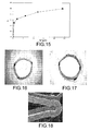
- Antibody cd134 was coated on the surface of the scaffold to adsorb endothelial progenitor cells in order to accelerate the repair of the endothelium. The release results were shown in Fig. 15 .
- the results of the 3-month animal experiment show that the thickness of the blood vessel tunica intima was 143 ⁇ 49 ⁇ m, and the blood vessel sections were shown in Fig. 16 and Fig. 17 . No thrombus or hemangioma was observed.
- Polycaprolactone (0.1g) was dissolved in chloroform (100ml), and As 2 O 3 (1.0 g) was dissolved in redistilled water (100 ml). Firstly the polymer solution was sprayed on the surface of the scaffold, and then the As 2 O 3 aqueous solution was sprayed thereon. Upon drying, As 2 O 3 formed conglomerated particles on the surface of the scaffold, and a solvent was sprayed on the surface of the scaffold to level the polymer coating. Enwrapping of the polymer was formed on the surface of the As 2 O 3 microparticles, and As 2 O 3 was fixed in the polymer coating in a form of "isolated island" structure, as indicated in Fig. 18 .
- the results of the 3-month animal experiment show that the thickness of the blood vessel tunica intima was 141 ⁇ 44 ⁇ m, and the blood vessel sections were shown in Fig.20 and Fig.21 , no thrombus or hemangioma was observed.
- the area of cavity was increased distinctly, while not a single thrombus or hemangioma occurred; the surface of the scaffold was fully covered by endothelium; the tunica intima, tunica media and tunica adventitia of the blood vessel were clear, and no obvious inflammation was observed.
- the results were shown in Fig.20 and Fig.21 .
- the results of the 3-month animal experiment show that the thickness of the blood vessel tunica intima was 128 ⁇ 35 ⁇ m, and the blood vessel sections were shown in Fig.24 and Fig.25 with no thrombus or hemangioma observed inside the scaffold.
- the surface of the scaffold was fully covered by endothelium; the tunica intima, tunica media and tunica adventitia of the blood vessel were clear, and no obvious inflammation was observed.
Landscapes
- Health & Medical Sciences (AREA)
- Life Sciences & Earth Sciences (AREA)
- General Health & Medical Sciences (AREA)
- Animal Behavior & Ethology (AREA)
- Heart & Thoracic Surgery (AREA)
- Public Health (AREA)
- Veterinary Medicine (AREA)
- Vascular Medicine (AREA)
- Surgery (AREA)
- Epidemiology (AREA)
- Chemical & Material Sciences (AREA)
- Medicinal Chemistry (AREA)
- Engineering & Computer Science (AREA)
- Molecular Biology (AREA)
- Biomedical Technology (AREA)
- Inorganic Chemistry (AREA)
- Cardiology (AREA)
- Nuclear Medicine, Radiotherapy & Molecular Imaging (AREA)
- General Chemical & Material Sciences (AREA)
- Chemical Kinetics & Catalysis (AREA)
- Pharmacology & Pharmacy (AREA)
- Bioinformatics & Cheminformatics (AREA)
- Organic Chemistry (AREA)
- Medicinal Preparation (AREA)
- Pharmaceuticals Containing Other Organic And Inorganic Compounds (AREA)
- Materials For Medical Uses (AREA)
- Medicines That Contain Protein Lipid Enzymes And Other Medicines (AREA)
Description
- The present invention relates to a medical scaffold, more particularly, relates to a medical elution scaffold for preventing restenosis after implantation of coronary artery scaffold.
- Arsenic trioxide (As2O3) is a main component in the traditional Chinese medicine White Arsenic, and had been used to treat many diseases such as malignant tumor in Chinese medical science.
- Natural arsenic compounds have been used as a medicine for more than 2400 years. In 1970s, hematologists used As2O3 to treat promyelocytic leukemia and notable therapeutic effect was obtained. As2O3 has also been used to treat solid tumor in recent years. Zhuoqi ZHANG et.al. and Jianwei SHAO et. al. found that As2O3 had effects of anti-proliferation, apoptosis promotion and cell cycle arrest on VSMCs, and was significant for possibly decreasing cell amount in the new tunica intima at the sites where blood vessel scaffold was implanted in vivo and reducing the thickness of the tunica intima.
- On the premise of ensuring medical effect, to reduce the dosage of arsenic trioxide, a highly toxic medicine, as much as possible and to ensure the stability of the medicine amount carried on the scaffold so as to achieve suitable uniformity and controllability for arsenic trioxide release is critical in determining the use value of the arsenic trioxide elution scaffold.
- Because of the difference in solubility between the water-soluble medicine arsenic trioxide and the polymer carrier, a characteristic two-phase separation is present during the spraying of the medicine and the carrier. For example, both of the arsenic trioxide spraying techniques disclosed in
CN200510023714.5 CN1413594A employ a process of spraying the medicine and the carrier together after mixing in a solvent. In these techniques, the control of the medicine amount on the scaffold and the uniformity of medicine distribution are restricted due to the settlement of arsenic trioxide (reduction in particle size of arsenic trioxide particles may induce conglomeration of the particles), and irregular channels may form among medicine particles, whereby it is difficult to control the stability of medicine release (seeFig.1 ). This leads to a high-dosage of the medicine, poor efficacy on restenosis inhibition, and increased risk in clinical treatment. - Several hours after damaging the blood vessel, scaffold and balloon dilatation may stimulate hyperplasia of SMC, which is beneficial for the formation of new tunica intima; several days to several months after the damage, relative deficiency of cell apoptosis during this period is an important reason for formation of restenosis. Therefore, it is necessary to provide desirable and stable medicine release on the scaffold.
- The object of the present invention is to provide an arsenic trioxide medical elution scaffold to overcome the above-mentioned disadvantages in the prior art.
- The arsenic trioxide medical elution scaffold according to the present invention comprises scaffold, polymer coating covering the scaffold and arsenic trioxide loaded in the polymer coating, characterized in that arsenic trioxide is loaded in the polymer coating in a form of single particle or isolated and scattered conglomerated particle the single particle or the conglomerated particle having a particle size of 0.01 to 50 µm and the arsenic trioxide particles being embedded in polymer microspheres, i.e., the medicine particles are separated from each other as an "isolated island" structure for the purpose of stable sustained-release of the medicine.
- The weight ratio of arsenic trioxide to the polymer coating is 0.1:9.9 to 9.9:0.1.
- Preferably, the weight ratio of arsenic trioxide to the polymer is 0.2 to 150.0, more preferably 0.5 to 1.5. ,
- Preferably, the content of arsenic trioxide on the scaffold is 0.01 to 3.2 µg/mm2, more preferably 0.47 to 1.11 µg/mm2.
- Base material of the scaffold includes stainless steel, cobalt-nickel alloy and other metal or alloy with good biocompatibility.
- The polymer is selected from degradable polymers.
- The degradable polymer is one or more selected from a group consisting of cellulose, polysaccharide, chitin, chitosan or derivatives thereof, polyhydroxyalkyl alcohol ester, poly(β-malate), poly-α-hydroxyacid esters, polycaprolactone, polycyanoacrylate, or polyamino acid, pseudo-polyamino acid, poly(lactide-co-glycolide) (PLGA), poly(lactic acid), polycarbonate, polyanhydride, polyvinylpyrrolidinone or polyvinylpyrrolidinone, or copolymer thereof.
- The scaffold according to the present invention is a routine blood vessel scaffold for implanting, such as balloon dilatation coronary artery scaffold, brain blood vessel scaffold and so on. These scaffolds have been described in details in various documents such as
CN1360951 ,CN1355005 andCN1669595 . - One of the methods for preparing the arsenic trioxide medical elution scaffold according to the present invention comprises the following steps:
- (1) dissolving the polymer in an organic solvent, dissolving arsenic trioxide in water, and adding a surfactant thereto respectively; mixing these two solution and emulsifying to obtain an emulsion, then spraying the emulsion on the scaffold to obtain a medical coating after evaporation of the solvent; the obtained arsenic trioxide particles have a particle size of 0.01 to 50 µm;
the weight ratio of arsenic trioxide to the polymer in the emulsion is 0.1:9.9 to 9.9:0.1;
the solvent is selected from organic solvents such as tetrahydrofuran, dichloromethane, chloroform, dimethylformamide or DMSO, and the content of the polymer is 1-90% by weight;
the content by weight of arsenic trioxide in the arsenic trioxide aqueous solution is 0.01% to saturation;
the surfactant is one or more selected from a group consisting of the Span series and the Tween series, Span 60 or Span 80 is preferred in the Span series and Tween 60 or Tween 80 is preferred in the Tween series, and the amount added is 0.01-1% by weight of the degradable polymer; - (2) leveling the polymer on the surface of the emulsion particles with a liquid solvent or a gas solvent, or spraying a new coating on the scaffold, and then drying in vacuum, at this time arsenic trioxide is fixed in the polymer coating in a form of "isolated island" structure, so as to obtain the arsenic trioxide medical elution scaffold.
- Another method for preparing the arsenic trioxide medical elution scaffold according to the present invention comprises the steps of:
- dissolving the polymer in an organic solvent, suspending the arsenic trioxide particles having a particle size of 0.01 to 50µm in alcohol, adding a surfactant thereto; adding the mixture into the polymer solution and stirring to form a polymer thin film on the surface of As2O3 and form an emulsion; spraying the emulsion on the surface of the scaffold and drying to obtain the arsenic trioxide elution scaffold in which arsenic trioxide is fixed in the polymer coating in a form of "isolated island" structure.
- The content of arsenic trioxide in alcohol is 0.01 to 50 g/ml, and the concentration by weight of the polymer in the organic solvent is 0.01-50%;
the weight ratio of arsenic trioxide to the polymer in the emulsion is 0.1:9.9 to 9.9:0.1;
the surfactant is selected from a composition of the Span series (such as Span 60 and Span 80) and the Tween series (such as Tween 60 and Tween 80), and the amount added can be 0.01-1% by weight of the degradable polymer. - Still another method for preparing the arsenic trioxide medical elution scaffold according to the present invention comprises the following steps:
- spraying the arsenic trioxide aqueous solution and the organic solvent containing the polymer on the scaffold respectively for several times, namely in a sandwich form, with one layer of the polymer solution on one layer of the arsenic trioxide aqueous solution coating, and so forth; and leveling the polymer with a liquid solvent or a gas solvent each time after coating the polymer solution to enwrap a layer of the polymer on the surface of the arsenic trioxide particles, at this time arsenic trioxide is fixed in the polymer coating in a form of "isolated island" structure, then drying to obtain the arsenic trioxide medical elution scaffold.
- The solvent is selected from organic solvents such as tetrahydrofuran, dichloromethane, chloroform, dimethylformamide or DMSO, and the content of the polymer in the organic solvent is 1-90% by weight;
the concentration by weight of the arsenic trioxide aqueous solution is 0.01 % to saturation;
spraying amount of the arsenic trioxide aqueous solution and that of the organic solvent containing the polymer are controlled so that the weight ratio of arsenic trioxide to the polymer in the coating is 0.1:9.9 to 9.9:0.1. - Yet still another method for preparing the arsenic trioxide medical elution scaffold according to the present invention comprises the following steps:
- (1) drilling micropores on the surface of the scaffold by a laser method; mixing the arsenic trioxide particles and an organic solvent containing the polymer to suspend the arsenic trioxide particles in the organic solvent containing the polymer, then coating the polymer solution with medicine in the micropores in the scaffold using sited feeding method; the surface can be coated or not coated with sustained-release controlled coating.
- The solvent is selected from organic solvents such as tetrahydrofuran, dichloromethane, chloroform, dimethylformamide or DMSO, in the organic solvent, the concentration by weight of the polymer in the solvent is 0.01 % to 90%, and the weight ratio of arsenic trioxide to the polymer is 0.1:9.9 to 9.9:0.1.
- The release properties of the medicine in the present invention can be measured by the method disclosed in "Preparation and the release characteristic in vitro of arsenic trioxide albumin nanospheres", Jie ZHOU, Puqing ZENG, Xiang GAO, Shusheng XIE, and Shuli WEI, Chinese Journal of New Drugs, 2005, 14(1), 54-57.
- The animal experiments prove that the medicine release of the arsenic trioxide medical elution scaffold according to the present invention is suitably uniform and controllable to meet the requirements of clinical application.
-
-
Figure 1 shows the scaffold prepared in the prior art. -
Figure 2 shows photograph of the scaffold in Example 1. -
Figure 3 shows the release result of the medicine on the scaffold in Example 1. -
Figure 4 shows the blood vessel section in the animal experiment of the scaffold in Example 1. -
Figure 5 shows the blood vessel section in the animal experiment of the scaffold in Example 1. -
Figure 6 shows the release result of the medicine on the scaffold in Example 2. -
Figure 7 shows the blood vessel section in the animal experiment of the scaffold in Example 2. -
Figure 8 shows the blood vessel section in the animal experiment of the scaffold in Example 2. -
Figure 9 shows the release result of the medicine on the scaffold in Example 3. -
Figure 10 shows the blood vessel section in the animal experiment of the scaffold in Example 3. -
Figure 11 shows the blood vessel section in the animal experiment of the scaffold in Example 3. -
Figure 12 shows the release result of the medicine on the scaffold in Example 4. -
Figure 13 shows the blood vessel section in the animal experiment of the scaffold in Example 4. -
Figure 14 shows the blood vessel section in the animal experiment of the scaffold in Example 4. -
Figure 15 shows the release result of the medicine on the scaffold in Example 5. -
Figure 16 shows the blood vessel section in the animal experiment of the scaffold in Example 5. -
Figure 17 shows the blood vessel section in the animal experiment of the scaffold in Example 5. -
Figure 18 shows photograph of the scaffold in Example 6. -
Figure 19 shows the release result of the medicine on the scaffold in Example 6. -
Figure 20 shows the blood vessel section in the animal experiment of the scaffold in Example 6. -
Figure 21 shows the blood vessel section in the animal experiment of the scaffold in Example 6. -
Figure 22 shows photograph of the scaffold in Example 7. -
Figure 23 shows the release result of the medicine on the scaffold in Example 7. -
Figure 24 shows the blood vessel section in the animal experiment of the scaffold in Example 7. -
Figure 25 shows the blood vessel section in the animal experiment of the scaffold in Example 7. - As2O3 (0.30 g) was milled into particles having an average particle size of 0.10 µm which were suspended in anhydrous alcohol (100 ml), and surfactant Span 80 (0.005 g) was added thereto. Polycaprolactone (2.5 g) was dissolved in tetrahydrofuran solution (100 ml), then the alcohol solution containing the arsenic trioxide particles was added to the polycaprolactone solution and dispersed therein under stirring, and then anhydrous alcohol (80 ml) was added dropwise thereto under stirring to form a thin film of polycaprolactone on the surface of the As2O3 particles, and a suspension was formed when the particles precipitated in a form of microcapsules. Upon being sprayed uniformly with the suspension on the surface, the 316 stainless steel scaffold was dried in vacuum at 80°C for 8 h to give the As2O3 elution scaffold, wherein the content of As2O3 was 0.05 µg/mm2 and As2O3 was fixed on the polymer coating in a form of "isolated island" structure, as indicated in
Fig.2 . - Antibody cd133 was coated on the surface of the scaffold to adsorb endothelial progenitor cells in order to accelerate the repair of the endothelium. The release results were shown in
Fig.3 . - The above-mentioned As2O3 medical elution scaffold was subjected to animal experiments, and the experimental method was as follows:
- three-month-old mini-pigs were used as the experimental samples. 20 mg/kg ketamine was injected intravenously to achieve general anesthesia, after disinfection and draping on the right-side groin, the femoral artery was separated and cut open, a 7F arterial sheath was placed therein, and 7F-guided ductal type coronary angiography was conducted; a 0.014"BMW guidewire was guided to the distal end of the coronary artery (RCA, LAD, LCX), and the scaffold was delivered along the guidewire, and then released with the expansion of balloon at certain pressure. After the balloon, the guidewire, the duct and the arterial sheath were removed, the groin was sutured.
- The animals were sacrificed after three months and hearts were harvested; the scaffold was embedded in resin and then prepared as hard issue sections. After stained with hematoxylin and eosin respectively, the sections were photographed under a microscope and the area and thickness of the tunica intima were calculated.
- The results of the 3-month animal experiment show that the thickness of the blood vessel tunica intima was 152 ± 41 µm, and the blood vessel sections were shown in
Fig.4 and Fix. 5. No thrombus or hemangioma was observed. - As2O3 (1.0 g) was milled into particles having an average particle size of 15 µm which were suspended in anhydrous alcohol (100 ml), and surfactant Span 80 (0.005 g) was added thereto. Poly(lactide-co-glycolide) (0.2 g, with the weight ratio of glycolide:lactide=2:8) was dissolved in chloroform solution (100 ml), then the alcohol solution containing the arsenic trioxide particles was added to the PLGA solution and dispersed therein under stirring, and then anhydrous alcohol (200 ml) was added dropwise thereto under stirring to form a thin film of PLGA on the surface of the As2O3 particles, and a suspension was formed when the particles precipitated in a form of microcapsules. Upon being sprayed uniformly with the suspension on the surface, the nickel-cobalt alloy scaffold was dried in vacuum at 80°C for 8 h to give the As2O3 elution scaffold, wherein the content of As2O3 was 2.65 µg/mm2 and As2O3 was fixed in the polymer coating in a form of "isolated island" structure.
- A polylactic acid sustained-release layer containing heparin was coated on the surface of the scaffold to prevent formation of acute thrombus and to control sustained-release of the medicine efficiently. The release results were shown in
Fig.6 . - The above-mentioned As2O3 medical elution scaffold was subjected to animal experiments. The experimental results show that the thickness of the blood vessel tunica intima was 161 ± 38 µm, and the blood vessel sections were shown in
Fig.7 and Fig.8 . No thrombus or hemangioma was observed. - As2O3 (1.0 g) was milled into particles having an average particle size of 0.25 µm which were suspended in anhydrous alcohol (100 ml), then surfactant Tween 80 (0.0025 g) was added thereto. Polylactic acid (1.0 g) was dissolved in a dichloromethane solution (100 ml), then the alcohol solution containing the arsenic trioxide particles was added to the polylactic acid solution and dispersed therein under stirring, and then anhydrous alcohol (125 ml) was added dropwise thereto under stirring to form a thin film of polylactic acid on the surface of the As2O3 particles, and a suspension was formed when the particles precipitated in a form of microcapsules. Upon being sprayed uniformly with the suspension on the surface, the 316 stainless steel scaffold was dried in vacuum at 80°C for 8 h to give the As2O3 elution scaffold, wherein the content of As2O3 was 0.52 µg/mm2, the weight ratio of the medicine to the polymer was 1:1, and As2O3 was fixed in the polymer coating in a form of "isolated island" structure. The release results were shown in
Fig.9 . - The above-mentioned As2O3 medical elution scaffold was subjected to animal experiments. The experimental results show that the thickness of the blood vessel tunica intima was 128 ± 25 µm, and the blood vessel sections were shown in
Fig.10 and Fig.11 . No thrombus or hemangioma was observed. - Poly(lactide-co-glycolide) (1 g, weight ratio of glycolide:lactide=1:1) copolymer was dissolved in a chloroform solution (100 ml), and surfactant Span 80 (0.098 g) was added and mixed therewith to form an oil phase; surfactant Tween 80 (0.002 g) was added to a saturated aqueous solution of As2O3 (50 ml) to form an aqueous phase. The aqueous phase was added to the oil phase and dispersed uniformly under stirring at 3000 rpm to form an emulsion. Upon being sprayed with the emulsion on the surface, the scaffold was dried in vacuum at 75°C for 10 h to form the As2O3 elution scaffold, wherein the content of As2O3 was 50 µg and As2O3 was fixed in the polymer coating in a form of "isolated island" structure. Antibody cd133 was coated on the surface of the scaffold to adsorb endothelial progenitor cells in order to accelerate the repair of the endothelium. The release results were shown in
Fig. 12 . - The results of the 3-month animal experiment show that the thickness of the blood vessel tunica intima was 143 ± 49 µm, and the blood vessel sections were shown in
Fig.13 and Fig. 14 . No thrombus or hemangioma was observed. - Polylactic acid (0.1 g) was dissolved in a chloroform solution (100ml), and surfactant Span 80 (0.098 g) was added and mixed therewith to form an oil phase; surfactant Tween 80 (0.002 g) was added to a saturated aqueous solution of As2O3 (50 ml) to form an aqueous phase. The aqueous phase was added to the oil phase and dispersed uniformly under stirring at 3000 rpm to form an emulsion. Upon being sprayed with the emulsion on the surface, the scaffold was dried in vacuum at 75°C for 10 h to form the As2O3 elution scaffold, wherein the content of As2O3 was 0.81 µg/mm2 and in the polymer coating in a form of "isolated island" structure. Antibody cd134 was coated on the surface of the scaffold to adsorb endothelial progenitor cells in order to accelerate the repair of the endothelium. The release results were shown in
Fig. 15 . - The results of the 3-month animal experiment show that the thickness of the blood vessel tunica intima was 143 ± 49 µm, and the blood vessel sections were shown in
Fig. 16 and Fig. 17 . No thrombus or hemangioma was observed. - Polycaprolactone (0.1g) was dissolved in chloroform (100ml), and As2O3 (1.0 g) was dissolved in redistilled water (100 ml). Firstly the polymer solution was sprayed on the surface of the scaffold, and then the As2O3 aqueous solution was sprayed thereon. Upon drying, As2O3 formed conglomerated particles on the surface of the scaffold, and a solvent was sprayed on the surface of the scaffold to level the polymer coating. Enwrapping of the polymer was formed on the surface of the As2O3 microparticles, and As2O3 was fixed in the polymer coating in a form of "isolated island" structure, as indicated in
Fig. 18 . - After drying, the polymer solution, the As2O3 aqueous solution and the solvent were sprayed in this order until the medicine content achieved a desired amount. The content of As2O3 was 1.25 µg/mm2. The release results were shown in
Fig. 19 . - The results of the 3-month animal experiment show that the thickness of the blood vessel tunica intima was 141 ± 44 µm, and the blood vessel sections were shown in
Fig.20 and Fig.21 , no thrombus or hemangioma was observed. After 12 weeks of the scaffold implantation, no obvious hyperplasia can be observed in the blood vessel tunica intima, the area of cavity was increased distinctly, while not a single thrombus or hemangioma occurred; the surface of the scaffold was fully covered by endothelium; the tunica intima, tunica media and tunica adventitia of the blood vessel were clear, and no obvious inflammation was observed. The results were shown inFig.20 and Fig.21 . - 10µm micropores were etched on the outer surface of the scaffold (the surface of the scaffold in contact with blood vessels after being implanted in vivo) with a laser etching machine. An emulsion was prepared with the method for preparing emulsion as described in Example 1, and the emulsion was distilled under reduced pressure to achieve a concentration of the microparticles of 30% by weight. The solution was injected into the micropores, and after evaporation of the solvent, As2O3 was fixed in the polymer coating in a form of "isolated island" structure on the outer surface of the scaffold, as indicated in
Fig.22 . After drying in vacuum, the As2O3 medical elution scaffold was obtained. The content of As2O3 was 2.4µg/mm2. The release results were shown inFig.23 . - The results of the 3-month animal experiment show that the thickness of the blood vessel tunica intima was 128 ± 35 µm, and the blood vessel sections were shown in
Fig.24 and Fig.25 with no thrombus or hemangioma observed inside the scaffold. The surface of the scaffold was fully covered by endothelium; the tunica intima, tunica media and tunica adventitia of the blood vessel were clear, and no obvious inflammation was observed.
Claims (18)
- An arsenic trioxide medical elution scaffold comprising scaffold, polymer coating covering the scaffold and arsenic trioxide loaded in the polymer coating, characterized in that the arsenic trioxide is loaded in the polymer coating in a form of single particle or isolated and scattered conglomerated particle, the single particle or the conglomerated particle having a particle size of 0.01 to 50 µm and the arsenic trioxide particles being embedded in polymer microspheres.
- The arsenic trioxide medical elution scaffold according to claim 1, characterized in that the weight ratio of arsenic trioxide to the polymer coating is 0.1:9.9 to 9.9:0.1.
- The arsenic trioxide medical elution scaffold according to claim 2, characterized in that the weight ratio of arsenic trioxide to the polymer is 0.2 to 1.5.
- The arsenic trioxide medical elution scaffold according to claim 3, characterized in that the weight ratio of arsenic trioxide to the polymer is 0.5 to 1.5.
- The arsenic trioxide medical elution scaffold according to claim 1, characterized in that the content of arsenic trioxide in the scaffold is 0.01-3.2 µg/mm2.
- The arsenic trioxide medical elution scaffold according to claim 5, characterized in that the content of arsenic trioxide in the scaffold is 0.47-1.11 µg/mm2.
- The arsenic trioxide medical elution scaffold according to claim 1, characterized in that the base material of the scaffold includes stainless steel, cobalt-nickel alloy.
- The arsenic trioxide medical elution scaffold according to claim 1, characterized in that the polymer is selected from degradable polymers.
- The arsenic trioxide medical elution scaffold according to claim 8, characterized in that the degradable polymer is one or more selected from a group consisting of cellulose, polysaccharide, chitin, chitosan or derivatives thereof, polyhydroxyalkyl alcohol ester, poly(β-malate), poly-α-hydroxyacid esters, polycaprolactone, polycyanoacrylate, or polyamino acid, pseudo-polyamino acid, poly(lactide-co-glycolide) (PLGA), poly(lactic acid), polycarbonate, polyanhydride, polyvinylpyrrolidinone or polyvinylpyrrolidinone, or copolymer thereof.
- The arsenic trioxide medical elution scaffold according to any one of claims 1 to 9, characterized in that the scaffold is a blood vessel scaffold for implanting.
- A method for preparing the arsenic trioxide medical elution scaffold according to any one of claims 1 to 10, characterized in that the method comprises steps of:(1) dissolving the polymer in an organic solvent and dissolving arsenic trioxide in water, then adding a surfactant thereto respectively; mixing these two and emulsifying to obtain an emulsion, then spraying the emulsion on the scaffold to obtain a medical coating after evaporation of the solvent;(2) leveling the polymer on the surface of the emulsion particles with a liquid solvent or a gas solvent, or spraying a new coating on the scaffold, then drying to obtain the arsenic trioxide medical elution scaffold.
- The method according to claim 11, characterized in that the arsenic trioxide particle has a particle size of 0.01 to 50 µm;
in the emulsion, the weight ratio of arsenic trioxide to the polymer is 0.1:9.9 to 9.9:0.1;
the solvent is selected from a group consisting of tetrahydrofuran, dichloromethane, chloroform, dimethylformamide or DMSO. - A method for preparing the arsenic trioxide medical elution scaffold according to any one of claims 1 to 10, characterized in that the method comprises steps of:dissolving the polymer in an organic solvent; suspending the arsenic trioxide particles with a particle size of 0.01 to 50µm in alcohol, adding a surfactant thereto, adding the suspension to the polymer solution and stirring to form a polymer thin film on the surface of As2O3 and form an emulsion, and then spraying the emulsion on the surface of the scaffold and drying to obtain the arsenic trioxide elution scaffold.
- The method according to claim 13, characterized in that the arsenic trioxide particle has a particle size of 0.01 to 50 µm, and in the emulsion, the weight ratio of arsenic trioxide to the polymer is 0.1:9.9 to 9.9:0.1;
in alcohol, the content of arsenic trioxide is 0.01 to 50 g/ml; in the organic solvent, the polymer has a concentration by weight of 0.01% to 50%;
the surfactant is one or more selected from a group consisting of the Span series and the Tween series, and the amount added is 0.01% by weight to 1% by weight of the degradable polymer. - A method for preparing the arsenic trioxide medical elution scaffold according to any one of claims 1 to 10, characterized in that the method comprises steps of: spraying the arsenic trioxide aqueous solution and the organic solvent containing the polymer on the scaffold respectively for several times with one layer of the polymer solution on one layer of the arsenic trioxide aqueous solution coating, and leveling the polymer with a liquid solvent or a gas solvent each time after coating the polymer solution to enwrap a layer of the polymer on the surface of the arsenic trioxide particles, then drying in vacuum to obtain the arsenic trioxide medical elution scaffold.
- The method according to claim 15, characterized in that the solvent is selected from a group consisting of tetrahydrofuran, dichloromethane, chloroform, dimethylformamide or DMSO, and the content of the polymer in the organic solvent is 1 to 90% by weight;
the concentration by weight of the arsenic trioxide aqueous solution is 0.01 % to saturation;
the spraying amount of the arsenic trioxide aqueous solution and that of the organic solvent containing the polymer are controlled so that the weight ratio of arsenic trioxide to the polymer in the coating is 0.1:9.9 to 9.9:0.1. - A method for preparing the arsenic trioxide medical elution scaffold according to any one of claims 1 to 10, characterized in that the method comprises steps of:(1) drilling micropores on the surface of the scaffold by a laser method, mixing the arsenic trioxide particles and an organic solvent containing the polymer to suspend the arsenic trioxide particles in the organic solvent containing the polymer, then coating the polymer solution comprising medicine into the micropores in the scaffold with sited feeding method; the surface can be coated or not coated with sustained-release controlled coating.
- The method according to claim 17, characterized in that the organic solvent is selected from a group consisting of tetrahydrofuran, dichloromethane, chloroform, dimethylformamide or DMSO, in the organic solvent, the concentration by weight of the polymer in the solvent is 0.01 to 90%, and the weight ratio of arsenic trioxide to the polymer is 0.1:9.9 to 9.9:0.1.
Applications Claiming Priority (2)
| Application Number | Priority Date | Filing Date | Title |
|---|---|---|---|
| CN200710171085XA CN101161300B (en) | 2007-11-27 | 2007-11-27 | Arsenic trioxide medicament elution bracket and its preparation method |
| PCT/CN2008/070760 WO2009067862A1 (en) | 2007-11-27 | 2008-04-21 | An arsenic trioxide medical elution scaffold |
Publications (3)
| Publication Number | Publication Date |
|---|---|
| EP2213313A1 EP2213313A1 (en) | 2010-08-04 |
| EP2213313A4 EP2213313A4 (en) | 2011-03-30 |
| EP2213313B1 true EP2213313B1 (en) | 2013-01-09 |
Family
ID=39296331
Family Applications (1)
| Application Number | Title | Priority Date | Filing Date |
|---|---|---|---|
| EP08734118A Active EP2213313B1 (en) | 2007-11-27 | 2008-04-21 | An arsenic trioxide medical elution scaffold |
Country Status (5)
| Country | Link |
|---|---|
| US (1) | US8518429B2 (en) |
| EP (1) | EP2213313B1 (en) |
| JP (1) | JP5209726B2 (en) |
| CN (1) | CN101161300B (en) |
| WO (1) | WO2009067862A1 (en) |
Families Citing this family (31)
| Publication number | Priority date | Publication date | Assignee | Title |
|---|---|---|---|---|
| ES2691646T3 (en) | 2005-07-15 | 2018-11-28 | Micell Technologies, Inc. | Polymer coatings containing controlled morphology drug powder |
| WO2007011708A2 (en) | 2005-07-15 | 2007-01-25 | Micell Technologies, Inc. | Stent with polymer coating containing amorphous rapamycin |
| CA2996768C (en) | 2006-04-26 | 2020-12-08 | Micell Technologies, Inc. | Coatings containing multiple drugs |
| CN101711137B (en) | 2007-01-08 | 2014-10-22 | 米歇尔技术公司 | Stent with biodegradable layer |
| US11426494B2 (en) | 2007-01-08 | 2022-08-30 | MT Acquisition Holdings LLC | Stents having biodegradable layers |
| CN101172168B (en) * | 2007-10-10 | 2010-06-02 | 大连理工大学 | Coating and preparation method of metal vascular stent loaded with glycosaminoglycans CD133 antibody |
| CN101161300B (en) * | 2007-11-27 | 2011-03-16 | 北京美中双和医疗器械有限公司 | Arsenic trioxide medicament elution bracket and its preparation method |
| CA2721832C (en) | 2008-04-17 | 2018-08-07 | Micell Technologies, Inc. | Stents having bioabsorbable layers |
| WO2010009335A1 (en) | 2008-07-17 | 2010-01-21 | Micell Technologies, Inc. | Drug delivery medical device |
| CN102481195B (en) | 2009-04-01 | 2015-03-25 | 米歇尔技术公司 | coated stent |
| EP3366326A1 (en) | 2009-04-17 | 2018-08-29 | Micell Technologies, Inc. | Stents having controlled elution |
| EP2453834A4 (en) | 2009-07-16 | 2014-04-16 | Micell Technologies Inc | Drug delivery medical device |
| EP2531140B1 (en) | 2010-02-02 | 2017-11-01 | Micell Technologies, Inc. | Stent and stent delivery system with improved deliverability |
| WO2011133655A1 (en) | 2010-04-22 | 2011-10-27 | Micell Technologies, Inc. | Stents and other devices having extracellular matrix coating |
| WO2012009684A2 (en) | 2010-07-16 | 2012-01-19 | Micell Technologies, Inc. | Drug delivery medical device |
| CN102068718B (en) * | 2010-11-05 | 2013-12-25 | 东南大学 | Preparation technology of nano arsenic trioxide/poly (lactic-co-glycolic acid) coated bracket |
| US10117972B2 (en) | 2011-07-15 | 2018-11-06 | Micell Technologies, Inc. | Drug delivery medical device |
| US10188772B2 (en) | 2011-10-18 | 2019-01-29 | Micell Technologies, Inc. | Drug delivery medical device |
| CN102872483B (en) * | 2012-09-19 | 2015-02-18 | 华东理工大学 | Poly-(Epsilon-caprolactone) drug eluting stent modification method |
| CN102974026A (en) * | 2012-09-21 | 2013-03-20 | 北京美中双和医疗器械有限公司 | Arsenic trioxide controllable-releasing balloon and preparing method thereof |
| JP2015533305A (en) * | 2012-10-18 | 2015-11-24 | ミセル テクノロジーズ,インク. | Drug delivery medical device |
| CN110269959A (en) | 2013-03-12 | 2019-09-24 | 脉胜医疗技术公司 | Bioabsorbable biomedical implants |
| WO2014186532A1 (en) | 2013-05-15 | 2014-11-20 | Micell Technologies, Inc. | Bioabsorbable biomedical implants |
| US20180085498A1 (en) * | 2016-09-23 | 2018-03-29 | Micell Technologies, Inc. | Prolonged drug-eluting products |
| CN108338989B (en) | 2017-08-08 | 2021-12-17 | 新昕医药科技(上海)有限公司 | Compound anti-restenosis medicine of coronary artery medicine elution bracket and controlled release system thereof |
| CN108379658B (en) * | 2018-02-06 | 2021-03-02 | 中国科学院金属研究所 | Orthopedic implant device with copper-containing coating and method of making the same |
| CN108478843B (en) * | 2018-02-06 | 2021-03-02 | 中国科学院金属研究所 | Medical dressing with copper-containing coating and preparation method thereof |
| CN108939175A (en) * | 2018-10-24 | 2018-12-07 | 上海市第六人民医院 | Colchicin coating FirebirdTM and preparation method thereof |
| CN111330091A (en) * | 2020-03-06 | 2020-06-26 | 上海交通大学医学院附属第九人民医院 | High-molecular drug-loaded film for cochlear implant, cochlear implant drug-loaded electrode and preparation method |
| CN114904052B (en) * | 2022-05-06 | 2024-02-02 | 南京友德邦医疗科技有限公司 | A new type of degradable magnesium alloy vascular stent and its preparation method |
| CN115382027B (en) * | 2022-10-26 | 2023-03-24 | 北京美中双和医疗器械股份有限公司 | Cobalt-chromium alloy stent and drug-coated stent |
Family Cites Families (15)
| Publication number | Priority date | Publication date | Assignee | Title |
|---|---|---|---|---|
| US20070190102A1 (en) * | 2000-06-30 | 2007-08-16 | Ping Luo | Method of preparing hydroxyapatite based drug delivery implant for infection and cancer treatment |
| CN1360951A (en) | 2000-12-28 | 2002-07-31 | 微创医疗器械(上海)有限公司 | Blood vessel support with coating to prevent blood vessel from becoming strictured again |
| CN1303947C (en) | 2001-12-13 | 2007-03-14 | 华东理工大学 | Medicine eluted cardiovascular frame and its preparing process |
| CN1398585A (en) | 2002-07-15 | 2003-02-26 | 裴福兴 | Slow-releasing bFGF-PLA-PEG microball and its prepn and use |
| CN2561433Y (en) * | 2002-08-07 | 2003-07-23 | 杨巍 | Blood vessel stent with medicinal coating |
| CN1413594A (en) | 2002-08-07 | 2003-04-30 | 杨巍 | Medicine for prevention and curing reangiostenosis |
| CN1223385C (en) | 2002-12-31 | 2005-10-19 | 中国科学院金属研究所 | Cardiovascular support having control releasing therapeutic medicine |
| JP4971580B2 (en) * | 2003-06-05 | 2012-07-11 | テルモ株式会社 | Stent and method for manufacturing stent |
| CN1669595A (en) | 2004-03-16 | 2005-09-21 | 程树军 | Medicine eluted cardiovascular support |
| CN1669597A (en) | 2004-03-16 | 2005-09-21 | 程树军 | Medicine eluted cardiovascular support |
| CN100435756C (en) * | 2005-01-31 | 2008-11-26 | 上海市第一人民医院 | A kind of arsenic trioxide controlled-release elution stent |
| CN100381183C (en) * | 2005-10-13 | 2008-04-16 | 上海交通大学 | Preparation method of drug-loaded sustained-release micro/nanosphere digestive tract stent |
| WO2007065016A2 (en) * | 2005-12-02 | 2007-06-07 | Au Jessie L S | Methods and compositions to improve activity and reduce toxicity of stents |
| US8585753B2 (en) * | 2006-03-04 | 2013-11-19 | John James Scanlon | Fibrillated biodegradable prosthesis |
| CN101161300B (en) * | 2007-11-27 | 2011-03-16 | 北京美中双和医疗器械有限公司 | Arsenic trioxide medicament elution bracket and its preparation method |
-
2007
- 2007-11-27 CN CN200710171085XA patent/CN101161300B/en active Active
-
2008
- 2008-04-21 JP JP2010528264A patent/JP5209726B2/en not_active Expired - Fee Related
- 2008-04-21 WO PCT/CN2008/070760 patent/WO2009067862A1/en not_active Ceased
- 2008-04-21 EP EP08734118A patent/EP2213313B1/en active Active
- 2008-04-21 US US12/681,097 patent/US8518429B2/en not_active Expired - Fee Related
Also Published As
| Publication number | Publication date |
|---|---|
| CN101161300A (en) | 2008-04-16 |
| EP2213313A1 (en) | 2010-08-04 |
| US8518429B2 (en) | 2013-08-27 |
| JP2010540199A (en) | 2010-12-24 |
| WO2009067862A1 (en) | 2009-06-04 |
| US20100247603A1 (en) | 2010-09-30 |
| EP2213313A4 (en) | 2011-03-30 |
| WO2009067862A8 (en) | 2010-05-14 |
| CN101161300B (en) | 2011-03-16 |
| JP5209726B2 (en) | 2013-06-12 |
Similar Documents
| Publication | Publication Date | Title |
|---|---|---|
| US8518429B2 (en) | Arsenic trioxide medical elution scaffold | |
| RU2721655C2 (en) | Coating for an intraluminal divergent catheter, providing contact delivery of microreservoirs with a drug preparation | |
| CN101940802B (en) | Sustained drug-releasing stent | |
| JP7842135B2 (en) | Coating for intraluminal expandable catheters that provides contact transfer of drugs in a microreservoir. | |
| JP2007153896A (en) | Compound of nanoparticle and/or microparticle for treating vascular disease based on topical injection | |
| CN101869514B (en) | Controlled drug-release composition and drug-releasable medical device | |
| CN105597163B (en) | Iron-based alloy implanted medical device and preparation method thereof | |
| CN102657899A (en) | Medicament coating stent capable of preventing blood vessel restenosis and preparation method thereof | |
| CN103566415A (en) | Human body blood vessel implant with coatings on two surfaces and manufacturing method thereof | |
| EP4400126B1 (en) | Drug-loaded balloon and preparation method therefor | |
| CN101007187A (en) | Preparation method of composite drug-eluting stent and its drug coated layer | |
| KR102409251B1 (en) | Drug-eluting stent | |
| Cao et al. | Analysis of inhibitory effect of the double-layer nano-infusion on vascular restenosis in animal models of coronary atherosclerosis | |
| JP2021511891A (en) | Methods and devices for reducing vascular smooth muscle cell proliferation | |
| Song et al. | Ultrasound controlled paclitaxel releasing system—A novel method for improving the availability of coronary artery drug coated balloon | |
| Bhambri et al. | Verapamil eluting stents as a possible treatment for vasospasm after subarachnoid hemorrhage | |
| JP2025528176A (en) | Drug-coated medical devices and manufacturing methods | |
| CN108339160A (en) | With the degradable magnesium alloy angiocarpy bracket and preparation method thereof containing copper coating | |
| KR101198642B1 (en) | Multi-layer coating stent for inhibiting in-stent restenosis and a method thereof | |
| CN108939175A (en) | Colchicin coating FirebirdTM and preparation method thereof | |
| JP2006206478A (en) | Method for producing bioabsorbable fine particle including water-soluble medicine | |
| CN113289074A (en) | Novel drug-coated stent and preparation method thereof |
Legal Events
| Date | Code | Title | Description |
|---|---|---|---|
| PUAI | Public reference made under article 153(3) epc to a published international application that has entered the european phase |
Free format text: ORIGINAL CODE: 0009012 |
|
| 17P | Request for examination filed |
Effective date: 20100329 |
|
| AK | Designated contracting states |
Kind code of ref document: A1 Designated state(s): AT BE BG CH CY CZ DE DK EE ES FI FR GB GR HR HU IE IS IT LI LT LU LV MC MT NL NO PL PT RO SE SI SK TR |
|
| AX | Request for extension of the european patent |
Extension state: AL BA MK RS |
|
| DAX | Request for extension of the european patent (deleted) | ||
| A4 | Supplementary search report drawn up and despatched |
Effective date: 20110224 |
|
| 17Q | First examination report despatched |
Effective date: 20111227 |
|
| REG | Reference to a national code |
Ref country code: DE Ref legal event code: R079 Ref document number: 602008021540 Country of ref document: DE Free format text: PREVIOUS MAIN CLASS: A61L0027000000 Ipc: A61L0031020000 |
|
| GRAJ | Information related to disapproval of communication of intention to grant by the applicant or resumption of examination proceedings by the epo deleted |
Free format text: ORIGINAL CODE: EPIDOSDIGR1 |
|
| GRAP | Despatch of communication of intention to grant a patent |
Free format text: ORIGINAL CODE: EPIDOSNIGR1 |
|
| GRAP | Despatch of communication of intention to grant a patent |
Free format text: ORIGINAL CODE: EPIDOSNIGR1 |
|
| RIC1 | Information provided on ipc code assigned before grant |
Ipc: A61L 31/10 20060101ALI20120706BHEP Ipc: A61L 31/16 20060101ALI20120706BHEP Ipc: A61L 31/02 20060101AFI20120706BHEP |
|
| GRAS | Grant fee paid |
Free format text: ORIGINAL CODE: EPIDOSNIGR3 |
|
| GRAA | (expected) grant |
Free format text: ORIGINAL CODE: 0009210 |
|
| AK | Designated contracting states |
Kind code of ref document: B1 Designated state(s): AT BE BG CH CY CZ DE DK EE ES FI FR GB GR HR HU IE IS IT LI LT LU LV MC MT NL NO PL PT RO SE SI SK TR |
|
| REG | Reference to a national code |
Ref country code: GB Ref legal event code: FG4D |
|
| RIN1 | Information on inventor provided before grant (corrected) |
Inventor name: MA, XIAOYI Inventor name: GE, JUNBO |
|
| REG | Reference to a national code |
Ref country code: CH Ref legal event code: EP Ref country code: AT Ref legal event code: REF Ref document number: 592366 Country of ref document: AT Kind code of ref document: T Effective date: 20130115 |
|
| REG | Reference to a national code |
Ref country code: IE Ref legal event code: FG4D |
|
| REG | Reference to a national code |
Ref country code: DE Ref legal event code: R096 Ref document number: 602008021540 Country of ref document: DE Effective date: 20130314 |
|
| REG | Reference to a national code |
Ref country code: CH Ref legal event code: NV Representative=s name: RENTSCH PARTNER AG, CH |
|
| REG | Reference to a national code |
Ref country code: NL Ref legal event code: T3 |
|
| PG25 | Lapsed in a contracting state [announced via postgrant information from national office to epo] |
Ref country code: SI Free format text: LAPSE BECAUSE OF FAILURE TO SUBMIT A TRANSLATION OF THE DESCRIPTION OR TO PAY THE FEE WITHIN THE PRESCRIBED TIME-LIMIT Effective date: 20130109 |
|
| REG | Reference to a national code |
Ref country code: AT Ref legal event code: MK05 Ref document number: 592366 Country of ref document: AT Kind code of ref document: T Effective date: 20130109 |
|
| REG | Reference to a national code |
Ref country code: LT Ref legal event code: MG4D |
|
| PG25 | Lapsed in a contracting state [announced via postgrant information from national office to epo] |
Ref country code: LT Free format text: LAPSE BECAUSE OF FAILURE TO SUBMIT A TRANSLATION OF THE DESCRIPTION OR TO PAY THE FEE WITHIN THE PRESCRIBED TIME-LIMIT Effective date: 20130109 Ref country code: NO Free format text: LAPSE BECAUSE OF FAILURE TO SUBMIT A TRANSLATION OF THE DESCRIPTION OR TO PAY THE FEE WITHIN THE PRESCRIBED TIME-LIMIT Effective date: 20130409 Ref country code: IS Free format text: LAPSE BECAUSE OF FAILURE TO SUBMIT A TRANSLATION OF THE DESCRIPTION OR TO PAY THE FEE WITHIN THE PRESCRIBED TIME-LIMIT Effective date: 20130509 Ref country code: AT Free format text: LAPSE BECAUSE OF FAILURE TO SUBMIT A TRANSLATION OF THE DESCRIPTION OR TO PAY THE FEE WITHIN THE PRESCRIBED TIME-LIMIT Effective date: 20130109 Ref country code: BE Free format text: LAPSE BECAUSE OF FAILURE TO SUBMIT A TRANSLATION OF THE DESCRIPTION OR TO PAY THE FEE WITHIN THE PRESCRIBED TIME-LIMIT Effective date: 20130109 Ref country code: BG Free format text: LAPSE BECAUSE OF FAILURE TO SUBMIT A TRANSLATION OF THE DESCRIPTION OR TO PAY THE FEE WITHIN THE PRESCRIBED TIME-LIMIT Effective date: 20130409 Ref country code: SE Free format text: LAPSE BECAUSE OF FAILURE TO SUBMIT A TRANSLATION OF THE DESCRIPTION OR TO PAY THE FEE WITHIN THE PRESCRIBED TIME-LIMIT Effective date: 20130109 Ref country code: ES Free format text: LAPSE BECAUSE OF FAILURE TO SUBMIT A TRANSLATION OF THE DESCRIPTION OR TO PAY THE FEE WITHIN THE PRESCRIBED TIME-LIMIT Effective date: 20130420 |
|
| PG25 | Lapsed in a contracting state [announced via postgrant information from national office to epo] |
Ref country code: GR Free format text: LAPSE BECAUSE OF FAILURE TO SUBMIT A TRANSLATION OF THE DESCRIPTION OR TO PAY THE FEE WITHIN THE PRESCRIBED TIME-LIMIT Effective date: 20130410 Ref country code: PL Free format text: LAPSE BECAUSE OF FAILURE TO SUBMIT A TRANSLATION OF THE DESCRIPTION OR TO PAY THE FEE WITHIN THE PRESCRIBED TIME-LIMIT Effective date: 20130109 Ref country code: LV Free format text: LAPSE BECAUSE OF FAILURE TO SUBMIT A TRANSLATION OF THE DESCRIPTION OR TO PAY THE FEE WITHIN THE PRESCRIBED TIME-LIMIT Effective date: 20130109 Ref country code: PT Free format text: LAPSE BECAUSE OF FAILURE TO SUBMIT A TRANSLATION OF THE DESCRIPTION OR TO PAY THE FEE WITHIN THE PRESCRIBED TIME-LIMIT Effective date: 20130509 Ref country code: FI Free format text: LAPSE BECAUSE OF FAILURE TO SUBMIT A TRANSLATION OF THE DESCRIPTION OR TO PAY THE FEE WITHIN THE PRESCRIBED TIME-LIMIT Effective date: 20130109 |
|
| PG25 | Lapsed in a contracting state [announced via postgrant information from national office to epo] |
Ref country code: HR Free format text: LAPSE BECAUSE OF FAILURE TO SUBMIT A TRANSLATION OF THE DESCRIPTION OR TO PAY THE FEE WITHIN THE PRESCRIBED TIME-LIMIT Effective date: 20130109 |
|
| PG25 | Lapsed in a contracting state [announced via postgrant information from national office to epo] |
Ref country code: DK Free format text: LAPSE BECAUSE OF FAILURE TO SUBMIT A TRANSLATION OF THE DESCRIPTION OR TO PAY THE FEE WITHIN THE PRESCRIBED TIME-LIMIT Effective date: 20130109 Ref country code: RO Free format text: LAPSE BECAUSE OF FAILURE TO SUBMIT A TRANSLATION OF THE DESCRIPTION OR TO PAY THE FEE WITHIN THE PRESCRIBED TIME-LIMIT Effective date: 20130109 Ref country code: EE Free format text: LAPSE BECAUSE OF FAILURE TO SUBMIT A TRANSLATION OF THE DESCRIPTION OR TO PAY THE FEE WITHIN THE PRESCRIBED TIME-LIMIT Effective date: 20130109 Ref country code: CZ Free format text: LAPSE BECAUSE OF FAILURE TO SUBMIT A TRANSLATION OF THE DESCRIPTION OR TO PAY THE FEE WITHIN THE PRESCRIBED TIME-LIMIT Effective date: 20130109 Ref country code: SK Free format text: LAPSE BECAUSE OF FAILURE TO SUBMIT A TRANSLATION OF THE DESCRIPTION OR TO PAY THE FEE WITHIN THE PRESCRIBED TIME-LIMIT Effective date: 20130109 |
|
| PLBE | No opposition filed within time limit |
Free format text: ORIGINAL CODE: 0009261 |
|
| STAA | Information on the status of an ep patent application or granted ep patent |
Free format text: STATUS: NO OPPOSITION FILED WITHIN TIME LIMIT |
|
| PG25 | Lapsed in a contracting state [announced via postgrant information from national office to epo] |
Ref country code: CY Free format text: LAPSE BECAUSE OF FAILURE TO SUBMIT A TRANSLATION OF THE DESCRIPTION OR TO PAY THE FEE WITHIN THE PRESCRIBED TIME-LIMIT Effective date: 20130109 Ref country code: MC Free format text: LAPSE BECAUSE OF FAILURE TO SUBMIT A TRANSLATION OF THE DESCRIPTION OR TO PAY THE FEE WITHIN THE PRESCRIBED TIME-LIMIT Effective date: 20130109 |
|
| 26N | No opposition filed |
Effective date: 20131010 |
|
| PG25 | Lapsed in a contracting state [announced via postgrant information from national office to epo] |
Ref country code: IT Free format text: LAPSE BECAUSE OF FAILURE TO SUBMIT A TRANSLATION OF THE DESCRIPTION OR TO PAY THE FEE WITHIN THE PRESCRIBED TIME-LIMIT Effective date: 20130109 |
|
| REG | Reference to a national code |
Ref country code: IE Ref legal event code: MM4A |
|
| REG | Reference to a national code |
Ref country code: DE Ref legal event code: R097 Ref document number: 602008021540 Country of ref document: DE Effective date: 20131010 |
|
| PG25 | Lapsed in a contracting state [announced via postgrant information from national office to epo] |
Ref country code: IE Free format text: LAPSE BECAUSE OF NON-PAYMENT OF DUE FEES Effective date: 20130421 |
|
| PG25 | Lapsed in a contracting state [announced via postgrant information from national office to epo] |
Ref country code: MT Free format text: LAPSE BECAUSE OF FAILURE TO SUBMIT A TRANSLATION OF THE DESCRIPTION OR TO PAY THE FEE WITHIN THE PRESCRIBED TIME-LIMIT Effective date: 20130109 |
|
| PG25 | Lapsed in a contracting state [announced via postgrant information from national office to epo] |
Ref country code: HU Free format text: LAPSE BECAUSE OF FAILURE TO SUBMIT A TRANSLATION OF THE DESCRIPTION OR TO PAY THE FEE WITHIN THE PRESCRIBED TIME-LIMIT; INVALID AB INITIO Effective date: 20080421 Ref country code: LU Free format text: LAPSE BECAUSE OF NON-PAYMENT OF DUE FEES Effective date: 20130421 |
|
| REG | Reference to a national code |
Ref country code: FR Ref legal event code: PLFP Year of fee payment: 9 |
|
| REG | Reference to a national code |
Ref country code: FR Ref legal event code: PLFP Year of fee payment: 10 |
|
| REG | Reference to a national code |
Ref country code: CH Ref legal event code: PCAR Free format text: NEW ADDRESS: BELLERIVESTRASSE 203 POSTFACH, 8034 ZUERICH (CH) |
|
| REG | Reference to a national code |
Ref country code: FR Ref legal event code: PLFP Year of fee payment: 11 |
|
| PGFP | Annual fee paid to national office [announced via postgrant information from national office to epo] |
Ref country code: FR Payment date: 20230309 Year of fee payment: 16 |
|
| PGFP | Annual fee paid to national office [announced via postgrant information from national office to epo] |
Ref country code: NL Payment date: 20230314 Year of fee payment: 16 |
|
| PGFP | Annual fee paid to national office [announced via postgrant information from national office to epo] |
Ref country code: CH Payment date: 20230501 Year of fee payment: 16 |
|
| PGFP | Annual fee paid to national office [announced via postgrant information from national office to epo] |
Ref country code: TR Payment date: 20230418 Year of fee payment: 16 |
|
| PGFP | Annual fee paid to national office [announced via postgrant information from national office to epo] |
Ref country code: GB Payment date: 20240229 Year of fee payment: 17 |
|
| PGFP | Annual fee paid to national office [announced via postgrant information from national office to epo] |
Ref country code: DE Payment date: 20240227 Year of fee payment: 17 |
|
| REG | Reference to a national code |
Ref country code: CH Ref legal event code: PL |
|
| REG | Reference to a national code |
Ref country code: NL Ref legal event code: MM Effective date: 20240501 |
|
| PG25 | Lapsed in a contracting state [announced via postgrant information from national office to epo] |
Ref country code: NL Free format text: LAPSE BECAUSE OF NON-PAYMENT OF DUE FEES Effective date: 20240501 |
|
| PG25 | Lapsed in a contracting state [announced via postgrant information from national office to epo] |
Ref country code: FR Free format text: LAPSE BECAUSE OF NON-PAYMENT OF DUE FEES Effective date: 20240430 |
|
| PG25 | Lapsed in a contracting state [announced via postgrant information from national office to epo] |
Ref country code: NL Free format text: LAPSE BECAUSE OF NON-PAYMENT OF DUE FEES Effective date: 20240501 Ref country code: FR Free format text: LAPSE BECAUSE OF NON-PAYMENT OF DUE FEES Effective date: 20240430 Ref country code: CH Free format text: LAPSE BECAUSE OF NON-PAYMENT OF DUE FEES Effective date: 20240430 |
|
| REG | Reference to a national code |
Ref country code: DE Ref legal event code: R119 Ref document number: 602008021540 Country of ref document: DE |
|
| GBPC | Gb: european patent ceased through non-payment of renewal fee |
Effective date: 20250421 |
|
| PG25 | Lapsed in a contracting state [announced via postgrant information from national office to epo] |
Ref country code: DE Free format text: LAPSE BECAUSE OF NON-PAYMENT OF DUE FEES Effective date: 20251104 |
|
| PG25 | Lapsed in a contracting state [announced via postgrant information from national office to epo] |
Ref country code: GB Free format text: LAPSE BECAUSE OF NON-PAYMENT OF DUE FEES Effective date: 20250421 |
|
| PG25 | Lapsed in a contracting state [announced via postgrant information from national office to epo] |
Ref country code: TR Free format text: LAPSE BECAUSE OF NON-PAYMENT OF DUE FEES Effective date: 20240421 |