EP2201981A1 - Radiotherapy system - Google Patents
Radiotherapy system Download PDFInfo
- Publication number
- EP2201981A1 EP2201981A1 EP09015876A EP09015876A EP2201981A1 EP 2201981 A1 EP2201981 A1 EP 2201981A1 EP 09015876 A EP09015876 A EP 09015876A EP 09015876 A EP09015876 A EP 09015876A EP 2201981 A1 EP2201981 A1 EP 2201981A1
- Authority
- EP
- European Patent Office
- Prior art keywords
- ray imaging
- internal
- irradiation
- gating
- respiratory
- Prior art date
- Legal status (The legal status is an assumption and is not a legal conclusion. Google has not performed a legal analysis and makes no representation as to the accuracy of the status listed.)
- Withdrawn
Links
- 238000001959 radiotherapy Methods 0.000 title claims abstract description 53
- 238000003384 imaging method Methods 0.000 claims abstract description 201
- 230000000241 respiratory effect Effects 0.000 claims abstract description 145
- 238000011282 treatment Methods 0.000 claims abstract description 50
- 238000012544 monitoring process Methods 0.000 claims description 9
- 230000029058 respiratory gaseous exchange Effects 0.000 abstract description 16
- 238000000605 extraction Methods 0.000 description 22
- 210000000988 bone and bone Anatomy 0.000 description 5
- 238000000034 method Methods 0.000 description 4
- 230000001360 synchronised effect Effects 0.000 description 4
- 230000004044 response Effects 0.000 description 3
- 230000037237 body shape Effects 0.000 description 1
- 230000000694 effects Effects 0.000 description 1
- 230000002708 enhancing effect Effects 0.000 description 1
- 238000005259 measurement Methods 0.000 description 1
- 230000003287 optical effect Effects 0.000 description 1
- 210000000056 organ Anatomy 0.000 description 1
- 239000002245 particle Substances 0.000 description 1
- 210000001519 tissue Anatomy 0.000 description 1
Images




Classifications
-
- A—HUMAN NECESSITIES
- A61—MEDICAL OR VETERINARY SCIENCE; HYGIENE
- A61N—ELECTROTHERAPY; MAGNETOTHERAPY; RADIATION THERAPY; ULTRASOUND THERAPY
- A61N5/00—Radiation therapy
- A61N5/10—X-ray therapy; Gamma-ray therapy; Particle-irradiation therapy
- A61N5/1048—Monitoring, verifying, controlling systems and methods
- A61N5/1049—Monitoring, verifying, controlling systems and methods for verifying the position of the patient with respect to the radiation beam
-
- A—HUMAN NECESSITIES
- A61—MEDICAL OR VETERINARY SCIENCE; HYGIENE
- A61B—DIAGNOSIS; SURGERY; IDENTIFICATION
- A61B6/00—Apparatus or devices for radiation diagnosis; Apparatus or devices for radiation diagnosis combined with radiation therapy equipment
- A61B6/54—Control of apparatus or devices for radiation diagnosis
- A61B6/541—Control of apparatus or devices for radiation diagnosis involving acquisition triggered by a physiological signal
-
- A—HUMAN NECESSITIES
- A61—MEDICAL OR VETERINARY SCIENCE; HYGIENE
- A61B—DIAGNOSIS; SURGERY; IDENTIFICATION
- A61B5/00—Measuring for diagnostic purposes; Identification of persons
- A61B5/103—Measuring devices for testing the shape, pattern, colour, size or movement of the body or parts thereof, for diagnostic purposes
- A61B5/11—Measuring movement of the entire body or parts thereof, e.g. head or hand tremor or mobility of a limb
- A61B5/113—Measuring movement of the entire body or parts thereof, e.g. head or hand tremor or mobility of a limb occurring during breathing
-
- A—HUMAN NECESSITIES
- A61—MEDICAL OR VETERINARY SCIENCE; HYGIENE
- A61N—ELECTROTHERAPY; MAGNETOTHERAPY; RADIATION THERAPY; ULTRASOUND THERAPY
- A61N5/00—Radiation therapy
- A61N5/10—X-ray therapy; Gamma-ray therapy; Particle-irradiation therapy
- A61N5/1048—Monitoring, verifying, controlling systems and methods
- A61N5/1049—Monitoring, verifying, controlling systems and methods for verifying the position of the patient with respect to the radiation beam
- A61N2005/1061—Monitoring, verifying, controlling systems and methods for verifying the position of the patient with respect to the radiation beam using an x-ray imaging system having a separate imaging source
-
- A—HUMAN NECESSITIES
- A61—MEDICAL OR VETERINARY SCIENCE; HYGIENE
- A61N—ELECTROTHERAPY; MAGNETOTHERAPY; RADIATION THERAPY; ULTRASOUND THERAPY
- A61N5/00—Radiation therapy
- A61N5/10—X-ray therapy; Gamma-ray therapy; Particle-irradiation therapy
- A61N5/103—Treatment planning systems
- A61N5/1037—Treatment planning systems taking into account the movement of the target, e.g. 4D-image based planning
-
- A—HUMAN NECESSITIES
- A61—MEDICAL OR VETERINARY SCIENCE; HYGIENE
- A61N—ELECTROTHERAPY; MAGNETOTHERAPY; RADIATION THERAPY; ULTRASOUND THERAPY
- A61N5/00—Radiation therapy
- A61N5/10—X-ray therapy; Gamma-ray therapy; Particle-irradiation therapy
- A61N5/1048—Monitoring, verifying, controlling systems and methods
- A61N5/1064—Monitoring, verifying, controlling systems and methods for adjusting radiation treatment in response to monitoring
-
- A—HUMAN NECESSITIES
- A61—MEDICAL OR VETERINARY SCIENCE; HYGIENE
- A61N—ELECTROTHERAPY; MAGNETOTHERAPY; RADIATION THERAPY; ULTRASOUND THERAPY
- A61N5/00—Radiation therapy
- A61N5/10—X-ray therapy; Gamma-ray therapy; Particle-irradiation therapy
- A61N5/1048—Monitoring, verifying, controlling systems and methods
- A61N5/1064—Monitoring, verifying, controlling systems and methods for adjusting radiation treatment in response to monitoring
- A61N5/1068—Gating the beam as a function of a physiological signal
Definitions
- the present invention relates generally to radiotherapy systems that irradiate patients with treatment beams and particularly to respiratory-gated radiotherapy systems that radiate treatment beams in synchronization with particular phases of patients' respiratory cycles.
- Radiotherapy that involves the irradiation of x-rays, particle beams, or the like onto the body of a patient for treatment purposes
- highly accurate positioning enables the reduction of the irradiation area margin that compensates for positioning-related uncertainties and results in the healthy tissues or organs that surround the irradiation target being less exposed to treatment beams.
- the positions of such internal body structures can be obtained by x-ray imaging. Respiratory-gated irradiation methods that are based on internal observation are disclosed, for example, in Japanese Patent No. 3053389 , JP-2008-154861-A , and JP-2004-283513-A .
- An object of the invention is therefore to provide a radiotherapy system that achieves highly-accurate respiratory-gated irradiation with less x-ray exposure by taking x-ray only during the respiratory phases that are necessary for the respiratory-gated irradiation.
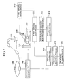
- FIG. 1 the configuration of the radiotherapy system of the first embodiment, which is shown in FIG. 1 .
- a beam accelerator 100 generates a treatment beam, and a nozzle 110 shapes the beam and radiates it onto a patient 130 on a couch 120.
- the beam accelerator 100 and the nozzle 110 are controlled by a beam extraction controller 140 upon beam irradiation.
- the beam extraction controller 140 performs its beam extraction control in response to a command from an irradiation controller 500.
- the irradiation controller 500 acquires from a treatment record database 510 a treatment plan that is created in advance by a treatment planning device 520.
- the irradiation controller 500 issues a beam extraction command to the beam extraction controller 140 according to the dose and method of beam irradiation specified by the plan of that treatment.
- the radiotherapy system of the first embodiment is provided with two types of observation devices that observe the respiratory states of the patient 130: an external-respiration observation device and an internal-respiration observation device.
- the external-respiration observation device externally monitors measurable respiratory signals such as those changes in body shape, respiratory flow rates, and the like, and an external respiration monitor 300 serves as the external-respiration observation device.
- Signals obtained by the external respiration monitor 300 are input to an x-ray imaging gating device 310, where the signals are used for respiratory gating.
- the internal-respiration observation device observes respiratory phases based on the positional information of internal body structures such as intracorporeal treatment targets, bony structures, and diaphragm or of implanted markers.
- Used as the internal-respiration observation device is an x-ray imaging controller 220 that acquires x-ray images of the patient 130 with the use of an x-ray source 200 and an x-ray detector 210.
- the x-ray images acquired by the x-ray imaging controller 220 are sent to an internal respiratory index acquisition device 400 for analysis of respiratory indexes.
- the analyzed respiratory indexes are used for respiratory gating by an irradiation gating device 410.
- Radiotherapy systems in general are equipped with x-ray devices for positioning patients, and the x-ray imaging device of the invention that comprises the x-ray source 200, the x-ray detector 210, and the x-ray imaging controller 220 can be used also as such a patient-positioning x-ray device.
- Positioning a patient is the step of conforming the positional relationship between the irradiation target and the treatment devices to the treatment plan, and x-ray images are used during the positioning step for the purpose of obtaining the positional information of internal body structures.
- FIG. 2 is a flowchart illustrating the operation of the radiotherapy system of the first embodiment.
- FIGs. 3A to 3B and 4A to 4B are timing charts illustrating the operation of the radiotherapy system of the first embodiment.
- Step S100 the operator starts up the external respiration monitor 300 to start the monitoring of an external respiratory index of the patient 130.
- the monitored external respiratory index is input to the x-ray imaging gating device 310.
- the monitoring of the external respiratory index is done by a laser rangefinder measuring positions on the body surface, a visible-light or infrared camera measuring the positions of markers placed on the body surface, or strain gauges placed on the body surface or a respiratory flow meter measuring respiratory flow rates.
- the external respiratory index as used herein refers to a measurable value that changes in response to respiration such as a position on the body surface. The external respiratory index is monitored during an entire treatment period.
- Step S200 the operator operates the x-ray imaging gating device 310 to set an x-ray imaging permissible range for the external respiratory index acquired by the external respiration monitor 300.
- the x-ray imaging gating device 310 is provided with means for the operator setting an x-ray imaging permissible range for the external respiratory index the x-ray imaging gating device 310 receives.
- Step S210 the x-ray imaging gating device 310 sends gating signals permitting x-ray imaging to the x-ray imaging controller 220.
- an external respiratory curve is obtained by plotting the values of an external respiratory index against time.
- the x-ray imaging gating device 310 is capable of displaying an external respiratory curve. By referring to the displayed external respiratory curve, the operator inputs into the x-ray imaging gating device 310 an x-ray imaging permissible range for the external respiratory index. While the external respiratory curve is within the x-ray imaging permissible range, the x-ray imaging gating device 310 sends gating signals permitting x-ray imaging to the x-ray imaging controller 220, as shown in FIG. 3B . It should be noted that the x-ray imaging permissible range can instead be set with the use of the change rates of the external respiratory curve. Also, the x-ray imaging gating device 310 can be allowed to store the x-ray imaging permissible range on a memory or a database for its later use as a default value.
- Step S300 the operator instructs the x-ray imaging controller 220 to start x-ray imaging.
- X-ray images are acquired intermittently while the x-ray imaging gating device 310 sends the gating signals permitting x-ray imaging to the x-ray imaging controller 220.
- One cycle of the external respiratory curve shown in FIG. 3A spans approximately 4 seconds although its length varies from person to person.
- the length of one cycle during which a gating signal permitting x-ray imaging is on which is shown in FIG. 3B , is approximately 2 seconds.
- the acquired x-ray images are input to the internal respiratory index acquisition device 400.
- Step S400 the internal respiratory index acquisition device 400 analyzes the x-ray images acquired by the x-ray imaging controller 220 to obtain an internal respiratory index.
- the internal respiratory index is obtained from the positions of irradiation targets, bones, diaphragm, or implanted markers.
- the positional analysis of the irradiation targets and bones is done by acquiring x-ray images of relevant regions based on a treatment plan and searching for the best matched region or by the operator specifying a region in advance and obtaining the optical flow of that region by image matching.
- the positional analysis of the diaphragm exploits the fact that an intensity value of an x-ray image changes steeply at the position of the diaphragm.
- the operator specifies an analysis region, and the diaphragm is located when the change rate of an intensity value within the analysis region exceeds a given value. Changes in the position of the diaphragm are then analyzed for obtaining the internal respiratory index.
- the positional analysis of the implanted markers is done by thresholding or pattern matching, using the fact that the markers on an x-ray image have high intensity values and distinctive shapes.
- the internal respiratory index obtained with the use of one or more of the above methods is sent to the irradiation gating device 410.
- the radiotherapy system of the first embodiment can be provided with a mechanism for storing the on/off states of the last gating signal on a memory and resuming analysis from the last on or off state of that signal.
- Step S500 the operator inputs into the irradiation gating device 410 a beam irradiation permissible range for the internal respiratory index by referring to its internal respiratory curves displayed by the irradiation gating device 410.
- the irradiation gating device 410 is provided with means for accepting the input of a gating condition for treatment beam irradiation.
- the internal respiratory curves or waves are obtained by plotting the values of an internal respiratory index against time.
- the internal respiratory curves are discontinuous since x-ray imaging is gated.
- the set condition for permitting beam irradiation can be stored on a memory for use during later treatments in order to reduce X-ray exposure.
- Step S510 the irradiation gating device 410 sends such gating signals permitting beam irradiation as shown in FIG. 4B through the irradiation controller 500 to the beam extraction controller 140 while the internal respiratory index is within the beam irradiation permissible range.
- respiratory-gated irradiation becomes ready, which is based on the gating signals permitting beam irradiation that are synchronous with the beam irradiation permissible periods of the internal respiratory index.
- the period during which a gating signal permitting x-ray imaging is on is longer than the period during which a gating signal permitting beam irradiation is on.
- the x-ray imaging permissible range can be adjusted such that the beam irradiation permissible periods coincide with the x-ray imaging permissible periods.
- Step S600 the operator instructs the irradiation controller 500 to start treatment.
- Step S700 the irradiation controller 500 instructs the beam extraction controller 140 to stop the beam extraction after a given dose of irradiation as specified by the treatment planning device 520.
- Step S800 the irradiation controller 500 instructs the x-ray imaging controller 220 to stop the x-ray imaging.
- the radiotherapy system of the first embodiment is designed to monitor an external respiratory index with the use of the external respiration monitor 300, and while the external respiratory index is within an x-ray imaging permissible range, the system sends gating signals permitting x-ray imaging to the x-ray imaging controller 220, thereby performing gated x-ray imaging.
- the system is capable of acquiring internal respiratory waves or curves necessary for the gating control of beam irradiation with less x-ray exposure.
- FIGs. 5 and 6A to 6E the configuration and operation of a radiotherapy system according to a second embodiment of the invention will be described. Note that the configuration of the radiotherapy system of the second embodiment is the same as the one shown in FIG. 1 .
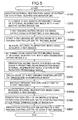
- FIG. 5 is a flowchart illustrating the operation of the radiotherapy system of the second embodiment. Note that the same step numbers as used in FIG. 2 indicate the same operational steps.
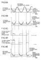
- FIGs. 6A to 6E are timing charts illustrating the operation of the radiotherapy system of the second embodiment.
- FIG. 6A shows the same external respiratory curve as shown in FIG. 3A .
- FIG. 6B shows the same internal respiratory curves as shown in FIG. 4A .
- FIG. 6C shows the same gating signals permitting beam irradiation as shown in FIG. 4B .
- FIG. 6D shows gating signals permitting x-ray imaging according to the second embodiment.
- Step S100 the operator starts up the external respiration monitor 300 to start the monitoring of an external respiratory index of the patient 130.
- the monitored external respiratory index is input to the x-ray imaging gating device 310.
- Step S200 the operator operates the x-ray imaging gating device 310 to set an x-ray imaging permissible range for the external respiratory index acquired by the external respiration monitor 300.
- the x-ray imaging gating device 310 is provided with means for the operator setting an x-ray imaging permissible range for the external respiratory index the x-ray imaging gating device 310 receives.
- Step S210 the x-ray imaging gating device 310 sends gating signals permitting x-ray imaging to the x-ray imaging controller 220.
- the x-ray imaging gating device 310 sends such gating signals permitting x-ray imaging as shown in FIG. 3B to the x-ray imaging controller 220 while the external respiratory index is within the x-ray imaging permissible range. Similar to the first embodiment, the x-ray imaging permissible range for the external respiratory index is input by the operator into the x-ray imaging gating device 310 by the operator referring to an external respiratory curve ( FIG. 6A ) displayed by the x-ray imaging gating device 310.
- Step S300A the operator instructs the x-ray imaging controller 220 to start x-ray imaging in "normal mode." Similar to Step S300 of FIG. 2 , normal mode is the mode in which the x-ray imaging controller 220 acquires x-ray images intermittently while the x-ray imaging controller 220 receives the gating signals permitting x-ray imaging from the x-ray imaging gating device 310.
- Step S400 the internal respiratory index acquisition device 400 analyzes the x-ray images acquired by the x-ray imaging controller 220 to obtain an internal respiratory index.
- the internal respiratory index is obtained from the positions of irradiation targets, bones, diaphragm, or implanted markers.
- the obtained internal respiratory index is sent to the irradiation gating device 410.
- the radiotherapy system of the second embodiment can also be provided with a mechanism for storing the on/off states of the last gating signal on a memory and resuming analysis from the last on or off state of that signal.
- Step S500 the operator inputs into the irradiation gating device 410 a beam irradiation permissible range for the internal respiratory index by referring to its internal respiratory curves displayed by the irradiation gating device 410.
- the irradiation gating device 410 is provided with means for accepting the input of a gating condition for treatment beam irradiation. As shown in FIG. 6B , the internal respiratory curves are discontinuous since x-ray imaging is gated.
- Step S510 the irradiation gating device 410 sends such gating signals permitting beam irradiation as shown in FIG. 6C through the irradiation controller 500 to the beam extraction controller 140 while the internal respiratory index is within the beam irradiation permissible range.
- Step S520 the operator switches the mode of the x-ray imaging controller 220 from "normal mode” to "irradiation-synchronous mode.”
- "Irradiation-synchronuous mode” is the mode in which x-ray imaging is permitted by such gating signals of x-ray imaging permission as shown in FIG. 6D that are turned off at the same time as the gating signals permitting beam irradiation that are shown in FIG. 6C are turned off.
- the x-ray imaging permissible period T2 of FIG. 6D (radiation-synchronous mode of Step S520) is shorter than the x-ray imaging permissible period T1 of FIG. 6A (normal mode of Step S300A).
- respiratory-gated irradiation becomes ready, which is based on the gating signals permitting beam irradiation that are synchronous with the beam irradiation permissible periods of the internal respiratory index.
- Step S600 the operator instructs the irradiation controller 500 to start treatment.
- Step S400A the internal respiratory index acquisition device 400 analyzes x-ray images acquired by the x-ray imaging controller 220 during "irradiation-synchronous mode" to obtain the internal respiratory index again.
- FIG. 6E shows examples of internal respiratory curves obtained during "irradiation-synchronous mode.”
- Step S700 the irradiation controller 500 instructs the beam extraction controller 140 to stop the beam extraction after a given dose of irradiation as specified by the treatment planning device 520.
- Step S800 the irradiation controller 500 instructs the x-ray imaging controller 220 to stop the x-ray imaging.
- the radiotherapy system of the second embodiment is designed to perform gated x-ray imaging by sending gating signals permitting x-ray imaging to the x-ray imaging controller 220 such that the x-ray imaging is turned off at the same time as gating signals permitting beam irradiation are turned off.
- the system is capable of acquiring internal respiratory waves or curves necessary for the gating control of beam irradiation with even less x-ray exposure.
- FIGs. 7 and 8A to 8F the configuration and operation of a radiotherapy system according to a third embodiment of the invention will be described. Note that the configuration of the radiotherapy system of the third embodiment is the same as the one shown in FIG. 1 .
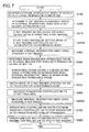
- FIG. 7 is a flowchart illustrating the operation of the radiotherapy system of the third embodiment. Note that the same step numbers as used in FIGs. 2 and 5 indicate the same operational steps.
- FIGs. 8A to 8F are timing charts illustrating the operation of the radiotherapy system of the third embodiment.
- FIGs. 8A to 8C are the same as FIGs. 5A to 5C, respectively.
- FIG. 8D shows signals indicative of beam-extractable periods.
- FIG. 8E shows gating signals permitting x-ray imaging according to the third embodiment.
- FIG. 8F shows internal respiratory curves according to the third embodiment.
- Step S100 the operator starts up the external respiration monitor 300 to start the monitoring of an external respiratory index of the patient 130.
- the monitored external respiratory index is input to the x-ray imaging gating device 310.
- Step S200 the operator operates the x-ray imaging gating device 310 to set an x-ray imaging permissible range for the external respiratory index acquired by the external respiration monitor 300.
- the x-ray imaging gating device 310 is provided with means for the operator setting an x-ray imaging permissible range for the external respiratory index the x-ray imaging gating device 310 receives.
- Step S210 the x-ray imaging gating device 310 sends gating signals permitting x-ray imaging to the x-ray imaging controller 220.
- the x-ray imaging gating device 310 sends such gating signals permitting x-ray imaging as shown in FIG. 3B to the x-ray imaging controller 220 while the external respiratory index is within the x-ray imaging permissible range. Similar to the first embodiment, the x-ray imaging permissible range for the external respiratory index is input by the operator into the x-ray imaging gating device 310 by the operator referring to an external respiratory curve ( FIG. 8A ) displayed by the x-ray imaging gating device 310.
- Step S300B the operator instructs the x-ray imaging controller 220 to start x-ray imaging in "accelerator-asynchronous mode.” Similar to Step S300 of FIG. 2 and normal mode, “accelerator-asynchronous mode” is the mode in which the x-ray imaging controller 220 acquires x-ray images intermittently while the x-ray imaging controller 220 receives the gating signals permitting x-ray imaging from the x-ray imaging gating device 310.
- Step S400 the internal respiratory index acquisition device 400 analyzes the x-ray images acquired by the x-ray imaging controller 220 to obtain an internal respiratory index.
- the internal respiratory index is obtained from the positions of irradiation targets, bones, diaphragm, or implanted markers.
- the obtained internal respiratory index is sent to the irradiation gating device 410.
- the radiotherapy system of the third embodiment can also be provided with a mechanism for storing the on/off states of the last gating signal on a memory and resuming analysis from the last on or off state of that signal.
- Step S500 the operator inputs into the irradiation gating device 410 a beam irradiation permissible range for the internal respiratory index by referring to its internal respiratory curves displayed by the irradiation gating device 410.
- the irradiation gating device 410 is provided with means for accepting the input of a gating condition for treatment beam irradiation.
- the internal respiratory curves are discontinuous since x-ray imaging is gated.
- the irradiation gating device 410 can be allowed to switch the mode of the x-ray imaging controller 220 from “accelerator-asynchronous mode” to "accelerator-synchronous mode” after the operator sets the beam irradiation permissible range.
- "Accelerator-synchronous mode” is the mode in which the x-ray imaging controller 220 acquires an x-ray image when the beam accelerator 100 is capable of radiating or extracting a treatment beam and the external respiratory index is within the x-ray imaging permissible range.
- the beam-extractable time precedes a gating signal permitting x-ray imaging
- x-ray imaging can be performed prior to that gating signal by a predetermined amount of time.
- Step S510 the irradiation gating device 410 sends such gating signals permitting beam irradiation as shown in FIG. 8C through the irradiation controller 500 to the beam extraction controller 140 while the internal respiratory index is within the beam irradiation permissible range.
- Step S550 the operator switches the mode of the x-ray imaging controller 220 from “accelerator-asynchronous mode” to “accelerator-synchronous mode.”
- "accelerator-synchronous mode” is the mode in which x-ray imaging is permitted by such gating signals of x-ray imaging permission as shown in FIG. 8E that coincide with the beam-extractable periods of FIG. 8D while the external respiratory index of FIG. 8A is within the x-ray imaging permissible range.
- the extractability of a treatment beam from the accelerator 100 is judged by a signal indicative of a beam-extractable period which is sent from the beam extraction controller 140.
- the x-ray imaging permissible period T3 of FIG. 8E (accelerator-synchronous mode of Step S550) is shorter than the x-ray imaging permissible period T1 of FIG. 8A (accelerator-asynchronous mode of Step S300B).
- respiratory-gated irradiation becomes ready, which is based on the gating signals permitting beam irradiation that are synchronous with the beam irradiation permissible periods of the internal respiratory index.
- Step S600 the operator instructs the irradiation controller 500 to start treatment.
- the irradiation controller 500 then starts to send to the x-ray imaging controller 220 such signals indicative of beam-extractable periods as shown in FIG. 8D .
- the extractability of treatment beams from the accelerator 100 is judged by the above signals indicative of beam-extractable periods that are received by the irradiation controller 500 from the beam extraction controller 140.
- the beam extraction controller 140 judges the extractability of a treatment beam based on the states of the accelerator 100 and the nozzle 110 and sends those signals indicative of beam-extractable periods to the irradiation controller 500.
- Step S400A the internal respiratory index acquisition device 400 analyzes x-ray images acquired by the x-ray imaging controller 220 during "acceleration-synchronous mode" to obtain the internal respiratory index again.
- Step S700 the irradiation controller 500 instructs the beam extraction controller 140 to stop the beam extraction after a given dose of irradiation as specified by the treatment planning device 520.
- Step S800 the irradiation controller 500 instructs the x-ray imaging controller 220 to stop the x-ray imaging.
- the radiotherapy system of the third embodiment is designed to perform gated x-ray imaging by sending gating signals permitting x-ray imaging to the x-ray imaging controller 220 in synchronization with signals indicative of beam-extractable periods.
- the system is capable of acquiring internal respiratory waves or curves necessary for the gating control of beam irradiation with far less x-ray exposure.
- FIGs. 9 and 10A to 10F the configuration and operation of a radiotherapy system according to a fourth embodiment of the invention will be described. Note that the configuration of the radiotherapy system of the fourth embodiment is the same as the one shown in FIG. 1 .
- FIG. 9 is a flowchart illustrating the operation of the radiotherapy system of the fourth embodiment. Note that the same step numbers as used in FIGs. 2 , 5 , and 7 indicate the same operational steps.
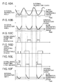
- FIGs. 10A to 10F are timing charts illustrating the operation of the radiotherapy system of the fourth embodiment.
- FIGs. 10A to 10D are the same as FIGs. 8A to 8D , respectively.
- FIG. 10E shows gating signals permitting x-ray imaging according to the fourth embodiment.
- FIG. 10F shows internal respiratory curves according to the fourth embodiment.
- Step S100 the operator starts up the external respiration monitor 300 to start the monitoring of an external respiratory index of the patient 130.
- the monitored external respiratory index is input to the x-ray imaging gating device 310.
- Step S200 the operator operates the x-ray imaging gating device 310 to set an x-ray imaging permissible range for the external respiratory index acquired by the external respiration monitor 300.
- the x-ray imaging gating device 310 is provided with means for the operator setting an x-ray imaging permissible range for the external respiratory index the x-ray imaging gating device 310 receives.
- Step S210 the x-ray imaging gating device 310 sends gating signals permitting x-ray imaging to the x-ray imaging controller 220.
- the x-ray imaging gating device 310 sends such gating signals permitting x-ray imaging as shown in FIG. 3B to the x-ray imaging controller 220 while the external respiratory index is within the x-ray imaging permissible range. Similar to the first embodiment, the x-ray imaging permissible range for the external respiratory index is input by the operator into the x-ray imaging gating device 310 by the operator referring to an external respiratory curve ( FIG. 10A ) displayed by the x-ray imaging gating device 310.
- Step S300B the operator instructs the x-ray imaging controller 220 to start x-ray imaging in "accelerator-asynchronous mode.”
- "accelerator-asynchronous mode” is the mode in which the x-ray imaging controller 220 acquires x-ray images intermittently while the x-ray imaging controller 220 receives the gating signals permitting x-ray imaging from the x-ray imaging gating device 310.
- Step S400 the internal respiratory index acquisition device 400 analyzes the x-ray images acquired by the x-ray imaging controller 220 to obtain an internal respiratory index.
- the internal respiratory index is obtained from the positions of irradiation targets, bones, diaphragm, or implanted markers.
- the obtained internal respiratory index is sent to the irradiation gating device 410.
- the radiotherapy system of the fourth embodiment can also be provided with a mechanism for storing the on/off states of the last gating signal on a memory and resuming analysis from the last on or off state of that signal.
- Step S500 the operator inputs into the irradiation gating device 410 a beam irradiation permissible range for the internal respiratory index by referring to its internal respiratory curves displayed by the irradiation gating device 410.
- the irradiation gating device 410 is provided with means for accepting the input of a gating condition for treatment beam irradiation. As shown in FIG. 10B , the internal respiratory curves are discontinuous since x-ray imaging is gated.
- the irradiation gating device 410 can be allowed to switch the mode of the x-ray imaging controller 220 from "accelerator-asynchronous mode" to "accelerator-synchronous mode” after the operator sets the beam irradiation permissible range.
- accelerator-synchronous mode is the mode in which the x-ray imaging controller 220 acquires an x-ray image when the beam accelerator 100 is capable of radiating or extracting a treatment beam and the external respiratory index is within the x-ray imaging permissible range.
- x-ray imaging can be performed prior to that gating signal by a predetermined amount of time.
- Step S510 the irradiation gating device 410 sends such gating signals permitting beam irradiation as shown in FIG. 10C through the irradiation controller 500 to the beam extraction controller 140 while the internal respiratory index is within the beam irradiation permissible range.
- Step S530 the operator switches the mode of the x-ray imaging controller 220 from “accelerator-asynchronous mode" to "prior-to-accelerator mode.”
- "Prior-to-accelerator mode” is the mode in which x-ray imaging is permitted by such gating signals of x-ray imaging permission as shown in FIG. 10E that are turned on prior to the beam-extractable periods of FIG. 10D by time T1 and turned off at the same time as the end of the beam-extractable periods while the external respiratory index of FIG. 10A is within the x-ray imaging permissible range.
- the x-ray imaging permissible period (t1 + T4) of FIG. 10E (prior-to-acceleration mode of Step S530) is shorter than the x-ray imaging permissible period T1 of FIG. 10A (accelerator-asynchronous mode of Step S300B) and also shorter than the x-ray imaging permissible period T2 of FIG. 6D .
- the x-ray imaging permissible period (t1 + T4) is longer than the x-ray imaging permissible period T3 of FIG. 8E (accelerator-synchronous mode), the internal respiratory index can be monitored for a slightly longer amount of time.
- respiratory-gated irradiation becomes ready, which is based on the gating signals permitting beam irradiation that are synchronous with the beam irradiation permissible periods of the internal respiratory index.
- Step S600 the operator instructs the irradiation controller 500 to start treatment.
- the irradiation controller 500 then starts to send to the x-ray imaging controller 220 such signals indicative of beam-extractable periods as shown in FIG. 10D .
- the extractability of treatment beams from the accelerator 100 is judged by the above signals indicative of beam-extractable periods that are received by the irradiation controller 500 from the beam extraction controller 140.
- the beam extraction controller 140 judges the extractability of a treatment beam based on the states of the accelerator 100 and the nozzle 110 and sends those signals indicative of beam-extractable periods to the irradiation controller 500.
- Step S400A the internal respiratory index acquisition device 400 analyzes x-ray images acquired by the x-ray imaging controller 220 during "prior-to-accelerator mode" to obtain the internal respiratory index again.
- Step S700 the irradiation controller 500 instructs the beam extraction controller 140 to stop the beam extraction after a given dose of irradiation as specified by the treatment planning device 520.
- Step S800 the irradiation controller 500 instructs the x-ray imaging controller 220 to stop the x-ray imaging.
- the radiotherapy system of the fourth embodiment is designed to perform gated x-ray imaging by sending gating signals permitting x-ray imaging to the x-ray imaging controller 220 in synchronization with and prior to signals indicative of beam-extractable periods.
- the system is capable of acquiring internal respiratory waves or curves necessary for the gating control of beam irradiation with far less x-ray exposure.
Landscapes
- Health & Medical Sciences (AREA)
- Life Sciences & Earth Sciences (AREA)
- Engineering & Computer Science (AREA)
- Biomedical Technology (AREA)
- Veterinary Medicine (AREA)
- Radiology & Medical Imaging (AREA)
- Nuclear Medicine, Radiotherapy & Molecular Imaging (AREA)
- Animal Behavior & Ethology (AREA)
- General Health & Medical Sciences (AREA)
- Public Health (AREA)
- Pathology (AREA)
- Medical Informatics (AREA)
- Biophysics (AREA)
- Physics & Mathematics (AREA)
- High Energy & Nuclear Physics (AREA)
- Physiology (AREA)
- Optics & Photonics (AREA)
- Heart & Thoracic Surgery (AREA)
- Molecular Biology (AREA)
- Surgery (AREA)
- Radiation-Therapy Devices (AREA)
Abstract
The invention provides a radiotherapy system that achieves highly-accurate respiratory-gated irradiation with less x-ray exposure by performing x-ray imaging only during the respiratory phases that are necessary for the respiratory-gated irradiation.
An external respiration monitor 300 externally monitors an external respiratory index of a patient. An x-ray source 200 and an x-ray detector 210, or internal-respiration observation devices, acquire x-ray images of the patient and monitor an internal respiratory index of the patient using the positions of internal body structures indicated by the acquired x-ray images. An x-ray imaging gating device 310 gates the x-ray imaging performed by the internal-respiration observation devices with the use of the external respiratory index monitored by the external respiration monitor 300 such that the x-ray imaging is performed while the external respiratory index is within a predetermined range. An irradiation gating device 410 gates the irradiation of a treatment beam with the use of the internal respiratory index monitored by the internal-respiration observation devices.
Description
- The present invention relates generally to radiotherapy systems that irradiate patients with treatment beams and particularly to respiratory-gated radiotherapy systems that radiate treatment beams in synchronization with particular phases of patients' respiratory cycles.
- In radiotherapy that involves the irradiation of x-rays, particle beams, or the like onto the body of a patient for treatment purposes, of importance for enhancing the treatment effects is highly accurate positioning of the irradiation target according to the treatment plan. Highly accurate positioning enables the reduction of the irradiation area margin that compensates for positioning-related uncertainties and results in the healthy tissues or organs that surround the irradiation target being less exposed to treatment beams.
- When the target moves due to the respiratory motion of the patient, this requires a margin that allows for the respiratory motion. In order to reduce that margin, respiratory-gated irradiation is now employed, in which treatment beams are radiated only during particular phases of respiration. This reduces the required margin, for the respiratory motion is limited within the limited respiratory phases. There are two methods for observation respiratory phases: external observation and internal observation. External observation involves the use of externally observable indexes or parameters such as those indicating the movements of the body surface and respiratory flow rates. In contrast, internal observation involves the use of such indexes as indicate the positions of internal body structures (e.g., intracorporeal irradiation targets, bony structures, and diaphragm). The positions of such internal body structures can be obtained by x-ray imaging. Respiratory-gated irradiation methods that are based on internal observation are disclosed, for example, in Japanese Patent No.
3053389 JP-2008-154861-A JP-2004-283513-A - Internal observation enables highly accurate measurement of the positions of internal body structures since they are measured directly. However, x-ray exposure of the patient is inevitable during internal observation (i.e., x-ray imaging). Thus, it is desired that this x-ray exposure be as little as possible.
- An object of the invention is therefore to provide a radiotherapy system that achieves highly-accurate respiratory-gated irradiation with less x-ray exposure by taking x-ray only during the respiratory phases that are necessary for the respiratory-gated irradiation.
- 1) To achieve the above object, a radiotherapy system according to the invention comprises: external-respiration observation means for externally monitoring an external respiratory index of a patient; internal-respiration observation means for acquiring x-ray images of the patient and monitoring an internal respiratory index of the patient using the positions of internal body structures indicated by the acquired x-ray images; imaging gating means for gating the x-ray imaging performed by the internal-respiration observation means with the use of the external respiratory index monitored by the external-respiration observation means such that the x-ray imaging is performed while the external respiratory index is within a predetermined range; and beam irradiation gating means for gating the irradiation of a treatment beam with the use of the internal respiratory index monitored by the internal-respiration observation means.
By performing x-ray imaging only during the respiratory phases that are necessary for the respiratory-gated irradiation with the use of the above configuration, it is possible to achieve highly-accurate respiratory-gated irradiation with less x-ray exposure. - 2) In the above radiotherapy system, the internal-respiration observation means preferably includes x-ray imaging control means for instructing the internal-respiration observation means to stop the x-ray imaging when a gating signal permitting treatment-beam irradiation which is generated by the beam irradiation gating means is turned off.
- 3) In the above radiotherapy system, the internal-respiration observation means preferably includes x-ray imaging control means for permitting the internal-respiration observation means to take the x-ray while a treatment beam can be radiated.
- 4) In the above radiotherapy system, the internal-respiration observation means preferably includes x-ray imaging control means for permitting the internal-respiration observation means to perform the x-ray imaging prior to a period during which a treatment beam can be radiated.
- 5) In the above radiotherapy system, the beam irradiation gating means preferably stores a condition for radiating a treatment beam so that the beam irradiation gating means can use the condition for later treatments.
- In accordance with the invention, it is possible to achieve highly-accurate respiratory-gated irradiation with less x-ray exposure by performing x-ray imaging only during the respiratory phases that are necessary for the respiratory-gated irradiation.
-
-
FIG. 1 illustrates the configuration of a radiotherapy system according to a first embodiment of the invention; -
FIG. 2 is a flowchart illustrating the operation of the radiotherapy system according to the first embodiment; -
FIGs. 3A and 3B are timing charts illustrating the operation of the radiotherapy system according to the first embodiment; -
FIGs. 4A and 4B are timing charts illustrating the operation of the radiotherapy system according to the first embodiment; -
FIG. 5 is a flowchart illustrating the operation of a radiotherapy system according to a second embodiment of the invention; -
FIGs. 6A to 6E are timing charts illustrating the operation of the radiotherapy system according to the second embodiment; -
FIG. 7 is a flowchart illustrating the operation of a radiotherapy system according to a third embodiment of the invention; -
FIGs. 8A to 8F are timing charts illustrating the operation of the radiotherapy system according to the third embodiment; -
FIG. 9 is a flowchart illustrating the operation of a radiotherapy system according to a fourth embodiment of the invention; and -
FIGs. 10A to 10F are timing charts illustrating the operation of the radiotherapy system according to the fourth embodiment. - With reference to
FIGs. 1 to 4B , the configuration and operation of a radiotherapy system according to a first embodiment of the invention will now be described. Described first is the configuration of the radiotherapy system of the first embodiment, which is shown inFIG. 1 . - A
beam accelerator 100 generates a treatment beam, and anozzle 110 shapes the beam and radiates it onto apatient 130 on acouch 120. Thebeam accelerator 100 and thenozzle 110 are controlled by abeam extraction controller 140 upon beam irradiation. Thebeam extraction controller 140 performs its beam extraction control in response to a command from anirradiation controller 500. In response to a command from the operator, theirradiation controller 500 acquires from a treatment record database 510 a treatment plan that is created in advance by atreatment planning device 520. When the operator instructs theirradiation controller 500 to start a treatment, theirradiation controller 500 issues a beam extraction command to thebeam extraction controller 140 according to the dose and method of beam irradiation specified by the plan of that treatment. - For the purpose of radiating treatment beams in synchronization with particular phases of the respiration of the
patient 130, the radiotherapy system of the first embodiment is provided with two types of observation devices that observe the respiratory states of the patient 130: an external-respiration observation device and an internal-respiration observation device. The external-respiration observation device externally monitors measurable respiratory signals such as those changes in body shape, respiratory flow rates, and the like, and anexternal respiration monitor 300 serves as the external-respiration observation device. Signals obtained by theexternal respiration monitor 300 are input to an x-rayimaging gating device 310, where the signals are used for respiratory gating. The internal-respiration observation device observes respiratory phases based on the positional information of internal body structures such as intracorporeal treatment targets, bony structures, and diaphragm or of implanted markers. Used as the internal-respiration observation device is anx-ray imaging controller 220 that acquires x-ray images of thepatient 130 with the use of anx-ray source 200 and anx-ray detector 210. The x-ray images acquired by thex-ray imaging controller 220 are sent to an internal respiratoryindex acquisition device 400 for analysis of respiratory indexes. The analyzed respiratory indexes are used for respiratory gating by anirradiation gating device 410. - Radiotherapy systems in general are equipped with x-ray devices for positioning patients, and the x-ray imaging device of the invention that comprises the
x-ray source 200, thex-ray detector 210, and thex-ray imaging controller 220 can be used also as such a patient-positioning x-ray device. Positioning a patient is the step of conforming the positional relationship between the irradiation target and the treatment devices to the treatment plan, and x-ray images are used during the positioning step for the purpose of obtaining the positional information of internal body structures. - With reference now to
FIGs. 2 to 4B , the operation of the radiotherapy system of the first embodiment is described. -
FIG. 2 is a flowchart illustrating the operation of the radiotherapy system of the first embodiment.FIGs. 3A to 3B and 4A to 4B are timing charts illustrating the operation of the radiotherapy system of the first embodiment. - In Step S100, the operator starts up the
external respiration monitor 300 to start the monitoring of an external respiratory index of thepatient 130. The monitored external respiratory index is input to the x-rayimaging gating device 310. - The monitoring of the external respiratory index is done by a laser rangefinder measuring positions on the body surface, a visible-light or infrared camera measuring the positions of markers placed on the body surface, or strain gauges placed on the body surface or a respiratory flow meter measuring respiratory flow rates. The external respiratory index as used herein refers to a measurable value that changes in response to respiration such as a position on the body surface. The external respiratory index is monitored during an entire treatment period.
- In Step S200, the operator operates the x-ray
imaging gating device 310 to set an x-ray imaging permissible range for the external respiratory index acquired by theexternal respiration monitor 300. The x-rayimaging gating device 310 is provided with means for the operator setting an x-ray imaging permissible range for the external respiratory index the x-rayimaging gating device 310 receives. - In Step S210, the x-ray
imaging gating device 310 sends gating signals permitting x-ray imaging to thex-ray imaging controller 220. - Here, we briefly discuss an external respiratory curve with reference to
FIG. 3A . As illustrated inFIG. 3A , an external respiratory curve is obtained by plotting the values of an external respiratory index against time. - The x-ray
imaging gating device 310 is capable of displaying an external respiratory curve. By referring to the displayed external respiratory curve, the operator inputs into the x-rayimaging gating device 310 an x-ray imaging permissible range for the external respiratory index. While the external respiratory curve is within the x-ray imaging permissible range, the x-rayimaging gating device 310 sends gating signals permitting x-ray imaging to thex-ray imaging controller 220, as shown inFIG. 3B . It should be noted that the x-ray imaging permissible range can instead be set with the use of the change rates of the external respiratory curve. Also, the x-rayimaging gating device 310 can be allowed to store the x-ray imaging permissible range on a memory or a database for its later use as a default value. - With reference back to
FIG. 2 , in Step S300, the operator instructs thex-ray imaging controller 220 to start x-ray imaging. X-ray images are acquired intermittently while the x-rayimaging gating device 310 sends the gating signals permitting x-ray imaging to thex-ray imaging controller 220. One cycle of the external respiratory curve shown inFIG. 3A spans approximately 4 seconds although its length varies from person to person. Thus, the length of one cycle during which a gating signal permitting x-ray imaging is on, which is shown inFIG. 3B , is approximately 2 seconds. The acquired x-ray images are input to the internal respiratoryindex acquisition device 400. - In Step S400, the internal respiratory
index acquisition device 400 analyzes the x-ray images acquired by thex-ray imaging controller 220 to obtain an internal respiratory index. The internal respiratory index is obtained from the positions of irradiation targets, bones, diaphragm, or implanted markers. The positional analysis of the irradiation targets and bones is done by acquiring x-ray images of relevant regions based on a treatment plan and searching for the best matched region or by the operator specifying a region in advance and obtaining the optical flow of that region by image matching. The positional analysis of the diaphragm exploits the fact that an intensity value of an x-ray image changes steeply at the position of the diaphragm. For example, the operator specifies an analysis region, and the diaphragm is located when the change rate of an intensity value within the analysis region exceeds a given value. Changes in the position of the diaphragm are then analyzed for obtaining the internal respiratory index. The positional analysis of the implanted markers is done by thresholding or pattern matching, using the fact that the markers on an x-ray image have high intensity values and distinctive shapes. The internal respiratory index obtained with the use of one or more of the above methods is sent to theirradiation gating device 410. - As stated above, x-ray images are acquired intermittently. Thus, when an analysis of the internal respiratory index needs to be followed by the next analysis without discontinuity at some later time, the radiotherapy system of the first embodiment can be provided with a mechanism for storing the on/off states of the last gating signal on a memory and resuming analysis from the last on or off state of that signal.
- In Step S500, the operator inputs into the irradiation gating device 410 a beam irradiation permissible range for the internal respiratory index by referring to its internal respiratory curves displayed by the
irradiation gating device 410. Similar to the x-rayimaging gating device 310 having means for setting an x-ray imaging permissible range for the external respiratory index, theirradiation gating device 410 is provided with means for accepting the input of a gating condition for treatment beam irradiation. - We now briefly discuss the internal respiratory curves with reference to
FIG. 4A . As illustrated inFIG. 4A , the internal respiratory curves or waves are obtained by plotting the values of an internal respiratory index against time. The internal respiratory curves are discontinuous since x-ray imaging is gated. - Note that the set condition for permitting beam irradiation can be stored on a memory for use during later treatments in order to reduce X-ray exposure.
- With reference back to
FIG. 2 , in Step S510, theirradiation gating device 410 sends such gating signals permitting beam irradiation as shown inFIG. 4B through theirradiation controller 500 to thebeam extraction controller 140 while the internal respiratory index is within the beam irradiation permissible range. - With the above steps, respiratory-gated irradiation becomes ready, which is based on the gating signals permitting beam irradiation that are synchronous with the beam irradiation permissible periods of the internal respiratory index. As shown in
FIGs. 3B and 4B , the period during which a gating signal permitting x-ray imaging is on is longer than the period during which a gating signal permitting beam irradiation is on. For the purpose of reducing x-ray exposure during x-ray imaging, the x-ray imaging permissible range can be adjusted such that the beam irradiation permissible periods coincide with the x-ray imaging permissible periods. - In Step S600, the operator instructs the
irradiation controller 500 to start treatment. - In Step S700, the
irradiation controller 500 instructs thebeam extraction controller 140 to stop the beam extraction after a given dose of irradiation as specified by thetreatment planning device 520. - Finally, in Step S800, the
irradiation controller 500 instructs thex-ray imaging controller 220 to stop the x-ray imaging. - As stated above, the radiotherapy system of the first embodiment is designed to monitor an external respiratory index with the use of the
external respiration monitor 300, and while the external respiratory index is within an x-ray imaging permissible range, the system sends gating signals permitting x-ray imaging to thex-ray imaging controller 220, thereby performing gated x-ray imaging. Thus, the system is capable of acquiring internal respiratory waves or curves necessary for the gating control of beam irradiation with less x-ray exposure. - With reference now to
FIGs. 5 and6A to 6E , the configuration and operation of a radiotherapy system according to a second embodiment of the invention will be described. Note that the configuration of the radiotherapy system of the second embodiment is the same as the one shown inFIG. 1 . -
FIG. 5 is a flowchart illustrating the operation of the radiotherapy system of the second embodiment. Note that the same step numbers as used inFIG. 2 indicate the same operational steps. -
FIGs. 6A to 6E are timing charts illustrating the operation of the radiotherapy system of the second embodiment.FIG. 6A shows the same external respiratory curve as shown inFIG. 3A .FIG. 6B shows the same internal respiratory curves as shown inFIG. 4A .FIG. 6C shows the same gating signals permitting beam irradiation as shown inFIG. 4B .FIG. 6D shows gating signals permitting x-ray imaging according to the second embodiment. - In Step S100, the operator starts up the
external respiration monitor 300 to start the monitoring of an external respiratory index of thepatient 130. The monitored external respiratory index is input to the x-rayimaging gating device 310. - In Step S200, the operator operates the x-ray
imaging gating device 310 to set an x-ray imaging permissible range for the external respiratory index acquired by theexternal respiration monitor 300. The x-rayimaging gating device 310 is provided with means for the operator setting an x-ray imaging permissible range for the external respiratory index the x-rayimaging gating device 310 receives. - In Step S210, the x-ray
imaging gating device 310 sends gating signals permitting x-ray imaging to thex-ray imaging controller 220. - More specifically, the x-ray
imaging gating device 310 sends such gating signals permitting x-ray imaging as shown inFIG. 3B to thex-ray imaging controller 220 while the external respiratory index is within the x-ray imaging permissible range. Similar to the first embodiment, the x-ray imaging permissible range for the external respiratory index is input by the operator into the x-rayimaging gating device 310 by the operator referring to an external respiratory curve (FIG. 6A ) displayed by the x-rayimaging gating device 310. - In Step S300A, the operator instructs the
x-ray imaging controller 220 to start x-ray imaging in "normal mode." Similar to Step S300 ofFIG. 2 , normal mode is the mode in which thex-ray imaging controller 220 acquires x-ray images intermittently while thex-ray imaging controller 220 receives the gating signals permitting x-ray imaging from the x-rayimaging gating device 310. - In Step S400, the internal respiratory
index acquisition device 400 analyzes the x-ray images acquired by thex-ray imaging controller 220 to obtain an internal respiratory index. The internal respiratory index is obtained from the positions of irradiation targets, bones, diaphragm, or implanted markers. The obtained internal respiratory index is sent to theirradiation gating device 410. - As stated above, x-ray images are acquired intermittently. Thus, when an analysis of the internal respiratory index needs to be followed by the next analysis without discontinuity at some later time, the radiotherapy system of the second embodiment can also be provided with a mechanism for storing the on/off states of the last gating signal on a memory and resuming analysis from the last on or off state of that signal.
- In Step S500, the operator inputs into the irradiation gating device 410 a beam irradiation permissible range for the internal respiratory index by referring to its internal respiratory curves displayed by the
irradiation gating device 410. Similar to the x-rayimaging gating device 310 having means for setting an x-ray imaging permissible range for the external respiratory index, theirradiation gating device 410 is provided with means for accepting the input of a gating condition for treatment beam irradiation. As shown inFIG. 6B , the internal respiratory curves are discontinuous since x-ray imaging is gated. - In Step S510, the
irradiation gating device 410 sends such gating signals permitting beam irradiation as shown inFIG. 6C through theirradiation controller 500 to thebeam extraction controller 140 while the internal respiratory index is within the beam irradiation permissible range. - In Step S520, the operator switches the mode of the
x-ray imaging controller 220 from "normal mode" to "irradiation-synchronous mode." "Irradiation-synchronuous mode" is the mode in which x-ray imaging is permitted by such gating signals of x-ray imaging permission as shown inFIG. 6D that are turned off at the same time as the gating signals permitting beam irradiation that are shown inFIG. 6C are turned off. - The x-ray imaging permissible period T2 of
FIG. 6D (radiation-synchronous mode of Step S520) is shorter than the x-ray imaging permissible period T1 ofFIG. 6A (normal mode of Step S300A). - With the above steps, respiratory-gated irradiation becomes ready, which is based on the gating signals permitting beam irradiation that are synchronous with the beam irradiation permissible periods of the internal respiratory index.
- In Step S600, the operator instructs the
irradiation controller 500 to start treatment. - In Step S400A, the internal respiratory
index acquisition device 400 analyzes x-ray images acquired by thex-ray imaging controller 220 during "irradiation-synchronous mode" to obtain the internal respiratory index again.FIG. 6E shows examples of internal respiratory curves obtained during "irradiation-synchronous mode." - In Step S700, the
irradiation controller 500 instructs thebeam extraction controller 140 to stop the beam extraction after a given dose of irradiation as specified by thetreatment planning device 520. - Finally, in Step S800, the
irradiation controller 500 instructs thex-ray imaging controller 220 to stop the x-ray imaging. - As stated above, the radiotherapy system of the second embodiment is designed to perform gated x-ray imaging by sending gating signals permitting x-ray imaging to the
x-ray imaging controller 220 such that the x-ray imaging is turned off at the same time as gating signals permitting beam irradiation are turned off. Thus, the system is capable of acquiring internal respiratory waves or curves necessary for the gating control of beam irradiation with even less x-ray exposure. - With reference now to
FIGs. 7 and8A to 8F , the configuration and operation of a radiotherapy system according to a third embodiment of the invention will be described. Note that the configuration of the radiotherapy system of the third embodiment is the same as the one shown inFIG. 1 . -
FIG. 7 is a flowchart illustrating the operation of the radiotherapy system of the third embodiment. Note that the same step numbers as used inFIGs. 2 and5 indicate the same operational steps. -
FIGs. 8A to 8F are timing charts illustrating the operation of the radiotherapy system of the third embodiment.FIGs. 8A to 8C are the same as FIGs. 5A to 5C, respectively.FIG. 8D shows signals indicative of beam-extractable periods.FIG. 8E shows gating signals permitting x-ray imaging according to the third embodiment.FIG. 8F shows internal respiratory curves according to the third embodiment. - In Step S100, the operator starts up the
external respiration monitor 300 to start the monitoring of an external respiratory index of thepatient 130. The monitored external respiratory index is input to the x-rayimaging gating device 310. - In Step S200, the operator operates the x-ray
imaging gating device 310 to set an x-ray imaging permissible range for the external respiratory index acquired by theexternal respiration monitor 300. The x-rayimaging gating device 310 is provided with means for the operator setting an x-ray imaging permissible range for the external respiratory index the x-rayimaging gating device 310 receives. - In Step S210, the x-ray
imaging gating device 310 sends gating signals permitting x-ray imaging to thex-ray imaging controller 220. - More specifically, the x-ray
imaging gating device 310 sends such gating signals permitting x-ray imaging as shown inFIG. 3B to thex-ray imaging controller 220 while the external respiratory index is within the x-ray imaging permissible range. Similar to the first embodiment, the x-ray imaging permissible range for the external respiratory index is input by the operator into the x-rayimaging gating device 310 by the operator referring to an external respiratory curve (FIG. 8A ) displayed by the x-rayimaging gating device 310. - In Step S300B, the operator instructs the
x-ray imaging controller 220 to start x-ray imaging in "accelerator-asynchronous mode." Similar to Step S300 ofFIG. 2 and normal mode, "accelerator-asynchronous mode" is the mode in which thex-ray imaging controller 220 acquires x-ray images intermittently while thex-ray imaging controller 220 receives the gating signals permitting x-ray imaging from the x-rayimaging gating device 310. - In Step S400, the internal respiratory
index acquisition device 400 analyzes the x-ray images acquired by thex-ray imaging controller 220 to obtain an internal respiratory index. The internal respiratory index is obtained from the positions of irradiation targets, bones, diaphragm, or implanted markers. The obtained internal respiratory index is sent to theirradiation gating device 410. - As stated above, x-ray images are acquired intermittently. Thus, when an analysis of the internal respiratory index needs to be followed by the next analysis without discontinuity at some later time, the radiotherapy system of the third embodiment can also be provided with a mechanism for storing the on/off states of the last gating signal on a memory and resuming analysis from the last on or off state of that signal.
- In Step S500, the operator inputs into the irradiation gating device 410 a beam irradiation permissible range for the internal respiratory index by referring to its internal respiratory curves displayed by the
irradiation gating device 410. Similar to the x-rayimaging gating device 310 having means for setting an x-ray imaging permissible range for the external respiratory index, theirradiation gating device 410 is provided with means for accepting the input of a gating condition for treatment beam irradiation. As shown inFIG. 8B , the internal respiratory curves are discontinuous since x-ray imaging is gated. - In Step S500, the
irradiation gating device 410 can be allowed to switch the mode of thex-ray imaging controller 220 from "accelerator-asynchronous mode" to "accelerator-synchronous mode" after the operator sets the beam irradiation permissible range. "Accelerator-synchronous mode" is the mode in which thex-ray imaging controller 220 acquires an x-ray image when thebeam accelerator 100 is capable of radiating or extracting a treatment beam and the external respiratory index is within the x-ray imaging permissible range. In this case, when the beam-extractable time precedes a gating signal permitting x-ray imaging, x-ray imaging can be performed prior to that gating signal by a predetermined amount of time. - In Step S510, the
irradiation gating device 410 sends such gating signals permitting beam irradiation as shown inFIG. 8C through theirradiation controller 500 to thebeam extraction controller 140 while the internal respiratory index is within the beam irradiation permissible range. - In Step S550, the operator switches the mode of the
x-ray imaging controller 220 from "accelerator-asynchronous mode" to "accelerator-synchronous mode." As stated briefly above, "accelerator-synchronous mode" is the mode in which x-ray imaging is permitted by such gating signals of x-ray imaging permission as shown inFIG. 8E that coincide with the beam-extractable periods ofFIG. 8D while the external respiratory index ofFIG. 8A is within the x-ray imaging permissible range. The extractability of a treatment beam from theaccelerator 100 is judged by a signal indicative of a beam-extractable period which is sent from thebeam extraction controller 140. - The x-ray imaging permissible period T3 of
FIG. 8E (accelerator-synchronous mode of Step S550) is shorter than the x-ray imaging permissible period T1 ofFIG. 8A (accelerator-asynchronous mode of Step S300B). - With the above steps, respiratory-gated irradiation becomes ready, which is based on the gating signals permitting beam irradiation that are synchronous with the beam irradiation permissible periods of the internal respiratory index.
- In Step S600, the operator instructs the
irradiation controller 500 to start treatment. Theirradiation controller 500 then starts to send to thex-ray imaging controller 220 such signals indicative of beam-extractable periods as shown inFIG. 8D . The extractability of treatment beams from theaccelerator 100 is judged by the above signals indicative of beam-extractable periods that are received by theirradiation controller 500 from thebeam extraction controller 140. Thebeam extraction controller 140 judges the extractability of a treatment beam based on the states of theaccelerator 100 and thenozzle 110 and sends those signals indicative of beam-extractable periods to theirradiation controller 500. - In Step S400A, the internal respiratory
index acquisition device 400 analyzes x-ray images acquired by thex-ray imaging controller 220 during "acceleration-synchronous mode" to obtain the internal respiratory index again. - In Step S700, the
irradiation controller 500 instructs thebeam extraction controller 140 to stop the beam extraction after a given dose of irradiation as specified by thetreatment planning device 520. - Finally, in Step S800, the
irradiation controller 500 instructs thex-ray imaging controller 220 to stop the x-ray imaging. - As stated above, the radiotherapy system of the third embodiment is designed to perform gated x-ray imaging by sending gating signals permitting x-ray imaging to the
x-ray imaging controller 220 in synchronization with signals indicative of beam-extractable periods. Thus, the system is capable of acquiring internal respiratory waves or curves necessary for the gating control of beam irradiation with far less x-ray exposure. - With reference now to
FIGs. 9 and10A to 10F , the configuration and operation of a radiotherapy system according to a fourth embodiment of the invention will be described. Note that the configuration of the radiotherapy system of the fourth embodiment is the same as the one shown inFIG. 1 . -
FIG. 9 is a flowchart illustrating the operation of the radiotherapy system of the fourth embodiment. Note that the same step numbers as used inFIGs. 2 ,5 , and7 indicate the same operational steps. -
FIGs. 10A to 10F are timing charts illustrating the operation of the radiotherapy system of the fourth embodiment.FIGs. 10A to 10D are the same asFIGs. 8A to 8D , respectively.FIG. 10E shows gating signals permitting x-ray imaging according to the fourth embodiment.FIG. 10F shows internal respiratory curves according to the fourth embodiment. - In Step S100, the operator starts up the
external respiration monitor 300 to start the monitoring of an external respiratory index of thepatient 130. The monitored external respiratory index is input to the x-rayimaging gating device 310. - In Step S200, the operator operates the x-ray
imaging gating device 310 to set an x-ray imaging permissible range for the external respiratory index acquired by theexternal respiration monitor 300. The x-rayimaging gating device 310 is provided with means for the operator setting an x-ray imaging permissible range for the external respiratory index the x-rayimaging gating device 310 receives. - In Step S210, the x-ray
imaging gating device 310 sends gating signals permitting x-ray imaging to thex-ray imaging controller 220. - More specifically, the x-ray
imaging gating device 310 sends such gating signals permitting x-ray imaging as shown inFIG. 3B to thex-ray imaging controller 220 while the external respiratory index is within the x-ray imaging permissible range. Similar to the first embodiment, the x-ray imaging permissible range for the external respiratory index is input by the operator into the x-rayimaging gating device 310 by the operator referring to an external respiratory curve (FIG. 10A ) displayed by the x-rayimaging gating device 310. - In Step S300B, the operator instructs the
x-ray imaging controller 220 to start x-ray imaging in "accelerator-asynchronous mode." As stated in Step S300B ofFIG. 7 , "accelerator-asynchronous mode" is the mode in which thex-ray imaging controller 220 acquires x-ray images intermittently while thex-ray imaging controller 220 receives the gating signals permitting x-ray imaging from the x-rayimaging gating device 310. - In Step S400, the internal respiratory
index acquisition device 400 analyzes the x-ray images acquired by thex-ray imaging controller 220 to obtain an internal respiratory index. The internal respiratory index is obtained from the positions of irradiation targets, bones, diaphragm, or implanted markers. The obtained internal respiratory index is sent to theirradiation gating device 410. - As stated above, x-ray images are acquired intermittently. Thus, when an analysis of the internal respiratory index needs to be followed by the next analysis without discontinuity at some later time, the radiotherapy system of the fourth embodiment can also be provided with a mechanism for storing the on/off states of the last gating signal on a memory and resuming analysis from the last on or off state of that signal.
- In Step S500, the operator inputs into the irradiation gating device 410 a beam irradiation permissible range for the internal respiratory index by referring to its internal respiratory curves displayed by the
irradiation gating device 410. Similar to the x-rayimaging gating device 310 having means for setting an x-ray imaging permissible range for the external respiratory index, theirradiation gating device 410 is provided with means for accepting the input of a gating condition for treatment beam irradiation. As shown inFIG. 10B , the internal respiratory curves are discontinuous since x-ray imaging is gated. - In Step S500, the
irradiation gating device 410 can be allowed to switch the mode of thex-ray imaging controller 220 from "accelerator-asynchronous mode" to "accelerator-synchronous mode" after the operator sets the beam irradiation permissible range. As stated above, accelerator-synchronous mode is the mode in which thex-ray imaging controller 220 acquires an x-ray image when thebeam accelerator 100 is capable of radiating or extracting a treatment beam and the external respiratory index is within the x-ray imaging permissible range. In this case, when the beam-extractable time precedes a gating signal permitting x-ray imaging, x-ray imaging can be performed prior to that gating signal by a predetermined amount of time. - In Step S510, the
irradiation gating device 410 sends such gating signals permitting beam irradiation as shown inFIG. 10C through theirradiation controller 500 to thebeam extraction controller 140 while the internal respiratory index is within the beam irradiation permissible range. - In Step S530, the operator switches the mode of the
x-ray imaging controller 220 from "accelerator-asynchronous mode" to "prior-to-accelerator mode." "Prior-to-accelerator mode" is the mode in which x-ray imaging is permitted by such gating signals of x-ray imaging permission as shown inFIG. 10E that are turned on prior to the beam-extractable periods ofFIG. 10D by time T1 and turned off at the same time as the end of the beam-extractable periods while the external respiratory index ofFIG. 10A is within the x-ray imaging permissible range. - The x-ray imaging permissible period (t1 + T4) of
FIG. 10E (prior-to-acceleration mode of Step S530) is shorter than the x-ray imaging permissible period T1 ofFIG. 10A (accelerator-asynchronous mode of Step S300B) and also shorter than the x-ray imaging permissible period T2 ofFIG. 6D . Although the x-ray imaging permissible period (t1 + T4) is longer than the x-ray imaging permissible period T3 ofFIG. 8E (accelerator-synchronous mode), the internal respiratory index can be monitored for a slightly longer amount of time. - With the above steps, respiratory-gated irradiation becomes ready, which is based on the gating signals permitting beam irradiation that are synchronous with the beam irradiation permissible periods of the internal respiratory index.
- In Step S600, the operator instructs the
irradiation controller 500 to start treatment. Theirradiation controller 500 then starts to send to thex-ray imaging controller 220 such signals indicative of beam-extractable periods as shown inFIG. 10D . The extractability of treatment beams from theaccelerator 100 is judged by the above signals indicative of beam-extractable periods that are received by theirradiation controller 500 from thebeam extraction controller 140. Thebeam extraction controller 140 judges the extractability of a treatment beam based on the states of theaccelerator 100 and thenozzle 110 and sends those signals indicative of beam-extractable periods to theirradiation controller 500. - In Step S400A, the internal respiratory
index acquisition device 400 analyzes x-ray images acquired by thex-ray imaging controller 220 during "prior-to-accelerator mode" to obtain the internal respiratory index again. - In Step S700, the
irradiation controller 500 instructs thebeam extraction controller 140 to stop the beam extraction after a given dose of irradiation as specified by thetreatment planning device 520. - Finally, in Step S800, the
irradiation controller 500 instructs thex-ray imaging controller 220 to stop the x-ray imaging. - As stated above, the radiotherapy system of the fourth embodiment is designed to perform gated x-ray imaging by sending gating signals permitting x-ray imaging to the
x-ray imaging controller 220 in synchronization with and prior to signals indicative of beam-extractable periods. Thus, the system is capable of acquiring internal respiratory waves or curves necessary for the gating control of beam irradiation with far less x-ray exposure.
Claims (5)
- A radiotherapy system comprising:external-respiration observation means for externally monitoring an external respiratory index of a patient;internal-respiration observation means for acquiring x-ray images of the patient and monitoring an internal respiratory index of the patient using the positions of internal body structures indicated by the acquired x-ray images;imaging gating means for gating the x-ray imaging performed by the internal-respiration observation means with the use of the external respiratory index monitored by the external-respiration observation means such that the x-ray imaging is performed while the external respiratory index is within a predetermined range; andbeam irradiation gating means for gating the irradiation of a treatment beam with the use of the internal respiratory index monitored by the internal-respiration observation means.
- The radiotherapy system defined in claim 1, wherein the internal-respiration observation means includes x-ray imaging control means for instructing the internal-respiration observation means to stop the x-ray imaging when a gating signal permitting treatment-beam irradiation which is generated by the beam irradiation gating means is turned off.
- The radiotherapy system defined in claim 1, wherein the internal-respiration observation means includes x-ray imaging control means for permitting the internal-respiration observation means to perform the x-ray imaging while a treatment beam can be radiated.
- The radiotherapy system defined in claim 1, wherein the internal-respiration observation means includes x-ray imaging control means for permitting the internal-respiration observation means to perform the x-ray imaging prior to a period during which a treatment beam can be radiated.
- The radiotherapy system defined in claim 1, wherein the beam irradiation gating means stores a condition for radiating a treatment beam so that the beam irradiation gating means can use the condition for later treatments.
Applications Claiming Priority (1)
| Application Number | Priority Date | Filing Date | Title |
|---|---|---|---|
| JP2008333280A JP2010154874A (en) | 2008-12-26 | 2008-12-26 | Radiotherapy system |
Publications (1)
| Publication Number | Publication Date |
|---|---|
| EP2201981A1 true EP2201981A1 (en) | 2010-06-30 |
Family
ID=42124265
Family Applications (1)
| Application Number | Title | Priority Date | Filing Date |
|---|---|---|---|
| EP09015876A Withdrawn EP2201981A1 (en) | 2008-12-26 | 2009-12-22 | Radiotherapy system |
Country Status (3)
| Country | Link |
|---|---|
| US (1) | US20100166145A1 (en) |
| EP (1) | EP2201981A1 (en) |
| JP (1) | JP2010154874A (en) |
Cited By (1)
| Publication number | Priority date | Publication date | Assignee | Title |
|---|---|---|---|---|
| WO2013107472A1 (en) * | 2012-01-20 | 2013-07-25 | Elekta Ab (Publ) | Radiotherapeutic apparatus |
Families Citing this family (12)
| Publication number | Priority date | Publication date | Assignee | Title |
|---|---|---|---|---|
| CN104306009A (en) * | 2010-09-08 | 2015-01-28 | 富士胶片株式会社 | Radiographic image capturing apparatus |
| EP2653105B1 (en) * | 2010-09-08 | 2015-04-15 | FUJIFILM Corporation | Body motion detection device and method, as well as radiographic imaging apparatus and method |
| DE102010048233B4 (en) * | 2010-10-12 | 2014-04-30 | Gsi Helmholtzzentrum Für Schwerionenforschung Gmbh | Method for generating an irradiation planning and method for applying a spatially resolved radiation dose |
| WO2014010073A1 (en) | 2012-07-13 | 2014-01-16 | 三菱電機株式会社 | X-ray positioning apparatus, x-ray positioning method, and image-of-interest imaging method |
| FR3018451A1 (en) | 2014-03-13 | 2015-09-18 | Chromalys | NANOPARTICLES FOR THEIR USE IN THE DETECTION OF MOBILE TUMORS |
| JP6465283B2 (en) * | 2014-12-04 | 2019-02-06 | 株式会社日立製作所 | Radiation therapy system |
| JP6440312B2 (en) * | 2015-01-20 | 2018-12-19 | 国立大学法人北海道大学 | Radiotherapy system and radiotherapy program |
| WO2017179091A1 (en) * | 2016-04-11 | 2017-10-19 | 三菱電機株式会社 | Particle beam therapy system |
| JP6746435B2 (en) * | 2016-08-25 | 2020-08-26 | 株式会社東芝 | Medical image processing apparatus, treatment system, and medical image processing program |
| JP7298835B2 (en) * | 2017-12-20 | 2023-06-27 | 国立研究開発法人量子科学技術研究開発機構 | MEDICAL DEVICE, METHOD OF CONTROLLING MEDICAL DEVICE, AND PROGRAM |
| JP6607650B2 (en) * | 2018-10-11 | 2019-11-20 | 国立研究開発法人量子科学技術研究開発機構 | Image processing apparatus, radiotherapy apparatus, and program |
| GB2582606B (en) | 2019-03-27 | 2021-12-22 | Elekta ltd | Delivery of radiotherapy |
Citations (9)
| Publication number | Priority date | Publication date | Assignee | Title |
|---|---|---|---|---|
| JPH0353389B2 (en) | 1984-02-24 | 1991-08-14 | Honda Motor Co Ltd | |
| WO2002019908A1 (en) * | 2000-09-08 | 2002-03-14 | Accuray Incorporated | Apparatus and method for compensating for respiratory and patient motion during treatment |
| US20040092815A1 (en) * | 2002-11-12 | 2004-05-13 | Achim Schweikard | Method and apparatus for tracking an internal target region without an implanted fiducial |
| JP2004283513A (en) | 2003-03-25 | 2004-10-14 | Hitachi Medical Corp | Apparatus for planning respiratory synchronization radiotherapy |
| US20060004281A1 (en) * | 2004-06-30 | 2006-01-05 | Michael Saracen | Vest-based respiration monitoring system |
| WO2006113323A2 (en) * | 2005-04-13 | 2006-10-26 | University Of Maryland, Baltimore | Techniques for compensating movement of a treatment target in a patient |
| US20070295910A1 (en) * | 2005-02-04 | 2007-12-27 | Mitsubishi Denki Kabushiki Kaisha | Particle Beam Irradiation Method and Particle Beam Irradiation Apparatus Used for the Same |
| US20080031404A1 (en) * | 2006-08-04 | 2008-02-07 | Siemens Corporate Research, Inc. | Four-dimensional (4d) image verification in respiratory gated radiation therapy |
| JP2008154861A (en) | 2006-12-25 | 2008-07-10 | Univ Of Tokyo | Radiation therapy system |
Family Cites Families (1)
| Publication number | Priority date | Publication date | Assignee | Title |
|---|---|---|---|---|
| JP3053389B1 (en) * | 1998-12-03 | 2000-06-19 | 三菱電機株式会社 | Moving object tracking irradiation device |
-
2008
- 2008-12-26 JP JP2008333280A patent/JP2010154874A/en active Pending
-
2009
- 2009-12-22 US US12/644,251 patent/US20100166145A1/en not_active Abandoned
- 2009-12-22 EP EP09015876A patent/EP2201981A1/en not_active Withdrawn
Patent Citations (9)
| Publication number | Priority date | Publication date | Assignee | Title |
|---|---|---|---|---|
| JPH0353389B2 (en) | 1984-02-24 | 1991-08-14 | Honda Motor Co Ltd | |
| WO2002019908A1 (en) * | 2000-09-08 | 2002-03-14 | Accuray Incorporated | Apparatus and method for compensating for respiratory and patient motion during treatment |
| US20040092815A1 (en) * | 2002-11-12 | 2004-05-13 | Achim Schweikard | Method and apparatus for tracking an internal target region without an implanted fiducial |
| JP2004283513A (en) | 2003-03-25 | 2004-10-14 | Hitachi Medical Corp | Apparatus for planning respiratory synchronization radiotherapy |
| US20060004281A1 (en) * | 2004-06-30 | 2006-01-05 | Michael Saracen | Vest-based respiration monitoring system |
| US20070295910A1 (en) * | 2005-02-04 | 2007-12-27 | Mitsubishi Denki Kabushiki Kaisha | Particle Beam Irradiation Method and Particle Beam Irradiation Apparatus Used for the Same |
| WO2006113323A2 (en) * | 2005-04-13 | 2006-10-26 | University Of Maryland, Baltimore | Techniques for compensating movement of a treatment target in a patient |
| US20080031404A1 (en) * | 2006-08-04 | 2008-02-07 | Siemens Corporate Research, Inc. | Four-dimensional (4d) image verification in respiratory gated radiation therapy |
| JP2008154861A (en) | 2006-12-25 | 2008-07-10 | Univ Of Tokyo | Radiation therapy system |
Cited By (1)
| Publication number | Priority date | Publication date | Assignee | Title |
|---|---|---|---|---|
| WO2013107472A1 (en) * | 2012-01-20 | 2013-07-25 | Elekta Ab (Publ) | Radiotherapeutic apparatus |
Also Published As
| Publication number | Publication date |
|---|---|
| US20100166145A1 (en) | 2010-07-01 |
| JP2010154874A (en) | 2010-07-15 |
Similar Documents
| Publication | Publication Date | Title |
|---|---|---|
| EP2201981A1 (en) | Radiotherapy system | |
| EP1673146B1 (en) | Target tracking apparatus for radiation treatment planning and delivery | |
| US6690965B1 (en) | Method and system for physiological gating of radiation therapy | |
| US8971490B2 (en) | Controlling x-ray imaging based on target motion | |
| EP1402761B1 (en) | Method and system for predictive physiological gating | |
| JP6725655B2 (en) | System and method for monitoring structural movements through radiation therapy | |
| EP2085118B1 (en) | Radiotherapy apparatus controller | |
| RU2702890C2 (en) | Therapeutic procedure system | |
| JP4639045B2 (en) | Non-invasive temperature distribution measuring method and apparatus for self-reference type and body movement tracking type by magnetic resonance tomography | |
| EP1677675B1 (en) | Method and system for radiation application | |
| US20100268072A1 (en) | Method and apparatus for positional tracking of therapeutic ultrasound transducer | |
| US7986227B2 (en) | System and method for position matching of a patient for medical imaging | |
| US20040092815A1 (en) | Method and apparatus for tracking an internal target region without an implanted fiducial | |
| JP2006515187A5 (en) | ||
| CN104219996A (en) | Reference-library extension during imaging of moving organs | |
| CN102908144A (en) | Magnetic resonance imaging for therapy planning | |
| JP2012045198A (en) | Treatment support device, and treatment support system | |
| EP3113702B1 (en) | Registration apparatus for interventional procedure | |
| KR101154100B1 (en) | Bone marker apparatus for navigation surgery and navigation method using the same | |
| CN117679064A (en) | Beam limiter | |
| JP2006230673A (en) | Moving body tracking irradiation apparatus, moving body tracking irradiation method, and program | |
| EP3363367B1 (en) | Body tissue location measurement system | |
| CN121337303A (en) | A real-time interactive prompting system and method for use in nuclear magnetic resonance imaging (MRI) equipment |
Legal Events
| Date | Code | Title | Description |
|---|---|---|---|
| PUAI | Public reference made under article 153(3) epc to a published international application that has entered the european phase |
Free format text: ORIGINAL CODE: 0009012 |
|
| 17P | Request for examination filed |
Effective date: 20100331 |
|
| AK | Designated contracting states |
Kind code of ref document: A1 Designated state(s): AT BE BG CH CY CZ DE DK EE ES FI FR GB GR HR HU IE IS IT LI LT LU LV MC MK MT NL NO PL PT RO SE SI SK SM TR |
|
| AX | Request for extension of the european patent |
Extension state: AL BA RS |
|
| STAA | Information on the status of an ep patent application or granted ep patent |
Free format text: STATUS: THE APPLICATION IS DEEMED TO BE WITHDRAWN |
|
| 18D | Application deemed to be withdrawn |
Effective date: 20101231 |