EP2199776A2 - Cell image analysis apparatus, cell image analysis method, and program - Google Patents
Cell image analysis apparatus, cell image analysis method, and program Download PDFInfo
- Publication number
- EP2199776A2 EP2199776A2 EP09015814A EP09015814A EP2199776A2 EP 2199776 A2 EP2199776 A2 EP 2199776A2 EP 09015814 A EP09015814 A EP 09015814A EP 09015814 A EP09015814 A EP 09015814A EP 2199776 A2 EP2199776 A2 EP 2199776A2
- Authority
- EP
- European Patent Office
- Prior art keywords
- region
- cell nucleus
- threshold value
- fragmented
- cell
- Prior art date
- Legal status (The legal status is an assumption and is not a legal conclusion. Google has not performed a legal analysis and makes no representation as to the accuracy of the status listed.)
- Granted
Links
- 238000010191 image analysis Methods 0.000 title claims abstract description 60
- 238000003703 image analysis method Methods 0.000 title claims description 9
- 210000003855 cell nucleus Anatomy 0.000 claims abstract description 330
- 210000004027 cell Anatomy 0.000 claims abstract description 110
- 238000000605 extraction Methods 0.000 claims abstract description 78
- 239000000126 substance Substances 0.000 claims abstract description 12
- 238000012545 processing Methods 0.000 claims description 33
- 239000000284 extract Substances 0.000 claims description 25
- 238000001514 detection method Methods 0.000 claims description 17
- 238000000034 method Methods 0.000 claims description 9
- 238000010586 diagram Methods 0.000 description 23
- 238000004458 analytical method Methods 0.000 description 21
- 230000006907 apoptotic process Effects 0.000 description 14
- 102000053602 DNA Human genes 0.000 description 4
- 108020004414 DNA Proteins 0.000 description 4
- 230000006870 function Effects 0.000 description 4
- 210000000633 nuclear envelope Anatomy 0.000 description 4
- 238000002474 experimental method Methods 0.000 description 3
- 238000004891 communication Methods 0.000 description 2
- 230000002093 peripheral effect Effects 0.000 description 2
- 238000004590 computer program Methods 0.000 description 1
- 210000000805 cytoplasm Anatomy 0.000 description 1
- 238000013461 design Methods 0.000 description 1
- 238000013467 fragmentation Methods 0.000 description 1
- 238000006062 fragmentation reaction Methods 0.000 description 1
- 230000010365 information processing Effects 0.000 description 1
- 238000012986 modification Methods 0.000 description 1
- 230000004048 modification Effects 0.000 description 1
- 210000004940 nucleus Anatomy 0.000 description 1
Images


Classifications
-
- G01N15/1433—
-
- G—PHYSICS
- G06—COMPUTING; CALCULATING OR COUNTING
- G06T—IMAGE DATA PROCESSING OR GENERATION, IN GENERAL
- G06T7/00—Image analysis
- G06T7/10—Segmentation; Edge detection
- G06T7/11—Region-based segmentation
-
- G—PHYSICS
- G06—COMPUTING; CALCULATING OR COUNTING
- G06T—IMAGE DATA PROCESSING OR GENERATION, IN GENERAL
- G06T7/00—Image analysis
- G06T7/10—Segmentation; Edge detection
- G06T7/136—Segmentation; Edge detection involving thresholding
-
- G—PHYSICS
- G06—COMPUTING; CALCULATING OR COUNTING
- G06V—IMAGE OR VIDEO RECOGNITION OR UNDERSTANDING
- G06V20/00—Scenes; Scene-specific elements
- G06V20/60—Type of objects
- G06V20/69—Microscopic objects, e.g. biological cells or cellular parts
- G06V20/695—Preprocessing, e.g. image segmentation
-
- G—PHYSICS
- G06—COMPUTING; CALCULATING OR COUNTING
- G06T—IMAGE DATA PROCESSING OR GENERATION, IN GENERAL
- G06T2207/00—Indexing scheme for image analysis or image enhancement
- G06T2207/10—Image acquisition modality
- G06T2207/10056—Microscopic image
-
- G—PHYSICS
- G06—COMPUTING; CALCULATING OR COUNTING
- G06T—IMAGE DATA PROCESSING OR GENERATION, IN GENERAL
- G06T2207/00—Indexing scheme for image analysis or image enhancement
- G06T2207/30—Subject of image; Context of image processing
- G06T2207/30004—Biomedical image processing
- G06T2207/30024—Cell structures in vitro; Tissue sections in vitro
Definitions
- the present invention relates to a cell image analysis apparatus, a cell image analysis method, and a program for analyzing a cell image in which a cell nucleus is stained with a fluorescent substance.
- a technique which captures an image of a cell stained with a fluorescent substance by a microscope and analyzes the captured cell image (see Japanese Patent No. 3576491 ).
- a cell is stained with a fluorescent substance, only a cell nucleus is stained, and other portions (cytoplasm) of the cell are hardly stained. For this reason, in the captured cell image, only the cell nucleus has high luminance.
- a portion with high luminance in the cell image is recognized as the cell nucleus.
- Apoptosis causes DNA (Deoxyribonucleic acid) fragmentation, and DNA is cut in short units. For this reason, a plurality of fragmented cell nuclei are generated inside the cell nucleus. In the known technique, while one cell nucleus can be detected, fragmented cell nuclei cannot be detected.
- a cell image analysis apparatus includes a threshold value storage unit storing a cell nucleus threshold value, a fragmented cell nucleus threshold value, and a cell nucleus area threshold value in advance, an image input unit inputting a cell image captured from a cell stained with a fluorescent substance, a cell nucleus region extraction unit extracting, from the input cell image, a region having an area equal to or larger than the cell nucleus area threshold value from among regions having a luminance value equal to or larger than the cell nucleus threshold value as a cell nucleus region, and a fragmented cell nucleus region extraction unit extracting, from the cell nucleus region, a region having a luminance value equal to or larger than the fragmented cell nucleus threshold value as a fragmented cell nucleus region.
- the fragmented cell nucleus region extraction unit may include a candidate region extraction unit extracting, from the cell nucleus region, a region having a luminance value equal to or larger than the fragmented cell nucleus threshold value as a candidate region, and a region determination unit performing boundary detection processing based on the gradient of the luminance value of each pixel to detect a boundary in the candidate region, and extracting a region surrounded by the detected boundary as the fragmented cell nucleus region.
- the fragmented cell nucleus region extraction unit may further include a peak detection unit detecting a pixel having a peak luminance value from the candidate region.
- the region determination unit may detect a boundary which includes a pixel having a peak and on which the average value of the gradient of the luminance value of each pixel has a maximum value.
- the threshold value storage unit may further store a fragmented cell nucleus area threshold value, and the region determination unit may extract a region surrounded by a boundary having an inner area equal to or smaller than the fragmented cell nucleus area threshold value from among the detected boundaries as the fragmented cell nucleus region.
- the fragmented cell nucleus region extraction unit may extract, from a region other than the cell nucleus region, a region having a luminance value equal to or larger than the fragmented cell nucleus threshold value as the fragmented cell nucleus region.
- the cell image analysis apparatus may further include a fragmented cell nucleus region selection unit determining, for each fragmented cell nucleus region extracted by the fragmented cell nucleus region extraction unit, whether one of conditions, a statistical value of the luminance values of pixels in the relevant region, a comparison result of the statistical value of the luminance values of the pixels in the relevant region and a statistical value of the luminance values of pixels around the relevant region, the size of the relevant region, a comparison result of the size of the relevant region and the size of a cell nucleus region including the relevant region, and the shape of the relevant region, or a plurality of conditions are satisfied or not and selecting only a fragmented cell nucleus region satisfying the conditions.
- a cell image analysis method includes the steps of inputting a cell image captured from a cell stained with a fluorescent substance, the steps of extracting, from the input cell image, a region having an area equal to or larger than a cell nucleus area threshold value from among regions having a luminance value equal to or larger than a cell nucleus threshold value, and the steps of extracting, from the cell nucleus region, a region having a luminance value equal to or larger than a fragmented cell nucleus threshold value as a fragmented cell nucleus region.
- the cell image analysis method according to the invention may be specified as a cell image analysis method which is executed by a cell image analysis apparatus having the threshold value storage unit.
- a program according to the invention may be specified as a computer program which causes a computer including the threshold value storage unit to execute the cell image analysis method.
- two threshold values regarding a luminance value are used, so a cell nucleus region and a fragmented cell nucleus region can be extracted from a cell image.
- FIG. 1 is a schematic block diagram showing the functional configuration of a cell image analysis apparatus 1a which is a first embodiment of a cell image analysis apparatus 1.
- the cell image analysis apparatus 1a includes an image input unit 101 inputting image data, a threshold value storage unit 102 recording threshold values, a cell nucleus region extraction unit 103 detecting a cell nucleus region, a fragmented cell nucleus region extraction unit 104a detecting a fragmented cell nucleus region, and an analysis result output unit 107 outputting an analysis result.
- an information processing apparatus such as a personal computer or a workstation, may be used, or an exclusive-use apparatus which is incorporated into a microscope may be used.
- the image input unit 101 inputs digital data of a cell image of a cell stained with a fluorescent substance captured by a microscope to the cell image analysis apparatus 1a.
- the threshold value storage unit 102 stores a cell nucleus threshold value, a fragmented cell nucleus threshold value, and a cell nucleus area threshold value, which are used for processing in the cell nucleus region extraction unit 103 and the fragmented cell nucleus region extraction unit 104a, in advance.
- the cell nucleus region extraction unit 103 extracts a cell nucleus region (a region where a cell nucleus is present) from the input cell image on the basis of the cell nucleus threshold value and the cell nucleus area threshold value, and acquires information representing the position and range of the cell nucleus region.
- the term "area” may mean not only an accurate numerical value of the area of the region, but also the number of pixels in the region.
- the fragmented cell nucleus region extraction unit 104a extracts a fragmented cell nucleus region (a region where a fragmented cell nucleus is present) from the cell nucleus region of the input cell image on the basis of the fragmented cell nucleus threshold value, and acquires information representing the position and range of the fragmented cell nucleus region.
- fragmented cell nucleus indicates the nucleus of an apoptosis cell with DNA fragmented by apoptosis.
- the fragmented cell nucleus region extraction unit 104a includes a candidate region extraction unit 105a extracting a region as a candidate of a fragmented cell nucleus region, and a region determination unit 106a determining a fragmented cell nucleus region.
- the candidate region extraction unit 105a extracts, from inside of the cell nucleus region, a region having a luminance value (a value of luminance) equal to or larger than the fragmented cell nucleus threshold value as a candidate region.
- the region determination unit 106a performs boundary detection processing based on the gradient of the luminance value of each pixel in the candidate region to detect a boundary between the cell nucleus region and the fragmented cell nucleus region, and extracts a region surrounded by the detected boundary as the fragmented cell nucleus region.
- luminance is a value indicating a gray-scale level of a gray-scale image, and is a value indicating brightness of each pixel in an image.
- the analysis result output unit 107 generates an analysis result on the basis of the information representing the positions and ranges of the cell nucleus region and the fragmented cell nucleus region, and outputs the analysis result.
- the analysis result may be displayed on an image output device in the form of characters or figures such as graphs, or may be printed by a printer.
- the analysis result output from the analysis result output unit 107 includes, for example, the following matters.
- FIG 2 is a schematic view showing the outline of the cell nucleus threshold value and the fragmented cell nucleus threshold value stored in the threshold value storage unit 102.
- the graph of FIG. 2 shows changes in the luminance value of a cell image obtained by an experiment in advance in the x-axis direction when the value on the y axis is fixed to a predetermined value.
- the value of the x coordinate as the boundary between regions A1 to A3 is set by a designer or an experimenter.
- the region A1 corresponds to neither the cell nucleus region nor the fragmented cell nucleus region.
- the cell nucleus is stained by using a fluorescent substance which selectively stains the cell nucleus.
- a region where no cell nucleus and fragmented cell nucleus exist includes pixels having a small luminance value.
- the region A2 is a cell nucleus region where no fragmented cell nucleus is present.
- the region A3 is a region where a fragmented cell nucleus is present. In the cell image, the higher cell nucleus density the region has, the larger the luminance value is, so a region where a fragmented cell nucleus is present has a luminance value larger than that of a cell nucleus region where no fragmented cell nucleus is present.
- the cell nucleus threshold value and the fragmented cell nucleus threshold value are set on the basis of such an analysis result. That is, the luminance value of each pixel at the boundary between the region A1 and the region A2 is set as the cell nucleus threshold value, and the luminance value of each pixel at the boundary between the region A2 and the region A3 is set as the fragmented cell nucleus threshold value. At this time, the fragmented cell nucleus threshold value is set larger than the cell nucleus threshold value.
- the cell nucleus threshold value and the fragmented cell nucleus threshold value are determined to optimum values in accordance with the experiment environment by carrying out a plurality of experiments in advance and compiling statistics of the luminance values of a cell nucleus region and a region where a fragmented cell nucleus is present.
- FIGS. 3A and 3B are diagrams showing an example of an image of a cell nucleus in which apoptosis is not present.
- display is white as the luminance value is small, and display is black as the luminance value is large.
- FIG. 3A is a diagram showing an example of an image of a cell nucleus in which apoptosis is not present.
- FIG. 3B is a diagram showing an example of a cell nucleus region extracted by the cell nucleus region extraction unit 103.
- the region surrounded by a broken line indicated by symbol X is the cell nucleus region extracted by the cell nucleus region extraction unit 103.
- FIG. 4A is a diagram showing an example of an image of a cell nucleus in which apoptosis is present. As shown in FIG. 4A , in an image of a cell nucleus in which apoptosis is present, a plurality of fragmented cell nuclei are present inside the cell nucleus.
- FIG. 4B is a diagram showing an example of the cell nucleus region extracted by the cell nucleus region extraction unit 103 and the fragmented cell nucleus region extracted by the fragmented cell nucleus region extraction unit 104a.
- a region surrounded by a broken line indicated by symbol X is the cell nucleus region extracted by the cell nucleus region extraction unit 103.
- the region surrounded by a solid line indicated by symbol Y is the fragmented cell nucleus region extracted by the fragmented cell nucleus region extraction unit 104a.
- FIG 5 is a flowchart showing an operation example of the cell image analysis apparatus 1a.
- the image input unit 101 inputs a digital image of a cell image to the cell image analysis apparatus 1a.
- the cell nucleus region extraction unit 103 reads the cell nucleus threshold value stored in the threshold value storage unit 102, and extracts all regions having a luminance value equal to or larger than the cell nucleus threshold value from the cell image.
- Step S103 The cell nucleus region extraction unit 103 reads the cell nucleus area threshold value stored in the threshold value storage unit 102, and extracts all regions having an area equal to or larger than the cell nucleus area threshold value from among the regions extracted in Step S102 as a cell nucleus region.
- Step S 104 The fragmented cell nucleus region extraction unit 104a executes fragmented cell nucleus region extraction processing.
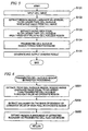
- FIG. 6 is a flowchart showing an operation example of the fragmented cell nucleus region extraction unit 104a during the fragmented cell nucleus region extraction processing.
- the operation example of the fragmented cell nucleus region extraction unit 104a will be described with reference to FIG. 6 .
- the candidate region extraction unit 105 reads the fragmented cell nucleus threshold value stored in the threshold value storage unit 102, and extracts all regions having a luminance value equal to or larger than the fragmented cell nucleus threshold value from inside of each cell nucleus region extracted in Step S103 as a candidate region.
- Step S202 The region determination unit 106a performs boundary detection processing based on the gradient of the luminance value of each pixel in each candidate region to detect all boundaries.
- the boundary detection processing is implemented by applying boundary extraction processing (boundary detection processing) in the known image processing technique.
- the region determination unit 106a calculates a differential value regarding the luminance value of each pixel in the candidate region, and successively connects pixels, in which the differential value is locally maximized, to extract a boundary.
- Step S203 The region determination unit 106a extracts each region surrounded by each extracted boundary as the fragmented cell nucleus region, and ends the fragmented cell nucleus region extraction processing.
- Step S 105 If the fragmented cell nucleus region extraction processing ends, the analysis result output unit 107 generates an analysis result on the basis of the detection result of the fragmented cell nucleus region extraction unit 104a and outputs the analysis result. Thus, the processing shown in FIG. 5 ends.
- FIG. 7 is a diagram showing an example of the analysis result of the cell image analysis apparatus 1a.
- the cell nucleus region extraction unit 103 extracts all the captured cell nucleus regions (regions inside three broken lines indicated by symbol X in FIG 7 ).
- the fragmented cell nucleus region extraction unit 104a extracts all the captured fragmented cell nucleus regions (regions inside eight solid lines indicated by symbol Y in FIG. 7 ).
- the cell nucleus threshold value and the fragmented cell nucleus threshold value larger than the cell nucleus threshold value are used. That is, two threshold values regarding the luminance value are used, so the cell nucleus region and the fragmented cell nucleus region can be extracted.
- the fragmented cell nucleus region extraction unit 104a performs processing using the fragmented cell nucleus threshold value and the boundary extraction processing based on the gradient of the luminance value of each pixel to extract a boundary. That is, a region surrounded by the extracted boundary is extracted as the fragmented cell nucleus region, such that a fragmented cell nucleus region can be more accurately extracted.
- the fragmented cell nucleus region extraction unit 104a may extract all the regions as a candidate extracted in Step S201 as the fragmented cell nucleus region, without executing Steps S202 and S203. In other words, the fragmented cell nucleus region extraction unit 104a may extract, from the cell nucleus region, a region having a luminance value equal to or larger than the fragmented cell nucleus threshold value as the fragmented cell nucleus region.
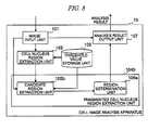
- FIG. 8 is a schematic block diagram showing the functional configuration of a cell image analysis apparatus 1b which is a second embodiment of the cell image analysis apparatus.
- the same functional parts as those in the cell image analysis apparatus 1a of the first embodiment are represented by the same reference numerals as those in FIG. 1 , and description thereof will not be repeated.
- the cell image analysis apparatus 1b is different from the cell image analysis apparatus 1a in that a candidate region extraction unit 105b, instead of the candidate region extraction unit 105a, is provided. Other parts are the same as those in the cell image analysis apparatus 1a.
- the candidate region extraction unit 105b extracts, not only from the cell nucleus region but also from regions other than the cell nucleus region of the input cell image, all regions having a luminance value equal to or larger than the fragmented cell nucleus threshold value as a candidate region.
- FIG. 9 is a flowchart showing fragmented cell nucleus region extraction processing which is executed by a fragmented cell nucleus region extraction unit 104b of the second embodiment.
- the fragmented cell nucleus region extraction processing in the second embodiment will be described with reference to FIG 9 .
- the same steps as those in FIG. 6 are represented by the same reference numerals as those in FIG 6 , and description thereof will not be repeated.
- Step S301 For a region which is determined in Step S102 that the luminance value is equal to or larger than the cell nucleus threshold value and also determined in Step S103 that the area is smaller than the cell nucleus area threshold value, the candidate region extraction unit 105b extracts a region having a luminance value equal to or larger than the fragmented cell nucleus threshold value as an additional candidate region.
- the region determination unit 106a performs Steps S202 and S203, and the fragmented cell nucleus region extraction processing ends.
- FIG. 10A is a diagram showing an example of an image in a state where the nuclear membrane of the cell nucleus is broken due to presence of apoptosis and stage advancement.
- FIG. 10A if the nuclear membrane is broken due to presence of apoptosis and stage advancement, a plurality of fragmented cell nuclei which are densely spaced inside the nuclear membrane as shown in FIG 4A are scattered as shown in FIG 10A.
- FIG 10B is a diagram showing an example of a fragmented cell nucleus region extracted by the fragmented cell nucleus region extraction unit 104b.
- FIG. 10A is a diagram showing an example of an image in a state where the nuclear membrane of the cell nucleus is broken due to presence of apoptosis and stage advancement.
- FIG. 10B is a diagram showing an example of a fragmented cell nucleus region extracted by the fragmented cell nucleus region extraction unit 104b.
- each region surrounded by a solid line indicated by symbol Y is a fragmented cell nucleus region extracted by the fragmented cell nucleus region extraction unit 104b.
- the fragmented cell nucleus region extraction unit 104b extracts a fragmented cell nucleus region from a region other than the cell nucleus region, so a fragmented cell nucleus region in a state where the nuclear membrane is broken due to stage advancement as shown in FIG. 10A can be extracted.
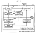
- FIG 11 is a schematic block diagram showing the functional configuration of a cell image analysis apparatus 1c which is a third embodiment of the cell image analysis apparatus 1.
- the same functional parts as those in the cell image analysis apparatus 1b of the second embodiment are represented by the same reference numerals as those in FIG. 8 , and description thereof will not be repeated.
- the cell image analysis apparatus 1c is different from the cell image analysis apparatus 1b in that the threshold value storage unit 102 further stores a fragmented cell nucleus area threshold value, a region determination unit 106c, instead of the region determination unit 106a, is provided, and a peak detection unit 108 is further provided. Other parts are the same as those in cell image analysis apparatus 1b.
- the peak detection unit 108 detects all pixels (hereinafter, referred to as peak pixel) having a peak (maximum) luminance value in each candidate region extracted by the candidate region extraction unit 105b.
- the region determination unit 106c extracts the boundary of the fragmented cell nucleus region on the basis of the detection result of the peak detection unit 108. The details of processing in the region determination unit 106c will be described below.
- FIG.12 is a flowchart showing fragmented cell nucleus region extraction processing which is executed by a fragmented cell nucleus region extraction unit 104c of the third embodiment.
- the fragmented cell nucleus extraction processing in the third embodiment will be described with reference to FIG. 12 .
- FIG. 12 the same steps as those in FIG. 9 are represented by the same reference numerals as those in FIG. 9 , and description thereof will not be repeated.
- Step S401 The peak detection unit 108 detects a peak pixel in each candidate region extracted by the candidate region extraction unit 105b.
- the peak pixel includes not only a pixel having the maximum luminance value in the candidate region, but also all pixels having a luminance value larger than peripheral pixels and the gradient of the luminance value is zero or close to zero (a value equal to or smaller than a predetermined threshold value).
- Step S402 The region determination unit 106c extracts a boundary on the basis of the detection result of the peak detection unit 108. Specifically, the region determination unit 106c searches and extracts a boundary satisfying the following conditions.
- the peak detection unit 108 detects the peak pixel from the candidate region, and the region determination unit 106c extracts a region including the peak pixel as the fragmented cell nucleus region.
- the region determination unit 106c extracts a region including the peak pixel as the fragmented cell nucleus region.
- the fragmented cell nucleus region around the center of the region has a maximum luminance value. For this reason, as described above, a region including the peak pixel is extracted as the fragmented cell nucleus region, so extraction accuracy of the fragmented cell nucleus region can be improved.
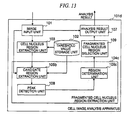
- FIG 13 is a schematic block diagram showing the functional configuration of a cell image analysis apparatus 1d which is a fourth embodiment of the cell image analysis apparatus.
- the same functional parts as those in the cell image analysis apparatus 1c of the third embodiment are represented by the same reference numerals as those in FIG 11 , and description thereof will not be repeated.
- the cell image analysis apparatus 1d is different from the cell image analysis apparatus 1c in that a fragmented cell nucleus region selection unit 109 is further provided. Other parts are the same as those in the cell image analysis apparatus 1c.
- the fragmented cell nucleus region selection unit 109 selects only a fragmented cell nucleus region satisfying predetermined conditions from among one or more fragmented cell nucleus regions extracted by the fragmented cell nucleus region extraction unit 104c and outputs the selected fragmented cell nucleus region to the analysis result output unit 107.
- the predetermined conditions include, for example, the following conditions.
- the fragmented cell nucleus region selection unit 109 selects a fragmented cell nucleus region, for example, under the following conditions.
- FIG. 14 is a flowchart showing processing which is executed by the cell image analysis apparatus 1d of the fourth embodiment.
- the processing in the cell image analysis apparatus 1d of the fourth embodiment will be described with reference to FIG 14 .
- the same steps as those in FIG 5 are represented by the same reference numerals as those in FIG 5 , and description thereof will not be repeated.
- Step S501 The fragmented cell nucleus region selection unit 109 determines whether or not each fragmented cell nucleus region extracted in the fragmented cell nucleus region extraction processing satisfies predetermined conditions. Then, the fragmented cell nucleus region selection unit 109 selects only a fragmented cell nucleus region satisfying the conditions and outputs the selected fragmented cell nucleus region to the analysis result output unit 107.
- Step S105 After Step S105, the analysis result output unit 107 generates an analysis result only on the basis of the fragmented cell nucleus region selected by the fragmented cell nucleus region selection unit 109, and outputs the generated analysis result.
- the fragmented cell nucleus region selection unit 109 selects only a fragmented cell nucleus region satisfying predetermined conditions, and outputs the selected fragmented cell nucleus region to the analysis result output unit 107. For this reason, extraction accuracy of the fragmented cell nucleus region can be improved.
- the functions of the cell image analysis apparatuses 1a to 1d in the foregoing embodiments may be implemented by a computer.
- a program for implementing each function may be recorded in a computer-readable recording medium, and a computer system may read and execute the program recorded on the recording medium.
- the term "computer system” includes an OS or hardware such as peripheral devices.
- the term "computer-readable recording medium” indicates a storage device, for example, a portable medium, such as a flexible disk, a magnetooptical disk, a ROM, or a CD-ROM, or a hard disk built in a computer system.
- computer-readable recording medium may be a medium for dynamically holding a program for a short time, for example, a communication line when a program is transmitted through a network, such as the Internet, or a communication line, such as a telephone line.
- computer-readable recording medium may be a medium for holding a program for a predetermined time, for example, a volatile memory in a computer system serving as a server or a client.
- the program may implement part of the above-described functions.
- the above-described functions may be implemented in combination with programs recorded in a computer system.
Abstract
Description
- The present invention relates to a cell image analysis apparatus, a cell image analysis method, and a program for analyzing a cell image in which a cell nucleus is stained with a fluorescent substance.
This application claims priority based on Japanese Patent Application No.2008-326108 filed on December 22, 2008 - A technique is known which captures an image of a cell stained with a fluorescent substance by a microscope and analyzes the captured cell image (see Japanese Patent No.
3576491 - Apoptosis causes DNA (Deoxyribonucleic acid) fragmentation, and DNA is cut in short units. For this reason, a plurality of fragmented cell nuclei are generated inside the cell nucleus. In the known technique, while one cell nucleus can be detected, fragmented cell nuclei cannot be detected.
- Taking the above-described situation into consideration, it is an object of the invention to provide a cell image analysis apparatus, a cell image analysis method, and a program capable of detecting fragmented cell nuclei from a cell image.
- The invention uses the following means so as to solve the above-described problem and to solve the relevant object.
A cell image analysis apparatus according to the invention includes a threshold value storage unit storing a cell nucleus threshold value, a fragmented cell nucleus threshold value, and a cell nucleus area threshold value in advance, an image input unit inputting a cell image captured from a cell stained with a fluorescent substance, a cell nucleus region extraction unit extracting, from the input cell image, a region having an area equal to or larger than the cell nucleus area threshold value from among regions having a luminance value equal to or larger than the cell nucleus threshold value as a cell nucleus region, and a fragmented cell nucleus region extraction unit extracting, from the cell nucleus region, a region having a luminance value equal to or larger than the fragmented cell nucleus threshold value as a fragmented cell nucleus region. - The fragmented cell nucleus region extraction unit may include a candidate region extraction unit extracting, from the cell nucleus region, a region having a luminance value equal to or larger than the fragmented cell nucleus threshold value as a candidate region, and a region determination unit performing boundary detection processing based on the gradient of the luminance value of each pixel to detect a boundary in the candidate region, and extracting a region surrounded by the detected boundary as the fragmented cell nucleus region.
- The fragmented cell nucleus region extraction unit may further include a peak detection unit detecting a pixel having a peak luminance value from the candidate region. The region determination unit may detect a boundary which includes a pixel having a peak and on which the average value of the gradient of the luminance value of each pixel has a maximum value.
- The threshold value storage unit may further store a fragmented cell nucleus area threshold value, and the region determination unit may extract a region surrounded by a boundary having an inner area equal to or smaller than the fragmented cell nucleus area threshold value from among the detected boundaries as the fragmented cell nucleus region.
- The fragmented cell nucleus region extraction unit may extract, from a region other than the cell nucleus region, a region having a luminance value equal to or larger than the fragmented cell nucleus threshold value as the fragmented cell nucleus region.
- The cell image analysis apparatus may further include a fragmented cell nucleus region selection unit determining, for each fragmented cell nucleus region extracted by the fragmented cell nucleus region extraction unit, whether one of conditions, a statistical value of the luminance values of pixels in the relevant region, a comparison result of the statistical value of the luminance values of the pixels in the relevant region and a statistical value of the luminance values of pixels around the relevant region, the size of the relevant region, a comparison result of the size of the relevant region and the size of a cell nucleus region including the relevant region, and the shape of the relevant region, or a plurality of conditions are satisfied or not and selecting only a fragmented cell nucleus region satisfying the conditions.
- A cell image analysis method according to the invention includes the steps of inputting a cell image captured from a cell stained with a fluorescent substance, the steps of extracting, from the input cell image, a region having an area equal to or larger than a cell nucleus area threshold value from among regions having a luminance value equal to or larger than a cell nucleus threshold value, and the steps of extracting, from the cell nucleus region, a region having a luminance value equal to or larger than a fragmented cell nucleus threshold value as a fragmented cell nucleus region.
- The cell image analysis method according to the invention may be specified as a cell image analysis method which is executed by a cell image analysis apparatus having the threshold value storage unit. A program according to the invention may be specified as a computer program which causes a computer including the threshold value storage unit to execute the cell image analysis method.
- According to the invention, two threshold values regarding a luminance value are used, so a cell nucleus region and a fragmented cell nucleus region can be extracted from a cell image.
-
-
FIG. 1 is a schematic block diagram showing the functional configuration of a cell image analysis apparatus which is a first embodiment of a cell image analysis apparatus. -
FIG 2 is a schematic view showing the outline of a cell nucleus threshold value and a fragmented cell nucleus threshold value stored in a threshold value storage unit. -
FIG. 3A is a diagram showing an example of an image of a cell nucleus in which apoptosis is not present. -
FIG 3B is a diagram showing an example of a cell nucleus region extracted by a cell nucleus region extraction unit. -
FIG. 4A is a diagram showing an example of an image of a cell nucleus in which apoptosis is present. -
FIG. 4B is a diagram showing an example of an image of a cell nucleus in which apoptosis is present. -
FIG 5 is a flowchart showing an operation example of the cell image analysis apparatus. -
FIG 6 is a flowchart showing an operation example of a fragmented cell nucleus region extraction unit during fragmented cell nucleus region extraction processing. -
FIG 7 is a diagram showing an example of an analysis result of the cell image analysis apparatus. -
FIG 8 is a schematic block diagram showing the functional configuration of a cell image analysis apparatus which is a second embodiment of the cell image analysis apparatus. -
FIG 9 is a flowchart showing fragmented cell nucleus region extraction processing which is executed by a fragmented cell nucleus region extraction unit of the second embodiment. -
FIG. 10A is a diagram showing an example of images of a plurality of fragmented cell nuclei in which apoptosis is present and a stage advances. -
FIG 10B is a diagram showing an example of images of a plurality of fragmented cell nuclei in which apoptosis is present and a stage advances. -
FIG 11 is a schematic block diagram showing the functional configuration of a cell image analysis apparatus which is a third embodiment of a cell image analysis apparatus. -
FIG 12 is a flowchart showing fragmented cell nucleus region extraction processing which is executed by a fragmented cell nucleus region extraction unit of the third embodiment. -
FIG 13 is a schematic block diagram showing the functional configuration of a cell image analysis apparatus which is a fourth embodiment of a cell image analysis apparatus. -
FIG 14 is a flowchart showing processing which is executed by the cell image analysis apparatus of the fourth embodiment. -
FIG. 1 is a schematic block diagram showing the functional configuration of a cellimage analysis apparatus 1a which is a first embodiment of a cell image analysis apparatus 1. The cellimage analysis apparatus 1a includes animage input unit 101 inputting image data, a thresholdvalue storage unit 102 recording threshold values, a cell nucleusregion extraction unit 103 detecting a cell nucleus region, a fragmented cell nucleusregion extraction unit 104a detecting a fragmented cell nucleus region, and an analysisresult output unit 107 outputting an analysis result. For the cell image analysis apparatus la, an information processing apparatus, such as a personal computer or a workstation, may be used, or an exclusive-use apparatus which is incorporated into a microscope may be used. - The
image input unit 101 inputs digital data of a cell image of a cell stained with a fluorescent substance captured by a microscope to the cellimage analysis apparatus 1a. - The threshold
value storage unit 102 stores a cell nucleus threshold value, a fragmented cell nucleus threshold value, and a cell nucleus area threshold value, which are used for processing in the cell nucleusregion extraction unit 103 and the fragmented cell nucleusregion extraction unit 104a, in advance. - The cell nucleus
region extraction unit 103 extracts a cell nucleus region (a region where a cell nucleus is present) from the input cell image on the basis of the cell nucleus threshold value and the cell nucleus area threshold value, and acquires information representing the position and range of the cell nucleus region. The term "area" may mean not only an accurate numerical value of the area of the region, but also the number of pixels in the region. - The fragmented cell nucleus
region extraction unit 104a extracts a fragmented cell nucleus region (a region where a fragmented cell nucleus is present) from the cell nucleus region of the input cell image on the basis of the fragmented cell nucleus threshold value, and acquires information representing the position and range of the fragmented cell nucleus region. The term "fragmented cell nucleus" indicates the nucleus of an apoptosis cell with DNA fragmented by apoptosis. - Specifically, the fragmented cell nucleus
region extraction unit 104a includes a candidateregion extraction unit 105a extracting a region as a candidate of a fragmented cell nucleus region, and aregion determination unit 106a determining a fragmented cell nucleus region. The candidateregion extraction unit 105a extracts, from inside of the cell nucleus region, a region having a luminance value (a value of luminance) equal to or larger than the fragmented cell nucleus threshold value as a candidate region. Theregion determination unit 106a performs boundary detection processing based on the gradient of the luminance value of each pixel in the candidate region to detect a boundary between the cell nucleus region and the fragmented cell nucleus region, and extracts a region surrounded by the detected boundary as the fragmented cell nucleus region. The term "luminance" is a value indicating a gray-scale level of a gray-scale image, and is a value indicating brightness of each pixel in an image. - The analysis
result output unit 107 generates an analysis result on the basis of the information representing the positions and ranges of the cell nucleus region and the fragmented cell nucleus region, and outputs the analysis result. With regard to the output means of the analysisresult output unit 107, the analysis result may be displayed on an image output device in the form of characters or figures such as graphs, or may be printed by a printer. The analysis result output from the analysisresult output unit 107 includes, for example, the following matters. - the number of cell nucleus regions
- the number of fragmented cell nucleus regions
- the number of cell nucleus regions from which no fragmented cell nucleus region is extracted
- the number of cell nucleus regions from which a fragmented cell nucleus region is extracted
- the total area of cell nucleus regions
- the total area of fragmented cell nucleus regions
- the ratio between the total area of cell nucleus regions and the total area of fragmented cell nucleus regions
- the ratio between the average value of the luminance values of cell nucleus regions and the average value of the luminance values of fragmented cell nucleus regions
-
FIG 2 is a schematic view showing the outline of the cell nucleus threshold value and the fragmented cell nucleus threshold value stored in the thresholdvalue storage unit 102. The graph ofFIG. 2 shows changes in the luminance value of a cell image obtained by an experiment in advance in the x-axis direction when the value on the y axis is fixed to a predetermined value. InFIG 2 , the value of the x coordinate as the boundary between regions A1 to A3 is set by a designer or an experimenter. The region A1 corresponds to neither the cell nucleus region nor the fragmented cell nucleus region. In the cell image, the cell nucleus is stained by using a fluorescent substance which selectively stains the cell nucleus. For this reason, like the region A1, a region where no cell nucleus and fragmented cell nucleus exist includes pixels having a small luminance value. The region A2 is a cell nucleus region where no fragmented cell nucleus is present. The region A3 is a region where a fragmented cell nucleus is present. In the cell image, the higher cell nucleus density the region has, the larger the luminance value is, so a region where a fragmented cell nucleus is present has a luminance value larger than that of a cell nucleus region where no fragmented cell nucleus is present. - The cell nucleus threshold value and the fragmented cell nucleus threshold value are set on the basis of such an analysis result. That is, the luminance value of each pixel at the boundary between the region A1 and the region A2 is set as the cell nucleus threshold value, and the luminance value of each pixel at the boundary between the region A2 and the region A3 is set as the fragmented cell nucleus threshold value. At this time, the fragmented cell nucleus threshold value is set larger than the cell nucleus threshold value. Actually, the cell nucleus threshold value and the fragmented cell nucleus threshold value are determined to optimum values in accordance with the experiment environment by carrying out a plurality of experiments in advance and compiling statistics of the luminance values of a cell nucleus region and a region where a fragmented cell nucleus is present.
-
FIGS. 3A and 3B are diagrams showing an example of an image of a cell nucleus in which apoptosis is not present. In figures of the present application includingFIGS. 3A and 3B , display is white as the luminance value is small, and display is black as the luminance value is large. -
FIG. 3A is a diagram showing an example of an image of a cell nucleus in which apoptosis is not present.FIG. 3B is a diagram showing an example of a cell nucleus region extracted by the cell nucleusregion extraction unit 103. InFIG. 3B , the region surrounded by a broken line indicated by symbol X is the cell nucleus region extracted by the cell nucleusregion extraction unit 103. -
FIG. 4A is a diagram showing an example of an image of a cell nucleus in which apoptosis is present. As shown inFIG. 4A , in an image of a cell nucleus in which apoptosis is present, a plurality of fragmented cell nuclei are present inside the cell nucleus.FIG. 4B is a diagram showing an example of the cell nucleus region extracted by the cell nucleusregion extraction unit 103 and the fragmented cell nucleus region extracted by the fragmented cell nucleusregion extraction unit 104a. InFIG. 4B , a region surrounded by a broken line indicated by symbol X is the cell nucleus region extracted by the cell nucleusregion extraction unit 103. InFIG. 4B , the region surrounded by a solid line indicated by symbol Y is the fragmented cell nucleus region extracted by the fragmented cell nucleusregion extraction unit 104a. -
FIG 5 is a flowchart showing an operation example of the cellimage analysis apparatus 1a. Hereinafter, the operation example of the cellimage analysis apparatus 1a will be described with reference toFIG. 5 .
(Step S101) Theimage input unit 101 inputs a digital image of a cell image to the cellimage analysis apparatus 1a.
(Step S102) The cell nucleusregion extraction unit 103 reads the cell nucleus threshold value stored in the thresholdvalue storage unit 102, and extracts all regions having a luminance value equal to or larger than the cell nucleus threshold value from the cell image.
(Step S103) The cell nucleusregion extraction unit 103 reads the cell nucleus area threshold value stored in the thresholdvalue storage unit 102, and extracts all regions having an area equal to or larger than the cell nucleus area threshold value from among the regions extracted in Step S102 as a cell nucleus region.
(Step S 104) The fragmented cell nucleusregion extraction unit 104a executes fragmented cell nucleus region extraction processing. -
FIG. 6 is a flowchart showing an operation example of the fragmented cell nucleusregion extraction unit 104a during the fragmented cell nucleus region extraction processing. Hereinafter, the operation example of the fragmented cell nucleusregion extraction unit 104a will be described with reference toFIG. 6 .
(Step S201) The candidateregion extraction unit 105 reads the fragmented cell nucleus threshold value stored in the thresholdvalue storage unit 102, and extracts all regions having a luminance value equal to or larger than the fragmented cell nucleus threshold value from inside of each cell nucleus region extracted in Step S103 as a candidate region.
(Step S202) Theregion determination unit 106a performs boundary detection processing based on the gradient of the luminance value of each pixel in each candidate region to detect all boundaries. In Step S202, the boundary detection processing is implemented by applying boundary extraction processing (boundary detection processing) in the known image processing technique. For example, theregion determination unit 106a calculates a differential value regarding the luminance value of each pixel in the candidate region, and successively connects pixels, in which the differential value is locally maximized, to extract a boundary.
(Step S203) Theregion determination unit 106a extracts each region surrounded by each extracted boundary as the fragmented cell nucleus region, and ends the fragmented cell nucleus region extraction processing. - The description will be continued with reference to
FIG. 5 again.
(Step S 105) If the fragmented cell nucleus region extraction processing ends, the analysisresult output unit 107 generates an analysis result on the basis of the detection result of the fragmented cell nucleusregion extraction unit 104a and outputs the analysis result. Thus, the processing shown inFIG. 5 ends. -
FIG. 7 is a diagram showing an example of the analysis result of the cellimage analysis apparatus 1a. When a plurality of cell nuclei or fragmented cell nuclei are captured in the cell image input to the cellimage analysis apparatus 1a, the cell nucleusregion extraction unit 103 extracts all the captured cell nucleus regions (regions inside three broken lines indicated by symbol X inFIG 7 ). Meanwhile, the fragmented cell nucleusregion extraction unit 104a extracts all the captured fragmented cell nucleus regions (regions inside eight solid lines indicated by symbol Y inFIG. 7 ). - In the cell
image analysis apparatus 1a configured as above, the cell nucleus threshold value and the fragmented cell nucleus threshold value larger than the cell nucleus threshold value are used. That is, two threshold values regarding the luminance value are used, so the cell nucleus region and the fragmented cell nucleus region can be extracted. - As shown in
FIG 2 , if a region having a luminance value equal to or larger than the fragmented cell nucleus threshold value is extracted as the fragmented cell nucleus region, a plurality of fragmented cell nucleus regions may be erroneously detected as a single fragmented cell nucleus region. Against such a problem, the fragmented cell nucleusregion extraction unit 104a performs processing using the fragmented cell nucleus threshold value and the boundary extraction processing based on the gradient of the luminance value of each pixel to extract a boundary. That is, a region surrounded by the extracted boundary is extracted as the fragmented cell nucleus region, such that a fragmented cell nucleus region can be more accurately extracted. - The fragmented cell nucleus
region extraction unit 104a may extract all the regions as a candidate extracted in Step S201 as the fragmented cell nucleus region, without executing Steps S202 and S203. In other words, the fragmented cell nucleusregion extraction unit 104a may extract, from the cell nucleus region, a region having a luminance value equal to or larger than the fragmented cell nucleus threshold value as the fragmented cell nucleus region. -
FIG. 8 is a schematic block diagram showing the functional configuration of a cellimage analysis apparatus 1b which is a second embodiment of the cell image analysis apparatus. InFIG. 8 , the same functional parts as those in the cellimage analysis apparatus 1a of the first embodiment are represented by the same reference numerals as those inFIG. 1 , and description thereof will not be repeated. - The cell
image analysis apparatus 1b is different from the cellimage analysis apparatus 1a in that a candidateregion extraction unit 105b, instead of the candidateregion extraction unit 105a, is provided. Other parts are the same as those in the cellimage analysis apparatus 1a. - The candidate
region extraction unit 105b extracts, not only from the cell nucleus region but also from regions other than the cell nucleus region of the input cell image, all regions having a luminance value equal to or larger than the fragmented cell nucleus threshold value as a candidate region. -
FIG. 9 is a flowchart showing fragmented cell nucleus region extraction processing which is executed by a fragmented cell nucleusregion extraction unit 104b of the second embodiment. Hereinafter, the fragmented cell nucleus region extraction processing in the second embodiment will be described with reference toFIG 9 . InFIG 9 , the same steps as those inFIG. 6 are represented by the same reference numerals as those inFIG 6 , and description thereof will not be repeated. - After Step S201, Step S301 is executed.
(Step S301) For a region which is determined in Step S102 that the luminance value is equal to or larger than the cell nucleus threshold value and also determined in Step S103 that the area is smaller than the cell nucleus area threshold value, the candidateregion extraction unit 105b extracts a region having a luminance value equal to or larger than the fragmented cell nucleus threshold value as an additional candidate region.
After Step S301, theregion determination unit 106a performs Steps S202 and S203, and the fragmented cell nucleus region extraction processing ends. -
FIG. 10A is a diagram showing an example of an image in a state where the nuclear membrane of the cell nucleus is broken due to presence of apoptosis and stage advancement. As shown inFIG. 10A , if the nuclear membrane is broken due to presence of apoptosis and stage advancement, a plurality of fragmented cell nuclei which are densely spaced inside the nuclear membrane as shown inFIG 4A are scattered as shown inFIG 10A. FIG 10B is a diagram showing an example of a fragmented cell nucleus region extracted by the fragmented cell nucleusregion extraction unit 104b. InFIG. 10B , each region surrounded by a solid line indicated by symbol Y is a fragmented cell nucleus region extracted by the fragmented cell nucleusregion extraction unit 104b. The fragmented cell nucleusregion extraction unit 104b extracts a fragmented cell nucleus region from a region other than the cell nucleus region, so a fragmented cell nucleus region in a state where the nuclear membrane is broken due to stage advancement as shown inFIG. 10A can be extracted. -
FIG 11 is a schematic block diagram showing the functional configuration of a cell image analysis apparatus 1c which is a third embodiment of the cell image analysis apparatus 1. InFIG 11 , the same functional parts as those in the cellimage analysis apparatus 1b of the second embodiment are represented by the same reference numerals as those inFIG. 8 , and description thereof will not be repeated. - The cell image analysis apparatus 1c is different from the cell
image analysis apparatus 1b in that the thresholdvalue storage unit 102 further stores a fragmented cell nucleus area threshold value, aregion determination unit 106c, instead of theregion determination unit 106a, is provided, and apeak detection unit 108 is further provided. Other parts are the same as those in cellimage analysis apparatus 1b. - The
peak detection unit 108 detects all pixels (hereinafter, referred to as peak pixel) having a peak (maximum) luminance value in each candidate region extracted by the candidateregion extraction unit 105b. - The
region determination unit 106c extracts the boundary of the fragmented cell nucleus region on the basis of the detection result of thepeak detection unit 108. The details of processing in theregion determination unit 106c will be described below. -
FIG.12 is a flowchart showing fragmented cell nucleus region extraction processing which is executed by a fragmented cell nucleusregion extraction unit 104c of the third embodiment. Hereinafter, the fragmented cell nucleus extraction processing in the third embodiment will be described with reference toFIG. 12 . InFIG 12 , the same steps as those inFIG. 9 are represented by the same reference numerals as those inFIG. 9 , and description thereof will not be repeated. - After Step S301, Steps S401 to S403 are executed sequentially.
(Step S401) Thepeak detection unit 108 detects a peak pixel in each candidate region extracted by the candidateregion extraction unit 105b.
The peak pixel includes not only a pixel having the maximum luminance value in the candidate region, but also all pixels having a luminance value larger than peripheral pixels and the gradient of the luminance value is zero or close to zero (a value equal to or smaller than a predetermined threshold value).
(Step S402) Theregion determination unit 106c extracts a boundary on the basis of the detection result of thepeak detection unit 108.
Specifically, theregion determination unit 106c searches and extracts a boundary satisfying the following conditions. - a peak pixel is included
- an area inside the boundary is smaller than the fragmented cell nucleus area threshold value
- the average value of the gradient of the luminance value of each pixel on the boundary has a maximum value, as compared with other boundaries satisfying the two conditions
- In the cell image analysis apparatus 1c configured as above, the
peak detection unit 108 detects the peak pixel from the candidate region, and theregion determination unit 106c extracts a region including the peak pixel as the fragmented cell nucleus region. In general, in the fragmented cell nucleus region, around the center of the region has a maximum luminance value. For this reason, as described above, a region including the peak pixel is extracted as the fragmented cell nucleus region, so extraction accuracy of the fragmented cell nucleus region can be improved. -
FIG 13 is a schematic block diagram showing the functional configuration of a cell image analysis apparatus 1d which is a fourth embodiment of the cell image analysis apparatus. InFIG. 13 , the same functional parts as those in the cell image analysis apparatus 1c of the third embodiment are represented by the same reference numerals as those inFIG 11 , and description thereof will not be repeated. - The cell image analysis apparatus 1d is different from the cell image analysis apparatus 1c in that a fragmented cell nucleus
region selection unit 109 is further provided. Other parts are the same as those in the cell image analysis apparatus 1c. - The fragmented cell nucleus
region selection unit 109 selects only a fragmented cell nucleus region satisfying predetermined conditions from among one or more fragmented cell nucleus regions extracted by the fragmented cell nucleusregion extraction unit 104c and outputs the selected fragmented cell nucleus region to the analysisresult output unit 107. - The predetermined conditions include, for example, the following conditions.
- condition regarding a statistical value of the luminance values of pixels in the fragmented cell nucleus region
- condition regarding a comparison result of the statistical value of the luminance values of the pixels in the fragmented cell nucleus region and a statistical value of the luminance values of pixels around the fragmented cell nucleus region
- condition regarding the size of the fragmented cell nucleus region
- condition regarding a comparison result of the size of the fragmented cell nucleus region and the size of the cell nucleus region including the fragmented cell nucleus region
- condition regarding the shape of the fragmented cell nucleus region
- Specifically, the fragmented cell nucleus
region selection unit 109 selects a fragmented cell nucleus region, for example, under the following conditions. - the average value of the luminance values of the pixels in the fragmented cell nucleus region is equal to or larger than a predetermined threshold value
- the ratio between the average value of the luminance values of the pixels in the fragmented cell nucleus region and the average value of the luminance values of pixels within a predetermined width circumscribing the fragmented cell nucleus region is equal to or larger than a predetermined threshold value
- the size of the fragmented cell nucleus region is equal to or larger than a predetermined threshold value
- the ratio between the size of the fragmented cell nucleus region and the size of the cell nucleus region which is present in the cell image is equal to or larger than a predetermined threshold value
- the circularity of the fragmented cell nucleus region is smaller than a predetermined threshold value (the region is close to a circle, as compared with a case where the circularity is the predetermined threshold value)
-
FIG. 14 is a flowchart showing processing which is executed by the cell image analysis apparatus 1d of the fourth embodiment. Hereinafter, the processing in the cell image analysis apparatus 1d of the fourth embodiment will be described with reference toFIG 14 . InFIG 14 , the same steps as those inFIG 5 are represented by the same reference numerals as those inFIG 5 , and description thereof will not be repeated. - After the fragmented cell nucleus region extraction processing of Step S 104, Steps S501 and S105 are executed.
(Step S501) The fragmented cell nucleusregion selection unit 109 determines whether or not each fragmented cell nucleus region extracted in the fragmented cell nucleus region extraction processing satisfies predetermined conditions. Then, the fragmented cell nucleusregion selection unit 109 selects only a fragmented cell nucleus region satisfying the conditions and outputs the selected fragmented cell nucleus region to the analysisresult output unit 107.
(Step S105) After Step S105, the analysisresult output unit 107 generates an analysis result only on the basis of the fragmented cell nucleus region selected by the fragmented cell nucleusregion selection unit 109, and outputs the generated analysis result. - In the cell image analysis apparatus 1d configured as above, the fragmented cell nucleus
region selection unit 109 selects only a fragmented cell nucleus region satisfying predetermined conditions, and outputs the selected fragmented cell nucleus region to the analysisresult output unit 107. For this reason, extraction accuracy of the fragmented cell nucleus region can be improved. - The functions of the cell
image analysis apparatuses 1a to 1d in the foregoing embodiments may be implemented by a computer. In this case, a program for implementing each function may be recorded in a computer-readable recording medium, and a computer system may read and execute the program recorded on the recording medium. The term "computer system" includes an OS or hardware such as peripheral devices. The term "computer-readable recording medium" indicates a storage device, for example, a portable medium, such as a flexible disk, a magnetooptical disk, a ROM, or a CD-ROM, or a hard disk built in a computer system. The term "computer-readable recording medium" may be a medium for dynamically holding a program for a short time, for example, a communication line when a program is transmitted through a network, such as the Internet, or a communication line, such as a telephone line. Or the term "computer-readable recording medium" may be a medium for holding a program for a predetermined time, for example, a volatile memory in a computer system serving as a server or a client. The program may implement part of the above-described functions. The above-described functions may be implemented in combination with programs recorded in a computer system. - Although the embodiments of the invention have been described in detail with reference to the drawings, the specific configuration is not limited to the embodiments, and design may also be made without departing from the gist of the invention.
The method of searching for a boundary satisfying the three conditions may be implemented by other methods using the known technique.
(Step S403) After Step S402, the
Claims (10)
- A cell image analysis apparatus comprising:a threshold value storage unit storing a cell nucleus threshold value, a fragmented cell nucleus threshold value, and a cell nucleus area threshold value in advance;an image input unit inputting a cell image captured from a cell stained with a fluorescent substance;a cell nucleus region extraction unit extracting, from the input cell image, a region having an area equal to or larger than the cell nucleus area threshold value from among regions having a luminance value equal to or larger than the cell nucleus threshold value as a cell nucleus region; anda fragmented cell nucleus region extraction unit extracting, from the cell nucleus region, a region having a luminance value equal to or larger than the fragmented cell nucleus threshold value as a fragmented cell nucleus region.
- The cell image analysis apparatus according to claim 1,
wherein the fragmented cell nucleus region extraction unit includes
a candidate region extraction unit extracting, from the cell nucleus region, a region having a luminance value equal to or larger than the fragmented cell nucleus threshold value as a candidate region, and
a region determination unit performing boundary detection processing based on the gradient of the luminance value of each pixel to detect a boundary in the candidate region, and extracting a region surrounded by the detected boundary as the fragmented cell nucleus region. - The cell image analysis apparatus according to claim 2,
wherein the fragmented cell nucleus region extraction unit further includes
a peak detection unit detecting a pixel having a peak luminance value from the candidate region,
wherein the region determination unit detects a boundary which includes a pixel having a peak and on which the average value of the gradient of the luminance value of each pixel has a maximum value. - The cell image analysis apparatus according to claim 2,
wherein the threshold value storage unit further stores a fragmented cell nucleus area threshold value, and
the region determination unit extracts a region surrounded by a boundary having an inner area equal to or smaller than the fragmented cell nucleus area threshold value from among the detected boundaries as the fragmented cell nucleus region. - The cell image analysis apparatus according to claim 3,
wherein the threshold value storage unit further stores a fragmented cell nucleus area threshold value, and
the region determination unit extracts a region surrounded by a boundary having an inner area equal to or smaller than the fragmented cell nucleus area threshold value from among the detected boundaries as the fragmented cell nucleus region. - The cell image analysis apparatus according to claim 1,
wherein the fragmented cell nucleus region extraction unit extracts, from a region other than the cell nucleus region, a region having a luminance value equal to or larger than the fragmented cell nucleus threshold value as the fragmented cell nucleus region. - The cell image analysis apparatus according to claim 1, further comprising:a fragmented cell nucleus region selection unit determining, for each fragmented cell nucleus region extracted by the fragmented cell nucleus region extraction unit, whether one of the following conditions or a plurality of the conditions is satisfied or not and selecting only a fragmented cell nucleus region satisfying the conditions:a statistical value of the luminance values of pixels in the relevant region;a comparison result of the statistical value of the luminance values of the pixels in the relevant region and a statistical value of the luminance values of pixels around the relevant region;a size of the relevant region;a comparison result of the size of the relevant region and a size of a cell nucleus region including the relevant region; anda shape of the relevant region.
- A cell image analysis method using a cell image analysis apparatus, which includes a threshold value storage unit storing a cell nucleus threshold value, a fragmented cell nucleus threshold value, and a cell nucleus area threshold value in advance, the method comprising the steps of:inputting a cell image captured from a cell stained with a fluorescent substance;extracting, from the input cell image, a region having an area equal to or larger than the cell nucleus area threshold value from among regions having a luminance value equal to or larger than the cell nucleus threshold value as a cell nucleus region; andextracting, from the cell nucleus region, a region having a luminance value equal to or larger than a fragmented cell nucleus threshold value as a fragmented cell nucleus region.
- A program which causes a computer including a threshold value storage unit storing a cell nucleus threshold value, a fragmented cell nucleus threshold value, and a cell nucleus area threshold value in advance to execute the steps of:inputting a cell image captured from a cell stained with a fluorescent substance;extracting, from the input cell image, a region having an area equal to or larger than the cell nucleus area threshold value from among regions having a luminance value equal to or larger than the cell nucleus threshold value as a cell nucleus region; andextracting, from the cell nucleus region, a region having a luminance value equal to or larger than the fragmented cell nucleus threshold value as a fragmented cell nucleus region.
- A cell image analysis method comprising the steps of:inputting a cell image captured from a cell stained with a fluorescent substance;extracting, from the input cell image, a region having an area equal to or larger than a cell nucleus area threshold value from among regions having a luminance value equal to or larger than a cell nucleus threshold value as a cell nucleus region; andextracting, from the cell nucleus region, a region having a luminance value equal to or larger than a fragmented cell nucleus threshold value as a fragmented cell nucleus region.
Applications Claiming Priority (1)
| Application Number | Priority Date | Filing Date | Title |
|---|---|---|---|
| JP2008326108A JP5058962B2 (en) | 2008-12-22 | 2008-12-22 | Cell image analysis apparatus, cell image analysis method, and program |
Publications (3)
| Publication Number | Publication Date |
|---|---|
| EP2199776A2 true EP2199776A2 (en) | 2010-06-23 |
| EP2199776A3 EP2199776A3 (en) | 2013-08-07 |
| EP2199776B1 EP2199776B1 (en) | 2019-04-17 |
Family
ID=42045368
Family Applications (1)
| Application Number | Title | Priority Date | Filing Date |
|---|---|---|---|
| EP09015814.8A Not-in-force EP2199776B1 (en) | 2008-12-22 | 2009-12-21 | Cell image analysis apparatus, cell image analysis method, and program |
Country Status (3)
| Country | Link |
|---|---|
| US (1) | US9176043B2 (en) |
| EP (1) | EP2199776B1 (en) |
| JP (1) | JP5058962B2 (en) |
Cited By (2)
| Publication number | Priority date | Publication date | Assignee | Title |
|---|---|---|---|---|
| WO2015079640A1 (en) * | 2013-11-28 | 2015-06-04 | Sony Corporation | Image processing device and image processing method |
| CN115620284A (en) * | 2022-12-19 | 2023-01-17 | 广东工业大学 | Cell apoptosis counting method, system and platform based on convolution attention mechanism |
Families Citing this family (7)
| Publication number | Priority date | Publication date | Assignee | Title |
|---|---|---|---|---|
| JP5745919B2 (en) * | 2011-04-28 | 2015-07-08 | 浜松ホトニクス株式会社 | Cell analysis method, cell analysis apparatus, and cell analysis program |
| JP5768948B1 (en) * | 2014-03-27 | 2015-08-26 | コニカミノルタ株式会社 | Image processing apparatus and image processing program |
| CN105067520B (en) * | 2015-07-28 | 2018-10-23 | 爱威科技股份有限公司 | A kind of microscopy recognition methods and device |
| TWI586954B (en) * | 2015-12-09 | 2017-06-11 | 財團法人金屬工業研究發展中心 | An apparatus for detecting cells being infected by human papillomavirus (hpv) and an detection method therefor |
| EP3469338A4 (en) * | 2016-06-10 | 2020-01-15 | The Regents of the University of California | Image-based cell sorting systems and methods |
| TWI595372B (en) * | 2016-08-31 | 2017-08-11 | 義守大學 | Microscopic image inspection system and method for inspecting actin filaments |
| CN114912493B (en) * | 2022-05-27 | 2022-11-29 | 深圳见康智能科技有限公司 | Flow type immune cell intelligent analysis system based on machine learning |
Citations (1)
| Publication number | Priority date | Publication date | Assignee | Title |
|---|---|---|---|---|
| JP3576491B2 (en) | 1999-02-26 | 2004-10-13 | セロミックス インコーポレイテッド | System for cell-based screening |
Family Cites Families (17)
| Publication number | Priority date | Publication date | Assignee | Title |
|---|---|---|---|---|
| JP2001211896A (en) * | 1999-11-26 | 2001-08-07 | Olympus Optical Co Ltd | Image analysis method, device for it, and recording medium |
| JP4854832B2 (en) * | 2000-03-27 | 2012-01-18 | オリンパス株式会社 | Cell image analysis method, apparatus, and recording medium |
| JP2002142800A (en) * | 2000-11-13 | 2002-05-21 | Olympus Optical Co Ltd | Method for cell form analysis and storage medium |
| JP4583670B2 (en) * | 2001-07-04 | 2010-11-17 | パナソニック株式会社 | Image distortion correction apparatus and method |
| JP4749637B2 (en) * | 2001-09-28 | 2011-08-17 | オリンパス株式会社 | Image analysis method, apparatus, and recording medium |
| JP3946590B2 (en) * | 2002-07-16 | 2007-07-18 | 富士通株式会社 | Image processing method, image processing program, and image processing apparatus |
| GB0227160D0 (en) * | 2002-11-21 | 2002-12-24 | Qinetiq Ltd | Histological assessment of pleomorphism |
| WO2004099773A1 (en) * | 2003-04-30 | 2004-11-18 | Pfizer Products Inc. | Automated in vitro cellular imaging assays for micronuclei and other target objects |
| US20070031818A1 (en) * | 2004-07-15 | 2007-02-08 | Cytokinetics, Inc., A Delaware Corporation | Assay for distinguishing live and dead cells |
| US20060127881A1 (en) | 2004-10-25 | 2006-06-15 | Brigham And Women's Hospital | Automated segmentation, classification, and tracking of cell nuclei in time-lapse microscopy |
| US20080279441A1 (en) * | 2005-03-29 | 2008-11-13 | Yuichiro Matsuo | Cell-Image Analysis Method, Cell-Image Analysis Program, Cell-Image Analysis Apparatus, Screening Method, and Screening Apparatus |
| JP2006285310A (en) * | 2005-03-31 | 2006-10-19 | Kanazawa Univ | Evaluation method of canopy of forest, and its canopy evaluation program |
| JP4744187B2 (en) * | 2005-05-10 | 2011-08-10 | オリンパス株式会社 | Cell observation device |
| US20070124085A1 (en) * | 2005-11-30 | 2007-05-31 | Geert Kalusche | Method of processing a biological image |
| TWI406664B (en) * | 2006-03-30 | 2013-09-01 | Univ Kyoto | Agent increasing the production of thioredoxin |
| JP5347272B2 (en) * | 2008-01-18 | 2013-11-20 | 日本電気株式会社 | Spot quantification device, spot quantification method and program |
| JP5365011B2 (en) * | 2008-01-29 | 2013-12-11 | 日本電気株式会社 | Pathological diagnosis support apparatus, pathological diagnosis support method, and program |
-
2008
- 2008-12-22 JP JP2008326108A patent/JP5058962B2/en not_active Expired - Fee Related
-
2009
- 2009-12-21 US US12/643,241 patent/US9176043B2/en not_active Expired - Fee Related
- 2009-12-21 EP EP09015814.8A patent/EP2199776B1/en not_active Not-in-force
Patent Citations (1)
| Publication number | Priority date | Publication date | Assignee | Title |
|---|---|---|---|---|
| JP3576491B2 (en) | 1999-02-26 | 2004-10-13 | セロミックス インコーポレイテッド | System for cell-based screening |
Cited By (4)
| Publication number | Priority date | Publication date | Assignee | Title |
|---|---|---|---|---|
| WO2015079640A1 (en) * | 2013-11-28 | 2015-06-04 | Sony Corporation | Image processing device and image processing method |
| US10330912B2 (en) | 2013-11-28 | 2019-06-25 | Sony Corporation | Image processing device and image processing method |
| CN115620284A (en) * | 2022-12-19 | 2023-01-17 | 广东工业大学 | Cell apoptosis counting method, system and platform based on convolution attention mechanism |
| CN115620284B (en) * | 2022-12-19 | 2023-04-18 | 广东工业大学 | Cell apoptosis counting method, system and platform based on convolution attention mechanism |
Also Published As
| Publication number | Publication date |
|---|---|
| US20100172569A1 (en) | 2010-07-08 |
| JP5058962B2 (en) | 2012-10-24 |
| EP2199776A3 (en) | 2013-08-07 |
| JP2010145366A (en) | 2010-07-01 |
| US9176043B2 (en) | 2015-11-03 |
| EP2199776B1 (en) | 2019-04-17 |
Similar Documents
| Publication | Publication Date | Title |
|---|---|---|
| EP2199776A2 (en) | Cell image analysis apparatus, cell image analysis method, and program | |
| CN110135411B (en) | Business card recognition method and device | |
| US20130163869A1 (en) | Apparatus and method for extracting edge in image | |
| CN109035256A (en) | User interface image cutting method, device, server and storage medium | |
| CN104750791A (en) | Image retrieval method and device | |
| WO2018207261A1 (en) | Image analysis device | |
| JP5495934B2 (en) | Image processing apparatus, processing method thereof, and program | |
| JP5111055B2 (en) | Image processing apparatus, image processing method, and computer program | |
| JP2008011484A (en) | Apparatus and method for extracting character and graphic string, program for executing the method, recording medium with the program stored therein | |
| CN112703531A (en) | Generating annotation data for tissue images | |
| CN114429640A (en) | Drawing segmentation method and device and electronic equipment | |
| JP2009123020A (en) | Information processor, information processing method, program and recording medium | |
| JP2011043350A (en) | Cell image analyzer, cell image analyzing method, and computer program | |
| JP4967045B2 (en) | Background discriminating apparatus, method and program | |
| CN114155547B (en) | Chart identification method, device, equipment and storage medium | |
| JP2007199865A (en) | Image processing algorithm evaluation apparatus, image processing algorithm creation apparatus, image inspection apparatus, method for evaluating image processing algorithm, method for creating image processing algorithm, and method for inspecting image | |
| CN113537253A (en) | Infrared image target detection method and device, computing equipment and storage medium | |
| CN109492396B (en) | Malicious software gene rapid detection method and device based on semantic segmentation | |
| JP2012222581A (en) | Image processing device, image processing method, program, and storage medium | |
| CN112257768A (en) | Method and device for identifying illegal financial picture and computer storage medium | |
| JP4381445B2 (en) | Image characteristic determination processing apparatus, image characteristic determination processing method, program for executing the method, and computer-readable storage medium storing the program | |
| JP2017072940A (en) | Image processing system, image processing method, and image processing program | |
| JP5298830B2 (en) | Image processing program, image processing apparatus, and image processing system | |
| CN111177512A (en) | Scientific and technological achievement missing processing method and device based on big data | |
| US10304195B2 (en) | Information processing apparatus, computer-readable storage medium, and information processing method for judging a recognition target area |
Legal Events
| Date | Code | Title | Description |
|---|---|---|---|
| PUAI | Public reference made under article 153(3) epc to a published international application that has entered the european phase |
Free format text: ORIGINAL CODE: 0009012 |
|
| 17P | Request for examination filed |
Effective date: 20091221 |
|
| AK | Designated contracting states |
Kind code of ref document: A2 Designated state(s): AT BE BG CH CY CZ DE DK EE ES FI FR GB GR HR HU IE IS IT LI LT LU LV MC MK MT NL NO PL PT RO SE SI SK SM TR |
|
| AX | Request for extension of the european patent |
Extension state: AL BA RS |
|
| PUAL | Search report despatched |
Free format text: ORIGINAL CODE: 0009013 |
|
| AK | Designated contracting states |
Kind code of ref document: A3 Designated state(s): AT BE BG CH CY CZ DE DK EE ES FI FR GB GR HR HU IE IS IT LI LT LU LV MC MK MT NL NO PL PT RO SE SI SK SM TR |
|
| AX | Request for extension of the european patent |
Extension state: AL BA RS |
|
| RIC1 | Information provided on ipc code assigned before grant |
Ipc: G06T 7/00 20060101ALN20130628BHEP Ipc: G01N 15/14 20060101AFI20130628BHEP Ipc: G06M 11/04 20060101ALN20130628BHEP Ipc: G06K 9/46 20060101ALN20130628BHEP |
|
| RBV | Designated contracting states (corrected) |
Designated state(s): AT BE BG CH CY CZ DE DK EE ES FI FR GB GR HR HU IE IS IT LI LT LU LV MC MK MT NL NO PL PT RO SE SI SK SM TR |
|
| RAP1 | Party data changed (applicant data changed or rights of an application transferred) |
Owner name: OLYMPUS CORPORATION |
|
| RAP1 | Party data changed (applicant data changed or rights of an application transferred) |
Owner name: OLYMPUS CORPORATION |
|
| RIN1 | Information on inventor provided before grant (corrected) |
Inventor name: KOBAYASHI, TAMIYO Inventor name: TAKAGI, KOSUKE |
|
| STAA | Information on the status of an ep patent application or granted ep patent |
Free format text: STATUS: EXAMINATION IS IN PROGRESS |
|
| 17Q | First examination report despatched |
Effective date: 20180905 |
|
| RIC1 | Information provided on ipc code assigned before grant |
Ipc: G06M 11/04 20060101ALN20181001BHEP Ipc: G06K 9/46 20060101ALN20181001BHEP Ipc: G06T 7/00 20060101ALN20181001BHEP Ipc: G01N 15/14 20060101AFI20181001BHEP |
|
| GRAP | Despatch of communication of intention to grant a patent |
Free format text: ORIGINAL CODE: EPIDOSNIGR1 |
|
| STAA | Information on the status of an ep patent application or granted ep patent |
Free format text: STATUS: GRANT OF PATENT IS INTENDED |
|
| RIC1 | Information provided on ipc code assigned before grant |
Ipc: G06K 9/46 20060101ALN20181031BHEP Ipc: G06T 7/00 20060101ALN20181031BHEP Ipc: G06M 11/04 20060101ALN20181031BHEP Ipc: G01N 15/14 20060101AFI20181031BHEP |
|
| INTG | Intention to grant announced |
Effective date: 20181116 |
|
| GRAS | Grant fee paid |
Free format text: ORIGINAL CODE: EPIDOSNIGR3 |
|
| GRAA | (expected) grant |
Free format text: ORIGINAL CODE: 0009210 |
|
| STAA | Information on the status of an ep patent application or granted ep patent |
Free format text: STATUS: THE PATENT HAS BEEN GRANTED |
|
| AK | Designated contracting states |
Kind code of ref document: B1 Designated state(s): AT BE BG CH CY CZ DE DK EE ES FI FR GB GR HR HU IE IS IT LI LT LU LV MC MK MT NL NO PL PT RO SE SI SK SM TR |
|
| REG | Reference to a national code |
Ref country code: GB Ref legal event code: FG4D |
|
| REG | Reference to a national code |
Ref country code: CH Ref legal event code: EP |
|
| REG | Reference to a national code |
Ref country code: DE Ref legal event code: R096 Ref document number: 602009057904 Country of ref document: DE |
|
| REG | Reference to a national code |
Ref country code: AT Ref legal event code: REF Ref document number: 1122103 Country of ref document: AT Kind code of ref document: T Effective date: 20190515 Ref country code: IE Ref legal event code: FG4D |
|
| REG | Reference to a national code |
Ref country code: NL Ref legal event code: MP Effective date: 20190417 |
|
| REG | Reference to a national code |
Ref country code: LT Ref legal event code: MG4D |
|
| PG25 | Lapsed in a contracting state [announced via postgrant information from national office to epo] |
Ref country code: NL Free format text: LAPSE BECAUSE OF FAILURE TO SUBMIT A TRANSLATION OF THE DESCRIPTION OR TO PAY THE FEE WITHIN THE PRESCRIBED TIME-LIMIT Effective date: 20190417 |
|
| PG25 | Lapsed in a contracting state [announced via postgrant information from national office to epo] |
Ref country code: PT Free format text: LAPSE BECAUSE OF FAILURE TO SUBMIT A TRANSLATION OF THE DESCRIPTION OR TO PAY THE FEE WITHIN THE PRESCRIBED TIME-LIMIT Effective date: 20190817 Ref country code: SE Free format text: LAPSE BECAUSE OF FAILURE TO SUBMIT A TRANSLATION OF THE DESCRIPTION OR TO PAY THE FEE WITHIN THE PRESCRIBED TIME-LIMIT Effective date: 20190417 Ref country code: FI Free format text: LAPSE BECAUSE OF FAILURE TO SUBMIT A TRANSLATION OF THE DESCRIPTION OR TO PAY THE FEE WITHIN THE PRESCRIBED TIME-LIMIT Effective date: 20190417 Ref country code: NO Free format text: LAPSE BECAUSE OF FAILURE TO SUBMIT A TRANSLATION OF THE DESCRIPTION OR TO PAY THE FEE WITHIN THE PRESCRIBED TIME-LIMIT Effective date: 20190717 Ref country code: HR Free format text: LAPSE BECAUSE OF FAILURE TO SUBMIT A TRANSLATION OF THE DESCRIPTION OR TO PAY THE FEE WITHIN THE PRESCRIBED TIME-LIMIT Effective date: 20190417 Ref country code: ES Free format text: LAPSE BECAUSE OF FAILURE TO SUBMIT A TRANSLATION OF THE DESCRIPTION OR TO PAY THE FEE WITHIN THE PRESCRIBED TIME-LIMIT Effective date: 20190417 Ref country code: LT Free format text: LAPSE BECAUSE OF FAILURE TO SUBMIT A TRANSLATION OF THE DESCRIPTION OR TO PAY THE FEE WITHIN THE PRESCRIBED TIME-LIMIT Effective date: 20190417 |
|
| PG25 | Lapsed in a contracting state [announced via postgrant information from national office to epo] |
Ref country code: LV Free format text: LAPSE BECAUSE OF FAILURE TO SUBMIT A TRANSLATION OF THE DESCRIPTION OR TO PAY THE FEE WITHIN THE PRESCRIBED TIME-LIMIT Effective date: 20190417 Ref country code: BG Free format text: LAPSE BECAUSE OF FAILURE TO SUBMIT A TRANSLATION OF THE DESCRIPTION OR TO PAY THE FEE WITHIN THE PRESCRIBED TIME-LIMIT Effective date: 20190717 Ref country code: PL Free format text: LAPSE BECAUSE OF FAILURE TO SUBMIT A TRANSLATION OF THE DESCRIPTION OR TO PAY THE FEE WITHIN THE PRESCRIBED TIME-LIMIT Effective date: 20190417 Ref country code: GR Free format text: LAPSE BECAUSE OF FAILURE TO SUBMIT A TRANSLATION OF THE DESCRIPTION OR TO PAY THE FEE WITHIN THE PRESCRIBED TIME-LIMIT Effective date: 20190718 |
|
| REG | Reference to a national code |
Ref country code: AT Ref legal event code: MK05 Ref document number: 1122103 Country of ref document: AT Kind code of ref document: T Effective date: 20190417 |
|
| PG25 | Lapsed in a contracting state [announced via postgrant information from national office to epo] |
Ref country code: IS Free format text: LAPSE BECAUSE OF FAILURE TO SUBMIT A TRANSLATION OF THE DESCRIPTION OR TO PAY THE FEE WITHIN THE PRESCRIBED TIME-LIMIT Effective date: 20190817 |
|
| REG | Reference to a national code |
Ref country code: DE Ref legal event code: R097 Ref document number: 602009057904 Country of ref document: DE |
|
| PG25 | Lapsed in a contracting state [announced via postgrant information from national office to epo] |
Ref country code: DK Free format text: LAPSE BECAUSE OF FAILURE TO SUBMIT A TRANSLATION OF THE DESCRIPTION OR TO PAY THE FEE WITHIN THE PRESCRIBED TIME-LIMIT Effective date: 20190417 Ref country code: AT Free format text: LAPSE BECAUSE OF FAILURE TO SUBMIT A TRANSLATION OF THE DESCRIPTION OR TO PAY THE FEE WITHIN THE PRESCRIBED TIME-LIMIT Effective date: 20190417 Ref country code: EE Free format text: LAPSE BECAUSE OF FAILURE TO SUBMIT A TRANSLATION OF THE DESCRIPTION OR TO PAY THE FEE WITHIN THE PRESCRIBED TIME-LIMIT Effective date: 20190417 Ref country code: SK Free format text: LAPSE BECAUSE OF FAILURE TO SUBMIT A TRANSLATION OF THE DESCRIPTION OR TO PAY THE FEE WITHIN THE PRESCRIBED TIME-LIMIT Effective date: 20190417 Ref country code: RO Free format text: LAPSE BECAUSE OF FAILURE TO SUBMIT A TRANSLATION OF THE DESCRIPTION OR TO PAY THE FEE WITHIN THE PRESCRIBED TIME-LIMIT Effective date: 20190417 Ref country code: CZ Free format text: LAPSE BECAUSE OF FAILURE TO SUBMIT A TRANSLATION OF THE DESCRIPTION OR TO PAY THE FEE WITHIN THE PRESCRIBED TIME-LIMIT Effective date: 20190417 |
|
| PLBE | No opposition filed within time limit |
Free format text: ORIGINAL CODE: 0009261 |
|
| STAA | Information on the status of an ep patent application or granted ep patent |
Free format text: STATUS: NO OPPOSITION FILED WITHIN TIME LIMIT |
|
| PG25 | Lapsed in a contracting state [announced via postgrant information from national office to epo] |
Ref country code: IT Free format text: LAPSE BECAUSE OF FAILURE TO SUBMIT A TRANSLATION OF THE DESCRIPTION OR TO PAY THE FEE WITHIN THE PRESCRIBED TIME-LIMIT Effective date: 20190417 Ref country code: SM Free format text: LAPSE BECAUSE OF FAILURE TO SUBMIT A TRANSLATION OF THE DESCRIPTION OR TO PAY THE FEE WITHIN THE PRESCRIBED TIME-LIMIT Effective date: 20190417 |
|
| 26N | No opposition filed |
Effective date: 20200120 |
|
| PG25 | Lapsed in a contracting state [announced via postgrant information from national office to epo] |
Ref country code: TR Free format text: LAPSE BECAUSE OF FAILURE TO SUBMIT A TRANSLATION OF THE DESCRIPTION OR TO PAY THE FEE WITHIN THE PRESCRIBED TIME-LIMIT Effective date: 20190417 |
|
| PG25 | Lapsed in a contracting state [announced via postgrant information from national office to epo] |
Ref country code: SI Free format text: LAPSE BECAUSE OF FAILURE TO SUBMIT A TRANSLATION OF THE DESCRIPTION OR TO PAY THE FEE WITHIN THE PRESCRIBED TIME-LIMIT Effective date: 20190417 |
|
| REG | Reference to a national code |
Ref country code: DE Ref legal event code: R119 Ref document number: 602009057904 Country of ref document: DE |
|
| REG | Reference to a national code |
Ref country code: CH Ref legal event code: PL |
|
| REG | Reference to a national code |
Ref country code: BE Ref legal event code: MM Effective date: 20191231 |
|
| PG25 | Lapsed in a contracting state [announced via postgrant information from national office to epo] |
Ref country code: MC Free format text: LAPSE BECAUSE OF FAILURE TO SUBMIT A TRANSLATION OF THE DESCRIPTION OR TO PAY THE FEE WITHIN THE PRESCRIBED TIME-LIMIT Effective date: 20190417 |
|
| GBPC | Gb: european patent ceased through non-payment of renewal fee |
Effective date: 20191221 |
|
| PG25 | Lapsed in a contracting state [announced via postgrant information from national office to epo] |
Ref country code: GB Free format text: LAPSE BECAUSE OF NON-PAYMENT OF DUE FEES Effective date: 20191221 Ref country code: LU Free format text: LAPSE BECAUSE OF NON-PAYMENT OF DUE FEES Effective date: 20191221 Ref country code: FR Free format text: LAPSE BECAUSE OF NON-PAYMENT OF DUE FEES Effective date: 20191231 Ref country code: IE Free format text: LAPSE BECAUSE OF NON-PAYMENT OF DUE FEES Effective date: 20191221 Ref country code: DE Free format text: LAPSE BECAUSE OF NON-PAYMENT OF DUE FEES Effective date: 20200701 |
|
| PG25 | Lapsed in a contracting state [announced via postgrant information from national office to epo] |
Ref country code: CH Free format text: LAPSE BECAUSE OF NON-PAYMENT OF DUE FEES Effective date: 20191231 Ref country code: BE Free format text: LAPSE BECAUSE OF NON-PAYMENT OF DUE FEES Effective date: 20191231 Ref country code: LI Free format text: LAPSE BECAUSE OF NON-PAYMENT OF DUE FEES Effective date: 20191231 |
|
| PG25 | Lapsed in a contracting state [announced via postgrant information from national office to epo] |
Ref country code: CY Free format text: LAPSE BECAUSE OF FAILURE TO SUBMIT A TRANSLATION OF THE DESCRIPTION OR TO PAY THE FEE WITHIN THE PRESCRIBED TIME-LIMIT Effective date: 20190417 |
|
| PG25 | Lapsed in a contracting state [announced via postgrant information from national office to epo] |
Ref country code: MT Free format text: LAPSE BECAUSE OF FAILURE TO SUBMIT A TRANSLATION OF THE DESCRIPTION OR TO PAY THE FEE WITHIN THE PRESCRIBED TIME-LIMIT Effective date: 20190417 Ref country code: HU Free format text: LAPSE BECAUSE OF FAILURE TO SUBMIT A TRANSLATION OF THE DESCRIPTION OR TO PAY THE FEE WITHIN THE PRESCRIBED TIME-LIMIT; INVALID AB INITIO Effective date: 20091221 |
|
| PG25 | Lapsed in a contracting state [announced via postgrant information from national office to epo] |
Ref country code: MK Free format text: LAPSE BECAUSE OF FAILURE TO SUBMIT A TRANSLATION OF THE DESCRIPTION OR TO PAY THE FEE WITHIN THE PRESCRIBED TIME-LIMIT Effective date: 20190417 |