EP2188779B1 - Extraktionsverfahren der zungenregion mithilfe graphenbasiertem ansatz und geometrischen eigenschaften - Google Patents
Extraktionsverfahren der zungenregion mithilfe graphenbasiertem ansatz und geometrischen eigenschaften Download PDFInfo
- Publication number
- EP2188779B1 EP2188779B1 EP08831511.4A EP08831511A EP2188779B1 EP 2188779 B1 EP2188779 B1 EP 2188779B1 EP 08831511 A EP08831511 A EP 08831511A EP 2188779 B1 EP2188779 B1 EP 2188779B1
- Authority
- EP
- European Patent Office
- Prior art keywords
- tongue
- region
- edge
- image
- coordinate
- Prior art date
- Legal status (The legal status is an assumption and is not a legal conclusion. Google has not performed a legal analysis and makes no representation as to the accuracy of the status listed.)
- Not-in-force
Links
Images


Classifications
-
- G—PHYSICS
- G06—COMPUTING OR CALCULATING; COUNTING
- G06T—IMAGE DATA PROCESSING OR GENERATION, IN GENERAL
- G06T7/00—Image analysis
- G06T7/40—Analysis of texture
-
- G—PHYSICS
- G06—COMPUTING OR CALCULATING; COUNTING
- G06T—IMAGE DATA PROCESSING OR GENERATION, IN GENERAL
- G06T7/00—Image analysis
- G06T7/10—Segmentation; Edge detection
- G06T7/12—Edge-based segmentation
-
- G—PHYSICS
- G06—COMPUTING OR CALCULATING; COUNTING
- G06T—IMAGE DATA PROCESSING OR GENERATION, IN GENERAL
- G06T7/00—Image analysis
-
- G—PHYSICS
- G06—COMPUTING OR CALCULATING; COUNTING
- G06T—IMAGE DATA PROCESSING OR GENERATION, IN GENERAL
- G06T7/00—Image analysis
- G06T7/10—Segmentation; Edge detection
- G06T7/162—Segmentation; Edge detection involving graph-based methods
-
- G—PHYSICS
- G06—COMPUTING OR CALCULATING; COUNTING
- G06T—IMAGE DATA PROCESSING OR GENERATION, IN GENERAL
- G06T7/00—Image analysis
- G06T7/10—Segmentation; Edge detection
- G06T7/181—Segmentation; Edge detection involving edge growing; involving edge linking
-
- G—PHYSICS
- G06—COMPUTING OR CALCULATING; COUNTING
- G06V—IMAGE OR VIDEO RECOGNITION OR UNDERSTANDING
- G06V10/00—Arrangements for image or video recognition or understanding
- G06V10/40—Extraction of image or video features
-
- G—PHYSICS
- G06—COMPUTING OR CALCULATING; COUNTING
- G06V—IMAGE OR VIDEO RECOGNITION OR UNDERSTANDING
- G06V10/00—Arrangements for image or video recognition or understanding
- G06V10/40—Extraction of image or video features
- G06V10/44—Local feature extraction by analysis of parts of the pattern, e.g. by detecting edges, contours, loops, corners, strokes or intersections; Connectivity analysis, e.g. of connected components
-
- G—PHYSICS
- G06—COMPUTING OR CALCULATING; COUNTING
- G06V—IMAGE OR VIDEO RECOGNITION OR UNDERSTANDING
- G06V40/00—Recognition of biometric, human-related or animal-related patterns in image or video data
- G06V40/10—Human or animal bodies, e.g. vehicle occupants or pedestrians; Body parts, e.g. hands
-
- G—PHYSICS
- G06—COMPUTING OR CALCULATING; COUNTING
- G06T—IMAGE DATA PROCESSING OR GENERATION, IN GENERAL
- G06T2207/00—Indexing scheme for image analysis or image enhancement
- G06T2207/10—Image acquisition modality
- G06T2207/10024—Color image
-
- G—PHYSICS
- G06—COMPUTING OR CALCULATING; COUNTING
- G06T—IMAGE DATA PROCESSING OR GENERATION, IN GENERAL
- G06T2207/00—Indexing scheme for image analysis or image enhancement
- G06T2207/30—Subject of image; Context of image processing
- G06T2207/30004—Biomedical image processing
-
- G—PHYSICS
- G06—COMPUTING OR CALCULATING; COUNTING
- G06T—IMAGE DATA PROCESSING OR GENERATION, IN GENERAL
- G06T2207/00—Indexing scheme for image analysis or image enhancement
- G06T2207/30—Subject of image; Context of image processing
- G06T2207/30196—Human being; Person
- G06T2207/30201—Face
Definitions
- the present disclosure relates to a tongue diagnosis extraction method, and more particularly, to a method of extracting only a tongue diagnosis region from a photographed face image by analyzing color and geometrical information.
- a tongue is known as an organ reflecting physiological and pathological condition of the human body. Also, the tongue has various pathological functions relevant to other internal organs of the human body. Especially, it is well known that the shape of the tongue objectively reflects various physiological or pathological changes in the human body. Accordingly, various features and conditions of the tongue are used as an important indicating data in diagnosis.
- the tongue is considered as a clear indicator reflecting various changes in the human body.
- the shape of the tongue acts as a window reflecting changes of the internal organs and an important identification indicator in various patterns of symptoms.
- a diagnostician looks at color, form, motion, body, and coating of the tongue because the diagnostician depends on visual information mainly.
- the tongue coating is the material coated on the tongue like moss.
- the diagnostician uses the tongue coating as an important diagnosis factor to determine a nature and an invasion range of ill symptom, by analyzing presence or absence of mucus by using color, humidity, thickness, form and range of the tongue coating.
- each part of the tongue may be recognized in connection with corresponding organ in the tongue diagnosis.
- the tip of the tongue reflects heart and lung.
- the middle of the tongue reflects spleen and stomach. Both sides of the tongue reflect liver and gall.
- the root of the tongue reflects kidney.
- each part of the tongue reflects the pathological change and condition of the corresponding organ.
- the diagnostician firstly looks at the presence/absence, color, thickness/thinness and point/thrust of the tongue coating and skin color, and then, looks at gloss, prosperity/decadence and gloss of the tongue substance, obesity/thinness of the tongue body and two veins under the tongue in order to find the condition of each organ.
- a tongue region including the tongue substance and the tongue coating is called a tongue body or a tongue diagnosis region.
- the tongue diagnosis is required to be preferentially studied in order to quantitatively embody the diagnosis of the Oriental medical doctor.
- the tongue diagnosis has not been widely applied due to a limitation of quantification and standardization. Because change in diagnosis environment such as a light source affects a diagnosis result and because the diagnostician relies on his experience and knowledge, it is difficult to obtain objective and reproducible result.
- various studies have been carried out to solve the above limitation of the tongue diagnosis.
- a digital tongue diagnosis system for quantitatively distinguishing the health condition of the tongue, it is preferentially necessary to obtain a tongue region from a face image including the tongue.
- Korean Patent Publication No. 2004-0059312 discloses a method of extracting an interest region from a tongue image and an apparatus and a method of monitoring health using the tongue image system. More particularly, the tongue region is segmented using a threshold value. Also, the tongue image is segmented into interest regions in accordance with the middle, the root, the sides, and the tip of the tongue by matching predetermined template images. In this case, it is impossible to accurately detect the tongue diagnosis region from various tongue shapes through only the template image.
- the digital tongue diagnosis system it is necessary to detect only a tongue region necessary to the tongue diagnosis from the tongue image obtained from a digital camera in order to minimize the influence by different diagnosis environments.
- the general image segmentation method has a limitation in discriminating the tongue diagnosis region because the color and texture characteristics of the tongue image may vary in accordance with the light condition.
- an object of the present invention is to provide a method of systematically extracting a tongue diagnosis region from an obtained tongue image to automate a tongue diagnosis, and a tongue diagnosis region extraction method using a graph-based approach and geometrical properties, which extracts the tongue diagnosis regions segmented by circumferential edges in consideration of the color and structural characteristics of the tongue.
- a tongue diagnosis region extraction method using a graph-based approach and geometrical properties in accordance with an aspect of the present invention includes: the steps of: 1) obtaining a face image having a tongue region focused by a camera; 2) correcting a size and a quality of the image; 3) determining a main segmentation region having a greatest area within a predetermined range from a center of the image after a graph-based over-segmentation of the image; 4) extracting an edge of a tongue diagnosis by detecting a part having a local minimum value of shading within a predetermined outer range from an edge of the main segmentation region; 5) extracting an edge of the tongue diagnosis by detecting a part having a greatest color difference between adjacent pixels within a predetermined outer range from the edge of the main segmentation region; and 6) smoothly connecting the edges detected in the steps 4) and 5) using geometrical structure information of a tongue.
- a tongue diagnosis region extraction method using a graph-based approach and geometrical properties in accordance with another aspect of the present invention includes: the steps of: 1) obtaining a face image having a tongue region focused by a camera; 2) correcting a size and a quality of the image; 3) determining a main segmentation region having a greatest area within a predetermined range from a center of the image after a graph-based image over-segmentation of the image; 4) extracting an edge of a tongue diagnosis region by detecting a part having a local minimum value of shading within a predetermined outer range from the main segmentation region; 5) correcting discontinuous edge surfaces at a present coordinate and a previous coordinate to be continuous when a difference between the present coordinate and the previous coordinate of the part having the local minimum value within a predetermined outer range from the main segmentation region is greater than a predetermined standard value; and 6) smoothly connecting the edges detected in the steps 4) and 5) using geometric
- a tongue diagnosis region extraction method using a graph-based approach and geometrical properties in accordance with another aspect of the present invention includes: the steps of: 1) obtaining a face image having a tongue region focused by a camera; 2) correcting a size and a quality of the image; 3) determining a main segmentation region having a greatest area within a predetermined range from a center of the image after a graph-based image over-segmentation of the image; 4) extracting an edge of a tongue diagnosis region by detecting a part having a local minimum value of shading within a predetermined outer range from the main segmentation region; 5) correcting, after the step 4), discontinuous edges at a present coordinate and a previous coordinate to be continuous when a difference between the present coordinate and the previous coordinate of the part having the local minimum value within a predetermined outer range from the main segmentation region is greater than a predetermined standard value; 6) obtaining a curve corresponding with the edge obtained
- a tongue diagnosis region extraction method using a graph-based approach and geometrical properties according to the present invention has an effect of automatically classifying a tongue diagnosis region, which is difficult to discriminate from a surrounding skin or an oral skin by analyzing a digital image. Also, it is possible to easily detect the tongue diagnosis region from tongue images having various colors due to various tongue coatings.
- the tongue diagnosis region extraction method using the graph-based approach and geometrical properties according to the present invention has an effect of objectifying and standardizing
- a face image having a tongue region is obtained by a camera focused on a tongue (tongue body).
- a preprocessing correction is performed with respect to the face image.
- a main segmentation region having the greatest area within a predetermined region is detected on the basis of the center of the image.
- Shading may occur on an outer part of the tongue due to three-dimensional property of the tongue.
- the sides R2 of the tongue which are an edge surface of the tongue, have a local minimum value of the shading because the sides R2 of the tongue are vertical to the illumination.
- a location of the local minimum value is extracted as an edge surface of the tongue body.
- the edge surface is extracted using a color value of the darkest part of the edge surface.
- the edge surface is extracted by selecting pixels having the smallest R value of the RGB color value.
- the reason why the R value is used is that the tongue has a red color generally. Accordingly, it is more efficient to use the R value than other color value.
- a first edge of the tongue diagnosis is detected by correcting a discontinuous part to be continuous. Then, a curve corresponding with the first edge is obtained.
- a second edge of the tongue diagnosis is extracted by detecting a region having a greatest color difference in comparison with adjacent pixels within a predetermined range surrounding the main segmentation region. Then, a curve corresponding with the second edge is obtained.
- the edge and curve having a smaller difference are selected by comparing a difference between the first edge and the curve corresponding with the first edge and a difference between the second edge and the curve corresponding with the second edge.
- the edge is smoothened into an oval shape using geometric structure information of the tongue.
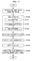
- FIG. 1 is a flowchart illustrating a tongue diagnosis region extraction method using a graph based approach and geometrical properties according to a first embodiment of the present invention.
- step S110 a face image having a tongue region is obtained from a camera, which is focused on the center of the tongue (tongue body) region.
- a digital tongue diagnosis system including the camera may be used in order to obtain a clearer face image.
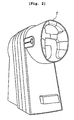
- FIG. 2 is a diagram illustrating an example of a digital tongue diagnosis system.
- the digital tongue diagnosis system 1 may obtain a tongue image using a standardized light source and a digital camera, and obtain a clearer image through color calibration.
- This system is designed in consideration of human engineering so as to form a dark room when user's eyes are fixed on an ocular part.
- the system uses a strobe light having the color temperature (5500K) property similar to the sunlight in order to standardize the light source.
- a tongue region i.e., a face image including the tongue body region is collected using the digital tongue diagnosis system 1 as configured above.
- the image of digital tongue diagnosis system 1 may be observed through GUI in real-time.
- the face image may be obtained by a CCD camera or a method of scanning a picture taken by a general camera.
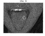
- FIG. 3 is a diagram illustrating a face image obtained by a digital tongue diagnosis system.
- a predetermined tongue part may be located at the center point of the image indicated by the cross as described in FIG. 3 .
- the obtained tongue image may be an RGB 24 bit BMP image with 1280 ⁇ 960 resolution without any limitation thereto. That is, the tongue image may include various formats or sizes.
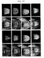
- the tongue body image is classified into five types of a non-coating image, a white coating image, a yellow coating image, a mixed coating image mixed with the white coating and the yellow coating, and a non-classified image, which is with dimly white coating.
- step S120 before performing the region segmentation, in step S120, a pre-processing correction is performed on the face image.
- a 533 ⁇ 400 image is generated through down-sampling because a big size image may require much processing time.
- the down-sampled image size is 533 ⁇ 400 for convenience, the size may be reduced to various sizes without any limitation thereto.
- step S122 histogram equalization is performed using brightness in order to normalize the brightness distribution, so that the image may have the uniform brightness property.
- the histogram equalization refers not to adding data to improve the original image, but to spreading out the histogram by analyzing the form of the histogram so as to distribute the brightness over a wider region without bias to a specific region.
- step S123 edge enhancement is performed as a final step of the pre-processing in order to further emphasize the edge surface of the tongue.
- the edge enhancement refers to clearing color discrimination in the edge surface in order to emphasize the edge of the tongue at the obtained face image.
- step S130 the greatest main segmentation region is detected on the basis of the center of the image after the corrected image is over-segmented.
- FIG. 4 is a diagram illustrating a face over-segmented according to an embodiment of the present invention. As described in FIG. 4 , the image object is segmented according to the property of the surface.
- the over-segmentation is performed to detect the main segmentation region.
- the over-segmentation method uses the graph based segmentation, which is based on selecting edges by each pixel compared to related art clustering methods.
- Each E has a weight, which means dissimilarity between two pixels connected through the edge. For example, the dissimilarity includes differences of color, intensity, motion and location. Similar parts are progressively merged by comparing the dissimilarity. Given a predetermined threshold, the edge surface is generated at a place where the difference of G is above the threshold to segment the region.
- the segmentation region occupying main area is found by measuring distribution within a predetermined range from the center point.
- a region having the greatest area within a predetermined range R1 from the center point of the tongue, e.g., the broadest color (purple) part is set as the main segmentation region.
- the different color region is also merged into the main segmentation region.
- an outer edge part of the region may not be segmented into the tongue region because the outer edge part of the tongue undergoes a color change due to a three-dimensional characteristic of the tongue.
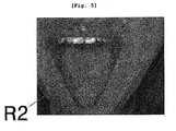
- FIG. 5 is a diagram illustrating shading around the outer edge of a tongue.
- shading is generated in the edge surface of the outer part of the tongue, which is caused by the three-dimensional characteristic of the tongue and an illumination.
- the sides R2 of the tongue which are an edge surface, have a local minimum value because the sides R2 are vertical to the illumination.
- a location of this local minimum value is extracted as the edge surface of the tongue body.
- the edge surface is detected using the color value of the dark part of the edge surface.
- the edge surface is extracted by selecting pixels having the smallest R or G value of the RGB color value.
- the reason why the R or G value is used is that the tongue is generally red or the major factor of shading is green. Accordingly, it is more efficient to use the R value than other color values.
- step 150 a part having the greatest color difference from the adjacent pixels within the outer predetermined range of the main segmentation region is detected to extract the edge of the tongue diagnosis region.
- FIG. 6 is a diagram of a form of a flat tongue.
- shading may not generate in the edge surface on the flat tongue lacking in bulky feeling or the lower part of the tongue.
- the shading may not be shown due to the rolling-up or the curling-up of the tongue.
- the color edge detection is performed to find a part where the greatest color difference occurs within the outer predetermined range of the main segmentation region. The part, where the difference of R value between the skin and the tongue is greatest, is determined as the edge.
- this procedure prepares for almost no shading by detecting a part where the R value differences between the adjacent pixels are greatest within the predetermined range value.
- R value can discriminate edges because the red color is mostly used in the skin and the tongue body of the human.
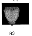
- FIG. 7 is a diagram illustrating a result of detecting color edge in a horizontal direction.
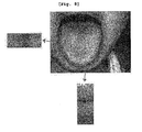
- FIG. 8 is a diagram illustrating a horizontal edge and a vertical edge of tongue edge.
- FIG. 9 is a diagram illustrating a result of detecting an edge in a vertical direction after detecting an edge in a horizontal direction.
- a location where the difference of the R value is horizontally greatest within a predetermined range is set to a side edge surface.
- the edge surface may not be detected at the lower part (R3) of the tongue.
- a location where the difference of the R value is vertically greatest within a predetermined range is set to a side edge surface.
- the edge surface may be detected at the lower part (R3) of the tongue.
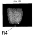
- FIG. 10 is a diagram illustrating a discontinuity surface in an edge after detecting a color edge.
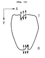
- FIG. 11 is a diagram illustrating geometric structure information according to an outline of a tongue body.
- FIG. 12 is a diagram of a finally detected tongue diagnosis region.
- step S160 after detecting the edge using the color value as described above, the detected edges are softly connected to each other using geometric structure information of the tongue.
- the edge surface of the tongue may not be softly connected to each other due to the shadow or the roughness of the skin.
- discontinuous surface R4 may be generated after detection of the color edge. Accordingly, symmetrical information is used to softly connect these rough edge surfaces.
- a continuous line is obtained by using a slope of the connection line.
- This method uses the regularity of the symmetrical information of the tongue structure by removing or filling pixels beyond the regularity.
- the tongue structure is an oval structure or a partly omitted oval as described in FIG. 11 .
- a sign is obtained by acquiring the slope and the curvature of X direction with respect to Y direction around the detected edge. Pixels of a part departing from the sign are removed or filled in accordance with predicted locations by using the entire slope and curvature. An example of the slope and the curvature according to each quadrant are described in FIG. 11 .
- the edge of the tongue is corrected by using the symmetrical information of this tongue.
- FIG. 13 is a flowchart illustrating a tongue diagnosis region extraction method using a graph based approach and geometrical properties according to a second embodiment of the present invention.
- a tongue diagnosis region extraction method includes image acquisition S210, image correction S220, and main segmentation region detection S230 as described in the first embodiment.
- step S250 the second embodiment of the present invention corrects the discontinuous edge to be continuous in the process of detecting the edge by using the local minimum value rather than extracts the edge on the basis of the color after detecting the main segmentation region.
- the edge when the edge is detected by using the local minimum as described in FIGS. 14 and 15 , the edge may be suddenly recessed or protruded.
- the edge of the tongue may be unnatural, which is necessary to correct.
- FIG. 14 is a diagram illustrating a method of correcting a discontinuous edge within a predetermined range of the left and right sides of a tongue diagnosis region according to the second embodiment of the present invention.
- FIG. 15 is a diagram illustrating a method of correcting a discontinuous edge within a predetermined range of the upper and lower sides of a tongue diagnosis region according to the second embodiment of the present invention.
- a looped curve is an edge of the tongue diagnosis region by using the local minimum value.
- the local minimum value is detected at the left side L and the right side R of the main segmentation region.
- the local minimum value is detected in the X-axis direction by increasing the value of the Y coordinate from the upper part to the lower part of the main segmentation region like arrows as shown in FIGS. 14 and 15 .
- the standard value may be set to three pixels.
- the coordinate of the next pixel to progress by one pixel in the detection direction is set to the edge of the tongue region.
- the coordinates X of the edges of the previous point of time and the present point of time are set to the same, thereby removing the discontinuous part that changes abruptly.
- the correction of the discontinuous edge surface is continuously performed until the corrected edge surface is within a predetermined range from the edge surface obtained by the local minimum value.
- the local minimum value is found at upper part T and the lower part B of the main segmentation region.
- the local minimum value is detected in the Y-axis direction by increasing the value of the Y coordinate from the left to the right side of the main segmentation region like arrows as shown in FIGS. 14 and 15 .
- the standard value may be set to three pixels.
- the coordinate of the next pixel to progress by one pixel in the detection direction is set to the edge of the tongue region.
- the coordinates Y of the edges of the previous point of time and the present point of time are set to the same, thereby removing the discontinuous part that changes abruptly.
- the correction of the discontinuous edge surface is continuously performed until the corrected edge surface is within a predetermined range from the edge surface obtained by the local minimum value.
- FIG. 16 is a flowchart illustrating a tongue region extraction method using a graph based approach and geometrical properties according to a third embodiment of the present invention.
- a tongue region extraction method according to the third embodiment of the present invention includes an image acquisition S310, an image correction 320, and main segmentation region detection S330 similarly to the second embodiment.
- step S351 a discontinuous part in the process is corrected to be continuous by using the local minimum value similarly to the second embodiment.
- step S353 a curve corresponding to the corrected edge is obtained.
- step 352 an edge is extracted on the basis of color after detection of the main segmentation region.
- step 354 a curve corresponding to the edge is obtained. That is, the extracted edge is changed into the form of curve through a curve fitting process.
- step S360 the edge and curve having a smaller difference are selected by comparing a difference between the edge of the tongue extracted in the S351 and the curve obtained in the step S353 and a difference of the edge of the tongue extracted in the S352 and the curve obtained in the S354.
- step S370 the edges are softly connected to each other by using geometrical structure information of the tongue.
- FIG. 17 is a diagram illustrating various forms of tongue region.
- the tongue may have various shapes such as an oval, reverse triangle, a half oval, etc. various form
- the graph based approach and the tongue region extraction method using the geometric characteristic according to the present invention can extract almost perfectly only the tongue region.
- three parameters are used for a graph based region segmentation for the over-segmentation.
- Three parameters include a sigma, a constant k, and a minimum factor size min.
- the sigma has a value range of 0.9 to 2.5.
- the constant k is 120.
- the minimum factor size min is set to 100.
- a region size set to search for the main segmentation region is 50 ⁇ 50 pixels on the basis of the center point.
- the retrieval range for the local minimum detection is -40 to 40 pixels from the edge surface of the main segmentation region.
- the retrieval range for color edge detection is from -20 to 20 pixels from the edge surface, where the maximum color value (R value) is retrieved.
- the slope and the curvature are stored at previous four locations and a present location. By comparing the slopes and the curvatures of the previous and present locations, it is determined whether the slope and the curvature of the present location are continuous. If continuous, the slope and the curvature of the present location are given. If discontinuous, the slope and the curvature of the previous location are given.
- tongue region and all types of the tongue body are well segmented for the diagnosis. It can be understood that the tongue regions are well segmented irrespective of the skin color or condition and gender.
- the present invention can be effectively used in the medical treatment equipment and information gathering devices for diagnosing abnormal healthy condition by automatically extracting a tongue part from a face image. Also, it can be applied to the fields of home healthcare and remote medical treatment devices.
Landscapes
- Engineering & Computer Science (AREA)
- Physics & Mathematics (AREA)
- General Physics & Mathematics (AREA)
- Theoretical Computer Science (AREA)
- Computer Vision & Pattern Recognition (AREA)
- Multimedia (AREA)
- Human Computer Interaction (AREA)
- Image Analysis (AREA)
- Measurement Of The Respiration, Hearing Ability, Form, And Blood Characteristics Of Living Organisms (AREA)
- Measuring And Recording Apparatus For Diagnosis (AREA)
- Image Processing (AREA)
Claims (15)
- Zungenregion-Extraktionsverfahren unter Verwendung eines graphenbasierten Ansatzes und geometrischer Eigenschaften, wobei das Verfahren folgende Schritte umfasst:1) Erhalten eines Gesichtsbilds mit einer von einer Kamera fokussierten Zungenregion;2) Korrigieren einer Größe und einer Qualität des Bilds;3) Ermitteln einer Hauptsegmentierungsregion mit einer größten Fläche in einem vorgegebenen Bereich von einem Mittelpunkt des Bilds aus nach einer graphenbasierten Übersegmentierung des Bilds;4) Extrahieren einer Kante einer Zungenregion durch Erkennen eines Teils mit einem lokalen Mindest-Schattierungswert in einem vorgegebenen äußeren Bereich von einer Kante der Hauptsegmentierungsregion;5) Extrahieren einer Kante der Zungenregion durch Erkennen eines Teils mit einer größten Farbdifferenz zwischen benachbarten Pixeln in einem vorgegebenen äußeren Bereich von der Kante der Hauptsegmentierungsregion; und6) glattes Verbinden der in den Schritten 4) und 5) erkannten Kanten unter Verwendung einer geometrischen Strukturinformation einer Zunge.
- Verfahren nach Anspruch 1,
wobei der Schritt 2) Folgendes umfasst:Herunterrechnen des erhaltenen Bilds, um eine Berechnungszeit zu verringern,Durchführen eines Histogrammausgleichs in Bezug auf das erhaltene Bild, um eine Helligkeitsverteilung auszugleichen, undVerbessern der Kante des erhaltenen Bilds, um eine Farbe an der Kantenfläche zu unterscheiden. - Verfahren nach Anspruch 1, wobei der Schritt 3) das Verschmelzen benachbarter Regionen zu der Hauptsegmentierungsregion durch folgende Gleichung umfasst:

wobei PA eine Fläche einer A-Region ist, PB eine Fläche einer B-Region ist, die A-Region die Hauptsegmentierungsregion ist und x∈B ein Pixel der B-Region ist. - Verfahren nach Anspruch 1, wobei der Schritt 3) das Verschmelzen aller Regionen in der Hauptsegmentierungsregion zu der Hauptsegmentierungsregion umfasst.
- Verfahren nach Anspruch 1, wobei der Schritt 3) das Verschmelzen restlicher Regionen zu der Hauptsegmentierungsregion mit einer größten Fläche umfasst, wenn mehr als drei Regionen auch in dem vorgegebenen Bereich vorhanden sind.
- Verfahren nach Anspruch 1, wobei der Schritt 4) das Erkennen eines Teils mit einem kleinsten R- oder G-Wert unter RGB-Werten umfasst.
- Verfahren nach Anspruch 1, wobei der Schritt 5) Folgendes umfasst:horizontales Erkennen eines Teils mit der größten R-Wert-Differenz von RGB-Werten zwischen benachbarten Pixeln in einem vorgegebenen äußeren Bereich von der Kante der Hauptsegmentierungsregion; undvertikales Erkennen eines Teils mit der größten R-Wert-Differenz von RGB-Werten zwischen benachbarten Pixeln in einem vorgegebenen äußeren Bereich von der Kante der Hauptsegmentierungsregion.
- Verfahren nach Anspruch 1, wobei der Schritt 6) das Verlängern einer Verbindungslinie durch Verwendung einer Steigung der Verbindungslinie umfasst, wenn eine Diskontinuität der die Kanten der Zunge verbindenden Verbindungslinie über einem vorgegebenen Schwellwert liegt.
- Verfahren nach Anspruch 1, wobei der Schritt 6) das Korrigieren der Kante durch Definieren einer geometrischen Struktur der Zunge zu einer ovalen Form sowie das Vergleichen von Zeichen einer Steigung und einer Krümmung gemäß der ovalen Form und von Zeichen einer Steigung und einer Krümmung, die aus der erkannten Kante an jedem Quadranten extrahiert wurden, umfasst.
- Zungenregion-Extraktionsverfahren unter Verwendung eines graphenbasierten Ansatzes und geometrischer Eigenschaften, wobei das Verfahren folgende Schritte umfasst:1) Erhalten eines Gesichtsbilds mit einer von einer Kamera fokussierten Zungenregion;2) Korrigieren einer Größe und einer Qualität des Bilds;3) Ermitteln einer Hauptsegmentierungsregion mit einer größten Fläche in einem vorgegebenen Bereich von einem Mittelpunkt des Bilds aus nach einer graphenbasierten Übersegmentierung des Bilds;4) Extrahieren einer Kante einer Zungendiagnoseregion durch Erkennen eines Teils mit einem lokalen Mindest-Schattierungswert in einem vorgegebenen äußeren Bereich von der Hauptsegmentierungsregion;5) Korrigieren von diskontinuierlichen Kantenflächen an einer aktuellen Koordinate und einer vorherigen Koordinate derart, dass sie kontinuierlich sind, wenn eine Differenz zwischen der aktuellen Koordinate und der vorherigen Koordinate des Teils mit dem lokalen Mindestwert in einem vorgegebenen äußeren Bereich von der Hauptsegmentierungsregion größer als ein vorgegebener Standardwert ist; und6) glattes Verbinden der in den Schritten 4) und 5) erkannten Kanten unter Verwendung einer geometrischen Strukturinformation einer Zunge.
- Zungenregion-Extraktionsverfahren unter Verwendung eines graphenbasierten Ansatzes und geometrischer Eigenschaften, wobei das Verfahren folgende Schritte umfasst:1) Erhalten eines Gesichtsbilds mit einer von einer Kamera fokussierten Zungenregion;2) Korrigieren einer Größe und einer Qualität des Bilds;3) Ermitteln einer Hauptsegmentierungsregion mit der größten Fläche in einem vorgegebenen Bereich von einem Mittelpunkt des Bilds aus nach einer graphenbasierten Bild-Übersegmentierung des Bilds;4) Extrahieren einer Kante einer Zungenregion durch Erkennen eines Teils mit einem lokalen Mindest-Schattierungswert in einem vorgegebenen äußeren Bereich von der Hauptsegmentierungsregion;5) Korrigieren, nach dem Schritt 4), von diskontinuierlichen Kanten an einer aktuellen Koordinate und einer vorherigen Koordinate derart, dass sie kontinuierlich sind, wenn eine Differenz zwischen der aktuellen Koordinate und der vorherigen Koordinate des Teils mit dem lokalen Mindestwert in einem vorgegebenen äußeren Bereich von der Hauptsegmentierungsregion größer als ein vorgegebener Standardwert ist;6) Erhalten einer Kurve, die der in dem Schritt 5) erhaltenen Kante entspricht;7) Extrahieren, nach dem Schritt 4), einer Kante einer Zungenregion durch Erkennen eines Teils mit einer größten Farbdifferenz zwischen benachbarten Pixeln in einem vorgegebenen äußeren Bereich von der Hauptsegmentierungsregion;8) Erhalten einer Kurve, die der in dem Schritt 7) erhaltenen Kante entspricht;9) Auswählen der Kante und der Kurve mit einer kleineren Differenz durch Vergleichen einer Differenz zwischen der in dem Schritt 5) korrigierten Kante der Zunge und der in dem Schritt 6) erhaltenen Kurve und einer Differenz zwischen der in dem Schritt 7) extrahierten Zungenregion und der in dem Schritt 8) erhaltenen Kurve; und10) glattes Verbinden der in dem Schritt 9) ausgewählten Kanten unter Verwendung einer geometrischen Strukturinformation einer Zunge.
- Verfahren nach Anspruch 10 oder 11, wobei, wenn der Teil mit dem lokalen Mindestwert entlang einer x-Achse durch Erhöhen einer y-Koordinate von einem linken oberen Ende oder einem rechten oberen Ende zu einem unteren Ende der Hauptsegmentierungsregion erkannt wird, der Schritt 5) das Einstellen einer x-Koordinate eines Pixels mit dem lokalen Mindestwert eines aktuellen Zeitpunkts derart umfasst, dass sie mit einer x-Koordinate eines vorherigen Zeitpunkts identisch ist, wenn eine Differenz zwischen einem Wert der x-Koordinate des vorherigen Zeitpunkts und einem Wert der x-Koordinate des aktuellen Zeitpunkts größer als ein vorgegebener Standardwert ist.
- Verfahren nach Anspruch 10 oder 11, wobei, wenn der Teil mit dem lokalen Mindestwert entlang einer y-Achse durch Erhöhen einer x-Koordinate von einem linken oberen Ende oder einem linken unteren Ende zu einem rechten Ende der Hauptsegmentierungsregion erkannt wird, der Schritt 5) das Einstellen einer y-Koordinate eines Pixels mit dem lokalen Mindestwert eines aktuellen Zeitpunkts derart umfasst, dass sie mit einer y-Koordinate eines vorherigen Zeitpunkts identisch ist, wenn eine Differenz zwischen einem Wert der y-Koordinate des vorherigen Zeitpunkts und einem Wert der y-Koordinate des aktuellen Zeitpunkts größer als ein vorgegebener Standardwert ist.
- Verfahren nach Anspruch 10, wobei das Korrigieren der diskontinuierlichen Kantenflächen in dem Schritt 5) durchgeführt wird, bis die korrigierte Kantenfläche in einem vorgegebenen Bereich von einer in dem Schritt 4) erhaltenen Mindest-Kantenfläche liegt.
- Verfahren nach Anspruch 11, wobei der Schritt 6) Folgendes umfasst:horizontales Erkennen eines Teils mit der größten R-Wert-Differenz von RGB-Werten zwischen benachbarten Pixeln in einem vorgegebenen äußeren Bereich von der Kante der Hauptsegmentierungsregion; undvertikales Erkennen eines Teils mit der größten R-Wert-Dififerenz von RGB-Werten zwischen benachbarten Pixeln in einem vorgegebenen äußeren Bereich von der Kante der Hauptsegmentierungsregion.
Applications Claiming Priority (2)
| Application Number | Priority Date | Filing Date | Title |
|---|---|---|---|
| KR1020070096688A KR100889014B1 (ko) | 2007-09-21 | 2007-09-21 | 그래프 기반 접근 방법을 이용한 설진 영역 추출 방법 |
| PCT/KR2008/005543 WO2009038376A1 (en) | 2007-09-21 | 2008-09-19 | Extraction method of tongue region using graph-based approach and geometric properties |
Publications (3)
| Publication Number | Publication Date |
|---|---|
| EP2188779A1 EP2188779A1 (de) | 2010-05-26 |
| EP2188779A4 EP2188779A4 (de) | 2013-03-20 |
| EP2188779B1 true EP2188779B1 (de) | 2014-04-23 |
Family
ID=40468094
Family Applications (1)
| Application Number | Title | Priority Date | Filing Date |
|---|---|---|---|
| EP08831511.4A Not-in-force EP2188779B1 (de) | 2007-09-21 | 2008-09-19 | Extraktionsverfahren der zungenregion mithilfe graphenbasiertem ansatz und geometrischen eigenschaften |
Country Status (3)
| Country | Link |
|---|---|
| EP (1) | EP2188779B1 (de) |
| KR (1) | KR100889014B1 (de) |
| WO (1) | WO2009038376A1 (de) |
Cited By (3)
| Publication number | Priority date | Publication date | Assignee | Title |
|---|---|---|---|---|
| CN105930798A (zh) * | 2016-04-21 | 2016-09-07 | 厦门快商通科技股份有限公司 | 基于学习的面向手机应用的舌像快速检测分割方法 |
| CN107610087A (zh) * | 2017-05-15 | 2018-01-19 | 华南理工大学 | 一种基于深度学习的舌苔自动分割方法 |
| CN109829373A (zh) * | 2018-12-26 | 2019-05-31 | 北京康加科技有限公司 | 舌态检测系统及方法 |
Families Citing this family (25)
| Publication number | Priority date | Publication date | Assignee | Title |
|---|---|---|---|---|
| KR101046670B1 (ko) * | 2009-03-17 | 2011-07-05 | 대승의료기기(주) | 설진기 |
| KR101195917B1 (ko) | 2010-06-15 | 2012-10-30 | 한국 한의학 연구원 | 혀 진단 영역 추출 방법 |
| CN102509312B (zh) * | 2011-09-20 | 2013-10-02 | 哈尔滨工业大学 | 人体数字舌图像颜色色域空间及其提取方法 |
| CN102509279B (zh) * | 2011-11-02 | 2013-11-06 | 北京工业大学 | 根部欠照明舌图像的自适应暗部细节复现方法 |
| WO2014183246A1 (zh) * | 2013-05-13 | 2014-11-20 | Huang Bo | 一种医学影像处理方法与系统 |
| CN104021552B (zh) * | 2014-05-28 | 2017-02-22 | 华南理工大学 | 一种基于图论分割过程的多目标粒子群参数优化方法 |
| KR101726505B1 (ko) * | 2015-08-11 | 2017-04-12 | 한국 한의학 연구원 | 설 촬영 장치 및 설 영상의 프로세싱 방법 |
| KR101908785B1 (ko) * | 2016-10-31 | 2018-10-18 | 한국 한의학 연구원 | 혀 영역 추출 방법 및 그 방법을 수행하는 영상 처리 장치 |
| CN106821324A (zh) * | 2017-03-06 | 2017-06-13 | 武汉嫦娥医学抗衰机器人股份有限公司 | 一种基于舌面和舌下综合分析的舌诊辅助医疗系统 |
| CN107316307B (zh) * | 2017-06-27 | 2020-05-08 | 北京工业大学 | 一种基于深度卷积神经网络的中医舌图像自动分割方法 |
| CN107977671B (zh) * | 2017-10-27 | 2021-10-26 | 浙江工业大学 | 一种基于多任务卷积神经网络的舌象分类方法 |
| CN108022246A (zh) * | 2017-12-21 | 2018-05-11 | 芜湖圣美孚科技有限公司 | 一种用于舌面仪的前置图像自动分割系统 |
| CN108742536A (zh) * | 2018-06-13 | 2018-11-06 | 天津大学 | 一种基于动态减影技术的舌像自动抓拍方法 |
| CN109005338A (zh) * | 2018-07-10 | 2018-12-14 | 芜湖圣美孚科技有限公司 | 一种舌像识别处理系统及其识别方法 |
| CN109410168B (zh) * | 2018-08-31 | 2021-11-16 | 清华大学 | 用于确定图像中的子图块类别的卷积神经网络的建模方法 |
| CN109522791A (zh) * | 2018-10-09 | 2019-03-26 | 广东数相智能科技有限公司 | 一种用于舌诊拍照的提醒方法、电子设备及存储介质 |
| CN111223117A (zh) * | 2018-11-26 | 2020-06-02 | 深圳市前海安测信息技术有限公司 | 舌面图像分割装置、方法及计算机存储介质 |
| CN110929740A (zh) * | 2019-11-21 | 2020-03-27 | 中电健康云科技有限公司 | 一种基于lgbm模型的舌质舌苔分离方法 |
| CN111260619A (zh) * | 2020-01-14 | 2020-06-09 | 浙江中医药大学 | 基于U-net模型的舌体自动分割方法 |
| CN112489053B (zh) * | 2020-11-26 | 2021-07-13 | 深圳市艾合芯科智慧医疗有限公司 | 一种舌像分割方法、装置及存储介质 |
| CN114862851B (zh) * | 2022-07-06 | 2022-09-30 | 深圳市圆道妙医科技有限公司 | 一种基于舌相分析的处理方法 |
| CN115311312B (zh) * | 2022-10-12 | 2023-03-24 | 南通鼎顺生物科技有限公司 | 一种用于中医舌诊的舌体精准分割方法 |
| CN115601358B (zh) * | 2022-12-01 | 2023-03-28 | 合肥云诊信息科技有限公司 | 一种自然光环境下的舌象图像分割方法 |
| CN117422720B (zh) * | 2023-12-19 | 2024-03-05 | 陕西秒康医疗科技有限公司 | 一种中医治疗舌诊图像智能分割方法 |
| CN117522865B (zh) * | 2024-01-03 | 2024-03-22 | 长春中医药大学 | 一种基于图像识别技术的中医健康监测系统 |
Family Cites Families (4)
| Publication number | Priority date | Publication date | Assignee | Title |
|---|---|---|---|---|
| KR20040093882A (ko) * | 2003-04-30 | 2004-11-09 | 노정희 | 의료용 디지털 영상에 대한 출력 및 저장을 용이하게실시하기 위한 시스템 |
| JP4311163B2 (ja) * | 2003-10-17 | 2009-08-12 | 株式会社島津製作所 | 医用画像診断装置 |
| JP4617116B2 (ja) | 2004-08-23 | 2011-01-19 | 商之器科技股▼ふん▲有限公司 | 即時医療映像自動的サーチ対照方法及びそのシステム |
| JP4649965B2 (ja) * | 2004-11-29 | 2011-03-16 | コニカミノルタホールディングス株式会社 | 健康度判定装置、及びプログラム |
-
2007
- 2007-09-21 KR KR1020070096688A patent/KR100889014B1/ko not_active Expired - Fee Related
-
2008
- 2008-09-19 EP EP08831511.4A patent/EP2188779B1/de not_active Not-in-force
- 2008-09-19 WO PCT/KR2008/005543 patent/WO2009038376A1/en not_active Ceased
Cited By (5)
| Publication number | Priority date | Publication date | Assignee | Title |
|---|---|---|---|---|
| CN105930798A (zh) * | 2016-04-21 | 2016-09-07 | 厦门快商通科技股份有限公司 | 基于学习的面向手机应用的舌像快速检测分割方法 |
| CN105930798B (zh) * | 2016-04-21 | 2019-05-03 | 厦门快商通科技股份有限公司 | 基于学习的面向手机应用的舌像快速检测分割方法 |
| CN107610087A (zh) * | 2017-05-15 | 2018-01-19 | 华南理工大学 | 一种基于深度学习的舌苔自动分割方法 |
| CN107610087B (zh) * | 2017-05-15 | 2020-04-28 | 华南理工大学 | 一种基于深度学习的舌苔自动分割方法 |
| CN109829373A (zh) * | 2018-12-26 | 2019-05-31 | 北京康加科技有限公司 | 舌态检测系统及方法 |
Also Published As
| Publication number | Publication date |
|---|---|
| KR100889014B1 (ko) | 2009-03-17 |
| EP2188779A4 (de) | 2013-03-20 |
| WO2009038376A1 (en) | 2009-03-26 |
| EP2188779A1 (de) | 2010-05-26 |
Similar Documents
| Publication | Publication Date | Title |
|---|---|---|
| EP2188779B1 (de) | Extraktionsverfahren der zungenregion mithilfe graphenbasiertem ansatz und geometrischen eigenschaften | |
| Dimauro et al. | Anaemia detection based on sclera and blood vessel colour estimation | |
| CN107451998B (zh) | 一种眼底图像质量控制方法 | |
| CN110930446B (zh) | 一种眼底图像定量分析的前置处理方法及存储设备 | |
| Sangeethaa | Presumptive discerning of the severity level of glaucoma through clinical fundus images using hybrid PolyNet | |
| Odstrcilik et al. | Thickness related textural properties of retinal nerve fiber layer in color fundus images | |
| Sánchez et al. | Mixture model-based clustering and logistic regression for automatic detection of microaneurysms in retinal images | |
| Ramakanth et al. | Approximate nearest neighbour field based optic disk detection | |
| WO2013187206A1 (ja) | 画像処理装置、画像処理方法、及び画像処理プログラム | |
| WO2012157835A1 (ko) | 영상의 융합 기법을 이용한 의료용 혈관영상 처리방법 | |
| Xiao et al. | Retinal hemorrhage detection by rule-based and machine learning approach | |
| Jaafar et al. | Detection of exudates in retinal images using a pure splitting technique | |
| KR102380560B1 (ko) | 영상 처리를 기반으로 하는 각막궤양 검출 장치 및 그 방법 | |
| CN114332132A (zh) | 图像分割方法、装置和计算机设备 | |
| Hatanaka et al. | Improvement of automatic hemorrhage detection methods using brightness correction on fundus images | |
| Niemeijer et al. | Automated localization of the optic disc and the fovea | |
| Lermé et al. | A fully automatic method for segmenting retinal artery walls in adaptive optics images | |
| Kumar et al. | Automatic optic disc segmentation using maximum intensity variation | |
| CN110458042B (zh) | 一种荧光ctc中的探针数目检测方法 | |
| CN108629780B (zh) | 基于颜色分解和阈值技术的舌图像分割方法 | |
| WO2024240089A1 (zh) | 内窥镜图像显示方法、装置、终端设备以及存储介质 | |
| CN111291706B (zh) | 一种视网膜图像视盘定位方法 | |
| CN120726059B (zh) | 一种基于图像处理的中医舌像识别方法及系统 | |
| Martins | Automatic microaneurysm detection and characterization through digital color fundus images | |
| CN119693280B (zh) | 一种神经内镜的图像去雾方法、装置及系统 |
Legal Events
| Date | Code | Title | Description |
|---|---|---|---|
| PUAI | Public reference made under article 153(3) epc to a published international application that has entered the european phase |
Free format text: ORIGINAL CODE: 0009012 |
|
| 17P | Request for examination filed |
Effective date: 20100219 |
|
| AK | Designated contracting states |
Kind code of ref document: A1 Designated state(s): AT BE BG CH CY CZ DE DK EE ES FI FR GB GR HR HU IE IS IT LI LT LU LV MC MT NL NO PL PT RO SE SI SK TR |
|
| AX | Request for extension of the european patent |
Extension state: AL BA MK RS |
|
| RIN1 | Information on inventor provided before grant (corrected) |
Inventor name: DO, JUN-HYEONG Inventor name: RYU, HYUNHEE Inventor name: KIM, KEUN HO Inventor name: KIM, JONG YEOL |
|
| DAX | Request for extension of the european patent (deleted) | ||
| RIC1 | Information provided on ipc code assigned before grant |
Ipc: G06T 7/00 20060101AFI20130123BHEP |
|
| A4 | Supplementary search report drawn up and despatched |
Effective date: 20130215 |
|
| REG | Reference to a national code |
Ref country code: DE Ref legal event code: R079 Ref document number: 602008031760 Country of ref document: DE Free format text: PREVIOUS MAIN CLASS: G06T0007400000 Ipc: G06T0007000000 |
|
| GRAP | Despatch of communication of intention to grant a patent |
Free format text: ORIGINAL CODE: EPIDOSNIGR1 |
|
| RIC1 | Information provided on ipc code assigned before grant |
Ipc: G06T 7/00 20060101AFI20131021BHEP |
|
| INTG | Intention to grant announced |
Effective date: 20131114 |
|
| GRAS | Grant fee paid |
Free format text: ORIGINAL CODE: EPIDOSNIGR3 |
|
| GRAA | (expected) grant |
Free format text: ORIGINAL CODE: 0009210 |
|
| AK | Designated contracting states |
Kind code of ref document: B1 Designated state(s): AT BE BG CH CY CZ DE DK EE ES FI FR GB GR HR HU IE IS IT LI LT LU LV MC MT NL NO PL PT RO SE SI SK TR |
|
| REG | Reference to a national code |
Ref country code: GB Ref legal event code: FG4D |
|
| REG | Reference to a national code |
Ref country code: CH Ref legal event code: EP |
|
| REG | Reference to a national code |
Ref country code: AT Ref legal event code: REF Ref document number: 664228 Country of ref document: AT Kind code of ref document: T Effective date: 20140515 |
|
| REG | Reference to a national code |
Ref country code: IE Ref legal event code: FG4D |
|
| REG | Reference to a national code |
Ref country code: DE Ref legal event code: R096 Ref document number: 602008031760 Country of ref document: DE Effective date: 20140605 |
|
| REG | Reference to a national code |
Ref country code: AT Ref legal event code: MK05 Ref document number: 664228 Country of ref document: AT Kind code of ref document: T Effective date: 20140423 |
|
| REG | Reference to a national code |
Ref country code: NL Ref legal event code: VDEP Effective date: 20140423 |
|
| REG | Reference to a national code |
Ref country code: LT Ref legal event code: MG4D |
|
| PG25 | Lapsed in a contracting state [announced via postgrant information from national office to epo] |
Ref country code: GR Free format text: LAPSE BECAUSE OF FAILURE TO SUBMIT A TRANSLATION OF THE DESCRIPTION OR TO PAY THE FEE WITHIN THE PRESCRIBED TIME-LIMIT Effective date: 20140724 Ref country code: FI Free format text: LAPSE BECAUSE OF FAILURE TO SUBMIT A TRANSLATION OF THE DESCRIPTION OR TO PAY THE FEE WITHIN THE PRESCRIBED TIME-LIMIT Effective date: 20140423 Ref country code: NO Free format text: LAPSE BECAUSE OF FAILURE TO SUBMIT A TRANSLATION OF THE DESCRIPTION OR TO PAY THE FEE WITHIN THE PRESCRIBED TIME-LIMIT Effective date: 20140723 Ref country code: IS Free format text: LAPSE BECAUSE OF FAILURE TO SUBMIT A TRANSLATION OF THE DESCRIPTION OR TO PAY THE FEE WITHIN THE PRESCRIBED TIME-LIMIT Effective date: 20140823 Ref country code: NL Free format text: LAPSE BECAUSE OF FAILURE TO SUBMIT A TRANSLATION OF THE DESCRIPTION OR TO PAY THE FEE WITHIN THE PRESCRIBED TIME-LIMIT Effective date: 20140423 Ref country code: LT Free format text: LAPSE BECAUSE OF FAILURE TO SUBMIT A TRANSLATION OF THE DESCRIPTION OR TO PAY THE FEE WITHIN THE PRESCRIBED TIME-LIMIT Effective date: 20140423 Ref country code: BG Free format text: LAPSE BECAUSE OF FAILURE TO SUBMIT A TRANSLATION OF THE DESCRIPTION OR TO PAY THE FEE WITHIN THE PRESCRIBED TIME-LIMIT Effective date: 20140723 Ref country code: CY Free format text: LAPSE BECAUSE OF FAILURE TO SUBMIT A TRANSLATION OF THE DESCRIPTION OR TO PAY THE FEE WITHIN THE PRESCRIBED TIME-LIMIT Effective date: 20140423 |
|
| PG25 | Lapsed in a contracting state [announced via postgrant information from national office to epo] |
Ref country code: AT Free format text: LAPSE BECAUSE OF FAILURE TO SUBMIT A TRANSLATION OF THE DESCRIPTION OR TO PAY THE FEE WITHIN THE PRESCRIBED TIME-LIMIT Effective date: 20140423 Ref country code: HR Free format text: LAPSE BECAUSE OF FAILURE TO SUBMIT A TRANSLATION OF THE DESCRIPTION OR TO PAY THE FEE WITHIN THE PRESCRIBED TIME-LIMIT Effective date: 20140423 Ref country code: LV Free format text: LAPSE BECAUSE OF FAILURE TO SUBMIT A TRANSLATION OF THE DESCRIPTION OR TO PAY THE FEE WITHIN THE PRESCRIBED TIME-LIMIT Effective date: 20140423 Ref country code: SE Free format text: LAPSE BECAUSE OF FAILURE TO SUBMIT A TRANSLATION OF THE DESCRIPTION OR TO PAY THE FEE WITHIN THE PRESCRIBED TIME-LIMIT Effective date: 20140423 Ref country code: PL Free format text: LAPSE BECAUSE OF FAILURE TO SUBMIT A TRANSLATION OF THE DESCRIPTION OR TO PAY THE FEE WITHIN THE PRESCRIBED TIME-LIMIT Effective date: 20140423 Ref country code: ES Free format text: LAPSE BECAUSE OF FAILURE TO SUBMIT A TRANSLATION OF THE DESCRIPTION OR TO PAY THE FEE WITHIN THE PRESCRIBED TIME-LIMIT Effective date: 20140423 |
|
| PG25 | Lapsed in a contracting state [announced via postgrant information from national office to epo] |
Ref country code: PT Free format text: LAPSE BECAUSE OF FAILURE TO SUBMIT A TRANSLATION OF THE DESCRIPTION OR TO PAY THE FEE WITHIN THE PRESCRIBED TIME-LIMIT Effective date: 20140825 |
|
| REG | Reference to a national code |
Ref country code: DE Ref legal event code: R097 Ref document number: 602008031760 Country of ref document: DE |
|
| PG25 | Lapsed in a contracting state [announced via postgrant information from national office to epo] |
Ref country code: EE Free format text: LAPSE BECAUSE OF FAILURE TO SUBMIT A TRANSLATION OF THE DESCRIPTION OR TO PAY THE FEE WITHIN THE PRESCRIBED TIME-LIMIT Effective date: 20140423 Ref country code: CZ Free format text: LAPSE BECAUSE OF FAILURE TO SUBMIT A TRANSLATION OF THE DESCRIPTION OR TO PAY THE FEE WITHIN THE PRESCRIBED TIME-LIMIT Effective date: 20140423 Ref country code: DK Free format text: LAPSE BECAUSE OF FAILURE TO SUBMIT A TRANSLATION OF THE DESCRIPTION OR TO PAY THE FEE WITHIN THE PRESCRIBED TIME-LIMIT Effective date: 20140423 Ref country code: RO Free format text: LAPSE BECAUSE OF FAILURE TO SUBMIT A TRANSLATION OF THE DESCRIPTION OR TO PAY THE FEE WITHIN THE PRESCRIBED TIME-LIMIT Effective date: 20140423 Ref country code: SK Free format text: LAPSE BECAUSE OF FAILURE TO SUBMIT A TRANSLATION OF THE DESCRIPTION OR TO PAY THE FEE WITHIN THE PRESCRIBED TIME-LIMIT Effective date: 20140423 Ref country code: BE Free format text: LAPSE BECAUSE OF FAILURE TO SUBMIT A TRANSLATION OF THE DESCRIPTION OR TO PAY THE FEE WITHIN THE PRESCRIBED TIME-LIMIT Effective date: 20140423 |
|
| PLBE | No opposition filed within time limit |
Free format text: ORIGINAL CODE: 0009261 |
|
| STAA | Information on the status of an ep patent application or granted ep patent |
Free format text: STATUS: NO OPPOSITION FILED WITHIN TIME LIMIT |
|
| PG25 | Lapsed in a contracting state [announced via postgrant information from national office to epo] |
Ref country code: IT Free format text: LAPSE BECAUSE OF FAILURE TO SUBMIT A TRANSLATION OF THE DESCRIPTION OR TO PAY THE FEE WITHIN THE PRESCRIBED TIME-LIMIT Effective date: 20140423 |
|
| 26N | No opposition filed |
Effective date: 20150126 |
|
| PG25 | Lapsed in a contracting state [announced via postgrant information from national office to epo] |
Ref country code: LU Free format text: LAPSE BECAUSE OF FAILURE TO SUBMIT A TRANSLATION OF THE DESCRIPTION OR TO PAY THE FEE WITHIN THE PRESCRIBED TIME-LIMIT Effective date: 20140919 Ref country code: MC Free format text: LAPSE BECAUSE OF FAILURE TO SUBMIT A TRANSLATION OF THE DESCRIPTION OR TO PAY THE FEE WITHIN THE PRESCRIBED TIME-LIMIT Effective date: 20140423 |
|
| REG | Reference to a national code |
Ref country code: CH Ref legal event code: PL |
|
| REG | Reference to a national code |
Ref country code: DE Ref legal event code: R097 Ref document number: 602008031760 Country of ref document: DE Effective date: 20150126 |
|
| REG | Reference to a national code |
Ref country code: IE Ref legal event code: MM4A |
|
| PG25 | Lapsed in a contracting state [announced via postgrant information from national office to epo] |
Ref country code: SI Free format text: LAPSE BECAUSE OF FAILURE TO SUBMIT A TRANSLATION OF THE DESCRIPTION OR TO PAY THE FEE WITHIN THE PRESCRIBED TIME-LIMIT Effective date: 20140423 Ref country code: LI Free format text: LAPSE BECAUSE OF NON-PAYMENT OF DUE FEES Effective date: 20140930 Ref country code: CH Free format text: LAPSE BECAUSE OF NON-PAYMENT OF DUE FEES Effective date: 20140930 |
|
| PG25 | Lapsed in a contracting state [announced via postgrant information from national office to epo] |
Ref country code: IE Free format text: LAPSE BECAUSE OF NON-PAYMENT OF DUE FEES Effective date: 20140919 |
|
| PG25 | Lapsed in a contracting state [announced via postgrant information from national office to epo] |
Ref country code: MT Free format text: LAPSE BECAUSE OF FAILURE TO SUBMIT A TRANSLATION OF THE DESCRIPTION OR TO PAY THE FEE WITHIN THE PRESCRIBED TIME-LIMIT Effective date: 20140423 |
|
| PG25 | Lapsed in a contracting state [announced via postgrant information from national office to epo] |
Ref country code: HU Free format text: LAPSE BECAUSE OF FAILURE TO SUBMIT A TRANSLATION OF THE DESCRIPTION OR TO PAY THE FEE WITHIN THE PRESCRIBED TIME-LIMIT; INVALID AB INITIO Effective date: 20080919 Ref country code: TR Free format text: LAPSE BECAUSE OF FAILURE TO SUBMIT A TRANSLATION OF THE DESCRIPTION OR TO PAY THE FEE WITHIN THE PRESCRIBED TIME-LIMIT Effective date: 20140423 |
|
| REG | Reference to a national code |
Ref country code: FR Ref legal event code: PLFP Year of fee payment: 9 |
|
| REG | Reference to a national code |
Ref country code: FR Ref legal event code: PLFP Year of fee payment: 10 |
|
| REG | Reference to a national code |
Ref country code: FR Ref legal event code: PLFP Year of fee payment: 11 |
|
| PGFP | Annual fee paid to national office [announced via postgrant information from national office to epo] |
Ref country code: FR Payment date: 20200624 Year of fee payment: 13 |
|
| PGFP | Annual fee paid to national office [announced via postgrant information from national office to epo] |
Ref country code: GB Payment date: 20200709 Year of fee payment: 13 Ref country code: DE Payment date: 20200622 Year of fee payment: 13 |
|
| REG | Reference to a national code |
Ref country code: DE Ref legal event code: R119 Ref document number: 602008031760 Country of ref document: DE |
|
| GBPC | Gb: european patent ceased through non-payment of renewal fee |
Effective date: 20210919 |
|
| PG25 | Lapsed in a contracting state [announced via postgrant information from national office to epo] |
Ref country code: GB Free format text: LAPSE BECAUSE OF NON-PAYMENT OF DUE FEES Effective date: 20210919 Ref country code: FR Free format text: LAPSE BECAUSE OF NON-PAYMENT OF DUE FEES Effective date: 20210930 Ref country code: DE Free format text: LAPSE BECAUSE OF NON-PAYMENT OF DUE FEES Effective date: 20220401 |