EP2183379B1 - Enrichment of a target sequence - Google Patents
Enrichment of a target sequence Download PDFInfo
- Publication number
- EP2183379B1 EP2183379B1 EP08794916.0A EP08794916A EP2183379B1 EP 2183379 B1 EP2183379 B1 EP 2183379B1 EP 08794916 A EP08794916 A EP 08794916A EP 2183379 B1 EP2183379 B1 EP 2183379B1
- Authority
- EP
- European Patent Office
- Prior art keywords
- target
- sequence
- pcr
- temperature
- reference sequence
- Prior art date
- Legal status (The legal status is an assumption and is not a legal conclusion. Google has not performed a legal analysis and makes no representation as to the accuracy of the status listed.)
- Active
Links
- 238000000034 method Methods 0.000 claims description 196
- 150000007523 nucleic acids Chemical class 0.000 claims description 133
- 102000039446 nucleic acids Human genes 0.000 claims description 111
- 108020004707 nucleic acids Proteins 0.000 claims description 111
- 108700028369 Alleles Proteins 0.000 claims description 86
- 238000003199 nucleic acid amplification method Methods 0.000 claims description 85
- 230000003321 amplification Effects 0.000 claims description 84
- 238000006243 chemical reaction Methods 0.000 claims description 68
- 125000003729 nucleotide group Chemical group 0.000 claims description 59
- 239000011541 reaction mixture Substances 0.000 claims description 58
- 239000002773 nucleotide Substances 0.000 claims description 57
- 238000012163 sequencing technique Methods 0.000 claims description 52
- 238000002844 melting Methods 0.000 claims description 46
- 238000009396 hybridization Methods 0.000 claims description 43
- 238000001514 detection method Methods 0.000 claims description 40
- 238000003753 real-time PCR Methods 0.000 claims description 40
- 238000004925 denaturation Methods 0.000 claims description 37
- 230000036425 denaturation Effects 0.000 claims description 37
- 230000008018 melting Effects 0.000 claims description 36
- 238000001840 matrix-assisted laser desorption--ionisation time-of-flight mass spectrometry Methods 0.000 claims description 31
- 238000000137 annealing Methods 0.000 claims description 24
- 230000001965 increasing effect Effects 0.000 claims description 23
- 238000007894 restriction fragment length polymorphism technique Methods 0.000 claims description 20
- 230000015572 biosynthetic process Effects 0.000 claims description 15
- 238000012217 deletion Methods 0.000 claims description 12
- 230000037430 deletion Effects 0.000 claims description 12
- 238000007847 digital PCR Methods 0.000 claims description 11
- 230000027455 binding Effects 0.000 claims description 9
- 239000003795 chemical substances by application Substances 0.000 claims description 9
- 238000003780 insertion Methods 0.000 claims description 9
- 230000037431 insertion Effects 0.000 claims description 9
- 238000012175 pyrosequencing Methods 0.000 claims description 8
- 230000004075 alteration Effects 0.000 claims description 7
- 101000829171 Hypocrea virens (strain Gv29-8 / FGSC 10586) Effector TSP1 Proteins 0.000 claims description 2
- DWAQJAXMDSEUJJ-UHFFFAOYSA-M Sodium bisulfite Chemical compound [Na+].OS([O-])=O DWAQJAXMDSEUJJ-UHFFFAOYSA-M 0.000 claims description 2
- 235000010267 sodium hydrogen sulphite Nutrition 0.000 claims description 2
- 230000035772 mutation Effects 0.000 description 123
- 108020004414 DNA Proteins 0.000 description 117
- 238000003752 polymerase chain reaction Methods 0.000 description 114
- 239000013615 primer Substances 0.000 description 102
- 239000000523 sample Substances 0.000 description 68
- 206010028980 Neoplasm Diseases 0.000 description 47
- 102100025064 Cellular tumor antigen p53 Human genes 0.000 description 45
- 210000004027 cell Anatomy 0.000 description 39
- IAZDPXIOMUYVGZ-UHFFFAOYSA-N Dimethylsulphoxide Chemical compound CS(C)=O IAZDPXIOMUYVGZ-UHFFFAOYSA-N 0.000 description 33
- 239000012634 fragment Substances 0.000 description 27
- 108091028043 Nucleic acid sequence Proteins 0.000 description 26
- 239000000047 product Substances 0.000 description 25
- 108091034117 Oligonucleotide Proteins 0.000 description 22
- 206010069754 Acquired gene mutation Diseases 0.000 description 20
- 230000037439 somatic mutation Effects 0.000 description 20
- 108090000623 proteins and genes Proteins 0.000 description 19
- 239000000975 dye Substances 0.000 description 18
- 238000011534 incubation Methods 0.000 description 17
- 229920002477 rna polymer Polymers 0.000 description 17
- 210000001519 tissue Anatomy 0.000 description 15
- 230000000295 complement effect Effects 0.000 description 14
- 239000003960 organic solvent Substances 0.000 description 12
- 102000053602 DNA Human genes 0.000 description 11
- 210000004369 blood Anatomy 0.000 description 11
- 239000008280 blood Substances 0.000 description 11
- 239000000872 buffer Substances 0.000 description 11
- 238000004422 calculation algorithm Methods 0.000 description 11
- 201000011510 cancer Diseases 0.000 description 11
- 102000054765 polymorphisms of proteins Human genes 0.000 description 11
- 101150105104 Kras gene Proteins 0.000 description 10
- 238000004458 analytical method Methods 0.000 description 10
- 238000003556 assay Methods 0.000 description 10
- 230000008901 benefit Effects 0.000 description 10
- 230000008859 change Effects 0.000 description 10
- 230000003247 decreasing effect Effects 0.000 description 10
- 210000002381 plasma Anatomy 0.000 description 10
- 102000040430 polynucleotide Human genes 0.000 description 10
- 108091033319 polynucleotide Proteins 0.000 description 10
- 239000002157 polynucleotide Substances 0.000 description 10
- 230000002441 reversible effect Effects 0.000 description 10
- 238000007480 sanger sequencing Methods 0.000 description 10
- 238000003786 synthesis reaction Methods 0.000 description 10
- 108020004705 Codon Proteins 0.000 description 9
- 239000012895 dilution Substances 0.000 description 9
- 239000001046 green dye Substances 0.000 description 9
- 108010014303 DNA-directed DNA polymerase Proteins 0.000 description 8
- 102000016928 DNA-directed DNA polymerase Human genes 0.000 description 8
- 230000007423 decrease Effects 0.000 description 8
- 238000013461 design Methods 0.000 description 8
- 230000001605 fetal effect Effects 0.000 description 8
- 239000000203 mixture Substances 0.000 description 8
- 210000002966 serum Anatomy 0.000 description 8
- 238000011282 treatment Methods 0.000 description 8
- 239000003155 DNA primer Substances 0.000 description 7
- 238000004364 calculation method Methods 0.000 description 7
- 238000005119 centrifugation Methods 0.000 description 7
- 239000002299 complementary DNA Substances 0.000 description 7
- 230000029087 digestion Effects 0.000 description 7
- 239000007850 fluorescent dye Substances 0.000 description 7
- 238000012544 monitoring process Methods 0.000 description 7
- 230000035945 sensitivity Effects 0.000 description 7
- 239000000126 substance Substances 0.000 description 7
- LFQSCWFLJHTTHZ-UHFFFAOYSA-N Ethanol Chemical compound CCO LFQSCWFLJHTTHZ-UHFFFAOYSA-N 0.000 description 6
- PEDCQBHIVMGVHV-UHFFFAOYSA-N Glycerine Chemical compound OCC(O)CO PEDCQBHIVMGVHV-UHFFFAOYSA-N 0.000 description 6
- 108091093037 Peptide nucleic acid Chemical class 0.000 description 6
- 102000052116 epidermal growth factor receptor activity proteins Human genes 0.000 description 6
- 108700015053 epidermal growth factor receptor activity proteins Proteins 0.000 description 6
- YOHYSYJDKVYCJI-UHFFFAOYSA-N n-[3-[[6-[3-(trifluoromethyl)anilino]pyrimidin-4-yl]amino]phenyl]cyclopropanecarboxamide Chemical compound FC(F)(F)C1=CC=CC(NC=2N=CN=C(NC=3C=C(NC(=O)C4CC4)C=CC=3)C=2)=C1 YOHYSYJDKVYCJI-UHFFFAOYSA-N 0.000 description 6
- 102000004169 proteins and genes Human genes 0.000 description 6
- 238000011897 real-time detection Methods 0.000 description 6
- 238000002560 therapeutic procedure Methods 0.000 description 6
- KCXVZYZYPLLWCC-UHFFFAOYSA-N EDTA Chemical compound OC(=O)CN(CC(O)=O)CCN(CC(O)=O)CC(O)=O KCXVZYZYPLLWCC-UHFFFAOYSA-N 0.000 description 5
- JLCPHMBAVCMARE-UHFFFAOYSA-N [3-[[3-[[3-[[3-[[3-[[3-[[3-[[3-[[3-[[3-[[3-[[5-(2-amino-6-oxo-1H-purin-9-yl)-3-[[3-[[3-[[3-[[3-[[3-[[5-(2-amino-6-oxo-1H-purin-9-yl)-3-[[5-(2-amino-6-oxo-1H-purin-9-yl)-3-hydroxyoxolan-2-yl]methoxy-hydroxyphosphoryl]oxyoxolan-2-yl]methoxy-hydroxyphosphoryl]oxy-5-(5-methyl-2,4-dioxopyrimidin-1-yl)oxolan-2-yl]methoxy-hydroxyphosphoryl]oxy-5-(6-aminopurin-9-yl)oxolan-2-yl]methoxy-hydroxyphosphoryl]oxy-5-(6-aminopurin-9-yl)oxolan-2-yl]methoxy-hydroxyphosphoryl]oxy-5-(6-aminopurin-9-yl)oxolan-2-yl]methoxy-hydroxyphosphoryl]oxy-5-(6-aminopurin-9-yl)oxolan-2-yl]methoxy-hydroxyphosphoryl]oxyoxolan-2-yl]methoxy-hydroxyphosphoryl]oxy-5-(5-methyl-2,4-dioxopyrimidin-1-yl)oxolan-2-yl]methoxy-hydroxyphosphoryl]oxy-5-(4-amino-2-oxopyrimidin-1-yl)oxolan-2-yl]methoxy-hydroxyphosphoryl]oxy-5-(5-methyl-2,4-dioxopyrimidin-1-yl)oxolan-2-yl]methoxy-hydroxyphosphoryl]oxy-5-(5-methyl-2,4-dioxopyrimidin-1-yl)oxolan-2-yl]methoxy-hydroxyphosphoryl]oxy-5-(6-aminopurin-9-yl)oxolan-2-yl]methoxy-hydroxyphosphoryl]oxy-5-(6-aminopurin-9-yl)oxolan-2-yl]methoxy-hydroxyphosphoryl]oxy-5-(4-amino-2-oxopyrimidin-1-yl)oxolan-2-yl]methoxy-hydroxyphosphoryl]oxy-5-(4-amino-2-oxopyrimidin-1-yl)oxolan-2-yl]methoxy-hydroxyphosphoryl]oxy-5-(4-amino-2-oxopyrimidin-1-yl)oxolan-2-yl]methoxy-hydroxyphosphoryl]oxy-5-(6-aminopurin-9-yl)oxolan-2-yl]methoxy-hydroxyphosphoryl]oxy-5-(4-amino-2-oxopyrimidin-1-yl)oxolan-2-yl]methyl [5-(6-aminopurin-9-yl)-2-(hydroxymethyl)oxolan-3-yl] hydrogen phosphate Polymers Cc1cn(C2CC(OP(O)(=O)OCC3OC(CC3OP(O)(=O)OCC3OC(CC3O)n3cnc4c3nc(N)[nH]c4=O)n3cnc4c3nc(N)[nH]c4=O)C(COP(O)(=O)OC3CC(OC3COP(O)(=O)OC3CC(OC3COP(O)(=O)OC3CC(OC3COP(O)(=O)OC3CC(OC3COP(O)(=O)OC3CC(OC3COP(O)(=O)OC3CC(OC3COP(O)(=O)OC3CC(OC3COP(O)(=O)OC3CC(OC3COP(O)(=O)OC3CC(OC3COP(O)(=O)OC3CC(OC3COP(O)(=O)OC3CC(OC3COP(O)(=O)OC3CC(OC3COP(O)(=O)OC3CC(OC3COP(O)(=O)OC3CC(OC3COP(O)(=O)OC3CC(OC3COP(O)(=O)OC3CC(OC3COP(O)(=O)OC3CC(OC3CO)n3cnc4c(N)ncnc34)n3ccc(N)nc3=O)n3cnc4c(N)ncnc34)n3ccc(N)nc3=O)n3ccc(N)nc3=O)n3ccc(N)nc3=O)n3cnc4c(N)ncnc34)n3cnc4c(N)ncnc34)n3cc(C)c(=O)[nH]c3=O)n3cc(C)c(=O)[nH]c3=O)n3ccc(N)nc3=O)n3cc(C)c(=O)[nH]c3=O)n3cnc4c3nc(N)[nH]c4=O)n3cnc4c(N)ncnc34)n3cnc4c(N)ncnc34)n3cnc4c(N)ncnc34)n3cnc4c(N)ncnc34)O2)c(=O)[nH]c1=O JLCPHMBAVCMARE-UHFFFAOYSA-N 0.000 description 5
- 230000001186 cumulative effect Effects 0.000 description 5
- 238000011161 development Methods 0.000 description 5
- 238000005516 engineering process Methods 0.000 description 5
- 238000011156 evaluation Methods 0.000 description 5
- 210000004602 germ cell Anatomy 0.000 description 5
- 238000004949 mass spectrometry Methods 0.000 description 5
- 230000011987 methylation Effects 0.000 description 5
- 238000007069 methylation reaction Methods 0.000 description 5
- 238000012545 processing Methods 0.000 description 5
- 238000012216 screening Methods 0.000 description 5
- 238000012360 testing method Methods 0.000 description 5
- 108020000946 Bacterial DNA Proteins 0.000 description 4
- 102000004190 Enzymes Human genes 0.000 description 4
- 108090000790 Enzymes Proteins 0.000 description 4
- ZHNUHDYFZUAESO-UHFFFAOYSA-N Formamide Chemical compound NC=O ZHNUHDYFZUAESO-UHFFFAOYSA-N 0.000 description 4
- 108091092878 Microsatellite Proteins 0.000 description 4
- ISAKRJDGNUQOIC-UHFFFAOYSA-N Uracil Chemical compound O=C1C=CNC(=O)N1 ISAKRJDGNUQOIC-UHFFFAOYSA-N 0.000 description 4
- 108020005202 Viral DNA Proteins 0.000 description 4
- 210000001124 body fluid Anatomy 0.000 description 4
- 238000012512 characterization method Methods 0.000 description 4
- 208000029742 colonic neoplasm Diseases 0.000 description 4
- 238000011109 contamination Methods 0.000 description 4
- 230000001351 cycling effect Effects 0.000 description 4
- 238000010790 dilution Methods 0.000 description 4
- 208000037265 diseases, disorders, signs and symptoms Diseases 0.000 description 4
- 230000002255 enzymatic effect Effects 0.000 description 4
- 238000002474 experimental method Methods 0.000 description 4
- 230000002068 genetic effect Effects 0.000 description 4
- 238000003205 genotyping method Methods 0.000 description 4
- KWIUHFFTVRNATP-UHFFFAOYSA-N glycine betaine Chemical compound C[N+](C)(C)CC([O-])=O KWIUHFFTVRNATP-UHFFFAOYSA-N 0.000 description 4
- 238000009830 intercalation Methods 0.000 description 4
- 230000007704 transition Effects 0.000 description 4
- 208000010507 Adenocarcinoma of Lung Diseases 0.000 description 3
- 108091093088 Amplicon Proteins 0.000 description 3
- 108700024394 Exon Proteins 0.000 description 3
- 206010069755 K-ras gene mutation Diseases 0.000 description 3
- 241001465754 Metazoa Species 0.000 description 3
- 239000002253 acid Substances 0.000 description 3
- 238000007792 addition Methods 0.000 description 3
- 125000003275 alpha amino acid group Chemical group 0.000 description 3
- 239000003146 anticoagulant agent Substances 0.000 description 3
- 229940127219 anticoagulant drug Drugs 0.000 description 3
- 125000004122 cyclic group Chemical group 0.000 description 3
- 230000002950 deficient Effects 0.000 description 3
- 230000001419 dependent effect Effects 0.000 description 3
- 201000010099 disease Diseases 0.000 description 3
- 238000000605 extraction Methods 0.000 description 3
- 229910052739 hydrogen Inorganic materials 0.000 description 3
- 239000001257 hydrogen Substances 0.000 description 3
- 238000000338 in vitro Methods 0.000 description 3
- 230000000977 initiatory effect Effects 0.000 description 3
- 201000005249 lung adenocarcinoma Diseases 0.000 description 3
- 238000007403 mPCR Methods 0.000 description 3
- 239000011159 matrix material Substances 0.000 description 3
- 239000003068 molecular probe Substances 0.000 description 3
- 208000002154 non-small cell lung carcinoma Diseases 0.000 description 3
- -1 nucleoside triphosphates Chemical class 0.000 description 3
- 239000013610 patient sample Substances 0.000 description 3
- 108090000765 processed proteins & peptides Proteins 0.000 description 3
- 238000011160 research Methods 0.000 description 3
- 230000004044 response Effects 0.000 description 3
- 238000010561 standard procedure Methods 0.000 description 3
- 238000013518 transcription Methods 0.000 description 3
- 230000035897 transcription Effects 0.000 description 3
- QKNYBSVHEMOAJP-UHFFFAOYSA-N 2-amino-2-(hydroxymethyl)propane-1,3-diol;hydron;chloride Chemical compound Cl.OCC(N)(CO)CO QKNYBSVHEMOAJP-UHFFFAOYSA-N 0.000 description 2
- 102000002260 Alkaline Phosphatase Human genes 0.000 description 2
- 108020004774 Alkaline Phosphatase Proteins 0.000 description 2
- IJGRMHOSHXDMSA-UHFFFAOYSA-N Atomic nitrogen Chemical compound N#N IJGRMHOSHXDMSA-UHFFFAOYSA-N 0.000 description 2
- LSNNMFCWUKXFEE-UHFFFAOYSA-M Bisulfite Chemical compound OS([O-])=O LSNNMFCWUKXFEE-UHFFFAOYSA-M 0.000 description 2
- HEDRZPFGACZZDS-UHFFFAOYSA-N Chloroform Chemical compound ClC(Cl)Cl HEDRZPFGACZZDS-UHFFFAOYSA-N 0.000 description 2
- 206010009944 Colon cancer Diseases 0.000 description 2
- 239000003298 DNA probe Substances 0.000 description 2
- 238000001712 DNA sequencing Methods 0.000 description 2
- 230000004568 DNA-binding Effects 0.000 description 2
- 102000004163 DNA-directed RNA polymerases Human genes 0.000 description 2
- 108090000626 DNA-directed RNA polymerases Proteins 0.000 description 2
- 108010067770 Endopeptidase K Proteins 0.000 description 2
- 108700039691 Genetic Promoter Regions Proteins 0.000 description 2
- HTTJABKRGRZYRN-UHFFFAOYSA-N Heparin Chemical compound OC1C(NC(=O)C)C(O)OC(COS(O)(=O)=O)C1OC1C(OS(O)(=O)=O)C(O)C(OC2C(C(OS(O)(=O)=O)C(OC3C(C(O)C(O)C(O3)C(O)=O)OS(O)(=O)=O)C(CO)O2)NS(O)(=O)=O)C(C(O)=O)O1 HTTJABKRGRZYRN-UHFFFAOYSA-N 0.000 description 2
- 108091027305 Heteroduplex Proteins 0.000 description 2
- 241000282412 Homo Species 0.000 description 2
- 102100034343 Integrase Human genes 0.000 description 2
- 206010058467 Lung neoplasm malignant Diseases 0.000 description 2
- 108700020796 Oncogene Proteins 0.000 description 2
- ISWSIDIOOBJBQZ-UHFFFAOYSA-N Phenol Chemical compound OC1=CC=CC=C1 ISWSIDIOOBJBQZ-UHFFFAOYSA-N 0.000 description 2
- CZPWVGJYEJSRLH-UHFFFAOYSA-N Pyrimidine Chemical compound C1=CN=CN=C1 CZPWVGJYEJSRLH-UHFFFAOYSA-N 0.000 description 2
- 108010092799 RNA-directed DNA polymerase Proteins 0.000 description 2
- 108020004682 Single-Stranded DNA Proteins 0.000 description 2
- FAPWRFPIFSIZLT-UHFFFAOYSA-M Sodium chloride Chemical compound [Na+].[Cl-] FAPWRFPIFSIZLT-UHFFFAOYSA-M 0.000 description 2
- 238000009825 accumulation Methods 0.000 description 2
- 150000007513 acids Chemical class 0.000 description 2
- PYMYPHUHKUWMLA-LMVFSUKVSA-N aldehydo-D-ribose Chemical compound OC[C@@H](O)[C@@H](O)[C@@H](O)C=O PYMYPHUHKUWMLA-LMVFSUKVSA-N 0.000 description 2
- 230000003466 anti-cipated effect Effects 0.000 description 2
- 230000000692 anti-sense effect Effects 0.000 description 2
- 108010058966 bacteriophage T7 induced DNA polymerase Proteins 0.000 description 2
- 229960003237 betaine Drugs 0.000 description 2
- 229920001222 biopolymer Polymers 0.000 description 2
- 150000001875 compounds Chemical class 0.000 description 2
- 238000010494 dissociation reaction Methods 0.000 description 2
- 230000005593 dissociations Effects 0.000 description 2
- 230000000694 effects Effects 0.000 description 2
- ZMMJGEGLRURXTF-UHFFFAOYSA-N ethidium bromide Chemical compound [Br-].C12=CC(N)=CC=C2C2=CC=C(N)C=C2[N+](CC)=C1C1=CC=CC=C1 ZMMJGEGLRURXTF-UHFFFAOYSA-N 0.000 description 2
- 229960005542 ethidium bromide Drugs 0.000 description 2
- 239000012530 fluid Substances 0.000 description 2
- 238000012252 genetic analysis Methods 0.000 description 2
- 229960002897 heparin Drugs 0.000 description 2
- 229920000669 heparin Polymers 0.000 description 2
- 230000001900 immune effect Effects 0.000 description 2
- 230000001939 inductive effect Effects 0.000 description 2
- PHTQWCKDNZKARW-UHFFFAOYSA-N isoamylol Chemical compound CC(C)CCO PHTQWCKDNZKARW-UHFFFAOYSA-N 0.000 description 2
- 238000007834 ligase chain reaction Methods 0.000 description 2
- 125000001921 locked nucleotide group Chemical group 0.000 description 2
- 210000004072 lung Anatomy 0.000 description 2
- 201000005202 lung cancer Diseases 0.000 description 2
- 208000020816 lung neoplasm Diseases 0.000 description 2
- 208000037841 lung tumor Diseases 0.000 description 2
- 238000004519 manufacturing process Methods 0.000 description 2
- 239000000463 material Substances 0.000 description 2
- 238000011880 melting curve analysis Methods 0.000 description 2
- 238000012986 modification Methods 0.000 description 2
- 230000004048 modification Effects 0.000 description 2
- 238000007857 nested PCR Methods 0.000 description 2
- 238000007899 nucleic acid hybridization Methods 0.000 description 2
- 239000013612 plasmid Substances 0.000 description 2
- 238000006116 polymerization reaction Methods 0.000 description 2
- 238000002360 preparation method Methods 0.000 description 2
- 239000002987 primer (paints) Substances 0.000 description 2
- 102000004196 processed proteins & peptides Human genes 0.000 description 2
- 150000003839 salts Chemical class 0.000 description 2
- 238000013207 serial dilution Methods 0.000 description 2
- 239000007858 starting material Substances 0.000 description 2
- 238000006467 substitution reaction Methods 0.000 description 2
- 238000013519 translation Methods 0.000 description 2
- 239000001226 triphosphate Substances 0.000 description 2
- 235000011178 triphosphate Nutrition 0.000 description 2
- 229940035893 uracil Drugs 0.000 description 2
- 229960005486 vaccine Drugs 0.000 description 2
- XLYOFNOQVPJJNP-UHFFFAOYSA-N water Chemical compound O XLYOFNOQVPJJNP-UHFFFAOYSA-N 0.000 description 2
- NWUYHJFMYQTDRP-UHFFFAOYSA-N 1,2-bis(ethenyl)benzene;1-ethenyl-2-ethylbenzene;styrene Chemical compound C=CC1=CC=CC=C1.CCC1=CC=CC=C1C=C.C=CC1=CC=CC=C1C=C NWUYHJFMYQTDRP-UHFFFAOYSA-N 0.000 description 1
- 238000005160 1H NMR spectroscopy Methods 0.000 description 1
- PRDFBSVERLRRMY-UHFFFAOYSA-N 2'-(4-ethoxyphenyl)-5-(4-methylpiperazin-1-yl)-2,5'-bibenzimidazole Chemical compound C1=CC(OCC)=CC=C1C1=NC2=CC=C(C=3NC4=CC(=CC=C4N=3)N3CCN(C)CC3)C=C2N1 PRDFBSVERLRRMY-UHFFFAOYSA-N 0.000 description 1
- NOIRDLRUNWIUMX-UHFFFAOYSA-N 2-amino-3,7-dihydropurin-6-one;6-amino-1h-pyrimidin-2-one Chemical compound NC=1C=CNC(=O)N=1.O=C1NC(N)=NC2=C1NC=N2 NOIRDLRUNWIUMX-UHFFFAOYSA-N 0.000 description 1
- ASJSAQIRZKANQN-CRCLSJGQSA-N 2-deoxy-D-ribose Chemical compound OC[C@@H](O)[C@@H](O)CC=O ASJSAQIRZKANQN-CRCLSJGQSA-N 0.000 description 1
- ZLSOONVQLWLPMF-UHFFFAOYSA-M 3-(3,8-diamino-6-phenylphenanthridin-5-ium-5-yl)propyl-diethyl-methylazanium;dibromide Chemical compound [Br-].[Br-].C12=CC(N)=CC=C2C2=CC=C(N)C=C2[N+](CCC[N+](C)(CC)CC)=C1C1=CC=CC=C1 ZLSOONVQLWLPMF-UHFFFAOYSA-M 0.000 description 1
- LOSIULRWFAEMFL-UHFFFAOYSA-N 7-deazaguanine Chemical compound O=C1NC(N)=NC2=C1CC=N2 LOSIULRWFAEMFL-UHFFFAOYSA-N 0.000 description 1
- VKKXEIQIGGPMHT-UHFFFAOYSA-N 7h-purine-2,8-diamine Chemical class NC1=NC=C2NC(N)=NC2=N1 VKKXEIQIGGPMHT-UHFFFAOYSA-N 0.000 description 1
- 208000035657 Abasia Diseases 0.000 description 1
- 241000143060 Americamysis bahia Species 0.000 description 1
- USFZMSVCRYTOJT-UHFFFAOYSA-N Ammonium acetate Chemical compound N.CC(O)=O USFZMSVCRYTOJT-UHFFFAOYSA-N 0.000 description 1
- 239000005695 Ammonium acetate Substances 0.000 description 1
- 241000203069 Archaea Species 0.000 description 1
- 108091007743 BRCA1/2 Proteins 0.000 description 1
- 241000894006 Bacteria Species 0.000 description 1
- 108020004394 Complementary RNA Proteins 0.000 description 1
- 108010017826 DNA Polymerase I Proteins 0.000 description 1
- 102000004594 DNA Polymerase I Human genes 0.000 description 1
- 230000004543 DNA replication Effects 0.000 description 1
- 241000238557 Decapoda Species 0.000 description 1
- 206010059866 Drug resistance Diseases 0.000 description 1
- 241000196324 Embryophyta Species 0.000 description 1
- 241000588724 Escherichia coli Species 0.000 description 1
- 108010007577 Exodeoxyribonuclease I Proteins 0.000 description 1
- 102100029075 Exonuclease 1 Human genes 0.000 description 1
- 241000233866 Fungi Species 0.000 description 1
- 210000000712 G cell Anatomy 0.000 description 1
- 241000193385 Geobacillus stearothermophilus Species 0.000 description 1
- 108010022901 Heparin Lyase Proteins 0.000 description 1
- 101000984753 Homo sapiens Serine/threonine-protein kinase B-raf Proteins 0.000 description 1
- UFHFLCQGNIYNRP-UHFFFAOYSA-N Hydrogen Chemical compound [H][H] UFHFLCQGNIYNRP-UHFFFAOYSA-N 0.000 description 1
- AVXURJPOCDRRFD-UHFFFAOYSA-N Hydroxylamine Chemical compound ON AVXURJPOCDRRFD-UHFFFAOYSA-N 0.000 description 1
- 208000026350 Inborn Genetic disease Diseases 0.000 description 1
- 229930010555 Inosine Natural products 0.000 description 1
- UGQMRVRMYYASKQ-KQYNXXCUSA-N Inosine Chemical compound O[C@@H]1[C@H](O)[C@@H](CO)O[C@H]1N1C2=NC=NC(O)=C2N=C1 UGQMRVRMYYASKQ-KQYNXXCUSA-N 0.000 description 1
- 229930182474 N-glycoside Natural products 0.000 description 1
- 108091092724 Noncoding DNA Proteins 0.000 description 1
- 238000012408 PCR amplification Methods 0.000 description 1
- 206010061902 Pancreatic neoplasm Diseases 0.000 description 1
- ZYFVNVRFVHJEIU-UHFFFAOYSA-N PicoGreen Chemical compound CN(C)CCCN(CCCN(C)C)C1=CC(=CC2=[N+](C3=CC=CC=C3S2)C)C2=CC=CC=C2N1C1=CC=CC=C1 ZYFVNVRFVHJEIU-UHFFFAOYSA-N 0.000 description 1
- 206010060862 Prostate cancer Diseases 0.000 description 1
- 208000000236 Prostatic Neoplasms Diseases 0.000 description 1
- KDCGOANMDULRCW-UHFFFAOYSA-N Purine Natural products N1=CNC2=NC=NC2=C1 KDCGOANMDULRCW-UHFFFAOYSA-N 0.000 description 1
- 241000205156 Pyrococcus furiosus Species 0.000 description 1
- 101150040459 RAS gene Proteins 0.000 description 1
- 101150076031 RAS1 gene Proteins 0.000 description 1
- 102000006382 Ribonucleases Human genes 0.000 description 1
- 108010083644 Ribonucleases Proteins 0.000 description 1
- 240000004808 Saccharomyces cerevisiae Species 0.000 description 1
- MTCFGRXMJLQNBG-UHFFFAOYSA-N Serine Natural products OCC(N)C(O)=O MTCFGRXMJLQNBG-UHFFFAOYSA-N 0.000 description 1
- 102100027103 Serine/threonine-protein kinase B-raf Human genes 0.000 description 1
- 108010006785 Taq Polymerase Proteins 0.000 description 1
- 241000589500 Thermus aquaticus Species 0.000 description 1
- 108010085671 Thermus thermophilus DNA polymerase Proteins 0.000 description 1
- 108010001244 Tli polymerase Proteins 0.000 description 1
- 239000007984 Tris EDTA buffer Substances 0.000 description 1
- 241000700605 Viruses Species 0.000 description 1
- GRRMZXFOOGQMFA-UHFFFAOYSA-J YoYo-1 Chemical compound [I-].[I-].[I-].[I-].C12=CC=CC=C2C(C=C2N(C3=CC=CC=C3O2)C)=CC=[N+]1CCC[N+](C)(C)CCC[N+](C)(C)CCC[N+](C1=CC=CC=C11)=CC=C1C=C1N(C)C2=CC=CC=C2O1 GRRMZXFOOGQMFA-UHFFFAOYSA-J 0.000 description 1
- 238000002835 absorbance Methods 0.000 description 1
- DPKHZNPWBDQZCN-UHFFFAOYSA-N acridine orange free base Chemical compound C1=CC(N(C)C)=CC2=NC3=CC(N(C)C)=CC=C3C=C21 DPKHZNPWBDQZCN-UHFFFAOYSA-N 0.000 description 1
- 230000009471 action Effects 0.000 description 1
- 239000011543 agarose gel Substances 0.000 description 1
- 235000019257 ammonium acetate Nutrition 0.000 description 1
- 229940043376 ammonium acetate Drugs 0.000 description 1
- 238000013459 approach Methods 0.000 description 1
- PYMYPHUHKUWMLA-UHFFFAOYSA-N arabinose Natural products OCC(O)C(O)C(O)C=O PYMYPHUHKUWMLA-UHFFFAOYSA-N 0.000 description 1
- DZBUGLKDJFMEHC-UHFFFAOYSA-N benzoquinolinylidene Natural products C1=CC=CC2=CC3=CC=CC=C3N=C21 DZBUGLKDJFMEHC-UHFFFAOYSA-N 0.000 description 1
- SRBFZHDQGSBBOR-UHFFFAOYSA-N beta-D-Pyranose-Lyxose Natural products OC1COC(O)C(O)C1O SRBFZHDQGSBBOR-UHFFFAOYSA-N 0.000 description 1
- 238000012742 biochemical analysis Methods 0.000 description 1
- 239000000090 biomarker Substances 0.000 description 1
- 238000001574 biopsy Methods 0.000 description 1
- 210000000601 blood cell Anatomy 0.000 description 1
- 210000000481 breast Anatomy 0.000 description 1
- 239000003183 carcinogenic agent Substances 0.000 description 1
- 239000003729 cation exchange resin Substances 0.000 description 1
- 239000003153 chemical reaction reagent Substances 0.000 description 1
- 210000000349 chromosome Anatomy 0.000 description 1
- 238000010367 cloning Methods 0.000 description 1
- 239000003184 complementary RNA Substances 0.000 description 1
- 230000001010 compromised effect Effects 0.000 description 1
- 238000004590 computer program Methods 0.000 description 1
- 239000000470 constituent Substances 0.000 description 1
- 238000010276 construction Methods 0.000 description 1
- 238000007796 conventional method Methods 0.000 description 1
- 239000008367 deionised water Substances 0.000 description 1
- 229910021641 deionized water Inorganic materials 0.000 description 1
- 239000005549 deoxyribonucleoside Substances 0.000 description 1
- 238000003795 desorption Methods 0.000 description 1
- 238000003745 diagnosis Methods 0.000 description 1
- AAOVKJBEBIDNHE-UHFFFAOYSA-N diazepam Chemical compound N=1CC(=O)N(C)C2=CC=C(Cl)C=C2C=1C1=CC=CC=C1 AAOVKJBEBIDNHE-UHFFFAOYSA-N 0.000 description 1
- MCQILDHFZKTBOD-UHFFFAOYSA-N diethoxy-hydroxy-imino-$l^{5}-phosphane Chemical compound CCOP(N)(=O)OCC MCQILDHFZKTBOD-UHFFFAOYSA-N 0.000 description 1
- 238000000113 differential scanning calorimetry Methods 0.000 description 1
- 208000035475 disorder Diseases 0.000 description 1
- 238000006073 displacement reaction Methods 0.000 description 1
- 239000003651 drinking water Substances 0.000 description 1
- 235000020188 drinking water Nutrition 0.000 description 1
- 238000001962 electrophoresis Methods 0.000 description 1
- 239000000839 emulsion Substances 0.000 description 1
- 230000001973 epigenetic effect Effects 0.000 description 1
- 238000012869 ethanol precipitation Methods 0.000 description 1
- 239000013613 expression plasmid Substances 0.000 description 1
- 239000013604 expression vector Substances 0.000 description 1
- 238000013467 fragmentation Methods 0.000 description 1
- 238000006062 fragmentation reaction Methods 0.000 description 1
- 230000006870 function Effects 0.000 description 1
- 230000004077 genetic alteration Effects 0.000 description 1
- 231100000118 genetic alteration Toxicity 0.000 description 1
- 208000016361 genetic disease Diseases 0.000 description 1
- 239000011521 glass Substances 0.000 description 1
- PCHJSUWPFVWCPO-UHFFFAOYSA-N gold Chemical compound [Au] PCHJSUWPFVWCPO-UHFFFAOYSA-N 0.000 description 1
- 239000010931 gold Substances 0.000 description 1
- 229910052737 gold Inorganic materials 0.000 description 1
- 238000010438 heat treatment Methods 0.000 description 1
- 238000013537 high throughput screening Methods 0.000 description 1
- 230000036737 immune function Effects 0.000 description 1
- 238000012606 in vitro cell culture Methods 0.000 description 1
- 238000010348 incorporation Methods 0.000 description 1
- 229960003786 inosine Drugs 0.000 description 1
- 230000000968 intestinal effect Effects 0.000 description 1
- 239000007788 liquid Substances 0.000 description 1
- 210000004880 lymph fluid Anatomy 0.000 description 1
- 230000014759 maintenance of location Effects 0.000 description 1
- 230000036210 malignancy Effects 0.000 description 1
- 230000003211 malignant effect Effects 0.000 description 1
- 208000015486 malignant pancreatic neoplasm Diseases 0.000 description 1
- 238000013507 mapping Methods 0.000 description 1
- 238000001869 matrix assisted laser desorption--ionisation mass spectrum Methods 0.000 description 1
- 238000005259 measurement Methods 0.000 description 1
- 230000001404 mediated effect Effects 0.000 description 1
- 238000001531 micro-dissection Methods 0.000 description 1
- 230000000813 microbial effect Effects 0.000 description 1
- 238000002156 mixing Methods 0.000 description 1
- 239000004570 mortar (masonry) Substances 0.000 description 1
- 230000000869 mutational effect Effects 0.000 description 1
- VMCOQLKKSNQANE-UHFFFAOYSA-N n,n-dimethyl-4-[6-[6-(4-methylpiperazin-1-yl)-1h-benzimidazol-2-yl]-1h-benzimidazol-2-yl]aniline Chemical compound C1=CC(N(C)C)=CC=C1C1=NC2=CC=C(C=3NC4=CC(=CC=C4N=3)N3CCN(C)CC3)C=C2N1 VMCOQLKKSNQANE-UHFFFAOYSA-N 0.000 description 1
- 230000001613 neoplastic effect Effects 0.000 description 1
- 229910052757 nitrogen Inorganic materials 0.000 description 1
- 239000002777 nucleoside Substances 0.000 description 1
- 238000005457 optimization Methods 0.000 description 1
- 210000000056 organ Anatomy 0.000 description 1
- 230000002611 ovarian Effects 0.000 description 1
- 201000002528 pancreatic cancer Diseases 0.000 description 1
- 208000008443 pancreatic carcinoma Diseases 0.000 description 1
- 230000037361 pathway Effects 0.000 description 1
- 150000004713 phosphodiesters Chemical class 0.000 description 1
- 230000035479 physiological effects, processes and functions Effects 0.000 description 1
- INAAIJLSXJJHOZ-UHFFFAOYSA-N pibenzimol Chemical compound C1CN(C)CCN1C1=CC=C(N=C(N2)C=3C=C4NC(=NC4=CC=3)C=3C=CC(O)=CC=3)C2=C1 INAAIJLSXJJHOZ-UHFFFAOYSA-N 0.000 description 1
- 239000013600 plasmid vector Substances 0.000 description 1
- 229920000642 polymer Polymers 0.000 description 1
- 230000037048 polymerization activity Effects 0.000 description 1
- 229920001184 polypeptide Polymers 0.000 description 1
- 238000010837 poor prognosis Methods 0.000 description 1
- 239000000843 powder Substances 0.000 description 1
- 230000037452 priming Effects 0.000 description 1
- 230000008569 process Effects 0.000 description 1
- 238000004393 prognosis Methods 0.000 description 1
- 230000002062 proliferating effect Effects 0.000 description 1
- 210000002307 prostate Anatomy 0.000 description 1
- IGFXRKMLLMBKSA-UHFFFAOYSA-N purine Chemical compound N1=C[N]C2=NC=NC2=C1 IGFXRKMLLMBKSA-UHFFFAOYSA-N 0.000 description 1
- 150000003212 purines Chemical class 0.000 description 1
- 239000000700 radioactive tracer Substances 0.000 description 1
- 102000016914 ras Proteins Human genes 0.000 description 1
- 230000003252 repetitive effect Effects 0.000 description 1
- 230000010076 replication Effects 0.000 description 1
- 230000000241 respiratory effect Effects 0.000 description 1
- 108091008146 restriction endonucleases Proteins 0.000 description 1
- 238000010839 reverse transcription Methods 0.000 description 1
- 238000005096 rolling process Methods 0.000 description 1
- 210000003296 saliva Anatomy 0.000 description 1
- 239000012488 sample solution Substances 0.000 description 1
- 239000013535 sea water Substances 0.000 description 1
- 230000028327 secretion Effects 0.000 description 1
- 239000011780 sodium chloride Substances 0.000 description 1
- 239000001509 sodium citrate Substances 0.000 description 1
- NLJMYIDDQXHKNR-UHFFFAOYSA-K sodium citrate Chemical compound O.O.[Na+].[Na+].[Na+].[O-]C(=O)CC(O)(CC([O-])=O)C([O-])=O NLJMYIDDQXHKNR-UHFFFAOYSA-K 0.000 description 1
- 239000007787 solid Substances 0.000 description 1
- 239000011343 solid material Substances 0.000 description 1
- 239000002904 solvent Substances 0.000 description 1
- 230000000392 somatic effect Effects 0.000 description 1
- 241000894007 species Species 0.000 description 1
- 238000004611 spectroscopical analysis Methods 0.000 description 1
- 238000007619 statistical method Methods 0.000 description 1
- 125000001424 substituent group Chemical group 0.000 description 1
- 239000013589 supplement Substances 0.000 description 1
- 238000002198 surface plasmon resonance spectroscopy Methods 0.000 description 1
- 230000002459 sustained effect Effects 0.000 description 1
- 210000001179 synovial fluid Anatomy 0.000 description 1
- 238000010189 synthetic method Methods 0.000 description 1
- 210000001138 tear Anatomy 0.000 description 1
- 230000001225 therapeutic effect Effects 0.000 description 1
- 125000002264 triphosphate group Chemical class [H]OP(=O)(O[H])OP(=O)(O[H])OP(=O)(O[H])O* 0.000 description 1
- 208000029729 tumor suppressor gene on chromosome 11 Diseases 0.000 description 1
- 229940121358 tyrosine kinase inhibitor Drugs 0.000 description 1
- 239000005483 tyrosine kinase inhibitor Substances 0.000 description 1
- 210000002700 urine Anatomy 0.000 description 1
- 238000010200 validation analysis Methods 0.000 description 1
- 239000013598 vector Substances 0.000 description 1
- 238000012795 verification Methods 0.000 description 1
- 238000011179 visual inspection Methods 0.000 description 1
Images


Classifications
-
- C—CHEMISTRY; METALLURGY
- C12—BIOCHEMISTRY; BEER; SPIRITS; WINE; VINEGAR; MICROBIOLOGY; ENZYMOLOGY; MUTATION OR GENETIC ENGINEERING
- C12Q—MEASURING OR TESTING PROCESSES INVOLVING ENZYMES, NUCLEIC ACIDS OR MICROORGANISMS; COMPOSITIONS OR TEST PAPERS THEREFOR; PROCESSES OF PREPARING SUCH COMPOSITIONS; CONDITION-RESPONSIVE CONTROL IN MICROBIOLOGICAL OR ENZYMOLOGICAL PROCESSES
- C12Q1/00—Measuring or testing processes involving enzymes, nucleic acids or microorganisms; Compositions therefor; Processes of preparing such compositions
- C12Q1/68—Measuring or testing processes involving enzymes, nucleic acids or microorganisms; Compositions therefor; Processes of preparing such compositions involving nucleic acids
- C12Q1/6844—Nucleic acid amplification reactions
- C12Q1/6858—Allele-specific amplification
-
- C—CHEMISTRY; METALLURGY
- C12—BIOCHEMISTRY; BEER; SPIRITS; WINE; VINEGAR; MICROBIOLOGY; ENZYMOLOGY; MUTATION OR GENETIC ENGINEERING
- C12P—FERMENTATION OR ENZYME-USING PROCESSES TO SYNTHESISE A DESIRED CHEMICAL COMPOUND OR COMPOSITION OR TO SEPARATE OPTICAL ISOMERS FROM A RACEMIC MIXTURE
- C12P19/00—Preparation of compounds containing saccharide radicals
- C12P19/26—Preparation of nitrogen-containing carbohydrates
- C12P19/28—N-glycosides
- C12P19/30—Nucleotides
- C12P19/34—Polynucleotides, e.g. nucleic acids, oligoribonucleotides
-
- C—CHEMISTRY; METALLURGY
- C12—BIOCHEMISTRY; BEER; SPIRITS; WINE; VINEGAR; MICROBIOLOGY; ENZYMOLOGY; MUTATION OR GENETIC ENGINEERING
- C12Q—MEASURING OR TESTING PROCESSES INVOLVING ENZYMES, NUCLEIC ACIDS OR MICROORGANISMS; COMPOSITIONS OR TEST PAPERS THEREFOR; PROCESSES OF PREPARING SUCH COMPOSITIONS; CONDITION-RESPONSIVE CONTROL IN MICROBIOLOGICAL OR ENZYMOLOGICAL PROCESSES
- C12Q1/00—Measuring or testing processes involving enzymes, nucleic acids or microorganisms; Compositions therefor; Processes of preparing such compositions
- C12Q1/68—Measuring or testing processes involving enzymes, nucleic acids or microorganisms; Compositions therefor; Processes of preparing such compositions involving nucleic acids
- C12Q1/6844—Nucleic acid amplification reactions
- C12Q1/6848—Nucleic acid amplification reactions characterised by the means for preventing contamination or increasing the specificity or sensitivity of an amplification reaction
Definitions
- a commonly encountered situation in genetic analysis entails the need to identify a low percent of variant DNA sequences ('target sequences') in the presence of a large excess of non-variant sequences ('reference sequences'). Examples for such situations include: (a) identification and sequencing of a few mutated alleles in the presence of a large excess of normal alleles; (b) identification of a few methylated alleles in the presence of a large excess of unmethylated alleles (or: vice versa) in epigenetic analysis; (c) identification and genotyping of a few fetal DNA sequences circulating in the blood of the mother where a large excess of mother's DNA sequences are also present; and (d) identification of tumor-circulating DNA in blood of cancer patients (or people suspected of having cancer) in the presence of a large excess of wild-type alleles.
- WO 03/072809 disclose PCR protocols using a denaturation temperature of 95°C which will cause denaturation of both target and reference sequence. Thereafter the temperature is decreased to first 58°C, followed by 72°C. Subsequently, the temperature is increased to a temperature between the Tm of the target and the reference sequences. In the next steps, primers are allowed to bind to the target sequence and be extended. Differentially methylated sequences can be enriched during amplification.
- the present invention is directed to methods for enriching low abundance alleles from a sample, as defined in the claims.
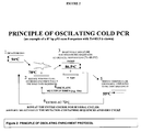
- the method is based in part on a modified nucleic acid amplification protocol that includes incubating the reaction mixture at a critical denaturing temperature or "Tc".
- Tc critical denaturing temperature
- the “critical temperature” or “Tc” refers to a temperature below the melting temperature "T m " of the reference sequence. In some embodiments, the Tc is below the T m of both the reference and target sequence.
- the critical temperature takes advantage of the lower T m of the double stranded target sequence or the target-reference cross hybridized double stranded DNA duplex so as to preferentially denature these duplexes over the reference/reference homoduplexes.
- T m melting temperature
- the critical denaturing temperature is the temperature below which PCR efficiency drops abruptly for a reference nucleic acid sequence.
- Tc The critical denaturing temperature
- the invention is directed to a method for enriching a target sequence in a nucleic acid sample suspected of having target and reference sequences.
- the method includes subjecting the amplification reaction mixture to a first denaturing temperature that is above the melting temperature "T m " of a reference sequence.
- T m melting temperature
- the temperature of the amplification reaction mixture is decreased allowing the single-stranded target sequences and reference sequences to hybridize to form double-stranded molecules.
- the reaction includes hybridization homoduplexes of target-target, reference-reference and heteroduplexes of target-reference strands.
- a heteroduplex by definition is an imperfectly matched duplex which nevertheless contains sufficient homology between strands to maintain a duplex form in the reaction mixture.
- a homoduplex by definition is a perfectly matched duplex.
- the temperature of the reaction mixture is then increased to the Tc, resulting in the preferential denaturation of the target-reference sequence hybridization duplexes.
- the Tc or critical temperature is below the T m of the reference sequence and can be determined by the methods described herein.
- the target-reference sequence duplexes and target-target sequence duplexes only if having a lower T m than the reference sequence
- the target-target duplexes if having a T m equal to or greater than the T m of the reference sequence
- the reference-reference sequence duplexes are substantially undenatured.
- Substantially means at least 60%, preferably at least 70%, more preferably at least 80%, even more preferably at least 90% and most preferably at least 98% in a given denatured or undenatured form.
- a nucleic acid sample suspected of containing each of a target sequence and a reference sequence is denatured by applying a first denaturing temperature that is above the T m of the reference sequence.
- the target and reference strands are annealed to each other so as to form double stranded target-reference sequence duplexes.
- the target-reference sequence duplex forms and is present in the reaction mixture along with double stranded target-target and reference-reference sequence duplexes.
- the double stranded target-reference and target-reference sequence duplexes are preferentially denatured by applying the Tc to the sample.
- the target-reference sequence duplexes (and target-target sequence duplexes only if having a lower T m than the reference sequence) are substantially denatured, whereas the target-target duplexes (if having a T m equal to or greater than the T m of the reference sequence) and the reference-reference sequence duplexes are substantially undenatured.
- “Substantially” means at least 60%, preferably at least 70%, more preferably at least 80%, even more preferably at least 90% and most preferably at least 98% in a given denatured or undenatured form.
- a pair of primers are then annealed to the target sequence and are extended, thus increasing the concentration of the target sequence relative to the reference sequence in the sample.
- the amplification reaction protocol includes a first denaturing temperature and a second denaturing temperature.
- the first denaturing temperature is above the T m of the reference sequence and the second denaturing temperature is below the T m of the reference sequence.
- the amplification reaction mixture is suspected of containing each of a target sequence and a reference sequence.
- the Tc is below the T m of the reference sequence, thus allowing for the preferential denaturation of the target sequence with the lower T m .
- the target-reference sequence duplexes and target-target sequence duplexes are substantially denatured, whereas the reference-reference sequence duplexes are substantially undenatured.
- Substantially means at least 60%, preferably at least 70%, more preferably at least 80%, even more preferably at least 90% and most preferably at least 98% in a given denatured or undenatured form.
- the step of reducing the temperature of the reaction mixture allows the pair of primers to anneal to the target sequence. These annealed primers are then extended by a polymerase, increasing the amount of the target sequence in the sample relative to the reference sequence.
- the amplification reaction mixture suspected of having each of a target and reference sequence, is first subjected to a first denaturing temperature which is above the T m of the reference sequence.
- the sample is cycled between two different temperature incubation steps.
- the temperature is decreased so as to allow the hybridization of the target sequence with the reference sequence so as to form a duplex.
- the temperature is increased to the Tc, which is below the T m of the reference sequence.
- the target-reference sequence duplexes (and target-target sequence duplexes only if having a lower T m than the reference sequence) are substantially denatured, whereas the target-target duplexes (if having a T m equal to or greater than the T m of the reference sequence) and the reference-reference sequence duplexes are substantially undenatured.
- “Substantially” means at least 60%, preferably at least 70%, more preferably at least 80%, even more preferably at least 90% and most preferably at least 98% in a given denatured or undenatured form.
- the temperature of the reaction mixture is decreased so as to allow a primer pair to anneal to the target sequence.
- These primers are then extended by a polymerase, thus enriching the target sequence relative to the reference sequence in the sample.
- the present invention is directed to methods for enriching low abundance alleles (e.g., target sequences) from a sample.
- the present invention is directed, in part, on the selective enrichment of target sequences by applying a critical denaturing temperature to a reaction mixture.
- the critical denaturing temperature or Tc is a temperature below the melting temperature T m of the reference sequence.
- the critical temperature takes advantage of the lower T m of the the target-reference cross hybridized double stranded DNA duplex so as to preferentially denature these duplexes over the reference/reference homoduplexes.
- PCR as the initial step for genetic testing is used in most mutation and sequencing reactions.
- a nucleic acid sequence e.g., genomic DNA/cDNA
- a mutation screening method e.g. SSCP, dHPLC, MALDI-TOF, pyrosequencing, High Resolution Melting.
- the term "enriching a target sequence” refers to increasing the amount of a target sequence and increasing the ratio of target sequence relative to the corresponding reference sequence in a sample.
- the target sequence may be preferentially amplified in an amplification reaction so as to produce a ratio of 70% target sequence to 30% reference sequence.
- Enrichment of a target sequence results in a 2X to 200X increase in the target sequence relative to the reference sequence prior to enrichment.
- the enrichment of the target is at least a 2X, 3X, 4X, 5X, 6X, 7X, 8X, 9X, 10X, 15X, 20X, 25X, 30X, 35X, 40X, 45X, 50X, 60X, 70X, 80X, 90X 100X, 150X, 200X or more fold enrichment over the reference sequence.
- Enrichment of a target sequence results in a sample having 10%, 15%, 20%, 25%, 30%, 35%, 40%, 45%, 50%, 55%, 60%, 65%, 70%, 80%, 90%, 95% or more, target sequence compared to reference sequence (e.g., 10% target sequence : 90% reference sequence to 95% target sequence : 5% reference sequence).
- target sequence refers to a nucleic acid that is less prevalent in a nucleic acid sample than a corresponding reference sequence.
- the target sequence makes-up less than 50% of the total amount of reference sequence + target sequence in a sample.
- the target sequence is expressed at the RNA and/or DNA level 1:10, 1:15, 1:20, 1:25X, 1:30, 1:35, 1:40, 1:45, 1:50, 1:60, 1:70, 1:80, 1:90, 1:100, 1:150, 1:200X or less than the reference sequence.
- the target sequence is a mutant allele.
- a sample e.g., blood sample
- the normal cells contain non-mutant or wild-type alleles, while the small number of cancerous cells contains somatic mutations.
- the mutant is the target sequence while the wild-type sequence is the reference sequence.
- the invention is directed to detecting fetal DNA in a nucleic acid sample obtained from a mother.
- the target sequence is present in the fetal DNA while the more prevalent mother DNA contains the reference sequence.
- a target sequence is meant to include fetal DNA obtained from a pregnant mother.
- a "target strand" refers to a single nucleic acid strand of a target sequence.
- the target sequence is about 17-2000 nucleotides long.
- the target sequence is 20, 30, 40, 50, 60, 70, 80, 90, 100, 110, 120, 130, 140, 150, 160, 170, 180, 190, 200, 250, 300, 350, 400, 450, 500, 600, 700, 800, 900 or more nucleotides long.
- Target sequences share at least 50%, 60%, 70%, 80%, 85%, 90%, 91%, 92%, 93%, 94%, 95%, 96%, 97%, 98%, 99% or more homology to the corresponding reference sequence, but differs by at least one nucleotide from the reference sequence.
- Target sequences can be amplified via PCR with the same pair of primers as those used for the reference sequence.
- the term "reference sequence" refers to a nucleic acid that is more prevalent in a nucleic acid sample than a corresponding target sequence (e.g, same gene but different nucleic acid sequence).
- the reference sequence makes-up over 50% of the total reference sequence + target sequence in a sample.
- the reference sequence is expressed at the RNA and/or DNA level 10X, 15X, 20X, 25X, 30X, 35X, 40X, 45X, 50X, 60X, 70X, 80X, 90X 100X, 150X, 200X or more than the target sequence.
- the reference sequence is a wild-type allele.
- a sample e.g., blood sample
- the normal cells contain non-mutant or wild-type alleles, while the small number of cancerous cells contain somatic mutations.
- the wild-type sequence is the reference sequence while the mutant sequence is the target sequence (e.g., mutant allele).
- a "reference strand” refers to a single nucleic acid strand of a reference sequence.
- the reference sequence is about 17-2000 nucleotides long. In one embodiment the reference sequence is 20, 30, 40, 50, 60, 70, 80, 90, 100, 110, 120, 130, 140, 150, 160, 170, 180, 190, 200, 250, 300, 350, 400, 450, 500, 600, 700, 800, 900 or more nucleotides long. Reference sequences will share at least 50%, 60%, 70%, 80%, 85%, 90%, 91%, 92%, 93%, 94%, 95%, 96%, 97%, 98%, 99% or more homology to the corresponding target sequence, but will differ by at least one nucleotide from the target sequence. Reference sequences can be amplified by PCR with the same pair of primers as that used for the reference sequence.
- allele refers to alternative forms of a gene, portion thereof or non coding region of DNA that occupy the same locus or position on homologous chromosomes that have at least one difference in the nucleotide sequence.
- the term allele can be used to describe DNA from any organism including but not limited to bacteria, viruses, fungi, protozoa, molds, yeasts, plants, humans, non-humans, animals, and archaebacteria.
- the alleles may be found in a single cell (e.g., two alleles, one inherited from the father and one from the mother) or within a population of cells (e.g., a wild-type allele from normal tissue and a somatic mutant allele from diseased tissue).
- An allele can be 17-2000 nucleotides long.
- the allele is 20, 30, 40, 50, 60, 70, 80, 90, 100, 110, 120, 130, 140, 150, 160, 170, 180, 190, 200, 250, 300, 350, 400, 450, 500, 600, 700, 800, 900 or more nucleotides long.
- Alleles will generally share 50%, 60%, 70%, 80%, 85%, 90%, 91%, 92%, 93%, 94%, 95%, 96%, 97%, 98%, 99% or more homology to each other.
- Alleles can be amplified by PCR with the same pair of primers.
- the present invention is used to enrich a polymorphism.
- Any given gene may have none, one, or many allelic forms (polymorphism).
- Common mutational changes which give rise to alleles may be the result of natural or artificial (e.g., chemical carcinogens) deletions, additions, or substitutions of nucleotides. Each of these types of changes may occur alone, or in combination with the others, one or more times in a given sequence.
- a restriction fragment length polymorphism is a variation in DNA sequence that alters the length of a restriction fragment ( Botstein et al. , Am. J. Hum. Genet. 32:314-331 (1980 )).
- the restriction fragment length polymorphism may create or delete a restriction site, thus changing the length of the restriction fragment.
- RFLPs have been widely used in human and animal genetic analyses (see WO 90/13668 ; WO 90/11369 ; Donis-Keller, Cell 51:319-337 (1987 ); Lander et al., Genetics 121:85-99 (1989 )).
- RFLP can be used in combination with the enrichment methods described herein so as to enhance the detection of polymorphisms. Such methods are described herein in Example 9.
- VNTR variable number tandem repeat
- polymorphisms take the form of single nucleotide variations between individuals of the same species. Such polymorphisms are far more frequent than RFLPs, STRs (short tandem repeats) and VNTRs (variable number tandem repeats). Some single nucleotide polymorphisms occur in protein-coding sequences, in which case, one of the polymorphic forms may give rise to the expression of a defective or other variant protein and, potentially, a genetic disease. Other single nucleotide polymorphisms occur in noncoding regions. Some of these polymorphisms may also result in defective protein expression (e.g., as a result of defective splicing). Other single nucleotide polymorphisms have no phenotypic effects.
- Somatic mutations are alterations in DNA that occurs after conception. Somatic mutations can occur in any of the cells of the body except the germ cells (sperm and egg) and therefore are not passed on to children. These alterations can (but do not always) lead to cancer or other diseases.
- wild-type refers to the most common polynucleotide sequence or allele for a certain gene in a population. Generally, the wild-type allele will be obtained from normal cells.
- mutant refers to a nucleotide change (i.e., a single or multiple nucleotide substitution, deletion, or insertion) in a nucleic acid sequence.
- a nucleic acid which bears a mutation has a nucleic acid sequence (mutant allele) that is different in sequence from that of the corresponding wild-type polynucleotide sequence.
- the methods of the invention are especially useful in selectively enriching a mutant allele which contains between 1 and 500 nucleotide sequence changes.
- a mutant allele may have 1, 2, 3, 4, 5, 6, 7, 8, 9, 10, 11, 12, 13, 14, 15, 16, 17, 18, 19, 20, 25, 30, 35, 40, 45, 50, 60, 70, 80, 90, 100, 200, 300, 400 or 500 nucleotide sequence changes compared to a corresponding wild-type allele.
- a mutation in a mutant allele will contain between 1 and 10 nucleotide sequence changes, and more preferably between 1 and 5 nucleotide sequence changes.
- the mutant allele will have 50%, 60%. 70%, 80%, 85%, 90%, 91 %, 92%, 93%, 94%, 95%, 96%, 97%, 98%, 99% or more homology to the wild-type allele.
- the mutant allele will be obtained from diseased tissues or cells and is associated with a disease state.
- T m refers to the temperature at which a polynucleotide dissociates from its complementary sequence.
- T m may be defined as the temperature at which one-half of the Watson-Crick base pairs in a duplex nucleic acid molecule are broken or dissociated (i.e., are "melted") while the other half of the Watson-Crick base pairs remain intact in a double stranded conformation.
- the T m is defined as the temperature at which 50% of the nucleotides of two complementary sequences are annealed (double strands) and 50% of the nucleotides are denatured (single strands).
- T m therefore defines a midpoint in the transition from double-stranded to single-stranded nucleic acid molecules (or, conversely, in the transition from single-stranded to double-stranded nucleic acid molecules).
- the T m can be estimated by a number of methods, for example by a nearest-neighbor calculation as per Wetmur 1991 ( Wetmur, J. G. 1991. DNA probes: applications of the principles of nucleic acid hybridization. Crit Rev Biochem Mol Biol 26: 227-259 , hereby incorporated by reference) and by commercial programs including OligoTM Primer Design and programs available on the internet.
- the T m can be determined though actual experimentation.
- double-stranded DNA binding or intercalating dyes such as ethidium bromide or SYBR-green (Molecular Probes) can be used in a melting curve assay to determine the actual T m of the nucleic acid. Additional methods for determining the T m of a nucleic acid are well know in the art and described herein.
- critical temperature refers to a temperature below the T m of the reference sequence.
- the Tc is applied to preferentially denature target sequence/reference sequence double-stranded duplex so as to allow the selective enrichment of the target sequence during an amplification reaction.
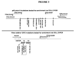
- the critical denaturing temperature (Tc) is the temperature below which PCR efficiency drops abruptly for a given nucleic acid sequence. For example, a 167 bp p53 sequence amplifies well if the PCR denaturing temperature is set at 87°C, amplifies modestly at 86.5 °C and yields no detectable product if PCR denaturation is set at 86°C or less. Therefore in this example Tc ⁇ 86.5°C.
- the Tc is about .1-20°C below the T m of the reference sequence. More preferably the Tc is about .1-10 °C, .1-9°C, .1-8°C, .1-7°C..1-6°C, .2°C -5 °C, .3 °C - 4.5 °C, .4-4°C, .5-3.5°C, .5-3°C, .5- 3 °C, .5 -2.5°C, .5-2°C, .5-1.5°C, .5-1 °C below the T m of the reference sequence. In some embodiments, the Tc is below the T m of both the reference and target sequences.
- the Tc may be about .1-10°C, .1-9 °C, .1-8°C, .1-7°C..1-6°C, .2°C-5°C, .3°C-4.5°C, .4-4°C, .5-3.5°C, .5-3°C, .5- 3°C, .5-2.5 °C, .5-2°C, .5-1.5°C, .5-1 °C below the T m of the target sequence.
- selective denaturation refers to the preferential breaking of hydrogen bonds between base pairs in a double-stranded nucleic acid molecule of a target sequence or target/reference sequence duplex to produce in single-stranded target sequence.
- the selective denaturation of the target sequence is accomplished by applying the critical temperature to the sample containing the target and reference sequences.
- primer pair refers to two primers that anneal to opposite strands of a target and reference sequence so as to form an amplification product during a PCR reaction.
- the primer pair is designed so as to have a T m lower than the Tc of the reaction.
- cross-hybridization refers to a double-stranded duplex formed between two nucleic acid sequences, that differ by one or more nucleotides, by virtue of the formation of hydrogen binds between complementary G and C bases and between complementary A and T or A and U bases.
- the two complementary nucleic acid sequences hydrogen bond in an antiparallel configuration.
- the cross-hybridized DNA will contain mismatches at the positions of difference between the reference and target sequences.
- the mismatches can include polymorphisms, mutations, insertions, deletions and other changes that result in such differences. For example, methylation.
- loops and/or single stranded regions of one or more nucleotides occur at sites of deletions/insertions.
- Cross hybridization typically involves denaturing the target and reference sequences, for example by heating, followed by renaturing under conditions (such as temperature) which allow hybridization and duplex formation to occur.
- reaction mixture is a mixture suspected of containing a target sequence duplex that comprises a suitable buffer for allowing the denaturing of a target sequence.
- identity refers to the subunit sequence similarity between two polymeric molecules, e.g., two polynucleotides or two polypeptides.
- identity refers to the subunit sequence similarity between two polymeric molecules, e.g., two polynucleotides or two polypeptides.
- a subunit position in both of the two molecules is occupied by the same monomeric subunit, e.g., if a position in each of two peptides is occupied by serine, then they are identical at that position.
- the identity between two sequences is a direct function of the number of matching or identical positions, e.g., if half (e.g., 5 positions in a polymer 10 subunits in length), of the positions in two peptide or compound sequences are identical, then the two sequences are 50% identical; if 90% of the positions, e.g., 9 of 10 are matched, the two sequences share 90% sequence identity.
- Percent nucleotide identity can be determined by the default parameters of BLAST. For sequence comparison, typically one sequence acts as a reference sequence, to which test sequences are compared. When using a sequence comparison algorithm, test and reference sequences are entered into a computer, subsequence coordinates are designated, if necessary, and sequence algorithm program parameters are designated. Default program parameters can be used, or alternative parameters can be designated. The sequence comparison algorithm then calculates the percent sequence identities for the test sequences relative to the reference sequence, based on the program parameters.
- the comparison window includes reference to a segment of any one of the number of contiguous positions selected from the group consisting of from 20 to 600, usually about 50 to about 200, more usually about 100 to about 150 in which a sequence may be compared to a reference sequence of the same number of contiguous positions after the two sequences are optimally aligned.
- Methods of alignment of sequences for comparison are well-known in the art. Optimal alignment of sequences for comparison can be conducted, e.g., by the local homology algorithm of Smith & Waterman, Adv. Appl. Math. 2:482 (1981 ), by the homology alignment algorithm of Needleman & Wunsch, J. Mol. Biol. 48:443 (1970 ), by the search for similarity method of Pearson & Lipman, Proc.
- HSPs high scoring sequence pairs
- T is referred to as the neighborhood word score threshold (Altschul et al., supra). These initial neighborhood word hits act as seeds for initiating searches to find longer HSPs containing them. The word hits are extended in both directions along each sequence for as far as the cumulative alignment score can be increased. Cumulative scores are calculated using, for nucleotide sequences, the parameters M (reward score for a pair of matching residues; always >0) and N (penalty score for mismatching residues; always ⁇ 0). For amino acid sequences, a scoring matrix is used to calculate the cumulative score.
- Extension of the word hits in each direction are halted when: the cumulative alignment score falls off by the quantity X from its maximum achieved value; the cumulative score goes to zero or below, due to the accumulation of one or more negative-scoring residue alignments; or the end of either sequence is reached.
- the BLAST algorithm parameters W, T, and X determine the sensitivity and speed of the alignment.
- the BLAST algorithm also performs a statistical analysis of the similarity between two sequences (see, e.g., Karlin & Altschul, Proc. Nat'l Acad. Sci. USA 90:58735787 (1993 )).
- One measure of similarity provided by the BLAST algorithm is the smallest sum probability (P(N)), which provides an indication of the probability by which a match between two nucleotide or amino acid sequences would occur by chance.
- P(N) the smallest sum probability
- a nucleic acid is considered similar to a reference sequence if the smallest sum probability in a comparison of the test nucleic acid to the reference nucleic acid is less than about 0.2, more preferably less than about 0.01, and most preferably less than about 0.001.
- the invention is directed to a method for enriching a target sequence in a nucleic acid sample suspected of having target and reference sequences.
- the reference and target sequences may be amplified prior to use in the present method. That is the reference and target sequences of interest may be amplified from a genomic template in a PCR reaction prior to use in the present method. An aliquot from this PCR reaction is then transferred for use in the selective enrichment method.
- the reference and target sequences need not be subjected to a first PCR reaction but can be used in their native form (e.g., genomic DNA) in the selective enrichment method.
- the target and reference sequences can be obtained from any nucleic acid sequence including, genomic DNA, cDNA, viral DNA, mammalian DNA, fetal DNA or bacterial DNA. While the reference sequence is generally the wild-type allele and the target sequence is the mutant allele, the reverse may also be true.
- the mutant allele may include any one or more nucleotide deletions, insertions or alterations. In some embodiments, the mutant allele is a somatic mutation.
- the target sequence is methylated DNA while the reference sequence is un-methylated DNA.
- the target sequence is un-methylated DNA while the reference sequence is methylated DNA.
- the primers used in the present method are generally design so as to produce reference and target sequence amplification products of about 17 to 1000 bases, more preferably about 25 to 500 bases, and most preferably about 50 to 100 bases in size.
- the method includes subjecting the amplification reaction mixture to a first denaturing temperature that is above the melting temperature "T m " of a reference sequence.
- T m of a nucleic acid can be determined through experimentation or estimated by calculation. The skilled artisan is well aware of numerous well known methods for determining the T m of a nucleic acid, some of which are described herein.
- the first denaturing temperature is set according to standard procedures used in PCR. Thus, the first denaturing temperature should be sufficiently high so as to allow the full denaturation of the target and reference sequences (e.g., 96 °C). In one embodiment, the first denaturing temperature is about 1°C to 30°C above the T m of the reference sequence, more preferably the T m of the reference sequence is about 5 °C to 20 °C above the Tm of the reference sequence.
- the temperature of the amplification reaction mixture is decreased allowing the target sequences and reference sequences to hybridize.
- This hybridization temperature or intermediate temperature (the temperature being below the first denaturing temperature and Tc but above the primer annealing/extension temperature, e.g., about 60 °C to 80 °C) is above the T m of the primer pair, and thus allows the target and reference sequences to hybridize while preventing binding of the primer pair to the target and/or reference sequences.
- This annealing step results in the formation of hybridization duplexes of double stranded target-target, reference-reference and target-reference sequences
- the target-reference hybridization duplexes are then preferentially denatured by increasing the temperature of the reaction mixture to the Tc.
- the Tc or critical temperature is below the T m of the reference sequence and can be determined by the methods described herein.
- the Tc is about .3 °C -5 °C below and more preferably about .5 °C to 1.5 °C below the T m of the reference sequence. Generally, the Tc will be about 70-90°C.
- the target-target hybridization duplexes may also be preferentially denatured if the target sequence has a nucleotide sequence which results in a lower T m compared to the reference sequence.
- the target-reference sequence duplexes (and target-target sequence duplexes only if having a lower T m than the reference sequence) are substantially denatured, whereas the target-target duplexes (if having a T m equal to or greater than the T m of the reference sequence) and the reference-reference sequence duplexes are substantially undenatured.
- “Substantially” means at least 60%, preferably at least 70%, more preferably at least 80%, even more preferably at least 90% and most preferably at least 98% in a given denatured or undenatured form.
- the Tc is generally applied from about 1 second to 5 minutes, more preferably 2 seconds to 1 minute and most preferably 5 seconds to 30 seconds.
- the temperature of the reaction mixture is reduced so as to allow a primer pair to anneal to the target sequence.
- the annealed primers are then extended by a nucleic acid polymerase, thus enriching the target sequence relative to the reference sequence in the sample.
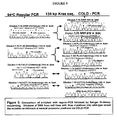
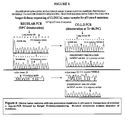
- the steps of the method are generally repeated for multiple cycles in order to get sufficient amplification of the target and reference sequences. In one embodiment, the steps of the method are repeated for 5-40 cycles and more preferably 10-30 cycles. The optimal number of cycles can be determined by one of ordinary skill in the art.
- the present methods are performed in a PCR device, more preferably under real-time reaction conditions in a real-time detection PCR device, such as the SMARTCYCLER real-time PCR device (Cepheid, Sunnyvale, CA) and the Mx3005P real-time PCR device (Stratagene, La Jolla, CA).
- the reaction mixture may include a nucleic acid detection agent (e.g., nucleic acid detection dye such as SYBR Green dye or LC-Green dye or a probe operatively coupled to a fluorescent dye) for quantifying and/or monitoring the amplification products of the reaction.
- a nucleic acid detection agent e.g., nucleic acid detection dye such as SYBR Green dye or LC-Green dye or a probe operatively coupled to a fluorescent dye
- the sample may be further processed (e.g., for identification of any genetic alterations enriched by the method, e.g., subjected to a sequencing reaction).
- the enriched reference sequences may be further processed by a variety of procedures including: MALDI-TOF, HR-Melting, Di-deoxy-sequencing, Single-molecule sequencing, pyrosequencing, RFLP, digital PCR and quantitative-PCR.
- the amplification reaction protocol includes a first denaturing temperature and a second denaturing temperature.
- the first denaturing temperature is above the T m of the reference sequence and the second denaturing temperature is below the T m of the reference sequence.
- the method includes subjecting the amplification reaction mixture to a first denaturing temperature that is above the melting temperature "T m " of a reference sequence.
- T m of a nucleic acid can be determined through experimentation or estimated by calculation. The skilled artisan is well aware of numerous well known methods for determining the T m of a nucleic acid.
- the first denaturing temperature is generally selected as one would generally select the denaturing temperature of a PCR reaction and should be sufficiently high so as to allow the denaturing of the target and reference sequences.
- the first denaturing temperature is about 1 °C to 30 °C above the T m of the reference sequence, more preferably the T m of the reference sequence is about 5 °C to 20 °C above the T m of the reference sequence.
- the second denaturing temperature is below the T m of the reference sequence and can be determined by the methods described herein.
- the Tc is about .3 °C -5 °C below the T m of the reference sequence and more preferably about .5 °C to 1.5 °C below the T m of the reference sequence.
- the Tc will be about 70-90 °C.
- the second denaturing temperature is generally applied from about 1 second to 5 minutes, more preferably 2 seconds to 1 minute and most preferably 5 seconds to 30 seconds.
- a method of enriching a target sequence by subjecting an amplification reaction mixture to a Tc, reducing the temperature of the reaction mixture and extending a primer pair.
- the amplification reaction mixture is suspected of containing a target and a reference sequence.
- the target sequence has a T m below the T m of the reference sequence.
- the Tc is below the T m of the reference sequence, thus allowing for the preferential denaturation of the target sequence which has a lower T m than the reference sequence as a result of its nucleotide composition (e.g., deletion).
- the reference and target sequences may be amplified prior to use in the present method.
- the reference and target sequences of interest may be amplified from a genomic template in a PCR reaction prior to use in the present method. An aliquot from this PCR reaction is then transferred for use in the selective enrichment method.
- the reference and target sequences need not be subjected to a first PCR reaction but can be used in their native form in the selective enrichment method, e.g., genomic DNA.
- the target and reference sequences can be obtained from any nucleic acid sequence including, genomic DNA, cDNA, viral DNA, mammalian DNA, fetal DNA or bacterial DNA. While the reference sequence is generally the wild-type allele and the target sequence is the mutant allele, the reverse may also be true.
- the mutant allele may include any one or more nucleotide deletions, insertions or alterations. In some embodiments, the mutant allele is a somatic mutation.
- the primers used in the present method are generally designed so as to produce reference and target sequence amplification products of about 15 to 1000 bases, more preferably about 25 to 500 bases, and most preferably about 50 to 100 bases in size.
- the target-target hybridization duplexes are preferentially denatured by increasing the temperature of the reaction mixture to the Tc.
- the Tc or critical temperature is below the T m of the reference sequence and can be determined by the methods described herein.
- the Tc is about .3 °C -5 °C below the T m of the reference sequence and more preferably about .5°C to 1.5 °C below the T m of the reference sequence.
- the Tc will be about 70-90.
- the Tc is generally applied from about 1 second to 5 minutes, more preferably 2 seconds to 1 minute and most preferably 5 seconds to 30 seconds.
- the target-reference sequence duplexes and target-target sequence duplexes are substantially denatured, whereas the reference-reference sequence duplexes are substantially undenatured.
- “Substantially” means at least 60%, preferably at least 70%, more preferably at least 80%, even more preferably at least 90% and most preferably at least 98% in a given denatured or undenatured form.
- the step of reducing the temperature of the reaction mixture allows the primer pair to anneal to the target sequence. These annealed primers are then extended by a polymerase, increasing the amount of the target sequence in the sample.
- the steps of the method are generally repeated for multiple cycles in order to get sufficient amplification of the target and reference sequences. In one embodiment, the steps of the method are repeated for 5-40 cycles and more preferably 10-30 cycles. The optimal number of cycles can be determined by one of ordinary skill in the art.
- the present methods are performed in a PCR device, more preferably under real-time reaction conditions in a real-time detection PCR device, such as the SMARTCYCLER real-time PCR device (Cepheid, Sunnyvale, CA) and the Mx3005P real-time PCR device (Stratagene, La Jolla, CA).
- a nucleic acid detection agent e.g., nucleic acid detection dye such as SYBR Green dye or LC-Green dye or a probe operatively coupled to a fluorescent dye
- a nucleic acid detection agent e.g., nucleic acid detection dye such as SYBR Green dye or LC-Green dye or a probe operatively coupled to a fluorescent dye
- the sample may be further processed, e.g., subjected to a sequencing reaction.
- the enriched alleles may be further processed by a variety of procedures including: MALDI-TOF, HR-Melting, Di-deoxy-sequencing, Single-molecule sequencing, pyrosequencing, RFLP, digital PCR and quantitative-PCR.
- the amplification reaction mixture having a target and reference sequence, is first subjected to a first denaturing temperature which is above the T m of the reference sequence.
- the reference and target sequences may be amplified prior to use in the present method. That is the reference and target sequences of interest may be amplified from a genomic template in a PCR reaction prior to use in the present method. An aliquot from this PCR reaction is then transferred for use in the selective enrichment method.
- the reference and target sequences need not be subjected to a first PCR reaction but can be used in their native form in the selective enrichment method, e.g., genomic DNA.
- the target and reference sequences can be obtained from any nucleic acid sequence including, genomic DNA, cDNA, viral DNA, mammalian DNA, fetal DNA or bacterial DNA. While the reference sequence is generally the allele and the target sequence is the mutant allele, the reverse may also be true.
- the mutant allele may include any one or more nucleotide deletions, insertions or alterations. In some embodiments, the mutant allele is a somatic mutation.
- the target sequence is methylated DNA while the reference sequence is un-methylated DNA.
- the target sequence is un-methylated DNA while the reference sequence is methylated DNA.
- the primers used in the present method are generally design so as to produce reference and target sequence amplification products of about 15 to 1000 bases, more preferably about 25 to 500 bases, and most preferably about 50 to 100 bases in size.
- the T m of a nucleic acid can be determined through experimentation or estimated by calculation. The skilled artisan is well aware of numerous well known methods for determining the T m of a nucleic acid.
- the first denaturing temperature is generally selected as one would generally select the denaturing temperature of a PCR reaction and should be sufficiently high so as to allow denaturing of the target and reference sequences. In one embodiment, the first denaturing temperature is about 1 °C to 30°C above the T m of the reference sequence, more preferably the T m of the reference sequence is about 5°C to 20 °C above the T m of the reference sequence.
- the sample is cycled between two different temperature incubation steps.
- the temperature is decreased so as to allow the hybridization of the target sequence with the reference sequence.
- the temperature is increased to the Tc, which is below the T m of the reference sequence.
- the first incubation step results in the formation of hybridization duplexes of target-target, reference-reference and target-reference sequences.
- this hybridization temperature or intermediate temperature (the temperature being below the first denaturing temperature and Tc but above the primer annealing/extension temperature, e.g., about 60°C to 80 °C) is above the T m of the primer pair, and thus allows the target and reference sequences to hybridize while preventing binding of the primer pair to the target and/or reference sequences.
- the target-reference and target-target (as long as the target has a lower T m than the reference sequence) hybridization duplexes are then preferentially denatured by increasing the temperature of the reaction mixture to the Tc in the second incubation step.
- the Tc or critical temperature is below the T m of the reference sequence and can be determined by the methods described herein. In one embodiment, the Tc is about .3 °C -5 °C below the T m of the reference sequence and more preferably about .5 °C to 1.5 °C below the T m of the reference sequence. Generally, the Tc will be about 70-90 °C.
- the target-target hybridization duplexes are also preferentially denatured if the target sequence has a nucleotide sequence which results in a lower T m compared to the reference sequence.
- the Tc is generally applied from about 1 second to 5 minutes, more preferably 2 seconds to 1 minute and most preferably 5 seconds to 30 seconds.
- the reaction may be repeated for multiple cycles in order to get sufficient amplification of the target and reference sequences.
- the steps of the method are repeated for 5-40 cycles and more preferably 10-30 cycles.
- the optimal number of cycles can be determined by one of ordinary skill in the art.
- the present methods are performed in a PCR device, more preferably under real-time reaction conditions in a real-time detection PCR device, such as the SMARTCYCLER real-time PCR device (Cepheid, Sunnyvale, CA) and the Mx3005P real-time PCR device (Stratagene, La Jolla, CA).
- the reaction mixture may include a nucleic acid detection agent (e.g., nucleic acid detection dye such as SYBR Green dye or LC-Green dye or a probe operatively coupled to a fluorescent dye) for quantifying and/or monitoring the amplification products of the reaction.
- a nucleic acid detection agent e.g., nucleic acid detection dye such as SYBR Green dye or LC-Green dye or a probe operatively coupled to a fluorescent dye
- Further processing includes MALDI-TOF, HR-Melting, Di-deoxy-sequencing, Single-molecule sequencing, pyrosequencing, RFLP, digital PCR and quantitative-PCR.
- the method of the present invention can be used to detect whether methylation has occurred in a target sequence or reference sequence.
- the method utilizes genomic DNA to assay the methylation.
- the methylation detection method comprises a chemical or enzymatic approach for methylation-sensitive treatment of DNA.
- Chemical treatments include the incubation of DNA with sodium bisulfite, which selectively converts non-methylated cytosines to uracils.
- the DNA is first heat-denatured and then can be treated with 5M bisulfite, pH 5-7.
- Pretreatment of genomic DNA to remove pre-existing uracils is used prior to bisulfite treatment. This pretreatment consists of uracil glycosylase treatment in the presence of 5 mM hydroxylamine, pH 7.
- the modified DNA may now be used in the methods of the invention.
- methylated cytosines of the reference or target sequence are converted to uracils, they will now form mismatches when duplexed with the opposite strand (target or reference), which was not methylated, during they cross-hybridization step of the reaction.
- any of the methods of the invention are used to enrich multiple different target sequences in a multi-plex reaction.
- the method includes additional sets of primer pairs to the additional target sequences.
- the invention is directed to a computer readable medium having program instructions for performing any of the methods of the invention. Further disclosed is a PCR system for enriching the target sequence.
- the system includes memory for implementing the program instructions of the computer readable medium.
- Figures 1 and 2 illustrate two different aspects of the invention.
- Figure 1 illustrates an aspect of the invention in which the method utilizes an amplification reaction having a first denaturing temperature and a critical denaturing temperature or Tc.
- Figure 2 also illustrates an amplification reaction having a first denaturing temperate and a Tc but further includes oscillating, or repeating, the annealing and critical denaturing temperature step multiple times prior to the primer annealing and extension phase of the reaction.
- Figure 1 shows the procedure for enriching a target sequence in a nucleic acid sample having target and reference sequences.
- the target and reference sequences can be obtained from any nucleic acid sequence including, genomic DNA, cDNA, viral DNA, mammalian DNA, fetal DNA or bacterial DNA. While the reference sequence is generally the allele and the target sequence is the mutant allele, the reverse may also be true.
- the mutant allele may include any one or more nucleotide deletions, insertions or alterations. In some embodiments, the mutant allele is a somatic mutation.
- the target sequence is methylated DNA while the reference sequence is un-methylated DNA. Alternatively, the target sequence is un-methylated DNA while the reference sequence is methylated DNA.
- the method includes subjecting the amplification reaction mixture to a first denaturing temperature ( Fig. 1A ) that is above the melting temperature "T m " of a reference sequence.
- the T m of a nucleic acid can be determined through experimentation or estimated by calculation. The skilled artisan is well aware of numerous well known methods for determining the T m of a nucleic acid some of which are described herein.
- the first denaturing temperature is generally selected as one would generally select the denaturing temperature of a PCR reaction and should be sufficiently high so as to allow the full denaturing of the target and reference sequences (e.g., 94 °C). In one embodiment, the first denaturing temperature is about 1 °C to 30°C above the T m of the reference sequence, more preferably the T m of the reference sequence is about 5 °C to 20 °C above the T m of the reference sequence.
- the temperature of the amplification reaction mixture is decreased allowing the target sequences and reference sequences to hybridize ( Fig. 1 B) .
- This annealing step results in the formation of hybridization duplexes of target-target, reference-reference and target-reference sequences. Determining an annealing temperature is well known to the skilled artisan.
- the PCR primers used in the method are designed to have a T m that prevents them from binding to the target and reference sequences at this intermediate temperature, so that they don't interfere with cross-hybridization of the mutant (target) and wild-type (reference) sequences. Because of mutations in the target sequence most target sequences end-up in a mismatched structure with the reference sequence, and thus have a lower melting temperature, when heteroduplexed with the reference sequence, than the fully-matched reference/reference homoduplexes.
- the target-reference hybridization duplexes are then preferentially denatured by increasing the temperature of the reaction mixture to the Tc ( Fig. 1C ).
- the Tc or critical temperature is below the T m of the reference sequence and can be determined by the methods described herein.
- the Tc is about .3 °C -5 °C below the T m of the reference sequence and more preferably about .5 °C to 1.5 °C below the T m of the reference sequence.
- the Tc will be about 70-90 °C.
- the target-target hybridization duplexes may also be preferentially denatured if the target sequence has a nucleotide sequence which results in a lower T m compared to the reference sequence.
- the Tc is generally applied from about 1 second to 5 minutes, more preferably 2 seconds to 1 minute and most preferably 5 seconds to 30 seconds.
- the temperature of the reaction mixture is reduced so as to allow one or more primers to anneal to the target sequence ( Fig. 1D ).
- the annealed primers are then extended by a nucleic acid polymerase, thus enriching the target sequence in the population of nucleic acids contained in the sample.
- the steps of the method are generally repeated for multiple cycles in order to get sufficient amplification of the target and reference sequences. In one embodiment, the steps of the method are repeated for 5-40 cycles and more preferably 10-30 cycles. The optimal number of cycles can be determined by one of ordinary skill in the art.
- the present methods are performed in a PCR device, more preferably under real-time reaction conditions in a real-time detection PCR device, such as the SMARTCYCLER real-time PCR device (Cepheid, Sunnyvale, CA) and the Mx3005P real-time PCR device (Stratagene, La Jolla, CA).
- the reaction mixture may include a nucleic acid detection agent (e.g., nucleic acid detection dye such as SYBR Green dye or LC-Green dye or a probe operatively coupled to a fluorescent dye) for quantifying and/or monitoring the amplification products of the reaction.
- a nucleic acid detection agent e.g., nucleic acid detection dye such as SYBR Green dye or LC-Green dye or a probe operatively coupled to a fluorescent dye
- the sample may be further processed, e.g., subjected to a sequencing reaction.
- the enriched alleles may be further processed by a variety of procedures including: MALDI-TOF, HR-Melting, Di-deoxy-sequencing, Single-molecule sequencing, pyrosequencing, RFLP, digital PCR and quantitative-PCR.
- the amount of mutant sequences is gradually enriched over the sequences (reference sequence).
- Both homozygous and heterozygous mutations are enriched via the method.
- Enrichment of mutation-containing sequences by 10-60-fold relative to performing regular PCR at a denaturation temperature of 94°C is routine.
- Tc critical denaturation temperature
- the mutation enrichment occurs simultaneously at all sequence positions, albeit with a different efficiency depending on the sequence context and overall size of the PCR amplicon.
- Both the critical denaturation temperature Tc and the anticipated enrichment at any position are predictable using appropriate DNA melting software and can be verified experimentally. Thus depending on where the mutation is, a somewhat different enrichment can be anticipated, however in all cases a substantial enrichment is achieved and therefore the detection limits of downstream assays improve, e.g., sequencing reactions.
- Figure 2 illustrates an embodiment of the method for enriching a target sequence by subjecting an amplification reaction mixture to multiple alternating steps of annealing and critical denaturing temperature. This embodiment takes advantage of both preferential denaturation at the critical temperate and preferential corss-bybridization at the hybridization temperature.
- the amplification reaction mixture having a target and reference sequence, is first subjected to a first denaturing temperature which is above the T m of the reference sequence ( Fig. 2A ).
- the sample is cycled between two different temperature incubation steps.
- the temperature is decreased so as to allow the preferential hybridization of the target sequence with the reference sequence ( Fig. 2B ).
- the temperature is increased to the Tc ( Fig. 2C ).
- the preferential hybridization temperature for a given sequence is a temperature at which the wild-type alleles hybridize back to themselves at a faster rate than do the mutation-containing alleles. Because mutated alleles are at a much lower prevalence than wild-type alleles, cross-hybridization of the wild-type alleles with themselves proceeds faster than cross-hybridization of mutant alleles with mutant alleles, or of mutant alleles with alleles (the latter forming a mismatch). As a result, when the PCR temperature is reduced to a cross-hybridization temperature the mutant alleles do not cross-hybridize to the same extent as the wild-type alleles do.
- This hybridization temperature or intermediate temperature (the temperature being below the first denaturing temperature and Tc but above the primer annealing/extension temperature, e.g., about 60°C to 80 °C) is above the T m of the primer pair, and thus allows the target and reference sequences to hybridize while preventing binding of the primer pair to the target and/or reference sequences.
- the Tc or critical temperature is below the T m of the reference sequence and can be determined by the methods described herein. In one embodiment, the Tc is about .3 °C -5 °C below the T m of the reference sequence and more preferably about .5 °C to 1.5 °C below the T m of the reference sequence. Generally, the Tc will be about 70-90 °C.
- the target-target hybridization duplexes may also be preferentially denatured if the target sequence has a nucleotide sequence which results in a lower T m compared to the reference sequence.
- the Tc is generally applied from about 1 second to 5 minutes, more preferably 2 seconds to 1 minute and most preferably 5 seconds to 30 seconds. This process is repeated for several times, oscillating between the annealing temperature ( Fig. 2B ) and the critical temperature ( Fig. 2C ), each time selectively generating more mutant sequences in the single-stranded form than sequences in the single stranded form.
- the temperature of the reaction mixture is decreased so as to allow the one or more primers to anneal to the target sequence ( Fig. 2D ). These primers are then extended by a polymerase, thus enriching the target sequence.
- the steps of the method are generally repeated for multiple cycles in order to get sufficient amplification of the target and reference sequences. In one embodiment, the steps of the method are repeated for 5-40 cycles and more preferably 10-30 cycles. The optimal number of cycles can be determined by one of ordinary skill in the art.
- the present methods are performed in a PCR device, more preferably under real-time reaction conditions in a real-time detection PCR device.
- the sample may be subjected to further processing. Further processing includes MALDI-TOF, HR-Melting, Di-deoxy-sequencing, Single-molecule sequencing, pyrosequencing, RFLP, digital PCR and quantitative-PCR.
- Digital PCR may be used in the detection of ultra-low level mutations in combination with an enrichment procedure.
- the DNA sample is diluted down to single molecules, so that in each PCR reaction the starting material is either wild-type or mutant. Following a large number of PCR reactions from the same starting material, mutant molecules are isolated and detected.
- Fluidigm South San Francisco, CA
- Fluidigm sells a variety of digital PCR based systems that can be used with the present invention. Accordingly, real-time enrichment can be performed from single molecules in thousands of parallel enrichment reactions simultaneously, allowing for the identification of the PCR reaction which originated the mutant DNA.
- Such a system is particularly useful for the detection of ultra-low mutations in cancer genomes. Combination of digital PCR with the enrichment procedure can similarly benefit single molecule sequencing applications.
- a nucleic acid sample utilized in the method of the invention comprises genomic DNA having a target and reference sequence.
- the nucleic acid sample of the method of the invention comprises a target and reference sequence that were previously amplified in a nucleic acid amplification reaction.
- PCR polymerase chain reaction
- a preferred variation of the PCR method is asymmetrical PCR (for example, see Mao et al., Biotechniques 27(4):674-678 (1999 ); Lehbein et al., Electrophoresis 19(8-9):1381-1384 (1998 ); Lazaro et al., Molec. Cell. Probes 6(5):357-359 (1992 ); and U.S. Pat. No. 6,197,499 ).
- Other amplification methods include, but are not limited to, strand displacement amplification (SDA) (see, Walker et al., Nuc. Acids Res. 20(7):1691-1696 (1992 ), as well as U.S. Pat. Nos.
- PCR is performed as described in Mullis and Faloona, 1987, Methods Enzymol., 155: 335 .
- the polymerase chain reaction (PCR) technique is disclosed in U.S. Pat. Nos. 4,683,202 , 4,683,195 and 4,800,159 .
- PCR is an in vitro method for the enzymatic synthesis of specific DNA sequences, using two oligonucleotide primers that hybridize to opposite strands and flank the region of interest in the target DNA.
- PCR is reported to be capable of producing a selective enrichment of a specific DNA sequence by a factor of 10 9 .
- the PCR method is also described in Saiki et al., 1985, Science 230:1350 .
- PCR is performed using template DNA (target and reference sequences) (at least 1fg; more usefully, 1-1000 ng) and at least 25 pmol of oligonucleotide primers.
- a typical reaction mixture includes: 2 ⁇ l of DNA, 25 pmol of oligonucleotide primer, 2.5 ⁇ l of a suitable buffer, 0.4 ⁇ l of 1.25 ⁇ M dNTP, 2.5 units of Taq DNA polymerase (Stratagene) and deionized water to a total volume of 25 ⁇ l.
- PCR is performed using a programmable thermal cycler.
- the length and temperature of each step of a PCR cycle, as well as the number of cycles, are adjusted according to the stringency requirements in effect.
- Annealing temperature and timing are determined both by the efficiency with which a primer is expected to anneal to a template and the degree of mismatch that is to be tolerated.
- the ability to optimize the stringency of primer annealing conditions is well within the knowledge of one of moderate skill in the art.
- An annealing temperature of between 30°C and 72°C is used.
- Initial denaturation of the template molecules normally occurs at between 92°C and 99°C for 4 minutes, followed by 20-40 cycles consisting of denaturation (94-99°C for 15 seconds to 1 minute), annealing (temperature determined as discussed above; 1-2 minutes), and extension (72°C for 1 minute).
- the final extension step is generally carried out for 4 minutes at 72°C, and may be followed by an indefinite (0-24 hour) step at 4°C.
- PCR utilizes a nucleic acid polymerase, or enzyme that catalyzes the polymerization of nucleoside triphosphates.
- the enzyme will initiate synthesis at the 3'-end of the primer annealed to the target sequence, and will proceed in the 5'-direction along the template.
- Known DNA polymerases include, for example, E. coli DNA polymerase I, T7 DNA polymerase, Thermus thermophilus (Tth) DNA polymerase, Bacillus stearothermophilus DNA polymerase, Thermococcus litoralis DNA polymerase, Thermus aquaticus (Taq) DNA polymerase and Pyrococcus furiosus (Pfu) DNA polymerase.
- nucleic acid polymerase also encompasses RNA polymerases. If the nucleic acid template is RNA, then “nucleic acid polymerase” refers to an RNA-dependent polymerization activity, such as a reverse transcriptase.
- the PCR protocol further includes a critical denaturing step.
- the enrichment procedures are performed in a PCR device, more preferably under real-time reaction conditions in a real-time PCR device.
- Real-time reaction conditions further utilize a nucleic acid detection agent (e.g., dye or probe) in order to measure/detect the PCR product as it is produced.
- a nucleic acid detection agent e.g., dye or probe
- the enrichment methods are practiced in a multiplex format.
- Multiplex PCR is when more than one pair of primers is used in a PCR reaction to detect more than one target sequence.
- the goal of multiplex PCR is to amplify more than one target sequence simultaneously and thereby to save time and minimize expense. It enables the amplification of multiple target sequences in one run.
- the primers are designed so that the resulting PCR-amplicons all share about the same Tc (critical denaturation temperature).
- the T m may be defined as the temperature at which one-half of the Watson-Crick base pairs in a duplex nucleic acid molecule are broken or dissociated (i.e., are "melted") while the other half of the Watson-Crick base pairs remain intact in a double stranded conformation. While the "critical temperature”, “critical denaturing temperature” or “Tc” refers to a temperature below the T m of the reference sequence. The Tc is applied to selectively denature the double-stranded target sequence or target sequence/reference sequence double-stranded duplex in a nucleic acid sample so as to allow the selective enrichment of the target sequence during an amplification reaction.
- the T m of a given pair of nucleic acid strands is indicative of the stability of the strand to strand binding and depends on the strands' complementarity, sequence length, GC content, the presence or absence of mismatches within the double stranded region and other factors of minor importance, e.g. the salt concentration of the sample ( Lewin, Genes V, Chapter 5, Oxford University Press and Cell Press: New York, (1994) pp. 109-126 ; SantaLucia, 1998).
- the melting point temperature is usually determined experimentally by subjecting the sample to a constitutive increase in temperature and continuously measuring the dissociation of the hybridization duplex into single strands.
- the dissociation can be detected by a variety of different methods, for example by a shift in UV absorbance, fluorescence of double-stranded DNA binding dyes, by surface plasmon resonance or preferably by means of fluorescence.
- the hybridization probe is usually labeled with a fluorescent entity, and the generation of a fluorescent signal somehow depends on the formation of the hybridization duplex.
- the T m can be determined experimentally or estimated based on well defined methods known to those of ordinary skill in the art. Methods to observe and analyze nucleic acid denaturation transitions include: measuring the enthalpy change within a sample as it denatures by differential scanning calorimetry (DSC) ( Kulinski et al., Nucleic Acids Res.
- T m values of double stranded nucleic acids can also be observed by monitoring the fluorescence of double-stranded DNA-specific dyes combined with the nucleic acids (Wittwer et al., 1996). Double stranded-specific dyes are nucleic acid-binding fluorophors. Typically, the fluorescence of these dyes increases when bound to duplexed nucleic acids ( Wittwer et al., BioTechniques 22:176-181 (1997 )). Ririe et al. (1997) demonstrated that post PCR products can be differentiated by melting curve analysis using the double stranded nucleic acid binding dye SYBR® Green I.
- SYBR® Green I binds preferentially to double stranded nucleic acid (Haugland, 1996).
- Other suitable dyes for determining the T m of a double stranded nucleic acid include SYBR® Gold, ethidium bromide, acridine orange, propidium bromide, PicoGreen®, Hoechst 33258, Hoechst 33342, Hoechst 34580, YO-PRO®-1 and YOYO®-1. Each of these dyes is commercially available.
- the melting of any double stranded nucleic acid structure generally occurs in a substantial proportion of a population of similar nucleic acids over a limited temperature range and will typically have a peak of melting (most rapid transition) at approximately the T m for that nucleic acid. Therefore, such peaks of change in fluorescence emission can be used to calculate the T m for double stranded nucleic acids.
- a melting temperature profile may be graphically represented by plotting-dF/dT against T, where dF is the change in measured fluorescence emission, dT is the change in temperature of the nucleic acid, and T is the temperature of the nucleic acid. Such a graphic representation will show peaks at temperatures at which the most rapid changes in fluorescence occur, indicating melting temperatures.
- the T m can be estimated by a number of methods, for example by a nearest-neighbor calculation as per Wetmur 1991 ( Wetmur, J. G. 1991. DNA probes: applications of the principles of nucleic acid hybridization. Crit Rev Biochem Mol Biol 26: 227-259 ) and by commercial programs including OligoTM Primer Design and programs available on the internet.
- the "critical temperature” or “Tc” refers to a temperature below the T m of the reference sequence.
- the Tc is applied to preferentially denature the double-stranded target sequence duplex or target sequence/reference sequence double-stranded duplex over the reference/reference sequence duplex, so as to allow the selective enrichment of the target sequence during an amplification reaction.
- the critical temperature takes advantage of the lower T m of the double stranded target sequence or the target-reference cross hybridized double stranded DNA duplex.
- annealing of the target-reference sequence because of the mismatches, will essentially have a lower T m than the known allele (e.g., reference sequence).
- the present invention exploits the small T m difference between fully-matched and mismatched sequences. Because critical denaturation is performed at every PCR cycle the differential enrichment of mutation-containing alleles is compounded exponentially, and results to a large difference in overall amplification efficiency between mutant and wild-type alleles, at the end of the cycling.
- the Tc is about .1-20°C below the T m of the reference sequence.
- the Tc is about .1-15°C, .1-10°C, .1-9°C, .1-8°C, .1-7°C..1-6°C, .2C-5°C, .3C- 4.5 °C, .4-4°C, 5-3.5 °C, .5-3°C, .5- 3 °C, .5-2.5 °C, .5-2°C, .5-1.5°C, .5-1°C below the T m of the reference sequence.
- the Tc can be below the Tm of both the reference sequence and target sequence.
- the Tm of a wild-type sequence was 84 °C and the Tm of the mutated sequence was 83.8 °C.
- the optimal Tc was 83.5 °C.
- the Tc is chosen such that it is below the Tm of both the reference sequence and all possible target sequences.
- the Tm of a wild-type sequence was 84 °c and the Tm of sequences mutated at different positions (single point mutations) was 83.8 °C, 83.7 °C, 83.9 °C, 83.6 °C, 83.75 °C.
- the optimal Tc was 83.5 °C.
- the critical denaturing temperature (Tc) is the temperature below which PCR efficiency drops abruptly for a given nucleic acid sequence. For example, a 167 bp p53 sequence amplifies well if the PCR denaturing temperature is set at 87°C, amplifies modestly at 86.5 °C and yields no detectable product if PCR denaturation is set at 86°C or less.
- the Tc for a given sequence can be identified either experimentally, or by calculation.
- a real-time melting curve in the presence of an intercalating dye (LC-GREEN or SYBR-Green) is performed to obtain the average melting temperature T m of the sequence (See above with respect to determining T m ).
- Temperatures of about 0.5-1.5°C less than T m are usually appropriate critical denaturation temperatures Tc that result to enrichment of target sequences.
- the ⁇ T m has been found to be dependent on the length of the target/reference sequence duplex, upon the percent guanine-cytosine (%GC) content, and also upon the location of the point mutation or base-pair mismatch in the duplex. For instance, if the mismatch lies toward either end of the duplex, ⁇ T m will generally be lower, typically in the range of about .1 °C to about 8° C. If the mismatch resides toward the center of the duplex, ⁇ T m will be relatively higher, typically in the range of about .2 °C to about 11°C.
- %GC percent guanine-cytosine
- the ⁇ T m is generally equivalent to about .5 °C to about 1.5° C per percent base mismatch in the target-reference cross-hybridized duplex. It is to be appreciated that ⁇ T m will vary depending on not only the length of the duplex and location of mutation, but also will vary depending on the specific order of the sequence. Consequently, all such variances are within the scope of the disclosure.
- the invention provides for methods of enriching a target sequence in a nucleic acid sample and also utilizes primers for amplifying a template nucleic acid sequence.
- nucleic acid refers to primers, probes, and oligomer fragments to be detected, and shall be generic to polydeoxyribonucleotides (containing 2-deoxy-D-ribose), to polyribonucleotides (containing D-ribose), and to any other type of polynucleotide which is an N-glycoside of a purine or pyrimidine base, or modified purine or pyrimidine bases (including abasic sites).
- nucleic acid refers only to the primary structure of the molecule. Thus, these terms include double- and single-stranded DNA, as well as double- and single-stranded RNA.
- oligonucleotide is not necessarily physically derived from any existing or natural sequence but may be generated in any manner, including chemical synthesis, DNA replication, reverse transcription or a combination thereof.
- oligonucleotide or nucleic acid intend a polynucleotide of genomic DNA or RNA, cDNA, semisynthetic, or synthetic origin which, by virtue of its synthetic origin or manipulation: (1) is not associated with all or a portion of the polynucleotide with which it is associated in nature; and/or (2) is linked to a polynucleotide other than that to which it is linked in nature.
- the nucleic acids used in the method contain modified nucleotides so as to increase the denaturing temperature differences between the reference/reference sequence homoduplex and the target/reference sequence heteroduplex. Such modifications would enhance the enrichment of the target sequence.
- the modified or non-natural nucleotides can be incorporated before or during the enrichment procedure.
- Modified nucleotides contemplated for use in the methods of the invention include: diamino-purine analogues (e.g.
- nucleic acids 2'-O-methyl-2,6-diaminopurine
- uracil peptide nucleic acid analogues
- biotin-modified analogues of the above fluorophore-modified analogues of the above, inosine, 7-deazaguanine, 2'-deoxy-2'-fluoro-ß-D-arabinonucleic acid (2'F-ANA) nucleotides, locked-nucleic acids (LNAs), ENAs: 2'-O,4'-C-ethylene-bridged nucleic acids and others.
- the modified nucleotides can be incorporated into any of the nucleic acids of the invention including, template, primer and probe nucleic acids.
- locked nucleic acids represent a class of conformationally restricted nucleotide analogues described, for example, in WO 99/1 4226 , increase the melting temperature of a nucleic acid.
- Oligonucleotides containing the locked nucleotide are described in Koshkin, A.A., et al., Tetrahedron (1998), 54: 3607-3630 ) and Obika, S. et al., Tetrahedron Lett. (1998), 39: 5401-5404 ), both of which are incorporated by reference.
- Complementarity need not be perfect; stable duplexes may contain mismatched base pairs or unmatched bases.
- Those skilled in the art of nucleic acid technology can determine duplex stability empirically considering a number of variables including, for example, the length of the oligonucleotide, base composition and sequence of the oligonucleotide, ionic strength, and incidence of mismatched base pairs. Stability of a nucleic acid duplex is measured by the melting temperature, or "T m ".
- the invention uses oligonucleotide primers for amplifying a template nucleic acid sequence.
- primer may refer to more than one primer and refers to an oligonucleotide, whether occurring naturally, as in a purified restriction digest, or produced synthetically, which is capable of acting as a point of initiation of synthesis along a complementary strand when placed under conditions in which synthesis of a primer extension product which is complementary to a nucleic acid strand is catalyzed.
- Such conditions include the presence of four different deoxyribonucleoside triphosphates and a polymerization-inducing agent such as DNA polymerase or reverse transcriptase, in a suitable buffer ("buffer” includes substituents which are cofactors, or which affect pH, ionic strength, etc.), and at a suitable temperature.
- the primer is preferably single-stranded for maximum efficiency in amplification.
- Oligonucleotide primers useful according to the invention are single-stranded DNA or RNA molecules that are hybridizable to a template nucleic acid sequence and prime enzymatic synthesis of a nucleic acid strand.
- the primer is complementary to a portion of a target molecule present in a pool of nucleic acid molecules. It is contemplated that oligonucleotide primers are prepared by synthetic methods, either chemical or enzymatic. Alternatively, such a molecule or a fragment thereof is naturally-occurring, and is isolated from its natural source or purchased from a commercial supplier.
- Oligonucleotide primers are 5 to 100 nucleotides in length, ideally from 17 to 40 nucleotides, although primers of different length are of use.
- Primers for amplification are preferably about 17-25 nucleotides. Primers useful according to the invention may also be designed to have a particular melting temperature (T m ) by the method of melting temperature estimation.
- T m melting temperature
- Commercial programs, including Oligo ⁇ , Primer Design and programs available on the internet, including Primer3 and Oligo Calculator can be used to calculate a T m of a nucleic acid sequence useful according to the invention.
- the T m of an amplification primer useful according to the invention is preferably between about 45 and 65 °C and more preferably between about 50 and 60 °C.
- selective hybridization occurs when two nucleic acid sequences are substantially complementary (at least about 65% complementary over a stretch of at least 14 to 25 nucleotides, preferably at least about 75%, more preferably at least about 90% complementary). See Kanehisa, M., 1984, Nucleic Acids Res. 12: 203 .
- mismatch may be small, such as a mono-, di- or tri-nucleotide.
- a region of mismatch may encompass loops, which are defined as regions in which there exists a mismatch in an uninterrupted series of four or more nucleotides.
- the enrichment procedures can be used in combination with peptide nucleic acid (PNA) primers so as to increase the sensitivity of mutation enrichment.
- PNA peptide nucleic acid
- the PNAs are used to suppress amplification of the wild-type (reference) sequences only.
- Primers may be synthesized so as to distinguish between a target (mutant) sequence and a reference sequence.
- the PNA based primers recognize and bind the complementary wild-type sequence with a higher thermal stability and specificity than primers which bind the mutated sequence. This not only increases the Tm difference between the PNA primer-reference nucleic acid and regular primer-target nucleic acid but also prevents the PNA based primer from being extended by a DNA polymerase, thus resulting in a further enrichment of the target sequence.
- Such assays are known and the art and described in Orum et al. Nucleic Acids Research, 21(23): 5332-5336 (1993 ).
- Oligonucleotide primers can be designed with these considerations in mind and synthesized according to the following methods.
- the design of a particular oligonucleotide primer for the purpose of sequencing or PCR involves selecting a sequence that is capable of recognizing the target sequence, but has a minimal predicted secondary structure.
- the oligonucleotide sequence may or may not bind only to a single site in the target nucleic acid.
- the T m of the oligonucleotide is optimized by analysis of the length and GC content of the oligonucleotide.
- the selected primer sequence does not demonstrate significant matches to sequences in the GenBank database (or other available databases).
- a primer useful according to the invention is facilitated by the use of readily available computer programs, developed to assist in the evaluation of the several parameters described above and the optimization of primer sequences.
- Examples of such programs are "PrimerSelect” of the DNAStarTM software package (DNAStar, Inc.; Madison, WI), OLIGO 4.0 (National Biosciences, Inc.), PRIMER, Oligonucleotide Selection Program, PGEN and Amplify (described in Ausubel et al., 1995, Short Protocols in Molecular Biology, 3rd Edition, John Wiley & Sons ).
- the primers are designed so as to have a T m below the temperature applied during the target/reference sequence cross-hybridization step. Thus, the primers do not anneal to the target or reference sequences during this hybridization step (See Fig. 1 ).
- the T m of the primers is 5-10 °C below the cross-hybridiation annealing step temperature.
- the primers themselves are synthesized using techniques that are also well known in the art. Methods for preparing oligonucleotides of specific sequence are known in the art, and include, for example, cloning and restriction digest analysis of appropriate sequences and direct chemical synthesis.
- oligonucleotides are prepared by a suitable chemical synthesis method, including, for example, the phosphotriester method described by Narang et al., 1979, Methods in Enzymology, 68:90 , the phosphodiester method disclosed by Brown et al., 1979, Methods in Enzymology, 68:109 , the diethylphosphoramidate method disclosed in Beaucage et al., 1981, Tetrahedron Letters, 22:1859 , and the solid supp-ort method disclosed in U.S. Pat. No. 4,458,066 , or by other chemical methods using either a commercial automated oligonucleotide synthesizer (which is commercially available) or VLSIPSTM technology.
- the primers may also be synthesized with modified nucleic acids by methods well known in the art.
- sample refers to any substance containing or presumed to contain a nucleic acid of interest (target and reference sequences) or which is itself a nucleic acid containing or presumed to contain a target nucleic acid of interest.
- sample thus includes a sample of nucleic acid (genomic DNA, cDNA, RNA), cell, organism, tissue, fluid, or substance including but not limited to, for example, plasma, serum, spinal fluid, lymph fluid, synovial fluid, urine, tears, stool, external secretions of the skin, respiratory, intestinal and genitourinary tracts, saliva, blood cells, tumors, organs, tissue, samples of in vitro cell culture constituents, natural isolates (such as drinking water, seawater, solid materials), microbial specimens, and objects or specimens that have been "marked” with nucleic acid tracer molecules.
- nucleic acid genomic DNA, cDNA, RNA
- cell organism, tissue, fluid, or substance including but not limited to, for example, plasma, serum, spinal fluid, lymph fluid, synovial fluid, urine, tears, stool, external secretions of the skin, respiratory, intestinal and genitourinary tracts, saliva, blood cells, tumors, organs, tissue, samples of in vitro cell culture constituents, natural isolates (such as drinking
- Nucleic acid sequences can be amplified from genomic DNA. Genomic DNA can be isolated from tissues or cells according to the following method. Alternatively nucleic acids sequences can be isolated from blood by methods well known in the art.
- the tissue is isolated.
- genomic DNA from mammalian tissue
- the tissue is minced and frozen in liquid nitrogen.
- Frozen tissue is ground into a fine powder with a prechilled mortar and pestle, and suspended in digestion buffer (100 mM NaCl, 10 mM Tris-HCl, pH 8.0, 25 mM EDTA, pH 8.0, 0.5% (w/v) SDS, 0.1 mg/ml proteinase K) at 1.2ml digestion buffer per 100 mg of tissue.
- digestion buffer 100 mM NaCl, 10 mM Tris-HCl, pH 8.0, 25 mM EDTA, pH 8.0, 0.5% (w/v) SDS, 0.1 mg/ml proteinase K
- cells are pelleted by centrifugation for 5 min at 500 x g, resuspended in 1-10 ml ice-cold PBS, repelleted for 5 min at 500 x g and resuspended in 1 volume of digestion buffer.
- Samples in digestion buffer are incubated (with shaking) for 12-18 hours at 50 °C, and then extracted with an equal volume of phenol/chloroform/isoamyl alcohol. If the phases are not resolved following a centrifugation step (10 min at 1700 x g), another volume of digestion buffer (without proteinase K) is added and the centrifugation step is repeated. If a thick white material is evident at the interface of the two phases, the organic extraction step is repeated. Following extraction the upper, aqueous layer is transferred to a new tube to which will be added 1/2 volume of 7.5M ammonium acetate and 2 volumes of 100% ethanol.
- the nucleic acid is pelleted by centrifugation for 2 min at 1700 x g, washed with 70% ethanol, air dried and resuspended in TE buffer (10 mM Tris-HCl, pH 8.0, 1 mM EDTA, pH 8.0) at 1mg/ml. Residual RNA is removed by incubating the sample for 1 hour at 37 °C in the presence of 0.1 % SDS and 1 ⁇ g/ml DNase-free RNase, and repeating the extraction and ethanol precipitation steps.
- the yield of genomic DNA, according to this method is expected to be approximately 2 mg DNA/1 g cells or tissue (Ausubel et al., supra). Genomic DNA isolated according to this method can be used according to the invention.
- the target DNA may also be extracted from whole blood.
- blood may be drawn by standard methods into a collection tube, preferably comprising siliconized glass, either without anticoagulant for preparation of serum, or with EDTA, sodium citrate, heparin, or similar anticoagulants, most preferably EDTA, for preparation of plasma.
- the preferred method although not absolutely required, is that plasma or serum be fractionated from whole blood.
- Plasma or serum may be fractionated from whole blood by centrifugation, preferably gentle centrifugation at 300 to 800 Xg for 5 10 minutes, or fractionated by other standard methods. Since heparin may interfere with PCR, use of heparinized blood may require pretreatment with heparinase.
- EDTA is the preferred anticoagulant for blood specimens.
- Either freshly-collected blood plasma or serum, or frozen (stored) and subsequently thawed plasma or serum can be used in the methods of the invention.
- Stored plasma or serum should be kept at -20 °C to -70 °C, and freshly-collected plasma or serum kept refrigerated or maintained on ice until use.
- the DNA may then be extracted by methods well known in the art as well as those described herein.
- the invention provides a method for enriching target sequences from a patient sample, for diagnosis, detection, monitoring, evaluation or treatment of a disorder, in particular a neoplastic or proliferative disease in an animal or a human.
- the nucleic acid is derived from a nucleic acid encoding an oncogene or other tumor-associated DNA.
- the preferential enrichment of the target sequence allows for the further analysis or manipulation of the DNA.
- the enriched alleles may be analyzed to define the characteristics of the cell from which the DNA originates. Any of several methods may be used, dependent upon the desired information, including nucleic acid sequencing, RFLP, digital PCR, spectroscopy including proton NMR spectroscopy, biochemical analysis, and immunologic analysis.
- amplified DNA is isolated by excising mutant DNA bands from an agarose gel, reamplified, cloned into a plasmid vector, for example the pGEM-T vector plasmid (Promega) and sequenced using a commercial kit such as Sequenase 2.0 (USB).
- Analysis to define the characteristics of the target DNA, and thus for example a tumor affords a wide array of clinical utility, including the description, characterization, or classification of the cell (e.g., tumor), whether known or occult, such as by tissue of origin, by type (such as premalignant or malignant), phenotype, and genotype, and by description or characterization of tumor behavior, physiology and biochemistry, as to gain understanding of tumor invasiveness, propensity to metastasize, and sensitivity or resistance to various therapies, thereby allowing the prediction of response to either ongoing or planned therapy and, further, allowing evaluation of prognosis.
- Comparison of the characteristics of target DNA to previous biopsy or surgical specimens permits further evaluation of tumor heterogeneity or similarity in comparison to that specimen, and thus evaluation of tumor recurrence.
- RNA ribonucleic acid
- transcription of RNA is performed by employing a primer with an RNA polymerase promoter region joined to the standard primer sequence for the DNA of interest in the amplification reaction (step three).
- RNA complimentary to the DNA is then transcribed from the attached promoter region.
- amplified allele DNA is cloned into an expression vector, and RNA complimentary to the DNA is transcribed.
- the complimentary RNA is used in an in vitro translation reaction to manufacture tumor-associated or tumor-specific protein.
- Characterization of the allele, amplification of tumor-derived or tumor-associated DNA, and characterization, transcription of complimentary RNA, and translation to tumor-associated or tumor-specific protein provides significant utility, both in the assignment of therapy and in the development of tumor-specific therapies. Sequencing of extracellular DNA or transcription of complementary RNA allows assignment or development of antisense compounds, including synthetic oligonucleotides and other antisense constructs appropriately specific to the extracellular DNA, such as by construction of an expression plasmid such as by adapting the method of Aoki et al. (1995, Cancer Res. 55: 3810 3816 ). Similarly, defining tumor characteristics allows assignment of specific monoclonal antibody or vaccine therapies appropriately specific to the amplified DNA. Production of corresponding immunologic protein can be used in the development of tumor-specific monoclonal antibodies. Similarly, translated protein can be used in tumor-specific vaccine development.
- the invention allows the development and application of these tumor-specific therapies or diagnostics even when only premalignant tumors, early cancers, or occult cancers are present.
- the invention allows therapeutic intervention when tumor burden is low, immunologic function is relatively intact, and the patient is not compromised, all increasing the potential for cure.
- an enriched target sequence is subjected to sequencing by MALDI-TOF.
- MS Mass spectrometry
- MALDI matrix-assisted laser desorption/ionization
- TOF time-of-flight
- PCR® polymerase chain reaction
- MALDI-TOF is ideally suited for detection of previously identified mutations in non-heterogeneous tumor samples.
- the reliability of MALDI-TOF decreases substantially when heterogeneous samples with ⁇ 10% mutated cells are used (e.g. pancreatic, lung or prostate cancer) or when DNA from bodily fluids are to be screened.
- the present enrichment method enables MALDI-TOF to detect low-level somatic mutations and will also provide the required reliability that is necessary for mainstream surgical tumor sample screening.
- an enriched target sequence is subjected to high resolution melting.
- Genes that contain clinically-relevant mutations at numerous positions along exons, such as p53, are easier screened via mutation scanning rather than individual mutation genotyping.
- HR-Melting is a high throughput mutation scanning technology introduced in the last few years, with excellent capabilities for discovering SNPs or germline mutations ( Chou, L.S., et al. (2005); Am J Clin Pathol, 124, 330-338 ; Wittwer, C.T., et al. (2003); Clin Chem, 49, 853-860 ; Reed, G.H. and Wittwer, C.T. (2004); Clin Chem, 50, 1748-1754 ).
- LC Green or Sybr-Green intercalating fluorescent dye
- SNP identification simultaneous gene scanning and mutation genotyping in a fraction of the time required when using traditional methods, while maintaining a closed-tube environment.
- the PCR requires ⁇ 30 min (capillaries) or 1.5 h (96- or 384-well plates) and melting acquisition takes 1-2 min per capillary or 5 min per plate.
- the present invention enables the convenience and throughput of HR-Melting to be applied for mainstream surgical tumor sample screening and also for detecting low-level somatic mutations in 'difficult' clinical samples with stromal contamination or DNA from bodily fluids.
- the enriched target sequence may be subjected to single molecule sequencing ( Thomas, R.K., et al. (2006); Nat Med, 12, 852-855 ).
- Single Molecule Sequencing would also benefit from incorporation of the present invention. For example, for mutation-screening in patient samples, PCR of selected exons in a regular PCR machine from genomic DNA is still required prior to initiating second generation sequencing. Further, detection of mutations at the level of 1-5% mutant-to-wild-type ratio in clinical samples requires repeated sequencing of numerous 'individual events' in order to achieve acceptable statistics.
- the method of selective enrichment of the invention may be applied during the in-emulsion stage of a single molecule sequencing reaction.
- the enriched target sequences are then subjected to pyrosequencing.
- the enriched target sequence may be subjected to a primer extension sequencing reaction.
- primer extension oligonucleotides are used to assess variation in sequence at a predetermined position thereof relative to a nucleic acid, the sequence of which is known.
- a sample oligonucleotide is provided as a single stranded molecule, the single stranded molecule is mixed with an inducing agent, a labeled nucleotide, and a primer having a sequence identical to a region flanking the predetermined position to form a mixture, the mixture having an essential absence of nucleotides constituted of bases other than the base of which the labeled nucleotide is constituted.
- the mixture is then subjected to conditions conducive for the annealing of the primer to the single-stranded molecule and the formation of a primer extension product incorporating the labeled nucleotide, and the mixture is analyzed for the presence of primer extension product containing labeled nucleotide ( U.S. Pat. No. 5,846,710 ).
- organic solvents to increase the T m differences between between mismatched and matched duplexes in the amplification method of the current invnetion.
- the inclusion of certain organic solvents can improve enrichment of the target sequence.
- inclusion of organic solvents may increase the denaturation temperature difference between reference and target DNA sequences and thus aid the preferential amplification of the target sequence.
- Organic solvents such as DMSO, formamide, betaine or glycerol ( Pomp, D. and Medrano, J.F.,Biotechniques, 10, 58-59 (1991 )) can increase the T m difference between matched (reference/reference) and mismatched (target/reference) sequences.
- the intermediate hybridization step (cross-hybridization) in the method of the present invention forms target-reference sequences containing mismatches, inclusion of organic solvents to the degree that they do not inhibit the action of polymerase is advantageous. Therefore, in some embodiments, the reaction mixture is supplemented with DMSO, formamide, betaine, glycerol, or a combination thereof, at levels of 1-10% volume to volume, or preferably 3-8% volume to volume, or most preferably 5-6% volume to volume.
- Example 8 illustrates the use of DMSO in an enrichment method.
- Tc that is appropriate for a given sequence changes upon using an organic solvent in the reaction.
- the Tc for a sequence is 83.5°C
- the Tc is 80.5°C.
- Example 1 Materials and methods for enriching a target sequence
- Sequences used for validation of COLD-PCR To validate the present invention, a series of genomic DNA and cell lines containing mutations at different positions of p53 exon 8 and Kras exon 2 (codons 12-13) were used ( Figure 3 ). The p53 exon 8 mutations correlate with poor prognosis in lung cancer and are low-prevalence mutations in the plasma of cancer patients. Similarly, Kras mutations have prognostic significance in lung adenocarcinoma.
- PCR was performed in the presence of 0.1 x LC-Green intercalating dye and followed in real-time in a Cepheid machine. Real-time-follow-up of PCR is not necessary but is convenient so it was adopted for all experiments.
- Figure 4 depicts representative results. For example, DNA from HCC cells initially diluted in wild-type cells down to 5% mutant-to-wild-type ratio, becomes ⁇ 70% mutant-to-wild-type ratio following the enrichment protocol, as estimated by observing the sequencing chromatogram (i.e. enrichment by a factor of ⁇ 14). Similarly, DNA from SW480 cells (homozygous G>A mutation at codon 273) diluted by a factor of 10 into wild-type is enriched by a factor of ⁇ 7 following the enrichment procedure.
- FIG. 5 illustrates the results of the 135 bp fragment from Kras. The results were compared to regular nested PCR performed at a denaturation temperature of 94°C and followed by Sanger sequencing. Figure 5 illustrates that mutations down to 33% mutant-to-wild-type can be clearly detected using Sanger sequencing.
- p53 exon 8 (G>A) mutation was detected in plasma-circulating DNA from 5 colon cancer patients via the enrichment procedure-Sanger sequencing, but not via regular PCR-Sanger sequencing.
- p53 (C>T) mutations that were missed by regular sequencing were also revealed using DNA obtained from a formalin-fixed (FFPE) specimen obtained from a non-small-cell lung cancer patient (NSCLC) ( Figure 6 ).
- the bottom chromatograph in Figure 6 demonstrates the detection of Kras codon 12 mutations in another FFPE sample obtained from a NSCLC patient.
- the mutations identified via the enrichment procedure were subsequently independently verified from genomic DNA via RFLP methods.
- relevant mutations missed by regular PCR-sequencing become readily detectable using COLD-PCR-sequencing.
- Example 3 Mutations that decrease the T m can be enriched without the mismatch annealing step.
- these two fragments were amplified from the initial p53 exon 8 amplicon via nested PCR followed by the enrichment protocol.
- Tc critical denaturation temperature
- the primer binding step e.g. 55°C
- the primer synthesis step e.g. 72 °C
- Figure 7A demonstrates sequencing chromatograms for the 87 bp fragment (either the forward or the reverse sequencing were performed depending where the mutation lies on the 87 bp sequence).
- the data indicates that, using this version of the modified enrichment protocol, enriches the 87 bp fragment 20-50 fold. For example, a 1% initial dilution of mutant-to-wild-type DNA of SW480 DNA resulted in 50% mutant-to-wild-type following the enrichment i.e. ⁇ 50-fold enrichment.
- the subsequent Sanger sequencing revealed a 'heterozygous' sequence.
- the affect of size on enrichment via the abbreviated the enrichment protocol is depicted in Figure 7B . This data illustrates that enrichment is highest for fragments ⁇ 100 bp, but still clearly evident ( ⁇ 8-10 fold) for fragments up to 210 bp.
- Example 4- Mutations that increase or decrease the T m can be enriched via the full enrichment method.
- the present invention is also expected to improve most other PCR-based technologies, including MALDI-TOF for somatic mutation detection.
- MALDI-TOF for somatic mutation detection.
- the reaction was then desalted by addition of a cation exchange resin followed by mixing and centrifugation to settle the contents of the tube.
- the extension product was spotted onto a 384 well spectroCHIP before being run on the MALDI-TOF mass spectrometer (Sequenom).
- the results are illustrated in Table I.
- the mutation enrichment factor is listed on the third column of Table I. The enrichment is calculated by comparison to the values obtained when regular PCR MALDI-TOF is applied. Enrichment factors of 10-60 are obtained for the majority of the mutations studied. The enrichment increases as the ratio of mutant-to-wild-type decreases, indicating a non-linear dependence of the enrichment factor on initial concentration of mutations.
- type ENRICHMENT SW480-w. type ratio 1:5 regular PCR 15 85 SW480-w. type ratio 1:10 regular PCR 5 (MassSpec limit) 95 SW480-w. type ratio 1:33 regular PCR 0 100 SW480-w. type ratio 1:10 CO.L.D-PCR 45 55 -9 SW480-w. type ratio 1:100 CO.L.D-PCR 33 67 >10 SW480-w. type ratio 1:300 CO.L.D-PCR 31 69 >30 WILD TYPE ONLY, CO.L.D-PCR 0 100 None MALDI-TOF RESULT p53 exon 8 mutation 14483C>T CO.L.D-PCR CELL LINE DILUTIONS SCREENED %T mutant %C w.
- type ratio 1:300 CO.L.D-PCR 27 73 ⁇ 60 WILD TYPE ONLY, CO.L.D-PCR 0 100 None MALDI-TOF RESULT p53 exon 8 mutation 14486G>T CO.L.D-PCR CELL LINE DILUTIONS SCREENED %G mutant %T w. type ENRICHMENT DU145-w. type ratio 1:5 regular PCR 28 72 N/A DU145-w. type ratio 1:10 regular PCR 18 82 N/A DU145-w. type ratio 1:33 regular PCR 0 100 N/A DU145-w. type ratio 1:33 CO.L.D-PCR 27 73 -7 DU145-w.
- type ratio 1:100 CO.L.D-PCR 12 88 >10 DU145-w. type ratio 1:300 CO.L.D-PCR 0 100 undetectable WILD TYPE ONLY, CO.L.D-PCR 0 100 None Kras mutations % mutant or w. type allele for Kras codon 12 mutations are indicated MALDI-TOF RESULT Kras codon 12 mutation GGT>AGT CO.L.D-PCR CELL LINE DILUTIONS SCREENED %A mutant %G w. type ENRICHMENT A549-w. type ratio 1:10 regular PCR 25 75 N/A A549-w. type ratio 1:33 CO.L.D-PCR 40 60 -5.5 A549-w.
- Example 6- Comparison of TAQMAN based regular real-time PCR and enrichment based real-time PCR.
- Nucleic acid amplification reactions were performed to compare TAQMAN probe assays in regular real-time and an enrichment based real-time PCR. To compare the two real-time detection methods both genomic DNA and clinical tumor samples having a G>A mutation in p53 exon 8 were assayed at various dilutions in wild-type sequence. Serial dilutions (1:3, 1:10, 1:30, 1:100 and 1:300) of genomic DNA from SW480 into wild-type DNA were made.
- real time PCR reactions were performed directly from 20 ng genomic DNA in the presence of 0.2 ⁇ M Taqman probe 5'-6-Fam-TTT GAG GTG CAT GTT TGT GCC-BHQ_1-3' that fully matches the p53 mutation-containing sequence on DNA from SW480 cells.
- Final concentrations of other reagents were: 1X GoTaq Flexi buffer (Promega), 1X GoTaq polymerase (Promega) 0.2mM each dNTP, 0.2 ⁇ M forward primer, 5'-TGG TAA TCT ACT GGG ACG-3', 0.2 ⁇ M reverse primer, 5'-CGG AGA TTC TCT TCC TCT-3', MgCL 2 3 mM, plus DNA.
- Fast COLD-PCR cycling was: 95°C, 120 sec; (95°C, 15 sec; 58°C fluorescence reading ON, 60 sec) x 25 cycles; (83.5 °C 15 sec; 58°C fluorescence reading ON, 60 sec) x 25 cycles.
- the same program was employed, but the denaturation temperature throughout PCR was 95°C. Experiments were repeated at least 5 times in independent experiments.
- FIG. 8A and 8B Amplification plots illustrating the sensitivity of regular and enriched real-time PCR as applied to genomic DNA from the colon cancer cell line SW480 are depicted in FIG 8A and 8B.
- FIG. 8B demonstrates that enriched real-time PCR can detect the presence of the mutation at a 1:300 mutant to wild-type allele ratio.
- regular real-time PCR conducted under identical conditions with the exception of the Tc step, can only detect the mutant at maximum dilution of 1:10 ( FIG. 8A ). Therefore, the sensitivity of the assay is 30 fold better using the enrichment procedure.
- FIG. 8C and 8D Amplification plots comparing the sensitivity of regular and enriched real-time PCR in clinical tumor samples having the p53 exon 8 mutation (one of which is know to contain a low level mutation (5% mutant to wild-type) in p53 exon 8, CT20) are illustrated in FIG. 8C and 8D .
- Enriched real-time PCR was readily able to detect the mutation ( FIG. 8D ), while regular PCR did not ( FIG. 8C ).
- the remaining sample (TL6, TL8 and TL18) which were known to be wild type samples, did not amplify ( FIG. 8C ) under the same conditions.
- Nucleic acid amplification reaction mixtures were created to compare the ability of regular and enriched real-time PCR utilizing DNA detection dyes to detect samples containing mutations anywhere along exon 8 of p53.
- the method provides for a fast and convenient method to identify unknown mutations or heterozygous SNPs.
- the present method can be adapted by one of ordinary skill in the art to scan large numbers of genes for germline or somatic mutations for a variety of different applications (e.g. scanning for BRCA1/2 mutations in populations at high risk for developing breast/ovarian CA, scanning entire genetic pathways for identifying mutations, etc).
- FIG. 9A and 9B illustrate amplification plots comparing both regular and enriched real-time PCR using LC-Green dye in a variety of cell lines and clinical samples that are known to contain mutations in exon 8 of p53.
- the data shows that real-time regular-PCR ( FIG. 9A ) is unable to distinguish between the mutant (SW480, TL6 and CT20) and wild-type (R27, TL8, TL18, TL81 and TL82) samples, while enriched real-time PCR does ( FIG. 9B ).
- the enrichment method provides earlier threshold detection for the mutation containing samples than for the wild-type samples.
- FIG. 9C and 9D illustrates the results from amplification reactions prepare under reaction conditions identical to those illustrated in FIG. 9A and 9B but here the samples are all lung tumors.
- the data shows that real-time regular-PCR ( FIG. 9C ) can not differentiate between the mutant and wild-type samples, while real-time enriched PCR ( FIG. 9D ) does.
- the enrichment method provides earlier threshold detection for the mutation containing samples than for the wild-type samples.
- the enriched detection method was able to identify a previously unknown C>T mutation in sample TL6.
- Example 8- Organic solvents increase enrichment of mutations during regular and enhanced real-time PCR.
- Nucleic acid amplification reaction mixtures were prepared to assess the impact of organic solvents on regular and enriched real-time PCR. Reactions were performed either in the presence or the absence of an organic solvent (3% DMSO). The procedure used was the same as that described in Example 7 and depicted in FIG. 9C and 9D except for the addition of 3% DMSO.
- the presence of the organic solvent enhanced discrimination between the amplification plots of samples containing the mutated sequences with those containing wild-type sequences ( FIG. 10 ).
- the threshold difference between wild-type samples and mutant samples increased from ⁇ 5-cycles (real-time enrichment without DMSO, see FIG. 9A ) to more than 10 cycles (real-time enrichment with DMSO, see FIG. 10A ).
- Enrichment PCR combined with RFLP-PCR can be used for improved identification of ultra-low-level mutations, e.g. for identifying random mutations in a cancer genome or resistance mutations in cancer samples at a very early stage, i.e. before treatment.
- the enrichment of the mutation was quantified by TaqI digestion followed by dHPLC ( FIG. 11 ). The amount of mutation present was quantified by the presence of a separate mutation peak at retention time of about 7 min. Following the enrichment procedure the mutation peak is much more evident than following regular-PCR. In conclusion, the enrichment procedure substantially improved the detection of very low level mutations identified via RFLP-PCR.
Landscapes
- Chemical & Material Sciences (AREA)
- Life Sciences & Earth Sciences (AREA)
- Organic Chemistry (AREA)
- Health & Medical Sciences (AREA)
- Wood Science & Technology (AREA)
- Engineering & Computer Science (AREA)
- Zoology (AREA)
- Proteomics, Peptides & Aminoacids (AREA)
- Molecular Biology (AREA)
- Chemical Kinetics & Catalysis (AREA)
- Biotechnology (AREA)
- Microbiology (AREA)
- Biochemistry (AREA)
- Bioinformatics & Cheminformatics (AREA)
- General Engineering & Computer Science (AREA)
- General Health & Medical Sciences (AREA)
- Genetics & Genomics (AREA)
- Physics & Mathematics (AREA)
- Analytical Chemistry (AREA)
- Biophysics (AREA)
- Immunology (AREA)
- General Chemical & Material Sciences (AREA)
- Measuring Or Testing Involving Enzymes Or Micro-Organisms (AREA)
Description
- A commonly encountered situation in genetic analysis entails the need to identify a low percent of variant DNA sequences ('target sequences') in the presence of a large excess of non-variant sequences ('reference sequences'). Examples for such situations include: (a) identification and sequencing of a few mutated alleles in the presence of a large excess of normal alleles; (b) identification of a few methylated alleles in the presence of a large excess of unmethylated alleles (or: vice versa) in epigenetic analysis; (c) identification and genotyping of a few fetal DNA sequences circulating in the blood of the mother where a large excess of mother's DNA sequences are also present; and (d) identification of tumor-circulating DNA in blood of cancer patients (or people suspected of having cancer) in the presence of a large excess of wild-type alleles.
- While reliable high throughput screening methods for germline or high-prevalence somatic mutations have recently been described (Thomas, R.K., et al. (2007) Nat Genet, 39, 347-351; Chou, L.S., et al. (2005) Am J Clin Pathol, 124; 330-338; Thomas, R.K., et al. (2006) Nat Med, 12; 852-855) detection of low-prevalence somatic mutations in tumors with heterogeneity, stromal contamination or in bodily fluids is still problematic. And yet, the clinical significance of identifying these mutations is major in several situations. For example: (a) in lung adenocarcinoma, low-level EGFR mutations that cannot be identified by regular sequencing can confer positive response to tyrosine kinase inhibitors (Paez, J.G.,. et al. (2004) Science, 304; 1497-1500.) or drug resistance (Janne, P.A., et al. (2006) Clin Cancer Res, 12; 751-758) (b) mutations in plasma useful as biomarkers for early detection (Diehl, F., et al. (2005) Proc Natl Acad Sci U S A, 102; 16368-16373) or tumor response to treatment (Kimura, T., et al. (2004) Ann N Y Acad Sci, 1022; 55-60) cannot be sequenced using conventional methods; and (c) mutations in tumors with frequent stromal contamination, such as pancreatic or prostate, can be 'masked' by presence of wild-type alleles, thus requiring laborious micro-dissection or missing mutations altogether.
-
WO 03/072809 - The present invention is directed to methods for enriching low abundance alleles from a sample, as defined in the claims.
- The method is based in part on a modified nucleic acid amplification protocol that includes incubating the reaction mixture at a critical denaturing temperature or "Tc". By employing the present invention the current detection limits of all PCR-based technologies are greatly improved.
- The "critical temperature" or "Tc" refers to a temperature below the melting temperature "Tm" of the reference sequence. In some embodiments, the Tc is below the Tm of both the reference and target sequence. The critical temperature takes advantage of the lower Tm of the double stranded target sequence or the target-reference cross hybridized double stranded DNA duplex so as to preferentially denature these duplexes over the reference/reference homoduplexes. When the target sequence and reference sequence cross hybridize, minor sequence differences of one or more single nucleotide mismatch anywhere along a short (e.g., <200 bp) double stranded DNA sequence will generate a small but predictable change in the melting temperature (Tm) for that sequence (Lipsky, R.H., et al. (2001) Clin Chem, 47, 635-644; Liew, M., et al. (2004) Clin Chem, 50, 1156-1164). Depending on the exact sequence context and position of the mismatch, melting temperature changes of 0.1-20°C, are contemplated.
- The critical denaturing temperature (Tc) is the temperature below which PCR efficiency drops abruptly for a reference nucleic acid sequence. For example, a 167 bp p53 sequence amplifies well if the PCR denaturing temperature is set at 87°C, amplifies modestly at 86.5 °C and yields no detectable product if PCR denaturation is set at 86°C or less. Therefore in this example Tc~ 86.5°C.
- In a first aspect, the invention is directed to a method for enriching a target sequence in a nucleic acid sample suspected of having target and reference sequences. The method includes subjecting the amplification reaction mixture to a first denaturing temperature that is above the melting temperature "Tm" of a reference sequence. Next, the temperature of the amplification reaction mixture is decreased allowing the single-stranded target sequences and reference sequences to hybridize to form double-stranded molecules. Thus, the reaction includes hybridization homoduplexes of target-target, reference-reference and heteroduplexes of target-reference strands. A heteroduplex by definition is an imperfectly matched duplex which nevertheless contains sufficient homology between strands to maintain a duplex form in the reaction mixture. A homoduplex by definition is a perfectly matched duplex. The temperature of the reaction mixture is then increased to the Tc, resulting in the preferential denaturation of the target-reference sequence hybridization duplexes. The Tc or critical temperature is below the Tm of the reference sequence and can be determined by the methods described herein. At the Tc, the target-reference sequence duplexes (and target-target sequence duplexes only if having a lower Tm than the reference sequence) are substantially denatured, whereas the target-target duplexes (if having a Tm equal to or greater than the Tm of the reference sequence) and the reference-reference sequence duplexes are substantially undenatured. "Substantially" means at least 60%, preferably at least 70%, more preferably at least 80%, even more preferably at least 90% and most preferably at least 98% in a given denatured or undenatured form. After the preferential denaturation of the target-reference duplexes and target-target duplexes (those that have a lower Tm than the reference sequence), a reduced temperature is applied to the reaction mixture so as to allow a pair of primers to anneal to the target sequence. The annealed primers are then extended, thus enriching the target sequence relative to the reference sequence in the sample.
- Disclosed is another method of enriching a target sequence. In this method a nucleic acid sample suspected of containing each of a target sequence and a reference sequence is denatured by applying a first denaturing temperature that is above the Tm of the reference sequence. Next, the target and reference strands are annealed to each other so as to form double stranded target-reference sequence duplexes. The target-reference sequence duplex forms and is present in the reaction mixture along with double stranded target-target and reference-reference sequence duplexes. The double stranded target-reference and target-reference sequence duplexes are preferentially denatured by applying the Tc to the sample. At the Tc, the target-reference sequence duplexes (and target-target sequence duplexes only if having a lower Tm than the reference sequence) are substantially denatured, whereas the target-target duplexes (if having a Tm equal to or greater than the Tm of the reference sequence) and the reference-reference sequence duplexes are substantially undenatured. "Substantially" means at least 60%, preferably at least 70%, more preferably at least 80%, even more preferably at least 90% and most preferably at least 98% in a given denatured or undenatured form. A pair of primers are then annealed to the target sequence and are extended, thus increasing the concentration of the target sequence relative to the reference sequence in the sample.
- Disclosed is further a method for enriching a target sequence by performing a nucleic acid amplification reaction protocol. The amplification reaction protocol includes a first denaturing temperature and a second denaturing temperature. The first denaturing temperature is above the Tm of the reference sequence and the second denaturing temperature is below the Tm of the reference sequence.
- Still further disclosed is a method of enriching a target sequence, which has a lower Tm than the corresponding reference sequence, by subjecting an amplification reaction mixture to a Tc, reducing the temperature of the reaction mixture and extending a primer pair. The amplification reaction mixture is suspected of containing each of a target sequence and a reference sequence. The Tc is below the Tm of the reference sequence, thus allowing for the preferential denaturation of the target sequence with the lower Tm. At the Tc, the target-reference sequence duplexes and target-target sequence duplexes are substantially denatured, whereas the reference-reference sequence duplexes are substantially undenatured. "Substantially" means at least 60%, preferably at least 70%, more preferably at least 80%, even more preferably at least 90% and most preferably at least 98% in a given denatured or undenatured form. The step of reducing the temperature of the reaction mixture allows the pair of primers to anneal to the target sequence. These annealed primers are then extended by a polymerase, increasing the amount of the target sequence in the sample relative to the reference sequence.
- Also disclosed is a method of enriching a target sequence by subjecting an amplification reaction mixture to multiple cycles of annealing conditions and the Tc. The amplification reaction mixture, suspected of having each of a target and reference sequence, is first subjected to a first denaturing temperature which is above the Tm of the reference sequence. Next, the sample is cycled between two different temperature incubation steps. In the first incubation step, the temperature is decreased so as to allow the hybridization of the target sequence with the reference sequence so as to form a duplex. In the second incubation step, the temperature is increased to the Tc, which is below the Tm of the reference sequence. At the Tc, the target-reference sequence duplexes (and target-target sequence duplexes only if having a lower Tm than the reference sequence) are substantially denatured, whereas the target-target duplexes (if having a Tm equal to or greater than the Tm of the reference sequence) and the reference-reference sequence duplexes are substantially undenatured. "Substantially" means at least 60%, preferably at least 70%, more preferably at least 80%, even more preferably at least 90% and most preferably at least 98% in a given denatured or undenatured form. These first and second steps are then repeated one or more times. Once the cyclic incubation step is complete, the temperature of the reaction mixture is decreased so as to allow a primer pair to anneal to the target sequence. These primers are then extended by a polymerase, thus enriching the target sequence relative to the reference sequence in the sample.
-
-
Figure 1 illustrates one embodiment of the target sequence enrichment procedure of the invention utilizing a first denaturing temperature and the Tc. -
Figure 2 illustrate one embodiment of the target sequence enrichment procedure of the invention utilizing an oscillating annealing/Tc step. -
Figure 3 illustrates the p53 and Kras mutations tested for enrichment via a target sequence enrichment protocol -
Figure 4 illustrates the enrichment of p53 mutant alleles with a target sequence enrichment protocol. -
Figure 5 illustrates the enrichment of Kras mutant alleles with a target sequence enrichment protocol. -
Figure 6 illustrates the enrichment of mutant alleles in clinical samples from lung and colon tumors. -
Figure 7 illustrates the enrichment of mutant alleles by an embodiment of the enrichment protocol which lacks the mismatch-forming step. -
Figure 8A-8D illustrates amplification plots for wild-type andmutant p53 exon 8 in regular real-time PCR or via an enrichment method of the invention applied in a real-time format. (a) shows amplification plots in regular real-time PCR for serially diluted cell lines containing a mutant inp53 exon 8 mutation. (b) shows amplification plots in an enrichment procedure of the invention as applied in a real-time PCR format for serially diluted cell lines containing a mutant inp53 exon 8 mutation. (c) shows amplification plots in regular real-time PCR of four clinical tumor samples one of which is known to contain ap53 exon 8 mutation (CT20). (d) shows amplification plots for an enrichment procedure of the invention as applied in a real-time PCR format for the four clinical tumor samples. -
Figure 9A-D regular and enriched real-time PCR amplification plots for wild-type and mutant p53,exon 8 utilizing a DNA detection dye (LCGreen). (a) shows amplification plots for regular real-time PCR of mutant (SW480, TL6, CT20) and wild-type (R27, TL8, TL18, TL81, TL82) p53,exon 8 containing samples. (b) shows amplification plots from an enrichment procedure of the invention as applied in a real-time format for mutant (SW480, TL6, CT20) and wild-type (R27, TL8, TL18, TL81, TL82) p53,exon 8 containing samples. (c) shows amplification plots for regular real-time PCR for mutant (SW480, CT7, HCC, CT20) and wild-type p53,exon 8 containing samples. (d) shows amplification plots for an enrichment procedure of the invention as applied in a real-time format for mutant (SW480, CT7, HCC, CT20) and wild-type p53,exon 8 containing samples. -
Figure 10A-B illustrates the impact of organic solvents (e.g., 3% DMSO) on target enrichment. (a) amplification plots in real-time PCR for wild-type andmutant p53 exon 8. (b) amplification plots of an enrichment procedure of the invention as applied in a real-time format for mutant and wild-type p53 exon 8. -
Figure 11 illustrates improved Restriction Fragment Length Polymorphism (RFLP) detection using an enrichment procedure of the invention following Taql digestion of the AA761 EGFR mutation. (a) illustrates the detection of wild-type and mutant (AA761) EGFR when diluted 1:10,000 (mutant:genome) after regular PCR or an enrichment procedure. (b) illustrates the detection of wild-type EGFR as assayed by regular PCR. - The present invention is directed to methods for enriching low abundance alleles (e.g., target sequences) from a sample. The present invention is directed, in part, on the selective enrichment of target sequences by applying a critical denaturing temperature to a reaction mixture. The critical denaturing temperature or Tc is a temperature below the melting temperature Tm of the reference sequence. The critical temperature takes advantage of the lower Tm of the the target-reference cross hybridized double stranded DNA duplex so as to preferentially denature these duplexes over the reference/reference homoduplexes.
- Many known mutation detection methods stand to benefit from the present invention which employs an amplification protocol which selectively enriches the target sequence. The use of PCR as the initial step for genetic testing is used in most mutation and sequencing reactions. Generally, a nucleic acid sequence (e.g., genomic DNA/cDNA) is amplified as a first step, followed by di-deoxy-sequencing or a mutation screening method (e.g. SSCP, dHPLC, MALDI-TOF, pyrosequencing, High Resolution Melting. Accordingly, the limits of essentially all mutation detection methods would uniformly benefit if enrichment of mutation-containing sequences (e.g., target sequences) was performed during the PCR step that precedes screening.
- As used herein, the term "enriching a target sequence" refers to increasing the amount of a target sequence and increasing the ratio of target sequence relative to the corresponding reference sequence in a sample. For example, where the ratio of target sequence to reference sequence is initially 5% to 95% in a sample, the target sequence may be preferentially amplified in an amplification reaction so as to produce a ratio of 70% target sequence to 30% reference sequence. Thus, there is a 14 fold enrichment of the target sequence relative to the reference sequence. Enrichment of a target sequence results in a 2X to 200X increase in the target sequence relative to the reference sequence prior to enrichment. The enrichment of the target is at least a 2X, 3X, 4X, 5X, 6X, 7X, 8X, 9X, 10X, 15X, 20X, 25X, 30X, 35X, 40X, 45X, 50X, 60X, 70X, 80X, 90X 100X, 150X, 200X or more fold enrichment over the reference sequence. Enrichment of a target sequence results in a sample having 10%, 15%, 20%, 25%, 30%, 35%, 40%, 45%, 50%, 55%, 60%, 65%, 70%, 80%, 90%, 95% or more, target sequence compared to reference sequence (e.g., 10% target sequence : 90% reference sequence to 95% target sequence : 5% reference sequence).
- As used herein the term "target sequence" refers to a nucleic acid that is less prevalent in a nucleic acid sample than a corresponding reference sequence. The target sequence makes-up less than 50% of the total amount of reference sequence + target sequence in a sample. Preferably the target sequence is expressed at the RNA and/or DNA level 1:10, 1:15, 1:20, 1:25X, 1:30, 1:35, 1:40, 1:45, 1:50, 1:60, 1:70, 1:80, 1:90, 1:100, 1:150, 1:200X or less than the reference sequence. In one embodiment, the target sequence is a mutant allele. For example, a sample (e.g., blood sample) may contain numerous normal cells and few cancerous cells. The normal cells contain non-mutant or wild-type alleles, while the small number of cancerous cells contains somatic mutations. In this case the mutant is the target sequence while the wild-type sequence is the reference sequence. In another embodiment, the invention is directed to detecting fetal DNA in a nucleic acid sample obtained from a mother. In this embodiment, the target sequence is present in the fetal DNA while the more prevalent mother DNA contains the reference sequence. As used herein, a target sequence is meant to include fetal DNA obtained from a pregnant mother. As used herein, a "target strand" refers to a single nucleic acid strand of a target sequence.
- The target sequence is about 17-2000 nucleotides long. In one embodiment the target sequence is 20, 30, 40, 50, 60, 70, 80, 90, 100, 110, 120, 130, 140, 150, 160, 170, 180, 190, 200, 250, 300, 350, 400, 450, 500, 600, 700, 800, 900 or more nucleotides long. Target sequences share at least 50%, 60%, 70%, 80%, 85%, 90%, 91%, 92%, 93%, 94%, 95%, 96%, 97%, 98%, 99% or more homology to the corresponding reference sequence, but differs by at least one nucleotide from the reference sequence. Target sequences can be amplified via PCR with the same pair of primers as those used for the reference sequence.
- As used herein, the term "reference sequence" refers to a nucleic acid that is more prevalent in a nucleic acid sample than a corresponding target sequence (e.g, same gene but different nucleic acid sequence). The reference sequence makes-up over 50% of the total reference sequence + target sequence in a sample. Preferably the reference sequence is expressed at the RNA and/or DNA level 10X, 15X, 20X, 25X, 30X, 35X, 40X, 45X, 50X, 60X, 70X, 80X, 90X 100X, 150X, 200X or more than the target sequence. In one embodiment, the reference sequence is a wild-type allele. For example, a sample (e.g., blood sample) may contain numerous normal cells and few cancerous cells. The normal cells contain non-mutant or wild-type alleles, while the small number of cancerous cells contain somatic mutations. In this case the wild-type sequence is the reference sequence while the mutant sequence is the target sequence (e.g., mutant allele). As used herein, a "reference strand" refers to a single nucleic acid strand of a reference sequence.
- The reference sequence is about 17-2000 nucleotides long. In one embodiment the reference sequence is 20, 30, 40, 50, 60, 70, 80, 90, 100, 110, 120, 130, 140, 150, 160, 170, 180, 190, 200, 250, 300, 350, 400, 450, 500, 600, 700, 800, 900 or more nucleotides long. Reference sequences will share at least 50%, 60%, 70%, 80%, 85%, 90%, 91%, 92%, 93%, 94%, 95%, 96%, 97%, 98%, 99% or more homology to the corresponding target sequence, but will differ by at least one nucleotide from the target sequence. Reference sequences can be amplified by PCR with the same pair of primers as that used for the reference sequence.
- The term "allele" refers to alternative forms of a gene, portion thereof or non coding region of DNA that occupy the same locus or position on homologous chromosomes that have at least one difference in the nucleotide sequence. The term allele can be used to describe DNA from any organism including but not limited to bacteria, viruses, fungi, protozoa, molds, yeasts, plants, humans, non-humans, animals, and archaebacteria. The alleles may be found in a single cell (e.g., two alleles, one inherited from the father and one from the mother) or within a population of cells (e.g., a wild-type allele from normal tissue and a somatic mutant allele from diseased tissue).
- An allele can be 17-2000 nucleotides long. In one embodiment the allele is 20, 30, 40, 50, 60, 70, 80, 90, 100, 110, 120, 130, 140, 150, 160, 170, 180, 190, 200, 250, 300, 350, 400, 450, 500, 600, 700, 800, 900 or more nucleotides long. Alleles will generally share 50%, 60%, 70%, 80%, 85%, 90%, 91%, 92%, 93%, 94%, 95%, 96%, 97%, 98%, 99% or more homology to each other. Alleles can be amplified by PCR with the same pair of primers.
- In one embodiment, the present invention is used to enrich a polymorphism. Any given gene may have none, one, or many allelic forms (polymorphism). Common mutational changes which give rise to alleles may be the result of natural or artificial (e.g., chemical carcinogens) deletions, additions, or substitutions of nucleotides. Each of these types of changes may occur alone, or in combination with the others, one or more times in a given sequence.
- Several different types of polymorphisms have been reported. A restriction fragment length polymorphism (RFLP) is a variation in DNA sequence that alters the length of a restriction fragment (Botstein et al. , Am. J. Hum. Genet. 32:314-331 (1980)). The restriction fragment length polymorphism may create or delete a restriction site, thus changing the length of the restriction fragment. RFLPs have been widely used in human and animal genetic analyses (see
WO 90/13668 WO 90/11369 - Other polymorphisms take the form of short tandem repeats (STRs) that include tandem di-, tri-and tetra-nucleotide repeated motifs. These tandem repeats are also referred to as variable number tandem repeat (VNTR) polymorphisms. VNTRs have been used in identity and paternity analysis (
U.S. Pat. No. 5,075,217 ; Armour et al., FEBS Lett. 307:113-115 (1992);Horn et al., WO 91/14003 Jeffreys, EP 370,719 - Other polymorphisms take the form of single nucleotide variations between individuals of the same species. Such polymorphisms are far more frequent than RFLPs, STRs (short tandem repeats) and VNTRs (variable number tandem repeats). Some single nucleotide polymorphisms occur in protein-coding sequences, in which case, one of the polymorphic forms may give rise to the expression of a defective or other variant protein and, potentially, a genetic disease. Other single nucleotide polymorphisms occur in noncoding regions. Some of these polymorphisms may also result in defective protein expression (e.g., as a result of defective splicing). Other single nucleotide polymorphisms have no phenotypic effects.
- Yet other mutations include somatic mutations. Somatic mutations are alterations in DNA that occurs after conception. Somatic mutations can occur in any of the cells of the body except the germ cells (sperm and egg) and therefore are not passed on to children. These alterations can (but do not always) lead to cancer or other diseases.
- As used herein, the term "wild-type" refers to the most common polynucleotide sequence or allele for a certain gene in a population. Generally, the wild-type allele will be obtained from normal cells.
- As used herein, the term "mutant" refers to a nucleotide change (i.e., a single or multiple nucleotide substitution, deletion, or insertion) in a nucleic acid sequence. A nucleic acid which bears a mutation has a nucleic acid sequence (mutant allele) that is different in sequence from that of the corresponding wild-type polynucleotide sequence. The methods of the invention are especially useful in selectively enriching a mutant allele which contains between 1 and 500 nucleotide sequence changes. A mutant allele may have 1, 2, 3, 4, 5, 6, 7, 8, 9, 10, 11, 12, 13, 14, 15, 16, 17, 18, 19, 20, 25, 30, 35, 40, 45, 50, 60, 70, 80, 90, 100, 200, 300, 400 or 500 nucleotide sequence changes compared to a corresponding wild-type allele. Preferably, a mutation in a mutant allele will contain between 1 and 10 nucleotide sequence changes, and more preferably between 1 and 5 nucleotide sequence changes. The mutant allele will have 50%, 60%. 70%, 80%, 85%, 90%, 91 %, 92%, 93%, 94%, 95%, 96%, 97%, 98%, 99% or more homology to the wild-type allele. Generally, the mutant allele will be obtained from diseased tissues or cells and is associated with a disease state.
- As used herein the term "melting temperature" or "Tm" refers to the temperature at which a polynucleotide dissociates from its complementary sequence. Generally, the Tm may be defined as the temperature at which one-half of the Watson-Crick base pairs in a duplex nucleic acid molecule are broken or dissociated (i.e., are "melted") while the other half of the Watson-Crick base pairs remain intact in a double stranded conformation. In other words the Tm is defined as the temperature at which 50% of the nucleotides of two complementary sequences are annealed (double strands) and 50% of the nucleotides are denatured (single strands). Tm, therefore defines a midpoint in the transition from double-stranded to single-stranded nucleic acid molecules (or, conversely, in the transition from single-stranded to double-stranded nucleic acid molecules).
- The Tm can be estimated by a number of methods, for example by a nearest-neighbor calculation as per Wetmur 1991 (Wetmur, J. G. 1991. DNA probes: applications of the principles of nucleic acid hybridization. Crit Rev Biochem Mol Biol 26: 227-259, hereby incorporated by reference) and by commercial programs including Oligo™ Primer Design and programs available on the internet. Alternatively, the Tm can be determined though actual experimentation. For example, double-stranded DNA binding or intercalating dyes, such as ethidium bromide or SYBR-green (Molecular Probes) can be used in a melting curve assay to determine the actual Tm of the nucleic acid. Additional methods for determining the Tm of a nucleic acid are well know in the art and described herein.
- As used herein, the term "critical temperature" or "Tc" refers to a temperature below the Tm of the reference sequence. The Tc is applied to preferentially denature target sequence/reference sequence double-stranded duplex so as to allow the selective enrichment of the target sequence during an amplification reaction. The critical denaturing temperature (Tc) is the temperature below which PCR efficiency drops abruptly for a given nucleic acid sequence. For example, a 167 bp p53 sequence amplifies well if the PCR denaturing temperature is set at 87°C, amplifies modestly at 86.5 °C and yields no detectable product if PCR denaturation is set at 86°C or less. Therefore in this example Tc~ 86.5°C. The Tc is about .1-20°C below the Tm of the reference sequence. More preferably the Tc is about .1-10 °C, .1-9°C, .1-8°C, .1-7°C..1-6°C, .2°C -5 °C, .3 °C - 4.5 °C, .4-4°C, .5-3.5°C, .5-3°C, .5- 3 °C, .5 -2.5°C, .5-2°C, .5-1.5°C, .5-1 °C below the Tm of the reference sequence. In some embodiments, the Tc is below the Tm of both the reference and target sequences. For example, the Tc may be about .1-10°C, .1-9 °C, .1-8°C, .1-7°C..1-6°C, .2°C-5°C, .3°C-4.5°C, .4-4°C, .5-3.5°C, .5-3°C, .5- 3°C, .5-2.5 °C, .5-2°C, .5-1.5°C, .5-1 °C below the Tm of the target sequence.
- As used herein, the term "selective denaturation" or "preferential denaturation" refers to the preferential breaking of hydrogen bonds between base pairs in a double-stranded nucleic acid molecule of a target sequence or target/reference sequence duplex to produce in single-stranded target sequence. The selective denaturation of the target sequence is accomplished by applying the critical temperature to the sample containing the target and reference sequences.
- As used herein, "primer pair" refers to two primers that anneal to opposite strands of a target and reference sequence so as to form an amplification product during a PCR reaction. The primer pair is designed so as to have a Tm lower than the Tc of the reaction.
- As used herein the term "cross-hybridization" refers to a double-stranded duplex formed between two nucleic acid sequences, that differ by one or more nucleotides, by virtue of the formation of hydrogen binds between complementary G and C bases and between complementary A and T or A and U bases. The two complementary nucleic acid sequences hydrogen bond in an antiparallel configuration. The cross-hybridized DNA will contain mismatches at the positions of difference between the reference and target sequences. The mismatches can include polymorphisms, mutations, insertions, deletions and other changes that result in such differences. For example, methylation. For example, loops and/or single stranded regions of one or more nucleotides occur at sites of deletions/insertions.
- Cross hybridization typically involves denaturing the target and reference sequences, for example by heating, followed by renaturing under conditions (such as temperature) which allow hybridization and duplex formation to occur.
- As used herein, a "reaction mixture" is a mixture suspected of containing a target sequence duplex that comprises a suitable buffer for allowing the denaturing of a target sequence.
- As used herein, "identity" or "homology" refers to the subunit sequence similarity between two polymeric molecules, e.g., two polynucleotides or two polypeptides. When a subunit position in both of the two molecules is occupied by the same monomeric subunit, e.g., if a position in each of two peptides is occupied by serine, then they are identical at that position. The identity between two sequences is a direct function of the number of matching or identical positions, e.g., if half (e.g., 5 positions in a
polymer 10 subunits in length), of the positions in two peptide or compound sequences are identical, then the two sequences are 50% identical; if 90% of the positions, e.g., 9 of 10 are matched, the two sequences share 90% sequence identity. - Percent nucleotide identity can be determined by the default parameters of BLAST. For sequence comparison, typically one sequence acts as a reference sequence, to which test sequences are compared. When using a sequence comparison algorithm, test and reference sequences are entered into a computer, subsequence coordinates are designated, if necessary, and sequence algorithm program parameters are designated. Default program parameters can be used, or alternative parameters can be designated. The sequence comparison algorithm then calculates the percent sequence identities for the test sequences relative to the reference sequence, based on the program parameters.
- The comparison window includes reference to a segment of any one of the number of contiguous positions selected from the group consisting of from 20 to 600, usually about 50 to about 200, more usually about 100 to about 150 in which a sequence may be compared to a reference sequence of the same number of contiguous positions after the two sequences are optimally aligned. Methods of alignment of sequences for comparison are well-known in the art. Optimal alignment of sequences for comparison can be conducted, e.g., by the local homology algorithm of Smith & Waterman, Adv. Appl. Math. 2:482 (1981), by the homology alignment algorithm of Needleman & Wunsch, J. Mol. Biol. 48:443 (1970), by the search for similarity method of Pearson & Lipman, Proc. Nat'l. Acad. Sci. USA 85:2444 (1988), by computerized implementations of these algorithms (GAP, BESTFIT, FASTA, and TFASTA in the Wisconsin Genetics Software Package, Genetics Computer Group, 575 Science Dr., Madison, Wis.), or by manual alignment and visual inspection (see, e.g., Current Protocols in Molecular Biology (Ausubel et al., eds. 1995 supplement)).
- An example of an algorithm that is suitable for determining percent sequence identity and sequence similarity are the BLAST and BLAST 2.0 algorithms, which are described in Altschul et al., Nuc. Acids Res. 25:3389-3402 (1977) and Altschul et al., J. Mol. Biol. 215:403-410 (1990), respectively. Software for performing BLAST analyses is publicly available through the National Center for Biotechnology Information (http://www.ncbi.nlm.nih.gov/). This algorithm involves first identifying high scoring sequence pairs (HSPs) by identifying short words of length W in the query sequence, which either match or satisfy some positive-valued threshold score T when aligned with a word of the same length in a database sequence. T is referred to as the neighborhood word score threshold (Altschul et al., supra). These initial neighborhood word hits act as seeds for initiating searches to find longer HSPs containing them. The word hits are extended in both directions along each sequence for as far as the cumulative alignment score can be increased. Cumulative scores are calculated using, for nucleotide sequences, the parameters M (reward score for a pair of matching residues; always >0) and N (penalty score for mismatching residues; always <0). For amino acid sequences, a scoring matrix is used to calculate the cumulative score. Extension of the word hits in each direction are halted when: the cumulative alignment score falls off by the quantity X from its maximum achieved value; the cumulative score goes to zero or below, due to the accumulation of one or more negative-scoring residue alignments; or the end of either sequence is reached. The BLAST algorithm parameters W, T, and X determine the sensitivity and speed of the alignment. The BLASTN program (for nucleotide sequences) uses as defaults a wordlength (W) of 11, an expectation (E) or 10, M=5, N=-4 and a comparison of both strands. For amino acid sequences, the BLASTP program uses as defaults a wordlength of 3, and expectation (E) of 10, and the BLOSUM62 scoring matrix (see Henikoff & Henikoff, Proc. Natl. Acad. Sci. USA 89:10915 (1989)) alignments (B) of 50, expectation (E) of 10, M=5, N=-4, and a comparison of both strands.
- The BLAST algorithm also performs a statistical analysis of the similarity between two sequences (see, e.g., Karlin & Altschul, Proc. Nat'l Acad. Sci. USA 90:58735787 (1993)). One measure of similarity provided by the BLAST algorithm is the smallest sum probability (P(N)), which provides an indication of the probability by which a match between two nucleotide or amino acid sequences would occur by chance. For example, a nucleic acid is considered similar to a reference sequence if the smallest sum probability in a comparison of the test nucleic acid to the reference nucleic acid is less than about 0.2, more preferably less than about 0.01, and most preferably less than about 0.001.
- In a first aspect, the invention is directed to a method for enriching a target sequence in a nucleic acid sample suspected of having target and reference sequences. The reference and target sequences may be amplified prior to use in the present method. That is the reference and target sequences of interest may be amplified from a genomic template in a PCR reaction prior to use in the present method. An aliquot from this PCR reaction is then transferred for use in the selective enrichment method. Alternatively, the reference and target sequences need not be subjected to a first PCR reaction but can be used in their native form (e.g., genomic DNA) in the selective enrichment method. The target and reference sequences can be obtained from any nucleic acid sequence including, genomic DNA, cDNA, viral DNA, mammalian DNA, fetal DNA or bacterial DNA. While the reference sequence is generally the wild-type allele and the target sequence is the mutant allele, the reverse may also be true. The mutant allele may include any one or more nucleotide deletions, insertions or alterations. In some embodiments, the mutant allele is a somatic mutation. In other embodiments, the target sequence is methylated DNA while the reference sequence is un-methylated DNA. Alternatively, the target sequence is un-methylated DNA while the reference sequence is methylated DNA. The primers used in the present method are generally design so as to produce reference and target sequence amplification products of about 17 to 1000 bases, more preferably about 25 to 500 bases, and most preferably about 50 to 100 bases in size.
- The method includes subjecting the amplification reaction mixture to a first denaturing temperature that is above the melting temperature "Tm" of a reference sequence. The Tm of a nucleic acid can be determined through experimentation or estimated by calculation. The skilled artisan is well aware of numerous well known methods for determining the Tm of a nucleic acid, some of which are described herein. The first denaturing temperature is set according to standard procedures used in PCR. Thus, the first denaturing temperature should be sufficiently high so as to allow the full denaturation of the target and reference sequences (e.g., 96 °C). In one embodiment, the first denaturing temperature is about 1°C to 30°C above the Tm of the reference sequence, more preferably the Tm of the reference sequence is about 5 °C to 20 °C above the Tm of the reference sequence.
- Next, the temperature of the amplification reaction mixture is decreased allowing the target sequences and reference sequences to hybridize.
- This hybridization temperature or intermediate temperature (the temperature being below the first denaturing temperature and Tc but above the primer annealing/extension temperature, e.g., about 60 °C to 80 °C) is above the Tm of the primer pair, and thus allows the target and reference sequences to hybridize while preventing binding of the primer pair to the target and/or reference sequences. This annealing step results in the formation of hybridization duplexes of double stranded target-target, reference-reference and target-reference sequences The target-reference hybridization duplexes are then preferentially denatured by increasing the temperature of the reaction mixture to the Tc. The Tc or critical temperature is below the Tm of the reference sequence and can be determined by the methods described herein. In one embodiment, the Tc is about .3 °C -5 °C below and more preferably about .5 °C to 1.5 °C below the Tm of the reference sequence. Generally, the Tc will be about 70-90°C. The target-target hybridization duplexes may also be preferentially denatured if the target sequence has a nucleotide sequence which results in a lower Tm compared to the reference sequence. At the Tc, the target-reference sequence duplexes (and target-target sequence duplexes only if having a lower Tm than the reference sequence) are substantially denatured, whereas the target-target duplexes (if having a Tm equal to or greater than the Tm of the reference sequence) and the reference-reference sequence duplexes are substantially undenatured. "Substantially" means at least 60%, preferably at least 70%, more preferably at least 80%, even more preferably at least 90% and most preferably at least 98% in a given denatured or undenatured form. The Tc is generally applied from about 1 second to 5 minutes, more preferably 2 seconds to 1 minute and most preferably 5 seconds to 30 seconds.
- After the preferential denaturation of the target-reference and/or target-target sequence hybridization duplexes, the temperature of the reaction mixture is reduced so as to allow a primer pair to anneal to the target sequence. The annealed primers are then extended by a nucleic acid polymerase, thus enriching the target sequence relative to the reference sequence in the sample.
- The steps of the method are generally repeated for multiple cycles in order to get sufficient amplification of the target and reference sequences. In one embodiment, the steps of the method are repeated for 5-40 cycles and more preferably 10-30 cycles. The optimal number of cycles can be determined by one of ordinary skill in the art. Preferably, the present methods are performed in a PCR device, more preferably under real-time reaction conditions in a real-time detection PCR device, such as the SMARTCYCLER real-time PCR device (Cepheid, Sunnyvale, CA) and the Mx3005P real-time PCR device (Stratagene, La Jolla, CA). In this embodiment, the reaction mixture may include a nucleic acid detection agent (e.g., nucleic acid detection dye such as SYBR Green dye or LC-Green dye or a probe operatively coupled to a fluorescent dye) for quantifying and/or monitoring the amplification products of the reaction. Once the enrichment of the target sequence is complete the sample may be further processed (e.g., for identification of any genetic alterations enriched by the method, e.g., subjected to a sequencing reaction). The enriched reference sequences may be further processed by a variety of procedures including: MALDI-TOF, HR-Melting, Di-deoxy-sequencing, Single-molecule sequencing, pyrosequencing, RFLP, digital PCR and quantitative-PCR.
- Disclosed is a method for enriching a target sequence by performing a nucleic acid amplification reaction protocol. The amplification reaction protocol includes a first denaturing temperature and a second denaturing temperature. The first denaturing temperature is above the Tm of the reference sequence and the second denaturing temperature is below the Tm of the reference sequence. The method includes subjecting the amplification reaction mixture to a first denaturing temperature that is above the melting temperature "Tm" of a reference sequence. The Tm of a nucleic acid can be determined through experimentation or estimated by calculation. The skilled artisan is well aware of numerous well known methods for determining the Tm of a nucleic acid. The first denaturing temperature is generally selected as one would generally select the denaturing temperature of a PCR reaction and should be sufficiently high so as to allow the denaturing of the target and reference sequences. In one embodiment, the first denaturing temperature is about 1 °C to 30 °C above the Tm of the reference sequence, more preferably the Tm of the reference sequence is about 5 °C to 20 °C above the Tm of the reference sequence.
- The second denaturing temperature is below the Tm of the reference sequence and can be determined by the methods described herein. In one embodiment, the Tc is about .3 °C -5 °C below the Tm of the reference sequence and more preferably about .5 °C to 1.5 °C below the Tm of the reference sequence. Generally, the Tc will be about 70-90 °C. The second denaturing temperature is generally applied from about 1 second to 5 minutes, more preferably 2 seconds to 1 minute and most preferably 5 seconds to 30 seconds.
- Further disclosed is a method of enriching a target sequence by subjecting an amplification reaction mixture to a Tc, reducing the temperature of the reaction mixture and extending a primer pair. The amplification reaction mixture is suspected of containing a target and a reference sequence. In this aspect, the target sequence has a Tm below the Tm of the reference sequence. The Tc is below the Tm of the reference sequence, thus allowing for the preferential denaturation of the target sequence which has a lower Tm than the reference sequence as a result of its nucleotide composition (e.g., deletion). As with respect to the other aspects of the invention, the reference and target sequences may be amplified prior to use in the present method. That is the reference and target sequences of interest may be amplified from a genomic template in a PCR reaction prior to use in the present method. An aliquot from this PCR reaction is then transferred for use in the selective enrichment method. Alternatively, the reference and target sequences need not be subjected to a first PCR reaction but can be used in their native form in the selective enrichment method, e.g., genomic DNA. The target and reference sequences can be obtained from any nucleic acid sequence including, genomic DNA, cDNA, viral DNA, mammalian DNA, fetal DNA or bacterial DNA. While the reference sequence is generally the wild-type allele and the target sequence is the mutant allele, the reverse may also be true. The mutant allele may include any one or more nucleotide deletions, insertions or alterations. In some embodiments, the mutant allele is a somatic mutation. The primers used in the present method are generally designed so as to produce reference and target sequence amplification products of about 15 to 1000 bases, more preferably about 25 to 500 bases, and most preferably about 50 to 100 bases in size.
- The target-target hybridization duplexes are preferentially denatured by increasing the temperature of the reaction mixture to the Tc. The Tc or critical temperature is below the Tm of the reference sequence and can be determined by the methods described herein. In one embodiment, the Tc is about .3 °C -5 °C below the Tm of the reference sequence and more preferably about .5°C to 1.5 °C below the Tm of the reference sequence. Generally, the Tc will be about 70-90. The Tc is generally applied from about 1 second to 5 minutes, more preferably 2 seconds to 1 minute and most preferably 5 seconds to 30 seconds. At the Tc, the target-reference sequence duplexes and target-target sequence duplexes are substantially denatured, whereas the reference-reference sequence duplexes are substantially undenatured. "Substantially" means at least 60%, preferably at least 70%, more preferably at least 80%, even more preferably at least 90% and most preferably at least 98% in a given denatured or undenatured form.
- The step of reducing the temperature of the reaction mixture allows the primer pair to anneal to the target sequence. These annealed primers are then extended by a polymerase, increasing the amount of the target sequence in the sample. The steps of the method are generally repeated for multiple cycles in order to get sufficient amplification of the target and reference sequences. In one embodiment, the steps of the method are repeated for 5-40 cycles and more preferably 10-30 cycles. The optimal number of cycles can be determined by one of ordinary skill in the art. Preferably, the present methods are performed in a PCR device, more preferably under real-time reaction conditions in a real-time detection PCR device, such as the SMARTCYCLER real-time PCR device (Cepheid, Sunnyvale, CA) and the Mx3005P real-time PCR device (Stratagene, La Jolla, CA). In this embodiment, the reaction mixture may include a nucleic acid detection agent (e.g., nucleic acid detection dye such as SYBR Green dye or LC-Green dye or a probe operatively coupled to a fluorescent dye) for quantifying and/or monitoring the amplification products of the reaction. Once the enrichment of the target sequence is complete the sample may be further processed, e.g., subjected to a sequencing reaction. The enriched alleles may be further processed by a variety of procedures including: MALDI-TOF, HR-Melting, Di-deoxy-sequencing, Single-molecule sequencing, pyrosequencing, RFLP, digital PCR and quantitative-PCR.
- Still further disclosed is a method of enriching a target sequence by subjecting an amplification reaction mixture to alternating steps of annealing conditions and denaturing conditions by applying the Tc. The amplification reaction mixture, having a target and reference sequence, is first subjected to a first denaturing temperature which is above the Tm of the reference sequence. As in the other aspects, the reference and target sequences may be amplified prior to use in the present method. That is the reference and target sequences of interest may be amplified from a genomic template in a PCR reaction prior to use in the present method. An aliquot from this PCR reaction is then transferred for use in the selective enrichment method. Alternatively, the reference and target sequences need not be subjected to a first PCR reaction but can be used in their native form in the selective enrichment method, e.g., genomic DNA. The target and reference sequences can be obtained from any nucleic acid sequence including, genomic DNA, cDNA, viral DNA, mammalian DNA, fetal DNA or bacterial DNA. While the reference sequence is generally the allele and the target sequence is the mutant allele, the reverse may also be true. The mutant allele may include any one or more nucleotide deletions, insertions or alterations. In some embodiments, the mutant allele is a somatic mutation. In other embodiments, the target sequence is methylated DNA while the reference sequence is un-methylated DNA. Alternatively, the target sequence is un-methylated DNA while the reference sequence is methylated DNA. The primers used in the present method are generally design so as to produce reference and target sequence amplification products of about 15 to 1000 bases, more preferably about 25 to 500 bases, and most preferably about 50 to 100 bases in size.
- The Tm of a nucleic acid can be determined through experimentation or estimated by calculation. The skilled artisan is well aware of numerous well known methods for determining the Tm of a nucleic acid. The first denaturing temperature is generally selected as one would generally select the denaturing temperature of a PCR reaction and should be sufficiently high so as to allow denaturing of the target and reference sequences. In one embodiment, the first denaturing temperature is about 1 °C to 30°C above the Tm of the reference sequence, more preferably the Tm of the reference sequence is about 5°C to 20 °C above the Tm of the reference sequence.
- Next, the sample is cycled between two different temperature incubation steps. In the first incubation step, the temperature is decreased so as to allow the hybridization of the target sequence with the reference sequence. In the second incubation step, the temperature is increased to the Tc, which is below the Tm of the reference sequence. These first and second steps are then repeated one or more times, more preferably 3- 20 times and most preferably 5-10 times.
- The first incubation step results in the formation of hybridization duplexes of target-target, reference-reference and target-reference sequences. In a preferred embodiment, this hybridization temperature or intermediate temperature (the temperature being below the first denaturing temperature and Tc but above the primer annealing/extension temperature, e.g., about 60°C to 80 °C) is above the Tm of the primer pair, and thus allows the target and reference sequences to hybridize while preventing binding of the primer pair to the target and/or reference sequences. The target-reference and target-target (as long as the target has a lower Tm than the reference sequence) hybridization duplexes are then preferentially denatured by increasing the temperature of the reaction mixture to the Tc in the second incubation step. The Tc or critical temperature is below the Tm of the reference sequence and can be determined by the methods described herein. In one embodiment, the Tc is about .3 °C -5 °C below the Tm of the reference sequence and more preferably about .5 °C to 1.5 °C below the Tm of the reference sequence. Generally, the Tc will be about 70-90 °C. The target-target hybridization duplexes are also preferentially denatured if the target sequence has a nucleotide sequence which results in a lower Tm compared to the reference sequence. The Tc is generally applied from about 1 second to 5 minutes, more preferably 2 seconds to 1 minute and most preferably 5 seconds to 30 seconds. Once the cyclic incubation step is complete, the temperature of the reaction mixture is decreased so as to allow the one or more primers to anneal to the target sequence. These primers are then extended by a polymerase, thus enriching the target sequence.
- Once each step is competed the reaction may be repeated for multiple cycles in order to get sufficient amplification of the target and reference sequences. In one embodiment, the steps of the method are repeated for 5-40 cycles and more preferably 10-30 cycles. The optimal number of cycles can be determined by one of ordinary skill in the art. Preferably, the present methods are performed in a PCR device, more preferably under real-time reaction conditions in a real-time detection PCR device, such as the SMARTCYCLER real-time PCR device (Cepheid, Sunnyvale, CA) and the Mx3005P real-time PCR device (Stratagene, La Jolla, CA). In this embodiment, the reaction mixture may include a nucleic acid detection agent (e.g., nucleic acid detection dye such as SYBR Green dye or LC-Green dye or a probe operatively coupled to a fluorescent dye) for quantifying and/or monitoring the amplification products of the reaction. Once the enrichment of the target sequence is complete the sample is subjected to further processing. Further processing includes MALDI-TOF, HR-Melting, Di-deoxy-sequencing, Single-molecule sequencing, pyrosequencing, RFLP, digital PCR and quantitative-PCR.
- In another embodiment, the method of the present invention can be used to detect whether methylation has occurred in a target sequence or reference sequence. In a further embodiment, the method utilizes genomic DNA to assay the methylation.
- The methylation detection method comprises a chemical or enzymatic approach for methylation-sensitive treatment of DNA. Chemical treatments include the incubation of DNA with sodium bisulfite, which selectively converts non-methylated cytosines to uracils. The DNA is first heat-denatured and then can be treated with 5M bisulfite, pH 5-7. Pretreatment of genomic DNA to remove pre-existing uracils is used prior to bisulfite treatment. This pretreatment consists of uracil glycosylase treatment in the presence of 5 mM hydroxylamine,
pH 7. The modified DNA may now be used in the methods of the invention. - Because the methylated cytosines of the reference or target sequence are converted to uracils, they will now form mismatches when duplexed with the opposite strand (target or reference), which was not methylated, during they cross-hybridization step of the reaction.
- In yet another aspect, any of the methods of the invention are used to enrich multiple different target sequences in a multi-plex reaction. In this embodiment, the method includes additional sets of primer pairs to the additional target sequences.
- In another aspect, the invention is directed to a computer readable medium having program instructions for performing any of the methods of the invention. Further disclosed is a PCR system for enriching the target sequence. The system includes memory for implementing the program instructions of the computer readable medium.
-
Figures 1 and2 illustrate two different aspects of the invention.Figure 1 illustrates an aspect of the invention in which the method utilizes an amplification reaction having a first denaturing temperature and a critical denaturing temperature or Tc.Figure 2 also illustrates an amplification reaction having a first denaturing temperate and a Tc but further includes oscillating, or repeating, the annealing and critical denaturing temperature step multiple times prior to the primer annealing and extension phase of the reaction. -
Figure 1 shows the procedure for enriching a target sequence in a nucleic acid sample having target and reference sequences. The target and reference sequences can be obtained from any nucleic acid sequence including, genomic DNA, cDNA, viral DNA, mammalian DNA, fetal DNA or bacterial DNA. While the reference sequence is generally the allele and the target sequence is the mutant allele, the reverse may also be true. The mutant allele may include any one or more nucleotide deletions, insertions or alterations. In some embodiments, the mutant allele is a somatic mutation. In other embodiments, the target sequence is methylated DNA while the reference sequence is un-methylated DNA. Alternatively, the target sequence is un-methylated DNA while the reference sequence is methylated DNA. - The method includes subjecting the amplification reaction mixture to a first denaturing temperature (
Fig. 1A ) that is above the melting temperature "Tm" of a reference sequence. The Tm of a nucleic acid can be determined through experimentation or estimated by calculation. The skilled artisan is well aware of numerous well known methods for determining the Tm of a nucleic acid some of which are described herein. The first denaturing temperature is generally selected as one would generally select the denaturing temperature of a PCR reaction and should be sufficiently high so as to allow the full denaturing of the target and reference sequences (e.g., 94 °C). In one embodiment, the first denaturing temperature is about 1 °C to 30°C above the Tm of the reference sequence, more preferably the Tm of the reference sequence is about 5 °C to 20 °C above the Tm of the reference sequence. - Next, the temperature of the amplification reaction mixture is decreased allowing the target sequences and reference sequences to hybridize (
Fig. 1 B) . This annealing step results in the formation of hybridization duplexes of target-target, reference-reference and target-reference sequences. Determining an annealing temperature is well known to the skilled artisan. The PCR primers used in the method are designed to have a Tm that prevents them from binding to the target and reference sequences at this intermediate temperature, so that they don't interfere with cross-hybridization of the mutant (target) and wild-type (reference) sequences. Because of mutations in the target sequence most target sequences end-up in a mismatched structure with the reference sequence, and thus have a lower melting temperature, when heteroduplexed with the reference sequence, than the fully-matched reference/reference homoduplexes. - The target-reference hybridization duplexes are then preferentially denatured by increasing the temperature of the reaction mixture to the Tc (
Fig. 1C ). The Tc or critical temperature is below the Tm of the reference sequence and can be determined by the methods described herein. In one embodiment, the Tc is about .3 °C -5 °C below the Tm of the reference sequence and more preferably about .5 °C to 1.5 °C below the Tm of the reference sequence. Generally, the Tc will be about 70-90 °C. The target-target hybridization duplexes may also be preferentially denatured if the target sequence has a nucleotide sequence which results in a lower Tm compared to the reference sequence. The Tc is generally applied from about 1 second to 5 minutes, more preferably 2 seconds to 1 minute and most preferably 5 seconds to 30 seconds. - After the preferential denaturing of the target-reference and/or target-target sequence hybridization duplexes, the temperature of the reaction mixture is reduced so as to allow one or more primers to anneal to the target sequence (
Fig. 1D ). The annealed primers are then extended by a nucleic acid polymerase, thus enriching the target sequence in the population of nucleic acids contained in the sample. - The steps of the method are generally repeated for multiple cycles in order to get sufficient amplification of the target and reference sequences. In one embodiment, the steps of the method are repeated for 5-40 cycles and more preferably 10-30 cycles. The optimal number of cycles can be determined by one of ordinary skill in the art. Preferably, the present methods are performed in a PCR device, more preferably under real-time reaction conditions in a real-time detection PCR device, such as the SMARTCYCLER real-time PCR device (Cepheid, Sunnyvale, CA) and the Mx3005P real-time PCR device (Stratagene, La Jolla, CA). In this embodiment, the reaction mixture may include a nucleic acid detection agent (e.g., nucleic acid detection dye such as SYBR Green dye or LC-Green dye or a probe operatively coupled to a fluorescent dye) for quantifying and/or monitoring the amplification products of the reaction. Once the enrichment of the target sequence is complete the sample may be further processed, e.g., subjected to a sequencing reaction. The enriched alleles may be further processed by a variety of procedures including: MALDI-TOF, HR-Melting, Di-deoxy-sequencing, Single-molecule sequencing, pyrosequencing, RFLP, digital PCR and quantitative-PCR.
- By performing the enrichment method in every PCR-cycle, the amount of mutant sequences (target sequence) is gradually enriched over the sequences (reference sequence). Both homozygous and heterozygous mutations are enriched via the method. Enrichment of mutation-containing sequences by 10-60-fold relative to performing regular PCR at a denaturation temperature of 94°C is routine. At a given critical denaturation temperature (Tc), the mutation enrichment occurs simultaneously at all sequence positions, albeit with a different efficiency depending on the sequence context and overall size of the PCR amplicon. Both the critical denaturation temperature Tc and the anticipated enrichment at any position are predictable using appropriate DNA melting software and can be verified experimentally. Thus depending on where the mutation is, a somewhat different enrichment can be anticipated, however in all cases a substantial enrichment is achieved and therefore the detection limits of downstream assays improve, e.g., sequencing reactions.
-
Figure 2 illustrates an embodiment of the method for enriching a target sequence by subjecting an amplification reaction mixture to multiple alternating steps of annealing and critical denaturing temperature. This embodiment takes advantage of both preferential denaturation at the critical temperate and preferential corss-bybridization at the hybridization temperature. - The amplification reaction mixture, having a target and reference sequence, is first subjected to a first denaturing temperature which is above the Tm of the reference sequence (
Fig. 2A ). - Next, the sample is cycled between two different temperature incubation steps. In the first incubation step, the temperature is decreased so as to allow the preferential hybridization of the target sequence with the reference sequence (
Fig. 2B ). In the second incubation step, the temperature is increased to the Tc (Fig. 2C ). These first and second steps are then repeated one or more times, more preferably 3-20 times and most preferably 5-10 times. - The preferential hybridization temperature for a given sequence is a temperature at which the wild-type alleles hybridize back to themselves at a faster rate than do the mutation-containing alleles. Because mutated alleles are at a much lower prevalence than wild-type alleles, cross-hybridization of the wild-type alleles with themselves proceeds faster than cross-hybridization of mutant alleles with mutant alleles, or of mutant alleles with alleles (the latter forming a mismatch). As a result, when the PCR temperature is reduced to a cross-hybridization temperature the mutant alleles do not cross-hybridize to the same extent as the wild-type alleles do. This hybridization temperature or intermediate temperature (the temperature being below the first denaturing temperature and Tc but above the primer annealing/extension temperature, e.g., about 60°C to 80 °C) is above the Tm of the primer pair, and thus allows the target and reference sequences to hybridize while preventing binding of the primer pair to the target and/or reference sequences.
- Next, the temperature of the reaction is increased to the Tc resulting in the target-reference and target-target hybridization duplexes to preferentially denatured (
Fig. 2C ). The Tc or critical temperature is below the Tm of the reference sequence and can be determined by the methods described herein. In one embodiment, the Tc is about .3 °C -5 °C below the Tm of the reference sequence and more preferably about .5 °C to 1.5 °C below the Tm of the reference sequence. Generally, the Tc will be about 70-90 °C. The target-target hybridization duplexes may also be preferentially denatured if the target sequence has a nucleotide sequence which results in a lower Tm compared to the reference sequence. The Tc is generally applied from about 1 second to 5 minutes, more preferably 2 seconds to 1 minute and most preferably 5 seconds to 30 seconds. This process is repeated for several times, oscillating between the annealing temperature (Fig. 2B ) and the critical temperature (Fig. 2C ), each time selectively generating more mutant sequences in the single-stranded form than sequences in the single stranded form. - Once the cyclic incubation step is complete, the temperature of the reaction mixture is decreased so as to allow the one or more primers to anneal to the target sequence (
Fig. 2D ). These primers are then extended by a polymerase, thus enriching the target sequence. The steps of the method are generally repeated for multiple cycles in order to get sufficient amplification of the target and reference sequences. In one embodiment, the steps of the method are repeated for 5-40 cycles and more preferably 10-30 cycles. The optimal number of cycles can be determined by one of ordinary skill in the art. Preferably, the present methods are performed in a PCR device, more preferably under real-time reaction conditions in a real-time detection PCR device. - Once the enrichment of the target sequence is complete the sample may be subjected to further processing. Further processing includes MALDI-TOF, HR-Melting, Di-deoxy-sequencing, Single-molecule sequencing, pyrosequencing, RFLP, digital PCR and quantitative-PCR.
- Digital PCR may be used in the detection of ultra-low level mutations in combination with an enrichment procedure. In digital PCR, the DNA sample is diluted down to single molecules, so that in each PCR reaction the starting material is either wild-type or mutant. Following a large number of PCR reactions from the same starting material, mutant molecules are isolated and detected. Fluidigm (South San Francisco, CA) sells a variety of digital PCR based systems that can be used with the present invention. Accordingly, real-time enrichment can be performed from single molecules in thousands of parallel enrichment reactions simultaneously, allowing for the identification of the PCR reaction which originated the mutant DNA. Such a system is particularly useful for the detection of ultra-low mutations in cancer genomes. Combination of digital PCR with the enrichment procedure can similarly benefit single molecule sequencing applications.
- In one embodiment, a nucleic acid sample utilized in the method of the invention comprises genomic DNA having a target and reference sequence. In another embodiment, the nucleic acid sample of the method of the invention comprises a target and reference sequence that were previously amplified in a nucleic acid amplification reaction. The skilled artisan will appreciate that there are many methods available to amplify a nucleic acid. Perhaps the most popular method is the polymerase chain reaction (PCR; for example, see,
U.S. Pat. Nos. 4,683,195 and4,683,202 , as well as Saiki et al., Science 230:1350-1354 (1985) and Gyllensten et al., PNAS (USA) 85:7652-7656 (1985)). A preferred variation of the PCR method is asymmetrical PCR (for example, see Mao et al., Biotechniques 27(4):674-678 (1999); Lehbein et al., Electrophoresis 19(8-9):1381-1384 (1998); Lazaro et al., Molec. Cell. Probes 6(5):357-359 (1992); andU.S. Pat. No. 6,197,499 ). Other amplification methods include, but are not limited to, strand displacement amplification (SDA) (see, Walker et al., Nuc. Acids Res. 20(7):1691-1696 (1992), as well asU.S. Pat. Nos. 5,744,311 ,5,648,211 and5,631,147 ), rolling circle amplification (RCA) (seePCT publication WO 97/19193 U.S. Pat. Nos. 5,409,818 and5,554,527 ), transcript mediated amplification (TMA) (see Kwoh et al., PNAS (USA) 86:1173-1177 (1989), as well asU.S. Pat. No. 5,399,491 ), self sustained sequence replication (3SR) (see Guatelli et al., PNAS (USA) 87:1874-1879 (1990) and ligase chain reaction (LCA) (seeU.S. Pat. Nos. 5,427,930 and5,792,607 ). - The present method utilizes a modified PCR. PCR, is performed as described in Mullis and Faloona, 1987, Methods Enzymol., 155: 335. The polymerase chain reaction (PCR) technique, is disclosed in
U.S. Pat. Nos. 4,683,202 ,4,683,195 and4,800,159 . In its simplest form, PCR is an in vitro method for the enzymatic synthesis of specific DNA sequences, using two oligonucleotide primers that hybridize to opposite strands and flank the region of interest in the target DNA. A repetitive series of reaction steps involving template denaturation, primer annealing and the extension of the annealed primers by DNA polymerase results in the exponential accumulation of a specific fragment whose termini are defined by the 5' ends of the primers. PCR is reported to be capable of producing a selective enrichment of a specific DNA sequence by a factor of 109. The PCR method is also described in Saiki et al., 1985, Science 230:1350. - PCR is performed using template DNA (target and reference sequences) (at least 1fg; more usefully, 1-1000 ng) and at least 25 pmol of oligonucleotide primers. A typical reaction mixture includes: 2µl of DNA, 25 pmol of oligonucleotide primer, 2.5 µl of a suitable buffer, 0.4 µl of 1.25 µM dNTP, 2.5 units of Taq DNA polymerase (Stratagene) and deionized water to a total volume of 25 µl. PCR is performed using a programmable thermal cycler.
- The length and temperature of each step of a PCR cycle, as well as the number of cycles, are adjusted according to the stringency requirements in effect. Annealing temperature and timing are determined both by the efficiency with which a primer is expected to anneal to a template and the degree of mismatch that is to be tolerated. The ability to optimize the stringency of primer annealing conditions is well within the knowledge of one of moderate skill in the art. An annealing temperature of between 30°C and 72°C is used. Initial denaturation of the template molecules normally occurs at between 92°C and 99°C for 4 minutes, followed by 20-40 cycles consisting of denaturation (94-99°C for 15 seconds to 1 minute), annealing (temperature determined as discussed above; 1-2 minutes), and extension (72°C for 1 minute). The final extension step is generally carried out for 4 minutes at 72°C, and may be followed by an indefinite (0-24 hour) step at 4°C.
- PCR utilizes a nucleic acid polymerase, or enzyme that catalyzes the polymerization of nucleoside triphosphates. Generally, the enzyme will initiate synthesis at the 3'-end of the primer annealed to the target sequence, and will proceed in the 5'-direction along the template. Known DNA polymerases include, for example, E. coli DNA polymerase I, T7 DNA polymerase, Thermus thermophilus (Tth) DNA polymerase, Bacillus stearothermophilus DNA polymerase, Thermococcus litoralis DNA polymerase, Thermus aquaticus (Taq) DNA polymerase and Pyrococcus furiosus (Pfu) DNA polymerase. The term "nucleic acid polymerase" also encompasses RNA polymerases. If the nucleic acid template is RNA, then "nucleic acid polymerase" refers to an RNA-dependent polymerization activity, such as a reverse transcriptase.
- In the methods of the invention, the PCR protocol further includes a critical denaturing step. These protocols are fully described herein and are illustrated in
Figures 1 and2 . - Preferably, the enrichment procedures are performed in a PCR device, more preferably under real-time reaction conditions in a real-time PCR device. Real-time reaction conditions further utilize a nucleic acid detection agent (e.g., dye or probe) in order to measure/detect the PCR product as it is produced.
- In one embodiment, the enrichment methods are practiced in a multiplex format. Multiplex PCR is when more than one pair of primers is used in a PCR reaction to detect more than one target sequence. The goal of multiplex PCR is to amplify more than one target sequence simultaneously and thereby to save time and minimize expense. It enables the amplification of multiple target sequences in one run. Generally, use of multiplex PCR in conjunction with the present invention for the purpose of enriching the minor alleles in all amplified sequences simultanesously, the primers are designed so that the resulting PCR-amplicons all share about the same Tc (critical denaturation temperature).
- The Tm may be defined as the temperature at which one-half of the Watson-Crick base pairs in a duplex nucleic acid molecule are broken or dissociated (i.e., are "melted") while the other half of the Watson-Crick base pairs remain intact in a double stranded conformation. While the "critical temperature", "critical denaturing temperature" or "Tc" refers to a temperature below the Tm of the reference sequence. The Tc is applied to selectively denature the double-stranded target sequence or target sequence/reference sequence double-stranded duplex in a nucleic acid sample so as to allow the selective enrichment of the target sequence during an amplification reaction.
- The Tm of a given pair of nucleic acid strands, is indicative of the stability of the strand to strand binding and depends on the strands' complementarity, sequence length, GC content, the presence or absence of mismatches within the double stranded region and other factors of minor importance, e.g. the salt concentration of the sample (Lewin, Genes V, ; SantaLucia, 1998).
- The melting point temperature is usually determined experimentally by subjecting the sample to a constitutive increase in temperature and continuously measuring the dissociation of the hybridization duplex into single strands. The dissociation can be detected by a variety of different methods, for example by a shift in UV absorbance, fluorescence of double-stranded DNA binding dyes, by surface plasmon resonance or preferably by means of fluorescence. In the latter case, the hybridization probe is usually labeled with a fluorescent entity, and the generation of a fluorescent signal somehow depends on the formation of the hybridization duplex.
- The Tm can be determined experimentally or estimated based on well defined methods known to those of ordinary skill in the art. Methods to observe and analyze nucleic acid denaturation transitions include: measuring the enthalpy change within a sample as it denatures by differential scanning calorimetry (DSC) (Kulinski et al., Nucleic Acids Res. 19(9):2449-2455 (1991); Paner et al., Biopolymers 29:1715-1734 (1990); Volker et al., Biopolymers 50:303-318 (1999)), measuring the fluorescence of covalently attached pairs of fluorophores (Vamosi and Clegg, Biochemistry 37:14300-14316 (1998)), and monitoring the change in hyperchromicity of nucleic acids (Haugland, "In Vitro Applications for Nucleic Acid Stains and Probes", in Handbook of Fluorescent Probes and Research Chemicals, 6th ed., Molecular Probes Inc, Eugene OR (1996) pp. 161-174).
- Tm values of double stranded nucleic acids can also be observed by monitoring the fluorescence of double-stranded DNA-specific dyes combined with the nucleic acids (Wittwer et al., 1996). Double stranded-specific dyes are nucleic acid-binding fluorophors. Typically, the fluorescence of these dyes increases when bound to duplexed nucleic acids (Wittwer et al., BioTechniques 22:176-181 (1997)). Ririe et al. (1997) demonstrated that post PCR products can be differentiated by melting curve analysis using the double stranded nucleic acid binding dye SYBR® Green I. SYBR® Green I binds preferentially to double stranded nucleic acid (Haugland, 1996). Other suitable dyes for determining the Tm of a double stranded nucleic acid include SYBR® Gold, ethidium bromide, acridine orange, propidium bromide, PicoGreen®, Hoechst 33258, Hoechst 33342, Hoechst 34580, YO-PRO®-1 and YOYO®-1. Each of these dyes is commercially available. For example,
Chapter 8 of the Molecular Probes (Eugene, Oreg.) catalog Handbook of Fluorescent Probes and Research Products, Eighth Edition (on CD-ROM, May, 2001 - As the skilled artisan will appreciate, the melting of any double stranded nucleic acid structure generally occurs in a substantial proportion of a population of similar nucleic acids over a limited temperature range and will typically have a peak of melting (most rapid transition) at approximately the Tm for that nucleic acid. Therefore, such peaks of change in fluorescence emission can be used to calculate the Tm for double stranded nucleic acids.
- A melting temperature profile may be graphically represented by plotting-dF/dT against T, where dF is the change in measured fluorescence emission, dT is the change in temperature of the nucleic acid, and T is the temperature of the nucleic acid. Such a graphic representation will show peaks at temperatures at which the most rapid changes in fluorescence occur, indicating melting temperatures.
- Other methods for determining the Tm of a double stranded nucleic acid are known in the art including those described in
U.S. Patent No. 7,226,736 ; and6,030,11 5 . - The T m can be predicted from empirical equations. Tm is known to depend on the length of the sequence of a nucleic acid forming complementary base pairs (n), the G and C content in the sequence, the concentrations of salt (µ) and the denaturing agent (% FA) in the sample solution, and, in general, follows an empirical equation Tm=81.5+ 16.6 log (µ)+0.41 (% GC)-500/n-0.61 (% FA). The Tm can be estimated by a number of methods, for example by a nearest-neighbor calculation as per Wetmur 1991 (Wetmur, J. G. 1991. DNA probes: applications of the principles of nucleic acid hybridization. Crit Rev Biochem Mol Biol 26: 227-259) and by commercial programs including Oligo™ Primer Design and programs available on the internet.
- The "critical temperature" or "Tc" refers to a temperature below the Tm of the reference sequence. The Tc is applied to preferentially denature the double-stranded target sequence duplex or target sequence/reference sequence double-stranded duplex over the reference/reference sequence duplex, so as to allow the selective enrichment of the target sequence during an amplification reaction. The critical temperature takes advantage of the lower Tm of the double stranded target sequence or the target-reference cross hybridized double stranded DNA duplex. When the target sequence and reference sequence cross hybridize, minor sequence differences of one or more single nucleotide mismatch anywhere along a short (e.g., <200 bp) double stranded DNA sequence will generate a small but predictable change in the melting temperature (Tm) for that sequence (Lipsky, R.H., et al. (2001) Clin Chem, 47, 635-644; Liew, M., et al. (2004) Clin Chem, 50, 1156-1164). Depending on the exact sequence context and position of the mismatch, melting temperature changes of 0.5-1.5°C, are common for sequences up to 200 bp. Thus, annealing of the target-reference sequence, because of the mismatches, will essentially have a lower Tm than the known allele (e.g., reference sequence). At least in part, the present invention exploits the small Tm difference between fully-matched and mismatched sequences. Because critical denaturation is performed at every PCR cycle the differential enrichment of mutation-containing alleles is compounded exponentially, and results to a large difference in overall amplification efficiency between mutant and wild-type alleles, at the end of the cycling. The Tc is about .1-20°C below the Tm of the reference sequence. More preferably the Tc is about .1-15°C, .1-10°C, .1-9°C, .1-8°C, .1-7°C..1-6°C, .2C-5°C, .3C- 4.5 °C, .4-4°C, 5-3.5 °C, .5-3°C, .5- 3 °C, .5-2.5 °C, .5-2°C, .5-1.5°C, .5-1°C below the Tm of the reference sequence.
- In some embodiments, the Tc can be below the Tm of both the reference sequence and target sequence. For example, in one instance the Tm of a wild-type sequence was 84 °C and the Tm of the mutated sequence was 83.8 °C. When a fast version of an enrichment procedure was used the optimal Tc was 83.5 °C.
- In some preferred embodiments, the Tc is chosen such that it is below the Tm of both the reference sequence and all possible target sequences. For example, in one instance the Tm of a wild-type sequence was 84 °c and the Tm of sequences mutated at different positions (single point mutations) was 83.8 °C, 83.7 °C, 83.9 °C, 83.6 °C, 83.75 °C. When a fast version of an enrichment procedure was used the optimal Tc was 83.5 °C.
- The critical denaturing temperature (Tc) is the temperature below which PCR efficiency drops abruptly for a given nucleic acid sequence. For example, a 167 bp p53 sequence amplifies well if the PCR denaturing temperature is set at 87°C, amplifies modestly at 86.5 °C and yields no detectable product if PCR denaturation is set at 86°C or less.
- Like the Tm, the Tc for a given sequence can be identified either experimentally, or by calculation. To identify Tc experimentally for a given PCR product, a real-time melting curve in the presence of an intercalating dye (LC-GREEN or SYBR-Green) is performed to obtain the average melting temperature Tm of the sequence (See above with respect to determining Tm). Temperatures of about 0.5-1.5°C less than Tm are usually appropriate critical denaturation temperatures Tc that result to enrichment of target sequences.
- One can also estimate the Tc by determining the ΔTm for a DNA sequence due to a base-pair mismatch, as would be present in the cross-hybridized target-reference duplex. This difference is from about .1 °C to about 12.5° C as compared to a perfect reference/reference sequence match. Thus, in one embodiment, Tc can be represented by the equation Tc=Tm-ΔTm wherein Tm is the melting temperature of the refernce/refernce sequence duplex and ΔT m is the change in the Tm of the reference sequence as the result of one or more base-pair mismatches that may be formed during the cross-hybridization of the target/reference sequences. The ΔTm has been found to be dependent on the length of the target/reference sequence duplex, upon the percent guanine-cytosine (%GC) content, and also upon the location of the point mutation or base-pair mismatch in the duplex. For instance, if the mismatch lies toward either end of the duplex, ΔTm will generally be lower, typically in the range of about .1 °C to about 8° C. If the mismatch resides toward the center of the duplex, ΔT m will be relatively higher, typically in the range of about .2 °C to about 11°C. The ΔTm is generally equivalent to about .5 °C to about 1.5° C per percent base mismatch in the target-reference cross-hybridized duplex. It is to be appreciated that ΔTm will vary depending on not only the length of the duplex and location of mutation, but also will vary depending on the specific order of the sequence. Consequently, all such variances are within the scope of the disclosure.
- The invention provides for methods of enriching a target sequence in a nucleic acid sample and also utilizes primers for amplifying a template nucleic acid sequence.
- As used herein, the terms "nucleic acid", "polynucleotide" and "oligonucleotide" refer to primers, probes, and oligomer fragments to be detected, and shall be generic to polydeoxyribonucleotides (containing 2-deoxy-D-ribose), to polyribonucleotides (containing D-ribose), and to any other type of polynucleotide which is an N-glycoside of a purine or pyrimidine base, or modified purine or pyrimidine bases (including abasic sites). There is no intended distinction in length between the term "nucleic acid", "polynucleotide" and "oligonucleotide", and these terms will be used interchangeably. These terms refer only to the primary structure of the molecule. Thus, these terms include double- and single-stranded DNA, as well as double- and single-stranded RNA.
- The oligonucleotide is not necessarily physically derived from any existing or natural sequence but may be generated in any manner, including chemical synthesis, DNA replication, reverse transcription or a combination thereof. The terms "oligonucleotide" or "nucleic acid" intend a polynucleotide of genomic DNA or RNA, cDNA, semisynthetic, or synthetic origin which, by virtue of its synthetic origin or manipulation: (1) is not associated with all or a portion of the polynucleotide with which it is associated in nature; and/or (2) is linked to a polynucleotide other than that to which it is linked in nature.
- In one embodiment, the nucleic acids used in the method contain modified nucleotides so as to increase the denaturing temperature differences between the reference/reference sequence homoduplex and the target/reference sequence heteroduplex. Such modifications would enhance the enrichment of the target sequence. The modified or non-natural nucleotides can be incorporated before or during the enrichment procedure. Modified nucleotides contemplated for use in the methods of the invention include: diamino-purine analogues (e.g. 2'-O-methyl-2,6-diaminopurine), uracil, peptide nucleic acid analogues, biotin-modified analogues of the above, fluorophore-modified analogues of the above, inosine, 7-deazaguanine, 2'-deoxy-2'-fluoro-ß-D-arabinonucleic acid (2'F-ANA) nucleotides, locked-nucleic acids (LNAs), ENAs: 2'-O,4'-C-ethylene-bridged nucleic acids and others. The modified nucleotides can be incorporated into any of the nucleic acids of the invention including, template, primer and probe nucleic acids.
- These modifications may increase the Tm difference between matched and mismatched bases and thereby increasing the enrichment obtained with the present methods. For example, locked nucleic acids represent a class of conformationally restricted nucleotide analogues described, for example, in
WO 99/1 4226 WO 99/14226 - Complementarity need not be perfect; stable duplexes may contain mismatched base pairs or unmatched bases. Those skilled in the art of nucleic acid technology can determine duplex stability empirically considering a number of variables including, for example, the length of the oligonucleotide, base composition and sequence of the oligonucleotide, ionic strength, and incidence of mismatched base pairs. Stability of a nucleic acid duplex is measured by the melting temperature, or "Tm".
- The invention uses oligonucleotide primers for amplifying a template nucleic acid sequence.
- The term "primer" may refer to more than one primer and refers to an oligonucleotide, whether occurring naturally, as in a purified restriction digest, or produced synthetically, which is capable of acting as a point of initiation of synthesis along a complementary strand when placed under conditions in which synthesis of a primer extension product which is complementary to a nucleic acid strand is catalyzed. Such conditions include the presence of four different deoxyribonucleoside triphosphates and a polymerization-inducing agent such as DNA polymerase or reverse transcriptase, in a suitable buffer ("buffer" includes substituents which are cofactors, or which affect pH, ionic strength, etc.), and at a suitable temperature. The primer is preferably single-stranded for maximum efficiency in amplification.
- Oligonucleotide primers useful according to the invention are single-stranded DNA or RNA molecules that are hybridizable to a template nucleic acid sequence and prime enzymatic synthesis of a nucleic acid strand. The primer is complementary to a portion of a target molecule present in a pool of nucleic acid molecules. It is contemplated that oligonucleotide primers are prepared by synthetic methods, either chemical or enzymatic. Alternatively, such a molecule or a fragment thereof is naturally-occurring, and is isolated from its natural source or purchased from a commercial supplier. Oligonucleotide primers are 5 to 100 nucleotides in length, ideally from 17 to 40 nucleotides, although primers of different length are of use. Primers for amplification are preferably about 17-25 nucleotides. Primers useful according to the invention may also be designed to have a particular melting temperature (Tm) by the method of melting temperature estimation. Commercial programs, including Oligo□ , Primer Design and programs available on the internet, including Primer3 and Oligo Calculator can be used to calculate a Tm of a nucleic acid sequence useful according to the invention. Preferably, the Tm of an amplification primer useful according to the invention, as calculated for example by Oligo Calculator, is preferably between about 45 and 65 °C and more preferably between about 50 and 60 °C.
- Typically, selective hybridization occurs when two nucleic acid sequences are substantially complementary (at least about 65% complementary over a stretch of at least 14 to 25 nucleotides, preferably at least about 75%, more preferably at least about 90% complementary). See Kanehisa, M., 1984, Nucleic Acids Res. 12: 203.
- As a result, it is expected that a certain degree of mismatch at the priming site is tolerated. Such mismatch may be small, such as a mono-, di- or tri-nucleotide. Alternatively, a region of mismatch may encompass loops, which are defined as regions in which there exists a mismatch in an uninterrupted series of four or more nucleotides.
- Disclosed is that the enrichment procedures can be used in combination with peptide nucleic acid (PNA) primers so as to increase the sensitivity of mutation enrichment. The PNAs are used to suppress amplification of the wild-type (reference) sequences only. Primers may be synthesized so as to distinguish between a target (mutant) sequence and a reference sequence. The PNA based primers recognize and bind the complementary wild-type sequence with a higher thermal stability and specificity than primers which bind the mutated sequence. This not only increases the Tm difference between the PNA primer-reference nucleic acid and regular primer-target nucleic acid but also prevents the PNA based primer from being extended by a DNA polymerase, thus resulting in a further enrichment of the target sequence. Such assays are known and the art and described in Orum et al. Nucleic Acids Research, 21(23): 5332-5336 (1993).
- Oligonucleotide primers can be designed with these considerations in mind and synthesized according to the following methods.
- The design of a particular oligonucleotide primer for the purpose of sequencing or PCR, involves selecting a sequence that is capable of recognizing the target sequence, but has a minimal predicted secondary structure. The oligonucleotide sequence may or may not bind only to a single site in the target nucleic acid. Furthermore, the Tm of the oligonucleotide is optimized by analysis of the length and GC content of the oligonucleotide. Furthermore, when designing a PCR primer useful for the amplification of genomic DNA, the selected primer sequence does not demonstrate significant matches to sequences in the GenBank database (or other available databases).
- The design of a primer useful according to the invention, is facilitated by the use of readily available computer programs, developed to assist in the evaluation of the several parameters described above and the optimization of primer sequences. Examples of such programs are "PrimerSelect" of the DNAStar™ software package (DNAStar, Inc.; Madison, WI), OLIGO 4.0 (National Biosciences, Inc.), PRIMER, Oligonucleotide Selection Program, PGEN and Amplify (described in Ausubel et al., 1995, Short Protocols in Molecular Biology, 3rd Edition, John Wiley & Sons).
- The primers are designed so as to have a Tm below the temperature applied during the target/reference sequence cross-hybridization step. Thus, the primers do not anneal to the target or reference sequences during this hybridization step (See
Fig. 1 ). In one embodiment, the Tm of the primers is 5-10 °C below the cross-hybridiation annealing step temperature. - The primers themselves are synthesized using techniques that are also well known in the art. Methods for preparing oligonucleotides of specific sequence are known in the art, and include, for example, cloning and restriction digest analysis of appropriate sequences and direct chemical synthesis. Once designed, oligonucleotides are prepared by a suitable chemical synthesis method, including, for example, the phosphotriester method described by Narang et al., 1979, Methods in Enzymology, 68:90, the phosphodiester method disclosed by Brown et al., 1979, Methods in Enzymology, 68:109, the diethylphosphoramidate method disclosed in Beaucage et al., 1981, Tetrahedron Letters, 22:1859, and the solid supp-ort method disclosed in
U.S. Pat. No. 4,458,066 , or by other chemical methods using either a commercial automated oligonucleotide synthesizer (which is commercially available) or VLSIPS™ technology. The primers may also be synthesized with modified nucleic acids by methods well known in the art. - As used herein, "sample" refers to any substance containing or presumed to contain a nucleic acid of interest (target and reference sequences) or which is itself a nucleic acid containing or presumed to contain a target nucleic acid of interest. The term "sample" thus includes a sample of nucleic acid (genomic DNA, cDNA, RNA), cell, organism, tissue, fluid, or substance including but not limited to, for example, plasma, serum, spinal fluid, lymph fluid, synovial fluid, urine, tears, stool, external secretions of the skin, respiratory, intestinal and genitourinary tracts, saliva, blood cells, tumors, organs, tissue, samples of in vitro cell culture constituents, natural isolates (such as drinking water, seawater, solid materials), microbial specimens, and objects or specimens that have been "marked" with nucleic acid tracer molecules.
- Nucleic acid sequences can be amplified from genomic DNA. Genomic DNA can be isolated from tissues or cells according to the following method. Alternatively nucleic acids sequences can be isolated from blood by methods well known in the art.
- To facilitate detection of a variant form of a gene from a particular tissue, the tissue is isolated. To isolate genomic DNA from mammalian tissue, the tissue is minced and frozen in liquid nitrogen. Frozen tissue is ground into a fine powder with a prechilled mortar and pestle, and suspended in digestion buffer (100 mM NaCl, 10 mM Tris-HCl, pH 8.0, 25 mM EDTA, pH 8.0, 0.5% (w/v) SDS, 0.1 mg/ml proteinase K) at 1.2ml digestion buffer per 100 mg of tissue. To isolate genomic DNA from mammalian tissue culture cells, cells are pelleted by centrifugation for 5 min at 500 x g, resuspended in 1-10 ml ice-cold PBS, repelleted for 5 min at 500 x g and resuspended in 1 volume of digestion buffer.
- Samples in digestion buffer are incubated (with shaking) for 12-18 hours at 50 °C, and then extracted with an equal volume of phenol/chloroform/isoamyl alcohol. If the phases are not resolved following a centrifugation step (10 min at 1700 x g), another volume of digestion buffer (without proteinase K) is added and the centrifugation step is repeated. If a thick white material is evident at the interface of the two phases, the organic extraction step is repeated. Following extraction the upper, aqueous layer is transferred to a new tube to which will be added 1/2 volume of 7.5M ammonium acetate and 2 volumes of 100% ethanol. The nucleic acid is pelleted by centrifugation for 2 min at 1700 x g, washed with 70% ethanol, air dried and resuspended in TE buffer (10 mM Tris-HCl, pH 8.0, 1 mM EDTA, pH 8.0) at 1mg/ml. Residual RNA is removed by incubating the sample for 1 hour at 37 °C in the presence of 0.1 % SDS and 1µg/ml DNase-free RNase, and repeating the extraction and ethanol precipitation steps. The yield of genomic DNA, according to this method is expected to be approximately 2 mg DNA/1 g cells or tissue (Ausubel et al., supra). Genomic DNA isolated according to this method can be used according to the invention.
- The target DNA may also be extracted from whole blood. For example, blood may be drawn by standard methods into a collection tube, preferably comprising siliconized glass, either without anticoagulant for preparation of serum, or with EDTA, sodium citrate, heparin, or similar anticoagulants, most preferably EDTA, for preparation of plasma. The preferred method, although not absolutely required, is that plasma or serum be fractionated from whole blood. Plasma or serum may be fractionated from whole blood by centrifugation, preferably gentle centrifugation at 300 to 800 Xg for 5 10 minutes, or fractionated by other standard methods. Since heparin may interfere with PCR, use of heparinized blood may require pretreatment with heparinase. Thus, EDTA is the preferred anticoagulant for blood specimens. Either freshly-collected blood plasma or serum, or frozen (stored) and subsequently thawed plasma or serum can be used in the methods of the invention. Stored plasma or serum should be kept at -20 °C to -70 °C, and freshly-collected plasma or serum kept refrigerated or maintained on ice until use. The DNA may then be extracted by methods well known in the art as well as those described herein.
- The invention provides a method for enriching target sequences from a patient sample, for diagnosis, detection, monitoring, evaluation or treatment of a disorder, in particular a neoplastic or proliferative disease in an animal or a human. In a preferred embodiment, the nucleic acid is derived from a nucleic acid encoding an oncogene or other tumor-associated DNA.
- The preferential enrichment of the target sequence allows for the further analysis or manipulation of the DNA. For example, the enriched alleles may be analyzed to define the characteristics of the cell from which the DNA originates. Any of several methods may be used, dependent upon the desired information, including nucleic acid sequencing, RFLP, digital PCR, spectroscopy including proton NMR spectroscopy, biochemical analysis, and immunologic analysis. In one embodiment, amplified DNA is isolated by excising mutant DNA bands from an agarose gel, reamplified, cloned into a plasmid vector, for example the pGEM-T vector plasmid (Promega) and sequenced using a commercial kit such as Sequenase 2.0 (USB). Analysis to define the characteristics of the target DNA, and thus for example a tumor, affords a wide array of clinical utility, including the description, characterization, or classification of the cell (e.g., tumor), whether known or occult, such as by tissue of origin, by type (such as premalignant or malignant), phenotype, and genotype, and by description or characterization of tumor behavior, physiology and biochemistry, as to gain understanding of tumor invasiveness, propensity to metastasize, and sensitivity or resistance to various therapies, thereby allowing the prediction of response to either ongoing or planned therapy and, further, allowing evaluation of prognosis. Comparison of the characteristics of target DNA to previous biopsy or surgical specimens permits further evaluation of tumor heterogeneity or similarity in comparison to that specimen, and thus evaluation of tumor recurrence.
- Also following the selective enrichment of the target sequence, complimentary ribonucleic acid (RNA) may be transcribed or manufactured from the DNA. In a preferred embodiment, transcription of RNA is performed by employing a primer with an RNA polymerase promoter region joined to the standard primer sequence for the DNA of interest in the amplification reaction (step three). RNA complimentary to the DNA is then transcribed from the attached promoter region. In an alternative method, amplified allele DNA is cloned into an expression vector, and RNA complimentary to the DNA is transcribed. Furthermore, as an optional preferred embodiment, the complimentary RNA is used in an in vitro translation reaction to manufacture tumor-associated or tumor-specific protein.
- Characterization of the allele, amplification of tumor-derived or tumor-associated DNA, and characterization, transcription of complimentary RNA, and translation to tumor-associated or tumor-specific protein, provides significant utility, both in the assignment of therapy and in the development of tumor-specific therapies. Sequencing of extracellular DNA or transcription of complementary RNA allows assignment or development of antisense compounds, including synthetic oligonucleotides and other antisense constructs appropriately specific to the extracellular DNA, such as by construction of an expression plasmid such as by adapting the method of Aoki et al. (1995, Cancer Res. 55: 3810 3816). Similarly, defining tumor characteristics allows assignment of specific monoclonal antibody or vaccine therapies appropriately specific to the amplified DNA. Production of corresponding immunologic protein can be used in the development of tumor-specific monoclonal antibodies. Similarly, translated protein can be used in tumor-specific vaccine development.
- Of particular value, the invention allows the development and application of these tumor-specific therapies or diagnostics even when only premalignant tumors, early cancers, or occult cancers are present. Thus, the invention allows therapeutic intervention when tumor burden is low, immunologic function is relatively intact, and the patient is not compromised, all increasing the potential for cure.
- Combination of the present methods with MALDI-TOF, High-Resolution Melting or Single Molecule Sequencing, would address 3 distinct needs in mutation detection: Rapid detection of somatic mutations known or suspected of correlating with clinical outcome (MALDI-TOF); rapid scanning of individual patient samples for unknown somatic mutations (HR-Melting), followed by selective sequencing of few exons; and massively-parallel sequencing (Single Molecule Sequencing, SMS) of multiple genes in 'difficult samples', i.e. in samples from tumors with heterogeneity, stromal contamination or bodily fluids where clinically relevant mutations can be at the 0.5-5 % level.
- In one embodiment, an enriched target sequence is subjected to sequencing by MALDI-TOF. Mass spectrometry (MS) has emerged as a powerful tool in DNA sequencing. Mass spectrometers produce a direct mass measurement, which can be acquired in seconds or minutes in the femtomolar to picomolar range. Matrix-assisted laser desorption/ionization (MALDI) time-of-flight (TOF) MS has been successfully used for fast DNA sequencing and the efficient size determination of DNA molecules. The advent of MALDI-TOF MS has made it easier to ionize intact large DNA molecules and measure their mass-to-charge ratios. Single-stranded and double-stranded polymerase chain reaction (PCR®) products of 500 nucleotide (nt) in length have been detected by MALDI-TOF MS. Using optimized matrix-laser combinations that reduce DNA fragmentation, infrared MALDI mass spectra of synthetic DNA, restriction enzyme fragments of plasmid DNA, and RNA transcripts up to a size of 2180 nt have been reported with an accuracy of ±0.5-1%. Although large oligomers have been detected by MALDI-TOF MS, it is generally accepted that up to a 100-mer is routine at present.
- For several genes, clinically-important mutations do not occur randomly in the genome (for example, gain-of-function point mutations occurring in most known oncogene). Instead, changes affecting a relatively small number of codons often account for the majority of somatic mutations. In principle, then, a limited number of judiciously designed genetic assays should effectively interrogate a large proportion of clinically-relevant mutations. For example, Garraway and colleagues (Thomas, R.K., et al. (2007) Nat Genet, 39, 347-351) showed that 16-44 MALDI-TOF assays per gene in RAS, EGFR and BRAF captured 90%-99% of the mutation prevalence observed thus far for these genes in human malignancies. Therefore, it is proposed that high-throughput genotyping might provide an effective means to detect critical and/or 'targetable' cancer mutations on a large scale in clinical specimens. MALDI-TOF is ideally suited for detection of previously identified mutations in non-heterogeneous tumor samples. However, while there is no question on the reliability of MALDI-TOF for germline mutation or SNP-identification, experience in detecting somatic mutations is relatively recent. Thus, the reliability of MALDI-TOF decreases substantially when heterogeneous samples with <10% mutated cells are used (e.g. pancreatic, lung or prostate cancer) or when DNA from bodily fluids are to be screened. By improving sensitivity, the present enrichment method enables MALDI-TOF to detect low-level somatic mutations and will also provide the required reliability that is necessary for mainstream surgical tumor sample screening.
- In another embodiment, an enriched target sequence is subjected to high resolution melting. Genes that contain clinically-relevant mutations at numerous positions along exons, such as p53, are easier screened via mutation scanning rather than individual mutation genotyping. HR-Melting is a high throughput mutation scanning technology introduced in the last few years, with excellent capabilities for discovering SNPs or germline mutations (Chou, L.S., et al. (2005); Am J Clin Pathol, 124, 330-338; Wittwer, C.T., et al. (2003); Clin Chem, 49, 853-860; Reed, G.H. and Wittwer, C.T. (2004); Clin Chem, 50, 1748-1754).
- Immediately following PCR amplification of a genomic region of interest in the presence of an intercalating fluorescent dye (LC Green or Sybr-Green) the presence of a mutation is identified in real-time by careful melting curve analysis and comparison to wild-type without any post-amplification treatments. In addition, high-resolution melting analysis accomplishes simultaneous gene scanning and mutation genotyping (i.e. SNP identification) in a fraction of the time required when using traditional methods, while maintaining a closed-tube environment. The PCR requires <30 min (capillaries) or 1.5 h (96- or 384-well plates) and melting acquisition takes 1-2 min per capillary or 5 min per plate.
- However, as with MALDI-TOF, the advantages of using HR-Melting cannot be exploited for somatic mutations below approximately ∼20% mutant-to-wild-type ratios, thereby several classes of clinical samples cannot be screened reliably via HR-Melting. By increasing the detection limits, the present invention enables the convenience and throughput of HR-Melting to be applied for mainstream surgical tumor sample screening and also for detecting low-level somatic mutations in 'difficult' clinical samples with stromal contamination or DNA from bodily fluids.
- The enriched target sequence may be subjected to single molecule sequencing (Thomas, R.K., et al. (2006); Nat Med, 12, 852-855). The capabilities of Single Molecule Sequencing would also benefit from incorporation of the present invention. For example, for mutation-screening in patient samples, PCR of selected exons in a regular PCR machine from genomic DNA is still required prior to initiating second generation sequencing. Further, detection of mutations at the level of 1-5% mutant-to-wild-type ratio in clinical samples requires repeated sequencing of numerous 'individual events' in order to achieve acceptable statistics. This ultimately reduces the throughput capabilities, and for mutations at the level of 1% just 10-20 sequences can be screened simultaneously, as opposed to ∼4,000 sequences if the mutations were prevalent (per 454 Life Sciences, Technical Service). By performing the present invention prior to single molecule sequencing the prevalence of mutations will increase by 1-2 orders of magnitude as a fraction of the overall number or alleles, thereby increasing the throughput of single molecule sequencing to an equivalent degree.
- The method of selective enrichment of the invention may be applied during the in-emulsion stage of a single molecule sequencing reaction. The enriched target sequences are then subjected to pyrosequencing.
- The enriched target sequence may be subjected to a primer extension sequencing reaction. In primer extension, oligonucleotides are used to assess variation in sequence at a predetermined position thereof relative to a nucleic acid, the sequence of which is known. A sample oligonucleotide is provided as a single stranded molecule, the single stranded molecule is mixed with an inducing agent, a labeled nucleotide, and a primer having a sequence identical to a region flanking the predetermined position to form a mixture, the mixture having an essential absence of nucleotides constituted of bases other than the base of which the labeled nucleotide is constituted. The mixture is then subjected to conditions conducive for the annealing of the primer to the single-stranded molecule and the formation of a primer extension product incorporating the labeled nucleotide, and the mixture is analyzed for the presence of primer extension product containing labeled nucleotide (
U.S. Pat. No. 5,846,710 ). - Inclusion of organic solvents to increase the Tm differences between between mismatched and matched duplexes in the amplification method of the current invnetion is contemplated. The inclusion of certain organic solvents can improve enrichment of the target sequence. For example, inclusion of organic solvents may increase the denaturation temperature difference between reference and target DNA sequences and thus aid the preferential amplification of the target sequence. Organic solvents such as DMSO, formamide, betaine or glycerol (Pomp, D. and Medrano, J.F.,Biotechniques, 10, 58-59 (1991)) can increase the Tm difference between matched (reference/reference) and mismatched (target/reference) sequences. Accordingly, since the intermediate hybridization step (cross-hybridization) in the method of the present invention forms target-reference sequences containing mismatches, inclusion of organic solvents to the degree that they do not inhibit the action of polymerase is advantageous. Therefore, in some embodiments, the reaction mixture is supplemented with DMSO, formamide, betaine, glycerol, or a combination thereof, at levels of 1-10% volume to volume, or preferably 3-8% volume to volume, or most preferably 5-6% volume to volume. Example 8 illustrates the use of DMSO in an enrichment method.
- Another practical advantage of using organic solvents is that the Tc that is appropriate for a given sequence changes upon using an organic solvent in the reaction. Thus, while in the absence of DMSO the Tc for a sequence is 83.5°C, in the presence of 3% DMSO the Tc is 80.5°C. As a result, by adding a different amount of DMSO or other solvent to different sequences, one can ensure that the Tc is the same for all sequences. This is useful for running numerous enrichment reactions for a variety of sequences on a single PCR machine-run, since the denaturation temperature is then the same for all.
- Sequences used for validation of COLD-PCR: To validate the present invention, a series of genomic DNA and cell lines containing mutations at different positions of
p53 exon 8 and Kras exon 2 (codons 12-13) were used (Figure 3 ). Thep53 exon 8 mutations correlate with poor prognosis in lung cancer and are low-prevalence mutations in the plasma of cancer patients. Similarly, Kras mutations have prognostic significance in lung adenocarcinoma. - Enrichment protocol and primers: PCR was performed in the presence of 0.1 x LC-Green intercalating dye and followed in real-time in a Cepheid machine. Real-time-follow-up of PCR is not necessary but is convenient so it was adopted for all experiments.
- For the 167 bp p53 sequence normal PCR was first performed for 10 cycles in order to generate sufficient product for use in the enrichment protocol. A Cepheid machine was programmed with the following cycling parameters:
- 95°C, 120 sec; (95°C, 15 sec/55°C fluorescence readout ON, 30 sec/72°C, 1 min extension) x 10 cycles.
- The resulting PCR product was then diluted 1:1000 and subjected to the below enrichment protocol which is also illustrated in
Figure 1 : - 95°C, 15 sec; 70°C for 120 sec; denaturation at a Tc=86.5 °C for 3 sec; 55°C fluorescence readout ON for 30 sec; then 72°C, 1 min extension for 30 cycles.
- In order to prepare the PCR product for Sanger dideoxy-sequencing the product was treated with exonuclease I and Shrimp Alkaline Phosphatase. The following primers were used in the sequencing method:
- 167 bp fragment: 5' - GCT TCT CTT TTC CTA TCC TG - 3' forward;
5' - CTT ACC TCG CTT AGT GCT - 3' reverse - For the 87 bp and 210 bp p53 fragments and the 135 bp, Kras fragments the enrichment protocol was as described above but the critical denaturation temperatures were set at Tc=83.5, 87.5, and 80°C, respectively. The primers used in the sequencing reaction were:
- 5' - TGG TAA TCT ACT GGG ACG-3' forward;
5' CGG AGA TTC TCT TCC TCT - 3' reverse (87bp p53 exon 8 fragment) - 5' - GCT TCT CTT TTC CTA TCC TG - 3' forward;
5' - TAA CTG CAC CCT TGG TC - 3' reverse (210bp p53 exon 8 fragment) - 5'-AACTTGTGGTAGTTGGACCT-3' forward;
5'-CTCTATTGTTGGATCATATT-3' reverse (Kras exon 2 fragment). The reproducibility of all enrichment protocols was tested in 3-6 independent experiments. -
p53 exon 8 mutations: When the enrichment protocol utilizing a critical denaturation temperature Tc=86.5°C was applied for the 167bp exon 8 fragment, the enrichment was evident for all mutations tested.Figure 4 depicts representative results. For example, DNA from HCC cells initially diluted in wild-type cells down to 5% mutant-to-wild-type ratio, becomes ∼70% mutant-to-wild-type ratio following the enrichment protocol, as estimated by observing the sequencing chromatogram (i.e. enrichment by a factor of ∼14). Similarly, DNA from SW480 cells (homozygous G>A mutation at codon 273) diluted by a factor of 10 into wild-type is enriched by a factor of ∼7 following the enrichment procedure. Enrichment by a factor of ∼12 for the CT7 sample (heterozygous C>A mutation) and by a factor of 6 for the MDA-MB231 sample (heterozygous C>T mutation) were also observed. The wild-type p53 sample, amplified via the enrichment method indicated no mutation (Figure 4 ). Over all the p53 mutations that were studied for the 167 bp fragment, as listed onFigure 2 , the enrichment varied by 5-14-fold. Thus, the enrichment procedure increased the prevalence of all the mutation-containing sequences irrespective of where the mutation lies. -
Kras codon 12/13 mutations:Figure 5 illustrates the results of the 135 bp fragment from Kras. The results were compared to regular nested PCR performed at a denaturation temperature of 94°C and followed by Sanger sequencing.Figure 5 illustrates that mutations down to 33% mutant-to-wild-type can be clearly detected using Sanger sequencing. - To apply the present invention for the analysis of clinical samples, 20 colon tumor and lung adenocarcinoma clinical samples that were previously sequenced following regular PCR were subjected to the enrichment protocol and sanger sequencing as described in Example 1. Results indicated that all mutations identified via regular PCR-Sanger sequencing were also identified via the enrichment procedure followed by sequencing. However, the enrichment procedure also identified mutations that were missed by regular sequencing.
Figure 6 depicts 2 clinical samples, TL64 and CT20, where low-prevalence G>A mutations were detected via the enrichment protocol-Sanger sequencing ofp53 exon 8, codon 273, but which were not detected by sequencing following regular PCR. An independent verification of the presence of mutations was conducted using RFLP-based sequencing. - Further, a p53 exon 8 (G>A) mutation was detected in plasma-circulating DNA from 5 colon cancer patients via the enrichment procedure-Sanger sequencing, but not via regular PCR-Sanger sequencing. Next, p53 (C>T) mutations that were missed by regular sequencing were also revealed using DNA obtained from a formalin-fixed (FFPE) specimen obtained from a non-small-cell lung cancer patient (NSCLC) (
Figure 6 ). The bottom chromatograph inFigure 6 demonstrates the detection ofKras codon 12 mutations in another FFPE sample obtained from a NSCLC patient. The mutations identified via the enrichment procedure were subsequently independently verified from genomic DNA via RFLP methods. Thus, relevant mutations missed by regular PCR-sequencing become readily detectable using COLD-PCR-sequencing. - The dependence of PCR on the nucleotide sequence is so pronounced when the denaturation temperature is set at the critical temperature (Tc) that even without forming a mismatch during PCR there is enrichment of those mutations that decrease the Tm. Thus, when a G and an A allele are present, the A-allele will be enriched during COLD-PCR as this decreases the Tm of the allele. To demonstrate this point, and to also examine the dependence of the enrichment on the size of the examined sequence, an 87 bp fragment and a 210 bp fragment containing the same mutations as the 167 bp fragment from
p53 exon 8 were examined (Figure 3 ). As with the 167 bp fragment, these two fragments were amplified from theinitial p53 exon 8 amplicon via nested PCR followed by the enrichment protocol. However, in this case a truncated version of the amplification protocol of Example 1 was used, but the mismatch-forming step at 70°C as well as the 94°C step were both omitted (critical denaturation temperatures Tc=83.5°C for 87 bp fragment and Tc=87.5 for 210 bp fragment). Therefore, in this version of the enrichment protocol the PCR cycles only between the critical denaturation temperature (Tc), the primer binding step (e.g. 55°C) and the primer synthesis step (e.g. 72 °C). -
Figure 7A demonstrates sequencing chromatograms for the 87 bp fragment (either the forward or the reverse sequencing were performed depending where the mutation lies on the 87 bp sequence). The data indicates that, using this version of the modified enrichment protocol, enriches the 87 bp fragment 20-50 fold. For example, a 1% initial dilution of mutant-to-wild-type DNA of SW480 DNA resulted in 50% mutant-to-wild-type following the enrichment i.e. ∼50-fold enrichment. The subsequent Sanger sequencing revealed a 'heterozygous' sequence. The affect of size on enrichment via the abbreviated the enrichment protocol is depicted inFigure 7B . This data illustrates that enrichment is highest for fragments <100 bp, but still clearly evident (∼8-10 fold) for fragments up to 210 bp. - Although most (∼70%) mutations encountered in diverse cancer samples decrease the Tm, ∼15% of mutations increase the Tm (e.g. A>G) while ∼15% retain the Tm (e.g. G>C). In order to be able to enrich for all possible mutations, including both G>A and A>G mutations and deletions, the full enrichment program is preferred (
Figure 1 ). To demonstrate the ability to enrich target sequences having mutations that increase or decrease the Tm, 167bp p53 exon 8 fragments with either C or T nucleotide (wild-type vs. HCC cell line) were amplified via the enrichment protocol ofFig. 1 . - Two mixtures were formed, one with the C allele in minority (C:T 1:10) and another with the T allele in minority (T:C 1:10). Following the enrichment procedure, or alternatively regular PCR, the products were sequenced. In both cases the minor allele was enriched, i.e. either the C or the T depending on which one was more dilute before amplification. Presumably the mismatched sequence has a lower melting temperature than either the C or the T allele, therefore by performing the protocol of
Figure 1 the mismatched sequences are always denatured preferentially. Therefore, by forming a mismatch at an intermediate temperature (∼70°C) during the enrichment protocol there is always enrichment of the minor allele even if the specific nucleotide change tends to increase the local Tm. - The present invention is also expected to improve most other PCR-based technologies, including MALDI-TOF for somatic mutation detection. To demonstrate this point, the same model applied in Example 1, for Sanger sequencing to identify
specific p53 exon 8 mutations using serial dilutions of mutation-containing cell lines into wild-type samples, was used to compare the enrichment protocol vs. regular PCR followed by MALDI-TOF. - Following the enrichment protocol or regular PCR, excess dNTPs were removed from the reaction by incubation with 0.3 U shrimp alkaline phosphatase (USB) at 37°C for 20 minutes followed by a 5 minute incubation at 85°C to deactivate the enzyme. Single primer extension over the SNP or insertion/deletion were performed at a final concentration of: 600 nM each extension primer, 50 uM d/ddNTP and 0.126 U Thermosequenase (Solis Biodyne) and were incubated at 94°C for 2 minutes followed by 45 cycles of 94 °C for 5 seconds, 52 °C for 5 seconds, and 72 °C for 5 seconds. The extension primers used were designed by the MALDI-TOF Harvard Core Facility for each p53 mutation studied, using MassArray Assay Design software version 3.1.2.2. The primers used for each mutation were:
- p53_sw480: CAGGACAGGCACAAACA;
- p53_CT7: AGGACAGGCACAAACAC;
- p53_DU145: ACAGCTTTGAGGTGCGT;
- KRAS_SW480: TGTGGTAGTTGGACCTG;
- KRAS_A549: ACTCTTGCCTACGCCAC.
- The reaction was then desalted by addition of a cation exchange resin followed by mixing and centrifugation to settle the contents of the tube. The extension product was spotted onto a 384 well spectroCHIP before being run on the MALDI-TOF mass spectrometer (Sequenom).
- The results are illustrated in Table I. The mutation enrichment factor is listed on the third column of Table I. The enrichment is calculated by comparison to the values obtained when regular PCR MALDI-TOF is applied. Enrichment factors of 10-60 are obtained for the majority of the mutations studied. The enrichment increases as the ratio of mutant-to-wild-type decreases, indicating a non-linear dependence of the enrichment factor on initial concentration of mutations.
TABLE I p53 mutations % mutant or w. type allele for various p53 exon 8 mutations are indicated MALDI-TOF RESULT p53 exon 8 mutation=14484G>A ENRICHMENT-PCR CELL LINE DILUTIONS SCREENED %A mutant %G w. type ENRICHMENT SW480-w. type ratio 1:5 regular PCR 15 85 SW480-w. type ratio 1:10 regular PCR 5 (MassSpec limit) 95 SW480-w. type ratio 1:33 regular PCR 0 100 SW480-w. type ratio 1:10 CO.L.D-PCR 45 55 -9 SW480-w. type ratio 1:100 CO.L.D-PCR 33 67 >10 SW480-w. type ratio 1:300 CO.L.D-PCR 31 69 >30 WILD TYPE ONLY, CO.L.D-PCR 0 100 None MALDI-TOF RESULT p53 exon 8 mutation=14483C>T CO.L.D-PCR CELL LINE DILUTIONS SCREENED %T mutant %C w. type ENRICHMENT CT7-w. type ratio 1:5 regular PCR 28 72 N/A CT7-w. type ratio 1:10 regular PCR 18 82 N/A CT7-w. type ratio 1:33 regular PCR 5 (MassSpec limit) 95 N/A CT7-w. type ratio 1:100 CO.L.D-PCR 45 54 -18 CT7-w. type ratio 1:200 CO.L.D-PCR 28 72 -40 CT7-w. type ratio 1:300 CO.L.D-PCR 27 73 ∼60 WILD TYPE ONLY, CO.L.D-PCR 0 100 None MALDI-TOF RESULT p53 exon 8 mutation=14486G>T CO.L.D-PCR CELL LINE DILUTIONS SCREENED %G mutant %T w. type ENRICHMENT DU145-w. type ratio 1:5 regular PCR 28 72 N/A DU145-w. type ratio 1:10 regular PCR 18 82 N/A DU145-w. type ratio 1:33 regular PCR 0 100 N/A DU145-w. type ratio 1:33 CO.L.D-PCR 27 73 -7 DU145-w. type ratio 1:100 CO.L.D-PCR 12 88 >10 DU145-w. type ratio 1:300 CO.L.D-PCR 0 100 undetectable WILD TYPE ONLY, CO.L.D-PCR 0 100 None Kras mutations % mutant or w. type allele for Kras codon 12 mutations are indicated MALDI-TOF RESULT Kras codon 12 mutation=GGT>AGT CO.L.D-PCR CELL LINE DILUTIONS SCREENED %A mutant %G w. type ENRICHMENT A549-w. type ratio 1:10 regular PCR 25 75 N/A A549-w. type ratio 1:33 CO.L.D-PCR 40 60 -5.5 A549-w. type ratio 1:100 CO.L.D-PCR 25 75 -10 A549-w. type ratio 1:200 CO.L.D-PCR 12 88 -10 Kras codon 12 mutation=GGT>GTT CO.L.D-PCR CELL LINE DILUTIONS SCREENED %A mutant %G w. type ENRICHMENT SW480-w. type ratio 1:10 regular PCR 15 85 N/A SW48-w. type ratio 1:33 CO.L.D-PCR 15 85 -3 SW48-w. type ratio 1:100 CO.L.D-PCR 7 93 -5 SW48-w. type ratio 1:200 CO.L.D-PCR 5 95 -7 WILD TYPE ONLY. CO.L.D- PCR 0 100 None Comparison of ENRICHMENT-PCR-MALDI-TOF with regular-PCR-MALDI-TOF. The mutation enrichment gained Is listed on the last column. - Nucleic acid amplification reactions were performed to compare TAQMAN probe assays in regular real-time and an enrichment based real-time PCR. To compare the two real-time detection methods both genomic DNA and clinical tumor samples having a G>A mutation in
p53 exon 8 were assayed at various dilutions in wild-type sequence. Serial dilutions (1:3, 1:10, 1:30, 1:100 and 1:300) of genomic DNA from SW480 into wild-type DNA were made. Specifically, real time PCR reactions were performed directly from 20 ng genomic DNA in the presence of 0.2 µM Taqman probe 5'-6-Fam-TTT GAG GTG CAT GTT TGT GCC-BHQ_1-3' that fully matches the p53 mutation-containing sequence on DNA from SW480 cells. Final concentrations of other reagents were: 1X GoTaq Flexi buffer (Promega), 1X GoTaq polymerase (Promega) 0.2mM each dNTP, 0.2 µM forward primer, 5'-TGG TAA TCT ACT GGG ACG-3', 0.2 µM reverse primer, 5'-CGG AGA TTC TCT TCC TCT-3',MgCL 2 3 mM, plus DNA. The size of the PCR amplicon was 87 bp and Tc=83.5°C. Fast COLD-PCR cycling was: 95°C, 120 sec; (95°C, 15 sec; 58°C fluorescence reading ON, 60 sec) x 25 cycles; (83.5 °C 15 sec; 58°C fluorescence reading ON, 60 sec) x 25 cycles. For Regular PCR cycling, the same program was employed, but the denaturation temperature throughout PCR was 95°C. Experiments were repeated at least 5 times in independent experiments. - Amplification plots illustrating the sensitivity of regular and enriched real-time PCR as applied to genomic DNA from the colon cancer cell line SW480 are depicted in
FIG 8A and 8B. FIG. 8B demonstrates that enriched real-time PCR can detect the presence of the mutation at a 1:300 mutant to wild-type allele ratio. In contrast, regular real-time PCR conducted under identical conditions, with the exception of the Tc step, can only detect the mutant at maximum dilution of 1:10 (FIG. 8A ). Therefore, the sensitivity of the assay is 30 fold better using the enrichment procedure. - Amplification plots comparing the sensitivity of regular and enriched real-time PCR in clinical tumor samples having the
p53 exon 8 mutation (one of which is know to contain a low level mutation (5% mutant to wild-type) inp53 exon 8, CT20) are illustrated inFIG. 8C and 8D . Enriched real-time PCR was readily able to detect the mutation (FIG. 8D ), while regular PCR did not (FIG. 8C ). The remaining sample (TL6, TL8 and TL18), which were known to be wild type samples, did not amplify (FIG. 8C ) under the same conditions. - Nucleic acid amplification reaction mixtures were created to compare the ability of regular and enriched real-time PCR utilizing DNA detection dyes to detect samples containing mutations anywhere along
exon 8 of p53. The method provides for a fast and convenient method to identify unknown mutations or heterozygous SNPs. Thus, the present method can be adapted by one of ordinary skill in the art to scan large numbers of genes for germline or somatic mutations for a variety of different applications (e.g. scanning for BRCA1/2 mutations in populations at high risk for developing breast/ovarian CA, scanning entire genetic pathways for identifying mutations, etc). -
FIG. 9A and 9B illustrate amplification plots comparing both regular and enriched real-time PCR using LC-Green dye in a variety of cell lines and clinical samples that are known to contain mutations inexon 8 of p53. The data shows that real-time regular-PCR (FIG. 9A ) is unable to distinguish between the mutant (SW480, TL6 and CT20) and wild-type (R27, TL8, TL18, TL81 and TL82) samples, while enriched real-time PCR does (FIG. 9B ). The enrichment method provides earlier threshold detection for the mutation containing samples than for the wild-type samples. -
FIG. 9C and 9D illustrates the results from amplification reactions prepare under reaction conditions identical to those illustrated inFIG. 9A and 9B but here the samples are all lung tumors. The data shows that real-time regular-PCR (FIG. 9C ) can not differentiate between the mutant and wild-type samples, while real-time enriched PCR (FIG. 9D ) does. The enrichment method provides earlier threshold detection for the mutation containing samples than for the wild-type samples. - Further, the enriched detection method was able to identify a previously unknown C>T mutation in sample TL6.
- Nucleic acid amplification reaction mixtures were prepared to assess the impact of organic solvents on regular and enriched real-time PCR. Reactions were performed either in the presence or the absence of an organic solvent (3% DMSO). The procedure used was the same as that described in Example 7 and depicted in
FIG. 9C and 9D except for the addition of 3% DMSO. - The presence of the organic solvent enhanced discrimination between the amplification plots of samples containing the mutated sequences with those containing wild-type sequences (
FIG. 10 ). For example, the threshold difference between wild-type samples and mutant samples increased from ∼5-cycles (real-time enrichment without DMSO, seeFIG. 9A ) to more than 10 cycles (real-time enrichment with DMSO, seeFIG. 10A ). - Enrichment PCR combined with RFLP-PCR can be used for improved identification of ultra-low-level mutations, e.g. for identifying random mutations in a cancer genome or resistance mutations in cancer samples at a very early stage, i.e. before treatment.
- For example, samples containing wild-type EGFR exon 19 were selectively digested with TaqI enzyme. Dilutions of up to 1:10,000 mutant-to-genomic DNA were then subjected to PCR either in an enrichment format (Tc= 81.5 or 81°C) or in regular-PCR format (95°C). The enrichment of the mutation was quantified by TaqI digestion followed by dHPLC (
FIG. 11 ). The amount of mutation present was quantified by the presence of a separate mutation peak at retention time of about 7 min. Following the enrichment procedure the mutation peak is much more evident than following regular-PCR. In conclusion, the enrichment procedure substantially improved the detection of very low level mutations identified via RFLP-PCR. - While this invention has been particularly shown and described with references to preferred embodiments thereof, it will be understood by those skilled in the art that various changes in form and details may be made therein without departing from the scope of the invention encompassed by the appended claims.
Claims (15)
- A method for enriching a target sequence in a reaction mixture, said method comprising:a. subjecting a reaction mixture suspected of having a target sequence duplex and a reference sequence duplex to a first denaturing temperature that is above the melting temperature (Tm) of the reference sequence duplex and the target sequence duplex so as to permit the denaturation of said target sequence duplex and said reference sequence duplex, wherein said target sequence duplex comprises one or more insertion, deletion or alteration and differ by at least one nucleotide from said reference sequence duplex, and wherein said target sequence is at least 50% homologous to said reference sequence duplex and is amplifiable by the same primer pair as said reference sequence duplex and wherein the target sequence is less prevalent in the reaction mixture than the reference sequence;b. reducing the temperature of the reaction mixture to a hybridization temperature so as to permit formation of target strand/reference strand duplexes and prevent binding of said primer pair to the denatured target and reference strands in the reaction mixture, wherein the hybridization temperature is below the critical temperature (Tc) and above the primer annealing temperature;c. increasing said amplification reaction mixture to a critical temperature (Tc) that is below the Tm of said reference sequence duplex so as to permit the preferential denaturation of said duplexes of step (b) to form denatured target and reference strands;d. reducing the temperature of the reaction mixture to a primer annealing temperature so as to permit said primer pair to anneal to said target and reference strands; ande. extending said primer pair so as to enrich said target sequence relative to said reference sequence;wherein the method is repeated for two or more cycles.
- The method of claim 1 wherein said target and reference sequences are first amplified by subjecting the reaction mixture to PCR.
- The method of claim 1 wherein said target sequence is a mutant allele differing from the reference sequence by between 1 and 10 nucleotides. .
- The method of any one of claims 1 to 3 wherein:(i) said target and reference sequences comprise 25 to 500 bases, and\or(ii) said Tc is 0.3°C to 5°C below the Tm of said reference sequence, and\or(iii) said Tc is below the Tm of the target sequence and/or(iv) said method is repeated for between 5 and 40 cycles and/or(v) said method is repeated for between 10 and 30 cycles.
- The method of any one of claims 1 to 4 further comprising the step of analyzing said reaction mixture with enriched target sequence,
wherein said analyzing optionally uses one or more of the methods selected from the group consisting of: MALDI-TOF, HR-Melting, Di-deoxy-sequencing, Single-molecule sequencing, pyrosequencing, SSCP, RFLP, dHPLC, digital PCR and quantitative-PCR. - The method of claim 1 wherein said Tc is applied for between 1 second and 5 minutes.
- The method of any one of the preceding claims wherein said target sequence is differentially methylated from the reference sequence, and prior to implementing the method of claim 1 on the reaction mixture, the reaction mixture is optionally treated with sodium bisulfite.
- The method of any one of the preceding claims wherein said reaction mixture contains a nucleic acid detection dye, and is optionally performed in a real-time PCR device.
- The method of claim 1 performed under real-time reaction conditions utilizing a nucleic acid detection agent.
- The method of claim 1 wherein said method is used to enrich two or more different target sequences and said method further comprises one or more additional pairs of primers specific to said target sequences.
- The method of claim 1 wherein said primer pair has a melting temperature that is below the temperature applied in step (b), optionally at least 5°C below the temperature in step (b).
- The method of claim 1 wherein the reaction mixture includes a modified nucleic acid.
- A method of enriching a target sequence as recited in claim 1 further comprising: repeating the steps b) and c) more than one time per cycle before executing step d).
- The method of claim 13, wherein the target sequence is at least 70% homologous to the reference sequence, and preferably at least 80% homologous to the reference sequence.
- A computer readable medium comprising programme instructions adapted to perform the method of claim 1.
Applications Claiming Priority (2)
| Application Number | Priority Date | Filing Date | Title |
|---|---|---|---|
| US96283807P | 2007-08-01 | 2007-08-01 | |
| PCT/US2008/009248 WO2009017784A2 (en) | 2007-08-01 | 2008-07-31 | Enrichment of a target sequence |
Publications (3)
| Publication Number | Publication Date |
|---|---|
| EP2183379A2 EP2183379A2 (en) | 2010-05-12 |
| EP2183379A4 EP2183379A4 (en) | 2011-08-10 |
| EP2183379B1 true EP2183379B1 (en) | 2015-04-15 |
Family
ID=40305136
Family Applications (1)
| Application Number | Title | Priority Date | Filing Date |
|---|---|---|---|
| EP08794916.0A Active EP2183379B1 (en) | 2007-08-01 | 2008-07-31 | Enrichment of a target sequence |
Country Status (11)
| Country | Link |
|---|---|
| US (1) | US8455190B2 (en) |
| EP (1) | EP2183379B1 (en) |
| JP (1) | JP5531367B2 (en) |
| KR (1) | KR101376359B1 (en) |
| CN (1) | CN101815789B (en) |
| AU (1) | AU2008282780B2 (en) |
| CA (1) | CA2695264C (en) |
| DK (1) | DK2183379T3 (en) |
| ES (1) | ES2547053T3 (en) |
| PT (1) | PT2183379E (en) |
| WO (1) | WO2009017784A2 (en) |
Families Citing this family (63)
| Publication number | Priority date | Publication date | Assignee | Title |
|---|---|---|---|---|
| US11111544B2 (en) | 2005-07-29 | 2021-09-07 | Natera, Inc. | System and method for cleaning noisy genetic data and determining chromosome copy number |
| US9424392B2 (en) | 2005-11-26 | 2016-08-23 | Natera, Inc. | System and method for cleaning noisy genetic data from target individuals using genetic data from genetically related individuals |
| US8980561B1 (en) | 2006-08-22 | 2015-03-17 | Los Alamos National Security, Llc. | Nucleic acid detection system and method for detecting influenza |
| US20090047673A1 (en) | 2006-08-22 | 2009-02-19 | Cary Robert B | Miniaturized lateral flow device for rapid and sensitive detection of proteins or nucleic acids |
| EP2140033B1 (en) | 2007-03-28 | 2012-10-10 | Signal Diagnostics | System and method for high resolution analysis of nucleic acids to detect sequence variations |
| AU2008282780B2 (en) | 2007-08-01 | 2014-04-17 | Dana- Farber Cancer Institute | Enrichment of a target sequence |
| JP2011522521A (en) | 2008-05-05 | 2011-08-04 | ロスアラモス ナショナル セキュリティ,エルエルシー | Highly simplified lateral flow-based nucleic acid sample preparation and passive fluid flow control |
| US10669574B2 (en) | 2008-11-18 | 2020-06-02 | XCR Diagnostics, Inc. | DNA amplification technology |
| US8623603B2 (en) | 2010-03-08 | 2014-01-07 | Dana-Farber Cancer Institute, Inc. | Full cold-PCR enrichment with reference blocking sequence |
| KR101312241B1 (en) * | 2010-04-27 | 2013-09-27 | 사회복지법인 삼성생명공익재단 | Method of detecting gene mutation using a blocking primer |
| US11339429B2 (en) | 2010-05-18 | 2022-05-24 | Natera, Inc. | Methods for non-invasive prenatal ploidy calling |
| US11322224B2 (en) | 2010-05-18 | 2022-05-03 | Natera, Inc. | Methods for non-invasive prenatal ploidy calling |
| US11939634B2 (en) | 2010-05-18 | 2024-03-26 | Natera, Inc. | Methods for simultaneous amplification of target loci |
| US20190010543A1 (en) | 2010-05-18 | 2019-01-10 | Natera, Inc. | Methods for simultaneous amplification of target loci |
| EP2854058A3 (en) | 2010-05-18 | 2015-10-28 | Natera, Inc. | Methods for non-invasive pre-natal ploidy calling |
| US11332793B2 (en) | 2010-05-18 | 2022-05-17 | Natera, Inc. | Methods for simultaneous amplification of target loci |
| US11332785B2 (en) | 2010-05-18 | 2022-05-17 | Natera, Inc. | Methods for non-invasive prenatal ploidy calling |
| US11408031B2 (en) | 2010-05-18 | 2022-08-09 | Natera, Inc. | Methods for non-invasive prenatal paternity testing |
| US11326208B2 (en) | 2010-05-18 | 2022-05-10 | Natera, Inc. | Methods for nested PCR amplification of cell-free DNA |
| US10316362B2 (en) | 2010-05-18 | 2019-06-11 | Natera, Inc. | Methods for simultaneous amplification of target loci |
| US9677118B2 (en) | 2014-04-21 | 2017-06-13 | Natera, Inc. | Methods for simultaneous amplification of target loci |
| JP6153874B2 (en) | 2011-02-09 | 2017-06-28 | ナテラ, インコーポレイテッド | Method for non-invasive prenatal ploidy calls |
| EP2681332A1 (en) * | 2011-02-28 | 2014-01-08 | Transgenomic, Inc. | Kit and method for sequencing a target dna in a mixed population |
| ITTO20110271A1 (en) | 2011-03-28 | 2012-09-29 | Fabio Gentilini | METHOD FOR IDENTIFICATION OF SINGLE-BASED GENETIC POLYMORPHISMS. |
| EP3333269B1 (en) * | 2011-03-31 | 2021-05-05 | Dana-Farber Cancer Institute, Inc. | Methods to enable multiplex cold-pcr |
| ES2583135T3 (en) | 2011-04-20 | 2016-09-19 | Mesa Biotech, Inc. | Integrated device for the detection and identification of nucleic acids |
| US9796748B2 (en) | 2011-05-02 | 2017-10-24 | President And Fellows Of Harvard College | Spatial sequestration of dynamic nucleic acid circuits |
| US9617392B2 (en) | 2011-07-10 | 2017-04-11 | President And Fellows Of Harvard College | Compositions and methods for self-assembly of polymers with complementary macroscopic and microscopic scale units |
| WO2013010074A1 (en) | 2011-07-13 | 2013-01-17 | Primeradx, Inc. | Multimodal methods for simultaneous detection and quantification of multiple nucleic acids in a sample |
| US9074247B2 (en) * | 2011-10-06 | 2015-07-07 | Drexel University | P53 assay for a urine test for HCC screening |
| EP2766711B1 (en) * | 2011-10-14 | 2017-12-13 | Becton, Dickinson and Company | Square wave thermal cycling |
| US9133490B2 (en) | 2012-05-16 | 2015-09-15 | Transgenomic, Inc. | Step-up method for COLD-PCR enrichment |
| US20150126376A1 (en) * | 2012-06-14 | 2015-05-07 | Fred Hutchinson Cancer Research Center | Compositions and methods for sensitive mutation detection in nucleic acid molecules |
| US10706957B2 (en) | 2012-09-20 | 2020-07-07 | The Chinese University Of Hong Kong | Non-invasive determination of methylome of tumor from plasma |
| US9732390B2 (en) | 2012-09-20 | 2017-08-15 | The Chinese University Of Hong Kong | Non-invasive determination of methylome of fetus or tumor from plasma |
| US10913977B2 (en) * | 2013-07-24 | 2021-02-09 | Dana-Farber Cancer Institute, Inc. | Methods and compositions to enable enrichment of minor DNA alleles by limiting denaturation time in PCR or simply enable enrichment of minor DNA alleles by limiting the denaturation time in PCR |
| US10370707B2 (en) | 2013-10-09 | 2019-08-06 | Fluoresentric, Inc. | Multiplex probes |
| US20160273022A1 (en) * | 2013-10-20 | 2016-09-22 | Trovagene, Inc. | Synthesis and enrichment of nucleic acid sequences |
| WO2015073163A2 (en) * | 2013-11-14 | 2015-05-21 | Trovagene, Inc. | Synthesis and enrichment of nucleic acid sequences |
| CN103602748A (en) * | 2013-11-28 | 2014-02-26 | 瑞希基因科技(北京)股份有限公司 | Pyrophosphoric acid sequencing method combined with fluorogenic quantitative PCR technology |
| EP3561075A1 (en) | 2014-04-21 | 2019-10-30 | Natera, Inc. | Detecting mutations in tumour biopsies and cell-free samples |
| RU2017103505A (en) * | 2014-07-10 | 2018-08-15 | Флюоресентрик, Инк. | DNA AMPLIFICATION TECHNOLOGY |
| TW202409295A (en) | 2014-07-18 | 2024-03-01 | 香港中文大學 | Methylation pattern analysis of tissues in a dna mixture |
| US20160310948A1 (en) | 2015-04-24 | 2016-10-27 | Mesa Biotech, Inc. | Fluidic Test Cassette |
| EP3294906B1 (en) | 2015-05-11 | 2024-07-10 | Natera, Inc. | Methods for determining ploidy |
| CN107636167B (en) * | 2015-05-18 | 2022-09-02 | 传奇诊断股份公司 | Detection of target nucleic acids and variants |
| CN107949642B (en) | 2015-05-27 | 2022-04-26 | 奎斯特诊断投资股份有限公司 | Compositions and methods for screening for solid tumors |
| WO2017070339A1 (en) * | 2015-10-20 | 2017-04-27 | Richardson Katherine | Microfluidic device for enrichment of nucleic acid sequence alterations |
| WO2018067517A1 (en) | 2016-10-04 | 2018-04-12 | Natera, Inc. | Methods for characterizing copy number variation using proximity-litigation sequencing |
| KR20230062684A (en) | 2016-11-30 | 2023-05-09 | 더 차이니즈 유니버시티 오브 홍콩 | Analysis of cell-free dna in urine and other samples |
| US10011870B2 (en) | 2016-12-07 | 2018-07-03 | Natera, Inc. | Compositions and methods for identifying nucleic acid molecules |
| WO2018111835A1 (en) * | 2016-12-12 | 2018-06-21 | Dana-Farber Cancer Institute, Inc. | Compositions and methods for molecular barcoding of dna molecules prior to mutation enrichment and/or mutation detection |
| WO2019023243A1 (en) | 2017-07-24 | 2019-01-31 | Dana-Farber Cancer Institute, Inc. | Methods and compositions for selecting and amplifying dna targets in a single reaction mixture |
| WO2019099420A1 (en) * | 2017-11-15 | 2019-05-23 | Yan Wang | A method for detecting multiple dna mutations and copy number variations |
| JP2021506342A (en) | 2017-12-14 | 2021-02-22 | ティーエーアイ ダイアグノスティックス インコーポレイテッドTai Diagnostics,Inc. | Evaluation of Graft Conformity for Transplantation |
| AU2019249422A1 (en) | 2018-04-02 | 2020-10-15 | Grail, Llc | Methylation markers and targeted methylation probe panels |
| CA3090426A1 (en) | 2018-04-14 | 2019-10-17 | Natera, Inc. | Methods for cancer detection and monitoring by means of personalized detection of circulating tumor dna |
| US11525159B2 (en) | 2018-07-03 | 2022-12-13 | Natera, Inc. | Methods for detection of donor-derived cell-free DNA |
| GB201812192D0 (en) | 2018-07-26 | 2018-09-12 | Ttp Plc | Variable temperature reactor, heater and control circuit for the same |
| JP6544783B1 (en) | 2018-07-31 | 2019-07-17 | 学校法人 岩手医科大学 | Probe / primer library for cancer diagnosis |
| CA3111887A1 (en) | 2018-09-27 | 2020-04-02 | Grail, Inc. | Methylation markers and targeted methylation probe panel |
| WO2021034999A1 (en) | 2019-08-21 | 2021-02-25 | Dana-Farber Cancer Institute, Inc. | Multi-site enrichment of deletions in dna microsatellites |
| US11866793B2 (en) | 2020-03-27 | 2024-01-09 | Regents Of The University Of Minnesota | Assay for detecting palmer amaranth DNA in individual and mixed samples |
Family Cites Families (36)
| Publication number | Priority date | Publication date | Assignee | Title |
|---|---|---|---|---|
| US4458066A (en) * | 1980-02-29 | 1984-07-03 | University Patents, Inc. | Process for preparing polynucleotides |
| US4683202A (en) * | 1985-03-28 | 1987-07-28 | Cetus Corporation | Process for amplifying nucleic acid sequences |
| US4683195A (en) * | 1986-01-30 | 1987-07-28 | Cetus Corporation | Process for amplifying, detecting, and/or-cloning nucleic acid sequences |
| US4800159A (en) * | 1986-02-07 | 1989-01-24 | Cetus Corporation | Process for amplifying, detecting, and/or cloning nucleic acid sequences |
| US5310652A (en) * | 1986-08-22 | 1994-05-10 | Hoffman-La Roche Inc. | Reverse transcription with thermostable DNA polymerase-high temperature reverse transcription |
| CA1340807C (en) * | 1988-02-24 | 1999-11-02 | Lawrence T. Malek | Nucleic acid amplification process |
| GB2228086A (en) | 1988-11-25 | 1990-08-15 | Ici Plc | Characterisation of genomic DNA |
| KR920700360A (en) | 1989-03-22 | 1992-02-19 | 하리크 프리드리히 | Sliding bearing |
| US5075217A (en) * | 1989-04-21 | 1991-12-24 | Marshfield Clinic | Length polymorphisms in (dC-dA)n ·(dG-dT)n sequences |
| AU5645690A (en) | 1989-05-05 | 1990-11-29 | Lifecodes Corporation | Method for genetic analysis of a nucleic acid sample |
| CA2020958C (en) * | 1989-07-11 | 2005-01-11 | Daniel L. Kacian | Nucleic acid sequence amplification methods |
| US5045450A (en) * | 1989-07-13 | 1991-09-03 | Massachusetts Institute Of Technology | Determination of a mutational spectrum |
| DE3938907C2 (en) * | 1989-11-24 | 1999-11-04 | Dade Behring Marburg Gmbh | Means for storing and suspending cells, in particular erythrocytes |
| US5427930A (en) * | 1990-01-26 | 1995-06-27 | Abbott Laboratories | Amplification of target nucleic acids using gap filling ligase chain reaction |
| KR950013953B1 (en) * | 1990-01-26 | 1995-11-18 | 애보트 래보라토리즈 | Method of amplifying target nucleic acids applicable to ligase chain reactions |
| EP0516753A4 (en) * | 1990-02-26 | 1994-05-18 | Us Health | A method for the fluorescent detection of a dna sequence in real time |
| WO1991014003A2 (en) | 1990-03-09 | 1991-09-19 | Ig Laboratories, Inc. | Characterization and analysis of polymorphic vntr loci |
| US5846710A (en) * | 1990-11-02 | 1998-12-08 | St. Louis University | Method for the detection of genetic diseases and gene sequence variations by single nucleotide primer extension |
| US5648211A (en) * | 1994-04-18 | 1997-07-15 | Becton, Dickinson And Company | Strand displacement amplification using thermophilic enzymes |
| US5631147A (en) * | 1995-09-21 | 1997-05-20 | Becton, Dickinson And Company | Detection of nucleic acids in cells by thermophilic strand displacement amplification |
| AU714486B2 (en) | 1995-11-21 | 2000-01-06 | Yale University | Unimolecular segment amplification and detection |
| US5849497A (en) * | 1997-04-03 | 1998-12-15 | The Research Foundation Of State University Of New York | Specific inhibition of the polymerase chain reaction using a non-extendable oligonucleotide blocker |
| JP3785517B2 (en) * | 1997-06-05 | 2006-06-14 | 東ソー株式会社 | Nucleic acid melting temperature measurement method |
| EP1557424A1 (en) | 1997-09-12 | 2005-07-27 | Exiqon A/S | Bi-cyclic nucleoside, nucleotide and oligonucleoide analogues |
| US6174680B1 (en) * | 1998-12-30 | 2001-01-16 | Dana-Farber Cancer Institute, Inc. | Method for identifying mismatch repair glycosylase reactive sites, compounds and uses thereof |
| US6850846B2 (en) * | 2000-01-11 | 2005-02-01 | Affymetrix, Inc. | Computer software for genotyping analysis using pattern recognition |
| US6762049B2 (en) * | 2001-07-05 | 2004-07-13 | Institute Of Microelectronics | Miniaturized multi-chamber thermal cycler for independent thermal multiplexing |
| AUPS076902A0 (en) * | 2002-02-26 | 2002-03-21 | Commonwealth Scientific And Industrial Research Organisation | Novel selective polymerase chain reaction |
| CN100334223C (en) * | 2002-12-18 | 2007-08-29 | 徐定邦 | Polymerase chain reaction method at superlow denatured temperature and use thereof |
| AU2003289651A1 (en) * | 2002-12-18 | 2004-07-09 | Dingbang Xu | Pcr method and application by transnormal low thermo-denaturation temperature |
| US7456281B2 (en) * | 2005-04-20 | 2008-11-25 | Idaho Technology, Inc. | Nucleic acid melting analysis with saturation dyes |
| US20070000020A1 (en) * | 2005-06-30 | 2007-01-04 | Kimberly-Clark Worldwide, Inc. | Surgical glove with modified frictional interface and method for producing same |
| US20070154892A1 (en) * | 2005-12-30 | 2007-07-05 | Simon Wain-Hobson | Differential amplification of mutant nucleic acids by PCR in a mixure of nucleic acids |
| US7635566B2 (en) * | 2007-06-29 | 2009-12-22 | Population Genetics Technologies Ltd. | Methods and compositions for isolating nucleic acid sequence variants |
| AU2008282780B2 (en) | 2007-08-01 | 2014-04-17 | Dana- Farber Cancer Institute | Enrichment of a target sequence |
| US8071338B2 (en) | 2007-08-08 | 2011-12-06 | Roche Molecular Systems, Inc. | Suppression of amplification using an oligonucleotide and a polymerase significantly lacking 5′-3′ nuclease activity |
-
2008
- 2008-07-31 AU AU2008282780A patent/AU2008282780B2/en not_active Ceased
- 2008-07-31 ES ES08794916.0T patent/ES2547053T3/en active Active
- 2008-07-31 CN CN2008801098230A patent/CN101815789B/en active Active
- 2008-07-31 CA CA2695264A patent/CA2695264C/en not_active Expired - Fee Related
- 2008-07-31 PT PT87949160T patent/PT2183379E/en unknown
- 2008-07-31 DK DK08794916.0T patent/DK2183379T3/en active
- 2008-07-31 JP JP2010519925A patent/JP5531367B2/en not_active Expired - Fee Related
- 2008-07-31 EP EP08794916.0A patent/EP2183379B1/en active Active
- 2008-07-31 US US12/671,295 patent/US8455190B2/en active Active
- 2008-07-31 KR KR1020107004305A patent/KR101376359B1/en active IP Right Grant
- 2008-07-31 WO PCT/US2008/009248 patent/WO2009017784A2/en active Application Filing
Also Published As
| Publication number | Publication date |
|---|---|
| AU2008282780A1 (en) | 2009-02-05 |
| PT2183379E (en) | 2015-09-25 |
| CN101815789B (en) | 2013-06-12 |
| US20100203532A1 (en) | 2010-08-12 |
| KR101376359B1 (en) | 2014-03-27 |
| CA2695264A1 (en) | 2009-02-05 |
| ES2547053T3 (en) | 2015-10-01 |
| JP2010535031A (en) | 2010-11-18 |
| CN101815789A (en) | 2010-08-25 |
| KR20100044870A (en) | 2010-04-30 |
| WO2009017784A3 (en) | 2009-03-26 |
| JP5531367B2 (en) | 2014-06-25 |
| DK2183379T3 (en) | 2015-07-27 |
| AU2008282780B2 (en) | 2014-04-17 |
| US8455190B2 (en) | 2013-06-04 |
| CA2695264C (en) | 2015-01-27 |
| EP2183379A2 (en) | 2010-05-12 |
| WO2009017784A2 (en) | 2009-02-05 |
| EP2183379A4 (en) | 2011-08-10 |
Similar Documents
| Publication | Publication Date | Title |
|---|---|---|
| EP2183379B1 (en) | Enrichment of a target sequence | |
| Mhlanga et al. | Using molecular beacons to detect single-nucleotide polymorphisms with real-time PCR | |
| KR101550489B1 (en) | Full cold-pcr enrichment with reference blocking sequence | |
| JP5805064B2 (en) | Methods, compositions, and kits for detecting allelic variants | |
| EP2992110B1 (en) | A method for dna amplification with a blocking oligonucleotide | |
| EP2744912B1 (en) | Compositions and methods for detecting allelic variants | |
| EP2031070B1 (en) | Multiplex amplification of polynucleotides | |
| US9534255B2 (en) | Methods, compositions, and kits for detecting allelic variants | |
| JP5235905B2 (en) | Nucleic acid sequence detection method | |
| EP1715063A2 (en) | Hybridisation beacon and method of rapid sequence detection and discrimination | |
| EP3755813B1 (en) | Improved detection of microsatellite instability | |
| EP2758546B1 (en) | Use of g-clamp for improved allele-specific pcr | |
| EP2785863B1 (en) | Suppression of non-specific amplification | |
| US20080172183A1 (en) | Systems and methods for methylation prediction | |
| EP4350002A1 (en) | Nachweis von molekularen analyten auf der grundlage massgeschneiderter sondenkonkurrenz | |
| HUI et al. | Clinical applications of the polymerase chain reaction for molecular pathology | |
| JP2008136436A (en) | Method for detecting variation of nucleic acid by using protein binding single-stranded dna | |
| WO2018085584A1 (en) | A method of detecting nucleotide polymorphism associated with risk of developing psychosis and determining individual genotype |
Legal Events
| Date | Code | Title | Description |
|---|---|---|---|
| PUAI | Public reference made under article 153(3) epc to a published international application that has entered the european phase |
Free format text: ORIGINAL CODE: 0009012 |
|
| 17P | Request for examination filed |
Effective date: 20100226 |
|
| AK | Designated contracting states |
Kind code of ref document: A2 Designated state(s): AT BE BG CH CY CZ DE DK EE ES FI FR GB GR HR HU IE IS IT LI LT LU LV MC MT NL NO PL PT RO SE SI SK TR |
|
| AX | Request for extension of the european patent |
Extension state: AL BA MK RS |
|
| REG | Reference to a national code |
Ref country code: DE Ref legal event code: R079 Ref document number: 602008037703 Country of ref document: DE Free format text: PREVIOUS MAIN CLASS: C12P0019340000 Ipc: C12Q0001680000 |
|
| RIC1 | Information provided on ipc code assigned before grant |
Ipc: C12Q 1/68 20060101AFI20110627BHEP |
|
| A4 | Supplementary search report drawn up and despatched |
Effective date: 20110708 |
|
| DAX | Request for extension of the european patent (deleted) | ||
| 17Q | First examination report despatched |
Effective date: 20130218 |
|
| GRAP | Despatch of communication of intention to grant a patent |
Free format text: ORIGINAL CODE: EPIDOSNIGR1 |
|
| INTG | Intention to grant announced |
Effective date: 20140612 |
|
| GRAP | Despatch of communication of intention to grant a patent |
Free format text: ORIGINAL CODE: EPIDOSNIGR1 |
|
| INTG | Intention to grant announced |
Effective date: 20141028 |
|
| GRAS | Grant fee paid |
Free format text: ORIGINAL CODE: EPIDOSNIGR3 |
|
| GRAA | (expected) grant |
Free format text: ORIGINAL CODE: 0009210 |
|
| AK | Designated contracting states |
Kind code of ref document: B1 Designated state(s): AT BE BG CH CY CZ DE DK EE ES FI FR GB GR HR HU IE IS IT LI LT LU LV MC MT NL NO PL PT RO SE SI SK TR |
|
| REG | Reference to a national code |
Ref country code: GB Ref legal event code: FG4D Ref country code: CH Ref legal event code: EP |
|
| REG | Reference to a national code |
Ref country code: IE Ref legal event code: FG4D |
|
| REG | Reference to a national code |
Ref country code: AT Ref legal event code: REF Ref document number: 722021 Country of ref document: AT Kind code of ref document: T Effective date: 20150515 |
|
| REG | Reference to a national code |
Ref country code: DE Ref legal event code: R096 Ref document number: 602008037703 Country of ref document: DE Effective date: 20150528 |
|
| REG | Reference to a national code |
Ref country code: FR Ref legal event code: PLFP Year of fee payment: 8 |
|
| REG | Reference to a national code |
Ref country code: DK Ref legal event code: T3 Effective date: 20150720 |
|
| REG | Reference to a national code |
Ref country code: SE Ref legal event code: TRGR |
|
| REG | Reference to a national code |
Ref country code: NL Ref legal event code: T3 |
|
| REG | Reference to a national code |
Ref country code: LT Ref legal event code: MG4D Ref country code: PT Ref legal event code: SC4A Free format text: AVAILABILITY OF NATIONAL TRANSLATION Effective date: 20150814 |
|
| REG | Reference to a national code |
Ref country code: ES Ref legal event code: FG2A Ref document number: 2547053 Country of ref document: ES Kind code of ref document: T3 Effective date: 20151001 |
|
| PG25 | Lapsed in a contracting state [announced via postgrant information from national office to epo] |
Ref country code: HR Free format text: LAPSE BECAUSE OF FAILURE TO SUBMIT A TRANSLATION OF THE DESCRIPTION OR TO PAY THE FEE WITHIN THE PRESCRIBED TIME-LIMIT Effective date: 20150415 Ref country code: LT Free format text: LAPSE BECAUSE OF FAILURE TO SUBMIT A TRANSLATION OF THE DESCRIPTION OR TO PAY THE FEE WITHIN THE PRESCRIBED TIME-LIMIT Effective date: 20150415 Ref country code: FI Free format text: LAPSE BECAUSE OF FAILURE TO SUBMIT A TRANSLATION OF THE DESCRIPTION OR TO PAY THE FEE WITHIN THE PRESCRIBED TIME-LIMIT Effective date: 20150415 Ref country code: NO Free format text: LAPSE BECAUSE OF FAILURE TO SUBMIT A TRANSLATION OF THE DESCRIPTION OR TO PAY THE FEE WITHIN THE PRESCRIBED TIME-LIMIT Effective date: 20150715 |
|
| PG25 | Lapsed in a contracting state [announced via postgrant information from national office to epo] |
Ref country code: LV Free format text: LAPSE BECAUSE OF FAILURE TO SUBMIT A TRANSLATION OF THE DESCRIPTION OR TO PAY THE FEE WITHIN THE PRESCRIBED TIME-LIMIT Effective date: 20150415 Ref country code: GR Free format text: LAPSE BECAUSE OF FAILURE TO SUBMIT A TRANSLATION OF THE DESCRIPTION OR TO PAY THE FEE WITHIN THE PRESCRIBED TIME-LIMIT Effective date: 20150716 Ref country code: IS Free format text: LAPSE BECAUSE OF FAILURE TO SUBMIT A TRANSLATION OF THE DESCRIPTION OR TO PAY THE FEE WITHIN THE PRESCRIBED TIME-LIMIT Effective date: 20150815 |
|
| REG | Reference to a national code |
Ref country code: DE Ref legal event code: R097 Ref document number: 602008037703 Country of ref document: DE |
|
| PG25 | Lapsed in a contracting state [announced via postgrant information from national office to epo] |
Ref country code: EE Free format text: LAPSE BECAUSE OF FAILURE TO SUBMIT A TRANSLATION OF THE DESCRIPTION OR TO PAY THE FEE WITHIN THE PRESCRIBED TIME-LIMIT Effective date: 20150415 |
|
| PLBE | No opposition filed within time limit |
Free format text: ORIGINAL CODE: 0009261 |
|
| STAA | Information on the status of an ep patent application or granted ep patent |
Free format text: STATUS: NO OPPOSITION FILED WITHIN TIME LIMIT |
|
| PG25 | Lapsed in a contracting state [announced via postgrant information from national office to epo] |
Ref country code: CZ Free format text: LAPSE BECAUSE OF FAILURE TO SUBMIT A TRANSLATION OF THE DESCRIPTION OR TO PAY THE FEE WITHIN THE PRESCRIBED TIME-LIMIT Effective date: 20150415 Ref country code: MC Free format text: LAPSE BECAUSE OF FAILURE TO SUBMIT A TRANSLATION OF THE DESCRIPTION OR TO PAY THE FEE WITHIN THE PRESCRIBED TIME-LIMIT Effective date: 20150415 Ref country code: PL Free format text: LAPSE BECAUSE OF FAILURE TO SUBMIT A TRANSLATION OF THE DESCRIPTION OR TO PAY THE FEE WITHIN THE PRESCRIBED TIME-LIMIT Effective date: 20150415 Ref country code: RO Free format text: LAPSE BECAUSE OF NON-PAYMENT OF DUE FEES Effective date: 20150415 Ref country code: SK Free format text: LAPSE BECAUSE OF FAILURE TO SUBMIT A TRANSLATION OF THE DESCRIPTION OR TO PAY THE FEE WITHIN THE PRESCRIBED TIME-LIMIT Effective date: 20150415 |
|
| 26N | No opposition filed |
Effective date: 20160118 |
|
| PG25 | Lapsed in a contracting state [announced via postgrant information from national office to epo] |
Ref country code: LU Free format text: LAPSE BECAUSE OF FAILURE TO SUBMIT A TRANSLATION OF THE DESCRIPTION OR TO PAY THE FEE WITHIN THE PRESCRIBED TIME-LIMIT Effective date: 20150731 |
|
| REG | Reference to a national code |
Ref country code: IE Ref legal event code: MM4A |
|
| PG25 | Lapsed in a contracting state [announced via postgrant information from national office to epo] |
Ref country code: SI Free format text: LAPSE BECAUSE OF FAILURE TO SUBMIT A TRANSLATION OF THE DESCRIPTION OR TO PAY THE FEE WITHIN THE PRESCRIBED TIME-LIMIT Effective date: 20150415 |
|
| REG | Reference to a national code |
Ref country code: FR Ref legal event code: PLFP Year of fee payment: 9 |
|
| PG25 | Lapsed in a contracting state [announced via postgrant information from national office to epo] |
Ref country code: IT Free format text: LAPSE BECAUSE OF NON-PAYMENT OF DUE FEES Effective date: 20150731 Ref country code: IE Free format text: LAPSE BECAUSE OF NON-PAYMENT OF DUE FEES Effective date: 20150731 |
|
| PG25 | Lapsed in a contracting state [announced via postgrant information from national office to epo] |
Ref country code: IT Free format text: LAPSE BECAUSE OF NON-PAYMENT OF DUE FEES Effective date: 20150731 |
|
| PGRI | Patent reinstated in contracting state [announced from national office to epo] |
Ref country code: IT Effective date: 20161125 |
|
| PG25 | Lapsed in a contracting state [announced via postgrant information from national office to epo] |
Ref country code: MT Free format text: LAPSE BECAUSE OF FAILURE TO SUBMIT A TRANSLATION OF THE DESCRIPTION OR TO PAY THE FEE WITHIN THE PRESCRIBED TIME-LIMIT Effective date: 20150415 |
|
| PG25 | Lapsed in a contracting state [announced via postgrant information from national office to epo] |
Ref country code: HU Free format text: LAPSE BECAUSE OF FAILURE TO SUBMIT A TRANSLATION OF THE DESCRIPTION OR TO PAY THE FEE WITHIN THE PRESCRIBED TIME-LIMIT; INVALID AB INITIO Effective date: 20080731 Ref country code: BG Free format text: LAPSE BECAUSE OF FAILURE TO SUBMIT A TRANSLATION OF THE DESCRIPTION OR TO PAY THE FEE WITHIN THE PRESCRIBED TIME-LIMIT Effective date: 20150415 |
|
| PG25 | Lapsed in a contracting state [announced via postgrant information from national office to epo] |
Ref country code: CY Free format text: LAPSE BECAUSE OF FAILURE TO SUBMIT A TRANSLATION OF THE DESCRIPTION OR TO PAY THE FEE WITHIN THE PRESCRIBED TIME-LIMIT Effective date: 20150415 |
|
| REG | Reference to a national code |
Ref country code: FR Ref legal event code: PLFP Year of fee payment: 10 |
|
| PG25 | Lapsed in a contracting state [announced via postgrant information from national office to epo] |
Ref country code: TR Free format text: LAPSE BECAUSE OF FAILURE TO SUBMIT A TRANSLATION OF THE DESCRIPTION OR TO PAY THE FEE WITHIN THE PRESCRIBED TIME-LIMIT Effective date: 20150415 |
|
| REG | Reference to a national code |
Ref country code: AT Ref legal event code: UEP Ref document number: 722021 Country of ref document: AT Kind code of ref document: T Effective date: 20150415 |
|
| REG | Reference to a national code |
Ref country code: FR Ref legal event code: PLFP Year of fee payment: 11 |
|
| PGFP | Annual fee paid to national office [announced via postgrant information from national office to epo] |
Ref country code: NL Payment date: 20210726 Year of fee payment: 14 |
|
| PGFP | Annual fee paid to national office [announced via postgrant information from national office to epo] |
Ref country code: FR Payment date: 20210726 Year of fee payment: 14 Ref country code: IT Payment date: 20210721 Year of fee payment: 14 |
|
| PGFP | Annual fee paid to national office [announced via postgrant information from national office to epo] |
Ref country code: DK Payment date: 20210728 Year of fee payment: 14 Ref country code: ES Payment date: 20210802 Year of fee payment: 14 Ref country code: GB Payment date: 20210727 Year of fee payment: 14 Ref country code: BE Payment date: 20210727 Year of fee payment: 14 Ref country code: CH Payment date: 20210804 Year of fee payment: 14 Ref country code: SE Payment date: 20210728 Year of fee payment: 14 Ref country code: DE Payment date: 20210728 Year of fee payment: 14 |
|
| REG | Reference to a national code |
Ref country code: DE Ref legal event code: R119 Ref document number: 602008037703 Country of ref document: DE |
|
| REG | Reference to a national code |
Ref country code: DK Ref legal event code: EBP Effective date: 20220731 |
|
| REG | Reference to a national code |
Ref country code: CH Ref legal event code: PL |
|
| REG | Reference to a national code |
Ref country code: NL Ref legal event code: MM Effective date: 20220801 |
|
| REG | Reference to a national code |
Ref country code: SE Ref legal event code: EUG |
|
| GBPC | Gb: european patent ceased through non-payment of renewal fee |
Effective date: 20220731 |
|
| REG | Reference to a national code |
Ref country code: BE Ref legal event code: MM Effective date: 20220731 |
|
| PG25 | Lapsed in a contracting state [announced via postgrant information from national office to epo] |
Ref country code: SE Free format text: LAPSE BECAUSE OF NON-PAYMENT OF DUE FEES Effective date: 20220801 Ref country code: LI Free format text: LAPSE BECAUSE OF NON-PAYMENT OF DUE FEES Effective date: 20220731 Ref country code: FR Free format text: LAPSE BECAUSE OF NON-PAYMENT OF DUE FEES Effective date: 20220731 Ref country code: CH Free format text: LAPSE BECAUSE OF NON-PAYMENT OF DUE FEES Effective date: 20220731 |
|
| PG25 | Lapsed in a contracting state [announced via postgrant information from national office to epo] |
Ref country code: GB Free format text: LAPSE BECAUSE OF NON-PAYMENT OF DUE FEES Effective date: 20220731 Ref country code: DE Free format text: LAPSE BECAUSE OF NON-PAYMENT OF DUE FEES Effective date: 20230201 Ref country code: BE Free format text: LAPSE BECAUSE OF NON-PAYMENT OF DUE FEES Effective date: 20220731 |
|
| PG25 | Lapsed in a contracting state [announced via postgrant information from national office to epo] |
Ref country code: NL Free format text: LAPSE BECAUSE OF NON-PAYMENT OF DUE FEES Effective date: 20220801 |
|
| PG25 | Lapsed in a contracting state [announced via postgrant information from national office to epo] |
Ref country code: DK Free format text: LAPSE BECAUSE OF NON-PAYMENT OF DUE FEES Effective date: 20220731 |
|
| REG | Reference to a national code |
Ref country code: ES Ref legal event code: FD2A Effective date: 20230901 |
|
| PG25 | Lapsed in a contracting state [announced via postgrant information from national office to epo] |
Ref country code: ES Free format text: LAPSE BECAUSE OF NON-PAYMENT OF DUE FEES Effective date: 20220801 |
|
| PGFP | Annual fee paid to national office [announced via postgrant information from national office to epo] |
Ref country code: AT Payment date: 20230705 Year of fee payment: 16 |
|
| PGFP | Annual fee paid to national office [announced via postgrant information from national office to epo] |
Ref country code: PT Payment date: 20230704 Year of fee payment: 16 |