EP2180896B1 - Composition for the diagnosis, prevention or treatment of diseases related to cells expressing il-8 or gro-alpha, comprising ucb-mscs - Google Patents
Composition for the diagnosis, prevention or treatment of diseases related to cells expressing il-8 or gro-alpha, comprising ucb-mscs Download PDFInfo
- Publication number
- EP2180896B1 EP2180896B1 EP08793529.2A EP08793529A EP2180896B1 EP 2180896 B1 EP2180896 B1 EP 2180896B1 EP 08793529 A EP08793529 A EP 08793529A EP 2180896 B1 EP2180896 B1 EP 2180896B1
- Authority
- EP
- European Patent Office
- Prior art keywords
- gene
- ucb
- mscs
- stem cells
- mesenchymal stem
- Prior art date
- Legal status (The legal status is an assumption and is not a legal conclusion. Google has not performed a legal analysis and makes no representation as to the accuracy of the status listed.)
- Not-in-force
Links
- 239000000203 mixture Substances 0.000 title claims description 83
- 102000016950 Chemokine CXCL1 Human genes 0.000 title claims description 46
- 208000037265 diseases, disorders, signs and symptoms Diseases 0.000 title claims description 32
- 201000010099 disease Diseases 0.000 title claims description 31
- 238000011282 treatment Methods 0.000 title claims description 29
- 108010014419 Chemokine CXCL1 Proteins 0.000 title description 2
- 238000003745 diagnosis Methods 0.000 title description 2
- 230000002265 prevention Effects 0.000 title description 2
- 108090000623 proteins and genes Proteins 0.000 claims description 390
- 210000004027 cell Anatomy 0.000 claims description 183
- 208000003174 Brain Neoplasms Diseases 0.000 claims description 108
- 108090001007 Interleukin-8 Proteins 0.000 claims description 108
- 102000004890 Interleukin-8 Human genes 0.000 claims description 106
- 210000002901 mesenchymal stem cell Anatomy 0.000 claims description 90
- 230000010415 tropism Effects 0.000 claims description 71
- 101001027327 Bos taurus Growth-regulated protein homolog alpha Proteins 0.000 claims description 44
- 238000001415 gene therapy Methods 0.000 claims description 43
- 239000008194 pharmaceutical composition Substances 0.000 claims description 40
- 210000004700 fetal blood Anatomy 0.000 claims description 38
- 230000001225 therapeutic effect Effects 0.000 claims description 28
- 210000004881 tumor cell Anatomy 0.000 claims description 27
- 102000004127 Cytokines Human genes 0.000 claims description 26
- 108090000695 Cytokines Proteins 0.000 claims description 26
- 230000000259 anti-tumor effect Effects 0.000 claims description 26
- 229940002612 prodrug Drugs 0.000 claims description 25
- 239000000651 prodrug Substances 0.000 claims description 25
- 102000004169 proteins and genes Human genes 0.000 claims description 25
- 108090000790 Enzymes Proteins 0.000 claims description 23
- 108700005075 Regulator Genes Proteins 0.000 claims description 17
- 239000003550 marker Substances 0.000 claims description 17
- 230000001105 regulatory effect Effects 0.000 claims description 17
- 108010033604 Apoptosis Inducing Factor Proteins 0.000 claims description 16
- 206010018338 Glioma Diseases 0.000 claims description 16
- 108700025716 Tumor Suppressor Genes Proteins 0.000 claims description 16
- 102000044209 Tumor Suppressor Genes Human genes 0.000 claims description 16
- 230000022131 cell cycle Effects 0.000 claims description 16
- 206010003571 Astrocytoma Diseases 0.000 claims description 15
- VEEGZPWAAPPXRB-BJMVGYQFSA-N (3e)-3-(1h-imidazol-5-ylmethylidene)-1h-indol-2-one Chemical compound O=C1NC2=CC=CC=C2\C1=C/C1=CN=CN1 VEEGZPWAAPPXRB-BJMVGYQFSA-N 0.000 claims description 14
- 239000004037 angiogenesis inhibitor Substances 0.000 claims description 14
- 229940121369 angiogenesis inhibitor Drugs 0.000 claims description 14
- 238000012544 monitoring process Methods 0.000 claims description 14
- 108020004459 Small interfering RNA Proteins 0.000 claims description 11
- 230000000692 anti-sense effect Effects 0.000 claims description 11
- 108010080611 Cytosine Deaminase Proteins 0.000 claims description 10
- 230000006907 apoptotic process Effects 0.000 claims description 10
- 239000003112 inhibitor Substances 0.000 claims description 10
- 244000309459 oncolytic virus Species 0.000 claims description 10
- 239000002246 antineoplastic agent Substances 0.000 claims description 9
- 229940041181 antineoplastic drug Drugs 0.000 claims description 9
- 102100031173 CCN family member 4 Human genes 0.000 claims description 8
- 102000004190 Enzymes Human genes 0.000 claims description 8
- 102000015696 Interleukins Human genes 0.000 claims description 8
- 108010063738 Interleukins Proteins 0.000 claims description 8
- 102100038593 Methylthioribulose-1-phosphate dehydratase Human genes 0.000 claims description 8
- 101710121400 Methylthioribulose-1-phosphate dehydratase Proteins 0.000 claims description 8
- 102100032543 Phosphatidylinositol 3,4,5-trisphosphate 3-phosphatase and dual-specificity protein phosphatase PTEN Human genes 0.000 claims description 8
- 101710132081 Phosphatidylinositol 3,4,5-trisphosphate 3-phosphatase and dual-specificity protein phosphatase PTEN Proteins 0.000 claims description 8
- 108050002653 Retinoblastoma protein Proteins 0.000 claims description 8
- 108010005173 SERPIN-B5 Proteins 0.000 claims description 8
- 102100030333 Serpin B5 Human genes 0.000 claims description 8
- 108060008682 Tumor Necrosis Factor Proteins 0.000 claims description 8
- 230000002401 inhibitory effect Effects 0.000 claims description 8
- 102000003390 tumor necrosis factor Human genes 0.000 claims description 8
- 108010020076 Cytochrome P-450 CYP2B1 Proteins 0.000 claims description 7
- 239000013604 expression vector Substances 0.000 claims description 6
- 102100021569 Apoptosis regulator Bcl-2 Human genes 0.000 claims description 5
- 208000034577 Benign intracranial hypertension Diseases 0.000 claims description 5
- 206010006143 Brain stem glioma Diseases 0.000 claims description 5
- 201000009047 Chordoma Diseases 0.000 claims description 5
- 208000009798 Craniopharyngioma Diseases 0.000 claims description 5
- 206010014967 Ependymoma Diseases 0.000 claims description 5
- 201000010915 Glioblastoma multiforme Diseases 0.000 claims description 5
- 101000971171 Homo sapiens Apoptosis regulator Bcl-2 Proteins 0.000 claims description 5
- 208000018127 Idiopathic intracranial hypertension Diseases 0.000 claims description 5
- 108060001084 Luciferase Proteins 0.000 claims description 5
- 239000005089 Luciferase Substances 0.000 claims description 5
- 208000000172 Medulloblastoma Diseases 0.000 claims description 5
- 208000009905 Neurofibromatoses Diseases 0.000 claims description 5
- 201000010133 Oligodendroglioma Diseases 0.000 claims description 5
- 206010073338 Optic glioma Diseases 0.000 claims description 5
- 201000007286 Pilocytic astrocytoma Diseases 0.000 claims description 5
- 208000007641 Pinealoma Diseases 0.000 claims description 5
- 208000007913 Pituitary Neoplasms Diseases 0.000 claims description 5
- 208000008938 Rhabdoid tumor Diseases 0.000 claims description 5
- 241000700584 Simplexvirus Species 0.000 claims description 5
- 208000001662 Subependymal Glioma Diseases 0.000 claims description 5
- 102100024598 Tumor necrosis factor ligand superfamily member 10 Human genes 0.000 claims description 5
- 206010002224 anaplastic astrocytoma Diseases 0.000 claims description 5
- 210000001175 cerebrospinal fluid Anatomy 0.000 claims description 5
- 208000031513 cyst Diseases 0.000 claims description 5
- 208000005017 glioblastoma Diseases 0.000 claims description 5
- 201000002222 hemangioblastoma Diseases 0.000 claims description 5
- 208000022080 low-grade astrocytoma Diseases 0.000 claims description 5
- 239000002122 magnetic nanoparticle Substances 0.000 claims description 5
- 206010027191 meningioma Diseases 0.000 claims description 5
- 201000004058 mixed glioma Diseases 0.000 claims description 5
- 208000007538 neurilemmoma Diseases 0.000 claims description 5
- 201000004931 neurofibromatosis Diseases 0.000 claims description 5
- 208000008511 optic nerve glioma Diseases 0.000 claims description 5
- 208000016800 primary central nervous system lymphoma Diseases 0.000 claims description 5
- 208000029340 primitive neuroectodermal tumor Diseases 0.000 claims description 5
- 208000001381 pseudotumor cerebri Diseases 0.000 claims description 5
- 206010039667 schwannoma Diseases 0.000 claims description 5
- 208000030819 subependymoma Diseases 0.000 claims description 5
- 102000009840 Angiopoietins Human genes 0.000 claims description 4
- 108010009906 Angiopoietins Proteins 0.000 claims description 4
- 101150012716 CDK1 gene Proteins 0.000 claims description 4
- 102400000730 Canstatin Human genes 0.000 claims description 4
- 101800000626 Canstatin Proteins 0.000 claims description 4
- 102000011727 Caspases Human genes 0.000 claims description 4
- 108010076667 Caspases Proteins 0.000 claims description 4
- 108090000026 Caveolin 1 Proteins 0.000 claims description 4
- 102000007644 Colony-Stimulating Factors Human genes 0.000 claims description 4
- 108010071942 Colony-Stimulating Factors Proteins 0.000 claims description 4
- 102000012666 Core Binding Factor Alpha 3 Subunit Human genes 0.000 claims description 4
- 108010079362 Core Binding Factor Alpha 3 Subunit Proteins 0.000 claims description 4
- 102000016736 Cyclin Human genes 0.000 claims description 4
- 108050006400 Cyclin Proteins 0.000 claims description 4
- 102000002554 Cyclin A Human genes 0.000 claims description 4
- 108010068192 Cyclin A Proteins 0.000 claims description 4
- 102000003910 Cyclin D Human genes 0.000 claims description 4
- 108090000259 Cyclin D Proteins 0.000 claims description 4
- 102000003909 Cyclin E Human genes 0.000 claims description 4
- 108090000257 Cyclin E Proteins 0.000 claims description 4
- 108010024986 Cyclin-Dependent Kinase 2 Proteins 0.000 claims description 4
- 108010025464 Cyclin-Dependent Kinase 4 Proteins 0.000 claims description 4
- 108010025468 Cyclin-Dependent Kinase 6 Proteins 0.000 claims description 4
- 102100032857 Cyclin-dependent kinase 1 Human genes 0.000 claims description 4
- 101710106279 Cyclin-dependent kinase 1 Proteins 0.000 claims description 4
- 102100036239 Cyclin-dependent kinase 2 Human genes 0.000 claims description 4
- 102100036252 Cyclin-dependent kinase 4 Human genes 0.000 claims description 4
- 102100026804 Cyclin-dependent kinase 6 Human genes 0.000 claims description 4
- 101100059559 Emericella nidulans (strain FGSC A4 / ATCC 38163 / CBS 112.46 / NRRL 194 / M139) nimX gene Proteins 0.000 claims description 4
- 102400001099 Endorepellin Human genes 0.000 claims description 4
- 101800001739 Endorepellin Proteins 0.000 claims description 4
- 102400001047 Endostatin Human genes 0.000 claims description 4
- 108010079505 Endostatins Proteins 0.000 claims description 4
- 102100026693 FAS-associated death domain protein Human genes 0.000 claims description 4
- 101000777560 Homo sapiens CCN family member 4 Proteins 0.000 claims description 4
- 101000911074 Homo sapiens FAS-associated death domain protein Proteins 0.000 claims description 4
- 102000014150 Interferons Human genes 0.000 claims description 4
- 108010050904 Interferons Proteins 0.000 claims description 4
- 241001480512 Mammalian orthoreovirus 3 Species 0.000 claims description 4
- 101710135898 Myc proto-oncogene protein Proteins 0.000 claims description 4
- 102100038895 Myc proto-oncogene protein Human genes 0.000 claims description 4
- 102000003946 Prolactin Human genes 0.000 claims description 4
- 108010057464 Prolactin Proteins 0.000 claims description 4
- 102400001051 Restin Human genes 0.000 claims description 4
- 108010046722 Thrombospondin 1 Proteins 0.000 claims description 4
- 102100036034 Thrombospondin-1 Human genes 0.000 claims description 4
- 101710150448 Transcriptional regulator Myc Proteins 0.000 claims description 4
- 208000026911 Tuberous sclerosis complex Diseases 0.000 claims description 4
- 102400000731 Tumstatin Human genes 0.000 claims description 4
- 102400001190 Vastatin Human genes 0.000 claims description 4
- 101800000422 Vastatin Proteins 0.000 claims description 4
- 208000014070 Vestibular schwannoma Diseases 0.000 claims description 4
- 102000013814 Wnt Human genes 0.000 claims description 4
- 108050003627 Wnt Proteins 0.000 claims description 4
- 210000001766 X chromosome Anatomy 0.000 claims description 4
- 101100273808 Xenopus laevis cdk1-b gene Proteins 0.000 claims description 4
- 208000004064 acoustic neuroma Diseases 0.000 claims description 4
- 108010038764 cytoplasmic linker protein 170 Proteins 0.000 claims description 4
- 230000003511 endothelial effect Effects 0.000 claims description 4
- 239000012634 fragment Substances 0.000 claims description 4
- 239000003966 growth inhibitor Substances 0.000 claims description 4
- 239000002471 hydroxymethylglutaryl coenzyme A reductase inhibitor Substances 0.000 claims description 4
- 229940079322 interferon Drugs 0.000 claims description 4
- 230000001537 neural effect Effects 0.000 claims description 4
- 229940097325 prolactin Drugs 0.000 claims description 4
- 208000009999 tuberous sclerosis Diseases 0.000 claims description 4
- 108010012374 type IV collagen alpha3 chain Proteins 0.000 claims description 4
- 230000002792 vascular Effects 0.000 claims description 4
- 102100021663 Baculoviral IAP repeat-containing protein 5 Human genes 0.000 claims description 3
- 102000009508 Cyclin-Dependent Kinase Inhibitor p16 Human genes 0.000 claims description 3
- 108010009392 Cyclin-Dependent Kinase Inhibitor p16 Proteins 0.000 claims description 3
- 102100033479 RAF proto-oncogene serine/threonine-protein kinase Human genes 0.000 claims description 3
- 101710141955 RAF proto-oncogene serine/threonine-protein kinase Proteins 0.000 claims description 3
- 108010002687 Survivin Proteins 0.000 claims description 3
- 230000004614 tumor growth Effects 0.000 claims description 3
- 208000025324 B-cell acute lymphoblastic leukemia Diseases 0.000 claims 2
- 208000010839 B-cell chronic lymphocytic leukemia Diseases 0.000 claims 1
- 208000012526 B-cell neoplasm Diseases 0.000 claims 1
- 208000011691 Burkitt lymphomas Diseases 0.000 claims 1
- 102100035888 Caveolin-1 Human genes 0.000 claims 1
- 206010009944 Colon cancer Diseases 0.000 claims 1
- 208000031422 Lymphocytic Chronic B-Cell Leukemia Diseases 0.000 claims 1
- 208000025205 Mantle-Cell Lymphoma Diseases 0.000 claims 1
- 102100023482 Mitogen-activated protein kinase 14 Human genes 0.000 claims 1
- 208000032852 chronic lymphocytic leukemia Diseases 0.000 claims 1
- 208000029742 colonic neoplasm Diseases 0.000 claims 1
- 201000003444 follicular lymphoma Diseases 0.000 claims 1
- 206010073071 hepatocellular carcinoma Diseases 0.000 claims 1
- 210000004185 liver Anatomy 0.000 claims 1
- 239000002243 precursor Substances 0.000 claims 1
- 229940096397 interleukin-8 Drugs 0.000 description 102
- XKTZWUACRZHVAN-VADRZIEHSA-N interleukin-8 Chemical compound C([C@H](NC(=O)[C@H](CC(O)=O)NC(=O)[C@H](CC=1C2=CC=CC=C2NC=1)NC(=O)[C@@H](NC(C)=O)CCSC)C(=O)N[C@@H](CC(O)=O)C(=O)N[C@@H](CC(O)=O)C(=O)N[C@@H](CC(C)C)C(=O)N[C@@H](CC(N)=O)C(=O)N[C@@H](CC=1C=CC=CC=1)C(=O)N[C@@H]([C@@H](C)O)C(=O)NCC(=O)N[C@@H](CCSC)C(=O)N1[C@H](CCC1)C(=O)N1[C@H](CCC1)C(=O)N[C@@H](C)C(=O)N[C@H](CC(O)=O)C(=O)N[C@H](CCC(O)=O)C(=O)N[C@H](CC(O)=O)C(=O)N[C@H](CC=1C=CC(O)=CC=1)C(=O)N[C@H](CO)C(=O)N1[C@H](CCC1)C(N)=O)C1=CC=CC=C1 XKTZWUACRZHVAN-VADRZIEHSA-N 0.000 description 102
- 206010028980 Neoplasm Diseases 0.000 description 51
- 239000002609 medium Substances 0.000 description 44
- 230000005012 migration Effects 0.000 description 42
- 238000013508 migration Methods 0.000 description 42
- 238000000034 method Methods 0.000 description 36
- 108010018951 Interleukin-8B Receptors Proteins 0.000 description 31
- 102000002791 Interleukin-8B Receptors Human genes 0.000 description 31
- 108010018976 Interleukin-8A Receptors Proteins 0.000 description 30
- 102000001417 CXC chemokine receptor 1 Human genes 0.000 description 29
- 239000000047 product Substances 0.000 description 24
- 239000013598 vector Substances 0.000 description 24
- 201000011510 cancer Diseases 0.000 description 23
- 108010043121 Green Fluorescent Proteins Proteins 0.000 description 21
- 102000004144 Green Fluorescent Proteins Human genes 0.000 description 21
- 239000005090 green fluorescent protein Substances 0.000 description 21
- 101000958041 Homo sapiens Musculin Proteins 0.000 description 15
- 210000000130 stem cell Anatomy 0.000 description 15
- 108700026220 vif Genes Proteins 0.000 description 14
- 238000012258 culturing Methods 0.000 description 12
- 239000000243 solution Substances 0.000 description 12
- 102000007272 Apoptosis Inducing Factor Human genes 0.000 description 11
- 238000004458 analytical method Methods 0.000 description 11
- 239000003636 conditioned culture medium Substances 0.000 description 11
- 108091003079 Bovine Serum Albumin Proteins 0.000 description 10
- 101710155857 C-C motif chemokine 2 Proteins 0.000 description 10
- 102000000018 Chemokine CCL2 Human genes 0.000 description 10
- 208000032612 Glial tumor Diseases 0.000 description 10
- 239000012091 fetal bovine serum Substances 0.000 description 10
- 108020004999 messenger RNA Proteins 0.000 description 10
- 238000002474 experimental method Methods 0.000 description 9
- 108020004414 DNA Proteins 0.000 description 8
- 210000001178 neural stem cell Anatomy 0.000 description 8
- 101100137549 Arabidopsis thaliana PRF5 gene Proteins 0.000 description 7
- 102100036597 Basement membrane-specific heparan sulfate proteoglycan core protein Human genes 0.000 description 7
- 101100137778 Zea mays PRO5 gene Proteins 0.000 description 7
- 210000001185 bone marrow Anatomy 0.000 description 7
- 230000000694 effects Effects 0.000 description 7
- 230000001900 immune effect Effects 0.000 description 7
- 210000001616 monocyte Anatomy 0.000 description 7
- GHASVSINZRGABV-UHFFFAOYSA-N Fluorouracil Chemical compound FC1=CNC(=O)NC1=O GHASVSINZRGABV-UHFFFAOYSA-N 0.000 description 6
- 102100031181 Glyceraldehyde-3-phosphate dehydrogenase Human genes 0.000 description 6
- 108090001005 Interleukin-6 Proteins 0.000 description 6
- 241000700605 Viruses Species 0.000 description 6
- 210000004556 brain Anatomy 0.000 description 6
- 239000006143 cell culture medium Substances 0.000 description 6
- 229960002949 fluorouracil Drugs 0.000 description 6
- 230000006870 function Effects 0.000 description 6
- 108020004445 glyceraldehyde-3-phosphate dehydrogenase Proteins 0.000 description 6
- 241000701161 unidentified adenovirus Species 0.000 description 6
- 241000282412 Homo Species 0.000 description 5
- 206010058467 Lung neoplasm malignant Diseases 0.000 description 5
- 230000012292 cell migration Effects 0.000 description 5
- 230000003399 chemotactic effect Effects 0.000 description 5
- XRECTZIEBJDKEO-UHFFFAOYSA-N flucytosine Chemical compound NC1=NC(=O)NC=C1F XRECTZIEBJDKEO-UHFFFAOYSA-N 0.000 description 5
- 229960004413 flucytosine Drugs 0.000 description 5
- 210000005260 human cell Anatomy 0.000 description 5
- 239000007972 injectable composition Substances 0.000 description 5
- 201000005202 lung cancer Diseases 0.000 description 5
- 208000020816 lung neoplasm Diseases 0.000 description 5
- 230000009456 molecular mechanism Effects 0.000 description 5
- 230000008569 process Effects 0.000 description 5
- 238000011160 research Methods 0.000 description 5
- 238000003757 reverse transcription PCR Methods 0.000 description 5
- 238000002054 transplantation Methods 0.000 description 5
- XLYOFNOQVPJJNP-UHFFFAOYSA-N water Substances O XLYOFNOQVPJJNP-UHFFFAOYSA-N 0.000 description 5
- 108091032973 (ribonucleotides)n+m Proteins 0.000 description 4
- IJGRMHOSHXDMSA-UHFFFAOYSA-N Atomic nitrogen Chemical compound N#N IJGRMHOSHXDMSA-UHFFFAOYSA-N 0.000 description 4
- IAZDPXIOMUYVGZ-UHFFFAOYSA-N Dimethylsulphoxide Chemical compound CS(C)=O IAZDPXIOMUYVGZ-UHFFFAOYSA-N 0.000 description 4
- 238000002965 ELISA Methods 0.000 description 4
- 108010017080 Granulocyte Colony-Stimulating Factor Proteins 0.000 description 4
- 102100039619 Granulocyte colony-stimulating factor Human genes 0.000 description 4
- 241000124008 Mammalia Species 0.000 description 4
- 241001465754 Metazoa Species 0.000 description 4
- 108010022233 Plasminogen Activator Inhibitor 1 Proteins 0.000 description 4
- 102100039418 Plasminogen activator inhibitor 1 Human genes 0.000 description 4
- 108700012411 TNFSF10 Proteins 0.000 description 4
- 210000004369 blood Anatomy 0.000 description 4
- 239000008280 blood Substances 0.000 description 4
- 238000004113 cell culture Methods 0.000 description 4
- 238000006243 chemical reaction Methods 0.000 description 4
- 239000003153 chemical reaction reagent Substances 0.000 description 4
- 238000003501 co-culture Methods 0.000 description 4
- 238000009472 formulation Methods 0.000 description 4
- 201000008361 ganglioneuroma Diseases 0.000 description 4
- 230000012010 growth Effects 0.000 description 4
- 238000001727 in vivo Methods 0.000 description 4
- 102000010681 interleukin-8 receptors Human genes 0.000 description 4
- 108010038415 interleukin-8 receptors Proteins 0.000 description 4
- 210000004498 neuroglial cell Anatomy 0.000 description 4
- 230000003248 secreting effect Effects 0.000 description 4
- 210000003625 skull Anatomy 0.000 description 4
- 238000012384 transportation and delivery Methods 0.000 description 4
- 210000003606 umbilical vein Anatomy 0.000 description 4
- 102000003727 Caveolin 1 Human genes 0.000 description 3
- 239000006144 Dulbecco’s modified Eagle's medium Substances 0.000 description 3
- 102100034221 Growth-regulated alpha protein Human genes 0.000 description 3
- 101001069921 Homo sapiens Growth-regulated alpha protein Proteins 0.000 description 3
- 239000000654 additive Substances 0.000 description 3
- 239000013592 cell lysate Substances 0.000 description 3
- 210000003169 central nervous system Anatomy 0.000 description 3
- 230000006378 damage Effects 0.000 description 3
- 229940079593 drug Drugs 0.000 description 3
- 239000003814 drug Substances 0.000 description 3
- 239000012535 impurity Substances 0.000 description 3
- 239000000463 material Substances 0.000 description 3
- 239000007758 minimum essential medium Substances 0.000 description 3
- 238000011580 nude mouse model Methods 0.000 description 3
- 102000002574 p38 Mitogen-Activated Protein Kinases Human genes 0.000 description 3
- 230000004044 response Effects 0.000 description 3
- 210000000278 spinal cord Anatomy 0.000 description 3
- 238000003860 storage Methods 0.000 description 3
- 239000003981 vehicle Substances 0.000 description 3
- 238000005406 washing Methods 0.000 description 3
- 206010008089 Cerebral artery occlusion Diseases 0.000 description 2
- 102000019034 Chemokines Human genes 0.000 description 2
- 108010012236 Chemokines Proteins 0.000 description 2
- 102000000311 Cytosine Deaminase Human genes 0.000 description 2
- -1 GM-DSF Proteins 0.000 description 2
- 229930182566 Gentamicin Natural products 0.000 description 2
- CEAZRRDELHUEMR-URQXQFDESA-N Gentamicin Chemical compound O1[C@H](C(C)NC)CC[C@@H](N)[C@H]1O[C@H]1[C@H](O)[C@@H](O[C@@H]2[C@@H]([C@@H](NC)[C@@](C)(O)CO2)O)[C@H](N)C[C@@H]1N CEAZRRDELHUEMR-URQXQFDESA-N 0.000 description 2
- 239000012981 Hank's balanced salt solution Substances 0.000 description 2
- 101000599951 Homo sapiens Insulin-like growth factor I Proteins 0.000 description 2
- 102100037852 Insulin-like growth factor I Human genes 0.000 description 2
- 239000007760 Iscove's Modified Dulbecco's Medium Substances 0.000 description 2
- 241000699666 Mus <mouse, genus> Species 0.000 description 2
- JGSARLDLIJGVTE-MBNYWOFBSA-N Penicillin G Chemical compound N([C@H]1[C@H]2SC([C@@H](N2C1=O)C(O)=O)(C)C)C(=O)CC1=CC=CC=C1 JGSARLDLIJGVTE-MBNYWOFBSA-N 0.000 description 2
- NBIIXXVUZAFLBC-UHFFFAOYSA-N Phosphoric acid Chemical compound OP(O)(O)=O NBIIXXVUZAFLBC-UHFFFAOYSA-N 0.000 description 2
- 239000012980 RPMI-1640 medium Substances 0.000 description 2
- 238000010240 RT-PCR analysis Methods 0.000 description 2
- 208000015634 Rectal Neoplasms Diseases 0.000 description 2
- 208000005718 Stomach Neoplasms Diseases 0.000 description 2
- 239000000427 antigen Substances 0.000 description 2
- 108091007433 antigens Proteins 0.000 description 2
- 102000036639 antigens Human genes 0.000 description 2
- 210000003719 b-lymphocyte Anatomy 0.000 description 2
- 230000008901 benefit Effects 0.000 description 2
- 230000000903 blocking effect Effects 0.000 description 2
- 230000037396 body weight Effects 0.000 description 2
- 210000001715 carotid artery Anatomy 0.000 description 2
- 239000006285 cell suspension Substances 0.000 description 2
- 238000002512 chemotherapy Methods 0.000 description 2
- 239000012228 culture supernatant Substances 0.000 description 2
- 239000003937 drug carrier Substances 0.000 description 2
- 238000004520 electroporation Methods 0.000 description 2
- 210000002950 fibroblast Anatomy 0.000 description 2
- 108020001507 fusion proteins Proteins 0.000 description 2
- 206010017758 gastric cancer Diseases 0.000 description 2
- 239000001963 growth medium Substances 0.000 description 2
- 102000046949 human MSC Human genes 0.000 description 2
- 239000000411 inducer Substances 0.000 description 2
- 102000009634 interleukin-1 receptor antagonist activity proteins Human genes 0.000 description 2
- 108040001669 interleukin-1 receptor antagonist activity proteins Proteins 0.000 description 2
- 230000021995 interleukin-8 production Effects 0.000 description 2
- 238000002955 isolation Methods 0.000 description 2
- 239000007788 liquid Substances 0.000 description 2
- 201000007270 liver cancer Diseases 0.000 description 2
- 208000014018 liver neoplasm Diseases 0.000 description 2
- 230000001404 mediated effect Effects 0.000 description 2
- 239000012528 membrane Substances 0.000 description 2
- 230000003340 mental effect Effects 0.000 description 2
- 201000007309 middle cerebral artery infarction Diseases 0.000 description 2
- 210000002569 neuron Anatomy 0.000 description 2
- 229910052757 nitrogen Inorganic materials 0.000 description 2
- 210000000056 organ Anatomy 0.000 description 2
- 210000002826 placenta Anatomy 0.000 description 2
- 238000002360 preparation method Methods 0.000 description 2
- 206010038038 rectal cancer Diseases 0.000 description 2
- 201000001275 rectum cancer Diseases 0.000 description 2
- 230000028327 secretion Effects 0.000 description 2
- 239000002356 single layer Substances 0.000 description 2
- 239000002904 solvent Substances 0.000 description 2
- 201000011549 stomach cancer Diseases 0.000 description 2
- 238000001356 surgical procedure Methods 0.000 description 2
- 239000000725 suspension Substances 0.000 description 2
- 210000001519 tissue Anatomy 0.000 description 2
- 238000012546 transfer Methods 0.000 description 2
- 230000032258 transport Effects 0.000 description 2
- WEEMDRWIKYCTQM-UHFFFAOYSA-N 2,6-dimethoxybenzenecarbothioamide Chemical compound COC1=CC=CC(OC)=C1C(N)=S WEEMDRWIKYCTQM-UHFFFAOYSA-N 0.000 description 1
- APKFDSVGJQXUKY-KKGHZKTASA-N Amphotericin-B Natural products O[C@H]1[C@@H](N)[C@H](O)[C@@H](C)O[C@H]1O[C@H]1C=CC=CC=CC=CC=CC=CC=C[C@H](C)[C@@H](O)[C@@H](C)[C@H](C)OC(=O)C[C@H](O)C[C@H](O)CC[C@@H](O)[C@H](O)C[C@H](O)C[C@](O)(C[C@H](O)[C@H]2C(O)=O)O[C@H]2C1 APKFDSVGJQXUKY-KKGHZKTASA-N 0.000 description 1
- 206010006187 Breast cancer Diseases 0.000 description 1
- 208000026310 Breast neoplasm Diseases 0.000 description 1
- 102100021943 C-C motif chemokine 2 Human genes 0.000 description 1
- 206010008342 Cervix carcinoma Diseases 0.000 description 1
- CMSMOCZEIVJLDB-UHFFFAOYSA-N Cyclophosphamide Chemical compound ClCCN(CCCl)P1(=O)NCCCO1 CMSMOCZEIVJLDB-UHFFFAOYSA-N 0.000 description 1
- 101000972324 Cynodon dactylon Leaf protein Proteins 0.000 description 1
- 206010061818 Disease progression Diseases 0.000 description 1
- 239000006145 Eagle's minimal essential medium Substances 0.000 description 1
- 102000006354 HLA-DR Antigens Human genes 0.000 description 1
- 108010058597 HLA-DR Antigens Proteins 0.000 description 1
- 206010061216 Infarction Diseases 0.000 description 1
- 108090000467 Interferon-beta Proteins 0.000 description 1
- 108010065805 Interleukin-12 Proteins 0.000 description 1
- 102000013462 Interleukin-12 Human genes 0.000 description 1
- 241000713666 Lentivirus Species 0.000 description 1
- 102000043131 MHC class II family Human genes 0.000 description 1
- 108091054438 MHC class II family Proteins 0.000 description 1
- 206010059282 Metastases to central nervous system Diseases 0.000 description 1
- 241000699660 Mus musculus Species 0.000 description 1
- 108091061960 Naked DNA Proteins 0.000 description 1
- 206010028851 Necrosis Diseases 0.000 description 1
- 102000048850 Neoplasm Genes Human genes 0.000 description 1
- 108700019961 Neoplasm Genes Proteins 0.000 description 1
- 208000005890 Neuroma Diseases 0.000 description 1
- 101100247316 Neurospora crassa (strain ATCC 24698 / 74-OR23-1A / CBS 708.71 / DSM 1257 / FGSC 987) ras-1 gene Proteins 0.000 description 1
- 102000008052 Nitric Oxide Synthase Type III Human genes 0.000 description 1
- 108010075520 Nitric Oxide Synthase Type III Proteins 0.000 description 1
- 108700020796 Oncogene Proteins 0.000 description 1
- 241001631646 Papillomaviridae Species 0.000 description 1
- 239000006146 Roswell Park Memorial Institute medium Substances 0.000 description 1
- 208000034189 Sclerosis Diseases 0.000 description 1
- FAPWRFPIFSIZLT-UHFFFAOYSA-M Sodium chloride Chemical compound [Na+].[Cl-] FAPWRFPIFSIZLT-UHFFFAOYSA-M 0.000 description 1
- 208000006011 Stroke Diseases 0.000 description 1
- 102000004142 Trypsin Human genes 0.000 description 1
- 108090000631 Trypsin Proteins 0.000 description 1
- 101710097160 Tumor necrosis factor ligand superfamily member 10 Proteins 0.000 description 1
- 208000006105 Uterine Cervical Neoplasms Diseases 0.000 description 1
- 241000700618 Vaccinia virus Species 0.000 description 1
- 208000006288 Vascular Tissue Neoplasms Diseases 0.000 description 1
- 206010000891 acute myocardial infarction Diseases 0.000 description 1
- 229910000147 aluminium phosphate Inorganic materials 0.000 description 1
- APKFDSVGJQXUKY-INPOYWNPSA-N amphotericin B Chemical compound O[C@H]1[C@@H](N)[C@H](O)[C@@H](C)O[C@H]1O[C@H]1/C=C/C=C/C=C/C=C/C=C/C=C/C=C/[C@H](C)[C@@H](O)[C@@H](C)[C@H](C)OC(=O)C[C@H](O)C[C@H](O)CC[C@@H](O)[C@H](O)C[C@H](O)C[C@](O)(C[C@H](O)[C@H]2C(O)=O)O[C@H]2C1 APKFDSVGJQXUKY-INPOYWNPSA-N 0.000 description 1
- 229960003942 amphotericin b Drugs 0.000 description 1
- 239000003242 anti bacterial agent Substances 0.000 description 1
- 230000002421 anti-septic effect Effects 0.000 description 1
- 229940088710 antibiotic agent Drugs 0.000 description 1
- 239000003146 anticoagulant agent Substances 0.000 description 1
- 229940127219 anticoagulant drug Drugs 0.000 description 1
- 229940121375 antifungal agent Drugs 0.000 description 1
- 239000003429 antifungal agent Substances 0.000 description 1
- 238000010420 art technique Methods 0.000 description 1
- 210000001367 artery Anatomy 0.000 description 1
- 239000007640 basal medium Substances 0.000 description 1
- 239000011230 binding agent Substances 0.000 description 1
- 230000033228 biological regulation Effects 0.000 description 1
- 230000029918 bioluminescence Effects 0.000 description 1
- 238000005415 bioluminescence Methods 0.000 description 1
- 210000004204 blood vessel Anatomy 0.000 description 1
- 210000001218 blood-brain barrier Anatomy 0.000 description 1
- 239000012888 bovine serum Substances 0.000 description 1
- 239000007853 buffer solution Substances 0.000 description 1
- 239000001506 calcium phosphate Substances 0.000 description 1
- 229960001714 calcium phosphate Drugs 0.000 description 1
- 229910000389 calcium phosphate Inorganic materials 0.000 description 1
- 235000011010 calcium phosphates Nutrition 0.000 description 1
- 244000309466 calf Species 0.000 description 1
- 230000005907 cancer growth Effects 0.000 description 1
- 125000002091 cationic group Chemical group 0.000 description 1
- 230000010261 cell growth Effects 0.000 description 1
- 230000004663 cell proliferation Effects 0.000 description 1
- 201000010881 cervical cancer Diseases 0.000 description 1
- 230000008859 change Effects 0.000 description 1
- 239000007795 chemical reaction product Substances 0.000 description 1
- 238000004891 communication Methods 0.000 description 1
- 230000000052 comparative effect Effects 0.000 description 1
- 239000002299 complementary DNA Substances 0.000 description 1
- 230000001143 conditioned effect Effects 0.000 description 1
- 238000011109 contamination Methods 0.000 description 1
- 238000007796 conventional method Methods 0.000 description 1
- 238000005138 cryopreservation Methods 0.000 description 1
- 210000004748 cultured cell Anatomy 0.000 description 1
- 229960004397 cyclophosphamide Drugs 0.000 description 1
- 210000000805 cytoplasm Anatomy 0.000 description 1
- 230000003247 decreasing effect Effects 0.000 description 1
- 238000011161 development Methods 0.000 description 1
- 239000003085 diluting agent Substances 0.000 description 1
- 230000005750 disease progression Effects 0.000 description 1
- 208000035475 disorder Diseases 0.000 description 1
- 239000012153 distilled water Substances 0.000 description 1
- 238000009826 distribution Methods 0.000 description 1
- 231100000673 dose–response relationship Toxicity 0.000 description 1
- 238000001035 drying Methods 0.000 description 1
- 239000003995 emulsifying agent Substances 0.000 description 1
- 230000001605 fetal effect Effects 0.000 description 1
- 210000003754 fetus Anatomy 0.000 description 1
- 239000007850 fluorescent dye Substances 0.000 description 1
- 102000034287 fluorescent proteins Human genes 0.000 description 1
- 108091006047 fluorescent proteins Proteins 0.000 description 1
- 230000008014 freezing Effects 0.000 description 1
- 102000037865 fusion proteins Human genes 0.000 description 1
- 229960002518 gentamicin Drugs 0.000 description 1
- 210000004907 gland Anatomy 0.000 description 1
- 239000011521 glass Substances 0.000 description 1
- 230000005484 gravity Effects 0.000 description 1
- 238000003306 harvesting Methods 0.000 description 1
- 210000003958 hematopoietic stem cell Anatomy 0.000 description 1
- 208000029824 high grade glioma Diseases 0.000 description 1
- 229960001101 ifosfamide Drugs 0.000 description 1
- HOMGKSMUEGBAAB-UHFFFAOYSA-N ifosfamide Chemical compound ClCCNP1(=O)OCCCN1CCCl HOMGKSMUEGBAAB-UHFFFAOYSA-N 0.000 description 1
- 230000008105 immune reaction Effects 0.000 description 1
- 238000012744 immunostaining Methods 0.000 description 1
- 238000000338 in vitro Methods 0.000 description 1
- 230000006698 induction Effects 0.000 description 1
- 230000001939 inductive effect Effects 0.000 description 1
- 230000007574 infarction Effects 0.000 description 1
- 229940117681 interleukin-12 Drugs 0.000 description 1
- 238000002372 labelling Methods 0.000 description 1
- 101150066555 lacZ gene Proteins 0.000 description 1
- 239000002502 liposome Substances 0.000 description 1
- 238000010841 mRNA extraction Methods 0.000 description 1
- 238000002595 magnetic resonance imaging Methods 0.000 description 1
- 230000003211 malignant effect Effects 0.000 description 1
- 201000011614 malignant glioma Diseases 0.000 description 1
- 230000008774 maternal effect Effects 0.000 description 1
- 230000007246 mechanism Effects 0.000 description 1
- 244000005700 microbiome Species 0.000 description 1
- 239000012982 microporous membrane Substances 0.000 description 1
- 238000010232 migration assay Methods 0.000 description 1
- 230000001617 migratory effect Effects 0.000 description 1
- 238000009126 molecular therapy Methods 0.000 description 1
- 208000010125 myocardial infarction Diseases 0.000 description 1
- 230000017074 necrotic cell death Effects 0.000 description 1
- 201000011682 nervous system cancer Diseases 0.000 description 1
- 210000005155 neural progenitor cell Anatomy 0.000 description 1
- 229960000988 nystatin Drugs 0.000 description 1
- VQOXZBDYSJBXMA-NQTDYLQESA-N nystatin A1 Chemical compound O[C@H]1[C@@H](N)[C@H](O)[C@@H](C)O[C@H]1O[C@H]1/C=C/C=C/C=C/C=C/CC/C=C/C=C/[C@H](C)[C@@H](O)[C@@H](C)[C@H](C)OC(=O)C[C@H](O)C[C@H](O)C[C@H](O)CC[C@@H](O)[C@H](O)C[C@](O)(C[C@H](O)[C@H]2C(O)=O)O[C@H]2C1 VQOXZBDYSJBXMA-NQTDYLQESA-N 0.000 description 1
- 230000007170 pathology Effects 0.000 description 1
- 229940056360 penicillin g Drugs 0.000 description 1
- 239000000546 pharmaceutical excipient Substances 0.000 description 1
- 239000004033 plastic Substances 0.000 description 1
- 238000011533 pre-incubation Methods 0.000 description 1
- 208000030266 primary brain neoplasm Diseases 0.000 description 1
- 230000001737 promoting effect Effects 0.000 description 1
- 208000002815 pulmonary hypertension Diseases 0.000 description 1
- 102000005962 receptors Human genes 0.000 description 1
- 108020003175 receptors Proteins 0.000 description 1
- 210000002966 serum Anatomy 0.000 description 1
- 239000011780 sodium chloride Substances 0.000 description 1
- 206010062261 spinal cord neoplasm Diseases 0.000 description 1
- 239000003381 stabilizer Substances 0.000 description 1
- 239000008223 sterile water Substances 0.000 description 1
- 229960002385 streptomycin sulfate Drugs 0.000 description 1
- 238000013337 sub-cultivation Methods 0.000 description 1
- 239000000126 substance Substances 0.000 description 1
- 239000006228 supernatant Substances 0.000 description 1
- 239000004094 surface-active agent Substances 0.000 description 1
- 238000012360 testing method Methods 0.000 description 1
- 229940126585 therapeutic drug Drugs 0.000 description 1
- 238000010361 transduction Methods 0.000 description 1
- 230000026683 transduction Effects 0.000 description 1
- QORWJWZARLRLPR-UHFFFAOYSA-H tricalcium bis(phosphate) Chemical compound [Ca+2].[Ca+2].[Ca+2].[O-]P([O-])([O-])=O.[O-]P([O-])([O-])=O QORWJWZARLRLPR-UHFFFAOYSA-H 0.000 description 1
- 239000012588 trypsin Substances 0.000 description 1
- 230000002476 tumorcidal effect Effects 0.000 description 1
- 210000003954 umbilical cord Anatomy 0.000 description 1
- 241001430294 unidentified retrovirus Species 0.000 description 1
- 201000011531 vascular cancer Diseases 0.000 description 1
- 235000015112 vegetable and seed oil Nutrition 0.000 description 1
- 239000008158 vegetable oil Substances 0.000 description 1
- 210000003462 vein Anatomy 0.000 description 1
- 230000002861 ventricular Effects 0.000 description 1
- 230000035899 viability Effects 0.000 description 1
Images







Classifications
-
- A—HUMAN NECESSITIES
- A61—MEDICAL OR VETERINARY SCIENCE; HYGIENE
- A61K—PREPARATIONS FOR MEDICAL, DENTAL OR TOILETRY PURPOSES
- A61K35/00—Medicinal preparations containing materials or reaction products thereof with undetermined constitution
- A61K35/12—Materials from mammals; Compositions comprising non-specified tissues or cells; Compositions comprising non-embryonic stem cells; Genetically modified cells
- A61K35/44—Vessels; Vascular smooth muscle cells; Endothelial cells; Endothelial progenitor cells
-
- A—HUMAN NECESSITIES
- A61—MEDICAL OR VETERINARY SCIENCE; HYGIENE
- A61K—PREPARATIONS FOR MEDICAL, DENTAL OR TOILETRY PURPOSES
- A61K49/00—Preparations for testing in vivo
- A61K49/06—Nuclear magnetic resonance [NMR] contrast preparations; Magnetic resonance imaging [MRI] contrast preparations
- A61K49/08—Nuclear magnetic resonance [NMR] contrast preparations; Magnetic resonance imaging [MRI] contrast preparations characterised by the carrier
-
- A—HUMAN NECESSITIES
- A61—MEDICAL OR VETERINARY SCIENCE; HYGIENE
- A61K—PREPARATIONS FOR MEDICAL, DENTAL OR TOILETRY PURPOSES
- A61K35/00—Medicinal preparations containing materials or reaction products thereof with undetermined constitution
- A61K35/12—Materials from mammals; Compositions comprising non-specified tissues or cells; Compositions comprising non-embryonic stem cells; Genetically modified cells
- A61K35/14—Blood; Artificial blood
-
- A—HUMAN NECESSITIES
- A61—MEDICAL OR VETERINARY SCIENCE; HYGIENE
- A61K—PREPARATIONS FOR MEDICAL, DENTAL OR TOILETRY PURPOSES
- A61K48/00—Medicinal preparations containing genetic material which is inserted into cells of the living body to treat genetic diseases; Gene therapy
-
- A—HUMAN NECESSITIES
- A61—MEDICAL OR VETERINARY SCIENCE; HYGIENE
- A61K—PREPARATIONS FOR MEDICAL, DENTAL OR TOILETRY PURPOSES
- A61K49/00—Preparations for testing in vivo
- A61K49/0002—General or multifunctional contrast agents, e.g. chelated agents
-
- A—HUMAN NECESSITIES
- A61—MEDICAL OR VETERINARY SCIENCE; HYGIENE
- A61K—PREPARATIONS FOR MEDICAL, DENTAL OR TOILETRY PURPOSES
- A61K49/00—Preparations for testing in vivo
- A61K49/001—Preparation for luminescence or biological staining
- A61K49/0013—Luminescence
- A61K49/0017—Fluorescence in vivo
-
- A—HUMAN NECESSITIES
- A61—MEDICAL OR VETERINARY SCIENCE; HYGIENE
- A61P—SPECIFIC THERAPEUTIC ACTIVITY OF CHEMICAL COMPOUNDS OR MEDICINAL PREPARATIONS
- A61P35/00—Antineoplastic agents
Definitions
- the present invention relates to a gene therapy composition for use in transferring a therapeutical gene, a marker gene, or a product thereof to a cell that expresses interleukin-8(IL-8) or GRO- ⁇ and induces tropism of umbilical cord blood-derived mesenchymal stem cells or mesenchymal stem cells isolated from umbilical cord blood and/or the mesenchymal stem cells expanded from said mesenchymal stem cells (UCB-MSCs), wherein the gene therapy composition includes UCB-MSCs.
- IL-8 interleukin-8
- the present invention also relates to a composition for use treating disease related to a cell expressing IL-8 or GRO- ⁇ , or brain tumors in gene therapy, wherein the composition includes UCB-MSCs.
- the present invention also relates to a composition or kit for use in diagnosing brain tumors, preventing brain tumors, treating brain tumors, or monitoring brain tumor treatment progression, using UCB-MSCs.
- BM-MSCs bone marrow-derived mesenchymal stem cells
- BM-MSCs bone marrow-derived mesenchymal stem cells
- Such BM-MSCs that can migrate to sites of specific tumors may prove to be a useful tool in gene therapy.
- BM-MSCs having a tropism for tumors can be used as vehicles for transferring a therapeutic suicide gene to tumor sites [see Ponte A.L. et al., Stem Cells, 25,1737-1745 (2005 ); Kahler C.M. et al., Respir Res 8, 50 (2007 )].
- the molecular mechanisms regulating MSCs trafficking to tumor are undear.
- MCP-1 monocyte chemoattractant protein-1
- CCL2 chemokine ligand2
- CCL-10 chemokine ligand10
- IGF-1 insulin-like growth factor-1
- BM-MSCs Genes introduced to BM-MSCs are over-expressed in vivo and show bioactivity.
- BM-MSCs to which a human hAng1 gene is introduced stimulate generation of blood vessels in an infarction site of an acute myocardial infarction model animal [see Sun L.
- BM-MSCs overexpressing Akt surprisingly treat myocardial infarction and improve functions of heart [see Nicolas N.et al., Molecular Therapy 14(6), 840-850, 2006 ], Bcl-2 gene-modified BM-MSCs prevent apoptosis and improve functions of heart [see Stem Cells 25, 2118-2127 (2007 )], and BM-MSCs overexpressing endothelial nitric oxide synthase recovers the damage of right ventricular caused by pulmonary hypertension [see Sachiko et al., Circulation, 114[suppl I]:I-181 ⁇ I-185 ]. These results indicate that MSCs to which genes are introduced can be used as a tool in gene therapy.
- tumors can be categorized as benign tumors or malignant tumors.
- the central nervous system has neurons, and glia cells that support and protect the neurons. Tumors generated in glia cells are known as glioma. Glioma accounts for 50% of primary brain tumors and accounts for 15% of primary spinal cord tumors.
- brain tumors include neural tumors, blood vessel tumors, and gland tumors.
- Brain tumors can be treated by physical surgery or chemotherapy.
- For physical surgery when tumor sites are completely removed, complications are likely to occur.
- chemotherapy a high-concentration anticancer drug needs to be injected due to a brain-blood barrier, and thus, it seriously damages other organs.
- gene therapy has been used to treat brain tumors.
- a gene is introduced for suppressing growth of cancer cells by using a virus vector. Since the virus vector does not have a selective migration capability toward a target cancer site, the virus vector is surface-modified to obtain such capability. However, there is a limit to migrate a sufficient amount of virus vectors to the target cancer site.
- brain tumors can be treated with neural stem cells carrying an immune regulatory gene, an apophosis promoting gene, a pro-drug converting enzyme, an oncolytic virus, etc.
- Brown et al. identified that brain tumors can be effectively treated by injecting a cytosine deaminase gene-containing vector into the brain, wherein the cytosine deaminase gene changes 5-fluorocytosine (5-FC) into 5-fluorouracil (5-FU), wherein 5-FU is an anticancer drug and 5-FC is a prodrug of 5-FU (see Brown AB et al., Human Gene Ther. 14(18), 1777-1785, 2003 ). Ehtesham et al.
- BM-MSCs have a tropism for brain tumors (see Cancer Res 65(8), 3307-3316, 2005 ).
- BM-MSCs can be taken from patients.
- BM-MSCs are injected through autologous transplantation, immunological rejection does not occur, which is an advantage for a clinical use.
- human BM-MSCs were injected into nude mice having skulls transplanted with human glioma cell lines through a carotid artery. As a result, the human BM-MSCs were found only in glioma and not in a normal part of the brain adjacent to glioma.
- BM-MSCs migrated toward glioma.
- human BM-MSCs were infected with an adenovirus vector containing cDNA of an IFN-beta gene and then the resultant vector was injected into a glioma-transplanted skull of a nude mouse through a carotid artery, the lifetime of the nude mouse was increased.
- WO07/037653A1 discloses a composition for treating cancer, comprising BM-MSCs expressing a cytosine deaminase gene.
- umbilical cord blood having many MSCs can be easily taken from umbilical cords which are discarded in delivery processes. Also, the UCB storage industry is well established and thus, it is easy to find donors. Even when MSCs taken from other human-induced UCB are used, immunological rejection does not occur after transplantation. Accordingly, high immunological stability can be obtained. Therefore, it is very important to identify whether a disease such as a brain tumor can be treated based on tropisom of UCB MSCs. However, such attempts for identifying availability of UCB-MSCs have not yet been made. All the information disclosed in the present specification is used only to help understanding of the background of the present inventive concept and cannot be prior art.
- Target gene therapy recently developed uses specific migration characteristics of mesenchymal stem cells (MSCs), in which therapeutical genes are introduced to MSCs and then the resultant MSCs are migrated to a disease site and the disease is treated.
- MSCs mesenchymal stem cells
- a molecular mechanism regulating the migration of MSCs toward tumors should be completely understood.
- the molecular mechanism has not yet been determined. Accordingly, the present inventive concept is to find out the molecular mechanism of UCB-MSCs migrating toward a tumor cell and uses the molecular mechanism in gene therapy.
- BM-MSCs bone marrow-derived mesenchymal stem cells
- the inventors of the present invention have conducted research in order to solve these problems, and found that mesenchymal stem cells isolated from umbilical cord blood and/or the mesenchymal stem cells expanded from said mesenchymal stem cells (UCB-MSCs) have a tropism for brain tumor cells and that UCB-MSCs have a stronger migration capability than mesenchymal stem cells isolated from bone marrow and/or the mesenchymal stem cells expanded from said mesenchymal stem cells (BM-MSCs).
- BM-MSCs mesenchymal stem cells expanded from said mesenchymal stem cells
- the inventors of the present invention found the tropism of UCB-MSCs is affected by interleukin-8 (IL-8) or GRO- ⁇ . Based on this finding, the inventors of the present invention provide a composition for use in delivering a therapeutical gene or a product thereof to a cell that expresses IL-8 or GRO- ⁇ and induces the tropism of UCB - MSCs, and a therapeutical application thereof.
- IL-8 interleukin-8
- GRO- ⁇ GRO- ⁇
- Umbilical cord blood derived mesenchymal stem cells (UCB-MSCs) in the composition according to the present invention has a selective tropism for cells that express IL-8 or GRO- ⁇ and thus induce the tropism of UCB-MSCs or brain tumor cells.
- the tropism capability of the UCB-MSCs is better than that of other stem cells and thus, therapeutical genes or a product thereof can be more effectively delivered than when other conventional stem cells are used.
- a pharmaceutical composition or a kit comprising UCB-MSCs according to the present invention can be used to diagnose, prevent, and treat diseases related to cells expressing interleukin(IL)-8 or GRO- ⁇ or brain tumors.
- the inventors of the present invention have studied stem cells having an effective tropism for tumors, and surprisingly found that umbilical cord blood-derived mesenchymal stem cells (UCB-MSCs) have a strong tropism for brain tumors, specifically a stronger tropism for brain tumors than bone marrow-derived mesenchymal stem cells (BM-MSCs), which has never been known before. Also, the inventors found that interleukin (IL)-8 or GRO- ⁇ relates to the tropism of UCB-MSCs.
- IL interleukin
- UCB-MSCs co-cultured UCB-MSCs and representative tumor cell lines to identify characteristics of the tropism of UCB-MSCs and cytokine related to those tumor cell lines.
- UCB - MSCs and one selected from human brain tumor cell lines, such as U-87 MG, LN18, U138, or U251 cells; human rectal cancer cell line such as LS-174T; human B lymphocyte such as NC37; mouse's fibroblast (NIH3T3); a gastric cancer cell line such as KATO III; a lung cancer cell line such as A549, and a liver cancer cell line such as PLC/PRF5 were co-cultured in a transwell chamber to measure mobility of UCB-MSCs.
- human brain tumor cell lines such as U-87 MG, LN18, U138, or U251 cells
- human rectal cancer cell line such as LS-174T
- human B lymphocyte such as NC37
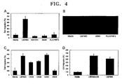
- UCB-MSCs have a strong tropism for U-87 MG, LN18, U138, and U251 cells which are brain tumor cells (see FIGS. 3 and 4 ).
- UCB-MSCs also had tropism for a conditioned media that did not include U-87 MG cells and was obtained by culturing U-87 MG (see FIG. 4 ).
- UCB-MSCs tropism for a brain tumor cell line of UCB-MSCs was compared with that of BM-MSCs that is currently used as a source of stem cells. As a result, it was found that UCB-MSCs have a stronger tropism for a brain tumor cell line than BM-MSCs ( FIG. 5 ). Also, the chemotactic index of UCB-MSCs was largest with respect to the brain tumor cell line among various cancer cell lines ( FIG. 6 ).
- Such a high chemotactic index with respect to the brain tumor cell is an additional advantage of UCB - MSCs, in addition to the fact that UCB-MSCs can be obtained more easily and have more immunological stability than BM-MSCs, proving that UCB-MSCs are a very suitable medium for gene therapy of a brain tumor because a therapeutical gene can be efficiently transferred to the inside or neighboring portions of the brain tumor.
- the tropism of UCB-MSCs in the transwell chamber may be derived by cytokines that are derived to be secreted in the co-culture of two cells.
- cytokines such as IL-8 or GRO- ⁇
- the inventors of the present invention cultured UCB-MSCs alone, cultured U-87 MG alone, and co-cultured UCB-MSCs and U-87 MG, and then analyzed IL-8 mRNA levels of these cells using RT-PCR.
- UCB-MSCs did not express IL-8 in either the presence or absence of U-87 MG cells.
- U-87 MG expressed IL-8 constitutively in both the presence and absence of UCB-MSCs (see FIG. 8 ).
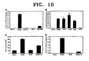
- Treatment of UCB-MSCs with IL-8 significantly enhanced its migration when compared to untreated cells (see (A) of FIG. 9 ).
- IL-8 was artificially overexpressed in A549 that is a low level of IL-8 expressing human lung cancer cells and then, A549 overexpressing IL-8 and UCB-MSCs were co-cultured.
- A549 overexpressing IL-8 and UCB-MSCs were co-cultured.
- IL-8 could be a strong inducer of UCB-MSCs (see FIG. 10C ).
- the inventors of the present invention compared migration characteristics of BM-MSCs and UCB-MSCs with respect to U-87 MG cells or IL-8. As a result, it was found that UCB-MSCs migrate more strongly toward U-87 MG cells or IL-8 than BM-MSCs. UCB-MSCs migration is enhanced dramatically in response to IL-8 treatment, but BM-MSCs migration is weak in response to IL-8 treatment (see FIG. 11 ).
- CXC chemokine receptor 1 CXCR1
- CXC chemokine receptor 2 CXCR2
- RT-PCR analysis using total RNA isolated from UCB-MSCs and BM-MSCs reveal that the PCR product of both CXCR1 and CXCR2 has a higher intensity in UCB-MSCs when compared to BM-MSCs.
- CXCR1 and CXCR2 are highly expressed both in UCB-MSCs and BM-MSCs. Since IL-8 has a high affinity to CXCR1 and CXCR2, increased UCB-MSCs migration toward U-87 MG may be due to up-regulated expression of CXCR1 and CXCR2.
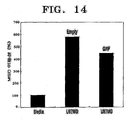
- the inventors of the present invention performed an experiment of introducing a gene coding green fluorescent protein (GFP) into UCB-MSCs. As a result, it was found that GFP was successfully introduced and expressed (see FIG.13 ). Also, it was found that UCB-MSCs overexpressing the gene coding GFP also have a tropism for U87 MG (see FIG. 14 ). Such results show that UCB-MSCs to which a gene or a product thereof is introduced can be transferred to cells secreting IL-8 or GRO- ⁇ .
- GFP green fluorescent protein
- the present Invention relates to a composition for use in transferring a gene or product thereof to a cell expressing IL-8 or GRO- ⁇ by using UCB-MSCs.
- the present invention also relates to a therapeutical composition containing UCB-MSCs for use in transferring a therapeutical or marker gene or a product thereof to a cell that expresses IL-8 or GRO- ⁇ and drives tropism of UCB-MSCs.
- the present invention also relates to a therapeutical pharmaceutical composition, a kit, for use in preventing or treating brain tumors, and a composition containing UCB-MSCs for use in brain tumors.
- the present invention also relates to a composition, a kit for use in diagnosing brain tumors or monitoring the progression of brain tumor treatment and a composition containing UCB-MSCs for use in diagnosing brain tumor or composition containing UCB-MSCs for use in monitoring the progression of brain tumor treatment.
- the present inventive concept relates to:
- the term "umbilical blood” refers to blood taken from an umbilical vein connecting a placenta to a fetus in all mammals including humans.
- UMB-MSC umbilical cord blood derived mesenchymal stem cell
- treatment refers to: preventing disease or disorder development in an animal, preferably a mammal, and more preferably humans which are not yet diagnosed with but susceptible to a disease; suppressing disease progression; and alleviating disease.
- the term "brain tumor” refers to a malignant or benign tumor developing in the brain and the spinal cord and all kinds of tumors developing in a glial cell and a non-glial cell.
- the brain tumor may be a primary brain tumor or a secondary brain tumor.
- the isolating method may be a ficoll-Hypaque density gradient method, but is not limited thereto.
- UCB taken from an umbilical vein after delivery and before the placenta is separated is centrifuged with a ficoll-hypaque gradient to obtain a monocyte, and then the monocyte is washed several times to remove impurities therefrom.
- the resultant monocyte can be directly used for isolation or culture of MSCs, or cryo preserved for a long period of time.
- a MSC can be isolated from UCB and cultured using any known method (see Pittinger MF et al. Science, 284: 143-7, 1999 ; and Lazarus HM et al. Bone Marrow Transplant, 16: 557-64, 1995 ), such as a method disclosed in Korean Publicated Patent No. 2003-0069115 .
- the isolated UCB is centrifuged with, for example, a ficoll-Hypaque gradient to separate monocytes including a hematopoietic cell and a MSC, and then, the monocyte is washed several times to remove impurities therefrom. Then, monocytes are seeded in an appropriate concentration in a culture dish to grow cells in a form of a single layer. These cells were identified with a phase-contrast microscope. In the phase-contrast microscopic image, a colony of cells having a homogeneous spindle shape are MSCs. Then, when cells are cultured and grow, cells are sub-cultured and then multiplied until the number of the cells reaches a desired number.
- UCB-MSCs included in the composition according to the present inventon can be cryopreserved using known methods (see Campos et al., Cryobiology 35:921-924, 1995 ).
- a medium for the cryoporeserving process may include 10 ⁇ 20% fetal bovine serum (FBS) and 10% dimethylsulfoxide (DMSO). The cells are suspended in the medium until the concentration of cells is about 1 ⁇ 10 6 to 5 ⁇ 10 6 cells per 1 mL of medium.
- FBS fetal bovine serum
- DMSO dimethylsulfoxide
- the cell suspension is divided and each part is loaded onto a glass or plastic ample for cryopreservation, and then, the sample is sealed and loaded into a temperature-controlled programed freezer.
- Cells may be frozen using a freezing program providing a temperature change of -1°C/minute so that damage to cells can be reduced when frozen cells are thawed.
- the temperature of the sample reaches -90°C or lower, the sample is moved to a liquid nitrogen storage tank having a temperature of -150°C or lower.
- the sample When the frozen cells are thawed, the sample is quickly moved from the liquid nitrogen storage tank to a water bath having a controlled temperature of 37°C. The thawed content in the ample is immediately moved to a culture dish containing a culture medium in a stabilized condition.
- a culture medium for isolating and culturing a MSC may be a cell culture medium containing 10% to 30% of FBS.
- the cell culture medium may be any cell culture medium that is conventionally used in the art. Examples of the cell culture medium include a DMEM medium, an MEM medium, a ⁇ -MEM medium, a McCoys 5A medium, an eagle's basal medium, a CMRL medium, a Glasgow minimum nessarily medium, a (Ham's) F-12 medium, an iscove's modified Dulbecco's medium (IMDM), a (Liebovitz') L-15 medium, and a RPMI 1640 medium.
- the cell culture medium may be the DMEM medium.
- cells may be suspended so that the concentration of cells is about 5 ⁇ 10 3 to 2 ⁇ 10 4 cells per 1 mL of the medium
- the cell culture medium may further include one or more additives, if required.
- the additives may include at least one material selected from the group consisting of: a serum of fetal calf, horse or human; a penicillin G for preventing microorganism contamination; antibiotics such as streptomycin sulfate or gentamycin; antifungal agents such as amphotericin B or nystatin; and mixtures of at least two materials selected from the forgoing.
- UCB-MSCs may be genetically engineered so as to transfer a therapeutic drug for substantially inhibiting growth of brain tumor cells.
- UCB-MSCs may be genetically engineered so as to transfer a therapeutical gene or product thereof to cells secreting IL-8 or GRO- ⁇ .
- the term “inhibiting” refers to inhibiting cell proliferation and growth, in addition necrosis and apoptosis.
- the therapeutical gene may be, for example, an anti-tumor gene, a gene of an enzyme converting a prodrug into a drug, an antisense or SiRNA of a gene related to a tumor, or oncolytic virus (see Yip S et al., The Cancer J.
- the anti-tumor gene may be a tumor suppressor gene, an apoptosis-inducing factor gene, a cell cycle regulatory gene, or an angiogenesis inhibitor gene.
- the tumor suppressor gene may be selected from the group consisting of a gene of phosphatase and tensin homolog (PTEN), a gene of Maspin, a gene of RUNX3, a gene of Caveolin-1, a gene of nm23, a gene of Rb protein, a gene of Brush-1, a gene of inhibitor of tumor growth (ING-4), a gene of urvivin, a gene of X chromosome linked inhibitor apoptosis protein (XIAP), a gene of neural apoptosis inhibitory protein (NAIP), and genes of proteins regulating these genes.
- PTEN phosphatase and tensin homolog
- XIAP X chromosome linked inhibitor apoptosis protein
- NAIP neural apoptosis inhibitory protein
- the tumor suppressor gene is not limited to those genes described above.
- the apoptosis inducing factor gene may be selected from the group consisting of a gene of cytokine, a gene of interleukin, a gene of a tumor necrosis factor (TNF), a gene of interferon(INF- ⁇ , INF- ⁇ , INF- ⁇ ), a gene of a colony stimulating factor (CSFs), a gene of p53, a gene of Apaf-1, a gene of TNF-related apoptosis-inducing ligand(TRAIL), a gene of Caspase, a gene of Bax, a gene of Bad, a gene of FADD, a gene of JNK, a gene of p38 kinase, and genes of proteins regulating these genes.
- the apoptosis inducing factor gene is not limited to those genes described above.
- the cell cycle regulatory gene may be selected from the group consisting of a gene of cdc2, a gene of Cyclin(Cyclin A, Cyclin D, Cyclin E), a gene of cdc25C, a gene of p21WAF, a gene of p161NK4, a gene of CDK(CDK1, CDK2, CDK4, CDK6), a gene of Rb protein, a gene of E2F.
- the cell cycle regulatory gene is not limited to those genes described above.
- the angiogenesis inhibitor gene may be selected from the group consisting of a gene of thrombospondin-1, a gene of endostatin, a gene of tumstatin, a gene of canstatin, a gene of vastatin, a gene of restin, a gene of a vascular endothelial growth inhibitor, a gene of maspin, a gene of angiopoietins, a gene of 16-kd prolactin fragment, and a gene of Endorepellin.
- the angiogenesis inhibitor gene is not limited to those genes described above.
- the gene of an enzyme converting a prodrug into a drug may be cytosine deaminase converting 5-FC into 5-FU that is an anticancer drug, or a CYP2B1 gene participating in bio-activating cyclophosphamide and ifosfamide which are anticancer drugs, wherein 5-FC is a prodrug of 5-FU.
- the gene of an enzyme converting a prodrug into a drug is not limited to these materials described above.
- the antisense or SiRNA of a gene related to a tumor may be, but is not limited to, an antisense or SiRNA of a gene of Ras family, a gene of c-myc, a gene of abl, a gene of erbB-1, a gene of EGF-R, a gene of Bax, a gene of an Apaf-1 interacting protein (APIP), a gene of Wnt-1-induced secreted protein 1 (WISP-1), a gene of Wnt, a gene of Raf-1, a gene of Src, a gene of Akt, a gene of Erk-1,2 and a gene of BcL-2.
- APIP Apaf-1 interacting protein
- WISP-1 Wnt-1-induced secreted protein 1
- the oncolytic virus that can be carried by UCB-MSCs may be a Herpes simplex virus or a Reovirus type 3, but is not limited thereto.
- UCB-MSCs to which a desired gene is introduced in an expressionable state can be appropriately manufactured using techniques that are known in the art.
- a vector including a to-be-introduced gene is prepared (see Dehari H et al., Cancer Gene Ther., 10, 75-85, 2003 ; WO07/037653 ) and then the vector can be transduced in ex vivo into primary culture MSCs.
- the vector include adenovirus vector, retrovirus vector, adeno-related virus vector, herpes simplex virus vector, SV40 vector, poliomavirus vector, papillomavirus vector, picarnovirus vector, vacciniavirus vector, and lentivirus vector.
- MSCs 5x10 5 cells
- a solution including an adenovirus vector to which the adenovirus gene is introduced are incubated at 37°C in a 5% CO 2 incubator for 1 hour, so that the MSCs are infected with the adenovirus gene.
- the infected MSCs are washed with a phosphoric acid buffer solution and then a conventional medium is applied thereto.
- a desired gene can be introduced into UCB-MSCs by using a naked DNA and any method selected from a calcium-phosphate method, a cationic liposome method, and an electrophoration method.
- a fusion protein gene of protein transduction domain (PTD) and anti-tumor protein can be introduced to UCB-MSCs using the fusion protein gene-including vector (see Wu SP et al. Biochem Biophys Res Commun. 346(1), 1-6, 2006 ).
- the vectors may further include a gene marker for an additional historical examination.
- the gene marker may be, for example, a gene for coding a chromogenic or fluorescent protein, such as lacZ or a green fluorescent protein (GFP), but is not limited thereto (see Yip Set al., The Cancer J. 9(3), 189-204, 2003 ).
- Brain tumors that can be diagnosed, prevented, and treated using the pharmaceutical composition according to the present invention may be primary brain tumors or secondary brain tumors.
- Examples of brain tumors that can be diagnosed, prevented, and treated using the composition according to the present invention include Astrocytoma, Pilocytic astrocytoma, Low-grade Astrocytoma, Anaplastic Astrocytoma, Glioblastoma Multiforme, Brain Stem Glioma, Ependymoma, Subependymoma, Ganglioneuroma, Mixed Glioma, Oligodendroglioma, Optic Nerve Glioma, Acoustic Neuroma, Chordoma, CNS Lymphoma, Craniopharyngioma, Hemangioblastoma, Medulloblastoma, Meningioma, Pineal Tumors, Pituitary Tumors, Primitive Neuroectodermal Tumors, Rhabdoid Tumors, Schwannoma, Cys
- composition according to the present invention may be administered together with any known anticancer drug.
- UCB-MSCs do not express HLA-DR (Major Histocompatibility Complex class II) that is a major cause for immunological rejection when a tissue or organ is transplanted (see Le Blanc, KC, Exp Hematol, 31:890-896, 2003 ; and Tse WT et al., Transplantation, 75:389-397, 2003 ), immunological reactions, such as rejection, which are major problems of transplantation do not occur or can be minimized.
- UCB-MSCs included in the pharmaceutical composition according to the present invention can be taken from, in addition to a self-derived UCB, another subject-derived UCB. According to the present invention, UCB-MSCs can be used after being cryopreserved.
- the UCB-MSCs-containing pharmaceutical composition for gene therapy or for preventing or treating diseases according to the present invention may further include pharmaceutically acceptable additives, in addition to the effective component.
- the UCB-MSCs-containing pharmaceutical composition may be formed into a suitable formulation that can be administered to a body.
- the suitable formulation may be a non-oral administration formulation, such as an injectable formulation or a locally administrable formulation.
- a sterilized solution or suspension that includes water or a pharmaceutically acceptable solvent can be non-orally administered in an injectable form.
- water or the pharmaceutically acceptable solvent is appropriately combined with a pharmaceutically acceptable carrier or medium, thereby forming an injectable formulation in a conventionally acceptable unit dosage.
- the pharmaceutically acceptable carrier or medium may include sterile water, saline, vegetable oils, emulsifier, suspension, surfactant, stabilizer, excipient, vehicle, an antiseptic substance, a binder, etc.
- the injectable formulation described above can be non-orally administered, specifically directly administered to a site of disease, by using a conventional method.
- the injectable formulation described above can be administered through a cerebrospinal fluid, a vein, or an artery supplying blood to the site of disease, preferably directly administered to neighboring portions of the site of disease in the brain or the spinal cord or the opposite portions thereto.
- the injectable formulation described can be administered using a clinical method developed by Douglas Kondziolka in Pittsburgh in 1998. That is, a skull of a subject to be administered is cut to a diameter of about 1 cm, the size of a pea, and then a MSC solution mixed with a Hank's balanced salt solution (HBSS) is injected thereto.
- the injecting the MSC solution is performed using an injector including a long needle and a stereotactic frame for accuratly injecting the MSC solution into the brain.
- the daily dose of UCB-MSCs may be 1x10 4 to 1x10 7 cells/kg body weight, preferably 5x10 5 to 5x10 6 cells/kg body weight.
- the daily dose may be administered at once or be divided for several treatments.
- the administration dose of UCB-MSCs may vary according to the kinds of diseases to be treated, the severity of disease to be treated, administration routes, and the weight, age, and gender of a patient. Accordingly, the administration dose described above does not limit the scope of the present invention.
- the present invention also discloses a method of treating a disease related to a cell that produces IL-8 or GRO- ⁇ and induces a tropism of UCB - MSCs, or a brain tumor, wherein the method includes administering a therapeutically effective amount of the pharmaceutical composition according to the present invention to a patient.
- UCB-MSCs included in the pharmaceutical composition may be taken from UCB of the patient itself or other humans or animals used for medical purposes.
- UCB-MSCs may be used after cyropreservation.
- Such a method using UCB-MSCs is not limited to humans, and can be applied to other mammals.
- the present invention also discloses a method of diagnosing a disease related to a cell that produces IL-8 or GRO- ⁇ and induces a tropism of UCB - MSCs, or a brain tumor, and a kit used to perform the method, wherein the method uses UCB-MSCs.
- UCB-MSCs may be labeled with a detectable marker.
- the UCB-MSCs labeled with the detectable marker can be visualized by a state-of-the-art technique, and can be tracked in real time in a live animal.
- the detectable marker may be a luciferase-containing enzyme based fluorescent detector or Tat peptide-derivatized magnetic nanoparticles ( Yip S et al., The Cancer J. 9(3), 189-204, 2003 ). If a stem cell expressing luciferase is used, the administered stem cell migrating to the site of disease can be tracked by identifying bioluminescence in real time and thus, diseases can be diagnosed and the site of disease can be identified (see Weissleder R et al., Nat Med 9, 123-128, 2003 ). Tat peptide-derivatized magnetic nanoparticles are connected to UCB-MSCs using a method developed by Lewin et al.
- UCB-MSCs labeled with a marker can be administered in vivo and then, by identifying a location where UCB-MSCs gather, the tumor and the site of the tumor can be identified.
- UCB-MSCs labeled with the marker can be administered during the brain tumor treatment or after the brain tumor treatment to identify the location and size of the distribution of the administered UCB-MSCs. That is, the progress of the brain tumor treatment can also be monitored.
- the present invention also discloses a method of monitoring progression of the brain tumor treatment and a kit used to perform the method, wherein the method uses UCB-MSCs.
- the monitoring method and the kit used to perform the method can also be applied to a disease related to a cell that produces IL-8 or GRO- ⁇ and induces a tropism of UCB-MSCs.
- U-87 MG, A549, KATO III, PLC/PRF5, LN18, U138 and U251 cell lines were purchased from American Type Culture Collection (ATCC) and used in the present experiments.
- A549, KATOIII and PLC/PRF5 cells were cultured in RPMI containing 10-20% (v/v) FBS (HyClone, Logan, UT, US) and Gentamicin at 37°C in a 5% CO 2 incubator.
- U-87 MG CELLS, LN18, U138 and U251 cells were grown in an Eagle's minimum essential medium (MEM) containing 10-20% (v/v) FBS.
- MEM Eagle's minimum essential medium
- Bone marrow-derived mesenchymal stem cell (BM-MSCs) was purchased from LONZA.
- BM-MSCs and established UCB-MSCs were cultured in ⁇ -MEM media containing 10-20% FBS.
- UCB samples were collected from the umbilical vein of deliveries, with informed maternal consent. Specifically, a 16-gauge needle of a UCB collection bag containing 44 mL of CPDA-1 anticoagulant (Greencross Co., Yongin, Kyungki-do, Korea) was inserted into the umbilical vein and UCB was collected by gravity. In all cases, UCB harvests were processed within 48 hours of collection, with viability of 90% or more.
- CPDA-1 anticoagulant Greencross Co., Yongin, Kyungki-do, Korea
- UCB-MSCs prepared according to Example 1 were centrifuged with a Ficoll-Hypaque gradient (produced by Sigma Co., density: 1.077 g/mL), and then washed several times to remove impurities. 10 to 20% FBS (HyClone Co.)-containing a basic medium ( ⁇ -MEM, Gibco BRL Co.) was added to the resultant product to suspend UCB-MSCs.
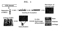
- the UCB-MSCs were portioned at a suitable concentration into each of 10 to 20% FBS- containing a basic media, and then cultivated at 37°C in a 5% CO 2 incubator while the medium was altered twice in a week ( FIG. 1 ). When the cultured cells formed a single layer, MSCs expanding in a spindle shape were identified with a phase-contrast microscope. Then, sub-cultivation was repeatedly performed until the MSCs expanded sufficiently.
- UCB-MSCs cultivated according to Example 2 were dyed with PKH-26 (Sigma Co.) using a method disclosed in a reference [ Barreda DA et al., Developmental and Comparative Immunology, 24:395-406, 2000 ].
- UCB-MSCs were separated from the cell culture dish by using Trypsin and then, 2x 10 7 cells were washed with an FBS-free medium. The washed cells were collected using a centrifuge and then suspended in 1 mL of Diluent C in a kit provided by a manufacturer. Then the resultant cell suspension solution(2x) was mixed with 1ml of the PKH fluorescent dye solution(2x) and then the mixture was reacted at 25°C for 5 minutes.
- FBS fetal bovine serum
- hUCB-MSCs Human UCB-MSCs
- PKH-26 produced by Sigma
- FALCON transwell chamber
- cancer cell lines-free MSCs were cultured in the same condition as described above.
- PKH26-labeled hUCB-MSCs were cultured; and in lower compartment, each of human brain tumor cell line U87-MG, human rectal cancer cell line LS174-T, human B lymphocyte NC-37, and mouse's fibroblast NIH3T3 was cultured.
- PKH26-labeled UCB-MSCs were co-cultured with various tumor cell lines in a transwell chamber and then, PKH-labeled mesenchymal stem cells that have migrated into the lower compartment were counted.
- UCB-MSCs had a strong tropism for U87-MG, LN18, U138, and U251 that are brain tumor cell lines, and a weak tropism for other tumor cells ( FIGS. 3 and 4 ).
- the left image shows the case of the control group in which a human cell line-free medium was added instead of tumor cells
- the right image shows the case in which UCB-MSCs were co-cultured with U87-MGcell.
- PKH26-labeled UCB-MSCs had a tropism for a conditioned medium that did not contain U-87 MG cells but U-87 MG cells had been cultured therein (see D of FIG. 4 ). Such results show that the conditioned medium contained soluble factors that function to attract UCB-MSCs toward U-87 MG cells.
- Example 5 Comparison of tropism of BM-MSCs for U87-MG and tropism of UCB-MSCs for U87-MG
- a tropism of BM-MSCs for U87-MG was compared to a tropism of UCB-MSCs for U87-MG, using a transwell chamber.
- U87-MG cancer cells or medium were placed in a lower compartment, and BM-MSCs and UCB-MSCs each were placed in an upper compartment. In all cases, the culturing was performed for two days. As a result, UCB-MSCs were found to have a stronger tropism for U87-MG than BM-MSCs ( FIG. 5 ).
- UCB-MSCs donated from four donors in the upper compartment and each of A549 that is lung cancer cell, HeLa that is cervical cancer cell, and U87-MG that is brain cancer-glioma cell in the lower compartment were co-cultured in a transwell chamber. Then, chemotactic indices of of UCB-MSCs in each case were compared ( FIG. 6 ).
- A549, HeLa, and U87-MG cells were purchased from American type culture collection (ATCC). Each of A549 and U87 MG cells was cultured in 10-20% bovine serum-containing RPMI1640, and HeLa was cultured in DMEM. In each case, the chemotactic index was calculated by dividing the number of UCB-MSCs migrated toward U87 MG by the number of UCB-MSCs migrated in the control experiment. A tropism of UCB-MSCs for those cancer cells was analyzed. As a result, it was found that UCB-MSCs had the stronggest tropism for U87 MG that is a brain tumor cell line.
- a film to which antibodies of various cytokine were attached was taken from a kit for a cytokine array purchased from R&D System Co. and was reacted with a blocking solution for one hour. Separately, amounts of the media prepared by co-culturing UCB-MSCs and each of three kinds of human cells, including U87-MG tumor cell were adjusted to 1.5 mL or less, and each medium was mixed with the mixed antibodies of cyokines contained in the kit and an antigen-antibody reaction was induced for one hour. The medium in which the supplied cytokine antibodies were combined with secreted cytokines was reacted with the film that was subjected to the blocking for 4°C for 12 hours.
- the film was placed in a washing solution and washed, and then washed with tertiary distilled water. Then the film was dried at room temperature. After repeating the washing and drying process two or three times, the film was reacted in a solution containg streptavidin-HRP for 30 minutes. Then the film was washed with a washing solution three times, reacted with a chromattic reagent, and then exposed to an X-ray film in the dark room.
- FIG. 7 shows results obtained by analyzing cell lysates and cell culture supernatants through a cytokine array. Array results and information about changed spots compared to the control group are shown in FIG. 1 .
- cytokines in parentheses are cytokines that were derived by the co-culturing with tumor cells. It is highly likely that these cytokines may induce a tropism of UCB-MSCs.
- FIG. 8A shows cytokine antibody array analysis results from the medium prepared by culturing UCS-MSCs only and a control medium.
- FIG. 8B shows cytokine antibody array analysis results from the medium prepared by culturing the U-87 MG only, and the medium prepared by co-culturing UCB-MSCs and U-87 MG.
- FIG. 8C shows results identified by isolating mRNA from UCB-MSCs cultured with and without U-87 MG (left) or from U-87 MG cultured with and without UCB-MSCs (right).
- RT-PCR was performed with IL-8 specific primers and GAPDH was used as a control.
- GAPDH GAPDH
- UCB-MSCs The effect of IL-8, GRO- ⁇ , MCP-1(RND Systems, MN, USA) with respect to migration of UCB-MSCs was measured using traswell migration assay.
- PHK-26-labelled UCB-MSCs were placed in the upper compartment and no cells were placed in the lower compartment.
- UCB-MSCs were cultured in an IL-8 free medium or in a medium containing different concentrations of recombinant humane IL-8 for 24 hours. As a result, it can be seen that UCB-MSCs migrated more when treated with IL-8 than when they were not treated with IL-8 ( FIG. 9A ).
- IL-8 receptor on UCB-MSCs can be effectively blocked by anti-human CXC chemokine receptor 1 (CXCR1) antibodies.
- CXCR1 CXC chemokine receptor 1
- recombinant IL-8 was again applied to UCB-MSCs.
- IL-8 mediated migration of UCB-MSCs was reduced, in a dose-dependant manner, by anti-CXCR1 treatment ( FIG. 9B ).
- Anti-CXCR2 treatment also showed the same effect.
- GRO- ⁇ treatment also enhanced UCB-MSCs migration when compared to untreated cells ( FIG. 9C ).
- GRO- ⁇ also belongs to the CC subfamily and can interact with the CXCR2 receptor [see Wuyts A. et al. Eur J Biochem 255, 67-73 (1998 )].
- UCB-MSCs migrates toward A549 cell overexpressing IL-8
- FIG. 10A shows ELISA results indicating concentrations of IL-8 secreted in media in which U87 MG (brain tumor), KATOIII (gastric cancer), A549 (lung cancer), and PLC/PRF5 (liver cancer) cells were cultured.
- the concentration of IL-8 was measured per 1x10 5 cells.
- U-87 MG showed the highest IL-8 production.
- This data suggested that UCB-MSCs had a strong migration attraction toward IL-8 producing cells.
- UCB-MSCs has a tropism for IL-8 secreted by brain tumor cells.
- FIG. 10C shows migration results of UCB-MSCs after IL-8 gene was introduced to A549 cells that secrete a low level of IL-8 by using a lipofectamine reagent and then IL-8 was overexpressed. Referring to FIG. 10C , more UCB-MSCs migrated in A549 cells having overexpreesed IL-8 than in A549 cells. Accordingly, it can be seen that IL-8 could be a strong inducer of UCB-MSCs migration.
- FIG. 10D shows the concentration of IL-8 secreted into a medium in a condition of C measured by ELISA.
- FIG. 11 shows tropism characteristics of BM-MSCs and UCB-MSCs migrated toward the lower compartment B, that is, toward U87 MG.
- B of FIG. 11 shows tropism characteristics of BM-MSCs and UCB-MSCs migrated toward the lower compartment B when they are treated with IL-8 for 14 hours. Referring to FIG. 11 , more UCB-MSCs migrated than BM-MSCs. Therefore, it can be seen that UCB-MSCs strongly corresponded to IL-8 and migrated more, but BM-MSCs relatively weakly corresponded to IL-8.
- FIG. 12 A shows results of RT-PCR which was performed with CXCR1 and CXCR2 primers after mRNA was isolated in each of CXCR1 and CXCR2. GAPDH was included into each sample to assess the quantity of the isolated RNA.
- FIG. 12 C shows analysis results obtained by immuostaining UCB-MSCs and BM-MSCs with anti-CXCR1 and CXCR2 antibodies to identify expression levels of CXCR1 and CXCR2 (x400).
- UCB-MSCs and BM-MSCs showed high expression levels of CXCR1 and CXCR2.
- FIG. 12 D shows analysis results obtained by immuostaining UCB-MSCs and BM-MSCs with a secondary antibody only without anti-CXCR1 and CXCR2 antibodies to identify antigen specificity of the anti-CXCR1 and CXCR2 antibodies.
- a green fluorescent protein was overexpressed using an electroporation method with a human MSC neucleofector produced by amaxa biosystem Co. and an electroporation method. 4x10 5 cells of UCB-MSCs were cultured for two days and then placed in a mixture of 5 mg of a GFP encoding gene and 100 ml of a human MSC nucelofector for 15 minutes and the gene was introduced in an electroporator. The resultant UCB-MSCs were moved to a plate and after 24 hours, expression levels of GFP were identified with a fluorescent microscope. The GFP could be identified in a cytoplasm of each MSC (see FIG. 13 ).
- each of a GFP encoding gene and an empty gene was overexpressed in UCB-MSCs and then, a tropism of the resultant UCB-MSCs for U-87 MG was identified.
- UCB-MSCs expressing GFP were placed in an upper compartment of a transwell chamber and U-87 MG was placed in a lower compartment of the transwell chamber, and then UCB-MSCs expressing GFP and U-87 MG were co-cultured for 24 hours. Among cells migrated toward the lower compartment, GFP positive cells were identified. As a result, it can be seen that UCB-MSCs overexpressing GFP due to introduction of a GFP gen had a strong tropism for U87 MG ( FIG. 14 ).
Landscapes
- Health & Medical Sciences (AREA)
- Life Sciences & Earth Sciences (AREA)
- Public Health (AREA)
- Veterinary Medicine (AREA)
- Animal Behavior & Ethology (AREA)
- General Health & Medical Sciences (AREA)
- Epidemiology (AREA)
- Cell Biology (AREA)
- Engineering & Computer Science (AREA)
- Chemical & Material Sciences (AREA)
- Medicinal Chemistry (AREA)
- Pharmacology & Pharmacy (AREA)
- Biomedical Technology (AREA)
- Biotechnology (AREA)
- Zoology (AREA)
- Virology (AREA)
- Immunology (AREA)
- Developmental Biology & Embryology (AREA)
- Vascular Medicine (AREA)
- Nuclear Medicine, Radiotherapy & Molecular Imaging (AREA)
- Genetics & Genomics (AREA)
- Molecular Biology (AREA)
- Radiology & Medical Imaging (AREA)
- Hematology (AREA)
- Organic Chemistry (AREA)
- General Chemical & Material Sciences (AREA)
- Chemical Kinetics & Catalysis (AREA)
- Medicines Containing Material From Animals Or Micro-Organisms (AREA)
- Medicines That Contain Protein Lipid Enzymes And Other Medicines (AREA)
- Micro-Organisms Or Cultivation Processes Thereof (AREA)
- Medicinal Preparation (AREA)
Description
- The present invention relates to a gene therapy composition for use in transferring a therapeutical gene, a marker gene, or a product thereof to a cell that expresses interleukin-8(IL-8) or GRO-α and induces tropism of umbilical cord blood-derived mesenchymal stem cells or mesenchymal stem cells isolated from umbilical cord blood and/or the mesenchymal stem cells expanded from said mesenchymal stem cells (UCB-MSCs), wherein the gene therapy composition includes UCB-MSCs.
- The present invention also relates to a composition for use treating disease related to a cell expressing IL-8 or GRO-α, or brain tumors in gene therapy, wherein the composition includes UCB-MSCs.
- The present invention also relates to a composition or kit for use in diagnosing brain tumors, preventing brain tumors, treating brain tumors, or monitoring brain tumor treatment progression, using UCB-MSCs.
- It is known that stem cells migrate toward sites of pathology. Recently, it was found that bone marrow-derived mesenchymal stem cells (BM-MSCs) have a tropism for tumors and migrate toward tumor sites. Such BM-MSCs that can migrate to sites of specific tumors may prove to be a useful tool in gene therapy. For example, BM-MSCs having a tropism for tumors can be used as vehicles for transferring a therapeutic suicide gene to tumor sites [see Ponte A.L. et al., Stem Cells, 25,1737-1745 (2005); Kahler C.M. et al., Respir Res 8, 50 (2007)]. Despite this interesting phenomenon, the molecular mechanisms regulating MSCs trafficking to tumor are undear.
- Growing evidence over several years indicates that induction of BM-MSCs migration seems to be stimulated by several soluble factors. Recently, monocyte chemoattractant protein-1 (MCP-1) secreted from breast cancer cells has been shown to stimulate BM-MSCs migration [see Dwyer R.M. et al. Clin Cancer Res 13, 5020-5027 (2007)]. Furthermore, chemokine ligand2 (CCL2) and chemokine ligand10 (CCL-10) can induce the migration of neural progenitor cells to sites damaged within the middle cerebral artery occlusion (MCAo) stroke model [see J Neurosci Res 85, 2120-2125 (2007)]. An insulin-like growth factor-1 (IGF-1) markedly increased the rat BM-MSCs migratory response [see Li Y. et al. Biochem Biophys Res Commun 356, 780-784 (2007)]. Therefore, identifing the soluble factors that affect migration events of MSCs is important for understanding how MSCs migrate toward tumors or damaged tissues.
- Genes introduced to BM-MSCs are over-expressed in vivo and show bioactivity. For example, BM-MSCs to which a human hAng1 gene is introduced stimulate generation of blood vessels in an infarction site of an acute myocardial infarction model animal [see Sun L. et al., Biochemical Biophysical Research Communication 357(2007) 779-784], BM-MSCs overexpressing Akt surprisingly treat myocardial infarction and improve functions of heart [see Nicolas N.et al., Molecular Therapy 14(6), 840-850, 2006], Bcl-2 gene-modified BM-MSCs prevent apoptosis and improve functions of heart [see Stem Cells 25, 2118-2127 (2007)], and BM-MSCs overexpressing endothelial nitric oxide synthase recovers the damage of right ventricular caused by pulmonary hypertension [see Sachiko et al., Circulation, 114[suppl I]:I-181~I-185]. These results indicate that MSCs to which genes are introduced can be used as a tool in gene therapy.
- Meanwhile, in general, cells of the central nervous system are well regulated, wherein the central nervous system consists of a brain and a spinal cord. However, when this regulation collapses, cells are continuously divided and tumors are formed. Tumors can be categorized as benign tumors or malignant tumors. The central nervous system has neurons, and glia cells that support and protect the neurons. Tumors generated in glia cells are known as glioma. Glioma accounts for 50% of primary brain tumors and accounts for 15% of primary spinal cord tumors. In addition, brain tumors include neural tumors, blood vessel tumors, and gland tumors. There is also a secondary brain tumor caused by other tumors developed in other sites of the body. The secondary brain tumor is the most common type of brain tumor
- Treating brain tumors are difficult due to the sites of the tumours. Brain tumors can be treated by physical surgery or chemotherapy. For physical surgery, when tumor sites are completely removed, complications are likely to occur. For chemotherapy, a high-concentration anticancer drug needs to be injected due to a brain-blood barrier, and thus, it seriously damages other organs. Recently, gene therapy has been used to treat brain tumors. In gene therapy, a gene is introduced for suppressing growth of cancer cells by using a virus vector. Since the virus vector does not have a selective migration capability toward a target cancer site, the virus vector is surface-modified to obtain such capability. However, there is a limit to migrate a sufficient amount of virus vectors to the target cancer site.
- Research results on a homing effect, which is a phenomenon in which stem cells migrate toward a disease site, have been disclosed, indicating that stem cells can be useful delivery media for treating brain tumors. However, mechanisms regulating stem cells trafficking to tumors are unclear. It is known that neural stem cells have a tropism for a type of brain tumor, that is, malignant glioma. Based on this theory, research into a method of transferring genes to a brain tumor site by using neural stem cells that function as a vehicle is being conducted (see Yip S et al., The Cancer J 9(3), 189-204, 2003; Kim SK et al., Clin Cancer Res 12(18), 5550-5556, 2006). Yip et al. found that brain tumors can be treated with neural stem cells carrying an immune regulatory gene, an apophosis promoting gene, a pro-drug converting enzyme, an oncolytic virus, etc. Brown et al. identified that brain tumors can be effectively treated by injecting a cytosine deaminase gene-containing vector into the brain, wherein the cytosine deaminase gene changes 5-fluorocytosine (5-FC) into 5-fluorouracil (5-FU), wherein 5-FU is an anticancer drug and 5-FC is a prodrug of 5-FU (see Brown AB et al., Human Gene Ther. 14(18), 1777-1785, 2003). Ehtesham et al. reported that growth of brain tumors was decreased by injecting neural stem cells treated for delivering interleukin-12 or a tumor necrosis factor-related apoptosis-inducing ligand (Cancer Res 62, 5657-5663, 2002; Cancer Res 62, 7170-7174, 2002). However, using neural stem cell in clinical experiments causes ethical problems related to how neural stem cells are taken, and immunological rejection caused by allogenic transplantation. Accordingly, there is a need to find other types of stem cells that do not cause these problems and can be easily obtained.
- Akira et al. disclosed that BM-MSCs have a tropism for brain tumors (see Cancer Res 65(8), 3307-3316, 2005). BM-MSCs can be taken from patients. When BM-MSCs are injected through autologous transplantation, immunological rejection does not occur, which is an advantage for a clinical use. In a study, human BM-MSCs were injected into nude mice having skulls transplanted with human glioma cell lines through a carotid artery. As a result, the human BM-MSCs were found only in glioma and not in a normal part of the brain adjacent to glioma. In addition, even when human BM-MSCs were transplanted into a skull, human BM-MSCs migrated toward glioma. When human BM-MSCs were infected with an adenovirus vector containing cDNA of an IFN-beta gene and then the resultant vector was injected into a glioma-transplanted skull of a nude mouse through a carotid artery, the lifetime of the nude mouse was increased.
WO07/037653A1 - Unlike bone marrow, umbilical cord blood (UCB) having many MSCs can be easily taken from umbilical cords which are discarded in delivery processes. Also, the UCB storage industry is well established and thus, it is easy to find donors. Even when MSCs taken from other human-induced UCB are used, immunological rejection does not occur after transplantation. Accordingly, high immunological stability can be obtained. Therefore, it is very important to identify whether a disease such as a brain tumor can be treated based on tropisom of UCB MSCs. However, such attempts for identifying availability of UCB-MSCs have not yet been made. All the information disclosed in the present specification is used only to help understanding of the background of the present inventive concept and cannot be prior art.
- Target gene therapy recently developed uses specific migration characteristics of mesenchymal stem cells (MSCs), in which therapeutical genes are introduced to MSCs and then the resultant MSCs are migrated to a disease site and the disease is treated. To develop gene therapy using tropism of MSCs, a molecular mechanism regulating the migration of MSCs toward tumors should be completely understood. However, the molecular mechanism has not yet been determined. Accordingly, the present inventive concept is to find out the molecular mechanism of UCB-MSCs migrating toward a tumor cell and uses the molecular mechanism in gene therapy.
- Although research into whether brain tumors can be treated with neural stem cells or bone marrow-derived mesenchymal stem cells (BM-MSCs) are being conducted, collecting neural stem cells and BM-MSCs may lead to ethical problems, immunological rejection, and mental and physical stress of a subject from which BM-MSCs are taken. Accordingly, the present inventive concept also provides a stem cell that can be taken without these problems described above and have better tropism for brain tumors.
- The inventors of the present invention have conducted research in order to solve these problems, and found that mesenchymal stem cells isolated from umbilical cord blood and/or the mesenchymal stem cells expanded from said mesenchymal stem cells (UCB-MSCs) have a tropism for brain tumor cells and that UCB-MSCs have a stronger migration capability than mesenchymal stem cells isolated from bone marrow and/or the mesenchymal stem cells expanded from said mesenchymal stem cells (BM-MSCs). The inventors have provided a therapeutical application for treating brain tumor using UCB-MSCs.
- Also, the inventors of the present invention found the tropism of UCB-MSCs is affected by interleukin-8 (IL-8) or GRO-α. Based on this finding, the inventors of the present invention provide a composition for use in delivering a therapeutical gene or a product thereof to a cell that expresses IL-8 or GRO-α and induces the tropism of UCB-MSCs, and a therapeutical application thereof.
- Umbilical cord blood derived mesenchymal stem cells (UCB-MSCs) in the composition according to the present invention has a selective tropism for cells that express IL-8 or GRO-α and thus induce the tropism of UCB-MSCs or brain tumor cells. The tropism capability of the UCB-MSCs is better than that of other stem cells and thus, therapeutical genes or a product thereof can be more effectively delivered than when other conventional stem cells are used. Accordingly, a pharmaceutical composition or a kit comprising UCB-MSCs according to the present invention can be used to diagnose, prevent, and treat diseases related to cells expressing interleukin(IL)-8 or GRO-α or brain tumors.
- The inventors of the present invention have studied stem cells having an effective tropism for tumors, and surprisingly found that umbilical cord blood-derived mesenchymal stem cells (UCB-MSCs) have a strong tropism for brain tumors, specifically a stronger tropism for brain tumors than bone marrow-derived mesenchymal stem cells (BM-MSCs), which has never been known before. Also, the inventors found that interleukin (IL)-8 or GRO-α relates to the tropism of UCB-MSCs.
- The inventors co-cultured UCB-MSCs and representative tumor cell lines to identify characteristics of the tropism of UCB-MSCs and cytokine related to those tumor cell lines. Specifically, UCB-MSCs, and one selected from human brain tumor cell lines, such as U-87 MG, LN18, U138, or U251 cells; human rectal cancer cell line such as LS-174T; human B lymphocyte such as NC37; mouse's fibroblast (NIH3T3); a gastric cancer cell line such as KATO III; a lung cancer cell line such as A549, and a liver cancer cell line such as PLC/PRF5 were co-cultured in a transwell chamber to measure mobility of UCB-MSCs. As a result, it was found that UCB-MSCs have a strong tropism for U-87 MG, LN18, U138, and U251 cells which are brain tumor cells (see
FIGS. 3 and4 ). UCB-MSCs also had tropism for a conditioned media that did not include U-87 MG cells and was obtained by culturing U-87 MG (seeFIG. 4 ). - Also, the tropism for a brain tumor cell line of UCB-MSCs was compared with that of BM-MSCs that is currently used as a source of stem cells. As a result, it was found that UCB-MSCs have a stronger tropism for a brain tumor cell line than BM-MSCs (
FIG. 5 ). Also, the chemotactic index of UCB-MSCs was largest with respect to the brain tumor cell line among various cancer cell lines (FIG. 6 ). Such a high chemotactic index with respect to the brain tumor cell is an additional advantage of UCB-MSCs, in addition to the fact that UCB-MSCs can be obtained more easily and have more immunological stability than BM-MSCs, proving that UCB-MSCs are a very suitable medium for gene therapy of a brain tumor because a therapeutical gene can be efficiently transferred to the inside or neighboring portions of the brain tumor. - The tropism of UCB-MSCs in the transwell chamber may be derived by cytokines that are derived to be secreted in the co-culture of two cells. Thus, two cells were co-cultured in a transwell chamber to prepare a medium and then, the medium was analyzed using the cytokine array. As a result, it was identified that high levels of cytokines, such as IL-8 or GRO-α, were secreted in a medium in which UCB-MSCs and U87 MG had been co-cultured (see
FIG. 7 ). Accordingly, it is highly likely that these cytokines may derive a tropism of the UCB-MSCs.. - The inventors of the present invention cultured UCB-MSCs alone, cultured U-87 MG alone, and co-cultured UCB-MSCs and U-87 MG, and then analyzed IL-8 mRNA levels of these cells using RT-PCR. As a result, UCB-MSCs did not express IL-8 in either the presence or absence of U-87 MG cells. However, U-87 MG expressed IL-8 constitutively in both the presence and absence of UCB-MSCs (see
FIG. 8 ). Treatment of UCB-MSCs with IL-8 significantly enhanced its migration when compared to untreated cells (see (A) ofFIG. 9 ). However, when UCB-MSCs were pre-incubated with anti-CXCR1 antibodies that are antibodies with respect to an IL-8 receptor and recombinant IL-8 was applied to UCB-MSCs, IL-8 mediated migration of UCB-MSCs were reduced in a dose-dependent manner by anti-CXCR1 treatment ((B) ofFIG. 9 ). Anti-CXCR2 treatment also showed the same effect. Similarly, GRO-α treatment also enhanced UCB-MSCs migration when compared to untreated UCB-MSCs ((C) ofFIG. 9 ). In contrast, there were no significant differences in UCB-MSCs migration in cultures treated with MCP-1 ((D) ofFIG. 9 ). This data indicates that IL-8 and GRO-α participate in UCB-MSCs migration toward U-87 MG cells. - The co-relationship between the concentration of IL-8 secreted by each cancer cell and UCB-MSCs migration was measured. As a result, U-87 MG, which migrated the highest concentration of UCB-MSCs, showed the highest IL-8 production (
FIG. 10A ). The inventor also measured IL-8 secretion level in various glioma cells. All tested glioma cell lines which were target cell of UCB-MSC tropism also showed high secretion level of IL-8 ((B) ofFIG. 10 ). This data suggested that UCB-MSCs had a strong migration attraction toward IL-8 secreting cells. In order to identify this, IL-8 was artificially overexpressed in A549 that is a low level of IL-8 expressing human lung cancer cells and then, A549 overexpressing IL-8 and UCB-MSCs were co-cultured. As a result, it was found that although the UCB-MSCs had a poor migration attraction toward A549, UCB-MSCs had a strong migration attraction toward A549 overexpressing IL-8. Therefore, IL-8 could be a strong inducer of UCB-MSCs (seeFIG. 10C ). - The inventors of the present invention compared migration characteristics of BM-MSCs and UCB-MSCs with respect to U-87 MG cells or IL-8. As a result, it was found that UCB-MSCs migrate more strongly toward U-87 MG cells or IL-8 than BM-MSCs. UCB-MSCs migration is enhanced dramatically in response to IL-8 treatment, but BM-MSCs migration is weak in response to IL-8 treatment (see
FIG. 11 ). - Expression levels of CXC chemokine receptor 1 (CXCR1) and CXC chemokine receptor 2 (CXCR2) in UCB-MSCs and BM-MSCs were compared by measuring mRNA and protein (see
FIG. 12 ). RT-PCR analysis using total RNA isolated from UCB-MSCs and BM-MSCs reveal that the PCR product of both CXCR1 and CXCR2 has a higher intensity in UCB-MSCs when compared to BM-MSCs. In regard of protein expression of CXCR1 and CXCR2, CXCR1 and CXCR2 are highly expressed both in UCB-MSCs and BM-MSCs. Since IL-8 has a high affinity to CXCR1 and CXCR2, increased UCB-MSCs migration toward U-87 MG may be due to up-regulated expression of CXCR1 and CXCR2. - The inventors of the present invention performed an experiment of introducing a gene coding green fluorescent protein (GFP) into UCB-MSCs. As a result, it was found that GFP was successfully introduced and expressed (see
FIG.13 ). Also, it was found that UCB-MSCs overexpressing the gene coding GFP also have a tropism for U87 MG (seeFIG. 14 ). Such results show that UCB-MSCs to which a gene or a product thereof is introduced can be transferred to cells secreting IL-8 or GRO-α. - Based on the results described above, the present Invention relates to a composition for use in transferring a gene or product thereof to a cell expressing IL-8 or GRO-α by using UCB-MSCs. The present invention also relates to a therapeutical composition containing UCB-MSCs for use in transferring a therapeutical or marker gene or a product thereof to a cell that expresses IL-8 or GRO-α and drives tropism of UCB-MSCs. The present invention also relates to a therapeutical pharmaceutical composition, a kit, for use in preventing or treating brain tumors, and a composition containing UCB-MSCs for use in brain tumors. The present invention also relates to a composition, a kit for use in diagnosing brain tumors or monitoring the progression of brain tumor treatment and a composition containing UCB-MSCs for use in diagnosing brain tumor or composition containing UCB-MSCs for use in monitoring the progression of brain tumor treatment.
- Specifically, the present inventive concept relates to:
- [1] a pharmaceutical composition for use in preventing or treating brain tumors, wherein the pharmaceutical composition comprises a UCB-MSC;
- [2] a pharmaceutical composition for preventing or treating brain tumors, wherein the pharmaceutical composition comprises a UCB-MSC, wherein the UCB-MSC functions as a carrier for gene therapy for brain tumors;
- [3] a pharmaceutical composition for preventing or treating brain tumors, wherein the pharmaceutical composition comprises a UCB-MSC, wherein an anti-tumor gene is introduced to the UCB-MSC;
- [4] a pharmaceutical composition for preventing or treating brain tumors, wherein the pharmaceutical composition comprises a UCB-MSC, wherein an anti-tumor gene is introduced to the UCB-MSC, wherein the anti-tumor gene is selected from a tumor suppressor gene, an apoptosis-inducing factor gene, a cell cycle regulatory gene, and an angiogenesis inhibitor gene;
- [5] a pharmaceutical composition for use in preventing or treating brain tumors, wherein the pharmaceutical composition comprises a UCB-MSC, wherein an anti-tumor gene is introduced to the UCB-MSC, wherein the anti-tumor gene is selected from a tumor suppressor gene, an apoptosis inducing factor gene, a cell cycle regulatory gene, and an angiogenesis inhibitor gene, wherein the tumor suppressor gene may be selected from the group consisting of a gene of phosphatase and tensin homolog (PTEN), a gene of Maspin, a gene of RUNX3, a gene of Caveolin-1, a gene of nm23, a gene of Rb protein, a gene of Brush-1, a gene of inhibitor of tumor growth (ING-4), a gene of survivin, a gene of X chromosome linked inhibitor apoptosis protein (XIAP), a gene of neural apoptosis inhibitory protein (NAIP), and genes of proteins regulating these genes;
- [6] a pharmaceutical composition for preventing or treating brain tumors, wherein the pharmaceutical composition comprises a UCB-MSC, wherein an anti-tumor gene is introduced to the UCB-MSC, wherein the anti-tumor gene is selected from a tumor suppressor gene, an apoptosis-inducing factor gene, a cell cycle regulatory gene, and an angiogenesis inhibitor gene, wherein the apoptosis-inducing factor gene may be selected from the group consisting of a gene of cytokine, a gene of interleukin, a gene of a tumor necrosis factor (TNF), a gene of interferon(INF-α, INF-β, INF-γ), a gene of a colony stimulating factor (CSFs), a gene of p53, a gene of Apaf-1, a gene of TRAIL, a gene of Caspase, a gene of Bax, a gene of Bad, a gene of FADD, a gene of JNK, a gene of p38 kinase, and genes of proteins regulating these genes;
- [7] a pharmaceutical composition for preventing or treating brain tumors, wherein the pharmaceutical composition comprises a UCB-MSC, wherein an anti-tumor gene is introduced to the UCB-MSC, wherein the anti-tumor gene is selected from a tumor suppressor gene, an apoptosis-inducing factor gene, a cell cycle regulatory gene, and an angiogenesis inhibitor gene, wherein the cell cycle regulatory gene may be selected from the group consisting of a gene of cdc2, a gene of Cyclin(Cyclin A, Cyclin D, Cyclin E), a gene of cdc25C, a gene of p21WAF, a gene of p16INK4, a gene of CDK(CDK1, CDK2, CDK4, CDK6), a gene of Rb protein, a gene of E2F, an antisense or SiRNA thereof, and genes of proteins regulating these genes;
- [8] a pharmaceutical composition for preventing or treating brain tumors, wherein the pharmaceutical composition comprises a UCB-MSC, wherein an anti-tumor gene is introduced to the UCB-MSC, wherein the anti-tumor gene is selected from a tumor suppressor gene, an apoptosis-inducing factor gene, a cell cycle regulatory gene, and an angiogenesis inhibitor gene, wherein the angiogenesis inhibitor gene may be selected from the group consisting of a gene of thrombospondin-1, a gene of endostatin, a gene of tumstatin, a gene of canstatin, a gene of vastatin, a gene of restin, a gene of a vascular endothelial growth inhibitor, a gene of maspin, a gene of angiopoietins, a gene of 16-kd prolactin fragment, and a gene of endorepellin;
- [9] a pharmaceutical composition for use in preventing or treating brain tumors, wherein the pharmaceutical composition comprises a UCB-MSC, wherein a prodrug converting enzyme gene is introduced to the UCB-MSC;
- [10] a pharmaceutical composition for preventing or treating brain tumors, wherein the pharmaceutical composition comprises a UCB-MSC, wherein a prodrug converting enzyme gene is introduced to the UCB-MSC, wherein the prodrug converting enzyme gene is selected from cytosine deaminase and CYP2B1 gene;
- [11] a pharmaceutical composition for preventing or treating brain tumors, wherein the pharmaceutical composition comprises a UCB-MSC, wherein an antisense or SiRNA of a gene related to a brain tumor is introduced to the UCB-MSC;
- [12] a pharmaceutical composition for use in preventing or treating brain tumors, wherein the pharmaceutical composition comprises a UCB-MSC, wherein an antisense or SiRNA of a gene related to a brain tumor is introduced to the UCB-MSC, wherein the gene related to a brain tumor may be selected from the group consisting of a gene of Ras family, a gene of c-myc, a gene of abl, a gene of erbB-1, a gene of EGF-R, a gene of Bax, a gene of Apaf-1 interacting protein (APIP), a gene of Wnt-1-induced secreted protein 1 (WISP-1), a gene of Wnt, a gene of Ras-1, a gene of Src, a gene of Akt, a gene of Erk-1,2 and a gene of BcL-2;
- [13] a pharmaceutical composition for preventing or treating brain tumors, wherein the pharmaceutical composition comprises a UCB-MSC, wherein an oncolytic virus is introduced to the UCB-MSC;
- [14] a pharmaceutical composition for preventing or treating brain tumors, wherein the pharmaceutical composition comprises a UCB-MSC, wherein an oncolytic virus is introduced to the UCB-MSC, wherein the oncolytic virus is selected from Herpes simplex virus and
Reovirus type 3; - [15] any one of the pharmaceutical compositions for preventing or treating brain tumors described above, wherein the brain tumor is selected from the group consisting of Astrocytoma, Pilocytic astrocytoma, Low-grade Astrocytoma, Anaplastic Astrocytoma, Glioblastoma Multiforme, Brain Stem Glioma, Ependymoma, Subependymoma, Ganglioneuroma, Mixed Glioma, Oligodendroglioma, Optic Nerve Glioma, Acoustic use in Neuroma, Chordoma, CNS Lymphoma, Craniopharyngioma, Hemangioblastoma, Medulloblastoma, Meningioma, Pineal Tumors, Pituitary Tumors, Primitive Neuroectodermal Tumors, Rhabdoid Tumors, Schwannoma, Cysts, Neurofibromatosis, Pseudotumor Cerebri and Tuberous Sclerosis;
- [16] a composition for diagnosing brain tumors or monitoring brain tumor treatment progression, wherein the composition includes a UCB-MSC;
- [17] a composition for diagnosing brain tumors or monitoring brain tumor treatment progression, wherein the composition includes a UCB-MSC, wherein the UCB-MSC is labeled with a detectable marker
- [18] a composition for diagnosing brain tumors or monitoring brain tumor treatment progression, wherein the composition includes a UCB-MSC, wherein the UCB-MSC is labeled with a detectable marker, wherein the detectable marker is selected from luciferase-containing enzyme-based fluorescent detector and Tat peptide-derivatized magnetic nanoparticles;
- [19] any one of the compositions for diagnosing brain tumors or monitoring brain tumor treatment progression, wherein the brain tumor is selected from the group consisting of Astrocytoma, Pilocytic astrocytoma, Low-grade Astrocytoma, Anaplastic Astrocytoma, Glioblastoma Multiforme, Brain Stem Glioma, Ependymoma, Subependymoma, Ganglioneuroma, Mixed Glioma, Oligodendroglioma, Optic Nerve Glioma, Acoustic Neuroma, Chordoma, CNS Lymphoma, Craniopharyngioma, Hemangioblastoma, Medulloblastoma, Meningioma, Pineal Tumors, Pituitary Tumors, Primitive Neuroectodermal Tumors, Rhabdoid Tumors, Schwannoma, Cysts, Neurofibromatosis, Pseudotumor Cerebri, and Tuberous Sclerosis;
- [20] a kit for use in treating brain tumors, including: an expression vector having a prodrug converting enzyme gene; an UCB-MSC; and an prodrug of anticancer drug;
- [21] a kit for use in treating brain tumors, including: an expression vector having a prodrug converting enzyme gene; an UCB-MSC; and an prodrug of anticancer drug, wherein the prodrug converting enzyme gene is selected from a cytosine deaminase gene and a CYP2B1 gene;
- [22] a kit for use in treating brain tumors, including: an expression vector having a prodrug converting enzyme gene; an UCB-MSC; and an prodrug of anticancer drug, wherein the prodrug converting enzyme gene is selected from a cytosine deaminase gene and a CYP2B1 gene, wherein the UCB-MSC is transfected with the expression vector having a prodrug converting enzyme gene;
- [23] any one of the kits described above, wherein the brain tumor is selected from the group consisting of Astrocytoma, Pilocytic astrocytoma, Low-grade Astrocytoma, Anaplastic Astrocytoma, Glioblastoma Multiforme, Brain Stem Glioma, Ependymoma, Subependymoma, Ganglioneuroma, Mixed Glioma, Oligodendroglioma, Optic Nerve Glioma, Acoustic Neuroma, Chordoma, CNS Lymphoma, Craniopharyngioma, Hemangioblastoma, Medulloblastoma, Meningioma, Pineal Tumors, Pituitary Tumors, Primitive Neuroectodermal Tumors, Rhabdoid Tumors, Schwannoma, Cysts, Neurofibromatosis, Pseudotumor Cerebri and Tuberous Sclerosis;
- [24] a gene therapy composition for use in transferring a therapeutical gene or product thereof to a cell, wherein the gene therapy composition includes a UCB-MSC, wherein the cell expresses IL-8 or GRO-α and induces tropism of the UCB-MSC;
- [25] a gene therapy composition for use in transferring a therapeutical gene or product thereof to a cell, wherein the gene therapy composition includes a UCB-MSC, wherein the cell expresses IL-8 or GRO-α and induces tropism of the UCB-MSC, wherein the UCB-MSC function as a carrier for gene therapy;
- [26] a gene therapy composition for use in transferring a therapeutical gene or product thereof to a cell, wherein the gene therapy composition includes a UCB-MSC, wherein the cell expresses IL-8 or GRO-α and induces tropism of the UCB-MSC, wherein an anti-tumor gene is introduced to the UCB-MSC;
- [27] a gene therapy composition for use in transferring a therapeutical gene or product thereof to a cell, wherein the gene therapy composition includes a UCB-MSC, wherein the cell expresses IL-8 or GRO-α and induces a tropism of the UCB-MSC, wherein an anti-tumor gene is introduced to the UCB-MSC, wherein the anti-tumor gene is selected from the group consisting of a tumor suppressor gene, an apoptosis-inducing factor gene, a cell cycle regulatory gene, and an angiogenesis inhibitor gene;
- [28] a gene-treating composition for use in transferring a therapeutical gene or product thereof to a cell, wherein the gene therapy composition includes a UCB-MSC, wherein the cell expresses IL-8 or GRO-α and induces a tropism of the UCB-MSC, wherein an anti-tumor gene is introduced to the UCB-MSC, wherein the anti-tumor gene is selected from the group consisting of a tumor suppressor gene, an apoptosis-inducing factor gene, a cell cycle regulatory gene, and an angiogenesis inhibitor gene, wherein the tumor suppressor gene may be selected from the group consisting of a gene of phosphatase and tensin homolog (PTEN), a gene of Maspin, a gene of RUNX3, a gene of Caveolin-1, a gene of nm23, a gene of Rb protein, a gene of Brush-1, a gene of inhibitor of tumor use in growth (ING-4), a gene of survivin, a gene of X chromosome linked inhibitor apoptosis protein (XIAP), a gene of neural apoptosis inhibitory protein (NAIP), and genes of proteins regulating these genes;
- [29] a gene therapy composition for use in transferring a therapeutical gene or product thereof to a cell, wherein the gene therapy composition includes a UCB-MSC, wherein the cell expresses IL-8 or GRO-α and induces a tropism of the UCB-MSC, wherein an anti-tumor gene is introduced to the UCB-MSC, wherein the anti-tumor gene is selected from the group consisting of a tumor suppressor gene, an apoptosis-inducing factor gene, a cell cycle regulatory gene, and an angiogenesis inhibitor gene, wherein the apoptosis-inducing factor gene may be selected from the group consisting of a gene of cytokine, a gene of interleukin, a gene of a tumor necrosis factor (TNF), a gene of interferon(INF-α, INF-β, INF-γ), a gene of a colony stimulating factor (CSFs), a gene of p53, a gene of Apaf-1, a gene of TRAIL, a gene of Caspase, a gene of Bax, a gene of Bad, a gene of FADD, a gene of JNK, a gene of p38 kinase, and genes of proteins regulating these genes;
- [30] a gene therapy composition for use in transferring a therapeutical gene or product thereof to a cell, wherein the gene therapy composition includes a UCB-MSC, wherein the cell expresses IL-8 or GRO-α and induces tropism of the UCB-MSC, wherein an anti-tumor gene is introduced to the UCB-MSC, wherein the anti-tumor gene is selected from the group consisting of a tumor suppressor gene, an apoptosis-inducing factor gene, a cell cycle regulatory gene, and an antiogenesis inhibitor gene, wherein the cell cycle regulatory gene may be selected from the group consisting of a gene of cdc2, a gene of Cyclin(Cyclin A, Cyclin D, Cyclin E), a gene of cdc25C, a gene of p21WAF, a gene of p16INK4, a gene of CDK(CDK1, CDK2, CDK4, CDK6), a gene of Rb protein, a gene of E2F, an antisense or SiRNA thereof, and genes of proteins regulating these genes;
- [31] a gene therapy composition for use in transferring a therapeutical gene or product thereof to a cell, wherein the gene therapy composition includes a UCB-MSC, wherein the cell expresses IL-8 or GRO-α and induces a tropism of the UCB-MSC, wherein an anti-tumor gene is introduced to the UCB-MSC, wherein the anti-tumor gene is selected from the group consisting of a tumor suppressor gene, an apoptosis-inducing factor gene, a cell cycle regulatory gene, and an antiogenesis inhibitor gene, wherein the angiogenesis inhibitor gene may be selected from the group consisting of a gene of thrombospondin-1, a gene of endostatin, a gene of tumstatin, a gene of canstatin, a gene of vastatin, a gene of restin, a gene of a vascular endothelial growth inhibitor, a gene of use in maspin, a gene of angiopoietins, a gene of 16-kd prolactin fragment, and a gene of endorepellin;
- [32] a gene therapy composition for use in transferring a therapeutical gene or product thereof to a cell, wherein the gene therapy composition includes a UCB-MSC, wherein the cell expresses IL-8 or GRO-α and induces a tropism of the UCB-MSC, wherein a prodrug converting enzyme gene is introduced to the UCB-MSC;
- [33] a gene therapy composition for use in transferring a therapeutical gene or product thereof to a cell, wherein the gene therapy composition includes a UCB-MSC, wherein the cell expresses IL-8 or GRO-α and induces a tropism of the UCB-MSC, wherein a prodrug converting enzyme gene is introduced to the UCB-MSC, wherein the prodrug converting enzyme gene is selected from the group consisting of cytosine deaminase gene and a CYP2B1 gene;
- [34] a gene therapy composition for use in transferring a therapeutical gene or product thereof to a cell, wherein the gene therapy composition includes a UCB-MSC, wherein the cell expresses IL-8 or GRO-α and induces a tropism of the UCB-MSC, wherein an antisense or SiRNA of a gene related to a tumor is introduced to the UCB-MSC;
- [35] a gene therapy composition for use in transferring a therapeutical gene or product thereof to a cell, wherein the gene therapy composition includes a UCB-MSC, wherein the cell expresses IL-8 or GRO-α and induces a tropism of the UCB-MSC, wherein an antisense or SiRNA of a gene related to a tumor is introduced to the UCB-MSC, wherein the gene related to a tumor may be selected from the group consisting of a gene of Ras family, a gene of c-myc, a gene of abl, a gene of erbB-1, a gene of EGF-R, a gene of Bax, a gene of an Apaf-1 interacting protein (APIP), a gene of Wnt-1-induced secreted protein 1 (WISP-1), a gene of Wnt, a gene of Raf-1, a gene of Src, a gene of Akt, a gene of Erk-1,2 and a gene of BcL-2;
- [36] a gene therapy composition for use in transferring a therapeutical gene or product thereof to a cell, wherein the gene-treating composition includes a UCB-MSC, wherein the cell expresses IL-8 or GRO-α and induces a tropism of the UCB-MSC, wherein an oncolytic virus is introduced to the UCB-MSC;
- [37] a gene therapy composition for use in transferring a therapeutical gene or product thereof to a cell, wherein the gene therapy composition includes a UCB-MSC, wherein the cell expresses IL-8 or GRO-α and induces a tropism of the UCB-MSC, wherein a oncolytic virus is introduced to the UCB-MSC, wherein the oncolytic virus is selected from the group consisting of Herpes simplex virus and
Reovirus type 3; - [38] a composition for use in diagnosing a disease occuring in a site including a cell or monitoring treatment progression of the disease, wherein the composition includes a UCB-MSC, wherein the cell expresses IL-8 or GRO-α and induces a tropism of the UCB-MSC;
- [39] a composition for diagnosing a disease occurring in a site including a cell or monitoring treatment progression of the disease, wherein the composition includes a UCB-MSC, wherein the cell expresses IL-8 or GRO-αand induces a tropism of the UCB-MSC, wherein the UCB-MSC is labeled with a detectable marker; and
- [40] a composition for use in diagnosing a disease occurring in a site including a cell or monitoring treatment progression of the disease, wherein the composition includes a UCB-MSC, wherein the cell expresses IL-8 or GRO-α and induces a tropism of the UCB-MSC, wherein the UCB-MSC is labeled with a detectable marker, wherein the detectable marker may be selected from the group consisting of a luciferase-containing enzyme based fluorescent detector and Tat peptide-derivatized magnetic nanoparticles.
- Hereinafter, the present invention will be described in detail.
- In the present specification, the term "umbilical blood" refers to blood taken from an umbilical vein connecting a placenta to a fetus in all mammals including humans.
- In the present specification, the term "umbilical cord blood derived mesenchymal stem cell (UCB-MSC)" refers to a MSC that is isolated from umbilical blood of mammals, preferably humans, and cultured.
- In the present specification, the term "treatment" refers to: preventing disease or disorder development in an animal, preferably a mammal, and more preferably humans which are not yet diagnosed with but susceptible to a disease; suppressing disease progression; and alleviating disease.
- In the present specification, the term "brain tumor" refers to a malignant or benign tumor developing in the brain and the spinal cord and all kinds of tumors developing in a glial cell and a non-glial cell. In this regard, the brain tumor may be a primary brain tumor or a secondary brain tumor.
- Meanwhile, terms which are not defined in the present specification may have meanings which are conventionally defined in the art.
- To isolate a monocyte including a MSC from an UCB, any known method such as the method disclosed in Korean Registered Patent No.
489248 - A MSC can be isolated from UCB and cultured using any known method (see Pittinger MF et al. Science, 284: 143-7, 1999; and Lazarus HM et al. Bone Marrow Transplant, 16: 557-64, 1995), such as a method disclosed in Korean Publicated Patent No.
2003-0069115 - First, the isolated UCB is centrifuged with, for example, a ficoll-Hypaque gradient to separate monocytes including a hematopoietic cell and a MSC, and then, the monocyte is washed several times to remove impurities therefrom. Then, monocytes are seeded in an appropriate concentration in a culture dish to grow cells in a form of a single layer. These cells were identified with a phase-contrast microscope. In the phase-contrast microscopic image, a colony of cells having a homogeneous spindle shape are MSCs. Then, when cells are cultured and grow, cells are sub-cultured and then multiplied until the number of the cells reaches a desired number.
- UCB-MSCs included in the composition according to the present inventon can be cryopreserved using known methods (see Campos et al., Cryobiology 35:921-924, 1995). A medium for the cryoporeserving process may include 10~20% fetal bovine serum (FBS) and 10% dimethylsulfoxide (DMSO). The cells are suspended in the medium until the concentration of cells is about 1×10⁶ to 5×10⁶ cells per 1 mL of medium.
- The cell suspension is divided and each part is loaded onto a glass or plastic ample for cryopreservation, and then, the sample is sealed and loaded into a temperature-controlled programed freezer. Cells may be frozen using a freezing program providing a temperature change of -1°C/minute so that damage to cells can be reduced when frozen cells are thawed. When the temperature of the sample reaches -90°C or lower, the sample is moved to a liquid nitrogen storage tank having a temperature of -150°C or lower.
- When the frozen cells are thawed, the sample is quickly moved from the liquid nitrogen storage tank to a water bath having a controlled temperature of 37°C. The thawed content in the ample is immediately moved to a culture dish containing a culture medium in a stabilized condition.
- According to the present invention, a culture medium for isolating and culturing a MSC may be a cell culture medium containing 10% to 30% of FBS. The cell culture medium may be any cell culture medium that is conventionally used in the art. Examples of the cell culture medium include a DMEM medium, an MEM medium, a α-MEM medium, a McCoys 5A medium, an eagle's basal medium, a CMRL medium, a Glasgow minimum nessarily medium, a (Ham's) F-12 medium, an iscove's modified Dulbecco's medium (IMDM), a (Liebovitz') L-15 medium, and a RPMI 1640 medium. For example, the cell culture medium may be the DMEM medium. During culturing, cells may be suspended so that the concentration of cells is about 5×10³ to 2×10⁴ cells per 1 mL of the medium
- Also, the cell culture medium may further include one or more additives, if required. The additives may include at least one material selected from the group consisting of: a serum of fetal calf, horse or human; a penicillin G for preventing microorganism contamination; antibiotics such as streptomycin sulfate or gentamycin; antifungal agents such as amphotericin B or nystatin; and mixtures of at least two materials selected from the forgoing.
- According to the present invention, UCB-MSCs may be genetically engineered so as to transfer a therapeutic drug for substantially inhibiting growth of brain tumor cells. Also, according to the present invention, UCB-MSCs may be genetically engineered so as to transfer a therapeutical gene or product thereof to cells secreting IL-8 or GRO-α. In this regard, the term "inhibiting" refers to inhibiting cell proliferation and growth, in addition necrosis and apoptosis. The therapeutical gene may be, for example, an anti-tumor gene, a gene of an enzyme converting a prodrug into a drug, an antisense or SiRNA of a gene related to a tumor, or oncolytic virus (see Yip S et al., The Cancer J. 9(3), 189-204, 2003). The anti-tumor gene may be a tumor suppressor gene, an apoptosis-inducing factor gene, a cell cycle regulatory gene, or an angiogenesis inhibitor gene. Specifically, the tumor suppressor gene may be selected from the group consisting of a gene of phosphatase and tensin homolog (PTEN), a gene of Maspin, a gene of RUNX3, a gene of Caveolin-1, a gene of nm23, a gene of Rb protein, a gene of Brush-1, a gene of inhibitor of tumor growth (ING-4), a gene of urvivin, a gene of X chromosome linked inhibitor apoptosis protein (XIAP), a gene of neural apoptosis inhibitory protein (NAIP), and genes of proteins regulating these genes. The tumor suppressor gene is not limited to those genes described above. The apoptosis inducing factor gene may be selected from the group consisting of a gene of cytokine, a gene of interleukin, a gene of a tumor necrosis factor (TNF), a gene of interferon(INF-α, INF-β, INF-γ), a gene of a colony stimulating factor (CSFs), a gene of p53, a gene of Apaf-1, a gene of TNF-related apoptosis-inducing ligand(TRAIL), a gene of Caspase, a gene of Bax, a gene of Bad, a gene of FADD, a gene of JNK, a gene of p38 kinase, and genes of proteins regulating these genes. The apoptosis inducing factor gene is not limited to those genes described above. The cell cycle regulatory gene may be selected from the group consisting of a gene of cdc2, a gene of Cyclin(Cyclin A, Cyclin D, Cyclin E), a gene of cdc25C, a gene of p21WAF, a gene of p161NK4, a gene of CDK(CDK1, CDK2, CDK4, CDK6), a gene of Rb protein, a gene of E2F. The cell cycle regulatory gene is not limited to those genes described above. The angiogenesis inhibitor gene may be selected from the group consisting of a gene of thrombospondin-1, a gene of endostatin, a gene of tumstatin, a gene of canstatin, a gene of vastatin, a gene of restin, a gene of a vascular endothelial growth inhibitor, a gene of maspin, a gene of angiopoietins, a gene of 16-kd prolactin fragment, and a gene of Endorepellin. The angiogenesis inhibitor gene is not limited to those genes described above. The gene of an enzyme converting a prodrug into a drug may be cytosine deaminase converting 5-FC into 5-FU that is an anticancer drug, or a CYP2B1 gene participating in bio-activating cyclophosphamide and ifosfamide which are anticancer drugs, wherein 5-FC is a prodrug of 5-FU. The gene of an enzyme converting a prodrug into a drug is not limited to these materials described above. The antisense or SiRNA of a gene related to a tumor may be, but is not limited to, an antisense or SiRNA of a gene of Ras family, a gene of c-myc, a gene of abl, a gene of erbB-1, a gene of EGF-R, a gene of Bax, a gene of an Apaf-1 interacting protein (APIP), a gene of Wnt-1-induced secreted protein 1 (WISP-1), a gene of Wnt, a gene of Raf-1, a gene of Src, a gene of Akt, a gene of Erk-1,2 and a gene of BcL-2.
- The oncolytic virus that can be carried by UCB-MSCs may be a Herpes simplex virus or a
Reovirus type 3, but is not limited thereto. - UCB-MSCs to which a desired gene is introduced in an expressionable state can be appropriately manufactured using techniques that are known in the art. For example, a vector including a to-be-introduced gene is prepared (see Dehari H et al., Cancer Gene Ther., 10, 75-85, 2003;
WO07/037653 - The vectors may further include a gene marker for an additional historical examination. The gene marker may be, for example, a gene for coding a chromogenic or fluorescent protein, such as lacZ or a green fluorescent protein (GFP), but is not limited thereto (see Yip Set al., The Cancer J. 9(3), 189-204, 2003).
- Brain tumors that can be diagnosed, prevented, and treated using the pharmaceutical composition according to the present invention may be primary brain tumors or secondary brain tumors. Examples of brain tumors that can be diagnosed, prevented, and treated using the composition according to the present invention include Astrocytoma, Pilocytic astrocytoma, Low-grade Astrocytoma, Anaplastic Astrocytoma, Glioblastoma Multiforme, Brain Stem Glioma, Ependymoma, Subependymoma, Ganglioneuroma, Mixed Glioma, Oligodendroglioma, Optic Nerve Glioma, Acoustic Neuroma, Chordoma, CNS Lymphoma, Craniopharyngioma, Hemangioblastoma, Medulloblastoma, Meningioma, Pineal Tumors, Pituitary Tumors, Primitive Neuroectodermal Tumors, Rhabdoid Tumors, Schwannoma, Cysts, Neurofibromatosis, Pseudotumor Cerebri, and uberous Sclerosis. However, brain tumors that can be diagnosed, prevented, and treated using the composition according to the present invention are not limited to these tumors described above.
- The pharmaceutical composition according to the present invention may be administered together with any known anticancer drug.
- Since UCB-MSCs do not express HLA-DR (Major Histocompatibility Complex class II) that is a major cause for immunological rejection when a tissue or organ is transplanted (see Le Blanc, KC, Exp Hematol, 31:890-896, 2003; and Tse WT et al., Transplantation, 75:389-397, 2003), immunological reactions, such as rejection, which are major problems of transplantation do not occur or can be minimized. Accordingly, UCB-MSCs included in the pharmaceutical composition according to the present invention can be taken from, in addition to a self-derived UCB, another subject-derived UCB. According to the present invention, UCB-MSCs can be used after being cryopreserved.
- The UCB-MSCs-containing pharmaceutical composition for gene therapy or for preventing or treating diseases according to the present invention may further include pharmaceutically acceptable additives, in addition to the effective component. The UCB-MSCs-containing pharmaceutical composition may be formed into a suitable formulation that can be administered to a body. The suitable formulation may be a non-oral administration formulation, such as an injectable formulation or a locally administrable formulation. For example, a sterilized solution or suspension that includes water or a pharmaceutically acceptable solvent can be non-orally administered in an injectable form. Specifically, water or the pharmaceutically acceptable solvent is appropriately combined with a pharmaceutically acceptable carrier or medium, thereby forming an injectable formulation in a conventionally acceptable unit dosage. Examples of the pharmaceutically acceptable carrier or medium may include sterile water, saline, vegetable oils, emulsifier, suspension, surfactant, stabilizer, excipient, vehicle, an antiseptic substance, a binder, etc.
- The injectable formulation described above can be non-orally administered, specifically directly administered to a site of disease, by using a conventional method. Alternatively, the injectable formulation described above can be administered through a cerebrospinal fluid, a vein, or an artery supplying blood to the site of disease, preferably directly administered to neighboring portions of the site of disease in the brain or the spinal cord or the opposite portions thereto. For example, the injectable formulation described can be administered using a clinical method developed by Douglas Kondziolka in Pittsburgh in 1998. That is, a skull of a subject to be administered is cut to a diameter of about 1 cm, the size of a pea, and then a MSC solution mixed with a Hank's balanced salt solution (HBSS) is injected thereto. In this regard, the injecting the MSC solution is performed using an injector including a long needle and a stereotactic frame for accuratly injecting the MSC solution into the brain.
- The daily dose of UCB-MSCs may be 1x10⁴to 1x10⁷ cells/kg body weight, preferably 5x10⁵ to 5x10⁶ cells/kg body weight. The daily dose may be administered at once or be divided for several treatments. However, according to the present invention, the administration dose of UCB-MSCs may vary according to the kinds of diseases to be treated, the severity of disease to be treated, administration routes, and the weight, age, and gender of a patient. Accordingly, the administration dose described above does not limit the scope of the present invention.
- The present invention also discloses a method of treating a disease related to a cell that produces IL-8 or GRO-α and induces a tropism of UCB-MSCs, or a brain tumor, wherein the method includes administering a therapeutically effective amount of the pharmaceutical composition according to the present invention to a patient. In this regard, UCB-MSCs included in the pharmaceutical composition may be taken from UCB of the patient itself or other humans or animals used for medical purposes. In this regard, UCB-MSCs may be used after cyropreservation. Such a method using UCB-MSCs is not limited to humans, and can be applied to other mammals.
- The present invention also discloses a method of diagnosing a disease related to a cell that produces IL-8 or GRO-α and induces a tropism of UCB-MSCs, or a brain tumor, and a kit used to perform the method, wherein the method uses UCB-MSCs. In this method, UCB-MSCs may be labeled with a detectable marker. The UCB-MSCs labeled with the detectable marker can be visualized by a state-of-the-art technique, and can be tracked in real time in a live animal. The detectable marker may be a luciferase-containing enzyme based fluorescent detector or Tat peptide-derivatized magnetic nanoparticles (Yip S et al., The Cancer J. 9(3), 189-204, 2003). If a stem cell expressing luciferase is used, the administered stem cell migrating to the site of disease can be tracked by identifying bioluminescence in real time and thus, diseases can be diagnosed and the site of disease can be identified (see Weissleder R et al., Nat Med 9, 123-128, 2003). Tat peptide-derivatized magnetic nanoparticles are connected to UCB-MSCs using a method developed by Lewin et al. (see
Nat Biotech 18, 410-414, 2000 - The present invention will be described in further detail with reference to the following examples. These examples are for illustrative purposes only.
- U-87 MG, A549, KATO III, PLC/PRF5, LN18, U138 and U251 cell lines were purchased from American Type Culture Collection (ATCC) and used in the present experiments. A549, KATOIII and PLC/PRF5 cells were cultured in RPMI containing 10-20% (v/v) FBS (HyClone, Logan, UT, US) and Gentamicin at 37°C in a 5% CO2 incubator. U-87 MG CELLS, LN18, U138 and U251 cells were grown in an Eagle's minimum essential medium (MEM) containing 10-20% (v/v) FBS. Bone marrow-derived mesenchymal stem cell (BM-MSCs) was purchased from LONZA. BM-MSCs and established UCB-MSCs were cultured in α-MEM media containing 10-20% FBS.
- UCB samples were collected from the umbilical vein of deliveries, with informed maternal consent. Specifically, a 16-gauge needle of a UCB collection bag containing 44 mL of CPDA-1 anticoagulant (Greencross Co., Yongin, Kyungki-do, Korea) was inserted into the umbilical vein and UCB was collected by gravity. In all cases, UCB harvests were processed within 48 hours of collection, with viability of 90% or more.
- UCB-MSCs prepared according to Example 1 were centrifuged with a Ficoll-Hypaque gradient (produced by Sigma Co., density: 1.077 g/mL), and then washed several times to remove impurities. 10 to 20% FBS (HyClone Co.)-containing a basic medium (α-MEM, Gibco BRL Co.) was added to the resultant product to suspend UCB-MSCs. The UCB-MSCs were portioned at a suitable concentration into each of 10 to 20% FBS- containing a basic media, and then cultivated at 37°C in a 5% CO2 incubator while the medium was altered twice in a week (
FIG. 1 ). When the cultured cells formed a single layer, MSCs expanding in a spindle shape were identified with a phase-contrast microscope. Then, sub-cultivation was repeatedly performed until the MSCs expanded sufficiently. - UCB-MSCs cultivated according to Example 2 were dyed with PKH-26 (Sigma Co.) using a method disclosed in a reference [Barreda DA et al., Developmental and Comparative Immunology, 24:395-406, 2000]. First, UCB-MSCs were separated from the cell culture dish by using Trypsin and then,
2x 10⁷ cells were washed with an FBS-free medium. The washed cells were collected using a centrifuge and then suspended in 1 mL of Diluent C in a kit provided by a manufacturer. Then the resultant cell suspension solution(2x) was mixed with 1ml of the PKH fluorescent dye solution(2x) and then the mixture was reacted at 25°C for 5 minutes. To terminate the labeling reaction, a medium containing an equal volume of fetal bovine serum (FBS) was added to the reaction product and then left to sit for 1 minute. Cells labeled with PKH26 were collected by centrifuging and then, washed with a 10 to 20% FBS-containing medium three times and used in experiments. - Human UCB-MSCs (hUCB-MSCs) were dyed with PKH-26 (produced by Sigma). The dyed hUCB-MSCs and other tumor cell lines were co-cultured under a culture condition in a transwell chamber (FALCON) (MSC: cancer cell lines=1:5). For a control group, cancer cell lines-free MSCs were cultured in the same condition as described above. The transwell chamber used in the co-culturing, as illustrated in
FIG. 2 , included a lower compartment and an upper compartment, wherein the lower compartment is separated from the upper compartment by a microporous membrane (8 µm size.) In the upper compartment, PKH26-labeled hUCB-MSCs were cultured; and in lower compartment, each of human brain tumor cell line U87-MG, human rectal cancer cell line LS174-T, human B lymphocyte NC-37, and mouse's fibroblast NIH3T3 was cultured. After co-culturing for one day, two days, and three days each, the migration of PKH26-labled UCB-MSCs was identified using a phase-contrast microscope (×100) and PKH26-labled UCB-MSCs migrated was counted (FIG. 3 ). The same experiment was performed using KATO III, A549, PLC/PRF5, LN18, U138, and U251 which are tumor cell lines and the migration of PKH26-labeled UCB-MSCs was identified. The same experiment was performed in a condition in which a medium conditioned by U-87 MG cells was placed in the lower compartment (FIG. 4 ). - That is, PKH26-labeled UCB-MSCs were co-cultured with various tumor cell lines in a transwell chamber and then, PKH-labeled mesenchymal stem cells that have migrated into the lower compartment were counted. As a result, UCB-MSCs had a strong tropism for U87-MG, LN18, U138, and U251 that are brain tumor cell lines, and a weak tropism for other tumor cells (
FIGS. 3 and4 ). In B ofFIG. 3 , the left image shows the case of the control group in which a human cell line-free medium was added instead of tumor cells, and the right image shows the case in which UCB-MSCs were co-cultured with U87-MGcell. Referring toFIG. 3 , the migration of many PKH26-labeled UCB-MSCs was identified. When the human cell line-free medium was used instead of tumor cells and cultured, the migration of PKH26-labeled UCB-MSCs was negligible (see the left image of B ofFIG. 3 ). However, PKH26-labeled UCB-MSCs had a tropism for a conditioned medium that did not contain U-87 MG cells but U-87 MG cells had been cultured therein (see D ofFIG. 4 ). Such results show that the conditioned medium contained soluble factors that function to attract UCB-MSCs toward U-87 MG cells. - A tropism of BM-MSCs for U87-MG was compared to a tropism of UCB-MSCs for U87-MG, using a transwell chamber. U87-MG cancer cells or medium were placed in a lower compartment, and BM-MSCs and UCB-MSCs each were placed in an upper compartment. In all cases, the culturing was performed for two days. As a result, UCB-MSCs were found to have a stronger tropism for U87-MG than BM-MSCs (
FIG. 5 ). - UCB-MSCs donated from four donors in the upper compartment and each of A549 that is lung cancer cell, HeLa that is cervical cancer cell, and U87-MG that is brain cancer-glioma cell in the lower compartment were co-cultured in a transwell chamber. Then, chemotactic indices of of UCB-MSCs in each case were compared (
FIG. 6 ). - A549, HeLa, and U87-MG cells were purchased from American type culture collection (ATCC). Each of A549 and U87 MG cells was cultured in 10-20% bovine serum-containing RPMI1640, and HeLa was cultured in DMEM. In each case, the chemotactic index was calculated by dividing the number of UCB-MSCs migrated toward U87 MG by the number of UCB-MSCs migrated in the control experiment. A tropism of UCB-MSCs for those cancer cells was analyzed. As a result, it was found that UCB-MSCs had the stronggest tropism for U87 MG that is a brain tumor cell line.
- MSCs and each of three kinds of human cells, including U87-MG tumor cell, were co-cultured, and the cultured medium was collected. The cytokine profile from the cultured medium was examined by using a cytokine antibody array.
- A film to which antibodies of various cytokine were attached was taken from a kit for a cytokine array purchased from R&D System Co. and was reacted with a blocking solution for one hour. Separately, amounts of the media prepared by co-culturing UCB-MSCs and each of three kinds of human cells, including U87-MG tumor cell were adjusted to 1.5 mL or less, and each medium was mixed with the mixed antibodies of cyokines contained in the kit and an antigen-antibody reaction was induced for one hour. The medium in which the supplied cytokine antibodies were combined with secreted cytokines was reacted with the film that was subjected to the blocking for 4°C for 12 hours. After the reaction, the film was placed in a washing solution and washed, and then washed with tertiary distilled water. Then the film was dried at room temperature. After repeating the washing and drying process two or three times, the film was reacted in a solution containg streptavidin-HRP for 30 minutes. Then the film was washed with a washing solution three times, reacted with a chromattic reagent, and then exposed to an X-ray film in the dark room.
- U87-MG inducing strong tropism of UCB-MSCs secreted growth-related oncogene (GRO-alpha), IL-8, MCP-1, G-CSF, GM-DSF, IL-6, IL-1β, a migration inhibitory factor (MIF), and Serpin E1. Specifically the amounts of GRO-alpha and IL-8 were higher than those of MCP-1, G-CSF, GM-DSF, IL-6, IL-1β, a MIF, and Serpin E1.
FIG. 7 shows results obtained by analyzing cell lysates and cell culture supernatants through a cytokine array. Array results and information about changed spots compared to the control group are shown inFIG. 1 . In Table 1, cytokines in parentheses are cytokines that were derived by the co-culturing with tumor cells. It is highly likely that these cytokines may induce a tropism of UCB-MSCs.[Table 1] Cell Lysate Supernatants NC37 MIF+++ MIF+++ sICAM-1++ Serpin E1+++ MIP-1a++ MIP-1a++ IL-16++ MIP-1b++ IL-6+ IL-8+ LS174-T MIF+++ MIF+++ GROa+ IL-1ra++ SerpinE1+++ GROa+ IL-6+ IL-8+ U87 sICAM-1+++ (GROa+++) IL-6+++ IL-6+++ MIF+++ (IL-8+++) SerpinE1+++ Serpin E1+++ IL-1ra++ (G-CSF++) IL-8++ MIF++ G-CSF++ (MCP-1+) IL-1a+ IL-1b+ - Conditioned media collected from UCB-MSCs cultures only, U-87 MG cell cultures only, and the co-culture of both cells were prepared. Each conditioned medium was incubated on an array membrane, and then visualized by an ECL reagent. Then the visualized conditioned media were compared to each other (see
FIG. 8). FIG. 8A shows cytokine antibody array analysis results from the medium prepared by culturing UCS-MSCs only and a control medium.FIG. 8B shows cytokine antibody array analysis results from the medium prepared by culturing the U-87 MG only, and the medium prepared by co-culturing UCB-MSCs and U-87 MG. -
FIG. 8C shows results identified by isolating mRNA from UCB-MSCs cultured with and without U-87 MG (left) or from U-87 MG cultured with and without UCB-MSCs (right). RT-PCR was performed with IL-8 specific primers and GAPDH was used as a control. When the level of IL-8 mRNA was measured by RT-PCR, it was found that U-87 MG expresses IL-8 in both cases which US-87 MG cultured with and without UCB-MSCs. - The effect of IL-8, GRO-α, MCP-1(RND Systems, MN, USA) with respect to migration of UCB-MSCs was measured using traswell migration assay. PHK-26-labelled UCB-MSCs were placed in the upper compartment and no cells were placed in the lower compartment. UCB-MSCs were cultured in an IL-8 free medium or in a medium containing different concentrations of recombinant humane IL-8 for 24 hours. As a result, it can be seen that UCB-MSCs migrated more when treated with IL-8 than when they were not treated with IL-8 (
FIG. 9A ). - IL-8 receptor on UCB-MSCs can be effectively blocked by anti-human CXC chemokine receptor 1 (CXCR1) antibodies. After pre-incubation of UCB-MSCs with anti-CXCR1 antibodies, recombinant IL-8 was again applied to UCB-MSCs. IL-8 mediated migration of UCB-MSCs was reduced, in a dose-dependant manner, by anti-CXCR1 treatment (
FIG. 9B ). Anti-CXCR2 treatment also showed the same effect. - Similarly, GRO-α treatment also enhanced UCB-MSCs migration when compared to untreated cells (
FIG. 9C ). GRO-α also belongs to the CC subfamily and can interact with the CXCR2 receptor [see Wuyts A. et al. Eur J Biochem 255, 67-73 (1998)]. - In contrast, significant differences were not found in UCB-MSCs migration in cultures treated with MCP-1 (
FIG. 9 D) . These data strongly indicate that IL-8 and GRO-α participate in migration of UCB-MSCs towards U-87 MG cells. - The relationship between the concentration of IL-8 secreted from several cancer cells and UCB-MSCs migration toward each cancer cell was identified.
FIG. 10A shows ELISA results indicating concentrations of IL-8 secreted in media in which U87 MG (brain tumor), KATOIII (gastric cancer), A549 (lung cancer), and PLC/PRF5 (liver cancer) cells were cultured. In this regard, the concentration of IL-8 was measured per 1x10⁵ cells. Among the cancer cell lines assayed, U-87 MG showed the highest IL-8 production. This data suggested that UCB-MSCs had a strong migration attraction toward IL-8 producing cells. In addition, other human brain tumor cells, that is, LN18, U138, and U251 cells, also showed the similar concentration level of IL-8 as that of U-87 MG (seeFIG. 10B ). Therefore, it can be seen that UCB-MSCs has a tropism for IL-8 secreted by brain tumor cells. - In order to know whether addition of IL-8 to cells expressing a lower level of IL-8 induces the migration of UCB-MSCs, IL-8 was overexpressed in A549 that is a human lung cancer cell.
FIG. 10C shows migration results of UCB-MSCs after IL-8 gene was introduced to A549 cells that secrete a low level of IL-8 by using a lipofectamine reagent and then IL-8 was overexpressed. Referring toFIG. 10C , more UCB-MSCs migrated in A549 cells having overexpreesed IL-8 than in A549 cells. Accordingly, it can be seen that IL-8 could be a strong inducer of UCB-MSCs migration.FIG. 10D shows the concentration of IL-8 secreted into a medium in a condition of C measured by ELISA. - Since it is known that BM-MSCs migrate toward U-87 MG cells in vitro and in vivo, migration characteristics of BM-MSCs and UCB-MSCs with respect to U-87 MG cells were compared to those with respect to IL-8 (
FIG. 11 ). A ofFIG. 11 shows tropism characteristics of BM-MSCs and UCB-MSCs migrated toward the lower compartment B, that is, toward U87 MG. B ofFIG. 11 shows tropism characteristics of BM-MSCs and UCB-MSCs migrated toward the lower compartment B when they are treated with IL-8 for 14 hours. Referring toFIG. 11 , more UCB-MSCs migrated than BM-MSCs. Therefore, it can be seen that UCB-MSCs strongly corresponded to IL-8 and migrated more, but BM-MSCs relatively weakly corresponded to IL-8. - Expression levels of CXCR1 and CXCR2 which are IL-8 receptors in UCB-MSCs and BM-MSCs were compared by measuring mRNA and protein in each of CXCR1 and CXCR2.
FIG. 12 A shows results of RT-PCR which was performed with CXCR1 and CXCR2 primers after mRNA was isolated in each of CXCR1 and CXCR2. GAPDH was included into each sample to assess the quantity of the isolated RNA.FIG. 12 B shows results of expression levels of CXCR1 and CXCR2 obtained by measuring each mRNA band intensity of gel obtained fromFIG. 12A using a densitometer (* and **, p<0.001; n=4). As a result of RT-PCR analysis for all the RNA isolated from UCB-MSCs and BM-MSCs, the band density of the PCR product of CXCR1 and CXCR2 was higher in UCB-MSCs than in BM-MSCs.FIG. 12 C shows analysis results obtained by immuostaining UCB-MSCs and BM-MSCs with anti-CXCR1 and CXCR2 antibodies to identify expression levels of CXCR1 and CXCR2 (x400). Referring to C ofFIG. 12 , UCB-MSCs and BM-MSCs showed high expression levels of CXCR1 and CXCR2. Since IL-8 has high affinity to CXCR1 and CXCR2, increased UCB-MSCs migration toward U-87 MG may be due to up-regulated expression of CXCR1 and CXCR2.FIG. 12 D shows analysis results obtained by immuostaining UCB-MSCs and BM-MSCs with a secondary antibody only without anti-CXCR1 and CXCR2 antibodies to identify antigen specificity of the anti-CXCR1 and CXCR2 antibodies. - As an example of an experiment in which a gene is introduced into UCB-MSCs, a green fluorescent protein (GFP) was overexpressed using an electroporation method with a human MSC neucleofector produced by amaxa biosystem Co. and an electroporation method. 4x10⁵ cells of UCB-MSCs were cultured for two days and then placed in a mixture of 5 mg of a GFP encoding gene and 100 ml of a human MSC nucelofector for 15 minutes and the gene was introduced in an electroporator. The resultant UCB-MSCs were moved to a plate and after 24 hours, expression levels of GFP were identified with a fluorescent microscope. The GFP could be identified in a cytoplasm of each MSC (see
FIG. 13 ). - To test a tropism of UCB-MSCs to which the GFP encoding gene is introduced, each of a GFP encoding gene and an empty gene was overexpressed in UCB-MSCs and then, a tropism of the resultant UCB-MSCs for U-87 MG was identified. After the gene was introduced as described above, UCB-MSCs expressing GFP were placed in an upper compartment of a transwell chamber and U-87 MG was placed in a lower compartment of the transwell chamber, and then UCB-MSCs expressing GFP and U-87 MG were co-cultured for 24 hours. Among cells migrated toward the lower compartment, GFP positive cells were identified. As a result, it can be seen that UCB-MSCs overexpressing GFP due to introduction of a GFP gen had a strong tropism for U87 MG (
FIG. 14 ). -
-
FIG. 1 is a view illustrating a process of collecting mesenchymal stem cells (MSCs); -
FIG. 2 is a schematic view of a transwell chamber that is used to co-culture umbilical cord blood derived mesenchymal stem cells (UCB-MSCs) and various cell lines according to the present invention; -
FIG. 3 is a graph of the number of UCB-MSCs migrated toward a lower compartment of a transwell chamber when PKH-labeled UCB-MSCs placed in an upper compartment of the transwell chamber and each of U87-MG, LS174-T, NC-37, and NIH3T3 cells placed in the lower compartment of the transwell chamber are co-cultured, wherein: in (A), a left bar indicates a case in which the cell number of cell lines in the lower compartment in the transwell chamber is 1X10⁵ cells and a right bar indicates a case in which the cell number of cell lines in the lower compartment in the transwell chamber is 5X10⁵ cells, and in both cases, the number of UCB-MSCs is 1X10⁵ cells and in (B), a left fluorescent microscopic image shows PKH26-labeled UCB-MSCs migrated toward the lower compartment of the transwell chamber when a human cell line-free medium only is used (control), and a right fluorescent microscopic image shows PKH26-labeled UCB-MSCs migrated toward the lower compartment of the transwell chamber when when UCB-MSCs are co-cultured with U87-MG cells; -
FIG. 4 is a graph of the number of UCB-MSCs migrated toward the lower compartment of the transwell chamber when PKH-labeled 2UCB-MSCs placed in the upper compartment of the transwell chamber and each of U87-MG, KATO III, A549, PLC/PRF5, LN18, U138, and U251 cells placed in the lower compartment of the transwell chamber are co-cultured (A and C), wherein (B) is an fluorescent microscopic image of PKH26-labeled UCB-MSCs migrated toward the lower compartment, and (D) is a graph of the number of UCB-MSCs migrated when UCB-MSCs are co-cultured with U-87 MG cells, or with U87 MG cells-free cultured-conditioned media which is prepared by culturing U87 MG cells in media and then removing U87 MG cells from the media; -
FIG. 5 is a graph for comparing tropisms of BM-MSCs and UCB-MSCs for U-87 MG cells, wherein each of PKH-labeled UCB-MSCs and PKH-labeled BM-MSCs placed in the upper compartment in the transwell chamber and U-87 MG cells placed in the lower compartment in the transwell chamber are co-cultured and the number of PKH-labeled UCB-MSCs migrated toward the lower compartment is compared with the number of PKH-labeled BM-MSCs, wherein a left bar indicates a case in which U-87 MG cells do not exist and a right bar indicates a case in which U-87 MG cells exist (the number of U-87 MG cells is 5X10⁵, and in both cases, the number of MSCs is 1X10⁵; -
FIG. 6 is a graph of chemotactic indices of UCB-MSCs when UCB-MSCs are co-cultured with cancer cell lines (A549, HeLa, and U-87 MG cells) ; -
FIG. 7 shows results obtained by analyzing cell lysates and cell culture supernatants through a cytokine array after each of NC37, LS174-T, and U-87 MG cells is co-cultured with UCB-MSCs; -
FIG. 8 shows analysis results obtained by collecting conditioned media in which UCB-MSCs were cultured alone, U-87 MG cells were cultured alone, and both of UCB-MSCs and U-87 MG cells were co-cultured, incubating the conditioned media on an array membrane, and then visualizing the incubated results with ECL reagents, wherein (A) shows cytokine antibody array analysis results from the conditioned media with UCB-MSCs and medium control, (B) shows analysis results when the conditioned media in which U-87 MG only is cultured is used and when the media in which a UCB-MSCs and U-87 MG are co-cultured is used, and (C) shows mRNA isolation results from UCB-MSCs (left) cultured with or without U-87 MG cells or UCB-MSCs, and from U-87 MG cells cultured with and without UCB-MSCs, wherein RP-PCT is performed with IL-8 specific primers and GAPDH is used as a control group; -
FIG. 9 is a graph for identifying the effect of IL-8 and GRO-α, among the cytokines analyzed with reference toFIG. 8 , on MSC migration, wherein (A) is a graph of cell migration toward the lower compartment when MSCs are treated with a recombinant IL-8 protein for 0, 1, 10, and 100 ng for 24 hours, (B) is a graph of cell migration toward the lower compartment when UCB-MSCs are pre-treated with 0.02, 0.2, and 2 µg of a CXC chemokine receptor 1 (CXCR1) antibody that is known as a receptor of IL-8 in cells and then treated with 50 ng of IL-8 to promote the MSC migration. (*, p=0.007; **, p<0.001), (C) is a graph of cell migration toward the lower compartment when UCB-MSCs are treated with GRO-α (*,p<0.005), and (D) is a graph of cell migration when MSCs are treated with Monocyte chemoattractant protein-1 (MCP-1); -
FIG. 10 consists of (A), (B), (C), and (D), wherein (A) and (B) are graphs of the amount of IL-8 secreted in the cultured media with U-87 MG, KATO III, A549, PLC/PRF5, LN18, U138, and U251 cells, measured by ELISA, (C) is a graph of cell migration when IL-8 gene is introduced to A549 cell secreting a low level of IL-8 and overexpressed, and (D) is a graph of the amount of IL-8 secreted in the media in the condition of (C), measured by ELISA; -
FIG. 11 are graphs for comparing tropisms of UCB-MSCs and BM-MSCs for U-87 MG cells, wherein (A) is a graph for comparing tropism of UCB-MSCs and BM-MSCs moved toward the lower compartment, with respect to U-87 MG cells, and (B) is a graph for comparing tropism of UCB-MSCs and BM-MSCs when UCB-MSCs and BM-MSCs are treated with IL-8 for 14 hours; -
FIG. 12 show analysis results for comparing expression levels ofCXC chemokine receptor 1 and CXC chemokine receptor 2 (CXCR1 and CXCR2), which are known as IL-8 receptors, in UCB-MSCs and BM-MSCs, by measuring mRNA and protein, wherein (A) shows analysis results when mRNA is separated from each of UCB-MSCs and BM-MSCs and RT-PCR was performed with CXCR1 and CXCR2 primers in UCB-MSCs and BM-MSCs, wherein the separated RNA was quantified with reference to GAPDH reacted with each sample, (B) is a graph of expression levels of CXCR1 and CXCR2 by measuring the band intensity of mRNA of each gel obtained from (A) with a densitometer (* and **, p<0.001; n=4), (C) shows analysis results of protein expression levels of CXCR1 and CXCR2 in UCB-MSCs and BM-MSCs by performing a immunostaining process and anti- CXCR1 and CXR2 antibodies (x400), and (D) shows analysis results obtained by immuostaining UCB-MSCs and BM-MSCs with a secondary antibody only instead of anti-CXCR1 and CXCR2 antibodies to identify antigen specificity of the anti-CXCR1 and CXCR2 antibodies; -
FIG. 13 is a fluorescent microscopic image of UCB-MSCs to which a gene coding green fluorescent protein (GFP) is introduced and overexpressed; -
FIG. 14 shows results of an experiment in which each of the gene coding GFP and an empty gene is overexpressed in UCB-MSCs. Said UCB-MSCs are placed in a upper compartment in a transwell and U-87 MG cells are placed in a lower compartment in the transwell and cocultured for 24 hours, and then UCB-MSCs migrated toward the lower compartment are identified; and -
FIG. 15 shows primer sequences used in Examples of the present invention. -
- <110> MEDIPOST CO., LTD
- <120> Composition for the diagnosis, prevention or treatment of diseases related to cells expressing IL-8 or GRO-ALPHA, comprising UCB-MSCS
- <130> SR37139 (EP)/SG
- <140>
EP 08793529.2
<141> 2008-08-27 - <150>
KR 10-2007-0087228
<151> 2007-08-29 - <150>
US 60/972,966
<151> 2007-09-17 - <150>
KR 10-2008-0067247
<151> 2008-07-10 - <150>
US 61/089,733
<151> 2008-08-18 - <160> 8
- <170> PatentIn version 3.5
- <210> 1
<211> 20
<212> DNA
<213> Unknown - <220>
<223>Primer sequence 1 for GAPDH - <400> 1
accacagtcc atgccatcac 20 - <210> 2
<211> 20
<212> DNA
<213> Unknown - <220>
<223>Primer sequence 2 for GAPDH - <400> 2
tccaccaccc tgttgctgta 20 - <210> 3
<211> 22
<212> DNA
<213> Unknown - <220>
<223>Primer sequence 1 for IL-8 - <400> 3
tcctgatttc tgcagctctg tg 22 - <210> 4
<211> 22
<212> DNA
<213> Unknown - <220>
<223>Primer sequence 2 for IL-8 - <400> 4
tgcttgaagt ttcactggca tc 22 - <210> 5
<211> 20
<212> DNA
<213> Unknown - <220>
<223>Primer sequence 1 for CXCR1 - <400> 5
gagccccgaa tctgacatta 20 - <210> 6
<211> 20
<212> DNA
<213> Unknown - <220>
<223>Primer sequence 2 for CXCR1 - <400> 6
gcagacactg caacacacct 20 - <210> 7
<211> 20
<212> DNA
<213> Unknown - <220>
<223>Primer sequence 1 for CXCR2 - <400> 7
attctgggca tccttcacag 20 - <210> 8
<211> 20
<212> DNA
<213> Unknown - <220>
<223>Primer sequence 2 for CXCR2 - <400> 8
tgcacttagg caggaggtct 20
Claims (15)
- A pharmaceutical composition for use in preventing or treating brain tumors, wherein the pharmaceutical composition comprises mesenchymal stem cells isolated from umbilical cord blood and/or mesenchymal stem cells expanded from said mesenchymal stem cells.
- A gene therapy composition for use in transferring a therapeutical gene or product thereof to a cell, wherein the gene therapy composition comprises the mesenchymal stem cells isolated from umbilical cord blood and/or the mesenchymal stem cells expanded from said mesenchymal stem cells, wherein the cell expresses IL-8 or GRO-α and induces tropism of the mesenchymal stem cells isolated from umbilical cord blood and/or the mesenchymal stem cells expanded from said mesenchymal stem cells.
- The composition for use according to claim 1 or 2, wherein the mesenchymal stem cells isolated from umbilical cord blood and/or the mesenchymal stem cells expanded from said mesenchymal stem cells function as a carrier for gene therapy for treating brain tumors.
- The composition for use according to claim 1 or 2, wherein an anti-tumor gene is introduce to the mesenchymal stem cells isolated from umbilical cord blood and/or the mesenchymal stem cells expanded from said mesenchymal stem cells.
- The composition for use according to claim 4, wherein the anti-tumor gene is selected from the group consisting of- a tumor suppressor gene, preferably selected from the group consisting of a gene of phosphatase and tensin homolog (PTEN), a gene of Maspin, a gene of RUNX3, a gene of Caveolin-1, a gene of nm23, a gene of Rb protein, a gene of Brush-1, a gene of inhibitor of tumor growth (ING-4), a gene of survivin, a gene of X chromosome linked inhibitor apoptosis protein (XIAP), a gene of neural apoptosis inhibitory protein (NAIP) and genes of proteins related to regulating said genes;- an apoptosis-inducing factor gene, preferably selected from the group consisting of a gene of cytokine, a gene of interleukin, a gene of a tumor necrosis factor (TNF), a gene of interferon (INF-α, INF-β, INF-γ), a gene of a colony stimulating factor (CSFs), a gene of p53, a gene of Apaf-1, a gene of TRAIL, a gene of Caspase, a gene of Bax, a gene of Bad, a gene of FADD, a gene of JNK, a gene of p38 kinase and genes of proteins related to regulating said genes;- a cell cycle regulatory gene, preferably selected from the group consisting of a gene of cdc2, a gene of Cyclin(Cyclin A, Cyclin D, Cyclin E), a gene of cdc25C, a gene of p21WAF, a gene of p16INK4, a gene of CDK(CDK1, CDK2, CDK4, CDK6), a gene of Rb protein, a gene of E2F, an antisense or SiRNA thereof and genes of proteins related to regulating said genes; and- an angiogenesis inhibitor gene, preferably selected from the group consisting of a gene of thrombospondin-1, a gene of endostatin, a gene of tumstatin, a gene of canstatin, a gene of vastatin, a gene of restin, a gene of a vascular endothelial growth inhibitor, a gene of maspin, a gene of angiopoietins, a gene of 16-kd prolactin fragment and a gene of endorepellin.
- The composition for use according to claim 1 or 2, wherein a prodrug converting enzyme gene, preferably selected from cytosine deaminase gene and CYP2B1 gene, is introduced to the mesenchymal stem cells isolated from umbilical cord blood and/or the mesenchymal stem cells expanded from said mesenchymal stem cells.
- The composition for use according to claim 1 or 2, wherein an antisense or siRNA of a gene related to brain tumor is introduced to the mesenchymal stem cells isolated from umbilical cord blood and/or the mesenchymal stem cells expanded from said mesenchymal stem cells.
- The composition for use according to claim 7, wherein the gene related to brain tumor is selected from the group consisting of a gene of Ras family, a gene of c-myc, a gene of abl, a gene of erbB-1, a gene of EGF-R, a gene of Bax, a gene of Apaf-1 interacting protein (APIP), a gene of Wnt-1-induced secreted protein 1 (WISP-1), a gene of Wnt, a gene of Raf-1, a gene of Src, a gene of Akt, a gene of Erk-1,2 and a gene of BcL-2.
- The composition for use according to claim 1 or 2, wherein an oncolytic virus, preferably selected from Herpes simplex virus and Reovirus type 3, is introduced to the mesenchymal stem cells isolated from umbilical cord blood and/or the mesenchymal stem cells expanded from said mesenchymal stem cells.
- A composition for use in diagnosing brain tumor or monitoring brain tumor treatment progression, wherein the composition includes the mesenchymal stem cells isolated from umbilical cord blood and/or the mesenchymal stem cells expanded from said mesenchymal stem cells.
- A kit for use in treating brain tumor, comprising:- an expression vector having a prodrug converting enzyme gene, preferably selected from a cytosine deaminase gene and a CYP2B1 gene;- mesenchymal stem cells isolated from umbilical cord blood and/or the mesenchymal stem cells expanded from said mesenchymal stem cells, preferably transfected with the expression vector having a prodrug converting enzyme gene; and- a prodrug of anticancer drug.
- The composition or kit for use according to any one of claims 1 and 3 to 11, wherein the brain tumor is selected from the group consisting of Astrocytoma, Pilocytic astrocytoma, Low-grade Astrocytoma, Anaplastic Astrocytoma, Glioblastoma Multiforme, Brain Stem Glioma, Ependymoma, Subependymoma, Gangliomeuroma, Mixed Glioma, Oligodendroglioma, Optic Nerve Glioma, Acoustic Neuroma, Chordoma, CNS Lymphoma, Craniopharyngioma, Hemangioblastoma, Medulloblastoma, Meningioma, Pineal Tumors, Pituitary Tumors, Primitive Neuroectodermal Tumors, Rhabdoid Tumors, Schwannoma, Cysts, Neurofibromatosis, Pseudotumor Cerebri and Tuberous Sclerosis.
- A composition for use in diagnosing a disease occurring in a site, comprising a cell or monitoring treatment procession of the disease, wherein the composition comprises the mesenchymal stem cells isolated from umbilical cord blood and/or the mesenchymal stem cells expanded from said mesenchymal stem cells, wherein the cell expresses IL-8 or GRO-α and induces tropism of the mesenchymal stem cells isolated from umbilical cord blood and/or the mesenchymal stem cells expanded from said mesenchymal stem cells.
- The composition for use according to claim 10 or 13, wherein the mesenchymal stem cells isolated from umbilical cord blood and/or the mesenchymal stem cells expanded from said mesenchymal stem cells are labeled with a detectable marker, preferably the detectable marker selected from the group consisting of a luciferase-containing enzyme based fluorescent detector and Tat peptide-derivatized magnetic nanoparticles.
- The composition for use according to any one of claims 2 to 9, 13 and 14, wherein the cell expressing IL-8 or GRO-α is selected from the group consisting of a brain tumor cell, a liver hepatoma cell, a colon cancer cell and a B-cell neoplasm cell, for example, a common B acute lymphoblastic leukemia cell, a precursor B acute lymphoblastic leukemia cell, a B-cell chronic lymphocytic leukemia cell, a mantle cell lymphoma cell, a Burkitt's lymphoma cell, and a Follicular lymphoma cell.
Applications Claiming Priority (5)
| Application Number | Priority Date | Filing Date | Title |
|---|---|---|---|
| KR20070087228 | 2007-08-29 | ||
| US97296607P | 2007-09-17 | 2007-09-17 | |
| KR1020080067247A KR101039235B1 (en) | 2007-08-29 | 2008-07-10 | A composition for diagnosing, preventing or treating a disease involving interleukin-8 or gialpha-alpha expressing cells including cord blood-derived mesenchymal stem cells |
| US8973308P | 2008-08-18 | 2008-08-18 | |
| PCT/KR2008/005026 WO2009028870A2 (en) | 2007-08-29 | 2008-08-27 | Composition for the diagnosis, prevention or treatment of diseases related to cells expressing il-8 or gro-alpha, comprising ucb-mscs |
Publications (3)
| Publication Number | Publication Date |
|---|---|
| EP2180896A2 EP2180896A2 (en) | 2010-05-05 |
| EP2180896A4 EP2180896A4 (en) | 2011-10-26 |
| EP2180896B1 true EP2180896B1 (en) | 2013-04-24 |
Family
ID=40388015
Family Applications (1)
| Application Number | Title | Priority Date | Filing Date |
|---|---|---|---|
| EP08793529.2A Not-in-force EP2180896B1 (en) | 2007-08-29 | 2008-08-27 | Composition for the diagnosis, prevention or treatment of diseases related to cells expressing il-8 or gro-alpha, comprising ucb-mscs |
Country Status (10)
| Country | Link |
|---|---|
| US (2) | US8506948B2 (en) |
| EP (1) | EP2180896B1 (en) |
| JP (1) | JP5529021B2 (en) |
| KR (1) | KR101039235B1 (en) |
| CN (2) | CN102776285A (en) |
| AU (1) | AU2008293208A1 (en) |
| CA (1) | CA2692232C (en) |
| ES (1) | ES2422739T3 (en) |
| SG (1) | SG184701A1 (en) |
| WO (1) | WO2009028870A2 (en) |
Families Citing this family (28)
| Publication number | Priority date | Publication date | Assignee | Title |
|---|---|---|---|---|
| KR101039235B1 (en) | 2007-08-29 | 2011-06-07 | 메디포스트(주) | A composition for diagnosing, preventing or treating a disease involving interleukin-8 or gialpha-alpha expressing cells including cord blood-derived mesenchymal stem cells |
| DK2330889T3 (en) | 2008-08-20 | 2017-01-30 | Anthrogenesis Corp | Improved cell composition and process for making the same |
| EP4663241A3 (en) | 2010-10-08 | 2026-03-25 | Mesoblast International Sàrl | Enhanced msc preparations |
| AU2011352036A1 (en) | 2010-12-31 | 2013-07-18 | Anthrogenesis Corporation | Enhancement of placental stem cell potency using modulatory RNA molecules |
| EP2670426B1 (en) | 2011-01-31 | 2017-05-10 | The General Hospital Corporation | Multimodal trail molecules and uses in cellular therapies |
| WO2012121577A2 (en) * | 2011-03-10 | 2012-09-13 | 이화여자대학교 산학협력단 | Composition for strengthening tissue regenerative power of stem cell having pten |
| CN104220081A (en) | 2011-06-01 | 2014-12-17 | 人类起源公司 | Treatment of pain using placental stem cells |
| RS57886B1 (en) * | 2011-11-30 | 2019-01-31 | Astellas Inst For Regenerative Medicine | Mesenchymal stromal cells and uses related thereto |
| US8961956B2 (en) | 2011-11-30 | 2015-02-24 | Ocata Therapeutics, Inc. | Mesenchymal stromal cells and uses related thereto |
| US20130171115A1 (en) * | 2011-12-30 | 2013-07-04 | Gang Li | Cell-mediated gene therapy for cancer using mesenchymal stem cells expressing a suicide gene |
| CN104160027B (en) * | 2012-02-01 | 2016-06-22 | 浦项工科大学校产学协力团 | Vector expressing dodecamer TRAIL and HSV-TK suicide gene simultaneously and anticancer stem cell therapeutic agent using same |
| CN102731658B (en) * | 2012-05-15 | 2014-07-30 | 山东大学 | Tat PTD-Endostatin recombination protein, preparation method and application thereof |
| CA2876499C (en) | 2012-07-12 | 2021-10-26 | Imstem Biotechnology, Inc. | Mesenchymal-like stem cells derived from human embryonic stem cells, methods and uses thereof |
| WO2014018113A1 (en) | 2012-07-24 | 2014-01-30 | The General Hospital Corporation | Oncolytic virus therapy for resistant tumors |
| WO2014035474A1 (en) | 2012-08-30 | 2014-03-06 | The General Hospital Corporation | Compositions and methods for treating cancer |
| GB201220573D0 (en) * | 2012-11-15 | 2013-01-02 | Univ Central Lancashire | Methods of diagnosing proliferative disorders |
| CN103602625B (en) * | 2013-08-16 | 2015-09-30 | 苏州大学 | Intestinal bacteria containing recombinant adenovirus plasmid and the application of recombinant adenovirus |
| EP3082834B1 (en) * | 2013-12-11 | 2020-03-11 | The General Hospital Corporation DBA Massachusetts | Stem cell delivered oncolytic herpes simplex virus and methods for treating brain tumors |
| JP6519727B2 (en) * | 2014-09-17 | 2019-05-29 | 国立大学法人山口大学 | Method of culturing mesenchymal stem cells, and method of producing therapeutic agent for liver dysfunction |
| HK1245096A1 (en) * | 2015-03-06 | 2018-08-24 | 脑康复有限公司 | Cell therapeutic agent for cancer treatment and combination therapy with same |
| CN104726411B (en) * | 2015-03-27 | 2017-06-23 | 中国人民解放军军事医学科学院基础医学研究所 | Suppress restructuring mescenchymal stem cell of tumour growth and preparation method and application |
| WO2016168601A1 (en) * | 2015-04-17 | 2016-10-20 | Khalid Shah | Agents, systems and methods for treating cancer |
| EP3562946B1 (en) * | 2016-12-28 | 2021-11-24 | Transgene SA | Oncolytic viruses and therapeutic molecules |
| AU2018372631B2 (en) | 2017-11-22 | 2025-06-05 | Mesoblast International Sarl | Cellular compositions and methods of treatment I |
| CN108018287A (en) * | 2018-01-10 | 2018-05-11 | 扬州大学 | A kind of special siRNA and method for suppressing goat follicular cell propagation |
| CN108553478B (en) * | 2018-04-02 | 2020-03-20 | 杭州师范大学 | Application of septin gene shRNA in preparation of septin gene activity inhibitor |
| US10835558B2 (en) * | 2018-04-16 | 2020-11-17 | Brahm Holdings, Inc. | Mammalian birth tissue composition for tumor treatment |
| CN114423860A (en) * | 2019-08-05 | 2022-04-29 | 迈索布拉斯特国际有限公司 | Cellular compositions comprising viral vectors and methods of treatment |
Family Cites Families (10)
| Publication number | Priority date | Publication date | Assignee | Title |
|---|---|---|---|---|
| ES2287447T3 (en) | 2002-02-19 | 2007-12-16 | Medipost, Co., Ltd. | METHODS OF ISOLATION AND EXPANSION OF THE CROP OF TRONCAL CELLS / MESENQUIMATOSAS MOTHER FROM BLOOD OF THE UMBILICAL CORDON, AND METHOD OF DIFFERENTIATION OF TRONCELL CELLS / MOTHER MESENQUIMATOSAS DERIVED FROM SANGREMOSDICAL TEAM. |
| KR20040022134A (en) * | 2002-09-06 | 2004-03-11 | 송준석 | Method for anticancer treatment using human mesenhymal stem cell and adenovirus producing p53 |
| CA2528669A1 (en) * | 2003-06-09 | 2005-01-20 | The Regents Of The University Of Michigan | Compositions and methods for treating and diagnosing cancer |
| KR101130220B1 (en) | 2003-06-27 | 2012-04-13 | 엔씨 메디컬 리서치 가부시키가이샤 | Internally administered therapeutic agents for diseases in central and peripheral nervous system comprising mesenchymal cells as an active ingredient |
| WO2005113534A2 (en) * | 2004-05-12 | 2005-12-01 | Schering Corporation | Cxcr1 and cxcr2 chemokine antagonists |
| EP2597149A1 (en) * | 2004-08-16 | 2013-05-29 | CellResearch Corporation Pte Ltd | Isolation, cultivation and uses of stem/progenitor cells |
| JP2006345726A (en) * | 2005-06-14 | 2006-12-28 | Hamamatsu Univ School Of Medicine | Expression vector for brain tumor treatment |
| WO2007011693A2 (en) | 2005-07-14 | 2007-01-25 | Medistem Laboratories, Inc. | Compositions of placentally-derived stem cells for the treatment of cancer |
| KR101022401B1 (en) | 2005-09-29 | 2011-03-15 | 아주대학교산학협력단 | Cancer therapeutic composition comprising mesenchymal stem cells expressing suicide genes |
| KR101039235B1 (en) | 2007-08-29 | 2011-06-07 | 메디포스트(주) | A composition for diagnosing, preventing or treating a disease involving interleukin-8 or gialpha-alpha expressing cells including cord blood-derived mesenchymal stem cells |
-
2008
- 2008-07-10 KR KR1020080067247A patent/KR101039235B1/en not_active Expired - Fee Related
- 2008-08-27 SG SG2012061172A patent/SG184701A1/en unknown
- 2008-08-27 CA CA2692232A patent/CA2692232C/en not_active Expired - Fee Related
- 2008-08-27 EP EP08793529.2A patent/EP2180896B1/en not_active Not-in-force
- 2008-08-27 AU AU2008293208A patent/AU2008293208A1/en not_active Abandoned
- 2008-08-27 CN CN2012102455712A patent/CN102776285A/en active Pending
- 2008-08-27 ES ES08793529T patent/ES2422739T3/en active Active
- 2008-08-27 JP JP2010522804A patent/JP5529021B2/en not_active Expired - Fee Related
- 2008-08-27 WO PCT/KR2008/005026 patent/WO2009028870A2/en not_active Ceased
- 2008-08-27 CN CN200880104400.XA patent/CN101815522B/en not_active Expired - Fee Related
-
2009
- 2009-12-08 US US12/633,622 patent/US8506948B2/en not_active Expired - Fee Related
-
2012
- 2012-11-19 US US13/680,995 patent/US20130189189A1/en not_active Abandoned
Also Published As
| Publication number | Publication date |
|---|---|
| ES2422739T3 (en) | 2013-09-13 |
| KR101039235B1 (en) | 2011-06-07 |
| CN101815522B (en) | 2016-12-28 |
| KR20090023070A (en) | 2009-03-04 |
| EP2180896A2 (en) | 2010-05-05 |
| US20100143261A1 (en) | 2010-06-10 |
| CA2692232A1 (en) | 2009-03-05 |
| WO2009028870A4 (en) | 2009-06-04 |
| WO2009028870A2 (en) | 2009-03-05 |
| SG184701A1 (en) | 2012-10-30 |
| CN102776285A (en) | 2012-11-14 |
| JP2010537973A (en) | 2010-12-09 |
| CN101815522A (en) | 2010-08-25 |
| WO2009028870A3 (en) | 2009-04-23 |
| CA2692232C (en) | 2016-11-22 |
| EP2180896A4 (en) | 2011-10-26 |
| JP5529021B2 (en) | 2014-06-25 |
| US8506948B2 (en) | 2013-08-13 |
| US20130189189A1 (en) | 2013-07-25 |
| AU2008293208A1 (en) | 2009-03-05 |
Similar Documents
| Publication | Publication Date | Title |
|---|---|---|
| CA2692232C (en) | Composition for the diagnosis, prevention or treatment of diseases related to cells expressing il-8 or gro-alpha, comprising ucb-mscs | |
| Masterson et al. | Modulating the distribution and fate of exogenously delivered MSCs to enhance therapeutic potential: knowns and unknowns | |
| Park et al. | CXCR4-transfected human umbilical cord blood-derived mesenchymal stem cells exhibit enhanced migratory capacity toward gliomas | |
| CN107375234B (en) | Multifunctional carrier based on cell-derived vesicles in body fluid and preparation method and application thereof | |
| Ding et al. | Nanodrug modified with engineered cell membrane targets CDKs to activate aPD-L1 immunotherapy against liver metastasis of immune-desert colon cancer | |
| KR100870233B1 (en) | Peptides for Diagnosis, Prevention and Treatment of Atherosclerosis and Uses thereof | |
| US7491389B2 (en) | Modulating angiogenesis | |
| US20230126485A1 (en) | Pancreatic stem cells and uses thereof | |
| CN116370647B (en) | An engineered exosome for pancreatic cancer immunotherapy and preparation method thereof | |
| CN118308301A (en) | A fusion exosome for treating glioblastoma and its application | |
| US10155024B2 (en) | Composition for preventing or treating B-cell lymphoma comprising IL-21 expressing mesenchymal stem cells | |
| WO2023179789A1 (en) | Preparation and anti-tumor application of gene therapy vector interfering cklf-like marvel transmembrane domain-containing protein 6 (cmtm6) expression | |
| KR101511934B1 (en) | Composition for the diagnosis, prevention or treatment of diseases related to cells expressing IL-8 or GRO-α, comprising UCB-MSCs | |
| AU2012216643A1 (en) | Composition for the diagnosis, prevention or treatment of diseases related to cells expressing IL-8 or GRO-alpha, comprising UCB-MSCs | |
| JP7430914B2 (en) | Pharmaceutical composition for the treatment of cancer | |
| HK1145636A (en) | Composition for the diagnosis, prevention or treatment of diseases related to cells expressing il-8 or gro-alpha, comprising ucb-mscs | |
| CN116194462A (en) | Peptides selectively binding to cancer cell-derived exosomes and uses thereof | |
| HK1175503A (en) | Composition for the diagnosis, prevention or treatment of diseases related to cells expressing il-8 or gro-alpha | |
| KR102282027B1 (en) | Anticancer composition comprising biological cell injection chip | |
| WO2022268238A1 (en) | Urine progenitor cells for use in cancer therapy | |
| CN121975735A (en) | Extracellular vesicles for inducing monocyte differentiation, their preparation method and applications | |
| Kaynar et al. | Glioblastoma Stem Cells and Comparison of Isolation Methods. | |
| WO2020172515A1 (en) | Preparing tissues for delivery of therapeutic and diagnostic agents and delivering the agents | |
| Golovko et al. | Stem Cell Tracking | |
| Blackwood et al. | Towards image-guided stem cell therapy |
Legal Events
| Date | Code | Title | Description |
|---|---|---|---|
| PUAI | Public reference made under article 153(3) epc to a published international application that has entered the european phase |
Free format text: ORIGINAL CODE: 0009012 |
|
| 17P | Request for examination filed |
Effective date: 20091228 |
|
| AK | Designated contracting states |
Kind code of ref document: A2 Designated state(s): AT BE BG CH CY CZ DE DK EE ES FI FR GB GR HR HU IE IS IT LI LT LU LV MC MT NL NO PL PT RO SE SI SK TR |
|
| AX | Request for extension of the european patent |
Extension state: AL BA MK RS |
|
| A4 | Supplementary search report drawn up and despatched |
Effective date: 20110923 |
|
| RIC1 | Information provided on ipc code assigned before grant |
Ipc: A61K 35/44 20060101AFI20110919BHEP Ipc: A61P 35/00 20060101ALI20110919BHEP |
|
| 17Q | First examination report despatched |
Effective date: 20120626 |
|
| DAX | Request for extension of the european patent (deleted) | ||
| GRAP | Despatch of communication of intention to grant a patent |
Free format text: ORIGINAL CODE: EPIDOSNIGR1 |
|
| GRAS | Grant fee paid |
Free format text: ORIGINAL CODE: EPIDOSNIGR3 |
|
| GRAA | (expected) grant |
Free format text: ORIGINAL CODE: 0009210 |
|
| AK | Designated contracting states |
Kind code of ref document: B1 Designated state(s): AT BE BG CH CY CZ DE DK EE ES FI FR GB GR HR HU IE IS IT LI LT LU LV MC MT NL NO PL PT RO SE SI SK TR |
|
| REG | Reference to a national code |
Ref country code: GB Ref legal event code: FG4D |
|
| REG | Reference to a national code |
Ref country code: CH Ref legal event code: EP |
|
| REG | Reference to a national code |
Ref country code: AT Ref legal event code: REF Ref document number: 608175 Country of ref document: AT Kind code of ref document: T Effective date: 20130515 |
|
| REG | Reference to a national code |
Ref country code: IE Ref legal event code: FG4D |
|
| REG | Reference to a national code |
Ref country code: CH Ref legal event code: NV Representative=s name: BOVARD AG, CH |
|
| REG | Reference to a national code |
Ref country code: DE Ref legal event code: R096 Ref document number: 602008024116 Country of ref document: DE Effective date: 20130620 |
|
| REG | Reference to a national code |
Ref country code: ES Ref legal event code: FG2A Ref document number: 2422739 Country of ref document: ES Kind code of ref document: T3 Effective date: 20130913 |
|
| REG | Reference to a national code |
Ref country code: AT Ref legal event code: MK05 Ref document number: 608175 Country of ref document: AT Kind code of ref document: T Effective date: 20130424 |
|
| REG | Reference to a national code |
Ref country code: LT Ref legal event code: MG4D |
|
| REG | Reference to a national code |
Ref country code: NL Ref legal event code: VDEP Effective date: 20130424 |
|
| PG25 | Lapsed in a contracting state [announced via postgrant information from national office to epo] |
Ref country code: SI Free format text: LAPSE BECAUSE OF FAILURE TO SUBMIT A TRANSLATION OF THE DESCRIPTION OR TO PAY THE FEE WITHIN THE PRESCRIBED TIME-LIMIT Effective date: 20130424 Ref country code: FI Free format text: LAPSE BECAUSE OF FAILURE TO SUBMIT A TRANSLATION OF THE DESCRIPTION OR TO PAY THE FEE WITHIN THE PRESCRIBED TIME-LIMIT Effective date: 20130424 Ref country code: SE Free format text: LAPSE BECAUSE OF FAILURE TO SUBMIT A TRANSLATION OF THE DESCRIPTION OR TO PAY THE FEE WITHIN THE PRESCRIBED TIME-LIMIT Effective date: 20130424 Ref country code: GR Free format text: LAPSE BECAUSE OF FAILURE TO SUBMIT A TRANSLATION OF THE DESCRIPTION OR TO PAY THE FEE WITHIN THE PRESCRIBED TIME-LIMIT Effective date: 20130725 Ref country code: PT Free format text: LAPSE BECAUSE OF FAILURE TO SUBMIT A TRANSLATION OF THE DESCRIPTION OR TO PAY THE FEE WITHIN THE PRESCRIBED TIME-LIMIT Effective date: 20130826 Ref country code: AT Free format text: LAPSE BECAUSE OF FAILURE TO SUBMIT A TRANSLATION OF THE DESCRIPTION OR TO PAY THE FEE WITHIN THE PRESCRIBED TIME-LIMIT Effective date: 20130424 Ref country code: IS Free format text: LAPSE BECAUSE OF FAILURE TO SUBMIT A TRANSLATION OF THE DESCRIPTION OR TO PAY THE FEE WITHIN THE PRESCRIBED TIME-LIMIT Effective date: 20130824 Ref country code: LT Free format text: LAPSE BECAUSE OF FAILURE TO SUBMIT A TRANSLATION OF THE DESCRIPTION OR TO PAY THE FEE WITHIN THE PRESCRIBED TIME-LIMIT Effective date: 20130424 Ref country code: BE Free format text: LAPSE BECAUSE OF FAILURE TO SUBMIT A TRANSLATION OF THE DESCRIPTION OR TO PAY THE FEE WITHIN THE PRESCRIBED TIME-LIMIT Effective date: 20130424 Ref country code: NO Free format text: LAPSE BECAUSE OF FAILURE TO SUBMIT A TRANSLATION OF THE DESCRIPTION OR TO PAY THE FEE WITHIN THE PRESCRIBED TIME-LIMIT Effective date: 20130724 |
|
| PG25 | Lapsed in a contracting state [announced via postgrant information from national office to epo] |
Ref country code: LV Free format text: LAPSE BECAUSE OF FAILURE TO SUBMIT A TRANSLATION OF THE DESCRIPTION OR TO PAY THE FEE WITHIN THE PRESCRIBED TIME-LIMIT Effective date: 20130424 Ref country code: HR Free format text: LAPSE BECAUSE OF FAILURE TO SUBMIT A TRANSLATION OF THE DESCRIPTION OR TO PAY THE FEE WITHIN THE PRESCRIBED TIME-LIMIT Effective date: 20130424 Ref country code: BG Free format text: LAPSE BECAUSE OF FAILURE TO SUBMIT A TRANSLATION OF THE DESCRIPTION OR TO PAY THE FEE WITHIN THE PRESCRIBED TIME-LIMIT Effective date: 20130724 Ref country code: CY Free format text: LAPSE BECAUSE OF FAILURE TO SUBMIT A TRANSLATION OF THE DESCRIPTION OR TO PAY THE FEE WITHIN THE PRESCRIBED TIME-LIMIT Effective date: 20130424 Ref country code: PL Free format text: LAPSE BECAUSE OF FAILURE TO SUBMIT A TRANSLATION OF THE DESCRIPTION OR TO PAY THE FEE WITHIN THE PRESCRIBED TIME-LIMIT Effective date: 20130424 |
|
| PG25 | Lapsed in a contracting state [announced via postgrant information from national office to epo] |
Ref country code: CZ Free format text: LAPSE BECAUSE OF FAILURE TO SUBMIT A TRANSLATION OF THE DESCRIPTION OR TO PAY THE FEE WITHIN THE PRESCRIBED TIME-LIMIT Effective date: 20130424 Ref country code: SK Free format text: LAPSE BECAUSE OF FAILURE TO SUBMIT A TRANSLATION OF THE DESCRIPTION OR TO PAY THE FEE WITHIN THE PRESCRIBED TIME-LIMIT Effective date: 20130424 Ref country code: DK Free format text: LAPSE BECAUSE OF FAILURE TO SUBMIT A TRANSLATION OF THE DESCRIPTION OR TO PAY THE FEE WITHIN THE PRESCRIBED TIME-LIMIT Effective date: 20130424 Ref country code: EE Free format text: LAPSE BECAUSE OF FAILURE TO SUBMIT A TRANSLATION OF THE DESCRIPTION OR TO PAY THE FEE WITHIN THE PRESCRIBED TIME-LIMIT Effective date: 20130424 |
|
| PG25 | Lapsed in a contracting state [announced via postgrant information from national office to epo] |
Ref country code: NL Free format text: LAPSE BECAUSE OF FAILURE TO SUBMIT A TRANSLATION OF THE DESCRIPTION OR TO PAY THE FEE WITHIN THE PRESCRIBED TIME-LIMIT Effective date: 20130424 Ref country code: RO Free format text: LAPSE BECAUSE OF FAILURE TO SUBMIT A TRANSLATION OF THE DESCRIPTION OR TO PAY THE FEE WITHIN THE PRESCRIBED TIME-LIMIT Effective date: 20130424 |
|
| PLBE | No opposition filed within time limit |
Free format text: ORIGINAL CODE: 0009261 |
|
| STAA | Information on the status of an ep patent application or granted ep patent |
Free format text: STATUS: NO OPPOSITION FILED WITHIN TIME LIMIT |
|
| 26N | No opposition filed |
Effective date: 20140127 |
|
| PG25 | Lapsed in a contracting state [announced via postgrant information from national office to epo] |
Ref country code: MC Free format text: LAPSE BECAUSE OF FAILURE TO SUBMIT A TRANSLATION OF THE DESCRIPTION OR TO PAY THE FEE WITHIN THE PRESCRIBED TIME-LIMIT Effective date: 20130424 |
|
| REG | Reference to a national code |
Ref country code: DE Ref legal event code: R097 Ref document number: 602008024116 Country of ref document: DE Effective date: 20140127 |
|
| REG | Reference to a national code |
Ref country code: IE Ref legal event code: MM4A |
|
| PG25 | Lapsed in a contracting state [announced via postgrant information from national office to epo] |
Ref country code: IE Free format text: LAPSE BECAUSE OF NON-PAYMENT OF DUE FEES Effective date: 20130827 |
|
| PG25 | Lapsed in a contracting state [announced via postgrant information from national office to epo] |
Ref country code: TR Free format text: LAPSE BECAUSE OF FAILURE TO SUBMIT A TRANSLATION OF THE DESCRIPTION OR TO PAY THE FEE WITHIN THE PRESCRIBED TIME-LIMIT Effective date: 20130424 Ref country code: MT Free format text: LAPSE BECAUSE OF FAILURE TO SUBMIT A TRANSLATION OF THE DESCRIPTION OR TO PAY THE FEE WITHIN THE PRESCRIBED TIME-LIMIT Effective date: 20130424 |
|
| PG25 | Lapsed in a contracting state [announced via postgrant information from national office to epo] |
Ref country code: HU Free format text: LAPSE BECAUSE OF FAILURE TO SUBMIT A TRANSLATION OF THE DESCRIPTION OR TO PAY THE FEE WITHIN THE PRESCRIBED TIME-LIMIT; INVALID AB INITIO Effective date: 20080827 Ref country code: LU Free format text: LAPSE BECAUSE OF NON-PAYMENT OF DUE FEES Effective date: 20130827 |
|
| REG | Reference to a national code |
Ref country code: FR Ref legal event code: PLFP Year of fee payment: 9 |
|
| REG | Reference to a national code |
Ref country code: FR Ref legal event code: PLFP Year of fee payment: 10 |
|
| REG | Reference to a national code |
Ref country code: FR Ref legal event code: PLFP Year of fee payment: 11 |
|
| PGFP | Annual fee paid to national office [announced via postgrant information from national office to epo] |
Ref country code: FR Payment date: 20200831 Year of fee payment: 13 Ref country code: DE Payment date: 20200806 Year of fee payment: 13 Ref country code: ES Payment date: 20200910 Year of fee payment: 13 Ref country code: GB Payment date: 20200820 Year of fee payment: 13 |
|
| PGFP | Annual fee paid to national office [announced via postgrant information from national office to epo] |
Ref country code: CH Payment date: 20200814 Year of fee payment: 13 Ref country code: IT Payment date: 20200813 Year of fee payment: 13 |
|
| REG | Reference to a national code |
Ref country code: DE Ref legal event code: R119 Ref document number: 602008024116 Country of ref document: DE |
|
| REG | Reference to a national code |
Ref country code: CH Ref legal event code: PL |
|
| GBPC | Gb: european patent ceased through non-payment of renewal fee |
Effective date: 20210827 |
|
| PG25 | Lapsed in a contracting state [announced via postgrant information from national office to epo] |
Ref country code: LI Free format text: LAPSE BECAUSE OF NON-PAYMENT OF DUE FEES Effective date: 20210831 Ref country code: CH Free format text: LAPSE BECAUSE OF NON-PAYMENT OF DUE FEES Effective date: 20210831 |
|
| PG25 | Lapsed in a contracting state [announced via postgrant information from national office to epo] |
Ref country code: IT Free format text: LAPSE BECAUSE OF NON-PAYMENT OF DUE FEES Effective date: 20210827 Ref country code: GB Free format text: LAPSE BECAUSE OF NON-PAYMENT OF DUE FEES Effective date: 20210827 Ref country code: FR Free format text: LAPSE BECAUSE OF NON-PAYMENT OF DUE FEES Effective date: 20210831 Ref country code: DE Free format text: LAPSE BECAUSE OF NON-PAYMENT OF DUE FEES Effective date: 20220301 |
|
| REG | Reference to a national code |
Ref country code: ES Ref legal event code: FD2A Effective date: 20221028 |
|
| PG25 | Lapsed in a contracting state [announced via postgrant information from national office to epo] |
Ref country code: ES Free format text: LAPSE BECAUSE OF NON-PAYMENT OF DUE FEES Effective date: 20210828 |