EP2155256B1 - Novel antibody and use in diagnosis and therapy of arthropathies - Google Patents
Novel antibody and use in diagnosis and therapy of arthropathies Download PDFInfo
- Publication number
- EP2155256B1 EP2155256B1 EP08737154.8A EP08737154A EP2155256B1 EP 2155256 B1 EP2155256 B1 EP 2155256B1 EP 08737154 A EP08737154 A EP 08737154A EP 2155256 B1 EP2155256 B1 EP 2155256B1
- Authority
- EP
- European Patent Office
- Prior art keywords
- nyssfdy
- yasylqs
- prt
- homo sapiens
- cii
- Prior art date
- Legal status (The legal status is an assumption and is not a legal conclusion. Google has not performed a legal analysis and makes no representation as to the accuracy of the status listed.)
- Not-in-force
Links
Images







Classifications
-
- A—HUMAN NECESSITIES
- A61—MEDICAL OR VETERINARY SCIENCE; HYGIENE
- A61K—PREPARATIONS FOR MEDICAL, DENTAL OR TOILETRY PURPOSES
- A61K47/00—Medicinal preparations characterised by the non-active ingredients used, e.g. carriers or inert additives; Targeting or modifying agents chemically bound to the active ingredient
- A61K47/50—Medicinal preparations characterised by the non-active ingredients used, e.g. carriers or inert additives; Targeting or modifying agents chemically bound to the active ingredient the non-active ingredient being chemically bound to the active ingredient, e.g. polymer-drug conjugates
- A61K47/51—Medicinal preparations characterised by the non-active ingredients used, e.g. carriers or inert additives; Targeting or modifying agents chemically bound to the active ingredient the non-active ingredient being chemically bound to the active ingredient, e.g. polymer-drug conjugates the non-active ingredient being a modifying agent
- A61K47/68—Medicinal preparations characterised by the non-active ingredients used, e.g. carriers or inert additives; Targeting or modifying agents chemically bound to the active ingredient the non-active ingredient being chemically bound to the active ingredient, e.g. polymer-drug conjugates the non-active ingredient being a modifying agent the modifying agent being an antibody, an immunoglobulin or a fragment thereof, e.g. an Fc-fragment
- A61K47/6835—Medicinal preparations characterised by the non-active ingredients used, e.g. carriers or inert additives; Targeting or modifying agents chemically bound to the active ingredient the non-active ingredient being chemically bound to the active ingredient, e.g. polymer-drug conjugates the non-active ingredient being a modifying agent the modifying agent being an antibody, an immunoglobulin or a fragment thereof, e.g. an Fc-fragment the modifying agent being an antibody or an immunoglobulin bearing at least one antigen-binding site
- A61K47/6843—Medicinal preparations characterised by the non-active ingredients used, e.g. carriers or inert additives; Targeting or modifying agents chemically bound to the active ingredient the non-active ingredient being chemically bound to the active ingredient, e.g. polymer-drug conjugates the non-active ingredient being a modifying agent the modifying agent being an antibody, an immunoglobulin or a fragment thereof, e.g. an Fc-fragment the modifying agent being an antibody or an immunoglobulin bearing at least one antigen-binding site the antibody targeting a material from animals or humans
-
- A—HUMAN NECESSITIES
- A61—MEDICAL OR VETERINARY SCIENCE; HYGIENE
- A61K—PREPARATIONS FOR MEDICAL, DENTAL OR TOILETRY PURPOSES
- A61K47/00—Medicinal preparations characterised by the non-active ingredients used, e.g. carriers or inert additives; Targeting or modifying agents chemically bound to the active ingredient
- A61K47/50—Medicinal preparations characterised by the non-active ingredients used, e.g. carriers or inert additives; Targeting or modifying agents chemically bound to the active ingredient the non-active ingredient being chemically bound to the active ingredient, e.g. polymer-drug conjugates
- A61K47/51—Medicinal preparations characterised by the non-active ingredients used, e.g. carriers or inert additives; Targeting or modifying agents chemically bound to the active ingredient the non-active ingredient being chemically bound to the active ingredient, e.g. polymer-drug conjugates the non-active ingredient being a modifying agent
- A61K47/68—Medicinal preparations characterised by the non-active ingredients used, e.g. carriers or inert additives; Targeting or modifying agents chemically bound to the active ingredient the non-active ingredient being chemically bound to the active ingredient, e.g. polymer-drug conjugates the non-active ingredient being a modifying agent the modifying agent being an antibody, an immunoglobulin or a fragment thereof, e.g. an Fc-fragment
- A61K47/6801—Drug-antibody or immunoglobulin conjugates defined by the pharmacologically or therapeutically active agent
- A61K47/6803—Drugs conjugated to an antibody or immunoglobulin, e.g. cisplatin-antibody conjugates
- A61K47/6811—Drugs conjugated to an antibody or immunoglobulin, e.g. cisplatin-antibody conjugates the drug being a protein or peptide, e.g. transferrin or bleomycin
-
- A—HUMAN NECESSITIES
- A61—MEDICAL OR VETERINARY SCIENCE; HYGIENE
- A61K—PREPARATIONS FOR MEDICAL, DENTAL OR TOILETRY PURPOSES
- A61K47/00—Medicinal preparations characterised by the non-active ingredients used, e.g. carriers or inert additives; Targeting or modifying agents chemically bound to the active ingredient
- A61K47/50—Medicinal preparations characterised by the non-active ingredients used, e.g. carriers or inert additives; Targeting or modifying agents chemically bound to the active ingredient the non-active ingredient being chemically bound to the active ingredient, e.g. polymer-drug conjugates
- A61K47/51—Medicinal preparations characterised by the non-active ingredients used, e.g. carriers or inert additives; Targeting or modifying agents chemically bound to the active ingredient the non-active ingredient being chemically bound to the active ingredient, e.g. polymer-drug conjugates the non-active ingredient being a modifying agent
- A61K47/68—Medicinal preparations characterised by the non-active ingredients used, e.g. carriers or inert additives; Targeting or modifying agents chemically bound to the active ingredient the non-active ingredient being chemically bound to the active ingredient, e.g. polymer-drug conjugates the non-active ingredient being a modifying agent the modifying agent being an antibody, an immunoglobulin or a fragment thereof, e.g. an Fc-fragment
- A61K47/6801—Drug-antibody or immunoglobulin conjugates defined by the pharmacologically or therapeutically active agent
- A61K47/6803—Drugs conjugated to an antibody or immunoglobulin, e.g. cisplatin-antibody conjugates
- A61K47/6811—Drugs conjugated to an antibody or immunoglobulin, e.g. cisplatin-antibody conjugates the drug being a protein or peptide, e.g. transferrin or bleomycin
- A61K47/6813—Drugs conjugated to an antibody or immunoglobulin, e.g. cisplatin-antibody conjugates the drug being a protein or peptide, e.g. transferrin or bleomycin the drug being a peptidic cytokine, e.g. an interleukin or interferon
-
- A—HUMAN NECESSITIES
- A61—MEDICAL OR VETERINARY SCIENCE; HYGIENE
- A61K—PREPARATIONS FOR MEDICAL, DENTAL OR TOILETRY PURPOSES
- A61K47/00—Medicinal preparations characterised by the non-active ingredients used, e.g. carriers or inert additives; Targeting or modifying agents chemically bound to the active ingredient
- A61K47/50—Medicinal preparations characterised by the non-active ingredients used, e.g. carriers or inert additives; Targeting or modifying agents chemically bound to the active ingredient the non-active ingredient being chemically bound to the active ingredient, e.g. polymer-drug conjugates
- A61K47/51—Medicinal preparations characterised by the non-active ingredients used, e.g. carriers or inert additives; Targeting or modifying agents chemically bound to the active ingredient the non-active ingredient being chemically bound to the active ingredient, e.g. polymer-drug conjugates the non-active ingredient being a modifying agent
- A61K47/68—Medicinal preparations characterised by the non-active ingredients used, e.g. carriers or inert additives; Targeting or modifying agents chemically bound to the active ingredient the non-active ingredient being chemically bound to the active ingredient, e.g. polymer-drug conjugates the non-active ingredient being a modifying agent the modifying agent being an antibody, an immunoglobulin or a fragment thereof, e.g. an Fc-fragment
- A61K47/6801—Drug-antibody or immunoglobulin conjugates defined by the pharmacologically or therapeutically active agent
- A61K47/6803—Drugs conjugated to an antibody or immunoglobulin, e.g. cisplatin-antibody conjugates
- A61K47/6811—Drugs conjugated to an antibody or immunoglobulin, e.g. cisplatin-antibody conjugates the drug being a protein or peptide, e.g. transferrin or bleomycin
- A61K47/6817—Toxins
-
- A—HUMAN NECESSITIES
- A61—MEDICAL OR VETERINARY SCIENCE; HYGIENE
- A61K—PREPARATIONS FOR MEDICAL, DENTAL OR TOILETRY PURPOSES
- A61K47/00—Medicinal preparations characterised by the non-active ingredients used, e.g. carriers or inert additives; Targeting or modifying agents chemically bound to the active ingredient
- A61K47/50—Medicinal preparations characterised by the non-active ingredients used, e.g. carriers or inert additives; Targeting or modifying agents chemically bound to the active ingredient the non-active ingredient being chemically bound to the active ingredient, e.g. polymer-drug conjugates
- A61K47/51—Medicinal preparations characterised by the non-active ingredients used, e.g. carriers or inert additives; Targeting or modifying agents chemically bound to the active ingredient the non-active ingredient being chemically bound to the active ingredient, e.g. polymer-drug conjugates the non-active ingredient being a modifying agent
- A61K47/68—Medicinal preparations characterised by the non-active ingredients used, e.g. carriers or inert additives; Targeting or modifying agents chemically bound to the active ingredient the non-active ingredient being chemically bound to the active ingredient, e.g. polymer-drug conjugates the non-active ingredient being a modifying agent the modifying agent being an antibody, an immunoglobulin or a fragment thereof, e.g. an Fc-fragment
- A61K47/6835—Medicinal preparations characterised by the non-active ingredients used, e.g. carriers or inert additives; Targeting or modifying agents chemically bound to the active ingredient the non-active ingredient being chemically bound to the active ingredient, e.g. polymer-drug conjugates the non-active ingredient being a modifying agent the modifying agent being an antibody, an immunoglobulin or a fragment thereof, e.g. an Fc-fragment the modifying agent being an antibody or an immunoglobulin bearing at least one antigen-binding site
- A61K47/6849—Medicinal preparations characterised by the non-active ingredients used, e.g. carriers or inert additives; Targeting or modifying agents chemically bound to the active ingredient the non-active ingredient being chemically bound to the active ingredient, e.g. polymer-drug conjugates the non-active ingredient being a modifying agent the modifying agent being an antibody, an immunoglobulin or a fragment thereof, e.g. an Fc-fragment the modifying agent being an antibody or an immunoglobulin bearing at least one antigen-binding site the antibody targeting a receptor, a cell surface antigen or a cell surface determinant
-
- A—HUMAN NECESSITIES
- A61—MEDICAL OR VETERINARY SCIENCE; HYGIENE
- A61K—PREPARATIONS FOR MEDICAL, DENTAL OR TOILETRY PURPOSES
- A61K47/00—Medicinal preparations characterised by the non-active ingredients used, e.g. carriers or inert additives; Targeting or modifying agents chemically bound to the active ingredient
- A61K47/50—Medicinal preparations characterised by the non-active ingredients used, e.g. carriers or inert additives; Targeting or modifying agents chemically bound to the active ingredient the non-active ingredient being chemically bound to the active ingredient, e.g. polymer-drug conjugates
- A61K47/51—Medicinal preparations characterised by the non-active ingredients used, e.g. carriers or inert additives; Targeting or modifying agents chemically bound to the active ingredient the non-active ingredient being chemically bound to the active ingredient, e.g. polymer-drug conjugates the non-active ingredient being a modifying agent
- A61K47/68—Medicinal preparations characterised by the non-active ingredients used, e.g. carriers or inert additives; Targeting or modifying agents chemically bound to the active ingredient the non-active ingredient being chemically bound to the active ingredient, e.g. polymer-drug conjugates the non-active ingredient being a modifying agent the modifying agent being an antibody, an immunoglobulin or a fragment thereof, e.g. an Fc-fragment
- A61K47/6835—Medicinal preparations characterised by the non-active ingredients used, e.g. carriers or inert additives; Targeting or modifying agents chemically bound to the active ingredient the non-active ingredient being chemically bound to the active ingredient, e.g. polymer-drug conjugates the non-active ingredient being a modifying agent the modifying agent being an antibody, an immunoglobulin or a fragment thereof, e.g. an Fc-fragment the modifying agent being an antibody or an immunoglobulin bearing at least one antigen-binding site
- A61K47/6851—Medicinal preparations characterised by the non-active ingredients used, e.g. carriers or inert additives; Targeting or modifying agents chemically bound to the active ingredient the non-active ingredient being chemically bound to the active ingredient, e.g. polymer-drug conjugates the non-active ingredient being a modifying agent the modifying agent being an antibody, an immunoglobulin or a fragment thereof, e.g. an Fc-fragment the modifying agent being an antibody or an immunoglobulin bearing at least one antigen-binding site the antibody targeting a determinant of a tumour cell
-
- A—HUMAN NECESSITIES
- A61—MEDICAL OR VETERINARY SCIENCE; HYGIENE
- A61K—PREPARATIONS FOR MEDICAL, DENTAL OR TOILETRY PURPOSES
- A61K51/00—Preparations containing radioactive substances for use in therapy or testing in vivo
- A61K51/02—Preparations containing radioactive substances for use in therapy or testing in vivo characterised by the carrier, i.e. characterised by the agent or material covalently linked or complexing the radioactive nucleus
- A61K51/04—Organic compounds
- A61K51/08—Peptides, e.g. proteins, carriers being peptides, polyamino acids, proteins
- A61K51/10—Antibodies or immunoglobulins; Fragments thereof, the carrier being an antibody, an immunoglobulin or a fragment thereof, e.g. a camelised human single domain antibody or the Fc fragment of an antibody
- A61K51/1093—Antibodies or immunoglobulins; Fragments thereof, the carrier being an antibody, an immunoglobulin or a fragment thereof, e.g. a camelised human single domain antibody or the Fc fragment of an antibody conjugates with carriers being antibodies
- A61K51/1096—Antibodies or immunoglobulins; Fragments thereof, the carrier being an antibody, an immunoglobulin or a fragment thereof, e.g. a camelised human single domain antibody or the Fc fragment of an antibody conjugates with carriers being antibodies radioimmunotoxins, i.e. conjugates being structurally as defined in A61K51/1093, and including a radioactive nucleus for use in radiotherapeutic applications
-
- A—HUMAN NECESSITIES
- A61—MEDICAL OR VETERINARY SCIENCE; HYGIENE
- A61P—SPECIFIC THERAPEUTIC ACTIVITY OF CHEMICAL COMPOUNDS OR MEDICINAL PREPARATIONS
- A61P19/00—Drugs for skeletal disorders
- A61P19/02—Drugs for skeletal disorders for joint disorders, e.g. arthritis, arthrosis
-
- C—CHEMISTRY; METALLURGY
- C07—ORGANIC CHEMISTRY
- C07K—PEPTIDES
- C07K16/00—Immunoglobulins [IG], e.g. monoclonal or polyclonal antibodies
- C07K16/18—Immunoglobulins [IG], e.g. monoclonal or polyclonal antibodies against material from animals or humans
-
- C—CHEMISTRY; METALLURGY
- C07—ORGANIC CHEMISTRY
- C07K—PEPTIDES
- C07K2317/00—Immunoglobulins specific features
- C07K2317/50—Immunoglobulins specific features characterized by immunoglobulin fragments
- C07K2317/55—Fab or Fab'
-
- C—CHEMISTRY; METALLURGY
- C07—ORGANIC CHEMISTRY
- C07K—PEPTIDES
- C07K2317/00—Immunoglobulins specific features
- C07K2317/50—Immunoglobulins specific features characterized by immunoglobulin fragments
- C07K2317/56—Immunoglobulins specific features characterized by immunoglobulin fragments variable (Fv) region, i.e. VH and/or VL
- C07K2317/565—Complementarity determining region [CDR]
-
- C—CHEMISTRY; METALLURGY
- C07—ORGANIC CHEMISTRY
- C07K—PEPTIDES
- C07K2317/00—Immunoglobulins specific features
- C07K2317/50—Immunoglobulins specific features characterized by immunoglobulin fragments
- C07K2317/56—Immunoglobulins specific features characterized by immunoglobulin fragments variable (Fv) region, i.e. VH and/or VL
- C07K2317/569—Single domain, e.g. dAb, sdAb, VHH, VNAR or nanobody®
-
- C—CHEMISTRY; METALLURGY
- C07—ORGANIC CHEMISTRY
- C07K—PEPTIDES
- C07K2317/00—Immunoglobulins specific features
- C07K2317/60—Immunoglobulins specific features characterized by non-natural combinations of immunoglobulin fragments
- C07K2317/62—Immunoglobulins specific features characterized by non-natural combinations of immunoglobulin fragments comprising only variable region components
- C07K2317/622—Single chain antibody (scFv)
-
- C—CHEMISTRY; METALLURGY
- C07—ORGANIC CHEMISTRY
- C07K—PEPTIDES
- C07K2317/00—Immunoglobulins specific features
- C07K2317/70—Immunoglobulins specific features characterized by effect upon binding to a cell or to an antigen
- C07K2317/76—Antagonist effect on antigen, e.g. neutralization or inhibition of binding
Definitions
- the present invention relates to a novel antibody and its use in the diagnosis and therapy of inflammatory diseases of the joints such as rheumatoid arthritis (RA) and osteoarthritis (OA).
- RA rheumatoid arthritis
- OA osteoarthritis
- RA rheumatoid arthritis
- OA osteoarthritis
- RA activated leukocytes
- OA activated chondrocytes
- Pro-inflammatory cytokine blockade such as anti-TNFa and IL-IRa is therefore currently used to treat arthritic conditions, mainly RA.
- RA is a classic inflammatory form of arthritis, which is a chronic autoimmune disease with extensive synovial inflammation. Influx of activated leukocytes infiltrating the inflamed synovial membrane results in up-regulation of inflammatory cytokines such as TNFa, interleukin-1 (IL-1) and interleukin-6 (IL-6) leading to increase in the levels of matrix metalloproteases (MMP).
- IL-1 interleukin-1
- IL-6 interleukin-6
- ROS reactive oxidant species
- SO 2 superoxide radicals
- H 2 O 2 hydrogen peroxide
- OH hydroxyl radicals
- HOCl hypochlorous acid
- NO nitric oxide
- ONOO peroxynitrite
- sequential oxidative reactions generate reactive oxidants such as advanced glycation end-products (AGE).
- AGE advanced glycation end-products
- synovial inflammation in OA is not as extensive as in RA and inflammatory cells are not significant in numbers, low grade synovitis is nearly a constant feature in OA.
- Abnormal mechanical force appears to stimulate chondrocytes to produce the same inflammatory mediators and ROS as the infiltrated leukocytes present in inflamed RA joints leading to post translational modifications of CII.
- the presence of strong staining of nitrotyrosine and low antioxidative capacity in the degenerative region of OA cartilage compared with the intact region from the same sample suggests a possible correlation between oxidative damage and cartilage degradation.
- RA rheumatoid arthritis
- OA osteoarthritis
- CII post-translationally modified Collagen II
- a pharmaceutical composition comprising an antibody or fragment thereof against oxidised Collagen II (CII) in which the antibody or fragment thereof is conjugated to a pharmaceutically active moiety according to the claims.
- CII oxidised Collagen II
- the present invention therefore provides a novel approach to the targeting of drugs to self-epitopes on Collagen II that are a normal component of the tissue but which become immunogenic after post-translational modification by free radicals as part of a disease process affecting Collagen II.
- the antibody may be a polyclonal antibody or a monoclonal antibody. It may be a human or humanized or chimeric antibody with sequences, residues or domains derived from more than one animal species. Fragments of antibodies include Fc, Fab, scFv, single domain (dAb) antibody, diabody, minibody, and scFv-Fc fragments
- the antibody comprises CDR sequences in the Variable Heavy (VH) Chains and Variable Light (VL) chains as shown in Table 1.
- VH Variable Heavy
- VL Variable Light
- CDRH2 and CDRH3 are in the VH chain and CDRL2 and CDRL3 are in the VL chain.
- the antibody may be an scFv selected from the group consisting of the following:
- the scFv may comprise a sequence as shown in Table 2.
- Table 2 Clone I.D. VH-CDR2 VH-CDR3 VL-CDR2 VL-CDR3 1-2E SITDSGDTTYYADSVKG NYSTFDY SASSLQS QQSNATPTT 1-11E SIDDSGATTYYADSVKG NYSSFDY YASSLQS QQAANYPTT 1-4D SIASTGDSTYYADSVKG NYSSFDY SASALQS QQASNYPTT 1-12A SISNSGTNTDYADSVKG NYASFDY YASYLQS QQGSASPST 12E SINDSGTTTYYADSVKG NYSAFDY AASDLQS QQSDSAPTT 3-9A TIDTAGSYTDYADSVKG NYSYFDY GASTLQS QQSTASPST 3-5C SIASTGDSTYYADSVKG NYSSFDY SASALQS QQASNYPTT 3-11E SIDSAGDSTYYADSVKG
- the scFv is 1-11E.
- Oxidised Collagen II is post-translationally modified Collagen II (CII) that has been oxidised by non enzymatic glycation or by reactive oxidant species (ROS) which may include superoxide radical (O 2 ), hydrogen peroxide (H 2 O 2 ), hydroxyl radical (OH), hypochlorous acid (HOCl), nitric oxide (NO) and peroxynitrite (ONOO).
- ROS reactive oxidant species
- the antigen may therefore be HOCl-Collagen II or Ribose-Collagen II.
- the antibody or fragment thereof is conjugated to the pharmaceutically active moiety which may be a peptide or peptide-based molecule by any suitable means.
- the pharmaceutically active moiety is a peptide or peptide-based molecule the conjugation may be by means of a peptide bond, including the insertion of one or more amino acid residues.
- conjugation of a peptide or a peptide-based molecule may be achieved by any generally convenient chemical means or biological means (see for example, Wu & Senter Nature Biotechnology, volume 23 (9) pages 1137-1146 (2005 ); " Chemistry of Protein Conjugation and Crosslinking" by S. S. Wong, CRC Press Inc. (1991 )).
- Chemical conjugation typically uses a bifunctional chemical reagent, for example glutaraldehyde can link molecules to the N-terminus of a peptide, carbodiimide can link molecules to the C-terminus of a peptide, succinimide esters (e.g. MBS, SMCC) can bind free amino groups and cysteine residues, benzidine links to tyrosine residues, periodate attaches to carbohydrate groups and isothiocyanate can also link molecules to antibodies.
- a bifunctional chemical reagent for example glutaraldehyde can link molecules to the N-terminus of a peptide, carbodiimide can link molecules to the C-terminus of a peptide, succinimide esters (e.g. MBS, SMCC) can bind free amino groups and cysteine residues, benzidine links to tyrosine residues, periodate attaches to carbohydrate groups and isothiocyanate can also link molecules to antibodies.
- a fusion protein may be synthesised using standard recombinant molecular biology techniques (see for example, Sambrook et al "Molecular Cloning: A Laboratory Manual", 3rd edition, CSHL Press, (2001 ); Trachsel et al Arthritis Research & Therapy, volume 9 (1) R9 (2007 ); Nagai Arthritis & Rheumatism, volume 54 (10) pages 3126-3134 (2006 )). Methods for producing fusion proteins are described in the Examples herein.
- the insertion of additional amino acid residues between the antibody or fragment thereof and the pharmaceutically active moiety may represent a site for cleavage by a protease.
- the proteolytic cleavage site may comprise any protease specific cleavage site.
- the proteolytic cleavage site may include, but is not limited to, a matrix metalloproteinase (MMP) cleavage site, a serine protease cleavage site, a site cleavable by a parasitic protease derived from a pathogenic organism ( Zhang et al., J. Mol. Biol.
- MMP matrix metalloproteinase
- the MMP cleavage site may comprise any amino acid sequence which is cleavable by a MMP.
- the amino acid sequence of the MMP cleavage site may be encoded by a nucleic acid sequence coding for an MMP sequence as shown in Figure 5 or a sequence of nucleotides which has at least 50%, 60%, 70%, 80%, 90%, 95% or 99% identity, using the default parameters of the BLAST computer program provided by HGMP, thereto.
- the nucleic acid sequence encoding the MMP cleavage site comprises the minimum number of residues required for recognition and cleavage by MMP.
- a MMP cleavage site may comprise a number of amino acid residues recognisable by MMP. Moreover, the amino acids of the MMP site may be linked by one or more peptide bonds which are cleavable, proteolytically, by MMP.
- MMPs which may cleave the MMP site include, but are not limited to, MMP1, MMP2, MMP3, MMP7, MMP8, MMP9 or MMP10 ( Yu and Stamenkovic, Genes and Dev. 14, 163-176 (2000 ); Nagase and Fields, Biopolymers, 40, 399-416 (1996 ); Massova et al., J. Mol. Model. 3, 17-30 (1997 ); reviewed in Vu and Werb, Genes and Dev. 14, 2123-2133 (2000 )).
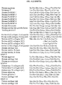
- the sequences of the protein cleavage sites of MMP1, MMP2, MMP3, MMP7, MMP8, MMP9 and MMP10 are shown in Figure 5
- the proteolytic cleavage site of the present invention is cleaved at sites of inflammation and tissue remodeling. More preferably, the proteolytic cleavage site of the present invention is a MMP cleavage site e.g. any one or more of MMP1, MMP2, MMP3, MMP7, MMP8, MMP9 or MMP10 as shown in Figure 5 .
- MMP cleavage site e.g. any one or more of MMP1, MMP2, MMP3, MMP7, MMP8, MMP9 or MMP10 as shown in Figure 5 .
- the pharmaceutically active moiety may comprise one or more molecules which may be the same or different, one or more radioisotopes which may be the same or different, or one or more non-radioactive elements which may be the same or different.
- the pharmaceutically active moiety may comprise a polypeptide or non-polypeptide molecule.
- references to a polypeptide include a peptide and vice versa unless the context specifies otherwise.
- the polypeptide may be an antibody or a fragment thereof, such as an anti-TNFalpha monoclonal antibody (for example infliximab or adalimumab), a soluble p75 TNF receptor molecule (for example etanercept) or a IL-1 receptor antagonist (for example anakinra).
- an anti-TNFalpha monoclonal antibody for example infliximab or adalimumab
- a soluble p75 TNF receptor molecule for example etanercept
- IL-1 receptor antagonist for example anakinra
- the composition will therefore comprise a bispecific antibody which may be a diabody (scFv with a linker which is too short to allow pairing between VH and VL and therefore the domains are forced to pair with the complementary domain of another scFv to create two antigen binding site), a minibody (composed of two scFv moieties linked via a constant heavy chain region (CH3)), a scFv-Fc molecule, or an intact antibody molecule containing the two separate binding regions.
- a bispecific antibody which may be a diabody (scFv with a linker which is too short to allow pairing between VH and VL and therefore the domains are forced to pair with the complementary domain of another scFv to create two antigen binding site), a minibody (composed of two scFv moieties linked via a constant heavy chain region (CH3)), a scFv-Fc molecule, or an intact antibody molecule containing the two separate binding regions.
- CH3 constant heavy chain
- a bispecific antibody may comprise a first binding region specific for modified Collagen II (CII) and a second binding region specific for anti-TNFa.
- CCII modified Collagen II
- the polypeptide is a TNF receptor (TNFR) antibody fusion protein, typically a TNFR2-Fc fusion protein.
- TNFR TNF receptor
- a bispecific antibody of the invention may also further comprise another pharmaceutically active moiety.
- a composition of the invention may comprise a first binding region specific for modified Collagen II, a second binding region specific for CD64, and a toxin, such as Ricin A.
- the polypeptide may be a growth factor (e.g. TGF ⁇ , epidermal growth factor (EGF), platelet derived growth factor (PDGF), nerve growth factor (NGF), colony stimulating factor (CSF) granulocyte/macrophage colony stimulating factor (GM-CSF), hepatocyte growth factor, insulin-like growth factor, placenta growth factor); differentiation factor, cytokine molecule, for example an interleukin, (e.g.
- IFN- ⁇ , IFN- ⁇ and IFN- ⁇ tumour necrosis factor
- IGIF IFN- ⁇ inducing factor
- BMP bone morphogenetic protein
- chemokine for example a MIP (Macrophage Inflammatory Protein) e.g.
- MIP1 ⁇ and MIP1 ⁇ a MCP (Monocyte Chemotactic Protein) e.g. MCP1, 2 or 3; RANTES (regulated upon activation normal T-cell expressed and secreted)); a trophic factor; a cytokine inhibitor; a cytokine receptor (for example, CD20, CD40, CD40L, CD64); a free-radical scavenging enzyme (e.g. superoxide dismutase or catalase), or a toxin (for example Ricin A toxin, or Pseudomonas exotoxin A), or an active fragment or portion thereof.
- the polypeptide is an interferon, typically IFN- ⁇ .
- antibody-fusion proteins include but are not limited to, antibody-TNFalpha, antibody-GM-CSF, and antibody-IL2 fusion proteins
- the pharmaceutically active polypeptide may be derived from the species to be treated e.g. human origin for the treatment of humans.
- composition may also comprise further peptide sequences which can target the composition inside a cell.
- intracellular targeting sequences include, but are not limited to, the TAT sequence YGRKKRQRRR (see for example, Cohen-Saidon et al Blood, volume 102 (7), pages 2506-2512 (2003 )).
- peptide mimetics includes, but is not limited to, agents having a desired peptide backbone conformation embedded into a non-peptide skeleton which holds the peptide in a particular conformation.
- Peptide mimetics which do not have some of the drawbacks of peptides, are of interest in those cases where peptides are not suitable in medicine.
- Peptide mimetics may comprise a peptide backbone which is of the L or D conformation.
- Examples of peptide mimetics include melanocortin, adrenocorticotrophin hormone (ACTH) and other peptide mimetic agents which play a role in the central nervous system, endocrine system, in signal transduction and in infection and immunity.
- ACTH adrenocorticotrophin hormone
- the pharmaceutically active agent may comprise a chemical compound such as a chemotherapeutic agent or other synthetic drug.
- the pharmaceutically active agent may comprise a peptide nucleic acid (PNA) sequence e.g. a poly-lysine sequence which binds to nucleic acids and permeabilises lipid bilayers ( Wyman et al., Biological Chemistry, 379, 1045-1052 (1998 )) or a KALA peptide which facilitates transfer through lipid bilayers ( Wyman et al., Biochemistry, 36, 3008-3017 (1997 )).
- PNA peptide nucleic acid
- the non-polypeptide may be a glycosaminoglycan molecule, such as glucosamine (suitably, glucosamine HCl) or chondroitin.
- the non-polypeptide molecule may be a non-steroidal anti-inflammatory drug (NSAID) such as a non-selective NSAID or a selective NSAID.
- NSAID non-steroidal anti-inflammatory drug
- non-selective NSAIDs include aspirin, ibuprofen, and naproxen.
- selective NSAIDs also called COX-2 inhibitors
- celecoxib Celebrex®
- rofecoxib Vioxx®
- valdecoxib Bosecoxib
- steroids such as cortisol
- polymeric molecules such as sodium hyaluronate or hyaluronic acid (for example hyaluronan (Hyalgan®) and hylan-GF-20 (Synvisc®)
- drug substances such as colchicine or hydroxychloroquine (Plaquenil®).
- Non-polypeptides may be conjugated to the antibody or fragment thereof using a linker that may be a labile bond in order to permit release of the pharmaceutically active substance.
- a linker that may be a labile bond in order to permit release of the pharmaceutically active substance.
- a hydrazone bond may be used where the drug is released under acidic conditions, or a disulfide bond which is reduced to release the drug, or also a peptide bond which is cleaved enzymatically by a protease as described above.
- the composition may comprise a radioactive element or a non-radioactive element.
- the radioisotope may be an alpha particle-emitting radionuclide such as 213 Bi or 211 At, a beta particle-emitting radionuclide such as 131 I, 90 Y, 177 Lu or 67 Cu, a gamma radiation-emitting radionuclide such as 99m Tc, 123 I or 111 In, or a positron-emitting radionuclide such as 18 F, 64 Cu, 68 Ga, 86 Y or 124 I. Radioisotopes may be used in order to render the composition detectably labelled for diagnostic uses of the composition.
- the non-radioactive element may be Au, Fe, Cu, Pt or Ag.
- a composition of the first aspect for use in medicine includes a composition of the first aspect for use in the treatment of an arthropathy, such as rheumatoid arthritis (RA) and osteoarthritis (OA).
- This aspect of the invention therefore extends to a method of treatment of an arthropathy, such as rheumatoid arthritis (RA) and osteoarthritis (OA), comprising the step of administering to a subject a composition of the first aspect of the invention.
- the present invention therefore also includes the use of a composition of the first aspect of the invention in the manufacture of a medicament for the treatment of an arthropathy, such as rheumatoid arthritis (RA) and osteoarthritis (OA).
- a composition of the first aspect of the invention is a pharmaceutical composition.
- a pharmaceutical composition may comprise a diluent, excipient, adjuvant and/or physiologically acceptable buffer.
- the pharmaceutical composition may be administered in any effective, convenient manner effective for treating a disease as described above including, for instance, administration by oral, topical, intravenous, intramuscular, intra-articular, intranasal, or intradermal routes among others.
- the composition of the invention may be administered to an individual as an injectable composition, for example as a sterile aqueous dispersion, preferably isotonic.
- the injection may suitably be made into the joint affected by the disease.
- the daily dosage of the composition of the invention will be from 0.01mg/kg body weight, typically around 1mg/kg.
- the physician in any event will determine the actual dosage which will be most suitable for an individual which will be dependant on factors including the age, weight, sex and response of the individual.
- the above dosages are exemplary of the average case. There can, of course, be instances where higher or lower dosages are merited, and such are within the scope of this invention
- a method for the diagnosis of an arthropathy comprising the steps of administering a detectably labelled composition comprising an antibody or fragment thereof against oxidised Collagen II (CII) to a subject and subsequently detecting the composition.
- This aspect of the invention therefore extends to a detectably labelled composition comprising an antibody or fragment thereof against oxidised Collagen II (CII) for use in the diagnosis of an arthropathy, such as rheumatoid arthritis (RA) and osteoarthritis (OA).
- Such embodiments also extend to the use of such compositions in the manufacture of an agent for the diagnosis of an arthropathy, such as rheumatoid arthritis (RA) and osteoarthritis (OA).
- the detectable label may be a radioactive or a fluorescent label.
- the radioisotope may be an alpha particle-emitting radionuclide such as 213 Bi or 211 At, a beta particle-emitting radionuclide such as 131 I, 90 Y, 177 Lu or 67 Cu, a gamma radiation-emitting radionuclide such as 99m Tc, 123 I or 111 In, or a positron-emitting radionuclide such as 18 F, 64 Cu, 68 Ga, 86 Y or 124 I. Radioisotopes may be used in order to render the composition detectably labelled for diagnostic uses of the composition.
- fluorescent dyes such as Alexa Fluor 488 or the Cy3 monofunctional N-hydroxysuccinimide (NHS) ester could also be used.
- composition comprising an antibody or fragment thereof against oxidised Collagen II (CII) according to the claims and a detectable label.
- CII oxidised Collagen II
- RA is the most common chronic inflammatory autoimmune disease, with disability occurring usually within 10 years. Over activation of the inflammatory pathway leads to synovitis, joint damage and destruction. Key players in the joint inflammation are inflammatory cytokines such as TNFa and IL-1.
- TNFa inflammatory cytokines
- IL-1 receptor antagonist IL-1 receptor antagonist
- CII is the best candidate to target therapy to the joint one needs to find a way to target the drugs solely to the inflamed joints.
- the present studies show the development of a targeting antibody that will specifically recognise collagen type II that has been modified by ROS present in inflamed joint which then allows targeting to inflammatory damage joint independently of the aetiology.
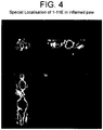
- a panel of human scFvs was developed ( Fig 1 ) that bind only to CII which was modified in vitro by HOCl or glycation, known reactive oxidants in RA. Most importantly this scFv binds only to damaged cartilage but not to normal cartilage ( Fig 3 ) and inversely correlates with the staining of safranin-O for the integrity of cartilage-specific proteoglycan. Most importantly, when inflammation was induced in only one paw in CH3 mice with antigen induced arthritis model 1-11E diabody localized only to the inflamed paw without any background to any of the other healthy paws ( Fig 4 ).
- a composition of the invention comprises mouse interferon-beta (IFN- ⁇ ), the scFv 1-11E and a MMP cleavage site.
- IFN- ⁇ mouse interferon-beta
- MCS multiple cloning sites
- mIFNb mouse interferon b
- Mouse interferon-beta is typically amplified using primers having the sequences shown in SEQ ID NO: 129 (forward) and SEQ ID NO: 130 (reverse).
- 1-11E is typically amplified using primers having the sequences shown in SEQ ID NO: 131 (forward) and SEQ ID NO: 132 (reverse), wherein 1-11E is amplified with NotI/ApaI ends to include a histidine (His) tag.
- 1-11E is typically then cloned into FastBac1.AH mIFN-b/MMP/SP/His and cut with Not/Apa to liberate SP/His.
- the mIFN-beta/His construct is typically cloned by amplifying mIFN-b with HindIII/ApaI using primers having the sequence shown in SEQ ID NO: 129 (forward) and SEQ ID NO: 133 (reverse).
- the constructs are then typically transformed into DHlOBac cells (Invitrogen).
- a composition of the invention comprises TNF receptor 2-Fc (TNFR2Fc), an scFv (either 1-11E or C7 as a negative control) and a MMP cleavage site.
- TNF receptor 2-Fc TNFR2Fc
- an scFv either 1-11E or C7 as a negative control
- MCS multiple cloning sites
- Mouse TNFR2-Fc is typically amplified using primers having the sequences shown in SEQ ID NO: 141 (forward) and SEQ ID NO: 142 (reverse).
- 1-11E is typically amplified using primers having the sequences shown in SEQ ID NO: 131 (forward) and SEQ ID NO: 132 (reverse), wherein 1-11E is amplified with NotI/ApaI ends to include a histidine (His) tag.
- infected High 5 cells are grown for 3 days at 27°C.
- the supernatant is typically then collected and run on an SDS-PAGE gel.
- Recombinant proteins can be detected by Western blot, for example using anti-tetra-His antibody (Qiagen) and anti-mouse HRP (Sigma).
- CII was prepared from bovine cartilage as in Miller ( Miller, Biochemistry 11(26): 4903-4909, 1972 ) and subsequently exposed to reactive oxygen generating systems as previously described (Nissim A, 2005). Briefly, CII was modified with ( ⁇ OH), HOCl (Hawkins CL, 2001; Hawkins CL, 2002), (ONOO - ), or 2M ribose by ON incubation at 37°C. Bovine serum albumin (BSA, Sigma) was also modified as above and was used as control antigen.
- BSA Bovine serum albumin
- Example 2 Selection of anti-modified CII scFv from phage-display library
- Phage display antibody technology ( Winter G et al, Annu. Rev. Immunol. 12: 433-455, 1994 ) was used to raise a single chain fragment variable (scFv) that binds only to CII that has been post-translationally modified by free radicals.
- scFv single chain fragment variable
- HOC1 modified CII was used as a target for panning as binding to HOCl modified CII was strongest in RA sera (Nissim A, 2005). Glycated CII was used in parallel.
- immunotubes (Nunc-Immuno Tubes, Maxi-Sorp, Nunc, Denmark) were coated with 10 ⁇ g/ml CII in phosphate-buffered saline (PBS). After blocking with 2% marvel in PBS (MPBS) coated tubes were exposed for 2 hours to 10 13 transforming units (tu) of the phage library in 2% MPBS. Unbound phage were then transferred to a second immunotube previously coated with HOCl or ribose-modified CII for a further 2 hours incubation at room temperature. Modified CII-bound phage were then used to infect E coli TG-1 and rescued by helper phage as described (Harrison J. L, 1996). The panning process was repeated three times and E coli TG-1 was infected with the final phage eluted after the third round and individual ampicillin-resistant colonies (phage clones) were selected for further analysis.
- PBS phosphate-buffered saline
- the reaction was monitored in an ELISA reader at 450 nm with a reference wavelength of 650 nm using GENios plate reader (TECAN, Theale Court, Reading UK) and Magellan software (TECAN, Theale Court, Reading UK)
- the reactive phage clones obtained from E coli TG-1 bacteria were used to infect E coli HB2151 non-suppressor bacterial strain to obtain soluble scFv. After overnight induction with 1 mM IPTG at 30°C, the antibody fragments, derived from the V H 3 family, were harvested from the supernatant and periplasmic space as described (Harrison J. L, 1996) and purified on a protein A affinity column (GE Healthcare Ltd, Little Chalfont, Buckinghamshire).
- Binding of purified scFv to modified CII was first analyzed by ELISA as above except that mouse anti-myc tag antibody (Santa Cruz Biotechnology, INC, Wembley, UK) followed by anti-mouse-HRP conjugate (Sigma, Dorset, UK) were used to probe bound scFv.
- phage clones specific to either glycated CII or HOCl modified CII were selected out of which 42 clones had unique sequences.
- 15 representative clones with different binding patterns but with good expression were then studied for further analysis ( Fig 1 and Table 1). As shown in Fig. 1 , out of these 15 clones there were 9 clones with stronger binding to modified CII, 3 clones bound to all forms of CII and and 3 clones had no binding reactivity to any form of CII.
- Clone 1-11E binds to modified CII (glycated, HOCl and to some extent to peroxynitrated CII), clone 6-11D binds to both native and modified CII and clone 12E that does not bind to any form of CII. None of these scFv bound to native or free radical modified BSA, or to collagen type III (data not shown).
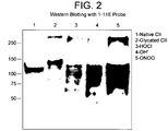
- 1-11E binds several CII fragments between 50 and 150 kDa as well as to a band >250 kDa which resulted from CII cross linking due to the ROS reactivity ( Fig 2 lane 2-5). 1-11E also binds to native CII corresponding to a band below 150KDa ( Fig 2 lane 1). Binding to native CII in Western blotting was also seen in sera from RA patients that did not bind to native CII in ELISA but only to ROS modified CII in ELISA (for example sera 33 (Nissim A, 2005)).
- Example 6 Immunohistochemistry of human OA and RA cartilage using selected anti-modified CII scFv
- osteochondral sample was obtained from the femoral condyle of a patient (female, 63 years old) undergoing prosthetic knee replacement for OA.
- One sample of normal human cartilage was obtained post-mortem from a preserved area of a knee with unicompartimental OA undergoing joint replacement (female, 54 years old). In both cases, cartilage was fixed overnight at 4°C in 4% paraformaldehyde, decalcified for 15 days in 0.5M EDTA at 4°C, washed in PBS, and embedded in paraffin according to standard protocols. Safranin O staining was performed according to standard protocols (Rosenberg, 1971). All samples were obtained in accordance with institutional policies and regulations.
- Immunostaining was performed using the selected scFv (10 ⁇ g/ml and 1 ⁇ g/ml) as well as control commercial mouse anti-CII antibodies (diluted 1:100 and 1:1000 dilution; Chemicon International, Chandlers Ford, Hampshire, UK) and polyclonal anti-CII antibodies (diluted 1: 100, 1:1000) from collagen induced arthritis (CIA) mice.
- ScFv or control antibodies were added to the slide in blocking buffer (0.5% BSA in PBS plus 0.05% sodium azide) and left overnight at 4°C.
- the cartilage extracellular matrix is a complex structure where several molecules interact to form a structural and functional unit. There is therefore the chance that the tertiary and quaternary structure of collagens in the intact tissue may alter the specificity of binding of the phage antibodies that had been selected in vitro.
- the capacity of anti-ROS-modified CII scFv to bind to CII within the matrix complex structure and to present immunoreactivity with damaged OA cartilage as opposed to normal cartilage was tested.
- a commercial anti-CII mAb did not stain the damaged cartilage areas stained by 1-11E on an adjacent section ( Fig 3 G) but intensely stained a histologically normal cartilage ( Fig 3H ), suggesting that the epitope recognised by the commercial antibody is lost in the OA section.
- Figure 6 also shows staining of OA cartilage. Although synovial inflammation in OA is not as extensive as in RA and inflammatory cells are not significant in numbers, low grade synovitis is nearly a constant feature in OA. Abnormal mechanical force appears to stimulate chrondocytes to produce some of the same inflammatory mediators and ROS as the infiltrating leukocytes present in inflamed RA joints, leading to post-translational modifications of CII in OA.
- Figure 6 confirms the results shown in Figure 3 and shows that staining of OA cartilage section is only pericellular, around the chrondocytes.
- a further sample was obtained from a patient (female, 47 years old) undergoing total right knee replacement for RA. Fixing and staining protocols were as described above.
- Figure 7 shows staining of the RA cartilage.
- infiltrating inflammatory cells consume increased amounts of oxygen, resulting in the generation of reactive oxygen species (ROS), which may cause excessive degradation of the extracellular matrix leading to cartilage destruction and chemical post-translational modification of CII by ROS.
- Figure 7 shows uniteral staining of RA cartilage across the section. This is due to the high influx of immune cells that produce high levels of ROS.
- ROS reactive oxygen species
- Example 8 Inducing arthritis in the animal models
- mice Male C3H mice (age 17-19 weeks) were used. 100mg of dessicated non-viable T.B. strain H37RA (Difco 231141) was added to 30ml of incomplete Freunds adjuvant (IFA, Difco 263910) to form complete Freunds adjuvant (CFA). An equal volume of CFA was added to a 2mg/ml solution (in PBS) of methylated BSA (mBSA) (Sigma A1009). The mixture was then emulsified on ice using an Ultra-Turrax T25 homogeniser at 13500-20500rpm until a fluffy milky consistency was obtained. Mice were anaesthetised with Hypnorm, and 100 ⁇ l of 1mg/ml (i.e.
- mice were anaesthetised with nitrous/oxygen and halothane, and inflammation was induced by injecting 50 ⁇ l of 1mg/ml (i.e. 50 ⁇ g) mBSA in PBS into the animals' left hind paw. As a control, 50 ⁇ l PBS was injected into the right hind paw. Inflammation was measured using callipers to measure the paw thickness. Swelling was seen only in the right paws from 24 hours, and persisted until 1 week later. 2 weeks later, the swelling had totally subsided.
- mice Four and 22 hours later the mice were anaesthetized by ip injection of Ketamine/Xylazine.
- the mice were imaged on a NanoSPECT/CT scanner (Bioscan Inc, Washington, USA) using a four-detector/36 x 1.4mm pinhole configuration. 30- 50,000 counts were acquired for the SPECT study over 20-50 minutes.
- FIG. 4 shows a representative image acquired 22 hours after injection of the radiotracer. Increased uptake of radioactivity is clearly seen in the rear left (inflamed) paw relative to the right (normal paw).
- Example 10 Staining of cartilage in mouse RA model
- mice were sensitised with mBSA (100 ⁇ g) in CFA intradermally at the base of the tail, and challenged either intra-articularly (both knees) or intra-plantarly (right, saline left) with 500 ⁇ g mBSA in saline 14 days later.
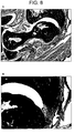
- Paw 12 hours post challenge with mBSA, the right paw was grossly inflamed in the subplantar region (seen by haematoxylin and eosin (H&E) staining), as shown in Figure 8A and 8B .
- the cartilage in the mBSA paws is uniformly and strongly stained by 1-11E, as shown in Figure 8C and 8D .
- the left paw injected by saline had no subplantar inflammation. Some cartilage within the left paw joints was stained heterogeneously, perhaps associated with spontaneous osteoarthritis.
- Example 11 Staining of joint in mouse OA model
- mice Seven week old C57BL/6 male mice were utilized for these experiments ( Dell'Accio F et al, Arthritis Res Ther. 2006; 8(5):R139 ). The mice were anesthetized and subjected to medial para-patellar arthrotomy. The patellar groove was exposed by lateral patellar dislocation. A longitudinal full thickness injury was made in the patellar groove using a custom made device in which the length of a 26G needle was limited by a glass bead (injured knee). The patellar dislocation was then reduced and the joint capsule and the skin sutured in separate layers. The animals were killed after 4 weeks and the knees dissected for histological and histochemical analysis.
- Staining methods are as set out in Example 6 above, except that rabbit anti-myc followed by anti-rabbit-HRP were used to avoid cross-reactivity with mouse antibody in the tissue.
- Example 12 Production of fusion proteins: 1-11E with anti-inflammatory cytokines
- pFastBac1.AH was created from pFastBacl (Invitrogen) by cutting out BamHI/HindIII fragment containing multiple cloning sites (MCS), and replacing with a linker to give another MCS of BamHI-KpnI-HindIII-ApaI.
- Mouse interferon b (mIFNb) was cloned into the HindIII-EcoRI sites, followed by a MMP cleavage site and 1-11E which were cloned into the NotI and ApaI sites as shown in Figure 10A .
- the MMP cleavage site can be cleaved by MPP-1 and MMP-3.
- Mouse interferon-beta was amplified with the following primers:
- the mIFN-beta / His construct was cloned by amplifying mIFN-b with HindIII/ApaI with the following primers:
- the IFN-beta portion is from amino acids 1 to 182 as follows:
- the MMP linker portion is from amino acids 183 to 202 as follows:
- the 1-11E portion is from amino acids 203 to 446 as follows:
- the His tag is from amino acids 448 to 502 as follows:
- the IFN-beta portion is from amino acids 1 to 184 as follows:
- the His tag is from amino acids 185 to 190 as follows:
- the constructs were transformed into competent DHlOBac cells (Invitrogen) to generate bacmid vectors.
- Recombinant bacmid vectors were confirmed by blue-white screening and PCR according to Invitrogen instructions.
- Bacmid DNA was transfected into Sf9 insect cells using cellfectin according to Invitrogen instructions.
- Baculovirus was harvested from the supernatant of transfected cells, and used to infect fresh Sf9 cells to amplify the virus stocks. P3 virus was used to infect High 5 insect cells for 72 hours, and the supernatant was collected and run on an SDS-PAGE gel. Recombinant proteins were detected by Western blot using anti-tetra-His antibody (Qiagen) and anti-mouse HRP (Sigma).
- test expression of the fusion constructs is shown in Figure 12 .
- pFastBac1.AH was created from pFastBacl (Invitrogen) by cutting out BamHI/HindIII fragment containing multiple cloning sites (MCS), and replacing with a linker to give another MCS of BamHI-KpnI-HindIII-ApaI.
- TNFR2Fc was cloned into the HindIII-EcoRI sites, followed by a MMP cleavage site and scFv (1-11E or C7) which were cloned into the NotI and ApaI sites as shown in Figure 10B .
- Mouse TNFR2-Fc was amplified with the following primers: forward primer: 5' GCT aag ctt ATG GCG CCC GCC GCC CTC 3' reverse primer: 5' CTTGAATTCTTTACCCAGAGACCGGGA 3'
- TNFR2Fc/MMP/1-11E The sequence of TNFR2Fc/MMP/1-11E is as follows:
- TNFR2Fc portion is as follows:
- the MMP linker portion is as follows:
- the 1-11E portion is as follows:
- the His tag is as follows:
- HEL Hen Egg Lysosyme
- TNFR2Fc portion is as follows:
- the MMP linker portion is as follows:
- the C7 portion is as follows:
- the His tag is as follows:
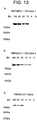
- Infected Hi-5 cells were grown for 3 days at 27°C. After 3 days, different 100, 50, 25 and 12 microliter aliquots of cell supernatant were taken for Western blot analysis. Fusion protein was probed with anti-His tag antibodies. As shown in Figure 13 the apparent molecular weight of the TNFR2Fc fusion proteins is slightly above 75kDa which reflects the predicted 50kDa TNFR2Fc plus 30kDa scFv.
Landscapes
- Health & Medical Sciences (AREA)
- Engineering & Computer Science (AREA)
- Bioinformatics & Cheminformatics (AREA)
- Life Sciences & Earth Sciences (AREA)
- Chemical & Material Sciences (AREA)
- General Health & Medical Sciences (AREA)
- Immunology (AREA)
- Medicinal Chemistry (AREA)
- Public Health (AREA)
- Animal Behavior & Ethology (AREA)
- Pharmacology & Pharmacy (AREA)
- Veterinary Medicine (AREA)
- Epidemiology (AREA)
- Molecular Biology (AREA)
- Proteomics, Peptides & Aminoacids (AREA)
- Organic Chemistry (AREA)
- Cell Biology (AREA)
- Zoology (AREA)
- Toxicology (AREA)
- Optics & Photonics (AREA)
- Physics & Mathematics (AREA)
- Genetics & Genomics (AREA)
- Biophysics (AREA)
- Biochemistry (AREA)
- Physical Education & Sports Medicine (AREA)
- Nuclear Medicine, Radiotherapy & Molecular Imaging (AREA)
- General Chemical & Material Sciences (AREA)
- Rheumatology (AREA)
- Orthopedic Medicine & Surgery (AREA)
- Chemical Kinetics & Catalysis (AREA)
- Peptides Or Proteins (AREA)
- Medicines Containing Antibodies Or Antigens For Use As Internal Diagnostic Agents (AREA)
- Medicines That Contain Protein Lipid Enzymes And Other Medicines (AREA)
Description
- The present invention relates to a novel antibody and its use in the diagnosis and therapy of inflammatory diseases of the joints such as rheumatoid arthritis (RA) and osteoarthritis (OA).
- The final outcome of most rheumatic conditions, the leading cause of disabilities in the western world, is breakdown of articular cartilage. This breakdown is the final outcome of inflammatory events in both rheumatoid arthritis (RA) and osteoarthritis (OA) mediated by either influx of activated leukocytes (RA) or activated chondrocytes (OA). Pro-inflammatory cytokine blockade such as anti-TNFa and IL-IRa is therefore currently used to treat arthritic conditions, mainly RA. These treatments however, are not consistently effective and the number of patients that fail anti-TNF therapy is increasing. Especially for anti-TNFa treatment there is a risk of serious infections and malignancies. These systemic side effects could be minimised by the development of technologies to target therapeutic agents specifically to the inflamed tissues, but has so far been impeded by the lack of proper target epitope(s) that would be present uniquely in the diseased joint and not in the healthy joint.
- RA is a classic inflammatory form of arthritis, which is a chronic autoimmune disease with extensive synovial inflammation. Influx of activated leukocytes infiltrating the inflamed synovial membrane results in up-regulation of inflammatory cytokines such as TNFa, interleukin-1 (IL-1) and interleukin-6 (IL-6) leading to increase in the levels of matrix metalloproteases (MMP). Moreover, infiltrated inflammatory cells consume increased amounts of oxygen, resulting in the generation of reactive oxidant species (ROS) including superoxide radicals (O2), hydrogen peroxide (H2O2), hydroxyl radicals (OH), hypochlorous acid (HOCl), nitric oxide (NO) and peroxynitrite (ONOO). In addition, sequential oxidative reactions generate reactive oxidants such as advanced glycation end-products (AGE). The combined activities of MMP and ROS may be the cause of the excessive degradation of the extracellular matrix leading to cartilage destruction.
- The immuno-pathological events following the ROS reactivity with cartilage specific collagen type II (CII) protein have been studied recently. A substantial increase in binding of RA sera to CII after chemical post-translational modification in vitro by ROS has been demonstrated in comparison to binding to native non-modified CII, which is significantly greater than in non-RA sera. Post-translational modification in the acute and chronic inflammation by ROS has also been postulated by the presence of other ROS damaged proteins and auto-antibodies against other auto-antigens that are post-translationally modified by ROS. Generation of neoantigenic epitopes on modified CII has been reported in Nissim et al Arthritis & Rheumatism, volume 52 (12) pages 3829-3838 (2005)). Antibodies against IgG-AGE and a T cell response against IgG modified by HOCl and peroxynitrite have also been observed. FERRARI-LACRAZ SYLVIE ET AL: , JOURNAL OF IMMUNOLOGY, vol. 173, (2004-11-01), pages 5818-5826 teaches that targeting IL-15 receptor-bearing cells with an antagonist mutant IL-15/Fc protein prevents disease development and progression in murine collagen-induced arthritis.
- Although synovial inflammation in OA is not as extensive as in RA and inflammatory cells are not significant in numbers, low grade synovitis is nearly a constant feature in OA. Abnormal mechanical force appears to stimulate chondrocytes to produce the same inflammatory mediators and ROS as the infiltrated leukocytes present in inflamed RA joints leading to post translational modifications of CII. There is a report of elevated levels of nitrated CII peptide in sera of patients with OA. The presence of strong staining of nitrotyrosine and low antioxidative capacity in the degenerative region of OA cartilage compared with the intact region from the same sample suggests a possible correlation between oxidative damage and cartilage degradation. As in RA, indirect involvement of oxidative stress has also been evidenced in OA by the fact that: (i) OA is strongly linked with age and in aged cartilage there is accumulation of AGE; and (ii) there is accumulation of lipid peroxidation product and nitrotyrosine.
- There is a need for improved means for diagnosing inflammatory diseases of the joints and for improved therapies for arthropathies such as rheumatoid arthritis (RA) and osteoarthritis (OA).
- It has been found that an antibody raised against post-translationally modified Collagen II (CII) can specifically target the antibody to the sites of inflammation in the joints. This degree of specificity is important since native CII may be present in both inflamed and healthy joints also.
- According to a first aspect of the invention, there is provided a pharmaceutical composition comprising an antibody or fragment thereof against oxidised Collagen II (CII) in which the antibody or fragment thereof is conjugated to a pharmaceutically active moiety according to the claims. The present invention therefore provides a novel approach to the targeting of drugs to self-epitopes on Collagen II that are a normal component of the tissue but which become immunogenic after post-translational modification by free radicals as part of a disease process affecting Collagen II.
- The antibody may be a polyclonal antibody or a monoclonal antibody. It may be a human or humanized or chimeric antibody with sequences, residues or domains derived from more than one animal species. Fragments of antibodies include Fc, Fab, scFv, single domain (dAb) antibody, diabody, minibody, and scFv-Fc fragments
- In one embodiment of the invention, the antibody comprises CDR sequences in the Variable Heavy (VH) Chains and Variable Light (VL) chains as shown in Table 1. CDRH2 and CDRH3 are in the VH chain and CDRL2 and CDRL3 are in the VL chain.
Table 1 CDRH2 CDRH3 CDRL2 CDRL3 DISSTGSYTAYADSVKG GAGSFDY AASALQS QQSSSTPTT AISAAGTATAYADSVKG GYDTFDY AASSLQS QQNYGYPNT SISNSGSYTDYADSVKG GYGSFDY AASTLQS QQANSSPDT SINNYGSNTAYADSVKG GYSSFDY AASYLQS QQTSSSPDT SINNYGSNTAYADSVKG GYSSFDY AASYLQS QQTSSSPDT SISYTGNSTDYADSVKG GYTAFDY YASYLQS QQADSTPTT SISYTGNSTDYADSVKG GYTAFDY YASYLQS QQADSTPTT SISYTGNSTDYADSVKG GYTAFDY YASYLQS QQADSTPTT SISYTGNSTDYADSVKG GYTAFDY YASYLQS QQADSTPTT SISYTGNSTDYADSVKG GYTAFDY YASYLQS QQADSTPTT NIATDGTTTYYADSVKG NSTYFDY SASTLQS QQAATSPTT SISNSGTNTDYADSVKG NYASFDY YASYLQS QQGSASPST SISNSGTNTDYADSVKG NYASFDY YASYLQS QQGSASPST SISNSGTNTDYADSVKG NYASFDY YASYLQS QQGSASPST SISNSGTNTDYADSVKG NYASFDY YASYLQS QQGSASPST SISNSGTNTDYADSVKG NYASFDY YASYLQS QQGSASPST SISNSGTNTDYADSVKG NYASFDY YASYLQS QQGSASPST SISNSGTNTDYADSVKG NYASFDY YASYLQS QQGSASPST SISYTGDSTYYADSVKG NYSAFDY YASYLQS QQADSTPTT SINDSGTTTYYADSVKG NYSAFDY AASDLQS QQSDSAPTT SIDSAGASTYYADSVKG NYSAFDY NASSLQS QQSDTYPST SISYTGDSTYYADSVKG NYSAFDY TASNLQS QQSYASPTT SISYTGDSTYYADSVKG NYSAFDY TASNLQS QQSYASPTT SISYTGDSTYYADSVKG NYSAFDY TASNLQS QQTGSYPTT SINATGYGTYYADSVKG NYSDFDY SASALQS QQGDSYPTT SINSNGTDTYYADSVKG NYSDFDY TASALQS QQGYGAPTT SISATGSSTYYADSVKG NYSDFDY SASDLQS QQSSYTPTT SISATGSSTYYADSVKG NYSDFDY SASDLQS QQSSYTPTT SIDDSGATTYYADSVKG NYSSFDY YASSLQS QQAANYPTT SIDDSGATTYYADSVKG NYSSFDY YASSLQS QQAANYPTT SIDDSGATTYYADSVKG NYSSFDY YASSLQS QQAANYPTT SIDDSGATTYYADSVKG NYSSFDY YASSLQS QQAANYPTT SIASTGDSTYYADSVKG NYSSFDY SASALQS QQASNYPTT SIASTGDSTYYADSVKG NYSSFDY SASALQS QQASNYPTT SIASTGDSTYYADSVKG NYSSFDY SASALQS QQASNYPTT SIASTGDSTYYADSVKG NYSSFDY SASALQS QQASNYPTT SIASTGDSTYYADSVKG NYSSFDY SASALQS QQASNYPTT SIASTGDSTYYADSVKG NYSSFDY SASALQS QQASNYPTT SISTNGSSTYYADSVKG NYSSFDY DASGLQS QQGDTSPTT SISTNGSSTYYADSVKG NYSSFDY DASGLQS QQGDTSPTT SISTNGSSTYYADSVKG NYSSFDY DASGLQS QQGDTSPTT SISTNGSSTYYADSVKG NYSSFDY DASGLQS QQGDTSPTT SIDTTGTTTYFADSVKG NYSSFDY SASYLQS QQGYSAPTT TISYSGNNTYYADSVKG NYSSFDY TASSLQS QQGYTSPTT SIDAGGNGTYYADSVKG NYSSFDY TASNLQS QQNNYYPTT SIDAGGNGTYYADSVKG NYSSFDY YASSLQS QQSDAYPTT SIDAGGNGTYYADSVKG NYSSFDY YASSLQS QQSDAYPTT SIDAGGNGTYYADSVKG NYSSFDY YASSLQS QQSDAYPTT SIDAGGNGTYYADSVKG NYSSFDY YASSLQS QQSDAYPTT SIDSAGNATYYADSVKG NYSSFDY AASTLQS TSNYPTTQQ SITDSGDTTYYADSVKG NYSTFDY SASSLQS QQSNATPTT SITDSGDTTYYADSVKG NYSTFDY SASSLQS QQSNATPTT SITDSGDTTYYADSVKG NYSTFDY SASSLQS QQSNATPTT SITDSGDTTYYADSVKG NYSTFDY SASSLQS QQSNATPTT SITDSGDTTYYADSVKG NYSTFDY SASSLQS QQSNATPTT SIATTGDNTYYADSVKG NYSYFDY TASTLQS QQAAGNPTT AINAYGGSTYYADSVKG NYSYFDY AASSLQS QQGSDYPTT AINAYGGSTYYADSVKG NYSYFDY AASSLQS QQGSDYPTT SIATTGTSTTYADSVKG NYSYFDY TASSLQS QQGST APTT SIATTGTSTTYADSVKG NYSYFDY TASSLQS QQGSTAPTT TIDTAGSYTDYADSVKG NYSYFDY GASTLQS QQSTASPST SISNNGSSTYYADSVKG NYSYFDY AASNLQS QQTSSYPTT SIAYGGAGTDYADSVKG NYTAFDY AASYLQS QQGAGSPST AIANTGSATNYADSVKG NYTAFDY DASTLQS QQRNTSPTT SISTAGTYTDYADSVKG NYTDFDY SASYLQS QQSNTSPAT SISTAGTYTDYADSVKG NYTDFDY SASYLQS QQSNTSPAT SINDTGYTTYYADSVKG NYTYFDY TASTLQS QQAYTAPTT SIASSGTTTYYADSVKG SYADFDY AASNLQS QQADTYPTT TITSTGAATAYADSVKG SYATFDY AASYLQS QQAANSPDT AIDGTGYGTAYADSVKG SYDTFDY GASSLQS QQTSDYPNT SIANAGTATYYADSVKG SYSNFDY SASTLQS QQASTSPTT SIDSAGDSTYYADSVKG SYSYFDY TASYLQS QQASDYPTT SISSSGDTTYYADSVKG SYSYFDY TASTLQS QQSSSNPTT SIDTGGSYTDYADSVKG SYTTFDY SASYLQS QQGSNSPTT SIDTGGSYTDYADSVKG SYTTFDY SASYLQS QQGSNSPTT SIDTGGSYTDYADSVKG SYTTFDY SASYLQS QQGSNSPTT SIDTGGSYTDYADSVKG SYTTFDY SASYLQS QQGSNSPTT SIDASGANTAYADSVKG TYGTFDY SASYLQS QQSATTPDT - In one embodiment of the invention, the antibody may be an scFv selected from the group consisting of the following:
- 3-11A, 6-6E, 1-7G, 3-7B, 6-9D, 1-1C, 1-8D, 1-3G, 4-12C, 6-3E, 6-9A, 1-12A, 4-6A, 4-8A, 4-9F, 4-4H, 3-3A, 3-6F, 6-10H, 12E, 3-5G, 3-4D, 3-5D, 6-4E, 3-6B, 3-6G, 4-11F, 6-7H, 1-11E, 1-2F, 1-6H, 3-8D, 1-4D, 4-2F, 3-3B, 3-5C, 6-9C, 4G, 3-12F, 3-4G, 6-11F, 6-11H, 3-2C, 5B, 6-10G, 1-4H, 4-5A, 4-1B, 4-12D, 6-4B, 1-2E, 1-7F, 1-10F, 1-9G, 4-1C, 6-7G, 3-7H, 6-1F, 6-3B, 4H, 3-9A, 6-10D, 3-5H, 3-2F, 1-6G, 3-11H, 6-9F, 3-9D, 4-3H, 3-3E, 3-10C, 3-11E, 6-8C, 6-11D, 4-5H, 6-5F, 6-7F, 1-10D
- These scFvs are listed in Table 3 in the Examples below and comprise the CDRH2, CDRH3, CDRL2 and CDRL3 sequences shown in Table 1.
- In one embodiment of the invention, the scFv may comprise a sequence as shown in Table 2.
Table 2 Clone I.D. VH-CDR2 VH-CDR3 VL-CDR2 VL-CDR3 1-2E SITDSGDTTYYADSVKG NYSTFDY SASSLQS QQSNATPTT 1-11E SIDDSGATTYYADSVKG NYSSFDY YASSLQS QQAANYPTT 1-4D SIASTGDSTYYADSVKG NYSSFDY SASALQS QQASNYPTT 1-12A SISNSGTNTDYADSVKG NYASFDY YASYLQS QQGSASPST 12E SINDSGTTTYYADSVKG NYSAFDY AASDLQS QQSDSAPTT 3-9A TIDTAGSYTDYADSVKG NYSYFDY GASTLQS QQSTASPST 3-5C SIASTGDSTYYADSVKG NYSSFDY SASALQS QQASNYPTT 3-11E SIDSAGDSTYYADSVKG SYSYFDY TASYLQS QQASDYPTT 3-2F AIANTGSATNYADSVKG NYTAFDY DASTLQS QQRNTSPTT 3-6G SINSNGTDTYYADSVKG NYSDFDY TASALQS QQGYGAPTT 3-9D SIASSGTTTYYADSVKG SYADFDY AASNLQS QQADTYPTT 6-3B SIATTGTSTTYADSVKG NYSYFDY TASSLQS QQGST APTT 6-11D SIDTGGSYTDYADSVKG SYTTFDY SASYLQS QQGSNSPTT 6-9F SINDTGYTTYYADSVKG NYTYFDY TASTLQS QQAYTAPTT 6-7G SIATTGDNTYYADSVKG NYSYFDY TASTLQS QQAAGNPTT - Typically, the scFv is 1-11E.
- Oxidised Collagen II (CII) is post-translationally modified Collagen II (CII) that has been oxidised by non enzymatic glycation or by reactive oxidant species (ROS) which may include superoxide radical (O2), hydrogen peroxide (H2O2), hydroxyl radical (OH), hypochlorous acid (HOCl), nitric oxide (NO) and peroxynitrite (ONOO).
- The antigen may therefore be HOCl-Collagen II or Ribose-Collagen II.
- The antibody or fragment thereof is conjugated to the pharmaceutically active moiety which may be a peptide or peptide-based molecule by any suitable means. Where the pharmaceutically active moiety is a peptide or peptide-based molecule the conjugation may be by means of a peptide bond, including the insertion of one or more amino acid residues.
- The conjugation of a peptide or a peptide-based molecule may be achieved by any generally convenient chemical means or biological means (see for example, Wu & Senter Nature Biotechnology, volume 23 (9) pages 1137-1146 (2005); " Chemistry of Protein Conjugation and Crosslinking" by S. S. Wong, CRC Press Inc. (1991)).
- Chemical conjugation typically uses a bifunctional chemical reagent, for example glutaraldehyde can link molecules to the N-terminus of a peptide, carbodiimide can link molecules to the C-terminus of a peptide, succinimide esters (e.g. MBS, SMCC) can bind free amino groups and cysteine residues, benzidine links to tyrosine residues, periodate attaches to carbohydrate groups and isothiocyanate can also link molecules to antibodies.
- Alternatively, a fusion protein may be synthesised using standard recombinant molecular biology techniques (see for example, Sambrook et al "Molecular Cloning: A Laboratory Manual", 3rd edition, CSHL Press, (2001); Trachsel et al Arthritis Research & Therapy, volume 9 (1) R9 (2007); Nagai Arthritis & Rheumatism, volume 54 (10) pages 3126-3134 (2006)). Methods for producing fusion proteins are described in the Examples herein.
- In certain embodiments of the invention, the insertion of additional amino acid residues between the antibody or fragment thereof and the pharmaceutically active moiety may represent a site for cleavage by a protease. The proteolytic cleavage site may comprise any protease specific cleavage site. The proteolytic cleavage site may include, but is not limited to, a matrix metalloproteinase (MMP) cleavage site, a serine protease cleavage site, a site cleavable by a parasitic protease derived from a pathogenic organism (Zhang et al., J. Mol. Biol. 289, 1239-1251 (1999); Voth et al., Molecular and Biochemical Parasitology, 93, 31-41 (1998); Yoshioka et al.., Folia Pharmacologica Japonica, 110, 347-355 (1997); Tort et al., Advances in Parasitology, 43, 161-266 (1999); McKerrow, International Journal for Parasitology, 29, 833-837 (1999); Young et al., International Journal for Parasitology, 29, 861-867 (1999); Coombs and Mottram, Parasitology, 114, 61-80 (1997)) or a site cleavable by the proteins of the complement cascade (Carroll, Annu. Rev. Immunol. 16, 545-568 (1998); Williams et al., Ann. Allergy, 60, 293-300 (1988)).
- The MMP cleavage site may comprise any amino acid sequence which is cleavable by a MMP. The amino acid sequence of the MMP cleavage site may be encoded by a nucleic acid sequence coding for an MMP sequence as shown in
Figure 5 or a sequence of nucleotides which has at least 50%, 60%, 70%, 80%, 90%, 95% or 99% identity, using the default parameters of the BLAST computer program provided by HGMP, thereto. Preferably, the nucleic acid sequence encoding the MMP cleavage site comprises the minimum number of residues required for recognition and cleavage by MMP. - A MMP cleavage site may comprise a number of amino acid residues recognisable by MMP. Moreover, the amino acids of the MMP site may be linked by one or more peptide bonds which are cleavable, proteolytically, by MMP. MMPs which may cleave the MMP site include, but are not limited to, MMP1, MMP2, MMP3, MMP7, MMP8, MMP9 or MMP10 (Yu and Stamenkovic, Genes and Dev. 14, 163-176 (2000); Nagase and Fields, Biopolymers, 40, 399-416 (1996); Massova et al., J. Mol. Model. 3, 17-30 (1997); reviewed in Vu and Werb, Genes and Dev. 14, 2123-2133 (2000)). The sequences of the protein cleavage sites of MMP1, MMP2, MMP3, MMP7, MMP8, MMP9 and MMP10 are shown in
Figure 5 - Preferably, the proteolytic cleavage site of the present invention is cleaved at sites of inflammation and tissue remodeling. More preferably, the proteolytic cleavage site of the present invention is a MMP cleavage site e.g. any one or more of MMP1, MMP2, MMP3, MMP7, MMP8, MMP9 or MMP10 as shown in
Figure 5 . - The pharmaceutically active moiety may comprise one or more molecules which may be the same or different, one or more radioisotopes which may be the same or different, or one or more non-radioactive elements which may be the same or different.
- In some embodiments of the invention, the pharmaceutically active moiety may comprise a polypeptide or non-polypeptide molecule. References to a polypeptide include a peptide and vice versa unless the context specifies otherwise.
- The polypeptide may be an antibody or a fragment thereof, such as an anti-TNFalpha monoclonal antibody (for example infliximab or adalimumab), a soluble p75 TNF receptor molecule (for example etanercept) or a IL-1 receptor antagonist (for example anakinra). In such embodiments of the invention, the composition will therefore comprise a bispecific antibody which may be a diabody (scFv with a linker which is too short to allow pairing between VH and VL and therefore the domains are forced to pair with the complementary domain of another scFv to create two antigen binding site), a minibody (composed of two scFv moieties linked via a constant heavy chain region (CH3)), a scFv-Fc molecule, or an intact antibody molecule containing the two separate binding regions.
- For example, a bispecific antibody may comprise a first binding region specific for modified Collagen II (CII) and a second binding region specific for anti-TNFa.
- In one embodiment, the polypeptide is a TNF receptor (TNFR) antibody fusion protein, typically a TNFR2-Fc fusion protein.
- A bispecific antibody of the invention may also further comprise another pharmaceutically active moiety. For example, a composition of the invention may comprise a first binding region specific for modified Collagen II, a second binding region specific for CD64, and a toxin, such as Ricin A.
- Alternatively, the polypeptide may be a growth factor (e.g. TGFβ, epidermal growth factor (EGF), platelet derived growth factor (PDGF), nerve growth factor (NGF), colony stimulating factor (CSF) granulocyte/macrophage colony stimulating factor (GM-CSF), hepatocyte growth factor, insulin-like growth factor, placenta growth factor); differentiation factor, cytokine molecule, for example an interleukin, (e.g. IL1, IL-2, IL-3, IL-4, IL-5, IL-6, IL-7, IL-8, IL-9, IL-10, IL-11, IL-12, IL-13, IL-14, IL-15, IL-16, IL-17, IL-18, IL-19, IL-20 or IL-21, either α or β), an interferon (e.g. IFN-α, IFN-β and IFN-γ), tumour necrosis factor (TNF), IFN-γ inducing factor (IGIF), a bone morphogenetic protein (BMP); a chemokine (for example a MIP (Macrophage Inflammatory Protein) e.g. MIP1α and MIP1β; a MCP (Monocyte Chemotactic Protein) e.g. MCP1, 2 or 3; RANTES (regulated upon activation normal T-cell expressed and secreted)); a trophic factor; a cytokine inhibitor; a cytokine receptor (for example, CD20, CD40, CD40L, CD64); a free-radical scavenging enzyme (e.g. superoxide dismutase or catalase), or a toxin (for example Ricin A toxin, or Pseudomonas exotoxin A), or an active fragment or portion thereof. Typically, the polypeptide is an interferon, typically IFN-β.
- See for example, Trachsel et al Arthritis Research & Therapy, volume 9 (1) R9 (2007) reporting antibody-ILlO fusion protein; Nagai Arthritis & Rheumatism, volume 54 (10) pages 3126-3134 (2006) reporting antibody-toxin fusion protein. Other examples of antibody-fusion proteins, include but are not limited to, antibody-TNFalpha, antibody-GM-CSF, and antibody-IL2 fusion proteins
- The pharmaceutically active polypeptide may be derived from the species to be treated e.g. human origin for the treatment of humans.
- The composition may also comprise further peptide sequences which can target the composition inside a cell. Such intracellular targeting sequences include, but are not limited to, the TAT sequence YGRKKRQRRR (see for example, Cohen-Saidon et al Blood, volume 102 (7), pages 2506-2512 (2003)).
- As used herein "peptide mimetics" includes, but is not limited to, agents having a desired peptide backbone conformation embedded into a non-peptide skeleton which holds the peptide in a particular conformation. Peptide mimetics, which do not have some of the drawbacks of peptides, are of interest in those cases where peptides are not suitable in medicine.
- Peptide mimetics may comprise a peptide backbone which is of the L or D conformation. Examples of peptide mimetics include melanocortin, adrenocorticotrophin hormone (ACTH) and other peptide mimetic agents which play a role in the central nervous system, endocrine system, in signal transduction and in infection and immunity.
- The pharmaceutically active agent may comprise a chemical compound such as a chemotherapeutic agent or other synthetic drug. Alternatively, the pharmaceutically active agent may comprise a peptide nucleic acid (PNA) sequence e.g. a poly-lysine sequence which binds to nucleic acids and permeabilises lipid bilayers (Wyman et al., Biological Chemistry, 379, 1045-1052 (1998)) or a KALA peptide which facilitates transfer through lipid bilayers (Wyman et al., Biochemistry, 36, 3008-3017 (1997)).
- The non-polypeptide may be a glycosaminoglycan molecule, such as glucosamine (suitably, glucosamine HCl) or chondroitin. Alternatively, the non-polypeptide molecule may be a non-steroidal anti-inflammatory drug (NSAID) such as a non-selective NSAID or a selective NSAID. Examples of non-selective NSAIDs include aspirin, ibuprofen, and naproxen. Examples of selective NSAIDs (also called COX-2 inhibitors) include celecoxib (Celebrex®), rofecoxib (Vioxx®) and valdecoxib (Bextra®)). Other substances may include steroids, such as cortisol, or polymeric molecules such as sodium hyaluronate or hyaluronic acid (for example hyaluronan (Hyalgan®) and hylan-GF-20 (Synvisc®)), or drug substances such as colchicine or hydroxychloroquine (Plaquenil®).
- Non-polypeptides may be conjugated to the antibody or fragment thereof using a linker that may be a labile bond in order to permit release of the pharmaceutically active substance. For example, a hydrazone bond may be used where the drug is released under acidic conditions, or a disulfide bond which is reduced to release the drug, or also a peptide bond which is cleaved enzymatically by a protease as described above.
- In some embodiments, the composition may comprise a radioactive element or a non-radioactive element. The radioisotope may be an alpha particle-emitting radionuclide such as 213Bi or 211At, a beta particle-emitting radionuclide such as 131I, 90Y, 177Lu or 67Cu, a gamma radiation-emitting radionuclide such as 99mTc, 123I or 111In, or a positron-emitting radionuclide such as 18F, 64Cu, 68Ga, 86Y or 124I. Radioisotopes may be used in order to render the composition detectably labelled for diagnostic uses of the composition.
- Alternatively, the non-radioactive element may be Au, Fe, Cu, Pt or Ag.
- Combinations of the various elements and substances described above may also be included as desired.
- According to a second aspect of the invention, there is provided a composition of the first aspect for use in medicine. This aspect of the invention includes a composition of the first aspect for use in the treatment of an arthropathy, such as rheumatoid arthritis (RA) and osteoarthritis (OA). This aspect of the invention therefore extends to a method of treatment of an arthropathy, such as rheumatoid arthritis (RA) and osteoarthritis (OA), comprising the step of administering to a subject a composition of the first aspect of the invention. The present invention therefore also includes the use of a composition of the first aspect of the invention in the manufacture of a medicament for the treatment of an arthropathy, such as rheumatoid arthritis (RA) and osteoarthritis (OA).
- A composition of the first aspect of the invention is a pharmaceutical composition. Suitably, a pharmaceutical composition may comprise a diluent, excipient, adjuvant and/or physiologically acceptable buffer.
- The pharmaceutical composition may be administered in any effective, convenient manner effective for treating a disease as described above including, for instance, administration by oral, topical, intravenous, intramuscular, intra-articular, intranasal, or intradermal routes among others. In therapy or as a prophylactic, the composition of the invention may be administered to an individual as an injectable composition, for example as a sterile aqueous dispersion, preferably isotonic. The injection may suitably be made into the joint affected by the disease.
- For administration to mammals, and particularly humans, it is expected that the daily dosage of the composition of the invention will be from 0.01mg/kg body weight, typically around 1mg/kg. The physician in any event will determine the actual dosage which will be most suitable for an individual which will be dependant on factors including the age, weight, sex and response of the individual. The above dosages are exemplary of the average case. There can, of course, be instances where higher or lower dosages are merited, and such are within the scope of this invention
- According to a third aspect of the present invention, there is disclosed a method for the diagnosis of an arthropathy, such as rheumatoid arthritis (RA) and osteoarthritis (OA), comprising the steps of administering a detectably labelled composition comprising an antibody or fragment thereof against oxidised Collagen II (CII) to a subject and subsequently detecting the composition. This aspect of the invention therefore extends to a detectably labelled composition comprising an antibody or fragment thereof against oxidised Collagen II (CII) for use in the diagnosis of an arthropathy, such as rheumatoid arthritis (RA) and osteoarthritis (OA). Such embodiments also extend to the use of such compositions in the manufacture of an agent for the diagnosis of an arthropathy, such as rheumatoid arthritis (RA) and osteoarthritis (OA).
- The detectable label may be a radioactive or a fluorescent label. In some embodiments the radioisotope may be an alpha particle-emitting radionuclide such as 213Bi or 211At, a beta particle-emitting radionuclide such as 131I, 90Y, 177Lu or 67Cu, a gamma radiation-emitting radionuclide such as 99mTc, 123I or 111In, or a positron-emitting radionuclide such as 18F, 64Cu, 68Ga, 86Y or 124I. Radioisotopes may be used in order to render the composition detectably labelled for diagnostic uses of the composition.
- For diagnostic purposes, fluorescent dyes such as Alexa Fluor 488 or the Cy3 monofunctional N-hydroxysuccinimide (NHS) ester could also be used.
- According to a fourth aspect of the invention, there is provided a composition comprising an antibody or fragment thereof against oxidised Collagen II (CII) according to the claims and a detectable label.
- RA is the most common chronic inflammatory autoimmune disease, with disability occurring usually within 10 years. Over activation of the inflammatory pathway leads to synovitis, joint damage and destruction. Key players in the joint inflammation are inflammatory cytokines such as TNFa and IL-1. The efficacy of anti-TNFa monoclonal antibodies (Infliximab and Adalimumab), soluble p75 TNF receptors (Etanercept) and IL-1 receptor antagonist (Anakinra) in the treatment of RA patients unresponsive to traditional therapy is now well established but unfortunately might be associated with an increase in serious infection and malignancies. It is therefore becoming very important to develop targeted delivery of anti-proinflammatory drugs to the inflamed joint rather than systemic administration because cytokines exert their function as auto or paracrine factors with high concentrations only in close vicinity of the producing cell. Systemic administration of sufficient blocking agents that can block the local high physiological concentration will likely cause severe side effects.
- Although CII is the best candidate to target therapy to the joint one needs to find a way to target the drugs solely to the inflamed joints. The present studies show the development of a targeting antibody that will specifically recognise collagen type II that has been modified by ROS present in inflamed joint which then allows targeting to inflammatory damage joint independently of the aetiology.
- By employing the phage display human antibody library, a panel of human scFvs was developed (
Fig 1 ) that bind only to CII which was modified in vitro by HOCl or glycation, known reactive oxidants in RA. Most importantly this scFv binds only to damaged cartilage but not to normal cartilage (Fig 3 ) and inversely correlates with the staining of safranin-O for the integrity of cartilage-specific proteoglycan. Most importantly, when inflammation was induced in only one paw in CH3 mice with antigen induced arthritis model 1-11E diabody localized only to the inflamed paw without any background to any of the other healthy paws (Fig 4 ). This strongly supports the specificity of 1-11E for damaged CII in vivo in inflammation setting and therefore have potential for targeting anti-TNFa, other pro-inflammatory cytokine blockade or cartilage regenerating factors to inflamed joints. This approach is significantly different from targeting citrullinated peptides that appear as a good biomarker for disease in RA but could not be used as a targeting molecule as its tissue expression is not joint specific. - In one embodiment, a composition of the invention comprises mouse interferon-beta (IFN-β), the scFv 1-11E and a MMP cleavage site. Such a composition can be produced by creating pFastBac1.AH by cutting out a BamHI/HindIII fragment containing multiple cloning sites (MCS) from pFastBacl (Invitrogen) and replacing this fragment with a linker to give another MCS of BamHI-KpnI-HindIII-ApaI, cloning mouse interferon b (mIFNb) into the HindIII-EcoRI sites and cloning MMP and 1-11E into the NotI and ApaI site as shown in
Figure 10A . - Mouse interferon-beta is typically amplified using primers having the sequences shown in SEQ ID NO: 129 (forward) and SEQ ID NO: 130 (reverse). 1-11E is typically amplified using primers having the sequences shown in SEQ ID NO: 131 (forward) and SEQ ID NO: 132 (reverse), wherein 1-11E is amplified with NotI/ApaI ends to include a histidine (His) tag.
- 1-11E is typically then cloned into FastBac1.AH mIFN-b/MMP/SP/His and cut with Not/Apa to liberate SP/His. The mIFN-beta/His construct is typically cloned by amplifying mIFN-b with HindIII/ApaI using primers having the sequence shown in SEQ ID NO: 129 (forward) and SEQ ID NO: 133 (reverse).
- The constructs are then typically transformed into DHlOBac cells (Invitrogen).
- In another embodiment, a composition of the invention comprises TNF receptor 2-Fc (TNFR2Fc), an scFv (either 1-11E or C7 as a negative control) and a MMP cleavage site. Such a composition can be produced by creating pFastBac1.AH from pFastBacl (Invitrogen) by cutting out a BamHI/HindIII fragment containing multiple cloning sites (MCS), and replacing this fragment with a linker to give another MCS of BamHI-KpnI-HindIII-ApaI, cloning TNFR2Fc into the HindIII-EcoRI sites and cloning a MMP cleavage site and scFv (1-11E or C7) into the NotI and ApaI sites as shown in
Figure 10B . - Mouse TNFR2-Fc is typically amplified using primers having the sequences shown in SEQ ID NO: 141 (forward) and SEQ ID NO: 142 (reverse). 1-11E is typically amplified using primers having the sequences shown in SEQ ID NO: 131 (forward) and SEQ ID NO: 132 (reverse), wherein 1-11E is amplified with NotI/ApaI ends to include a histidine (His) tag.
- Expression of the constructs is typically carried out using a protocol set out in
Figure 11 . Such a protocol typically involves the following steps: - 1. Transforming the constructs into competent DHlOBac cells (Invitrogen) to generate bacmid vectors.
- 2. Confirming recombinant bacmid vectors by blue-white screening and PCR, typically according to Invitrogen instructions.
- 3. Transfecting bacmid DNA into Sf9 insect cells using cellfectin, typically according to Invitrogen instructions.
- 4. Harvesting baculovirus (P1) from the supernatant of transfected cells.
- 5. Using the harvested baculovirus to infect fresh Sf9 cells to amplify the virus stocks.
- 6. Using P3 virus to infect High 5 insect cells, typically for 72 hours.
- In one embodiment, infected
High 5 cells are grown for 3 days at 27°C. - The supernatant is typically then collected and run on an SDS-PAGE gel.
Recombinant proteins can be detected by Western blot, for example using anti-tetra-His antibody (Qiagen) and anti-mouse HRP (Sigma). - The invention will now be described by way of reference to the following Examples which are present for the purposes of illustration only and are not to be construed as being limiting on the present invention. Reference is also made in the Examples to the following drawings in which:
-
FIGURE 1 shows representative ELISA of unique scFvs -
FIGURE 2 shows Western Blotting with scFv 1-11E probe -
FIGURE 3 shows specific binding to damaged human cartilage tissue by anti-ROS-modified CII scFv in patients with OA in photographs (A) to (H). -
FIGURE 4 shows the localisation of scFv 1-11E in inflamed paw. -
FIGURE 5 shows the sequences of the protein cleavage sites of MMP1, MMP2, MMP3, MMP7, MMP8, MMP9 and MMP1O. -
FIGURE 6 shows specific binding to damaged human cartilage tissue by anti-ROS-modified CII scFv in patients with OA. -
FIGURE 7 shows specific binding to damaged human cartilage tissue by anti-ROS-modified CII scFv in patients with RA. -
FIGURE 8 shows: (A) and (B) histological staining of the right paw; (C) and (D) 1-11E staining of cartilage in the paw; (E) staining with a non-relevant scFv; all in a mouse RA model. -
FIGURE 9 shows staining of a joint surface injury in a mouse OA model. -
FIGURE 10 shows the construction of (A) an IPN-β/1-11E fusion protein; and (B) a TNFR2Fc/scFv fusion protein. -
FIGURE 11 shows the protocol used for expression of the IPN-β/1-11E fusion protein and the TNFR2Fc/scFv fusion protein. -
FIGURE 12 is a Western blot of the IPN-β/1-11E fusion protein. -
FIGURE 13 is a Western blot of (A) and (B) TNFR2Fc/1-11E fusion proteins; (C) a TNFR2Fc/C7 fusion protein - CII was prepared from bovine cartilage as in Miller (Miller, Biochemistry 11(26): 4903-4909, 1972) and subsequently exposed to reactive oxygen generating systems as previously described (Nissim A, 2005). Briefly, CII was modified with (·OH), HOCl (Hawkins CL, 2001; Hawkins CL, 2002), (ONOO-), or 2M ribose by ON incubation at 37°C. Bovine serum albumin (BSA, Sigma) was also modified as above and was used as control antigen.
- Phage display antibody technology (Winter G et al, Annu. Rev. Immunol. 12: 433-455, 1994) was used to raise a single chain fragment variable (scFv) that binds only to CII that has been post-translationally modified by free radicals.
- A human semi-synthetic scFv library constructed from a single human framework for VH (DP-47 and JH4) and VL (DPK9 and JK1) was employed, in which diversity was incorporated in CDR3 and CDR2 (de Wildt R.M et al, Nat. Biotechnol. 18(9): 989-994, 2000). To select for phage binding to modified CII and not to native non-modified CII, subtractive selection was performed using native non-modified CII for subtraction. HOC1 modified CII was used as a target for panning as binding to HOCl modified CII was strongest in RA sera (Nissim A, 2005). Glycated CII was used in parallel. Briefly, immunotubes (Nunc-Immuno Tubes, Maxi-Sorp, Nunc, Denmark) were coated with 10 µg/ml CII in phosphate-buffered saline (PBS). After blocking with 2% marvel in PBS (MPBS) coated tubes were exposed for 2 hours to 1013 transforming units (tu) of the phage library in 2% MPBS. Unbound phage were then transferred to a second immunotube previously coated with HOCl or ribose-modified CII for a further 2 hours incubation at room temperature. Modified CII-bound phage were then used to infect E coli TG-1 and rescued by helper phage as described (Harrison J. L, 1996). The panning process was repeated three times and E coli TG-1 was infected with the final phage eluted after the third round and individual ampicillin-resistant colonies (phage clones) were selected for further analysis.
- Screening for positive anti-modified CII phage clones was first performed by enzyme-linked immunosorbent assay (ELISA), as previously described (Harrison J. L, 1996). Microtiter plate (Nunc, Paisley, UK) wells were coated with 10µg/ml native or modified CII and incubated with 100 µl phage suspension for 90 minutes. In addition, native and modified BSA were used as negative control. After removal of the supernatants, the amount of bound phage was determined using peroxidase-labeled anti-M13 antibodies (GE Healthcare Ltd, Little Chalfont, Buckinghamshire) and developed by using
100mM 3,3'5,5' tetramethylbenzidine (TMB) as substrate. The reaction was monitored in an ELISA reader at 450 nm with a reference wavelength of 650 nm using GENios plate reader (TECAN, Theale Court, Reading UK) and Magellan software (TECAN, Theale Court, Reading UK) - The entire scFv DNA fragment of each modified CII bound phage clone was sequenced using the primers LMB-3 (5'-C AGGAAACAGCTATGAC) and Fd-Seq (5'-GAATTTTCTGTATGAGG). Sequences were analyzed using Chromas (Technelysium Pty Ltd) and VBASE (http://vbase.mrc-cpe.cam.ac.uk), to identify unique scFv sequences as shown in Table 3.
Table 3 Clone Antigen CDRH2 CDRH3 CDRL2 CDRL3 3-11A HOCl-CII DISSTGSYTAYADSVKG GAGSFDY AASALQS QQSSSTPTT 6-6E HOCl-CII AISAAGTATAYADSVKG GYDTFDY AASSLQS QQNYGYPNT 1-7G Ribose-CII SISNSGSYTDYADSVKG GYGSFDY AASTLQS QQANSSPDT 3-7B HOCl-CII SINNYGSNTAYADSVKG GYSSFDY AASYLQS QQTSSSPDT 6-9D HOCl-CII SINNYGSNTAYADSVKG GYSSFDY AASYLQS QQTSSSPDT 1-1C Ribose-CII SISYTGNSTDYADSVKG GYTAFDY YASYLQS QQADSTPTT 1-8D Ribose-CII SISYTGNSTDYADSVKG GYTAFDY YASYLQS QQADSTPTT 1-3G Ribose-CII SISYTGNSTDYADSVKG GYTAFDY YASYLQS QQADSTPTT 4-12C Ribose-CII SISYTGNSTDYADSVKG GYTAFDY YASYLQS QQADSTPTT 6-3E HOCl-CII SISYTGNSTDYADSVKG GYTAFDY YASYLQS QQADSTPTT 6-9A HOCl-CII NIATDGTTTYYADSVKG NSTYFDY SASTLQS QQAATSPTT 1-12A Ribose-CII SISNSGTNTDYADSVKG NYASFDY YASYLQS QQGSASPST 4-6A Ribose-CII SISNSGTNTDYADSVKG NYASFDY YASYLQS QQGSASPST 4-8A Ribose-CII SISNSGTNTDYADSVKG NYASFDY YASYLQS QQGSASPST 4-9F Ribose-CII SISNSGTNTDYADSVKG NYASFDY YASYLQS QQGSASPST 4-4H Ribose-CII SISNSGTNTDYADSVKG NYASFDY YASYLQS QQGSASPST 3-3A HOCl-CII SISNSGTNTDYADSVKG NYASFDY YASYLQS QQGSASPST 3-6F HOCl-CII SISNSGTNTDY ADSVKG NYASFDY YASYLQS QQGSASPST 6-10H HOCl-CII SISYTGDSTYYADSVKG NYSAFDY YASYLQS QQADSTPTT 12E Unknown SINDSGTTTYY ADSVKG NYSAFDY AASDLQS QQSDSAPTT 3-5G HOCl-CII SIDSAGASTYYADSVKG NYSAFDY NASSLQS QQSDTYPST 3-4D HOCl-CII SISYTGDSTYYADSVKG NYSAFDY TASNLQS QQSYASPTT 3-5D HOCl-CII SISYTGDSTYYADSVKG NYSAFDY TASNLQS QQSYASPTT 6-4E HOCl-CII SISYTGDSTYYADSVKG NYSAFDY TASNLQS QQTGSYPTT 3-6B HOCl-CII SINATGYGTYYADSVKG NYSDFDY SASALQS QQGDSYPTT 3-6G HOCl-CII SINSNGTDTYYADSVKG NYSDFDY TASALQS QQGYGAPTT 4-11F Ribose-CII SISATGSSTYYADSVKG NYSDFDY SASDLQS QQSSYTPTT 6-7H HOCl-CII SISATGSSTYYADSVKG NYSDFDY SASDLQS QQSSYTPTT 1-11E Ribose-CII SIDDSGATTYYADSVKG NYSSFDY YASSLQS QQAANYPTT 1-2F Ribose-CII SIDDSGATTYYADSVKG NYSSFDY YASSLQS QQAANYPTT 1-6H Ribose-CII SIDDSGATTYYADSVKG NYSSFDY YASSLQS QQAANYPTT 3-8D HOCl-CII SIDDSGATTYYADSVKG NYSSFDY YASSLQS QQAANYPTT 1-4D Ribose-CII SIASTGDSTYYADSVKG NYSSFDY SASALQS QQASNYPTT 4-2F Ribose-CII SIASTGDSTYYADSVKG NYSSFDY SASALQS QQASNYPTT 3-3B HOCl-CII SIASTGDSTYYADSVKG NYSSFDY SASALQS QQASNYPTT 3-5C HOCl-CII SIASTGDSTYYADSVKG NYSSFDY SASALQS QQASNYPTT 6-9C HOCl-CII SIASTGDSTYYADSVKG NYSSFDY SASALQS QQASNYPTT 4G Unknown SIASTGDSTYYADSVKG NYSSFDY SASALQS QQASNYPTT 3-12F HOCl-CII SISTNGSSTYYADSVKG NYSSFDY DASGLQS QQGDTSPTT 3-4G HOCl-CII SISTNGSSTYYADSVKG NYSSFDY DASGLQS QQGDTSPTT 6-11F HOCl-CII SISTNGSSTYYADSVKG NYSSFDY DASGLQS QQGDTSPTT 6-11H HOCl-CII SISTNGSSTYYADSVKG NYSSFDY DASGLQS QQGDTSPTT 3-2C HOCl-CII SIDTTGTTTYFADSVKG NYSSFDY SASYLQS QQGYSAPTT 5B Unknown TISYSGNNTYYADSVKG NYSSFDY TASSLQS QQGYTSPTT 6-10G HOCl-CII SIDAGGNGTYYADSVKG NYSSFDY TASNLQS QQNNYYPTT 1-4H Ribose-CII SIDAGGNGTYYADSVKG NYSSFDY YASSLQS QQSDAYPTT 4-5A HOCl-CII SIDAGGNGTYYADSVKG NYSSFDY YASSLQS QQSDAYPTT 4-1B HOCl-CII SIDAGGNGTYYADSVKG NYSSFDY YASSLQS QQSDAYPTT 4-12D HOCl-CII SIDAGGNGTYYADSVKG NYSSFDY YASSLQS QQSDAYPTT 6-4B HOCl-CII SIDSAGNATYYADSVKG NYSSFDY AASTLQS TSNYPTTQQ 1-2E Ribose-CII SITDSGDTTYYADSVKG NYSTFDY SASSLQS QQSNATPTT 1-7F Ribose-CII SITDSGDTTYYADSVKG NYSTFDY SASSLQS QQSNATPTT 1-10F Ribose-CII SITDSGDTTYYADSVKG NYSTFDY SASSLQS QQSNATPTT 1-9G Ribose-CII SITDSGDTTYYADSVKG NYSTFDY SASSLQS QQSNATPTT 4-1C Ribose-CII SITDSGDTTYYADSVKG NYSTFDY SASSLQS QQSNATPTT 6-7G HOCl-CII SIATTGDNTYYADSVKG NYSYFDY TASTLQS QQAAGNPTT 3-7H HOCl-CII AINAYGGSTYYADSVKG NYSYFDY AASSLQS QQGSDYPTT 6-1F HOCl-CII AINAYGGSTYYADSVKG NYSYFDY AASSLQS QQGSDYPTT 6-3B HOCl-CII SIATTGTSTTYADSVKG NYSYFDY TASSLQS QQGSTAPTT 4H Unknown SIATTGTSTTYADSVKG NYSYFDY TASSLQS QQGSTAPTT 3-9A HOCl-CII TIDTAGSYTDYADSVKG NYSYFDY GASTLQS QQSTASPST 6-10D HOCl-CII SISNNGSSTYYADSVKG NYSYFDY AASNLQS QQTSSYPTT 3-5H HOCl-CII SIAYGGAGTDYADSVKG NYTAFDY AASYLQS QQGAGSPST 3-2F HOCl-CII AIANTGSATNYADSVKG NYTAFDY DASTLQS QQRNTSPTT 1-6G Ribose-CII SISTAGTYTDYADSVKG NYTDFDY SASYLQS QQSNTSPAT 3-11H HOCl-CII SISTAGTYTDYADSVKG NYTDFDY SASYLQS QQSNTSPAT 6-9F HOCl-CII SINDTGYTTYYADSVKG NYTYFDY TASTLQS QQAYTAPTT 3-9D HOCl-CII SIASSGTTTYYADSVKG SYADFDY AASNLQS QQADTYPTT 4-3H Ribose-CII TITSTGAATAYADSVKG SYATFDY AASYLQS QQAANSPDT 3-3E HOCl-CII AIDGTGYGTAYADSVKG SYDTFDY GASSLQS QQTSDYPNT 3-10C HOCl-CII SIANAGTATYYADSVKG SYSNFDY SASTLQS QQASTSPTT 3-11E HOCl-CII SIDSAGDSTYYADSVKG SYSYFDY TASYLQS QQASDYPTT 6-8C HOCl-CII SISSSGDTTYYADSVKG SYSYFDY TASTLQS QQSSSNPTT 6-11D HOCl-CII SIDTGGSYTDYADSVKG SYTTFDY SASYLQS QQGSNSPTT 4-5H Ribose-CII SIDTGGSYTDYADSVKG SYTTFDY SASYLQS QQGSNSPTT 6-5F HOCl-CII SIDTGGSYTDYADSVKG SYTTFDY SASYLQS QQGSNSPTT 6-7F HOCl-CII SIDTGGSYTDYADSVKG SYTTFDY SASYLQS QQGSNSPTT 1-10D Ribose-CII SIDASGANTAYADSVKG TYGTFDY SASYLQS QQSATTPDT - The reactive phage clones obtained from E coli TG-1 bacteria were used to infect E coli HB2151 non-suppressor bacterial strain to obtain soluble scFv. After overnight induction with 1 mM IPTG at 30°C, the antibody fragments, derived from the
V H3 family, were harvested from the supernatant and periplasmic space as described (Harrison J. L, 1996) and purified on a protein A affinity column (GE Healthcare Ltd, Little Chalfont, Buckinghamshire). Binding of purified scFv to modified CII was first analyzed by ELISA as above except that mouse anti-myc tag antibody (Santa Cruz Biotechnology, INC, Wembley, UK) followed by anti-mouse-HRP conjugate (Sigma, Dorset, UK) were used to probe bound scFv. - After three rounds of subtractive selection 82 phage clones specific to either glycated CII or HOCl modified CII were selected out of which 42 clones had unique sequences. 15 representative clones with different binding patterns but with good expression were then studied for further analysis (
Fig 1 and Table 1). As shown inFig. 1 , out of these 15 clones there were 9 clones with stronger binding to modified CII, 3 clones bound to all forms of CII and and 3 clones had no binding reactivity to any form of CII. Three scFvs that have different binding characteristics were then further studied: Clone 1-11E binds to modified CII (glycated, HOCl and to some extent to peroxynitrated CII), clone 6-11D binds to both native and modified CII andclone 12E that does not bind to any form of CII. None of these scFv bound to native or free radical modified BSA, or to collagen type III (data not shown). - Western blot using scFv as probe and modified or native CII as target antigens was done as described (Nissim A, 2005). Briefly, modified and native CII (2µg of each) were run on a 7.5% denaturing SDS gel and electroblotted into a nitrocellulose membrane. After blocking with 2% MPBS, membranes were incubated with 10µg/ml purified scFv in 2% MPBS for 2 hr at room temperature, followed by incubation with mouse anti-myc tag (Santa Cruz Biotechnology, INC, Wembley, UK) and then with anti-mouse-HRP (Sigma, Dorset). Membranes were washed three times with 0.1% Tween PBS (5 min each) and three times with PBS (5 min each) before development with ECL (GE Healthcare Ltd, Little Chalfont, Buckinghamshire).
- 1-11E binds several CII fragments between 50 and 150 kDa as well as to a band >250 kDa which resulted from CII cross linking due to the ROS reactivity (
Fig 2 lane 2-5). 1-11E also binds to native CII corresponding to a band below 150KDa (Fig 2 lane 1). Binding to native CII in Western blotting was also seen in sera from RA patients that did not bind to native CII in ELISA but only to ROS modified CII in ELISA (for example sera 33 (Nissim A, 2005)). - One osteochondral sample was obtained from the femoral condyle of a patient (female, 63 years old) undergoing prosthetic knee replacement for OA. One sample of normal human cartilage was obtained post-mortem from a preserved area of a knee with unicompartimental OA undergoing joint replacement (female, 54 years old). In both cases, cartilage was fixed overnight at 4°C in 4% paraformaldehyde, decalcified for 15 days in 0.5M EDTA at 4°C, washed in PBS, and embedded in paraffin according to standard protocols. Safranin O staining was performed according to standard protocols (Rosenberg, 1971). All samples were obtained in accordance with institutional policies and regulations.
- For immunostaining, 5mm thick sections were cut, deparaffinized and hydrated according to standard protocols. After endogenous peroxidase quenching in 0.5% hydrogen peroxide for 15 min antigen retrieval was done by 45 min incubation of slides with 3mg/ml pepsin (Zymed, Chandlers Ford, Hampshire, UK) at 37°C followed by two washes with PBS. Endogenous avidin activity was blocked using a commercially available kit (Vector Laboratories, Orton Southgate, Peterborough, UK) according to the manufacturer's instructions. This was followed by 30 min blocking with 0.5% BSA. Immunostaining was performed using the selected scFv (10µg/ml and 1µg/ml) as well as control commercial mouse anti-CII antibodies (diluted 1:100 and 1:1000 dilution; Chemicon International, Chandlers Ford, Hampshire, UK) and polyclonal anti-CII antibodies (diluted 1: 100, 1:1000) from collagen induced arthritis (CIA) mice. ScFv or control antibodies were added to the slide in blocking buffer (0.5% BSA in PBS plus 0.05% sodium azide) and left overnight at 4°C. When scFv were used for probing, next day slides were washed with PBS for 2 minutes and incubated for 30 minutes with anti-myc tag mouse antibodies to bind to the myc tag incorporated at the carboxy terminal end of the scFv (diluted 1:200, Santa Cruz Biotechnology Inc, Wembley, UK). After two washes as above anti-mouse biotinylated antibodies were added (Vector kit PK-6102) followed by two washes with PBS and development with DAB substrate (DAKO, Ely, Cambridgeshire, UK) and nuclear counterstaining with Mayer's haematoxylin. Slides were finally dehydrated and mounted with DPX mount (BDH, London, UK)
- The cartilage extracellular matrix is a complex structure where several molecules interact to form a structural and functional unit. There is therefore the chance that the tertiary and quaternary structure of collagens in the intact tissue may alter the specificity of binding of the phage antibodies that had been selected in vitro. To determine binding specificity in the intact tissue, the capacity of anti-ROS-modified CII scFv to bind to CII within the matrix complex structure and to present immunoreactivity with damaged OA cartilage as opposed to normal cartilage was tested. 1-11E stained the extracellular matrix of cartilage tissue that displayed marked features of OA (
Fig 3A, B ) with mostly pericellular staining (Fig 3 B) , with severe damage of the extracellular matrix determined with reduced staining with safranin O (Fig 3 C) . No staining by scFv 1-11E was detected when using histologically normal cartilage from normal cartilage (Fig 3D, E ). By contrast, polyclonal antibodies from CIA mice bound the OA cartilage in both the damaged and non-damaged regions strongly (Fig 3 F) and weakly stained with safranin O (Fig 3 C) . A commercial anti-CII mAb did not stain the damaged cartilage areas stained by 1-11E on an adjacent section (Fig 3 G) but intensely stained a histologically normal cartilage (Fig 3H ), suggesting that the epitope recognised by the commercial antibody is lost in the OA section. -
Figure 6 also shows staining of OA cartilage. Although synovial inflammation in OA is not as extensive as in RA and inflammatory cells are not significant in numbers, low grade synovitis is nearly a constant feature in OA. Abnormal mechanical force appears to stimulate chrondocytes to produce some of the same inflammatory mediators and ROS as the infiltrating leukocytes present in inflamed RA joints, leading to post-translational modifications of CII in OA.Figure 6 confirms the results shown inFigure 3 and shows that staining of OA cartilage section is only pericellular, around the chrondocytes. - A further sample was obtained from a patient (female, 47 years old) undergoing total right knee replacement for RA. Fixing and staining protocols were as described above.
-
Figure 7 shows staining of the RA cartilage. In RA, infiltrating inflammatory cells consume increased amounts of oxygen, resulting in the generation of reactive oxygen species (ROS), which may cause excessive degradation of the extracellular matrix leading to cartilage destruction and chemical post-translational modification of CII by ROS.Figure 7 shows uniteral staining of RA cartilage across the section. This is due to the high influx of immune cells that produce high levels of ROS. - Out of the unique scFv assessed for specific binding to modified CII as well as best expression in bacteria, the most promising scFv, 1-11E of 25kDa, was engineered to a larger fragment of 55KDa. The linker between the VH and VL was shortened by digesting the phagemid vector with XhoI and SalI and relegation. This results in bivalent diabody, a superior molecule with an increased half life (Hudson, 2005) built from two scFv. Expression and screening of diabody binders was done as above. Molecular weight profile of the resulted expressed diabody was analyzed by gel filtration.
- Male C3H mice (age 17-19 weeks) were used. 100mg of dessicated non-viable T.B. strain H37RA (Difco 231141) was added to 30ml of incomplete Freunds adjuvant (IFA, Difco 263910) to form complete Freunds adjuvant (CFA). An equal volume of CFA was added to a 2mg/ml solution (in PBS) of methylated BSA (mBSA) (Sigma A1009). The mixture was then emulsified on ice using an Ultra-Turrax T25 homogeniser at 13500-20500rpm until a fluffy milky consistency was obtained. Mice were anaesthetised with Hypnorm, and 100µl of 1mg/ml (i.e. 100µg) mBSA in CFA was injected over 2-3 separate sites intradermally. 1 week later, the immunisation was repeated as previously, except that no bacteria were added (i.e. IFA/mBSA). Two weeks after the 2nd immunisation, mice were anaesthetised with nitrous/oxygen and halothane, and inflammation was induced by injecting 50µl of 1mg/ml (i.e. 50µg) mBSA in PBS into the animals' left hind paw. As a control, 50µl PBS was injected into the right hind paw. Inflammation was measured using callipers to measure the paw thickness. Swelling was seen only in the right paws from 24 hours, and persisted until 1 week later. 2 weeks later, the swelling had totally subsided.
- 50 µg of 1-11E diabody was radiolabelled with 20 MBq of sodium [I-125] iodide (GE Healthcare, Amersham, UK) using the iodogen method (Perbio Science, Cramlingham, UK) and diluted in PBS to a final volume of 240 µl. Radiochemical purity was determined by thin-layer chromatography on silica gel (ITLC, Pall Corporation, Portsmouth, UK) using 85% methanol as mobile phase. A volume of 100 µl of the labeled diabody was injected intravenously via the tail vein into two arthritis-bearing C3H mice 24 hours after injection of the mBSA. Four and 22 hours later the mice were anaesthetized by ip injection of Ketamine/Xylazine. The mice were imaged on a NanoSPECT/CT scanner (Bioscan Inc, Washington, USA) using a four-detector/36 x 1.4mm pinhole configuration. 30- 50,000 counts were acquired for the SPECT study over 20-50 minutes.
- SPECT and CT images from the NanoSPECT/CT camera were fused and displayed using PMOD software.
Figure 4 shows a representative image acquired 22 hours after injection of the radiotracer. Increased uptake of radioactivity is clearly seen in the rear left (inflamed) paw relative to the right (normal paw). - Staining of cartilage was observed in the mouse mBSA model described in Example 8 above, except that C57BL mice were used.
- Mice were sensitised with mBSA (100µg) in CFA intradermally at the base of the tail, and challenged either intra-articularly (both knees) or intra-plantarly (right, saline left) with 500µg mBSA in saline 14 days later.
- Staining of cartilage is shown in
Figure 8 . - Paw: 12 hours post challenge with mBSA, the right paw was grossly inflamed in the subplantar region (seen by haematoxylin and eosin (H&E) staining), as shown in
Figure 8A and 8B . The cartilage in the mBSA paws is uniformly and strongly stained by 1-11E, as shown inFigure 8C and 8D . The left paw injected by saline had no subplantar inflammation. Some cartilage within the left paw joints was stained heterogeneously, perhaps associated with spontaneous osteoarthritis. - Staining was observed in mice with joint surface injury.
- Seven week old C57BL/6 male mice were utilized for these experiments (Dell'Accio F et al, Arthritis Res Ther. 2006; 8(5):R139). The mice were anesthetized and subjected to medial para-patellar arthrotomy. The patellar groove was exposed by lateral patellar dislocation. A longitudinal full thickness injury was made in the patellar groove using a custom made device in which the length of a 26G needle was limited by a glass bead (injured knee). The patellar dislocation was then reduced and the joint capsule and the skin sutured in separate layers. The animals were killed after 4 weeks and the knees dissected for histological and histochemical analysis.
- Staining methods are as set out in Example 6 above, except that rabbit anti-myc followed by anti-rabbit-HRP were used to avoid cross-reactivity with mouse antibody in the tissue.
- As shown in
Figure 9 , there is strong staining at the site of the injury. - pFastBac1.AH was created from pFastBacl (Invitrogen) by cutting out BamHI/HindIII fragment containing multiple cloning sites (MCS), and replacing with a linker to give another MCS of BamHI-KpnI-HindIII-ApaI.
- Mouse interferon b (mIFNb) was cloned into the HindIII-EcoRI sites, followed by a MMP cleavage site and 1-11E which were cloned into the NotI and ApaI sites as shown in
Figure 10A . The MMP cleavage site can be cleaved by MPP-1 and MMP-3. - Mouse interferon-beta was amplified with the following primers:
- forward: mIFNBHindFOR 5' get aag ctt atg aac aac agg tgg atT 3' HindIII Start *
- reverse:
- mIFNBEcoRIREV 5' CGC GAA TTC GTT TTG GAA GTT TCT GGT 3'
- 1-11E was amplified with the following primers:
- forward:
- NotI1-11Efor: 5'cag GC GGC CGC a ATG GCC GAG GTG CAG CTG 3' NotI * Start
- forward:
- reverse:
- 1-11EApaRev 5' CTTGGGCCCTCAATGGTGGTGGTGATGGTGTCTAGACCGTTTGATTTCCAC CTT 3'
- 1-11E was amplified with NotI/ApaI ends to include a histidine (His) tag and then cloned into FastBac1.AH mIFN-b/MMP/SP/His and cut with Not/Apa to liberate SP/His.
- The mIFN-beta / His construct was cloned by amplifying mIFN-b with HindIII/ApaI with the following primers:
- forward (this primer is the same as the primer used for cloning IFN-b/MMP/1-11F/His):
- mIFNBHindFOR 5' get aag ctt atg aac aac agg tgg atT 3' HindIII Start *
- reverse primer:
- mIFN-bApaRev 5'
- CTTGGGCCCTCAATGGTGGTGGTGATGGTGTCTAGAGTTTTGGAAGTTTCT GGT3'
- mIFN-bApaRev 5'
- These constructs were transformed into DHlOBac cells from Invitrogen and the sequence was confirmed as follows:
-

- Within this sequence, the IFN-beta portion is from
amino acids 1 to 182 as follows:
- The MMP linker portion is from amino acids 183 to 202 as follows:

- The 1-11E portion is from amino acids 203 to 446 as follows:

- The His tag is from amino acids 448 to 502 as follows:

-

- Within this sequence, the IFN-beta portion is from
amino acids 1 to 184 as follows:
- The His tag is from amino acids 185 to 190 as follows:
- The protocol for expression of the constructs is shown in
Figure 11 . - Briefly, the constructs were transformed into competent DHlOBac cells (Invitrogen) to generate bacmid vectors. Recombinant bacmid vectors were confirmed by blue-white screening and PCR according to Invitrogen instructions. Bacmid DNA was transfected into Sf9 insect cells using cellfectin according to Invitrogen instructions.
- Baculovirus (PI) was harvested from the supernatant of transfected cells, and used to infect fresh Sf9 cells to amplify the virus stocks. P3 virus was used to infect High 5 insect cells for 72 hours, and the supernatant was collected and run on an SDS-PAGE gel. Recombinant proteins were detected by Western blot using anti-tetra-His antibody (Qiagen) and anti-mouse HRP (Sigma).
- The test expression of the fusion constructs is shown in
Figure 12 . - pFastBac1.AH was created from pFastBacl (Invitrogen) by cutting out BamHI/HindIII fragment containing multiple cloning sites (MCS), and replacing with a linker to give another MCS of BamHI-KpnI-HindIII-ApaI.
- TNFR2Fc was cloned into the HindIII-EcoRI sites, followed by a MMP cleavage site and scFv (1-11E or C7) which were cloned into the NotI and ApaI sites as shown in
Figure 10B . - Mouse TNFR2-Fc was amplified with the following primers:
forward primer: 5' GCT aag ctt ATG GCG CCC GCC GCC CTC 3' reverse primer: 5' CTTGAATTCTTTACCCAGAGACCGGGA 3' - 1-11E was amplified with the same primers as above for the INFb.
- The sequence of TNFR2Fc/MMP/1-11E is as follows:

- Of this sequence, the TNFR2Fc portion is as follows:

- The MMP linker portion is as follows:
- EFGGGGSPLGLWAGGGSAAA
- The 1-11E portion is as follows:

- The His tag is as follows:
- As a negative control a non specific scFv was developed that binds to Hen Egg Lysosyme (HEL). Clone C7 was the best expressed and was taken forward for TNFR2Fc fusion as done for 1-11E.
-

- Of this sequence, the TNFR2Fc portion is as follows:

- The MMP linker portion is as follows:
- EFGGGGSPLGLWAGGGSAAA
- The C7 portion is as follows:

- The His tag is as follows:
- HHHHHH*
- The protocol for expression of the constructs is shown in
Figure 11 and is as described above for the IFN-beta/1-11E fusion protein. - Infected Hi-5 cells were grown for 3 days at 27°C. After 3 days, different 100, 50, 25 and 12 microliter aliquots of cell supernatant were taken for Western blot analysis. Fusion protein was probed with anti-His tag antibodies. As shown in
Figure 13 the apparent molecular weight of the TNFR2Fc fusion proteins is slightly above 75kDa which reflects the predicted 50kDa TNFR2Fc plus 30kDa scFv. -
- <110> Queen Mary & Westfield College
- <120> Novel Antibody and Use in Diagnosis and Therapy of Arthropathies
- <130> P42632EP-K
- <140>
EP08737154.8
<141> 2008-05-02 - <150>
PCT/GB2008/001526
<151> 2008-05-02 - <150>
GB0708585.5
<151> 2007-05-03 - <160> 223
- <170> PatentIn version 3.3
- <210> 1
<211> 17
<212> PRT
<213> Homo sapiens - <400> 1

- <210> 2
<211> 17
<212> PRT
<213> Homo sapiens - <400> 2

- <210> 3
<211> 17
<212> PRT
<213> Homo sapiens - <400> 3

- <210> 4
<211> 17
<212> PRT
<213> Homo sapiens - <400> 4

- <210> 5
<211> 17
<212> PRT
<213> Homo sapiens - <400> 5

- <210> 6
<211> 17
<212> PRT
<213> Homo sapiens - <400> 6

- <210> 7
<211> 17
<212> PRT
<213> Homo sapiens - <400> 7

- <210> 8
<211> 17
<212> PRT
<213> Homo sapiens - <400> 8


- <210> 9
<211> 17
<212> PRT
<213> Homo sapiens - <400> 9

- <210> 10
<211> 17
<212> PRT
<213> Homo sapiens - <400> 10

- <210> 11
<211> 17
<212> PRT
<213> Homo sapiens - <400> 11

- <210> 12
<211> 17
<212> PRT
<213> Homo sapiens - <400> 12

- <210> 13
<211> 17
<212> PRT
<213> Homo sapiens - <400> 13

- <210> 14
<211> 17
<212> PRT
<213> Homo sapiens - <400> 14

- <210> 15
<211> 17
<212> PRT
<213> Homo sapiens - <400> 15

- <210> 16
<211> 17
<212> PRT
<213> Homo sapiens - <400> 16

- <210> 17
<211> 17
<212> PRT
<213> Homo sapiens - <400> 17


- <210> 18
<211> 17
<212> PRT
<213> Homo sapiens - <400> 18

- <210> 19
<211> 17
<212> PRT
<213> Homo sapiens - <400> 19

- <210> 20
<211> 17
<212> PRT
<213> Homo sapiens - <400> 20

- <210> 21
<211> 17
<212> PRT
<213> Homo sapiens - <400> 21

- <210> 22
<211> 17
<212> PRT
<213> Homo sapiens - <400> 22

- <210> 23
<211> 17
<212> PRT
<213> Homo sapiens - <400> 23

- <210> 24
<211> 17
<212> PRT
<213> Homo sapiens - <400> 24

- <210> 25
<211> 17
<212> PRT
<213> Homo sapiens - <400> 25

- <210> 26
<211> 17
<212> PRT
<213> Homo sapiens - <400> 26

- <210> 27
<211> 17
<212> PRT
<213> Homo sapiens - <400> 27

- <210> 28
<211> 17
<212> PRT
<213> Homo sapiens - <400> 28

- <210> 29
<211> 17
<212> PRT
<213> Homo sapiens - <400> 29

- <210> 30
<211> 17
<212> PRT
<213> Homo sapiens - <400> 30

- <210> 31
<211> 17
<212> PRT
<213> Homo sapiens - <400> 31

- <210> 32
<211> 17
<212> PRT
<213> Homo sapiens - <400> 32

- <210> 33
<211> 17
<212> PRT
<213> Homo sapiens - <400> 33

- <210> 34
<211> 17
<212> PRT
<213> Homo sapiens - <400> 34

- <210> 35
<211> 17
<212> PRT
<213> Homo sapiens - <400> 35

- <210> 36
<211> 17
<212> PRT
<213> Homo sapiens - <400> 36

- <210> 37
<211> 17
<212> PRT
<213> Homo sapiens - <400> 37

- <210> 38
<211> 17
<212> PRT
<213> Homo sapiens - <400> 38

- <210> 39
<211> 7
<212> PRT
<213> Homo sapiens - <400> 39

- <210> 40
<211> 7
<212> PRT
<213> Homo sapiens - <400> 40

- <210> 41
<211> 7
<212> PRT
<213> Homo sapiens - <400> 41

- <210> 42
<211> 7
<212> PRT
<213> Homo sapiens - <400> 42

- <210> 43
<211> 7
<212> PRT
<213> Homo sapiens - <400> 43

- <210> 44
<211> 7
<212> PRT
<213> Homo sapiens - <400> 44

- <210> 45
<211> 7
<212> PRT
<213> Homo sapiens - <400> 45

- <210> 46
<211> 7
<212> PRT
<213> Homo sapiens - <400> 46

- <210> 47
<211> 7
<212> PRT
<213> Homo sapiens - <400> 47

- <210> 48
<211> 7
<212> PRT
<213> Homo sapiens - <400> 48

- <210> 49
<211> 7
<212> PRT
<213> Homo sapiens - <400> 49

- <210> 50
<211> 7
<212> PRT
<213> Homo sapiens - <400> 50

- <210> 51
<211> 7
<212> PRT
<213> Homo sapiens - <400> 51

- <210> 52
<211> 7
<212> PRT
<213> Homo sapiens - <400> 52

- <210> 53
<211> 7
<212> PRT
<213> Homo sapiens - <400> 53

- <210> 54
<211> 7
<212> PRT
<213> Homo sapiens - <400> 54

- <210> 55
<211> 7
<212> PRT
<213> Homo sapiens - <400> 55

- <210> 56
<211> 7
<212> PRT
<213> Homo sapiens - <400> 56

- <210> 57
<211> 7
<212> PRT
<213> Homo sapiens - <400> 57

- <210> 58
<211> 7
<212> PRT
<213> Homo sapiens - <400> 58

- <210> 59
<211> 7
<212> PRT
<213> Homo sapiens - <400> 59

- <210> 60
<211> 7
<212> PRT
<213> Homo sapiens - <400> 60

- <210> 61
<211> 7
<212> PRT
<213> Homo sapiens - <400> 61

- <210> 62
<211> 7
<212> PRT
<213> Homo sapiens - <400> 62

- <210> 63
<211> 7
<212> PRT
<213> Homo sapiens - <400> 63

- <210> 64
<211> 7
<212> PRT
<213> Homo sapiens - <400> 64

- <210> 65
<211> 7
<212> PRT
<213> Homo sapiens - <400> 65

- <210> 66
<211> 7
<212> PRT
<213> Homo sapiens - <400> 66

- <210> 67
<211> 7
<212> PRT
<213> Homo sapiens - <400> 67

- <210> 68
<211> 7
<212> PRT
<213> Homo sapiens - <400> 68

- <210> 69
<211> 7
<212> PRT
<213> Homo sapiens - <400> 69

- <210> 70
<211> 7
<212> PRT
<213> Homo sapiens - <400> 70

- <210> 71
<211> 7
<212> PRT
<213> Homo sapiens - <400> 71

- <210> 72
<211> 7
<212> PRT
<213> Homo sapiens - <400> 72

- <210> 73
<211> 7
<212> PRT
<213> Homo sapiens - <400> 73

- <210> 74
<211> 7
<212> PRT
<213> Homo sapiens - <400> 74

- <210> 75
<211> 7
<212> PRT
<213> Homo sapiens - <400> 75

- <210> 76
<211> 7
<212> PRT
<213> Homo sapiens - <400> 76

- <210> 77
<211> 7
<212> PRT
<213> Homo sapiens - <400> 77

- <210> 78
<211> 7
<212> PRT
<213> Homo sapiens - <400> 78

- <210> 79
<211> 7
<212> PRT
<213> Homo sapiens - <400> 79

- <210> 80
<211> 7
<212> PRT
<213> Homo sapiens - <400> 80

- <210> 81
<211> 7
<212> PRT
<213> Homo sapiens - <400> 81

- <210> 82
<211> 7
<212> PRT
<213> Homo sapiens - <400> 82

- <210> 83
<211> 7
<212> PRT
<213> Homo sapiens - <400> 83

- <210> 84
<211> 7
<212> PRT
<213> Homo sapiens - <400> 84

- <210> 85
<211> 7
<212> PRT
<213> Homo sapiens - <400> 85

- <210> 86
<211> 9
<212> PRT
<213> Homo sapiens - <400> 86

- <210> 87
<211> 9
<212> PRT
<213> Homo sapiens - <400> 87

- <210> 88
<211> 9
<212> PRT
<213> Homo sapiens - <400> 88

- <210> 89
<211> 9
<212> PRT
<213> Homo sapiens - <400> 89

- <210> 90
<211> 9
<212> PRT
<213> Homo sapiens - <400> 90

- <210> 91
<211> 9
<212> PRT
<213> Homo sapiens - <400> 91

- <210> 92
<211> 9
<212> PRT
<213> Homo sapiens - <400> 92

- <210> 93
<211> 9
<212> PRT
<213> Homo sapiens - <400> 93

- <210> 94
<211> 9
<212> PRT
<213> Homo sapiens - <400> 94

- <210> 95
<211> 9
<212> PRT
<213> Homo sapiens - <400> 95

- <210> 96
<211> 9
<212> PRT
<213> Homo sapiens - <400> 96

- <210> 97
<211> 9
<212> PRT
<213> Homo sapiens - <400> 97

- <210> 98
<211> 9
<212> PRT
<213> Homo sapiens - <400> 98

- <210> 99
<211> 9
<212> PRT
<213> Homo sapiens - <400> 99

- <210> 100
<211> 9
<212> PRT
<213> Homo sapiens - <400> 100

- <210> 101
<211> 9
<212> PRT
<213> Homo sapiens - <400> 101

- <210> 102
<211> 9
<212> PRT
<213> Homo sapiens - <400> 102


- <210> 103
<211> 9
<212> PRT
<213> Homo sapiens - <400> 103

- <210> 104
<211> 9
<212> PRT
<213> Homo sapiens - <400> 104

- <210> 105
<211> 9
<212> PRT
<213> Homo sapiens - <400> 105

- <210> 106
<211> 9
<212> PRT
<213> Homo sapiens - <400> 106

- <210> 107
<211> 9
<212> PRT
<213> Homo sapiens - <400> 107

- <210> 108
<211> 9
<212> PRT
<213> Homo sapiens - <400> 108

- <210> 109
<211> 9
<212> PRT
<213> Homo sapiens - <400> 109

- <210> 110
<211> 9
<212> PRT
<213> Homo sapiens - <400> 110

- <210> 111
<211> 9
<212> PRT
<213> Homo sapiens - <400> 111

- <210> 112
<211> 9
<212> PRT
<213> Homo sapiens - <400> 112

- <210> 113
<211> 9
<212> PRT
<213> Homo sapiens - <400> 113

- <210> 114
<211> 9
<212> PRT
<213> Homo sapiens - <400> 114

- <210> 115
<211> 9
<212> PRT
<213> Homo sapiens - <400> 115

- <210> 116
<211> 9
<212> PRT
<213> Homo sapiens - <400> 116

- <210> 117
<211> 9
<212> PRT
<213> Homo sapiens - <400> 117

- <210> 118
<211> 9
<212> PRT
<213> Homo sapiens - <400> 118

- <210> 119
<211> 9
<212> PRT
<213> Homo sapiens - <400> 119

- <210> 120
<211> 9
<212> PRT
<213> Homo sapiens - <400> 120

- <210> 121
<211> 9
<212> PRT
<213> Homo sapiens - <400> 121

- <210> 122
<211> 9
<212> PRT
<213> Homo sapiens - <400> 122

- <210> 123
<211> 9
<212> PRT
<213> Homo sapiens - <400> 123

- <210> 124
<211> 9
<212> PRT
<213> Homo sapiens - <400> 124

- <210> 125
<211> 9
<212> PRT
<213> Homo sapiens - <400> 125

- <210> 126
<211> 10
<212> PRT
<213> Homo sapiens - <400> 126

- <210> 127
<211> 17
<212> PRT
<213> Artificial - <220>
<223> Primer LMB-3 - <400> 127

- <210> 128
<211> 17
<212> PRT
<213> Artificial - <220>
<223> Primer Fd-Seq - <400> 128

- <210> 129
<211> 27
<212> DNA
<213> Artificial - <220>
<223> Primer mIFNBHindFOR - <400> 129
gctaagctta tgaacaacag gtggatt 27 - <210> 130
<211> 27
<212> DNA
<213> Artificial - <220>
<223> Primer mIFNBEcoRIREV - <400> 130
cgcgaattcg ttttggaagt ttctggt 27 - <210> 131
<211> 30
<212> DNA
<213> Artificial - <220>
<223> Primer NotI1-11Efor - <400> 131
caggcggccg caatggccga ggtgcagctg 30 - <210> 132
<211> 54
<212> DNA
<213> Artificial - <220>
<223> Primer 1-11EApaRev - <400> 132
cttgggccct caatggtggt ggtgatggtg tctagaccgt ttgatttcca cctt 54 - <210> 133
<211> 54
<212> DNA
<213> Artificial - <220>
<223> Primer mIFN-bApaRev - <400> 133
cttgggccct caatggtggt ggtgatggtg tctagagttt tggaagtttc tggt 54 - <210> 134
<211> 452
<212> PRT
<213> Mus musculus - <400> 134



- <210> 135
<211> 182
<212> PRT
<213> Mus musculus - <400> 135

- <210> 136
<211> 20
<212> PRT
<213> Mus musculus - <400> 136

- <210> 137
<211> 244
<212> PRT
<213> Mus musculus - <400> 137


- <210> 138
<211> 6
<212> PRT
<213> Mus musculus - <400> 138

- <210> 139
<211> 190
<212> PRT
<213> Mus musculus - <400> 139


- <210> 140
<211> 184
<212> PRT
<213> Mus musculus - <400> 140

- <210> 141
<211> 27
<212> DNA
<213> Artificial - <220>
<223> Forward Primer for Mouse TNFR2-Fc - <400> 141
gctaagctta tggcgcccgc cgccctc 27 - <210> 142
<211> 27
<212> DNA
<213> Artificial - <220>
<223> Reverse primer for Mouse TNFR2-Fc - <400> 142
cttgaattct ttacccagag accggga 27 - <210> 143
<211> 706
<212> PRT
<213> Mus musculus - <400> 143


- <210> 144
<211> 436
<212> PRT
<213> Mus musculus - <400> 144


- <210> 145
<211> 20
<212> PRT
<213> Mus musculus - <400> 145


- <210> 146
<211> 244
<212> PRT
<213> Mus musculus - <400> 146


- <210> 147
<211> 690
<212> PRT
<213> Mus musculus - <400> 147


- <210> 148
<211> 436
<212> PRT
<213> Mus musculus - <400> 148


- <210> 149
<211> 20
<212> PRT
<213> Mus musculus - <400> 149

- <210> 150
<211> 228
<212> PRT
<213> Mus musculus - <400> 150


- <210> 151
<211> 8
<212> PRT
<213> Homo sapiens - <400> 151

- <210> 152
<211> 8
<212> PRT
<213> Homo sapiens - <400> 152

- <210> 153
<211> 8
<212> PRT
<213> Homo sapiens - <400> 153

- <210> 154
<211> 8
<212> PRT
<213> Homo sapiens - <400> 154

- <210> 155
<211> 8
<212> PRT
<213> Homo sapiens - <400> 155

- <210> 156
<211> 8
<212> PRT
<213> Rattus sp. - <400> 156

- <210> 157
<211> 8
<212> PRT
<213> Rattus sp. - <400> 157

- <210> 158
<211> 8
<212> PRT
<213> Rattus sp. - <400> 158

- <210> 159
<211> 8
<212> PRT
<213> Rattus sp. - <400> 159

- <210> 160
<211> 8
<212> PRT
<213> Gallus sp. - <400> 160

- <210> 161
<211> 8
<212> PRT
<213> Homo sapiens - <400> 161

- <210> 162
<211> 8
<212> PRT
<213> Homo sapiens - <400> 162

- <210> 163
<211> 8
<212> PRT
<213> Homo sapiens - <400> 163

- <210> 164
<211> 8
<212> PRT
<213> Homo sapiens - <400> 164

- <210> 165
<211> 8
<212> PRT
<213> Homo sapiens - <400> 165

- <210> 166
<211> 8
<212> PRT
<213> Homo sapiens - <400> 166

- <210> 167
<211> 8
<212> PRT
<213> Homo sapiens - <400> 167

- <210> 168
<211> 8
<212> PRT
<213> Homo sapiens - <400> 168

- <210> 169
<211> 8
<212> PRT
<213> Cavia porcellus - <220>
<221> MOD_RES
<222> (3)..(3)
<223> Hydroxyproline Hyp - <220>
<221> MOD_RES
<222> (6)..(6)
<223> Glutamate or Glutamine Glx - <400> 169

- <210> 170
<211> 8
<212> PRT
<213> Rattus sp. - <400> 170

- <210> 171
<211> 8
<212> PRT
<213> Rattus sp. - <400> 171

- <210> 172
<211> 8
<212> PRT
<213> Rattus sp. - <220>
<221> MOD_RES
<222> (6)..(6)
<223> Hydroxyproline Hyp - <400> 172

- <210> 173
<211> 8
<212> PRT
<213> Rattus sp. - <400> 173

- <210> 174
<211> 8
<212> PRT
<213> Rattus sp. - <400> 174

- <210> 175
<211> 8
<212> PRT
<213> Rattus sp. - <400> 175

- <210> 176
<211> 8
<212> PRT
<213> Rattus sp. - <400> 176

- <210> 177
<211> 8
<212> PRT
<213> Rattus sp. - <400> 177

- <210> 178
<211> 8
<212> PRT
<213> Rattus sp. - <220>
<221> MOD_RES
<222> (3)..(3)
<223> Hydroxylysine Hyl - <400> 178

- <210> 179
<211> 8
<212> PRT
<213> Bos sp. - <400> 179

- <210> 180
<211> 8
<212> PRT
<213> Bos sp. - <400> 180

- <210> 181
<211> 8
<212> PRT
<213> Homo sapiens - <400> 181

- <210> 182
<211> 8
<212> PRT
<213> Homo sapiens - <400> 182

- <210> 183
<211> 8
<212> PRT
<213> Homo sapiens - <400> 183

- <210> 184
<211> 8
<212> PRT
<213> Homo sapiens - <400> 184

- <210> 185
<211> 8
<212> PRT
<213> Homo sapiens - <400> 185

- <210> 186
<211> 8
<212> PRT
<213> Homo sapiens - <400> 186

- <210> 187
<211> 8
<212> PRT
<213> Homo sapiens - <400> 187

- <210> 188
<211> 8
<212> PRT
<213> Homo sapiens - <400> 188

- <210> 189
<211> 8
<212> PRT
<213> Homo sapiens - <400> 189

- <210> 190
<211> 8
<212> PRT
<213> Homo sapiens - <400> 190

- <210> 191
<211> 8
<212> PRT
<213> Gallus sp. - <400> 191

- <210> 192
<211> 8
<212> PRT
<213> Homo sapiens - <400> 192

- <210> 193
<211> 8
<212> PRT
<213> Homo sapiens - <400> 193

- <210> 194
<211> 8
<212> PRT
<213> Homo sapiens - <400> 194

- <210> 195
<211> 8
<212> PRT
<213> Homo sapiens - <400> 195

- <210> 196
<211> 8
<212> PRT
<213> Homo sapiens - <400> 196

- <210> 197
<211> 8
<212> PRT
<213> Homo sapiens - <400> 197

- <210> 198
<211> 8
<212> PRT
<213> Homo sapiens - <400> 198

- <210> 199
<211> 8
<212> PRT
<213> Homo sapiens - <400> 199

- <210> 200
<211> 8
<212> PRT
<213> Homo sapiens - <400> 200

- <210> 201
<211> 8
<212> PRT
<213> Homo sapiens - <400> 201

- <210> 202
<211> 8
<212> PRT
<213> Homo sapiens - <400> 202

- <210> 203
<211> 8
<212> PRT
<213> Homo sapiens - <400> 203

- <210> 204
<211> 8
<212> PRT
<213> Bos sp. - <400> 204

- <210> 205
<211> 8
<212> PRT
<213> Bos sp. - <400> 205

- <210> 206
<211> 8
<212> PRT
<213> Bos sp. - <400> 206

- <210> 207
<211> 4
<212> PRT
<213> Bos sp. - <400> 207

- <210> 208
<211> 4
<212> PRT
<213> Bos sp. - <400> 208

- <210> 209
<211> 8
<212> PRT
<213> Bos sp. - <400> 209

- <210> 210
<211> 8
<212> PRT
<213> Homo sapiens - <400> 210

- <210> 211
<211> 8
<212> PRT
<213> Bos sp. - <400> 211

- <210> 212
<211> 8
<212> PRT
<213> Bos sp. - <400> 212

- <210> 213
<211> 8
<212> PRT
<213> Homo sapiens - <400> 213

- <210> 214
<211> 8
<212> PRT
<213> Homo sapiens - <400> 214

- <210> 215
<211> 8
<212> PRT
<213> Homo sapiens - <400> 215

- <210> 216
<211> 8
<212> PRT
<213> Homo sapiens - <400> 216

- <210> 217
<211> 8
<212> PRT
<213> Homo sapiens - <400> 217

- <210> 218
<211> 8
<212> PRT
<213> Homo sapiens - <400> 218

- <210> 219
<211> 8
<212> PRT
<213> Homo sapiens - <400> 219

- <210> 220
<211> 8
<212> PRT
<213> Homo sapiens - <400> 220

- <210> 221
<211> 8
<212> PRT
<213> Homo sapiens - <400> 221

- <210> 222
<211> 8
<212> PRT
<213> Homo sapiens - <400> 222

- <210> 223
<211> 8
<212> PRT
<213> Homo sapiens - <400> 223

Claims (20)
- A pharmaceutical composition comprising an antibody or fragment thereof against oxidised Collagen II (CII) in which the antibody or fragment thereof is conjugated to a pharmaceutically active moiety in which the pharmaceutically active moiety is (a) selected from a group consisting of an antibody or a fragment thereof, a growth factor, a differentiation factor, a cytokine molecule, an interferon, a bone morphogenetic protein (BMP); a chemokine, a MCP (Monocyte Chemotactic Protein), a cytokine inhibitor; a cytokine receptor, a free-radical scavenging enzyme or a toxin, or an active fragment or portion thereof; or (b) selected from a group consisting of a glycosaminoglycan molecule, chondroitin, a non-steroidal anti-inflammatory drug (NSAID), a steroid, sodium hyaluronate or hyaluronic acid, colchicine or hydroxychloroquine.
- A pharmaceutical composition as claimed in claim 1, in which the antibody is a polyclonal antibody or a monoclonal antibody.
- A pharmaceutical composition as claimed in claim 1 or claim 2, in which the antibody fragment is a Fc, Fab, scFv, single domain (dAb) antibody, diabody, minibody, or scFv-Fc fragment.
- A pharmaceutical composition as claimed in any one of claims 1 to 3, in which the antibody or fragment thereof comprises CDR sequences in the Variable Heavy (VH) chains and Variable Light (VL) chains as shown in Table 1
Table 1 CDRH2 CDRH3 CDRL2 CDRL3 DISSTGSYTAYADSVKG GAGSFDY AASALQS QQSSSTPTT AISAAGTATAYADSVKG GYDTFDY AASSLQS QQNYGYPNT SISNSGSYTDYADSVKG GYGSFDY AASTLQS QQANSSPDT SINNYGSNTAYADSVKG GYSSFDY AASYLQS QQTSSSPDT SINNYGSNTAYADSVKG GYSSFDY AASYLQS QQTSSSPDT SISYTGNSTDYADSVKG GYTAFDY YASYLQS QQADSTPTT SISYTGNSTDYADSVKG GYTAFDY YASYLQS QQADSTPTT SISYTGNSTDYADSVKG GYTAFDY YASYLQS QQADSTPTT SISYTGNSTDYADSVKG GYTAFDY YASYLQS QQADSTPTT SISYTGNSTDYADSVKG GYTAFDY YASYLQS QQADSTPTT NIATDGTTTYYADSVKG NSTYFDY SASTLQS QQAATSPTT SISNSGTNTDYADSVKG NYASFDY YASYLQS QQGSASPST SISNSGTNTDYADSVKG NYASFDY YASYLQS QQGSASPST SISNSGTNTDYADSVKG NYASFDY YASYLQS QQGSASPST SISNSGTNTDYADSVKG NYASFDY YASYLQS QQGSASPST SISNSGTNTDYADSVKG NYASFDY YASYLQS QQGSASPST SISNSGTNTDYADSVKG NYASFDY YASYLQS QQGSASPST SISNSGTNTDYADSVKG NYASFDY YASYLQS QQGSASPST SISYTGDSTYYADSVKG NYSAFDY YASYLQS QQADSTPTT SINDSGTTTYYADSVKG NYSAFDY AASDLQS QQSDSAPTT SIDSAGASTYYADSVKG NYSAFDY NASSLQS QQSDTYPST SISYTGDSTYYADSVKG NYSAFDY TASNLQS QQSYASPTT SISYTGDSTYYADSVKG NYSAFDY TASNLQS QQSYASPTT SISYTGDSTYYADSVKG NYSAFDY TASNLQS QQTGSYPTT SINATGYGTYYADSVKG NYSDFDY SASALQS QQGDSYPTT SINSNGTDTYYADSVKG NYSDFDY TASALQS QQGYGAPTT SISATGSSTYYADSVKG NYSDFDY SASDLQS QQSSYTPTT SISATGSSTYYADSVKG NYSDFDY SASDLQS QQSSYTPTT SIDDSGATTYYADSVKG NYSSFDY YASSLQS QQAANYPTT SIDDSGATTYYADSVKG NYSSFDY YASSLQS QQAANYPTT SIDDSGATTYYADSVKG NYSSFDY YASSLQS QQAANYPTT SIDDSGATTYYADSVKG NYSSFDY YASSLQS QQAANYPTT SIASTGDSTYYADSVKG NYSSFDY SASALQS QQASNYPTT SIASTGDSTYYADSVKG NYSSFDY SASALQS QQASNYPTT SIASTGDSTYYADSVKG NYSSFDY SASALQS QQASNYPTT SIASTGDSTYYADSVKG NYSSFDY SASALQS QQASNYPTT SIASTGDSTYYADSVKG NYSSFDY SASALQS QQASNYPTT SIASTGDSTYYADSVKG NYSSFDY SASALQS QQASNYPTT SISTNGSSTYYADSVKG NYSSFDY DASGLQS QQGDTSPTT SISTNGSSTYYADSVKG NYSSFDY DASGLQS QQGDTSPTT SISTNGSSTYYADSVKG NYSSFDY DASGLQS QQGDTSPTT SISTNGSSTYYADSVKG NYSSFDY DASGLQS QQGDTSPTT SIDTTGTTTYFADSVKG NYSSFDY SASYLQS QQGYSAPTT TISYSGNNTYYADSVKG NYSSFDY TASSLQS QQGYTSPTT SIDAGGNGTYYADSVKG NYSSFDY TASNLQS QQNNYYPTT SIDAGGNGTYYADSVKG NYSSFDY YASSLQS QQSDAYPTT SIDAGGNGTYYADSVKG NYSSFDY YASSLQS QQSDAYPTT SIDAGGNGTYYADSVKG NYSSFDY YASSLQS QQSDAYPTT SIDAGGNGTYYADSVKG NYSSFDY YASSLQS QQSDAYPTT SIDSAGNATYYADSVKG NYSSFDY AASTLQS TSNYPTTQQ SITDSGDTTYYADSVKG NYSTFDY SASSLQS QQSNATPTT SITDSGDTTYYADSVKG NYSTFDY SASSLQS QQSNATPTT SITDSGDTTYYADSVKG NYSTFDY SASSLQS QQSNATPTT SITDSGDTTYYADSVKG NYSTFDY SASSLQS QQSNATPTT SITDSGDTTYYADSVKG NYSTFDY SASSLQS QQSNATPTT SIATTGDNTYYADSVKG NYSYFDY TASTLQS QQAAGNPTT AINAYGGSTYYADSVKG NYSYFDY AASSLQS QQGSDYPTT AINAYGGSTYYADSVKG NYSYFDY AASSLQS QQGSDYPTT SIATTGTSTTYADSVKG NYSYFDY TASSLQS QQGSTAPTT SIATTGTSTTYADSVKG NYSYFDY TASSLQS QQGSTAPTT TIDTAGSYTDYADSVKG NYSYFDY GASTLQS QQSTASPST SISNNGSSTYYADSVKG NYSYFDY AASNLQS QQTSSYPTT SIAYGGAGTDYADSVKG NYTAFDY AASYLQS QQGAGSPST AIANTGSATNYADSVKG NYTAFDY DASTLQS QQRNTSPTT SISTAGTYTDYADSVKG NYTDFDY SASYLQS QQSNTSPAT SISTAGTYTDYADSVKG NYTDFDY SASYLQS QQSNTSPAT SINDTGYTTYYADSVKG NYTYFDY TASTLQS QQAYTAPTT SIASSGTTTYYADSVKG SYADFDY AASNLQS QQADTYPTT TITSTGAATAYADSVKG SYATFDY AASYLQS QQAANSPDT AIDGTGYGTAYADSVKG SYDTFDY GASSLQS QQTSDYPNT SIANAGTATYYADSVKG SYSNFDY SASTLQS QQASTSPTT SIDSAGDSTYYADSVKG SYSYFDY TASYLQS QQASDYPTT SISSSGDTTYYADSVKG SYSYFDY TASTLQS QQSSSNPTT SIDTGGSYTDYADSVKG SYTTFDY SASYLQS QQGSNSPTT SIDTGGSYTDYADSVKG SYTTFDY SASYLQS QQGSNSPTT SIDTGGSYTDYADSVKG SYTTFDY SASYLQS QQGSNSPTT SIDTGGSYTDYADSVKG SYTTFDY SASYLQS QQGSNSPTT SIDASGANTAYADSVKG TYGTFDY SASYLQS QQSATTPDT - A pharmaceutical composition as claimed in claim 4, in which the antibody fragment is an scFv selected from the group consisting of:3-11A, 6-6E, 1-7G, 3-7B, 6-9D, 1-1C, 1-8D, 1-3G, 4-12C, 6-3E, 6-9A, 1-12A, 4-6A, 4-8A, 4-9F, 4-4H, 3-3A, 3-6F, 6-10H, 12E, 3-5G, 3-4D, 3-5D, 6-4E, 3-6B, 3-6G, 4-11F, 6-7H, 1-11E, 1-2F, 1-6H, 3-8D, 1-4D, 4-2F, 3-3B, 3-5C, 6-9C, 4G, 3-12F, 3-4G, 6-11F, 6-11H, 3-2C, 5B, 6-10G, 1-4H, 4-5A, 4-1B, 4-12D, 6-4B, 1-2E, 1-7F, 1-10F, 1-9G, 4-1C, 6-7G, 3-7H, 6-1F, 6-3B, 4H, 3-9A, 6-10D, 3-5H, 3-2F, 1-6G, 3-11H, 6-9F, 3-9D, 4-3H, 3-3E, 3-10C, 3-11E, 6-8C, 6-11D, 4-5H, 6-5F, 6-7F, 1-10Dand is preferably the scFv 1-11E.
- A pharmaceutical composition as claimed in any one of claims 1 to 5, in which the composition comprises a proteolytic cleavage site between the antibody or fragment thereof and the pharmaceutically active moiety.
- A pharmaceutical composition as claimed in claim 6, in which the proteolytic cleavage site is a matrix metalloproteinase (MMP) cleavage site, a serine protease cleavage site, or a site cleavable by a parasitic protease derived from a pathogenic organism.
- A pharmaceutical composition as claimed in claim 7, in which the MMP cleavage site is one or more of MMP1, MMP2, MMP3, MMP7, MMP8, MMP9 or MMP10.
- A pharmaceutical composition as claimed in any one of claims 1 to 8, in which the pharmaceutically active moiety is a TNF receptor (TNFR) antibody fusion protein.
- A pharmaceutical composition as claimed in any one of claims 1 or 9, in which the pharmaceutically active moiety is interferon beta (IFN-β) or TNFR2-Fc.
- A pharmaceutical composition as claimed in claim 10, which composition comprises the scFv 1-11E, a MMP cleavage site and IFN-β or TNFR2-Fc.
- A pharmaceutical composition as claimed in any one of claims 1 to 11 for use in medicine.
- A pharmaceutical composition as claimed in any one of claims 1 to 11 for use in the treatment of an arthropathy.
- The use of a pharmaceutical composition as claimed in any one of claims 1 to 11 in the manufacture of a medicament for the treatment of an arthropathy.
- A composition comprising an antibody or fragment thereof against oxidised Collagen II (CII) and a detectable label, in which the antibody or fragment thereof comprises CDR sequences in the Variable Heavy (VH) chains and Variable Light (VL) chains as shown in Table 1
Table 1 CDRH2 CDRH3 CDRL2 CDRL3 DISSTGSYTAYADSVKG GAGSFDY AASALQS QQSSSTPTT AISAAGTATAYADSVKG GYDTFDY AASSLQS QQNYGYPNT SISNSGSYTDYADSVKG GYGSFDY AASTLQS QQANSSPDT SINNYGSNTAYADSVKG GYSSFDY AASYLQS QQTSSSPDT SINNYGSNTAYADSVKG GYSSFDY AASYLQS QQTSSSPDT SISYTGNSTDYADSVKG GYTAFDY YASYLQS QQADSTPTT SISYTGNSTDYADSVKG GYTAFDY YASYLQS QQADSTPTT SISYTGNSTDYADSVKG GYTAFDY YASYLQS QQADSTPTT SISYTGNSTDYADSVKG GYTAFDY YASYLQS QQADSTPTT SISYTGNSTDYADSVKG GYTAFDY YASYLQS QQADSTPTT NIATDGTTTYYADSVKG NSTYFDY SASTLQS QQAATSPTT SISNSGTNTDYADSVKG NYASFDY YASYLQS QQGSASPST SISNSGTNTDYADSVKG NYASFDY YASYLQS QQGSASPST SISNSGTNTDYADSVKG NYASFDY YASYLQS QQGSASPST SISNSGTNTDYADSVKG NYASFDY YASYLQS QQGSASPST SISNSGTNTDYADSVKG NYASFDY YASYLQS QQGSASPST SISNSGTNTDYADSVKG NYASFDY YASYLQS QQGSASPST SISNSGTNTDYADSVKG NYASFDY YASYLQS QQGSASPST SISYTGDSTYYADSVKG NYSAFDY YASYLQS QQADSTPTT SINDSGTTTYYADSVKG NYSAFDY AASDLQS QQSDSAPTT SIDSAGASTYYADSVKG NYSAFDY NASSLQS QQSDTYPST SISYTGDSTYYADSVKG NYSAFDY TASNLQS QQSYASPTT SISYTGDSTYYADSVKG NYSAFDY TASNLQS QQSYASPTT SISYTGDSTYYADSVKG NYSAFDY TASNLQS QQTGSYPTT SINATGYGTYYADSVKG NYSDFDY SASALQS QQGDSYPTT SINSNGTDTYYADSVKG NYSDFDY TASALQS QQGYGAPTT SISATGSSTYYADSVKG NYSDFDY SASDLQS QQSSYTPTT SISATGSSTYYADSVKG NYSDFDY SASDLQS QQSSYTPTT SIDDSGATTYYADSVKG NYSSFDY YASSLQS QQAANYPTT SIDDSGATTYYADSVKG NYSSFDY YASSLQS QQAANYPTT SIDDSGATTYYADSVKG NYSSFDY YASSLQS QQAANYPTT SIDDSGATTYYADSVKG NYSSFDY YASSLQS QQAANYPTT SIASTGDSTYYADSVKG NYSSFDY SASALQS QQASNYPTT SIASTGDSTYYADSVKG NYSSFDY SASALQS QQASNYPTT SIASTGDSTYYADSVKG NYSSFDY SASALQS QQASNYPTT SIASTGDSTYYADSVKG NYSSFDY SASALQS QQASNYPTT SIASTGDSTYYADSVKG NYSSFDY SASALQS QQASNYPTT SIASTGDSTYYADSVKG NYSSFDY SASALQS QQASNYPTT SISTNGSSTYYADSVKG NYSSFDY DASGLQS QQGDTSPTT SISTNGSSTYYADSVKG NYSSFDY DASGLQS QQGDTSPTT SISTNGSSTYYADSVKG NYSSFDY DASGLQS QQGDTSPTT SISTNGSSTYYADSVKG NYSSFDY DASGLQS QQGDTSPTT SIDTTGTTTYFADSVKG NYSSFDY SASYLQS QQGYSAPTT TISYSGNNTYYADSVKG NYSSFDY TASSLQS QQGYTSPTT SIDAGGNGTYYADSVKG NYSSFDY TASNLQS QQNNYYPTT SIDAGGNGTYYADSVKG NYSSFDY YASSLQS QQSDAYPTT SIDAGGNGTYYADSVKG NYSSFDY YASSLQS QQSDAYPTT SIDAGGNGTYYADSVKG NYSSFDY YASSLQS QQSDAYPTT SIDAGGNGTYYADSVKG NYSSFDY YASSLQS QQSDAYPTT SIDSAGNATYYADSVKG NYSSFDY AASTLQS TSNYPTTQQ SITDSGDTTYYADSVKG NYSTFDY SASSLQS QQSNATPTT SITDSGDTTYYADSVKG NYSTFDY SASSLQS QQSNATPTT SITDSGDTTYYADSVKG NYSTFDY SASSLQS QQSNATPTT SITDSGDTTYYADSVKG NYSTFDY SASSLQS QQSNATPTT SITDSGDTTYYADSVKG NYSTFDY SASSLQS QQSNATPTT SIATTGDNTYYADSVKG NYSYFDY TASTLQS QQAAGNPTT AINAYGGSTYYADSVKG NYSYFDY AASSLQS QQGSDYPTT AINAYGGSTYYADSVKG NYSYFDY AASSLQS QQGSDYPTT SIATTGTSTTYADSVKG NYSYFDY TASSLQS QQGSTAPTT SIATTGTSTTYADSVKG NYSYFDY TASSLQS QQGSTAPTT TIDTAGSYTDYADSVKG NYSYFDY GASTLQS QQSTASPST SISNNGSSTYYADSVKG NYSYFDY AASNLQS QQTSSYPTT SIAYGGAGTDYADSVKG NYTAFDY AASYLQS QQGAGSPST AIANTGSATNYADSVKG NYTAFDY DASTLQS QQRNTSPTT SISTAGTYTDYADSVKG NYTDFDY SASYLQS QQSNTSPAT SISTAGTYTDYADSVKG NYTDFDY SASYLQS QQSNTSPAT SINDTGYTTYYADSVKG NYTYFDY TASTLQS QQAYTAPTT SIASSGTTTYYADSVKG SYADFDY AASNLQS QQADTYPTT TITSTGAATAYADSVKG SYATFDY AASYLQS QQAANSPDT AIDGTGYGTAYADSVKG SYDTFDY GASSLQS QQTSDYPNT SIANAGTATYYADSVKG SYSNFDY SASTLQS QQASTSPTT SIDSAGDSTYYADSVKG SYSYFDY TASYLQS QQASDYPTT SISSSGDTTYYADSVKG SYSYFDY TASTLQS QQSSSNPTT SIDTGGSYTDYADSVKG SYTTFDY SASYLQS QQGSNSPTT SIDTGGSYTDYADSVKG SYTTFDY SASYLQS QQGSNSPTT SIDTGGSYTDYADSVKG SYTTFDY SASYLQS QQGSNSPTT SIDTGGSYTDYADSVKG SYTTFDY SASYLQS QQGSNSPTT SIDASGANTAYADSVKG TYGTFDY SASYLQS QQSATTPDT - A composition as claimed in claim 15, in which the antibody fragment is an scFv selected from the group consisting of:3-11A, 6-6E, 1-7G, 3-7B, 6-9D, 1-1C, 1-8D, 1-3G, 4-12C, 6-3E, 6-9A, 1-12A, 4-6A, 4-8A, 4-9F, 4-4H, 3-3A, 3-6F, 6-10H, 12E, 3-5G, 3-4D, 3-5D, 6-4E, 3-6B, 3-6G, 4-11F, 6-7H, 1-11E, 1-2F, 1-6H, 3-8D, 1-4D, 4-2F, 3-3B, 3-5C, 6-9C, 4G, 3-12F, 3-4G, 6-11F, 6-11H, 3-2C, 5B, 6-10G, 1-4H, 4-5A, 4-1B, 4-12D, 6-4B, 1-2E, 1-7F, 1-10F, 1-9G, 4-1C, 6-7G, 3-7H, 6-1F, 6-3B, 4H, 3-9A, 6-10D, 3-5H, 3-2F, 1-6G, 3-11H, 6-9F, 3-9D, 4-3H, 3-3E, 3-10C, 3-11E, 6-8C, 6-11D, 4-5H, 6-5F, 6-7F, 1-10Dand is preferably the scFv 1-11E.
- A composition as claimed in claim 15 or 16, in which the detectable label is a radioactive or a fluorescent label.
- A composition as claimed in claim 17, in which the detectable label is a radionuclide or a dye.
- A composition of any one of claims 15 to 18 for use in the diagnosis of an arthropathy.
- The pharmaceutical composition of claim 13, the use of claim 14, or the composition of claim 19, wherein the arthropathy is rheumatoid arthritis (RA) or osteoarthritis (OA).
Applications Claiming Priority (2)
| Application Number | Priority Date | Filing Date | Title |
|---|---|---|---|
| GBGB0708585.5A GB0708585D0 (en) | 2007-05-03 | 2007-05-03 | Novel antibody and use in diagnosis and therapy of arthropathies |
| PCT/GB2008/001526 WO2008135734A1 (en) | 2007-05-03 | 2008-05-02 | Novel antibody and use in diagnosis and therapy of arthropathies |
Publications (2)
| Publication Number | Publication Date |
|---|---|
| EP2155256A1 EP2155256A1 (en) | 2010-02-24 |
| EP2155256B1 true EP2155256B1 (en) | 2017-11-29 |
Family
ID=38198689
Family Applications (1)
| Application Number | Title | Priority Date | Filing Date |
|---|---|---|---|
| EP08737154.8A Not-in-force EP2155256B1 (en) | 2007-05-03 | 2008-05-02 | Novel antibody and use in diagnosis and therapy of arthropathies |
Country Status (5)
| Country | Link |
|---|---|
| US (3) | US9844602B2 (en) |
| EP (1) | EP2155256B1 (en) |
| ES (1) | ES2663381T3 (en) |
| GB (1) | GB0708585D0 (en) |
| WO (1) | WO2008135734A1 (en) |
Families Citing this family (13)
| Publication number | Priority date | Publication date | Assignee | Title |
|---|---|---|---|---|
| US20090155275A1 (en) * | 2007-07-31 | 2009-06-18 | Medimmune, Llc | Multispecific epitope binding proteins and uses thereof |
| FR2932182A1 (en) * | 2008-06-10 | 2009-12-11 | Commissariat Energie Atomique | CYCLOPEPTIDES USEFUL AS INHIBITORS OF MMP PROTEASE ENZYMES |
| GB0908613D0 (en) | 2009-05-20 | 2009-06-24 | Immunocore Ltd | T Cell Reseptors |
| EP2621950A4 (en) * | 2010-09-27 | 2015-09-02 | Janssen Biotech Inc | Antibodies binding human collagen ii |
| US8906649B2 (en) | 2010-09-27 | 2014-12-09 | Janssen Biotech, Inc. | Antibodies binding human collagen II |
| HK1218920A1 (en) * | 2013-03-15 | 2017-03-17 | Abbvie Inc. | Improved tnf binding proteins |
| GB201413357D0 (en) * | 2014-07-28 | 2014-09-10 | Philogen Spa | Antibodies for treatment and diagnosis |
| RU2021129189A (en) | 2016-01-22 | 2021-11-15 | Мерк Шарп И Доум Корп. | ANTIBODIES AGAINST COAGULATION FACTOR XI |
| US11827688B2 (en) | 2016-06-02 | 2023-11-28 | Immunocore Limited | Dosing regimen for GP100-specific TCR—anti-CD3 SCFV fusion protein |
| WO2017211731A1 (en) | 2016-06-06 | 2017-12-14 | F. Hoffmann-La Roche Ag | Fusion proteins for ophthalmology with increased eye retention |
| MY201852A (en) | 2016-06-14 | 2024-03-20 | Merck Sharp & Dohme Llc | Anti-coagulation factor xi antibodies |
| AU2018226215B2 (en) * | 2017-02-24 | 2019-11-21 | Philogen S.P.A. | Immunoconjugates with optimized linkers and orientation |
| EP4442275A1 (en) * | 2023-04-06 | 2024-10-09 | ModiQuest B.V. | Anti-type ii collagen antibodies |
Family Cites Families (5)
| Publication number | Priority date | Publication date | Assignee | Title |
|---|---|---|---|---|
| US5541295A (en) | 1993-02-12 | 1996-07-30 | The Board Of Governors For Higher Education State Of Rhode Island And Providence Plantations | Detection of type II collagen and its peptides |
| US6642007B1 (en) | 1998-11-02 | 2003-11-04 | Pfizer Inc. | Assays for measurement of type II collagen fragments in urine |
| FI20050814A0 (en) | 2005-08-11 | 2005-08-11 | Procollagen Oy | Procedure for observing autoantibodies formed in rheumatoid arthritis |
| CA2756073A1 (en) | 2009-03-30 | 2010-10-14 | Inserm (Institut De La Sante Et De La Recherche Medicale) | Biomarkers, methods and kits for the diagnosis of rheumatoid arthritis |
| KR20140045409A (en) | 2011-05-13 | 2014-04-16 | 인쎄름 (엥스띠뛰 나씨오날 드 라 쌍떼 에 드 라 흐쉐르슈 메디깔) | Method for the diagnosis of early rheumatoid arthritis |
-
2007
- 2007-05-03 GB GBGB0708585.5A patent/GB0708585D0/en not_active Ceased
-
2008
- 2008-05-02 EP EP08737154.8A patent/EP2155256B1/en not_active Not-in-force
- 2008-05-02 US US12/598,671 patent/US9844602B2/en not_active Expired - Fee Related
- 2008-05-02 WO PCT/GB2008/001526 patent/WO2008135734A1/en not_active Ceased
- 2008-05-02 ES ES08737154.8T patent/ES2663381T3/en active Active
-
2016
- 2016-01-04 US US14/987,308 patent/US20160228570A1/en not_active Abandoned
-
2017
- 2017-12-18 US US15/844,974 patent/US20180221505A1/en not_active Abandoned
Also Published As
| Publication number | Publication date |
|---|---|
| ES2663381T3 (en) | 2018-04-12 |
| GB0708585D0 (en) | 2007-06-13 |
| US20180221505A1 (en) | 2018-08-09 |
| US20110129415A1 (en) | 2011-06-02 |
| EP2155256A1 (en) | 2010-02-24 |
| WO2008135734A1 (en) | 2008-11-13 |
| US9844602B2 (en) | 2017-12-19 |
| US20160228570A1 (en) | 2016-08-11 |
Similar Documents
| Publication | Publication Date | Title |
|---|---|---|
| US9844602B2 (en) | Antibody and use in diagnosis and therapy of arthropathies | |
| AU2016225810C1 (en) | Modified Antibody Compositions, Methods of Making and Using Thereof | |
| US12281166B2 (en) | Methods of treating inflammatory diseases by blocking Galectin-3 | |
| ES2614260T3 (en) | Use of antibodies against the interleukin-4 receptor and compositions thereof | |
| CN102272159B (en) | ScFv binding to SPARC | |
| AU2009313902B9 (en) | Methods and compositions for regulating iron homeostasis by modulation of BMP-6 | |
| JP6240195B2 (en) | Anti-CD26 antibody and use thereof | |
| JP2007511593A (en) | Method for treating multiple sclerosis by inhibiting IL-17 activity | |
| KR101681331B1 (en) | Inhibitors of gm-csf and il-17 for therapy | |
| JP2018527350A (en) | Molecular construct for prevention of clot formation and / or treatment of thrombosis | |
| CN105263964A (en) | Anti-TNF-α/CXCL10 dual targeting antibody and its application | |
| US20200165326A1 (en) | Immunoconjugates with optimized linkers and orientation | |
| JP7270944B1 (en) | Anti-CADM1 antibody or antigen-binding fragment thereof | |
| US20240199762A1 (en) | Anti-fibroblast activation protein antibodies | |
| KR102558891B1 (en) | Anti-il4-il13 bispecific antibodies | |
| CN102548573A (en) | Use of NKG2D inhibitors for treating cardiovascular and metabolic diseases, such as type 2 diabetes | |
| CN110267680B (en) | Preventive and/or therapeutic agents for infectious or inflammatory diseases | |
| AU2016211890B2 (en) | Methods and Compositions for Regulating Iron Homeostasis by Modulation of BMP-6 | |
| CN114269791A (en) | Compositions and uses of humoral immunosuppressive antagonists for treatment of humoral immunosuppressive diseases | |
| CN116970078A (en) | An anti-TrkA antibody and its application in pets | |
| EP4670742A1 (en) | ANTI-CD79B ANTIBODY ACTIVE INGREDIENT CONJUGATES | |
| EP3344659A1 (en) | Molecular constructs for preventing the formation of blood clot and/or treating thrombosis |
Legal Events
| Date | Code | Title | Description |
|---|---|---|---|
| PUAI | Public reference made under article 153(3) epc to a published international application that has entered the european phase |
Free format text: ORIGINAL CODE: 0009012 |
|
| 17P | Request for examination filed |
Effective date: 20091203 |
|
| AK | Designated contracting states |
Kind code of ref document: A1 Designated state(s): AT BE BG CH CY CZ DE DK EE ES FI FR GB GR HR HU IE IS IT LI LT LU LV MC MT NL NO PL PT RO SE SI SK TR |
|
| AX | Request for extension of the european patent |
Extension state: AL BA MK RS |
|
| DAX | Request for extension of the european patent (deleted) | ||
| RIN1 | Information on inventor provided before grant (corrected) |
Inventor name: MATHER, STEPHEN Inventor name: PERRETT, DAVID Inventor name: FAURHOLM, BJARNE Inventor name: HUGHES, CHRISTOPHER Inventor name: CHERNAJOVSKI, YUTI Inventor name: NISSIM, AHUVA Inventor name: WINYARD, PAUL Inventor name: DELL'ACCIO, FRANCESCO |
|
| RIN1 | Information on inventor provided before grant (corrected) |
Inventor name: NISSIM, AHUVA Inventor name: CHERNAJOVSKY, YUTI Inventor name: DELL'ACCIO, FRANCESCO Inventor name: MATHER, STEPHEN Inventor name: FAURHOLM, BJARNE Inventor name: PERRETT, DAVID Inventor name: WINYARD, PAUL Inventor name: HUGHES, CHRISTOPHER |
|
| 17Q | First examination report despatched |
Effective date: 20120330 |
|
| REG | Reference to a national code |
Ref country code: DE Ref legal event code: R079 Ref document number: 602008053157 Country of ref document: DE Free format text: PREVIOUS MAIN CLASS: A61K0047480000 Ipc: A61P0019020000 |
|
| RIC1 | Information provided on ipc code assigned before grant |
Ipc: A61K 47/68 20170101ALI20170213BHEP Ipc: A61P 19/02 20060101AFI20170213BHEP Ipc: A61K 51/10 20060101ALI20170213BHEP Ipc: C07K 16/18 20060101ALI20170213BHEP |
|
| GRAP | Despatch of communication of intention to grant a patent |
Free format text: ORIGINAL CODE: EPIDOSNIGR1 |
|
| INTG | Intention to grant announced |
Effective date: 20170411 |
|
| GRAS | Grant fee paid |
Free format text: ORIGINAL CODE: EPIDOSNIGR3 |
|
| GRAA | (expected) grant |
Free format text: ORIGINAL CODE: 0009210 |
|
| AK | Designated contracting states |
Kind code of ref document: B1 Designated state(s): AT BE BG CH CY CZ DE DK EE ES FI FR GB GR HR HU IE IS IT LI LT LU LV MC MT NL NO PL PT RO SE SI SK TR |
|
| REG | Reference to a national code |
Ref country code: GB Ref legal event code: FG4D |
|
| REG | Reference to a national code |
Ref country code: CH Ref legal event code: EP |
|
| REG | Reference to a national code |
Ref country code: AT Ref legal event code: REF Ref document number: 949847 Country of ref document: AT Kind code of ref document: T Effective date: 20171215 |
|
| REG | Reference to a national code |
Ref country code: IE Ref legal event code: FG4D |
|
| REG | Reference to a national code |
Ref country code: DE Ref legal event code: R096 Ref document number: 602008053157 Country of ref document: DE |
|
| REG | Reference to a national code |
Ref country code: NL Ref legal event code: FP |
|
| REG | Reference to a national code |
Ref country code: SE Ref legal event code: TRGR |
|
| REG | Reference to a national code |
Ref country code: LT Ref legal event code: MG4D |
|
| REG | Reference to a national code |
Ref country code: ES Ref legal event code: FG2A Ref document number: 2663381 Country of ref document: ES Kind code of ref document: T3 Effective date: 20180412 |
|
| REG | Reference to a national code |
Ref country code: AT Ref legal event code: MK05 Ref document number: 949847 Country of ref document: AT Kind code of ref document: T Effective date: 20171129 |
|
| PG25 | Lapsed in a contracting state [announced via postgrant information from national office to epo] |
Ref country code: LT Free format text: LAPSE BECAUSE OF FAILURE TO SUBMIT A TRANSLATION OF THE DESCRIPTION OR TO PAY THE FEE WITHIN THE PRESCRIBED TIME-LIMIT Effective date: 20171129 Ref country code: FI Free format text: LAPSE BECAUSE OF FAILURE TO SUBMIT A TRANSLATION OF THE DESCRIPTION OR TO PAY THE FEE WITHIN THE PRESCRIBED TIME-LIMIT Effective date: 20171129 Ref country code: NO Free format text: LAPSE BECAUSE OF FAILURE TO SUBMIT A TRANSLATION OF THE DESCRIPTION OR TO PAY THE FEE WITHIN THE PRESCRIBED TIME-LIMIT Effective date: 20180228 |
|
| REG | Reference to a national code |
Ref country code: FR Ref legal event code: PLFP Year of fee payment: 11 |
|
| PG25 | Lapsed in a contracting state [announced via postgrant information from national office to epo] |
Ref country code: BG Free format text: LAPSE BECAUSE OF FAILURE TO SUBMIT A TRANSLATION OF THE DESCRIPTION OR TO PAY THE FEE WITHIN THE PRESCRIBED TIME-LIMIT Effective date: 20180228 Ref country code: AT Free format text: LAPSE BECAUSE OF FAILURE TO SUBMIT A TRANSLATION OF THE DESCRIPTION OR TO PAY THE FEE WITHIN THE PRESCRIBED TIME-LIMIT Effective date: 20171129 Ref country code: GR Free format text: LAPSE BECAUSE OF FAILURE TO SUBMIT A TRANSLATION OF THE DESCRIPTION OR TO PAY THE FEE WITHIN THE PRESCRIBED TIME-LIMIT Effective date: 20180301 Ref country code: HR Free format text: LAPSE BECAUSE OF FAILURE TO SUBMIT A TRANSLATION OF THE DESCRIPTION OR TO PAY THE FEE WITHIN THE PRESCRIBED TIME-LIMIT Effective date: 20171129 Ref country code: LV Free format text: LAPSE BECAUSE OF FAILURE TO SUBMIT A TRANSLATION OF THE DESCRIPTION OR TO PAY THE FEE WITHIN THE PRESCRIBED TIME-LIMIT Effective date: 20171129 |
|
| PG25 | Lapsed in a contracting state [announced via postgrant information from national office to epo] |
Ref country code: SK Free format text: LAPSE BECAUSE OF FAILURE TO SUBMIT A TRANSLATION OF THE DESCRIPTION OR TO PAY THE FEE WITHIN THE PRESCRIBED TIME-LIMIT Effective date: 20171129 Ref country code: CZ Free format text: LAPSE BECAUSE OF FAILURE TO SUBMIT A TRANSLATION OF THE DESCRIPTION OR TO PAY THE FEE WITHIN THE PRESCRIBED TIME-LIMIT Effective date: 20171129 Ref country code: DK Free format text: LAPSE BECAUSE OF FAILURE TO SUBMIT A TRANSLATION OF THE DESCRIPTION OR TO PAY THE FEE WITHIN THE PRESCRIBED TIME-LIMIT Effective date: 20171129 Ref country code: EE Free format text: LAPSE BECAUSE OF FAILURE TO SUBMIT A TRANSLATION OF THE DESCRIPTION OR TO PAY THE FEE WITHIN THE PRESCRIBED TIME-LIMIT Effective date: 20171129 Ref country code: CY Free format text: LAPSE BECAUSE OF FAILURE TO SUBMIT A TRANSLATION OF THE DESCRIPTION OR TO PAY THE FEE WITHIN THE PRESCRIBED TIME-LIMIT Effective date: 20171129 |
|
| PGFP | Annual fee paid to national office [announced via postgrant information from national office to epo] |
Ref country code: IE Payment date: 20180524 Year of fee payment: 11 |
|
| REG | Reference to a national code |
Ref country code: DE Ref legal event code: R097 Ref document number: 602008053157 Country of ref document: DE |
|
| PG25 | Lapsed in a contracting state [announced via postgrant information from national office to epo] |
Ref country code: PL Free format text: LAPSE BECAUSE OF FAILURE TO SUBMIT A TRANSLATION OF THE DESCRIPTION OR TO PAY THE FEE WITHIN THE PRESCRIBED TIME-LIMIT Effective date: 20171129 Ref country code: RO Free format text: LAPSE BECAUSE OF FAILURE TO SUBMIT A TRANSLATION OF THE DESCRIPTION OR TO PAY THE FEE WITHIN THE PRESCRIBED TIME-LIMIT Effective date: 20171129 |
|
| PGFP | Annual fee paid to national office [announced via postgrant information from national office to epo] |
Ref country code: MT Payment date: 20160418 Year of fee payment: 10 |
|
| PGFP | Annual fee paid to national office [announced via postgrant information from national office to epo] |
Ref country code: SE Payment date: 20180524 Year of fee payment: 11 |
|
| PLBE | No opposition filed within time limit |
Free format text: ORIGINAL CODE: 0009261 |
|
| STAA | Information on the status of an ep patent application or granted ep patent |
Free format text: STATUS: NO OPPOSITION FILED WITHIN TIME LIMIT |
|
| 26N | No opposition filed |
Effective date: 20180830 |
|
| PG25 | Lapsed in a contracting state [announced via postgrant information from national office to epo] |
Ref country code: SI Free format text: LAPSE BECAUSE OF FAILURE TO SUBMIT A TRANSLATION OF THE DESCRIPTION OR TO PAY THE FEE WITHIN THE PRESCRIBED TIME-LIMIT Effective date: 20171129 |
|
| PG25 | Lapsed in a contracting state [announced via postgrant information from national office to epo] |
Ref country code: MC Free format text: LAPSE BECAUSE OF FAILURE TO SUBMIT A TRANSLATION OF THE DESCRIPTION OR TO PAY THE FEE WITHIN THE PRESCRIBED TIME-LIMIT Effective date: 20171129 |
|
| PG25 | Lapsed in a contracting state [announced via postgrant information from national office to epo] |
Ref country code: LU Free format text: LAPSE BECAUSE OF NON-PAYMENT OF DUE FEES Effective date: 20180502 |
|
| PGFP | Annual fee paid to national office [announced via postgrant information from national office to epo] |
Ref country code: NL Payment date: 20190527 Year of fee payment: 12 |
|
| PGFP | Annual fee paid to national office [announced via postgrant information from national office to epo] |
Ref country code: DE Payment date: 20190527 Year of fee payment: 12 Ref country code: ES Payment date: 20190619 Year of fee payment: 12 Ref country code: IT Payment date: 20190521 Year of fee payment: 12 |
|
| PGFP | Annual fee paid to national office [announced via postgrant information from national office to epo] |
Ref country code: FR Payment date: 20190521 Year of fee payment: 12 |
|
| PGFP | Annual fee paid to national office [announced via postgrant information from national office to epo] |
Ref country code: CH Payment date: 20190524 Year of fee payment: 12 |
|
| PGFP | Annual fee paid to national office [announced via postgrant information from national office to epo] |
Ref country code: GB Payment date: 20190528 Year of fee payment: 12 |
|
| PG25 | Lapsed in a contracting state [announced via postgrant information from national office to epo] |
Ref country code: MT Free format text: LAPSE BECAUSE OF NON-PAYMENT OF DUE FEES Effective date: 20180502 Ref country code: SE Free format text: LAPSE BECAUSE OF NON-PAYMENT OF DUE FEES Effective date: 20190503 |
|
| REG | Reference to a national code |
Ref country code: BE Ref legal event code: MM Effective date: 20190531 |
|
| PG25 | Lapsed in a contracting state [announced via postgrant information from national office to epo] |
Ref country code: TR Free format text: LAPSE BECAUSE OF FAILURE TO SUBMIT A TRANSLATION OF THE DESCRIPTION OR TO PAY THE FEE WITHIN THE PRESCRIBED TIME-LIMIT Effective date: 20171129 |
|
| PG25 | Lapsed in a contracting state [announced via postgrant information from national office to epo] |
Ref country code: IE Free format text: LAPSE BECAUSE OF NON-PAYMENT OF DUE FEES Effective date: 20190502 |
|
| REG | Reference to a national code |
Ref country code: SE Ref legal event code: EUG |
|
| PG25 | Lapsed in a contracting state [announced via postgrant information from national office to epo] |
Ref country code: BE Free format text: LAPSE BECAUSE OF NON-PAYMENT OF DUE FEES Effective date: 20190531 Ref country code: HU Free format text: LAPSE BECAUSE OF FAILURE TO SUBMIT A TRANSLATION OF THE DESCRIPTION OR TO PAY THE FEE WITHIN THE PRESCRIBED TIME-LIMIT; INVALID AB INITIO Effective date: 20080502 Ref country code: PT Free format text: LAPSE BECAUSE OF FAILURE TO SUBMIT A TRANSLATION OF THE DESCRIPTION OR TO PAY THE FEE WITHIN THE PRESCRIBED TIME-LIMIT Effective date: 20171129 |
|
| PG25 | Lapsed in a contracting state [announced via postgrant information from national office to epo] |
Ref country code: IS Free format text: LAPSE BECAUSE OF FAILURE TO SUBMIT A TRANSLATION OF THE DESCRIPTION OR TO PAY THE FEE WITHIN THE PRESCRIBED TIME-LIMIT Effective date: 20180329 |
|
| REG | Reference to a national code |
Ref country code: DE Ref legal event code: R119 Ref document number: 602008053157 Country of ref document: DE |
|
| REG | Reference to a national code |
Ref country code: NL Ref legal event code: MM Effective date: 20200601 |
|
| PG25 | Lapsed in a contracting state [announced via postgrant information from national office to epo] |
Ref country code: LI Free format text: LAPSE BECAUSE OF NON-PAYMENT OF DUE FEES Effective date: 20200531 Ref country code: CH Free format text: LAPSE BECAUSE OF NON-PAYMENT OF DUE FEES Effective date: 20200531 |
|
| PG25 | Lapsed in a contracting state [announced via postgrant information from national office to epo] |
Ref country code: NL Free format text: LAPSE BECAUSE OF NON-PAYMENT OF DUE FEES Effective date: 20200601 |
|
| GBPC | Gb: european patent ceased through non-payment of renewal fee |
Effective date: 20200502 |
|
| PG25 | Lapsed in a contracting state [announced via postgrant information from national office to epo] |
Ref country code: GB Free format text: LAPSE BECAUSE OF NON-PAYMENT OF DUE FEES Effective date: 20200502 Ref country code: FR Free format text: LAPSE BECAUSE OF NON-PAYMENT OF DUE FEES Effective date: 20200531 |
|
| PG25 | Lapsed in a contracting state [announced via postgrant information from national office to epo] |
Ref country code: DE Free format text: LAPSE BECAUSE OF NON-PAYMENT OF DUE FEES Effective date: 20201201 |
|
| PG25 | Lapsed in a contracting state [announced via postgrant information from national office to epo] |
Ref country code: IT Free format text: LAPSE BECAUSE OF NON-PAYMENT OF DUE FEES Effective date: 20200502 |
|
| PG25 | Lapsed in a contracting state [announced via postgrant information from national office to epo] |
Ref country code: ES Free format text: LAPSE BECAUSE OF NON-PAYMENT OF DUE FEES Effective date: 20200503 |