EP2118309B1 - Diagnostic and therapeutic target for autoimmune diseases and uses thereof - Google Patents
Diagnostic and therapeutic target for autoimmune diseases and uses thereof Download PDFInfo
- Publication number
- EP2118309B1 EP2118309B1 EP07870089.5A EP07870089A EP2118309B1 EP 2118309 B1 EP2118309 B1 EP 2118309B1 EP 07870089 A EP07870089 A EP 07870089A EP 2118309 B1 EP2118309 B1 EP 2118309B1
- Authority
- EP
- European Patent Office
- Prior art keywords
- znt8
- protein
- antibodies
- cell
- diabetes
- Prior art date
- Legal status (The legal status is an assumption and is not a legal conclusion. Google has not performed a legal analysis and makes no representation as to the accuracy of the status listed.)
- Active
Links
- 208000023275 Autoimmune disease Diseases 0.000 title claims description 18
- 230000001225 therapeutic effect Effects 0.000 title description 34
- 238000003556 assay Methods 0.000 claims description 165
- 210000004027 cell Anatomy 0.000 claims description 118
- 239000012634 fragment Substances 0.000 claims description 97
- 150000001413 amino acids Chemical group 0.000 claims description 54
- 102000037865 fusion proteins Human genes 0.000 claims description 42
- 108020001507 fusion proteins Proteins 0.000 claims description 42
- 206010067584 Type 1 diabetes mellitus Diseases 0.000 claims description 34
- 230000005784 autoimmunity Effects 0.000 claims description 34
- 238000001514 detection method Methods 0.000 claims description 33
- 238000012360 testing method Methods 0.000 claims description 29
- 238000011282 treatment Methods 0.000 claims description 26
- 230000001965 increasing effect Effects 0.000 claims description 18
- ZDXPYRJPNDTMRX-VKHMYHEASA-N L-glutamine Chemical compound OC(=O)[C@@H](N)CCC(N)=O ZDXPYRJPNDTMRX-VKHMYHEASA-N 0.000 claims description 16
- 230000001066 destructive effect Effects 0.000 claims description 15
- 239000004475 Arginine Substances 0.000 claims description 13
- ODKSFYDXXFIFQN-UHFFFAOYSA-N arginine Natural products OC(=O)C(N)CCCNC(N)=N ODKSFYDXXFIFQN-UHFFFAOYSA-N 0.000 claims description 13
- 210000004900 c-terminal fragment Anatomy 0.000 claims description 12
- 238000005259 measurement Methods 0.000 claims description 12
- QIVBCDIJIAJPQS-UHFFFAOYSA-N Tryptophan Natural products C1=CC=C2C(CC(N)C(O)=O)=CNC2=C1 QIVBCDIJIAJPQS-UHFFFAOYSA-N 0.000 claims description 11
- 206010018429 Glucose tolerance impaired Diseases 0.000 claims description 10
- 238000010814 radioimmunoprecipitation assay Methods 0.000 claims description 9
- 229910052693 Europium Inorganic materials 0.000 claims description 8
- QIVBCDIJIAJPQS-VIFPVBQESA-N L-tryptophane Chemical compound C1=CC=C2C(C[C@H](N)C(O)=O)=CNC2=C1 QIVBCDIJIAJPQS-VIFPVBQESA-N 0.000 claims description 8
- OGPBJKLSAFTDLK-UHFFFAOYSA-N europium atom Chemical compound [Eu] OGPBJKLSAFTDLK-UHFFFAOYSA-N 0.000 claims description 8
- ZDXPYRJPNDTMRX-UHFFFAOYSA-N glutamine Natural products OC(=O)C(N)CCC(N)=O ZDXPYRJPNDTMRX-UHFFFAOYSA-N 0.000 claims description 7
- 238000012544 monitoring process Methods 0.000 claims description 6
- 125000000637 arginyl group Chemical group N[C@@H](CCCNC(N)=N)C(=O)* 0.000 claims description 5
- 230000002860 competitive effect Effects 0.000 claims description 5
- 230000003247 decreasing effect Effects 0.000 claims description 5
- 238000007422 luminescence assay Methods 0.000 claims description 5
- 239000013642 negative control Substances 0.000 claims description 5
- ODKSFYDXXFIFQN-BYPYZUCNSA-P L-argininium(2+) Chemical compound NC(=[NH2+])NCCC[C@H]([NH3+])C(O)=O ODKSFYDXXFIFQN-BYPYZUCNSA-P 0.000 claims description 4
- 210000004898 n-terminal fragment Anatomy 0.000 claims description 4
- 238000002965 ELISA Methods 0.000 claims description 3
- 238000010166 immunofluorescence Methods 0.000 claims description 3
- 230000002250 progressing effect Effects 0.000 claims description 2
- 125000000430 tryptophan group Chemical group [H]N([H])C(C(=O)O*)C([H])([H])C1=C([H])N([H])C2=C([H])C([H])=C([H])C([H])=C12 0.000 claims description 2
- 238000003271 compound fluorescence assay Methods 0.000 claims 1
- 108090000623 proteins and genes Proteins 0.000 description 265
- 102000004169 proteins and genes Human genes 0.000 description 192
- 235000018102 proteins Nutrition 0.000 description 189
- 239000000523 sample Substances 0.000 description 149
- 108091007433 antigens Proteins 0.000 description 144
- 102000036639 antigens Human genes 0.000 description 144
- 239000000427 antigen Substances 0.000 description 143
- 241000282414 Homo sapiens Species 0.000 description 126
- 108090000765 processed proteins & peptides Proteins 0.000 description 117
- 238000000034 method Methods 0.000 description 108
- 150000007523 nucleic acids Chemical class 0.000 description 107
- 206010012601 diabetes mellitus Diseases 0.000 description 93
- 102000039446 nucleic acids Human genes 0.000 description 88
- 108020004707 nucleic acids Proteins 0.000 description 88
- NOESYZHRGYRDHS-UHFFFAOYSA-N insulin Chemical compound N1C(=O)C(NC(=O)C(CCC(N)=O)NC(=O)C(CCC(O)=O)NC(=O)C(C(C)C)NC(=O)C(NC(=O)CN)C(C)CC)CSSCC(C(NC(CO)C(=O)NC(CC(C)C)C(=O)NC(CC=2C=CC(O)=CC=2)C(=O)NC(CCC(N)=O)C(=O)NC(CC(C)C)C(=O)NC(CCC(O)=O)C(=O)NC(CC(N)=O)C(=O)NC(CC=2C=CC(O)=CC=2)C(=O)NC(CSSCC(NC(=O)C(C(C)C)NC(=O)C(CC(C)C)NC(=O)C(CC=2C=CC(O)=CC=2)NC(=O)C(CC(C)C)NC(=O)C(C)NC(=O)C(CCC(O)=O)NC(=O)C(C(C)C)NC(=O)C(CC(C)C)NC(=O)C(CC=2NC=NC=2)NC(=O)C(CO)NC(=O)CNC2=O)C(=O)NCC(=O)NC(CCC(O)=O)C(=O)NC(CCCNC(N)=N)C(=O)NCC(=O)NC(CC=3C=CC=CC=3)C(=O)NC(CC=3C=CC=CC=3)C(=O)NC(CC=3C=CC(O)=CC=3)C(=O)NC(C(C)O)C(=O)N3C(CCC3)C(=O)NC(CCCCN)C(=O)NC(C)C(O)=O)C(=O)NC(CC(N)=O)C(O)=O)=O)NC(=O)C(C(C)CC)NC(=O)C(CO)NC(=O)C(C(C)O)NC(=O)C1CSSCC2NC(=O)C(CC(C)C)NC(=O)C(NC(=O)C(CCC(N)=O)NC(=O)C(CC(N)=O)NC(=O)C(NC(=O)C(N)CC=1C=CC=CC=1)C(C)C)CC1=CN=CN1 NOESYZHRGYRDHS-UHFFFAOYSA-N 0.000 description 87
- 208000037265 diseases, disorders, signs and symptoms Diseases 0.000 description 73
- 201000010099 disease Diseases 0.000 description 72
- 230000027455 binding Effects 0.000 description 69
- 210000001744 T-lymphocyte Anatomy 0.000 description 63
- 235000001014 amino acid Nutrition 0.000 description 63
- 230000009257 reactivity Effects 0.000 description 63
- 230000004044 response Effects 0.000 description 61
- 239000003795 chemical substances by application Substances 0.000 description 58
- 102000004196 processed proteins & peptides Human genes 0.000 description 51
- 241000699666 Mus <mouse, genus> Species 0.000 description 46
- 102000008214 Glutamate decarboxylase Human genes 0.000 description 44
- 108091022930 Glutamate decarboxylase Proteins 0.000 description 44
- 229940125396 insulin Drugs 0.000 description 44
- 102000004877 Insulin Human genes 0.000 description 43
- 108090001061 Insulin Proteins 0.000 description 43
- 230000014509 gene expression Effects 0.000 description 43
- 102100034091 Receptor-type tyrosine-protein phosphatase-like N Human genes 0.000 description 42
- 108010044226 Class 8 Receptor-Like Protein Tyrosine Phosphatases Proteins 0.000 description 41
- 101000818846 Homo sapiens Zinc transporter 8 Proteins 0.000 description 37
- 102000004248 Zinc Transporter 8 Human genes 0.000 description 37
- 108090000702 Zinc Transporter 8 Proteins 0.000 description 37
- 125000003275 alpha amino acid group Chemical group 0.000 description 37
- 230000028993 immune response Effects 0.000 description 36
- 210000001519 tissue Anatomy 0.000 description 36
- 230000000694 effects Effects 0.000 description 35
- 102000052010 human SLC30A8 Human genes 0.000 description 35
- 239000000203 mixture Substances 0.000 description 34
- 238000002474 experimental method Methods 0.000 description 33
- 210000002966 serum Anatomy 0.000 description 33
- 241001465754 Metazoa Species 0.000 description 32
- 238000002560 therapeutic procedure Methods 0.000 description 29
- 238000004458 analytical method Methods 0.000 description 26
- 239000003153 chemical reaction reagent Substances 0.000 description 26
- 239000002773 nucleotide Substances 0.000 description 26
- 125000003729 nucleotide group Chemical group 0.000 description 26
- 108700018351 Major Histocompatibility Complex Proteins 0.000 description 25
- 210000003719 b-lymphocyte Anatomy 0.000 description 24
- 108020004414 DNA Proteins 0.000 description 23
- 150000001875 compounds Chemical class 0.000 description 23
- 238000009396 hybridization Methods 0.000 description 23
- 108091028043 Nucleic acid sequence Proteins 0.000 description 21
- 230000004927 fusion Effects 0.000 description 21
- 230000020382 suppression by virus of host antigen processing and presentation of peptide antigen via MHC class I Effects 0.000 description 21
- 210000000227 basophil cell of anterior lobe of hypophysis Anatomy 0.000 description 20
- 210000000496 pancreas Anatomy 0.000 description 20
- 241000699670 Mus sp. Species 0.000 description 19
- 238000011161 development Methods 0.000 description 19
- 238000000338 in vitro Methods 0.000 description 19
- 230000001105 regulatory effect Effects 0.000 description 19
- 230000014616 translation Effects 0.000 description 19
- 239000013598 vector Substances 0.000 description 19
- 230000018109 developmental process Effects 0.000 description 18
- 210000004153 islets of langerhan Anatomy 0.000 description 18
- 239000003550 marker Substances 0.000 description 18
- 239000000047 product Substances 0.000 description 18
- 238000013519 translation Methods 0.000 description 18
- 241000699660 Mus musculus Species 0.000 description 17
- 230000005867 T cell response Effects 0.000 description 16
- 230000004071 biological effect Effects 0.000 description 16
- 229960005486 vaccine Drugs 0.000 description 16
- 108091008874 T cell receptors Proteins 0.000 description 15
- 238000013459 approach Methods 0.000 description 15
- 210000004408 hybridoma Anatomy 0.000 description 15
- 238000003752 polymerase chain reaction Methods 0.000 description 15
- 241000282412 Homo Species 0.000 description 14
- 102000016266 T-Cell Antigen Receptors Human genes 0.000 description 14
- 238000001727 in vivo Methods 0.000 description 14
- 108091032973 (ribonucleotides)n+m Proteins 0.000 description 13
- 230000035772 mutation Effects 0.000 description 13
- 239000000556 agonist Substances 0.000 description 12
- 239000005557 antagonist Substances 0.000 description 12
- 230000000890 antigenic effect Effects 0.000 description 12
- 238000013461 design Methods 0.000 description 12
- 238000003745 diagnosis Methods 0.000 description 12
- 238000004519 manufacturing process Methods 0.000 description 12
- 239000003981 vehicle Substances 0.000 description 12
- -1 will also be targets in other autoimmune Diseases 0.000 description 12
- 239000011701 zinc Substances 0.000 description 12
- 101100268257 Mus musculus Slc30a8 gene Proteins 0.000 description 11
- 102000007056 Recombinant Fusion Proteins Human genes 0.000 description 11
- 108010008281 Recombinant Fusion Proteins Proteins 0.000 description 11
- 125000000539 amino acid group Chemical group 0.000 description 11
- 210000004899 c-terminal region Anatomy 0.000 description 11
- 230000006870 function Effects 0.000 description 11
- 210000000987 immune system Anatomy 0.000 description 11
- 239000012528 membrane Substances 0.000 description 11
- 238000012986 modification Methods 0.000 description 11
- 239000000126 substance Substances 0.000 description 11
- 108700028369 Alleles Proteins 0.000 description 10
- 230000001413 cellular effect Effects 0.000 description 10
- 239000002299 complementary DNA Substances 0.000 description 10
- 238000012217 deletion Methods 0.000 description 10
- 230000037430 deletion Effects 0.000 description 10
- 239000000539 dimer Substances 0.000 description 10
- 239000003814 drug Substances 0.000 description 10
- PCHJSUWPFVWCPO-UHFFFAOYSA-N gold Chemical compound [Au] PCHJSUWPFVWCPO-UHFFFAOYSA-N 0.000 description 10
- 230000002163 immunogen Effects 0.000 description 10
- 108020004999 messenger RNA Proteins 0.000 description 10
- 125000001360 methionine group Chemical group N[C@@H](CCSC)C(=O)* 0.000 description 10
- 230000004048 modification Effects 0.000 description 10
- 230000002265 prevention Effects 0.000 description 10
- 239000013615 primer Substances 0.000 description 10
- 230000008569 process Effects 0.000 description 10
- 230000001681 protective effect Effects 0.000 description 10
- 230000022532 regulation of transcription, DNA-dependent Effects 0.000 description 10
- VOUAQYXWVJDEQY-QENPJCQMSA-N 33017-11-7 Chemical compound OC(=O)CC[C@H](N)C(=O)N[C@@H](C)C(=O)N[C@@H](CCC(O)=O)C(=O)N[C@@H](CC(O)=O)C(=O)N[C@@H](CC(C)C)C(=O)N[C@@H](CCC(N)=O)C(=O)N[C@@H](C(C)C)C(=O)NCC(=O)N[C@@H](CCC(N)=O)C(=O)N[C@@H](C(C)C)C(=O)N[C@@H](CCC(O)=O)C(=O)N[C@@H](CC(C)C)C(=O)NCC(=O)NCC(=O)NCC(=O)N1CCC[C@H]1C(=O)NCC(=O)N[C@@H](C)C(=O)NCC(=O)N[C@@H](CO)C(=O)N[C@@H](CC(C)C)C(=O)N[C@@H](CCC(N)=O)C(=O)N1[C@H](C(=O)N[C@@H](CC(C)C)C(=O)N[C@@H](C)C(=O)N[C@@H](CC(C)C)C(=O)N[C@@H](CCC(O)=O)C(=O)NCC(=O)N[C@@H](CO)C(=O)N[C@@H](CC(C)C)C(=O)N[C@@H](CCC(N)=O)C(O)=O)CCC1 VOUAQYXWVJDEQY-QENPJCQMSA-N 0.000 description 9
- 108010075254 C-Peptide Proteins 0.000 description 9
- 230000001363 autoimmune Effects 0.000 description 9
- 230000008901 benefit Effects 0.000 description 9
- 230000008859 change Effects 0.000 description 9
- 230000001086 cytosolic effect Effects 0.000 description 9
- 230000003993 interaction Effects 0.000 description 9
- 230000004043 responsiveness Effects 0.000 description 9
- 101710168689 Receptor-type tyrosine-protein phosphatase N2 Proteins 0.000 description 8
- 102100037404 Receptor-type tyrosine-protein phosphatase N2 Human genes 0.000 description 8
- 238000006243 chemical reaction Methods 0.000 description 8
- 238000009472 formulation Methods 0.000 description 8
- 230000003834 intracellular effect Effects 0.000 description 8
- 239000003446 ligand Substances 0.000 description 8
- 238000002493 microarray Methods 0.000 description 8
- 108091033319 polynucleotide Proteins 0.000 description 8
- 102000040430 polynucleotide Human genes 0.000 description 8
- 239000002157 polynucleotide Substances 0.000 description 8
- 230000002829 reductive effect Effects 0.000 description 8
- 238000012216 screening Methods 0.000 description 8
- 239000003053 toxin Substances 0.000 description 8
- 231100000765 toxin Toxicity 0.000 description 8
- 108700012359 toxins Proteins 0.000 description 8
- 238000001890 transfection Methods 0.000 description 8
- 108010047762 HLA-DQ8 antigen Proteins 0.000 description 7
- 241001529936 Murinae Species 0.000 description 7
- 210000000612 antigen-presenting cell Anatomy 0.000 description 7
- 210000000349 chromosome Anatomy 0.000 description 7
- 238000010367 cloning Methods 0.000 description 7
- 230000002596 correlated effect Effects 0.000 description 7
- 210000003890 endocrine cell Anatomy 0.000 description 7
- 230000002068 genetic effect Effects 0.000 description 7
- 235000014304 histidine Nutrition 0.000 description 7
- 230000001900 immune effect Effects 0.000 description 7
- 230000001404 mediated effect Effects 0.000 description 7
- 238000004393 prognosis Methods 0.000 description 7
- 230000035945 sensitivity Effects 0.000 description 7
- 230000000405 serological effect Effects 0.000 description 7
- 239000000758 substrate Substances 0.000 description 7
- 238000011830 transgenic mouse model Methods 0.000 description 7
- 108010078791 Carrier Proteins Proteins 0.000 description 6
- ZHNUHDYFZUAESO-UHFFFAOYSA-N Formamide Chemical compound NC=O ZHNUHDYFZUAESO-UHFFFAOYSA-N 0.000 description 6
- 101001100327 Homo sapiens RNA-binding protein 45 Proteins 0.000 description 6
- 108060003951 Immunoglobulin Proteins 0.000 description 6
- 108091034117 Oligonucleotide Proteins 0.000 description 6
- 102100038823 RNA-binding protein 45 Human genes 0.000 description 6
- HCHKCACWOHOZIP-UHFFFAOYSA-N Zinc Chemical compound [Zn] HCHKCACWOHOZIP-UHFFFAOYSA-N 0.000 description 6
- 230000004913 activation Effects 0.000 description 6
- 238000001994 activation Methods 0.000 description 6
- 125000003178 carboxy group Chemical group [H]OC(*)=O 0.000 description 6
- 230000000875 corresponding effect Effects 0.000 description 6
- 230000006378 damage Effects 0.000 description 6
- 230000007423 decrease Effects 0.000 description 6
- 238000002405 diagnostic procedure Methods 0.000 description 6
- 238000009510 drug design Methods 0.000 description 6
- HNDVDQJCIGZPNO-UHFFFAOYSA-N histidine Natural products OC(=O)C(N)CC1=CN=CN1 HNDVDQJCIGZPNO-UHFFFAOYSA-N 0.000 description 6
- 102000018358 immunoglobulin Human genes 0.000 description 6
- 238000001114 immunoprecipitation Methods 0.000 description 6
- 150000002632 lipids Chemical class 0.000 description 6
- 230000036961 partial effect Effects 0.000 description 6
- 230000037361 pathway Effects 0.000 description 6
- 102000054765 polymorphisms of proteins Human genes 0.000 description 6
- CDAISMWEOUEBRE-UHFFFAOYSA-N scyllo-inosotol Natural products OC1C(O)C(O)C(O)C(O)C1O CDAISMWEOUEBRE-UHFFFAOYSA-N 0.000 description 6
- 241000894007 species Species 0.000 description 6
- 238000006467 substitution reaction Methods 0.000 description 6
- 230000008685 targeting Effects 0.000 description 6
- 230000009466 transformation Effects 0.000 description 6
- 239000013603 viral vector Substances 0.000 description 6
- 238000005406 washing Methods 0.000 description 6
- 229910052725 zinc Inorganic materials 0.000 description 6
- 210000002237 B-cell of pancreatic islet Anatomy 0.000 description 5
- 241000894006 Bacteria Species 0.000 description 5
- 108090000994 Catalytic RNA Proteins 0.000 description 5
- 102000053642 Catalytic RNA Human genes 0.000 description 5
- 108091026890 Coding region Proteins 0.000 description 5
- 206010061818 Disease progression Diseases 0.000 description 5
- 208000001280 Prediabetic State Diseases 0.000 description 5
- 108020004511 Recombinant DNA Proteins 0.000 description 5
- 230000024932 T cell mediated immunity Effects 0.000 description 5
- 230000009471 action Effects 0.000 description 5
- 238000010171 animal model Methods 0.000 description 5
- 230000006907 apoptotic process Effects 0.000 description 5
- 238000002820 assay format Methods 0.000 description 5
- 230000001580 bacterial effect Effects 0.000 description 5
- 230000037396 body weight Effects 0.000 description 5
- 239000013522 chelant Substances 0.000 description 5
- 230000000295 complement effect Effects 0.000 description 5
- 230000009260 cross reactivity Effects 0.000 description 5
- 230000001419 dependent effect Effects 0.000 description 5
- 230000005750 disease progression Effects 0.000 description 5
- 238000001914 filtration Methods 0.000 description 5
- 230000028996 humoral immune response Effects 0.000 description 5
- 230000008348 humoral response Effects 0.000 description 5
- 230000003053 immunization Effects 0.000 description 5
- 238000002649 immunization Methods 0.000 description 5
- 206010022498 insulinoma Diseases 0.000 description 5
- 238000002955 isolation Methods 0.000 description 5
- 230000004807 localization Effects 0.000 description 5
- 239000006166 lysate Substances 0.000 description 5
- 239000011159 matrix material Substances 0.000 description 5
- 230000003278 mimic effect Effects 0.000 description 5
- 210000000056 organ Anatomy 0.000 description 5
- 230000026731 phosphorylation Effects 0.000 description 5
- 238000006366 phosphorylation reaction Methods 0.000 description 5
- 201000009104 prediabetes syndrome Diseases 0.000 description 5
- 108091092562 ribozyme Proteins 0.000 description 5
- 239000000243 solution Substances 0.000 description 5
- 210000004988 splenocyte Anatomy 0.000 description 5
- 208000024891 symptom Diseases 0.000 description 5
- 229940124597 therapeutic agent Drugs 0.000 description 5
- 208000001072 type 2 diabetes mellitus Diseases 0.000 description 5
- 108091023037 Aptamer Proteins 0.000 description 4
- 102000004190 Enzymes Human genes 0.000 description 4
- 108090000790 Enzymes Proteins 0.000 description 4
- 241000588724 Escherichia coli Species 0.000 description 4
- 108010064885 HLA-DR3 Antigen Proteins 0.000 description 4
- 108091054438 MHC class II family Proteins 0.000 description 4
- 108020005187 Oligonucleotide Probes Proteins 0.000 description 4
- 241000283973 Oryctolagus cuniculus Species 0.000 description 4
- 108010029485 Protein Isoforms Proteins 0.000 description 4
- 102000001708 Protein Isoforms Human genes 0.000 description 4
- 101150033262 Slc30a8 gene Proteins 0.000 description 4
- 108020004459 Small interfering RNA Proteins 0.000 description 4
- 108700019146 Transgenes Proteins 0.000 description 4
- 210000004102 animal cell Anatomy 0.000 description 4
- 230000000599 auto-anti-genic effect Effects 0.000 description 4
- 239000011324 bead Substances 0.000 description 4
- 150000001720 carbohydrates Chemical class 0.000 description 4
- 235000014633 carbohydrates Nutrition 0.000 description 4
- 230000015556 catabolic process Effects 0.000 description 4
- 238000013270 controlled release Methods 0.000 description 4
- 238000009826 distribution Methods 0.000 description 4
- 229940079593 drug Drugs 0.000 description 4
- 239000003937 drug carrier Substances 0.000 description 4
- 239000012530 fluid Substances 0.000 description 4
- 238000001943 fluorescence-activated cell sorting Methods 0.000 description 4
- 239000008187 granular material Substances 0.000 description 4
- 125000000487 histidyl group Chemical class [H]N([H])C(C(=O)O*)C([H])([H])C1=C([H])N([H])C([H])=N1 0.000 description 4
- 238000011534 incubation Methods 0.000 description 4
- 210000001165 lymph node Anatomy 0.000 description 4
- 238000013507 mapping Methods 0.000 description 4
- 229910052751 metal Inorganic materials 0.000 description 4
- 239000002184 metal Substances 0.000 description 4
- 229930182817 methionine Natural products 0.000 description 4
- 230000000813 microbial effect Effects 0.000 description 4
- 244000005700 microbiome Species 0.000 description 4
- 239000002751 oligonucleotide probe Substances 0.000 description 4
- 239000000546 pharmaceutical excipient Substances 0.000 description 4
- 229920001184 polypeptide Polymers 0.000 description 4
- 238000003127 radioimmunoassay Methods 0.000 description 4
- 238000011160 research Methods 0.000 description 4
- 210000004739 secretory vesicle Anatomy 0.000 description 4
- 238000012163 sequencing technique Methods 0.000 description 4
- 239000004055 small Interfering RNA Substances 0.000 description 4
- 235000002639 sodium chloride Nutrition 0.000 description 4
- 238000012289 standard assay Methods 0.000 description 4
- 238000013517 stratification Methods 0.000 description 4
- 238000007920 subcutaneous administration Methods 0.000 description 4
- 231100000331 toxic Toxicity 0.000 description 4
- 230000002588 toxic effect Effects 0.000 description 4
- 241000701161 unidentified adenovirus Species 0.000 description 4
- 229920000936 Agarose Polymers 0.000 description 3
- 102000014914 Carrier Proteins Human genes 0.000 description 3
- 241000287828 Gallus gallus Species 0.000 description 3
- 108010051539 HLA-DR2 Antigen Proteins 0.000 description 3
- 241000238631 Hexapoda Species 0.000 description 3
- FFEARJCKVFRZRR-BYPYZUCNSA-N L-methionine Chemical compound CSCC[C@H](N)C(O)=O FFEARJCKVFRZRR-BYPYZUCNSA-N 0.000 description 3
- 102000043131 MHC class II family Human genes 0.000 description 3
- 108010033276 Peptide Fragments Proteins 0.000 description 3
- 102000007079 Peptide Fragments Human genes 0.000 description 3
- 238000012228 RNA interference-mediated gene silencing Methods 0.000 description 3
- 102000000574 RNA-Induced Silencing Complex Human genes 0.000 description 3
- 108010016790 RNA-Induced Silencing Complex Proteins 0.000 description 3
- 240000004808 Saccharomyces cerevisiae Species 0.000 description 3
- 241000269360 Xenopus sp. Species 0.000 description 3
- 108091006550 Zinc transporters Proteins 0.000 description 3
- 230000003321 amplification Effects 0.000 description 3
- 230000000692 anti-sense effect Effects 0.000 description 3
- 230000005875 antibody response Effects 0.000 description 3
- 230000006472 autoimmune response Effects 0.000 description 3
- 239000000090 biomarker Substances 0.000 description 3
- 239000002981 blocking agent Substances 0.000 description 3
- 210000004369 blood Anatomy 0.000 description 3
- 239000008280 blood Substances 0.000 description 3
- 239000001362 calcium malate Substances 0.000 description 3
- 238000004422 calculation algorithm Methods 0.000 description 3
- 238000000423 cell based assay Methods 0.000 description 3
- 230000016396 cytokine production Effects 0.000 description 3
- 238000010790 dilution Methods 0.000 description 3
- 239000012895 dilution Substances 0.000 description 3
- 210000002472 endoplasmic reticulum Anatomy 0.000 description 3
- 238000005516 engineering process Methods 0.000 description 3
- 238000011156 evaluation Methods 0.000 description 3
- 230000001605 fetal effect Effects 0.000 description 3
- 238000000684 flow cytometry Methods 0.000 description 3
- 230000009368 gene silencing by RNA Effects 0.000 description 3
- 230000007614 genetic variation Effects 0.000 description 3
- 230000013595 glycosylation Effects 0.000 description 3
- 238000006206 glycosylation reaction Methods 0.000 description 3
- 230000012010 growth Effects 0.000 description 3
- RAXXELZNTBOGNW-UHFFFAOYSA-N imidazole Natural products C1=CNC=N1 RAXXELZNTBOGNW-UHFFFAOYSA-N 0.000 description 3
- 238000003018 immunoassay Methods 0.000 description 3
- 238000003119 immunoblot Methods 0.000 description 3
- 230000002055 immunohistochemical effect Effects 0.000 description 3
- 238000003364 immunohistochemistry Methods 0.000 description 3
- 230000001024 immunotherapeutic effect Effects 0.000 description 3
- 238000009169 immunotherapy Methods 0.000 description 3
- 238000007901 in situ hybridization Methods 0.000 description 3
- 230000006698 induction Effects 0.000 description 3
- 230000001939 inductive effect Effects 0.000 description 3
- 208000015181 infectious disease Diseases 0.000 description 3
- 230000000977 initiatory effect Effects 0.000 description 3
- 238000007918 intramuscular administration Methods 0.000 description 3
- 238000007912 intraperitoneal administration Methods 0.000 description 3
- 238000001990 intravenous administration Methods 0.000 description 3
- 208000001921 latent autoimmune diabetes in adults Diseases 0.000 description 3
- 239000002502 liposome Substances 0.000 description 3
- 210000004698 lymphocyte Anatomy 0.000 description 3
- 230000007246 mechanism Effects 0.000 description 3
- 239000002609 medium Substances 0.000 description 3
- 238000010369 molecular cloning Methods 0.000 description 3
- 230000009871 nonspecific binding Effects 0.000 description 3
- 238000003199 nucleic acid amplification method Methods 0.000 description 3
- 208000021255 pancreatic insulinoma Diseases 0.000 description 3
- 239000013610 patient sample Substances 0.000 description 3
- 210000003819 peripheral blood mononuclear cell Anatomy 0.000 description 3
- 239000012071 phase Substances 0.000 description 3
- 239000002953 phosphate buffered saline Substances 0.000 description 3
- 239000013641 positive control Substances 0.000 description 3
- 239000002987 primer (paints) Substances 0.000 description 3
- 238000000746 purification Methods 0.000 description 3
- 238000003156 radioimmunoprecipitation Methods 0.000 description 3
- 210000001995 reticulocyte Anatomy 0.000 description 3
- 102220000423 rs13266634 Human genes 0.000 description 3
- 150000003839 salts Chemical class 0.000 description 3
- 230000003248 secreting effect Effects 0.000 description 3
- 230000028327 secretion Effects 0.000 description 3
- 238000001228 spectrum Methods 0.000 description 3
- 230000000638 stimulation Effects 0.000 description 3
- 238000013518 transcription Methods 0.000 description 3
- 230000035897 transcription Effects 0.000 description 3
- 230000009261 transgenic effect Effects 0.000 description 3
- 230000032258 transport Effects 0.000 description 3
- 239000003656 tris buffered saline Substances 0.000 description 3
- 210000004881 tumor cell Anatomy 0.000 description 3
- YBJHBAHKTGYVGT-ZKWXMUAHSA-N (+)-Biotin Chemical compound N1C(=O)N[C@@H]2[C@H](CCCCC(=O)O)SC[C@@H]21 YBJHBAHKTGYVGT-ZKWXMUAHSA-N 0.000 description 2
- DIGQNXIGRZPYDK-WKSCXVIASA-N (2R)-6-amino-2-[[2-[[(2S)-2-[[2-[[(2R)-2-[[(2S)-2-[[(2R,3S)-2-[[2-[[(2S)-2-[[2-[[(2S)-2-[[(2S)-2-[[(2R)-2-[[(2S,3S)-2-[[(2R)-2-[[(2S)-2-[[(2S)-2-[[(2S)-2-[[2-[[(2S)-2-[[(2R)-2-[[2-[[2-[[2-[(2-amino-1-hydroxyethylidene)amino]-3-carboxy-1-hydroxypropylidene]amino]-1-hydroxy-3-sulfanylpropylidene]amino]-1-hydroxyethylidene]amino]-1-hydroxy-3-sulfanylpropylidene]amino]-1,3-dihydroxypropylidene]amino]-1-hydroxyethylidene]amino]-1-hydroxypropylidene]amino]-1,3-dihydroxypropylidene]amino]-1,3-dihydroxypropylidene]amino]-1-hydroxy-3-sulfanylpropylidene]amino]-1,3-dihydroxybutylidene]amino]-1-hydroxy-3-sulfanylpropylidene]amino]-1-hydroxypropylidene]amino]-1,3-dihydroxypropylidene]amino]-1-hydroxyethylidene]amino]-1,5-dihydroxy-5-iminopentylidene]amino]-1-hydroxy-3-sulfanylpropylidene]amino]-1,3-dihydroxybutylidene]amino]-1-hydroxy-3-sulfanylpropylidene]amino]-1,3-dihydroxypropylidene]amino]-1-hydroxyethylidene]amino]-1-hydroxy-3-sulfanylpropylidene]amino]-1-hydroxyethylidene]amino]hexanoic acid Chemical compound C[C@@H]([C@@H](C(=N[C@@H](CS)C(=N[C@@H](C)C(=N[C@@H](CO)C(=NCC(=N[C@@H](CCC(=N)O)C(=NC(CS)C(=N[C@H]([C@H](C)O)C(=N[C@H](CS)C(=N[C@H](CO)C(=NCC(=N[C@H](CS)C(=NCC(=N[C@H](CCCCN)C(=O)O)O)O)O)O)O)O)O)O)O)O)O)O)O)N=C([C@H](CS)N=C([C@H](CO)N=C([C@H](CO)N=C([C@H](C)N=C(CN=C([C@H](CO)N=C([C@H](CS)N=C(CN=C(C(CS)N=C(C(CC(=O)O)N=C(CN)O)O)O)O)O)O)O)O)O)O)O)O DIGQNXIGRZPYDK-WKSCXVIASA-N 0.000 description 2
- ISPYQTSUDJAMAB-UHFFFAOYSA-N 2-chlorophenol Chemical compound OC1=CC=CC=C1Cl ISPYQTSUDJAMAB-UHFFFAOYSA-N 0.000 description 2
- PMGDADKJMCOXHX-BQBZGAKWSA-N Arg-Gln Chemical compound NC(=N)NCCC[C@H](N)C(=O)N[C@@H](CCC(N)=O)C(O)=O PMGDADKJMCOXHX-BQBZGAKWSA-N 0.000 description 2
- OGUPCHKBOKJFMA-SRVKXCTJSA-N Arg-Glu-Lys Chemical compound NCCCC[C@@H](C(O)=O)NC(=O)[C@H](CCC(O)=O)NC(=O)[C@@H](N)CCCN=C(N)N OGUPCHKBOKJFMA-SRVKXCTJSA-N 0.000 description 2
- 102100026189 Beta-galactosidase Human genes 0.000 description 2
- 101150075558 CHGA gene Proteins 0.000 description 2
- 101150020786 CHGB gene Proteins 0.000 description 2
- 208000015943 Coeliac disease Diseases 0.000 description 2
- 108090000695 Cytokines Proteins 0.000 description 2
- 102000004127 Cytokines Human genes 0.000 description 2
- 241000702421 Dependoparvovirus Species 0.000 description 2
- 241000255581 Drosophila <fruit fly, genus> Species 0.000 description 2
- 238000011510 Elispot assay Methods 0.000 description 2
- WSFSSNUMVMOOMR-UHFFFAOYSA-N Formaldehyde Chemical compound O=C WSFSSNUMVMOOMR-UHFFFAOYSA-N 0.000 description 2
- 206010071602 Genetic polymorphism Diseases 0.000 description 2
- 206010018404 Glucagonoma Diseases 0.000 description 2
- WQZGKKKJIJFFOK-GASJEMHNSA-N Glucose Chemical compound OC[C@H]1OC(O)[C@H](O)[C@@H](O)[C@@H]1O WQZGKKKJIJFFOK-GASJEMHNSA-N 0.000 description 2
- 102000053187 Glucuronidase Human genes 0.000 description 2
- 108010060309 Glucuronidase Proteins 0.000 description 2
- 108010070675 Glutathione transferase Proteins 0.000 description 2
- 102000005720 Glutathione transferase Human genes 0.000 description 2
- 108010088652 Histocompatibility Antigens Class I Proteins 0.000 description 2
- 102000008949 Histocompatibility Antigens Class I Human genes 0.000 description 2
- 102000018713 Histocompatibility Antigens Class II Human genes 0.000 description 2
- 101150099453 Hmgn3 gene Proteins 0.000 description 2
- 101000976075 Homo sapiens Insulin Proteins 0.000 description 2
- 101000591210 Homo sapiens Receptor-type tyrosine-protein phosphatase-like N Proteins 0.000 description 2
- 101150034518 Iapp gene Proteins 0.000 description 2
- 102100023915 Insulin Human genes 0.000 description 2
- 102100037850 Interferon gamma Human genes 0.000 description 2
- 108010074328 Interferon-gamma Proteins 0.000 description 2
- 108010002350 Interleukin-2 Proteins 0.000 description 2
- 102000036770 Islet Amyloid Polypeptide Human genes 0.000 description 2
- 108010041872 Islet Amyloid Polypeptide Proteins 0.000 description 2
- 102100027640 Islet cell autoantigen 1 Human genes 0.000 description 2
- 108050004848 Islet cell autoantigen 1 Proteins 0.000 description 2
- PMGDADKJMCOXHX-UHFFFAOYSA-N L-Arginyl-L-glutamin-acetat Natural products NC(=N)NCCCC(N)C(=O)NC(CCC(N)=O)C(O)=O PMGDADKJMCOXHX-UHFFFAOYSA-N 0.000 description 2
- 102000043129 MHC class I family Human genes 0.000 description 2
- 108091054437 MHC class I family Proteins 0.000 description 2
- 241000124008 Mammalia Species 0.000 description 2
- 108010052285 Membrane Proteins Proteins 0.000 description 2
- 102000003792 Metallothionein Human genes 0.000 description 2
- 108090000157 Metallothionein Proteins 0.000 description 2
- 238000005481 NMR spectroscopy Methods 0.000 description 2
- 206010028980 Neoplasm Diseases 0.000 description 2
- 101150045055 PCSK2 gene Proteins 0.000 description 2
- 101150067412 PDE3A gene Proteins 0.000 description 2
- 101150111723 PDX1 gene Proteins 0.000 description 2
- 206010061902 Pancreatic neoplasm Diseases 0.000 description 2
- 229920001213 Polysorbate 20 Polymers 0.000 description 2
- WCUXLLCKKVVCTQ-UHFFFAOYSA-M Potassium chloride Chemical compound [Cl-].[K+] WCUXLLCKKVVCTQ-UHFFFAOYSA-M 0.000 description 2
- 101150015197 Pou3f4 gene Proteins 0.000 description 2
- 102100024952 Protein CBFA2T1 Human genes 0.000 description 2
- 241000700159 Rattus Species 0.000 description 2
- 239000006146 Roswell Park Memorial Institute medium Substances 0.000 description 2
- 101150029512 SCG2 gene Proteins 0.000 description 2
- 101150078339 Scg5 gene Proteins 0.000 description 2
- 108091081021 Sense strand Proteins 0.000 description 2
- 241000872198 Serjania polyphylla Species 0.000 description 2
- FAPWRFPIFSIZLT-UHFFFAOYSA-M Sodium chloride Chemical compound [Na+].[Cl-] FAPWRFPIFSIZLT-UHFFFAOYSA-M 0.000 description 2
- 108091081024 Start codon Proteins 0.000 description 2
- 241000209140 Triticum Species 0.000 description 2
- 235000021307 Triticum Nutrition 0.000 description 2
- NZCPCJCJZHKFGZ-AAEUAGOBSA-N Trp-Gln Chemical compound C1=CC=C2C(C[C@H](N)C(=O)N[C@@H](CCC(N)=O)C(O)=O)=CNC2=C1 NZCPCJCJZHKFGZ-AAEUAGOBSA-N 0.000 description 2
- 101150105205 Ttyh1 gene Proteins 0.000 description 2
- 241000251539 Vertebrata <Metazoa> Species 0.000 description 2
- JLCPHMBAVCMARE-UHFFFAOYSA-N [3-[[3-[[3-[[3-[[3-[[3-[[3-[[3-[[3-[[3-[[3-[[5-(2-amino-6-oxo-1H-purin-9-yl)-3-[[3-[[3-[[3-[[3-[[3-[[5-(2-amino-6-oxo-1H-purin-9-yl)-3-[[5-(2-amino-6-oxo-1H-purin-9-yl)-3-hydroxyoxolan-2-yl]methoxy-hydroxyphosphoryl]oxyoxolan-2-yl]methoxy-hydroxyphosphoryl]oxy-5-(5-methyl-2,4-dioxopyrimidin-1-yl)oxolan-2-yl]methoxy-hydroxyphosphoryl]oxy-5-(6-aminopurin-9-yl)oxolan-2-yl]methoxy-hydroxyphosphoryl]oxy-5-(6-aminopurin-9-yl)oxolan-2-yl]methoxy-hydroxyphosphoryl]oxy-5-(6-aminopurin-9-yl)oxolan-2-yl]methoxy-hydroxyphosphoryl]oxy-5-(6-aminopurin-9-yl)oxolan-2-yl]methoxy-hydroxyphosphoryl]oxyoxolan-2-yl]methoxy-hydroxyphosphoryl]oxy-5-(5-methyl-2,4-dioxopyrimidin-1-yl)oxolan-2-yl]methoxy-hydroxyphosphoryl]oxy-5-(4-amino-2-oxopyrimidin-1-yl)oxolan-2-yl]methoxy-hydroxyphosphoryl]oxy-5-(5-methyl-2,4-dioxopyrimidin-1-yl)oxolan-2-yl]methoxy-hydroxyphosphoryl]oxy-5-(5-methyl-2,4-dioxopyrimidin-1-yl)oxolan-2-yl]methoxy-hydroxyphosphoryl]oxy-5-(6-aminopurin-9-yl)oxolan-2-yl]methoxy-hydroxyphosphoryl]oxy-5-(6-aminopurin-9-yl)oxolan-2-yl]methoxy-hydroxyphosphoryl]oxy-5-(4-amino-2-oxopyrimidin-1-yl)oxolan-2-yl]methoxy-hydroxyphosphoryl]oxy-5-(4-amino-2-oxopyrimidin-1-yl)oxolan-2-yl]methoxy-hydroxyphosphoryl]oxy-5-(4-amino-2-oxopyrimidin-1-yl)oxolan-2-yl]methoxy-hydroxyphosphoryl]oxy-5-(6-aminopurin-9-yl)oxolan-2-yl]methoxy-hydroxyphosphoryl]oxy-5-(4-amino-2-oxopyrimidin-1-yl)oxolan-2-yl]methyl [5-(6-aminopurin-9-yl)-2-(hydroxymethyl)oxolan-3-yl] hydrogen phosphate Polymers Cc1cn(C2CC(OP(O)(=O)OCC3OC(CC3OP(O)(=O)OCC3OC(CC3O)n3cnc4c3nc(N)[nH]c4=O)n3cnc4c3nc(N)[nH]c4=O)C(COP(O)(=O)OC3CC(OC3COP(O)(=O)OC3CC(OC3COP(O)(=O)OC3CC(OC3COP(O)(=O)OC3CC(OC3COP(O)(=O)OC3CC(OC3COP(O)(=O)OC3CC(OC3COP(O)(=O)OC3CC(OC3COP(O)(=O)OC3CC(OC3COP(O)(=O)OC3CC(OC3COP(O)(=O)OC3CC(OC3COP(O)(=O)OC3CC(OC3COP(O)(=O)OC3CC(OC3COP(O)(=O)OC3CC(OC3COP(O)(=O)OC3CC(OC3COP(O)(=O)OC3CC(OC3COP(O)(=O)OC3CC(OC3COP(O)(=O)OC3CC(OC3CO)n3cnc4c(N)ncnc34)n3ccc(N)nc3=O)n3cnc4c(N)ncnc34)n3ccc(N)nc3=O)n3ccc(N)nc3=O)n3ccc(N)nc3=O)n3cnc4c(N)ncnc34)n3cnc4c(N)ncnc34)n3cc(C)c(=O)[nH]c3=O)n3cc(C)c(=O)[nH]c3=O)n3ccc(N)nc3=O)n3cc(C)c(=O)[nH]c3=O)n3cnc4c3nc(N)[nH]c4=O)n3cnc4c(N)ncnc34)n3cnc4c(N)ncnc34)n3cnc4c(N)ncnc34)n3cnc4c(N)ncnc34)O2)c(=O)[nH]c1=O JLCPHMBAVCMARE-UHFFFAOYSA-N 0.000 description 2
- 239000000443 aerosol Substances 0.000 description 2
- 235000004279 alanine Nutrition 0.000 description 2
- PLOPBXQQPZYQFA-AXPWDRQUSA-N amlintide Chemical compound C([C@@H](C(=O)NCC(=O)N[C@@H](C)C(=O)N[C@@H]([C@@H](C)CC)C(=O)N[C@@H](CC(C)C)C(=O)N[C@@H](CO)C(=O)N[C@@H](CO)C(=O)N[C@@H]([C@@H](C)O)C(=O)N[C@@H](CC(N)=O)C(=O)N[C@@H](C(C)C)C(=O)NCC(=O)N[C@@H](CO)C(=O)N[C@@H](CC(N)=O)C(=O)N[C@@H]([C@@H](C)O)C(=O)N[C@@H](CC=1C=CC(O)=CC=1)C(N)=O)NC(=O)[C@H](CC(N)=O)NC(=O)[C@H](CC(N)=O)NC(=O)[C@H](CO)NC(=O)[C@H](CO)NC(=O)[C@H](CC=1NC=NC=1)NC(=O)[C@@H](NC(=O)[C@H](CC(C)C)NC(=O)[C@H](CC=1C=CC=CC=1)NC(=O)[C@H](CC(N)=O)NC(=O)[C@H](C)NC(=O)[C@H](CC(C)C)NC(=O)[C@H](CCCNC(N)=N)NC(=O)[C@H](CCC(N)=O)NC(=O)[C@@H](NC(=O)[C@H](C)NC(=O)[C@H]1NC(=O)[C@H]([C@@H](C)O)NC(=O)[C@H](C)NC(=O)[C@H]([C@@H](C)O)NC(=O)[C@H](CC(N)=O)NC(=O)[C@@H](NC(=O)[C@@H](N)CCCCN)CSSC1)[C@@H](C)O)C(C)C)C1=CC=CC=C1 PLOPBXQQPZYQFA-AXPWDRQUSA-N 0.000 description 2
- 238000000540 analysis of variance Methods 0.000 description 2
- 230000003042 antagnostic effect Effects 0.000 description 2
- 229940121363 anti-inflammatory agent Drugs 0.000 description 2
- 239000002260 anti-inflammatory agent Substances 0.000 description 2
- 230000030741 antigen processing and presentation Effects 0.000 description 2
- 108010008355 arginyl-glutamine Proteins 0.000 description 2
- 150000001540 azides Chemical class 0.000 description 2
- 230000009286 beneficial effect Effects 0.000 description 2
- 108010005774 beta-Galactosidase Proteins 0.000 description 2
- 239000011230 binding agent Substances 0.000 description 2
- 108091008324 binding proteins Proteins 0.000 description 2
- 230000031018 biological processes and functions Effects 0.000 description 2
- 210000001124 body fluid Anatomy 0.000 description 2
- 239000002775 capsule Substances 0.000 description 2
- 239000000969 carrier Substances 0.000 description 2
- 210000000170 cell membrane Anatomy 0.000 description 2
- 238000004587 chromatography analysis Methods 0.000 description 2
- 238000010276 construction Methods 0.000 description 2
- 238000007796 conventional method Methods 0.000 description 2
- 238000012937 correction Methods 0.000 description 2
- 210000000805 cytoplasm Anatomy 0.000 description 2
- 230000007812 deficiency Effects 0.000 description 2
- 238000006731 degradation reaction Methods 0.000 description 2
- 238000003795 desorption Methods 0.000 description 2
- 230000001904 diabetogenic effect Effects 0.000 description 2
- 238000010586 diagram Methods 0.000 description 2
- 230000009699 differential effect Effects 0.000 description 2
- 235000013601 eggs Nutrition 0.000 description 2
- 238000004520 electroporation Methods 0.000 description 2
- 210000001163 endosome Anatomy 0.000 description 2
- 239000002158 endotoxin Substances 0.000 description 2
- 239000003623 enhancer Substances 0.000 description 2
- 239000013604 expression vector Substances 0.000 description 2
- 102000054766 genetic haplotypes Human genes 0.000 description 2
- RWSXRVCMGQZWBV-WDSKDSINSA-N glutathione Chemical compound OC(=O)[C@@H](N)CCC(=O)N[C@@H](CS)C(=O)NCC(O)=O RWSXRVCMGQZWBV-WDSKDSINSA-N 0.000 description 2
- 230000036541 health Effects 0.000 description 2
- 229910001385 heavy metal Inorganic materials 0.000 description 2
- 230000007062 hydrolysis Effects 0.000 description 2
- 238000006460 hydrolysis reaction Methods 0.000 description 2
- 239000003018 immunosuppressive agent Substances 0.000 description 2
- 229940125721 immunosuppressive agent Drugs 0.000 description 2
- 230000001976 improved effect Effects 0.000 description 2
- 230000002779 inactivation Effects 0.000 description 2
- 239000012678 infectious agent Substances 0.000 description 2
- 238000002347 injection Methods 0.000 description 2
- 239000007924 injection Substances 0.000 description 2
- CDAISMWEOUEBRE-GPIVLXJGSA-N inositol Chemical compound O[C@H]1[C@H](O)[C@@H](O)[C@H](O)[C@H](O)[C@@H]1O CDAISMWEOUEBRE-GPIVLXJGSA-N 0.000 description 2
- 238000003780 insertion Methods 0.000 description 2
- 230000037431 insertion Effects 0.000 description 2
- 150000002500 ions Chemical class 0.000 description 2
- 230000021633 leukocyte mediated immunity Effects 0.000 description 2
- 238000001638 lipofection Methods 0.000 description 2
- 229920006008 lipopolysaccharide Polymers 0.000 description 2
- 238000005567 liquid scintillation counting Methods 0.000 description 2
- 238000002794 lymphocyte assay Methods 0.000 description 2
- 108010054155 lysyllysine Proteins 0.000 description 2
- RLSSMJSEOOYNOY-UHFFFAOYSA-N m-methyl-PhOH Natural products CC1=CC=CC(O)=C1 RLSSMJSEOOYNOY-UHFFFAOYSA-N 0.000 description 2
- 210000004962 mammalian cell Anatomy 0.000 description 2
- 238000004949 mass spectrometry Methods 0.000 description 2
- 230000035800 maturation Effects 0.000 description 2
- 238000002844 melting Methods 0.000 description 2
- 230000008018 melting Effects 0.000 description 2
- 238000000520 microinjection Methods 0.000 description 2
- 201000006417 multiple sclerosis Diseases 0.000 description 2
- 238000002703 mutagenesis Methods 0.000 description 2
- 231100000350 mutagenesis Toxicity 0.000 description 2
- 238000010606 normalization Methods 0.000 description 2
- QWVGKYWNOKOFNN-UHFFFAOYSA-N o-cresol Chemical compound CC1=CC=CC=C1O QWVGKYWNOKOFNN-UHFFFAOYSA-N 0.000 description 2
- 201000002528 pancreatic cancer Diseases 0.000 description 2
- 230000009996 pancreatic endocrine effect Effects 0.000 description 2
- 210000004923 pancreatic tissue Anatomy 0.000 description 2
- 244000052769 pathogen Species 0.000 description 2
- 230000001717 pathogenic effect Effects 0.000 description 2
- 230000007255 peripheral T cell response Effects 0.000 description 2
- 230000002093 peripheral effect Effects 0.000 description 2
- 208000033808 peripheral neuropathy Diseases 0.000 description 2
- 230000002085 persistent effect Effects 0.000 description 2
- 239000013612 plasmid Substances 0.000 description 2
- 239000000256 polyoxyethylene sorbitan monolaurate Substances 0.000 description 2
- 235000010486 polyoxyethylene sorbitan monolaurate Nutrition 0.000 description 2
- 230000004481 post-translational protein modification Effects 0.000 description 2
- 239000002243 precursor Substances 0.000 description 2
- 230000035755 proliferation Effects 0.000 description 2
- 238000000159 protein binding assay Methods 0.000 description 2
- 108020001580 protein domains Proteins 0.000 description 2
- 230000009145 protein modification Effects 0.000 description 2
- 230000017854 proteolysis Effects 0.000 description 2
- 210000001938 protoplast Anatomy 0.000 description 2
- 238000003753 real-time PCR Methods 0.000 description 2
- 102000005962 receptors Human genes 0.000 description 2
- 108020003175 receptors Proteins 0.000 description 2
- 230000009467 reduction Effects 0.000 description 2
- 238000012340 reverse transcriptase PCR Methods 0.000 description 2
- 230000002441 reversible effect Effects 0.000 description 2
- 238000003345 scintillation counting Methods 0.000 description 2
- 238000005204 segregation Methods 0.000 description 2
- 238000002864 sequence alignment Methods 0.000 description 2
- 238000002741 site-directed mutagenesis Methods 0.000 description 2
- 150000003384 small molecules Chemical class 0.000 description 2
- 229910052708 sodium Inorganic materials 0.000 description 2
- 239000011734 sodium Substances 0.000 description 2
- 239000002904 solvent Substances 0.000 description 2
- 238000001179 sorption measurement Methods 0.000 description 2
- 230000009870 specific binding Effects 0.000 description 2
- 210000000952 spleen Anatomy 0.000 description 2
- 230000002269 spontaneous effect Effects 0.000 description 2
- 230000007480 spreading Effects 0.000 description 2
- 238000003892 spreading Methods 0.000 description 2
- 239000000725 suspension Substances 0.000 description 2
- 238000003786 synthesis reaction Methods 0.000 description 2
- 201000000596 systemic lupus erythematosus Diseases 0.000 description 2
- 210000001541 thymus gland Anatomy 0.000 description 2
- 230000009258 tissue cross reactivity Effects 0.000 description 2
- 230000037317 transdermal delivery Effects 0.000 description 2
- 238000012546 transfer Methods 0.000 description 2
- 230000007704 transition Effects 0.000 description 2
- 230000005945 translocation Effects 0.000 description 2
- 238000009966 trimming Methods 0.000 description 2
- 230000003827 upregulation Effects 0.000 description 2
- 238000001262 western blot Methods 0.000 description 2
- 230000004572 zinc-binding Effects 0.000 description 2
- LZOIGVDSAMDBIO-LXWJMTKESA-N (2S)-2-[[(2S,3R)-2-[[(2S)-2-[[(2S,3S)-2-[[(2S)-4-amino-2-[[(2S,3S)-2-[[(2S)-2-[[(2S)-2-[[(2S)-2-amino-4-methylsulfanylbutanoyl]amino]-3-(4-hydroxyphenyl)propanoyl]amino]-3-phenylpropanoyl]amino]-3-methylpentanoyl]amino]-4-oxobutanoyl]amino]-3-methylpentanoyl]amino]-4-methylpentanoyl]amino]-3-hydroxybutanoyl]amino]-4-methylpentanoic acid Chemical compound C([C@@H](C(=O)N[C@H](C(=O)N[C@@H](CC(N)=O)C(=O)N[C@@H]([C@@H](C)CC)C(=O)N[C@@H](CC(C)C)C(=O)N[C@@H]([C@@H](C)O)C(=O)N[C@@H](CC(C)C)C(O)=O)[C@@H](C)CC)NC(=O)[C@H](CC=1C=CC(O)=CC=1)NC(=O)[C@@H](N)CCSC)C1=CC=CC=C1 LZOIGVDSAMDBIO-LXWJMTKESA-N 0.000 description 1
- 102000040650 (ribonucleotides)n+m Human genes 0.000 description 1
- 101150082072 14 gene Proteins 0.000 description 1
- CGNKOBNGEFZRQU-UHFFFAOYSA-N 2-[2-methyl-8-[(4-methylphenyl)sulfonylamino]quinolin-6-yl]oxyacetic acid Chemical compound C1=CC(C)=CC=C1S(=O)(=O)NC1=CC(OCC(O)=O)=CC2=CC=C(C)N=C12 CGNKOBNGEFZRQU-UHFFFAOYSA-N 0.000 description 1
- 102100024419 28S ribosomal protein S31, mitochondrial Human genes 0.000 description 1
- 101710119973 28S ribosomal protein S31, mitochondrial Proteins 0.000 description 1
- CYDQOEWLBCCFJZ-UHFFFAOYSA-N 4-(4-fluorophenyl)oxane-4-carboxylic acid Chemical compound C=1C=C(F)C=CC=1C1(C(=O)O)CCOCC1 CYDQOEWLBCCFJZ-UHFFFAOYSA-N 0.000 description 1
- 108020003589 5' Untranslated Regions Proteins 0.000 description 1
- OPIFSICVWOWJMJ-AEOCFKNESA-N 5-bromo-4-chloro-3-indolyl beta-D-galactoside Chemical compound O[C@@H]1[C@@H](O)[C@@H](O)[C@@H](CO)O[C@H]1OC1=CNC2=CC=C(Br)C(Cl)=C12 OPIFSICVWOWJMJ-AEOCFKNESA-N 0.000 description 1
- 102100038222 60 kDa heat shock protein, mitochondrial Human genes 0.000 description 1
- 101150093595 ACO1 gene Proteins 0.000 description 1
- 101150014463 ADRA2A gene Proteins 0.000 description 1
- 101150071783 APOA1 gene Proteins 0.000 description 1
- 101150009437 APOA2 gene Proteins 0.000 description 1
- 101150098259 ARFGEF1 gene Proteins 0.000 description 1
- 101150027183 ATP1A1 gene Proteins 0.000 description 1
- 101150027490 Acvr2a gene Proteins 0.000 description 1
- 208000026872 Addison Disease Diseases 0.000 description 1
- 241000710929 Alphavirus Species 0.000 description 1
- NLXLAEXVIDQMFP-UHFFFAOYSA-N Ammonia chloride Chemical compound [NH4+].[Cl-] NLXLAEXVIDQMFP-UHFFFAOYSA-N 0.000 description 1
- 108010032595 Antibody Binding Sites Proteins 0.000 description 1
- 101100126955 Arabidopsis thaliana KCS2 gene Proteins 0.000 description 1
- 101150018431 Arx gene Proteins 0.000 description 1
- 101150069541 Atp2a3 gene Proteins 0.000 description 1
- 208000030767 Autoimmune encephalitis Diseases 0.000 description 1
- 101150068685 BTG2 gene Proteins 0.000 description 1
- 208000023328 Basedow disease Diseases 0.000 description 1
- UHOVQNZJYSORNB-UHFFFAOYSA-N Benzene Chemical compound C1=CC=CC=C1 UHOVQNZJYSORNB-UHFFFAOYSA-N 0.000 description 1
- 108030001720 Bontoxilysin Proteins 0.000 description 1
- 241000283690 Bos taurus Species 0.000 description 1
- 101100284398 Bos taurus BoLA-DQB gene Proteins 0.000 description 1
- 101800001415 Bri23 peptide Proteins 0.000 description 1
- 241000269420 Bufonidae Species 0.000 description 1
- 125000001433 C-terminal amino-acid group Chemical group 0.000 description 1
- 102400000107 C-terminal peptide Human genes 0.000 description 1
- 101800000655 C-terminal peptide Proteins 0.000 description 1
- 101150084780 C1qb gene Proteins 0.000 description 1
- 101150052583 CALM1 gene Proteins 0.000 description 1
- 101150097343 CAPZA2 gene Proteins 0.000 description 1
- 101150017002 CD44 gene Proteins 0.000 description 1
- 101150048716 CDK16 gene Proteins 0.000 description 1
- 101150072309 COTL1 gene Proteins 0.000 description 1
- 101150047910 CSNK1D gene Proteins 0.000 description 1
- 101100041181 Caenorhabditis elegans mrps-16 gene Proteins 0.000 description 1
- UXVMQQNJUSDDNG-UHFFFAOYSA-L Calcium chloride Chemical compound [Cl-].[Cl-].[Ca+2] UXVMQQNJUSDDNG-UHFFFAOYSA-L 0.000 description 1
- 241000282472 Canis lupus familiaris Species 0.000 description 1
- 108090000565 Capsid Proteins Proteins 0.000 description 1
- 102100024633 Carbonic anhydrase 2 Human genes 0.000 description 1
- 102100032378 Carboxypeptidase E Human genes 0.000 description 1
- 108010058255 Carboxypeptidase H Proteins 0.000 description 1
- 102000005403 Casein Kinases Human genes 0.000 description 1
- 108010031425 Casein Kinases Proteins 0.000 description 1
- 108010078140 Cation Transport Proteins Proteins 0.000 description 1
- 102000000918 Cation efflux proteins Human genes 0.000 description 1
- 108050007960 Cation efflux proteins Proteins 0.000 description 1
- 101150047856 Cav2 gene Proteins 0.000 description 1
- 241000700199 Cavia porcellus Species 0.000 description 1
- 101150071041 Ccnb1 gene Proteins 0.000 description 1
- 101150004064 Ccnh gene Proteins 0.000 description 1
- 108010058432 Chaperonin 60 Proteins 0.000 description 1
- 108010007718 Chromogranins Proteins 0.000 description 1
- 102000007345 Chromogranins Human genes 0.000 description 1
- 244000260524 Chrysanthemum balsamita Species 0.000 description 1
- 235000005633 Chrysanthemum balsamita Nutrition 0.000 description 1
- 101150081766 Clcn3 gene Proteins 0.000 description 1
- 108020004705 Codon Proteins 0.000 description 1
- 206010010356 Congenital anomaly Diseases 0.000 description 1
- 241000699800 Cricetinae Species 0.000 description 1
- 101700024220 DACH2 Proteins 0.000 description 1
- 239000003155 DNA primer Substances 0.000 description 1
- 101150029906 DPEP1 gene Proteins 0.000 description 1
- 101150093210 DPP4 gene Proteins 0.000 description 1
- 102100025694 Dachshund homolog 2 Human genes 0.000 description 1
- 241000252212 Danio rerio Species 0.000 description 1
- 101100161148 Danio rerio ywhabb gene Proteins 0.000 description 1
- 102100027887 Deaminated glutathione amidase Human genes 0.000 description 1
- 206010011968 Decreased immune responsiveness Diseases 0.000 description 1
- 101100467605 Drosophila melanogaster Hrb27C gene Proteins 0.000 description 1
- 101100048245 Drosophila melanogaster Usp32 gene Proteins 0.000 description 1
- 102100024739 E3 ubiquitin-protein ligase UHRF1 Human genes 0.000 description 1
- 101150062404 EDNRA gene Proteins 0.000 description 1
- 101150077578 ELOVL2 gene Proteins 0.000 description 1
- 101150031068 ESYT1 gene Proteins 0.000 description 1
- 102100023078 Early endosome antigen 1 Human genes 0.000 description 1
- 101150077445 Ecm1 gene Proteins 0.000 description 1
- 101150077884 Eif2s3y gene Proteins 0.000 description 1
- 241000196324 Embryophyta Species 0.000 description 1
- 102000018651 Epithelial Cell Adhesion Molecule Human genes 0.000 description 1
- 108010066687 Epithelial Cell Adhesion Molecule Proteins 0.000 description 1
- 241000588722 Escherichia Species 0.000 description 1
- LFQSCWFLJHTTHZ-UHFFFAOYSA-N Ethanol Chemical compound CCO LFQSCWFLJHTTHZ-UHFFFAOYSA-N 0.000 description 1
- 108700024394 Exon Proteins 0.000 description 1
- 101150019878 F13a1 gene Proteins 0.000 description 1
- 101150018889 FABP4 gene Proteins 0.000 description 1
- 101150063610 FBP2 gene Proteins 0.000 description 1
- 101150107205 FCGR2 gene Proteins 0.000 description 1
- 101150089574 FOXA3 gene Proteins 0.000 description 1
- 101150088876 Fabp1 gene Proteins 0.000 description 1
- 238000000729 Fisher's exact test Methods 0.000 description 1
- 102100023374 Forkhead box protein M1 Human genes 0.000 description 1
- 101150057663 Foxa2 gene Proteins 0.000 description 1
- 241000233866 Fungi Species 0.000 description 1
- 101150008480 GALNT7 gene Proteins 0.000 description 1
- 101150062673 GNA11 gene Proteins 0.000 description 1
- 101150025800 GNG5 gene Proteins 0.000 description 1
- 101150054976 GSTT2 gene Proteins 0.000 description 1
- 101150014889 Gad1 gene Proteins 0.000 description 1
- 101150111008 Gbp2 gene Proteins 0.000 description 1
- 208000034826 Genetic Predisposition to Disease Diseases 0.000 description 1
- 101150107722 Gfra1 gene Proteins 0.000 description 1
- 101800001586 Ghrelin Proteins 0.000 description 1
- 102400000442 Ghrelin-28 Human genes 0.000 description 1
- 101150020692 Gipr gene Proteins 0.000 description 1
- 101150007480 Glp1r gene Proteins 0.000 description 1
- 102000003638 Glucose-6-Phosphatase Human genes 0.000 description 1
- 108010086800 Glucose-6-Phosphatase Proteins 0.000 description 1
- 102100035902 Glutamate decarboxylase 1 Human genes 0.000 description 1
- 108010024636 Glutathione Proteins 0.000 description 1
- 229930186217 Glycolipid Natural products 0.000 description 1
- 101150068094 Gna13 gene Proteins 0.000 description 1
- 101150007919 Gper1 gene Proteins 0.000 description 1
- 101150012024 Gpr27 gene Proteins 0.000 description 1
- 108010017213 Granulocyte-Macrophage Colony-Stimulating Factor Proteins 0.000 description 1
- 102100039620 Granulocyte-macrophage colony-stimulating factor Human genes 0.000 description 1
- 208000015023 Graves' disease Diseases 0.000 description 1
- 101150064739 H2-Ab1 gene Proteins 0.000 description 1
- 101150092640 HES1 gene Proteins 0.000 description 1
- 108010058597 HLA-DR Antigens Proteins 0.000 description 1
- 102000006354 HLA-DR Antigens Human genes 0.000 description 1
- 108010046732 HLA-DR4 Antigen Proteins 0.000 description 1
- 101150053603 HMGCR gene Proteins 0.000 description 1
- 101150094272 HPCA gene Proteins 0.000 description 1
- 101150112743 HSPA5 gene Proteins 0.000 description 1
- 239000012981 Hank's balanced salt solution Substances 0.000 description 1
- 208000030836 Hashimoto thyroiditis Diseases 0.000 description 1
- 108010027412 Histocompatibility Antigens Class II Proteins 0.000 description 1
- 101150007614 Hnrnpu gene Proteins 0.000 description 1
- 101150110131 Homez gene Proteins 0.000 description 1
- 101100005713 Homo sapiens CD4 gene Proteins 0.000 description 1
- 101000760643 Homo sapiens Carbonic anhydrase 2 Proteins 0.000 description 1
- 101000632167 Homo sapiens Deaminated glutathione amidase Proteins 0.000 description 1
- 101000760417 Homo sapiens E3 ubiquitin-protein ligase UHRF1 Proteins 0.000 description 1
- 101001050162 Homo sapiens Early endosome antigen 1 Proteins 0.000 description 1
- 101000907578 Homo sapiens Forkhead box protein M1 Proteins 0.000 description 1
- 101000986087 Homo sapiens HLA class I histocompatibility antigen, B alpha chain Proteins 0.000 description 1
- 101000946053 Homo sapiens Lysosomal-associated transmembrane protein 4A Proteins 0.000 description 1
- 101000665846 Homo sapiens Receptor expression-enhancing protein 3 Proteins 0.000 description 1
- 101000837849 Homo sapiens Trans-Golgi network integral membrane protein 2 Proteins 0.000 description 1
- 101000772888 Homo sapiens Ubiquitin-protein ligase E3A Proteins 0.000 description 1
- 101150043450 Hs3st1 gene Proteins 0.000 description 1
- 101900013069 Human cytomegalovirus Major DNA-binding protein Proteins 0.000 description 1
- 101150006655 INS gene Proteins 0.000 description 1
- 101150068197 INSRR gene Proteins 0.000 description 1
- 102000006496 Immunoglobulin Heavy Chains Human genes 0.000 description 1
- 108010019476 Immunoglobulin Heavy Chains Proteins 0.000 description 1
- 101150089655 Ins2 gene Proteins 0.000 description 1
- 101710186630 Insulin-1 Proteins 0.000 description 1
- 101710186643 Insulin-2 Proteins 0.000 description 1
- 101150049246 KHDRBS3 gene Proteins 0.000 description 1
- 101150023321 KIF5B gene Proteins 0.000 description 1
- 101150023743 KLF9 gene Proteins 0.000 description 1
- 101150090494 KRT8 gene Proteins 0.000 description 1
- QNAYBMKLOCPYGJ-REOHCLBHSA-N L-alanine Chemical compound C[C@H](N)C(O)=O QNAYBMKLOCPYGJ-REOHCLBHSA-N 0.000 description 1
- KZSNJWFQEVHDMF-BYPYZUCNSA-N L-valine Chemical compound CC(C)[C@H](N)C(O)=O KZSNJWFQEVHDMF-BYPYZUCNSA-N 0.000 description 1
- 101150040658 LHX2 gene Proteins 0.000 description 1
- 108091026898 Leader sequence (mRNA) Proteins 0.000 description 1
- 241000713666 Lentivirus Species 0.000 description 1
- 101150046409 Lgals2 gene Proteins 0.000 description 1
- 206010025323 Lymphomas Diseases 0.000 description 1
- 108010009254 Lysosomal-Associated Membrane Protein 1 Proteins 0.000 description 1
- 102100034728 Lysosomal-associated transmembrane protein 4A Human genes 0.000 description 1
- 102100035133 Lysosome-associated membrane glycoprotein 1 Human genes 0.000 description 1
- 101150115032 MAOB gene Proteins 0.000 description 1
- 108010066345 MHC binding peptide Proteins 0.000 description 1
- 241000282560 Macaca mulatta Species 0.000 description 1
- 101150074014 Map3k7 gene Proteins 0.000 description 1
- 101150003941 Mapk14 gene Proteins 0.000 description 1
- 101150108723 Matr3 gene Proteins 0.000 description 1
- 102000018697 Membrane Proteins Human genes 0.000 description 1
- 101150076359 Mhc gene Proteins 0.000 description 1
- 101150026882 Mlxipl gene Proteins 0.000 description 1
- 108010006519 Molecular Chaperones Proteins 0.000 description 1
- 101100269674 Mus musculus Alyref2 gene Proteins 0.000 description 1
- 101100005631 Mus musculus Ccni gene Proteins 0.000 description 1
- 101100495391 Mus musculus Ceacam2 gene Proteins 0.000 description 1
- 101100225689 Mus musculus Enah gene Proteins 0.000 description 1
- 101100503238 Mus musculus Folr2 gene Proteins 0.000 description 1
- 101100394291 Mus musculus Hba gene Proteins 0.000 description 1
- 101100473053 Mus musculus Hnrnpab gene Proteins 0.000 description 1
- 101100452384 Mus musculus Ikzf2 gene Proteins 0.000 description 1
- 101100344221 Mus musculus Lyz2 gene Proteins 0.000 description 1
- 101100076879 Mus musculus Mgp gene Proteins 0.000 description 1
- 101100523830 Mus musculus Rbpms gene Proteins 0.000 description 1
- 101100087096 Mus musculus Reep1 gene Proteins 0.000 description 1
- 101100198344 Mus musculus Rnf115 gene Proteins 0.000 description 1
- 101100268514 Mus musculus Serpina1a gene Proteins 0.000 description 1
- 101100489508 Mus musculus Slc30a3 gene Proteins 0.000 description 1
- 101100366940 Mus musculus Stom gene Proteins 0.000 description 1
- 101100166358 Mus musculus Tmem30a gene Proteins 0.000 description 1
- 101100369908 Mus musculus Tmem41b gene Proteins 0.000 description 1
- 101100373276 Mus musculus Xlr3a gene Proteins 0.000 description 1
- 101100377251 Mus musculus Zdhhc14 gene Proteins 0.000 description 1
- 101000818847 Mus musculus Zinc transporter 8 Proteins 0.000 description 1
- 101100321486 Mus musculus Znf219 gene Proteins 0.000 description 1
- 101150006989 NDEL1 gene Proteins 0.000 description 1
- 101150079937 NEUROD1 gene Proteins 0.000 description 1
- 101150114723 NNAT gene Proteins 0.000 description 1
- 102100032063 Neurogenic differentiation factor 1 Human genes 0.000 description 1
- 108050000588 Neurogenic differentiation factor 1 Proteins 0.000 description 1
- 238000000636 Northern blotting Methods 0.000 description 1
- 101150097252 Nptn gene Proteins 0.000 description 1
- 108700026244 Open Reading Frames Proteins 0.000 description 1
- 102000007354 PAX6 Transcription Factor Human genes 0.000 description 1
- 101150081664 PAX6 gene Proteins 0.000 description 1
- 101150025786 PCLO gene Proteins 0.000 description 1
- 238000012408 PCR amplification Methods 0.000 description 1
- 101150071716 PCSK1 gene Proteins 0.000 description 1
- 101150082999 PCSK1N gene Proteins 0.000 description 1
- 101150092744 PITPNB gene Proteins 0.000 description 1
- 102100026450 POU domain, class 3, transcription factor 4 Human genes 0.000 description 1
- 101150035463 PPP1R1A gene Proteins 0.000 description 1
- 101150075026 PRKCB gene Proteins 0.000 description 1
- 101150009643 PSMB3 gene Proteins 0.000 description 1
- 101150040424 PTTG1 gene Proteins 0.000 description 1
- 101150075928 Pax4 gene Proteins 0.000 description 1
- 241001494479 Pecora Species 0.000 description 1
- 108010067902 Peptide Library Proteins 0.000 description 1
- 206010035226 Plasma cell myeloma Diseases 0.000 description 1
- 241000276498 Pollachius virens Species 0.000 description 1
- 239000002202 Polyethylene glycol Substances 0.000 description 1
- 241000288906 Primates Species 0.000 description 1
- 101710093543 Probable non-specific lipid-transfer protein Proteins 0.000 description 1
- 108010076181 Proinsulin Proteins 0.000 description 1
- 108010044159 Proprotein Convertases Proteins 0.000 description 1
- 102000006437 Proprotein Convertases Human genes 0.000 description 1
- 102000001253 Protein Kinase Human genes 0.000 description 1
- 101150040985 Ptprn gene Proteins 0.000 description 1
- 241001467519 Pyrococcus sp. Species 0.000 description 1
- LCTONWCANYUPML-UHFFFAOYSA-M Pyruvate Chemical compound CC(=O)C([O-])=O LCTONWCANYUPML-UHFFFAOYSA-M 0.000 description 1
- 101150117360 RAB6A gene Proteins 0.000 description 1
- 101150042492 RASGRF1 gene Proteins 0.000 description 1
- 101150018064 RCN2 gene Proteins 0.000 description 1
- 239000012980 RPMI-1640 medium Substances 0.000 description 1
- 108700039779 Rab6 Proteins 0.000 description 1
- 101150026505 Ramp1 gene Proteins 0.000 description 1
- 101150083298 Ramp2 gene Proteins 0.000 description 1
- 102100025219 Ras-related protein Rab-6A Human genes 0.000 description 1
- 101150099356 Rasd1 gene Proteins 0.000 description 1
- 101100506286 Rattus norvegicus Hba1 gene Proteins 0.000 description 1
- 101001012176 Rattus norvegicus Insulin-2 Proteins 0.000 description 1
- 101000852966 Rattus norvegicus Interleukin-1 receptor-like 1 Proteins 0.000 description 1
- 101100268258 Rattus norvegicus Slc30a8 gene Proteins 0.000 description 1
- 101000818848 Rattus norvegicus Zinc transporter 8 Proteins 0.000 description 1
- 241000700161 Rattus rattus Species 0.000 description 1
- 108700008625 Reporter Genes Proteins 0.000 description 1
- 101150010490 Resp18 gene Proteins 0.000 description 1
- 101150061614 Rgs4 gene Proteins 0.000 description 1
- 108010039491 Ricin Proteins 0.000 description 1
- 108700012261 Rotavirus VP7 Proteins 0.000 description 1
- 101150094423 SCP2 gene Proteins 0.000 description 1
- 101150095449 SDC4 gene Proteins 0.000 description 1
- 102000039821 SLC30A family Human genes 0.000 description 1
- 108091068127 SLC30A family Proteins 0.000 description 1
- 101150050559 SOAT1 gene Proteins 0.000 description 1
- 101150076597 SQLE gene Proteins 0.000 description 1
- 101150064154 SYCP3 gene Proteins 0.000 description 1
- 101150092128 SYTL4 gene Proteins 0.000 description 1
- 101150036293 Selenop gene Proteins 0.000 description 1
- 238000012300 Sequence Analysis Methods 0.000 description 1
- MTCFGRXMJLQNBG-UHFFFAOYSA-N Serine Natural products OCC(N)C(O)=O MTCFGRXMJLQNBG-UHFFFAOYSA-N 0.000 description 1
- BQCADISMDOOEFD-UHFFFAOYSA-N Silver Chemical compound [Ag] BQCADISMDOOEFD-UHFFFAOYSA-N 0.000 description 1
- VMHLLURERBWHNL-UHFFFAOYSA-M Sodium acetate Chemical compound [Na+].CC([O-])=O VMHLLURERBWHNL-UHFFFAOYSA-M 0.000 description 1
- 102100030684 Sphingosine-1-phosphate phosphatase 1 Human genes 0.000 description 1
- 101710168942 Sphingosine-1-phosphate phosphatase 1 Proteins 0.000 description 1
- 108010090804 Streptavidin Proteins 0.000 description 1
- 101150086571 Stx3 gene Proteins 0.000 description 1
- 101150011246 Sycp2 gene Proteins 0.000 description 1
- 101150102312 Syt13 gene Proteins 0.000 description 1
- 101150083545 Syt7 gene Proteins 0.000 description 1
- 230000006052 T cell proliferation Effects 0.000 description 1
- 101150081494 TMPO gene Proteins 0.000 description 1
- 101150073743 TNFRSF11B gene Proteins 0.000 description 1
- 101150026442 TOR3A gene Proteins 0.000 description 1
- 101150007604 TRIM44 gene Proteins 0.000 description 1
- 101150074234 TXNRD1 gene Proteins 0.000 description 1
- 108091036066 Three prime untranslated region Proteins 0.000 description 1
- AYFVYJQAPQTCCC-UHFFFAOYSA-N Threonine Natural products CC(O)C(N)C(O)=O AYFVYJQAPQTCCC-UHFFFAOYSA-N 0.000 description 1
- 239000004473 Threonine Substances 0.000 description 1
- 102000003911 Thyrotropin Receptors Human genes 0.000 description 1
- 108090000253 Thyrotropin Receptors Proteins 0.000 description 1
- 101150079992 Timp3 gene Proteins 0.000 description 1
- 102000005406 Tissue Inhibitor of Metalloproteinase-3 Human genes 0.000 description 1
- 101150069870 Tnnt1 gene Proteins 0.000 description 1
- 102100028621 Trans-Golgi network integral membrane protein 2 Human genes 0.000 description 1
- 108091023040 Transcription factor Proteins 0.000 description 1
- 102000040945 Transcription factor Human genes 0.000 description 1
- 108010033576 Transferrin Receptors Proteins 0.000 description 1
- 102000007238 Transferrin Receptors Human genes 0.000 description 1
- 108060008539 Transglutaminase Proteins 0.000 description 1
- 239000007983 Tris buffer Substances 0.000 description 1
- 108050002568 Tumor necrosis factor ligand superfamily member 6 Proteins 0.000 description 1
- 102100031988 Tumor necrosis factor ligand superfamily member 6 Human genes 0.000 description 1
- 101150035006 UBA3 gene Proteins 0.000 description 1
- 101150017783 USP15 gene Proteins 0.000 description 1
- 101150114224 USP47 gene Proteins 0.000 description 1
- 101150086715 Ubl3 gene Proteins 0.000 description 1
- KZSNJWFQEVHDMF-UHFFFAOYSA-N Valine Natural products CC(C)C(N)C(O)=O KZSNJWFQEVHDMF-UHFFFAOYSA-N 0.000 description 1
- 241000876466 Varanus bengalensis Species 0.000 description 1
- 241000439496 Varanus dumerilii Species 0.000 description 1
- 241000857212 Varanus nebulosus Species 0.000 description 1
- 101150097457 Vcam1 gene Proteins 0.000 description 1
- 101150036482 Vegfc gene Proteins 0.000 description 1
- 108010067390 Viral Proteins Proteins 0.000 description 1
- 241000700605 Viruses Species 0.000 description 1
- 101150115477 Vldlr gene Proteins 0.000 description 1
- 206010049644 Williams syndrome Diseases 0.000 description 1
- 201000001305 Williams-Beuren syndrome Diseases 0.000 description 1
- 101150068402 Wipf1 gene Proteins 0.000 description 1
- 101150096461 ZFP36L1 gene Proteins 0.000 description 1
- 102220637224 Zinc transporter 8_R325Q_mutation Human genes 0.000 description 1
- 230000002159 abnormal effect Effects 0.000 description 1
- 230000005856 abnormality Effects 0.000 description 1
- 238000009825 accumulation Methods 0.000 description 1
- 101150054399 ace2 gene Proteins 0.000 description 1
- 230000021736 acetylation Effects 0.000 description 1
- 238000006640 acetylation reaction Methods 0.000 description 1
- 230000002378 acidificating effect Effects 0.000 description 1
- 230000003213 activating effect Effects 0.000 description 1
- 101150024775 actr3 gene Proteins 0.000 description 1
- 239000000654 additive Substances 0.000 description 1
- 230000000996 additive effect Effects 0.000 description 1
- 230000001464 adherent effect Effects 0.000 description 1
- 210000004404 adrenal cortex Anatomy 0.000 description 1
- 238000001042 affinity chromatography Methods 0.000 description 1
- 238000001261 affinity purification Methods 0.000 description 1
- 230000032683 aging Effects 0.000 description 1
- 125000003295 alanine group Chemical group N[C@@H](C)C(=O)* 0.000 description 1
- 230000004075 alteration Effects 0.000 description 1
- 230000009435 amidation Effects 0.000 description 1
- 238000007112 amidation reaction Methods 0.000 description 1
- BFNBIHQBYMNNAN-UHFFFAOYSA-N ammonium sulfate Chemical compound N.N.OS(O)(=O)=O BFNBIHQBYMNNAN-UHFFFAOYSA-N 0.000 description 1
- 229910052921 ammonium sulfate Inorganic materials 0.000 description 1
- 235000011130 ammonium sulphate Nutrition 0.000 description 1
- 210000003484 anatomy Anatomy 0.000 description 1
- 239000012805 animal sample Substances 0.000 description 1
- 239000003242 anti bacterial agent Substances 0.000 description 1
- 230000003466 anti-cipated effect Effects 0.000 description 1
- 230000003172 anti-dna Effects 0.000 description 1
- 230000002788 anti-peptide Effects 0.000 description 1
- 229940088710 antibiotic agent Drugs 0.000 description 1
- 230000009833 antibody interaction Effects 0.000 description 1
- 230000001640 apoptogenic effect Effects 0.000 description 1
- 239000008365 aqueous carrier Substances 0.000 description 1
- 101150060782 asah2 gene Proteins 0.000 description 1
- QVGXLLKOCUKJST-UHFFFAOYSA-N atomic oxygen Chemical compound [O] QVGXLLKOCUKJST-UHFFFAOYSA-N 0.000 description 1
- 101150106629 banf1 gene Proteins 0.000 description 1
- 238000004166 bioassay Methods 0.000 description 1
- 229920000249 biocompatible polymer Polymers 0.000 description 1
- 238000007622 bioinformatic analysis Methods 0.000 description 1
- 238000003766 bioinformatics method Methods 0.000 description 1
- 230000008512 biological response Effects 0.000 description 1
- 239000012472 biological sample Substances 0.000 description 1
- 229960002685 biotin Drugs 0.000 description 1
- 235000020958 biotin Nutrition 0.000 description 1
- 239000011616 biotin Substances 0.000 description 1
- 230000000903 blocking effect Effects 0.000 description 1
- 210000001185 bone marrow Anatomy 0.000 description 1
- 229940053031 botulinum toxin Drugs 0.000 description 1
- 239000000872 buffer Substances 0.000 description 1
- 239000007853 buffer solution Substances 0.000 description 1
- 230000000981 bystander Effects 0.000 description 1
- 239000001110 calcium chloride Substances 0.000 description 1
- 229910001628 calcium chloride Inorganic materials 0.000 description 1
- 235000011148 calcium chloride Nutrition 0.000 description 1
- 238000004364 calculation method Methods 0.000 description 1
- 210000001715 carotid artery Anatomy 0.000 description 1
- 230000003197 catalytic effect Effects 0.000 description 1
- 150000001768 cations Chemical class 0.000 description 1
- 238000004113 cell culture Methods 0.000 description 1
- 230000003915 cell function Effects 0.000 description 1
- 230000010261 cell growth Effects 0.000 description 1
- 239000013592 cell lysate Substances 0.000 description 1
- 238000001516 cell proliferation assay Methods 0.000 description 1
- 230000005859 cell recognition Effects 0.000 description 1
- 230000030570 cellular localization Effects 0.000 description 1
- 230000036755 cellular response Effects 0.000 description 1
- 230000009920 chelation Effects 0.000 description 1
- 230000002759 chromosomal effect Effects 0.000 description 1
- 230000014107 chromosome localization Effects 0.000 description 1
- 208000025302 chronic primary adrenal insufficiency Diseases 0.000 description 1
- 230000006329 citrullination Effects 0.000 description 1
- 238000003776 cleavage reaction Methods 0.000 description 1
- 238000003759 clinical diagnosis Methods 0.000 description 1
- 238000012411 cloning technique Methods 0.000 description 1
- 238000003501 co-culture Methods 0.000 description 1
- 238000012875 competitive assay Methods 0.000 description 1
- 230000000536 complexating effect Effects 0.000 description 1
- 238000005094 computer simulation Methods 0.000 description 1
- 230000002153 concerted effect Effects 0.000 description 1
- 230000021615 conjugation Effects 0.000 description 1
- 239000000470 constituent Substances 0.000 description 1
- 239000013068 control sample Substances 0.000 description 1
- 239000013078 crystal Substances 0.000 description 1
- 238000013211 curve analysis Methods 0.000 description 1
- 238000005520 cutting process Methods 0.000 description 1
- 235000018417 cysteine Nutrition 0.000 description 1
- XUJNEKJLAYXESH-UHFFFAOYSA-N cysteine Natural products SCC(N)C(O)=O XUJNEKJLAYXESH-UHFFFAOYSA-N 0.000 description 1
- 230000007547 defect Effects 0.000 description 1
- 230000003111 delayed effect Effects 0.000 description 1
- 230000002939 deleterious effect Effects 0.000 description 1
- 238000001212 derivatisation Methods 0.000 description 1
- 239000003599 detergent Substances 0.000 description 1
- 230000001627 detrimental effect Effects 0.000 description 1
- 239000000032 diagnostic agent Substances 0.000 description 1
- 229940039227 diagnostic agent Drugs 0.000 description 1
- 235000005911 diet Nutrition 0.000 description 1
- 230000000378 dietary effect Effects 0.000 description 1
- 230000004069 differentiation Effects 0.000 description 1
- 238000002050 diffraction method Methods 0.000 description 1
- 238000009792 diffusion process Methods 0.000 description 1
- 230000029087 digestion Effects 0.000 description 1
- 102000038379 digestive enzymes Human genes 0.000 description 1
- 108091007734 digestive enzymes Proteins 0.000 description 1
- LOKCTEFSRHRXRJ-UHFFFAOYSA-I dipotassium trisodium dihydrogen phosphate hydrogen phosphate dichloride Chemical compound P(=O)(O)(O)[O-].[K+].P(=O)(O)([O-])[O-].[Na+].[Na+].[Cl-].[K+].[Cl-].[Na+] LOKCTEFSRHRXRJ-UHFFFAOYSA-I 0.000 description 1
- 208000035475 disorder Diseases 0.000 description 1
- 101150073666 donson gene Proteins 0.000 description 1
- 230000003828 downregulation Effects 0.000 description 1
- 238000001647 drug administration Methods 0.000 description 1
- 230000009977 dual effect Effects 0.000 description 1
- 239000003221 ear drop Substances 0.000 description 1
- 229940047652 ear drops Drugs 0.000 description 1
- 239000012636 effector Substances 0.000 description 1
- 210000003162 effector t lymphocyte Anatomy 0.000 description 1
- 230000002121 endocytic effect Effects 0.000 description 1
- 108010048367 enhanced green fluorescent protein Proteins 0.000 description 1
- 230000002708 enhancing effect Effects 0.000 description 1
- 101150104041 eno2 gene Proteins 0.000 description 1
- 230000007613 environmental effect Effects 0.000 description 1
- 230000002255 enzymatic effect Effects 0.000 description 1
- 238000003114 enzyme-linked immunosorbent spot assay Methods 0.000 description 1
- 210000003743 erythrocyte Anatomy 0.000 description 1
- 150000002148 esters Chemical class 0.000 description 1
- 210000003527 eukaryotic cell Anatomy 0.000 description 1
- 230000001747 exhibiting effect Effects 0.000 description 1
- 210000002907 exocrine cell Anatomy 0.000 description 1
- 230000028023 exocytosis Effects 0.000 description 1
- 210000001723 extracellular space Anatomy 0.000 description 1
- 239000000284 extract Substances 0.000 description 1
- 239000003889 eye drop Substances 0.000 description 1
- 229940012356 eye drops Drugs 0.000 description 1
- 230000002349 favourable effect Effects 0.000 description 1
- 238000001917 fluorescence detection Methods 0.000 description 1
- 239000012737 fresh medium Substances 0.000 description 1
- 229960003692 gamma aminobutyric acid Drugs 0.000 description 1
- BTCSSZJGUNDROE-UHFFFAOYSA-N gamma-aminobutyric acid Chemical compound NCCCC(O)=O BTCSSZJGUNDROE-UHFFFAOYSA-N 0.000 description 1
- 238000001476 gene delivery Methods 0.000 description 1
- 238000003209 gene knockout Methods 0.000 description 1
- 238000003205 genotyping method Methods 0.000 description 1
- 210000004602 germ cell Anatomy 0.000 description 1
- GNKDKYIHGQKHHM-RJKLHVOGSA-N ghrelin Chemical compound C([C@H](NC(=O)[C@@H](NC(=O)[C@H](CO)NC(=O)CN)COC(=O)CCCCCCC)C(=O)N[C@@H](CC(C)C)C(=O)N[C@@H](CO)C(=O)N1[C@@H](CCC1)C(=O)N[C@@H](CCC(O)=O)C(=O)N[C@@H](CC=1N=CNC=1)C(=O)N[C@@H](CCC(N)=O)C(=O)N[C@@H](CCCNC(N)=N)C(=O)N[C@@H](C(C)C)C(=O)N[C@@H](CCC(N)=O)C(=O)N[C@@H](CCC(N)=O)C(=O)N[C@@H](CCCNC(N)=N)C(=O)N[C@@H](CCCCN)C(=O)N[C@@H](CCC(O)=O)C(=O)N[C@@H](CO)C(=O)N[C@@H](CCCCN)C(=O)N[C@@H](CCCCN)C(=O)N1[C@@H](CCC1)C(=O)N1[C@@H](CCC1)C(=O)N[C@@H](C)C(=O)N[C@@H](CCCCN)C(=O)N[C@@H](CC(C)C)C(=O)N[C@@H](CCC(N)=O)C(=O)N1[C@@H](CCC1)C(=O)N[C@@H](CCCNC(N)=N)C(O)=O)C1=CC=CC=C1 GNKDKYIHGQKHHM-RJKLHVOGSA-N 0.000 description 1
- 101150113268 ghrl gene Proteins 0.000 description 1
- 210000001511 glucagon-secreting cell Anatomy 0.000 description 1
- 230000001890 gluconeogenic effect Effects 0.000 description 1
- 239000008103 glucose Substances 0.000 description 1
- 229960003180 glutathione Drugs 0.000 description 1
- 150000002334 glycols Chemical class 0.000 description 1
- 230000002414 glycolytic effect Effects 0.000 description 1
- 229930004094 glycosylphosphatidylinositol Natural products 0.000 description 1
- 125000003630 glycyl group Chemical group [H]N([H])C([H])([H])C(*)=O 0.000 description 1
- 210000003714 granulocyte Anatomy 0.000 description 1
- 239000001963 growth medium Substances 0.000 description 1
- 230000002440 hepatic effect Effects 0.000 description 1
- 102000052598 human REEP3 Human genes 0.000 description 1
- 235000020256 human milk Nutrition 0.000 description 1
- 210000004251 human milk Anatomy 0.000 description 1
- 229910052739 hydrogen Inorganic materials 0.000 description 1
- 239000001257 hydrogen Substances 0.000 description 1
- 230000002209 hydrophobic effect Effects 0.000 description 1
- 230000003100 immobilizing effect Effects 0.000 description 1
- 230000002519 immonomodulatory effect Effects 0.000 description 1
- 230000037451 immune surveillance Effects 0.000 description 1
- 230000006058 immune tolerance Effects 0.000 description 1
- 230000009851 immunogenic response Effects 0.000 description 1
- 230000005847 immunogenicity Effects 0.000 description 1
- 230000016784 immunoglobulin production Effects 0.000 description 1
- 229940072221 immunoglobulins Drugs 0.000 description 1
- 238000002991 immunohistochemical analysis Methods 0.000 description 1
- 238000010324 immunological assay Methods 0.000 description 1
- 239000012133 immunoprecipitate Substances 0.000 description 1
- 230000001506 immunosuppresive effect Effects 0.000 description 1
- 238000005470 impregnation Methods 0.000 description 1
- 239000012535 impurity Substances 0.000 description 1
- 238000000099 in vitro assay Methods 0.000 description 1
- 238000012606 in vitro cell culture Methods 0.000 description 1
- 210000003000 inclusion body Anatomy 0.000 description 1
- 125000001041 indolyl group Chemical group 0.000 description 1
- 230000002458 infectious effect Effects 0.000 description 1
- 230000002757 inflammatory effect Effects 0.000 description 1
- 230000002401 inhibitory effect Effects 0.000 description 1
- 230000005764 inhibitory process Effects 0.000 description 1
- 238000001361 intraarterial administration Methods 0.000 description 1
- 230000010189 intracellular transport Effects 0.000 description 1
- 238000007917 intracranial administration Methods 0.000 description 1
- 238000007914 intraventricular administration Methods 0.000 description 1
- 238000007852 inverse PCR Methods 0.000 description 1
- 238000011835 investigation Methods 0.000 description 1
- 230000037427 ion transport Effects 0.000 description 1
- 230000000155 isotopic effect Effects 0.000 description 1
- 210000003734 kidney Anatomy 0.000 description 1
- 238000011813 knockout mouse model Methods 0.000 description 1
- 150000002611 lead compounds Chemical class 0.000 description 1
- 230000000670 limiting effect Effects 0.000 description 1
- 210000004185 liver Anatomy 0.000 description 1
- 244000144972 livestock Species 0.000 description 1
- 230000007774 longterm Effects 0.000 description 1
- 238000004020 luminiscence type Methods 0.000 description 1
- 108010045758 lysosomal proteins Proteins 0.000 description 1
- 210000003712 lysosome Anatomy 0.000 description 1
- 230000001868 lysosomic effect Effects 0.000 description 1
- 238000010197 meta-analysis Methods 0.000 description 1
- 230000011987 methylation Effects 0.000 description 1
- 238000007069 methylation reaction Methods 0.000 description 1
- 239000000693 micelle Substances 0.000 description 1
- 238000010208 microarray analysis Methods 0.000 description 1
- 239000003094 microcapsule Substances 0.000 description 1
- 239000011859 microparticle Substances 0.000 description 1
- 238000007431 microscopic evaluation Methods 0.000 description 1
- 238000000386 microscopy Methods 0.000 description 1
- 210000001589 microsome Anatomy 0.000 description 1
- 230000002297 mitogenic effect Effects 0.000 description 1
- 238000002156 mixing Methods 0.000 description 1
- 108091005601 modified peptides Proteins 0.000 description 1
- 210000001616 monocyte Anatomy 0.000 description 1
- 238000010172 mouse model Methods 0.000 description 1
- 101150096914 mttp gene Proteins 0.000 description 1
- 230000000869 mutational effect Effects 0.000 description 1
- 206010028417 myasthenia gravis Diseases 0.000 description 1
- 201000000050 myeloid neoplasm Diseases 0.000 description 1
- 230000007498 myristoylation Effects 0.000 description 1
- 229940100662 nasal drops Drugs 0.000 description 1
- 101150037779 ndrg3 gene Proteins 0.000 description 1
- 230000017074 necrotic cell death Effects 0.000 description 1
- 230000001338 necrotic effect Effects 0.000 description 1
- 230000000955 neuroendocrine Effects 0.000 description 1
- 210000002569 neuron Anatomy 0.000 description 1
- 229940127264 non-peptide agonist Drugs 0.000 description 1
- 230000036963 noncompetitive effect Effects 0.000 description 1
- 239000003921 oil Substances 0.000 description 1
- 238000002966 oligonucleotide array Methods 0.000 description 1
- 238000005457 optimization Methods 0.000 description 1
- 210000003463 organelle Anatomy 0.000 description 1
- 230000003204 osmotic effect Effects 0.000 description 1
- 238000012261 overproduction Methods 0.000 description 1
- 229910052760 oxygen Inorganic materials 0.000 description 1
- 239000001301 oxygen Substances 0.000 description 1
- 230000015031 pancreas development Effects 0.000 description 1
- 210000002571 pancreatic alpha cell Anatomy 0.000 description 1
- 230000004203 pancreatic function Effects 0.000 description 1
- 238000004091 panning Methods 0.000 description 1
- 230000008506 pathogenesis Effects 0.000 description 1
- 230000001575 pathological effect Effects 0.000 description 1
- 230000001991 pathophysiological effect Effects 0.000 description 1
- 108010011903 peptide receptors Proteins 0.000 description 1
- 102000014187 peptide receptors Human genes 0.000 description 1
- 210000005259 peripheral blood Anatomy 0.000 description 1
- 239000011886 peripheral blood Substances 0.000 description 1
- 230000008823 permeabilization Effects 0.000 description 1
- 239000008363 phosphate buffer Substances 0.000 description 1
- 150000004713 phosphodiesters Chemical group 0.000 description 1
- 230000004962 physiological condition Effects 0.000 description 1
- 239000004033 plastic Substances 0.000 description 1
- 229920003023 plastic Polymers 0.000 description 1
- 108700028325 pokeweed antiviral Proteins 0.000 description 1
- 230000010287 polarization Effects 0.000 description 1
- 229920001223 polyethylene glycol Polymers 0.000 description 1
- 230000003234 polygenic effect Effects 0.000 description 1
- 229920002704 polyhistidine Polymers 0.000 description 1
- 230000002516 postimmunization Effects 0.000 description 1
- 230000000291 postprandial effect Effects 0.000 description 1
- 239000001103 potassium chloride Substances 0.000 description 1
- 235000011164 potassium chloride Nutrition 0.000 description 1
- 239000002244 precipitate Substances 0.000 description 1
- 230000001376 precipitating effect Effects 0.000 description 1
- 230000035935 pregnancy Effects 0.000 description 1
- 230000013823 prenylation Effects 0.000 description 1
- 238000002360 preparation method Methods 0.000 description 1
- 239000003755 preservative agent Substances 0.000 description 1
- 125000002924 primary amino group Chemical group [H]N([H])* 0.000 description 1
- 230000009696 proliferative response Effects 0.000 description 1
- 230000000644 propagated effect Effects 0.000 description 1
- 230000000069 prophylactic effect Effects 0.000 description 1
- 238000011321 prophylaxis Methods 0.000 description 1
- 108060006633 protein kinase Proteins 0.000 description 1
- 238000001243 protein synthesis Methods 0.000 description 1
- 230000012743 protein tagging Effects 0.000 description 1
- 230000002685 pulmonary effect Effects 0.000 description 1
- 230000002285 radioactive effect Effects 0.000 description 1
- 101150024910 rbp4 gene Proteins 0.000 description 1
- 238000010188 recombinant method Methods 0.000 description 1
- 230000006798 recombination Effects 0.000 description 1
- 238000005215 recombination Methods 0.000 description 1
- 238000011084 recovery Methods 0.000 description 1
- 230000007115 recruitment Effects 0.000 description 1
- 238000004064 recycling Methods 0.000 description 1
- 230000003830 regulated secretory pathway Effects 0.000 description 1
- 210000003289 regulatory T cell Anatomy 0.000 description 1
- 230000003014 reinforcing effect Effects 0.000 description 1
- 230000010076 replication Effects 0.000 description 1
- 208000004124 rheumatic heart disease Diseases 0.000 description 1
- 206010039073 rheumatoid arthritis Diseases 0.000 description 1
- 238000011808 rodent model Methods 0.000 description 1
- 238000005070 sampling Methods 0.000 description 1
- 230000007017 scission Effects 0.000 description 1
- 238000007493 shaping process Methods 0.000 description 1
- 230000011664 signaling Effects 0.000 description 1
- 229910052709 silver Inorganic materials 0.000 description 1
- 239000004332 silver Substances 0.000 description 1
- 101150052401 slc44a1 gene Proteins 0.000 description 1
- 101150039622 so gene Proteins 0.000 description 1
- 235000017281 sodium acetate Nutrition 0.000 description 1
- 239000001632 sodium acetate Substances 0.000 description 1
- 239000011780 sodium chloride Substances 0.000 description 1
- 238000002415 sodium dodecyl sulfate polyacrylamide gel electrophoresis Methods 0.000 description 1
- 239000001540 sodium lactate Substances 0.000 description 1
- 235000011088 sodium lactate Nutrition 0.000 description 1
- 229940005581 sodium lactate Drugs 0.000 description 1
- 239000007790 solid phase Substances 0.000 description 1
- 210000000130 stem cell Anatomy 0.000 description 1
- 230000004936 stimulating effect Effects 0.000 description 1
- 238000012916 structural analysis Methods 0.000 description 1
- 239000013589 supplement Substances 0.000 description 1
- 230000001629 suppression Effects 0.000 description 1
- 238000002198 surface plasmon resonance spectroscopy Methods 0.000 description 1
- 230000004083 survival effect Effects 0.000 description 1
- 230000002459 sustained effect Effects 0.000 description 1
- 210000002504 synaptic vesicle Anatomy 0.000 description 1
- 230000009897 systematic effect Effects 0.000 description 1
- 229960000814 tetanus toxoid Drugs 0.000 description 1
- 101150070659 tfpI gene Proteins 0.000 description 1
- RTKIYNMVFMVABJ-UHFFFAOYSA-L thimerosal Chemical compound [Na+].CC[Hg]SC1=CC=CC=C1C([O-])=O RTKIYNMVFMVABJ-UHFFFAOYSA-L 0.000 description 1
- 229940033663 thimerosal Drugs 0.000 description 1
- 208000008732 thymoma Diseases 0.000 description 1
- 210000001685 thyroid gland Anatomy 0.000 description 1
- 210000002303 tibia Anatomy 0.000 description 1
- 231100000419 toxicity Toxicity 0.000 description 1
- 230000001988 toxicity Effects 0.000 description 1
- 210000003412 trans-golgi network Anatomy 0.000 description 1
- 230000005026 transcription initiation Effects 0.000 description 1
- 230000002103 transcriptional effect Effects 0.000 description 1
- 238000010361 transduction Methods 0.000 description 1
- 230000026683 transduction Effects 0.000 description 1
- 238000012301 transgenic model Methods 0.000 description 1
- 102000003601 transglutaminase Human genes 0.000 description 1
- 108091005703 transmembrane proteins Proteins 0.000 description 1
- 102000035160 transmembrane proteins Human genes 0.000 description 1
- 230000001960 triggered effect Effects 0.000 description 1
- LENZDBCJOHFCAS-UHFFFAOYSA-N tris Chemical compound OCC(N)(CO)CO LENZDBCJOHFCAS-UHFFFAOYSA-N 0.000 description 1
- 238000013024 troubleshooting Methods 0.000 description 1
- OUYCCCASQSFEME-UHFFFAOYSA-N tyrosine Natural products OC(=O)C(N)CC1=CC=C(O)C=C1 OUYCCCASQSFEME-UHFFFAOYSA-N 0.000 description 1
- 125000001493 tyrosinyl group Chemical group [H]OC1=C([H])C([H])=C(C([H])=C1[H])C([H])([H])C([H])(N([H])[H])C(*)=O 0.000 description 1
- 101150108949 ubap1 gene Proteins 0.000 description 1
- 241000701447 unidentified baculovirus Species 0.000 description 1
- 241001529453 unidentified herpesvirus Species 0.000 description 1
- 241001430294 unidentified retrovirus Species 0.000 description 1
- 210000000689 upper leg Anatomy 0.000 description 1
- 238000002255 vaccination Methods 0.000 description 1
- 239000004474 valine Substances 0.000 description 1
- XLYOFNOQVPJJNP-UHFFFAOYSA-N water Substances O XLYOFNOQVPJJNP-UHFFFAOYSA-N 0.000 description 1
- 238000002424 x-ray crystallography Methods 0.000 description 1
- 101150099010 ywhab gene Proteins 0.000 description 1
- 101150076297 ywhaz gene Proteins 0.000 description 1
- DGVVWUTYPXICAM-UHFFFAOYSA-N β‐Mercaptoethanol Chemical compound OCCS DGVVWUTYPXICAM-UHFFFAOYSA-N 0.000 description 1
Images
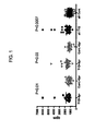


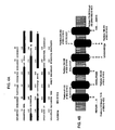
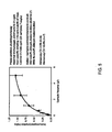
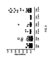


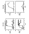
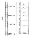


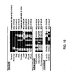
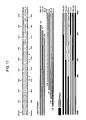
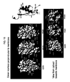


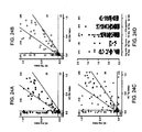
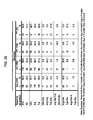

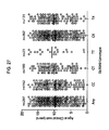

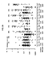
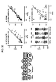
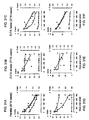

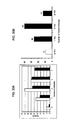
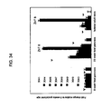
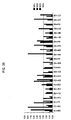

Classifications
-
- C—CHEMISTRY; METALLURGY
- C07—ORGANIC CHEMISTRY
- C07K—PEPTIDES
- C07K14/00—Peptides having more than 20 amino acids; Gastrins; Somatostatins; Melanotropins; Derivatives thereof
- C07K14/435—Peptides having more than 20 amino acids; Gastrins; Somatostatins; Melanotropins; Derivatives thereof from animals; from humans
- C07K14/705—Receptors; Cell surface antigens; Cell surface determinants
-
- A—HUMAN NECESSITIES
- A61—MEDICAL OR VETERINARY SCIENCE; HYGIENE
- A61P—SPECIFIC THERAPEUTIC ACTIVITY OF CHEMICAL COMPOUNDS OR MEDICINAL PREPARATIONS
- A61P3/00—Drugs for disorders of the metabolism
- A61P3/08—Drugs for disorders of the metabolism for glucose homeostasis
- A61P3/10—Drugs for disorders of the metabolism for glucose homeostasis for hyperglycaemia, e.g. antidiabetics
-
- C—CHEMISTRY; METALLURGY
- C07—ORGANIC CHEMISTRY
- C07K—PEPTIDES
- C07K14/00—Peptides having more than 20 amino acids; Gastrins; Somatostatins; Melanotropins; Derivatives thereof
- C07K14/435—Peptides having more than 20 amino acids; Gastrins; Somatostatins; Melanotropins; Derivatives thereof from animals; from humans
- C07K14/46—Peptides having more than 20 amino acids; Gastrins; Somatostatins; Melanotropins; Derivatives thereof from animals; from humans from vertebrates
- C07K14/47—Peptides having more than 20 amino acids; Gastrins; Somatostatins; Melanotropins; Derivatives thereof from animals; from humans from vertebrates from mammals
-
- G—PHYSICS
- G01—MEASURING; TESTING
- G01N—INVESTIGATING OR ANALYSING MATERIALS BY DETERMINING THEIR CHEMICAL OR PHYSICAL PROPERTIES
- G01N33/00—Investigating or analysing materials by specific methods not covered by groups G01N1/00 - G01N31/00
- G01N33/48—Biological material, e.g. blood, urine; Haemocytometers
- G01N33/50—Chemical analysis of biological material, e.g. blood, urine; Testing involving biospecific ligand binding methods; Immunological testing
- G01N33/53—Immunoassay; Biospecific binding assay; Materials therefor
- G01N33/564—Immunoassay; Biospecific binding assay; Materials therefor for pre-existing immune complex or autoimmune disease, i.e. systemic lupus erythematosus, rheumatoid arthritis, multiple sclerosis, rheumatoid factors or complement components C1-C9
-
- G—PHYSICS
- G01—MEASURING; TESTING
- G01N—INVESTIGATING OR ANALYSING MATERIALS BY DETERMINING THEIR CHEMICAL OR PHYSICAL PROPERTIES
- G01N2800/00—Detection or diagnosis of diseases
- G01N2800/04—Endocrine or metabolic disorders
- G01N2800/042—Disorders of carbohydrate metabolism, e.g. diabetes, glucose metabolism
-
- G—PHYSICS
- G01—MEASURING; TESTING
- G01N—INVESTIGATING OR ANALYSING MATERIALS BY DETERMINING THEIR CHEMICAL OR PHYSICAL PROPERTIES
- G01N2800/00—Detection or diagnosis of diseases
- G01N2800/52—Predicting or monitoring the response to treatment, e.g. for selection of therapy based on assay results in personalised medicine; Prognosis
Definitions
- This invention generally relates to the identification of autoantigen targets, and particularly, autoantigen targets in type I autoimmune diabetes (T1D), and more particularly, the protein ZnT8 as a novel autoantigen, upon which therapeutic, diagnostic, and prognostic tools and methods are based and described herein.
- T1D type I autoimmune diabetes
- Autoimmune disorders are conditions caused by an immune response against the body's own tissues. Autoimmune disorders result in destruction of one or more types of body tissues, abnormal growth of an organ, or changes in organ function. The disorder may affect only one organ or tissue type or may affect multiple organs and tissues.
- Type 1 autoimmune diabetes (T1D) in humans and the NOD mouse model is a polygenic T-cell dependent autoimmune disease, characterized by the selective destruction of the B cells of the islets of Langerhans (1-3) arising from a breakdown in tolerance to B cell antigens (4) in susceptible individuals who have inherent defects in critical immunomodulatory mechanisms (5) (e.g., individuals who exhibit a pathogenic rather than a protective immune response to self).
- Pancreatic exocrine and endocrine cells that share the same developmental lineage as the B-cell and are often in direct contact with B-cells are largely unaffected, and insulitis resolves once the B-cells are lost, which suggests that the autoimmune targets are largely B-cell specific.
- the molecular targets of autoreactive T-cells may include either an unidentified component of B-cells, or known B-cell proteins that are intrinsically unable to elicit a humoral response. Accordingly, defining the cognate antigens for them remains an important goal, and with the recent advances in genomic and proteomic techniques, now appears a realistic aim.
- the target of the well studied NY8.3 CD8 + clone (23) was recently shown to be a peptide derived from the B-cell protein IGRP (islet glucose 6-phosphatase related protein) by using a combination of a sensitive bioassay and analysis of proteomic peptides eluted from H-2K d molecules from NIT1 insulinoma cells (29).
- TID T1D
- insulin 33; 35-38
- IGRP IGRP
- T-cells recognizing this antigen in islet infiltrates.
- An alternative view is that disease results from polyclonal activation due to a breakdown in normal toleragenic mechanisms that would otherwise generate regulatory cells directed to the same molecules (41). Given the latter scenario, the inventors hypothesize that any molecule that is a significant target of autoimmunity in T1D is a candidate for use in antigen-based therapy.
- the non-obese diabetic (NOD) mouse is currently the best model of human T1D (8) where three stages of disease progression are evident, namely, expansion of autoreactive T-cells ("checkpoint 0"), their homing to the pancreatic islets (“checkpoint 1”), and the transition from a benign peri-insulitis to an invasive insulitis resulting in B-cell destruction (“checkpoint 2”) (9). It has been postulated that passage through "checkpoint 0" coincides with a wave of islet cell apoptosis that leads to enhanced presentation of B-cell antigens in the pancreatic lymph nodes.
- diagnostic assays based on humoral or cellular autoreactivity to autoantigens that are useful in monitoring disease progression are essential in making any decision to treat a patient and in the subsequent monitoring of treatment outcome.
- T1D diagnostic assays
- new immunotherapeutics based on disease targets many targets identified in one autoimmune disease, such as T1D, will also be targets in other autoimmune, disease, thereby expanding the available diagnostic and therapeutic approaches in a variety of autoimmune conditions.
- the fragment consists of a fragment represented by any one of SEQ ID NOs: 9,10, 14-18, 22, 23, 49-51 or 53-65 wherein the fragment comprises the amino acid sequence SLTIQMES (positions 346-353 of SEQ ID NO:2) and position 325 of SEO ID NO:2.
- a fragment of the invention comprises position 325 of SEQ ID NO:2 and in one embodiment of this aspect of the invention, the amino acid at position 325 is a tryptophan, glutamine, or arginine.
- a chimeric protein comprising, consisting essentially of, or consisting of, any two or more fragments as described herein.
- the chimeric protein comprises an N-terminal fragment of ZnT8 and a C-terminal fragment of ZnT8.
- the chimeric protein comprises two C-terminal fragments of ZnT8, wherein each of the fragments comprises amino acid position 325, and wherein each of the fragments comprises a different amino acid at position 325.
- one fragment comprsies an arginine at position 325 and wherein a second fragment comprises a tryptophan at position 325.
- Yet another embodiment of the invention relates to a fragment or homologue of ZnT8 comprising at least one epitope of ZnT8 that is selective bound by an anti-ZnT8 antibody.
- the anti-ZnT8 antibody is an antibody obtained from an individual.
- the individual has, or is suspected of having, type I diabetes.
- Another embodiment of the invention relates to use of a fragment for diagnosing an individual who is susceptible to or who is developing an autoimmune disease, comprising detecting antibodies that selectively bind to the ZnT88 fragment in a test sample from the individual, wherein detection of increased antibodies in the individual as compared to a negative control, indicates that the individual is susceptible to or is developing an autoimmune disease.
- the method comprises detecting antibodies specific for a human ZnT8 comprising an arginine at position 325.
- the method comprises detecting antibodies specific for a human ZnT8, wherein the amino acid at position 325 is an arginine, a tryptophan, and/or a glytamine.
- the autoimmune disease is type I diabetes.
- Another embodiment of the invention relates to a method to monitor the progression of type I diabetes autoimmunity in an individual; from an initial benign autoreactivity to destructive insulitis, comprising detecting antibodies that selectively bind to the ZnT8 fragment in a test sample from the individual, wherein detection of increased antibodies in the individual as compared to a prior measurement of antibodies in the same individual, indicates that the individual is progressing towards destructive insulitis.
- the method comprises detecting antibodies that selectively bind to human ZnT8, wherein the amino acid at position 325 is an arginine, a tryptophan, and/or glutamine.
- Yet another embodiment of the invention relates to a method to monitor the efficacy of a treatment for preventing type I diabetes, delaying the onset of type I diabetes, or ameliorating autoimmunity in prediabetic individuals, comprising detecting antibodies that selectively bind to ZnT8 in a test sample from the individual, wherein the detection of decreased or substantially the same level of antibodies in the individual indicates that the treatment is effective and wherein detection of increased antibodies in the individual as compared to a prior measurement of antibodies in the same individual indicates that the treatment is not effective.
- the method comprises detecting antibodies that selectively bind to human ZnT8, wherein the amino acid at position 325 is an arginine, a tryptophan, and/or a glutamine.
- the method can include the use of an assay selected from, but not limited to: radioimmunoprecipitation assay, ELISA, immunofluorescence assay time-resolved florescence assay, fluorescence activated cell sorter assay, competitive Europium assay and luminescence assay.
- an assay selected from, but not limited to: radioimmunoprecipitation assay, ELISA, immunofluorescence assay time-resolved florescence assay, fluorescence activated cell sorter assay, competitive Europium assay and luminescence assay.
- the present invention generally relates to the identification of autoantigen targets, and particularly, autoantigen targets in type I autoimmune diabetes (T1D), and more particularly, the protein ZnT8 as a novel autoantigen, upon which therapeutic, diagnostic, and prognostic tools and methods are based and described herein.
- the invention also relates to the identification of genetic variation in ZnT8 that is described herein as an important player in the initiation of the disease process and the progression of autoimmunity to clinical diabetes.
- the molecule itself, derived peptides, peptide analogs and secondary immune reagents also serve as the basis of therapeutic interventions to prevent the onset or progression of autoimmunity to clinical disease.
- the ability to determine the genotype of an individual as soon as one is born provides a distinct advantage to implementing therapeutic measures, since prevention of the emergence is likely to be easier than reversal of the autoimmune process once it is established.
- insulin was defined as a candidate gene based on its tissue association, GAD65 and IA-2 from the ability of patient sera to immunoprecipitate proteins from radiolabelled islets (64-66), and IGRP as a peptide bound to MHC H-2 K d that stimulated a T-cell clone derived from a spontaneously diabetic mouse.
- the inventors have asked the question of whether one can develop a more systematic approach to the identification of target autoantigens based upon what is now understood of the cellular and molecular biology of autoantigens, and the availability of tools to study and analyze genome-wide expression in different tissue by microarray analysis.
- the objective was to create a list of candidate genes that would include diabetes autoantigens of major importance which may potentially number upwards of 50 proteins from among the 15,000 or so gene transcripts expressed in the B-cell. Robust assays can then be used to detect cellular and humoral immune responses to such molecules.
- the results of this research are described below, with particular emphasis on the discovery of a novel target autoantigen, namely ZnT8.
- the structural biology of antigen presentation and T-cell recognition provided relatively few clues to identification of novel T1D autoantigens, since both MHC and T-cell receptor molecules have broad ranges of peptide binding specificities and relatively low affinities. It has proven difficult to identify native epitopes even when detailed knowledge of the binding properties of mimotope peptides is known, simply because the number of contact residues is small and conservative amino acid substitutions are readily accepted.
- the present inventors describe herein the development of a knowledge-based algorithm to identify candidate autoantigens on a genome wide basis. A number of primary and secondary attributes of an "ideal" autoantigen are initially assumed based on empirical information and weighted accordingly.
- the conformity of the antigen to each of the ideal attributes is weighted to derive an "autoantigenic score" and hence an ordered list of best candidates.
- a meta-analysis based on gene ontology functions and information such as gene location is then considered before embarking on an evaluation of a shortlist of candidates based upon spontaneous and primed cell-mediated autoimmune responses in NOD mice and humoral autoreactivity determined in both NOD mice and human sera.
- the microarray data that underpins this approach has largely been obtained.
- candidate antigens are first screened by serological analyses in human subjects and then HLA-DR3, -DR4, DQ2- and -DQ8 HLA A2 and other class I HLA diabetes susceptibility alleles in transgenic mice are screened for cell-mediated immune responses to the candidate antigens.
- Zinc transporter protein 8 (ZnT8) is a protein that appears to be confined to insulin-producing ⁇ cells of the islet in man and mice.
- the present inventors have demonstrated that ZnT8 is a target for autoantibodies in human subjects that develop T1D, and have identified polymorphic variants of ZnT8 as genetic markers of type 1 diabetes susceptibility, autoimmune status, autoantibody specificity and disease progression and severity. Autoantibodies to ZnT8 appear before the development of clinical disease and are believed to be a novel marker for incipient diabetes.
- the inventors have discovered that the ZnT8 autoantibodies target two different regions of the molecule, and that one of these regions incorporates one of 3 different amino acids that play a crucial role in determining both the specificity and the magnitude of the immune response. Determination of the genotype of an individual using a single nucleotide polymorphism analysis at any time prior to the development of autoimmunity and diabetes provides information regarding which variant amino acid is encoded, and can therefore be used in making clinical decision regarding treatment and care. In addition, the present invention permits the precise analysis of the specificity of the autoantibodies that are directed at ZnT8, and further classification of the antibodies with respect to which of the variant forms of ZnT8 are targeted by the immune system.
- isoepitopes three major forms that depend on the identity of the variant amino acid at position 325 in the molecule (Arg, Trp or Gln), which are referred to herein as “isoepitopes", and a fourth epitope that incorporates conserved amino acids (aa325 independent), are recognized.
- the response of the individual can be categorized as being one of 8 different responses: (1) none; (2) Arg325-restricted; (3) Trp325-restricted; (4) Gln325-restricted; (5) Arg325- and Trp325- restricted; (6) Arg325- and Gln325-restricted; (7) Trp325-and Gln325-restricted; and (8) amino acid 325-independent.
- stratification of the autoantibody response is proposed herein to provide information about the disease progression and will be essential in determining how to treat T1Dautoimmunity with antigen-specific therapeutic agents.
- ZnT8 autoantibodies target two different regions of the molecule, and that one of these regions incorporates one of 3 different amino acids that play a crucial role in determining both the quality and the magnitude of the immune response. Since the variant amino acid sequence is determined by the nucleotide sequence of the genome, it is therefore possible to determine which variant amino acid is encoded. The inventors have demonstrated for the first time that autoantibodies directed at this site match the sequence that the gene encodes and thus are truly self-reactive. The gene shows 3 known alleles which are present in high (75%), medium (25%) and low (1%) frequency and show significant variation between racial groups.
- the object of immune therapy may be the induction of immune tolerance, or conversely, marshalling the immune system through respectively engaging or deleting elements of immune elements, such as destructive effector T-cells or protective regulatory T-cells.
- ZnT8 Prior to the present invention, there was no reason to suspect a priori that ZnT8 could be an autoantigen recognized either by antibodies or the cellular arm of the immune system. Indeed, to the best of the present inventors' knowledge, ZnT8 has not been investigated in the context of autoimmune diabetes and there are no other published data on the molecule in an immunological context. As discussed above, there are literature reports on the use of autoantigens as diagnostic and therapeutic agents in other autoimmune diseases and in the context of diabetes. However, these are directed to molecules such as insulin, glutamate decarboxylase (GAD65), IA2, phogrin and heat shock protein 60, which are unrelated to ZnT8 from a structural, cell biological and immunological standpoint.
- GAD65 glutamate decarboxylase
- IA2 IA2
- phogrin heat shock protein 60
- This SNP encodes an alternative Arg or Trp at amino acid 325 in ZnT8 (SEQ ID NO:2), which coincides with a polymorphic variant identified in the present inventors' studies.
- TID type 1 diabetes
- a genome wide association study identified ZnT8 as containing a number of SNPs that were associated with type 2 diabetes, including the above referenced rs13266634 ( Sladek et al., 2007, Nature. 2007 Feb 22;445(7130):881-5.
- the present inventors describe herein the use of genes encoding ZnT8, nucleic acid molecules derived therefrom, ZnT8 proteins or fragments thereof encoded by such genes and nucleic acid molecules, as well as homologues of such genes and proteins and related agents (e.g ., antibodies, agonists, antagonists), and polymorphic variants of ZnT8, and the use or targeting of such genes, nucleic acids, proteins, homologues, variants and/or related agents, and/or compositions or formulations comprising the same, in the development of a variety of diagnostic and therapeutic tools and assays.
- tools and assays include, but are not limited to:
- Fig. 20 shows a comparison of the ten variants of the Slc30A gene that are recognized in humans, although one of these (Slc30A9) is probably not a true homologue given its low amino acid sequence identity and similarity (shown in parentheses above).
- the closest relatives of ZnT8 are Slc30A2, a lysosomal protein, Slc30A3 a protein associated with synaptic vesicles and Slc30A4.
- Antibodies raised in rabbits to Slc30A8 show some cross-reactivity with Slc30A3, which raises the possibility that reactivity or cross-reactivity might also occur in other human autoimmune diseases, like multiple sclerosis.
- autoimmune diseases other than TID such as, but not limited to, systemic lupus erythematosus, myasthenia gravis, rheumatoid arthritis, multiple sclerosis, celiac disease, autoimmune thyroiditis, Addison's disease, Graves' disease and rheumatic carditis.
- the method of the invention is applicable to any autoimmune disease in which Slc30A8 (ZnT8) or a homologue is a target or autoantigen (autoantibody or T cell autoantigen).
- the inventors have shown that individuals with autoantibodies to ZnT8 in some cases show reactivity to the other members of the Slc30A gene family encoding other ZnT transporters. For example, the inventors show some reactivity to ZnT3, which is normally considered a neuron-specific isoform. An important implication of this finding is that such reactivity could have pathological consequences in tissues outside of the pancreas. It is known that diabetes especially type 1 diabetes is associated with peripheral neuropathy, and cross-reactivity of antigens may represent a link between these conditions. Accordingly, the tools and methods of the invention are believed to be useful and applicable to other conditions that are linked by antigen cross-reactivity, including, but not limited to, peripheral neuropathy.
- the present invention encompasses the use of a gene encoding ZnT8, as well as nucleic acid molecules derived from or comprising at least a portion of the coding region and/or regulatory region of such gene, and including any polymorphic variant of such gene.
- the present invention also encompasses the use of any ZnT8 proteins, homologues or fragments thereof, including any polymorphic variant thereof, or agonists or antagonists thereof, including those encoded by the above-referenced genes or nucleic acid molecules.
- Novel ZnT8 nucleic acid molecules and proteins are encompassed by the invention as compositions of matter.
- the present invention further includes ZnT8 antibodies, antigen-binding fragments thereof, and antigen-binding peptides that are useful in a variety of methods in the present invention.
- Zinc transporter protein 8 is a 369 amino acid protein encoded by the ZnT8 gene.
- ZnT8 contains six transmembrane domains and a histidine-rich loop between transmembrane domains IV and V, like the other ZnT proteins (see Chimienti et al., 2004, Diabetes, supra, and Fig. 4 presented herein). This protein is solely transcribed in the pancreas and more particularly, is expressed only in the ⁇ cells of the islets of Langerhans. As described by Chimienti et al., ibid., the ZnT8 gene is located on chromosome 8q24.11, contains eight exons, and spans 37 kb.
- the cDNA and deduced amino acid sequence of human ZnT-8 protein are provided in Chimienti et al., ibid., and this publication shows the gene structure, chromosomal localization, and putative splicing of human ZnT8 (see Fig. 1A of Chimienti et al., ibid .).
- the cDNA sequence encoding human ZnT8 and the deduced amino acid sequence are reflected in Fig. 1B of Chimienti et al., ibid., where the predicted transmembrane domains of ZnT8 are also indicated.
- Fig. 1B of Chimienti et al., ibid.
- a nucleic acid sequence encoding human ZnT8 is represented herein by SEQ ID NO:1.
- SEQ ID NO:1 encodes the human ZnT8 protein, the amino acid sequence of which is represented herein by SEQ ID NO:2.
- Murine ZnT8 is also known in the art.
- the nucleic acid sequence encoding murine ZnT8 is represented herein by SEQ ID NO:3.
- SEQ ID NO:3 encodes the murine ZnT8 protein, the amino acid sequence of which is represented herein by SEQ ID NO:4.
- nucleotide sequence encoding human ZnT8 is also described in the National Center for Biotechnology Information (NCBI) database Accession No. NM_173851 (gi: 64762488).
- the amino acid sequence for human ZnT8 is also found in the NCBI database Accession No. NP_776250 (gi: 64762489).
- the nucleotide sequences encoding rat ZnT8 is also known. All of the information contained in the database accession numbers and in the publications referenced herein is incorporated herein by reference. Various polymorphic variants, fragments, chimeric proteins, and fusion proteins of these sequences are described elsewhere herein, and are also encompassed by the invention.
- the present invention also encompasses numerous naturally occurring and synthetically derived variants (homologues) of ZnT8, including nucleotide and amino acid polymorphisms in the gene, proteins encoded by alternate start sites, and various fragments, fusion proteins, and chimeric proteins, and the nucleic acid molecules encoding such fragments, fusion proteins, and chimeric proteins.
- homologues including nucleotide and amino acid polymorphisms in the gene, proteins encoded by alternate start sites, and various fragments, fusion proteins, and chimeric proteins, and the nucleic acid molecules encoding such fragments, fusion proteins, and chimeric proteins.
- a homologue or variant of a ZnT8 protein useful in any of the methods described herein comprises, consists essentially of, or consists of, an amino acid sequence that is at least about 45%, or at least about 50%, or at least about 55%, or at least about 60%, or at least about 65%, or at least about 70%, or at least about 75%, or at least about 80%, or at least about 85%, or at least about 90%, or at least about 95% identical, or at least about 95% identical, or at least about 96% identical, or at least about 97% identical, or at least about 98% identical, or at least about 99% identical (or any percent identity between 45% and 99%, in whole integer increments), to the amino acid sequence of a wild-type ZnT8 protein, and particularly, the human ZnT8 protein described herein (see discussion above).
- the homologue comprises, consists essentially of, or consists of, an amino acid sequence that is less than 100% identical, less than about 99% identical, less than about 98% identical, less than about 97% identical, less than about 96% identical, less than about 95% identical, and so on, in increments of 1%, to less than about 70% identical to the naturally occurring amino acid sequence of the ZnT8 protein.
- the invention also includes various naturally occurring and synthetically derived variants of ZnT8 that are proteins encoded by alternate start sites, as well as fragments (peptides) thereof, fusion proteins, or chimeric proteins thereof that are useful in any of the diagnostic, therapeutic, or research methods described herein.
- a natural variant of human ZnT8 with an alternate start site is represented herein by SEQ ID NO:5. This protein spans from amino acid position 50 to 367 of SEQ ID NO:2.
- SEQ ID NO:5 a natural variant of human ZnT8 with an alternate start site is represented herein by SEQ ID NO:5. This protein spans from amino acid position 50 to 367 of SEQ ID NO:2.
- the gene structure shown was deduced by the inventors from alignments of published EST sequences and the gene. It differs however from the 8 exon gene structured that is accepted as "normal".
- downstream Met can be used as an alternate start site resulting in the truncation of the NH2 terminus of the sequence. If this occurs in a tissue specific manner, e.g . in thymus and not in pancreas, then a cryptic epitope occurs that might be targeted by the immune system.
- the present inventors' laboratory has evidence of this occurring and contributing to autoreactivity to other diabetes-related autoantigens, IA-2 and IGRP.
- the alternative translation product might also be presented to the immune system as a "foreign" peptide epitope.
- polymorphic variants of the full-length ZnT8 protein include, but are not limited to, a substitution of a histidine for the tyrosine at position 18 of SEQ ID NO:2, a substitution of a valine for the alanine at position 261 of SEQ ID NO:2, and a substitution of a tryptophan or a glutamine for the arginine at position 325 of SEQ ID NO:2.
- Polymorphic variants of ZnT8, particularly at position 325 are of particular interest in the present invention and are discussed in detail below. Proteins, variants, fragments, chimeric proteins, and fusion proteins comprising such polymorphisms, and the nucleotides that encode them, are encompassed by the invention.
- the NH2 and COOH termini are predicted to lie on the same side of the membrane.
- the N-terminus is 74aa long in the case of the reference sequence and 25aa in the case of the isoforms with the alternate start site.
- the COOH terminus of 94aa incorporates the major humoral immunological determinant in type 1 diabetes.
- This epitope is cryptic in the full length molecule and lies between aa268 and 358 as shown by the minimal effect on antigenicity caused by removal of the last 15aa. Curiously, this is a region which is strongly conserved among ZnT transporters from different species.
- autoreactivity can be generated against the ZnT8 protein (unique to the pancreatic B-cell) and also by homologous proteins of infectious organisms.
- a similar relationship has been observed with another diabetes autoantigen IA2 (unpublished work).
- IA2 diabetes autoantigen
- It is a general principle then one can postulate that autoimmunity in T1D could be triggered by a minor infection with a microorganism with a similar Zn transporter. Equally it might be possible to immunize with the defined epitope of the infectious agent. A second weaker epitope is located in the N-terminus which is not conserved.
- the identified epitopes appear to reside inside the cell as is the case of the autoantigens IA2 and GAD65.
- the short loops at the top side of the figure (encompassing positions represented by positions 97-105; 164-168, and 239-252) would likely be oriented towards the lumen of the cellular elements of the secretory pathway. This potentially would include the endoplasmic reticulum, the Golgi, the insulin secretory granule and ultimately the extracellular space. It is likely that they are exposed on the cell surface during the normal exocytosis of insulin granules during normal secretion or if the cell were damaged in some way.
- antibodies to peptides representing these loops are encompassed herein for the detection of ZnT8, and it would thus serve as a biomarker which might be used to image the B-cell mass in vivo, to determine the activity of the cell (more secretion equals more exposure) and to monitor the generation of ⁇ -cells from other sources such as stem cells, and during normal development.
- One antibody of the invention is proposed that is raised against the luminal domain 2 sequence of the mouse ZnT8 (positions 163-183 of SEQ ID NO:4, or ERLLYPDYQIQAGIMITVSGC) for cell biological applications.
- antibodies have been made to the human C-terminus as a fusion protein with glutathione S-transferase (see below).
- Other antipeptide antibodies to sequences in the N-term and C-term region can be produced using the guidance provided herein. Generation of anti-phosphopeptide antibodies can also be prepared.
- the third intracellular loop (depicted as residues 196-215 of SEQ ID NO:2 in Fig. 4B ) is rich in histidine residues (positions 197, 203 and 205 of SEQ ID NO:2) which may be important in binding Zn and other heavy metals. This could be part of the mechanism by which the molecule transfer Zn from the cytoplasm to the lumen of membrane enveloped compartments that constitute the secretory pathway.
- fragments of ZnT8 are encompassed for use in diagnostic and therapeutic methods, or for the generation of antibodies.
- the minimum size of a ZnT8 protein, portion of a protein (e.g. a fragment, portion, domain, etc.), or region or epitope of a protein is a size sufficient to serve as an epitope or conserved binding surface for the generation of an antibody or as a target in an in vitro assay.
- a protein of the present invention is at least about 4, 5, 6, 7 or 8 amino acids in length (e.g., suitable for an antibody epitope or as a detectable peptide in an assay), or at least about 25 amino acids in length, or at least about 50 amino acids in length, or at least about 100 amino acids in length, or at least about 150 amino acids in length, and so on, in any length between 4 amino acids and up to the full length of a ZnT8 protein or portion thereof or longer, in whole integers (e.g., 8, 9, 10,...25, 26,...300, 301,).
- any N-terminal fragment, C-terminal fragment, chimera thereof or deletion fragment of any length is encompassed by this invention, with respect to the ZnT8 protein from any species, with human ZnT8 being preferred, and including any polymorphic forms thereof.
- Table 1 and Figs. 15-19 , 21 and 25 describe additional information regarding the localization of epitopes and isoepitopes of ZnT8, including the identification of residues in the C-terminus that are critical for autoreactivity. This information allows for the design, production and use of a variety of fragments and homologues of ZnT8 in any of the diagnostic, prognostic and therapeutic methods described herein.
- Figs. 4A and 4B show the predicted transmembrane domains of the ZnT8 protein. These domains pose a problem in the design of assays as they are usually surrounded by lipid and are otherwise very sticky, so the protein either precipitates or doesn't fold. Such regions may be targets for T-cells, and are accordingly encompassed for use in fragments according to the invention, but they are unlikely to be targets for antibodies.
- the full-length sequence does show reactivity to circulating antibodies and more so in diabetics than controls.
- the assay detects only 10-20% of individuals with recent-onset disease. accordingly, the inventors have provided alternative probes, namely the N-terminus, C-terminus and the Zn binding loop probes as set forth below.
- a particularly preferred fragment of a ZnT8 protein useful in the present invention is any N-terminal or C-terminal fragment of ZnT8, with C-terminal fragments being particularly preferred.
- Another preferred fragment of ZnT8 is a mixture or hybrid (chimera) of N- and C-terminal fragments of ZnT8.
- Particularly preferred fragments of ZnT8 comprise, consist essentially of, or consist of, any C-terminal fragment from about the C-terminal 110 residues to about the C-terminal 8 residues of human ZnT8.
- Particularly preferred C-terminal fragments include the 101 and 102 C-terminal residues of human ZnT8.
- proteins, variants, chimeric proteins, and fragments that can be used in various embodiments of the present invention are illustrated in Table I below. Any of these proteins, variants, chimeric proteins, or fragments can be appended with one, two, or more amino acid residues (e.g., a methionine at the N-terminus of the fragment, as shown for the C-terminal fragments below). Moreover, given the provision of these constructs and the information provided in the Examples, one of skill in the art will readily be able to produce other constructs where additional amino acids are deleted, where domains of a chimera are reversed in order, or where two or more fragments or portions thereof described herein are combined to produce additional chimeric proteins.
- amino acid residues e.g., a methionine at the N-terminus of the fragment, as shown for the C-terminal fragments below.
- the sequences listed in Table 1 were designed by the inventors to test various features of patient autoreactivity, and to design assays that either discriminated better between patients, or conversely, were pan-reactive.
- the following listing shows how the sequences described above have been categorized by the inventors with respect to patient autoreactivity (patient serum samples).
- Reference to a sequence not in parentheses indicates identification of patient reactivity towards the construct comprising that sequence; sequences shown in parentheses indicate either reduced activity or variable activity that is likely the product of other specificities that have yet to be defined.
- the absence of identification of a sequence number with respect to a particular sample type indicates lack of reactivity.
- the information is limited as only one patient serum has been identified to date from more than 500 that have been tested.
- the present invention also includes ZnT8 proteins that comprise, consist essentially of, or consist of an antigenic peptide or T cell epitope (also referred to as a major histocompatibility complex (MHC)-binding peptide).
- MHC major histocompatibility complex
- Such a peptide includes any peptide that is capable of binding to an MHC protein in a manner such that the MHC-peptide complex can bind to a T cell receptor (TcR) and, in a preferred embodiment, thereby induce a T cell response (e.g ., a stimulatory or toleragenic response, described below).
- TcR T cell receptor
- An MHC-binding peptide that binds to an MHC molecule and is recognized, in conjunction with the MHC molecule, by a T cell receptor, is considered to be an antigenic peptide.
- peptides that are produced by hydrolysis of antigens undergo hydrolysis prior to binding of the antigen to an MHC protein.
- Class I MHC proteins typically present antigenic peptides derived from proteins actively synthesized in the cytoplasm of the cell.
- class II MHC proteins typically present antigenic peptides derived either from exogenous proteins that enter a cell's endocytic pathway or from proteins synthesized in the endoplasmic reticulum (ER). Intracellular trafficking permits an antigenic peptide to become associated with an MHC protein.
- the resulting MHC-peptide complex then travels to the surface of the cell where it is available for interaction with a TcR.
- the binding of a peptide to an MHC peptide binding groove can control the spatial arrangement of MHC and/or peptide amino acid residues recognized by a TcR. Such spatial control is due in part to hydrogen bonds formed between a peptide and an MHC protein.
- the length of T cell epitope is from about 5 to about 40 amino acid residues, more preferably from about 6 to about 30 amino acid residues, and even more preferably from about 8 to about 20 amino acid residues, and even more preferably between about 9 and 11 amino acid residues, including any size peptide between 5 and 40 amino acids in length, in whole integer increments (i.e., 5, 6, 7, 8, 9...40). While naturally MHC Class II-bound peptides vary from about 9-40 amino acids, in nearly all cases the peptide can be truncated to an about 9-11 amino acid core without loss of MHC binding activity or T cell recognition.
- an isolated polynucleotide (also referred to as an isolated nucleic acid molecule) is a nucleic acid molecule that has been removed from its natural milieu (e.g ., that has been subject to human manipulation), its natural milieu being the genome or chromosome in which the nucleic acid molecule is found in nature.
- isolated does not necessarily reflect the extent to which the nucleic acid molecule has been purified, but indicates that the molecule does not include an entire genome or an entire chromosome in which the nucleic acid molecule is found in nature.
- the polynucleotides useful in the present invention are typically a portion of a gene (sense or non-sense strand) of the present invention that is suitable for use as a hybridization probe or PCR primer for the identification of a full-length gene (or portion thereof) in a given sample, or that is suitable for encoding a ZnT8 protein or fragment thereof, or that is suitable as a therapeutic reagent ( e.g ., antisense or an aptamer).
- An isolated nucleic acid molecule can include a gene or a portion of a gene (e.g ., the regulatory region or promoter), for example, to produce a reporter construct or a recombinant protein.
- An isolated nucleic acid molecule that includes a gene is not a fragment of a chromosome that includes such gene, but rather includes the coding region and regulatory regions associated with the gene, but no additional genes naturally found on the same chromosome.
- An isolated nucleic acid molecule can also include a specified nucleic acid sequence flanked by (i.e., at the 5' and/or the 3' end of the sequence) additional nucleic acids that do not normally flank the specified nucleic acid sequence in nature (i.e., heterologous sequences).
- Isolated nucleic acid molecule can include DNA, RNA (e.g., mRNA), or derivatives of either DNA or RNA (e.g., cDNA).
- nucleic acid molecule primarily refers to the physical nucleic acid molecule and the phrase “nucleic acid sequence” primarily refers to the sequence of nucleotides on the nucleic acid molecule, the two phrases can be used interchangeably, especially with respect to a nucleic acid molecule, or a nucleic acid sequence, being capable of encoding a protein.
- an isolated nucleic acid molecule of the present invention is produced using recombinant DNA technology (e.g., polymerase chain reaction (PCR) amplification, cloning) or chemical synthesis.
- PCR polymerase chain reaction
- the minimum size of a nucleic acid molecule or polynucleotide of the present invention is a size sufficient to encode a protein having a desired biological activity, sufficient to form a probe or oligonucleotide primer that is capable of forming a stable hybrid with the complementary sequence of a nucleic acid molecule encoding the natural protein (e.g., under moderate, high or very high stringency conditions), or to otherwise be used as a target or agent in an assay or in any therapeutic method discussed herein.
- the size of the polynucleotide can be dependent on nucleic acid composition and percent homology or identity between the nucleic acid molecule and a complementary sequence as well as upon hybridization conditions per se (e.g., temperature, salt concentration, and formamide concentration).
- the minimum size of a polynucleotide that is used as an oligonucleotide probe or primer is at least about 5 nucleotides in length, and preferably ranges from about 5 to about 50 or about 500 nucleotides or greater (1000, 2000, etc.), including any length in between, in whole number increments (i.e., 5, 6, 7, 8, 9, 10,...33, 34,...256, 257,...500...1000...), and more preferably from about 10 to about 40 nucleotides, and most preferably from about 15 to about 40 nucleotides in length.
- the oligonucleotide primer or probe is typically at least about 12 to about 15 nucleotides in length if the nucleic acid molecules are GC-rich and at least about 15 to about 18 bases in length if they are AT-rich.
- the nucleic acid molecule can include a portion of a protein-encoding sequence or a nucleic acid sequence encoding a full-length protein.
- an oligonucleotide probe is a nucleic acid molecule which most typically ranges in size from about 8 nucleotides to several hundred nucleotides in length. Such a molecule is typically used to identify a target nucleic acid sequence in a sample by hybridizing to such target nucleic acid sequence under stringent hybridization conditions.
- stringent hybridization conditions refer to standard hybridization conditions under which nucleic acid molecules are used to identify similar nucleic acid molecules. Such standard conditions are disclosed, for example, in Sambrook et al., Molecular Cloning: A Laboratory Manual, Cold Spring Harbor Labs Press, 1989 .
- moderate stringency hybridization and washing conditions refer to conditions which permit isolation of nucleic acid molecules having at least about 70% nucleic acid sequence identity with the nucleic acid molecule being used to probe in the hybridization reaction (i.e., conditions permitting about 30% or less mismatch of nucleotides).
- High stringency hybridization and washing conditions refer to conditions which permit isolation of nucleic acid molecules having at least about 80% nucleic acid sequence identity with the nucleic acid molecule being used to probe in the hybridization reaction (i.e., conditions permitting about 20% or less mismatch of nucleotides).
- Very high stringency hybridization and washing conditions refer to conditions which permit isolation of nucleic acid molecules having at least about 90% nucleic acid sequence identity with the nucleic acid molecule being used to probe in the hybridization reaction (i.e., conditions permitting about 10% or less mismatch of nucleotides).
- conditions permitting about 10% or less mismatch of nucleotides i.e., conditions permitting about 10% or less mismatch of nucleotides.
- Meinkoth et al., ibid. can use the formulae in Meinkoth et al., ibid. to calculate the appropriate hybridization and wash conditions to achieve these particular levels of nucleotide mismatch. Such conditions will vary, depending on whether DNA:RNA or DNA:DNA hybrids are being formed. Calculated melting temperatures for DNA:DNA hybrids are 10°C less than for DNA:RNA hybrids.
- stringent hybridization conditions for DNA:DNA hybrids include hybridization at an ionic strength of 6X SSC (0.9 M Na + ) at a temperature of between about 20°C and about 35°C (lower stringency), more preferably, between about 28°C and about 40°C (more stringent), and even more preferably, between about 35°C and about 45°C (even more stringent), with appropriate wash conditions.
- 6X SSC 0.9 M Na +
- stringent hybridization conditions for DNA:RNA hybrids include hybridization at an ionic strength of 6X SSC (0.9 M Na + ) at a temperature of between about 30°C and about 45°C, more preferably, between about 38°C and about 50°C, and even more preferably, between about 45°C and about 55°C, with similarly stringent wash conditions.
- 6X SSC 0.9 M Na +
- T m can be calculated empirically as set forth in Sambrook et al., supra, pages 9.31 to 9.62.
- the wash conditions should be as stringent as possible, and should be appropriate for the chosen hybridization conditions.
- hybridization conditions can include a combination of salt and temperature conditions that are approximately 20-25°C below the calculated T m of a particular hybrid
- wash conditions typically include a combination of salt and temperature conditions that are approximately 12-20°C below the calculated T m of the particular hybrid.
- hybridization conditions suitable for use with DNA:DNA hybrids includes a 2-24 hour hybridization in 6X SSC (50% formamide) at about 42°C, followed by washing steps that include one or more washes at room temperature in about 2X SSC, followed by additional washes at higher temperatures and lower ionic strength (e.g., at least one wash as about 37°C in about 0.1X-0.5X SSC, followed by at least one wash at about 68°C in about 0.1X-0.5X SSC).
- PCR primers are also nucleic acid sequences, although PCR primers are typically oligonucleotides of fairly short length that are used in polymerase chain reactions. PCR primers and hybridization probes can readily be developed and produced by those of skill in the art, using sequence information from the target sequence. (See, for example, Sambrook et al., supra or "Molecular Biotechnology," Second Edition, by Glick and Pasternak, ASM Press, Washington D.C., 1998, pp. 555-590 ).
- nucleic acid sequences of certain nucleic acid molecules of the present invention allows one skilled in the art to, for example, (a) make copies of those nucleic acid molecules and/or (b) obtain nucleic acid molecules including at least a portion of such nucleic acid molecules (e.g., nucleic acid molecules including full-length genes, full-length coding regions, regulatory control sequences, truncated coding regions).
- nucleic acid molecules can be obtained in a variety of ways including traditional cloning techniques using oligonucleotide probes to screen appropriate libraries or DNA and PCR amplification of appropriate libraries or DNA using oligonucleotide primers.
- Preferred libraries to screen or from which to amplify nucleic acid molecule include mammalian genomic DNA libraries. Techniques to clone and amplify genes are disclosed, for example, in Sambrook et al., ibid.
- an anti-sense nucleic acid molecule is defined as an isolated nucleic acid molecule that reduces expression of a protein by hybridizing under high stringency conditions to a gene encoding the protein. Such a nucleic acid molecule is sufficiently similar to the gene encoding the protein that the molecule is capable of hybridizing under high stringency conditions to the coding or complementary strand of the gene or RNA encoding the natural protein.
- RNA interference is a process whereby double stranded RNA, and in mammalian systems, short interfering RNA (siRNA), is used to inhibit or silence expression of complementary genes.
- siRNA are unwound and associate with an RNA induced silencing complex (RISC), which is then guided to the mRNA sequences that are complementary to the siRNA, whereby the RISC cleaves the mRNA.
- RISC RNA induced silencing complex
- a ribozyme is an RNA segment that functions by binding to the target RNA moiety and inactivate it by cleaving the phosphodiester backbone at a specific cutting site.
- a ribozyme can serve as a targeting delivery vehicle for a nucleic acid molecule, or alternatively, the ribozyme can target and bind to RNA encoding the biomarker, for example, and thereby effectively inhibit the translation of the biomarker.
- Aptamers are short strands of synthetic nucleic acids (usually RNA but also DNA) selected from randomized combinatorial nucleic acid libraries by virtue of their ability to bind to a predetermined specific target molecule with high affinity and specificity. Aptamers assume a defined three-dimensional structure and are capable of discriminating between compounds with very small differences in structure.
- a recombinant nucleic acid molecule is a molecule that can include at least one of any nucleic acid sequence encoding a ZnT8 protein or other protein described herein.
- a recombinant nucleic acid molecule is operatively linked to at least one expression control sequence capable of effectively regulating expression of the nucleic acid molecule(s) in a host cell.
- a recombinant nucleic acid molecule is produced using recombinant DNA technology (e.g., polymerase chain reaction (PCR) amplification, cloning).
- a recombinant nucleic acid molecule includes a recombinant vector, which is any nucleic acid sequence, typically a heterologous sequence, which is operatively linked to the isolated nucleic acid molecule encoding a protein (e.g ., ZnT8), which is capable of enabling recombinant production of the protein, or which is capable of delivering the nucleic acid molecule into a host cell in vitro , ex vivo or in vivo, according to the present invention.
- a vector can contain nucleic acid sequences that are not naturally found adjacent to the isolated nucleic acid molecules to be inserted into the vector.
- the vector can be either RNA or DNA, either prokaryotic or eukaryotic, and preferably in the present invention, is a virus or a plasmid.
- Recombinant vectors can be used in the cloning, sequencing, and/or otherwise manipulating of nucleic acid molecules.
- Recombinant vectors are preferably used in the expression of nucleic acid molecules, and can also be referred to as expression vectors.
- Preferred recombinant vectors are capable of being expressed in a transfected host cell, and particularly, in a transfected mammalian host cell in vivo.
- nucleic acid molecules are operatively linked to expression vectors containing regulatory sequences such as transcription control sequences, translation control sequences, origins of replication, and other regulatory sequences that are compatible with the host cell and that control the expression of nucleic acid molecules of the present invention.
- regulatory sequences such as transcription control sequences, translation control sequences, origins of replication, and other regulatory sequences that are compatible with the host cell and that control the expression of nucleic acid molecules of the present invention.
- the phrase "operatively linked” refers to linking a nucleic acid molecule to an expression control sequence in a manner such that the molecule is expressed when transfected (i.e., transformed, transduced or transfected) into a host cell.
- Transcription control sequences are sequences that control the initiation, elongation, and termination of transcription. Particularly important transcription control sequences are those that control transcription initiation, such as promoter, enhancer, operator and repressor sequences. Suitable transcription control sequences include any transcription control sequence that can function in a host cell according to the present invention. A variety of suitable transcription control sequences are known to those skilled in the art. Particularly preferred transcription control sequences include inducible promoters, cell-specific promoters, tissue-specific promoters (e.g., insulin promoters) and enhancers. Transcription control sequences of the present invention can also include naturally occurring transcription control sequences naturally associated with the protein to be expressed prior to isolation. In one embodiment, a transcription control sequence includes an inducible promoter.
- recombinant vector useful in a recombinant nucleic acid molecule of the present invention is a recombinant viral vector.
- a recombinant viral vector includes a recombinant nucleic acid sequence encoding a ZnT8 protein of the present invention that is packaged in a viral coat that can be expressed in a host cell in an animal or ex vivo after administration.
- a number of recombinant viral vectors can be used, including, but not limited to, those based on alphaviruses, poxviruses, adenoviruses, herpesviruses, lentiviruses, adeno-associated viruses and retroviruses.
- viral vectors are those based on adenoviruses and adeno-associated viruses.
- Viral vectors suitable for gene delivery are well known in the art and can be selected by the skilled artisan for use in the present invention. A detailed discussion of current viral vectors is provided in " Molecular Biotechnology," Second Edition, by Glick and Pasternak, ASM Press, Washington D.C., 1998, pp. 555-590 , the entirety of which is incorporated herein by reference.
- Suitable host cells to transfect with a recombinant nucleic acid molecule include any microbial, insect, or animal cell that can be transfected.
- Host cells can be either untransfected cells or cells that are already transfected with at least one nucleic acid molecule.
- the term “transfection” is used to refer to any method by which an exogenous nucleic acid molecule (i.e., a recombinant nucleic acid molecule) can be inserted into the cell.
- transformation can be used interchangeably with the term “transfection” when such term is used to refer to the introduction of nucleic acid molecules into microbial cells, such as bacteria and yeast.
- transformation is used to describe an inherited change due to the acquisition of exogenous nucleic acids by the microorganism and is essentially synonymous with the term “transfection”.
- transformation has acquired a second meaning which can refer to changes in the growth properties of cells in culture after they become cancerous, for example.
- transfection is preferably used with regard to the introduction of exogenous nucleic acids into animal cells, and the term “transfection” will be used herein to generally encompass both transfection of animal cells and transformation of microbial cells, to the extent that the terms pertain to the introduction of exogenous nucleic acids into a cell. Therefore, transfection techniques include, but are not limited to, transformation, electroporation, microinjection, lipofection, adsorption, infection and protoplast fusion.
- an isolated protein or polypeptide in the present invention is a protein that has been removed from its natural milieu (i.e., that has been subject to human manipulation), and includes full-length proteins, fusion or chimeric proteins, or any fragment or homologue of such a protein.
- a protein can include, but is not limited to, purified proteins, partially purified proteins, recombinantly produced proteins, synthetically produced proteins, membrane-bound proteins, proteins complexed with lipids, soluble proteins and isolated proteins associated with other proteins.
- isolated does not reflect the extent to which the protein has been purified.
- an isolated protein of the present invention is produced recombinantly.
- a "human ZnT8 protein” or a protein "derived from” a human ZnT8 protein refers to a ZnT8 protein (generally including a homologue of a naturally occurring ZnT8 protein) from a human ( Homo sapiens ) or to a ZnT8 protein that has been otherwise produced from the knowledge of the structure (e.g., sequence) and perhaps the function of a naturally occurring ZnT8 protein from Homo sapiens.
- a human ZnT8 protein includes any ZnT8 protein that has substantially similar structure and function of a naturally occurring ZnT8 protein from Homo sapiens or that is a biologically active (i.e., has biological activity) homologue of a naturally occurring ZnT8 protein from Homo sapiens as described in detail herein.
- a human ZnT8 protein can include purified, partially purified, recombinant, mutated/modified and synthetic proteins.
- the terms "modification” and “mutation” can be used interchangeably, particularly with regard to the modifications/mutations to the amino acid sequence of protein (or nucleic acid sequences) described herein.
- An isolated protein useful as an antagonist or agonist according to the present invention can be isolated from its natural source, produced recombinantly or produced synthetically.
- a fusion protein is a protein produced by linking (typically recombinantly, although chemical and other types of linkage are encompassed by the invention) of a protein or peptide of the invention (e.g. , ZnT8 or a variant or fragment thereof) to a fusion partner (fusion segment).
- Suitable fusion partners for use with the present invention include, but are not limited to, fusion partners that can: enhance a protein's stability; enhance or permit secretion of a protein from the host cell; provide other biological activity; and/or assist purification of a protein from a host cell ( e.g., by affinity chromatography).
- a suitable fusion partner can be a protein or domain or fragment thereof of any size that has the desired function (e.g., imparts increased stability, solubility, action or activity; provides other activity; and/or simplifies purification of a protein). Fusion partners can be joined to amino and/or carboxyl termini of the protein of interest (e.g., ZnT8), and can be susceptible to cleavage in order to enable straight-forward recovery of the expressed exogenous protein.
- a chimeric protein is similar to a fusion protein, and the terms may be used interchangeably, except that in the case of the chimeric protein, the fusion partner is most typically a second protein of interest (or a fragment thereof), such as a second protein with a desired biological activity. Accordingly, a chimeric protein may have the activity of each/both of the protein/peptide components, or a new activity resulting from the combination of protein domains.
- proteins are produced using in vitro translation systems, such as systems based on reticulocyte lysate, wheat germ, yeast and bacteria.
- the systems preferably correctly post-translationally process the protein, e.g., by proteolysis and/or glycosylation.
- Products of in vitro translation systems are most typically used in the methods of the invention, although the invention is not limited to such products.
- homologue or “variant” is used to refer to a protein or peptide which differs from a naturally occurring protein or peptide (i.e., the "prototype” or “wild-type” protein) by minor modifications to the naturally occurring protein or peptide, but which maintains the basic protein and side chain structure of the naturally occurring form.
- Such changes include, but are not limited to: changes in 1, 2, 3, 4, 5, 6, 7, 8, 9, 10, or a few more amino acid side chains; changes 1, 2, 3, 4, 5, 6, 7, 8, 9, 10, or a few more amino acids, including deletions (e.g., a truncated version of the protein or peptide) insertions and/or substitutions; changes in stereochemistry of one or a few atoms; and/or minor derivatizations, including but not limited to: methylation, glycosylation, phosphorylation, acetylation, myristoylation, prenylation, palmitation, amidation and/or addition of glycosylphosphatidyl inositol.
- a homologue can have enhanced, decreased, or substantially similar properties as compared to the naturally occurring protein or peptide.
- a homologue can include an agonist of a protein or an antagonist of a protein.
- Homologues can be the result of natural allelic variation or genetic polymorphism, or any natural mutation.
- a naturally occurring allelic variant or genetic polymorphism of a nucleic acid encoding a protein is a gene that occurs at essentially the same locus (or loci) in the genome as the gene which encodes such protein, but which, due to natural variations, has a similar but not identical sequence.
- Allelic variants typically encode proteins having similar activity to that of the protein encoded by the gene to which they are being compared.
- a single nucleotide polymorphism is a DNA sequence variation occurring when a single nucleotide in the genome differs between members of a species, or between paired chromosomes in an individual.
- a SNP allele that is common in one geographical or ethnic group may be much rarer in another.
- variations in the DNA sequences of humans can affect how humans develop diseases and respond to pathogens, chemicals, drugs, vaccines, and other agents, and that has been exemplified herein by the autoantibody response to polymorphisms occurring in the ZnT8 gene.
- allelic variants can encode the same protein but have different nucleic acid sequences due to the degeneracy of the genetic code. Allelic variants can also comprise alterations in the 5' or 3' untranslated regions of the gene (e.g., in regulatory control regions). Allelic variants are well known to those skilled in the art.
- Homologues can be produced using techniques known in the art for the production of proteins including, but not limited to, direct modifications to the isolated, naturally occurring protein, direct protein synthesis, or modifications to the nucleic acid sequence encoding the protein using, for example, classic or recombinant DNA techniques to effect random or targeted mutagenesis.
- an isolated protein including a biologically active homologue or fragment thereof, has at least one characteristic of biological activity of activity the wild-type, or naturally occurring reference protein (which can vary depending on whether the homologue or fragment is an agonist or antagonist of the protein, or whether an agonist or antagonist mimetic of the protein is described).
- the biological activity or biological action of a protein refers to any function(s) exhibited or performed by the protein that is ascribed to the naturally occurring form of the protein as measured or observed in vivo ( i.e., in the natural physiological environment of the protein) or in vitro ( i.e., under laboratory conditions).
- the biological activity of a ZnT8 protein of the invention includes transporting zinc out of cells or sequestrating zinc into intracellular compartments. More particularly, a biological activity of ZnT8 according to the invention includes the translocation of the cytoplasmic zinc into intracellular vesicles of the ⁇ cells of the islet.
- Other biological activities of ZnT8 useful in the present invention include induction of an immune response against ZnT8, including both a cellular and humoral immune response, as well as the ability to be recognized by a binding agent in an assay (e.g. , forming a primary or tertiary structure or conformational epitope that can be recognized by a ZnT8-specific binding agent).
- Modifications, activities or interactions which result in a decrease in protein expression or a decrease in the activity of the protein can be referred to as inactivation (complete or partial), down-regulation, reduced action, or decreased action or activity of a protein.
- modifications, activities or interactions that result in an increase in protein expression or an increase in the activity of the protein can be referred to as amplification, overproduction, activation, enhancement, up-regulation or increased action of a protein.
- the biological activity of a protein according to the invention, and particularly a ZnT8 protein can be measured or evaluated using any assay for the biological activity of the protein as known in the art.
- Such assays can include, but are not limited to, binding assays (including a variety of immunological assays), assays to determine internalization or localization of the protein and/or associated proteins, zinc transport assays (e.g., see the zinquin assay described in Chimienti et al., 2004, Diabetes, supra ) and/or assays for determining downstream cellular events that result from the activity of the protein.
- reference to a percent (%) identity refers to an evaluation of homology which is performed using: (1) a BLAST 2.0 Basic BLAST homology search using blastp for amino acid searches and blastn for nucleic acid searches with standard default parameters, wherein the query sequence is filtered for low complexity regions by default (described in Altschul, S.F., Madden, T.L., Shuäffer, A.A., Zhang, J., Zhang, Z., Miller, W. & Lipman, D.J. (1997) "Gapped BLAST and PSI-BLAST: a new generation of protein database search programs.” Nucleic Acids Res.
- PSI-BLAST provides an automated, easy-to-use version of a "profile" search, which is a sensitive way to look for sequence homologues.
- the program first performs a gapped BLAST database search.
- the PSI-BLAST program uses the information from any significant alignments returned to construct a position-specific score matrix, which replaces the query sequence for the next round of database searching. Therefore, it is to be understood that percent identity can be determined by using any one of these programs.
- BLAST 2 sequence alignment is performed in blastp or blastn using the BLAST 2.0 algorithm to perform a Gapped BLAST search (BLAST 2.0) between the two sequences allowing for the introduction of gaps (deletions and insertions) in the resulting alignment.
- BLAST 2.0 Gapped BLAST search
- reference to an "agonist" of a given protein refers to any compound that is characterized by the ability to agonize (e.g., stimulate, induce, increase, enhance, or mimic) the biological activity of the naturally occurring protein, and includes any homologue, binding protein (e.g., an antibody), agent that interacts with a protein or receptor bound by the protein, or any suitable product of drug/compound/peptide design or selection which is characterized by its ability to agonize (e.g., stimulate, induce, increase, enhance) the biological activity of the naturally occurring protein in a manner similar to the natural agonist, which is the reference protein.
- agonize e.g., stimulate, induce, increase, enhance
- an antagonist refers to any compound which inhibits (e.g., antagonizes, reduces, decreases, blocks, reverses, or alters) the effect of a given agonist of a protein (including the protein itself) as described above. More particularly, an antagonist is capable of acting in a manner relative to the activity of the protein, such that the biological activity of the natural agonist or reference protein, is decreased in a manner that is antagonistic (e.g., against, a reversal of, contrary to) to the natural action of the protein.
- Such antagonists can include, but are not limited to, a protein, peptide, or nucleic acid (including ribozymes, RNAi, aptamers, and antisense), antibodies and antigen binding fragments thereof, or product of drug/compound/peptide design or selection that provides the antagonistic effect.
- Homologues of a given protein such as ZnT8, including peptide and non-peptide agonists and antagonists (analogs)
- Such homologues can be referred to as mimetics.
- Various methods of drug design, useful to design or select mimetics or other therapeutic compounds useful in the present invention are disclosed in Maulik et al., 1997, Molecular Biotechnology: Therapeutic Applications and Strategies, Wiley-Liss, Inc. , which is incorporated herein by reference in its entirety.
- a mimetic refers to any peptide or non-peptide compound that is able to mimic the biological action of a naturally occurring peptide, often because the mimetic has a basic structure that mimics the basic structure of the naturally occurring peptide and/or has the salient biological properties of the naturally occurring peptide.
- Mimetics can include, but are not limited to: peptides that have substantial modifications from the prototype such as no side chain similarity with the naturally occurring peptide (such modifications, for example, may decrease its susceptibility to degradation); anti-idiotypic and/or catalytic antibodies, or fragments thereof; non-proteinaceous portions of an isolated protein ( e.g ., carbohydrate structures); or synthetic or natural organic molecules, including nucleic acids and drugs identified through combinatorial chemistry, for example.
- Such mimetics can be designed, selected and/or otherwise identified using a variety of methods known in the art.
- a mimetic can be obtained, for example, from molecular diversity strategies (a combination of related strategies allowing the rapid construction of large, chemically diverse molecule libraries), libraries of natural or synthetic compounds, in particular from chemical or combinatorial libraries (i.e., libraries of compounds that differ in sequence or size but that have the similar building blocks) or by rational, directed or random drug design. See for example, Maulik et al., supra.
- a molecular diversity strategy large compound libraries are synthesized, for example, from peptides, oligonucleotides, carbohydrates and/or synthetic organic molecules, using biological, enzymatic and/or chemical approaches.
- the critical parameters in developing a molecular diversity strategy include subunit diversity, molecular size, and library diversity.
- the general goal of screening such libraries is to utilize sequential application of combinatorial selection to obtain high-affinity ligands for a desired target, and then to optimize the lead molecules by either random or directed design strategies. Methods of molecular diversity are described in detail in Maulik, et al., ibid.
- the three-dimensional structure of a regulatory compound can be analyzed by, for example, nuclear magnetic resonance (NMR) or X-ray crystallography. This three-dimensional structure can then be used to predict structures of potential compounds, such as potential regulatory agents by, for example, computer modeling.
- the predicted compound structure can be used to optimize lead compounds derived, for example, by molecular diversity methods.
- the predicted compound structure can be produced by, for example, chemical synthesis, recombinant DNA technology, or by isolating a mimetope from a natural source (e.g., plants, animals, bacteria and fungi).
- Maulik et al. also disclose, for example, methods of directed design, in which the user directs the process of creating novel molecules from a fragment library of appropriately selected fragments; random design, in which the user uses a genetic or other algorithm to randomly mutate fragments and their combinations while simultaneously applying a selection criterion to evaluate the fitness of candidate ligands; and a grid-based approach in which the user calculates the interaction energy between three dimensional receptor structures and small fragment probes, followed by linking together of favorable probe sites.
- antibodies and antigen binding fragments thereof that selectively bind to ZnT8 are also included in the present invention.
- Antibodies that selectively bind to a protein can be produced using the structural information available for the protein (e.g ., the amino acid sequence of at least a portion of the protein).
- the term "selectively binds to” refers to the specific binding of one protein to another (e.g., an antibody, fragment thereof, or binding partner to an antigen), wherein the level of binding, as measured by any standard assay (e.g., an immunoassay), is statistically significantly higher than the background control for the assay.
- controls when performing an immunoassay, typically include a reaction well/tube that contain antibody or antigen binding fragment alone (i.e., in the absence of antigen), wherein an amount of reactivity (e.g., non-specific binding to the well) by the antibody or antigen binding fragment thereof in the absence of the antigen is considered to be background.
- an amount of reactivity e.g., non-specific binding to the well
- Binding can be measured using a variety of methods standard in the art, including, but not limited to: Western blot, immunoblot, enzyme-linked immunosorbant assay (ELISA), radioimmunoassay (RIA), immunoprecipitation, surface plasmon resonance, chemiluminescence, fluorescent polarization, phosphorescence, immunohistochemical analysis, matrix-assisted laser desorption/ionization time-of-flight (MALDI-TOF) mass spectrometry, microcytometry, microarray, microscopy, fluorescence activated cell sorting (FACS), and flow cytometry.
- Western blot immunoblot
- enzyme-linked immunosorbant assay ELISA
- RIA radioimmunoassay
- MALDI-TOF matrix-assisted laser desorption/ionization time-of-flight
- an epitope of a given protein or peptide or other molecule is generally defined, with regard to antibodies, as a part of or site on a larger molecule to which an antibody or antigen-binding fragment thereof will bind, and against which an antibody will be produced.
- the term epitope can be used interchangeably with the term “antigenic determinant”, “antibody binding site”, or “conserved binding surface” of a given protein or antigen. More specifically, an epitope can be defined by both the amino acid residues involved in antibody binding and also by their conformation in three dimensional space (e.g., a conformational epitope or the conserved binding surface).
- An epitope can be included in peptides as small as about 4-6 amino acid residues, or can be included in larger segments of a protein, and need not be comprised of contiguous amino acid residues when referring to a three dimensional structure of an epitope, particularly with regard to an antibody-binding epitope.
- Antibody-binding epitopes are frequently conformational epitopes rather than a sequential epitope (i.e., linear epitope), or in other words, an epitope defined by amino acid residues arrayed in three dimensions on the surface of a protein or polypeptide to which an antibody binds.
- the conformational epitope is not comprised of a contiguous sequence of amino acid residues, but instead, the residues are perhaps widely separated in the primary protein sequence, and are brought together to form a binding surface by the way the protein folds in its native conformation in three dimensions.
- the present invention includes any proteins or peptides comprising or consisting of any ZnT8 epitopes, as well as antibodies, antigen-binding fragments, or other binding partners (binding peptides) that bind to any epitope of a ZnT8 protein.
- an “isoepitope”, according to the invention, is an epitope that exists in variant forms or isoforms (naturally or by synthetic design), such as an epitope containing a polymorphic variant amino acid position(s).
- An example of an isoepitope is described herein by an epitope of ZnT8 containing position 325, wherein one isoepitope contains an arginine at position 325, and where two other variants naturally occur at that position (Trp325 and Gln325). These three variants are isoepitopes.
- One of skill in the art can identify and/or assemble conformational epitopes and/or sequential epitopes using known techniques, including mutational analysis (e.g., site-directed mutagenesis); protection from proteolytic degradation (protein footprinting); mimotope analysis using, e.g., synthetic peptides and pepscan, BIACORE or ELISA; antibody competition mapping; combinatorial peptide library screening; matrix-assisted laser desorption/ionization time-of-flight (MALDI-TOF) mass spectrometry; or three-dimensional modeling (e.g., using any suitable software program, including, but not limited to, MOLSCRIPT 2.0 (Avatar Software AB, Heleneborgsgatan 21C, SE 11731 Sweden), the graphical display program O (Jones et.
- mutational analysis e.g., site-directed mutagenesis
- protection from proteolytic degradation protein footprinting
- mimotope analysis using, e.g., synthetic peptides and
- Antibodies useful in the present invention can include polyclonal and monoclonal antibodies, divalent and monovalent antibodies, bi- or multi-specific antibodies, serum containing such antibodies, antibodies that have been purified to varying degrees, and any functional equivalents of whole antibodies.
- Isolated antibodies of the present invention can include serum containing such antibodies, or antibodies that have been purified to varying degrees.
- antibodies such as antigen binding fragments in which one or more antibody domains are truncated or absent (e.g., Fv, Fab, Fab', or F(ab) 2 fragments), as well as genetically-engineered antibodies or antigen binding fragments thereof, including single chain antibodies or antibodies that can bind to more than one epitope (e.g., bi-specific antibodies), or antibodies that can bind to one or more different antigens (e.g., bi- or multi-specific antibodies), may also be employed in the invention.
- antigen binding fragments in which one or more antibody domains are truncated or absent e.g., Fv, Fab, Fab', or F(ab) 2 fragments
- genetically-engineered antibodies or antigen binding fragments thereof including single chain antibodies or antibodies that can bind to more than one epitope (e.g., bi-specific antibodies), or antibodies that can bind to one or more different antigens (e.g., bi- or multi-specific antibodies)
- Genetically engineered antibodies include those produced by standard recombinant DNA techniques involving the manipulation and re-expression of DNA encoding antibody variable and/or constant regions. Particular examples include, chimeric antibodies, where the V H and/or V L domains of the antibody come from a different source to the remainder of the antibody, and CDR grafted antibodies (and antigen binding fragments thereof), in which at least one CDR sequence and optionally at least one variable region framework amino acid is (are) derived from one source and the remaining portions of the variable and the constant regions (as appropriate) are derived from a different source. Construction of chimeric and CDR-grafted antibodies are described, for example, in European Patent Applications: EP-A 0194276 , EP-A 0239400 , EP-A 0451216 and EP-A 0460617 .
- a suitable experimental animal such as, for example, but not limited to, a rabbit, a sheep, a hamster, a guinea pig, a mouse, a rat, or a chicken, is exposed to an antigen against which an antibody is desired.
- an animal is immunized with an effective amount of antigen that is injected into the animal.
- An effective amount of antigen refers to an amount needed to induce antibody production by the animal.
- the animal's immune system is then allowed to respond over a pre-determined period of time. The immunization process can be repeated until the immune system is found to be producing antibodies to the antigen.
- serum is collected from the animal that contains the desired antibodies (or in the case of a chicken, antibody can be collected from the eggs). Such serum is useful as a reagent.
- Polyclonal antibodies can be further purified from the serum (or eggs) by, for example, treating the serum with ammonium sulfate.
- Monoclonal antibodies may be produced according to the methodology of Kohler and Milstein (Nature 256:495-497, 1975 ). For example, B lymphocytes are recovered from the spleen (or any suitable tissue) of an immunized animal and then fused with myeloma cells to obtain a population of hybridoma cells capable of continual growth in suitable culture medium. Hybridomas producing the desired antibody are selected by testing the ability of the antibody produced by the hybridoma to bind to the desired antigen.
- the invention also extends to non-antibody polypeptides, sometimes referred to as antigen binding partners or antigen binding peptides, that have been designed to bind selectively to the protein of interest.
- non-antibody polypeptides sometimes referred to as antigen binding partners or antigen binding peptides
- antigen binding partners or antigen binding peptides
- Examples of the design of such polypeptides, which possess a prescribed ligand specificity are given in Beste et al. (Proc. Natl. Acad. Sci. 96:1898-1903, 1999 ), incorporated herein by reference in its entirety.
- the general use herein of the term "antigen” refers: to any portion of a protein (peptide, partial protein, full-length protein), wherein the protein is naturally occurring or synthetically derived, to a cellular composition (whole cell, cell lysate or disrupted cells), to an organism (whole organism, lysate or disrupted cells) or to a carbohydrate or other molecule, or a portion thereof, wherein the antigen elicits an antigen-specific immune response (humoral and/or cellular immune response), or alternatively acts as a toleragen, against the same or similar antigens that are encountered within the cells and tissues of the animal to which the antigen is administered.
- an antigen when it is desirable to stimulate an immune response, can be used interchangeably with the term “immunogen”, and is used herein to describe an antigen that elicits a humoral and/or cellular immune response (i.e., is immunogenic), such that administration of the immunogen to an animal ( e.g. , via a vaccine of the present invention) mounts an antigen-specific immune response against the same or similar antigens that are encountered within the tissues of the animal.
- an antigen when it is desirable to suppress an immune response against a given antigen, can include a toleragen.
- a "toleragen” is used to describe an antigen that is provided in a form, amount, or route of administration such that there is a reduced or changed immune response to the antigen, and preferably substantial non-responsiveness, anergy, other inactivation, or deletion of immune system cells in response to contact with the toleragen or a cell expressing or presenting such toleragen.
- a "vaccinating antigen” can be an immunogen or a toleragen, but is an antigen used in a vaccine, where a biological response (elicitation of an immune response, tolerance) is to be elicited against the vaccinating antigen.
- An immunogenic domain (portion, fragment, epitope) of a given antigen can be any portion of the antigen (e.g ., a peptide fragment or subunit or an antibody epitope or other conformational epitope) that contains at least one epitope that acts as an immunogen (or toleragen, for a toleragenic domain) when administered to an animal.
- a single protein can contain multiple different immunogenic domains.
- Immunogenic domains need not be linear sequences within a protein, in the case of a humoral response.
- an epitope is defined herein as a single immunogenic site within a given antigen that is sufficient to elicit an immune response, or a single toleragenic site within a given antigen that is sufficient to suppress, delete or render inactive an immune response.
- T cell epitopes are different in size and composition from B cell epitopes, and that epitopes presented through the Class I MHC pathway differ from epitopes presented through the Class II MHC pathway.
- Epitopes can be linear sequence or conformational epitopes (conserved binding regions), depending on the type of immune response.
- An antigen can be as small as a single epitope, or larger, and can include multiple epitopes.
- an antigen can be as small as about 5-12 amino acids (e.g., a peptide) and as large as: a full length protein, including a multimer and fusion proteins, chimeric proteins, whole cells, whole microorganisms, or portions thereof (e.g., lysates of whole cells or extracts of microorganisms).
- a full length protein including a multimer and fusion proteins, chimeric proteins, whole cells, whole microorganisms, or portions thereof (e.g., lysates of whole cells or extracts of microorganisms).
- compositions, formulations and particularly, vaccines useful in the present invention can be included in compositions, formulations and particularly, vaccines useful in the present invention.
- Such compositions, formulations or vaccines can include a pharmaceutically acceptable carrier, which includes pharmaceutically acceptable excipients and/or delivery vehicles.
- a pharmaceutically acceptable carrier refers to any substance suitable for delivering a composition, formulation or vaccine useful in the method of the present invention to a suitable in vivo or ex vivo site.
- Preferred pharmaceutically acceptable carriers are capable of maintaining the agent to be delivered (e.g ., ZnT8 proteins, homologues (including altered peptides), fragments, peptides, peptide and non-peptide mimetics, and antibodies and antigen-binding fragments thereof) in a form that, upon arrival of the agent to a target cell or target site, the agent is capable of acting at that cell or site ( e.g. capable of inducing an immune response).
- Suitable excipients of the present invention include excipients or formularies that transport or help transport, but do not specifically target an agent to a site (also referred to herein as non-targeting carriers).
- compositions of the present invention can be sterilized by conventional methods and/or lyophilized.
- a controlled release formulation that is capable of slowly releasing a composition of the present invention into an animal.
- a controlled release formulation comprises an agent useful in the present invention in a controlled release vehicle.
- Suitable controlled release vehicles include, but are not limited to, biocompatible polymers, other polymeric matrices, capsules, microcapsules, microparticles, bolus preparations, osmotic pumps, diffusion devices, liposomes, lipospheres, and transdermal delivery systems.
- Suitable delivery vehicles also include, but are not limited to liposomes, viral vectors or other delivery vehicles, including ribozymes.
- Natural lipid-containing delivery vehicles include cells and cellular membranes. Artificial lipid-containing delivery vehicles include liposomes and micelles.
- a delivery vehicle of the present invention can be modified to target to a particular site in a patient, thereby targeting and making use of an agent at that site. Suitable modifications include manipulating the chemical formula of the lipid portion of the delivery vehicle and/or introducing into the vehicle a targeting agent capable of specifically targeting a delivery vehicle to a preferred site, for example, a preferred cell type.
- a vaccine is a specific type of composition that is used to immunize or tolerize an animal against a particular antigen.
- a vaccine comprises at least one compound or agent that elicits an immune response against an antigen or immunogenic or toleragenic portion thereof, as a result of administration of the vaccine.
- Administration of a vaccine preferably results in a protective or therapeutic effect, wherein subsequent exposure to the antigen (or a source of the antigen) elicits an immune response against the antigen (or source) that reduces or prevents a disease or condition in the animal.
- Such immune responses can generally enhance or suppress the immune response to the antigen and in the case of ZnT8, it is preferred that a vaccine suppress an immune response against ZnT8 and/or the ⁇ cells of the pancreatic islets.
- the concept of vaccination is well known in the art.
- the immune response that is elicited by administration of a therapeutic composition of the present invention can be any detectable change in any facet of the immune response (e.g., cellular response, humoral response, cytokine production), as compared to in the absence of the administration of the vaccine.
- the present invention also includes a variety of methods that make use of the identification of ZnT8 as a novel autoantigen involved in type I diabetes (T1D).
- Such methods include diagnostic assays and assays used to monitor progression of disease or efficacy of treatment, as well as prophylactic and therapeutic methods, including immunotherapeutic methods and vaccine strategies.
- ZnT8 can also be used as a novel target to identify compounds useful for the diagnosis, prevention and/or treatment of T1D.
- the methods of the invention make use of any of the ZnT8 proteins, peptides, mimetics, homologues, antibodies, or even nucleic acid molecules described above. In some embodiments, such agents are combined with other diagnostic or therapeutic moieties to increase the effectiveness of a method.
- detection of other autoantigens in addition to ZnT8 can be combined with detection of ZnT8 to extend the effectiveness, sensitivity and specificity of a diagnostic assay, and various therapeutic moieties (e.g ., toxins, anti-inflammatory agents, antigens) can be combined with or attached to ZnT8 agents to enhance a therapeutic effect in patients.
- various therapeutic moieties e.g ., toxins, anti-inflammatory agents, antigens
- one embodiment of the invention relates to a method and assay to detect diabetes related autoimmunity (i.e., type I diabetes or T1D).
- Such an assay can be used as a diagnostic assay (e.g., to identify a patient or group of subjects that is/are developing T1D or predict susceptibility to T1D) or as a prognostic/monitoring assay.
- the latter assay can be used to monitor the progression of autoimmunity in a patient or group of subjects from an initial benign (non-destructive) indication of an autoimmune response to destructive insulitis indicating overt T1D.
- the latter assay can also be used to monitor the efficacy of treatments that are directed to the prevention and/or treatment or amelioration of autoimmunity in prediabetic subjects.
- subjects can be administered one or more immunosuppressive or preventative agents, including, but not limited to agents based on the ZnT8 molecule itself, and then the progression or non-progression to T1D can be monitored using an assay of the invention.
- the assay can be based on the detection of autoantibody responses to ZnT8 in the subject or the detection in the subject of T lymphocyte reactivity to ZnT8 antigenic epitope(s).
- the assays can be used separately or in conjunction with one another.
- the invention includes an antibody assay, which detects the presence or absence of antibodies that specifically bind to ZnT8 in a subject.
- the method of the invention can be used to effectively identify or select patients who, based on the level of antibodies against ZnT8 in the patient serum, are most likely to develop, or are developing, type I diabetes, including to predict factors such as time to onset, or monitor progression or stage of disease.
- the method can also be used to effectively identify or select patients who respond or do not respond to a particular therapeutic procedure ( i.e., the method is used to indicate or contraindicate a therapeutic procedure for a particular patient).
- the specific isoepitopes defined by the polymorphisms in the ZnT8 gene and protein is detected. Diagnosis of the isoepitope specificity can be achieved with a simple diagnostic test that can be based on antibody binding to the specific ligand, be it as the intact protein or a peptide derived from it. Further analysis of specificity can be achieved by determining the ability of excess amounts of the specific variants of the protein or derived peptides to compete with the antibody interaction with the ligand.
- the analytical platform for such assays can be radioimmunoprecipitation, ELISA, luminescence time resolved fluorescence or a number of other generic assay formats.
- the method generally includes detecting autoantibodies that selectively bind to ZnT8 in a patient or subject sample (test sample) using any suitable technique.
- the level of patient antibody binding can be standardized against a positive control (e.g ., a positive serum control) and compared to an experimentally determined or predetermined cut-off (negative control level) for the antigen (ZnT8) and assay, in order to determine whether the test sample contains a level of anti-ZnT8 antibodies that is clinically or otherwise relevant.
- the assay may include the ability to screen for additional autoantigens other than ZnT8, including but not limited to, insulin, insulinoma antigen, and glutamic acid decarboxylase.
- This method or assay of the invention more particularly includes providing a ZnT8 antigen against which patient antibodies can be detected.
- the ZnT8 antigen can be any suitable ZnT8-derived antigen including the full-length ZnT8 protein, or a homologue, fragment, fusion protein, altered peptide or peptide mimetic thereof, that detects serum antibodies against ZnT8 in a patient.
- the ZnT8 antigen contains at least one antibody epitope.
- a particularly preferred ZnT8 antigen is a C-terminal peptide of the ZnT8 protein, which is described in detail above.
- an assay is conducted under conditions which are effective to screen for antibodies in accordance with the intended uses of this information as described herein.
- Effective conditions include, but are not limited to, appropriate media, temperature, pH and oxygen conditions that permit cell growth.
- a test sample from the patient is contacted with the ZnT8 antigen under competitive or non-competitive conditions, and binding of autoantibodies in the test sample to the ZnT8 antigen is detected, quantitated and compared to a negative and/or positive control.
- the techniques used to detect autoantibodies against ZnT8 in the sample can include any suitable assay, including but not limited to, ELISA (direct or indirect), radioimmunoprecipitation assays, time resolved fluorescence and luminescence assays. Such assay formats are well-known in the art.
- One preferred assay for use in this embodiment of the invention is a competitive Europium assay. Such assay has been described in detail in U.S. Provisional Application No. 60/822,786 , which is incorporated herein by reference in its entirety.
- This method generally includes performing a competitive antibody assay, where the detection method uses Europium fluorescence.
- the method more particularly includes the steps of immobilizing the antigen to which the subject antibody selectively binds to a substrate, such as the well of an assay plate or other suitable substrate; blocking non-specific binding sites on the substrate; applying a test sample (e.g., a serum sample from a subject to be evaluated for antibodies) to the plate, where the test sample has been preincubated in the presence and the absence of a fluid form of the antigen ( i.e., the competition step); and finally, detecting antibody bound to the immobilized antigen using a Europium based detection system, such as secondary antibody conjugated to an agent (e.g., biotin), followed by a second agent that binds to the first agent (e.g., streptavidin) that is labeled with Europium.
- a substrate such as the well of an assay plate or other suitable substrate
- a test sample e.g., a serum sample from a subject to be evaluated for antibodies
- the level of fluorescence emitted by the Europium can then be detected using standard detection methods. Since the assay is a competitive assay, the fluorescence counts from the sample that was preincubated with the antigen is subtracted from the fluorescence counts from the sample that was not preincubated with the antigen to provide the result level. This level can be standardized against a positive serum control and compared to an experimentally determined or predetermined cut-off (negative control level) for the antigen and assay, in order to determine whether the test sample contains a level of antibodies that is clinically or otherwise relevant.
- test sample can be used generally to refer to a sample of any type which is believed to contain or may contain the antibodies to be detected by the present invention.
- a test sample can include a prepared sample, such as a sample that has been derived from a natural sample or synthetically produced, and is more preferably any biological sample obtained from a test subject (individual, patient, animal).
- a sample can therefore include a cell supernate, bodily fluid, tissue or other media that may contain an antibody to be detected.
- Bodily fluids suitable for sampling include, but are not limited to, blood, mucous, and breast milk, and most preferably is a blood or serum sample, with serum being particularly preferred.
- an assay of the invention can be formatted to detect one antibody specificity (i.e. , ZnT8), more than one ( e.g. , 2, 3, 4, 5, 6, 7, 8, 9, or 10) antibody specificity (i.e. , detection of antibodies that bind to different antigens or to different epitopes of the same antigen, or combinations thereof), or multiple (>10) antibody specificities.
- the method is formatted to detect one, more than one, or multiple different antibodies in a single assay or experiment ( e .
- the assay can be formatted to screen multiple subjects in a high-throughput manner, in order to obtain information regarding a population of subjects.
- the method of the present invention has several different uses.
- the method of the invention can be used to detect antibodies in a sample for any useful purpose, including both clinical ( e . g ., diagnostic, prognostic, and therapeutic) and research purposes.
- the method of the invention can be performed using human or non-human animal samples.
- the method can be used to diagnose T1D, or more importantly the potential to develop or time to onset of development of T1D, in a subject.
- the subject can be an individual who is suspected of having T1D, who is known to be predisposed to T1D, or an individual who is presumed to be healthy, but who is undergoing a routine or diagnostic screening.
- the subject can also be an individual who has previously been diagnosed with T1D and in whom treatment has been initiated, and who is now under surveillance for progression of T1D.
- diagnosis refers to the identification of a disease or condition on the basis of its signs and symptoms.
- a "positive diagnosis” indicates that the disease or condition, or a potential for developing the disease or condition, has been identified.
- a “negative diagnosis” indicates that the disease or condition, or a potential for developing the disease or condition, has not been identified.
- the method can be used to select a patient who is predicted to benefit or not benefit from a therapeutic procedure for T1D. Similarly, the method can be used to indicate or contraindicate a particular therapeutic procedure for a specific patient.
- the method generally includes the steps of: (a) performing the method of the present invention for detection of an antibody as described in detail herein; (b) comparing the level of the antibody in the patient sample to a control level of the antibody selected from the group consisting of: (1) a control level of the antibody that has been correlated with responsiveness to the therapeutic procedure; and (2) a control level of the antibody that has been correlated with non-responsiveness to the therapeutic procedure; and (c) selecting the patient as being predicted to benefit from the therapeutic procedure, if the level of the antibody in the patient's sample is statistically more similar to the control level of the antibody that has been correlated with responsiveness to the therapeutic procedure than to the control level of the antibody that has been correlated with non-responsiveness to the therapeutic procedure; or (d) selecting the patient as being
- such a method can be useful to indicate or contraindicate administration of an anti-inflammatory agent in the patient or the administration of an agent that selectively targets ZnT8 or another autoantigen or expression thereof.
- an agent that selectively targets ZnT8 or another autoantigen or expression thereof can be useful to indicate or contraindicate administration of an anti-inflammatory agent in the patient or the administration of an agent that selectively targets ZnT8 or another autoantigen or expression thereof.
- Other aspects of this embodiment will be apparent to those of skill in the art.
- a positive diagnosis or prognosis with respect to detection of an antibody using the method of the invention indicates that the antibody is present in the sample at a level that is statistically significantly above an experimentally determined or predetermined negative or "normal" level of the antibody in a sample of the same type (i.e., the "normal” level is a level of antibody detection that is found or is an average of what is found in subjects who do not have T1D).
- the level of antibody detected in the test sample is increased over the established or determined baseline by an amount that is statistically significant (i.e., with at least a 95% confidence level, or p ⁇ 0.05).
- a negative diagnosis or prognosis with respect to detection of an antibody using the method of the invention indicates that the antibody is either not detectable in the sample, or is present at a level that is not statistically significantly higher than (and can be lower than) a level that is statistically significantly above an experimentally determined or predetermined negative or "normal" level of the antibody in a sample of the same type.
- Yet another embodiment of the invention includes a T lymphocyte assay, which detects the presence or absence of T cells in a subject that specifically bind to ZnT8.
- this method of the invention can be used to effectively identify or select patients who, based on the level of T cell responsiveness against ZnT8 in the patient serum, are most likely to develop, or are developing, type I diabetes, including to predict factors such as time to onset, or monitor progression or stage of disease.
- the method can also be used to effectively identify or select patients who respond or do not respond to a particular therapeutic procedure ( i.e ., the method is used to indicate or contraindicate a therapeutic procedure for a particular patient).
- the method includes detection of the isoepitopes described herein with respect to the ZnT8 polymorphisms.
- Assays can discriminate between small peptides, typically 8 to 20 amino acids in length that encode protein sequence that includes either variant regions of the molecule or invariant regions. Such T-cell responses are monitored typically in peripheral blood but could be applied to lymphocyte population obtained from other parts of the body.
- the analytical platform for such assays could be based on the proliferation, cytokine production or other markers of activation be they surface or intracellular protein markers or lipids or carbohydrates. Interpretation of these assays will usually also involve knowledge of the HLA class 1 and class 2 genotype of the individual.
- a ZnT8 antigen containing at least one ZnT8 T cell epitope is provided in an assay that detects ZnT8-specific T lymphocyte (T cell) responses in a test sample from a patient.
- ZnT8 proteins and antigens, including T cell epitopes have been discussed in detail above.
- products of in vitro translation systems are useful in the present assays.
- the assay format can be any suitable T lymphocyte assay for an antigen, including, but not limited to, T lymphocyte proliferation assays, assays using MHC class I and II tetramer reagents, flow cytometry assays and ELISPOT assays.
- suitable assays can include cell-based assays and non-cell-based assays.
- soluble T cell receptors can be used, for example, in binding or immunoassays to detect ZnT8 antigens bound to soluble MHC molecules (e . g ., tetramer reagents).
- binding of antigen to T cell receptors on the surface of a cell is detected, typically by detecting proliferation of or cytokine production by the cell.
- the T cell assays of the invention can be used to detect more than one or multiple autoantigens in a patient sample, and/or to detect T cell responses in multiple subjects.
- a positive diagnosis or prognosis with respect to detection of ZnT8 T cell responses indicates that ZnT8-specific T cells (autoreactive T cells) are present in the patient sample at a level that is statistically significantly above an experimentally determined or predetermined negative or "normal" level of such T cell responses in a sample of the same type (i.e. , the "normal" level is a level of T cell responses that is found or is an average of what is found in subjects who do not have T1D).
- the level of ZnT8-specific T cell responses detected in the test sample is increased over the established or determined baseline by an amount that is statistically significant (i.e., with at least a 95% confidence level, or p ⁇ 0.05).
- Methods of standardizing, or creating an "index" of levels of ZnT8-specific T cell responses in a sample are known in the art.
- a negative diagnosis or prognosis with respect to detection of ZnT8-specific T cell responses using the method of the invention indicates that ZnT8-specific T cell responses are not detectable in the sample, or are present at a level that is not statistically significantly higher than (and can be lower than) a level that is statistically significantly above an experimentally determined or predetermined negative or "normal" level of such responses in a sample of the same type.
- kits for performing any of the diagnostic methods as described above includes (a) a ZnT8 antigen (including any of the proteins, peptides, or mimetics described above) to be used in the assay in fluid phase or immobilized on a substrate (the antigen may be provided already immobilized or in a form suitable for immobilization); and (b) a reagent or reagents that are used to detect binding of an antibody to the antigen and/or a reagent or reagents that are used to detect binding of a T cell receptor to the antigen (in a cell-based or non-cell-based assay).
- a ZnT8 antigen including any of the proteins, peptides, or mimetics described above
- reagents useful to perform the assay may also be included, such as, but not limited to, buffers, secondary antibodies, detectable labels and reagents useful for reading the assay, soluble binding proteins (e . g ., soluble MHC, soluble T cell receptors), and other useful reagents.
- Another embodiment of the present invention relates to the development and use of various reagents based on the identification of ZnT8 as an important autoantigen in therapeutic and preventative strategies to vaccinate a subject in order to prevent, delay or ameliorate the onset of type I diabetes in an individual, to suppress or destroy autoreactive T cell responses against the ⁇ cells of the pancreatic islets, and/or to modify an immune response against the ⁇ cells of the pancreatic islets from a destructive response to a less destructive or more protective response.
- ZnT8 proteins, peptides, homologues, mimetics, ZnT8 antibodies or antigen binding fragments thereof, and other ZnT8-derived or ZnT8-based agents are administered ex vivo or in vivo to a subject in order to alter the course of T1D development or onset, and/or to induce a ZnT8-specific T lymphocyte response in the subject that is toleragenic or protective, rather than destructive.
- such agents are administered as a vaccine.
- Such ZnT8-related agents/reagents have been described above.
- antigen-specific reagents based on ZnT8 that are designed to target ZnT8-specific autoreactive T cells are administered to a subject in combination with an agent that induces apoptosis in or is otherwise toxic to the T cells.
- This method can be performed in vivo or ex vivo.
- soluble MHC molecules that form a peptide-binding groove see, e.g., U.S. Patent No. 5,820,866
- ZnT8 antigenic or toleragenic peptide including altered peptides and homologues of ZnT8 that bind to MHC and can elicit a T cell response in accordance with this embodiment).
- Such complexes can be further complexed with (by any covalent or non-covalent technique) a toxin or other agent that will induce necrosis or apoptosis of T cells when the T cells bind to the MHC-peptide complex via their T cell receptors.
- Toxins suitable for use in the present invention as agents to be complexed (conjugated, coupled) with ZnT8 peptides and homologues include any toxin or protein, agent or molecule that is toxic to a cell (necrotic or apoptotic), and include any toxin that can be used in a therapeutic setting as described herein.
- Such toxins include, but are not limited to, Fas ligand, pokeweed antiviral protein, botulinum toxin, ricin, etc.
- a specific therapeutic regime can be based on agents unrelated to ZnT8 or Slc30A8 or antigen specific agents such as recombinant ZnT8 protein, ZnT8 derived peptides either alone on linked with another agent, e . g . toxin, a cell such as a lymphocyte, a protein such as a HLA molecule or immunoglobulin) or Slc30A8 DNA either alone or linked with another agent.
- agents unrelated to ZnT8 or Slc30A8 or antigen specific agents such as recombinant ZnT8 protein, ZnT8 derived peptides either alone on linked with another agent, e . g . toxin, a cell such as a lymphocyte, a protein such as a HLA molecule or immunoglobulin) or Slc30A8 DNA either alone or linked with another agent.
- Treatment could be based on either the antigen or nucleic acids encoding it or inhibiting its expression, and could be tailored to either match the isoepitope or mismatch it depending on the desired outcome. Matching the isoepitope with the patient that has autoreactivity may be protective by induction of a state of tolerance. On the other hand, mismatching it would be a form of immunization which could also be harnessed for beneficial effects. It will be essential at the outset to know the isoepitope status of the individual as it will determine which path to follow and which isoform of the agent to use.
- Such therapeutic approaches would be linked to diagnostic assays aimed at testing efficacy of the therapeutic agent.
- diagnostics would in some cases be a reiteration of the autoantibody and T-cell assays mentioned above; however some of the reagents that are generated may be applied to disease-specific objectives.
- an isoepitope containing or encoding a therapeutic may be used to engage a B-lymphocyte with the view to expanding, activating or ablating the specific cell type.
- Efficacy of the treatment could be assessed by following the population of the target cell type and could, for example, use an isoepitope peptide coupled to an enzyme of fluorescent molecule to tag the B-cell population, which could then be enumerated and phenotypically characterized by flow cytometry. Such procedures could also be applied to the isolation of such cell population by panning procedures or fluorescence activated cell sorting.
- agents for use in preventative and therapeutic methods of the invention can be administered alone, in a composition, formulation or vaccine, and/or in conjunction with the administration (together or sequentially) of other agents useful for prevention or treatment of T1D.
- a composition or agent of the present invention is administered to a patient in a manner effective to deliver the agent to a target cell or target site, so that the agent can act at the site and/or have an effect on the cell.
- Suitable administration protocols include any in vivo or ex vivo administration protocol.
- An effective administration protocol (i.e., administering a composition or agent of the present invention in an effective manner) comprises suitable dose parameters and modes of administration that result in the desired activity of the composition or agent in the subject or in the cell, such as tolerance of autoreactive T cells to the autoantigen ZnT8, apoptosis of autoreactive T cells, prevention or reduction of insulitis and/or destruction of the islet ⁇ cells, and prevention, delay in onset, or amelioration of severity of T1D. It is preferable that the patient obtains some measurable, observable or perceived benefit from such administration.
- Effective dose parameters can be determined by experimentation, for example, using in vitro cell cultures, in vivo animal models, and eventually, clinical trials if the patient is human. Effective dose parameters can be determined using methods standard in the art for a particular disease or condition that the patient has or is at risk of developing. Such methods include, for example, determination of survival rates, side effects (i.e., toxicity) and onset, progression or regression of disease.
- Administration routes include in vivo , in vitro and ex vivo routes.
- In vivo routes include, but are not limited to, intravenous administration, intraperitoneal administration, intramuscular administration, intranodal administration, intracoronary administration, intraarterial administration (e.g., into a carotid artery), subcutaneous administration, transdermal delivery, intratracheal administration, subcutaneous administration, intraarticular administration, intraventricular administration, inhalation (e.g., aerosol), intracranial, intraspinal, intraocular, aural, intranasal, oral, pulmonary administration, impregnation of a catheter, and direct injection into a tissue.
- intravenous administration intraperitoneal administration, intramuscular administration, intranodal administration, intracoronary administration, intraarterial administration (e.g., into a carotid artery), subcutaneous administration, transdermal delivery, intratracheal administration, subcutaneous administration, intraarticular administration, intraventricular administration, inhalation (
- a composition is administered by a parenteral route (e.g., subcutaneous, intradermal, intravenous, intramuscular and intraperitoneal routes).
- Intravenous, intraperitoneal, intradermal, subcutaneous and intramuscular administrations can be performed using methods standard in the art.
- Aural delivery can include ear drops
- intranasal delivery can include nose drops or intranasal injection
- intraocular delivery can include eye drops.
- Aerosol (inhalation) delivery can also be performed using methods standard in the art (see, for example, Stribling et al., Proc. Natl. Acad. Sci. USA 189:11277-11281, 1992 , which is incorporated herein by reference in its entirety).
- Oral delivery can be performed by complexing an agent or composition of the present invention to a carrier capable of withstanding degradation by digestive enzymes in the gut of an animal. Examples of such carriers, include plastic capsules or tablets, such as those known in the art.
- Ex vivo refers to performing part of the regulatory step outside of the patient, such as by delivering an agent or composition to a population of cells (or contacting the cells with the agent or composition), where the cells have been removed from a patient, and returning the treated cells to the patient.
- Such cells could include, for example, T cells, wherein the T cells are induced to alter they type of immune response produced when contacted with ZnT8 in vivo.
- In vitro and ex vivo routes of administration of a composition to a culture of host cells can be accomplished by a method including, but not limited to, transfection, transformation, electroporation, microinjection, lipofection, adsorption, protoplast fusion, use of protein carrying agents, use of ion carrying agents, use of detergents for cell permeabilization, and simply mixing (e.g., combining) a compound in culture with a target cell and/or target protein.
- a preferred single dose of such an agent typically comprises between about 0.01 microgram x kilogram -1 and about 10 milligram x kilogram -1 body weight of an animal.
- a more preferred single dose of an agent comprises between about 1 microgram x kilogram -1 and about 10 milligram x kilogram -1 1 body weight of an animal.
- An even more preferred single dose of an agent comprises between about 5 microgram x kilogram -1 and about 7 milligram x kilogram -1 body weight of an animal.
- An even more preferred single dose of an agent comprises between about 10 microgram x kilogram -1 and about 5 milligram x kilogram -1 body weight of an animal.
- Another particularly preferred single dose of an agent comprises between about 0.1 microgram x kilogram -1 and about 10 microgram x kilogram -1 body weight of an animal, if the agent is delivered parenterally.
- a preventative or therapeutic benefit resulting from a method described herein is not necessarily a cure for a particular disease or condition (e . g ., type I diabetes), but rather, can also encompass a result which most includes delay of onset of prediabetes or overt T1D or amelioration of the destructive aspect of the disease so that complete islet destruction is delayed for longer than would be expected in the absence of the treatment, thus allowing the patient a longer time to complete reliance on insulin therapy, other therapeutic interventions, and downstream detrimental health impacts of the disease.
- the phrase "protected from a disease” refers to reducing the symptoms of the disease; reducing the occurrence or onset of the disease, and/or reducing the severity of the disease.
- Protecting a patient can refer to the ability of an agent or a composition of the present invention, when administered to a patient, to prevent a disease from occurring and/or to cure or to alleviate disease symptoms, signs or causes.
- to protect a patient from a disease includes both preventing or delaying disease occurrence (prophylactic treatment) and treating a patient that has a disease or predisease (therapeutic treatment).
- the term, "disease” generally refers to any deviation from the normal health of a mammal and includes a state when disease symptoms are present, as well as conditions in which a deviation (destruction of islet ⁇ cells) has occurred, but symptoms are not yet manifested.
- Another embodiment of the invention relates to a method to identify a compound useful in a diagnostic assay as described herein, or in a preventative or therapeutic method of the invention as described herein.
- a method includes the steps of contacting a ZnT8-based agent (e.g., a ZnT8 nucleic acid molecule, protein or peptide, homologue, mimetic, or antibody or fragment thereof, or ZnT8-specific T cell receptor) with a putative regulatory compound, and detecting an interaction and/or effect resulting from the interaction between the putative regulatory compound and the ZnT8-based agent.
- a ZnT8-based agent e.g., a ZnT8 nucleic acid molecule, protein or peptide, homologue, mimetic, or antibody or fragment thereof, or ZnT8-specific T cell receptor
- Such assays can be cell-based or non-cell based.
- ZnT8 antibodies or ZnT8-specific T cell receptors can be used to identify ZnT8 homologues, peptide mimetics, altered peptides, and fragments that will be useful in a diagnostic or therapeutic assay.
- ZnT8-based reagents can be used to design novel synthetic reagents, including mimetics, for use in the diagnostic or therapeutic methods described herein.
- the steps of such a method generally include contacting a ZnT8-based agent with the putative regulatory compound, and measuring an effect on the ZnT8-based agent or cells in the assay, such as detecting ZnT8 mRNA transcription (e.g., by polymerase chain reaction (PCR), reverse transcriptase-PCR (RT-PCR), in situ hybridization, Northern blot, sequence analysis or detection of a reporter gene); detecting ZnT8 translation (e.g., by immunoblot, enzyme-linked immunosorbant assay (ELISA), radioimmunoassay (RIA), immunoprecipitation, immunohistochemistry and immunofluorescence); and/or detecting ZnT8 biological activity (e.g., by detecting any of the activities of ZnT8 as described herein, or by detecting the inhibition or suppression of such activities.
- Compounds detected in this method may be used in the diagnostic, preventative or therapeutic methods described herein.
- the methods and assays described herein are suitable for use in a patient that is a member of the Vertebrate class, Mammalia, including, without limitation, primates, livestock and domestic pets (e.g., a companion animal). Most typically, a patient will be a human patient.
- ZnT8 also known as Slc30a8
- Slc30a8 a novel diabetes autoantigen
- the inventors performed oligonucleotide microarray experiments using human U133 and mouse MOE430 Affymetrics chips that cover virtually the entire genome as well as the Panchip 5.0 that reports on gene transcripts in mouse pancreas.
- Data has been obtained from isolated islets from normal mice, diabetic models (NOD and ob / ob ) and mice with deficiencies in the IAPP gene and Ngn 3 as well as from mouse pancreatic tumor cell lines (aTC1-6 glucagonoma, ßTC3 and Min6 insulinomas and mPAC ductal tumor line).
- the data has been analyzed to highlight transcripts that display islet cell-type-specific expression, and their segregation between pancreatic a- and ß-cells.
- the inventors have performed more than 50 microarray experiments using both human U133 and mouse MOE430 oligonucleotide chips that report on virtually all transcripts from each species.
- the latter is completely devoid of pancreatic endocrine cells, and thus analysis at different gestational timepoints has allowed the identification of transcripts that are highly expressed in the endocrine cells relative to exocrine and ductal tissue throughout development (69).
- These data have been compared to array data and dbEST sequencing data from a large non-pancreatic tissue pool of 45 tissue types (Novartis dataset and Unigene expression profiles).
- pancreatic tumor cell lines ( ⁇ TC1-6 glucagonoma, ßTC3 and Min6 insulinomas and mPAC ductal tumor line) have further allowed the generation of predictive scores for select transcripts likely to display islet cell-type-specific expression, and their segregation between pancreatic ⁇ - and ß-cells. These cell lines express genes related to the tumor cell phenotype and thus analyses were also performed on isolated pancreatic ß-cell from a transgenic mouse expressing the autoantigen Phogrin linked to EGFP under the rat insulin 2 promoter.
- Table 2 lists some of the genes for which transcripts were defined by ANOVA analysis, firstly as being differentially expressed in Ngn3 wild-type and knock out mice at any embryological age (pancreatic endocrine and precursors) and secondly as being present in adult mouse islets. The list was then stratified on the basis of the relative expression in ⁇ TC and ßTC cell lines. These approaches successfully predicted the islet cell specificity of the majority of the known transcriptional regulatory components involved in islet development, such as Ipf1, Arx, Pax4, Pax6, Brn4, NeuroD and known cell type specificity of several ⁇ -and B cell genes.
- Known neuroendocrine transcripts such as PTPRN (IA-2), prohormone convertases (Pcsk1, Pcsk2, Cpe) and the granins (Chga, Chgb Scg2, Sgne1) were in a pool of common ⁇ TC and ⁇ TC transcripts.
- Genes associated with other islet endocrine cells were, as predicted, not expressed in either (Ppy, Pyy, and ghrelin).
- There were some surprises among known genes for example the appearance of the Williams Beuren syndrome chromosomal region 14 gene as a transcript showing ⁇ -cell specificity.
- This transcription factor is an important regulator of glycolytic and gluconeogenic enzymes and associated with a form of type 2 diabetes of hepatic origin (70).
- Table 2 also shows distribution of known T1D autoantigens, highlighted in bold type.
- This relatively simple bioinformatic analysis showed insulin 1 and 2, IGRP (G6Pc-rs), IAPP and IA-2 (PTPRN) in their expected cellular locations but not GAD65. However the latter is not detected in mouse islets by most investigators, including the inventors, even though it is abundant in human islets (71).
- the above analysis underlines the fact that many autoantigens show cell type specificity. However that of itself is not sufficient to define a candidate, and thus the bioinformatic approach has been refined to take into account a number of features that are thought of as being attributes of autoantigen candidates and at the same time not exclude candidates based on a present/absent call.
- the model in essence takes into account the following features:
- ICA69 and GAD67 are in the top 100 candidates.
- Many of the proteins in the top 100 are secretory granule proteins or membrane proteins associated with the secretory pathway as determined by examination of the gene ontology (GO) functions in SWISS PROT, Prosite, and EPCONdb databases.
- an immunoprecipitation assay for Slc30a8 (now known as ZnT8), a transmembrane cation transporter that appears high on both the mouse and human genelist as a transcript of moderate abundance but high tissue specificity.
- a radioimmunoprecipitation assay was developed using in vitro translation of the cloned sequence and human sera from 44 new onset subjects prior to insulin therapy and 40 age- and HLA-matched controls ( Fig. 1 ).
- Slc30A8 was amplified from human islet cDNA and cloned into pCDNA3 directional topo vector, sequence verified and used as a template (0.5 ⁇ g) in a reticulocyte lysate in vitro translation reaction with 5 ⁇ Ci 35 S methionine.
- Human serum samples (5 ⁇ l) were incubated overnight at 4°C with 20,000dpm of the translation product in 50 ⁇ l Tris buffered saline containing 0.1% NP40.
- Immobilized Protein A was added to each incubation and the immunoglobulin-bound radioactivity isolated by filtration and determined by scintillation counting.
- IGRP IGRP-reactive T-cell hybridoma clone that was generated by fusion of draining lymph node cells from IGRP immunized NOD mice with BWZ36 lymphoma lines which stably express LacZ under control of the IL-2 promotor (73).
- Fig. 2A S2 cells were stably transfected with mIGRP V5 His construct and expression was induced by 0.5mM Cu and cell membrane fraction (CMF) blotted with anti V5 antibody.
- CMF cell membrane fraction
- the protein was efficiently extracted with TX-100, bound to metal chelate affinity column and eluted with imidazole under non-denaturing conditions.
- Fig. 2B is a graph showing a response assay for IGRP-CMF T cell hybridomas (clone 1-76-54).
- Figs. 2A-3B demonstrate that IGRP production is tightly regulated by the metallothionein promoter ( Fig. 2A ) and that crude membrane fractions can be processed by mammalian APCs to induce an IGRP-specific response from the IGRP reactive T-cell hybridoma clone ( Fig.
- TRF time resolved fluorescence detection
- cDNA cloned under a CMV promoter will be used as template (0.5 ⁇ g) in a coupled transcription/translation reaction (TNT kit; Promega) using reticulocyte lysate and 5 ⁇ Ci 35 S methionine.
- Serum samples (5 ⁇ l) in 96 well plates are incubated overnight at 4°C with 20,000dpm of the translation product in 50 ⁇ l Tris buffered saline containing 0.1% NP40.
- Immobilized Protein A is then added to each incubation to capture immunoglobulin-bound radioactivity that is then recovered by filtration and radioactivity determined by scintillation counting. Positive and negative reference sera are run in each assay to achieve standardization and normalization.
- the cDNA is expressed as a histidine tagged construct in E.coli or insect cells as appropriate and purified by metal chelate chromatography.
- 96 well plates are coated with the purified protein (1-10 ⁇ g/ml), blocked and then incubated with serum overnight at 4°C in 50 ⁇ l Tris buffered saline containing 0.1% NP40.
- Immunoglobulin binding to the plate is determined using mouse anti human IgG conjugated to Europium (DELFIA) and time-resolved fluorescence measurement at an acidic pH (Victor 2 multichannel counter).
- the initial objective will be to screen a bank of 190 new onset T1D sera and 190 matched controls to establish the specificity and sensitivity of each assay using receiver operator curve analysis to determine acceptable cutoffs and Mann Whitney non-parametric tests to establish statistical significance (Prism software; Graphpad Inc.).
- a positive result with diabetic sera will be followed up by further optimization of the assay procedure to minimize background and maximize signal/noise.
- a truly negative result (low background and signal/noise of 1) can be further followed by modifying the assay, for example by introducing pancreatic microsomes in the in vitro translation assay.
- Assays with low disease specificity can sometimes be improved by concerted efforts to reduce noise in the assay by using constructs without hydrophobic domains, different blocking agents, different immunoglobulin capture procedures and by matching test sera with samples preabsorbed with recombinant antigen.
- the amount of effort directed to troubleshooting will obviously depend on the nature of the problem and the indication of whether disease-specific autoreactivity is suspected.
- Promising assays will be followed up by application to other available clinical samples to answer the questions of when in the natural history of disease does the autoreactivity occurs, the overlap of autoreactivity with other autoantigens, and whether there is an association with the age or HLA status of the patients.
- a number of publications from the inventors' laboratory and the Barbara Davis Center serve as template for such studies (76; 77).
- HLA-DQ8 + I-Aß o/o mice on a B10.M background can mount CD4 + T-cell recall responses to the phogrin epitope peptides 2 and 7, and that the same peptides are targeted by peripheral T-cells in human new onset patients many of whom have the DR4/DQ8 haplotype (78).
- These previous studies supported the prediction that the human HLA-DQ8 has similar binding specificity to mouse I-A g7 , and that the -DQ8 molecule could present antigen in the context of either mouse or human co-stimulatory and accessory molecules.
- the mouse transgenic models affirm the utility of the mouse transgenic models as a means of identifying epitopes that are relevant to disease in humans even when the HLA transgenes are not carried on a strictly diabetes-susceptible background.
- the T-cell recall responses after immunization with the human recombinant protein are analyzed using transgenic HLA-DQ8, -DR3 (DRB1*0301) and -DR4 (DRB1*0401) mice as representing known diabetes susceptibility loci and HLA-DR2 (DRB1*1502) as a control for a "protective" class II molecule without diabetes association.
- HLA-DR and -DQ transgenic mice have been generated to evaluate the role of these molecules in shaping the immune response in diseases such as T1D, experimental autoimmune encephalitis and systemic lupus erythematosus (79; 80).
- the inventors have obtained HLA-DQ8 + I-A ⁇ o/o , -DR2, -DR3 and -DR4 transgenic mice from Dr. Chella David, Department of Immunology, Mayo Clinic, Rochester, MN and established colonies of these in our center.
- a second HLA-DR4 line with a human CD4 has recently been obtained from Dr. Greta Sonderstrup, Stanford University and the numbers are currently being expanded.
- the HLA-DQ8 mice were produced with a genomic construct on an I-A ⁇ o/o I-Ea o/o background and thus produce HLA-DQ8 as the only class II molecule.
- a genomic -DR2 (DRB1*1502) and cDNA constructs of -DR4 (DRB1*0401) and - DR3 (DRB1*0301) are paired either with a mouse I-Ea or human DRa transgene. All transgenic animals are genotyped by PCR of DNA obtained from the tail for MHC expression and FACS analyses performed on PBMCs and splenocytes to establish the expression levels of the human transgenes and to monitor the expression of mouse MHC gene products.
- Antigen presenting cells Irradiated (20-35 Gy) syngeneic mouse splenocytes or DCs derived from bone-marrow will be used for in vitro stimulation of CD4 + cells by co-incubation in the presence of protein antigen (1-100 ⁇ g/ml) or peptides (0.1-10 ⁇ g/ml). When used, DCs will be prepared from the marrow of tibias and femurs of 8-12 week old male mice after lavage with a 25 gauge syringe needle.
- APCs Antigen presenting cells
- Non-adherent cells are removed from the culture on days 2 and 4 by replacement with fresh media and the loosely adherent cells recovered on day 6 (predominantly immature DCs with some contaminating monocytes and granulocytes). At this juncture the cells will be exposed for 24-48h to recombinant antigen together with 0.1 ⁇ g/ml bacterial lipopolysaccharide (LPS) to induce "maturation" and upregulation of class II MHC molecules.
- LPS bacterial lipopolysaccharide
- T-cell lines and hybridomas T-cell lines and clones will be generated from the transgenic mice by the strategy we have used previously (78) that relies on immunization (5-100 ⁇ g recombinant antigen injected subcutaneously at the base of the tail in 50 ⁇ l CFA) to induce antigen-specific responses. T-cells will be harvested from the inguinal and periaortic lymph nodes 8-10 days post-immunization and tested for CD4 + recall responses to the antigen preferably produced in an alternate vector system (i.e. immunize with His tagged antigen from S2 cells; recall with bacterial GST hybrid). Subsequently antigen-specific cells will used to generate T-cell hybridomas.
- immunization 5-100 ⁇ g recombinant antigen injected subcutaneously at the base of the tail in 50 ⁇ l CFA
- CD4 + recall responses to the antigen preferably produced in an alternate vector system (i.e. immunize with His tagged antigen from S2 cells; recall with bacterial GST
- T-cells will be subjected to a single round of in vitro stimulation before polyethylene glycol mediated fusion with the BWZ36 thymoma line which stably expresses LacZ under control of the NFAT elements from the IL-2 promotor (73).
- HAT-resistant lines will be propagated in RPMI/FBS, cloned by limiting dilution, and assayed by co-culture of 1x10 5 hybridoma cells with 1x10 6 syngeneic splenocytes in RPMI/FBS and antigen. After 16-24h induced ⁇ -galactosidase will be measured using the soluble colorimetric substrate chloro-phenol red- ⁇ -galactoside (see Fig.
- hybridomas will also be assayed using alternative APCs including splenocytes from non-transgenic mice of the same genetic background, and a panel of EBV-transformed B-lymphoblastoid cell lines that we are currently establishing from T1D and control organ donors; see (78).
- APCs including splenocytes from non-transgenic mice of the same genetic background, and a panel of EBV-transformed B-lymphoblastoid cell lines that we are currently establishing from T1D and control organ donors; see (78).
- TCR usage will be determined by sequencing of cloned cDNA prepared from total RNA by inverse PCR (81).
- HLA-DR2 transgene is included in the present study as a class II molecule which is not associated with diabetes susceptibility and which might be linked to different epitopes that are not disease related.
- the inventors' previous experience suggests that they are more likely to obtain greater diversity of TCRs using hybridomas rather than T-cell clones, and so our initial focus will be to generate such reagents.
- HLA-DR3/-DQ8 animals develop a more severe insulitis (but not diabetes) and show enhanced spontaneous responses to GAD65 as compared to the parental animals (82; 83). Without being bound by theory, the inventors believe that they might also show increased responsiveness to a candidate autoantigen. Similarly, the effects of co-expressing HLA-DR3 and -DR4 or HLA-DR2 and -DQ8 could be examined.
- the inventors produced a full length ZnT8 and tested the protein in a radioimmunoprecipitation assay with new-onset diabetic serum in a series of experiments.
- Initial data indicates that 10% of patients showed reactivity, indicating that ZnT8 could be a novel autoantigen useful in diagnostic assays for type I diabetes.
- the inventors then designed and produced a C-terminal fragment of ZnT8, corresponding to the final approximately 102 amino acids of the full-length molecule. Testing of this fragment in assays is summarized as follows.
- Serum from 144 new onset T1D patients (ages 1 to 55 years of age; average age 11.9 years) was tested using the ZnT8 fragment, and 45% were seropositive in a radioimmunoprecipitation assay versus ⁇ 1% in an age and HLA matched control population.
- Stratification by age showed that the antibodies showed a tendency to be higher in older individuals, with a peak of 70% in the group with diabetes onset at 12-15 years.
- the age profile was similar to that of other diabetes autoantigens, GAD65 and IA2, but appear later than for insulin antibodies.
- Autoreactivity to the new antigen showed no correlation with IAA, GAD and IA2 levels of autoreactivity, and is thus considered an independent marker.
- this figure shows a standard assay for ZnT8 C-term probes.
- the curve depicts the mean ⁇ SD of a dilution series assayed in duplicate in 7 separate experiments. The experiments were performed over a period of 3 months and used different in vitro translation reactions.
- In vitro translated 35 S-labeled probes (20000cpm) were incubated overnight with 5 ⁇ l serum in 50 ⁇ l PBS pH7.4 containing 0.15% Tween 20, 1% BSA and 0.01% Na azide before addition of 20 ⁇ l a 50% by volume of Protein A agarose bead suspension.
- Fig. 6 shows the results of a blinded DASP study with different ZnT8 constructs.
- In vitro translated 35 S-labeled constructs (20000cpm) were incubated overnight with 5 ⁇ l serum in a 50 ⁇ l PBS containing 0.15% Tween 20, 1% BSA and 0.01% Na azide before addition of 20 ⁇ l a 50% by volume of Protein A agarose bead suspension. After 45min the beads were recovered by filtration, washed 4 times then dried before the addition of 30 ⁇ l scintillation fluid and liquid scintillation counting.
- ICA islet cytoplasmic antibodies
- ICA islet cytoplasmic antibodies
- Figs. 8A-C show the relationship of autoantibodies to ZnT8, with respect to each of Ins ( Fig. 8A ), GAD ( Fig. 8B ) and IA2 ( Fig. 8C ) in a new-onset population.
- the relationship between autoantibodies to the ZnT8 C-term probe was compared to the 3 gold standards. Data is derived from 175 new onset patients in BDC collection. Results are shown as antigenic index on a logarithmic scale to show the full range of the data and its overlap.
- this experiment shows that ZnT8 seropositivity is higher in older subjects at disease onset, whereas insulin autoreactivity falls.
- Data was derived from 237 subjects whose age at onset of diabetes ranged from 9months to 18 years of age. Data was binned in 1 year intervals to derive a frequency profile as a function of age.
- ZnT8 C-term reactivity increased as a function of age of onset whereas insulin reactivity showed the expected decline as seen in published studies.
- GAD reactivity also tended to increase though not as dramatically.
- a marker that detects autoreactivity in older subjects compensates for the poor utility of insulin as a marker in older individuals.
- Post onset of disease insulin cannot be used as antibodies are generated to exogenous insulin used to treat the disease.
- ZnT8 antibodies should provide a good index to detect latent autoimmunity of diabetes with aging (LADA or type 1.5 diabetes) which is frequently misdiagnosed as type 2 diabetes in older subjects and treated inappropriately. In the US there are probably as many LADA patients as type 1 patients.
- Figs. 10A and 10B shows the results of an experiment investigating ZnT8 autoantibodies as a T1D predictive marker.
- a panel of samples from 43 individuals who had been followed from 9 months of age to diabetes were assayed retrospectively for autoantibodies to ZnT8 and the 3 gold standard antibodies to insulin, GAD65 and IA2.
- a series of results from 9 individuals are shown here as examples.
- Antibody reactivity is expressed as cpm with proband minus control divided by the standard deviation of the control response. Cutoff in the assay is defined by the gray area which is equivalent to 3SD or approximately 1% probability that a positive assay would be generated by chance.
- 10A panels did not develop autoantibodies to insulin but tested positive at 1.5 years of age to IA2 and GAD.
- the IA2 antibodies were transiently expressed but returned at age 4. In the interim a high and sustained reactivity to ZnT8 developed. Thereafter IA2 antibodies returned and 5 years later the individual developed clinical diabetes.
- the patient shown in Fig. 10B did not develop autoantibodies to insulin, IA2 or GAD65 but did show ZnT8 Abs at age 2, 18 months prior to clinical disease.
- Fig. 11 is a table showing diabetic autoantibody status at disease onset.
- the measurement of ZnT8 autoantibodies provides additional predictive power to the 3 gold standard autoantigens IA2, insulin and GAD65.
- Published studies show that the number of antibodies detected rather than the titer of antibodies is the strongest predictor of disease. Nevertheless, some people develop T1D without ever exhibiting antibodies to the 3 gold standard antibodies IA2, GAD65 and insulin. This indicates that there may be missing reactivities, a conclusion that is also born out by immunohistochemical assay of autoantigens which detect more patients than the combined measurement of INS, GAD and IA2 Abs.
- the ICA assay however is tedious and subjective.
- Fig. 11 shows the antibody status at clinical diagnosis of individuals who did develop diabetes. Results are from a blinded study of 50 new onset diabetic subjects and 100 age matched controls provided by the Diabetes Autoantigen Standardization Program and the data shown on the previous slides. With the addition of assays for ZnT8 autoreactivity the number of individuals who were antibody negative is lowered significantly from 14 to 8% and additionally 12% were designated as higher risk. Put another way, of the 7 individuals who would be rated as antibody negative, 43% actually had antibodies to ZnT8. Of 7 individuals in the "low risk" 1Ab group, again, 3 (43%) tested positive for ZnT8 antibodies putting them in a higher risk category.
- Fig. 11 documents how the antibodies are distributed between patients in different categories.
- ZnT8C-term antibodies often appear as the only antibody in this group, similar to GAD again attesting to its predictive value.
- 25% were ZnT8C-term positive.
- Figs. 12A and 12B shows receiver operator characteristics of the ZnT8 antibody assays. Data were derived from a combination of new onset patients at the Barbara Davis Center in Denver, Colorado, and a sample set provided by DASP. The combination covered individuals ranging from 1.5 years to 59 years of age at onset. Cutoffs were determined from the 3SD limited of a group of 16 control sera corresponding. ROC plots were generated by comparison of the control and diabetic group in each case.
- Fig. 12A shows the immunoprecipitation index
- Fig. 12B shows the relationship between the sensitivity and specificity of each assay.
- Fig. 13 shows the relationship between antibody reactivity to ZnT8ORF, C-term and N-term probes.
- Data were derived from a combination of new onset patients at the Barbara Davis Center and a sample set provided by DASP totaling 227 individuals.
- the top 50 C-term positive patients included the majority of individuals who tested positive for reactivity to the ZnT8 ORF and ZnT8 N-term probe.
- the C-term could include additional epitopes some of which could be cryptic unless released from the intact molecule.
- Figs. 14A and 14B investigates whether the N-term and C-term of ZnT8 interact to either generate a new epitope that depends on both domains or if N-term sequences can mask epitopes in the C-terminus.
- FIG. 15A shows the aligned sequences of murine Slc30A8 (top; shown are positions 267-367 of SEQ ID NO:4), human S1c30A8 (middle; shown are positions 268 to 369 of SEQ ID NO:2) and murine Slc30A3 (bottom; sequence is SEQ ID NO:25). Since mouse Slc30A8 and Slc30A3 are not recognized by the majority of T1D sera, the data reveal that variant residues are likely to hold the key to predicting the epitope in the human Slc30A8 sequence.
- C-term probes were made with point mutations in charged residues (K340, H345 and E352) that potentially contribute to the antigenicity and to Serine 353 that is a putative phosphorylation site for casein kinase and potentially modified posttranslationally.
- K340, H345 and E352 charged residues
- Serine 353 that is a putative phosphorylation site for casein kinase and potentially modified posttranslationally.
- Fig. 16 shows residues in the C-term of ZnT8 that are critical for autoreactivity, and illustrates conservation of the defined epitope among animal species.
- Truncation and mutation mutants (described in Table 1 above) indicate that the core sequence SLTIQMES (positions 346-353 of SEQ ID NO:2) is critical for autoantibody reactivity to human Slc30A8 and that the individual residues E352 and S353 make important contributions to antibody binding.
- the overall sequence is well conserved in Slc30A8 different vertebrate species but less well in the closest mammalian homologues Slc30A2 and Slc30A3. The latter protein expressed as a C-term fragment is not detected by diabetic autoantibodies.
- ZnT8 is an independent serological marker of autoimmunity preceding type 1 diabetes, and is expected to be particularly useful in predicting the transition from ongoing autoreactivity to incipient diabetes.
- the assay format described herein allows the screening of up to 500 samples per day and could be easily automated to increase its capacity.
- the fact that human subjects have autoantibodies to ZnT8 indicates that an autoreactive T-cell response is highly probable. This in turn means that additional diagnostic assays can be developed as described in detail above, as well as new therapeutic approaches to treatment and prevention of type I diabetes, also as described in detail above.
- the crystal structure of the E. coli Yiip cation efflux transporter was used to generate a 3-dimensional model of the human ZnT8 C-term using MODELLER ( Fig. 18 ). Residues highlighted in light gray vary between the human and the mouse sequence, residues highlighted in medium gray were designated as poor antigenic candidates based upon immunological analysis of NH2 and COOH deletion constructs. The polymorphic residue at position 325 in human ZnT8 is shown in the spacefill model as the darkest gray residues (all positions in this figure are with respect to SEQ ID NO:2).
- Trp325 and Arg325 lie distal to the membrane at the cytoplasmic pole of the structure and are surrounded for the most part by residues that are conserved between humans and mice.
- the Arg side chain extends into the solvent, whereas the Trp indole ring folds back on to the molecule so potentially creating a very different surface without distorting the overall fold.
- the Gln325 variant although closer in geometry to the Arg side chain is predicted to result in rotation of the structure from around Ser353.
- a cluster of variant residues of the second alpha helical segment (aa328-341) whose topology is largely unaffected by the polymorphism were postulated to be a strong candidate for an epitope that was recognized equally by probes bearing the polymorphic variants.
- the adjacency and solvent exposure of Arg332, Glu333, Arg336 and Arg340 were notable in this regard.
- Fig. 19 the conservation of ZnT8 residues in the regions targeted by T1D autoantibodies is shown.
- the N-terminus is the target for antibodies in approximately 5% of patients whereas up to 80% of T1D subjects show antibodies to the C-terminus. Constructs at the start and end of the C-terminus have been made to map the boundaries of the epitopes in the C-term. The area shaded in gray is where most of the autoreactivity is focused.
- Comparison of the ZnT8 sequence of mice, humans and toads indicate that much of the epitope region is conserved, with the exception of amino acids 322-341 (referring to SEQ ID NO:2). This region incorporates a major polymorphic variant in humans (325R or 325W) that is not found in other species.
- Mouse ZnT8 C-term differs from human ZnT8 at 18 amino acids out of 104 and is not reactive with new onset diabetic sera. Constructs based on the mouse sequence were generated in which the variant amino acids were substituted for their human counterparts either singly or in multiples. The results of this experiment are shown in Figs. 21A-21C . Each data point is the mean of 2-7 responses determined with human sera that had been previously categorized as being restricted either to the human Arg325 (CR) or Trp325 (CW) polymorphic variant or reactive to an equal extent to human Arg325, Trp325 or Gln325 (CQ) variants.
- the amino acid numbering references the human sequence positions (SEQ ID NO:2).
- Figs. 22A-22D show that multisite mutation of mouse ZnT8 recapitulates human ZnT8 reactivity.
- Fig. 22A shows that a single point mutation of the mouse sequence, while still able to detect a number of human Arg-reactive antibody responses, does not react to the majority of sera.
- further mutation of 4 residues based on differences in human and mouse structures now rendered the mouse sequence almost as reactive as the human Arg 325 native structure ( Fig. 22B ).
- the same probe was not seen by the majority of hTrp reactive sera ( Fig. 22D ).
- the same sera reacted weakly with the mTrp only probe ( Fig. 22C ).
- Figs. 23A-23D ZnT8 from 171 new onset T1D subjects was assayed with mQ>R325, mQ>W325, hR325, hW325 and hQ325 probes, and the data was calculated relative to rabbit anti C-term antibody (BUN-E).
- 32 individuals exhibited hArg325-restricted antibody binding, 19 of whom showed responses to mArg325 probes.
- 13 showed hTrp325-restricted antibody binding, 10 of whom showed responses to mArg325 probes. Binding to mArg325 or mTrp325 was not observed in the absence of the human counterpart.
- the Arg325 restricted responses correlated between human and mouse and were of a similar magnitude after correction for binding represented by hQ325 probe.
- Fig. 24D shows the level and frequency of the response segregated on the basis of the response to individual probes or probe combinations.
- Fig. 24C shows the relationship between the responses to the common Arg and Trp variants and is divided in 5 sectors based on a 95th percentile cutoff for Trp (vertical) and Arg (horizontal) only responses and diagonals representing the boundaries equivalent responses ⁇ 3SD to both probes assuming a 15%CV in the assay.
- Fig. 24A and 24B uses the same stratification in examining the relationship between the responses to the Gln probe and the Arg and Trp probes.
- Fig. 25 shows the relationship between ZnT8 autoantibody specificity and patient genotype (SNP rs13266634) for the codon at amino acid 325.
- Newly diagnosed T1D subjects were genotyped with TaqMan probes and sera assayed with human ZnT8 C-term constructs incorporating Gln (Q), Arg (R) or Trp (W) at aa325.
- Assay cutoffs were set at an index of 0.02. In the cases where signal between the R or W exceeded that of the Q probe it was assumed that the CV of the respective measurements was 15% and designated as having dual reactivity positive if indices differed by >3SD.
- Fig. 26 shows the correlation between genotype and reactivity to the hCR and hCW probes. More particularly, Fig. 26 summarizes the principal findings that reactivity restricted to the Arg325 epitope is strongly associated with the C allele and particularly the CC genotype, and conversely that antibody reactivity restricted to the Trp325 epitope is associated with the T-allele and particularly the TT genotype.
- Fig. 27 shows the Slc30A8 genotype in relation to age of diabetes onset. There is no obvious correlation between the genotype and the age at which T1D is diagnosed. However, more samples would be required to conclude, for example, that individuals with TT genotype are less likely to develop the disease before the age of 6, although the trend is there and the conclusion is an important one in terms of intervention.
- Figs. 28A-B the level of autoantibody reactivity is shown relative to the probe used and the Slc30A8 genotype.
- the CC genotype is represented by 169 samples, the CT by 108 and the TT by 23.
- the ZnT8 hC Gln probe is predicted to reflect antibody binding to the REKK epitope alone, whereas the hC Arg probe binds both to this epitope and the epitope centered on Arg 325 residues.
- the hC Trp probe reports on both REKK binding and the epitope centered on Arg 325.
- the mean levels and prevalence of hCArg reactivity was affected by genotype in the expected direction, i.e.
- Fig. 29 Experiments summarizing the current state of development of ZnT8A assays is shown in Fig. 29 . Assays were performed on a series of blinded samples provided by the Diabetes Autoantigen Program (DASP) from the Center of Disease Control (CDC) comprised of 50 new onset diabetes patients and 99 controls.
- DASP Diabetes Autoantigen Program
- CDC Center of Disease Control
- probes were generated by fusion of the N-terminal 74aa (N) of ZnT8 to the C-terminus using a linker sequence of 3 glycine residues e.g hNCW or by creating a dimer of the C-terminus that had a hCW construct linked to a hCR construct through a flexible linker arm (SEQ ID NO:52) to generate the hCWR construct (SEQ ID NO:60).
- the linker arm was derived from the immunoglobulin heavy chain sequence that is known to provide a flexible connection between the constant and variable regions of its sequence.
- the linker potentially allows a single antibody molecule to bind with higher avidity as it can connect at 2 sites to the probes.
- the probe can bind 2 antibody molecules simultaneously including immunoglobulins of different epitope specificity.
- Fig. 30 shows autoreactivity to different ZnT8 constructs combining N-term, C-term, luminal loops, cytosolic loops and polymorphic residues.
- hCTrp minus TM (SEQ ID NO:67) is a construct that incorporates the 3 extracellular (luminal) and 2 cytosolic loops that connect the 6 transmembrane domains, designed to capture any epitope that resides in these loops. It detected an extra 4% of new onset patients.
- hCArgN incorporates both the N- and C-termini but in the reverse order (SEQ ID NO:66). The levels of antibodies measured by this probe correlate with those measured with hCArg alone, but also captures some additional patients, particularly those showing low levels of reactivity.
- the data also illustrate that the polymorphism exchanging Arg for Trp at position 325 identifies a significant number of patients that are restricted in the antibody response to hCArg or hCTrp alone in addition to patients that reactive equally or independently of epitopes incorporating amino acid 325.
- the following example provides additional data relative to ZnT8 as a novel target and marker for type I diabetes.
- Figs. 31A-F shows results of a study in which autoantibodies to ZnT8 were compared to C-peptide (2 hour post prandial) and other autoantibodies over time in a group of 23 new onset T1D subjects.
- the principal result of this analysis is that autoantibodies to ZnT8 fall away post-disease onset with kinetics that are similar to the loss of beta cell mass as reflected in the fall off in C-peptide responses.
- This data led the inventors to suspect initially that there was genetic variation in the molecule as in Figs. 31A-31C , responses to a C-term construct can be seen that are sometimes equivalent to a N-C fusion construct ( Fig. 31 A) or quite disparate ( Figs.
- C-term was the Arg325 version (SEQ ID NO:49), and N-C the Trp325 version (SEQ ID NO:64) of the molecule.
- the polymorphic variants were introduced through the use of 2 different islet sources for the cloning or cloning of products from a heterozygous individuals.
- GADA by contrast increased by 42 ⁇ 191% possibly because there is another tissue source of this antigen.
- Fig. 32 shows ZnT8A and C-peptide levels 5-10 yr post onset.
- a follow up from the previous study (described in Examples above) out to 10 years shows that antibodies declined consistently in individuals and that those with high levels initially were more persistent. Seroconversion occurred in around 60% samples by 5+ years and in all cases levels were down on average by 90%. Samples with low Ab levels ( ⁇ 0.4) tended to seroconvert sooner. Some samples with high levels of antibodies had residual beta cell mass as indicated by a positive random C-peptide.
- Fig. 33A further illustrates the concept as the change in detection rate (black with ZnT8; white without).
- Fig. 33B shows the differential effect of adding the ZnT8 antibody measurement.
- the following example describes autoantibody preadsorption experiments showing the utility of recombinant proteins to discriminate between different ZnT8 epitopes.
- ZnT8 molecules have been generated using vectors such as pGEX (produces Glutathione S transferase fusion protein that can be purified on glutathione agarose) or pQE series (produces polyhistidine tagged protein at N or C terminus of protein that can be purified by metal chelate chromatography).
- pGEX produces Glutathione S transferase fusion protein that can be purified on glutathione agarose
- pQE series produces polyhistidine tagged protein at N or C terminus of protein that can be purified by metal chelate chromatography.
- the inventors found a solution in using a vector (pET43.1 vector, Novagen, EMD Biosciences) that incorporates both a His tag for metal chelate affinity purification and a protein fusion partner that is a bacterial chaperone and ensures proper folding.
- the proteins are expressed in BL-21(DE3) bacteria (Novagen).
- NUS hZnT8 C-term proteins based on SEQ ID NO:49 (Arg325 variant), SEQ ID NO:50 (Gln325 variant), and SEQ ID NO:51 (Trp325 variant) were generated and purified on Ni chelate columns and 20 microgram samples incubated with TID sera for 2h at room temperature prior to the addition of 35S Met labeled hCArg, hCTrp and hCGln probes and performance of the conventional radioimmunoprecipitation assay.
- results show remarkable specificity of the sera that are restricted to the epitopes defined by the polymorphism at position 325.
- the results also show that the Trp and Arg constructs cannot be discriminated by the 325 unrestricted sera. In practical terms, this provides a better means of determining the relative reactivity of a serum to Arg325, Trp325 and nonrestricted epitopes when these are represented in the same sample.
- three radio assays using a single external pan-reactive C-terminal standard (BUN-E) are used to calibrate the responses, and then the numbers are subtracted from one another. With the availability recombinant proteins described herein, the level of reactivity can be determined using the one relevant probe and determining the degree to which the signal is squelched by different recombinant proteins.
- ZnT8-based fusion proteins such as the hCNUS fusion proteins described herein, that show the expected epitope specificity, is an important step in the production of solid phase autoantibody assays that are based on non-isotopic procedures.
- the following example describes the production of adenoviral vector encoding hZnT8, and its expression in host cells.
- the inventors have also produced adenoviral vectors for the expression of full-length hZnT8.
- Cos7 cells were transduced with control adenovirus (AdLacZ), or Ad-hZnT8-V5-His at an MOl of 100. 48h later the cells were harvested and expression monitored by SDS-PAGE and immunoblotting with a polyclonal rabbit anti-hZnT8 (data not shown).
- ⁇ -glucuronidase (GUS) or hZnT8-V5-His were translated in vitro (RTS 100 wheat germ CECF kit, Roche) according to the manufacturer's instructions. 1 ⁇ l (2%) of the mix was analyzed by western blotting. Expression of the hZnT8 protein was detected (data not shown).
- the following example shows the immunohistochemical localization of ZnT8 in various cells.
- the INS-1 cell is a beta cell line derived from pancreatic beta cells that provides a good cell culture model of the beta cell.
- the antibody used in the above analysis was raised to a recombinant fusion protein termed BUN-E and used to identify the intracellular localization of ZnT8 as encoded by the Slc30A8 gene.
- the antibody highlights intracellular punctate organelles of an approximate size of 0.2-0.5 microns (data not shown). These are distributed in the cell in a similar pattern to insulin that marks the insulin granule where previous studies have localized ZnT8.
- ZnT8 is not entirely in the granule but shows partial overlap with a transGolgi network marker TGN38. It does not localize with the early endosome marker EEA1 or with the recycling endosome as marked by the transferrin receptor however substantial overlap does occur with LAMP1 a marker of mature lysosomes. Overall, these data disagree with the previous report that ZnT8 is exclusively a secretory granule protein (Chimienti et al., supra ).
- This experiment shows hZnT8-dependent IFN- ⁇ production by PBMCs from newly diabetic subjects.
- PBMCs were isolated from 4 newly diabetic subjects and cultured for 48h with pools of 2 consecutive hZnT8 peptides (10 ⁇ g/ml) that span the entire human ZnT8 sequence with 20mer peptides overlapping by 7aa. After washing cells were transferred to ELISPOT plates coated with an anti-IFN- ⁇ monoclonal and cultured for an additional 17h. After washing to remove cells and media, secreted cytokine was detected using a second-site biotinylated anti-IFN-g monoclonal, GABA, and a precipitating silver solution.
Description
- This application incorporates by reference the entire disclosure of
U.S. Provisional Application No. 60/882,815, filed December 29, 2006 - This invention generally relates to the identification of autoantigen targets, and particularly, autoantigen targets in type I autoimmune diabetes (T1D), and more particularly, the protein ZnT8 as a novel autoantigen, upon which therapeutic, diagnostic, and prognostic tools and methods are based and described herein.
- Autoimmune disorders are conditions caused by an immune response against the body's own tissues. Autoimmune disorders result in destruction of one or more types of body tissues, abnormal growth of an organ, or changes in organ function. The disorder may affect only one organ or tissue type or may affect multiple organs and tissues.
-
Type 1 autoimmune diabetes (T1D) in humans and the NOD mouse model is a polygenic T-cell dependent autoimmune disease, characterized by the selective destruction of the B cells of the islets of Langerhans (1-3) arising from a breakdown in tolerance to B cell antigens (4) in susceptible individuals who have inherent defects in critical immunomodulatory mechanisms (5) (e.g., individuals who exhibit a pathogenic rather than a protective immune response to self). Pancreatic exocrine and endocrine cells that share the same developmental lineage as the B-cell and are often in direct contact with B-cells are largely unaffected, and insulitis resolves once the B-cells are lost, which suggests that the autoimmune targets are largely B-cell specific. Many of the known diabetes autoantigens, such as insulin and IGRP reflect this cell specificity, however, the list of known immune targets is by no means comprehensive. This applies especially to the antigens recognized by the cellular arm of the immune system, and the targets of the majority of CD4+ diabetogenic T-cells remain elusive in spite of more than a decade of investigation. Few new targets have emerged in recent years using the immunological screening procedures that originally identified such molecules as GAD65, IA-2 and ICA 69. - Many individuals with a strong genetic predisposition to T1D never develop overt disease (50% of monozygotic twins with one diabetic member remain discordant) and non-germ line encoded factors significantly influence the rate of progression of T1D (7). This low penetrance and variable natural history points to the importance of environmental factors, and also the stochastic nature of the immune response itself. Therapeutic strategies that shift this balance towards a more toleragenic environment have the potential to prevent or slow the progression of autoimmunity to destructive insulitis.
- There appears to be a restricted number of islet-cell reactive T-cell clones in early pancreatic infiltrates in NOD mice (16; 17) and disease progression appears to involve epitope spreading and (18) avidity maturation of early T-cell responses (19) along with the recruitment of new autoantigens (20). Numerous islet-specific CD4+ and CD8+ T-cell clones have been isolated from spleen, lymph nodes or islet infiltrates of pre- or newly diabetic NOD mice (21-25) whose cognate antigens are poorly defined and do not appear to correspond to any of the known serological markers of diabetes autoimmunity in humans such as GAD65, the insulin granule membrane proteins ICA512 (IA-2) and phogrin (IA-2B) (20), carboxypeptidase E (26), ICA69 (27) and sulphated glycolipids (28). The molecular targets of autoreactive T-cells may include either an unidentified component of B-cells, or known B-cell proteins that are intrinsically unable to elicit a humoral response. Accordingly, defining the cognate antigens for them remains an important goal, and with the recent advances in genomic and proteomic techniques, now appears a realistic aim. Thus the target of the well studied NY8.3 CD8+ clone (23) was recently shown to be a peptide derived from the B-cell protein IGRP (islet glucose 6-phosphatase related protein) by using a combination of a sensitive bioassay and analysis of proteomic peptides eluted from H-2Kd molecules from NIT1 insulinoma cells (29).
- Although the humoral response per se probably contributes little to the pathogenesis of the disease (30), identification of the molecular targets of B-lymphocytes is also an important objective, since B-cells play a role in antigen presentation in T1D (31). In addition, circulating autoantibodies provide useful pre-clinical markers for diabetic autoimmunity. The production of high affinity antibodies is a T-dependent process, and thus it is reasonable to expect that molecules recognized by autoantibodies should also be the targets of autoreactive diabetogenic T-cells. This hypothesis appears correct, at least for insulin, phogrin and GAD65 (32-34). The list of known targets, while long, is far from comprehensive. This is highlighted by the fact that serological diagnosis of pre-T1D determined by immunohistochemistry of human pancreas is still the most sensitive index, simply because it defines additional targets that are not among the autoantigens defined in molecular terms.
- There is a continuing debate about whether there are primary or initiating autoantigens in T1D, and in other autoimmune diseases. For TID, the best candidate in the NOD mouse at present is insulin (33; 35-38) although it has also been proposed that IGRP can play this crucial role (29; 39; 40) based upon the precursor frequency of T-cells recognizing this antigen in islet infiltrates. An alternative view is that disease results from polyclonal activation due to a breakdown in normal toleragenic mechanisms that would otherwise generate regulatory cells directed to the same molecules (41). Given the latter scenario, the inventors hypothesize that any molecule that is a significant target of autoimmunity in T1D is a candidate for use in antigen-based therapy. Effective tolerization strategies in NOD mice based on immunization with the native epitopes of insulin (42; 43), GAD65 (44-48), HSP 65 (49) and IGRP (50) appear to bear this out. Moreover, the inventors' preliminary studies with IA-2 and phogrin show that a known mouse and human peptide epitope (peptide 7 (51)) likewise slows the emergence of disease when administered neonatally to NOD mice. Other known T-cell targets, including IAPP (52), IMOGEN 38 (53-57) appear not to have been characterized in this regard.
- Examples of putative molecular mimicry between autoantigens and viral proteins based upon sequence homology abound in the scientific literature (58). For example, in the case of T1D, cross-reactivity between GAD65 and Coxsackie B3 P2-C protein (59) or human cytomegalovirus major DNA-binding protein (60), and IA-2 with rotavirus VP7 (61) or Coxsackie B4 VP1 (62) have been proposed. Similarly, mimicry between proinsulin and GAD65 has also been postulated (63). Exposure to a molecular mimic could conceivably either trigger autoimmunity, or protect against it, by establishing and consolidating immune networks. Epidemiological studies of autoimmune triggering by infectious agents in man have not been particularly informative possibly because of the long prodrome of the disease and failure to identify a specific organism (or rare serotype of a common pathogen) that is involved (14). Protective responses that are central to the "hygiene hypothesis" by their very nature are more difficult to establish.
- The non-obese diabetic (NOD) mouse is currently the best model of human T1D (8) where three stages of disease progression are evident, namely, expansion of autoreactive T-cells ("
checkpoint 0"), their homing to the pancreatic islets ("checkpoint 1"), and the transition from a benign peri-insulitis to an invasive insulitis resulting in B-cell destruction ("checkpoint 2") (9). It has been postulated that passage through "checkpoint 0" coincides with a wave of islet cell apoptosis that leads to enhanced presentation of B-cell antigens in the pancreatic lymph nodes. A related hypothesis suggests that exposure to novel antigens in the gut at this time (when weaning is occurring) results in activation of Th1 polarized T-cells that subsequently migrate to the pancreas and perturb the response in the draining nodes such that an immunogenic response to islet cell antigens results. A similar involvement of post-natal islet cell apoptosis (10), congenital B-cell abnormalities (11), and dietary (12; 13) or enteroviral (14) triggers have also been proposed in human T1D, although at present, their relative roles (if any), and generality as casual factors, remain controversial. Nevertheless it is clear that islet cell antigens are critical to the disease process (15), and that a detailed knowledge of their molecular characteristics is essential both to the rational design of the immunotherapies, and in the monitoring and identification of at-risk individuals. - There are upwards of 2 million
type 1 diabetes patients in the United States alone, all of whom require lifelong multiple daily doses of insulin to stay alive. The majority of these individuals will suffer from the complications of diabetes within their lifetime and it is estimated that at any age, their lifespan may be reduced by up to a third. Prevention or even slowing of the development of the disease would thus have immense social and economic benefits. There are a number of experimental approaches to diabetes prevention and reversal that have proven to be very effective in rodent models of T1D and which are currently being tested in clinical trials. For example, preliminary findings with anti-CD3 monoclonal antibody treatment are particularly promising. To be efficacious, any immune-based therapy requires diagnostic tests to establish whether intervention is appropriate and when to implement therapy relative to staging of the disease. Once therapy is initiated, it is essential to assess its short term and long term efficacy. Diagnostic assays based on humoral or cellular autoreactivity to autoantigens that are useful in monitoring disease progression are essential in making any decision to treat a patient and in the subsequent monitoring of treatment outcome. - Therefore, there is a continued need in the art for improved diagnostic assays T1D, as well as new immunotherapeutics based on disease targets. In addition, many targets identified in one autoimmune disease, such as T1D, will also be targets in other autoimmune, disease, thereby expanding the available diagnostic and therapeutic approaches in a variety of autoimmune conditions.
- One embodiment of the invention relates to various fragments of ZnT8. In one aspect, the fragment consists of a fragment represented by any one of SEQ ID NOs: 9,10, 14-18, 22, 23, 49-51 or 53-65 wherein the fragment comprises the amino acid sequence SLTIQMES (positions 346-353 of SEQ ID NO:2) and position 325 of SEO ID NO:2.
- A fragment of the invention comprises position 325 of SEQ ID NO:2 and in one embodiment of this aspect of the invention, the amino acid at position 325 is a tryptophan, glutamine, or arginine.
- One embodiment of the invention relates to a chimeric protein, comprising, consisting essentially of, or consisting of, any two or more fragments as described herein. In one aspect, the chimeric protein comprises an N-terminal fragment of ZnT8 and a C-terminal fragment of ZnT8. In another aspect, the chimeric protein comprises two C-terminal fragments of ZnT8, wherein each of the fragments comprises amino acid position 325, and wherein each of the fragments comprises a different amino acid at position 325. In one aspect, one fragment comprsies an arginine at position 325 and wherein a second fragment comprises a tryptophan at position 325.
- Yet another embodiment of the invention relates to a fragment or homologue of ZnT8 comprising at least one epitope of ZnT8 that is selective bound by an anti-ZnT8 antibody. In one aspect, the anti-ZnT8 antibody is an antibody obtained from an individual. In one aspect, the individual has, or is suspected of having, type I diabetes.
- Another embodiment of the invention relates to use of a fragment for diagnosing an individual who is susceptible to or who is developing an autoimmune disease, comprising detecting antibodies that selectively bind to the ZnT88 fragment in a test sample from the individual, wherein detection of increased antibodies in the individual as compared to a negative control, indicates that the individual is susceptible to or is developing an autoimmune disease. In one aspect, the method comprises detecting antibodies specific for a human ZnT8 comprising an arginine at position 325. In one aspect, the method comprises detecting antibodies specific for a human ZnT8, wherein the amino acid at position 325 is an arginine, a tryptophan, and/or a glytamine.
- In any of the above-described methods, in one aspect, the autoimmune disease is type I diabetes.
- Another embodiment of the invention relates to a method to monitor the progression of type I diabetes autoimmunity in an individual; from an initial benign autoreactivity to destructive insulitis, comprising detecting antibodies that selectively bind to the ZnT8 fragment in a test sample from the individual, wherein detection of increased antibodies in the individual as compared to a prior measurement of antibodies in the same individual, indicates that the individual is progressing towards destructive insulitis. In one aspect, the method comprises detecting antibodies that selectively bind to human ZnT8, wherein the amino acid at position 325 is an arginine, a tryptophan, and/or glutamine.
- Yet another embodiment of the invention relates to a method to monitor the efficacy of a treatment for preventing type I diabetes, delaying the onset of type I diabetes, or ameliorating autoimmunity in prediabetic individuals, comprising detecting antibodies that selectively bind to ZnT8 in a test sample from the individual, wherein the detection of decreased or substantially the same level of antibodies in the individual indicates that the treatment is effective and wherein detection of increased antibodies in the individual as compared to a prior measurement of antibodies in the same individual indicates that the treatment is not effective. In one aspect, the method comprises detecting antibodies that selectively bind to human ZnT8, wherein the amino acid at position 325 is an arginine, a tryptophan, and/or a glutamine.
- In one aspect of any of the above-described methods detecting antibodies, the method can include the use of an assay selected from, but not limited to: radioimmunoprecipitation assay, ELISA, immunofluorescence assay time-resolved florescence assay, fluorescence activated cell sorter assay, competitive Europium assay and luminescence assay.
-
-
Fig. 1 is a graph showing diabetes-specific autoantibodies to Slc30A8. -
Figs. 2A and 2B show the expression and purification of a membrane associated diabetes autoantigen in Drosophila S2 cells.Fig. 2A is a digitized image showing expression of the mIGRP V5 His construct was induced in S2 cells and the cell membrane fraction (CMF) blotted with anti V5 antibody.Fig. 2B is a graph showing a response assay for IGRP-CMF T cell hybridomas (clone 1-76-54). -
Fig. 3 shows the revised gene structure for ZnT8 based on EST sequence variation and predicted alternate translation products. Referring to the depicted alternative translation products, the "start" sequence of MYHCH corresponds to positions 50-54 of SEQ ID NO:2 and the "start" sequence of MEFLER corresponds to positions 1-6 of SEQ ID NO:2. -
Fig. 4A shows the amino acid sequence of human ZnT8 (SEQ ID NO:2), with predicted transmembrane domains highlighted in darker gray. -
Fig. 4B shows the predicted membrane topology of ZnT8 and the amino acid positions of the loops are shown. -
Fig. 5 illustrates the standard assay for ZnT8 C-term probe. -
Fig. 6 is a graph showing results from a blinded DASP study with different ZnT8 constructs. -
Fig. 7 is a graph demonstrating that ZnT8 detects autoantibodies in patients who are negative for ICA and gold standard biochemical antibodies. -
Figs. 8A-8C is a series of graphs showing the relationship of autoantibodies to ZnT8 with respect to Insulin (Fig. 8A ), GAD (Fig. 8B ) and IA2 (Fig. 8C ) in a new-onset population. -
Figs. 9A-9D is a series of graphs showing the expression of autoantibodies at disease onset relative to age (Fig. 9A = ZnT8;Fig. 9B = GAD;Fig. 9C = Insulin;Fig. 9D = IA2). -
Figs. 10A and 10B are graphs illustrating the use of ZnT8 autoantibodies as a T1D predictive marker in two different patients, represented inFigs. 10A and 10B , respectively. -
Fig. 11 is a table showing the diabetic autoantibody status of T1D patients at disease onset. -
Figs. 12A and 12B are graphs showing receiver operator characteristics of the ZnT8 antibody assays (Fig. 12A shows the immunoprecipitation index, andFig. 12B shows sensitivity). -
Fig. 13 is a graph showing relationship between reactivity to ZnT8ORF, C-term and N-term. -
Figs. 14A and 14B show results of an experiment investigating whether the N-term and C-term of ZnT8 can interact to generate a new epitope; the experiment was performed with 2 pools of diabetic serum that were used as standards in the immunoprecipitation assays, the first selected on the basis of a strong response to a C-term probe (Fig. 14A ), the second based on N-terminal reactivity (Fig. 14B ). -
Fig. 15A shows the aligned sequences of murine Slc30A8 (top; shown are positions 267-367 of SEQ ID NO:4), human Slc30A8 (middle; shown arepositions 268 to 369 of SEQ ID NO:2) and murine Slc30A3 (bottom; sequence is SEQ ID NO:25). -
Fig. 15B is a graph showing the effect of modifications in the C-terminal probes with respect to autoantibody reactivity (sequence referenced to show restriction sites is positions 336-369 of SEQ ID NO:2) -
Fig. 15C is a graph showing the effects of C-term probes with point mutations in charged residues (K340, H345 and E352) with respect to autoantibody reactivity. -
Fig. 16 shows residues in the C-term of ZnT8 that are critical for autoreactivity. -
Fig. 17 shows epitope mapping using deletion mutants and mouse/human chimeras. The mouse ZnT8 sequence shown is positions 267-367 of SEQ ID NO:4; the human ZnT8 sequence shown is positions 268-369 of SEQ ID NO:2. -
Fig. 18 shows a molecular model used to direct mutagenesis studies. -
Fig. 19 shows the conservation of ZnT8 residues in the regions targeted by T1D autoantibodies (Mse N-term sequence is positions 1-74 of SEQ ID NO:4; Hum N-term sequence is positions 1-75 of SEQ ID NO:2; Xen N-term sequence is SEQ ID NO:38; Mse C-term sequence is positions 267-367 of SEQ ID NO:4; Hum C-term sequence is positions 268-369 of SEQ ID NO:2; Xen C-term sequence is SEQ ID NO:39). -
Fig. 20 shows sequence homology between human Slc30A family members. -
Figs. 21A-2C show site-directed mutagenesis of mouse ZnT8 identifies major epitopes in the structure with respect to CR-restricted autoantibody responses (Fig. 21A ), CW-restricted autoantibody responses (Fig. 21B ) and CQ autoantibody responses (Fig. 21C ). -
Figs. 22A-22D show that multisite mutation of mouse ZnT8 recapitulates human ZnT8 reactivity (Fig. 22A = mCArg probe with hCArg reactive sera;Fig. 22B = mCArg (Arg Glu Lys Lys) with hCArg reactive sera;Fig. 22C = mCTrp probe with hCTrp reactive sera;Fig. 22D = mCArg (Arg Glu Lys Lys) probe with hCTrp reactive sera). -
Figs. 23A-23D show the relationship between levels of antibodies detected by human polymorphic probes and mouse site-directed mutants (Fig. 23A shows the hC Arg-Gln index versus mC Arg index;Fig. 23B shows the mCArg, hCArg, and hC Arg-Gln indices;Fig. 23C shows the mC Trp index versus the hC Trp-Gln index;Fig. 23D shows mCTrp, hCTrp and hC Trp-Gln indices). -
Figs. 24A-24D show the relationship between Slc30A8 aa325 polymorphism and ZnT8AFig. 24D shows the level and frequency of the response segregated on the basis of the response to individual probes or probe combinations.Fig. 24C shows the relationship between the responses to the common Arg and Trp variants and is divided in 5 sectors based on a 95th percentile cutoff for Trp (vertical) and Arg (horizontal) only responses and diagonals representing the boundaries equivalent responses ± 3SD to both probes assuming a 15%CV in the assay.Fig. 24A and 24B uses the same stratification in examining the relationship between the responses to the Gln probe and the Arg and Trp probes. -
Fig. 25 shows the prevalence of ZnT8A relative to the genotype encoding aa325. -
Fig. 26 shows the correlation between genotype and reactivity to the hCR and hCW probes. -
Fig. 27 shows Slc30A8 genotype in relation to age of diabetes onset. -
Figs. 28A-B show Slc30A8 genotype versus antibody levels (Fig. 28A shows the level of Slc30A8 autoantibody reactivity relative to genotype;Fig. 28B shows the level of other autoantibody reactivity relative to genotype). -
Fig. 29 shows a summary of current ZnT8 assays. -
Fig. 30 shows autoreactivity to different ZnT8 constructs combining N-term, C-term, luminal loops, cytosolic loops and polymorphic residues. -
Figs. 31A-31F show antibody versus C-peptide 0-2yr post onset (Figs. 31A-C versus C-peptide;Figs. 31D-F versus GADA and IA-2A). -
Fig. 32A shows ZnT8A and C-peptide levels 5-10 yr post onset. -
Fig. 33A illustrates the concept as the change in detection rate (black with ZnT8; white without). -
Fig. 33B shows the differential effect of adding the ZnT8 antibody measurement. -
Fig. 34 is a graph illustrating that real time PCR of human fetal pancreas shows high islet specificity. -
Fig. 35 shows that newly diagnosed T1D patients show peripheral T-cell responses to ZnT8 synthetic peptides. -
Fig. 36 is a graph showing autoantibody presadsorption experiments demonstrating the utility of recombinant proteins to discriminate between different ZnT8 epitopes. - The present invention generally relates to the identification of autoantigen targets, and particularly, autoantigen targets in type I autoimmune diabetes (T1D), and more particularly, the protein ZnT8 as a novel autoantigen, upon which therapeutic, diagnostic, and prognostic tools and methods are based and described herein. The invention also relates to the identification of genetic variation in ZnT8 that is described herein as an important player in the initiation of the disease process and the progression of autoimmunity to clinical diabetes. These findings have allowed the inventors to provide new diagnostic tools that can be used to refine the prognostic evaluation of the course of the disease and to monitor the effectiveness of therapeutic interventions in the preclinical phase of the disease. The molecule itself, derived peptides, peptide analogs and secondary immune reagents also serve as the basis of therapeutic interventions to prevent the onset or progression of autoimmunity to clinical disease. The ability to determine the genotype of an individual as soon as one is born provides a distinct advantage to implementing therapeutic measures, since prevention of the emergence is likely to be easier than reversal of the autoimmune process once it is established.
- Previously, as discussed above, insulin was defined as a candidate gene based on its tissue association, GAD65 and IA-2 from the ability of patient sera to immunoprecipitate proteins from radiolabelled islets (64-66), and IGRP as a peptide bound to MHC H-2 Kd that stimulated a T-cell clone derived from a spontaneously diabetic mouse. The inventors have asked the question of whether one can develop a more systematic approach to the identification of target autoantigens based upon what is now understood of the cellular and molecular biology of autoantigens, and the availability of tools to study and analyze genome-wide expression in different tissue by microarray analysis. The objective was to create a list of candidate genes that would include diabetes autoantigens of major importance which may potentially number upwards of 50 proteins from among the 15,000 or so gene transcripts expressed in the B-cell. Robust assays can then be used to detect cellular and humoral immune responses to such molecules. The results of this research are described below, with particular emphasis on the discovery of a novel target autoantigen, namely ZnT8.
- More specifically, the structural biology of antigen presentation and T-cell recognition provided relatively few clues to identification of novel T1D autoantigens, since both MHC and T-cell receptor molecules have broad ranges of peptide binding specificities and relatively low affinities. It has proven difficult to identify native epitopes even when detailed knowledge of the binding properties of mimotope peptides is known, simply because the number of contact residues is small and conservative amino acid substitutions are readily accepted. The present inventors describe herein the development of a knowledge-based algorithm to identify candidate autoantigens on a genome wide basis. A number of primary and secondary attributes of an "ideal" autoantigen are initially assumed based on empirical information and weighted accordingly. The conformity of the antigen to each of the ideal attributes is weighted to derive an "autoantigenic score" and hence an ordered list of best candidates. A meta-analysis based on gene ontology functions and information such as gene location is then considered before embarking on an evaluation of a shortlist of candidates based upon spontaneous and primed cell-mediated autoimmune responses in NOD mice and humoral autoreactivity determined in both NOD mice and human sera. The microarray data that underpins this approach has largely been obtained. Accordingly, candidate antigens are first screened by serological analyses in human subjects and then HLA-DR3, -DR4, DQ2- and -DQ8 HLA A2 and other class I HLA diabetes susceptibility alleles in transgenic mice are screened for cell-mediated immune responses to the candidate antigens.
- Using this approach, the present inventors have identified Zinc transporter protein 8 (ZnT8, also known as Slc30A8) as a new autoantigen target for prognostic, diagnostic and therapeutic methods associated with T1D. ZnT8 is a protein that appears to be confined to insulin-producing β cells of the islet in man and mice. The present inventors have demonstrated that ZnT8 is a target for autoantibodies in human subjects that develop T1D, and have identified polymorphic variants of ZnT8 as genetic markers of
type 1 diabetes susceptibility, autoimmune status, autoantibody specificity and disease progression and severity. Autoantibodies to ZnT8 appear before the development of clinical disease and are believed to be a novel marker for incipient diabetes. - In addition, the inventors have discovered that the ZnT8 autoantibodies target two different regions of the molecule, and that one of these regions incorporates one of 3 different amino acids that play a crucial role in determining both the specificity and the magnitude of the immune response. Determination of the genotype of an individual using a single nucleotide polymorphism analysis at any time prior to the development of autoimmunity and diabetes provides information regarding which variant amino acid is encoded, and can therefore be used in making clinical decision regarding treatment and care. In addition, the present invention permits the precise analysis of the specificity of the autoantibodies that are directed at ZnT8, and further classification of the antibodies with respect to which of the variant forms of ZnT8 are targeted by the immune system. Currently, three major forms that depend on the identity of the variant amino acid at position 325 in the molecule (Arg, Trp or Gln), which are referred to herein as "isoepitopes", and a fourth epitope that incorporates conserved amino acids (aa325 independent), are recognized. The response of the individual can be categorized as being one of 8 different responses: (1) none; (2) Arg325-restricted; (3) Trp325-restricted; (4) Gln325-restricted; (5) Arg325- and Trp325- restricted; (6) Arg325- and Gln325-restricted; (7) Trp325-and Gln325-restricted; and (8) amino acid 325-independent. Such stratification of the autoantibody response is proposed herein to provide information about the disease progression and will be essential in determining how to treat T1Dautoimmunity with antigen-specific therapeutic agents.
- More specifically, the inventors have shown that:
- 60% of new onset TID patients are positive for autoantibodies to ZnT8;
- 20-30% of T1D patients are positive for autoantibodies to ZnT8 who are negative for other autoantibodies measured by biochemical (GAD, Ins and IA2 autoantibodies) or histological means (islet cytoplasmic autoantibodies);
- Identification of autoantibodies to ZnT8 changes 10% of T1D patients from low risk (1 Above) to high risk category (2 Above);
- In a single assay format with GAD, the anti-ZnT8 assay detects 90% of new onset TID patients;
- The present invention enables large scale screening of patients;
- ZnT8 autoantibodies target two different regions of the molecule (N-terminus and C-terminus), the latter of which incorporates one of 3 different amino acids that play a crucial role in determining both the quality and the magnitude of the immune response;
- Autoantibodies directed at the sites identified above match the sequence that the gene encodes, are thus truly self-reactive, and the alleles are present in high (75%), medium (25%) and low (1%) frequency and show significant variation between racial groups;
- Young children who develop diabetes before the age of 3 have a higher than expected frequency of the CC (homozygous for Arg325 genotype) of ZnT8 (75% vs 55%) and a lower frequency of the CT genotype (heterozygous for Arg325 and Trp 325 genotype) of ZnT8 (35% vs 40%). Older children show a genotype frequency similar to that reported in the normal population. Accordingly, the CC genotype may be considered to be a risk factor for diabetes.
Prior to the present invention, a partial nucleotide sequence encoding a portion of ZnT8 was identified and reported in a publication aimed at identifying molecules associated specifically with the pancreatic beta cell, as an mRNA that was more highly expressed in pancreatic beta cells than in alpha cells, liver or kidney (Neophytou et al., 1996, Diabetes 45: 127-133). Two cloned partial nucleotide sequences were deposited in GENBANK® under the Accession numbers Z47772 (clone 23) and Z47779 (clone 41). However, this publication did not identify a full length nucleotide or amino acid sequence for ZnT8, nor does this publication identify or demonstrate the partial sequences as encoding a potential autoantigen.
The entire ZnT8 molecule was originally cloned in 2004 by a group in Switzerland that works principally on heavy metal ion transport proteins (see Chimienti et al., Biometals 2005 Aug;18(4):313-7; Chimienti et al., Diabetes 2004 Sep;53(9):2330-7; and Seve et al., BMC Genomics 2004 May 23;5(1):32, each of which is incorporated herein by reference in its entirety). From a bioinformatics approach and immunohistochemistry, Chimienti et al. observed that the molecule is expressed in the pancreatic islet, and they report its association with the insulin secretory granule. Furthermore, Chimienti et al. teach that ZnT8 leads to zinc accumulation in intracellular vesicles when overexpressed in HeLa cells, and suggest that ZnT8 is a ZnT involved in the translocation of the cytoplasmic zinc into intracellular vesicles. However, there is no mention in these reports of ZnT8 being a potential autoantigen and accordingly, no mention of its use in diagnostic or therapeutic approaches such as those described herein.
The recognition by the present inventors of the key role of ZnT8 as an autoantigen intype 1 diabetes stems from a series of recent experiments performed in the inventors' laboratory that specifically addressed the question whether ZnT8 was a target of humoral autoreactivity in new-onset type 1 diabetic patients. The initial studies performed by the inventors indicated that around 10% of patients showed modest autoreactivity to ZnT8 (see Examples). Further development of the assay using a COOH terminal (C-term) fragment of the molecule in place of the full length ZnT8 (described below) markedly increased the sensitivity of the assay, such that up to 70% of T1D subjects tested positive at the onset of disease versus less than 1% of matched controls. Such sensitivity is as good, or better than the currently used assays for humoral autoreactivity in T1D subjects based on the well-known molecules: insulin, glutamic acid decarboxylase (GAD65) and IA-2 (PTrN). The inventors have also demonstrated that ZnT8 is an independent disease marker and as such, complements and extends the sensitivity of antibody-based assays. Through the examination of a series of samples collected over 10 years, it was shown that antibodies to ZnT8 appear later in the development of prediabetes than other antibodies, and it may thus indicate the final destructive phase of insulitis and accordingly, a marker of incipient clinical disease. - In addition, the inventors have discovered that ZnT8 autoantibodies target two different regions of the molecule, and that one of these regions incorporates one of 3 different amino acids that play a crucial role in determining both the quality and the magnitude of the immune response. Since the variant amino acid sequence is determined by the nucleotide sequence of the genome, it is therefore possible to determine which variant amino acid is encoded. The inventors have demonstrated for the first time that autoantibodies directed at this site match the sequence that the gene encodes and thus are truly self-reactive. The gene shows 3 known alleles which are present in high (75%), medium (25%) and low (1%) frequency and show significant variation between racial groups. This has implications in terms of how to diagnose autoimmunity and how to direct therapeutic measures, since it may be beneficial or deleterious to direct to the correct (self) or wrong (non-self) encoded sequence depending on the desired outcome. For example, the object of immune therapy may be the induction of immune tolerance, or conversely, marshalling the immune system through respectively engaging or deleting elements of immune elements, such as destructive effector T-cells or protective regulatory T-cells.
- Prior to the present invention, there was no reason to suspect a priori that ZnT8 could be an autoantigen recognized either by antibodies or the cellular arm of the immune system. Indeed, to the best of the present inventors' knowledge, ZnT8 has not been investigated in the context of autoimmune diabetes and there are no other published data on the molecule in an immunological context. As discussed above, there are literature reports on the use of autoantigens as diagnostic and therapeutic agents in other autoimmune diseases and in the context of diabetes. However, these are directed to molecules such as insulin, glutamate decarboxylase (GAD65), IA2, phogrin and
heat shock protein 60, which are unrelated to ZnT8 from a structural, cell biological and immunological standpoint. - Moreover, while identification of single amino acid differences in a protein or posttranslational modification of a single amino acid by, e.g., phosphorylation or citrullination, is not unprecedented, there is no reason to suspect that such a change would be a specific determinant of autoantibody specificity as the present inventors have shown. Indeed, single nucleotide polymorphism (SNP) numbers rs13266634 and rs16889462, that encode non-synonymous changes in the coding sequence of human ZnT8 are published in the National Center for Biotechnology Information (NCBI) single nucleotide polymorphism database. This SNP encodes an alternative Arg or Trp at amino acid 325 in ZnT8 (SEQ ID NO:2), which coincides with a polymorphic variant identified in the present inventors' studies. However, no association of this SNP with
type 1 diabetes (TID) has previously been identified, to the best of the present inventors' knowledge. A genome wide association study identified ZnT8 as containing a number of SNPs that were associated withtype 2 diabetes, including the above referenced rs13266634 (Sladek et al., 2007, Nature. 2007 ), and an online report of association of ZnT8 polymorphism with beta cell function has recently been reported (Staiger et al., 2007, PLoS ONE. 2007 ). However,type 2 diabetes is not an autoimmune disease, and no association of this SNP withtype 1 diabetes has been previously identified, to the best of the present inventors' knowledge. Moreover,Type 2 diabetic patients have not been shown to display ZnT8 autoantibodies. - Polymorphic variants in the sequences of other TID autoantigens, namely GAD65 and IA2, have been identified; however, none of these has been implicated in changes in the autoreactivity of these autoantigens in
type 1 diabetes. In IA2 there appears to be alternative splicing of the mRNA that can occur in a tissue specific manner and conceivably results in escape from immune surveillance. However, this does not equate with the type of genetic variation reported herein by the present inventors, namely, a single amino acid change that is associated with specific recognition of the molecule by an autoantibody. Therefore, this discovery by the inventors is unprecedented. The discovery, described herein, that the two forms of ZnT8 encoded by the common polymorphism induce qualitatively different immune responses and consequently take autoantibody determination to a new level. It also indicates that different forms of the molecule can be used to target different components of the immune system to bring about different immunological outcomes. - Accordingly, the present inventors describe herein the use of genes encoding ZnT8, nucleic acid molecules derived therefrom, ZnT8 proteins or fragments thereof encoded by such genes and nucleic acid molecules, as well as homologues of such genes and proteins and related agents (e.g., antibodies, agonists, antagonists), and polymorphic variants of ZnT8, and the use or targeting of such genes, nucleic acids, proteins, homologues, variants and/or related agents, and/or compositions or formulations comprising the same, in the development of a variety of diagnostic and therapeutic tools and assays. Such tools and assays include, but are not limited to:
- 1. Assays in humans and experimental animals for the detection of diabetes related autoimmunity based on antibody responses to ZnT8, specific domains of the molecule, and peptides derived from the protein. Such assays are provided in the format of radioimmunoprecipitation assays, ELISAs, time resolved fluorescence and luminescence assays, although other formats are also encompassed by the invention. Such assays can be used to:
- a. predict susceptibility to the development of
type 1 diabetes in individuals and groups of subjects; - b. monitor the progression of autoimmunity from an initial benign autoreactivity to destructive insulitis; and/or
- c. monitor the efficacy of treatments aimed at preventing or ameliorating autoimmunity in the prediabetic state. Such treatments could be based on a range of potential immunosuppressive agents including those designed on the ZnT8 molecule itself.
- a. predict susceptibility to the development of
- 2. Epitope-specific autoantibody assays in humans and experimental animals for the detection of diabetes related autoimmunity based on probes directed at the 3 variant forms of ZnT8, and the use of recombinant proteins that encode the 3 forms as competing or blocking agents. Such assays could be in the format of radioimmunoprecipitation assays, ELISAs, time resolved fluorescence and luminescence assays, although other formats are also encompassed by the invention. Such assays can be used to:
- a. generally predict susceptibility to the development of
type 1 diabetes in individuals and groups of subjects from the prevalence of antibodies, the level of response, the titer of the antibodies or the avidity, affinity or clonality of the response, as in (1) above; - b. determine which of the 3 epitopes are targeted in a particular individual and changes in the specificity of the response in the course of disease and treatment as a measure of the progress and response to therapy;
- c. determine the likely target epitope specificity of the immune response by a combination of genotyping, and thus determine in advance the best modality of treatment; and/or
- d. monitor the efficacy of treatments aimed at preventing or ameliorating autoimmunity in the prediabetic state. Such treatments could be based on a range of potential immunosuppressive agents including those designed on the ZnT8 molecule itself.
- a. generally predict susceptibility to the development of
- 3. Assays in humans and experimental animals for the detection of diabetes-related autoimmunity based on the reactivity of T-lymphocytes to the protein and derived peptides using lymphocyte proliferative responses, MHC class I and II tetramer reagents and ELISPOT assays.
- 4. Labeled antibodies to the antigen, and assays based upon competition of the autoantibodies with the labeled antibodies as means of assay.
- 5. Antiidiopathic antibodies as a mimic for the ligand.
- 6. Therapeutics based on antigen, antibodies bound to the antigen, and/or antigen or antibodies bound to cells.
- 7. Reagents based on ZnT8 for further diagnostic purposes. These include polyclonal and monoclonal antibodies to the molecule, peptides, peptide mimics and altered peptides that when administered in vivo, could affect the course of disease, or which could be used ex vivo to stimulate T-cells from an autoimmune subject to adopt a different phenotype and thus become more toleragenic.
- 8. Antigen-specific immunotherapeutic reagents based on the cognate peptide epitope or altered peptide bound to its MHC molecule together with an agent that is toxic to T-cells that would engage with the peptide/MHC complex, or a monoclonal antibody that would recognize a antigenic peptide, or a mimic such as a discrete molecule or a molecule complexed to another molecular component such as a peptide/MHC complex or peptide/ T-cell receptor complex that would act as an agonist or antagonist of the normal peptide interaction and signaling. Depending on the individual's genotype and the epitope specificity of the prevalent antibodies at the time, the therapeutic agent can be matched to the specificity of the antibody, or mismatched to stimulate an alternate immune response.
- 9. Use of the ZnT8 molecule as a recombinant protein to alter the response of the immune system in a way that is protective rather than destructive. This involves the use of recombinant protein, chemically or physically modified forms of the protein, peptide sequences derived from the protein, chemically or physically modified peptides and peptide homologues which could be used as a vaccine.
- 10. Generation of conjugates or combination of effector molecules with the ZnT8 epitope specific peptide or a reagent to (such as a small molecule or antibody) to very specifically target the immune process that sustains the autoimmune reaction. This is based on the concept that, antibodies are the product of specific B cells, that also act as antigen presenting cells to auto reactive T cells, which in turn provide signals to activate and cause the differentiation of the B cell.
- 11. cDNA constructs based on the human and mouse ZnT8 mRNA including deletions, chimeric constructs, point mutations at single and multiple sites to alter the coding sequence, and the use of such constructs as diagnostic reagents or to study the specificity of the immune response;
- 12. Epitope-tagged and fusion proteins, including GFP, GST, NUS, and poly His sequences, in vectors for expression of ZnT8 in mammalian cells, yeast and E. coli, and the use of such constructs as diagnostic reagents or to study the specificity of the immune response.
- 13. Adenoviral constructs for transduction and expression of ZnT8 in mammalian cells, including the polymorphic variant and epitope-tagged and reporter protein constructs, and the use of such constructs as diagnostic reagents or to study the specificity of the immune response.
-
Fig. 20 shows a comparison of the ten variants of the Slc30A gene that are recognized in humans, although one of these (Slc30A9) is probably not a true homologue given its low amino acid sequence identity and similarity (shown in parentheses above). The closest relatives of ZnT8 are Slc30A2, a lysosomal protein, Slc30A3 a protein associated with synaptic vesicles and Slc30A4. Antibodies raised in rabbits to Slc30A8 show some cross-reactivity with Slc30A3, which raises the possibility that reactivity or cross-reactivity might also occur in other human autoimmune diseases, like multiple sclerosis. Conversely, diabetic Slc30A8 autoantibodies to Slc30A8 reactive T-cells might also target other tissues and contribute to complications of diabetes. Accordingly, the tools and methods of the invention are believed to be useful and applicable to autoimmune diseases other than TID, such as, but not limited to, systemic lupus erythematosus, myasthenia gravis, rheumatoid arthritis, multiple sclerosis, celiac disease, autoimmune thyroiditis, Addison's disease, Graves' disease and rheumatic carditis. The method of the invention is applicable to any autoimmune disease in which Slc30A8 (ZnT8) or a homologue is a target or autoantigen (autoantibody or T cell autoantigen). - Moreover, the inventors have shown that individuals with autoantibodies to ZnT8 in some cases show reactivity to the other members of the Slc30A gene family encoding other ZnT transporters. For example, the inventors show some reactivity to ZnT3, which is normally considered a neuron-specific isoform. An important implication of this finding is that such reactivity could have pathological consequences in tissues outside of the pancreas. It is known that diabetes especially
type 1 diabetes is associated with peripheral neuropathy, and cross-reactivity of antigens may represent a link between these conditions. Accordingly, the tools and methods of the invention are believed to be useful and applicable to other conditions that are linked by antigen cross-reactivity, including, but not limited to, peripheral neuropathy. - Aspects of the invention demonstrated herein include the following clinical observations:
- Prediabetes:
- ZnT8 autoantibodies appear early usually, after GADA and IAA along with IA2A
- There is no strict hierarchy of appearance and ZnT8 can precede diabetes by 1-12 yr
- New onset
- ZnT8 increase in prevalence after 2-3 yr at onset reaching a maximum at 16-18 yr of age of 60-80%
- Post onset
- ZnT8 fall post onset with half life of 1 year possibly in parallel with loss of B-cell mass as measured by C-peptide.
- ZnT8 are less persistent that GAD or IA2.
- ZnT8 can persist in the absence of measurable C-peptide and vice versa.
- In one embodiment, the present invention encompasses the use of a gene encoding ZnT8, as well as nucleic acid molecules derived from or comprising at least a portion of the coding region and/or regulatory region of such gene, and including any polymorphic variant of such gene. The present invention also encompasses the use of any ZnT8 proteins, homologues or fragments thereof, including any polymorphic variant thereof, or agonists or antagonists thereof, including those encoded by the above-referenced genes or nucleic acid molecules. Novel ZnT8 nucleic acid molecules and proteins (including various novel homologues, variants, fragments, fusion proteins, and chimeric proteins of ZnT8), as well as agonists and antagonists thereof, are encompassed by the invention as compositions of matter. The present invention further includes ZnT8 antibodies, antigen-binding fragments thereof, and antigen-binding peptides that are useful in a variety of methods in the present invention.
- Zinc transporter protein 8 (ZnT8, also known as Slc30A8) is a 369 amino acid protein encoded by the ZnT8 gene. ZnT8 contains six transmembrane domains and a histidine-rich loop between transmembrane domains IV and V, like the other ZnT proteins (see Chimienti et al., 2004, Diabetes, supra, and
Fig. 4 presented herein). This protein is solely transcribed in the pancreas and more particularly, is expressed only in the β cells of the islets of Langerhans. As described by Chimienti et al., ibid., the ZnT8 gene is located on chromosome 8q24.11, contains eight exons, and spans 37 kb. The cDNA and deduced amino acid sequence of human ZnT-8 protein are provided in Chimienti et al., ibid., and this publication shows the gene structure, chromosomal localization, and putative splicing of human ZnT8 (seeFig. 1A of Chimienti et al., ibid.). The cDNA sequence encoding human ZnT8 and the deduced amino acid sequence are reflected inFig. 1B of Chimienti et al., ibid., where the predicted transmembrane domains of ZnT8 are also indicated.Fig. 1C of Chimienti et al., ibid., shows a comparison of hsZnT-8, mmZnT-8, and rnZnT-8 amino acid sequences, with identical residues being indicated by black boxes. A nucleic acid sequence encoding human ZnT8 is represented herein by SEQ ID NO:1. SEQ ID NO:1 encodes the human ZnT8 protein, the amino acid sequence of which is represented herein by SEQ ID NO:2. Murine ZnT8 is also known in the art. The nucleic acid sequence encoding murine ZnT8 is represented herein by SEQ ID NO:3. SEQ ID NO:3 encodes the murine ZnT8 protein, the amino acid sequence of which is represented herein by SEQ ID NO:4. The nucleotide sequence encoding human ZnT8 is also described in the National Center for Biotechnology Information (NCBI) database Accession No. NM_173851 (gi: 64762488). The amino acid sequence for human ZnT8 is also found in the NCBI database Accession No. NP_776250 (gi: 64762489). The nucleotide sequences encoding rat ZnT8 is also known. All of the information contained in the database accession numbers and in the publications referenced herein is incorporated herein by reference. Various polymorphic variants, fragments, chimeric proteins, and fusion proteins of these sequences are described elsewhere herein, and are also encompassed by the invention. - Chimienti et al., ibid.,
Figs. 3A-D , provides further description of the structural analysis of ZnT8, including the predicted membrane-spanning domains of ZnT8, and the location of the histidine-rich domain, which is shared among other members of the ZnT family (see Chimienti et al.). These structural motifs are conserved in rat and mouse ZnT-8. In the human ZnT8 protein, the conserved histidines appear at positions 197, 203 and 205 with respect to SEQ ID NO:2. In mouse, a histidine at position 196 of SEQ ID NO:4 aligns with the human His197, and a histidine at position 204 of SEQ ID NO:4 aligns with the human His205. Therefore, there was an abundance of structural and functional information available at the time of the invention regarding ZnT8 nucleic acid and amino acid sequences. - The present invention also encompasses numerous naturally occurring and synthetically derived variants (homologues) of ZnT8, including nucleotide and amino acid polymorphisms in the gene, proteins encoded by alternate start sites, and various fragments, fusion proteins, and chimeric proteins, and the nucleic acid molecules encoding such fragments, fusion proteins, and chimeric proteins. These polynucleotides and proteins or peptides are useful in a variety of diagnostic, therapeutic and research applications of the invention, as discussed below.
- In one embodiment of the present invention, a homologue or variant of a ZnT8 protein useful in any of the methods described herein comprises, consists essentially of, or consists of, an amino acid sequence that is at least about 45%, or at least about 50%, or at least about 55%, or at least about 60%, or at least about 65%, or at least about 70%, or at least about 75%, or at least about 80%, or at least about 85%, or at least about 90%, or at least about 95% identical, or at least about 95% identical, or at least about 96% identical, or at least about 97% identical, or at least about 98% identical, or at least about 99% identical (or any percent identity between 45% and 99%, in whole integer increments), to the amino acid sequence of a wild-type ZnT8 protein, and particularly, the human ZnT8 protein described herein (see discussion above). In one embodiment, the homologue comprises, consists essentially of, or consists of, an amino acid sequence that is less than 100% identical, less than about 99% identical, less than about 98% identical, less than about 97% identical, less than about 96% identical, less than about 95% identical, and so on, in increments of 1%, to less than about 70% identical to the naturally occurring amino acid sequence of the ZnT8 protein.
- The invention also includes various naturally occurring and synthetically derived variants of ZnT8 that are proteins encoded by alternate start sites, as well as fragments (peptides) thereof, fusion proteins, or chimeric proteins thereof that are useful in any of the diagnostic, therapeutic, or research methods described herein. For example, a natural variant of human ZnT8 with an alternate start site is represented herein by SEQ ID NO:5. This protein spans from
amino acid position 50 to 367 of SEQ ID NO:2. Referring toFig. 3 , the gene structure shown was deduced by the inventors from alignments of published EST sequences and the gene. It differs however from the 8 exon gene structured that is accepted as "normal". Specifically, around 210 EST sequences were evaluated, and notable splice site variation was identified, which can lead to potential changes in tissue expression, since the 5'UTR is altered. In instances whereexon 4 is spliced, the sequence goes out of reading frame and a truncated transcript, typically MEFLERAYLVNDKAAKMYAFTLERRSCK* (SEQ ID NO:6), although another truncated form, whereinposition 7 is a threonine instead of an alanine, and whereposition 27 is an arginine instead of a cysteine, is represented by SEQ ID NO:7 (MEFLERTYLVNDKAAKMYAFTLERRSRK*). It appears that the downstream Met can be used as an alternate start site resulting in the truncation of the NH2 terminus of the sequence. If this occurs in a tissue specific manner, e.g. in thymus and not in pancreas, then a cryptic epitope occurs that might be targeted by the immune system. The present inventors' laboratory has evidence of this occurring and contributing to autoreactivity to other diabetes-related autoantigens, IA-2 and IGRP. The alternative translation product might also be presented to the immune system as a "foreign" peptide epitope. In addition, natural polymorphic variants of the full-length ZnT8 protein have been identified, which include, but are not limited to, a substitution of a histidine for the tyrosine atposition 18 of SEQ ID NO:2, a substitution of a valine for the alanine atposition 261 of SEQ ID NO:2, and a substitution of a tryptophan or a glutamine for the arginine at position 325 of SEQ ID NO:2. Polymorphic variants of ZnT8, particularly at position 325, are of particular interest in the present invention and are discussed in detail below. Proteins, variants, fragments, chimeric proteins, and fusion proteins comprising such polymorphisms, and the nucleotides that encode them, are encompassed by the invention. - Referring to
Figs. 4A and 4B , The NH2 and COOH termini are predicted to lie on the same side of the membrane. The N-terminus is 74aa long in the case of the reference sequence and 25aa in the case of the isoforms with the alternate start site. The COOH terminus of 94aa incorporates the major humoral immunological determinant intype 1 diabetes. This epitope is cryptic in the full length molecule and lies between aa268 and 358 as shown by the minimal effect on antigenicity caused by removal of the last 15aa. Curiously, this is a region which is strongly conserved among ZnT transporters from different species. Accordingly, it is possible that autoreactivity can be generated against the ZnT8 protein (unique to the pancreatic B-cell) and also by homologous proteins of infectious organisms. A similar relationship has been observed with another diabetes autoantigen IA2 (unpublished work). It is a general principle then one can postulate that autoimmunity in T1D could be triggered by a minor infection with a microorganism with a similar Zn transporter. Equally it might be possible to immunize with the defined epitope of the infectious agent. A second weaker epitope is located in the N-terminus which is not conserved. - The identified epitopes appear to reside inside the cell as is the case of the autoantigens IA2 and GAD65. Referring to
Fig. 4B , the short loops at the top side of the figure (encompassing positions represented by positions 97-105; 164-168, and 239-252) would likely be oriented towards the lumen of the cellular elements of the secretory pathway. This potentially would include the endoplasmic reticulum, the Golgi, the insulin secretory granule and ultimately the extracellular space. It is likely that they are exposed on the cell surface during the normal exocytosis of insulin granules during normal secretion or if the cell were damaged in some way. Thus antibodies to peptides representing these loops are encompassed herein for the detection of ZnT8, and it would thus serve as a biomarker which might be used to image the B-cell mass in vivo, to determine the activity of the cell (more secretion equals more exposure) and to monitor the generation of β-cells from other sources such as stem cells, and during normal development. One antibody of the invention is proposed that is raised against theluminal domain 2 sequence of the mouse ZnT8 (positions 163-183 of SEQ ID NO:4, or ERLLYPDYQIQAGIMITVSGC) for cell biological applications. In addition antibodies have been made to the human C-terminus as a fusion protein with glutathione S-transferase (see below). Other antipeptide antibodies to sequences in the N-term and C-term region can be produced using the guidance provided herein. Generation of anti-phosphopeptide antibodies can also be prepared. - There are potential sites of phosphorylation of the molecule in the second intracellular domain and in the C-terminal loop. Phosphorylation of these sites could determine the localization of the molecule in the cell and its activity as a Zn transporter. A new generation of therapeutic agents are targeted at protein kinases and it thus may be possible to modulate the activity of the protein in vivo through drug administration.
- The third intracellular loop (depicted as residues 196-215 of SEQ ID NO:2 in
Fig. 4B ) is rich in histidine residues (positions 197, 203 and 205 of SEQ ID NO:2) which may be important in binding Zn and other heavy metals. This could be part of the mechanism by which the molecule transfer Zn from the cytoplasm to the lumen of membrane enveloped compartments that constitute the secretory pathway. - In one embodiment of the present invention, fragments of ZnT8 are encompassed for use in diagnostic and therapeutic methods, or for the generation of antibodies. According to the present invention, the minimum size of a ZnT8 protein, portion of a protein (e.g. a fragment, portion, domain, etc.), or region or epitope of a protein, is a size sufficient to serve as an epitope or conserved binding surface for the generation of an antibody or as a target in an in vitro assay. In one embodiment, a protein of the present invention is at least about 4, 5, 6, 7 or 8 amino acids in length (e.g., suitable for an antibody epitope or as a detectable peptide in an assay), or at least about 25 amino acids in length, or at least about 50 amino acids in length, or at least about 100 amino acids in length, or at least about 150 amino acids in length, and so on, in any length between 4 amino acids and up to the full length of a ZnT8 protein or portion thereof or longer, in whole integers (e.g., 8, 9, 10,...25, 26,...300, 301,...). Any N-terminal fragment, C-terminal fragment, chimera thereof or deletion fragment of any length is encompassed by this invention, with respect to the ZnT8 protein from any species, with human ZnT8 being preferred, and including any polymorphic forms thereof. Table 1 and
Figs. 15-19 ,21 and 25 , for example, describe additional information regarding the localization of epitopes and isoepitopes of ZnT8, including the identification of residues in the C-terminus that are critical for autoreactivity. This information allows for the design, production and use of a variety of fragments and homologues of ZnT8 in any of the diagnostic, prognostic and therapeutic methods described herein. - As discussed above,
Figs. 4A and 4B show the predicted transmembrane domains of the ZnT8 protein. These domains pose a problem in the design of assays as they are usually surrounded by lipid and are otherwise very sticky, so the protein either precipitates or doesn't fold. Such regions may be targets for T-cells, and are accordingly encompassed for use in fragments according to the invention, but they are unlikely to be targets for antibodies. The full-length sequence does show reactivity to circulating antibodies and more so in diabetics than controls. However the assay detects only 10-20% of individuals with recent-onset disease. accordingly, the inventors have provided alternative probes, namely the N-terminus, C-terminus and the Zn binding loop probes as set forth below. - A particularly preferred fragment of a ZnT8 protein useful in the present invention is any N-terminal or C-terminal fragment of ZnT8, with C-terminal fragments being particularly preferred. Another preferred fragment of ZnT8 is a mixture or hybrid (chimera) of N- and C-terminal fragments of ZnT8. Particularly preferred fragments of ZnT8 comprise, consist essentially of, or consist of, any C-terminal fragment from about the C-
terminal 110 residues to about the C-terminal 8 residues of human ZnT8. Particularly preferred C-terminal fragments include the 101 and 102 C-terminal residues of human ZnT8. - A variety of proteins, variants, chimeric proteins, and fragments that can be used in various embodiments of the present invention are illustrated in Table I below. Any of these proteins, variants, chimeric proteins, or fragments can be appended with one, two, or more amino acid residues (e.g., a methionine at the N-terminus of the fragment, as shown for the C-terminal fragments below). Moreover, given the provision of these constructs and the information provided in the Examples, one of skill in the art will readily be able to produce other constructs where additional amino acids are deleted, where domains of a chimera are reversed in order, or where two or more fragments or portions thereof described herein are combined to produce additional chimeric proteins.
Table 1 N3 Probe (N-term fragment) aa1-74 of SEQ ID NO:2 Mr=8576 pl=6.72 
C4 Probe (C-term fragment) aa268-369 of SEQ ID NO:2 Mr 11235 PI=4.95 
Truncated C-terminal probe 1 (active in assays described herein) 
Truncated C-terminal probe 2 (inactive in assays described herein) 
Mutant C-terminal probe (reduced activity in assays described herein) 
Zinc Binding Loop VLTVVLHQRCLGHNHKEVQANASVRA (SEQ ID NO:13) M275 Probe aa275-369 of SEQ ID NO:2 Mr 10414 PI=5.21 
M282 Probe aa283-369 of SEQ ID NO:2 Mr 9703 PI=5.15 
M282 Probe aa290-369 of SEQ ID NO:2 Mr 8699 PI=5.08 
M300 Probe aa300-369 of SEQ ID NO:2 Mr 7845 PI=5.50 
M305 Probe aa300-369 of SEQ ID NO:2 Mr 7257 PI=4.74 
C4 Probe Δaa293-369 of SEQ ID NO:2 Mr 2827 PI=4.68 (Sall cut) MDFSILLMEG VPKSLNYSGV KELILA (SEQ ID NO:19) C4 Probe Δaa325-369 of SEQ ID NO:2 Mr 6109 PI=6.13 (Hpall cut) 
C4 Probe Δaa344-369 of SEQ ID NO:2 Mr 8282 PI=8.87 (Hphl cut) 
C4 Probe Δaa351-369 of SEQ ID NO:2 Mr 9093 PI=8.87 (BstXI cut) 
C4 Probe Δaa357-369 of SEQ ID NO:2 Mr 9752 PI=6.86 (BstNI cut) 
C4 Wild type aa268-369 of SEQ ID NO:2 Mr 11235 PI=4.95 (mutants underlined) 
Additional C4 Mutant probes (SEQ ID NO:9 with mutations relevant to positions of SEQ ID NO:2): K340A; Mr=11177 pl=4.74 H345A; Mr=11168 pl=4.74 E352A; Mr=11176 pl=5.17 S353A; Mr=11218 pl=4.95 S353D; Mr=11262 pl=4.77 Triple K340A, H345A, E345A; Mr=11053 pl=4.67 Mouse ZnT8 amino acid sequence variants mSLC30aR C-term Met266, Gln324 
mSLC30a8 C-term Met266, Arq324 
mSL C30a8 C-term Met266, Trp324 
mSLC30a8 C-term Met266, Lys339(insert) 
mSLC30a8 C-term Met266, Arg324. Lys339(insert) 
mSLC30a8 C-term Met266, Trp324, Lys339(insert) 
Met266 REKK mutant TGIAQALS331-338>REIAKALSK332-340 
Met266 RREKK mutant (R325: TGIAQALS331-338 > REIAKALSK332-340 
Met266 WREKK mutant (R325; TGIAQALS331-338 > REIAKALSK332-340 
Human ZnT8 amino acid sequence variants hSLC30a8 C-term 267Met, 325Arg 
hSLC30a8 C-term 267Met. 325Gln 
hSLC30a8 C-term 267Met, 325Trp 
hSLC30A8 linker sequence PSTPPGSSGG G (SEQ ID NO:52) hSLC30a8 C-term dimer 1Met, 59Arg, linker, 172Arq ( Positions 59 and 172 correspond to aa325 in wildtype)
hSLC30a8 C-term dimer 1Met, 59Arg, linker 172Gln 
hSLC30a8 C- term dimer 1 Met. 59Arg, linker. 172Trp
hSLC30a8 C- term dimer 1 Met. 59Gln, linker, 172Gln
hSLC30a8 C- term dimer 1 Met, 59Gln, linker, 172Arg
hSLC30a8 C-term dimer 1Met, 59Gln, linker, 172Trp ( Positions 59 and 172 correspond to aa325 in wildtype)
hSLC30a8 C- term dimer 1 Met, 59Trp, linker, 172Trp
hSLC30a8 C- term dimer 1 Met. 59Trp, linker, 172Arg
hSLC30a8 C- term dimer 1 Met. 59Trn. linker. 172Gln
hSLC30a8 N-term / C-term fusion wild type Trp325 variant 

hSLC30a8 N-term / C-term fusion wild type Arn325 variant 
hSLC30a8 N-term / C-term fusion wild type Trp325 variant - alternate start codon 
hSLC30a8 N-term / C-term fusion wild type Arg325 variant - alternate start codon 
hSLC30a8 C-term / N-term fusion wild type Arg325 variant 
Sequence hSLC30a8 without TM segments Trp325 variant 
- The sequences listed in Table 1 were designed by the inventors to test various features of patient autoreactivity, and to design assays that either discriminated better between patients, or conversely, were pan-reactive. The following listing shows how the sequences described above have been categorized by the inventors with respect to patient autoreactivity (patient serum samples). Reference to a sequence not in parentheses indicates identification of patient reactivity towards the construct comprising that sequence; sequences shown in parentheses indicate either reduced activity or variable activity that is likely the product of other specificities that have yet to be defined. The absence of identification of a sequence number with respect to a particular sample type indicates lack of reactivity. In the case of Gln325-restricted reactivity, the information is limited as only one patient serum has been identified to date from more than 500 that have been tested.
- N-term reactive samples: SEQ ID NOs: 8, 63, 64, 65
- Arg325-restricted reactivity: SEQ ID NOs: 9, 10, (11), (12), 14, 15, (16 to 21), (22), 23, (24 to 30), (40), 41, (42), (43), 44, 47, 49, (52), 53, 54, 55, 57, 60, 63, 65
- Trp325-restricted reactivity: SEQ ID NOs: (40), (41), 42, (43), 45, 48, 51, (52), 55, 58, 59,60,61,62,64
- Gln325-restricted reactivity: SEQ ID NOs: 40, (41), (42), 50
- Amino acid 325 independent: SEQ ID NOs: 9 to 16, (17 to 21), 22, 23, 24, (25 to 30), (40, 41), 42, (43), 47 to 51, (52), 53 to 65
- The present invention also includes ZnT8 proteins that comprise, consist essentially of, or consist of an antigenic peptide or T cell epitope (also referred to as a major histocompatibility complex (MHC)-binding peptide). Such a peptide includes any peptide that is capable of binding to an MHC protein in a manner such that the MHC-peptide complex can bind to a T cell receptor (TcR) and, in a preferred embodiment, thereby induce a T cell response (e.g., a stimulatory or toleragenic response, described below). An MHC-binding peptide that binds to an MHC molecule and is recognized, in conjunction with the MHC molecule, by a T cell receptor, is considered to be an antigenic peptide. In nature, peptides that are produced by hydrolysis of antigens undergo hydrolysis prior to binding of the antigen to an MHC protein. Class I MHC proteins typically present antigenic peptides derived from proteins actively synthesized in the cytoplasm of the cell. In contrast, class II MHC proteins typically present antigenic peptides derived either from exogenous proteins that enter a cell's endocytic pathway or from proteins synthesized in the endoplasmic reticulum (ER). Intracellular trafficking permits an antigenic peptide to become associated with an MHC protein. The resulting MHC-peptide complex then travels to the surface of the cell where it is available for interaction with a TcR.
- The binding of a peptide to an MHC peptide binding groove can control the spatial arrangement of MHC and/or peptide amino acid residues recognized by a TcR. Such spatial control is due in part to hydrogen bonds formed between a peptide and an MHC protein. Preferably, the length of T cell epitope is from about 5 to about 40 amino acid residues, more preferably from about 6 to about 30 amino acid residues, and even more preferably from about 8 to about 20 amino acid residues, and even more preferably between about 9 and 11 amino acid residues, including any size peptide between 5 and 40 amino acids in length, in whole integer increments (i.e., 5, 6, 7, 8, 9...40). While naturally MHC Class II-bound peptides vary from about 9-40 amino acids, in nearly all cases the peptide can be truncated to an about 9-11 amino acid core without loss of MHC binding activity or T cell recognition.
- The practice of the present invention will employ, unless otherwise indicated, conventional techniques of molecular biology (including recombinant techniques), microbiology, cell biology, biochemistry, nucleic acid chemistry, and immunology, which are well known to those skilled in the art. Such techniques are explained fully in the literature, such as, Methods of Enzymology, Vol. 194, Guthrie et al., eds., Cold Spring Harbor Laboratory Press (1990); Molecular Cloning: A Laboratory Manual, second edition (Sambrook et al., 1989) and Molecular Cloning: A Laboratory Manual, third edition (Sambrook and Russel, 2001), (jointly referred to herein as "Sambrook"); Current Protocols in Molecular Biology (F.M. Ausubel et al., eds., 1987, including supplements through 2001); PCR: The Polymerase Chain Reaction, (Mullis et al., eds., 1994); Harlow and Lane (1988) Antibodies, A Laboratory Manual, Cold Spring Harbor Publications, New York; Harlow and Lane (1999) Using Antibodies: A Laboratory Manual Cold Spring Harbor Laboratory Press, Cold Spring Harbor, NY (jointly referred to herein as "Harlow and Lane"), Beaucage et al. eds., Current Protocols in Nucleic Acid Chemistry John Wiley & Sons, Inc., New York, 2000).
- In accordance with the present invention, an isolated polynucleotide (also referred to as an isolated nucleic acid molecule) is a nucleic acid molecule that has been removed from its natural milieu (e.g., that has been subject to human manipulation), its natural milieu being the genome or chromosome in which the nucleic acid molecule is found in nature. As such, "isolated" does not necessarily reflect the extent to which the nucleic acid molecule has been purified, but indicates that the molecule does not include an entire genome or an entire chromosome in which the nucleic acid molecule is found in nature. The polynucleotides useful in the present invention are typically a portion of a gene (sense or non-sense strand) of the present invention that is suitable for use as a hybridization probe or PCR primer for the identification of a full-length gene (or portion thereof) in a given sample, or that is suitable for encoding a ZnT8 protein or fragment thereof, or that is suitable as a therapeutic reagent (e.g., antisense or an aptamer). An isolated nucleic acid molecule can include a gene or a portion of a gene (e.g., the regulatory region or promoter), for example, to produce a reporter construct or a recombinant protein. An isolated nucleic acid molecule that includes a gene is not a fragment of a chromosome that includes such gene, but rather includes the coding region and regulatory regions associated with the gene, but no additional genes naturally found on the same chromosome. An isolated nucleic acid molecule can also include a specified nucleic acid sequence flanked by (i.e., at the 5' and/or the 3' end of the sequence) additional nucleic acids that do not normally flank the specified nucleic acid sequence in nature (i.e., heterologous sequences). Isolated nucleic acid molecule can include DNA, RNA (e.g., mRNA), or derivatives of either DNA or RNA (e.g., cDNA). Although the phrase "nucleic acid molecule" primarily refers to the physical nucleic acid molecule and the phrase "nucleic acid sequence" primarily refers to the sequence of nucleotides on the nucleic acid molecule, the two phrases can be used interchangeably, especially with respect to a nucleic acid molecule, or a nucleic acid sequence, being capable of encoding a protein. Preferably, an isolated nucleic acid molecule of the present invention is produced using recombinant DNA technology (e.g., polymerase chain reaction (PCR) amplification, cloning) or chemical synthesis.
- The minimum size of a nucleic acid molecule or polynucleotide of the present invention is a size sufficient to encode a protein having a desired biological activity, sufficient to form a probe or oligonucleotide primer that is capable of forming a stable hybrid with the complementary sequence of a nucleic acid molecule encoding the natural protein (e.g., under moderate, high or very high stringency conditions), or to otherwise be used as a target or agent in an assay or in any therapeutic method discussed herein. If the polynucleotide is an oligonucleotide probe or primer, the size of the polynucleotide can be dependent on nucleic acid composition and percent homology or identity between the nucleic acid molecule and a complementary sequence as well as upon hybridization conditions per se (e.g., temperature, salt concentration, and formamide concentration). The minimum size of a polynucleotide that is used as an oligonucleotide probe or primer is at least about 5 nucleotides in length, and preferably ranges from about 5 to about 50 or about 500 nucleotides or greater (1000, 2000, etc.), including any length in between, in whole number increments (i.e., 5, 6, 7, 8, 9, 10,...33, 34,...256, 257,...500...1000...), and more preferably from about 10 to about 40 nucleotides, and most preferably from about 15 to about 40 nucleotides in length. In one aspect, the oligonucleotide primer or probe is typically at least about 12 to about 15 nucleotides in length if the nucleic acid molecules are GC-rich and at least about 15 to about 18 bases in length if they are AT-rich. There is no limit, other than a practical limit, on the maximal size of a nucleic acid molecule of the present invention, in that the nucleic acid molecule can include a portion of a protein-encoding sequence or a nucleic acid sequence encoding a full-length protein.
- According to the present invention, an oligonucleotide probe (or simply, probe) is a nucleic acid molecule which most typically ranges in size from about 8 nucleotides to several hundred nucleotides in length. Such a molecule is typically used to identify a target nucleic acid sequence in a sample by hybridizing to such target nucleic acid sequence under stringent hybridization conditions. As used herein, stringent hybridization conditions refer to standard hybridization conditions under which nucleic acid molecules are used to identify similar nucleic acid molecules. Such standard conditions are disclosed, for example, in Sambrook et al., Molecular Cloning: A Laboratory Manual, Cold Spring Harbor Labs Press, 1989. Sambrook et al., ibid., is incorporated by reference herein in its entirety (see specifically, pages 9.31-9.62). In addition, formulae to calculate the appropriate hybridization and wash conditions to achieve hybridization permitting varying degrees of mismatch of nucleotides are disclosed, for example, in Meinkoth et al., 1984, Anal. Biochem. 138, 267-284; Meinkoth et al., ibid., is incorporated by reference herein in its entirety.
- More particularly, moderate stringency hybridization and washing conditions, as referred to herein, refer to conditions which permit isolation of nucleic acid molecules having at least about 70% nucleic acid sequence identity with the nucleic acid molecule being used to probe in the hybridization reaction (i.e., conditions permitting about 30% or less mismatch of nucleotides). High stringency hybridization and washing conditions, as referred to herein, refer to conditions which permit isolation of nucleic acid molecules having at least about 80% nucleic acid sequence identity with the nucleic acid molecule being used to probe in the hybridization reaction (i.e., conditions permitting about 20% or less mismatch of nucleotides). Very high stringency hybridization and washing conditions, as referred to herein, refer to conditions which permit isolation of nucleic acid molecules having at least about 90% nucleic acid sequence identity with the nucleic acid molecule being used to probe in the hybridization reaction (i.e., conditions permitting about 10% or less mismatch of nucleotides). As discussed above, one of skill in the art can use the formulae in Meinkoth et al., ibid. to calculate the appropriate hybridization and wash conditions to achieve these particular levels of nucleotide mismatch. Such conditions will vary, depending on whether DNA:RNA or DNA:DNA hybrids are being formed. Calculated melting temperatures for DNA:DNA hybrids are 10°C less than for DNA:RNA hybrids. In particular embodiments, stringent hybridization conditions for DNA:DNA hybrids include hybridization at an ionic strength of 6X SSC (0.9 M Na+) at a temperature of between about 20°C and about 35°C (lower stringency), more preferably, between about 28°C and about 40°C (more stringent), and even more preferably, between about 35°C and about 45°C (even more stringent), with appropriate wash conditions. In particular embodiments, stringent hybridization conditions for DNA:RNA hybrids include hybridization at an ionic strength of 6X SSC (0.9 M Na+) at a temperature of between about 30°C and about 45°C, more preferably, between about 38°C and about 50°C, and even more preferably, between about 45°C and about 55°C, with similarly stringent wash conditions. These values are based on calculations of a melting temperature for molecules larger than about 100 nucleotides, 0% formamide and a G + C content of about 40%. Alternatively, Tm can be calculated empirically as set forth in Sambrook et al., supra, pages 9.31 to 9.62. In general, the wash conditions should be as stringent as possible, and should be appropriate for the chosen hybridization conditions. For example, hybridization conditions can include a combination of salt and temperature conditions that are approximately 20-25°C below the calculated Tm of a particular hybrid, and wash conditions typically include a combination of salt and temperature conditions that are approximately 12-20°C below the calculated Tm of the particular hybrid. One example of hybridization conditions suitable for use with DNA:DNA hybrids includes a 2-24 hour hybridization in 6X SSC (50% formamide) at about 42°C, followed by washing steps that include one or more washes at room temperature in about 2X SSC, followed by additional washes at higher temperatures and lower ionic strength (e.g., at least one wash as about 37°C in about 0.1X-0.5X SSC, followed by at least one wash at about 68°C in about 0.1X-0.5X SSC).
- PCR primers are also nucleic acid sequences, although PCR primers are typically oligonucleotides of fairly short length that are used in polymerase chain reactions. PCR primers and hybridization probes can readily be developed and produced by those of skill in the art, using sequence information from the target sequence. (See, for example, Sambrook et al., supra or "Molecular Biotechnology," Second Edition, by Glick and Pasternak, ASM Press, Washington D.C., 1998, pp. 555-590).
- Knowing the nucleic acid sequences of certain nucleic acid molecules of the present invention allows one skilled in the art to, for example, (a) make copies of those nucleic acid molecules and/or (b) obtain nucleic acid molecules including at least a portion of such nucleic acid molecules (e.g., nucleic acid molecules including full-length genes, full-length coding regions, regulatory control sequences, truncated coding regions). Such nucleic acid molecules can be obtained in a variety of ways including traditional cloning techniques using oligonucleotide probes to screen appropriate libraries or DNA and PCR amplification of appropriate libraries or DNA using oligonucleotide primers. Preferred libraries to screen or from which to amplify nucleic acid molecule include mammalian genomic DNA libraries. Techniques to clone and amplify genes are disclosed, for example, in Sambrook et al., ibid.
- As used herein, an anti-sense nucleic acid molecule is defined as an isolated nucleic acid molecule that reduces expression of a protein by hybridizing under high stringency conditions to a gene encoding the protein. Such a nucleic acid molecule is sufficiently similar to the gene encoding the protein that the molecule is capable of hybridizing under high stringency conditions to the coding or complementary strand of the gene or RNA encoding the natural protein.
- RNA interference (RNAi) is a process whereby double stranded RNA, and in mammalian systems, short interfering RNA (siRNA), is used to inhibit or silence expression of complementary genes. In the target cell, siRNA are unwound and associate with an RNA induced silencing complex (RISC), which is then guided to the mRNA sequences that are complementary to the siRNA, whereby the RISC cleaves the mRNA.
- A ribozyme is an RNA segment that functions by binding to the target RNA moiety and inactivate it by cleaving the phosphodiester backbone at a specific cutting site. A ribozyme can serve as a targeting delivery vehicle for a nucleic acid molecule, or alternatively, the ribozyme can target and bind to RNA encoding the biomarker, for example, and thereby effectively inhibit the translation of the biomarker.
- Aptamers are short strands of synthetic nucleic acids (usually RNA but also DNA) selected from randomized combinatorial nucleic acid libraries by virtue of their ability to bind to a predetermined specific target molecule with high affinity and specificity. Aptamers assume a defined three-dimensional structure and are capable of discriminating between compounds with very small differences in structure.
- A recombinant nucleic acid molecule is a molecule that can include at least one of any nucleic acid sequence encoding a ZnT8 protein or other protein described herein. In one embodiment, a recombinant nucleic acid molecule is operatively linked to at least one expression control sequence capable of effectively regulating expression of the nucleic acid molecule(s) in a host cell. Preferably, a recombinant nucleic acid molecule is produced using recombinant DNA technology (e.g., polymerase chain reaction (PCR) amplification, cloning). A recombinant nucleic acid molecule includes a recombinant vector, which is any nucleic acid sequence, typically a heterologous sequence, which is operatively linked to the isolated nucleic acid molecule encoding a protein (e.g., ZnT8), which is capable of enabling recombinant production of the protein, or which is capable of delivering the nucleic acid molecule into a host cell in vitro, ex vivo or in vivo, according to the present invention. Such a vector can contain nucleic acid sequences that are not naturally found adjacent to the isolated nucleic acid molecules to be inserted into the vector. The vector can be either RNA or DNA, either prokaryotic or eukaryotic, and preferably in the present invention, is a virus or a plasmid. Recombinant vectors can be used in the cloning, sequencing, and/or otherwise manipulating of nucleic acid molecules. Recombinant vectors are preferably used in the expression of nucleic acid molecules, and can also be referred to as expression vectors. Preferred recombinant vectors are capable of being expressed in a transfected host cell, and particularly, in a transfected mammalian host cell in vivo.
- In a recombinant molecule of the present invention, nucleic acid molecules are operatively linked to expression vectors containing regulatory sequences such as transcription control sequences, translation control sequences, origins of replication, and other regulatory sequences that are compatible with the host cell and that control the expression of nucleic acid molecules of the present invention. The phrase "operatively linked" refers to linking a nucleic acid molecule to an expression control sequence in a manner such that the molecule is expressed when transfected (i.e., transformed, transduced or transfected) into a host cell.
- Transcription control sequences are sequences that control the initiation, elongation, and termination of transcription. Particularly important transcription control sequences are those that control transcription initiation, such as promoter, enhancer, operator and repressor sequences. Suitable transcription control sequences include any transcription control sequence that can function in a host cell according to the present invention. A variety of suitable transcription control sequences are known to those skilled in the art. Particularly preferred transcription control sequences include inducible promoters, cell-specific promoters, tissue-specific promoters (e.g., insulin promoters) and enhancers. Transcription control sequences of the present invention can also include naturally occurring transcription control sequences naturally associated with the protein to be expressed prior to isolation. In one embodiment, a transcription control sequence includes an inducible promoter.
- One type of recombinant vector useful in a recombinant nucleic acid molecule of the present invention is a recombinant viral vector. Such a vector includes a recombinant nucleic acid sequence encoding a ZnT8 protein of the present invention that is packaged in a viral coat that can be expressed in a host cell in an animal or ex vivo after administration. A number of recombinant viral vectors can be used, including, but not limited to, those based on alphaviruses, poxviruses, adenoviruses, herpesviruses, lentiviruses, adeno-associated viruses and retroviruses. Particularly preferred viral vectors are those based on adenoviruses and adeno-associated viruses. Viral vectors suitable for gene delivery are well known in the art and can be selected by the skilled artisan for use in the present invention. A detailed discussion of current viral vectors is provided in "Molecular Biotechnology," Second Edition, by Glick and Pasternak, ASM Press, Washington D.C., 1998, pp. 555-590, the entirety of which is incorporated herein by reference.
- Suitable host cells to transfect with a recombinant nucleic acid molecule according to the present invention include any microbial, insect, or animal cell that can be transfected. Host cells can be either untransfected cells or cells that are already transfected with at least one nucleic acid molecule.
- According to the present invention, the term "transfection" is used to refer to any method by which an exogenous nucleic acid molecule (i.e., a recombinant nucleic acid molecule) can be inserted into the cell. The term "transformation" can be used interchangeably with the term "transfection" when such term is used to refer to the introduction of nucleic acid molecules into microbial cells, such as bacteria and yeast. In microbial systems, the term "transformation" is used to describe an inherited change due to the acquisition of exogenous nucleic acids by the microorganism and is essentially synonymous with the term "transfection". However, in animal cells, transformation has acquired a second meaning which can refer to changes in the growth properties of cells in culture after they become cancerous, for example. Therefore, to avoid confusion, the term "transfection" is preferably used with regard to the introduction of exogenous nucleic acids into animal cells, and the term "transfection" will be used herein to generally encompass both transfection of animal cells and transformation of microbial cells, to the extent that the terms pertain to the introduction of exogenous nucleic acids into a cell. Therefore, transfection techniques include, but are not limited to, transformation, electroporation, microinjection, lipofection, adsorption, infection and protoplast fusion.
- As used herein, reference to an isolated protein or polypeptide in the present invention, including a ZnT8 protein, is a protein that has been removed from its natural milieu (i.e., that has been subject to human manipulation), and includes full-length proteins, fusion or chimeric proteins, or any fragment or homologue of such a protein. Such a protein can include, but is not limited to, purified proteins, partially purified proteins, recombinantly produced proteins, synthetically produced proteins, membrane-bound proteins, proteins complexed with lipids, soluble proteins and isolated proteins associated with other proteins. As such, "isolated" does not reflect the extent to which the protein has been purified. Preferably, an isolated protein of the present invention is produced recombinantly. In addition, and again by way of example, a "human ZnT8 protein" or a protein "derived from" a human ZnT8 protein refers to a ZnT8 protein (generally including a homologue of a naturally occurring ZnT8 protein) from a human (Homo sapiens) or to a ZnT8 protein that has been otherwise produced from the knowledge of the structure (e.g., sequence) and perhaps the function of a naturally occurring ZnT8 protein from Homo sapiens. In other words, a human ZnT8 protein includes any ZnT8 protein that has substantially similar structure and function of a naturally occurring ZnT8 protein from Homo sapiens or that is a biologically active (i.e., has biological activity) homologue of a naturally occurring ZnT8 protein from Homo sapiens as described in detail herein. As such, a human ZnT8 protein can include purified, partially purified, recombinant, mutated/modified and synthetic proteins. According to the present invention, the terms "modification" and "mutation" can be used interchangeably, particularly with regard to the modifications/mutations to the amino acid sequence of protein (or nucleic acid sequences) described herein. An isolated protein useful as an antagonist or agonist according to the present invention can be isolated from its natural source, produced recombinantly or produced synthetically.
- Fusion proteins and chimeric proteins are also encompassed by the invention. A fusion protein is a protein produced by linking (typically recombinantly, although chemical and other types of linkage are encompassed by the invention) of a protein or peptide of the invention (e.g., ZnT8 or a variant or fragment thereof) to a fusion partner (fusion segment). Suitable fusion partners for use with the present invention include, but are not limited to, fusion partners that can: enhance a protein's stability; enhance or permit secretion of a protein from the host cell; provide other biological activity; and/or assist purification of a protein from a host cell (e.g., by affinity chromatography). A suitable fusion partner can be a protein or domain or fragment thereof of any size that has the desired function (e.g., imparts increased stability, solubility, action or activity; provides other activity; and/or simplifies purification of a protein). Fusion partners can be joined to amino and/or carboxyl termini of the protein of interest (e.g., ZnT8), and can be susceptible to cleavage in order to enable straight-forward recovery of the expressed exogenous protein. A chimeric protein is similar to a fusion protein, and the terms may be used interchangeably, except that in the case of the chimeric protein, the fusion partner is most typically a second protein of interest (or a fragment thereof), such as a second protein with a desired biological activity. Accordingly, a chimeric protein may have the activity of each/both of the protein/peptide components, or a new activity resulting from the combination of protein domains.
- In one preferred embodiment, proteins (including peptides and homologues) are produced using in vitro translation systems, such as systems based on reticulocyte lysate, wheat germ, yeast and bacteria. The systems preferably correctly post-translationally process the protein, e.g., by proteolysis and/or glycosylation. Products of in vitro translation systems are most typically used in the methods of the invention, although the invention is not limited to such products.
- As used herein, the term "homologue" or "variant" is used to refer to a protein or peptide which differs from a naturally occurring protein or peptide (i.e., the "prototype" or "wild-type" protein) by minor modifications to the naturally occurring protein or peptide, but which maintains the basic protein and side chain structure of the naturally occurring form. Such changes include, but are not limited to: changes in 1, 2, 3, 4, 5, 6, 7, 8, 9, 10, or a few more amino acid side chains; changes 1, 2, 3, 4, 5, 6, 7, 8, 9, 10, or a few more amino acids, including deletions (e.g., a truncated version of the protein or peptide) insertions and/or substitutions; changes in stereochemistry of one or a few atoms; and/or minor derivatizations, including but not limited to: methylation, glycosylation, phosphorylation, acetylation, myristoylation, prenylation, palmitation, amidation and/or addition of glycosylphosphatidyl inositol. A homologue can have enhanced, decreased, or substantially similar properties as compared to the naturally occurring protein or peptide. A homologue can include an agonist of a protein or an antagonist of a protein.
- Homologues can be the result of natural allelic variation or genetic polymorphism, or any natural mutation. A naturally occurring allelic variant or genetic polymorphism of a nucleic acid encoding a protein is a gene that occurs at essentially the same locus (or loci) in the genome as the gene which encodes such protein, but which, due to natural variations, has a similar but not identical sequence. Allelic variants typically encode proteins having similar activity to that of the protein encoded by the gene to which they are being compared. A single nucleotide polymorphism (SNP) is a DNA sequence variation occurring when a single nucleotide in the genome differs between members of a species, or between paired chromosomes in an individual. Due to variations between human populations, a SNP allele that is common in one geographical or ethnic group may be much rarer in another. In addition, variations in the DNA sequences of humans can affect how humans develop diseases and respond to pathogens, chemicals, drugs, vaccines, and other agents, and that has been exemplified herein by the autoantibody response to polymorphisms occurring in the ZnT8 gene.
- One class of allelic variants can encode the same protein but have different nucleic acid sequences due to the degeneracy of the genetic code. Allelic variants can also comprise alterations in the 5' or 3' untranslated regions of the gene (e.g., in regulatory control regions). Allelic variants are well known to those skilled in the art.
- Homologues can be produced using techniques known in the art for the production of proteins including, but not limited to, direct modifications to the isolated, naturally occurring protein, direct protein synthesis, or modifications to the nucleic acid sequence encoding the protein using, for example, classic or recombinant DNA techniques to effect random or targeted mutagenesis.
- According to the present invention, an isolated protein, including a biologically active homologue or fragment thereof, has at least one characteristic of biological activity of activity the wild-type, or naturally occurring reference protein (which can vary depending on whether the homologue or fragment is an agonist or antagonist of the protein, or whether an agonist or antagonist mimetic of the protein is described). In general, the biological activity or biological action of a protein refers to any function(s) exhibited or performed by the protein that is ascribed to the naturally occurring form of the protein as measured or observed in vivo (i.e., in the natural physiological environment of the protein) or in vitro (i.e., under laboratory conditions).
- The biological activity of a ZnT8 protein of the invention includes transporting zinc out of cells or sequestrating zinc into intracellular compartments. More particularly, a biological activity of ZnT8 according to the invention includes the translocation of the cytoplasmic zinc into intracellular vesicles of the β cells of the islet. Other biological activities of ZnT8 useful in the present invention include induction of an immune response against ZnT8, including both a cellular and humoral immune response, as well as the ability to be recognized by a binding agent in an assay (e.g., forming a primary or tertiary structure or conformational epitope that can be recognized by a ZnT8-specific binding agent).
- Modifications, activities or interactions which result in a decrease in protein expression or a decrease in the activity of the protein, can be referred to as inactivation (complete or partial), down-regulation, reduced action, or decreased action or activity of a protein. Similarly, modifications, activities or interactions that result in an increase in protein expression or an increase in the activity of the protein, can be referred to as amplification, overproduction, activation, enhancement, up-regulation or increased action of a protein. The biological activity of a protein according to the invention, and particularly a ZnT8 protein, can be measured or evaluated using any assay for the biological activity of the protein as known in the art. Such assays can include, but are not limited to, binding assays (including a variety of immunological assays), assays to determine internalization or localization of the protein and/or associated proteins, zinc transport assays (e.g., see the zinquin assay described in Chimienti et al., 2004, Diabetes, supra) and/or assays for determining downstream cellular events that result from the activity of the protein.
- As used herein, unless otherwise specified, reference to a percent (%) identity refers to an evaluation of homology which is performed using: (1) a BLAST 2.0 Basic BLAST homology search using blastp for amino acid searches and blastn for nucleic acid searches with standard default parameters, wherein the query sequence is filtered for low complexity regions by default (described in Altschul, S.F., Madden, T.L., Schääffer, A.A., Zhang, J., Zhang, Z., Miller, W. & Lipman, D.J. (1997) "Gapped BLAST and PSI-BLAST: a new generation of protein database search programs." Nucleic Acids Res. 25:3389-3402, incorporated herein by reference in its entirety); (2) a
BLAST 2 alignment (using the parameters described below); (3) and/or PSI-BLAST with the standard default parameters (Position-Specific Iterated BLAST. It is noted that due to some differences in the standard parameters between BLAST 2.0 Basic BLAST andBLAST 2, two specific sequences might be recognized as having significant homology using theBLAST 2 program, whereas a search performed in BLAST 2.0 Basic BLAST using one of the sequences as the query sequence may not identify the second sequence in the top matches. In addition, PSI-BLAST provides an automated, easy-to-use version of a "profile" search, which is a sensitive way to look for sequence homologues. The program first performs a gapped BLAST database search. The PSI-BLAST program uses the information from any significant alignments returned to construct a position-specific score matrix, which replaces the query sequence for the next round of database searching. Therefore, it is to be understood that percent identity can be determined by using any one of these programs. - Two specific sequences can be aligned to one another using
BLAST 2 sequence as described in Tatusova and Madden, (1999), ", incorporated herein by reference in its entirety.BLAST 2 sequence alignment is performed in blastp or blastn using the BLAST 2.0 algorithm to perform a Gapped BLAST search (BLAST 2.0) between the two sequences allowing for the introduction of gaps (deletions and insertions) in the resulting alignment. For purposes of clarity herein, aBLAST 2 sequence alignment is performed using the standard default parameters as follows. - For blastn, using 0 BLOSUM62 matrix:
- Reward for match = 1
- Penalty for mismatch = -2
- Open gap (5) and extension gap (2) penalties
- gap x_dropoff (50) expect (10) word size (11) filter (on)
- For blastp, using 0 BLOSUM62 matrix:
- Open gap (11) and extension gap (1) penalties
- gap x_dropoff (50) expect (10) word size (3) filter (on).
- As used herein, reference to an "agonist" of a given protein refers to any compound that is characterized by the ability to agonize (e.g., stimulate, induce, increase, enhance, or mimic) the biological activity of the naturally occurring protein, and includes any homologue, binding protein (e.g., an antibody), agent that interacts with a protein or receptor bound by the protein, or any suitable product of drug/compound/peptide design or selection which is characterized by its ability to agonize (e.g., stimulate, induce, increase, enhance) the biological activity of the naturally occurring protein in a manner similar to the natural agonist, which is the reference protein.
- Similarly, reference to an "antagonist" refers to any compound which inhibits (e.g., antagonizes, reduces, decreases, blocks, reverses, or alters) the effect of a given agonist of a protein (including the protein itself) as described above. More particularly, an antagonist is capable of acting in a manner relative to the activity of the protein, such that the biological activity of the natural agonist or reference protein, is decreased in a manner that is antagonistic (e.g., against, a reversal of, contrary to) to the natural action of the protein. Such antagonists can include, but are not limited to, a protein, peptide, or nucleic acid (including ribozymes, RNAi, aptamers, and antisense), antibodies and antigen binding fragments thereof, or product of drug/compound/peptide design or selection that provides the antagonistic effect.
- Homologues of a given protein such as ZnT8, including peptide and non-peptide agonists and antagonists (analogs), can be products of drug design or selection and can be produced using various methods known in the art. Such homologues can be referred to as mimetics. Various methods of drug design, useful to design or select mimetics or other therapeutic compounds useful in the present invention are disclosed in Maulik et al., 1997, Molecular Biotechnology: Therapeutic Applications and Strategies, Wiley-Liss, Inc., which is incorporated herein by reference in its entirety.
- As used herein, a mimetic refers to any peptide or non-peptide compound that is able to mimic the biological action of a naturally occurring peptide, often because the mimetic has a basic structure that mimics the basic structure of the naturally occurring peptide and/or has the salient biological properties of the naturally occurring peptide. Mimetics can include, but are not limited to: peptides that have substantial modifications from the prototype such as no side chain similarity with the naturally occurring peptide (such modifications, for example, may decrease its susceptibility to degradation); anti-idiotypic and/or catalytic antibodies, or fragments thereof; non-proteinaceous portions of an isolated protein (e.g., carbohydrate structures); or synthetic or natural organic molecules, including nucleic acids and drugs identified through combinatorial chemistry, for example. Such mimetics can be designed, selected and/or otherwise identified using a variety of methods known in the art.
- A mimetic can be obtained, for example, from molecular diversity strategies (a combination of related strategies allowing the rapid construction of large, chemically diverse molecule libraries), libraries of natural or synthetic compounds, in particular from chemical or combinatorial libraries (i.e., libraries of compounds that differ in sequence or size but that have the similar building blocks) or by rational, directed or random drug design. See for example, Maulik et al., supra.
- In a molecular diversity strategy, large compound libraries are synthesized, for example, from peptides, oligonucleotides, carbohydrates and/or synthetic organic molecules, using biological, enzymatic and/or chemical approaches. The critical parameters in developing a molecular diversity strategy include subunit diversity, molecular size, and library diversity. The general goal of screening such libraries is to utilize sequential application of combinatorial selection to obtain high-affinity ligands for a desired target, and then to optimize the lead molecules by either random or directed design strategies. Methods of molecular diversity are described in detail in Maulik, et al., ibid.
- In a rational drug design procedure, the three-dimensional structure of a regulatory compound can be analyzed by, for example, nuclear magnetic resonance (NMR) or X-ray crystallography. This three-dimensional structure can then be used to predict structures of potential compounds, such as potential regulatory agents by, for example, computer modeling. The predicted compound structure can be used to optimize lead compounds derived, for example, by molecular diversity methods. In addition, the predicted compound structure can be produced by, for example, chemical synthesis, recombinant DNA technology, or by isolating a mimetope from a natural source (e.g., plants, animals, bacteria and fungi).
- Maulik et al. also disclose, for example, methods of directed design, in which the user directs the process of creating novel molecules from a fragment library of appropriately selected fragments; random design, in which the user uses a genetic or other algorithm to randomly mutate fragments and their combinations while simultaneously applying a selection criterion to evaluate the fitness of candidate ligands; and a grid-based approach in which the user calculates the interaction energy between three dimensional receptor structures and small fragment probes, followed by linking together of favorable probe sites.
- Also included in the present invention are antibodies and antigen binding fragments thereof that selectively bind to ZnT8, as well as the use of such antibodies and antigen binding fragments thereof in any of the methods described herein. Antibodies that selectively bind to a protein can be produced using the structural information available for the protein (e.g., the amino acid sequence of at least a portion of the protein). As used herein, the term "selectively binds to" refers to the specific binding of one protein to another (e.g., an antibody, fragment thereof, or binding partner to an antigen), wherein the level of binding, as measured by any standard assay (e.g., an immunoassay), is statistically significantly higher than the background control for the assay. For example, when performing an immunoassay, controls typically include a reaction well/tube that contain antibody or antigen binding fragment alone (i.e., in the absence of antigen), wherein an amount of reactivity (e.g., non-specific binding to the well) by the antibody or antigen binding fragment thereof in the absence of the antigen is considered to be background. Binding can be measured using a variety of methods standard in the art, including, but not limited to: Western blot, immunoblot, enzyme-linked immunosorbant assay (ELISA), radioimmunoassay (RIA), immunoprecipitation, surface plasmon resonance, chemiluminescence, fluorescent polarization, phosphorescence, immunohistochemical analysis, matrix-assisted laser desorption/ionization time-of-flight (MALDI-TOF) mass spectrometry, microcytometry, microarray, microscopy, fluorescence activated cell sorting (FACS), and flow cytometry.
- According to the present invention, an "epitope" of a given protein or peptide or other molecule is generally defined, with regard to antibodies, as a part of or site on a larger molecule to which an antibody or antigen-binding fragment thereof will bind, and against which an antibody will be produced. The term epitope can be used interchangeably with the term "antigenic determinant", "antibody binding site", or "conserved binding surface" of a given protein or antigen. More specifically, an epitope can be defined by both the amino acid residues involved in antibody binding and also by their conformation in three dimensional space (e.g., a conformational epitope or the conserved binding surface). An epitope can be included in peptides as small as about 4-6 amino acid residues, or can be included in larger segments of a protein, and need not be comprised of contiguous amino acid residues when referring to a three dimensional structure of an epitope, particularly with regard to an antibody-binding epitope. Antibody-binding epitopes are frequently conformational epitopes rather than a sequential epitope (i.e., linear epitope), or in other words, an epitope defined by amino acid residues arrayed in three dimensions on the surface of a protein or polypeptide to which an antibody binds. As mentioned above, the conformational epitope is not comprised of a contiguous sequence of amino acid residues, but instead, the residues are perhaps widely separated in the primary protein sequence, and are brought together to form a binding surface by the way the protein folds in its native conformation in three dimensions. Accordingly, the present invention includes any proteins or peptides comprising or consisting of any ZnT8 epitopes, as well as antibodies, antigen-binding fragments, or other binding partners (binding peptides) that bind to any epitope of a ZnT8 protein.
- An "isoepitope", according to the invention, is an epitope that exists in variant forms or isoforms (naturally or by synthetic design), such as an epitope containing a polymorphic variant amino acid position(s). An example of an isoepitope is described herein by an epitope of ZnT8 containing position 325, wherein one isoepitope contains an arginine at position 325, and where two other variants naturally occur at that position (Trp325 and Gln325). These three variants are isoepitopes.
- One of skill in the art can identify and/or assemble conformational epitopes and/or sequential epitopes using known techniques, including mutational analysis (e.g., site-directed mutagenesis); protection from proteolytic degradation (protein footprinting); mimotope analysis using, e.g., synthetic peptides and pepscan, BIACORE or ELISA; antibody competition mapping; combinatorial peptide library screening; matrix-assisted laser desorption/ionization time-of-flight (MALDI-TOF) mass spectrometry; or three-dimensional modeling (e.g., using any suitable software program, including, but not limited to, MOLSCRIPT 2.0 (Avatar Software AB, Heleneborgsgatan 21C, SE 11731 Stockholm, Sweden), the graphical display program O (Jones et. al., Acta Crystallography, vol. A47, p. 110, 1991), the graphical display program GRASP, or the graphical display program INSIGHT). For example, one can use molecular replacement or other techniques and the known three-dimensional structure of a related protein to model the three-dimensional structure of ZnT8 and predict the conformational epitope of antibody binding to this structure. Indeed, one can use one or any combination of such techniques to define the antibody binding epitope.
- Antibodies useful in the present invention can include polyclonal and monoclonal antibodies, divalent and monovalent antibodies, bi- or multi-specific antibodies, serum containing such antibodies, antibodies that have been purified to varying degrees, and any functional equivalents of whole antibodies. Isolated antibodies of the present invention can include serum containing such antibodies, or antibodies that have been purified to varying degrees. Alternatively, functional equivalents of whole antibodies, such as antigen binding fragments in which one or more antibody domains are truncated or absent (e.g., Fv, Fab, Fab', or F(ab)2 fragments), as well as genetically-engineered antibodies or antigen binding fragments thereof, including single chain antibodies or antibodies that can bind to more than one epitope (e.g., bi-specific antibodies), or antibodies that can bind to one or more different antigens (e.g., bi- or multi-specific antibodies), may also be employed in the invention.
- Genetically engineered antibodies include those produced by standard recombinant DNA techniques involving the manipulation and re-expression of DNA encoding antibody variable and/or constant regions. Particular examples include, chimeric antibodies, where the VH and/or VL domains of the antibody come from a different source to the remainder of the antibody, and CDR grafted antibodies (and antigen binding fragments thereof), in which at least one CDR sequence and optionally at least one variable region framework amino acid is (are) derived from one source and the remaining portions of the variable and the constant regions (as appropriate) are derived from a different source. Construction of chimeric and CDR-grafted antibodies are described, for example, in European Patent Applications:
EP-A 0194276 ,EP-A 0239400 ,EP-A 0451216 andEP-A 0460617 . - Generally, in the production of an antibody, a suitable experimental animal, such as, for example, but not limited to, a rabbit, a sheep, a hamster, a guinea pig, a mouse, a rat, or a chicken, is exposed to an antigen against which an antibody is desired. Typically, an animal is immunized with an effective amount of antigen that is injected into the animal. An effective amount of antigen refers to an amount needed to induce antibody production by the animal. The animal's immune system is then allowed to respond over a pre-determined period of time. The immunization process can be repeated until the immune system is found to be producing antibodies to the antigen. In order to obtain polyclonal antibodies specific for the antigen, serum is collected from the animal that contains the desired antibodies (or in the case of a chicken, antibody can be collected from the eggs). Such serum is useful as a reagent. Polyclonal antibodies can be further purified from the serum (or eggs) by, for example, treating the serum with ammonium sulfate.
- Monoclonal antibodies may be produced according to the methodology of Kohler and Milstein (Nature 256:495-497, 1975). For example, B lymphocytes are recovered from the spleen (or any suitable tissue) of an immunized animal and then fused with myeloma cells to obtain a population of hybridoma cells capable of continual growth in suitable culture medium. Hybridomas producing the desired antibody are selected by testing the ability of the antibody produced by the hybridoma to bind to the desired antigen.
- The invention also extends to non-antibody polypeptides, sometimes referred to as antigen binding partners or antigen binding peptides, that have been designed to bind selectively to the protein of interest. Examples of the design of such polypeptides, which possess a prescribed ligand specificity are given in Beste et al. (Proc. Natl. Acad. Sci. 96:1898-1903, 1999), incorporated herein by reference in its entirety.
- According to the present invention, the general use herein of the term "antigen" refers: to any portion of a protein (peptide, partial protein, full-length protein), wherein the protein is naturally occurring or synthetically derived, to a cellular composition (whole cell, cell lysate or disrupted cells), to an organism (whole organism, lysate or disrupted cells) or to a carbohydrate or other molecule, or a portion thereof, wherein the antigen elicits an antigen-specific immune response (humoral and/or cellular immune response), or alternatively acts as a toleragen, against the same or similar antigens that are encountered within the cells and tissues of the animal to which the antigen is administered.
- In one embodiment of the present invention, when it is desirable to stimulate an immune response, the term "antigen" can be used interchangeably with the term "immunogen", and is used herein to describe an antigen that elicits a humoral and/or cellular immune response (i.e., is immunogenic), such that administration of the immunogen to an animal (e.g., via a vaccine of the present invention) mounts an antigen-specific immune response against the same or similar antigens that are encountered within the tissues of the animal. In another embodiment, when it is desirable to suppress an immune response against a given antigen, an antigen can include a toleragen. According to the present invention, a "toleragen" is used to describe an antigen that is provided in a form, amount, or route of administration such that there is a reduced or changed immune response to the antigen, and preferably substantial non-responsiveness, anergy, other inactivation, or deletion of immune system cells in response to contact with the toleragen or a cell expressing or presenting such toleragen.
- A "vaccinating antigen" can be an immunogen or a toleragen, but is an antigen used in a vaccine, where a biological response (elicitation of an immune response, tolerance) is to be elicited against the vaccinating antigen.
- An immunogenic domain (portion, fragment, epitope) of a given antigen can be any portion of the antigen (e.g., a peptide fragment or subunit or an antibody epitope or other conformational epitope) that contains at least one epitope that acts as an immunogen (or toleragen, for a toleragenic domain) when administered to an animal. For example, a single protein can contain multiple different immunogenic domains. Immunogenic domains need not be linear sequences within a protein, in the case of a humoral response.
- In generic terms, an epitope is defined herein as a single immunogenic site within a given antigen that is sufficient to elicit an immune response, or a single toleragenic site within a given antigen that is sufficient to suppress, delete or render inactive an immune response. As discussed above, those of skill in the art will recognize that T cell epitopes are different in size and composition from B cell epitopes, and that epitopes presented through the Class I MHC pathway differ from epitopes presented through the Class II MHC pathway. Epitopes can be linear sequence or conformational epitopes (conserved binding regions), depending on the type of immune response. An antigen can be as small as a single epitope, or larger, and can include multiple epitopes. As such, the size of an antigen can be as small as about 5-12 amino acids (e.g., a peptide) and as large as: a full length protein, including a multimer and fusion proteins, chimeric proteins, whole cells, whole microorganisms, or portions thereof (e.g., lysates of whole cells or extracts of microorganisms).
- ZnT8 proteins, homologues (including altered peptides), fragments, peptides, peptide and non-peptide mimetics, and antibodies and antigen-binding fragments thereof can be included in compositions, formulations and particularly, vaccines useful in the present invention. Such compositions, formulations or vaccines, can include a pharmaceutically acceptable carrier, which includes pharmaceutically acceptable excipients and/or delivery vehicles. As used herein, a pharmaceutically acceptable carrier refers to any substance suitable for delivering a composition, formulation or vaccine useful in the method of the present invention to a suitable in vivo or ex vivo site. Preferred pharmaceutically acceptable carriers are capable of maintaining the agent to be delivered (e.g., ZnT8 proteins, homologues (including altered peptides), fragments, peptides, peptide and non-peptide mimetics, and antibodies and antigen-binding fragments thereof) in a form that, upon arrival of the agent to a target cell or target site, the agent is capable of acting at that cell or site (e.g. capable of inducing an immune response). Suitable excipients of the present invention include excipients or formularies that transport or help transport, but do not specifically target an agent to a site (also referred to herein as non-targeting carriers). Examples of pharmaceutically acceptable excipients include, but are not limited to water, phosphate buffered saline, Ringer's solution, dextrose solution, serum-containing solutions, Hank's solution, other aqueous physiologically balanced solutions, oils, esters and glycols. Aqueous carriers can contain suitable auxiliary substances required to approximate the physiological conditions of the recipient, for example, by enhancing chemical stability and isotonicity. Suitable auxiliary substances include, for example, sodium acetate, sodium chloride, sodium lactate, potassium chloride, calcium chloride, and other substances used to produce phosphate buffer, Tris buffer, and bicarbonate buffer. Auxiliary substances can also include preservatives, such as thimerosal, m- or o-cresol, formalin and benzol alcohol. Compositions of the present invention can be sterilized by conventional methods and/or lyophilized.
- One type of pharmaceutically acceptable carrier includes a controlled release formulation that is capable of slowly releasing a composition of the present invention into an animal. As used herein, a controlled release formulation comprises an agent useful in the present invention in a controlled release vehicle. Suitable controlled release vehicles include, but are not limited to, biocompatible polymers, other polymeric matrices, capsules, microcapsules, microparticles, bolus preparations, osmotic pumps, diffusion devices, liposomes, lipospheres, and transdermal delivery systems. Suitable delivery vehicles also include, but are not limited to liposomes, viral vectors or other delivery vehicles, including ribozymes. Natural lipid-containing delivery vehicles include cells and cellular membranes. Artificial lipid-containing delivery vehicles include liposomes and micelles. A delivery vehicle of the present invention can be modified to target to a particular site in a patient, thereby targeting and making use of an agent at that site. Suitable modifications include manipulating the chemical formula of the lipid portion of the delivery vehicle and/or introducing into the vehicle a targeting agent capable of specifically targeting a delivery vehicle to a preferred site, for example, a preferred cell type.
- A vaccine is a specific type of composition that is used to immunize or tolerize an animal against a particular antigen. Accordingly, a vaccine comprises at least one compound or agent that elicits an immune response against an antigen or immunogenic or toleragenic portion thereof, as a result of administration of the vaccine. Administration of a vaccine preferably results in a protective or therapeutic effect, wherein subsequent exposure to the antigen (or a source of the antigen) elicits an immune response against the antigen (or source) that reduces or prevents a disease or condition in the animal. Such immune responses can generally enhance or suppress the immune response to the antigen and in the case of ZnT8, it is preferred that a vaccine suppress an immune response against ZnT8 and/or the β cells of the pancreatic islets. The concept of vaccination is well known in the art. The immune response that is elicited by administration of a therapeutic composition of the present invention can be any detectable change in any facet of the immune response (e.g., cellular response, humoral response, cytokine production), as compared to in the absence of the administration of the vaccine.
- The present invention also includes a variety of methods that make use of the identification of ZnT8 as a novel autoantigen involved in type I diabetes (T1D). Such methods include diagnostic assays and assays used to monitor progression of disease or efficacy of treatment, as well as prophylactic and therapeutic methods, including immunotherapeutic methods and vaccine strategies. ZnT8 can also be used as a novel target to identify compounds useful for the diagnosis, prevention and/or treatment of T1D. The methods of the invention make use of any of the ZnT8 proteins, peptides, mimetics, homologues, antibodies, or even nucleic acid molecules described above. In some embodiments, such agents are combined with other diagnostic or therapeutic moieties to increase the effectiveness of a method. For example, detection of other autoantigens in addition to ZnT8 (e.g., insulin, insulinoma antigen, and/or glutamic acid decarboxylase) can be combined with detection of ZnT8 to extend the effectiveness, sensitivity and specificity of a diagnostic assay, and various therapeutic moieties (e.g., toxins, anti-inflammatory agents, antigens) can be combined with or attached to ZnT8 agents to enhance a therapeutic effect in patients.
- Accordingly, one embodiment of the invention relates to a method and assay to detect diabetes related autoimmunity (i.e., type I diabetes or T1D). Such an assay can be used as a diagnostic assay (e.g., to identify a patient or group of subjects that is/are developing T1D or predict susceptibility to T1D) or as a prognostic/monitoring assay. The latter assay can be used to monitor the progression of autoimmunity in a patient or group of subjects from an initial benign (non-destructive) indication of an autoimmune response to destructive insulitis indicating overt T1D. The latter assay can also be used to monitor the efficacy of treatments that are directed to the prevention and/or treatment or amelioration of autoimmunity in prediabetic subjects. For example, subjects can be administered one or more immunosuppressive or preventative agents, including, but not limited to agents based on the ZnT8 molecule itself, and then the progression or non-progression to T1D can be monitored using an assay of the invention. The assay can be based on the detection of autoantibody responses to ZnT8 in the subject or the detection in the subject of T lymphocyte reactivity to ZnT8 antigenic epitope(s). The assays can be used separately or in conjunction with one another.
- In one embodiment, the invention includes an antibody assay, which detects the presence or absence of antibodies that specifically bind to ZnT8 in a subject. The method of the invention can be used to effectively identify or select patients who, based on the level of antibodies against ZnT8 in the patient serum, are most likely to develop, or are developing, type I diabetes, including to predict factors such as time to onset, or monitor progression or stage of disease. The method can also be used to effectively identify or select patients who respond or do not respond to a particular therapeutic procedure (i.e., the method is used to indicate or contraindicate a therapeutic procedure for a particular patient).
- In one aspect of the autoantibody assay of the invention, the specific isoepitopes defined by the polymorphisms in the ZnT8 gene and protein is detected. Diagnosis of the isoepitope specificity can be achieved with a simple diagnostic test that can be based on antibody binding to the specific ligand, be it as the intact protein or a peptide derived from it. Further analysis of specificity can be achieved by determining the ability of excess amounts of the specific variants of the protein or derived peptides to compete with the antibody interaction with the ligand. The analytical platform for such assays can be radioimmunoprecipitation, ELISA, luminescence time resolved fluorescence or a number of other generic assay formats.
- The method generally includes detecting autoantibodies that selectively bind to ZnT8 in a patient or subject sample (test sample) using any suitable technique. The level of patient antibody binding can be standardized against a positive control (e.g., a positive serum control) and compared to an experimentally determined or predetermined cut-off (negative control level) for the antigen (ZnT8) and assay, in order to determine whether the test sample contains a level of anti-ZnT8 antibodies that is clinically or otherwise relevant. The assay may include the ability to screen for additional autoantigens other than ZnT8, including but not limited to, insulin, insulinoma antigen, and glutamic acid decarboxylase.
- This method or assay of the invention more particularly includes providing a ZnT8 antigen against which patient antibodies can be detected. The ZnT8 antigen can be any suitable ZnT8-derived antigen including the full-length ZnT8 protein, or a homologue, fragment, fusion protein, altered peptide or peptide mimetic thereof, that detects serum antibodies against ZnT8 in a patient. The ZnT8 antigen contains at least one antibody epitope. A particularly preferred ZnT8 antigen is a C-terminal peptide of the ZnT8 protein, which is described in detail above.
- In accordance with the present invention, an assay is conducted under conditions which are effective to screen for antibodies in accordance with the intended uses of this information as described herein. Effective conditions include, but are not limited to, appropriate media, temperature, pH and oxygen conditions that permit cell growth. Generally, a test sample from the patient is contacted with the ZnT8 antigen under competitive or non-competitive conditions, and binding of autoantibodies in the test sample to the ZnT8 antigen is detected, quantitated and compared to a negative and/or positive control. The techniques used to detect autoantibodies against ZnT8 in the sample can include any suitable assay, including but not limited to, ELISA (direct or indirect), radioimmunoprecipitation assays, time resolved fluorescence and luminescence assays. Such assay formats are well-known in the art.
- One preferred assay for use in this embodiment of the invention is a competitive Europium assay. Such assay has been described in detail in
U.S. Provisional Application No. 60/822,786 , which is incorporated herein by reference in its entirety. This method generally includes performing a competitive antibody assay, where the detection method uses Europium fluorescence. The method more particularly includes the steps of immobilizing the antigen to which the subject antibody selectively binds to a substrate, such as the well of an assay plate or other suitable substrate; blocking non-specific binding sites on the substrate; applying a test sample (e.g., a serum sample from a subject to be evaluated for antibodies) to the plate, where the test sample has been preincubated in the presence and the absence of a fluid form of the antigen (i.e., the competition step); and finally, detecting antibody bound to the immobilized antigen using a Europium based detection system, such as secondary antibody conjugated to an agent (e.g., biotin), followed by a second agent that binds to the first agent (e.g., streptavidin) that is labeled with Europium. The level of fluorescence emitted by the Europium can then be detected using standard detection methods. Since the assay is a competitive assay, the fluorescence counts from the sample that was preincubated with the antigen is subtracted from the fluorescence counts from the sample that was not preincubated with the antigen to provide the result level. This level can be standardized against a positive serum control and compared to an experimentally determined or predetermined cut-off (negative control level) for the antigen and assay, in order to determine whether the test sample contains a level of antibodies that is clinically or otherwise relevant. - According to the present invention, the term "test sample" can be used generally to refer to a sample of any type which is believed to contain or may contain the antibodies to be detected by the present invention. A test sample can include a prepared sample, such as a sample that has been derived from a natural sample or synthetically produced, and is more preferably any biological sample obtained from a test subject (individual, patient, animal). A sample can therefore include a cell supernate, bodily fluid, tissue or other media that may contain an antibody to be detected. Bodily fluids suitable for sampling include, but are not limited to, blood, mucous, and breast milk, and most preferably is a blood or serum sample, with serum being particularly preferred.
- As discussed above, an assay of the invention can be formatted to detect one antibody specificity (i.e., ZnT8), more than one (e.g., 2, 3, 4, 5, 6, 7, 8, 9, or 10) antibody specificity (i.e., detection of antibodies that bind to different antigens or to different epitopes of the same antigen, or combinations thereof), or multiple (>10) antibody specificities. In other words, the method is formatted to detect one, more than one, or multiple different antibodies in a single assay or experiment (e.g., by putting different antigens and/or epitopes of an antigen (a panel of antigens and/or epitopes) in different wells of a single plate or assay substrate, so that the assay screens a single sample for as many different antibodies as is useful). In addition, the assay can be formatted to screen multiple subjects in a high-throughput manner, in order to obtain information regarding a population of subjects.
- The method of the present invention has several different uses. The method of the invention can be used to detect antibodies in a sample for any useful purpose, including both clinical (e.g., diagnostic, prognostic, and therapeutic) and research purposes. The method of the invention can be performed using human or non-human animal samples. First, the method can be used to diagnose T1D, or more importantly the potential to develop or time to onset of development of T1D, in a subject. The subject can be an individual who is suspected of having T1D, who is known to be predisposed to T1D, or an individual who is presumed to be healthy, but who is undergoing a routine or diagnostic screening. The subject can also be an individual who has previously been diagnosed with T1D and in whom treatment has been initiated, and who is now under surveillance for progression of T1D. The terms "diagnose", "diagnosis", "diagnosing" and variants thereof refer to the identification of a disease or condition on the basis of its signs and symptoms. As used herein, a "positive diagnosis" indicates that the disease or condition, or a potential for developing the disease or condition, has been identified. In contrast, a "negative diagnosis" indicates that the disease or condition, or a potential for developing the disease or condition, has not been identified.
- In another embodiment of the invention, the method can be used to select a patient who is predicted to benefit or not benefit from a therapeutic procedure for T1D. Similarly, the method can be used to indicate or contraindicate a particular therapeutic procedure for a specific patient. In this embodiment, the method generally includes the steps of: (a) performing the method of the present invention for detection of an antibody as described in detail herein; (b) comparing the level of the antibody in the patient sample to a control level of the antibody selected from the group consisting of: (1) a control level of the antibody that has been correlated with responsiveness to the therapeutic procedure; and (2) a control level of the antibody that has been correlated with non-responsiveness to the therapeutic procedure; and (c) selecting the patient as being predicted to benefit from the therapeutic procedure, if the level of the antibody in the patient's sample is statistically more similar to the control level of the antibody that has been correlated with responsiveness to the therapeutic procedure than to the control level of the antibody that has been correlated with non-responsiveness to the therapeutic procedure; or (d) selecting the patient as being predicted to not benefit from the therapeutic procedure, if the level of the antibody in the patient's sample is statistically more similar to or less than the level of the antibody that has been correlated with non-responsiveness to the therapeutic procedure than to the control level of the antibody that has been correlated with responsiveness to the therapeutic procedure. For example, such a method can be useful to indicate or contraindicate administration of an anti-inflammatory agent in the patient or the administration of an agent that selectively targets ZnT8 or another autoantigen or expression thereof. Other aspects of this embodiment will be apparent to those of skill in the art.
- A positive diagnosis or prognosis with respect to detection of an antibody using the method of the invention indicates that the antibody is present in the sample at a level that is statistically significantly above an experimentally determined or predetermined negative or "normal" level of the antibody in a sample of the same type (i.e., the "normal" level is a level of antibody detection that is found or is an average of what is found in subjects who do not have T1D). In order to establish a positive diagnosis or prognosis, the level of antibody detected in the test sample is increased over the established or determined baseline by an amount that is statistically significant (i.e., with at least a 95% confidence level, or p<0.05). Methods of standardizing, or creating an "index" of levels of antibody in a sample, are known in the art. A negative diagnosis or prognosis with respect to detection of an antibody using the method of the invention indicates that the antibody is either not detectable in the sample, or is present at a level that is not statistically significantly higher than (and can be lower than) a level that is statistically significantly above an experimentally determined or predetermined negative or "normal" level of the antibody in a sample of the same type.
- Yet another embodiment of the invention includes a T lymphocyte assay, which detects the presence or absence of T cells in a subject that specifically bind to ZnT8. As with the antibody assays discussed above, this method of the invention can be used to effectively identify or select patients who, based on the level of T cell responsiveness against ZnT8 in the patient serum, are most likely to develop, or are developing, type I diabetes, including to predict factors such as time to onset, or monitor progression or stage of disease. The method can also be used to effectively identify or select patients who respond or do not respond to a particular therapeutic procedure (i.e., the method is used to indicate or contraindicate a therapeutic procedure for a particular patient).
- In one aspect of this embodiment, the method includes detection of the isoepitopes described herein with respect to the ZnT8 polymorphisms. Assays can discriminate between small peptides, typically 8 to 20 amino acids in length that encode protein sequence that includes either variant regions of the molecule or invariant regions. Such T-cell responses are monitored typically in peripheral blood but could be applied to lymphocyte population obtained from other parts of the body. The analytical platform for such assays could be based on the proliferation, cytokine production or other markers of activation be they surface or intracellular protein markers or lipids or carbohydrates. Interpretation of these assays will usually also involve knowledge of the
HLA class 1 andclass 2 genotype of the individual. - In these embodiments of the invention, a ZnT8 antigen containing at least one ZnT8 T cell epitope is provided in an assay that detects ZnT8-specific T lymphocyte (T cell) responses in a test sample from a patient. ZnT8 proteins and antigens, including T cell epitopes, have been discussed in detail above. For example, in one embodiment, products of in vitro translation systems are useful in the present assays. The assay format can be any suitable T lymphocyte assay for an antigen, including, but not limited to, T lymphocyte proliferation assays, assays using MHC class I and II tetramer reagents, flow cytometry assays and ELISPOT assays. Accordingly, suitable assays can include cell-based assays and non-cell-based assays. In the latter case, soluble T cell receptors can be used, for example, in binding or immunoassays to detect ZnT8 antigens bound to soluble MHC molecules (e.g., tetramer reagents). In the former case, binding of antigen to T cell receptors on the surface of a cell is detected, typically by detecting proliferation of or cytokine production by the cell. Also as in the antibody assays, the T cell assays of the invention can be used to detect more than one or multiple autoantigens in a patient sample, and/or to detect T cell responses in multiple subjects.
- As with the antibody assays, a positive diagnosis or prognosis with respect to detection of ZnT8 T cell responses indicates that ZnT8-specific T cells (autoreactive T cells) are present in the patient sample at a level that is statistically significantly above an experimentally determined or predetermined negative or "normal" level of such T cell responses in a sample of the same type (i.e., the "normal" level is a level of T cell responses that is found or is an average of what is found in subjects who do not have T1D). In order to establish a positive diagnosis or prognosis, the level of ZnT8-specific T cell responses detected in the test sample is increased over the established or determined baseline by an amount that is statistically significant (i.e., with at least a 95% confidence level, or p<0.05). Methods of standardizing, or creating an "index" of levels of ZnT8-specific T cell responses in a sample are known in the art. A negative diagnosis or prognosis with respect to detection of ZnT8-specific T cell responses using the method of the invention indicates that ZnT8-specific T cell responses are not detectable in the sample, or are present at a level that is not statistically significantly higher than (and can be lower than) a level that is statistically significantly above an experimentally determined or predetermined negative or "normal" level of such responses in a sample of the same type.
- Included in the present invention are kits for performing any of the diagnostic methods as described above. The kit includes (a) a ZnT8 antigen (including any of the proteins, peptides, or mimetics described above) to be used in the assay in fluid phase or immobilized on a substrate (the antigen may be provided already immobilized or in a form suitable for immobilization); and (b) a reagent or reagents that are used to detect binding of an antibody to the antigen and/or a reagent or reagents that are used to detect binding of a T cell receptor to the antigen (in a cell-based or non-cell-based assay). Other reagents useful to perform the assay may also be included, such as, but not limited to, buffers, secondary antibodies, detectable labels and reagents useful for reading the assay, soluble binding proteins (e.g., soluble MHC, soluble T cell receptors), and other useful reagents.
- Another embodiment of the present invention relates to the development and use of various reagents based on the identification of ZnT8 as an important autoantigen in therapeutic and preventative strategies to vaccinate a subject in order to prevent, delay or ameliorate the onset of type I diabetes in an individual, to suppress or destroy autoreactive T cell responses against the β cells of the pancreatic islets, and/or to modify an immune response against the β cells of the pancreatic islets from a destructive response to a less destructive or more protective response.
- In one embodiment, ZnT8 proteins, peptides, homologues, mimetics, ZnT8 antibodies or antigen binding fragments thereof, and other ZnT8-derived or ZnT8-based agents (including chemically or physically modified forms of the peptide or antibodies) are administered ex vivo or in vivo to a subject in order to alter the course of T1D development or onset, and/or to induce a ZnT8-specific T lymphocyte response in the subject that is toleragenic or protective, rather than destructive. In one embodiment, such agents are administered as a vaccine. Such ZnT8-related agents/reagents have been described above.
- In another embodiment, antigen-specific reagents based on ZnT8 that are designed to target ZnT8-specific autoreactive T cells are administered to a subject in combination with an agent that induces apoptosis in or is otherwise toxic to the T cells. This method can be performed in vivo or ex vivo. For example, soluble MHC molecules that form a peptide-binding groove (see, e.g.,
U.S. Patent No. 5,820,866 ) are bound with a ZnT8 antigenic or toleragenic peptide (including altered peptides and homologues of ZnT8 that bind to MHC and can elicit a T cell response in accordance with this embodiment). Such complexes can be further complexed with (by any covalent or non-covalent technique) a toxin or other agent that will induce necrosis or apoptosis of T cells when the T cells bind to the MHC-peptide complex via their T cell receptors. - Toxins suitable for use in the present invention as agents to be complexed (conjugated, coupled) with ZnT8 peptides and homologues include any toxin or protein, agent or molecule that is toxic to a cell (necrotic or apoptotic), and include any toxin that can be used in a therapeutic setting as described herein. Such toxins include, but are not limited to, Fas ligand, pokeweed antiviral protein, botulinum toxin, ricin, etc.
- Furthermore, the knowledge that can be obtained from the genetic, autoantibody and T-cell analyses described herein can be applied to making decisions on the use a specific therapeutic regime. Such a regime could be based on agents unrelated to ZnT8 or Slc30A8 or antigen specific agents such as recombinant ZnT8 protein, ZnT8 derived peptides either alone on linked with another agent, e.g. toxin, a cell such as a lymphocyte, a protein such as a HLA molecule or immunoglobulin) or Slc30A8 DNA either alone or linked with another agent. Treatment could be based on either the antigen or nucleic acids encoding it or inhibiting its expression, and could be tailored to either match the isoepitope or mismatch it depending on the desired outcome. Matching the isoepitope with the patient that has autoreactivity may be protective by induction of a state of tolerance. On the other hand, mismatching it would be a form of immunization which could also be harnessed for beneficial effects. It will be essential at the outset to know the isoepitope status of the individual as it will determine which path to follow and which isoform of the agent to use.
- Such therapeutic approaches would be linked to diagnostic assays aimed at testing efficacy of the therapeutic agent. Such diagnostics would in some cases be a reiteration of the autoantibody and T-cell assays mentioned above; however some of the reagents that are generated may be applied to disease-specific objectives. For example, an isoepitope containing or encoding a therapeutic may be used to engage a B-lymphocyte with the view to expanding, activating or ablating the specific cell type. Efficacy of the treatment could be assessed by following the population of the target cell type and could, for example, use an isoepitope peptide coupled to an enzyme of fluorescent molecule to tag the B-cell population, which could then be enumerated and phenotypically characterized by flow cytometry. Such procedures could also be applied to the isolation of such cell population by panning procedures or fluorescence activated cell sorting.
- The above-described agents for use in preventative and therapeutic methods of the invention can be administered alone, in a composition, formulation or vaccine, and/or in conjunction with the administration (together or sequentially) of other agents useful for prevention or treatment of T1D. As discussed above, a composition or agent of the present invention is administered to a patient in a manner effective to deliver the agent to a target cell or target site, so that the agent can act at the site and/or have an effect on the cell. Suitable administration protocols include any in vivo or ex vivo administration protocol. An effective administration protocol (i.e., administering a composition or agent of the present invention in an effective manner) comprises suitable dose parameters and modes of administration that result in the desired activity of the composition or agent in the subject or in the cell, such as tolerance of autoreactive T cells to the autoantigen ZnT8, apoptosis of autoreactive T cells, prevention or reduction of insulitis and/or destruction of the islet β cells, and prevention, delay in onset, or amelioration of severity of T1D. It is preferable that the patient obtains some measurable, observable or perceived benefit from such administration. Effective dose parameters can be determined by experimentation, for example, using in vitro cell cultures, in vivo animal models, and eventually, clinical trials if the patient is human. Effective dose parameters can be determined using methods standard in the art for a particular disease or condition that the patient has or is at risk of developing. Such methods include, for example, determination of survival rates, side effects (i.e., toxicity) and onset, progression or regression of disease.
- Administration routes include in vivo, in vitro and ex vivo routes. In vivo routes include, but are not limited to, intravenous administration, intraperitoneal administration, intramuscular administration, intranodal administration, intracoronary administration, intraarterial administration (e.g., into a carotid artery), subcutaneous administration, transdermal delivery, intratracheal administration, subcutaneous administration, intraarticular administration, intraventricular administration, inhalation (e.g., aerosol), intracranial, intraspinal, intraocular, aural, intranasal, oral, pulmonary administration, impregnation of a catheter, and direct injection into a tissue. In one preferred embodiment of the present invention, a composition is administered by a parenteral route (e.g., subcutaneous, intradermal, intravenous, intramuscular and intraperitoneal routes). Intravenous, intraperitoneal, intradermal, subcutaneous and intramuscular administrations can be performed using methods standard in the art. Aural delivery can include ear drops, intranasal delivery can include nose drops or intranasal injection, and intraocular delivery can include eye drops. Aerosol (inhalation) delivery can also be performed using methods standard in the art (see, for example, Stribling et al., Proc. Natl. Acad. Sci. USA 189:11277-11281, 1992, which is incorporated herein by reference in its entirety). Oral delivery can be performed by complexing an agent or composition of the present invention to a carrier capable of withstanding degradation by digestive enzymes in the gut of an animal. Examples of such carriers, include plastic capsules or tablets, such as those known in the art.
- Ex vivo refers to performing part of the regulatory step outside of the patient, such as by delivering an agent or composition to a population of cells (or contacting the cells with the agent or composition), where the cells have been removed from a patient, and returning the treated cells to the patient. Such cells could include, for example, T cells, wherein the T cells are induced to alter they type of immune response produced when contacted with ZnT8 in vivo. In vitro and ex vivo routes of administration of a composition to a culture of host cells can be accomplished by a method including, but not limited to, transfection, transformation, electroporation, microinjection, lipofection, adsorption, protoplast fusion, use of protein carrying agents, use of ion carrying agents, use of detergents for cell permeabilization, and simply mixing (e.g., combining) a compound in culture with a target cell and/or target protein.
- For proteins, small molecules (i.e., the products of drug design) or antibodies, a preferred single dose of such an agent typically comprises between about 0.01 microgram x kilogram-1 and about 10 milligram x kilogram-1 body weight of an animal. A more preferred single dose of an agent comprises between about 1 microgram x kilogram-1 and about 10 milligram x kilogram-1 1 body weight of an animal. An even more preferred single dose of an agent comprises between about 5 microgram x kilogram-1 and about 7 milligram x kilogram-1 body weight of an animal. An even more preferred single dose of an agent comprises between about 10 microgram x kilogram-1 and about 5 milligram x kilogram-1 body weight of an animal. Another particularly preferred single dose of an agent comprises between about 0.1 microgram x kilogram-1 and about 10 microgram x kilogram-1 body weight of an animal, if the agent is delivered parenterally.
- A preventative or therapeutic benefit resulting from a method described herein is not necessarily a cure for a particular disease or condition (e.g., type I diabetes), but rather, can also encompass a result which most includes delay of onset of prediabetes or overt T1D or amelioration of the destructive aspect of the disease so that complete islet destruction is delayed for longer than would be expected in the absence of the treatment, thus allowing the patient a longer time to complete reliance on insulin therapy, other therapeutic interventions, and downstream detrimental health impacts of the disease. As used herein, the phrase "protected from a disease" refers to reducing the symptoms of the disease; reducing the occurrence or onset of the disease, and/or reducing the severity of the disease. Protecting a patient can refer to the ability of an agent or a composition of the present invention, when administered to a patient, to prevent a disease from occurring and/or to cure or to alleviate disease symptoms, signs or causes. As such, to protect a patient from a disease includes both preventing or delaying disease occurrence (prophylactic treatment) and treating a patient that has a disease or predisease (therapeutic treatment). The term, "disease" generally refers to any deviation from the normal health of a mammal and includes a state when disease symptoms are present, as well as conditions in which a deviation (destruction of islet β cells) has occurred, but symptoms are not yet manifested.
- Another embodiment of the invention relates to a method to identify a compound useful in a diagnostic assay as described herein, or in a preventative or therapeutic method of the invention as described herein. Such a method includes the steps of contacting a ZnT8-based agent (e.g., a ZnT8 nucleic acid molecule, protein or peptide, homologue, mimetic, or antibody or fragment thereof, or ZnT8-specific T cell receptor) with a putative regulatory compound, and detecting an interaction and/or effect resulting from the interaction between the putative regulatory compound and the ZnT8-based agent. Such assays can be cell-based or non-cell based. For example, one can identify antibodies and antigen-binding fragments thereof that selectively bind to ZnT8 using the ZnT8 proteins described above (including fragments and homologues thereof). One can use ZnT8 antibodies or ZnT8-specific T cell receptors to identify ZnT8 homologues, peptide mimetics, altered peptides, and fragments that will be useful in a diagnostic or therapeutic assay. ZnT8-based reagents can be used to design novel synthetic reagents, including mimetics, for use in the diagnostic or therapeutic methods described herein. One can also use ZnT8 nucleic acid molecules, proteins, peptides, mimetics or antibodies to identify various regulatory compounds that can suppress or alter an immune response to ZnT8 in an individual.
- The steps of such a method generally include contacting a ZnT8-based agent with the putative regulatory compound, and measuring an effect on the ZnT8-based agent or cells in the assay, such as detecting ZnT8 mRNA transcription (e.g., by polymerase chain reaction (PCR), reverse transcriptase-PCR (RT-PCR), in situ hybridization, Northern blot, sequence analysis or detection of a reporter gene); detecting ZnT8 translation (e.g., by immunoblot, enzyme-linked immunosorbant assay (ELISA), radioimmunoassay (RIA), immunoprecipitation, immunohistochemistry and immunofluorescence); and/or detecting ZnT8 biological activity (e.g., by detecting any of the activities of ZnT8 as described herein, or by detecting the inhibition or suppression of such activities. Compounds detected in this method may be used in the diagnostic, preventative or therapeutic methods described herein.
- According to the present invention, the methods and assays described herein are suitable for use in a patient that is a member of the Vertebrate class, Mammalia, including, without limitation, primates, livestock and domestic pets (e.g., a companion animal). Most typically, a patient will be a human patient.
- The following example describes the initial identification of ZnT8 (also known as Slc30a8) as a novel diabetes autoantigen.
- The inventors performed oligonucleotide microarray experiments using human U133 and mouse MOE430 Affymetrics chips that cover virtually the entire genome as well as the Panchip 5.0 that reports on gene transcripts in mouse pancreas. Data has been obtained from isolated islets from normal mice, diabetic models (NOD and ob/ob) and mice with deficiencies in the IAPP gene and
Ngn 3 as well as from mouse pancreatic tumor cell lines (aTC1-6 glucagonoma, ßTC3 and Min6 insulinomas and mPAC ductal tumor line). The data has been analyzed to highlight transcripts that display islet cell-type-specific expression, and their segregation between pancreatic a- and ß-cells. Further analysis based on gene ontology (GO) annotation has been used to generate genelists both for human and mouse of candidate autoantigens. Of the 10 highest scoring candidates, 5 are known diabetes autoantigens and prompted the inventors to perform a proof-of-principle experiment on one of the high scoring transcripts (corresponding to ZnT8) as a target of humoral autoimmunity in new-onset T1D human subjects. A serological assay developed to this candidate detected immunoreactivity in 20% of diabetic subjects and none of the controls (<2.5%). The current proposal seeks to examine this and other gene candidates on these lists using assays of both humoral and cell-mediated autoimmunity - Specifically, the advent of gene microarrays covering almost the complete spectrum of encoded mouse mRNAs (transcriptome) has enabled the identification of the subsets of genes that are expressed in pancreatic islets. A number of published studies have documented genes that are expressed in pancreatic islet tissue, specific islet cell types and islet-derived cell lines (67) (Shalev, 2002) (68). In addition, studies have reported on the responses of islets to physiological and pathophysiological manipulation such as stimulation with glucose or inflammatory cytokines in vitro, and from mice carrying mutant genes that affect pancreatic function or development. Unfortunately, much of this data is not currently accessible through central repository public domain databases or is on a variety of microarray platforms making data normalization difficult. The inventors have performed more than 50 microarray experiments using both human U133 and mouse MOE430 oligonucleotide chips that report on virtually all transcripts from each species. This includes data from normal mice, diabetic models (NOD and ob/ob) and mice with deficiencies in the IAPP gene and
Ngn 3. The latter is completely devoid of pancreatic endocrine cells, and thus analysis at different gestational timepoints has allowed the identification of transcripts that are highly expressed in the endocrine cells relative to exocrine and ductal tissue throughout development (69). These data have been compared to array data and dbEST sequencing data from a large non-pancreatic tissue pool of 45 tissue types (Novartis dataset and Unigene expression profiles). Analysis of mouse pancreatic tumor cell lines (αTC1-6 glucagonoma, ßTC3 and Min6 insulinomas and mPAC ductal tumor line) have further allowed the generation of predictive scores for select transcripts likely to display islet cell-type-specific expression, and their segregation between pancreatic α- and ß-cells. These cell lines express genes related to the tumor cell phenotype and thus analyses were also performed on isolated pancreatic ß-cell from a transgenic mouse expressing the autoantigen Phogrin linked to EGFP under therat insulin 2 promoter. Table 2 lists some of the genes for which transcripts were defined by ANOVA analysis, firstly as being differentially expressed in Ngn3 wild-type and knock out mice at any embryological age (pancreatic endocrine and precursors) and secondly as being present in adult mouse islets. The list was then stratified on the basis of the relative expression in αTC and ßTC cell lines. These approaches successfully predicted the islet cell specificity of the majority of the known transcriptional regulatory components involved in islet development, such as Ipf1, Arx, Pax4, Pax6, Brn4, NeuroD and known cell type specificity of several α-and B cell genes. Known neuroendocrine transcripts such as PTPRN (IA-2), prohormone convertases (Pcsk1, Pcsk2, Cpe) and the granins (Chga, Chgb Scg2, Sgne1) were in a pool of common αTC and βTC transcripts. Genes associated with other islet endocrine cells were, as predicted, not expressed in either (Ppy, Pyy, and ghrelin). There were some surprises among known genes, for example the appearance of the Williams Beuren syndromechromosomal region 14 gene as a transcript showing β-cell specificity. This transcription factor is an important regulator of glycolytic and gluconeogenic enzymes and associated with a form oftype 2 diabetes of hepatic origin (70). It is commonly considered as broadly distributed. Follow up using in situ hybridization analyses (data not shown) indicated that, indeed, the transcript is found in region of the developing pancreas where endocrine cells appear and in the adult pancreas is confined to islets with a distribution consistent with β-cell specificity.Table 2. Gene transcripts that were depleted in the Ngn3 ko pancreas at e12.5, e15.5 or e18.5 were examined for expression in the endocrine cell lines αTC and βTC and adult islets. Components of islet endocrine cells are underlined; known autoantigens are highlighted in bold type. >5-fold enriched in αTC >5-fold enriched in βTC Not enriched in αTC or βTC Not expressed in αTC or βTC 1110005D19Rik 1100001E04Rik 7-Sep 9030612M13Rik 1810044E12Rik 1700040L02Rik 1110035L05Rik 9830160H19Rik 2310010116Rik 2310014L03Rik 1700041 C02Rik A430107J06Rik Abcc8 2310067E08Rik 2810431N21Rik 1810018P12Rik Aco1 Actr3 ank Aplp1 4731413G05Rik 2900052J15Rik 2010011I20Rik Atp1a1 AW011752 5133401 E04Rik 6430527G18Rik 2310007H09Rik AW011752 Banf1 5730453H04Rik 6430527G18Rik 2610016M12Rik BC016198 BC042620 5930418K15Rik 7420452D20Rik 2700049Bl6Rik BC061928 C130083N04Rik 6430401 D08 6720464I07Rik 9430022M17Rik 2900001G08Rik C230068E13 8430421 H08Rik 9430023B20 3100002J23Rik C820002P14Rik Calm1 9030425P06Rik AA589382 9530058B02Rik 3110018A08Rik Capza2 Ccnb1 Ccnh Ccni Ace2 Acvr2 Apoa1 Arfgef1 A630013F22Rik Apoa2 Arx 3110050F08Rik Cda08-pending Cdc5I Asah2 B230312l18Rik B230206N24Rik 5830437M04Rik Cdkn2d Cgef2-pending BC027756 BC054438 Braf B230309E09Rik 5930418K15Rik Chga Chgb Chic1 Clcn3 C030034l22Rik B430319H24Rik Btg2 9330186A19Rik Cotl1 Cpe Csnk1d C130047D21Rik Cald1 Car2 CGI-141- 9830147J24Rik D16lum22e D7Ertd743e C130099L13Rik C1qb C3 pending Copg2as2 A530058N18Rik D9Wsu20e Ddx9 Donson C430010P07Rik Cdw92 D6Ertd253e Ednra Eno2 A930001M12Rik Dscr2 Emb Emb Foxa2 Ceacam2 Cfh Cpne3 Ctss Epb7.2 Fbp2 Fev-pending A930009L07Rik Adcy7 Gna11 Gng5 H2-D1 Dnajc13 E130113K08Rik Foxf2 Galnt7 Gcq Gfra1 Adra2a AI173274 AI315068 Hdac2 Hmgcr Hmgn3 Ecm1 Enah Fabp1 Fabp4 Glcci1 Gpr30 Gstt2 Hes1 AI987662 Ang Asc-pending Hmgn3 Hnrpab Hnrpu Fbxl12 Fcgr2b Fgl2 FIt1 Hs3st1 Ier3 Irx2 Itih2 Kap Atp2a3 AW125421 Hspa5 lerepo4-pending IsI1 Foxa3 Frzb Gbp2 Gca LOC224093 Mttp Pde3a B630019K06Rik Khdrbs3 Kif11 Kif5b Ghrl H2-Ab1 Hba-a1 Pde3a Pou3f4 Rbp4 Rgs4 B930068K11Rik BC026600 LOC218490 LOC226144 Homez Hpvc2 II6ra Insrr Sbsn-pending Sdc4 Sdc4 BC052055 Bicc1 Bok Cat LOC231887 LOC240396 Jarid1c Klf9 LOC214424 SIc38a1 Soat1 Spp1 Tfpi Cav2 Cd44 Crip Crp Map3k7 Matr3 Matr3 LOC56628 Lyzs Lyzs Lyzs TIe6 Trf Ttr D930029E11Rik Dach2 Dcx MGC65558 MGC6694 Mapk14 MGC25863 Mglap 4930459B04Rik Ttyh1 Dpep1 Dpp4 Dscr1I1 Ebf3 Mrps16 Mtch2-pending Mta3 Narg2 Ndel1 Nedd9 Ttyh1 Vegfc Vldlr Zdhhc14 Eif2s3y Elovl2 F13a Ndr3 Neurod1 Nkx2-2 Nov Pah Pkhd1 Ppy Pyy Zfp52. Frabin-pending G6pc-rs Np95 Paxip1 Pcsk1n Rbp7 Ret Rgpr-pending Gch Gck Gipr Glp1r Pcsk2 Pctk1 Pfdn1 Pitpnb S100a6 S100a8 Scp2 Gna13 Gpr27 H2-D1 Pmp Psk1-pending Psmb3 Siat8c Sst Sycp3 Tacstd1 Hlxb9 Hpca Hspa12a-pending lapp Ins1 Ins2 Ptprn Pttg1 Rab6 Rad21 Timp3 Tm4sf3 Tnfrsf11b Ramp2 Ranbp1 Rbpms Tor3a Tpra40-pending Insm1 Ipf1 Iqgap1 Krt2-8 Rcn2 Refbp2 Resp18 Trim44 Tsll2-pending Lgals2 Lhx2 Lmwdsp20-pending LOC194126 Risc-pending Rnpc2 Scg2 Usp15 Usp47 Utx Vcam1 Sdfr1 Sfrs3 Sgne1 Smc4l1 Waspip Zfp219 Zfp36l1 LOC215866 LOC328644 Spi1-1 Sqle Ssb Syt13 Zfp40 Zfpn1a2. Maob Mbc2 MGC47419 Syt7 Syt7 Tmpo Tomm20-pending Txnrd1 Ube1c Myo7b Necab2-pending Nmi Nnat Npy Nudt7 Ubl3 Ubqln2 Vdu1-pending Papss2 Papss2 Pclo Wwp4-pending Xlr3a Ppp1r1a Prcad-pending Ywhab Ywhaz Yy1 Prkcb Pvrl3 Ramp1 Rasd1 Zfp364. Rasgrf1 Sepp1 SIc12a7 SIc2a2 Slitl2 Stx3 Svil Sytl4 Tec Tnnt1 Ubap1 Wbscr14. - Table 2 also shows distribution of known T1D autoantigens, highlighted in bold type. This relatively simple bioinformatic analysis showed
insulin - 1. Autoantigens show specificity with the targeted cell, although this is not necessarily absolute.
- 2. The majority of autoantigens are expressed at moderate to high levels in the targeted cell.
- 3. T1D autoantigens appear to be physically associated with elements of the regulated secretory pathway.
- 4. Many of the targets of cell-mediated autoimmunity appear to be membrane associated.
- 5. Most are also expressed in peripheral antigen expressing cells of the thymus.
- 6. Several show tissue specific patterns of alternative splicing.
- Features 1-4 can be evaluated based on the intersection of microarray datasets and annotation of the component genes. It is believed to be possible to derive an "autoantigenic index" based on such data, although the amount of emphasis to place on particular structural, molecular biological and cell biological features as a contributor to antigenicity is under determination, as is the relative contribution to humoral- compared to cell-mediated responses. To address these issues, the inventors have compiled 2 sets of genes, one for mouse and one for human, based on abundance and specificity of expression to the endocrine pancreas (Table 3); the mouse data derived from the data in Table 2, the human data on expression data from the Novartis GeneAtlas of 79 human tissues (72). Analyses were performed in GeneSpring and GeneSpeed, the latter a protein domain database developed in the Barbara Davis Center in Denver, Colorado by Jan Jensen and colleagues.
-
- A: Mouse ß-cell transcripts 308 transcripts were selected on the basis of their absence in Ngn3 null mouse pancreas, their presence in adult islets, and their presence in ß-TC cell lines. The Unigene EST expression database was interrogated with each of these genes to determine the frequency with which each gene was transcribed in 38 different mouse tissues, including pancreas (but not islets). The EST clonal frequency per million is tabulated along with the % of transcripts noted in pancreas compared to the total in all tissues (specificity). The product of the abundance and specificity was used to sort the data. Transcripts encoding known diabetes autoantigens are highlighted.
- B: Human transcripts The Novartis custom oligonucleotide array representing 71 different human tissues was initially queried to determine which genes in the pancreatic islet dataset showed a significantly different signal from the median value of all other tissues (ANOVA cutoff <0.0002). The subset of these transcripts that displayed a
signal 5 fold greater than that of the median of all tissues was filtered to remove transcripts showing low signal strengths (<200) and those that were expressed at higher levels in pancreas versus islets. The 140 genes which met these criteria were then used to interrogate the Unigene EST expression database to determine the frequency with which each gene was transcribed in 52 different human tissues including pancreas. The EST clonal frequency per million for pancreas (abundance) is tabulated along with the % of transcripts in pancreas relative to all tissues (specificity). Data were sorted on the Abu*Spec index. Transcripts encoding known diabetes autoantigens are highlighted.
- In both cases the known diabetes autoantigens appear high on the list and even minor humoral autoimmune targets such as ICA69 and GAD67 are in the top 100 candidates. The transcripts on both lists overlapped considerably and their relative abundance and tissue specificity was similar with the exception of GAD65 which is poorly expressed in mouse islets compared to humans. Many of the proteins in the top 100 are secretory granule proteins or membrane proteins associated with the secretory pathway as determined by examination of the gene ontology (GO) functions in SWISS PROT, Prosite, and EPCONdb databases.
- To address the question of whether these genelists can actually predict autoantigenic targets, the inventors developed an immunoprecipitation assay for Slc30a8 (now known as ZnT8), a transmembrane cation transporter that appears high on both the mouse and human genelist as a transcript of moderate abundance but high tissue specificity. A radioimmunoprecipitation assay was developed using in vitro translation of the cloned sequence and human sera from 44 new onset subjects prior to insulin therapy and 40 age- and HLA-matched controls (
Fig. 1 ). Specifically, Slc30A8 was amplified from human islet cDNA and cloned into pCDNA3 directional topo vector, sequence verified and used as a template (0.5µg) in a reticulocyte lysate in vitro translation reaction with 5µCi 35S methionine. Human serum samples (5 µl) were incubated overnight at 4°C with 20,000dpm of the translation product in 50 µl Tris buffered saline containing 0.1% NP40. Immobilized Protein A was added to each incubation and the immunoglobulin-bound radioactivity isolated by filtration and determined by scintillation counting. - Nine of the T1D serum samples showed significant binding above levels observed with control serum (Mann Whitney non -parametric 2 tailed test) reaching as high as 50% of the radioactive ligand presented in the assay. Autoantibodies were present both in early onset (<8 years of age) and older patients (>8 years). Of the 9 positive patients, 6 also tested positive for insulin autoantibodies, 5 for GAD and 6 for IA-2. Slc30a8 would appear to be an independent disease marker.
- The further development of diagnostic serological assays and assays for cell mediated autoimmunity requires the production of antigens which are at least functionally pure, folded in a native conformation and free from mitogenic impurities. This is not a trivial issue, especially for transmembrane proteins like Slc30a8 and IGRP that tend to be toxic to bacteria and eukaryotic cells when overexpressed.
- In the case of IGRP, the inventors have previously been able to express high levels of antigen in a native form by stably transfecting Drosophila S2 cells using a construct driven by a regulated metallothionein promoter. Crude membrane fractions (CMF) from such cells have been tested with a IGRP-reactive T-cell hybridoma clone that was generated by fusion of draining lymph node cells from IGRP immunized NOD mice with BWZ36 lymphoma lines which stably express LacZ under control of the IL-2 promotor (73).
- For example, in the digitized image shown in
Fig. 2A , S2 cells were stably transfected with mIGRP V5 His construct and expression was induced by 0.5mM Cu and cell membrane fraction (CMF) blotted with anti V5 antibody. The protein was efficiently extracted with TX-100, bound to metal chelate affinity column and eluted with imidazole under non-denaturing conditions.Fig. 2B is a graph showing a response assay for IGRP-CMF T cell hybridomas (clone 1-76-54). T cell hybridomas (2x10⁵ cells/well) were incubated overnight with irradiated NOD splenocytes as APCs (1x10⁶ cells/well) and antigen (IGRP-CMF or S2-CMF, 10µg protein/well). ß-galactosidase activity was assayed spectrophotmetrically.Figs. 2A-3B demonstrate that IGRP production is tightly regulated by the metallothionein promoter (Fig. 2A ) and that crude membrane fractions can be processed by mammalian APCs to induce an IGRP-specific response from the IGRP reactive T-cell hybridoma clone (Fig. 2B ) measured spectrophotometrically with chloro-phenol red-β-galactoside as substrate. The combination of insect cell expression of putative antigen with either T-cell proliferation or hybridoma activation assays provides a generic yet robust set of standard operating protocols that should be adaptable to a variety of proteins, including soluble and membrane constituents and those that require glycosylation and other post-translational modifications. - The following example describes assays to identify additional targets of humoral and cell-mediated autoimmunity by further evaluating the shortlist of potential candidates in Table 3 (see Example 1).
- Initially, for unfamiliar candidates and molecules represented only as ESTs, full length clones are obtained, and in situ hybridization analyses are performed on human and mouse pancreas tissue to verify their expression in the β-cell.
- These assays are best performed in human subjects since they are likely to show a wider spectrum of autoimmune responses because of the genetic diversity and complexity of immune response genes. Large numbers of samples also can be screened per day using semiautomated procedures in 96 well format at low cost per sample. The Barbara Davis Center has archived tens of thousands of samples that are well annotated and analyzed with respect to HLA haplotype, autoreactivity to molecules associated with T1D (insulin, GAD65, IA2, phogrin), celiac disease (transglutaminase IgA) and polyendocrine disease including affecting adrenal cortex (21β-hydroxylase for Addisons (74)) and thyroid (TSH receptor (75)). More than 20,000 sera from new onset patients are available, along with lesser numbers of samples from identical twins discordant for T1D and serial samples obtained from first degree relatives of diabetic patients spanning disease stages from antibody negativity, to single and multiple antibody positivity, and disease onset.
- Two types of assay are initially developed based on radioimmunoprecipitation and time resolved fluorescence detection (TRF) methods respectively. Both assays require cloning and expression of the candidate antigen, and the inventors have taken a generic approach starting either with reverse transcribed mRNA or a certified MGC plasmid which will serve as PCR templates using primers designed to insert the sequence into an Invitrogen GATEWAY entry vector. From the entry vector the sequence can be directed to in vitro translation, generation of bacterial recombinant protein, adenovirus or baculovirus production through single step recombination.
- For the radioprecipitation assay, cDNA cloned under a CMV promoter will be used as template (0.5µg) in a coupled transcription/translation reaction (TNT kit; Promega) using reticulocyte lysate and 5µCi 35S methionine. Serum samples (5µl) in 96 well plates are incubated overnight at 4°C with 20,000dpm of the translation product in 50µl Tris buffered saline containing 0.1% NP40. Immobilized Protein A is then added to each incubation to capture immunoglobulin-bound radioactivity that is then recovered by filtration and radioactivity determined by scintillation counting. Positive and negative reference sera are run in each assay to achieve standardization and normalization.
- For the TRF assay, the cDNA is expressed as a histidine tagged construct in E.coli or insect cells as appropriate and purified by metal chelate chromatography. 96 well plates are coated with the purified protein (1-10µg/ml), blocked and then incubated with serum overnight at 4°C in 50µl Tris buffered saline containing 0.1% NP40. Immunoglobulin binding to the plate is determined using mouse anti human IgG conjugated to Europium (DELFIA) and time-resolved fluorescence measurement at an acidic pH (
Victor 2 multichannel counter). In both types of assays, controls will be performed with preabsorption of the antibodies with recombinant antigen to test for specificity and to rule out heterophilic antibody artifacts. In the case of membrane-associated antigens it may prove necessary to work with domain-specific constructs rather than full-length molecules however this can be readily achieved by designing primers to amplify specific domains and cloning in the same way as described above. - The initial objective will be to screen a bank of 190 new onset T1D sera and 190 matched controls to establish the specificity and sensitivity of each assay using receiver operator curve analysis to determine acceptable cutoffs and Mann Whitney non-parametric tests to establish statistical significance (Prism software; Graphpad Inc.). A positive result with diabetic sera will be followed up by further optimization of the assay procedure to minimize background and maximize signal/noise. A truly negative result (low background and signal/noise of 1) can be further followed by modifying the assay, for example by introducing pancreatic microsomes in the in vitro translation assay. Assays with low disease specificity can sometimes be improved by concerted efforts to reduce noise in the assay by using constructs without hydrophobic domains, different blocking agents, different immunoglobulin capture procedures and by matching test sera with samples preabsorbed with recombinant antigen. The amount of effort directed to troubleshooting will obviously depend on the nature of the problem and the indication of whether disease-specific autoreactivity is suspected. Promising assays will be followed up by application to other available clinical samples to answer the questions of when in the natural history of disease does the autoreactivity occurs, the overlap of autoreactivity with other autoantigens, and whether there is an association with the age or HLA status of the patients. A number of publications from the inventors' laboratory and the Barbara Davis Center serve as template for such studies (76; 77).
- The inventors' previous data show that HLA-DQ8+ I-Aßo/o mice on a B10.M background can mount CD4+ T-cell recall responses to the
phogrin epitope peptides - Animals: Several strains of HLA-DR and -DQ transgenic mice have been generated to evaluate the role of these molecules in shaping the immune response in diseases such as T1D, experimental autoimmune encephalitis and systemic lupus erythematosus (79; 80). The inventors have obtained HLA-DQ8+ I-Aβo/o, -DR2, -DR3 and -DR4 transgenic mice from Dr. Chella David, Department of Immunology, Mayo Clinic, Rochester, MN and established colonies of these in our center. A second HLA-DR4 line with a human CD4 has recently been obtained from Dr. Greta Sonderstrup, Stanford University and the numbers are currently being expanded. The HLA-DQ8 (DQA1*0301/ DQB1*0302) mice were produced with a genomic construct on an I-Aβo/o I-Eao/o background and thus produce HLA-DQ8 as the only class II molecule. A genomic -DR2 (DRB1*1502) and cDNA constructs of -DR4 (DRB1*0401) and - DR3 (DRB1*0301) are paired either with a mouse I-Ea or human DRa transgene. All transgenic animals are genotyped by PCR of DNA obtained from the tail for MHC expression and FACS analyses performed on PBMCs and splenocytes to establish the expression levels of the human transgenes and to monitor the expression of mouse MHC gene products.
- Antigen presenting cells (APCs): Irradiated (20-35 Gy) syngeneic mouse splenocytes or DCs derived from bone-marrow will be used for in vitro stimulation of CD4+ cells by co-incubation in the presence of protein antigen (1-100µg/ml) or peptides (0.1-10µg/ml). When used, DCs will be prepared from the marrow of tibias and femurs of 8-12 week old male mice after lavage with a 25 gauge syringe needle. Debris and large cellular aggregates are removed by filtration (70µm mesh), erythrocytes lysed with NH4Cl and the cells resusupended in RPMI 1640 containing 10% heat inactivated endotoxin-free FBS and supplemented with antibiotics, pyruvate (1mM), β-mercaptoethanol (50µM) and 10ng/ml GM-CSF. Non-adherent cells are removed from the culture on
days - T-cell lines and hybridomas: T-cell lines and clones will be generated from the transgenic mice by the strategy we have used previously (78) that relies on immunization (5-100µg recombinant antigen injected subcutaneously at the base of the tail in 50µl CFA) to induce antigen-specific responses. T-cells will be harvested from the inguinal and periaortic lymph nodes 8-10 days post-immunization and tested for CD4+ recall responses to the antigen preferably produced in an alternate vector system (i.e. immunize with His tagged antigen from S2 cells; recall with bacterial GST hybrid). Subsequently antigen-specific cells will used to generate T-cell hybridomas. T-cells will be subjected to a single round of in vitro stimulation before polyethylene glycol mediated fusion with the BWZ36 thymoma line which stably expresses LacZ under control of the NFAT elements from the IL-2 promotor (73). HAT-resistant lines will be propagated in RPMI/FBS, cloned by limiting dilution, and assayed by co-culture of 1x10⁵ hybridoma cells with 1x10⁶ syngeneic splenocytes in RPMI/FBS and antigen. After 16-24h induced β-galactosidase will be measured using the soluble colorimetric substrate chloro-phenol red-β-galactoside (see
Fig. 1 ), or in individual fixed hybridoma cells using X-Gal. To confirm MHC restriction, hybridomas will also be assayed using alternative APCs including splenocytes from non-transgenic mice of the same genetic background, and a panel of EBV-transformed B-lymphoblastoid cell lines that we are currently establishing from T1D and control organ donors; see (78). To ensure clonality and to evaluate clonal diversity, TCR usage will be determined by sequencing of cloned cDNA prepared from total RNA by inverse PCR (81). - The inventors' prior experience investigating T-cell responses to the cytoplasmic domain of phogrin in HLA-DQ8+ and DR4+ transgenic animals indicates that, assuming immunogenicity, responses to other antigens should be obtained in these animals, and that they will likely be informative. I-Ag7 and -DQ8 are functionally highly similar in terms of their ability to present related peptides, even though APCs bearing these molecules do not cross present to T-cells restricted to the orthologous molecule. Since the production of high affinity class-switched antibodies is a T-dependent process, it is anticipated that recall responses will be detected from animals immunized with antigens previously shown to elicit autoantibodies in the experiment described above. However, the converse is not necessarily true; antigens that do not show a humoral response may still be targets of cell-mediated immunity. The HLA-DR2 transgene is included in the present study as a class II molecule which is not associated with diabetes susceptibility and which might be linked to different epitopes that are not disease related. The inventors' previous experience suggests that they are more likely to obtain greater diversity of TCRs using hybridomas rather than T-cell clones, and so our initial focus will be to generate such reagents.
- Unlike the NOD mouse, human diabetic subjects express multiple MHC class II molecules and a potential area of future study is to investigate various diabetes susceptibility or protective alleles in doubly-transgenic animals. HLA-DR3/-DQ8 animals, for example, develop a more severe insulitis (but not diabetes) and show enhanced spontaneous responses to GAD65 as compared to the parental animals (82; 83). Without being bound by theory, the inventors believe that they might also show increased responsiveness to a candidate autoantigen. Similarly, the effects of co-expressing HLA-DR3 and -DR4 or HLA-DR2 and -DQ8 could be examined.
- The following example describes additional experiments related to the identification of ZnT8 as a novel marker and target of diabetes autoimmunity.
- The concept that the cDNA encoding ZnT8 might be of interest as a marker of diabetes autoimmunity arose from a bioinformatics analysis of microarray datasets from a gene knockout mouse (Ngn3 null) and comparison to patterns of tissue expression in other tissues (Example 1). Initially, the marker was annotated as the Unigene Mm.208831 corresponding to the Riken EST C820002P14, and was then recognized to correspond to ZnT8.
- The inventors produced a full length ZnT8 and tested the protein in a radioimmunoprecipitation assay with new-onset diabetic serum in a series of experiments. Initial data indicates that 10% of patients showed reactivity, indicating that ZnT8 could be a novel autoantigen useful in diagnostic assays for type I diabetes.
- The inventors then designed and produced a C-terminal fragment of ZnT8, corresponding to the final approximately 102 amino acids of the full-length molecule. Testing of this fragment in assays is summarized as follows.
- Serum from 144 new onset T1D patients (
ages 1 to 55 years of age; average age 11.9 years) was tested using the ZnT8 fragment, and 45% were seropositive in a radioimmunoprecipitation assay versus <1% in an age and HLA matched control population. Stratification by age showed that the antibodies showed a tendency to be higher in older individuals, with a peak of 70% in the group with diabetes onset at 12-15 years. The age profile was similar to that of other diabetes autoantigens, GAD65 and IA2, but appear later than for insulin antibodies. Autoreactivity to the new antigen showed no correlation with IAA, GAD and IA2 levels of autoreactivity, and is thus considered an independent marker. - Samples from 9 individuals who were followed from <1 year of age to onset of clinical diabetes were analyzed. These came from the sibling cohort of the DAISY study at the Barbara Davis Center. 8 of these individuals were seropositive during the course of prediabetes. Autoreactivity tended to appear later in the course of prediabetes and usually followed the emergence of GAD and insulin autoantibodies. The time of onset in some cases preceded or followed IA2 autoreactivity. In one of the subjects, anti-ZnT8 was the only antibody to be detected before the onset of disease, underlining the importance of the discovery of this new autoantigen.
- 8 new onset individuals who were negative for insulin GAD or IA2 antibodies were tested. Of this group, 2 tested positive for the new antigen, again reinforcing the concept that it has predictive value independently of existing markers.
- More specifically, referring to
Fig. 5 , this figure shows a standard assay for ZnT8 C-term probes. The curve depicts the mean ±SD of a dilution series assayed in duplicate in 7 separate experiments. The experiments were performed over a period of 3 months and used different in vitro translation reactions. In vitro translated 35S-labeled probes (20000cpm) were incubated overnight with 5µl serum in 50µl PBS pH7.4 containing 0.15% Tween -
Fig. 6 shows the results of a blinded DASP study with different ZnT8 constructs. In vitro translated 35S-labeled constructs (20000cpm) were incubated overnight with 5µl serum in a 50µl PBS containing 0.15% Tween basis 8%, 4% and 60% of the diabetic samples tested positive. - As illustrated in
Fig. 6 , a number of control sera consistently tested positive with the ORF and N-term assays. The assay was also less sensitive and showed a reduced signal to noise relative to the C-term assay. Although the difference between diabetic and control samples were significant in all assays (P<0.0001 Mann Whitney rank test), the most robust assay was that with the C-term construct (C4 probe SEQ ID:24). The better performance of the C-term assay may be linked to better solubility of the probe (transmembrane domains absent) and/or the exposure of otherwise cryptic epitopes. - The experiment shown in
Fig. 7 shows that ZnT8 detects autoantibodies in patients who are negative for ICA and gold standard biochemical antibodies. The original gold standard for detection of diabetic autoantibodies is ICA (islet cytoplasmic antibodies) a complicated, tedious and relatively subjective assay that entails exposure of histological pancreatic sections fromblood group 0 human subjects to sera followed by incubation with a fluorescently labeled second antibody and microscopic evaluation by a trained observer. It has not been fully replaced by the 3 biochemical antibody assays (Insulin GAD and IA2) since there are individuals who are ICA+ve yet biochemical antibody negative. Thus for the largeDiabetes Prevention Trial 1 involving more than 60000 subjects, ICA was used as the procedure to define autoimmunity in a population of high risk first degree relatives of T1D patients. - Analysis of 30 new onset patient sera who were ICA-positive, but Insulin-, GAD- and IA2-negative showed that 24% were reactive to the ZnT8 C-term probe, suggesting that ZnT8 may be a component of the antibodies detected in the ICA assay. Even more remarkable is the finding that 30 (20.3%) of samples from individuals who were negative for ICA as well as insulin, GAD and IA2 tested positive for ZnT8 antibodies. This indicates that the epitope detected by the C-term probe is perhaps cryptic in the ORF molecule, a conclusion that was born out in subsequent studies. There is the potential that the reactivity to ZnT8 might not be related to generally to autoimmunity, but specifically to diabetes. A series of 24 samples of individuals who tested positive for anti-DNA antibodies were negative in the ZnT8 autoantibody assay.
-
Figs. 8A-C show the relationship of autoantibodies to ZnT8, with respect to each of Ins (Fig. 8A ), GAD (Fig. 8B ) and IA2 (Fig. 8C ) in a new-onset population. In this experiment, the relationship between autoantibodies to the ZnT8 C-term probe was compared to the 3 gold standards. Data is derived from 175 new onset patients in BDC collection. Results are shown as antigenic index on a logarithmic scale to show the full range of the data and its overlap. -
Figs. 9A-9D shows the expression of autoantibodies at disease onset relative to age (Fig. 9A = ZnT8;Fig. 9B = GAD;Fig. 9C = Insulin;Fig. 9D = IA2). Specifically, this experiment shows that ZnT8 seropositivity is higher in older subjects at disease onset, whereas insulin autoreactivity falls. Data was derived from 237 subjects whose age at onset of diabetes ranged from 9months to 18 years of age. Data was binned in 1 year intervals to derive a frequency profile as a function of age. ZnT8 C-term reactivity increased as a function of age of onset whereas insulin reactivity showed the expected decline as seen in published studies. GAD reactivity also tended to increase though not as dramatically. A marker that detects autoreactivity in older subjects compensates for the poor utility of insulin as a marker in older individuals. Post onset of disease insulin cannot be used as antibodies are generated to exogenous insulin used to treat the disease. This experiment demonstrates that ZnT8 antibodies should provide a good index to detect latent autoimmunity of diabetes with aging (LADA or type 1.5 diabetes) which is frequently misdiagnosed astype 2 diabetes in older subjects and treated inappropriately. In the US there are probably as many LADA patients astype 1 patients. -
Figs. 10A and 10B shows the results of an experiment investigating ZnT8 autoantibodies as a T1D predictive marker. A panel of samples from 43 individuals who had been followed from 9 months of age to diabetes were assayed retrospectively for autoantibodies to ZnT8 and the 3 gold standard antibodies to insulin, GAD65 and IA2. A series of results from 9 individuals are shown here as examples. Antibody reactivity is expressed as cpm with proband minus control divided by the standard deviation of the control response. Cutoff in the assay is defined by the gray area which is equivalent to 3SD or approximately 1% probability that a positive assay would be generated by chance. The patient shown in theFig. 10A panels did not develop autoantibodies to insulin but tested positive at 1.5 years of age to IA2 and GAD. The IA2 antibodies were transiently expressed but returned atage 4. In the interim a high and sustained reactivity to ZnT8 developed. Thereafter IA2 antibodies returned and 5 years later the individual developed clinical diabetes. The patient shown inFig. 10B did not develop autoantibodies to insulin, IA2 or GAD65 but did show ZnT8 Abs atage -
Fig. 11 is a table showing diabetic autoantibody status at disease onset. The measurement of ZnT8 autoantibodies provides additional predictive power to the 3 gold standard autoantigens IA2, insulin and GAD65. Published studies show that the number of antibodies detected rather than the titer of antibodies is the strongest predictor of disease. Nevertheless, some people develop T1D without ever exhibiting antibodies to the 3 gold standard antibodies IA2, GAD65 and insulin. This indicates that there may be missing reactivities, a conclusion that is also born out by immunohistochemical assay of autoantigens which detect more patients than the combined measurement of INS, GAD and IA2 Abs. The ICA assay however is tedious and subjective. For an individual who exhibits only one of the gold standard antibodies the relative risk of developing disease in 5 years is low, however 2 or more antibodies suggests a bad prognosis and would be a indicator for therapeutic intervention if this were available. The upper portion ofFig. 11 shows the antibody status at clinical diagnosis of individuals who did develop diabetes. Results are from a blinded study of 50 new onset diabetic subjects and 100 age matched controls provided by the Diabetes Autoantigen Standardization Program and the data shown on the previous slides. With the addition of assays for ZnT8 autoreactivity the number of individuals who were antibody negative is lowered significantly from 14 to 8% and additionally 12% were designated as higher risk. Put another way, of the 7 individuals who would be rated as antibody negative, 43% actually had antibodies to ZnT8. Of 7 individuals in the "low risk" 1Ab group, again, 3 (43%) tested positive for ZnT8 antibodies putting them in a higher risk category. - Of the
gold standard 3, insulin autoantibodies are difficult to measure and there is poor reproducibility with the assay between labs. If the insulin assay were discarded and replaced by the ZnT8 assay there would be the advantage that the number of 0Ab patients would still be reduced from 14 to 8% and there would be comparable numbers of double and triple positive patients. A longitudinal series would need to be performed to show that there was additional diagnostic power, but it is reasonable to assume that ZnT8/GAD/IA2 would be a better combination than INS/GAD/IA2. Moreover, it would be possible to avoid using 125I labeled ligand and to perform antigen preabsorption as needed for the anti-insulin assay. - The portion of
Fig. 11 documents how the antibodies are distributed between patients in different categories. ZnT8C-term antibodies often appear as the only antibody in this group, similar to GAD again attesting to its predictive value. In a selected population of individuals who were ICA positive but negative for the 3 gold standard, 25% were ZnT8C-term positive. -
Figs. 12A and 12B shows receiver operator characteristics of the ZnT8 antibody assays. Data were derived from a combination of new onset patients at the Barbara Davis Center in Denver, Colorado, and a sample set provided by DASP. The combination covered individuals ranging from 1.5 years to 59 years of age at onset. Cutoffs were determined from the 3SD limited of a group of 16 control sera corresponding. ROC plots were generated by comparison of the control and diabetic group in each case.Fig. 12A shows the immunoprecipitation index, andFig. 12B shows the relationship between the sensitivity and specificity of each assay. -
Fig. 13 shows the relationship between antibody reactivity to ZnT8ORF, C-term and N-term probes. Data were derived from a combination of new onset patients at the Barbara Davis Center and a sample set provided by DASP totaling 227 individuals. The top 50 C-term positive patients included the majority of individuals who tested positive for reactivity to the ZnT8 ORF and ZnT8 N-term probe. There was a significant correlation between the reactivity to the N-term probe and the ORF but not between the ORF and the C-term probe. This suggests that the epitopes involved in N-term reactivity are a subset of those detected by the ORF probe, but that the C-term picked up additional patients and was a better indicator of autoreactivity than the ORF translation product. This could arise because the ORF probe fails to fold correctly because the transmembrane region misfolds and distorts the folding of the normal C-term. The C-term could include additional epitopes some of which could be cryptic unless released from the intact molecule. - The experiment in
Figs. 14A and 14B investigates whether the N-term and C-term of ZnT8 interact to either generate a new epitope that depends on both domains or if N-term sequences can mask epitopes in the C-terminus. - The experiment was performed with 2 pools of diabetic serum that had a strong response to a C-term probe but either a low (
Fig. 14A ) or high N-terminal reactivity (Fig. 14B ). With both mixes, it was apparent that the reactivity to the N-term and C-term were independent, and that the radioactivity immunoprecipitated when the probes were mixed was equivalent to the sum of the probes individually. Other variations on this experiment included the addition of Zn ions or chelation of endogenous Zn as a possible factor that might be involved in the interaction of these domains. The results were indistinguishable from those above suggesting that N- and C-term interactions did not affect immunoreactivity. - Referring to
Figs. 15A-15C , these figures show results from experiments aimed at localization of autoantibody epitopes in the C-terminus of ZnT8.Fig. 15A shows the aligned sequences of murine Slc30A8 (top; shown are positions 267-367 of SEQ ID NO:4), human S1c30A8 (middle; shown arepositions 268 to 369 of SEQ ID NO:2) and murine Slc30A3 (bottom; sequence is SEQ ID NO:25). Since mouse Slc30A8 and Slc30A3 are not recognized by the majority of T1D sera, the data reveal that variant residues are likely to hold the key to predicting the epitope in the human Slc30A8 sequence. The inventors hypothesized that the final 11 amino acids (PDCLFCEDPCD; positions 359-369 of SEQ ID NO:2) might therefore be the antigenic epitope; however probes that deleted this region (BstN1 digestion,Fig. 15B ; sequence referenced to show restriction sites is positions 336-369 of SEQ ID NO:2) were immunoprecipitated as efficiently as the wild type C-term by a pool of 9 diabetic sera (Fig. 15B ). Trimming back another 6 amino acids reduced autoantibody binding, and trimming back 12 amino acids abolished antibody binding, suggesting that this region was especially important. The sequences for these probes are provided in Table 1 herein. - As shown in
Fig. 15C , C-term probes were made with point mutations in charged residues (K340, H345 and E352) that potentially contribute to the antigenicity and to Serine 353 that is a putative phosphorylation site for casein kinase and potentially modified posttranslationally. When tested with the same pool of diabetic sera, it was evident that each of these residues contributed the immunoreactivity and proved to be additive in the mutant with all 3 charged residues were changed (AAA). The sequences for these probes are provided in Table 1 herein. - Further experiments were performed with overlapping 20mer synthetic peptides covering this region as potential blocking agents (data not shown). Their ability to block binding was at best modest (<33% reduction), suggesting that the autoantibody epitope is probably conformational in nature and not a simple linear sequence. A further approach is to create chimeric molecules of Slc30A8 and Slc30A3 which retain the general conformation of the C-term region but allow critical sequences to be swapped in and out with disturbing the secondary structure. These chimeric molecules will also serve to map T-cell responses to the protein.
-
Fig. 16 shows residues in the C-term of ZnT8 that are critical for autoreactivity, and illustrates conservation of the defined epitope among animal species. Truncation and mutation mutants (described in Table 1 above) indicate that the core sequence SLTIQMES (positions 346-353 of SEQ ID NO:2) is critical for autoantibody reactivity to human Slc30A8 and that the individual residues E352 and S353 make important contributions to antibody binding. The overall sequence is well conserved in Slc30A8 different vertebrate species but less well in the closest mammalian homologues Slc30A2 and Slc30A3. The latter protein expressed as a C-term fragment is not detected by diabetic autoantibodies. - Interestingly, homologous sequences are observed in cation efflux proteins (other CzcD) from more distantly related bacterial species and in two unrelated proteins, a conjugation transfer protein from E. coli and the human REEP3. This raises the possibility that molecular mimicry could play a role in the development of autoreactivity to the zinc transporter in man or could contribute to antigen epitope spreading and bystander activation of the immune response.
- In summary, ZnT8 is an independent serological marker of
autoimmunity preceding type 1 diabetes, and is expected to be particularly useful in predicting the transition from ongoing autoreactivity to incipient diabetes. - The assay format described herein allows the screening of up to 500 samples per day and could be easily automated to increase its capacity. The fact that human subjects have autoantibodies to ZnT8 indicates that an autoreactive T-cell response is highly probable. This in turn means that additional diagnostic assays can be developed as described in detail above, as well as new therapeutic approaches to treatment and prevention of type I diabetes, also as described in detail above.
- The following example describes additional autoantibody epitope mapping of ZnT8 using deletion mutants and mouse/human chimeras.
- In this experiment, the results of which are reflected in
Fig. 17 , the boundaries of the region incorporating the C-term autoantibody binding sites were mapped using a series of ZnT8 peptides in which the NH2 or COOH termini were truncated, or by making chimeras from human and mouse ZnT8 sequences, the latter being not being immunoreactive in the standard assay. The degree of overlap is depicted to scale and the extent of deletion shown as numbers of amino acids deleted (deletion diagrams) or the amino acid position number where the fusion proteins were stitched together, preceded by the amino acid as a single letter code (chimera diagrams). - The crystal structure of the E. coli Yiip cation efflux transporter was used to generate a 3-dimensional model of the human ZnT8 C-term using MODELLER (
Fig. 18 ). Residues highlighted in light gray vary between the human and the mouse sequence, residues highlighted in medium gray were designated as poor antigenic candidates based upon immunological analysis of NH2 and COOH deletion constructs. The polymorphic residue at position 325 in human ZnT8 is shown in the spacefill model as the darkest gray residues (all positions in this figure are with respect to SEQ ID NO:2). - The major human polymorphic variants of ZnT8 in the Caucasian population, Trp325 and Arg325, lie distal to the membrane at the cytoplasmic pole of the structure and are surrounded for the most part by residues that are conserved between humans and mice. The Arg side chain extends into the solvent, whereas the Trp indole ring folds back on to the molecule so potentially creating a very different surface without distorting the overall fold. The Gln325 variant, although closer in geometry to the Arg side chain is predicted to result in rotation of the structure from around Ser353. A cluster of variant residues of the second alpha helical segment (aa328-341) whose topology is largely unaffected by the polymorphism were postulated to be a strong candidate for an epitope that was recognized equally by probes bearing the polymorphic variants. The adjacency and solvent exposure of Arg332, Glu333, Arg336 and Arg340 were notable in this regard.
- Referring to
Fig. 19 , the conservation of ZnT8 residues in the regions targeted by T1D autoantibodies is shown. The N-terminus is the target for antibodies in approximately 5% of patients whereas up to 80% of T1D subjects show antibodies to the C-terminus. Constructs at the start and end of the C-terminus have been made to map the boundaries of the epitopes in the C-term. The area shaded in gray is where most of the autoreactivity is focused. Comparison of the ZnT8 sequence of mice, humans and toads indicate that much of the epitope region is conserved, with the exception of amino acids 322-341 (referring to SEQ ID NO:2). This region incorporates a major polymorphic variant in humans (325R or 325W) that is not found in other species. - The following examples describes experiments related to embodiments of the invention directed to polymorphisms in the ZnT8 gene and protein.
- Mouse ZnT8 C-term differs from human ZnT8 at 18 amino acids out of 104 and is not reactive with new onset diabetic sera. Constructs based on the mouse sequence were generated in which the variant amino acids were substituted for their human counterparts either singly or in multiples. The results of this experiment are shown in
Figs. 21A-21C . Each data point is the mean of 2-7 responses determined with human sera that had been previously categorized as being restricted either to the human Arg325 (CR) or Trp325 (CW) polymorphic variant or reactive to an equal extent to human Arg325, Trp325 or Gln325 (CQ) variants. The amino acid numbering references the human sequence positions (SEQ ID NO:2). - The results showed that a change of amino acid 325 to its human counterpart restored binding by CR- and CW-restricted sera but only with the corresponding amino acid. No single amino acid change restored reactivity of CQ responding sera. However, the combined substitution of amino acids 332, 333, 336 and inclusion of an amino acid missing in the mouse sequence at aa340 restored reactivity. It was further enhanced by inclusion of Arg at position 325.
-
Figs. 22A-22D show that multisite mutation of mouse ZnT8 recapitulates human ZnT8 reactivity. In this experiment,Fig. 22A shows that a single point mutation of the mouse sequence, while still able to detect a number of human Arg-reactive antibody responses, does not react to the majority of sera. However, further mutation of 4 residues based on differences in human and mouse structures (REKK mutants) now rendered the mouse sequence almost as reactive as the human Arg 325 native structure (Fig. 22B ). The same probe was not seen by the majority of hTrp reactive sera (Fig. 22D ). The same sera reacted weakly with the mTrp only probe (Fig. 22C ). - In a further experiment, illustrated in
Figs. 23A-23D , ZnT8 from 171 new onset T1D subjects was assayed with mQ>R325, mQ>W325, hR325, hW325 and hQ325 probes, and the data was calculated relative to rabbit anti C-term antibody (BUN-E). 32 individuals exhibited hArg325-restricted antibody binding, 19 of whom showed responses to mArg325 probes. 13 showed hTrp325-restricted antibody binding, 10 of whom showed responses to mArg325 probes. Binding to mArg325 or mTrp325 was not observed in the absence of the human counterpart. The Arg325 restricted responses correlated between human and mouse and were of a similar magnitude after correction for binding represented by hQ325 probe. The binding of the mTrp325 probe, while mapping onto the hTrp325-restricted response, was of a lesser magnitude even after correction for hQ325 binding. These data indicate that the amino acid at position 325 is a determinant of the autoantibody epitope specificity, and in the case of the Arg variant, the mouse point mutant resembles the native human conformation. - In another experiment, the ZnT8 autoantibody response in new onset T1D patients was determined with variants of amino acids 325. Mutant C-terminal probes based on the Arg, Trp and Gln variants ZnT8 325 were used to determine the humoral immune response in 300 new onset patients.
Fig. 24D shows the level and frequency of the response segregated on the basis of the response to individual probes or probe combinations.Fig. 24C shows the relationship between the responses to the common Arg and Trp variants and is divided in 5 sectors based on a 95th percentile cutoff for Trp (vertical) and Arg (horizontal) only responses and diagonals representing the boundaries equivalent responses ± 3SD to both probes assuming a 15%CV in the assay.Fig. 24A and 24B uses the same stratification in examining the relationship between the responses to the Gln probe and the Arg and Trp probes. -
Fig. 25 shows the relationship between ZnT8 autoantibody specificity and patient genotype (SNP rs13266634) for the codon at amino acid 325. Newly diagnosed T1D subjects were genotyped with TaqMan probes and sera assayed with human ZnT8 C-term constructs incorporating Gln (Q), Arg (R) or Trp (W) at aa325. Assay cutoffs were set at an index of 0.02. In the cases where signal between the R or W exceeded that of the Q probe it was assumed that the CV of the respective measurements was 15% and designated as having dual reactivity positive if indices differed by >3SD. - There is a number of important conclusions to be drawn from this data:
- 1) Reactivity to the Gln probe (hCQ) is relatively unaffected by genotype, whereas the Arg reactivity (hCR) is more prominent in individuals with the C allele (Arg encoding) and Trp activity with the T allele (Trp encoding).
- 2) Only one individual of 300 was shown to react to hCQ probe but not hCW or hCR. The individual possibly encodes a Gln residue at aa325 (nucleotide sequencing analysis). Individuals who show a restricted response to Arg (Arg only) all had the encoding C-allele. Individuals who shows a restricted response to Trp (Trp only) all had the encoding T-allele. The same was essentially true for individuals who showed hCR and hCW reactivity that exceeded hCQ reactivity.
- 3) Individuals who showed reactivity to hCR and hCW but not hCQ were principally heterozygous.
- These data are summarized in
Fig. 26 , which shows the correlation between genotype and reactivity to the hCR and hCW probes. More particularly,Fig. 26 summarizes the principal findings that reactivity restricted to the Arg325 epitope is strongly associated with the C allele and particularly the CC genotype, and conversely that antibody reactivity restricted to the Trp325 epitope is associated with the T-allele and particularly the TT genotype. -
Fig. 27 shows the Slc30A8 genotype in relation to age of diabetes onset. There is no obvious correlation between the genotype and the age at which T1D is diagnosed. However, more samples would be required to conclude, for example, that individuals with TT genotype are less likely to develop the disease before the age of 6, although the trend is there and the conclusion is an important one in terms of intervention. - There is some distortion of the genotype in the present inventors' diabetic population versus that reported in the literature for control subjects. The inventors observe 56.8%CC, 35.1%CT and 8.1%TT (n=285). The reported genotype frequency in a European population is 52.3%CC, 44.1%CT and 3.6% TT (n=168) P=0.05 Fisher exact test. The allele frequency of 74.4% C and 25.6% T however is identical in both populations. The distribution of genotypes is not markedly different from the Hardy Wienberg distribution (55.33%CC, 38.11%CT and 6.56%TT) . The differences are more apparent if one looks at the heterozygotes vs homozygous TT (P=0.026) and might suggest that there are more TT genotypes and fewer heterozygotes.
- Population studies do not report the existence of T1D associated genes on
chromosome 8 in the region where Slc30A8 is located. A genome-wide SNP analysis nevertheless shows an association of Slc30A8 T allele withtype 2 diabetes and such an association required the analysis of around 8000 subjects to detect show an odds ratio of around 1.2 and a P<0.05. - An interesting feature of the combined immunological and genetic data was the observation that the prevalence of Arg-restricted antibodies was higher in the CC homozygous group than the heterozygotes, and likewise, the Trp-restricted antibodies were higher in the TT homozygous group than the heterozygotes. This could in part be a gene dosage effect with more antigen expression being associated with more autoreactivity.
- In
Figs. 28A-B , the level of autoantibody reactivity is shown relative to the probe used and the Slc30A8 genotype. The CC genotype is represented by 169 samples, the CT by 108 and the TT by 23. The ZnT8 hC Gln probe is predicted to reflect antibody binding to the REKK epitope alone, whereas the hC Arg probe binds both to this epitope and the epitope centered on Arg 325 residues. The hC Trp probe reports on both REKK binding and the epitope centered on Arg 325. The mean levels and prevalence of hCArg reactivity was affected by genotype in the expected direction, i.e. increasing reactivity associated with the C-allele frequency (Arg 325 encoding) (Fig. 28A ). Likewise, the mean levels and prevalence of hCTrp reactivity increased with the T-allele frequency (Trp325 encoding) (Fig. 28A ). The hC Gln reactivity tended to increase with the C-allele though not significantly with respect to either prevalence or level (Fig. 28A ). Reactivity to the other biochemical autoantibodies, IAA, GADA and IA2A were not affected by the Slc30A8 genotype (Fig. 28B ). - Experiments summarizing the current state of development of ZnT8A assays is shown in
Fig. 29 . Assays were performed on a series of blinded samples provided by the Diabetes Autoantigen Program (DASP) from the Center of Disease Control (CDC) comprised of 50 new onset diabetes patients and 99 controls. Assays were performed using the basic radioimmunoprecipitation format with in vitro translated 35S methionine radiolabeled probes based on either mouse (m) or the human (h) ZnT8 sequences All probes incorporated the C-terminal 104aa (C) of ZnT8 with a Gln (Q) (SEQ ID NO:50), Arg (R) (SEQ ID NO:49), of Trp (W) (SEQ ID NO:51) at the human amino acid position 325. In addition, probes were generated by fusion of the N-terminal 74aa (N) of ZnT8 to the C-terminus using a linker sequence of 3 glycine residues e.g hNCW or by creating a dimer of the C-terminus that had a hCW construct linked to a hCR construct through a flexible linker arm (SEQ ID NO:52) to generate the hCWR construct (SEQ ID NO:60). The linker arm was derived from the immunoglobulin heavy chain sequence that is known to provide a flexible connection between the constant and variable regions of its sequence. The linker (sequence PSTPPGSSGGG; SEQ ID NO:52) potentially allows a single antibody molecule to bind with higher avidity as it can connect at 2 sites to the probes. Alternatively, the probe can bind 2 antibody molecules simultaneously including immunoglobulins of different epitope specificity. - The results which are obtained with exactly the same samples illustrate many of the current state of the art and features of diabetic autoreactivity namely:
- 1) The low reactivity of human T1D autoantibodies towards the native mouse sequence (SEQ ID NO:40) compared to its human counterpart (hCQ) (SEQ ID NO:50) {mCQ vs hCQ}.
- 2) Increased binding of antibodies to mouse ZnT8 by a single point mutation that replaces the Gln residue at amino acid 324 (SEQ ID NO:41) (equivalent to human aa325) with an Arg residue (SEQ ID NO:49) {mCQ vs. mCR data}.
- 3) The differential reactivity towards human probes bearing a single amino acid change at position 325 (invariably the prevalence of autoantibodies at onset is in the order hCR>hCW>hCQ). This is because hCR (SEQ ID NO:49) and hCW (SEQ ID NO:51) probes have additional epitopes centered on position aa325 and because antibodies towards the Arg variant occurs more frequently than the Trp variant in the inventors population, which is largely Caucasian.
- 4) The fusion of the N-terminus with the C-terminus W (SEQ ID NO:62) or R (SEQ ID NO:63) variant introduces the potential of incorporating N-terminal epitopes recognized by 5-8% of the T1D population alongside the more reactive C-terminal ones. However the overall prevalence of antibodies measured in this way is actually lower than the C-term probes alone because the background and thus the cutoff in the assay is higher. Further engineering of the molecule will correct this.
- 5) Fusion of the CW and the CR domains into one molecule (SEQ ID NO:60) created a probe that detected autoreactivity in individuals who were reactive to CR, CW and CQ probes. Moreover the signal was higher in the diabetic subjects without any sacrifice in the background resulting in an assay with superior performance than a combination of 3 different assays. It is conceivable that further multimerization of the probe may increase detection levels even further. At 78% the detection rate is as high or higher than any other diabetic biochemical autoantibody marker.
-
Fig. 30 shows autoreactivity to different ZnT8 constructs combining N-term, C-term, luminal loops, cytosolic loops and polymorphic residues. hCTrp minus TM (SEQ ID NO:67) is a construct that incorporates the 3 extracellular (luminal) and 2 cytosolic loops that connect the 6 transmembrane domains, designed to capture any epitope that resides in these loops. It detected an extra 4% of new onset patients. hCArgN incorporates both the N- and C-termini but in the reverse order (SEQ ID NO:66). The levels of antibodies measured by this probe correlate with those measured with hCArg alone, but also captures some additional patients, particularly those showing low levels of reactivity. The data also illustrate that the polymorphism exchanging Arg for Trp at position 325 identifies a significant number of patients that are restricted in the antibody response to hCArg or hCTrp alone in addition to patients that reactive equally or independently of epitopes incorporating amino acid 325. - The following example provides additional data relative to ZnT8 as a novel target and marker for type I diabetes.
-
Figs. 31A-F shows results of a study in which autoantibodies to ZnT8 were compared to C-peptide (2 hour post prandial) and other autoantibodies over time in a group of 23 new onset T1D subjects. The principal result of this analysis is that autoantibodies to ZnT8 fall away post-disease onset with kinetics that are similar to the loss of beta cell mass as reflected in the fall off in C-peptide responses. This data led the inventors to suspect initially that there was genetic variation in the molecule as inFigs. 31A-31C , responses to a C-term construct can be seen that are sometimes equivalent to a N-C fusion construct (Fig. 31 A) or quite disparate (Figs. 31B-C ). When these probes were resequenced, it was shown that C-term was the Arg325 version (SEQ ID NO:49), and N-C the Trp325 version (SEQ ID NO:64) of the molecule. The polymorphic variants were introduced through the use of 2 different islet sources for the cloning or cloning of products from a heterozygous individuals. The mean annual decline in reactivity for C-term was 26±13%, N/C 28±9%, C-pep 32±14%,IA2 22±11 (±SD n=15). GADA by contrast increased by 42±191% possibly because there is another tissue source of this antigen. -
Fig. 32 shows ZnT8A and C-peptide levels 5-10 yr post onset. A follow up from the previous study (described in Examples above) out to 10 years shows that antibodies declined consistently in individuals and that those with high levels initially were more persistent. Seroconversion occurred in around 60% samples by 5+ years and in all cases levels were down on average by 90%. Samples with low Ab levels (<0.4) tended to seroconvert sooner. Some samples with high levels of antibodies had residual beta cell mass as indicated by a positive random C-peptide. - Measurement of ZnT8 autoantibodies in addition to insulin, GAD and IA2 has been described in prior Examples herein. In those examples, the inventors only measured hCR reactivity, which would have excluded Trp325 reactivity.
Fig. 33A further illustrates the concept as the change in detection rate (black with ZnT8; white without).Fig. 33B shows the differential effect of adding the ZnT8 antibody measurement. - The following example describes autoantibody preadsorption experiments showing the utility of recombinant proteins to discriminate between different ZnT8 epitopes.
- A number of recombinant ZnT8 molecules have been generated using vectors such as pGEX (produces Glutathione S transferase fusion protein that can be purified on glutathione agarose) or pQE series (produces polyhistidine tagged protein at N or C terminus of protein that can be purified by metal chelate chromatography). However, these proved to produce insoluble inclusion bodies which are difficult to purify and do not fold appropriately to recreate the auto antigenic epitopes. The inventors found a solution in using a vector (pET43.1 vector, Novagen, EMD Biosciences) that incorporates both a His tag for metal chelate affinity purification and a protein fusion partner that is a bacterial chaperone and ensures proper folding. The proteins are expressed in BL-21(DE3) bacteria (Novagen).
- NUS hZnT8 C-term proteins based on SEQ ID NO:49 (Arg325 variant), SEQ ID NO:50 (Gln325 variant), and SEQ ID NO:51 (Trp325 variant) were generated and purified on Ni chelate columns and 20 microgram samples incubated with TID sera for 2h at room temperature prior to the addition of 35S Met labeled hCArg, hCTrp and hCGln probes and performance of the conventional radioimmunoprecipitation assay.
- In the experiment shown in Fig. 2C, three sera were selected that were known to be restricted to Arg 325 (serum #541827 designated hCArg), to Trp325 (serum #533855 designated hCTrp) and to react with epitopes shared by all hC-term constructs independently of the amino acid in position 325 (serum #BT designated hCGln),
- The results show remarkable specificity of the sera that are restricted to the epitopes defined by the polymorphism at position 325. The results also show that the Trp and Arg constructs cannot be discriminated by the 325 unrestricted sera. In practical terms, this provides a better means of determining the relative reactivity of a serum to Arg325, Trp325 and nonrestricted epitopes when these are represented in the same sample. Currently, three radio assays using a single external pan-reactive C-terminal standard (BUN-E) are used to calibrate the responses, and then the numbers are subtracted from one another. With the availability recombinant proteins described herein, the level of reactivity can be determined using the one relevant probe and determining the degree to which the signal is squelched by different recombinant proteins.
- Therefore, the availability of ZnT8-based fusion proteins, such as the hCNUS fusion proteins described herein, that show the expected epitope specificity, is an important step in the production of solid phase autoantibody assays that are based on non-isotopic procedures.
- The following example describes the production of adenoviral vector encoding hZnT8, and its expression in host cells.
- The inventors have also produced adenoviral vectors for the expression of full-length hZnT8. Cos7 cells were transduced with control adenovirus (AdLacZ), or Ad-hZnT8-V5-His at an MOl of 100. 48h later the cells were harvested and expression monitored by SDS-PAGE and immunoblotting with a polyclonal rabbit anti-hZnT8 (data not shown). β-glucuronidase (GUS) or hZnT8-V5-His were translated in vitro (
RTS 100 wheat germ CECF kit, Roche) according to the manufacturer's instructions. 1µl (2%) of the mix was analyzed by western blotting. Expression of the hZnT8 protein was detected (data not shown). - The following example shows the immunohistochemical localization of ZnT8 in various cells.
- The INS-1 cell is a beta cell line derived from pancreatic beta cells that provides a good cell culture model of the beta cell. The antibody used in the above analysis was raised to a recombinant fusion protein termed BUN-E and used to identify the intracellular localization of ZnT8 as encoded by the Slc30A8 gene. The antibody highlights intracellular punctate organelles of an approximate size of 0.2-0.5 microns (data not shown). These are distributed in the cell in a similar pattern to insulin that marks the insulin granule where previous studies have localized ZnT8. The inventors' immunohistochemical studies described in this example (data not shown) indicate that ZnT8 is not entirely in the granule but shows partial overlap with a transGolgi network marker TGN38. It does not localize with the early endosome marker EEA1 or with the recycling endosome as marked by the transferrin receptor however substantial overlap does occur with LAMP1 a marker of mature lysosomes. Overall, these data disagree with the previous report that ZnT8 is exclusively a secretory granule protein (Chimienti et al., supra).
- The following example shows that real time PCR of human fetal pancreas shows that ZnT8 has high islet specificity unlike related members of the gene family (ZnT1-7, and ZnT9).
- Human fetal pancreas was obtained at the indicated ages, mRNA extracted and then used for determination of expression levels in the developing (9 and 23 weeks gestation) and adult islets. At 9 weeks, the pancreas is largely comprised of undifferentiated mesenchymal tissue. By 23 weeks the gross anatomy is similar to that of the adult and shows substantially increased expression of ZnT1 (S1c30A1) and especially ZnT8 (Slc30A8), see
Fig. 34 . Isolated islets have less ZnT1 since this is principally confined to the exocrine tissue. - The following example demonstrates that newly diagnosed T1D patients show peripheral T-cell responses to ZnT8 synthetic peptides.
- This experiment shows hZnT8-dependent IFN-γ production by PBMCs from newly diabetic subjects. PBMCs were isolated from 4 newly diabetic subjects and cultured for 48h with pools of 2 consecutive hZnT8 peptides (10µg/ml) that span the entire human ZnT8 sequence with 20mer peptides overlapping by 7aa. After washing cells were transferred to ELISPOT plates coated with an anti-IFN-γ monoclonal and cultured for an additional 17h. After washing to remove cells and media, secreted cytokine was detected using a second-site biotinylated anti-IFN-g monoclonal, GABA, and a precipitating silver solution. Spots were counted with a
Bioreader 4000 Pro X (BIOSYS). The control subject did not give a significant signal (SI ≥ 3) to any peptide, but showed SI = 22 for a tetanus toxoid/diptheria toxin positive control. All patients tested, but not the control, gave significant IFN-γ responses (SI ≥ 3) to at least 3 peptides, with subject NO3 responding to 12 of the peptide pools. Little correlation was observed between individuals, with only pool 17 (E5 + F5) being recognized by all subjects. -
- 1. Wucherpfennig KW, Eisenbarth GS:
- 2. Yoon JW, Jun HS: Cellular and molecular pathogenic mechanisms of insulin-dependent diabetes mellitus. Ann N YAcad Sci 928:200-211, 2001
- 3. Rosmalen JG, van Ewijk W, Leenen PJ: T-cell education in autoimmune diabetes: teachers and students. Trends Immunol 23:40-46, 2002
- 4. Rossini AA: Autoimmune diabetes and the circle of tolerance. Diabetes 53:267-275, 2004
- 5. Kukreja A, Cost G, Marker J, Zhang C, Sun Z, Lin-Su K, Ten S, Sanz M, Exley M, Wilson B, Porcelli S, Maclaren N: Multiple immuno-regulatory defects in type-1 diabetes. J Clin Invest 109:131-140, 2002
- 6. Arif S, Tree TI, Astill TP, Tremble JM, Bishop AJ, Dayan CM, Roep BO, Peakman M: Autoreactive T cell responses show proinflammatory polarization in diabetes but a regulatory phenotype in health. JClin Invest 113:451-463, 2004
- 7. Redondo MJ, Yu L, Hawa M, Mackenzie T, Pyke DA, Eisenbarth GS, Leslie RD: Heterogeneity of type I diabetes: analysis of monozygotic twins in Great Britain and the United States. Diabetologia 44:354-362, 2001
- 8. Atkinson MA, Leiter EH: The NOD mouse model of
- 9. Andre I, Gonzalez A, Wang B, Katz J, Benoist C, Mathis D: Checkpoints in the progression of autoimmune disease: lessons from diabetes models. Proc Natl Acad Sci U S A 93:2260-2263, 1996
- 10. Kassem SA, Ariel I, Thornton PS, Scheimberg I, Glaser B: Beta-cell proliferation and apoptosis in the developing normal human pancreas and in hyperinsulinism of infancy. Diabetes 49:1325-1333, 2000
- 11. Rosmalen JG, Leenen PJ, Pelegri C, Drexhage HA, Homo-Delarche F: Islet abnormalities in the pathogenesis of autoimmune diabetes. Trends Endocrinol Metab 13:209-214,2002
- 12. Ziegler AG, Schmid S, Huber D, Hummel M, Bonifacio E: Early infant feeding and risk of developing
- 13. Norris JM, Barriga K, Klingensmith G, Hoffman M, Eisenbarth GS, Erlich HA, Rewers M: Timing of initial cereal exposure in infancy and risk of islet autoimmunity. Jama 290:1713-1720, 2003
- 14. Hyoty H, Taylor KW: The role of viruses in human diabetes. Diabetologia 45:1353-1361,2002
- 15. Larger E, Becourt C, Bach JF, Boitard C: Pancreatic islet beta cells drive T cell-immune responses in the nonobese diabetic mouse model. J Exp Med 181:1635-1642, 1995
- 16. Baker FJ, Lee M, Chien YH, Davis MM: Restricted islet-cell reactive T cell repertoire of early pancreatic islet infiltrates in NOD mice. Proc Natl Acad Sci U S A 99:9374-9379,2002
- 17. Yang Y, Charlton B, Shimada A, Dal Canto R, Fathman CG: Monoclonal T cells identified in early NOD islet infiltrates. Immunity 4:189-194, 1996
- 18. Tian J, Gregori S, Adorini L, Kaufman DL: The frequency of high avidity T cells determines the hierarchy of determinant spreading. J Immunol 166:7144-7150, 2001
- 19. Amrani A, Verdaguer J, Serra P, Tafuro S, Tan R, Santamaria P: Progression of autoimmune diabetes driven by avidity maturation of a T-cell population. Nature 406:739-742, 2000
- 20. Notkins AL, Lernmark A:
- 21. Bowie L, Tite J, Cooke A: Generation and maintenance of autoantigen-specific CD8(+) T cell clones isolated from NOD mice. J Immunol Methods 228:87-95, 1999
- 22. Haskins K, Portas M, Bergman B, Lafferty K, Bradley B: Pancreatic islet-specific T-cell clones from nonobese diabetic mice. Proc Natl Acad Sci U S A 86:8000-8004, 1989
- 23. Nagata M, Santamaria P, Kawamura T, Utsugi T, Yoon JW: Evidence for the role of CD8+ cytotoxic T cells in the destruction of pancreatic beta-cells in nonobese diabetic mice. J Immunol 152:2042-2050, 1994
- 24. Wegmann DR, Shehadeh N, Lafferty KJ, Norbury-Glaser M, Gill RG: Establishment of islet-specific T-cell lines and clones from islet isografts placed in spontaneously diabetic NOD mice. J Autoimmun 6:517-527, 1993
- 25. Gelber C, Paborsky L, Singer S, McAteer D, Tisch R, Jolicoeur C, Buelow R, McDevitt H, Fathman CG: Isolation of nonobese diabetic mouse T-cells that recognize novel autoantigens involved in the early events of diabetes. Diabetes 43:33-39, 1994
- 26. Castano L, Russo E, Zhou L, Lipes MA, Eisenbarth GS: Identification and cloning of a granule autoantigen (carboxypeptidase-H) associated with type I diabetes. J Clin Endocrinol Metab 73:1197-1201., 1991
- 27. Martin S, Lampasona V, Dosch M, Pietropaolo M: Islet cell autoantigen 69 antibodies in IDDM. Diabetologia 39:747, 1996
- 28. Buschard K, Josefsen K, Horn T, Fredman P: Sulphatide and sulphatide antibodies in insulin-dependent diabetes mellitus. Lancet 342:840, 1993
- 29. Lieberman SM, Evans AM, Han B, Takaki T, Vinnitskaya Y, Caldwell JA, Serreze DV, Shabanowitz J, Hunt DF, Nathenson SG, Santamaria P, DiLorenzo TP: Identification of the {beta} cell antigen targeted by a prevalent population of pathogenic CD8+ T cells in autoimmune diabetes. Proc Natl Acad Sci USA 100:8384-8388, 2003
- 30. Eisenbarth GS, Moriyama H, Robles DT, Liu E, Yu L, Babu S, Redondo M, Gottlieb P, Wegmann D, Rewers M: Insulin autoimmunity: prediction/precipitation/prevention type 1A diabetes. Autoimmun Rev 1:139-145, 2002
- 31. Serreze DV, Fleming SA, Chapman HD, Richard SD, Leiter EH, Tisch RM: B lymphocytes are critical antigen-presenting cells for the initiation of T cell-mediated autoimmune diabetes in nonobese diabetic mice. J Immunol 161:3912-3918, 1998
- 32. Kelemen K, Wegmann D, JC H: T cell epitope regions on phogrin and IA-2 in the NOD mouse. Diabetologia 43:A98, 2000
- 33. Wegmann DR, Norbury-Glaser M, Daniel D: Insulin-specific T cells are a predominant component of islet infiltrates in pre-diabetic NOD mice. Eur J Immunol 24:1853-1857,1994
- 34. Zekzer D, Wong FS, Ayalon O, Millet I, Altieri M, Shintani S, Solimena M, Sherwin RS: GAD-reactive CD4+ Th1 cells induce diabetes in NOD/SCID mice. J Clin Invest 101:68-73, 1998
- 35. Wong FS, Karttunen J, Dumont C, Wen L, Visintin I, Pilip IM, Shastri N, Pamer EG, Janeway CA, Jr.: Identification of an MHC class I-restricted autoantigen in
- 36. Daniel D, Gill RG, Schloot N, Wegmann D: Epitope specificity, cytokine production profile and diabetogenic activity of insulin-specific T cell clones isolated from NOD mice. Eur J Immunol 25:1056-1062, 1995
- 37. Chen W, Bergerot I, Elliott JF, Harrison LC, Abiru N, Eisenbarth GS, Delovitch TL: Evidence that a peptide spanning the B-C junction of proinsulin is an early Autoantigen epitope in the pathogenesis of
- 38. Nakayama M, Abiru N, Moriyama H, Babaya N, Liu E, Miao D, Yu L, Wegmann DR, Hutton JC, Elliott JF, Eisenbarth GS: Prime role for an insulin epitope in the development of
- 39. DiLorenzo TP, Graser RT, Ono T, Christianson GJ, Chapman HD, Roopenian DC, Nathenson SG, Serreze DV: Major histocompatibility complex class I-restricted T cells are required for all but the end stages of diabetes development in nonobese diabetic mice and use a prevalent T cell receptor alpha chain gene rearrangement. Proc Natl Acad Sci U S A 95:12538-12543., 1998
- 40. Trudeau JD, Kelly-Smith C, Verchere CB, Elliott JF, Dutz JP, Finegood DT, Santamaria P, Tan R: Prediction of spontaneous autoimmune diabetes in NOD mice by quantification of autoreactive T cells in peripheral blood. J Clin Invest 111:217-223, 2003
- 41. Herold KC: Achieving antigen-specific immune regulation. J Clin Invest 113:346-349,2004
- 42. Daniel D, Wegmann DR: Protection of nonobese diabetic mice from diabetes by intranasal or subcutaneous administration of insulin peptide B-(9-23). Proc Natl Acad Sci U S A 93:956-960, 1996
- 43. Wolfe T, Bot A, Hughes A, Mohrle U, Rodrigo E, Jaume JC, Baekkeskov S, von Herrath M: Endogenous expression levels of autoantigens influence success or failure of DNA immunizations to prevent
- 44. Kaufman DL, Clare-Salzler M, Tian J, Forsthuber T, Ting GS, Robinson P, Atkinson MA, Sercarz EE, Tobin AJ, Lehmann PV: Spontaneous loss of T-cell tolerance to glutamic acid decarboxylase in murine insulin-dependent diabetes. Nature 366:69-72, 1993
- 45. Tisch R, Yang XD, Singer SM, Liblau RS, Fugger L, McDevitt HO: Immune response to glutamic acid decarboxylase correlates with insulitis in non-obese diabetic mice. Nature 366:72-75, 1993
- 46. Petersen JS, Karlsen AE, Markholst H, Worsaae A, Dyrberg T, Michelsen B: Neonatal tolerization with glutamic acid decarboxylase but not with bovine serum albumin delays the onset of diabetes in NOD mice. Diabetes 43:1478-1484, 1994
- 47. Tian J, Clare-Salzler M, Herschenfeld A, Middleton B, Newman D, Mueller R, Arita S, Evans C, Atkinson MA, Mullen Y, Sarvetnick N, Tobin AJ, Lehmann PV, Kaufman DL: Modulating autoimmune responses to GAD inhibits disease progression and prolongs islet graft survival in diabetes-prone mice. Nat Med 2:1348-1353, 1996
- 48. Ramiya VK, Shang XZ, Wasserfall CH, Maclaren NK: Effect of oral and intravenous insulin and glutamic acid decarboxylase in NOD mice. Autoimmunity 26:139-151, 1997
- 49. Ablamunits V, Elias D, Cohen IR: The pathogenicity of islet-infiltrating lymphocytes in the non-obese diabetic (NOD) mouse. Clin Exp Immunol 115:260-267, 1999
- 50. Mukherjee R, Wagar D, Stephens TA, Lee-Chan E, Singh B: Identification of CD4+ T cell-specific epitopes of islet-specific glucose-6-phosphatase catalytic subunit-related protein: a novel beta cell autoantigen in
- 51. Kelemen K, Wegmann DR, Hutton JC: T-cell epitope analysis on the autoantigen phogrin (IA-2beta) in the nonobese diabetic mouse. Diabetes 50:1729-1734, 2001
- 52. Panagiotopoulos C, Qin H, Tan R, Verchere CB: Identification of a beta-cell-specific HLA class I restricted epitope in
- 53. Van Vliet E, Roep BO, Meulenbroek L, Bruining GJ, De Vries RR: Human T cell clones with specificity for insulinoma cell antigens. Eur J Immunol 19:213-216, 1989
- 54. Roep BO, Arden SD, de Vries RR, Hutton JC: T-cell clones from a type-1 diabetes patient respond to insulin secretory granule proteins. Nature 345:632-634,1990
- 55. Neophytou PI, Ozegbe P, Healey D, Quartey-Papafio R, Cooke A, Hutton JC: Development of a procedure for the direct cloning of T-cell epitopes using bacterial expression systems. J Immunol Methods 196:63-72, 1996
- 56. Arden SD, Roep BO, Neophytou PI, Usac EF, Duinkerken G, de Vries RR, Hutton JC: Imogen 38: a novel 38-kD islet mitochondrial autoantigen recognized by T cells from a newly diagnosed
- 57. Kallan AA, Roep BO, Arden SD, Hutton JC, de Vries RR: Beta-cell reactive T-cell clones from type I diabetes patients are not beta cell specific and recognize multiple antigens. J Autoimmun 8:887-899, 1995
- 58. Benoist C, Mathis D: Autoimmunity provoked by infection: how good is the case for T cell epitope mimicry? Nat Immunol 2:797-801, 2001
- 59. Atkinson MA, Bowman MA, Campbell L, Darrow BL, Kaufman DL, Maclaren NK: Cellular immunity to a determinant common to glutamate decarboxylase and coxsackie virus in insulin-dependent diabetes. J Clin Invest 94:2125-2129, 1994
- 60. Roep BO, Hiemstra HS, Schloot NC, De Vries RR, Chaudhuri A, Behan PO, Drijfhout JW: Molecular mimicry in
- 61. Honeyman MC, Stone NL, Harrison LC: T-cell epitopes in
- 62. Harkonen T, Lankinen H, Davydova B, Hovi T, Roivainen M: Enterovirus infection can induce immune responses that cross-react with beta-cell autoantigen tyrosine phosphatase IA-2/IAR. J Med Virol 66:340-350, 2002
- 63. Rudy G, Stone N, Harrison LC, Colman PG, McNair P, Brusic V, French MB, Honeyman MC, Tait B, Lew AM: Similar peptides from two beta cell autoantigens, proinsulin and glutamic acid decarboxylase, stimulate T cells of individuals at risk for insulin-dependent diabetes. Mol Med 1:625-633, 1995
- 64. Baekkeskov S, Nielsen JH, Marner B, Bilde T, Ludvigsson J, Lernmark A: Autoantibodies in newly diagnosed diabetic children immunoprecipitate human pancreatic islet cell proteins. Nature 298:167-169, 1982
- 65. Baekkeskov S, Aanstoot HJ, Christgau S, Reetz A, Solimena M, Cascalho M, Folli F, Richter-Olesen H, De Camilli P: Identification of the 64K autoantigen in insulin-dependent diabetes as the GABA-synthesizing enzyme glutamic acid decarboxylase. Nature 347:151-156, 1990
- 66. Bonifacio E, Lampasona V, Genovese S, Ferrari M, Bosi E: Identification of protein tyrosine phosphatase-like IA2 (islet cell antigen 512) as the insulin-dependent diabetes-related 37/40K autoantigen and a target of islet-cell antibodies. J Immunol 155:5419-5426, 1995
- 67. Cardozo AK, Heimberg H, Heremans Y, Leeman R, Kutlu B, Kruhoffer M, Orntoft T, Eizirik DL: A comprehensive analysis of cytokine-induced and nuclear factor-kappa B-dependent genes in primary rat pancreatic beta-cells. J Biol Chem 276:48879-48886, 2001
- 68. Lilla V, Webb G, Rickenbach K, Maturana A, Steiner DF, Halban PA, Irminger JC: Differential gene expression in well-regulated and dysregulated pancreatic beta-cell (MIN6) sublines. Endocrinology 144:1368-1379, 2003
- 69. Gradwohl G, Dierich A, LeMeur M, Guillemot F: neurogenin3 is required for the development of the four endocrine cell lineages of the pancreas. Proc Natl Acad Sci U S A 97:1607-1611,2000
- 70. Iizuka K, Miller B, Uyeda K: Deficiency of carbohydrate-activated transcription factor ChREBP prevents obesity and improves plasma glucose control in leptin-deficient (ob/ob) mice. Am J Physiol Endocrinol Metab 291:E358-364, 2006
- 71. Kash SF, Condie BG, Baekkeskov S: Glutamate decarboxylase and GABA in pancreatic islets: lessons from knock-out mice. Horm Metab Res 31:340-344, 1999
- 72. Su AI, Wiltshire T, Batalov S, Lapp H, Ching KA, Block D, Zhang J, Soden R, Hayakawa M, Kreiman G, Cooke MP, Walker JR, Hogenesch JB: A gene atlas of the mouse and human protein-encoding transcriptomes. Proc Natl Acad Sci U S A 101:6062-6067, 2004
- 73. Malarkannan S, Mendoza LM, Shastri N: Generation of antigen-specific, lacZ-inducible T-cell hybrids. Methods Mol Biol 156:265-272, 2001
- 74. Bednarek J, Furmaniak J, Wedlock N, Kiso Y, Baumann-Antczak A, Fowler S, Krishnan H, Craft JA, Rees Smith B: Steroid 21-hydroxylase is a major autoantigen involved in adult onset autoimmune Addison's disease. FEBSLett 309:51-55, 1992
- 75. Mullins RJ, Cohen SB, Webb LM, Chernajovsky Y, Dayan CM, Londei M, Feldmann M: Identification of thyroid stimulating hormone receptor-specific T cells in Graves' disease thyroid using autoantigen-transfected Epstein-Barr virus-transformed B cell lines. J Clin Invest 96:30-37, 1995
- 76. Kawasaki E, Eisenbarth GS, Wasmeier C, Hutton JC: Autoantibodies to protein tyrosine phosphatase-like proteins in type I diabetes. Overlapping specificities to phogrin and ICA512/IA-2. Diabetes 45:1344-1349, 1996
- 77. Kawasaki E, Yu L, Rewers MJ, Hutton JC, Eisenbarth GS: Definition of multiple ICA512/phogrin autoantibody epitopes and detection of intramolecular epitope spreading in relatives of patients with
- 78. Kelemen K, Gottlieb PA, Putnam AL, Davidson HW, Wegmann DR, Hutton JC: HLA-DQ8-associated T cell responses to the diabetes autoantigen phogrin (IA-2 beta) in human prediabetes. J Immunol 172:3955-3962, 2004
- 79. Taneja V, David CS: HLA class II transgenic mice as models of human diseases. Immunol Rev 169:67-79, 1999
- 80. Sonderstrup G, McDevitt H: Identification of autoantigen epitopes in MHC class II transgenic mice. Immunol Rev 164:129-138, 1998
- 81. Inaba T, Koseki H, Suzuki M, Taniguchi M: Double-step and inverse polymerase chain reaction for sensitive detection and cloning of T cell receptor variable region sequences. Int Immunol 3:1053-1057, 1991
- 82. Abraham RS, Wen L, Marietta EV, David CS:
- 83. Abraham RS, Kudva YC, Wilson SB, Strominger JL, David CS: Co-expression of HLA DR3 and DQ8 results in the development of spontaneous insulitis and loss of tolerance to GAD65 in transgenic mice. Diabetes 49:548-554, 2000
- 84. Christianson SW, Shultz LD, Leiter EH: Adoptive transfer of diabetes into immunodeficient NOD-scid/scid mice. Relative contributions of CD4+ and CD8+ T-cells from diabetic versus prediabetic NOD.NON-Thy-la donors. Diabetes 42:44-55, 1993
- 85. Wong FS, Visintin I, Wen L, Flavell RA, Janeway CA, Jr.: CD8 T cell clones from young nonobese diabetic (NOD) islets can transfer rapid onset of diabetes in NOD mice in the absence of CD4 cells. J Exp Med 183:67-76, 1996
- 86. Wang B, Gonzalez A, Benoist C, Mathis D: The role of CD8+ T cells in the initiation of insulin-dependent diabetes mellitus. Eur J Immunol 26:1762-1769, 1996
- 87. Amrani A, Verdaguer J, Anderson B, Utsugi T, Bou S, Santamaria P: Perforin-independent beta-cell destruction by diabetogenic CD8(+) T lymphocytes in transgenic nonobese diabetic mice. J Clin Invest 103:1201-1209, 1999
- 88. Graser RT, DiLorenzo TP, Wang F, Christianson GJ, Chapman HD, Roopenian DC, Nathenson SG, Serreze DV: Identification of a CD8 T cell that can independently mediate autoimmune diabetes development in the complete absence of CD4 T cell helper functions. J Immunol 164:3913-3918., 2000
- 89. Haskins K, McDuffie M: Acceleration of diabetes in young NOD mice with a CD4+ islet-specific T cell clone. Science 249:1433-1436, 1990
- 90. Takaki T, Marron MP, Mathews CE, Guttmann ST, Bottino R, Trucco M, DiLorenzo TP, Serreze DV: HLA-A*0201-restricted T cells from humanized NOD mice recognize autoantigens of potential clinical relevance to type 1 diabetes. J Immunol 176:3257-3265, 2006
-
- <110> The Regent of the University of Colorado Hut t on, John Wenzlau, Jan Jensen, Jan Davidson, Howard
- <120> Diagnostic and Therapeutic Target for Autoimmune Diseases and Uses Thereof
- <130> 2848-94-PCT
- <150>
60/882, 815 - <160> 67
- <170> Patent Inversion 3.4
- <210> 1
<211> 5373
<212> DNA
<213> Homo sapi ens - <220>
<221> > CDS
<222> (231)..(1340) - <400> 1


- <210> 2
<211> 369
<212> PRT
<213> Homo sapiens - <400> 2


- <210> 3
<211> > 1906
<212> DNA
<213> Mus musculus - <220>
<221> CDS
<222> (334)..(1437) - <400> 3


- <210> 4
<211> > 367
<212> PRT
<213> Mus musculus - <400> 4


- <210> 5
<211> 320
<212> PRT
<213> Homo sapiens - <400> 5


- <210> 6
<211> 28
<212> PRT
<213> Homo sapiens - <400> 6

- <210> 7
<211> 28
<212> PRT
<213> Homo sapiens - <400> 7

- <210> 8
<211> 74
<212> PRT
<213> Homo sapi ens - <400> 8

- <210> 9
<211> 103
<212> PRT
<213> Homo sapiens - <400> 9


- <210> 10
<211> 87
<212> PRT
<213> Homo sapiens - <400> 10

- <210> 11
<211> 78
<212> PRT
<213> Homo sapiens - <400> 11


- <210> 12
<211> 103
<212> PRT
<213> Homo sapiens - <400> 12

- <210> 13
<211> 26
<212> PRT
<213> Homo sapi ens - <400> 13

- <210> 14
<211> 95
<212> PRT
<213> Homo sapiens - <400> 14

- <210> 15
<211> 88
<212> PRT
<213> Homo sapiens - <400> 15

- <210> 16
<211> 80
<212> PRT
<213> Homo sapiens - <400> 16


- <210> 17
<211> 70
<212> PRT
<213> Homo sapiens - <400> 17

- <210> 18
<211> 65
<212> PRT
<213> Homo sapi ens - <400> 18

- <210> 19
<211> 26
<212> PRT
<213> Homo sapiens - <400> 19

- <210> 20
<211> 57
<212> PRT
<213> Homo sapiens - <400> 20

- <210> 21
<211> 76
<212> PRT
<213> Homo sapiens - <400> 21

- <210> 22
<211> 102
<212> PRT
<213> Homo sapi ens - <400> 22

- <210> 23
<211> 89
<212> PRT
<213> Homo sapiens - <400> 23

- <210> 24
<211> 102
<212> PRT
<213> Homo sapiens - <400> 24

- <210> 25
<211> 103
<212> PRT
<213> Mus musculu - <400> 25

- <210> 26
<211> 17
<212> PRT
<213> Macaca mulatta - <400> 26

- <210> 27
<211> 17
<212> PRT
<213> Canis familiaris - <400> 27

- <210> 28
<211> 17
<212> PRT
<213> Rattus rattus - <400> 28

- <210> 29
<211> 17
<212> PRT
<213> Bos taurus - <400> 29

- <210> 30
<211> 17
<212> PRT
<213> Xenopus sp. - <400> 30

- <210> 31
<211> 17
<212> PRT
<213> Gallus gallus - <400> 31

- <210> 32
<211> 17
<212> PRT
<213> Danio rerio - <400> 32

- <210> 33
<211> 17
<212> PRT
<213> Homo sapiens - <400> 33

- <210> 34
<211> 17
<212> PRT
<213> Homo sapiens - <400> 34

- <210> 35
<211> 17
<212> PRT
<213> Pyrococcus sp. - <400> 35


- <210> 36
<211> 17
<212> PRT
<213> Escherichia a coli - <400> 36

- <210> 37
<211> 17
<212> PRT
<213> Homo sapiens - <400> 37

- <210> 38
<211> 69
<212> PRT
<213> Xenopus sp. - <400> 38

- <210> 39
<211> 102
<212> PRT
<213> Xenopus sp. - <400> 39

- <210> 40
<211> 102
<212> PRT
<213> Mus musculus - <400> 40

- <210> 41
<211> 102
<212> PRT
<213> Mus musculus - <400> 41

- <210> 42
<211> 102
<212> PRT
<213> Mus musculus - <400> 42

- <210> 43
<211> 103
<212> PRT
<213> Mus musculus - <400> 43

- <210> 44
<211> 103
<212> PRT
<213> Mus musculus - <400> 44

- <210> 45
<211> 103
<212> PRT
<213> Mus muscul us - <400> 45

- <210> 46
<211> 103
<212> PRT
<213> Mus musculus - <400> 46

- <210> 47
<211> 103
<212> PRT
<213> Mus musculus - <400> 47

- <210> 48
<211> 103
<212> PRT
<213> Mus musculus - <400> 48

- <210> 49
<211> 103
<212> PRT
<213> Homo sapiens - <400> 49

- <210> 50
<211> 103
<212> PRT
<213> Homo sapiens - <400> 50

- <210> 51
<211> 103
<212> PRT
<213> Homo sapiens - <400> 51

- <210> 52
<211> 11
<212> PRT
<213> Artificial - <220>
<223> synthetic peptide - <400> 52

- <210> 53
<211> 216
<212> PRT
<213> Homo sapiens - <400> 53


- <210> 54
<211> 216
<212> PRT
<213> Homo sapi ens - <400> 54


- <210> 55
<211> 216
<212> PRT
<213> Homo sapiens - <400> 55


- <210> 56
<211> 216
<212> PRT
<213> Homo sapiens - <400> 56


- <210> 57
<211> 216
<212> PRT
<213> Homo sapiens - <400> 57


- <210> 58
<211> 216
<212> PRT
<213> Homo sapiens - <400> 58


- <210> 59
<211> 216
<212> PRT
<213> Homo sapiens - <400> 59


- <210> 60
<211> 216
<212> PRT
<213> Homo sapiens - <400> 60

- <210> 61
<211> 216
<212> PRT
<213> Homo sapiens - <400> 61

- <210> 62
<211> 181
<212> PRT
<213> Homo sapiens - <400> 62

- <210> 63
<211> 181
<212> PRT
<213> Homo sapiens - <400> 63


- <210> 64
<211> 132
<212> PRT
<213> Homo sapiens - <400> 64


- <210> 65
<211> 132
<212> PRT
<213> Homo sapiens - <400> 65

- <210> 66
<211> 182
<212> PRT
<213> Homo sapiens - <400> 66


- <210> 67
<211> 244
<212> PRT
<213> Homo sapiens - <400> 67


Claims (16)
- A fragment of ZnT8 consisting of a fragment represented by any one of SEQ ID NOs: 9, 10, 14-18, 22, 23, 49-51 or 53-65, wherein the fragment comprises the amino acid sequence SLTIQMES (positions 346-353 of SEQ ID NO:2) and position 325 of SEQ ID NO:2.
- A chimeric protein, comprising, consisting essentially of, or consisting of, any two or more fragments represented in claim 1.
- The chimeric protein of Claim 2, wherein the chimeric protein comprises an N-terminal fragment of ZnT8 and a C-terminal fragment of ZnT8.
- The chimeric protein of Claim 2, wherein the chimeric protein comprises two C-terminal fragments of ZnT8, wherein each of the fragments comprises a different amino acid at position 325.
- The chimeric protein of Claim 4, wherein one fragment comprises an arginine at position 325 and wherein a second fragment comprises a tryptophan at position 325.
- The fragment of Claim 1, wherein the fragment comprises at least one epitope of ZnT8 that is selectively bound by an anti-ZnT8 antibody.
- The fragment of Claim 6, wherein the anti-ZnT8 antibody is an antibody obtained from an individual.
- The fragment of Claim 6, wherein the individual has, or is suspected of having, type I diabetes.
- Use of the fragment of any one of the preceding claims for diagnosing an individual who is susceptible to or who is developing an autoimmune disease comprising detecting antibodies that selectively bind to the ZnT8 fragment in a test sample from the individual, wherein detection of increased antibodies in the individual as compared to a negative control, indicates that the individual is susceptible to or is developing the autoimmune disease.
- The use of Claim 9, wherein the amino acid at position 325 of the fragment is selected from the group consisting of arginine, tryptophan, and glutamine.
- The use of Claim 9, wherein the autoimmune disease is type I diabetes.
- Use of the fragments of any one of claims 1 or 6 to 8 for monitoring the progression of type I diabetes autoimmunity in an individual from an initial benign autoreactivity to destructive insulitis, comprising detecting antibodies that selectively bind to the ZnT8 fragment in a test sample from the individual, wherein detection of increased antibodies in the individual as compared to a prior measurement of antibodies in the same individual, indicates that the individual is progressing toward destructive insulitis.
- The use of Claim 12, wherein the amino acid at position 325 of the fragment is selected from the group consisting of arginine, tryptophan, and glutamine.
- Use of the fragments of any one of claims 1 or 6 to 8 for monitoring the efficacy of a treatment for preventing type I diabetes, delaying the onset of type I diabetes, or ameliorating autoimmunity in prediabetic individuals, comprising detecting antibodies that selectively bind to the ZnT8 fragment in a test sample from the individual, wherein detection of decreased or substantially the same level of antibodies in the individual as compared to a prior measurement of antibodies in the same individual, indicates that the treatment is effective, and wherein detection of increased antibodies in the individual as compared to a prior measurement of antibodies in the same individual indicates that the treatment is not effective.
- The use of Claim 14, wherein the amino acid at position 325 of the fragment is selected from the group consisting of arginine, tryptophan, and glutamine.
- The use of any one of Claims 9-15, wherein the use includes the use of an assay selected from the group consisting of: radioimmunoprecipitation assay, ELISA, immunofluorescence assay, time-resolved fluorescence assay, fluorescence activated cell sorter assay, competitive Europium assay and luminescence assay.
Priority Applications (1)
| Application Number | Priority Date | Filing Date | Title |
|---|---|---|---|
| PL07870089T PL2118309T3 (en) | 2006-12-29 | 2007-12-28 | Diagnostic and therapeutic target for autoimmune diseases and uses thereof |
Applications Claiming Priority (2)
| Application Number | Priority Date | Filing Date | Title |
|---|---|---|---|
| US88281506P | 2006-12-29 | 2006-12-29 | |
| PCT/US2007/089125 WO2008083331A2 (en) | 2006-12-29 | 2007-12-28 | Diagnostic and therapeutic target for autoimmune diseases and uses thereof |
Publications (3)
| Publication Number | Publication Date |
|---|---|
| EP2118309A2 EP2118309A2 (en) | 2009-11-18 |
| EP2118309A4 EP2118309A4 (en) | 2010-06-02 |
| EP2118309B1 true EP2118309B1 (en) | 2015-01-28 |
Family
ID=39589229
Family Applications (1)
| Application Number | Title | Priority Date | Filing Date |
|---|---|---|---|
| EP07870089.5A Active EP2118309B1 (en) | 2006-12-29 | 2007-12-28 | Diagnostic and therapeutic target for autoimmune diseases and uses thereof |
Country Status (8)
| Country | Link |
|---|---|
| US (1) | US9023984B2 (en) |
| EP (1) | EP2118309B1 (en) |
| JP (1) | JP5694668B2 (en) |
| CN (1) | CN101646781B (en) |
| DK (1) | DK2118309T3 (en) |
| ES (1) | ES2533566T3 (en) |
| PL (1) | PL2118309T3 (en) |
| WO (1) | WO2008083331A2 (en) |
Families Citing this family (31)
| Publication number | Priority date | Publication date | Assignee | Title |
|---|---|---|---|---|
| GB2486122A (en) * | 2009-09-18 | 2012-06-06 | Univ California | Methods for detecting autodigestion |
| CN102060910B (en) * | 2010-11-12 | 2012-11-21 | 中国人民解放军第三军医大学 | HLA (Human Leukocyte Antigen)-A*0201 limitation CTL (Cytotoxic T Lymphocyte) epitope of zinc translocator and application thereof |
| KR20230006042A (en) | 2011-04-29 | 2023-01-10 | 셀렉타 바이오사이언시즈, 인크. | Tolerogenic synthetic nanocarriers to reduce antibody responses |
| EP2771690A1 (en) | 2011-10-26 | 2014-09-03 | Universita' Degli Studi Di Sassari | Use of mycobacterium avium paratuberculosis peptides to diagnose type 1 diabetes |
| CN103177198B (en) * | 2011-12-26 | 2016-11-23 | 深圳华大基因科技服务有限公司 | A kind of protein identification method |
| EP2836845B1 (en) | 2012-04-13 | 2018-05-09 | DiabetOmics, Inc. | Maternal biomarkers for gestational diabetes |
| CN102967704B (en) * | 2012-11-26 | 2014-05-14 | 深圳市伯劳特生物制品有限公司 | Kit for combined detection of 6 diabetic antibodies |
| WO2014160175A1 (en) * | 2013-03-14 | 2014-10-02 | The Board Of Trustees Of The Leland Stanford Junior University | Plasmonic substrate for multiplex assessment of type 1 diabetes |
| CN110841067A (en) | 2013-05-03 | 2020-02-28 | 西莱克塔生物科技公司 | Tolerogenic synthetic nanocarriers and therapeutic macromolecules for reduced or enhanced pharmacodynamic effects |
| JP6514689B2 (en) | 2013-06-11 | 2019-05-15 | プレジデント アンド フェローズ オブ ハーバード カレッジ | SC-β cells and compositions and methods for producing the same |
| EP3760224A3 (en) * | 2014-06-04 | 2021-03-31 | Diamyd Medical AB | Novel combinations for antigen based therapy |
| KR101482908B1 (en) | 2014-07-25 | 2015-01-15 | 충남대학교산학협력단 | Biomaker composition for diagnosing status epilepticus |
| CN107073091A (en) | 2014-09-07 | 2017-08-18 | 西莱克塔生物科技公司 | Method and composition for weakening the antiviral transfer vector immune response of exon skipping |
| CN113234661A (en) | 2014-12-18 | 2021-08-10 | 哈佛学院校长同事会 | Method for producing stem cell-derived beta cells and method for using same |
| WO2016100898A1 (en) | 2014-12-18 | 2016-06-23 | President And Fellows Of Harvard College | Serum-free in vitro directed differentiation protocol for generating stem cell-derived b cells and uses thereof |
| US10190096B2 (en) | 2014-12-18 | 2019-01-29 | President And Fellows Of Harvard College | Methods for generating stem cell-derived β cells and uses thereof |
| GB201510056D0 (en) * | 2015-06-10 | 2015-07-22 | King S College London | Multi-peptide composition |
| US10542961B2 (en) | 2015-06-15 | 2020-01-28 | The Research Foundation For The State University Of New York | System and method for infrasonic cardiac monitoring |
| CN106084043B (en) * | 2016-02-17 | 2018-03-02 | 张梅 | A kind of ZnT8 specific single-chain antibodies scFv C22 and its application |
| CN106084051B (en) * | 2016-02-17 | 2018-03-02 | 张梅 | A kind of ZnT8 specific single-chain antibodies scFv C27 and its application |
| WO2017189483A1 (en) | 2016-04-25 | 2017-11-02 | The Johns Hopkins University | Znt8 assays for drug development and pharmaceutical compositions |
| CN106771233A (en) * | 2016-12-05 | 2017-05-31 | 上海良润生物医药科技有限公司 | ZnT8A autoantibody detection kits |
| BR112019018748A2 (en) | 2017-03-11 | 2020-04-07 | Selecta Biosciences Inc | methods and compositions related to combined treatment with anti-inflammatories and synthetic nanocarriers comprising an immunosuppressant |
| US11892457B2 (en) | 2017-07-12 | 2024-02-06 | The Johns Hopkins University | Proteoliposome-based ZnT8 self-antigen for type 1 diabetes diagnosis |
| JP7428653B2 (en) | 2017-11-15 | 2024-02-06 | バーテックス ファーマシューティカルズ インコーポレイテッド | Islet cell production composition and method of use |
| CN108414766A (en) * | 2018-01-29 | 2018-08-17 | 上海良润生物医药科技有限公司 | Kit for quantitatively detecting diabetes autoantibody and its application |
| CA3108275A1 (en) | 2018-08-10 | 2020-02-13 | Vertex Pharmaceuticals Incorporated | Stem cell derived islet differentiation |
| CN109628462B (en) * | 2018-12-13 | 2021-02-19 | 宁波大学 | Rice root elongation control gene OsKSR7 and encoded protein |
| CN109734793B (en) * | 2019-03-14 | 2022-12-02 | 深圳市药品检验研究院(深圳市医疗器械检测中心) | ZnT8 recombinant protein and preparation method and application thereof |
| CN111579791A (en) * | 2020-04-30 | 2020-08-25 | 江苏省人民医院(南京医科大学第一附属医院) | Electrochemical luminescence detection kit for zinc transporter 8 islet autoantibodies |
| CN113999841A (en) * | 2021-09-16 | 2022-02-01 | 江苏省人民医院(南京医科大学第一附属医院) | Protein scaffold OVAL100 and application thereof in radioligand method |
Family Cites Families (7)
| Publication number | Priority date | Publication date | Assignee | Title |
|---|---|---|---|---|
| GB8422238D0 (en) | 1984-09-03 | 1984-10-10 | Neuberger M S | Chimeric proteins |
| GB8607679D0 (en) | 1986-03-27 | 1986-04-30 | Winter G P | Recombinant dna product |
| IL162181A (en) | 1988-12-28 | 2006-04-10 | Pdl Biopharma Inc | A method of producing humanized immunoglubulin, and polynucleotides encoding the same |
| CA2043472A1 (en) | 1990-06-04 | 1991-12-05 | Mitsutaka Miyabayashi | Electrode for secondary battery |
| US5820866A (en) | 1994-03-04 | 1998-10-13 | National Jewish Center For Immunology And Respiratory Medicine | Product and process for T cell regulation |
| US20020082207A1 (en) | 2000-06-07 | 2002-06-27 | Hirst Shannon K. | Compositions and methods for the therapy and diagnosis of pancreatic cancer |
| FR2847263B1 (en) * | 2002-11-18 | 2006-01-13 | Commissariat Energie Atomique | SPECIFIC POLYNUCLEOTIDE OF THE PANCREATIC CELL BETA OF THE ISLANDS OF LANGERHANS |
-
2007
- 2007-12-28 ES ES07870089.5T patent/ES2533566T3/en active Active
- 2007-12-28 DK DK07870089T patent/DK2118309T3/en active
- 2007-12-28 PL PL07870089T patent/PL2118309T3/en unknown
- 2007-12-28 JP JP2009544308A patent/JP5694668B2/en active Active
- 2007-12-28 CN CN200780051859.3A patent/CN101646781B/en active Active
- 2007-12-28 US US12/521,022 patent/US9023984B2/en active Active
- 2007-12-28 WO PCT/US2007/089125 patent/WO2008083331A2/en active Application Filing
- 2007-12-28 EP EP07870089.5A patent/EP2118309B1/en active Active
Also Published As
| Publication number | Publication date |
|---|---|
| EP2118309A2 (en) | 2009-11-18 |
| PL2118309T3 (en) | 2015-07-31 |
| US20100143374A1 (en) | 2010-06-10 |
| ES2533566T3 (en) | 2015-04-13 |
| CN101646781A (en) | 2010-02-10 |
| JP5694668B2 (en) | 2015-04-01 |
| DK2118309T3 (en) | 2015-03-30 |
| US9023984B2 (en) | 2015-05-05 |
| WO2008083331A2 (en) | 2008-07-10 |
| EP2118309A4 (en) | 2010-06-02 |
| CN101646781B (en) | 2016-06-22 |
| WO2008083331A3 (en) | 2008-11-13 |
| JP2010514805A (en) | 2010-05-06 |
Similar Documents
| Publication | Publication Date | Title |
|---|---|---|
| EP2118309B1 (en) | Diagnostic and therapeutic target for autoimmune diseases and uses thereof | |
| Schranz et al. | Immunology in diabetes: an update | |
| Pisetsky | Pathogenesis of autoimmune disease | |
| Lampasona et al. | Islet autoantibodies | |
| Ziegler et al. | Prediction and pathogenesis in type 1 diabetes | |
| Lernmark | Glutamic acid decarboxylase–gene to antigen to disease | |
| Zhang et al. | Prediction and prevention of Type 1 diabetes mellitus | |
| Brezar et al. | Beyond the hormone: insulin as an autoimmune target in type 1 diabetes | |
| US20100120041A1 (en) | Methods for detecting and treating kidney disease | |
| Miao et al. | Role of autoantibodies in type 1 diabetes | |
| Wenzlau et al. | Novel antigens in type 1 diabetes: the importance of ZnT8 | |
| Imam et al. | Diabetes-associated dry eye syndrome in a new humanized transgenic model of type 1 diabetes | |
| EP0547164B1 (en) | Methods for the diagnosis and treatment of diabetes | |
| Nayak et al. | ZnT8-reactive T cells are weakly pathogenic in NOD mice but can participate in diabetes under inflammatory conditions | |
| Fiallo-Scharer et al. | Patophysiology of insulin-dependent diabetes | |
| Uibo et al. | GAD65 autoimmunity—clinical studies | |
| Franke et al. | Developments in the prediction of type 1 diabetes mellitus, with special reference to insulin autoantibodies | |
| Hänninen et al. | Characterization of CD4+ T cells specific for glutamic acid decarboxylase (GAD65) and proinsulin in a patient with stiff‐person syndrome but without type 1 diabetes | |
| La Torre et al. | Immunology of β-cell destruction | |
| Chuzho et al. | Differential HLA association of GAD65 and IA2 autoantibodies in North Indian type 1 diabetes patients | |
| Weenink et al. | Autoantibodies in diabetes | |
| US20060014941A1 (en) | Isolated T lymphocyte receptors specific for human autoantigens complexed with human MHC molecules and methods of making and using same | |
| Batstra | Prediction of type-1 diabetes: evaluation of assays for ß-cell antibodies | |
| Leslie et al. | 10 Aetiology of Insulin-dependent Diabetes | |
| Chuzho et al. | Research Article Differential HLA Association of GAD65 and IA2 Autoantibodies in North Indian Type 1 Diabetes Patients |
Legal Events
| Date | Code | Title | Description |
|---|---|---|---|
| PUAI | Public reference made under article 153(3) epc to a published international application that has entered the european phase |
Free format text: ORIGINAL CODE: 0009012 |
|
| 17P | Request for examination filed |
Effective date: 20090716 |
|
| AK | Designated contracting states |
Kind code of ref document: A2 Designated state(s): AT BE BG CH CY CZ DE DK EE ES FI FR GB GR HU IE IS IT LI LT LU LV MC MT NL PL PT RO SE SI SK TR |
|
| AX | Request for extension of the european patent |
Extension state: AL BA HR MK RS |
|
| A4 | Supplementary search report drawn up and despatched |
Effective date: 20100504 |
|
| 17Q | First examination report despatched |
Effective date: 20121015 |
|
| REG | Reference to a national code |
Ref country code: DE Ref legal event code: R079 Ref document number: 602007040211 Country of ref document: DE Free format text: PREVIOUS MAIN CLASS: C12Q0001680000 Ipc: C07K0014470000 |
|
| GRAP | Despatch of communication of intention to grant a patent |
Free format text: ORIGINAL CODE: EPIDOSNIGR1 |
|
| RIC1 | Information provided on ipc code assigned before grant |
Ipc: G01N 33/564 20060101ALI20140402BHEP Ipc: C07K 14/47 20060101AFI20140402BHEP Ipc: C07K 14/705 20060101ALI20140402BHEP |
|
| INTG | Intention to grant announced |
Effective date: 20140425 |
|
| RAP1 | Party data changed (applicant data changed or rights of an application transferred) |
Owner name: THE REGENTS OF THE UNIVERSITY OF COLORADO, A BODY |
|
| GRAP | Despatch of communication of intention to grant a patent |
Free format text: ORIGINAL CODE: EPIDOSNIGR1 |
|
| INTG | Intention to grant announced |
Effective date: 20140909 |
|
| GRAS | Grant fee paid |
Free format text: ORIGINAL CODE: EPIDOSNIGR3 |
|
| GRAA | (expected) grant |
Free format text: ORIGINAL CODE: 0009210 |
|
| AK | Designated contracting states |
Kind code of ref document: B1 Designated state(s): AT BE BG CH CY CZ DE DK EE ES FI FR GB GR HU IE IS IT LI LT LU LV MC MT NL PL PT RO SE SI SK TR |
|
| AX | Request for extension of the european patent |
Extension state: AL BA HR MK RS |
|
| REG | Reference to a national code |
Ref country code: GB Ref legal event code: FG4D |
|
| REG | Reference to a national code |
Ref country code: CH Ref legal event code: EP |
|
| REG | Reference to a national code |
Ref country code: IE Ref legal event code: FG4D |
|
| REG | Reference to a national code |
Ref country code: CH Ref legal event code: NV Representative=s name: KELLER AND PARTNER PATENTANWAELTE AG, CH |
|
| REG | Reference to a national code |
Ref country code: DE Ref legal event code: R096 Ref document number: 602007040211 Country of ref document: DE Effective date: 20150312 |
|
| REG | Reference to a national code |
Ref country code: AT Ref legal event code: REF Ref document number: 708205 Country of ref document: AT Kind code of ref document: T Effective date: 20150315 |
|
| REG | Reference to a national code |
Ref country code: DK Ref legal event code: T3 Effective date: 20150326 |
|
| REG | Reference to a national code |
Ref country code: CH Ref legal event code: PCAR Free format text: NEW ADDRESS: EIGERSTRASSE 2 POSTFACH, 3000 BERN 14 (CH) |
|
| REG | Reference to a national code |
Ref country code: ES Ref legal event code: FG2A Ref document number: 2533566 Country of ref document: ES Kind code of ref document: T3 Effective date: 20150413 |
|
| REG | Reference to a national code |
Ref country code: SE Ref legal event code: TRGR |
|
| REG | Reference to a national code |
Ref country code: NL Ref legal event code: T3 |
|
| REG | Reference to a national code |
Ref country code: LT Ref legal event code: MG4D |
|
| PG25 | Lapsed in a contracting state [announced via postgrant information from national office to epo] |
Ref country code: BG Free format text: LAPSE BECAUSE OF FAILURE TO SUBMIT A TRANSLATION OF THE DESCRIPTION OR TO PAY THE FEE WITHIN THE PRESCRIBED TIME-LIMIT Effective date: 20150428 Ref country code: LT Free format text: LAPSE BECAUSE OF FAILURE TO SUBMIT A TRANSLATION OF THE DESCRIPTION OR TO PAY THE FEE WITHIN THE PRESCRIBED TIME-LIMIT Effective date: 20150128 |
|
| REG | Reference to a national code |
Ref country code: PL Ref legal event code: T3 |
|
| PG25 | Lapsed in a contracting state [announced via postgrant information from national office to epo] |
Ref country code: GR Free format text: LAPSE BECAUSE OF FAILURE TO SUBMIT A TRANSLATION OF THE DESCRIPTION OR TO PAY THE FEE WITHIN THE PRESCRIBED TIME-LIMIT Effective date: 20150429 Ref country code: IS Free format text: LAPSE BECAUSE OF FAILURE TO SUBMIT A TRANSLATION OF THE DESCRIPTION OR TO PAY THE FEE WITHIN THE PRESCRIBED TIME-LIMIT Effective date: 20150528 Ref country code: LV Free format text: LAPSE BECAUSE OF FAILURE TO SUBMIT A TRANSLATION OF THE DESCRIPTION OR TO PAY THE FEE WITHIN THE PRESCRIBED TIME-LIMIT Effective date: 20150128 |
|
| REG | Reference to a national code |
Ref country code: DE Ref legal event code: R097 Ref document number: 602007040211 Country of ref document: DE |
|
| PG25 | Lapsed in a contracting state [announced via postgrant information from national office to epo] |
Ref country code: RO Free format text: LAPSE BECAUSE OF FAILURE TO SUBMIT A TRANSLATION OF THE DESCRIPTION OR TO PAY THE FEE WITHIN THE PRESCRIBED TIME-LIMIT Effective date: 20150128 Ref country code: EE Free format text: LAPSE BECAUSE OF FAILURE TO SUBMIT A TRANSLATION OF THE DESCRIPTION OR TO PAY THE FEE WITHIN THE PRESCRIBED TIME-LIMIT Effective date: 20150128 Ref country code: CZ Free format text: LAPSE BECAUSE OF FAILURE TO SUBMIT A TRANSLATION OF THE DESCRIPTION OR TO PAY THE FEE WITHIN THE PRESCRIBED TIME-LIMIT Effective date: 20150128 Ref country code: SK Free format text: LAPSE BECAUSE OF FAILURE TO SUBMIT A TRANSLATION OF THE DESCRIPTION OR TO PAY THE FEE WITHIN THE PRESCRIBED TIME-LIMIT Effective date: 20150128 |
|
| PLBE | No opposition filed within time limit |
Free format text: ORIGINAL CODE: 0009261 |
|
| STAA | Information on the status of an ep patent application or granted ep patent |
Free format text: STATUS: NO OPPOSITION FILED WITHIN TIME LIMIT |
|
| REG | Reference to a national code |
Ref country code: FR Ref legal event code: PLFP Year of fee payment: 9 |
|
| 26N | No opposition filed |
Effective date: 20151029 |
|
| REG | Reference to a national code |
Ref country code: AT Ref legal event code: UEP Ref document number: 708205 Country of ref document: AT Kind code of ref document: T Effective date: 20150128 |
|
| PG25 | Lapsed in a contracting state [announced via postgrant information from national office to epo] |
Ref country code: SI Free format text: LAPSE BECAUSE OF FAILURE TO SUBMIT A TRANSLATION OF THE DESCRIPTION OR TO PAY THE FEE WITHIN THE PRESCRIBED TIME-LIMIT Effective date: 20150128 |
|
| PG25 | Lapsed in a contracting state [announced via postgrant information from national office to epo] |
Ref country code: MC Free format text: LAPSE BECAUSE OF FAILURE TO SUBMIT A TRANSLATION OF THE DESCRIPTION OR TO PAY THE FEE WITHIN THE PRESCRIBED TIME-LIMIT Effective date: 20150128 Ref country code: LU Free format text: LAPSE BECAUSE OF FAILURE TO SUBMIT A TRANSLATION OF THE DESCRIPTION OR TO PAY THE FEE WITHIN THE PRESCRIBED TIME-LIMIT Effective date: 20151228 |
|
| REG | Reference to a national code |
Ref country code: FR Ref legal event code: PLFP Year of fee payment: 10 |
|
| PG25 | Lapsed in a contracting state [announced via postgrant information from national office to epo] |
Ref country code: HU Free format text: LAPSE BECAUSE OF FAILURE TO SUBMIT A TRANSLATION OF THE DESCRIPTION OR TO PAY THE FEE WITHIN THE PRESCRIBED TIME-LIMIT; INVALID AB INITIO Effective date: 20071228 |
|
| PG25 | Lapsed in a contracting state [announced via postgrant information from national office to epo] |
Ref country code: CY Free format text: LAPSE BECAUSE OF FAILURE TO SUBMIT A TRANSLATION OF THE DESCRIPTION OR TO PAY THE FEE WITHIN THE PRESCRIBED TIME-LIMIT Effective date: 20150128 |
|
| PG25 | Lapsed in a contracting state [announced via postgrant information from national office to epo] |
Ref country code: MT Free format text: LAPSE BECAUSE OF FAILURE TO SUBMIT A TRANSLATION OF THE DESCRIPTION OR TO PAY THE FEE WITHIN THE PRESCRIBED TIME-LIMIT Effective date: 20150128 |
|
| REG | Reference to a national code |
Ref country code: FR Ref legal event code: PLFP Year of fee payment: 11 |
|
| PG25 | Lapsed in a contracting state [announced via postgrant information from national office to epo] |
Ref country code: PT Free format text: LAPSE BECAUSE OF FAILURE TO SUBMIT A TRANSLATION OF THE DESCRIPTION OR TO PAY THE FEE WITHIN THE PRESCRIBED TIME-LIMIT Effective date: 20150128 |
|
| REG | Reference to a national code |
Ref country code: CH Ref legal event code: PFA Owner name: THE REGENTS OF THE UNIVERSITY OF COLORADO, A B, US Free format text: FORMER OWNER: THE REGENTS OF THE UNIVERSITY OF COLORADO, A BODY CORPORATE, US |
|
| PGFP | Annual fee paid to national office [announced via postgrant information from national office to epo] |
Ref country code: PL Payment date: 20221221 Year of fee payment: 16 Ref country code: BE Payment date: 20221221 Year of fee payment: 16 |
|
| PGFP | Annual fee paid to national office [announced via postgrant information from national office to epo] |
Ref country code: TR Payment date: 20221222 Year of fee payment: 16 Ref country code: ES Payment date: 20230227 Year of fee payment: 16 Ref country code: CH Payment date: 20230103 Year of fee payment: 16 |
|
| P01 | Opt-out of the competence of the unified patent court (upc) registered |
Effective date: 20230523 |
|
| PGFP | Annual fee paid to national office [announced via postgrant information from national office to epo] |
Ref country code: GB Payment date: 20231214 Year of fee payment: 17 |
|
| PGFP | Annual fee paid to national office [announced via postgrant information from national office to epo] |
Ref country code: SE Payment date: 20231220 Year of fee payment: 17 Ref country code: NL Payment date: 20231220 Year of fee payment: 17 Ref country code: IT Payment date: 20231222 Year of fee payment: 17 Ref country code: IE Payment date: 20231220 Year of fee payment: 17 Ref country code: FR Payment date: 20231221 Year of fee payment: 17 Ref country code: FI Payment date: 20231220 Year of fee payment: 17 Ref country code: DK Payment date: 20231227 Year of fee payment: 17 Ref country code: DE Payment date: 20231220 Year of fee payment: 17 Ref country code: AT Payment date: 20231221 Year of fee payment: 17 |
|
| PGFP | Annual fee paid to national office [announced via postgrant information from national office to epo] |
Ref country code: PL Payment date: 20231218 Year of fee payment: 17 Ref country code: BE Payment date: 20231220 Year of fee payment: 17 |
|
| PGFP | Annual fee paid to national office [announced via postgrant information from national office to epo] |
Ref country code: ES Payment date: 20240126 Year of fee payment: 17 |